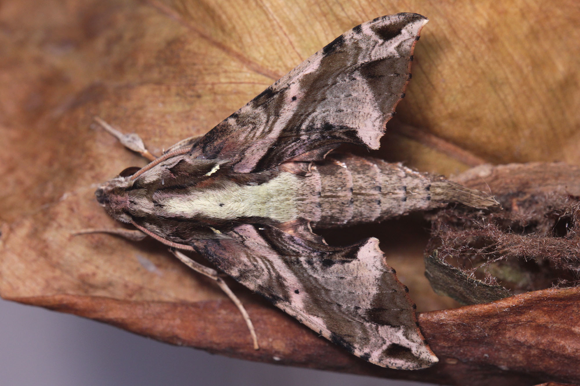
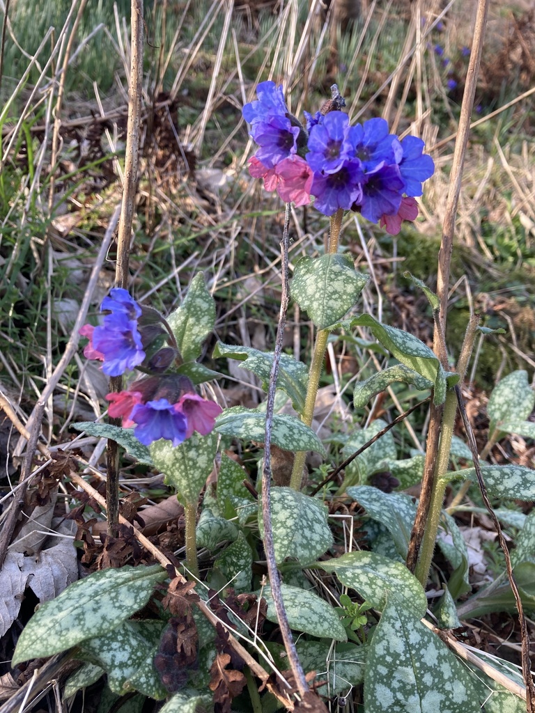
Author Archives: Piter Keo

About Piter Keo

PhD in Biology working with ecology, behavior and taxonomy of land planarians. I love biology, astronomy, languages and mythology, among other things.

Friday Fellow: Green-Pus Bacterium

by Piter Kehoma Boll

The world is filled with bacteria. Trillions, quadrillions, quintillions of them all around us. But while most of them are harmless or even beneficial to us, others are really evil.

Currently, one of the most dangerous bacteria for humans is called Pseudomonas aeruginosa, which I decided to nickname the green-pus bacterium and you will soon find out why. With a typical rod shape as many bacteria, its cells measure about 1.5 to 3 µm in length and 0.5 to 0.8 µm in width.

Gram-sainted cells of Pseudomonas aeruginosa under the microscope. Photo by Wikimedia user Y_tambe.**

Find all around the world, the green-pus bacterium is one of the most generalist species that we have ever discovered. It often lives in oxygen-rich environments, but can also survive in anaerobic places. Provided that the environment has some moisture, it can thrive anywhere, including soil, water, on the surface of animals, plants, and fungi, and on the surface of human-made objects.

The green-pus bacterium feeds on almost anything organic, including living tissue and even hydrocarbons. It has been used, for example, to clean soil and water after oil spills by eating away the oil.

Although not exactly a parasitic species, the opportunistic and generalist habits of the green-pus bacterium make it a potential pathogenic species for humans and many other organisms. If it has the chance to feed on live tissue, it will. And this is not as difficult to occur, as this species can be found living on our skin as part of our microbiota. And how wouldn’t it be there, right? As I said, it thrives everywhere.

Nevertheless, this species is mostly harmless in normal conditions for healthy individuals. It is especially dangerous to immunocompromised individuals or other seriously injured ones. It is one of the most common species to spread and cause hospital-acquired infections. Carried through the air, it can infect the respiratory tract of immunocompromised people and cause pneumonia. Similarly, it can penetrate the urinary tract through infected catheters and cause urinary infections or, also through catheters, it can end up in the bloodstream. Another important route to infect humans is through severe skin burns, through which it can reach the inner tissues and spread, eating the infected individual alive.

Pseudomonas aeruginosa infecting lungs cells. Credits to Benoit-Joseph Laventie.*

Infections by the green-pus bacterium produce, as you may have guessed, a characteristic green pus. When cultivated in the laboratory, the cultures also show a blue-green color, hence the epithet aeruginosa, which in Latin means verdigris-colored. This color is caused by two metabolites produced by the green-pus bacterium named pyocyanin (with a blue color) and pyoverdine (with a green color).

Cultures of P. aeruginosa often show a greenish color. Photo by Wikimedia user Sun14916.**

One of the most frightening facts about the green-pus bacterium is that it is resistant to most antibiotics, most of which is due to natural resistance, although it also easily acquires new resistances by natural selection after being exposed to them during antibiotic treatments. This makes it very difficult to fight an infection caused by this species, and an infected person can easily die. As a result, the green-pus bacterium is also one of the most studied bacteria in the world and it stimulates research for the development of new methods to fight against bacterial infections that go beyond the use of antibiotics.

So take care! This little fellow is all around us and, although harmless most of the time, it will not hesitate to infect you if a good opportunity arises.

– – –

More pathogenic bacteria:

Friday Fellow: H. pylori (on 8 September 2017)

Friday Fellow: Coldwater disease bacterium (on 11 June 2021)

Friday Fellow: Human Chlamydia (on 14 January 2022)

– – –

References:

Diggle, S. P., & Whiteley, M. (2020). Microbe Profile: Pseudomonas aeruginosa: opportunistic pathogen and lab rat. Microbiology166(1), 30. https://doi.org/10.1099/mic.0.000860

Wikipedia. Pseudomonas aeruginosa. Available at < https://en.wikipedia.org/wiki/Pseudomonas_aeruginosa >. Access on 24 November 2022.

– – –

*Creative Commons License This work is licensed under a Creative Commons Attribution 4.0 International License.

**Creative Commons License This work is licensed under a Creative Commons Attribution-ShareAlike 3.0 Unported License.

Leave a comment

Filed under Bacteria, Friday Fellow

Friday Fellow: Elegant Hawkmoth

by Piter Kehoma Boll

Lepidopterans are among the most beloved invertebrates among the general public, especially because of the beauty of either the adult or the larval forms, or sometimes even the pupa! Hawkmoths are among those groups that often call people’s attention. They make up the family Sphingidae, and the adults have a peculiar and easily recognizable shape, but their caterpillars are most remarkable.

An adult of the elegant hawkmoth in Indonesia. Photo by Franz Anthony.*

The Elegant Hawkmoth, Eupanacra elegantulus, is a species found in Southeast Asia. The adults have a brownish color that is not that different from an average hawkmoth. The caterpillars, on the other hand, are very interesting. They feed on plants of the family Araceae, including the genera Aglaonema, Alocasia, Dieffenbachia, Monstera and Syngonium, many of which are highly toxic.

A very young caterpillar with its somewhat plain green color and straight spine near the posterior end. Photo by Ananda Virgiana.*

They live on the underside of the leaves on which they feed and start their lives as small, almost plain green caterpillars, with a small straight spine near the posterior end. At their last instars, they show a curious pattern. The spine at the posterior end becomes hooked, and a little behind the three pairs of true legs the dorsum shows a large eyespot on each side. A black line forming a more or less oval shape connects the eyespots on both sides, and the area that this line surrounds shows a pattern that resembles reptile scales.

A brown caterpillar with the head withdrawn, making it look like a snake. Photo by iNaturalist user klearad.*

The body at this stage can be either green or brown, and the caterpillar kind of resembles a snake, when its head is withdrawn, or a crocodile, when it is extended forward.

A caterpillar with the head exposed, making it look kind of like a crocodile. Photo by iNaturalist user tchaianunporn.*

When they pupate, they attach themselves under a leaf surrounded by a very loose cocoon of silk with some debris.

A pupa surrounded by the very loose cocoon of silk and debris. Photo by Ananda Virgiana.*

Despite being a species with a very interesting pattern that mimics snakes or crocodiles, which catches the attention of many people, there are no studies at all focused on this species. Nowadays, citizen science is becoming an important source of knowledge and I think this species would benefit a lot of projects that include the participation of the community.

– – –

Follow us on Twitter!

– – –

References:

Samuel. Eupanacra elegantulus life history. Common butterflies of Singapore. Available at < http://butterybuttermoth.blogspot.com/2009/03/eupanacra-elegantulus-life-history.html >. Access on 16 November 2022.

– – –

*Creative Commons License This work is licensed under a Creative Commons Attribution-NonCommercial 4.0 International License.

Leave a comment

Filed under Entomology, Friday Fellow

Friday Fellow: Bristly Beard Lichen

by Piter Kehoma Boll

Lichens form an astonishingly diverse group of algae-associated fungi that are found in all sorts of places over the world. One of the most easily recognizable genera is Usnea, whose species are commonly known as beard lichen. One of the most widespread species across the globe is Usnea hirta, the bristly beard lichen.

A specimen of the bristly beard lichen hanging from a branch in Estonia. Photo by Teodor Agabus.*

Like all species of Usnea, the bristly beard lichen is a fruticose lichen, which means it grows in the shape of a small leafless shrub or coral on the surface of trees. It has a grayish-green or greenish-gray color, and its “branches” are very flexible but not as long as in other species that look more like a beard. It prefers to grow on acid bark, especially branches of conifers such as Pinus, and is not that common in deciduous trees, at least in temperate regions. It likes open sites where it can receive lots of sunlight.

A species of Usnea, likely the bristly beard lichen, growing in Uruguay. Photo by Pablo Balduvino.*

With worldwide distribution, the bristly beard lichen is a relatively heterogeneous species, which led to many problems in its classification, as many regional forms were described as separate species and later revealed to be the same Usnea hirta.

In this specimen from Spain, we can see a large apothecium, a structure that produces spores for sexual reproduction. Photo by Maria José Chesa Marro.*

Like many lichens, the bristly beard lichen can reproduce both sexually and asexually. Sexual reproduction occurs through spore production in apothecia (singular apothecium), cup-shaped structures. When the spores are released in the environment and germinate, they need to find a compatible alga to start a new association or the fungus will not survive. Thus, sexual reproduction is very difficult and asexual reproduction is the most efficient strategy. It consists of forming soralia (singular soralium), small “warts” that grow attached to the branches of the lichen. The soralia are groups of sorecia (singular sorecium), which are small units formed by a piece of alga surrounded by fungal hyphae. As both components of the association are already present, a sorecium can germinate whenever it lands on a suitable surface.

The greener wart-like structures covering most of the branches in this specimen from Latvia are likely soralia, the structures for asexual reproduction. Photo by Davis Birzgalis.*

The bristly beard lichen is very sensitive to air pollution, especially to sulfur dioxide (SO2) and nitrogen compounds. It has also shown the ability to bioaccumulate heavy metals in its tissues. As a result, more recent studies are trying to turn it into a model species for bio-monitoring of air pollution, especially in North America.

Thus, if you find yourself surrounded by trees covered with lots and lots of bristly beard lichens, it is likely that the air where you live is not that bad, at least considering sulfur and nitrogen compounds.

– – –

More lichens:

Friday Fellow: Elegant sunburst lichen (on 15 July 2016)

Friday Fellow: Christmas wreath lichen (on 23 December 2016)

Friday Fellow: Pygmy black lichen (on 13 November 2020)

Friday Fellow: Black wart lichen (on 29 October 2021)

– – –

References:

Clerc, P. (1997). Notes on the genus Usnea Dill. ex Adanson. The Lichenologist29(3), 209-215. https://doi.org/10.1006/lich.1996.0075

Fos, S., & Clerc, P. (2000). The lichen genus Usnea on Quercus suber in Iberian cork-oak forests. The lichenologist32(1), 67-88. https://doi.org/10.1006/lich.1999.0242

Shrestha, G., Petersen, S. L., & CLAIR, L. L. S. (2012). Predicting the distribution of the air pollution sensitive lichen species Usnea hirta. The Lichenologist44(4), 511-521. https://doi.org/10.1017/S0024282912000060

Van Herk, C. M., Mathijssen-Spiekman, E. A. M., & De Zwart, D. (2003). Long distance nitrogen air pollution effects on lichens in Europe. The Lichenologist35(4), 347-359. https://doi.org/10.1016/S0024-2829(03)00036-7

– – –

*Creative Commons License This work is licensed under a Creative Commons Attribution-NonCommercial 4.0 International License.

Leave a comment

Filed under Friday Fellow, Fungi

Friday Fellow: Shore-dwelling Swordworm

by Piter Kehoma Boll

One of the most enigmatic animal phyla is Chaetognatha, which are commonly known as arrow worms. Less than 200 species are known, but some species are very abundant, so it is not that difficult to find one if you look at the right place. One particularly common species is Spadella cephaloptera, which I decided to call the shore-dwelling swordworm.

A typical shore-dwelling swordworm. Head to the right, Photo by Wikimedia user Zatelmar.**

All arrowworms are very small, measuring up to 10 or 12 cm in the largest specimens, but the shore-dwelling swordworm is much smaller, with only about 5 mm in length. They are marine creatures and most species are planktonic, but the shore-dwelling swordworm is an exception that lives in shallow waters near the shore, usually attached to the surface of seaweed or marine angiosperms around Europe.

Detail of the head showing the grasping spines at the sizes and the two small black eyes. Photo by Lukas Schärer.*

The body of the shore-dwelling swordworm is kind of dart-shaped, transparent, and consists of a rounded head, an elongate trunk and a short tail. The second half of the trunk and the tail are surrounded by a flat expansion that functions like a fin. Like in all arrowworms, the shore-dwelling swordworm has one set of hooked, grasping spines on each side of the head, on their “cheeks”. They use these spines to capture their prey, which consist mainly of small crustaceans living in the same habitat.

The shore-dwelling swordworm is a hermaphrodite like all arrowworms. The female reproductive apparatus lies in the second half of the trunk and the male one in the tail. Mating does not include reciprocal insemination like in other hermaphrodites. One of the two individuals, which has its seminal vesicles full, approaches another, which has its seminal receptacle empty, and deposits a mass of sperm at the entrance of the vagina of the second. The cilia of the vagina start to become very active and the spermatozoa start to swim across the cilia to enter the vagina until they reach the seminal receptacle.

Detail showing the seminal vesicles as two brown structures on each side. Photo by Lukas Schärer.*

Both individuals remain in contact, with one facing the tail of the other, until the seminal receptacle of the receiving one is full or until they are disturbed. If some sperm mass still remains outside of the vagina when they separate, the receiving one eats what has been left out. After the eggs are fertilized, they still remain some hours inside the mother until they are laid. About 12 to 16 eggs are laid at intervals of eight to ten days. The eggs remain attached to the substrate and, in about 48 hours, they hatch into small versions of the adults, without a larval phase.

Detail showing several eggs at different stages of development on each side of the body. Photo by Lukas Schärer.*

Although almost invisible and often ignored, the shore-dwelling swordworm is an important predator in this specific shore ecosystem which is, of course, connected to the large marine ecosystem and, consequently, to the whole biosphere. But how much do they affect the dynamics of their ecosystem? This is something that still needs to be investigated.

– – –

Follow us on Twitter!

– – –

References:

Goto, T. (1999). Fertilization process in the arrow worm Spadella cephaloptera (Chaetognatha). Zoological science16(1), 109-114. https://doi.org/10.2108/zsj.16.109

John, C. C. (1933). Habits, Structure, and Development of Spadella cephalopteraJournal of Cell Science2(300), 625-696. https://doi.org/10.1242/jcs.s2-75.300.625

– – –

*Creative Commons License This work is licensed under a Creative Commons Attribution 4.0 International License.

**Creative Commons License This work is licensed under a Creative Commons Attribution 3.0 Unported License.

Leave a comment

Filed under Friday Fellow, worms, Zoology

Friday Fellow: Neglected Spirogyra

by Piter Kehoma Boll

As a kid and teen, I was fascinated by the lifeforms that I could find in our backyard, including many simple photosynthetic organisms such as cyanobacteria (my lovely Nostoc, already presented here a long time ago), mosses and, of course, green algae. Green filamentous algae used to grow in large quantities in ponds and puddles forming widespread slimy and hairy “patches”. Most of them were, I suppose, part of the genus Spirogyra.

Widespread across the world, especially in temperate climates, the genus Spirogyra contains hundreds of species. They are all very similar and, from what I can tell, very hard to be determined to the species level. It’s been quite some time since I wanted to bring a species of Spirogyra here, but most images available online are identified only to the genus level, and studies that go to the species level lack photographs of them. But after some time trying to find a good species, I ended up choosing one named Spirogyra neglecta, which would translate as the neglected spirogyra, a name that somehow describes the overall status of the genus, I think, at least regarding material that is easily available to non-spirogyrologists.

An unidentified species of Spirogyra that is probably the neglected spirogyra. Photo by iNaturalist user jjlisowski.*

Anyway, the neglected spirogyra, as all species of Spirogyra, belongs to the order Zygnematales, which consists of filamentous algae. But what does filamentous algae mean? It means that they exist as a colony of cells attached to each other in very long strings, or filaments, that can grow longer and longer as the cells continue to divide. In the case of Spirogyra, the most striking feature is their chloroplast, which has a spiral shape, hence the name of the genus. The number of chloroplasts in each cell varies from species to species. Some have only one, while others can have as many as eight, or perhaps even more. In the case of the neglected spirogyra, the number is usually two or three.

Found across the Holarctic region (North America, Europe, and northern Asia), the neglected spirogyra likes to grow in clear and eutrophic (nutrient-rich) water. It often grows completely submerged, but on sunny days the increased photosynthesis makes oxygen bubbles appear among the filaments and the whole “mat” can end up floating to the surface.

This is the overall aspect of all Spirogyra species when you see them in ponds. Photo by iNaturalist user clasher929.*

The most common form of reproduction among species of Spirogyra is through simple cellular division. The typical cells that form the colonies are haploid, having only one copy of each chromosome. When the environmental conditions are not good for them to survive or grow, sexual reproduction can occur through a process called conjugation. In this process, two cells, usually from different filaments that lie side by side, connect to each other through a conjugation tube. The content of one of the cells (considered the male) migrates into the other cell (considered the female) and their nuclei fuse, thus originating an ovoid zygote. The zygote becomes surrounded by a thick wall, forming the so-called zygospore, which can withstand harsh conditions such as drought or lack of nutrients for several months. During this time, the diploid nucleus of the zygospore undergoes meiosis, forming four haploid nuclei of which only one survives. When the environmental conditions are adequate, the zygospore germinates into a new Spirogyra cell, which will grow into a new filament as it reproduces by fission.

Several zygospores formed inside a “female” filament after conjugation. Notice the empty “male” cells below, Photo by Vasily Vishnyakov.*

Species of the genus Spirogyra are edible, and the neglected spirogyra is no different. These algae are a common ingredient in northern Thai cuisine, where they are eaten especially raw as salad. Despite being a cheap food, the neglected spirogyra is rich in nutrients and has antioxidant properties. Extracts from this alga have shown anti-inflammatory and anticancer activities, as well as the ability to stimulate the immune system.

Have you ever thought of making a salad from that green filamentous mat that grows in ponds around you?

– – –

Follow us on Twitter!

– – –

References:

Duangjai, A., Limpeanchob, N., Trisat, K., & Amornlerdpison, D. (2016). Spirogyra neglecta inhibits the absorption and synthesis of cholesterol in vitro. Integrative Medicine Research5(4), 301-308. https://doi.org/10.1016/j.imr.2016.08.004

Mesbahzadeh, B., Rajaei, S. A., Tarahomi, P., Seyedinia, S. A., Rahmani, M., Rezamohamadi, F., … & Moradi-Kor, N. (2018). Beneficial effects of Spirogyra Neglecta Extract on antioxidant and anti-inflammatory factors in streptozotocin-induced diabetic rats. Biomolecular Concepts9(1), 184-189. https://doi.org/10.1515/bmc-2018-0015

Ontawong, A., Saowakon, N., Vivithanaporn, P., Pongchaidecha, A., Lailerd, N., Amornlerdpison, D., … & Srimaroeng, C. (2013). Antioxidant and renoprotective effects of Spirogyra neglecta (Hassall) Kützing extract in experimental type 2 diabetic rats. BioMed Research International2013. https://doi.org/10.1155/2013/820786

Schagerl, M., & Zwirn, M. (2015). A brief introduction to the morphological species concept of Spirogyra and emanating problems. Algological studies, 67-86. 10.1127/algol_stud/2015/0231

Surayot, U., Wang, J., Lee, J. H., Kanongnuch, C., Peerapornpisal, Y., & You, S. (2015). Characterization and immunomodulatory activities of polysaccharides from Spirogyra neglecta (Hassall) Kützing. Bioscience, Biotechnology, and Biochemistry79(10), 1644-1653. https://doi.org/10.1080/09168451.2015.1043119

Thumvijit, T., Taya, S., Punvittayagul, C., Peerapornpisal, Y., & Wongpoomchai, R. (2014). Cancer chemopreventive effect of Spirogyra neglecta (Hassall) Kützing on diethylnitrosamine-induced hepatocarcinogenesis in rats. Asian Pacific Journal of Cancer Prevention15(4), 1611-1616. https://doi.org/10.7314/APJCP.2014.15.4.1611

– – –

*Creative Commons License This work is licensed under a Creative Commons Attribution-NonCommercial 4.0 International License.

Leave a comment

Filed under Algae, Botany, Friday Fellow

Friday Fellow: Mediterranean Plumefoot Mite

by Piter Kehoma Boll

There are endless forms of beauty among the small creatures that we often do not see around us. Mites, which are so ubiquitous, contain several neglected beauties. One of them is today’s fellow, Eatoniana plumipes, known as the Mediterranean Plumefoot Mite.

Mediterranean Plumefoot Mite photographed in Spain by Simon Oliver*.

Adults of the Mediterranean Plumefoot Mite are considerably large for a mite, measuring a few millimeters in length, often more than 1 cm when the legs are considered. They are reddish-brown, lighter at the legs and other appendices, and their hind legs are much longer than the others and have a tuft of long black hair that makes them look like plumes, hence the name plumefoot mite. As the common name also suggest, this species is found around the Mediterranean, including southern Europe, northern Africa, Turkey and the Middle East.

Despite being a large and rather beautiful mite, very little is known about the life history of the Mediterranean Plumefoot Mite. It belongs to a group of mites that are predators as adults but parasites as larvae. The larvae hatch from red eggs laid by the female in the environment and are, of course, much smaller than the adults. They also have only three pairs of legs, and not four like the adults, and lack the characteristic plumes seen in the adults.

Some eggs of the Mediterranean Plumefoot Mite and one recently hatched larva. Extracted from Mąkol & Sevsay (2015).

Little to nothing is known about the feeding habits of this species. Grasshoppers are among the identified hosts of the larvae, but it is likely that other arthropods are parasitized as well. The larvae attach to the legs of the hosts and feed there, sucking their hemolymph (the “blood” of arthropods). I could not find any information about which species serve as prey for the adults.

Even though we know almost nothing about the ecology of the Mediterranean plumefoot mite, we can still appreciate its beauty, and it certainly plays a fundamental role in its ecosystem.

If you live around the Mediterranean, have you ever seen one of them? Let us know!

– – –

More mites:

Friday Fellow: Giant red velvet mite (on 22 July 2016)

Friday Fellow: Cuban-laurel-thrips mite (on 28 June 2019)

Friday Fellow: Rhinoceros Tick (on 18 October 2019)

Friday Fellow: Aloe Mite (on 7 February 2020)

– – –

Follow us on Twitter!

– – –

References:

GBIF. Eatoniana plumipes (L. Koch, 1856). Available at < https://www.gbif.org/species/4539982 >. Access on 20 October 2022.

Mąkol, J., & Sevsay, S. (2015). Abalakeus Southcott, 1994 is a junior synonym of “plume-footed” Eatoniana Cambridge, 1898 (Trombidiformes, Erythraeidae)-evidence from experimental rearing. Zootaxa3918(1), 92-112. https://doi.org/10.11646/zootaxa.3918.1.4

Noei, J., & Rabieh, M. M. (2019). New data on Nothrotrombidium, Southcottella and Eatoniana larvae (Acari: Trombellidae, Neothrombiidae, Erythraeidae) from Iran. Persian Journal of Acarology8(3). https://doi.org/10.22073/pja.v8i3.46776

– – –

*Creative Commons License This work is licensed under a Creative Commons Attribution-NonCommercial 4.0 International License.

Leave a comment

Filed under Arachnids, Friday Fellow, Parasites, Zoology

Friday Fellow: Hermaphrodite Turner

by Piter Kehoma Boll

Most flatworms known to date are parasites, and it is likely that these are, indeed, the majority of the phylum. Nevertheless, the diversity of free-living flatworms is certainly underestimated. This is especially noticeable when we talk about today’s fellow, Gyratrix hermaphroditus, a tiny flatworm that I decided to call the hermaphrodite turner in English.

Measuring about 1 to 2 mm in length, the hermaphrodite turner is a cute little pal. It is shaped like an elongate drop, rounded at the posterior end and kind of pointed at the anterior end. There are two small black eyes that lie a little behind the anterior end, making it look like it has an elongate and movable snout. It kind of looks like a microscopic version of a seal.

A cute specimen of the hermaphrodite turner. We can see the two black eyes with the pharynx in front of then, the intestine filled with food, a needle-like stylet near the posterior end, and an oval-shaped egg. Photo extracted from plingfactory.de

Because the body of the hermaphrodite turner is transparent, you can see its internal organs very easily. Between the eyes and the anterior tip, we can notice a sort of transparent cylindrical structure, which is its proboscis, used to capture prey. When the intestine is full of food, it can also be noticed.

The hermaphrodite turner is a curious animal because it is not only found all over the world, on all continents, but also in both freshwater and marine environments. In fact, it is not a single species, but a complex of very similar species that have been considered, or treated like, a single species for decades. Recent molecular studies, however, made it clear that the hermaphrodite turner is not one but many different species. Although instances of species complexes considered a single species is common among microscopic organisms, the hermaphrodite turner is a special case in which, from what we discovered until now, the complex includes a really huge number of species, probably hundreds of them.

Take a look of one of them in action in the tweet above.

As the name of the hermaphrodite turner implies, it is a hermaphrodite like most flatworms. Near the posterior end of its body, we can see a straight needle-like structure, the stylet. This is part of the male copulatory apparatus, forming kind of a hard needle-like penis, which is used to penetrate the body of other individuals to inseminate them. Sometimes we can also see individuals with unlaid eggs inside them. These appear as oval-shaped brownish structures. When the eggs are laid, they are attached to the substrate by a small stalk to prevent them from being carried away by the water.

An egg of the hermaphrodite turner about to hatch. We can see the two eyes through the shell. Its sad face is probably because it will have to live in the same world as Jair Bolsonaro. Photo extracted from plingfactory.de

Despite the worldwide distribution of this species complex, we know almost nothing about the behavior and ecology of these organisms. They feed on smaller animals and protists that share the same environment as them. They often capture prey using their proboscis, but they can also use their stylet to stab. Afterward, they place their mouth over the wound and start sucking the prey’s contents. The mouth is on the ventral side at about the middle of the body as in most flatworms.

Let’s hope that our fellow zoologists that work with these tiny flatworms start splitting this complex into the actual species, as it deserves.

– – –

Follow us on Twitter!

– – –

References:

Tessens, B., Gijbels, M., & Artois, T. (2009). MATING AND FEEDING BEHAVIOUR OF THE EURYHALINE COSMOPOLITAN FLATWORM GYRATRIX HERMAPHRODITUS. Belgium, 27 November 2009: VLIZ Special Publication, 43. Vlaams Instituut voor de Zee (VLIZ): Oostende, Belgium. xiii+ 221 pp.

Tessens, B., Monnens, M., Backeljau, T., Jordaens, K., Van Steenkiste, N., Breman, F. C., … & Artois, T. (2021). Is ‘everything everywhere’? Unprecedented cryptic diversity in the cosmopolitan flatworm Gyratrix hermaphroditus. Zoologica Scripta50(6), 837-851. https://doi.org/10.1111/zsc.12507

Leave a comment

Filed under flatworms, Friday Fellow, worms

Friday Fellow: Deadly Plasmodium

by Piter Kehoma Boll

Humans can be infected by a vast number of different parasites, but no parasite has such a big impact on our species as those of the genus Plasmodium, which cause the disease called malaria. Several different species of Plasmodium infect humans, but today we will talk about the deadliest of them, Plasmodium falciparum, which I decided to name the Deadly Plasmodium.

The genus Plasmodium belongs to the phylum Apicomplexa, a group of exclusively parasitic protists that evolved from free-living algae. Besides Plasmodium, another important apicomplexan that infects humans is the Toxo, which causes toxoplasmosis and already appeared as a fellow here some years ago.

The life cycle of the deadly plasmodium is very complex and includes two hosts, a human and a mosquito of the genus Anopheles. When an infected mosquito bites a human, it releases between 20 and 200 sporozoites, which are one of the life stages of the deadly plasmodium. The sporozoites are elongated cells that glide through the bloodstream of the infected human until they reach the liver. They use their apical complex, a structure formed by several glands and organelles, to penetrate the liver cells. The apical complex is lost in the process.

Life cycle of the deadly plasmodium. Credits to La Roche Lab, UC Riverside.**

Inside the liver cells, the sporozoite changes into a trophozoite, which lives inside a vacuole and starts to grow and undergo mitosis and meiosis without cell division until becoming a single cell with tens of hundreds of nuclei called a schizont. The schizont eventually bursts into tens of thousands of small haploid cells called merozoites, which enter the bloodstream again. Provided with new apical complexes, the merozoites now use it to enter red blood cells and turn again into trophozoites, which are now haploid. This haploid red-cell-infecting trophozoite grows again and undergoes mitosis to make a new large schizont, which eventually bursts again into new merozoites that enter the bloodstream once more and infect new red cells, starting the cycle again. Inside the red cells, the trophozoite feeds on hemoglobin and converts part of it into an insoluble granular pigment called hemozoin.

A trophozoite (left) and a schizont (lower center) in red blood cells.

All plasmodium cells infecting the red blood cells have their cycle in synchrony so that all merozoites are released into the bloodstream and infect new cells simultaneously. The cycle in the red blood cells takes about 48 hours and is related to the typical cyclic manifestations of fever and chills caused by falciparum malaria.

Eventually, some merozoites change into sexual forms, the male and female gametocytes, known as microgametocyte and macrogametocyte, respectively. They take between one and two weeks to reach maturity. After reaching this stage, if the infected human is bitten by a mosquito, the gametocytes are ingested and, inside the mosquito’s gut, undergo morphological changes. The macrogametocytes grow into a large, spherical form, the macrogamete. The microgametocyte undergoes nuclear division in 15 minutes and splits into eight flagellated microgametes. A microgamete fertilizes a macrogamete, which originates a zygote. The zygote develops into a motile cell called the ookinete, which penetrates the mosquito tissues and turns into an immotile oocyst. The oocyst grows and undergoes nuclear division until becoming a large multinucleate cell called sporoblast. The sporoblast bursts into thousands of sporozoites, which migrate to the mosquito’s salivary glands and, when the mosquito bites another human, they are released into the bloodstream and restart the cycle.

A macrogametocyte (left) and a microgametocyte (right) among red blood cells.

The main deleterious effects caused by malaria in humans occur due to the massive destruction of red blood cells and the production of hemozoin, which is toxic to tissue and starts to accumulate in several organs, impairing their function. Although several species of Plasmodium can cause malaria in humans, about 50% of all deaths by malaria are caused by the deadly plasmodium alone, making it the deadliest parasite in humans.

The deadly plasmodium apparently evolved about 10 thousand years ago in Africa from a very similar species that infects gorillas. Currently, although most infections and deaths still occur in Subsaharan Africa, the deadly plasmodium has spread around the world and is also prevalent in regions near the equator in South America and Asia.

Due to the high lethality of falciparum malaria in humans, the disease has placed great selective pressure on human populations in areas where this parasite is common. Thus, although some say that natural selection does not affect humans anymore, the deadly plasmodium is here to refute this argument.

– – –

Other Apicomplexans:

Friday Fellow: Toxo (on 5 May 2017)

Friday Fellow: Arrow Anchor (on 15 January 2021)

– – –

Follow us on Twitter!

– – –

References:

Lambros, C., & Vanderberg, J. P. (1979). Synchronization of Plasmodium falciparum erythrocytic stages in culture. The Journal of parasitology, 418-420.

Maier, A. G., Matuschewski, K., Zhang, M., & Rug, M. (2019). Plasmodium falciparum. Trends in parasitology35(6), 481-482. https://doi.org/10.1016/j.pt.2018.11.010

Talman, A. M., Domarle, O., McKenzie, F. E., Ariey, F., & Robert, V. (2004). Gametocytogenesis: the puberty of Plasmodium falciparum. Malaria journal3(1), 1-14. https://doi.org/10.1186/1475-2875-3-24

Wikipedia. Plasmodium falciparum. Available at < https://en.wikipedia.org/wiki/Plasmodium_falciparum >.

– – –

**Creative Commons License This work is licensed under a Creative Commons Attribution 3.0 Unported License.

Leave a comment

Filed under Friday Fellow, Parasites, protists

Friday Fellow: Common Lungwort

by Piter Kehoma Boll

Blue is a relatively rare color in nature, but in some places it appears much more than in others. Flowers of plants in the family Boraginaceae are often blue. The forget-me-not is possibly the most popular example, but not the only one. Today our fellow is another blue flowered species that is also popular, not so much for its flowers though, but for its lung-like leaves, the common lungwort Pulmonaria officinalis.

The spotted leaves of the common lungwort are said to resemble diseased lungs. Photo by Jakob Fahr.*

Whenever you find a plant whose name refers to a body part, it most likely has to do with the ancient doctrine of signatures, the idea that a plant that resembles a human organ can be used to treat diseases on that organ. This is the case with the common lungwort and the common liverwort, which was presented here some time ago. The oval and kind of heart-shaped leaves of the common lungwort are slightly hairy on the upper side and marked by several white or pale spots. They were thought to represent an ulcerated lung and, therefore, used to treat diseases of the lungs. Although some studies revealed that lungwort extracts can present biological properties such as antioxidant, anti-inflammatory and wound healing activities, nothing has been found that is directly related to the lungs.

Found across most of continental Europe, the common lungwort is a small plant formed by a creeping rhizome from which the leaves sprout in the form of rosettes. It likes to grow on the forest floor, below the tree canopy, but dislikes places with too much shade. As a European species, it is very tolerant to cold, supporting temperatures as low as -29 °C.

The flowers of the common lungwort start red and slowly turn to blue as they age. Photo by iNaturalist user laivoi.*

In spring, between March and May, the flowers appear in inflorescences on elongated stems that grow from the leaf rosettes. They grow during a period in which the trees are only starting to produce new leaves so the flowers are fully exposed to the sun. Each inflorescence has 5 to 15 hermaphrodite flowers with five petals. The flowers start red and, as they age, they change color to purple and finally blue. This change in color occurs because the pigments are anthocyanins that are affected by pH, being red in acidic environments and blue in alkaline ones. The main pollinators of the common lungwort are bees and the plant only wants them to visit young, red flowers. As a result, the change in color helps direct the pollinators to the right flowers by signalling that the blue ones are uninteresting (and they, in fact, have no nectar anymore). But why does the plant keep flowers for longer periods instead of shedding the petals and producing fruits at once? Well, because the more flowers you have, the more pollinators you can attract from the distance. A large number of flowers makes you visible from far away but, as the pollinators come closer, the different colors guide them to the right spot.

Although they are too old to be pollinated, blue flowers are still useful in attracting pollinators for the younger, red flowers. Photo by iNaturalist user oburridge.*

The fruit of the common lungwort is a schizocarp, i.e., a small and dry fruit that splits into smaller portions, each containing a seed. In the case of the common lungwort, each fruit contains four seeds, and their main dispersers are ants. The ants collect the fruits, carry them to their colonies and feed the larvae with the fleshy portion, discarding the seed afterward.

The common lungwort is a popular plant because of its color-changing flowers and its resistance to cold, but, as we can see, this beauty hides an even deeper beauty caused by its interaction with the small creatures that share the same space with it.

– – –

Follow us on Twitter!

– – –

References:

Chauhan, S., Jaiswal, V., Cho, Y. I., & Lee, H. J. (2022). Biological Activities and Phytochemicals of Lungworts (Genus Pulmonaria) Focusing on Pulmonaria officinalisApplied Sciences12(13), 6678. https://doi.org/10.3390/app12136678

Meeus, S., Brys, R., Honnay, O., & Jacquemyn, H. (2013). Biological flora of the British Isles: Pulmonaria officinalisJournal of Ecology101(5), 1353-1368. https://doi.org/10.1111/1365-2745.12150

Wikipedia. Pulmonaria officinalis. Available at < https://en.wikipedia.org/wiki/Pulmonaria_officinalis >. Access on 22 September 2022.

– – –

*Creative Commons License This work is licensed under a Creative Commons Attribution-NonCommercial 4.0 International License.

1 Comment

Filed under Botany, Friday Fellow

Friday Fellow: Lined Earwig

by Piter Kehoma Boll

Of the 334 Friday Fellows that this blog had so far, 50 were insect. Yet, several insect orders have not been represented yet. One of them is the order Dermaptera, commonly known as earwigs, but this changes today. And the first species of this order to be featured here is Doru taeniatum, the lined earwig.

Occurring across the Americas, at least from the United States to Argentina, the lined earwig is often found in human crops, especially corn/maize. It has the typical appearance of most earwigs. The body is elongated, mostly brown, darker, almost black at the abdomen and reddish at the head. The antennae are a bit long and simple, the legs are yellowish and the wings consist of a small and hardened anterior pair, which resembles the elytra of beetles, and a larger and membranous posterior pair that keeps folded under the anterior pair when they are not being used to fly. The elytra are yellow, with a brown stripe running along the margin where both wings touch, causing a pattern of the yellow stripes separated by a brown stripe. And like all earwigs, the lined earwig has a pair of forceps-like cerci at the tip of the abdomen.

A male lined earwig in Pirassununga, Brazil. Photo by Antonio Bordignin.*

The lined earwig is a predator and feeds on other insects, especially eggs and small larvae, although it has also been observed ingesting plant matter, such as pollen and leaf tissues. One of its prey is the fall armyworm, Spodoptera frugiperda, a moth that is a major pest of crops, especially maize. The lined earwig feeds on both the eggs and caterpillars of S. frugiperda and is considered one of its most important predators, so its presence in maize plantations is always good, and, sometimes, when its numbers are large enough, no other form of pest control is needed.

A female on a human hand to give a perspective of size. Photo by David Aragonés Borrego.*

The predators of the lined earwig include other arthropods, such as ants and spiders, as well as vertebrates, such as anurans and possibly birds. Its defense mechanisms include both the use of the cerci to pinch the predator, mostly small ones, as well as a chemical defense, which consists of a spray that is released by glands that open at the fourth abdominal segment. This spray consists of quinones with a strong odor that can deter predators such as frogs and ants.

Being active both day and night, the lined earwig spends most of its life on maize plants. They use the space between the leaf and the stem of the plant as shelter, mating site and egg deposit. Males are larger than females, having larger elytra and larger cerci. Each maize plant often has only one earwig or a pair consisting of a male and a female living together. When a male wants to copulate, he approaches the female by moving his antennae near her, then moves to her side and strokes her abdomen with his cerci. If she accepts to copulate, they position themselves in opposite directions and connect the final segments of their abdomens, when the male then extrudes its genitalia and inserts them into the genital pouch of the female. Males are territorial and will fight other males that invade their territory, using the cerci to attack each other.

A female taking care of her eggs in Copándaro, Mexico. Photo by iNaturalist user ekdelval.*

After the pair mates, the female lays the eggs and takes special care of them. She frequently cleans them with her mouth, preventing infection by fungi and mites. She also defends them aggressively using her cerci as a weapon to pinch any intruder. After the eggs hatch, the female still takes care of the nymphs for a few days before they disperse in the environment and start to clean the crops from pests just like their parents did.

– – –

Follow us on Twitter!

– – –

References:

Briceño, R. D., & Schüch, W. (1988). Reproductive biology and behavior of Doru taeniatum (Forficulidae). Revista de Biologia Tropical36(2B), 437-440. https://revistas.ucr.ac.cr/index.php/rbt/article/view/23851

Eisner, T., Rossini, C., & Eisner, M. (2000). Chemical defense of an earwig (Doru taeniatum). Chemoecology10(2), 81-87. https://doi.org/10.1007/s000490050011

Jones, R. W., Gilstrap, F. E., & Andrews, K. L. (1988). Biology and life tables for the predaceous earwig, Doru taeniatum [Derm.: Forficulidae]. Entomophaga33(1), 43-54. https://doi.org/10.1007/BF02372312

– – –

*Creative Commons License This work is licensed under a Creative Commons Attribution-NonCommercial 4.0 International License.

Leave a comment

Filed under Entomology, Friday Fellow