Sometimes you lack the ability to become a great scientist, so you seek recognition by ruining the career of great names of science. At least that is basically the definition of the man whose birthday we celebrate today.
Carl Wilhelm von Nägeli was born on 27 May 1817 in Kilchberg, near Zurich, as the son of a Physician. In 1836, he started to study medicine at the University of Zurich but soon changed his interest toward botany, having Lorenz Oken and Oswald Heer, among others, as teachers. In 1939 he started to study botany at the University of Geneva under Augustin Pyramus de Candolle and graduated in 1840 with a botanical thesis entitled Die Cirsien der Schweiz (The Cirsium species of Switzerland).
Portrait of Carl Nägeli. Date and author unknown.
In 1842, Nägeli started to work at Jena with the botanist Matthias Jakob Schleiden on the microscopic study of plants. That year, he observed cellular division during the formation of pollen but apparently was unable to understand what he was seeing, different from Robert Remak, who observed cell division at about the same time.
Nägeli coined the terms meristem, xylem and phloem in 1858 and this is probably his main contribution to science. He is more commonly remembered by how he did almost everything wrong, especially regarding evolution and heredity. For example, Nägeli exchanged extensive correspondence with Gregor Mendel, who mentioned to him his works on plants, but Nägeli considered them useless and somewhat discouraged Mendel to go on with his studies.
In 1884, Nägeli published a work entitled Mechanisch-physiologische Theorie der Abstammungslehre (A mechanico-physiological theory of organic evolution) in which he proposes the concept of idioplasm, which would be a special part of a plant’s cytoplasm that transmitted inherited characters. Mendel, who died that same year, was not even mentioned in this work, which makes it clear how Nägeli despised Mendel’s work.
Likewise, Nägeli rejected Darwin’s theory of Natural Selection as a force guiding evolution. Instead of this, he defended the idea of orthogenesis, developing the concept of an inner perfecting principle that he considered to direct evolution.
Hagfish are primitive chordates that make up the class Myxini. They are marine animals that live at the bottom of the sea and feed mainly on polychaete worms that they pull out of the substrate. However, they are also scavengers and have a peculiar behavior in which they perforate the body of dead fish and enter it, eating the dead animal from inside out.
Specimen of the Pacific hagfish Eptatretus stoutii. Photo by Jeanette Bham.*
Morphologically, hagfish are characterized by the presence of a cartilaginous skull, like vertebrates, but lack a vertebral column, keeping the notochord, the dorsal cartilage-like structure of chordates, during their whole lives. Due to this lack of vertebrae, the hagfish were classified outside of the vertebrates, but united to them due to the presence of the skull. Thus, Myxini was seen as the sister-group of Vertebrata and both together formed the clade Craniata.
Among the vertebrates, most extant groups have a jaw that evolved from modified gill arches, making up the clade Gnathostomata. The only animals with a vertebral column that lack jaws are the lampreys (Petromyzontiformes) and, although this lack of jaws is shared with hagfish, it is not usually seen as a synapomorphy uniting these groups. In hagfish, the jawless mouth have lateral keratin plates with tooth-like structures that act somewhat like the true jaws of Gnathostomata, but working from the sides and not from above and below. In lampreys, on the other hand, the mouth is circular and have keratin tooth-like structures arranged circularly.
General organization of the head of hagfish, lampreys and jawed vertebrates, with special attention to the mouths. Extracted from Oisi et al. (2012).
There are a lot of morphological features that unite lampreys to vertebrates and separate them from hagfish, the main one being the already mentioned vertebrate column. Likewise, lampreys and jawed vertebrates have dorsal fins while hagfish lack them. Lampreys also have lensed eyes in common with jawed vertebrates, while hagfish have simple eyesposts without lenses or even associated muscles.
Some of the traits shared between hagfish and lampreys, just as the lack of jaws, are usually seen as a primitive state that changed in jawed vertebrates, or have clearly evolved independently. For example, both hagfish and lampreys have only a single nostril, while jawed vertebrates have two, but this is likely a primitive character. Adult hagfish and lampreys have also a single gonad, but this appears in hagfish by an atrophy of the left gonad, so that only the right one develops, while in lampreys the left and right gonads fuse into a single organ.
Specimens of the least brook lamprey Lampetra aepyptera. Photo by Jerry Reynolds.*
Therefore, morphologically, it seems logical to consider hagfish as a sister group of vertebrates, which include lampreys and jawed vertebrates. It is also important to mention that there are more groups of jawless vertebrates that are currently extinct, such as the class Osteostraci, one of several fossil groups traditionally called ostracoderms. Although lacking a jaw as well, these vertebrates had paired fins just like jawed vertebrates. Thus, the phylogenetic organization of these major groups based on morphology would be as shown in the figure below:
The craniate hypothesis, where hagfish are a sister-group to vertebrates.
However, in the last decades, the use of molecular phylogenetics has challenged this view by grouping hagfish and lampreys into a monophyletic clade that is sister-group of jawed vertebrates. But how could this be possible? Such a relationship would imply that the primitive state of hagfish is the result of secondary loss.
The cyclostome hypothesis. Hagfish are a sister-group to lampreys.
Evidence from fossils could help clarify this issue, but most fossils that have been associated with hagfish have not good enough morphological characters preserved to assess their correct phylogenetic position. Recently, however, a well preserved hagfish fossil from the Cretaceous helped to elucidate part of the hagfish phylogeny. The divergence between lampreys and hagfish, considering previous knowledge, was usually put around the early Cambrian period, just after the beginning of the divergence of most animal phyla, but with data of the new fossil, it is pushed to a more recent point in time, around the Early Silurian, more than 130 million years after. This new fossil, named Tethymyxine tapirostrum, clearly lacks a skeleton or dorsal fins as seen in lampreys and jawed vertebrates, but has several characters shared with extant hagfish.
At least two synapomorphies can be found uniting hagfish and lampreys and separating them from jawed vertebrates. The first one are the teeth, which in these two groups are composed of keratin plates. The second one is the organization of the myomeres, the series of muscles arranged along the body of chordates in a somewhat segmented fashion, that in both hagfish and lampreys begin right around the eyes.
Considering the evidence from molecular data, the new fossil that makes it likely that hagfish and lampreys diverged more recently if they form a monophyletic group, and the likely true synapomorphies uniting these two jawless vertebrate groups, it seems that hagfish and lampreys are indeed sister-groups, forming a clade called Cyclostomata and sister-group of the jawed vertebrates Gnathostomata. If this is really the case, then the apparently more primitive features of hagfish are in fact the result of secondary losses and its ancestor likely had a more vertebrate look, with a vertebral column, dorsal fins and lensed eyes.
But let’s keep watching. Things may change again in the future as new data become available.
Miyashita T, Coates MI, Farrar R, Larson P, Manning PL, Wogelius RA, Edwards NP, Anné J, Bergmann U, Palmer AR, Currie PJ (2019) Hagfish from the Cretaceous Tethys Sea and a reconciliation of the morphological–molecular conflict in early vertebrate phylogeny. PNAS116(6): 2146–2151. doi: 10.1073/pnas.1814794116
Oisi Y, Ota KG, Kuraku S, Fujimoto S, Kuratani S (2012) Craniofacial development of hagfishes and the evolution of vertebrates. Nature 493: 175–180. doi: 10.1038/nature11794
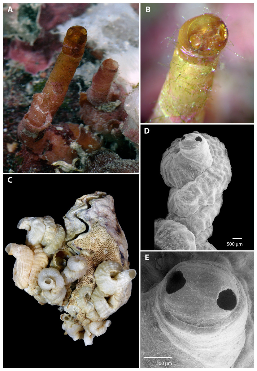
It is time for our second bryozoan fellow, and this time I am bringing you a problematic species that can become a serious nuisance, the red-rust bryozoan Watersipora subtorquata.
The red-rust bryozoan is a colonial bryozoan that lives in tropical and temperate waters and grows on hard surfaces such as rocks or eventually on other colonial organisms. Each individual in the colony, called a zooid, lives inside an elongate structure of mineralized material, mainly calcium carbonate with an oval-shaped opening through which the mouth protrudes. The colony begins spreading over a surface and forming a thin plate of mineralized material that has a dark-red color, hence the common name red-rust. As the colonies become larger, the central parts tend to change to a grayish or blackish color. Older colonies tend to overgrow themselves and turn into leaf-like structures, especially when growing on irregular surfaces.
A leaf-like colony of the red-rust bryozoan. The small dots are the openings of each zooid. Photo by Alison Young.*
With a cosmopolitan distribution, the original location of the red-rust bryozoan is unknown but it was most likely spread throughout the oceans by human activities and soon became a nuisance to humans. The red-rust bryozoan, as a species that grows on hard substrates, found ideal habitats in human structures, such as pipes and ships, which end up covered by colonies, a process called biofouling. Some anti-fouling substances, such as copper-based paint used on ships hulls to prevent biofouling are unable to prevent its growth, as the red-rust bryozoan is a copper-resistant species. And after covering the anti-fouling paint with its colonies, the red-rust bryozoan creates a habitat that allows copper-sensitive creatures to grow.
A closer look in which the individual structures of each zooid are clearly visible. Photo by Damon Tighe.*
On a positive side for humans, the red-rust bryozoan is known to produce bryoanthrathiophene, a substance with antiangiogenic properties, i.e., it prevents the growth of new blood vessels, which may be useful in the treatment of some types of cancer.
Jeong S-J, Higuchi R, Miyamoto T, Ono M, Kuwano M, Mawatari SF (2002) Bryoanthrathiophene, a New Antiangiogenic Constituent from the Bryozoan Watersipora subtorquata (d’Orbigny, 1852). Journal of Natural Products 65(9): 1344–1345. doi: 10.1021/np010577+
Mackie HA, Keough MJ, Christidis L (2006) Invasion patterns inferred from cytochrome oxidase I sequences in three bryozoans, Bugula neritina, Watersipora subtorquata and Watersipora arcuata. Marine Biology 149(2): 285-295. doi: 10.1007/s00227-005-0196-x
Ryland JS, De Blauwe H, Lord R, Mackie JA (2009) Recent discoveries of alien Watersipora (Bryozoa) in Western Europe, with redescription of species. Zootaxa 2093: 43–59.
Vieira LM, Jones MS, Taylor PD (2014) The identity of the invasive fouling bryozoan Watersipora subtorquata (d’Orbigny) and some other congeneric species. Zootaxa 3857: 151–182. doi: 10.11646/zootaxa.3857.2.1
Today I am presenting a 18th century scientist who worked on several areas of the natural sciences.
Torbern Olaf Bergman was born on 20 March 1735 in Låstad parish, Sweden, the son of Barthold Bergman and Sara Hägg. His interest in botany was raised by his teacher Sven Hof at Katedralskolan in Skara.
At the age of 17, he enrolled at the University of Uppsala. He wanted to study mathematics and natural science, but his father wanted him to study law or divinity. Trying to please both his father and himself, he overworked himself and became ill, which forced him to stay some time away from study. During this period, he entertained himself with field botany and entomology.
Portrait of Torbern Bergman by Ulrika Pasch.
Through his entomological collections, Bergman became acquainted with Linnaeus and sent him several insects of new species. In 1756, he succeeded in proving that, contrary to Linnaeus’ opinion, the species called Coccus aquaticus was simply the ovum of a leech, which Linnaeus recognized as correct. Due to this discovery, as well as because he developed a method to capture the wingless females of winter moths, Bergman was awarded a prize by the Swedish Academy of Sciences, being elected a member of the Academy in 1764. The next year he was ellected a Fellow of the Royal Society of London.
Bergman also defended a thesis in astronomy and founded the Cosmography Society in Uppsala, through which the published, in 1766, his work Physisk beskrifning öfver jordklotet (Physical description over the globe), which was one of the first books of modern geography. He then became an associate professor of physics and studied the electrical properties of tourmaline, as well as meteorological phenomena such as the northern lights, thunder and rainbow.
In 1767, the chemist Johan Gottschalk Wallerius resigned from his position as professor of chemistry and mineralogy at the University of Uppsala and Bergman was decided to be a candidate. However, he did not have previous experience in publishing works on chemistry and his competitors charged him with ignorance on the subject. To refute them, he isolated himself in a laboratory for some time and wrote a treatise on the manufacture of alum and it became a standard work. Nevertheless, he still faced strong opposition and only got the chair of chemistry through the influence of the prince Gustavus III, who was also chancellor of the university. He kept this position until his death.
Bergman married his wife, Margareta Catharina Trast, in 1771. In 1772, he was one of the first to receive the Royal Order of Vasa, which was awarded to Swedish citizens for their service to the state and society, especially in the fields of agriculture, mining and commerce.
In 1775, Bergman published his most important chemical paper, Essay on Elective Attractions, a study of chemical affinity. In March 1782, he was elected a foreign associate of the French Academy of Sciences.
He died prematurely on 8 July 1784, aged 49, in Medevi, Sweden, due to a stroke. The radiactive uranium mineral torbernite was named in his honor.
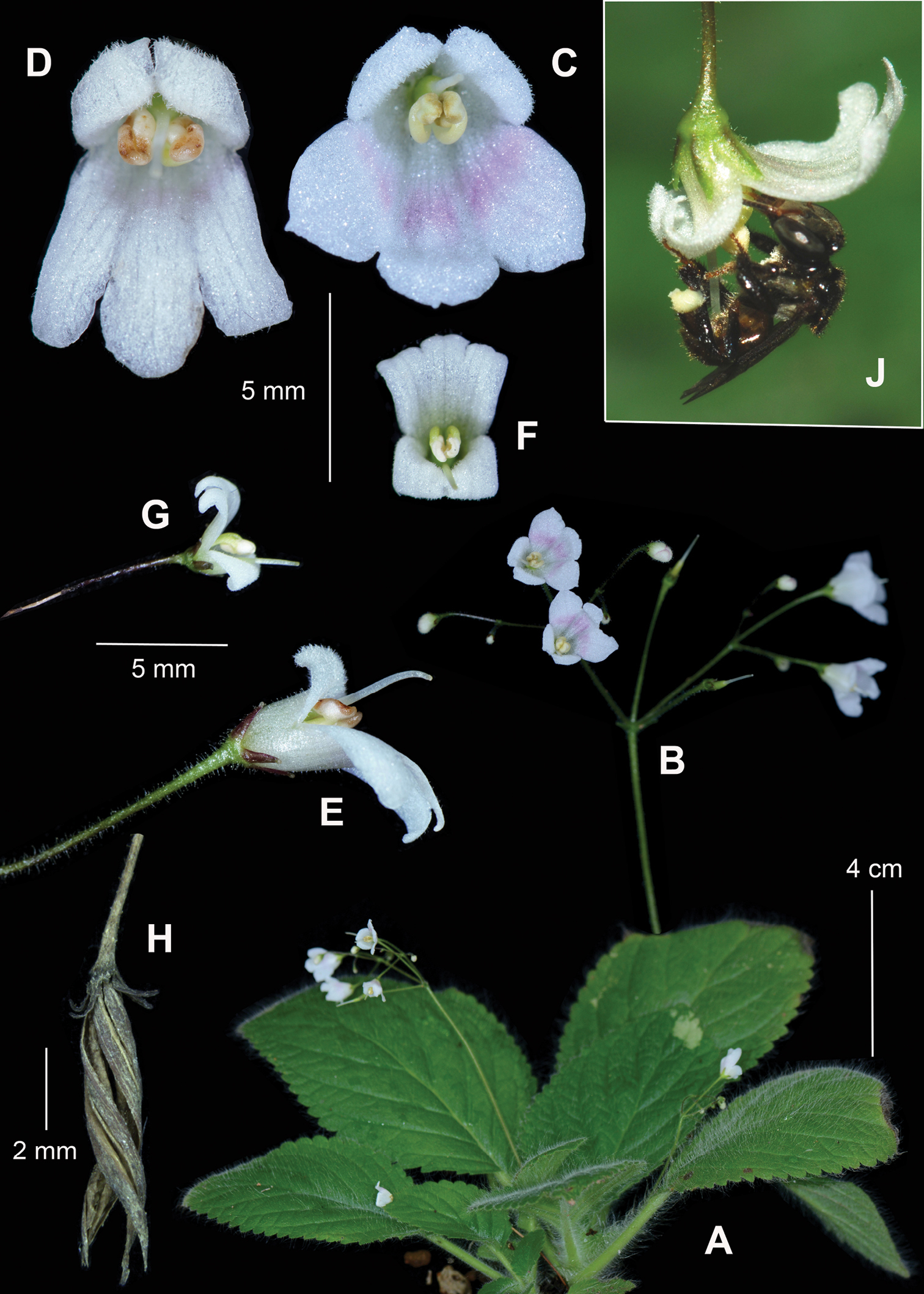
Let’s bring a high dose of beauty into today’s Friday Fellow with a wonderful species that may sometimes be found in your garden.
Imperial fritillary growing in its natural environment in Kurdistan. Photo by Wikipedia user A2raya07.*
Fritillaria imperialis, the imperial fritillary or crown imperial, is native from Asian highlands between Turkey and the Himalayas but is cultivated worldwide, having a series of artificially selected cultivars. The plant reaches a height of about 1 m and has a series of lance-shaped leaves along its stem, similarly to what is found in other species of the lily family, Liliaceae, to which it belongs. The flowers appear in a whorl close to the top of the stem and face downwards. A crown of small leaves tops the flowers, hence its name imperialis. The bell-shaped flowers are usually orange in the wild but, in cultivars, they vary between red and yellow.
A cultivar named ‘Rubra Maxima’. Photo by Hendry Heatly.**
The imperial fritillary has been used in traditional medicine for centuries by people living around its native range. Recent studies revealed that the plant contains a series of alkaloids, mostly anticholinergic steroidal alkaloids, which have the potential to be used for the development of new medicines to treat several conditions.
Despite its popularity as an ornamental plant, wild populations of the imperial fritillary are endangered in many countries in which it occurs, especially due to habitat loss. In order to aid in the preservation and restoration of wild populations, some laboratory techniques have been developed to generate clones that could help increase population size in the wild.
Akhtar MN, Rahman A, Choudhary MI, Sener B, Erdogan I, Tsuda Y (2003) New class of steroidal alkaloids from Fritillaria imperialis. Phytochemistry 63: 115–122. doi: 10.1016/S0031-9422(02)00569-1
Gilani AH, Shaheen F, Christopoulos A, Mitchelson F (1997) Interaction of ebeinone, an alkaloid from Fritillaria imperialis, at two muscarinic acetylcholine receptor subtypes. Life Sciences 60 (8): 535–544. doi: 10.1016/S0024-3205(96)00691-1
Kiani M, Mohammadi S, Babaei A, Sefidkon F, Naghavi MR, Ranjbar M, Razavi SA, Saeidi K, Jafari H, Asgardi D, Potter D (2017) Iran supports a great share of biodiversity and floristic endemism for Fritillaria spp. (Liliaceae): A review. Plant Diversity 39(5): 245–262. doi: 10.1016/j.pld.2017.09.002
Mohammadi-Dehcheshmeh M, Khalighi A, Naderi R, Sardari M, Ebrahimie E (2008) Petal: a reliable explant for direct bulblet regeneration of endangered wild populations of Fritillaria imperialis L. Acta Physiologiae Plantarum 30(3): 395–399. doi: 10.1007/s11738-007-0126-2
Today we celebrate the birthday of a French scientist and surgeon that achieved a great renown during his time.
Jean-Louis Petit, usually known as Petit the surgeon, was born on 13 May 1674 in Paris. He was interested in anatomy since childhood and received lessons on the subject from the anatomist Alexis Littré, who lived in his house. When Petit was only 12 years old, Littré trusted him his anatomical theater.
After studying surgery with the surgeon Georges Mareschal, Petit obtained the title of master in surgery in 1700. In 1705, he published a work entitled L’Art de guérir les maladies des os (The art of curing bone diseases), which would be translated into English in 1726.
Portrait of Jean-Louis Petit by Ambroise Tardieu.
In 1715, he became a member of the French Royal Academy of Sciences and, in 1731, was named, by the King Louis XV, director of the Royal Academy of Surgery after its foundation.
Petit’s reputation was achieved due to his talent and experience, especially because of his case reports of bleeding, lacrimal fistulas and operations on the frenum of the penis, as well as his work on bone diseases. Due to his fame, he was invited to Poland in 1726 to treat King Augustus II, and to Spain in 1735 to treat Prince Ferdinand VI. Both offered great advantages for him to stay in their countries, but he decided to return to France.
Petit is also acknowledged for the first broad clinical description of an epidural hematoma and its treatment by trepanning at the opposite side of the beginning of neurological signs.
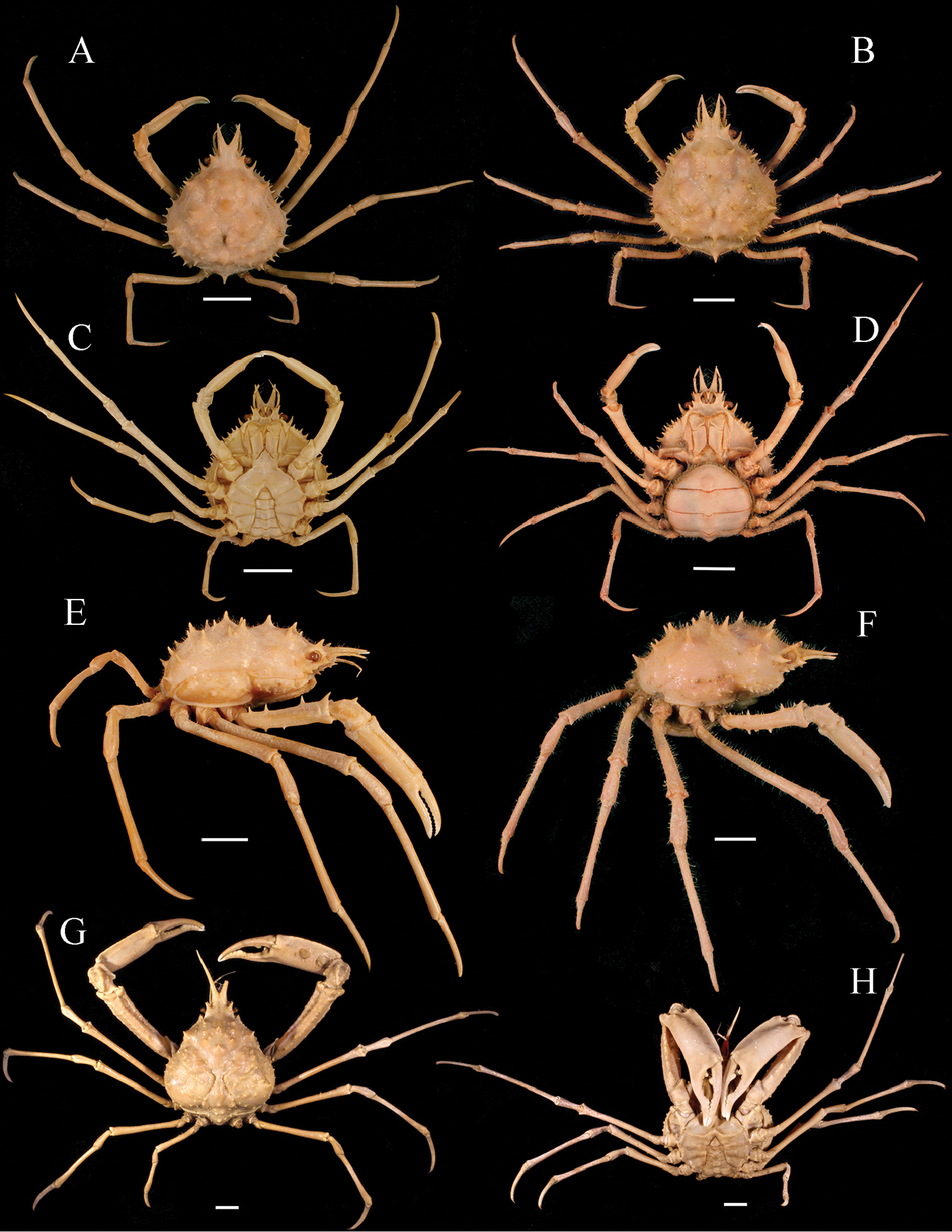
Beneath fallen logs and rocks or in the leaf litter of forests and gardens around the La Plata River in Argentina and Uruguay, you may find today’s fellow. Scientifically known as Discocyrtus prospicuus, it is a harvestman, a member of a group of arachnids that resemble spiders. As usual among small hidden invertebrates, it lacks a common name, so I coined the term Platine shield harvestman to refer to it.
Discocyrtus prospicuus in Buenos Aires, Argentina. Photo by Nicolas Olejnik.*
The Platine shield harvestman belongs to the family Gonyleptidae, which includes harvestmen with astonishing armored bodies and a prosoma (or cephalothorax) with a triangular shape resembling some sort of shield. It has a dark reddish brown color and two powerful hindlegs armed with several spines.
The Platine shield harvestman is found in several localities of Argentina and Uruguay, but especially in forested areas around the La Plata River and its tributaries. As usually among gonyleptid harvestmen, the Platine shield harvestman is dependent on environments with a considerable degree of humidity.
Different from most arachnids, harvestmen are usually omnivorous scavengers, feeding on dead animal and plant material, and the Platine shield harvestman is not different. In predator-prey relationships, they are usually the prey of other animals, especially spiders, such as wolf spiders that share the same habitat. When facing a large spider that is about to hunt it, the Platine shield harvestman can use a series of defense mechanisms. One of the simplest ways to avoid being eaten is remaining motionless or playing dead, a behavior called thanatosis. When facing an apparently dead harvestman, a wolf spider usually ignores it completely, as if it wasn’t even there. When this is not enough to stop the attack, the harvestman can use additional strategies, such as “showing its butt” to the spider by lifting its abdomen toward the predator and sometimes kicking the spider with its hind legs. Another common defense mechanism in harvestmen is releasing chemicals with a strong and repulsive scent but the Platine shield harvestman does not seem to use it often, at least not against spiders.
Little is known about the natural history of the Platine shield harvestman or of any of its close relatives. As I said several times before, we need more people studying the small creatures that live all around us.
Costa LE, Guerrero EL (2011) Geographical distribution of Discocyrtus prospicuus (Arachnida: Opiliones: Gonyleptidae): Is there a pattern? Zootaxa 2043: 1–24.
Fernandes NS, Stanley E, Costa FG, Toscano-Gadea CA, Willemart RH (2017) Chemical sex recognition in the harvestman Discocyrtus prospicuus (Arachnida: Opiliones). Acta Ethologica 20(3): 215–221. doi: 10.1007/s10211-017-0264-5
Segalerba A, Toscano-Gadea CA (2016) Description of the Defensive Behaviour of Four Neotropical Harvestmen (Laniatores: Gonyleptidae) Against a Synchronic and Sympatric Wolf Spider (Araneae: Lycosidae). Arachnology 17(1): 52–58. doi:10.13156/arac.2006.17.1.52
Today we celebrate the birthday of a French zoologist and rival of Darwin.
Charles Émile Blanchard was born in Paris on 6 March 1819, the son of the painter Émile-Théophile Blanchard. Due to his father’s interest in nature, Blanchard started to engage in the study of natural history since an early age. When he was 14, in 1833, the naturalist Jean Victoire Audouin allowed him access to the laboratory of the Muséum National d’Histoire Naturelle. Five years later, in 1838, he became a technician in the museum and, in 1841, was promoted to assistant-naturalist.
Soon after that, Blanchard went on a marine zoology expedition to Sicily with the zoologists Henri Milne-Edwards and Jean Louis Armand de Quatrefages de Breau.
In 1845, Blanchard published a book entitled Histoire des Insects (History of the Insects). About a decade later, from 1854 to 1856, he published his work Zoologie Agricole (Agricultural Zoology), which is a remarkable work, illustrated by his father, which presents in great details how harmful species damage crops. Between 1852 and 1864 he also published an atlas of the anatomy of the vertebrates.
From about 1860 on, Blanchard started to gradually lose sight. However, this did not discourage him to continue his work.
In 1862, he received the chair of crustaceans, arachnids and insects of the Muséum and was elected a member of the French Academy of Sciences. By this time, he started to gradually restrict the access by amateurs to the collections of the museum, which led the overall activity of the museum to decline and the collections to become dispersed.
In 1870, with the death of August Duméril, the chair of reptiles and fish of the Muséum was left vacant and Blanchard was hoping to receive it due to his atlas of the anatomy of vertebrates, but the chair was given to Léon Vaillant instead.
Blanchard in his late years.
Blanchard was openly against Darwinism and stated that Darwin’s ideas on evolution were false and unoriginal. In 1870, Darwin was nominated by Quatrefages and Milne-Edwards to be a corresponding member of the French Academy of Sciences. This was strongly opposed by Blanchard and others, which led Darwin to lose the election by a narrow margin.
Blanchard became completely blind in 1890, but continued with the chair of the crustaceans, arachnids and insects until 1894. He died on 11 February 1900, aged 84.
Here is a list of species described this month. It certainly does not include all described species. You can see the list of Journals used in the survey of new species here.
If you are walking around in the woods sometime after heavy rains, you may see clusters of small yellow to orange slick finger-like projections coming out of the barkless wood of dead trees such as oaks and other hardwoods. These little structures are the fruiting bodies of Calocera cornea, also known as the club-like tuning fork.
Calocera cornea growing on decaying oak wood. Photo by Ashley Duval.*
The club-like tuning fork may look at first like a club fungus, but those are distant relatives. It actually belongs to a group called Dacrymycetes, which constitutes one of the many groups commonly knowns as jelly fungi. The finger-like fruiting bodies, called basidiocarps, are very variable in shape, although usually not branched, and contain several Y-shaped basidia, each carrying two spores.
With a worldwide distribution, the club-like tuning fork grows on decaying wood of several trees, both angiosperms and gymnosperms, but is more fond of hardwoods such as the oak, so it is more commonly found in temperate forests in places such as North America and Eurasia. Its hyphae never grow very deep, being restricted to the more superficial layers of wood, and are very narrow, about 1µm in diameter only, and grow parallel to the long axis of the dead cells of the wood.
Club-like tuning fork growing together with other wood-decaying fungi. Photo by Christian Schwarz.*
Although usually not a species of economic concern, some strains of the club-like tuning fork may cause considerable decay in wood objects that have not been properly treated to avoid fungus growth.
Recently, the genome of Calocera cornea has been sequenced as part of a project that is trying to determine the origins of the ability of basidiomycetes to decompose lignocellulose, the main component of the cell walls of woody plants.
Kennedy LL (1972) Basidiocarp development in Calocera cornea. Canadian Journal of Botany 50(3): 413–417. doi: 10.1139/b72-060
McNabb RFR (1965) Taxonomic studies in the Dacrymycetaceae. II. Calocera (Fries) Fries. New Zealand Journal of Botany 3(1): 31–58. doi: 10.1080/0028825X.1965.10428712