Background
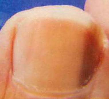
Melanonychia is brown or black pigmentation of the nail unit. Melanonychia commonly presents as pigmented band arranged lengthwise along the nail unit, and this presentation is known as longitudinal melanonychia or melanonychia striata. The most concerning cause of melanonychia is subungual melanoma, although a variety of other causes includes physiologic longitudinal melanonychia, systemic disorders, trauma, inflammatory disorders, fungal infections, drugs, and benign melanocytic hyperplasias. [1, 2] See the image below.
Pathophysiology
Melanonychia most often occurs because of increased production of melanin by melanocytes in the nail matrix. A healthy adult has approximately 200 melanocytes per mm2 in the nail matrix, of which the majority remain dormant. When these melanocytes are activated, melanosomes filled with melanin are transferred to differentiating matrix cells, which migrate distally as they become nail plate onychocytes. [3] This results in a visible band of pigmentation in the nail plate.
Etiology
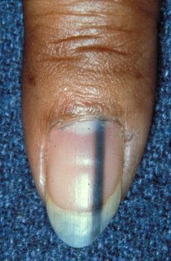
When determining the etiology of longitudinal melanonychia, it is important to distinguish between melanocytic activation and melanocytic hyperplasia. Melanocytic activation is caused by an increased synthesis of melanin with a normal number of melanocytes. Melanocytic hyperplasia (including melanocytic nevi and melanoma) refers to an increased synthesis of melanin with an increased number of melanocytes. Nevi constitute 12% of longitudinal melanonychia cases in adults and 50% of longitudinal melanonychia cases in children. Note the clinical image below.
Approximately two thirds of cases have a brown-black color, and one third of cases have periungual pigmentation (benign pseudo-Hutchinson sign). The bands are wider than 3 mm in greater than 50% of cases.
Below are the causes of melanonychia. [3, 4, 5, 6]
Melanocytic activation/hyperplasia–related causes are as follows:
-
Nevi
-
Melanotic macule of the nail unit
-
Melanocytic activation
-
Subungual melanoma
Physiologic causes of melanonychia are as follows:
-
Racial melanonychia (African American, Hispanic, Indian, Japanese, other dark-skinned races) (multiple bands)
-
Pregnancy (multiple bands)
Local and regional causes of melanonychia are as follows:
-
Trauma (acute or chronic) (single band)
-
Poor-fitting shoes (single band)
-
Onychotillomania (single band)
-
Nail biting (single band)
-
Carpal tunnel syndrome (single band)
-
Foreign body (subungual) (single band)
-
Radiation therapy (multiple bands)
-
Ultraviolet light (multiple bands) [7]
-
Postinflammatory hyperpigmentation (single band)
Systemic causes of melanonychia are as follows:
-
Addison disease (multiple bands)
-
Cushing syndrome (multiple bands)
-
Nelson syndrome (multiple bands)
-
Hyperthyroidism (multiple bands)
-
Hemosiderosis (multiple bands)
-
Hyperbilirubinemia (multiple bands)
-
Alcaptonuria (multiple bands)
-
Porphyria (multiple bands)
-
Acquired immunodeficiency syndrome (multiple bands)
-
Graft versus host disease (multiple bands)
-
Malnutrition (multiple bands)
-
Vitamin B-12 deficiency (multiple bands)
Dermatologic causes of melanonychia are as follows:
-
Psoriasis (multiple bands)
-
Lichen planus (multiple bands)
-
Chronic radiodermatitis (single bands)
-
Scleroderma (multiple bands)
-
Systemic lupus erythematosus (multiple bands)
-
Fungal infection of the nail bed (Aspergillus, Scopulariopsis, Candida, Blastomyces, Trichophyton, [8] Fonsecaea pedrosoi) (multiple bands)
-
Basal cell carcinoma (single band)
-
Bowen disease (single band)
-
Subungual fibrous histiocytoma (single band)
-
Mucous cyst (single band)
-
Lichen striatus (multiple bands)
Chemotherapeutic causes of melanonychia are as follows (multiple bands):
-
Bleomycin sulfate
-
Busulfan
-
Cyclophosphamide
-
Dacarbazine
-
Daunorubicin hydrochloride
-
Doxorubicin
-
Etoposide
-
5-Fluorouracil
-
Melphalan hydrochloride
-
Methotrexate
-
Nitrogen mustard
-
Nitrosourea
-
Tegafur
Other drug/agent-related causes of melanonychia are as follows (multiple bands):
-
Corticotropin
-
Amodiaquine
-
Amorolfine
-
Arsenic
-
Chloroquine
-
Clofazimine
-
Clomipramine
-
Cyclines
-
Diquat
-
Fluconazole
-
Fluoride
-
Gold salts
-
Ibuprofen
-
Ketoconazole
-
Lamivudine
-
Mepacrine
-
Mercury
-
Minocycline
-
Melanocyte-stimulating hormone
-
Polychlorinated biphenyl (PCB)
-
Phenytoin
-
Phenothiazine
-
Psoralen [7]
-
Roxithromycin
-
Steroids
-
Sulfonamide
-
Tetracycline
-
Thallium
-
Timolol
-
Zidovudine
Causes of melanonychia associated with a syndrome are as follows:
-
Laugier-Hunziker syndrome (multiple bands) [11]
-
Peutz-Jeghers syndrome (multiple bands)
-
Touraine syndrome (multiple bands)
Epidemiology
Frequency
Physiologic melanonychia is more common in darker-pigmented individuals. Seventy-seven percent of black individuals older than 20 years and almost 100% older than 50 years have evidence of this condition. [12, 13] Longitudinal melanonychia is present in 10-20% of Japanese individuals. [4] In the general white population, the prevalence of longitudinal melanonychia is 1.4%. [14]
In a study of 68 Hispanic patients with longitudinal melanonychia, melanonychia secondary to skin pigmentation was observed in 48 cases (68.6%). [15] Of the remaining patients, 6 (8.5%) were secondary to trauma, 5 (7.1%) were secondary to fungal infection, 4 (5.7%) were associated with benign melanocytic hyperplasia, and 4 (5.7%) had a nail apparatus malignancy. The remaining 3 (4.3%) cases were of mixed etiology.
The prevalence of affected individuals increases with age. [14, 16]
Race
The frequency of melanonychia varies by the degree of skin pigmentation, as described in Frequency, International.
Sex
Melanonychia affects males and females equally. [16]
Age
Typically, melanonychia is more common in older individuals. In children, melanonychia is often caused by melanocytic nevi. Subungual melanoma or melanoma in situ is very rare in children, [17, 18] but has been reported. [19]
Prognosis
In cases of melanonychia associated with systemic diseases, treatment of the primary condition may improve the nail pigmentation. Similarly, discontinuation of any offending drugs may improve melanonychia.
The prognosis of patients with subungual melanoma is poor. In a 2007 series of 106 subungual melanomas, 48 (45%) patients developed a recurrence or metastasis and, of these, 33 (69%) died from their disease during the follow-up period (median 56 mo). Patients with a melanoma deeper than 2.5 mm had a statistically worse survival rate than those with thin melanomas. Melanonychia secondary to subungual melanoma has the highest morbidity and mortality compared with other body sites, with reported 5- and 10-year survival rates of 30% and 13%, respectively. [20]
Patient Education
Patients should be instructed to follow lesions of suspected benign causes of longitudinal melanonychia for any change in color, pattern, size of the band, or new onset pain and/or ulceration, as these can be signs of subungual melanoma.
-
Longitudinal melanonychia secondary to a nevus.
-
Pigmented longitudinal streak secondary to a nail matrix melanoma.


