Buergenerula spartinae

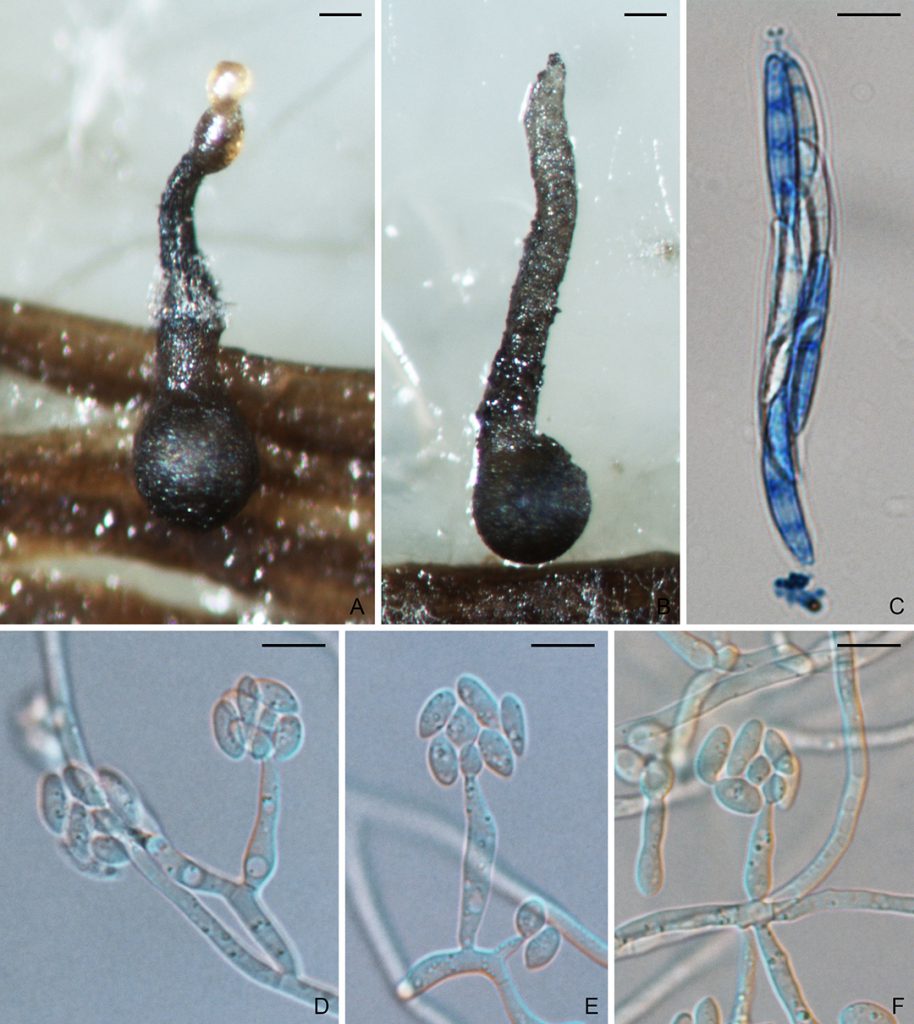
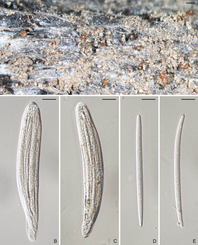
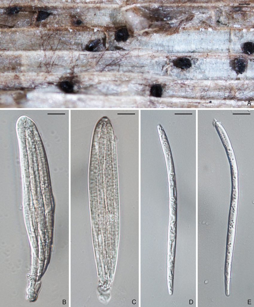
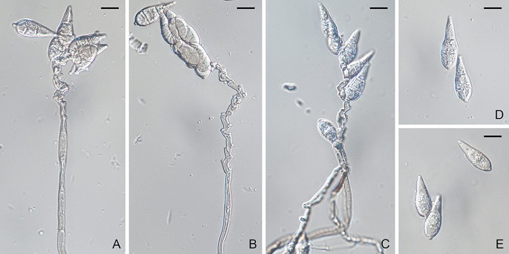
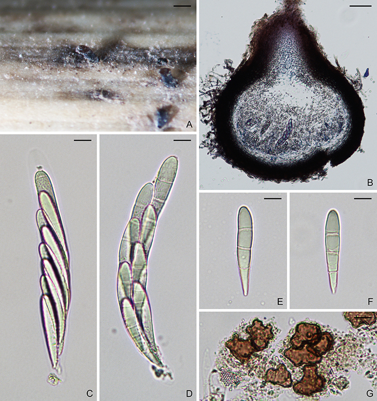
Figure. Buergenerula spartinae (NYBG986085). A–B. Ascomata. C–D. Asci. E–F. Ascospores. G. Hyphopodium. Scale bars: A = 200 µm; B = 50 µm; C–G = 10 µm.

Buergenerula spartinae Kohlm. & R.V. Gessner, Can. J. Bot. 54(15): 1764 (1976).
MycoBank: MB310003.

Ascomata perithecial, immersed, solitary or gregarious, dark brown to black, 200–450 µm diam, with a cylindrical, hyaline to brownish neck, 150–250 × 90–150 µm. Paraphyses hyaline, septate, dissolving at maturity. Asci 8-spored, unitunicate, clavate, 100–165 × 15–20 µm, with a refractive ring. Ascospores biseriate in ascus, clavate, rounded at apex, narrowed toward base, straight or slightly curved, 3-septate, not or slightly constricted at septum, hyaline to yellowish, smooth, 35–55 × 9–13 µm. On CMA with seawater, conidiophores branched, up to 20 µm long. Conidiogenous cells phialidic. Conidia curved, 5 × 2 µm (Asexual state description from Kohlmeyer and Gessner, 1976).

Typification: Holotype NY J. Kohlmeyer (JK) No. 3498. Isotype IMS JK3498. Paratypes NY and IMS JK3503.

Specimens examined: Argentina, Buenos Aires, on Spartina alterniflora, 17 Oct. 1973, JK3498 (NY986083 notes, NY986084 dried specimen, NY986085 slides, NY986086 slides), JK3503 (NY986087).

Gene sequences: DQ341492 (28S), JX134666 (ITS), JX134692 (TEF1).

Genome sequences: SRX798618 (transcriptome).

Hosts/substrates: On leaf sheaths and dead stalks of Spartina and Panicum virgatum (Poaceae).

Distribution: Argentina, Canada, China, UK, USA.

Notes: Buergenerula spartinae was reported mainly on old leaf sheaths and dead stalks of Spartina in salt marshes.

Copyright 2022 by The American Phytopathological Society. Reproduced, by permission, from Luo, J., and Zhang, N. 2022. The Rice Blast Fungus and Allied Species: A Monograph of the Fungal Order Magnaporthales (https://my.apsnet.org/APSStore/Product-Detail.aspx?WebsiteKey=2661527A-8D44-496C-A730-8CFEB6239BE7&iProductCode=46826). American Phytopathological Society, St. Paul, MN. 

Magnaporthiopsis poae

Figure. Magnaporthiopsis poae (M47 X M48). A–B. Ascomata. C. Ascus and ascospores. D–F. Conidiophores and conidia. Scale bars: A–B = 100um; C–F = 10µm.

Magnaporthiopsis poae (Landsch. & N. Jacks.) J. Luo & N. Zhang., Mycologia 105(4): 1024 (2013).
MycoBank: MB802973.
        ≡ Magnaporthe poae Landsch. & N. Jacks. Mycol. Res. 93:59 (1989).

Ascomata perithecial, superficial or submerged, solitary or gregarious, dark brown to black, 230–500 µm diam, with a cylindrical, hyaline to brownish neck, 300–650 × 100–135 µm. Paraphyses septate, hyaline, dissolving at maturity. Asci 8-spored, unitunicate, clavate, 60–100 × 8–15 µm, with a refractive ring. Ascospores 1–4-seriate, fusiform, straight or slightly curved, 3-septate, not or slightly constricted at septum, unequally colored, smooth, 20–40 × 3.5–5.5 µm, with darkly pigmented inner cells. Asexual state phialophora-like. On CMA, hyphae branched, septate, hyaline to light brown, smooth, 2–5 µm diam. Conidiophores micronematous, solitary, erect, straight or curved, unbranched or sparsely branched, hyaline, smooth, 1–4-septate, 5–40 × 3–6.5 µm. Conidiogenous cells phialidic, erect, terminal or intercalary, hyaline, smooth, 5–20 × 2–6 µm. Conidia aggregated in slimy heads, ovoid to ellipsoidal, straight or slightly curved, aseptate, hyaline, smooth, 3–9 × 2–5 µm.

Colonies on PDA 7 cm diam after 7 days at 25 °C in dark; surface floccose, parrot green; aerial mycelium yellowish; reverse cedar green. Colonies on CMA reaching 6.5 cm after 7 days in dark at 25 °C; surface pale yellow green; aerial mycelium sparse; reverse pale yellow green.

Typification: Holotype DAR59044. Ex-holotype culture ATCC64411, ATCC64412.

Gene sequences: JF414860 (18S), JF414836 (ITS), JF414885 (28S), JF710390 (MCM7), JF710433 (RPB1), JF710415 (TEF1).

Genome sequences: ADBL01000000 (genome).

Specimens examined: USA, California, Nipomo, on Festuca, 19 Sep. 2018, D. Mosdell, RUTPP180919a3, RUTPP180919d1; New Jersey, New Brunswick, on Poa pratensis, 11 Aug. 2011, S. Zhao and L. Bierns, M47, M48; ibid., Adelphia, on Poa pratensis, 22 Aug. 2018, P.L. Vines, RUTPP11s-1, RUTPP1213-1, RUTPP14t-1, RUTPP1412-10, RUTPP1416-1; New York, on unknown host, 25 Aug. 2018, S. Tirpak and R. Buckley, RUTPPd29709-13.

Hosts/substrates: From Poaceae.

Distribution: Canada (Ontario), China, USA (California, New Jersey, New York).

Notes: Magnaporthiopsis poae infects turf roots, crowns, and leaf sheaths, and causes summer patch disease on Kentucky bluegrass (Poa pratensis), annual bluegrass (Poa annua), fine fescue (Festuca), perennial ryegrass (Lolium perenne), and bentgrass (Agrostis). The summer patch disease has been primarily reported in temperate regions of North America, ranging from Canada (Ontario) to USA from New Jersey and North Carolina as far west as California.

Copyright 2022 by The American Phytopathological Society. Reproduced, by permission, from Luo, J., and Zhang, N. 2022. The Rice Blast Fungus and Allied Species: A Monograph of the Fungal Order Magnaporthales (https://my.apsnet.org/APSStore/Product-Detail.aspx?WebsiteKey=2661527A-8D44-496C-A730-8CFEB6239BE7&iProductCode=46826). American Phytopathological Society, St. Paul, MN.

Gaeumannomyces tritici

Figure. Gaeumannomyces tritici (BPI 626756). A. Ascomata. B–C. Asci. D–E. Ascospores. Scale bars: A = 200um; B–E = 10µm.

Gaeumannomyces tritici (J. Walker) Hern.-Restr. & Crous, in Hernández-Restrepo, Groenewald, Elliott, Canning, Mcmillan & Crous, Stud. Mycol. 83: 26 (2016).
MycoBank: MB816900.
    ≡ Gaeumannomyces graminis var. tritici J. Walker, Trans. Br. mycol. Soc. 58(3): 439 (1972).

Morphological description: Ascomata perithecial, superficial or submerged, solitary or gregarious, dark brown to black, 200–450µm in diam, with a cylindrical, hyaline to brownish neck, 150–300 × 55–80 µm. Paraphyses hyaline, septate, dissolving at mature. Asci unitunicate, clavate, 8-spored, 90–120 × 10–18 µm, with a refractive ring. Ascospores parallel in the ascus, hyaline to yellowish, long fusiform to filiform, straight or slightly curved, 0–5-septate, not constricted at the septum, smooth, 70–110 × 3–5 µm.
    Conidiophores solitary, erect, straight or curved, unbranched or sparsely branched, hyaline, smooth. Conidiogenous cells phialidic, erect, terminal or intercalary, hyaline, smooth, 9–18 × 2–4 µm, with a cylindrical collarette, 1–3 × 1–2 µm. Conidia, lunate, allantoid to fusiform, straight to curved, hyaline, smooth, aseptate, 4–7 × 1–2 µm.

Specimens examined: Australia, New South Wales, on Triticum aestivum, 25 Feb. 1970, A.M. Smith, dar 19601 (BPI 626755); ibid., 12 Nov. 1969, L. Meakins, dar 19118 (BPI 626756). USA, New York, Geneva, on Triticum sativum, 5 Jul. 1921, R.J. Haskell and R.S. Kirby, BPI 626757.

Pathogenicity: Gaeumannomyces tritici can cause take-all diseases of wheat (Triticum), Barley (Hordeum), couch grass (Elymus repens), and wheatgrass (Agropyron) worldwide.

References:
Hernández-Restrepo M, Groenewald JZ, Elliott ML, Canning G, McMillan VE, Crous PW. 2016. Take-all or nothing. Studies in Mycology 83:19–48.

Geographical distribution: Australia, New South Walles. USA, New York.

Copyright 2022 by The American Phytopathological Society. Reproduced, by permission, from Luo, J., and Zhang, N. 2022. The Rice Blast Fungus and Allied Species: A Monograph of the Fungal Order Magnaporthales (https://my.apsnet.org/APSStore/Product-Detail.aspx?WebsiteKey=2661527A-8D44-496C-A730-8CFEB6239BE7&iProductCode=46826). American Phytopathological Society, St. Paul, MN.

Gaeumannomyces oryzinus

Figure. Gaeumannomyces oryzinus (BPI 626741). A. Ascomata. B–C. Asci. D–E. Ascospores. Scale bars: A = 200um; B–E = 10µm.

Gaeumannomyces oryzinus (Sacc.) Schrantz, Bull. trimest. Soc. mycol. Fr. 76(4): 337 (1961).
MycoBank: MB331025.
    ≡ Ophiobolus oryzinus Sacc., G. bot. ital., n.s. 23(2): 203 (1916).
    ≡ Linocarpon oryzinum (Sacc.) Petr., Sydowia 6(5-6): 387 (1952).
    ≡ Gaeumannomyces oryzinus (Sacc.) Schrantz, Bull. trimest. Soc. mycol. Fr. 76(4): 337 (1961).

Morphological description: Ascomata perithecial, superficial or submerged, solitary or gregarious, dark brown to black, 200–400µm in diam, with a cylindrical, hyaline to brownish neck, 100–300 × 50–80 µm. Paraphyses hyaline, septate, dissolving at mature. Asci unitunicate, clavate, 8-spored, 90–135 × 13–20 µm, with a refractive ring. Ascospores parallel in the ascus, hyaline to yellowish, long fusiform to filiform, straight or slightly curved, 0–5-septate, not constricted at the septum, smooth, 90–115 × 3–6 µm.
    Conidiophores solitary, erect, straight or curved, unbranched or sparsely branched, hyaline, smooth. Conidiogenous cells phialidic, erect, terminal or intercalary, hyaline, smooth, 7–20 × 2–6 µm, 2–3.5 µm, with a cylindrical collarette, 1–3 × 1–2 µm. Conidia, lunate, allantoid to fusiform, straight to curved, hyaline, smooth, aseptate, 5–9 × 1–3 µm.

Material examined: China, Hong Kong, on Oryza sativa, 14 Apr. 1975, S. Jordan, BPI 626742. USA, Arkansas, Ahuyna, on Oryza sativa, 8 Oct. 1928, E.C. Zieller, BPI 626741.

Pathogenicity: Gaeumannomyces oryzinus was reported from rooting roots and culms mainly on rice (Oryza sativa), but was also found on Bermuda grass (Cynodon sp.) as well.

References:
Hernández-Restrepo M, Groenewald JZ, Elliott ML, Canning G, McMillan VE, Crous PW. 2016. Take-all or nothing. Studies in Mycology 83:19–48.

Geographical distribution: China, Hong Kong. USA, Arkansas.


Nakataea oryzae

Figure. Nakataea oryzae (BPI 617624, M69). A. Ascomata. B–C. Asci. D–E. Ascospores. F–G. Conidiophores. H–J. Conidia. D. Ascus and ascospores. K–L. Sclerotia. M. Hyphopodium. Scale bars: A = 400µm; B–E = 20 µm; F–J, M = 10 µm; K–L = 200µm.

Nakataea oryzae (Catt.) J. Luo & N. Zhang, Mycologia 105(4): 1025 (2013).
MycoBank: MB802971.
        ≡ Sclerotium oryzae Catt., Arch. Triennale Lab. Bot. Crittog. 1:10 (1877).
= Nakataea sigmoidea (Cavara) Hara, Diseases Rice Plant, 2:185 (1939).
        ≡ Helminthosporium sigmoideum Cavara, Mat. Lomb.:15 (1889).
= Magnaporthe salvinii (Catt.) R.A. Krause & R.K. Webster, Mycologia 64: 110 (1972).
        ≡ Leptosphaeria salvinii Catt., Arch. Labor. Bot. Critt. Univ. Pavia 2, 3:115–128 (1879).

Ascomata perithecial, superficial or immersed, solitary or gregarious, globose to subglobose, dark brown to black, 300–600 µm diam, with a cylindrical, light brown neck, 350–650 × 100–125 µm. Paraphyses septate, hyaline, dissolving at maturity. Asci 8-spored, unitunicate, clavate, 120–165 × 10–23 µm, with a refractive ring. Ascospores 1–4-seriate, fusiform, curved, 3-septate, not or slightly constricted at septum, hyaline to pale brown, smooth, 55–70 × 7.5–10 µm. Hyphae branched, septate, light brown to brown, smooth, 1–3.5 µm diam. Conidiophores solitary, erect, straight or curved, unbranched or sparsely branched, 4–7-septate, brown, smooth, 125–145 × 3.5–6.5 µm. Conidiogenous cells terminal or intercalary, light brown, smooth, 15–35 × 3.5–5.5 µm, 1–3.5 µm wide at base, 0.5–1.5 µm wide near apex. Conidia falcate, curved, 3-septate, unequally colored, smooth, 55–75 × 12–17.5 µm; inner cells brown; end cells yellowish to light brown.

Typification: Holotype BPI841381.

Gene sequences: JX134658 (18S), JX134672 (ITS), JX134684 (28S), JX134712 (MCM7), JX134726 (RPB1), JX134698 (TEF1).

Genome sequences: SRX798605 (genome), SRX798628 (transcriptome).

Specimens examined: Unknown, on Sachs rice-straw-agar medium, M. Tsuda, BPI617624. USA, California, Glenn County, 3 Jun. 2011, J. Oster, RUTPPM69, RYTPPM70, RYTPPM71.

Hosts/substrates: On stems of Oryza as well as Echinochloa and Zizania (Poaceae).

Distribution: Worldwide.

Notes: Cattaneo (1877) first named the sclerotial state of this species as Sclerotium oryzae. Hara (1939) formerly introduced Nakataea sigmoidea (synonym Helminthosporium sigmoideum) for the conidial state while the ascosporic state was described as Magnaporthe salvinii (synonym Leptosphaeria salvinii) by Krause and Webster (1972). Luo and Zhang (2013) regarded Nakataea oryzae for this species using the oldest legitimate generic name and species. It causes stem rot disease of Oryza and has also been reported from Zizania and other Poaceae.

Copyright 2022 by The American Phytopathological Society. Reproduced, by permission, from Luo, J., and Zhang, N. 2022. The Rice Blast Fungus and Allied Species: A Monograph of the Fungal Order Magnaporthales (https://my.apsnet.org/APSStore/Product-Detail.aspx?WebsiteKey=2661527A-8D44-496C-A730-8CFEB6239BE7&iProductCode=46826). American Phytopathological Society, St. Paul, MN.

Pyricularia grisea

Figure. Pyricularia grisea (BPI 881984). A–E. Conidiophores and conidia. Scale bars: A–E = 10µm.

Pyricularia grisea Cooke ex Sacc., Michelia 2(no. 6): 20 (1880).
MycoBank: MB224559.
        ≡ Trichothecium griseum (Cooke ex Sacc.) Cooke, in Ravenel, Amer. Fungi: no. 580 (1881).
        ≡ Dactylaria grisea (Cooke ex Sacc.) Shirai, in Miyake, J. Coll. Agric. imp. Univ. Tokyo 2: 262 (1910).
= Magnaporthe grisea (T.T. Hebert) M.E. Barr, Mycologia 69:954 (1977).
        ≡ Ceratosphaeria grisea T.T. Hebert, Phytopathology 61:86 (1971).
        ≡ Phragmoporthe grisea (T.T. Hebert) M. Monod, Beih. Sydowia 9: 153 (1983).

Ascomata perithecial, superficial or immersed, solitary or gregarious, dark brown to black, 60–300 µm diam, with a cylindrical, hyaline to light brown neck, 60–150 × 100–1200 µm. Paraphyses septate, hyaline, dissolving at maturity. Asci 8-spored, unitunicate, clavate, 7–10 × 55–90 µm, with a refractive ring. Ascospores 1–3-seriate, fusiform, straight or slightly curved, 3-septate, not or slightly constricted at septum, hyaline, smooth, 4–7 × 17–24 µm (Sexual state description from Hebert, 1971). Conidiophores solitary, erect, straight or curved, unbranched, 1–3-septate, brown, smooth, 100–135 × 3–6.5 µm. Conidiogenous cells integrated, terminal or intercalary, light brown, smooth, 80–110 × 3–6 µm, with several protruding denticles, 0.5–1.5 × 1–2 µm. Conidia solitary, pyriform to obclavate, 2-septate, light brown, smooth to finely roughened, 24–35 × 7–11 µm, with a truncate hilum, 0.5–1.5 × 1–2 µm.

Typification: Lectotype of Pyricularia grisea BPI, Ellis & Everhart, North American Fungi no. 374 (Rossman et al., 1990). Epitype of P. grisea CBSH-22280 (Crous et al., 2015a). Ex-epitype culture of P. grisea CBS138707 (US0043) (Crous et al., 2015a). Holotype of “Magnaporthe grisea” BPI625033 (Hebert, 1971).

Gene sequences: JX134656 (18S), JX134670 (ITS), JX134682 (28S), KM485187 (ACT), KM485258 (CAL), JX134710 (MCM7), JX134724 (RPB1), JX134696 (TEF1).

Genome sequences: PRJEB7653 (genome), SRX798638 (transcriptome).

Specimens examined: USA, Georgia, on Digitaria sanguinalis, Aug. 2000, L.P. Tredway, 1120-20 (BPI881983), 1100-4 (BPI881984); Indiana, on Digitaria, 3 Jun. 2011, J. Xu, M63; New Jersey, on Digitaria, 2 Aug. 2013, J. Luo and N. Zhang, RUTPPpg130802a, RUTPPpg130802d.

Hosts/substrates: On Digitaria, Echinochlos crus-galli var. frumentacea, and Lollium perenne (Poaceae).

Distribution: Worldwide.

Notes: Saccardo (1880a, b) first proposed Pyricularia grisea for a species from Digitaria sanguinalis in New Jersey. Cavara (1892) subsequently introduced P. oryzae for another species from Oryza sativa in Italy. These two species are morphological similar. Historically P. grisea was applied to grass and other host isolates, while P. oryzae to rice isolates. According to morphology and success interfertility among isolates from various hosts, Rossman et al. (1990) synonymized P. oryzae under P. grisea. However, Couch and Kohn (2002) revealed generic distinction between them based on multigene phylogeny and RFLP assay. They proposed P. grisea for the crabgrass isolates, and P. oryzae for rice and other grass isolates. Klaubauf et al. (2014) further investigated the phylogeny of Pyricularia and refined definitions of both species. Currently P. grisea includes isolates mostly on Digitaria, as well as few on Echinochlos crus-galli var. frumentacea and Lollium perenne. Pyricularia grisea can cause leaf spot or leaf blight diseases on Digitaria.

Copyright 2022 by The American Phytopathological Society. Reproduced, by permission, from Luo, J., and Zhang, N. 2022. The Rice Blast Fungus and Allied Species: A Monograph of the Fungal Order Magnaporthales (https://my.apsnet.org/APSStore/Product-Detail.aspx?WebsiteKey=2661527A-8D44-496C-A730-8CFEB6239BE7&iProductCode=46826). American Phytopathological Society, St. Paul, MN.

Pyricularia oryzae

Figure. Pyricularia oryzae (181001p). A–E. Conidiophores and conidia. Scale bars: A–E = 10µm.

Pyricularia oryzae Cavara, Fung. Long. Exsicc. 1: no. 49 (1892).
MycoBank: MB224486.
        ≡ Dactylaria oryzae (Cavara) Sawada, Spec. Bull. Agric. Exp. Station Formosa 16: 59 (1917).
= Magnaporthe oryzae B.C. Couch, Mycologia 94:692 (2002).
= Pyricularia graminis-tritici Castroag., S.I. Moreira, Maciel, B.A. McDonald, Crous & Ceresini, Persoonia 37: 211 (2016).

Ascomata perithecial, superficial or submerged, solitary or gregarious, dark brown to black, 60–300 µm diam, with a cylindrical, hyaline to brownish neck, 60–150 × 100–1200 µm. Paraphyses hyaline, septate, dissolving at maturity. Asci 8-spored, unitunicate, clavate, 7–10 × 55–90 µm, with a refractive ring. Ascospores 1–3-seriate, fusiform, straight or slightly curved, 3-septate, not or slightly constricted at septum, hyaline, smooth, 4–7 × 17–24 µm (Sexual state description from (Couch and Kohn, 2002; Yaegashi and Udagawa, 1978). Conidiophores solitary, erect, straight or curved, unbranched, 1–3-septate, brown, smooth, 85–140 × 3–6.5 µm. Conidiogenous cells integrated, terminal or intercalary, pale brown, smooth, 65–100 × 3–6 µm, with several protruding denticles, 0.5–1.5 × 1–2 µm. Conidia solitary, pyriform to obclavate, 2-septate, light brown, smooth to finely roughened, 25–33.5 × 7–11 µm, with a truncate hilum, 0.5–1.5 × 1–2 µm.

Typification: Lectotype of Pyricularia oryzae Cavara, Fungi Longobardiae no. 49 (Rossman et al., 1990). Holotype of “Magnaporthe oryzae” BPI841383 (Couch and Kohn, 2002). Isotype of “M. oryzae” TRTC52742 (Couch and Kohn, 2002).

Genome sequences: AACU00000000 (Genome).

Specimens examined: Guyana and other localities, cross of strains from Oryza sativa and Eleusine, 2002, B.C. Couch, Guy 11 and 2539 (BPI841383). USA, New Jersey, New Brunswick, on Festuca brevipila, 1 Oct. 2018, P.L. Vines, RUTPP181001p4; RUTPP181001p48; RUTPPd300452; RUTPPd300453; ibid., Adelphia, on Festuca arundinacea, 17 Oct. 2018, P.L. Vines, RUTPP181017p1; RUTPP181017p2.

Hosts/substrates: On Eleusine coracana, Festuca arundinacea, F. brevipila, Hordeum vulgare, Lolium perenne, Oryza sativa, Setaria italica, and Triticum (Poaceae).

Distribution: Worldwide.

Notes: Rice blast caused by Pyricularia oryzae is one of the most devastating diseases of Oryza sativa (rice) worldwide. Pyricularia oryzae can also cause blast diseases on Triticum (wheat), which emerged in Brazil in 1985 and has spread to Argentina, Bolivia, and Paraguay as well as Bangladesh by 2016 (Callaway, 2016). It can infect and damage other crops including Eleusine coracana (finger millet) and Setaria italica (foxtail millet). In addition, Pyricularia oryzae was proven to be causal agent of the grey leaf spots on turfgrasses, Lolium perenne (perennial ryegrass) and Festuca arundinacea (tall fescue), and a recent study has confirmed its pathogenicity on Festuca brevipila (hard fescue) (Vines et al., 2021).

Copyright 2022 by The American Phytopathological Society. Reproduced, by permission, from Luo, J., and Zhang, N. 2022. The Rice Blast Fungus and Allied Species: A Monograph of the Fungal Order Magnaporthales (https://my.apsnet.org/APSStore/Product-Detail.aspx?WebsiteKey=2661527A-8D44-496C-A730-8CFEB6239BE7&iProductCode=46826). American Phytopathological Society, St. Paul, MN.

Magnaporthiopsis agrostidis

Magnaporthiopsis agrostidis P. Wong, Khemmuk & R.G. Shivas
MycoBank: MB814222.

Asexual state phialophora-like. On PDA, hyphae branched, septate, hyaline to dark brown, smooth, 1–4 µm diam. Conidiophores micronematous, simple, branched, brown. Conidiogenous cells phialidic, terminal or intercalary, cylindrical to lageniform, straight or curved, brown to light brown, 5–20 × 1.5–3 µm, with a cylindrical, refractive collarette, 3 × 1.5 µm. Conidia aggregated in slimy heads, lunate, allantoid to fusiform, straight to curved, hyaline, smooth, aseptate, 4–6 × 1 µm. Sexual state unknown.

Colonies on PDA 7.5 cm diam after 7 days at 25 °C in dark; aerial mycelium moderately abundant, grey to olivaceous brown, forming dark grey to dark brown crust-like mycelial aggregations on surface with age; reverse dark grey to olivaceous brown, paler at margin (Description from Wong et al., 2015).

Typification: Holotype BRIP59300 (PW13010). Ex-holotype culture BRIP59300 (PW13010).

Gene sequences: MF178145 (18S), KT364753 (ITS), KT364754 (28S), MF178161 (MCM7), KT364755 (RPB1), KT364756 (TEF1).

Hosts/substrates: On roots of Agrostis stolonifera (Poaceae).

Distribution: Australia.

Notes: This species is associated with patch disease of Agrostis stolonifera (creeping bentgrass) (Wong et al., 2015).

Copyright 2022 by The American Phytopathological Society. Reproduced, by permission, from Luo, J., and Zhang, N. 2022. The Rice Blast Fungus and Allied Species: A Monograph of the Fungal Order Magnaporthales (https://my.apsnet.org/APSStore/Product-Detail.aspx?WebsiteKey=2661527A-8D44-496C-A730-8CFEB6239BE7&iProductCode=46826). American Phytopathological Society, St. Paul, MN.

Pseudophialophora whartonensis

Figure. Pseudophialophora whartonensis (WSF14RG66). A–C. Conidiophores and conidia. Scale bars: A–C = 10 µm.

Pseudophialophora whartonensis J. Luo & N. Zhang.

MycoBank: MB811711.

Asexual state phialophora-like. On CMA, hyphae branched, septate, hyaline to yellowish, smooth. Conidiophores micronematous, single or branched. Conidiogenous cells phialidic, curved, hyaline to yellowish, 8–25 × 2.5–3 µm. Conidia aggregated in slimy heads, oblong ellipsoidal to ellipsoidal, straight or slightly curved, aseptate, hyaline, smooth, 4–8 × 2.5–4 µm. Sexual state unknown.

Colonies on PDA 2.8 cm diam after 7 days at 25 °C in dark; surface velvety to floccose, calliste green; aerial mycelium yellowish; reverse parrot green. Colonies on CMA 2.6 cm after 7 days at 25 °C in dark; surface pale green yellow; aerial mycelium sparse; reverse pale greenish yellow (Description from Luo et al., 2015b).

Typification: Holotype RUTPPWSF14RG66. Ex-holotype culture WSF14RG66.

Gene sequences: KP769818 (18S), KP769834 (ITS), KP769826 (28S), KP784809 (MCM7), KP784817 (RPB1), KP784825 (TEF1).

Specimens examined: USA, New Jersey, Wharton State Forest, N 39 46.136, W 074 40.885, alt. 40 m, from roots of Dichanthelium acuminatum, 26 Jun. 2014, J. Luo and N. Zhang, RUTPPWSF14RG66.

Hosts/substrates: From roots of Dichanthelium acuminatum (Poaceae).

Distribution: USA (New Jersey).

Copyright 2022 by The American Phytopathological Society. Reproduced, by permission, from Luo, J., and Zhang, N. 2022. The Rice Blast Fungus and Allied Species: A Monograph of the Fungal Order Magnaporthales (https://my.apsnet.org/APSStore/Product-Detail.aspx?WebsiteKey=2661527A-8D44-496C-A730-8CFEB6239BE7&iProductCode=46826). American Phytopathological Society, St. Paul, MN.

Pseudophialophora tarda

Figure. Pseudophialophora tarda (WSF14SW13). A–C. Conidiophores and conidia. Scale bars: A–C = 10 µm.

Pseudophialophora tarda J. Luo & N. Zhang.

MycoBank: MB811710.

Asexual state phialophora-like. On CMA, hyphae branched, septate, hyaline to yellowish, smooth. Conidiophores micronematous, single or branched. Conidiogenous cells phialidic, curved, hyaline to yellowish, 8–20 × 2.5–4.5 µm. Conidia aggregated in slimy heads, oblong ellipsoidal to ellipsoidal, straight or slightly curved, aseptate, hyaline, smooth, 7.5–9.5 × 2.5–3.5 µm. Sexual state unknown.

Colonies on PDA 2 cm diam after 7 days at 25 °C in dark; surface velvety, sulphur yellow; aerial mycelium yellowish; reverse pale green yellow. Colonies on CMA 1.5 cm after 7 days at 25 °C in dark; surface sulphur yellow; aerial mycelium sparse; reverse light green yellow (Description from Luo et al., 2015b).

Typification: Holotype RUTPPWSF14SW13. Ex-holotype culture WSF14SW13.

Gene sequences: KP769823 (18S), KP769839 (ITS), KP769831 (28S), KP784814 (MCM7), KP784822 (RPB1), KP784830 (TEF1).

Specimens examined: USA, New Jersey, Wharton State Forest, N 39 46.136, W 074 40.885, alt. 40 m, from roots of Dichanthelium acuminatum, 26 Jun. 2014, J. Luo and N. Zhang, RUTPPWSF14RG22, RUTPPWSF14RG45, RUTPPWSF14RG48, RUTPPWSF14RG64-2; ibid., from roots of Panicum virgatum, 26 Jun. 2014, J. Luo and N. Zhang, RUTPPWSF14SW13.

Hosts/substrates: From roots of Dichanthelium acuminatum and Panicum virgatum (Poaceae).

Distribution: USA (New Jersey).

Copyright 2022 by The American Phytopathological Society. Reproduced, by permission, from Luo, J., and Zhang, N. 2022. The Rice Blast Fungus and Allied Species: A Monograph of the Fungal Order Magnaporthales (https://my.apsnet.org/APSStore/Product-Detail.aspx?WebsiteKey=2661527A-8D44-496C-A730-8CFEB6239BE7&iProductCode=46826). American Phytopathological Society, St. Paul, MN.