Archives

Peach X Disease

Caused by a Phytoplasma, formerly called mycoplasma.

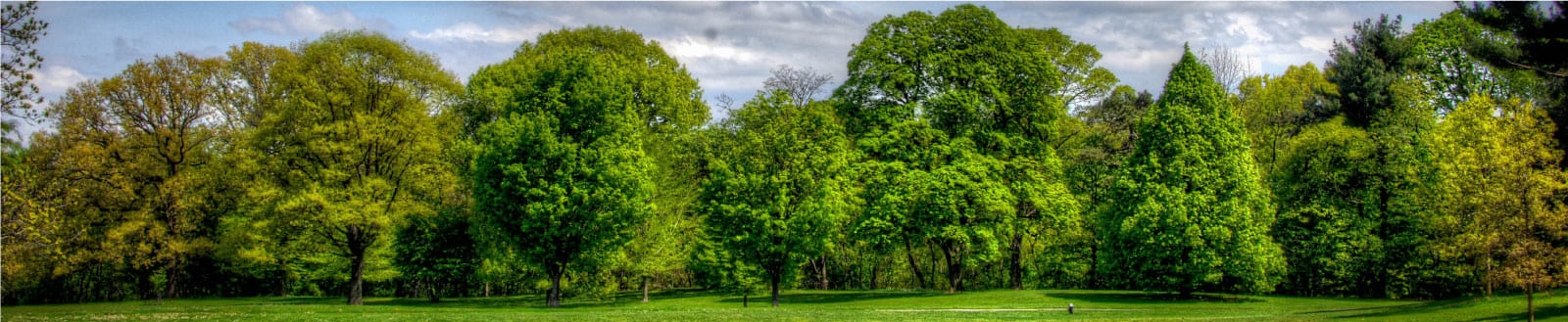
Peach trees are affected throughout North America.


X-disease phytoplasma can be transmitted by several species of leafhoppers. These leafhoppers typically acquire the X-disease pathogen while sucking juices from the leaves of X-disease-infected peaches or chokecherries. Wild chokecherry is an important reservoir for the X-disease phytoplasma. Two to three weeks later, feeding leafhoppers may inject the agent into healthy leaves while feeding. These leafhoppers are usually not considered pests of peach and cherry. The main damage they cause is the incidental transmission of the X-disease, phytoplasma.

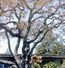
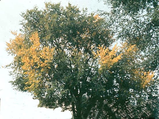
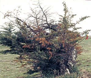
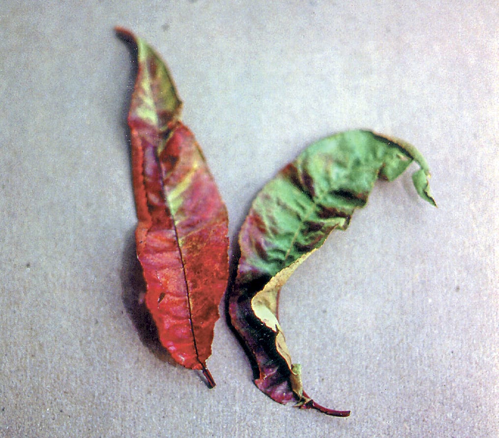
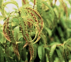
Symptoms in peach are apparent after about two months of growth. Leaves on isolated branches curl inward and develop irregular yellow to reddish-purple spots which later drop out, leaving leaves tattered  with a “shothole” appearance. Leaves on affected branches fall prematurely, starting at the base of the branch, until only a tuft of leaves remains at the tips of infected shoots. Fruit drops prematurely. Two to three years after initial infection, progressive death of scaffold branches occurs and eventually death of the entire tree follows.

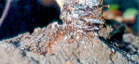
Phytoplasma moves thru sieve cells in the phloem causing death of the inner bark. Progressive root loss results in chlorosis, stunting, decline and death. Leafhoppers carrying phytoplasma transmit the pathogen while feeding on host  trees.

Penicillium Vermoeseni

PALM Bud Rot
Penicillium vermoeseni    (fungus)
Palms affected include Cocos plumosa, Phoenix canariensis, and Washingtonia filifera, W. robusta resistant.

In the United States it’s found in Coastal areas of central and southern California.
Favored by cool, foggy weather.
Watch for dying of terminal buds; leafstalk bases rotten; tree eventually dying.
Infection is by conidiospores moving throughout crown whorls until all leafstalks are infected. Once infection is established it continues all year. Inoculum may spread rapidly to nearby palms.

Pestalotia

OAK DECLINE COMPLEX:
Caused by one or more of the following fungi:
Botryodiplodia querci, Fusarium solani, Cephalosporium diospyri, Pestalotia macrosporum, Diplodia quercina, Phialophora sp., Dothiorella querci, Verticicladiella sp., Fusarium oxysporum, Verticillium albo-atrum

All oak species are hosts.

This complex has been reported throughout the continental United States.

Drought (soil moisture levels below 60% field capacity), with air temperatures above 60° F. Root damage from transplanting, construction, or compaction all contribute.

Symptoms vary according to host and fungus involved. Initial discoloration of leaf margins, sometimes with interveinal yellowing; later total yellowing possible, followed by stunting, slow to quick wilting, and dieback; usually evident first in upper crown. Leaves may or may not fall prematurely. As disease progresses individual branches may have thinner foliage, followed by a thinning of the entire crown. Vascular discoloration varies with organism, red-brown to black-blue, in twigs and/or root crown. Tree may die in as few as 2-3 months, or death may be gradual over 3-8 years.

Lethal infection, generally through roots from soilborne phase of fungus. Some of the fungi (Botryodiplodia, Diplodia) may become established as twig-canker inciters, at which stage decline is slow. Falling branches and twigs may introduce the fungus to the root area.

Phloem Necrosis

Phytoplasma.
Usually, the signs of elm phloem necrosis become visible between July and September. Rootlet necrosis is followed by degeneration of phloem and cambium in the roots and the lower trunk Elm yellow is present in the phloem tissue of infected trees.

Elm yellows is a systemic disease of elms caused by a bacteria-like organism called a phytoplasma.
Two former names for this disease are elm phloem necrosis in the U.S. and witches’ broom in Europe.

Due to the deposition of callose within the sieve tubes and the subsequent collapse of these cells and their neighbors, the inner phloem and the outer wood at the base of the stem turn lemon-yellow to butterscotch brown with or without flecks. The discolored area gives off a faint odor of oil of wintergreen (methyl salicylate).
The cambium fails to replace the affected phloem, since the newly produced cells become necrotic as well. In young trees, the discoloration may extend to the branches.

As the phloem necrosis progresses, the leaves of EY infected elms will droop , curl, turn bright yellow then brown, and premature defoliation takes place (Photo 68, {[896]}). The foliage discoloration generally occurs over most of the crown simultaneously. Especially the top leaves are boat-shaped. No twig wilting is observed. In such elm species as Ulmus rubra, EY infection results in the formation of witches’-brooms, a dense cluster of shoots of the same age. Due to the loss of apical dominance, the terminal shoot elongates no more than others on a branch, and the buds in the leaf axils are released as soon as they form.
Although some elms die within a few weeks after EY infection, usually when disease symptoms appear in June/July the tree will be killed by the end of the growing season. The symptoms of a late summer or autumn infection with EY cannot be distinguished from normal autumn senescence. Trees infected late in the season will have a crown with thin twigs and small leaves the following spring. In this new growing season, the infected elms can be expected to die soon. EY symptoms exhibited by these autumn infected elm trees can easily be confused with DED symptoms expressed byelms infected with the latter disease the previous year.

Affected are;
Ulmus alata, U. americana, U. crassifolia, U. glabra, U. japonica, U. minor, U. parvifolia, U. pumila, U. rubra, U. serotina, and U. villosa

Phloem Necrosis

Phytoplasma.
Usually, the signs of elm phloem necrosis become visible between July and September. Rootlet necrosis is followed by degeneration of phloem and cambium in the roots and the lower trunk Elm yellow is present in the phloem tissue of infected trees.
Due to the deposition of callose within the sieve tubes and the subsequent collapse of these cells and their neighbors, the inner phloem and the outer wood at the base of the stem turn lemon-yellow to butterscotch brown with or without flecks. The discolored area gives off a faint odor of oil of wintergreen (methyl salicylate).
The cambium fails to replace the affected phloem, since the newly produced cells become necrotic as well. In young trees, the discoloration may extend to the branches.

As the phloem necrosis progresses, the leaves of EY infected elms will droop , curl, turn bright yellow then brown, and premature defoliation takes place (Photo 68, {[896]}). The foliage discoloration generally occurs over most of the crown simultaneously. Especially the top leaves are boat-shaped. No twig wilting is observed. In such elm species as U. rubra, EY infection results in the formation of witches’-brooms, a dense cluster of shoots of the same age. Due to the loss of apical dominance, the terminal shoot elongates no more than others on a branch, and the buds in the leaf axils are released as soon as they form.

Although some elms die within a few weeks after EY infection, usually when disease symptoms appear in June/July the tree will be killed by the end of the growing season. The symptoms of a late summer or autumn infection with EY cannot be distinguished from normal autumn senescence. Trees infected late in the season will have a crown with thin twigs and small leaves the following spring. In this new growing season, the infected elms can be expected to die soon. EY symptoms exhibited by these autumn infected elm trees can easily be confused with DED symptoms expressed by elms infected with the latter disease the previous year.

Elms affaected are;
U. alata, U. americana, U. crassifolia, U. glabra, U. japonica, U. minor, U. parvifolia, U. pumila, U. rubra,   U. serotina, U. villosa

Phomopsis

Phomopsis juniperovora   (imperfect fungus), Kabatina juniperi, Sclerophoma pithyophila

Plants affected are arborvitae, cedar, cypress, Douglas fir, false cypress, fir, giant and coast redwoods, hemlock, Japanese yew, juniper, larch, pine and spruce. And is Found worldwide.

Splashing water (rain, overhead irrigation); cool, cloudy weather; birds (spread spores); pruning wet plants; dead or dying branches; infected trees nearby; mulches from infected plant material.

Symptoms are most evident in seedlings and transplant beds on trees less than 4 years old. On older trees gradual decline and eventual death may follow. Tips of branches brown with progressive dieback until an entire branch or tree is killed. Small, sunken lesions on branch or stem may make tree appear one-sided or flattened. The lesions are bordered by gray-black spore fruits (Phomopsis; pycnidia, Kabatina and Sclerophoma; acervuli). Kabatina and Sclerophoma attack principally one year-old twigs.

The spores produced by thousands in pycnidia and acervuli on diseased twigs; ooze out in little tendrils (not evident in drier zones) in warm, moist weather. Spread by splashing water, insects, and pruning. Entrance through unbroken tissue as well as wounds (Kabatina and Sclerophoma cannot enter unbroken tissue, enter through insect wounds). Stem or branch killed above and below the point of entrance; death caused by girdling of conducting tissues.

Phomopsis Canker

DOUGLAS FIR
Phomopsis lokoyae    (fungus)

Douglas fir trees throughout California and Oregon.

Malnutrition, improper irrigation, poor soils, construction stress, persistent rainy or foggy weather; wounding by squirrels, birds, improper pruning, and climbing spurs.

Long, narrow cankers on branches, usually pointed at ends; may girdle entire branch in one season. Usually in saplings on poor sites, but it may attack individual branches in mature ornamental trees under stress.

The fungus may enter through needle scars or wounds. It girdles branches and is usually successful in stressed trees. Cankers are established at the point of infection. During wet season, spores are produced in spore fruits (pycnidia) at the edges of the cankers from which they spread to other parts of the tree, or to adjacent trees.

Phosphorus Deficiency

Phosphorus in plant tissue depends on the phosphorus content in the soil.  Phosphorus in plants ranges from  0.2%-0.8% of the total dry substance. It is more abundant  throughout the plant than are sulfur and magnesium, and less than calcium. Mycorrhizal fungi are thought to play a key role in making phosphorus in soil available to plant roots.

Phosphorus is an essential element. It functions directly and indirectly in metabolism. Lack of phosphorus leads to stunting and can also affect protein formation, leading to high concentrations of sugar and causing purple symptoms similar to nitrogen deficiency. Phosphorus is also important in root development.

The symptoms on broadleaf plants are the leaves are green to dark green; veins, petioles, and lower surfaces dull bronze to purple, especially when young; foliage may be sparse, slightly smaller than            normal, and distorted; leaves drop early. Shoots are of normal length unless deficiency is severe, but they are smaller in diameter. Flowers are few. Fruit sparse and small.
The symptoms on conifers are  purple needles in young seedlings, starting at tips of lower needles
and progressing inward and upward. Few or no second year needles occur. The needles die, starting in lower regions and spreading upward. Buds set early. Tree foliage appears dull-blue or gray-green. Roots sparse with no mycorrhizae.

Physalospora Bleeding Canker

ALDER BLEEDING CANKER.

Physalospora obtusa (fungus)

Trees affected include Ailanthus, alder, American holly, apple, boxelder, black locust, chinaberry, crabapple, crape myrtle, dogwood, flowering quince, honey locust, maple, mountain ash, oak, pecan, Persian walnut, persimmon, sassafras, Atlantic white cedar, Austrian and eastern white pines and found mainley United States: Northeast, southeast, central California.

Warm, rainy weather in early spring at bud break. Inoculum source from dead and infected twigs and branches on tree all contribute .

This disease is noticed by are wet, bleeding cankers on stems and branches of alder, with twig dieback. May cause fruit rot, cankers, cone and seed rot on conifers, and twig blights.

It overwinters as perithecia on infected and dead branches. Ascospores released in spring, during and following wet weather, when conditions are right. These spores invade bark wounds, germinate in the bark, and form pycnidia which provide inoculum throughout the summer, until cool fall weather favors perithecial development in bark and fruit.

Phytophthora Cactorum

Crown Rot, Collar Rot, Root Rot, Stem Rot

Phytophthora Spp. including,
P. ramorum, P. cactorum, P. cinnamomi, P. palmivora, P. citricola, P. nicotianae, P. ramora

World wide.

The pathogen survives in soil for several years as spores, especially in poorly drained soils. Too deep planting may contribute to the severity of this disease. Cool, wet weather favors development of the disease in the spring.

Phytophthora is a Soil borne fungal pathogen. Cankers may be seen at or below the soil line and may extend into the root system.  The infection can also invade the trunk or main stem.  Susceptible scions can be infected at the bud union. Infected bark is brown and often slimy when wet. When the bark is pulled away, the cambium and phloem will be an orange, reddish brown color. The cankers caused by the fungus girdle the tree, resulting in poor vegetative growth and chlorotic foliage that may turn purple in the autumn. A severely infected tree may die. Trees may be killed in one growing season or may linger for a number of growing seasons.

This fungus survives cold weather and drought as thick walled resting spores (oospores) in infected tissue or in soil. The resting spores are capable of surviving extended periods of time in soil. The fungus may build up to high levels in the soil in a short period under favorable conditions – i.e. during wet, cool periods. Under moist conditions, they germinate, forming another type of structure called a sporangium. A sporangium is a minute reproductive structure produced by the fungus in response to free water. The sporangia germinate in the presence of free water, releasing numerous one-celled, motile spores called zoospores. Zoospores swim through a thin film of water, contact roots or lower trunks of potential hosts, germinate, and infect. Although host wounds are frequent infection courts (areas where infections are initiated), the fungus can also directly penetrate host tissue. The fungus grows through host vascular tissue eventually girdling the tree. As the host dies, the fungus forms resting spores in the decaying tissue. Research revealed that the fungus is readily transported in irrigation water. Few agricultural soils should be considered free of the fungus once they have been irrigated. Infection is most likely to occur during cooler weather, i.e. early spring and fall. Research has indicated that trees are most susceptible during the bloom period.