Fusarium is a large group of filamentous fungi belonging to the hyphomycetes. Commonly distributed widely in the soil they are saprophytic fungi known to associate with plants, causing a wide range of plant diseases. This is because of their ability to produce mycotoxins especially in cereal crops, which can cause disease in human and animal hosts if ingested. Fusarium spp majorly produces fumonisins and trichothecenes mycotoxins.
Fusarium spp does not commonly cause diseases in humans because some exist as commensals in the skin, but it has been found to cause opportunistic infections in immunocompromised individuals. It is vastly known for its pathological effects on plants and animals. Some of the common plant diseases caused by Fusarium spp include crown rot, head blight, and scab on cereal grains; vascular wilts on a wide range of horticultural crops; root rots; cankers; and other diseases such as Pokkah boeng on sugarcane and bakanae disease of rice.
Interesting Science Videos
Classification of Fusarium spp
- Research on the classification and taxonomy of Fusarium spp has indicated that there are many species, several populations within the species, and several unidentified groups in the genera. This explains the wide range of variations exhibited by these groups with respect to morphology, cultural characterization, and physiology of the fungal group.
- And these variations might be an explanation as to the ability of the fungi to colonize a wide range of environments globally.
- Fusarium classification and taxonomy are as follows:
Kingdom: Fungi
Division: Ascomycota
Class: Sordariomycetes
Order: Hypocreales
Family: Nectriaceae
Genus: Fusarium
- Presently, the genus Fusarium has at least 300 phylogenetically distinct species, 20 species complexes, and nine monotypic lineages of which some have been identified to be opportunistic Fusarium pathogens
- Some of these species include:
- Fusarium solani represents a complex of over 45 phylogenetically distinct species of which at least 20 are associated with human infections.
- Fusarium oxysporum complex is phylogenetically diverse
- Fusarium incarnatum-equiseti complex is diverse
- Fusarium chlamydosporum complex is equally diverse.
- F. fujikuroi complex
- F. dimerum complex is less diverse
- F. sporotrichioides complex is less diverse
The opportunistic pathogenic Fusarium groups for plants, animals, and humans majorly belong to the F. solani complex, F. oxysporum complex, and F. fujikuroi complex.
Habitat of Fusarium spp
- Fusarium spp is commonly found in soil and environmental habitats, with many growing and thriving in tropical and temperate regions and even in desert regions, the alpine, the arctic regions with harch cold conditions, they seem to prevail.
- Fusarium species are widely distributed in soil and on subterranean and aerial plant parts, plant debris, and other organic substrates.
- Commonly and abundantly found in soil, they seem to thrive in fertile cultivated soil and therefore they are classified as soilborne fungi where they closely associate with plant roots saprophytically or parasitically.
- They disperse in the atmosphere and become airborne, thus colonizing aerial plants and causing diseases.
- Therefore, Fusarium species are widely distributed and have efficient dispersal mechanisms thus growing in a wide range of substrates as well.
Morphology of Fusarium spp
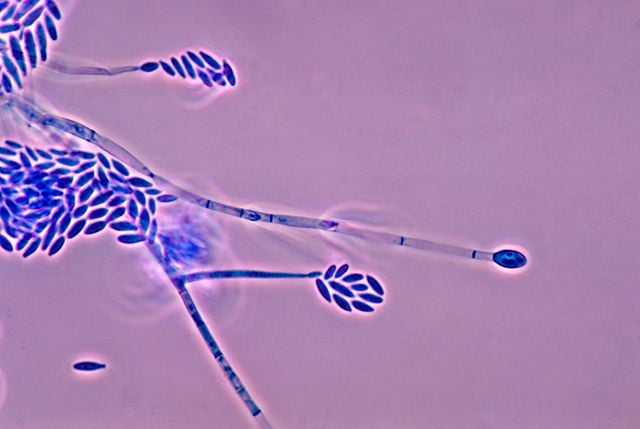
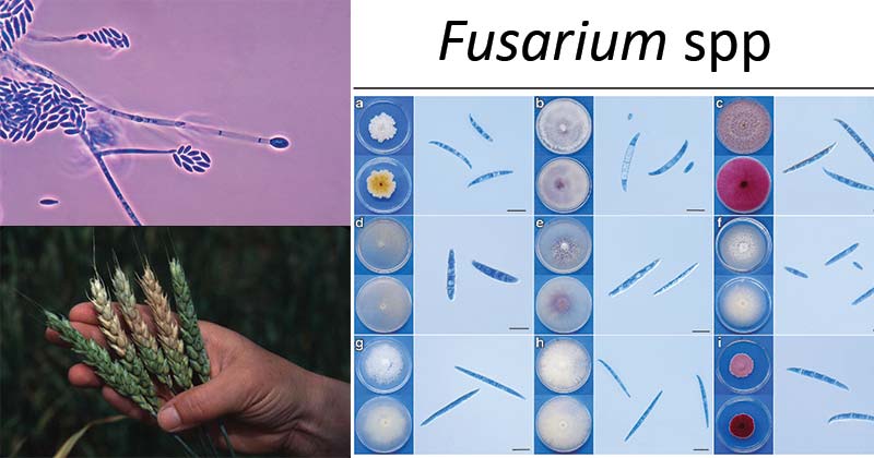
Figure: Fusarium verticillioides. Image Source: Wikipedia.
- Fusarium spp reproduces asexually and produces three kinds of fungal spores known as macroconidia, microconidia, and chlamydospores.
- Some species of Fusarium produce all three types of spore while others produce singularly.
- These spores especially the microconidia are held by microconidiophores.
- These conidiophores may be either mono-phialides only or both mono-phialides and poly-phialides in a given species producing microconidia.
- Macroconidia are produced in a sporodochium, which is an erumpent crowded cluster of conidiophores arising from stroma to form a cushion-like mass that supports the macroconidia.
- Macroconidia are also produced on mono-phialides (a conidiophore with a single opening through which an endoconidia is released) and poly-phialides (two or more openings or pores from which the endoconidia are forced out) on aerial mycelium.
- The macroconidia vary in size and shape.
- The Major producers of macroconidia are Fusarium semitectum, Fasarium avenaceum and Fusarium suglutinans.
- Microconidia are produced in the aerial mycelium.
- The microconidia can be produced on false heads or false chains on mono-phialides or poly-phialides. False Heads are a result of moisture drops on the conidiophore and they contain the endoconidia as they are produced.
- Microconidia have different shapes and sizes. the microconidia produced in chains have a truncate base.
- Chlamydospores are thick-walled spores filled with lipid-like material that carries the spores overwinter in the soil.
- Chlamydopsores are sometimes airborne occurring in pairs, in clumps, or in chains.
- They have an outer wall which can be smooth or rough.
Cultural characteristics of Fusarium spp
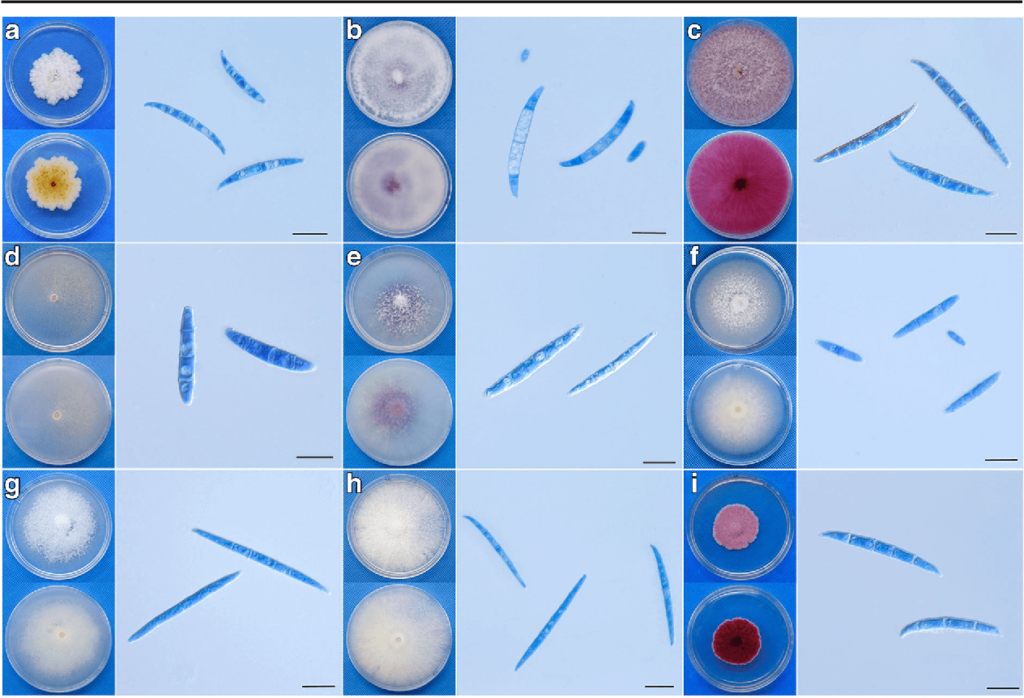
Figure: Typical colonies and macroconidia of Fusarium isolates from soybean root rot in Sichuan, China. Typical colonies of the representative Fusarium isolates were observed on PDA after 5 days and macroconidia in Carboxymethyl cellulose Medium after 5 days grown. a F. equiseti; b F. oxysporum; c F. graminearum; d F. solani; e F. commune; f F. verticillioides; g F. proliferatum; h F. fujikuroi; i F. avenaceum. Scale bar = 20 μm. Image Source: https://doi.org/10.1007/s10658-017-1410-7
Carnation leaf agar
- It promotes sporulation and suppresses mycelial growth. It produces conidia and conidiophores in large numbers and specialized morphologies of the spores are distinct. Carnation leaf agar has low carbohydrates with complex substances that provide a natural environment that promotes Fusarium growth.
- This is the most valuable medium for Fusarium growth-producing gross morphological appearance and colony colorations. The medium which contains a high carbohydrate content which promotes sporulation, however, takes longer to grow in this medium. The conidia produced are misshapen and atypical.
KCl medium
- KCI medium is used to observe the formation of microconidia in chains by species. The species that do form chains of microconidia form more abundant, longer chains on this medium. The chains are easier to observe because there is less moisture on the surface of the agar and fewer droplets of moisture in the aerial mycelium.
Soil agar
- Soil agar promotes rapid chlamydospore formation in a number of Fusarium species. Large inoculum with actively growing fusarium inoculates produces chlamydospores within 3-4 days but secondary inoculates produce chlamydospores in 30 days.
Pathogenesis and Clinical Features of Fusarium spp
Fusarium spp causes disease in plants, animals, and human hosts, but most commonly in plants. These pathogenesis have been linked to the toxigenicity of the species associated with the production of mycotoxins such as trichothecenes (types A and B), fusaric acid and fumonisins
Plant pathologies caused by Fusarium species

Figure: Fusarium pathogenicity and host defense mechanisms. Image Source: https://doi.org/10.1146/annurev-micro-092412-155650
- The effect of Fusarium spp on plants affects and infects specific parts such as grains, seedlings, heads, roots, or stems.
- The plant infections by Fusarium spp are caused by the secretion of mycotoxins by various group complexes of Fusarium.
- They cause various plant diseases which contribute to decreased quality and production yield of these crops.
- Fusarium solani species complex (FSSC) causes diseases in many agriculturally important crops, such as Fusarium Rot and/or Root Rot and necrosis of the infected host plant.
- The infection by FSSC causes symptoms, such as wilting, stunting, and chlorosis. Necrosis depends on the severity of fungal development.
- Two of the most serious diseases of wheat known globally are Fusarium Crown Rot (FCR) and Fusarium Fusarium Head Blight (FHB).
- Plant pathologies include:
- Fusarium head blight (FHB) caused by F. graminearum, which contributed to the loss of starch and proteins in cereals.
- Footrot (FR) and root rot (RR)
- Crown rot (CR)
- Fusarium wilts a destructive disease in bananas caused by F. oxysporum.
Fusarium Head Blight
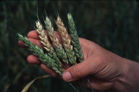
Figure: Fusarium head blight in wheat. Image Source: The American Phytopathological Society (APS)
- It is caused by Fusarium graminearum.
- This is a disease that commonly affects cereals such as wheat, barley, oats, rye, and triticale.
- The disease infects the heads of the crop, reducing grain yield.
- The disease cycle has three stages of infection:
- Fusarium graminearum multiplies rapidly on inoculation on the plant site making large biomass within the first 2 days. This allows the fungal spores to germinate forming superficial hyphae on the leaf sheath.
- Stage two involves a decrease in the fungal biomass due to penetration of the hyphae into the leaf sheath base from the outer leaf sheath.
- The third stage included the increase in biomass of the fungus allowing colonization on the cereal crops especially the crown head of wheat parenchyma.
Footrot and root rot
- This is commonly caused by Fusarium solani, saprophytic fungus.
- The fungus colonizes the dead or decaying plant tissues allowing the fungal invasion of the stem of the nodes or the soil line and opportunistically infecting the plant wound.
- The fungus spores then germinate in the affected area (wound), favored by high humidity and temperatures.
Fusarium wilt
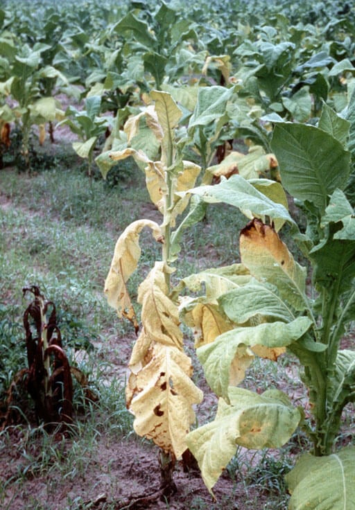
Figure: A tobacco plant suffering from Fusarium wilt. Image Source: Wikipedia (R.J. Reynolds).
- It is caused by Fusarium oxysporum as the only pathogenic group of Fusarium known to grow inside the plant vessels and spreads upwards inside the plant.
- It is a saprophytic fungus that can survive in soil between crop cycles in infected plant debris.
- Survival morphologies can be in the mycelial form or in spore forms.
- The fungus infects the root tips directly or through damaged tissues allowing mycelial growth into the root cortex into the xylem and later affecting the whole vascular tissue.
- This causes a reduction of water and nutrient intake leading to leaf wilting and plant death.
Human pathologies of Fusarium species
- Fusarium species cause superficial, locally invasive, and diffuse infections in humans.
- Localized infection includes septic arthritis, endophthalmitis, osteomyelitis, cystitis, and brain abscess.
- Invasive infections are as a result of surgery and oral antifungal therapy.
- Disseminated infection occurs when two or more noncontiguous sites are involved.
- The infections are opportunistic and they are majorly caused by F. solani complex including F. solani, F. oxysporum, F. verticillioides, and F. proliferatum, F. moniliforme and F. fujikuroi species complex.
- They cause opportunistic infections in immunocompromised patients.
- The elderly and diabetics with prevalent meningospondylodiscitis are opportunistically infected by F. oxysporum. F. sacchari, F. anthophilum, F. chlamydosporum, and F. dimerum.
- A perinephric abscess caused by F. chlamydosporum is common in children who have been reported before.
- Corneal infections (endophthalmitis) caused by Fusarium oxysporum and Fusarium solani also occur because of the adherence of the Fungi to the corneal membrane causing eye damage.
- Some Fusarium species, such as F. dimerum, are associated with keratomycosis, particularly in the bad hygiene conditions.
- Mycotoxicosis caused by Fusarium species is common in the ingestion of the mycotoxins produced by the fungi.
Fusarium infection in animals
- Fusarium spp can cause Fusarium infections in animals including mycotoxicosis which affects the growth, reproduction, and hormonal condition of the animal.
- The effect of these mycotoxins on animals depends on the quantity of mycotoxin intake. After intake, these mycotoxins arrive at the gastrointestinal epithelial cell layer. In high doses, the mycotoxins cause abdominal distress, diarrhea, cardiac insufficiency, emesis, and even death in pigs and equine leukoencephalomalacia (ELEM) in horses.
Identification and diagnosis of Fusarium species
- Use of mycological and blood culture methods to identify hyaline, banana-shaped, and multicellular macroconidia.
- Use of molecular methods such as genus-specific PCR, 28 s rRNA gene sequencing, sequence-based PCR, multiplex tandem PCR, and automated repetitive sequence-based PCR for differentiation of the various Fusarium species groups.
- The diagnosis of Fusarium infection in humans and animals can be may be done using histopathology, gram stain, mycology, blood culture, or serological identification of the fungal antigens.
Treatment, Prevention, Control, and Management of Fusarium
- For human and animal infections can be treated with intravenous administration of itraconazole, oral amphotericin B.
- Plant infections can only be controlled by used on control agents and measures.
- Biocultural control by the use of mycoparasitic fungi such as T. harzianum may be used as a biocontrol agent against Fusarium. The parasite has shown to have an antagonistic effect against Fusarium that causes footrot, root rot, and crown rot.
- Fungicides have very minimal effects with likely resistance development on most of the Fusarium species groups.
- Resistant cultivars can be used to control Fusarium Head Blight by controlling the plant lines that allow the release of mycotoxins. This reduces fungal growth and lowers mycotoxin contamination.
Sources and References
- https://mycology.adelaide.edu.au/descriptions/hyphomycetes/fusarium/
- https://www.sciencedirect.com/science/article/pii/B978008100674000014X
- Fusarium infection in immunocompromised patients by Marcio Nucci and Elias Anaissie: Clinical Microbiology Review
- https://www.intechopen.com/books/fusarium-plant-diseases-pathogen-diversity-genetic-diversity-resistance-and-molecular-markers/introductory-chapter-fusarium-pathogenicity-infections-diseases-mycotoxins-and-management
- https://www.koppert.co.ke/challenges/disease-control/fusarium-wilt/
- Fusarium infection of the Skin by Gupta A.K, Baran R., Summerbell R.C. PubMed.gov
- Fusarium onychomycosis: prevalence, clinical presentations, response to itraconazole and terbinafine pulse therapy, and 1-year follow-up in nine cases by Ranawaka R.R., Nagahawatte A., Gunasekara T.A. PubMed.gov
- https://en.wikipedia.org/wiki/Fusarium_ear_blight
- https://en.wikipedia.org/wiki/Fusarium