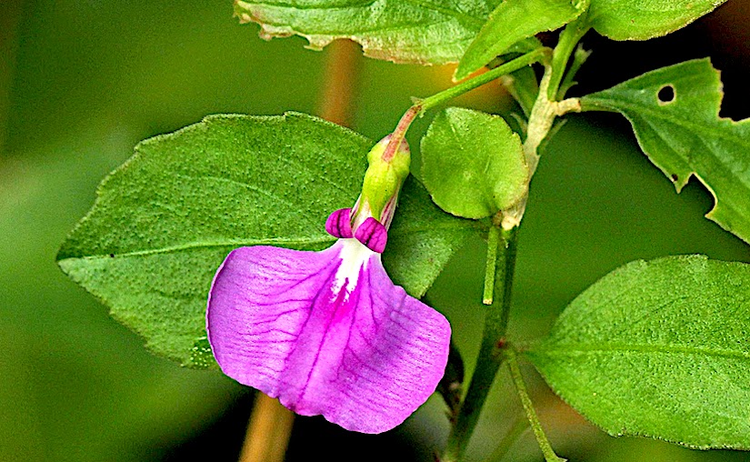
Photo credit: Google
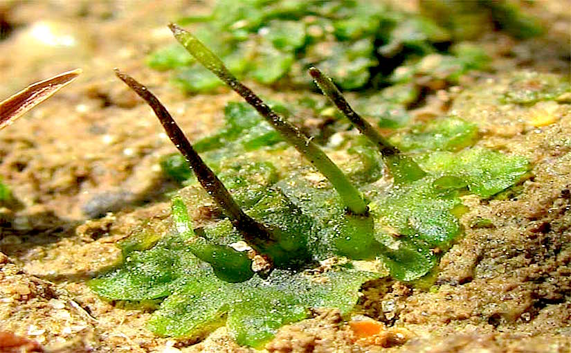
Hornwort: Anthoceros punctatus
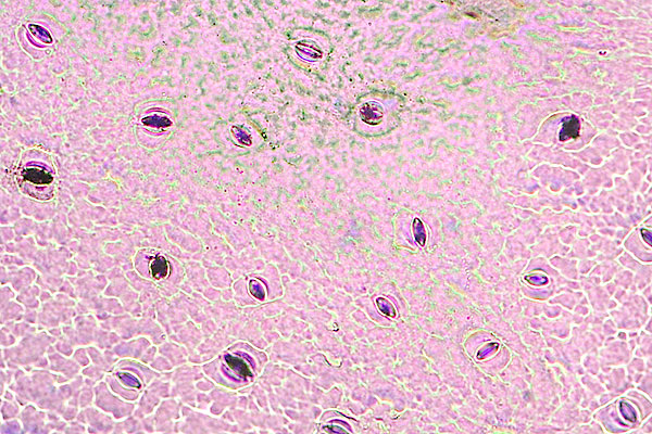
Stomatal differentiation and abnormal stomata in hornworts
by Pressel S., Goral T., Duckett J. G. (2014)
Silvia Pressel, Tomasz Goral
Jeffrey G. Duckett 
in Maney Online: Journal of Bryology, Volume 36, Issue 2 (June 2014), pp. 87-103
http://www.tandfonline.com/doi/pdf/10.1179/1743282014Y.0000000103
This light and electron microscope study reveals considerable uniformity in hornwort stomata morphology and density in contrast to common spatial and developmental abnormalities in tracheophytes and mosses.
Stomata arise from a median longitudinal division of sporophyte epidermal cells morphologically indistinguishable from their neighbours apart from the retention of a single chloroplast whilst those in the other epidermal cells fragment. Chloroplast division and side-by-side repositioning of the two daughter chloroplasts determines the division plane in the stomatal mother cell. The nascent guard cells contain giant, starch-filled chloroplasts which subsequently divide and, post aperture opening, regain their spherical shape. Accumulation of wall material over the guard cells and of wax rodlets lining the pores follows opening.

While the majority of stomata are bilaterally symmetrical those lining the dehiscence furrows display dextral and sinistral asymmetry due to differential expansion of the adjacent epidermal cells.
The ubiquity of open stomata suggests that these never close with the maturational wall changes rendering movement extremely unlikely. These structural limitations, a liquid-filled stage in the ontogeny of the intercellular spaces, and spores already at the tetrad stage when stomata open, suggest that their primary role is facilitating sporophyte desiccation leading to dehiscence and spore dispersal rather than gaseous exchange.
Stomata ontogeny and very low densities, like those in Devonian fossils, suggest either ancient origins at a time when atmospheric carbon dioxide levels were much greater than today or a function other than gaseous exchange regulation. We found no evidence for stomatal homology between hornworts, mosses and tracheophytes.
See also: Maney Online


You must be logged in to post a comment.