Photo credit: Am. J. Bot.
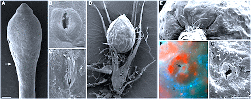
(A–C) Oedipodium griffithianum. (A) SEM of capsule and part of apophysis (arrow to bottom of image). Bar = 300 µm. (B) SEM of open long-pored stoma. Bar = 10 µm. (C) SEM of closed pore filled with cuticular waxes. Bar = 10 µm. (D–G)Ephemerum spinulosum. (D) SEM of mature sporophyte attached to gametophyte. Bar = 200 µm. (E) SEM of base of capsule with stomata. Bar = 100 µm. (F) Stoma in fluorescent light treated with DAPI showing blue nuclei and autofluorescing red chloroplasts; the round pore is flanked by wall ledges (arrow), and bacteria are abundant around guard cells (b). Bar = 10 µm. (G) SEM of stoma partially filled with cuticular waxes. Bar = 10 µm.
Moss stomata in highly elaborated Oedipodium (Oedipodiaceae) and highly reduced Ephemerum (Pottiaceae) sporophytes are remarkably similar
by Merced A.,
Renzaglia K. S.
(2013)
in American Journal of Botany 100(12): 2318-2327. 2013.
(http://www.amjbot.org/cgi/doi/10.3732/ajb.1300214)
http://www.amjbot.org/content/100/12/2318/F1.small.gif
ABSTRACT
• Premise of the study: Mosses are central in understanding the origin, diversification, and early function of stomata in land plants. Oedipodium, the first extant moss with true stomata, has an elaborated capsule with numerous long-pored stomata; in contrast, the reduced and short-lived Ephemerum has few round-pored stomata. Here we present a comparative study of sporophyte anatomy and ultrastructure of stomata in two divergent mosses and its implications for stomata diversity and function.
http://www.biosphere-images.net/Atlantic-European%20Album%20(Bryophytes)/pictures/picture-38.jpg
• Methods: Mature sporophytes of two moss species were studied using light, fluorescence, and scanning and transmission electron microscopy. Immunolocalization of pectin was conducted on Oedipodium using the LM19 antibody.
http://www.cisfbr.org.uk/images/Ephemerum_serratum_007C.JPG
• Key results: Oedipodium capsules have extensive spongy tissue along the apophysis, whereas those of Ephemerum have minimal substomatal cavities. Stomatal ultrastructure and wall thickenings are highly similar. Sporophytes are covered by a cuticle that is thicker on guard cells and extends along walls surrounding the pore. Epicuticular waxes and pectin clog pores in old capsules.
• Conclusions: Ultrastructure of stomata in these mosses is similar to each other and less variable than that of tracheophytes. Anatomical features such as the presence of a cuticle, water-conducting cells, and spongy tissues with large areas for gas exchange are more pronounced in Oedipodium sporophytes and support the role of stomata in gas exchange and water transport during development and maturation. These features are modified in the reduced sporophytes of Ephemerum. Capsule anatomy coupled with the exclusive existence of stomata on capsules supports the concept that stomata in moss may also facilitate drying and dispersal of spores.
Read the full article: Am. J. Bot.

/pictures/picture-38.jpg)