Ontogenetic classifications of stomatal complexes

Screen Shot 2017-09-25 at 11.42.10

Ontogenetic basis for classification of stomatal complexes – a reapproach

by Timonin A. C. (1995)

A. C. Timonin, Department of Higher Plants Morphology and Systematics, Biological Faculty, M. V. Lomonosov Moscow State University, Vorobyevy Gory, 119899, Moscow, Russia.

in Flora 190: 189-195 – https://doi.org/10.1016/S0367-2530(17)30650-3

https://ac.els-cdn.com/S0367253017306503/1-s2.0-S0367253017306503-main.pdf?_tid=4af011a4-a1d4-11e7-8787-00000aab0f01&acdnat=1506332062_d0a41c9b0dd95ad69d6c89f8ff3e0e16

Screen Shot 2017-09-25 at 12.53.31

Summary

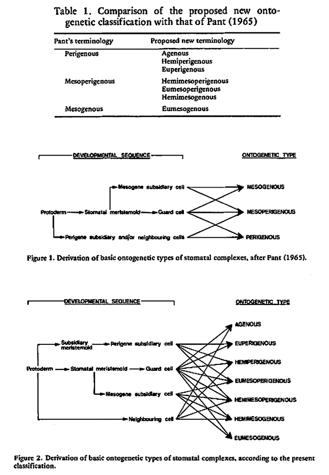
The majority of existing ontogenetic classifications of stomatal complexes is considered to be, in fact, a mixture of structural and ontogenetic ones.

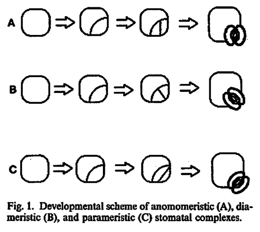
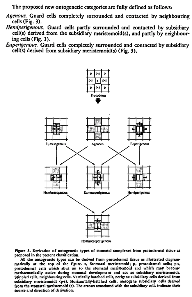
Purely ontogenetic classifications should be based on only three characters:

a) orientation of guard cells’ mother cell division in relation to the plane of the preceding cell division ;

b) orientation of the divisions of cells other than guard cells’ mother cell and

c) number of cell divisions leading to mature stomatal complex formation.

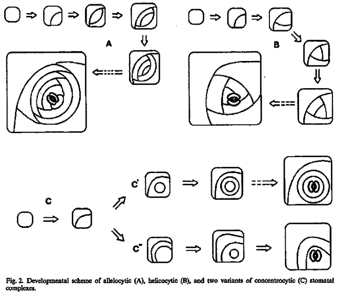
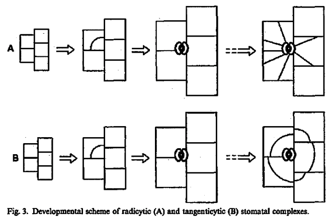
This method would result in three independent and reciprocally supplementary stomatal classifications. The first one consists of only PAYNE’S anomo-, dia-, and parameristic types. The second one contains both PAYNE’S allelo- and helicocytic types and new concentro-, radi-, and tangenticytic stomatal types. For the third classification, monomeristic and bimeristic types, and so forth, are proposed.

Two neglected unnamed STRASBURGER’S and PRANTL’S stomatal types are reintroduced as initial, resp. terminal.

Screen Shot 2017-09-25 at 12.56.22Screen Shot 2017-09-25 at 12.58.50

The mystery of the evolution of stomata


The mystery of the evolution of stomata – Success of vascular plants through stomata

VIDEO:Duration: 56 mins 29 secs


The Gatsby Plant Science Summer School Lecture Collection

University of Cambridge

http://intobiology.org.uk/stomata-key-elements-essential-for-the-succes-of-the-vascular-plants/

https://sms.cam.ac.uk/media/2113497

Prof Alistair Hetherington – Stomata: key elements essential for the success of the vascular plants. Prof Alistair Hetherington, University of Bristol, shares his research on stomata, and explains why fundamental research is important to tackling the world’s biggest problems. This research lecture was recorded at the Gatsby Summer School, 2015.


Evolution, structure and functioning of stomata.


The evolution, structure and functioning of stomata.

by Willmer C. M. (1993)

in Bot. J. Scotl. 46. 433-445. – DOI: 10.1080/03746609308684805 –

http://www.tandfonline.com/doi/abs/10.1080/03746609308684805


The anatomy and ultrastructure of guard cells from a range of species varying from the primitive types, such as mosses, to the advanced grasses and orchids are described.

An attempt is made to trace the lines along which stomata developed and to define what might be considered advanced stomata.

Additionally, the differentiation of guard cells from guard mother cells is discussed.

Of particular note is the preprophase band of microtubules which marks the zone where the future cell wall will form and the movement of the spindle and developing cell plate through 45 degrees. The structure and function of guard cells are intimately linked.

Stomata are turgor regulated valves; the osmotica for absorbing water during opening are K+, Cl and malate anions which accumulate in the guard cell vacuoles.

Upon stomatal closure, K+ and Cl exit from the guard cells while at least some of the carbon from malate is channelled into starch and there is a resultant loss of guard cell turgor.

The Calvin cycle may be absent or of low activity in guard cell chloroplasts and under those circumstances a source of carbon and energy to sustain the guard cells is needed. Hence it is believed that sucrose is transported into the guard cells from mesophyll cells.

A brief consideration of the mechanism by which the ions are transported across the plasma membrane and tonoplast is made: the driving force for the K+, Cl and malate movement across the membranes is the proton motive force set up by proton-pumping ATPases.

Stomatal cells as idioblasts (in German)

Photo credit: Google

Scutellaria altissima (Tall skullcap)


Stomata-zellen als Idioblasten

by Weber F. (1955)

Friedl Weber, Pflanzenphysiologischen Institut der Universität Graz, Österreich

in Österr. bot. Z. 102: 436-443 –

https://link.springer.com/article/10.1007/BF02135220

Zusammenfassung

Den Schließzellen des Spaltöffnungsapparates als Idioblasten, Sonderlinge, der Epidermis fehlen bei verschiedenen Pflanzen eine Reihe von Inhaltskörpern, die in den anderen Epidermiszellen vorkommen.

Ein neues Beispiel für diese Regel bildet das Auftreten langer stab-, nadel-, oder spindelförmiger Inhaltskörper in den gewöhnlichen Oberhautzellen der Blattunterseite von Scutellaria altissima und das Fehlen dieser Körper in den Schließzellen dieser Pflanze. Diese Gebilde haben eine Ähnlichkeit mit den Eiweißspindeln der Kakteen, doch ist die Eiweißnatur der Scutellaria-Spindeln noch fraglich.

Die Schließzellen machen in der Regel die in anderen Blattzellen vorsichgehende Umwandlung der Chloroplasten in Chromoplasten nicht mit. Eine Ausnahme von dieser Regel bilden die Stomazellen von Selaginella helvetica, in denen sich in der kalten Jahreszeit die Chloroplasten in Chromoplasten umwandeln, ebenso wie in den übrigen Epidermiszellen.

No Ca-oxalate crystals in stomata (in German)

Photo credit: Google

A Vanilla planifolia flower. [Taken by Michael Doss.]


Kalziumoxalatkristalle fehlen den Schliesszellen

by Weber F. (1955)

Friedl Weber, Pflanzcnphysiologischen Institut der Universität GrazÖsterreich

in Protoplasma 44: 464-468 –

https://link.springer.com/article/10.1007/BF01250781

Zusammenfassung

Die Schließzellen der Stomata enthalten auch dann keine Kalziumoxalatkristalle, wenn die übrigen Epidermiszellen solche Kristalle regelmäßig aufweisen.

Als Beispiel für diese Regel wird die Oberhaut von Blatt und Stengel von Vanilla planifolia angeführt. Es wird erörtert, wodurch das Fehlen der Kalziumoxalatkristalle in den Schließzellen bedingt sein könnte.

Effectiveness of change in upper and lower stomatal openings


Relative effectiveness of change in upper and lower stomatal openings

by Waggoner P. E. (1965)

Paul E. Waggoner, Connecticut Agricultural Experiment Station

in Crop Sci. 5: 291 –

https://dl.sciencesocieties.org/publications/cs/abstracts/5/4/CS0050040291?access=0&view=pdfScreen Shot 2017-09-16 at 13.50.58


Stomatal types classification


A classification of the stomatal types

by Van Cotthem W. R. J. (1970)

University of Ghent, Belgium

Willem Van Cotthem
Willem Van Cotthem, Ghent University, Belgium

in Bot. J. Linn. Soc. 63: 235-246. –

http://dx.doi.org/10.1111/j.1095-8339.1970.tb02321.x

https://academic.oup.com/botlinnean/article-lookup/doi/10.1111/j.1095-8339.1970.tb02321.x

Abstract

In 1950 Metcalfe & Chalk showed the need for new technical terms ‘devoid of taxonomic or ontogenetic implications’ to replace those of Vesque.Their new terms and some others, proposed by Metcalfe and Stace, are now generally used. All previous classifications of stomatal types included only those of Angiosperms and Gymnosperms.

The eight new forms described in this and a previous paper (Van Cotthem, 1968) include five known only from the Filices and bring the number of known stomatal types to 15, of which two have been subdivided.

No proteinoplasts in stomata (in German)


Proteinoplasten fehlen den Schliesszellen

by Thaler I. (1953)

  • Irmtraud Thaler, Pflanzenphysiologischen Institut der Universität Graz, Österreich

in Protoplasma 42: 90-93 –

https://link.springer.com/article/10.1007%2FBF01248660

Zusammenfassung

Die Epidermiszellen der Laubblätter von Colchicum autumnale, Helleborus corsicus und Cerinthe minor enthalten Proteinoplasten, den Schließzellen fehlen diese Plastiden.


Screen Shot 2017-09-16 at 13.18.25

Stevens’ new ontogenetic classification of stomatal types

Photo credit : Bot. J. Linn. Soc.

Figures 5-7. 5. Developing stomatal complex of Polypodium vulgare, x430. Dividing subsidiary meristemoid (s) at late metaphase lying adjacent to the guard-cell mother-cell (g) which is also bordered by mesogene subsidiary cells (m’ & m”) and a neighbouring cell (n). 6. Mature hemimesoperigenous stomatal complex in Polypodium vulgare, x180. The guard cell complex is contacted by a mesogenous subsidiary cell (m), a perigenous subsidiary cells (p), and a neighbouring cell (n). 7. Mature eumesoperigenous stomatal complex in Polypodium vulgare, xl80. The guard cell complex is contacted by a mesogenous subsidiary cell (m), and a perigenous subsidiary cell (p). Neighbouring cells are excluded from the stomatal complex.


A new ontogenetic classification of stomatal types

by Stevens R. A., Martin E. S. (1978)

in Bot. J. Linn. Soc. London 77: 53-64 – DOI: 10.1111/j.1095-8339.1978.tb01372.x

Google Scholar

http://onlinelibrary.wiley.com/doi/10.1111/j.1095-8339.1978.tb01372.x/full


Abstract

A new ontogenetic classification of stomatal types is proposed which replaces the three ontogenetic types of Pant (1965) with seven new ones. The new classification clearly differentiates between the developmental involvement of the subsidiary cells and the purely structural relationship of the neighbouring cells. All known, and hypothetical, ontogenetic pathways of stomatal development can he incorporated into the new classification.

An, hitherto unknown, ontogenetic type which incorporates neighbouring, mesogene subsidiary, and perigene subsidiary cell elements into the stomatal complex is described from the fern, Polypodium vulgare L.

Screen Shot 2017-09-16 at 12.55.08

Screen Shot 2017-09-16 at 12.56.48

Screen Shot 2017-09-16 at 12.58.36

Screen Shot 2017-09-16 at 13.09.00