Stomata in Zingiberaceae

Photo credit: Google

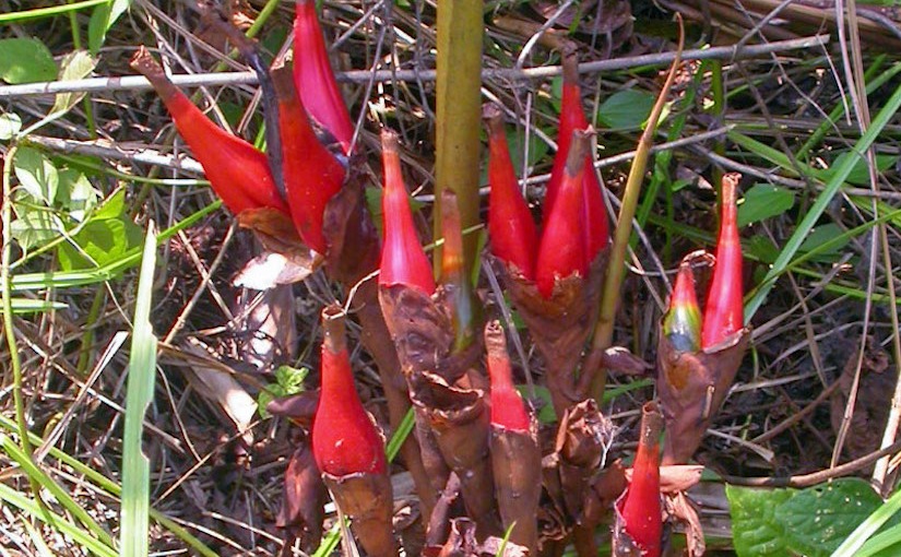
Aframomum angustifolium fruit

Structure and development of stomata in the leaves of some Zingiberaceae

by Nyawuame H. G. K., Gill L. S. (1990)

Department of Botany, University of Benin, Benin

in Korean J. Bot, 33(3): 169-172 – ISSN : 0583-421X –

http://agris.fao.org/agris-search/search.do?recordID=KR9136449

http://www.papersearch.net/thesis/article.asp?key=242749

1280px-Native_turmeric
Photo credit Google – Curcuma longa – turmeric – https://upload.wikimedia.org/wikipedia/commons/thumb/4/4f/Native_turmeric.jpg/1280px-Native_turmeric.jpg


Abstract

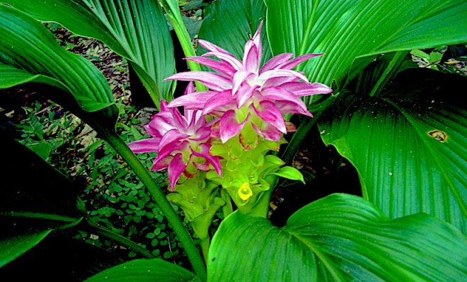
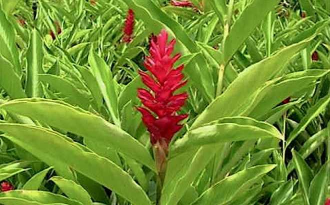
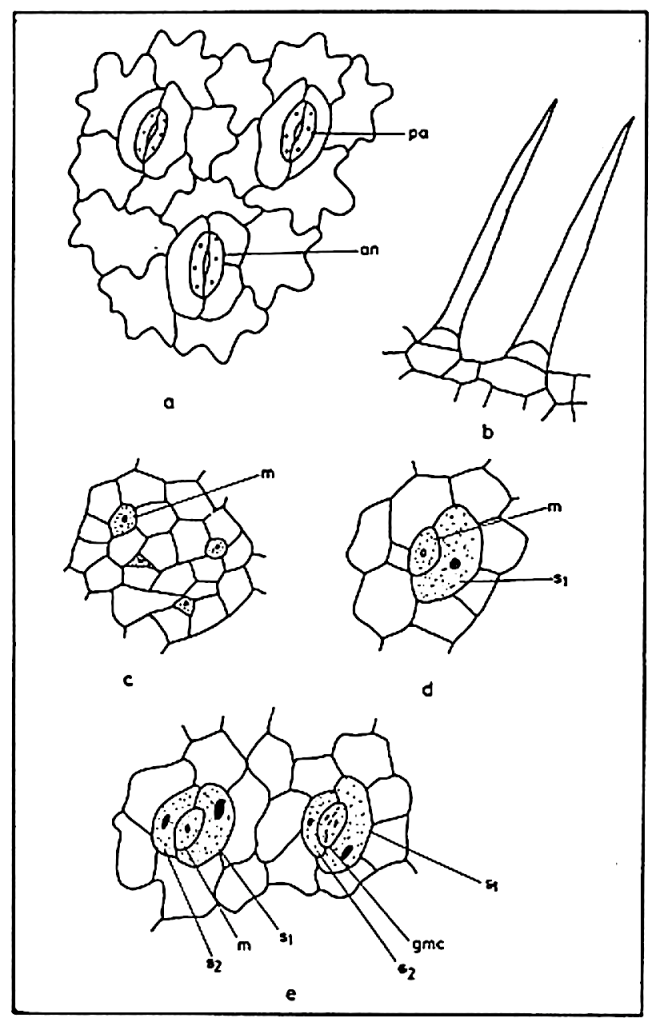
The epidermal structure and development of stomata in four taxa of Zingiberaceae viz: Aframomum melegueta K. Schum, Aframomum sceptrum K. Schum, Curcuma longa L. and Zingiber officinale Rosc. have been investigated.

Unicellular, eglandular trichomes are observed on the epidermis of A.sceptrum and Z.officinale.

hqdefault
Photo credit Google – Zingiber officinale – Red Ginger – https://i.ytimg.com/vi/tsAXgE5lzDg/hqdefault.jpg

Anomocytic stomata with agenous ontogeny, paracytic stomata with eumesogenous ontogeny and tetracytic stomata with mesoperigenous ontogeny are recorded in Z. officinale, Aframomum species and C. longa respectively.

Stomata of Z. officinale are the smallest in size (20.6 × 1.4 × 10.5 ㎛) while those of C. longa are the largest (42.5 × 31.5 × 20.2 ㎛). These two taxa also recorded the highest (43.7/㎟) and lowest (28.6/㎟) stomatal frequency respectively which suggests a linear regression of frequency on size.

Stomata in Leucaena (Fabaceae)

Photo credit: Google

Leucaena leucocephala


Screen Shot 2017-11-21 at 23.50.45

Figure la. Figure lb. Figure lc. Figure Id Mature epidermis (abaxial) showing the nature of epidermal cells and stomata of Leucaena leucocephala. Simple, uniseriate eglandular trichomes of L . leucocephala. Young epidermis (abaxial) showing stomatal initials (meristemoids) at various stages of development. & e.Various stages of development of paracytic stomata in L . leucocephala. Note: an = anisocytic stoma, gmc = guard cell mother cell, m – meristemoid, pa = paracytic stoma, S, = first mesogene subsidiary cell, S2 = second mesogene subsidiary cell.

Epidermal morphology and ontogeny of stomata in Leucaena leucocephala (Lam.) De Wit

by Nyawuame H. G. K., Gill L. S. ( —-)

–  – 104-106 –

http://biology-web.nmsu.edu/~bailey/Volume11/Nyawuame,%20H.%20G.%20K.%20and%20L.%20S.%20Gill.%20Epidermal%20morphology%20and%20ontogeny%20of%20stomata%20in%20Leucaena%20leucocephala%20(Lam.)%20d%20eWit.pdf


=============

Mature stomata are paracytic but a few anisocytic type of stomata have been observed (Figure la). The occurrence of both paracytic and anisocytic stomata on the same surface of the leaf confirms the earlier reports of Pant (1965), Pant and Banerji (1965), Pant and Kidwai (1964), Shah (1967) and Shah and Gopal (1971).

Shah and Gopal (1971) stated that in all leguminous plants so far studied different types of stomata found on the same surface of the leaf follow the same developmental sequence. During the present study it has been observed that both anisocytic and paracytic stomata show similar ontogenetic pathway. The stomatal initials (meristemoids) which are variously shaped are granular with dense cytoplasm and prominent nuclei (Figure lc). A functional meristemoid division gives rise to two cells of which the smaller one retains the ability to divide and represent the meristemoid while the larger cell divides no longer and represents the first mesogene subsidiary cell (Sj). The meristemoid divides again so that it is now surrounded by two mesogene subsidiary cells (S! and S2). The meristemoid eventually differentiates into the guard cell mother cell which divides longitudinally to form the two guard cells (Figure le). The epidermal structure and ontogcncy of stomata reported here may serve as diagnostic features of L. leucocephala for further investigations.


Stomata in ornamental monocots


Structure and ontogeny of stomata in some tropical ornamental plants (Monocotyledons).

by Nyawuame H. G. K., Gill L. S. (1990)

in Giorn. Bot. Ital. 124, 249–258. – http://dx.doi.org/10.1080/11263509009428146 –

http://www.tandfonline.com/doi/abs/10.1080/11263509009428146


Stomatal features and ontogeny of stomata of 11 ornamental taxa of monocotyledonous families with Agavaceae (1 species), Amaryllidaceae (1 species), Araceae (3 species), Cannaceae (1 species), Commelinaceae (3 species), Liliaceae (1 species), and Musaceae (1 species) have been studied.

Features like stomatal area, leaf area occupied by stomata and per cent leaf area occupied by stomata are reported for these taxa for the first time.

Stomata in trees of 17 dicotyledonous families


Epidermal studies in some Nigerian trees

by Nyawuame H. G. K., Adedio M. U., Gill L. S. (1993)

(With one Table) –

Feddes Repertorium Volume 104, Issue 7-8, 1993, 475–480 – DOI: 10.1002/fedr.19931040710 – 

http://onlinelibrary.wiley.com/doi/10.1002/fedr.19931040710/full

Abstract

Leaf epidermal structure of 30 trees distributed in 17 dicotyledenous families was studied.

Interspecific variation in cell wall pattern was observed in the two species of Macaranga covered in this study. The cells were straight-walled in M. barteri but sinuously-walled in M. monandra. In addition to this variation, peltate hairs were observed in M. barteri.

The two species also varied in their stomatal features. Alchornea was another species which showed interspecific variation in epidermal and stomatal features. Stellate hairs were observed in A. cordifolia but were absent in A. laxiflora.

Mature stomata were paracytic in A. cordifolia but in A. laxiflora they were anomocytic. Differences in stomatal size and frequency were also observed in the two species. Contiguous stomata were reported here for Avicennia africana, Mitragyna inermis and Pterygota macrocarpa.

Stomata in Boraginaceae (dicots)


Epidermal morphology and ontogeny of stomata in some tropical Boraginaceae

by Nyawuame H. G. K., Gill L. S. (1990)

in Feddes Repertorium Volume 101, Issue 5-6, 1990, 289–295 – DOI: 10.1002/fedr.19901010513 – 

http://onlinelibrary.wiley.com/doi/10.1002/fedr.19901010513/full

Abstract

Epidermal morphology, the structure and development of stomata in 13 species of Boraginaceae are described.

The epidermal cells are straight-walled on the upper surface and sinuous on the lower in the majority of the investigated species.

The leaves are hypostomatic with either anomocytic or anisocytic stomata distributed on the lower surface.

The anomocytic stomata follow agenous ontogenetic pathway while the anisocytic stomata are hemimesogenously derived.

Laterally contiguous stomata in Cordia platythyrsa are recorded for the first time.

Stomata in Scrophulariaceae (dicots)


Cuticular studies and their bearing on the taxonomy of some tropical Scrophulariaceae

by Nyawuame H. G. K., Gill L. S. (1993)

(With 58 Figures and one Table)

in Feddes Repertorium Volume 104, Issue 1-2, 1993, 41–50 – DOI: 10.1002/fedr.19931040108 – 

http://onlinelibrary.wiley.com/doi/10.1002/fedr.19931040108/full

Abstract

The structure of the epidermis and the ontogeny of the stomatal complex of 26 tropical taxa of the family Scrophulariaceae are described.

The epidermal cells depict variations with the cells being straight-walled on both surfaces, straight-walled on the upper surface only or sinuously walled on both surfaces. Trichomes are also variable. The different types of trichomes observed include unicellular eglandular, peltate glandular and multicellular eglandular.

All the investigated species except Lindernia crustacea and Veronica abyssinica are hypostomatic.

During the present study three types of stomata are recorded, viz: anomocytic, anisocytic and diacytic types. Anomocytic stomata follow agenous or hemiperigenous ontogenetic development whereas the ontogeny of anisocytic stomata is either hemiperigenous or hemimesogenous.

Within limitations stomatal distribution and frequency could serve as reliable taxonomic characters in the tropical taxa of the family Scrophulariaceae.

Stomata in Apocynaceae (dicots)

Cuticular studies of some West African species of the Apocynaceae of medicinal value

by Nyawuame H. G. K., Gill L. S. (1991)

in Feddes Repertorium Volume 102, Issue 1-2, 1991, 87–104 – DOI: 10.1002/fedr.19911020110 – 

http://onlinelibrary.wiley.com/doi/10.1002/fedr.19911020110/full 

Abstract

A comparative study of the leaf epidermis of 45 West African Apocynaceous taxa using the light microscope has been undertaken.

Variations observed in anticlinal cell wall pattern, trichomes, size, distribution, frequency, morphology and ontogeny of the stomatal complex are discussed.

Aborted stomata and single guard-celles stomata are reported in Allamanda and Thevetia for the first time.

Stomata in Solanaceae (dicots)


Epidermal studies of some species of family Solanaceae used in traditional medicine in West Africa

by Nyawuame H. G. K., Gill L. S. (1994)

in Feddes Repertorium Volume 105, Issue 1-2, 1994, 49–60 – DOI: 10.1002/fedr.19941050112 – 

Abstract

Leaf epidermal structure and developmental pattern of the stomatal complex of 30 species of the family Solanaceae are described.

Epidermal cells of almost all the species showed wall sinuousity. However, in Brunfelsia uniflora and some species of the genus Solanum viz. S. aethiopicum, S. dasyphyllum, S. macrocarpon and S. verbascifolium the cells were straight-walled. Several types of epidermal hairs were observed on both the upper and lower epidermis. They include bicellular eglandular, multicellular stellate and club-shaped hairs.

The leaves of most species were amphistomatic; however, a few species with hypostomatic leaves were also encountered.

Mature stomata were either anomocytic or anisocytic with hemimesogeneous developmental pattern. In Schwenkia americana, however, mature stomata were diacytic with eumesogenous developmental pathway.

Interspecific variation observed in the distribution, size and frequency of stomata may prove useful in the authentication of the foliar materials of these plants when used as crude drugs.

Stomata of Gentianaceae (dicots)


Epidermal studies in some species of Gentianaceae from West Africa

by Nyawuame H. G. K.,  Gill L. S. (1990)

Dr. H. G. K. Nyawuame and Prof. Dr. L. S. Gill

in Feddes Repertorium, Volume 101, Issue 7-8, pages 395–400, 1990

Abstract

The structure of mature epidermis and the ontogenetic pathway of mature stomata in some species of the family Gentianaceae, viz. Canscora decussata, C. diffusa, Exacum quinquenervium, Faroa pusilla, Neurotheca loeselioides, Sebaea brachyphylla and Swertia mannii are described.

Ontogeny of stomata is shown to be eumesogenous in C. decussata, C. diffusa, N. loeselioides and S. brachyphylla, mesoperigenous in E. quinquenervium and F. pusilla and euperigenous in S. mannii. Juxtaposed contiguous stomata have been observed in C. decussata.

See also: Wiley Online Library