The Skin Is the Largest Organ of the Human Body and Carries Out Several Vital Functions

The Essential Info
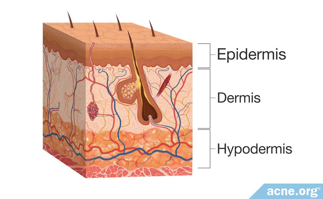
The skin is the body’s largest organ, and consists of three main layers:
- Epidermis – topmost layer
- Dermis – middle layer
- Hypodermis – bottom layer
These layers are composed of skin cells called corneocytes, keratinocytes, melanocytes, fibroblasts, and mast cells, as well as blood vessels, glands, and nerves, all of which give the skin a variety of functions. These functions include:
- Providing a physical barrier to protect the body
- Immune system regulation
- Body temperature regulation
- Protection from UV radiation from the sun
- Ability to detect surroundings through sensory cells
- Fat storage
- Synthesis of vitamin D

The Science
The skin is the largest organ in the body, comprising up to 16% of the body’s weight.
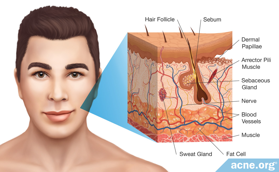
Within each square centimeter of skin, there are nearly 3 million cells, including 300 sensory cells that detect touch, pain, and heat, 14 – 300 microscopic hair follicles, 50 – 900 sebum (skin oil) glands, 150 – 340 sweat glands, nearly 4 meters of blood vessels, and 10 hairs.
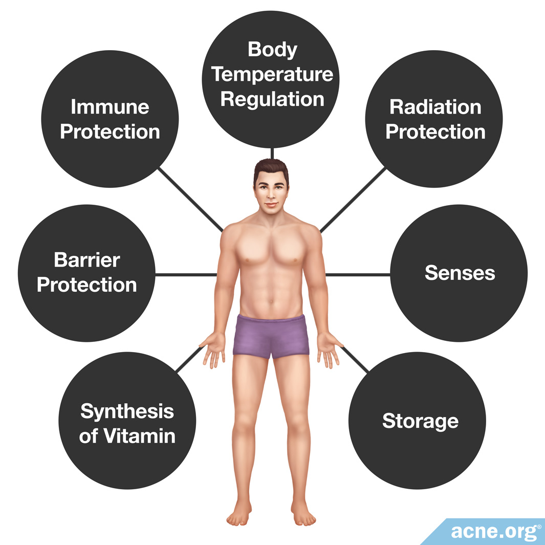
The skin provides a variety of functions, including:
- Barrier protection: The skin provides the body a physical barrier that acts as a wall to protect the inside of the body against any mechanical, chemical, or microbial damage. This barrier also plays a role in preventing dehydration by retaining water inside the skin.
- Immune protection: If bacteria or viruses pass through the outer layer of the skin, immune system cells in the skin protect the body from these “invaders.”
- Body temperature regulation: The skin helps maintain a proper body temperature by providing insulation when the body becomes cold and by producing sweat, which cools down the body, when it is hot.
- UV radiation protection: The skin contains a special protein called melanin, which acts like a sunscreen to protect the skin and the body from the sun’s harmful ultraviolet (UV) radiation.
- Senses: The skin contains cells called sensory cells, which allow for detecting the environment through touch. These cells can also sense heat or cold and can alert the body to pain.
- Storage: The skin stores fat that the body can use for energy.
- Synthesis of vitamin D: The skin is the only organ of the body where vitamin D, an essential nutrient, can be synthesized.
Structure of the Skin
The skin contains three main layers.
- The epidermis
- The dermis
- The hypodermis
The Epidermis
The epidermis is the topmost layer of the skin. It is composed mainly of skin cells called keratinocytes, which produce the protein, keratin. This is the same protein that is found in hair and in nails.
Keratinocytes are produced at the bottom of the epidermis and slowly move up through the epidermis until they eventually flake off the top of the skin. It takes about four weeks for a keratinocyte to make its journey from creation, at the bottom of the epidermis, to flaking off the top of the skin.
In acne, keratinocytes reproduce faster than normal. Also, they do not shed as they should, and instead stick around and clog pores.
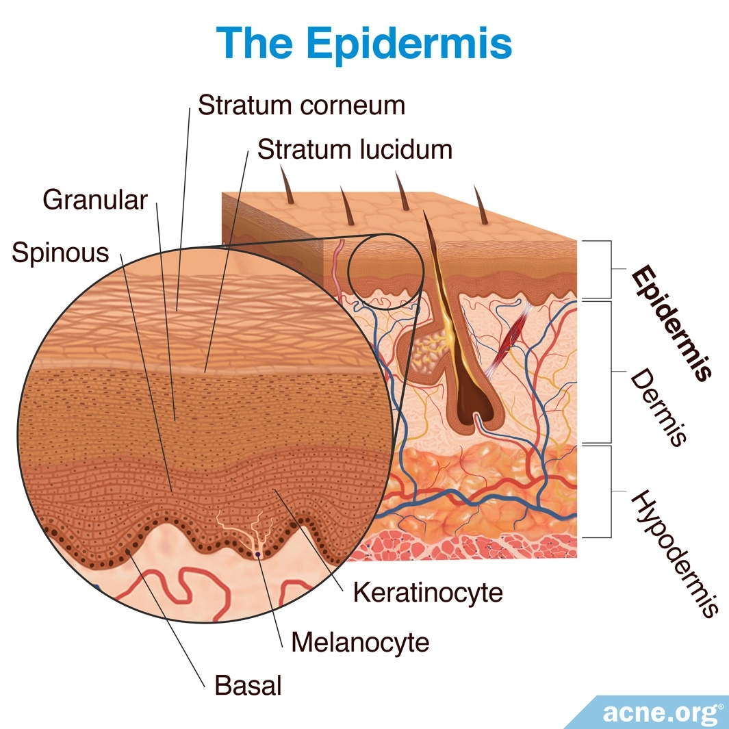
The epidermis is broken down into the following five layers:
- Basal layer: The basal layer is the bottom layer of the epidermis, where keratinocytes are formed. Another skin cell type found in the basal layer is a melanocyte. Melanocytes provide the skin protection against the sun’s rays.
- Spinous layer: The spinous layer lies on top of the basal layer and contains keratinocytes that have traveled upward from the basal layer. The spinous layer gives off a spiny-like appearance.
- Granular layer: The granular layer sits on top of the spinous layer and contains keratinocytes that have traveled upward from the spinous layer. The keratinocytes in this layer produce fats that provide for a barrier that prevents dehydration by keeping water inside the skin.
- Stratum lucidum layer: The stratum lucidum layer, which is present only on the palms of hands and soles of feet, is on top of the granular layer, and it contains keratinocytes that have traveled upward from the granular layer. This layer helps provide protection against the sun.
- Stratum corneum layer: The stratum corneum layer is the topmost layer of the epidermis. In the stratum corneum the keratinocytes die and flatten out, and are now called corneocytes. Corneocytes are full of keratin, which gives the skin its protection function by providing a physical barrier.
The production rate of new skin cells in the basal layer is normally equal to the shedding frequency of old ones in the stratum corneum, so the number of cells in the epidermis remains steady. However, in the case of acne, the dead skin cells do not shed properly and instead clog the opening of the hair follicle pore, giving rise to a chain of events that eventually result in acne.1,3-5
The Dermis
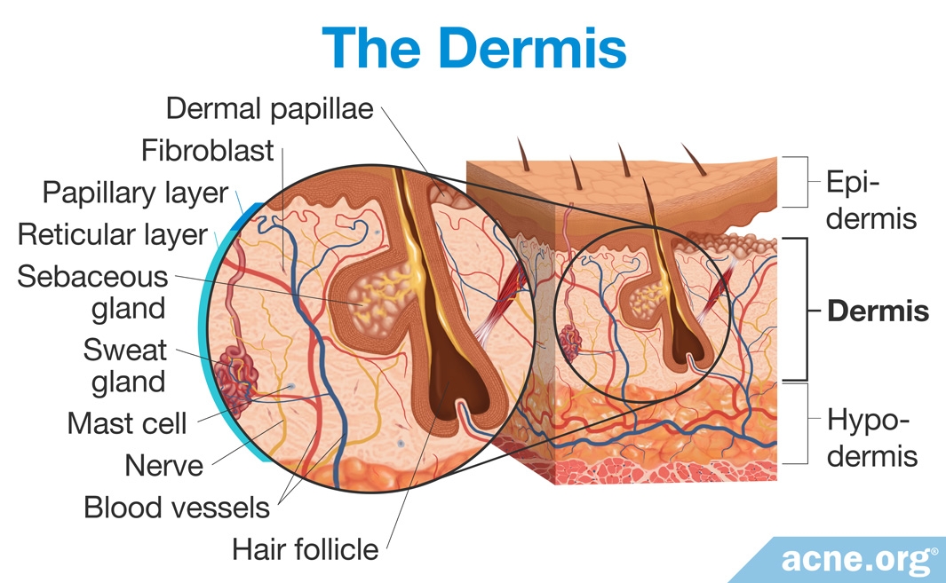
The dermis sits directly beneath the epidermis, providing support for it. The dermis contains skin cells, blood vessels, nerves, glands, and hair follicles and is much thicker in size than the epidermis.
There are two layers of the dermis.
- Papillary layer: The papillary layer of the dermis connects with the bottom layer of the epidermis. It is a thin layer that resembles “egg-crate” foam, because it contains wavelike projections that curve up and down at the border of the dermis and epidermis.
- Reticular layer: The reticular layer of the dermis connects with the papillary layer. It is much thicker than the papillary layer and consists primarily of collagen protein fibers that travel in every direction throughout the layer. It contains many important structures, including:
- Hair follicles
- Sweat glands
- Sebaceous (sebum-producing) glands
- Nerves
- Blood vessels
Although the dermis is composed mainly of protein fibers, there are three main cell types that travel through the dermis.
- Fibroblasts are special cells that produce proteins important for wound healing and scarring.
- Mast cells are immune system cells that mediate immune reactions like allergies.
- Fat cells are full of fat, which protects the dermis by cushioning it.1
The Hypodermis
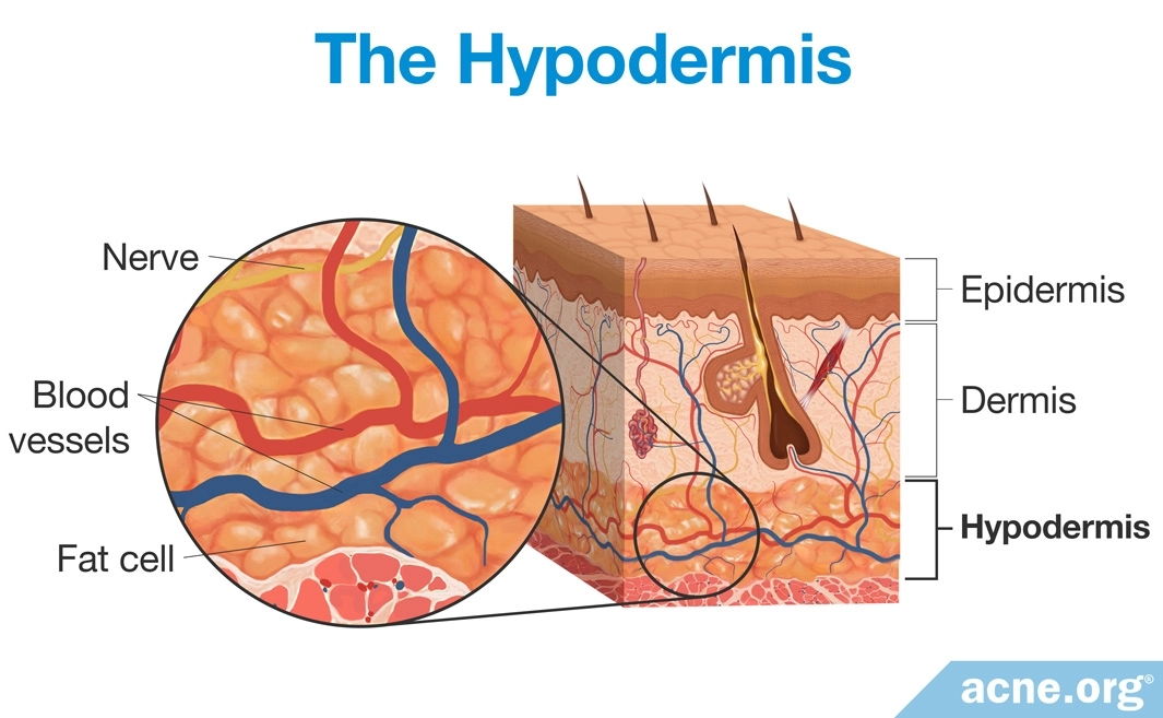
The hypodermis is a thick fat layer that sits directly beneath the dermis and separates it from other body structures like muscles and bone. The fat in this layer provides insulation for the body, stores energy, and protects the body through its cushioning effect.1
References
- Basic Science of the Skin: Structure and Function. Am. Acad. Dermatol. (2014). https://www.ncbi.nlm.nih.gov/pubmed/24314373
- Human skin. https://en.wikipedia.org/wiki/Human_skin#Functions
- Otberg, N. et al. Variations of Hair Follicle Size and Distribution in Different Body Sites. J. Invest. Dermatol. 122, 14 – 19 (2004). https://www.ncbi.nlm.nih.gov/pubmed/14962084
- Zeligman, I. The skin as a Physiologic Organ. J. Natl. Med. Assoc. 53, 148 – 150 (1961). https://www.ncbi.nlm.nih.gov/pmc/articles/PMC2641878/
- Elbing, J. & Montagna, W. Human skin. https://www.britannica.com/science/human-skin