Anthostomella ravennica Daranagama, Camporesi & K.D. Hyde, in Hyde et al., Fungal Diversity 80: 210 (2016)
Index Fungorum number: IF 552286; Facesoffungi number: FoF 02418
Etymology – ‘‘ravennica’’ refers to the province from where it was first collected.
Saprobic on dead stem of Ammophila arenaria (L.) Link. Sexual morph: Ascomata 170–190 × 180–200 μm (x̄ = 175 μm × 192 μm, n = 10), immersed, visible as black, irregular to dome-shaped areas, coriaceous, solitary, scattered, in cross-section globose, with inconspicuous ostiole. Ostiole black, papillate. Peridium 30–50 μm wide (x̄ = 35 μm, n = 10), with two cell layers, outwardly comprising thick-walled, compressed, dark brown cells of textura angularis and inwardly comprising a few layers of thin-walled, hyaline cells of textura angularis. Paraphyses 3.3–3.8 μm wide at the base (x̄ = 3.5 μm, n = 30), shorter than the asci, numerous, filamentous, septate. Asci 116–127 × 11–13.6 μm (x̄ = 124 × 12.8 μm, n = 20), 8-spored, unitunicate, cylindrical, short pedicellate, with conspicuous, J+, discoid, apical ring, 0.7–1.5 × 0.2–0.8 μm (x̄ = 1.2 × 0.5 μm, n = 20). Ascospores 14–17 × 7–8 μm (x̄ = 16.2 × 7.6 μm, n = 20), uni-seriate, inequilaterally ellipsoidal, with one convex surface, pointed ends, light brown-dark brown, smooth-walled, without germ slit. Asexual morph: Undetermined.
Culture characteristics – Colonies on OA 9 cm diam. after 4 weeks at 27 ºC, white at the margins, pale yellowish at the center; reverse yellowish to cream, colony azonate, cottony appearance, dense.
Material examined – ITALY, Ravenna Province, Lido di Dante, on dead stems of Ammophila arenaria (L.) Link, 28 January 2015, E. Camporesi, IT 2358 (MFLU 16-0972, holotype), ibid. HKAS, isotype; ex-type living culture, MFLUCC 15-0012.
Notes – Our collection is reminiscent of both A. zongluensis K.D. Hyde and A. consanguinea (Ces.) Sacc. However, A. zongluensis has ascospores with more parallel sides, minutely verruculose walls and ascomata with periphysate ostiolar canals (Lu and Hyde 2000). Ascospores of A. thailandica are similar in size to those of A. consanguinea (Ces.) Sacc. However, ascospores of A. consanguinea have verruculose walls and a short, straight germ slit, while those in A. thailandica have a smooth wall and lack germ slits. In the phylogenetic analysis A. thailandica clusters in a separate clade with A. formosa Kirschst., A. conorum (Fuckel) Sacc., A. rubicola Speg. ex Sacc. & Trotter and A. obesa Daranagama, E. Camporesi & K.D. Hyde.
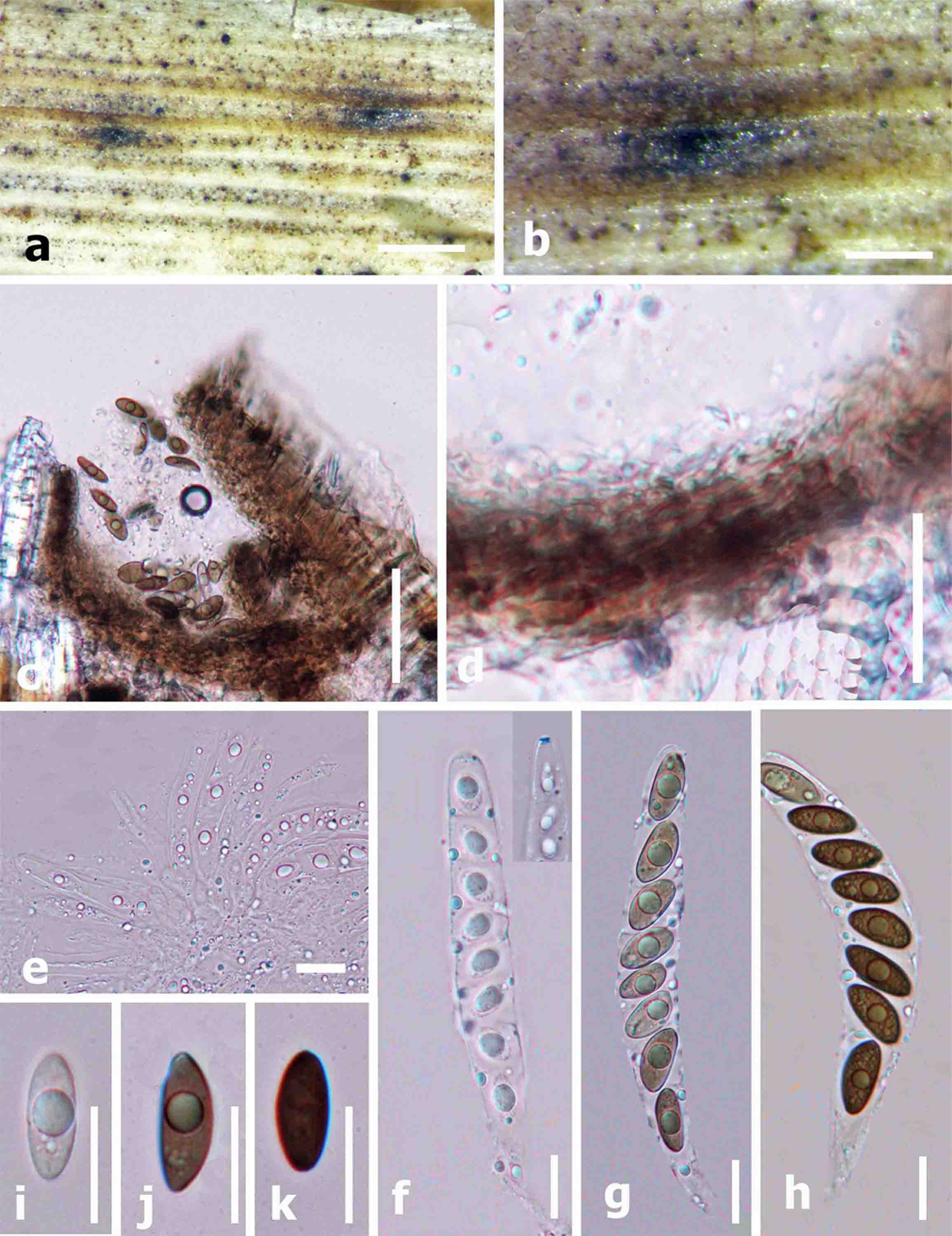
Fig 1. Anthostomella ravennica (holotype). a, b Appearance of ascomata on the host. c Cross section of ascoma. d Peridium. e Paraphyses. f–h Asci (f note the apical ring bluing in Melzer’s reagent). i–k Ascospores without visible germ slits. Scale bars: a = 200 μm, b, c = 100 μm, d = 50 μm, f–k = 10 μm.