11 pages, 667 KiB  
Article
Fungal Pathogens as Causes of Acute Respiratory Illness in Hospitalized Veterans: Frequency of Fungal Positive Test Results Using Rapid Immunodiagnostic Assays
by Diego H. Caceres 1,2,3, Maria C. Rodriguez-Barradas 4, Michael Whitaker 1, Brendan R. Jackson 1,5, Lindsay Kim 1,5, Diya Surie 1,5, Bryanna Cikesh 1, Mark D. Lindsley 1, Orion Z. McCotter 1,6, Elizabeth L. Berkow 1 and Mitsuru Toda 1,*
1 Centers for Disease Control and Prevention (CDC), Atlanta, GA 30329, USA
2 Center of Expertise in Mycology Radboudumc, Canisius Wilhelmina Hospital, 6532 SZ Nijmegen, The Netherlands
3 Studies in Translational Microbiology and Emerging Diseases (MICROS) Research Group, School of Medicine and Health Sciences, Universidad del Rosario, Bogota 111221, Colombia
4 Michael E. DeBakey VA Medical Center, Houston, TX 77030, USA
5 US Public Health Service, Rockville, MD 20852, USA
6 Oregon Health Authority, Portland, OR 97232, USA
J. Fungi 2023, 9(4), 456; https://doi.org/10.3390/jof9040456 - 8 Apr 2023
Cited by 1 | Viewed by 1678
Abstract
Fungal respiratory illnesses caused by endemic mycoses can be nonspecific and are often mistaken for viral or bacterial infections. We performed fungal testing on serum specimens from patients hospitalized with acute respiratory illness (ARI) to assess the possible role of endemic fungi as [...] Read more.
Fungal respiratory illnesses caused by endemic mycoses can be nonspecific and are often mistaken for viral or bacterial infections. We performed fungal testing on serum specimens from patients hospitalized with acute respiratory illness (ARI) to assess the possible role of endemic fungi as etiologic agents. Patients hospitalized with ARI at a Veterans Affairs hospital in Houston, Texas, during November 2016–August 2017 were enrolled. Epidemiologic and clinical data, nasopharyngeal and oropharyngeal samples for viral testing (PCR), and serum specimens were collected at admission. We retrospectively tested remnant sera from a subset of patients with negative initial viral testing using immunoassays for the detection of Coccidioides and Histoplasma antibodies (Ab) and Cryptococcus, Aspergillus, and Histoplasma antigens (Ag). Of 224 patient serum specimens tested, 49 (22%) had positive results for fungal pathogens, including 30 (13%) by Coccidioides immunodiagnostic assays, 19 (8%) by Histoplasma immunodiagnostic assays, 2 (1%) by Aspergillus Ag, and none by Cryptococcus Ag testing. A high proportion of veterans hospitalized with ARI had positive serological results for fungal pathogens, primarily endemic mycoses, which cause fungal pneumonia. The high proportion of Coccidioides positivity is unexpected as this fungus is not thought to be common in southeastern Texas or metropolitan Houston, though is known to be endemic in southwestern Texas. Although serological testing suffers from low specificity, these results suggest that these fungi may be more common causes of ARI in southeast Texas than commonly appreciated and more increased clinical evaluation may be warranted. Full article
Show Figures

Figure 1

17 pages, 5735 KiB  
Article
Mitogen-Activated Protein Kinases SvPmk1 and SvMps1 Are Critical for Abiotic Stress Resistance, Development and Pathogenesis of Sclerotiophoma versabilis
by Felix Abah 1,†, Yunbo Kuang 1,2,*,†, Jules Biregeya 1, Yakubu Saddeeq Abubakar 1, Zuyun Ye 2 and Zonghua Wang 1,3,*
1 Ministerial and Provincial Joint Innovation Centre for Safety Production of Cross-Strait Crops, College of Life Sciences & College of Plant Protection, Fujian Agriculture and Forestry University, Fuzhou 350002, China
2 The Engineering Technology Research Center of Characteristic Medicinal Plants of Fujian, College of Life Sciences, Ningde Normal University, Ningde 352100, China
3 Fujian Provincial Key Laboratory on Conservation and Sustainable Utilization of Marine Biodiversity, Fuzhou Institute of Oceanography, Minjiang University, Fuzhou 350108, China
These authors contributed equally to this work.
J. Fungi 2023, 9(4), 455; https://doi.org/10.3390/jof9040455 - 7 Apr 2023
Cited by 3 | Viewed by 1468
Abstract
Mitogen-activated protein kinase (MAPK) signaling pathways are evolutionarily conserved in eukaryotes and modulate responses to both internal and external stimuli. Pmk1 and Mps MAPK pathways regulate stress tolerance, vegetative growth and cell wall integrity in Saccharomyces cerevisiae and Pyricularia oryzae. Here, we deployed [...] Read more.
Mitogen-activated protein kinase (MAPK) signaling pathways are evolutionarily conserved in eukaryotes and modulate responses to both internal and external stimuli. Pmk1 and Mps MAPK pathways regulate stress tolerance, vegetative growth and cell wall integrity in Saccharomyces cerevisiae and Pyricularia oryzae. Here, we deployed genetic and cell biology strategies to investigate the roles of the orthologs of Pmk1 and Mps1 in Sclerotiophoma versabilis (herein referred to as SvPmk1 and SvMps1, respectively). Our results showed that SvPmk1 and SvMps1 are involved in hyphal development, asexual reproduction and pathogenesis in S. versabilis. We found that ∆Svpmk1 and ∆Svmps1 mutants have significantly reduced vegetative growths on PDA supplemented with osmotic stress-inducing agents, compared to the wild type, with ∆Svpmps1 being hypersensitive to hydrogen peroxide. The two mutants failed to produce pycnidia and have reduced pathogenicity on Pseudostellaria heterophylla. Unlike SvPmk1, SvMps1 was found to be indispensable for the fungal cell wall integrity. Confocal microscopic analyses revealed that SvPmk1 and SvMps1 are ubiquitously expressed in the cytosol and nucleus. Taken together, we demonstrate here that SvPmk1 and SvMps1 play critical roles in the stress resistance, development and pathogenesis of S. versabilis. Full article
(This article belongs to the Special Issue Molecular Biology of Fungal Plant Pathogens)
Show Figures

Figure 1

28 pages, 2402 KiB  
Review
Fungal Pigments: Carotenoids, Riboflavin, and Polyketides with Diverse Applications
by Maria Afroz Toma 1,*, Md. Hasibur Rahman 1, Md. Saydar Rahman 1, Mohammad Arif 2, K. H. M. Nazmul Hussain Nazir 2 and Laurent Dufossé 3,4,*
1 Department of Food Technology & Rural Industries, Bangladesh Agricultural University, Mymensingh 2202, Bangladesh
2 Department of Microbiology and Hygiene, Bangladesh Agricultural University, Mymensingh 2202, Bangladesh
3 Laboratoire de Chimie et de Biotechnologie des Produits Naturals, CHEMBIOPRO EA 2212, Université de La Réunion, ESIROI Agroalimentaire, 97744 Saint-Denis, France
4 Laboratoire ANTiOX, Université de Bretagne Occidentale, Campus de Créac’h Gwen, 29000 Quimper, France
J. Fungi 2023, 9(4), 454; https://doi.org/10.3390/jof9040454 - 7 Apr 2023
Cited by 12 | Viewed by 7129
Abstract
Natural pigments and colorants have seen a substantial increase in use over the last few decades due to their eco-friendly and safe properties. Currently, customer preferences for more natural products are driving the substitution of natural pigments for synthetic colorants. Filamentous fungi, particularly [...] Read more.
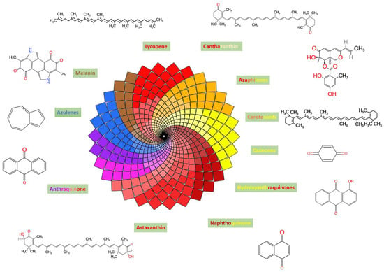
Natural pigments and colorants have seen a substantial increase in use over the last few decades due to their eco-friendly and safe properties. Currently, customer preferences for more natural products are driving the substitution of natural pigments for synthetic colorants. Filamentous fungi, particularly ascomycetous fungi (Monascus, Fusarium, Penicillium, and Aspergillus), have been shown to produce secondary metabolites containing a wide variety of pigments, including β-carotene, melanins, azaphilones, quinones, flavins, ankaflavin, monascin, anthraquinone, and naphthoquinone. These pigments produce a variety of colors and tints, including yellow, orange, red, green, purple, brown, and blue. Additionally, these pigments have a broad spectrum of pharmacological activities, including immunomodulatory, anticancer, antioxidant, antibacterial, and antiproliferative activities. This review provides an in-depth overview of fungi gathered from diverse sources and lists several probable fungi capable of producing a variety of color hues. The second section discusses how to classify coloring compounds according to their chemical structure, characteristics, biosynthetic processes, application, and present state. Once again, we investigate the possibility of employing fungal polyketide pigments as food coloring, as well as the toxicity and carcinogenicity of particular pigments. This review explores how advanced technologies such as metabolic engineering and nanotechnology can be employed to overcome obstacles associated with the manufacture of mycotoxin-free, food-grade fungal pigments. Full article
(This article belongs to the Special Issue Fungal Pigments 3.0)
Show Figures

Figure 1

28 pages, 3888 KiB  
Review
Potential of Secondary Metabolites of Diaporthe Species Associated with Terrestrial and Marine Origins
by Wei Wei 1,†, Babar Khan 2,†, Qun Dai 1, Jie Lin 1, Liqin Kang 1, Nasir Ahmed Rajput 3, Wei Yan 2,* and Guiyou Liu 1,*
1 School of Life Sciences and Chemical Engineering, Jiangsu Second Normal University, Nanjing 211200, China
2 College of Plant Protection, State & Local Joint Engineering Research Center of Green Pesticide Invention and Application, Nanjing Agricultural University, Nanjing 210095, China
3 Department of Plant Pathology, University of Agriculture, Faisalabad 38000, Pakistan
These authors contributed equally to this work.
J. Fungi 2023, 9(4), 453; https://doi.org/10.3390/jof9040453 - 7 Apr 2023
Cited by 3 | Viewed by 1505
Abstract
Diaporthe species produce versatile secondary metabolites (SMs), including terpenoids, fatty acids, polyketides, steroids, and alkaloids. These structurally diverse SMs exhibit a wide range of biological activities, including cytotoxic, antifungal, antibacterial, antiviral, antioxidant, anti-inflammatory, and phytotoxic activities, which could be exploited in the medical, [...] Read more.
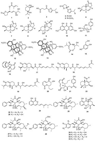
Diaporthe species produce versatile secondary metabolites (SMs), including terpenoids, fatty acids, polyketides, steroids, and alkaloids. These structurally diverse SMs exhibit a wide range of biological activities, including cytotoxic, antifungal, antibacterial, antiviral, antioxidant, anti-inflammatory, and phytotoxic activities, which could be exploited in the medical, agricultural, and other modern industries. This review comprehensively covers the production and biological potencies of isolated natural products from the genus Diaporthe associated with terrestrial and marine origins. A total of 275 SMs have been summarized from terrestrial (153; 55%) and marine (110; 41%) origins during the last twelve years, and 12 (4%) compounds are common to both environments. All secondary metabolites are categorized predominantly on the basis of their bioactivities (cytotoxic, antibacterial, antifungal, and miscellaneous activity). Overall, 134 bioactive compounds were isolated from terrestrial (92; 55%) and marine (42; 34%) origins, but about half the compounds did not report any kind of activity. The antiSMASH results suggested that Diaporthe strains are capable of encoding a wide range of SMs and have tremendous biosynthetic potential for new SMs. This study will be useful for future research on drug discovery from terrestrial and marine natural products. Full article
(This article belongs to the Special Issue Emerging Investigators in Bioactive Fungal Metabolites)
Show Figures

Figure 1

16 pages, 13442 KiB  
Article
Pneumocystis Exacerbates Inflammation and Mucus Hypersecretion in a Murine, Elastase-Induced-COPD Model
by Diego A. Rojas 1,*, Carolina A. Ponce 2, Adriel Bustos 1, Vicente Cortés 1, Daniela Olivares 1 and Sergio L. Vargas 2,*
1 Instituto de Ciencias Biomédicas (ICB), Facultad de Ciencias de la Salud, Universidad Autónoma de Chile, Santiago 8910132, Chile
2 Programa de Microbiología y Micología, ICBM, Facultad de Medicina, Universidad de Chile, Santiago 8380492, Chile
J. Fungi 2023, 9(4), 452; https://doi.org/10.3390/jof9040452 - 7 Apr 2023
Cited by 2 | Viewed by 1690
Abstract
Inflammation and mucus hypersecretion are frequent pathology features of chronic respiratory diseases such as asthma and COPD. Selected bacteria, viruses and fungi may synergize as co-factors in aggravating disease by activating pathways that are able to induce airway pathology. Pneumocystis infection induces inflammation [...] Read more.
Inflammation and mucus hypersecretion are frequent pathology features of chronic respiratory diseases such as asthma and COPD. Selected bacteria, viruses and fungi may synergize as co-factors in aggravating disease by activating pathways that are able to induce airway pathology. Pneumocystis infection induces inflammation and mucus hypersecretion in immune competent and compromised humans and animals. This fungus is a frequent colonizer in patients with COPD. Therefore, it becomes essential to identify whether it has a role in aggravating COPD severity. This work used an elastase-induced COPD model to evaluate the role of Pneumocystis in the exacerbation of pathology, including COPD-like lung lesions, inflammation and mucus hypersecretion. Animals infected with Pneumocystis developed increased histology features of COPD, inflammatory cuffs around airways and lung vasculature plus mucus hypersecretion. Pneumocystis induced a synergic increment in levels of inflammation markers (Cxcl2, IL6, IL8 and IL10) and mucins (Muc5ac/Muc5b). Levels of STAT6-dependent transcription factors Gata3, FoxA3 and Spdef were also synergically increased in Pneumocystis infected animals and elastase-induced COPD, while the levels of the mucous cell-hyperplasia transcription factor FoxA2 were decreased compared to the other groups. Results document that Pneumocystis is a co-factor for disease severity in this elastase-induced-COPD model and highlight the relevance of STAT6 pathway in Pneumocystis pathogenesis. Full article
Show Figures

Figure 1

14 pages, 17874 KiB  
Article
Arthrobotrys blastospora sp. nov. (Orbiliomycetes): A Living Fossil Displaying Morphological Traits of Mesozoic Carnivorous Fungi
by Fa Zhang 1,2,3, Saranyaphat Boonmee 2,3, Yao-Quan Yang 1, Fa-Ping Zhou 1, Wen Xiao 1,4,5,6 and Xiao-Yan Yang 1,4,*
1 Institute of Eastern-Himalaya Biodiversity Research, Dali University, Dali 671003, China
2 Center of Excellence in Fungal Research, Mae Fah Luang University, Chiang Rai 57100, Thailand
3 School of Science, Mae Fah Luang University, Chiang Rai 57100, Thailand
4 Key Laboratory of Yunnan State Education Department on Er’hai Lake Basin Protection and the Sustainable Development Research, Dali University, Dali 671003, China
5 The Provincial Innovation Team of Biodiversity Conservation and Utility of the Three Parallel Rivers from Dali University, Dali University, Dali 671003, China
6 Yunling Black-and-White Snub-Nosed Monkey Observation and Research Station of Yunnan Province, Dali 671003, China
J. Fungi 2023, 9(4), 451; https://doi.org/10.3390/jof9040451 - 6 Apr 2023
Cited by 3 | Viewed by 1750
Abstract
The evolution of carnivorous fungi in deep time is still poorly understood as their fossil record is scarce. The approximately 100-million-year-old Cretaceous Palaeoanellus dimorphus is the earliest fossil of carnivorous fungi ever discovered. However, its accuracy and ancestral position has been widely questioned [...] Read more.
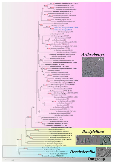
The evolution of carnivorous fungi in deep time is still poorly understood as their fossil record is scarce. The approximately 100-million-year-old Cretaceous Palaeoanellus dimorphus is the earliest fossil of carnivorous fungi ever discovered. However, its accuracy and ancestral position has been widely questioned because no similar species have been found in modern ecosystems. During a survey of carnivorous fungi in Yunnan, China, two fungal isolates strongly morphologically resembling P. dimorphus were discovered and identified as a new species of Arthrobotrys (Orbiliaceae, Orbiliomycetes), a modern genus of carnivorous fungi. Phylogenetically, Arthrobotrys blastospora sp. nov. forms a sister lineage to A. oligospora. A. blastospora catches nematodes with adhesive networks and produces yeast-like blastospores. This character combination is absent in all other previously known modern carnivorous fungi but is strikingly similar to the Cretaceous P. dimorphus. In this paper, we describe A. blastospora in detail and discuss its relationship to P. dimorphus. Full article
(This article belongs to the Special Issue New Perspectives on Entomopathogenic and Nematode-Trapping Fungi)
Show Figures

Figure 1

1 pages, 162 KiB  
Correction
Correction: Moss, R.B. Severe Fungal Asthma: A Role for Biologics and Inhaled Antifungals. J. Fungi 2023, 9, 85
by Richard B. Moss
Center of Excellence in Pulmonary Biology, Division of Pulmonary, Asthma and Sleep Medicine, Department of Pediatrics, Stanford University School of Medicine, 770 Welch Road, Suite 350, Palo Alto, CA 94304, USA
J. Fungi 2023, 9(4), 450; https://doi.org/10.3390/jof9040450 - 6 Apr 2023
Viewed by 683
Abstract
In the original publication [...] Full article
21 pages, 4580 KiB  
Article
Clonal Expansion in Multiple Phyllosticta Species Causing Citrus Black Spot or Similar Symptoms in China
by Wen Wang 1, Tao Xiong 1, Yating Zeng 1, Wenwen Li 2, Chen Jiao 1, Jianping Xu 3 and Hongye Li 1,*
1 The Key Laboratory of Molecular Biology of Crop Pathogens and Insects of Ministry of Agricultural, The Key Laboratory of Biology of Crop Pathogens and Insects of Zhejiang Province, Institute of Biotechnology, Zhejiang University, 866 Yuhangtang Road, Hangzhou 310058, China
2 Department of Biochemistry, Genetics and Microbiology, Forestry and Agricultural Biotechnology Institute (FABI), University of Pretoria, Pretoria 0028, South Africa
3 Department of Biology, McMaster University, Hamilton, ON L8S 4K1, Canada
J. Fungi 2023, 9(4), 449; https://doi.org/10.3390/jof9040449 - 6 Apr 2023
Cited by 2 | Viewed by 1958
Abstract
Phyllosticta spp. are important pathogens of citrus plants. Several Phyllosticta species associated with Citrus species grown in China have been reported; however, the relative prevalences of individual species and the distributions of their genotypes among host Citrus species remain largely unknown. In this [...] Read more.
Phyllosticta spp. are important pathogens of citrus plants. Several Phyllosticta species associated with Citrus species grown in China have been reported; however, the relative prevalences of individual species and the distributions of their genotypes among host Citrus species remain largely unknown. In this study, we conducted an extensive survey of Phyllosticta species across 11 citrus-producing provinces in southern China. From fruits and leaves with black spots or black-spot-like symptoms, a total of 461 Phyllosticta strains were isolated. Based on molecular (ITS, actA, tef1, gapdh, LSU, and rpb2 sequences) and morphological data, the strains were systematically identified as belonging to five species: P. capitalensis, P. citrichinaensis, P. citriasiana, P. citricarpa, and P. paracitricarpa. To further understand intraspecific genetic diversity and relationships, strains of five species from different geographic and host sources were analyzed based on the multilocus sequence data. Our population genetic analyses revealed that all five Phyllosticta species on citrus showed evidence for clonal dispersals within and among geographic regions. In addition, pathogenicity tests using representative strains showed that all five species can cause disease on the tested Citrus spp. We discuss the implications of our results for the control and management of Citrus Black Spot and related diseases. Full article
(This article belongs to the Section Fungal Genomics, Genetics and Molecular Biology)
Show Figures

Figure 1

21 pages, 3797 KiB  
Article
Differential Recognition of Clinically Relevant Sporothrix Species by Human Mononuclear Cells
by Laura C. García-Carnero 1, Iván Martínez-Duncker 2, Manuela Gómez-Gaviria 1 and Héctor M. Mora-Montes 1,*
1 Departamento de Biología, División de Ciencias Naturales y Exactas, Campus Guanajuato, Universidad de Guanajuato, Noria Alta s/n, col. Noria Alta, C.P., Guanajuato 36050, Gto., Mexico
2 Laboratorio de Glicobiología Humana y Diagnóstico Molecular, Centro de Investigación en Dinámica Celular, Instituto de Investigación en Ciencias Básicas y Aplicadas, Universidad Autónoma del Estado de Morelos, Cuernavaca 62209, Mor., Mexico
J. Fungi 2023, 9(4), 448; https://doi.org/10.3390/jof9040448 - 6 Apr 2023
Cited by 4 | Viewed by 1821
Abstract
Sporotrichosis is a human and animal fungal infection distributed worldwide that is caused by the thermodimorphic species of the Sporothrix pathogenic clade, which includes Sporothrix brasiliensis, Sporothrix schenckii, and Sporothrix globosa. The cell wall composition and the immune response against [...] Read more.
Sporotrichosis is a human and animal fungal infection distributed worldwide that is caused by the thermodimorphic species of the Sporothrix pathogenic clade, which includes Sporothrix brasiliensis, Sporothrix schenckii, and Sporothrix globosa. The cell wall composition and the immune response against the Sporothrix species have been studied mainly in S. brasiliensis and S. schenckii, whilst little is known about the S. globosa cell wall and the immune response that its components trigger. Therefore, in this study, we aimed to analyze the cell wall composition of S. globosa in three morphologies (germlings, conidia, and yeast-like cells) and the differences in cytokine production when human peripheral blood mononuclear cells (PBMCs) interact with these morphotypes, using S. schenckii and S. brasiliensis as a comparison. We found that S. globosa conidia and yeast-like cells have a higher cell wall chitin content, while all three morphologies have a higher β-1,3-glucan content, which was found most exposed at the cell surface when compared to S. schenckii and S. brasiliensis. In addition, S. globosa has lower levels of mannose- and rhamnose-based glycoconjugates, as well as of N- and O-linked glycans, indicating that this fungal cell wall has species-specific proportions and organization of its components. When interacting with PBMCs, S. brasiliensis and S. globosa showed a similar cytokine stimulation profile, but with a higher stimulation of IL-10 by S. globosa. Additionally, when the inner cell wall components of S. globosa were exposed at the surface or N- and O-glycans were removed, the cytokine production profile of this species in its three morphotypes did not significantly change, contrasting with the S. schenckii and S. brasiliensis species that showed different cytokine profiles depending on the treatment applied to the walls. In addition, it was found that the anti-inflammatory response stimulated by S. globosa was dependent on the activation of dectin-1, mannose receptor, and TLR2, but not TLR4. All of these results indicate that the cell wall composition and structure of the three Sporothrix species in the three morphologies are different, affecting their interaction with human PBMCs and generating species-specific cytokine profiles. Full article
(This article belongs to the Special Issue Sporothrix Emerging Investigators)
Show Figures

Figure 1

17 pages, 7248 KiB  
Article
Proposal of Four New Aureobasidium Species for Exopolysaccharide Production
by Feng Wu 1, Zixuan Feng 1, Manman Wang 1,* and Qiming Wang 1,2,3,*
1 School of Life Sciences, Institute of Life Sciences and Green Development, Hebei University, Baoding 071002, China
2 Engineering Laboratory of Microbial Breeding and Preservation of Hebei Province, Hebei University, Baoding 071002, China
3 Key Laboratory of Microbial Diversity Research and Application of Hebei Province, Hebei University, Baoding 071002, China
J. Fungi 2023, 9(4), 447; https://doi.org/10.3390/jof9040447 - 6 Apr 2023
Cited by 1 | Viewed by 2263
Abstract
In this study, 99 strains of Aureobasidium species were isolated from various samples collected from different locations in China, among which 14 isolates showed different morphological characteristics to other strains identified as known Aureobasidium species. Based on morphological characteristics, those 14 strains were [...] Read more.
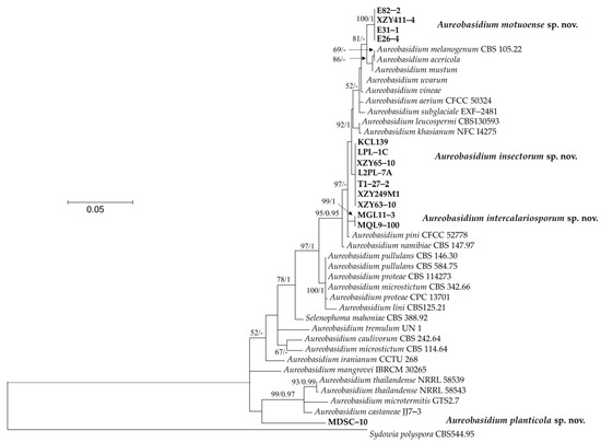
In this study, 99 strains of Aureobasidium species were isolated from various samples collected from different locations in China, among which 14 isolates showed different morphological characteristics to other strains identified as known Aureobasidium species. Based on morphological characteristics, those 14 strains were classified into four groups, represented by stains of KCL139, MDSC−10, XZY411−4, and MQL9−100, respectively. Molecular analysis of the internal transcriptional spacer (ITS) and part of the large ribosome subunit (D1/D2 domains) indicated that those four groups represent four new species in the Aureobasidium. Therefore, the names Aureobasidium insectorum sp. nov., A. planticola sp. nov., A. motuoense sp. nov., and A. intercalariosporum sp. nov. are proposed for KCL139, MDSC−10, XZY411−4, and MQL9−100, respectively. We also found that there were differences in the yield of exopolysaccharides (EPS) among and within species, indicating strain-related exopolysaccharide-producing diversity. Full article
(This article belongs to the Special Issue Development and Utilization of Yeast Resources)
Show Figures

Figure 1

15 pages, 1226 KiB  
Review
Global Change Factors Influence Plant-Epichloë Associations
by Daniel A. Bastías 1, Andrea C. Ueno 2,3 and Pedro E. Gundel 2,4,*
1 AgResearch Limited, Grasslands Research Centre, Palmerston North 4442, New Zealand
2 Centro de Ecología Integrativa, Instituto de Ciencias Biológicas, Universidad de Talca, Talca 3480094, Chile
3 Instituto de Investigación Interdisciplinaria (I3), Universidad de Talca, Campus Talca, Talca 3480094, Chile
4 Facultad de Agronomía, IFEVA, CONICET, Universidad de Buenos Aires, Buenos Aires C1417DSE, Argentina
J. Fungi 2023, 9(4), 446; https://doi.org/10.3390/jof9040446 - 6 Apr 2023
Cited by 3 | Viewed by 1514
Abstract
There is an increasing interest in determining the influence of global change on plant–microorganism interactions. We review the results of experiments that evaluated the effects of the global change factors carbon dioxide, ozone, temperature, drought, flooding, and salinity on plant symbioses with beneficial [...] Read more.
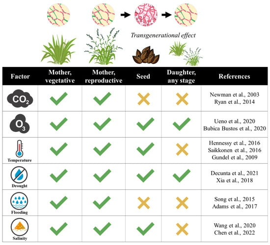
There is an increasing interest in determining the influence of global change on plant–microorganism interactions. We review the results of experiments that evaluated the effects of the global change factors carbon dioxide, ozone, temperature, drought, flooding, and salinity on plant symbioses with beneficial Epichloë endophytes. The factors affected the performance of both plants and endophytes as well as the frequency of plants symbiotic with the fungus. Elevated carbon dioxide levels and low temperatures differentially influenced the growth of plants and endophytes, which could compromise the symbioses. Furthermore, we summarise the plant stage in which the effects of the factors were quantified (vegetative, reproductive, or progeny). The factors ozone and drought were studied at all plant stages, but flooding and carbon dioxide were studied in just a few of them. While only studied in response to ozone and drought, evidence showed that the effects of these factors on symbiotic plants persisted trans-generationally. We also identified the putative mechanisms that would explain the effects of the factors on plant–endophyte associations. These mechanisms included the increased contents of reactive oxygen species and defence-related phytohormones, reduced photosynthesis, and altered levels of plant primary metabolites. Finally, we describe the counteracting mechanisms by which endophytes would mitigate the detrimental effects of the factors on plants. In presence of the factors, endophytes increased the contents of antioxidants, reduced the levels of defence-related phytohormones, and enhanced the plant uptake of nutrients and photosynthesis levels. Knowledge gaps regarding the effects of global change on plant–endophyte associations were identified and discussed. Full article
(This article belongs to the Special Issue Fungal Endophytes of Grasses)
Show Figures

Figure 1

13 pages, 1880 KiB  
Article
A Correlation between 3′-UTR of OXA1 Gene and Yeast Mitochondrial Translation
by Maryam Hajikarimlou 1,†, Mohsen Hooshyar 1,†, Noor Sunba 1,†, Nazila Nazemof 1, Mohamed Taha Moutaoufik 2, Sadhena Phanse 2, Kamaledin B. Said 1,3, Mohan Babu 2, Martin Holcik 4, Bahram Samanfar 1,5, Myron Smith 1 and Ashkan Golshani 1,*
1 Department of Biology and Ottawa Institute of Systems Biology, Carleton University, Ottawa, ON K1S 5B6, Canada
2 Department of Biochemistry, Research and Innovation Centre, University of Regina, Regina, SK S4S 0A2, Canada
3 Department of Pathology and Microbiology, College of Medicine, University of Hail, Hail P.O. Box 2240, Saudi Arabia
4 Department of Health Sciences, Carleton University, Ottawa, ON K1S 5B6, Canada
5 Agriculture and Agri-Food Canada, Ottawa Research and Development Centre (ORDC), Ottawa, ON K2H 8S2, Canada
These authors contributed equally to this work.
J. Fungi 2023, 9(4), 445; https://doi.org/10.3390/jof9040445 - 5 Apr 2023
Viewed by 1669
Abstract
Mitochondria possess their own DNA (mtDNA) and are capable of carrying out their transcription and translation. Although protein synthesis can take place in mitochondria, the majority of the proteins in mitochondria have nuclear origin. 3′ and 5′ untranslated regions of mRNAs (3′-UTR and [...] Read more.
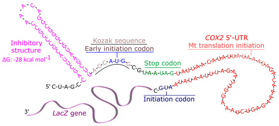
Mitochondria possess their own DNA (mtDNA) and are capable of carrying out their transcription and translation. Although protein synthesis can take place in mitochondria, the majority of the proteins in mitochondria have nuclear origin. 3′ and 5′ untranslated regions of mRNAs (3′-UTR and 5′-UTR, respectively) are thought to play key roles in directing and regulating the activity of mitochondria mRNAs. Here we investigate the association between the presence of 3′-UTR from OXA1 gene on a prokaryotic reporter mRNA and mitochondrial translation in yeast. OXA1 is a nuclear gene that codes for mitochondrial inner membrane insertion protein and its 3′-UTR is shown to direct its mRNA toward mitochondria. It is not clear, however, if this mRNA may also be translated by mitochondria. In the current study, using a β-galactosidase reporter gene, we provide genetic evidence for a correlation between the presence of 3′-UTR of OXA1 on an mRNA and mitochondrial translation in yeast. Full article
(This article belongs to the Special Issue Yeast Genetics 2022)
Show Figures

Figure 1

14 pages, 1425 KiB  
Article
Evaluation of Thermography as a Diagnostic Technique in Asymptomatic or Incipient Onychomycosis
by Julia Villar Rodríguez 1,†, Ana María Pérez Pico 2,†, Francisco Manuel García Blázquez 2,†, Juan Francisco Morán Cortés 2 and Raquel Mayordomo Acevedo 1,*
1 Department of Anatomy, Cellular Biology and Zoology, Centro Universitario de Plasencia, Universidad de Extremadura, 10600 Plasencia, Spain
2 Department of Nursing, Centro Universitario de Plasencia, Universidad de Extremadura, 10600 Plasencia, Spain
These authors contributed equally to this work.
J. Fungi 2023, 9(4), 444; https://doi.org/10.3390/jof9040444 - 5 Apr 2023
Cited by 3 | Viewed by 1349
Abstract
Onychomycosis is usually diagnosed symptomatically due to the very clear signs caused by the fungus on the nail surface and structure, although the growth of the infecting agent must also be verified by culture in an enriched medium. This procedure is normally lengthy [...] Read more.
Onychomycosis is usually diagnosed symptomatically due to the very clear signs caused by the fungus on the nail surface and structure, although the growth of the infecting agent must also be verified by culture in an enriched medium. This procedure is normally lengthy (four weeks), and samples can be contaminated, delaying the prescription of appropriate and effective treatment. Only one previous study has addressed the possibility of using thermography as a diagnostic method for onychomycosis in older people (31–70 years). The present study confirms this use but in individuals aged 18–31 years with incipient mycosis and no pathological signs. Using an FLIR E60 BX camera in a study with 214 samples, we found that men had more onychomycosis than women. We observed a relation between the presence of infection and nail temperature, with a higher temperature in yeast infections (+1 °C) and a lower temperature in dermatophyte infections (−2 °C). A higher temperature by almost 1 °C was also observed in older participants. Thermography can be viewed as a new diagnostic method in asymptomatic or incipient onychomycosis, providing the thermographic camera is sufficiently sensitive and the appropriate procedure is followed, although fungal culture is always necessary to confirm recovery after treatment. Full article
(This article belongs to the Special Issue Women in Mycology)
Show Figures

Figure 1

15 pages, 3381 KiB  
Article
Fusarium mindanaoense sp. nov., a New Fusarium Wilt Pathogen of Cavendish Banana from the Philippines Belonging to the F. fujikuroi Species Complex
by Shunsuke Nozawa 1, Yosuke Seto 2, Yoshiki Takata 1, Lalaine Albano Narreto 3, Reynaldo R. Valle 4, Keiju Okui 5, Shigeya Taida 5, Dionisio G. Alvindia 6, Renato G. Reyes 7 and Kyoko Watanabe 1,*
1 College of Agriculture, Tamagawa University, 6-1-1 Tamagawa-Gakuen, Machida, Tokyo 194-8610, Japan
2 Cancer Chemotherapy Center, Japanese Foundation for Cancer Research, 3-8-31 Ariake, Koto-ku, Tokyo 135-8550, Japan
3 Unifrutti Tropical Philippines, Inc., Km. 15, Panacan, Davao City 8000, Philippines
4 BaCaDM Project of College of Agriculture, Tamagawa University, 6-1-1 Tamagawa-Gakuen, Machida, Tokyo 194-8610, Japan
5 Unifrutti Japan Corporation, 1-11-1 Marunouchi, Chiyoda-Ku, Tokyo 100-6217, Japan
6 Philippine Center for Postharvest Development and Mechanization, Science City of Muñoz 3120, Philippines
7 Department of Biology, Central Luzon State University, Science City of Muñoz 3120, Philippines
J. Fungi 2023, 9(4), 443; https://doi.org/10.3390/jof9040443 - 5 Apr 2023
Viewed by 4025
Abstract
The pathogen causing Fusarium wilt in banana is reported to be Fusarium oxysporum f. sp. cubense (FOC). In 2019, wilt symptoms in banana plants (cultivar: Cavendish) in the Philippines were detected, i.e., the yellowing of the leaves and discoloration of the pseudostem and [...] Read more.
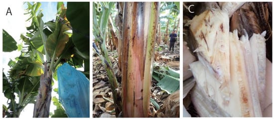
The pathogen causing Fusarium wilt in banana is reported to be Fusarium oxysporum f. sp. cubense (FOC). In 2019, wilt symptoms in banana plants (cultivar: Cavendish) in the Philippines were detected, i.e., the yellowing of the leaves and discoloration of the pseudostem and vascular tissue. The fungus isolated from the vascular tissue was found to be pathogenic to Cavendish bananas and was identified as a new species, F. mindanaoense, belonging to the F. fujikuroi species complex (FFSC); species classification was assessed using molecular phylogenetic analyses based on the tef1, tub2, cmdA, rpb1, and rpb2 genes and morphological analyses. A reciprocal blast search using genomic data revealed that this fungus exclusively included the Secreted in Xylem 6 (SIX6) gene among the SIX homologs related to pathogenicity; it exhibited a highly conserved amino acid sequence compared with that of species in the FFSC, but not with that of FOC. This was the first report of Fusarium wilt in Cavendish bananas caused by a species of the genus Fusarium other than those in the F. oxysporum species complex. Full article
(This article belongs to the Special Issue Fungal Plant Pathogens)
Show Figures

Figure 1

12 pages, 2440 KiB  
Article
Insight into the Antifungal Effects of Propolis and Carnosic Acid—Extension to the Pathogenic Yeast Candida glabrata: New Propolis Fractionation and Potential Synergistic Applications
by Alejandra Argüelles 1, Ruth Sánchez-Fresneda 1,2, José P. Guirao-Abad 1,2,†, José Antonio Lozano 3, Francisco Solano 3 and Juan-Carlos Argüelles 2,*
1 Vitalgaia España S.L., 30005 Murcia, Spain
2 Área de Microbiología, Facultad de Biología, Universidad de Murcia, 30071 Murcia, Spain
3 Departamento de Bioquímica y Biología Molecular B e Inmunología, Facultad de Medicina, Universidad de Murcia, 30120 Murcia, Spain
Current address: Department of Pathology and Laboratory Medicine, College of Medicine, University of Cincinnati, Medical Sciences Building, 231 Albert Sabin Way, Cincinnati, OH 45267, USA.
J. Fungi 2023, 9(4), 442; https://doi.org/10.3390/jof9040442 - 4 Apr 2023
Cited by 4 | Viewed by 1585
Abstract
Fungi have traditionally been considered opportunistic pathogens in primary infections caused by virulent bacteria, protozoan, or viruses. Consequently, antimycotic chemotherapy is clearly less developed in comparison to its bacterial counterpart. Currently, the three main families of antifungals (polyenes, echinocandins, and azoles) are not [...] Read more.
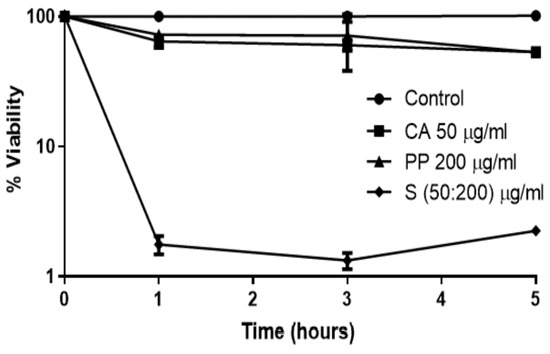
Fungi have traditionally been considered opportunistic pathogens in primary infections caused by virulent bacteria, protozoan, or viruses. Consequently, antimycotic chemotherapy is clearly less developed in comparison to its bacterial counterpart. Currently, the three main families of antifungals (polyenes, echinocandins, and azoles) are not sufficient to control the enormous increase in life-threatening fungal infections recorded in recent decades. Natural substances harvested from plants have traditionally been utilized as a successful alternative. After a wide screening of natural agents, we have recently obtained promising results with distinct formulations of carnosic acid and propolis on the prevalent fungal pathogens Candida albicans and Cryptococcus neoformans. Here, we extended their use to the treatment against the emerging pathogenic yeast Candida glabrata, which displayed lower susceptibility in comparison to the fungi mentioned above. Taking into account the moderate antifungal activity of both natural agents, the antifungal value of these combinations has been improved through the obtention of the hydroethanolic fractions of propolis. In addition, we have demonstrated the potential clinical application of new therapeutical designs based on sequential pre-treatments with carnosic/propolis mixtures, followed by exposure to amphotericin B. This approach increased the toxic effect induced by this polyene. Full article
(This article belongs to the Special Issue Antifungal Discovery of Natural Products)
Show Figures

Figure 1