DESCRIPTIONS OF MEDICAL FUNGI
DESCRIPTIONS OF MEDICAL FUNGI
DESCRIPTIONS OF MEDICAL FUNGI

Create successful ePaper yourself
Turn your PDF publications into a flip-book with our unique Google optimized e-Paper software.
<strong>DESCRIPTIONS</strong> <strong>OF</strong> <strong>MEDICAL</strong> <strong>FUNGI</strong><br />
SECOND EDITION<br />
DAVID ELLIS<br />
STEPHEN DAVIS<br />
HELEN ALEXIOU<br />
ROSEMARY HANDKE<br />
ROBYN BARTLEY<br />
MYCOLOGY UNIT<br />
WOMEN’S AND CHILDREN’S HOSPITAL<br />
SCHOOL <strong>OF</strong> MOLECULAR & BIO<strong>MEDICAL</strong> SCIENCE<br />
UNIVERSITY <strong>OF</strong> ADELAIDE<br />
ADELAIDE<br />
AUSTRALIA<br />
2007<br />
Cover: Cryptococcus neoformans, and montages including Microsporum, Candida,<br />
Schizophyllum, Sordaria, Conidiobolus, Fusarium, Bipolaris, Aspergillus, Curvularia,<br />
Saksenaea, Gliocladium, Trichophyton and Phialophora.
Published by the Authors<br />
Mycology Unit<br />
Women’s and Children’s Hospital<br />
North Adelaide 5006<br />
AUSTRALIA<br />
Direct Phone: (08) 8161 7365<br />
International + 618 8161 7365<br />
Direct Fax: (08) 8161 7589<br />
International + 618 8161 7589<br />
Email: dellis@adelaide.edu.au<br />
www.mycology.adelaide.edu.au<br />
© Copyright 2007<br />
The National Library of Australia Cataloguing-in-Publication entry:<br />
Descriptions of medical fungi.<br />
2nd ed.<br />
Bibliography.<br />
Includes index.<br />
ISBN 9780959851267 (pbk.).<br />
1. Fungi - Indexes. 2. Mycology - Indexes. I. Ellis, David (David H.).<br />
579.5<br />
Printed in Adelaide by<br />
Nexus Print Solutions<br />
153 Holbrooks Road<br />
Underdale, South Australia 2032
The Mycology Unit at the Adelaide Women’s and Children’s Hospital has played a key<br />
role in the provision of the Mycology component of the Microbiology Quality Assurance<br />
Program (QAP) organised by the Royal College of Pathologists of Australasia<br />
since its inception in 1979. The idea to provide all laboratories with a set of description<br />
sheets covering medical fungi evolved in the late 1980s and the first edition of this<br />
book was published in 1992. We now provide an updated edition which includes new<br />
and revised descriptions. We have endeavoured to reconcile current morphological<br />
descriptions with more recent genetic data, however in some cases, especially for the<br />
anthropophilic dermatophytes this is currently not possible.<br />
These descriptions have by necessity been kept brief and many have been based on<br />
previous descriptions by other authors. For further information regarding any of the<br />
mycoses or pathogenic fungi mentioned, the reader is referred to the references cited.<br />
For the precise definitions of the mycological terminology used, the reader is referred<br />
to Ainsworth and Bisby’s Dictionary of the Fungi (Kirk et al. 2001).<br />
For many species, antifungal susceptibility data has also been provided. This has<br />
been derived from both the literature and in-house data from Australian clinical isolates<br />
generated by using the CLSI M27-A2 protocol for yeasts and the CLSI M38-A protocol<br />
for moulds. This composite data is provided as a guide only. MIC 90 s for Aspergillus,<br />
Candida, Cryptococcus and Scedosporium species are provided from large Australian<br />
studies based predominantly on primary isolates. In many cases the clinical relevance<br />
of in vitro antifungal susceptibility results remains difficult to interpret, and expert advice<br />
from a consulting microbiologist or infectious disease specialist may be required.<br />
Risk group (RG) recommendations are based on published data and on current laboratory<br />
safety procedures in accordance with the Australian/New Zealand Standard AS/<br />
NZS 2243.3:2002. Safety in laboratories Part 3: Microbiological aspects and containment<br />
facilities.<br />
David Ellis BSc (Hons), MSc, PhD, FASM, FRCPA (Hon).<br />
Associate Professor<br />
School of Molecular & Biomedical Sciences<br />
University of Adelaide<br />
Head, Mycology Unit<br />
Women’s and Children’s Hospital<br />
North Adelaide, Australia 5006<br />
September, 2007<br />
PREFACE
Absidia corymbifera 1<br />
Acremonium 2<br />
Acrophialophora fusispora 3<br />
Alternaria 4<br />
Aphanoascus fulvescens 5<br />
Apophysomyces elegans 6<br />
Aspergillus 8<br />
Aspergillus flavus 9<br />
Aspergillus fumigatus 10<br />
Aspergillus nidulans 11<br />
Aspergillus niger 12<br />
Aspergillus terreus 13<br />
Aureobasidium pullulans 14<br />
Basidiobolus ranarum 15<br />
Beauveria 16<br />
Bipolaris 17<br />
Blastomyces dermatitidis 19<br />
Candida 20<br />
Candida albicans 23<br />
Candida colliculosa 24<br />
Candida dubliniensis 25<br />
Candida fabianii 26<br />
Candida famata 27<br />
Candida glabrata 28<br />
Candida guilliermondii 29<br />
Candida haemulonii 30<br />
Candida inconspicua 31<br />
Candida kefyr 32<br />
Candida krusei 33<br />
Candida lipolytica 34<br />
Candida lusitaniae 35<br />
Candida norvegensis 36<br />
Candida parapsilosis 37<br />
Candida pelliculosa 38<br />
Candida rugosa 39<br />
Candida tropicalis 40<br />
Chaetomium 41<br />
Chrysosporium tropicum 42<br />
Cladophialophora bantiana 43<br />
Cladophialophora carrionii 44<br />
Cladosporium 45<br />
Coccidioides immitis 46<br />
Colletotrichum 48<br />
Conidiobolus coronatus 49<br />
Cryptococcus 50<br />
Cryptococcus albidus 51<br />
Cryptococcus laurentii 51<br />
Cryptococcus gattii 52<br />
Cryptococcus neoformans 53<br />
CONTENTS<br />
Cunninghamella bertholletiae 55<br />
Curvularia 57<br />
Cylindrocarpon 58<br />
Drechslera 59<br />
Epicoccum purpurascens 60<br />
Epidermophyton floccosum 61<br />
Exophiala dermatitidis 62<br />
Exophiala jeanselmei complex 63<br />
Exophiala spinifera complex 65<br />
Exserohilum 66<br />
Fonsecaea 67<br />
Fusarium 68<br />
Fusarium oxysporum 69<br />
Fusarium solani 70<br />
Geotrichum candidum 71<br />
Geotrichum capitatum 72<br />
Gliocladium 73<br />
Graphium 74<br />
Histoplasma capsulatum 75<br />
Hortaea werneckii 77<br />
Lasiodiplodia theobromae 78<br />
Lecythophora hoffmannii 79<br />
Madurella grisea 80<br />
Madurella mycetomatis 81<br />
Malassezia 82<br />
Malbranchea 83<br />
Microsporum 84<br />
Microsporum audouinii 85<br />
Microsporum canis 86<br />
Microsporum canis var. distortum 88<br />
Microsporum canis var. equinum 89<br />
Microsporum cookei 90<br />
Microsporum ferrugineum 91<br />
Microsporum fulvum 92<br />
Microsporum gallinae 93<br />
Microsporum gypseum 94<br />
Microsporum nanum 95<br />
Microsporum persicolor 96<br />
Mortierella wolfii 97<br />
Mucor 98<br />
Mucor amphibiorum 99<br />
Mucor circinelloides 100<br />
Mucor indicus 100<br />
Mucor ramosissimus 100<br />
Nattrassia mangiferae 101<br />
Ochroconis gallopava 102<br />
Onychocola canadensis 103<br />
Paecilomyces 104<br />
Paecilomyces lilacinus 105
Paecilomyces variotii. 106<br />
Paracoccidioides brasiliensis 107<br />
Penicillium 108<br />
Penicillium marneffei 110<br />
Phaeoacremonium parasiticum 111<br />
Phialophora 112<br />
Phialophora richardsiae 112<br />
Phialophora verrucosa 113<br />
Phoma 114<br />
Pithomyces 115<br />
Prototheca 116<br />
Ramichloridium 117<br />
Ramichloridium schulzeri 117<br />
Rhinocladiella 118<br />
Rhinocladiella atrovirens 118<br />
Rhizomucor 119<br />
Rhizomucor miehei 119<br />
Rhizomucor pusillus 120<br />
Rhizopus 121<br />
Rhizopus azygosporus 122<br />
R. microsporus var. microsporus 122<br />
R. microsporus var. oligosporus 123<br />
R. microsporus var. rhizopodiformis 123<br />
Rhizopus oryzae 124<br />
Rhodotorula 125<br />
Rhodotorula glutinis 126<br />
Rhodotorula mucilaginosa 127<br />
Saccharomyces cerevisiae 128<br />
Saksenaea vasiformis 129<br />
Scedosporium apiospermum 131<br />
Scedosporium aurantiacum 131<br />
Scedosporium prolificans 134<br />
Schizophyllum commune 135<br />
Scopulariopsis 136<br />
Sepedonium 137<br />
Sporothrix schenckii 138<br />
Stemphylium 140<br />
Syncephalastrum 141<br />
Trichoderma 142<br />
Trichophyton 143<br />
Trichophyton ajelloi 144<br />
Trichophyton concentricum 145<br />
Trichophyton equinum 147<br />
Trichophyton erinacei 149<br />
Trichophyton interdigitale 151<br />
T. interdigitale var. nodulare 153<br />
Trichophyton mentagrophytes 154<br />
T. mentag. var. quinckeanum 156<br />
Trichophyton rubrum 158<br />
CONTENTS<br />
Trichophyton rubrum granular type 160<br />
Trichophyton schoenleinii 162<br />
Trichophyton soudanense 163<br />
Trichophyton terrestre 164<br />
Trichophyton tonsurans 165<br />
Trichophyton verrucosum 167<br />
Trichophyton violaceum 169<br />
Trichosporon 170<br />
Trichosporon asahii 171<br />
Trichosporon asteroides 172<br />
Trichosporon cutaneum 172<br />
Trichosporon inkin 172<br />
Trichosporon mucoides 173<br />
Trichosporon ovoides 173<br />
Trichothecium roseum 174<br />
Ulocladium 175<br />
Veronaea botryosa 176<br />
Verticillium 177<br />
Microscopy Stains & Techniques 178<br />
Calcofluor White with 10% KOH 178<br />
KOH with Chlorazol Black 178<br />
India Ink Mounts 178<br />
Lactophenol Cotton Blue (LPCB) 179<br />
Direct Microscopic Preparations 179<br />
Cellotape Flag Preparations 179<br />
Slide Culture Preparations 180<br />
Specialised Culture Media 181<br />
Bird seed agar 181<br />
Bromcresol Purple Milk Agar 181<br />
CDBT media 182<br />
CGB media 182<br />
Cornmeal agar 183<br />
Cornmeal glucose sucrose agar 183<br />
Czapek Dox agar 183<br />
Dixon’s agar 183<br />
Hair perforation test 184<br />
Lactritmel agar 184<br />
Littman oxgall agar 184<br />
Malt extract agar 185<br />
1% Peptone agar 185<br />
Potato dextrose agar 185<br />
Rice grain slopes 185<br />
Sabouraud dextrose agar 186<br />
Sabouraud dextrose agar 5% NaCl 186<br />
Tap water agar 187<br />
Urease agar with 0.5% glucose 187<br />
Vitamin free agar 187<br />
References 188
Descriptions of Medical Fungi<br />
Absidia corymbifera (Cohn) Saccardo & Trotter<br />
The genus Absidia is characterised by a differentiation of the hyphae into arched stolons<br />
bearing more or less verticillate sporangiophores at the raised part of the stolon<br />
(internode), and rhizoids formed at the point of contact with the substrate (at the node).<br />
This feature separates species of Absidia from the genus Rhizopus, where the sporangia<br />
arise from the nodes and are therefore found opposite the rhizoids. The sporangia<br />
are relatively small, globose, pyriform or pear-shaped and are supported by a<br />
characteristic funnel-shaped apophysis. This distinguishes Absidia from the genera<br />
Mucor and Rhizomucor, which have large, globose sporangia without an apophysis.<br />
Absidia currently contains 21 mostly soil-borne species. A. corymbifera is a known<br />
human pathogen causing pulmonary, rhinocerebral, disseminated, CNS or cutaneous<br />
zygomycosis.<br />
Colonies are fast growing, floccose, white at first becoming pale grey with age, and<br />
up to 1.5 cm high. Sporangiophores are hyaline to faintly pigmented, simple or sometimes<br />
branched, arising solitary from the stolons, in groups of three, or in whorls of up<br />
to seven. Rhizoids are very sparingly produced and may be difficult to find without the<br />
aid of a dissecting microscope to examine the colony on the agar surface. Sporangia<br />
are small (10-40 µm in diameter) and are typically pyriform in shape with a characteristic<br />
conical-shaped columella and pronounced apophysis, often with a short projection<br />
at the top. Sporangiospores vary from subglobose to oblong-ellipsoidal (3-7 x 2.5-4.5<br />
µm), hyaline to light grey and smooth-walled. Temperature: optimum 35-37 O C; maximum<br />
45 O C. RG-2 organism.<br />
For descriptions of species, keys to taxa and additional information see Ellis and Hesseltine<br />
(1965 and 1966), Hesseltine and Ellis (1964 and 1966), Nottebrock et al. (1974),<br />
O’Donnell (1979), Samson et al. (1995), Domsch et al. (1980), McGinnis (1980), de<br />
Hoog et al. (2000) and Ellis (2005b).<br />
Key Features: zygomycete, small pyriform-shaped<br />
sporangia with a characteristic conical-shaped columellae<br />
and pronounced apophysis, rapid growth at<br />
40 O C.<br />
Antifungal<br />
MIC µg/mL<br />
Range MIC90 Amphotericin B 0.03-2 1<br />
Flucytosine >256 >256<br />
Fluconazole >16 >16<br />
Itraconazole 0.03-2 0.5<br />
Posaconazole 0.03 - 1 0.25<br />
Voriconazole 2->64 >16<br />
Very limited data, antifungal susceptibility testing<br />
of individual strains is recommended. Sun et<br />
al. (2002), Dannaoui et al. (2003), Espinel-Ingroff<br />
et al. (2001), Espinel-Ingroff (2003, 2006), Singh<br />
et al. (2005), Sabatelli et al. (2006) and WCH inhouse<br />
data.<br />
15 µm<br />
1<br />
A. corymbifera showing a typical<br />
pyriform-shaped sporangium<br />
with a conical-shaped columella<br />
and pronounced apophysis (arrow).
2<br />
Descriptions of Medical Fungi<br />
Acremonium Link ex Fries<br />
Colonies are usually slow growing, often compact and moist at first, becoming powdery,<br />
suede-like or floccose with age, and may be white, grey, pink, rose or orange in<br />
colour. Hyphae are fine and hyaline and produce mostly simple awl-shaped erect phialides.<br />
Conidia are usually one-celled (ameroconidia), hyaline or pigmented, globose<br />
to cylindrical, and mostly aggregated in slimy heads at the apex of each phialide.<br />
The genus Acremonium currently contains 100 species, most are saprophytic being<br />
isolated from dead plant material and soil. However a number of species including A.<br />
falciforme, A. kiliense, A. recifei, A. alabamensis, A. roseogriseum and A. strictum are<br />
recognised as opportunistic pathogens of man and animals, causing mycetoma, mycotic<br />
keratitis and onychomycosis. RG-2 for species isolated from humans.<br />
Microconidial Fusarium isolates may be confused with Acremonium, but they usually<br />
grow faster and have colonies with a characteristic fluffy appearance.<br />
Key Features: hyphomycete with solitary, erect, hyaline, awl-shaped phialides producing<br />
single-celled, globose to cylindrical conidia, mostly in slimy heads.<br />
For descriptions of species, keys to taxa and additional information see Gams (1971),<br />
Domsch et al. (1980), Samson et al. (1995) and de Hoog et al. (2000).<br />
10 µm<br />
Acremonium showing long awl-shaped phialides producing cylindrical,<br />
one-celled conidia mostly aggregated in slimy heads at the<br />
apex of each phialide.<br />
Antifungal<br />
MIC µg/mL<br />
Range<br />
Antifungal<br />
MIC µg/mL<br />
Range<br />
Itraconazole 0.5->8 Amphotericin B 0.5-16<br />
Posaconazole 0.06-4 Caspofungin 0.03->8<br />
Voriconazole 0.06-4 Anidulafungin 0.5->8<br />
Very limited data, antifungal susceptibility testing of individual strains<br />
is recommended. Guarro et al. (1997), Pfaller et al. (1998, 2002a),<br />
Espinel-Ingroff (2003), Cuenca-Estrella et al. (2006) and WCH inhouse<br />
data.
Descriptions of Medical Fungi 3<br />
Acrophialophora fusispora (S.B. Saksena) Samson<br />
Colonies fast growing, greyish-brown with a black reverse. Conidiophores arising singly,<br />
terminally and laterally from the hyphae, erect, straight or slightly flexuose, tapering<br />
towards the apex, pale brown, rough-walled, up to 15 μm long, 2-5 μm wide, with<br />
whorls of phialides on the upper part. Phialides flask-shaped with a swollen base<br />
and a long, narrow neck, hyaline, smooth-walled or echinulate, 9-15 x 3-4.5 μm in the<br />
broadest part. Conidia in long chains, limoniform, one-celled, pale brown 5-12 x 3-6<br />
μm, smooth to finely echinulate with indistinct spiral bands. Temperature: optimum<br />
40 O C; maximum 50 O C.<br />
The genus Acrophialophora contains 3 species and is most commonly associated with<br />
soil, especially from India. A. fusispora is a rare human pathogen. RG-1 organism.<br />
Key Features: hyphomycete with flask-shaped phialides producing long chains of<br />
one-celled, limoniform, pale brown conidia, with indistinct spiral bands.<br />
For descriptions of species, keys to taxa and additional information see Domsch et al.<br />
(1980), de Hoog et al. (2000) and Al-Mohsen et al. (2000).<br />
10 μm<br />
Culture, phialides and conidia with striations (arrows) of A. fusispora.<br />
Antifungal<br />
MIC µg/mL<br />
Range<br />
Antifungal<br />
MIC µg/mL<br />
Range<br />
Fluconazole 8-32 Amphotericin B 0.25-2<br />
Itraconazole 0.06-0.125 Flucytosine >64<br />
Voriconazole 0.06 Posaconazole 0.03<br />
Very limited data, antifungal susceptibility testing of individual strains<br />
is recommended. Al-Mohsen et al. (2000) and WCH in-house data.
4<br />
Descriptions of Medical Fungi<br />
Colonies are fast growing, black to olivaceous-black or greyish, and are suede-like to<br />
floccose. Microscopically, branched acropetal chains (blastocatenate) of multicellular<br />
conidia (dictyoconidia) are produced sympodially from simple, sometimes branched,<br />
short or elongate conidiophores. Conidia are obclavate, obpyriform, sometimes ovoid<br />
or ellipsoidal, often with a short conical or cylindrical beak, pale brown, smooth-walled<br />
or verrucose.<br />
The genus contains 44 species, most are plant parasites, but a few species are ubiquitous<br />
and are also frequently soil-borne. A. alternata is the most common of these.<br />
Although usually seen as saprophytic contaminants, Alternaria species are recognised<br />
causative agents of mycotic keratitis.<br />
Alternaria species soon lose their ability to sporulate in culture. Potato dextrose agar<br />
and cornmeal agar are the most suitable media to use, and incubation under near<br />
ultra-violet light is recommended to maintain sporulation. Temperature: optimum 25-<br />
28 O C; maximum 31-32 O C. RG-1 organism.<br />
Key Features: dematiaceous hyphomycete producing chains of darkly pigmented,<br />
ovoid to obclavate dictyoconidia, often with short conical or cylindrical beaks.<br />
For descriptions of species, keys to taxa and additional information see Simmons (1967),<br />
Ellis (1971), Domsch et al. (1980), Samson et al. (1995), de Hoog et al. (2000).<br />
Antifungal<br />
Alternaria Nees ex Fries<br />
20 μm<br />
Alternaria alternata showing branched acropetal chains and multi-celled,<br />
obclavate to obpyriform conidia with short conical beaks.<br />
MIC µg/mL<br />
MIC µg/mL<br />
Range MIC90 Antifungal<br />
Range MIC90 Fluconazole 16->64 >64 Amphotericin B 0.125->16 2 (4)<br />
Itraconazole 0.125-2 1 Flucytosine >128 >128<br />
Voriconazole 0.5-2 1 Posaconazole 0.06-0.25 0.25<br />
Limited data, antifungal susceptibility testing of individual strains is recommended.<br />
McGinnis and Pasarell (1998), Pujol et al. (2000), Espinel-Ingroff et al. (2001),<br />
Espinel-Ingroff (2003), Sabatelli et al. (2006) and WCH in-house data.
Descriptions of Medical Fungi 5<br />
Aphanoascus fulvescens (Cooke) Apinis<br />
Colonies are moderately fast growing, white to tan with the production of numerous<br />
spherical, pseudoparenchymatous, buff to light brown cleistothecia (non-ostiolate<br />
ascocarps). Asci are subspherical to ellipsoidal and eight-spored. Ascospores light<br />
brown, yellowish to pale brown in mass, irregularly reticulate, lens-shaped, 3.5-4.7 x<br />
2.5-3.5 µm. Aphanoascus fulvescens has a Chrysosporium anamorph showing typical<br />
pyriform to clavate-shaped conidia with truncated bases, 15.0-17.5 x 3.7-6.0 µm,<br />
which are formed either intercalary, laterally or terminally.<br />
Aphanoascus fulvescens is a soil keratinolytic ascomycete which occasionally causes<br />
dermatomycosis in man and animals. RG-2 organism.<br />
Key Features: keratinolytic ascomycete with a Chrysosporium anamorph.<br />
For descriptions of species, keys to taxa and additional information see Domsch et al.<br />
(1980), McGinnis (1980) and de Hoog et al. (2000).<br />
100 μm<br />
10 μm<br />
Culture, cleistothecium and conidia of Aphanoascus fulvescens.
6<br />
Descriptions of Medical Fungi<br />
Apophysomyces elegans Misra, Srivastava & Lata<br />
Colonies are fast growing, white, becoming creamy white to buff with age, downy with<br />
no reverse pigment, and are composed of broad, sparsely septate (coenocytic) hyphae<br />
typical of a zygomycetous fungus. Sporangiophores are unbranched, straight or<br />
curved, slightly tapering towards the apex, up to 200 µm long, 3- 5 µm in width near<br />
the apophysis, and hyaline when young but developing a light to dark brown pigmentation<br />
and a conspicuous sub-apical thickening 10-16 µm below the apophysis with age.<br />
Sporangiophores arise at right angles from the aerial hyphae and often have a septate<br />
basal segment resembling the “foot cell” commonly seen in Aspergillus. Rhizoids are<br />
thin-walled, subhyaline and predominantly unbranched. Sporangia are multispored,<br />
small (20-50 µm diameter), typically pyriform in shape, hyaline at first, sepia-coloured<br />
when mature, columellate and strongly apophysate. Columellae are hemispherical in<br />
shape and the apophyses are distinctively funnel or bell-shaped. Sporangiospores are<br />
smooth-walled, mostly oblong, occasionally subglobose, (3-4 x 5-6 µm), subhyaline to<br />
sepia in mass. Good growth at 26 O C, 37 O C and 42 O C. RG-2 organism.<br />
Apophysomyces elegans is readily distinguishable from other zygomycetes of medical<br />
importance, especially the morphologically similar, strongly apophysate pathogen<br />
Absidia corymbifera, by having sporangiophores with distinctive funnel or bell-shaped<br />
apophyses and hemispherical-shaped columellae. In addition, there is a conspicuous<br />
pigmented sub-apical thickening which constricts the lumen of the sporangiophore<br />
below the apophysis, and there are also distinctive foot cells.<br />
Laboratory identification of this fungus may be difficult or delayed because of the<br />
mould’s failure to sporulate on the primary isolation media or on subsequent subculture<br />
onto potato dextrose agar. Sporulation may be stimulated by the use of nutrient<br />
deficient media, like cornmeal-glucose-sucrose-yeast extract agar, Czapek Dox agar,<br />
or by using the agar block method described by Ellis and Ajello (1982) and Ellis and<br />
Kaminski (1985).<br />
Key Features: zygomycete, rare human pathogen usually associated with invasive<br />
lesions following the traumatic implantation of the fungus through the skin. Soil fungus<br />
with a tropical to sub-tropical distribution. Characteristic “cocktail glass” apophysate<br />
sporangial morphology with conspicuous sub-apical thickening of the sporangiophore,<br />
rapid growth at 42 O C.<br />
For descriptions of species, keys to taxa and additional information see Cooter et al.<br />
(1990), Ellis and Ajello, (1982), Misra et al. (1979), Padhye and Ajello (1988), Lawrence<br />
et al. (1986), Wieden et al. (1985), de Hoog et al. (2000) and Ellis (2005b).<br />
Antifungal<br />
MIC µg/mL<br />
MIC µg/mL<br />
Antifungal<br />
Range Range<br />
Fluconazole >64 Amphotericin B 0.03-2<br />
Itraconazole 0.03-8 Flucytosine >256<br />
Posaconazole 0.03-4 Voriconazole 8->64<br />
Very limited data, antifungal susceptibility testing of individual strains is<br />
recommended. Sun et al. (2002), Dannaoui et al. (2003), Sabatelli et al.<br />
(2006) and WCH in-house data.
Descriptions of Medical Fungi 7<br />
Apophysomyces elegans Misra, Srivastava & Lata<br />
10 μm 10 μm<br />
a b<br />
(a) Young, multispored, pyriform sporangium of A. elegans showing a typical funnel-shaped<br />
apophysis but without the sub-apical thickening of a more mature<br />
sporangiophore. (b) Mature sporangium of A. elegans showing distinctive funnel-shaped<br />
apophyses, columellae, and a conspicuous pigmented sub-apical<br />
thickening which constricts the lumen of the sporangiophore below the apophysis<br />
(arrow). Sporangiospores are smooth-walled, oblong and subhyaline.
8<br />
Descriptions of Medical Fungi<br />
Aspergillus Micheli ex Link<br />
Colonies are usually fast growing, white, yellow, yellow-brown, brown to black or<br />
shades of green, mostly consisting of a dense felt of erect conidiophores. Conidiophores<br />
terminate in a vesicle covered with either a single palisade-like layer of phialides<br />
(uniseriate) or a layer of subtending cells (metulae) which bear small whorls of<br />
phialides (the so called biseriate structure). The vesicle, phialides, metulae (if present)<br />
and conidia form the conidial head. Conidia are one-celled, smooth or rough walled,<br />
hyaline or pigmented are produced in long dry chains which may be divergent (radiate)<br />
or aggregated in compact columns (columnar). Some species may produce Hülle cells<br />
or sclerotia.<br />
For identification, isolates are usually inoculated at three points on Czapek Dox agar<br />
and 2% malt extract agar and incubated at 25 O C. Most species sporulate within 7<br />
days. Descriptions are primarily based on colony pigmentation and morphology of the<br />
conidial head. Microscopic mounts are best made using cellotape flag or slide culture<br />
preparations mounted in lactophenol cotton blue. A drop of alcohol is usually needed<br />
to remove bubbles and excess conidia.<br />
Key Features: hyaline hyphomycete showing distinctive conidial heads with flaskshaped<br />
phialides arranged in whorls on a vesicle.<br />
For descriptions of species, keys to taxa and additional information see Raper and<br />
Fennell (1965), Domsch et al. (1980), McGinnis (1980), Onions et al. (1981), Samson<br />
and Pitt (1990), Samson et al. (1995), de Hoog et al. (2000) and Klich (2002).<br />
a<br />
conidia<br />
phialides<br />
vesicle<br />
stipe<br />
metulae<br />
Conidial head morphology in Aspergillus (a) uniseriate, (b) biseriate.<br />
b
Descriptions of Medical Fungi 9<br />
On Czapek Dox agar, colonies are granular,<br />
flat, often with radial grooves, yellow<br />
at first but quickly becoming bright to dark<br />
yellow-green with age. Conidial heads<br />
are typically radiate, later splitting to form<br />
loose columns (mostly 300-400 µm in diameter),<br />
biseriate but having some heads<br />
with phialides borne directly on the vesicle<br />
(uniseriate). Conidiophore stipes are<br />
hyaline and coarsely roughened, often<br />
more noticeable near the vesicle. Conidia<br />
are globose to subglobose (3-6 µm in<br />
diameter), pale green and conspicuously<br />
echinulate. Some strains produce brownish<br />
sclerotia.<br />
A. flavus has a world-wide distribution and<br />
normally occurs as a saprophyte in soil<br />
and on many kinds of decaying organic<br />
matter, however, it is also a recognised<br />
pathogen of humans and animals. RG-2<br />
organism.<br />
Key Features: spreading yellow-green<br />
colonies, rough-walled stipes, mature<br />
vesicles bearing phialides over their entire<br />
surface and conspicuously echinulate<br />
conidia.<br />
Antifungal<br />
MIC µg/mL<br />
Range MIC90 Amphotericin B 0.06->8 4<br />
Itraconazole 0.03-8 0.5<br />
Voriconazole 0.03-2 0.5<br />
Posaconazole 0.03-1 0.5<br />
Anidulafungin
10<br />
Descriptions of Medical Fungi<br />
On Czapek Dox agar, colonies show typical<br />
blue-green surface pigmentation with<br />
a suede-like surface consisting of a dense<br />
felt of conidiophores. Conidial heads are<br />
typically columnar (up to 400 x 50 µm<br />
but often much shorter and smaller) and<br />
uniseriate. Conidiophore stipes are short,<br />
smooth-walled and have conical-shaped<br />
terminal vesicles which support a single<br />
row of phialides on the upper two thirds<br />
of the vesicle. Conidia are produced in<br />
basipetal succession forming long chains<br />
and are globose to subglobose (2.5-3.0<br />
µm in diameter), green and rough-walled<br />
to echinulate. Note: This species is thermotolerant<br />
and grows at temperatures up<br />
to 55 O C.<br />
A. fumigatus is truly a cosmopolitan mould<br />
and has been found almost everywhere<br />
on every conceivable type of substrate. It<br />
is an important pathogen of humans and<br />
animals. RG-2 organism.<br />
Key Features: uniseriate and columnar<br />
conidial heads with the phialides limited<br />
to the upper two thirds of the vesicle and<br />
curving to be roughly parallel to each<br />
other.<br />
Antifungal<br />
MIC µg/mL<br />
Range MIC90 Amphotericin B 0.03->8 2<br />
Itraconazole 16 0.5<br />
Voriconazole
Descriptions of Medical Fungi 11<br />
Aspergillus nidulans (Eidam) Wint.<br />
Teleomorph: Emericella nidulans (Eidam) Vuill.<br />
On Czapek Dox agar, colonies are typically plain green in colour with dark red-brown<br />
cleistothecia developing within and upon the conidial layer. Reverse may be olive to<br />
drab-grey or purple-brown. Conidial heads are short columnar (up to 70 x 30 µm in<br />
diameter) and biseriate. Conidiophore stipes are usually short, brownish and smoothwalled.<br />
Conidia are globose (3-3.5 µm in diameter) and rough-walled.<br />
A. nidulans is a typical soil fungus with a world-wide distribution, it has also been reported<br />
causing disease in human and animals. RG-1 organism.<br />
Key Features: conidial heads are short columnar and biseriate. Stipes are usually<br />
short, brownish and smooth-walled. Conidia are globose and rough-walled.<br />
20 μm<br />
10 μm 20 μm<br />
a b c<br />
(a) Cleistothecium of Emericella nidulans (anamorph Aspergillus nidulans) showing<br />
numerous reddish-brown ascospores and thick-walled hülle cells; (b) cleistothecia are<br />
often surrounded by a mass of hülle cells which are up to 25 µm in diameter; (c) conidial<br />
head and stipe and (d) culture of A. nidulans.<br />
Antifungal<br />
MIC µg/mL<br />
Range MIC90 Amphotericin B 0.125-4 2<br />
Itraconazole 0.03-8 0.25<br />
Voriconazole 0.125-4 0.25<br />
Posaconazole 0.03-1 0.25<br />
Caspofungin 0.125-8 nd<br />
Espinel-Ingroff et al. (2001), Espinel-Ingroff<br />
(2003), Cuenca-Estrella et al. (2006).<br />
MIC s from Australian clinical isolates (nd<br />
90<br />
= not done).<br />
d
12<br />
Descriptions of Medical Fungi<br />
On Czapek Dox agar, colonies consist of<br />
a compact white or yellow basal felt covered<br />
by a dense layer of dark-brown to<br />
black conidial heads. Conidial heads are<br />
large (up to 3 mm by 15 to 20 µm in diameter),<br />
globose, dark brown, becoming<br />
radiate and tending to split into several<br />
loose columns with age. Conidiophore<br />
stipes are smooth-walled, hyaline or turning<br />
dark towards the vesicle. Conidial<br />
heads are biseriate with the phialides<br />
borne on brown, often septate metulae.<br />
Conidia are globose to subglobose (3.5-5<br />
µm in diameter), dark brown to black and<br />
rough-walled.<br />
A. niger is one of the most common and<br />
easily identifiable species of the genus<br />
Aspergillus, with its white to yellow mat<br />
later bearing black conidia. This species<br />
is very commonly found in aspergillomas<br />
and is the most frequently encountered<br />
agent of otomycosis. It is also a common<br />
laboratory contaminant. RG-1 organism.<br />
Key Features: conidial heads are dark<br />
brown to black, radiate and biseriate with<br />
metulae twice as long as the phialides.<br />
Conidia brown and rough-walled.<br />
Antifungal<br />
MIC µg/mL<br />
Range MIC90 Amphotericin B 0.125-2 2<br />
Itraconazole 0.03->8 0.5<br />
Voriconazole
Descriptions of Medical Fungi 13<br />
On Czapek Dox agar, colonies are typically<br />
suede-like and cinnamon-buff to<br />
sand brown in colour with a yellow to deep<br />
dirty brown reverse. Conidial heads are<br />
compact, columnar (up to 500 x 30-50 µm<br />
in diameter) and biseriate. Conidiophore<br />
stipes are hyaline and smooth-walled.<br />
Conidia are globose to ellipsoidal (1.5-2.5<br />
µm in diameter), hyaline to slightly yellow<br />
and smooth-walled.<br />
A. terreus occurs commonly in soil and is<br />
occasionally reported as a pathogen of<br />
humans and animals. RG-2 organism.<br />
Key Features: cinnamon-brown cultures,<br />
conidial heads biseriate with metulae as<br />
long as the phialides.<br />
For descriptions of species, keys to taxa<br />
and additional information see Raper and<br />
Fennell (1965), Domsch et al. (1980),<br />
McGinnis (1980), Onions et al. (1981),<br />
Samson and Pitt (1990), Samson et al.<br />
(1995), de Hoog et al. (2000) and Klich<br />
(2002).<br />
Antifungal<br />
MIC µg/mL<br />
Range MIC90 Amphotericin B 0.06-16 4<br />
Itraconazole 0.03-1 0.25<br />
Voriconazole 0.06-2 0.25<br />
Posaconazole 0.03-2 0.125<br />
Anidulafungin 0.03 nd<br />
Caspofungin 0.015-0.5 nd<br />
Espinel-Ingroff et al. (2001), Pfaller et<br />
al. (2002), Diekema et al. (2003), Espinel-Ingroff<br />
(2003), Serrano et al. (2003),<br />
Cuenca-Estrella et al. (2006). MIC s 90<br />
from Australian clinical isolates (nd = not<br />
done).<br />
Aspergillus terreus Thom<br />
10 μm<br />
Culture and conidial head and conidiophore<br />
of A. terreus. Note: conidial heads<br />
are biseriate.
14<br />
Descriptions of Medical Fungi<br />
Aureobasidium pullulans (de Bary) Arnaud<br />
Colonies are fast growing, smooth, soon covered with slimy masses of conidia, cream<br />
or pink to brown or black. Hyphae hyaline and septate, frequently becoming darkbrown<br />
with age and forming chains of one- to two-celled, thick-walled, darkly pigmented<br />
arthroconidia. These arthroconidia actually represent the Scytalidium anamorph of<br />
Aureobasidium and are only of secondary importance in recognising members of this<br />
genus. Conidia are produced synchronously in dense groups from indistinct scars or<br />
from short denticles on undifferentiated, hyaline to sub-hyaline hyphae. Conidia are<br />
hyaline, smooth-walled, single-celled, ellipsoidal but of very variable shape and size<br />
(8-12 x 4-6 µm), often with an indistinct hilum (= a mark or scar at the point of attachment).<br />
Temperature: optimum 25 O C; maximum 35-37 O C.<br />
This species has two varieties: A. pullulans var. pullulans, with a colony which remains<br />
pink, light brown, or yellow for at least three weeks, and A. pullulans var. melanogenum<br />
which soon becomes black or greenish-black due to dark hyphae which often fall apart<br />
into separate cells. A. pullulans has a world-wide distribution and is usually isolated<br />
as a saprophyte, occasionally from skin and nails. However, it has also been reported<br />
as a rare causative agent of phaeohyphomycosis, mycotic keratitis and peritonitis in<br />
patients on continuous ambulatory peritoneal dialysis (CAPD). RG-1 organism.<br />
Key Features: hyphomycete (so called black yeast) producing hyaline blastoconidia<br />
simultaneously from the vegetative hyphae, which may also form chains of darkly pigmented,<br />
thick-walled arthroconidia.<br />
For descriptions of species, keys to taxa and additional information see Hermanides-<br />
Nijhof (1977), Domsch et al. (1980), McGinnis (1980) and de Hoog et al. 2000.<br />
20 μm<br />
A. pullulans showing chains of one- to two-celled, darkly pigmented arthroconidia of<br />
the Scytalidium anamorph of Aureobasidium and the presence of numerous hyaline,<br />
single-celled, ovoid-shaped conidia which are produced on short denticles.<br />
Antifungal<br />
MIC µg/mL MIC µg/mL MIC µg/mL<br />
Antifungal Antifungal<br />
Range Range Range<br />
Amphotericin B 0.125-2 Itraconazole 0.03-0.25 Voriconazole 0.03-0.5<br />
Very limited data, antifungal susceptibility testing of individual strains is recommended.<br />
McGinnis and Pasarell (1998), Espinel-Ingroff et al. (2001) and WCH in-house data.
Descriptions of Medical Fungi 15<br />
Synonyms: Basidiobolus meristosporus Drechsler; Basidiobolus heterosporus Srinivasan<br />
& Thirumalachar; Basidiobolus haptosporus Drechsler.<br />
Colonies are moderately fast growing at 30 O C, flat, yellowish-grey to creamy-grey, glabrous,<br />
becoming radially folded and covered by a fine, powdery, white surface mycelium.<br />
Note: satellite colonies are often formed by germinating conidia ejected from<br />
the primary colony. Microscopic examination usually shows the presence of large<br />
vegetative hyphae (8-20 µm in diameter) forming numerous round (20-50 µm in diameter),<br />
smooth, thick-walled zygospores that have two closely appressed beak-like<br />
appendages. The production of “beaked” zygospores is diagnostic for the genus. Two<br />
types of asexual conidia are formed, although isolates often lose their sporulating ability<br />
with subculture and special media incorporating glucosamine hydrochloride and<br />
casein hydrolsate may be needed to stimulate sporulation (Shipton and Zahari, 1987).<br />
Primary conidia are globose, one-celled, solitary and are forcibly discharged from a<br />
sporophore. The sporophore has a distinct swollen area just below the conidium that<br />
actively participates in the discharge of the conidium. Secondary (replicative) conidia<br />
are clavate, one-celled and are passively released from a sporophore. These sporophores<br />
are not swollen at their bases. The apex of the passively released spore has<br />
a knob-like adhesive tip. These spores may function as sporangia, producing several<br />
sporangiospores. RG-2 organism.<br />
Basidiobolus ranarum is commonly present in decaying fruit and vegetable matter, and<br />
as a commensal in the intestinal tract of frogs, toads and lizards. It has been reported<br />
from tropical Africa, India, Indonesia and South East Asia including Australia.<br />
For descriptions of species, keys to taxa and additional information see Strinivasan and<br />
Thirumalachar (1965), Greer and Friedman (1966), Dworzack et al. (1978), McGinnis<br />
(1980), King (1983), Rippon (1988), Davis et al. (1994), Jong and Dugan (2003), de<br />
Hoog et al. (2000) and Ellis (2005a).<br />
20 μm<br />
Basidiobolus ranarum Eidem<br />
a 20 μm<br />
b<br />
(a) Sporophore and conidia and (b) zygospores of Basidiobolus ranarum.
16<br />
Descriptions of Medical Fungi<br />
Colonies are usually slow growing, mostly not exceeding 2 cm in ten days at 20 O C,<br />
downy, at first white but later often becoming yellow to pinkish. The genus Beauveria<br />
is characterised by the sympodial development of single-celled conidia (ameroconidia)<br />
on a geniculate or zig-zag rachis. Conidiogenous cells are flask-shaped, rachiform,<br />
proliferating sympodially and are often aggregated into sporodochia or synnemata.<br />
Conidia are hyaline and globose or ovoid in shape. RG-1 organism.<br />
Three species are recognised, two of which are well known parasites of insects. B.<br />
bassiana is the most common species and is best known as the causal agent of the<br />
disastrous muscardine in silkworms. Beauveria species are occasionally isolated in<br />
the clinical laboratory as saprophytic contaminants.<br />
Key Features: hyphomycete showing sympodial development of ameroconidia on a<br />
geniculate or zig-zag rachis emanating from a flask-shaped conidiophore.<br />
For descriptions of species, keys to taxa and additional information see de Hoog (1972),<br />
Domsch et al. (1980), McGinnis (1980) and de Hoog et al. (2000).<br />
20 μm<br />
Beauveria Vuillemin<br />
Beauveria bassiana showing sympodial development of conidia on a geniculate<br />
or zig-zag rachis. Conidiogenous cells are flask-shaped, rachiform,<br />
proliferating sympodially and are often aggregated into sporodochia or synnemata.<br />
Conidia are hyaline and globose or ovoid in shape, 2-3 mm diameter<br />
(phase contrast image).
Descriptions of Medical Fungi 17<br />
Teleomorph: Cochliobolus Drechsler<br />
Bipolaris Shoemaker<br />
Colonies are moderately fast growing, effuse, grey to blackish brown, suede-like to<br />
floccose with a black reverse. Microscopic morphology shows sympodial development<br />
of pale brown pigmented, pseudoseptate conidia on a geniculate or zig-zag rachis.<br />
Conidia are produced through pores in the conidiophore wall (poroconidia) and are<br />
straight, fusiform to ellipsoidal, rounded at both ends, smooth to finely roughened, germinating<br />
only from the ends (bipolar).<br />
The genus Bipolaris contains about 45 species which are mostly subtropical and tropical<br />
plant parasites; however several species, notably B. australiensis, B. hawaiiensis<br />
and B. spicifera are well documented human pathogens. RG-1 organisms.<br />
Key Features: dematiaceous hyphomycete producing sympodial, pseudoseptate, pale<br />
brown, straight, fusiform to ellipsoidal poroconidia, which are rounded at both ends.<br />
The genera Drechslera, Bipolaris, Curvularia and Exserohilum are all closely related<br />
and differentiation of the genera relies upon a combination of characters including conidial<br />
shape, the presence or absence of a protruding hilum, the contour of the basal<br />
portion of the conidium and its hilum, the point at which the germ tube originates from<br />
the basal cell and, to a lesser degree, the sequence and location of the first three conidial<br />
septa. The table below is modified from Domsch et al. (1980).<br />
Anamorph Main characters Teleomorph<br />
Drechslera Conidia cylindrical, germinating from any cell,<br />
hilum not protuberant<br />
Bipolaris Conidia fusiform-ellipsoidal, central cells not<br />
much darker and broader than the distal ones,<br />
hilum not protuberant, germination bipolar.<br />
Curvularia Conidia with 2-3 broader and darker central<br />
cells, often curved, with or without a prominent<br />
hilum, germination bipolar.<br />
Exserohilum Conidia fusiform-cylindrical to obclavate, with<br />
a protuberant hilum germination bipolar.<br />
Pyrenophora<br />
Cochliobolus<br />
Cochliobolus<br />
Setosphaeria<br />
Species of Bipolaris, Curvularia and Exserohilum are causative agents of phaeohyphomycosis<br />
which is an emerging mycotic infection of humans and lower animals<br />
caused by a number of dematiaceous (brown-pigmented) fungi where the tissue morphology<br />
of the causative organism is mycelial. This separates it from other clinical<br />
types of disease involving brown-pigmented fungi where the tissue morphology of the<br />
organism is a grain (mycotic mycetoma) or sclerotic body (chromoblastomycosis).<br />
For descriptions of species, keys to taxa and additional information see Ellis (1971<br />
and 1976), Luttrell (1978), Domsch et al. (1980), Alcorn (1983), Padhye et al. (1986),<br />
McGinnis et al. (1986b), Sivanesan (1987), Rippon (1988) and de Hoog et al. (2000).<br />
Also see Descriptions for Curvularia, Drechslera and Exserohilum.
18<br />
Descriptions of Medical Fungi<br />
10 μm<br />
Bipolaris australiensis showing sympodial development of pale<br />
brown, fusiform to ellipsoidal, pseudoseptate, poroconidia on a<br />
geniculate or zig-zag rachis.<br />
Antifungal<br />
Bipolaris Shoemaker<br />
MIC µg/mL<br />
MIC µg/mL<br />
Range<br />
Antifungal<br />
Range<br />
Itraconazole 0.03-1 Amphotericin B 0.06-2<br />
Posaconazole 0.06-0.05 Anidulafungin 1-4<br />
Voriconazole 0.06-0.05 Caspofungin 1-4<br />
Limited data, antifungal susceptibility testing of individual strains is recommended.<br />
Espinel-Ingroff et al. (2001), Pfaller et al. (2002a), Espinel-Ingroff<br />
(2003), McGinnis and Pasarell (1998) and WCH in-house data.
Descriptions of Medical Fungi 19<br />
Blastomyces dermatitidis Gilchrist & Stokes<br />
Colonies (SDA) at 25 O C are variable in morphology and rate of growth. They may grow<br />
rapidly, producing a fluffy white mycelium or slowly as glabrous, tan, nonsporulating<br />
colonies (Fig. a). Growth and sporulation may be enhanced by yeast extract. Most<br />
strains become pleomorphic with age. Microscopically, hyaline, ovoid to pyriform, onecelled,<br />
smooth-walled conidia (2-10 µm in diameter) of the Chrysosporium type, are<br />
borne on short lateral or terminal hyphal branches.<br />
Colonies on blood agar at 37 O C are wrinkled and folded, glabrous and yeast-like. Microscopically,<br />
the organism produces the characteristic yeast phase as seen in tissue<br />
pathology; ie. B. dermatitidis is a dimorphic fungus.<br />
WARNING: RG-3 organism. Cultures of Blastomyces dermatitidis may represent a<br />
biohazard to laboratory personnel and should be handled in an appropriate pathogen<br />
handling cabinet. In the past, conversion from the mould form to the yeast form was<br />
necessary to positively identify this dimorphic pathogen from species of Chrysosporium<br />
or Sepedonium; however, culture identification by exoantigen test is now the method<br />
of choice.<br />
Histopathology: Tissue sections show large, broad-based, unipolar budding yeastlike<br />
cells, which may vary in size from 8-15 µm (Fig. b), with some larger forms up to<br />
30 µm in diameter. Note: tissue sections need to be stained by Grocott’s methenamine<br />
silver method to clearly see the yeast-like cells, which are often difficult to observe in<br />
H&E preparations.<br />
Key Features: clinical history, tissue pathology, culture identification by positive<br />
exoantigen test.<br />
For descriptions of species, keys to taxa and additional information see McGinnis<br />
(1980), Chandler et al. (1980), Kaufman and Standard (1987) and Rippon (1988).<br />
Antifungal<br />
a b<br />
10 μm<br />
MIC µg/mL<br />
MIC µg/mL<br />
Antifungal<br />
Range MIC Range MIC 90 90<br />
Fluconazole 0.125-64 4-16 Amphotericin B 0.03-1 0.5<br />
Itraconazole 0.03->16 0.125-2 Voriconazole 0.03-16 0.25<br />
Posaconazole 0.03-2 0.125 Caspofungin 0.5-8 2<br />
Limited data available. Sugar and Liu (1996), Espinel-Ingroff et al. (2001),<br />
Espinel-Ingroff (2003), Gonzales et al. (2005) and Sabatelli et al. (2006).
20<br />
Descriptions of Medical Fungi<br />
The genus Candida is characterised by globose to elongate yeast-like cells or blastoconidia<br />
that reproduce by multilateral budding, polar budding if present on a narrow<br />
base, pseudohyphae and occasionally true hyphae may also be present. Arthroconidia,<br />
ballistoconidia and colony pigmentation are always absent. Fermentation or<br />
not: Nitrate assimilation or not: Inositol assimilation or not, however all inositol positive<br />
strains form pseudohyphae. In the past, the genus Torulopsis was separated from<br />
the genus Candida by the absence of pseudomycelium. However, in 1978 Yarrow &<br />
Meyer amended the description of Candida to include all species previously included<br />
in Torulopsis.<br />
Several species of Candida may be aetiological agents, most commonly C. albicans,<br />
followed by C. parapsilosis, C. glabrata, C. krusei and C. tropicalis. However a number<br />
of other species may also be isolated (see table below). All are ubiquitous and occur<br />
naturally on humans.<br />
Identification:<br />
Candida Berkhout<br />
Ensure that you start with a fresh growing pure culture; streak for single colony isolation<br />
if necessary.<br />
Chromogenic agars are now being used for primary isolation for both the detection of<br />
mixed flora and rapid species identification, especially from non-sterile sites.<br />
Germ Tube Test. A rapid screening test for Candida albicans and Candida dubliniensis.<br />
0.5 mL of serum, containing 0.5% glucose, is lightly inoculated with the test organism<br />
and incubated at 35 O C for 2-3 hours. On microscopy, the production of germ tubes by<br />
the cells is diagnostic for Candida albicans.<br />
10 μm<br />
Production of germ tubes by C. albicans.<br />
Species distribution from 944 patients<br />
with candidemia (Australian<br />
Candidemia Study 2002-2004).<br />
Species No %<br />
C. albicans 447 47.3<br />
C. parapsilosis 182 19.3<br />
C. glabrata 167 17.8<br />
C. krusei 46 4.9<br />
C. tropicalis 46 4.9<br />
C. dubliniensis 22 2.3<br />
C. guilliermondii 11 1.2<br />
C. lusitaniae 8 0.8<br />
C. kefyr 5 0.5<br />
C. pelliculosa 3 0.3<br />
C. rugosa 2 0.2<br />
C. colliculosa 1 0.1<br />
C. famata 1 0.1<br />
C. inconspicua 1 0.1<br />
C. lipolytica 1 0.1<br />
C. fabianii 1 0.1
Descriptions of Medical Fungi 21<br />
Candida Berkhout<br />
For the full identification of germ tube negative yeasts, morphological (Dalmau<br />
plate culture), physiological and biochemical tests are essential.<br />
(a) Dalmau Plate Culture: To set up a yeast morphology plate, dip a flamed sterilised<br />
straight wire into a culture to make a light inoculum and then lightly scratch the wire<br />
into the surface of a cornmeal/tween 80, rice/tween 80 or yeast morphology agar plate,<br />
then place a flamed coverslip onto the agar surface covering the scratches. Dalmau<br />
morphology plates are examined in-situ directly under the lower power of a microscope<br />
for the presence of pseudohyphae which may take up to 4-5 days at 26 O C to develop.<br />
Candida albicans also produces characteristic large, round, terminal, thick-walled vesicles<br />
(often called chlamydospores). The key features to remember are to use a light<br />
inoculum and to scratch the surface of the agar with the wire when inoculating.<br />
(b) Physiological and biochemical tests including fermentation and assimilation studies<br />
should be performed based on those used at the Centraalbureau voor Schimmelcultures,<br />
Utrecht, The Netherlands. Reference “The Yeasts: a taxonomic study”, edited<br />
by Kurtzman and Fell (1998), Elsevier Science Publishers B.V. Amsterdam. Reliable<br />
commercially available yeast identification kits are the API 20C, ID32C, MicroScan and<br />
Vitek systems. For specific identification of species see appropriate text book.<br />
5 mm 10 μm<br />
a b<br />
(a) Dalmau plate culture showing colonies of C. albicans growing out from scratches<br />
on the surface of a cornmeal/tween 80 agar plate. Note: a coverslip has been placed<br />
onto the agar surface covering the scratches. (b) Confirmatory test for C. albicans.<br />
Production of large round, thick-walled vesicles (often called chlamydospores) in<br />
Dalmau plate cultures.<br />
For descriptions of species, keys to taxa and additional information see Barnett et al.<br />
(1983), Kurtzman and Fell (1988) and de Hoog et al. (2000).
22<br />
Descriptions of Medical Fungi<br />
Candida albicans (Robin) Berkhout<br />
10 μm<br />
10 μm<br />
CHROMagar Candida plate<br />
showing chromogenic colour<br />
change for C. albicans (green),<br />
C. tropicalis (blue), C. parapsilosis<br />
(white) and C. glabrata<br />
(pink).<br />
Candida albicans on Sabouraud’s<br />
dextrose agar showing<br />
typical cream coloured, smooth<br />
surfaced, waxy colonies.<br />
Direct smear of urine from a<br />
patient with candidiasis of the<br />
kidney showing C. albicans in<br />
mycelial or tissue phase with<br />
blastoconidia budding from the<br />
pseudohyphae.<br />
Microscopic morphology of<br />
C. albicans showing budding<br />
spherical to ovoid blastoconidia.
Descriptions of Medical Fungi 23<br />
Candida albicans (Robin) Berkhout<br />
Culture: Colonies (SDA) white to cream-coloured smooth, glabrous yeast-like.<br />
Microscopy: Spherical to subspherical budding blastoconidia, 2-7 x 3-8 µm in size.<br />
India Ink Preparation: Negative - No capsules present.<br />
Dalmau Plate Culture on Cornmeal and Tween 80 Agar: Branched pseudohyphae<br />
with dense verticils of blastoconidia. Spherical chlamydospores, mostly terminal, often<br />
on a slightly swollen subtending cell are formed near the edge of the cover slip.<br />
Physiological Tests: + Positive, - Negative, v Variable, w Weak, s Slow<br />
Germ Tube + L-Sorbose v L-Arabinose v D-Glucitol -(s)<br />
Fermentation Sucrose v D-Arabinose v α-M-D-glucoside v<br />
Glucose + Maltose + D-Ribose -(s) D-Gluconate -(s)<br />
Galactose v Cellobiose - L-Rhamnose - DL-Lactate +<br />
Sucrose -(s) Trehalose +(s) D-Glucosamine v myo-Inositol +<br />
Maltose + Lactose - N-A-D-glucosamine + 2-K-D-gluconate +<br />
Lactose - Melibiose - Glycerol v D-Glucuronate -<br />
Trehalose v Raffinose - Erythritol - Nitrate -<br />
Assimilation Melezitose v Ribitol v Urease -<br />
Glucose + Soluble Starch + Galactitol - 0.1% Cycloheximide +<br />
Galactose + D-Xylose + D-Mannitol + Growth at 40 O C +<br />
Key Features: germ tube positive, production of chlamydospores on Dalmau plate<br />
culture, fermentation of glucose, sugar assimilation profile and a distinctive green<br />
colour on CHROMagar. Note: germ tube negative variants, known as C. claussenii,<br />
and sucrose-negative variants described as C. stellatoidea have proven to be<br />
synonymous with C. albicans. C. albicans is a commensal of mucous membranes<br />
and the gastrointestinal tract. Environmental isolations have been made from sources<br />
contaminated by human or animal excreta, such as polluted water, soil, air and plants.<br />
RG-2 organism.<br />
Antifungal<br />
MIC µg/mL<br />
MIC µg/mL<br />
Antifungal<br />
Range MIC Range MIC 90 90<br />
Fluconazole 0.03->64 2 Amphotericin B 0.03-4 0.25<br />
Itraconazole 0.008->8 0.125 Flucytosine 0.03->64 0.5<br />
Posaconazole 0.008->8 0.016 Caspofungin 0.008->4 0.125<br />
Voriconazole 0.008->8 0.03 Anidulafungin 0.008->8 nd<br />
Good data available. Espinel-Ingroff et al. (2001), Pfaller et al. (2002b, 2006, 2007),<br />
Espinel-Ingroff (2003), Hajjeh et al. (2004), Richter et al. (2005) and Cuenca-Estrella<br />
et al. (2006). MIC 90 s from the Australian Candidemia Study (nd = not done).
24<br />
Descriptions of Medical Fungi<br />
Candida colliculosa (Hartmann) S.A. Meyer & Yarrow<br />
Teleomorph: Torulaspora delbrueckii (Lindner) Lindner.<br />
Culture: Colonies (SDA) white to cream-coloured smooth, glabrous yeast-like.<br />
Microscopy: Spherical to ellipsoidal budding blastoconidia, 2-6 x 3-7 µm in size. Ascospores<br />
may be produced on 5% malt extract or cornmeal agar after 5-30 days at<br />
25 O C.<br />
India Ink Preparation: Negative - No capsules present.<br />
Dalmau Plate Culture on Cornmeal and Tween 80 Agar: Budding yeast cells only.<br />
No pseudohyphae or true hyphae produced.<br />
Physiological Tests: + Positive, - Negative, v Variable, w Weak, s Slow<br />
Germ Tube - L-Sorbose v L-Arabinose - D-Glucitol v<br />
Fermentation Sucrose v D-Arabinose - α-M-D-glucoside v<br />
Glucose + Maltose v D-Ribose - D-Gluconate v<br />
Galactose v Cellobiose - L-Rhamnose - DL-Lactate v<br />
Sucrose v Trehalose -,s D-Glucosamine - myo-Inositol -<br />
Maltose v Lactose - N-A-D-glucosamine - 2-K-D-gluconate +<br />
Lactose - Melibiose - Glycerol v D-Glucuronate v<br />
Trehalose v Raffinose v Erythritol - Nitrate -<br />
Assimilation Melezitose v Ribitol v Urease -<br />
Glucose + Soluble Starch - Galactitol - 0.1% Cycloheximide -<br />
Galactose v D-Xylose v D-Mannitol + Growth at 37 O C v<br />
Key Features: asci containing 1-4 spheroidal ascospores, variable growth at 37 O C<br />
and a variable sugar assimilation profile. C. colliculosa is a rare cause of candidemia.<br />
RG-1 organism.<br />
Antifungal<br />
MIC µg/mL<br />
MIC µg/mL<br />
Range Antifungal<br />
Range<br />
Fluconazole 8 Amphotericin B 0.25<br />
Itraconazole 0.25 Flucytosine 0.03<br />
Posaconazole 0.25 Caspofungin 0.06<br />
Voriconazole 0.06 Anidulafungin nd<br />
Very limited data, antifungal susceptibility testing of individual stains is recommended.<br />
Data from the Australian Candidemia Study (nd = not done).
Descriptions of Medical Fungi 25<br />
Candida dubliniensis Sullivan et al.<br />
Culture: Colonies (SDA) white to cream-coloured smooth, glabrous yeast-like.<br />
Microscopy: Spherical to subspherical budding blastoconidia, 3-8 x 2-7 µm in size.<br />
India Ink Preparation: Negative - No capsules present.<br />
Dalmau Plate Culture on Cornmeal and Tween 80 Agar: Branched pseudohyphae<br />
with dense verticils of blastoconidia and spherical, mostly terminal chlamydospores.<br />
Physiological Tests: + Positive, - Negative, v Variable, w Weak, s Slow<br />
Germ Tube + L-Sorbose - L-Arabinose - D-Glucitol +<br />
Fermentation Sucrose + D-Arabinose - α-M-D-glucoside +,s<br />
Glucose + Maltose + D-Ribose - D-Gluconate -,s<br />
Galactose v Cellobiose - L-Rhamnose - DL-Lactate +<br />
Sucrose - Trehalose + D-Glucosamine -,s myo-Inositol -<br />
Maltose + Lactose - N-A-D-glucosamine + 2-K-D-gluconate +<br />
Lactose - Melibiose v Glycerol +,s D-Glucuronate -<br />
Trehalose v Raffinose - Erythritol - Nitrate -<br />
Assimilation Melezitose + Ribitol +,s Urease -<br />
Glucose + Soluble Starch + Galactitol - 0.1% Cycloheximide +<br />
Galactose + D-Xylose v D-Mannitol + Growth at 40 O C +<br />
Key Features: germ tube positive, similar to C. albicans, except for absence of growth<br />
at 45 o C; glycerol (mostly +), methyl-α-D-glucoside (-), trehalose (-), and D-xylose (-).<br />
Initial colonies dark green colour on CHROMagar and producing rough colonies on<br />
bird seed agar. C. dubliniensis is an uncommon cause of candidemia and mucosal<br />
infection, especially in HIV patients. RG-2 organism.<br />
Antifungal<br />
MIC µg/mL<br />
MIC µg/mL<br />
Range MIC90 Antifungal Range MIC90 Fluconazole 0.05->64 1 Amphotericin B 0.03-2 0.125<br />
Itraconazole 0.008->8 0.125 Flucytosine 0.03-64 0.125<br />
Posaconazole 0.03-1 0.125 Caspofungin 0.008-1 0.25<br />
Voriconazole 0.008-2 0.016 Anidulafungin
26<br />
Descriptions of Medical Fungi<br />
Candida fabianii (Hartmann) S.A. Meyer & Yarrow<br />
Teleomorph: Pichia fabianii (Wickerham) Kurtzman<br />
Culture: Colonies (SDA) white to cream-coloured smooth, glabrous yeast-like.<br />
Microscopy: Spheroidal to ellipsoidal budding blastoconidia, 3.0-6.5 x 2-5.5 µm in<br />
size. No pseudohyphae produced. Asci when present spherical, containing 1-4 spherical,<br />
faintly roughened ascospores.<br />
India Ink Preparation: Negative - No capsules present.<br />
Dalmau Plate Culture on Cornmeal and Tween 80 Agar: Spherical to ovoid budding<br />
yeast cells and occasional pseudohyphae produced.<br />
Physiological Tests: + Positive, - Negative, v Variable, w Weak, s Slow<br />
Germ Tube - L-Sorbose - L-Arabinose - D-Glucitol +<br />
Fermentation Sucrose + D-Arabinose - α-M-D-glucoside +<br />
Glucose + Maltose + D-Ribose - D-Gluconate +<br />
Galactose - Cellobiose + L-Rhamnose - DL-Lactate +<br />
Sucrose + Trehalose + D-Glucosamine - myo-Inositol -<br />
Maltose +,s Lactose - N-A-D-glucosamine - 2-K-D-gluconate -<br />
Lactose - Melibiose - Glycerol + D-Glucuronate -<br />
Trehalose - Raffinose + Erythritol - Nitrate +<br />
Assimilation Melezitose + Ribitol - Urease -<br />
Glucose + Soluble Starch + Galactitol - 0.1% Cycloheximide -<br />
Galactose - D-Xylose + D-Mannitol + Growth at 37 O C +<br />
Key Features: germ tube negative yeast and sugar assimilation pattern. Molecular<br />
identification may be required. Candida fabianii is a rare cause of candidemia. RG-1<br />
organism.<br />
Antifungal<br />
MIC µg/mL<br />
MIC µg/mL<br />
Antifungal<br />
Range Range<br />
Fluconazole 8 Amphotericin B 0.125<br />
Itraconazole 0.5 Flucytosine 0.03<br />
Posaconazole 0.5 Caspofungin 0.5<br />
Voriconazole 0.125 Anidulafungin nd<br />
Very limited data, antifungal susceptibility testing of individual strains is recommended.<br />
Data from the Australian Candidemia Study (nd = not done).
Descriptions of Medical Fungi 27<br />
Teleomorph: Debaryomyces hansenii (Zopf) Lodder & Kreger-van Rij.<br />
Culture: Colonies (SDA) white to cream-coloured smooth, glabrous yeast-like.<br />
Microscopy: Ovoid to broadly ellipsoidal budding blastoconidia, 3.5-5 x 2-3.5 µm in<br />
size. No pseudohyphae produced. Asci when present spherical, persistent, containing<br />
1-2 spherical ascospores with rough walls.<br />
India Ink Preparation: Negative - No capsules present.<br />
Dalmau Plate Culture on Cornmeal and Tween 80 Agar: Spherical to ovoid budding<br />
yeast cells only. No pseudohyphae produced.<br />
Key Features: germ tube negative yeast and sugar assimilation pattern. Candida<br />
famata is a common environmental isolate, however it is only rarely recovered from<br />
clinical specimens, usually associated with skin. RG-1 organism.<br />
Antifungal<br />
Candida famata (Harrison) S.A. Meyer & Yarrow<br />
Physiological Tests: + Positive, - Negative, v Variable, w Weak, s Slow<br />
Germ Tube - L-Sorbose v L-Arabinose +,w D-Glucitol +,w<br />
Fermentation Sucrose + D-Arabinose v α-M-D-glucoside +<br />
Glucose -,w Maltose + D-Ribose v D-Gluconate +,w<br />
Galactose -,w Cellobiose + L-Rhamnose v DL-Lactate v<br />
Sucrose -,w Trehalose + D-Glucosamine v myo-Inositol -<br />
Maltose - Lactose v N-A-D-glucosamine v 2-K-D-gluconate +<br />
Lactose - Melibiose v Glycerol + D-Glucuronate v<br />
Trehalose -,w Raffinose + Erythritol v Nitrate -<br />
Assimilation Melezitose v Ribitol + Urease -<br />
Glucose + Soluble Starch v Galactitol v 0.1% Cycloheximide v<br />
Galactose + D-Xylose + D-Mannitol + Growth at 40 O C +,w<br />
MIC µg/mL<br />
MIC µg/mL<br />
Range Antifungal<br />
Range<br />
Fluconazole 0.125->64 Amphotericin B 0.06-2<br />
Itraconazole 0.03->8 Flucytosine 0.06-128<br />
Posaconazole 0.06-1 Caspofungin 0.06->16<br />
Voriconazole 0.03-1 Anidulafungin 0.008->16<br />
Limited data, antifungal susceptibility testing of individual strains is recommended.<br />
Espinel-Ingroff et al. (2001), Pfaller et al. (2003, 2007), Espinel-Ingroff (2003),<br />
Cuenca-Estrella et al. (2006) and the Australian Candidemia Study.
28<br />
Descriptions of Medical Fungi<br />
Synonym: Torulopsis glabrata (Anderson) Lodder & de Vries<br />
Culture: Colonies (SDA) white to cream-coloured smooth, glabrous yeast-like.<br />
Microscopy: Ovoid to ellipsoidal budding blastoconidia, 3.4 x 2.0 µm in size. No pseudohyphae<br />
or chlamydospores produced.<br />
India Ink Preparation: Negative - No capsules present.<br />
Dalmau Plate Culture on Cornmeal and Tween 80 Agar: Ovoid budding yeast cells<br />
only. No pseudohyphae produced.<br />
Key Features: germ tube negative yeast and sugar assimilation pattern. Candida glabrata<br />
is one of the most common yeast species to be found on the body surface and<br />
is often isolated as an incidental finding from skin and urine. It has been implicated<br />
as an “opportunistic” cause of both superficial and systemic infections, especially in<br />
immunocompromised patients, and it has been isolated from patients with septicemia,<br />
pyelonephritis, pulmonary infections, endocarditis and hyperalimentation. Approximately<br />
10% of clinical isolates show azole cross resistance. RG-2 organism.<br />
Antifungal<br />
Candida glabrata (Anderson) S.A. Meyer & Yarrow<br />
Physiological Tests: + Positive, - Negative, v Variable, w Weak, s Slow<br />
Germ Tube - L-Sorbose - L-Arabinose - D-Glucitol -<br />
Fermentation Sucrose - D-Arabinose - α-M-D-glucoside -<br />
Glucose + Maltose - D-Ribose - D-Gluconate -<br />
Galactose - Cellobiose - L-Rhamnose - DL-Lactate -<br />
Sucrose - Trehalose - D-Glucosamine - myo-Inositol -<br />
Maltose - Lactose - N-A-D-glucosamine - 2-K-D-gluconate v<br />
Lactose - Melibiose - Glycerol +,s D-Glucuronate -<br />
Trehalose v Raffinose - Erythritol - Nitrate -<br />
Assimilation Melezitose - Ribitol - Urease -<br />
Glucose + Soluble Starch - Galactitol - 0.1% Cycloheximide -<br />
Galactose - D-Xylose - D-Mannitol - Growth at 40 O C +<br />
MIC µg/mL<br />
MIC µg/mL<br />
Range MIC90 Antifungal Range MIC90 Fluconazole 0.03->128 128 Amphotericin B 0.008-2 0.5<br />
Itraconazole 0.008->16 16 Flucytosine 0.008-16 0.03<br />
Posaconazole 0.008-8 8 Caspofungin 0.008->8 0.25<br />
Voriconazole 0.008-16 2 Anidulafungin 0.008-8 nd<br />
Good data available. Espinel-Ingroff et al. (2001), Pfaller et al. (2002b, 2006, 2007),<br />
Espinel-Ingroff (2003), Hajjeh et al. (2004), Richter et al. (2005) and Cuenca-Estrella<br />
et al. (2006). MIC 90 s from the Australian Candidemia Study (note: in this study 10%<br />
of primary blood isolates were azole cross-resistant, nd = not done).
Descriptions of Medical Fungi 29<br />
Teleomorph: Pichia guilliermondii Wickerham.<br />
Culture: White to cream-coloured smooth, glabrous yeast-like colonies.<br />
Microscopy: Spherical to subspherical budding yeast-like cells or blastoconidia, 2.0-<br />
4.0 x 3.0-6.5 µm.<br />
India Ink Preparation: Negative - No capsules present.<br />
Dalmau Plate Culture on Cornmeal and Tween 80 Agar: Branched pseudohyphae<br />
with dense verticils of blastoconidia.<br />
Antifungal<br />
Candida guilliermondii (Castellani) Langeron & Guerra<br />
Physiological Tests: + Positive, - Negative, v Variable, w Weak, s Slow<br />
Germ Tube - L-Sorbose v L-Arabinose v D-Glucitol v<br />
Fermentation Sucrose + D-Arabinose v α-M-D-glucoside v<br />
Glucose + Maltose + D-Ribose + D-Gluconate v<br />
Galactose v Cellobiose v L-Rhamnose v DL-Lactate v<br />
Sucrose + Trehalose + D-Glucosamine + myo-Inositol -<br />
Maltose - Lactose - N-A-D-glucosamine + 2-K-D-gluconate +<br />
Lactose - Melibiose v Glycerol + D-Glucuronate -<br />
Trehalose + Raffinose + Erythritol - Nitrate -<br />
Assimilation Melezitose v Ribitol + Urease -<br />
Glucose + Soluble Starch - Galactitol v 0.1% Cycloheximide v<br />
Galactose + D-Xylose + D-Mannitol v Growth at 37 O C v<br />
Key Features: germ tube negative yeast and sugar assimilation pattern. Candida<br />
guilliermondii has been isolated from numerous human infections, mostly of cutaneous<br />
origin. It is also found from normal skin and in sea water, faeces of animals, fig wasps,<br />
buttermilk, leather, fish, and beer. RG-1 organism.<br />
MIC µg/mL<br />
MIC µg/mL<br />
Range MIC90 Antifungal Range MIC90 Fluconazole 0.125->128 16 Amphotericin B 0.03-1 0.5<br />
Itraconazole 0.03-8 1.0 Flucytosine 0.03-8 0.125<br />
Posaconazole 0.03-8 0.5 Caspofungin 0.125->8 0.5<br />
Voriconazole 0.03-8 0.25 Anidulafungin 0.06-4 nd<br />
Good data available. Espinel-Ingroff et al. (2001), Pfaller et al. (2003, 2006, 2007),<br />
Espinel-Ingroff (2003) and Cuenca-Estrella et al. (2006). MIC 90 s from the Australian<br />
Candidemia Study (nd = not done).
30<br />
Descriptions of Medical Fungi<br />
Candida haemulonii (van Uden & Kolipinski) Meyer & Yarrow<br />
Synonym: Torulopsis haemulonii van Uden & Kolipinski<br />
Culture: Colonies (SDA) white to cream-coloured smooth, glabrous yeast-like.<br />
Microscopy: Ovoid to globose, budding yeast-like cells or blastoconidia, 3.0-5.0 x<br />
3.0-6.5 µm. No pseudohyphae produced.<br />
India Ink Preparation: Negative - No capsules present.<br />
Dalmau Plate Culture on Cornmeal and Tween 80 Agar: Spherical to ovoid, budding<br />
yeast-like cells only. No pseudohyphae produced.<br />
Physiological Tests: + Positive, - Negative, v Variable, w Weak, s Slow<br />
Germ Tube - L-Sorbose - L-Arabinose -,s D-Glucitol +<br />
Fermentation Sucrose + D-Arabinose -,s α-M-D-glucoside -<br />
Glucose + Maltose + D-Ribose -,s D-Gluconate +<br />
Galactose - Cellobiose - L-Rhamnose +,s DL-Lactate -<br />
Sucrose + Trehalose + D-Glucosamine +,s myo-Inositol -<br />
Maltose - Lactose - N-A-D-glucosamine + 2-K-D-gluconate +<br />
Lactose - Melibiose - Glycerol +,s D-Glucuronate -<br />
Trehalose +,s Raffinose +,s Erythritol - Nitrate -<br />
Assimilation Melezitose +,s Ribitol s Urease -<br />
Glucose + Soluble Starch v Galactitol -,s 0.1% Cycloheximide +<br />
Galactose -,s D-Xylose -,s D-Mannitol + Growth at 37 O C +<br />
Key Features: germ tube negative yeast and sugar assimilation pattern. Molecular<br />
identification may be required. Candida haemulonii has been reported from a few<br />
cases of fungemia but clinical isolations remain rare. It has also been isolated from<br />
fish and a dolphin. C. haemulonii may be difficult to distinguish from C. famata using<br />
some commercial yeast identification systems due to data base limitations. RG-1 organism.<br />
Antifungal<br />
MIC µg/mL<br />
MIC µg/mL<br />
Range<br />
Antifungal<br />
Range<br />
Fluconazole 32->256 Amphotericin B 2-8<br />
Itraconazole 0.125-4 Flucytosine 0.008-0.125<br />
Voriconazole 0.06-0.5 Caspofungin 0.03-0.5<br />
Very limited data, antifungal susceptibility testing of individual strains is recommended.<br />
Rodero et al. (2002) and Khan et al. (2007).
Descriptions of Medical Fungi 31<br />
Candida inconspicua (Lodder & Kreger-van Rij) S.A.Meyer & Yarrow<br />
Synonym: Torulopsis inconspicua Lodder & Kreger-van Rij.<br />
Culture: Colonies (SDA) white to cream-coloured smooth, glabrous yeast-like.<br />
Microscopy: Ovoidal budding blastoconidia, 2.0-5 x 5.0-11.0 µm.<br />
India Ink Preparation: Negative - No capsules present.<br />
Dalmau Plate Culture on Cornmeal and Tween 80 Agar: Spherical to ovoid budding<br />
yeast cells only. Primitive pseudohyphae may be produced after 14 days.<br />
Physiological Tests: + Positive, - Negative, v Variable, w Weak, s Slow<br />
Germ Tube - L-Sorbose - L-Arabinose - D-Glucitol -<br />
Fermentation Sucrose - D-Arabinose - α-M-D-glucoside -<br />
Glucose - Maltose - D-Ribose - D-Gluconate -<br />
Galactose - Cellobiose - L-Rhamnose - DL-Lactate +<br />
Sucrose - Trehalose - D-Glucosamine + myo-Inositol -<br />
Maltose - Lactose - N-A-D-glucosamine + 2-K-D-gluconate -<br />
Lactose - Melibiose - Glycerol + D-Glucuronate -<br />
Trehalose - Raffinose - Erythritol - Nitrate -<br />
Assimilation Melezitose - Ribitol - Urease -<br />
Glucose + Soluble Starch - Galactitol - 0.1% Cycloheximide -<br />
Galactose - D-Xylose - D-Mannitol - Growth at 40 O C +<br />
Key Features: germ tube negative yeast and sugar assimilation pattern. Candida<br />
inconspicua is a rare cause of candidemia. RG-1 organism.<br />
Antifungal<br />
MIC µg/mL<br />
MIC µg/mL<br />
Range Antifungal<br />
Range<br />
Fluconazole 4-128 Amphotericin B 0.125->8<br />
Itraconazole 0.25-8 Flucytosine 1-64<br />
Posaconazole 0.5-8 Caspofungin 0.008-0.25<br />
Voriconazole 0.125-4 Anidulafungin nd<br />
Very limited data, antifungal susceptibility testing of individual strains is recommended.<br />
Pfaller et al. (2003), Espinel-Ingroff (2003) and the Australian Candidemia Study (nd<br />
= not done).
32<br />
Descriptions of Medical Fungi<br />
Synonym: Candida pseudotropicalis (Castellani) Basgal.<br />
Teleomorph: Kluyveromyces marxianus (Hansen) van der Walt.<br />
Culture: Colonies (SDA) white to cream-coloured smooth, glabrous yeast-like.<br />
Microscopy: Short-ovoid to long-ovoid, budding blastoconidia, 3.0-6.5 x 5.5-11.0 µm,<br />
sometimes becoming elongate (up to 16.0 µm).<br />
India Ink Preparation: Negative - No capsules present.<br />
Dalmau Plate Culture on Cornmeal and Tween 80 Agar: Abundant, long, wavy,<br />
branched pseudohyphae usually formed, with ovoid blastoconidia, budding off singly,<br />
in pairs or chains, often in a verticillated position. Note: in some strains pseudohyphae<br />
may be scarce or almost absent.<br />
Key Features: germ tube negative yeast and sugar assimilation pattern. Candida<br />
kefyr is a rare cause of candidiasis and is usually associated with superficial cutaneous<br />
manifestations rather than systemic disease. Environmental isolations have been<br />
made from cheese and dairy products. RG-1 organism.<br />
Antifungal<br />
Candida kefyr (Beijerinck) van Uden & Buckley<br />
Physiological Tests: + Positive, - Negative, v Variable, w Weak, s Slow<br />
Germ Tube - L-Sorbose v L-Arabinose v D-Glucitol v<br />
Fermentation Sucrose + D-Arabinose - α-M-D-glucoside -<br />
Glucose + Maltose - D-Ribose v D-Gluconate -<br />
Galactose +,s Cellobiose v L-Rhamnose - DL-Lactate +<br />
Sucrose + Trehalose -,w D-Glucosamine - myo-Inositol -<br />
Maltose - Lactose v N-A-D-glucosamine - 2-K-D-gluconate -<br />
Lactose v Melibiose - Glycerol s D-Glucuronate -<br />
Trehalose - Raffinose + Erythritol - Nitrate -<br />
Assimilation Melezitose - Ribitol s Urease -<br />
Glucose + Soluble Starch - Galactitol - 0.1% Cycloheximide +<br />
Galactose s D-Xylose s D-Mannitol v Growth at 40 O C +<br />
MIC µg/mL<br />
MIC µg/mL<br />
Range MIC90 Antifungal Range MIC90 Fluconazole 0.125-4 0.5 Amphotericin B 0.125->8 1<br />
Itraconazole 0.03-0.5 0.06 Flucytosine 0.03-64 16<br />
Posaconazole 0.03-0.25 0.25 Caspofungin 0.03-1 0.125<br />
Voriconazole 0.008-0.125 0.016 Anidulafungin 0.03-0.5 nd<br />
Limited data, antifungal susceptibility testing of individual strains is recommended.<br />
Espinel-Ingroff et al. (2001), Pfaller et al. (2003, 2006), Espinel-Ingroff (2003) and<br />
Cuenca-Estrella et al. (2006). MIC 90 s from the Australian Candidemia Study (nd =<br />
not done).
Descriptions of Medical Fungi 33<br />
Teleomorph: Issatchenkia orientalis Kudryavtesev.<br />
Culture: Colonies (SDA) white to cream-coloured smooth, glabrous yeast-like.<br />
Microscopy: Predominantly small, elongated to ovoid budding blastoconidia, 2.0-5.5<br />
x 4.0-15.0 µm.<br />
India Ink Preparation: Negative - No capsules present.<br />
Dalmau Plate Culture on Cornmeal and Tween 80 Agar: Abundant long, wavy,<br />
branched pseudohyphae with elongated to ovoid blastoconidia, budding off in verticillate<br />
branches.<br />
Key Features: germ tube negative yeast and sugar assimilation pattern. Candida<br />
krusei is regularly associated with some forms of infant diarrhoea and occasionally<br />
with systemic disease. It has also been reported to colonise the gastrointestinal, respiratory<br />
and urinary tracts of patients with granulocytopenia. Environmental isolations<br />
have been made from beer, milk products, skin, faeces of animals and birds and pickle<br />
brine. RG-2 organism.<br />
Antifungal<br />
Candida krusei (Castellani) Berkhout<br />
Physiological Tests: + Positive, - Negative, v Variable, w Weak, s Slow<br />
Germ Tube - L-Sorbose - L-Arabinose - D-Glucitol -<br />
Fermentation Sucrose - D-Arabinose - α-M-D-glucoside -<br />
Glucose + Maltose - D-Ribose - D-Gluconate -<br />
Galactose - Cellobiose - L-Rhamnose - DL-Lactate +<br />
Sucrose - Trehalose - D-Glucosamine + myo-Inositol -<br />
Maltose - Lactose - N-A-D-glucosamine + 2-K-D-gluconate -<br />
Lactose - Melibiose - Glycerol + D-Glucuronate -<br />
Trehalose - Raffinose - Erythritol - Nitrate -<br />
Assimilation Melezitose - Ribitol - Urease -<br />
Glucose + Soluble Starch - Galactitol - 0.1% Cycloheximide v<br />
Galactose - D-Xylose - D-Mannitol - Growth at 40 O C +<br />
MIC µg/mL<br />
MIC µg/mL<br />
Range MIC90 Antifungal Range MIC90 Fluconazole 0.25->128 64 Amphotericin B 0.06->8 1<br />
Itraconazole 0.03->8 0.5 Flucytosine 0.5-64 16<br />
Posaconazole 0.03-1 1 Caspofungin 0.125->4 1<br />
Voriconazole 0.03-4 0.5 Anidulafungin 0.008-8 nd<br />
Good data available. Espinel-Ingroff et al. (2001), Pfaller et al. (2002b, 2003, 2007),<br />
Espinel-Ingroff (2003), Hajjeh et al. (2004), Richter et al. (2005) and Cuenca-Estrella<br />
et al. (2006). MIC 90 s from the Australian Candidemia Study (nd = not done).
34<br />
Descriptions of Medical Fungi<br />
Candida lipolytica (F.C. Harrison) Diddens & Lodder<br />
Teleomorph: Yarrowia lipolytica (Wickerham et al.) van der Walt & von Arx.<br />
Culture: Colonies (SDA) white to cream-coloured smooth, glabrous yeast-like.<br />
Microscopy: Spherical, ellipsoidal to elongate budding blastoconidia, 3.0-5 x 3.3-15.0<br />
µm.<br />
India Ink Preparation: Negative - No capsules present.<br />
Dalmau Plate Culture on Cornmeal and Tween 80 Agar: Pseudohyphae and true<br />
hyphae are produced.<br />
Physiological Tests: + Positive, - Negative, v Variable, w Weak, s Slow<br />
Germ Tube - L-Sorbose v L-Arabinose - D-Glucitol +<br />
Fermentation Sucrose - D-Arabinose - α-M-D-glucoside -<br />
Glucose - Maltose - D-Ribose v D-Gluconate v<br />
Galactose - Cellobiose w,- L-Rhamnose - DL-Lactate +<br />
Sucrose - Trehalose - D-Glucosamine - myo-Inositol -<br />
Maltose - Lactose - N-A-D-glucosamine + 2-K-D-gluconate -<br />
Lactose - Melibiose - Glycerol + D-Glucuronate -<br />
Trehalose - Raffinose - Erythritol + Nitrate -<br />
Assimilation Melezitose - Ribitol v Urease -<br />
Glucose + Soluble Starch - Galactitol - 0.1% Cycloheximide -<br />
Galactose v D-Xylose - D-Mannitol + Growth at 37 O C v<br />
Key Features: germ tube negative yeast and sugar assimilation pattern. Candida<br />
lipolytica is a rare cause of candidemia. RG-1 organism.<br />
Antifungal<br />
MIC µg/mL<br />
MIC µg/mL<br />
Range Antifungal<br />
Range<br />
Fluconazole 1->64 Amphotericin B 0.06-1<br />
Itraconazole 0.06-8 Flucytosine 0.125-64<br />
Posaconazole 0.03-4 Caspofungin 0.25-2<br />
Voriconazole 0.03-1 Anidulafungin 0.125-0.5<br />
Limited data, antifungal susceptibility testing of individual strains is recommended.<br />
Espinel-Ingroff et al. (2001), Pfaller et al. (2003), Espinel-Ingroff (2003) and Australian<br />
Candidemia Study.
Descriptions of Medical Fungi 35<br />
Candida lusitaniae van Uden & do Carmo-Sousa<br />
Teleomorph: Clavispora lusitaniae Rodrigues de Miranda.<br />
Culture: Colonies (SDA) white to cream-coloured smooth, glabrous yeast-like.<br />
Microscopy: Ovoid to ellipsoidal budding blastoconidia, 1.5-6.0 x 2.5-10.0 µm.<br />
India Ink Preparation: Negative - No capsules present.<br />
Dalmau Plate Culture on Cornmeal and Tween 80 Agar: Abundant pseudohyphae<br />
with short chains of blastoconidia.<br />
Physiological Tests: + Positive, - Negative, v Variable, w Weak, s Slow<br />
Germ Tube - L-Sorbose + L-Arabinose v D-Glucitol +<br />
Fermentation Sucrose + D-Arabinose - α-M-D-glucoside v<br />
Glucose + Maltose + D-Ribose - D-Gluconate s<br />
Galactose v Cellobiose + L-Rhamnose v DL-Lactate +,w<br />
Sucrose v Trehalose + D-Glucosamine - myo-Inositol -<br />
Maltose v Lactose - N-A-D-glucosamine + 2-K-D-gluconate +<br />
Lactose - Melibiose - Glycerol + D-Glucuronate -<br />
Trehalose v Raffinose - Erythritol - Nitrate -<br />
Assimilation Melezitose + Ribitol s Urease -<br />
Glucose + Soluble Starch - Galactitol - 0.1% Cycloheximide -<br />
Galactose + D-Xylose + D-Mannitol + Growth at 40 O C +<br />
Key Features: germ tube negative yeast and sugar assimilation pattern. Candida<br />
lusitaniae is a known cause of disseminated candidiasis, including septicemia and<br />
pyelonephritis. C. lusitaniae was first isolated from the alimentary tract of warm blooded<br />
animals and environmental isolations have been made from cornmeal, citrus peel,<br />
fruit juices, and milk from cows with mastitis. C. lusitaniae may also be difficult to distinguish<br />
from C. tropicalis using some yeast identification systems. RG-2 organism.<br />
Antifungal<br />
MIC µg/mL<br />
MIC µg/mL<br />
Range MIC90 Antifungal Range MIC90 Fluconazole 0.125-64 8 Amphotericin B 0.03-8 0.125<br />
Itraconazole 0.06-2 0.125 Flucytosine 0.03-128 0.03<br />
Posaconazole 0.008-0.5 0.25 Caspofungin 0.125-4 0.5<br />
Voriconazole 0.008-2 0.125 Anidulafungin 0.03->8 nd<br />
Some data available. Espinel-Ingroff et al. (2001), Pfaller et al. (2002b, 2003),<br />
Espinel-Ingroff (2003) and Cuenca-Estrella et al. (2006). MIC 90 s from the Australian<br />
Candidemia Study (nd = not done).
36<br />
Descriptions of Medical Fungi<br />
Candida norvegensis Dietrichson ex van Uden & Buckley<br />
Teleomorph: Pichia norvegensis Leask & Yarrow.<br />
Culture: Colonies (SDA) white to cream-coloured smooth, glabrous yeast-like.<br />
Microscopy: Ovoid, budding blastoconidia, 2.0-3.5 x 3.5-5.0 µm. Pseudohyphae<br />
rarely produced.<br />
India Ink Preparation: Negative - No capsules present.<br />
Dalmau Plate Culture on Cornmeal and Tween 80 Agar: Spherical to ovoid budding<br />
yeast cells only. No pseudohyphae produced.<br />
Physiological Tests: + Positive, - Negative, v Variable, w Weak, s Slow<br />
Germ Tube - L-Sorbose - L-Arabinose - D-Glucitol -<br />
Fermentation Sucrose - D-Arabinose - α-M-D-glucoside -<br />
Glucose s Maltose - D-Ribose - D-Gluconate -<br />
Galactose - Cellobiose + L-Rhamnose - DL-Lactate w<br />
Sucrose - Trehalose - D-Glucosamine + myo-Inositol -<br />
Maltose - Lactose - N-A-D-glucosamine - 2-K-D-gluconate -<br />
Lactose - Melibiose - Glycerol + D-Glucuronate -<br />
Trehalose - Raffinose - Erythritol - Nitrate -<br />
Assimilation Melezitose - Ribitol - Urease -<br />
Glucose + Soluble Starch - Galactitol - 0.1% Cycloheximide -<br />
Galactose - D-Xylose - D-Mannitol - Growth at 37 O C +<br />
Key Features: germ tube negative yeast and sugar assimilation pattern. Candida<br />
norvegensis is a very rare clinical isolate that has been reported as a causative agent<br />
of peritonitis and disseminated candidiasis in a patient on CAPD. RG-1 organism.<br />
Antifungal<br />
MIC µg/mL<br />
MIC µg/mL<br />
Range<br />
Antifungal<br />
Range<br />
Fluconazole 16 Amphotericin B 1<br />
Itraconazole 0.25 Flucytosine 8<br />
Voriconazole 0.125 Posaconazole 0.125<br />
Very limited data, antifungal susceptibility testing of individual strains is recommended.<br />
Pfaller et al. (2002b).
Descriptions of Medical Fungi 37<br />
Candida parapsilosis Complex<br />
Recently Candida parapsilosis has been recognised as 3 species: C. parapsilosis, C.<br />
orthopsilosis and C. metapsilosis (Tavanti et al. 2005). These three species are phenotypically<br />
indistinguishable and are best distinguished by genetic analysis. Antifungal<br />
susceptibility data from the Australian Candidemia Study also shows no significant differences<br />
between the species.<br />
Culture: Colonies (SDA) white to cream-coloured smooth, glabrous yeast-like.<br />
Microscopy: Predominantly small, globose to ovoid budding blastoconidia, 2.0-3.5 x<br />
3.0-4.5 µm, with some larger elongated forms present.<br />
India Ink Preparation: Negative - No capsules present.<br />
Dalmau Plate Culture on Cornmeal and Tween 80 Agar: Abundant, much-branched<br />
pseudohyphae in a delicate tree-like pattern with 2-3 blastoconidia in small clusters at<br />
intervals along the pseudohyphae.<br />
Physiological Tests: + Positive, - Negative, v Variable, w Weak, s Slow<br />
Germ Tube - L-Sorbose +,s L-Arabinose + D-Glucitol +<br />
Fermentation Sucrose + D-Arabinose - α-M-D-glucoside +<br />
Glucose + Maltose + D-Ribose v D-Gluconate +,s<br />
Galactose v Cellobiose - L-Rhamnose - DL-Lactate -<br />
Sucrose -,s Trehalose + D-Glucosamine v myo-Inositol -<br />
Maltose -,s Lactose - N-A-D-glucosamine + 2-K-D-gluconate +<br />
Lactose - Melibiose - Glycerol + D-Glucuronate -<br />
Trehalose -,s Raffinose - Erythritol - Nitrate -<br />
Assimilation Melezitose + Ribitol +,s Urease -<br />
Glucose + Soluble Starch - Galactitol - 0.1% Cycloheximide -<br />
Galactose + D-Xylose + D-Mannitol + Growth at 37 O C +<br />
Key Features: germ tube negative yeast and sugar assimilation pattern. Candida<br />
parapsilosis is an opportunistic human pathogen which may cause cutaneous infections,<br />
especially of the nail and systemic disease, especially endocarditis. Other clinical<br />
manifestations include endophthalmitis and fungemia. Environmental isolations have<br />
been made from intertidal and oceanic waters, pickle brine, cured meats, olives and<br />
normal skin, and faeces. RG-1 organism.<br />
Antifungal<br />
MIC µg/mL<br />
MIC µg/mL<br />
Range MIC90 Antifungal Range MIC90 Fluconazole 0.125->64 8 Amphotericin B 0.016-2 0.5<br />
Itraconazole 0.015-2 0.25 Flucytosine 0.03->64 0.25<br />
Posaconazole 0.008-0.5 0.03 Caspofungin 0.03->8 1<br />
Voriconazole 0.008-2 0.25 Anidulafungin 0.008->8 nd<br />
Good data available. Espinel-Ingroff et al. (2001), Pfaller et al. (2002b, 2006, 2007),<br />
Espinel-Ingroff (2003), Hajjeh et al. (2004), Richter et al. (2005) and Cuenca-Estrella<br />
et al. (2006). MIC 90 s from the Australian Candidemia Study (nd = not done).
38<br />
Descriptions of Medical Fungi<br />
Candida pelliculosa Redaelli<br />
Teleomorph: Pichia anomala (Hansen) Kurtzman.<br />
Culture: Colonies (SDA) white to cream-coloured smooth, glabrous yeast-like.<br />
Microscopy: Spherical to ellipsoidal budding blastoconidia, 2-4 x 2-6 µm. Pseudohyphae<br />
may be present. Asci when present, containing 1-4 hat-shaped ascospores.<br />
India Ink Preparation: Negative - No capsules present.<br />
Dalmau Plate Culture on Cornmeal and Tween 80 Agar: Spherical to ellipsoidal<br />
budding yeast cells and abundant pseudohyphae in most strains.<br />
Physiological Tests: + Positive, - Negative, v Variable, w Weak, s Slow<br />
Germ Tube - L-Sorbose - L-Arabinose v D-Glucitol +<br />
Fermentation Sucrose + D-Arabinose - α-M-D-glucoside +<br />
Glucose + Maltose + D-Ribose v D-Gluconate v<br />
Galactose v Cellobiose + L-Rhamnose - DL-Lactate +<br />
Sucrose + Trehalose + D-Glucosamine - myo-Inositol -<br />
Maltose v Lactose - N-A-D-glucosamine - 2-K-D-gluconate -<br />
Lactose - Melibiose - Glycerol + D-Glucuronate n<br />
Trehalose - Raffinose + Erythritol + Nitrate +<br />
Assimilation Melezitose + Ribitol v Urease -<br />
Glucose + Soluble Starch + Galactitol - 0.1% Cycloheximide -<br />
Galactose v D-Xylose v D-Mannitol + Growth at 37 O C v<br />
Key Features: germ tube negative yeast and sugar assimilation pattern. Candida<br />
pelliculosa has been reported from cases of candidemia and catheter related infections<br />
in humans and has been isolated from soil, grains, fruit and warm blooded animals.<br />
RG-1 organism.<br />
Antifungal<br />
MIC µg/mL<br />
MIC µg/mL<br />
Range<br />
Antifungal<br />
Range<br />
Fluconazole 2-16 Amphotericin B 0.125-2<br />
Itraconazole 0.25-2 Flucytosine 0.03-64<br />
Posaconazole 0.125-1 Caspofungin 0.06-0.5<br />
Voriconazole 0.06-0.25 Anidulafungin nd<br />
Limited data available, antifungal susceptibility testing of individual strains is<br />
recommended. Pfaller et al. (2003), Espinel-Ingroff (2003), Cuenca-Estrella et al.<br />
(2006) and the Australian Candidemia Study (nd = not done).
Descriptions of Medical Fungi 39<br />
Candida rugosa (Anderson) Diddens & Lodder<br />
Culture: Colonies (SDA) white to cream-coloured smooth, glabrous yeast-like.<br />
Microscopy: Ellipsoidal to elongate budding blastoconidia, 6-10 x 2-3.5 µm. Sometimes<br />
short pseudohyphae may be produced.<br />
India Ink Preparation: Negative - No capsules present.<br />
Dalmau Plate Culture on Cornmeal and Tween 80 Agar: Ellipsoidal budding yeast<br />
cells only and densely branched pseudohyphae produced.<br />
Physiological Tests: + Positive, - Negative, v Variable, w Weak, s Slow<br />
Germ Tube - L-Sorbose v L-Arabinose - D-Glucitol +,s<br />
Fermentation Sucrose - D-Arabinose - α-M-D-glucoside -<br />
Glucose - Maltose - D-Ribose - D-Gluconate v<br />
Galactose - Cellobiose - L-Rhamnose - DL-Lactate +,s<br />
Sucrose - Trehalose - D-Glucosamine - myo-Inositol -<br />
Maltose - Lactose - N-A-D-glucosamine s 2-K-D-gluconate -<br />
Lactose - Melibiose - Glycerol + D-Glucuronate -<br />
Trehalose - Raffinose - Erythritol - Nitrate -<br />
Assimilation Melezitose - Ribitol -,s Urease -<br />
Glucose + Soluble Starch - Galactitol - 0.1% Cycloheximide -<br />
Galactose + D-Xylose v D-Mannitol +,s Growth at 37 O C +<br />
Key Features: germ tube negative yeast and sugar assimilation pattern. Candida<br />
rugosa has been associated with catheter related fungemia and has been isolated<br />
from human and bovine faeces, sea water and soil. RG-1 organism.<br />
Antifungal<br />
MIC µg/mL<br />
MIC µg/mL<br />
Range Antifungal<br />
Range<br />
Fluconazole 1-16 Amphotericin B 0.25-4<br />
Itraconazole 0.03-1 Flucytosine 0.06-16<br />
Posaconazole 0.06-0.25 Caspofungin 0.25-2<br />
Voriconazole 0.008-0.25 Anidulafungin 0.03-4<br />
Limited data, antifungal susceptibility testing of individual strains is recommended.<br />
Espinel-Ingroff et al. (2001), Pfaller et al. (2003), Espinel-Ingroff (2003) and the<br />
Australian Candidemia Study.
40<br />
Descriptions of Medical Fungi<br />
Culture: Colonies (SDA) white to cream-coloured smooth, glabrous yeast-like.<br />
Microscopy: Spherical to subspherical budding yeast-like cells or blastoconidia, 3-5.5<br />
x 4-9 µm.<br />
India Ink Preparation: Negative - No capsules present.<br />
Dalmau Plate Culture on Cornmeal and Tween 80 Agar: Abundant, long, wavy,<br />
branched pseudohyphae with numerous ovoid blastoconidia, budding off. Terminal<br />
vesicles (chlamydospores) are not produced.<br />
Key Features: germ tube negative yeast and sugar assimilation pattern. Candida<br />
tropicalis is a major cause of septicemia and disseminated candidiasis. It is also found<br />
as part of the normal human mucocutaneous flora and environmental isolations have<br />
been made from faeces, shrimp, kefir, and soil. RG-2 organism.<br />
Antifungal<br />
Candida tropicalis (Castellani) Berkhout<br />
Physiological Tests: + Positive, - Negative, v Variable, w Weak, s Slow<br />
Germ Tube - L-Sorbose v L-Arabinose - D-Glucitol +<br />
Fermentation Sucrose v D-Arabinose - α-M-D-glucoside v<br />
Glucose + Maltose + D-Ribose -,s D-Gluconate v<br />
Galactose + Cellobiose +,s L-Rhamnose - DL-Lactate v<br />
Sucrose v Trehalose + D-Glucosamine v myo-Inositol -<br />
Maltose + Lactose - N-A-D-glucosamine + 2-K-D-gluconate +<br />
Lactose - Melibiose - Glycerol v D-Glucuronate -<br />
Trehalose +,s Raffinose - Erythritol - Nitrate -<br />
Assimilation Melezitose v Ribitol +,s Urease -<br />
Glucose + Soluble Starch + Galactitol - 0.1% Cycloheximide +<br />
Galactose + D-Xylose + D-Mannitol + Growth at 40 O C +<br />
MIC µg/mL<br />
MIC µg/mL<br />
Range MIC90 Antifungal Range MIC90 Fluconazole 0.125->128 2 Amphotericin B 0.03-8 0.5<br />
Itraconazole 0.03->8 0.5 Flucytosine 0.03->64 0.125<br />
Posaconazole 0.008->8 0.06 Caspofungin 0.03->8 0.25<br />
Voriconazole 0.008->8 0.25 Anidulafungin 0.03->8 nd<br />
Good data available. Espinel-Ingroff et al. (2001), Pfaller et al. (2002b, 2006, 2007),<br />
Espinel-Ingroff (2003), Hajjeh et al. (2004), Richter et al. (2005) and Cuenca-Estrella<br />
et al. (2006). MIC 90 s from the Australian Candidemia Study (nd = not done).
Descriptions of Medical Fungi 41<br />
Chaetomium Kunze ex Fries<br />
This genus is a common ascomycete (pyrenomycete) characterised by the formation of<br />
darkly-pigmented, globose, ovoid, barrel to flask-shaped, ostiolate ascocarps (perithecia)<br />
beset with dark-coloured terminal hairs (setae) which are straight, branched or<br />
curved. Asci are clavate to cylindrical, typically eight-spored and evanescent. Ascospores<br />
are one-celled, darkly-pigmented, smooth-walled, of varying shape, mostly<br />
ovoid, ellipsoidal or lemon-shaped. Chlamydospores and solitary conidia may also be<br />
produced. RG-1 organism.<br />
The genus contains between 160 and 180 species, and all are saprophytic being isolated<br />
from soil, straw, dung and plant debris. However, quite a few species are thermophilic<br />
and can grow at temperatures above 37 O C. Chaetomium species are important<br />
agents for the decomposition of cellulose waste and plant materials, and are only<br />
rarely isolated in medical mycology laboratories.<br />
Key Features: ascomycete producing darkly-pigmented ostiolate perithecia beset with<br />
long dark terminal setae.<br />
For descriptions of species, keys to taxa and additional information see Ames (1963),<br />
Seth (1970), Millner (1975), Domsch et al. (1980), Ellis and Keane (1981), Ellis (1981)<br />
and de Hoog et al. (2000).<br />
100 μm<br />
10 μm<br />
Ascocarp (perithecia), terminal hairs, asci and ascospores of Chaetomium.<br />
Antifungal<br />
MIC µg/mL MIC µg/mL MIC µg/mL<br />
Antifungal Antifungal<br />
Range Range Range<br />
Amphotericin B 0.125-16 Itraconazole 0.03-0.125 Voriconazole 0.125-0.5<br />
Very limited data, antifungal susceptibility testing of individual strains is recommended.<br />
McGinnis and Pasarell (1998) and WCH in-house data.
42<br />
Descriptions of Medical Fungi<br />
Chrysosporium Corda<br />
Colonies are moderately fast growing, flat, white to tan to beige in colour, often with a<br />
powdery or granular surface texture. Reverse pigment absent or pale brownish-yellow<br />
with age. Hyaline, one-celled conidia are produced directly on vegetative hyphae<br />
by non-specialised conidiogenous cells. Conidia are typically pyriform to clavate with<br />
truncate bases and are formed either intercalary (arthroconidia), laterally (often on<br />
pedicels) or terminally.<br />
Species of Chrysosporium are occasionally isolated from skin and nail scrapings, especially<br />
from feet, but because they are common soil saprophytes they are usually<br />
considered as contaminants. There are about 22 species of Chrysosporium, several<br />
are keratinolytic with some also being thermotolerant, and cultures may closely resemble<br />
some dermatophytes, especially Trichophyton mentagrophytes, and some strains<br />
may also resemble cultures of Histoplasma and Blastomyces.<br />
Chrysosporium tropicum Carmichael<br />
Colonies are flat, white to cream-coloured with a very granular surface. Reverse pigment<br />
absent or pale brownish-yellow with age. Microscopically, conidia are numerous,<br />
hyaline, single-celled, clavate to pyriform, smooth, slightly thick-walled (6-7 x 3.5-4<br />
µm), and have broad truncate bases and pronounced basal scars. The conidia are<br />
formed at the tips of the hyphae, on short or long lateral branches, or sessile along the<br />
hyphae (intercalary). No macroconidia or hyphal spirals are seen. RG-2 organism.<br />
For descriptions of species, keys to taxa and additional information see Carmichael<br />
(1962), Rebell and Taplin (1970), Sigler and Carmichael (1976), Van Oorschot (1980),<br />
Domsch et al. (1980) and de Hoog et al. (2000).<br />
10 μm<br />
Chrysosporium tropicum showing typical pyriform to clavate-shaped conidia<br />
with truncated bases which may be formed either intercalary, laterally<br />
or terminally.
Descriptions of Medical Fungi 43<br />
Cladophialophora bantiana (Saccardo) de Hoog et al.<br />
Synonym: Xylohypha bantiana (Saccardo) McGinnis, Borelli and Ajello<br />
Colonies are moderately fast growing, olivaceous-grey, suede-like to floccose and<br />
grow at temperatures up to 42-43 O C. Conidia are formed in long, sparsely branched,<br />
flexuose, acropetal chains from undifferentiated conidiophores. Conidia are one-celled<br />
(very occasionally two-celled), pale brown, smooth-walled, ellipsoid to oblong-ellipsoid<br />
and are 2-3 x 4-7 µm in size.<br />
Cladophialophora bantiana has been isolated from soil and is a recognised agent of<br />
cerebral phaeohyphomycosis. The fungus is neurotropic and may cause brain abscesses<br />
in both normal and immunosuppressed patients. RG-2 organism.<br />
Cladophialophora bantiana may be distinguished from Cladosporium species by the<br />
absence of conidia with distinctly pigmented hila, the absence of characteristic shield<br />
cells and by growth at 42 O C (compared with Cladophialophora carrionii which has a<br />
maximum growth temperature of 35-36 O C, and Cladosporium species which have a<br />
maximum of less than 35 O C).<br />
For descriptions of species, keys to taxa and additional information see McGinnis<br />
(1980), McGinnis and Borelli (1981), McGinnis et al. (1986a), Rippon (1988), Kwon-<br />
Chung and Bennett (1992) and de Hoog et al. (2000).<br />
10 μm<br />
Conidiophore and conidia of Cladophialophora bantiana.<br />
Antifungal<br />
MIC µg/mL<br />
Range MIC90 Antifungal<br />
MIC µg/mL<br />
Range MIC90 Amphotericin B 0.03-2 0.5 Posaconazole 0.008-0.06 0.06<br />
Itraconazole 0.03-0.5 0.5 Voriconazole 0.03-1 0.125<br />
Limited data, antifungal susceptibility testing of individual strains is recommended.<br />
McGinnis and Pasarell (1998), Espinel-Ingroff et al. (2001), Espinel-Ingroff (2001)<br />
and WCH in-house data.
44<br />
Descriptions of Medical Fungi<br />
Cladophialophora carrionii (Trejos) de Hoog et al.<br />
Synonym: Cladosporium carrionii Trejos<br />
Colonies are slow growing, reaching 3-4 cm in diameter after one month, with a compact<br />
suede-like to downy surface and are olivaceous-black in colour. Microscopy shows<br />
ascending to erect, olivaceous-green, apically branched, elongate conidiophores<br />
producing branched acropetal chains of smooth-walled conidia. Conidia are pale<br />
olivaceous, smooth-walled or slightly verrucose, limoniform to fusiform, 1.5-3.0 x 2.0-<br />
7.0 µm in size. Bulbous phialides with large collarettes and minute, hyaline conidia<br />
are occasionally formed on nutritionally poor media. Maximum growth temperature<br />
35-37 O C. RG-2 organism.<br />
Cladophialophora carrionii is a recognised agent of chromoblastomycosis and it<br />
has been isolated from soil and fence posts made from Eucalyptus sp. Cases of<br />
chromoblastomycosis caused by C. carrionii are commonly found in Australia,<br />
Venezuela, Madagascar and South America. Isolates from phaeomycotic cysts and<br />
opportunistic infections have also been reported.<br />
Key Features: conidia are smaller and comprise heavily branched systems which fall<br />
apart much more easily than in the other Cladophialophora species.<br />
For descriptions of species, keys to taxa and additional information see McGinnis<br />
(1980), Rippon (1988), de Hoog et al. (1995) and de Hoog et al. (2000).<br />
10 μm<br />
Conidiophores and conidia of Cladophialophora carrionii.<br />
Antifungal<br />
MIC µg/mL<br />
Range MIC90 Antifungal<br />
MIC µg/mL<br />
Range MIC90 Amphotericin B 0.06-4 1 Posaconazole 0.06-0.5 0.25<br />
Itraconazole 0.03-0.5 0.5 Voriconazole 0.03-0.5 0.25<br />
Limited data, antifungal susceptibility testing of individual strains is recommended.<br />
McGinnis and Pasarell (1998), Espinel-Ingroff et al. (2001), Gonzales et al. (2005)<br />
and WCH in-house data.
Descriptions of Medical Fungi 45<br />
Cladosporium Link ex Fries<br />
Colonies are rather slow growing, mostly olivaceous-brown to blackish brown but also<br />
sometimes grey, buff or brown, suede-like to floccose, often becoming powdery due<br />
to the production of abundant conidia. Vegetative hyphae, conidiophores and conidia<br />
are equally pigmented. Conidiophores are more or less distinct from the vegetative<br />
hyphae, being erect, straight or flexuose, unbranched or branched only in the apical<br />
region, with geniculate sympodial elongation in some species. Conidia are produced in<br />
branched acropetal chains, being smooth, verrucose or echinulate, one- to four-celled,<br />
and have a distinct dark hilum. The term blastocatenate is often used to describe<br />
chains of conidia where the youngest conidium is at the apical or distal end of the chain.<br />
Note: the conidia closest to the conidiophore, and where the chains branch, are usually<br />
“shield-shaped”. The presence of shield-shaped conidia, a distinct hilum, and chains<br />
of conidia that readily disarticulate, are diagnostic for the genus Cladosporium.<br />
Cladosporium species have a world-wide distribution and are amongst the most common<br />
of air-borne fungi. Some 500 species have been described. Isolates of Cladosporium<br />
are frequently isolated as contaminants. RG-1 organisms. The pathogenic species<br />
have now been transferred to the genus Cladophialophora.<br />
Key Features: dematiaceous hyphomycete forming branched acropetal chains of<br />
conidia, each with a distinct hilum.<br />
For descriptions of species, keys to taxa and additional information see Ellis (1971 and<br />
1976), Domsch et al. (1980), McGinnis (1980) and de Hoog et al. (2000).<br />
Antifungal<br />
10 μm<br />
Conidiophores and conidia of Cladosporium cladosporioides.<br />
MIC µg/mL MIC µg/mL MIC µg/mL<br />
Antifungal Antifungal<br />
Range Range Range<br />
Amphotericin B 0.03-8 Itraconazole 0.03-32 Voriconazole 0.06-1<br />
Very limited data, antifungal susceptibility testing of individual strains is recommended.<br />
McGinnis and Pasarell (1998) and WCH in-house data.
46<br />
Descriptions of Medical Fungi<br />
Coccidioides immitis/posadasii complex<br />
Recently Coccidioides immitis has been recognised as 2 species: C. immitis and C.<br />
posadasii (Fisher et al. 2002). The two species are morphologically identical and can<br />
be distinguished only by genetic analysis and different rates of growth in the presence<br />
of high salt concentrations (C. posadasii grows more slowly). C. immitis is geographically<br />
limited to California’s San Joaquin Valley region, whereas C. posadasii is found in<br />
the desert regions of the USA southwest, Mexico and South America. The two species<br />
appear to coexist in the desert regions of the USA southwest and Mexico.<br />
Colonies of C. immitis/posadasii on Sabouraud’s dextrose agar at 25 O C are initially<br />
moist and glabrous, but rapidly become suede-like to downy, greyish white with a tan<br />
to brown reverse, however considerable variation in growth rate and culture morphology<br />
has been noted. Microscopy shows typical single-celled, hyaline, rectangular to<br />
barrel-shaped, alternate arthroconidia, 2.5-4 x 3-6 µm in size, separated from each<br />
other by a disjunctor cell. This arthroconidial state has been classified in the genus<br />
Malbranchea and is similar to that produced by many non-pathogenic soil fungi, e.g.<br />
Gymnoascus species.<br />
WARNING: RG-3 organism. Cultures of Coccidioides immitis/posadasii represent a<br />
severe biohazard to laboratory personnel and must be handled with extreme caution in<br />
an appropriate pathogen handling cabinet. C. immitis/posadasii is a dimorphic fungus,<br />
existing in living tissue as spherules and endospores, and in soil or culture in a mycelial<br />
form. Culture identification by exoantigen test is now the method of choice.<br />
Key Features: clinical history, tissue pathology, culture identification by positive<br />
exoantigen test.<br />
20 μm<br />
Tissue morphology showing typical endosporulating spherules of C. immitis. Young<br />
spherules have a clear centre with peripheral cytoplasm and a prominent thick-wall.<br />
Endospores (sporangiospores) are later formed within the spherule by repeated cytoplasmic<br />
cleavage. Rupture of the spherule releases endospores into the surrounding<br />
tissue where they re-initiate the cycle of spherule development.
Descriptions of Medical Fungi 47<br />
Coccidioides immitis/posadasii complex<br />
5 μm<br />
Culture and arthroconidia separated from each other by disjunctor<br />
cells of Coccidioides immitis/posadasii.<br />
For descriptions of species, keys to taxa and additional information see Ajello (1957),<br />
Steele et al. (1977), McGinnis (1980), Chandler et al. (1980), Catanzaro (1986), Rippon<br />
(1988), de Hoog et al. (2000) and Fisher et al. (2002).<br />
Antifungal<br />
MIC µg/mL<br />
Range MIC90 Antifungal<br />
MIC µg/mL<br />
Range MIC90 Amphotericin B 0.06-2 1 Posaconazole 0.03-1 0.5<br />
Fluconazole 2-64 32 Voriconazole 0.03-1 0.5<br />
Itraconazole 0.03-2 0.5<br />
Limited data available. Espinel-Ingroff et al. (2001), Espinel-Ingroff (2003), Gonzalez<br />
et al. (2005) and Sabatelli et al. (2006).
48<br />
Descriptions of Medical Fungi<br />
Colletotrichum coccodes (Wallroth) S. Hughes<br />
Colonies usually darkly pigmented with white aerial mycelium, consisting of numerous<br />
black sclerotia and light brown-coloured conidial masses, reverse is dark brown.<br />
Sclerotia are usually abundant, setose, spherical and are often confluent. Conidia are<br />
straight, fusiform, attenuated at the ends, 16-22 x 3-4 µm. Appressoria are common,<br />
clavate, brown, 11-16.5 x 6-9.5 µm, variable in shape. RG-1 organism.<br />
Over 500 Colletotrichum species have been reported. C. coccodes is a common soil<br />
and plant pathogen widely distributed in Africa, Asia, Australasia, Europe, and the<br />
Americas. It has been reported from a case of human mycotic keratitis.<br />
For descriptions of species, keys to taxa and additional information see Domsch et al.<br />
(1980), McGinnis (1980) and de Hoog et al. (2000).<br />
20 μm<br />
10 μm<br />
50 μm<br />
Sclerotia with setae, conidia and appressoria of C. coccodes.
Descriptions of Medical Fungi 49<br />
Synonym: Entomophthora coronata (Costantin) Kevorkian<br />
The species of the genus Conidiobolus produce characteristic multinucleate primary<br />
and secondary (replicative) conidia on top of unbranched conidiophores. Each subspherical<br />
conidium is discharged as a result of the pressure developed within the conidium,<br />
and each bears a more or less prominent papilla after discharge (King 1983).<br />
The genus contains 27 species, however C. coronatus and C. incongruus are the only<br />
species that are known to cause human disease, although C. lamprauges has also<br />
been reported once from a horse (Humber, Brown and Kornegay, 1989).<br />
Colonies of C. coronatus grow rapidly and are flat, cream-coloured, glabrous becoming<br />
radially folded and covered by a fine, powdery, white surface mycelium and conidiophores.<br />
The lid of the petri dish soon becomes covered with conidia, which are<br />
forcibly discharged by the conidiophores. The colour of the colony may become tan to<br />
brown with age. Conidiophores are simple forming solitary, terminal conidia which are<br />
spherical, 10 to 25 µm in diameter, single-celled and have a prominent papilla. Conidia<br />
may also produce hair-like appendages, called villae. Conidia germinate to produce<br />
either, (1) single or multiple hyphal tubes that may also become conidiophores which<br />
bear secondary conidia, or (2) replicate by producing multiple short conidiophores,<br />
each bearing a small secondary conidium. RG-2 organism.<br />
Conidiobolus coronatus is commonly present in soil and decaying leaves. It has a<br />
world-wide distribution especially tropical rain forests of Africa. Human infections are<br />
usually restricted to the rhinofacial area. However, there are occasional reports of<br />
dissemination to other sites. All human infections have been confined to the tropics.<br />
For descriptions of species, keys to taxa and additional information see Emmons and<br />
Bridges (1961), King (1976a, 1976b, 1983), McGinnis (1980), Rippon (1988), Kwon-<br />
Chung and Bennett (1992), de Hoog et al. (2000) and Ellis (2005a).<br />
10 μm<br />
Conidiobolus coronatus (Costantin) Batko<br />
10 μm<br />
Spherical conidia with hair-like appendages (villae) and prominent papillae<br />
characteristic of Conidiobolus coronatus.
50<br />
Descriptions of Medical Fungi<br />
Cryptococcus Kützing emend. Phaff & Spencer<br />
The genus Cryptococcus is characterised by globose to elongate yeast-like cells or<br />
blastoconidia that reproduce by multilateral budding, by polar budding on a narrow<br />
base (may or may not be present), and pseudohyphae being absent or rudimentary.<br />
Most strains have capsulated cells; extent of capsule formation depends on the medium.<br />
Under certain conditions of growth the capsule may contain starch-like compounds,<br />
which are released into the medium by many strains. On solid media the<br />
cultures are generally mucoid or slimy in appearance; red, orange or yellow carotenoid<br />
pigments may be produced, but young colonies of most species are usually non-pigmented,<br />
being cream in colour. No fermentation: Nitrate assimilated or not: Inositol<br />
assimilated. The genus Cryptococcus is similar to the genus Rhodotorula. The distinctive<br />
difference between the two is the assimilation of inositol, which is positive in<br />
Cryptococcus.<br />
Cryptococcosis is a chronic, subacute to acute pulmonary, systemic or meningitic disease,<br />
initiated by the inhalation of basidiospores and/or desiccated yeast cells of Cryptococcus<br />
species. Primary pulmonary infections have no diagnostic symptoms and<br />
are usually subclinical. On dissemination, the fungus usually shows a predilection for<br />
the central nervous system, however skin, bones and other visceral organs may also<br />
become involved. Although C. neoformans and C. gattii are regarded as the principle<br />
pathogenic species, Cryptococcus albidus and Cryptococcus laurentii have on occasion<br />
also been implicated in human infection.<br />
5 μm<br />
Culture appearances on Bird Seed Agar of Cryptococcus neoformans (brown<br />
colonies) and Candida albicans (white colonies) and India ink preparation of<br />
C. neoformans surrounded by a characteristic wide gelatinous capsule.<br />
For descriptions of species, keys to taxa and additional information see Rippon (1982),<br />
Barnett et al. (1983), McGinnis (1980), Kurtzman and Fell (1988), Casadevall and Perfect<br />
(1988) and de Hoog et al. (2000).
Descriptions of Medical Fungi 51<br />
Cryptococcus albidus (Saito) Skinner<br />
Culture: Colonies (SDA) are cream-coloured smooth, mucoid glabrous yeast-like.<br />
Budding yeast cells only. No pseudohyphae present.<br />
Microscopy: Globose to ovoid budding yeast-like cells, 3.5-8.8 x 5.5-10.2 μm.<br />
India Ink Preparation: Positive - Distinct capsules are present.<br />
Physiological Tests: + Positive, - Negative, v Variable, w Weak, s Slow<br />
Germ Tube - L-Sorbose v L-Arabinose + D-Glucitol +<br />
Fermentation Sucrose + D-Arabinose v α-M-D-glucoside v<br />
Glucose - Maltose + D-Ribose v D-Gluconate +<br />
Galactose - Cellobiose + L-Rhamnose v DL-Lactate v<br />
Sucrose - Trehalose +,w D-Glucosamine - myo-Inositol +<br />
Maltose - Lactose v N-A-D-glucosamine - 2-K-D-gluconate +<br />
Lactose - Melibiose v Glycerol v D-Glucuronate +<br />
Trehalose - Raffinose + Erythritol v Nitrate +<br />
Assimilation Melezitose + Ribitol v Urease +<br />
Glucose + Soluble Starch v Galactitol v 0.1% Cycloheximide -<br />
Galactose v D-Xylose + D-Mannitol + Growth at 37 O C v<br />
C. albidus has variable growth at 37 O C, and rare human infections have been reported<br />
however its pathogenicity is questionable. RG-1 organism.<br />
Cryptococcus laurentii (Kufferath) Skinner<br />
Culture: Colonies (SDA) are cream-coloured, often becoming a deeper orange-yellow<br />
with age, with a smooth mucoid texture. Budding yeast cells only. No pseudohyphae<br />
present.<br />
Microscopy: Spherical and elongated budding yeast-like cells or blastoconidia, 2.0-<br />
5.5 x 3.0-7.0 μm. No pseudohyphae present.<br />
India Ink Preparation: Positive - Narrow but distinct capsules are present.<br />
Physiological Tests: + Positive, - Negative, v Variable, w Weak, s Slow<br />
Germ Tube - L-Sorbose v L-Arabinose + D-Glucitol +<br />
Fermentation Sucrose + D-Arabinose + α-M-D-glucoside +<br />
Glucose - Maltose + D-Ribose + D-Gluconate +<br />
Galactose - Cellobiose + L-Rhamnose + DL-Lactate v<br />
Sucrose - Trehalose + D-Glucosamine - myo-Inositol +<br />
Maltose - Lactose + N-A-D-glucosamine - 2-K-D-gluconate +<br />
Lactose - Melibiose + Glycerol v D-Glucuronate +<br />
Trehalose - Raffinose + Erythritol v Nitrate -<br />
Assimilation Melezitose + Ribitol + Urease +<br />
Glucose + Soluble Starch v Galactitol + 0.1% Cycloheximide -<br />
Galactose v D-Xylose + D-Mannitol + Growth at 37 O C -,w<br />
Note: some strains of C. laurentii may develop a brown pigment on Bird Seed agar and<br />
turn CGB media blue, similar to C. gattii, however C. laurentii assimilates both lactose<br />
and melibiose while C. gattii does not. Rare human infections have been reported<br />
however its pathogenicity is questionable. RG-1 organism.
52<br />
Descriptions of Medical Fungi<br />
Cryptococcus gattii (Vanbreus. & Takashio) Kwon-Chung & Boekhout<br />
Teleomorph: Filobasidiella bacillispora Kwon-Chung,<br />
Synonym: Cryptococcus neoformans var. gattii Vanbreus & Takashio<br />
Cryptococcus gattii has two serotypes (B and C) and has recently been reclassified as<br />
a separate species (Kwon-Chung et al. 2002). C. gattii generally has a more restricted<br />
geographical distribution than C. neoformans, causing human disease in climates<br />
ranging from temperate to tropical Australia, Papua New Guinea, parts of Africa, India,<br />
southeast Asia, Mexico, Brazil, Paraguay and Southern California, although recent<br />
infections have also been reported from Vancouver Island, Canada. Non-immunocompromised<br />
hosts are usually affected and large mass lesions in the lung and/or brain<br />
(cryptococcomas) are often present (Sorrell, 2001).<br />
Canavanine glycine bromothymol blue (CGB) agar (Kwon-Chung et al. 1982) is the<br />
media of choice to differentiate C. gattii from C. neoformans. This simple biotype test<br />
is based on the ability of C. gattii isolates to grow in the presence of L-canavanine and<br />
to assimilate glycine as a sole carbon source.<br />
C. gattii turns CGB agar blue within 2-5 days;<br />
C. neoformans does not grow on this medium<br />
Culture: Colonies (SDA) cream-coloured smooth, mucoid yeast-like colonies.<br />
Microscopy: Globose to ovoid budding yeast-like cells 3.0-7.0 x 3.3- 7.9 µm.<br />
India Ink Preparation: Positive - Distinct, wide gelatinous capsules are present.<br />
Dalmau Plate Culture on Cornmeal and Tween 80 Agar: Budding yeast cells only.<br />
No pseudohyphae present.<br />
Bird Seed Agar: Colonies turn dark brown in colour as they selectively absorb a<br />
brown pigment from this media. Colonies are often more mucoid when compared to<br />
C. neoformans (Staib, 1987).<br />
Canavanine-Glycine-Bromothymol Blue (CGB) Agar: turns blue within 2-5 days.<br />
Key Features: encapsulated yeast; absence of pseudohyphae; growth at 37 O C; positive<br />
hydrolysis of urea; negative fermentation of sugars and positive assimilation of<br />
glucose, maltose, sucrose, galactose, trehalose, raffinose, inositol, cellobiose, rhamnose,<br />
arabinose, melezitose and xylose, and negative assimilation of nitrate, lactose,<br />
melibiose, erythritol and soluble starch; growth on bird seed (Guizotia abyssinica seed)<br />
or caffeic acid agar - colonies turn a dark brown colour; growth on CGB agar turning it<br />
blue within 2-5 days. RG-2 organism, however mating experiments for the production<br />
of basidiospores should be done in an appropriate pathogen handling cabinet.
Descriptions of Medical Fungi 53<br />
Cryptococcus gattii (Vanbreus. & Takashio) Kwon-Chung & Boekhout<br />
Physiological Tests: + Positive, - Negative, v Variable, w Weak, s Slow<br />
Germ Tube - L-Sorbose v L-Arabinose +,w D-Glucitol +<br />
Fermentation Sucrose + D-Arabinose + α-M-D-glucoside +<br />
Glucose - Maltose + D-Ribose v D-Gluconate +<br />
Galactose - Cellobiose +,w L-Rhamnose + DL-Lactate -<br />
Sucrose - Trehalose + D-Glucosamine v myo-Inositol +<br />
Maltose - Lactose - N-A-D-glucosamine v 2-K-D-gluconate +<br />
Lactose - Melibiose - Glycerol - D-Glucuronate +<br />
Trehalose - Raffinose +,w Erythritol - Nitrate -<br />
Assimilation Melezitose + Ribitol v Urease +<br />
Glucose + Soluble Starch + Galactitol + 0.1% Cycloheximide -<br />
Galactose + D-Xylose + D-Mannitol + Growth at 37 O C +<br />
Antifungal<br />
MIC µg/mL<br />
MIC µg/mL<br />
Range MIC90 Antifungal Range MIC90 Fluconazole 1->64 16 Amphotericin B 0.03-2 0.25<br />
Itraconazole
54<br />
Descriptions of Medical Fungi<br />
Cryptococcus neoformans (Sanfelice) Vuillemin<br />
Culture: Colonies (SDA) cream-coloured smooth, mucoid yeast-like colonies.<br />
Microscopy: Globose to ovoid budding yeast-like cells 3.0-7.0 x 3.3- 7.9 µm.<br />
India Ink Preparation: Positive - Distinct, wide gelatinous capsules are present.<br />
Dalmau Plate Culture on Cornmeal and Tween 80 Agar: Budding yeast cells only.<br />
No pseudohyphae present.<br />
Bird Seed Agar: Colonies turn dark brown in colour as colonies selectively absorb a<br />
brown pigment from this media (Staib, 1987).<br />
Canavanine-Glycine-Bromothymol Blue (CGB) Agar: leaves this medium unchanged.<br />
Creatinine dextrose bromothymol blue thymine (CDBT) agar: Cryptococcus neoformans<br />
var. neoformans grows as bright red colonies, turning the medium a bright<br />
orange after 5 days. No colour change is observed for C. neoformans var. grubii.<br />
Physiological Tests: + Positive, - Negative, v Variable, w Weak, s Slow<br />
Germ Tube - L-Sorbose v L-Arabinose +,w D-Glucitol +<br />
Fermentation Sucrose + D-Arabinose + α-M-D-glucoside +<br />
Glucose - Maltose + D-Ribose v D-Gluconate +<br />
Galactose - Cellobiose +,w L-Rhamnose + DL-Lactate -<br />
Sucrose - Trehalose + D-Glucosamine v myo-Inositol +<br />
Maltose - Lactose - N-A-D-glucosamine v 2-K-D-gluconate +<br />
Lactose - Melibiose - Glycerol - D-Glucuronate +<br />
Trehalose - Raffinose +,w Erythritol - Nitrate -<br />
Assimilation Melezitose + Ribitol v Urease +<br />
Glucose + Soluble Starch + Galactitol + 0.1% Cycloheximide -<br />
Galactose + D-Xylose + D-Mannitol + Growth at 37 O C +<br />
Key Features: encapsulated yeast; absence of pseudohyphae; growth at 37 O C; positive<br />
hydrolysis of urea; negative fermentation of sugars and positive assimilation of<br />
glucose, maltose, sucrose, galactose, trehalose, raffinose, inositol, cellobiose, rhamnose,<br />
arabinose, melezitose and xylose, and negative assimilation of nitrate, lactose,<br />
melibiose, erythritol and soluble starch; growth on bird seed (Guizotia abyssinica seed)<br />
or caffeic acid agar - colonies turn a dark brown colour; does not growth on CGB agar<br />
(no colour change). RG-2 organism, however mating experiments for the production<br />
of basidiospores should be done in an appropriate pathogen handling cabinet.<br />
Antifungal<br />
MIC µg/mL<br />
MIC µg/mL<br />
Range MIC90 Antifungal Range MIC90 Fluconazole 1->64 8 Amphotericin B 0.03-2 0.5<br />
Itraconazole
Descriptions of Medical Fungi 55<br />
Cunninghamella bertholletiae Stadel<br />
Synonyms: Cunninghamella elegans Lendner<br />
Cunninghamella echinulata var. elegans (Lendner) Lunn & Shipton<br />
The genus Cunninghamella is characterised by white to grey, rapidly growing colonies,<br />
producing erect, straight, branching sporangiophores. These sporangiosphores<br />
end in globose or pyriform-shaped vesicles from which several one-celled, globose to<br />
ovoid, echinulate or smooth-walled sporangiola develop on swollen denticles. Chlamydospores<br />
and zygospores may also be present.<br />
Colonies are very fast growing, white at first, but becoming rather dark grey and powdery<br />
with sporangiola development. Sporangiophores to 20 μm wide, straight, with<br />
verticillate or solitary branches. Vesicles subglobose to pyriform, the terminal ones up<br />
to 40 µm and the lateral ones 10-30 µm in diameter. Sporangiola are globose (7-11<br />
µm diameter), or ellipsoidal (9-13 x 6-10 μm), verrucose or short-echinulate, hyaline<br />
singly but brownish in mass. Temperature: optimum 25 to 30 O C: maximum up to 50 O C.<br />
RG-2 organism.<br />
Cunninghamella species are mainly soil fungi of the Mediterranean and subtropical<br />
zones; they are only rarely isolated in temperate regions. The genus now contains<br />
seven species with C. bertholletiae the only known species to cause disease in man<br />
and animals which is often associated with trauma and immunosuppression.<br />
Key Features: zygomycete, clinical isolates grow at 40 O C, one-celled, globose to<br />
ovoid, echinulate sporangiola borne on swollen terminal or lateral globose to clavate<br />
fertile vesicles.<br />
Once again, there has been some confusion as to the correct name of this zygomycete.<br />
Many medical mycologists (McGinnis, 1980, Weitzman, 1984, and Rippon, 1988)<br />
preferred the name Cunninghamella bertholletiae because of the thermophilic nature<br />
of human isolates that grow at temperatures as high as 45 O C. However, Samson<br />
(1969) and Domsch et al. (1980) preferred the name Cunninghamella elegans and<br />
Lunn and Shipton (1983) went further and reduced C. elegans (= C. bertholletiae) to<br />
a variety of Cunninghamella echinulata; i.e. C. echinulata var. elegans. However, C.<br />
bertholletiae is currently the most acceptable name; C. elegans differs by having purely<br />
grey colonies and by not growing at temperatures above 40 O C (Weitzman and Crist,<br />
1979 and de Hoog et al. 2000).<br />
Antifungal<br />
MIC µg/mL<br />
MIC µg/mL<br />
Antifungal<br />
Range MIC Range MIC 90 90<br />
Fluconazole >64 >64 Amphotericin B 0.125-8 2<br />
Itraconazole 0.125-4 2 Flucytosine >256 >256<br />
Posaconazole 0.003-1 1 Voriconazole 8->64 >64<br />
Very limited data, antifungal susceptibility testing of individual strains is recommended.<br />
Sun et al. (2002), Dannaoui et al. (2003), Espinel-Ingroff (2001, 2003), Singh et al.<br />
(2005), Sabatelli et al. (2006) and WCH in-house data.
56<br />
Descriptions of Medical Fungi<br />
10 μm<br />
Cunninghamella bertholletiae Stadel<br />
10 μm<br />
Microscopic morphology of Cunninghamella bertholletiae showing simple sporangiophores<br />
forming a swollen, terminal vesicle around which single-celled, globose<br />
to ovoid sporangiola develop on swollen denticles.<br />
For descriptions of species, keys to taxa and additional information see McGinnis<br />
(1980), Weitzman (1984), Lunn and Shipton (1983), Domsch et al. (1980), Samson<br />
(1969), de Hoog et al (2000) and Ellis (2005b).
Descriptions of Medical Fungi 57<br />
Teleomorph: Cochliobolus Drechsler<br />
Curvularia Boedijn<br />
Colonies are fast growing, suede-like to downy, brown to blackish brown with a black<br />
reverse. Conidia are pale brown, with three or more transverse septa (phragmoconidia)<br />
and are formed apically through a pore (poroconidia) in a sympodially elongating<br />
geniculate conidiophore similar to Drechslera. Conidia are cylindrical or slightly<br />
curved, with one of the central cells being larger and darker, germination is bipolar and<br />
some species may have a prominent hilum.<br />
The genus Curvularia contains some 35 species which are mostly subtropical and<br />
tropical plant parasites; however three ubiquitous species, C. lunata, C. pallescens<br />
and C. geniculata have been recovered from human infections, principally from cases<br />
of mycotic keratitis. However, cases of subcutaneous, sinusitis, endocarditis, peritonitis<br />
and disseminated infection have also been reported in immunosuppressed patients.<br />
RG-1 organisms.<br />
Key Features: dematiaceous hyphomycete producing sympodial, pale brown,<br />
cylindrical or slightly curved phragmoconidia, with one of the central cells being larger<br />
and darker.<br />
For descriptions of species, keys to taxa and additional information see Ellis (1971),<br />
Domsch et al. (1980), McGinnis (1980), Rippon (1988) and de Hoog et al. (2000).<br />
Antifungal<br />
20 µm<br />
Conidia of Curvularia lunata.<br />
MIC µg/mL MIC µg/mL MIC µg/mL<br />
Antifungal Antifungal<br />
Range Range Range<br />
Amphotericin B 0.03-16 Itraconazole 0.03-32 Voriconazole 0.06-1<br />
Very limited data, antifungal susceptibility testing of individual strains is recommended.<br />
McGinnis and Pasarell (1998), Espinel-Ingroff et al. (2001) and WCH in-house data.
58<br />
Descriptions of Medical Fungi<br />
Teleomorph: Nectria (Fries) Fr.<br />
Cylindrocarpon Wollenw.<br />
Colonies are fast growing, hyaline or bright-coloured, suede-like or woolly. Sporodochia<br />
may occasionally be present. Conidiophores consist of simple or repeatedly verticillate<br />
phialides, arranged in brush-like structures. Phialides are cylindrical to subulate,<br />
with small collarettes producing hyaline, smooth-walled conidia, which are arranged in<br />
slimy masses. Two types of conidia may be produced; macroconidia which are one to<br />
several septate, hyaline, straight or curved, cylindrical to fusiform, with a rounded apex<br />
and flat base; and microconidia which are one-celled, which are usually clearly distinct<br />
from the macroconidia. Chlamydospores may be present or absent, hyaline to brown,<br />
spherical, formed singly, in chains or in clumps, intercalary or terminal. RG-2 organism<br />
if isolated from humans.<br />
The genus contains 35 species, is widespread, isolated mostly from soil and is recorded<br />
as an occasional human and animal pathogen. Cylindrocarpon differs from<br />
Fusarium by lacking an asymmetrical foot-cell on the macroconidia.<br />
For descriptions of species, keys to taxa and additional information see Booth (1966),<br />
Domsch et al. (1980) and de Hoog et al. (2000).<br />
15 µm<br />
15 µm<br />
Culture, chlamydospores and macroconidia of Cylindrocarpon lichenicola.
Descriptions of Medical Fungi 59<br />
Teleomorph: Pyrenophora Fries<br />
Drechslera Ito<br />
Colonies are fast growing, suede-like to downy, brown to blackish brown with a black<br />
reverse. Conidia are pale to dark brown, usually cylindrical or subcylindrical, straight,<br />
smooth-walled, and are formed apically through a pore (poroconidia) on a sympodially<br />
elongating, geniculate conidiophore. Conidia are transversely septate (phragmoconidia),<br />
with the septum delimiting the basal cell formed first during conidium maturation.<br />
Germinating is from any or all cells and the hilum is not protuberant. RG-1 organism.<br />
McGinnis et al. (1986b) have reviewed the isolates from human and animal disease<br />
purported to be Drechslera or Helminthosporium and concluded that all pathogenic<br />
isolates examined actually belong to the genera Bipolaris or Exserohilum.<br />
Key Features: dematiaceous hyphomycete producing sympodial, pale brown,<br />
cylindrical or subcylindrical, transversely septate poroconidia.<br />
For descriptions of species, keys to taxa and additional information see Luttrell (1978),<br />
Ellis (1971 and 1976), Domsch et al. (1980), McGinnis (1980), McGinnis et al. (1986b),<br />
Sivanesan (1987), Rippon (1988) and de Hoog et al. (2000). Also see Descriptions for<br />
Bipolaris, Curvularia and Exserohilum.<br />
Antifungal<br />
Conidia of Drechslera.<br />
20 µm<br />
MIC µg/mL MIC µg/mL MIC µg/mL<br />
Antifungal Antifungal<br />
Range Range Range<br />
Amphotericin B 0.25 Itraconazole 0.25 Voriconazole 0.06<br />
Very limited data, antifungal susceptibility testing of individual strains is recommended.<br />
McGinnis and Pasarell (1998).
60<br />
Descriptions of Medical Fungi<br />
Epicoccum purpurascens Ehrenb. ex Schlecht.<br />
Synonym: Epicoccum nigrum Link<br />
Colonies are fast growing, suede-like to downy, with a strong yellow to orange-brown<br />
diffusible pigment. When sporulating numerous black sporodochia (aggregates of<br />
conidiophores) are visible. Conidia are formed singly on densely compacted, nonspecialised,<br />
determinant, slightly pigmented conidiophores. Conidia are globose to<br />
pyriform, mostly l5-25 µm diameter with a funnel-shaped base and broad attachment<br />
scar, often seceding with a protuberant basal cell; i.e. aleuric or rhexolytic dehiscence<br />
of conidia. Conidia become multicellular (dictyoconidia), darkly pigmented and have a<br />
verrucose external surface. RG-1 organism.<br />
Epicoccum purpurascens is a cosmopolitan saprophyte of world-wide distribution which<br />
is occasionally isolated as a contaminant from clinical specimens like skin.<br />
Key Features: dematiaceous hyphomycete producing darkly pigmented, large globose<br />
to pyriform, verrucose dictyoconidia on a sporodochium.<br />
For descriptions of species, keys to taxa and additional information see Ellis (1971),<br />
Domsch et al. (1980), McGinnis (1980) and Samson et al. (1995).<br />
15 µm<br />
Conidia of Epicoccum purpurascens.
Descriptions of Medical Fungi 61<br />
Epidermophyton floccosum (Harz) Langeron et Milochevitch<br />
Colonies (SDA) are usually slow growing, greenish-brown or khaki-coloured with a<br />
suede-like surface, raised and folded in the centre, with a flat periphery and submerged<br />
fringe of growth. Older cultures may develop white pleomorphic tufts of mycelium. A<br />
deep yellowish-brown reverse pigment is usually present. Microscopic morphology<br />
shows characteristic smooth, thin-walled macroconidia which are often produced in<br />
clusters growing directly from the hyphae. Numerous chlamydospores are formed in<br />
older cultures. Microconidia are not formed.<br />
Epidermophyton floccosum is an anthropophilic dermatophyte with a world-wide distribution<br />
which often causes tinea pedis, tinea cruris, tinea corporis and onychomycosis.<br />
It is not known to invade hair in vivo and no specific growth requirements have been<br />
reported. RG-2 organism.<br />
Key Features: culture characteristics, microscopic morphology and clinical disease.<br />
For descriptions of species, keys to taxa and additional information see Rebell and<br />
Taplin (1970), Mackenzie et al. (1987), Rippon (1988) and de Hoog et al. (2000).<br />
20 µm<br />
15 µm<br />
Culture, macroconidia and chlamydospores of E. floccosum.<br />
Antifungal<br />
MIC µg/mL<br />
Range MIC90 Antifungal<br />
MIC µg/mL<br />
Range MIC90 Griseofulvin 0.06-2 1 Amphotericin B 0.03-0.5 0.25<br />
Itraconazole 0.01-8 0.125 Fluconazole 0.5->64 >64<br />
Terbinafine 0.01-1 0.06 Voriconazole 0.01-8 0.125<br />
Fernandez-Torres et al. (2001) and Sabatelli et al. (2006) and WCH in-house data.
62<br />
Descriptions of Medical Fungi<br />
Exophiala dermatitidis (Kano) de Hoog<br />
Synonym: Wangiella dermatitidis (Kano) McGinnis<br />
Colonies are slow growing, initially yeast-like and black, becoming suede-like, olivaceous<br />
grey with the development of aerial mycelium with age. The initial yeast-like<br />
phase is referred to as the Phaeococcomyces exophialae synanamorph, which is characterised<br />
by unicellular, ovoid to elliptical, budding yeast-like cells. The yeast-like cells<br />
are hyaline and thin-walled when young becoming darkly pigmented (dematiaceous)<br />
and thick-walled when mature. With the development of mycelium, flask-shaped to cylindrical<br />
annellides are produced. Conidia are hyaline to pale brown, one-celled, round<br />
to obovoid, 2.0-4.0 x 2.5-6.0 µm, smooth-walled and accumulate in slimy balls at the<br />
apices of the annellides or down their sides. Cultures grow at 42 O C.<br />
E. dermatitidis has been isolated from plant debris and soil and is a recognised causative<br />
agent of mycetoma and phaeohyphomycosis in humans. RG-2 organism.<br />
For descriptions of species, keys to taxa and additional information see de Hoog and<br />
Hermanides-Nijhof (1977), McGinnis (1980), Hohl et al. (1983), Nishimura and Miyaji<br />
(1983), Matsumoto et al. (1984), Dixon and Polak-Wyss (1991), de Hoog et al.<br />
(2000).<br />
Antifungal<br />
10 µm<br />
Annellides and conidia of Exophiala dermatitidis.<br />
MIC µg/mL<br />
MIC µg/mL<br />
Range MIC90 Antifungal<br />
Range MIC90 Itraconazole 0.03-2 0.5 Amphotericin B 0.03-2 0.5<br />
Voriconazole 0.06-0.5 0.25 Posaconazole 0.03-1 nd<br />
Limited data, antifungal susceptibility testing of individual strains is recommended.<br />
McGinnis and Pasarell (1998), Espinel-Ingroff (2001), Espinel-Ingroff et al. (2001),<br />
Pfaller et al. (2002a) and WCH in-house data (nd = not done).
Descriptions of Medical Fungi 63<br />
Exophiala jeanselmei Complex<br />
Synonyms: Phialophora jeanselmei (Langeron) Emmons<br />
E. jeanselmei has long been recognised as heterogeneous (de Hoog 1977). Recent<br />
molecular studies have redefined Exophiala jeanselmei and three additional species<br />
have been identified: E. oligosperma, E. nishimurae, and E. xenobiotica (Vitale and<br />
de Hoog, 2002 , de Hoog et al. 2003, 2006). These species are morphologically very<br />
similar and can best be distinguished by genetic analysis.<br />
Conidiogenous cells are predominantly annellidic and erect, multicellular conidiophores<br />
are absent. No growth at 40 O C.<br />
E. jeanselmei Mature conidiogenous cells rocket-shaped, slightly darker than<br />
the supporting hyphae, with regular tapering annellated zones.<br />
E. oligosperma Mature conidiogenous cells remain concolorous with supporting<br />
hyphae and may be intercalary and lateral, the latter being flask<br />
or rocket-shaped. Annellated zones have the appearance of inconspicuous<br />
flat scars. Chlamydospores are absent.<br />
E. nishimurae Similar morphology to E. oligosperma, however large chlamydospore-like<br />
cells are present.<br />
E. xenobiotica A segregant genotype of the E. jeanselmei complex with less<br />
melanised conidiogenous cells.<br />
Colonies are initially smooth, greenish-grey to black, mucoid and yeast-like, becoming<br />
raised and developing tufts of aerial mycelium with age, often becoming domeshaped<br />
and suede-like in texture. Reverse is olivaceous-black. Numerous ellipsoidal,<br />
yeast-like, budding cells are usually present, especially in young cultures. Scattered<br />
amongst these yeast-like cells are larger, inflated, subglobose to broadly ellipsoidal<br />
cells (germinating cells) which give rise to short torulose hyphae that gradually change<br />
into unswollen hyphae. Conidia are formed on lateral pegs either arising apically or laterally<br />
at right or acute angles from essentially undifferentiated hyphae or from strongly<br />
inflated detached conidia. Conidiogenous pegs are 1-3 µm long, slightly tapering and<br />
imperceptibly annellate. Conidia are hyaline, smooth, thin-walled, broadly ellipsoidal,<br />
3.2-4.4 x 1.2-2.2 µm, and with inconspicuous basal scars. Cultures grow at 37 O C but<br />
not at 40 O C. RG-2 organism.<br />
E. jeanselmei has a world-wide distribution and is a recognised causative agent of<br />
mycetoma and phaeohyphomycosis in humans.<br />
For descriptions of species, keys to taxa and additional information see de Hoog and<br />
Hermanides-Nijhof (1977), de Hoog (1977, 1985), McGinnis and Padhye (1977),<br />
McGinnis (1978, 1980), Domsch et al. (1980), Nishimura and Miyaji (1983), Matsumoto<br />
et al. (1987), Dixon and Polak-Wyss (1991) and de Hoog et al. (2000, 2003, 2006).<br />
Antifungal<br />
MIC µg/mL<br />
MIC µg/mL<br />
Range MIC90 Antifungal<br />
Range MIC90 Itraconazole 0.03-2 0.5 Amphotericin B 0.03-4 0.5<br />
Voriconazole 0.06-2 0.5 Posaconazole 0.25-0.5 0.5<br />
Limited data, antifungal susceptibility testing of individual strains is recommended.<br />
McGinnis and Pasarell (1998), Espinel-Ingroff et al. (2001), Nucci et al. (2001) and<br />
WCH in-house data.
64<br />
Descriptions of Medical Fungi<br />
Exophiala jeanselmei/spinifera Complex<br />
5 µm<br />
15 µm<br />
5 µm<br />
Annellides, conidia and conidiogenous pegs (annellides) on yeast-like cells and<br />
torulose hyphae of Exophiala jeanselmei.<br />
10 µm<br />
Erect, multiseptate conidiophores that are darker than the supporting hyphae,<br />
with long annellated zones and conidia of Exophiala spinifera.
Descriptions of Medical Fungi 65<br />
Exophiala spinifera Complex<br />
Synonyms: Phialophora spinifera Nielsen & Conant<br />
Rhinocladiella spinifera (Nielsen & Conant) de Hoog<br />
Recent molecular studies have re-examined Exophiala spinifera and have recognised<br />
two species: E. spinifera and E. attenuata (Vitale and de Hoog, 2002). These two<br />
species are morphologically very similar and can best be distinguished by genetic<br />
analysis.<br />
Conidiogenous cells are predominately annellidic and erect, multicellular conidiophores<br />
that are darker than the supporting hyphae are present. No growth at 40 O C.<br />
E. spinifera Annellated zones are long with clearly visible, frilled annellations.<br />
E. attenuata Annellated zones are inconspicuous and degenerate.<br />
Colonies are initially mucoid and yeast-like, black, becoming raised and developing<br />
tufts of aerial mycelium with age, finally becoming suede-like to downy in texture. Reverse<br />
is olivaceous-black. Conidiophores are simple or branched, erect or sub-erect,<br />
spine-like with rather thick brown pigmented walls. Conidia are formed in basipetal succession<br />
on lateral pegs either arising apically or laterally at right or acute angles from<br />
the spine-like conidiophores or from undifferentiated hyphae. Conidiogenous pegs<br />
are 1-3 µm long, slightly tapering and imperceptibly annellate. Conidia are one-celled,<br />
subhyaline, smooth, thin-walled, subglobose to ellipsoidal, 1.0-2.9 x 1.8-2.5 µm, and<br />
aggregate in clusters at the tip of each annellide. Toruloid hyphae and yeast-like cells<br />
with secondary conidia are typically present. No growth at 40 O C. RG-2 organism.<br />
E. spinifera has a world-wide distribution and is a recognised causative agent of<br />
mycetoma and phaeohyphomycosis in humans.<br />
For descriptions of species, keys to taxa and additional information see de Hoog and<br />
Hermanides-Nijhof (1977), McGinnis and Padhye (1977), Domsch et al. (1980), McGinnis<br />
(1980), Nishimura and Miyaji (1983), de Hoog (1985), Matsumoto et al. (1987),<br />
Dixon and Polak-Wyss (1991) and de Hoog et al. (2000, 2003, 2006).<br />
Antifungal<br />
MIC µg/mL MIC µg/mL MIC µg/mL<br />
Antifungal Antifungal<br />
Range Range Range<br />
Amphotericin B 0.125-1 Itraconazole 0.03-1 Voriconazole 0.125-1<br />
Very limited data, antifungal susceptibility testing of individual strains is recommended.<br />
McGinnis and Pasarell (1998), Espinel-Ingroff et al. (2001) and WCH in-house data.
66<br />
Descriptions of Medical Fungi<br />
Exserohilum Leonard and Suggs<br />
Colonies are grey to blackish-brown, suede-like to floccose in texture and have an olivaceous<br />
black reverse. Conidia are straight, curved or slightly bent, ellipsoidal to fusiform<br />
and are formed apically through a pore (poroconidia) on a sympodially elongating<br />
geniculate conidiophore. Conidia have a strongly protruding, truncate hilum and the<br />
septum above the hilum is usually thickened and dark, with the end cells often paler<br />
than other cells, walls often finely roughed. Conidial germination is bipolar.<br />
The genus Exserohilum may be differentiated from the closely related genera Bipolaris<br />
and Dreschlera by forming conidia with a strongly protruding truncate hilum (i.e.<br />
exserted hilum). The hilum is defined as “a scar on a conidium at the point of attachment<br />
to the conidiophore”. In Drechslera species, the hilum does not protrude; in Bipolaris<br />
species the hilum protrudes only slightly. Several species of Exserohilum have<br />
been reported as agents of phaeohyphomycosis, notably E. rostratum (= E. halodes),<br />
E. mcginnisii and E. longirostratum. RG-1 organisms.<br />
Key Features: dematiaceous hyphomycete producing sympodial, transverse septate,<br />
ellipsoidal to fusiform conidia with a strongly protruding, truncate hilum.<br />
For descriptions of species, keys to taxa and additional information see Domsch et<br />
al. (1980), Alcorn (1983), Adam et al. (1986), McGinnis et al. (1986b), Rippon (1988),<br />
Burges et al. (1987), Dixon and Polak-Wyss (1991) and de Hoog et al. (2000).<br />
Antifungal<br />
20 µm<br />
20 µm<br />
Conidiophores and conidia with distinctive hilum (arrow) of E. rostratum.<br />
MIC µg/mL MIC µg/mL MIC µg/mL<br />
Antifungal Antifungal<br />
Range Range Range<br />
Amphotericin B 0.125-2 Itraconazole 0.03-0.5 Voriconazole 0.03-0.5<br />
Very limited data, antifungal susceptibility testing of individual strains is recommended.<br />
McGinnis and Pasarell (1998) and WCH in-house data.
Descriptions of Medical Fungi 67<br />
Fonsecaea pedrosoi/monophora Complex<br />
Morphologically the genus Fonsecaea is defined by the presence of indistinct melanised<br />
conidiophores with blunt, scattered denticles bearing conidia singly or in short<br />
chains that eventually become branched. de Hoog et al. (2004) revised the genus on<br />
the basis of ribosomal DNA internal transcribed spacer (ITS) sequence data recognising<br />
two species; F. pedrosoi and F. monophora. The previously described species F.<br />
compacta was found to be a morphological variant of F. pedrosoi. Morphological F.<br />
pedrosoi and F. monophora are very similar and can best be distinguished by genetic<br />
analysis. F. monophora on average has slightly longer conidial chains and slightly<br />
shorter denticles than F. pedrosoi. All strains grow at 37 O C but not at 40 O C. Both species<br />
are recognised etiologic agents of chromoblastomycosis. RG-2 organism.<br />
Colonies are slow growing, flat to heaped and folded, suede-like to downy, olivaceous<br />
to black with black reverse. Conidiogenous cells pale olivaceous, arranged in loosely<br />
branched systems, with prominent denticles. Conidia pale olivaceous, clavate to<br />
ellipsoidal, in short chains, subhyaline, smooth and thin-walled, 3.5-5 x 1.5-2 µm.<br />
For descriptions of species, keys to taxa and additional information see McGinnis<br />
(1980), Dixon and Polak-Wyss (1991), de Hoog et al. (2000), de Hoog et al. (2004).<br />
Antifungal<br />
Conidiophores and conidia of Fonsecaea.<br />
10 µm<br />
MIC µg/mL<br />
MIC µg/mL<br />
Range MIC90 Antifungal<br />
Range MIC90 Itraconazole 0.03-1 0.25 Amphotericin B 0.03-2 1<br />
Voriconazole 0.06-1 0.06 Posaconazole 0.06-1 nd<br />
Limited data, antifungal susceptibility testing of individual strains is recommended.<br />
McGinnis and Pasarell (1998), Espinel-Ingroff et al. (2001), Espinel-Ingroff (2003)<br />
and WCH in-house data (nd = not done).
68<br />
Descriptions of Medical Fungi<br />
Fusarium Link ex Fries<br />
Colonies are usually fast growing, pale or bright coloured (depending on the species)<br />
with or without a cottony aerial mycelium. The colour of the thallus varies from whitish<br />
to yellow, pink, red or purple shades. Species of Fusarium typically produce both macro-<br />
and microconidia from slender phialides. Macroconidia are hyaline, two- to severalcelled,<br />
fusiform to sickle-shaped, mostly with an elongated apical cell and pedicellate<br />
basal cell. Microconidia are one- or two-celled, hyaline, smaller than macroconidia,<br />
pyriform, fusiform to ovoid, straight or curved. Chlamydospores may be present or<br />
absent.<br />
Cultures of F. oxysporum showing purple pigmentation<br />
and F. subglutinans showing pink pigmentation.<br />
Identification of Fusarium species is often difficult due to the variability between isolates<br />
(e.g. in shape and size of conidia and colony colour) and because not all features<br />
required are always well developed (e.g. the absence of macroconidia in some isolates<br />
after subculture). The important characters used in the identification of Fusarium species<br />
are as follows. Note: sporulation may need to be induced in some isolates and a<br />
good slide culture is essential.<br />
1. Colony growth diameters on potato dextrose agar and/or potato sucrose agar after<br />
incubation in the dark for 4 days at 25 O C.<br />
2. Culture pigmentation on potato dextrose agar and/or potato sucrose agar after incubation<br />
for 10-14 days with daily exposure to light.<br />
3. Microscopic morphology including shape of the macroconidia; presence or absence<br />
of microconidia; shape and mode of formation of microconidia; nature of the conidiogenous<br />
cell bearing microconidia; and presence or absence of chlamydospores.<br />
Most Fusarium species are soil fungi and have a world-wide distribution. Some are<br />
plant pathogens, causing root and stem rot, vascular wilt or fruit rot. Several species,<br />
notably F. oxysporum, F. solani and F. moniliforme are recognised as being pathogenic<br />
to man and animals causing hyalohyphomycosis (especially in burn victims and<br />
bone marrow transplant patients), mycotic keratitis and onychomycosis. Other species<br />
cause storage rot and are important mycotoxin producers.<br />
For descriptions of species, keys to taxa and additional information see Booth (1971<br />
and 1977), Domsch et al. (1980), McGinnis (1980), Burgess and Liddell (1983), Rippon<br />
(1988), Samson et al. (1995) and de Hoog et al. (2000).
Descriptions of Medical Fungi 69<br />
15 µm<br />
Fusarium oxysporum Schlecht<br />
Colonies growing rapidly, 4.5 cm in 4 days, aerial mycelium white, becoming purple,<br />
with discrete orange sporodochia present in some strains; reverse hyaline to dark blue<br />
or dark purple. Conidiophores are short, single, lateral monophialides in the aerial<br />
mycelium, later arranged in densely branched clusters. Macroconidia are fusiform,<br />
slightly curved, pointed at the tip, mostly three septate, basal cells pedicellate, 23-54 x<br />
3-4.5 µm. Microconidia are abundant, never in chains, mostly non-septate, ellipsoidal<br />
to cylindrical, straight or often curved, 5-12 x 2.3 - 3.5 µm. Chlamydospores are terminal<br />
or intercalary, hyaline, smooth or rough-walled, 5-13 µm. In contrast to F. solani<br />
the phialides are short and mostly non-septate. RG-2 organism.<br />
Antifungal<br />
15 µm<br />
Microconidia on short phialides and macroconidia of F. oxysporum.<br />
MIC µg/mL<br />
MIC µg/mL<br />
Range MIC<br />
Antifungal<br />
Range MIC 90 90<br />
Itraconazole 0.5->16 >8 Amphotericin B 0.25->16 1-2<br />
Voriconazole 0.25->8 1-2 Posaconazole 1->8 4<br />
Limited data, antifungal susceptibility testing of individual strains is recommended.<br />
Espinel-Ingroff et al. (2001), Espinel-Ingroff (2001, 2003), Diekema et al. (2003),<br />
Cuenca-Estrella et al. (2006), Sabatelli et al. (2006) and WCH in-house data.
70<br />
Descriptions of Medical Fungi<br />
Fusarium solani (Mart.) Sacc.<br />
Colonies growing rapidly, 4.5 cm in 4 days, aerial mycelium white to cream, becoming<br />
bluish-brown when sporodochia are present. Macroconidia are formed after 4-7 days<br />
from short multi-branched conidiophores which may form sporodochia. They are 3- to<br />
5- septate (usually 3- septate), fusiform, cylindrical, often moderately curved, with an<br />
indistinctly pedicellate foot cell and a short blunt apical cell, 28-42 x 4-6 µm. Microconidia<br />
are usually abundant, cylindrical to oval, one- to two-celled and formed from long<br />
lateral phialides, 8-16 x 2-4.5 µm. Chlamydospores are hyaline, globose, smooth to<br />
rough-walled, borne singly or in pairs on short lateral hyphal branches or intercalary,<br />
6-10 µm. RG-2 organism.<br />
Antifungal<br />
15 µm<br />
15 µm<br />
15 µm<br />
Microconidia on long phialides, macroconidia and chlamydospores of F. solani.<br />
MIC µg/mL<br />
MIC µg/mL<br />
Range MIC<br />
Antifungal<br />
Range MIC 90 90<br />
Itraconazole 0.25->16 >8 Amphotericin B 0.25->16 4<br />
Voriconazole 0.125->8 4 (>8) Posaconazole >8 >8<br />
Limited data, antifungal susceptibility testing of individual strains is recommended.<br />
Espinel-Ingroff et al. (2001), Espinel-Ingroff (2001, 2003), Diekema et al. (2003),<br />
Cuenca-Estrella et al. (2006), Sabatelli et al. (2006) and WCH in-house data.
Descriptions of Medical Fungi 71<br />
Geotrichum Link<br />
Species of the genus Geotrichum typically produce chains of hyaline, smooth, onecelled,<br />
subglobose to cylindrical arthroconidia by the holoarthric fragmentation of undifferentiated<br />
hyphae. Conidia may also develop sympodially and chlamydospores and<br />
endoconidia may also be present. The arthroconidia, which are quite variable in size<br />
may germinate at one end giving the appearance of a bud. However, the latter develops<br />
into a septate mycelium. True blastoconidia production is not found in the genus.<br />
This characteristic distinguishes the genus Geotrichum from Trichosporon which usually<br />
does produce blastoconidia.<br />
The need to exercise care when identifying species of Geotrichum must be stressed,<br />
as this name has often been used erroneously to describe any hyaline hyphomycete<br />
producing arthroconidia (McGinnis, 1980). Geotrichum species may be differentiated<br />
from each other using physiological and biochemical tests similar to those used for the<br />
identification of yeasts (Gueho, 1979 and Buchta and Otcenasek, 1988).<br />
For descriptions of species, keys to taxa and additional information see Gueho (1979),<br />
Domsch et al. (1980), McGinnis (1980), Barnett et al. (1983), Buchta and Otcenasek<br />
(1988), Samson et al. (1995), Kurtzman and Fell (1998) and de Hoog et al. (2000).<br />
Arthroconidium formation in G. candidum. Hyphal elements are progressively<br />
compartmentalised by fragmentation of septa. Conidial secession is<br />
by the centripetal separation (schizolysis) of a so called double septum and<br />
concomitant rupture of the original outer hyphal wall layer.<br />
Antifungal<br />
15 µm<br />
15 µm<br />
MIC µg/mL<br />
MIC µg/mL<br />
Range MIC90 Antifungal Range MIC90 Fluconazole 0.25-32 8-32 Amphotericin B 0.06-1.0 0.125<br />
Itraconazole 0.03->32 >32 Flucytosine 0.125-16 4<br />
Voriconazole 0.03-0.5 0.25<br />
Limited data, antifungal susceptibility testing of individual strains is recommended.<br />
Girmenia et al. (2003), Kucukates et al. (2005) and WCH in-house data.
72<br />
Descriptions of Medical Fungi<br />
Geotrichum candidum Link<br />
Teleomorph: Galactomyces geotrichum (Butler & Petersen) Redhead & Malloch<br />
Colonies are fast growing, flat, white to cream, dry and finely suede-like with no reverse<br />
pigment. Hyphae are hyaline, septate, branched and break up into chains of hyaline,<br />
smooth, one-celled, subglobose to cylindrical arthroconidia. They are 6-12 x 3-6 µm in<br />
size and are released by the separation of a double septum. RG-1 organism.<br />
Physiological Tests: + Positive, - Negative, v Variable, w Weak, s Slow, n No data<br />
Germ Tube - L-Sorbose + L-Arabinose - D-Glucitol +<br />
Fermentation Sucrose - D-Arabinose - α-M-D-glucoside -<br />
Glucose v Maltose - D-Ribose v D-Gluconate -<br />
Galactose v Cellobiose - L-Rhamnose - DL-Lactate v<br />
Sucrose - Trehalose + D-Glucosamine - myo-Inositol -<br />
Maltose - Lactose - N-A-D-glucosamine n 2-K-D-gluconate -<br />
Lactose - Melibiose - Glycerol + D-Glucuronate -<br />
Trehalose - Raffinose - Erythritol - Nitrate -<br />
Assimilation Melezitose - Ribitol v Urease -<br />
Glucose + Soluble Starch - Galactitol - 0.1% Cycloheximide +<br />
Galactose + D-Xylose + D-Mannitol v Growth at 37 O C v<br />
Geotrichum candidum is an extremely common fungus with a world-wide distribution.<br />
Pulmonary involvement is the most frequently reported form of the disease, but bronchial,<br />
oral, vaginal, cutaneous and alimentary infections have also been noted.<br />
Geotrichum capitatum (Diddens & Lodder) v. Arx<br />
Teleomorph: Dipodascus capitatis de Hoog et al.<br />
Synonyms: Trichosporon capitatum, Blastoschizomyces capitis<br />
Colonies are moderately fast growing, flat, whitish, and finely suede-like with no reverse<br />
pigment. Hyphae are profusely branched at acute angles, with terminal and intercalary<br />
conidiogenous cells which form long, cicatrized rachids on which conidia are borne.<br />
Conidia are hyaline, smooth, one-celled, cylindrical to clavate, with a rounded apex<br />
and flat base, 7-10 x 2.5-3.5 µm. Rectangular arthroconidia are also often present.<br />
RG-2 organism.<br />
Physiological Tests: + Positive, - Negative, v Variable, w Weak, s Slow, n No data<br />
Germ Tube - L-Sorbose v L-Arabinose - D-Glucitol -<br />
Fermentation Sucrose - D-Arabinose - α-M-D-glucoside -<br />
Glucose - Maltose - D-Ribose v D-Gluconate -<br />
Galactose - Cellobiose - L-Rhamnose - DL-Lactate +<br />
Sucrose - Trehalose - D-Glucosamine - myo-Inositol -<br />
Maltose - Lactose - N-A-D-glucosamine n 2-K-D-gluconate -<br />
Lactose - Melibiose - Glycerol + D-Glucuronate -<br />
Trehalose - Raffinose - Erythritol - Nitrate -<br />
Assimilation Melezitose - Ribitol - Urease -<br />
Glucose + Soluble Starch - Galactitol - 0.1% Cycloheximide +<br />
Galactose + D-Xylose - D-Mannitol - Growth at 40 O C +<br />
Geotrichum capitatum occurs quite commonly in humans, usually as a transient component<br />
of normal skin flora and sputum. Systemic infections including pulmonary,<br />
fungemia and endocarditis have been reported in immunosuppressed patients.
Descriptions of Medical Fungi 73<br />
Gliocladium Corda<br />
The genus Gliocladium is often described as a counterpart of Penicillium with slimy<br />
conidia. Colonies are fast growing, suede-like to downy in texture, white at first, sometimes<br />
pink to salmon, becoming pale to dark green with sporulation. The most characteristic<br />
feature of the genus is the distinctive erect, often densely penicillate conidiophores<br />
with phialides which bear slimy, one-celled hyaline to green, smooth-walled<br />
conidia in heads or columns. Although, some penicillate conidiophores are always<br />
present, Gliocladium species may also produce verticillate branching conidiophores<br />
which can be confused with Verticillium or Trichoderma.<br />
Gliocladium species have a world-wide distribution and are commonly isolated from a<br />
wide range of plant debris and soil. RG-1 organism.<br />
Key Features: hyphomycete producing distinctive erect penicillate conidiophores with<br />
phialides bearing clusters of single-celled conidia.<br />
For descriptions of species, keys to taxa and additional information see Domsch et al.<br />
(1980), McGinnis (1980), Onions et al. (1981), Rippon (1988), de Hoog et al. (2000).<br />
Conidiophore and conidia of Gliocladium.<br />
10 µm
74<br />
Descriptions of Medical Fungi<br />
Graphium Corda<br />
The genus Graphium is characterised by the formation of synnemata which consist of<br />
a more or less compact group of erect conidiophores that are cemented together, usually<br />
splaying out and bearing conidia at the apex. Synnemata are darkly pigmented,<br />
erect and occur solitary or in clusters. Conidia are hyaline, one-celled, smooth, subglobose<br />
to ovoid and are usually aggregated in slimy heads at the apex of the synnemata.<br />
Colonies are effuse, grey, olivaceous brown or black.<br />
Graphium eumorphum is one of the synanamorphs of Pseudallescheria boydii and is<br />
commonly found on woody plant material. RG-1 organism.<br />
Key Features: dematiaceous hyphomycete producing erect synnemata with apical<br />
aggregates of single-celled conidia in slimy heads.<br />
For descriptions of species, keys to taxa and additional information see Barron (1968),<br />
Ellis (1971), McGinnis (1980) and de Hoog et al. (2000).<br />
20 µm<br />
Synnemata and conidia of Graphium.
Descriptions of Medical Fungi 75<br />
Histoplasma capsulatum Darling<br />
Histoplasma capsulatum exhibits thermal dimorphism growing in living tissue or in culture<br />
at 37 O C as a budding yeast-like fungus and in soil or culture at temperatures below<br />
30 O C as a mould.<br />
Colonies (SDA) at 25 O C are slow growing, white or buff-brown, suede-like to cottony<br />
with a pale yellow-brown reverse. Other colony types are glabrous or verrucose, and a<br />
red pigmented strain has been noted (Rippon, 1988). Microscopic morphology shows<br />
the presence of characteristic large, rounded, single-celled, 8-14 µm in diameter, tuberculate<br />
macroconidia formed on short, hyaline, undifferentiated conidiophores. Small,<br />
round to pyriform, 2-4 µm in diameter, microconidia borne on short branches or directly<br />
on the sides of the hyphae may also be present.<br />
On brain heart infusion agar containing blood incubated at 37 O C colonies are smooth,<br />
moist, white and yeast-like. Microscopically, numerous small round to oval budding<br />
yeast-like cells, 3-4 x 2-3 µm in size are observed.<br />
Three varieties of Histoplasma capsulatum are recognised, depending on the clinical<br />
disease: var. capsulatum is the common cause of histoplasmosis; var. duboisii is the<br />
African type and var. farciminosum causes lymphangitis in horses. Histoplasma isolates<br />
may also resemble species of Sepedonium and Chrysosporium. Traditionally,<br />
positive identification required conversion of the mould form to the yeast phase by<br />
growth at 37 O C on enriched media, however culture identification by exoantigen test is<br />
now the method of choice.<br />
WARNING: RG-3 organism. Cultures of Histoplasma capsulatum represent a severe<br />
biohazard to laboratory personnel and must be handled with extreme caution in an<br />
appropriate pathogen handling cabinet.<br />
Key Features: clinical history, tissue morphology, culture morphology and positive<br />
exoantigen test.<br />
Histoplasma capsulatum has a world wide distribution, however the Mississippi-Ohio<br />
River Valley in the USA is recognised as a major endemic region. Environmental isolations<br />
of the fungus have been made from soil enriched with excreta from chicken, starlings<br />
and bats. Histoplasmosis is an intracellular mycotic infection of the reticuloendothelial<br />
system caused by the inhalation of the fungus. Approximately 95% of cases<br />
of histoplasmosis are inapparent, subclinical or benign. Five percent of the cases have<br />
chronic progressive lung disease, chronic cutaneous or systemic disease or an acute<br />
fulminating fatal systemic disease. All stages of this disease may mimic tuberculosis.<br />
Sporadic cases do occur in Australia.<br />
For descriptions of species, keys to taxa and additional information see McGinnis<br />
(1980), Chandler et al. (1980), George and Penn (1986), Rippon (1988) and de Hoog<br />
et al. (2000).
76<br />
Descriptions of Medical Fungi<br />
20 µm<br />
Culture and microscopic morphology of the saprophytic or mycelial form of<br />
H. capsulatum showing characteristic large, rounded, single-celled, tuberculate<br />
macroconidia formed on short, hyaline, undifferentiated conidiophores.<br />
Antifungal<br />
MIC µg/mL<br />
Range MIC90 Antifungal<br />
MIC µg/mL<br />
Range MIC90 Amphotericin B 0.03-2 0.5-1 Posaconazole 0.25-1 0.25 (2)<br />
Fluconazole 0.125-64 16 Voriconazole 0.03-2 0.25 (1)<br />
Itraconazole 0.03-8 0.06 (1)<br />
Histoplasma capsulatum Darling<br />
Limited data available. Espinel-Ingroff et al. (2001), Espinel-Ingroff (2003), Gonzales<br />
et al. (2005) and Sabatelli et al. (2006).
Descriptions of Medical Fungi 77<br />
Synonyms: Phaeoannellomyces werneckii (Horta) McGinnis & Schell<br />
Exophiala werneckii (Horta) v. Arx<br />
Colonies are slow growing, initially mucoid, yeast-like and shiny black. However with<br />
age they develop abundant aerial mycelia and become dark olivaceous in colour. Microscopically,<br />
colonies consist of brown to dark olivaceous, septate hyphal elements<br />
and numerous two-celled, pale brown, cylindrical to spindle-shaped yeast-like cells that<br />
taper towards the ends to form an annellide. Most yeast-like cells also have prominent<br />
darkly-pigmented septa. Annellides may also arise from the hyphae. Conidia are one<br />
to two-celled, cylindrical to spindle-shaped, hyaline to pale brown and usually occur in<br />
aggregated masses. RG-1 organism.<br />
Hortaea werneckii is a common saprophytic fungus believed to occur in soil, compost,<br />
humus and on wood in humid tropical and sub-tropical regions and is the causative<br />
agent of tinea nigra in humans.<br />
Key Features: dematiaceous hyphomycete, two-celled yeast-like cells producing<br />
annelloconidia.<br />
For description of species, keys to taxa and additional information see Mok (1982),<br />
McGinnis (1980), McGinnis et al. (1985), Rippon (1988) and de Hoog et al. (2000).<br />
Antifungal<br />
Hortaea werneckii (Horta) Nishimura & Miyaji<br />
Culture and conidia of Hortaea werneckii.<br />
20 µm<br />
MIC µg/mL MIC µg/mL MIC µg/mL<br />
Antifungal Antifungal<br />
Range Range Range<br />
Amphotericin B 0.03-1 Itraconazole 0.03-0.25 Voriconazole 0.03-0.125<br />
Very limited data, antifungal susceptibility testing of individual strains is recommended.<br />
McGinnis and Pasarell (1998) and WCH in-house data.
78<br />
Descriptions of Medical Fungi<br />
Lasiodiplodia theobromae (Pat.) Griffon & Maublanc<br />
Teleomorph: Botryosphaeria rhodina (Berk, & Curt. v. Arx<br />
Synonym: Botryodiplodia theobromae Patouillard<br />
Colonies are greyish sepia to mouse grey to black, fluffy with abundant aerial mycelium;<br />
reverse fuscous black to black. Pycnidia are simple or compound, often aggregated,<br />
stromatic, ostiolate, frequently setose, up to 5 mm wide. Conidiophores are hyaline,<br />
simple, sometimes septate, rarely branched cylindrical, arising from the inner layers of<br />
cells lining the pycnidial cavity. Conidiogenous cells are hyaline, simple, cylindrical to<br />
subobpyriform, holoblastic, annellidic. Conidia are initially unicellular, hyaline, granulose,<br />
subovoid to ellipsoide-oblong, thick-walled, base truncate; mature conidia oneseptate,<br />
cinnamon to fawn, often longitudinally striate, 20-30 x 10-15 µm. Paraphyses<br />
when present are hyaline, cylindrical, sometimes septate, up to 50 µm long.<br />
Lasiodiplodia theobromae is a well known plant pathogen and it has been reported<br />
from about 500 host plants, mainly confined to an area 40 O north to 40 O south of the<br />
equator. It has also been associated with mycotic keratitis, lesions on nail and subcutaneous<br />
tissue. RG-1 organism.<br />
Key Features: coelomycete, with pycnidia producing characteristic two-celled, dark<br />
brown, striated conidia.<br />
For description of species, keys to taxa and additional information see de Hoog et al.<br />
(2000).<br />
10 µm<br />
Mature two-celled dark brown conidia with typical striations of L. theobromae.<br />
Antifungal<br />
MIC µg/mL MIC µg/mL MIC µg/mL<br />
Antifungal Antifungal<br />
Range Range Range<br />
Amphotericin B 0.03 Itraconazole 16 Voriconazole 0.25<br />
Very limited data, antifungal susceptibility testing of individual strains is recommended.<br />
WCH in-house data only.
Descriptions of Medical Fungi 79<br />
Lecythophora Nannfeldt<br />
Colonies are pink to salmon, later becoming blackish, smooth, often mucoid or yeastlike.<br />
Conidiogenous cells are poorly differentiated, usually lateral or intercalary, hyphae<br />
bearing one or several scattered inconspicuous collarettes, either sessile or on small<br />
outgrowths. Conidia are hyaline, smooth and thin walled, broadly ellipsoidal, cylindrical,<br />
reniform or allantoid. Chlamydospores may be present. Lecythophora contains 6<br />
species, with two species of medical interest; L hoffmannii and L. mutabilis.<br />
Lecythophora hoffmannii (J.F.H. Beyma) W. Gams & McGinnis<br />
Teleomorph: Coniochaeta ligniaria (Grev.) Cooke<br />
Colonies are flat, smooth, moist, pink to orange, with regular and sharp margin; reverse<br />
pink. Hyphae are narrow, hyaline, producing conidia laterally from small collarettes<br />
directly on the hyphae, or from lateral cells which are sometimes arranged in dense<br />
groups; lateral cells flask-shaped or nearly cylindrical. Collarettes are unpigmented,<br />
about 1.5 µm wide. Conidia are hyaline, smooth and thin walled, broadly ellipsoidal to<br />
cylindrical or allantoid, 3.0-3.5 x 1.5-2.5 µm, produced in slimy heads. RG-1 organism.<br />
For description of species, keys to taxa and additional information see de Hoog (1983)<br />
and de Hoog et al. (2000).<br />
10 µm<br />
10 µm<br />
Culture, hyphae with small collarettes and conidia of Lecythophora hoffmannii.<br />
Antifungal<br />
MIC µg/mL MIC µg/mL MIC µg/mL<br />
Antifungal Antifungal<br />
Range Range Range<br />
Amphotericin B 0.06-0.5 Itraconazole 0.06-32 Voriconazole 0.125-0.5<br />
Very limited data, antifungal susceptibility testing of individual strains is recommended.<br />
McGinnis and Pasarell (1998).
80<br />
Descriptions of Medical Fungi<br />
Madurella grisea Mackinnon, Ferrada and Montemayer<br />
The genus Madurella is based on tissue morphology (mycetoma with black grains)<br />
and the formation of sterile cultures on mycological media. Both M. mycetomatis and<br />
M. grisea have been isolated from soil and are one of the major causative agents of<br />
mycetoma.<br />
Colonies are slow growing, dark, leathery, folded with radial grooves and with a light<br />
brown to greyish surface mycelium. With age, colonies become dark brown to reddish-brown<br />
and have a brownish-black reverse. Microscopically, cultures are sterile,<br />
although hyphae of two widths have been described, thin at 1-3 µm in width or broad at<br />
3-5 µm in width. The optimum temperature for growth of M. grisea is 30 O C; this fungus<br />
does not grow at 37 O C. RG-2 organism.<br />
M. grisea can be distinguished from Madurella mycetomatis by the inability to grow at<br />
37 O C and to assimilate lactose.<br />
Key Features: black grain mycetoma, no growth at 37 O C, no diffusible brown pigment<br />
produced on culture and absence of conidia.<br />
For descriptions of species, keys to taxa and additional information see McGinnis<br />
(1980), Chandler et al. (1980), Rippon (1988) and de Hoog et al. (2000).<br />
Antifungal<br />
100 µm<br />
Grains of Madurella grisea (tissue microcolonies) are black, round to<br />
lobed, soft to firm, up to 1.0 mm, with two distinctive zones, a hyaline to<br />
weakly pigmented central zone and a deeply pigmented periphery.<br />
MIC µg/mL MIC µg/mL MIC µg/mL<br />
Antifungal Antifungal<br />
Range Range Range<br />
Amphotericin B 0.25 Itraconazole 0.5 Voriconazole 0.5<br />
Very limited data, antifungal susceptibility testing of individual strains is recommended.<br />
Espinel-Ingroff et al. (2001).
Descriptions of Medical Fungi 81<br />
Madurella mycetomatis (Laveran) Brumpt<br />
Colonies are slow growing, flat and leathery at first, white to yellow to yellowish-brown,<br />
becoming brownish, folded and heaped with age and with the formation of aerial mycelia.<br />
A brown diffusible pigment is characteristically produced in primary cultures. Although<br />
most cultures are sterile, two types of conidiation have been observed, the first<br />
being flask-shaped phialides that bear rounded conidia, the second being simple or<br />
branched conidiophores bearing pyriform conidia (3-5 µm) with truncated bases. The<br />
optimum temperature for growth of this mould is 37 O C. RG-2 organism<br />
Grains of Madurella mycetomatis (tissue microcolonies) are brown or black, 0.5-1.0<br />
mm in size, round or lobed, hard and brittle, composed of hyphae which are 2-5 µm in<br />
diameter, with terminal cells expanded to 12-15 (30) µm in diameter.<br />
M. mycetomatis can be distinguished from Madurella grisea by growth at 37 O C and its<br />
inability to assimilate sucrose.<br />
Key Features: black grain mycetoma, growth at 37 O C, diffusible brown pigment<br />
produced on culture and the occasional presence of phialides.<br />
For descriptions of species, keys to taxa and additional information see McGinnis<br />
(1980), Chandler et al. (1980), Rippon (1988) and de Hoog et al. (2000).<br />
20 µm<br />
Culture showing brown diffusible pigment and phialides of M. mycetomatis.<br />
Antifungal<br />
MIC µg/mL MIC µg/mL MIC µg/mL<br />
Antifungal Antifungal<br />
Range Range Range<br />
Amphotericin B 0.03 Itraconazole 0.03-0.125 Voriconazole 0.03-0.6<br />
Very limited data, antifungal susceptibility testing of individual strains is recommended.<br />
McGinnis and Pasarell (1998) and Espinel-Ingroff et al. (2001).
82<br />
Descriptions of Medical Fungi<br />
Malassezia Baillon<br />
Malassezia is characterised by globose, oblong-ellipsoidal to cylindrical, yeast cells.<br />
Reproduction is by budding on a broad base and from the same site at one pole<br />
(unipolar). With the exception of M. pachydermatis, Malassezia are lipophilic yeasts,<br />
therefore in vitro growth must be stimulated by natural oils or other fatty substances.<br />
The most common method used is to overlay Sabouraud’s dextrose agar containing<br />
cycloheximide (actidione) with olive oil or alternatively to use a more specialised media<br />
like Dixon’s agar which contains glycerol mono-oleate. On such media, colonies are<br />
cream to yellowish, smooth or lightly wrinkled, glistening or dull, and with the margin<br />
being either entire or lobate (see photo). Seven species have now been recognised<br />
(Gueho et al. 1996). RG-2 organisms.<br />
Species Source<br />
M. furfur humans, normal flora, pityriasis<br />
M. globosa humans, normal flora, pityriasis<br />
M. pachydermatis animals, especially dogs<br />
M. obtusa humans, normal flora,<br />
atopic dermatitis<br />
M. restricta humans, normal flora<br />
M. slooffiae human and pig normal flora<br />
M. sympodialis humans, normal flora<br />
Identification criteria for the differentiation of Malassezia species (de Hoog et. al. 2000).<br />
Buds SDA 40 O C Cremophor<br />
EL<br />
Tween<br />
80<br />
Tween<br />
40<br />
Tween<br />
20<br />
Esculine Catalase<br />
M. furfur wide - + + + + + w +<br />
M. globosa narrow - - - - - - - +<br />
M. pachydermatis wide + + -,w + + -,w v v<br />
M. obtusa wide - - - - - - + +<br />
M. restricta narrow - - - - - - - -<br />
M. slooffiae wide - + - +,w + + - +<br />
M. sympodialis narrow - + -,w + + + + +<br />
For descriptions of species, keys to taxa and additional information see Guillot and<br />
Gueho (1995), Gueho et al. (1996), Guillot et al. (1996, 2000), de Hoog et al. (2000).<br />
Antifungal<br />
MIC µg/mL<br />
MIC µg/mL<br />
Range MIC90 Antifungal Range MIC90 Fluconazole 0.125->64 4 (8) Amphotericin B 0.03-16 1 (8)<br />
Itraconazole 0.03-16 0.125 Voriconazole 0.03-16 0.125 (1)<br />
Ketoconazole 0.03-4 0.25 Posaconazole 0.03-32 0.125 (2)<br />
Very limited data available. Special growth conditions are needed for antifungal<br />
susceptibility testing. Nakamura et al. (2000), Velegraki et al. (2004) and Miranda et<br />
al. (2007).
Descriptions of Medical Fungi 83<br />
Malbranchea Saccardo<br />
Colonies are white to sulphur-yellow to ochre-brown in colour, suede-like in texture,<br />
with a reddish-brown reverse, and often a reddish diffusible pigment. Microscopic morphology<br />
shows typical hyaline, one-celled, cylindrical, truncate, alternate arthroconidia<br />
produced in terminal fertile portions of the hyphae. Arthroconidia are released by lysis<br />
of the disjunctor cells. These arthroconidia may be perceived as a yellow dust when<br />
released at maturity. RG-1 organisms.<br />
Key Features: hyphomycete producing alternate arthroconidia with disjunctor cells.<br />
Malbranchea species are soil fungi of world-wide distribution which microscopically<br />
may resemble Coccidioides immitis/posadasii. Exoantigen tests are now the method<br />
of choice for culture identification of C. immitis/posadasii.<br />
For description of the species, keys to taxa and additional information see Cooney and<br />
Emerson (1964), McGinnis (1980), Rippon (1988) and de Hoog et al. (2000).<br />
20 µm<br />
Arthroconidia of Malbranchea.
84<br />
Descriptions of Medical Fungi<br />
Microsporum Gruby<br />
Teleomorph: Arthroderma Currey and Berkeley emend Weitzman et al.<br />
Microsporum species form both macro- and microconidia on short conidiophores.<br />
Macroconidia are hyaline, multiseptate, variable in form, fusiform, spindle-shaped to<br />
obovate, ranging from 7-20 x 30-60 µm in size, with thin- or thick- echinulate to verrucose<br />
cell walls. Their shape, size and cell wall features are important characteristics<br />
for species identification. Microconidia are hyaline, single-celled, pyriform to clavate,<br />
smooth-walled, 2.5-3.5 x 4-7 µm in size and are not diagnostic for any one species.<br />
The separation of this genus from Trichophyton is essentially based on the roughness<br />
of the macroconidial cell wall, although in practice this may sometimes be difficult to<br />
observe. Seventeen species of Microsporum have been described (Rippon, 1988)<br />
however only the more common species are included in these descriptions.<br />
It is essential to observe macroconidia when identifying species of Microsporum.<br />
Strains of M. canis often do not produce macroconidia and/or microconidia on primary<br />
isolation media and it is recommended that sub-cultures be made onto polished rice<br />
grains to stimulate sporulation. These non-sporulating strains of M. canis are often<br />
erroneously identified as M. audouinii and it is surprising just how many laboratories<br />
have difficulty in differentiating between M. canis and M. audouinii.<br />
For descriptions of species, keys to taxa and additional information see Rebell and<br />
Taplin (1970), Vanbreusegham et al. (1978), Rippon (1988), McGinnis (1980), Domsch<br />
et al. (1980), Ajello (1977), Weitzman et al. (1986), Mackenzie et al. (1986), Kane et al.<br />
(1997) and de Hoog et al. (2000).<br />
a<br />
(a) M. audouinii showing poor growth on rice grains, usually being visible only<br />
as a brown discolouration. (b) M. canis on rice grains showing good growth,<br />
yellow pigmentation and sporulation.<br />
Antifungal<br />
MIC µg/mL<br />
MIC µg/mL<br />
Range MIC90 Antifungal Range MIC90 Griseofulvin 0.125-2 1 Amphotericin B 0.03-8 1-2<br />
Itraconazole 0.01-4 0.25-0.5 Fluconazole 0.06->64 64<br />
Terbinafine 0.01-16 0.06 Voriconazole 0.007-1 0.5<br />
Fernandez-Torres et al. (2001), Sabatelli et al. (2006) and WCH in-house data.<br />
b
Descriptions of Medical Fungi 85<br />
Microsporum audouinii Gruby<br />
Colonies (SDA) are flat, spreading, greyish-white to light tan-white in colour, and have<br />
a dense suede-like to downy surface, suggestive of mouse fur in texture. Reverse<br />
can be yellow-brown to reddish-brown in colour. Some strains may show no reverse<br />
pigment. Macroconidia and microconidia are only rarely produced, most cultures are<br />
sterile or produce only occasional thick-walled terminal or intercalary chlamydospores.<br />
When present macroconidia may resemble those of M. canis but are usually longer,<br />
smoother and more irregularly fusiform in shape; microconidia when present are pyriform<br />
to clavate in shape and are similar to those seen in other species of Microsporum.<br />
So called pectinate (comb-like) hyphae and racquet hyphae (a series of hyphal segments<br />
swollen at one end) may also be present. RG-2 organism.<br />
Culture and a thick-walled intercalary chlamydospore of M. audouinii. Note:<br />
macroconidia and microconidia are only rarely produced.<br />
Growth on Rice Grains: Very poor or absent, usually being visible only as a brown<br />
discolouration. This is one of the features which distinguish M. audouinii from M.<br />
canis.<br />
Reverse Pigment on Potato Dextrose Agar: Salmon to pinkish-brown (M. canis is<br />
bright yellow).<br />
Lactritmel Agar: Colonies are usually flat, spreading, with a fine, whitish suede-like<br />
surface and a very pale yellow-brown reverse. Microscopic morphology as described<br />
above.<br />
Vitamin Free Agar (Trichophyton Agar No.1): Good growth indicating no special<br />
nutritional requirements. Cultures are flat, white, suede-like to downy, with a yellowbrown<br />
reverse. Note: growth of some strains of M. audouinii is enhanced by the presence<br />
of thiamine (Trichophyton agar No.4).<br />
Hair Perforation Test: Negative after 28 days.<br />
10 µm<br />
Key Features: absence of conidia, poor or no growth on polished rice grains, inability<br />
to perforate hair in vitro, and culture characteristics.<br />
M. audouinii is an anthropophilic fungus causing non-inflammatory infections of scalp<br />
and skin especially in children. Once the cause of epidemics of tinea capitis in Europe<br />
and North America, it is now becoming less frequent. Invaded hairs show an ectothrix<br />
infection and usually fluoresce a bright greenish-yellow under Wood’s ultra-violet light.<br />
Only rarely found in Australasia, most reports are in fact non-sporulating strains of M.<br />
canis.
86<br />
Descriptions of Medical Fungi<br />
Microsporum canis Bodin<br />
Teleomorph: Arthroderma otae (Hasegawa and Usui) McGinnis, et al.<br />
Colonies (SDA) are flat, spreading, white to cream-coloured, with a dense cottony<br />
surface which may show some radial grooves. Colonies usually have a bright golden<br />
yellow to brownish yellow reverse pigment, but non-pigmented strains may also occur.<br />
Macroconidia are typically spindle-shaped with 5-15 cells, verrucose, thick-walled and<br />
often have a terminal knob, 35-110 x 12-25 µm. A few pyriform to clavate microconidia<br />
are also present. Macroconidia and/or microconidia are often not produced on primary<br />
isolation media and it is recommended that sub-cultures be made onto Lactritmel Agar<br />
and/or boiled polished rice grains to stimulate sporulation. RG-2 organism.<br />
Growth on Rice Grains: good growth of white aerial mycelium with production of yellow<br />
pigment. Microscopy reveals numerous macroconidia and microconidia similar to<br />
those described above.<br />
Lactritmel Agar: Flat, white suede-like to cottony colony with a bright yellow reverse.<br />
Microscopy reveals moderate numbers of thick-walled, multiseptate, long, spindleshaped<br />
macroconidia, some of which show a terminal knob. Walls of macroconidia<br />
are slightly rough or echinulate especially at terminal knobs. Numerous clavate to<br />
pyriform microconidia are also present in this strain. Lactritmel agar with sterile soil<br />
added is also an excellent medium for the stimulation of macroconidial development<br />
in M. canis.<br />
Reverse Pigment on Potato Dextrose Agar: Bright yellow (both M. audouinii and M.<br />
canis var. equinum are salmon to pinkish-brown).<br />
Vitamin Free Agar (Trichophyton Agar No.1): Good growth indicating no special<br />
nutritional requirements. Cultures are flat, white, suede-like to downy, with a yellow to<br />
pale yellow-brown reverse.<br />
Hair Perforation Test: Positive at 14 days.<br />
Key Features: distinctive macroconidia and culture characteristics. Abundant growth<br />
and sporulation on polished rice grains and in vitro perforation of hair.<br />
Culture of Microsporum canis.
Descriptions of Medical Fungi 87<br />
Microsporum canis Bodin<br />
Typical spindle-shaped macroconidia of Microsporum canis.<br />
20 µm<br />
Dysgonic strains of M. canis are rare but may also occur. These dysgonic strains have<br />
a typically heaped and folded, yellow-brown thallus and macroconidia are usually<br />
absent. However, typical colonies and macroconidia of M. canis are usually produced<br />
by this variant when subcultured onto polished rice grains. Note: the dysgonic type<br />
colony of M. canis is similar to that of Microsporum ferrugineum.<br />
M. canis is a zoophilic dermatophyte of world-wide distribution and is a frequent cause<br />
of ringworm in humans, especially children. Invades hair, skin and rarely nails. Cats<br />
and dogs are the main sources of infection. Invaded hairs show an ectothrix infection<br />
and fluoresce a bright greenish-yellow under Wood’s ultra-violet light.
88<br />
Descriptions of Medical Fungi<br />
Microsporum canis var. distortum di Menna & Marples<br />
Supplementary description for Microsporum canis var. distortum, a dysgonic variant of<br />
M. canis with distinctive distorted macroconidia. Abundant growth and sporulation on<br />
rice grains. RG-2 organism.<br />
Microsporum canis var. distortum is a zoophilic fungus known to cause infections in<br />
cats, dogs and other animals. It is a rare cause of tinea capitis in New Zealand,<br />
Australia and North America. Clinical disease is similar to M. canis . Invaded hairs<br />
show an ectothrix infection and fluoresce a bright greenish-yellow under Wood’s ultraviolet<br />
light.<br />
20 µm<br />
Culture and distorted macroconidia of M. canis var. distortum.
Descriptions of Medical Fungi 89<br />
Microsporum canis var. equinum (Delacroix & Bodin) comb. nov.<br />
Basionym: Microsporum equinum (Delacroix & Bodin) Gueguen<br />
Microsporum equinum is now considered to be a genotypic synonym of Microsporum<br />
canis (de Hoog et al. 2000), however we propose it be maintained as a variety of M.<br />
canis due to phenotypic and epidemiologic differences.<br />
Colonies are flat, spreading, suede-like, pale buff to pale salmon, usually with some radial<br />
furrows. A buff to pinkish-buff to yellow-brown reverse pigment is produced. Note:<br />
only a few strains produce conidia on primary isolation, however growth on urea agar<br />
usually stimulates the production of macroconidia. Macroconidia are small (especially<br />
when compared to those produced by M. canis), broad, irregular, spindle-shaped, 18-<br />
60 x 5-15 mm with rough thick walls and few septa. Microconidia are pyriform to clavate<br />
in shape, 3-9 x 1.5-3.5 mm, but are rarely produced. RG-2 organism.<br />
Growth on Rice Grains: Poor with minimal surface growth, usually being visible only<br />
as a brown discolouration. This is one of the features which distinguish M. canis var.<br />
equinum from M. canis.<br />
Reverse Pigment on Potato Dextrose Agar: Salmon to pinkish-brown (M. canis is<br />
bright yellow).<br />
Lactritmel Agar: Colonies are usually flat, spreading, with a fine, whitish suede-like<br />
surface and a very pale yellow-brown reverse. Microscopic morphology as described<br />
above.<br />
Vitamin Free Agar (Trichophyton Agar No.1): Good growth indicating no special<br />
nutritional requirements. Cultures are flat, white, suede-like to downy, with a yellowbrown<br />
reverse.<br />
Hair Perforation Test: Negative after 28 days.<br />
Key Features: macroconidial shape and size, inability to perforate hair in vitro, and<br />
poor growth on polished rice grains.<br />
20 µm<br />
Culture and macroconidia of M. canis var. equinum.<br />
Microsporum equinum is a<br />
rare cause of ringworm of<br />
horses. Invaded hairs show<br />
an ectothrix infection and<br />
fluoresce a bright greenishyellow<br />
under Wood’s ultraviolet<br />
light. Rarely infects<br />
man or other animal species.<br />
Reported from Australia,<br />
Europe and North America.
90<br />
Descriptions of Medical Fungi<br />
Teleomorph: Arthroderma cajetani Ajello, Weitzman, McGinnis & Padhye<br />
Colonies (SDA) are flat, spreading, buff to pale brown, powdery to suede-like, with a<br />
slightly raised and folded centre and some radial grooves. Reverse pigment dark reddish<br />
brown. Numerous large, very thick-walled, echinulate (rough) elliptical macroconidia<br />
with predominantly 5-6 septa but may be 2-8 septate. Occasional spirals may be<br />
seen. Moderate numbers of mainly slender clavate with some pyriform microconidia<br />
are present. The macroconidia are quite characteristic and diagnostic of M. cookei and<br />
further tests are not necessary. The thick walls and usually larger size of the macroconidia<br />
distinguish M. cookei from M. gypseum. RG-1 organism.<br />
Lactritmel Agar: Flat, buff-coloured, suede-like to powdery colony with a deep redbrown<br />
reverse. Microscopic morphology as described above for the primary culture.<br />
Vitamin Free Agar (Trichophyton Agar No.1): Good growth indicating no special<br />
nutritional requirements, pinkish-buff-coloured, suede-like colony with a deep magenta<br />
red reverse.<br />
Hair Perforation Test: Positive.<br />
Microsporum cookei Ajello<br />
Key Features: distinctive macroconidial morphology and culture characteristics.<br />
20 µm<br />
Culture and macroconidia of Microsporum cookei.<br />
Microsporum cookei is a geophilic fungus which has been isolated from hair of small<br />
mammals showing no clinical lesions. Infection has been reported in rodents, dogs<br />
and rarely in humans. It is not known to invade hair in vivo, but produces hair perforations<br />
in vitro. M. cookei has a world-wide distribution.
Descriptions of Medical Fungi 91<br />
Microsporum ferrugineum Ota<br />
Colonies (SDA) are slow growing, forming a waxy, glabrous, convoluted thallus with a<br />
cream to buff-coloured surface and no reverse pigment. Note: surface pigmentation<br />
may vary from cream to yellow to deep red and a flatter white form sometimes occurs.<br />
Cultures rapidly become downy and pleomorphic. Microconidia or macroconidia are<br />
not produced. However, irregular branching hyphae with prominent cross walls (“bamboo<br />
hyphae”) and chlamydospores are seen. The so-called “bamboo hyphae” is a<br />
characteristic of this species. RG-2 organism.<br />
Key Features: clinical history, culture characteristics and distinctive “bamboo”<br />
hyphae.<br />
20 µm<br />
Culture and “bamboo hyphae” of Microsporum ferrugineum.<br />
Microsporum ferrugineum is an anthropophilic fungus causing epidemic juvenile tinea<br />
capitis in humans. The clinical features are similar to those of infections caused by<br />
M. audouinii. Invaded hairs show an ectothrix infection and fluoresce a greenish-yellow<br />
under Wood’s ultra-violet light. Reported from Asia (including China and Japan),<br />
USSR, Eastern Europe and Africa.
92<br />
Descriptions of Medical Fungi<br />
Microsporum fulvum Uriburu<br />
Teleomorph: Arthroderma fulvum (Stockdale) Weitzman et al.<br />
Colonies (SDA) are fast growing, flat, suede-like, tawny-buff to pinkish-buff in colour and<br />
frequently have a fluffy white advancing edge. A dark red under surface is occasionally<br />
seen, otherwise it is colourless to yellow brown. Abundant thin-walled, elongate, ellipsoidal<br />
macroconidia are formed which closely resemble those of M. gypseum, except<br />
they are longer and more bullet-shaped (clavate) with 3 to 6 septa. Numerous spiral<br />
hyphae, which are often branched are seen. Numerous pyriform to clavate microconidia<br />
are also produced but these are not diagnostic. RG-1 organism.<br />
Key Features: macroconidial morphology and culture characteristics.<br />
20 µm<br />
Culture and macroconidia of Microsporum fulvum.<br />
Microsporum fulvum is a geophilic fungus of world-wide distribution which may cause<br />
occasional infections in humans and animals. Clinical disease is similar to M. gypseum<br />
but less common. Invaded hairs show a sparse ectothrix infection but do not fluoresce<br />
under Wood’s ultra-violet light.
Descriptions of Medical Fungi 93<br />
Microsporum gallinae (Megnin) Grigorakis<br />
Colonies (SDA) are flat with a suede-like texture and are white in colour with a pinkish<br />
tinge. Some cultures show radial folding. An orange-pink “strawberry-coloured”<br />
reverse pigment is usually present. Macroconidia when present are usually five- to sixcelled,<br />
thin to thick-walled, slightly echinulate, cylindrical to clavate with narrow base<br />
and blunt tip, 15-60 x 6-10 µm. Microconidia are ovoidal to pyriform in shape. RG-2<br />
organism.<br />
Key Features: macroconidial morphology, culture characteristics and clinical lesions<br />
in chickens.<br />
Culture and macroconidia of Microsporum gallinae.<br />
20 µm<br />
Microsporum gallinae is a zoophilic fungus causing fowl favus in chickens and other<br />
fowl, affecting the comb and wattles producing “white comb” lesions. A rare cause of<br />
tinea in humans. Invaded hairs show a sparse ectothrix infection but do not fluoresce<br />
under Wood’s ultra-violet light.
94<br />
Descriptions of Medical Fungi<br />
Microsporum gypseum (Bodin) Guiart & Grigorakis<br />
Teleomorphs: Arthroderma gypsea (Nannizzi) Weitzman et al.<br />
Arthroderma incurvatum (Stockdale) Weitzman et al.<br />
Colonies (SDA) are usually flat, spreading, suede-like to granular, with a deep cream<br />
to tawny-buff to pale cinnamon-coloured surface. Many cultures develop a central<br />
white downy umbo (dome) or a fluffy white tuft of mycelium and some also have a narrow<br />
white peripheral border. A yellow-brown pigment, often with a central darker brown<br />
spot, is usually produced on the reverse, however a reddish-brown reverse pigment<br />
may be present in some strains. Cultures produce abundant, symmetrical, ellipsoidal,<br />
thin-walled, verrucose, four- to six-celled macroconidia. The terminal or distal ends of<br />
most macroconidia are slightly rounded, while the proximal ends (point of attachment<br />
to hyphae) are truncate. Numerous clavate-shaped microconidia are also present, but<br />
these are not diagnostic. RG-1 organism.<br />
Key Features: distinctive macroconidia and culture characteristics.<br />
20 µm<br />
Culture and macroconidia of Microsporum gypseum.<br />
Microsporum gypseum is a geophilic fungus with a world-wide distribution which may<br />
cause infections in animals and humans, particularly children and rural workers during<br />
warm humid weather. Usually produces a single inflammatory skin or scalp lesion. Invaded<br />
hairs show an ectothrix infection but do not fluoresce under Wood’s ultra-violet<br />
light.
Descriptions of Medical Fungi 95<br />
Microsporum nanum Fuentes<br />
Teleomorph: Arthroderma obtusum (Dawson and Gentles) Weitzman et al.<br />
Colonies (SDA) are flat, cream to buff in colour with a suede-like to powdery surface<br />
texture. Young colonies have a brownish-orange pigment which deepens into a dark<br />
reddish-brown with age. Cultures produce numerous small ovoid to pyriform macroconidia<br />
with one to three (mostly 2) cells, with relatively thin, finely echinulate (rough)<br />
walls, and broad truncate bases. Many macroconidia are borne on conidiophores<br />
(stalks) which do not stain readily. Occasional clavate microconidia are present, which<br />
distinguishes M. nanum from some species of Chrysosporium. RG-2 organism.<br />
Key Features: distinctive macroconidia and culture characteristics.<br />
20 µm<br />
Culture and macroconidia of Microsporum nanum.<br />
Microsporum nanum is a geophilic and zoophilic fungus frequently causing chronic<br />
non-inflammatory lesions in pigs and a rare cause of tinea in humans. Also present in<br />
soil of pig-yards. Infections in man are usually contacted directly from pig or fomites.<br />
Invaded hairs may show a sparse ectothrix or endothrix infection but do not fluoresce<br />
under Wood’s ultra-violet light. The geographical distribution is world-wide.
96<br />
Descriptions of Medical Fungi<br />
Microsporum persicolor (Sabouraud) Guiart & Grigorakis<br />
Teleomorph: Arthroderma persicolor (Stockdale) Weitzman et al.<br />
Colonies (SDA) are generally flat, white to pinkish in colour, with a suede-like to granular<br />
texture and peripheral fringe. Reverse pigmentation is orange to red. Macroconidia<br />
are thin-walled, cigar-shaped, four- to seven-celled, 40-60 x 6-8 µm but are only rarely<br />
produced. Microconidia are abundant, spherical to pyriform. RG-2 organism.<br />
Key Features: microscopic morphology and culture characteristics.<br />
Culture and microconidia of Microsporum persicolor.<br />
20 µm<br />
Microsporum persicolor is a zoophilic fungus often occurring as a saprophyte on voles<br />
and bats. A rare cause of tinea corporis in humans. Not known to invade hair in<br />
vivo, but produces hair perforations in vitro. Distribution: Africa, Australia, Europe and<br />
North America.
Descriptions of Medical Fungi 97<br />
Mortierella wolfii Mehrotra & Baijal<br />
The genus Mortierella has now been placed in a separate order, the Mortierellales<br />
(Cavalier-Smith 1998), and the genus contains about 90 recognised species, however<br />
Mortierella wolfii is probably the only pathogenic species being an important causal<br />
agent of bovine mycotic abortion, pneumonia and systemic mycosis in New Zealand,<br />
Australia, Europe and USA.<br />
Cultures are fast growing, white to greyish white, downy, often with a broadly zonate<br />
or lobed (rosette-like) surface appearance and no reverse pigment. Sporangiophores<br />
are typically erect, delicate, 80-250 µm in height, 6-20 µm wide at the base, arising<br />
from rhizoids or bulbous swellings on the substrate hyphae and terminating with<br />
a compact cluster of short acrotonous (terminal) branches. Sporangia are usually<br />
15-48 µm in diameter, with transparent walls and a conspicuous collarette is usually<br />
present following dehiscence of the sporangiospores. Columella are generally lacking<br />
and sporangiospores are single-celled, short-cylindrical, 6-10 x 3-5 µm, with a double<br />
membrane. Chlamydospores with or without blunt appendages (amoeba-like) may be<br />
present, zygospores have not been observed. Temperature: grows well at 40-42 O C;<br />
maximum 48 O C. RG-2 organism.<br />
Key Features: zygomycete, rapid growth at 40 O C (thermotolerant), and characteristic<br />
delicate acrotonous branching sporangia without columellae.<br />
For descriptions of species, keys to taxa and additional information see Domsch et al.<br />
(1980), McGinnis (1980), Rippon (1988), Smith (1989) and de Hoog et al. (2000).<br />
20 µm<br />
20 µm<br />
Culture of M. wolfii showing a broadly zonate or lobed rosette-like surface appearance<br />
and sporangium, showing a sporangiophore, wide at the base, arising from rhizoids,<br />
and acrotonous (terminal) branches, collarettes and sporangiospores.
98<br />
Descriptions of Medical Fungi<br />
Mucor Micheli ex Staint-Amans<br />
The genus Mucor can be differentiated from Absidia, Rhizomucor and Rhizopus by the<br />
absence of stolons and rhizoids. Colonies are very fast growing, cottony to fluffy, white<br />
to yellow, becoming dark-grey, with the development of sporangia. Sporangiophores<br />
are erect, simple or branched, forming large (60-300 µm in diameter), terminal, globose<br />
to spherical, multispored sporangia, without apophyses and with well-developed<br />
subtending columellae. A conspicuous collarette (remnants of the sporangial wall) is<br />
usually visible at the base of the columella after sporangiospore dispersal. Sporangiospores<br />
are hyaline, grey or brownish, globose to ellipsoidal, and smooth-walled or<br />
finely ornamented. Chlamydospores and zygospores may also be present.<br />
Key Features: zygomycete, large, spherical, non-apophysate sporangia with pronounced<br />
columellae and conspicuous collarette at the base of the columella following<br />
sporangiospore dispersal.<br />
Antifungal<br />
20 µm 20 µm<br />
Sporangia, columella with a conspicuous collarette (arrow)<br />
and sporangiospores of Mucor.<br />
MIC µg/mL<br />
MIC µg/mL<br />
Range MIC<br />
Antifungal<br />
Range MIC 90 90<br />
Fluconazole >64 >64 Amphotericin B 0.03-4 1<br />
Itraconazole 0.125-8 2 Flucytosine >256 >256<br />
Posaconazole 0.06-8 1 Voriconazole 8->64 >64<br />
Very limited data, antifungal susceptibility testing of individual strains is recommended.<br />
Sun et al. (2002), Dannaoui et al. (2003), Espinel-Ingroff (2001, 2003), Singh et al.<br />
(2005), Sabatelli et al. (2006) and WCH in-house data.
Descriptions of Medical Fungi 99<br />
Mucor Micheli ex Staint-Amans<br />
The genus Mucor contains about 50 recognised taxa, many of which have widespread<br />
occurrence and are of considerable economic importance (Zycha et al. 1969, Schipper<br />
1978, Domsch et al. 1980). However, only a few thermotolerant species are of<br />
medical importance and human infections are only rarely reported. Most infections<br />
reported list M. circinelloides and similar species such as M. indicus (M. rouxii), M. ramosissimus<br />
and M. amphibiorum as the causative agents. However, M. hiemalis and<br />
M. racemosus have also been reported as infectious agents, although their inability to<br />
grow at temperatures above 32 O C raises doubt as to their validity as human pathogens<br />
and their pathogenic role may be limited to cutaneous infections (Scholer et al. 1983,<br />
Goodman and Rinaldi 1991, Kwon-Chung and Bennett 1992, de Hoog et al. 2000).<br />
Maximum temperature for growth of the reported pathogenic species of Mucor.<br />
Species Max temp. ( O C) Pathogenicity<br />
M. amphibiorum 36 Animals, principally amphibians<br />
M. circinelloides 36-40 Animals, occasionally humans<br />
M. hiemalis 30 Questionable cutaneous infections only<br />
M. indicus 42 Humans and animals<br />
M. racemosus 32 Questionable<br />
M. ramosissimus 36 Humans and animals<br />
For descriptions of species, keys to taxa and additional information see Schipper<br />
(1978), Domsch et al. (1980), McGinnis (1980), Onions et al. (1981), Scholer et al.<br />
(1983), Rippon (1988), Goodman and Rinaldi (1991), Samson et al. (1995), de Hoog<br />
et al. (2000), Schipper and Staplers (2003) and Ellis (2005b).<br />
Mucor amphibiorum Schipper<br />
Colonies are greyish-brown, slightly aromatic and do not grow at 37 O C (maximum<br />
temperature for growth is 36 O C). Sporangiophores are hyaline, erect and mostly unbranched,<br />
rarely sympodially branched. Sporangia are dark-brown, up to 75 µm in<br />
diameter, and are slightly flattened with a diffluent membrane. Columellae are subglobose<br />
to ellipsoidal or pyriform, up to 60 x 50 µm, with small collarettes. Sporangiospores<br />
are smooth-walled, spherical, and 3.5-5.5 µm in diameter. Zygospores,<br />
when formed by compatible mating types, are spherical to slightly compressed, up to<br />
70 x 60 µm in diameter, with stellate projections. Mucor amphibiorum is distinguished<br />
by poor ramification of the sporangiophores and by globose sporangiospores. Ethanol<br />
and nitrate are not assimilated (Schipper 1978, Scholer et al. 1983, Hoog et al. 2000).<br />
RG-2 organism.
100<br />
Descriptions of Medical Fungi<br />
Mucor circinelloides v. Tiegh<br />
M. circinelloides is a common and variable species that includes 4 forms: circinelloides,<br />
lusitanicus, griseo-cyanus and janssenii (Schipper 1978, Scholer et al. 1983). Colonies<br />
are floccose, pale greyish-brown and grow poorly at 37 O C (maximum growth temperature<br />
36-40 O C). Sporangiophores are hyaline and mostly sympodially branched with<br />
long branches erect and shorter branches becoming circinate (recurved). Sporangia<br />
are spherical, varying from 20-80 µm in diameter, with small sporangia often having a<br />
persistent sporangial wall. Columellae are spherical to ellipsoidal and are up to 50 µm<br />
in diameter. Sporangiospores are hyaline, smooth-walled, ellipsoidal, and 4.5-7 x 3.5-<br />
5 µm in size. Chlamydospores are generally absent. Zygospores are only produced<br />
in crosses of compatible mating types and are reddish-brown to dark-brown, spherical<br />
with stellate spines, up to 100 µm in diameter and have equal to slightly unequal suspensor<br />
cells. M. circinelloides differs from other species of Mucor in its formation of<br />
short circinated (coiled), branched sporangiophores bearing brown sporangia and its<br />
ability to assimilate ethanol and nitrate (Schipper 1976, Scholer et al. 1983, Samson et<br />
al. 1995, de Hoog et al. 2000, Schipper and Staplers 2003). RG-1 organism.<br />
Mucor indicus Lendner<br />
Colonies are characteristically deep-yellow, aromatic and have a maximum growth<br />
temperature of 42 O C. Sporangiophores are hyaline to yellowish, erect or rarely circinate<br />
and repeatedly sympodially branched, with long branches. Sporangia are yellow<br />
to brown, up to 75 µm in diameter, with diffluent membranes. Columellae are subglobose<br />
to pyriform, often with truncate bases, up to 40 µm high. Sporangiospores are<br />
smooth-walled, subglobose to ellipsoidal, and 4-5 µm in diameter. Chlamydospores<br />
are produced in abundance, especially in the light. Zygospores when formed by crosses<br />
of compatible mating strains are black, spherical up to 100 µm in diameter, with<br />
stellate spines and unequal suspensor cells. Mucor indicus differs from other species<br />
of Mucor by its characteristic deep-yellow colony colour, growth at over 40 O C, assimilating<br />
ethanol, but not nitrate, and being thiamine dependent (Schipper 1978, de Hoog<br />
et al. 2000, Schipper and Staplers 2003). RG-1 organism.<br />
Mucor ramosissimus Samutsevich<br />
Colonial growth is restricted, greyish and does not grow at 37 O C (maximum temperature<br />
for growth is 36 O C). Sporangiophores are hyaline, slightly roughened, tapering<br />
towards the apex and are erect with repeated sympodial branching. Sporangia are<br />
grey to black, globose or somewhat flattened, up to 80 µm in diameter and have very<br />
persistent sporangial walls. Columellae are applanate (flattened), up to 40-50 µm in<br />
size and are often absent in smaller sporangia. Sporangiospores are faintly brown,<br />
smooth-walled, subglobose to broadly ellipsoidal, 5-8 x 4.5-6 µm in size. Oidia may be<br />
present in the substrate hyphae, chlamydospores and zygospores are absent. Assimilation<br />
of ethanol is negative and that of nitrate is positive. Mucor ramosissimus differs<br />
from other species of Mucor by its low, restricted growth on any medium, extremely persistent<br />
sporangial walls, columellae that are applanate or absent in smaller sporangia<br />
(often resembling Mortierella species), short sporangiophores that repeatedly branch<br />
sympodially as many as 12 times, and the occurrence of racket-shaped enlargements<br />
in the sporangiophores (Hesseltine and Ellis 1964, Schipper 1976, Scholer et al. 1983,<br />
de Hoog et al. 2000, Schipper and Staplers 2003). RG-1 organism.
Descriptions of Medical Fungi 101<br />
Nattrassia mangiferae (H. Syd. & Syd) Sutton & Dyko<br />
Pycnidial synanamorph: Hendersonula toruloidea Nattras<br />
Arthroconidial synanamorph: Scytalidium dimidiatum (Penzig) Sutton & Dyko<br />
Scytalidium hyalinum Campbell & Mulder<br />
This coelomycete is characterised by the presence of black, ostiolate pycnidia containing<br />
numerous hyaline, flask-shaped phialides. Phialoconidia are at first one-celled and<br />
hyaline, later becoming three-celled and brown, with the centre cell darker than the end<br />
cells. Conidia are ovoid to ellipsoidal in shape. Pycnidia are only occasionally formed<br />
in older cultures. In culture the Scytalidium dimidiatum anamorph is typically present<br />
showing chains of one- to two-celled, darkly pigmented arthroconidia, 3.5-5 x 6.5-12<br />
µm, produced by the holothallic fragmentation of undifferentiated hyphae. Cultures are<br />
effuse, hairy, dark grey to blackish-brown, or white to greyish, with a cream-coloured to<br />
deep ochraceous-yellow colony reverse. Colourless (hyaline) mutants (=Scytalidium<br />
hyalinum) often occur.<br />
Nattrassia mangiferae (arthric synanamorph = Scytalidium dimidiatum) is a recognised<br />
agent of onychomycosis and superficial skin infections, especially in tropical regions.<br />
However, isolates are sensitive to cycloheximide (actidione) which is commonly added<br />
to primary isolation media used for culturing skin scrapings. RG-2 organism.<br />
For descriptions of species, keys to taxa and additional information see McGinnis<br />
(1980), Moore (1986), Rippon (1988), Frankel and Rippon (1989), Sutton and Dyko<br />
(1989) and de Hoog et al. (2000).<br />
20 µm<br />
Microscopic morphology of the Scytalidium dimidiatum synanamorph of Nattrassia<br />
mangiferae showing chains of one- to two-celled, darkly pigmented<br />
arthroconidia.<br />
Antifungal<br />
MIC µg/mL MIC µg/mL MIC µg/mL<br />
Antifungal Antifungal<br />
Range Range Range<br />
Amphotericin B 0.125-2 Itraconazole 0.03-32 Voriconazole 0.03-0.5<br />
Very limited data, antifungal susceptibility testing of individual strains is recommended.<br />
Espinel-Ingroff et al. (2001) and WCH in-house data.
102<br />
Descriptions of Medical Fungi<br />
Ochroconis gallopava (W.B. Cooke) de Hoog<br />
Colonies are smooth to suede-like, dry, flat, tobacco-brown to brownish-black with a<br />
dark brown diffusible pigment. Hyphae are brown with relatively thick walls. Conidiophores<br />
are mostly cylindrical to acicular, sometimes poorly differentiated, bearing a few<br />
conidia at the tip. Conidia are two-celled, subhyaline to pale brown, smooth-walled to<br />
verrucose, cylindrical to clavate, constricted at the septum, 11-18 x 2.5-4.5 µm in size,<br />
with the apical cell wider than the basal cell. A remnant of a denticle may also be seen<br />
at the conidial base. Optimum growth at 35 O C, tolerant to 40 O C. RG-2 organism.<br />
Ochroconis gallopava is a well recognised species and has been reported as an<br />
avian pathogen, especially in poultry. Occasional human infections have also been<br />
reported.<br />
For descriptions of species, keys to taxa and additional information see Domsch et al.<br />
1980, McGinnis 1980 and de Hoog et al. 2000.<br />
Antifungal<br />
10 µm<br />
10 µm<br />
Hyphae, conidiophores and conidia of Ochroconis gallopava.<br />
MIC µg/mL MIC µg/mL MIC µg/mL<br />
Antifungal Antifungal<br />
Range Range Range<br />
Amphotericin B 0.03-2 Itraconazole 0.03-0.5 Voriconazole 0.03-1<br />
Very limited data, antifungal susceptibility testing of individual strains is recommended.<br />
McGinnis and Pasarell (1998), Espinel-Ingroff (2001) and WCH in-house data.
Descriptions of Medical Fungi 103<br />
Onychocola canadensis Sigler<br />
Teleomorph: Arachnomyces nodososetosus Sigler & Abbott<br />
Colonies grow slowly and are velvety to lanose, white to yellowish, with a brownish<br />
reverse. Arthroconidia are cylindrical to broadly ellipsoidal, one- or two-celled, hyaline<br />
to subhyaline, 4-16 x 2-5 µm in size, forming long chains. Older cultures may show<br />
broad, brown, rough-walled hyphae. RG-2 organism.<br />
Onychocola canadensis is an uncommon cause of distal and lateral subungual or white<br />
superficial onychomycosis. However, it may sometimes be present in an abnormalappearing<br />
nail as an insignificant finding, not acting as a pathogen.<br />
Key Features: slow growing, white, arthroconidial mould isolated from nails.<br />
For descriptions of species, keys to taxa and additional information see Sigler and<br />
Congly (1990), Sigler et al. (1994), Gupta et al. (1998) and de Hoog et al. (2000).<br />
Culture and arthroconidia of Onychocola canadensis.<br />
10 µm
104<br />
Descriptions of Medical Fungi<br />
Paecilomyces Bain<br />
Colonies are fast growing, powdery or suede-like, gold, green-gold, yellow-brown, lilac<br />
or tan, but never green or blue-green as in Penicillium. Phialides are swollen at their<br />
bases, gradually tapering into a rather long and slender neck, and occur solitarily, in<br />
pairs, as verticils, and in penicillate heads. Long, dry chains of single-celled, hyaline<br />
to dark, smooth or rough, ovoid to fusoid conidia are produced in basipetal succession<br />
from the phialides.<br />
The genus Paecilomyces may be distinguished from the closely related genus Penicillium<br />
by having long slender divergent phialides and colonies that are never typically<br />
green. Paecilomyces species are common environmental moulds and are seldom<br />
associated with human infection. However, some species, P. variotii, P. marquandii<br />
and P. lilacinus are emerging as causative agents of mycotic keratitis and of hyalohyphomycosis<br />
in the immunocompromised patient.<br />
Key Features: long slender divergent phialides and culture pigmentation.<br />
For descriptions of species, keys to taxa and additional information see Samson (1974),<br />
Domsch et al. (1980), McGinnis (1980), Onions et al. (1981), Rippon (1988) and de<br />
Hoog et al. (2000).<br />
a b<br />
Cultures of P. variotii (a) and P. lilacinus (b) showing colony pigmentation.
Descriptions of Medical Fungi 105<br />
Antifungal<br />
Paecilomyces lilacinus (Thom) Samson<br />
Colonies are fast growing, suede-like to floccose, vinaceous to violet-coloured. Conidiophores<br />
are erect 400-600 µm in length, bearing branches with densely clustered<br />
phialides. Conidiophore stipes are 3-4 µm wide, yellow to purple and rough-walled.<br />
Phialides are swollen at their bases, gradually tapering into a slender neck. Conidia<br />
are ellipsoidal to fusiform, smooth-walled to slightly roughened, hyaline to purple in<br />
mass, 2.5-3.0 x 2-2.2 µm, and are produced in divergent chains. Chlamydospores are<br />
absent. Growth at 38 O C. RG-1 organism.<br />
Key Features: colony pigmentation, phialides with swollen bases, and pigmented and<br />
rough-walled conidiophore stipes, absence of chlamydospores.<br />
10 µm 10 µm<br />
Conidiophores, phialides and conidia of Paecilomyces lilacinus. Note:<br />
rough-walled conidiophore (arrow).<br />
MIC µg/mL<br />
MIC µg/mL<br />
Range MIC90 Antifungal Range MIC90 Amphotericin B 2-16 8 Posaconazole 0.06-2 0.5 (2)<br />
Itraconazole 0.5-16 16 Voriconazole 0.06-4 0.25 (2)<br />
Limited data, antifungal susceptibility testing of individual strains is recommended.<br />
Espinel-Ingroff et al. (2001), Espinel-Ingroff (2001, 2003), Gonzales et al. (2005), and<br />
WCH in-house data.
106<br />
Descriptions of Medical Fungi<br />
Paecilomyces variotii Bain<br />
Colonies are fast growing, powdery to suede-like, funiculose or tufted, and yellowbrown<br />
or sand-coloured. Conidiophores bearing dense, verticillately arranged branches<br />
bearing phialides. Phialides are cylindrical or ellipsoidal, tapering abruptly into a<br />
rather long and cylindrical neck. Conidia are subspherical, ellipsoidal to fusiform, hyaline<br />
to yellow, smooth-walled, 3-5 x 2-4 µm and are produced in long divergent chains.<br />
Chlamydospores are usually present, singly or in short chains, brown, subspherical to<br />
pyriform, 4-8 µm in diameter, thick-walled to slightly verrucose. RG-2 organism.<br />
Key Features: yellow-brown colony pigmentation, cylindrical phialides, and presence<br />
of chlamydospores.<br />
10 µm 5 µm 10 µm<br />
Conidiophores, phialides, conidia and terminal chlamydospores of P. variotii.<br />
Antifungal<br />
MIC µg/mL<br />
MIC µg/mL<br />
Range MIC90 Antifungal Range MIC90 Amphotericin B 0.06-1 1 Posaconazole 0.03-0.5 0.5<br />
Itraconazole 0.03-8 0.5 Voriconazole 0.03-2 0.5<br />
Limited data, antifungal susceptibility testing of individual strains is recommended.<br />
Espinel-Ingroff et al. (2001), Espinel-Ingroff (2003) and WCH in-house data.
Descriptions of Medical Fungi 107<br />
Paracoccidioides brasiliensis (Splendore) Almeida<br />
Colonies (SDA) at 25 O C are slow growing and variable in morphology. Colonies may<br />
be flat, wrinkled and folded, glabrous, suede-like or downy in texture, white to brownish<br />
with a tan or brown reverse. Microscopically, a variety of conidia may be seen, including<br />
pyriform microconidia, chlamydospores and arthroconidia. However, none of these<br />
are characteristic of the species, and most strains may grow for long periods of time<br />
without the production of conidia.<br />
On BHI blood agar at 37 O C, the mycelium converts to the yeast phase and colonies<br />
are white to tan, moist and glabrous and become wrinkled, folded and heaped. Microscopically,<br />
numerous large, 20-60 μm, round, narrow base budding yeast cells are<br />
present. Single and multiple budding occurs, the latter are thick-walled cells that form<br />
the classical “steering wheel” or “mickey mouse” structures that are diagnostic for this<br />
fungus, especially in methenamine silver stained tissue sections.<br />
WARNING: RG-3 Organism. Cultures of Paracoccidioides brasiliensis may represent<br />
a biohazard to laboratory personnel and should be handled with extreme caution in an<br />
appropriate pathogen handling cabinet. P. brasiliensis is geographically restricted to<br />
areas of South and Central America (Rippon, 1988).<br />
Key Features: clinical history, tissue pathology, culture identification with conversion<br />
to yeast phase at 37 O C.<br />
For descriptions of species, keys to taxa and additional information see McGinnis<br />
(1980), Chandler et al. (1980), Rippon (1988) and de Hoog et al. (2000).<br />
20 µm<br />
20 µm<br />
Multiple, narrow base budding yeast cells “steering wheels” of P. brasiliensis.<br />
Antifungal<br />
MIC µg/mL<br />
Range MIC90 Antifungal<br />
MIC µg/mL<br />
Range MIC90 Amphotericin B 0.03-4 0.25 Itraconazole 0.03-1 0.06<br />
Fluconazole 0.125-64 na Voriconazole 0.03-2 na<br />
Limited data available. Espinel-Ingroff et al. (2001), Espinel-Ingroff (2001) and<br />
Sabatelli et al. (2006) (na = not available).
108<br />
Descriptions of Medical Fungi<br />
Penicillium Link: Fries<br />
Colonies are usually fast growing, in shades of green, sometimes white, mostly consisting<br />
of a dense felt of conidiophores. Microscopically, chains of single-celled conidia<br />
are produced in basipetal succession from a specialised conidiogenous cell called<br />
a phialide. The term basocatenate is often used to describe such chains of conidia<br />
where the youngest conidium is at the basal or proximal end of the chain. In Penicillium,<br />
phialides may be produced singly in groups or from branched metulae, giving a<br />
brush-like appearance (a penicillus). The penicillus may contain both branches and<br />
metulae (penultimate branches which bear a whorl of phialides). All cells between the<br />
metulae and the stipes of the conidiophores are referred to as branches. The branching<br />
pattern may be either simple (non-branched or monoverticillate), one-stage branched<br />
(biverticillate-symmetrical), two-stage branched (biverticillate-asymmetrical) or three-<br />
to more-staged branched. Conidiophores are hyaline, smooth or rough-walled. Phialides<br />
are usually flask-shaped, consisting of a cylindrical basal part and a distinct neck,<br />
or lanceolate (more or less with a narrow basal part tapering to a somewhat pointed<br />
apex). Conidia are in long dry chains, divergent or in columns, are globose, ellipsoidal,<br />
cylindrical or fusiform, hyaline or greenish, smooth or rough-walled. Sclerotia are<br />
produced by some species. RG-1 organisms, with the exception of P. marneffei which<br />
is an RG-3 organism.<br />
For identification, isolates are usually inoculated at three points on Czapek Dox agar<br />
and 2% Malt extract agar and incubated at 25 O C. Most species sporulate within 7<br />
days. Microscopic mounts are best made using a cellotape flag or a slide culture<br />
preparation mounted in lactophenol cotton blue. A drop of alcohol is usually needed to<br />
remove bubbles and excess conidia (see Samson et al., 1995).<br />
Key Features: hyphomycete, flask-shaped phialides arranged in groups from branched<br />
metulae forming a penicillus.<br />
metulae<br />
branches<br />
phialides<br />
a b c d<br />
Morphological structures and types of conidiophore branching in Penicillium.<br />
(a) simple; (b) one-stage branched; (c) two-stage branched; (d) three-stage<br />
branched (see Samson et al. 1995).
Descriptions of Medical Fungi 109<br />
Penicillium Link:Fries<br />
Many species of Penicillium are common contaminants on various substrates and are<br />
known as potential mycotoxin producers. Correct identification is therefore important<br />
when studying possible Penicillium contamination of food. In some species odour and<br />
exudate production will help to recognise the taxa, but it should be pointed out that<br />
inhalation of conidia and volatiles may affect health. Human pathogenic species are<br />
rare, however opportunistic infections leading to mycotic keratitis, otomycosis and endocarditis<br />
(following insertion of valve prosthesis) have been reported (see Samson et<br />
al., 1995 and Rippon, 1988).<br />
For descriptions of species, keys to taxa and additional information see Raper and<br />
Thom (1949), Pitt (1979), Domsch et al. (1980), McGinnis (1980), Onions et al. (1981),<br />
Ramirez (1982), Samson et al. (1995) and de Hoog et al. (2000).<br />
20 µm 20 µm<br />
Conidiophores of P. verrucosum var. cyclopium showing two-stage branching. Simple<br />
conidiophore of P. cheresanum showing long chains of single-celled conidia.<br />
Antifungal<br />
MIC µg/mL<br />
MIC µg/mL<br />
Range MIC90 Antifungal Range MIC90 Amphotericin B 0.125-2 1-2 Posaconazole 0.03-2 0.25-1<br />
Itraconazole 0.03-2 0.5-2 Voriconazole 0.03->8 0.5-2<br />
Limited data, antifungal susceptibility testing of individual strains is recommended.<br />
Pfaller et al. (2002), Diekema et al. (2003), Espinel-Ingroff (2003) and WCH in-house<br />
data.
110<br />
Descriptions of Medical Fungi<br />
Penicillium marneffei Segretain<br />
Colonies (SDA) at 25 O C are fast growing, suede-like to downy, white with yellowishgreen<br />
conidial heads. Colonies become greyish-pink to brown with age and produce<br />
a diffusible brownish-red to wine-red pigment. Conidiophores are hyaline, smoothwalled<br />
and bear terminal verticils of 3-5 metulae, each bearing 3-7 phialides. Conidia<br />
are globose to subglobose, 2 to 3 µm in diameter, smooth-walled and are produced in<br />
basipetal succession from the phialides.<br />
On brain heart infusion (BHI) blood agar incubated at 37 O C, colonies are rough, glabrous,<br />
tan-coloured and yeast-like. Microscopically, yeast cells are spherical to ellipsoidal,<br />
2-6 µm in diameter, and divide by fission rather than budding. Numerous short<br />
hyphal elements are also present.<br />
WARNING: RG-3 organism. Cultures of Penicillium marneffei may represent a biohazard<br />
to laboratory personnel and should be handled with caution in an appropriate<br />
pathogen handling cabinet. P. marneffei exhibits thermal dimorphism and is endemic<br />
in Southeast Asia and the southern region of China.<br />
Tissue sections show small, oval to elliptical yeast-like cells, 3 µm in diameter, either<br />
packed within histiocytes or scattered through the tissue. Occasional, large, elongated<br />
sausage-shaped cells, up to 8 µm long, with distinctive septa may be present.<br />
5 µm<br />
15 µm 5 µm<br />
Colony, a giemsa stained touch smear showing typical septate yeast-like cells<br />
(arrow), phialides and conidia of Penicillium marneffei.
Descriptions of Medical Fungi 111<br />
Phaeoacremonium parasiticum (Ajello et al.) W. Gams et al.<br />
Synonym: Phialophora parasiticum Ajello, Gerog & Wang<br />
Cultures are usually slow growing, suede-like with radial furrows, initially whitish-grey<br />
becoming olivaceous-grey with age. Hyphae hyaline, later becoming brown and some<br />
becoming rough-walled. Phialides are brown, thick-walled, slender, acular to cylindrical<br />
slightly tapering towards the tip, 15-50 μm long, often proliferating, with small, funnel-shaped<br />
collarettes. Conidia, often in balls, are hyaline, thin-walled, cylindrical to<br />
sausage-shaped, 3-6 x 1-2 μm, later inflating (de Hoog et al. 2000). RG-2 organism.<br />
P. parasiticum is a plant pathogen but it has also been reported from cases of<br />
subcutaneous infection, arthritis, mycetoma, endocarditis and mycotic keratitis.<br />
For descriptions of species and additional information see de Hoog et al. (2000).<br />
Antifungal<br />
20 µm<br />
10 µm<br />
Colony, phialides and conidia of Phaeoacremonium parasiticum.<br />
MIC µg/mL MIC µg/mL MIC µg/mL<br />
Antifungal Antifungal<br />
Range Range Range<br />
Amphotericin B 0.06-16 Itraconazole 0.06-32 Voriconazole 0.06-2<br />
Very limited data, antifungal susceptibility testing of individual strains is recommended.<br />
McGinnis and Pasarell (1998) and WCH in-house data.
112<br />
Descriptions of Medical Fungi<br />
Phialophora Medlar<br />
Colonies are usually slow growing, grey to olivaceous-black, often becoming brown<br />
with age. Microscopically, members of the genus Phialophora produce clusters of<br />
single-celled conidia in basipetal succession from characteristic flask-shaped or cylindrical<br />
phialides which have distinctive collarettes. Conidia are hyaline to olivaceous<br />
brown, smooth-walled, ovoid to cylindrical or allantoid, and usually aggregate in slimy<br />
heads at the apices of the phialides, which may be solitary, or in a brush-like arrangement.<br />
The genus Phialophora contains more than 40 species, most are saprophytes commonly<br />
found in soil or on decaying wood. However, several species have been documented<br />
as causing either chromoblastomycosis (P. verrucosa) or phaeohyphomycosis<br />
(P. verrucosa and P. richardsiae).<br />
Phialophora richardsiae (Nannf.) Conant<br />
Colonies grow rapidly, and are powdery to woolly or tufted, greyish-brown with a greybrown<br />
to olivaceous-black reverse. Two conidial types are produced; (1) hyaline conidia<br />
which are allantoid or cylindrical, 3-6 x 1.5-2.5 μm in size, formed on inconspicuous,<br />
peg-like phialides on thin-walled hyphae; and (2) brown, thick-walled conidia which are<br />
spherical to sub-spherical, 2.5-3.5 x 2-3 μm, formed on dark brown, slender, tapering<br />
phialides with flaring collarettes. RG-2 organism.<br />
Phialophora richardsiae is a soft rot fungus of wood and is an uncommon cause of<br />
human infection. However, cases of subcutaneous phaeohyphomycosis have been<br />
reported.<br />
Antifungal<br />
10 µm<br />
10 µm<br />
Phialides of P. richardsiae producing 2 types of conidia. (1) hyaline conidia, formed on<br />
inconspicuous, peg-like phialides on thin-walled hyphae; and (2) brown, thick-walled<br />
conidia formed on dark brown, slender, tapering phialides with flaring collarettes.<br />
MIC µg/mL MIC µg/mL MIC µg/mL<br />
Antifungal Antifungal<br />
Range Range Range<br />
Amphotericin B 0.125-1 Itraconazole 0.03-2 Voriconazole 0.125-2<br />
Very limited data, antifungal susceptibility testing of individual strains is recommended.<br />
McGinnis and Pasarell (1998) and WCH in-house data.
Descriptions of Medical Fungi 113<br />
Phialophora verrucosa Medlar<br />
Colonies (SDA) are slow growing, initially dome-shaped, later becoming flat, suedelike<br />
and olivaceous to black in colour. Phialides are flask-shaped or elliptical with distinctive<br />
funnel-shaped, darkly pigmented collarettes. Conidia are ellipsoidal, smoothwalled,<br />
hyaline, mostly 3.0-5.0 x 1.5-3.0 μm, and aggregate in slimy heads at the<br />
apices of the phialide. RG-2 organism.<br />
Phialophora verrucosa is a well documented causative agent of chromoblastomycosis,<br />
and mycetoma. It produces characteristic flask-shaped phialides with distinctive funnel-shaped,<br />
darkly pigmented collarettes.<br />
For descriptions of species, keys to taxa and additional information see Ellis (1971),<br />
McGinnis (1978), Domsch et al. (1980), McGinnis (1980) and de Hoog et al. (2000).<br />
Antifungal<br />
20 µm<br />
20 µm<br />
Phialides and conidia of Phialophora verrucosa.<br />
MIC µg/mL MIC µg/mL MIC µg/mL<br />
Antifungal Antifungal<br />
Range Range Range<br />
Amphotericin B 0.03-4 Itraconazole 0.03-0.06 Voriconazole 0.03-0.5<br />
Very limited data, antifungal susceptibility testing of individual strains is recommended.<br />
McGinnis and Pasarell (1998) and WCH in-house data.
114<br />
Descriptions of Medical Fungi<br />
Phoma Saccardo<br />
Colonies are spreading, greyish-brown, powdery or suede-like and produce large,<br />
globose, membranous to leathery, darkly pigmented, ostiolate pycnidia. Conidia are<br />
produced in abundance within the pycnidia on narrow thread-like phialides, which are<br />
hardly differentiated from the inner pycnidial wall cells. Conidia are globose to cylindrical,<br />
one-celled, hyaline, and are usually extruded in slimy masses from the apical<br />
ostiole. RG-1 organism.<br />
Members of the genus Phoma have a world-wide distribution and are ubiquitous in<br />
nature, with over 2000 species having been described from soil, as saprophytes on<br />
various plants, and as pathogens to plants and humans.<br />
Key Features: coelomycete, ostiolate pycnidia producing masses of slimy, hyaline,<br />
single-celled conidia.<br />
For descriptions of species, keys to taxa and additional information see Punithalingam<br />
(1979), McGinnis (1980), Sutton (1980), Rippon (1988), Montel et al. (1991), Samson<br />
et al. (1995) and de Hoog et al. (2000).<br />
20 µm<br />
Pycnidia of Phoma.
Descriptions of Medical Fungi 115<br />
Pithomyces Berkeley and Broome<br />
Colonies are fast growing, dark grey to black, suede-like to downy and produce darkly<br />
pigmented, multicellular conidia formed on small peg-like branches of the vegetative<br />
hyphae. Conidia are broadly elliptical, pyriform, oblong, and are commonly echinulate<br />
or verrucose.<br />
The genus Pithomyces contains about 15 species commonly found from a very wide<br />
range of plant material, also from air, soil, hay, sawn timber and ceiling plaster. However,<br />
one species, Pithomyces chartarum is often involved with facial eczema of sheep.<br />
Pithomyces chartarum (Berk. & M.A. Curtis) M.B. Ellis<br />
Colonies are fast growing, suede-like to downy and black. Conidiophores are pale olive,<br />
smooth or verrucose, 2.5-10 x 2-3.5 µm. Conidiogenous cells integrated, intercalary<br />
or terminal, indeterminate, with 1-2 loci of similar width in the conidiogenous cells.<br />
Conidia muriform, medium to dark brown, echinulate to verrucose, 3 (-4)-euseptate,<br />
slightly constricted at the septa, with one or both median cells divided by longitudinal<br />
septa, thick-walled, broadly ellipsoidal, apex obtuse, base truncate and characteristically<br />
with part of the conidiogenous cell remaining attached as a small pedicel, 18-29<br />
x 10-17 µm. RG-1 organism.<br />
Key Features: dematiaceous hyphomycete with multi-celled conidia produced on<br />
small peg-like branches of the vegetative hyphae.<br />
For descriptions of species, keys to taxa and additional information see Ellis (1971 and<br />
1976), Domsch et al. (1980) and Rippon (1988).<br />
15 µm<br />
Conidiophores and conidia of Pithomyces chartarum.
116<br />
Descriptions of Medical Fungi<br />
Prototheca Kruger<br />
Prototheca species are achlorophyllous algae with phylogenetic affinities to the genus<br />
Chlorella. Colonies are smooth, moist, white to cream and yeast-like. Cultures are<br />
sensitive to cycloheximide (actidione) and optimal growth occurs at 25 O C to 30 O C.<br />
Mycelium and conidia are absent. Vegetative cells are globose to ovoid, hyaline, varying<br />
in size from approximately 8-20 µm, and have a relatively thick and highly refractile<br />
wall. No budding cells are present; reproduction is by the development of large sporangia<br />
(theca) which contain from 2-20 or more small sporangiospores (endospores or<br />
autospores) which are asexually produced by nuclear division and cleavage of the cytoplasm<br />
(Kaplan, 1977, McGinnis, 1980, Rippon, 1988, Pore, 1985). RG-1 organism.<br />
Key Features: achlorophyllous algae reproducing by sporangia (theca) and sporangiospores<br />
(autospores). The genus Prototheca contains four species which can be<br />
differentiated by assimilation tests and morphological criteria as outlined below. The<br />
API 20C yeast identification strip may be used for species identification. So far only P.<br />
wickerhamii and P. zopfii have been involved in human or animal infections.<br />
P. wickerhamii P. zopfii P. stagnora P. moriformis<br />
Growth at 37 O C + + - -<br />
Glucose + + + +<br />
Trehalose + - - -<br />
L-propanol - + +/- +<br />
Acetate (pH5) - + +/- +<br />
Galactose + - + -<br />
Capsule - - + +<br />
Antifungal<br />
5 µm<br />
Thecae and autospores of Prototheca wickerhamii.<br />
MIC µg/mL MIC µg/mL MIC µg/mL<br />
Antifungal<br />
Antifungal<br />
Range Range Range<br />
Amphotericin B 0.25-0.5 Voriconazole 0.25 Posaconazole 0.25<br />
WCH in-house data only.
Descriptions of Medical Fungi 117<br />
Ramichloridium Stahel ex de Hoog<br />
Colonies are rapid growing, smooth, farinose or velvety, brown or olivaceous-green,<br />
often with orange or yellow soluble pigments. Conidiophores are erect, brown, apically<br />
with small denticles on which conidia are produced in sympodial succession. Conidia<br />
are one-celled, hyaline to pale brown.<br />
Ramichloridium contains about 25 species that are usually associated with forest litter<br />
and rotting wood, however the genus contains two species of medical interest; R.<br />
mackenziei and R. schulzeri.<br />
Ramichloridium schulzeri (Sacc.) de Hoog<br />
Colonies growing moderately rapidly, consisting of a rather compact, flat, submerged<br />
mycelium, pale orange, locally with some powdery, brownish aerial mycelium; reverse<br />
pink to orange. Conidiophores are erect, straight, unbranched, thick-walled, reddishbrown,<br />
up to 250 µm high, gradually becoming paler towards the apex, of variable<br />
length, elongating sympodially during conidiogenesis, with scattered, pimple-shaped<br />
conidium bearing denticles which have unpigmented scars. Conidia are subhyaline,<br />
smooth-walled or slightly rough-walled, ellipsoidal, obovoidal or fusiform, 6.5-10.0 x 3-<br />
4 µm, usually with an acuminate base and unpigmented scars. RG-1 organism.<br />
For descriptions of species, keys to taxa and additional information see de Hoog (1977),<br />
Rippon et al. (1985) and de Hoog et al. (2000).<br />
10 µm<br />
20 µm<br />
Conidiophores showing sympodial development of conidia of R. schulzeri.
118<br />
Descriptions of Medical Fungi<br />
Rhinocladiella Nannfeldt<br />
Colonies are restricted, velvety, lanose or nearly smooth, grey to olivaceous-brown.<br />
Hyphae pale olivaceous. Conidiophores are slightly differentiated, sub-erect, usually<br />
branched, pale to dark brown. Conidiogenous cells are intercalary or free, cylindrical,<br />
in the apical part with conidium bearing denticles with unpigmented scars. Conidia are<br />
hyaline to subhyaline, one-celled and smooth-walled. Budding cells and an accompanying<br />
Exophiala state may be present.<br />
Rhinocladiella contains 6-8 species, with two species of medical interest; R. atrovirens<br />
and R. aquaspersa.<br />
Rhinocladiella atrovirens Nannfeldt<br />
Colonies are restricted, velvety or lanose, olivaceous, often slightly mucoid at the centre;<br />
reverse dark olivaceous green to blackish. Conidiophores are short, brown, thickwalled.<br />
Conidiogenous cells are cylindrical, intercalary or free, 9-19 x 1.6-2.2 µm;<br />
denticulate rachis up to 15 µm long, with crowded, flat or butt-shaped, unpigmented<br />
conidial denticles. Conidia are hyaline, thin- and smooth-walled, short-cylindrical,<br />
with truncate basal scars, 3.7-5.5 x 1.2-1.8 µm. Budding cells, if present, are hyaline,<br />
thin-walled, broadly ellipsoidal, 3.0-4.3 x 1.7-2.5 µm. Germinating cells are inflated,<br />
spherical to subspherical, 4.5-6.0 µm. An annellidic Exophiala synanamorph may be<br />
present. RG-1 organism.<br />
For descriptions of species, keys to taxa and additional information see de Hoog (1977,<br />
1983), Schell et al. (1983) and de Hoog et al. (2000).<br />
Antifungal<br />
10 µm 10 µm 10 µm<br />
Culture, conidiophores showing a terminal denticulate rachis, conidia and budding<br />
yeast cells of Rhinocladiella atrovirens.<br />
MIC µg/mL MIC µg/mL MIC µg/mL<br />
Antifungal Antifungal<br />
Range Range Range<br />
Amphotericin B 0.03-0.25 Itraconazole 0.03-0.06 Voriconazole 0.03-0.5<br />
Very limited data, antifungal susceptibility testing of individual strains is recommended.<br />
McGinnis and Pasarell (1998), Espinel-Ingroff et al. (2001) and WCH in-house data.
Descriptions of Medical Fungi 119<br />
The genus Rhizomucor is distinguished from Mucor by the presence of stolons and<br />
poorly developed rhizoids at the base of the sporangiophores and by the thermophilic<br />
nature of its 3 species: R. miehei, R. pusillus and R. tauricus. All 3 of these species<br />
are potential human and animal pathogens and were originally classified in the genus<br />
Mucor. Rhizomucor variabilis as described by de Hoog et al. (2000) is not thermophilic<br />
and is probably a degenerate culture of Mucor hiemalis (Voigt et al., 1999). Rhizomucor<br />
pusillus is cosmopolitan and both R. miehei and R. pusillus have been reported as<br />
pathogens to humans and animals, the latter to a greater extent.<br />
For descriptions of species, keys to taxa and additional information see Cooney and<br />
Emerson (1964), Schipper (1978), Domsch et al. (1980), McGinnis (1980), Ellis and<br />
Keane (1981), Scholer et al. (1983), Hoog et al. (2000), Schipper and Staplers (2003)<br />
and Ellis (2005b).<br />
Remember, identification of most zygomycetes is based primarily on the morphology<br />
of the sporangia; i.e. arrangement and number of sporangiospores, shape, colour,<br />
presence or absence of columellae and apophyses, as well as the arrangement of the<br />
sporangiophores and the presence or absence of rhizoids. Growth temperature tests<br />
can also be especially helpful in identifying and differentiating members of the genera<br />
Rhizomucor, Rhizopus and Absidia.<br />
Rhizomucor miehei (Cooney and Emerson) Schipper<br />
Synonym: Mucor miehei Lindt<br />
This species has been reported as a rare cause of bovine mastitis (Scholer et al. 1983)<br />
and is similar in most respects to R. pusillus. However, all strains are homothallic<br />
forming numerous zygospores, which are reddish-brown to blackish-brown, globose to<br />
slightly compressed, up to 50 µm in diameter, with stellate warts and equal suspensor<br />
cells. Colony colour is a dirty grey rather than brown, and sporangia have spiny walls,<br />
are up to 50-60 µm in diameter, with columellae rarely larger than 30 µm in diameter.<br />
Growth is stimulated by thiamine, with no assimilation of sucrose and maximum temperature<br />
of growth is 54–58 O C. RG-1 organism.<br />
Key Features: growth at 45 O C, the formation of numerous zygospores, a dirty grey<br />
culture colour and a partial growth requirement for thiamine.<br />
Synonym: Mucor pusillus Lindt<br />
Rhizomucor Lucet & Costantin<br />
Rhizomucor pusillus (Lindt) Schipper<br />
This species is a rare human pathogen. It has been reported from cases of pulmonary,<br />
disseminated and cutaneous types of infection. It is more often associated with animal<br />
disease, especially bovine abortion. Rhizomucor pusillus has a world-wide distribution<br />
and is commonly associated with compost heaps.
120<br />
Descriptions of Medical Fungi<br />
Rhizomucor pusillus (Lindt) Schipper<br />
This thermophilic zygomycete is readily recognizable by its characteristic compact, low<br />
growing (2-3 mm high), grey to greyish brown-coloured mycelium and by the development<br />
of typical sympodially branched, hyaline to yellow-brown sporangiophores (8-15<br />
μm in diameter), always with a septum below the sporangium. Sporangia are globose<br />
(40-60 µm in diameter), each possessing an oval or pear-shaped columella (20-30<br />
µm), often with a collarette. Sporangiospores are hyaline, smooth-walled, globose to<br />
subglobose, occasionally oval (3-5 µm), and are often mixed with crystalline remnants<br />
of the sporangial wall. Chlamydospores are absent. Zygospores are rough-walled,<br />
reddish brown to black, 45-65 mm in diameter and may be produced throughout the<br />
aerial hyphae in matings between compatible isolates. Temperature growth range:<br />
minimum 20-27 O C; optimum 35-55 O C; maximum 55-60 O C. There is positive assimilation<br />
of sucrose and no thiamine dependence. RG-2 organism.<br />
Key Features: zygomycete, growth at 45 O C (thermophilic), poorly developed stolons<br />
and rhizoids, branching sporangiophores with a septum below the sporangium, darkcoloured<br />
sporangia without apophyses and smooth-walled globose to subglobose<br />
sporangiospores.<br />
15 µm 20 µm<br />
Sporangiophores, collumellae and primitive rhizoids of R. pusillus.<br />
Antifungal<br />
MIC µg/mL<br />
MIC µg/mL<br />
Antifungal<br />
Range Range<br />
Fluconazole >64 Amphotericin B 0.06-0.25<br />
Itraconazole 0.03-0.25 Flucytosine >256<br />
Posaconazole 0.06-0.25 Voriconazole 2-16<br />
Very limited data, antifungal susceptibility testing of individual strains is<br />
recommended. Dannaoui et al. (2003), Singh et al. (2005) and WCH inhouse<br />
data.
Descriptions of Medical Fungi 121<br />
The genus Rhizopus is characterised by the presence of stolons and pigmented rhizoids,<br />
the formation of sporangiophores, singly or in groups from nodes directly above<br />
the rhizoids, and apophysate, columellate, multispored, generally globose sporangia.<br />
After spore release the apophyses and columella often collapse to form an umbrellalike<br />
structure. Sporangiospores are globose to ovoid, one-celled, hyaline to brown<br />
and striate in many species. Colonies are fast growing and cover an agar surface with<br />
a dense cottony growth that is at first white becoming grey or yellowish brown with<br />
sporulation.<br />
Antifungal<br />
Rhizopus Ehrenberg ex Corda.<br />
MIC µg/mL<br />
MIC µg/mL<br />
Range MIC<br />
Antifungal<br />
Range MIC 90 90<br />
Fluconazole >64 >64 Amphotericin B 0.03-4 2<br />
Itraconazole 0.25-8 4 Flucytosine >256 >256<br />
Posaconazole 0.03-8 2 Voriconazole 4->64 >64<br />
Very limited data, antifungal susceptibility testing of individual strains is recommended.<br />
Sun et al. (2002), Dannaoui et al. (2003), Espinel-Ingroff (2001, 2003), Singh et al.<br />
(2005), Sabatelli et al. (2006) and WCH in-house data.<br />
In the past, numerous attempts have been made to clarify the species concepts of<br />
the genus Rhizopus. Recently, 3 excellent revisions with easy to use keys have been<br />
produced by Schipper (1984), Ellis (1985, 1986) and Schipper and Stalpers (2003).<br />
Basically, 3 groups have been recognised: the ‘stolonifer’ group, the ‘oryzae’ group and<br />
the ‘microsporus’ group. The G-C values of the 3 groups have been defined by Frye<br />
and Reinhardt (1993), and temperature growth studies at 30 O C, 36 O C and 45 O C are<br />
characteristic for each of the groups.<br />
The ‘stolonifer’ group has sporangia up to 275 µm in diameter and grows at 30 O C, but<br />
has a maximum growth temperature of 36 O C. Species in this group include R. sexualis<br />
and R. stolonifer. The latter has been unconvincingly implicated in human infection<br />
(Ferry and Abedi 1983), although with a maximum growth temperature of only 32 O C its<br />
pathogenicity is thus questionable.<br />
The ‘oryzae’ group has been reduced to a single species that is able to grow at 40 O C<br />
but not at 45 O C, and has sporangia not exceeding 240 µm in diameter. There is no<br />
doubt that R. oryzae and R. arrhizus are synonymous, the contentious issue being<br />
which species name to use. The taxonomic treatment of Schipper and Stalpers (2003)<br />
will be used in this book; however, the synonym R. arrhizus is commonly used in the<br />
medical literature. Rhizopus oryzae is an important human pathogen.<br />
The ‘microsporus’ group has simple rhizoids, and smaller sporangia up to 100 µm in<br />
diameter and grows at both 40 and 45 O C. This group contains 4 species: R. homothallicus,<br />
R. azygosporus, R. schipperae and R. microsporus with the later subdivided into<br />
3 varieties, namely R. microsporus var. microsporus, R. microsporus var. oligosporus<br />
and R. microsporus var. rhizopodiformis. All are thermophilic and R. microsporus is a<br />
well-recognised pathogen of humans and animals.
122<br />
Descriptions of Medical Fungi<br />
Rhizopus Ehrenberg ex Corda.<br />
Differentiation of pathogenic Rhizopus microsporus group isolates.<br />
Species Growth<br />
at 45O Growth<br />
C at 50O Main species characteristics<br />
C<br />
R. azygosporus Good No Abundant azygospores<br />
R. microsporus var. Good No Sporangiospores angular to ellipsoidal<br />
microsporus<br />
and distinctly striate, up to 5–6 µm diam<br />
R. microsporus var. Restricted No Sporangiospores globose, up to<br />
oligosporus<br />
9 µm diameter or more, heterogeneous<br />
R. microsporus var. Good Good Sporangiospores globose rarely over 5<br />
rhizopodiformis<br />
µm in diameter minutely spinulose<br />
R. schipperae Good No Abundant chlamydospores and<br />
restricted sporulation<br />
For descriptions of species, keys to taxa and additional information, see Domsch et al.<br />
(1980), McGinnis (1980), Onions et al. (1981), Scholer et al. (1983), Schipper (1984),<br />
Schipper and Stalpers (1984, 2003), Ellis (1985, 1986), Rippon (1988), Kwon-Chung<br />
and Bennett (1992), Samson et al. (1995); Hoog et al. (2000) and Ellis (2005b).<br />
Rhizopus azygosporus Yuan & Jong<br />
Rhizopus azygosporus is closely related to R. microsporus (Yuan and Jong 1984) and<br />
has been reported as the causative agent of 3 fatal cases of gastrointestinal infection<br />
in premature babies (Woodward et al. 1992, Schipper et al. 1996). Previously, this fungus<br />
was only known from its type culture, which has been isolated from tempeh, a solid<br />
fermented soybean food from Indonesia (Yuan and Jong 1984). Colonies are whitish<br />
to grey-black, producing pale-brown simple rhizoids. Sporangiophores are brownish,<br />
up to 350 µm high and 6-14 µm wide. Sporangia are greyish-black, spherical and<br />
50-100 µm in diameter. Columellae are subglobose to globose. Sporangiospores<br />
are ovoid to ellipsoidal 4-5 to 6-7 µm in diameter with faint striations. Azygospores<br />
are pale to dark-brown, spherical to subglobose, 30–70 µm in diameter, with coarse<br />
conical projections. All strains produce abundant azygospores in unmated isolates as<br />
a species characteristic. There is good growth at 45 O C with a maximum of 46-48 O C.<br />
RG-2 organism.<br />
Rhizopus microsporus var. microsporus<br />
Rhizopus microsporus var. microsporus is a rare cause of human infection (Kerr et al.<br />
1988, Kwon-Chung and Bennett, 1992). Colonies are pale brownish-grey producing<br />
simple rhizoids. Sporangiophores are brownish, up to 400 µm high and 10 µm wide,<br />
but most are smaller and are produced in pairs. Sporangia are greyish-black, spherical,<br />
up to 80 µm in diameter. Columellae are subglobose to globose to conical. Sporangiospores<br />
are angular to broadly ellipsoidal to lemon-shaped, quite equal in size,<br />
up to 5-6 µm in diameter and are distinctly striate. Zygospores, formed by crosses of<br />
compatible mating strains, are dark red–brown, spherical, up to 100 µm in diameter,<br />
with stellate projections and unequal suspensor cells. There is good growth at 45 O C,<br />
with a maximum of 46–48 O C. RG-2 organism.
Descriptions of Medical Fungi 123<br />
Rhizopus microsporus var. oligosporus (Saito) Schipper & Stalpers<br />
Rhizopus microsporus var. oligosporus is a rare cause of human zygomycosis (Tintelnot<br />
and Nitsche 1989). Colonies are pale yellowish-brown to grey and sporulation is<br />
often poor. Rhizoids are subhyaline and simple. Sporangiophores are brownish, up to<br />
300 µm high and 15 µm wide, with 1-3 produced together. Sporangia are black, spherical,<br />
up to 100 µm in diameter. Columellae are subglobose to somewhat conical. Sporangiospores<br />
are subglobose to globose, up to 9 µm in diameter, almost smooth, with<br />
larger spores often irregular in shape. Chlamydospores are abundant, hyaline, single<br />
or in chains, spherical, ellipsoidal or cylindrical, 7-35 µm in diameter. Zygospores are<br />
not known. There is growth at 45 O C with a maximum of 46–48 O C. RG-2 organism.<br />
Rhizopus microsporus var. rhizopodiformis (Cohn) Schipper & Stalpers<br />
Rhizopus microsporus var. rhizopodiformis is the second most frequently isolated zygomycete,<br />
accounting for between 10% and 15% of reported human cases (Scholer<br />
et al. 1983; Kwon-Chung and Bennett 1992). Colonies are dark greyish-brown, up to<br />
10 mm high with simple rhizoids. Sporangiophores are brownish, up to 500 µm high<br />
and 8 µm wide, with 1-4 produced together. Sporangia are bluish to greyish-black,<br />
spherical and up to 100 µm in diameter. Columellae are pyriform comprising 80% of<br />
the sporangium. Sporangiospores are subglobose to globose, quite equal in size, up<br />
to 6 µm in diameter and minutely spinulose. Zygospores, when formed by crosses of<br />
compatible mating strains, are reddish-brown, spherical, up to 100 µm in diameter, with<br />
stellate projections and unequal suspensor cells. There is good growth at 45 O C with a<br />
maximum of 50–52 O C. RG-2 organism.<br />
30 µm<br />
Sporangia showing sporangiophores, columellae, sporangiospores<br />
and rhizoids of R. microsporus var. oligosporus.
124<br />
Descriptions of Medical Fungi<br />
Rhizopus oryzae Went & Prinsen Geerligs<br />
Synonym: Rhizopus arrhizus Fischer<br />
Rhizopus oryzae is the most common causative agent of zygomycosis, accounting for<br />
some 60% of the reported culture positive cases, and nearly 90% of the rhinocerebral<br />
form of infection. Colonies are very fast growing, about 5-8 mm high, with some tendency<br />
to collapse, white cottony at first becoming brownish grey to blackish-grey depending<br />
on the amount of sporulation. Sporangiophores up to 1500 µm in length and<br />
18 µm in width, smooth-walled, non-septate, simple or branched, arising from stolons<br />
opposite rhizoids usually in groups of 3 or more. Sporangia are globose, often with a<br />
flattened base, greyish black, powdery in appearance, up to 175 µm in diameter and<br />
many spored. Columellae and apophysis together are globose, subglobose or oval, up<br />
to 130 µm in height and soon collapsing to an umbrella-like form after spore release.<br />
Sporangiospores are angular, subglobose to ellipsoidal, with ridges on the surface,<br />
and up to 8 µm in length. No growth at 45 O C; good growth at 40 O C. RG-2 organism<br />
20 µm<br />
100 µm<br />
Culture, sporangia, sporangiophores and rhizoids of R.oryzae.
Descriptions of Medical Fungi 125<br />
Rhodotorula Harrison<br />
The genus Rhodotorula is characterised by the combination of red or yellow cultures<br />
due to the presence of carotenoid pigments, the inability to assimilate inositol and the<br />
absence of fermentation. The basidiomycetous nature of yeasts is usually indicated<br />
by a positive urease test. The genus Cryptococcus is similar to Rhodotorula both in<br />
production of carotenoid pigments and the presence of capsulated blastoconidia. The<br />
distinctive difference between the two is the assimilation of inositol, which is positive in<br />
Cryptococcus.<br />
Rhodotorula mucilaginosa is a common airborne contaminant of skin, lungs, urine and<br />
faeces. R. mucilaginosa is a known cause of fungal peritonitis in patients on continuous<br />
ambulatory peritoneal dialysis (CAPD). This is usually due to saprophytic colonisation<br />
of catheters or dialysis machinery and removal of the source of contamination<br />
usually leads to clearing of the symptoms.<br />
For descriptions of species, keys to taxa and additional information see McGinnis<br />
(1980), Barnett et al. (1983), Kreger-Van Rij (1984), Rippon (1988), Kurtzman and<br />
Fell (1988) and de Hoog et al. (2000).<br />
Culture of Rhodotorula mucilaginosa.
126<br />
Descriptions of Medical Fungi<br />
Rhodotorula glutinis (Fresenius) Harrison<br />
Culture: Colonies (SDA) are coral red to salmon-coloured or slightly orange, smooth<br />
to wrinkled, highly glossy to semi-glossy. Mucoid to pasty to slightly tough, yeast-like<br />
colonies.<br />
Microscopy: Ovoidal to globose or more elongate budding yeast-like cells or blastoconidia,<br />
2.3-5.0 x 4.0-10.0 µm.<br />
India Ink Preparation: Small capsules present.<br />
Dalmau Plate Culture on Cornmeal and Tween 80 Agar: Budding blastoconidia<br />
only. No pseudohyphae are formed.<br />
Physiological Tests: + Positive, - Negative, v Variable, w Weak, s Slow<br />
Germ Tube - L-Sorbose v L-Arabinose v D-Glucitol v<br />
Fermentation Sucrose + D-Arabinose v α-M-D-glucoside v<br />
Glucose - Maltose + D-Ribose v D-Gluconate +<br />
Galactose - Cellobiose v L-Rhamnose v DL-Lactate v<br />
Sucrose - Trehalose + D-Glucosamine - myo-Inositol -<br />
Maltose - Lactose - N-A-D-glucosamine - 2-K-D-gluconate v<br />
Lactose - Melibiose - Glycerol v D-Glucuronate v<br />
Trehalose - Raffinose v Erythritol - Nitrate -<br />
Assimilation Melezitose + Ribitol v Urease +<br />
Glucose + Soluble Starch - Galactitol v 0.1% Cycloheximide v<br />
Galactose v D-Xylose v D-Mannitol v Growth at 37 O C v<br />
Key Features: germ tube negative yeast and sugar assimilation pattern. Common<br />
saprophyte however cases of fungemia have been reported. RG-1 organism.<br />
Antifungal<br />
MIC µg/mL<br />
MIC µg/mL<br />
Range Antifungal<br />
Range<br />
Fluconazole >64 Amphotericin B 0.06-0.25<br />
Itraconazole 0.5-1 Flucytosine 0.03-0.06<br />
Voriconazole 1-4 Caspofungin 0.25<br />
Very limited data, antifungal susceptibility testing of individual strains is recommended.<br />
WCH in-house data only.
Descriptions of Medical Fungi 127<br />
Synonym: Rhodotorula rubra (Demme) Lodder.<br />
Culture: Colonies (SDA) are coral pink, usually smooth, sometimes reticulate, rugose<br />
or corrugated, moist to mucoid, yeast-like colonies.<br />
Microscopy: Spherical to elongate budding yeast-like cells or blastoconidia, 2.5-6.5 x<br />
6.5-14.0 µm.<br />
India Ink Preparation: Negative - No capsules present.<br />
Dalmau Plate Culture on Cornmeal and Tween 80 Agar: Budding blastoconidia<br />
only. No pseudohyphae are formed.<br />
Key Features: germ tube negative yeast and sugar assimilation pattern. Common<br />
saprophyte however cases of peritonitis and fungemia have been reported. RG-1<br />
organism.<br />
Antifungal<br />
Rhodotorula mucilaginosa (Jorgensen) Harrison<br />
Physiological Tests: + Positive, - Negative, v Variable, w Weak, s Slow<br />
Germ Tube - L-Sorbose v L-Arabinose v D-Glucitol v<br />
Fermentation Sucrose + D-Arabinose v α-M-D-glucoside v<br />
Glucose - Maltose v D-Ribose v D-Gluconate +<br />
Galactose - Cellobiose v L-Rhamnose v DL-Lactate v<br />
Sucrose - Trehalose + D-Glucosamine v myo-Inositol -<br />
Maltose - Lactose - N-A-D-glucosamine - 2-K-D-gluconate v<br />
Lactose - Melibiose - Glycerol v D-Glucuronate -<br />
Trehalose - Raffinose + Erythritol v Nitrate -<br />
Assimilation Melezitose v Ribitol v Urease +<br />
Glucose + Soluble Starch - Galactitol v 0.1% Cycloheximide -<br />
Galactose v D-Xylose + D-Mannitol v Growth at 40 O C +<br />
MIC µg/mL<br />
MIC µg/mL<br />
Range MIC90 Antifungal Range MIC90 Fluconazole 0.5->64 >64 Amphotericin B 0.03-1 0.5<br />
Itraconazole 0.25-4 2 Flucytosine 0.03-0.25 0.25<br />
Voriconazole 0.25-4 2 Caspofungin 16 >16<br />
Very limited data, antifungal susceptibility testing of individual strains is recommended.<br />
Espinel-Ingroff et al. (2001), Espinel-Ingroff (2003), Cuenca-Estrella et al. (2006) and<br />
WCH in-house data.
128<br />
Descriptions of Medical Fungi<br />
Saccharomyces cerevisiae Meyen ex Hansen<br />
The genus Saccharomyces is characterised by strong fermentation of at least glucose<br />
and the presence of rather large globose to ellipsoidal cells with multilateral budding.<br />
Pseudohyphae can be formed, but never true hyphae. Ascospores are globose to ellipsoidal,<br />
with a smooth wall, usually one to four per ascus. Lactose and nitrate are<br />
not utilised. The species S. cerevisiae, commonly known as Baker’s yeast, is the most<br />
important representative.<br />
Culture: Colonies (SDA) are white to cream, smooth, glabrous yeast-like colonies.<br />
Microscopy: Large globose to ellipsoidal budding yeast-like cells or blastoconidia,<br />
3.0-10.0 x 4.5-21.0 µm.<br />
India Ink Preparation: Negative - No capsules present.<br />
Dalmau Plate Culture on Cornmeal and Tween 80 Agar: Usually budding blastoconidia<br />
only, however pseudohyphae may be formed rarely.<br />
Physiological Tests: + Positive, - Negative, v Variable, w Weak, s Slow<br />
Germ Tube - L-Sorbose - L-Arabinose - D-Glucitol -<br />
Fermentation Sucrose + D-Arabinose - α-M-D-glucoside v<br />
Glucose + Maltose + D-Ribose - D-Gluconate -<br />
Galactose v Cellobiose - L-Rhamnose - DL-Lactate v<br />
Sucrose + Trehalose + D-Glucosamine - myo-Inositol -<br />
Maltose v Lactose - N-A-D-glucosamine - 2-K-D-gluconate -<br />
Lactose - Melibiose v Glycerol - D-Glucuronate -<br />
Trehalose - Raffinose + Erythritol - Nitrate -<br />
Assimilation Melezitose v Ribitol - Urease -<br />
Glucose + Soluble Starch - Galactitol - 0.1% Cycloheximide -<br />
Galactose v D-Xylose - D-Mannitol - Growth at 37 O C v<br />
Key Features: germ tube negative yeast and sugar assimilation pattern. Common<br />
food and environmental saprophyte. RG-1 organism.<br />
For descriptions of species, keys to taxa and additional information see McGinnis<br />
(1980), Barnett et al. (1983), Kreger-Van Rij (1984), Rippon (1988), Kurtzman and<br />
Fell (1988) and de Hoog et al. (2000).<br />
Antifungal<br />
MIC µg/mL<br />
MIC µg/mL<br />
Range MIC90 Antifungal Range MIC90 Fluconazole 0.125-16 8 Amphotericin B 0.06-2 1<br />
Itraconazole 0.03-4 0.5 Flucytosine 0.03-0.5 0.25<br />
Voriconazole 0.06-0.25 0.125 Caspofungin 1 1<br />
Very limited data, antifungal susceptibility testing of individual strains is recommended.<br />
Espinel-Ingroff et al. (2001), Espinel-Ingroff (2003), Cuenca-Estrella et al. (2006)<br />
and WCH in-house data.
Descriptions of Medical Fungi 129<br />
Saksenaea vasiformis Saksena<br />
The genus Saksenaea is characterised by the formation of flask-shaped sporangia<br />
with columellae and simple, darkly pigmented rhizoids. Saksenaea vasiformis is the<br />
only known species and appears to have a world-wide distribution in association with<br />
soil. It is an emerging human pathogen (Holland, 1997) that is most often associated<br />
with cutaneous or subcutaneous lesions after trauma. Colonies are fast growing,<br />
downy, white with no reverse pigment made up of broad, non-septate hyphae typical of<br />
a zygomycetous fungus. Sporangia are typically flask-shaped with a distinct spherical<br />
venter and long-neck, arising singly or in pairs from dichotomously branched, darkly<br />
pigmented rhizoids. Collumellae are prominent and dome-shaped. Sporangiospores<br />
are small, oblong, 1-2 x 3-4 µm, and are discharged through the neck following the dissolution<br />
of an apical mucilaginous plug. RG-2 organism.<br />
Key Features: zygomycete, unique flask-shaped sporangia, failure to sporulate on<br />
primary isolation media.<br />
Laboratory identification of this fungus may be difficult or delayed because of the<br />
mould’s failure to sporulate on the primary isolation media or on subsequent subculture<br />
onto potato dextrose agar. Sporulation may be stimulated by the use of nutrient deficient<br />
media, like cornmeal-glucose-sucrose-yeast extract agar, Czapek Dox agar, or<br />
by using the agar block method described by Ellis and Ajello (1982), Ellis and Kaminski<br />
(1985) and Padhye and Ajello (1988).<br />
For descriptions of species, keys to taxa and additional information see Saksena<br />
(1953), Ellis and Hesseltine (1966), Ajello et al. (1976), Ellis and Ajello (1982), Ellis<br />
and Kaminski (1985), Pritchard et al. (1986), Padhye et al. (1988), Padhye and Ajello<br />
(1988), Goldschmied-Reouven et al. (1989), de Hoog et al. (2000) and Ellis (2005b).<br />
Antifungal<br />
The agar block method to induce<br />
sporulation of Saksenaea vasiformis<br />
and Apophysomyces elegans.<br />
A small block of agar is cut from a well<br />
established culture grown on PDA and<br />
is placed in the centre of petri dish<br />
containing 1% agar in distilled water.<br />
After 21 days at 26 O C look for sporangium<br />
formation at the periphery of the<br />
petri dish.<br />
MIC µg/mL<br />
MIC µg/mL<br />
Antifungal<br />
Range Range<br />
Fluconazole >64 Amphotericin B 0.125-2<br />
Itraconazole 0.015-0.3 Flucytosine >256<br />
Posaconazole 0.015-0.25 Voriconazole 0.5-4<br />
Very limited data, poor sporulation, antifungal susceptibility testing of individual strains<br />
is recommended. Sun et al. (2002) and WCH in-house data.
130<br />
Descriptions of Medical Fungi<br />
Saksenaea vasiformis Saksena<br />
15 µm<br />
Sporangium of Saksenaea vasiformis.
Descriptions of Medical Fungi 131<br />
Scedosporium apiospermum/aurantiacum Complex<br />
Recent genetic studies have shown Scedosporium apiospermum to be a species complex<br />
and two species of medical importance have now been recognised: S. apiospermum<br />
and S. aurantiacum (Gilgado et al. 2005).<br />
Scedosporium apiospermum (Saccardo) Castellani and Chalmers<br />
Colonies are fast growing, greyish-white, suede-like to downy with a greyish-black reverse.<br />
Numerous single-celled, pale-brown, broadly clavate to ovoid conidia, 4-9 x 6-<br />
10 mm, rounded above with truncate bases are observed. Conidia are borne singly or<br />
in small groups on elongate, simple or branched conidiophores or laterally on hyphae.<br />
Conidial development can be described as annellidic, although the annellations (ringlike<br />
scars left at the apex of an annellide after conidial secession) are extremely difficult<br />
to see. Optimum temperature for growth is 30-37 O C. RG-2 organism.<br />
Ascocarp formation may be stimulated on cornmeal agar or other nutrient deficient<br />
media, however it should be noted that many isolates may fail to produce cleistothecia.<br />
Cleistothecia (non-ostiolate ascocarps) are yellow-brown to black, spherical, are mostly<br />
submerged in the agar and are composed of irregularly interwoven brown hyphae.<br />
When crushed cleistothecia release numerous, faintly brown, ellipsoidal ascospores,<br />
4-5 x 7- 9 µm in size. A Graphium synanamorph may also be present.<br />
The teleomorph is currently referred to as Pseudallescheria boydii, however as all<br />
species of Pseudallescheria have Scedosporium anamorphs, it is presumptive to use<br />
the teleomorph name to describe this fungus without seeing cleistothecia (ascocarps).<br />
Until the taxonomy of Pseudallescheria is resolved, it is recommended that laboratories<br />
use the anamorphic name Scedosporium apiospermum when describing this fungus.<br />
Scedosporium aurantiacum Gilgado et al.<br />
All isolates produce a light yellow diffusible pigment on PDA after a few days incubation.<br />
Conidiogenous cells and conidia are similar in shape and size to S. apiospermum,<br />
and the two can best be distinguished by genetic analysis. Conidiogenous cells arising<br />
from undifferentiated hyphae are cylindrical to slightly flask-shaped, producing slimy<br />
heads of one-celled , smooth-walled, subhyaline, obovoid or sub-cylindrical conidia.<br />
5-14 x 2-5 um. Erect synnemata (a Graphium synanamorph) may be present in some<br />
isolates, but the teleomorph is unknown. Optimum temperature for growth 37-40 O C,<br />
max 45 O C. RG-2 organism.<br />
S. apiospermum and S. aurantiacum appear to be common soil fungi capable of causing<br />
a spectrum of diseases similar in terms of variety and severity to those caused by<br />
Aspergillus collectively referred to as Pseudallescheriasis. The vast majority of infections<br />
are mycetomas, the remainder include infections of the eye, ear, central nervous<br />
system, internal organs and more commonly the lungs.<br />
For descriptions of species, keys to taxa and additional information see McGinnis<br />
(1980), Domsch et al. (1980), McGinnis et al. (1982), Campbell and Smith (1982), Rippon<br />
(1988), de Hoog et al. (2000) and Gilgado et al. (2005).
132<br />
Descriptions of Medical Fungi<br />
Scedosporium apiospermum (Saccardo) Castellani and Chalmers<br />
Antifungal<br />
Culture and a cleistothecium of Pseudallescheria boydii.<br />
20 µm<br />
20 µm<br />
Conidiophores (annellides) and conidia of Scedosporium apiospermum.<br />
MIC µg/mL<br />
MIC µg/mL<br />
Range MIC90 Antifungal Range MIC90 Itraconazole 0.06-4 2 Amphotericin B 1-16 16<br />
Voriconazole 0.03-1 0.5 Posaconazole 0.5-2 1<br />
Good data available. McGinnis and Pasarell (1998), Espinel-Ingroff (2001, 2003),<br />
Espinel-Ingroff et al. (2001) and Cuenca-Estrella et al. (2006). MIC 90 s from the<br />
Australian Scedosporium Study.
Descriptions of Medical Fungi 133<br />
Scedosporium aurantiacum Gilgado et al.<br />
Culture reverse (PDA) of S. apiospermum (left) and S. aurantiacum (right) showing<br />
the production of a light yellow diffusible pigment that is typical of S. aurantiacum.<br />
Antifungal<br />
20 µm<br />
Conidiophores (annellides) and conidia of Scedosporium aurantiacum.<br />
MIC µg/mL<br />
MIC µg/mL<br />
Range MIC90 Antifungal Range MIC90 Itraconazole 0.25-2 1 Amphotericin B 2-16 16<br />
Voriconazole 0.03-0.5 0.25 Posaconazole 0.125-1 0.5<br />
Good data available. Australian Scedosporium Study.
134<br />
Descriptions of Medical Fungi<br />
Scedosporium prolificans (Hennebert & Desai) Gueho & de Hoog<br />
Synonym: Scedosporium inflatum Malloch & Salkin<br />
Colonies are rapid growing, flat, spreading, olive-grey to black and have a suede-like<br />
to downy surface texture. Conidia are borne in small groups on distinctive basally<br />
swollen, flask-shaped annellides, which occur singly or in clusters along the vegetative<br />
hyphae. Conidia are single-celled, hyaline to pale-brown, ovoid to pyriform, 2-5 x 3-13<br />
µm (average 3.4 x 5.3 µm), and have smooth thin walls. RG-2 organism.<br />
Key Features: dematiaceous hyphomycete with initial black pasty colony, basally<br />
swollen (inflated) annellides and no growth on media containing cycloheximide<br />
(actidione).<br />
For descriptions of species, keys to taxa and additional information see Malloch and<br />
Salkin (1984), Salkin et al. (1988), Rippon (1988), Wilson et al. (1990), Gueho and de<br />
Hoog (1991) and de Hoog et al. (2000).<br />
Antifungal<br />
20 µm<br />
Conidiophores (annellides) and conidia of Scedosporium prolificans.<br />
MIC µg/mL<br />
MIC µg/mL<br />
Range MIC90 Antifungal Range MIC90 Itraconazole 1-32 >8 Amphotericin B 1-16 >8<br />
Voriconazole 0.5-32 >8 Posaconazole >8 >8<br />
Good data available. McGinnis and Pasarell (1998), Espinel-Ingroff (2001, 2003),<br />
Espinel-Ingroff et al. (2001), Sabatelli et al. (2006), Cuenca-Estrella et al. (2006) and<br />
the Australian Scedosporium Study.
Descriptions of Medical Fungi 135<br />
Schizophyllum commune Fries<br />
Colonies on 2% malt extract agar are spreading, woolly, whitish to pale greyish-brown,<br />
soon forming macroscopically visible fruiting bodies. Although some isolates may take<br />
up to 12 weeks to form fruiting bodies. Fruit bodies are sessile, kidney-shaped, lobed<br />
with split gills on the lower side. Hyphae are hyaline, wide and have clamp connections<br />
(although many primary clinical isolates are monokaryotic and will therefore not<br />
produce clamp connections). Basidia bear 4 basidiospores on erect sterigmata. Basidiospores<br />
hyaline, smooth-walled, elongate with lateral scar at lower end, 6-7 x 2-3<br />
µm.<br />
Shizophyllum is a common bracket fungus on rotten wood, and is an occasional human<br />
pathogen, principally associated with sinusitis, allergic bronchopulmonary mycosis<br />
and as a contaminant from respiratory specimens. RG-1 organism.<br />
Note: many clinical isolates of S. commune are monokaryotic and therefore do not<br />
show clamp connections, therefore any white, rapidly growing, sterile isolate showing<br />
good growth at 37 O C with tolerance to benomyl, susceptibility to cycloheximide, and a<br />
pronounced odour should be suspected of being S. commune (Sigler et al., 1995).<br />
For descriptions of species, keys to taxa and additional information see McGinnis<br />
(1980), Rippon (1988), Sigler et al. (1995) and de Hoog et al. (2000).<br />
Basidiocarps of Schizophyllum commune on malt extract agar.
136<br />
Descriptions of Medical Fungi<br />
Scopulariopsis Bain<br />
Colonies are fast growing, varying in colour from white, cream, grey, buff to brown,<br />
black, but are predominantly light brown. Microscopic morphology shows chains of<br />
single-celled conidia produced in basipetal succession from by a specialised conidiogenous<br />
cell called an annellide. Once again, the term basocatenate can be used<br />
to describe such chains of conidia where the youngest conidium is at the basal end of<br />
the chain. In Scopulariopsis, annellides may be solitary, in groups, or organised into<br />
a distinct penicillus. Conidia are globose to pyriform, usually truncate, with a rounded<br />
distal portion, smooth to rough, and hyaline to brown in colour.<br />
Most members of the genus Scopulariopsis are soil fungi, however a few, in particular<br />
S. brevicaulis, have been reported as causative agents of onychomycosis and hyalohyphomycosis.<br />
RG-2 for species isolated from humans.<br />
Key Features: hyphomycete, conidia often shaped like light globes, basocatenate<br />
arising from annellides.<br />
For descriptions of species, keys to taxa and additional information see Morton and<br />
Smith (1963), Domsch et al. (1980), McGinnis (1980), Rippon (1988), Samson et al.<br />
(1995) and de Hoog et al. (2000).<br />
Antifungal<br />
10 µm<br />
Conidiophores (annellides) and conidia of Scopulariopsis brevicaulis.<br />
MIC µg/mL MIC µg/mL MIC µg/mL<br />
Antifungal Antifungal<br />
Range Range Range<br />
Amphotericin B 2-16 Itraconazole 32 Voriconazole 2-8<br />
Very limited data, antifungal susceptibility testing of individual strains is recommended.<br />
McGinnis and Pasarell (1998) and WCH in-house data.
Descriptions of Medical Fungi 137<br />
Sepedonium Link ex Greville<br />
Colonies are moderately fast growing, usually white to golden yellow, suede-like to<br />
downy, becoming fluffy with age. Conidiophores are hyaline and non-specialised, resembling<br />
short branches of the vegetative hyphae. Conidia are terminal, solitary, or<br />
in clusters, one-celled, globose to ovoid, 7 to 17 µm, hyaline to amber, smooth to verrucose<br />
and usually with a thick wall.<br />
The microscopic morphology of Sepedonium isolates resembles that of Histoplasma<br />
capsulatum. Therefore, positive identification of the latter, requires conversion of the<br />
mould form to the yeast phase by growth at 37 O C on enriched media or by exoantigen<br />
test. RG-1 organism.<br />
Key Features: hyphomycete, producing large, thick-walled, one-celled, verrucose,<br />
globose, terminal macroconidia from non-specialised conidiophores, resembling Histoplasma<br />
capsulatum.<br />
For descriptions of species, keys to taxa and additional information see McGinnis<br />
(1980) and Rippon (1988).<br />
Conidia of Sepedonium.<br />
15 µm
138<br />
Descriptions of Medical Fungi<br />
Sporothrix schenckii Hektoen & Perkins<br />
Colonies (SDA) at 25 O C, are slow growing, moist and glabrous, with a wrinkled and<br />
folded surface. Some strains may produce short aerial hyphae and pigmentation may<br />
vary from white to cream to black. Conidiophores arise at right angles from thin septate<br />
hyphae and are usually solitary, erect and tapered toward the apex. Conidia are<br />
formed in clusters on tiny denticles by sympodial proliferation at the apex of the conidiophore,<br />
their arrangement often suggestive of a flower. As the culture ages, conidia<br />
are subsequently formed singly along the sides of both conidiophores and undifferentiated<br />
hyphae. Conidia are ovoid or elongated, 3-6 x 2-3 µm, hyaline, one-celled and<br />
smooth-walled. In some isolates, solitary, darkly-pigmented, thick-walled, one-celled,<br />
obovate to angular conidia may also be observed along the hyphae.<br />
On brain heart infusion agar containing blood at 37 O C, colonies are glabrous, white to<br />
greyish-yellow and yeast-like consisting of spherical or oval budding yeast cells.<br />
Sporothrix schenckii is a dimorphic fungus and has a world-wide distribution, particularly<br />
in tropical and temperate regions. It is commonly found in soil and on decaying<br />
vegetation and is a well known pathogen of humans and animals. Sporotrichosis is<br />
primarily a chronic mycotic infection of the cutaneous or subcutaneous tissues and adjacent<br />
lymphatics characterised by nodular lesions which may suppurate and ulcerate.<br />
Infections are caused by the traumatic implantation of the fungus into the skin, or very<br />
rarely, by inhalation into the lungs. Secondary spread to articular surfaces, bone and<br />
muscle is not infrequent, and the infection may also occasionally involve the central<br />
nervous system, lungs or genitourinary tract. RG-2 organism.<br />
Key Features: hyphomycete characterised by thermal dimorphism and clusters of<br />
ovoid, denticulate conidia produced sympodially on short conidiophores.<br />
For descriptions of species, keys to taxa and additional information see McGinnis<br />
(1980), Domsch et al. (1980), de Hoog et al. (1985), Rippon (1988) and de Hoog et al.<br />
(2000).<br />
10 µm<br />
Periodic Acid-Schiff (PAS) stained tissue section showing budding<br />
yeast-like cells of S. schenckii.
Descriptions of Medical Fungi 139<br />
Antifungal<br />
Sporothrix schenckii Hektoen & Perkins<br />
10 µm<br />
Culture on SDA at 25 O C and budding yeast cells in BHI at 37 O C.<br />
10 µm<br />
Conidiophores and conidia of Sporothrix schenckii on SDA at 25 O C.<br />
MIC µg/mL<br />
MIC µg/mL<br />
Range MIC90 Antifungal Range MIC90 Fluconazole 32->64 >64 Amphotericin B 0.06->16 >16<br />
Itraconazole 0.03->16 0.5 (4) Flucytosine 4->64 >64<br />
Posaconazole 0.125-4 2 Caspofungin 1->8 nd<br />
Voriconazole 0.125->16 4 (16) Anidulafungin 0.25->8 nd<br />
Limited data, antifungal susceptibility testing of individual strains is recommended.<br />
McGinnis et al. (2001), Espinel-Ingroff et al. (2001), Espinel-Ingroff (2003), Gonzales<br />
et al. (2005) and Alvarado-Ramirez and Torres-Rodriguez (2007). (nd = not done).
140<br />
Descriptions of Medical Fungi<br />
Stemphylium Wallroth<br />
Colonies are rapid growing, brown to olivaceous-black or greyish and suede-like to<br />
floccose. Microscopically, solitary, darkly pigmented, terminal, multicellular conidia<br />
(dictyoconidia) are formed on a distinctive conidiophore with a darker terminal swelling.<br />
Note: the conidiophore proliferates percurrently through the scar where the terminal<br />
conidium (poroconidium) was formed. Conidia are pale to mid-brown, oblong,<br />
rounded at the ends, ellipsoidal, obclavate or subspherical and are smooth or in part<br />
verrucose. Stemphylium should not be confused with Ulocladium which produces similar<br />
dictyoconidia from a sympodial conidiophore, not from a percurrent conidiogenous<br />
cell as in Stemphylium. RG-1 organism.<br />
Key Features: dematiaceous hyphomycete producing darkly pigmented, dictyoconidia<br />
from the swollen end of a percurrent conidiophore.<br />
For descriptions of species, keys to taxa and additional information see Ellis (1971 and<br />
1976), Rippon (1988) and de Hoog et al. (2000).<br />
20 µm<br />
Conidiophores and conidia of Stemphylium.
Descriptions of Medical Fungi 141<br />
Syncephalastrum Schröter<br />
The genus Syncephalastrum is characterised by the formation of cylindrical merosporangia<br />
on a terminal swelling of the sporangiophore. Sporangiospores are arranged<br />
in a single row within the merosporangia. Syncephalastrum racemosum is the type<br />
species of the genus and a potential human pathogen; however, well-documented<br />
cases are lacking. It is found mainly from soil and dung in tropical and subtropical regions.<br />
It can also be a difficult laboratory aerial contaminant. The sporangiophore and<br />
merosporangia of Syncephalastrum species may also be mistaken for an Aspergillus<br />
species, if the isolate is not looked at carefully.<br />
Colonies are very fast growing, cottony to fluffy, white to light grey, becoming dark<br />
grey with the development of sporangia. Sporangiophores are erect, stolon-like, often<br />
producing adventitious rhizoids, and show sympodial branching (racemose branching)<br />
producing curved lateral branches. The main stalk and branches form terminal, globose<br />
to ovoid vesicles which bear finger-like merosporangia directly over their entire<br />
surface. At maturity, merosporangia are thin-walled, evanescent and contain 5-10(18)<br />
globose to ovoid, smooth-walled sporangiospores (merospores). Optimum growth<br />
temperature 20-40 O C. RG-2 organism.<br />
Key Features: zygomycete producing sympodially branching sporangiophores with<br />
terminal vesicles bearing merosporangia.<br />
For descriptions of species, keys to taxa and additional information see Domsch et al.<br />
(1980), McGinnis (1980), Onions et al. (1981), Rippon (1988), Samson et al. (1995),<br />
Hoog et al. (2000) and Ellis (2005b).<br />
30 µm<br />
10 µm<br />
Terminal vesicle, merosporangia and merospores of S. racemosum.
142<br />
Descriptions of Medical Fungi<br />
Trichoderma Persoon ex Grey<br />
Colonies are fast growing, at first white and downy, later developing yellowish-green<br />
to deep green compact tufts, often only in small areas or in concentric ring-like zones<br />
on the agar surface. Conidiophores are repeatedly branched, irregularly verticillate,<br />
bearing clusters of divergent, often irregularly bent, flask-shaped phialides. Conidia<br />
are mostly green, sometimes hyaline, with smooth or rough walls and are formed in<br />
slimy conidial heads (gloiospora) clustered at the tips of the phialides.<br />
Trichoderma is a very common genus especially in soil and decaying wood. Gliocladium<br />
(with strongly convergent phialides) and Verticillium (with straight and moderately<br />
divergent phialides) are closely related genera. Trichoderma is an RG-1 organism.<br />
Key Features: hyphomycete with repeatedly branched conidiophores bearing clusters<br />
of divergent, flask-shaped phialides.<br />
For descriptions of species, keys to taxa and additional information see Domsch et al.<br />
(1980), McGinnis (1980), Rippon (1988), Samson et al. (1995), de Hoog et al. (2000).<br />
Antifungal<br />
20 µm<br />
Phialides and conidia of Trichoderma harzianum.<br />
MIC µg/mL MIC µg/mL MIC µg/mL<br />
Antifungal Antifungal<br />
Range Range Range<br />
Amphotericin B 0.5-2 Itraconazole 2-16 Voriconazole 0.25-2<br />
Very limited data, antifungal susceptibility testing of individual strains is recommended.<br />
Espinel-Ingroff (2001) and WCH in-house data.
Descriptions of Medical Fungi 143<br />
Trichophyton Malmsten<br />
The genus Trichophyton is characterised by the development of both smooth-walled<br />
macro- and microconidia. Macroconidia are mostly borne laterally directly on the hyphae<br />
or on short pedicels, and are thin- or thick-walled, clavate to fusiform, and range<br />
from 4-8 x 8-50 mm in size. Macroconidia are few or absent in many species. Microconidia<br />
are spherical, pyriform to clavate or of irregular shape and range from 2-3 x 2-4<br />
mm in size. The presence of microconidia distinguishes this genus from Epidermophyton<br />
and the smooth-walled, mostly sessile macroconidia separate it from Microsporum.<br />
Twenty species have been recognised, however only the more common species are<br />
included in these descriptions.<br />
In practice, two groups may be recognised on direct microscopy:<br />
1. Those species that usually produce microconidia, macroconidia may or may not be<br />
present i.e. T. rubrum, T. interdigitale, T. mentagrophytes, T. equinum, T. erinacei,<br />
T. tonsurans, T. terrestre and to a lesser extent T. verrucosum, which may produce<br />
conidia on some media; and<br />
2. Those species that usually do not produce conidia. Chlamydospores or other<br />
hyphal structures may be present, but microscopy is generally non-diagnostic; i.e.<br />
T. verrucosum, T. violaceum, T. concentricum, T. schoenleinii and T. soudanense.<br />
Many laboratories seem to have difficulty in distinguishing between species of Trichophyton,<br />
especially isolates of T. rubrum, T. interdigitale, T. mentagrophytes and T.<br />
tonsurans. Basically, the laboratories which consistently identify these fungi correctly<br />
do more work and use additional media and/or confirmatory tests. However, it must<br />
be stressed that no one single test is infallible, dermatophyte species are very variable<br />
organisms and many characteristics either overlap or are inconsistent.<br />
The Mycology Unit at the Adelaide Women’s and Children’s Hospital uses a dermatophyte<br />
identification scheme, devised by the late Geraldine Kaminski, comprising 6<br />
different media to help identify and differentiate the various species and strains of<br />
Trichophyton. The media in this scheme are Littman Oxgall agar, Lactritmel agar,<br />
Sabouraud’s agar with 5% NaCl, 1% Peptone agar, Trichophyton agar No. 1, and hydrolysis<br />
of urea (see appendix for details).<br />
Note: species concepts in dermatophytes are currently in a state of flux. Recent molecular<br />
studies have shown that many species appear to be clonal and that there is<br />
little correlation between “genetic” and “phenotypic” species (Graser et al. 2006). The<br />
descriptions and species concepts provided in this publication are based on traditional<br />
morphological criteria which may not correspond to molecular identification results.<br />
For description of species, keys to taxa and additional information see Rebell and<br />
Taplin (1970), Ajello (1972), Vanbreusegham et al. (1978), Rippon (1988), McGinnis<br />
(1980), Domsch et al. (1980), Kane et al. (1997) and de Hoog et al. (2000).<br />
Antifungal<br />
MIC µg/mL<br />
MIC µg/mL<br />
Range MIC90 Antifungal Range MIC90 Griseofulvin 0.06-4 1-2 Amphotericin B 0.03-16 0.5-1<br />
Itraconazole 0.01-8 0.25-0.5 Fluconazole 0.06->64 32<br />
Terbinafine 0.01-16 0.06 Voriconazole 0.007-8 0.25<br />
Fernandez-Torres et al. (2001), Sabatelli et al. (2006), Santos and Hamdan (2006).
144<br />
Descriptions of Medical Fungi<br />
Trichophyton ajelloi (Vanbreuseghem) Ajello<br />
Teleomorph: Arthroderma uncinatum Dawson and Gentles<br />
Colonies are usually flat, powdery, cream to tan to orange-tan in colour, with a blackish-purple<br />
submerged fringe and reverse. Macroconidia are numerous, smooth, thickwalled,<br />
elongate, cigar-shaped, 29-65 x 5-10 µm, and multiseptate with up to 9 or 10<br />
septa. Microconidia are usually absent, but when present are ovate to pyriform in<br />
shape. RG-1 organism.<br />
Trichophyton ajelloi is a geophilic fungus with a world-wide distribution which may occur<br />
as a saprophytic contaminant on humans and animals. Infections in man and animals<br />
doubtful. Not known to invade hair in vivo, but produces hair perforations in vitro.<br />
Key Features: culture characteristics, macroconidial morphology, urease positive and<br />
good growth on Sabouraud’s 5% salt agar.<br />
20 µm<br />
Culture and macroconidia of Trichophyton ajelloi.
Descriptions of Medical Fungi 145<br />
Trichophyton concentricum Blanchard<br />
Teleomorph: Arthroderma benhamiae Ajello & Chang.<br />
Colonies (SDA) are slow growing, raised and folded, glabrous becoming suede-like,<br />
mostly white to cream-coloured, but sometimes orange-brown-coloured, often deeply<br />
folded into the agar which may produce splitting of the medium in some cultures. Reverse<br />
is buff to yellow-brown to brown in colour. Cultures consist of broad, muchbranched,<br />
irregular, often segmented, septate hyphae which may have “antler” tips<br />
resembling T. schoenleinii. Chlamydospores are often present in older cultures. Microconidia<br />
and macroconidia are not usually produced, although some isolates will<br />
produce occasional clavate to pyriform microconidia. Note: hyphal segments may<br />
artificially resemble macroconidia. RG-2 organism.<br />
Kaminski’s Dermatophyte Identification Scheme<br />
Littman Oxgall Agar: Small, heaped and folded, blue-grey, suede to glabrous colonies<br />
with no reverse or diffusible pigment.<br />
Lactritmel Agar: Colonies tend to be flatter, dull-white, suede-like, almost glabrous<br />
with a yellow-brown reverse pigment. Microscopic morphology is similar to that described<br />
above, however occasional clavate to pyriform microconidia may be present.<br />
Note: occasional, small, slender, smooth thin-walled macroconidia with 2-3 septa have<br />
been reported on specialised media like lactritmel agar, bean pod agar and hay infusion<br />
agar.<br />
Sabouraud’s Dextrose Agar with 5% NaCl: Small, stunted, heaped and folded,<br />
cream to buff, glabrous colonies with no reverse pigment.<br />
1% Peptone Agar: Flat, white, suede-like colony with a pale yellow-brown reverse.<br />
Hydrolysis of Urea: Negative after 7 days<br />
Vitamin Free Agar (Trichophyton Agar No.1): Growth occurs on vitamin free agar<br />
(T1) but is usually slightly better on media containing thiamine i.e. T3 = T1 + thiamine<br />
and inositol, and T4 = T1 + thiamine. The slight enhancement of growth in the presence<br />
of thiamine helps to distinguish T. concentricum from T. schoenleinii, although<br />
this does not occur in all strains.<br />
Hair Perforation Test: Negative at 28 days.<br />
Trichophyton concentricum is an anthropophilic fungus which causes chronic widespread<br />
non-inflammatory tinea corporis known as tinea imbricata because of the concentric<br />
rings of scaling it produces. It is not known to invade hair. Infections among<br />
Europeans are rare. Distribution is restricted to the Pacific Islands of Oceania, South<br />
East Asia and Central and South America.<br />
Key Features: hyphomycete (dermatophyte), clinical disease, geographical distribution<br />
and culture characteristics.
146<br />
Descriptions of Medical Fungi<br />
Trichophyton concentricum Blanchard<br />
20 µm<br />
T. concentricum on mycobiotic agar showing a typical slow<br />
growing, heaped and folded, glabrous to suede like colony.<br />
Microscopic morphology of T. concentricum showing the formation<br />
of typical “balloon-shaped” chlamydospores. Note: microconidia<br />
and macroconidia are usually not produced.
Descriptions of Medical Fungi 147<br />
Trichophyton equinum (Matruchot & Dassonville) Gedoelst<br />
Teleomorph: Arthroderma vanbreuseghemii Takashio<br />
Colonies (SDA) are usually flat, but some may develop gentle folds or radial grooves,<br />
white to buff in colour, suede-like to downy in texture, and are similar to T. mentagrophytes.<br />
Cultures usually have a deep-yellow submerged fringe and reverse which<br />
later becomes dark red in the centre. Microscopically, abundant microconidia which<br />
may be clavate to pyriform and sessile or spherical and stalked are formed laterally<br />
along the hyphae. Macroconidia are only rarely produced, but when present are clavate,<br />
smooth, thin-walled and of variable size. Occasional nodular organs may be<br />
present and the microconidia often undergo a transformation to produce abundant<br />
chlamydospores in old cultures. RG-2 organism.<br />
Kaminski’s Dermatophyte Identification Scheme<br />
Littman Oxgall Agar: Compact, raised velvety white surface with yellowish reverse<br />
and diffusing yellow pigment.<br />
Lactritmel Agar: Flat spreading, white to cream-coloured, powdery to granular surface<br />
with a central downy papilla, and deep brownish red reverse. Microscopic morphology<br />
as described above for the primary culture.<br />
Sabouraud’s Dextrose Agar with 5% NaCl: An extremely stunted, slow growing dark<br />
brown colony with a few submerged brown hyphae.<br />
1% Peptone Agar: Flat white to cream, suede-like surface with raised white downy<br />
centre and pale yellow reverse.<br />
Hydrolysis of Urea: Positive in 4-5 days.<br />
Nutritional Tests on Trichophyton Agars: Most strains require nicotinic acid (Fig. 1)<br />
for growth except those from Australia and New Zealand, which are autotrophic (Fig.<br />
2). T1 = vitamin free agar, T5 = vitamin free + nicotinic acid agar.<br />
Fig. 1. Fig. 2.<br />
Hair Perforation Test: Negative; but positive for the autotrophicum strains.<br />
Key Features: microscopic morphology, culture characteristics, nicotinic acid requirement<br />
and clinical lesions in horses.
148<br />
Descriptions of Medical Fungi<br />
Trichophyton equinum (Matruchot & Dassonville) Gedoelst<br />
Trichophyton equinum is a zoophilic fungus causing ringworm in horses and rare infections<br />
in humans. It has a world-wide distribution except for the autotrophicum strain<br />
which is restricted to Australia and New Zealand. Invaded hairs show an ectothrix<br />
infection but do not fluoresce under Wood’s ultra-violet light.<br />
20 µm<br />
Culture, microconidia, macroconidia and nodular organs of T. equinum.
Descriptions of Medical Fungi 149<br />
Trichophyton erinacei (Smith & Marples) Quaife<br />
Teleomorph: Arthroderma benhamiae Ajello & Chang.<br />
Colonies (SDA) are white, flat, powdery, sometimes downy to fluffy with a brilliant lemon<br />
yellow reverse. Numerous large clavate microconidia are borne on the sides of<br />
hyphae. Macroconidia are smooth-walled, two- to six-celled, clavate, variable in size,<br />
and may have terminal appendages. Macroconidia are much shorter than those seen<br />
in T. mentagrophytes. RG-2 organism.<br />
Kaminski’s Dermatophyte Identification Scheme<br />
Littman Oxgall Agar: White, downy colony with yellowish-green diffusible pigment.<br />
Lactritmel Agar: White suede-like to powdery colony with brilliant yellow reverse.<br />
Numerous large slender clavate microconidia.<br />
Sabouraud’s Dextrose Agar with 5% NaCl: White folded suede-like to powdery<br />
colony with no reverse pigment.<br />
1% Peptone Agar: White, suede-like to powdery colony with pale yellow reverse.<br />
Hydrolysis of Urea: Negative at 7 days.<br />
Vitamin Free Agar (Trichophyton Agar No.1): Good growth indicating no special<br />
nutritional requirements. Colonies are white suede-like to powdery with no reverse<br />
pigment.<br />
Hair Perforation Test: Positive<br />
Trichophyton erinacei is a zoophilic fungus associated with hedgehogs and the epidermal<br />
mites which they often harbour. Human infections occur most frequently on the<br />
exposed parts of the body, but tinea of the scalp and nails can also occur. Invaded<br />
hairs show an ectothrix infection but do not fluoresce under Wood’s ultra-violet light.<br />
The distribution of this fungus is New Zealand and Europe.<br />
Positive “in vitro” hair perforation test.
150<br />
Descriptions of Medical Fungi<br />
Trichophyton erinacei (Smith & Marples) Quaife<br />
Key Features: culture characteristics, microscopic morphology, geographical<br />
distributions and negative urease test.<br />
20 µm<br />
Culture, microconidia and macroconidia of Trichophyton erinacei.<br />
Trichophyton erinacei is generally distinguished from T. mentagrophytes by (a) its<br />
microscopic morphology showing numerous large slender clavate microconidia borne<br />
on the sides of hyphae and its smooth, thin-walled clavate macroconidia; (b) its brilliant<br />
lemon yellow reverse pigment on plain Sabouraud’s agar and Lactritmel agar; (c) its<br />
lack of reverse pigment on Sabouraud’s salt agar; and (d) its negative hydrolysis of<br />
urea.
Descriptions of Medical Fungi 151<br />
Teleomorph: Arthroderma vanbreuseghemii Takashio<br />
Colonies (SDA) are usually flat, white to cream in colour with a powdery to suede-like<br />
surface and yellowish and pinkish brown reverse pigment, often becoming a darker<br />
red-brown with age. Numerous subspherical to pyriform microconidia, occasional spiral<br />
hyphae and spherical chlamydospores are present, the latter being more abundant<br />
in older cultures. Occasional slender, clavate, smooth-walled, multiseptate macroconidia<br />
are also present in some cultures. RG-2 organism.<br />
Kaminski’s Dermatophyte Identification Scheme<br />
Littman Oxgall Agar: raised white downy colony with no reverse pigment.<br />
Lactritmel Agar: Macroscopic and microscopic features as described for the primary<br />
culture.<br />
Sabouraud’s Dextrose Agar with 5% NaCl: Heaped and folded, buff-coloured suedelike<br />
surface with a dark reddish-brown submerged fringe and brown reverse.<br />
1% Peptone Agar: Flat, white to cream, suede-like surface with raised white downy<br />
centre. No reverse pigment.<br />
Hydrolysis of Urea: Positive within 5 days.<br />
Vitamin Free Agar (Trichophyton Agar No.1): Good growth indicating no special<br />
nutritional requirements, flat cream powdery surface with central downy tuft. Reverse<br />
pale pinkish-brown.<br />
Hair Perforation Test: Positive.<br />
Trichophyton interdigitale Priestley<br />
Key Features: culture characteristics, microscopic morphology and in vitro perforation<br />
of human hair.<br />
Trichophyton interdigitale can be distinguished from T. rubrum and T. mentagrophytes<br />
by (a) its culture characteristics and microscopic morphology on Sabouraud’s dextrose<br />
agar and/or lactritmel agar; (b) its growth and colony morphology on Sabouraud’s salt<br />
agar (colonies of T. interdigitale and T. mentagrophytes unlike T. rubrum, grow very<br />
well on this medium and usually produce a distinctive dark reddish-brown reverse pigment);<br />
(c) a positive urease test (within 7 days), a positive hair perforation test and the<br />
production of a yellow-brown to pinkish-brown reverse pigment on pigment stimulation<br />
media like lactritmel and Trichophyton No.1 agars; and (d) on 1% peptone agar<br />
T. interdigitale has a suede-like to downy surface whereas T. mentagrophytes has a<br />
characteristic granular appearance.<br />
Trichophyton interdigitale is an anthropophilic fungus which is a common cause of<br />
tinea pedis, particularly the vesicular type, tinea corporis, and sometimes superficial<br />
nail plate invasion. It is not known to invade hair in vivo but produces hair perforations<br />
in vitro. Distribution is world-wide.
152<br />
Descriptions of Medical Fungi<br />
20 µm<br />
Trichophyton interdigitale Priestley<br />
20 µm<br />
Culture, microconidia, macroconidia, chlamydospores and spiral hyphae in<br />
Trichophyton interdigitale.
Descriptions of Medical Fungi 153<br />
Trichophyton interdigitale var. nodulare<br />
Supplementary description for Trichophyton interdigitale var. nodulare, a dysgonic variant<br />
of T. interdigitale with distinctive bright yellow to apricot-coloured colonies with a<br />
suede-like to powdery surface and a bright yellow-brown to orange reverse. On primary<br />
isolation, colonies are often glabrous with minimal surface mycelium. Microscopically<br />
characteristic “nodular organs” are observed in the vegetative hyphae. Usually,<br />
no conidia are seen but some isolates, especially with subculture, may produce subspherical<br />
to pyriform microconidia similar to those of T. interdigitale. RG-2 organism.<br />
T. interdigitale var. nodulare is an unusual cause of tinea pedis. It has a world-wide distribution.<br />
It is not known to invade hair in vivo, but produces hair perforations in vitro.<br />
20 µm<br />
Culture and “nodular organs” T. interdigitale var. nodulare.
154<br />
Descriptions of Medical Fungi<br />
Trichophyton mentagrophytes (Robin) Blanchard<br />
Teleomorph: Arthroderma simii Stockdale et al.<br />
Colonies (SDA) are generally flat, white to cream in colour, with a powdery to granular<br />
surface. Some cultures show central folding or develop raised central tufts or pleomorphic<br />
suede-like to downy areas. Reverse pigmentation is usually a yellow-brown to<br />
reddish-brown colour. Numerous single-celled microconidia are formed, often in dense<br />
clusters. Microconidia are hyaline, smooth-walled, and are predominantly spherical to<br />
subspherical in shape, however occasional clavate to pyriform forms may occur. Varying<br />
numbers of spherical chlamydospores, spiral hyphae and smooth, thin-walled, clavate-shaped,<br />
multi-celled macroconidia may also be present. RG-2 organism.<br />
Kaminski’s Dermatophyte Identification Scheme<br />
Littman Oxgall Agar: Raised greyish-white, suede-like to downy colony. Some cultures<br />
may show a diffusible yellow to brown pigment.<br />
Lactritmel Agar: Cultures are flat, white to cream in colour, with a powdery to granular<br />
surface. Some cultures develop a raised central tuft or pleomorphic downy areas. Reverse<br />
pigmentation is yellow-brown to pinkish-brown to red-brown. Microscopic morphology<br />
similar to that described above, with predominantly spherical microconidia,<br />
often forming in dense clusters and varying numbers of spherical chlamydospores,<br />
spiral hyphae and smooth, thin-walled, clavate, multiseptate macroconidia.<br />
Sabouraud’s Dextrose Agar with 5% NaCl: Cultures are heaped and folded, buff to<br />
brown in colour, with a suede-like surface texture and characteristically have a very<br />
dark reddish-brown submerged peripheral fringe and reverse pigmentation.<br />
1% Peptone Agar: Flat, cream-coloured, powdery to granular colony with no reverse<br />
pigment.<br />
Hydrolysis of Urea: Positive within 7 days (usually 3 to 5 days).<br />
Vitamin Free Agar (Trichophyton Agar No.1): Good growth indicating no special<br />
nutritional requirements. Cultures are flat, cream-coloured, with a powdery to suedelike<br />
surface, and have a reddish-brown reverse pigmentation.<br />
Hair Perforation Test: Positive within 14 days.<br />
Key Features: culture characteristics, microscopic morphology and clinical disease<br />
with known animal contacts. T. mentagrophytes can be distinguished from T. interdigitale<br />
by (a) its granular appearance on 1% Peptone agar, (b) its microscopic morphology<br />
of more spherical microconidia and generally greater numbers of macroconidia<br />
and (c) a yellow to brown diffusible pigment is often seen on Littman Oxgall agar.<br />
T. mentagrophytes is the zoophilic form of T. mentagrophytes with a world-wide distribution<br />
and a wide range of animal hosts including mice, guinea-pigs, kangaroos, cats,<br />
horses, sheep and rabbits. Produces inflammatory skin or scalp lesions in humans,<br />
particularly in rural workers. Kerion of the scalp and beard may occur. Invaded hairs<br />
show an ectothrix infection but do not fluoresce under Wood’s ultra-violet light.
Descriptions of Medical Fungi 155<br />
Trichophyton mentagrophytes (Robin) Blanchard<br />
20 µm 20 µm<br />
20 µm<br />
Cultures, microconidia, macroconidia, chlamydospores and spiral hyphae in<br />
Trichophyton mentagrophytes.
156<br />
Descriptions of Medical Fungi<br />
Trichophyton mentagrophytes var. quinckeanum (Zopf) MacLeod & Muende<br />
Colonies (SDA) are flat or slightly raised and folded, white to cream, suede-like in<br />
texture with a pale yellow-brown to pinkish brown reverse. A characteristic pungent<br />
“mousy” odour may be present. Numerous microconidia, predominantly slender clavate<br />
when young, are borne laterally along the sides of hyphae. With age the microconidia<br />
become broader and pyriform, and some subspherical forms are present. Occasional<br />
to moderate numbers of smooth-walled, multiseptate, clavate macroconidia<br />
may be present. RG-2 organism.<br />
Kaminski’s Dermatophyte Identification Scheme<br />
Littman Oxgall Agar: Raised, dome-like bluish-grey, suede-like colony with a narrow<br />
flat, greyish-white, suede-like border. No diffusible or reverse pigment should be<br />
present.<br />
Lactritmel Agar: Flat, white to cream, suede-like to powdery colony with either no<br />
reverse pigment or a pale yellow-brown to pinkish-brown reverse. Numerous slender<br />
clavate to pyriform (depending on age of sub-culture) microconidia and occasional to<br />
moderate numbers of smooth, thin-walled, clavate macroconidia are present.<br />
Sabouraud’s Dextrose Agar with 5% NaCl: Heaped up and much folded white<br />
suede-like colony with very pale yellow-brown reverse. No submerged fringe.<br />
1% Peptone Agar: Raised white suede-like to downy colony with no reverse<br />
pigment.<br />
Hydrolysis of Urea: Positive within 7 days (usually very rapid 2-3 days).<br />
Vitamin Free Agar (Trichophyton Agar No.1): Flat, white to cream, suede-like<br />
colony with either no reverse pigment or a pale yellow-brown reverse. i.e. no special<br />
nutritional requirements.<br />
Hair Perforation Test: Positive in 7 to 10 days.<br />
Key Features: culture characteristics, microscopic morphology, contact with mice,<br />
odour and rapid urease test.<br />
T. mentagrophytes var. quinckeanum may be distinguished from T. mentagrophytes by<br />
(a) its characteristic culture appearance on Littman Oxgall agar (i.e. raised, dome-like,<br />
bluish-grey suede-like colony with a narrow flat, greyish-white, suede-like border and<br />
no diffusible or reverse pigment); and on Sabouraud’s salt agar (typically heaped and<br />
folded white suede-like colony, but with no distinctive dark reddish-brown submerged<br />
fringe and reverse pigment as seen in T. mentagrophytes); (b) microscopic morphology<br />
showing numerous slender clavate with some pyriform microconidia and moderate<br />
numbers of smooth thin-walled, clavate macroconidia; (c) a rapid urease test, usually<br />
within 2 to 3 days; and (d) cultures often have a characteristic pungent “mousy”<br />
odour.
Descriptions of Medical Fungi 157<br />
Trichophyton mentagrophytes var. quinckeanum (Zopf) MacLeod & Muende<br />
Trichophyton mentagrophytes var. quinckeanum causes “mouse favus” on mice, and<br />
this is seen as thick, yellow, saucer-shaped crusted lesions up to 1 cm in diameter<br />
called scutula. Invaded hairs are rarely seen but they may show either ectothrix or<br />
endothrix infection. Infected human hairs do not fluoresce under Wood’s ultra-violet<br />
light, but very occasional hairs from experimental lesions in guinea pigs may show a<br />
pale yellow fluorescence. The geographical distribution of this dermatophyte is difficult<br />
to establish, but it is probably world-wide. It is often associated with mice plagues in<br />
the Australian Wheat Belt.<br />
20 µm<br />
Culture, microconidia and macroconidia of T. mentagrophytes var.<br />
quinckeanum.
158<br />
Descriptions of Medical Fungi<br />
Trichophyton rubrum (Castellani) Semon<br />
Many strains and varieties of T. rubrum have been described and opinion differs between<br />
mycologists as to the exact validity of many of these. For practical purposes we<br />
will distinguish two types: T. rubrum downy type and T. rubrum granular type.<br />
Microscopically, the downy type is characterised by the production of scanty to moderate<br />
numbers of slender clavate microconidia and no macroconidia. It frequently causes<br />
chronic infections of skin and nails. Granulomatous lesions may sometimes occur.<br />
Microscopically, the granular type is characterised by the production of moderate to<br />
abundant numbers of clavate to pyriform microconidia and moderate to abundant numbers<br />
of thin-walled, cigar-shaped macroconidia. The macroconidia may or may not<br />
have terminal appendages.<br />
Trichophyton rubrum granular strain is a frequent cause of tinea corporis in South East<br />
Asia and in Aborigines living in the Northern Territory of Australia. However, since the<br />
Vietnam War, it has been spread throughout the world, especially to those countries<br />
with returning troops or to those receiving refugees, where it has often been described<br />
as a new species. The granular strain of T. rubrum represents the parent strain of T.<br />
rubrum downy type; the latter evolved by establishing a niche in feet (tinea pedis) when<br />
the former was imported into Europe and North America at the beginning of the 20th<br />
century. It should be stressed that intermediate strains between the two types do occur<br />
and that many culture and morphological characteristics overlap.<br />
Trichophyton rubrum typical downy strain<br />
Colonies (SDA) are flat to slightly raised, white to cream, suede-like to downy, with a<br />
yellow-brown to wine-red reverse. Most cultures show scanty to moderate numbers of<br />
slender clavate to pyriform microconidia. Macroconidia are usually absent, however<br />
closterospore-like projections may be present in some mounts. Note: on primary isolation<br />
some cultures may lack reverse pigmentation and fail to produce microconidia.<br />
These will need to be subcultured onto media like Lactritmel agar or potato dextrose<br />
agar which stimulate pigmentation and sporulation. If sporulation still fails subculture<br />
the fungus onto Trichophyton Agar No.1. RG-2 organism.<br />
Culture of Trichophyton rubrum downy strain.
Descriptions of Medical Fungi 159<br />
Trichophyton rubrum (Castellani) Semon<br />
Kaminski’s Dermatophyte Identification Scheme<br />
Littman Oxgall Agar: Raised, greyish-white, suede-like to downy colony with no reverse<br />
pigment. Some cultures may have showed a faint greenish-yellow diffusible<br />
pigment.<br />
Lactritmel Agar: Flat, white, downy colonies with a deep wine-red reverse pigment.<br />
Microscopically, cultures show the typical downy type morphology of pyriform to slender<br />
clavate microconidia.<br />
Sabouraud’s Dextrose Agar with 5% NaCl: Very stunted, white downy colony with a<br />
pale yellow-brown reverse pigment.<br />
1% Peptone Agar: Flat, white to cream, downy colony often with a raised centre. No<br />
reverse pigment produced.<br />
Hydrolysis of Urea: Negative at 7 days.<br />
Vitamin Free Agar (Trichophyton Agar No.1): Good growth indicating no special<br />
vitamin requirements. Colonies are flat, white to cream, suede-like to downy with a<br />
deep wine-red reverse pigment.<br />
Hair Perforation Test: Negative at 28 days.<br />
Key Features: culture characteristics, microscopic morphology and failure to perforate<br />
hair “in vitro”.<br />
20 µm<br />
Typical slender clavate microconidia of Trichophyton rubrum downy type.
160<br />
Descriptions of Medical Fungi<br />
Colonies (SDA) are flat to slightly raised, white to cream, suede-like with a pinkish-red<br />
reverse. Microscopically, most cultures have numerous clavate to pyriform microconidia<br />
and moderate numbers of smooth, thin-walled multiseptate, slender cylindrical<br />
macroconidia. Older cultures may show numerous chlamydospores with few clavate<br />
to pyriform microconidia.<br />
Kaminski’s Dermatophyte Identification Scheme<br />
Littman Oxgall Agar: Raised, greyish, suede-like colonies with some radial folding<br />
and a greenish-yellow diffusible pigment.<br />
Lactritmel Agar: Flat, white to rose pink, suede-like to granular colonies with a pinkish<br />
to wine-red reverse. Numerous broad clavate to pyriform microconidia and moderate<br />
numbers of smooth, thin-walled, slender cylindrical macroconidia are present. A few<br />
chlamydospores may be present in older cultures.<br />
Sabouraud’s Dextrose Agar with 5% NaCl: A very stunted, heaped and folded,<br />
glabrous, cream thallus, later developing a dark red central spot. Reverse is a brownishyellow<br />
colour.<br />
1% Peptone Agar: Flat, white to cream, glabrous to suede-like colony with no reverse<br />
pigment.<br />
Hydrolysis of Urea: Positive at 7 days<br />
Vitamin Free Agar (Trichophyton Agar No.1): Good growth indicating no special<br />
nutritional requirements. White to cream suede-like colonies with a pinkish-red to<br />
wine-red reverse.<br />
Hair Perforation Test: Positive.<br />
Trichophyton rubrum granular type<br />
Key Features: culture characteristics and microscopic morphology.<br />
Culture of Trichophyton rubrum granular strain.
Descriptions of Medical Fungi 161<br />
20 µm<br />
Trichophyton rubrum granular type<br />
20 µm<br />
Macroconidia and microconidia of Trichophyton rubrum granular type.
162<br />
Descriptions of Medical Fungi<br />
Trichophyton schoenleinii (Lebert) Langeron & Milochevitch<br />
Colonies (SDA) are slow growing, waxy or suede-like with a deeply folded honeycomb-like<br />
thallus and some sub-surface growth. The thallus is cream-coloured to yellow<br />
to orange brown. Cultures are difficult to maintain in their typical convoluted form,<br />
and rapidly become flat and downy. No reverse pigmentation is present. No macroconidia<br />
and microconidia are seen in routine cultures, however numerous chlamydospores<br />
may be present in older cultures. However, characteristic antler “nail head”<br />
hyphae also known as “favic chandeliers” may be observed. A few distorted clavate<br />
microconidia may be formed by some isolates when grown on polished rice grains.<br />
RG-2 organism<br />
Key Features: clinical history, culture characteristics and microscopic morphology<br />
showing favic chandeliers.<br />
20 µm<br />
Culture and “favic chandeliers” of Trichophyton schoenleinii.<br />
Trichophyton schoenleinii is an anthropophilic fungus causing favus in humans. Favus<br />
is a chronic, scarring form of tinea capitis characterised by saucer-shaped crusted lesions<br />
or scutula and permanent hair loss. Invaded hairs remain intact and fluoresce<br />
a pale greenish yellow under Wood’s ultra-violet light. Favus was once common in<br />
Eurasia and North Africa, however its incidence is now in decline.
Descriptions of Medical Fungi 163<br />
Trichophyton soudanense Joyeux.<br />
Colonies (SDA) are slow-growing with a flat to folded, suede-like surface. Often there<br />
is a broad fringe of submerged growth. The surface mycelium and reverse pigment are<br />
characteristically a deep apricot-orange in colour. Microscopically, the hyphae often<br />
show reflexive or right-angle branching. Pyriform microconidia may occasionally be<br />
present and numerous chlamydospores are often found in older cultures. On BCPmilk<br />
solids agar, a thin halo of clearing usually appears in the milk solids around the<br />
colony edge at 7-10 days. RG-2 organism.<br />
T. soudanense appears to be genetically related to Trichophyton rubrum and T. violaceum<br />
(Graser et al. 2007), however we have maintained the current description until<br />
the taxonomy is clarified.<br />
Key Features: clinical history, culture characteristics and microscopic morphology<br />
showing reflexive hyphal branching and endothrix invasion of hair.<br />
20 µm<br />
Culture and “reflexive” hyphal branching in Trichophyton soudanense.<br />
Trichophyton soudanense is an anthropophilic fungus which is a frequent cause of<br />
tinea capitis in Africa. Invaded hairs show an endothrix infection but do not fluoresce<br />
under Wood’s ultra-violet light. Distribution is mainly in Africa with imported cases now<br />
reported from Europe, Brazil, Australia and USA.
164<br />
Descriptions of Medical Fungi<br />
Trichophyton terrestre Durie and Frey<br />
Teleomorphs: Arthroderma insingulare Padhye and Carmichael<br />
Arthroderma lenticulare Pore, Tsao and Plunkett<br />
Arthroderma quadrifidum Dawson and Gentles<br />
Colonies (SDA) are usually flat to downy with a suede-like to granular texture resembling<br />
T. mentagrophytes. The surface colour may range from white to cream, buff to<br />
yellow, or greenish-yellow. Reverse pigmentation is usually yellowish-brown although<br />
some variants have a deep rose-red reverse. Microconidia are large, clavate or pedicellate,<br />
usually exhibiting transition forms to more or less abundant lateral macroconidia.<br />
Macroconidia are clavate to cylindrical with rounded ends, smooth and thin-walled,<br />
and are two- to six-celled. Chlamydospores, hyphal spirals, racquet mycelium and<br />
antler hyphae may also be present. No growth at 37 O C. RG-1 organism.<br />
Trichophyton terrestre is a geophilic fungus of world-wide distribution which may occur<br />
as a saprophytic contaminant on humans and animals. It is not known to invade hair<br />
in vivo, but produces hair perforations in vitro.<br />
Key Features: culture characteristics and microscopic morphology.<br />
20 µm<br />
Culture and macroconidia of Trichophyton terrestre.
Descriptions of Medical Fungi 165<br />
Colonies (SDA) show considerable variation in texture and colour. They may be suedelike<br />
to powdery, flat with a raised centre or folded, often with radial grooves. The colour<br />
may vary from pale-buff to yellow, (the sulfureum form which resembles Epidermophyton<br />
floccosum), to dark-brown. The reverse colour varies from yellow-brown to reddish-brown<br />
to deep mahogany. Hyphae are relatively broad, irregular, much branched<br />
with numerous septa. Numerous characteristic microconidia varying in size and shape<br />
from long clavate to broad pyriform, are borne at right angles to the hyphae, which often<br />
remain unstained by lactophenol cotton blue. Very occasional smooth, thin-walled,<br />
irregular, clavate macroconidia may be present on some cultures. Numerous swollen<br />
giant forms of microconidia and chlamydospores are produced in older cultures. RG-2<br />
organism.<br />
Kaminski’s Dermatophyte Identification Scheme<br />
Littman Oxgall Agar: Restricted colony with cream, sometimes greyish, suede-like<br />
folded surface with no reverse pigment.<br />
Lactritmel Agar: Macroscopic and microscopic features as described above for the<br />
primary culture.<br />
Sabouraud’s Dextrose Agar with 5% NaCl: Very stunted slow growing colony with<br />
dark brown surface and reverse.<br />
1% Peptone Agar: Flat, white to cream suede-like surface with raised centre. No<br />
reverse pigment.<br />
Hydrolysis of Urea: positive at 5 days<br />
Trichophyton tonsurans Malmsten<br />
Nutritional Tests on Trichophyton Agars: results demonstrate a partial requirement<br />
for thiamine. T1 = vitamin free agar, T4 = vitamin free + thiamine agar.<br />
Hair Perforation Test: Positive within 14 days.<br />
T1 T4<br />
Key Features: microscopic morphology, culture characteristics, endothrix invasion of<br />
hairs and partial thiamine requirement.<br />
Trichophyton tonsurans is an anthropophilic fungus with a world wide distribution which<br />
causes inflammatory or chronic non-inflammatory finely scaling lesions of skin, nails<br />
and scalp. It is a common cause of tinea capitis in the Australian Aborigine and African<br />
Americans. Invaded hairs show an endothrix infection and do not fluoresce under<br />
Wood’s ultra-violet light.
166<br />
Descriptions of Medical Fungi<br />
Trichophyton tonsurans Malmsten<br />
20 µm<br />
Colonies, hyphae, microconidia and macroconidia of Trichophyton tonsurans.
Descriptions of Medical Fungi 167<br />
Teleomorph: Arthroderma benhamiae Ajello & Chang.<br />
Colonies (SDA) are slow growing, small, button or disc-shaped, white to cream-coloured,<br />
with a suede-like to velvety surface, a raised centre, and flat periphery with<br />
some submerged growth. Reverse pigment may vary from non-pigmented to yellow.<br />
Broad, irregular hyphae with many terminal and intercalary chlamydospores are<br />
present. Chlamydospores are often in chains. The tips of some hyphae are broad<br />
and club-shaped, and occasionally divided, giving the so-called “antler” effect. When<br />
grown on thiamine-enriched media, occasional strains produce clavate to pyriform microconidia<br />
borne singly along the hyphae. Macroconidia are only rarely produced, but<br />
when present have a characteristic tail or string-bean shape. RG-2 organism.<br />
Confirmatory Tests:<br />
Trichophyton verrucosum Bodin<br />
Growth at 37 O C: unlike other dermatophytes growth is enhanced at 37 O C<br />
Nutritional Requirements: all strains require thiamine and approximately 80% require<br />
thiamine and inositol. There is no growth on casein vitamin free agar (T1), minimal<br />
submerged growth on T1 + inositol (T2), good growth on T1 + inositol and thiamine<br />
(T3) and good growth on T1 + thiamine only (T4).<br />
All strains produce typical chains of chlamydospores, often referred to as “chains of<br />
pearls”, especially when grown on BCP milk solids glucose agar at 37 O C. Also when<br />
grown at 25 O C on milk solids glucose agar a halo of peripheral clearing of milk solids<br />
occurs within 7 days.<br />
Microscopic examination of young 4 to 5 day old colonies, grown from a very small<br />
inoculum, on Sabouraud’s’ dextrose agar containing 0.5% yeast extract and incubated<br />
at 30 O C, show characteristic terminal vesicles (not chlamydospores) at the tips of<br />
hyphae. The number of vesicles produced is greater from primary inoculations of skin<br />
scrapings or hairs.<br />
Key Features: culture characteristics and requirements for thiamine and inositol, large<br />
ectothrix invasion of hair, clinical lesions and history.<br />
Trichophyton verrucosum is a zoophilic fungus causing ringworm in cattle. Infections<br />
in humans result from direct contact with cattle or infected fomites and are usually<br />
highly inflammatory involving the scalp, beard or exposed areas of the body (ie. nails,<br />
skin). Invaded hairs show an ectothrix infection and fluorescence under Wood’s ultraviolet<br />
light has been noted in cattle but not in humans. Geographic distribution is<br />
world-wide.
168<br />
Descriptions of Medical Fungi<br />
Trichophyton verrucosum Bodin<br />
20 µm<br />
20 µm<br />
30 µm<br />
20 µm<br />
Trichophyton verrucosum showing clavate to pyriform microconidia,<br />
characteristic rat tail or string bean-shaped macroconidia, terminal vesicles<br />
at the tips of hyphae in young colonies and chains of chlamydospores.
Descriptions of Medical Fungi 169<br />
Trichophyton violaceum Sabouraud apud Bodin<br />
Colonies (SDA) are very slow growing, glabrous or waxy, heaped and folded and a<br />
deep violet in colour. Cultures often become pleomorphic, forming white sectors and<br />
occasional non-pigmented strains may occur. Hyphae are relatively broad, tortuous,<br />
much branched and distorted. Young hyphae usually stain well in lactophenol cotton<br />
blue, whereas older hyphae stain poorly and show small central fat globules and granules.<br />
No conidia are usually seen, although occasional pyriform microconidia have<br />
been observed on enriched media. Numerous chlamydospores are usually present,<br />
especially in older cultures. RG-2 organism.<br />
Nutritional Requirements: T. violaceum has a partial nutrient requirement for thiamine.<br />
There is minimal growth on casein vitamin-free agar (Trichophyton Agar No. 1),<br />
and slightly better growth on vitamin-free agar plus thiamine (Trichophyton Agar No.<br />
4). The partial requirement for thiamine separates this organism from T. gourvillii, T.<br />
rubrum, and other species that may produce purple pigmented colonies.<br />
Key Features: culture characteristics, partial thiamine requirement and endothrix hair<br />
invasion.<br />
Culture and chlamydospores of Trichophyton violaceum.<br />
20 µm<br />
Trichophyton violaceum is an anthropophilic fungus causing inflammatory or chronic<br />
non-inflammatory finely scaling lesions of skin, nails, beard and scalp, producing the<br />
so-called “black dot” tinea capitis. Distribution is world-wide, particularly in the Near<br />
East, Eastern Europe, USSR and North Africa. Invaded hairs show an endothrix<br />
infection and do not fluoresce under Wood’s ultra-violet light.
170<br />
Descriptions of Medical Fungi<br />
Trichosporon Behrend<br />
The genus Trichosporon is characterised by the development of hyaline, septate hyphae<br />
that fragment into oval or rectangular arthroconidia. Some blastoconidia are also<br />
seen. The colonies are usually raised and have a waxy appearance, which develop<br />
radial furrows and irregular folds.<br />
Following recent molecular studies, the genus has undergone major revision (Gueho<br />
et al. 1992, de Hoog et al. 2000, Rodriguez-Tudela et al. 2005) and 6 species of medical<br />
importance are described below. In particular, the name Trichosporon beigelii is<br />
now obsolete, and previously described infections reported in the literature under this<br />
name could in fact be due to any one of the species listed below.<br />
Trichosporon species are a minor component of normal skin flora, and are widely distributed<br />
in nature. They are regularly associated with the soft nodules of white piedra,<br />
and have been involved in a variety of opportunistic infections in the immunosuppressed<br />
patient. Disseminated infections are most frequently caused by T. asahii<br />
and have been associated with leukaemia, organ transplantation, multiple myeloma,<br />
aplastic anaemia, lymphoma, solid tumours and AIDS. Disseminated infections are<br />
often fulminate and widespread, with lesions occurring in the liver, spleen, lungs and<br />
gastrointestinal tract. Infections in non-immunosuppressed patients include endophthalmitis<br />
after surgical extraction of cataracts, endocarditis usually following insertion<br />
of prosthetic cardiac valves, peritonitis in patients on continuous ambulatory peritoneal<br />
dialysis (CAPD), and intravenous drug abuse.<br />
For descriptions of species, keys to taxa and additional information see Kurtzman<br />
and Fell (1988), Gueho et al. (1992), de Hoog et al. (2000), Rodriguez-Tudela et al.<br />
(2005).<br />
Key to medically important species (de Hoog et al. 2000).<br />
1. Growth with melibiose 2<br />
No growth with melibiose 3<br />
2. Tolerant to cycloheximide T. mucoides<br />
Not tolerant to cycloheximide T. cutaneum<br />
3. Growth with myo-inositol, no growth with L-arabinose T. inkin<br />
No growth with myo-inositol, growth with L-arabinose 4<br />
4. Colony with very slow growth; thallus consisting of clumps<br />
of meristematic cells (sarcinae) T. asteroides<br />
Colonies and microscopy otherwise 5<br />
5. Appressoria present in slide cultures T. ovoides<br />
Appressoria absent in slide cultures 6<br />
6. Arthroconidia barrel-shaped; thallus not meristematic T. asahii<br />
Arthroconidia elongate, or thallus meristematic T. asteroides
Descriptions of Medical Fungi 171<br />
Trichosporon asahii Akagi ex Sugita et al.<br />
Colonies (SDA) are white to cream-coloured, powdery, suede-like to farinose with radial<br />
furrows and irregular folds. Budding cells and lateral conidia are absent. Arthroconidia<br />
are barrel-shaped. Appressoria absent. This species assimilates L-arabinose<br />
but not melibiose. Growth at 37 O C. Most common species, especially from invasive<br />
infections. RG-2 organism.<br />
Assimilation Tests: + Positive, - Negative, v Variable, w Weak, s Slow<br />
Glucose + Melibiose - L-Rhamnose + D-Glucitol v<br />
Galactose + Raffinose - D-Glucosamine + α-M-D-glucoside +<br />
L-Sorbose v Melezitose v N-A-D-glucosamine + D-Gluconate +<br />
Sucrose v Soluble Starch v Glycerol v DL-Lactate v<br />
Maltose + D-Xylose v Erythritol + myo-Inositol v<br />
Cellobiose + L-Arabinose + Ribitol v Nitrate -<br />
Trehalose + D-Arabinose + Galactitol - 2-K-D-gluconate +<br />
Lactose + D-Ribose + D-Mannitol v D-Glucuronate +<br />
Culture, hyphae and arthroconidia of Trichosporon asahii.<br />
20 µm<br />
Antifungal<br />
MIC µg/mL<br />
Range MIC90 Antifungal<br />
MIC µg/mL<br />
Range MIC90 Fluconazole 0.25-16 8.0 Amphotericin B 0.25-16 8.0<br />
Itraconazole 0.03-16 0.5 Flucytosine 2-128 16<br />
Posaconazole 0.06-16 1.0 Caspofungin >8 >8<br />
Voriconazole 0.03-16 0.25 Anidulafungin >8 >8<br />
MIC data for T. asahii. Antifungal susceptibility may vary between species and<br />
resistant strains have been reported. Therefore, antifungal susceptibility testing of<br />
individual strains is recommended. Paphitou et al. (2002), Espinel-Ingroff (2003),<br />
Rodriguez-Tudela et al. (2005), Metin et al. (2005) and WCH in-house data.
172<br />
Descriptions of Medical Fungi<br />
Trichosporon asteroides (Rischin) Ota<br />
Colonies (SDA) are restricted, dry, cream-coloured, cerebriform, with radial furrows<br />
and irregular folds. The meristematic form is punctiform, brownish and consists of hyphae<br />
which swell and become multiseptate which may fall apart into smaller packets.<br />
Budding cells and lateral conidia are absent. Arthroconidia are elongate and hyphae<br />
are often present. Appressoria absent. This species assimilates L-arabinose but not<br />
myo-inositol. Growth at 37 O C is variable. Uncommon species usually associated with<br />
superficial infections. RG-2 organism.<br />
Assimilation Tests: + Positive, - Negative, v Variable, w Weak, s Slow<br />
Glucose + Melibiose - L-Rhamnose + D-Glucitol v<br />
Galactose + Raffinose - D-Glucosamine v α-M-D-glucoside +<br />
L-Sorbose v Melezitose + N-A-D-glucosamine + D-Gluconate +<br />
Sucrose + Soluble Starch + Glycerol + DL-Lactate +<br />
Maltose + D-Xylose + Erythritol + myo-Inositol -<br />
Cellobiose + L-Arabinose + Ribitol v Nitrate -<br />
Trehalose + D-Arabinose + Galactitol - 2-K-D-gluconate +<br />
Lactose + D-Ribose + D-Mannitol v D-Glucuronate +<br />
Trichosporon cutaneum (de Beurmann et al.) Ota<br />
Colonies (SDA) are cream-coloured, cerebriform, glabrous, with radial furrows and irregular<br />
folds. Budding cells abundant in primary cultures; hyphae developing in older<br />
cultures. Arthroconidia are cylindrical to ellipsoidal. Appressoria absent. This species<br />
assimilates melibiose; not tolerant to 0.1% cycloheximide. No growth at 37 O C. Uncommon<br />
species usually associated with superficial infections. RG-2 organism.<br />
Assimilation Tests: + Positive, - Negative, v Variable, w Weak, s Slow<br />
Glucose + Melibiose + L-Rhamnose + D-Glucitol +<br />
Galactose + Raffinose + D-Glucosamine v α-M-D-glucoside +<br />
L-Sorbose v Melezitose + N-A-D-glucosamine + D-Gluconate +<br />
Sucrose + Soluble Starch + Glycerol + DL-Lactate +<br />
Maltose + D-Xylose + Erythritol + myo-Inositol +<br />
Cellobiose + L-Arabinose + Ribitol + Nitrate -<br />
Trehalose + D-Arabinose v Galactitol - 2-K-D-gluconate +<br />
Lactose + D-Ribose + D-Mannitol + D-Glucuronate +<br />
Trichosporon inkin (Oho ex Ota) do Carmo-Sousa & van Uden<br />
Colonies (SDA) are restricted, white, finely cerebriform with a granular covering, without<br />
marginal zone, often cracking the media. Budding cells and lateral conidia absent.<br />
Arthroconidia are long cylindrical. Appressoria present in slide cultures. Sarcinae<br />
present on media with high sugar-content. This species assimilates myo-inositol but<br />
not melibiose. Growth at 37 O C. Usually associated with white piedra on pubic hairs.<br />
RG-2 organism.
Descriptions of Medical Fungi 173<br />
Trichosporon inkin (Oho ex Ota) do Carmo-Sousa & van Uden<br />
Assimilation Tests: + Positive, - Negative, v Variable, w Weak, s Slow<br />
Glucose + Melibiose - L-Rhamnose - D-Glucitol -<br />
Galactose v Raffinose - D-Glucosamine v α-M-D-glucoside +<br />
L-Sorbose v Melezitose + N-A-D-glucosamine + D-Gluconate +<br />
Sucrose + Soluble Starch + Glycerol v DL-Lactate +<br />
Maltose + D-Xylose + Erythritol + myo-Inositol +<br />
Cellobiose + L-Arabinose v Ribitol - Nitrate -<br />
Trehalose + D-Arabinose v Galactitol - 2-K-D-gluconate +<br />
Lactose + D-Ribose + D-Mannitol v D-Glucuronate +<br />
Trichosporon mucoides Gueho & M.Th. Smith<br />
Colonies (SDA) are moist and glabrous, white, cerebriform, heaped and folded. Budding<br />
cells present in primary cultures. Broadly clavate, terminal or lateral blastoconidia<br />
often present, becoming thick-walled with age. Arthroconidia are barrel-shaped. Appressoria<br />
absent. This species assimilates melibiose and is tolerant to 0.1% cycloheximide.<br />
Growth at 37 O C. Common species associated with superficial infections, white<br />
piedra and onychomycosis. RG-2 organism.<br />
Assimilation Tests: + Positive, - Negative, v Variable, w Weak, s Slow<br />
Glucose + Melibiose + L-Rhamnose + D-Glucitol +<br />
Galactose + Raffinose + D-Glucosamine + α-M-D-glucoside +<br />
L-Sorbose + Melezitose + N-A-D-glucosamine + D-Gluconate +<br />
Sucrose + Soluble Starch + Glycerol + DL-Lactate +<br />
Maltose + D-Xylose + Erythritol + myo-Inositol +<br />
Cellobiose + L-Arabinose + Ribitol + Nitrate -<br />
Trehalose + D-Arabinose + Galactitol + 2-K-D-gluconate +<br />
Lactose + D-Ribose + D-Mannitol + D-Glucuronate +<br />
Trichosporon ovoides Behrend<br />
Colonies (SDA) are restricted, white, granular, folded at the centre, with a flat marginal<br />
zone. Budding cells and lateral conidia absent. Arthroconidia are cylindrical. Appressoria<br />
present in slide cultures. This species does not assimilate melibiose, but tolerates<br />
0.1% cycloheximide. Growth at 37 O C is variable. Uncommon species usually<br />
associated with superficial infections, like white piedra. RG-2 organism.<br />
Assimilation Tests: + Positive, - Negative, v Variable, w Weak, s Slow<br />
Glucose + Melibiose - L-Rhamnose + D-Glucitol v<br />
Galactose + Raffinose v D-Glucosamine v α-M-D-glucoside +<br />
L-Sorbose v Melezitose v N-A-D-glucosamine + D-Gluconate +<br />
Sucrose + Soluble Starch + Glycerol v DL-Lactate +<br />
Maltose + D-Xylose + Erythritol + myo-Inositol +<br />
Cellobiose + L-Arabinose v Ribitol - Nitrate -<br />
Trehalose v D-Arabinose v Galactitol - 2-K-D-gluconate +<br />
Lactose + D-Ribose + D-Mannitol + D-Glucuronate +
174<br />
Descriptions of Medical Fungi<br />
Trichothecium roseum (Persoon) Link ex Gray<br />
Colonies are moderately fast growing, flat, suede-like to powdery, initially white but<br />
becoming rosy, pink or orange with age. The conidiophores are indistinguishable<br />
from the vegetative hyphae until the first conidium is produced. They are erect, unbranched,<br />
often septate near the base, more or less rough-walled, bearing basipetal<br />
zig-zag (alternating) chains of conidia at the apex. Note: the conidiophore is progressively<br />
shortened with the formation of each conidium i.e. retrogressive conidial development.<br />
Conidia are two-celled ellipsoidal to pyriform, with an obliquely truncate basal<br />
scar, hyaline, smooth to delicately roughened and thick-walled.<br />
Trichothecium roseum has a world-wide distribution and is often isolated from decaying<br />
plant substrates, soil, seeds of corn, and food-stuffs (especially flour products). It<br />
is occasionally isolated as a saprophyte in the clinical laboratory. RG-1 organism.<br />
Trichothecium roseum should not be confused with Microsporum nanum. Colonies of<br />
the latter may be pinkish-buff in colour and also produce ovoid to pear-shaped, mostly<br />
two-celled macroconidia with thin, verrucose walls. However, M. nanum usually produces<br />
a red-brown reverse pigment and the two-celled macroconidia are sessile and<br />
formed singly, sometimes on stalks, on undifferentiated conidiophores which do not<br />
undergo further change or produce secondary conidia. Note: conidia are not produced<br />
in basipetal chains as in T. roseum. Finally, M. nanum will perforate hair in vitro.<br />
Key Features: hyphomycete, basipetal zig-zag chains of two-celled conidia showing<br />
retrogressive development where the conidiophore becomes progressively shorter.<br />
For descriptions of species, keys to taxa and additional information see McGinnis<br />
(1980), Domsch et al. (1980), Rippon (1988) and Samson et al. (1995).<br />
20 µm<br />
Conidiophores of T. roseum demonstrating retrogressive conidial development.
Descriptions of Medical Fungi 175<br />
Ulocladium Preuss<br />
Colonies are rapid growing, brown to olivaceous-black or greyish and suede-like to<br />
floccose. Microscopically, numerous, usually solitary, multi-celled conidia (dictyoconidia)<br />
are formed through a pore (poroconidia) by a sympodially elongating geniculate<br />
conidiophore. Conidia are typically obovoid (narrowest at the base), dark brown and<br />
often rough-walled. Seven species have been described, all being saprophytes. RG-<br />
1 organism.<br />
Species of Ulocladium should not be confused with other poroconidial genera such as<br />
Stemphylium, Alternaria, Bipolaris, Exserohilum, Dreschlera and Curvularia.<br />
For descriptions of species, keys to taxa and additional information see Ellis (1970 and<br />
1976), Domsch et al. (1980), Rippon (1988), Samson et al. (1995) and de Hoog et al.<br />
(2000).<br />
Antifungal<br />
Conidia of Ulocladium.<br />
10 µm<br />
MIC µg/mL<br />
MIC µg/mL<br />
Antifungal<br />
Range Range<br />
Fluconazole 8->64 Amphotericin B 1->16<br />
Itraconazole 0.06->16 Flucytosine >128<br />
Voriconazole 0.25<br />
Very limited data. Antifungal susceptibility testing of individual strains is<br />
recommended. Pujol et al. (2000) and WCH in-house data.
176<br />
Descriptions of Medical Fungi<br />
Veronaea botryosa Ciferri & Montemartini<br />
Colonies grow rapidly and are suede-like to downy, greyish-brown to blackish-brown.<br />
Conidiophores are erect, straight or flexuose, occasionally branched and are usually<br />
geniculate, due to the sympodial development of the conidia. They are smooth-walled,<br />
pale to medium olivaceous-brown, up to 250 µm long and 2-4 µm wide. Conidia are<br />
pale brown, two-celled, cylindrical with a truncated base, smooth-walled or slightly verrucose,<br />
5-12 x 3-4 µm. RG-1 organism.<br />
This genus is very similar to Rhinocladiella, however the conidia are typically twocelled.<br />
Occasional skin infections have been reported from humans.<br />
For descriptions of species, keys to taxa and additional information see Ellis (1971)<br />
and de Hoog et al. (2000).<br />
Conidiophores and conidia of Veronaea botryosa.<br />
10 µm
Descriptions of Medical Fungi 177<br />
Verticillium Nees ex Link<br />
Colonies are fast growing, suede-like to downy, white to pale yellow in colour, becoming<br />
pinkish brown, red, green or yellow with a colourless, yellow or reddish brown reverse.<br />
Conidiophores are usually well differentiated and erect, verticillately branched<br />
over most of their length, bearing whorls of slender awl-shaped divergent phialides.<br />
Conidia are hyaline or brightly coloured, mostly one-celled, and are usually borne in<br />
slimy heads (glioconidia).<br />
Members of this genus are often isolated from the environment. It has been reported<br />
as a rare agent of mycotic keratitis. RG-1 organism.<br />
Key Features: hyphomycete, verticillate branched conidiophores bearing whorls of<br />
awl-shaped, divergent phialides.<br />
For descriptions of species, keys to taxa and additional information see Domsch et al.<br />
(1980), McGinnis (1980), Rippon (1988), Samson et al. (1995), de Hoog et al. (2000).<br />
20 µm<br />
Conidiophores, phialides and conidia of Verticillium.
178<br />
Descriptions of Medical Fungi<br />
Calcofluor White with 10% KOH.<br />
For the direct microscopic examination of skin scrapings, hairs, nails and other clinical<br />
specimens for fungal elements. This as a very sensitive method, however, a fluorescence<br />
microscope with the correct ultraviolet filters is required (Hageage and Harrington,<br />
1984; Hollander et al., 1984; Monheit et al., 1984).<br />
Solution A: Potassium hydroxide reagent.<br />
Potassium hydroxide 10 g<br />
Glycerine 10 mL<br />
Distilled water 80 mL<br />
Solution B: Calcofluor white reagent.<br />
Calcofluor white 0.5 g<br />
Evans blue 0.02 g<br />
Distilled water 50 mL<br />
Mix one drop of each solution on the centre of a clean microscope slide.<br />
Place the specimen in the solution and cover with a coverslip.<br />
Potassium Hydroxide (KOH) with Chlorazol Black.<br />
For the direct microscopic examination of skin scrapings, hairs, nails and other clinical<br />
specimens for fungal elements. Note: Parker Quink ink is no longer available.<br />
Potassium hydroxide 10 g<br />
Coral Azole E Black (0.1% - 100mg in 100ml) 10 mL<br />
Glycerol 10 mL<br />
Distilled water 80 mL<br />
Using sterile technique, remove a small portion of the specimen with an<br />
inoculation needle and mount in a drop of KOH on a clean microscope<br />
slide. Cover with a coverslip, squash the preparation with the butt of the<br />
inoculation needle and then blot off the excess fluid.<br />
Indian Ink Mounts.<br />
MICROSCOPY STAINS & TECHNIQUES<br />
For the direct microscopic examination of CSF for Cryptococcus species. Place a drop<br />
of Indian Ink on the specimen, mix well with a sterilised loop, and cover with a coverslip.<br />
Best brands to use are “Pelikan” or “Talons” Indian Ink.
Descriptions of Medical Fungi 179<br />
Lactophenol Cotton Blue (LPCB).<br />
For the staining and microscopic identification of fungi.<br />
Cotton Blue (Aniline Blue) 0.05 g<br />
Phenol Crystals (C 6 H 5 O 4 ) 20 g<br />
Glycerol 40 mL<br />
Lactic acid (CH 3 CHOH COOH) 20 mL<br />
Distilled water 20 mL<br />
This stain is prepared over two days.<br />
1. On the first day, dissolve the Cotton Blue in the distilled water. Leave<br />
overnight to eliminate insoluble dye.<br />
2. On the second day, wearing gloves add the phenol crystals to the lactic<br />
acid in a glass beaker. Place on magnetic stirrer until the phenol is<br />
dissolved.<br />
3. Add the glycerol.<br />
4. Filter the Cotton Blue and distilled water solution into the phenol/glycerol/<br />
lactic acid solution. Mix and store at room temperature.<br />
Direct Microscopic Mounts or Squash Preparations.<br />
Using sterile technique, remove a small portion of the colony with an inoculation needle<br />
and mount in a drop of Lactophenol Cotton Blue on a clean microscope slide.<br />
Cover with a coverslip, squash the preparation with the butt of the inoculation needle<br />
and then blot off the excess fluid.<br />
Cellotape Flag Preparations.<br />
MICROSCOPY STAINS & TECHNIQUES<br />
An excellent technique for the rapid mounting of sporulating fungi because it keeps<br />
more of the reproductive structures intact.<br />
1. Using clear 2cm wide cellotape and a wooden applicator stick (orange<br />
stick) make a small cellotape flag (2 x 2 cm).<br />
2. Using sterile technique, gently press the sticky side of the flag onto the<br />
surface of the culture.<br />
3. Remove and apply a drop of 95% alcohol to the flag, this acts as a wetting<br />
agent and also dissolves the adhesive glue holding the flag to the<br />
applicator stick.<br />
4. Place the flag onto a small drop of Lactophenol cotton blue on a clean<br />
glass slide, remove the applicator stick and discard, add another drop<br />
of stain, cover with a coverslip, gently press and mop up any excess<br />
stain.
180<br />
Descriptions of Medical Fungi<br />
Slide Culture Preparations.<br />
MICROSCOPY STAINS & TECHNIQUES<br />
In order to accurately identify many fungi it is essential to observe the precise arrangement<br />
of the conidiophores and the way in which spores are produced (conidial ontogeny).<br />
Riddel’s simple method of slide culturing (Mycologia 42:265, 1950) permits fungi<br />
to be studied virtually in situ with as little disturbance as possible. A simple modification<br />
of this method using a single agar plate is described below.<br />
One plate of nutrient agar; potato dextrose is recommended, however,<br />
some fastidious fungi may require harsher media to induce sporulation like<br />
Cornmeal agar or Czapek Dox agar.<br />
1. Using a sterile blade cut out an agar block (7 x 7 mm) small enough to<br />
fit under a coverslip.<br />
2. Flip the block up onto the surface of the agar plate.<br />
3. Inoculate the four sides of the agar block with spores or mycelial<br />
fragments of the fungus to be grown.<br />
4. Place a flamed coverslip centrally upon the agar block.<br />
5. Incubate the plate at 26 O C until growth and sporulation have<br />
occurred.<br />
6. Remove the cover slip from the agar block.<br />
7. Apply a drop of 95% alcohol as a wetting agent.<br />
8. Gently lower the coverslip onto a small drop of Lactophenol cotton blue<br />
on a clean glass slide.<br />
9. The slide can be left overnight to dry and later sealed with fingernail<br />
polish.<br />
10. When sealing with nail polish use a coat of clear polish followed by one<br />
coat of red-coloured polish.<br />
Simple agar block method, inoculated on four sides with cover slip<br />
on top. Make at least 2 slides per culture.
Descriptions of Medical Fungi 181<br />
Bird Seed Agar (Staib, 1987).<br />
for selective isolation of Cryptococcus neoformans and C. gattii.<br />
Guizotia abyssinica (niger seed) 50 g Glucose 1 g<br />
KH PO (potassium dihydrogen 1 g Creatinine 1 g<br />
2 4<br />
orthophosphate)<br />
Bacto Agar (BD 214010) 15 g Distilled water 1000 mL<br />
Penicillin G (20 units/mL) 1 mL Gentamicin (40 mg/mL) 1 mL<br />
1. Grind seeds of Guizotia abyssinica as finely as possible with an electric mixer<br />
and add to 1000 mL distilled water in a stainless steel jug.<br />
2. Boil for 30 minutes, pass through filter paper and adjust volume to 1000 mL.<br />
3. Add remaining ingredients except Bacto Agar to filtrate and dissolve.<br />
If required: Cool to room temperature and adjust pH to 5.5.<br />
Dispense into 500 mL bottles.<br />
4. Add 7.5 g Bacto Agar to each 500 mL reagent bottle.<br />
5. Autoclave 110OC for 20 minutes.<br />
6. Cool to 48OC and add 0.5 mL Penicillin G and 0.5 mL Gentamicin to each 500<br />
mL of Bird Seed Agar.<br />
7. Mix gently and pour into 90 mm plastic petri dishes.<br />
Bromcresol Purple Milk Solids Glucose Agar (BCP-MS-G).<br />
for the differentiation of Trichophyton species (Kane et al. 1977).<br />
Solution A:<br />
Distilled water 1000 mL<br />
Skim milk powder (Carnation Brand) 80 g<br />
Bromcresol (or bromocresol) purple<br />
2 mL<br />
(1.6% solution in alcohol)<br />
Dissolve in 2 litre flask and autoclave 10 psi/15 minutes.<br />
Solution B:<br />
Glucose 40 g Distilled water 200 mL<br />
Dissolve and autoclave at 10 psi/8minutes.<br />
Solution C:<br />
SPECIALISED CULTURE MEDIA<br />
Bacto Agar (BD 214010) 30 g Distilled water 800 mL<br />
Soak for 15 minutes in 3 litre flask; autoclave at 15 psi/15 minutes<br />
To make media; add solution A and B to solution C. Adjust final pH to 6.6.<br />
Aseptically dispense for slopes (7 mL amounts into 30 mL disposable bottles).<br />
Caution: Do not substitute casein for skim milk. Check pH is 6.6.
182<br />
Descriptions of Medical Fungi<br />
CDBT (Creatinine dextrose bromothymol blue thymine agar).<br />
for differentiation of Cryptococcus neoformans var. neoformans and<br />
Cryptococcus neoformans var. grubii (Irokanulo et al. 1994).<br />
Solution A:<br />
Creatinine 1 g Dextrose 0.5 g<br />
KH 2 PO 4 1 g MgSO 4 7H 2 O 0.5 g<br />
Thymine 0.1 g Distilled water 980 mL<br />
1. Dissolve ingredients in small beaker and adjust pH to 5.6<br />
2. Store in refrigerator.<br />
Solution B (Aqueous Bromothymol Blue):<br />
Bromothymol blue 0.4 g 0.01N NaOH 64 mL<br />
Distilled water 36 mL<br />
1. Dissolve the Bromothymol Blue in the NaOH<br />
2. Add to the water.<br />
To prepare medium (1 litre for plates):<br />
Solution A 980 mL Solution B 20 mL<br />
Bacto Agar (BD 214010) 20 g<br />
Autoclave to 121 O C for 15 minutes, cool to 48 O C and pour plates.<br />
CGB (L-Canavanine glycine bromothymol blue agar).<br />
for differentiation of Cryptococcus neoformans and Cryptococcus gattii<br />
(Kwon-Chung et al. 1982).<br />
Solution A:<br />
Glycine Univar 10 g KH 2 PO 4 1 g<br />
MgSO 4 1 g Thiamine HCl 1 mg<br />
L-canavanine sulphate 30 mg Distilled water 100 mL<br />
1. Dissolve ingredients in small beaker and adjust pH to 5.6<br />
2. Filter sterilise solution using 0.22 µm filter.<br />
3. Store in refrigerator.<br />
Solution B (Aqueous Bromothymol Blue):<br />
Bromothymol blue 0.4 g 0.01N NaOH 64 mL<br />
Distilled water 36 mL<br />
1. Dissolve the Bromothymol Blue in the NaOH<br />
2. Add to the water.<br />
To prepare medium (1 litre for plates):<br />
SPECIALISED CULTURE MEDIA<br />
Distilled water 980 mL Solution B 20 mL<br />
Bacto Agar (BD 214010) 20 g<br />
1. Autoclave to 121 O C for 15 minutes, cool to 48 O C.<br />
2. For plates add 100 mL of the filtered solution A and mix. Dispense in<br />
plates.
Descriptions of Medical Fungi 183<br />
SPECIALISED CULTURE MEDIA<br />
Cornmeal Agar.<br />
for routine cultivation and identification of fungi.<br />
Cornmeal agar (Oxoid CM 0103) 8.5 g<br />
Distilled water 500 mL<br />
1. Mix dry ingredients into 100 mL H 2 O, boil remaining water.<br />
2. Add boiling water to mixture and bring to boil.<br />
3. Autoclave for 10 minutes at 120 O C, then slope on racks.<br />
Cornmeal Glucose Sucrose Yeast Extract Agar.<br />
for zygomycete sporulation<br />
Cornmeal agar (Oxoid CM 0103) 17 g<br />
Dextrose (Glucose) 2 g<br />
Sucrose 3 g<br />
Yeast extract 1 g<br />
Distilled water 1000 mL<br />
1. Mix dry ingredients into 100 mL H 2 O, boil remaining water.<br />
2. Add boiling water to mixture and bring to boil.<br />
3. Dispense for slopes.<br />
4. Autoclave for 10 minutes at 120 O C, remove and slope.<br />
Czapek Dox Agar.<br />
for routine cultivation of fungi, especially Aspergillus, Penicillium,<br />
and non-sporulating moulds.<br />
Czapek Dox Agar (Oxoid CM97) 45.4 g<br />
Distilled water 1000 mL<br />
1. Soak the ingredients in small amount of water.<br />
2. Bring remaining water to boil, add to soaking ingredients and bring to<br />
the boil again, stirring continuously.<br />
3. Dispense for slopes as required.<br />
4. Autoclave at 121 O C for 10 minutes, remove and slope or pour for plates<br />
as required.<br />
Dixon’s Agar (modified).<br />
for primary isolation and cultivation of Malassezia species.<br />
Malt extract (Oxoid L39) 9 g Bacto Tryptone 1.5 g<br />
Ox-bile Desiccated (Oxoid L50) 5 g Tween 40 2.5 mL<br />
Oleic acid 0.5 g Glycerol 0.5 mL<br />
Bacto Agar 3 g Distilled water 250 mL<br />
1. Soak ingredients in a little of the water.<br />
2. Bring remaining water to boil, add to the soaking ingredients and bring<br />
to the boil again constantly stirring.<br />
3. Dispense for slopes (7 mL amounts into 30 mL disposable bottles.<br />
4. Autoclave at 121 O C for 10 minutes and then slope.
184<br />
Descriptions of Medical Fungi<br />
SPECIALISED CULTURE MEDIA<br />
Hair Perforation Test.<br />
for the differentiation of Trichophyton species.<br />
Blonde pre-pubital hair cut into short pieces (1 cm) 10-20 hairs<br />
Distilled water 5 mL<br />
1. Autoclave hair at 121 O C for 10 minutes and store in sterile container.<br />
2. Place 10-20 short pieces of hair in 5 mL water in vial.<br />
3. Inoculate with small fragments of the test fungus.<br />
4. Incubate at room temperature.<br />
5. Individual hairs are removed at intervals up to 4 weeks and examined<br />
microscopically in lactophenol cotton blue. Isolates of T. mentagrophytes<br />
produce marked localised areas of pitting and marked erosion whereas<br />
those of T. rubrum do not.<br />
Lactritmel Agar.<br />
for the production of pigment by Trichophyton species.<br />
Skimmed milk powder<br />
7 g<br />
(use only Dutch Jug skimmed milk powder)<br />
Honey 10 g<br />
Cornmeal agar (Oxoid CM 0103) 17 g<br />
Chloramphenicol 1 x 250 capsule<br />
Distilled water 1000 mL<br />
1. Weigh skimmed milk into stainless steel jug. Slowly add some water,<br />
mixing milk into smooth paste. Continue adding small quantities of<br />
water until powder is dissolved (about 150 mL).<br />
2. Weigh other ingredients into skimmed milk and allow to soak.<br />
3. Boil remaining water, and with it wash out honey from beaker.<br />
4. Add to other ingredients and boil.<br />
5. Dispense for slopes (7 mL).<br />
6. Autoclave for 10 minutes at 115OC. 7. On removal from autoclave allow to stand 5 minutes then shake and<br />
slope on racks.<br />
Note: Do not filter or adjust pH in any way<br />
Littman Oxgall Agar.<br />
for the differentiation of Trichophyton species.<br />
Littman Oxgall Agar (US Biological L3025) 27.5 g<br />
Distilled water 500 mL<br />
1. Soak agar in 100 mL of water in stainless steel jug. Boil remaining<br />
400mL in a separate jug.<br />
2. When water has boiled add to soaking agar and reboil, stirring<br />
constantly.<br />
3. Dispense for slopes.<br />
4. Autoclave for 10 minutes at 121 O C, remove and slope.
Descriptions of Medical Fungi 185<br />
SPECIALISED CULTURE MEDIA<br />
Malt Extract Agar.<br />
for routine cultivation and identification of fungi.<br />
Oxoid Malt Extract (L39) 20 g<br />
Bacto Agar (BD 214010) 20 g<br />
Distilled water 1000 mL<br />
1. Dissolve malt extract in a plastic beaker and pH the solution to pH 6.5<br />
with NaOH.<br />
2. Soak agar in small quantity of solution. Bring remaining solution to<br />
the boil, stirring constantly.<br />
3. Add to soaking agar. Bring to boil, stirring constantly.<br />
4. Dispense for slopes as required.<br />
5. Autoclave at 121 O C for 10 minutes, remove and slope or pour for<br />
plates as required.<br />
1% Peptone Agar.<br />
for the differentiation of Trichophyton species.<br />
Tryptone Peptone (BD 211705) 5 g<br />
Bacto Agar (BD 214010) 10 g<br />
Distilled water 500 mL<br />
1. Soak agar and peptone in about 50 mL of water.<br />
2. Boil remaining water, add this to soaking ingredients and bring to boil<br />
again.<br />
3. Dispense for slopes (7 mL).<br />
4. Autoclave for 10 minutes at 121 O C, then slope on racks.<br />
Potato Dextrose Agar.<br />
for routine cultivation and identification of fungi.<br />
Potato Dextrose Agar (Oxoid CM139) 39 g<br />
Distilled water 1000 mL<br />
1. Soak potato dextrose agar in small amount of the water in a stainless<br />
steel jug.<br />
2. Boil remaining water, add to soaking ingredients, bring to the boil,<br />
stirring constantly.<br />
3. Dispense for slopes as required.<br />
4. Autoclave at 121 O C for 15 minutes. Remove and slope or pour for<br />
plates as required.<br />
Rice Grain Slopes.<br />
to induce sporulation and for differentiation of M. audouinii and M. canis.<br />
Polished rice grains Distilled water<br />
1. Place ~ 1/2 teaspoon rice grains into wide neck 20 mL glass vials.<br />
2. Add 8 mL distilled water to each vial.<br />
3. Lid, then slope on racks ensuring rice grains are evenly distributed.<br />
4. Autoclave racks at 121 O C for 15 minutes.
186<br />
Descriptions of Medical Fungi<br />
SPECIALISED CULTURE MEDIA<br />
Sabouraud Dextrose Agar with Cycloheximide, Chloramphenicol,<br />
Gentamicin and Yeast Extract.<br />
for the primary isolation and cultivation of dermatophytes.<br />
Sabouraud Dextrose Agar (Oxoid CM41) 65 g<br />
Cycloheximide (Actidione) 0.5 g<br />
Chloramphenicol 1 x 250 capsule<br />
Gentamicin (40mg/mL) 0.56 mL<br />
Yeast extract 5 g<br />
Distilled water 1000 mL<br />
1. Soak all ingredients, except Gentamicin, in 100 mL water.<br />
2. Boil remaining water, add to soaking ingredients, and bring to boil to<br />
dissolve, stirring well to prevent from charring.<br />
3. Add the Gentamicin. Mix well.<br />
4. Dispense for slopes as required.<br />
5. Autoclave at 121 O C for 10 minutes. Remove and slope, or pour for plates<br />
as required.<br />
Sabouraud Dextrose Agar with Chloramphenicol and Gentamicin.<br />
for primary isolation and routine culture of yeasts and moulds.<br />
Sabouraud Dextrose Agar (Oxoid CM41) 65 g<br />
Chloramphenicol 1 x 250 capsule<br />
Gentamicin (40mg/mL) 0.56 mL<br />
Distilled water 1000 mL<br />
See above method for Sabouraud Dextrose Agar with Cycloheximide,<br />
Chloramphenicol, Gentamicin and Yeast Extract.<br />
Sabouraud Dextrose Agar with 5% Salt.<br />
for the differentiation of Trichophyton species.<br />
Sabouraud Dextrose Agar (Oxoid CM41) 32.5 g<br />
Sodium Chloride NaCl (Univar 465) 25 g<br />
Distilled water 500 mL<br />
1. Soak ingredients in approximately 100 mL water.<br />
2. Bring remaining water to boil, add to soaking ingredients.<br />
3. Dispense for slopes (7 mL).<br />
4. Autoclave at 118 O C for 10 minutes, then slope on racks.
Descriptions of Medical Fungi 187<br />
Tap Water Agar.<br />
for the stimulation of sporulation in Apophysomyces and Saksenaea isolates.<br />
Bacto Agar (BD 214010) 15 g<br />
Distilled water 1000 mL<br />
1. Add agar to water in stainless steel jug, allow to soak.<br />
2. Dispense for slopes.<br />
3. Autoclave at 118 O C for 10 minutes, remove and slope.<br />
Urease Agar Slopes with 0.5% Glucose.<br />
for the differentiation of Urease producing organisms.<br />
Urease glucose broth base:<br />
Urea, broth base (Oxoid CM71) 0.9 g<br />
Glucose 5 g<br />
Distilled water 450 mL<br />
1. Add the Urea broth base and glucose to the distilled water in a 500mL<br />
beaker.<br />
2. Dispense in 5 X 90 mL amounts.<br />
3. Autoclave at 115 O C for 20 mins.<br />
4. When cool, label and store in the fridge.<br />
Method to make slopes:<br />
SPECIALISED CULTURE MEDIA<br />
40% Urea Solution (Oxoid SR 20) 10 mL<br />
Bacto Agar (BD 214010) 3 g<br />
Distilled water 100 mL<br />
1. Add 3.0 grams of agar to 100 mL of distilled water in a 250 mL pyrex<br />
bottle.<br />
2. Autoclave at 121 O C for 15 minutes and place in 50 O C water bath.<br />
3. When cool add 90 mL of the Urease broth with glucose and the 10 mL<br />
of 40% urea solution to agar and dispense in 3 mL aliquots and slope<br />
on racks.<br />
Vitamin Free Agar (Trichophyton Agar No.1).<br />
for the differentiation of Trichophyton species.<br />
Trichophyton Agar No. 1 (BD 287710) 11.8 g<br />
Distilled water 200 mL<br />
1. Add agar to water in stainless steel jug, allow to soak.<br />
2. Bring to boil to dissolve, stirring constantly.<br />
3. Once boiled remove immediately to avoid discolouration.<br />
4. Dispense for slopes.<br />
5. Autoclave at 118 O C for 10 minutes, remove and slope.
188<br />
Descriptions of Medical Fungi<br />
REFERENCES<br />
Adam, R.D., M.L. Paquin, E.A. Petersen et al. (1986). Phaeohyphomycosis caused<br />
by the fungal genera Bipolaris and Exserohilum. A report of 9 cases and review of<br />
the literature. Medicine. 65:203-217.<br />
Ajello, L. 1957. Coccidioides immitis: Isolation procedures and diagnostic criteria.<br />
Proceedings of symposium on Coccidioidomycosis. Public Health Publication No.<br />
575, CDC Atlanta, USA.<br />
Ajello, L. 1977. Taxonomy of the dermatophytes: a review of their imperfect and perfect<br />
states. In “Recent Advances in Medical and Veterinary Mycology” (K. Iwata,<br />
ed.), pp. 289-297. University Park Press, Baltimore, Maryland, USA.<br />
Ajello, L., D.F. Dean and R.S. Irwin. 1976. The zygomycete Saksenaea vasiformis<br />
as a pathogen of humans with a critical review of the etiology of zygomycosis. Mycologia.<br />
68:52-62.<br />
Alcorn, J.L. 1983. Genetic concepts in Drechslera, Bipolaris and Exserohilum. Mycotaxon.<br />
17:1-86.<br />
Al-Mohsen, I.Z., D.A. Sutton, L. Sigler et. al. 2000. Acrophialophora fusispora brain<br />
abscess in a child with acute lymphoblastic leukaemia: review of cases and taxonomy.<br />
J. Clin. Microbiol. 38:4569-4576.<br />
Alvarado-Ramirez, E., J.M. Torres-Rodriguez. 2007. In vitro susceptibility of Sporothrix<br />
schenckii to six antifungal agents using three different methods. Antimicrob.<br />
Agents Chemother. Apr 16 (Epub).<br />
Ames, L.M. 1963. A monograph of the Chaetomiaceae. U.S. Army Research and<br />
Development Serial. 2:1-125.<br />
Barnett, J.A., R.W. Payne and D. Yarrow. 1983. Yeasts: characteristics and identification.<br />
Cambridge University Press, London, UK.<br />
Barron, G.L. 1968. The genera of hyphomycetes from soil. Williams & Wilkins Co.<br />
Balitmore, USA.<br />
Booth, C. 1966. The genus Cylindrocarpon. Mycol. Pap. 104:1-56.<br />
Booth, C. 1971. The genus Fusarium. Commonwealth Mycological Institute, Kew,<br />
Surrey, England.<br />
Booth, C. 1977. Fusarium: laboratory guide to the identification of the major species.<br />
Commonwealth Mycological Institute, Kew, Surrey, England.<br />
Buchta, V. and M. Otcenasek. 1988. Geotrichum candidum - an opportunistic agent<br />
of mycotic diseases. Mycoses. 31:363-370.<br />
Burges, G.E., C.T. Walls and J.C. Maize. 1987. Subcutaneous phaeohyphomycosis<br />
caused by Exserohilum rostratum in an immunocompetent host. Arch. Dermatol.<br />
123:1346-1350.<br />
Burgess, L.W. and C.M. Liddell. 1983. Laboratory manual for Fusarium research.<br />
Fusarium Research Laboratory, Department of Plant Pathology and Agricultural<br />
Entomology. The University of Sydney.<br />
Campbell, C.K. and M.D. Smith. 1982. Conidiogenesis in Petriellidium boydii (Pseudallescheria<br />
boydii). Mycopathologia. 78:145-150.<br />
Carmichael, J.W. 1962. Chrysosporium and some aleuriosporic hyphomycetes. Can.<br />
J. Bot. 40:1137-1173.<br />
Casadevall, A. and J.R. Perfect. 1988. Cryptococcus neoformans. ASM Press<br />
USA.<br />
Catanzaro, A. 1985. Coccidiomycosis. In Fungal Diseases of the Lung, eds G.A.<br />
Sarosi and S.F. Davies. Grune and Stratton Inc.<br />
Cavalier-Smith, T. 1998. A revised six-kingdom system of life. Biol Rev Canm Philos<br />
Soc. 73: 203-266.
Descriptions of Medical Fungi 189<br />
REFERENCES<br />
Chandler, F.W., W. Kaplan and L. Ajello. 1980. A colour atlas and textbook of the<br />
histopathology of mycotic diseases. Wolfe Medical Publications Ltd.<br />
Cooney, D.H. and R. Emerson. 1964. Thermophilic fungi. W.H. Freeman & Co.<br />
Cooter, R.T., I.S. Lim, D.H. Ellis et. al. 1990. Burn wound zygomycosis caused by<br />
Apophysomyces elegans. J.Clin. Microbiol. 28: 2151-2153.<br />
Cuenca-Estrella, M., A. Gomez-Lopez, E. Mellado et. al. 2006. Head-to head comparision<br />
of the activities of currently available antifungal agents against 3,378 Spanish<br />
clinical isolates of yeasts and filamentous fungi. Antimicrob. Agents Chemother.<br />
50:917-921.<br />
Dannaoui, E., J. Meletiadis, J.W. Mouton et. al. 2003. In vitro susceptibilities of zygomycetes<br />
to conventional and new antifungals. J. Antimicrob. Chemother. 51:45-<br />
52.<br />
Davis, S.R., D.H. Ellis, P. Goldwater et. al. 1994. First human culture-proven Australian<br />
case of entomophthoromycosis caused by Basidiobolus ranarum. J Med.<br />
Vet. Mycol. 32: 225-230.<br />
de Hoog, de G.S. 1972. The genera Beauvaria, Isaria, Tritrachium and Acrodontium<br />
Gen. Nov. Studies in Mycology, Centraalbureau voor Schimmelcultures, Baarn.<br />
1:1-41.<br />
de Hoog, G.S. 1977. Rhinocladiella and allied genera. Studies in Mycology, Centraalbureau<br />
voor Schimmelcultures, Baarn. 15:1-140<br />
de Hoog, G.S. 1983. On the potentially pathogenic dematiaceous Hyphomycetes. In:<br />
D.H. Howard (ed). The fungi pathogenic to humans and animals. A:149-216.<br />
de Hoog, G.S. 1985. The taxonomic structure of Exophiala. in Fungi pathogenic for<br />
humans and animals. Part B: Pathogenicity and detection: II. (ed. D. Howard).<br />
Marcel Dekker Inc.<br />
de Hoog, G.S., E. Gueho, F. Masclaux et. al. 1995. Nutritional physiology and<br />
taxonomy of human-pathogenic Cladosporium-Xylohypha species. J. Med. Vet.<br />
Mycol. 33:339-347.<br />
de Hoog, G.S. and E.J. Hermanides-Nijhof. 1977. The black yeasts and allied hyphomycetes.<br />
Studies in Mycology No. 15. Centraalbureau voor Schimmelcultures,<br />
The Netherlands.<br />
de Hoog, G.S., A.H. Rantio-Lehtimaki and M.TH. Smith. 1985. Blastobotryis; Sporothrix<br />
and Trichosporiella; generic delimitation, new species, and a Stephanoascus<br />
teleomorph. Antontie van Leeuwenhoek. 51:79-109.<br />
de Hoog, G.S., V. Vincent, R.B. Caligiorne et. al. 2003. Species diversity and polymorphism<br />
in the Exophiala spinifera clade containing opportunistic black yeastslike<br />
fungi. J. Clin. Microbiol. 41:4767-4778.<br />
de Hoog, G.S., D. Attili, V.A. Vicente et. al. 2004. Molecular ecology and pathogenic<br />
potential of Fonsecaea species. Med. Mycol. 42:405-416.<br />
de Hoog, G.S., J. Guarro, J. Gene and M.J. Figueras. 2000. Atlas of Clinical Fungi<br />
(second edition). Centraalbureau voor Schimmelcultures, Utrecht, The Netherlands.<br />
de Hoog, G.S., J.S. Zeng, M.J. Harrak and D.A. Sutton. 2006. Exophiala xenobiotica<br />
sp. nov., an opportunistic black yeast inhabiting environments rich in hydrocarbons.<br />
Antonie Van Leeuwenhoek 90:257-268.<br />
Diekema, D.J., S.A. Messer, R.J. Hollis et. al. 2003. Activities of caspofungin, itraconazole,<br />
posaconazole, ravuconazole, voriconazole, and amphotericin B against<br />
448 recent clinical isolates of filamentous fungi. J. Clin. Microbiol. 41:3623-3626.
190<br />
Descriptions of Medical Fungi<br />
REFERENCES<br />
Dixon, D.M. and A. Polak-Wyss. 1991. The medically important dematiaceous fungi<br />
and their identification. Mycoses. 34:1-18.<br />
Domsch, K.H., W. Gams and T.H. Anderson. 1980. Compendium of soil fungi. Volume<br />
1. Academic Press.<br />
Dworzack, D.L., A.S. Pollock, G.L. Hodges et. al. 1978. Zygomycosis of the maxillary<br />
sinus and palate caused by Basidiobolus haptosporus. Arch. Intern. Med.<br />
138:1274-1276<br />
Ellis, D.H. 1981. Ascocarp morphology and terminal hair ornamentation in thermophilic<br />
Chaetomium species. Mycologia. 73:755-773.<br />
Ellis, D.H. 2005a. Subcutaneous Zygomycetes - Entomophthoromycosis. Chapter<br />
17. In Topley and Wilson’s Microbiology and Microbial Infections: medical Mycology,<br />
10th edition, Hodder Arnold London. pp 347-355.<br />
Ellis, D.H. 2005b. Systemic Zygomycetes - Mucormycosis. Chapter 33. In Topley<br />
and Wilson’s Microbiology and Microbial Infections: Medical Mycology, 10th edition,<br />
Hodder Arnold London. pp 659-686.<br />
Ellis, D.H., and G. Kaminski 1984. Laboratory identification of Saksenaea vasiformis:<br />
a rare cause of zygomycosis in Australia. Sabouraudia: Journal of Medical<br />
and Veterinary Mycology. 23:137-140.<br />
Ellis, D.H., and P.J. Keane. 1981. Thermophilic fungi isolated from some Australian<br />
soils. Aust. J. Bot. 29:689-704.<br />
Ellis, J.J. 1985. Species and varieties in Rhizopus arrhizus - Rhizopus oryzae group<br />
as indicated by their DNA complementarity. Mycologia. 77:243-247.<br />
Ellis, J.J. 1986. Species and varieties in the Rhizopus microsporus group as indicated<br />
by their DNA complementarity. Mycologia. 78:508-510<br />
Ellis, J.J., and L. Ajello. 1982. An unusual source of Aphophysomyces elegans and<br />
a method of stimulating sporulation of Saksenaea vasiformis. Mycologia 74:144-<br />
145.<br />
Ellis, J.J. and C.W. Hesseltine. 1966. Two new families of Mucorales. Mycologia.<br />
66:87-95.<br />
Ellis, J.J. and C.W. Hesseltine. 1965. The genus Absidia: globose spored species.<br />
Mycologia. 57:222-235.<br />
Ellis, J.J. and C.W. Hesseltine. 1966. Species of Absidia with ovoid sporangiospores.<br />
II. Sabouraudia. 5:59-77.<br />
Ellis, M.B. 1971. Dematiaceous Hyphomycetes. Commonwealth Mycological Institute,<br />
Kew, Surrey, England.<br />
Ellis, M.B. 1976. More Dematiaceous Hyphomycetes. Commonwealth Mycological<br />
Institute, Kew, Surrey, England.<br />
Emmons, C.W. and C.H. Bridges. 1977. Entomophthora coronata, the etiologic<br />
agent of a phycomycosis of horses. Mycologia. 53:307-312.<br />
Espinel-Ingroff, A., K. Boyle and D.J. Sheehan. 2001. In vitro antifungal activities<br />
of voriconazole and reference agents as determined by NCCLS methods: review of<br />
the literature. Mycopathologia 150:101-115.<br />
Espinel-Ingroff, A. 2001. In vitro fungicidal activities of voriconazole. itraconazole.<br />
and amphotericin B against opportunistic moniliaceous and dematiaceous fungi. J.<br />
Clin. Microbiol. 39:954-958.<br />
Espinel-Ingroff, A. 2003. In vitro antifungal activities of anidulafungin and micrafungin,<br />
licensed agents and the investigational triazole posaconazole as determined by<br />
NCCLS methods for 12,052 fungal isolates: review of the literature. Rev. Iberoam.<br />
Micol. 20:121-136.
Descriptions of Medical Fungi 191<br />
REFERENCES<br />
Espinel-Ingroff, A. 2006. Comparison of three commercial assays and a modified<br />
disk diffusion assay with two broth microdilution reference assays for testing zygomycetes,<br />
Aspergillus spp., Candida spp., and Cryptococcus neoformans with<br />
posaconazole and amphotericin B. J. Clin. Microbiol. 44:3616-3622.<br />
Fernandez-Torres, B., A.J. Carrillo, E. Martin et al. 2001. In vitro activities of 10<br />
antifungal drugs against 508 dermatophyte strains. Antimicrob. Agents Chemother.<br />
45:2524-2528.<br />
Ferry, A.P. and S. Abedi. 1983. Diagnosis and management of rhino-orbitocerebral<br />
mucormycosis (phycomycosis). Ophathalmology. 90: 1096-1104.<br />
Fisher, M.C., G.L. Koenig, T.J. White and J.W. Taylor. 2002. Molecular and phenotypic<br />
description of Coccidioides posadasii sp. nov., previously recognised as the<br />
non-California population of Coccidioides immitis. Mycologia. 94:73-84.<br />
Frankel, D.H. and J.W. Rippon. 1989. Hendersonula toruloidea infection in man.<br />
Mycopathologia. 105:175-186.<br />
Franzot, S.P., I.R. Salkin and A. Casadevall. 1999. Cryptococcus neoformans var.<br />
grubii: separate varietal status for Cryptococcus neoformans serotype A isolates.<br />
J. Clin. Microbiol. 37:838-840.<br />
Frye, C.B. and J. Reinhardt. 1993. Characterization of groups of the zygomycete<br />
genus Rhizopus. Mycopathologia. 124: 139-147.<br />
Gams, W. 1971. Cephalosporium-artige Schimmelpilze (Hyphomycetes). G. Fisher,<br />
Stuttgart, p.262.<br />
George, R.B. and R.L. Penn. 1986. Histoplasmosis. In Fungal diseases of the Lung.<br />
eds Sarosi, G.A. and S.F. Davies. Grune and Stratton Inc.<br />
Gilgado, F., J. Cano, J. Gene and J. Guarro. 2005. Molecular phylogeny of the<br />
Pseudallescheria boydii species complex: proposal of two new species. J. Clin.<br />
Microbiol. 43:4930-4942.<br />
Girmenia, C., G. Pizzarelli, D. D’Antonio et. al. 2003. In vitro susceptibility testing of<br />
Geotrichum capitatum: comparison of the Etest, disk diffusion, and sensititre colorimetric<br />
methods with the NCCLS M27-A2 broth microdilution reference method.<br />
Antimicrob. Agents Chemother. 47:3985-3988.<br />
Goldschmied-Reouven, A., A. Shvoron, M. Topaz and C. Block. 1989. Saksenaea<br />
vasiformis infection in a burn wound. J. Med. Vet. Mycol. 27:427-429.<br />
Gonzalez G.M., A.W. Fothergill, D.A. Sutton et al. 2005. In vitro activities of new<br />
and established triazoles against opportunistic filamentous and dimorphic fungi.<br />
Med. Mycol. 43:281-284.<br />
Goodman, N.L. and M.G. Rinaldi. 1991 Agents of zygomycosis. In Balows, A., Hausler,<br />
W.J., Herrmann, K.L. et al. (eds.), Manual Clinical Microbiology 5th edition.<br />
American Society for Microbiology Washington DC.<br />
Graser, Y., S. de Hoog and R.C. Summerbell. 2006. Dermatophytes: recognising<br />
species of clonal fungi. Med. Mycol. 44:199-209.<br />
Greer, D.L. and L. Friedman. 1966. Studies on the genus Basidiobolus with reclassification<br />
of the species pathogenic for man. Sabouraudia. 4:231-241.<br />
Guarro, J., W. Gams, I. Puiol and J. Gene. 1997. Acremonium species: new emerging<br />
fungal opportunistics: in vitro antifungal susceptibilities and review. Clin. Infect.<br />
Dis. 25:1222-1229.<br />
Gueho, E.S. 1979. Dexoyribonucleic acid base composition and taxonomy in the<br />
genus Geotrichum Link. Antonie van Leeuwenhoek. 45:199-210.<br />
Gueho, E. and G.S. de Hoog. 1991. Taxonomy of the medical species of Pseudallescheria<br />
and Scedosporium. J. Mycol. Med. 118:3-9.
192<br />
Descriptions of Medical Fungi<br />
REFERENCES<br />
Gueho, E., M.Th. Smith, G.S. de Hoog et. al. 1992. Contributions to a revision of the<br />
genus Trichosporon. Antonie van Leeuwenhoek. 61:289-316.<br />
Gueho, E., G. Midgley and J. Guillot. 1996. The genus Malassezia with description<br />
of four new species. Antonie Van Leeuwenhoek. 69:337-55.<br />
Guillot J. and E. Gueho. 1995. The diversity of Malassezia yeasts confirmed by rRNA<br />
sequence and nuclear DNA comparisons. Antonie Van Leeuwenhoek. 67:297-<br />
314.<br />
Guillot J., E. Gueho, M. Lesourd et al. 1996. Identification of Malassezia species.<br />
J. Mycol. Med. 6:103-110.<br />
Guillot J., M. Deville, M. Berthelemy et. al. 2000. A single PCR-restriction endonuclease<br />
analysis for rapid identification of Malassezia species. Lett. Appl. Microbiol.<br />
31:400-403.<br />
Gupta, A.K., C.B. Horgan-Bell and R.C. Summerbell. 1998. Onychomycosis associated<br />
with Onychocola canadensis: ten case reports and a review of the literature.<br />
J. A. Acad. Dermatol. 39:410-407.<br />
Hajjeh, R.A., A.N. Sofair, L.H. Harrison et. al. 2004. Incidence of bloodstream infections<br />
due to Candida species and in vitro susceptibilities of isolates collected from<br />
1998 to 2000 in a population based active surveillance program. J. Clin. Microbiol.<br />
42:1519-1527.<br />
Hermanides-Nijhof, E.J. 1977. Aureobasidium and allied genera. Studies in Mycology,<br />
Baarn. 15:141-177.<br />
Hesseltine, C.W. and J.J. Ellis. 1964. The genus Absidia: Gongronella and cylindrical-spored<br />
species of Absidia. Mycologia. 56:568-601.<br />
Hesseltine, C.W. and J.J. Ellis. 1964. An interesting case of Mucor, M. ramosissimus.<br />
Sabouraudia. 3: 151-154.<br />
Hesseltine, C.W. and J.J. Ellis. 1966. Species of Absidia with ovoid sporangiospores.<br />
I. Mycologia. 58:173-194.<br />
Hohl, P.E., H.P. Holley, E. Prevost et. al. 1983. Infections due to Wangiella dermatitidis<br />
in humans: Report of the first documented case from the United States and a<br />
review of the literature. Reviews of Infectious Diseases. 5:854-864.<br />
Holland, J. 1997. Emerging zygomycosis of humans: Saksenaea vasiformis and<br />
Apophysomyces elegans. Curr. Top. Med. Mycol. 8: 27-34.<br />
Humber, R.A., C.C. Brown and R.W. Kornegay. 1989. Equine zygomycosis caused<br />
by Conidiobolus lampragues. J. Clin. Microbiol. 27: 573–6.<br />
Irokanulo, E.A.O., C.O. Akueshi and A.A. Makinde. 1994. Differentiation of Cryptococcus<br />
neoformans serotypes A and D using creatinine dextrose bromothymol blue<br />
thymine medium. Br. J. Biomed. Sci. 51:100-103.<br />
Jong, S.C. and F.M. Dugan. 2003. Zygomycetes: The Order Entomophthorales. In<br />
Howard, D.H. (ed.), Pathogenic Fungi in Humans and Animals. 2 nd edition, Marcel<br />
Dekker Inc., New York, 127-139.<br />
Kane, J., R. Summerbell, L. Sigler et. al. 1997. Laboratory handbook of dermatophytes.<br />
Star Publishing Co. Belmont, CA. USA.<br />
Kaplan, W. 1977. Protothecosis and infections caused by morphologically similar<br />
green algae. The black and white yeasts. Proceedings of the Fourth International<br />
Conference on the Mycoses. Scientific Publication No. 356. Pan American Health<br />
Organization. Washington D.C. USA.<br />
Kaufman, L. and P.G. Standard. 1987. Specific and rapid identification of medically<br />
important fungi by exoantigen detection. Ann. Rev. Microbiol. 41:209-225.
Descriptions of Medical Fungi 193<br />
REFERENCES<br />
Kerr, P.G., H. Turner, A. Davidson et. al. 1988. Zygomycosis requiring amputation<br />
of the hand: an isolated case in a patient receiving haemodialysis. Med. J. Aust.<br />
148: 258-259.<br />
Khan, Z.U., N.A. Al-Sweih, S. Ahmad et. al. 2007. Outbreak of fungemia among<br />
neonates caused by Candida haemulonii resistant to amphotericin B, itraconazole,<br />
and fluconazole. J. Clin. Microbiol. 45:2025-2027.<br />
King, D.S. 1976a. Systematics of Conidiobolus (Entomophthorales) using numerical<br />
taxonomy. I. Biology and cluster analysis. Can J Bot 54: 45-46.<br />
King, D.S. 1976b. Systematics of Conidiobolus (Entomophthorales) using numerical<br />
taxonomy. II. Taxonomic considerations. Can J Bot 54: 1285-1296.<br />
King, D.S. 1983. Entomophthorales. In: Howard DH, ed. Fungi pathogenic for humans<br />
and animals. Part A Biology. Marcel Dekker Inc. New York pp 61-73.<br />
Klich, M.A. 2002. Identification of common Aspergillus species. Centraalbureau voor<br />
Schimmelcultures, The Netherlands.<br />
Kreger-van Rij, N.J.W. (ed.). 1984. The yeasts, a taxonomic study, 3 rd edition. Elsevier<br />
Sci. Publ., Amsterdam, 1082 pp.<br />
Kucukates, E., Z. Erturan, S. Susever and Y. Yegenoglu. 2005. In vitro susceptibility<br />
of yeast isolated from patients in intensive care units to fluconazole and amphotericin<br />
B during a 3-year period. APMIS 113:278-283.<br />
Kurtzman and J.W. Fell. 1998. The Yeasts: a taxonomic study. 4 th Edition. Elsevier<br />
Science Publishers B.V. Amsterdam.<br />
Kwon-Chung K.J., Polacheck I. and Bennett J.E. (1982): Improved diagnostic medium<br />
for separation of Cryptococcus neoformans var. neoformans Serotypes A and<br />
D) and Cryptococcus neoformans var. gattii (Serotypes B and C). – J. Clin. Microbiol.<br />
15:535-537.<br />
Kwon-Chung, K.J. and J.W. Bennett. 1992. Medical Mycology. Lea & Febiger,<br />
Philadelphia, 861pp.<br />
Lawrence, R.M., Snodgrass, W.T., Reichel, G.W. et. al. 1986. Systemic zygomycosis<br />
caused by Apophysomyces elegans. J. Med. Vet. Mycol. 24: 57-65.<br />
Lunn, J.A. and W.A. Shipton. 1983. Re-evaluation of taxonomic criteria in Cunninghamella.<br />
Trans. Br. Mycol. Soc. 81:303-312.<br />
Luttrell, E.S. 1978. Biosystematics of Helminthosporium: impact on agriculture. In<br />
Biosystematics in Agriculture. eds. J.A. Romberger et al. Allanheld, Osmon & Co.,<br />
N.J. USA.<br />
Mackenzie, D.W.R., W. Loeffler, A. Mantovani and T. Fujikura. 1986. Guidelines for<br />
the prevention, preservation and control of dermatophytoses in man and animals.<br />
World Health Organization.<br />
Malloch, D. and I.F. Salkin. (1984). A new species of Scedosporium associated with<br />
osteomyelitis in humans. Mycotaxon. 21:247-255.<br />
Matsumoto, T., A.A. Padhye and L. Ajello. 1987. Medical significance of the socalled<br />
black yeasts. Eur. J. Epidemiol. 3:87-95.<br />
Matsumoto, T., A.A. Padhye, L. Ajello et. al. 1984. Critical review of human isolates<br />
of Wangiella dermatitidis. Mycologia. 76:232-249.<br />
McGinnis, M.R. 1978. Human pathogenic species of Exophiala, Phialophora, and<br />
Wangiella. In the black and white yeasts. Proceedings of the fourth international<br />
conference on the mycoses. 1978. Scientific Publication No. 356. Pan American<br />
Health Organization. Washington D.C. USA. pp.37-59.<br />
McGinnis, M.R. 1978. Taxonomy of Exophiala jeanselmei. Mycopathologia. 65:79-<br />
87.
194<br />
Descriptions of Medical Fungi<br />
REFERENCES<br />
McGinnis, M.R. 1980. Laboratory handbook of medical mycology. Academic Press.<br />
McGinnis, M.R. and D. Borelli. 1981. Cladosporium bantianum and its synonym<br />
Cladosporium trichoides. Mycotaxon. 13:127-136.<br />
McGinnis, M.R. and W.A. Schell and J. Carson. 1985. Phaeoannellomyces and<br />
the Phaeococcomycetaceae, new dematiaceous blastomycete taxa. J. Med. Vet.<br />
Mycol. 23:179-188.<br />
McGinnis, M.R., D. Borelli, A.A. Padhye and L. Ajello. 1986a. Reclassification of<br />
Cladosporium bantiana in the genus Xylohypha. J. Clin. Microbiol. 23:1148-1151.<br />
McGinnis, M.R., M.G. Rinaldi and R.E. Winn. 1986b. Emerging agents of Phaeohyphomycosis:<br />
pathogenic species of Bipolaris and Exserohilum. J. Clin. Microbiol.<br />
24:250-259.<br />
McGinnis, M.R. and A.A. Padhye. 1977. Exophiala jeanselmei, a new combination<br />
for Phialophora jeanselmei. Mycotaxon. 5:341-352.<br />
McGinnis, M.R., A.A. Padhye and L. Ajello. 1982. Pseudallescheria Negroni et<br />
Fischer, 1943 and its later synonym Petriellidium Malloch, 1970. Mycotaxon 9:94-<br />
102.<br />
McGinnis, M.R., N. Nordoff, R.K. Li et. al. 2001. Sporothrix schenckii sensitivity to<br />
voriconazole, itraconazole and amphotericin B. Med. Mycol. 39:369-371.<br />
McGinnis, M.R. and L. Pasarell. 1998. In vitro testing of susceptibilities of filamentous<br />
ascomycetes to voriconazole, itraconazole, and amphotericin B, with consideration<br />
of phylogenetic implications. J. Clin. Microbiol. 36:2353-2355.<br />
Metin, D.Y., S. Hilmioglu-Polat, F. Hakim et. al. 2005. Evaluation of the microdilution,<br />
Etest and disk diffusion methods for antifungal susceptibility testing of clinical<br />
strains of Trichosporon spp. J. Chemother. 17:404-408.<br />
Millner, P.D. 1975. Radial growth responses to temperature by 58 Chaetomium species,<br />
and some taxonomic relationships. Mycologia. 69:492-502.<br />
Miranda, K.C., C.R. de Araujo, C.R. Costa et. al. 2007. Antifungal activities of azole<br />
agents against the Malassezia species. Int. J. Antimicrob. Agents. 29:281-284.<br />
Misra, P.C., Srivastava, K.J. and Latas, K. 1979. Apophysomyces, a new genus of<br />
the Mucorales. Mycotaxon. 8: 377-382.<br />
Mok, W.Y. 1982. Nature and identification of Exophiala werneckii. J. Clin. Microbiol.<br />
16:976-978.<br />
Montel, E., P.D. Bridge and B.C. Sutton. 1991. An integrated approach to Phoma<br />
systematics. Mycopathologia. 115:89-103.<br />
Moore, M.K. 1986. Hendersonula toruloidea and Scytalidium hyalinum infections in<br />
London, England. J. Med. Vet. Mycol. 24:219-230.<br />
Morton, F.J. and G. Smith. 1963. The genera Scopulariopsis Bainier, Microascus<br />
Zukal, and Doratomyces Corda. Mycological Papers, No. 86. Commonwealth Mycological<br />
Institute, Kew, London.<br />
Nakamura, Y., R. Kano, T. Mural et. al. 2000. Susceptibility testing of Malassezia<br />
species using the urea broth microdilution method. Antimicrob. Agents. Chemother.<br />
44:2185-2186.<br />
Nishimura, K. and M. Miyaji. 1983. Studies on the phylogenesis of pathogenic “black<br />
yeasts”. Mycopathologia. 81:135-144.<br />
Nottebrock, H., H.J. Scholer and M. Wall. 1974. Taxonomy and identification of mucormycosis<br />
causing fungi. 1. Synonymity of Absidia ramosa with A. corymbifera.<br />
Sabouraudia. 12:64-74.
Descriptions of Medical Fungi 195<br />
REFERENCES<br />
Nucci, M., T. Akiti, G. Barreiros et. al. 2001. Nosocomial fungemia due to Exophiala<br />
jeanselmei var. jeanselmei and a Rhinocladiella species: newly described causes<br />
of bloodstream infection. J. Clin. Microbiol. 39:514-518.<br />
O’Donnell, K.L. 1979. Zygomycetes in culture. Palfrey Contributions in Botany 2.<br />
University of Georgia. pp 257.<br />
Onions, A.H.S., D. Allsopp and H.O.W. Eggins. 1981. Smith’s introduction to industrial<br />
mycology. Edward Arnold.<br />
Padhye, A.A., and L. Ajello 1988. Simple method of inducing sporulation by Apophysomyces<br />
elegans and Saksenaea vasiformis. J. Clin. Microbiol. 26:1861-1863.<br />
Padhye, A.A., G. Koshi, V. Anandi et. al. 1988. First case of subcutaneous zygomycosis<br />
caused by Saksenaea vasiformis in India. Diagn. Microbiol. Infect. Dis.<br />
9:69-77.<br />
Paphitou, N.I., L. Ostrosky-Zeichner, V.L. Paetznick et. al. 2002. In vitro antifungal<br />
susceptibility of Trichosporon species. Antimicrob. Agents. Chemother. 46:1144-<br />
1146.<br />
Pfaller, M.A., F. Marco, S.A. Messer and R,N. Jones. 1998. In vitro activity of two<br />
echinocandin derivatives, LY303366 and MK-0991 (L-743,792), against clinical isolates<br />
of Aspergillus, Fusarium, Rhizopus, and other filamentous fungi. Diagn. Microbiol.<br />
Infect. Dis. 30:251-255.<br />
Pfaller, M.A., S.A. Messer, R.J. Hollis et. al. 2002a. Antifungal activities of posaconazole,<br />
ravuconazole and voriconazole compared with those of itraconazole and<br />
amphotericin B against 239 clinical isolates of Aspergillus spp. and other filamentous<br />
fungi: report from SENTRY antimicrobial surveillance program, 2000. Antimicrob.<br />
Agents Chemother. 46:1032-1037.<br />
Pfaller, M.A., S.A. Messer, R.J. Hollis et. al. 2002b. In vitro activities of ravuconazole<br />
and voriconazole compared with those of four approved systemic antifungal<br />
agents against 6,970 clinical isolates of Candida spp. Antimicrob. Agents Chemother.<br />
46:1723-1727.<br />
Pfaller, M.A., D.J. Diekema, S.A. Messer et. al. 2003. In vitro activities of voriconazole,<br />
posaconazole, and four licensed systemic antifungal agents against Candida<br />
species infrequently isolated from blood. J. Clin. Microbiol. 41:78-83.<br />
Pfaller, M.A., L. Boyken, R.J. Hollis et. al. 2006. In vitro susceptibility of Candida<br />
spp. to Caspofungin: four years of global surveillance. J. Clin. Microbiol. 44:760-<br />
763.<br />
Pfaller, M.A. and D.J. Diekema. 2007. The epidemiology of invasive candidiasis: a<br />
persistent public health problem. Clin. Microbiol. Rev. 20:133-163.<br />
Pitt, J.I. 1979. The genus Penicillium and its teleomorphic states Eupenicillium and<br />
Talaromyces. Academic Press.<br />
Pore, R.S. 1985. Prototheca taxonomy. Mycopathologia. 129:129-139.<br />
Pritchard, R.C., D.B. Muir, K.H. Archer et. al. 1986, Subcutaneous zygomycosis<br />
due to Saksenaea vasiformis in an infant. Med. J. Aust. 145:630-631.<br />
Pujol, I., C. Aguilar, J. Gene, J. Guarro. 2000. In vitro antifungal susceptibility of<br />
Alternaria spp. and Ulocladium spp. J. Antimicrob. Chemother. 46:337.<br />
Punithalingam, E. 1979. Sphaeropsidales in culture from humans. Nova Hedwigia.<br />
31:119-158.<br />
Raper, K.B. and D.I. Fennell. 1965. The genus Aspergillus. William & Wilkins Co.,<br />
Baltimore.
196<br />
Descriptions of Medical Fungi<br />
REFERENCES<br />
Raper, K.B. and C.H. Thom. 1949. A manual of the penicillia. William & Wilkins Co.,<br />
Baltimore.<br />
Ramirez, C. 1982. Manual and atlas of the Penicillia. Elsevier Biomedical Press.<br />
Rebell, G., and D. Taplin. 1970. The Dermatophytes. 2nd. revised edition. University<br />
of Miami Press, Coral Gables, Florida. USA.<br />
Richter, S.R., R.P. Galask, S.A. Messer et. al. 2005. Antifungal susceptibility of Candida<br />
species causing vulvovaginitis and epidemiology of recurrent cases. J. Clin.<br />
Microbiol. 43:2155-2162.<br />
Rippon, J.W. 1988. Medical Mycology. 3rd Edition. W.B. Saunders Co.<br />
Rippon, J.W., P.M. Arnow, R.A. Larson et. al. 1985. “Golden tongue” syndrome<br />
caused by Ramichloridium schulzeri. Arch. Dermatol. 121:892-894.<br />
Rodero, L., M. Cuenca-Estrella, S. Cordoba et. al. 2002. Transient fungemia caused<br />
by an amphotericin B-resistant isolate of Candida haemulonii. J. Clin. Microbiol.<br />
40:2266-2269.<br />
Rodriguez-Tudela, J.L., T.M. Diaz-Guerra, E. Mellado et. al. (2005). Susceptibility<br />
patterns and molecular identification of Trichosporon species. Antimicrob. Agents<br />
Chemother. 49:4026-4034.<br />
Sabatelli, F., R. Patel, P.A. Mann et al. 2006. In vitro activities of posaconazole,<br />
fluconazole. itraconazole, voriconazole, and amphotericin B against a large collection<br />
of clinically important moulds and yeasts. Antimicrob. Agents Chemother.<br />
50:2009-2015.<br />
Saksena, S.B. 1953. A new genus of Mucorales. Mycologia 45:426-436<br />
Salkin, I.F., M.R. McGinnis, M.J. Dykstra and M.G. Rinaldi. 1988. Scedosporium<br />
inflatum, an emerging pathogen. J. Clin. Microbiol. 26:498-503.<br />
Samson, R.A. 1969. Revision of the genus Cunninghamella (Fungi, Mucorales).<br />
Proceedings of the Koninklijke Nederlandse Akademie van Wetenschappen, ser.<br />
C, 72:322-335.<br />
Samson, R.A. 1974. Paecilomyces and some allied hyphomycetes. Studies in Mycology<br />
No. 6. Baarn, The Netherlands.<br />
Samson, R.A., E.S. Hoekstra, J.C. Frisvad and O. Filtenborg. 1995. Introduction<br />
to food-borne fungi. Centraalbureau voor Schimmelcultures, P.O.Box 273, 3740 AG<br />
BAARN, The Netherlands.<br />
Samson, R.A. and J.I. Pitt. 1990. Modern concepts in Penicillum and Aspergillus<br />
classification. Plenum Press, New York, USA.<br />
Santos D.A., and J.S. Hamdan. 2006. In vitro antifungal oral drug and drug-combination<br />
activity against onychomycosis causative dermatophytes. Medical Mycology.<br />
44:357-362.<br />
Schell, W.A., M.R. McGinnis and D. Borelli. 1983. Rhinocladiella aquaspora a new<br />
combination for Acrotheca aquaspersa. Mycotaxon 17:341-348<br />
Schipper, M.A.A. 1976. On Mucor circinelloides, Mucor racemosus and related species.<br />
Stud Mycol. 12: 1-40.<br />
Schipper, M.A.A. 1978. 1. On certain species of Mucor with a key to all accepted species.<br />
2. On the genera Rhizomucor and Parasitella. Studies in Mycology No.17.<br />
Centraalbureau voor Schimmelcultures, Baarn, The Netherlands.<br />
Schipper, M.A.A. 1984. A revision of the genus Rhizopus 1. The Rhizopus stolonifergroup<br />
and Rhizopus oryzae. Stud. Mycol. 25: 1-19.<br />
Schipper, M.A.A. and Stalpers, J.A. 1984. A revision of the genus Rhizopus II. The<br />
Rhizopus microsporus group. Stud. Mycol. 25: 30-34.
Descriptions of Medical Fungi 197<br />
REFERENCES<br />
Schipper, M.A.A. and Stalpers, J.A. 2003. Zygomycetes: The Order Mucorales. In<br />
Howard, D.H. (ed.), Pathogenic Fungi in Humans and Animals. 2 nd edition, Marcel<br />
Dekker Inc., New York, 67-125.<br />
Schipper, M.A.A., M.M. Maslen, G.G. Hogg et. al. 1996. Human infection by Rhizopus<br />
azygosporus and the occurrence of azygospores in Zygomycetes. J. Med. Vet.<br />
Mycol. 34: 199-203.<br />
Scholer, H.J., E. Müller and M.A.A. Schipper. 1983. Mucorales. In: Howard DH,<br />
ed. Fungi pathogenic for humans and animals, Part A Biology. Marcel Dekker Inc<br />
New York, pp 9-59.<br />
Serrano, M.C., D. Morilla, A. Valverde et. al. 2003. Comparison of Etest with modified<br />
broth microdilution method for testing susceptibility of Aspergillus spp. to voriconazole.<br />
J. Clin. Microbiol. 41:5270-5272.<br />
Seth, H.K. 1970. A monograph of the genus Chaetomium. Nova Hedwigia 37:1-<br />
134.<br />
Shipton, W.A. and P. Zahari. 1987. Sporulation media for Basidiobolus species. J.<br />
Med. Vet. Mycol. 25:323-327.<br />
Sigler, L., S.P. Abbott and A.J. Woodgyer. 1994. New records of nail and skin infection<br />
due to Onychocola canadensis and description of its teleomorph Arachnomyces<br />
nodosetosus sp. nov. J. Med. Vet. Mycol. 32:275-285.<br />
Sigler, L. and H. Congly. 1990. Toenail infection caused by Onychocola canadensis<br />
gen. et. sp. nov. J. Med. Vet. Mycol. 28:405-417.<br />
Sigler, L., L.M. de la Maza, G. Tan et. al. 1995. Diagnostic difficulties caused by a<br />
nonclamped Schizophyllum commune isolate in a case of fungus ball of the lung.<br />
J. Clin. Micro. 33:1979-1983.<br />
Sigler, L. and J.W. Carmichael. 1976. Taxonomy of Malbranchea and some other<br />
hyphomycetes with arthroconidia. Mycotaxon. 4:349-488.<br />
Singh, J., D. Rimek and R. Kappe. 2005. In vitro susceptibility of 15 strains of zygomycetes<br />
to nine antifungal agents as determined by the NCCLS M38-A microdilution<br />
method. Mycoses. 48:246-250.<br />
Simmons, E.G. 1967. Typification of Alternaria, Stemphylium and Ulocladium. Mycologia.<br />
59:67-92.<br />
Sivanesan, A. 1987. Graminicolous species of Bipolaris, Curvularia, Drechslera, Exserohilum<br />
and their teleomorphs. Mycological Paper No. 158. CAB International,<br />
U.K.<br />
Staib F. (1987). Cryptococcus in AIDS Mycological Diagnostic and Epidemiological<br />
Observations. Aids Forshung (AIFO)2, 363-382.<br />
Steele, T., G.W. Kaminski and D. Hansman. 1977. A case of coccidioidomycosis in<br />
Australia. Med. J. Aust 1:968-969.<br />
Sorrell, T. C. 2001. Cryptococcus neoformans variety gattii. Med, Mycol. 39:155-<br />
168.<br />
Strinivasan, M.C. and M.J. Thirumalachar. 1965. Basidiobolus species pathogenic<br />
for man. Sabouraudia. 4:32-34.<br />
Sugar, A.M. and X.P. Liu. 1996. In vitro and in vivo activities of SCH 56592 against<br />
Blastomyces dermatitidis. Antimicrob. Agents Chemother. 40:1314-1316.<br />
Sun, Q.N., A.W. Fothergill, D.I. McCarthy et. al. 2002. In vivo activities of posaconazole,<br />
itraconazole, voriconazole, amphotericin B, and fluconazole against 37 clinical<br />
isolates of zygomycetes. Antimicrob. Agents Chemother. 46: 1581-1582.
198<br />
Descriptions of Medical Fungi<br />
REFERENCES<br />
Sutton, B.C. 1980. The Coelomycetes, fungi imperfecti with pycnidia, acervuli and<br />
stromata. Commonwealth Mycology Institute, Kew, London.<br />
Sutton, B.C. and B.J. Dyko. 1989. Revision of Hendersonula. Mycol. Res. 93:466-<br />
488.<br />
Tintelnot, K. and B. Nitsche. 1989. Rhizopus oligosporus as a cause of mucormycosis<br />
in man. Mycoses. 32: 115-118.<br />
Trilles, L., B. Fernandez-Torres, M. dos Santos Lazera et. al. 2004. In vitro antifungal<br />
susceptibility of Cryptococcus gattii. J. Clin. Microbiol 42:4815-4817.<br />
Vanbreusegham, R, CH. de Vroey and M. Takashio. 1978. Practical guide to medical<br />
and veterinary mycology. Mason Publishing USA, Inc.<br />
Van Oorschot, C.A.N. 1980. A revision of Chrysosporium and allied genera. Studies<br />
in Mycology No.20. Centraalbureau voor Schimmelcultures, Baarn, The Netherlands.<br />
Velegraki, A., E.C. Alexopoulos, S. Kritikou et. al. 2004. Use of fatty acid RPMI<br />
1640 media for testing susceptibilities of eight Malassezia species to the new triazole<br />
posaconazole and six established antifungal agents by a modified NCCLS<br />
M27-A2 microdilution method and Etest. J. Clin. Microbiol. 42:3589-3593.<br />
Vitale, R.G. and G.S. de Hoog. 2002. Molecular diversity, new species and antifungal<br />
susceptibilities in the Exophiala spinifera clade. Medical Mycology. 40:545-556.<br />
Voigt, K., E. Cigelnik and K. O’Donnell, K. 1999. Phylogeny and PCR identification<br />
of clinically important zygomycetes based on nuclear ribosomal-DNA sequence<br />
data. J. Clin. Microbiol. 37: 3957-3964.<br />
Weitzman, I. 1984. The case for Cunninghamella elegans, C. bertholletiae and C.<br />
echinulata as separate species. Trans. Br. Mycol. Soc. 83:527-528.<br />
Weitzman, I., M.R. McGinnis, A.A. Padhye and L. Ajello. 1986. The genus Arthroderma<br />
and its later synonym Nannizzia. Mycotaxon. 25:505-505.<br />
Weitzman, I. and M.Y. Crist. 1980. Studies with clinical isolates of Cunninghamella.<br />
II. Physiological and morphological studies. Mycologia. 72: 661-669.<br />
Wieden, M.A., Steinbronn, K.K., Padhye, A.A. et. al. 1985. Zygomycosis caused by<br />
Apophysomyces elegans. J Clin Microbiol. 22: 522-526.<br />
Wilson, C.M., E.J. O’Rourke, M.R. McGinnis et. al. 1990. Scedosporium inflatum:<br />
Clinical spectrum of a newly recognised pathogen. J. Infect. Dis. 161:102-107.<br />
Woodward, A., C. McTigue, G. Hogg et. al. 1992. Mucormycosis of the neonatal<br />
gut: a new disease or a variant of necrotizing entercolitis? J. Pediatr. Surg. 27:<br />
737-740.<br />
Yarrow, D. and S.A. Meyer. 1978. Proposal for the amendment of the diagnosis of<br />
the genus Candida Berkhout nom. cons. Int. J. Syst. Bacteriol. 28:611-615.<br />
Yuan, G.F. and S.C. Jong. 1984. A new obligate azygosporic species of Rhizopus.<br />
Mycotaxon. 20: 397-400.<br />
Zycha, H., R. Siepmann and G. Linnemann. 1969. Mucorales, eine Beschreibung<br />
aller Gattungen und Arten dieser Pilzgruppe. Cramer Lehre, 355p.
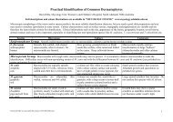
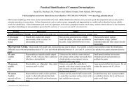
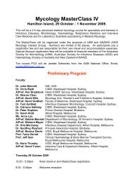
![List for QAP Fungi [2006].](https://img.yumpu.com/36263775/1/184x260/list-for-qap-fungi-2006.jpg?quality=85)