
Create successful ePaper yourself
Turn your PDF publications into a flip-book with our unique Google optimized e-Paper software.
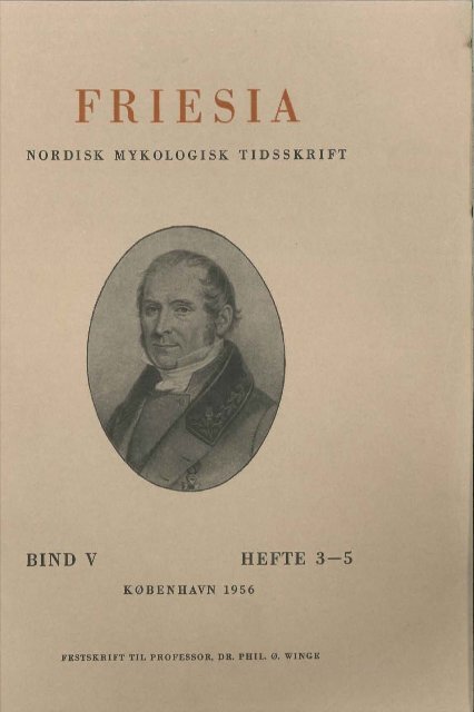
F ES<br />
NORDISK MYKOLOGISK TIDSSKRIFT<br />
<strong>BIND</strong> V HEFTE 3-5<br />
KØBENHAVN 1956<br />
FESTSKRIFT TIL PROFESSOR, DR. PH IL. 0. WINGE
INDHOLD<br />
Side<br />
Catherine Roberts: The inheritance of enzymatic characters in<br />
yeasts .................................................................. 161<br />
Olof Andersson: Three rare or little known bolets in Sweden.<br />
Boletus pulverulentus OPAT., B. radicans PERS. ex FR.<br />
and Phylloporus rhodoxanthus (SCHW.) BRES. ............ 180<br />
Erik Bille-Hansen: The nitrogen requirements of Coprinus heptemer-us-.M.<br />
LANGE & SMITH .......................................... 190<br />
N. Fabritius Buchwald: On the dimorphism of the ascospores and<br />
their arrangement in the ascus of Monilinia oxycocci<br />
(WOR.) HONEY (syn. Sclerotinia oxycocci WOR.) ......... 196<br />
Kjeld Biilow: Ornamentation of spores of Russula lauroceraci<br />
MELZER and Russula foetens (PERS.) FR...................... 204<br />
M. P. Christiansen: Two new species of Corticiaceae from Denmark.<br />
Peniophora danica sp. n. and Corticium salicicola<br />
sp. n. .................................................................. 207<br />
M. Skytte Christiansen: A new species of the form-genus Lichenoconium<br />
PETR. & SYD. (Fungi imperfecti) J L. xanthoriae<br />
sp. n. .................................................................. 212<br />
E. J. H. COl'ner: A new European Clavaria: Clavulinopsis septentrionalis<br />
sp. nov. ................................................ 218<br />
Anders Danielsen: Coprinus comatus (SeRUM.) FR. sprenger asfalt<br />
i Bergen, Norge. (Summary: Coprinus comatus<br />
breaks through asphalt in Bergen, Norway) .......... ..... 221<br />
Finn-Egil Eckblad: Some operculate Discomycetes new to Norway<br />
..................................................................... 223<br />
K. J. Frandsen: Variations in resistance of Trifolium pratense<br />
to attacks of Kabatiella caulivora (KrRCHN.) KARAK.... 231<br />
P. Sonne Frederiksen: A new Rhodotorula species, Rhodotorula<br />
macerans sp. n., isolated from field-retted flax straw ... 234<br />
Paul Gelting: Parmelia subaurifera NYL. and P. fraudans (NYL.)<br />
NYL. in Greenland . ............... .................. .............. 240<br />
Asbjørn Hagen: Rustsopper og rustverter nye for Færøerne.<br />
(Summary: Two rusts and several host plants of Puccinia<br />
hieracii new to the Færoes) ................................. 247<br />
Lise Hansen: Two polyporaceous fungi with merulioid hymenophore.<br />
Poria taxicola (PERS.) BRES. and Polyporus<br />
dichrous FR. ex FR. ................................................ 251<br />
Louis Hal'ffisen: On Merulius silvester FALCK and M. himantioides<br />
FR. Preliminary remarks .......................................... 257<br />
Ernst Hayren: Die in FinnIand bisher gefundenen Wasserpilze... 264<br />
K. Hauerslev: Om Fund af Judasøre (Hirneola auricula Judae<br />
(L.) BERK.) i Korsør og nærmeste Omegn. (Summary:<br />
A mass occurrence of Hirneola auricula Judae (L.) BERK.<br />
in Korsør, Sealand, in Denmark) ................. ............. 266<br />
J. P. Jensen: Recherches sur le cycle de Coprinus disseminatus<br />
(PERS. ex FR.) S. F. GRAY.. ..................................... 271<br />
Henrik Alb. JOl'gensen: M onascus ruber van TIEGH. demonstrated<br />
in Denmar k ...................................................... 274<br />
Ivar Jørstad: On the Sonchus rust Peristemma pseudosphaeria<br />
(MONT.) n. comb. (syn. Puccinia sonchi ROB.) .. .......... 278<br />
Axel B. Klinge: Beitrage zur Pilzflora Jtitlands .............. ....... 284<br />
Morten Lange: Pyrenomycetes parasitic on hypogeous fungi ... 289<br />
Fortsættes paa Omslagets Side 3<br />
PDF scanning and OCR by the Danish Mycological Society 2010 - www.svampe.com
Friesia . Bind V, Hefte 3- 5, Side 161- 432, 1956.<br />
TIL<br />
PROFESSOR, DR. PHIL. ØJVIND WINGE<br />
PAA 70 AARSDAGEN<br />
DEN 19. MAJ 1956
F'RIESIA V , 1956 PLATE I
Fotograferet 1950 paa Carlsberg Laboratorium
THE INHERITANCE OF<br />
ENZYMATIC CHARACTERS IN YEASTS<br />
By CATHERINE ROBERTS<br />
Leeture presented in CDpenhagen at the 1951<br />
Microbial Genetics Symposium<br />
Introduetion<br />
In the following I would like to discuss the problem of the inheritance<br />
of enzymatic characters in the yeast fungi and, in doing so, to<br />
attempt to review critically the available evidence in support of men delian<br />
and non-mendelian heredity in these organisms. Such an attempt<br />
will necessarily involve a discussion of enzymatic adaptation in its<br />
bearing upon yeast genetics, especially since current views offered in<br />
explanation of this phenomenon are as widely divergent as those<br />
which attempt to explain the mechanism of gene transmission. Since<br />
the contributions to yeast genebcs have been exceedingly numerous,<br />
I propose to tre at the subject chronologically in order to trace the<br />
developments within the field in as clear and unambiguous a manne r<br />
as possible.<br />
1935-1939<br />
The year 1935 ean be taken as a starting point, for it was in this<br />
year that a paper by Professor WINGE entitled "On Haplophase and<br />
Diplophase in some Saccharomycetes" appeared, in which the complete<br />
life cycle of Saccharomyces) including sexuality, sporulation, and<br />
alternation of generations, was incontestably established. As LINDE<br />
GREN has aptly remarked in his recent book, "The Yeast Cell", it was<br />
WINGE "who brought yeast genetics into being", for with thi s 1935<br />
paper as a basis, there followed in the years 1937-1939 a series of<br />
now classical papers by Professor WINGE and his collaborator LAUST-<br />
FRIESIA V -161 - 11
- 162-<br />
SEN, in which the yeast fungi were, for the first time, employed in thc<br />
field of genetics. With the aid of the micromanipulator, they were<br />
able to isolate each of the 4 spores from an ascus, and in addition to<br />
finding that genetic segregation occurred within the ascus, the possibility<br />
presented itself of utilizing the micromanipulator technique<br />
in spore crossings and tetrad analysis. By pairing two spores and by<br />
observing under the microscope their subsequent copulation, the first<br />
artificial yeast hybrids were thus produced. These hybrids (14 interspecific<br />
and 1 intergeneric) were purposely produced from parent<br />
types which differed in their ability to ferment various sugars in<br />
order that the inheritance of the enzymes responsible for fermentation<br />
could be investigated; but although the hybrids showed heterosis and<br />
sporulated abundantly, their spores unfortunately germinated very<br />
poorly, making impossible a detailed genetic analysis of the enzymatic<br />
characters involved in this material. An important fact was adduced,<br />
however, from these experiments and that was that a hybrid<br />
between a fermenter of any particular sug ar and a non-fermenter<br />
would always be capable of fermenting the sugar, and, in addition,<br />
that there was always an excess of fermenters among the few viable<br />
spores from such hybrids. Thus the conc1usion to be drawn from<br />
these results was that in all instances studied, "the ability to synthesize<br />
a specific enzym e is dominant in the Fl." Nothing more definite<br />
could be stated, and such was the state of our knowledge until<br />
1943, when LINDE GREN began his researches in yeast genetics.<br />
1943-1944<br />
LINDEGREN'S first contribution to the field was an important<br />
one - namely, the discovery in 1943 of heterothallism in Saccharomyces.<br />
By tetrad analysis he was able to show that from each 4spored<br />
ascus 2 spores belong to one mating type and two to another<br />
and that therefore mating type specificity is controlled by a single<br />
pair of allelomorphs. He also described a phenomenon which he called<br />
"illegitimate copulation", in which he maintained that self-diploidization<br />
of a single-spore culture invariably lea ds to poor sporulation<br />
and low viability of the ascospores. Although heterothallism in<br />
Saccharomyces has been confirmed, it is not true that self-diploidized<br />
single spore cultures usually sporulate poorly and produce spores with<br />
poor germination; we have in our work generally found the opposite<br />
to be the case.
- 163-<br />
Figs. 1-4. WINGE and LAUSTSEN'S method for isolating the four spores<br />
within one ascus with the aid of a micromanipulator equipped with two<br />
glass needles. - (Af ter WINGE & LAUSTSEN, 1937).<br />
Figs. 5-6. Artificial species hybridization in yeasts by the pairing of two<br />
ascospores in a nutrient droplet with the aid of a micromanipulator. The<br />
smalle r spore is from Saccharomyces validus and the larger from S. cere<br />
visiae. Copulation of the two spores results in a hybrid zygote, the first bud<br />
of which is seen in Fig 6. - (Af ter WINGE & LAUSTSEN, 1938).<br />
2.<br />
11*
- 164 -<br />
These investigations of LINDEGREN led, in 1944, to a new method<br />
for hybridizing yeasts. Instead of employing the spore crossing tech<br />
nique of WINGE and LAUSTSEN, he simply mixed two haplophase cul<br />
tures of opposite mating type together in a small amount of nutrient<br />
medium, and after a period of incubation to allow for copulation, he<br />
transferred the mixture to a sporulating medium. Spores to be used<br />
in genetic analyses were then isolated from the asci which arose in this<br />
mixture. The advantage of this method over the spore crossing method<br />
is, as LINDEGREN justly claimed, the fact that the parents are not<br />
"used up" in the crossing and can therefore be employed in further<br />
study. On the other hand, the hybrids themselves are not available<br />
for analysis unless individual zygotes or their derivatives are isolated<br />
from the mixture. A more serious objection to this technique is that<br />
self-diploidization of haploid yeasts occurs frequently, so that the<br />
so-called hybrids obtained may actually represent the diplophase of<br />
one or the other parent.<br />
Employing thi s new hybridization technique, LINDEGREN and<br />
SPIEGELMAN in 1944 were successful in producing hybrids between<br />
S. carlsbergensis and S. cerevisiae which were completely fertile, i. e.,<br />
a high percentage of the spores were viable, thus making possibIe for<br />
the first time the tetrad analyses of artificially produced yeast hy<br />
brids. One of the parents, S. carlsbergensis) is able to ferment raffinose<br />
completely, while the other parent, S. cerevisiae) ferments only 1/3 of<br />
it - in other words, S. carlsbergensis is capable of fermenting meli<br />
biose through the activity of a specific enzyme, while S. cerevisiae is<br />
not. As was to be expected from the earlier researches of WINGE and<br />
LAUSTSEN, the hybrid itself fermented melibiose. Tetrad analyses of<br />
the spores from this hybrid yielded segregation ratios which excluded<br />
the possibility that only one gene in S. carlsbergensis was involved in<br />
the production of the enzyme, since in addition to ratios of 2 fermen<br />
ters: 2 non-fermenters, ratios of 4:0 and 3:1 were also obtained. The<br />
conclusion which LINDEGREN and SPIEGELMAN drew from this investi<br />
gation was that S. carlsbergensis contains 2 dominant genes, each of<br />
which is able to initiate the synthesis of the enzyme when melibiose<br />
is present, and that the other parent, S. cerevisiae) contains the two<br />
recessive alleles. In other words, this was interpreted as an example<br />
of true mendelian inheritance.<br />
SPIEGELMAN and LINDEGREN then turned to a more detailed investi<br />
gation of the mechanism of enzymatic adaptation in yeasts by com<br />
paring this process in haploid and diploid strains of the same species.
- 165 -<br />
A stable, homogeneous diploid was found to adapt to galactose fermentation<br />
in the absence of cell division, while genetically unstable<br />
haploids were found to adapt only during growth. This was taken to<br />
mean that both cytoplasmic interaction as well as mutation and<br />
natural selection may be operative in any particular adaptive enzyme<br />
system according to the strain employed, and in order to study the<br />
biochemistry of enzymatic adaptation, the importance of eliminating<br />
natural selection by employing a diploid strain of high genetic stability<br />
was stressed.<br />
Thus in the 9 years which had elapsed since the publication of<br />
Professor WINGE'S first paper on the life cycle of Saccharomyces<br />
sufficient evidence had accumulated to indicate that inheritance in the<br />
yeasts, just as in other organisms, followed classical mendelian laws,<br />
and, in addition, that the yeasts were exceedingly promising organisms<br />
for future investigations in the field of biochemical genetics.<br />
But it was during the years following 1944 that startling and revolutionary<br />
hypotheses based upon the yeast genetic work performed in<br />
the United States were proposed, which, if they are to be accepted,<br />
would mean profound changes in our concept of the gene. It is thi s<br />
work during the period 1945 to 1947 that I would like to discuss now.<br />
1945-1947<br />
,sPIEGELMAN and LINDEGREN repeated by means of the mass mating<br />
technique the crossing between S. carZsbergensis) a melibiose-fermenter,<br />
and S. cerevisiae) a non-fermenter of melibiose. The strain of<br />
S. carZsbergensis used in this crossing was now assumed to have only<br />
one gene for melibiase production, since 10 asci from the hybrid<br />
segregated out regularly as 2 fermenters: 2 non-fermenters. However,<br />
when the mating, sporulation, and planting were undertaken in the<br />
presence of melibiose, 6 asci gave segregation ratios of 4 fermenters:<br />
O non-fermenters. When melibiose was removed from the substratum,<br />
the 4:0 ratios reverted to the normal 2:2 ratios. However, before the<br />
single spore cultures carrying the recessive allele had completely lost<br />
their enzyme content in the melibiose-deficient medium, they were<br />
re-exposed to melibiose, and it was found that all cultures showed a<br />
marked increase in enzymatic activity. The conclusions to be drawn<br />
from thi s experiment were significant. First, it was assumed that the<br />
4:0 ratios resulted from the fact that the 2 normally negative cultures<br />
from each ascus were able to ferment due to the transfer of enzyme-
- 166-<br />
forming factors from the cytoplasm of the hybrid to that of the spores.<br />
Secondly, it was assumed that here was an example of the maintenance<br />
and synthesis of an enzyme in the complete absence of the<br />
gene. These conclusions, of course, were based on the implicit assumption<br />
that melibiase production is due to the presence of a dominant<br />
gene, the recessive allelomorph of which is unable to initiate the<br />
formation of the enzyme. As will be seen later from a discussion of<br />
our work on galactose fermentation, another explanation is possible.<br />
In the years 1945 and 1946 there followed a series of papers by<br />
LINDEGREN dealing with mendelian and non-mendelian inheritance<br />
and with the cytogene theory, proposed to account for the behaviour<br />
observed. It is scarcely worthwhile to discuss in detail the experiments<br />
upon which the cytogene theory is based, since the theory has later on<br />
been completely abandoned by LINDEGREN himself, but a few general<br />
remarks concerning it may be of interest. In addition to the experiments<br />
I have aIready discussed, LINDEGREN produced hybrids heterozygous<br />
with respect to galactozymase, but instead of the expected 2:2<br />
segregations from the hybrid, 4:0 ratios were obtained. This was<br />
interpreted as cytoplasmic transfer - i. ,e., the adaptive enzyme, or<br />
"cytogene" was assumed to be transmitted through the cytoplasm<br />
and maintained in cells lacking the dominant gene. Thus the recessive<br />
cells were contaminated by the cytogene and continued to ferment as<br />
long as the specific substrate was present. Subsequent experimental<br />
work by LINDE GREN yielded a large number of irregular segregations<br />
which he maintained could not be accounted for in terms of multiple<br />
genes, but which, he claimed, gave further support for the cytogene<br />
theory. At the Cold Spring Harbor Symposium in 1946 this theory<br />
was considerably revised, in that a new phenomenon was considered<br />
to be involved - namely, "masked recessiveness". Again, in his genetic<br />
analyses, LINDE GREN had obtained a large number of irregular<br />
segregation ratios, the majority of which showed an excess of fermenters.<br />
One ascus, in particular, derived from a MmGg hybrid yielded<br />
4 M fermenters and 4 G fermenters. This was explained by assuming<br />
that the recessive alleles were "masked" by the acquisition at meiosis<br />
of the dominant genotype - i. e., m becomes M and g becomes G.<br />
When such a "masked recessive" was crossed to a normal recessive,<br />
2:2 ratios were obtained, but when it was crossed to a normal dominant,<br />
not 4 :0, but 2:2 ratios were likewise obtained. A satisfactory<br />
and logical explanation of this behaviour appears impossible, and the<br />
experimental results seem only to throw doubt upon the validity of
-167 -<br />
the concept of masked recessiveness. In addition to these theoreticaI<br />
considerations, which, it must be remembered, were primarily based<br />
upon the behaviour of one exceptional ascus, LINDEGREN stated that<br />
he had repeated the melibiose experiment to which I have aiready<br />
alluded - namely, that maintenance and increase of an adaptive<br />
enzyme in the absence of the gene occurs through cytoplasmic transfer<br />
from the dominant type. The original results however, could not be<br />
duplicated, since just as many 4:0 ratios were obtained this time in the<br />
absence of the substrate as were obtained in its presence ; this was<br />
regarded by LINDEGREN as further evidence for the existence of<br />
maske d recessives. It is worthy of note that in the discussion following<br />
the presentation of this cytogene theory with all of its complexities,<br />
LINDE GREN stated in response to a direct question that he would not<br />
even consider the multiple factor hypothesis as an explanation for<br />
his experimental results, as he deemed it "too elaborate to be<br />
justified" .<br />
At this same Symposium SPIEGELMAN also discussed, with special<br />
reference to yeast investigations, the gene-enzyme problem, wherein<br />
he introduced the plasmagene hypothesis. It is important to note,<br />
however, that SPIEGELMAN considered that inheritance in yeasts was<br />
completely mendelian; his "plasmagenes" were concerned therefore<br />
with the mechanism of gene action, rather than with gene transmission.<br />
Apparently unaware that LINDE GREN was unable to repeat the<br />
now famous experiment involving the apparent synthesis and maintenance<br />
of an adaptive enzyme in the absence of the gene, he endeavoured<br />
to account for this phenomenon in terms of plasmagenes. His<br />
argument was as follows: if 2:2 ratios are normally obtained from a<br />
heterozygous hybrid, but 4:0 ratios are obtained in the presence of<br />
the specific substrate, then the adaptive enzyme system must be part<br />
of a cytoplasmic self-duplicating mechanism, and once the enzyme is<br />
formed, its further produetion ean proceed independently of the presence<br />
of the gene. In other words, the 2 spores which carry the recessive<br />
alle le obtain from the cytoplasm of the hybrid replicas of the<br />
dominant gene, which he called the plasmagenes. These plasmagenes<br />
are self-duplicating and combine with precursor protein and with the<br />
substrate to form the enzyme, thus allowing the formation of a specific<br />
enzyme to occur within cells which lack the gene normally necessary<br />
for its synthesis. The important feature of SPIEGELMAN'S theory<br />
is, therefore, that the genes are continually producing nucleo-protein<br />
replicas of themselves which pass into the cytoplasm of the cello<br />
In the ensuing year, 1947, a series of papers by SPIEGELMAN and
- 168 -<br />
his colleagues appeared which de alt with the purely biochemical<br />
aspects of enzymatic adaptation. It will suffice to say that they were<br />
able to demonstrate the occurrence of competitive interaction between<br />
adaptive enzyme systems, a phenomenon which SPIEGELMAN had<br />
earlier predicted on the basis of the plasmagene hypothesis, since the<br />
different plasmagenes must necessarily com pete with one another for<br />
protein and energy, and as he termed it, "the outcome determines the<br />
enzymatic constitution of the cell".<br />
Meanwhile LINDEGREN had turne d from a consideration of the<br />
inheritance of the enzymes responsibIe for the fermentation of carbohydrates<br />
to a study of the inheritance of the genes involved in vitamin<br />
synthesis. Working with biotin-, pantothenic acid-, para-aminobenzoic<br />
acid-, and thiamine-deficient strains, and crossing these with synthesizers,<br />
he found for the most part that 2:2 segregations were obtained<br />
from the hybrid progeny. In other words, not only was mendelian<br />
inheritance operative in the synthesis of vitamins by yeasts, but<br />
the ability to synthesize appeared to be controlled in each instance<br />
by a single gene. However, further investigations appeared to throw<br />
doubt on the simplicity of the phenomenon. In a paper published<br />
together with RAUT in 1947, a pantothenate + type was crossed with<br />
a pantothenate -type. The hybrid produced was, as expected, capable<br />
of synthesizing pantothenate. However, 4 spores from 1 ascus derived<br />
from this hybrid were all pantothenate +. This case was explained<br />
by the authors as being due to "gene transformation",<br />
Still another complicating factor was introduced into yeast genetics<br />
by LINDE GREN in 1947 - namely, "depletion mutations". Since<br />
time does not permit a critical discussion of all of LINDEGREN'S<br />
theoreticai considerations, it will suffice in this case to state that<br />
he conceived a depletion mutation as a phenomenon in which a modification<br />
is transmitted indefinitely during vegetative reproduction but<br />
which is unable to be transmitted through the sexual cycle. According<br />
to LINDEGREN, this behaviour, which phenotypically resembles gene<br />
mutation, is not due merely to the effect of the environment upon the<br />
cytoplasm, but is actually under genetic control.<br />
Thus, to summarize, thi s period from 1945 to 1947 was characterized<br />
by the production by the American investigators of a large<br />
number of yeast hybrids, the progeny of which yielded both regular<br />
segregation ratios in accordance with classical mendelism, but also<br />
irregular, so-called "non-mendelian" ratios. By way of explanation,<br />
LINDEGREN proposed his cytogene theory and developed his concepts
- 169-<br />
of masked recessiveness and depletion mutations. During the same<br />
period SPIEGELMAN'S plasmagene theo ry was proposed in order to<br />
account for the maintenance and increase of an adaptive enzyme in<br />
the absence of the gene. Now I would like to discuss some of the work<br />
on adaptation and the inheritance of enzymatic character s in yeasts<br />
which Professor WINGE and I undertook at the Carlsberg Laborato<br />
rium, and which first appeared in 1948.<br />
1948<br />
By the spore-crossing technique a hybrid was produced between<br />
S. cerevisiae and S. chevalieri with sufficient fertility to make possibIe<br />
genetic investigations by tetrad analyses. The two parents are distin<br />
guished by the fact that S. cerevisiae is able to ferment both maltose<br />
and galactose, while S. chevalieri is unable to do so. Here, then, we<br />
had the possibility of determining what type of enzymatic inheritance<br />
was operative in thi s material; our experimental results established<br />
incontestably that it was mendelian.<br />
First, with regard to maltose fermentation, our original hybrid<br />
yielded the following segregation ratios: 9-4:0 and 3-3 :1, which<br />
of course excluded the possibility that 1 or 2 genes were involved in<br />
the production of maltase. If it is assumed that 3 genes are present,<br />
and that 50 % crossing over occurs between all three genes and their<br />
centromeres, then the theoretically expected ratios would be 52.8%<br />
4 :0, 44.4% 3 :1, and 2.8% 2 :2. Although agreement between the ob<br />
served and the expected was not close, it nevertheless was indicative<br />
of the fact that 3 genes were involved, and in orde r to test the validity<br />
of thi s hypothesis, we attempted several backcrossings of the single<br />
spore progeny of the hybrid to the recessive S. chevaZieri. Assuming<br />
that 3 genes are involved, any spore from the hybrid would contain<br />
either O, 1, 2, or 3 genes, and by crossing the fermenters to the reces<br />
sive, one should expect to obtain a 1, 2, or 3 gene segregation in the<br />
backcross hybrid's progeny. Such indeed was the case. One of our<br />
backcross hybrids yielded a typical 1 gene segregation (14-2 :2) and<br />
two others yielded 2 gene segregations (12-3:1 and 5-2:2; and<br />
4-4:0 and 16-3 :1). The conclusion to be drawn from these results<br />
is, therefore, that S. cerevisiae apparently contains 3 non-linked<br />
polymeric genes, Ml) M2) and M3) for the production of maltase.<br />
Now, with regard to galactose fermentation, we found at the outset<br />
of our experiments that although S. chevalieri had been reported to
- 170-<br />
be incapable of fermenting galactose, it actually was capable of doing<br />
so, but only slowly, after a period of adaptation. The tetrad analysis<br />
of 12 asci of our original hybrid showed in all cases an unmistakable<br />
2:2 segregation, in that 2 cultures fermented galactose rapidly (1-2<br />
days) and 2 slowly (5-14 days). We assumed therefore that S. cere<br />
visiae contained only 1 gene for the production of galactozymase.<br />
The tetrad analyses of our backcross hybrids which were heterozygous<br />
for galactose fermentation confirmed this assumption. In all cases,<br />
a 2:2 segregation was obtained. In the light of the earlier investiga<br />
tions of LINDEGREN and SPIEGELMAN on the mechanism of melibiose<br />
adaptation, these findings were of considerable significance. They had<br />
implicitly assumed that the recessive allelomorph was incapable of<br />
producing the specific enzyme and consequently that when a single<br />
ascus from a heterozygous hybrid yielded 4:0 ratios in the presence<br />
of the substrate, enzyme synthesis in the absence of the gene must<br />
be involved. We, 'On the other hand, found that in the case of galac<br />
tozymase, the recessive allelomorph of G was not g} but 9 s which<br />
controlled the slow synthesis of the enzyme; in other words, a "long<br />
term adaptation" was involved in the fermentation of galactose by<br />
the recessive alleles. Further studies of thi s long-term adaptation<br />
convinced us that it was not result of mutation and natural selection,<br />
since we were able to adapt, de-adapt, and re-adapt our gs types at<br />
will, depending upon whether they were grown in a galactose-contain<br />
ing or galactose-deficient medium. We consider, therefore, that long<br />
term adaptation is an expression of the fact that enzyme production<br />
in the presence of the sugar is gradually increased until it exists in a<br />
sufficiently high concentration to bring about visible fermentation.<br />
1949<br />
The year 1949 brought forth criticism of our views concerning the<br />
nature of long-term adaptation, as well as both criticism and confir<br />
mation of our views concerning the mendelian nature of the inheri<br />
tance of enzymatic characters in yeasts.<br />
MUNDKUR and LINDEGREN had reinvestigated the phenomenon of<br />
long-term adaptation to galactose fermentation but came to the con<br />
clusion that it could be explained entirely on the basis of mutation<br />
and natural selection. This explanation was based on the assumption<br />
that 3 phenotypes existed: G} a rapid fermenter; g} a non-fermenter;<br />
and gx a slow fermenter, and that it is through chance mutation from
- 171 -<br />
g -+ G that the phenotypic slow fermenter, gXJ which is actually a<br />
mixture of G and g cells, is produced (x signifies the number of days<br />
elapsing before fermentation is visible). It is true that our g s types<br />
adapted after varying periods of time and that it would be tempting<br />
to conclude that these discrepancies involved chance mutations, but,<br />
on the other hand, we had only 2 phenotypes in our material, a rapid<br />
and slow fermenter, and had never isolated a spore belonging to<br />
MUNDKUR and LINDEGREN'S category of "non-fermenters." Another<br />
point to bear in mind is that we were unable to de-adapt our G type.<br />
If we accept LINDEGREN'S view that long-term adaptation is due to<br />
mutation, then the logical consequence would be that in addition to<br />
the stable G gene, there also exists an unstable G gene which readily<br />
mutates to g. We cannot accept this view of 2 different G genes in<br />
S. cerevisiae for the synthesis of galactozymase.<br />
Through an investigation of the behaviour of non-synthesizers of<br />
thiamine, tryptophane, adenine, and methionine in crosses with syn<br />
thesizers, POMPER and BURKHOLDER found a 2:2 segregation for each<br />
character, demonstrating that only 1 gene was involved in each bio<br />
synthesis. With regard to uracil synthesis, non-allelic complementary<br />
genes appeared to be involved. The conclusion arrived at by these<br />
authors was that "the characters segregated independently and<br />
regularly according tomendelian principles."<br />
GILLILAND, using the spore crossing technique, produced a hybrid<br />
between S. italicus and S. chevalieri which was heterozygous in 4<br />
fermentation characters, and he was able to demonstrate conclusively<br />
by tetrad analyses that S. italicus contains 1 gene for the production<br />
of maltase (which has subsequently been identified as Ml) and 1 gene<br />
for the production of galactozymase. With regard to raffinose,<br />
GILLILAND obtained a 3-gene segregation from 20 asci; these asci gave<br />
exactly the same 3 gene segregation for sucrose fermentation, the<br />
only difference being that the sucrose recessives, which corresponded<br />
to the raffinose negatives, were actually able to ferment sucrose<br />
slowly, after adaptation. GILLILAND concluded that S. chevalieri<br />
apparently contains 3 polymeric genes for the production of (3-h<br />
fructosidase, an enzyme capable of hydrolyzing both sucrose and<br />
raffinose, but that definite proof of this assertion would have to be<br />
obtained from backcrosses to the recessive. An additional observation<br />
of interest was that all the raffinose-negative, sucrose-slow cultures<br />
were able to ferment maltose, indicating that a-glucosidase may be<br />
present in the other parent, S. italicus.
- 172-<br />
It was also in 1949 that LINDEGREN'S book, "The Yeast Cell",<br />
appeared. I will make no attempt to review thi s book, which, although<br />
containing linkage calculations and the first chromosome maps of the<br />
yeasts, deals primarily with much of the material already discussed.<br />
I would like to emphasize, however, that in this book LINDEGREN<br />
rejects both classical mendelism and his own cytogene theory, and<br />
proposes an elaboration of the theory involving the conversion or<br />
contamination of alleles to account for inheritance behaviour in<br />
yeasts. Since LINDE GREN is apparently convinced that all enzymatic<br />
characters in yeasts which have been studied are controlled by single<br />
genes, he should obtain only 2:2 segregations in his tetrad analyses<br />
of heterozygous hybrids. This, of course, he does not. Therefore the<br />
irregular ratios, which for the most part yield an exces s of the do<br />
minant type, are interpreted as being due to the contamination of the<br />
recessive allele by the dominant allele by contact at synapsis during<br />
meiosis. Gene conversion is regarded as being controlled by heredity,<br />
since it occurs with higher frequency in some lines; thus he would<br />
have, for example, a gene infIuencing the stickiness of the chromo<br />
some. I think it is clear that the highly speculative nature of such<br />
considerations, interesting as they may be, does not warrant a more<br />
detailed discussion. It will suffice to quote LINDE GREN directly in<br />
regard to his concept of the gene: "The explanation ... fits all the<br />
cases without any fundamental difficulty except that it requires<br />
alteration of our apparently well-established views concerning the<br />
stability of the gene."<br />
I would like at this time to mention the work of EPHRUSSI and his<br />
colleagues which also appeared in 1949 and which de alt for the first<br />
time with the inheritance of respiratory enzymes in yeasts. Mutants<br />
deficient in cytochrome oxidase and characterized by the production<br />
of small colonies were induced through the action of acrifIavine.<br />
These mutant s were employed in extensive crossing experiments with<br />
the result that only a very small percentage of them reappeared in<br />
the progeny of the hybrids. If the results are to be explained on the<br />
basis of multiple genes, EPHRUSSI has calculated that at least 16<br />
dominant genes must be involved. Since this seems highly improb<br />
able, it is assumed that the acriflavine-induced mutants have lost<br />
self-duplicating cytoplasmic particles which are essential for enzyme<br />
synthesis. Accordingly, these first investigations on the inheritance<br />
of respiratory enzymes point to the interesting fact that cytoplasmic,<br />
rather than mendelian, inheritance is involved.
- 173-<br />
1950-1951<br />
One of the most interesting of the recent contributions is a paper<br />
by SPIEGELMAN, SUSSMAN, & PINSKA dealing with the nature of longterm<br />
adaptation in yeasts. It will be recalled that we have explained<br />
the phenomenon by assuming that a type containing the recessive<br />
allele is a slow synthesizer of the enzyme and that it becomes adapted<br />
through a gradual accumulation of enzyme in the presence of the<br />
substrate; MUNDKUR and LINDEGREN, however, consider that the<br />
phenomenon is due entirely to the occurrence of chance mutations<br />
from non-fermenters to fermenters. SPIEGELMAN, on the other hand,<br />
considers both explanations inadequate and assumes that long-term<br />
adaptation is cytoplasmic in nature. His experimental results indicate<br />
that during the period of adaptation, the population of a gs type<br />
actually becomes heterogeneous through production of so-calle d<br />
"positive" cells by the original "negatives". These modifications are<br />
not gene mutations; they were found to be induced only by the<br />
presence of the substrate, and in its absence, the positive cells were<br />
stated to produce negative buds. SPIEGELMAN believes that such a<br />
phenomenon can be explained only on a cytoplasmic basis, and furthermore,<br />
that it represents additional evidence in support of the<br />
plasmagene hypothesis. The theory is admittedly attractive, but<br />
similar investigations of adaptive enzym e systems other than galacto·<br />
zymase are necessary to prove its universal validity.<br />
The final contribution I would like to discuss deals with the genes<br />
responsibIe for maltose fermentation. Professor WINGE and I undertook<br />
a further study of this problem in order to establish conclusively<br />
that polymeric genes are involved in the production of maltase, and<br />
that the inheritance of fermentative characters in yeasts is mendelian<br />
in nature. This I believe we have done. The work involved the isolation<br />
of the 3 M genes, as well as a fourth M gene, M4) which arose<br />
both spontaneously and as aresult of Rontgen irradiation. The identit<br />
Y of these 4 genes was firmly established by extensive hybridization<br />
work, involving the artificial production of 81 new hybrids. In<br />
all hybrids in which 2 different M genes were present, the progeny<br />
yielded 2·gene segregations, whereas the progeny of hybrids between<br />
types containing the same gene yielded no segregation. In addition<br />
to these investigations, work is now in progress on the inheritance<br />
of the ability to ferment raffinose and sucrose. It can be stated that<br />
we have completely confirmed GILLILAND'S findings with regard to
- 174-<br />
the existence of 3 polymeric genes for these fermentations, and at<br />
present we have aiready isolated 2 of the 3 genes involved - namely,<br />
R2 and Ra.<br />
Finally, to summarize the developments which have taken place in<br />
the field of yeast genetics, it ean be said that the work was initiated<br />
here in Denmark on the assumption that inheritance in yeasts was<br />
mendelian in nature. Subsequent investigations by LINDEGREN demonstrated<br />
the occurrence of apparent non-mendelian segregation ratios<br />
with regard to fermentation characters and the synthesis of vitamins<br />
and amina acids. Accordingly, his concepts of the nature of inheritance<br />
in yeasts passe d through three distinct phases: first, classical<br />
mendelism; second, cytoplasmic inheritance, embodied in thc<br />
cytogene theory; and finally, his current view of the instability of<br />
the gene. It is therefore a matter of considerable interest that the<br />
recent investigations of EPHRUSSI have indicated that cytoplasmic as<br />
well as mendelian inheritance is involved in the synthesis of respiratory<br />
enzym es in yeasts. I think that it is evident, however, that the<br />
continuation of the work at the Carlsberg Laboratorium, complicated<br />
as it may be by the related problem of enzymatic adaptation, has<br />
nevertheless conclusively established that the inheritance of fermentative<br />
characters in the yeasts which we have investigated is mendelian<br />
and in some cases involves the existence of polymeric genes.<br />
Whatever the reasons for the disparity between LINDEGREN'S and our<br />
experimental results may be, and the occurrence of self-diploidization,<br />
overlapping of generations, and 8-nucleate asci may be mentioned<br />
in this connection, the faet remains that we have not obtained segregation<br />
ratios in disaccord with MENDEL'S laws. We have no choice,<br />
therefore, but to consider the concepts of cytogenes, depletion mutations,<br />
masked recessives, and gene conversions to be superfluous, and<br />
to regard the inheritance of fermentative characters in yeasts as<br />
completely mendelian.<br />
Postscript added January 1956.<br />
Limitation of space precludes more than a brief mention of some<br />
of the major contributions to the field made during the past five<br />
years.<br />
Interest in the problem of irregular segregation of yeast tetrads<br />
has continued unabated, and several different explanations of this<br />
phenomenon have been advanced. In 1953 MUNDKUR observed the
- 175-<br />
localization and movement of mitochondria within the living yeast<br />
cell, and knowledge of the localization of enzymes within mitochondria<br />
led him to con cl ude that the random distribution of mitochondria<br />
within mother cells and buds could be responsibie for vegetative<br />
mutants and genetic irregularities. Another explanation, based upon<br />
both genetic and cytological evidence, has been found in the occurrenee<br />
of supernumerary mitoses in the ascogenous cells of yeast<br />
monohybrids (WINGE & ROBERTS 1954). If the normal number of 4<br />
nuclei in the immature ascus is increased to 8 with consequent duplication<br />
of the genotypes, and if some of the nuclei (or spores) do not<br />
survive, then it ean be a matter of chance which genotype s the surviving<br />
spores contain. In addition, these extra mitoses may lead to<br />
the formation of binucleate heterozygous or homozygous spores, and<br />
examples of irregular segregation in LINDEGREN'S haploid material<br />
revealed that one of the 4 single-spore cultures in each of 3 irregular<br />
tetrads was diploid and heterozygous. Still another explanation -<br />
and one which enjoys many adherents - involves the occurrence of<br />
polyploidy in yeasts. It is obvious that polyploid inheritance in a<br />
yeast would result in segregations quite different from the 2:2<br />
expected from a monohybrid, and claims of its existence were set<br />
forth in 1951 by LINDEGREN & LINDEGREN and by ROMAN, HA WTHORNE,<br />
& DOUGLAS. In 1954 further evidence for polyploid segregation was<br />
presented by POMPER & McKEE and by LEUPOLD & HOTTINGUER, while<br />
a still more detailed study by ROMAN, PHILLIPS, & SANDS appeared<br />
the following year. It should be noted that these investigators all<br />
worked with haploid material, whose single spore progeny could, as a<br />
rare event, diploidize, and by mixing such a clone with another<br />
haploid or diploid of opposite mating type, triploids or tetraploids<br />
were obtained in small numbers. Diploid material, on the other hand,<br />
contains the D-gene which brings about diploidization whenever<br />
present and also appears to abolish the expression of mating type.<br />
The fusion of diploid cells to form tetraploids or the fusion of a<br />
diploid and a haploid cell has not been found in this material.<br />
Another important advance has been the demonstration of the<br />
existence of complementary genes in the inheritance of fermentative<br />
characters in yeasts. LEUPOLD & HOTTINGUER (1954), working on<br />
galactose fermentation, obtained segregation ratios which led them<br />
to conclude that 2 complementary genes were involved in the fermentation<br />
of this monosaccharide. The elegant investigations of GILLILAND<br />
(1954) on Saccharomyces diastaticus have elucidated the genetics of
- 177-<br />
super-attenuation, or dextrin fermentation, which is of practical significance<br />
in brewing and, at the same time, have demonstrated the<br />
occurrence of complementary genes in this material. He isolated the<br />
gene S) which produces amylase, responsibIe for the hydrolysis of<br />
dextrin to maltose, Ml) which is responsibIe for the rapid fermentation<br />
of maltose, and a new M-gene, M5) which is responsibIe for the<br />
slow fermentation of maltose. For super-attenuation to occur, therefore,<br />
the presence 'Df both S and an M-gene is required. Finally, with<br />
regard to complementary gene action, it has been demonstrated<br />
(WINGE & ROBERTS 1956) that raffinose fermentation may be brought<br />
about not only by the R-genes responsibIe for the synthesis of (3-hfructosidase<br />
which hydrolyzes the raffinose molecule into melibiose<br />
and fructose, but also by the combined action of the melibiase gene,<br />
Me) and the galactozymase gene, G. It was demonstrated that melibiase<br />
alone is able tD hydrolyze the raffinose molecule into sucrose<br />
and galactose, but if galactozymase is also present, then the galactose<br />
portion is fermented away, resulting in a 1/3 fermentation of the<br />
raffinose molecule.<br />
Finally, mention should be made of the significant contribution of<br />
WINGE (1955) in the form of a new hypothesis termed interallelic<br />
crossing-over, an hypothesis which was not evolved in order to. account<br />
for irregular segregation but to account for the occurrence in<br />
yeasts of extremely close linkage between two pairs of fermentative<br />
genes (Ml-RI and M3-R3) which could not reasonably be credited to<br />
chance. While not denying the validity of mendelian law, this hypothesis<br />
regards these two linked genes actually as one complex gene<br />
molecule, whose individual molecular groups may undergo interallelic<br />
crossing-over. It differs essentially from the conversion hypothesis of<br />
WINKLER and of LINDE GREN in that the result of this new type of<br />
crossing-over is not the transformation of a dominant or a recessive<br />
into its allelomorph, but the creation of two new allelomorphs, and<br />
it is significant that one of its fundamental assumptions is that the<br />
enzymes synthesized by these closely-linked genes are probably chemically<br />
similar, since the difference between them does not exceed<br />
more than a portion of the whole molecular complex.<br />
LITERATURE<br />
Ephrussi, Boris: Nucleo-cytoplasmic relations in micro-organisms. - Oxford<br />
University Press, 1953.<br />
FRIESIA V 12
- 178-<br />
Gilliland, R. B.: A yeast hybrid heterozygotic in four fermentation characters.<br />
- Compt. Rend. d. Lab. Carlsberg, Ser. Physiol. 24: 347-<br />
356. 1949.<br />
: A study of a wild yeast - S a c c h a r o m y c e s d i a s t at<br />
i c u s. - Wallerstein Lab. Comm. 17: 165-175. 1954.<br />
LeupoldJ Urs & Hottinguer, Helene: Some data on segregation in S a cc<br />
h a r o m y c e s. - Heredity 8: 243-258. 1954.<br />
Lindegren, Carl C. & Lindegren, Gertrude: Selecting, inbreeding, recombining,<br />
and hybridizing commercial yeasts. - Journ. Bact. 46:<br />
405-419. 1943.<br />
: A new method for hybridizing yeast. - N atl. Acad. Sci. Proc.<br />
29: 306-308. 1943.<br />
Lindegren, Carl C., Spiegelman, S., & Lindegren, Gertrude: Mendelian inheritance<br />
of adaptive enzymes in yeast. - Natl. Acad. Sci. Proc.<br />
30: 346-352. 1944.<br />
Lindegren, Carl C. & Lindegren, Gertrude: The cytogene theory. - Cold<br />
Spring Harbor Symposia Quant. Biol. 11: 115-129. 1946.<br />
: Depletion mutation in S a c c h a r o m y c e s. - Natl. Acad.<br />
Sci. Proc. 33: 314-318. 1947.<br />
: Tetraploid S a c c h a r o m y c e s. - J ourn. Gen. Microb. 5:<br />
885-893. 1951.<br />
: Proximity of gene s controlling the fermentation of similar<br />
carbohydrates in S a c c h a r o m y c e s. - Nature 170: 965-<br />
968. 1952.<br />
Carl C.: Concepts of gene-structure and gene-action derived<br />
from tetrad analysis of S a c c h a r o m y c e s. - Experientia 9:<br />
75-80. 1953.<br />
Carl C. & Lindegren, Gertrude: Asci in S a c c h a r o m y c e s<br />
with more than four spores. - Genetics 38: 73-78. 1953.<br />
Mundkur, Balaji D. & Lindegren, C. C.: An analysis of the phenomenon of<br />
long-term adaptation to galactose by S a c c h a r o m y c es.<br />
- Arner. Jour. Bot. 36: 722-727. 1949.<br />
B. D.: Mitochondrial distribution in S a c c h a r o m y c e s. -<br />
Nature 171: 793-794. 1953.<br />
Pomper, Seymour & Burkholder, P. R.: Studies on the biochemical genetics<br />
of yeast. - Nat. Acad. Sci. Proc. 35: 456-464. 1949.<br />
S., Daniels, K. M. & McKee, D. W.: Genetic analysis of polyploid<br />
yeast. - Genetics 39: 343-355. 1954.<br />
Roman, Herschel, Hawthorne, Douglas C., & Douglas, Howard C.: Polyploidy<br />
in yeast and its bearing on the occurrence of irregular<br />
genetic ratios. - N atl. Acad. Sci. Proc. 37: 79-84. 1951.<br />
Phillips, Marcia M., & Sands, Stanley M.: Studies of polyploid<br />
S a c c h a r o m y c e s. L Tetraploid segregation. - Genetics<br />
40: 546-561. 1955.<br />
Spiegelman, S., Lindegren, Carl C., & Lindegren, Gertrude: Maintenance<br />
and increase of a genetic character by a substrate-cytoplasmic<br />
interaction in the absence of the specific gene. - N atl. Acad.<br />
Sci. Proc. 31: 95-102. 1945.
- 179-<br />
Spiegelman, S., Sussman, Racquel Rotman, & Pinska, E.: On the cytoplasmic<br />
nature of "long-term adaptation" in yeast. --:- Natl. Acad. Sci.<br />
Proc. 36: 591-606. 1950.<br />
Winge, O.: On haplophase and diplophase in som e S a c c h a r o m y c e t e s.<br />
- Compt. Rend. d. Lab. Carlsberg, Ser. Physiol. 21: 77-11l.<br />
1935.<br />
& Laustsen, O.: On two types of spore germination, and OD<br />
genetic segregations in S a c c h a r o m y c e s, demonstrated<br />
through single-spore cultures. - Ibid. 22: 99-116. 1937.<br />
& Laustsen, O.: Artificial species hybridization in yeast. -<br />
Ibid. 22: 235-244. 1938.<br />
& Laustsen, O.: On 14 new yeast types, produced by hybridi<br />
zation. - Ibid. 22: 337-352. 1939.<br />
& Roberts, Catherine: Inheritance of enzymatic characters in<br />
yeasts, and the phenomenon of long-term adaptation. - Ibid. 24:<br />
263-315. 1948.<br />
& Roberts, Catherine: The polymeric gene s for maltose fermen<br />
tation in yeasts, and their mutability. - Ibid. 25: 35-83. 1950.<br />
& Roberts, Catherine: Non-mendelian segregation from hetero<br />
zygotic yeast asci. - Nature 165: 157. 1950.<br />
& Roberts, Catherine: Causes of deviations from 2:2 segre<br />
gations in the tetrads of monohybrid yeasts. - Compt. Rend. d.<br />
Lab. Carlsberg, Ser. Physiol. 25: 285-329. 1954.<br />
: On interallelic crossing over. - Ibid. 25: 341-354. 1955.<br />
& Roberts, Catherine: Complementary action of melibiase and<br />
galactozymase on raffinose fermentation. - Nature 177: 383-<br />
384. 1956.<br />
Copenhagen, J anuary 1956.
- 181-<br />
("Buckthorn Brown", "Brussels Brown"), often with an olivaceous<br />
("Tawny Olive") and paler reddish tinge ("Morocco Red"), as older<br />
darker ("Auburn", "Reeds Brown"), on pressure immediately dark<br />
brown; cuticle not separable.<br />
T u b e s adnate to adnate sinuate, the walls of the tubes decurrent<br />
with a tooth on the stem, 0.5 to 2 cm long, yellow, lemon gold ("Strontium<br />
Yellow") with a tinge of olive, on injury becoming instantly<br />
very blue.<br />
p o r e s roundish to angular, in age edged and denticulate, lemon<br />
to gold, then pale greenish, at last rust y brown, immediately turning<br />
dark blue at touch.<br />
S t i p e 3.5-6 X 1-1.5 cm (sometimes broader at apex, to 3.5 cm),<br />
variable in stature, but mostly cylindraceous, at the base curved<br />
and radicating, dry, velutinous punctate, at the apex gold, red brown<br />
to dark brown at the base.<br />
C o n t e x t soft, gummous, citrin to gold, under the cuticle<br />
brownish to reddish, instantly turning blu e when cut, in the stem<br />
dark purpIe below.<br />
S p o r e s yellow, ellipsoid to fusiform, 10-12-14 (-16) X 4.5-<br />
5 (-6)1'.<br />
C Y s t i d i a clavate, fusiform, 40-75 X (5)-9-121'.<br />
T a x o n o m i c R e m a r k s. Owing to the strong inconstancy<br />
in colour in young and old stages the determination of B. pulverulentus<br />
often meets with difficulties. It is primarily characterized by<br />
the coarse, first yellow, mild context, immediately turning blue on<br />
injury and all parts being dark to almost black on pressure.<br />
The specimens found by the present writer remind us mainly of<br />
those described by SKOVSTED (1940-41) from Denmark. They agree<br />
best with BRESADOLA'S figure in leon. myc. (1931, vol. XIX, plate<br />
916, sub nom. radicans) and with USAK'S excellent pieture (Ceska<br />
mykologi e 1953, plate 11), but not with KONRAD & MAUBLANC'S<br />
figures (1924-37) which are too dark. Older stages have a strong<br />
likeness to OPATOWSKI'S (1836) original pieture.<br />
H i s t o r y. As regards the general historicaI survey of the<br />
species cf. KALLENBACH (1927, pag. 15-16).<br />
B. pulverulentus seems to be first reported from Sweden sub<br />
nom. B. radicans PERS. by FRIES (1836-38): "In quercetis (Naset<br />
ad Odensjo)". Although he refers to PERSOON (1801), it is evident<br />
from some parts of his description that he was not describing B.<br />
radicans sensu PERS. (see below) but B. pulverulentus: " .... stipite
-182 -<br />
attenuato-radicato laevi flavo e pruina rubente flocculoso, tactu nu do<br />
obscurato, tubulis adnatis amplis inaequalibus citrinis ...... odor et<br />
sapor ingratus. Caro flava, illico obscure caerulescit." These statements<br />
which are in agreement with OPATOWSKI'S diagnosis (l. c.),<br />
must be based on FRIES' own observations, because he as it seems<br />
had no access to OPATOWSKI'S thesis, when he wrote his diagnosis.<br />
First in Summa Veg. (1849) does he adopt B. pulverulentus as<br />
a synonym and records the species from Vastergotland, too, where<br />
it was found by LINDGREN (1845). In Hym. Eur. (1874) he still<br />
uses the name B. radicans but states "Sapor amaricans". This statement<br />
must be influenced by PERSOON'S diagnosis.<br />
The species was, then, not mentioned in Swedish literature before<br />
INGELSTROM (1940), but he does not communicate any stations. At<br />
present there are 13 localities known from Sweden.<br />
In Denmark it was discovered in 1924, but thi s find was not communicated<br />
before 1940 by CHRISTIANSEN (Friesia 1940-41). Earlier,<br />
however, it had been mentioned in excursion records from 1933 and<br />
1934 (Friesia 1934, 1936). Now 14 Danish localities are stated.<br />
MALMSTROM'S (1943) statement from Finland is due to a misinterpretation<br />
(MALMSTROM in litt. 30.4.1955). From Norway no records.<br />
N o r d i c D i s t r i b u t i o n. B. pulverulentus is a species, which<br />
reaches its northern limit in the southernmost Nordic.<br />
Denmark<br />
F Y n. District 29 - "Røverskov", 17.8.32 (SKOVSTED l. c.).<br />
Langesø Skov, J. LANGE (,sKOVSTED l. c.). D. 28 - Nørre-Søby st.<br />
(SKOVSTED 1. c.).<br />
S j æ Il a n d. D. 39 - Vintersbølle Skov, 21.9.41 (Friesia 2, p. 280).<br />
D. 40 - Jægersborg Dyrehave, 23.9.45 (Friesia 3, p. 232). - Bregentved,<br />
10.9.33 (Friesia 1, p. 196). - Charlottenlund Slotspark, 9.8.36<br />
(CHRISTIANSEN 1. c.). - Hæsede Skov, 4.10.36 (Friesia 2, p. 126). -<br />
Køg.e Aas, 20.9.24 (CHRISTIANSEN 1. c.). - Næsbyholm, 16.9.34<br />
(Friesia 1, p. 322). - Vallø Dyrehave, 1.8.36 (CHRISTIANSEN l. c.).<br />
D. 45 a - Ganløse Ore, 30.8.36 (Friesia 2, p. 123). - Gelskov, 9.10.38<br />
(Friesia 2, p. 192). D. 45 b - Lille Dyrehave at Hillerød (Friesia 2,<br />
p. 124).<br />
Sweden<br />
S k å n e. Bjaresjo, Bjaresjoholm, 27.8.52. - Fågeltofta, Kronovall,<br />
13.8.52. - Gladsax, ornaberga, 29.8.54. - Lovestad, "Lovestads<br />
åsar", 24.8.54. - Rorum, Strantemolla, 30.8.54. - ,skepparslOv, Kal-
- 183-<br />
lunda, 26.8.53. - Smedstorp, Listarum, 14.9.54. - S. Mellby, Stens<br />
huvud, 30.9.49. - Tosterup, near the church, 7.9.52. - Trane, Oves<br />
holm, 17.9.55. - o. SonnarslOv, Maltesholm, 2.10.52.<br />
S m å l a n d. Odensjo, ad Naset (FRIES L c.).<br />
Va s t e r g o t l a n d. Rackeby (LINDGREN 1. c.).<br />
E x t r a - N o r d i c D i s t r i b u t ion. The species has a cir<br />
cumpolar distribution in the northern hemisphere with a '8outhern<br />
tendency. In Europe it is known from Austria, Czechoslovakia, Eng<br />
land, France, Germany, Italy, Luxemburg, Switzerland (KALLENBACH<br />
L c.), Poland (SKIRGIELLO 1939) and Russia / Caucasia / (SINGER<br />
1947). MURRILL (1. c.) and SINGER (L c.) report it from North<br />
America.<br />
E n v i r o n m e n t a l C o n d i t i o n s. In the Nordic B. pulveru<br />
len tus occurs in deciduous woods, especiaIly those of beech and<br />
hornbeam both in the Allietum-ass. and the Galietum-ass.,<br />
communities, corresponding to the Allietum-union resp. Galietum<br />
union, described by HÅKANSSON (1949, in MS).<br />
A few words will be said about the constitution of these communi<br />
ties which compose the field-Iayer ,especiaIly in the beech woods in<br />
southernmost Sweden. The Allietum-ass. occurs in clayey, calcareous<br />
moraine, alum slate moraine and sometimes in sandy soil, where the<br />
inclination of the ground favours the oozing out of the subsoil<br />
water, while the Galietum-ass. is correlated with more or less sandy<br />
soil. In the latter association Galium odoratum is dominating ofte n<br />
with a subdominance of Oxalis acetosella. The vernal aspect is cha<br />
racterized by Anemone nemorosa. The Galietum-ass. is richer in<br />
fungi than the Allietum-ass. In spring Allium ursinum and Anemone<br />
nemorosa are dominating in the Allietum-ass., the Allium ursinum<br />
soc. of which in summer is characterized by a few species with a<br />
low degree of cover.<br />
Outside the Nordic B. pulverulentus occurs in mixed woods but<br />
with partiality for beech woods in Europe. AIso in North America it<br />
prefers deciduous trees. SINGER (1. c.) states: "In high and meso<br />
phytic hammocks and in gardens, on lawns, farther north and in the<br />
mountains usually in mixed frondose woods or under shrubs near<br />
Fagales (in Florida Quercus laurijolia) and possibly other species of<br />
oak, maybe also hickory) on grassy, mostly sandy and argillaceous<br />
soil."
- 184-<br />
2. Boletus radicans PERS. ex FR. sensu KBcH.<br />
Syn.: B. candieans INZENGA (ex FR.) 1869. - Boletus albidus ss<br />
KONRAD 1929, non ROQUES 1841. - B. albidus ssp. eupachypus KONR. 1929.<br />
P i l e u s thick, hemisphericaI, then convex, with Iong incurved,<br />
une ven margin, 7-15- 20 cm, tomentose, then smooth, somewhat<br />
silky shining, dingy white-grey, paIe milky coffee, in age often<br />
shamoy ("Isabella eolor").<br />
T u b e s first paIe yellow, soon lemon yellow, being bluish at<br />
touch, adnexed, up to 3 cm long.<br />
p o r e s citrin yellowish ("Reed Yellow") with a paIe greenish<br />
tinge, olive brownish, on injury and at touch blue, small, roundish<br />
angul ar.<br />
S t i p e 4-10 X 3-6 cm, ovate to bulbous, in age often elongated,<br />
radicating, tomentose to smooth, paIe yellow ("Primrose Yellow"),<br />
more vivid at the apex, with a concolorous net of fine meshes, which<br />
are Iarger against the middle, and an olivaceous tinge below, often<br />
brownish to reddish, being blue at touch.<br />
e o n t e x t very firm and hard, in age softer, in the cap paIe<br />
lemon yellow, at the apex of the tubes a more vivid yellow, under the<br />
cuticle somewhat greyish brownish, in the stem olive yellow above,<br />
olive brown below, turning blue on injury, then fading.<br />
S p o r e s subfusoid, 10-12-14 X 4.5-5-6,u.<br />
e y s t i d i a conicaI fusiform, hyalin to yellow, 30-60 X 5-11,u.<br />
H i s t o r y. The interpretations of this species have been very<br />
difficult and various. As pointed out above (pag. 181), it has been<br />
confused with B. pulverulentus. It was first described by PERSOON<br />
(1801) and then adopted by FRIES in Syst. Myc. (1821).<br />
That by ROQUES (1841, 3 ed., 1864) described and pictured B. al bi<br />
dus which by some authors, KONRAD (L c.) , KONRAD & MAUBLANC<br />
(1952) and KUHNER & ROMAGNESI (1953), has been regarded as ide n<br />
tical, does not belong here. According to ROQUES (L c.) the context of<br />
his ,albidus has a white colour and a sweet taste, while B. radicans has<br />
a paIe yellow to lemon context with a bitter taste. B. albidus sensu<br />
KONRAD (L c.) and B. albidus ssp. eupachypus KONRAD (1. c.) are both<br />
identical with B. radicans.<br />
B. radicans was first reported from Sweden by KALLENBACH (1. c.)<br />
but sine loco. His statement is based on an acceptable pieture of the<br />
species by ROMELL, who calls it B. jlavipes ad. int. It was next found<br />
by G. LYTTKENS in Gotland in 1946. At the present time it has been
- 185-<br />
collected in 13 stations. MØLLER (1940) first communicated it from<br />
Denmark, where it now has been stated from 18 localities. No records<br />
from Finland or N orway.<br />
N o r d i c D i s t r i b u t i o n. The species is limited to the<br />
southern provinces, where it aften grows in districts with limestone<br />
or loose calcareous layers.<br />
Denmark<br />
N ø r r e j y Il a n d. District 21 - Aarhus (MØLLER L c.).<br />
S ø n d e r j y Il a n d. D. 53 - Als Nørreskov, Taksensand, 21.8.48<br />
(F. TERKELSEN). - Løjt Sønderskov, 31.7.53 (F. TERKELSEN).<br />
L o Il a n d. D. 35 - Christianssæde (MØLLER L c.). - Pederstrup<br />
(MØLLER L c.). - Sæbyholm Skov at Nakskov (JENSEN). - Ryde<br />
Skov, 30.8.25 (MØLLER). - Ludvigshave, 20.8.20 (MØLLER). - Eriksvolde<br />
at Skåningshave, 16.8.24 (MØLLER). - D. 36 - GI. Fredskov,<br />
19.8.34 (MØLLER). - Knuthenborg Park, 15.8.37 (MØLLER). - Alholm<br />
Park, 11.9.37 (MØLLER).<br />
F a l s t e r. D. 37 - Ourupgaard Park (MØLLER L c.) - Korselitzeskovene<br />
(MØLLER L c.).<br />
M ø e n. D. 38 - Klinteskoven (JENSEN); 14.9.47 (Friesia 3, p.<br />
446).<br />
S j æ Il a n d. D. 39 a - Langebæk Skov (JENSEN). - D. 40. -<br />
Vallø Slotspark at Køge (JENSEN). - Marielyst Skov at Vordingborg,<br />
5.8.21 (MØLLER).<br />
Sweden<br />
S k å n e. Lund, Botanical Garden, amongst grass, on very hard<br />
soil, under beech, 30.9.50.<br />
O s t e r g ° t l a n d. V. Tollstad, Hoje, meadow with oak, 3.9.51<br />
(G. HAGLUND).<br />
U p P l a n d. Djuro, Runmaro, Våno brygga, 26.8.51 (G. HAGLUND) ;<br />
Sodersunda, about 150 m E of the church, under oak, 10.9.50 (R.<br />
RYDBERG) . - Ljustero, Kalvholmen, Siaro, under oak and hazel,<br />
19.8.51 (OLLE PERSSON) . - Lovo, Drottningholm, under oak, 25,.5.51<br />
(G. HAGLUND) ; 30.8.51 (MAJA ENGLUND). - osteråker, Fåglaro,<br />
Fåglaro brygga, 6.9.52 (OLLE PERSSON).<br />
G o t l a n d. Buttle, N of Ange, 30.8.48 (BENGT PETTERSSON) . -<br />
Endre, Allekvie, on hard clayey soil, under oak, 15.9.53; Svenskens,<br />
17.8.46 (G. LYTTKENS) ; 26.8.46 (BENGT PETTERSSON) ; 30.8.46 (E. TH.<br />
FRIES). - Eskelhem, Ekebys, 28.9.47 (GOSTA and ESTER LOFVEBERG).<br />
Kallunge, at the railway stn, 26.8.47 (E. TH. FRIES). - Vasterhejde,<br />
Stenstu (E. TH. FRIES).
-186 -<br />
E x t r a - N o r d i c D i s t r i b u t i o n. The are a lies within the<br />
limits of the deciduous woods in Europe. Within this are a it is recorded<br />
from Austria, England, France, Germany, Italy, Pol and,<br />
Switzerland, Russia (Latvia) and Yugoslavia. KALLENBACH (l. c.)<br />
reports it from North Africa, too. AIso found by SINGER (l. c.) in<br />
Maine, U. S. A.<br />
E n v i r o n m e n t a l C o n d i t i o n s. Owing to the faet that<br />
the species is rare, it is very difficult to get a general idea about its<br />
ecology. The finds hitherto made, however, indicate that it prefers<br />
light and dry woods and park meadows on hard lime rich soil.<br />
The small number of statements from other parts of Europe point<br />
in the same direction. Thus KALLENBACH (l. c.) states deciduous<br />
woods, even wood meadows, under oak and beech. In southern Europe<br />
it is known from oak woods and in North Africa under Quercu8 Suber.<br />
3. Phylloporus rhodoxanthus (SCHW.) BRES.<br />
Syn.: Agaricus rhodoxanthus SCHW. 1822. - Agaricus P elletieri LEV.<br />
apud CROUAN et CRaUA N 1867. - Agaricus paradoxus K ALCHBR. 1873. -<br />
Clitocybe Pelletieri GILL. 1878. - Gomphidius rhodoxanthus SACC. 1887. -<br />
Paxillus Pelletieri VEL. 1920.<br />
P i l e u s first pulvinate, then convex, with a somewhat uneven<br />
recurved margin, velutinous, 4.5-8 cm, reddish brown ("Auburn",<br />
"Burnels Brown").<br />
G i Il s broad, rather distant, anastomosing, decurrent with ribs<br />
on the sti pe, golden ("Lemon Chrome", "Aniline Y ellow"), on injury<br />
reddish 'or fulvous.<br />
S t i p e 4--5 X 1.2-1.5 cm, tapering and somewhat curved at<br />
the base, yellow, with a tinge af rufous, fibrillase-striate and<br />
squamulose at the apex.<br />
C o n t e x t paIe yellow, under the cuticle somewhat reddish.<br />
S p o r e s ellipsoidic, 11-14 X 3.5-5J.l.<br />
C y s t i d i a fusoid, 45-70 X 5.5-10 J.l, yellowish-brownish.<br />
T a x o n o m i c R e m a r k s. P. rhodoxanthu8 has been placed<br />
both in Boletaceae and Agaricaceae. !ts natural position is in Boletaceae}<br />
which is indicated by the velutinous pileus, the spore shape<br />
and the characteristic chemical reaction of the pileus surfaee by a<br />
deep blue colour on ammonia. The latter character has not been found<br />
in any gill fungus (.SINGER 1. c.).
-187 -<br />
It is a rather variable species. SINGER (1. c.) has erected 4 sub<br />
species, which he distinguishes with regard to morphological dif<br />
ferences and different areas of distribution in ssp. europaeus J ameri<br />
canus J bogoriensis and foliiporus. Of these foliiporus and bogoriensis<br />
have been described as species before. SINGER'S interpretation seems<br />
to be right as regards the differentiating between foliiporus and<br />
bogoriensis and these against europaeus and americanus. From<br />
SINGER'S descriptions it se ems to the present writer that no marked<br />
morphological differences actually exist between these last mentioned<br />
taxa. They might be brought together into 'one taxon with the rank<br />
of a subspecies, in the present paper called the main type.<br />
H i s t o ry. The species was described by SCHWEINITZ (1822).<br />
It is uncertain if Agaricus Tammii ss FRIES, which by some authors<br />
(i. e. KONRAD & MAUBLANC 1924-37) is regarded as synonymous to<br />
P. rhodoxanthus J<br />
ean belong here. Neither FRIES' description nor<br />
illustration of A. Tammii in leon. sel. Hym. (1877) ean motive such<br />
a presumption. His diagnosis lacks some important characters, i. e.<br />
the broad anastomosing gills and the velutinous pileus. But<br />
PATOUILLARD'S pieture of A. Tammii (1885) is identicaI with the<br />
main type of P. rhodoxanthus.<br />
The first Swedish locality is reeorded by lNGELSTROM (1. c.). At<br />
the present time it is known from 8 stations. And in Denmark it is<br />
found in 7 loealities. No records from Norway and Finland.<br />
N o r d i e D i s t r i b u t i o n. P. rhodoxanthus (the main type)<br />
is restricted to the southern parts of the Nordie and occurs mainly<br />
within the beech region. Its northernmost station is Stockholm (?).<br />
Denmark<br />
F Y n. D. 32 - "Egeskov" at Kværndrup (JENSEN).<br />
L o Il a n d. D. 36 - Grænge skov (MØLLER). - Fuglsang Stor<br />
skov (JENSEN).<br />
S j æ Il a n d. D. 39 a - Vintersbølle skov at Vordingborg<br />
(JENSEN). - D. 40 - Skjoldnæsholm skov (JENSEN). - D. 45 a<br />
Jægersborg Dyrehave (JENSEN. - D. 45 b - Grib skov, 20.9.53<br />
(Friesia 5, p. 126).<br />
Sweden<br />
S k å n e. Borringe, Borringekloster, 2.9.45. - Genarp, Hacke<br />
berga, 18.8.45. - Hyby, Bokebergsslatt, 2.9.45. - Lovestad, "Love<br />
stads åsar" , 23.9.54. - Ravlunda, Brostorp, 26.8.51. - S. Mellby,<br />
Stenshuvud, 25.8.53. - Tolånga, Anklam, 1950-51.
- 189-<br />
Kalchbrenner, C. (1873): leones selectae Hymenomycetum Hungariae I. -<br />
Pestini.<br />
Kallenbach, J. (1926-38): Die Rohrlinge in "Die Pilze Mitteleuropas". -<br />
Leipzig.<br />
Kern, Hs. (1945): Die Rohrlinge. - Olten.<br />
Konrad, P. (1929): Notes critiques sur quelques champignons du Jura.<br />
- Bull. Soc. Myc. Fr. Tome XLV. Paris.<br />
et Maublanc, A. (1924-37): leones selectae Fungorum. Tome l<br />
VI. - Paris.<br />
(1952): Les AgaricaIes. - Encycl. Myc. XX. Paris.<br />
Kiihner, R. et Romagnesi, H. (1953): Flore analytique des champignons<br />
superieurs (Agaries, Bolets, Chantarelles). - Paris.<br />
Lindgren, S. J. (1845): Notiser från Wenern. - Bot. Not. Lund.<br />
Malmstrom, N. (1943): B o l e t u s P u l vel' u l e n t u s Opat. och B.<br />
c a s t a n e u s BuH., nya for Finland. - Mern. Soc. Fauna et<br />
Flora Fenn. 18. 1941-42. Helsingforsiae.<br />
Møller, F. H. (1940) : Svampeekskursion til Petersgaard-Skovene ved Kalvehave<br />
den 17.9.39. - Flora og Fauna. 46. Aarg. Aarhus.<br />
Murrill, W. A. (1943): More New Fungi from Florida. - Lloydia. Cincinnati.<br />
Ohio.<br />
Opatowski, G. (1836): De familia fungorum Boletoideorum. - Arch. Naturgesch.<br />
von Fr. Aug. Wiegmann Zweit. Jahrg. Berlin.<br />
Patouillard, N. (1885): Tabulae analyticae Fungorum. Fase. IV. - Paris.<br />
Pearson, A. A. (1950): British Boleti. - The Naturalist. Leeds.<br />
Persoon, D. C. H. (1801): Synopsis Methodica Fungorum. Par s I. - Gottingae.<br />
Pilåt, A. (1953): Suchobl'ib modracka - X e r o c o m u s p u l v e r u l e nt<br />
u s (Opat.) Gilb. - Ceskå myk. VII.<br />
Ridgway, R. (1912): Color Standards and Color Nomenclature. - Washington,<br />
D. C.<br />
Roques, J. (1864): Histoire de Champignons comestibles et veneneux. 3 ed.<br />
- Paris.<br />
Saccardo, P. A. (1887-1912): SyHoge fungorum.<br />
Schweinitz, L. D. (1822): Synopsis fungorum Carolinae superioris. - Schr.<br />
Naturf. Ges. Leipzig.<br />
Sin ger, R. (1938): Sur les gen re s I x o c o m u s, B o l e t i n u s, P h Y Il op<br />
o r u s, G y r o d o n et G o m p h i d i u s. - Rev. Myc., N. S.<br />
Paris.<br />
(1945-47): The Boletineae of Florida with notes on extralimital<br />
species. - Reprinted from Farlowia 1945-46 and The Arner.<br />
Midlands Nat. 1947.<br />
Skirgiello, Alina. (1939): Polskie nazi emme grzyby rurkowe (B o l e t ae<br />
e a e et partim P o l y p o r a c e a e t e r r e s t r e s Poloniae ).<br />
- Planta Pol. Vol. VIII. Warszawa.<br />
Skovsted, A. P. (1940-41): B o l e t u s P u l vel' u l e n t u s (Opat.) En<br />
ny Rørhat for Danmark. - Friesia 2. København.<br />
Velenovsky, J. (1920): teske Houby. Dil. II. - V Praze.<br />
Stockholm, March 1956.
THE NITROGEN REQUIREMENTS OF<br />
COPRINUS HEPTEMERUS M. LANGE & SMITH<br />
By ERIK BILLE-HANSEN<br />
Laboratory of Plant Physiology, University of Copenhagen<br />
Coprophilous species of the genus Coprinus have now been used for<br />
experimental work for nearly a century. Almost all investigators have<br />
used either natura l media e. g. sterilised horse dung or semisynthetic<br />
media as KAUFFMAN'S horse dung agar or potato-glucose agar.<br />
LEONIAN & LILLY (1938) were the first to grow a Coprinus on a<br />
truely synthetic medium. They studied the growth of Coprinus lag opus<br />
(i. e. C. fimetarius (L.) ex FR.) and 24 other fungi, combining thi amine<br />
with several nitrogen sources. KEYWORTH (1942) ob tai ned fruit bodies<br />
of Coprinus ephemerus on a synthetic medium. LISBETH FRIES (1945)<br />
showed thiamine-deficiency of eight species of Coprinus and the effects<br />
of pH on their growth. Later she studied the requirements of Coprinus<br />
fimetarius under high temperature conditions (L. FRIES 1953).<br />
I have obtained normal and re gul ar fructification of Coprinus sassii<br />
M. LANGE & SMITH, C. congregatus FR., C. heptemerus M. LANGE<br />
& SMITH (BILLE-HANSEN 1953 a, b) and C. bisporus J. LANGE and C.<br />
plagioporus Romagnesi (unpublished data) - all on the same synthetic<br />
medium.<br />
It has not been possible to reproduce the positive results obtained<br />
with C. heptemerus (1953 b). The error is probably due to the unnoticed<br />
presence of small fruit body primordia on the bits of mycelia<br />
used for inoculation. - The four other species, viz. Coprinus bisporus,<br />
C. congregatus, C. plagioporus and C. sassii fru it nicely on the synthetic<br />
medium employed. For experimental purposes they have the draw-<br />
-190 -
- 192-<br />
EXPERIMENT 2<br />
Y. M. agar with various amounts of yeast extract. Ca(N03)2 replaced<br />
by ammonium nitrate (2 g pr. liter); daylight; 20-22° C.;<br />
incubation-time 18 days; 3 flasks.<br />
Table 2<br />
Yeast extract g/ lOD g I Dry weight in mg<br />
1,25<br />
2,5<br />
5,0<br />
10,0<br />
362<br />
379<br />
379<br />
426<br />
Fruit bodies<br />
normal<br />
normal<br />
normal<br />
sterile<br />
Table 2 shows the effect of various amounts of yeast extract. The<br />
series "1,25 %", "2,5 %" and "5 %", produced normal spore shedding<br />
fruit bodies. The serie "10 %" made sterile, "etiolated" fruit bodies.<br />
EXPERIMENT 3<br />
As mentioned above C. heptemerus do es not grow on the synthetic<br />
medium, but addition of 1 per cent yeast extract gave good growth<br />
and fructification. This effect was tried imitated by adding to medium<br />
A folIowing mixture of vitamins: Thiamine, riboflavine, pyridoxine,<br />
nicotinamide, Ca-pantothenate, p-aminobenzoic acid (all 50 ,ug per<br />
liter), and biotin (5 ,u g per liter). No growth was obtained.<br />
The effect of various treatments of yeast extract was studied. To<br />
medium A (liquid cultures) was added: 1) 0.5 % yeast extract, 2)<br />
same treated with charcoal (1 g/100 ml), 3) same filtered 3 times<br />
through a kation filter (Amberlite IR-l00), 4) same dialysed 24 hours<br />
in tap water. - Four flasks in each series, grown 11 days at 25° C.<br />
Table 3<br />
Treatment of<br />
pH<br />
IDry weight<br />
0,5 % yeast extract Initial I final In mg<br />
Control 7,1 7,2 55,2<br />
Charcoal treated 6,9 7,1 3,5<br />
Kation filtered 5,8 6,0 3,5<br />
Dialysed 7,0 7,2 18,8
-193 -<br />
Table 3 shows that the various treatments have decreased the effect<br />
of the yeast extract much. The kation filter could e. g. remove NH 1<br />
and some amino acids.<br />
EXPERIMENT 4<br />
8 different amina acids were tested separately. To liquid medium<br />
A was added 10 pmol amina acid per flask. The cultures were grown<br />
2 months at room temperature. 3 flasks in each series.<br />
Control (Ca(NOa)2)<br />
L-glutamic acid<br />
L-tryptophane<br />
DL-alanine<br />
Table 4<br />
Nitrogen source added<br />
L-histidinemonochlor hydra t<br />
Glycine<br />
DL-phenylalanine<br />
L-cystine<br />
L( - ) -tyrosine<br />
Dry weight<br />
in mg<br />
2,4<br />
0,7<br />
2,2<br />
36,2<br />
1,2<br />
5,0<br />
12,0<br />
6,7<br />
39,2<br />
Table 4 shows the results. Only DL-alanine, L ( -) -tyrosine and<br />
to some extent DL-phenylalanine could support growth. Asparagine<br />
has been tested on the medium of HACSKA YLO et al. (1954) and could<br />
not be utilised at all. L. FRIES (1953) tried the effect of 21 different<br />
amina acids on Coprinus fimetarius at 44° C, but I do not think a<br />
comparison is justified as the Coprinus species and the experimental<br />
conditions are rather different.<br />
EXPERIMENT 5<br />
In this experiment the nitrogen sources potassium nitrate, ammo<br />
niumtartrate and L( -)-tyrosine were compared. Basal medium A, with<br />
out calcium nitrate. All combinations were made in liquid media<br />
and on agar media. The cultures were grown 16 days at 25° C. - Four<br />
flasks in each experiment.<br />
FRIESIA V 13
-194 -<br />
Table 5<br />
Nitrogen 20 ml liquid per flask 20 ml agar medium per flask<br />
source mg Initial<br />
I<br />
Final I D,: w,igh, Initial<br />
I Final per flask pH pH 10 mg. pH pH<br />
I D" w,igh,<br />
In mg<br />
No nitrogen<br />
added 6,3 6,0 0,5 7,1 6,9 4,9<br />
KN03: 10 mg 6,3 5,9 0,6 7,2 6,4 5,9<br />
NH 4-tartrate:<br />
20 mg 7,0 5,2 8,1 6,9 4,7 27,1<br />
L( - )-tyr osine:<br />
9,05 mg 6,9 6,3 14,8 7,1 6,4 23,0<br />
Table 5 demonstrates the inability of C. heptemerus to use nitrate.<br />
Ammoniumtartrate and tyrosine can both be utilised. In the liquid<br />
series tyrosine is twice as effective as ammoniumtartrate. In the agar<br />
series the ammoniumtartrate is the best but the difference is not very<br />
large. At present I have no explanation for this peculiar difference.<br />
When grown on the ammoniumtartrate medium, C. heptemerus produces<br />
a dark orange pigment which diffuses a few millimeter out in the<br />
agar. On the tyrosine medium a clear yellow pigment is produced<br />
diffusing so rapidly that the whole agar is coloured.<br />
The two synthetic media, viz. medium A with ammoniumtartrate<br />
and A with tyrosine as nitrogen source, permit a reasonable growth<br />
of C. heptemerus} but they are bad fructification media. Fruit bodies<br />
are allways obtained, but they are sterile and rather different from<br />
those produced on the Y. M. medium. An optimal synthetic fructification<br />
medium for C. heptemerus has not yet been found. The four<br />
species, Coprinus bisporus} C. congregatus} C. plagioporus and C. sas<br />
sii fruit all on medium A as mentioned above. According to LANGE &<br />
SMITH (1953) they are all closely related to C. heptemerus although<br />
not belonging in the same subsection, but the physiological difference<br />
seems to be noteworthy.<br />
SUMMARY<br />
1) The coprophilous species Coprinus heptemerus LANGE & SMITH<br />
is well suited for fructification experiments for the following reasons:<br />
a) it is homothallic, b) fruit bo dies are produced on 7 days on yeast<br />
extract maltose agar. Agreement in parallel experiments is very good.<br />
2) C. heptemerus is thiamine-deficient; it cannot utilise nitrate,<br />
but ammoniumtartrate, alanine or tyrosine can be used.
-195 -<br />
3) Reasonable growth is obtained on synthetic media, containing<br />
alanine, tyrosine or ammoniumtartrate as nitrogen source, but only<br />
sterile, somewhat atypical fruit bodies are developed.<br />
POSTSCRIPT<br />
N o t e a d d e d i n p r o o f: Since this paper was sent in print. I<br />
received the dissertation of Dr. LISBETH FRIES: "Studies in the physiology<br />
of Coprinus" (1956), in which the physiology of 19 species of Ooprinus have<br />
been investigated from various points of view. The thesis consists of the<br />
folIowing papers: 1) Studies in the physiology of Ooprinus. L Growth substance,<br />
nitrogen and carbon requirements. - Sv. Bot. Tidskr. 49: 475<br />
(1955); 2) Studies in the physiology of Ooprinus. II. Influence of pH,<br />
metal factors and temperature. - Sv. Bot. Tidskr. 50: 47 (1956); 3)<br />
Studies in the physiology of Ooprinus. III. Cultivation experiments with<br />
running media. - Bot. Not. 109: 12 (1956); 4) Mutations induced in<br />
Ooprinus fimetarius (L.) by nitrogen mustard. - Nature 162: 846 (1948);<br />
5) Factors promoting growth of Ooprinus fimetarius under high temperature<br />
conditions. - Physiol. Plantar. 6: 551 (1953).<br />
REFERENCES<br />
Bille-Hansen, E.: Fructification of a coprophilous C o p r i n u s on synthetic<br />
medium. - Phys. Plant. 6: 523-528. 1953.<br />
Fructification of three coprophilous species of C o p r i n u s using<br />
glucose, sucrose, and maltose as carbon source. - Bot. Tidsskr.<br />
50: 81-85. 1953.<br />
Fries, L.: tiber das Wachstum einiger C o p r i n u s-Arten bei verschiedener<br />
Wasserstoffionenkonzentrationen. - Arkiv f. Bot. 32: 1-8. 1945.<br />
Factors promoting growth of C o p r i n u s f i m e t a r i u s (L.)<br />
under high temperature conditions.-Phys. Plant. 6: 551-563.1953.<br />
Hacskaylo, J., V. G. Lilly) & H. L. Barnett: Growth of fungi on three sources<br />
of nitrogen. - Mycologia 46: 691-701. 1954.<br />
Hawker, L. E.: The effect of vitamin B, on the concentration of glucose<br />
optimal for the fruiting of certain fungi. - Ann. Bot. 6: 631<br />
-636. 1942.<br />
Keyworth, W. G.: Investigations on the physiology of some B a s i d i o m yc<br />
e t e s . . - Thesis, Univ. of London (cited by Hawker, 1942).<br />
Lange, M.: Species concept in the genus C o p r i n u s. - Dansk. Bot. Ark.<br />
14, Nr. 6: 1-161. 1952.<br />
Lange, M. & A. H. Smith: The Coprinus ephemerus group. -<br />
Mycologia 45: 747-780. 1953.<br />
Leonian, L. H. & V. G. Lilly: Studies on the nutrition of fungi. IV. Factors<br />
influencing the growth of some thiamine-requiring fungi.<br />
Phytopathology 28: 531-548. 1938.<br />
Lilly, V. G. & H. L. Barnett: Physiology of fungi. - New York 1951.<br />
Melin, E., & G. Lindeberg : tiber den Einfluss von Aneurin und Biotin auf<br />
das Wachstum einiger Mykorrhizapilze. - Bot. Not. 1939:<br />
241-245. 1939.<br />
Copenhagen, February 1956.<br />
13*
ON THE DIMORPHISM OF THE ASCOSPORES<br />
AND THEIR ARRANGEMENT IN THE ASCUS<br />
OF MONILINIA OXYCOCCI (WOR.) HONEY (SYN.<br />
SCLEROTINIA OXYCOCCI WOR.)<br />
By N. FABRITIUS BUCHWALD<br />
Contribution No. 46 from the Department of Plant Pathology,<br />
The Royal Veterinary and Agricultural College, Copenhagen.<br />
It is an outstanding feature of the morphology of Monilin'i,a<br />
(Sclerotinia) oxycocci (WOR.) HONEY that [our of the ascospores in an<br />
ascus are always much smaller than the other four spores, the proportion<br />
between the two sorts of spores being approximately 1 :2, both<br />
in respect of the length and the width (cf. the spore dimensions given<br />
at the bottom of this page) -)(-). This difference in the sizes of the<br />
ascospores within the same ascus - a difference which is also found<br />
in the most closely related species: Monilinia baccarum (SCHROT.)<br />
HONEY (cf. the annexed Fig. 1» '"* ) - was, of course, observed already<br />
by the author of the species, M. WORONIN (1885, 1888), and<br />
also by later authors (REHM 1893 (1896), SCHROTER 1893 (1908),<br />
SHEAR 1931). WORONIN writes on Sclerotinia oxycocci (1888, p. 29) :<br />
"In ihrer Vertheilung und gegenseitigen Lage im Ascus ist dabei<br />
immer eine gewisse Symmetrie vorhanden, die in alle moglichen<br />
* ) Measurements of 10 big and 10 small ascospores gave the folIowing<br />
results:<br />
Big ascospores: 15.2 X 7.2p (variation: 14.3-16.5X 6.0- 8.3!l).<br />
Small - : 8.1X 4.3 p (variation: 6.0- 9.8 X 3.8- 4.5p).<br />
* *) The author knows only of one instance among other fungi, viz.:<br />
Phaeobulgaria (Bulgaria) inquinans, where a similar difference is<br />
found between the eight spores. In this species the small spores are<br />
also more light-coloured than the big ones, which are dark brown.<br />
AIready TULASNE (1853, pp. 160- 164) made this observation. Cf.<br />
als o MOREAU'S treatise on Bulgaria (1914).<br />
-196 -
Fig. 1. Representation of<br />
WORONIN'S figures of four<br />
ripe asci of Monilinia (Sclerotinia)<br />
baccarum) with four<br />
big and four small spores,<br />
respectively, in each ascus<br />
(WORONIN 1888, Table VII<br />
Fig. 17). - Compare ascus<br />
types Nos. 1, 2, 3 and 5 in<br />
Fig. 2.<br />
- 197-<br />
Variationen auftritt, wie man sich durch<br />
die Betrachtung der hier beigegebenen<br />
Figuren 9-13 (Taf. VII) sofort iiberzeugen<br />
kann. Die vier kleineren Ascosporen<br />
erweisen sich als unentwickelte,<br />
sind dabei keimungsunfahig und gehen<br />
sehr bald zu Grunde." A similar observation<br />
is made by SHEAR (1931), who -<br />
unlike WORONIN - furthermore points<br />
out that the distribution of the big and<br />
the small spores within the same ascus<br />
is irregular and, in illustration of this<br />
faet, depicts four asci (SHEAR, 1931,<br />
Fig. 11 A, p. 10) three of which correspond<br />
to asci Nos. 4, 5 and 9, respectively,<br />
in the accompanying Fig. 2.<br />
A more thorough investigation of<br />
the spore arrangement in a larger number<br />
of asci shows, however, that the<br />
distribution is not so irregular by far as<br />
would appear at a cursory glance.<br />
The author of this paper has examined<br />
in detail a hundred asci picked at<br />
random from a single apothecium and<br />
thereby been able to demonstrate 19<br />
different types of arrangements of asco<br />
spores, of which t'he 12 occurring most frequently are depicted in Fig.<br />
2. Some of those types, again, were much commoner than the others;<br />
for example, 18 asci of Type No. 1 and 15 asci of Type No. 5 were<br />
found in which the distribution may be characterized as "regular" . The<br />
"irregular" types, on the other hand, were generally represented by<br />
a single ascus only; of Type No. 8 (Fig. 2 and 6) five asci were<br />
found, however. This distribution of the spores certainly tends to<br />
indicate that the development of big and small spores is determined<br />
genotypically, i. e. the size of the spores is determined by a pair of<br />
geneticaI factors, similarly to what has been demonstrated in respect<br />
of sex and colour in e. g. N eurospora) Pleurage and Bombardia<br />
(WILCOX 1928, DODGE 1930, AMES 1932, ZICKLER 1934).<br />
The types of distribution concerned may be referred to the three<br />
folIowing principal groups :
- 198-<br />
1st group: Asci Nos. 1-2 in Fig. 2; 30 asci in all.<br />
2nd 3-6 - - 2; 46<br />
3rd The other types of asci, of which some are depicted<br />
as Nos. 7-12 in Fig. 2; 24 asci in all.<br />
1 2 3 4 5 6 7 8 9 10 11 12<br />
Fig. 2. The 12 ascus types most frequently occurring in Monilinia (3clerotinia)<br />
oxycocci. Nos. 1- 6 show regular location of the ascospores, resulting<br />
from prereduction (N os. 1 and 2) and postreduction (N os. 3-6), respectively.<br />
Nos. 7-12 show some of the irregular types of location most<br />
frequently occurring.<br />
Let us, for simplicity's sake, term the pair of factors for the<br />
spore size Aa, A being the factor for the big as cos pores and a the<br />
factor for the small ascospores. The distribution of the spores in<br />
t h e f i r s t g r o u p may then be denoted in either of these<br />
ways:
- 201-<br />
Provided the foregoing assumptions are right, segregation of Aa<br />
would take place now in the first meiotic division (prereduction) and<br />
now in the second meiotic division (postreduction). If the attraction<br />
(or the repulsion, respectively) between two sister chromatids (chromosome<br />
halves) is equal to the attraction (or the repulsion, respectively)<br />
between two non-sister chromatids, the disjunction of the<br />
four chromatids may be expected to be purely accidental. Thus each<br />
individual chromatid would have the chance of pairing with the<br />
three others, one of which is a sister chromatid, the two others nonsister<br />
chromatids. If these possibilities are realised equally often,<br />
segregation of the pair Aa will take place half as frequently in the<br />
first division (prereduction) as in the second division (postreduction) ;<br />
Fig. 5. Monilinia ox ycocci.<br />
Diagram of postreduction.<br />
Location type No. 5 with<br />
two small spores at top and<br />
bottom and four big spores<br />
in the middle of the ascus.<br />
Fig. 6. Monilinia ox ycocci.<br />
An example of irregular<br />
location in the ripe ascus,<br />
probably resulting from<br />
postreduction and displacement<br />
of two spindles during<br />
the third division. - Compare<br />
location type No. 8 in<br />
Fig. 2.
- 202-<br />
in other words, the ratio between prereduction and postreduction<br />
will be 1 :2. Although the number of asci examined is very small<br />
(only 100), it will still be found that the ratio between the number<br />
of asci that may be referred to group 1, viz. 30%, and the number of<br />
asci that may be referred to group 2, viz.: 46% - i. e. approximately<br />
1 :2 - tends to indicate that the distribution of the spores in the former<br />
group is due to prereduction, whereas the distribution in the<br />
. latter group is due to postreduction. By his far more comprehensive<br />
examinations of Bombardia Zunata} ZICKLER (1934) arrived at a<br />
similar conclusion in respect of the segregation of the pair 'Df factors<br />
(viridis - rubiginosa) for the colour of the ascospores of this ascomycete.<br />
A study of t h e t h i r d g r o u p of ascus types, which represents<br />
24% of the material examined, shows, however, that the segregation<br />
of Aa is not always as regular as described above. The irregular<br />
location of the spores in these cases tends to indicate that a<br />
translocation of the nuclei must have taken place, the spindles<br />
having shot past each other during one of the three divisions resulting<br />
in the formation of spores, and this has happened whether<br />
the segregation has taken place in the form of prereduction or postreduction.<br />
An example of the mechanism leading to an irregular<br />
location of the ascospores is illustrated in Fig. 6.<br />
As far as is known, M oniZinia oxycocci has not been the subject<br />
of either cytological or geneticai examinations before, but it occurs<br />
to the author of this paper that this species of fungus, as well as<br />
M. baccarum} which is closely related to it, presents itself as an<br />
extremely favourable object of such examinations on account of the<br />
pronounced difference in the sizes of the a.scospores.<br />
ACKNOWLEDGMENT<br />
I am very grateful to Mr. J. H. WANSCHER, Amanuensis, M. Sc., for<br />
his excellent assistance in the preparation of thi s paper.<br />
LITERATURE<br />
Arnes, L. M.: An hermaphroditic self-sterile, but cross-fertile condition in<br />
p l e u r a g e a n s e r i n a. - BuH. Torrey Club 59: 341-345.<br />
1932.
- 203-<br />
Dodge, B. O.: Breeding albinistic strains of the M o n i l i a bread mold. -<br />
Mycologia 22: 9-38. 1930.<br />
Moreau, F.: Sur le dimorphisme des ascospores de B u.1 g a r i a i n q u in<br />
a n s (Pers.) Fr. - Bull. Soc. Mycol. France 30: 361-367.<br />
1914.<br />
Rehm, H.: Ascomyceten: H y s t e r i a c e e n und D i s c o m y c e t e n. In<br />
Rabenhorst's Kryptogamen-Flora. I. Die Pilze. 1893 (1896).<br />
Schroter, J.: Die Pilze Schlesiens. II. 1893 (1908).<br />
Shear, C. L., Stevens, N. E. & Bain, H. F.: Fungous Diseases of the cultivated<br />
Cranberry. - U. S. Dept. Agric., Techn. Bull. 258: 1-56.<br />
1931.<br />
Tulasne, L. R.: Nouvelles recherches sur l'appareil reproducteur des<br />
champignons. - Ann. Science nat., 3. Ser., 20: 129-182. 1853.<br />
Woronin, M.: Ueber P e z i z a b a c c a r u m. - Ber. Deutsch. Botan. Ges.<br />
3: LIX-LXII. 1885.<br />
: fiber die Sclerotienkrankheit der Vaccinieen-Beeren. - Mern.<br />
l'Acad. Imper. Science St.-Petersbourg, VII Ser; Tome XXXVI,<br />
No. 6. 1888.<br />
Wilcox, M. S.: The sexuality and arrangement of the spores in the ascus<br />
of N e u r o s p o r a s i t o p h i l a. - Mycologia 20: 3-17. 1928.<br />
Zickler, H.: Genetische Untersuchungen an einem heterothallischen Askomyzeten<br />
(B o m b a r d i a l u n a t a nov. spec.). - Planta 22:<br />
573-613. 1934.<br />
Copenhagen, J anuary 1956.
ORNAMENTATION OF<br />
SPORES OF RUSSULA LAUROCERACI MELZER<br />
AND RUSSULA FOETENS (PERS.) FR.<br />
By KJELD BULOW<br />
Russula lauroceraci MELZER was distinguished from Russula<br />
foetens (PERS.) FR. and set up as a particular species by MELZER<br />
(1927). According to SCHAFFER (1952) it must be regarded as a<br />
distinct sub-species, only distinguished from Russula foetens by two<br />
characteristics, viz. by having a smell like bitter almonds and by the<br />
spores being cristate with long crests. SCHAFFER is of the opinion that<br />
there are no intermediaries between the two types of spores.<br />
RICKEN (1915) does not mention that R. foetens may have a<br />
smell like bitter almonds. J. E. LANGE (1940) mentions that the<br />
smell of R. foetens is sometimes suggestive of cherry laurel (Prunus<br />
laurocerasus) and that "this form is sometimes segregated<br />
sub nom. RussuZa lauroceraci MELZ.", but he does not mention<br />
the difference in the spores. He states that the spores of R. foetens<br />
are coarsely warty-spinulose, spores from later finds having Y2- % p<br />
long spinules, generally without connecting lines. Therefore, he cannot<br />
have seen the typical Zauroceraci spores, which are markedly<br />
lamella te-crista te.<br />
KUHNER & ROMAGNESI (1953) distinguish between three species,<br />
viz.: R. foetens with warty-spinulose spores, adisgusting smell and<br />
a peppery taste; R. grata BRITZELMAYR with warty - partially reticulate<br />
- spores, a faint smell and a mild taste; and R. Zauroceraci<br />
MELZER smelling like bitter almonds and ha ving a peppery taste and<br />
cristate spores with long crests. PEARSON (1950) and MOSER (1953)<br />
distinguish between R. foetens and R. lauroceraci by spore ornamentation<br />
and smell.<br />
- 204-
- 205-<br />
In the autumn of 1952 I gathered in Riis Forest in Jutland some<br />
specimens of R. foetens which, apparently, were typical, but the<br />
smell of the fungi was quite pleasant, like that of bitter almonds.<br />
Microscopical examination by MELZER'S chloral-iodine mixture disc10sed<br />
the typically cristate lauroceraci spores. For comparison, I<br />
examined spores of some specimens of R. foetens. and was sometimes<br />
surprised at finding spores of types intermediate between lauroceraci<br />
spores and foetens spores.<br />
As may be known, the smell of R. foetens may vary considerably<br />
both in intensity and character. FERDINANDSEN & WINGE (1943)<br />
describe the smell as sweet and nauseous, and it is just this type of<br />
smell that is found in different mixtures.<br />
NO.of I<br />
specimen<br />
I<br />
II<br />
III<br />
IV<br />
-----<br />
V<br />
Smell<br />
Almost just like<br />
that of bitter almonds<br />
Faint, not characteristic<br />
Commonly mingled<br />
Strong, mingled<br />
Very disgusting<br />
TABLE<br />
Spore ornamentation<br />
Markedly warty-spinulose, almost without<br />
connecting lines (71h-9 fl incl. ornament)<br />
Like No. I (7%- 10 fl )<br />
Most closely resembling lauroceraci spores,<br />
but with rather low crests « Ph fl ), a few<br />
of them warty-spinulose (8-10% fl)<br />
Pronounced lauroceraci spores with crests<br />
> 2 fl (- 4 fl ) ; (9-10 1 h - (12) fl )<br />
Like No. I; some spores, however, like No.<br />
III, but with more conglomerate crests;<br />
(9 1 h - 10%- (12) fl )<br />
In the autumn of 1954 I again gathered specimens of R. foetens<br />
and R. lauroceraci and compared spores and smells. The Table contains<br />
types of spores and smells of five specimens of ((foetens") gathered<br />
at different places in Slagelse Forest (Zealand) in the autumn<br />
of 1954 (Fig. 1). It is evident that widely different types of spores are<br />
represented, and that the pronounced smell of bitter almonds is not<br />
always found in specimens with cristate spores. Finally, it appears<br />
that the same specimen may produce widely different spores, though<br />
a clear-cut type is usually predominant in a given spore print.
-- 206 -<br />
I III IV V<br />
Fig. 1. Types of spores of four specimens of Russula »/oetens«, gathered<br />
at different places.<br />
The problem apparently calls for more detailed examination.<br />
Among other things, the taste ought to be accurately investigated<br />
(compare R. grata BRITZ.).<br />
At any rate, it is evident that the problem of species is not at all<br />
as simple as formerly assumed.<br />
BIBLIOGRAPHY<br />
Ferdinandsen, C. & Winge, ø.: Mykologisk Ekskursionsflora. 2nd. Ed. 1943.<br />
Kiihner, R. & Romagnesi, H.: Flore analytique des champignons superieurs.<br />
1953.<br />
Lange, J. E.: Flora Agaricina Danica, Vol. 5. 1940.<br />
Melzer, V. & Zvara, J.: Russulae bohemiae. 1927.<br />
Moser, M.: Die Blatter- und Bauchpilze. 1953.<br />
Pearson, A. A.: The Genus Russula. - The Naturalist 1948: 85-108,<br />
1948; 2nd Ed. 1950.<br />
Ricken, A.: Die Blatterpilze. I-II. 1915.<br />
Schaffer, J.: Russula-Monographie. Die Pilze Mitteleuropas. Vol. III. 1952.<br />
Slagelse, Denmark, January 1956.
TWO NEW SPECIES OF CORTICIACEAE<br />
FROM DEN MARK<br />
PENIOPHORA DANICA SP. N. AND CORTICIUM<br />
SALICICOLA SP. N.<br />
By M. P. CHRISTIANSEN<br />
In the following contribution two species, Peniophora danica and<br />
Corticium saZicicoZa) are described and illustrated and proposed as<br />
new species. Both have been known to me since 1948.<br />
1. Peniophora danica sp. n.<br />
Sjælland: Hareskoven Forest, in a copse of willows, on a rotten piece<br />
of trunk of Salix (einerea?) in a moist location, 27.9.1948 (!) and following<br />
years (!) (Nos. 259, 260, 261, 1296, 2264); in the same place on a<br />
decorticated but almost fresh piece of a trunk of Sambucus} 16.7.1950<br />
and 12.10.1950 (!) (1074, 1161); Sønderskov Forest, in a copse of willows,<br />
on Salix} 4.11.1955 (BREGNH0J LARSEN); Allerød, in a copse of willows<br />
close to the Bloustrød Path, on a rotten piece of wood, December 1955<br />
(BREGNH0J LARSEN).<br />
( !) = own finds.<br />
Fruit body resupinate, effused, adnate, at first thin and then<br />
lemon-greenish with a whitish to greenish-white, fringed margin,<br />
fully grown soft waxy-membranaceous, somewhat gelatinous, on the<br />
thickest portions ± densely covered by yellowish-light brownish,<br />
about 1 mm high and 1/4 to 1/2 mm wide warts or blunt-topped<br />
rounded upgrowths- X -); during drying-up, the fruit body shrinks<br />
heavily and assumes a much darker shade; the warts only become<br />
visible under a magnifying glass, and the thinnest portions of the<br />
fruit body can hardly be seen on the substrate.<br />
When dried up, the fruit body is 160-300 Il thick; the basal texture<br />
is very loosely built up of ± cylindrical, thin-walled hyphae,<br />
*) Colours from JAKOB E . LANGE'S colour plate: Margin 1 3-1 4-k3,<br />
inwardly k3- e3- b5-g2-h1-h2 ("Studies in the Agarics of Denmark",<br />
Dansk Botanisk Arkiv, Bd. 4, Nr. 12, 1926).<br />
-207 -
- 208-<br />
Fig. 1. Peniophora danica sp. n.<br />
a. Hyphae; b. detail of hymenium; c. basidia; d. cystidia; e. basidiospores;<br />
f. detail of hymenium. - X 1250. a-d. drawn from Hareskov finds;<br />
f. drawn from Allerød find.
- 209-<br />
2-5 I' wide, with clamps, even and smooth closest to the substrate<br />
but intermingled with somewhat irregular, alittIe thick-walled, up to<br />
7 I' wide hyphae, which are ± heavily coated with a yellowish, finegrained,<br />
gritt y mass and then forming stellate crystal lumps; suhhymenial<br />
hyphae 0p3Jque, hyaline, thin-walled, closely interwowen; the<br />
hymenium consists of conglomerate basidia and few cystidia, though<br />
on the top of the warts there are often many.<br />
Cystidia somewhat fusiform, hyaline, thin-walled, blunt to ±<br />
pointed, 45-60 X 5 1', protruding up to 40 I' above the basidia.<br />
Basidia clavate, thin-walled, 25-35 X 3.5-51', with 4 sterigmata.<br />
Spore powder pallid to faintly cream-coloured.<br />
Spores ellipsoid, with a lateral tip, 4.5-6 X 2.8-3.21'; contents<br />
finely granulate or with a few droplets.<br />
L a t i n d i a g n o s i s: Fructificatio resupinata, late effusa, adhaerens,<br />
juvenilis tenuis, citrino-viridis, margine albo vel albidoviridio,<br />
leviter fimbriato, mature laxe membranacea vel ceracea, leviter<br />
gelatine a, dense et obtuse tuberculata, lutea - ± brunnea (k3e3<br />
- b5 - g2 - hl - h2, margine 13 -15 - k3) -;'- ), siccitate valde<br />
contraeta et obscure colorata; sicca 150-3001' crassa; subiculum<br />
hyphis basidum laxe intricatis, cylindraceis, tenuiter tunicatis,<br />
2-5 I' diam., nodoso-septatis, levibus, interdum usque 71' diam., ±<br />
crassiuscule tunicatis et valde luteo-incrustatis, hyphis subhymeniorum<br />
dense intricatis; cystidia sat sparsa, interdum numerosa, ± fusoidea,<br />
obtusa velleviter acutata, 45--60 X 5 1', usque 401' basidia<br />
superantes ; basidia densi, clavata, 25-35 X 3.5-51', 4 sterigmata gerentia<br />
; basidiosporae ellipsoide ae, oblique apiculatae, granulatae vel<br />
guttulatae, 4.5-6 X 2.8-3.21', tunicis tenuibus, levibus, non amyloideis.<br />
Hab. Ad ligna Salicis et Sambuci, Dania.<br />
2. Corticium salicicola sp. n.<br />
Sjælland, Ermelunden, on the bark on the underside of anearly horizontal,<br />
live stem of SaZix virens. 30.10.1948 (!) (No. 246) and in the same<br />
place 2.10.1949(!) (644). The tree was later felled.<br />
Fruit body resupinate, effused, membranaceous-waxy, fairly thick,<br />
about 3001', even or with a few warts, smooth, whitish-cream<br />
coloured, uniform out to the margin.<br />
*) Colours from JAKOB E. LANGE'S colour plate in »Studies in the Agaries<br />
of Denmark«, Dansk Botanisk Arkiv, Bd. 4, Nr. 12, 1926.<br />
FRIESIA V 14
- 210-<br />
Basal hyphae somewhat obscure, opaque, hyaline, thin-walled,<br />
smooth or slightly and somewhat irregularly thick-walled and ± incrusted,<br />
1.5-3.5/-l wide, often with granulate contents, few clamps ;<br />
hymenial hyphae thin-walled, 1.5-2/-l wide, as somewhat twisted.<br />
Gloeocystidia fe w, clavate or ending in a bulb-shaped enlargement<br />
at top, thin-walled, nidulant or projecting somewhat over the basidia,<br />
50-90 X 5-6.5/-l. Head slightly broader, contents even.<br />
Fig. 2. Corticium salicicola sp. n.<br />
a. Hyphae; b. detail of hymenium; c. gloeocystidia; d. basidiospores. -<br />
X 1350.
- 211-<br />
Paraphyses present, cylindrical, undulated, often branched at the<br />
end, 1-2,u wide.<br />
Basidia clavate, narrowing downwards, thin-walled, 20-32 X<br />
4.5-6,u, (2)- 4 thin sterigmata, 3-5,u long .<br />
.spores narrowly ellipsoid-oblong, obliquely acute towards the<br />
base, granulate, 6-8 X 2.4-2.8,u .<br />
L a t i n d i a g n o s i s: Fructificatio resupinata, late effusa, c.<br />
300,u crassa, membranacea vel ceracea, alba vel cremea, adhaerens,<br />
levis velleviter tuberculata; subiculum hyphis intricatis tenuiter<br />
tunicatis, hyalinis, 1.5-3.5,u diarn., nodoso-septatis, levibus vel<br />
leviter incrassatis et incrustatis; gloeocystidia clavata, interdum capitata,<br />
tenuiter tunicata, e subhymenio emergentia, 50-90 X 5- 6.5,u ;<br />
basidia clavata, 20-32 X 4.5-6,u; 4 gracilia sterigmata gerentia;<br />
basidiosporae subellipsoideae vel subcylindraceae, oblique apiculatae,<br />
6-8 X 2.4-2.8,u, granulosae, tunicis tenuibus, levibus, non amyloideis.<br />
Hab. Ad cortices Salicis virentis) Dania.<br />
Copenhagen, J anuary 1956.<br />
14*
A NEW SPECIES OF THE<br />
FORM-GENUS LICHENOCONIUM PETR. & SYD.<br />
(FUNGI IMPERFECTI), L. XANTHORIAE SP. N.<br />
By M. SKYTTE CHRISTIANSEN<br />
A parasitic fungus has on several occasions been noticed by the<br />
author on the apothecial disc of the lichen Xanthoria poZycarpa<br />
(EHRH.) RIEBER. It belongs to the Fungi Imperjecti (SphaeropsidaZes<br />
Phaeosporae)) and as it differs from the aIready known species of<br />
thi s group, it is described here under the name Lichenoconium xanthoriae.<br />
Lichenoconium xanthoriae sp. ll.<br />
L a t i n d i a g n o s i s: Pycnidia sparsa vel ±. dense aggregata,<br />
apotheciis ± denigratis hospitis immersa, initio clausa et globulosa,<br />
deinde conice erumpentia cupulariterque dehiscentia, nigra colore,<br />
diam. 100-200 p. Paries pycnidii 6-10 p crassus, textura paraplectenchymatica,<br />
cellulis ± angulosis, paene isodiametricis vel ± ellipsoideis,<br />
2-3-stratificatis, externis autem cellulis membrana crassa<br />
obscuraque instructis. Conidiophora hyalina, aseptata, subcylindrica,<br />
6-10p longa, 2-4p crassa, apicem versus attenuata, unde conidia<br />
formantur. Conidia sphaerico-subovoidea, fusca, simplicia, 2.5-4 X<br />
3-5p, cicatrice praedita.<br />
Habitatio: parasitice in apotheciis lichenis Xanthoriae poZycarpae<br />
per Daniam et Sueciam meridionalem.<br />
Locus classicus: Dania, Jutlandia borealis, in parochia Råbjærg<br />
(in Cimbrorum promontorio), parasitice in apotheciis Xanthoriae<br />
poZycarpae (habitantis ramulos emortuos Cytisi scoparii) in pineto<br />
loco arenoso posito). Legit M. SKYTTE CHRISTIANSEN 26.6.1942. Typus<br />
in Herbario Musei Botanici Hauniensis.<br />
- 212 -
- 213-<br />
Fig. 1. Lichenoconium xanthoriae (type collection).<br />
Pycnidia on the apothecial disc of Xanthoria polycarpa (EHRH.) RIEBER<br />
(magnification 19X, the white line at the top corresponds to 1 mm).<br />
Description of the new species (see Figs. 1 and 2) :<br />
Pycnidia scattered or ± densely aggregated, at first globose and<br />
immersed in the hymenium of the apothecia of the host (reaching<br />
down into the upper part of the hypothecium), later conically erumpent,<br />
opening by rupture of the pycnidium-wall and becoming cupshaped,<br />
diam. 100-200 p. The sides of the erumpent pycnidia remain<br />
covered by a thin layer of the epithecium of the lichen.<br />
The wall of the pycnidium is 6-10p thick, of paraplectenchymatic<br />
structure, composed of 2-3 layers of irregularly angular, almost<br />
isodiametric or oblong cells, 3-6 X 3-8p. In young pycnidia only the<br />
cells of the external part of the pycnidium-wall have thick, darkcoloured<br />
membranes, later all cells of the pycnidium-wall get thick,<br />
brown membranes. The inside of the pycnidium-wall is covered by<br />
closely standing, 1-celled, subcylindric conidiophores, which get narrower<br />
towards their tips, 6-10p long, 2-4p thick. The membrane<br />
of the conidiophore is thin, and in older pycnidia the conidiophores<br />
have almost disintegrated.<br />
The conidia are formed in succession at the tips of the conidiophores<br />
(sometimes 2 or 3 conidia remain attache d toeach other);
- 214-<br />
100 p.<br />
Fig. 2. Lichenoconium xanthoriae (type collection).<br />
a and b: sections through pycnidia in the hymenium of Xanthoria<br />
polycarpa (EHRH.) RIEBER; the lichen tissue (epithecium, paraphyses<br />
and hypothecium) is indicated schematically. - c: cells of the<br />
pycnidium-wall (from a tangential section of a pycnidium). - d:<br />
conidia. - e: part of a section through a pycnidium showing wallcells<br />
and conidiophores with young conidia.<br />
they are thick-walled and brown, sphaerical to broadly ovoid, with<br />
a distinct hilum, 2.5-4 X 3-5,u. The conidia to some degree agglutinate<br />
and form a mass which completely fills the cavity of the still<br />
closed pycnidia. The membrane of the conidia may sometimes have a<br />
somewhat scabrous appearance, pos si bly due to the substance which<br />
keeps the conidia together. The spores exude as a mass from the op en<br />
pycnidia when these are moistened.<br />
The mycelium of Lichenoconium xanthoriae is difficult to trace<br />
outside the pycnidia in sections of the host tissue, only a few sinuous,<br />
light-brown to almost hyaline, 1.5,u wide hyphae are seen.<br />
The parasitized apothecia of the lichen have a somewhat de generated<br />
hymenium. Only a few asci are still containing spores, most of the<br />
asci being empty and conglutinated with the paraphyses. No young<br />
asci are seen, nor any deeply staining ascogenous hyphae. The margin<br />
and the subhymenial tissue of the apothecia do still contain living<br />
algae. The colour of the apothecial disc may still be yellow as in<br />
unaffected apothecia (this coloration is due to a granular substance,<br />
parietin, excreted by the tips of the paraphyses and forming an
- 216-<br />
After having recognized Lichenoconium xanthoriae as a distinct<br />
species, I sent herbarium--specimens to Dr. ROLF SANTESSON. The<br />
species was then found by him in Sweden, and its occurrence here<br />
published without indication of locality (SANTESSON 1949, 142). Dr.<br />
SANTESSON has kind ly permitted me to publish these localities here,<br />
for which obligingness I am greatly indebted to him.<br />
Discussion of the systematic position of the new species:<br />
The form-genus Lichenoconium was established by PETRAK &<br />
SYDOW (1927, p. 432) to include species formerly referred to the<br />
form-genus Coniothyrium CDA., emend. SACC. (1880, p. 7), but having<br />
to be separated from this genus on account of the folIowing characters:<br />
1°. Pycnidia open irregularly, at last becoming erumpent and ±<br />
cup-shaped, while in Coniothyriu'Yfl' the pycnidia are opening with<br />
aminute ostiolum or a simple porus, and are as a rule immersed,<br />
except at the top.<br />
2°. The conidia are formed on distinct conidiophores, not on protruding<br />
parts of the cells of the pycnidium-wall, as in Coniothyrium.<br />
Owing to the insufficient observations concerning many species of<br />
Coniothyrium hitherto described it is difficult to ascertain, whether<br />
the above generic distinctions are really clear-cut and constant. The<br />
authors of the genus Lichenoconium have, however, formed their<br />
opinion after a careful study of the type specimens of a long series of<br />
species of the form-genus Coniothyrium. Until further studies have<br />
been made, I think it best to keep the two form-genera apart. No<br />
ascus-producing fungus connected with species of the form-genus<br />
Lichenoconium is known.<br />
Most species of Lichenoconium are parasitic on lichens. In the<br />
monograph on lichen-parasites by KErssLER (1930, p.558) 5 lichenicolous<br />
species of thi s form-genus are listed from North and Central<br />
Europe.<br />
The new species seems to have some resemblance in size and form<br />
of the pycnidia to L. lichenicolum (KARST.) PETR. & SYDOW, according<br />
to the descriptions of the type specimen made by KARSTEN (1887, p.<br />
104) and by PETRAK & SYDOW (1927, p. 432). I have not been able to<br />
study the original material, but a comparison of the descriptions of<br />
form and size of the spores and the conidiophores clearly shows that<br />
the two species cannot be identical:
- 217-<br />
Lichenoconium lichenicolum: spores oblong-ovoid or club-shaped,<br />
5-8 (-10) X 2.5-4.5 /-l. The length of the conidiophores does scarcely<br />
exceed that of the spores.<br />
L. xanthoriae: spores subsphaerical or broadly ovoid, 3-5 X 2.5-<br />
4 /-l. The length of the conidiophores 2-3 times that of the spores.<br />
A search of the available literature, especiaIly VOUAUX (1914, p.<br />
291), for lichenicolous species referred to the form-genus Coniothyrium)<br />
which might be identical with Lichenoconium xanthoriae) was<br />
without result.<br />
ACKNOWLEDGEMENT<br />
The author wishes to thank Mr. K. E. JENSEN, M. A ., for help with the<br />
Latin diagnosis of the new species.<br />
REFERENCES<br />
Karsten, P. A., 1887: Symbolae ad Mycologiam Fennicam. Pars XXI. -<br />
Meddel. Societas pro Fauna et Flora fennica 14: 103-110.<br />
Keissler, K. V., 1930: Die Flechtenparasiten. Rabenhorst's Kryptogamen<br />
Flora. 2. Aufl. Bd. VIII, 712 pp.<br />
Petrak, F. u. SydowJ H., 1926-1927: Die Gattungen der P y r e n o m y z et<br />
e n, S p h a e r o p s i d e e n und M e l a n c o n i e e n. 1. Teil:<br />
Die phaeosporen S p h a e r o p s i d e e n und die Gattung M ae<br />
r o p h o m a. - Fedde, Repert. spec. novar., Beih. Bd. XLII,<br />
551 pp.<br />
Saccardo, P. A., 1880: Conspectus generum fungorum Italiae inferiorum,<br />
nempe ad S p h a e r o p s i d e a s, M e l a n c o n i e a s et H yp<br />
h o m y c e t e s pertinentium, systemate sporologico dispositorum.<br />
- Michelia 2: 1-38.<br />
Santesson, R., 1949: Svampar som leva på lavar. - Sv. Bot. Tidskr. 43:<br />
141-143.<br />
Vouaux, l'abbe L., 1912-1914: Synopsis des Champignons parasites de<br />
Lichens. - BuH. Soc. Myc. France 28: 177-256; 29: 33-128,<br />
399--494; 30: 135--198, 281--329.<br />
Copenhagen, February 1956.
A NEW EUROPEAN CLAV ARlA:<br />
CLA VULINOPSIS SEPTENTRIONALIS SP. NOV.<br />
By E. J. H. CORNER<br />
Botany School, Cambridge.<br />
In December 1954, Mr. L. HJORTSBERG, Umeå, Sweden, sent me a<br />
painting and dried specimens of a clavaria collected by him from<br />
four localities in Sweden. It did not agree with any known European<br />
species, and I was much puzzled until I saw that the basidia had four<br />
to six sterigmata, as in the Malayan Clavulinopsis has tula. Its<br />
alliance lay clearly with the small phycophilous species which I put in<br />
section B of Clavulinopsis (CORNER, 1950, p. 346). Indeed, had the<br />
fungus been sent to me from the tropics, whence I have several more<br />
to describe, I should have been in no doubt, but the mind becomes<br />
"pigeon-holed" in its European dealings and has yet to learn that the<br />
European fungus-flora, though traditionally the fount of mycology,<br />
is in faet but a small part of the world-flora.<br />
Clavulinopsis septentrionalis is very close to the Malayan C.<br />
hastula) which has softly gelatinous, flattened and pinkish fruitbodies<br />
with unevenly inflated hyphae. The north temperate C.<br />
vernalis (SCHW.) has 2-4-spored basidia, longer spores, and smaller<br />
fruit-bodies. C. calocera (MARTIN), from Colombia, is 4-spored with<br />
larger spores and has rich yellow, very toughly gelatinous fruitbodies<br />
with stout hyphae, 20 fÆ wide. By analogy with these, I expect<br />
that the hyphae of C. septentrionalis may grow in intimate contact<br />
with soil-algae.<br />
Clavulinopsis septentrionalis sp. nov.<br />
R e c e p t a c u l a ad 20 mm. alta, ela vata, superne ad 2-4 mm.<br />
lata, simplicia vel plus minus subdigitata, compressa, levia vel subrugosa,<br />
gregaria, ochraceo-flavidula vel subaurantiaca, translucentia,<br />
-218 -
- 219-<br />
Fig. 1. Clavulinopsis seprentrionalis sp. nov. Figures (watercolour) executed<br />
from nature by Mr. L. HJORTSBERG. Nat. size.<br />
apicibus albidis: stipite 1-1.5 mm. lata, basi subcaesia: carne tenera,<br />
pallida, inodora, insipida: ad terram arenosam vel argillaceam et<br />
propter vias in pinetis, Sue cia.<br />
S p o r a e 5-7 X 2.5-3,u, hyalinae, leves, ovoideo-ellipticae vel<br />
subcylindricae, breviter apiculatae, ut videtur aguttatae.<br />
B a s i d i a 25-35 X 5,u , cla vata vel pyriformia, sterigmatibus<br />
4-5-6, 4,u longis, incurvatis: hymenium ad 100,u incrassatum:<br />
cystidia nulla: hyphae subhymeniales 2-3,u latae, tenuiter tunicatae,<br />
su bgela tinosae.<br />
li<br />
U
- 220-<br />
H Y P h a e 3-12,u latae, fibulatae, inflatae, tenuiter tunieatae,<br />
in stipite et propter subhymenium plus minus tenaeiter gelatinosae,<br />
eellulis regulariter inflatis, etiam fibulis inflatis.<br />
Fig. 2. Clavulinopsis septentrionalis sp. nov. Spores, basidia and hyphae.<br />
X IOOO.<br />
c o Il e e t i o n s: Stoeksjo, parish of Umeå, 24 Aug. 1949, on<br />
sandy earth in forest clearing among low moss and Calluna in pinewood.<br />
- StOeksjo, parish of Umeå, 9 km south of Umeå, 18 Aug.<br />
1950, Aug. 1951, 16 Aug. 1953, 20 Aug. 1954, 2 Sept. 1954, in gravelpit<br />
in pine-wood. - Håkmark, parish of Umeå, 14 Aug. 1950, 8 Sept.<br />
1953, by forest road in sandy pine-wood. - StOeke, parish 'of Umeå,<br />
Aug. 1951, 17 Sept. 1953, 6 Oet. 1953, by forest-road on bare soil,<br />
pine-wood. Leg. L. HJORTSBERG.<br />
T y P u s: Stoeke, parish of Umeå, 17 Sept. 1953. Institutionen for<br />
Systematisk Botanik, Uppsala, Sweden.<br />
REFERENCE<br />
Corner, E. J. H. (1950): A monograph of Clavaria and allied genera. -<br />
Ann. Bot. Mern. n. 1.<br />
Cambridge, February 1956.
COPRINUS COMATUS (SeHUM.) FR.<br />
SPRENGER ASFALT I BERGEN, NORGE<br />
Av ANDERS DANIELSEN<br />
I slutten av september 1955 kom det inn en pakke til Universitetets<br />
botaniske museum i Bergen. Innholdet, som ble fotografert av "Bergens<br />
Tidende" like etter ankomsten, er avbildet på tavle her (s. 222).<br />
Ved et besøk på voksestedet fikk jeg noen nærmere detaljer om<br />
forekomsten. Soppene vokste opp inne i et mørkt skur på Veivesenets<br />
hovedlager på Grønneviksøren i Bergen. Gulvet i skuret ble asfaltert<br />
på tomtens eldre fyllmasser for en del år tilbake.<br />
At noe var i gjære la man først merke til ved at asfaltdekket<br />
begynte å heve seg på forskjellige små og vel avgrensete partier -<br />
omtrent som en slags bobler under overfJaten. Lagerformannens<br />
nysgjerrighet var vakt, og han lot forhøyningene utvikle seg uten<br />
inngrep. I løpet av ca. 1 døgn hadde den avbildete gruppe vokset frem,<br />
og den ble raskt sendt til museet "forat botanikkstudentene skulle få<br />
se den merkelige og kraftige soppen". Det kan ellers nevnes at en<br />
annen sprengning ble forhindret ved harde slag med en slegge på<br />
asfaltbulken.<br />
Det var til en viss grad overraskende at fruktlegemene vokste opp<br />
inne i et skur, da blekksoppene trives best på lysåpne steder. Men<br />
dette skulle vel gi et visst håp om at de ikke kommer igjen inne i<br />
skuret.<br />
En slik ruptur i asfalt gjør et sterkt inntrykk første gang man<br />
ser den - ganske spesielt fordi den var laget av en bløt og lett forgjengelig<br />
Caprinus. Man tenker uvilkårlig på den kraft som må til<br />
for å sprenge asfalt med hakke eller trykklufthammer. Det skal imidlertid<br />
innrømmes at sammenligningen halter. Hva gatearbeideren<br />
klarer på få sekunder, har soppen brukt diverse timer - og tilsvarende<br />
mindre kraft - på. Jeg har ikke funnet noe tall for hvor<br />
- 221-
- 222-<br />
mange atmosfærers trykk voksende sopper kan utøve. I "Vaxternas<br />
Liv", bd. 1, p. 241, angir ROMELL opp til 12 atm. rent generelt på<br />
overflaten av voksende organer.<br />
Nå er tilfellet i Bergen på ingen måte enestående. Ifølge opplysninger<br />
fra amanuensis ECKBLAD bryter sjampinjonger årvisst gjennom<br />
asfalten på bestemte steder i Oslo.<br />
Fra "Vaxternas Liv" (ovennevte bd., p. 239) anføres: Voksende<br />
sopper har i sin tid sprengt asfalten et sted i Stockholm og en steinlegning<br />
i Koping. I et annet tilfelle skal en steinblokk på 160 kg være<br />
blitt løftet av voksende sopper. Følgende morsomme notis sto i den<br />
engelske avisen "Cheltenham Examiner" i 1841: "Forleden torsdag<br />
utførte tre eller fire store sopper en ny slags heltedåd da de på en av<br />
våre folkerikeste gater, i sin ivrige streben etter å bryte frem i den<br />
synlige verden, formelig løftet bort en stor gatestein." Sitatet er blitt<br />
klassisk fordi SCHOPENHAUER bruker det som bevis på den såkalte<br />
viljekraften eller livsviljen i naturen.<br />
Professor BUCHWALD har gjort meg oppmerksom på et godt bilde<br />
av en Coprinus (utvilsomt C. atramentarius)} "der den Steinbelag<br />
eines Kellers in die Hohe hebt" (Zeitschr. f. Pilzkunde, Bd. 18 (N. F.),<br />
Tafel 3, 1939).<br />
SUMMARY<br />
Coprinus comatus breaks through asphalt in Bergen, N orway.<br />
Fruiting bodies broke through an asphalt ground cover of c. 5 cm<br />
thickness in a dark store-house. EspeciaIly Psalliota spp. often break<br />
through asphalt, and are regularly found in Oslo. Breaking by Coprinus<br />
spp. might be considerably less frequent.<br />
LITTERATUR<br />
RomelI, Lars-Gunnar: De grona vaxternas kraftkalla. - Vaxternas Liv I:<br />
238-270, 1932.<br />
Bergen, januar 1956.
SOME OPERCULATE DISCOMYCETES NEW<br />
TO NORWAY<br />
By FINN-EGIL ECKBLAD<br />
The knowledge of N orwegian operculate Discomycetes is extremely<br />
scanty. The historicaI basis was formed by GUNNERUS (1766, 1772)<br />
and SOMMERFELT (1826). None of GUNNERuS' specimens has been<br />
left, but many of the species recorded by SOMMERFELT are still to be<br />
seen in the Herbarium of the Botanical Museum of Oslo. Some of his<br />
collections, however, contain but few specimens or only one, sometimes<br />
badly preserved, thus rendering a verification of his determinations<br />
in certain cases rather difficult. Later on A. BLYTT (1892)<br />
recorded Discomycetes from Dovre, Central Norway, and ROSTRUP<br />
(1904) determined a large number of gatherings made by BLYTT and<br />
his coUaborators. Since then the interest in these fungi died away,<br />
though records of species new to our flora have been published from<br />
time to time, viz. Caloscypha fulgens (ST0RMER 1946), Bulgaria globosa<br />
(NANNFELDT 1938), Anthopeziza protracta (GELIN 1938), Pithya<br />
cupressina (NANNFELDT 1949), and Helvella acetabulum (ECKBLAD<br />
1953). In 1949 NANNFELDT published a study of the Swedish species<br />
of Sarcoscyphaceae} including several records of Norwegian representatives.<br />
Although the exact number of operculate Discomycetes known to<br />
Norway cannot be given, it seems obvious that still several species<br />
rem ai n to be discovered. In this paper I have the pleasure to report<br />
10 species as new to Norway.<br />
My thanks are due to Professor J. A. NANNFELDT who has given<br />
me valuable hel p with several identifications and references to<br />
- 223-
- 224-<br />
literature. Thanks are also due to the many collectors who placed<br />
their collections at my disposal.<br />
The following abbreviations indicate the herbaria in which specimens<br />
are deposited:<br />
(O) Botanical Museum, Oslo<br />
(U) Institute of Systematic Botany, Uppsala.<br />
1. Ascobolus carbonarius KARST.<br />
Norwegian records:<br />
O s lo: Bygdøy, near the statue of Carl-Johan, Aug. 10. 1952, F.-E.<br />
ECKBLAD (O).<br />
A k e r s h u s: Bærum: At the Lysaker river near Jar, Sept. 3. and<br />
Sept. 17. 1955, F.-E. ECKBLAD (O).<br />
Seemingly not reported from N orway before. Confined to burned<br />
places. It seems to be the only species of Ascobolus growing on that<br />
substrate in our country. Easily recognized by the characteristically<br />
sculptured spores.<br />
2. Disciotis venosa (PERS. ex FR.) BOUD. - Syn.: Peziza venosa<br />
PERS. ex FR.<br />
Norwegian records:<br />
O s l o: Bygdøy, under Larix at the cafe »Rodeløkken«, April 7. 1955,<br />
F.-E. ECKBLAD (O).<br />
The spring-fungi of N orway, as of most countries, are not well<br />
known. Among the lesser known Discomycetes occurring at that time<br />
of the year, are D. venosa and Diseina perlata (FR.) FR. They have<br />
ofte n been taken for one another. During the last two years I have<br />
studied Norwegian material which, judging from habit and time of<br />
collecting, might be either of them. Thus I have been unable to trace<br />
any specimen of D. venosa J whereas D. perlata is now known from at<br />
least 15 places in Southern N orway.<br />
In early spring of 1955, however, I collected several specimens<br />
of a rather large (4-7 cm in diam.), deeply cupulate discomycete.<br />
The hymenium was dark reddish brown, almost the same colour as<br />
in Gyromitra esculenta (PERS. ex FR.) FR. Excipulum dingy white,
- 225<br />
somewhat pruinose. Most specimens proved to be immature, but at<br />
least three of them contained mature ascospores, 20- 22.5 X 10.5--<br />
11.5 Il. The microscopical analysis showed them to belong to Disciotis<br />
venosa. It is readily distinguished from Diseina perlata by the spores<br />
which are apiculate in the latter, but ellipsoid and non-apiculate in<br />
the former.<br />
3. Geopyxis carbonaria (A. & S. ex. FR.) SACC. - Syn.: Peziza<br />
carbonaria A. & S. ex. FR.<br />
N orwegian records:<br />
A k e r s h u s: Bærum: Eiksmarka Aug. 1953, F.-E. ECKBLAD (O);<br />
Near Ramsåsen, May 28. 1954, F .-E. ECKBLAD (O); At the Lysaker river<br />
near Jar, Sept. 3. 1955, F.-E. ECKBLAD (O). - Asker: Slottsberget, May<br />
28. 1955, S. G. SUNDBYE (O).<br />
Apparently this species has not been reported from Norway before.<br />
n grows exclusively in burned places, where it is one of the more<br />
conspicuous species. Easily recognized by its deeply cupulate, short·<br />
stipitate, dull yellowish apothecia which have crenated margins. It<br />
is probably rather common.<br />
4. Helvellella sphaerospora (PECK) IMA!. - Syn.: Helvella<br />
sphaerospora PECK, Gyromitra sphaerospora (PECK) SACC.<br />
Specimens preserved:<br />
A k e r s h u s: Bærum: Near the Sinober bridge in Sørkedalen, June<br />
1948, S. G. SUNDBYE (O), (U); July 10. 1949, S. G. SUNDBYE (O).<br />
In N orway this curious discomycete is known from only one 10cality.<br />
Mr. S. G. SUNDBYE, the collector of the species, has kindly<br />
informed me that he first discovered it in 1941, and has seen it fructificate<br />
almost every year since. It developed most abundantly in the<br />
dry and hot summer of 1947, the year in which the largest specimen<br />
occurred, measuring 28 cm high.<br />
n is easily distinguished from any Scandinavian species of Helvella<br />
by its globose spores. Macroscopically it may be recognized by the<br />
large size of the apothecia and the pinkish colour of the base of the<br />
stipe. Spores uniseriate, hyaline, 8.5-10 Il in diam., mostly with one<br />
large oil-drop.<br />
FRIESIA V 15
- 226-<br />
5. Helvella arctica NANNF. - Syn.: Paxina arctica (NANNF.)<br />
E. K. CASH.<br />
N orwegian records:<br />
S ø r - T r ø n d e l a g: Oppdal: Kongsvoll, about 1 km W of the hotel,<br />
July 31. 1952, F.-E. ECKBLAD (O) . - T r o m s: Sørfjord: Breivikeidet,<br />
Aug. 29. 1954, F.-E. ECKBLAD (O).<br />
This small black and disciform, stipitate HelveZla was described<br />
by NANNFELDT (1937) on several Swedish collections. It is distinguished<br />
from H. corium (WEBERB.) MASS., its nearest relative, by the<br />
white tomentum covering the border and upper parts of the apothecium.<br />
Very probably it is an arctic-alpine species with more or less<br />
Northern circumpolar distribution. It was collected in Spitzbergen in<br />
1868 by TH. M. FRIES (NANNFELDT 1. c.), and was recently discovered<br />
in two localities in Alaska (CASH 1954).<br />
From Switzerland FAVRE (1955) describes a new variety, var.<br />
macrosperma FAVRE, distinguished by the larger spores (20-23 X<br />
13.5- 15.5/1). He collected it in three places in Engadine, one in Val<br />
Nliglia, and two at Murtarol, all specimens at rather high altitudes<br />
2400 to 2550 m a. s. 1.).<br />
6. Humarina aggregata (BERK. & BR.) SEAVER. - Syn. : Peziza<br />
aggregata BERK. & BR., Humaria aggregata (BERK. & BR.) SACC.<br />
Norwegian records:<br />
A k e r s h u s: Skedsmo: Bråteskogen near Strømmen, May 1942,<br />
F. CHR. SØRLYE (U), and May 25. 1942, F. CHR. SØRLYE (O).<br />
H e d m a r k: Vå ler: Blakavegen, b etween Enberget and Gravberget,<br />
May 22. 1947, O. FURUSET (U).<br />
B u s k e r u d : Norderhov: Eikli SW of Hønefoss, May 31. 1954, F.-E.<br />
ECKBLAD (O).<br />
This bright orange discomycete occurs gregariously or in dense<br />
masses in spring on burned places or on needle beds in coniferous<br />
woods. The apothecia arise from a visible mycelial mat, and may<br />
reach a diameter of 5 mm. The spores are fusoid, 18-20 X 7-8.5/1 ,<br />
smooth, hyaline, and mostly with two small oil-drops. Paraphyses<br />
septate below, strongly enlarged above, reaching a diameter of 5-6/1.<br />
The first Norwegian gathering of H. aggregata known to me, was<br />
made by the late Mr. F. CHR. SØRLYE, an interested amateur botanist,<br />
who collected it in Bråteskogen near Strømmen in the neighbourhood
- 227-<br />
of Oslo in May 1942. These specimens were sent to professor<br />
NANNFELDT for determination, and are kept in the Herbarium of the<br />
Botanical Museum, Uppsala.<br />
FUCK.<br />
7. Melastiza miniata (FUCK.) BOUD. - Syn.: Humaria miniata<br />
N orwegian records:<br />
A k e r s h u s: Frogn: On a road nea r Drøbak south of the fortresses ,<br />
Sept. 12. 1954, G. & F.-E. ECKBLAD (O).<br />
B u s k e r u d : Norderhov: Veienmoen, on a road, Aug. 23. 1953, F.-E.<br />
ECKBLAD (O) .<br />
This small and rather inconspicuous discomycete which so far<br />
has been found only twice in Norway, may at first sight be confounded<br />
with AZeuria aurantia (PERS.) FUCK. It differs from the latter i. a .<br />
in the smaller size, the colour of the hymenium which is slightly<br />
duller, and in having the excipulum furnished with hairs. At t he<br />
margin the hairs are bound together in fascicles.<br />
Asci cylindrical, 8-spored. Spores hyaline, ellipsoid, 14-17.5 X<br />
7- 9.5,u (without sculpturing), usually with two small oil-drops. The<br />
sculpturing soon forms a distinct reticulum, and mostly gives rise to<br />
a rather large apiculus at either end. Parafyses clavate above, granul<br />
ated. Our specimens agree well with the description by GRELET (1942,<br />
p. 22) who gave the spore-size as 16-20 X 9- 11/l. Those measurements,<br />
however, certainly included the spore-sculpturing.<br />
SEAVER (1928) united M. miniata with M. chateri (W. G. SM.)<br />
BOUD. (incorrectly speIling the specific epithet ))Charteri(( )' specific<br />
name after J. J. CHATER, the first collector of that species, see W. G.<br />
SMITH in Gard. Chron. 1872, p. 9). As clearly demonstrated and<br />
beautifully illustrated by Mme. LE GAL (1947) , M. miniata shows<br />
distinctly reticulated spores even before maturity, whereas the spore<br />
sculpturing of M. chateri consists of large warts with only few, obso<br />
lete anastomoses. It seems well founded to treat them as separate<br />
species.<br />
LEv.<br />
8. Sepultaria arenicola (LEv.) MASS. - Syn.: Peziza arenicoZa<br />
N orwegian records:<br />
Ves t - A g d e r: Lista: The sand dunes at Kviljo, near the Soviet<br />
Russian war monument, Sept. 2. 1952, F.-E. ECKBLAD (O).<br />
15*
- 228-<br />
R o g a l a n d: Ogna: The sand dunes at Ogna, Aug. 30. 1952, F.-E.<br />
EKCBLAD (O). - Sola: The sand dunes at Sola, Aug. 29. 1952, F.-E.<br />
ECKBLAD (O).<br />
This species will probably prove to be common on sand dunes in<br />
Southern Norway. It may be difficult to detect as it is usually buried<br />
in the sand, and only appears as a small "hole" in the ground.<br />
ANDERSSON (1950) regards it as a facultative sand-fungus, but mentions<br />
that it even may grow on or in gravel-walks.<br />
The apothecia are subglobose, becoming cupulate, externally<br />
clothed with long, sinuous, septate hairs incrusted with sand. Asci<br />
cylindrical, 8-spored. Spores ellipsoid, 21-25 X 11.5-13.5,u, smooth,<br />
hyaline, usually containing one large oil-drop. Paraphyses slender,<br />
slightly enlarged above.<br />
9. Sowerbyella radiculata (Sow. ex FR.) NANNF. - For synonyms<br />
see NANNFELDT 1938.<br />
N orwegian specimen:<br />
O p P l a n d: Fåberg: Nordre Jørstad, Smiuhaugen, Aug. 28. 1950,<br />
OLAV S. JØRSTAD (O).<br />
The apothecia are cupulate and stipitate with a thick, hollow stipe<br />
which is mostly buried in the ground. The surface is first white, later<br />
brownish, and furnished with wavy, septate, blunt, hyaline hairs. The<br />
hymenium changes in colour from yellow to brownish. Spores ellipsoid,<br />
12-14.5 X 6.5-7.5,u, hyaline, verrucose, mostly biguttulate.<br />
The verrucae are often confluent, forming an incomplete network.<br />
A detailed description of thi s fungus is given by NANNFELDT<br />
(1938), who, discussing the delimitation of some difficult groups of<br />
operculate Discomycetes) established the new genus Sowerbyella<br />
NANNF., to comprise our species and a second one, viz. S. unicolor<br />
(GILL.) NANNF. (= Aleuria unicolor GILL.). The latter species, known<br />
from a few places in Sweden, ought to be searched for in N orway too.<br />
It differs i. a. in the brighter colour of the hymenium and the more<br />
elongated spores with smaller warts.<br />
10. Thecotheus pelletieri (CROUAN) BOUD. - Syn.: Ascobolus<br />
pelletieri CROUAN.<br />
N orwegian specimens :<br />
O s l o: Bygdøy Mars 16. 1955, on cow-dung, developed in moist chambers<br />
in ten days, leg. F.-E. ECKBLAD (O).
- 229-<br />
Apothecia about 1 mm in diameter, growing on dung of various<br />
kind. More or less cylindrical in shape, rare ly becoming discoid, white<br />
or whitish. Asci clavate, 300-350 X 40-60/1, 32-spored. Spores<br />
ellipsoid, smooth, hyaline, 23-27 X 13-14/1, with thick wall. Parafyses<br />
filiform.<br />
The very small apothecia of this species are easily overlooked, in<br />
faet, they were first thought to be young fructifications of a Coprinu8<br />
sp., which grew on the same substrate. T. pelletieri is probably not<br />
rare, but easily overlooked. It is readily distinguished by the microscopicaI<br />
characters mentioned above.<br />
LITERATURE<br />
Andersson, O., 1950: Larger fungi on sandy grass heaths and sand dunes<br />
in Scandinavia. - Bot. Notiser, Suppl. 2 (2): 1-89.<br />
Blytt, A., 1892: Bidrag til kundskaben om Norges sopparter. II. Ascomyceter<br />
fra Dovre, samlede af A. BLYTT, E. ROSTRUP. - Forh.<br />
Vidensk.-Selsk. Christiania 1891, No. 9, pp. 1-14.<br />
Cash, Edith K., 1954: Some D i s c o m y c e t e s new to Alaska. - Jour.<br />
Wash. Ac. Sci. 44: 44--46.<br />
Eckblad, F.-E., 1953: H e l v e Il a a c e t a b u l u m i Norge. - Blyttia<br />
11: 130-132.<br />
Favre, Jules, 1955: Les champignons superieurs de la zone alpine du parc<br />
national Suisse. - Ergebn. d. wissenschaftl. Unters. d. schweiz.<br />
Nationalparks 5: 33: 1-212.<br />
Gelin, Olov E. V., 1938: The distribution in Scandinavia of P l e c t a n i a<br />
p r o t r a c t a (Fries) Gelin, comb. nov. and P l e c t a n i a c o cc<br />
i n e a (Scop.) Fuckel. - D. Kgl. Norske Vidensk.-Selsk. Skr.<br />
10 (52): 194-197.<br />
Grelet, L.-J., 1942: Les Discomycetes de France d'apres la classification de<br />
Boudier. 9. fasc. - Rev. Myc. 7: 3-26.<br />
Gunnerus, J. E., 1766: Flora Norvegica. Pars Prior. - Nidrosiæ.<br />
1772: Flora N orvegica. Pars Posterior. - Hafniæ.<br />
Le Gal, Marcelle, 1947: Recherches sur les ornementations sporaIes des<br />
Discomycetes opercules. - Ann. d. Sci. Nat. 11. Ser. Bot.<br />
8: 73-297.<br />
Nannfeldt, J. A., 1937: Contributions to the mycoflora of Sweden. 4. On<br />
some species of H e l v e Il a, together with a discussion of<br />
the natural affinities within H e l v e Il a c e a e and P e z iz<br />
a c e a e trib. A c e t a b u l e a e. - Svensk Bot. Tidskr. 31:<br />
47-66.
- 230-<br />
Nannfeldt, J. A., 1938: Discomyceten B u l g a r i a g lob o s aSchmied.<br />
ex Fr. funnen i Norge. - Naturen 1938: 2-4.<br />
1938: Contributions to the mycoflora of Sweden. 5. On P e z iz<br />
a C a t i n u s Holmskj. ex Fr. and P. r a d i c u l a t a Sow. ex<br />
Fr. with a discussion of the genera P u s t u l a r i a Fuck. emend.<br />
Boud. and S o w e r b y e Il aNannf. n. gen. - Svensk Bot.<br />
Tidskr. 32: 108-120.<br />
1949: Contributions to the mycoflora of Sweden. 7. A new winter<br />
discomycete, U r n u l a h i e m a l i s Nannf. n. sp., and a short<br />
account of the Swedish species of S a r c o s c y p h a c e a e.<br />
- Svensk Bot. Tidskr. 43: 468-484.<br />
Rostrup, E., 1904: Norske Ascomyceter i Christiania Universitets botaniske<br />
Museum. - Vidensk.-Selsk. Skrifter. L Math.-naturv. Kl. 1904,<br />
No. 4, 1-44.<br />
Seaver, F. J.) 1928: The North American Cup-fungi (O p e r c u l a t e s) .<br />
- New York.<br />
Sommerfelt, S. Chr., 1826: Supplementum Florae Lapponicae. - Christiania.<br />
Størmer, Per, 1946: C a los c y p h a f u l g e n s ny for Norge. - Blyttia<br />
4: 81-82.<br />
Oslo, February 1956.
VARIATIONS IN RESISTANCE OF TRIFOLIUM<br />
PRATENSE TO ATTACKS OF KABATIELLA<br />
CAULIVORA (KIRCHN.) KARAK.<br />
By K. J. FRANDSEN<br />
D. L. F. and F. D. B. Plant Breeding station,<br />
0toftegaard, Denmark<br />
Attacks of Kabatiella caulivora (KrRCHN.) KARAK. on red clover<br />
are occasionally reported in Denmark and complete crop failures have<br />
been observed in red clover seed fields. In some cases it was necessary<br />
to abandon growing of clover for a time (STAPEL & BOVIEN 1943).<br />
Serious attacks have been reported both in Germany and the Netherlands.<br />
In the colder areas of North America, the so-called Northern<br />
anthracnose caused by Kabatiella<br />
caulivora) is a major disease<br />
of red clover (HANSON & KREITLOW<br />
1953, KREITLOW, GRAHAM & GARBER<br />
1953).<br />
Symptoms, dark brown or black<br />
spots, are mainly confined to stems<br />
and petioles. The lesions soon<br />
inhibit translooation tO' the superadjacent<br />
parts of stem and petiole<br />
which causes them to wilt and die.<br />
The petioles bend downward as a<br />
shepherd's crook (Fig. 2) at the<br />
site of the lesion and thus forms<br />
the characteristic picture of the di-<br />
- 231 -<br />
Fig. 1.<br />
Conidia of Kabatiella caulivora.<br />
X 1100.
- 232-<br />
Fig. 2. Plants of red clover (Trifolium pratense) . A. Sound plants. B.<br />
Plants infected by Kabatiella caulivora. Note the characteristically bended<br />
stems and petioles.<br />
sease in the fieId. Lesions on the stem develop as dark spots which<br />
lengthen and turn light brown with a dark margin. The stem s ofte n<br />
crack at the lesions as the plant grows.<br />
Attacks of Kabatiella caulivora on red elover have been observed<br />
several times in field experiments at øtoftegaard, Zealand. In 1948<br />
and 1954 our experiments with red elover families were infeeted with<br />
stem disease in the seeond harvest year. The severity of the attaek<br />
was judged and expressed in figures (O = no attaek; 10 = severe<br />
attaek). The figures showed that families differed in resistanee to<br />
the disease. The severest attaek was observed in 1948. Charaeters<br />
(figures) from a red elover test of that year are given. 45 families<br />
and 4 strains, one of whieh served as standard, were sown in<br />
8 series, eaeh with 6 families and the standard. The plot size<br />
was 10 sq. meters; the number of replicates 7. The attaek was severe<br />
in 4 of the series. It was rather evenly spread over the area. The average<br />
figure for attaek in the standard plots of the 4 series was 4.1, 3.3,<br />
4.0 and 3.7 respeetively.<br />
On August 20 characters (figures) were given for degree of attack<br />
in all 392 plots.<br />
Analysis of variation of the figures from series 5-8 appears in<br />
Table 1 where the 4 series have been treated as one.<br />
8
A NEW RHODOTORULA SPECIES,<br />
RHODOTORULA MACERANS SP. N., ISOLATED<br />
FROM FIELD-RETTED FLAX STRA W<br />
By P. SONNE FREDERIKSEN<br />
Report No. 23 from The Danish Flax Research Institute,<br />
Viby J ., Denmark.<br />
The processing of fibre plants comprises a microbiologioal proces;;<br />
called "retting" . By exposure to moisture the stem tissues are subjected<br />
to incipient decomposition, which gives the straw a brittleness<br />
enabling the fibres to be extracted mtchanically.<br />
In today's Danish flax industry the retting method most widely<br />
used is field retting. In connection with pulling (harvesting) , the flax<br />
is spread out on the ground in thin layers, thus being exposed to the<br />
weather conditions. Through moistening by precipitation and dew a<br />
condition is established which makes the activities of the microorganisms<br />
possible. Fermentation will take place first in the pectin<br />
substances and the water-soluble tissues of the straw and, next, in the<br />
actual component of value: the fibre bundle, which consists essentiaIly<br />
of cellulose. When rubbing of the straw by hand shows that it has<br />
obtained the brittleness desired, it should be removed from the<br />
ground and gathered in as soon as it has become sufficiently dry.<br />
LITERATURE<br />
Current literature on field retting and its biology is very sporadic<br />
and deals only with some rather commercial aspects and stray<br />
floristic studies, which, as far as the author can see, are often founded<br />
- 234-
- 236-<br />
connection between the retting flora and the quality of the rettingl).<br />
These investigations - spread over 6 years - demonstrated that the<br />
group of "red imperfect yeasts" amounts to about 30% of the mycolo<br />
gical flora 2 ). Isolation and determination of 38 isolates of these red<br />
yeast colonies revealed that 95% of all the isolates could be referred<br />
to the genus Rhodotorula.<br />
The occurrence of Rhodotorula species on the retted flax straw<br />
varies with the season as shown in Fig. 1. The yeasts are most<br />
frequent in the moist and cold season and may in certain spelIs of<br />
winter reign supreme within the mycological flora jointly with spe<br />
cies of Cryptococcus. While the mouIds are exterminated by the<br />
low temperatures of winter, the yeasts will spread in consequence of<br />
the higher humidity of the atmosphere, the soil and the flax straw.<br />
These microorganisms then give rise to the winter field retting the<br />
eminent quality of which is well known from practicaI and experi<br />
mental field retting in the cold season.<br />
Investigations of altogether 36 isolates of Rhodotorula according<br />
to LoDDER & VAN RIJ ("The Yeasts" 1952) proved that 9 isolates, or<br />
25%, could be referred to Rho<br />
dotorula glutinis) while 27 isola<br />
tes, or 75%, might be regarded<br />
as a new species. This Rhodo<br />
torula species, whose description<br />
is given below, consequently<br />
constitutes the major proportion<br />
of the red imperfect yeasts on<br />
field-retted flax straw and<br />
is therefore a microorganism<br />
which is ve ry valuable for flax<br />
retting. It represents 15-20%<br />
of the mycoflora of field retting<br />
in the six winter months. It is<br />
proposed to name this new spe<br />
cies Rhodotorula macerans) af-<br />
cP<br />
······, /O/7 O<br />
' ...... (//<br />
.. CJ<br />
Fig. 2. Cells of Rhodotorula macerans<br />
sp. n. after 3 days in malt extract.<br />
1) A detailed account of these investigations will be published later.<br />
2) Samples of the reUed flax straw were cut up by shaking with glass<br />
balls in sterile water. The author realizes that the mouids have thereby<br />
become more numerous because of the cuUing-up of the mycelium.<br />
Considering that the reUing organisms live in the flax straw, the method<br />
is still regarded as necessary and correct. Spreading of the shaking<br />
water af ter dilution was done on malt-agar.
- 237-<br />
ter the retting process. Probably, this species occurs commonly on<br />
dead plants in nature, and not exclusively on flax 1 ).<br />
Rhodotorula macerans sp. nov.<br />
C u l t u r a i n e x t r a c t o m a l t i: Post 3 dies ad 25° C cellulae<br />
sunt oblongo-oViales (3.3-5.5 X 7-12,u), singulae vel binae. (Fig. 2).<br />
Post unum mensem ad 17° C sedimentum atque annulus formati sunt.<br />
C u l t u r a i n m a l t o - a g a r: Post 3 dies ad 25° C cellulae<br />
sunt oblongo-ovales (3-5 X 7-12,u) , singulae vel binae. Cultura in<br />
striis post unum mensem ad 17° C laevis et nitens est vel ex parte<br />
rugosa et opaca, color roseus vel ruber.<br />
C u l t u r a i n l a m i n a v i t r e a: Nullum pseudomycelium.<br />
F e r m ,e n t a t i o: N ulla.<br />
A s s i m i l a t i o s a c c h a r i: Glucosis +<br />
Galactosis + ( exigua)<br />
Saccharosis +<br />
Maltosis +<br />
Lactosis + (saepe exigua).<br />
A s s i m i l a t i o k a l i u m n i t r a t i : Adest.<br />
E t h a n o l e u m u t u n i c a o r i g o c a r b o n i s: Nullum<br />
incrementum.<br />
D e c o m p o s i t i o a r b u t i n i: Variabilis.<br />
C u l t u r a i n p e c t i n o: Incrementum observatur.<br />
p r o d u c t i o c o m p o s i t o r u m a m y los i m i l i u m: Adest.<br />
Culturae huius speciei conservantur in "Centralbureau voor Schimmelcultures",<br />
Delft, Hollandia, item in collectionibus culturarum fungorum,<br />
quas Academia regia agriculturae, pars phytopathologica, in<br />
Hafnta sustentat.<br />
Standard description of Rhodotorula macerans sp. n.<br />
G r o w t h o n m a l t e x t r a c t: After 3 days at 25° C, cells<br />
are oblong-oval (3.3-5.5 X 7-12,u), single or in pairs (Fig. 2).<br />
Af ter one month at 17° C a sediment and a ring are formed.<br />
G r o w t h o n m a l t - a g a r: Af ter 3 days at 25° C, cells are<br />
oblong-oval (3-5 X 7-12,u ), single or in pairs.<br />
1) The specific name JJlini JJ (af ter the flax plant) ought not to be used<br />
for this saprophytic yeast but should be reserved to the pathogene<br />
fungi of the flax plant.
- 238-<br />
The streak culture after one month at 17° C is smooth and shiny<br />
or partially rugose and dull; the colour is pink to red.<br />
S l i d e c u l t u r e s: No pseudomycelium.<br />
F e r m e n t a t i o n: Absent.<br />
S u g a r a s s i m i l a t i o n: Glucose +<br />
Galactose + (weak)<br />
Saccharose +<br />
Maltose +<br />
Lactose + (often weak).<br />
p o t a s s i u m - n i t r a t e a s s i m i l a t i o n: Positive.<br />
E t h a n o l a s s o l e s o u r c e o f c a r b o n : No growth.<br />
S p l i t t i n g o f a r b u t i n: Variable.<br />
Growth on pectin: Positive.<br />
P r o d u c t i o n o f s t a r c h - l i k e c o m p o u n d s: Positive.<br />
Cultures of the abovenamed species are kept in "Centraalbureau<br />
voor Schimmelcultures", Delft, Holland, and in the colleotion of fungus<br />
cultures in the Plant Pathological Department of The Royal<br />
Veterinary and Agricultural College, Copenhagen.<br />
DISCUSSION<br />
Testing with LUGOL's iodine solution in accordance with LODDER<br />
Fig. 3. Slide cultures of Rhodotorula<br />
macerans sp. n. after<br />
10 days.<br />
& VAN RIJ's description gave a distinct<br />
blu e colour at all the cultures, which<br />
shows that the cells of Rhodotorula macerans<br />
con tai n compound s of a starchlike<br />
nature. This character is one of the<br />
most important character s of the species.<br />
Rh. macerans assimilates nitrate and<br />
thereby resembles Rh. glutinis) which is<br />
found on field-retted flax straw, too,<br />
and, according to LODDER & VAN RIJ<br />
(1952), also produces starch-like compounds.<br />
In relation to sugar species Rh.<br />
macerans differs from Rh. glutinis in the<br />
capacity for assimilation of lactose. The<br />
new species assimilates sugar species in<br />
the same way as Rh. jlava) but is easily<br />
discerned from the latter by the yellow<br />
colour of its streak culture and by its<br />
reaction as to nitrate and iodine. These
- 239-<br />
characters are sufficient to distinguish Rh. macerans from the Rhodo<br />
torula species described by LODDER & VAN RIJ in "The Yeasts" (1952).<br />
ACKNOWLEDGMENTS<br />
The author offers his best thanks to DRA. W. CH. SLOOT, "Centraalbureau<br />
voor Schimmelcultures", Delft, Holland, for investigations of two cultures of<br />
the abovenamed Rhodotorula species and for the opinion that it is a new<br />
species. Also to Dr. K. T. WIERINGA, Laboratorium voor Microbiologie de<br />
Landbouwhogeschool, Wageningen, Holland, the most sincere thanks are<br />
due for having been so kind as to forward the cultures to the "Centraal<br />
bureau voor Schimmelcultures" and, besides, assisted me in my work with<br />
the field-retting flora.<br />
In the preparation of the manuscript I have had valuable assistance<br />
from Professor N. FABRITIUS BUCHWALD. Mr. F:. BROCKMEYER has been so<br />
kind to prepare the Latin diagnosis. I acknowledge with gratitude the help<br />
rendered me by these two gentlemen.<br />
BIBLIOGRAPHY<br />
Lodder) J. & Kreger-van Rij, N. J. W., 1952: The Yeasts. 713 pages, 268<br />
figures. Amsterdam.<br />
Ruschmann, Gerhard, 1923: Grundlagen der Roste. 188 pages, 27 figures.<br />
Leipzig.<br />
Wieringa, K. T., 1955: Der Abbau der Pektine; der erste Angriff der<br />
organischen Pflanzensubstanz. Zeitschrift fur Pflanzenerniih<br />
rung, Dungung und Bodenkunde 69: 150-155.<br />
Viby J., Denmark, February 1956.
PARMELIA SUBAURIFERA NYL. AND<br />
P. FRAUDANS (NYL.) NYL. IN GREENLAND<br />
By PAUL GELTING<br />
An auto trip on the sea ice along the S. coast of Disko Is1., W.<br />
Greenland, gave me on February 23,1950 the opportunity of studying<br />
the lichens at Engelskmandens Havn. For the vegetation at the<br />
stenothermous springs here, keeping a temperature of ca. + 17° C,<br />
see PORSILD (Medd. om Grønl. 25, 1902, 181, & ibid. 60, 1921, 313).<br />
On the day of my visit the air temperature was ca. --;- 25° C, and<br />
the main stream of the springs ran unhampered into the sea. The<br />
easternmost spring, which in summer irrigates a meadowy stand of<br />
Juneus are ti eu s rich in Habenaria hyperborea) was frozen. Conse<br />
quently I could lay down with my back on the frozen spring and<br />
study the lichens growing on the lower side of the large gneiss<br />
blocks forming almost a cave over the spring.<br />
The roof of the cave was parti ally covered by rime and ice, evident<br />
ly formed when the spring was still flowing. In the crust of sterile<br />
and leprarioid lichens I could determine: Cetraria hepatizon) Parmelia<br />
saxatilis) P. subaurifera) Peltigera seutata and Physeia grisea. On the<br />
top of the blocks outside the cave grew: Cetraria eommixta) Parme<br />
lia disjuneta) P. fraudans and P. suleata.<br />
In the folIowing some data are given about Parmelia subaurifera<br />
and P. fraudans) the first never, the second rare ly recorded from the<br />
Arctic. Microtome sections studied, 5 and 10 p thick. Field numbers<br />
are cited in paranthesis after the collecting places.<br />
The material was partly worked up at the Arctic Station, God<br />
havn, W. Greenland, partly at the Institute of Systematic Botany of<br />
the University of Uppsala (UPS). To Professor J. A. NANNFELDT<br />
and his staff, especiaIly Docent ROLF SANTESSON, I express my cordial<br />
thanks. Fil. lic. T. E. HASSELROT, curator at the Riksmuseum, Stock<br />
holm (S), has kindly confirmed my determinations of Parmelia frau<br />
dans.<br />
-240 -
- 241-<br />
1. ParmeIia subaurifera NYL.<br />
in Flora, 56, 1873, 22. ZAHLBRUCKNER, Catal. 6, 1930, 117. TAVARES 1945,<br />
78. M AAS GEESTERANUS 1947, 126.<br />
leon.: GALL0E 1948, PI. 35 & 36, figs. 230-247.<br />
Exs.: CLAUD. 121; ELENK. 153; FLAG. Fr. C. 110; Krypt. Vind. 1872;<br />
MALME 135; NORRL. 31; OLlV. 12; ROUM. 405; Zw. 525, 865; KRAWIEC 82;<br />
SUZA 145. All seen in UPS.<br />
W. G re e n l a n d: Godhavn, Engelskmandens Havn, cave (cf.<br />
p. 241) , 3-4 m. s. m. (10689,12709).<br />
Specimens 1- 2 cm in diam., upper surface yellowish brown. Isidial<br />
soredia numerous, typical; as young yellowish, in age whitening.<br />
Lower surface black to brown, rhizinose.-Apothecia absent.<br />
Thallus 90-160/1 thick. Upper cortex 6-10 11. thick, brown, formed<br />
of one layer of firmly coherent cells (as in GALL0E L c., fig. 242).<br />
Medulla up to 120/1 thick, white, forming an open tissue, C + red.<br />
Hyphae 4-6(9) /1, membrane 1/1 thick, at intervals covere d by white<br />
granules, giving the hyphae a woolly appearance (as in GALL0E, fig.<br />
246). Lower cortex 6-8/1 thick, brown, formed of one cell-layer (as<br />
in GALL0E, fig. 243). Algae globoid, 8-14/1, light green, cystococcoid,<br />
forming a thin, ± open layer below upper cortex. Description af ter<br />
12709.<br />
The specimens recaU N. Fennoscandian ones and may be referred to<br />
the main species (cf. MAAS GEESTERANUS L c.).<br />
H a b i t a t : The lichen crust, in which Parmelia subaurifera<br />
takes part, is slightly dusty. The presence of Physcia grisea (interwoven<br />
with P. subaurifera) indicates the dust to be rich in nutritive<br />
matter. The dust may originate from droppings of the snow-buntings<br />
visiting the cave, or from the disintegrating "tufa" layers surrounding<br />
the gneiss area at Engelskmandens Havn.<br />
The spring breaks early and freezes late. It ameliorates in several<br />
respects the summer climate of the cave in diminishing the temperature<br />
oscillations and augmenting the aerial humidity.<br />
The roof of the cave receives light reflected from the snow outside,<br />
which persists for a long time after the spring has broken<br />
through. It also receives light reflected from the spring itself running<br />
the whole vegetation period. Light reflected from the snow is of great<br />
importance in the Arctic and of particular influence in spots not<br />
reached by the sun. This may illustrate how P. subaurifera and other<br />
species can sustain life in a cave never receiving direct sunlight. The<br />
locality is far N. of the previously known distribution of the species<br />
F RI ESIA V 16
- 242-<br />
and represents so far the sole known Arctic record. Within its main<br />
area in more southern climes, P. subaurifera is mostly an epiphyte<br />
of trees.<br />
Geographical distrib.: Greenland (cf. p. 242). - Europe:<br />
From Finnmark, Swedish, Finnish and Russian Lapponia through all<br />
European eountries to the Mediterranean (Spain, Italy, Dalmatia, Greeee).<br />
Crimea: Jalta, leg. DEGELIUS (Herb.) ; Caucasus. From Ural: Molotov<br />
(Perm), pr. pag.Oborino, leg. A. OXNER (UPS), to Scotland and the islands<br />
W. hereof: lsI. of Mull, leg. P. O. LINDAHL (UPS). Ireland; Bretagne;<br />
Portugal. In S. E. Europe mainly montane. - N. A f r i e a: Moroeeo;<br />
Algeria. - A s i a: Middle Ural; Konda; Minusinsk. At the Ob: Kalinsk,<br />
610 N. lat., leg. M. BRENNER (S); at the Yenisej: Yenisejsk, Stolba and<br />
Uskij mys, 610 27' N. lat., leg. M. BRENNER (S). China: Kansu, Tsagaling,<br />
on Picea and Betula) 1900-2200 m. s. m., leg. D. HUMMEL (S). - N. A m er<br />
i e a: U. S. A.: Maine, Mt. Desert lsI., leg. G. LLANO (UPS); Conneetieut;<br />
New York; Tennessee; Miehigan; Wiseonsin: Lincoln eo., leg. J. W.<br />
THOMSON (2438, UPS); Minnesota. Canada: Nova Seotia, Cape Breton lsI.;<br />
Ontario: Bear lsI., leg. R. F . CAIN (21738, UPS); British Columbia: Vancouver<br />
lsI., leg. J. MACOUN (UPS). - I have seen no reeords from Asia<br />
Minor, E . Asia and western U. S. A.<br />
2. Parmelia fraudans (NYL.) NYL.<br />
in NORRLIN, Not. Sallsk. F. Fl. Fenn. Forh. 13, 1871, 324; ZAHLBRUCKNER,<br />
Catal. 6, 1930, 166; LYNGE 1921, 171; HASSELROT 1953, 80. - P. saxatilis<br />
* P. jraudans NYL., Lieh. Seand. 1861, 100 & Lich. Lapp. Orient. 1866, 119.<br />
leon.: Fig. 1, p . 244.<br />
Exs.: HAV. 557; Krypt Vind. 1970; MALME 556; NORRL. 25; RAS. 45, 46.<br />
W. G r e e n l a n d: Upernivik: På. Disko: Diskofjord, basalt<br />
block at trading post; Godhavn, Engelskmandens Havn, gneiss block<br />
4-5 m. s. m. (10688); Lyngmarken, gneiss block in Salix copse, 20<br />
m. s. m. (12719, 18573a). Egedesminde: Arfersiorfik Fjord: Serfartok<br />
Isl., low gneiss block in Betula nana heath, 15 m. s. m. (16538); Agpakarfik,<br />
N. exp. gneiss rocks with Lobaria scrobiculata} 30 m. s. m.<br />
(16236a-b); Ordlerfik, low gneiss blocks near bird place, 8 m. s. m.<br />
(16061b). All collected by GELTING. The first find of this species in<br />
Greenland was made by M. SKYTTE CHRISTIANSEN in 1946 at Holsteinsborg:<br />
Head of S. Strømfjord (kind verbal communication).<br />
Specimens up to 8 cm, occasionally 12 cm in diameter. Upper<br />
surface darker or lighter greyish, often with a yellow tinge. Soredia
- 243 -<br />
typical, frequently elevated on an isidioid base; as young limbiform<br />
(nu RIETZ 1924, 379), in age ± confluent (Fig. 1, p. 244). Lower sur<br />
face black to brown, rhizinose. - Apothecia absent.<br />
Thallus 150-260 p thick; upper surface covered by a 4-5 p thick,<br />
uncoloured, necral stratum. Upper cortex 20-36p thick, paraplect-<br />
Fig. 1. ParmeZia fraudans (NYL.) NYL. Specimen from Agpakarfik<br />
(16236 a). SVEN ERIKSSON (Vaxtbiol. Inst.) phot. X 2.<br />
enchymatous, uncoloured or greyish; at middle enlargements seem<br />
ingly divided in an upper darker and a lower lighter stratum. Cortical<br />
cells 4-6 in number, membrane not particularly thick. Medulla 40-<br />
150 p thick, whitish, forming an open tissue, C-, K + yellow-red;<br />
hyphae 2-3(4) p thick, leptodermatic; membrane 0.5-0.8p thick.<br />
Lower cortex 8-30 p thick, paraplectenchymatous, brown; cells<br />
3-4(5) in number, mesodermatic. In the apices of the lobes a brown<br />
ish stratum with ± coherent cells is found between lower cortex and<br />
medulla. It is found also in the interior of the rhizinae. Algae globoid,<br />
6-15 p in diam., cystococcoid, forming a layer, 30-90 p thick, below<br />
upper cortex. Description af ter 18573a.<br />
16*
- 244-<br />
NYLANDER (Flora 52, 1869, 292) doubted the value of his subspe<br />
cies, but raised it to a specific rank 1871. P.armelia fraudans devi<br />
ates from P. saxatilis in bearing soredia, from P. sulcata in the form<br />
and arrangement of the soredia. The Greenland specimens belong to<br />
the main species.<br />
p a r a s i t e s: P. fraudans is often overgrown by dark hyphae. In<br />
18573a from Lyngmarken the hyphae bear spinulose apothecia be<br />
longing to Echinothecium reticulatum ZOPF.<br />
H a b i t a t: In the interior of Upernivik and Egedesminde di<br />
stricts P. fraudans is mainly found in the vicinity of bird places<br />
(eiderducks, ptarmigans, snow-buntings), however, not on the more<br />
intense ly dunged rocks. At Ordlerfik the larger stones visited by<br />
ptarmigans were inhabited by: Physcia dubia) Umbilicaria decussata)<br />
Xanthoria candeZaria) Lecanora rubina) Rinodina cacuminum) R.<br />
oreina a. o., while P. fraudans was not found. In the vicinity, however,<br />
it occurred abundantly on low stones together with ± mesotrophic<br />
species as: Alectoria pubescens) Cetraria hepatizon) P. saxatilis a. o ..<br />
On a block slightly influenced by snow-buntings it occurred at<br />
Lyngmarken together with: P.armelia disjuneta) P. saxatilis) P. inte<br />
stiniformis) Sphaerophorus globosus) Umbilicaria hyperborea) Crocynia<br />
negleeta) Lecanora polytropa and Rhizocarpon geminatum)' in the<br />
main a mesotrophic to slightly eutrophic community. In Fennoscan<br />
dia P. fraudans is (HASSELROT, 1953, 82) in some degree favoured by<br />
human agency and becomes especiaIly towards S. an apophyte.<br />
As in Fennoscandia, Parmelia fraudans is also in Greenland most<br />
frequent in the inland. Godhavn and Diskofjord are the localities,<br />
where the species occurs nearest the open sea.<br />
G e o g r a p h i c a l d i s t r i b.: Greenland, W. coast 660-730 N. lat. -<br />
E u r o p e: Mainly Fennoscandian. From Finnmark, Swedish, Finnish and<br />
Russian Lapponia S. to Oslo, Småland, Gulf of Finland, Karelian Isthmus,<br />
Leningrad, Ladoga- and Olonetz Karelia. Recorded from Novaya Zemlya,<br />
Tallin and Tartu. Caucasus. - N. A m e r i c a: Miquelon. Canada: Keewa<br />
tin, Repulse Bay. Alaska: N. coast, Camden Bay.<br />
In addition an unlocalized record (ECKFELDT 1895, 243) from Labra<br />
dor or from New Foundland.<br />
RESUME<br />
Parmelia subaurijera rapporteres for første gang fra Grønland,<br />
hvor den er fundet i en grotte ved de "varme" kilder i Engelskman<br />
dens Havn ved Godhavn. Parmelia jraudans angives fra adskillige
- 245<br />
steder paa vestkysten indtil 73° n. br. Hos begge arter beskrives<br />
thallus-bygningen (apothecier fandtes ikke), ligesom der gives op<br />
lysninger om voksestedsbetingelser og geografisk udbredelse. Som<br />
parasit paa P. fraudans optræder i Godhavn Echinothecium reticula<br />
tum) der ikke tidligere er angivet fra Grønland.<br />
LITERATURE<br />
Ahlner, S.: Fleehten aus Nordfinnland. - Ann. Bot. Soe. Zool. Bot. Fenn.<br />
Vanamo, 9, 1. Helsinki 1937.<br />
Arnold, F.: Liehenologisehe Fragmente, 29, Miquelon. - Flora 71. Re<br />
gens burg 1888.<br />
Burnham, S. H.: Lichens of Lake George. - Bryologist 25. Pittsburgh<br />
1922.<br />
Degelius, G.: Contributions to the Lichen Flora of N. America I-II. - Ark.<br />
f. Bot. 30 A, 1 & 3. Uppsala 1940-41.<br />
Dubrovsky, G. 1.: Notulae ad floram lichenum reservati "Stolby" regionis<br />
Krasnojarsk. - Not. Syst. Crypt. Inst. Bot. Komarovii. Acad.<br />
Scient. URSS. 9. Moskva 1953.<br />
Du Rietz, G. E.: Die Soredien und Isidien der Flechten. - Sv. Bot. Tidskr.<br />
18. Uppsala 1924.<br />
Eckfeldt, J. W.: An Enumeration of the Lichens of New Foundland and<br />
Labrador. - BuH. Torrey Bot. Club. 22, 6. Lancaster, Pa. 1895.<br />
Evans, A. W. & Meyrowitz, R.: Catalogue of the Lichens of Connecticut.<br />
- Conn. State Geol. Nat. Hist. Surv. BuH. 37. Hartford 1926.<br />
Flagey, C.: Catalogue des Lichenes. - Battandier & Trabut: Flore de<br />
l'Algerie, 2. Alger 1896-1897.<br />
Galløe, O.: Natural History of the Danish Lichens. 7. Copenhagen 1948.<br />
Gattefosse, J. & Werner, R. G.: Catalogus Lichenum Marocanum. - BuH.<br />
Soe. Sei. Nat. Maroc. 11. Roehefort-sur-mer 1931.<br />
Hasselrot, T. E.: Nordliga lavar i Syd- oeh MeHansverige. - Aet. Phyto<br />
geogr. Sueciea 33. Uppsala 1953.<br />
Lamb, 1. M.: Lichens of Cape Breton Island, N ova Seotia. - Ann. Rept.<br />
Nat. Mus. Canada 1952-53, BuH. 132. Ottawa 1954.<br />
Lynge, B.: Studies on the Lichen Flora of N orway. - Vid. Selsk. Skr. r.<br />
Mat. Nat. KI. 1921, 7. Kristiania 1921.<br />
: Liehens from Novaya Zemlya. - Rep. Norw. Exp. N. Z. 1921,<br />
43. Oslo 1928.<br />
: A contribution to the Lichen Flora of Canadian Arctic. - Publ.<br />
Catholie Univ. America, Washington, D.C. 1939.<br />
Maas Geesteranus, R. A.: Revision of the Lichens of the N ether lands. 1.<br />
Parmeliaeeae. Leiden 1947 (Diss.).<br />
Magnusson, A. H.: Lichens from Torne Lappmark. - Ark. f. Bot. 2, 2.<br />
Uppsala 1952.<br />
Merrill, G. K.: Liehens CoHeeted by the Canadian Arctic Exp. 1913-18. -<br />
Rep. Can. Arct. Exp. 1913-18, 4. Ottawa 1924.
- 246-<br />
Nichols, G.: Lichens of Douglas Lake. - Bryologist 28. Pittsburgh 1925.<br />
Rasanen, V.: Die Flechten Estlands L - Ann. Acad. Sci. Fenn., Ser. A.<br />
34, 4. Helsinki 1931.<br />
Tavares, C. N.: Contribul
RUSTSOPPER OG RUSTVERTER<br />
NYE FOR FÆRØERNE<br />
Av ASBJØRN HAGEN<br />
Under studier av arktiske planter i Botanisk Museum i København<br />
så jeg også leilighetsvis over en del planter fra Færøene og gjorde<br />
herunder noen funn av mykogeografisk interesse.<br />
En historikk over den mykologiske utforskning av Færøene er gitt<br />
av MØLLER (1945, s. 11-13). MØLLER'S meget vakre avhandling samt<br />
ROSTRUP'S liste (ROSTRUP 1901, s. 304-316) er de to arbeider hvori<br />
vår viten om Færøenes rustsopper er samlet.<br />
Hva vertplanter for Puccinia hieracii angår, anfører ROSTRUP<br />
(1901, s. 307) bare ))On Hieracium(() og MØLLER (1945, s. 101-102)<br />
nevner Hieracium caesium FR. og H. vulgatum FR. Da både H.<br />
DAHLSTEDT og S. O. F. OMANG har revidert de færøiske hieracier i<br />
København-museet, ønsket jeg å se over disse, og jeg fant en del nytt<br />
materiale m. h. t. rustangrep på de forskjellige småarter. Det verdifulle<br />
rustsoppmateriale som ble tilveiebragt på denne måte, ble selvsagt<br />
til dels sparsomt, og jeg foretok derfor med en gang omhyggelige<br />
sporemålinger (i 50% meIkesyre), og har på grunn av materialknappheten<br />
oppbevart også preparatene.<br />
1. Chrysomyxa empetri (PERs.) SCHROET.<br />
På Empetrum hermaphroditum (LGE.) HAGERUP. Haugen ved<br />
Thorshavn, II, 12.6.1927; leg. FR. SVENDSEN (uredosporer 28.5-38<br />
( 46) X 23-28.5,u ). - Chrysomyxa empetri er ny for Færøene, men er<br />
kjent f. eks. fra Island (JØRSTAD 1952, s. 10-11, 85), Norge, Skottland,<br />
Vest- og øst-Grønland (HAGEN 1952, s. 97) og ø.- og V.-Nordamerika.<br />
- 2 47 -
- 248-<br />
2. Puccinia arenariae (SCHUM.) WINT.<br />
På Sagina procumbens L. Thorshavn: Høivikshauge, 23.7.1934;<br />
leg. LEVI RYGG (teleutosporer 30-52 X 14-17 p, apikal fortykkelse opp<br />
til 6.5 p, noen sporeskaft målte mellem 50 og 76.5 p). - Denne rust<br />
sopp iant jeg under studier av færøiske planter i Botanisk Museum,<br />
Oslo, og den oppbevares her. Puccinia arenariae er ny for Færøene,<br />
men er funnet en gang på Sagina procumbens (og bare på denne vert<br />
plante) på Island. J0RSTAD (1952, s. 25) skriver om denne rustsopps<br />
forekomst på Island: "Evidently this rust is ve ry rare in Iceland.<br />
S. procumbens is quite common, and although I regularly investigated<br />
this species carefully, I never succeeded in finding the rust."<br />
3. Puccinia hieracii MART.<br />
På .,.,Hieracia cerinthoidea((., det. H. DAHLSTEDT, av ROSTRUP be<br />
stemt til H. murorum L. Strømø: Sandegjærde, n+ III, 31.7.1867; leg.<br />
E. ROSTRUP (uredosporer 25.5-33 X 22-27 p; teleutosporer 26-37.5 X<br />
19-22.5 (26.5) p).<br />
På Hieracium constrictiforme DAHLST. (Vulgata genuina). "Insula<br />
Suderø, in rupibus ad Vaag, alt. m. 25, 10.8.1895, No. 757; leg.<br />
H. G. SIMMONS; det. H. DAHLSTEDT", II+III (uredosp. 25-27 X 20.5-<br />
24.5 p, teleutosp. 24-35.5 X 19-23 p). Strømø, "ved Bygden Kvalvik",<br />
(II) + III, 21.9.1925; leg. LEMCHE; det. S. O. F. OMANG (uredosp.<br />
ytterst sparsomme i prøven, gamle 26 X 25 p; teleutosp. rikelig, 22-<br />
34.5 X 18.5-24p ).<br />
På H. epileucoides DAHLST. (Rigida) ., DAHLSTEDT 1903, s. 639-<br />
640; av OMANG kalt H. simmonsianum DAHLST. var. epileucoides<br />
DAHLST. (OSTENFELD & GR0NTVED 1934, s. 154). Strømø: hammer ved<br />
Torsvig, II + In, 21.8.1897; leg. J. HARTZ & C. H. OSTENFELD (uredosp.<br />
24.5-30.5 X 19.5-27 p; teleutosp. 25-38 X 19-27 p).<br />
På H. faeroense DAHLST. (Rigida) . Suderø: Frodebø, n + (III),<br />
7.8.1867; leg. E. ROSTRUP (prøvene inneholdt så å si bare uredosporer :<br />
22-29.5 X 19-26 p, og ytterst få teleutosporer: 34 X 20 p). Frodebø, n +<br />
III, 19.7.1897; leg. J. HARTZ & C. H. OSTENFELD (uredosp. 22-28.5X<br />
21-25.5p; teleutosp. 27-40.5 X 19-22p). Thorshavn: skrænt ved Sande<br />
gjærde, n + III, 30.8.-5.9.1903; leg. C. H. OSTENFELD (uredosp. 22.5-<br />
29.5 X 22-26.5 p; teleutosp. 27-38 X 19-24.5 p).<br />
På H. gnophodes OM. (Cerinthoidea)., det. S. O. F. OMANG (av H.<br />
DAHLSTEDT bestemt som H. leucograptum DAHLST.). Strømø: Højvig,<br />
"in grassy hillslopes in the lowland", II + III, 29.8.1903; leg. C. H.
- 249-<br />
OSTENFELD (uredosp. 25.5-28.5 X 21.5-24.5 Il; mest av teleutosporer:<br />
26-38 (48) X16-261l).<br />
På H. kalsoense DAHLST. (Cerinthoidea). Kalsø: Mygledal, n +<br />
(III), 5.8.1897; leg. J. HARTZ & C. H. OSTENFELD (uredosp. 24-29.5 X<br />
18.5-27 Il; i prøven bare noen få teleutosporer: 28.5 X 19.5-24.5 Il).<br />
På H. peramplum DAHLST. (Cerinthoidea). 0sterø: hammer ved<br />
Troågjov nord for Svinå, n + nI, 12.8.1896; leg. C. H. OSTENFELD<br />
(uredosp. 26-30 X 19-24.5; teleutosp. 27-37 X 19-24.5 Il). Suderø: Tran<br />
gisvåg, oppskyllet floddelta, n + III, 18.7.1897; leg. J. HARTZ & C. H.<br />
OSTENFELD (uredosp. i flertall i prøven: 23.5-27.5 X 17.5-241l; te<br />
leutosp. 24.5-30.5 X 19-25.5 Il). Thorshavn, "in grassy, fairly dry pla<br />
ces near the rivulet," II + III, 29.8.1903; leg. C. H. OSTENFELD (i prø<br />
ven uredosp. i flertall: 24-36 X 18.5-241l; teleutosp. 27-30 X 19-241l).<br />
På H. sarcophylloides DAHLST. (Silvatica) . østerø: Kodlen nær<br />
Eide, 400 m o. h., II + III, 17.8.1895; leg. H. G. SIMMONS (uredosp.<br />
25.5-28.5 X 19-24.5 Il; teleutosp. 23-35.5 X 19-23 Il).<br />
På H. scoticiforme DAHLST.(Rigida). "Insula Suderø, in rupibus<br />
ad Vaag," II+III, 10.8.1895; leg. H. G. SIMMONS (uredosp. 23.5-27.5 X<br />
20.5-251l; teleutosp. (23) 29-35.5 X 18-21 (24.5)1l). Strømø: Kirkebø,<br />
II + III, 18.8.1897; leg. J. HARTZ & C. H. OSTENFELD (uredosp. 24.5-31.5<br />
X 19-24.5 Il; teleutosp. 27.5-33.5 X 19.5-24.5 Il). Strømø: Højvig, "in<br />
grassy, fairly dry slopes near the sea", (II) + III, 29.8.1903; leg.<br />
C. H. OSTENFELD (uredosp. i prøven sparsomt: 25-32 X 20.5-25.51l ;<br />
teleutosp. 28.5-41 X 16.5-241l). Strømø: Kirkebø, II + III, 10.8.1913;<br />
leg. J. JEPPESEN (uredosp. 23-25.5 X 21-241l; teleutosp. 22-34(38) X<br />
18.5-26.5 Il) .<br />
På H. subrubicundum DAHLST. (Cerinthoidea). "No. 755. Ad<br />
"Karagjov" prope Qualbø insulae Suderø. Alt. m. 100", II, 7.8.1895;<br />
leg. H. G. SIMMONS (sparsomt materiale, uredosp. 22-27.5 X 18-24.5 Il).<br />
Suderø: på en hammer ved Trangisvåg, II + III, 17.8.1895; leg. C. H.<br />
OSTENFELD (uredosp. 24.5-26 X 22-24.51l; teleutosp. dominerte i prø<br />
ven: 27.5-41.5 X 18-23 (31.5) Il). 0sterø: hammer ved Troågjov nord for<br />
Svinå, II, 12.8.1896; leg. C. H. OSTENFELD (bare uredosp. i prøven:<br />
23-29.5 X 19-26 Il). Bordø: Holgafjæld, 475 m, II + III, 16.8.1897; leg.<br />
J. HARTZ & C. H. OSTENFELD (overveiende uredo i prøvene, til dels<br />
gammel, uredosp. 26-30 X (16.5)18.5-26.51l; teleutosp. 27-35 X 19-<br />
221l ).<br />
På H. subvarians ZAHN, syn. H. heterophyllum DAHLST. (Cerinthoi<br />
dea). Kunø: Haugen ved Haraldssund, II + III, 4.8.1897; leg. J. HARTZ<br />
& C. H. OSTENFELD (uredosp. 22-32.5 X 19.5-24.51l; teleutosp. 24-33 X<br />
18.5-25 Il).
- 250-<br />
På H. veterascens DAHLST. (Cerinthoidea). "No. 475. Ad Mølen<br />
prope Eide insulae 0sterø", II + III, 19.8.1895, leg. H. G. SIMMONS<br />
(uredosp. 24.5-27.5 X 19-21.5 /-l; teleutosp. 30-39 X 18-24/-l). 0sterø:<br />
Næs- Reuk, på en hammer, II + III, 20.8.1897; leg. J. HARTZ & C. H.<br />
OSTENFELD (uredosp. 23-27.5 X 19.5-24.5/-l; teleutosp. 27.5-37 X 19-<br />
24/-l ).<br />
På H. veterascens DAHLST. var. eidense DAHLST. 0sterø: Mølen<br />
ved Eide, II + III, 19.8.1895; leg. H. G. SIMMONS (No. 11 og No. 461;<br />
uredosp. 22.5-28.5 X 19-27/-l; teleutosp. 25-31.5 X 18-22 (28) /-l).<br />
Sett under ett blir sporemålene for det her undersøkte materiale<br />
av Puccinia hieracii: 22-33 (36) X (16.5) 17.5-27 /-l for uredosporenes<br />
vedkommende og 22-41.5 ( 48) X 16-28(31.5) /-l for teleutosporene.<br />
Det fremgår av ovenstående at mange av de angrepne arter tilhører<br />
den vest-europæiske artsgruppe Cerinthoidea (OMANG 1949); av til<br />
sammen 25 prøver tilhørte de 14 Cerinthoidea) 8 Rigida) 2 Vulgata<br />
genuina og 1 Silvatica.<br />
SUMMARY<br />
Two rusts and several host plant s of Puccinia hieracii new to the Færoes.<br />
In thi s small annotated list of rust specimens from the Færoes<br />
Chrysomyxa empetri and Puccinia arenariae are new species to the<br />
rust flora of the islands. A number of Hieracium species (identified<br />
by the specialists H. DAHLSTEDT and S. O. F. OMANG) are new Færoese<br />
host plants of Puccinia hieracii. Accurate spore measurements are<br />
given for each specimen.<br />
LITTERATUR<br />
Hagen, A.: Neue Beitrage zur arktisehen Pilzflora. Ber. Deutsch. Bot. Ges.<br />
65: 97--100. 1952.<br />
Dahlstedt, H.: The Hieracia from the FærDes. Botany of the FærDes. Part<br />
II: 625--659 and Plate XI, XII. Copenhagen 1903.<br />
J ørstad, 1.: The Uredinales of Iceland. Skr. utgitt av Det Norske Vidensk.<br />
Akad. i Oslo, I, 1951, 2: 1--87. 1952.<br />
Møller, F. H.: Fungi of the FærDes. Part L Basidiomycetes. Copenhagen 1945.<br />
Omang, S. O. F.: Cerinthoidea, et nytt element i den norske Hieracium<br />
flora. Blyttia 7: 8--12. 1949.<br />
Ostenfeld, C. H. & J. Grøntved: The Flora of Iceland and the FærDes. Copen<br />
hagen 1934 (H i e r a c i u m by S. O. F. Omang, pp. 146--168).<br />
Rostrup, E.: Fungi from the FærDes. Botany of the FærDes. Part I: 304--<br />
316. Copenhagen 1901.<br />
Oslo, Januar 1956.
TWO POLYPORACEOUS FUNGI WITH<br />
MERULIOID HYMENOPHORE<br />
PORIA TAXICOLA (PERS.) BRES. AND POLYPORUS<br />
DICHROUS FR. EX FR.<br />
By LISE HANSEN<br />
One of the distinguishing characters between M eruliaceae and<br />
Polyporaceae is that the hymenium in the Meruliaceae is formed before<br />
the tubes develope and thus cover the edges of the dissipiments. A<br />
small group in the genus Poria shows this merulioid character, combi<br />
ned with normal polyporoid features such as rather deep and regular<br />
pores. This group, which contains the species Poria taxicola (PERS.)<br />
BRES., Poria violacea FR., and Poria purpurea FR., has been placed<br />
by several authors in a merulioid group of Poria and recently ele<br />
vated to generic rank as Merulioporia by BONDARZEW & SINGER<br />
(1941). Merulioporia is characterized by a soft pellicular, resupinate<br />
fruit body and the hymenophore is at first folded reticulate, then<br />
tubular, with edges of the tubes covered by basidia. The hyphae have<br />
no ampullae and no clamp connections, and the spores are cylindrical.<br />
During anatomi cal investigations on Polyporus dichrous FR. ex FR.<br />
it was found that this fungus shows merulioid characters too, in<br />
having fertile edges on the dissipiments. It hence appears that this<br />
merulioid character can be found in different groups of Polypores)<br />
which are not normally classified together. The genus Merulius s.1.<br />
itself has been divided into two genera: Serpula (PERS.) S. F. GRAY<br />
with brown spores, and Merulius FR. with hyaline spores. Even this<br />
last group seems heterogenous, combining species with distinctly<br />
allantoid spores and coloured hymenium and species with ellipsoid or<br />
cylindric spores and paIe or white hymenium.<br />
- 251-
- 252-<br />
The Botanical Museum of the University of Copenhagen holds a<br />
few collections of Poria taxicola) and more collections are found in the<br />
Herbarium of the Department of Plant Pathology, Royal Vetr. and<br />
Agric. Coll., Copenhagen. There are one collection of Polyporus di<br />
chrous in the Botanical Museum, and two collections in the Department<br />
of Plant Pathology, Royal Vetr. and Agric. Coll. But none of these<br />
species have been reported in the Danish literature.<br />
1. Pori l taxier la (PERS.) BRES.<br />
Syn.: Xylomyzon taxicola PERS.; Merulioporia taxicoZa (PERS.)<br />
BOND. & SING.; Polyporus rufus SCHRAD. ex FR.?; Poria rufa<br />
(SCHRAD. ex FR.) COOIm; Polyporus haematodus Ro STK. ; Poria hae<br />
mat odes (ROSTK.) EGELAND.<br />
The fruit body resupinate, membranaceous or fleshy membranace<br />
ous, adherent, 5-6 X 5-6 cm., often confluent, forming rather big<br />
crusts; margin normally adpressed, but sometimes free and slightly<br />
reflexed, 1-3 mm bro ad, white, velvety-pubescent. Pore layer flesh<br />
colour to reddish-brown, in dried specimens black, less than 1 mm<br />
thick, fleshy-waxy. Tubes at first merulioid, then becoming deeper,<br />
somewhat irregular, with entire mouths and obtuse, fertile edges,<br />
ca. 5 pr. mm.<br />
Context white, up to 1 mm thick, monomitic, formed of hyaline,<br />
branche d, septate hyphae, 3-7 p broad, walls up to 1 p thick, several<br />
hyphae collapsed, H-connections very common, no clamp connections<br />
se en in the context or on the hyphae of the hymenium, but occasional<br />
ly on mycelium (in pure culture) on very wide and thinwalled hyphae.<br />
The context continues directly into the pore trama. Hyphae of<br />
dissipiments like those of the context, but all thinwalled and with large<br />
intercellular globules of purplish-brown pigment. Basidia 15-25 X<br />
3-5 p, forming a compact palisade; no cystidia. Spores hyaline,<br />
cylindric-allantoid, 4-6.5 X 1.5 p .<br />
Poria taxicola is not a common fungus in Denmark. It is known<br />
from all parts of the temperate zone on the northern hemisphere.<br />
It grows on coniferous trees and fallen branches and causes a rather<br />
intensive yellow rot.<br />
M a t e r i a l s t u d i e d: Sjælland: Tisvilde Hegn, July 1898, on Pinus<br />
silvestris) leg. E. ROSTRUP; Køge As, May 23. 1895, on Picea abies) leg.<br />
E. ROSTRUP (both labelled as Merulius serpens) corrected by EGELAND to<br />
Poria haematodes) ; Hundested, Dec. 4. 1951, on Pinus sp., leg. et det. L.<br />
HARMSEN; Asserbo, Oct. 26. 1954, on Picea sp., leg. et det. L. HARMS:SN;
- 253-<br />
Kregme, Jan. 6. 1956, on a pole of Picea sp., leg. et det. L. HARMSEN; all<br />
eolleetions from the Botanieal Museum of the University.<br />
Sjælland: Tisvilde, July 1. 1898, on Pop1tlus canescens, leg. E. ROSTRUP;<br />
Tisvilde, July 1898, on Pinus silvestris, leg. E. ROSTRUP; Hornbæk Plantage,<br />
Feb. 1935, on Pinus silvestris, leg. BOOTS; Søllerød, Sept. 5. 1936, on worked<br />
timber of Pinus, leg. N. F. BUCHWALD; Dyssekilde, Sept. 1937, on Pinus<br />
silvestris, leg. R. HESTEHAVE; Kongelunden, April 15. 1950, on Pinus, leg.<br />
M. P. CHRISTIANSEN; Asserbo Plantage, June 20. 1951, on Pinus silvestris,<br />
leg. E. JØRGENSEN. - Jylland: Lammehave Skov, Ry, Oet. 14. 1951,<br />
on Quercus, leg. N. F. BUCHWALD; 3 other finds without detailed reeords;<br />
all eolleetions det. N. F. BUCHWALD, and from the Herbarium of Dept. Plant.<br />
Pathol., Royal Vetr. and Agrie. Coll.<br />
Two collections, from Tisvilde and from Lammehave Skov, grew<br />
on deciduous wood; they seem nevertheless to belong here.<br />
2. Polyporus dichrous FR. ex FR.<br />
Syn.: Leptoporus dichrous (FR.) QUEL.; Bjerkandera dichroa (FR.)<br />
KARST. ; Gloeoporus dichrous (FR.) BRES.; Gloeoporus conchoides<br />
MONT. ?<br />
The fruit body is sessile, often imbricate, 1.5-3 cm broad, 2-5<br />
mm thick, gelatinous soft when fresh, rigid when dry. Surfaee of cap<br />
white, light grey, isabelline, near basis almost black, tomentose, in<br />
concentric zones loosely tufted.-hispid. Margin thick, wavy, obtuse,<br />
more or less involute as dry, white. Pore layer waxy, in dried specimens<br />
horny, reddish purpIe, on margin more paIe, near basis purplish-black<br />
to black, easily separable from the context. Pores small,<br />
polygonal, regular, with entire mouths, ca. 5 pr. mm, dissipiments<br />
thin with obtuse and fertil edges; marginal pores low and indistinct,<br />
merulioid.<br />
Context white, 2-4 mm thick, monomitic, composed of hyaline,<br />
septate hyphae with slightly thickened walls and numerous clamp<br />
connections, either very much branched and leading in all directions,<br />
binding the hyphae together, or longitudinal arranged in bunches,<br />
sparsely branche d with long intervals. Hyphae 2.3-5.111 wide, walls<br />
up to 111. Context slightly thickened under the tomentose surfaee<br />
cover and forming adistinet pseudoP3:renchymatic layer over the pore<br />
trama, ca. 5011 thick, penetrated of normal context hyphae. This layer<br />
is connected with the pore trama through a ca. 5011 thick absrission<br />
layer, compose d of thin-walled, branched, septate, clamped, very 100sely<br />
woven, 2-511 wide hyphae. Hyphae on surfaee of margin parallel,<br />
adpressed, breaking up into tufts as margin developes. Tube trama<br />
compact with strongly gelatinized hyp hal walls, still penetrated of<br />
context hyphae which colour strongly in lactophenol-cottonblue.
a b c d<br />
Polyporus dichrous. Section through the context and pore trama. a. context; b. pseudoparenchymatic layer; c. abscission<br />
layer; d. pore trama. X 1000.<br />
N<br />
Ul<br />
If:.
- 255<br />
Subhymenium pseudoparenchymatic, not gelatinized. Hymenium for<br />
ming a very compact palisade of cylindric, 12-15 X 2-4 p, 4-spo<br />
red basidia. Numerous large balls of intercellular, purplish-brown<br />
pigment present; no cystidia. Spores hyaline, smooth, allantoid, 4-5<br />
X 1-2 p.<br />
Polyporus dichrous se ems to be a rather rare fungus. It grows on<br />
fallen branches and trunks of deciduous trees, in Europe esp. on<br />
BetuZa) Fagus and Quercus) rarely reported on Conifers. It is a cos<br />
mopolitic species, known from temperate zones of both hemispheres.<br />
It is found rather frequently in U. S. A., but is rare in Europe.<br />
M a t e r i a l s t u d i e d: Sjælland: Horserød Hegn, March 28. 1943, on<br />
Betula J leg. 0. WINGE; Helsingør, Dec. 1. 1953, on Betula J leg. M. S. KROG;<br />
both collections det. N. F. BUCHWALD and from the Herbarium of Dept.<br />
Plant. Pathol., Royal Vetr. and Agric. Coll.; Tisvilde, Nov. 13. 1955, on<br />
Betula J leg. M . LANGE, det. L . HARMSEN ; in the Botanical Museum of the<br />
University.<br />
It is worth while noting that the two species mentioned above<br />
have some taxonomical very important characters in common. The<br />
spores are in both species hyaline, cylindric-curved or allantoid, and<br />
the colour of the spore layer and the pigment are exactly the same.<br />
Besides both of them have as mentioned merulioid pores.<br />
They differ in their anatorny, the one being imbricate, with a<br />
distinct stratified context, hyphae with clamp connections, the other<br />
resupinate, with homogenous context, hyphae without clamp connec<br />
tions. Poria taxicola was carefully examined for stratified context in<br />
the reflexed parts of the fruit body, but no such tissue was present.<br />
The gelatinous pore trama of Polyporus dichrous) which does not<br />
exist in Poria taxicola) places this species in the genus Gloeoporus<br />
BRES.<br />
ACKNOWLEDGMENT<br />
The author is indepted to Professor N. F. BUCHWALD, L. HARMSEN, M.<br />
Sc., and Dr. MORTEN LANGE, who have supplied me with the material of<br />
these two interesting and rare species.<br />
RESUME<br />
En af karaktererne hos slægten M erulius - at porerne har fertile<br />
rande - træffes også i en lille gruppe af Poria. BONDARZEW & SINGER
- 256-<br />
(1941) har oprettet slægten Merulioporia for de følgende tre arter:<br />
Poria taxicola) P. violacea og P. purpurea. Ved en anatomisk under<br />
søgelse af Polyporus dichrous har det vist sig, at denne art også har<br />
fertile porerande. Det ser således ud til, at den merulioide karakter<br />
kan træffes inden for forskellige grupper af poresvampe, der normalt<br />
ikke stilles systematisk nær hinanden.<br />
Der er undersøgt 13 danske kollektioner af Poria taxicola og 3<br />
kollektioner af Polyporus dichrous.<br />
LITERATURE<br />
Bondarzew, A. S., 1953: Polyporaceous fungi of the European part of the<br />
U. S. S. R. and the Caucasus.<br />
Bondarzew, A. S. & R. Sin ger, 1941: Zur Systematik der P o l y p o r a<br />
c e ae. - Ann. Myc. 39: 43-65.<br />
Bourdot, H. & A. Galzin, 1927: Hymenomycetes de France.<br />
Donk, M. A., 1932: Revision der NiederIandischen H o m o b a s i d i o m y c et<br />
a e, A p h y Il o p h o r a c e a e II.<br />
Egeland, J., 1914: Norske resupinate poresopper. - Nyt. Mag. Nat. 52:156.<br />
Overholts, L. O., 1953: P o l y p o r a c e a e of the United States, Alaska and<br />
Canada. - Univ. Mich. Stud. Sci. Ser. Vol. 19.<br />
Pilåt, A. 1942: P o l y p o r a c e a e, I-III, in Kavina, C. & A. Pilåt: Atlas<br />
des Champignons de l'Europe, Tome 3.<br />
Copenhagen, February 1956.
ON MERULIUS SILVESTER FALCK AND<br />
M. HIMANTIOIDES FR.<br />
PRELIMINARY REMARKS<br />
By LOUIS HARMSEN<br />
Institute of Technology, Copenhagen.<br />
Everyone who has to do with investigations on fungi in buildings<br />
especially the Merulius species, will sooner or later meet the name<br />
Merulius silvester FALCK in literature and in connection with it, meet<br />
different opinions regarding the value of the species.<br />
Many years of investigations on wood destroying fungi have given<br />
the author the perception that FALCK in his thorough observations<br />
on the Merulius species has found characters which have given reason<br />
for proposing new species; though it seems that the material used<br />
has been heterogene. Merulius minor FALCK thus probably has been<br />
proposed on specimens partly of M. pinastri (FR.) BURT - compare<br />
FALCK'S remark (1912, p. 400) : "Die Art ist wahrscheinlich identisch<br />
mit Merulius umbrinus FR. und M. hydnoides HENNINGS" - partly of<br />
M. tignicola HARMSEN (HARMSEN 1952).<br />
This matter has presumably contributed to the mycologists' doubt<br />
of those of FALCK proposed species, the more so as the species have<br />
not been re ally described.<br />
C u l t u r e s of Merulius silvester and M. himantioides. The<br />
publications referring to M. silvester also indicate that the species<br />
partly include M. himantioides FR. (see ULBRICH 1941, p. 23). A photo<br />
graph published by FALCK (1912, tab. XIV) of a pure culture of<br />
M. silvester corresponds completely to a culture of M. himantioides<br />
isolated by the author from a Danish specimen (Fig. 1). The heavy<br />
exudation of colourless drops on the mycelium is very characteristic,<br />
but it is not se en in an other strain or in cultures neither German nor<br />
FRIESIA V - 257- 17
Fig. 1: M. himantioides) no.<br />
2704 pure culture. X 14 .<br />
- 258-<br />
Fig. 2: M. silvester) no. 3613,<br />
pure culture. X 14.<br />
Danish identified as M. silvester (Fig. 2). The present material is too<br />
sparse to decide whether thi s heavy ,exudation has any diagnostic<br />
value. In other ways the cultures of the two species are uniform with<br />
white mycelium, in older cultures sometimes with yellow patches,<br />
rather loose and with very fine strands radiating from the inoculum;<br />
their growth is faster than normal for M. lacrymans) which has a<br />
similar mycelium in pure culture though usually somewhat more<br />
dense. They have both an optimum temperature about 27° C. and<br />
maximum temperature a littIe above 34° C. while M. lacrymans has<br />
its optimum at 20-220 C. and maximum at 28° C. (table 1). An optimum<br />
temperature about 27° C. is found likewise for M. pinastri and<br />
M. molluscus FR., both occurring as well in the open as in buildings.<br />
The temperature relations must be ascribed some significance even<br />
if it may be possibIe to alter them in experiments (FALCK, MEZ). When<br />
Table 1.<br />
Daily increment in mm (in contemporary experiments)<br />
Culture<br />
Temperature -- o C.<br />
15 I 21 I 27 I 31 I 34<br />
M. silvester) mean of 4 German strains 3.3 5.3 6.5 2.5 0.3<br />
do. 3613 1.6 2.7 3.0 1.8 0.1<br />
M. himantioides) 2704 2.4 4.2 4.8 2.5 0.3<br />
M. lacrymans) 888 (fast growing strain) 3.5 4.6 0.5 O O<br />
do. German strain 1.5 2.6 0.7 O O
- 259-<br />
isolated and kept under uniform conditions the temperature relations<br />
of the cultures will be constant.<br />
The f r u i t b o d i e s of the two species have like M. lacryman.s<br />
the characteristic rugulose-porose surface and yellow to yellow brown<br />
spores of the same size and form. On the basis of the present material<br />
it seems that M. silvester takes a position between M. lacrymans and<br />
M. himantioides. The fruit body is not as thick as normal M . lacrymans<br />
but thicker than M. himantioides. It is easily detached from the<br />
substrate and when dry it is tough and leathery like that of M .<br />
lacrymans. The sterile margin is flat, indefinite and formed of white,<br />
dense mycelium. The fruit body of M. himantioides is thin, nearly<br />
membraneous, attached to the substrate and is loosened only in flakes.<br />
When dry it is brittle. The sterile margin is white, membraneous,<br />
formed of rather loosely woven hyphae, often arachnoid.<br />
The m y c e l i a l s t r a n d s are well developed in M. silvester.<br />
They are grey as in M. lacrymans} but not noticed equally vigorous;<br />
they are more smooth with a barklike outer layer which is rather<br />
easily loosened, and mainly consisting of fiber hyphae. The mycelium<br />
between the strands is rather sparse. In M. himantioides the<br />
superficial mycelium is poorly developed, the strands are most often<br />
brownish grey or very dark, rather fine, noticed up to 1 mm.<br />
Fig. 3: M. lacrymans) section<br />
through a thin fruit body;<br />
note the thick subhymenial<br />
layer; stained with aniline<br />
blue in lactic acid. X30.<br />
Fig. 4: M. silvester ) no. 3613,<br />
section through fru it body;<br />
stained as Fig. 3. X30.<br />
17*
- 260-<br />
As will be seen from the present material it might be possibIe<br />
to distinguish the two species frequently, but it is difficult to find<br />
exact characters for delimitating them.<br />
T h e m i c r o s c o p i c a l s t r u c t u r e of the fruit bodies<br />
(compare table 2) of the two species is distinctly different from<br />
M. lacrymans which between the hymenium and the context has a<br />
layer of densely interwoven hyaline hyphae, more or less gelatinized.<br />
Here also the context is rather dense and stratified with numerous<br />
brown thick-walled hyphae with septa and clamp connections (Fig. 3).<br />
In the other two species the context is loose and spongy, some brown<br />
coloured hyphae occur, but they are thin-walled (Fig. 4). A thin layer<br />
of hyaline thin-walled hyphae is found in M. himantioides between<br />
the hymenium and the context (Fig. 5), while no special subhymenial<br />
layer in M. silvester is seen (Fig. 6).<br />
In the strands the thickness of t h e f i b e r h y p h a e is of<br />
special interest. In M. lacrymans the mean value is 4p (3-5(-5.5p»<br />
as given by FALCK (1912) and in M. silvester 2.5p (2-3(-3.5p».<br />
In M. himantioides the author has found 1.5-2p but fiber hyphae<br />
up to 4p have been noticed. When stained in Benzo brilliant blue and<br />
Benzo brown (STARFINGER 1953) the fiber hyphae in M. lacrymans<br />
will stain blue, while in M. silvester and M. himantioides they will<br />
stain brown with a slight tinge of blue.<br />
Fig. 5: M. himantioides} no.<br />
3885, hymenium and subhymenial<br />
layer, stained as<br />
Fig. 3. X330.<br />
Fig. 6: M. silvester} no. 3613,<br />
hymenium and part of the<br />
context, stained as Fig. 3.<br />
X330.
fru it body<br />
superficial<br />
mycelium<br />
temperature<br />
relations<br />
losses in<br />
weight of<br />
wood blocks<br />
(spruce) exposed<br />
to attack<br />
during<br />
4 months<br />
M. lacrymans<br />
thick, fleshy;<br />
margin thick,<br />
sharp<br />
- 261-<br />
T a b l e 2.<br />
M. silvester<br />
rather thick;<br />
margin flat,<br />
indefinite,<br />
formed of dens e<br />
mycelium<br />
I M. himantioides<br />
thin; margin<br />
membraneous of<br />
loose woven<br />
hyphae,<br />
arachnoid<br />
easily detached from substrate;<br />
tough and leathery when dry I attached;<br />
brittle when dry<br />
subhymenial<br />
layer of densely<br />
interwoven.<br />
hyaline hyphae,<br />
± gelatinized<br />
context rather<br />
dense, with<br />
numerous brown,<br />
thick-walled<br />
hyphae with<br />
septa and clamp<br />
connections<br />
vigorous, often<br />
as greyish,<br />
skinlike flak es,<br />
also between<br />
the strands<br />
strands vigorous,<br />
up to 8 mm,<br />
grey, surface<br />
uneven, wolly<br />
fiber hyphae<br />
about 4 Il<br />
(3-5.5fl ),<br />
stain blue with<br />
B.B.<br />
optimum<br />
20-22° C;<br />
max. about<br />
28° C<br />
50-60 %<br />
no special<br />
subhymenial<br />
layer<br />
thin subhymenial<br />
layer of<br />
thin-walled,<br />
hyaline hyphae<br />
context loose, spongy, with some<br />
brown but thin-walled hyphae<br />
with septa and clamp connections<br />
rather well<br />
developed,<br />
spars e between<br />
the strands<br />
strands rather<br />
vigorous,<br />
up to 4-5 mm,<br />
grey to brown,<br />
smooth<br />
with "bark"<br />
of fiber hyphae<br />
sparse and<br />
passing<br />
strands thin,<br />
up to 1 mm,<br />
grey to brown<br />
or dark, wolly<br />
fiber hyphae about 2.5 fl (1.5-3.5<br />
(-4fl» I<br />
(2-3.5 fl ) (1.5-2.5( -4 fl »,<br />
stain brown and partly slight blue<br />
with B. B.<br />
about 14 %<br />
(5-18 %)<br />
optimum 27-28° C;<br />
max. about 35 ° C<br />
about 7 %<br />
(4-10 %)
- 262-<br />
Some experiments have been done in respect of the ability of the<br />
fungi to d e c o m p o s e t h e wo o d. Pure cultures of M. lacrymans<br />
mayas known cause a loss in dry weight of the wood samples on<br />
50-60 per cent or more in 4 months. In experiments with M. silvester<br />
the loss in dry weight is found to be approx. 14 per cent and with<br />
M. himantioides approx. 7 per cent.<br />
c o n c l u s i o n s. As aresult of the investigations it must be<br />
concluded that M. silvester is specifically different from M. lacrymans<br />
and it seems to be specifically different also from M. himantioides.<br />
M. silvester and M. himantioides might as well be regarded as extreme<br />
forms of a variable species. The author though has seen no intermediate<br />
forms.<br />
WM. BRIDGE COOKE mentions in a letter (June 1955) to the author<br />
that he considers M. lacrymans) himantioides and some American<br />
species to be all one species, however, not including M. silvester. In<br />
the same letter he mentions M. tignicola as a synonym of M. pinastri)<br />
the former as a "domestic" form of the "wild" pinastri. The author<br />
does, however, not agree to these considerations. The faet of regarding<br />
M. lacrymans as a "domestic" or "dry rot" form of the "wild" M.<br />
silvester or M. tignicola of M. pinastri seems no solution of the<br />
species conception, as the "wild" species occur as well in buildings as<br />
in the open. Should thi s point of view be of any reality M. silvester<br />
might better be regarded as a "domestic" form of M. himantioides<br />
than M. lacrymans of M. silvester.<br />
At the same time COOKE unfortunately propose to re-introduce<br />
the generic name Serpula for the b r o w n - s p o r e d species. The<br />
author, mainly working with wood destroying fungi, would prefer<br />
to conserve the generic name Merulius which is well known - and<br />
fe are d - as everybody who has to do with timber knows. This<br />
because that group of fungi is of very great importance in world<br />
economy, and it is therefore a matter of interest, not only for the<br />
mycological nomenclature and taxonomy.<br />
M a t e r i a l u s e d. M. silvester. H e r b a r i u m m a t e r i a l:<br />
4 collections from Germany (2 with fruit bodies), 1 Danish with fruit<br />
bodies (3613, Asserbo 26/10 1954) and 1 sterile (3827, from cooling<br />
waggon, D.S.B. 6/10 1955). P u r e c u l t u r e s: 4 German and 2<br />
Danish cultures.
- 263-<br />
M. himantioides: 2 collections from Denmark (2704, Gribskov<br />
25/111950 and 3885, Kregme 6/11956) and 2 Danish cultures.<br />
M. lacrymans: Several Danish collections; 1 Danish and 1 German<br />
culture.<br />
SUMMAR y<br />
The author has taken up an investigation of M. silvester FALCK<br />
as compared with M. himantioides FR. Differences are seen in the<br />
anatomy of the fruit body (compare table 2). On basis of the rather<br />
sparse material at hand, it is difficult to decide if they are specifically<br />
different; they might be forms of a variable species, though no inter<br />
mediate forms are seen. M. silvester is considered to be specifically<br />
different from M. lacrymans. It is suggested to conserve the generic<br />
name Merulius for the brown-spored species of this group of fungi.<br />
LITERATURE<br />
Buchwald, N. F.: De danske Arter af Slægten M e r u l i u s (Hall.) Fr. med<br />
en særlig Omtale af Gruppen C o n i o p h o r i Fr. - Dansk Bot.<br />
Arkiv 5, Nr. 21. 1928.<br />
Falck, R.: Die M e r u l i u s - Faule des Bauholzes (in Moller: Haus<br />
sehwammforsehungen VI). 1912.<br />
Harmsen, L.: M e r u l i u s t i g n i e o l a sp. nov. og dens Forekomst i Dan<br />
mark. - Friesia 4. 1953. (Fortryk 1952).<br />
De danske M e r u l i u s-Arter. - Bot. Tidsskr. 50. 1954.<br />
Mez, C.: Der Hausschvamm. - Dresden 1908.<br />
Starfinger, Kathe: Farben der Strange von Bauholzpilzen zur mikro sko<br />
pisehen Artbestimmung. - Holz-Centralbl. nr. 137. 1953.<br />
Theden, Gerda: Holzzerstorende Pilze in Berliner Gebauden naeh dem<br />
Krieg. - Fortschritte und Forsch. im Bauwesen. Reihe D. Hf.<br />
11. 1953.<br />
Ulbrich, E.: Hausschwamm, Nassfaulen (Trockenfaulen) und andere Zersto<br />
rer unserer Hauser und Bauten. - 1941.<br />
Copenhagen, February 1956.
DIE IN FINNLAND<br />
BISHER GEFUNDENEN W ASSERPILZE<br />
Von ERNST HAYREN<br />
Die Wasserpilze sind in Finniand bisher nur mehr oder weniger<br />
zufaUig untersucht worden. N achstehend folgt ein Verzeichnis der<br />
beobachteten Arten. Durch kiinftige Untersuchungen wird die Anzahl<br />
der Arten in dem an verschiedenartigen Gewassern sehr reichen Lande<br />
si cher erheblich steigen. Wenn im Folgenden nicht anders erwahnt<br />
wird, ist die Art in der Gegend von Helsingfors gefunden.<br />
Olpidiaceae<br />
Plasmophagus oedogoniorum DE WILD. var. ulothricis WILLE. 1m<br />
Friihjahr in H ormidium flaccidum in den Schmelzlochern im Eise.<br />
- Mern. 5, S. 137. 1929.<br />
Rhizidiaceae<br />
Phlyctidium laterale A. BRAUN var. minus HAYR. 1m Flusse bei der<br />
Stadt Borgå in · Siidfinnland. Auf Ophiocytium parvulum. - Bidrag<br />
88, No 5, S. 106. 1944.<br />
Rhizophidium sphaerocarpum (ZOPF) FISCHER. Sockenbacka in<br />
der Nahe von Helsingfors. Auf Tribonema viride. - Mern. 20, S. 6.1945.<br />
Phlyctochytrium vernale (ZOPF) SCHROTER. In Chlamydomonas.<br />
- Mern. 5, S. 138. 1929.<br />
Ancylistaceae<br />
Lagenidium Rabenhorstii ZOPF. In steriler umhertreibender Spiro<br />
gyra. - Bidrag 80, No 3, S. 110. 1921.<br />
Ancylistes Closterii PFITZER. In einem Scharentiimpel im Kirch<br />
spiel Korpo, Regio aboensis. - Mern. 30, S. 18. 1955.<br />
- 264-
- 265-<br />
Monoblepharidaeeae<br />
Monoblepharis sphaerica CORNU. Nykyrka auf dem lsthmus karelicus<br />
(ausserhalb der politischen Grenze Finniands). - Medd. 29, S.<br />
166.1904.<br />
Saprolegniaeeae<br />
Saprolegnia dioica DE BARY. - Medd. 29, S. 165. 1904. - Mem.<br />
18, S. 89-93.1943. - Comm. Biol. 15, No 3, S. 1-45. 1954.<br />
S. monoica (PRINGSH.) DE BARY. - Medd. 29, S. 165. 1904.<br />
Mern. 20, S. 149. 1945.<br />
S. Thuretii DE BARY. - Medd. 29, S. 165. 1904.<br />
S. torulosa DE BARY. - Medd. 29, S. 165. 1904.<br />
S. asterophora DE BARY. lsthmus karelicus (ausserhalb der politischen<br />
Grenze Finniands). Auf einem Brachsen. - Mern. 4, S. 50.<br />
1928.<br />
Leptolegnia caudata DE BARY. Auf toten Chironomidenlarven. -<br />
Mern. 31. 1956.<br />
Achlya prolifera (NEES) DE BARY. - Medd. 29, S. 166. 1904.<br />
A. polyandra (HILDEBRAND) DE BARY. - Medd. 29, S. 166. 1904.<br />
- Mern. 20, S. 149. 1945.<br />
A. racemosa (HILDEBR.) PRINGSH. - Medd. 29, S. 166. 1904.<br />
A. spin osa DE BARY. - Mern. 20, S. 149. 1945.<br />
Leptom i taeeae<br />
Gonapodya polymorpha THAXTER. 1m Kirchspiel Ilomantsi, Karelia<br />
borealis. - Mern. 5, S. 97. 1929.<br />
Leptomitus lacteus (ROTH) AGARDH. In verschmutzem Slisswasser<br />
und auf einem toten Stichling. - Medd. 29, S. 166. 1904. - Mern. 6,<br />
S. 121. 1929-1931. - Mern. 20, S. 7. 1945.<br />
VERKtJRZUNGEN<br />
Bidrag = Bidrag till kannedom af Finlands natur och folk, utgifna af Finska<br />
Vetenskaps-Societeten.<br />
Comm. Biol. = Commentationes Biologicae, Societas Scientiarum Fennica.<br />
Medd. = Meddelanden av Societas pro Fauna et Flora Fennica.<br />
Mem. = Memoranda Societatis pro Fauna et Flora Fennica.<br />
Helsingfors, Januar 1956.
OM FUND AF JUDASØRE<br />
(HIRNEOLA AURICULA JUDAE (L.) BERK.)<br />
I KORSØR OG NÆRMESTE OMEGN<br />
Af K. HADERSLEV<br />
Siden den 27. September 1953, da jeg første Gang fandt et lille<br />
Eksemplar af Judasøre (Hirneola auricula Judae) i Korsør Skov<br />
(Lokalitet Nr. 1, se Kortet), har jeg holdt en Del Hyldetræer (Sambucus<br />
nigra) under Observation for eventuelt at finde flere Eksemplarer.<br />
Tidligt paa Aaret 1954 fandtes derefter et enkelt Individ,<br />
ogsaa i Korsør Skov (Lokalitet Nr. 3). I Sommeren 1954 iagttoges<br />
yderligere flere Frugtlegemer paa Lokaliteterne Nr. 3 og 5. I Vjnteren<br />
1954 blev Antallet af Findesteder forøget · med Lokaliteterne<br />
Nr. 4, 6, 43 og 45, og en ret stor Posefuld indsamledes. Den varme<br />
og tørre Sommer 1955 frembragte ingen Eksemplarer af Judasøre,<br />
men sent paa Aaret begyndte Svampen atter at bryde frem, og i<br />
December fandtes den i Overflod mange Steder. I Juledagene 1955<br />
noteredes den saaledes paa ca. 50 Lokaliteter, og der indsamledes ca.<br />
400 Gram Svampe (tørret Vægt). Alle Fund var hidtil blevet gjort<br />
paa Hyld (Sambucus nigra)) Artens almindelige Værtplante, men<br />
iblandt de sidste Fund fra Juledagene stammede et Par fra P o p p e l<br />
(Populus sp.) og K i r s e b æ r (Cerasus vulgaris)) der begge er nye<br />
Værter for Svampen i Danmark. En Hyldegren i ca. 2 m's Højde<br />
(Lokalitet Nr. 20) var besat med et Dusin grumset-mælkehvide<br />
Frugtlegemer. De største og de fleste Eksemplarer fandtes paa Grene<br />
og Stammer af Hyld, der laa paa Jorden. Ingen Eksemplarer er set i<br />
over ca. 2% m's Højde.<br />
I Betragtning af, at Judasøre i Almindelighed anses for en her i<br />
Landet sjælden Svampeart, skal der nedenfor gives en samlet Fortegnelse<br />
over de ialt 48 Lokaliteter i Korsør 'Og nærmeste Omegn,<br />
hvor jeg i de sidste Par Aar har samlet den. Tallene i Fortegnelsen<br />
henviser til Findestedernes Nummer paa det vedføjede Kort (S. 269).<br />
- 266-
- 267<br />
BUCHWALD (1928) har tidligere henledt Opmærksomheden paa<br />
den ejendommelige geografiske Udbredelse af Judasøre her i Landet.<br />
Den synes nemlig at være en udpræget "oceanisk" Art, der kun kan<br />
vokse nær Kysterne af de større Landsdele eller paa Smaaøer, og<br />
kan paa saadanne Steder undertiden endog optræde i meget stor<br />
Mængde, f. Eks. paa Hesselø og langs Kysterne af Bornholm. Min<br />
Paavisning af Svampens hyppige Forekomst i Korsør og Omegn,<br />
altsaa langs Kysten af Store Bælt, stemmer saaledes ganske overens<br />
med tidligere Iagttagelser over dens Udbredelse.<br />
FORTEGNELSE OVER LOKALITETER<br />
Hyld (Sambucus nigra).<br />
1. Korsør Skov, Østrand : Gruppe paa et Dusin Træer; her er næ<br />
sten altid Svampe, især paa en væltet Stamme.<br />
2. Korsør Skov, Østrand : Gruppe paa en Snes Træer; kun faa<br />
Eksemplarer.<br />
3. Korsør Skov, Vestrand nær Havet: Et enkelt Træ med mange<br />
Individer.<br />
4. Søskær Mose: Et Træ paa Roden med faa Eksemplarer og nogle<br />
fældede Træer med mange og store Frugtlegemer.<br />
5. Søskær Mose: 1/2 Dusin Træer, hvor der næsten til Stadighed<br />
har været nogle faa Individer.<br />
6. Søskær Mose: % Dusin Træer med kun faa og smaa Eksem-<br />
plarer.<br />
7. Søskær Mose: Et enkelt mindre Træ med et Par Individer.<br />
8. Søskær Mose: Et enkelt stort Træ med et Par Svampe.<br />
9. Forreste Markvej: Hegn af høje, klippede Træer med mange<br />
Frugtlegemer.<br />
10: Forreste Markvej: Ligeledes.<br />
11: Bageste Markvej: Lavt klippet Hyldehegn med et Par Haand<br />
fulde Eksemplarer.<br />
12. Bageste Markvej: Enkelte Træer i et klippet Hegn med faa<br />
Frugtlegemer.<br />
13. Fjordstien ved Grusgraven: Et Par Træer med faa Frugtlegemer.<br />
14. Fjordstien ved Kolonihaverne: Hegn paa Skrænten med faa Eks<br />
emplarer.<br />
15. Fjordstien ved Opgangen til Skoven: Et Par Hyldetræer i et<br />
Slaaenhegn med faa Individer.
Korsør<br />
o<br />
- 268-<br />
Store Bælt<br />
Store Bælt<br />
I ' 20000<br />
2 k m<br />
Korsør<br />
Kort over Korsør og nærmeste Omegn med Findesteder for Judasøre<br />
(Hirneola auricula Judae (L. ) BERK.) .<br />
Nor
- 269-<br />
16. Landevejen til Halseby, hvor der drejes af til Svenstrup: Et Hegn<br />
ved en Have med faa Frugtlegemer.<br />
17. Landevejen, Haneklint ind mod Vejen: En halv Snes Træer med<br />
tykke Stammer; her samledes en lille Kurvfuld Svampe.<br />
18. Landevejen, Høneklint ind mod Vejen: En halv Snes Træer og<br />
væltede Stammer; her samledes flere Haandfulde Frugtlegemer.<br />
19. Lunden ved Vandværket: Et fældet Træ med en Haandfuld store<br />
Svampe.<br />
20. Taarnborg Park: Et Par Træer ud mod Vejen med et Par Haandfulde<br />
Eksemplarer.<br />
21. Taarnborg Park: En gruppe paa et Dusin Træer med faa Individer.<br />
22. Revvej, vest for den nye Havn: Lavt Hegn og Buske i Grøften:<br />
Judasøre voksede mest paa Grene tæt ved Jorden; der indsamledes<br />
en lille Kurvfuld til Dels store Eksemplarer.<br />
23. Re vve j , Pilevænget : En enkelt Svamp paa en nedfalden Gren i<br />
Krattet.<br />
24. Revvej, Grosserer Næsteds Have: Faa Eksemplarer paa en<br />
Række nedskaarne Træer i et Hegn.<br />
25. Revvej, ved Privatvejen til Frugtplantagen: Paa Stammen af et<br />
stort Træ en Haandfuld Frugtlegemer.<br />
26. Revvej : Hegn ved Kolonihaverne med faa Svampe.<br />
27. Birkemosevej : Højt Hegn ved en Have med faa Individer.<br />
28. Birkemosevej : Mindre, fritstaaende Busk med faa Svampe.<br />
29. Slotsvej : Hegn ved en Have med faa Eksemplarer.<br />
30. Revvej Nr. 78: Et Træ i en Forhave med faa Eksemplarer.<br />
31. Hejsesvej, "Kærlighedsstien" : Hegn ved en Have med faa Frugtlegemer.<br />
32. Ved Viadukten: En nedskaaret Busk med en Haandfuld Individer.<br />
33. Magleøgade Nr. 22: Et Træ i Forhaven med faa Eksemplarer.<br />
34. Lilleøvej: Større Træ ved en Fiskerhytte med en Haandfuld<br />
Frugtlegemer.<br />
35. Lilleøvej: En Busk med faa Frugtlegemer.<br />
36. Lilleøvej: En Busk med faa Frugtlegemer.<br />
37. Fæstningsterrænet: Et Træstød samt flere spredte Træer eller<br />
Trægrupper; her samledes flere Haandfulde Frugtlegemer.<br />
38. Flasken Nr. 3: Træ i en Have med faa Eksemplarer.<br />
39. Badstuevej Nr. 16: Et Træ i en Have med faa Eksemplarer.<br />
40. Liitzensvej: Et Træ ved "De Gamles Hjem" med faa Individer.<br />
41. Nygade Nr. 25: En Busk med faa Svampe.
- 270-<br />
42. Møllebjergvej, ved Mathiesens Have: Et Hegn med faa Frugtlegemer.<br />
43. Sandvejsbanken, Fisehers Have: Faldefærdig Stamme med faa<br />
Eksem plarer.<br />
44. Kjærsvej i Quistgaards Have: Et Træ med en Haandfuld Eksemplarer.<br />
45. Jens Baggesensgade i Ejlerts Have: Et Træ med en Haandfuld<br />
Frugtlegemer.<br />
46. TejImanns Alle, ved Gasværket: Flere Træer, men kun eet Frugtlegeme.<br />
Kirsebær (Cerasus vulg,aris).<br />
47. Taarnborg Park: Ca. 7 cm. tyk Gren paa Jorden med nogle mindre<br />
Eksemplarer.<br />
Poppel (Populus sp.)<br />
48. Landevejen til Halseby: En Stribe af Frugtlegemer op ad den<br />
afbarkede Del af Stammen af et Vejtræ.<br />
SUMMARY<br />
A mass occurrence of Hirneola auricula Judae (L.) Berk.<br />
in Korsør, Sealand, in Denmark.<br />
After a single specimen of Hirneola auricula Judae (L.) BERK.<br />
found in 1953 near the town Korsør (Sealand, Denmark) an increasing<br />
number of finding-places has been discovered here. In the Christmas<br />
holidays ca. 400 gr. of the mushrooms (dried) was collected from ca.<br />
50 localities.<br />
LITTERATUR<br />
Buchwald, N. Fabritius: Om en Masseforekomst af Judasøre (Hirneola<br />
auricula Judae (L.) Berk.) paa Hesselø. - Meddelelser Foren.<br />
t. Svampekundsk.'s Fremme 4: 21-24. 1928.<br />
Copenhagen, February 1956.
RECHERCHES SUR<br />
LE CYCLE DE COPRINUS DISSEMINATUS<br />
(PERS. EX FR.) S. F. GRAY<br />
Par J. P. JENSEN<br />
Les recherches sur le Coprinus disseminatus traitees ici ont eu<br />
lieu au pare "Jægersborg Dyrehave" a Klampenborg pres de Copenhague.<br />
Elles comprennent la periode allant du commencement de mai<br />
jusqu'a la fin de septembre 1948.<br />
Plusieurs localites ont ete etudiees, dont les trois indiquees sur<br />
le tableau, a savoir:<br />
A. Souche tres pourrie de Ulmus<br />
B. Souche assez fraiche de Acer campestre<br />
C. Base du tronc de Fraxinus sur l'ecorce. Arbre sur pied.<br />
Pendant la plus grande partie de la saison indiquee, la pluie est<br />
tombee avec des intervalles de secheresse de sorte qu'il parait possibIe<br />
d'etablir un lien entre certaines tombees de pluie et l'apparition du<br />
champignon et de constater que la croissance commence 4-5-6 jours<br />
apres une tombee de pluie suffisante.<br />
Elle a - d'une maniere constante - dure 5 jours, c. a. d. au<br />
quatrieme jour apres leur apparition les champignons s'affaissent en<br />
etat noirci.<br />
La petit Coprin a fleuri 7 fois au cours de la saison en question,<br />
revenant en meme temps dans les differentes localites, le plus vite<br />
sur la souche pourrie, un jour ou deux plus tard sur les autres h6tes,<br />
mais toujours de telle sorte que les memes endroits sont tout a fait<br />
envahis par les corpuscules.<br />
l re floraison: (date de l'apparition) le 4 juin, causee semble-t-il<br />
par les pluies 28/5 6 mm, 29/5 25 mm et 30/5 6 mm apres trois<br />
semaines assez seches.<br />
- 271-
- 273-<br />
2 me floraison: le 4 juillet. Pluies: 29/6 9 mm, 30/6 7mm et 1/7 3 mm.<br />
Dans l'intervalle 4-28 juin l'humidite ne mesurait que 10 mm en<br />
tout.<br />
3 me floraison: le 24 juillet. L'humidite tombee le 12-17/7 etait<br />
due il des averses d'orage fort limitees. Impossible d'etablir ici la<br />
relation faute de connaissance exacte.<br />
4 me floraison: le 11 aoilt. Pluies: 7/8 7 mm, 8/8 18 mm et 9/8 9 mm.<br />
La periode precedente 18/7-6/8 donnait seulement 2 mm.<br />
5 me floraison: le 24 aoilt. Pluies: 18/8 12 mm et 19/8 3 mm.<br />
Retardee ou non par la floraison precedente.<br />
6 me floraison: le 4 septembre. Pluies: 22-26/8 environ 30 m.m.<br />
L'apparition paralt ici retardee par la floraison precedente.<br />
7 me floraison: le 20 septembre. Pluies: 12-17/9 7 mm (?). Sans<br />
relation marquante.<br />
Les quatre dernieres floraisons sont indiquees en detail sur le<br />
tableau qui montre qu'une regeneration a eu lieu au bout de 11 il. 13<br />
jours.<br />
Quant il la temperature, elle a os cille d'une maniere stable entre<br />
15 0 et 30 0 le jour et 10 0-20 0 la nuit.<br />
Copenhague, fevrier 1956.<br />
FRIESIA V 18
MONASCUS RUBER VAN TIEGH.<br />
DEMONSTRATED IN DENMARK<br />
By HENRIK ALB. JbRGENSEN<br />
The Department of Plant Pathology, The Royal Veterinary<br />
and Agricultural College, Copenhagen.<br />
The Botanicai Department, The Danish State Experimental<br />
and Research Station for Plant Diseases and Pests, Lyngby.<br />
In September 1949 the Department of Plant Pathology of the<br />
Royal Veterinary and Agricultural College received from Mr. ALBERT<br />
HANSEN, Director of ALFRED JORGENSEN'S Zymological Laboratory,<br />
Copenhagen, for determination a culture of a fungus which had been<br />
isolated from the faeces of a child. It was stated that the mycelium<br />
assumed an intense red colour when cultivated on an acid nutrient<br />
medium.<br />
The fungus was transferred to the four following media: 2 %<br />
potato-dextrose-agar; 2% potato-dextrose-agar with an addition of<br />
0.1 % malie acid, sterilized in a current of water vapour (pH 4.2);<br />
autoclaved whole milk (pH 7.2); and autoclaved curdled milk (pH<br />
4.5). The cultures were allowed to stand at ordinary room temperature.<br />
In a few days, growth was ascertained on all media, and after ten<br />
days in both of the agar cultures a red mycelium had developed, which<br />
was proved by microscopy to contain perithecia as well as conidia.<br />
The perithecia, which were suggestive of Papulaspora-bulbils,<br />
were brownish red, roundish, had a mycelium appendage, and measured<br />
35-40/1 in diameter. The hyaline, ellipsoid ascospores measured<br />
4.5-6 X 3-4.5/1, on an average 5.25 X 3.75/1 (10 spores were<br />
measured).<br />
The conidia, which were present in large numbers, were brownish,<br />
pearshaped, and measured 10-13 X 8-11/1, on an average 11.75 X<br />
- 274-
- 275-<br />
9.25p (10 conidia were measured) ; they were constricted off terminally<br />
from short hyphae, ofte n 2 or 3 in a row, and may be designated<br />
as aleuriospores (VUILLEMIN 1910, 1931).<br />
Hence, there could be no doubt that the species of fungus concerned<br />
might be grouped under the genus M onascus VAN TIEGH., within<br />
the order Eurotiales (Plectascales). Fungi of this order form fruit<br />
bodies surrounded by a pseudoparenchymatous peridium within which<br />
asci are forme d irregularly in the loose parenchyma; on their maturit<br />
y the ascospores are released passive ly by the decay of the asci.<br />
Judging from the literature available on the subject (LIND 1913,<br />
O. ROSTRUP 1916 and 1935, KU)CKER 1924, and ALFR. JORGENSEN<br />
1940), none of the species of Monascus have ever been observed in<br />
Denmark before.<br />
According to YOUNG (1930), five well-defined species of Monascus<br />
have been described, viz. Monascus ruber VAN TIEGHEM (1884), the<br />
type species; M. mucoroides v. TIEGH (1884); M. purpureus WENT<br />
(1895); M. Barkeri DANGEARD (1907); and M. olei PIEDALLU (1910).<br />
It does not appear, either from PETRAK'S Verzeichnis (1938-39,<br />
1944) or the "Index of Fungi" (1940-55), that new species of this<br />
genus have been described later.<br />
In the determination of the fungus sent in, Monascus mucoroides<br />
could be left out of consideration directly because - unlike the<br />
present one - it will not form a red pigment in culture.<br />
All the four other species form a more or less intense red pigment<br />
and differ only by minor characters.<br />
M onascus olei is most easily distinguishable from the other three<br />
species by biological characters; for one thing, it is more thermophilic,<br />
it can growat temperatures right up to 46° C; and for this reason it<br />
could be ruled out.<br />
The last three species: Monascus ruber) M. purpureus and M. Barkeri)<br />
can be distinguished from each other by certain minute dissimilarities<br />
in shape and size of the perithecia, and, besides, there is a<br />
difference in the colour of the ascospores, both M. purpureus and<br />
M. Barkeri having reddish ascospores - purpie and brownish red,<br />
respectively - while the ascospores of M. ruber are hyaline. As the<br />
isolate received had hyaline ascospores, the species should thereby<br />
be determined as Monascus ruber.<br />
To support the determination, pure cultures of M onascus ruber<br />
and M. purpureus were imported from "Centraalbureau voor Schimmelcultures"<br />
at Baarn, in Holland, and for comparison these were<br />
18*
- 277-<br />
J ahresbericht 58 (1930), Abt. 1: 447-570, 1938; 60 (1931), Abt.<br />
1: 449-514, 1939; 63 (1935),2. Abt.: 805-1056, 1944.<br />
Peh·ak, F.: Index of Fungi 1936-1939. - Published by Commonwealth<br />
Mycological Institute, Kew. 116 pp., 1950.<br />
PiedalluJ A.: Sur une nouvelle moisissure du tannage a l'huile. Monascus<br />
oleL - Compt. Rend. Acad. Sci. Paris. 151: 397-399. 1910.<br />
Rostrup, O.: Bidrag til Danmarks Svampeflora. I. (Contributions to the<br />
fungus-flora of Denmark. L). - Dansk Bot. Arkiv 2, Nr. 5:<br />
1-56.1916.<br />
: Bidrag til Danmarks Svampeflora. II. (Contributions to the<br />
fungus-flora of Denmark. IL). - Dansk Bot. Arkiv 8, Nr. 8:<br />
1-74. 1935.<br />
van Tieghem, M.: Monascus, genre nouveau de l'ordre des Ascomycetes.<br />
Bull. Soc. Bot. France. 31: 226-230. 1884.<br />
Went, F. A. F. C.: Monascus purpureus, le champignon de l'Ang-Quac, une<br />
nouvelle Thelebolee. - Ann. ScL Nat. Bot., Ser. 8: 1-18. 1895.<br />
Y oung, E. M.: Physiological Studies in Relation to the Taxonomy of<br />
Monascus spp. - Trans. Wisc. Acad. ScL, Arts Lett. 25: 227-<br />
244. 1930.<br />
Index of Fungi. 1940-1954. - Publish. by Commonwealth Mycological<br />
Institute, Kew, 1940-1955.<br />
Lyngby, January 1956.
ON THE SONCHUS RUST<br />
PERISTEMMA PSEUDOSPHAERIA (MONT.) N. COMB.<br />
(SYN. PUCCINIA SONCHI ROB.)<br />
By IVAR JØRSTAD<br />
The Sonehus rust that in more than a hundred years has passed<br />
under the name of Puccinia sonehi) is certainly no true Puccinia) owing<br />
to the uredospores being produced in a peridium; nor is the specific<br />
epithet "sonehi" tenable, as the oldest valid name of the rust is<br />
Puccinia pseudosphaeria Mont.<br />
It seems that GROVE (1913, p. 156) was the first one to discover<br />
the peridial condition of the uredosori, stating that the "so-called<br />
paraphyses are really the upper part of adelicate, imperfect peridium,<br />
composed of hyaline pseudo-parenchyma (cells 5-10p diam.) ; at<br />
the top these cells become elongated, linear parallel, at first colourless,<br />
then brownish and more or les s clavate, and finally very dark brown,<br />
subopaque and irregular" . GROVE also expressed the opinion that "the<br />
species is widely different from a typical Puccinia)). LAMB (1934, p.<br />
185) agrees with GROVE, stating that "die Paraphysen seien in Wirklichkeit<br />
der obere Teil eines zarten Pseudoperidiums".<br />
H. SYDOW (1921, p. 174) places the rust near Miyagia) which like<br />
the Sonehus rust possesses Puccinia-like teleutospores, and in which<br />
the uredospores, and subsequently the teleutospores, develop in a funnel-shaped<br />
peridium consisting of elongated, paraphyse-like cells. But<br />
as the sole member of Miyagia then known, viz. M. anaphalidis Miyabe,<br />
is an aut-euform possessing also aecidia, H. SYDOW erected the new<br />
genus Peristemma for the brachyform Puccinia sonehi)· apparently<br />
he would otherwise have placed it in Miyagia. However, such difference<br />
is not considered to be of generic importance any more, and the genus<br />
Peristemma has, in faet, never been recognized; it has generally been<br />
looked upon as synonymous with Puccinia) what is rather surprising,<br />
considering that H. SYDOW placed it in the vicinity of Miyagia) which<br />
-278 -
- 279-<br />
is recognized as a valid genus by most authors; thi s is als o in accordance<br />
with the view of the present author.-l
- 280-<br />
sori; they are essentially of similar type, but in the long-covered teleutosori<br />
free paraphyse tips are less or not at all apparent. Instructive<br />
drawings of uredosori of the Sonehus rust are published by MAGNUS<br />
(1901, plate XIV, fig. 3) and LAMB (1934, p. 184, fig. 1), by the latter<br />
author also of teleutosori (with 1-celled teleutospores).<br />
Regarding the specific name of the Sonehus rust C. MONTAGNE<br />
described the species in 1840 under the name of Puccinia pseudosphaeria)<br />
while the name P. sonchi ROB. dates from 1849. However, in 1856<br />
MON TAGNE republished without alterations his diagnosis of P. pseudosphaeria)<br />
terming it a new species and without referring to his<br />
earlier description; as the latter was published in a work not well<br />
known by mycologists, it was generally overlooked. However, the<br />
oldest name, viz. from 1831, is Aecidium sonchi JOHNST., but as referring<br />
to the uredo-stage it is not valid; owing to the peridial paraphyses<br />
the uredosori have some faint resemblance to aecidia. - A full<br />
synonym list follows:<br />
Peristemma pseudosphaeria (MONT.) JØRST. n. comb.<br />
[Aeci.dium sonchi JOHNST., Flora of Berwick, 2, p. 205, 1831.]<br />
Puccinia pseudosphaeria MONT. apud BARKER-WEBB & BERTHELOT,<br />
Hist. Nat. Iles Canar., III, 2, p. 89, 1840.<br />
P. sonchi ROB. ex DESM., Ann. ScL Nat., Bot., III, 11, p. 274, 1849.<br />
P . pseudo-sphaeria MONT., Syll. PI. Crypt., p. 313, 1856.<br />
[Aecidium sonchi WEST., BuH. Acad. R. ScL Belg., 2. Ser., 11, p. 649,<br />
1861.]<br />
Uromyces sonchi OUDS., Rabh., Fungi Eur., No. 1952, 1872.<br />
U . puccinioides FAUTR. & ROLL. apud Roumeg., Rev. Mycol. 15, p. 15,<br />
1893.<br />
Puccinia tagananensis P. MAGN., Ber. D. Bot. Gesellsch. 19, p. 297,1901.<br />
Peristemma sonchi H. SYD., Annal. Mycol. 19, p. 175, 1921.<br />
Peristemma pseudosphaeria lives in the Eastern Hemisphere on<br />
many species of Sonehus) but is also reporled (ep. H. & P. SYDOW<br />
& BUTLER 1907, p. 494, and BUTLER & BISBY 1931, p. 74) from the<br />
Himalayas and from Mysore in South India on Launaea nudicaulis<br />
(L.) HOOK (syn. Zollikoferia nudicaulis BOISS.) , which is allied to<br />
Sonehus.<br />
Main hosts of the rust are in Europe the common weeds Sonehus<br />
a.rvensis L. s. 1. -)(' ), asper (L.) HILL, and oleraceus L. On these 3 hosts<br />
it has been found also in North Africa. On S. arvensis it has, further,<br />
*) Incl. Sonehus brachyotus DC., decorus CAST., mauritanicus BOlSS &<br />
REUT., and uliginosus M. B.
282<br />
Distribution of Peristemma pseudosphaeria.<br />
Russia and the Near Orient it extends eastward into West Siberia,<br />
Russian Central Asia and the Northwest Himalayas, but it has also<br />
an area of distribution in East Asia. It is als o reported from<br />
South India (on Launaea)) Ceylon (probably introduced) , New Zealand<br />
(introduced) , and from Uganda (here on a native host). It is<br />
thus seen, that the rust do es not extend as far towards north as some<br />
of its hosts, and als o avoids the more continental lowlands of central<br />
Europe. Apparently no other rust has a similar distribution, but Puccinia<br />
virgae-aureae (DC.) LIB., on Solidago) resembles it in so far as<br />
it inhabits the coastal lowlands of West Europe, but not the more<br />
continental lowlands of central Europe; it also occurs in the more<br />
southern European mountains, but differs from Peristemma pseudosphaeria<br />
in extending much farther towards north, chiefly in subalpine<br />
areas, while it is tot ally absent from the Mediterranean lowlands.<br />
However, in the subalpine areas of North Europe species of Sonchus<br />
are practically lacking, while on the other hand Solidago is often<br />
scarce in the Mediterranean lowlands and may be absent; in the Canary<br />
Islands it is lacking. The present author has elsewhere (JØRSTAD<br />
1948, p. 26) tried to explain the peculiar European distribution of<br />
P. virgae-aureae by assuming that infection in spring is possibie in
- 283-<br />
coastal are as with mild winters, and also in areas with a heavy snowcover<br />
that melts late, after warm weather having set in, but that<br />
infection is hampered in areas with cold winters without snow or with<br />
little snow melting early, as e. g. in the coastal areas of West Norway<br />
and in the continentallowlands of Germany. Possibly a similar reasoning<br />
is applicable also to Peristemma pseudosphaeria)' in this connection<br />
it should hardly make much difference that P. virgae-aureae is<br />
a non-leptosporic microform, but Peristemma pseudosphaeria a<br />
brachyform, consequently possessing a ure do-generation.<br />
REFERENCES<br />
Butler, E. J. & Bisby, G. R.: The fungi of India. - Imp. Counc. Agric. Res.,<br />
Monogr. 1. Calcutta 1931 (237 pp.).<br />
Grove, W. B.: The British rust fungi (U r e d i n a l e s). - Cambridge 1913<br />
(412 pp.)<br />
Iwanow, K. S.: Parazitnye griby v okrestnostsjach S.-Peterburga letom<br />
1898 go da. - Trav. Soc. Imp. Natural. St. Petersb., 30, Fase. 3,<br />
Sect. Bot., pp. 1-20, 1900.<br />
Jørstad, 1.: Microcyclic Uredineae on Geranium and Solidago. - Nytt Mag.<br />
Naturv. 86: 1-30, 1948.<br />
Kuhnholtz-Lordat, G.: Les Uredinees corbiculees. - BuH. Acad. Sci. Lett.<br />
Montpellier, 1941 (Reprint, 2 pp.)<br />
Lagerheim, G.: Verzeichnis von parasitischen Pilzen aus Sodermanland und<br />
BohusHin. - Sv. Bot. Tidskr. 1: 18-40, 1909.<br />
Lamb, 1.: Entwicklungsgeschichtliche Untersuchung einer morphologisch<br />
abweichenden Puccinia-Art (P u c c. S o n c h i Rob.) - Hedwigia<br />
74: 181-194, 1934.<br />
Magnus, P.: tiber einige von J. Bornmiiller im Jahre 1900 auf den canarisehen<br />
Inseln gesammelte Uredineen. - Ber. D. Bot. Gesellsch.<br />
19: 292-300, 1901.<br />
Sydow, H.: Die Verwertung der Verwandtschaftverhaltnisse und des gegenwartigen<br />
Entwicklungsganges zur Umgrenzung der Gattungen<br />
bei den Uredineen. - Annal. Mycol. 19: 161-175, 1921.<br />
Sydow, H. & P. & Butler, E. J.: Fungi Indiae orientalis. Pars II. - Annal.<br />
Mycol. 5: 485-515, 1907.<br />
Sydow, P. & H.: Monographia Uredinearum. I. Genus P u c c i n i a. - Lipsiae<br />
1904 (972 pp.)<br />
Thirumalachar, M. J.: Some noteworthy rusts. II. - Mycologia 39: 231-<br />
248, 1947.<br />
Tranzschel, W.: Conspectus Uredinalium URSS. - Mosqua & Leningrad<br />
1939 (426 pp.).<br />
Trotter, A.: Basidiomycetes. - Saccardo, Sylloge Fungorum 23. - Abellini.<br />
1925 (1026 pp.)<br />
Oslo, January 1956.
BEITRAGE ZUR PILZFLORA JUTLANDS<br />
Von AXEL B. KLINGE<br />
1. Hirneola auricu)a Judae (L.) BERK. Diese Art ist von<br />
mehreren Standorten an der Ostkliste Djurslands bekannt, wo der<br />
Pilz recht zahlreich auf Sambucus nigra vorkommt, so z. B. bei Elsegaarde<br />
und Blushøj, 12.4.1953 (neuer Fundort), sowie in den Rugaard<br />
Waldern (Friesia 3: 69- 70,1944.). Wenn der Pilz wieder vom letzteren<br />
Fundort erwahnt wird, liegt es daran, dass ich ihn am 2.6.1952<br />
auf einem neuen Wirt fand, namlich auf abgefallenen Zweigen von<br />
Fagus silvatica (Friesia 4: 347, 1953).<br />
2. Cytidia floccuJenta (FR.) v. HOHN et LITSCH. (Fig. 1). In<br />
Spalten eines umgestlirzten, teilweise entrindeten Stammes von Popu<br />
lus alba. Gesellschaftlicher Wuchs. Der Pilz scheint an Populus<br />
Arten geknlipft zu sein, ist aber hier in Danemark selten. Am Strande<br />
bei Rugaard, Djursland, 16. 10. 1955. - Ein frliherer Fund aus Jlitland<br />
liegt vor, namlich Hammer Bakker bei Aalborg, 1935 auf Populus<br />
tre mul a} leg. POUL LARSEN.<br />
3. Hydnum coralloides SCOP. Auf Stammen von Fagus silvatica.<br />
Walder bei Fussingø bei Randers, im Troldeskov-Gebiet, 6.11.<br />
1955.<br />
4. Boletus versico)or ROSTK. Hut kirschrot. Die Fruchtkorper<br />
wachsen blindelweise im Grase an Waldwegen oder an Grabenrandern<br />
a u s s e r h a l b des Waldes.<br />
Wald bei østrup, Randers, 13. 8. 1949.<br />
Wald bei Hungstrup, Randers, 18.9. 1948.<br />
Am Grabenrand an der Landstrasse Randers-Grenaa in der Nahe<br />
der Løvenholm-Walder 31 km ostlich der Stadt Randers, 3.9.1951.<br />
Krabbesholm bei Skive am Grabenrand, 5. 9. 1955.<br />
- 284-
- 285-<br />
Fig. 1. Cytidia flocculenta (Fr.) v. Hohn et Litsch.<br />
Rugaard Strand, Djursland, auf Populus alba) 16. 10. 1955.<br />
Links: Fruchtkorper in feuchten Perioden. In der Mitte: Fruchtkorper<br />
von unten gesehen, so dass man das faltig-geaderte Hymenium sieht.<br />
Rechts: Zusammengerollte Fruchtkorper in trockenen Perioden.<br />
5. Collybia racemosa (PERS.) FR. Hut 0,3 cm, aschgrau, Stiel<br />
4-6 cm, Wurzelfasern treibend, von der Basis aufwarts mit zahlreichen<br />
abstehenden, zerstreuten Haaren versehen, die oft rudimentare<br />
Htite tragen. Wald bei Eskær, Tolne, 25.9.1955. In der modernden<br />
Laubschicht unter Fagus silvatica. 6 Fruchtkorper, von denen zwei<br />
mit einem schwarzen, rundlichen Sclerotium verkntipft waren.<br />
6. Dictyophora dnplicata (Basc.) ED. FISCR. Ein Individuum<br />
unter Fagus silvatica am Fusse des Htigels "Knøsen", Dronninglund<br />
Storskov, 10.9.1950.<br />
7. Geaster nanns PERS. Zwischen Flechten und Moos auf alteren<br />
Strandwallen mit einer Pflanzendecke aus Hieracium pilosella)<br />
Trifolium arvense) Veronica spicata) Saxifraga grwnulata und Festuca<br />
ovina bestehend. Sporen rund, 4,5-6!-l im Diameter. Capillitienfaden<br />
5-7!-l breit. Rugaard Strand beim Kalkbruch Glatved stidlich von<br />
Grenaa, 25.11.1951.
- 286-<br />
8. Ciboria Sydowiana (REHM) REHM. Auf Blattnerven und<br />
Blattstielen vorjahriger Bliitter von Quercus robur. Ascosporen 11,7 X<br />
6,5 p. Asci 115-130 X 8,5-9,0 p. Htigellandschaft, Læsten bei Randers,<br />
1.8.1954. Die Art ist ein Mal frtiher in Danemark beobachtet,<br />
namlich auf Seeland 1914, leg. O. ROSTRUP (Friesia 3: 277-278, 1947).<br />
9. Sclerotinia ficariae REHM. Auf sclerotisierten Wurzelknollen<br />
von Ficaria verna. Apothecien hellbraun. Stiel dunkler braun, haarfein.<br />
Fruchtscheibe 2-5 mm breit; der Sti el 1- 15 mm lang, je nach der<br />
Tiefenlage des Sclerotiums in der Erde. Ascosporen 12,5-15 X 4,5<br />
- 6,5p. Laut N. FABRITIUS BUCHWALD: Sclerotiniaceae Daniae (Friesia<br />
3: 312, 1947) ist Sclerotinia ficariae nicht in Danemark nachgewiesen;<br />
aber seit 1951 habe ich den Pilz an verschiedenen Standorten der Randersgegend<br />
sowie bei Mariager gefunden. Nicht allzu dichte Ficaria<br />
Bestande auf feuchtem, lockerem Boden bieten dem Pilze die besten<br />
Bedingungen. 1st der Ficaria-Teppich zu dicht, suche man am Rande<br />
des Bestandes oder wo Baume oder andere Vegetation den T'eppich unterbrechen.<br />
Die Apothecien erreichen ihre Vollreife wahrend der Bltitezeit<br />
der Ficaria-Pflanzen und etwa acht Tage spater. Die Fruchtkorper<br />
konnen dann so zahlreich sein, dass sie in Form von Sporenwolken<br />
ihre Anwesenheit verraten, wenn man die Finger durch den Teppich<br />
gleiten lasst. Sclerotien, die in der Erdoberflache liegen, konnen von<br />
fast sitzenden Apothecien ganz bedeckt sein. Die Sclerotien sind sehr<br />
verschieden geformt. Auf steifem Lehmboden sind sie langlich, oft<br />
schwach gekrtimmt, zugespitzt 3-5 X 1- 2 mm, wahrend sie auf lockerem,<br />
feuchtem Boden ofte r langlich-knollenformig sind und eben die<br />
schwammige Konsistenz haben, die man bei Sclerotinia tuberosa<br />
kennt, aber die Grosse tibersteigt wohl kaum 6 mm. Die sclerotisierten<br />
Wurzelknollen der Ficaria verna findet man Ende Juni, und sie sind zu<br />
dem Zeitpunkt im lnnern rosafarbig unter einer ausseren schwarzen<br />
Rindenschicht.<br />
Wald bei Haslund, Randers, 22.4.1951. - 28.4.1951.<br />
Wald bei Mygind, Randers, 29.4.1951.<br />
Walder bei Fussingø, Randers, 3.5.1951. - 7.5.1955 Massenvorkommen.<br />
Marenmølle bei Mariager, 13.5.1951 Massenvorkommen. - 29.7.<br />
1951 Sclerotien. - 26. 4. 1952. - 9. 5. 1952. - 29.6. 1952 Sclerotien.<br />
Wald bei Ulstrup, Randers, 6.5.1951.<br />
Wald bei østrup, Randers, 1. 5. 1955.<br />
Walder bei Bidstrup, Laurbjerg, 8. 5. 1955.
- 287-<br />
10. Myriosc1erotinia Duriaeana (TUL.) BUCHW. Auf vorjahrigen,<br />
fast vom Wasser aufgelOsten Stange In von Carex acutiformis.<br />
Ascosporen 13 X 5,2p. Asci 130-140 X 7-9 p. Sclerotien spindelformig,<br />
schwach gesaumt, dicht und deutlich gestreift, mit 1-2 Apothecien.<br />
Wald bei Toholt, Langaa, im Sumpfwald auf Carex acutiformis}<br />
22.5.1955.<br />
In einem Carex-Sumpf auf vorjahrigen Stangeln einer Carex sp.,<br />
der H omostachyae-Vigneae-Gruppe angehorig (C. disticha ?). Sclerotien<br />
mit 1-3 Apothecien. Moor bei Læsten, Randers, 28.5.1955.<br />
Die giirtelweise gestellten Mikrosporodochien, die man Ende Juni<br />
sollte beobachten konnen, habe ich nicht nachgewiesen.<br />
11. Rhizina inflata (SCHAEFF.) KARST. Auf Brandstatten zusammen<br />
mit Flammula carbonaria} Geopyxis carbonaria sowie zahlreichen<br />
Collybia ambusta. Wald bei Ulstrup, Randers, 25.9.1955.<br />
12. Hypocrea alutacea (PERS.) TUL. Wenige Individuen auf<br />
einem Erdwall (Waldeinfriedigungswall) unter Corylus avellana zu-<br />
Fig. 2. Hypocreopsis lichenoides (Tode) Seaver auf Corylus avellana ..<br />
Wald bei østrup, Randers, 13. 11. 1955.<br />
Man bemerke, wie der handformige Pilz die Unterlage mit seinen »Fingern«<br />
umfasst.
- 288-<br />
sammen mit zahlreichen SpathuZaria flavida. Wald bei Gjerrild, Djursland,<br />
26.7.1953.<br />
13. Hypoereopsis liehenoides (TODE) SEAVER (Fig. 2). Nach<br />
J. LIND, Danish Fungi (1913) solI E. ROSTRUP am 31. 12. 1864 den Pilz<br />
auf Corylus gesammelt haben. In den spateren Jahren habe ich den<br />
Pilz an verschiedenen Standorten notiert, aber in keinem Falle waren<br />
Ascosporen gebildet. Der kanelartige Belag auf den Fruchtkorpern<br />
besteht aus Nebensporen. (PETCH in Trans. Brit. Myc. Soc. 21: 288,<br />
1938).<br />
Hiigellandschaft bei Læsten, Randers, 1.8.1954 auf Zweigen von<br />
Sorbus aucuparia.<br />
Moor bei Læsten, Randers, 11. 4. 1955 auf Zweigen von Salix aurita<br />
und vorjahrigen Stangeln von Chamaenerium.<br />
Klørup bei Randers, 3. 4. 1955 auf Salix caprea und Picea canadensis.<br />
Kaas, im westlichen Salling, 27.7.1955 auf Corylus avellana und<br />
Lonicera periclymenum.<br />
Wald bei østrup, Randers, 13.11.1955 auf Corylus avellana.<br />
Weiter habe ich 1955 Material aus der Gegend bei Ribe (leg. LEN<br />
NART EDELBERG) erhalten.<br />
14. Daldinia tuberosa (ScoP.) SCHROET. Auf abgefallenen<br />
Zweigen eines Laubbaumes (Fagus ? )} Rugaard Sønderskov, Djursland,<br />
6. 6. 1943.<br />
Ein Massenvorkommen in einem Erlensumpf auf abgefallenen<br />
Zweigen von Alnus glutinosa. Walder bei Fussingø, Randers, 6.11.<br />
1955.<br />
Randers, Danemark, Februar 1956.
PYRENOMYCETES PARASITIC ON<br />
HYPOGEOUS FUNGI<br />
By MORTEN LANGE<br />
Two members of Clavicipitales) viz. Cordyceps capitata and C.<br />
ophioglossoides range with Nyetalis among the most well known<br />
fungi parasitic on other fungi. They inhabit various species of the<br />
Elaphomyces granulatus group. C. ophioglossoides is not uncommon<br />
in Denmark ; I have noted it on Elaphomyces muricatus) E. variegatus)<br />
and E. granulatus coll. C. capitata is much less frequent and have<br />
been found but a few times in this country. I have collected it in<br />
Grib Skov (Elaphomyces variegatus) Sept. 27, 1945), Tisvilde Hegn<br />
(E. granulatus coll., Oct. 14, 1945 and later years), Frederiksdal<br />
Skov (E. variegatus) Aug. 10, 1952), and Rude Skov (E. variegatus)<br />
Aug. 18, 1952 and later years) :>:-)<br />
There are, however, at least three other "pyrenomycetes" which<br />
are known to occur on hypogeous· fungi. During my study of Danish<br />
hypogeas (LANGE, 1956), I have encountered these three species;<br />
two of them have not previously been found in Denmark, while the<br />
third has only been met with on epigeous hosts. Hypomyces chryso<br />
spel'mus was found on Arcangeliella asterosperma) Battarrina inclusa<br />
grew in the gleba of Tuber puberulum) while Melanospora ornat a inhabited<br />
the gleba of Elaphomyces asperulus. All three species belong<br />
in Hypocreales) although the one of them, Battarrina) is very reduced,<br />
and fin ally may be placed somewhere else.<br />
:;:) A third species with the same biology, Cordyceps intermedia IMAI,<br />
has not yet been found in Denmark. It is sometimes considered a<br />
hybrid, as it in all characters is intermediary between C. ophioglossoides<br />
and C. capitata.<br />
FHIESTA V - 289- IC)
- 291-<br />
isodiametric cells, inner part of 2-3 rows of more narrow cells; asci<br />
forming cluster in bottom of perithecium; paraphyses not seen; setae<br />
pointed, 25-40 p long, base more or less inflated, up to 10 p broad;<br />
asci clavate, short, stipitate, very thinwalled, 50 X 12-25 p , 7-8<br />
spored, spores irregularly arranged; ascus wall bursting before the<br />
spores are fully mature, no special apical structure, not coloured by<br />
Iodine ; spores 17.5-20.5 X 9.5-12.5p, lemon-shaped, snout-like protracted<br />
with germ pore in both ends, flattened, almost cigar-shaped<br />
in side view; distinctly reticulate, reticulum with wide, irregular, low<br />
meshes; dark brown when fully mature, but long remaining hyalin.<br />
Mycelium hyaline, a few hyphae springing from peridium.<br />
Numerous perithecia scattered in half mature gleba of Elaphomyces<br />
asperulus) Jylland, Munkebjerg by Vejle, Aug. 22, 1940, leg.<br />
J. E. & M. LANGE.<br />
The genus Melanospora has recently been monographed by DOGUET<br />
(1955), who divides the genus in several groups, using spore characters<br />
as main criterion. A small group (four species) is characterized<br />
by reticulate spores, but these species seem to be rare. None of them<br />
are mentioned by VON ARX & MULLER (1954), and PETCR (1938) reports<br />
but one of them, M. episphaeria PRILL. & PLOWR. from England.<br />
It differs from the present species through much larger spores and<br />
lack of setae. ZUKAL found Melanospora ornata growing on Polyporus<br />
zonatus) whHe DOGUET reports it from Elaphomyces (France) and<br />
from soH with Fusarium (Holland, Madagascar), DOGUET'S description<br />
covers my plant exactly, while ZUKAL gives somewhat smaller<br />
spores (14- 15 X 10-11p).<br />
DOGUET mentions no differences between his specimens from<br />
Elaphomyces and from soH. It should, however, be further studied,<br />
whether the plant occurring on Elaphomyces represents a distinct<br />
taxon.<br />
Another species of MeZanospora) M. zobeZii (CORDA) FUCK. is recorded<br />
from Tuber sp. (cp. PETCR, 1938), but is more frequently<br />
found on fruit bodies of Sepultaria (cp. LANGE & HANSEN, 1950).<br />
3. Hypomyces chrysospermus TUL.<br />
The imperfect stage of this fungus, Sepedonium chrysospermum<br />
FR., is frequently found on BoZetus spp. and Paxillus invoZutus.<br />
TULASNE (1860) gives als o Melanogaster and ArcangelieZZa (Octaviania)<br />
as hosts, and HAWKER (1955) has recently found it on<br />
Melanogaster and on Rhizopogon) while records from ArcangelieZla<br />
19*
- 292-<br />
are very few (SCHROETER, 1908). alder records from Scleroderma<br />
needs confirmation, especially as Boletus parasiticus could have been<br />
the true host, without thi s being noticed.<br />
The perfect stage, Hypomyces chrysospermus TUL., is rare ly<br />
found, and has, as far as I know, only been encountered on Boletus<br />
and Paxillus. The Danish finds of the perfect stage are all from<br />
Boletus.<br />
All Danish collections of Rhizopogon) Melanogaster and Arcangeliella<br />
asterosperma have been examined for Hypomyces-attacks. The<br />
result was negative for Melanogaster and Rhizopogon) but three colleetions<br />
of Arcangeliella asterosperma had distinct yellow crusts with<br />
chlamydospores of the Sepedonium-stage. ane collection (ML4601,<br />
Sjælland, Jægersborg Dyrehave, aet. 3, 1955, very strongly decayed)<br />
had only chlamydospores; another collection (ML4032, Sjælland,<br />
Pramskov, Vallø, Sept. 9, 1951) had young chlamydospores, and very<br />
well developed, hyaline, elliptic conidia. The third specimen (ML4199,<br />
Sjælland, Rude Skov, Aug. 20, 1952) was very strongly infected, the<br />
whole fruit body covered by yellow chlamydospores, which also<br />
developed in the surrounding soil. The gleba was, however, normally<br />
developed, with mature spores, and the peridium only slightly collapsed.<br />
Numerous perithecia were imbedded in the peridium; they<br />
were globose, up to 300 p broad, paIe yellowish brown, and hyaline,<br />
with a wall of 3 layers of thinwalled cells. The ascogonial hyphae<br />
were developed in the central part of the larger perithecia, while no<br />
asci were formed as yet.<br />
LITERATURE<br />
von Arx, J. A. & E. Miiller, 1954: Die Gattungen der amerosporen Pyrenomyceten.<br />
- Beitr. Kryptogamenflora Schweiz 11, 1.<br />
Doguet, G., 1955: Le genre M e l a n o s p o r a; biologie, morphologie, developpement,<br />
systematique. - Le Botaniste 39: 1-313.<br />
Hawker, L. E., 1955: Hypogeous fungi V. Two fungal parasites of hypogeous<br />
fruit bodies. - Trans. Brit. Myc. Soc. 38: 75-77.<br />
Lange, M., 1956: Danish hypogeous Macromycetes. - D. Bot. Ark. 16, 1.<br />
& E. Bille Hansen, 1950: Notes on Danish Fungi. - Friesia 4:<br />
61-65.<br />
Petch, T., 1938: British Hypocreaies. - Trans. Brit. Myc. Soc. 21: 243-305.<br />
Schroeter J J., 1908: Die Pilze Schlesiens. Zweite Halfte.<br />
Tulasne, L. R. & C., 1851: Fungi hypogei.<br />
1860: De quelques spheries fongieoles. - Ann. ScL Nat. (Bot.)<br />
IV, 13: 5-19.<br />
Copenhagen, February 1956.
QUELQUES LEPIOTES NOUVELLES<br />
OU CRITIQUES<br />
LEPIOTA HELVEOLA BRES. SS. JOSSERAND, L. SERICIFERA<br />
(LocQ.) LocQ. et L. KUHNERIANA sP. NOV.<br />
par MARCEL LOCQUIN<br />
Des trois especes decrites ci-apres, l'une est nouvelle, l'autre rare,<br />
la troisieme mal connue bien que toxique. Elles appartiennent toutes<br />
les trois au genre Lepiota) florissant au Danemark, puisque c'est dans<br />
la F lo r a A g a r i c i n a D a n i c a, de J. E. LANGE que se trouve<br />
la source de toutes les etudes modernes sur ce genre.<br />
1. Lepiota helveola BRES. ss. JOSSERAND.<br />
Nous redonnons une description detaillee de cette espece toxique<br />
souvent meconnue car nous la connaissons bien de nombreuses stations<br />
differentes.<br />
Nombreuses recoltes dans les regions: Lyonnaise, AIsacienne et<br />
Parisienne, sous feuillus ou au bord des haies. Septembre-Octobre.<br />
C h a p e a u: (D = 20-35 mm) arrondi, puis convexe, puis<br />
etale, plus ou moins regulier, non mamelonne; revetement separable,<br />
densement veloute-hispide au disque chez le jeune, puis rompu diffracte<br />
en meches de taille et de forme tres irreguliere, ocre briquete,<br />
ocraee lie de vin, sur le fond rose ocre ou rose incarnat de la chair;<br />
marge incurvee puis droite, reguliere, iL peine plus paJe et moins ornee;<br />
chair cassante, peu epaisse, blanche, rosissant iL peine; odeur suave<br />
puis de lepiote; saveur fongique.<br />
p i e d: (H = 35-40 mm; d = 4-5 mm) non separable, assez<br />
trapu, cylindrace; chair iL cortex miel hyalin, puis pelure oignon,<br />
blanche en profondeur; moelle blanche; revetement supere blanc<br />
- 293-
SPOROBOLOMYCES AND OTHER YEASTS<br />
ON GRAINS OF BARLEY<br />
By AAGE LUND<br />
Tuborg Breweries, Copenhagen.<br />
The genus Sporobolomyces) together with Bullera) holds a special<br />
position among yeasts, due to its formation of the so-called ballistospores.<br />
These arise on sterigmata and, when ripe, are discharged<br />
by a drop-excretion mechanism. After KLUYVER & VAN NIEL (1925)<br />
calle d attention to the similarity between the spore-discharging<br />
mechanism in Sporobolomyces and in many Basidiomycetes) pointing<br />
out that Sporobolomyces might belong to the Basidiomycetes) many<br />
mycologists have associated themselves with this view. Others, however,<br />
regard the ballistospores as conidia and consequently in cl ude<br />
Sporobolomyces under Fungi imperfecti. However, it would seem more<br />
reasonable to regard the genus as belonging to the Basidiomycetes.<br />
According to LoDDER & . KREGER-VAN RIJ (1952), 7 species of<br />
Sporobolomyces are known which have been isolated from leaves,<br />
flower nectar, soil, air, yeast, beer, etc. in various parts of the world.<br />
As far as the writer knows, this yeast has not so far been mentioned<br />
in the literature as having been found in either Denmark or, for<br />
that matter, in the rest of Scandinavia. However, it should be noted<br />
that one of the red yeasts pictured by E. CHR. HANSEN (1879, Tab. II,<br />
Figs. 1-37) must doubtless be regarded as a Sporobolomyces· X ). This<br />
yeast, to which HANSEN gave no systematic designation, came from<br />
rose-coloured stains on some paste which he had left standing for a<br />
few days in the laboratory.<br />
The discharge of spores in Sporobolomyces salmonicolor (F. et BR.)<br />
KL. et v. N. was examined by MULLER (1954) through a very instruc-<br />
* ) P resumably being the oldest drawing of Spor obolom yces.<br />
- 297 -
- 298 -<br />
tive film he had made. However, the culture which MULLER used for<br />
his investigations was of Dutch, not of Danish origin. Mr. E. BILLE<br />
HANSEN, M. Sc., has informed the writer that in air analyses on agar<br />
plates he has found red yeasts which produced "mirror images" characteristic<br />
of spores dis charge d by Sporobolomyces.<br />
Recently, the writer found Sporobolomyces to occur commonly<br />
on grains of barley, and the idea readily suggests itself that it may<br />
occur, just as commonly on other vegetable matter in Denmark.<br />
DERX (1930) in the vicinity of Prague found Sporobolomyces on<br />
grains of barley and other cereals and also on straws and old leaves.<br />
Recently, LAST (1955) has shown Sporobolomyces roseus KL. et v. N.<br />
to occur commonly on old and dead leaves of barley and wheat in<br />
England. From these, moreover, he isolated a species of Bullera) the<br />
other ballistospore-forming yeast genus.<br />
It may seem strange that Sporobolomyces was not observed in the<br />
writer's former investigations on yeasts on natural substrates (LUND<br />
1954). This presumably is due to the faet that - as will be mentioned<br />
below - Sporobolomyces forms exceedingly small colonies with<br />
the method used and hence might be easily overlooked. The faet that<br />
it was found on barley was probably due to its being a dominant<br />
element of the yeast flora on such grains.<br />
COUNTING AND ISOLATING YEASTS<br />
In the very hot and dry summer of 1955, the yeast flora on grains<br />
of barley was examined. The investigations comprised barley-grain<br />
samples taken aseptically in sterile Petri dishes from the plants in<br />
two fieIds immediately before the harvest in August as well as samples<br />
of threshed barley taken immediately on its arrival at the Tuborg<br />
Breweries, Copenhagen.<br />
From each sample, 10 healthy undamaged grains were transferred<br />
to 10 cc sterile water in which they stood for 30 minutes during which<br />
time they were subjected to repeated vigorous shaking. The shakings<br />
were then plated out on hoppe d wort agar to which 0,25 % sodium<br />
propionate had been added. On thi s medium, the growth of mouIds<br />
is inhibited while the development of yeasts as a rule is unaffected.<br />
The agar plates were permitted to stand at about 20 0 C for 3-4 days,<br />
whereupon the yeast colonies that were found were counted and converted<br />
into number of yeast cells per grain. The figures found give<br />
the amount of yeast cells present on the outside of the grains. Some
- 299-<br />
A B<br />
Fig. 1. Sporobolomyces on agar plates at about 20° C.<br />
A. Wort agar 3 days old. - B. Wort agar with 0,25 % sodium propionate<br />
3 days old (colonies hardly visible to the naked eye).<br />
of the yeast colonies were transferred to wort agar slopes, and pure<br />
cultures were secured from these by repeated platings.<br />
Sporobolomyces} unlike other yeasts examined by the writer,<br />
forme d very small colonies on wort agar with sodium propionate, so<br />
that they could be counted only through a magnifying glass. In<br />
A B<br />
Fig. 2. Sporobolomyces on agar plates at about 20° C.<br />
A . Wort agar 5 days old. - B . Wort agar with 0,25 % sodium propionate<br />
5 days old (colonies readily visible to the naked eye) .
- 300-<br />
experiments with a pure culture, the yeast after 3 days at about 20° C<br />
formed readily visible colonies, frequently 1 mm in diameter, on wort<br />
agar (without sodium propionate). On the other hand, its colonies<br />
on wort agar + 0.25% sodium propionate under the same conditions<br />
were barely visible to the naked eye, measuring only 100- 250/1 in<br />
diameter (Fig. 1). Only after 5 days' growth the colonies were readily<br />
visible on that medium (Fig. 2). However, in the examination of<br />
grains for yeasts the agar plates should be counted af ter 3-4 days,<br />
longer standing frequently resulting in extensive mould growth.<br />
YEASTS ON BARLEY GRAINS<br />
The results of yeast-cell counts on grains of barley are given in<br />
Table 1. It will be se en that yeasts were found in 14 out of the 17<br />
samples (Nos. 1-17) taken on barley plants in two fields. They occurred<br />
in greatly varying numbers, all the way from ve ry few to<br />
3400 yeast cells per grain. Some of the samples contained so large<br />
amounts of yeast cells that the yeasts no doubt grew on the grains<br />
in question.<br />
Table 1, moreover, shows that the samples of threshed barley<br />
behaved somewhat differently. Out of the 15 samples (Nos. 18-32)<br />
from 3 lots of barley that wera examined, the presence of yeasts<br />
Field<br />
Table 1.<br />
Barley grains from fieIds Barley grains on arrival at Tuborg<br />
I<br />
Sample INumber of yeast Barley I Sample I Number of yeast<br />
no. cells per grain. lot no. cells per grain.<br />
A 1<br />
2<br />
3<br />
8<br />
13<br />
30<br />
I 18<br />
19<br />
20<br />
O<br />
O<br />
1<br />
4 41 21 3<br />
5 60 22 28<br />
6 160<br />
7 750<br />
II 23 O<br />
B 8<br />
9<br />
O<br />
O<br />
24<br />
25<br />
26<br />
O<br />
1<br />
1<br />
10 O<br />
27 2<br />
11 1<br />
12 1<br />
13 80 III 28 O<br />
14 110 29 1<br />
15 900 30 1<br />
16 2600 31 3<br />
17 3400 32 7
- 301-<br />
was demonstrated in 10 samples but they occurred in small amounts<br />
only, from 1 to 28 yeast cells per grain.<br />
Incidentally, somewhat similar findings were also made in regard<br />
to moulds. These occurred in large amounts in platings of the field<br />
samples, rendering the examination of yeast colonies exceedingly difficult.<br />
On the other hand, mouIds occurred in much smaller amounts<br />
in platings of threshed barley, so that yeast analyses presented no<br />
difficulties. It would seem that large amounts of mould spores and<br />
yeast cells were removed in the threshing process.<br />
A total of 36 isolates of yeasts from the barley samples were<br />
pure-cultured. 17 cultures of RhodotoruZa and 11 of SporoboZomyces<br />
were isolated, the rest being distributed on HansenuZa (3), ToruZopsis<br />
(4), and Candida (1).<br />
SporoboZomyces and the red RhodotoruZa yeasts are very similar.<br />
The difference between them is that SporoboZomyces forms basidiospores<br />
while no perfect stage is known in RhodotoruZa. According to<br />
MULLER (1954), the spores in Sp. saZmonicoZor are discharged to a<br />
distance of up to 0.4 mm in both the horizontal and vertical directions.<br />
To determine which of the red-coloured isolations belonged to<br />
Fig. 3. "Mirror images" consisting of discharged<br />
spores from Sporobolomyces colonies<br />
on the lid of an inverted Petri dish.<br />
Sporobolomyces) the isolates were inoculated on wort agar in Petri<br />
dishes and on wort agar slopes in tubes, both of which were placed<br />
with the layer of agar and the culture facing downwards. After 2-3
- 302-<br />
days at about 20° C "mirror images" consisting of discharged Sporobolomyccs<br />
spores were formed in 11 of the red yeasts. No discharge<br />
of spores occurred in the other 17 red isolations, which were consequently<br />
regarded as belonging to Rhodotorula.<br />
Future investigations, however, may show this distinction to be<br />
unjustified. As expressed by LoDDER & KREGER-VAN RIJ (1952), it<br />
seems very probable that at any rate certain Rhodotorula species must<br />
be regarded as asporogenous Sporobolomyces species.<br />
SUMMARY<br />
Sporobolomyces) together with Rhodotorula) was found to be a<br />
dominant ,element in the yeast flora on grains of barley in Denmark<br />
examined after the very hot and dry summer of 1955. On grains<br />
taken from barley plants immediately before the harvest, considerable<br />
amounts of yeast cells were often found. On the other hand, few<br />
yeast cells occurred in samples of threshed barley.<br />
REFERENCES<br />
Derx, H. G.: Etude sur les S p o r o b o lom y c e t e s. - Ann. Myc. 28:<br />
1-23. 1930.<br />
Hansen, E. Chr.: Organismer i 01 og 01urt. Kjøbenhavn 1879.<br />
Kluyver, A. J. & C. B. van Niel: uber Spiegelbilder erzeugende Hefenarten<br />
und die neue Hefengattung S p o r o b o lom y c e s. -<br />
Centralbl. Bakteriologie II, 63: 1-20. 1925.<br />
Last, F. T.: Seasonal incidence of S p o r o b o lom y c e s on cerealleaves.<br />
- Trans. Brit. Myc. Soc. 38: 221-239. 1955.<br />
Lodder, J. & N. J. W. Kreger-van Rij: The Yeasts. A taxonomic study.<br />
Amsterdam 1952.<br />
Lund, A.: Studies on the ecology of yeasts. Copenhagen 1954.<br />
Muller, D.: Die Abschleuderung der Sporen von S p o r o b o lom y c e s -<br />
Spiegelhefe - gefilmt. - Friesia 5: 65-74. 1954.<br />
Copenhagen, February 1956.
ON METASPHAERIA COCCODES (KARST.) SACC.<br />
AND OTHER FUNGI<br />
PROBABLY RELATED TO MASSARINA SACC.<br />
(MASSARINACEAE N . FAM.)<br />
By ANDERS MUNK<br />
In a previous paper (MUNK 1953) I with some doubt placed the<br />
genus Massarina SACC. together with Massaria DE NOT. and Massar';'ella<br />
SPEG. Further investigations haverevealed that Massarina is<br />
more related to other genera which I (1953) placed elsewhere in the<br />
system. The present paper is an attempt to clarify the problem of the<br />
relationship of Massarina. As a conclusion of the report the new<br />
family Massarinaeeae is established.<br />
The most elucidating investigation was the microscopic examination<br />
of a Leptosphaeria-like fungus with hyaline spores, growing on<br />
sheaths of grass. It is preserved in the herbarium of the Department<br />
af Plant Pathology of The Royal Veterinary and Agricultural College,<br />
Copenhagen. The specimen was collected at Dronninggaard Skov<br />
(Sealand) by OVE ROSTRUP and was labelled Leptosphaeria anarithma<br />
(BERK. & BR.); it seems to me that abetter name for the fungus<br />
would be Metasphaeria coccodes (KARSTEN) SACC. (= Leptosphaeria<br />
coccodes KARSTEN). The fungus is distinctly related to Massarina<br />
and not to any gro up of Leptosphaeria. I shaH make no attempt to<br />
discuss the generic name of this fungus; all other species of M etasphaeria<br />
examined by me, however, have appeared to belong to more<br />
or less well-defined genera with other names, and I think it is reasonable<br />
to use the name Metasphaeria for it. (I am not aware that the<br />
genus Metasphaeria is so strictly typified as to forbid thi s procedure).<br />
- 303-
- 305-<br />
ecology which reminds one of Pseudotrichia; its spores are somewhat<br />
atypical for the group; it seems probable that it belongs to the same<br />
taxon as the other genera mentioned here.<br />
In my opinion the facts presented above justify the erection of<br />
the group to a new family, Massarinaceae:<br />
Massarinaeeae fam. n. (Genus typicum: Massarina SACC.).<br />
Pseudotheciis non-stromaticis vel saepius clypeatis. Peridio textura prismatica,<br />
atro-brunneo, ad basim saepius subhyalino, ad porum obscuriore,<br />
parvicellulare (conum internum nigrum, vel pilos rigidos, atros, acutos<br />
saepe formante ). - Ascis basalibus, bitunicatis; textura interasciculari<br />
paraphysiformi. - Ascosporis hyalinis, transversaliter septatis (2-cellularibus<br />
vel saepius 4-cellularibus vel ad 6-7-cellularibus), saepius strato tenui,<br />
mucoso, persistente obductis, ±. distincte guttulatis, typice valde refringentibus.<br />
e<br />
Massarinaceae. Pseudothecia (X 95) and spores (a X 765, f X 930,<br />
b-e X 585). - a. Metasphaeria coccodes. - b. Trichometasphaeria culmi/ida.<br />
- c. Massarina eburnea. - d. Massarina rubi. - e. Pseudotrichia<br />
minor. - f. Keissleriella sambuci.<br />
FRIESIA V 20
- 307-<br />
cylindric-clavate, sessile, double-walled; interascicular tis sue paraphy<br />
soid. Spores 1-2-seriate, transversally septate and constricted, hyaline,<br />
with strongly refractive contents, generally with a "gelatinous"<br />
outer wall of the epispore. - On herbaceous stems.<br />
Species examined:<br />
Trichometasphaeria dianthi (ROSTR.) MUNK (type species).<br />
pentamera sp. n. (MUNK 1956, in press).<br />
culmifida (KARSTEN) comb. n. (Leptosphaeria<br />
culmif'ida KARSTEN).<br />
sp., cfr. Leptosphaeria setulosa SACC. &<br />
ROUM.<br />
Massarina SACC.<br />
With a + distinct clypeus-stroma coalescent with the upper por<br />
tion of the peridium. Pseudothecia subspheric to flattened, in M. ebur<br />
nea hyaline in the bottom, uniform in the other species; towards the<br />
porus the inner portion of the peridium is differentiated into an<br />
opaque blackish, small-celled tis sue in the shape of a cone (in M.<br />
eburnea a flat disc). Asci clavate; interascicular tissue paraphysoid.<br />
Spores biseriate, transversally septate, hyaline, strongly refractive,<br />
constricted at the septa, outer epispore "gelatinous". - Corticolous.<br />
Species examined:<br />
Massarina eburnea (TUL.) SACC. (type species).<br />
salicinicola REHM.<br />
micacea (KZE.) SACC.<br />
polymorpha (REHM) SACC.<br />
alni (OTTH) SACC.<br />
rubi (FUCK.) SACC.<br />
Pseudotrichia KIRSCHSTEIN<br />
Pseudothecia small, gregarious, immersed, vertically elongate.<br />
Peridium thin and soft; towards the porus the inner portion is<br />
differentiated as in Massarina) but the black tissue forms a cylinder<br />
instead of a cone. Asci cylindric-clavate; interascicular tissue paraphy<br />
soid. Spores biseriate, 2-, then 4-celled, strongly constricted at the<br />
primary septum, hyaline. - Hypersaprophytes, probably approach<br />
ing real mycoparasitism.<br />
Species examined:<br />
Pseudotricha minor MUNK.<br />
20*
- 308<br />
Keissleriella v. H. (Syn.: Coenosphaeria MUNK).<br />
Pseudothecia immersed, but prominent, clypeate, Peridium smalIcelIed.<br />
Papilla outside and in the porus covered with dark brown bristles.<br />
Asci clavate; interascicular tissue paraphysoid. Spores 1-2-seriate,<br />
clavate, bicellular, hyaline. - Hypersaprophytes on rotten wood.<br />
(Dr. V. ARX has called my attention to the fact that my Coenosphaeria<br />
diaporthides (MUNK 1953 : 134) seems to be identical with<br />
Didymella sambuci REHM, which is the type species of Keissleriella<br />
v. H . My thanks are due to Dr. v. ARX for this valuable information).<br />
Species examined:<br />
Keissleriella sambuci (REHM) v. H.<br />
REFERENCES<br />
Munk, A. (1953): The System of the Pyrenomycetes. 163 pp.<br />
Munk, A. (1956): Danish Pyrenomycetes. A preliminary Flora. About 550<br />
pp. (in press).<br />
Copenhagen, February 1956.
L YCOGALA FLA VO·FUSCUM IN DENMARK<br />
By D. MULLER<br />
Until now the big but rather rare myxomycet Lycogala flavofuscum<br />
(Ehrenb.) Rost. has not been recorded from Denmark. It is<br />
neither mentioned in the monograph by RAUNKIÆR (1888) nor<br />
by ELLIOTT (1927) in his review of the Danish myxomycetes in the<br />
Botanical Museum of Copenhagen.<br />
In Sweden it is mentioned from 4 localities by FRIES (1899); from<br />
Norway no finds have been published.<br />
Fig. 1. Lycogala flavo-fuscum. Cluster of aethalia<br />
on A esculus hippocastanum. Elsinore, 18.<br />
July 1955. Phot. by D. MULLER.<br />
- 309-
- 311-<br />
The hypothallus had a yellow-ochraceus colour, a character men<br />
tioned by EHRENBERG (1818) but not since.<br />
CEJP (1952) who mentions that Lycogala flavo-fuscum has been<br />
found in 6 localities in Czechoslovakia, says that thi s myxomycet nor<br />
mally occurs on Acer. But the Danish finds, and also some of those<br />
stated by CEJP, are not from Acer. Re also mentions, that the preva<br />
lence is at the end of the summer or in the autumn. But in Denmark the<br />
aethalia ripen in July. The fruit bodies, which I gathered on the 18.<br />
July 1955, had ripened during the days from the 13. to the 18. July.<br />
The Danish finds are the following four, all from Zealand:<br />
1) Frederiksværk, the Arresodal Forest, on a stump. Spores 5.5-<br />
6.2 fÆ. July 1890. Leg. et det. E. ROSTRUP. At Laboratory of Plant<br />
Pathology, Royal Vetr. and Agric. College, Copenhagen.<br />
2) Tisvilde, on Aesculus hippocast,anum in the neighbourhood of<br />
the Sand-flight Monument. Spores 6.2-6.9 fÆ. 9. Sep. 1954. Leg. M.<br />
SKYTTE CHRISTIANSEN; det. M. P. CHRISTIANSEN. At Botanical Mu<br />
seum of Copenhagen.<br />
3) Copenhagen, Assistens Cemetery, on Fraxinus excelsior. Spores<br />
5.5 fÆ. 6. Aug. 1954. Leg. et det. M. P. CHRISTIANSEN.<br />
4) Elsinore, Marienlyst Alle, on Aesculus hippocastanum. Spores<br />
5.5 fÆ. 12. July 1953 and 18. July 1955. Leg. D. MULLER. At Labora<br />
tory of Plant Pathology, Royal Vetr. and Agric. College, Copenhagen.*)<br />
REFERENCES<br />
Cejp, K.: New finds of the rare Myxomycet, L y c o g a l a f l a v o - f u se<br />
u m, in this country. (Orig. in Czech.). - Ceska Mykologie 6:<br />
156-159, 1952.<br />
Ehrenberg, G.: Sylvae mycologicae Berolinensis. Dissert. Berlin 1818.<br />
Elliott, W.: Danish Myxomycetes. - Botan. Tidsskr. 39: 357-367, 1927.<br />
Fries, R.: Sveriges Myxomyceter. - Kungl. Vetensk.-Akad. Forhandl.,<br />
Stockholm 1899: 215-246, 1899.<br />
Raunkiær, C.: Myxomycetes Daniae eller Danmarks Slim svampe. - Botan.<br />
Tidsskr. 17: 20-105, 1888.<br />
Rostafinski, J.: Sluzowce (Mycetozoa). Monografia. Paris 1874-76.<br />
Copenhagen, J anuary 1956.<br />
*) In 1956 the following two finds are recorded: 23. July on Acer pseudo<br />
platanus at the Vigeland Museum in Oslo, Norway, the first find in<br />
Norway. Leg. et det. M. P. CHRISTIANSEN. 31. July on Ulmus glabra<br />
at a height of 1,5 m by Kronborg, Elsinore. Leg. D. MULLER.
TWO LITTLE-KNOWN DANISH MUSHROOMS:<br />
BOLETUS FRAGRANS VITT. AND BOLETUS EDULIS VAR.<br />
CITRINUS PELTEREAU<br />
By F. H. MaLLER<br />
. The occurrences of our less common Danish Boletus species in<br />
Lolland, Falster and Mon, as well as in the vicinity of Vor<br />
dingborg (Zealand) are now fairly well established in detail. Never<br />
theless, the following three species have not yet been found in that<br />
area: Boletus cavipes OPAT., B. lignicola KALLENB., and B. placidus<br />
BoN., all of which are associated with particular trees or substrates<br />
and have been observed only once or a couple of times in Denmark.<br />
A great number of the other species are rare, or fairly rare, in the<br />
abovenamed southern region of Denmark, viz.:<br />
Boletus aeruginascens SECR. (syn. B. viscidus FR.); Lolland: 2<br />
localities; Falster: 3 localities.<br />
B. castaneus BULL. ex. FR.; Mon: 2 localities.<br />
B. cramesinus SECR. (syn. B. gentiZis QUEL., B. aurisporus sensu<br />
KALLENB.) ; Falster: 1 locality, 1 specimen.<br />
B. crocipodius LET. (syn. B. nigrescens RICH. & ROZE, B. rimosus<br />
sensu KALLENB.) ; Lolland: 2 localities.<br />
ties.<br />
B. cyanescens BULL. ex. FR.; Lolland: 1 locality; Falster 2 locali<br />
B. duriusculus KALCHBR. & SCHULZ.; Lolland: 1 locality.<br />
B. fechtneri VEL. (syn. B. aestivalis sensu KALLENB.); Falster: 3<br />
localities.<br />
B. flavidus FR. var. juranus QUEL.; Falster: 1 locality.<br />
- 312-
- 313-<br />
B. impolitus FR.; Lolland: 2 localities; Falster: 1 locality.<br />
B. lividus BULL. ex. FR.; Lolland: 2 localities; Falster: 1 locality.<br />
B. parasiticus BULL. ex. FR.; Lolland: 4 localities.<br />
B. pseudoscaber SECR., non KALLENB. (syn. B. porphyrosporus<br />
FR.) ; Falster: 1 locality.<br />
B. pulverulentus OPAT.; Lolland: 2 localities; Vintersbølle Forest<br />
at Vordingborg: 1 locality.<br />
B. purpureus FR. (syn. B. rhodoxanthus KROMBH.) ; Lolland: 2<br />
localities; Falster: 2 localities.<br />
Phylloporus rhodoxanthus (SCHW.) BRES.; Lolland: 2 localities;<br />
Vintersbolle Forest: 1 locality.<br />
Besides, two other species or varieties may deserve the designation<br />
"rare", viz. Boletus fragrans VITT. and B. edulis BULL. ex. FR.<br />
var. citrinus PeItereau. These two species are mentioned in greater<br />
detail in the folIowing.<br />
1. Boletus fragrans VITT. 1835.<br />
Syn.: Boletus aereus SECR. 1833; B. xanthoporus KROMBH. 1846.<br />
H a b i t a t: Lolland: Skejten at Fuglsang 6/VII 1920 under Quer<br />
cus. Falster: Hovedskov Forest at Tromnæs 9/IX 1934 under Fagu8<br />
and Quercus; Vålse Vesterskov Forest 15IVII 1937 under Quercus J'<br />
Nykobing Falster at the Vesterskov Pavilion under old, preserved<br />
Quercus 13/VIII 1952, 17 lIX 1953.<br />
C a p: 6-12 cm wide, pulvinate, repand, highly tomentose, umber<br />
brown or umber-olive, becoming purple-brown-spotted where touched<br />
or rubbed.<br />
T u b e s: lemon-yellow, sinuate-free, in the last stage fairly long;<br />
pore orifices sulphur-yellow, small, round.<br />
S t e m: 4-6 cm X 4--6 cm, ventricose, attenuated at the base,<br />
tomentose, apex not reticulate but more or less roughly warty-to<br />
mentose with yellow granules, everywhere yellow or, below the yellow<br />
apex, olive and usually spotte d with a purple-brown colour.<br />
F l e s h: compact, parti ally white in the young cap and stem, gene<br />
rally deep lemon-yellow above the tubes and near the cuticle of the
- 315-<br />
It is a familiar fact that Boletus impolitus is described with flesh<br />
which does not turn blu e when cut, but in Falster this species is<br />
occasionally found with thi s flesh reaction, and KUHNER & ROMAGNESI<br />
(Flore anal., p. 38, 1953) distinguish between B. jragrans and B. impolitus<br />
in the following way:<br />
Boletus jragrans. Chair et pores bleuissant legerement ....<br />
Spores 9.5-11 X 3.5--4 fÆ.<br />
Boletus impolitus. Chair bleuissant tres rarement un peu iL la<br />
coupe au moment de la recolte, en general immuable<br />
.... Spores (9)10-14(16) X 4-6 fÆ.<br />
The very dark coloured figures of B. impolitus in MICHAEL Fiihr.<br />
f. Pilzfr. nI. No. 282, 1927, are probably B. jragrans. BRESADOLA has<br />
given a bette r, though a little too paIe picture (lcon. Myc., pI. 926) of<br />
B. jragrans) which KONRAD & MAUBLANC (Agaricales II, p. 120, 1952),<br />
however, call a very dubious, too paIe figure. On the other hand, they<br />
refer to PELTEREAU's colour plate of B. jragrans in BULL. Soc. MYc.<br />
FR. 47, Atlas Pl. 45, as "tres bonne", but no doubt thi s one represents<br />
rather B. appendiculatus) because the stem above is reticulate.<br />
FRIES, who has not seen B. jragrans in nature, describes it very<br />
well, but he states the colour of the cap to be darker ("fusco-umbrino")<br />
than does the author of this paper, and according to FRIES the<br />
stem is "laevi", which, however, probably means "not reticulate", for<br />
the stem of B. impolitus is also described as "laevi".<br />
2. Boletus edulis BULL. ex FR. var. citrinus PELTEREAU 1931.<br />
Only a brief, incomplete description can be given, because the<br />
numerous (about 20) specimens had all expanded and were rather<br />
old. These fruit bodies had an appearance quite different from that of<br />
the typical Boletus edulis) but the reticulate stem and the spore size,<br />
together with the qualities of the flesh (mild, not turning blue) agreed<br />
only with those of the latter species.<br />
H a b i t a t: Zealand: Vintersbølle Forest, east of Vordingborg,<br />
under young beeches, in old leaves, immediately west of the gamekeeper's<br />
lodge at Blegen, 28/VlII 1921.<br />
C a p: 8-12 cm wide, pulvinate, in the last stage with acute,<br />
rising margin, beautifully lemon-coloured with fulvous parts, especiaIly<br />
at the centre of the capo
- 316-<br />
T u b e s: Sinuate-free, long, lemon-coloured (the first colour not<br />
seen) , in the last stage with an olive tinge. The pore orifices paIe<br />
yellow, later yellowish-green, small and round.<br />
S t e m: 8-11 X 2-3 cm, more or less fusiform, rather slender,<br />
downwards white, upwards Isabel coloured and white-reticulate like<br />
that of Boletus edulis.<br />
F l e s h: white, later yellow in the cap and sometimes also at<br />
the base of the stem; also a reddish tinge is seen in the thick portion<br />
of the cap, not turning blue, mild.<br />
S p o r e s: 13-15 X 3.75-4.5,u.<br />
Two water-colours executed in 1921 were sent to KALLENBACH,<br />
who did not recognize the fungus and wrote on the tables : "Bitte<br />
weiter beobachten" ! Unfortunately, the variety was not found again,<br />
and if KURNER & ROMAGNESI (1. c., p. 38) had not given the name<br />
of the fungus, the Danish find of it had not been published here.<br />
As I have not seen PELTEREAU'S description (in GILBERT: Les Bolets,<br />
1931), I cannot compare it with my notes of 1921. It would therefore<br />
be a good thing if the variety could be found again.<br />
Nykøbing Falster, February 9th, 1956.
POLYPORUS HISPIDUS (BULL.) FR.<br />
FUNNEN PÅ OLAND<br />
Av J. A. NANNFELDT<br />
Under en sommarvistelse på Oland 1953 antraffade jag den 3. aug.<br />
på en relativt ung ask i en tradgård vid vagen från Kalla odekyrka<br />
till Kalla hamn några ung a just utvaxande fruktkroppar av en stor<br />
Polyporus-art, som jag misstankte vara P. hispidus (BULL.) FR. (=<br />
Inonotus hispidus KARST.) . Vid återkomsten till Uppsala var Dr.<br />
SETH LUNDELL vanlig bekrafta bestamningens riktighet. Svampen<br />
sågs .ej på något mera trad i omgivningen. Tiden medgav dock ej en<br />
grundligare inventering. Ej heller såg jag den någon ytterligare gång<br />
under de 10 dagar jag var kvar på Oland och hade arten i tankarna.<br />
Fyndet har sitt givna intresse, i det att arten i Sverige tidigare<br />
antraffats blott på Gotland, dar den emellertid ar ganska utbredd och<br />
vaxer på bl. a. ask, alm och oxel (Sorbus intermedia). Det kan darfor<br />
måhanda fort jana påpekas, att Kalla liksom ovriga hamnar på norra<br />
Olands ostkust i gångna tider var en viktig overfartsort till och från<br />
Gotland.<br />
Det foreligger en litteraturuppgift från Skåne (Hackeberga, på<br />
bok, N. F. BUCHWALD; Friesia 1, p. 146, 1933) men denna uppgift<br />
avser P. cuticularis (BULL.) FR. (= Inonotus cuticularis KARST.),<br />
som ar en typisk bok-svamp och som bland danska mykologer gått<br />
under namnet hispidus) anda tills detta rattats av BUCHWALD (Naturhist.<br />
Tid. 5, p. 64, 1941). Xkta hispidus ar annu ej kand från Sveriges<br />
fastland.<br />
I Mellaneuropa ar arten en allman och skadlig parasit, bl. a. på<br />
appeltrad. Från Danmark synes efter BUCHWALD'S korrigering foreligga<br />
blott en uppgift, namligen om ett fynd från Falster på bjork<br />
(F. H. MØLLER, Friesia 2, p. 186, 1941). Från Norge foreligger en<br />
uppgift, naml. från Oslo på asp, och från Finland likaledes en, naml.<br />
från Runsala på ek. Jag har emellertid icke forsokt kontrollera om<br />
dessa uppgifter från Norge och Finland ara riktiga.<br />
- 31 7 -
- 318-<br />
SUMMARY<br />
Polyporus hispidus (BuH.) Fr. found on the Island of Oland (Sweden).<br />
The author reports a find of Polyporus hispidus (BULL.) FR. on a<br />
planted ash (Fraxinus excelsior) between "Kalla odekyrka" and<br />
"Kalla hamn" . The species was previously known in Sweden only<br />
from the Island of Gotland, where it is not rare. It is still unknown<br />
from the mainland of Sweden.<br />
Uppsala, February 1956.
ZUR VERBREITUNG<br />
DER AGARICALES IN DEN WALDERN DES<br />
SUDWESTLICHEN SCHWEDENS<br />
Von T. NATHORST-WINDAHL<br />
In den -' letzten zwanzig Jahren habe ich in den Landschaften<br />
BohusUin und Vastergotland im sUdwestlichen Schweden zahlreiche<br />
Exkursionen gemacht, um besonders die AgaricaZes-Arten zu studieren.<br />
Aber auch im nordlichen Halland, in DaIsIand und im mittleren<br />
Varmland bis an die norwegische Grenze hinauf habe ich an verschiedenen<br />
Orten Aufzeichnungen der Pilzflora gemacht und mir<br />
unbekannte Arten gesammeIt, eingehend beschrieben, photographiert<br />
oder gemalt, um sie spater zu bestimmen. Das sehr unterschiedliche<br />
Auftreten der Pilzarlen in den klimatisch stark voneinander abweichenden<br />
Beobachtungsjahren machte es notwendig, fortlaufende<br />
Untersuchungen im Gebiet zu machen, um ein getreues Bild dieser<br />
Flora zu erhalten. Ich habe keine Probeflachen verwendet, sondern<br />
mich mit Notierung aller gefundenen Arten und der begleitenden<br />
Flora begnUgt.<br />
Der Felsengrund dieses Gebietes besteht grosstenteils aus Gneis,<br />
welcher teilweise mit Moranensand, Kies und Geroll bedeckt ist. Die<br />
Humusschicht ist oft mit Sand und Lehm untermischt. An mehreren<br />
Stellen kommt das Urgebirge zum Vorschein. Besonders in Vastergotland<br />
kommt Sand- und Kalkstein vor, und in gewissen Teilen dieser<br />
Landschaft dominiert das bebaute Flachland. Bohuslan ist an der<br />
KUste reich an Felsen, Inseln, Scharen, tiefen Meerbusen, Mooren<br />
und Strandwiesen. Auf diesen Wiesen wachsen sehr oft PsaZZiota<br />
campestris und Lepiota excoriata. Die Berge dieser Landschaft sind<br />
nicht hoch (bis 225 m) und oft teilweise mit CaZZuna bedeckt. In den<br />
Landschaften DaIsIand und Varmland sind sie dagegen hoher (in<br />
- 319-
- 320-<br />
Varmland bis 700 m) und die Walder grosser. Die Seen sind zahlreich,<br />
und der Wanersee, der grosste See Schwedens, bedeckt eine Flache<br />
von 5568 qkm. Das Klima des sUdwestlichen Gebietes ist subatlantisch,<br />
die Bodenflliche erhebt sich gegen die norwegische Grenze, und<br />
das Klima ist dort mehr kontinental. Die mittlere Temperatur ist in<br />
Gotenburg + 7,7 0 C, in Karlstad am Wanersee + 5,9 0 C. Die jahrliche<br />
durchschnittliche Niederschlagsmenge variiert im Gebiet zwischen<br />
625 und 800 mm.<br />
Grosse Walder von einheitlichen Charakters sind in Bohuslan und<br />
Vastergotland kaum vorhanden, und deshalb wurde dort die Feststellung<br />
der okologischen Bindung der Agaricales an bestimmte Wald<br />
Gesellschaften sehr erschwert. Die Fruchtkorper vieler Pilze treten<br />
nur in einzelnen Jahren auf, das Myzel aber kann manchmal jahrelang<br />
steril im Boden verbleiben. 1m Herbst 1955 erschien Clitocybe<br />
(Hygrophoropsis) aurantiaca var. pallida Uberall in den Nadelwaldern<br />
in der Nahe von Gotenburg. Dieser Pilz ist dort frUher sehr selten<br />
beobachtet worden. In den Jahren 1941-1943 wurde Tricholoma<br />
co los sus an verschiedenen Orten im Gebiet beobachtet. Seitdem ist<br />
sie meines Wissens nur einmal dort wiedergefunden. FUr die Erscheinung<br />
der Fruchtkorper dUrfte wohl die Warme und Feuchtigkeit<br />
des Bodens und die Lage des Grundwasserstands massgebend<br />
sein.<br />
Die unterschiedliche Verbreitung vieler Agaricales entspricht in<br />
mancher Beziehung der Verbreitung bestimmter Waldverbande.<br />
Folgende Baume kommen im Gebiet Uberall vor: Pinus silvestris)<br />
Picea abies) Populus tremula) Betula pubescens) B. verrucosa und Sorbus<br />
aucuparia. Die Larix-Arten werden zuweilen kultiviert und Picea<br />
abies kommt an der KUste spontan nicht vor. Ulmus glabra) Malus<br />
silvestris) Sorbus intermedia) S. suecica) Tilia cordata und Acer platanoides<br />
kommen in Varmland zerstreut vor, sind aber sonst haufig.<br />
Fraxinus excelsior) Quercus robur und Corylus avellana haben ihre<br />
Nordgrenze im mittleren Varmland. Fagus silvatica ist sUdlich<br />
orientiert und in Varmland nicht spontan. Alnus incana ist dagegen<br />
nordlich orientiert und kommt im Gebiet hauptsachlich in Varmland<br />
vor. Es gibt grossere Bllitterpilze, die nur in den Waldern leben, ohne,<br />
wie es scheint, mit den Baumen dort Mykorrhiza zu bilden, z. B. Stropharia<br />
(Geophila) Hornemannii) Lepiota rhacodes) Clitocybe clavipes)<br />
Tricholoma (Rhodopaxillus) nudum und die meisten Cortinarien. Collybia<br />
maculata) die sonst in den Kiefernwaldern wachst, kann zufallig<br />
im Laubwald ganz Uberraschend erscheinen. Es lasst sich ja denken,
- 321-<br />
das s diese Art nicht Mykorrhizabildner ist. Nach verschiedenen Floren<br />
solI Boletus badius im N adelwald wachsen, im slidwestlichen<br />
Schweden kommt diese Art auch unter Laubbaumen, besonders<br />
Eichen, vor. Professor FABRITIUS BUCHWALD teilt mir mit, dass sie<br />
auch in Danemark unter Laubbaumen vorkommen kann. In Bohuslan<br />
und im nordlichen Halland habe ich niemals Hygrophorus erubescens<br />
und Stropharia (Geophi1a) Hornemannii gesehen, obwohl sie in den<br />
librigen Landschaften des Gebietes nicht selten sind. Dagegen scheint<br />
Russula sardonia viel haufiger an der Westkliste zu sein als in den<br />
librigen Teilen Schwedens. Einige Agarica1es} die ich im Gebiet nie<br />
beobachtet habe, wachsen im slidlichen Schweden, z. B. Boletus /loccopus}<br />
Lepiota Bucknal1ii} L. hystrix} Mycena crocata} P1uteus leoninus}<br />
Inocybe jurana} I. Patouillardii und Coprinus picaceus.<br />
Die folgenden Listen sind keineswegs volIstandig. Manchmal dauert<br />
es eine geraume Zeit, bis z. B. eine Cortinarius-Art in ihrer ganzen<br />
Variationsbreite erfasst wird, und ganz gewiss erlibrigt es, noch<br />
weitere Arten im Gebiet zu finden und manche Gegenden, besonders<br />
in Daisiand und Varmland, zu untersuchen. In den Listen, die alphabetisch<br />
angeordnet sind, habe ich nicht die Moglichkeit die Pilze umgebende<br />
Flora zu erIautern. Die Listen wlirden dann viel zu umfangreich<br />
werden. Die Autornamen mussten wegbleiben. Nur im Falle, das s<br />
cine Art nicht eindeutig ist, folgt hinter dem Artnamen "ss.", das<br />
heisst sensu oder im Sinne von, z. B. ss. Lge. Die Zahl hinter den<br />
Namen gibt die Haufigkeit an, wobei 4 die grosste Haufigkeit, 3<br />
ziemlich haufig, 2 ziemlich selten und 1 selten angibt. Der Buchstabe<br />
"s." hinter dem Artnamen bedeutet saurer Boden, "S." Sphagnum<br />
und "m.", dass der Pilz auch im Mischwald wachst; "a." bedeutet<br />
Alnus} "b." Betu1a} "c." Cory1us} "f." Fagus} "p." Popu1us tremu1a und<br />
"q." Quercus} unter welchen Baumen diese Pilze das Optimum ihrer<br />
Verbreitung haben.<br />
Kiefernwalder<br />
Boletus bovinus 4, edulis ssp. pinicola<br />
3, flavidus S. 1, luteus 4,<br />
piperatus 3, variegatus 4,<br />
Oollybia maculata 2, tenaeella ss.<br />
FAVRE 2,<br />
Oortinarius mucosus 3, obtusus 4,<br />
Gomphidius rutilus (viscidus) 1,<br />
Hygrophorus hypothejus 4,<br />
Lactarius deliciosus 3, helvus s. 4,<br />
mammosus 3, rufus 4,<br />
FRIESIA V<br />
Russula caerulea 3, sardonia 4,<br />
Tricholoma colossus 1, flavovirens<br />
(equestre) 2, imbricatum 3,<br />
portentosum 3.<br />
Nadelwalder<br />
A manita citrina 3, fulva 2, muscaria<br />
ssp. regalis 2, porphyria 3,<br />
spissa 2, vaginata 1,<br />
Armillaria (Olitocybe) mellea 3,<br />
A. (Biannularia) imperialis 1,
Boletus badius 2, calopus 2, felleus<br />
3, subtomentosus 3,<br />
Clitocybe Alexandri 2, C. (Hygrophoropsis)<br />
aurantiaca 4, brumalis<br />
ss. LGE 2, clavipes 3,<br />
diatreta 2 , dicolor ss. LGE 2,<br />
ditopoda 3, gilva ffi. 3, inversa<br />
ss. RICK. ffi. 3, Langei ffi. 2,<br />
metachroa 2, odora 3, pithyophila<br />
ss. LGE 3, squamulosa<br />
1, C. (Hygrophoropsis) umbonata<br />
s. 2, vibecina ss. RICK. 2,<br />
Collybia (Marasmius) acervata ss.<br />
F AVRE 2, C. butyracea 3, distorta<br />
1, esculenta ss. FAVRE 3,<br />
Cortinarius armeniacus 2, brunneus<br />
l , callisteus 4, camphoratus<br />
3, cinnamomeo-lutescens 3,<br />
cinnamomeus 4, collinitus ffi. 3,<br />
cumatilis 1, erythrinus ss. LGE<br />
2, fasciatus 3, gentilis 2, glaucopus<br />
2, malachius ss. LGE 2.<br />
obtusus 4, rapaceus ss. LGE<br />
1, sanguineus 3, scaurus ss.<br />
RICK. S . 2, semisanguineus 4,<br />
traganus 3, turmalis 2, violaceus<br />
2,<br />
Cystoderma amianthinum 3, carcharias<br />
3,<br />
Flammula (Dryophila) lenta 3, F.<br />
(Gymnopilus) penetrans 3, F.<br />
(Gymnopilus) picrea 2, F.<br />
(Dryophila) astragalina 2, F.<br />
(DryophiZa) scamba 2, F.<br />
(DryophiZa) spumosa 3,<br />
Gomphidius gZutinosus 4, roseus l ,<br />
H ebeZoma mesophaeum 2, sinuosum<br />
ss. K. & M. 1, spoZiatum 2,<br />
strophosum 2,<br />
Hygrophorus agathosmus 3, camarophyllus<br />
2, erubescens 3,<br />
pustuZatus 4, olivaceoalbus ss.<br />
K. & R. 3, tephroZeucus 2,<br />
Hypholoma (Geophila) capnoides<br />
4, H. (Geophila) dispersum ss.<br />
K. & M. 2, H. (Geophila) radicosum<br />
ss. LGE l,<br />
Inocybe calamistrata 2, dulcamara<br />
ss. HElM l, lacera 4, sambucina<br />
1,<br />
Laccaria laccata 3,<br />
Lactarius camphoratus 3, deliciosus<br />
2, flexuosus 4, fuliginosus<br />
2, helvus b. s. 4, hepaticus l,<br />
hysginus 1, lignyotus 2, mitissimus<br />
2, repraesentaneus s. 2,<br />
rufus 4, scrobiculatus 2, trivialis<br />
3, voZemus 1,<br />
322 -<br />
Lentinus (Lentinellus) bisus 1,<br />
Lepiota rhacodes 3,<br />
Marasmius androsaceus 3, M. (Xeromp<br />
halina) fulvo bulbillosus<br />
2, impudicus l, perforans 4,<br />
putilZus 1,<br />
Mycena aZcaZina 3, amicta 2, cyanorrhiza<br />
1, elegans ss. SMITH<br />
1, epipterygia 4, flavoalba 2,<br />
galop oda 3, lactea 2, metata<br />
ss. ROMELL, LUNDELL, RICK.,<br />
LGE, SMITH, 3, rosella 3, rubromarginata<br />
ss. SMITH 2, sanguinolenta<br />
4, vulgaris 3, zephirus<br />
3,<br />
Naucoria (Galerina) sideroides 1,<br />
Omphalia bisphaerigera 1, cyathiformis<br />
3, chrysophylla 2, O.<br />
(Xeromphalina) camp anella 3,<br />
Paxillus atrotomentosus 3, involutus<br />
4, panuoides 2,<br />
PhoZiota (Rozites) caperata 3, P.<br />
fZammans 3, P. (Galerina)<br />
marginata 3,<br />
Pleurotus (Panellus) mitis 4, P.<br />
(PZeurotellus) porrigens 3,<br />
Pluteus atromarginatus 1,<br />
Psalliota silvatica 2,<br />
Rhodophyllus hirtipes ss. K. & M.<br />
1, madidus 2, nitidus 2, vernus J<br />
non Ent. pascuum ss. BRES. 1,<br />
Russula aurata 1, azurea 2, consobrina<br />
1, decolorans 4, emetica<br />
3, integra 3, Mariae ss. J.<br />
SCHAFF. 1, obscura (vinosa)<br />
3, ochroleuca 4, paludosa (elatior)<br />
s. 3, puellaris 2, Queletii<br />
2, sanguinea 2,<br />
Stropharia (Psathyrella) Caput<br />
Medusae 1, S. (Geophila) Hornemannii<br />
(depilata) 2,<br />
Tricholoma aestuans 2, albobrunneum<br />
3, T. (M elanoleuca) cognatum<br />
1, T. decorum 3, T. inamoenum<br />
2, T. (Leucopaxillus)<br />
lentum 1, T. (Rhodopaxillus)<br />
nudum 3, T. rutiZans 3, saponaceum<br />
3, sejunctum 2, sudum<br />
1, vaccinum 3.<br />
Larchenbaume<br />
Boletus cavipes 1, Grevillei 3, viscidus<br />
1,<br />
Lactarius porninsis 1,<br />
Tricholoma psammopus 2.
Laub- und Mischwalder<br />
A manita citrina m. 4, excelsa 1,<br />
julva m. 4, inaurata (strangulata)<br />
m. 2, muscaria m. 4,<br />
pantherina m. 2, phalloides m.<br />
1, rubescens m. 4, vaginata m.<br />
2, virosa f . 2,<br />
Armillaria (Clitocybe) mellea m.<br />
3, A. (Collybia) mucida f. 2,<br />
Asterophora lycoperdoides m. 3,<br />
parasitica 1,<br />
Boletus appendiculatus 1, aurantiacus<br />
p. 3, badius m. 2, calopus<br />
f. m. 2, carpini c. p. 1, chrysent<br />
eron 4, cyanescens 2, edulis<br />
4, erythropus q. 3, lividus<br />
a. 1, luridus 1, parasiticus 2,<br />
reticulatus q. 2, rubellus (versicolor)<br />
s. a. q. 2, scaber b. 3,<br />
subtomentosus m. 3, B. (L eccinum)<br />
testaceo-scaber b. 3,<br />
Claudopus (Phyllotopsis) nidulans<br />
1,<br />
Clitocybe candicans ss. RICK. 2,<br />
C. cerussata 1, connata 2,<br />
fragrans 3, C. (Leucopaxillus)<br />
gigant ea 2, C. injundibulijormis<br />
m. 3, nebula1"is m. 3, odora<br />
m. 3, phyllophila ss. LGE 2,<br />
Clitopilus cretatus 1, hirneolus m.<br />
2, prunulus m. 3,<br />
Collybia butyracea m. 3, butyr.<br />
var. asema 4, cirrhata 2, cirrh.<br />
var. Cookei 2, dryophila 4, palustris<br />
(leucomyosotis) S. 3,<br />
platyphylla 3, plexipes 1, racemosa<br />
1) radicata f. 3, C. (Lyophyllum)<br />
1"ancida m. 2, C.<br />
tuberosa m. 3, velutipes 3,<br />
Coprinus atramentarius 3, disseminatus<br />
3, dilectus 1, extinctorius<br />
ss. RICK. 1, impatiens<br />
3, lagopus 2, micaceus 3,<br />
niveus 2, plicatilis 2, silvaticus<br />
(tardus KARST. ss. LGE) 2,<br />
Cortinarius acutus m. 2, alboviolaceus<br />
m . 2, anomalus b. m. 3,<br />
armillatus b. m. 4, balteatus<br />
ss. K. & M. 2, bibulus s. a. 1,<br />
bolaris q. 3, cinnamomeus m.<br />
4, cinnamomeo-lutescens m. 3,<br />
concinnus s. 1, croceocaeruleus<br />
ss. LGE 1, cyanopus m. 2, delibutus<br />
m. 2, elatior 3, emollitus<br />
f. 2, evernius m. s. 3, jlexipes<br />
m. 3, hemitrichus m. 3, hinnu<br />
Zeus q. 2, helvelloides a. 2,<br />
holophaeus f. 1, injractus m. 3,<br />
lucorum 1, multijormis ss. K. &<br />
- 323-<br />
M . m. 2, nemorensis ss. LGE<br />
f. 1, paleaceus m. s. 3, phoen'lceus<br />
ss. K. & M. 2, pholideus<br />
m. 4, psammocephalus ss. LGE<br />
1, rigidus ss. LGE (rigidus<br />
forma) 3, rubicundulus 1, sciophyllus<br />
2, scutulattts ss. K. &<br />
M. s. 2, subpurpurascens ss.<br />
RICK. 2, torvus f. 3, triumphans<br />
b . 2,<br />
Crepidotus Cesati m. 3, jragilis<br />
(autochtonus ss. LGE) 2, haustellaris<br />
2, Lundellii 3, mollis 2,<br />
variabilis 3,<br />
Cystoderma g1"anulosum m. 2,<br />
Flammula (Dryophila) alnicola 4,<br />
F. gummosa m. 2,<br />
H ebeloma longicaudum 2, sacchariolens<br />
3,<br />
Hygrophorus chrysodon 1, citrinus<br />
ss. LGE 1 , cossus 2, eburneus f.<br />
2, leucophaeus f. 1, subradiatus<br />
1, turundus s. 2, unguinosus<br />
2,<br />
Hypholoma (Drosophila) appendiculatum<br />
(hydrophilum auct.<br />
plur.) 3, H. (Drosophila) Candolleanum<br />
3, H. (Geophila)<br />
jasciculare 4, H. (Geophila)<br />
sublateritium 3,<br />
Inocybe asterospora q. 2, calospora<br />
s. 2, cincinnata 2, Cookei ss.<br />
LGE 2, decipientoides 3, descissa<br />
1, eutheles 2, jastigiata 4,<br />
geophylla 4, grammata ss. LGE<br />
2, hystrix f. 1, lacera 4, lanuginosa<br />
q. c. 2, maculata 2, microspora<br />
1, napipes q. 3, paludinella<br />
(trechispora ss. LGE)<br />
S . 1, perlata 1, petiginosa f . 2,<br />
pusio ss. LGE 1, umbrina q. 2,<br />
xanthomelaena 1,<br />
Laccaria laccata 4, tortilis 2,<br />
Lactarius aspideus 2 (Salix) ) blennius<br />
f. 4, camphoratus m. 3,<br />
chrysorrheus q . 2, juliginosu8<br />
m. 3, glyciosmus 3, insulsus<br />
ss. NEUH. q. 1, lilacinus a. 1,<br />
obscuratus a. 4, pallidus f. 2,<br />
piperatus 2, pyrogalus c. 3,<br />
quietus q. 4, serijluus (cimicarius)<br />
3, spinosulus b. 2, subdulcis<br />
f. 3, thejogalus 3, tormino8us<br />
b. 4, turpis s. b. ffi.<br />
4, uvidus s. m. 2, vellereus 2,<br />
vietus s. 3, violascens 1, volemus<br />
f. m. 2,<br />
Lentinus (Lentinellus) cochleatus<br />
2, suavissimus 3,<br />
Lepiota acutesquamosa 2, cristata<br />
3,<br />
21*
Marasmius alliaceus f. 2, confluens<br />
3, epiphyllus 3, foetidus c. 1,<br />
fuscopurpureus ss. LGE f. 3,<br />
languidus (candidus LGE) 3,<br />
peronatus 3, prasiosmus 1, ramealis<br />
4, recubans f. 1, ro tula<br />
4, scorodonius 3, splachnoides<br />
2,<br />
Mycena acicula 2, adoni:l 1, alcalina<br />
3, capillaris f. 3, citrinomarginata<br />
m. 1, corticola 3, fag etorum<br />
f. 2, fibula (Omphalia)<br />
4, M. filopes ss. K. & M. 2,<br />
flavescens m. 2, galericulata<br />
4, galopoda 4, haematopoda 3,<br />
inclinata q. 3, leptocephala ss.<br />
RICK. m. 3, niveipes 2, polygramma<br />
3, pterigena 3, pura<br />
3, rorida 2, sanguinolenta 4,<br />
M. (Omphalia) speirea 2, M.<br />
stylobates 2, vitilis ss. LGE 2,<br />
Naucoria (Alnicola) alnetorum a.<br />
2, N. (Agrocybe) arvalis 2, N.<br />
bohemica 2, carpophila f. 3,<br />
N. (Phaeocollybia) Christinae<br />
1, N. (Geophila) crobula 1, N.<br />
(Macrocystidia) cucumis m. 2,<br />
N. erinacea 3, escharoides ss.<br />
LGE a. 4, granulosa 1, reducta<br />
a. 1, scolecina FR. a. 2, subme<br />
Zinoides a. 2, N. (Gymnopilus)<br />
subsphaerospora 1, N. triscopoda<br />
2,<br />
Omphalia (Delicatula) integrella<br />
s. 3, O. oniscus ss. FAVRE S.<br />
1, picta 2,<br />
Panus conchatus b. 3, ringens b. 1,<br />
stipticus q. 4,<br />
Paxillus involutus m. 4,<br />
Pholiota (Dryophila) aurivella 2,<br />
P. (Rozites) caperata m. 3,<br />
P. destruens 2, P. (Agrocybe)<br />
erebia 2, P. (Conocybe) filaris<br />
1, P. (Dryophila) flammans<br />
m. 1, P. (Naucoria) muricata<br />
f. 1, P. (Dryophila) mutabilis<br />
4, P. (Agrocybe) praecox 3,<br />
P. (Hebeloma) radicosa f. 1,<br />
P. (Gymnopilus) spectabilis<br />
q. 3, P. (Dryophila) squarrosa<br />
3,<br />
Pleurotus (Geopetalum) applicatus<br />
2, P. (Geopetalum) atrocaeruleus<br />
3, P. dryinus (corticatus)<br />
1, P. Zignatilis 1, P.<br />
(Geopetalum) rhacodium 1. P.<br />
(Panellus) serotinus 3, P.<br />
(Lyophyllum) ulmarius 2,<br />
324 -<br />
Pluteolus (Bolbitius) aleuriatus 2,<br />
Pluteus cervinus 3, chrysophaeus<br />
1, Godeyi 1, lutescens 2, nanus<br />
3, pellitus f. 1, phlebophorus<br />
2) plautus 1, saZicinus 2,<br />
Psalliota arvensis 3, augusta 2,<br />
Langei 1, macrospora 2, silvatica<br />
m. 2, silvicola 1, vaporaria<br />
ss. MOLL. 1,<br />
Psathyrella (Drosophila) fibrillosa<br />
ss. RICK. 2, gracilis 3, microrrhiza<br />
ss. RICK. 1, multipedata<br />
2, noZi tangere a. 1, obtusata<br />
ss. LGE 2, papyracea ss. LGE 1,<br />
spadicea ss. RICK. 2, sphagnicola<br />
S. 1,<br />
Rhodophyllus apriZis 1, byssisedus<br />
2, carneoalbus s. 1, cetratus<br />
2, clypeatus 3, dysthales 1,<br />
euchrous 2, icterinus 3, minutus<br />
2, nidorosus S. 3, porphyrophaeus<br />
1, rhodocyZix s. 1,<br />
rhodopoZius 2, staurosporus 4,<br />
undatus 1, versatilis 1,<br />
Russula adusta m. 3, aeruginea b.<br />
4, albonigra m. 1, alutacea m.<br />
2, claroflava s., b. 3, cyanoxantha<br />
3, delica 3, emetica f.<br />
fagicola 3, exalbicans b. 2,<br />
farinipes 1, fellea f. 3, foetens<br />
3, fragilis s. 3, grisea f. 1,<br />
heterophylla 1, integra m. 3,<br />
lauroceraci 2, lepida f. 2, lilacea<br />
3, Lundellii b. 1, lutea 2,<br />
nigricans 4, nitida s. 3, ochroleuca<br />
4, pectinata 2, pseudointegra<br />
q. 1, solaris f. 1, sororia<br />
q. 2, Velenovskyi b. 2, versicolor<br />
b. 2, vesca 3, virescens<br />
1, xerampelina m. 3,<br />
Schizophyllum commune 2,<br />
Stropharia (Geophila) squamosa 2,<br />
Tricholoma album 2, T. (Melanoleuca)<br />
cognatum 1, T. columbetta<br />
l, flavobrunneum b. 3,<br />
T. (Melanoleuca) grammopodium<br />
1, T. (Rhodopaxillus)<br />
irinum 1, T. (Rhodopaxillus)<br />
nudum 3, T. (Rhodopaxillus)<br />
personatum 1, T. scalpturatum<br />
2, sulphureum 2, terreum 2,<br />
ustale f. 2, virgatum f. 3,<br />
Tubaria (Naucoria) autochthona 1,<br />
conspersa ss. K. & R. 3, furfuracea<br />
ss. ROMAGN. 1, pellucida<br />
ss. RICK. 2.<br />
Gotenburg, Februar 1956.
YELLOW PHYLLODE SPOT<br />
AND DROP OF ACACIA ARMATA R. BR.<br />
(SEPTORIA ACACIAE SP. NOV.)<br />
By PAUL NEERGAARD<br />
Phytopathological Service of Denmark<br />
In October 1954 a serious phyllode spot and drop was encountered<br />
in a consignment of Acacia armata R. BR. (A. paradoxa DC.), subjected<br />
to inspection for export at the Phytopathological Service of Denmark.<br />
The disease seems not previously to have been reported in literature.<br />
The following description of the disease and its probable causal agent<br />
is based upon examination of plants kindly placed at my disposal by<br />
the exporter.<br />
ECONOMIC SIGNIFICANCE<br />
In Denmark the host, Acacia armata has been cultured for some<br />
years in a few nurseries as a pot culture under glass. During 1954 and<br />
1955 a considerable number of plant s in the nursery of the exporter<br />
was so severely attacked, that they had to be discarded, and the rest<br />
part of the culture, comprising several thousand plants, were affected<br />
to such a degree that their commercial value was reduced by<br />
about 50 per cent. The losses due to the disease during these years in<br />
the said nursery are estimated at several thousand kr. Essentially on<br />
account of the disease it has been decided to abandon the culture.<br />
SYMPTOMS<br />
Small chlorotic spots appear, scattered irregularly on the phyllode.<br />
At first they are almost circular, later more irregular, sometimes finally<br />
necrotic at the center. The spots are frequently less than 1 mm<br />
in diameter, but the chlorotic areas commonly coalesce to affect almost<br />
the entire phylIode (Fig. 1).<br />
- 325-
- 326 -
Fig. 2. Plants of Acacia armata, attacked by the yellow phyllode spot and drop. The two plants at left severely,<br />
the two plants at right slightly affected.<br />
.,<br />
w<br />
t--:)<br />
-J
FRIESIA V, 1956 PLATE III<br />
Figs. 3-8. Septoria acaciae sp. nov.<br />
a Fig. 3 b<br />
Fig. 4 Fig. 5<br />
Fig. 6 Fig. 7<br />
a Fig. 8 b<br />
Fig. 3, a . Epidermis of a phyllode; b. Texture of hyphae developed below<br />
the epidermis shown in a. - Fig. 4. Pycnidium forme d around and below<br />
a stoma. - Fig. 5. Transverse section of a pycnidium. - Fig. 6. Sporulating<br />
pycnidial structures on m a lt extract agar. - Fig. 7. Sporulating tissues<br />
on malt extract agar. - Fig. 8. Conidia; a) from a phyllode of A cacia<br />
anl1,ata,' b) from culture on malt extract agar. - Figs. 3- 5 and 7- 8<br />
about X 250; Fig. 6 about X 100.
- 329-<br />
Fig. 9. Primordium of a pycnidium of Septoria acaciae) developed<br />
around a stoma.<br />
c o n i d i a (Fig. 8) filiform to subcylindrical, rounded at both<br />
ends, straight, seldom slightly curved, frequently with one indistinct<br />
medial septum, hyaline, 8-36 X 1-2.5 f1 (see table 1), issuing in<br />
white to rosy masses.<br />
On phyllodes of Acacia armata R. BR. (A. paradoxa DC.), October<br />
1954, Sept. 1955, January 1956, in North Zealand, Denmark.<br />
C u l t u r e on malt extract agar. Aerial mycelium dense, velvety,<br />
white to slightly grey, submerged mycelium dark olive green to almost<br />
black, producing pycnidial structures and sporulating profusely. Diameter<br />
of 5 days old culture at room temperature : 8-9 mm.<br />
TABLE 1<br />
Spore measurements in f1 for Septoria acaciae sp. nov.<br />
from Acacia armata.<br />
Medium Length Width lAVerage length<br />
(25 conidia)<br />
Leaf 10- 22 1- 1.5 17.9<br />
Malt extract agar 8- 36.5 1.5- 2.5 19.2
330 -<br />
Fig. 10. Pyenidium of L eptothY1'ium sp. on Acacia armata.<br />
DIAGNOSE LATINE<br />
Septoria acacia sp. nov.<br />
M a e u l i s amphigenis, subrotundatis irregularibusque, eonfluentibus,<br />
chlorinis.<br />
p y c n i d i i s amphigenis, immersis, dispersis vel subgregariis, subglobosis<br />
vel lentieularibus, glabris, atro-brunneis, 60- 110 Il diam., eontextu<br />
pseudoparenehymatieo.<br />
C o n i d i o P h o r i s vix ullIs.<br />
C o n i d i i s filiformibus vel subeylindraeeis, reetis vel leviter eurvulis,<br />
utrinque rotundatis, eontinuis vel indistinete-1-septatis, hyalinis, 8- 36X<br />
1-2,5fl.<br />
Hab. in phyllodiis vivis vel emortuis-dejeetis Acaciae armatae R. BR.,<br />
Oet. 1954, Sept. 1955, Jan. 1956. Selandia, Daniae.<br />
Typus in Kgl. Veterinær- & Landbohøjskole, Plantepatologisk Laboratorium,<br />
Hafnia, Dania, eultura ex typo in Centraalbureau voor Sehimmeleultures,<br />
Baarn, Hollandia, depos.<br />
CYCLE OF TRE DISEASE AND CONTROL<br />
The fungus seems not to develop pycnidia in the affected, but<br />
still living tissues of the phyllodes on the plant. In the dead tissues<br />
of fallen phyllodes, however, the pycnidia are formed profusely, and<br />
from these the conidia have ample opportunities of reaching and
- 331 -<br />
infecting healthy phyllodes. Consequently, in order to control the<br />
disease, it is imperative to remove all phyllodes as soon as they have<br />
been dropped, and still better, to remove cautiously all infected plants.<br />
Furthermore, it is suggested repeatedly to spray the plants with fungicides.<br />
This has been practised in the said nursery by spraying with<br />
% % Captan, but the treatment caused a heavy phylIode drop and<br />
no further experiments were carried out. The whole lot of plants,<br />
however, were here entirely affected by the fungus so that no or liUle<br />
protection from the spraying could be obtained anymore. Only in the<br />
case of healthy plants, protection with fungicides can be expected to<br />
work efficiently.<br />
SUMMARY<br />
A serious phylIode spot and drop on Acacia armata R. BR. was<br />
detected in October 1954. The disease caused a loss of several<br />
thousand kr. in one Danish nursery. Septoria acaciae sp. nov. was<br />
isolated, partly from the diseased tissues of phyllodes taken from the<br />
plants, partly from pycnidia developed in dead, fallen phyllodes.<br />
Description and Latin diagnosis are given and measures for control<br />
suggested.<br />
Copenhagen, February 1956.
- 333-<br />
experiment found that more than one-third of the isolates (mouids<br />
and bacteria) had inhibitory effects on the annosus mycelium in<br />
vitrO J one of them being Trichoderma viride J which was inhibitory<br />
at 22° C, but was itself overgrown by P. annosus at 12° C.<br />
On the other hand, as far as the author knows, no investigations<br />
have been made of the antagonistic effect of soil-borne actinomycetes<br />
on Polyporus annosus. In view of the great importance of these<br />
microorganisms as producers of antibiotics it was therefore natural<br />
to go into this question in greater detail, and in the folIowing a brief<br />
account is given of som e preliminary investigations made by the<br />
author on actinomycetes isolated from woodland soil.<br />
EXPERIMENTAL METHODS AND RESULTS<br />
1. Isolation of actinomycetes from woodland soil.<br />
In the period May-October 1954 a number of soil samples were<br />
taken from two regions in Jægersborg Hegn Forest, Zealand.<br />
a. Older stands of Norwegian spruce (Picea abies) (forestal<br />
sample plot, former name of the region: Husarsletten) . The soH<br />
samples were taken under sterile conditions at a depth of 5-20 cm<br />
underneath the unde compose d layer of needles. pH of the samples<br />
(measured electrometrically): 4.6-4.8. In the region were found<br />
numerous fruit bodies of Polyporus annosus FR., which according to<br />
the Danish Forest Experiment Station caused considerable damage.<br />
b. Deciduous forest, consisting preponderantly of beech (Fagus<br />
silvatioa) J immediately west of the abovenamed Norwegian spruce<br />
stand. The soil samples were taken under sterile conditions at a depth<br />
of 5-20 cm underneath the unde compose d layer of leaves. pH of the<br />
samples (measured electrometrically): 5.9-6.2. Attacks by Polyporus<br />
annosus could not be demonstrated.<br />
Spreading on glucose-casein-agar (pH 6.8) was done according<br />
to the conventional microbiological technique immediately af ter the<br />
samples were taken. All the colonies produced were investigated,<br />
but only actinomycetes were isolated. Many of these isolates proved<br />
to be replicates of the same species. Altogether, 18 different species<br />
were found, out of which 15 had to be referred to Streptomyces J 2 to<br />
Micromonospora and 1 to Nocardia.<br />
2. I nteraction experiments in agar media.<br />
The isolated actinomycetes were then tef.;ted for antagonistic<br />
effect in vitro on the mycelium of Polyporus anno sus FR. Petri
- 334-<br />
dishes with 2% potato-glucose-agar (pH 6.9) were first inoculated<br />
with Polyporus annosus in one side, after which they were left to<br />
stand for 3 to 5 days at 25° C, because the fungus grows rather<br />
slowly. Only when the growth of the fungus was in good progress<br />
were the dishes inoculated with the actinomycete - at a fair distance<br />
from Polyporus annosus.<br />
Fig. 1. Interaction between Polyporus annosus FR. and Streptomyces sp. ;<br />
some colonies (clear) of Nocardia sp. are seen in the left-hand side of the<br />
left-hand Petri dish. The antagonistic activity of the actinomycetes takes<br />
place in a distinct zone around the antagonist, into which the hyphae of<br />
the fungus do not penetrate.<br />
Of the 18 isolated actinomycetes, altogether 8 species, including<br />
Nocardia sp., displayed a pronounced antagonistic activity, distinct<br />
inhibitory zones being produced af ter 3 to 7 days (Fig. 1). Five of the<br />
antagonists originated from deciduous forest soil, the other three<br />
from coniferous forest soil.<br />
3. Interaction experiments in sterilized soil.<br />
On one of the abovenamed antagonistic actinomycetes which had<br />
shown the strongest activity (Streptomyces sp.) a number of experiments<br />
were made in 1954-55 in order to investigate whether the<br />
inhibitory activity ascertained in agar media (in vi tro) could also be<br />
demonstrated in soil experiments.<br />
In the tests, larger glass dishes with close-fitting lids were used.<br />
To each dish 75 g air-dried garden soil (pH 7.3) and 20% water were<br />
added, after which the dishes were autoclaved at 120° C for 3 hours.<br />
They were then inoculated with suspensions of spores of the above-
- 335-<br />
mentioned Streptomyces in sterile water. Some dishes, the soil of<br />
which was not inoculated with Streptomyces spores, were used as<br />
controis.<br />
The following tests on Polyporus annosus were then made on two<br />
- slightly different - methods.<br />
1. After 7 days, a slide covered on one side with nutrient agar<br />
and overgrown with mycelium of Polyporus annosus was placed on<br />
the soil surface under sterile conditions, with the covered side down,<br />
in direct contact with the soil.<br />
The slide cultures were produced in the following way: A sterile<br />
slide was placed on the bottom of a Petri dish and then covered with<br />
a thin layer of nutrient agar; next, the dish was inoculated with<br />
Polyporus annosus. When the agar was overgrown with the mycelium<br />
the slide was cautiously cut out of the culture and placed in a dish<br />
with soil, as described above. Also in each of the control dishes,<br />
which were not inoculated with Streptomyces) a slide with a culture<br />
of Polyporus annosus was placed. All the tests were made at 25° C.<br />
Af ter 7 days, the results of the tests were made up. Polyporu8<br />
annosus in the control dishes then appeared to have spread from the<br />
edges of the slides over the soil surface as a fine cobweb-like mycelium,<br />
while in the dishes which had been inoculated with Streptomyces<br />
no growth of the mycelium from the slides on to the soil could be<br />
a b<br />
Fig. 2. Interaction experiments in soil between Polyporus annosus FR. and<br />
Streptomyces sp. a. Growth of Polyporus annosus from 3 inocula (agar<br />
dises) in sterile soil. b. Antagonistic activity. No growth of Polyporus<br />
annosus in soil previously inoculated with a spore suspension of Streptomyces<br />
sp. Around the three agar discs the growth of Streptomyces sp.<br />
is seen as tiny white dots.
- 336-<br />
ascertained. The growth of the actinomycetes could be demonstrated<br />
as numerous, tiny white dots all over the soil surface.<br />
2. Instead of slide cultures, agar discs overgrown with Polyporus<br />
annosus were used as inocula. The discs were 10 mm in diameter and<br />
had been cut from young cultures in Petri dishes. Otherwise, the<br />
experiment was performed in a manner similar to that described<br />
under 1.<br />
AIso in thi s test series, a distinct inhibition of the mycelium of<br />
Polyporus annosus could be demonstrated in the dishes which had<br />
been inoculated with spores of Streptomyces sp. 7 days before, while<br />
there was no inhibition at all in the control dishes (Fig. 2 a & b).<br />
DISCUSSION<br />
Great importance is attache d to the question whether the antagonistic<br />
microorganisms of which it is known that they form strong<br />
antifungal antibiotics in synthetic media, are also capable of producing<br />
such substances in unsterilized soil. WRIGHT (1954) has investigated<br />
this question with a view to Trichoderma viride) which produces in<br />
vitro two antifungal substances, gliotoxin and viridin. She found that<br />
gliotoxin was also produced in acid heath soil, when this soil was<br />
amended with accessibIe carbon compounds (e. g. 1 % dried clover),<br />
but not in garden soil, unless its pH was changed at the same time<br />
towards higher acidity by the addition of acid. Furthermore, she was<br />
of the opinion that the increase in the production of antibiotics which<br />
was demonstrable after autoclaving of the green-manured soil must<br />
be ascribed to the autoclaving having increased the quantity of<br />
accessibIe N and C, rather than having removed competitive microorganisms.<br />
The possibility of controIling soil-borne plant diseases by inoculation<br />
of the soil with suitable antagonistic microorganisms has been<br />
discussed, for instance, by NISSEN (1953) and BRIAN (1954), and<br />
they both point out that present experience tends to indicate that<br />
control by thi s means would give positive results only when large<br />
quantities of a suitable medium (green manure) are added together<br />
with the antagonist, seeing that it is very difficult for a foreign<br />
microorganism to gain a foothold in the well-balance d environment<br />
constituted by the soil microflora. On the other hand, there is much to<br />
indicate that it will be possibIe by suitable green-manuring alone to<br />
stimulate the antagonistic fraction of the natural micropopulation
- 337-<br />
or to give the latter a favourable trend so that infestations originating<br />
from soil-borne microorganisms are thereby controlled or entirely<br />
prevented.<br />
It would be natural to correlate WRIGHT'S demonstration of antibiotic-production<br />
in natural soil (1954) with the studies on fungistasis<br />
carried out by DOBBS & HrNSON (1953) and JEFFERYS & HEMMING<br />
(1953). According to these investigations, fungistasis having the<br />
effect that fungal spores do not germinate, has a universal occurrence<br />
in soil. The fact that it is very difficult to demonstrate antibiotics in<br />
the soil is, in the first place, presumably due to the considerable<br />
adsorption power of the soil, but mayaIso be ascribed to instability<br />
of the substances and to their microbial decomposition (JEFFERYS<br />
1952, NISSEN 1954).<br />
On the part played by the actinomycetes as soil-borne antagonists<br />
to plant diseases a few positive accounts are available. As early as in<br />
1927 MILLARD & TAYLOR demonstrated that potato scab (caused by<br />
Streptomyces scabies) could be effectively controlled by the application<br />
of green manure, and they assumed that this effect of green<br />
manure was due to microorganisms. LoCHHEAD & LANDERKIN have<br />
rather recently (1949) established the rightness of this assumption,<br />
having managed to isolate from the potato rhizosphere in greenmanured<br />
soil actinomycetes which were antagonistic to the potato<br />
scab parasite. Promising are also the results of MEREDITH'S experiments<br />
(1946) with the control of a banana disease (Fusarium oxysporum<br />
cubense) by inoculation of the soil with actinomycetes. Quite<br />
recently (1955) it has been demonstrated at Rothamsted Experimental<br />
Station that the activity of the actinomycetes on H elminthosporium<br />
sativum) which is the cause of foot-rot of barley and wheat, is antibiotic<br />
and not mere ly competitive.<br />
In view of the wide-spread occurrence of antagonistic actinomycetes<br />
(LANDERKIN et al. 1950, ROUATT et al. 1951) and the ever<br />
increasing number of antibiotics which it has been possibIe to isolate<br />
from actinomycetes, it is certainly justifiable to suppose that these<br />
microorganisms are able to make their antagonistic activities felt in a<br />
high degree in nature. As, moreover, the actinomycetes belong to<br />
the zymogenic fraction of the soil micro-flora, i. e. the fraction which,<br />
in particular, enters into function when organic matter is added to<br />
the soH, there seem to be rather good chances of shifting, by means<br />
of green manuring, the microbial balance in a direction adverse to<br />
soil-borne pathogens.<br />
FRIESIA V 22
- 338-<br />
CONCLUSIONS<br />
Previous investigations, made particularly by Swedish workers,<br />
have proved that cerlain mould fungi, especially Penicillium species,<br />
exert an antibiotic activity on Polyporus annosus FR. By the experiments<br />
made here it has been demonstrated - and, as far as the<br />
author knows, for the first time - that also actinomycetes, not only<br />
in vitro but also in sterilized soil, may have a strong antibiotic effect<br />
on the mycelium of Polyporus annosus. Indeed, such antagonistic<br />
actinomycetes seem to be rather common in woodland soils. These<br />
results are bound to encourage a closer investigation to find out<br />
whether it will be possible, by the addition of suitable green manures,<br />
to stimulate the natural actinomycete flora in forests infested with<br />
Polyporus annosus) thereby possibly inhibiting the spread of this<br />
highly injurious pathogenic fungus.<br />
SUMMARY<br />
Of 18 actinomycetes species isolated from Danish woodland soil,<br />
8 proved to be antagonistic to Polyporus annosus FR. in vitro. One<br />
species (Streptomyces sp.) showed a pronounced inhibitory effect<br />
on this fungus in experiments with sterilized soil. It is discussed<br />
whether it will be possibie to control Polyporus annosus biologically<br />
by green manuring, so that the actinomycete flora of the soil is<br />
stimulated or shifted in a direction adverse to the fungus. As such<br />
possibility appears to be reasonable, it is proposed to proceed with<br />
experiments of thi s kind.<br />
ACKNOWLEDGMENTS<br />
This investigation has received financial support by a grant from<br />
The Danish State General Research Foundation to Professor N. FABRITIUS<br />
BUCHWALD, The Royal Veterinary and Agricultural College, Copenhagen.<br />
I am indebted to Professor N. FABRITIUS BUCHWALD and to Dr. H. L.<br />
JENSEN, the Stat e Laboratory of Plant Culture, Lyngby, for valuable<br />
discussions of these problems.<br />
REFERENCES<br />
Benedict, R. G.: Antibiotics produced by actinomycetes. - Botan. Review<br />
19: 229-320. 1953.<br />
Bjorkman, E.: Soil Antibiotics acting against the root-rot fungus (P o l yp<br />
o r u s a n n o s u s Fr.). - Physiol. Plant. 1: 1-10. 1949.
- 339-<br />
Brian, P. W.: The use of antibiotics for control of plant diseases caused by<br />
bacteria and fungi. - Jour. Appl. Bacteriology 17: 142-151.<br />
1954.<br />
Dobbs, C. G. & W. H. Hinson: A widespread fungistasis in soils. - Nature<br />
172: 197-199. 1953.<br />
Enebo, L.: Experiments with Claviformin as an antibiotic against P o l y<br />
p o r u s a n n o s u s Fr. - Physiol. Plant. 2: 56-60. 1949.<br />
Jefferys, E. G.: The stability of antibiotics in soils. - Journ. Gen. Microbiol.<br />
7: 295-312. 1952.<br />
Jefferys, E. G. & H. G. Hemming: Fungistasis in soils. - Nature 172:<br />
872-873. 1953.<br />
Landerkin, G. B. et al.: A study of the antibiotic activity of actinomycetes<br />
from soils of N orther Canada. - Canad. Jour. Res. C. 28: 690-<br />
698. 1950.<br />
Lochhead, A. G. & G. B. Landerkin: Aspects of antagonisms between<br />
microorganisms in soil. - Plant and Soil 1: 271-276. 1949.<br />
Meredith, C. H.: Soil actinomycetes applied to banana plants in the fieid.<br />
- Phytopathology 36: 983-987. 1946.<br />
Millard, W. A. & C. B. Taylor: Antagonisms of microorganisms as the<br />
controIling factor in the inhibition of scab by green manuring.<br />
- Ann. Appl. Biol. 14: 202-216. 1927.<br />
Nissen, T. Vincents: Om mikrobiel antagonisme og om antibiotica i jord<br />
bunden og mulighederne for praktisk anvendelse af disse fak<br />
torer. - Tidsskr. Planteavl 56: 633-659. 1953.<br />
: Effects of antibiotics on carbon dioxide produetion in soil. -<br />
Nature 174: 226-227. 1954.<br />
: Actinomycetes antagonistic to P o l y p o r u s a n n o s u s Fr.<br />
- Experientia 12: 229-230. 1956.<br />
Rennerfelt, E.: The effect of soil organisms on the development of<br />
P o l y p o r u s a n n o s u s Fr., the root rot fungus. - Oikos 1:<br />
65-78. 1949.<br />
Rennerfelt, E. & Sheila K. Paris: Some physiological and ecological experi<br />
ments with P o l y p o r u s a n n o s u s Fr. - Oikos 4: 58-76.<br />
1952-53.<br />
Rouatt, J. W., M. Lechevalier & S. A. Waksman: Distribution . of anta<br />
gonistic properties among actinomycetes isolated from different<br />
soils. - Antibiotics & Chemotherapy 1: 185-192. 1951.<br />
Wright) Joyce M.: The production of antibiotics in soil. I. Produetion of<br />
gliotoxin by T r i c h o d e r m a v i r i d e. - Ann. Appl. Biol. 41:<br />
280-289. 1954.<br />
Rothamsted Experimental Station: Report for 1954. - Nature 176: 1253-<br />
1254. 1955.<br />
Copenhagen, February 1956.<br />
22*
BOLETINUS ASIATICUS SING. I FINLAND<br />
A v WOLMAR NYBERG<br />
Bland de svampar som jag for flere år sedan overHimnade till<br />
Helsingfors universitets botaniske museum i exsiccat fanns aven<br />
Boletinus cavipes Opat. De voro tagna i en skogsdunge av någon<br />
gång i tiden planterade Iarktrad, som uppenbarligan spritt sig ut<br />
over sitt egentliga område, i vilket åter talriKa representanter for<br />
våra vanliga skogstrad trangt in. I dess centrum voro traden gamla,<br />
många redan doda. De forsta f ynden gjordes i aug. 1930 och under<br />
åren 1931, 1932 och 1933 hade jag tillfalle att fortsattningsvis finna<br />
svampen dar. Jag hade tyckt mig att det fanns två typer av arten.<br />
Den ena av dem, som jag for mig sjalv kallade var. karmin skilde<br />
genom vakkert karminrod hattfarg, morkare sporer och något storra<br />
dimensioner på dem från den mera i brunt gående formen. I "Karstenia",<br />
vår inhemska svampjournal, konstaterar numera professor<br />
R. TUOMIKOSKI (rattare redan 1953 i "Karstenia" II) att denna mera<br />
karminfargade svamp ar Boletinus asiaticus Sing. och att en så sall·<br />
synt framling - får man val saga - funnit vagen till oss i Finland<br />
har ju ett visst interesse.<br />
SUMMARY<br />
Boletinus asiaticus Sing. found in Finland.<br />
A short note is given about finds of the rare Boletinus asiaticus<br />
Sing. in Finland.<br />
REFERENCES<br />
Nyberg, w.: Några i Borgå och dess omnejd funna sallsyntare svamp-arter.<br />
- Memor. Soc. F. Fl. Fenn. 10: 20-23. 1934.<br />
Tuomikoski, R.: Boletinus asiaticus Sing. in FinnIand gefunden.- Karstenia<br />
2:42. 1953.<br />
Helsinki, Januar 1956.<br />
- 340-
INFECTIO;N OF BARLEY BY LOOSE SMUT,<br />
USTILAGO NUDA (JENS.) ROSTR.<br />
By P. NORUP PEDERSEN<br />
Department of Plant Culture, Royal Veterinary and<br />
Agricultural College, Copenhagen.<br />
INTRODUCTION<br />
Since MADDOX (1895, 1897) discovered that barley could only be<br />
infected by Ustilago nuda through the flowers, other workers have<br />
observed mycelium of this fungus in the barley seed at various stages<br />
of its development. Thus HECKE (1905) found hyphae in the scutellum<br />
and hypocotyl of germinating seeds, and BREFELD (1905) noted that<br />
almost all the loose smut spores located on the stigma had germinated<br />
and the hyphae penetrated to the style tissue, but he was unable to<br />
follow the course of the fine mycelium filaments further. Later<br />
BREFELD (1907) reported hyphae to be present in both embryo and<br />
endosperm. FALCK (1908) like-wise discovered the presence of hyphae<br />
in the scuteHum, the embryo and the tissues immediately surrounding.<br />
BROILI & SCHIKORRA (1913) examined germinated as well as dormant<br />
seeds and found an abundance of hyphae in the scutellum, and observed<br />
hyphae also in various parts of the embryo and hypocotyl as well<br />
as in leaf and bud primordia. A few years later LANG (1917) published<br />
the results of his investigations on the stages of infection by U. nuda<br />
in barley. His results which will be discussed later are briefly as follows:<br />
The spores germinate on the styles and follow the path of the<br />
pollen tube to the integuments which they follow to the embryo, ar<br />
they germinate on the surface of the ovary, penetrate the ovary wall<br />
and the integuments and continue between the latter and the nucellar<br />
epidermis till they reach the embryo.<br />
- 341-
- 342-<br />
In seeds studied 12 days after inoculation RUTTLE (1934) found<br />
hyphae in the pericarp, in the crushed nucellus, in the aleuron and<br />
endosperm as well as in the scutellum and hypocotyl. The pericarp<br />
was invaded at various points along the ovary wall, especiaIly in the<br />
brus h end. In latter years, recognition of the faet that hyphae are<br />
present in the scutellum has been useful in the development of<br />
methods to demonstrate loose smut infection in eertified seed barley.<br />
It appears from this short review that most investigations coineide<br />
with regard to most points of the infection biology mentioned, but that<br />
there is some disagreement as to the aetual place from which the<br />
penetration hyphae start. The present investigation is centered on<br />
this' problem.<br />
MATERlALS AND METHODS<br />
In the summer of 1950, 400 heads of Rigel barley were inoeulated<br />
according to the method of SHANDS (1940). Inoculation was performed<br />
immediately af ter flowering. From the date of inoculation till shortly<br />
before ripening, 10 heads were fixed daily. NAVASHIN'S fluid (Cromie<br />
acid and formalin) was used for fixation, and the specimens were kept<br />
in 70 pct alcohol. During the winter of 1950-51 the fixed material<br />
was imbedded in paraffin wax, sectioned by microtome (15 f.l), stained<br />
with safranin-lightgreen, mounted in Canada balsam, and examined.<br />
From this treatment very good slides resulted, the plasma-filled<br />
hyphae appearing a vivi d red, which showed up well against the green<br />
cells of the host.<br />
RESULTS<br />
As early as 24 hours after inoculation many spores have fully<br />
developed promycelia, and some have developed even further, having<br />
long filaments of mycelium twisted around the spores (Fig. 1). The<br />
spores do not seem to germinate better on the styles than on the<br />
ovary. A count made on flowers with many germinated spores,<br />
shows a germination percentage of about 60 in both areas. Although<br />
there are large numbers of germinating spores on the styles, only one<br />
instance is noted in whieh the promycelium has entered the style tissue,<br />
whereas several cases of infection are observed on the surface of<br />
the ovary. In these latter cases appressoria-like swellings ean be observed,<br />
from which vigorous pointe d hyphae have entered the cells<br />
immediately below (Figs. 2-4). Af ter 48 hours, the first hyphae have<br />
penetrated the epidermal eell layer and a few the subepidermal one
- 343 -<br />
Fig. 4. Spore with promycelium, appressorium, and penetration hypha 24<br />
hours after inoculation. - X 900.<br />
Fig. 5. Infection hypha which has penetrated the epidermis and entered<br />
the subepidermis. Note smaller diameter of hypha at point of penetration.<br />
48 hours af ter inoculation. - X 1300.<br />
Fig. 6. Infection hypha running straight from Uie epidermis to the integuments.<br />
5 days af ter inoculation. - X 225.<br />
Fig. 7. Infection hyphae running parallel to each other. - X 400.<br />
Fig. 8. Hyphae in the collapsed integuments. - X 1300.<br />
Fig. 9. Hyphae in the integuments near the base of the embryo. - X 330 .<br />
Fig. 10. Hyphae in the scutellum and hypocotyl. 24 days af ter<br />
inoculation. - X 90.<br />
. i<br />
i<br />
j
- 344-<br />
(Fig. 5), and new infections, which seem to have been established<br />
within the preceding 24 hours, are observed. In the next few days the<br />
infection develops further, and additional new infections are noted.<br />
After 5 days the first hyphae have reached the integuments (Fig. 6),<br />
but the greater part of the hyphae, however, have not penetrated this<br />
far. Having reached the integumental layers, the hyp ha e branch<br />
off and proceed through these toward the embryo (F'igs. 8-9). On the<br />
eighteenth day the first hyphae are found in the scutellum, and a few<br />
days later hyphae are plentiful in the embryo (Fig. 10).<br />
The promycelium and the hyphae located outside the pericarp, with<br />
exception of the appressoria, have a much smaller diameter than the<br />
hyphae found within the pericarp, and they are only stained very<br />
lightly by the method used. Partly for thi s reason it was impossible<br />
to ascertain whether the appressoria had been formed by swelling of<br />
promycelia or by swelling of hyphae originating from the latter. In<br />
every instance of infection observed, the infection hyphae have penetrated<br />
the cell wall near the centre of the wall. The pore thus made<br />
by the infection hyphae is always smaller than the average diameter<br />
of the hyphae af ter penetration (Figs. 3-4). The oldest parts of the<br />
hyphae from early infections are often emptied of protoplasm. It<br />
is highly characteristic of hyphae from early infections that they<br />
gro w directly towards the integuments. In several cases hyphae were<br />
seen to run parallel to one another toward the integuments from<br />
two closely situated foci of infection (Fig. 7). Having entered the<br />
integuments, the hyphae bend rather sharply and grow toward the<br />
embryo. The hyphae running on a straight course from the surface<br />
of the pericarp to the integuments have very few branches, or none<br />
at all. They seem to have no difficulties in penetrating cell walls,<br />
and they develop no swellings, or very small ones only, at the points<br />
of penetration (Figs. 6-7).<br />
After a few days, the situation changes somewhat. Along with the<br />
hyphae travelling a straight course, as described above, several hyphae<br />
are now observed growing irregularly, with several branches and<br />
bulbous swellings. On penetrating cell walls, these hyphae form large,<br />
irregular swellings, often shaped like a dumb-bell (Figs. 13-14). Swollen<br />
U-shaped hyphae located next to the cell wall are frequently note d<br />
within the cells. At a later stage, large are as are seen in which all<br />
cells are invaded by hyphae. Such areas are found in all parts of the<br />
pericarp from the brush end to the chalaza region. Ha ving reached<br />
the region where the scutellum adjoins the integuments, the hyphae
- 345-<br />
Fig. 11. Infection in the upper part of the scutellum. 24 days af ter inoculation.<br />
- X I80.<br />
Fig. 12. Heavily infected scutellum. - X 120.<br />
Fig. 13. Hyphae in the upper part of the pericarp 18 days after inoculation.<br />
At this time, large swellings of the hyphae appear upon penetration<br />
of the cell walls. - X 340.<br />
Fig. 14. Detail from ;Fig. 13. - X 1700.<br />
branch off and ente r the scutellum. At this time the diameter of the<br />
hyphae tends to increase. Many hyphae are now present in the layer<br />
of collapsed cells lying between endosperm and the outer cell layer<br />
of scutellum. This epidermal cell layer consists of long, epitheliallike<br />
cells whose distal ends are adjacent to the endosperm. Here<br />
too, the hyphae form a vigorous network, and frequently large<br />
areas are seen to be completely interwoven with hyphae. The hyphae
- 346-<br />
then proceed further into the epithelial layer (Fig. 12) and continue<br />
up through the hypocotyl (Fig. 10). In the week prior to ripening,<br />
hyphae are present in all parts of the embryo. Most are observed in<br />
the hypocotyl and leaf primordia; some, however, are also found in<br />
root primordia. At thi s stage, hyphae are furthermore not uncommon<br />
in the endosperm, especially at the base.<br />
DISCUSSION<br />
As mentioned in the introduction, the mycelium of U. nu da has<br />
often been found in barley seed at various stages of its development.<br />
The observations published by various authors coincide in most respects.<br />
There is, however, as mentioned before some disagreement<br />
regarding the place of the primary infection and the route of the<br />
hyphae to the young embryo. It is the object of this investigation to<br />
shed some light on these points. It has not been possibIe to confirm<br />
LANG'S assertion that infection takes place through the styles as well<br />
as through the pericarp; in the material used here infection only takes<br />
place through the pericarp. It is highly unlikely that, in the material<br />
examined, a possibIe path of infection through the styles could remain<br />
unobserved, as the contrast staining used was extremely effective as<br />
regard all other parts of the material, and there is no reason why the<br />
same should not apply to the styles. However, to exclude the possibility<br />
that the staining method should have failed in the styles, a large<br />
part of the material was also examined by phase contrast microscop<br />
but nothing was found. LANG'S statement that pericarpial cells are<br />
frequently destroyed by the fungus could not be confirmed, and<br />
this applies also to his allegation that very few hyphae are present<br />
in the embryo, an allegation which also is inconsistent with the findings<br />
of HECKE. Furthermore, LANG'S theory to the effect that the thin<br />
epidermis surrounding the young embryo should protect the latter<br />
against the early phases of hyphal invasion, making it impossible for<br />
the fungus to reach the embryo except through the epithelial cells of<br />
the scutellum at a late stage of embryonal development, must be<br />
doubted, as in several instances hyphae were observed coming from the<br />
integument, penetrating the scutellum "from behind" and fin ally<br />
reaching the epithelial cells of that body.<br />
According to the results presented in this paper, the following<br />
hypothesis seems more probable. During its early growth, the embryo<br />
secretes substances (growth-hormones?) which chemotactically attract<br />
the fungus hyphae. Via the epitheliallayer of the scutellum and
- 347-<br />
the integuments, these substances by diffusion enter the pericarp.<br />
About a week af ter pollination the chemotactic action is much lessened<br />
and the substances probably no longer reach the pericarp.<br />
This hypothesis would explain several of the observed peculiar<br />
features. Due to chemotactic attraction, the early infection hyphae will<br />
strive to reach the integuments, hence their straight course of trave!.<br />
The hyphae of later infections, which are not in the same degree subject<br />
to this chemotactic influence develop irregularly and erratically,<br />
with large swellings upon penetration of cell walls. These swellings are<br />
strongly reminiscent of the appressoria-like swellings found outside<br />
the pericarp which appear at a time when they cannot be .affected by<br />
any substance seereted by the embryo.<br />
Consistent with this hypothesis is furthermore the faet that, having<br />
reached the integuments, the hyphae branch off and follow the<br />
integuments toward the embryo instead 'of going through the endosperm,<br />
frequently a much shorter route. The hypothesis also explains<br />
why hyphae are often seen in the endosperm at a later stage, since by<br />
this time there is no longer any chemotactic attraction.<br />
SUMMARY<br />
The previously held theory that barley may be infected with U.<br />
nuda through the styles should be discarded. The result of the present<br />
investigation shows that the hyphae penetrate the pericarp and enter<br />
the integuments which they follow to the scutellum. Through the scutellum<br />
they finally reach the embryo.<br />
REFERENCES<br />
Brefeld, O. (1905): Untersuchungen aus dem Gesamtgebiete der Mycologie<br />
XIII.<br />
(1907): tiber die Brandkrankheiten des Getreides. - Jahrb. d.<br />
Deutch. Landw.-Ges. 22: 75-84.<br />
Broili, J. & Schikorra, W. (1913): Beitrage zur Biologie des Gerstenflugbrandes.<br />
- Ber. Deutsch. Bot. Ges. 31: 336-339.<br />
Falck, R. (1908): Die Flugbrandarten des Getreides, ihre Verbreitung und<br />
Bekampfung. - Journ. f. Landw. 56: 173-182.<br />
Hecke, L. (1905): Zur Theorie der Blliteninfektion des Getreides durch<br />
Flugbrand. - Ber. Deutsch. Bot. Ges. 23: 248-250.
- 348-<br />
Lang, W. (1917): Zur Ansteckung der Gerste durch Ustilago nuda. - Ber.<br />
d. Deutsch. Bot. Ges. 35: 4-20.<br />
Maddox, F. (1895): Experiments at Eastfield. Dept. Agric., Tasmania.<br />
(1897): Notes and results on agricultural experiments carried on<br />
under the auspices of the council of Agriculture of Tasmania at<br />
Eastfield, Newhavn. Tasmania.<br />
Popp, W. (1951): Infektion in seeds and seedlings of wheat and barley in<br />
relation to development of loose smut. - Phytopath. 41: 261<br />
-275.<br />
Russell, R. C. (1950): The whole embryo method of testing barley for loose<br />
smut as a routine test. - Scient. Agr. 30: 361-366.<br />
Russell, R. C. & Popp, W. (1951): The embryo test as a method of forecasting<br />
loose-smut infection in barley. - Scient. Agr. 31: 559-565.<br />
Ruttle, M. L. (1934): Studies on barley smuts and on loose smut of wheat.<br />
- N. Y. Agr. Exp. Sta. Tech. BuH. 221.<br />
Shands, H. L. (1940): Wisconsin Agr. Exp. St. BuH. 449, p. 74. (Description<br />
of inoculation method).<br />
Simmonds, P. M. (1946): Detection of the loose smut fungi in embryos of<br />
barley and wheat. - Scient. Agr. 26: 51-58.<br />
Copenhagen, February 1956.
YIELD EXPERIMENTS WITH MUSHROOMS<br />
CULTIV ATED ON SYNTHETIC COMPOST OR<br />
HORSE AND PIG MANURE SUPPLEMENTED<br />
WITH VARIOUS SUBSTANCES<br />
By C. RIBER RASMUSSEN<br />
The Mushroom Laboratory, The Royal Veterinary and Agricultural College,<br />
Copenhagen<br />
From June 1954 to January 1955 and from February 1955 to October<br />
1955 two identicaI experiments were carried out with the cultivation<br />
of mushrooms in the experimental houses of the Mushroom<br />
Laboratory under the Royal Veterinary and Agricultural College,<br />
Copenhagen. The object of these experiments was to establish the<br />
difference between the yields from three synthetic composts, two<br />
horse manure composts and one pig manure compost. Furthermore,<br />
in both experiments two casing soil types were tested, one of them<br />
a meadow soil, rich in humus, which had given the highest yield in<br />
previous experiments, and the other, used for comparison, a very<br />
stiff clayey soil (potter's clay). Finally, an investigation was made<br />
of the difference in yields obtained by growing on shelves and by<br />
growing in trays, as well as of the effect of pasteurization on the yield.<br />
PLAN OF EXPERIMENTS<br />
A. - Horse manure (control).<br />
Addition per ton of fresh manure :<br />
4 kilos ammonium sulphate (at stacking)<br />
15 fodder lime (at 1st turn)<br />
4 superphosphate plus 15 kilos gypsum (at 2nd turn)<br />
B. - Horse manure (+ 50 %).<br />
Addition per ton of fresh manure :<br />
6 kilos ammonium sulphate (at stacking)<br />
22.5 fodder lime (at 1st turn)<br />
6 superphosphate plus 22.5 kilos gypsum (at 2nd turn)<br />
- 349-
- 350-<br />
O. - M. R. A. (100 % blood meal) 1 )<br />
Per 1000 kilos of dry wheat straw:<br />
10 kilos ammonium sulphate (preparation of straw)<br />
Aet. 2) X<br />
(at staeking) {<br />
Aet. Y<br />
(at 1st turn)<br />
Aet. Z<br />
(at 2nd turn)<br />
{<br />
150.0 kilos<br />
6.5<br />
16.0<br />
6.5<br />
22.5<br />
blood meal<br />
superphosphate<br />
gypsum<br />
potassium sulphate<br />
fodder lime<br />
340 grammes magnesium sulphate<br />
340<br />
iron sulphate<br />
42<br />
aluminium sulphate<br />
42<br />
eopper sulphate<br />
32<br />
zine sulphate<br />
32<br />
boric aeid<br />
32<br />
ammonium molybdate<br />
14<br />
ehromium sulphate<br />
7<br />
potassium bromide<br />
7<br />
potassium iodide<br />
6.5 kilos superphosphate<br />
32.0 gypsum<br />
D . - M. R. A. (50 % blood m eal) 3)<br />
Per 1000 kilos of dry wheat straw:<br />
10 kilos ammonium sulphate (preparation of straw)<br />
E.<br />
75.0 kilos blood meal<br />
20.0 urea<br />
J 4 Aet. Xa<br />
(at staeking)<br />
)<br />
6.5 superphosphate<br />
16.0 gypsum<br />
6.5 potassium sulphate<br />
22.5 fodder lime<br />
l<br />
Aet. Y { 6.5 kilos superphosphate<br />
(omitted) 5) 32.0 gypsum<br />
- Pig manure 6)<br />
Addition per ton of manure :<br />
4 hectolitres brewers grains plus 4 kilos ammonium sulphate<br />
(at staeking)<br />
15 kilos fodder lime (at 1st turn)<br />
4 superphosphate plus 15 kilos gypsum (at 2nd turn)<br />
(30 gypsum (at 4th turn))<br />
1) A detailed description of M. R. A. (Mushroom Research Association's<br />
formula) compost is given in "Gartnertidende", Copenhagen, No. 4/1953.<br />
2 ) Aet. = Activator.<br />
3) In the 2nd experiment a further amount of 75 kilos blood meal was<br />
added at the 4th turn.<br />
4 ) According to English statements, urea gives, in general, a lower yield<br />
than does blood meal.<br />
5) Omission of Activator Y (the micronutrients) should, theoretically, enhance<br />
the chance of tracing a decrease in the yield, thus proving the<br />
indispensability of these substance.<br />
6) In the 2nd experiment neither brewers grains nor ammonium sulphate<br />
was added at the set-up. Furthermore, 30 kg gypsum were omiUed at<br />
the 4th turn.
F. - Brewers grains compost<br />
- 351<br />
Per 1000 kilos of wheat straw:<br />
10 kilos ammonium sulphate (preparation of straw)<br />
Aet. Xb<br />
(at staeking)<br />
Aet. Ya<br />
(at 1st turn)<br />
Aet. Za<br />
(at 2nd turn)<br />
{<br />
{<br />
15 heetolitres brewers grains plus 5 kilos<br />
ammonium sulphate<br />
40 kilos fodder lime<br />
340 grammes magnesium sulphate<br />
340 iron sulphate<br />
42 aluminium sulphate<br />
42 eopper sulphate<br />
32 zinc sulphate<br />
32 boric acid<br />
32 ammonium molybdate<br />
14 chromium sulphate<br />
7 potassium iodide<br />
7 potassium bromide<br />
12 kilos superphosphate<br />
40 gypsum<br />
According to the plan of experiments, the object was to investigate<br />
(1) whether it would be possibIe to increase the yield by an extra ad<br />
dition of chemicals to horse manure than had been used so far; (2)<br />
whether any difference in yields from the two M. R. A. composts could<br />
be traced when 50 % of the blood meal was replaced with urea while<br />
the micronutrients were omitted; (3) whether, after all, the M. R. A.<br />
composts were able to give the same yields as the tried-out horse<br />
manure composts (according to the British Mushroom Laboratory,<br />
the two M. R. A. composts give the same yield - at any rate when<br />
micronutrients are added - and they are on a level with horse ma<br />
nure composts); (4) whether it is possibIe to produce a synthetic<br />
compost, but at a considerably lower cost of materials ; and, finally<br />
(5) whether pig manure lends itself to be used as a culture medium<br />
at all.<br />
COMPOSTING<br />
Composting of the culture medium for the 1st experiment was<br />
carried out under favourable climatic conditions in the summer of<br />
1954, and everything turne d out approximately as expected. In the<br />
2nd experiment, however, composting took place under unfavourable<br />
climatic conditions (in the winter of 1955 with very severe frost),<br />
and in particular it proved difficult to make the M. R. A. composts<br />
absorb sufficient heat, which involved adisplacement of the dates<br />
prefixed for the stackings for simultaneous termination of the fer<br />
mentation; the treatments which were completed first therefore had
- 352-<br />
to be "stored" for a shorter or longer period, until filling and pasteu<br />
rization could be carried out simultaneously.<br />
Table 1 gives the numbers of days and turns required before the<br />
individual composts could be considered ready for filling.<br />
TABLE 1<br />
1st experiment 2nd experiment<br />
NO.of<br />
\<br />
No.of NO.of<br />
I<br />
NO. of<br />
days turns days turns<br />
A: Horse manure (control) 14 4 24 5<br />
B : - - + 50 % 14 4 24 5<br />
C: M. R. A. (100 % blood meal) 17 4 49 7<br />
D: M. R. A. ( 50 % - - ) 19 4 49 7<br />
E: Pig manure 17 5 28 5<br />
F: Brewers grains compost 18 4 30 5<br />
For each of the three synthetic composts (C, D and F), 1000 kilos<br />
fresh wheat straw were used, which were pre-fermented by addition<br />
of the ammonium sulphate and large quantities of water before the<br />
actual stacking with activators (vide the Plan of Experiments). For<br />
the two sorts of horse manure, 2000 kilos fresh racing stable manure<br />
were used, while 3000 kilos pig manure wereemployed.<br />
The compost heaps were stacking as normal for the substratum<br />
concerned and covered with straw mats on the sides and at the ends.<br />
All turns were performed meticulously but otherwise as is done in<br />
practice. The additives were spread as evenlyas possible, and a turn<br />
ing machine was used at all turns. The dates for the turns were, as a<br />
rule, determined by the changes in temperature, the formation of the<br />
actinomycetes and the relative humidity. Great importance was at<br />
tached to the proper supply of water during the turns.<br />
Shelf house:<br />
At filling<br />
At spawning<br />
Tray house:<br />
At filling<br />
At spawning<br />
TABLE 2<br />
Water percentages (averages of 3 samples)<br />
A I B I C I D E I F<br />
1 * ) 70 70 79 77 66 78<br />
2*) 72 71 79 77 72 79<br />
1 64 63 74 74 46 75<br />
2 70 . 68 79 74 70 78<br />
1 70 69 79 78 64 77<br />
2 72 71 80 77 73 79<br />
1 67 66 77 76 48 76<br />
2 69 68 78 75 71 79<br />
:1: ) 1 = 1st experiment. 2 = 2nd expenment.
Shelf house:<br />
At filling<br />
At spawning<br />
Tray house:<br />
At filling<br />
At spawning<br />
- 353-<br />
TABLE 3<br />
pH readings (averages of 3 samples)<br />
A I B I C I D E I F<br />
1 *} 7.5 7.4 7.4 7.4 8.2 7.1<br />
2*} 6.9 6.8 6.7 6.6 6.6 6.6<br />
1 7.3 7.2 7.1 7.4 7.8 7.3<br />
2 7.0 7.0 6.9 7.4 7.4 7.0<br />
1 7.5 7.4 7.4 7.3 8.1 7.2<br />
2 6.9 6.8 6.5 6.6 6.8 6.8<br />
1 7.5 7.3 7.3 7.4 7.8 7.2<br />
2 7.3 7.5 7.6 7.5 7.5 7.3<br />
Tables 2 and 3 indicate the water percentages and the pH-values,<br />
respectively, at the filling and at the spawning. The six treatments<br />
were filled partly on shelves and partly in trays after having been<br />
intimately mixed shortly before. All the while, compost was tak en out<br />
alternately from each heap both for shelves and trays. The experiments<br />
were laid out with systematic plot distribution and with six<br />
repetitions in the shelf house (72 m 2 bed surface) and 8 repetitions<br />
in the tray house (56 m 2 bed surface). The area of each plot in the<br />
shelf house was 1 m 2 , while the area of each frame was only V2 m 2 •<br />
On an average, 80 kilos compost were filled per square metre of bed<br />
surface.<br />
After filling, the trays were stacked in a specially designed pasteurizing<br />
room, and immediately thereafter the so-called "peak-heating n<br />
(pasteurization) was commenced, steam being piped into the shelf<br />
house and the tray room from a steam locomobile, while the central<br />
heating lines were fully open.<br />
In the 1st experiment, it took 10-12 hours for the temperature<br />
to rise to the desired 60-61° C, and pasteurization was terminated<br />
after some 50 hours at thi s temperature. In the 2nd experiment, it<br />
took 20-22 hours to reach the temperature of about 60° C, and the<br />
steam was turned off after 75 hours.<br />
The mushroom spawn was planted when the temperature had dropped<br />
to 30° C in the composts. After another 21 days, the plots were<br />
cased with the two types of soil. At the same time the trays were<br />
stacked for growing in an adjacent room. In both rooms the tempera-<br />
* } 1 = 1st experiment. 2 = 2nd experiment.<br />
FRIE SIA V 23
TABLE 4<br />
A = Horse manure (control)<br />
B = Horse manure (+ 50 %)<br />
o = clayey soil<br />
No of days<br />
Com bi· from planting<br />
nation to galhering<br />
H<br />
I<br />
l{<br />
1<br />
Ao 2<br />
37<br />
41<br />
38<br />
42<br />
1<br />
Bo 2<br />
37<br />
39<br />
37<br />
41<br />
1<br />
Co 2<br />
38<br />
41<br />
37<br />
43<br />
1<br />
Do 2<br />
39<br />
44<br />
39<br />
44<br />
1<br />
Eo 2<br />
(60)<br />
45<br />
(60)<br />
45<br />
1<br />
Fo 2<br />
38<br />
41<br />
38<br />
42<br />
Ab 1<br />
2<br />
35<br />
39<br />
38<br />
40<br />
Bb 1<br />
2<br />
35<br />
37<br />
37<br />
41<br />
Cb 1<br />
2<br />
38<br />
38<br />
36<br />
41<br />
Db 1<br />
2<br />
38<br />
40<br />
39<br />
45<br />
Eb 1<br />
2<br />
(60)<br />
42<br />
(60)<br />
44<br />
Fb 1<br />
2<br />
36<br />
37<br />
38<br />
40<br />
YIELDS<br />
C = M. R. A. (100 % blood meal) E = Pig manure 1 = 1st experiment<br />
D = M. R. A. (50 % blood meal) F = Brewers grains compost 2 = 2nd experiment<br />
b = meadow soil H = shelves K = trays<br />
1 st period 2 nd period 3 rd period<br />
Normal picking period<br />
1: abt. 90 days<br />
2: abt. 90 days H<br />
28 days kg/m 2 28 days kg/m 2 34 days kg/m2 Shelf house Tray house less<br />
K<br />
I Grames I crease In· I Grames I crease In-<br />
kg m 2 mush in yield kg ll\21 JlIush in yield<br />
H J{<br />
I<br />
H J(<br />
I<br />
H I<br />
!{ Irooms I m 2 rooms I m 2<br />
kgm2 8.3<br />
8.3<br />
5.6<br />
8.2<br />
4.9<br />
2.8<br />
5.0<br />
4.8<br />
4.2<br />
4.7<br />
3.7<br />
6.9<br />
17.4 12.6 O<br />
15.8 12.2 O<br />
14.3<br />
19.9<br />
12.2<br />
12.3<br />
O<br />
O<br />
+3.1<br />
- 4.1<br />
9.2<br />
8.7<br />
6.0<br />
8.2<br />
7.5<br />
5.1<br />
5.1<br />
3.9<br />
4.5<br />
5.2<br />
5.1<br />
4.2<br />
4.5<br />
4.6<br />
4.5<br />
4.3<br />
7.3<br />
4.5<br />
18.8<br />
17.2<br />
15.0<br />
13.1<br />
12.2<br />
15.1<br />
+1.4<br />
+1.4<br />
- 2.4<br />
17.7<br />
19.9<br />
13.8<br />
13.3<br />
12.4<br />
14.1<br />
+3.4<br />
O<br />
-0.5<br />
+1.1<br />
- 2.7<br />
+1.2<br />
4.0 4.1 1.8 2.6 3.5 4.5 9.3 13.1 - 6.5 11.2 12.9 - 8.7 -1.9<br />
4.7<br />
2.7<br />
4.1<br />
1.6<br />
4.7<br />
1.7<br />
4.2<br />
2.3<br />
5.8<br />
3.5<br />
5.6<br />
3.8<br />
15.2<br />
7.9<br />
14.8<br />
12.5<br />
-<br />
-<br />
2.2<br />
7.9<br />
13.9<br />
7.7<br />
15.1 -0.4<br />
10.2 - 12.2<br />
+1.3<br />
+0.2<br />
0.4 0.4 1.1 0.6 1.3 0.6 2.8 12.2 - 14.6 1.6 12.1 - 12.7 +1.2<br />
5.9 5.8 3.8 5.4 3.5 5.5 13.2 12.3 - 2.6 16.7 12.5 -3.2 -3.5<br />
4.4 4.5 3.6 3.3 4.1 3.5 12.1 15.1 -5.3 11.3 12.1 -3.0 +0.8<br />
3.5 4.1 2.6 3.1 4.7 5.4 10.8 13.6 - 5.0 12.6 13.3 - 7.3 -1.8<br />
9.6 7.8 5.7 5.5 4.0 4.1 19.3 11.8 O 17.4 11.7 O +1.9<br />
8.8 9.2 3.4 5.4 6.4 7.4 18.6 11.3 O 22.0 11.3 O -3.4<br />
11.2 8.7 6.1 5.4 4.9 4.2 22.2 11.8 +2.9 18.3 10.9 +0.9 +3.9<br />
9.4 7.6 3.7 5.1 5.5 7.4 18.6 11.4 O 20.1 11.2 - 1.9 -1.5<br />
6.0 4.8 4.3 4.0 5.9 4.5 16.2 13.5 - 3.1 13.3 13.3 -4.1 +2.9<br />
4.5 3.9 2.6 2.4 4.2 5.4 11.3 11.9 -7.3 11.7 11.4 - 10.3 -0.4<br />
5.0 4.4 5.8 4.6 7.0 5.6 17.8 13.6 -1.5 14.6 13.0 - 2.8 +3.2<br />
3.0 1.5 2.9 2.5 4.9 4.1 10.8 11.5 -7.8 8.1 10.3 - 13.9 +2.7<br />
0.7 1.4 1.4 1.5 1.3 1.5 3.4 10.6 - 15.9 4.4 10.8 - 13.0 -1.0<br />
6.3 6.4 3.1 5.2 5.4 6.3 14.8 10.9 -3.8 17.9 11.2 -4.1 - 3.1<br />
5.3 4.5 3.9 3.4 3.9 3.8 13.1 11.7 -6.2 11.7 13.7 - 5.7 +1.4<br />
3.7 3.7 2.8 3.4 4.9 6.6 11.4 12.9 -7.2 1 13.7 11.4 - 8.3 -2.3<br />
w<br />
C/l<br />
tf:.
- 355-<br />
ture was lowered gradually to about 16° C and the ventilation was<br />
increased gradually so as to correspond to complete air renewal 2 or<br />
3 times an hour. In the 1st experiment, all the composts, with the exception<br />
of pig manure, were almost transpierced by spawn. In the 2nd<br />
experiment, the two M. R. A. composts had a considerably poorer<br />
growth than the other treatments.<br />
The mushrooms ripe ,for picking from the individual plots were<br />
counted, weighed and recorded daily. The normal picking period extended<br />
over about 90 days.<br />
Table 4 represents the results of the pickings. In the first column<br />
a combination of compost treatment and casing soil is indicated. The<br />
second column contains the number of days which passed from the<br />
spawning to the picking of the first rip e mushrooms. The following<br />
three column show the results from shelves and trays in the 1st picking<br />
period (28 days), the 2nd period (the next 28 days) , and the 3rd<br />
period (about 34 days). Finally, the total yield, the average weight<br />
per mushroom, and the increase (decrease) in yield in comparison<br />
with the control (horse manure with normal addition). In the last<br />
column the difference between the yields from shelves and trays is<br />
given.<br />
NORMAL PICKING PERIOD<br />
Experiment 1.<br />
(a) Shelf house<br />
The highest total yield was noted for the B manure (+ 50 %)<br />
cased with meadow soil, viz. 22.2 kilos per square metre, and in comparison<br />
with the control manure (A) cased with the same soil the<br />
increase in yield was 2.9 kilos per square metre, and 1.4 kilos when<br />
clayey soil is used for the casing. The best synthetic compost was D:<br />
M. R. A. (50 %), though the difference is but slight. A comparison of<br />
the best synthetic com post and the best horse manure (B + 50 %)<br />
cased with meadow soil shows a difference of 4.4 kilos per sq. metre.<br />
The F compost cased with meadow soil gave 9.1 kilos less per sq.<br />
metre than the best horse manure. Altogether, the meadow soil has:<br />
given a somewhat larger increase in yield than the clayey soil - in<br />
a single case up to over 3 kilos per sq. metre. Both soils seem,<br />
however, to be very suitable. Considering the column for grammes<br />
per mushroom, it is evident that, irrespective of the casing soil, the<br />
two horse manure composts give the lowest weight and thus the<br />
poorest quality. All mushrooms picked from clayey soil plots give<br />
heavier weights than those from the meadow soil.<br />
23*
(b) Tray house<br />
- 356-<br />
Here, almost the same conditions prevailed as in the shelf house.<br />
The two horse manures were best, and the B manure (though case d<br />
with clay soil) gave an increase in yield of 3.4 kilos per sq. metre.<br />
AIso D: M. R. A. (50 % blood meal) gave slightly higher yields than<br />
C: "(100 % blood meal). F: Brewers grains compost gave a comparatively<br />
low yield. Generally, the difference between the test treatments<br />
was lower in the tray house than in the shelf house.<br />
(a) Shelf house<br />
Experiment 2.<br />
In the total picking period the yields were, on an average, somewhat<br />
lower than in the 1st experiment. The two horse manures gave<br />
the highest yields (A: control, 18.6 kilos per sq. metre cased with<br />
meadow soil) ; and they proved equally good when cased with meadow<br />
soil, whereas B: + 50 % gave 1.4 kilos more per sq. metre with<br />
meadow soil. The two M. R. A. composts gave a considerable decrease,<br />
as was expected. C. (100 % blood meal) was somewhat better than<br />
D (50 % bio od meal) with 11.3 kilos per sq. metre cased with meadow<br />
soil. The pig manure proved surprisingly good, with a yield only 3-4<br />
kilos lower than that obtained with the horse manures. Of the synthetic<br />
composts, F: brewers grains compost was best 11-12 kilos per sq.<br />
metre, but the difference between brewers grains compost and the<br />
M. R. A. composts was comparatively small, considering that the brewers<br />
grains compost fermented satisfactorily, whereas the M. R. A.<br />
composts, and particularly the D compost, were all but abandoned. In<br />
this experiment, the meadow soil gave about the same yield as in the<br />
1st experiment. The average weight per mushroom was considerably<br />
lower in this experiment, and it was also obvious that the quality was<br />
poorer, but it was once more established that clayey soil gives abetter<br />
quality than does meadow soil.<br />
(b) Tray house<br />
The variation within the five treatments was almost the same as<br />
in the shelf house; however, there was no increase in yield for B:<br />
+ 50 % - on the contrary, the control A cased with meadow soil<br />
gave 1.9 kilos more per sq. metre (22.0 kilos altogether).
- 357-<br />
In the last column in the left-hand main section (H less K) of<br />
Table 4 the difference between the yields from shelves and trays is<br />
stated. In the 1st experiment the shelves gave a somewhat higher<br />
yield than the trays; but in the 2nd experiment the reverse is the<br />
case, the trays generally giving surprisingly high increases in yields.<br />
It will probably be difficult to account for this faet, but the difference<br />
may be due to a more or less optimal fresh-air supply during peakheating,<br />
even though it was impossible in any of the experiments to<br />
established this by comparison of the pH readings, the water percentages<br />
and the growth of the spawn at eas in g time. It is quite likely,<br />
however, that the difference is caused by other factors.<br />
In addition to the normal number of trays used in the experiments.<br />
several trays were left unpasteurized, and Tables 5 and 6 show the<br />
increases in yield which were obtained by pasteurization. In most<br />
cases the increases in yield are quite appreciable, proving that the<br />
extra work and expenditure pay many times over.<br />
DISCUSSION<br />
The two experiments did not agree very well, which, in the first<br />
place, must be ascribed to the abnormal fermentation in the 2nd<br />
experiment. Altogether, it may be said that the horse manures<br />
suffered no deterioration by the long storage time before the filling.<br />
Whereas in the 1st experiment a fairly constant increase in yield<br />
was obtained with an extra 50% of the additive which was used in<br />
control A, this was not pronounced in the 2nd experiment. However,<br />
it is impossible to say whether this is due to the abnormal conditions<br />
or to other things, or whether the increased yield in the 1st experiment<br />
is accidental; but for the present it cannot be recommended to<br />
add 50% of chemicals in relation to the control with "normaF'<br />
addition, in the hope of obtaining a higher yield. Nor is it possible<br />
to say whether the extra addition may give greater security in<br />
cultivation. In the 1st experiment it is interesting to note that<br />
although half of the nitrogen has been applied in the form of urea,<br />
and the micronutrients have been left out, the M. R. A. (50 % blood<br />
meal) has nevertheless given the highest yield in the total picking<br />
period. In the 2nd experiment there is upwards of 3 kilos more per<br />
sq. metre for M. R. A. (100%) as compared with M. R. A. ("50%")<br />
less micronutrients, but, considering the comparatively low yields<br />
alto gethe r and the very abnormal course of the fermentation, the
- 359-<br />
2nd experiment actually shows nothing concrete beyond the faet that<br />
M. R. A. compost requires favourable conditions to be a complete<br />
success, and that M. R. A. (100 % blood meal) fares slightly better<br />
under unfavourable climatie conditions than when half blood meal<br />
and half urea are used.<br />
F: Brewers grain compost, has not given particularly high, though<br />
fairly equal, yields in the two experiments. It should be observed,<br />
however, that the brewers grains compost fermented excellently, even<br />
under the unfavourable climatie conditions, and apparently there is<br />
good reason to use brewers grains as kind of a "starter" and then<br />
add suitable nitrogen sources. The two experiments demonstrate<br />
clearly that the brewers grains compost lacks one or more "substanees"<br />
to give yields comparable to those obtained with horse<br />
manure - but what substances, and in what quantities, ean only be<br />
found by future experiments.<br />
Whereas the pig manure in the 1st experiment failed to give good<br />
results owing to various causes, the 2nd experiment showed that<br />
good yields ean be obtained if manure from pig herds with normal<br />
feeding and wheat-straw litter is used - and with a likelihood of<br />
more constant yields than with the use of the synthetic compost used<br />
in Europe today.<br />
In both experiments the interesting observation is made for the<br />
synthetic compost, particularly D and C in the 1st experiment, that<br />
the yields are increasing from period to period. This certainly tends<br />
to indicate that: (1) some sort of fermentation takes place during<br />
growing, (2) or the composting / "peak heating" has been inadequate,<br />
(3) or the mushroom spawn ean somehow "decompose" the compost<br />
(say, by enzymes) during cultivation.<br />
SUMMARY<br />
1. The horse manures proved superior to all other compost.<br />
2. An extra addition of 50 % of the inorganic substances used in the<br />
control had no consistent effect on the yield.<br />
3. Under favourable climatie conditions the M. R. A. compost gave<br />
fairly good yields, but they are uncertain when composting is<br />
done during spelIs of severe frost.
- 360-<br />
4. The brewers grains compost fermented excellently under all climatic<br />
conditions, but yields were too low in comparison with those<br />
obtained with horse manures.<br />
5. Fairly good yields are obtainable if a good wheat-straw littered<br />
pig manure is used.<br />
6. It is generally alleged that lower yields are obtained by growing<br />
in trays, but the experiments demonstrated that this need not<br />
always be so.<br />
7. The meadow soil gave the highest yield, but the poorest quality of<br />
mushrooms.<br />
8. Pasteurizing of the composts gave considerably increased yields<br />
in most cases.<br />
Copenhagen, February 1956.
THE NATURAL RESISTANCE TO DECAY OF<br />
CERTAIN CONIFERS<br />
By ERIK RENNERFELT<br />
Forest Research Institute of Sweden, Stockholm 51.<br />
A number of conifers contain heartwood which is strongly resistant<br />
to insects, termites and fungi while in others the heartwood<br />
can be destroyed almost as easily as the sapwood. It is now generally<br />
accepted that this natural resistance depends to very great extent on<br />
the occurrence of specific constituents which are laid down during the<br />
formation of the heartwood. Through the extensive investigations of,<br />
in the first place, ERDTMAN and his co-workers (ERDTMAN 1953,1955)<br />
many of these substances have been isolated and their chemical constitution<br />
determined. In some cases it has also been shown that these<br />
compound s have strong fungicidal properties; pinosylvin, nootkatin<br />
and the thujaplicins inhibit decay fungi on an agar substrate in 0.01<br />
-0.001 % concentrations (RENNERFELT & NACHT 1955).<br />
In the present investigation the resistance to decay of the heartwoods<br />
of a number of conifers has been tested. The wood samples<br />
were kindly provided by Professor H. ERDTMAN, Stockholm. The<br />
rotting tests have been carried out with three commonly used fungi,<br />
namely Coniophora puteana) Lentinus lepideus and Poria vaporaria;<br />
four different samples of each kind of wood were tested over a period<br />
of four months by the LEUTRITZ' soil method.<br />
From the results in Table 1 it may be seen that different species of<br />
conifers have very different resistances to decay. The following species<br />
show g r e a t d u r a b i l i t y: Actinostrobus pyramidalis) Cedrus<br />
deodara) Chamaecyparis nootkatensis) Ch. pisifera) Cupressus Law<br />
soniana) Libocedrus spp., Podocarpus totara) Sequoia sempervirens)<br />
Thuja plicata and Widdringtonia whytei. In one or two cases one of<br />
the three fungi, principally Coniophora puteana) has been able to<br />
aUack the wood although neither of the other fungi had any appreci-<br />
- 361-
- 362-<br />
Table 1<br />
Durability of sorne conifer heartwoods<br />
Per cent los s in Per cent<br />
Density weight caused by moisture<br />
No. Species of the content<br />
dry wood<br />
Con. Lent. Poria af ter<br />
put. lep. vap. decay<br />
I I<br />
1 Actinostrobus pyramidalis 0.68 2.8 0.1 31.1<br />
2 Agathis alba 0.58 19.4 23.3 24.6 47.3<br />
3 Araucaria imbricata 0.54 30.4 23.9 30.2 51.7<br />
4 Cedrus deodara 0.40 9.5 2.4 2.5 28.6<br />
5 Chamaecyparis nootkatensis 0.48 18.1 1.5 1.2 40.0<br />
6 - pisifera 0.36 20.5 1.7 6.3 28.4<br />
7 Cryptomeria japonica 0.54 22.1 25.1 46.6<br />
8 Cupressus Lawsoniana 0.52 4.2 4.2 6.5 61.7<br />
9 Fitzroya patagonica 0.34 2.5 16.5 30.3<br />
10 Juniperus communis 0.52 30.7 12.0 21.0 63.2<br />
11 Larix decidua 0.55 24.4 29.3 32.6 58.9<br />
12 Libocedrus Bidwillii 0.35 21.5 0.7 3.6 37.5<br />
13 - chilensis 0.51 1.6 0.7 0.8 31.4<br />
14 - decurrens 0.38 3.3 0.7 0.6 23.1<br />
15 Pilgerodendron uviferum 0.45 1.8 2.7 31.5<br />
16 Podocarpus totara 0.51 1.3 31.2<br />
17 Sequoia sempervirens 0.42 2.8 2.9 3.0 75.2<br />
18 Sequoiodendron giganteum 0.29 23.6 1.5 12.1 42.1<br />
19 Taxus baccata 0.59 22.7 13.7 23.2 39.8<br />
20 Thuja plicata 0.27 5.7 1.9 0.8 33.4<br />
21 Widdringtonia whytei 0.42 0.4 1.9 1.8 28.4<br />
22 Pinus silvestris 0.63 25.9 30.5 34.2 54.4<br />
23 - - sapwood 0.62 67.6 42.0 50.3 64.7<br />
able effeet. The folIowing species ean be elassed as of l o w o r m od<br />
e r a t e r e s i s t a n e e: Agathis alba) Araucaria imbricata) Juni<br />
perus communis) Larix decidua and Pinus silvestris. Taxus baccata<br />
also suffered eonsiderable attaek. Field tests by other investigators<br />
(SMITH 1949, GRAHAM 1954) have given results whieh agree well<br />
with these laboratory tests.<br />
As already mentioned resistanee to deeay depends on the oeeurrenee<br />
of e h e m i e a l e o m p o u n d s in the heartwood. As ean be<br />
seen from Fig. 2 the deeay resistant woods, with the exeeption of<br />
Actinostrobus pyramidalis) do not have a partieularly high density;<br />
the very durable Thuja plicata has the lowest density.
- 363-<br />
Table 2<br />
Resinous appearence and heartwood constituents of the conifers<br />
No. Species<br />
1 Actinostrobus pyramidalis<br />
2 Agathis alba<br />
3 Araucaria imbricata<br />
4 Cedrus deodara<br />
5 Chamaecyparis nootkatensis<br />
6 Ch. pisifera<br />
7 Cryptomeria japonica<br />
8 Cupressus Lawsoniana<br />
9 Fitzroya patagonica<br />
10 Juniperus communis<br />
11 Larix decidua<br />
12 Libocedrus Bidwillii<br />
13 chilensis<br />
14 decurrens<br />
15 Pilgerodendron uvijerum<br />
16 Podocarpus totara<br />
17 Sequoia sempervirens<br />
18 Sequoiodendron giganteum<br />
19 Taxus baccata<br />
20 Thuja plicata<br />
21 Widdringtonia whytei<br />
22 Pinus silvestris<br />
*) toxic to fungi.<br />
Resinous<br />
appearance<br />
+<br />
+<br />
(+ )<br />
?<br />
+<br />
+<br />
Heartwood eonstituents<br />
not investigated<br />
sesquiterpenes, e. g. atlantone<br />
nootkatin *), earvaerol *), ehamie<br />
acid * ), ehaminie acid * ) ,<br />
sesquiterpenes<br />
sesquiterpenes, no tropolones<br />
diterpenes, sesquiterpenes<br />
little investigated; no fungicidal<br />
eompounds known<br />
unknown; eonsiderable amounts<br />
of »tannins«<br />
diterpenes, sesquiterpenes; no<br />
fungicidal compounds known<br />
lariciresinol, taxifolin<br />
nothing known<br />
one per cent fJ -thujapliein*)<br />
carvacrol *), hydrothymoquinone,<br />
thymoquinone*), libocedrol<br />
sesquiterpenes; no tropolones;<br />
not yet possibie to explain<br />
decay resistance<br />
totarol (probably not active)<br />
eonsiderable »tannins«, pinitol,<br />
sequoyin etc.<br />
unknown<br />
isotaxiresinol (not active)<br />
a-,fJ-,y- thujaplicin*), thujic ae id<br />
sesquiterpenes, sesquiterpene<br />
aleohols<br />
pinosylvin *), pinobanksin, pinoeembrin<br />
The summary in Table 2 gives a list of the compounds known to<br />
occur (ERDTMAN 1955) in the conifer heartwoods examined in the<br />
present investigation. Nootkatin, carvacrol, chamic acid, a-, {Jo and<br />
y-thujaplicin, thymoquinone, pinosylvin etc. have been shown to have<br />
fungicidal properties (Fig. 1). In a number of cases e. g. sesquiterpenes,<br />
which sometimes occur in relatively large amounts, fungicidal<br />
activity could not be detected with the method used. In other cases<br />
e. g. with Fitzroya patagonica and Sequoia sempervirens the resistance
- 364-<br />
Fig. 1. Effect of pinosyIvin (left) and nootkatin (right) on the growth of<br />
the bIue stain fungus Pullularia pullulans.<br />
to decay must be connected at least partly with the occurrence of<br />
tannins.<br />
Certain physical properties should assis t thi s resistance in some<br />
types of heartwood. In a number of trees the wood is rich in o i l<br />
which thus renders it water-repellent. Four of the species examined,<br />
namely Cedrus deodara) Chamaecyparis pisifera) Libocedrus chilensis<br />
and Widdringtonia whytei) are noted for their oiliness. The curves for<br />
water content (Fig. 2) show that the water uptake of the wood of<br />
these species was small as estimated in the pieces of wood after exposure<br />
to rotting with Lentinus lepideus for four months.<br />
There appear to be two methods for the protection of heartwood.<br />
In one, poisonous, somewhat water-soluble substances (nootkatin,<br />
pinosylvin, thujaplicins etc.) have been deposited - as in Chamaecyparis<br />
nootkatensis) Libocedrus chilensis) L. decurrens and Thuja plicata<br />
- in the other the wood is impregnated with oily, water-repellent<br />
substances. In certain cases heartwood is impregnated in both ways<br />
as in Libocedrus chilensis which has been found in this investigation<br />
to be the most resistant type of wood. It is both resinous and contains<br />
not less than 1 % ,8-thujaplicin (ca 5 kg/m 3 ).
SOME FUNGI OCCURRING ON DIED-BACK<br />
TOPS AND BRANCHES OF PICEA ABIES AND<br />
ABIES SPP. IN WESTERN NORWAY<br />
By HAKON ROBAK<br />
Forest Research Institute of Western Norway, Bergen.<br />
With many interruptions, the author has for some years made<br />
studies on an apical die-back 'Df Norway spruce (Picea abies (L.)<br />
KARST.). In its typical appearance it is characterized by the leader<br />
and the youngest branch whorl and the upper part of the last year's<br />
internodium being dead, just as in the top necrosis once described<br />
from Sweden by LAGERBERG (1913). By less advanced attack the<br />
leader and branch whorl still carry healthy bark and green needles<br />
while a more or less broad belt on the stem, most often 'Dn the last<br />
year's internodium, exhibits dead and resin-soaked bark and yellow<br />
or wilted needles. Thus the die-back is initiated by a girdling of the<br />
main axis, just as stated by LAGERBERG for the corresponding disease<br />
described by that author.<br />
Tops showing the initial stage of attack carried no external signs<br />
of fungal growth. Culture tests demonstrated that the bark had already<br />
been invaded by fungi, however. On the tops already dead a<br />
more or less abundant vegetation of fungal fructifications flourished.<br />
From causes brie fly referred to in two earlier pamphlets (ROBAK<br />
1952 a and b) none among the fungi met with can be considered the<br />
causal organism of the disease.<br />
It is not my intention now to deal with the etiology of the latter.<br />
I should only think it of interest to mycologists to get a brief account<br />
on the species of fungi encountered during my work with the problem<br />
in question because our knowledge of the distribution of Micromycetes<br />
on conifers in Fenno-Scandia is still rather incomplete (for closer<br />
information on that subject see f. i. JØRSTAD 1945 and KUJALA 1950).<br />
- 366-
- 367-<br />
For the same reason I inc]ude in my list some findings made in connection<br />
with adieback of branch ends observed on several species<br />
of Abies.<br />
Unfortunately, my list can give only a very deficient knowledge<br />
of the fungus flora really occuring. Partly, thi s is due to the fact<br />
that a great number of mycelia isolated in culture have been unidentifiable.<br />
Further one must have in mind that in the beginning taxonomical<br />
studies were of only secondary interest to me compared with<br />
the etiological problem. Therefore, some of the species which I observed<br />
during my first examinations<br />
of died-back tops<br />
and which, owing to tlieir<br />
obvious unimportance as<br />
possibIe causal organisms,<br />
N<br />
I studied only superficially,<br />
could not be recovered by<br />
renewed examination of the<br />
top samples. Among the mycelia<br />
forming pycnidia in culture<br />
some lost their fertility<br />
so quickly that specialists to<br />
which I sent them for identification<br />
were not able to get<br />
much out of them any longer.<br />
Others formed pycnidia<br />
of such an abnormal structure<br />
that identification had<br />
in any case to be rather<br />
hypothetical.<br />
Fig. 1. Map of Western Norway.<br />
Localities mentioned in text numbered<br />
in succession.<br />
In the following account<br />
a quotient in parentheses<br />
tells on how many of the<br />
examined samples the species<br />
has been observed. In cases<br />
of fungi littIe known in artificial<br />
culture I have given a<br />
brief description of their<br />
cultural characters. When<br />
nothing particular is said<br />
about the medium used, the
- 368-<br />
latter has been malt agar with 3 % malt extract and 2 % agar<br />
agar.<br />
Of synonyms I have quoted only the most important ones.<br />
Parish:<br />
Bolsøy<br />
Eid i Romsdal<br />
Evanger<br />
Fana<br />
"<br />
Fjaler<br />
"<br />
Førde<br />
Gaular<br />
"<br />
Hyllestad<br />
Jondal<br />
Jostedal<br />
Kyrkjebø<br />
"<br />
Luster<br />
"<br />
Os<br />
Surnadal<br />
Tingvoll<br />
Vik<br />
Voss<br />
"<br />
List of collection localities mentioned in text.<br />
Locality:<br />
Bårdalen<br />
Torviken<br />
Evanger Priest yard-Forest<br />
Kismul<br />
Stend<br />
Dale<br />
Hellebust State Forest<br />
Svinevik<br />
Førde Priest-yard Forest<br />
Døskeland<br />
Senneset<br />
Sy stad<br />
Hallaråkershagan<br />
Jostedal Priest-yard Forest<br />
Osland in Bjordal<br />
Tangen at Vadheim<br />
AIsmo<br />
Berteig<br />
Sørheim<br />
Ekhaug at Syfteland<br />
Heggset<br />
Gyl<br />
Nessedalen<br />
Bjørke<br />
Møen<br />
HYMENOMYCETES<br />
No. on the<br />
map:<br />
3<br />
4<br />
19<br />
23<br />
22<br />
6<br />
7<br />
8<br />
5<br />
9<br />
10<br />
11<br />
25<br />
15<br />
13<br />
12<br />
16<br />
18<br />
17<br />
24<br />
2<br />
1<br />
14<br />
20<br />
21<br />
Only in one case hymenomycetous fructifications have been observed<br />
on the samples. At Dale in Fjaler parish, on one among 20 examined<br />
spruce tops collected 31.3.1948, were observed small, sterile fructifications<br />
closely resembling those of Corticium laeve PERS. (Syn. C.<br />
evolvens FR.). From the border of necrosis of the same top was isolated<br />
a mycelium which I am very much inclined to identify with that<br />
of the species in question the culture characters of which I have<br />
studied quite closely before (ROBAK 1942). There is but little doubt<br />
that our fungus is C. laeve.<br />
From another sample collected in the same locality 8.11.1947 the<br />
same mycelium has been isolated together with another hymenomycete<br />
which remained unidentified.<br />
Finally, from one among three tops received from Gyl in Tingvoll<br />
parish 13.6.1949 a mycelium was isolated which had very much in
- 369-<br />
common with that of Stereum sanguinolentum (A. & SCHW.) FR. but<br />
which carried its clamp connections singly at the septae and not in<br />
pairs or whorls as is obviously always the case in S. sanguinolentum<br />
(ROBAK 1942, NOBLES 1948).<br />
These ,findings might indicate that the die-back may in some cases<br />
serve as a way of entrance to apical decay in the stem.<br />
BY)?<br />
DISCOMYCETES<br />
1. Cenangium ferruginosum FR. (Syn. C. abietis (PERS.) Du<br />
On Picea abies:<br />
Os parish, Ekhaug at Syfteland 14.4.1948 (3:3).<br />
As far as I know, C. ferruginosum has never been reported from<br />
spruce before. Therefore, I hesitated in identifying' the ·examined specimens<br />
with that species, the more so as their colour (exterior almost<br />
yellow-grey !) was much lighter than normally observed in the lastnamed<br />
one when growing on pines. Actually, however, I think thi s colour<br />
(which may to some extent be due to their somewhat immature<br />
state) was the only characteristic distinguishing our apothecia from<br />
those of C. ferruginosum. The distinguished expert on arboricolous<br />
fungi in Norway, Dr. 1. JØRSTAD, thought their microscopical features<br />
showed good agreement with that species. The apothecia occurred in<br />
small groups near the base of the dead part of the tops. Bark cultures<br />
from the same areas yielded mycelia different from that of C.<br />
ferruginosum} however (cultural characteristics see GREMMEN 1952).<br />
PYRENOMYCETES<br />
2. Diaporlhe eres NKE. sens. WEHMEYER<br />
Isolated from Picea abies:<br />
Fjaler parish, Dale 8.11.1947 (1:1), fructifications obtained in culture<br />
identified by L. E. WEHMEYER.<br />
As is well known, WEHMEYER operates with very wide species<br />
limits within the genus Diaporthe (WEHMEYER 1933). His D. eres<br />
covers 12 among the Diaporthe species noted by WINTER (1887).<br />
Obviously, however, WEHMEYER thinks those species to be more or<br />
FRIESIA V 24
- 373-<br />
exuding black drops of conidia. The brown-olive-coloured spores<br />
measured 4.5-9.2 X 2.8-4.61-'. Size and shape extremely variable.<br />
(Fig. 3 c-d).)<br />
6. Cytispora abietis (SACC.) ?<br />
On Picea abies:<br />
Hyllestad parish, Systad<br />
Fana '" Kismul<br />
Gaular '" Senneset school<br />
Isolated from the bark of Picea abies :<br />
Gaular parish, Senneset school<br />
7. C. Curreyi (SACC.) ?<br />
Isolated from the bark af Picea abies :<br />
Gaular parish, Senneset school<br />
8. C. Mougeotii LEv.<br />
On Picea abies:<br />
Fjaler parish, Dale<br />
Gaular '" Senneset school<br />
February 1948 (1:2)<br />
26.5.1948 (1:1)<br />
3.4.1948 (2 :4)<br />
3.4.1948 (1 :4)<br />
3.4.1948 (1:4)<br />
31.3.1948 (1 :20)<br />
31.3 and 3.4 1948 (2:6)<br />
Isolated from the bark of Picea abies from the same localities on the<br />
same occasions.<br />
Partly, these identifications must be considered rather uncertain.<br />
Df what is here calle d Cytispora abietis the natural material was very<br />
scanty, so that only culture material (partly on sterilized twigs) was<br />
sent Dr. PETRAK for identifications. His opinion regarding the Systad<br />
material of this fungus was as follows: uC. abietis SACC., identification<br />
very doubtful". Df the Senneset material he writes: uC. abietis<br />
SACC. Development entirely abnormous".<br />
Df our C. Mougeotii Dr. PETRAK received natural material from<br />
both localities quoted above. Re identified both samples as C. Mougeotii.<br />
Df the Senneset fungus he writes: «Cytospora Mougeotii LEv.<br />
Stimmt am besten mit dieser Art iiberein, die aber zweifelhaft und<br />
wahrscheinlich mit C. abietis oder C. Curreyi identisch ist". (C. M.)<br />
shows the best agreement with this species which seems to be dubious<br />
and most probably identicaI to C. abietis or C. Curreyi) however).<br />
Dur supposed C. Curreyi (culture material) Dr. PETRA K refers to<br />
C. Mougeotii.<br />
I think it justified, however, to keep the name C. Mougeotii only<br />
for the material quoted under that name above. For the same material
- 374-<br />
Dr. PETRAK'S identification has confirmed my own. Among the Cytispora<br />
species described from coniferous hosts none is note d to have<br />
conidia as long and narrow as observed in our fungus, with the only<br />
exception of C. Mougeotii. The conidia (Fig. 4 a) have measured<br />
5.5-7 X 0.6-0.8 (1.0) p. They are exuded in slimy, white masses.<br />
Mycelium on malt agar appressed, with a low, felt y, white to creamcoloured<br />
aerial growth. Daily radial increment at room tempo 3-<br />
4.5 (5) mm.<br />
In shape of the conidia and in cultural characteristics thi s fungus<br />
has been distinctly different from the remaining two forms.<br />
Of these, the one for which (following PETRAK'S suggestion) I<br />
have chosen the name C. abietis exhibits dingy-yellow spore masses<br />
just as noted by ALLESCHER (1901)<br />
and GROVE (1935) for that species.<br />
The conidia (Fig. 4 b- c) measure<br />
4.5 - 6 X 1-1.4 p (abnormally<br />
Fig. 4. Pycnospores and sporophores<br />
of Oytispora from culture pycnidia.<br />
shaped individuals from malt agar<br />
cultures up to 8-9 p) . Cultures:<br />
Mat appressed, margin even, with<br />
very low, felt y to mealy, greybrown<br />
(Ostw. ge2-ig2) air mycelium.<br />
Daily radial increment at<br />
room tempo 6-7.5 mm.<br />
a. O. M ougeotii LEV. - b. O. abietis<br />
(SACC.) (?) from Systad. - c.<br />
do. from Senneset. d.-e. O.<br />
Ourreyi SACC. (? ). - ( X 1330) .<br />
For the third fungus I have<br />
proposed C. Curreyi SACC. mainly<br />
because of its brick-red spore masses<br />
(LINn says purplish, acc. to<br />
GROVE 1935) and its entirely simple conidiophores (Fig. 4 e). In my<br />
other two Cytispora forms the conidiophores have been very variable<br />
and most ofte n abnormal in shape and thus of very little help to the<br />
identification work. Individually, the conidia (Fig. 4 d) resemble<br />
quite closely those of our C. abietis. Dimensions 4.1-6.3 X 1-1.3p.<br />
Cultures: Mat appressed, with deeply lobed margin, brown-black with<br />
little or no aerial mycelium, daily radial increment at room tempo<br />
normally 10-15 mm. ,self-staling is ofte n pronounced, however, and<br />
may lead to early suppression of the growth.<br />
Pycnidia have developed rather abundantly in all three species<br />
(or varieties?) on malt agar and on sterilized twigs. Most often<br />
they have appeared as large black stromata, more or less like gigantic<br />
magnifications of natural Cytispora pycnidia.
- 375-<br />
9. Dothichiza pithyophila (CDA.) PETR. (Syn. Sclerophoma pithyophila<br />
(CDA.) HOEHN.)<br />
On Picea abies:<br />
Fjaler parish, Dale<br />
Kyrkjebø" ,Tangen at Vadheim<br />
Luster '" Berteig<br />
" '" Sørheim<br />
Gaular " ,Senneset school<br />
Bolsoy '" Bårdalen state forest<br />
On Abies Nordmanniana:<br />
Os parish, Ekhaug at Syfteland 26.5.1948 and 24.1.1949.<br />
Isolated from the bark of Picea abies:<br />
Fjaler parish, Dale<br />
" "<br />
Gaular " ,Døskeland saw-mill<br />
Kyrkjebø" ,Tangen at Vadheim<br />
Luster '" Berteig<br />
Bolsoy '" Bårdalen state forest<br />
Tingvoll " ,Sandvika at Gyl<br />
31.3.1948 (3:20)<br />
30.3.1948 (2:5)<br />
April 1948 (2:8)<br />
21.2.1948 (2:2)<br />
31.3.1948 (1:2)<br />
May 1948 (1:3)<br />
12.2.1948 (1:2)<br />
31.3.1948 (3 :20)<br />
16.6.1948 (1:2)<br />
31.3.1948 (1:6)<br />
30.3.1948 (1:2)<br />
April 1948 (5:8)<br />
May 1948 (1:3)<br />
8.6.1948 (2:4)<br />
The mycelium of D. pithyophila is of the Pullularia pullulans type<br />
and certainly identical with Hormonema dematioides LAGER BERG et<br />
MELIN (ROBAK 1952 a).<br />
!ts cultural characteristics are well known from the comprehensive<br />
description given by LAGERBERG, LUNDBERG & MELIN (1927).<br />
I have never succeeded in obtaining pycnidia in culture. From<br />
USSR NAZAROWA (1936, p. 1208) describes cultures of Sclerophoma<br />
pithyophila developing pycnidia and open acervuli. She does not mention<br />
the formation of conidia by direct budding from the hyphal walls<br />
which is so characteristical to the P. pullulans type of mycelia. Consequently,<br />
I doubt that her identification is correct. By me numerous<br />
cultures have been made from spores exuded by pycnidia within<br />
material the identification of which has been confirmed by Dr. PETRAK<br />
(the Senneset and Sørheim samples).<br />
However, pycnidia resembling very much D. pithyophila occurred<br />
abundantly in one culture isolated from the bark of a die d-back spruce<br />
leader from Bjørke at Voss 20.4.1948 and, in faet, Dr. PETRAK was<br />
inclined to identify those culture pycnidia as D. pithyophila. However,<br />
the mycelium was of a type entirely different from that of the pithyophila<br />
mycelia otherwise isolated by me. It was very slow-growing,<br />
daily radial increment at ro om tempo 0.5-0.8 mm. against nearly<br />
5 mm. in fresh isolates of D. pithyophila) and exhibited no formation<br />
of conidia from the hyphae. A pycnidiferous mycelium of almost
- 376-<br />
identicaI appearance was isolated from spores of pycnidia observed<br />
on one sample among the tops sent me from Berteig in Luster April<br />
1948. The natural pycnidia differed from those of D. pithyophilc(,<br />
mainly in their decidedly smaller size, their somewhat more narrow<br />
conidia and their superficial position. Unfortunately, the fertility of<br />
the culture strain rapidly declined, and Dr. PETRAK could not find<br />
any fertile pycnidia when he received cultures for identification.<br />
Owing to other work I could not spare time for persecuting the<br />
problem of the identity of those two strains. I have mentioned them<br />
here only to show that pycnidiferous fungi exist which resemble<br />
D. pithyophila closely enough to be mistaken for that species and<br />
which are, nevertheless, distinct. Most probably, NAZAROWA has<br />
happened to meet with such a fungus.<br />
10. Sclerophoma entoxylina LAGER BERG et MELIN<br />
Isolated from the bark of Picea abies:<br />
Fjaler parish, Dale<br />
Vik '" N essedalen valley<br />
16.6.1948 (1:2)<br />
24.4.1948 (2:2)<br />
This fungus which has been described on culture material only<br />
has been so comprehensively dealt with by its authors that identification<br />
of cultures of the same can be made without difficulty. I refer<br />
to the original description (LAGERBERG, LUNDBERG & MEL IN 1927).<br />
11. Phomopsis conorum (SACC.) DIED.<br />
On Picea abies:<br />
Evanger<br />
Fjaler<br />
Gaular<br />
Jondal<br />
Os<br />
Voss<br />
parish, Evanger priest-yard forest<br />
June 1948 (1:3)<br />
"<br />
"<br />
"<br />
,Dale<br />
,Døskeland saw-mill<br />
,Hallaråkershagan<br />
31.3.1948 ( 4:20)<br />
31.3.1948 (2:6)<br />
April-May 1948 (2:2)<br />
"<br />
"<br />
,Ekhaug at Syfteland<br />
,Bjørke<br />
26.5.1948 and 24.1.1949 (2:7)<br />
April 1948 (1:3)<br />
Isolated from the bark of Picea abies:<br />
Fjaler parish, Dale 12.2.1948<br />
31.3.1948<br />
" " , " 16.6.1948<br />
Gaular<br />
, Døskeland saw-mill 31.3.1948<br />
Os " , Ekhaug 14.4.1948<br />
"<br />
" " , " 26.5.1948<br />
Luster<br />
, Berteig April 1948<br />
Bolsoy " , Bårdalen state forest May 1948<br />
"<br />
(1:2)<br />
(7 :20)<br />
(1:2)<br />
(1:6)<br />
(2:3)<br />
(4:6)<br />
(1:8)<br />
(1:3)
- 378-<br />
13. Phomopsis pseudotsugae WILS. (Syn. Discula pinicola<br />
(NAUMOW) PETR.)<br />
Isolated from the bark of Picea abies:<br />
Fjaler parish, Dale<br />
Gaular " ,Døskeland saw-mill<br />
Os '" Ekhaug at Syfteland<br />
Tingvoll " ,Gyl<br />
Bolsøy ", Bårdalen state forest<br />
12.2. and 31.3. 1948 (5:22)<br />
31.3.1948 (2:6)<br />
26.5.1948 (1:6)<br />
8.6.1948 and 13.6.1949 (3 :7)<br />
May 1948 (1:3)<br />
Apart from an earlier communication by the author (ROBAK<br />
1952 b) Phomopsis pseudotsugae has - under that name - never<br />
been reported from spruce. Under the name Discula pinicola it is well<br />
known however, as a blueing fungus in pine and spruce wood and in<br />
ground-wood pulp of spruce as well (LAGERBERG, LUNDBERG & MELIN<br />
1927, ROBAK 1932). That the two names are synonyms has been confirmed<br />
by HAHN (ROBAK 1952 b). Thus, my isolations demonstrate<br />
that P. pseudotsugae must be relatively wide-spread as asaprophyte<br />
on spruce although this host does not form a favourable medium for<br />
its fructification.<br />
The culture characteristics of the fungus have been described by<br />
HAHN (1930) and - very comprehensively - by LAGER BERG & MELIN<br />
(1. c.).<br />
14. Zytostroma pinastri (KARST.) HOEHN. (Syn. Zythia pinastri<br />
KARST.)<br />
On Picea abies:<br />
Gaular parish, Døskeland saw-mill<br />
Jostedal " ,Jostedal priest-yard forest<br />
Isolated from the bark of Picea abies:<br />
Fjaler parish, Svinevik<br />
Gaular '" Døskeland saw-mill<br />
" '" Senneset school<br />
Jondal '" Hallaråkershagan<br />
Voss '" Bjørke<br />
Bolsøy '" Bårdalen state forest<br />
31.3.1948 (1: 6)<br />
June 1948 (1 :4)<br />
1.4.1948 (1:2)<br />
31.3.1948 (4:6)<br />
31.3. and 3.4.1948 (3:6)<br />
April-May 1948 (1:2)<br />
March-April 1948 (1:3)<br />
May 1948 (1:3)<br />
Identification confirmed by Dr. PETRAK for the Døskeland material.<br />
Cultures: Mat at first appressed, glabrous, later gradually covered<br />
by a low, white, cottony air mycelium of a loose texture. Daily<br />
radial increment at room tempo 3 mm. Pycnidia not observed. Conidia<br />
forming directly from the mycelium. AIso this fungus seems to be<br />
more widely distributed than expected from the sparse occurrence of<br />
its pycnidia.
- 379 -<br />
15. Hormococcus conorum (SACC. et ROUM.) n. comb. (Syn.<br />
Sirococcus conorum SACC. et ROUM.)<br />
Isolated from Picea abies:<br />
Hyllestad parish, Sy stad near Leirvik 13.12.1947<br />
The cultures carried superficial, globular, less than 0.5 mm.<br />
broad, black, pore-Iess pycnidia the almost globular, hyaline spores of<br />
which were formed in chains from bottle-shaped to cylindrical, hyaline<br />
sterigmata and measured ab. 2 X 2p, each spore having one oil guttul<br />
e (Fig. 6 a). These observations agree so well with ALLESCHER'S<br />
description of SirococcU8 conorum (ALLESCHER 1901, p. 445) that<br />
I do not hesitate in identifying our fungus with that species.<br />
The mycelium grows very slowly on malt agar. Daily radial increment<br />
at room tempo ab. 0.4 mm. Mat dense, almost glabrous and<br />
colourless, with even margin. Pycnidia on the fresh isolate numerous,<br />
crowded.<br />
There is no doubt that this species and the folIowing are far from<br />
being nearly related to each other. Sirococcus conorum is not the<br />
type species of the genus and ought to be given another generic name.<br />
I have here proposed Hormococcu8 n. g.<br />
16. Sirococcus strobilinus (DESM.) PETR. (Syn. Sporonema<br />
strobilinum DESM., Discella strobilina (DESM.) DIED., S. strobilinus<br />
PREUSS (?), Ascochyta parasitica (HART.) ROSTR. (?) , A. piniperda<br />
LINDAU (?) a. o.)<br />
On Picea abies:<br />
Fjaler parish, Dale<br />
" '" Hellebust state forest*)<br />
Gaular '" Senneset school*)<br />
?Luster " , Berteig<br />
Vik '" N essedalen valley<br />
Voss '" Bjørke<br />
Isolated from the bark of Picea abies :<br />
Fjaler parish, Dale<br />
Luster Berteig<br />
Os , Ekhaug at Syfteland<br />
Vik N essedalen<br />
31.3.1948 (1:20)<br />
1.4.1948 (1:1)<br />
31.3. and 3.4.1948 (2:6)<br />
April 1948 (1:8)<br />
24.4.1948 (2:2)<br />
March- April 1948 (1:3)<br />
31.3.1948 (1:20)<br />
April 1948 (1:8)<br />
26.5.1948 (1:6)<br />
24.4.1948 (1:2)<br />
JØRSTAD (1945) considers Discella strobilina (DESM.) DIED. idencal<br />
to Ascochyta piniperda LINDAU and A. parasitica (RART.) ROSTR.<br />
*) Material examined by Dr. PETRAK, who confirmed the identification.
- 380-<br />
In my opinion, the material resting in the Herbarium of the Botanical<br />
Museum of Oslo university supports this view. What separates the<br />
specimens given the species names in question is only the different<br />
habitat from which they have been collected (i. e. cones or twigs !).<br />
HARTIG'S illustrations of his Septoria parasitica (HARTIG 1890)<br />
show spores which - according to the magnification noted - measure<br />
ab. 12.5-14.5 X ab. 2%-3p. For Sporonema strobilinum DESM.<br />
ALLESCHER (1903) notes: 10-15X2.5-3p. For the Norwegian ma-<br />
Fig. 6. a. Hormococcus conorum (SACC.<br />
et ROUM. ) n. comb. Conidia and conidiophore<br />
( X 1000).<br />
b.-f. Eirococcus strobilinus (DESM.)<br />
PETR. Main form; b. Conidia from natural<br />
pycnidia; c.- f. Conidia from culture<br />
pycnidia of different isolations.<br />
( X 500).<br />
g. Eirococcus strobilinus (?) deviating<br />
type from natural pycnidium. Berteig in<br />
Luster, with exclusively quadricellular<br />
conidia. ( X 500) .<br />
h.-i. Eirococcus strobilinus var. acutisporus.<br />
Conidia and conidiophore with<br />
young conidia. ( X 500) .<br />
terial (referred to both species<br />
names) J 0RSTAD quotes :<br />
9.5-15 X2-2.5 (-3) p .<br />
Within my own material<br />
(Fig. 6 b) the Bjørke specimens<br />
had spore dimensions<br />
like 9.8-17.2X3-4p and<br />
the N essedalen specimens<br />
9.8-19.8 X 3-4p (where<br />
two-celled spores are concerned)<br />
. For Danish material<br />
of A. parasitica (HART.)<br />
ROSTR. FERDIN ANDSEN & J 0R<br />
GENSEN (1938) note 13-15 X<br />
5-6p, i. e. obviously broader<br />
conidia then ever described<br />
for Sirococcus strobilinus. In<br />
this respect their figures differ<br />
just as much from HAR<br />
TIG'S original illustrations of<br />
S. parasitica) however. Thus,<br />
thi s slight difference in spore<br />
width which seems to be the<br />
only noticeable difference between<br />
the shoot-inhabiting<br />
fungus of the Danish authors<br />
and the cone-inhabiting S.<br />
strobilinus is most probably<br />
due to inter-regional variabilit<br />
Y only. Obviously, some of<br />
my own specimens are in the<br />
said respect intermediate.
- 381-<br />
A peculiarity of my material is, however, that many pycnidia<br />
contained a · small proportion of 3-4-celled conidia, the length of<br />
which was correspondingly greater, i. e. up to 30/1. (Fig. 6 c-f). In<br />
pycnidia developed in culture such big and otherwise strange-looking<br />
conidia often dominated entirely, particularly in mycelia having been<br />
cultivated for a more extended period.<br />
The mycelium of Sirococcus strobilinus on malt agar develops a<br />
slow-growing mat with even margin and low, felty-woolly, yellowgreen<br />
(Ostw. ne2-pg2 or in sectors ig2) aerial mycelium. Daily<br />
radial increment at room tempo 0.7-1.6 mm. Pycnidia sometimes<br />
occur on malt agar, abundantly when a sterilized spruce twig is partly<br />
embedded in the agar slant.<br />
Two forms differing more or less markedly from the rest have<br />
been met with.<br />
One form was isolated from spores exuded by a singly occurring<br />
pycnidium on spruce from Berteig in Luster parish April 1948. This<br />
pycnidium had almost exclusively 4-celled spores, closely resembling<br />
the 4-celled individuals amongst the S. strobilinus conidia (Fig. 6 g).<br />
By germination they developed a mycelium which differed from that<br />
of S. strobilinus by its somewhat darker colour and slightly faster<br />
growth. The identity of this fungus with S. strobilinus may be doubtful.<br />
Material is too scanty for giving conelusive information in thi s<br />
respect, however.<br />
The other form, occurring as pycnidia on one of the original<br />
Senneset samples (besides typical specimens) was distinguished by<br />
its distinetly pointed spore extremities (resembling those of Darlucal).<br />
Dr. PETRAK identified this fungus as Sirococcus strobilinus)<br />
and the pycnidia could hard ly if at all be distinguished from those of<br />
that species. In culture, however, it behaved entirely different from<br />
the last-named one. In faet, it developed an appressed, glabrous mat,<br />
almost without aerial hyphae, but with distinct zones forming concentricaI<br />
waves 7-8 cm. in width on the mat surface. At first colourless,<br />
later more or less black. Daily radial increment 1.1-1.4 mm. Growth<br />
rate about three times as great when a sterilized spruce twig has been<br />
partly embedded in the slant. Pycnidia then abundant. Pycnospores<br />
exclusively two-celled, 11.8-17.8 X 2.5-3/1, shape more variable in<br />
culture than in the original material (Fig. 6 h). I should like to propose<br />
the provisional name Sirococcus strobilinus var. acutisporus for this<br />
fungus.
- 382-<br />
17. Brunchorstia pinea (KARST.) HOEHN. (Syn. B. destruens<br />
ERIKSS.)<br />
On Picea abies:<br />
Gaular parish, Døskeland saw-mill 31.31.1948 (3:6)<br />
Jondal '" Hallaråkershagan May 1948 (1:2)<br />
Voss '" Bjørke March- April 1948 (1:3)<br />
Isolated from the bark of Picea abies from one sample of each of the<br />
three collections noted above.<br />
Although the ontogeneticaI connection between Brunchorstia<br />
pinea and CrumenuZa abietina LAGERB. (ScZeroderris abietina<br />
(LAGERB.) GREMMEN 1953) must be considered definitely prooved and<br />
seems now to be generally accepted, I mention the fungus here under<br />
the first name because (with one, somewhat doubtful exception) only<br />
pycnidia have been met with during my investigation.<br />
The fungus has been encountered by me in relatively few cases.<br />
It has neither been observed on nor isolated from samples showing<br />
only initial symptoms of the disease. As a possibIe causal agency<br />
obviously no importance can be attached to it. Its culture characteristics<br />
are well known (cmp. f. i. VAN VLOTEN & GREMMEN 1953) for<br />
which reason I do not mention them here. Within my material, pycnospores<br />
have measured 30-47 (-51) X3-4p (cmp. ETTLINGER<br />
1945).<br />
18. Hendersonia abietis ROUM. et FAUTR.<br />
On Picea abies:<br />
Luster parish, Berteig<br />
Det. Dr. PETRAK.<br />
April 1948 (2:8)<br />
Spores (Fig. 7 a) 12-16.2X5-6p (rarely up to 18 X7p) against<br />
ALLESCHER (1903): 12 X 4p.<br />
Cultures on malt agar glabrous, almost without air mycelium,<br />
colourless, daily radial increment at room tempo 2-2.3 mm. Pycnidia<br />
having extremely delicate walls develop in the presence of sterilized<br />
spruce twigs in the slant.<br />
19. Camarosporium strobilinum BONN.<br />
Gaular parish, Senneset school 31.3.1948 (1:2)<br />
Førde '" Førde priest-yard forest 21.2.1948 (1: 1)<br />
Jondal '" Hallaråkershagan April- May 1948 (1:2)<br />
The Senneset material examined and identification confirmed by Dr.<br />
PETRAK.<br />
The conidia (Fig. 7 b) are very much like the Camarosporiurn<br />
conidia attributed to Cucurbitaria piceae BORTHW. but our fungus has
- 384-<br />
20. Myxoeyelus eenangioides (ELL. & ROTH) PETR. (Syn. Camarosporium<br />
abietis WILS. & AND.)<br />
On Abies concolor Fana parish, Kismul 26.5.1948<br />
A. nobilis Os Ekhaug 24.1.1949<br />
" A. nordmanniana: " "<br />
A. grandis Fana Stend 19.1.1949<br />
The material from A. concolor was identified by r. JØRSTAD.<br />
Spores of Myxocyclus cenangioides germinate extremely slowly on<br />
malt agar. When the mycelia have attained macroscopicaI dimensions,<br />
growth may for some days be moderately rapid (daily radial increment<br />
max. 1.4 mm.), the low, dark-coloured mat very unevenly outlined<br />
owing to self-staling which quickly prevents further growth.<br />
Staling les s pronounced on a douglas fir decoction agar on whieh<br />
pycnidia with mature spores (Fig. 7 c) developed.<br />
21. Robakia aretiea PETR. n. g. et n. sp.<br />
On Picea abies:<br />
Fjaler parish, Svinevik 1.4.1948<br />
This species was discovered by Dr. PETRAK on a top sample sent<br />
him for examination and has been given a very full description by him<br />
(PETRAK 1952). I have not succeeded in recovering the fungus on any<br />
of the samples otherwise examined and, consequently, I am unable to<br />
yield any informations about its culture characteristics.<br />
MELANCONIALES<br />
22. Coryneum juniperi ALLESCHER (?)<br />
Isolated from the bark of Picea abies:<br />
Gaular parish, Senneset school 3.4.1948 (1:4)<br />
Of his Coryneum juniperi ALLESCHER (1903) notes: "Spores with 5<br />
septae, the 4 central cells of almost even size, dark-brown, terminal<br />
cells hyaline, conicaI. Spores at first n o t, later f a i n t l Y constricted<br />
at the septae, 25-30 X 6-91-'." The spores of our fungus (Fig. 7 d)<br />
agree fairly well with that description, their dimensions being 20-<br />
30 X 6- 10 1-', the short terminal spines not included. Cultures on malt<br />
agar: Mat low, covered by a finelyand irregularly curIe d air mycelium,<br />
margin white and finely fringed, otherwise blueish olive-grey<br />
(near Ostw. gc17). Daily radial increment at room tempo 1.4 mm.<br />
Acervuli develop af ter a few weeks.
- 385-<br />
23. Pestalolia sabinae FAUTR. (?)<br />
On Pseudotsuga taxifolia f. viridis:<br />
Førde parish, Førde 1.5.1948<br />
Among the Pestalotia species described from conifers P. sabinae<br />
FAUTR. is certainly the species with which our fungus has most in<br />
common. In faet, ALLESCHER'S description of P. sabinae agrees very<br />
well with my observations on the Førde fungus. P. sabinae: spores<br />
20-22 X 7-8 ,li, our fungus: 16-24 X 6.5-7.5 ,li. Important seems<br />
the folIowing characteristic which both have in common : The only<br />
existing terminal projection divided into two or three stiffly diverging<br />
branches (cfr. ALLESCHER 1903). Certainly, within the original material<br />
of our fungus the spores were slightly smalle r (max. 20 ,li ?) and<br />
entirely without any projections. However, in acervuli developed in<br />
culture on douglas fir decoction agar a certain proportion among the<br />
spores carried the projections described above (Fig. 7 e) .<br />
24. Cryptosporiopsis diversispora ROBAK<br />
Isolated from Picea abies:<br />
Os parish, Ekhaug<br />
Isolated from A bies grandis :<br />
Fana parish, Stend<br />
25. Cryptosporiopsis balsameae ROBAK<br />
Isolated from A bies balsamea:<br />
Os parish, Ekhaug<br />
Isolated from Abies grandis:<br />
Fana parish, Stend<br />
14.4.1948 (1:3)<br />
19.1.1949 (2:2)<br />
24.1.1949 (1:3)<br />
19.1.1949 (1:2)<br />
These two fungi have been comprehensively dealt with by the author<br />
in earlier publications (ROBAK 1949 and 1950), for the last-named<br />
one als o its Pezicula stage.<br />
HVPHOMVCETALES<br />
26. Tubercularia vulgaris TODE<br />
On Picea abies:<br />
Fjaler parish, Dale<br />
Luster , Berteig<br />
" '" Sørheim<br />
Eid in Romsdal, Torviken<br />
Surnadal par., Heggset<br />
FRIESIA V<br />
31.3.1948 (5 :20)<br />
Apr.1948 (1:8)<br />
21.2.1948 (1:2)<br />
17.6.1948 (1:3)<br />
10.6.1948 (1:22)<br />
25
- 387-<br />
The excellent student of Alternaria) StemphyZium and morphologically<br />
related genera Dr. P AVL NEERGAARD, Copenhagen, has prop ose d<br />
to place the Senneset fungus within the genus Sirodesmium DE NOT.,<br />
preliminarily as a new species. The fungus forms its spores in chains,<br />
in basipetal succession (contrary to Alternaria) . The chains radiate<br />
from a center, thus forming a dense, semiglobular mass of max. 2 mm.<br />
width and of a jet-black colour. Mature spores (Fig. 7 f) are dark<br />
brown, 20-33 X 12-14 f.Æ (when still longer apparently formed by<br />
incomplete separation of two individuals). Cultures: On malt agar<br />
mat fur-like to woolly, few mm. high, of a light-greyolive colour<br />
(Ostw. ec2 to ge2). Daily radial increment at room tempo 1.3-1.9 mm.<br />
Spores form in fresh isolates, fertility then quickly declining.<br />
CONCLUSIONS<br />
In this paper I have mentioned and, partly, described the fungi<br />
which could with some degree of certainty be identified, amongst the<br />
total of species met with during my work regarding the diseases mentioned<br />
in the introduction.<br />
A greater number, however, could not be identified at all, although<br />
in some cases suggestions could be made as to the genera in which<br />
they had to be placed. Both sphaeriaceous and shaeropsidaceous fungi<br />
as well as Hyphomycetes and sterile mycelia have been represented<br />
within this incompletely known material of fungi, the greater number<br />
of them amongst the species encountered in cultures made from the<br />
bark tissue. This fact indicates that the dead bark of conifers is invaded<br />
by a far greater number of fungi than those known to fructify on<br />
the different species in Iquestion. The repeated isolations of Phomopsis<br />
pseudotsugae Wils. from Norway spruce yield a striking example of a<br />
fungus the host list of which includes most probably, a far greater<br />
number of tre e species than might be concluded merely from the occurrence<br />
of its fructifications in nature.<br />
ACKNOWLEDGMENTS<br />
Specialists to whom I am much indebted for their kind and valuable<br />
help are Dr. F. PETRAK, Vienna, Dr. G. G. HAHN, New Haven, Conn., Dr.<br />
L. E. WEHMEYER, Ann Arbor, Mich., Dr. P. NEERGAARD, Copenhagen, and<br />
Dr. I. JØRSTAD, Oslo.<br />
25*
- 388-<br />
LITERATURE<br />
1901. AllescherJ H.: Fungi imperfecti. I. In Rabenhorst's Kryptogamenflora<br />
V. 1, Abt. VI. Leipzig 1901.<br />
1903. AllescherJ H.: Fungi imperfecti. II. Ibid. V. 1, Abt. VII. Leipzig 1903.<br />
1945. Ettlinger, L.: fiber die Gattung C r u m e n u l a sensu Rehm etc. Dissertation.<br />
Bern 1945.<br />
1938. Ferdinandsen, C. & Jørgensen, C. A.: Skovtræernes Sygdomme. I. Copenhagen<br />
1938.<br />
1952. Gremmen, J.: A preliminary study on the culture of D i s c o m y c et<br />
e s, especially the perfect stage. - Antonie v. Leeuwenhoek V.<br />
18: 153-164, 1952.<br />
1935. Grove, W. B.: British Stem- and Leaf-Fungi. S p a e r o p s i d a l e s I.<br />
Cambridge 1935.<br />
1930. Hahn, G. G.: Life-history studies of the species of P h o m o p s i s occurring<br />
on Conifers. I. - Trans. Brit. Mycol. Soc. 15: 32-93,<br />
1930.<br />
1890. Hartig, R.: Eine Krankheit der Fichtentriebe. - Zeitschr. f. Forst-u.<br />
J agdwesen 22: 667-670, 1890.<br />
1952. Jørgensen, H. A.: Studies on N e c t r i a c i n n a b a r i n a. Hosts<br />
and variations. - Contrib. No. 35. Dept. Plant Pathology of the<br />
Royal Veterinary and Agricult. CoU., Copenhagen. Den kg!.<br />
Veterinær- og Landbohøjskoles Årsskrift 1952: 57-120, 1952.<br />
1945. J ørstad, I.: Parasittsoppene på kultur- og nyttevekster i Norge, I.<br />
Tillegg til Meld. fra Landbruksdirektøren, Oslo 1948.<br />
1950. Kujala, V.: fiber die Kleinpilze der Koniferen in FinnIand. - Commun.<br />
Instit. Forestali Fenniae V. 38.4.1950.<br />
1913. Lagerberg, T.: Granens topptorka. - Meddel. Statens Skogsfors.anst.<br />
10: 9-44, 1913.<br />
1927. Lagerberg, T., Lundberg, G. & Melin, E.: Biological and Practical<br />
Researches into Blueing in Pine and Spruce. I. - Sv. Skogsvårdsf.<br />
Tidskr. 1927: 145-272, 1927.<br />
1936. Nazarowa, E. S.: Pine disease caused by S c l e r o p h o m a p i t h y op<br />
h i l a v. H. - BuU. Acad. Sci. USSR Ser. bio!. 6: 1191-1208,<br />
1936.<br />
1948. Nobles, M. K.: Studies in forest pathology. VI. Identification of cultures<br />
of wood-rotting fungi. - Canad. Journ. Res. C. V. 26:<br />
281-431, 1948.<br />
1930. Ostwald, W.: Die kleine Farbmesstafel, Edit. A. Muster-Schmidt,<br />
Gottingen 1930.<br />
1952. Petrak, F.: Zwei neue Gattungen der P a r a s p h a e r o p s i d e e n.<br />
- Sydowia VI: 372-377, 1952.<br />
1896. Rehm, H.: H y s t e r i a c e e n und D i s c o m'Y c e t e n. In Rabenhorst's<br />
Kryptogamenflora V. 1, Abt. III. Leipzig 1896.<br />
1932. Robak, H.: Investigations regarding fungi in Norwegian groundwood<br />
pulp and fungal infection at wood-pulp mills. - Nyt Mag.<br />
f. Natvsk. 71: 185-330, 1932.
- 389-<br />
1942. Robak, B.: Cultural studies in some Norwegian wood-destroying<br />
fungi. - Meddel. VestI. Forstl. Fors.st. No. 25, 1942.<br />
1949. - : A heterosporous D i p l o i d i a sp. Isolated from necrotic<br />
bark tissue of Conifers in Western Norway. - Sv. Bot. Tidskr.<br />
43: 534-539, 1949.<br />
1950. - : On the identity of D i p lod i a d i ve r s i s p o r a Robak<br />
n. sp. ad int. and of a related fungus likewise isolated from dead<br />
coniferous bark tissue. - Ibid 44: 465-472, 1950.<br />
1951. - : Noen iakttakelser til belysning av forholdet mellom klimatiske<br />
skader og soppangrep på nåletrær. - Meddel. VestI. Forstl.<br />
Fors.st. No. 27, 1951.<br />
1952 a. - : Dothichiza pithyophila (Cda.) Petr., the pycnidial<br />
stage of a mycelium of the type P u Il u l a r i a p u Il u l a n s<br />
(De B.) Berkhout. - Sydowia VI: 361-362, 1952.<br />
1952 b. - : P h o m o p s i s p s e u d o t s u g a e Wilson D i s c u l a<br />
p i n i c o l a (Naumov) Petro as asaprophyte on coniferous<br />
woods. - Ibid. VI: 378-382, 1952.<br />
1953. Van Vloten, B. & Gremmen, J.: Studies in the Discomycete genera<br />
C r u m e n u l a de Not and C e n a n g i u m Fr. - Acta Botan.<br />
N eerlandica 2: 226-246, 1953.<br />
1933. Wehmeyer, L. E.: The genus D i a p o r t h e and its segregates.<br />
Ann Arbor, Mich. 1952.<br />
1887. Winter, G.: Gymnoascaceen und Pyrenomyceten. In<br />
Rabenhor.st's Kryptogamenflora V. 1, Abt. II. Leipzig 1887.<br />
Bergen, February 1956.
CAPILLIPES CAVORUM G. NOV., SP. NOV., A NEW<br />
TERRICOLOUS INOPERCULATE DISCOMYCETE<br />
FROM SWEDISH LAPPLAND<br />
By ROLF SANTESSON<br />
In 1945 I collected in Torne Lappmark a small Discomycete with<br />
a remarkable appearance and a characteristic anatomy of its fruit<br />
bodies. For many years I have now sought for it in the literature but<br />
in vaino Apparently it represents a new genus.<br />
The fungus was fouild on plant debris in the entrance of a small<br />
rodent's cave, most probably a lemming's, Lemmus lemmus. This may<br />
seem to be a strange habitat for a plant. However, on such places the<br />
lichen Coniocybe furfuracea is not very rare. The locality is situated<br />
in Abisko National Park in the subalpine Betula pubescens (B. tortuosa)<br />
forest between Abisko Tourist Station and the southern shore<br />
of the lake Torne Trask, at an altitude of somewhat more than 350 m.<br />
Capillipes nov. gen.<br />
Ascomata stipitata, ± ceracea, sti pit e longo, tenui. Excipulum cellulis<br />
prismaticis vel subglobosis formatum. Hymenium valde convexum, iodo<br />
non mutatum. Asci cylindrici. Sporae ellipsoide ae, continuae, incoloratae.<br />
Paraphyses sim pli ces apice subsphaeroidaliter incrassatae, verrucosae,<br />
ascos superantes. Planta saprophytica.<br />
Species typica (et adhuc unica):<br />
C. cavorum nov. spec.<br />
Ascomata pusilla, gracilia. Pileus ± globosus, (0,2- ) 0,3- 0,4 (- 0,65)<br />
mm diarn., pallide griseo-fulvus (fere avellaneus). Stipes (8- ) 11- 18 mm<br />
longus, 0,05- 0,08 (- 0,12) mm crassus, parte apicali pallida, parte basali<br />
nigra. Excipulum tenue, parte externa solum leviter fulvescente, ceterum<br />
incoloratum. Hymenium 50- 70 fl altum; apices paraphysium epithecium<br />
pallide fulvescens formantes; asci ut paraphyses non agglutinatae. Asci<br />
- 390-
- 391-<br />
octospori. Sporae monostiehae, oblongo-ellipsoideae, 8-10X 2-3 fl. Paraphyses<br />
basaliter 1,5-21-'_' apiee inerassato 6-8 fl erassae, sat dense<br />
verrueosae.<br />
Hab.: Suecia, Lapponia tornensis, Abisko, alt. cire. 350 m, in terra<br />
(plantis valde destruetis) in introitu eavi animalis (Lemmus lemmus ?).<br />
9.VIII.1945, leg. ROLF SANTESSON. (Holotypus in herb. Instit. Botan., Uppsala.)<br />
Fig. 1. Capillipes cavorum. Two specimens on a pieee of plant debris.<br />
About X 20. (Delin. M. INDUSS.)<br />
Ascomata solitary or in loose groups, stipitate, with an almost<br />
globose pileus on a very long and slender stipe. Pileus (0.2-) 0.3-<br />
0.4 (wet 0.35-0.65)mm diam., ± waxy, paIe greyish brown (RIDGWAY:<br />
Tawny-Olive to Dark Olive-Buff), sometimes as with a thin grey<br />
pruina. Stipe (8-) 11-18 mm long, 0.05-0.08 (wet 0.09-0.12) mm<br />
thick, abruptly and repeatedly bent in various directions, when wet<br />
± cylindrical, when dry angularly and irregularly flattened, its upper<br />
(114-115) part grey to paIe brownish, its lower part black, ± nitidous.<br />
Stipe a bundle of hyphae forming a regular prosoplectenchymatous<br />
tissue, cells 15-30p long, 5-7 (-9) p diam., their walls dark<br />
brown (except for the upper part of the stipe) and thick in the<br />
peripheral, thinner in the central part of the stipe.<br />
Pileus exposing the hymenium at the surface of the globe which<br />
it forms. The stipe reachs the centre of this globe, its hyphae there<br />
bending 'outwards and downwards, ending in the excipular tissue.<br />
Excipulum thin (15-40 p) formed by leptodermatous cells which<br />
are mainly prismaticai and uncoloured, the outermost ones are ±
- 392-<br />
Fig. 2 Oapillipes cavorum. a. Section through pileus and upper part of the<br />
stipe. - X 100. b. Detail of a, showing excipulum, marginal part of hymenium,<br />
etc. - X 450. c. Upper part of paraphyses. - X 1000.<br />
globose and with paIe brown walls; the margin of the excipulum not<br />
prominent. Between the hyphae of the top of the stipe and the hypothecium<br />
there is a loose medullary tissue, 40-120p thick, formed by<br />
leptodermatous, 2-4p thick hyphae. Hypothecium uncoloured, nubilous,<br />
10-20p thick. Hymenium I-, 50-70p thick, its elements not<br />
glued together, with relatively few paraphyses the swollen ends of<br />
which form a distinct but not very dense, 7-10p thick, light brown<br />
epithecium. Asci 8-spored, cylindrical, 45-60 X 3.5-4.5p, their wall<br />
thickened at the apex but otherwise very thin; the apical apparatus<br />
totally I-; the spores obliquously arranged in one row. Spores<br />
(oblong-) ellipsoidal, continuous, uncoloured, smooth, 8-10 X 2-3p;<br />
many spores I + paIe bluish after a long time in iodine solution. Paraphyses<br />
simple, not septate, basally 1.5-2p thick, apically broadly<br />
clavate to almost globose, 6-8p thick, the uppermost part with a<br />
thick, paIe brownish wall furnished with rather numerous small<br />
warts.<br />
c
- 394 -<br />
matica") but has a very well developed stipe with a "textura porrecta".<br />
In the family Hyaloscyphaceae the apothecia are often distinctly<br />
stipitate, the excipulum with a "textura prismatica" is usually furnished<br />
with characteristic hairs, the asci are usually ± clavate and<br />
with a positive iodine reaction. Capillipes has no hairs, its asci are<br />
cylindrical and I - . I know no representative of the Hyaloscyphaceae<br />
with a strongly convex hymenium, and none with a stipe comparable<br />
with that of Capillipes.<br />
The family HeZotiaceae) as delimited by NANNFELDT, has an<br />
excipulum with a "textura intricata" or/ and "textura oblita". Thus<br />
the structure of the excipulum of Capillipes is clearly differing.<br />
In the Helotiaceae there are many species with apothecia having a<br />
long stipe and also with convex hymenium. However, none of the species<br />
studied by me, from specimens or descriptions, showed any characteristics<br />
revealing close affinity of Capillipes.<br />
The most remarkable character of Capillipes is the verrucose,<br />
head-like ends of its paraphyses. This type of paraphyses seem to<br />
be extremely rare in the whole order Helotiales.<br />
In spite of the comprehensive and careful studies on the taxonomy<br />
of Helotiales performed by NANNFELDT there are still a great number<br />
of imperfectly known members. Future studies may probably reveal<br />
the exsistence of fungi related to Capillipes) making it possibly to<br />
give this genus a correct place within the families of Helotiales. Now<br />
I shaH not try to force it into any of the families recognized.<br />
Capillipes cavorum is known only from the type collection. Most<br />
probably the fungus is not so rare as could be supposed by our present<br />
knowledge. The reason why it has not been found previously,<br />
and now only once, certainly lies in its inconspicuousness and its rather<br />
peculiar habitat ecology. Ii the small rodent's caves in alpine -<br />
and perhaps other - regions are carefully studied it will perhaps<br />
be found in many places.<br />
ACKNOWLEDGMENTS<br />
I am very much indebted to Prof. J. A. N ANNFELDT for valuable advise<br />
during my studies of thi s fungus, and to Docent H ARRY SMITH for his<br />
kind help at the preparation of the manuscript.
- 395-<br />
LIT E R A T U R E C I T E D.<br />
Dm'and, E. J. (1908): The G e o g los s a c e a e of North America. -<br />
Ann. Myc. 6: 387-477.<br />
Nannfeldt, J. A. (1932): Studien iiber die Morphologie und Systematik der<br />
nicht-lichenisierten inoperculaten Discomyceten. - Nov. Aet.<br />
Reg. Soe. Sei. Ups. ser. 4, 8(2): 1-368.<br />
(1942): The G e o g los s a c e a e of Sweden. - Ark. f. Bot.<br />
30 A(4): 1-67.<br />
Rizzini, C. T. (1952): On a new Brazilian Hemiliehen. - Arq. Jard. Bot.<br />
Rio de Janeiro 12: 139-144.<br />
Uppsala, January 1956.
A SHORT SURVEY<br />
OF DANISH NEMATOPHAGOUS FUNGI<br />
By AUDREY M. SHEPHERD<br />
School of Agriculture, University of Cambridge<br />
The first discovery of a fungus which captured, killed and consumed<br />
nematodes took place in 1888, when ZOPF recorded that Arthrobotrys<br />
oligospora) earlier described by FRESENIUS (1850) as asaprophytic<br />
species, was in fact also parasitic upon nematodes. Since then<br />
DRECHSLER, DUDDINGTON and others have described some 80 species<br />
and the morphological diversity amongst these fungi is now well<br />
established. There are two main groups, t h e e n d o z o i c, o b l ig<br />
a t e p a r a s i t e s which only exist independently of the host in the<br />
form of spores, and t h e n e m a t o d e - t r a p p i n g f u n g i, existing<br />
as saprophytes in the absence of nematodes but stimulated by<br />
their presence to form trap s for their capture. These traps, which<br />
are specialised branches of the mycelium, are of four main types; the<br />
adhesive network, the adhesive knob, the non-constricting ring, and<br />
the constricting ring, all of which have been fully described by<br />
various authors.<br />
In Denmark, 19 species of nematophagous fungi were recorded<br />
during a brief survey carried out in 1952-54, which comprised mainly<br />
an investigation into the incidence of predacious species in soH. An<br />
analysis of the records from 130 collections of various kinds from<br />
widely distributed parts of the country is shown in Table L<br />
One species, Harposporium crassum) was described for the first<br />
time (SHEPHERD 1955) and another, Haptoglossa heterosporr.t<br />
DRECHSLER, was recorded for the first time in Europe. As far as the<br />
Danish mycoflora is concerned, all the species listed in Table 1, with<br />
the exception of Arthrobotrys oligospora, are almost certainly first<br />
records. O. ROSTRUP (1916, 1935) reported several species of Arthro-<br />
- 396-
- 397-<br />
TABLE 1.<br />
Occurrence of predacious fungi in various substrata in Denmark<br />
Analysis of records from 130 collections<br />
Species<br />
Leaf<br />
mouid.<br />
Rotting<br />
wood<br />
Decayed<br />
plant<br />
remain<br />
s<br />
Moss Soil l\Iisc. Total<br />
Acrostalagmus obovatus - - - 1 - - 1<br />
Arthrobotrys conoides ......<br />
A . cladodes var. macroides<br />
-<br />
-<br />
I<br />
-<br />
-<br />
-<br />
-<br />
--<br />
2<br />
2<br />
1<br />
-<br />
-<br />
2<br />
3<br />
A . dactyloides .............. .. - - - 1 1 - 2<br />
A . musiformis .... ... .... .... . - - 1 .. - 4 - 5<br />
A . oligospora .............. ... 2 - 1 1 35 - 39<br />
A. robusta .. .... ............... 2 - 1 - 2 - 5<br />
Dactylaria brochopaga ... 1 - - - - - 1<br />
D. psychrophila .......... .. .. - - - l 1 - 2<br />
D. thaumasia var. longa - - - - 7 - 7<br />
Dactylella cionopaga .... .. ·1 - - 1 1 - 6<br />
D. doedycoides .... .... ...... . 1 1 - - - - 2<br />
D. ellipsospora ...... ......... - 1 - - - - 1<br />
Haptoglossa heterospora - - - - - 1 1<br />
H arposporium anguillulae 3 - - - 1 -- 4<br />
H . crassum .......... ... ....... - - - - - 1 1<br />
H. lilliputanum ............... 1 - - - - - 1<br />
N ematoctonus tylosporus 1 - - - - - 1<br />
Stylopage hadra ... ... ... .. .. 1 - - - - - 1<br />
Total records 16 2 3 7 55 2 85<br />
No. of collections 19 11 5 17 76 2 130<br />
botrys) Dactylaria and Monacrosporium in Denmark, including the<br />
folIowing:<br />
Arthrobotrys oligospora FRES.<br />
A. longispora PREUSS<br />
A. superba CORDA<br />
Dactylaria echinophila MASSAL.<br />
D. acicularia ROSTRUP<br />
Monacrosporium elegans OUD.<br />
M. oxysporum SACC.<br />
Of these, only Arthrobotrys oligospora FRES. corresponds definitely<br />
to any of the present records, although A. longispora PREUSS may<br />
perhaps be synonymous with A. conoides DRECHSLER and Monacro-
- 398 -<br />
sporium elegans OUD. with Dactylella ellipsospora (PREUSS) GROVE.<br />
A. superba CORDA is now known to be nematode-trapping but none of<br />
the others can with certainty be regarded as synonymous with any<br />
of the predacious species. O. ROSTRUP does not mention predacious<br />
activity in any other than A. oligospora J for which he refers to a<br />
publication by EMIL CHR. HANSEN (1890), where ZOPF'S description<br />
of A. oligospora is quoted. Some of ROSTRUP'S records are again listed<br />
by LIND (1913).<br />
Whilst the primary aim of the survey was to establish as far as<br />
possible which species were to be found in the country, account was<br />
also taken of the type of location and the substratum from which<br />
collections were made. The results indicate that leafmould from rather<br />
damp situations was the richest source of predacious fungi. From<br />
nineteen collections, sixteen records were obtained comprising nine<br />
species. Beech litte r, however, seemed rather less suitable than that<br />
from other deciduous trees, possibly on account of its much slower<br />
rate of decomposition. Soil gave a high yield, sixty-two records being<br />
obtained from se vent y collections, although only ten different species<br />
occurred. Moss and rotting wood, which other workers have found to<br />
yield predacious fungi in abundance, were not very productive in<br />
this survey.<br />
Considering the various locations from which collections were<br />
made, the reed-swamp association appeared to be a very favourable<br />
habitat, yielding both quantity and variety. Beechwood was less<br />
favourable, and boggy situations proved to be ve ry unsatisfactory,<br />
eleven collections yielding only one record. The incidence of predacious<br />
fungi in soil had not been investigated until DUDDINGTON<br />
(1954) published an introductory survey from British agricultural<br />
soils. The outstanding point which emerged from a survey of Danish<br />
agricultural soils from widely distributed districts was the frequency<br />
of appearance of Arthrobotrys oligospora FRES. This fungus, apparently,<br />
is not noticeably affected by any variations of hydrogen ion<br />
concentration, organic matter content or soil structure, since it was<br />
found over almost the whole range of each of these factors. From<br />
the results obtained, which corroborate DUDDINGTON'S results in<br />
Britain, Arthrobotrys oligospora e m e r g e s a s a n i m p o r t a n t<br />
element in the soil mycoflora and can almost<br />
c e r t a i n l y b e r e g a r d e d a s a n e n d e m i c s o i l s p e c i e s.
- 399-<br />
The folIowing is a description of the generic characteristics and<br />
specific details of those nematophagous fungi found in Denmark.<br />
Further investigations would undoubtedly yield many more species.<br />
ACROSTALAGMUS<br />
Mycelium branched, septate, endozoic, giving rise to fertile branches<br />
emerging from the host. Whorls of flask-shaped phialides borne at intervals<br />
on the fertile branches, from which aseptate conidia are produced.<br />
Hyphomycetes.<br />
Acrostalagmus obovatus DRECHSLER, Phytopathology 31, p.<br />
784,1941.<br />
In moss (DicraneZZa sp.), Sorgenfri, Sept. 1952.<br />
Mycelium 2Ji wide, fertile branches 50-400Ji long. Phialides<br />
2-3 per whorl; conidia very small, 2 Ji across, slightly egg-shaped.<br />
HARPOSPORIUM<br />
Mycelium branched, septate, endozoic, giving off fertile hyphae emerging<br />
from the host, these bearing spherical or subspherical branches with<br />
short sterigmata, from which arcuate conidia are produced. Hyphomycetes.<br />
Harposporium anguilIulae LOHDE, Tageblatt 47. Versamml.<br />
deutsch. Naturforscher u. Arzte in Breslau, p. 203, 1874.<br />
In rotting leaves (UZmus and Sambucus)) Sorgenfri, Slotsparken,<br />
Sept. 1952 and leafmould, Rungsted, Dec. 1952.<br />
Very common parasite of nematodes. Conidia 13-17 Ji around the<br />
arc. Chlamydospores produced on the endozoic mycelium.<br />
Harposporium lilIiputanum DIXON, Trans. Brit. mycol. Soc.<br />
35, p. 146, 1952.<br />
In leafmould (beech), Folehave, Dec. 1952.<br />
Endozoic mycelium 2-3 Ji wide. Fertile hyphae up to 80 Ji long.<br />
Conidia very small, 4-8Ji long around the arc, 0.5-1Ji wide.<br />
Harposporium crassum SHEPHERD, Trans. Brit. mycol. Soc.<br />
38, p. 47, 1955.<br />
In horse dung, Agård, Oct. 1952.<br />
Endozoic mycelium 5.5-6.5 Ji wide, cells rather short and broad,
- 400-<br />
somewhat thickwalled. Fertile hyphae 50-150p long, 1.5-2.5p<br />
wide. Subspherical branches 2.5-4.5 p in diameter, bearing conidia<br />
18-22 p long around the arc, 2-3 p wide.<br />
HAPTOGLOSSA<br />
Haptoglossa heterospora DRECHSLER, J. Wash. Acad. Sci.<br />
30, p. 246, 1940.<br />
This fungus appeared in nematodes (Ditylenchus dipsaci) isolated<br />
from infected celeriac. It consisted of an oval-shaped thallus which<br />
broke up into spores, these being released through a papilla penetrating<br />
the cuticle of the nematode. On release, the spores were of a<br />
rather irregular shape, 5.0-6.5 p wide. They then encysted, and on<br />
emerging from the cyst the empty membrane remained attached to the<br />
spore, which had now developed a tongue-shaped lobe. These infective<br />
bo dies were 5-10 p in width.<br />
b<br />
O·05mm.<br />
Fig. 1. Haptoglossa heterospora DRECHSLER.<br />
a, b, eelworms containing a thallus ; c, thallus, now a sporangium, showing<br />
cleavage lines; d, release of spores through a papilla penetrating the cuticle<br />
of a nematode; e, release of sporangiospores from a sporangium producing<br />
only a few spores; f, infective bodies with the cyst membrane still attached.
- 403 -<br />
Arthrobotrys cladodes var. macroides DRECHSLER, Myeologia<br />
36, p. 144, 1944.<br />
In moss, Sorgenfri, Sept. 1952 and Rungsted Dee. 1952.<br />
Trap meehanism adhesive networks, often extensive. In pure eulture,<br />
large storage hyphae, 8-12,.u wide, were observed. Conidia<br />
15-22 X 6.5-8.5,.u. Tips of eonidiophores ofte n widened and lobed.<br />
Arthrobotrys dactyloides DRECHSLER, Myeologia 29, p. 486,<br />
1937.<br />
In moss and soil, Sorgenfri, Sept. 1952 and Oet. 1953.<br />
Trap meehanism eonstrieting rings, mostly 20,.u in diameter. Conidia<br />
43-51 X 8.5-9.6,.u, in loose heads eontaining up to 12 eonidia.<br />
Terminal eell of trophie hyphae within the nematode swollen.<br />
I I<br />
0 '02."""<br />
Fig. 4. Arthrobotrys dactyloides DRECHSLER.<br />
a, conidia; b, constricting rings, showing the three-celled ring and the<br />
two-celled stalk; c, constricting rings as they appear after stimulation;<br />
d, a captured nematode with trophic hyphae, showing the enlarged ultimate<br />
cell of the branch.<br />
Arthrobotrys musiforrnis DRECHSLER, Myeologia 29, p. 481.,<br />
1937.<br />
In rotting corn stubble and soil, Sorgenfri, Sept. 1952.<br />
Trap mechanism simple adhesive networks, characteristic for the<br />
species. Conidia 32- 44 X 10-14,.u, in loose heads.<br />
b<br />
26*
I o-OZmrn I<br />
404 -<br />
Fig. 5. Arthrobotrys musiformis DRECHSLER.<br />
a, conidia; b, c, e, networks; d, f, captured nematodes showing the infection<br />
bulb and trophic hyphae.<br />
Arthrobotrys oligospora FRESENIUS, Beitdige zur Mykologie,<br />
p. 18, 1850.<br />
In soil from many parts of Denmark ; in leafmould, decaying<br />
plant remains and moss.<br />
Trap mechanism adhesive networks. Conidial ranges within those<br />
given by DRECHSLER, 22-32 X 12-20 p (slightly lower in pure culture),<br />
varying from strain to strain. Conidia in heads, or later in<br />
whorls owing to proliferation.
- 405-<br />
Arthrobotrys robusta DUDDlNGTON, Trans. Brit. mycol., Soc.<br />
34, p. 598, 1951.<br />
In leafmould, Sorgenfri, Slotsparken, Sept. 1952 and Rungsted,<br />
Dec. 1952; in decaying plant remains, Valby, March, 1953.<br />
Trap mechanism adhesive networks. Conidia 18-24 X 8-12p, in<br />
heads.<br />
DACTYLELLA<br />
Mycelium branched, septate. Conidia hyaline, 3-6-celled, borne singly<br />
at the apex of aerial conidiophores. Hyphomycetes) Moniliales) Moniliaceae)<br />
Phragmosporae.<br />
Dactylella cionopaga DRECHSLER, Mycologia 42, p. 30, 1950.<br />
In leafmould (beech), Sorgenfri, Sept. 1952 and Folehave, Dec.<br />
1952; in Sphagnum, Lyngby Mose, Sept. 1952.<br />
Trap mechanism adhesive networks, characteristic for the species,<br />
the cells being short and constricted at the septa. Conidia spindleshaped,<br />
4-5-celled, 40-50 X 18-24p .<br />
Daclylella doedycoides DRECHSLER, Mycologia 32, p. 454,<br />
1940.<br />
In leafmould (beech) and rotting wood, Sorgenfri, Sept. 1952.<br />
Trap mechanism constricting rings, 20-22p across. Conidia topshaped,<br />
28-38 X 16.5-24p, 3-celled. Conidiophores slightly swollen<br />
at the tip.<br />
Dactylella ellipsospora (PREUSS) GROVE, J. Bot. 34, p. 200,<br />
1886.<br />
In rotting wood, Sorgenfri, Sept. 1952.<br />
Trap mechanism adhesive knobs, sometimes proliferating to form<br />
simple networks. Conidia spindle-shaped, 43-50 X 10-13 p, mostly<br />
3-celled.<br />
DACTYLARIA<br />
Mycelium branched, septate, Conidia hyaline, 3-7-celled, in heads on<br />
aerial conidiophores. Hyphomycetes) Moniliales) Moniliaceae) Phragmosporae.
- 406-<br />
Dactylaria brochopaga DRECHSLER, Mycologia 29, p. 517,<br />
1937.<br />
In leafmould, Rungsted, Dec. 1952.<br />
Trap mechanism constricting rings, 25-30 p in diameter. Conidia<br />
cylindricaI, 4-celled, 33-41 X 6.6-8.1p.<br />
Dactylaria psychrophila DRECHSLER, Mycologia 36, p. 161,<br />
1944.<br />
In moss and soil, Sorgenfri, Sept. 1952 and Aug. 1953.<br />
Trap mechanism adhesive networks. Conidia ellipsoidal, 4--5-celled,<br />
48-63 X 21-27 p.<br />
Fig. 6. Dactylaria psychrophila DRECHSLER.<br />
a, b, eonidia; c, a eaptured nematode showing trophie hyphae; d, networks.<br />
Dactylaria thaumasia DRECHSLER var. longa DIXON var. nov.<br />
ined.<br />
In soil, Horsens, Amager, Maarsø, Næstved, Lyngby, Aug. 1952.<br />
Trap mechanism adhesive networks. Conidia 3-4-celled, 40.8-<br />
54.0 X 17.6-25.2p, central cell rather swollen. Conidia frequently<br />
germinating i n s i t u.
- 407-<br />
STYLOPAGE<br />
Stylopage hadra DRECHSLER, Mycologia 27, p. 206, 1935.<br />
In rotting leaves (Ulmus and Sambucus)) Sorgenfri, Slotsparken,<br />
Sept. 1952.<br />
Mycelium branched, aseptate, hyphae 3.5-5.0 p wide. Conidia<br />
aseptate, obovoid, 33-37 X 15-21p, containing rather granular cytoplasm<br />
and borne singly on aerial conidiophores. Nematodes captured<br />
by adhesion to the ordinary vegetative mycelium. Zoopagaceae.<br />
ACKNOWLEDGMENTS<br />
This work was carried out under the tenure of a Danish Government<br />
scholarship, awarded through The British Council. I am indebted to both<br />
these authorities and also to the Director and staff of Statens plantepatologiske<br />
Forsøg, Lyngby, for so kindly providing the necessary facilities.<br />
I should especiaIly like to thank Dr. C. L. DUDDINGTON for his continual<br />
help and advice and Dr. P. BOVlEN, Mr. KNUD LINDHARDT and Dr. R. A.<br />
DUNNING for their interest and criticism.<br />
The material here set out has formed part of a thesis for the Ph. D.<br />
degree in the University of London (SHEPHERD, 1955).<br />
REFERENCES<br />
Dixon, S. M. (1952): Predacious fungi from rotten wood. - Trans. Brit.<br />
mycol. Soc. 35: 144-148.<br />
Drechsler, C. (1935): A new species of conidial phycomycete preying on<br />
nematodes. - Mycologia 27: 206-215.<br />
(1937): Some Hyphomycetes that prey on free-living terricolous<br />
nematodes. - Mycologia 29: 447-552.<br />
(1940): Three fungi destructive to free-living terricolous nematodes.<br />
- J. Wash. Acad. ScL 30: 240-254.<br />
(1940): Three new Hyphomycetes preying on free-living terricolous<br />
nematodes. - Mycologia 32: 448-470.<br />
(1941): Some Hyphomycetes parasitic on free-living terricolous<br />
nematodes. - Phytopathology 31: 773-802.<br />
(1944) : Three Hyphomycetes that capture nematodes in adhesive<br />
networks. - Mycologia 36: 138-171.<br />
(1950): Several species of Dactylella and Dactylaria that capture<br />
free-living nematodes. - Mycologia 42: 1-79.<br />
Duddington, C. L. (1951): Two new predacious Hyphomycetes. - Trans.<br />
Brit. mycol. Soc. 34: 598-603.<br />
(1954): Nematode-destroying fungi in agricultural soils. -<br />
Nature, Lond. 173; 500.
- 408-<br />
Fresenius, G. (1850): Beitrage zur Mykologie. Heft 1, p. 18.<br />
Grove, W. B. (1886): New or noteworthy fungi. Part 3. - J. Bot., Lond.<br />
34: 129-137, 197-206.<br />
Hansen, E. C. (1890): Nye biologiske Undersøgelser hos Svampe. - Naturen<br />
og Mennesket 3: 129-132.<br />
Lind) J. (1913): Danish fungi as represented in the herbarium of E. Rostrup.<br />
Copenhagen.<br />
Lohde, G. (1874): Einige neue parasitische Pilze. - Tageblatt der 47. VersammIung<br />
deutscher Naturforscher u. Arzte in Breslau: 203-<br />
206.<br />
Rostrup, O. (1916): Bidrag til Danmarks Svampeflora. I. - Dansk bot.<br />
Arkiv 2, Nr. 5: 1-56.<br />
(1935): Bidrag til Danmarks Svampeflora. II. - Dansk bot.<br />
Arkiv 8, Nr. 8: 1-74.<br />
Shepherd, A. M. (1955): Harposporium crassum sp. nov. - Trans. Brit.<br />
mycol. Soc. 38: 47-48.<br />
(1955): Some observations on the distribution and biology of<br />
fungi predacious on nematodes. - Thesis for degree of Ph. D.<br />
The Library of Univel'sity of London.<br />
Zopf, W. (1888): Zur Kenntnis der Infections-Krankheiten niederer Thiere<br />
und Pflanzen. - N ova Acta Leop. Carol. Deut. Acad. naturf. 52:<br />
314-376.<br />
Cambridge, J anuary 1956.
DISTRIBUTION OF TRICHOLOMA GAMBOSUM<br />
(FR.) GILL. AND BOLETUS GRANULATUS L. EX FR.<br />
IN NORWAY<br />
By JENS STORDAL<br />
It is a well-known faet that several higher plants prefer calcareous<br />
soH, requiring at the same time particular climatie conditions, e. g.<br />
in respect of temperature. This is an important factor in the distribution<br />
of the plants (ep. HULTEN 1950).<br />
However, in the case of Basidiomycetes only ve ry little is known<br />
in this matter. All the same, in several references it is suggested<br />
that Tricholoma gambosum} Boletus granulatus} and others are particularly<br />
fond of lime. This induced me to procure information about<br />
Norwegian finds to find out whether their distribution might verify<br />
these suppositions. The two species mentioned above are well known<br />
to most of us who are engaged in the study of fungi. The faet that<br />
they are delicious mushrooms as well, has contributed to the comparatively<br />
great attention that has been paid to them.<br />
Everything that we know about the occurrence of these species<br />
in N orway before 1905 is collected in "Norges Hymenomyceter"<br />
(BLYTT & ROSTRUP 19,05). The information given here is based on<br />
collections and records by A. BLYTT, E. HENNING (1885), K. O.<br />
BJORLYKKE, J. OLSEN (dr. O. Sopp), and P. HENNINGS (1904). In the<br />
last twenty years especially much information has been gained<br />
from Eastern Norway by CHR. FR. BOHME (1935-52, 1938). Also<br />
in Trondelag the species are known, mentioned by O. A. HOEG (1942,<br />
1943, 1946) in his reports.<br />
With a view to procure further information I arranged for sending<br />
out -inquiry forms to the people interested all over the country. In<br />
-409 -
- 410-<br />
this way answers have been submitted, stating new localities or veri<br />
fying the non-existence of the species in certains parts of the country.<br />
The existing herbarium material is available in the Botanic Mu<br />
seum of Oslo, being marked in the lists with (O).<br />
1. Tricholoma gambosum (FR.) GILL.<br />
In N orway this species has only been found in spring-time, in<br />
May and June. The earliest date recorded, to my knowledge, is May<br />
10, 1937. However, it is only in the latter half of May it can be said<br />
to appear in larger numbers. Then it usually keeps for a month's time.<br />
In July it is only seldom to be seen, but in the Oslo area it may some<br />
times keep till well into the middle of J uly (1938, 1951).<br />
Fig. 1. The distribution of Tricholoma gambo8um<br />
in Norway.
- 411-<br />
T. gambosum is recorded by BLYTT (1905: 26) to grow in mixed<br />
spruce forest. This has also been verified by several others later. At<br />
Grav in Bærum it was found growing on a dry crest under pine trees.<br />
On Kråkeroy it occurred among grass both in thick brush-wood and<br />
on more open grass plains among frondage trees and scrubs.<br />
Even BLYTT stated that it occurred in several places in the lower<br />
areas near Oslo. A closer look at the particular localities will show<br />
that they can all be located in Oslo or on the western side of the<br />
town. Reports made by BORME also verify these facts. S. G. SUNDBYE,<br />
who has been collecting mushrooms in Oslo for years, has been<br />
aoquainted with a great many localities which have given very good<br />
yield.<br />
On the western side of the Oslofjord T. gambosum has been recorded<br />
in the lowland down to Sandefjord. In ostfold the Fredrikstad<br />
area has ,been subjected to thorough investigations by W. RAMM.<br />
He states that T. gambosum in that area has only been found on<br />
Kråkeroy, where it was found growing in several localities. The<br />
ground in which it has been found, has always contained shell fragments.<br />
In the interior of Eastern Norway it has so far been recorded up<br />
to Toten and Vardal. The interior of the country with its broad<br />
valleys stretching along Eastern N orway has not been subjected to<br />
closer investigations when larger fungi are concerned, and this is<br />
particularly the case with spring fungi. - In Trondelag it reappears<br />
in the lower areas around the Trondheim fjord, but here it is very<br />
rare.<br />
LOCALITIES<br />
Ostfold<br />
K r å k e r o y: Enhus, Holte, Strålsund and several places along<br />
the road between Enhus and the church 1951/55 (W. RAMM).<br />
Oslo<br />
Several places in Oslo (A. BLYTT 1905: 26); Malmoya June 17,<br />
1945 (J. STORDAL) ; Hauketo (S. G. SUNDBYE) ; V. Aker June 2-July<br />
17, 1950, May 18 - June 23, 1952; o. Sogn June 20 and 29, 1951;<br />
Ullevålsalleen June 29, 1951; Bergsalleen July 4, 13 and 16, 1951<br />
(A. BRATSBERG) ; Dronningberget on Bygdoy May 24, 1885 (K. O.<br />
BJORLYKKE) (O); Gaustad, Frognerparken, V. Gravlund, Slottsparken,
- 412-<br />
Parkveien, Skoyen, Hofsbyen, Vækero, Bjornsletta, Valler-Gjettum<br />
and Fleskum (S. G. SUNDBYE).<br />
Akershus<br />
Many places around Oslo (A. BLYTT, CRR. FR. BORME). S k e d sm<br />
o: Leirsund (I. A. LYSEBRAATE). B æ r u m: Grav (l. A. LYSE<br />
BRAATE, J. STOKKE); Kalvoya May 31, 1953 (F.-E. ECKBLAD) (O);<br />
V. Sandvika June 7, 1953 (F.-E. ECKBLAD) (O). A s k e r: Gullhella<br />
1944 (H. ROED).<br />
Oppland<br />
T o t e n (J. OLSEN). Var d a l (J. OLSEN).<br />
Buskerud<br />
H u r u m: Tofteholmen (J. EGELAND 1921).<br />
Vestfold<br />
B o r r e: Falkensten (J. OLSEN). S a n d e f j o r d and the surrounding<br />
country (The mushroom controller in Sandefjord) .<br />
Nord -Trondelag<br />
.s t j o r d a l: Vikan 1942 (O. A. HOEG). Ve r d a l: Svinhamarmarka<br />
near the sea-side (K. BRAARUD).<br />
2. Boletus granulatus L. ex FR.<br />
This is one of the earliest larger fungi in the autumn season. If<br />
the weather is not too dry, it appears in the latter half of July. The<br />
earliest find ever recorded is dr. O. Sopp's material from Hovedoya<br />
near Oslo, July 13, 1882. It usually is at its height in August, but It<br />
mayaIso keep till late in September, as for instance in Eidanger,<br />
Sept. 23, 1942. Last autumn a sample was even found as late as Oet.<br />
11 on Bygdoy. However, the drought in the summer of 1955 caused<br />
several anomalies in the normal occurrences.<br />
According to BLYTT (1905: 115) B. granulatus grows in spruce<br />
and pine forest. The same kind of localities is noted by RAMM on<br />
Kråkeroy, but here it als o appears among grass in glades. In Tronfjell<br />
it was common in spruce forest and by road-sides (E. HENNING<br />
1885: 67). Pines are always to be found near the localities, and most<br />
likely it lives in mycorrhiza with this kind of trees.
- 413-<br />
Fig. 2. The distribution of Boletus granulatus<br />
in Norway.<br />
BLYTT noted it as being frequent in Vestre Aker, Bærum and<br />
Asker, and P. HENNINGS (1904) found it "in vast quantities" June<br />
1902, in Kongsskogen on Bygdoy (Oslo). AIso later it has been<br />
ascertained that thi s fungus is not rare in Oslo and in the districts<br />
west of the town. It appears in Vestfold and Buskerud, and reappears<br />
in the south in the lime area near Brevik in Telemark.<br />
In Ostfold the same factors come into play as in the case of T.<br />
gambosum. It has only been seen on Kråkeroy in three localities where<br />
the soi! was rich in shell fragments.<br />
Up in the valleys of Eastern Norway a few more localities are<br />
known for this species than for T. gambosum. This is probably due<br />
to the fact that there are a greater number of botanists out practising<br />
at the time when B. granuZatus fructifies.
- 414-<br />
Then this species, too, reappears in Trondelag along the Trondheim<br />
fjord, but also thi s species is rare here.<br />
OsUold<br />
LOCALITIES<br />
K r å k e r o y: Fuglevik Aug. 11, 1951, July 24 and 29, 1953, July<br />
17, Aug. 17 and 20, 1954, Aug. 22 and 30, 1955 (W. RAMM); Sandbukta<br />
July 16, 1953 (W. RAMM); Barkedalen Aug. 1953 (W.RAMM).<br />
Oslo<br />
Rustadsaga in ostmarka (R. RUl); Ulvoya 1942 (B. BULL) ; Gaustad-Sognsvatn<br />
Aug. 18, 1940 (CHR. FR. BOHME); y. Aker Sept. 9,<br />
1950 (A. BRATSBERG) ; V. Aker (A. BLYTT 1905: 115); Sogn Sept. 4,<br />
1951 (A. BRATSBERG) ; Bygdoy June 1902 (P. HENNINGS 1904: 17);<br />
Kongsskogen on Bygdoy Oct. 11, 1955 (L A. LYSEBRAATE) (O);<br />
Hovedoya July 13, 1882 (J. OLSEN) (O).<br />
Akershus<br />
E i d s von: SW. of Eidsvoll st. Sept. 3, 1942 (CHR. FR. BOHME) ;<br />
B æ r u m and A s k e r: Common in the lower areas (A. BLYTT<br />
1905: 115).<br />
Hedmark<br />
R a m a r (J. OLSEN); A l v d a l: The crest E. of Alvdal st. Aug.<br />
20, 1943 (CHR. FR. BOHME); Around Tronfjell July 30, 1884 (E.<br />
HENNING 1884: 67); T Y n s e t: Around Tronfjell July 30, 1884<br />
(E. HENNING 1885: 67); F o Il d a l: Enunda-Arlet (A. BLYTT 1905:<br />
115).<br />
Oppland<br />
L u n n e r: Roa, near the old road to Bergen (H. RUl).<br />
Buskerud<br />
K o n g s b e r g: W. of the town Aug. 28, 1940 (CHR. FR. BOHME).<br />
Vestfold<br />
S k o g e r (A. BLYTT 1905: 115).
Telemark<br />
- 415-<br />
E i d a n g e r: The areas around Heistad Sept. 9, 1942 (CRR. FR.<br />
BORME).<br />
Sor -Trondelag<br />
Strinda: W. of Lianvatn (O. A. HOEG 1946: 22).<br />
Nord-Trondelag<br />
V e r d a l: Vera, about 400 m above sea level (K. BRA AR UD ) ;<br />
S n å s a: F'insåsskogen Sept. 22, 1944 (O. A. HOEG).<br />
SUMMARY<br />
Although we have not nearly complete knowledge of the distribution<br />
of Tricholoma gambosum and Boletus granulatus in Norway<br />
as yet, certain interesting tendencies seem to reveal themselves which<br />
might induce us to closer investigations. Apparently both species<br />
show a rather similar distribution, a fact that is reminiscent of<br />
several heat and lime loving phanerogamia, e. g. Polygala amarella<br />
( NORD HAGEN 1940: 426).<br />
In the Oslo area, which must be said to be well investigated, they<br />
undoubtedly prefer the Silurian in Oslo and on the western side of the<br />
Oslofjord. East of the Oslofjord this calcareous rock is on the whole<br />
lacking. Here the localities are to be found near the fjord, an occasiGnal<br />
occurrence of lime being left in the soil, e. g. in form of shell.<br />
An exception to this are finds from Rustadsaga in bstmarka.<br />
On the western side of the Oslofjord they extend over Vestfold.<br />
B. granulatus also reappears in the numerous calcareous are as around<br />
Brevik.<br />
Up in the country they follow our warm valleys in the interior of<br />
the east. The interesting plant mountains around Tronfjell seem to<br />
offer favourable conditions for B. granulatus at least (E. HENNING<br />
1885), and here it gets high into the pine forest. - Then we again<br />
rediscover both species around the Trondheim fjord, but here they<br />
are rarer.<br />
; In Southern, Western and Northern Norway neither has been<br />
noted even though they have been searched for in several places both<br />
in outer and inner districts.
- 416-<br />
LITERATURE<br />
Blytt, A. & Rostrup, E.: Norges Hymenomyceter. - Vidensk.-Selsk.<br />
Skrifter L Math.-N aturv. Kl. No. 6. Christiania 1905.<br />
Bøhme, Chr. Fr.: Våre Nyttevekster. Heggedal 1935/ 52.<br />
: Oslo Helseråds kontroll av torvfort sopp gjennom 6 år. -<br />
Nytt Mag. for Naturv. B. 78. Oslo 1938.<br />
Ferdinandsen, C. & Winge, ø.: Mykologisk Ekskursionsflora. 2. Udg. København<br />
1943.<br />
Henning, E.: Bidrag till svampfloran i Norges sydligare fjelltrakter. -<br />
ofvs. Kongl. Vetensk.-Akad. Forh. Stockholm 1885.<br />
Hennings, P.: Beitrag zur Pilzflora der Umgebung Christiania (Halbinsel<br />
Bygdo). - Nyt Mag. for Naturv. Christiania 1904.<br />
Hulten, E.: Atlas over vaxternas utbredning i Norden. Stockholm 1950.<br />
Høeg, O. A.: Det Kongelige Norske Videnskabers Selskab. Museet. Årsberetning.<br />
Trondheim 1942.<br />
: Notater om sopper i Trondelag og Nordmore. - Våre Nyttevekster.<br />
Heggedal 1943.<br />
: Norsk Botanisk Forening. Trondelagsavdelingen. Årsmelding<br />
for 1944. - Blyttia. Oslo 1946.<br />
Nordhagen, R.: Norsk Flora. Oslo 1940.<br />
Ski, Norway, February 1956.
LACTARIUS GROENLANDICU5 SP. NOV.<br />
By FREDE TERKELS EN<br />
Thanks to a grant from The Danish National Scientific Foundation<br />
in August-September, 1955, I was able to make a journey to West<br />
Greenland for the purpose of collecting Macromycetes) especially<br />
BoZetaceae and Agaricaceae) in the regions north of the Arctic Circle<br />
which in that respect had so far been rather unexplored.<br />
Of course, it was a revelation for me to make investigations of<br />
the fungus flora of areas which, practically speaking, were entirely<br />
virgin. I came across a large number of strange fungi, and many<br />
species from these regions will probably appear to be undescribed,<br />
especially species within the genera RussuZa and Lactarius.<br />
The fungi of Greenland may in many cases be so closely related<br />
to well-known European and American species that it would be right<br />
to consider them just as varieties of these. But in other cases it is no<br />
doubt possibIe to find species of fungi which have never been described<br />
before. Such species may, of course, resemble some species<br />
hitherto known, but may nevertheless possess certain characteristics<br />
of their own which show that they are separate species, rather than<br />
varieties of other species. In my opinion, thi s applies to the species<br />
of the genus Lactarius which I am going to describe in the following.<br />
- As this fungus seems to have been found only in Greenland, I find<br />
it appropriate to name it: Lactarius groenZandicus.<br />
Lactarius groenlandicus sp. nov.<br />
F i n d s: Col!. No. 16 (F. T.), Hassells Fjeld, Sondre Stromfjord, 13/VIII,<br />
1955. - Co l!. No. 49 (F. T.), Godhavn, between the town and the arctic station,<br />
17/VIII, 1955, (colour-photos were taken, see Fig. 1). - Also found on<br />
the heath between the arctic station and the Rodeelv stream, 19/VIII, 1955,<br />
22/VIII, 1955, and 27/VIII. 1955.<br />
FRIESIA V - 417- 27
- 418-<br />
Fig. 1. Lactarius groenZandicus sp. n. - Photo. FREDE TERKELSEN.<br />
H a b i t a t: Always found in shrubs of willow (Salix ) or near such<br />
shrubs.<br />
D e s c r i p t i o n: Cap: 7- 11 cm broad, convex-expanded, often with<br />
ovaloutlines and then alittie concave at the centre between two flat "sadd<br />
Ies" ; whitish, turning yellowish; slightly viscid; later dry and somewhat<br />
shining; smooth, but the incurved margin at first very shaggy. - Gills:<br />
rather narrow; moderately crowded; whitish, at last straw-coloured, stained<br />
rust y or brownish when cut or bruised. - Milk: white, unchangeable, copious,<br />
mild. - Stem: sometimes a little eecentric, 2- 5 X 1--1.5 cm, whitish<br />
or yellowish, sometimes pitted with lemon-yellow spots, cylindrical or ae uminate,<br />
hollow. - Flesh: white, firm, with apleasant, sweetish smell of<br />
Lactarius helvus or curry, which gets stronger when dried.<br />
Gregarious, often almost hidden by earth and foliage sticking to the<br />
viscid surfaee of the capo<br />
S p o r e s: Ellipsoidic or subglobose, minutely echinulate or reticulate,<br />
(6.5-) 7-8 X 5-5.5 fl. See Fig. 2.<br />
V e i l made up of hyaline fibrils, 3-4 fl broad; similar hyphae densely<br />
matted on surfaee of pileus, not gelatinous.<br />
C y s t i d i a on face of lamellae hardly protruding over mature basidia,<br />
about 30- 50 X 6- 7 fl.<br />
DIAGNOSE LATINE<br />
Pileus 7-11 cm latus, e convexo expansus, centro plus minusve<br />
concavo, albidus, dein lutescens, e viscido siccus, glaber, margine<br />
primitus inflexo et floccoso-barbato.<br />
LamelIae subangustae, subconfertae, albidae,<br />
postremo stramineae, vulneratis ferrugineo-maculatis.<br />
Lac album, invariabile, copiosum, mite.<br />
Stipes 2-5 cm X 1-1.5 cm, interdum<br />
subexcentricus, albidus vel lutescens, saepe<br />
scrobiculis citrinis ornatus, cylindricus vel<br />
sursum attenuatus, cavus.<br />
Caro alba, firma, odore subdulci fere Coci<br />
nuciferae} sicca Lactarium helvum vel "curry"<br />
redolente.<br />
Fig. 2. Spores of Lactarius<br />
groenZandicus<br />
Del. F. H. MØLLER<br />
X 2000.
- 419-<br />
Sporae (6.5-)7-8 X 5-5.5 1-', ellipticae vel subglobosae, minute<br />
echinulatae vel minutissime et parce reticulatae.<br />
Gregarius, in salicetis, saepe sub sabulone et foliis fere occultus.<br />
Typus die 16' Augusti 1955 sub Salice ad sinum Sdr. Strømfjord<br />
Groenlandiae occidentalis sub numero F. T. 16 lectus, in Museo Botanico<br />
Hauniensi depositus.<br />
This big and beautiful species, the most prominent Lactarius I<br />
saw in Greenland, should be classed in the section EuZactarius) subsection<br />
L CZaricoZorini teste NEUHOFF. It does not seem to be found<br />
anywhere outside Greenland.<br />
Lactarius groenZandicus is very much like L. controversus (PERS.)<br />
FR., but is not acrid and never zoned or stained with pink or red; it<br />
also differs in habitat. From L. resimus FR. it differs in the milk not<br />
turning sulphur-yellow. - The two American species which seem to<br />
be most closely related to L. groenlandicus} viz. L. deceptivus Peck<br />
and L. subveZZereus Peck, are both characterized as "very acrid", and<br />
also their spore dimensions are different.<br />
ACKNOWLEDGMENTS<br />
In the preparation of this paper I have had valuable assistance from<br />
Mr. F. H. Moller) Senior Schoolmaster, who also has made the drawings of<br />
the spores, as well as from Mr. Morten Lange) Ph. D. Mr. E. Brockmeyer<br />
has been so kind as to go over the Latin diagnosis with a critical eye. I<br />
acknowledge with gratitude the help rendered me by these three gentlemen.<br />
Danebod Folk-Highschool, Als, Denmark, April 1956.<br />
27*
TRICHOLOMA VERRUCIPES FR. ELLER<br />
CLITOCYBE VERRUCIPES (FR.) MAIRE FUNNEN<br />
I HALSINGBORGSTRAKTEN, SYDSVERIGE<br />
Av HERVID VALLIN<br />
Forst några litteraturuppgifter om denna sallsynta, sporadiskt<br />
upptradande, vita, vackra svamp.<br />
E. FRIES: Hymenomycetes Europaei, Upsaliae 1874. - S. 43. Agaricus<br />
(Armillaria) verrucipes. Upptackt av v. POST 4.5.1870 nara<br />
Uppsala. "Odor farin ae recentis, gratus; facile esculentus, ut plurimi<br />
fungi stricte vernales. "<br />
P. A. KARSTEN: Rysslands, Finlands och den skandinaviska halfons<br />
hattsvampar. 1. Helsingfors 1879 (Bidr. t. kannedom af Finlands<br />
natur och folk 32). - S. 65: Clitocybe puellula KARST. Vit, hatten<br />
snovit, n. pucklig ; foten brun- eller svartfjallig, lamellerna fastvaxta.<br />
Karrjord, dikeskanter, r, 6, 9. Finland (Mustiala) . S. 20 upptages<br />
Armillaria verrucipes) lokal Uppsala.<br />
L. QUELET: Flore Mycologique de la France et des pays limitrophes.<br />
Paris 1888. - S. 272-273: Gyrophila verrucipes: Chair<br />
ferme, blanche, tres parfume, odeur de farine et de fruits. -<br />
Printemps. - En cercle dans les pres ou bois sablonneux. Jura.<br />
Comestible.<br />
G. BRESADOLA: lconographia Mycologica. Vol. II. Mediolani 1927.<br />
Tab. 68. Tricholoma verrucipes (FR.) QUEL.; QUEL., Fl. Myc., p. 272;<br />
Sacc., Fl. It. Crypt., Hym., p. 141. Armillaria verrucipes FR. in QUEL.,<br />
Champs du Jura II, p. 304, t. 11, f. 1; FR., Hym. Eur., p. 43; Sacc.,<br />
Syll. V. p. 78. Clitocybe puellula KARST., leon II, t. I, f. 31. .... odore<br />
pirino demum farinaceo, sapore grato; .....<br />
-420 -
- 421-<br />
Hab.: in herbidis juxta vias, saepe caespitosus, autumno, raro.<br />
Area distr. : Europa.<br />
P. MAGNUS: Die Pilze (Fungi) von Tirol, Vorarlberg und Lichtenstein.<br />
Innsbruck 1905. - S. 296: Tricholoma verrucipes (Fr.)<br />
Bres. Italienisches Tirol: auf grasigen PHitzen an Wegrandern, meist<br />
rasig (Bres. II, p. 6.)<br />
A. RICKEN: Die BIatterpilze (Agaricaceae) Deutschlands und der<br />
angrenzenden Lander, besonders Oesterreichs und der Schweiz. I. Leipzig<br />
1915. - S. 330: Tricholoma verrucipes (Fr. 1873.*) - Bres.). Warzenftissiger<br />
Ritterling .... Fleisch weiss, riecht birnen-schl. mehlartig.<br />
An grasigen Orten, neben Wegen, meist rasig 8-11. Selten.<br />
P. KONRAD & A. MAUBLANC: Icones selectae Fungorum, VI. Paris<br />
1924-1937. - S. 333: Clitocybe verrucipes (FRIEs) R. MAIRE (1911).<br />
Syn .. Armillaria verrucipes FRIES ap. QUELET (1872); Agaricus ver<br />
rucipes FRIES (1874); Gyrophila verrucipes QUELET (1886); Tricholoma<br />
verrucipes BRESADOLA (1892); Agaricus puellula KARSTEN<br />
(1876); Clitocybe puellula KARSTEN (1879).<br />
Jura (QUELET), Grande-Chartreuse (R. MAIRE), Trentin (BRESA<br />
DOLA), Finland (KARSTEN), etc.<br />
E. BILLE HANSEN & F. H. MØLLER: Clitocybe verrucipes (FR.)<br />
MAIRE (Rufodet Tragthat) i Danmark. Friesia III (1944-48), Hefte<br />
2 (1945). - S. 101: "Paa Botanisk Forenings Svampeexkursion til<br />
Lillerød d. 8.10.1944 fandtes (E. BILLE HANSEN) ved en Sti paa fugtig<br />
Bund i Allerød Mose mellem Græs (Molinia coerulea) nær Eg (Quercus<br />
robur) og Rødel (Alnus glutinosa) en Flok paa 5-6 Individer af en<br />
særpræget, hvid Tragthat, som senere bestemtes (F. H. MØLLER) til<br />
ovenstaaende Art."<br />
E. INGELSTROM: Svampflora, Stockholm 1940. - S. 184: Tr. verrucipes<br />
FR., parlmusseron ..... uppgiven for Uppsala- och Goteborgstrakten.<br />
5 km oster om centrum av Halsingborgs stad ligger Vasatorps<br />
skog, i vilken finns ett tamligen oppet, grasbevuxet område, en rest av<br />
en gammal faladsmark med glest stående enbuskar och bjorkar och<br />
en sparsam vegetation av ljung (Calluna vulgaris) och odon (Vaccinium<br />
uliginosum) J blåtåtel (Molinia coerulea) m. fl. gras och orter.<br />
I utkanten av faladsmarken ligger en liten damm, kring vilken vaxer<br />
klibbal (Alnus glutinosa) med kuddar av Leucobryum glaucum och<br />
Sphagnum i sankorna. 7-8 m vaster om dammen fann jag på gras-<br />
* ) i. e. "Hym. Eur.", 1874!
- 422 -<br />
Fig. 1. Tricholoma verrucipes FR.<br />
Vasatorps skog, Halsingborg, 28.9.1941.<br />
mark med Galium saxatile och Rhytiadelphus squarrosus nara en<br />
enbuske den 28.9.1941 5 st. glansande, vita, vackra parlmusseroner,<br />
Tricholoma verrucipes FR., med sina synnerligen karakteristiska<br />
svartbruna flockprickar (darav namnet "parlmusseron") på foten,<br />
vilka på oversta delen av foten, %-1 cm under hatten, sitta tatare<br />
och darigenom gora en antyd an om ett velum.<br />
Enar parlmusseronen utforligt beskrivits av BILLE HANSEN &<br />
F. H. MØLLER (1945), skall jag endast i korthet namna några ord<br />
om den.<br />
De 5 ex. stodo nara varandra, hattarna vore plana, mattgIansande,<br />
vita till mycket svagt ljusbruna med en karakteristisk något morkare<br />
liten puckel i mitten. Hattarnas diametrar vore 6,5 X 6,5, 5,5 X 5,5,<br />
4 X 3,5, 6 X 6 och 7 X 7 cm; fotterna voro resp. 7, 6, 5, 7.5, 7.5 cm långa<br />
samt något tjockare nedtill ; alltså av betydligt mindre dimensioner<br />
an hos de i Allerøds mosse (Danmark) funna exemplaren. Jag återfann<br />
några individ av samma art på samma lokal den 1.11.1942, 13.9.<br />
1946, 2.10. och 16.10.1949 samt 6.10 och 12.10.1950. Den 12.10.1950<br />
var hattdiametern hos ett ex. 6,5 X 5,5 cm, foten 5 cm och upptill med<br />
en diam. av 7 X 5 mm. Ett annat ex. (det storsta) hade en hatt av
- 423 -<br />
Fig. 2. TTicholoma ven'ucipes FR.<br />
Vasatorps skog, Halsingborg, 12.10.1950.<br />
8 X 7,5 cm, foten var 7 cm lång. Sedan dess har jag tyvarr ej återfunnit<br />
dem. Enbusken, under vilken svamparna vaxte, ar borthuggen,<br />
men detta har troligen icke med svampens forsvinnande att gora.<br />
Aldre ex. hade hatten svagt inbuktad. Lukten hos svampen var forst<br />
angenam, men ett pioekat ex. hade efter några timmar en sotaktig,<br />
acklig lukt. Ett torkat ex., som jag haft i ett kuvert sedan den<br />
2.10.1949 har en fran kryddlukt, svår att definiera.<br />
Av ett visst intresse ar, att den enda kanda danska lokalen i<br />
Allerøds mosse och den i Vasatorp vid Halsingborg blott ligger på<br />
c:a 30 km:s avstånd från varandra. For ovrigt ar ju den sallsynta<br />
svampen kand endast från ett fåtal lokaler i Europa, belagna på
- 424-<br />
stora avstånd från varandra. Dess sporadiska upptradande ar ocksa<br />
en gåta. Intressant vore, om man genom mykologers medverkan i<br />
olika lander exakt kunde få reda på, nar och var svampen funnits<br />
under t. ex. de sista 10 åren.<br />
SUMMARY<br />
Tricholoma verrucipes Fr. is found in Scania, Sweden.<br />
Tricholoma verrucipes FR. (Clitocybe verrucipes (FR.) MAIRE) is<br />
only known from a few localities in Europe. In Sweden the species<br />
was found near Uppsala in the year 1870, in Gothenburg(?) and in<br />
the neighbourhood of the town of Halsingborg during the years<br />
1941-1950. In Denmark it was found 1944 (good description of the<br />
fungus), and in the 19th century it was found in Finland, France and<br />
Italy. It would be of great interest to known the exact localities of<br />
this rare, beautiful and peculiar fungus, f. i. during the latest 10yearperiod<br />
in the different countries of Europe.<br />
Halsingborg, February 1956.
NÅGRA VASTSVENSKA SVAMPFYND<br />
Av S. WOLDMAR<br />
Svampfloran på Sveriges vastkust har behandlats i arbeten av<br />
O. ROB. FRIES, T. NATHORST-WINDAHL m. fl. Dess sardrag, praglade<br />
framfor allt av ett maritimt klimat, kunnaemellertid annu knappast<br />
skonjas. I den mån uppgifter om fynd och fyndorter bli kanda, klarnar<br />
dock bilden, och sammanstallda med de mykofloristiska undersokningar,<br />
som gjorts i skilda delar av Sverige och i de ovriga<br />
nordiska landerna, vidgas den vaxtgeografiska overblicken, och hittills<br />
outredda problem betraffande svamp arnas forekomst kunna måhanda<br />
harigenom lOsas.<br />
Foreliggande anteckningar avse fynd av anmarkningsvarda arter<br />
inom ett mindre område på den svenska vastkusten, omfattande mellersta<br />
BohusIan, speciellt Uddevallatrakten, och angransande del av<br />
Vastergotland (Vanersborg och V. Tunhem). Samtliga arter, som<br />
antecknats från området, ha ej kunnat medtagas, vilket hade varit<br />
onskvart, då harigenom en battre bild av floran hade kunnat erhållas.<br />
Den av LUNDELL & NANNFELDT i "Fungi Exsiccati Suecici" anvanda<br />
nomenklaturen har jag sokt fOlja, så långt det varit mojligt.<br />
Hymenomycetes<br />
Amanita muscaria L. ex FR. f. aureola KALCHBR. - Uddevalla,<br />
terrangen S om sjon Sarven, under bjork och Salix) 5.10.1947.<br />
Denna form stammer val med såval text som avbildning (PI. 3 C)<br />
hos J. E. LANGE.<br />
-425 -
- 426-<br />
A. phaZZoides (VAILL. ex FR.) SECR. - Skafto, Vagerod, under ek,<br />
27.7.1953. Åven antecknad från S. Bohuslan (L. ÅKERBLOM in Htt.) :<br />
Marstrand, Koon, 1935 och flera foljande år; Lycke, 1941.<br />
A. virosa FR. - Uddevalla, Rosserod sept. 1950; terr. S om Sarven,<br />
sept. 1952.<br />
Amanitopsis adnata (W. G. SM. ap. SAUND. & SM.) SACC. - Uddevalla,<br />
Bjorback, i gras under bjork, sept. 1948 (det. A. MELDERIS), i<br />
grus intill bjork (fig. 1), aug. 1953 (det. S. LUNDELL). - Artens<br />
taxonomi ar omtvistad. Enl. LUNDELL (in litt.) bor den av mig<br />
funna arten t. v. beteeknas som<br />
A manitopsis adnata SMITH sensu<br />
COOKE, då den val overensstammer<br />
med COOKE'S avbildning<br />
men icke med SMITH'S ursprungliga<br />
beskrivning. Den<br />
stammer aven val med LANGE'S<br />
bild a v den ringlosa formen<br />
(var. exannuZata) av Amanita<br />
junquiZZea QUEL. (PI. 2 D), som<br />
emelIertid vaxer i barrskog.<br />
Kategoriskt anmarker LANGE:<br />
"This is Amanitopsis adnata of<br />
W. G. SMITH. But even when the<br />
ring is entirely absent it cannot<br />
be mistaken for a true A manitopsis((.<br />
Fig. 1. A manitopsis adnata SMITH<br />
sensu COOKE.<br />
Uddevalla, Bjorback, Aug. 1953. Note<br />
the habitat! - Phot. by GUSTAF<br />
CARLSSON.<br />
Conocybe striaepes (CKE)<br />
LUNDELL. - U ddevalla, Porsen,<br />
14. 9. 1949 och flera foljande år.<br />
- Det forsta svenska f yndet av<br />
denna calcifila art, som har<br />
vaxer i starkt snackskalsbemangd jord. - Fung. exs. suec., n. 2049.<br />
Mycena quisquiZZiaris (Joss.) KUHNER. - Uddevalla, Sarven, på<br />
Phragmites} sedan 1950 (det. R. RYDBERG) (Fig. 2). - Denna art har<br />
av RYDBERG observerats på Carex paradoxa m. fl. arter i karr på Runmaro,<br />
Uppl., sedan 1948. Av JOSSERAND fordes den till OmphaZia medan<br />
CEIP inrangerat den under DeZicatuZa. En art som latt forbises (hatt:
- 427-<br />
2- 4 mm; fot 3- 5 mm) och som sakerligen ar allrnannare an de fåtaliga<br />
f ynden utvisa. Från Danmark ar den noterad av MORTEN LANGE<br />
(Sjælland: Maglemose och Buresø, på Molinia och Eriophorum vaginatum).<br />
M. buZbosa (CEIP) KUHNER. - Vg. Vanersborg, på Carex graciZis i<br />
sjon Vassbotten sept. 1953 (det. R. RYDBERG). - Denna art, som ar<br />
ny for Sverige, ar mindre och har kortare fot an den narstående allmanna<br />
M. styZobates.<br />
EnI. RYD BERG (in liU.)<br />
igenkannes den på sin<br />
gelatinosa hatthud. Å ven<br />
denna art forbises la tt<br />
(storlek ung. som foregående).<br />
PZeurotus chioneus<br />
PERS. U ddevalla,<br />
Bjorback, på forarbetat<br />
virke, okt. 1947, sept.<br />
1948.<br />
Fig. 2. Mycena quisquilliaris (JOSS.) KOHNER.<br />
Uddevalla, the lake Sarven, on Phragmites,<br />
Sept. 1955. X 3. Phot. by GUSTAF CARLSSON.<br />
P. atrocoeruZeus FR.<br />
ex FR. - Lane-Ryr. terr. NO om St. Koperodsjon, juli 1947, okt.<br />
1948.<br />
P. porrigens PERS. ex FR. - Ett flertal lokaler i Uddevallatrakten<br />
antecknade for denna art, som vissa år forekommer tamligen rikligt.<br />
Marasmius Zimosus BouD. & QUEL. - Uddevalla, Sarven, på Phragmites}<br />
juli-okt. 1951-1955. - En liten art (hatt: 2-3 mm; fot:<br />
10-15 mm, hårfin), latt igenkand på den for RotuZa-gruppen karakteristiska<br />
lamellringen. Jfr. ø. WINGE'S vackra teckning av M. rotuZa<br />
i FERDINANDSEN & WINGE: Mykologisk Ekskursionsflora, 1943, p. 218!<br />
Artnamnet ar missvisande, eftersom den ej vaxer i dy jord uta n på<br />
fuktiga, vissna blad, strån o.d. J. E. LANGE har sett den på Deschampsia<br />
caespitosa.<br />
Lentinus suavissimus FR. - Uddevalla, Graskarr, sept. 1946; terr.<br />
S om Sarven, sept. 1947; Bjorback, flera lokaler 1947 och de foljande
- 428-<br />
åren; Grindhult, skagen O om Grindsjon, 2. 12. 1949; Lane-Ryr, terre<br />
V om Hukeberg, sept.-nov. 1947. På samtliga lokaler på Salix. - Av<br />
lokaluppgifterna framgår, att denna svamp ej ar ovanlig i Uddevallatrakten.<br />
I ovriga delar av Sverige torde den doek vara sallsynt, vilket<br />
framgår av HARRY G. SVENSSON'S sammanstallning av tidigare fynd<br />
(SVENSSON 1944). Dessa uppgå till ett lO-tal, Troligen ar den forbisedd<br />
p. g. a. sin forekomst i ± tata Salix-snår oeh allmannare i vastra<br />
Sverige an i ovriga delar av landet. NATHORST-WINDAHL (1949) har<br />
sett den på flera stallen i Goteborgstrakten. - Fung. exs. suee., n.<br />
1774.<br />
Panus torulosus (PERS. ex FR.) FR. - Uddevalla, terre S om Sarven,<br />
sept.-okt. 1947 oeh flera foljande år. - Fung. exe. suee., n. 1771.<br />
SchizophyllurYl, commune FR. - Uddevalla, skogen O om Bjorbaeks<br />
gård, på asp, 1. 12. 1948; Salekarr, på asp, 5. 5. 1953.<br />
Trogia crispa (PERS. ex FR.) FR. - Uddevalla, skogskant e:a 300<br />
m O om Bjorbaeks skola, 19.11.1948.<br />
Asterophora parasitica (BuLL. ex FR.) SING. - Skaf to, Vagerod,<br />
på Russula delica, 17.8.1953. S. å. rikligt i V. Tunhem, dar den tidigare<br />
(1944) antraffats av NATHORST-WINDAHL. Denna saregna svamp,<br />
som ej antraffats utanfor ekens nordgrans (WOLDMAR 1954), synes<br />
vara beroende av hog luftfuktighet. Under den torra hosten 1955 stod<br />
ej något exemplar att finna vare sig i V. Tunhem eller vid Vagerod.<br />
A. lycoperdoides FR. ex FR. - Forshalla, Stenshult, på Russula<br />
nigricans, 21.9.1946 oeh de fOljande åren t. O. m. 1954. En storvuxen<br />
form (LANGE'S f. major?) har jag anteeknat från denna lokal 29.9.<br />
1949.<br />
Cantharellus umbonatus PERS. ex FR. - Liksom C. aurantiacus<br />
synes denna svamp "ersatta" vanlig kantarell under torra hostar. Så<br />
var fallet i Uddevallatrakten 1947 oeh 1955.<br />
Entoloma bahusiense LUNDELL. - Uddevalla, Bjorbaek. - En intressant<br />
E. clypeatum narstående art, som årligen 1948-1953 fore-
- 429-<br />
kom rikligt på ett begriinsat område. 1954 och 1955 syntes den ej till.<br />
Marklig ar den korta fruktifikationstiden, c:a 10 dage r, i slutet av<br />
maj. - Fung. exs. suec., n. 2007 (originalkoll.).<br />
Stropharia Hornemannii FR. - Uddevalla, S och SV Sarven, i enstaka<br />
ex. under åren 1947-1953 och Uimligen rikligt hosten 1955;<br />
Lane-Ryr, teIT. V om St. Koperodsjon, okt. 1948.<br />
Coprinus Friesii var. (C. Friesii QUEL.?). - Uddevalla, Sarven,<br />
på Phragmites 26.6.1950, aven 1951-1954. - EnI. LUNDELL (in<br />
litt.) ar det ovisst om detta ar QUELET'S art.<br />
Hydnum corrugatum FR. - Uddevalla, S om Sarven, på bjork,<br />
juli 1948; Lane-Ryr, Skallerod, på bjork, sept. 1948.<br />
Clavaria abietina PERS. var. flaccida. - Lane-Ryr, Koperod, sept.<br />
1950. (Det. S. LUNDELL, som aven bestamt de fOljande Clavaria-arterna<br />
utom C. botrytis).<br />
C. botrytis PERS. - Uddevalla, skogskant S om Bjorbacks gård,<br />
sept. 1946 (det. T. NATHORST-WINDAHL); Kurod, sept. 1948.<br />
C. crocea FR. - Uddevalla, Bjorback, 1. 9. 1953. - Det andra<br />
svenska f yndet. Tidigare har den antraffats av NATHORST-WINDAHL<br />
i Bot. Tradgården i Goteborg.<br />
C. epichnoa FR. - Uddevalla, Graskarr, sept.-nov. 1947-1949.<br />
C. inaequalis MULL. - V. Tunhem, 20.8.1953.<br />
C. rugosa BULL. - Forshalla, i barrskog V om oresjon, 18. 9. 1940.<br />
C. corniculata SCHAEFF. ex FR. - Uddevalla, Kurod, på skalgrus,<br />
31. 8.1953.<br />
C. corniculata forma (?). - V. Tunhem, aug. 1953. - En intressant<br />
form, habituellt lik C. corniculata men gråblek och med 2-sporiga<br />
basidier.<br />
C. subtilis PERS. ex FR. - Uddevalla, Bjorback, i mossa vid ormbunksrotter,<br />
1.9.1953.<br />
C. pulchra PECK. - V. Tunhem, 1.9.1953.
-- 430 -<br />
Fig. 3. Clavaria cineroides ATKINSON or a related species.<br />
Viisterg. V. Tunhem, 20.8.1953. X 4. Phot. by GUSTAF CARLSSON.<br />
Clavaria Sp. - V. Tunhem, 20.8.1953. - EnI. LUNDELL (in litt.)<br />
ny art eller C. cineroides Atkinson. (Fig. 3).<br />
Solenia crocea KARST. - En forbisedd svamp, som synes forekomma<br />
Himligen allmant på aldre ex. av Matteuccia struthiopteris<br />
(WOLDMAR 1954). Jag har aven funnit den på Matteuccia vid Farum,<br />
Sjælland, 5. 8. 1954, vilket ar det forsta danska f yndet.<br />
Peniophora subcalcea Litsch. - Uddevalla, Graskarr, 20.5.1950. -<br />
En intressant art, som ej tidigare uppmarksammats i Sverige. Det.<br />
E. RUTH DEARDEN och H. S. JACKSON, Toronto, som aven haft tillgång<br />
till LITSCHAUER'S typmaterial. Bestamningen har också bekraftats av<br />
JOHN ERIKSSON.<br />
Discomycetes<br />
Cudonia con/usa BRES. - Lane-Ryr, i skogen S om St. Abborrvattnet,<br />
5. 9. 1950 (det. S. LUNDELL). En sammanstallning av tidigare
skandina viska fynd har gjorts<br />
av J. A. NANNFELDT (1942).<br />
Cyathicula coronata<br />
(BULL.) REHM. - Uddevalla,<br />
St. Uggelhult, på Matteuccia)<br />
23.9.1954 (det. J. A. NANN<br />
FELDT); Ramserod, på Dry<br />
opteris filix mas 15.10.1954.<br />
Rhizopodella melastoma<br />
(Sow. ex FR.) RICHON. - Uddevalla,<br />
Bjorback, på ljungrotter,<br />
30.4.1949, i enstaka<br />
ex. aven flera foljande år<br />
(det. J. A. NANNFELDT). -<br />
Artens taxonomi och fore<br />
431 -<br />
Fig. 4. Rhizopodella melastoma (SOW. ex<br />
FR.) RICHON.<br />
UddevalIa, Bjorback, April 1953. X 4.<br />
Phot. by GUSTAF CARLSSON.<br />
komst i Skandinavien behandlad av NANNFELDT (1949), som anser<br />
att den intar en mellanstallning mellan Urnula och Bulgaria. (Fig. 4).<br />
P I ectascal es<br />
Onygena equina WILLD. - Uddevalla, Graskarr, terr. SV om St.<br />
Koperodsjon, på ruttnande horn, 13.12.1948.<br />
Professor J. A. NANNFELDT, Uppsala, har hjalpt mig med artbestamningar<br />
och lamnat mig vardefulla uppgifter och goda råd under flera år.<br />
Hjalp med examinering har jag också fått av fil. dr. SETH LUNDELL, Uppsala,<br />
tradgårdsdirektOr T. NATHORST-WINDAHL, GOteborg, lektor JOHN<br />
ERIKSSON, Varnamo, och disponent R. RYDBERG, Stockholm. Min van,<br />
folkskolIarare GUSTAF CARLSSON, UddevalIa, har stallt de valtagna fotografierna<br />
till mitt forfogande. Till alIa ett varmt tack.<br />
SUMMARY<br />
Notes Oll Macromycetes from Western Sweden.<br />
The author reports about fort y finds of Hymenomycetes) Discomycetes)<br />
and Plectascales (one species) from the Swedish West Coast,<br />
especiaIly from the district ro und Uddevalla. The folIowing species<br />
are new to Sweden: Conocybe striaepes (CKE.) LUNDELL, Mycena bul<br />
bosa (CEIP) KUHNER, Entoloma bahusiense LUNDELL, Peniophora
432 -<br />
subcalcea LITSCHAUER, and one Clavaria sp., probably C. cineroides<br />
ATKINSON. A number of species, for instance Mycena quisquilliaris<br />
(Joss.) KUHNER and M. bulbosa (CEIP) KUHNER, Marasmius limosus<br />
BOUD. & QUEL., Rhizopodella melastoma (Sow. ex FR.) RICHON,<br />
Solenia crocea KAR ST. , and Cyathicula coronata (BULL.) REHM seem<br />
to have been overlooked, owing to their smallness and/or their special<br />
way of growing. A find of Solenia crocea KARST. is also reported from<br />
Denmark, where it has not been met with previously.<br />
LITTERATUR<br />
Ferdinandsen, C. & Winge, ø., (1943): Mykologisk Ekskursionsflora. -<br />
København.<br />
Fries, O. Rob., (1907): Anteckningar om Svenska H y m e n o m y c e t e r.<br />
- Ark. f. bot., Bd. 6, N:o 15. Stockholm.<br />
Lange, Jakob E., (1935-1940): Flora Agaricina Danica. Vol. I-V. -<br />
København.<br />
Lange, Morten, (1946): Mykologiske Iagttagelser i Danmark 1943-1945.<br />
- Friesia. Bd. III, H. 3. København.<br />
Lundell, S. & Nannfeldt, J. A., (1934-1953): Fungi exsiccati suecici, ....<br />
Fase. I-XLII. (pro parte). - Uppsala.<br />
Nannfeldt, J. A., (1942): The G e o g los s a c e a e of Sweden (with regard<br />
also to the surrounding Countries). - Ark. f. bot., Bd. 30 A.,<br />
N:o 4. Stockholm.<br />
, (1949): Contributions to the Mycoflora of Sweden 7. A new<br />
winter Discomycete, U r n u l a h i e m a l i s N annfeldt n. sp.,<br />
and a short account of the Swedish species of S a r c o s c yp<br />
h a c e a e. - Sv. Bot. Tidskr. Bd. 43. Stockholm.<br />
Nathorst-Windahl, T., (1945): Anmarkningsvarda fynd av H y m e n om<br />
y c e t e r i Bohuslan och Vastergotland. - Meddel. fr. Goteborgs<br />
Bot. tradgård. Bd. 16. Goteborg.<br />
, (1949): Anmarkningsvarda fynd av H y m e n o m y c e t e r<br />
i Bohuslan, Vastergotland och Daisiand. - Bot. Not. h. 3. Lund.<br />
Svensson, Harry G., (1944): Anteckningar om Karlstadtraktens skivlingflora<br />
II (Broskskivlingar: M a r a s m i a e och rodsporingar:<br />
R h o d o s p o r a e). - Medd. fr. Varmlands Naturhist. For. 15:<br />
1. Karlstad.<br />
Woldmar, S., (1954 a): Om utbredningen i Norden av gråkremling,<br />
A s t e r o p h o r a p a r a s i t i c a (BuH. ex Fr.) Sing. - Sv.<br />
Bot. Tidskr. Bd. 48. Stockholm.<br />
, (1954 b): S o l e n i a c r o c e a Karst. - en forbisedd svampart.<br />
- Friesia. Bd. V, H. 1. København.<br />
Bjorback, UddevaHa, januari 1956.
Indhold (fortsat)<br />
Side<br />
Marcel Locquin: Quelques Lepiotes nouvelles ou critiques. Lepiota<br />
helveola BRES. ss. JOSSERAND, L. sericifera (LocQ.)<br />
LocQ. et L. Kuhneriana sp. nov. ................................. 293<br />
Aage Lund: Sporobolomyces and other yeasts on grains of<br />
barley .................................................... . ............. 297<br />
Anders Munk: On Metasphaeria coccodes (KARST.) SACC. and<br />
other fungi probably related to Massarina SACC. (Massarinaeeae<br />
n. fam.) . . . . . . . . . . . . . . . . . . . . . . . . . . . . . . . . . . . . . . . . . . . . . . . . . . . 303<br />
D. Miiller: Lycogala flavo-fuscum in Denmark .. ................... 309<br />
F. H. MiHIer: Two little-known Danish mushrooms: Boletus<br />
fragrans VITT. and Boletus edulis var. citrinus PELTEREAU 312<br />
J. A. Nannfeldt: Polyporus hispidus (BULL.) FR. funnen på<br />
bland. (Summary: Polyporus hispidus (BULL.) FR. found<br />
on the Island of bland (Sweden». .. ...... ................... 317<br />
T. Nathorst-Windahl: Zur Verbreitung der Agaricales in den<br />
Waldern des siidwestlichen Schwedens ........................ 319<br />
Paul Neergaard: Yellow phyllode spot and drop of Acacia<br />
armata R. BR. (Septoria acaciae sp. nov.) .................. 325<br />
T. Vincents Nissen: Soil actinomycetes antagonistic to Polyporus<br />
annosus FR. . . . . . . . . . . . . . . . . . . . . . . . . . . . . . . . . . . . . . . . . . . . . . . . . . . . . . . . . . 332<br />
Wolmar Nyberg: Boletinus asiaticus SING. i Finland. (Summary:<br />
Boletinus asiaticus SING. found in Finland) .................. 340<br />
P. Norup Pedersen: Infectioll of barley by loose smut Ustilago<br />
nu da (JENS.) ROSTR. ....................... ......................... 341<br />
C. Riber Rasmussen: Yield experiments with mushrooms cultivated<br />
on synthetic compost or horse and pig manure<br />
supplemented with various substances .... .... ... ............. 349<br />
Erik Rennerfelt: The natural resistance to decay of certain<br />
conifers .. '" .................. " . .. .... ... ..... .... ... .. . ... .. . ... ... . 361<br />
Håkon Robak: Some fungi occurring on died-back tops and<br />
branches of Picea abies and Abies spp. in Western Norway<br />
..................................................................... 366<br />
Rolf Santesson: Capillipes cavorum g. nov., sp. nov., a new terricolous<br />
inoperculate discomycete from Swedish Lappland 390<br />
Audrey M. Shepherd: A short survey of Danish nematophagous<br />
fungi .................................................................. 396<br />
Jens Stordal: Distribution of Tricholoma gambosum (FR.) GILL.<br />
and Boletus granulatus L. ex FR. in Norway .. ... .......... 409<br />
Frede Terkelsen: Lactarius groenlandicus sp. nov. .................. 417<br />
Hervid Vallin: Tricholoma verrucipes FR. eller Clitocybe verrucipes<br />
(FR.) MAIRE funnen i Halsingborgstrakten, Sydsverige.<br />
(Summary : Tricholoma verrucipes FR. is found<br />
in Scania, Sweden) ........ ........................ ... ... ...... .... 420<br />
S. Woldmar: Några vastsvenska svampfynd. (Summary: Notes<br />
on Macromycetes from Western Sweden) ................ ..... 425<br />
REDAKTION:<br />
N. F. BUCHWALD F. H. MØLLER<br />
*<br />
Udgivet af Foreningen til Svampekundskabens Fremme<br />
Rolighedsvej 23, København V.<br />
PRIS: 50 KR.<br />
Trykningen afsluttet Maj 1956.
A New Standard Iconography of Agarics.<br />
JAKOB E. LANGE, the famous Danish mycologist, during his life<br />
time made a series of excellent water-colour figures with short de<br />
scriptions of about 1200 species of Agarics. The original paintings<br />
were purchased by the Botanical Museum of the Copenhagen Uni<br />
versity.<br />
A joint grant of 70.000 Danish Kroner from the Carlsberg Founda<br />
tion and the Rask-Ørsted Foundation made it possibIe to publish<br />
these during 1935-1940 and to offer the work at a very low price<br />
considering its first rate quality and the high cost of colour printing.<br />
The entire work consists of five volumes in folio (about 550 pages)<br />
and 200 plates in chromo-lithography, the process involving up to<br />
ten printings. Altogether about 1200 species are figured. The text<br />
includes a complete set of keys and a description of all the species<br />
including spores, basidia, cystidia etc.<br />
This iconography is of great importance to mycologists all over the<br />
world, many species of agarics being cosmopolitan.<br />
The price has been fixed at 700 Danish Kroner for the complete<br />
work. Single volumes are not sold.<br />
We shall be glad to send a specimen plate and a page of the text<br />
in order that an opinion may be formed of the high standard of<br />
the work.<br />
PIe ase apply to:<br />
Flora Agaricina Danica,<br />
The Society for the Advancement of Mycology,<br />
The Royal Veterinary and Agricultural College,<br />
23, Rolighedsvej, Copenhagen V<br />
Denmark.<br />
Hen. - BOgfrykkergaarden. KJlbenhavn