A Pictorial Guide for the Identification of Mold ... - OAR@ICRISAT
A Pictorial Guide for the Identification of Mold ... - OAR@ICRISAT
A Pictorial Guide for the Identification of Mold ... - OAR@ICRISAT

Create successful ePaper yourself
Turn your PDF publications into a flip-book with our unique Google optimized e-Paper software.
A <strong>Pictorial</strong> <strong>Guide</strong> <strong>for</strong> <strong>the</strong> <strong>Identification</strong><br />
<strong>of</strong> <strong>Mold</strong> Fungi on Sorghum Grain<br />
S S Navi, R Bandyopadhyay, A J Hall, and Paula J Bramel-Cox<br />
International Crops Research Institute<br />
<strong>for</strong> <strong>the</strong> Semi-Arid Tropics<br />
In<strong>for</strong>mation Bulletin no. 59<br />
Natural Resources Institute
Citation: Navi, S.S., Bandyopadhyay, R., Hall, A.J., and Bramel-Cox, P.J. 1999. A pictorial guide <strong>for</strong> <strong>the</strong> identification<br />
<strong>of</strong> mold fungi on sorghum grain. In<strong>for</strong>mation Bulletin no. 59 (In En. Summaries in En, Fr). Patancheru 502 324,<br />
Andhra Pradesh, India: International Crops Research Institute <strong>for</strong> <strong>the</strong> Semi-Arid Tropics. 118 pp. ISBN 92-9066-416-9.<br />
Order code IBE 059.<br />
Abstract<br />
Sorghum is one <strong>of</strong> <strong>the</strong> main staple food crops <strong>of</strong> <strong>the</strong> world's poorest and most food-insecure people. Approximately<br />
90% <strong>of</strong> <strong>the</strong> world's sorghum areas are located in Africa and Asia. During 1992-94, 42% <strong>of</strong> <strong>the</strong> total sorghum<br />
produced worldwide was utilized <strong>for</strong> food, and 48% <strong>for</strong> animal feed. A preliminary study was conducted to<br />
understand <strong>the</strong> various storage conditions <strong>of</strong> sorghum grain, and <strong>the</strong> potential occurrence <strong>of</strong> mold fungi under such<br />
conditions. A total <strong>of</strong> 67 sorghum grain samples were collected from two surveys, 15 samples from <strong>the</strong> 1996 rainy<br />
season harvest, and 11 from <strong>the</strong> 1996/97 postrainy season harvest collected in June 1997, and 19 samples from<br />
<strong>the</strong> 1996/97 postrainy season and 22 from 1997 rainy season harvest collected in October 1997. Approximately<br />
1 kg grain from each <strong>of</strong> <strong>the</strong> grain lots stored under various conditions (gunny bags, mud-lined baskets, metallic<br />
containers, polypropylene bags, and grains piled in a corner <strong>of</strong> a room) by farmers in rural India was collected.<br />
Each grain sample (200 grains treatment 1 ) was examined to identify fungi up to <strong>the</strong> species level. Grains with and<br />
without surface sterilization were transferred separately to pre-sterilized petri dish humid chambers under aseptic<br />
conditions. The petri dishes were incubated <strong>for</strong> 5 days at 28±1 °C in an incubator with a 12-h light cycle. Under each<br />
treatment, 200 grains (25 grains dish -1 ) were examined <strong>for</strong> 49 mold fungi, including <strong>the</strong> species <strong>of</strong> Aspergillus and<br />
Penicillium. The major fungi observed on <strong>the</strong> grains included species <strong>of</strong> Alternaria, Curvularia, Drechslera,<br />
Fusarium, and Rhizopus. The frequency <strong>of</strong> occurrence <strong>of</strong> <strong>the</strong> various fungi on each grain sample under <strong>the</strong> various<br />
treatments was analyzed. This bulletin reports some new mold fungi on sorghum grain in India: Alternaria longipes,<br />
Bipolaris zeicola, Curvularia affinis, C. clavata, C. fallax, C. geniculata, C. harveyi, C. ovoidea, C. pallescens,<br />
C. tuberculata, Drechslera halodes, Gonatobotrys simplex, Nigrospora oryzae, Periconia macrospinosa, Spadicoides<br />
obovata, Torula graminis, and Tricho<strong>the</strong>cium roseum.<br />
Abstrait<br />
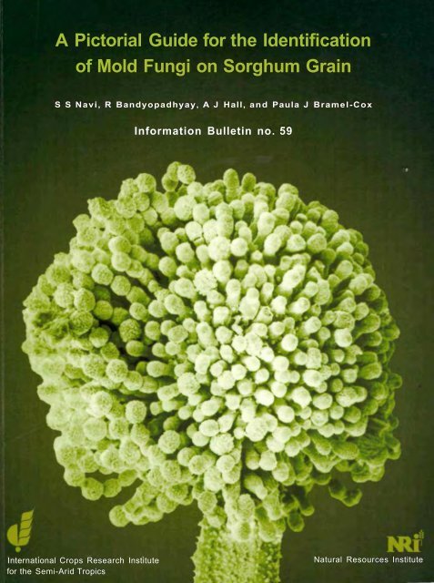
Cover Micrograph <strong>of</strong> Aspergillus flavus. (Note: The sample was critical point dried and observed under JSM35<br />
CF Scanning Electron Microscope at 10kV.)<br />
Front Spore head containing spiny conidia on rough conidiophore <strong>of</strong> 15 μm width.<br />
Back Conidiophores (15 μrn width) bearing spore heads with spiny conidia.
A <strong>Pictorial</strong> <strong>Guide</strong> <strong>for</strong> <strong>the</strong> <strong>Identification</strong><br />
<strong>of</strong> <strong>Mold</strong> Fungi on Sorghum Grain<br />
S S Navi, R Bandyopadhyay, A J Hall, and Paula J Bramel-Cox<br />
In<strong>for</strong>mation Bulletin no. 59<br />
ICRISAT<br />
International Crops Research Institute <strong>for</strong> <strong>the</strong> Semi-Arid Tropics<br />
Patancheru 502 324, Andhra Pradesh, India<br />
Natural Resources Institute<br />
Central Avenue, Chatham Maritime, Kent ME4 4TB, UK<br />
1999
ICRISAT, Patancheru, Andhra Pradesh, India<br />
Authors<br />
S S Navi, Scientific Officer (Pathology), Genetic Resources and Enhancement Program (GREP)<br />
R Bandyopadhyay, Senior Scientist (Pathology), GREP<br />
Paula J Bramel-Cox, Principal Scientist, GREP<br />
Natural Resources Institute, UK<br />
A J Hall, Principal Scientist<br />
The designations employed and <strong>the</strong> presentation <strong>of</strong> <strong>the</strong> material in this publication do not imply <strong>the</strong><br />
expression <strong>of</strong> any opinion whatsoever on <strong>the</strong> part <strong>of</strong> ICRISAT concerning <strong>the</strong> legal status <strong>of</strong> any country,<br />
territory, city, or area, or <strong>of</strong> its authorities, or concerning <strong>the</strong> delimitation <strong>of</strong> its frontiers or boundaries.<br />
Where trade names are used this does not constitute endorsement <strong>of</strong> or discrimination against any<br />
product by <strong>the</strong> Institute.<br />
Copyright® 1999 by <strong>the</strong> International Crops Research Insitute <strong>for</strong> <strong>the</strong> Semi-Arid Tropics (ICRISAT).<br />
All rights reserved. Except <strong>for</strong> quotations <strong>of</strong> short passages <strong>for</strong> <strong>the</strong> purpose <strong>of</strong> criticism and review, no<br />
part <strong>of</strong> this publication may be reproduced, stored in retrieval systems, or transmitted in any <strong>for</strong>m or by<br />
any means, electronic, mechanical, photocopying, recording, or o<strong>the</strong>rwise, without prior permission<br />
from ICRISAT. The Institute does not require payment <strong>for</strong> <strong>the</strong> noncommercial use <strong>of</strong> its published works,<br />
and hopes that this Copyright declaration will not diminish <strong>the</strong> bonafide use <strong>of</strong> its research findings in<br />
agricultural research and development.<br />
Photography<br />
Figures 1a & b: L Vidyasagar, Partnerships and In<strong>for</strong>mation Management Division<br />
Photomicrography<br />
Figures 2-19a, 20-27, 29-33, 35-70, 73-88a, and 89-95: S S Navi<br />
Figures 19b, 28, 34, 71-72, and 88b: K M Ahmed and Ravinder Reddy, GREP<br />
Cover: AK Murthy, Electron Microscope Unit, GREP<br />
Acknowledgement<br />
This publication is an output from two research projects funded by <strong>the</strong> United Kingdom Department <strong>for</strong><br />
International Development (DFID) <strong>for</strong> <strong>the</strong> benefit <strong>of</strong> developing countries. The views expressed are not<br />
necessarily those <strong>of</strong> DFID [R6767, R7506, <strong>the</strong> Crop Protection Programme, and <strong>the</strong> Crop Post-Harvest<br />
Programme].
Contents<br />
Foreword 1<br />
Introduction 2<br />
Collection <strong>of</strong> s o r g h u m samples and storage conditions 3<br />
Detection technique 4<br />
<strong>Identification</strong> and photomicrography <strong>of</strong> fungi 6<br />
S y m p t o m s and morphology 7<br />
Acladium conspersum 8<br />
Acremonium strictum 10<br />
Alternaria alternata 12<br />
Alternaria brassicicola 14<br />
Alternaria longipes 16<br />
Alternaria longissima 18<br />
Alternaria tenuissima 20<br />
Aspergillus candidus 22<br />
Aspergillus flavus 24<br />
Aspergillus niger 26<br />
Bipolaris australiensis 28<br />
Bipolaris halodes 30<br />
Bipolaris maydis 32<br />
Bipolaris sacchari 34<br />
Bipolaris spicifera 36<br />
Bipolaris zeicola 38<br />
Botrytis cinerea 40<br />
Chaetomium oryzae 42<br />
Cladosporium oxysporum 44<br />
Cladosporium sphaerospermum 46<br />
Colletotrichum graminicola 48<br />
Curvularia affinis 50<br />
Curvularia clavata 52<br />
Curvularia eragrostidis 54
Curvularia fallax 56<br />
Curvularia geniculata 58<br />
Curvularia harveyi 60<br />
Curvularia lunata 62<br />
Curvularia lunata var aeria 64<br />
Curvularia ovoidea 66<br />
Curvularia pallescens 68<br />
Curvularia trifolii 70<br />
Curvularia tuberculata 72<br />
Epicoccum nigrum 74<br />
Exserohilum rostratum 76<br />
Exserohilum turcicum 78<br />
Fusarium monili<strong>for</strong>me 80<br />
Fusarium semitectum 82<br />
Gloecercospora sorghi 84<br />
Gonatobotrys simplex 86<br />
Nigrospora oryzae 88<br />
Penicillium citrinum 90<br />
Penicillium grise<strong>of</strong>ulvum 92<br />
Periconia macrospinosa 93<br />
Phoma sorghina 96<br />
Rhizopus stolonifer 98<br />
Spadicoides obovata 100<br />
Torula graminis 102<br />
Tricho<strong>the</strong>cium roseum 104<br />
References 106<br />
Appendix 1 112<br />
Glossary 114
Foreword<br />
The International Crops Research Institute <strong>for</strong> <strong>the</strong> Semi-Arid Tropics (ICRISAT) aims<br />
to help <strong>the</strong> poor by increasing <strong>the</strong> productivity <strong>of</strong> resources committed to its mandate<br />
crops while protecting <strong>the</strong> environment, through agricultural research and in concert<br />
with national agricultural research systems.<br />
Germplasm improvement continues to be ICRISAT's main line <strong>of</strong> work, responding to<br />
a predicted increase in demand <strong>for</strong> advanced germplasm products and <strong>for</strong> source<br />
populations containing special traits. For this reason ICRISAT also serves as a world<br />
storage and trust facility <strong>for</strong> <strong>the</strong> genetic resources <strong>of</strong> sorghum, pearl millet, finger millet,<br />
pigeonpea, chickpea, and groundnut.<br />
By recognizing and reducing <strong>the</strong> enormous crop losses that occur between harvesting<br />
and final utilization a significant contribution can be made to improving <strong>the</strong> supply <strong>of</strong><br />
agricultural products above and beyond what may be achieved by increased primary<br />
production. Historically, <strong>the</strong> study <strong>of</strong> postharvest crop losses has largely been<br />
associated with protection <strong>of</strong> food stocks, particularly emergency grain supplies,<br />
during wartime and especially where more developed temperate countries have been<br />
involved.<br />
The main objective <strong>of</strong> this bulletin was to compile and collate in<strong>for</strong>mation <strong>of</strong> practical<br />
value which plant pathologists, plant quarantine experts, and seed technologists could<br />
use in handling such seed stocks both in <strong>the</strong> field and in <strong>the</strong> laboratory. This publication<br />
is <strong>the</strong> result <strong>of</strong> a fruitful cooperation between ICRISAT, India, and <strong>the</strong> Food Security<br />
Department, Natural Resources Institute (NRI), UK.<br />
The study conducted by <strong>the</strong> authors at ICRISAT was to understand <strong>the</strong> various storage<br />
conditions <strong>of</strong> sorghum grain and <strong>the</strong> potential occurrence <strong>of</strong> mold fungi under such<br />
conditions, and <strong>the</strong> importance <strong>of</strong> individual fungi including production <strong>of</strong> mycotoxins.<br />
The in<strong>for</strong>mation in this bulletin is based on observations <strong>of</strong> <strong>the</strong> sorghum grain samples<br />
collected from grain lots stored by farmers in gunny bags, polypropylene bags,<br />
mud-lined baskets, a corner <strong>of</strong> a room, and metallic containers in rural India. This<br />
bulletin is a ready reference <strong>for</strong> researchers working on sorghum grain mold.<br />
Director General<br />
International Crops Research Institute<br />
<strong>for</strong> <strong>the</strong> Semi-Arid Tropics<br />
Director<br />
Genetic Resources and<br />
Enhancement Program<br />
1
Introduction<br />
People need food, and a crop is not food until it is eaten. A program to reduce storage losses<br />
probably could result in an increase <strong>of</strong> available food in some developing countries, and might<br />
also assure that whatever increases in production occur in future would be used <strong>for</strong> <strong>the</strong><br />
nourishment <strong>of</strong> people, not <strong>for</strong> feeding pests. Overall postharvest losses <strong>of</strong> cereals, oilseeds,<br />
and pulses have been estimated at 20% <strong>of</strong> <strong>the</strong> harvested crop in Africa, Asia, and Latin<br />
America. The Food and Agriculture Organization <strong>of</strong> <strong>the</strong> United Nations (FAO) has estimated<br />
losses <strong>of</strong> <strong>the</strong>se commodities at 10% on a worldwide basis (FAO/ICRISAT 1996). In individual<br />
cases losses may be much greater and it is suggested that losses at <strong>the</strong> farm-level <strong>of</strong> 35-50%<br />
followed by 10-12% in traders' stores and fur<strong>the</strong>r 5% in centralized stores may not be<br />
uncommon (Booth and Burden 1983).<br />
There is little doubt that grain mold in its broadest sense constitutes one <strong>of</strong> <strong>the</strong> most important<br />
biotic constraints to sorghum (Sorghum bicolor (L.) Moench) improvement and production. The<br />
real and potential importance <strong>of</strong> grain mold has been emphasized <strong>for</strong> Africa, <strong>the</strong> Americas, and<br />
India (Forbes et al. 1992). Grain mold fungi have repeatedly been associated with losses in<br />
seed mass, grain density, and germination and o<strong>the</strong>r damage relating to storage quality, food<br />
and feed processing quality, and market value <strong>of</strong> <strong>the</strong> grain. More specifically, <strong>the</strong> effects <strong>of</strong><br />
fungi in quality loss in stored grains are: (1) decrease in germinability; (2) discoloration <strong>of</strong> part<br />
or all <strong>of</strong> <strong>the</strong> seed or kernel; (3) heating and mustiness; (4) various biochemical changes; and<br />
(5) production <strong>of</strong> toxins that if consumed may be injurious to humans and to domestic animals.<br />
Grain mold continues to receive much attention because <strong>of</strong> <strong>the</strong> growing concern <strong>for</strong> deleterious<br />
nature <strong>of</strong> subacute dosages <strong>of</strong> mycotoxins on animals. Mycotoxin content <strong>of</strong> grains<br />
contaminated during pre-harvest increases when <strong>the</strong> grains are stored. There are species <strong>of</strong><br />
32 dematiaceous hyphomycetes which produce mycotoxins and o<strong>the</strong>r metabolites. More<br />
species in <strong>the</strong> genera Alternaria, Bipolaris, Curvularia, Drechslera, Exserohilum, and Fusarium<br />
have been investigated <strong>for</strong> mycotoxins than those in <strong>the</strong> o<strong>the</strong>r fungal genera (Sivanesan<br />
1991). In addition, species <strong>of</strong> Aspergillus can produce aflatoxins (Pitt 1991).<br />
Seeds carry myc<strong>of</strong>lora which vary with <strong>the</strong> host species. This is especially true <strong>for</strong> <strong>the</strong> more<br />
deeply seated myc<strong>of</strong>lora, whilst on <strong>the</strong> surface many "accidental guests" may be carried as<br />
well. The seedborne myc<strong>of</strong>lora can be identified through <strong>the</strong> use <strong>of</strong> seed health tests. The tests<br />
are used <strong>for</strong> several purposes:<br />
• To assess <strong>the</strong> incidence <strong>of</strong> a seedborne pathogen that may affect seed quality.<br />
• To detect organisms <strong>of</strong> quarantine concern.<br />
• To determine seed quality in terms <strong>of</strong> germinability and or vigor.<br />
• To determine if pesticide treatment <strong>of</strong> <strong>the</strong> seed is necessary.<br />
In this study, ef<strong>for</strong>ts were made to compile in<strong>for</strong>mation on symptoms <strong>of</strong> 49 grain mold fungi, to<br />
detail <strong>the</strong>ir morphology, provide quick clues <strong>for</strong> identification, and describe <strong>the</strong>ir importance in<br />
terms <strong>of</strong> diseases, and mycotoxin and metabolite production.<br />
2
Collection <strong>of</strong> Sorghum Samples and<br />
Storage Conditions<br />
A total <strong>of</strong> 67 sorghum grain samples, representing hybrids, varieties, and local cultivars were<br />
collected in two surveys in rural areas <strong>of</strong> <strong>the</strong> states <strong>of</strong> Andhra Pradesh, Karnataka, and<br />
Maharashtra in India. The grain samples were collected from lots stored by farmers <strong>for</strong> food<br />
purpose in five types <strong>of</strong> storage conditions: gunny bags, mud-lined baskets, metallic<br />
containers, polypropylene bags, and piled in a corner <strong>of</strong> a room. During <strong>the</strong> first survey in June<br />
1997, 15 samples were collected from grain lots stored after harvest in <strong>the</strong> 1996 rainy season<br />
and 11 from <strong>the</strong> 1996/97 postrainy season harvest. During <strong>the</strong> second survey in October<br />
1997, 19 samples were obtained from 1996/97 postrainy season harvest and 22 samples<br />
from <strong>the</strong> 1997 rainy season harvest. Approximately 1 kg grain samples were collected from<br />
each lot using compartment probe (80 cm long x 2.5 cm diameter) where <strong>the</strong>re was open<br />
access to <strong>the</strong> grain bulk (mud-lined basket and loose grain piles) and where access was more<br />
difficult (stacks <strong>of</strong> gunny bags and polypropylene bags), a short probe (27 cm long x 1.5 cm<br />
diameter) was used. Farmers were paid <strong>for</strong> <strong>the</strong>ir grain at <strong>the</strong> market rate. Care was taken not<br />
to mention to farmers that a fur<strong>the</strong>r sample would be taken at a later stage. This was done to<br />
ensure that <strong>the</strong>ir subsequent behavior would not be influenced by <strong>the</strong> opportunity to sell grain.<br />
3
Detection Technique<br />
Eight hundred grains from each sample were examined to identify fungi up to <strong>the</strong> species<br />
level. Each grain sample was subjected to four treatments (200 grains treatment 1 ):<br />
1. Grains were surface sterilized in 1 % sodium hypochlorite (NaOCI) [prepared from Clorox®<br />
(Clorox Company, Oakland, CA 94612, USA) containing 5.25% NaOCI] without fungicide<br />
treatment.<br />
2. Grains were sterilized in NaOCI, and treated with benomyl (0.05%) [Benefit ® 50 WP<br />
(benomyl 50% WP), EID Parry (India)].<br />
3. Grains were sterilized in NaOCI and treated with benomyl.<br />
4. Grains were not sterilized and no benomyl treatment.<br />
The grains were transferred to pre-sterilized petri dish humid chambers @ 25 grains dish 1<br />
(Fig. 1 a, b) under aseptic conditions, and were incubated <strong>for</strong> 5 days at 28±1 °C in an incubator<br />
(Percival®) with a 12-h light cycle <strong>for</strong> observation. The fungi mentioned in this bulletin were<br />
encountered across <strong>the</strong> treatments, storage conditions, seasons, and cultivars. The effects <strong>of</strong><br />
all <strong>the</strong>se factors on mean frequency <strong>of</strong> occurrence <strong>of</strong> individual fungi are published separately.<br />
4
Figure 1a. Be<strong>for</strong>e incubation.<br />
Figure 1b. After incubation.<br />
5
<strong>Identification</strong> and Photomicrography <strong>of</strong> Fungi<br />
Each <strong>of</strong> <strong>the</strong> grains in <strong>the</strong> four treatments were examined under a stereoscopic microscope<br />
(Olympus C01) <strong>for</strong> grain colonization and a compound microscope (Olympus BH2) <strong>for</strong><br />
proper identification <strong>of</strong> fungi using <strong>the</strong> scotch-tape method (Appendix 1). This method was<br />
mainly to preserve attachment <strong>of</strong> conidia to conidiophores. It was particularly useful <strong>for</strong><br />
those fungi in which <strong>the</strong> conidia readily dislodge from conidiophores under normal<br />
procedures <strong>for</strong> slide preparation. Photomicrographs were made <strong>of</strong> <strong>the</strong> colonization <strong>of</strong><br />
grains ei<strong>the</strong>r by an individual fungus, or by a group <strong>of</strong> fungi using <strong>the</strong> stereoscopic<br />
microscope and <strong>for</strong> fungal structures using <strong>the</strong> compound microscope. The proper<br />
identification <strong>of</strong> fungi was confirmed by comparison with <strong>the</strong> details available in <strong>the</strong><br />
literature, and <strong>the</strong> knowledge acquired by <strong>the</strong> senior author in <strong>the</strong> international course on<br />
identification <strong>of</strong> fungi <strong>of</strong> agricultural and environmental significance at <strong>the</strong> International<br />
Mycological Institute, Egham, Surrey, UK in 1996. In addition, most descriptions <strong>of</strong> each<br />
fungus included in this bulletin are from Standen (1945), Nelson (1959), Whitehead and<br />
Calvert (1959), Simmons (1967), Barron (1968), Ellis (1971, 1976), Barnett and Hunter<br />
(1972), Raper and Fennel (1973), Sutton (1980), Zillinsky (1983), Sivanesan (1987), Pitt<br />
(1988), Hanlin (1990), Champ et al. (1991), and Hawksworth et al. (1995).<br />
6
Symptoms and Morphology
Acladium conspersum Link ex Pers.<br />
Symptoms on grain. Colonies are effuse, <strong>of</strong>ten very large, cottony and pale at first, later<br />
becoming velvety and fulvous or snuff-colored (Fig. 2).<br />
Morphology. Mycelium is mostly superficial. Conidiophores and hyphae have same<br />
thickness (6-9 μm), up to 350 μm long but usually shorter, and are subhyaline; cylindrical<br />
denticles are numerous especially on <strong>the</strong> upper part. Conidia are ellipsoidal, papillate at <strong>the</strong><br />
base, smooth, individually subhyaline or straw-colored, fulvous in mass, 15-20 (average 17)<br />
μm x 9-14 (average 12) μm (Fig. 3).<br />
Quick clue. Lemon-shaped conidia are present on <strong>the</strong> conidiophore.<br />
Importance. Acladium conspersum is very common on dead wood and bark <strong>of</strong> many different<br />
trees and shrubs in Canada, Europe including Great Britain, and USA. Occurrence <strong>of</strong> this<br />
fungus and also <strong>the</strong> method to kill <strong>the</strong> fungus adhering to <strong>the</strong> grains <strong>for</strong> its safe consumption<br />
has been reported on sorghum by Navi et al. (1997).<br />
Figure 2 x67<br />
8
Acladium conspersum<br />
Figure 3 x956<br />
9
Acremonium strictum W. Gams<br />
Teleomorph. Cephalosporium acremonium Corda<br />
Cephalosporium madurae Padhye, Sukapure, & Thirumalachar<br />
Symptoms on grain. Colony on grain is compact, slow-growing, white to pale and becomes<br />
slate gray or black with age (Fig. 4). Hyphae are hyaline, septate, simple or branched, and are<br />
<strong>of</strong>ten grouped toge<strong>the</strong>r <strong>for</strong>ming threads and along <strong>the</strong> sides <strong>of</strong> <strong>the</strong> threads numerous solitary<br />
conidiophores are <strong>for</strong>med, each with a globule <strong>of</strong> spores. Infected grain may show white<br />
streaks on <strong>the</strong> grain surface.<br />
Morphology. Conidiophores, arising directly and singly at right angles from <strong>the</strong> vegetative<br />
hyphae, are hyaline, short, tapered towards <strong>the</strong> tip, and measure 30-60 μm in length and<br />
1.5 μm in width at <strong>the</strong> base (Fig. 5).<br />
Quick clue. The characteristic <strong>of</strong> Acremonium is <strong>the</strong> ball <strong>of</strong> spores produced at <strong>the</strong> apex <strong>of</strong><br />
solitary, tapering conidiophores, usually borne at right angles to <strong>the</strong> hyphae.<br />
(Note: The genus can be readily confused with o<strong>the</strong>r genera such as Gliomastix, Verticillium,<br />
and microconidial Fusarium or Cylindrocarpon. Never<strong>the</strong>less, it is perhaps one <strong>of</strong> <strong>the</strong> easiest<br />
fungi to identify at <strong>the</strong> genus level and one <strong>of</strong> <strong>the</strong> most difficult in which to make species<br />
determinations.)<br />
Importance. Acremonium strictum is distributed worldwide, but is more frequent in <strong>the</strong> tropics.<br />
It causes acremonium wilt <strong>of</strong> sorghum (Bandyopadhyay et al. 1987) and black bundle disease<br />
<strong>of</strong> maize (Zea mays L). The latter is a late season disease which is common in USA<br />
and o<strong>the</strong>r countries.<br />
10
Acremonium strictum<br />
Figure 4 x12<br />
Figure 5 x 5085<br />
11
Alternaria alternata (Fr.) Keissler<br />
Symptoms on grain. The fungus produces woolly or powdery chains <strong>of</strong> dark brown conidia<br />
<strong>of</strong> variable lengths and shapes. The color <strong>of</strong> <strong>the</strong> colony is usually extremely variable between<br />
olive green to dark brown (Fig. 6a, b).<br />
Morphology. The mycelium may be ei<strong>the</strong>r sparse or abundant and variable in color, usually<br />
light olive green to brown. Hyphae are dark brown, thick, septate, and branched.<br />
Conidiophores are simple, erect, 40-50 μm long, 2-6 μm thick, and <strong>of</strong>ten clustered.<br />
Conidiophores produce dark pigmented conidia in an acropetal succession <strong>of</strong> simple or<br />
branched chains. These chains normally branch at <strong>the</strong> beak <strong>of</strong> a spore, or sometimes from<br />
<strong>the</strong> short lateral projection <strong>of</strong> <strong>the</strong> beak. Conidia have transverse and oblique septa, measure<br />
10-18 x 20-65 μm, and are ovoid to obovoid, obclavate, obpyri<strong>for</strong>m, ellipsoidal, uni<strong>for</strong>m,<br />
with an elongated terminal cell (Fig. 7). Conidia <strong>of</strong>ten have a short conical or cylindrical beak<br />
which is about one third <strong>the</strong> length <strong>of</strong> <strong>the</strong> conidium, and measure 2-5 x 10-20 μm. Surface<br />
walls are ei<strong>the</strong>r smooth or verrucose and pale to mid-golden brown.<br />
Quick clue. Chains <strong>of</strong> conidia are produced at <strong>the</strong> beak <strong>of</strong> a spore, or sometimes from <strong>the</strong><br />
short lateral projection <strong>of</strong> <strong>the</strong> beak.<br />
Importance. The fungus is distributed worldwide and is usually seedborne. It causes leaf<br />
spot on several hosts and blight <strong>of</strong> pigeonpea (Cajanus cajan (L.) Millsp.), chickpea (Cicer<br />
arietinum L), and groundnut (Arachis hypogaea L ) . Several metabolites and toxins have<br />
been isolated from A. alternata: tentoxin (Templeton 1972), AF-toxins I and II (Maekawa et al.<br />
1984), alkaloids (Rizk et al. 1985), alternariol (Logrieco et al. 1990), and mannitol<br />
(Combe et al. 1970).<br />
12
Alternaria alternata<br />
Figure 6a x17 Figure 6b x42<br />
Figure 7 X 1 8<br />
13
Alternaria brassicicola (Schwein.) Wiltshire<br />
Helminthosporium brassicicola Schweinitz<br />
Macrosporium cheiranthi Fr. var circinans Berk. & Curt.<br />
Alternaria circinans (Berk. & Curt.) Bolle.<br />
Alternaria oleracea Milbraith.<br />
Symptoms on grain. Colonies are amphigenous, effuse, dark olivaceous brown to dark<br />
blackish brown, and velvety. Dark brown to almost black, circular (1-10 mm diameter), zonate<br />
spots are <strong>for</strong>med (Fig. 8).<br />
Morphology. The mycelium is immersed; hyphae are branched, septate, hyaline at first, later<br />
turn brown or olivaceous brown, inter- and intracellular, smooth, and 1.5-7.5 μm thick. The<br />
conidiophores arise singly or in groups <strong>of</strong> 2-12 or more, and emerge through <strong>the</strong> stomata. They<br />
are usually simple, erect or ascending, straight or curved, occasionally geniculate, more or less<br />
cylindrical but <strong>of</strong>ten slightly swollen at <strong>the</strong> base, septate, pale to mid-olivaceous brown, smooth,<br />
70 μm long, and 5-8 μm thick. The conidia are usually produced in chains <strong>of</strong> 20 or more,<br />
sometimes branched, acropleurogenous, and arise through small pores in <strong>the</strong> conidiophore wall.<br />
They are straight, nearly cylindrical, usually tapering, slightly towards <strong>the</strong> apex or obclavate, with<br />
<strong>the</strong> basal cell rounded, <strong>the</strong> beak usually almost non-existent, <strong>the</strong> apical cell being more or less<br />
rectangular or resembling a truncated cone, occasionally better developed but <strong>the</strong>n always short<br />
and thick, with 1-11, mostly less than 6 transverse septa and usually few but up to 6 longitudinal<br />
septa, <strong>of</strong>ten slightly constricted at <strong>the</strong> septa, pale to dark olivaceous brown, smooth or becoming<br />
slightly warted with age, 18-130 μm long, 8-20 μm thick in <strong>the</strong> broadest part, with <strong>the</strong> beak 1/6<br />
<strong>the</strong> length <strong>of</strong> <strong>the</strong> conidium and 6-8 μm thick (Fig. 9).<br />
Quick clue. Conidia are nearly cylindrical, usually tapering, <strong>the</strong> beak usually almost nonexistent.<br />
Importance. "Brassicicolon A" metabolite was isolated from Alternaria brassicicola (Ciegler and<br />
Lindenfelser 1969). The fungus causes leaf spot <strong>of</strong> crucifers.<br />
14
Figure 8<br />
Alternaria brassicicola<br />
Figure 9 x 1749<br />
x17<br />
15
Alternaria longipes (Ellis & Everh.) Mason<br />
Figure 10 x56<br />
16<br />
Macrosporium longipes Ellis & Everh.<br />
Symptoms on grain. Colonies are amphigenous. The spots which appear first are orbicular,<br />
brown, and frequently zonate (Fig. 10). The entire grain eventually becomes brown and <strong>the</strong><br />
spots <strong>the</strong>n appear a shade paler than <strong>the</strong> surrounding areas (Fig. 10).<br />
Morphology. Conidiophores arise singly or in groups, erect or ascending, simple or loosely<br />
branched, straight or flexuous, cylindrical, septate, ra<strong>the</strong>r pale olivaceous brown, 80 μm long,<br />
3-5 μm thick, with 1 or several conidial scars. Conidia are sometimes solitary but usually in<br />
chains, obclavate, rostrate, pale to mid-pale brown, smooth or verruculose, overall length<br />
35-110 (average 69) μm, body <strong>of</strong> conidium 11-21 (average 14) μm thick in <strong>the</strong> broadest part,<br />
tapering gradually into <strong>the</strong> pale brown beak which is usually 1/3 to 1/2 <strong>the</strong> total length, 2-5 μm<br />
thick and <strong>of</strong>ten slightly swollen at <strong>the</strong> tip; <strong>the</strong>re are 3-7, usually 5-6 transverse septa and 1 to<br />
several longitudinal or oblique septa (Fig. 11).<br />
Quick clue. Refer Figure 11.<br />
Importance. On tobacco (Nicotiana tabacum L), A. longipes causes brown spot. But this is<br />
<strong>the</strong> first report <strong>of</strong> its occurrence on sorghum in India.
Alternaria longipes<br />
Figure 11 x686<br />
17
Alternaria longissima Deighton & MacGarvie<br />
Symptoms on grain. Colony on grain is brown to blackish brown (Fig. 12).<br />
Morphology. Mycelium is partly superficial and partly immersed. Conidiophores are erect or<br />
ascending, simple or occasionally branched, straight or slightly flexuous, sometimes geniculate,<br />
somewhat swollen at <strong>the</strong> apex, septate, pale to mid-pale brown, smooth below, verruculose at<br />
and sometimes below <strong>the</strong> apex, 150 μm long, 3-5 μm thick, with one to several conidial scars.<br />
Conidia are solitary or catenulate, extremely variable in shape and size, pale straw colored to<br />
brown. They are usually very long (up to 500 μm), Cercospora-like, obclavate or with a basal<br />
sub-cylindric portion <strong>of</strong> few to several cells and a very long, narrow septate beak (Fig. 13). They<br />
have 5-40 transverse septa. Conidia are 4-17 μm thick in <strong>the</strong> broadest part and about 2.5 μm<br />
thick at <strong>the</strong> apex. Shorter conidia, variable in shape and <strong>of</strong>ten with a few longitudinal or oblique<br />
septa, are also <strong>for</strong>med. Conidia are thin-walled, smooth except around <strong>the</strong> base where <strong>the</strong>y are<br />
<strong>of</strong>ten verruculose. Dark brown, multicellular, muri<strong>for</strong>m chlamydospores 16-42 x 16-34 μm<br />
sometimes occur, both on natural substrata and in culture.<br />
Quick clue. Very long, Cercospora-like conidium is a distinct feature <strong>of</strong> A. longissima.<br />
Importance. The fungus was previously reported on sorghum along with method(s) to kill <strong>the</strong><br />
fungus adhering to <strong>the</strong> grains <strong>for</strong> safe use <strong>of</strong> grains <strong>for</strong> consumption (Navi et al. 1997).<br />
Metabolites isolated from A. longissima include tenuazonic acid, cellulase, and<br />
polygalacturonase (von Ramm and Lucas 1963; Mikami et al. 1971).<br />
Figure 12<br />
18<br />
x13
Alternaria longissima<br />
Figure 13 x1102<br />
19
Alternaria tenuissima (Kunze ex Pers.) Wiltshire<br />
Figure 14 x51<br />
20<br />
Helminthosporium tenuissimum Kunze in C.G. & T.F.L. Nees<br />
Macrosporium tenuissimum Fr.<br />
Symptoms on grain. Golden brown to black growth on <strong>the</strong> seed surface (Fig. 14).<br />
Morphology. Conidiophores are solitary or in groups, simple or branched, straight or<br />
flexuous, more or less cylindrical, septate, pale or mid-pale brown, smooth, with one or several<br />
conidial scars, up to 115 μrn long, and 4 μm thick. Conidia are solitary or in short chains,<br />
straight or curved, obclavate or ellipsoidal tapering gradually to <strong>the</strong> beak which is up to half <strong>the</strong><br />
length <strong>of</strong> <strong>the</strong> conidium, usually shorter, sometimes tapered to a point but more frequently<br />
swollen at <strong>the</strong> apex where <strong>the</strong>re may be several scars; pale to clear mid-golden brown, usually<br />
smooth, sometimes minutely verruculose generally with 4-7 transverse and several<br />
longitudinal or oblique septa, and slightly or not constricted at <strong>the</strong> septa; overall length 22-95<br />
(average 54) μm, 8-19 (average 13.8) μm thick in <strong>the</strong> broadest part, beak 2-4 μm thick, and<br />
swollen apex 4-5 μm wide (Fig. 15).<br />
Quick clue. Refer Figure 15.<br />
Importance. Alternaria tenuissima is extremely common and recorded on a wide range <strong>of</strong><br />
plant species, usually as a secondary invader ra<strong>the</strong>r than a primary parasite. It produces<br />
tenuazonic acid (Davies et al. 1977). It has been reported to cause leaf spot <strong>of</strong> pigeonpea. It<br />
produces <strong>the</strong> same toxins as A. alternata.
Figure 15<br />
Alternaria tenuissima<br />
x2046<br />
21
Aspergillus candidus Link<br />
Symptoms on grain. Conidial heads are persistently white or become yellowish cream with<br />
age (Fig. 16a); typically globose when young, <strong>of</strong>ten splitting with age, or approaching columnar<br />
in small heads (Fig. 16b).<br />
Morphology. Conidiophores are smooth, colorless or slightly yellow in terminal areas.<br />
Vesicles are typically globose to subglobose and fertile over <strong>the</strong> entire surface. Sterigmata<br />
typically in two series, with primary series <strong>of</strong>ten much enlarged, sometimes varying greatly in<br />
size within <strong>the</strong> same head (Fig. 17). Conidia are globose or subglobose and smooth.<br />
Quick clue. Absence <strong>of</strong> pigmentation and smooth conidia. White conidial heads are present.<br />
Importance. Aspergillus candidus is widely distributed in nature. It is encountered most<br />
commonly on stored cereal grains and on grain products. It has been revealed frequently in<br />
necropsies <strong>of</strong> birds and mammals at <strong>the</strong> Paris Zoological Gardens. It is a <strong>the</strong>rmo-tolerant<br />
fungus, capable <strong>of</strong> growing at 40-50°C, and is xerophilic (Raper and Fennel 1973).<br />
Figure 16a x36 Figure 16b x436<br />
22
Aspergillus candidus<br />
Figure 17 x 1980<br />
23
Aspergillus flavus Link<br />
Symptoms on grain. Colony on seed is usually spreading and very light yellow-green, deep<br />
yellow-green, olive brown, or brown (Fig. 18a). Conidiophores are swollen apically and bear<br />
numerous conidia-bearing cells (phialides) with conidia in long, dry chains. Conidial heads are<br />
typically spherical, splitting into several poorly defined columns, rarely exceeding 500-600 μm<br />
diameter, but mostly 300-400 μm (Fig. 18b).<br />
(Note: Severely infected sorghum grains are discolored and shrivelled.)<br />
Morphology. Conidiophores are heavy walled, hyaline, coarsely roughened, and usually<br />
Aspergillus flavus<br />
Figure 18a x11 Figure 18b x37<br />
Figure 19a x502 Figure 19b X1130<br />
25
Aspergillus niger van Tieghem<br />
Symptoms on grain. Colony on seed grows slowly, consisting <strong>of</strong> a compact to fairly loose<br />
white to faintly yellow basal mycelium, which bears abundant erect and usually crowded<br />
conidial structures, typically carbon black but sometimes deep brown-black, covering <strong>the</strong> entire<br />
colony except <strong>for</strong> a narrow growing margin (Fig. 20). Conidial heads are typically large and<br />
black, compact at first, spherical, or split into two or more loose to reasonably well-defined<br />
columns, and commonly reach 700-800 μm in diameter.<br />
(Note: Severely infected sorghum grains are discolored and shrivelled.)<br />
Morphology. Conidiophores are smooth, hyaline or faintly brownish near <strong>the</strong> apex and up to 3<br />
μm in length and 15-20 μrn in diameter. Apices are spherical or nearly so, up to 75 μm in<br />
diameter but <strong>of</strong>ten quite small. Two series <strong>of</strong> conidia-bearing cells (supporting cells and<br />
phialides) are produced, but in some heads only phialides are present. Supporting cells are <strong>of</strong><br />
varying lengths and sometimes septate, but when mature usually 20-30 μrn long. Phialides<br />
are more uni<strong>for</strong>m in length, usually 7-10 x 2-3 μm. Conidia are typically spherical at maturity,<br />
<strong>of</strong>ten very rough or spiny, mostly 4—5 μm diameter, and very dark in color or with conspicuous<br />
longitudinal striations (Fig. 21).<br />
Quick clue. Aspergillus niger is recognized by <strong>the</strong> production <strong>of</strong> compact, greenish black,<br />
brownish black, purplish black, or carbon black, spherical or columnar spore heads.<br />
Importance. Seed infection can reduce germination. Production <strong>of</strong> large numbers <strong>of</strong> airdisseminated<br />
spores can cause respiratory diseases in man and animals. Aspergillus niger is<br />
worldwide in distribution and occurs in and upon <strong>the</strong> greatest variety <strong>of</strong> substrata including<br />
grains, <strong>for</strong>age products, spoiled fruits and vegetables, exposed cotton textiles and fabrics,<br />
lea<strong>the</strong>r, dairy products, and o<strong>the</strong>r protein-rich substrata (Raper and Fnnel 1973).<br />
26
Aspergillus niger<br />
Figure 20 x14<br />
Figure 21 x1617<br />
27
Bipolaris australiensis (M.B. Ellis) Tsuda & Ueyama<br />
(Bipolaris species "with" Cochliobolus teleomorph)<br />
Drechslera australiensis M.B. Ellis<br />
Helminthosporium australiense Bugnicourt<br />
Teleomorph. Cochliobolus australiensis (Tsuda & Ueyama) Alcorn<br />
Symptoms on grain. Conidial colonies are effuse, gray to blackish brown, and velvety.<br />
Hyphae are pale to dark brown, smooth, and septate. Stromata are erect, straight, cylindrical,<br />
and black (Fig. 22).<br />
Morphology. Conidiophores are single, flexuous, geniculate, septate, smooth, cylindrical,<br />
reddish brown, up to 150 μm long and 3-7 μm thick, having verruculose, conidiogenous nodes.<br />
Conidia are straight, ellipsoidal or oblong, rounded at <strong>the</strong> ends, pale brown to mid-reddish<br />
brown, usually 3-, rarely 4-5 distoseptate, 14—40 x 6-11 μm (Fig. 23).<br />
The species is heterothallic and <strong>the</strong> teleomorph is obtained by pairing opposite compatible<br />
monoconidial isolates in Sach's agar media with sterilized rice straw. Ascomata on rice straw<br />
are globose to subglobose, black, superficial on columnar to flat stromata, 375-940 μm in<br />
diameter with a long cylindrical ostiolar beak 250-1250 x 90-125 μm. Pseudoparaphyses are<br />
filamentous, hyaline, septate, and branched. Asci are cylindrical to long, 100-182 x<br />
8.5-15 μm clavate, vestigial bitunicate, short pedicellate, with 1-8 spores. Ascospores are<br />
parallel to partly or closely coiled in a helix in <strong>the</strong> ascus, fili<strong>for</strong>m, somewhat tapering towards <strong>the</strong><br />
ends, flagelli<strong>for</strong>m at <strong>the</strong> ends, hyaline to very pale brown, 3-13 septate, 81-206 x<br />
2.5-5.6 μm.<br />
Quick clue. Verruculose conidiogenous nodes are present.<br />
Importance. Production <strong>of</strong> mycotoxin by <strong>the</strong> fungus is unknown. Cochliobolus australiensis<br />
causes leaf spot <strong>of</strong> pearl millet (Pennisetum glaucum (L.) R. Br.) (Chand and Singh 1966) and<br />
leaf blight <strong>of</strong> citronella grass (Cymbopogan winterianus Jowitt.) (Ramaiah and Chandrashekar<br />
1981) in India.<br />
28
Bipolaris australiensis<br />
Figure 22 x49<br />
Figure 23 x1452<br />
29
Bipolaris halodes (Drechsler) Shoem.<br />
(Bipolaris species "without" Cochliobolus teleomorph)<br />
Drechslera halodes (Drechsler) Subram. & Jain<br />
Bipolaris rostrata (Drechsler) Shoem.<br />
Drechslera rostrata (Drechsler) Richardson & Fraser<br />
Exserohilum halodes (Drechsler) Leonard & Suggs<br />
Exserohilum rostratum (Drechsler) Leonard & Suggs Imp.<br />
Helminthosporium appatternae K.S. Deshpande & K.S. Deshpande<br />
Helminthosporium halodes Drechsler<br />
Helminthosporium rostratum Drechsler<br />
Helminthosporium halodes Drechsler var tritici Mitra<br />
Helminthosporium halodes Drechsler var elaeidicola Kovachich.<br />
Luttrellia rostrata (Drechsler) Gonorstai<br />
Symptoms on grain. Stromata are <strong>for</strong>med on seeds and are erect, simple or branched,<br />
cylindrical, dark, blackish brown to start, up to 2 x 1 μm (Fig. 24).<br />
Morphology. Conidiophores are up to 200 μm long, 5-8 μm thick, septate, cylindrical,<br />
olivaceous brown, paler towards <strong>the</strong> apex, simple, and geniculate. Conidia are straight to<br />
slightly curved, ellipsoidal to narrowly obclavate or rostrate, brown or olivaceous, thick-walled,<br />
except in a small subhyaline region at <strong>the</strong> apex and a similar region surrounding <strong>the</strong> hilum<br />
which protrudes as a darkened cylinder or truncate cone from <strong>the</strong> end <strong>of</strong> <strong>the</strong> basal cell, basal<br />
septum darker and thicker than <strong>the</strong> o<strong>the</strong>r septa, up to 18-distoseptate, 15-200 x 7-29 μm<br />
(Fig. 25). Germination occurs from <strong>the</strong> subhyaline region <strong>of</strong> <strong>the</strong> end cells and germ tubes<br />
grow semiaxially.<br />
(Note: Teleomorph is absent.)<br />
Quick clue. A small subhyaline region is present at <strong>the</strong> apex <strong>of</strong> <strong>the</strong> conidium.<br />
Importance. It is a seedborne fungus and is widely distributed. Mycotoxin production by this<br />
fungus is unknown. It commonly occurs on grasses, and many o<strong>the</strong>r plant species, soil, and<br />
textiles (Sivanesan 1987).<br />
30
Bipolaris halodes<br />
Figure 24 x26<br />
Figure 25 x1320<br />
31
Bipolaris maydis (Nisikado & Miyake) Shoem.<br />
(Bipolaris species "with" Cochliobolus teleomorph)<br />
Helminthosporium maydis Nisikado & Miyake<br />
Drechslera maydis (Nisikado & Miyakae) Subram. & Jain<br />
Teleomorph. Cochliobolus heterostrophus (Drechsler) Drechsler<br />
Symptoms on grain. Colony on seed is pale to mid-dark golden brown with some white aerial<br />
mycelium, and moderate in density (Fig. 26). A black matted mold may cover <strong>the</strong> affected grain<br />
and can reduce germination.<br />
Morphology. Conidiophores are mid- to dark brown, medium to long, commonly long,<br />
slender, straight or curved, single or in groups <strong>of</strong> 2 or 3, pale near <strong>the</strong> apex, smooth, up to 700<br />
μm long, and 5-10 μm thick, and bear conidia at wide intervals. Conidia are distinctly curved,<br />
broad in <strong>the</strong> middle, sharply tapering towards rounded ends, pale to mid-dark golden brown,<br />
smooth, 5-11 septate, mostly 70-160 μm long, 15-20 μm thick in <strong>the</strong> broadest part; and point<br />
<strong>of</strong> attachment is dark, <strong>of</strong>ten flat, and 3-5 μm wide (Fig. 27).<br />
Pseudo<strong>the</strong>cia contain asci with four slender, thread-like, 5-9 septate ascospores (6-7 x<br />
130-340 μm) arranged in parallel coils. Pseudo<strong>the</strong>cia rarely occur under natural conditions.<br />
Quick clue. Conidia are light brown, slender, typically curved, and tapering sharply towards<br />
both ends. The curvature is more pronounced than in any o<strong>the</strong>r related species. Conidiophores<br />
are usually long, slender, alternately bent, and bearing conidia at wide intervals.<br />
Importance. Bipolaris maydis is distributed worldwide but predominantly in <strong>the</strong> tropics and<br />
subtropics. There are quarantine restrictions in many countries including Malaysia. Maize<br />
germplasm with male sterile T cytoplasm also has quarantine restrictions. Bipolaris maydis<br />
produces four host-specific toxins <strong>of</strong> race T and C. heterostrophus produces ophiobolin B,<br />
ophiobolin C, ophiobolin F, anhydroophiobolin A, 6-epiophiobolin A, and geranylnerolidol<br />
(Ishibashi 1962; Nozoe et al. 1965, 1966; Canonica et al. 1966; Tsuda et al. 1967; Cordell<br />
1974; Karr et al. 1974, 1975; Payne and Yoder 1978; Sugawera et al. 1987).<br />
32
Figure 26<br />
Bipolaris maydis<br />
Figure 27 x568<br />
x22<br />
33
Bipolaris sacchari (E. Butler) Shoem.<br />
(Bipolaris species "without" Cochliobolus teleomorph)<br />
Helminthosporium sacchari E. Butler<br />
Drechslera sacchari (E. Butler) Subram. & Jain<br />
Symptoms on grain. Stromata are <strong>for</strong>med on seeds and are erect, simple or branched,<br />
cylindrical, dark, blackish brown to start, up to 2 x 1 mm (Fig. 28).<br />
Morphology. Conidiophores are single or in small groups, <strong>of</strong>ten from groups <strong>of</strong> dark cells<br />
which <strong>for</strong>m a loose stroma, straight to flexuous, mid- to dark brown or olivaceous brown, paler<br />
towards <strong>the</strong> apex, septate, smooth, cylindrical, up to 200 μm long, 5-8 μm thick; in culture up to<br />
700 μm long and 10 μm thick. Conidiogenous nodes are smooth to slightly verruculose.<br />
Conidia are slightly curved, rarely straight, cylindrical or narrowly ellipsoidal, mid-pale to midyellow<br />
golden brown, 5-9 (commonly 8) distoseptate, 35-96 x 9-17 μm, hilum 2-3 μm wide<br />
(Fig. 29).<br />
(Note: Teleomorph is absent.)<br />
Quick clue. Groups <strong>of</strong> dark cells and slightly curved distoseptate conidia are <strong>for</strong>med.<br />
Importance. Bipolaris sacchari produces helminthosporoside (Beier et al. 1982) and three<br />
isomeric host-specific toxins (Macko et al. 1983). It causes eye spot and seedling blight <strong>of</strong><br />
sugarcane (Saccharum <strong>of</strong>ficinarum L.) and leaf spots <strong>of</strong> grasses.<br />
Fiqure 28 x521<br />
34
Bipolaris sacchari<br />
Figure 29 x1980<br />
35
Bipolaris spicifera (Bainier) Subram.<br />
(Bipolaris species "with" Cochliobolus teleomorph)<br />
Helminthosporium spiciferum (Bainier) Nicot<br />
Helminthosporium tetramera McKinney<br />
Curvularia spicifera (Bainier) Boedijn<br />
Teleomorph. Cochliobolus spicifer Nelson<br />
Symptoms on grain. Colony on seed is brown, gray or black, hairy, cottony or cushion-like<br />
and spreads loosely with abundant brownish conidiophores, single or in clusters <strong>of</strong> 2-3<br />
(Fig. 30). Many small conidia are produced at very short intervals, giving rise to a bottle-brush<br />
appearance. Colonies strongly resemble those <strong>of</strong> Curvularia spp.<br />
Morphology. Conidiophores are brown and curved, with obvious and numerous scars<br />
resulting in an irregular zigzag appearance. Conidia are short, typically 3-septate, light to dark<br />
brown, oval, curved to straight with rounded ends, and measure 20-40 μm x 9-14 μm. Conidia<br />
are lighter in color towards <strong>the</strong> terminal cells.<br />
Ascomata are black, spherical to oval, curved, 460-710 x 350-650 μm, with an inverted cone-<br />
shaped neck and pore. Asci are cylindrical to club-shaped, straight to slightly curved, with 1-8<br />
spores and 130-160 x 12-20 μm. Ascospores are parallel to closely coiled in <strong>the</strong> ascus, thread-<br />
like, somewhat tapered at <strong>the</strong> ends, 6-16 septate, hyaline, and 135-240 x 3-7 μm<br />
(Fig. 31).<br />
Quick clue. Under <strong>the</strong> dissecting microscope, conidia appear to be clustered <strong>for</strong> some length<br />
on <strong>the</strong> conidiophores, giving <strong>the</strong> appearance <strong>of</strong> a bottle-brush. Conidia are very small and<br />
typically 3-septate, almost cylindrical, more or less uni<strong>for</strong>m in size, and <strong>the</strong> end cells have<br />
subhyaline areas towards <strong>the</strong>ir terminal ends.<br />
Importance. Bipolaris spicifera is distributed worldwide and is very common in tropical and<br />
subtropical areas. The mycotoxins isolated from B. spicifera are spiciferone A and cynodontin<br />
metabolites and those from C. spicifera are curvularin and D-mannitol (Combe et al. 1968;<br />
Nakajima et al. 1989). The main diseases caused by B. spicifera are foot rot (or common root<br />
rot) <strong>of</strong> winter wheat (Triticum aestivum L.) and mycotic keratitis in humans. A subcutaneous<br />
mycosis in cat and horses is also induced by C. spicifer.<br />
36
Figure 30<br />
Bipolaris spicifera<br />
Figure 31 x1353<br />
x10<br />
37
Bipolaris zeicola (Stout) Shoem.<br />
(Bipolaris species "with" Cochliobolus teleomorph)<br />
Helminthosporium carbonum Ullstrup<br />
Helminthosporium zeicola Stout<br />
Drechslera carbonum (Ullstrup) Sivan<br />
Drechslera zeicola (Stout) Subram. & Jain<br />
Teleomorph. Cochliobolus carbonum Nelson<br />
Symptoms on grain. Grains are covered by very dark brown to black mycelium which gives a<br />
characteristic charcoal appearance. Conidia are also visible (Fig. 32).<br />
Morphology. Conidiophores are single or in small groups, straight to flexuous, mid- to dark<br />
brown or olivaceous brown, up to 250 μm long, 5-8 μm thick, smooth, septate, and cylindrical.<br />
Conidiogenous nodes are verruculose with <strong>the</strong> surface wall below <strong>the</strong>m granulose. Conidia are<br />
curved or sometimes straight, occasionally almost cylindrical but usually broad in <strong>the</strong> middle<br />
and tapering towards <strong>the</strong> rounded ends, 6-12 (commonly 7-8) distoseptate, 30-100 x 12-18<br />
μm, <strong>of</strong>ten finally becoming dark or very dark brown or olivaceous brown, with <strong>the</strong> end cells<br />
sometimes remaining tapered than <strong>the</strong> middle cells (Fig. 33). The surface is <strong>of</strong>ten granulose<br />
and hilum is not very conspicuous.<br />
The species is heterothallic and <strong>the</strong> teleomorph is obtained by pairing opposite mating single<br />
conidial isolates in Sach's agar media holding sterilized maize leaf segments or barley<br />
(Hordeum vulgare L.) grains at 24°C (Nelson 1959). Ascomata are black, globose to<br />
ellipsoidal, 355-550 x 320-420 μm, with setae over <strong>the</strong> upper half <strong>of</strong> <strong>the</strong> wall mixed with<br />
conidiophores, and with a well-defined sub-conical to paraboloid ostiolar beak 60-200 μm<br />
long. Pseudoparaphyses are fili<strong>for</strong>m, hyaline, septate, and branched. Asci are cylindrical to<br />
clavate, short-stalked, straight to slightly curved, 1-8 spored, vestigial bitunicate, 160-257 x<br />
18.0-27.5 (am. Ascospores are fili<strong>for</strong>m or flagelli<strong>for</strong>m, somewhat tapering towards <strong>the</strong> ends,<br />
hyaline, 5-9 septate, 180-307 x 6-10 μm, <strong>of</strong>ten surrounded by a thin hyaline mucilaginous<br />
sheath.<br />
Quick clue. Distoseptate dark to dark brown conidia are present.<br />
Importance. Bipolaris zeicola is distributed worldwide. There are quarantine restrictions <strong>for</strong><br />
Indonesia, Egypt, and Chile. Bipolaris zeicola produces HC-toxins I, II, III, IV, and CHS<br />
polypeptide (Ramussen and Scheffer 1988), and C carbonum produces carbtoxinine and<br />
victoxinine (Nishimura et al. 1966; Pringle and Scheffer 1967). Cochliobolus carbonum is<br />
reported on maize from many countries including India. This is <strong>the</strong> first report on sorghum in<br />
India.<br />
38<br />
r
Figure 33<br />
Bipolaris zeicola<br />
Figure 32 x53<br />
x1320<br />
39
Botrytis cinerea Pers. ex Pers.<br />
Teleomorph. Botryotinia fuckeliana (de Bary) Whetzel<br />
Symptoms on grain. Colony on seed is white or gray or grayish-brown, and spreading <strong>for</strong> a<br />
short distance around <strong>the</strong> affected seed (Fig. 34).<br />
Morphology. Conidiophores are brown, tall, upright or nearly so, septate and branched, up to<br />
30 μm wide and 2 μm long. The branches are constricted at <strong>the</strong>ir point <strong>of</strong> origin and quickly<br />
collapse when removed from a moist atmosphere. Conidia occur in clusters at <strong>the</strong> swollen<br />
rounded apices and at intervals along with conidiophores on short blunt teeth. Conidia are oval<br />
or egg-shaped, <strong>of</strong>ten with a slightly projecting point <strong>of</strong> attachment, colorless to pale brown, and<br />
measure 6-18 x 4 - 1 μm (Fig. 35).<br />
Fairly large, black, irregular sclerotia can be produced, but not normally within <strong>the</strong> period <strong>of</strong> a<br />
seed health test. They are ra<strong>the</strong>r flat in appearance and measure 5 x 2 x 2 μm.<br />
Quick clue. The funugs is characterized by stout, brown, branched conidiophores supporting<br />
glistening gray heads <strong>of</strong> pale conidia, which can be observed under low magnification <strong>of</strong> a<br />
binocular microscope.<br />
Importance. The fungus is a common gray mold, frequently parasitic, and produces abscisic<br />
acid, botrydial, botrylacton, citric acid, and <strong>the</strong>rmostable toxins (Fehlhaber et al. 1974; Kamoen<br />
and Jamart 1974; Lyon 1977; Welmer et al. 1979; Morooko et al. 1986). However, it is not<br />
noted as a toxigenic species.<br />
Figure 34 x131<br />
40
Botrytis cinerea<br />
Figure 35 x858<br />
41
Chaetomium oryzae<br />
Symptoms on grain. Colony on seed is white with <strong>the</strong> density <strong>of</strong> mycelium varying from light<br />
to dense. The peri<strong>the</strong>cia are found on <strong>the</strong> seed surface beneath <strong>the</strong> aerial white mycelium (Fig.<br />
36).<br />
Morphology. Peri<strong>the</strong>cia are spherical or elongate, with a pore opening, and a dark,<br />
membranous, cellular wall which is covered with conspicuous hairs <strong>of</strong> various types (Fig. 37).<br />
Asci are hyaline, usually club-shaped but in a few cases cylindrical, and contain eight<br />
ascospores. Ascospores are one-celled and in most cases lemon-shaped. They are extruded<br />
through <strong>the</strong> pore opening ei<strong>the</strong>r as a mass amongst <strong>the</strong> hairs or as a column depending on<br />
conditions.<br />
Quick clue. Colonies <strong>of</strong> Chaetomium species can be readily recognized by <strong>the</strong> presence <strong>of</strong><br />
peri<strong>the</strong>cia with many stiff dark terminal hairs with ornamentation.<br />
Importance. Chaetomium is distributed worldwide. It has no significance in crop production.<br />
However, it is a common saprophyte and secondary invader. Seeds <strong>of</strong> low germinating capacity<br />
are sometimes found to be heavily contaminated with Chaetomium (Skolko and Groves 1953).<br />
Figure 36<br />
42<br />
x23
Chaetomium oryzae<br />
Figure 37 x396<br />
43
Cladosporium oxysporum Berk. & Curt.<br />
Symptoms on grain. Colonies are effuse, pale gray or grayish brown, thinly hairy on natural<br />
substrata (Fig. 38); cottony or loosely felted in culture.<br />
Morphology. Conidiophores are macronematous, straight or slightly flexuous, distinctly<br />
nodose, pale or mid-pale brown, smooth, up to 500 μm long or sometimes even longer in<br />
culture, 3-5 μm thick, with terminal and intercalary swellings <strong>of</strong> 6-8 μm diameter. Conidia<br />
arise from terminal swellings, which later become intercalary, in simple or branched chains.<br />
Conidia are cylindrical, rounded at <strong>the</strong> ends, ellipsoidal, limoni<strong>for</strong>m or subspherical, subhyaline<br />
or pale olivaceous brown, smooth, 5-30 x 3-6 μm (Fig. 39).<br />
Quick clue. Cladosporium is characterized by erect, pigmented conidiophores with chains <strong>of</strong><br />
conidia in tree-like heads. This genus can frequently be identified by <strong>the</strong> distinctive lemon-<br />
shaped conidia, which have well marked, dark attachment scars and show considerable<br />
variation in size and septation within and between species.<br />
Importance. Heavily infected sorghum grains may have dark green to black blotches, or<br />
streaks that extend from <strong>the</strong> grain tips. The fungus is common, widely distributed in <strong>the</strong> tropics<br />
on dead leaves and stems <strong>of</strong> herbaceous and woody plants. Many saprophytic species are<br />
commonly encountered on seeds. Cladosporium is usually associated with frost damage and<br />
wet wea<strong>the</strong>r. Black head molds are caused by saprophytic or weakly parasitic species and are<br />
usually associated with insect infestations, lodging, nutrient deficiencies, and/or wet wea<strong>the</strong>r at<br />
maturation and harvest.<br />
Figure 38<br />
44<br />
x52
Cladosporium oxysporum<br />
Figure 39 x3102<br />
45
Cladosporium sphaerospermum Penz.<br />
Symptoms on grain. Colony on seed spreads loosely or occasionally small, point-like,<br />
cushion-like, cotton-like groups or with tufts, or hairy (Fig. 40a). It is <strong>of</strong>ten olive green but also<br />
sometimes gray, light brownish yellow, brown or dark blackish brown (Fig. 40b). Colonies are<br />
relatively slow growing and produce little aerial mycelium but normally sporulate freely.<br />
Conidiophores are produced in dense stands from <strong>the</strong> seed.<br />
(Note: Heavily infected sorghum grains may have dark green to black blotches, or streaks that<br />
extend from <strong>the</strong> grain tips.)<br />
Morphology. Mycelium is hyaline, becoming dark, septate, smooth or finely rough, 3-4 μm<br />
wide. Conidiophores arise laterally from <strong>the</strong> mycelium or are <strong>for</strong>med terminally on <strong>the</strong> hyphae,<br />
brown, smooth or finely roughened, septate, variable in length, up to about 160 μm long, 3-4<br />
μm wide. Conidial heads are composed <strong>of</strong> branched chains <strong>of</strong> spores, a large proportion <strong>of</strong><br />
which are globose. Conidia are brown, echinulate (echinulation not readily seen at x600), <strong>the</strong><br />
majority globose or subglobose or ra<strong>the</strong>r ellipsoidal, continuous, 4-6 μm in diameter; a smaller<br />
number <strong>of</strong> larger spores are more irregular in shape, globose, ovoid, ellipsoidal with both ends<br />
pointed or pointed at one end and with two or more pretensions at <strong>the</strong> o<strong>the</strong>r, sometimes<br />
septate, 6-14 x 4-6 μm (Fig. 41).<br />
Quick clue. Cladosporium sphaerospermum is characterized by erect, pigmented<br />
conidiophores with chains <strong>of</strong> conidia in tree-like heads. The genus can frequently be identified<br />
by <strong>the</strong> distinctive lemon-shaped conidia, which have well marked, dark attachment scars and<br />
show considerable variation in size and septation within and between species. Tree-like heads<br />
<strong>of</strong> conidiophores can be readily observed by using <strong>the</strong> scotch-tape method (see Appendix 1)<br />
under <strong>the</strong> microscope at low power (x100).<br />
Importance. The fungus is a very common cosmopolitan species. It occurs as secondary<br />
invader on many plant species and has been isolated from air, soil, foodstuff, paint, textiles,<br />
and occasionally from man and animals.<br />
46
Figure 40a<br />
Cladosporium sphaerospermum<br />
X 1 5 Figure 40b<br />
Figure 41 x2640<br />
x46<br />
47
Colletotrichum graminicola (Cesati) W i l s o n<br />
Colletotrichum sublineolum Henn. Kab & Bubak<br />
Teleomorph. Glomerella graminicola Politis<br />
Symptoms on grain. Visible symptoms are dark brown to black acervuli scattered on grain<br />
surface (Fig. 42). These acervuli are irregular in shape and consist <strong>of</strong> dark setae. Sometimes<br />
acervuli are also <strong>for</strong>med on <strong>the</strong> glumes.<br />
Morphology. Acervuli are rounded or elongate, separate or confluent, superficial, erumpent,<br />
with conspicuous multicellular, darkly pigmented setae, and 70-300 μm in diameter. The<br />
acervuli consist <strong>of</strong> a gelatinous or mucoid, salmon orange colored conidial mass.<br />
Conidiophores are hyaline, single-celled, falcate, fusi<strong>for</strong>m, spindle shaped, with acute apices,<br />
and measure 19-28.9 x 3.3-4.8 μm. Setae are brown with a dark swollen base and a pale<br />
rounded tip (Sutton 1980) (Fig. 43).<br />
Quick clue. Conidia are sickle-shaped and single celled.<br />
Importance. Colletotrichum graminicola is widespread. It causes anthracnose <strong>of</strong> sorghum<br />
and many o<strong>the</strong>r plant species.<br />
Figure 42 x37<br />
48
Figure 43<br />
Colletotrichum graminicola<br />
x396<br />
49
Curvularia affinis Boedijn<br />
(Curvularia species "without" Cochliobolus teleomorph)<br />
Symptoms on grain. Colonies are effuse, gray, brown or blackish brown, hairy, cottony or<br />
cushion-like and spread loosely (Fig. 44). Stromata are cylindrical, black, and unbranched.<br />
Morphology. Conidiophores arise singly or in groups, terminally and laterally on <strong>the</strong> hyphae,<br />
also on stromata when <strong>the</strong>se are present. On natural substrata, conidiophores are erect,<br />
simple, straight or flexuous, sometimes geniculate, septate, brown, paler near <strong>the</strong> apex,<br />
smooth, up to 200 μm long, <strong>of</strong>ten swollen at <strong>the</strong> base (9-11 μm), 6-8 μm thick just above <strong>the</strong><br />
basal swelling, and 3-4 μm at <strong>the</strong> apex; in culture simple or loosely branched, flexuous, <strong>of</strong>ten<br />
geniculate, septate, pale brown to brown, smooth, up to 400 μm long, 2-3 μm thick at <strong>the</strong> base<br />
broadening to 4-5 μm near <strong>the</strong> apex. Conidia are straight or curved, broadly fusi<strong>for</strong>m to<br />
ellipsoidal, usually 4-, occasionally 5-distoseptate, cell at each end pale brown, intermediate<br />
cells brown, middle cell sometimes darker, 27-49 (average 32) μm long, 8-13 (average 10)<br />
μm thick in <strong>the</strong> broadest part (Fig. 45).<br />
(Note: Teleomorph is absent.)<br />
Quick clue. Conidia are <strong>of</strong>ten curved but seldom geniculate, 32 x 10 μm.<br />
Importance. Curvularia affinis is isolated from rice (Oryza sativa L), maize, and some<br />
dicotyledon hosts, and soil. This probably is a new report on sorghum grain from India.<br />
Figure 44<br />
50<br />
x16
Curvularia affinis<br />
Figure 45 x3300<br />
51
Curvularia clavata Jain<br />
(Curvularia species "without" Cochliobolus teleomorph)<br />
Symptoms on grain. Colonies are grayish brown or brown and cottony (Fig. 46).<br />
Morphology. Conidiophores arise terminally and laterally on <strong>the</strong> hyphae, simple, straight or<br />
flexuous, sometimes geniculate, septate, pale brown to brown, smooth, up to 150 μm long,<br />
2-6 μm thick, narrower at <strong>the</strong> base, and thicker towards <strong>the</strong> apex. Conidia are straight or<br />
occasionally slightly curved, usually clavate, sometimes truncate at <strong>the</strong> base, 3-distoseptate,<br />
smooth, 17-29 (average 23) μm long, 7-13 (average 9.6) μm thick in <strong>the</strong> broadest part<br />
(Fig. 47). The hilum is not or very slightly protuberant, basal cell is pale brown and o<strong>the</strong>r cells<br />
are brown or dark brown.<br />
(Note: Teleomorph is absent.)<br />
Quick clue. Conidia are straight or almost straight, symmetrical, and clavate.<br />
Importance. Curvularia clavata is distributed worldwide especially in <strong>the</strong> tropics and is<br />
frequently encountered as a pathogen or saprophyte. It causes serious losses in tropical<br />
regions, but is a minor pathogen in temperate regions. An unidentified toxin produced by C.<br />
clavata has been reported (Olufolaji1986).<br />
Figure 46 x29<br />
52
Curvularia clavata<br />
Figure 47 x2739<br />
53
Curvularia eragrostidis (Henn.)<br />
(Curvularia species "with" Cochliobolus teleomorph)<br />
Teleomorph. Cochliobolus eragrostidis (Tsuda & Ueyama) Sivanesan comb. nov.<br />
Pseudocochliobolus eragrostidis Tsuda & Ueyama<br />
Brachysporium eragrostidis P. Hennings<br />
Spondylocladium maculans Bancr<strong>of</strong>t<br />
Symptoms on grain. Colony on seed is brown, gray, or black, hairy, cottony or cushion-like<br />
and spreads loosely (Fig. 48).<br />
Morphology. Conidiophores are solitary or in groups, simple or rarely branched, straight or<br />
curved, sometimes geniculate near <strong>the</strong> apex, multiseptate, brown to light brown, variable in<br />
length up to 5 μm diameter. Conidia are 3-distoseptate, ellipsoidal or barrel-shaped, <strong>the</strong> middle<br />
septum almost median appearing as a black band, with brown to dark brown central cells and<br />
paler end cells, ra<strong>the</strong>r smooth, 18-37x 11-20 μm (Fig. 49). Stromata are <strong>for</strong>med on rice straw<br />
or o<strong>the</strong>r substrata.<br />
The species is heterothallic and <strong>the</strong> teleomorph is obtained by pairing compatible conidial<br />
isolates in Sach's agar media containing sterilized rice straw (Tsuda and Ueyama 1985).<br />
Ascomata are superficial, globose, black, 375-750 x 375-750 μm, with protruding ostiolar<br />
beaks, developing from columnar or flat stromata firmly adhering to <strong>the</strong> substrate at <strong>the</strong> base;<br />
ostiolar beak 250-1125 x 85-190 μm, with a hyaline apex. Asci are vestigial bitunicate, almost<br />
cylindrical with a short stalk, 1-8 spored, 150-240 x 12.5-22 μm, among filamentous<br />
pseudoparaphyses. Ascospores are hyaline, fili<strong>for</strong>m or flagelli<strong>for</strong>m, 175-240 x 3.8-6.3 μm,<br />
12-22 septate, parallel to loosely coiled in <strong>the</strong> ascus or rarely coiled in a helix.<br />
Quick clue. Conidia are symmetrical, and middle septum is usually truly median appearing as<br />
a black band.<br />
Importance. The fungus was also isolated by Adiver and Anahosur (1994) from sorghum<br />
grain samples. Mycotoxin production <strong>of</strong> this fungus is unknown. This fungus is widely<br />
distributed on cereals, dicotyledons, and o<strong>the</strong>r substrata.<br />
54
Figure 48<br />
Figure 49<br />
Curvularia eragrostidis<br />
x28<br />
x1419<br />
55
Curvularia fallax Boedijn<br />
(Curvularia species "without" Cochliobolus teleomorph)<br />
Symptoms on grain. Colonies are effuse, blackish brown, velvety or cottony. Stromata are up<br />
to 7 mm long, <strong>of</strong>ten branched, black, <strong>for</strong>med frequently on potato-dextrose agar and always on<br />
grains.<br />
Morphology. Conidiophores arise singly or in groups, terminally and laterally on <strong>the</strong> hyphae,<br />
also on stromata, simple or loosely branched, straight or flexuous, sometimes geniculate,<br />
reddish brown, <strong>of</strong>ten paler near <strong>the</strong> apex, smooth, septate; on natural substrata up to 250 μm<br />
long and swollen at <strong>the</strong> base (11-16 μm diameter), and in culture up to 1 mm long and 4-6 μm<br />
thick. Conidia are straight or slightly curved, broadly fusi<strong>for</strong>m or ellipsoidal, almost always<br />
4-distoseptate, smooth; cell at each end is subhyaline or very pale brown, and intermediate<br />
cells are mid-pale brown to brown. On natural substrata conidia are 24-26 (average 30) μm<br />
long, 10-16 (average 12.2) μm thick in <strong>the</strong> broadest part, in culture 24-38 (average 30.6) μm x<br />
9-15 (average12.3) μm ( Fig. 50).<br />
(Note: Teleomorph is absent.)<br />
Quick clue. Conidia are <strong>of</strong>ten curved but seldom geniculate, 30 x 12.2 μm. Stromata are<br />
branched.<br />
Importance. The fungus has a wide host range (species <strong>of</strong> Oryza, Panicum, Sorghum, and a<br />
variety <strong>of</strong> dicotyledonous hosts). It is also isolated from air, house dust, soil, and wood.<br />
Probably this is a new report <strong>of</strong> <strong>the</strong> occurrence <strong>of</strong> C. fallax on sorghum grain in India. However,<br />
C. fallax has been reported on rice in India.<br />
56
Figure 50<br />
Curvularia fallax<br />
x1980<br />
57
Curvularia geniculata (Tracy & Earle) Boedijn<br />
(Curvulaha species "with" Cochliobolus teleomorph)<br />
Teleomorph. Cochliobolus geniculatus Nelson<br />
Symptoms on grain. Colony on seed is brown, gray, or black, hairy, cottony or cushion-like<br />
and spreads loosely (Fig. 51).<br />
Morphology. Conidiophores are up to 600 μm long. Conidia are usually curved, geniculate,<br />
fusi<strong>for</strong>m, 3-4 distoseptate but almost always 4-distoseptate, rarely 5-distoseptate, smooth,<br />
26-48 x 8-13 μm on natural substrata and 18-37 x 8-14 μm in culture (Fig. 52). The end cells<br />
are subhyaline or very pale brown, intermediate cells brown to dark brown, and <strong>the</strong> central cell<br />
usually dark brown and swollen.<br />
The species is heterothallic and <strong>the</strong> teleomorph is obtained by pairing compatible conidial<br />
isolates in Sach's agar media containing sterilized barley grains at 24°C under constant<br />
artificial light (Nelson 1964). Ascomata are free or frequently develop on a columnar stroma,<br />
up to 830 μm broad. Asci are 1-8 spored, cylindrical, vestigial bitunicate, and 170-290 x<br />
15-20 μm among filamentous pseudoparaphyses. Ascospores are somewhat tapered at <strong>the</strong><br />
ends, fili<strong>for</strong>m, 6-16 septate, 160-270 x 4-7 μm, coiled in a helix inside <strong>the</strong> ascus.<br />
Quick clue. Conidia are <strong>of</strong>ten distinctly geniculate, curved, and tapering gradually towards<br />
each end.<br />
Importance. Curvularia geniculata and its teleomorph is known to produce 1,4,5,8tetrahydroxy-2,6-dimethylanthraquinone<br />
metabolite (Combe et al. 1968). This is a new report<br />
<strong>of</strong> its occurrence on sorghum grain in India. However, <strong>the</strong> frequency <strong>of</strong> occurrence was less<br />
(only 24 grains were colonized out <strong>of</strong> 20,800 grains).<br />
58
Figure 51<br />
Figure 52<br />
Curvularia geniculata<br />
x48<br />
x1680<br />
59
Curvularia harveyi Shipton<br />
(Curvularia species "without" Cochliobolus teleomorph)<br />
Symptoms on grain. Colonies are effuse, grayish brown, cottony to velvety (Fig. 53).<br />
Morphology. Conidiophores arise singly or in groups, terminally and laterally on <strong>the</strong> hyphae,<br />
simple or occasionally branched, straight or flexuous, sometimes geniculate, septate, pale<br />
brown to brown, smooth, up to 250 μm long, 3-7 μm thick. Conidia are straight or slightly<br />
curved, cylindrical to ellipsoidal, with a markedly protuberant hilum at <strong>the</strong> base, rounded at <strong>the</strong><br />
apex, and almost always 3-distoseptate, but rarely 1-4 distoseptate (Fig. 54).<br />
(Note: Teleomorph is absent.)<br />
Quick clue. Conidia are cylindrical to ellipsoidal with protuberant hilum at <strong>the</strong> base.<br />
Importance. Occurrence <strong>of</strong> C. harveyi has been reported only on Triticum sp from Australia.<br />
This is a new report <strong>of</strong> its occurrence on sorghum grain in India.<br />
Figure 53<br />
60<br />
x11
Curvularia harveyi<br />
Figure 54 x2508<br />
61
Curvularia lunata (Wakker) Boedijn<br />
(Curvularia species "with" Cochliobolus teleomorph)<br />
Teleomorph. Cochliobolus lunatus Nelson & Haasis<br />
Pseudocochliobolus pallescens Tsuda & Ueyama<br />
Curvularia leonensis M.B. Ellis<br />
Symptoms on grain. Colony on seed is brown, gray, or black, hairy, cottony or cushion-like<br />
and spreads loosely (Fig. 55).<br />
Morphology. Conidiophores arise singly or in groups, simple or rarely branched, straight or<br />
sometimes geniculate near <strong>the</strong> apex, brown to dark brown, multiseptate, variable in length, up<br />
to 5-6 μm diameter. Conidia are mostly 3-distoseptate, ellipsoidal to fusi<strong>for</strong>m, or <strong>of</strong>ten<br />
disproportionately enlarged in <strong>the</strong> third cell and markedly geniculate or hook-shaped, pale to<br />
somewhat colored, almost concolorous, 17-32 x 7-12.5 μm, and smooth (Fig. 56). Conidia are<br />
sparse in culture, and variable in shape and size among isolates.<br />
Teleomorph is produced when compatible conidial isolates are paired in Sach's agar media<br />
(Tsuda and Ueyama 1983). Ascomata are superficial, globose to subglobose, black, 250-750<br />
x 250-750 μm, with protruding ostiolar beaks, developing from columnar or flat stromata, firmly<br />
adhering to <strong>the</strong> substrate at <strong>the</strong> base; ostiolar beak 190-690 x 60-160 μm, with a hyaline apex.<br />
Asci are vestigial bitunicate, almost cylindrical with a short stalk, 140-215 x 12.5-19.0 μm,<br />
produced among <strong>the</strong> filamentous pseudoparaphyses, arising from <strong>the</strong> base <strong>of</strong> <strong>the</strong> locule.<br />
Ascospores are flagelli<strong>for</strong>m or fili<strong>for</strong>m, hyaline, tapering towards ei<strong>the</strong>r end, 125-215 x<br />
2.5-6.3 μm, 6-13 septate, parallel or coiled in a certain portion <strong>of</strong> <strong>the</strong> ascus.<br />
Quick clue. Stromata are very rarely <strong>for</strong>med; conidia are 18-32 x 8-16 μm, always curved at<br />
<strong>the</strong> third cell.<br />
Importance. Curvularia lunata is distributed worldwide especially in <strong>the</strong> tropics and is<br />
frequently encountered as a pathogen or saprophyte. It causes serious losses in <strong>the</strong> tropical<br />
regions but is a minor pathogen in temperate regions. Curvularia lunata and C. lunatus are<br />
known to produce <strong>the</strong> metabolites brefeldin A, D-mannitol, anthraquinone, cytochalasin B,<br />
cynadontin, and radicinol (Bohlmann et al. 1961; Combe et al. 1968; Nukina and Marumo<br />
1976; van Eijk and Roeymans 1977; Wells et al. 1981).<br />
62
Figure 55<br />
Figure 56<br />
Curvularia lunata<br />
x23<br />
x1815<br />
63
Curvularia lunata var aeria (Bat., Lima, & Vasconcelos) M.B. Ellis<br />
(Curvularia species "without" Cochliobolus teleomorph)<br />
Malustela aeria Bat., Lima, & Vasconcelos<br />
Curvularia caricae-papayae Srivastava & Bilgrami<br />
Curvularia lycopersici Tandon & Kakkar<br />
Symptoms on grain. Colonies are floccose, brown, dark brown to black, <strong>of</strong>ten zonate,<br />
showing reverse alternating bands <strong>of</strong> red, yellow, or gray. Stromata are large, black, cylindrical,<br />
simple or branched, <strong>for</strong>med abundantly on grains (Fig. 57).<br />
Morphology. Conidiophores are terminal and lateral on hyphae and stromata, simple or<br />
branched, straight or flexuous, <strong>of</strong>ten geniculate, septate, pale brown to brown, smooth, up to<br />
800 μm thick. Conidia are straight to curved, ellipsoidal, obovoid or clavate, <strong>of</strong>ten truncate at<br />
<strong>the</strong> scar, almost always 3-distoseptate, rarely 4-distoseptate, with one or more septa<br />
sometimes thicker and darker than <strong>the</strong> o<strong>the</strong>rs, smooth, with walls <strong>of</strong>ten ra<strong>the</strong>r thicker, 18-32 x<br />
8-16 μm (Fig. 58). The third cell from base is frequently larger and darker than <strong>the</strong> o<strong>the</strong>rs, end<br />
cells are usually pale brown, and intermediate cells are brown or dark brown.<br />
(Note: Teleomorph is absent.)<br />
Quick clue. Stromata are large, black, cylindrical, simple or branched, <strong>for</strong>med abundantly on<br />
grains.<br />
Importance. The fungus is distributed worldwide especially in <strong>the</strong> tropics and is frequently<br />
encountered as a pathogen or saprophyte. It causes serious losses in tropical regions but is a<br />
minor pathogen in temperate regions. It produces a <strong>the</strong>rmostable toxin (Bisen 1983).<br />
Figure 57<br />
64<br />
x45
Curvularia lunata var aeria<br />
Figure 58 x 2739<br />
65
Curvularia ovoidea (Hiroe & Watan) Muntanola<br />
(Curvularia species "without" Cochliobolus teleomorph)<br />
Brachysporium ovoideum Hiroe & Watan<br />
Symptoms on grain. Colonies are circular to irregular, pale brown to dark brown, and velvety.<br />
Stromata are not seen (Fig. 59a, b).<br />
Morphology. Conidiophores are straight to flexuous, multiseptate, cylindrical, smooth, pale<br />
brown, geniculate above, up to 400 μm long, 4-9 μm thick. Conidia are ovoid, 1-3<br />
distoseptate, straight or curved, 16-29 x 10-17 μm, commonly 20-25 x 13-16 μm, brown with<br />
paler end cells (Fig. 60).<br />
(Note: Teleomorph is absent. Tsuda et al. (1985) treated this species as a synonym <strong>of</strong> C.<br />
lunata.)<br />
Quick clue. Stromata are absent and <strong>of</strong>ten symmetrical conidia are produced.<br />
Importance. Occurrence <strong>of</strong> Curvularia ovoidea on species <strong>of</strong> Capsicum, Pennisetum, and<br />
Zea has been reported from Egypt, India, and Japan. This is a new report <strong>of</strong> C. ovoidea on<br />
sorghum grain from India.<br />
Figure 59a x15 Figure 59b x59<br />
66
Figure 60<br />
Curvularia ovoidea<br />
x2277<br />
67
Curvularia pallescens Boedijn<br />
(Curvularia species "with" Cochliobolus teleomorph)<br />
Teleomorph. Cochliobolus pallescens (Tsuda & Ueyama) Sivan.<br />
Symptoms on grain. Colony on seed is brown, gray, or black, hairy, cottony or cushion-like<br />
and spreads loosely (Fig. 61).<br />
Morphology. Conidiophores arise singly or in groups, simple, rarely branched, straight or<br />
sometimes geniculate near <strong>the</strong> apex, brown to dark brown, multiseptate, variable in length, up<br />
to 5-6 μm. Conidia are mostly 3-distoseptate, ellipsoidal to fusi<strong>for</strong>m, or <strong>of</strong>ten disproportionately<br />
enlarged in <strong>the</strong> third cell, markedly geniculate or hook-shaped, pale to somewhat colored,<br />
almost concolorous, 17-32 x 7-12.5 μm, smooth (Fig. 62). Conidia are sparse in culture, and<br />
variable in shape and size among isolates.<br />
Ascomata are superficial, globose to subglobose, black, 250-750 x 250-750 μm, with<br />
protruding ostiolar beaks, developing from columnar or flat stromata, firmly adhering to <strong>the</strong><br />
substrate at <strong>the</strong> base; ostiolar beak 190-690 x 60-160 μm, with a hyaline apex. Asci are<br />
vestigial bitunicate, almost cylindrical with a short stalk, 140-215 x 12.5-19.0 μm, among <strong>the</strong><br />
pseudoparaphyses, arising from <strong>the</strong> base <strong>of</strong> <strong>the</strong> locule. Ascospores are flagelli<strong>for</strong>m or fili<strong>for</strong>m,<br />
hyaline, tapering towards ei<strong>the</strong>r end, 125-215 x 2.5-6.3 μm, 6-13 septate, parallel or coiled in<br />
certain portion <strong>of</strong> <strong>the</strong> ascus.<br />
Quick clue. Conidia are usually straight or only slightly curved, hook-shaped; all conidial cells<br />
are usually pale or very pale brown.<br />
Importance. The fungus is distributed worldwide especially in <strong>the</strong> tropics and is frequently<br />
encountered as a pathogen or saprophyte. It causes serious losses in tropical regions, but is a<br />
minor pathogen in temperate regions. The production <strong>of</strong> an unidentified toxin by this fungus<br />
has been reported (Olufolaji 1986).<br />
68
Figure 61 x53<br />
Figure 62<br />
Curvularia pallescens<br />
x2079<br />
69
Curvularia trifolii (Kauffm.) Boedijn<br />
(Curvularia species "without" Cochliobolus teleomorph)<br />
Symptoms on grain. Colonies are effuse, brown or grayish brown, hairy or dark blackish<br />
brown, cottony, sometimes floccose (Fig. 63). Stromata are cylindrical, black, sometimes<br />
<strong>for</strong>med in old cultures.<br />
Morphology. Conidiophores arise singly or in groups, terminally and laterally on <strong>the</strong> hyphae,<br />
simple or branched, straight or flexuous, sometimes geniculate, septate; on natural substrata<br />
ra<strong>the</strong>r pale brown, seldom up to 150 μm long, with a swollen base <strong>of</strong> 8-13 u.m, 5-17 μm thick<br />
just above <strong>the</strong> basal swelling, 3-5 μm at <strong>the</strong> apex; in culture pale brown to brown, smooth or<br />
verrucose, up to 400 μm long, 3-8 μm thick. Conidia are 3-distoseptate, smooth, almost<br />
always curved at <strong>the</strong> third cell from <strong>the</strong> base which is usually larger than <strong>the</strong> o<strong>the</strong>rs. The hilum<br />
is protuberant, cell at each end is subhyaline or pale brown, intermediate cells are brown or<br />
dark brown, and <strong>the</strong> third cell from <strong>the</strong> base is <strong>of</strong>ten <strong>the</strong> darkest. On natural substrata conidia<br />
are 28-38 (average 33.3) μm long, 12-16 (average 14) μm thick in <strong>the</strong> broadest part<br />
whereas in culture <strong>the</strong>y are 20-34 (average 27.7) μm x 8-14 (average 11.5) μm (Fig. 64).<br />
(Note: Teleomorph is absent.)<br />
Quick clue. Conidia are 3-distoseptate,
Figure 64<br />
Curvularia trifolii<br />
x1980<br />
71
Curvularia tuberculata Jain<br />
(Curvularia species "with" Cochliobolus teleomorph)<br />
Teleomorph. Cochliobolus tuberculatus Sivan.<br />
Symptoms on grain. Colony on seed is brown, gray, or black, hairy, cottony or cushion-like<br />
and spreads loosely (Fig. 65).<br />
Morphology. Conidiophores arise singly or in groups, terminal or lateral on hyphae, stromata,<br />
and ascomata, simple or branched, straight or flexuous, smooth, pale to mid-brown, septate,<br />
up to 300 μm long, 2-7 μm thick. Conidia are straight, ovoid, obclavate or ellipsoidal, 3-5<br />
(sometimes 8, but mostly 3) septate, intermediate cells brown to dark brown, end cells<br />
subhyaline to pale or dark brown, mature conidia tuberculate, 23-52 x 13-20 μm (Fig. 66).<br />
Young conidia are smooth and subhyaline. First septum in <strong>the</strong> conidium is usually median,<br />
second septum <strong>of</strong>ten delimiting <strong>the</strong> basal cell but variations in septal <strong>for</strong>mation may occur.<br />
Germination is both by bipolar and lateral germ tubes.<br />
The species is heterothallic and <strong>the</strong> teleomorph is obtained by pairing monoconidial<br />
compatible isolates (Sivanesan 1985). Ascomata are black, globose, <strong>of</strong>ten borne on a<br />
columnar basal stroma or a flattened crust, 500-720 μm high, 400-490 μm wide, with a conical<br />
truncate beak up to 300 μm high, 115-140 μm wide at <strong>the</strong> base, <strong>of</strong>ten hairy in <strong>the</strong> globose part<br />
with simple, brown, septate hyphae. Conidiophores arise from <strong>the</strong> globose part <strong>of</strong> <strong>the</strong> ascoma<br />
but are not <strong>for</strong>med abundantly. Pseudoparaphyses are hyaline, fili<strong>for</strong>m, and branched above.<br />
Asci are cylindrical, short-stalked, with 2-8 spored, vestigial bitunicate, 170-340 x 13.5 μm.<br />
The stalk is cylindrical with or without a bifurcate base, with a wall that does not stain in<br />
lactophenol cotton blue. Ascospores are fili<strong>for</strong>m, hyaline, helically coiled in <strong>the</strong> ascus and <strong>of</strong>ten<br />
straight at one or both ends, commonly tapering at both ends but more so at <strong>the</strong> base,<br />
sometimes with a truncate apex, with hyaline mucilaginous sheath up to 4 μm thick (only visible<br />
in water mounts), not constricted, 13-23 distoseptate, 160-460 x 3-4.5 μm.<br />
Quick clue. Conidia are straight, 3-septate, tuberculate (having tubercles) or rough-walled<br />
unlike o<strong>the</strong>r Curvularia species.<br />
Importance. Curvularia tuberculata is distributed worldwide especially in <strong>the</strong> tropics and is<br />
frequently encountered as a pathogen or saprophyte. It causes serious losses in tropical<br />
regions but is a minor pathogen in temperate regions. The production <strong>of</strong> an unidentified toxin<br />
by this fungus has been reported (Olufolaji 1986). This is a new report <strong>of</strong> C. tuberculata on<br />
sorghum grain in India.<br />
72
Figure 65<br />
Figure 66<br />
Curvularia tuberculata<br />
x48<br />
x1650<br />
73
Epicoccum nigrum Link<br />
Epicoccum purpurascens Ehrenb.<br />
Symptoms on grain. Colony on seed grows rapidly, <strong>of</strong>ten producing a yellow, amber to<br />
orange, or red/black pigmentation within but particularly surrounding <strong>the</strong> white, compact<br />
mycelium (Fig. 67). Due to <strong>the</strong>se features, <strong>the</strong> fungus is occasionally confused with Fusarium<br />
spp and frequently mistaken as Ustilaginales.<br />
(Note: Infected sorghum grains may become red.)<br />
Morphology. Epicoccum nigrum is a mitosporic fungus. Conidiophores are compact or<br />
occasionally branched, loose, dark, smooth, short, occurring in tight clusters from <strong>the</strong> hyphae<br />
and produce a single, terminal conidium. Mature conidia are dark brown to black, mostly<br />
spherical but also pear-shaped, irregularly septate, and may appear to be very coarsely<br />
marked like a net. The septa are <strong>of</strong>ten hidden by <strong>the</strong> thick, rough spore wall, which appears to<br />
be covered by short, blunt projections. Conidia measure 15-25 μm in diameter and <strong>of</strong>ten occur<br />
in dark, cushion shaped spore masses <strong>of</strong> variable size within and on <strong>the</strong> surface <strong>of</strong> <strong>the</strong><br />
mycelium (Fig. 68).<br />
Quick clue. Dark spore masses look like black spots scattered over <strong>the</strong> mycelium. Individual<br />
spores resemble dark, rough soccer balls, and may be confused with spores <strong>of</strong> smuts and<br />
bunts.<br />
Importance. Occurrence <strong>of</strong> E. nigrum on sorghum grains has been reported along with<br />
method(s) to kill <strong>the</strong> fungus adhering to <strong>the</strong> grains <strong>for</strong> safe use <strong>of</strong> grains <strong>for</strong> consumption (Navi<br />
et al. 1997). The fungus is distributed worldwide. It is a common saprophyte and secondary<br />
invader. Its quarantine importance is not known. Unidentified toxins have been isolated from<br />
this fungus (Schol-Schwarz 1959).<br />
74
Figure 67 x21<br />
Figure 68<br />
Epicoccum nigrum<br />
x1452<br />
75
Exserohilum rostratum (Drechsler) Leonard & Suggs<br />
Helminthosporium rostratum Drechsler<br />
Drechslera rostrata (Drechsler) Richardson & Fraser<br />
Bipolaris rostrata (Drechsler) Shoemaker<br />
Teleomorph. Setosphaeria rostrata Leonard<br />
Symptoms on grain. Colony on seed appears mid- to dark brown or golden brown with very<br />
little white, aerial mycelium. Conidiophores are <strong>for</strong>med toge<strong>the</strong>r in a dense mat covering <strong>the</strong><br />
seed. Infected sorghum grains show pink discoloration or are charcoal black when severely<br />
colonized (Fig. 69).<br />
Morphology. Conidiophores are solitary or in groups, straight or bending, mid- to dark brown<br />
or olive brown, up to 200 μm long and 8 μm thick. Conidia are straight or slightly curved,<br />
tapering at both ends with one end typically wider, and <strong>the</strong> narrow end terminating in a<br />
pronounced beak. Conidia have golden brown intermediate cells, 6-16 transverse septa,<br />
hyaline or pale end cells with a thick dark septum, and measure 40-180 x 14-22 μm (Fig. 70).<br />
Ascocarps are spherical, black, 340-600 x 330-580 μm, with pore opening and upper part<br />
surrounded with dark brown, blunt spine-like projections. Asci have a slimy sheath and are<br />
short-stalked, club-shaped to cylindrical, 1-8 spored, and measure 105-260 x 26-42 μm.<br />
Ascospores are hyaline to pale brown, straight to curved, 2-5 septate, narrowed at septa,<br />
29-85 x 9-21 μm.<br />
Quick clue. Conidia have a distinctive shape and are straight or slightly curved, with a<br />
pronounced beak, and visible, dark, end septa.<br />
Importance. Infected sorghum grains show pink discoloration or are charcoal black when<br />
severely colonized. Exserohilum rostratum causes leaf blight <strong>of</strong> sorghum and produces<br />
glyceollin toxin (Kumar et al. 1984) and cynodontin toxin (van Eijk and Roeymans 1977).<br />
76
Figure 69<br />
Figure 70<br />
Exserohilum rostratum<br />
x48<br />
x729<br />
77
Exserohilum turcicum (Pass.) Leonard & Suggs<br />
Helminthosporium turcicum Pass.<br />
Drechslera turcica (Pass.) Subram. & Jain<br />
Helminthosporium inconspicuum Cooke & Ellis<br />
Teleomorph. Setosphaeria turcica (Luttrell) Leonard & Suggs<br />
Symptoms on grain. Colony on seed is pale to mid-dark brown with very little white, aerial<br />
mycelium (Fig. 71a, b).<br />
Morphology. Conidiophores are single or in groups <strong>of</strong> 2-6, straight or bent, light to dark olive<br />
brown, medium to long, sometimes very long, and measure 150-300 x 7-11 μm. Conidia are<br />
straight or slightly curved, club-shaped or widest near <strong>the</strong> middle, tapering towards <strong>the</strong> ends,<br />
with a rounded apex, and basal cell swollen at <strong>the</strong> point <strong>of</strong> attachment. Conidia are pale to midstraw<br />
colored or yellowish brown or olive gray in color, 4-9 septate, and 50-144 x 18-33 μm<br />
(Fig. 72).<br />
(Note: Peri<strong>the</strong>cia rarely occur in nature.)<br />
Quick clue. Conidia arise from long conidiophores and are large, yellowish brown, straight, or<br />
slightly curved, narrowing towards both ends (almost cigar shaped), with <strong>the</strong> basal cell bulging<br />
at <strong>the</strong> point <strong>of</strong> attachment. '<br />
Importance. The fungus is distributed worldwide but predominantly in subtropical to<br />
temperate climates. There are quarantine restrictions <strong>for</strong> some countries. Mycotoxins<br />
produced by this fungus are monocerin, ophiobolin A (Ishibashi 1961; Nozoe et al. 1965;<br />
Canonica et al. 1966; Robeson and Strobel 1982), and ravenelin (Raistrick et al. 1936).<br />
78
Exserohilum turcicum<br />
Figure 71a x72 Figure 71b x237<br />
Figure 72<br />
x2706<br />
79
Fusarium monili<strong>for</strong>me J. Sheld. Lisea fujikuroi Sawada<br />
Fusarium verticilloides (Sacc.) Nirenberg<br />
Teleomorph. Gibberella fujikuroi (Sawada) Ito<br />
Gibberella monili<strong>for</strong>me Wineland<br />
Symptoms on grain. Colony on grain grows rapidly with white aerial mycelium <strong>of</strong>ten<br />
becoming tinged with purple, particularly on <strong>the</strong> blotting paper in <strong>the</strong> petri dish. Mycelium has a<br />
powdery appearance due to <strong>the</strong> presence <strong>of</strong> chains <strong>of</strong> microconidia. Tan to orange spore<br />
masses <strong>of</strong> irregular shape and size are occasionally present (Fig. 73).<br />
Morphology. Abundant microconidia are <strong>for</strong>med. They are hyaline, usually one-celled but<br />
occasionally two-celled, 5-12 x 1-3 μm, oval to club-shaped, and slightly flattened at each end<br />
(Fig. 74). Macroconidia are <strong>for</strong>med infrequently. They are hyaline, delicate with thin walls,<br />
curved to almost straight, 3-7 septate, 25-60 x 2-4 μm, and have a foot-shaped basal cell<br />
(Fig. 74). Chlamydospores are never present in <strong>the</strong> mycelium or conidia.<br />
Peri<strong>the</strong>cia, which occur rarely, are spherical, blue-black, and 250-350 x 220-300 μm. Asci are<br />
oval to club-shaped with 4-8 ascospores. Ascospores are hyaline, straight, mostly one-<br />
septate, and measure 4-7 x 12-17 μm.<br />
Quick clue. Abundant uni<strong>for</strong>m microconidia are <strong>for</strong>med in long chains that can readily be<br />
observed using <strong>the</strong> scotch-tape method (see Appendix 1) under <strong>the</strong> microscope at low<br />
power (X100). Chlamydospores are never <strong>for</strong>med.<br />
Importance. The fungus produces <strong>the</strong> mycotoxin fumonisin which is toxic to humans and<br />
livestock when heavily infected grain is consumed. It is widespread in both humid and subhumid,<br />
temperate zones and subtropical and tropical zones. There are quarantine restrictions<br />
<strong>for</strong> this fungus in Egypt.<br />
80
Figure 73 x12<br />
Figure 74<br />
Fusarium monili<strong>for</strong>me<br />
x594<br />
81
Fusarium semitectum Berk. & Rav. [W&R, G,B,J]<br />
Fusarium roseum LK. emend. Snyd. & Hans. Pro Parte [S&H]<br />
Fusarium roseum LK. emend. Snyd. & Hans. var arthrosponioides (Sherb) Messiaen<br />
& Cassini Pro Parte [M&C]<br />
Teleomorph. Not known.<br />
Symptoms on grain. Colony on grain is pink or orange in color and <strong>of</strong>ten turns white (Fig. 75).<br />
Morphology. Microconidia are rarely produced. However, two types <strong>of</strong> macroconidia are<br />
produced. Some are borne on mycelium and are spindle-shaped, straight to slightly curved.<br />
The o<strong>the</strong>r type are sickle-shaped and are borne in sporodochia. These are slightly curved, with<br />
a foot-shaped basal cell. Conidiophores are unbranched and monophialides and polyphialides<br />
are branched (Fig. 76).<br />
Quick clue. Polyphialides are present in <strong>the</strong> aerial mycelium and spindle-shaped<br />
macroconidia are produced in <strong>the</strong> aerial mycelium.<br />
Importance. Fusarium semitectum has been reported to be toxigenic (Nelson et al. 1983).<br />
Figure 75<br />
82<br />
x25
Fusarium semitectum<br />
Figure 76 x600<br />
83
Gloecercospora sorghi Bain & Edgerton ex Deighton<br />
Symptoms on grain. Black, shiny, spindle or irregular shaped sclerotia, about 0.1-0.2 mm<br />
diameter are seen on infected grains. The sclerotia are embedded in <strong>the</strong> pericarp, and <strong>of</strong>ten<br />
become errumpent by rupturing it (Fig. 77).<br />
Morphology. The fungus produces dark brown to charcoal black sclerotia, and pink to reddish<br />
orange sporodochia. Sometimes only sclerotia are produced. Mycelium is scanty or abundant,<br />
white to dull white, thin, and branched. Sporodochia are pink to salmon pink and are visible to<br />
<strong>the</strong> naked eye. Each sporodochium consists <strong>of</strong> numerous hyaline conidiophores and conidia<br />
that can be seen under a compound microscope. Conidiophores are hyaline, branched or<br />
unbranched, septate, short, 5-10 μm long, with a somewhat swollen apex. Conidia are borne<br />
in a pinkish, slimy matrix, and are hyaline, elongate to fili<strong>for</strong>m, 1.4-3.2 x 20-195 μm, and<br />
septate (Fig. 78).<br />
Quick clue. Dark brown to charcoal black sclerotia, and pink to reddish orange sporodochia<br />
are seen on <strong>the</strong> grain. Hyaline, elongate to fili<strong>for</strong>m conidia are produced in a slimy matrix.<br />
Importance. The fungus is widely distributed. It causes grain discoloration and also zonate<br />
leaf spot <strong>of</strong> sorghum.<br />
Figure 77 x27<br />
84
Figure 78<br />
Gloecercospora sorghi<br />
x1452<br />
85
Gonatobotrys simplex Corda<br />
Gonatobotrys zeae Futrell & Bain (nomen nudum)<br />
Symptoms on grain. Colony on seed is white and usually on <strong>the</strong> surface <strong>of</strong> o<strong>the</strong>r fungal<br />
species, e.g., Alternaria, Cladosporium, Curvularia, and Fusarium (Fig. 79a,). Mycelium<br />
appears as a mass <strong>of</strong> strings with clusters <strong>of</strong> "flower-like" bunches <strong>of</strong> conidia (Fig. 79b).<br />
Morphology. Conidiophores are erect, sometimes tall, septate, simple or occasionally<br />
branched, with inflated cells covered with a series <strong>of</strong> blunt teeth bearing conidia, inserted at<br />
intervals and terminally on <strong>the</strong> hyphae. Conidia are borne singly on <strong>the</strong> blunt teeth. They are<br />
1-celled, hyaline, oval to subspherical, and measure 10-22 x 6-12 μm (Fig. 80).<br />
Quick clue. Gonatobotrys simplex is distinguished by <strong>the</strong> cluster <strong>of</strong> large, hyaline, conidia<br />
arising from "nodes" along <strong>the</strong> length <strong>of</strong> <strong>the</strong> conidiophores, and appearing like a "string <strong>of</strong> beads".<br />
Importance. Gonatobotrys simplex has worldwide distribution. Its quarantine significance is<br />
not known. It is a parasite on Alternaria spp and Cladosporium spp (Whaley and Barnett 1963).<br />
Figure 79a x45<br />
86
Gonatobotrys simplex<br />
Figure 79b x58<br />
Figure 80 x2211<br />
87
Nigrospora oryzae (Berk. & Br.) Petch<br />
Teleomorph. Khuskia oryzae Hudson<br />
Symptoms on grain. Colony on seed is initially white and <strong>the</strong> shiny, black conidia standing<br />
out in sharp contrast give <strong>the</strong> colonies a striking appearance under <strong>the</strong> binocular dissecting<br />
microscope (Fig. 81). In older cultures <strong>the</strong> hyphae darken and <strong>the</strong> colonies appear black, with<br />
pr<strong>of</strong>use conidial production.<br />
(Note: Infected seeds have white streaks with black spore masses near <strong>the</strong> tips.)<br />
Morphology. Conidiophores are short, pale brown, inflated and borne at right angles to<br />
hyphae, bearing conidia singly and terminally. Conidia are smoky brown or jet black, spherical<br />
or egg-shaped, 10-16 x 10-13 μm, and commonly measure 12-14 μm in diameter (Fig. 82).<br />
Peri<strong>the</strong>cia are <strong>for</strong>med in clusters <strong>of</strong> 1-7 in series or irregular rows, up to 2 μm long. They are<br />
spherical or oval and up to 250 μm in diameter with protruding pore openings. Asci are shortstalked,<br />
club-shaped, and measure 55-75 x 8-12 μm, with 8 ascospores. Ascospores are<br />
hyaline, granular, curved, 16-21 x 5-7 μm, and tapering to <strong>the</strong> base with rounded ends. They<br />
are initially one-celled but after discharge from <strong>the</strong> ascus may develop a single transverse<br />
septum dividing <strong>the</strong> spore unequally into two cells.<br />
Quick clue. Very dark conidia, slightly longer in <strong>the</strong> horizontal axis are borne on very short,<br />
pale brown conidiophores with a characteristic bulge.<br />
Importance. The fungus is distributed worldwide. It occurs commonly on Oryza spp and<br />
maize but <strong>the</strong>re are reports <strong>of</strong> isolation from air and soil (Hudson 1983). It is a new report on<br />
sorghum grain from India. Nigrospora oryzae produces aphidicolin metabolite (Startratt and<br />
Loschiavo 1974).<br />
88
Figure 81 x46<br />
Figure 82<br />
Nigrospora oryzae<br />
x1485<br />
89
Penicillium citrinum Thorn.<br />
Symptoms on grain. The fungus is readily recognized by its penicilli, which consist <strong>of</strong> 3-5<br />
divergent and usually vesiculate metulae, bearing long, well-defined columns <strong>of</strong> conidia.<br />
Colonies are <strong>of</strong>ten dominated by copious, clear to yellow or brown exudate at <strong>the</strong> centers<br />
(Fig. 83). On malt extract agar, <strong>the</strong> growth is slower and usually dense, with heavy conidial<br />
production.<br />
[Note: Lact<strong>of</strong>uchsin stain was used <strong>for</strong> microscopical observations (Carmichael 1955) (see<br />
Appendex 1).]<br />
Morphology. Conidiophores are borne from subsurface or surface hyphae, with stipes<br />
100-300 μm long, smooth walled, characteristically terminating in well defined verticils <strong>of</strong> 3-5<br />
divergent metulae, less commonly with a divergent ramus, or subterminal or intercalary<br />
metulae. Metulae are usually <strong>of</strong> uni<strong>for</strong>m length, 12-15 μm long, commonly spathulate or<br />
terminally vesiculate, up to 5 μm diameter; phialides are ampulli<strong>for</strong>m, 7-8 (sometimes 12) μm<br />
long. Conidia are spherical to subspheroidal, 2.2-3.0 μm with walls smooth or very finely<br />
roughened, typically borne in long, well defined columns, one per metula, arranged in a<br />
characteristic whorl on each conidiophore (Fig. 84).<br />
Quick clue. Penicillium citrinum is an isolated species. Occasionally isolates show a few<br />
characteristics suggesting a relationship to P. corylophilum Dierckx, i.e., faster growth on malt<br />
extract agar and metulae <strong>of</strong> unequal length.<br />
Importance. Like several o<strong>the</strong>r Penicillium metabolites, citrinin produced by P. citrinum is<br />
known to be a potentially hazardous mycotoxin. Citrinin causes watery diarrhoea, increased<br />
food consumption, and reduced weight gain due to kidney degeneration in chickens,<br />
ducklings, and turkeys. The effect <strong>of</strong> citrinin on humans is not documented. However, kidney<br />
damage appears to be a likely result <strong>of</strong> prolonged ingestion. Penicillium citrinum may well be<br />
one <strong>of</strong> <strong>the</strong> most common eukaryotic life <strong>for</strong>ms <strong>of</strong> earth. It is ubiquitous in soil, decaying<br />
vegetation, and <strong>the</strong> air. It is also a powerful biodeteriogen, commonly causing decay and<br />
losses in foods, textiles, paints, and plastics (Pitt 1991).<br />
90
Figure 83<br />
Figure 84<br />
Penicillium citrinum<br />
x20<br />
x1683<br />
91
Penicillium grise<strong>of</strong>ulvum Dierckx<br />
Penicillium palulem Bainier<br />
Penicillium urticae Bainier<br />
Penicillium grise<strong>of</strong>ulvum<br />
Symptoms on grain. The fungus produces very short phialides and it bears <strong>the</strong>m on highly<br />
branched conidiophores. Colonies on Czapek yeast extract agar and malt extract agar are gray<br />
with only weak greenish overtones; and surface texture is fasciculate to minutely coremial.<br />
Morphology. Conidiophores are borne in fascicles, with stipes <strong>of</strong> indeterminate length, <strong>of</strong>ten<br />
sinuous, smooth walled, brownish, terminating in distinctive penicilli, sometimes terverticillate,<br />
more commonly a quaterverticillate, and not infrequently with 5 or even more branch points<br />
between stipe and phialide; rami are 15-25 (sometimes 30) μm long and ramuli are 10-15 μm<br />
long. Metulae are 7-10 μm long, sometimes apically inflated; phialides are ampulli<strong>for</strong>m, closely<br />
packed, exceptionally short, 4.5-6.0 μm, abruptly tapering to short collula. Conidia are<br />
ellipsoidal, 3.0-3.5 μm long, smooth walled, borne in closely packed, disordered chains.<br />
[Note: Lact<strong>of</strong>uchsin stain was used <strong>for</strong> microscopical observations (Carmichael 1955)<br />
(see Appendix 1).]<br />
Quick clue. The fungus is a stable species, with little isolate to isolate variation. Penicillium<br />
grise<strong>of</strong>ulvum has several features which set it apart from <strong>the</strong> o<strong>the</strong>r species, especially <strong>the</strong><br />
highly branched conidiophores, brown walled stipes, and very short phialides. It may provide a<br />
link with <strong>the</strong> genus Nomuraea.<br />
Importance. Penicillium grise<strong>of</strong>ulvum is a very commonly occurring species, with worldwide<br />
distribution. It plays a major role in <strong>the</strong> decay <strong>of</strong> vegetation, and <strong>of</strong> seeds (cereals), food, and<br />
feed. The fungus produces <strong>the</strong> antibiotic grise<strong>of</strong>ulvin (Pitt 1991) and <strong>the</strong> mycotoxins patulin,<br />
cyclopiazonic acid, and roque<strong>for</strong>tine C.<br />
(Note: Figures could not be reproduced due to technical reasons.)<br />
92
Periconia macrospinosa Lefebvre & A.G. Johnson<br />
Periconia macrospinosa<br />
Symptoms on grain. Colonies are effuse, gray, brown, and hairy. The mycelium is mostly<br />
immersed but sometimes partly superficial (Fig. 85).<br />
Figure 85<br />
x53<br />
93
Morphology. Conidiophores are very dark brown, up to 420 μm long, 7-12 μm thick at <strong>the</strong><br />
base, and 6-10 μm immediately below <strong>the</strong> head (Fig. 86a). Conidia are 18-35 μm in diameter,<br />
coarsely echinulate; <strong>the</strong> spines are 2-7 μm long and sometimes adhere closely to one ano<strong>the</strong>r<br />
in groups (Fig. 86b).<br />
Quick clue. Conidia are echinulated.<br />
Importance. Periconia macrospinosa has been isolated from species <strong>of</strong> Chenopodium,<br />
Prunus, Trifolium, and Triticum and soil in Australia, Canada, Europe, Hong Kong, India, Iraq,<br />
and USA (Ellis 1971). However, this is a new report <strong>of</strong> its occurrence on sorghum grain in India.<br />
Figure 86a<br />
94<br />
x3531
Figure 86b<br />
Periconia macrospinosa<br />
x3531<br />
95
Phoma sorghina (Sacc.) Boerema, Dorenbosch, & van Kesteren<br />
Phoma insidiosa Tassi<br />
Symptoms on grain. Colony on seed has very little white or gray mycelium but produces<br />
large numbers <strong>of</strong> dark brown or black pycnidia on seed surface or on <strong>the</strong> blotting paper in <strong>the</strong><br />
petri dish. Grains with large number <strong>of</strong> pycnidia appear shrivelled (Fig. 87).<br />
Morphology. Pycnidia are almost spherical, dark brown, thin-walled, and variable in size<br />
(100-300 μm diameter), with one conspicuous protruding pore opening. Conidia are released<br />
from <strong>the</strong> pycnidia in <strong>the</strong> <strong>for</strong>m <strong>of</strong> a creamy colored curved tendril (Fig. 88a). Conidia are<br />
unicellular, oblong to oval, hyaline, and measure 5-8 x 2-4 μm (Fig. 88b).<br />
Quick clue. Spherical, dark brown pycnidia release unicellular, hyaline conidia through a<br />
pronounced pore opening in <strong>the</strong> <strong>for</strong>m <strong>of</strong> a curved tendril. The pycnidia <strong>of</strong> Phoma species <strong>of</strong>ten<br />
develop in compact colonies and produce spores pr<strong>of</strong>usely. Unicellular conidia distinguish<br />
Phoma species from <strong>the</strong> pycnidial fungi <strong>of</strong> <strong>the</strong> Septoria complex.<br />
Importance. The fungus is distributed worldwide. It occurs as a pathogen after prolonged<br />
periods <strong>of</strong> humid wea<strong>the</strong>r. It is frequently observed as a secondary invader. It produces<br />
tenuaronic acid.<br />
Figure 87<br />
96<br />
x58
Figure 88a<br />
Figure 88b<br />
Phoma sorghina<br />
x 118<br />
x4972<br />
97
Rhizopus stolonifer (Ehrenb: Fr.) Lindner<br />
Symptoms on grain. Colony on <strong>the</strong> seed spreads rapidly by means <strong>of</strong> stolons with abundant,<br />
loose, gray mycelium (Fig. 89). Stolons produce numerous, brown sporangiophores and<br />
rhizoids.<br />
(Note; The fungus is so common on maize seeds, that tests <strong>for</strong> o<strong>the</strong>r pathogens <strong>of</strong>ten employ<br />
precautionary measures to avoid growth <strong>of</strong> Rhizopus, e.g., by surface sterilization <strong>of</strong> seeds<br />
with NaOCI.)<br />
Morphology. Stolons are hyaline becoming brown towards nodes, near which a septum may<br />
occur. Rhizoids are short, brown and sometimes absent. Sporangiophores arise singly or in<br />
small groups from nodes on <strong>the</strong> stolons. They are brown, smooth or finely roughened, nonseptate,<br />
1000-3500 μm long and up to 34 μm wide. Sporangia are spherical, initially white but<br />
later black, and 100-350 μm in diameter with numerous spores (Fig. 90). Columellae are light<br />
brown, subspherical, 63-224 x 70-140 μm, and umbrella-shaped when dehisced. Sporangiospores<br />
are yellow to dilute brown, spherical or oval, longitudinally striped, and measure 5-8 x<br />
20-26 μm.<br />
Quick clue. Dark, spherical sporangia can readily be seen under a dissecting microscope,<br />
enabling identification <strong>of</strong> Rhizopus (without removal <strong>of</strong> <strong>the</strong> lid <strong>of</strong> <strong>the</strong> petri dish). The fungus is<br />
<strong>of</strong>ten referred to as pin mold as <strong>the</strong> sporangia resemble black pinheads and are widely<br />
interspersed in cotton wool-like mycelium.<br />
Importance. The fungus is distributed worldwide. It is a common saprobe and facultative<br />
parasite <strong>of</strong> mature fruits and vegetables. It is important in storage rot complex under high<br />
moisture and temperature conditions.<br />
Figure 89<br />
98<br />
x15
Figure 90<br />
Rhizopus stolonifer<br />
x1980<br />
99
Spadicoides obovata (Cooke & Ellis) Hughes<br />
Symptoms on grain. Colonies are effuse, dark olivaceous brown, blackish brown or black.<br />
Stroma, setae, and hyphopodia are absent (Fig. 91).<br />
Morphology. Mycelium is partly superficial and partly immersed. Conidiophores are<br />
macronematous, mononematous, generally unbranched, straight or flexuous, pale to very<br />
dark brown or olivaceous brown, and smooth. Conidiogenous cells are polytretic, integrated,<br />
terminal and intercalary, determinate, and cylindrical. Conidia are solitary, dry,<br />
acropleurogenous, developing through minute channels in <strong>the</strong> thick wall <strong>of</strong> <strong>the</strong> conidiogenous<br />
cell, simple, ellipsoidal, oblong, rounded at one end or obovoid and hooked at <strong>the</strong> o<strong>the</strong>r end,<br />
mid-pale to dark brown or reddish brown, smooth, 0-3 septate, sometimes with thick, black or<br />
dark brown bands at <strong>the</strong> septa (Fig. 92).<br />
Quick clue. Hook-like structure <strong>of</strong> conidia is diagnostic.<br />
Importance. Spadicoides obovata is reported on dead wood <strong>of</strong> magnolia (Magnolia<br />
grandiflora L) in USA. This is a new report <strong>of</strong> occurrence on sorghum grain in India.<br />
Figure 91<br />
100<br />
x20
Spadicoides obovata<br />
Figure 92 x3135<br />
101
Torula graminis Desm.<br />
Symptoms on grain. Colony on seed <strong>for</strong>ms small, compact, olive green mounds which may<br />
coalesce and when older tend to become brown. Colonies are round or oval up to 1.5 x 0.5 μm.<br />
Morphology. Conidiophores are short including conidiogenous cells, 2-5 μm thick, or lacking,<br />
and not readily distinguished, with conidia arising more or less directly from <strong>the</strong> vegetative<br />
hyphae. Conidia develop in long chains, which break into segments from one to many cells<br />
when mature, brown, minutely verruculose, 4-5 x 4-6 μm; cells or zero septate conidia are<br />
almost spherical but <strong>of</strong>ten slightly broader than long. Conidia are barrel shaped, with <strong>the</strong> end<br />
cells rounded, smooth to moderately rough surface, and dark brown to black (Fig. 93).<br />
Quick clue. Torula graminis is characterized by simple or branched chains <strong>of</strong> dark conidia<br />
which break up readily and which arise more or less directly from <strong>the</strong> vegetative hyphae.<br />
Importance. The fungus is distributed worldwide. It is a common saprophyte and secondary<br />
invader. It is predominant in wet harvests. It causes sooty head mold <strong>of</strong> wheat. Occurrence <strong>of</strong><br />
T. graminis on grasses in Europe has been reported. However, this is a new report <strong>of</strong><br />
occurrence on sorghum grain from India.<br />
102
Torula graminis<br />
Figure 93 x792<br />
103
Tricho<strong>the</strong>cium roseum Link<br />
Symptoms on grain. Colony on seed usually appears as a salmon pink crust with <strong>the</strong><br />
production <strong>of</strong> numerous conidia (Fig. 94). Colonies can be cushion-like or powdery.<br />
Morphology. Conidiophores are erect or suberect, produced singly or in groups, simple or<br />
sparingly branched, long, slender, hyaline, and septate. Conidia are produced in short, fragile<br />
chains. Conidia are large (12-18 x 8-10 μm), smooth, two-celled (slightly narrowed at <strong>the</strong><br />
septum), hyaline, more or less egg-shaped, with well marked attachment point and upper cell<br />
somewhat larger than <strong>the</strong> lower one (Fig. 95).<br />
Quick clue. Colony on seed superficially resembles <strong>the</strong> spore masses <strong>of</strong> Fusarium or<br />
Gliocladium species. The short chains <strong>of</strong> two-celled conidia at <strong>the</strong> apex <strong>of</strong> a hyaline, simple<br />
conidiophore are diagnostic.<br />
Importance. The fungus is widespread. It is a common saprophyte and secondary invader. Its<br />
quarantine significance is not known. It causes pink rot <strong>of</strong> apple (Malus pumila Miller). It<br />
produces tricho<strong>the</strong>cene mycotoxins, e.g., tricho<strong>the</strong>cin and tricho<strong>the</strong>colon.<br />
Figure 94<br />
104<br />
x66
Figure 95<br />
Tricho<strong>the</strong>cium roseum<br />
x3102<br />
105
References<br />
Adiver, S.S., and Anahosur, K.H. 1994. Association <strong>of</strong> Curvularia and Exserohilum species as<br />
sorghum molds and <strong>the</strong>ir effect on seed germination and seedling growth. Current Research<br />
23:3-4.<br />
Bandyopadhyay, R., Mughogho, L.K., and Satyanarayana, M.V. 1987. Systemic infection<br />
<strong>of</strong> sorghum by Acremonium strictum and its transmission through seed. Plant Disease 71:647-<br />
650.<br />
Barnett, H.L., and Hunter, B.B. 1972. Illustrated genera <strong>of</strong> imperfect fungi. Minneapolis,<br />
Minnesota, USA: Burgess Publishing Co. 241 pp.<br />
Barron, G.L. 1968. The genera <strong>of</strong> hyphomycetes from soil. New York, USA: Robert E Krieger<br />
Publishing Co. 364 pp.<br />
Beier, R.C., Mundy, B.P., and Strobel, G.A. 1982. Helminthosporoside, a host-specific toxin<br />
from Helminthosporium sacchari: a structure review and a new portion structure. Experientia<br />
38:1312-1314.<br />
Bisen, P.S. 1983. Production <strong>of</strong> toxic metabolites by Curvularia lunata (Wakker) Boedijn var.<br />
aeria and its role in leaf spot disease <strong>of</strong> bean (Phaseolus vulgaris L). Acta Botanica India<br />
11:235-237.<br />
Bohlmann, F., Luders, W., and Plettner, W. 1961. Uber einen Anthrachinon-farbst<strong>of</strong>f aus<br />
Curvularia lunata. Archiv der Pharmazie 294:521-524.<br />
Booth, R.H., and Burden, O.J. 1983. Post-harvest losses. Pages 144-160 in Plant<br />
pathologist's pocket book. Walling<strong>for</strong>d, Oxon, UK: CAB International.<br />
Canonica, L, Feicchi, A., Galli, K.M., and Scala, A. 1966. Isolation and constitution <strong>of</strong><br />
cochlibolin B. Tetrahedron Letters 13:1329-1339.<br />
Carmichael, J.W. 1955. Lacto-fuchsin: a new medium <strong>for</strong> mounting fungi. Mycologia 47:611.<br />
Champ, B.R., Highley, E., Hocking, A.D., and Pitt, J.I. (eds.) 1991. Fungi and mycotoxins in<br />
stored products: Proceedings <strong>of</strong> an international conference, Bangkok, Thailand, 23-26 April<br />
1991. ACIAR Proceedings No. 36. Australia: The Griffin Press Ltd. 270 pp.<br />
Chand, J.N., and Singh, B. 1966. A new Helminthosporium disease <strong>of</strong> bajra. Current Science<br />
35:240.<br />
Ciegler, A., and Lindenfelser, L.A. 1969. An antibiotic complex from Alternaria brassicicola.<br />
Experientia 25:719-720.<br />
Combe, R.G., Jacobs, J.J., and Watson, T.R. 1968. Constituents <strong>of</strong> some Curvularia<br />
species. Australian Journal <strong>of</strong> Chemistry 21:783-788.<br />
106
Combe, R.G., Jacobs, J.J., and Watson, T.R. 1970. Metabolites <strong>of</strong> some Alternaria species.<br />
The structures <strong>of</strong> altenusin and dehydroaltenusin. Australian Journal <strong>of</strong> Chemistry 23:2343-<br />
2351.<br />
Cordell, G.A. 1974. The occurrence, structure elucidation and biosyn<strong>the</strong>sis <strong>of</strong><br />
sesterterpenes. Phytochemistry 13:2343-2364.<br />
Davies, N.D., Diener, U.L., and Morgan-Jones, G. 1977. Tenuazonic acid production by<br />
Alternaria alternata and Alternaria tenuissima isolated from cotton. Applied and Environmental<br />
Microbiology 34:155-157.<br />
Ellis, M.B. 1971. Dematiaceous hyphomycetes. Kew, Surrey, England: Commonwealth<br />
Mycological Institute. 608 pp.<br />
Ellis, M.B. 1976. More dematiaceous hyphomycetes. Kew, Surrey, England: Commonwealth<br />
Mycological Institute. 507 pp.<br />
FAO/ICRISAT (Food and Agriculture Organization <strong>of</strong> <strong>the</strong> United Nations/International Crops<br />
Research Institute <strong>for</strong> <strong>the</strong> Semi-Arid Tropics). 1996. The world sorghum and millet economies:<br />
facts, trends, and outlook. Rome, Italy: FAO; and Patancheru 502 324, Andhra Pradesh, India:<br />
ICRISAT 68 pp.<br />
Fehlhaber, H.F., Gejpel, R., Mercker, J.J., Tschescha, R., and Weimar, K. 1974. Botrydial,<br />
ein sesquitepen-antibioticum and der nahrlosung des Pilzes Botrytis cinerea. Chemische<br />
Berichte 107:1720-1730.<br />
Forbes, G.A., Bandyopadhyay, R., and Garcia, G. 1992. A review <strong>of</strong> sorghum grain mold.<br />
Pages 253-264 in Sorghum and millets diseases: a second world review (de Milliano, W.A.J.,<br />
Frederiksen, R.A, and Bengston, G.D., eds.). Patancheru 502 324, Andhra Pradesh, India:<br />
International Crops Research Institute <strong>for</strong> <strong>the</strong> Semi-Arid Tropics.<br />
Hanlin, R.T. 1990. Illustrated genera <strong>of</strong> Ascomycetes. St. Paul, Minnesota, USA: American<br />
Phytopathological Society. 263 pp.<br />
Hawksworth, D.L., Kirk, P.M., Sutton, B.C., and Pegler, D.N. 1995. Ainswoth & Bisby's<br />
dictionary <strong>of</strong> <strong>the</strong> fungi. 8th edition. Surrey, UK: International Mycological Institute.<br />
Hudson, H.J. 1983. The perfect state <strong>of</strong> Nigrospora oryzae. Transactions <strong>of</strong> <strong>the</strong> British<br />
Mycological Society 46:355-360.<br />
Ishibashi, K. 1961. Studies on antibiotics from Helminthosporium sp. Fungi III. Ophiobolin<br />
production by Helminthosporium turcicum. Journal <strong>of</strong> <strong>the</strong> Agricultural Chemical Society <strong>of</strong><br />
Japan 35:323-326.<br />
Ishibashi, K. 1962. Studies on antibiotics from Helminthosporium sp. Fungi IV.Ophiobolin<br />
production by Ophiobolus heterostrophus, Helminthosporium leersii, H. panici-miliacei, and H.<br />
zizaniae. Journal <strong>of</strong> <strong>the</strong> Agricultural Chemical Society <strong>of</strong> Japan 36:226-228.<br />
107
Kamoen, O., and Jamart, G. 1974. Citric acid, a vivotoxin secreted by Botrytis cinerea during<br />
infection <strong>of</strong> Begonia. Chemical Abstracts 81:148-619.<br />
Karr, A.L. Jr., Karr, D.B., and Strobel, G.A. 1974. Isolation and partial characterization <strong>of</strong> four<br />
host-specific toxins from Helminthosporium maydis (race T). Plant Physiology 53:250-257.<br />
Karr, D.B., Karr, A.L., and Strobel, G.A. 1975. The toxins <strong>of</strong> Helminthosporium maydis (Race<br />
T). Plant Physiology 55:727-730.<br />
Kumar, S., Shukia, R.S., Singh, K.P., and Paxton, J.D. 1984. Glyceollin: Phytoalexin in leaf<br />
blight <strong>of</strong> Costos speciosus. Phytopathology 74:1349-1352.<br />
Logrieco, A., Visconti, A., and Battalico, A. 1990. Mandarin fruit rot caused by Altemaria<br />
alternata and associated mycotoxins. Plant Disease 74:415-417.<br />
Lyon, G.D. 1977. Report <strong>of</strong> <strong>the</strong> Scottish Horticultural Research Institute, 23rd annual report <strong>for</strong><br />
<strong>the</strong> year 1976. Invergowrie, Dundee, UK: Scottish Horticultural Research Institute. 118 pp.<br />
Macko, V., Acklin, W., and Hildenbrand, C. 1983. Structure <strong>of</strong> <strong>the</strong> isomeric host-specific<br />
toxins from Helminthosporium sacchari. Experientia 39:343-347.<br />
Maekawa, N., Yamamoto, M., Nishimura, S., Kohmoto, K., Kawada, M., and Watanabe, Y.<br />
1984. Studies on host-specific AF-toxins produced by Altemaria alternata strawberry<br />
pathotype causing Altemaria black spot <strong>of</strong> strawberry. (1) Production <strong>of</strong> host-specific toxins<br />
and <strong>the</strong>ir biological activities. Annals <strong>of</strong> Phytopathological Society <strong>of</strong> Japan 50:600-609.<br />
Mikami, Y, Nishijima, T., Imura, H., Suzuki, A., and Tamura, S. 1971. Chemical studies on<br />
brown-spot disease <strong>of</strong> tobacco plants. Part I. Tenuazonic acid as vivotoxin <strong>of</strong> Altemaria<br />
longipes. Agricultural and Biological Chemistry 35:611-618.<br />
Morooko, N., Tsunoda, H., and Tatsuno, T. 1986. Isolation <strong>of</strong> abscicic acid from Botrytis<br />
cinerea, a fungus used in cultivation <strong>of</strong> Bursaplenchus xylophilus and its toxin effects.<br />
Proceedings <strong>of</strong> Japanese Association <strong>of</strong> Mycotoxicology 24:63-66.<br />
Nakajima, H., Hamasaki, T., and Kimura, Y. 1989. Structure <strong>of</strong> spiciferone A, a novel pyrene<br />
plant growth inhibitor produced by <strong>the</strong> fungus Cochliobolus spicifer. Agricultural and Biological<br />
Chemistry 53:2297-2298.<br />
Navi, S.S., Singh, S.D., Lenne, J.M., Kirk, P.M., and Bray<strong>for</strong>d, D. 1997. New grain mold<br />
fungi <strong>of</strong> sorghum in India. Journal <strong>of</strong> Mycology and Plant Pathology 27:104-105. (Abstract.)<br />
Nelson, P.E., Toussoun, T.A., and Marasas, W.F.O. 1983. Fusarium species. An illustrated<br />
manual <strong>for</strong> identification. Pennsylvania, USA: The Pennsylvania State University Press.<br />
193pp.<br />
Nelson, R.R. 1959. Cochliobolus carbonum, <strong>the</strong> perfect stage <strong>of</strong> Helminthosporium<br />
carbonum. Phytopathology 49:807-810.<br />
108
Nelson, R.R. 1964. The perfect stage <strong>of</strong> Curvularia geniculata. Mycologia 56:777-779.<br />
Nishimura, S., Scheffer, R.P., and Nelson, R.R. 1966. Victoxinine production by<br />
Helminthosporium species. Phytopathology 56:53-57.<br />
Nozoe, S., Hiari, K., and Tsuda, K. 1966. The structure <strong>of</strong> zizanin-A and -B, C25-terpenoids<br />
isolated from Helminthosporium zizaniae. Tetrahedron Letters 20:2211-2216.<br />
Nozoe, S., Morisaki, M., Tsuda, K., litaka, Y., Takahashi, N., Tamura, S., Ishibashi, K., and<br />
Shirasaka, M. 1965. The structure <strong>of</strong> ophiobolin, A C25-terpenoid having a novel skeleton.<br />
Journal <strong>of</strong> American Chemical Society 87:4968-4970.<br />
Nukina, M., and Marumo, S. 1976. Aversion factors, antibiotics among different strains <strong>of</strong> a<br />
fungal species. Aversion factors <strong>of</strong> Cochliobolus setariae. Agricultural and Biological Chemistry<br />
40:2121-2123.<br />
Olufolaji, D.B. 1986. Production and bioassay <strong>of</strong> Curvularia pallescens Boedijn toxin.<br />
Cryptogamie Mycologie 7:335-342.<br />
Payne, G.A., and Yoder, O.C. 1978. Production and some characteristics <strong>of</strong> host-specific<br />
toxin(s) produced by several isolates <strong>of</strong> Helminthosporium maydis race T. Phytopathology<br />
68:707-714.<br />
Pitt, J.I. 1988. A laboratory guide to common Penicillium species. Australia: Commonwealth<br />
Scientific and Industrial Research Organization, Division <strong>of</strong> Food Processing. 187 pp.<br />
Pitt, J.I. 1991. Penicillium toxins. Pages 99-103 in Fungi and myctoxins in stored products:<br />
Proceedings <strong>of</strong> an international conference, Bangkok, Thailand, 23-26 April 1991 (Champ,<br />
B.R, Highley, E., Hocking, A.D., and Pitt, J.I., eds). ACIAR Proceedings No. 36. Australia: The<br />
Griffin Press Ltd.<br />
Pringle, R.B., and Scheffer, R.P. 1967. Isolation <strong>of</strong> host-specific toxin and a related<br />
substance with nonspecific toxicity from Helminthosporium carbonum. Phytopathology<br />
57:1169-1172.<br />
Raistrick, H., Robinson, R., and White, D.E. 1936. Studies in <strong>the</strong> biochemistry <strong>of</strong> microorganisms<br />
L. Ravenelin (3-methyl 1:4:8-trihydroxy-anthone), a new metabolite product <strong>of</strong><br />
Helminthosporium ravenelii Curtis and H. turcicum Passerini. Biochemical Journal 30:1330-<br />
1314.<br />
Ramaiah, K.S., and Chandrashekar, M. 1981. A new leaf blight disease <strong>of</strong> citronella grass.<br />
Current Science 50:724-725.<br />
Ramussen, J.B., and Scheffer, R.P. 1988. Isolation and biological activities <strong>of</strong> four selective<br />
toxins from Helminthosporium carbonum. Plant Physiology 86:187-191.<br />
109
Raper, K.B., and Fennel, D.I. 1973. The genus Aspergillus. New York, USA: Robert E.<br />
Kreiger Publishing Co. 686 pp.<br />
Rizk, A.M., Hammouda, F.M., Ei-Missiry. M.M., Mayergi, H.A., Lashin, S.M., and N<strong>of</strong>al,<br />
M.A. 1985. Mycotoxin <strong>of</strong> Lolium seeds in response to fungal infections. Annals <strong>of</strong> Agricultural<br />
Science, Ain Shams University 30:607-615.<br />
Robeson, D.J., and Strobel, G.A. 1982. Monocerin, a phytotoxin from Exserohilum turcicum.<br />
Agricultural and Biological Chemistry 46:2681-2684.<br />
Schol-Schwarz, M.B. 1959. The genus Epicoccum Link. Transactions <strong>of</strong> <strong>the</strong> British<br />
Mycological Society 42(2):149-173.<br />
Simmons, E.G. 1967. Typification <strong>of</strong> Alternaria, Stemphylium and Ulocladium. Mycologia<br />
59:67-92.<br />
Sivanesan, A. 1985. The teleomorph <strong>of</strong> Curvularia tuberculata. Transactions <strong>of</strong> <strong>the</strong> British<br />
Mycological Society 84:584-591.<br />
Sivanesan, A. 1987. Graminicolous species <strong>of</strong> Bipolaris, Curvularia, Drechslera,<br />
Exserohilum and <strong>the</strong>ir teleomorphs. Mycological paper No. 158. Walling<strong>for</strong>d, Oxon, UK: CAB<br />
International.<br />
Sivanesan, A. 1991. The taxonomy and biology <strong>of</strong> dematiaceous hyphomycetes and <strong>the</strong>ir<br />
mycotoxins. Pages 47-64 in Fungi and myctoxins in stored products: Proceedings <strong>of</strong> an<br />
international conference, Bangkok, Thailand, 23-26 April 1991 (Champ, B.R., Highley, E.,<br />
Hocking, A.D., and Pitt, J.I., eds.). ACIAR Proceedings No. 36. Australia: The Griffin Press Ltd.<br />
Skolko, A.J., and Groves, J.W. 1953. Notes on seed-borne fungi. VII. Chaetomium.<br />
Canadian Journal <strong>of</strong> Botany 31:779-809.<br />
Standen, J.H. 1945. Nigrospora oryzae (B and Br.) Petch on maize. Phytopathology 35:552-<br />
564.<br />
Startratt, A.N., and Loschiavo, S.R. 1974. The production <strong>of</strong> aphidicolin by Nigrospora<br />
sphaerica. Canadian Journal <strong>of</strong> Microbiology 20:416-417.<br />
Sugawera, F., Strobel, G., Strange, R.N., Siedow, J., van Duyne, G.D., and Clardy, J.<br />
1987. Phytotoxins from pathogenic fungi Drechslera maydis and Drechslera sorghicola.<br />
Proceedings <strong>of</strong> <strong>the</strong> National Academy <strong>of</strong> Sciences, USA 84:3081-3085.<br />
Sutton, B.C. 1980. The Coelomycetes - Fungi Imperfecti with pycnidia, acervuli and stromata.<br />
Kew, Surrey, UK: Commonwealth Mycological Insitute. 696 pp.<br />
Templeton, G.E. 1972. Alternaria toxins related to pathogenesis in plants. Pages 169-192 in<br />
Microbial toxins VIII (Kadis, S., Ciegler, A., and Ajl, S.J., eds.). Academic Press.<br />
110
Tsuda, K., Nozoe, S., Morisaki, K., Hirai, K., Itai, Y, Okuda, S., Canonica, L, Feicchi, A.,<br />
Galli, K.M., and Scala, A. 1967. Nomenclature <strong>of</strong> ophiobolins. Tetrahedron Letters 35:3369-<br />
3370.<br />
Tsuda, M., Nagakubo, T, Taga, M., and Ueyama, A. 1985. Sexuality <strong>for</strong> <strong>the</strong> teleomorph<br />
<strong>for</strong>mation and conidial variability in Curvularia lunata. Transactions <strong>of</strong> <strong>the</strong> Mycological Society<br />
Japan 26:27-39.<br />
Tsuda, M., and Ueyama, A. 1983. Pseudocochliobolus pallescens and variability <strong>of</strong> conidium<br />
morphology. Memmoirs <strong>of</strong> College <strong>of</strong> Agriculture <strong>of</strong> Kyoto University 122:85-91.<br />
Tsuda, M., and Ueyama, A. 1985. Two new Pseudocochliobolus and a new species <strong>of</strong><br />
Curvularia. Transactions <strong>of</strong> <strong>the</strong> Mycological Society Japan 26:321-330.<br />
van Eijk, G.W., and Roeymans, H.J. 1977. Cynodontin, <strong>the</strong> tetrahydroxyanthra-quinone <strong>of</strong><br />
Curvularia and Drechslera species. Experientia 33:1283-1284.<br />
von Ramm, C., and Lucas, G.B. 1963. Production <strong>of</strong> enzymes and antibiotic substances by<br />
Alternaria longipes. Tobacco Science 7:81-84.<br />
Wells, J.M., Cole, R.J., Cutler, H.C., and Spalding, D.H. 1981. Curvularia lunata, a new<br />
source <strong>of</strong> cytochalsin B. Applied and Environmental Microbiology 41:967-971.<br />
Welmer, K., Tschesche, R., and Breitmaier, E. 1979. Botrylacton, ein neuer wirkst<strong>of</strong>faus der<br />
nahrlosung des plizes Botrytis cinerea. Chemische Berichte 112:3598-3602.<br />
Whaley, J.W., and Barnett, H.L. 1963. Parasitism and nutrition <strong>of</strong> Gonatobotrys simplex.<br />
Mycologia 55:199-210.<br />
Whitehead, M.D., and Calvert, O.H. 1959. Helminthosporium rostratum inciting ear rot <strong>of</strong><br />
corn and leaf spot <strong>of</strong> thirteen grass hosts. Phytopathology 49:817-820.<br />
Zillinsky, F.J. 1983. Common diseases <strong>of</strong> small grain cereals: A guide to identification.<br />
Mexico: Centra Internacional de Mejoramiento de Mafz y de Trigo.<br />
111
Appendix 1<br />
<strong>Identification</strong> procedures: scotch-tape method<br />
The scotch-tape method is used to assist identification <strong>of</strong> different fungi by preserving <strong>the</strong><br />
attachment <strong>of</strong> conidia to conidiophores. It is particularly useful <strong>for</strong> those fungi in which <strong>the</strong><br />
conidia readily detach <strong>the</strong>mselves from <strong>the</strong> conidiophore (e.g., Cladosporium spp) or those in<br />
which chains <strong>of</strong> conidia readily break up (e.g., Fusarium monili<strong>for</strong>me) under normal<br />
procedures <strong>for</strong> slide preparation. The procedure is as follows:<br />
1. Cut a small section <strong>of</strong> cello-tape (sticky transparent tape; scotch-tape) approximately<br />
4 cm long.<br />
2. Gently hold <strong>the</strong> tape at each end between <strong>the</strong> thumb and <strong>for</strong>efinger with <strong>the</strong> sticky side<br />
pointing downwards in a U shape and <strong>the</strong> least amount <strong>of</strong> tape in contact with <strong>the</strong> fingers<br />
as possible.<br />
3. Gently place <strong>the</strong> bottom <strong>of</strong> <strong>the</strong> U onto <strong>the</strong> surface <strong>of</strong> a colony culture so that <strong>the</strong> sticky<br />
side picks up some mycelium and conidia from <strong>the</strong> colony. Contact with <strong>the</strong> colony<br />
should be very light so as to only pick up a very small amount <strong>of</strong> fungal material.<br />
4. Place <strong>the</strong> piece <strong>of</strong> tape on top <strong>of</strong> a drop <strong>of</strong> water on a slide without touching <strong>the</strong> middle<br />
section <strong>of</strong> <strong>the</strong> tape.<br />
5. Place a coverslip on top <strong>of</strong> <strong>the</strong> cello-tape.<br />
6. Observe <strong>the</strong> slide under <strong>the</strong> microscope.<br />
Microscopical observations <strong>of</strong> Penicillium spp<br />
Preparation <strong>of</strong> wet mounts. Use an inoculating needle, or a nichrome or platinum wire cut to<br />
a chisel point, or a steel sewing needle, to cut out a small portion <strong>of</strong> <strong>the</strong> colony including sporing<br />
structures. With freely sporing isolates, cut a piece <strong>of</strong> colony near <strong>the</strong> margin, where penicilli are<br />
just maturing, and conidial numbers are not excessive. If sporulation is tardy, examination with<br />
<strong>the</strong> stereomicroscope can be useful. Cleisto<strong>the</strong>cia should be taken from near colony centers,<br />
where <strong>the</strong> chance <strong>of</strong> obtaining mature ascospores is highest. Float <strong>the</strong> cut colony sample from<br />
<strong>the</strong> needle on to a slide with <strong>the</strong> aid <strong>of</strong> a drop <strong>of</strong> 70% alcohol. It may be necessary to tease out<br />
<strong>the</strong> mycelium with <strong>the</strong> needle and <strong>the</strong> corner <strong>of</strong> a cover slip (square coverslips are best).<br />
Penicillium conidia and penicilli are highly hydrophobic; <strong>the</strong> alcohol helps to set <strong>the</strong> preparation,<br />
minimizing <strong>the</strong> amount <strong>of</strong> entrapped air. When most <strong>of</strong> <strong>the</strong> alcohol has evaporated, add a drop<br />
<strong>of</strong> lactic acid (<strong>for</strong> phase or interference contrast optics) or lact<strong>of</strong>uchsin stain <strong>for</strong> bright field.<br />
Place a coverslip; if necessary, remove excess liquid from <strong>the</strong> preparation by gently blotting with<br />
facial tissue or similar absorbent paper. The preparation is now ready <strong>for</strong> examination.<br />
Staining. A wide variety <strong>of</strong> stains are used <strong>for</strong> mycological work. However, most are time<br />
consuming to prepare, or to use, or are mild, because walls and spores <strong>of</strong> some fungi are highly<br />
resistant to stains. By far <strong>the</strong> most effective stain <strong>for</strong> preparations <strong>of</strong> Penicillia is lact<strong>of</strong>uchsin<br />
112
(Carmichael 1955), which suffers from none <strong>of</strong> <strong>the</strong>se faults. It consists <strong>of</strong> 0.1% acid fuchsin<br />
dissolved in lactic acid <strong>of</strong> 85% or higher purity. Young actively growing structures are<br />
preferentially stained bright pink; hence penicilli, cleisto<strong>the</strong>cial initials, developing asci, and<br />
mature ascospores can be readily distinguished against a background <strong>of</strong> old mycelium.<br />
Observation. Commence observation under a low power objective, x10 or X 1 6 , to locate <strong>the</strong><br />
preparation on <strong>the</strong> slide, and an area <strong>of</strong> <strong>the</strong> preparation where fruiting structures are most<br />
readily observable. Then use a x40 objective to study <strong>the</strong> morphology <strong>of</strong> <strong>the</strong> fruiting structures.<br />
Measurement <strong>of</strong> lengths <strong>of</strong> fruiting structure elements and examination <strong>of</strong> conidia require <strong>the</strong><br />
use <strong>of</strong> a x 100 oil immersion objective.<br />
113
Glossary<br />
Acervulus (pl = acervuli) Saucer-shaped conidioma in which <strong>the</strong> hymenium <strong>of</strong> conidiogenous<br />
cells develops on <strong>the</strong> floor <strong>of</strong> <strong>the</strong> cavity from<br />
a pseudoparenchymatous stroma beneath on <strong>the</strong><br />
integument <strong>of</strong> <strong>the</strong> host tissue which ruptures at maturity.<br />
Acropleurogenous Borne at <strong>the</strong> tip and along <strong>the</strong> sides.<br />
Amphigenous Growth all round or on two sides.<br />
Ampulli<strong>for</strong>m Flask-like in <strong>for</strong>m.<br />
Ascoma (pl = ascomata) An ascus-containing structure (also called ascocarp).<br />
Ascospore A meiospore borne in an ascus.<br />
Ascus (pl = asci) A sac-like cell generally containing a definite number <strong>of</strong><br />
ascospores <strong>for</strong>med by free cell <strong>for</strong>mation usually after<br />
karyogamy and meiosis; characteristic <strong>of</strong> <strong>the</strong> class<br />
Ascomycetes.<br />
Bitunicate An ascus in which <strong>the</strong> inner wall is elastic and expands<br />
greatly beyond <strong>the</strong> outer wall at <strong>the</strong> time <strong>of</strong> spore liberation.<br />
Cantenulate In chains or end-to-end series.<br />
Chlamydospore An asexual 1 -celled spore (primarily <strong>for</strong> perennation and not<br />
dissemination) originating endogenously and singly within<br />
part <strong>of</strong> a pre- existing cell, by <strong>the</strong> contraction <strong>of</strong> <strong>the</strong> protoplast<br />
and possessing an inner secondary and <strong>of</strong>ten thickened<br />
hyaline or brown wall, usually impregnated with hydrophobic<br />
material.<br />
Clavate Club-shaped, thickened towards <strong>the</strong> apex.<br />
Columella A sterile central axis within a mature fruit-body which may be<br />
Concolors Of one color.<br />
unicellular or multicellular, unbranched or branched, <strong>of</strong><br />
fungal or host origin.<br />
Confluent Coming toge<strong>the</strong>r; running into one ano<strong>the</strong>r.<br />
Conidiogenous cell Any cell from or within which a conidium is directly produced.<br />
114
Conidiophore<br />
Conidium (pl = conidia)<br />
Determinate<br />
Distoseptate<br />
Echinulate<br />
Ellipsoid<br />
Erumpent<br />
Fasciculate<br />
Fili<strong>for</strong>m<br />
Flexuous<br />
Fusi<strong>for</strong>m<br />
Geniculate<br />
Globose<br />
Hyphopodium<br />
Hilum<br />
Hypha<br />
Indeterminate<br />
Heterothallic<br />
Limoni<strong>for</strong>m<br />
Macroconidium<br />
Macronematous<br />
A single or branched hypha (fertile) bearing or consisting <strong>of</strong><br />
conidiogenous cells from which conidia are produced.<br />
Any asexual spore which when mature is liberated from a<br />
conidiophore or conidiogenous cell.<br />
Growth ceasing with <strong>the</strong> production <strong>of</strong> terminal conidia.<br />
Having individual cells each surrounded by a sac-like wall<br />
distinct from <strong>the</strong> outerwall.<br />
Having sharply pointed spines; spinose.<br />
A conidium having an outline <strong>of</strong> an ellipse.<br />
Bursting through <strong>the</strong> surface <strong>of</strong> <strong>the</strong> substratum.<br />
Hyphae having growth in fascicles.<br />
Thread-like.<br />
Bent alternately in opposite directions.<br />
Spindle-like; narrowing towards <strong>the</strong> ends.<br />
Bent like a knee.<br />
Nearly spherical.<br />
A short branch <strong>of</strong> one or two cells on epiphytic mycelium <strong>of</strong><br />
Meliolales.<br />
A mark or scar especially that on a spore at <strong>the</strong> point <strong>of</strong><br />
attachment to a conidiogenous cell or sterigma.<br />
A fungus thread or filament.<br />
Continuing growth indefinitely.<br />
Two different thalli being required <strong>for</strong> sexual reproduction.<br />
Lemon-like in <strong>for</strong>m.<br />
The larger and generally more diagnostic conidium <strong>of</strong> a<br />
fungus which also has microconidia (and sometimes also<br />
mesoconidia); (infrequent) a large conidium.<br />
Conidiophores morphologically different from vegetative<br />
hyphae.<br />
115
Metula<br />
Microconidium<br />
Mononematous<br />
Monophialide<br />
Mucilaginous<br />
Muri<strong>for</strong>m<br />
Mycelium<br />
Obclavate<br />
Obovoid<br />
Obpyri<strong>for</strong>m<br />
Ovoid<br />
Papilla<br />
Papillate<br />
Pedicel<br />
Pedicellate<br />
Peri<strong>the</strong>cium<br />
Phialide<br />
Phialidic<br />
Polyphialide<br />
116<br />
A conidiophore branch having phialides, eg., <strong>of</strong> Penicillium<br />
and Aspergillus.<br />
The smaller conidium <strong>of</strong> a fungus which also has macro-<br />
conidia.<br />
Conidiophores, solitary or in tufts or loose fascicles.<br />
Conidiogenous cell having one locus through which conidia<br />
are produced.<br />
Sticky when wet; slimy.<br />
Being dividied by intersecting septa in more than one plane.<br />
A mass or group <strong>of</strong> hyphae making up <strong>the</strong> thallus <strong>of</strong> a fungus.<br />
The shape <strong>of</strong> a club upside down, thickened towards <strong>the</strong><br />
base.<br />
The shape <strong>of</strong> an egg upside down with <strong>the</strong> narrow end at <strong>the</strong><br />
base.<br />
The shape <strong>of</strong> a pear upside down with <strong>the</strong> broad end at <strong>the</strong><br />
base.<br />
Egg-shaped, with one end narrower than <strong>the</strong> o<strong>the</strong>r.<br />
A minute rounded projection.<br />
Having a papilla.<br />
A small stalk.<br />
Having a pedicel.<br />
A closed ascocarp with a pore at <strong>the</strong> top, a true ostiole, and a<br />
wall <strong>of</strong> its own.<br />
A discrete or integrated, phialidic conidiogenous cell.<br />
Enteroblastic and producing conidia, usually in large numbers<br />
in basipetal succession through one opening or several<br />
openings which are <strong>of</strong>ten provided with collarettes, and<br />
with nei<strong>the</strong>r <strong>the</strong> outer nor inner wall contributing towards <strong>the</strong><br />
<strong>for</strong>mation <strong>of</strong> <strong>the</strong> conidium wall.<br />
Conidiogenous cell having more than one conidiogenous<br />
locus at which conidia are produced.
Pseudoparaphyses A little or strongly, modified terminal hypha in <strong>the</strong> hymenium<br />
<strong>of</strong> Hymenomycetes (paraphyses, pseudoparaphysis,<br />
paraphysoid, dikaryoparaphysis, and pseudophysis are<br />
synonyms or near synonyms).<br />
Pseudoperi<strong>the</strong>cium An uniloculate ascostroma.<br />
Pseudo<strong>the</strong>cium Contrition <strong>of</strong> pseudoperi<strong>the</strong>cium.<br />
Pycnidium (pl = pycnidia) A frequently flask-shaped conidioma <strong>of</strong> fungal tissue with a<br />
circular or longitudinal ostiole, <strong>the</strong> inner surface <strong>of</strong> which is<br />
lined entirely or partially by conidiogenous cells.<br />
Pyri<strong>for</strong>m Pear-shaped with <strong>the</strong> broad end uppermost.<br />
Quaterverticillate Hairy branching at four levels.<br />
Rhizoid A root-like structure consisting <strong>of</strong> anucleate, filamentous,<br />
branched, extension <strong>of</strong> chytrid thallus acting as<br />
a feeding organ.<br />
Rostrate Beaked or strongly attenuated at <strong>the</strong> apex.<br />
Sclerotium (pl = sclerotia) A firm, frequently rounded, mass <strong>of</strong> hyphae, with or without<br />
<strong>the</strong> addition <strong>of</strong> host tissue or soil, normally having no spores in<br />
or on it.<br />
Seta (pl = setae) A stiff hair, generally thick-walled and dark in color.<br />
Solitary Arising singly at one point.<br />
Spinulose Covered with little spines.<br />
Sporangiophore Thallus element (usually morphologically differentiated)<br />
subtending one or more sporangia.<br />
Sporangium An organ enclosing endogenously generated spore(s), <strong>the</strong><br />
walls <strong>of</strong> <strong>the</strong> spore(s) not being derived from <strong>the</strong> supporting or<br />
containing structure.<br />
Sporodochium A pulvinate stroma with closely packed, relatively short<br />
conidiophore covering its upper surface.<br />
Sterigma (pl = sterigmata) An extension <strong>of</strong> <strong>the</strong> metabasidium composed <strong>of</strong> a basal<br />
filamentous or inflated part and an apical spore-bearing<br />
projection.<br />
Stolon A runner as in Rhizopus.<br />
117
Stroma An <strong>of</strong>ten cushion-like mass <strong>of</strong> fungal cells or closely inter-<br />
woven hyphae.<br />
Teleomorph Sexual stage.<br />
Terverticillate Having branching at three levels, i.e., having rami bearing<br />
metulae and phialides.<br />
Tretic The sort <strong>of</strong> conidiogenesis in which each conidium<br />
(tretoconidium, tretic conidium, poroconidium, porospore)<br />
is delimited by an extension <strong>of</strong> <strong>the</strong> inner wall <strong>of</strong> <strong>the</strong><br />
conidiogenous cell.<br />
Truncated Ending abruptly, as though with <strong>the</strong> end cut <strong>of</strong>f horizontally.<br />
Verrucose Warted.<br />
Verruculose Finely warted.<br />
Vesicle A bladder-like sac; swollen apex <strong>of</strong> <strong>the</strong> conidiophore.<br />
118
Notes
Notes
About ICRISAT<br />
The semi-arid tropics (SAT) encompasses parts <strong>of</strong> 48 developing countries including most <strong>of</strong> India, parts<br />
<strong>of</strong> sou<strong>the</strong>ast Asia, a swa<strong>the</strong> across sub-Saharan Africa, much <strong>of</strong> sou<strong>the</strong>rn and eastern Africa, and parts<br />
<strong>of</strong> Latin America. Many <strong>of</strong> <strong>the</strong>se countries are among <strong>the</strong> poorest in <strong>the</strong> world. Approximately one-sixth <strong>of</strong><br />
<strong>the</strong> world's population lives in <strong>the</strong> SAT, which is typified by unpredictable wea<strong>the</strong>r, limited and erratic<br />
rainfall, and nutrient-poor soils.<br />
ICRISAT's mandate crops are sorghum, pearl millet, finger millet, chickpea, pigeonpea, and groundnut;<br />
<strong>the</strong>se six crops are vital to life <strong>for</strong> <strong>the</strong> ever-increasing populations <strong>of</strong> <strong>the</strong> SAT. ICRISAT's mission is to conduct<br />
research which can lead to enhanced sustainable production <strong>of</strong> <strong>the</strong>se crops and to improved management<br />
<strong>of</strong> <strong>the</strong> limited natural resources <strong>of</strong> <strong>the</strong> SAT. ICRISAT communicates in<strong>for</strong>mation on technologies as <strong>the</strong>y are<br />
developed through workshops, networks, training, library services, and publishing.<br />
ICRISAT was established in 1972. It is one <strong>of</strong> 16 nonpr<strong>of</strong>it, research and training centers funded through<br />
<strong>the</strong> Consultative Group on International Agricultural Research (CGIAR). The CGIAR is an in<strong>for</strong>mal<br />
association <strong>of</strong> approximately 50 public and private sector donors; it is co-sponsored by <strong>the</strong> Food and<br />
Agriculture Organization <strong>of</strong> <strong>the</strong> United Nations (FAO), <strong>the</strong> United Nations Development Programme (UNDP),<br />
<strong>the</strong> United Nations Environment Programme (UNEP), and <strong>the</strong> World Bank.