

Inoculum 56(4) - Mycological Society of America
Inoculum 56(4) - Mycological Society of America
Inoculum 56(4) - Mycological Society of America

Create successful ePaper yourself
Turn your PDF publications into a flip-book with our unique Google optimized e-Paper software.
Newsletter <strong>of</strong> the <strong>Mycological</strong> <strong>Society</strong> <strong>of</strong> <strong>America</strong><br />
— In This Issue —<br />
Fungal Cell Biology: Centerpiece<br />
for a New Department<br />
<strong>of</strong> Microbiology in Mexico . . . 1<br />
Cordyceps Diversity<br />
in Korea . . . . . . . . . . . . . . . . 3<br />
MSA Business . . . . . . . . . . . . 5<br />
Abstracts . . . . . . . . . . . . . . . . 6<br />
<strong>Mycological</strong> News . . . . . . . . 68<br />
Mycologist’s Bookshelf . . . . 71<br />
<strong>Mycological</strong> Classifieds . . . . 74<br />
Mycology On-Line . . . . . . . . 75<br />
Calender <strong>of</strong> Events . . . . . . . 76<br />
Sustaining Members . . . . . . 78<br />
— Important Dates —<br />
August 15 Deadline:<br />
<strong>Inoculum</strong> <strong>56</strong>(5)<br />
July 23-28, 2005:<br />
International Union <strong>of</strong><br />
Microbiology Societies<br />
(Bacteriology and<br />
Applied Microbiology,<br />
Mycology, and Virology)<br />
July 30-August 5, 2005:<br />
MSA-MSJ, Hilo, HI<br />
August 15-19, 2005:<br />
International Congress on<br />
the Systematics<br />
and Ecology<br />
<strong>of</strong> Myxomycetes V<br />
Editor — Richard E. Baird<br />
Entomology and Plant Pathology Dept.<br />
Box 9655<br />
Mississippi State University<br />
Mississippi State, MS 39762<br />
Telephone: (662) 325-9661<br />
Fax: (662) 325-8955<br />
Email: rbaird@plantpath.msstate.edu<br />
MSA Homepage:<br />
http://msafungi.org<br />
Supplement to<br />
Mycologia<br />
Vol. <strong>56</strong>(4)<br />
August 2005<br />
Fungal Cell Biology: Centerpiece for a New<br />
Department <strong>of</strong> Microbiology in Mexico<br />
By Meritxell Riquelme<br />
With a strong emphasis on Fungal Cell Biology, a new Department<br />
<strong>of</strong> Microbiology was created at the Center for Scientific<br />
Research and Higher Education <strong>of</strong> Ensenada (CICESE) located<br />
in Ensenada, a small city in the northwest <strong>of</strong> Baja<br />
California, Mexico, 60 miles south <strong>of</strong> the Mexico-US border.<br />
Founded in 1973, CICESE is one <strong>of</strong> the most prestigious research<br />
centers in the country conducting basic and applied research<br />
and training both national and international graduate students<br />
in the areas <strong>of</strong> Earth Science, Applied Physics and<br />
Oceanology. Just 2 years ago a new Experimental and Applied<br />
Biology Division was created under the direction <strong>of</strong> Salomon<br />
Bartnicki-Garcia, who retired after 38 years as faculty member<br />
<strong>of</strong> the Department <strong>of</strong> Plant Pathology at the University <strong>of</strong> California,<br />
Riverside and decided to move south to his country <strong>of</strong><br />
origin to create a Division in an area that was not developed at<br />
CICESE. The Experimental and Applied Biology Division is<br />
Continued on following page<br />
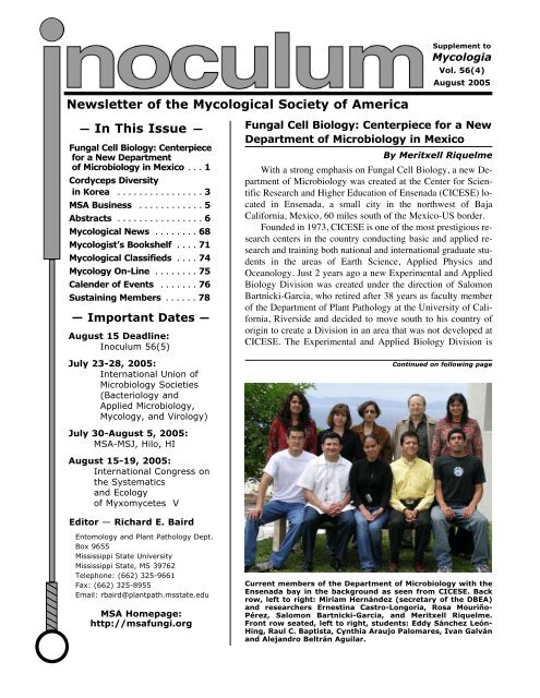
Current members <strong>of</strong> the Department <strong>of</strong> Microbiology with the<br />
Ensenada bay in the background as seen from CICESE. Back<br />
row, left to right: Miriam Hernández (secretary <strong>of</strong> the DBEA)<br />
and researchers Ernestina Castro-Longoria, Rosa Mouriño-<br />
Pérez, Salomon Bartnicki-Garcia, and Meritxell Riquelme.<br />
Front row seated, left to right, students: Eddy Sánchez León-<br />
Hing, Raul C. Baptista, Cynthia Araujo Palomares, Ivan Galván<br />
and Alejandro Beltrán Aguilar.
Fungal foray in the Chichihuas forest (road from Ensenada<br />
to Tijuana) with UABC Mycology students during<br />
raining season (It does rain from time to time in<br />
Baja!).<br />
composed <strong>of</strong> 3 Departments (Microbiology, Marine<br />
Biotechnology and Conservation Biology).<br />
The Department <strong>of</strong> Microbiology gives an opportunity<br />
to young investigators to develop independent research<br />
projects under the unifying theme <strong>of</strong> Fungal Cell<br />
Biology. Although government funding for research in<br />
Mexico is less generous than in other countries, the Department<br />
is equipped to conduct both molecular studies<br />
and cell biology projects (video-microscopy, confocal<br />
microscopy and transmission electron microscopy) and<br />
is scheduled to move into a new building this year. An<br />
initial core <strong>of</strong> 4 researchers composes the Department:<br />
Salomon Bartnicki-Garcia (Mathematical and computer-assisted<br />
modeling <strong>of</strong> fungal growth), Rosa Mouriño-<br />
Perez (The fungal microtubular cytoskeleton), Ernestina<br />
Castro-Longoria (Cellular basis <strong>of</strong> the circadian<br />
a b<br />
2 <strong>Inoculum</strong> <strong>56</strong>(4), August 2005<br />
rhythm in Neurospora) and Meritxell Riquelme (The<br />
secretory pathway in filamentous fungi).<br />
A major aim <strong>of</strong> the Department is to promote International<br />
Research Cooperation. The proximity to the<br />
US makes it practical to establish close links with US<br />
researchers. We already have ongoing projects with<br />
colleagues <strong>of</strong> the University <strong>of</strong> California, Arizona<br />
State University, the University <strong>of</strong> Oregon and Massachusetts<br />
Institute <strong>of</strong> Technology. The Department has<br />
also established strong links with local institutions,<br />
such as the Autonomous University <strong>of</strong> Baja California<br />
(UABC) and the National Institute for Forest and Agricultural<br />
Research (INIFAP). We participate in undergraduate<br />
Mycology courses at the UABC.<br />
For further information and current openings<br />
please visit the websites www.cicese.mx and microbio.cicese.mx/.<br />
Immunolocalization<br />
<strong>of</strong> microtubules in a<br />
chemically fixed cell<br />
<strong>of</strong> Sclerotium rolfsii.<br />
(R. Mouriño-Pérez)<br />
Questions or comments should be sent to<br />
Meritxell Riquelme Pérez, Departamento de<br />
Microbiología, DBEA, CICESE, Km. 107 Carretera<br />
Tijuana-Ensenada, 22860 Ensenada,<br />
Baja Califo. Email: riquelme@cicese.mx<br />
Molecular tagging <strong>of</strong> proteins involved in<br />
hyphal growth and morphology with GFP.<br />
Laser scanning confocal micrographs <strong>of</strong><br />
growing hyphae <strong>of</strong> N. crassa with a) a fluorescent<br />
Spk; c) fluorescent plasma membrane.<br />
(M. Riquelme)
Cordyceps Diversity in Korea<br />
Cordyceps is traditionally known as a highly medicinal<br />
mushroom in oriental society <strong>of</strong> Asia. It is quite<br />
diverse in its morphological characters, host range, natural<br />
habitat, etc. Due to contrast climatic variation and<br />
its unique geographical position, Cordyceps diversity is<br />
rich in Korea. Research on Cordyceps <strong>of</strong> Korea during<br />
last 20 years has shown that some species are widely<br />
distributed, while others grow in specific locations.<br />
Maturation periods <strong>of</strong> stromata range from late spring to<br />
summer till early autumn every year. Most species<br />
show their host specificity, but few species grow in diverse<br />
hosts. Microscopically, Cordyceps species differ<br />
in their spore shape, size, and their conidiation nature.<br />
There are about 300-400 Cordyceps species all over<br />
the world and are distributed universally (Kobayasi,<br />
1982; Kobayasi and Shimizu, 1983; Sung, 1996).<br />
Species <strong>of</strong> Cordyceps (Clavicipitaceae, Hypocreales,<br />
Ascomycota) grow inside insect host bodies as endosclerotium<br />
during winter and produce stromata in summer.<br />
Hosts <strong>of</strong> Cordyceps species include different stages<br />
<strong>of</strong> insect life cycle ranging from larva to adult <strong>of</strong> different<br />
insect orders, bee, wasp, cicadae, beetle, etc., except<br />
few which grow on hypogeous Elaphomyces species.<br />
Their scientific study and cultivation have been done in<br />
Korea for a long time. Every year, entomopathogenic<br />
fungal specimens including Cordyceps species are collected<br />
from different parts <strong>of</strong> Korea and are air-dried and<br />
preserved along with their isolates in Entomopathogenic<br />
Fungal Culture Collection (EFCC), Kangwon National<br />
University, Korea (Sung, 2004). The specimens are<br />
identified on the basis <strong>of</strong> their morphological characters.<br />
By Jae-Mo Sung, Bhushan Shrestha, Sang-Kuk Han, Su-Young Kim,<br />
Young-Jin Park, Won-Ho Lee, Kwang-Yeol Jeong, Sung-Keun Choi<br />
Continued on following page<br />
Fig. 3. C. gracilis<br />
Fig. 1. C. militaris<br />
Fig. 2. C. bifusispora<br />
<strong>Inoculum</strong> <strong>56</strong>(4), August 2005 3
Fig. 4. C. longissima<br />
Cordyceps species are collected from different parts<br />
<strong>of</strong> Korea every year from early May to late October.<br />
Cordyceps species, such as C. militaris (Fig. 1), C. pruinosa<br />
and C. sphecocephala are frequently collected,<br />
while C. bifusispora (Fig. 2), C gracilis (Fig. 3), C. heteropoda,<br />
C. longissima (Fig. 4), C. nakazawai, C.<br />
ochraceostromata, C. pentatomi, C. ramosopulvinata<br />
(Fig. 5), C. rosea, C. scarabaeicola, C. staphylinidaecola<br />
and C. yakushimensis are moderate or rare in distribution.<br />
Ascospores are discharged from fresh specimens<br />
and observed for their morphology and germination by<br />
staining with cotton blue in Lactophenol. Ascospores are<br />
inoculated in nutrient agar media in test-tubes and incubated<br />
till pr<strong>of</strong>use mycelium growth occurs. Original isolates<br />
and their sub-cultures are preserved at 4C in Entomopathogenic<br />
Fungal Culture Collection (EFCC),<br />
Kangwon National University, South Korea. Specimens<br />
are air-dried and preserved in herbarium boxes.<br />
4 <strong>Inoculum</strong> <strong>56</strong>(4), August 2005<br />
Fig. 5. C. ramosopulvinata<br />
It is very interesting to observe ascospore morphology<br />
and their germination behavior <strong>of</strong> different Cordyceps<br />
species. Among the observed species, only few<br />
such as C. bifusispora and C. pruinosa produced filamentous<br />
ascospores with threads in the center, while the<br />
remaining produced filamentous ascospores with continuous<br />
part-spores throughout the length. The germination<br />
rate <strong>of</strong> C. bifusispora, C. militaris, C. pentatomi, C.<br />
pruinosa, C. scaraebaeicola, C. staphylinidaecola were<br />
found faster, while those <strong>of</strong> C. gracilis, C. heteropoda,<br />
C. longissima, C. nakazawai, C. ochraceostromata, C.<br />
ramosopulvinata, C. rosea, C. sphecocephala, C.<br />
yakushimensis were found shower. Microscopic figures<br />
<strong>of</strong> ascospores are shown in sides <strong>of</strong> each figure <strong>of</strong> different<br />
Cordyceps species. C. militaris and C. pruinosa<br />
developed conidia on germinating hyphae soon, showing<br />
microcyclic conidiation character. Cordyceps isolates<br />
vary in their growth speed and cultural characteristics.<br />
Stromata <strong>of</strong> different Cordyceps species, such as<br />
C. militaris, C. scarabaeicola, C. pruinosa have been<br />
successfully produced in brown rice medium.<br />
References: Kobayasi, Y. 1941., Sci. Rept. Tokyo.<br />
Bunrika, Daigaku Sect. B. 5:53-260; Kobayasi, Y.<br />
1982. Trans. Mycol. Soc. Japan. 23:329-364; Kobayasi,<br />
Y. and D. Shimizu. 1983. Hoikusha Publishing Company<br />
Ltd. Osaka. 280 pp.; Sung, J.M. 1996. Kyo-Hak<br />
Publishing Co. Ltd., Seoul. 299 pp.; Sung, J.M. 2004.<br />
<strong>Inoculum</strong> 55(4):1-3.<br />
Questions or comments should be sent to<br />
Jae-Moe Sung, Entomopathogenic Fungal<br />
Culture Collection (EFCC), Department <strong>of</strong><br />
Applied Biology, Kangwon National University,<br />
Chuncheon 200-701, South Korea.<br />
Email: cordyceps@nate.com.
From the President’s Corner …<br />
Dear Friends and Colleagues,<br />
The success <strong>of</strong> the <strong>Mycological</strong><br />
<strong>Society</strong> <strong>of</strong> <strong>America</strong> depends heavily<br />
on the volunteer contributions <strong>of</strong> its<br />
members. We are always looking for<br />
volunteers who would like to be involved<br />
in the various functions <strong>of</strong> the<br />
society, and there are many <strong>of</strong> them<br />
from annual meeting preparations to<br />
specialized committees on subdisciplines<br />
within mycology. The list <strong>of</strong><br />
committees is on the MSA website.<br />
We need to hear from you if you<br />
would like to participate in some<br />
MSA Secretary Email Express<br />
Council completed five email polls since my May report and<br />
approved the following:<br />
• Poll 2005-3. Editor-in-Chief Donald Natvig nominated<br />
Dr. Robby Roberson to serve as Mycologia Associate<br />
Editor for the term 2005-2007. Approved.<br />
• Poll 2005-5. It was moved by President McLaughlin and<br />
seconded by Secretary Murrin that Executive Council approve<br />
a further increase <strong>of</strong> $4000 for the MSA Student<br />
Mentor Travel Awards for Hilo, to be awarded from income<br />
from the Uncommitted Endowment Fund. Approved.<br />
Background: The MSA had previously allotted<br />
an increased amount for Student Mentor Travel Awards<br />
this year, from approximately $4000 to $10,000, based on<br />
the anticipated increased costs <strong>of</strong> travel for the Hilo meeting.<br />
The Mentor Travel Awards Committee received 38<br />
applications this year, which appears to be a record number.<br />
The increase approved here allowed for an additional<br />
5 students to be funded for a total <strong>of</strong> 18 students. MSA<br />
coordinated efforts with Deep Hypha, the NSF-funded<br />
Research Coordination Network (RCN), which also<br />
awarded travel funding for Hilo.<br />
• Polls 2005 6-8: Approved. These motions concerned the<br />
approval <strong>of</strong> the Honorary Member and MSA Fellows for<br />
2005 following recommendations by George Carroll,<br />
Chair <strong>of</strong> the Honorary Awards Committee, and one other<br />
special award. Due to the confidential nature <strong>of</strong> these decisions<br />
until they are announced at the Annual Business<br />
Meeting in Hilo, August 3rd , they will not be reported on<br />
fully until the next issue <strong>of</strong> <strong>Inoculum</strong>.<br />
New Members: The MSA extends a warm welcome to all<br />
new (and returning) members. New memberships will be formally<br />
approved by the <strong>Society</strong> at the Annual Business Meeting<br />
in Hilo, Hawaii (Aug 3rd , 2005).<br />
• Australia: Susanna Ann Driessen, Kelli Maria Gowland<br />
MSA ABSTRACTS<br />
BUSINESS<br />
way. Whether there is a topic in<br />
which you would especially like to<br />
be involved or whether you would<br />
just like to contribute in some way,<br />
please consider volunteering. Send<br />
and email or talk to me or Vice President<br />
James Anderson or Secretary<br />
Faye Murrin and let us know your<br />
interest. Participating on a <strong>Society</strong><br />
committee is a great way to get to<br />
know the society and even a small<br />
contribution <strong>of</strong> your time and energy<br />
will be very helpful to the <strong>Society</strong>.<br />
David J. McLaughlin,<br />
MSA President<br />
• Canada: Melissa Day<br />
• Ghana: Hubert D Nyarko<br />
• Hong Kong: Justin Bahl, Wing Yan Chum<br />
• Japan: Yuko Ota<br />
• Mexico: Cristina Medina<br />
• United States: Anabelle Aranda, Kelly D Craven, Jennifer<br />
M. Davidson, Joyce Eberhart, Javesh Garan,<br />
Michele Therese H<strong>of</strong>fman, Bradley Warren Miller, Anil<br />
Kumar H Raghavendra, Kim Ryall, Marian N Viveros,<br />
Sandra W Woolfolk<br />
Emeritus membership: There has been one new application<br />
for emeritus membership: L J Wickerham <strong>of</strong> Tucson,<br />
Arizona. Emeritus memberships will be formally approved<br />
by the <strong>Society</strong> at the Annual Business Meeting in Hilo,<br />
Hawaii (Aug 3 rd , 2005).<br />
Faye Murrin<br />
MSA Secretary<br />
fmurrin@mun.ca<br />
Canine Foray — Faye Murrin’s dog, Rosie, protects the<br />
catch from mushroom bandits.<br />
<strong>Inoculum</strong> <strong>56</strong>(4), August 2005 5
MSA ABSTRACTS<br />
Aaltonen, Ronald E.*, Barrow, Jerry R., Lucero, Mary L., Osuna-Avila, Pedro<br />
and Reyes-Vera, Isaac. USDA-ARS Jornada Experimental Range, Las Cruces,<br />
NM 88003, USA. jbarrow@nmsu.edu. The microscopic identification <strong>of</strong> vertically<br />
transferred symbiotic fungi intrinsically integrated with cells, tissues<br />
and organs <strong>of</strong> host plants.<br />
Dual staining methodology and analysis with light microscopy and scanning<br />
electron microscopy were used to determine the nature and extent <strong>of</strong> symbiotic<br />
fungi with native desert grasses shrubs. Trypan Blue that targets fungal chitin<br />
and sudan IV that targets lipid bodies attached to fungal structures were used to<br />
stain cleared roots and leaves. Trypan blue revealed a densely stained fungal network,<br />
bound to the plasmalema <strong>of</strong> meristematic cells, that were transferred to cells<br />
in culture, tissues and all plant organs. Fungal structures were atypical and were<br />
substantially different than commonly observed fungal structures such as hyphae,<br />
spores, etc. Fungal associations with meristem cells facilitate their distribution and<br />
vertical transfer to all parts <strong>of</strong> the plant, seed and to succeeding generations. Significant<br />
are fungal associations with vascular tissue, photosynthetic cells and with<br />
the stomatal complex and suggests significant plant-fungus interactions within<br />
these critical plant cells. poster<br />
Abdelzaher, Hani M. A. Faculty <strong>of</strong> Science, El-Minia University 61519, Egypt.<br />
abdelzaher@link.net. Biological control <strong>of</strong> damping-<strong>of</strong>f and root rot diseases<br />
<strong>of</strong> soybean caused by Pythium spinosum Sawada var. spinosum using three<br />
rhizosphere species <strong>of</strong> soil fungi.<br />
Pythium spinosum was isolated from rhizosphere soil and rhizoplane <strong>of</strong><br />
healthy and infected soybean roots cultivated in an agricultural field located in<br />
Shahean district, El-Minia city, Egypt in June 2003. Rhizosphere and rhizoplane<br />
myc<strong>of</strong>lora isolated from the same sites were tested for their antagonism toward<br />
Pythium spinosum in agar plates. Among the isolated fungi, Aspergillus sulphureus,<br />
Penicillium islandicum and Paecilomyces variotii were chosen according<br />
to their antagonism on agar plates for experimentation to test their effectiveness<br />
for biological control in either autoclaved or nonsterilized soil. Coating<br />
soybean seeds and roots with spores and mycelia <strong>of</strong> these three antagonists gave<br />
germinating seeds and seedlings a very good protection from root-rot, pre- and<br />
post-emergence damping-<strong>of</strong>f caused by P. spinosum. Applying these biocontrol<br />
agents to autoclaved and nonsterilized soil infested with P. spinosum provided an<br />
excellent way <strong>of</strong> protection. contributed presentation<br />
Abe, Jun-ichi P. University <strong>of</strong> Tsukuba, Graduate School <strong>of</strong> Life and Environmental<br />
Science, 1-1, Tennoudai 1 chome, Tsukuba, Ibaraki 305-8572, Japan. jave@sakura.cc.tsukuba.ac.jp.<br />
An arbuscular mycorrhizal genus in the Ericaceae.<br />
Enkianthus is an ericaceous genus with about 17 spp. <strong>of</strong> shrubs and small<br />
trees, commonly distributed in Japan and southern China. Four Japanese native<br />
species <strong>of</strong> the genus Enkianthus (E. campanulatus, E. cernuus f. rubens, E. perulatus,<br />
E. subsessilis) were examined to determine the mycorrhizal status by comparing<br />
with typical ericoid mycorrhizal roots <strong>of</strong> Rhododendron kaempferi. The<br />
roots <strong>of</strong> all species were collected from trees <strong>of</strong> natural stands or public gardens<br />
and from seedlings <strong>of</strong> E. cernuus f. rubens grown in controlled conditions. These<br />
roots were observed with a compound light microscope and SEM. All examined<br />
roots <strong>of</strong> Enkianthus spp. formed only arbuscular mycorrhiza <strong>of</strong> the Paris-type.<br />
The fine roots <strong>of</strong> these species were usually thicker (approx. 150 µm) than the hair<br />
roots <strong>of</strong> R. kaempferi (approx. 80 µm). Short root hair- like structures with thick<br />
walls (approx. 5 µm) were observed occasionally on the fine roots <strong>of</strong> all<br />
Enkianthus spp. and root hairs were observed in E. subsessilis. Consequently, the<br />
mycorrhizal and root morphology <strong>of</strong> these four species are completely different<br />
from R. kaempferi. This result reveals that at least these four species <strong>of</strong> Enkianthus<br />
seem to be arbuscular mycorrhizal and lack ericoid mycorrhiza. These findings<br />
and the mycorrhizal status <strong>of</strong> the ancient ericaceous species are discussed. contributed<br />
presentation<br />
Aime, M. Catherine. USDA-Agricultural Research Service, Systematic Botany &<br />
Mycology Lab, Beltsville, MD 20705, USA. cathie@nt.ars-grin.gov. Molecular<br />
systematics <strong>of</strong> Uredinales.<br />
Rust fungi (Basidiomycota, Uredinales) consist <strong>of</strong> > 7000 species <strong>of</strong> obligate<br />
plant pathogens that possess some <strong>of</strong> the most complex life cycles in the Eumycota.<br />
Traditionally, phylogenetic inference within the Uredinales has been<br />
hampered by a lack <strong>of</strong> morphological characters and incomplete life cycle and<br />
host-specificity data. The application <strong>of</strong> modern molecular characters to rust systematics<br />
has been limited by several factors, including, to name a few, the inability<br />
to pure culture most rusts or unequivocally separate rust from host cells and<br />
other associated fungi in a specimen. Previous molecular systematic studies <strong>of</strong><br />
rusts have focused on analyses <strong>of</strong> 28S or 18S ribosomal DNA, but current contradictions<br />
in rust systematics, especially in the deeper nodes, have not yet been<br />
resolved. In this study, several genes (including 18S, 28S, and EF1alpha) were examined<br />
across the breadth <strong>of</strong> the Uredinales to resolve systematic conflicts and<br />
provide a framework for the group. It is concluded that morphology alone is a<br />
poor predictor <strong>of</strong> rust relationships at most levels and strict morphology-based<br />
classifications and species-delimitations appear obsolete. Host selection, on the<br />
other hand, has played a significant role in rust evolution. The difficulties and utility<br />
<strong>of</strong> analyzing protein-coding genes vs. rDNA in rust systematics are also discussed.<br />
symposium presentation<br />
6 <strong>Inoculum</strong> <strong>56</strong>(4), August 2005<br />
Aime, M. Catherine 1 * and Henkel, Terry W. 21 USDA-Agricultural Research Service,<br />
Systematic Botany & Mycology Lab, Beltsville, MD 20705, USA, 2 Humboldt<br />
State University, Dept. <strong>of</strong> Biological Sciences, Arcata, CA 95521, USA.<br />
cathie@nt.ars-grin.gov. Strategies for bioinventory: lessons from Guyana.<br />
Bioinventory, the identification and enumeration <strong>of</strong> taxa within a given<br />
area, provides the foundation for numerous biological and ecological studies. Yet<br />
fungal inventories are vastly underrepresented in the literature, and the conduction<br />
<strong>of</strong> such inventories presents a host <strong>of</strong> unique difficulties and problems. Special<br />
problems faced in conducting fungal inventories include deciding at which level<br />
to record units (e.g., fruiting bodies, cultures, and/or environmental sequences);<br />
how to construct significant sampling strategies (e.g. plot studies, transects, and/or<br />
sweeps); and, especially, how to conduct the alpha-taxonomy. We have completed<br />
five years <strong>of</strong> comparative plot studies for fungi in a remote, rain-forested region<br />
<strong>of</strong> west-central Guyana. To date we have documented nearly 1,000 species<br />
or morphospecies <strong>of</strong> macromycetes, both ectomycorrhizal and saprotrophic, and<br />
counted >20,000 fruiting bodies. Yet the most time-consuming aspect <strong>of</strong> this<br />
study is the taxonomy. To date, we have been able to identify and publish less than<br />
10% <strong>of</strong> the Guyanese taxa; > 40% <strong>of</strong> these have been species or genera new to<br />
science. Some ideas for recording, analyzing, and identifying fungal taxa from<br />
previously under-sampled areas will be discussed. symposium presentation<br />
Akamatsu, Y. and Saikawa, Masatoshi*. Department <strong>of</strong> Environmental Science,<br />
Tokyo Gakugei University, Koganei-shi, Tokyo 184-8501, Japan. saikawa@ugakugei.ac.jp.<br />
Giant mycelium by Sommerstorffia spinosa.<br />
Since 1984, Sommerstorffia spinosa was obtained several times from samples<br />
<strong>of</strong> debris floating on the water <strong>of</strong> a fire reservoir located in the campus <strong>of</strong><br />
Tokyo Gakugei University. The species has been known as a rotifer-parasite, to<br />
have the ability to capture the animals with a peg, a distal narrow protuberance <strong>of</strong><br />
hypha, or to parasitize them with a bowling-pin shaped sporeling developed from<br />
an encysted, secondary zoospore. In all the strains reported up to now, each <strong>of</strong> the<br />
isolates finished capturing animals after 3 to 7 times <strong>of</strong> capture by the peg and became<br />
solely to produce zoospores. Thus the mycelium was quite limited in size in<br />
all cases. However, in spring, 2004, we obtained a strain <strong>of</strong> the species, the<br />
mycelium <strong>of</strong> which did not finish capturing rotifers (Lepadella oblonga) and grew<br />
unlimitedly to establish a giant mycelium. At 30 days <strong>of</strong> cultivation under water,<br />
it could be seen with a naked eye, because the size being 0.5-3.0 mm in diameter.<br />
The hyphae (7-9 µm wide) constituting the giant mycelium were empty except<br />
only their distal portion terminated with a peg (6-8 µm long, 3-5 µm wide). After<br />
capturing a rotifer, the peg grew into an endozoic thallus that developed one or<br />
two hyphae externally and transformed itself into a zoosporangium with an evacuation<br />
tube (50-80 µm long, 8-10 µm wide). The primary zoospores encysted at<br />
the mouth <strong>of</strong> the evacuation tube to form a mass <strong>of</strong> cysts (7-9 µm). In normal<br />
strains, each <strong>of</strong> the primary cysts soon produces a secondary zoospore, though the<br />
cyst <strong>of</strong> our strain did not produce it, but lost its content, or, in rare occasion, developed<br />
a sporeling (ca. 17 µm long, 8 µm wide). The giant mycelium <strong>of</strong> our<br />
strain also captured two testaceous rhizopods <strong>of</strong> Cryptodifflugia and Euglypha.<br />
contributed presentation<br />
Almeida-Leñero, Lucia 1 , Ludlow-Wiechers, Beatriz 1 , Geel, Bas van 2 , González,<br />
María C. 3 * and Aptroot, André. 4 1 Dept. Ecología y Rec. Nat, Fac. Ciencias,<br />
UNAM, Mexico DF 04510, 2 IBED, Paleoecology and Landscape Ecology, Univ.<br />
Amsterdam Kruislaan 318, 1098 SM Amsterdam The Netherlands, 3 Inst. Biología,<br />
UNAM, Mexico, 4 Centraalbureau voor Schimmelcultures (CBS), Fungal<br />
Biodiversity Centre, P.O. Box 85167, 3508 AD Utrecht, The Netherlands.<br />
mcgv@ibiologia.unam.mx. Records <strong>of</strong> mid-Holocene fungi from Lake Zempoala,<br />
Central Mexico.<br />
The aim <strong>of</strong> the present study is to describe and illustrate fungal spores<br />
recorded on Zempoala lake at 2800 m altitude. The studied interval is <strong>of</strong> mid-<br />
Holocene age. The lake with submerged aquatical and hydroseral shore vegetation,<br />
lie today in the Abies religiosa dominated forest belt. The samples were prepared<br />
using standard techniques for palynological studies. A number <strong>of</strong> selected<br />
fungal spores have been identified, described and illustrated. Ecological environmental<br />
preferences <strong>of</strong> present fungi are also given. Among the fungi type spores<br />
analysed are 2 basidiospores: Enthorriza (type 527), and Urocystis, 3 ascospores:<br />
Astrosphaeriella, Ustulina, Valsaria, and 10 mitospores: Acrodictys, Antennatula,<br />
Brachydesmiella, Endophragmiella, Monodictys, Trichocladium type 1 and 2,<br />
Papulospora, and Virgatospora. These fungus occur together with pollen elements<br />
that define a landscape <strong>of</strong> forest and a water body. Antennatula confirm an<br />
Abies sp. forest, Acrodictys suggests species <strong>of</strong> Populus sp., Virgatospora <strong>of</strong><br />
broad leaved tree forest, and Ustulina the presence <strong>of</strong> deciduous trees. Enthorriza,<br />
and Trichocladium are evidence <strong>of</strong> freshwater. Environment conditions may be<br />
presumed <strong>of</strong> high air humidity considering the presence <strong>of</strong> decaying plant debris<br />
and soil forming fungi such as Monodictys and Papulospora. The fungi were<br />
recorded for first time in paleoecological studies in Mexico. poster<br />
An, Zhiqiang. Merck Research Laboratories, West Point, PA, USA. Polyketide<br />
synthase genes in the pneumocandin-producing fungus Glarea lozoyensis.<br />
Glarea lozoyensis is the producer <strong>of</strong> pneumocandin B0, a potent inhibitor<br />
Continued on following page
<strong>of</strong> fungal glucan synthesis. This industrially important filamentous fungus is<br />
slow-growing, is very darkly pigmented, and has not been easy to manipulate genetically.<br />
Using a PCR strategy to survey the G. lozoyensis genome for secondary<br />
metabolic encoding pathways, we have identified three polyketide synthase<br />
genes: pks1, pks2, and pks3. pks1 encodes a 2,124 amino-acid protein with five<br />
catalytic modules: ketosynthase, acyltransferase, two acyl carrier sites, and<br />
thioesterase/Claisen cyclase. Pks2 encodes a 1,791 amino-acid protein with five<br />
catalytic modules: â-ketosynthase (KS), acyltransferase (AT), dehydratase (DH),<br />
â-ketoacyl reductase (KR), and acyl carrier protein (ACP). The pks3 gene acts as<br />
an operon and encodes two enzymes, PKS3-NRPS1 and NRPS2. Cluster analysis<br />
<strong>of</strong> 37 fungal ketosynthase modules grouped the pks1p with PKSs involved in<br />
1,8-dihydroxynaphthalene melanin biosynthesis; the pks2p with PKSs involved<br />
in 6-methylsalicylic acid biosynthesis; and the pks3p was grouped with PKSs that<br />
synthesize structurally and bioactively complex polyketides. An Agrobacteriummediated<br />
transformation system was developed for the disruption <strong>of</strong> these three<br />
pks genes. Disruption <strong>of</strong> pks1 yielded knockout mutants that displayed an albino<br />
phenotype, suggesting that pks1 encodes a tetrahydroxynaphthalene synthase.<br />
Heterologous expression <strong>of</strong> pks2 in Aspergillus nidulans showed that pks2 encodes<br />
for 6-methylsalicylic acid synthase. Disruption <strong>of</strong> pks3 showed no difference<br />
in chemical pr<strong>of</strong>iles under the fermentation conditions used. Other genes reside<br />
in the three pks loci will also be discussed. symposium presentation<br />
Anagnost, Susan E. 1 *, Catranis, Catharine M. 2 , Fernando, Analie A. 1 , Morey,<br />
Shannon R. 1 , Zhou, Shuang 1 , Zhang, Lianjun 3 and Wang, C.J.K. 41 Wood Products<br />
Engineering, SUNY College <strong>of</strong> Environmental Science and Forestry, Syracuse,<br />
NY 13210, 2 <strong>America</strong>n Type Culture Collection, 10801 University Blvd.,<br />
Manassas, VA 20110 USA, 3 Forest and Natural Resources, SUNY-ESF, Syracuse,<br />
NY, 4 Faculty <strong>of</strong> Environmental and Forest Biology, SUNY-ESF, Syracuse<br />
NY 13210, USA. seanagno@esf.edu. Aeromycology <strong>of</strong> homes in Syracuse,<br />
New York.<br />
Airborne fungi were recovered at 103 homes in Syracuse, New York as part<br />
<strong>of</strong> an environmental survey <strong>of</strong> homes <strong>of</strong> infants predisposed to asthma (EPA project<br />
No. R-82860501-0). Total colony-forming units per cubic meter <strong>of</strong> air<br />
(CFU/m 3 ) and isolate identifications were obtained from samples collected with<br />
the Andersen N6 sampler. Samples collected on two consecutive days, indoors<br />
and outdoors, during 147 visits to these homes yielded 14<strong>56</strong>5 isolates that were<br />
classified into 170 fungal taxa. Among the most frequent were Hyaline unknowns,<br />
Cladosporium cladosporioidies, Penicillium spp., Aspergillus spp., basidiomycetes,<br />
Cladosporium herbarum, and Alternaria spp. Aspergillus spp were<br />
more frequent indoors (547 isolations) compared to outdoors (59 isolations), as<br />
were Penicillium spp (771 indoor, 137 outdoor). The total CFU/m3 was greater<br />
during the summer and fall seasons; certain species only appeared during summer<br />
and fall. Three new records for the USA were: Acrodontium myxomyceticola, 173<br />
isolates from 41 homes; Acremonium roseolum, 36 isolates from 18 homes; and<br />
Tetracoccosporium paxianum, once. A new sub-culturing method (Random-50)<br />
allowed the recovery <strong>of</strong> slow-growing, sometimes rare, fungi. These same Random-50<br />
plates can estimate with high confidence the total fungal concentration in<br />
these homes. poster<br />
Aoki, Takayuki 1 *, Tomomi, Tsunematsu 2 and Sato, Toyozo 3 . 1 Genetic Diversity<br />
Department, National Institute <strong>of</strong> Agrobiological Sciences, Kannondai, Tsukuba,<br />
Japan, 2 University <strong>of</strong> Tsukuba, Tennodai, Tsukuba, Japan, 3 Genebank, National<br />
Institute <strong>of</strong> Agrobiological Sciences, Kannondai, Tsukuba, Japan.<br />
taoki@nias.affrc.go.jp. Re-identification <strong>of</strong> Fusarium moniliforme isolates deposited<br />
at the MAFF Genebank, NIAS, Japan based on analysis <strong>of</strong> DNA sequences<br />
<strong>of</strong> the Histone H3 gene region.<br />
On a long used fungal name, Fusarium moniliforme, a recommendation to<br />
refrain its usage was issued by the ISPP/ICTF Subcommittee on Fusarium Systematics.<br />
This is because <strong>of</strong> the facts that: (1) the name, F. moniliforme represents<br />
an unacceptably broad species concept; and (2) F. verticillioides as mating population<br />
(MP) A <strong>of</strong> the Gibberella fujikuroi (GF) species complex is the older name<br />
for the species in strict sense. Microorganisms Section <strong>of</strong> the MAFF Genebank,<br />
NIAS, Japan has been preserving rather many number <strong>of</strong> strains identified previously<br />
as F. moniliforme for a distribution purpose. To respond to the recommendation,<br />
70 strains <strong>of</strong> F. moniliforme deposited at MAFF were re-identified based<br />
on the DNA sequences <strong>of</strong> the Histone H3 gene region. By using a PCR primerset,<br />
H3-1a and H3-1b, gene fragments <strong>of</strong> this region, ca. 520 bps, were amplified<br />
and sequenced. DNA sequences were aligned with Clustal X ver. 1.8 and phylogenetic<br />
analyses were made with PAUP ver. 4.0b10 by generating NJ and MP<br />
trees. Sequence data for the same gene region <strong>of</strong> related species <strong>of</strong> Fusarium were<br />
downloaded from the GenBank site, NCBI, and analyzed together. Out <strong>of</strong> 70<br />
strains examined, <strong>56</strong>, 7 and 4 strains were identified as F. fujikuroi (corresponding<br />
to the MP-C <strong>of</strong> the GF-complex.), F. proliferatum (MP-D), F. subglutinans (MP-<br />
E), respectively. Identity <strong>of</strong> 3 strains was still under consideration. poster<br />
Aranda, Anabelle*, Viveros, Marian N. and Elley, Joanne T. Biological Sciences,<br />
The University <strong>of</strong> Texas at El Paso, El Paso, TX 79968-0519, USA.<br />
jellzey@utep.edu. Localization <strong>of</strong> G-Protein in Saccharomyces cerevisiae and<br />
Schizosaccharomyces pombe.<br />
It has been suggested that components <strong>of</strong> the cytoskeleton contribute to the<br />
MSA ABSTRACTS<br />
signal transduction process in association with one or more members <strong>of</strong> the G protein<br />
family. Relatively high-affinity binding between dimeric tubulin and the<br />
alpha subunits <strong>of</strong> Gs and Gi1 has also been reported (Wang N. and Rasenick,<br />
1991). Tubulin has binding domains for microtubule-associated proteins. Tubulin<br />
modifies G-protein signaling. Heterotrimeric G-proteins regulate microtubule assembly<br />
in mammalian cells. G alpha inhibits microtubule assembly and increases<br />
microtubule disassembly by activating the intrinsic GTPase <strong>of</strong> tubulin. G beta<br />
gamma promotes microtubule assembly (Roychowdhury et al, 1999). In the present<br />
study, we have analyzed the interaction between alpha and beta gamma subunits<br />
<strong>of</strong> G proteins and tubulin in Saccharomyces cerevisiae and the Schizosaccharomyces<br />
pombe by immun<strong>of</strong>luorescence (Hagan and Hyams, 1988). Results<br />
from the immun<strong>of</strong>luorescence experiments were confirmed by electrophoresis<br />
and immunoblotting. We have obtained protein analyses and immunoblotting for<br />
S. cerevisiae and S. pombe. The visualization <strong>of</strong> the gamma and alpha tubulin is<br />
most evident in S. cerevisiae. There is evidence <strong>of</strong> a G-protein role in microtubule<br />
assembly/disassembly. poster<br />
Arenz, Brett E.*, Held, Ben W., Jurgens, Joel A. and Blanchette, Robert A. Department<br />
<strong>of</strong> Plant Pathology, University <strong>of</strong> Minnesota, 495 Borlaug Hall, 1991<br />
Upper Buford Circle, Saint Paul, MN 55108, USA. aren0058@umn.edu. Fungal<br />
diversity in wood and soils at the historic expedition huts <strong>of</strong> Ross Island,<br />
Antarctica, as revealed by denaturing gradient gel electrophoresis (DGGE).<br />
Culture-dependent methods have long been the primary tool to determine<br />
the biodiversity <strong>of</strong> microorganisms in soils and other substrates. New molecular<br />
methods to study fungal pr<strong>of</strong>iles in samples from the environment have shown<br />
that these previous methods usually give an incomplete picture <strong>of</strong> all the organisms<br />
present. This study utilized denaturing gradient gel electrophoresis (DGGE)<br />
to analyze the fungal diversity in wood and other materials brought to Ross Island,<br />
Antarctica by explorers Robert Scott and Ernest Shackleton. Fungal diversity in<br />
soils near the historic structures was also analyzed. Fungal specific primers were<br />
used to target the ITS 2 region which shows significant variability between<br />
species. The DNA was separated by DGGE, and bands extracted and sequenced.<br />
Although previously reported Antarctic fungi such as Geomyces, Cladosporium,<br />
Cadophora, and Phoma, were frequently identified, DNA <strong>of</strong> many species show<br />
very little similarity to sequences available in databases based on BLASTn<br />
searching. Species <strong>of</strong> Cadophora and Cladosporium were found associated with<br />
deteriorating historic woods and other artifacts. These fungi were also found in<br />
Antarctic soil samples. This work is providing a more comprehensive understanding<br />
<strong>of</strong> the microbes found in Antarctica and provides new insights on the<br />
fungi attacking wood in the historic huts. contributed presentation<br />
Arnold, A. Elizabeth 1 *, Miadlikowska, Jolanta 2 , Higgins, K. Lindsay 2 , Dalling,<br />
James W. 3 , Gallery, Rachel E. 3 , Henk, Daniel A. 2 , Eells, Rebecca L. 2 , Vilgalys,<br />
Rytas J. 2 and Lutzoni, François 2 . 1 Division <strong>of</strong> Plant Pathology and Microbiology,<br />
Department <strong>of</strong> Plant Sciences, University <strong>of</strong> Arizona, Tucson, AZ 85721, USA,<br />
2 Department <strong>of</strong> Biology, Duke University, Durham, NC 27708, USA, 3 Department<br />
<strong>of</strong> Plant Biology, University <strong>of</strong> Illinois, Urbana, IL 61801, USA.<br />
arnold@ag.arizona.edu. What can environmental PCR tell us about foliar<br />
fungal endophyte communities?<br />
While it is clear that a tremendous diversity <strong>of</strong> endophytic fungi can be isolated<br />
from leaves using standard culturing techniques, the potential occurrence <strong>of</strong><br />
unculturable endophytes limits our understanding <strong>of</strong> the ecology, evolution, and<br />
diversity <strong>of</strong> endophytic symbioses. In particular, environmental sampling may be<br />
key to uncovering endophytes with obligate host associations and/or vertical<br />
transmission, slowly growing species that do not occur readily in standard media,<br />
and species that lose in competitive interactions within cultured leaf pieces. Results<br />
<strong>of</strong> paired environmental sampling (direct PCR) + culturing approaches to assessing<br />
endophyte diversity and community structure in boreal, temperate, and<br />
tropical foliage, and tropical seeds, will be compared. In each case, environmental<br />
sampling was complementary to culturing: direct PCR recovered numerous<br />
lineages that were not represented in cultures from the same hosts, recovered sequences<br />
that have few close matches in GenBank, provided evidence for infections<br />
in apparently uninfected tissues, and fundamentally changed our view <strong>of</strong> the<br />
taxonomic distribution <strong>of</strong> endophytes present in each host species. In discussing<br />
these case studies, special attention will be given to (1) the importance <strong>of</strong> multilocus<br />
datasets for phylogenetic analyses <strong>of</strong> environmental samples; (2) methods<br />
<strong>of</strong> phylogenetic analysis that can result in reliable topologies given limited data<br />
from clones; (3) the utility <strong>of</strong> BLAST results based on ITS data for identifying<br />
clones; and (4) the utility <strong>of</strong> ITS genotype groups as functional taxonomic units<br />
for ecological analyses <strong>of</strong> environmental samples. symposium presentation<br />
Avis, Peter G.*, Leacock, Pat R. and Mueller, Greg M. Department <strong>of</strong> Botany,<br />
The Field Museum <strong>of</strong> Natural History, 1400 S. Lake Shore Drive, Chicago, IL<br />
60605, USA. pavis@fieldmuseum.org. Potential changes in ectomycorrhizal<br />
fungal communities caused by nitrogen deposition in oak forests <strong>of</strong> the<br />
Chicago region.<br />
Ectomycorrhizal (ECM) fungi may mediate the impact nitrogen (N) deposition<br />
has on temperate deciduous forests. To test this hypothesis, we are documenting<br />
the above- and belowground components <strong>of</strong> ECM communities in con-<br />
Continued on following page<br />
<strong>Inoculum</strong> <strong>56</strong>(4), August 2005 7
MSA ABSTRACTS<br />
trol and experimentally N-fertilized treatments at three forest sites found along a<br />
N deposition gradient in the Chicago region. Field surveys <strong>of</strong> sporocarps in treatment<br />
plots have identified over 90 ECM fungal species across the three sites since<br />
2003. Over 200 <strong>of</strong> these collections were used to develop a reference database <strong>of</strong><br />
terminal restriction fragment length polymorphisms (T-RFLP) for the identification<br />
<strong>of</strong> ECM collected belowground. In 2004, we conducted morphological and<br />
T-RFLP analyses <strong>of</strong> ECM collected from soil cores from each treatment plot.<br />
Early results indicate that the ECM communities in these sites are rich and potentially<br />
vulnerable to N increase. Over 130 likely species <strong>of</strong> ECM fungi have<br />
been identified from over 5000 oak root tips examined. Species richness estimates<br />
indicate that significantly fewer numbers <strong>of</strong> ECM species are found on oak roots<br />
in N fertilization treatments at two <strong>of</strong> three sites. We will continue to monitor<br />
ECM community responses to N fertilization over the next two years and also examine<br />
how the composition <strong>of</strong> these communities relates to their function within<br />
the context <strong>of</strong> N deposition. contributed presentation<br />
Badalyan, Suzanna M.*, Garibyan, Narine G. and Sakeyan, Carmen Z. Dept. <strong>of</strong><br />
Botany, Yerevan State University, Aleg Manoogian St., 375025, Yerevan, Armenia.<br />
badalyan_s@yahoo.com. Culture collection <strong>of</strong> Basidiomycetes fungi at<br />
the Yerevan State University (Armenia).<br />
Establishment and maintenance <strong>of</strong> Culture Collection <strong>of</strong> macroscopic fungi<br />
(Basidiomycetes) at the Yerevan State University are the way <strong>of</strong> preserving biodiversity<br />
<strong>of</strong> mushrooms and extending fungal genetic and biotechnological research<br />
in Armenia. Study <strong>of</strong> mushroom cultures can also be valuable in obtaining<br />
novel bio-pharmaceuticals and functional food additives with health-enhancing<br />
effect. Presently, the Collection comprises around 210 living strains <strong>of</strong> 60 mushroom<br />
species. They were mostly isolated in Armenia and obtained from other institutions.<br />
Among them, 35 species and 197 strains possess known medicinal<br />
properties. The Flammulina velutipes, Pleurotus ostreatus and Coprinus spp. collections<br />
are represented by a wide eco-geographical diversity <strong>of</strong> strains. ITSrDNA<br />
nucleotide sequence analyses <strong>of</strong> collected 25 species and 105 strains were<br />
carried out together with international collaborators. The project <strong>of</strong> genetic identification<br />
<strong>of</strong> Armenian medicinal mushrooms is currently realizing. Further extension<br />
<strong>of</strong> taxonomic, ecological and geographical diversity <strong>of</strong> species/strains and<br />
their genetic identification, as well as digitalization and creation <strong>of</strong> information<br />
DataBase are in progress. The catalogue <strong>of</strong> Culture Collection will be available<br />
soon. This research is supported by NATO (#FEL.RIG.980764) and DAAD<br />
(#548.104401.174) grants. poster<br />
Badalyan, Suzanna M. 1 * and Kües, Ursula 2 . 1 Dept. <strong>of</strong> Botany, Yerevan State University,<br />
Aleg Manoogian St., 375025, Yerevan, Armenia, 2 Section Molecular<br />
Wood Biotechnology, Institute <strong>of</strong> Forest Botany, Georg-August University, Büsgenweg<br />
2, D-37077 Göttingen, Germany. badalyan_s@yahoo.com. Mycelial<br />
morphology and growth characteristics <strong>of</strong> wood-related coprinoid mushrooms.<br />
Around 80 species <strong>of</strong> the traditional genus Coprinus (Coprinoid mushrooms)<br />
have been observed on wooden material. Mycelial micro-, macromorphology<br />
and growth characteristics <strong>of</strong> xylotrophic species Coprinus comatus, Coprinellus<br />
angulatus, C. bisporus, C. curtus, C. disseminatus, C. domesticus, C.<br />
ellisii, C. micaceus, C. xanthothrix, Coprinopsis atramentaria, C. cinerea, C.<br />
cothurnata, C. gonophylla, C. radians, C. romagnesiana, C. scobicola, C. strossmayeri<br />
and Parasola plicatilis have been studied. Cultures were grown on Malt-<br />
Extract Agar (MEA), Potato-Dextrose Agar (PDA) and Glucose-Peptone Agar<br />
(GPA) at 25 ∞C and pH 6. Growth rates and growth coefficients were highest on<br />
MEA (up to 85 mm and above 20, respectively), then PDA and GPA. Macromorphological<br />
characteristics were described after 10 days <strong>of</strong> growth. Oval and<br />
round shape clamps occur in most <strong>of</strong> the Coprini. Clamps were not found in some<br />
Coprinellus species. Material for micromorphological investigations was obtained<br />
by slide cultures. Hyphal loops were particularly formed in Coprinellus<br />
species. Arthroconidia were <strong>of</strong>ten observed, whereas blastic sporulation was rare.<br />
Chlamydospore formation is also typical for Coprini. Mycelial cysts, micr<strong>of</strong>ilaments<br />
and crystals were detected in some species.Thanks DAAD, NATO and<br />
Deutsche Bundesstiftung Umwelt for financial support. poster<br />
Bahl, Justin, Jeewon, Rajesh* and Hyde, Kevin. Dept. Ecology & Biodiversity,<br />
The University <strong>of</strong> Hong Kong, Hong Kong, SAR, China. jbahl@hkusua.hku.hk.<br />
Intergeneric relationships <strong>of</strong> Linocarpon and Neolinocarpon: does phylogenetic<br />
analysis support the generic delineation?<br />
Species from the genera Linocarpon and Neolinocarpon are common<br />
saprobic fungi found in subtropical to tropical regions and mainly occurs on<br />
monocotyledonous hosts. Both genera are <strong>of</strong>ten found on the same host. Based on<br />
morphology these genera share many similarities and have resulted in difficulties<br />
in assigning taxa. The most significant delineating characters are ascomata position<br />
and morphology. Based on parsimony and likelihood analyses <strong>of</strong> multi-locus<br />
partial sequences derived from nuclear encoded ribosomal DNA, beta-tubulin and<br />
RNA polymerase regions from fresh and dried herbarium material, an attempt has<br />
been made to assess which morphological characters are phylogenetically significant<br />
for generic delineation or whether the genera should be circumscribed under<br />
the priority name, Linocarpon. Analysis confirmed that the two genera are not<br />
monophyletic and indicates parallel evolution <strong>of</strong> morphological and ecological<br />
8 <strong>Inoculum</strong> <strong>56</strong>(4), August 2005<br />
characters. Results are discussed in relation to the significance <strong>of</strong> morphological<br />
characters currently used in the taxonomy <strong>of</strong> Linocarpon and Neolinocarpon.<br />
contributed presentation<br />
Barnes, Irene*, Wingfield, Michael J. and Wingfield, Brenda D. Department <strong>of</strong><br />
Genetics, Forestry and Agricultural Biotechnology Institute (FABI), University <strong>of</strong><br />
Pretoria, Pretoria 0002, South Africa. irene.barnes@fabi.up.ac.za. Development<br />
<strong>of</strong> microsatellite markers for the red band needle blight pathogen Dothistroma<br />
septosporum using two different isolation methods.<br />
Very little is known regarding the population biology <strong>of</strong> Dothistroma septosporum,<br />
one <strong>of</strong> the most important pathogens <strong>of</strong> plantation grown pines in the<br />
southern hemisphere. Thus, twelve sets <strong>of</strong> microsatellite markers have been developed<br />
to study the population dynamics <strong>of</strong> this pathogen. Two techniques,<br />
ISSR-PCR and FIASCO enrichment were used to screen for microsatellite rich<br />
regions. ISSR-PCR was effective in locating many microsatellite sites. However,<br />
after the necessary genome walking, many <strong>of</strong> the microsatellites were found to be<br />
redundant artifacts <strong>of</strong> the initial primers used. With FIASCO, variable success<br />
was observed depending primarily on the primer combination used in the enrichment.<br />
In one screen, 57 % <strong>of</strong> the clones contained microsatellites, in others, none<br />
were found. From a total <strong>of</strong> 22 primer pairs, 11 were found to be polymorphic<br />
amongst isolates <strong>of</strong> D. septosporum. An additional primer was polymorphic between<br />
D. pini and D. septosporum and can be used for further diagnostic purposes<br />
within populations. Cross-species amplification was successful in D. pini, D.<br />
rhabdoclinis and Mycosphaerella dearnessi. Future studies using these primers<br />
will focus on gaining an improved understanding <strong>of</strong> the population structure, genetic<br />
diversity, gene flow and the genetic relatedness between different populations<br />
<strong>of</strong> these important tree pathogens. poster<br />
Barnes, Irene 1 *, Crous, Pedro W. 2 , Wingfield, Michael J. 1 and Wingfield, Brenda<br />
D. 11 Department <strong>of</strong> Genetics, Forestry and Agricultural Biotechnology Institute<br />
(FABI), University <strong>of</strong> Pretoria, Pretoria, South Africa, 0002, 2 Centraalbureau<br />
voor Schimmelcultures (CBS), Fungal Biodiversity Centre, P.O. Box 85167,<br />
3508 AD Utrecht, The Netherlands. irene.barnes@fabi.up.ac.za. Multigene phylogenetic<br />
analyses reveal that Dothistroma septosporum and D. pini represent<br />
two distinct taxa and a serious threat to pine forestry.<br />
The sudden increase in severity <strong>of</strong> the red band needle blight disease in the<br />
U.K., Canada and parts <strong>of</strong> Europe where Dothistroma has been present for decades<br />
is a matter <strong>of</strong> great concern. Although the etiology <strong>of</strong> the disease is well known,<br />
phylogenetic and population level relationships amongst isolates <strong>of</strong> the fungus are<br />
poorly understood. We have thus constructed multigene phylogenies for isolates <strong>of</strong><br />
Dothistroma from 13 different countries. These have illustrated that the isolates are<br />
separated into two distinct lineages representing two discrete species and supported<br />
by morphological differences. The one species, referred to as D. septosporum<br />
occurs worldwide and infects over 30 species <strong>of</strong> pines. It is the major cause <strong>of</strong> the<br />
serious blight disease plaguing Pinus radiata plantations in New Zealand, Chile<br />
and other Southern Hemisphere countries The second species, D. pini, is restricted<br />
in its distribution to the North Central United States where it causes a serious disease<br />
on exotic P. nigra. A simple ITS-PCR-RFLP is also presented that allows accurate<br />
and rapid distinction between the two species. contributed presentation<br />
Baroni, Timothy J. 1 *, Lindner Czederpiltz, Daniel L. 2 , Lodge, D. Jean 3 , H<strong>of</strong>stetter,<br />
Valérie 4 and Franco-Molano, Ana Esperanza 5 . 1 Department <strong>of</strong> Biological<br />
Sciences, State University <strong>of</strong> New York, College at Cortland, Cortland, NY<br />
13045, USA, Center for Forest Mycology Research, USDA Forest Service, Forest<br />
Products Laboratory, One Gifford Pinchot Dr., Madison, WI 53726-2398,<br />
USA, Center for Forest Mycology Research, USDA Forest Service, P.O. Box<br />
1377, Luquillo, PR 00773-1377, USA, 4 Botany Department, Duke University,<br />
Durham, NC 27708-0338, USA, 5 Laboratorio de Taxonomía de Hongos, Instituto<br />
de Biología, Universidad de Antioquia, A.A.1226, Medellín, Colombia. baronitj@cortland.edu.<br />
Arthrosporella, a recently rediscovered neotropical<br />
genus, is phylogenetically related to Termitomyces in the Lyophylleae.<br />
In 1996, Sharon Cantrell and TJB collected an odd nail-shaped agaric covered<br />
with dark brown conidia in Puerto Rico; that collection could not be named<br />
at that time. Over the next 9 years <strong>of</strong> intense collecting, this odd arthrospore-producing<br />
species was collected only three more times in Puerto Rico, but also twice<br />
in the Dominican Republic. TJB realized this was a new species <strong>of</strong> Arthrosporella,<br />
originally described by Rolf Singer as a monotypic genus from Argentina. A<br />
second, distinctly different and new conidia producing agaricoid species was<br />
found just recently in Colombia by AEFM, and we now know <strong>of</strong> two other collections<br />
<strong>of</strong> this taxon from the Dominican Republic by Egon Horak and TJB.<br />
Very recently (August 2004) a third new, and completely different arthrosporeproducing<br />
agaricoid species was found by TJB, DJL and CDL, fruiting abundantly<br />
in the cloud forest on the highest peak in Belize (Doyle’s Delight). All <strong>of</strong><br />
these new taxa possess siderophilous granules in the basidia. Phylogenetic analysis<br />
<strong>of</strong> nLSU indicates, with significant support, the monophyly <strong>of</strong> Arthrosporella<br />
with Termitomyces (Lyophylleae) and suggests a sister relationship between these<br />
two genera. All collections <strong>of</strong> Arthrosporella appear to be saprotrophic and not<br />
termitophilous, thus perhaps indicating a closer relationship with Podabrella,<br />
which is also in this branch <strong>of</strong> the Lyphylleae clade. poster<br />
Continued on following page
Barrett, Luke G., Thrall, Peter H., Burdon, Jeremy J. and van der Merwe, Marlien<br />
M.* Centre for Plant Biodiversity Research, CSIRO – Plant Industry, GPO Box<br />
1600, Canberra ACT 2601, Australia. marlien.vandermerwe@csiro.au Pancontinental<br />
patterns <strong>of</strong> genetic variation in the rust fungus Melampsora lini.<br />
The Linum marginale-Melampsora lini plant-pathogen interaction is endemic<br />
to Australia and has been a focus <strong>of</strong> epidemiological and coevolutionary<br />
studies for more than a decade. Considerable variation for both host resistance and<br />
pathogen virulence has been shown at a range <strong>of</strong> spatial scales from the local to<br />
the continental. Here we report on a study using AFLP and SSR markers to examine<br />
pancontinental patterns <strong>of</strong> genetic variation in 102 clonal lines <strong>of</strong> M. lini<br />
representing 35 populations. Molecular marker genotypes partition all <strong>of</strong> the isolates<br />
into two major lineages, and in combination with subsequent sequencing <strong>of</strong><br />
beta-tubulin and elongation factor genes suggest a possible hybrid origin for one<br />
<strong>of</strong> the lineages. Subsequent comparison with data on phenotypic variation for virulence<br />
in a subset <strong>of</strong> these isolates also demonstrates striking differences between<br />
the two lineages in terms <strong>of</strong> pathogenicity on the host L. marginale. Molecular genetic<br />
variation within the lineages was very limited, and within populations both<br />
AFLP and SSR markers regularly failed to distinguish among several lines with<br />
different pathotypes. These results are important for developing an understanding<br />
<strong>of</strong> how pathogen virulence might evolve within natural populations. poster<br />
Barrow, Jerry R.*, Lucero, Mary L., Osuna-Avila, Pedro, Reyes-Vera, Isaac and<br />
Aaltonen, Ronald E. USDA-ARS Jornada Experimental Range, Las Cruces, NM<br />
88003, USA. jbarrow@nmsu.edu. Hybrid vigor, structural modification and<br />
enhanced plant performance induced by symbiotic fungi-plant interactions.<br />
Hybrid vigor, structural modification and enhanced plant performance induced<br />
by symbiotic fungi-plant interactions. Communities <strong>of</strong> symbiotic fungi are<br />
intrinsically integrated with single cells, tissues and organs <strong>of</strong> desert plants. They<br />
are obligately associated with living tissues and generally cannot be isolated and<br />
cultured separately. Symbiotic fungi indigenous to native desert plants were transferred,<br />
using cell culture methods, to non-host plants and reciprocally transferred<br />
between native grasses. Crop and native plants, with recipient fungi have astounding<br />
and increased levels <strong>of</strong> vigor. Up to five-fold increases in root and shoot<br />
biomass and substantial morphological changes and reproductive potential were<br />
obtained. Enhanced chlorophyll and phosphorous content were common responses.<br />
Once integrated, fungi were intimately interfaced with the plasmalema<br />
<strong>of</strong> the new host plant and were ecto-cytoplasmically inherited. This technology<br />
accesses a previously unexplored source <strong>of</strong> genetic variability within native<br />
ecosystems with the potential <strong>of</strong> immediate transfer to improve tolerance <strong>of</strong> native<br />
and crop plants to stress, disease and pests. Symbiotic fungal transfers provides<br />
a powerful alternative for genetic improvement <strong>of</strong> plants. contributed presentation<br />
Bartnicki-García, Salomón. Microbiology Dept., CICESE, Ensenada, Baja California,<br />
22860, México. bartnick@cicese.mx. Introductory remarks: Current<br />
perspectives in hyphal morphogenesis.<br />
Hyphal morphogenesis is clearly the most basic developmental process in<br />
mycology. The origin <strong>of</strong> the fungal kingdom could be traced to the “invention” <strong>of</strong><br />
hyphal morphogenesis, i.e. the ability <strong>of</strong> a cell to form long tubular walls by tip<br />
growth. Not surprisingly, over the years, fungal biologists have become increasingly<br />
interested in elucidating the cellular and molecular basis <strong>of</strong> hyphal morphogenesis.<br />
The problem revolves basically about understanding the mechanism(s)<br />
responsible for polarized growth <strong>of</strong> the cell wall. A number <strong>of</strong> structural and molecular<br />
players have been identified but the mechanism cannot be attributed to a<br />
single gene or protein. Polarized secretion probably requires a specific concerted<br />
action between cytoskeleton and secretory vesicles to produce a pattern <strong>of</strong> vesicle<br />
discharge that would generate cells with a pr<strong>of</strong>ile described by the hyphoid equation:<br />
y = x cot (xV/N). This symposium on Advances in Fungal Morphogenesis<br />
will cover developments in different fronts: a new way <strong>of</strong> probing the surface<br />
properties <strong>of</strong> the hyphal wall (S. Kaminskyj), assessing the importance <strong>of</strong> protein<br />
glycosylation in the maintenance <strong>of</strong> hyphal growth (B. Shaw), and analyzing the<br />
role <strong>of</strong> the Spitzenkörper in apical growth (M. Riquelme). symposium presentation<br />
Baucom, Deana L.*, Bruhn, Johann N. and Mihail, Jeanne D. Division <strong>of</strong> Plant<br />
Sciences, University <strong>of</strong> Missouri, Columbia MO 65211, USA. dlba3c@mizzou.edu.<br />
Armillaria species involved in Missouri Ozark forest decline.<br />
In the Missouri Ozark Mountains, Armillaria spp. contribute to oak decline.<br />
To investigate the distributions and roles <strong>of</strong> Armillaria spp., 142 isolates collected<br />
in 2002 from 40 plots were identified. Amplification <strong>of</strong> IGS1 followed by restriction<br />
with AluI identified 121 isolates as A. mellea (52 %), A. gallica (38 %),<br />
and A. tabescens (10 %). Two new RFLP patterns were found to represent 41 %<br />
<strong>of</strong> A. mellea and 17 % <strong>of</strong> A. gallica isolates. Isolates yielding these patterns are<br />
currently being characterized by sequence analysis <strong>of</strong> IGS1. Major hosts were<br />
dogwood (44 %), red oaks (18 %), and white oaks (7 %). Armillaria mellea predominated<br />
on both white and red oaks; A. mellea and A. gallica occurred equally<br />
on dogwood. Recently killed trees provided 28 % <strong>of</strong> study isolates. Armillaria<br />
was almost universally present on recently killed trees. Red oaks, dogwood, and<br />
white oaks provided 38 %, 32 %, and 10 % <strong>of</strong> recent mortality isolates, respectively.<br />
Nearly all recent oak mortality yielded A. mellea. Recent dogwood mor-<br />
MSA ABSTRACTS<br />
tality yielded A. gallica (54 %) more frequently than A. mellea (38 %). Ozark oak<br />
decline affects red oaks and dogwoods most heavily. The Armillaria sp. which<br />
most commonly contributed to oak mortality on our plots in 2002 was A. mellea,<br />
though A. gallica was more commonly recovered from recent dogwood mortality.<br />
We found little evidence <strong>of</strong> mortality caused by A. tabescens. poster<br />
Beeson, Esther 1 , Beltz, Shannon 2 , Klich, Maren 2 and Bennett, Joan W. 1 * 1 Tulane<br />
University, New Orleans, LA, USA, 2 Southern Regional Resource Center, New<br />
Orleans, LA, USA. ebeeson1@cox.net. Sclerotial production in Aspergillus<br />
flavus varies with temperature and nitrogen source.<br />
Twenty strains <strong>of</strong> Aspergillus flavus from the culture collection <strong>of</strong> the<br />
Southern Regional Research Laboratory, New Orleans, LA, were grown on defined<br />
and complex media at 25 o C and 37 o C for one week. Colonies were<br />
screened for sclerotial production as well as other colony characters including production<br />
<strong>of</strong> conidia, floccose hyphae, and mycelial pigment. When grown on complex<br />
media with yeast extract as the nitrogen source, 9 <strong>of</strong> the strains produced<br />
sclerotia at 25 o C and 11 produced sclerotia at 37 o C. When grown on a defined<br />
medium with nitrate as nitrogen source, 8 <strong>of</strong> the strains produced sclerotia at 37<br />
o C and 5 produced sclerotia at 25 o C. When ammonium was used as the nitrogen<br />
source, only one strain produced sclerotia at 37 o C and there was no sclerotial production<br />
at 25 o C. Sporulation was sparse or absent for all strains grown on ammonium<br />
at 37 o C. When microarrays <strong>of</strong> the A. flavus genome become available,<br />
these data will be useful in designing conditions for RNA isolation in order to<br />
probe the genes involved in sclerotial formation. poster<br />
Bennett, Chandalin 1 *, Newcombe, George 1 and Aime, M. Catherine 2 . 1 Department<br />
<strong>of</strong> Forest Resources, University <strong>of</strong> Idaho, Moscow, Idaho 83844, USA,<br />
2 USDA ARS Systematic Botany and Mycology Lab, Beltsville, Maryland 20705,<br />
USA. benn4449@uidaho.edu. Regional studies <strong>of</strong> Melampsora on Salix in the<br />
Pacific Northwest.<br />
Melampsora epitea Thuem. is a species complex that represents all willow<br />
rusts in N. <strong>America</strong>. To better understand how much host-specialization and genetic<br />
diversity exists in the Pacific Northwest (PNW), a large-scale host-range inoculation<br />
study was performed along with ITS sequencing, morphological analysis,<br />
and a two-year field survey. Distinct host-specificity was shown for three<br />
different Melampsora isolates inoculated on nearly equal sets <strong>of</strong> 440 willows.<br />
Less than 20 percent <strong>of</strong> the plants in each experiment were susceptible to the inoculum<br />
and greater than 15 percent <strong>of</strong> those showed some signs <strong>of</strong> resistance. The<br />
genetic sequencing resulted in four distinct clades, the largest <strong>of</strong> which likely represents<br />
a complex in this region. The other three clades were strongly divergent<br />
from the complex. There was up to three genetically distinct rusts present in a<br />
given geographic location and four or more genetically distinct rusts on a given<br />
Salix sp. There was found at most two distinct rusts on the same species from the<br />
same population. It’s evident that willow rusts are incredibly diverse in the PNW.<br />
Every experimental inoculum was specialized in its host range and the genetic diversity<br />
was spread across four unique and highly divergent clades. symposium<br />
presentation<br />
Bennett, Joan W. Tulane University, New Orleans, LA 70118, USA.<br />
jbennett@tulane.edu. Industrial mycology: from Takamine’s diastase to<br />
TIGR’s database.<br />
Industrial mycology has its roots in numerous food fermentations that were<br />
developed early in human history. Of these, the Japanese koji process, utilizing<br />
Aspergillus oryzae , is <strong>of</strong> particular interest because the first microbial enzyme<br />
ever patented was a secreted diastase (amylase) isolated from A. oryzae by Dr. Jokichi<br />
Takamine, a Japanese chemist working in the U.S.A during the late nineteenth<br />
century. In the first half <strong>of</strong> the twentieth century, several filamentous fungi<br />
were harnessed for the production <strong>of</strong> additional commercial enzymes and for organic<br />
acids. Although the discovery <strong>of</strong> penicillin and the subsequent golden age<br />
<strong>of</strong> antibiotics transformed fermentation technology, the molecular biology <strong>of</strong> industrial<br />
molds lagged behind that <strong>of</strong> model species. Now, in the twenty-first century,<br />
genomic research has ushered in a new era <strong>of</strong> understanding. Genome projects<br />
for several Aspergillus species are completed or underway, revealing new<br />
insights into secondary metabolism, carbon and nitrogen regulation, and other genetic<br />
control mechanisms. aymposium presentation<br />
Berbee, Mary L. 1 *, James, Tim Y. 2 , Longcore, Joyce E. 3 , Stajich, Jason 4 and Vilgalys,<br />
Rytas J. 2 . 1 Dept. <strong>of</strong> Botany, University <strong>of</strong> British Columbia, V6T 1Z4<br />
Canada, 2 Dept. <strong>of</strong> Biology, Duke University, Durham, NC 27708 USA, 3 Biological<br />
Sciences, University <strong>of</strong> Maine, Orono, ME 04469-5722 USA, 4 Dept. <strong>of</strong> Molecular<br />
Genetics and Microbiology, Duke University, Durham, NC 27708, USA.<br />
berbee@interchange.ubc.ca. What makes a fungus? Fungal-specific genes<br />
from EST libraries <strong>of</strong> the basal fungi Batrachochytrium dendrobatidis and<br />
Mortierella verticillata.<br />
The Chytridiomycota and Zygomycota include ancient fungal lineages that<br />
may have originated hundreds <strong>of</strong> millions <strong>of</strong> years before plants invaded land. We<br />
looked for shared genes that distinguish the fungi from other kingdoms through<br />
comparison <strong>of</strong> expressed sequence tag (EST) libraries, from a chytrid, Batrachochytrium<br />
dendrobatidis (1588 ESTs, average length, 572 nucleotides) and<br />
Continued on following page<br />
<strong>Inoculum</strong> <strong>56</strong>(4), August 2005 9
MSA ABSTRACTS<br />
from Mortierella verticillata, a readily cultured terrestrial mold in the Zygomycota<br />
(1278 ESTs, average length, 5<strong>56</strong> nucleotides). Of 1247 non-redundant ESTs<br />
from B. dendrobatidis, 100 were ‘fungal- specific’ in that sequence similarity<br />
within fungi is high but drops <strong>of</strong>f sharply beyond the kingdom level. From M. verticillata,<br />
64 ESTs were fungal-specific. Fungal-specific genes included six subfamilies<br />
<strong>of</strong> chitin synthases that arose through ancient gene duplications. Suggesting<br />
that early fungi had to scavenge for iron, B. dendrobatidis and higher<br />
fungi share a high affinity iron permease. Septins are also present in animals, but<br />
they help control the form <strong>of</strong> fungi during development. We found four septin paralogues<br />
among ESTs from Batrachochytrium dendrobatidis and two from<br />
Mortierella verticillata. The septins from the spheroidal chytrid appeared to be<br />
basal to genes from other fungi. contributed presentation<br />
Bérube, Jean A. 1 *, Stefani, Frank O.P. 1 and Sokolski, S. 1 Canadian Forest Service,<br />
1055 du PEPS, P.O. Box 3800, Ste-Foy, QC, G1V 4C7 Canada.<br />
jberube@cfl.forestry.ca. Comparison <strong>of</strong> foliar endophyte biodiversity from<br />
three boreal conifers.<br />
We compared the foliar endophyte biodiversity <strong>of</strong> white spruce (Picea glauca),<br />
black spruce (P. mariana) and eastern white pine (Pinus strobus). Asymptomatic<br />
healthy needles were collected from 67 conifer populations in eastern Canada,<br />
surface sterilized and then plated on nutrient agar. The ribosomal ITS region<br />
were amplified, sequenced and analyzed using maximum parsimony and<br />
Bayesian inference. Results from morphological and molecular studies indicate<br />
white spruce is hosting as many as 49 fungal endophyte species, black spruce is<br />
hosting 42 species and white pine is hosting 30 species. Only four endophyte<br />
species were common among those three hosts, among them Hormonema dematioides<br />
and Phaeocryptopus gaeumannii. Some fourteen species were common to<br />
the two spruces and only one species was common between black spruce and<br />
white pine. Five other species were also found on other conifers not examined in<br />
this study. The remaining 85 species were unique to one conifer host only. They<br />
represent a source <strong>of</strong> new species needed to fill the gap between known number<br />
<strong>of</strong> species, 76000 and the 1.5 million estimated number by Hawksworth. contributed<br />
presentation<br />
Bérube, Jean A. 1 *, Stefani, Frank O.P. 1 , Piercey-Normore, Michele D. 2 , Guillaumin,<br />
Jean-Jacques 3 and Hamelin, Richard C. 1 . 1 Canadian Forest Service, 1055 du<br />
PEPS, P.O. Box 3800, Ste-Foy, QC, G1V 4C7 Canada, 2 Department <strong>of</strong> Botany,<br />
University <strong>of</strong> Manitoba, Winnipeg, MB, R3T2N2, Canada, 3 INRA Centre de Clermont-Ferrand,<br />
UMR 234 Breset, 63039, Clermont-Ferrand, France.<br />
jberube@cfl.forestry.ca. Evidence <strong>of</strong> ancient recombination in genus Armillaria.<br />
The genus Armillaria has been studied extensively to determine its evolutionary<br />
history in relation to virulence, hosts and distribution. Four coding genes<br />
(actin, beta-tubulin, G3PD and MLL) from 21 species <strong>of</strong> Armillaria from North<br />
<strong>America</strong>, Europe, Africa, Asia, Australia and South <strong>America</strong> were sequenced and<br />
analyzed. Phylogeny <strong>of</strong> combined genes showed important differences with published<br />
ITS phylogenies. The basal position <strong>of</strong> A. ostoyae, A. gemina, A. borealis<br />
in the phylogenetic trees inferred from the actin and G3PD genes is evidence <strong>of</strong><br />
ancient recombination events in the northern hemisphere that took place after the<br />
Gondwanian disjunction (100 Myr BP) but before the Laurasian disjunction (60<br />
Myr BP). Once recombinant sites removed from phylogenetic analysis, corrected<br />
trees indicate that Armillaria seems to be an ancient genus that originated in the<br />
Jurassic, with some species that have radiated throughout the southern hemisphere<br />
before the Gondwana disjunction. The evolutionary history <strong>of</strong> many Armillaria<br />
species in relation to continental drift will be presented. contributed presentation<br />
Beyer, David M.*, Paley, Kimerley and Wilkinson, Vija. Dept. <strong>of</strong> Plant Pathology,<br />
The Pennsylvania State University, University Park, PA 16802 USA.<br />
dmb8@psu.edu. Influence <strong>of</strong> organic acids on the growth and development <strong>of</strong><br />
Trichoderma harzianum, a pathogen <strong>of</strong> Agaricus bisporus.<br />
Previous research has shown that Phase I substrate for Agaricus bisporus<br />
prepared under low oxygen conditions resulted in earlier and more severe development<br />
<strong>of</strong> Trichoderma harzianum green mold disease. Organic acids are known<br />
to be produced under anaerobic conditions, and their residual compounds may be<br />
involved in the development <strong>of</strong> the pathogen in mushroom substrate. This research<br />
looked at the influence <strong>of</strong> several organic acids at different concentrations<br />
on the growth <strong>of</strong> Trichoderma harzianum in vitro and in vivo. Results suggested<br />
that in vitro higher concentrations <strong>of</strong> most organic acids had a negative influence<br />
on the growth <strong>of</strong> the pathogen while lower concentrations <strong>of</strong> Fumaric, Succinic<br />
and Lactic acid did not. In vivo assays suggested that when Fumaric acid was<br />
added to the substrate it predisposed the substrate to disease development. poster<br />
Binder, Manfred 1 *, Bodensteiner, Philomena 2 , Wang, Zheng 1 and Hibbett, David<br />
S. 1 . 1 Clark University, Biology Department, Lasry Bioscience Center, 950 Main<br />
Street, Worcester, Massachusetts 01610-1477, USA, 2 Fakultät für Biologie der<br />
Ludwig-Maximilians Universität München, Department I: Biodiversitätsforschung,<br />
Systematische Mykol., Munich, Germany. mbinder@clarku.edu.<br />
Phylogenetic relationships <strong>of</strong> cyphelloid and aquatic homobasidiomycetes.<br />
Cyphelloid fungi include minute terrestrial, cupulate or tubular-shaped<br />
forms in the euagarics clade. Our previous studies showed that some aquatic fungi<br />
like the marine gasteromycete Nia vibrissa have been derived from cyphelloid<br />
10 <strong>Inoculum</strong> <strong>56</strong>(4), August 2005<br />
forms, while others (e. g., Mycaureola dilseae) are derived from agaricoid forms<br />
and represent separate lineages <strong>of</strong> aquatic fungi in the euagarics. In a broad phylogenetic<br />
survey <strong>of</strong> cyphelloid and aquatic homobasidiomycetes, we used a twopronged<br />
approach generating nuc-lsu and ITS data for 140 collections <strong>of</strong> cyphelloid<br />
fungi consisting <strong>of</strong> multiple samples <strong>of</strong> 57 well described species. Data from<br />
multiple rDNA regions (nuc-ssu, nuc-lsu, mt-ssu, mt-lsu, ITS) that were additionally<br />
sequenced for a subset <strong>of</strong> 25 cyphelloid species served as a backbone in<br />
weighted parsimony and Bayesian analyses to identify the major lineages <strong>of</strong><br />
cyphelloids within the euagarics and their closest relatives. It has long been suspected<br />
that cyphelloid fungi form a polyphyletic group and our study suggests<br />
there may be at least 18 independent origins, which will be presented in detail.<br />
Thus, morphological reduction via cyphellization is a major theme in the evolution<br />
<strong>of</strong> euagarics. One <strong>of</strong> the most remarkable examples <strong>of</strong> evolutionary transitions<br />
from the terrestrial cyphelloid habit to fungi living in aquatic habitats can be<br />
observed in the Nia clade, which contains the largest proportion <strong>of</strong> cyphelloid<br />
fungi studied so far. Our preliminary results indicate that the marine fungi in this<br />
clade are a monophyletic group including N. vibrissa, Halocyphina villosa, Calathella<br />
mangrovei, and as a new result Digitatispora marina, a resupinate species<br />
that has lost the ability <strong>of</strong> active spore discharge. poster<br />
Bisch<strong>of</strong>f, Joseph F. 1 *, Rehner, Stephen A. 2 and Humber, Richard A. 3 . 1 GenBank,<br />
NCBI-NIH, Bethesda, Maryland 20892, USA, 2 Insect Biocontrol Laboratory,<br />
USDA-ARS, Beltsville, Maryland 20705, USA, 3 Plant Protection Research Unit,<br />
USDA-ARS-ARSEF, Ithaca, New York 14853, USA.<br />
bisch<strong>of</strong>f@ncbi.nlm.nih.gov. A multilocus phylogenetic analysis <strong>of</strong> the entomopathogen<br />
Metarhizium.<br />
Species <strong>of</strong> the cosmopolitan entomopathogenic form genus Metarhizium<br />
play important ecological roles by regulating arthropod populations and recycling<br />
insect biomass. The ubiquity <strong>of</strong> Metarhizium species and their broad host range<br />
have made them an attractive alternative to chemical pesticides. Despite commercial<br />
interest in the genus and its widespread occurrence, species delimitation,<br />
intrageneric relationships, and placement within the Clavicipitaceae remain undefined.<br />
Historically, two species and a single variety were accepted in the genus.<br />
However, in a recent molecular phylogenetic study 10 lineages (i.e., species and<br />
varieties) were proposed. The purpose <strong>of</strong> this study is to, 1) determine if Metarhizium<br />
is monophyletic, 2) identify its sister lineage within Clavicipitaceae, 3) resolve<br />
phylogenetic species and infer intrageneric relationships. A multilocus<br />
dataset (EF1-alpha, RPB1, beta-tubulin and RPB2) supports the monophyly <strong>of</strong><br />
Metarhizium and its close association with the largely plant associated members<br />
<strong>of</strong> Clavicipitaceae (subfamily Clavicipitoideae). In addition, a minimum <strong>of</strong> 12 distinct<br />
lineages has been resolved, exceeding prior estimates <strong>of</strong> diversity in the<br />
genus. contributed presentation<br />
Branco, Sara. The Committee on Evolutionary Biology, University <strong>of</strong> Chicago,<br />
Chicago, IL 60637 USA. sbranco@uchicago.edu. Macr<strong>of</strong>ungal communities <strong>of</strong><br />
northeastern Portugal.<br />
Quercus pyrenaica woodlands and serpentinic Quercus rotundifolia woodlands<br />
are two important plant communities <strong>of</strong> northeastern Portugal. These forests<br />
are significant natural habitats in the region and are considered priorities in terms<br />
<strong>of</strong> nature conservation. Quercus pyrenaica is a native and widespread oak species.<br />
Serpentine woodlands are communities with an impoverished flora characterized<br />
by Quercus rotundifolia and several endemic herbaceous species. They are confined<br />
to serpentine soils, characterized by high heavy metal content and low nutrient<br />
levels. In each woodland one 1000 sqm permanent plot was established and<br />
macr<strong>of</strong>ungi were surveyed biweekly during 2001, 2002 and 2003. In both woodlands<br />
the sporocarp fruiting occurred throughout autumn, between October and<br />
December. The Quercus pyrenaica plot had much higher species diversity (146<br />
species) than the serpentinic plot (73 species) and only 17% <strong>of</strong> the recorded<br />
species occurred in both plots. Mycorrhizal species composed more than 60% <strong>of</strong><br />
all the species found in either habitat. Twenty three <strong>of</strong> the identified taxa were new<br />
records for the country and 47 were new records for the region. These results indicate<br />
that these forests are diverse and distinct in macr<strong>of</strong>ungal species and require<br />
proper management and conservation. poster<br />
Bruhn, Johann N. 1 , Kromroy, Kathryn 2 , Baucom, Deana 1 and Mihail, Jeanne D. 1 *<br />
1 Division <strong>of</strong> Plant Sciences, University <strong>of</strong> Missouri, Columbia MO 65211, USA,<br />
2 USDA Forest Service, North Central Research Station, St. Paul, MN 55108,<br />
USA. bruhnj@missouri.edu. Armillaria species associated with oak stump<br />
sprout regeneration in upland Missouri Ozark forests.<br />
Stump sprouts are favored in North <strong>America</strong>n oak regeneration. Many trees<br />
selected for cutting in upland Missouri Ozark forests are infected with one or more<br />
Armillaria spp., especially in oak stands experiencing forest decline. After harvest,<br />
Armillaria infections in these trees spread to the root crown, reducing stump<br />
sprout production and causing sprout mortality. We examined the implications <strong>of</strong><br />
these infections for survival and growth <strong>of</strong> remaining sprouts. During 2003 and<br />
2004, we characterized sprouting and presence <strong>of</strong> Armillaria on oak stumps created<br />
in 1996 on 35 study plots. Single Armillaria isolates were collected from 145<br />
stumps; dual isolates were collected from widely-spaced roots <strong>of</strong> 29 additional<br />
stumps. Dual isolates from 14 stumps represent different genets. We have found<br />
Continued on following page
one new IGS1 RFLP banding pattern for each <strong>of</strong> A. mellea and A. gallica. Armillaria<br />
mellea is widely represented in the stump populations in our plots, raising<br />
concern that models <strong>of</strong> stump sprout regeneration may be optimistic, especially in<br />
stands experiencing oak decline. Stump sprout regeneration can result in the establishment<br />
<strong>of</strong> crop trees on previously infected root systems. Our study also focuses<br />
attention on the disparity between stem age and the age <strong>of</strong> their root systems<br />
including existing Armillaria infections, in stands managed using stump sprout regeneration.<br />
poster<br />
Bruns, Thomas D.*, Boynton, Primrose, Shamieh, Karimeh, Szaro, Timothy M.<br />
and Kennedy, Peter G. Department <strong>of</strong> Plant & Microbial Biology, University <strong>of</strong><br />
California, Berkeley 94720-3102, USA. pogon@berkeley.edu. Spatial and temporal<br />
structure <strong>of</strong> Rhizopogon sporebanks.<br />
Rhizopogon species are ectomycorrhizal associates <strong>of</strong> the Pinaceae. In<br />
young, disturbed pine forests they are <strong>of</strong>ten among the dominant fungi colonizing<br />
seedlings and saplings. This dominance is due to abundant sporebanks that are<br />
produced by deposition <strong>of</strong> spores in place and by dispersal through mammal mycophagy.<br />
We have used rodent baiting, bioassays, spore burial, and PCR-based<br />
analyses to examine the distance and frequency <strong>of</strong> dispersal from forest to nonforested<br />
borders and the longevity <strong>of</strong> spores. We then compared these parameters<br />
to the spatial distribution <strong>of</strong> sporebanks. We found Rhizopogon is efficiently dispersed<br />
at distances <strong>of</strong> 40 meters from a border in a single year, and that the distances<br />
and quantity <strong>of</strong> spores dispersed are greatest from young post-fire forests.<br />
Current dispersal appears to be sufficient to account for the spatial pattern <strong>of</strong> the<br />
sporebank for the four most common species. However our results suggest that<br />
longevity <strong>of</strong> the spores may be substantial, and we think that longevity is likely to<br />
be important at greater spatial and temporal scales. symposium presentation<br />
Burdsall, Harold H. Jr. USDA - FPL - Cntr. Forest Mycol. Res., Madison, WI and<br />
Fungal & Decay Diagnostics, LLC, Black Earth, WI, USA. burdsall@fungaldecay.com.<br />
Cyphellaceae in the Arctic and subantarctic islands.<br />
Members <strong>of</strong> the family “Cyphellaceae” in the traditional sense are now<br />
known to be distributed among several families <strong>of</strong> the homobasidiomycetes.<br />
However, they are recognizable in the field as a “morphological group”, unnatural<br />
as it may be. Collecting in the subantarctic islands <strong>of</strong> New Zealand and near<br />
and above the Arctic Circle in Alaska has provided a number <strong>of</strong> specimens for<br />
comparing the occurrence <strong>of</strong> these fungi from opposite ends <strong>of</strong> the globe. Species<br />
<strong>of</strong> Lachnella, Henningsomyces and Cyphellopsis were collected. Species <strong>of</strong> Lachnella<br />
were common on the subantarctic islands while Henningsomyces and<br />
Cyphellopsis were lacking. The reverse was true in the Arctic collecting sites. Differences<br />
in types <strong>of</strong> substrate may explain this difference, there being more wood<br />
in the Arctic collecting sites than on the subantarctic islands. However, the lack <strong>of</strong><br />
Lachnella specimens from the Arctic may be a result <strong>of</strong> collecting bias and collecting<br />
on forbs and ferns on the Arctic sites may provide more specimens <strong>of</strong><br />
Lachnella species. These comparisons are continuing. symposium presentation<br />
Burgess, Joshua W. 1 , Schwan, William 2 and Volk, Thomas J. 2 *. Departments <strong>of</strong><br />
1 Microbiology and 2 Biology, University <strong>of</strong> Wisconsin-La Crosse, La Crosse WI<br />
54601, USA. volk.thom@uwlax.edu. Detection <strong>of</strong> Blastomyces dermatitidis<br />
DNA from natural samples using rapid PCR-based methods.<br />
Blastomyces dermatitidis is the dimorphic fungal agent <strong>of</strong> blastomycosis, a<br />
disease that primarily affects humans and dogs. The clinical appearance <strong>of</strong> this<br />
mycosis is well characterized, but there is still little known about its environmental<br />
niche, having been isolated from nature only 21 times. We have developed a<br />
PCR-based assay to detect B. dermatitidis from soil samples using primers specific<br />
to a portion <strong>of</strong> the promoter region <strong>of</strong> the virulence gene BAD1. An internal<br />
standard control, pTJV2-2, was designed to ensure that negative results from soil<br />
samples were not the result <strong>of</strong> PCR failure due to soil inhibitors. The lower detection<br />
limit <strong>of</strong> the plasmid, using Blastomyces specific primers (BSP), was 0.1<br />
femtograms. With chromosomal DNA, the lower detection limit is 500 fg. To test<br />
the assay for cross reactivity, the BSP were tested successfully against many<br />
fungi, bacteria, and actinomycetes, especially those genetically related or found in<br />
the same geographic areas. This method is sensitive to 304 copies when detecting<br />
pTJV2-2 DNA spiked directly into the soil extraction. When extracting live Blastomyces<br />
yeast, this newly developed method can detect as few as 8,450 live cells.<br />
We plan on using this method to screen soils to better determine the environmental<br />
niche <strong>of</strong> Blastomyces. contributed presentation<br />
Buyck, Bart 1 *, Parrent, Jeri Lynn 2 and Vilgalys, Rytas J. 2 . 1 Dépt. Systématique et<br />
Evolution, Muséum National d’Histoire Naturelle, 75005 Paris, France, 2 Dept. <strong>of</strong><br />
Biology, Duke University, Durham, NC 27708-0338, USA. buyck@mnhn.fr.<br />
The Russula virescens-crustosa species complex (Russulales, Basidiomycotina)<br />
from eastern North <strong>America</strong>.<br />
The type species <strong>of</strong> the subsection Virescentinae, R.virescens Fr. was originally<br />
described from Europe, but is common throughout most <strong>of</strong> the northern hemisphere,<br />
including the eastern US. R. crustosa Peck and R. heterosporoides Murrill<br />
are currently the only other North <strong>America</strong>n species placed in this subsection.<br />
Since their original description, revisions <strong>of</strong> these taxa did not reveal major identification<br />
problems, although microscopic observation was needed to separate<br />
green forms <strong>of</strong> R. crustosa and R.virescens . Recent field work by the senior au-<br />
MSA ABSTRACTS<br />
thor and several <strong>America</strong>n amateur mycologists suggests additional taxa are likely<br />
to belong in Virescentinae. In this study we have combined morphological and<br />
molecular data generated from over 100 north <strong>America</strong>n, as well as some selected<br />
Asian, African and Australian collections to examine the virescens-crustosa<br />
complex. This work is part <strong>of</strong> a complete revision <strong>of</strong> the genus Russula in the eastern<br />
US by the senior author. Detailed morphological study revealed that commonly<br />
used characters for identifying Russula species, spore ornamentation and<br />
form <strong>of</strong> hyphal extremities, are too variable within the virescens-crustosa complex<br />
to discriminate among taxa, except for R. parvovirescens sp.nov. Results<br />
from the multilocus molecular analysis confirm the placement <strong>of</strong> several undescribed<br />
north <strong>America</strong>n taxa in thevirescens-crustosa complex, highlight the speciose<br />
nature <strong>of</strong> this subsection, and allow for a firm placement <strong>of</strong> these species in<br />
a more worldwide phylogenetic dataset. Poster<br />
Cafaro, Matias J. 1 and Lichtwardt, Robert W. 2 *. 1 Department <strong>of</strong> Bacteriology,<br />
University <strong>of</strong> Wisconsin, Madison, WI 53706, USA, 2 Department <strong>of</strong> Ecology and<br />
Evolutionary Biology, University <strong>of</strong> Kansas, Lawrence, KS 66045, USA. cafaro@wisc.edu.<br />
Unveiling cryptic relationships <strong>of</strong> the Eccrinales.<br />
The order Eccrinales includes a diverse group <strong>of</strong> arthropod-associated organisms,<br />
most <strong>of</strong> which present relatively simple morphology. Unbranched, nonseptate,<br />
multinucleate thalli and sporangiospores that are formed basipetally from<br />
the thallus apex have been used as characters to relate the group to the class Trichomycetes.<br />
No member <strong>of</strong> the Eccrinales has been cultured. In order to address<br />
the phylogenetic relationships <strong>of</strong> the group, DNA was extracted from material<br />
collected from different arthropods and ribosomal genes were amplified using<br />
specific primers. Twelve sequences for the 18S gene and five for the 28S gene<br />
were generated. Maximum parsimony, maximum likelihood and Bayesian analyses<br />
confirmed the monophyly <strong>of</strong> the Eccrinales as well as their close relationship<br />
to the Amoebidiales in the protist class Mesomycetozoea rather than the fungal<br />
class Trichomycetes. The classification <strong>of</strong> the Eccrinales in three families is not<br />
supported and needs revision. There is some phylogenetic structure that indicates<br />
that Eccrinales associated with crustaceans are a monophyletic group different<br />
from those species associated with millipedes. At a more refined scale, three<br />
species associated with isopod hosts (Palavascia patagonica, Alacrinella linmoriae<br />
and Astreptonema sp.) form a well supported clade. Future addition <strong>of</strong> taxa<br />
and other genes may improve patterns presented in these analyses. symposium<br />
presentation<br />
Cai, Lei*, Jeewon, Rajesh and Hyde, Kevin D. Centre for Research in Fungal Diversity,<br />
Department <strong>of</strong> Ecology & Biodiversity, The University <strong>of</strong> Hong Kong,<br />
Hong Kong, SAR, China. leicai@hkusua.hku.hk. Phylogenetic relationships <strong>of</strong><br />
Podospora and allied genera based on multi-gene sequences and morphology.<br />
Podospora and allied genera were investigated for phylogenetic relationships.<br />
Multiple gene sequences (partial nuclear 28S ribosomal DNA, nuclear<br />
ITS/5.8S ribosomal DNA and partial nuclear beta-tubulin) were analyzed using<br />
maximum parsimony and Bayesian analysis with Markov Chain Monte Carlo algorithms.<br />
Analyses <strong>of</strong> different gene datasets were performed individually and<br />
then combined to generate phylogenies. In all analyses, Podospora was found to<br />
be a highly polyphyletic genus, consisting <strong>of</strong> a group <strong>of</strong> morphologically heterogeneous<br />
and phylogenetically distant species. Podospora species possessing ascomata<br />
adorned with swollen agglutinated hairs or prominent protruding peridial<br />
cells formed a strongly supported monophyletic clade in all analyses. The generic<br />
name Schizothecium is reintroduced to accommodate species possessing above<br />
morphological characters. Zopfiella is restricted to species having ascospores with<br />
septum in the dark cell. Other species lacking spore septum in the dark cell (socalled<br />
Tripterospora species) should be transferred to other genera. The redefined<br />
Zopfiella becomes a natural group. Cercophora is also found to be a polyphyletic<br />
genus and should be restricted to a few species bearing morphological similarity<br />
to the type species. The family Lasiosphaeriaceae was found to be polyphyletic<br />
based on morphological and molecular data. poster<br />
Cantrell, Sharon A. Science & Technology, Universidad del Turabo, P. O. Box<br />
3030, Gurabo, PR 00778, USA. scantrel@suagm.edu. Fungal inventory <strong>of</strong> the<br />
discomycetes <strong>of</strong> the Greater Antilles.<br />
The Greater Antilles (Cuba, Hispaniola, Puerto Rico & Jamaica) are considered<br />
a diversity hotspot <strong>of</strong> the world. Discomycetes, particularly inoperculate, have<br />
been poorly studied in the GA. A total <strong>of</strong> 117 Pezizales, 171 Helotiales, 28<br />
Rhytismatales and 451 Ostropales have been reported from 11,268 fungal species.<br />
For Dominican Republic and Puerto Rico, 75 % <strong>of</strong> the species are characteristic <strong>of</strong><br />
the tropics and 25 % are temperate. A study in the Dominican Republic revealed<br />
the lack <strong>of</strong> information on this group where 17 % <strong>of</strong> the taxa recorded were new<br />
species and 38 % were new reports. Many species <strong>of</strong> discomycetes are very small<br />
and tend to be habitat and substrate specific. This helps to explain the lack <strong>of</strong> information<br />
on the inoperculate discomycetes. The fungal inventories that have been<br />
conducted in the region have not incorporated systematic and standardized methods.<br />
Because <strong>of</strong> this, a study was conducted to develop an optimal sampling<br />
method for discomycetes. In summary, the sampling method should select study<br />
areas based on diversity and distribution <strong>of</strong> plant species, be conducted along transects<br />
with a minimum <strong>of</strong> 10 plots <strong>of</strong> 1 m 2 at 5-10 m intervals and be conducted for<br />
Continued on following page<br />
<strong>Inoculum</strong> <strong>56</strong>(4), August 2005 11
MSA ABSTRACTS<br />
at least a year. While the works in Dominican Republic and Puerto Rico have provided<br />
good information on the discomycetes <strong>of</strong> the region, more studies should be<br />
conducted on other islands, habitats and substrates. symposium presentation<br />
Carpenter, Margaret A.*, Stringer, Alison, Stewart, Alison and Ridgway, Hayley.<br />
National Centre for Advanced Bio-Protection Technologies, Lincoln University,<br />
Canterbury, New Zealand. carpentm@lincoln.ac.nz. Mycoparasitism by Trichoderma<br />
hamatum involves expression <strong>of</strong> a novel FAD-dependent<br />
monooxygenase.<br />
Biological control <strong>of</strong> fungal plant diseases by the mycoparasite Trichoderma<br />
hamatum is facilitated by the actions <strong>of</strong> a range <strong>of</strong> cell wall degrading enzymes.<br />
As mycoparasitism is a complex process, it is likely that T. hamatumuses<br />
additional mechanisms to attack its hosts. Any unknown genes involved in mycoparasitism<br />
may show changes in expression when T. hamatum makes contact<br />
with a host fungus. Subtractive hybridization was used to target T. hamatum genes<br />
up-regulated during mycoparasitism <strong>of</strong> the plant pathogen Sclerotinia sclerotiorum.<br />
One <strong>of</strong> the genes identified encoded an FAD-dependent monooxygenease.<br />
The gene product was 66 % identical to an Aspergillus flavus monooxygenase<br />
which is encoded by a gene that lies adjacent to, and may be part <strong>of</strong>, a gene cluster<br />
involved in synthesis <strong>of</strong> a mycotoxin. The T. hamatum monooxygenase gene<br />
was not expressed when T. hamatum was growing alone, nor in dual culture with<br />
itself. However when T. hamatum was grown in dual culture with S. sclerotiorum,<br />
expression <strong>of</strong> the gene was detected when the two colonies overlapped, but not<br />
prior to contact. Analysis <strong>of</strong> the promoter region <strong>of</strong> the gene revealed motifs suggesting<br />
that expression <strong>of</strong> the gene may be affected by carbon and nitrogen repression,<br />
and by pH. Gene expression in response to these factors has been investigated<br />
by northern blotting. poster<br />
Carris, Lori M. 1 * and Castlebury, Lisa A. 2 . 1 Dept. <strong>of</strong> Plant Pathology, Washington<br />
State University, Pullman WA 99164, USA, 2 USDA ARS Systematic Botany<br />
and Mycology Lab, 10300 Baltimore Ave., Beltsville, MD 20705, USA. carris@wsu.edu.<br />
Identification <strong>of</strong> species <strong>of</strong> Tilletia in U.S. wheat and grass seed<br />
exports.<br />
Tilletia controversa (dwarf bunt <strong>of</strong> wheat) is a quarantine pest with major<br />
impact on the worldwide movement <strong>of</strong> wheat and grass seed. Commonly used<br />
identification methods rely on morphological features <strong>of</strong> teliospores found in seed<br />
washes. However, teliospores <strong>of</strong> closely related species are difficult to identify<br />
based only on morphological features, and the source <strong>of</strong> spores in wheat and grass<br />
seed may be contaminating weeds. For example, in 2004 teliospores <strong>of</strong> T. bromi<br />
from Bromus tectorum (downy brome, cheatgrass), misidentified as T. controversa,<br />
were present in sufficiently high levels in some fields in the Pacific Northwest<br />
to prevent the export <strong>of</strong> that wheat to China. Similarly, shipments <strong>of</strong> Kentucky<br />
bluegrass seed (Poa pratensis) have been refused for import to China because<br />
they are contaminated with spores <strong>of</strong> T. bromi. In contrast, two putative new<br />
species <strong>of</strong> Tilletia have recently been intercepted by Chinese quarantine <strong>of</strong>ficials,<br />
one in seed <strong>of</strong> Puccinellia distans (alkali grass) from the U.S., and a second in<br />
Festuca rubra (fine fescue) from the U.S. and Lolium perenne (perennial ryegrass)<br />
from Australia. Species <strong>of</strong> Tilletia commonly encountered in wheat or grass<br />
seed shipments, including the new species from Puccinellia and Festuca/Lolium,<br />
are morphologically characterized and relationships among the weedy grass-infecting<br />
bunts and wheat bunt species are presented. poster<br />
Castlebury, Lisa A. USDA ARS SBML, 10300 Baltimore Ave, Beltsville, MD<br />
20705, USA. castlebury@nt.ars-grin.gov. The Diaporthe vaccinii complex <strong>of</strong><br />
fruit pathogens.<br />
Diaporthe vaccinii (anamorph Phomopsis vaccinii) causes twig blight and<br />
upright dieback <strong>of</strong> Vaccinium corymbosum (blueberry) and V. macrocarpon<br />
(cranberry). However, other species <strong>of</strong> Diaporthe/Phomopsis have also been reported<br />
on Vaccinium spp. In addition, isolates from a variety <strong>of</strong> hosts are reported<br />
to be closely related to D. vaccinii. Relationships among this complex <strong>of</strong> fungi<br />
were characterized with the ITS regions <strong>of</strong> the nuclear rDNA and intron regions<br />
in the actin and translation elongation factor-1 alpha genes. Four species were resolved:<br />
D. vaccinii, P. fukushii, D. allegheniensis and an as-yet unidentified<br />
species with a broad host range. In this analysis, only D. vaccinii was host specific<br />
and limited to species <strong>of</strong> Vaccinium. Authentic isolates <strong>of</strong> P. fukushii, a<br />
pathogen <strong>of</strong> Pyrus, grouped with isolates from Vaccinium, Chamaecyparis and<br />
Ulmus. Diaporthe allegheniensis from Betula was most closely related to D. vaccinii.<br />
A large group <strong>of</strong> isolates from a variety <strong>of</strong> hosts including Vaccinium, Acer,<br />
Rubus, Malus, Corylus, Glycine, Viburnum and Vitis form a separate and wellsupported<br />
species in this complex. Several isolates in this group are from specimens<br />
identified as D. eres, the type species <strong>of</strong> the genus, which has been reported<br />
to have a broad host range and variable morphology. Results <strong>of</strong> this study<br />
suggest that although certain pathogenic species in Diaporthe/Phomopsis may be<br />
host specific, many taxa in this genus will have a broad host range. poster<br />
Celio, Gail J. 1 *, Padamsee, Mahajabeen 1 , Detinger, Bryn C. 1 , McLaughlin, David<br />
J. 1 , Hibbett, David S. 2 , Lutzoni, François 3 , Spatafora, Joseph W. 4 , and Vilgalys,<br />
Rytas J. 3 . 1 Dept. <strong>of</strong> Plant Biology, Univ. <strong>of</strong> Minnesota, St. Paul, MN 55018, USA,<br />
2 Biology Department, Clark University, Worcester, MA 01610, USA, 3 Dept. <strong>of</strong><br />
Biology, Duke University, Durham, NC 27708, USA, 4 Dept. <strong>of</strong> Botany and Plant<br />
12 <strong>Inoculum</strong> <strong>56</strong>(4), August 2005<br />
Pathology, Oregon State University, Corvallis, OR 97331, USA.<br />
celio001@umn.edu. The Nucleus in the AFTOL Non-Molecular Database.<br />
Subcellular characters relating to polar structures and nuclear division have<br />
been useful for inferring evolutionary relationships since the 1960s. Nuclear division<br />
features are shared by all eukaryotes. The long evolutionary history <strong>of</strong> the<br />
Fungi has permitted diversification <strong>of</strong> nuclear division and related processes. In<br />
motile fungal cells polar structures include centrioles, while in other fungi they appear<br />
as electron-opaque spindle pole bodies (SPBs). SPBs vary among taxa in<br />
shape, position relative to the nuclear envelope, and timing <strong>of</strong> replication, migration,<br />
and spindle initiation. The nuclear envelope may remain intact, become disrupted,<br />
or disperse completely and reappear at various times during nuclear division.<br />
Several patterns <strong>of</strong> nucleolus behavior are observed during the cell cycle.<br />
Perinuclear endoplasmic reticulum and/or spindle pole body caps may be associated<br />
with dividing nuclei. The database currently contains 17 characters and 67<br />
character states from 81 species <strong>of</strong> the estimated 85,000 species <strong>of</strong> fungi. Compilation<br />
<strong>of</strong> data from published nuclear division studies reveals the need for comprehensive<br />
examination <strong>of</strong> representative species. poster<br />
Charlton, Nikki D. 1 , Carbone, Ignazio 1 , Tavantzis, Stellos M. 2 and Cubeta, Marc<br />
A. 1 *. 1 Dept. <strong>of</strong> Plant Pathology, North Carolina State University, Raleigh, NC<br />
27695-7616, USA, 2 Dept. <strong>of</strong> Biological Sciences, University <strong>of</strong> Maine, Orono,<br />
ME 04469-5722, USA. marc_cubeta@ncsu.edu. Genetic diversity and genealogical<br />
history <strong>of</strong> the M2 dsRNA mycovirus in a field population <strong>of</strong> Rhizoctonia<br />
solani anastomosis group 3.<br />
Populations <strong>of</strong> the soil fungus Rhizoctonia solani anastomosis group 3 (AG-<br />
3) (teleomorph= Thanatephorus cucumeris are genetically diverse and can harbor<br />
double stranded RNA (dsRNA) mycoviruses. Recently, Tavantzis and colleagues<br />
have demonstrated that isogenic strains <strong>of</strong> R. solani AG-3 that possess the 3.6 kb<br />
dsRNA (M2) exhibited reduced virulence on potato stems. In this study, a sample<br />
<strong>of</strong> 115 isolates from a field population <strong>of</strong> R. solani AG-3 was examined for the<br />
M2 dsRNA with reverse transcription PCR (RT-PCR). The M2 dsRNA was detected<br />
in approximately 48% <strong>of</strong> the sample. Three regions consisting <strong>of</strong> 3248 bp<br />
<strong>of</strong> the M2 dsRNA from a subsample <strong>of</strong> 11 isolates were sequenced to examine<br />
the genetic diversity and to reconstruct the genealogical history <strong>of</strong> the M2 dsRNA.<br />
Ten haplotypes were inferred for each <strong>of</strong> the three regions and mutation rates were<br />
not significantly different between these three regions. A preliminary site compatibility<br />
analysis performed using SNAP Workbench for all putative clades in<br />
the M2 mycovirus strict consensus tree identified recombination events among<br />
the clades. M2 dsRNA haplotypes and clades will be examined further to determine<br />
their relationship with phenotypic characteristics <strong>of</strong> the fungus and to design<br />
transmission experiments. contributed presentation<br />
Chatasiri, Sinchai 1 * and Ono, Yoshitaka 2 . 1 Graduate School <strong>of</strong> Science and Engineering,<br />
Ibaraki University, 2 College <strong>of</strong> Education, Ibaraki University, 2-1-1<br />
Bunkyo, Mito, Ibaraki 310-8512, Japan. herb-iba@mx.ibaraki.ac.jp. Taxonomy<br />
and phylogeny <strong>of</strong> the Puccinia hemerocallidis species complex.<br />
Puccinia hemerocallidis is a pathogen <strong>of</strong> daylily rust native <strong>of</strong> East Asia;<br />
and it has recently invaded and established in North <strong>America</strong> and Australia. The<br />
fungus has long been believed to cause plantainlily rust as well. However, P.<br />
hemerocallidis and a pathogen <strong>of</strong> plantainlily rust, once named as P. funkiae, are<br />
found distinct in host specificity in their macrocyclic life cycle and urediniospore<br />
morphology; thus, the two fungi are taxonomically separated. Puccinia patriniae<br />
and P. melanoplaca are microcylic, producing only telia/teliospores on different<br />
species <strong>of</strong> Patrinia, a spermogonial- aecial host genus <strong>of</strong> P. hemerocallidis and P.<br />
funkiae. Puccinia patriniae and P. melanoplaca are similar to P. hemerocallids<br />
and P. funkiae in teliospore morphology. These suggest monophyly <strong>of</strong> the four<br />
species and evolutionary derivation <strong>of</strong> either one <strong>of</strong> the two or both microcyclic<br />
species from either P. hemerocallids or P. funkiae. To determine possible phylogenetic<br />
relationships <strong>of</strong> the four species, ITS regions including 5.8S gene <strong>of</strong> small<br />
subunit rDNA were amplified by a PCR method, sequenced and analyzed. During<br />
the course <strong>of</strong> the study, P. melanoplaca was deleted from the consideration<br />
due to failure <strong>of</strong> rDNA amplification. The results show that the three species are<br />
monophyletic as indicated by their host specificity and teliospore morphology and<br />
indicate that P. hemerocallidis and P. funkiae had derived from a hypothetical<br />
common ancestor and P. patriniae had branched <strong>of</strong>f from a hypothetical common<br />
ancestor shared by P. hemerocallidis. symposium presentation<br />
Chavarria, Allan* and González, Maria C. Departamento de Botanica, Instituto de<br />
Biologia, UNAM, Mexico. acchs2002@yahoo.com.mx. Some freshwater mitosporic<br />
fungi from Xochimilco, Mexico City.<br />
New records <strong>of</strong> freshwater mitosporic fungi from Xochimilco, Mexico City,<br />
are described and illustrated. Sixty wood blocks (Pinus sp.) were submerged during<br />
2 months in two canals and one lagoon. All the samples were placed in plastic<br />
bags and returned to the laboratory. Collected material was washed in tap water<br />
and placed in sterile plastic boxes containing paper towels and the samples were<br />
incubated at laboratory conditions. Woody substrates were examinated periodically<br />
for the presence <strong>of</strong> fungal structures. The common recorded genera were<br />
Arthrobotrys, Phoma, Taeniollela, and Trichocladium. The present study is the<br />
Continued on following page
first contribution on the knowledge <strong>of</strong> the biodiversity <strong>of</strong> the mitosporic fungi on<br />
submerged wood in lentic habitats <strong>of</strong> Mexico. poster<br />
Chaverri, Priscila and Hodge, Kathie T.*. Dept. <strong>of</strong> Plant Pathology, Cornell University,<br />
Ithaca NY 14853, USA. pc234@cornell.edu. Systematics <strong>of</strong> the entomopathogenic<br />
genus Hypocrella/Aschersonia: Stroma morphology correlates<br />
with monophyletic groups.<br />
Hypocrella (anamorph Aschersonia) contains species that are pathogenic on<br />
scale insects and white flies. Systematics research on the genus is scarce and outdated<br />
and its classification has been primarily based on phenotype (e.g. presence<br />
<strong>of</strong> paraphyses in the conidiomata) and ecology (e.g. insect host). The objectives<br />
<strong>of</strong> the present research are to study the systematics and phylogenetics <strong>of</strong> the genus<br />
Hypocrella and reveal morphological characters that correlate with monophyletic<br />
groups. To accomplish this, several phenotypic variables were evaluated for<br />
species <strong>of</strong> Hypocrella/Aschersonia: stroma morphology, presence <strong>of</strong> paraphyses<br />
in the conidiomata, and insect host (white fly vs. scale insect), among others. The<br />
morphological characters were then mapped onto a phylogenetic tree. The phylogenetic<br />
tree was constructed based on Bayesian Inference and Maximum Parsimony<br />
analyses <strong>of</strong> three gene loci: large subunit nuclear ribosomal DNA (LSU),<br />
translation elongation factor 1-alpha (TEF), and RNA polymerase II subunit 1<br />
(RPB1). The results reveal three major monophyletic groups that correlate with<br />
stroma morphology: “Globose”, “Effuse”, and “Pulvinate”. The Globose group<br />
contains species that have round, compact, hard or coriaceous, large stromata<br />
without hypothalli. The Effuse group includes species with effuse, flat stromata<br />
composed <strong>of</strong> loose hyphal tissue, with hypothalli. Species in the Pulvinate group<br />
are pulvinate or cushion-like, with or without hypothalli. On the other hand, molecular<br />
phylogenetic analyses reveal that presence <strong>of</strong> paraphyses and type <strong>of</strong> insect<br />
host are polyphyletic characters. The results <strong>of</strong> this research show that stroma<br />
morphology in Hypocrella/Aschersonia can be used to define natural<br />
monophyletic groups at the subgeneric level. poster<br />
Chedgy, Russell 1 , Lim, Young Woon 1 *, Daniels, Robert 2 , Morris, Paul I. 2 and<br />
Breuil, Colette 1 . 1 Department <strong>of</strong> Wood Science, University <strong>of</strong> British Columbia,<br />
Vancouver, BC, 2 Forintek Canada Corp., 2665 East Mall, Vancouver, BC, V6T<br />
1Z4 Canada. ywlim@interchange.ubc.ca. Black stain <strong>of</strong> western red-cedar siding<br />
and shingles by Aureobasidium pullulans and its relationship with extractive<br />
depletion by weathering.<br />
Western red-cedar (WRC) (Thuja plicata (Donn.)) is valued for its natural<br />
durability conferred by fungicidal extractive chemicals present in its heartwood.<br />
As a result, this s<strong>of</strong>twood is utilized heavily in the manufacture <strong>of</strong> exterior housing<br />
decor products which account for a significant proportion <strong>of</strong> Canada’s value<br />
added forest products industry. However, such products are susceptible to black<br />
stain at weathered surfaces caused by the fungus Aureobasidium pullulans which<br />
drastically reduce their aesthetic qualities and in turn their useful service life. The<br />
effect <strong>of</strong> weathering on the extractive content <strong>of</strong> heartwood was characterized and<br />
correlated with the ability <strong>of</strong> A.pullulans to colonize the same weathered surfaces.<br />
Water spray and UV plus water spray regimes severely reduced extractive content<br />
but did not lead to increased colonization compared to un-weathered wood.<br />
Less decrease in extractive content was measured with the UV-only treatment,<br />
however, fungal colonization significantly increased. Isolates exhibited high tolerance<br />
to the tropolone beta-thujaplicin (considered a major fungicidal constituent<br />
<strong>of</strong> WRC extractives) in vitro; extractive content may not be the sole factor that determines<br />
colonization ability, products <strong>of</strong> lignin photo-degradation by UV could<br />
also be an important growth limiting factor; such compounds are likely washed<br />
away by water spray, leaving the surface void <strong>of</strong> accessible carbon sources. poster<br />
Chiu, Siu-Wai*, Cheung, Ka-Wan, Chu, Mabel, Koo, Owen and Chuang, Philip.<br />
Dept. <strong>of</strong> Biology, The Chinese University <strong>of</strong> Hong Kong, Shatin, N.T., Hong Kong<br />
SAR, China. SWChiu@cuhk.edu.hk. Pathogenesis <strong>of</strong> Ganoderma lucidum.<br />
Phenotypic plasticity leads to proliferation <strong>of</strong> names and misidentification<br />
in the species complex Ganoderma lucidum. The present study integrates field<br />
observation and artificial infection to define pathology <strong>of</strong> G. lucidum (W. Curt.:<br />
Fr.). Aerial basidiomes <strong>of</strong> G. lucidum were found on live Acacia confusa, Albizia<br />
lebbeck, Lephostemon confertus, Litsea cubeba and Cerbera manghas. The latter<br />
three hosts are first reported. A. confusa, L. cubeba and L. confertus are popular<br />
afforestation species while A. confusa and A. lebbeck are commonly planted in<br />
urban areas. This practice may explain the cosmopolitan distribution <strong>of</strong> G. lucidum<br />
in Hong Kong with A. confusa as the major host. Artificial infection failed<br />
with intact susceptible hosts but was established in artificially wounded plants <strong>of</strong><br />
A. confusa, L. cubeba and L. confertus by Ganoderma-colonized grains <strong>of</strong><br />
Triticum aestivum. L. cubeba was the most susceptible host with complete infection.<br />
This pathogen consumed host reserve materials, degraded host cell wall,<br />
spread intracellularly and ramified through wall pits or penetrated wall in host tissues<br />
as revealed by scanning electron microscopy. Using basidiospore suspension<br />
as inoculum, the infection rate was only 40% with wounded L. cubeba. Wounding<br />
in host seems to be a prerequisite for establishment <strong>of</strong> an infection. poster<br />
Chiu, Siu-Wai, Yau, Sze-Nga*, Chan, Kam-Chi and Wang, Pui. Department <strong>of</strong><br />
Biology, The Chinese University <strong>of</strong> Hong Kong, Shatin, N.T., Hong Kong SAR,<br />
China. SWChiu@cuhk.edu.hk. Differential gene expression and activities <strong>of</strong><br />
MSA ABSTRACTS<br />
ligninolytic enzymes <strong>of</strong> Pleurotus pulmonarius by lignocellulosic substrates<br />
and organopollutants.<br />
Laccase (lac) and manganese-dependent peroxidase (MnP) can degrade<br />
both nutrient lignocellulosic substrates and a variety <strong>of</strong> organopollutants. This<br />
study examines the effect <strong>of</strong> normal substrates, straw and sawdust, and 7<br />
organopollutants, namely: wood preservative PCP, plasticizer DEHP, DDT deadend<br />
metabolite DDE, azo dye Congo red, and 3 PAHs, on ligninolytic enzymes<br />
and their gene expression <strong>of</strong> Pleurotus pulmonarius, an edible fungus commercially<br />
cultivated in tropical and subtropical regions. PCP and the PAHs were toxic<br />
to P. pulmonarius. Most organopollutants were efficiently removed in 4 days except<br />
PCP. Constitutive low levels <strong>of</strong> laccase and MnP were detected in all cultures.<br />
PCP, straw and sawdust greatly increased specific laccase and MnP activities<br />
but only PCP increased laccase and MnP productivities. By RT-PCR,<br />
differential expression pr<strong>of</strong>iles comprised <strong>of</strong> 5 MnP genes and 7 laccase genes<br />
under the modulation <strong>of</strong> these substrates revealed differences. Gene PPMnP1 was<br />
totally repressed by the lignocellulosic substrates. Gene PPlac6 was upregulated<br />
by all the organopollutants tested. Even at the first hour <strong>of</strong> exposure to PCP, upregulation<br />
<strong>of</strong> expression <strong>of</strong> gene PPlac6 was significantly detected and peaked at<br />
48 hours by real-time PCR. Thus P. pulmonarius is a potential bioremediating<br />
agent for environmental cleanup. contributed presentation<br />
Chum, Wing Yan, Ng, Tak-Pan, Bian, Xue-Lin, Shih, Sheung-Mei and Kwan,<br />
Hoi-Shan. Dept. <strong>of</strong> Biology, The Chinese University <strong>of</strong> Hong Kong, Shatin,<br />
Hong Kong SAR, China. winnie_chum@yahoo.com. Serial analysis <strong>of</strong> gene expression<br />
<strong>of</strong> dikaryotic mycelium and primordium <strong>of</strong> Shiitake mushroom,<br />
Lentinula edodes.<br />
Lentinula edodes (L. edodes) is a popular cultivated mushroom because <strong>of</strong><br />
its high nutritional value and good taste. We used Serial Analysis <strong>of</strong> Gene Expression<br />
(SAGE) to analyze genes expressed in different developmental stages <strong>of</strong><br />
L. edodes. SAGE is an efficient molecular method to count transcripts and identify<br />
novel genes, using 3 steps: 1) 9-13bp sequence tags are extracted from<br />
mRNAs by NlaIII anchoring enzyme 2) Concatemers are created from tags for<br />
cloning and sequencing 3) SAGE tags are extracted from raw sequences and analyzed<br />
by the SAGE analysis s<strong>of</strong>tware mRNAs from the dikaryotic mycelial<br />
and the primordial stages <strong>of</strong> L. edodes were extracted to generate SAGE libraries.<br />
Over 200 colonies were randomly selected from each SAGE library for sequencing.<br />
A total <strong>of</strong> 6844 tags were collected, 3395 tags from the primordium SAGE<br />
library and 3349 tags from the dikaryotic mycelium SAGE library. About 200<br />
unique tags were matched to the ESTs generated from primordial stage <strong>of</strong> L. edodes<br />
and were catalogued using the Expressed Gene Anatomy Database (EGAD).<br />
In dikaryotic mycelial stage, among the matched unique tags, 1.1 % was involved<br />
in cell division, 4.4 % in cell signalling, 3.3 % in cell structure, 9.8 % cell defence,<br />
21.7 % in gene/protein expression, and 16.3 % in metabolism. In the primordial<br />
stage, among the matched unique tags, 6.7 % was involved in cell division, 3.4 %<br />
in cell signalling, 1.7 % in cell structure, 10.9% in cell defence, 29.4 % in<br />
gene/protein expression, and 16.8 % in metabolism. Most interestingly, genes expressed<br />
at high levels in one stage were expressed at low levels in the other stage.<br />
When comparing SAGE tags <strong>of</strong> these 2 developmental stages, we have a better<br />
understanding <strong>of</strong> L. edodes development. To validate the result obtained from<br />
SAGE, cDNA microarray analysis on the ESTs in mycelial and primordial stages<br />
was performed. Also, the expression pr<strong>of</strong>ile <strong>of</strong> some abundant SAGE tags was<br />
confirmed by Northern blotting. Our studies indicate that SAGE is reliable to determine<br />
gene expression pr<strong>of</strong>iles and to identify differentially expressed novel<br />
genes, providing data that are difficult to achieve by conventional methods. poster<br />
Chung, Wen-Hsin 1 *, Chen, Chi-Yu 2 , Huang, Jenn-Wen 2 , Yamaoka, Yuichi 1 ,<br />
Ono, Yoshitaka 3 and Kakishima, Makoto 1 . 1 Graduate School <strong>of</strong> Life and Environmental<br />
Sciences, University <strong>of</strong> Tsukuba, Tsukuba, Ibaraki 305-8572, Japan,<br />
2 Department <strong>of</strong> Plant Pathology, National Chung Hsing University, Taichung<br />
402, Taiwan, ROC, 3 College <strong>of</strong> Education, Ibaraki Univeristy, Mito, Ibaraki,<br />
Japan. wenchung@niaes.affrc.go.jp. Rust fungi newly recorded in Taiwan.<br />
In Taiwan, 334 species in 41 teleomorphic genera and 109 anamorphic<br />
species have been recorded. These figures are far greater in terms <strong>of</strong> species number<br />
per unit area than those calculated for Japan, one <strong>of</strong> the most intensively studied<br />
geographic regions for the rust fungi. This indicates that more intensive explorations<br />
than what had been done in the past would find new taxa or new host/<br />
distribution records in Taiwan. In the field studies undertaken central and southern<br />
Taiwan in the past two years, over 200 rust specimens were collected. Among<br />
these specimens, Puccinia fusispora on Urtica thunbergiana (Urticaceae), P.<br />
paullula on Raphidophora sp. (Araceae) and Pucciniastrum potentillae on Fragaria<br />
hayatai (Rosaceae) were found as new geographic distribution records. A<br />
rust fungus on Alpinia sp. (Zingiberaceae) was found unique in producing urediniospores<br />
in the substomatal cavity <strong>of</strong> the host tissues as in Maravalia pseudosuprastomatalis<br />
that occurs on Globba sp. (Zingiberaceae) and considered as a<br />
new anamorphic species. A rust fungus on Dioscorea sp. collected in Shanping<br />
Forest Park, Kaohsiung Co. was identified as Uredo spinulosa. This is the second<br />
record <strong>of</strong> this rare rust fungus, which was first recorded in Hengchun, Pingtung<br />
Co. poster<br />
Continued on following page<br />
<strong>Inoculum</strong> <strong>56</strong>(4), August 2005 13
MSA ABSTRACTS<br />
Clark, Malcolm* and Bailey, Chris. Gourmet Mushrooms, Inc., P.O. Box 180, Sebastopol,<br />
CA 95473, USA. chris@mycopia.com. Organic mushroom production.<br />
There is a clear and growing desire in markets around the world for natural<br />
foods, free <strong>of</strong> pesticides and additives that may be harmful to human health. Additionally,<br />
the call for foods free <strong>of</strong> genetic manipulation (GMO-free) is increasing.<br />
There is debate in the scientific community over the health advantages <strong>of</strong><br />
such mushrooms and mushroom products. However, it is possible to produce organic<br />
mushrooms with relatively few cost disadvantages and it therefore behooves<br />
growers to consider filling this market niche. We at Gourmet Mushrooms,<br />
Inc. take the position that it is our responsibility to present the highest quality<br />
product to the consumer. Organic certification is one route we take to do this.<br />
poster<br />
Clark, Travis A.* and Anderson, James B. Department <strong>of</strong> Botany, University <strong>of</strong><br />
Toronto, Mississauga, Ontario L5L 1C6, Canada. travis.clark@utoronto.ca.<br />
Ploidy determines evolvability in filamentous basidiomycetes.<br />
The dikaryon represents a novel alternative to diploidy in that both gametic<br />
genomes are present, but are maintained in separate nuclei in each cell. The objective<br />
<strong>of</strong> our research was to evaluate the adaptive and evolutionary implications<br />
<strong>of</strong> dikaryosis, relative to those <strong>of</strong> haploidy and diploidy. Laboratory populations<br />
have been maintained over long-term evolution (24 months or ~17,500 generations)<br />
to determine if haploid and dikaryotic mycelia <strong>of</strong> Schizophyllum commune<br />
adapt to novel environments under natural selection. The fitness <strong>of</strong> the experimental<br />
lines was evaluated in 20 different environments to determine the extent<br />
<strong>of</strong> changes in phenotypic plasticity over time. The results strongly suggest that the<br />
dikaryotic phase <strong>of</strong> the lifecycle <strong>of</strong> has the greater potential for expressing phenotypic<br />
change than the haploid stage. Additional experiments have been conducted<br />
to compare the adaptive potential <strong>of</strong> the dikaryon with the isogenic diploid.<br />
The dikaryons and diploids differ with respect to their patterns <strong>of</strong> gene by environment<br />
interaction. They had an equivalent response in 24 treatments (48 %), the<br />
dikaryons grew better in 15 treatments (30 %) and the diploids grew better in 11<br />
treatments (22 %). The results clearly show that dikaryons and diploids have differences<br />
in the expression <strong>of</strong> their phenotype associated with the spatial distribution<br />
<strong>of</strong> equivalent genomes within a cell. contributed presentation<br />
Coetzee, Martin, P.A. 1 , Maphosa, Lance 1 , Mwenje, Eddie 2 , Wingfield, Michael<br />
J. 1 and Wingfield, Brenda D. 1 *. 1 Dept. Genetics, Forestry and Agricultural<br />
Biotechnology Institute, University <strong>of</strong> Pretoria, Pretoria, South Africa, 2 Dept. Applied<br />
Biology and Biochemistry, National University <strong>of</strong> Science and Technology,<br />
P.O. Box AC 939, Ascot, Bulawayo, Zimbabwe. martin.coetzee@fabi.up.ac.za.<br />
Biochemical and DNA based characterization <strong>of</strong> Armillaria spp.<br />
Armillaria spp. are important pathogens that cause Amillaria root rot on a<br />
wide variety <strong>of</strong> plants. Species <strong>of</strong> Armillaria are found worldwide where they can<br />
cause major losses to the forestry and agricultural industries. Reliable identification<br />
<strong>of</strong> Armillaria spp. is essential for successful disease management. Accurate<br />
identification <strong>of</strong> these fungi has improved greatly subsequent to the application <strong>of</strong><br />
DNA sequence analyses. This technology is, however, not available in many institutes<br />
in developing world countries. Isozyme based identification systems are<br />
less expensive than DNA-based techniques and have been successfully used to<br />
identify Armillaria spp. The aim <strong>of</strong> this study was to determine congruence in the<br />
relationships between different Armillaria spp. based on isozyme and DNA sequence<br />
data. Isozymes employed were pectin lyase, pectin methylesterase and<br />
polygalacturonase. DNA sequence data were obtained from the Elongation Factor<br />
(EF) 1-alpha gene. Euclidean distances were calculated from the isozyme patterns<br />
and used to generate a dendrogram. Cladograms were obtained from the EF<br />
1-alpha sequences based on parsimony analysis. The trees were then compared<br />
with each other as well as with cladograms previously published based on ITS and<br />
IGS-1 sequence data. Results showed that isozyme data could be used to separate<br />
isolates into groups representing their respective species. Generally, the topologies<br />
<strong>of</strong> trees from this study were similar. They also correlated well with those<br />
emerging from previous comparisons using ITS and IGS-1 sequence data.<br />
Isozymes, therefore, provide an inexpensive and useful tool for identifying Armillaria<br />
species. poster<br />
Cohen, Susan D. USDA APHIS, Policy and Program Development, Animal and<br />
Plant Health Inspection Service, U.S. Department <strong>of</strong> Agriculture, Riverdale, MD<br />
20737, USA. susan.d.cohen@aphis.usda.gov. Potential distribution <strong>of</strong> Melampsora<br />
larici-populina in the USA predicted based on physiological responses <strong>of</strong><br />
spore types to climate variables.<br />
The labyrinthulids are common marine protists, some <strong>of</strong> which cause devastating<br />
diseases on seagrasses and other marine organisms. On the basis <strong>of</strong> morphological<br />
characteristics that typify the genus, a Labyrinthula sp. has recently<br />
been reported as the causal agent <strong>of</strong> an emerging disease <strong>of</strong> land plants, namely<br />
“rapid blight” in cool-season turfgrasses. We previously utilized ssRNA gene sequences<br />
to determine the phylogenetic relationships <strong>of</strong> 11 “rapid blight”<br />
pathogens isolated from four species <strong>of</strong> diseased turfgrass collected across the<br />
U.S. All <strong>of</strong> the isolates grouped very closely to other Labyrinthula spp., confirming<br />
their genetic identity and supporting previous morphological characterization.<br />
Further, our analysis showed that all <strong>of</strong> the turf pathogens examined form a monophyletic<br />
clade and have very little genetic diversity, suggesting they share a recent<br />
14 <strong>Inoculum</strong> <strong>56</strong>(4), August 2005<br />
common ancestor and that colonization <strong>of</strong> land plants may have occurred once in<br />
the evolution <strong>of</strong> these unique organisms. We are currently sequencing additional<br />
gene fragments from the translation elongation factor (tef1-a) and mating type<br />
(mat) loci to examine whether detectable genetic diversity exists among rapid<br />
blight pathogens. We will present results <strong>of</strong> these investigations to date. poster<br />
Crane, Patricia E. Forest Research, P.O. Box 3020, Rotorua, New Zealand.<br />
pat.crane@forestresearch.co.nz. Taxonomy and species relationships in the<br />
genus Chrysomyxa.<br />
Most species in the genus Chrysomyxa are part <strong>of</strong> a closely related group <strong>of</strong><br />
heteroecious rust fungi that alternate between Picea and plants in the Ericaceae,<br />
particularly Rhododendron. In a systematic study <strong>of</strong> the genus, scanning electron<br />
microscopy was applied to the spore morphology for the first time, and has resulted<br />
in a new understanding <strong>of</strong> the diversity <strong>of</strong> species in the genus. In particular,<br />
several taxa in North <strong>America</strong> and Asia that have traditionally been named C.<br />
rhododendri or C. ledi, based on European species, have been shown to have<br />
unique morphology that correlates with host specificity. Furthermore, taxa that infect<br />
the same subgenus <strong>of</strong> Rhododendron <strong>of</strong>ten share similar spore and sorus morphology,<br />
suggesting coevolution with these hosts. Many questions remain unanswered<br />
for the genus, including the relationship <strong>of</strong> autoecious to heteroecious<br />
species and the relationship <strong>of</strong> species with stalked telia to those with sessile telia.<br />
symposium presentation<br />
Craven, Kelly D. 1 *, Lawrence, Christopher B. 2 and Mitchell, Thomas K. 1 . 1 Dept.<br />
<strong>of</strong> Plant Pathology, North Carolina State University, Raleigh, NC 27606, USA,<br />
2 Virginia Bioinformatics Institute, Blacksburg, VA, USA. kdcraven@ncsu.edu.<br />
Acquisition and transmission <strong>of</strong> pathogenicity factors in the genus Alternaria<br />
through horizontal gene transfer.<br />
Many distinct pathotypes and species <strong>of</strong> Alternaria have been shown to produce<br />
host-selective toxins required for disease on plants, and further, that the toxin<br />
biosynthetic genes are typically located on small, conditionally dispensable (CD)<br />
chromosomes. Despite their crucial role in disease development, such chromosomes<br />
are not characteristic <strong>of</strong> all isolates <strong>of</strong> a given fungal species, and thus are<br />
likely to be under-represented in genome sequences <strong>of</strong> organisms containing<br />
them. We chose the tomato pathogen, A. arborescens, from which to isolate a<br />
known 1.0 Mb CD chromosome for library construction and sequencing. Approximately<br />
500 sequences have been obtained thus far, <strong>of</strong> which roughly half appear<br />
bacterial in nature, with no known fungal homologs in publicly available<br />
databases. It has been postulated that the clustering <strong>of</strong> pathogenicity genes on CD<br />
chromosomes may allow for efficient horizontal transfer between fungal strains<br />
or species through hyphal fusion (anastomosis). We are investigating the role<br />
anastomosis plays in mediating gene flow within and between Alternaria species<br />
through the construction <strong>of</strong> nitrate-utilization mutants and strains marked with distinct<br />
antibiotic resistance genes. Our evidence shows that hyphal anastomosis occurs<br />
readily both within and between Alternaria species, suggesting that genes required<br />
for pathogenicity may be transferred between Alternaria fungi in nature.<br />
poster<br />
Craven, Kelly D. 1 *, Peterson, Paul D. 2 , Martin, Stan B. 2 and Mitchell, Thomas<br />
K. 1 . 1 Dept. <strong>of</strong> Plant Pathology, North Carolina State University, Raleigh, NC<br />
27606, USA, 2 Clemson University, Clemson, SC, USA. kdcraven@ncsu.edu.<br />
Genetic diversity among rapid blight pathogens <strong>of</strong> cool-season turfgrass.<br />
Melampsora larici-populina (Eurasian Poplar Leaf Rust) is considered a recent<br />
exotic introduction to the USA; however, the extent <strong>of</strong> its potential geographic<br />
distribution range in the USA is unknown. A climate modeling approach<br />
using Climex version 2.0, was used to estimate this potential distribution. Climex<br />
usually contains data from 3,935 meteorological stations in the USA; this study<br />
was supplemented with an additional 1,589 stations. Each station contains a 30year<br />
dataset with monthly averages for maximum temperature, minimum temperature,<br />
and precipitation from 1979-2000. Germination responses <strong>of</strong> urediniospores<br />
and teliospores to temperature and rainfall were modeled and compared<br />
to existing locations in the USA. Temperature modeling revealed 23 % <strong>of</strong> the meteorological<br />
stations were considered favorable for urediniospore germination and<br />
less than 1 % <strong>of</strong> the stations were favorable for teliospore survival. Rainfall patterns<br />
greater than 1,000 mm annually, or 38 % <strong>of</strong> the locations, were favorable for<br />
disease outbreaks. Climate model results were mapped using Climex s<strong>of</strong>tware<br />
based on three predicted areas <strong>of</strong> establishment categorized as unsuitable (25 %<br />
similarity), marginal (50 % similarity) and favorable (75 % similarity). Results<br />
from these models were validated with survey results from overseas locations<br />
published in the scientific literature. Based on this study with Melampsora laricipopulina,<br />
climate modeling will enhance the risk assessment process for fungal<br />
pathogens. poster<br />
Cripps, Cathy L. Plant Sciences and Plant Pathology Dept., Montana State University,<br />
Bozeman MT 59717, USA. CCripps@montana.edu. Amanita in the<br />
Rocky Mountain alpine zone: Where mycorrhizal mushrooms tower over<br />
miniature forests.<br />
Amanita is an important ectomycorrhizal genus with a North <strong>America</strong>n seat<br />
Continued on following page
<strong>of</strong> diversity in the warm climes <strong>of</strong> the southeastern states. However, it occurs with<br />
limited diversity in the cold-dominated Arctic-alpine biome. Here, l<strong>of</strong>ty sporocarps<br />
tower above miniature forests <strong>of</strong> dwarf willow in a stunning reversal <strong>of</strong><br />
canopy strata. A number <strong>of</strong> arctic-alpine macromycetes have circumpolar distributions<br />
across northern mountain tops and arctic tundra. This is true for the<br />
Amanita nivalis group, while A. arctica and A. groenlandica appear more restricted.<br />
There are reports <strong>of</strong> Arctic-alpine Amanitas from North <strong>America</strong> (Alaska,<br />
Canada), but virtually nothing is known for the Rocky Mountain alpine zone<br />
south <strong>of</strong> the Canadian border. Our NSF sponsored survey <strong>of</strong> the Alpine Mycota<br />
<strong>of</strong> this region revealed one section <strong>of</strong> the genus (Vaginatae) above treeline, and<br />
we report several species: Amanita nivalis s.l., Amanita vaginata (rare) and a new<br />
species Amanita absarokensis. Interestingly, A. nivalis is recorded only from the<br />
southern Rockies thousands <strong>of</strong> miles from the arctic, A. absarokensis only from<br />
the northern Rockies (Beartooth Plateau, WY/MT), and distributions to not appear<br />
to overlap. Mycorrhizal hosts include dwarf willows Salix reticulata, S. arctica,<br />
and shrub willow S. glauca. Neither A. nivalis nor A. absarokensis are<br />
recorded with willows below treeline in the Rockies. contributed presentation<br />
Davidson, Jennifer M. 1 *, Maloney, Patricia E. 2 , Fichtner, Elizabeth J. 2 , Falk, Kristen,<br />
R. 2 , Jensen, Camille 2 , Kane, Sarah F. 2 and Lynch, Shannon C. 2 . 1 Dept. <strong>of</strong> Zoology,<br />
University <strong>of</strong> Hawai`i, Honolulu HI 96822, USA; 2 Dept. <strong>of</strong> Plant Pathology,<br />
University <strong>of</strong> California, Davis CA 9<strong>56</strong>16, USA. jmd@hawaii.edu.<br />
Long-term monitoring <strong>of</strong> inoculum production by Phytophthora ramorum in<br />
mixed-evergreen and tanoak-redwood forest in California.<br />
We tracked timing and levels <strong>of</strong> inoculum production by Phytophthora<br />
ramorum in two common forest types, mixed-evergreen and tanoak-redwood forest,<br />
in coastal, northern California during the past four years. Significant year-toyear<br />
variation in inoculum production occurred within and between forest types.<br />
In addition, timing <strong>of</strong> inoculum production tended to occur later in mixed-evergreen<br />
as opposed to tanoak-redwood forest. This lag may be due, in part, to a<br />
greater decrease in the inoculum reservoir in bay laurel (Umbellularia californica)<br />
leaves, the main source <strong>of</strong> inoculum, during the hot, dry summer months in<br />
mixed-evergreen forest. In support <strong>of</strong> this hypothesis, in 2002 and 2003, recovery<br />
<strong>of</strong> P. ramorum from attached, infected bay laurel leaves declined from over 90%<br />
at the beginning <strong>of</strong> the summer in both forest types to 25% in mixed-evergreen<br />
forest versus 60% in tanoak-redwood forest. Measurements taken at the end <strong>of</strong><br />
summer in 2004 showed that bay laurel leaves from mixed-evergreen forest had<br />
significantly lower moisture content and water potential than bay laurel leaves<br />
from tanoak-redwood forest. Further studies are planned to address the influence<br />
<strong>of</strong> tree water potential on summer survival <strong>of</strong> P. ramorum in bay laurel leaves, and<br />
the corresponding effects on timing <strong>of</strong> inoculum production in these two forest<br />
types. contributed presentation<br />
Davis, E. Christine and Shaw, Jon. Dept. <strong>of</strong> Biology, Duke University, P.O. Box<br />
90338, Durham NC 27708, USA. christine.davis@duke.edu. Surveys <strong>of</strong> liverwort-associated<br />
endophytes in North <strong>America</strong>, Europe, and New Zealand reveal<br />
unexpectedly rich fungal communities.<br />
Surveys <strong>of</strong> liverwort endophytes were conducted in North Carolina, the Pacific<br />
Northwest (Washington, Idaho, British Columbia), Germany, and New<br />
Zealand. Nearly 100% <strong>of</strong> the fungi cultured were Ascomycota. All major lineages<br />
<strong>of</strong> non- lichenized ascomycetes were recovered, however 50-80% <strong>of</strong> cultures<br />
were Xylariales. Species-accumulation curves reveal that most endophytic fungi<br />
encountered were singletons (found only once), and that liverwort endophytes are<br />
very diverse. There was no significant difference in species richness between<br />
North Carolina, the Pacific Northwest, Germany, or New Zealand. Extrapolation<br />
estimators indicate that North Carolina and New Zealand are likely to be significantly<br />
richer than Germany or the Pacific Northwest with larger survey sizes. This<br />
prediction is due to the increased frequency <strong>of</strong> uninfected hosts in both Germany<br />
and the Pacific Northwest, as well as decreases in richness and species shifts in<br />
the Xylarialean community. In both <strong>of</strong> these sample regions, the same species <strong>of</strong><br />
Nemania was the commonest isolate encountered. This similarity is likely due to<br />
similarity in overall plant communities: conifers dominate both Germany and Pacific<br />
Northwest sample sites. Implications <strong>of</strong> this finding toward understanding<br />
the ecology <strong>of</strong> Xylariaceous endophytes are discussed. poster<br />
Day, Melissa 1 *, Gibas, Connie 1 , Fujimura, Kei 2 , Egger, Keith 2 and Currah, Randolph<br />
S. 1 . 1 Dept. <strong>of</strong> Biological Sciences, University <strong>of</strong> Alberta, Edmonton AB T6G<br />
2E9, Canada, 2 Ecosystem Science and Management Program, University <strong>of</strong> Northern<br />
British Columbia, Prince George BC V2N 4Z9, Canada. mjday@ualberta.ca.<br />
Root endophytic Cryptosporiopsis species from the Canadian High Arctic.<br />
Species <strong>of</strong> the hyphomycete genus Cyptosporiopsis have teleomorphs in<br />
Pezicula and Fabraea (Helotiales) and have been reported as either endophytes<br />
or pathogens in a wide range <strong>of</strong> plant species from temperate regions <strong>of</strong> the world.<br />
Recently, we isolated species <strong>of</strong> this genus from the roots <strong>of</strong> Cassiope tetragona,<br />
Dryas octopetala, Salix arctica, and Saxifraga oppositifolia collected from<br />
Ellesmere Island in the Canadian High Arctic (79 degrees N, 76 degrees W).<br />
Colonies are cream to light brown, sometimes causing dark brown discolouration<br />
<strong>of</strong> the medium. Straight to curved, cylindrical, hyaline macrospores 15-20 micrometers<br />
in length and filled with oil droplets were produced. Microconidia were<br />
absent. Molecular data indicates the presence <strong>of</strong> two species: C. radicicola<br />
MSA ABSTRACTS<br />
Kowalski & Bartnik and C. ericaecea Sigler. Prior reports <strong>of</strong> dark septate endophytes<br />
(DSE) in the roots <strong>of</strong> arctic or alpine plants refer to Phialocephala fortinii<br />
and Leptodontidium orchidicola but there are no reports <strong>of</strong> species <strong>of</strong> Cryptosporiopsis<br />
in these habitats. Like other DSE, the species seemed to have little host<br />
specificity. poster<br />
De Meyer, Elsie M. 1 *, De Beer, Z. Wilhelm 1 , Vismer, Hester F. 2 and Wingfield,<br />
Michael J. 1 1 Department <strong>of</strong> Microbiology and Plant Pathology, Forestry and<br />
Agricultural Biotechnology Institute (FABI), University <strong>of</strong> Pretoria, Pretoria,<br />
0002, South Africa, 2 PROMEC Unit, Medical Research Council, P.O. Box<br />
19070, Tygerberg, 7505, South Africa. elsie.demeyer@fabi.up.ac.za. Sporothrix<br />
spp. associated with utility pole decay in Southern Africa.<br />
Wooden utility poles are widely used in electricity distribution networks<br />
throughout Southern Africa. These poles are continuously degraded by microorganisms,<br />
particularly fungi. In a recent survey <strong>of</strong> the fungi responsible for decay<br />
<strong>of</strong> wooden poles in South Africa, several isolates <strong>of</strong> Sporothrix were collected.<br />
The aim <strong>of</strong> this study was to identify these isolates. Based on micro-morphology,<br />
growth studies, and sequences <strong>of</strong> the ITS 1 and 2 regions <strong>of</strong> the ribosomal DNA<br />
operon and part <strong>of</strong> the beta-tubulin gene, isolates from poles could be separated<br />
into two distinct groups. One <strong>of</strong> these groups included isolates with a morphology<br />
similar to S. schenckii. Sequences confirmed that these isolates were the same<br />
as environmental isolates <strong>of</strong> S. schenckii, previously shown to be distinct from<br />
clinical isolates. Growth studies showed that the environmental isolates <strong>of</strong> S.<br />
schenckii, including those from poles, grew significantly faster than clinical isolates<br />
<strong>of</strong> this fungus. Isolates in the other group from poles, were clearly distinct in<br />
morphology, growth characteristics and sequences from those in the S. schenckii<br />
group as well as from all other Sporothrix spp. included in the study. We conclude<br />
that the Sporothrix spp. from utility poles in South Africa represent two distinct<br />
species and these are currently being described. poster<br />
De Vos, Lieschen 1 *, Myburg, Alexander A. 1 , Wingfield, Michael J. 2 and Wingfield,<br />
Brenda D. 1 . 1 Department <strong>of</strong> Genetics, Forestry and Agricultural Biotechnology<br />
Institute (FABI), University <strong>of</strong> Pretoria, Pretoria 0002, S. Africa, 2 Forestry<br />
and Agricultural Biotechnology Institute (FABI), University <strong>of</strong> Pretoria, Pretoria<br />
0002, S. Africa. lbahlman@fabi.up.ac.za. Genetic linkage analysis <strong>of</strong> pathogenicity<br />
in a unique cross between Fusarium circinatum and F. subglutinans.<br />
Fusarium isolates that are associated with the Gibberella fujikuroi species<br />
complex include at least nine different biological species (mating population A–I)<br />
that are reproductively isolated. In a recent study, a cross between F. circinatum<br />
and F. subglutinans (mating population H and E) was reported. The aim <strong>of</strong> this<br />
study was to generate a genetic linkage map <strong>of</strong> the two species as well as to identify<br />
possible Quantitative Trait Loci (QTLs) linked to pathogenicity <strong>of</strong> the pitch<br />
canker parent in an F2 backcross population. A framework map was constructed<br />
using 582 Amplified Fragment Length Polymorphism (AFLP) markers, the mating<br />
type (MAT-1 and MAT-2) and histone (H3) genes. A total <strong>of</strong> 12 linkage groups<br />
were identified. The F2 backcross population was derived from an F1 hybrid isolate<br />
that was backcrossed to the pitch canker parent. Results <strong>of</strong> inoculations using<br />
94 F2 backcross individuals on susceptible Pinus patula seedlings showed that lesion<br />
length had a heritability (H2) <strong>of</strong> 0.30. Although not high, this indicates an appreciable<br />
amount <strong>of</strong> genetic control for pathogenicity. AFLP analysis was performed<br />
on these backcross individuals using the same AFLP primer combinations<br />
previously used. Identification <strong>of</strong> QTLs will provide powerful tools to study the<br />
genetic architecture <strong>of</strong> interspecific differentiation <strong>of</strong> pathogenicity in the two<br />
parental genomes. contributed presentation<br />
Dean, Ralph A. Center for Integrated Fungal Research, Dept. Plant Pathology,<br />
North Carolina State University, USA. ralph_dean@ncsu.edu. The rice blast<br />
story: from genome sequence to function.<br />
Magnaporthe grisea is the causal agent <strong>of</strong> rice blast, the most devastating<br />
disease <strong>of</strong> rice world-wide. Because <strong>of</strong> its molecular genetic tractability the rice<br />
blast pathosystem has emerged as a seminal model to elucidate the basis <strong>of</strong><br />
pathogen–host interactions. Over the past few years, international consortia were<br />
successful in obtaining complete genome sequences for both Magnaporthe and<br />
rice. Other recent initiatives have resulted in genome sequences for several other<br />
pathogenic and non-pathogenic filamentous fungi. With this wealth <strong>of</strong> new information,<br />
providing access to the raw components <strong>of</strong> the pathogen’s <strong>of</strong>fensive arsenal<br />
and host’s defenses, how far have we come to understand the molecular<br />
basis <strong>of</strong> disease outcome? In my presentation I will discuss some <strong>of</strong> the novel discoveries<br />
that have only come to light as a result <strong>of</strong> having access to the genome<br />
sequences, such as novel classes <strong>of</strong> secreted proteins, surface receptors and large<br />
suites <strong>of</strong> enzymes involved in secondary metabolism that may play a role in the<br />
disease process. I will also highlight some <strong>of</strong> the limitations <strong>of</strong> the current state<br />
<strong>of</strong> knowledge, such as inadequate gene predictions, lack <strong>of</strong> sensible annotation <strong>of</strong><br />
the majority <strong>of</strong> predicted genes and little knowledge <strong>of</strong> gene function. To address<br />
the latter I will discuss recent results from functional analyses including transcription<br />
pr<strong>of</strong>iling and gene knockout experiments. I will close with some<br />
thoughts on strategies and other resources needed to fully appreciate this hostpathogen<br />
interaction. symposium presentation<br />
Continued on following page<br />
<strong>Inoculum</strong> <strong>56</strong>(4), August 2005 15
MSA ABSTRACTS<br />
Degawa, Yousuke. Kanagawa Prefectural Museum <strong>of</strong> Natural History, 499,<br />
Iryuda, Odawara, Kanagawa 250-0031, Japan. degawa@nh.kanagawa-museum.jp.<br />
Taxonomic studies on the family Mortierellaceae.<br />
The family Mortierellaceae is one <strong>of</strong> the well-known soil fungi, including<br />
about 100 spp. The difficulties to maintain their sporulation and insufficient information<br />
on sexual reproduction prevented constructing a natural classification<br />
<strong>of</strong> the family. Direct field observation on their microhabitats enabled to establish<br />
a baiting method for selective isolation <strong>of</strong> each species, and a good condition for<br />
inducing the sexual reproduction. Using the dead bodies <strong>of</strong> arthropods as baits,<br />
new or rare species were repeatedly obtained. Six species <strong>of</strong> them belonged to the<br />
Section Actinomortierella, four <strong>of</strong> which were new. Five <strong>of</strong> them were heterothallic<br />
and showed new style <strong>of</strong> zygospore formation on the medium containing<br />
arthropods. Based on these ecological and morphological characters, the concept<br />
<strong>of</strong> the section was revised, and treated it as an independent genus. Five<br />
heterothallic species <strong>of</strong> the genus Actinomortirella were biologically isolated. Furthermore,<br />
within A. capitata, morphologically identical two intraspecific isolated<br />
mating groups were detected. They showed clear differences in geographical distribution<br />
(tropical and temperate area), and temperature ranges for mycelial<br />
growth and zygospore production. Molecular analysis on 18SrDNA ITS region<br />
suggested that each mating group should be treated as an independent species respectively.<br />
contributed presentation: MSJ Research Encouragement Award<br />
Lecture<br />
Degawa, Yousuke 1 * and Sato, Hiroki 2 . 1 Kanagawa Prefectural Museum <strong>of</strong> Natural<br />
History, 499, Iryuda, Odawara, Kanagawa 250-0031, Japan, 2 Kyushu Research<br />
Center, Forestry and Forest Research Institute, 11-16, Kurokami 4 chome,<br />
Kumamoto, Kumamoto 860-0862, Japan. degawa@nh.kanagawa-museum.jp.<br />
How to clarify the true status <strong>of</strong> anamorphic Trichomycetes? -an approach<br />
based on the observation <strong>of</strong> its host animal behavior.<br />
The orders Asellariales and Eccrinales are anamorphic Trichomycetes, in<br />
which neither zygospore nor trichospore has been known. Do they lose these<br />
kinds <strong>of</strong> spores? Careful observations on the living hosts brought us invaluable<br />
hints for solving this question. In Orchesellaria mauguioi (Asellariales), trichospore-like<br />
spore formation was observed when the hosts (Isotomurus sp.) were<br />
keeping on the water in chamber. They <strong>of</strong>ten molted and their shed molts floated<br />
on the water. The fungal thalli were situated at the lower surface <strong>of</strong> the molts in<br />
water. Arthrospores germinated to produce short stalks penetrating the molt and<br />
bearing the thick walled, cylindrical spores on their tips. Each spore accompanied<br />
a bowling-pin-shaped thin walled cell with external long appendage, which reminds<br />
us the fructification <strong>of</strong> the genus Orphella. When the millipede (Epanerchodus<br />
sp.), a host <strong>of</strong> Enterobryus sp. (Eccrinales) was keeping in moist chamber,<br />
peculiar conidiophore with mushroom-shaped conidia <strong>of</strong>ten emerged on the accumulated<br />
dungs <strong>of</strong> hosts. This fungus was identified as a rare hyphomycete,<br />
Aenigmatospora pulchra R. F. Castaneda Ruiz et al., described from Cuba. The<br />
stalk <strong>of</strong> this fungus was constantly connected to the short cylindrical 2 or 3-celled<br />
hyphal segments, similar to the arthrospores <strong>of</strong> Enterobryus sp., buried in the<br />
dung. The implications <strong>of</strong> these newly found spores are discussed. symposium<br />
presentation<br />
Dentinger, Bryn C.* and McLaughlin, David J. Plant Biological Sciences, University<br />
<strong>of</strong> Minnesota, St. Paul, MN 55108, USA. dent0015@umn.edu. Homoplastic<br />
surgery: reconstructing the classification <strong>of</strong> two coral mushroom<br />
families (Clavariaceae and Pterulaceae).<br />
Club and coral fungi are a polyphyletic assemblage <strong>of</strong> hymenomycetous<br />
mushrooms. The clavarioid sporocarp has repeatedly evolved in the Homobasidiomycetidae<br />
(Pine et al. 1999), yet to what degree this morphology is homoplasious<br />
is still not completely known. We evaluated the systematics <strong>of</strong> the coral<br />
mushroom families Clavariaceae and Pterulaceae using DNA sequence data and<br />
multiple phylogenetic methods. We generated 35 sequences from the 5’ region <strong>of</strong><br />
the nucLSU rDNA spanning 10 genera and 19 species <strong>of</strong> clavarioid fungi. These<br />
sequences, plus 45 published sequences, were aligned to the dataset <strong>of</strong> Moncalvo<br />
et al. (2002) and analyzed under the parsimony criterion. Based on this analysis,<br />
subsets <strong>of</strong> Clavaria and Pterulaceae were evaluated separately to obtain better resolution<br />
within each group. Our preliminary results indicate that both Clavariaceae<br />
and Pterulaceae need to be revised in order to reflect a monophyletic classification.<br />
The genus Clavaria appears to be monophyletic, but also requires revision<br />
to include some species while excluding others. Several species <strong>of</strong> Pterula are<br />
identified within the G2 cultivar clade <strong>of</strong> fungus-farming ants for the first time.<br />
These results provide a preliminary view <strong>of</strong> the systematics <strong>of</strong> two core families<br />
<strong>of</strong> coral mushrooms, contribute to understanding the history <strong>of</strong> the ant-fungus mutualism,<br />
and present a framework for future investigation. poster<br />
Desjardin, Dennis E. 1 * and Hemmes, Don E. 2 1 Dept. <strong>of</strong> Biology, San Francisco<br />
State University, San Francisco, CA 94132, USA, 2 Dept. <strong>of</strong> Biology, University<br />
<strong>of</strong> Hawai`i, Hilo, Hawai`i 96720, USA. ded@sfsu.edu. Agaricales <strong>of</strong> the native<br />
forests <strong>of</strong> Hawai`i.<br />
The Hawaiian Archipelago is the most isolated oceanic island group on<br />
earth. Constantly forming, migrating and subsiding, the volcanic chain has formed<br />
land that has remained above sea level continuously for the past 29 million years,<br />
resulting in terrestrial laboratories for the evolution <strong>of</strong> a diverse biota. Human<br />
16 <strong>Inoculum</strong> <strong>56</strong>(4), August 2005<br />
habitation over the past 2000 years has dramatically impacted the landscape. Currently,<br />
over 90 % <strong>of</strong> Hawaiian terrestrial habitats are dominated by introduced<br />
plants (>4600 spp.), whereas less than 10 % remains in undisturbed native forests.<br />
As a consequence <strong>of</strong> these factors, the Islands display some <strong>of</strong> the highest levels<br />
<strong>of</strong> endemism and extinction documented to date. The diversity <strong>of</strong> Hawaiian Agaricales<br />
also reflects these factors. Of the 335 species documented by us to date, 275<br />
are associated with introduced plants in non-native habitats, whereas only 60<br />
species (18 %) are known from native habitats and are considered by us as native<br />
species with 75 % endemism. Surprising observations include: the low diversity<br />
<strong>of</strong> native Agaricales; no native Hawaiian trees support ectomycorrhizae; native<br />
Hawaiian Agaricales are dominated by Tricholomataceae s.l. and Hygrophoraceae<br />
s.l.; there is very limited adaptive radiation within native agaric lineages.<br />
We will present data from 12 years <strong>of</strong> fieldwork, focused on the diversity<br />
and origins <strong>of</strong> native Hawaiian Agaricales. symposium presentation<br />
Dickie, Ian A. 1 *, Dentinger, Bryn C. 4 , Avis, Pete G. 3 , McLaughlin, David J. 4 and<br />
Reich, P. B. 2 1 Landcare Research - Manaaki Whenua, P.O. Box 69, Lincoln, Canterbury,<br />
New Zealand, 2 Department <strong>of</strong> Forest Resources, University <strong>of</strong> Minnesota,<br />
Green Hall, 1530 Cleveland Ave. N., St. Paul, MN 55108, USA, 3 Chicago<br />
Field Museum <strong>of</strong> Natural History, Botany Department, Roosevelt Rd. at<br />
Lakeshore Drive, Chicago, IL 60605, USA, 4 Department <strong>of</strong> Plant Biology, University<br />
<strong>of</strong> Minnesota, 250 Biological Science Center, 1445 Gortner Ave, St. Paul,<br />
MN 55108, USA. dickiei@landcareresearch.co.nz. Ectomycorrhizal fungi <strong>of</strong><br />
North <strong>America</strong>n oak savannas.<br />
Oak savannas, once a dominant ecosystem <strong>of</strong> Midwestern North <strong>America</strong>,<br />
have all but disappeared from the landscape. Although much <strong>of</strong> oak savanna has<br />
been lost to agricultural conversion, other areas have converted to oak woodlands<br />
due to suppression <strong>of</strong> fire. We compare the ectomycorrhizal fungal community <strong>of</strong><br />
Minnesota oak savannas subjected to frequent fire as a restoration tool with former<br />
oak savannas that have converted to oak woodlands due to fire suppression.<br />
Ectomycorrhizal root tip surveys using RFLP analysis and sporocarp collections,<br />
and T-RFLP analysis <strong>of</strong> rhizosphere fungi gave somewhat different views <strong>of</strong><br />
community composition, but all suggest an extremely high diversity <strong>of</strong> fungi in<br />
these systems (estimated at more than 250 species <strong>of</strong> ectomycorrhizal fungi), with<br />
the fungal community broadly dominated by Cenococcum and Russula spp., with<br />
more local dominance by other groups. The high diversity <strong>of</strong> fungi is remarkable,<br />
given the low diversity <strong>of</strong> ectomycorrhizal host plants (three to four species). Although<br />
we predicted that frequent fire might make savanna mycorrhizal communities<br />
more prone to invasion than oak woodland fungal communities, reciprocal<br />
bulk soil transfers between plots had idiosyncratic effects on fungal communities<br />
as measured by T-RFLP. Nonetheless, T-RFLP did distinguish between site differences<br />
and showed similar patterns <strong>of</strong> species dominance as root tip collections.<br />
contributed presentation<br />
Douhan, Greg W. 1 *, Martin, Darren P. 2 and Rizzo, David M. 1 1 Department <strong>of</strong><br />
Plant Pathology, University <strong>of</strong> California, Davis, CA, 9<strong>56</strong>16, USA, 2 Institute <strong>of</strong><br />
Infectious Diseases and Molecular Medicine, University <strong>of</strong> Cape Town, Observatory,<br />
7925, South Africa. gwdouhan@ucdavis.edu. Intragenic recombination<br />
within an actin locus <strong>of</strong> the ectomycorrhizal fungus Cenococcum geophilum.<br />
We have recently detected three divergent lineages <strong>of</strong> Cenococcum<br />
geophilum isolated from a California oak-woodland. One lineage contained two<br />
divergent subclades; within this lineage incongruent phylogenies <strong>of</strong> one mitochondrial<br />
and three nuclear genes suggested potential recombination, but only<br />
when isolates from outside <strong>of</strong> the studied population were included in the analysis.<br />
To further investigate recombination in our local population <strong>of</strong> this putative<br />
asexual fungus, additional loci were sequenced from 44 isolates. Phylogenetic incongruence<br />
between these 10 loci is consistent with recombination occurring<br />
within the local population. However, most <strong>of</strong> the incongruence was caused by<br />
potential recombinant sequence types within the actin locus (~240 bp). Additional<br />
sequence data from the actin locus (~1300 bp) were obtained from parental and<br />
recombinant sequence types. Recombination analyses (RDP, GENECONV,<br />
MAXCHI, CHIMAERA, SISCAN and BOOTSCAN) using the recombination<br />
detection program RDP2 detected intragenic recombination and a potential recombination<br />
breakpoint with the actin locus. However, the divergence between<br />
the two subclades suggests that the recombination event is ancient or was due to<br />
some type <strong>of</strong> horizontal gene transfer or parasexual event. Actin sequence data<br />
from a world-wide sample <strong>of</strong> Cenococcum geophilum may help to differentiate<br />
between these two hypotheses. poster<br />
Douhan, LeAnn I., Andrews, John H., Douhan, Greg W. and Rizzo, David M.<br />
Department <strong>of</strong> Plant Pathology, University <strong>of</strong> California, Davis, CA, 9<strong>56</strong>16,<br />
USA. lidouhan@ucdavis.edu. Phyllosphere fungi associated with bay laurel<br />
trees in California.<br />
Phytophthora ramorum (Pr), causal agent <strong>of</strong> sudden oak death, is an emerging<br />
pathogen in coastal forests <strong>of</strong> CA and OR. Pr is a generalist that also causes<br />
leaf necrosis on many native hosts. In particular, bay laurel (Umbellularia californica)<br />
serves as a foliar host that supports high levels <strong>of</strong> sporulation by Pr and<br />
plays a key role in the spread <strong>of</strong> the pathogen. The presence <strong>of</strong> other leaf colo-<br />
Continued on following page
nizing fungi on bay laurel, both epiphytes and endophytes, may influence the establishment<br />
<strong>of</strong> Pr on this host. To understand the diversity <strong>of</strong> the fungal community<br />
associated with bay laurel leaves, we are using both cultural and molecular<br />
methods. The primers ITS1F and LR3 were used to amplify a region that includes<br />
the highly variable ITS regions but also covers the conserved LSU-rDNA. Preliminary<br />
data has demonstrated a diverse assemblage <strong>of</strong> phyllosphere fungi based<br />
on clones derived from DNA extractions from adhesive tape strips applied to the<br />
leaf surface. We also found that most clones could not be identified because no<br />
close matches were found in the ITS regions and only the 5.8s region gave strong<br />
hits based on BLAST searches. However, the clones could be identified with better<br />
solution based on LSU-rDNA. In contrast, most fungi cultured from leaf surfaces<br />
were represented in GenBank based on ITS. This suggests that the unidentifiable<br />
epiphytes are possibly unculturable using standard approaches and/or may<br />
be unknown taxa. poster<br />
Dubey, Tara. AerotechP&K, 1220 Quarry Lane, Pleasanton CA 94<strong>56</strong>6, USA.<br />
TDubey@aerotechpk.com. Factors affecting the collection <strong>of</strong> air samples for<br />
spore counts.<br />
Spore trap analysis for counting fungal spores present in the air, serves as<br />
primary criteria for the detection <strong>of</strong> mold problem present inside a building.<br />
Severity <strong>of</strong> the mold problem inside a building is estimated by comparing the<br />
presence or absence <strong>of</strong> genera in out door samples and by comparing if indoor<br />
spore counts are higher than outdoor spore counts. In order to make these techniques<br />
more dependable, it is essential to understand the effect <strong>of</strong> various natural<br />
and technical factors influencing the spore distribution in outdoor environment.<br />
Air samples from outdoor air were analyzed to see fungal diversity and their distribution<br />
pattern during different hours <strong>of</strong> the day to see the effect <strong>of</strong> light, temp.<br />
humidity, wind and volume <strong>of</strong> the sampled air. Samples were collected for 5 minutes<br />
at different times <strong>of</strong> the day (8am, 12pm, 4pm, 8pm, 12 am, and 4am), and<br />
at 12 noon for different durations (5, 10, 30, and 60 minutes) with a rate <strong>of</strong> 15<br />
liters/minute. A comparison <strong>of</strong> percent trace analyzed (15%, 25% and 100%) indicated<br />
the significant counts lost due to partial analysis <strong>of</strong> a trace. Over all, Cladosporium,<br />
Penicillium/Aspergillus. were most predominating spore types followed<br />
by Alternaria, Rust/Smut types <strong>of</strong> spores. Maximum spore count was seen<br />
during morning indicating the significant role <strong>of</strong> sunlight in spore releasing<br />
process. poster<br />
Dulmer, Kristopher M. and Horton, Thomas R.* Environmental Forest Biology,<br />
State University <strong>of</strong> New York- Environmental Science and Forestry, Syracuse,<br />
NY 13210, USA. kdulmer@hotmail.com. Evidence that <strong>America</strong>n chestnut<br />
seedlings tap into existing ectomycorrhizal networks <strong>of</strong> non-chestnut hosts.<br />
The ectomycorrhizal (EM) communities <strong>of</strong> <strong>America</strong>n chestnut (Castanea<br />
dentata) (AC) were explored in a forest setting in order to evaluate their potential<br />
for associating with EM networks <strong>of</strong> canopy trees. AC seed were planted in spring<br />
in three different red and white oak dominated sites in New York State and harvested<br />
5 months later. Seedlings were harvested using a 10cm soil corer to include<br />
bulk soil with intermingling roots <strong>of</strong> canopy trees. The seedlings and bulk soil<br />
were cleaned and all <strong>of</strong> the ectomycorrhizae were morphotyped. Molecular techniques<br />
(PCR amplification and RFLP analysis) were used to compare and identify<br />
plant and fungal genotypes <strong>of</strong> all morphotypes. Fungal and plant specific ITS<br />
primers were used to identify the symbionts. There were 74 different fungal RFLP<br />
types found across the 30 soil cores. Of the 36 RFLP types found on AC, 28 were<br />
multi-host fungi, 8 were found solely on AC, four <strong>of</strong> which were represented by<br />
only one sample. There were 24 EM fungi found on AC and another host within<br />
the same core. Red oak was most commonly found sharing the same RFLP type<br />
with AC (23 times) followed by white oak and <strong>America</strong>n beech (7 times each),<br />
and eastern hemlock, white pine and black birch (2 times each). These results suggest<br />
there is a high potential that AC seedlings are tapping into the EM networks<br />
supported by canopy trees. contributed presentation<br />
Dunham, Susie M.* and Spatafora, Joseph W. Department <strong>of</strong> Botany and Plant<br />
Pathology, Oregon State University, Corvallis OR, 97331, USA. dunhams@science.oregonstate.edu.<br />
Species diversity and encounter rates for mat forming<br />
ectomycorrhizal fungi in Douglas fir forests at the HJ Andrews experimental<br />
forest, Oregon, USA.<br />
Dense hyphal mats formed by ectomycorrhizal fungi can contribute up to<br />
40% <strong>of</strong> the microbial biomass in Pacific Northwest forest soils. Past research has<br />
shown that EcM mats are formed by fungi from distinct clades <strong>of</strong> the Basidiomycota<br />
(e.g., Hysterangiales and Gomphales). To further define the diversity <strong>of</strong><br />
fungi forming EcM mats we sampled and phylotyped <strong>56</strong> mats from 17 old growth<br />
(350+ yrs) sites and 37 mats from 14 second growth (40-60 yrs) sites. Sampling<br />
was restricted to forest sites dominated by Douglas fir (Pseudotsuga menziesii).<br />
Within each age class mats formed by Piloderma species were encountered most<br />
frequently with 61% and 53% <strong>of</strong> respective old growth and second growth mats<br />
belonging to this genus. We identified four total ITS sequence variants within Piloderma<br />
cf. fallax and cf. byssinum. Following Piloderma the second most commonly<br />
encountered genus in old growth was Ramaria, in second growth Hysterangium.<br />
All other species encountered were relatively uncommon. Overall, 18<br />
mat forming species were encountered in old growth, 11 in second growth. Rarefaction<br />
analyses indicate that the diversity <strong>of</strong> mat forming species is significant-<br />
MSA ABSTRACTS<br />
ly lower in second growth stands. This research highlights the potential importance<br />
<strong>of</strong> Piloderma in forest soils and the need for a better taxonomic understanding<br />
<strong>of</strong> this genus. symposium presentation<br />
Dunham, Susie M. 1 *, Spatafora, Joseph W. 1 and Kretzer, Annette M. 2 1 Department<br />
<strong>of</strong> Botany and Plant Pathology, Oregon State University, Corvallis OR<br />
97331, USA, 2 Faculty <strong>of</strong> Environmental and Forest Biology, SUNY College <strong>of</strong><br />
Environmental Science and Forestry, Syracuse, NY 13210, USA. dunhams@science.oregonstate.edu.<br />
The utility <strong>of</strong> genetic spatial autocorrelation analyses in<br />
fungal population biology.<br />
Cryptic growth habits make fungal individuals difficult to observe and hinders<br />
hypotheses testing about life histories <strong>of</strong> important species. Knowledge <strong>of</strong><br />
within-population genetic structure can yield important insights into population<br />
dynamics and spatial analysis <strong>of</strong> genetic data has potential for contributing to this<br />
research area. We used spatial autocorrelation analysis to study genet size in two<br />
Rhizopogon sister species. Within this context analysis <strong>of</strong> genetic relatedness as a<br />
function <strong>of</strong> spatial distance provides an accurate measure <strong>of</strong> genet size estimated<br />
across all genets sampled. At larger spatial scales patterns <strong>of</strong> genetic isolation<br />
<strong>of</strong>ten are estimated with allele frequencies calculated from predefined sampling<br />
units. Precision <strong>of</strong> these statistics declines when sampling units encompass multiple<br />
random breeding units (neighborhoods) as the genetic variance among sampling<br />
units, relative to the total, declines and the power to detect genetic structure<br />
is lost. We analyzed the degree <strong>of</strong> genetic relatedness as a function <strong>of</strong> distance in<br />
the Pacific golden chanterelle (Cantharellus formosus) within a 50 ha forest stand.<br />
Significant fine-scale genetic structure was detected with genetic ‘patches’ approximating<br />
400 m in diameter indicating spore dispersal limitations for this<br />
species. These results have important implications for population sampling and<br />
fungal ecology. contributed presentation<br />
Eberhart, Joyce L. 1 *, Luoma Dan L. 1 , Abbott, Rick 2 and Moore, Andy 2 . 1 Department<br />
<strong>of</strong> Forest Science, Oregon State University, Corvallis, OR 97331, USA,<br />
2 Umpqua National Forest, Roseburg, OR 97470, USA. joyce.eberhart@orst.edu.<br />
Ten years <strong>of</strong> monitoring the effects <strong>of</strong> harvest techniques on <strong>America</strong>n matsutake<br />
(Tricholoma magnivelare) production.<br />
The commercial harvest <strong>of</strong> <strong>America</strong>n matsutake (Tricholoma magnivelarehas<br />
become a multi-million dollar industry in the Pacific Northwest. There is considerable<br />
controversy regarding how the resource should be managed, including<br />
concern over whether raking <strong>of</strong> surface soil layers to find mushrooms will reduce<br />
subsequent fruiting. The objective <strong>of</strong> this study is to evaluate the effects <strong>of</strong> several<br />
harvest techniques on matsutake production. In 1994 the study was established<br />
in the Oregon Cascades, selecting 18 similar shiros <strong>of</strong> matsutake. In 1995 six<br />
treatments were implemented: 1) No harvest (control), 2) Harvest with minimal<br />
disturbance (gentle rocking and pulling), 3) Raking litter and duff layers, sporocarp<br />
removal and NO replacement <strong>of</strong> the duff, 4) Raking litter and duff layers,<br />
sporocarp removal, and careful replacement <strong>of</strong> the duff, 5) Removal <strong>of</strong> the litter<br />
and duff layer and 10cm <strong>of</strong> mineral soil, sporocarp removal and NO replacement<br />
<strong>of</strong> duff and mineral soil, 6) Removal <strong>of</strong> the litter and duff layer and 10cm <strong>of</strong> mineral<br />
soil, sporocarp removal and replacement <strong>of</strong> duff and mineral soil. Mushroom<br />
production <strong>of</strong> the shiros has now been monitored for 10 years. Results indicate<br />
that careful harvest methods have no impact on sporocarp production, while raking<br />
without replacement <strong>of</strong> the duff has long-term negative impacts. Damage to<br />
shiros caused by repeated raking has not been tested. poster<br />
Edwards, Sally M.* and Spiegel, Frederick W. Department <strong>of</strong> Biological Sciences,<br />
SCEN 632, University <strong>of</strong> Arkansas, Fayetteville AR 72701, USA. smedwar@uark.edu.<br />
Branch patterns and formation in the dictyostelids.<br />
The dictyostelid cellular slime molds are notable for several unusual components<br />
<strong>of</strong> their life cycles. Members <strong>of</strong> this group typically exist in humus, soil,<br />
or dung as free-living amoebae. When their bacterial food sources become scarce,<br />
the amoebae aggregate in response to a chemical signal. These aggregations <strong>of</strong><br />
discrete cells, known as pseudoplasmodia, can form migratory slugs. Fruiting occurs<br />
once a suitable environment is reached; in many species, the anterior cells <strong>of</strong><br />
the slug form a non-living cellular stalk while the posterior cells develop into the<br />
sticky spore mass, or sorus. However, many dictyostelids produce fruiting bodies<br />
which display elaborate branching patterns and multiple sori. The most familiar<br />
<strong>of</strong> these patterns are the Christmas tree-like whorls <strong>of</strong> Polysphondylium. However,<br />
other types <strong>of</strong> branching do occur, as in the irregular fruiting bodies <strong>of</strong> Dictyostelium<br />
aureo-stipes and the coremiform formations <strong>of</strong> D. polycephalum. It has<br />
previously been difficult to illustrate the development <strong>of</strong> these more complex<br />
fruiting bodies due to limitations in photographic equipment. Using Auto-Montage<br />
with both a dissecting and compound microscope, in-focus photomicrographs<br />
were obtained for the successive stages <strong>of</strong> fruiting body formation. The<br />
taxonomic implications <strong>of</strong> developmental characteristics such as branching will<br />
also be discussed. poster<br />
Elmore, Whitney C.*, Kimbrough, James W. and Benny, Gerald. Mycology Lab,<br />
University <strong>of</strong> Florida, P.O. Box 110680, Gainesville, FL 32611, USA. wcelmore@ufl.edu.<br />
Arbuscular mycorrhizal fungal diversity and colonization <strong>of</strong><br />
Continued on following page<br />
<strong>Inoculum</strong> <strong>56</strong>(4), November 2005 17
MSA ABSTRACTS<br />
production St. Augustine grass sod in North Central Florida.<br />
The arbuscular mycorrhizal fungi colonizing ‘Floratam’ St. Augustinegrass<br />
on sod farms in North Central Florida was studied using percent colonization and<br />
spore density experiments. Sampling was performed on three sod farms in Bradford,<br />
Union, and Marion counties. St. Augustinegrass was chosen due to a lack <strong>of</strong><br />
information concerning mycorrhizal colonization. Sod farm locations were chosen<br />
based on their continual production <strong>of</strong> sod for more than 10 years. The field<br />
soil was characterized as having low amounts <strong>of</strong> organic matter and having a<br />
slightly acidic to neutral pH. Roots from sod samples were cut from the crown and<br />
examined for percent colonization by root clearing and staining procedures. Spore<br />
densities <strong>of</strong> sample field soils were calculated for each location using spore staining<br />
and microscopic examination following trap plantings <strong>of</strong> sorghum-sudan<br />
grass and wet-sieving techniques. Identification <strong>of</strong> mycorrhizal species was completed<br />
with microscopic examination <strong>of</strong> spore wall(s), size, and color. Trap soil<br />
was a 50% (w/w) low phosphorous, sandy soil combined with a 50% field sample<br />
soil. Amplification <strong>of</strong> potentially cryptic species as well as increased spore<br />
production was accomplished by soil dilution. Phosphorous deficient soil was<br />
used to enhance mycorrhizal colonization since most species respond more favorably<br />
in these soils. Various arbuscular mycorrhizal species were discovered<br />
colonizing this cultivar <strong>of</strong> St. Augustinegrass with a range <strong>of</strong> colonization percentages<br />
between locations. The results <strong>of</strong> these studies are evidence <strong>of</strong> arbuscular<br />
mycorrhizal association with St. Augustinegrass. poster<br />
Engkhaninun, Jintana 1 , To-anun, Chaiwat 2 , Ono, Yoshitaka 3 and Kakishima,<br />
Makoto 1 . 1 Graduate School <strong>of</strong> Life and Environmental Sciences, University <strong>of</strong><br />
Tsukuba, Tsukuba, Ibaraki, 305-8572, Japan, 2 Department <strong>of</strong> Plant Pathology,<br />
Chiang Mai University, Chiang Mai, Thailand, 3 College <strong>of</strong> Education, Ibaraki<br />
University, Mito, Ibaraki 310-8572, Japan. engjintana@yahoo.com. Rust fungi<br />
newly recorded in Thailand.<br />
Eighty-six species in 23 telemorphic genera and 5 anamorphic species <strong>of</strong><br />
rust fungi have been recorded in Thailand. Thailand is geographically, topographically<br />
and climatically diverse; and the diversities have created varieties <strong>of</strong><br />
vascular plants inhabiting in Thailand. However, the number <strong>of</strong> rust fungi, that<br />
parasitize living vascular plants, reported for Thailand are less than those reported<br />
for several countries in Southeastern Asia. This indicates that Thailand has<br />
been under-explored. In the past years, the rust fungus surveys have intermittently<br />
undertaken and, recently, extensive surveys have been carried out in the northern<br />
part <strong>of</strong> Thailand, during which over 500 specimens were collected. We here<br />
report 17 species newly recorded in Thailand as follows: Coleosporium plumeriae,<br />
C. paederiae, Endophyllum paederiae, Melampsora ricini, M. kusanoi,<br />
Phakopsora elettariae, P. cheoana, P. cingens, P. fici-erectae, P. tecta, P. zizyphivulgaris,<br />
Puccinia oxalidis, P. hypoxidis, P. rhei-undulati, P. epilopii, Uredo<br />
clemensiae and Uromyces lespedizae-procumbentis. poster<br />
Ezawa, Tatsuhiro 1 , Mori, Akinobu 2 , Ohtomo, Ryo 3 and Osaki, Mitsuru 1 . 1 Graduate<br />
School <strong>of</strong> Agriculture, Hokkaido University, Sapporo 060-8589 Japan, 2 Graduate<br />
School <strong>of</strong> Bioagricultural Science, Nagoya University, Chikusa, Nagoya<br />
464-8601, Japan, 3 National Institute <strong>of</strong> Livestock and Grassland Science, Nishinasuno,<br />
Tochigi 329-2793, Japan. tatsu@res.agr.hokudai.ac.jp. Isolation <strong>of</strong> organelles<br />
involved in polyphosphate accumulation in arbuscular mycorrhizal<br />
fungi. Arbuscular mycorrhizal fungi form symbiotic associations with 80% <strong>of</strong><br />
land. The fungi take up phosphate (Pi) from soil through extraradical hyphae and<br />
translocate to the host. In this process, Pi is condensed into inorganic polyphosphate<br />
(polyP) which is a linear chain <strong>of</strong> 3 to < 1,000 Pi linked by high-energy<br />
phosphoanhydride bonds and a major translocation form <strong>of</strong> Pi in the fungi. Although<br />
the metabolic pathway <strong>of</strong> polyP in prokaryotes has been clarified, that in<br />
eukaryotic microorganisms has not been elucidated. The objective <strong>of</strong> the present<br />
study is to isolate and characterize organelles involved in polyP synthesis and accumulation<br />
in arbuscular mycorrhizal fungi. Marigold (Tagetes patula) was inoculated<br />
with <strong>of</strong> Glomus sp. HR1 and cultured in a growth chamber for 5 weeks.<br />
Extraradical hyphae <strong>of</strong> the fungi were collected, homogenized on a mortar and<br />
fractionated in the continuous density gradient <strong>of</strong> Percoll. PolyP content <strong>of</strong> the<br />
fractionated organelles was determined by the polyphosphate kinase/luciferase<br />
method. PolyP was enriched in the layer 1 (specific gravity: 1.07 g mL -1 ) and the<br />
layer 2 (sediment, specific gravity: >1.15 g mL -1 ) at 10- to 15-fold. These fractions<br />
showed polyP synthesizing activity in the presence <strong>of</strong> ATP and Pi as substrate.<br />
Further purification and characterization are undergoing. poster<br />
Farr, David. USDA, ARS, Systematic Botany & Mycology Laboratory, Beltsville<br />
MD 20705, USA. davef@nt.ars-grin.gov. Managing and organizing bioinventory<br />
data.<br />
The broader the geographic area covered in biodiversity studies, the more<br />
significant the data become. The combining <strong>of</strong> data from different studies is an<br />
obvious way to expand the coverage <strong>of</strong> a biodiversity project. The Web is the perfect<br />
vehicle for the distribution <strong>of</strong> biodiversity data. Data can be compared and<br />
contrasted and hypotheses formulated to meet the needs <strong>of</strong> a diverse group <strong>of</strong><br />
users - planners, ecologists, pathologists etc. However, to meet this goal two requirements<br />
must be considered: (1) the data must be incorporated into logically<br />
constructed databases, and (2) web access to the raw data - not just html pages for-<br />
18 <strong>Inoculum</strong> <strong>56</strong>(4), August 2005<br />
matted for a presentation - should be provided. Ways to achieve both these requirements<br />
will be presented and discussed. symposium presentation<br />
Flores, Roberto*, Honrubia, M. & Morales, O. Depto. Microbiología, Facultad de<br />
CCQQ y Farmacia, Universidad de San Carlos de Guatemala. & Depto. Biología<br />
Vegetal, Facultad de Biología. Universidad de Murcia, Spain.<br />
rfloresa@yahoo.com, rflores@ceroble.edu. A new variety <strong>of</strong> Amanita<br />
hemibapha in Guatemala.<br />
In Guatemala, Central <strong>America</strong>, we have found some possible new species<br />
and varieties <strong>of</strong> Amanita, including A. hemibapha. In the case <strong>of</strong> the latter one, we<br />
have seen differences from the type variety using molecular and microscopic<br />
analysis. Guatemala is a very interesting place for studying the diversity and evolution<br />
<strong>of</strong> macr<strong>of</strong>ungi, especially those that are mycorrhizal. poster<br />
Flores, Roberto. Depto. Microbiología, Facultad de CCQQ y Farmacia, Universidad<br />
de San Carlos de Guatemala. & Depto. Biología Vegetal, Facultad de Biología.<br />
Universidad de Murcia, Spain. rfloresa@yahoo.com. Disjunt species <strong>of</strong><br />
ectomycorrhizal fungi in Guatemala.<br />
The position <strong>of</strong> Guatemala as part <strong>of</strong> the Southern extreme <strong>of</strong> the Old North<br />
<strong>America</strong>n Continent and its orographic relief with high elevations has permitted<br />
the development <strong>of</strong> many genera <strong>of</strong> ectomycorrhizal mushrooms that live in the<br />
Northern Hemisphere. These disjunct taxa include many <strong>of</strong> the well known<br />
species in Europe and North <strong>America</strong> (e.g., Hydnum repandum, Cantharellus<br />
cibarius, Amanita muscaria, etc., while others have disjunt distributions from<br />
populations found in Asia & North <strong>America</strong> (e.g., Lactarius indigo, L. rimosellus,<br />
Amanita smithiana, Cathatelasma ventricosa, Tricholoma portentosum, etc.).<br />
Many species, however, seems to be endemic (e.g., Boletus guatemalensis, B. luteoloincrustatus,<br />
and many undescribed species). The number <strong>of</strong> species is still<br />
growing with the continuing local research. poster<br />
Frisvad, Jens C. and Andersen, Birgitte. BioCentrum-DTU, B. 221, Technical<br />
University <strong>of</strong> Denmark, DK-2800 Kgs. Lyngby, Denmark.<br />
jcf@biocentrum.dtu.dk. An extended phenotypic characterization <strong>of</strong> filamentous<br />
fungi is needed in future taxonomic research.<br />
Species descriptions and taxonomic revisions <strong>of</strong> fungi are usually based on<br />
core micromorphological features and few other features that have proven to be<br />
<strong>of</strong> value in a particular genus under consideration. If two species look superficially<br />
similar in a phenotypic sense, but have a significant number <strong>of</strong> DNA sequence<br />
differences in one or more genes, they are <strong>of</strong>ten called sibling species. In order to<br />
secure that such phylogenetic species are not just representatives <strong>of</strong> different populations,<br />
or that individual mutants are elevated to species level, several correlated<br />
phenotypic differences must be present in order to accept a species. We advocate<br />
a species model based on a unique combination <strong>of</strong> functional apomorphic<br />
features, i.e. diagnostic features. Such a species model is based on several gene<br />
clusters and in addition epigenetic features. This species model is operational if a<br />
minimum number <strong>of</strong> feature types are considered, including micromorphology,<br />
macromorphology, extrolites (including secondary metabolites, organic acids,<br />
volatiles and extracellular enzymes), water activity, temperature, and pH etc.<br />
More than one basal growth medium should be used to get full phenotypic expression.<br />
poster<br />
Fujimura, Kei E.* and Egger, Keith N. Dept. Ecosystem Science and Management,<br />
University <strong>of</strong> Northern British Columbia, Prince George, BC V2N 4Z9, Canada.<br />
kfujimura@gmail.com. Impact <strong>of</strong> directional, non-replacement succession on<br />
the root associated fungal community in the Canadian High Arctic.<br />
In directional, non-replacement succession, plant species are not replaced as<br />
the succession progresses, so plant diversity increases with age. This type <strong>of</strong> succession<br />
is opportune for examining how belowground diversity changes with<br />
plant community diversity. We examined the root associated fungal community<br />
at a High Arctic site on Ellesmere Island, Nunavut. Plots were places in zones representing<br />
10 year time intervals since deglaciation, with a control plot in an area<br />
that had not been glaciated since the Little Ice Age. For the youngest plot only two<br />
non-mycorrhizal plant species were harvested, but as succession continued more<br />
plant species were added for a total <strong>of</strong> six different hosts. T-RFLPs were used to<br />
describe fungal community diversity based on the ITS region <strong>of</strong> the nrDNA.<br />
Canonical correspondence analysis was used to test for time and plant host effects.<br />
Simple regression was used to test the temporal effect for each plant host. Results<br />
suggest that the mycorrhizal status <strong>of</strong> the plant host is the primary determinant <strong>of</strong><br />
fungal community composition. Surprisingly, the species richness <strong>of</strong> the root endophytic<br />
community on non-mycorrhizal plant hosts was comparable to the diversity<br />
<strong>of</strong> mycorrhizal plants. contributed presentation<br />
Fujimura, Kei E. 1 *, Egger, Keith N. 1 and Henry, Greg H.R. 2 1 Dept. Ecosystem<br />
Science and Management, University <strong>of</strong> Northern British Columbia, Prince<br />
George, BC V2N 4Z9, Canada, 2 University <strong>of</strong> British Columbia, Vancouver, BC<br />
V6T 1Z4, Canada. kfujimura@gmail.com. Impact <strong>of</strong> warming on the root associated<br />
fungal community from the Canadian High Arctic.<br />
We examined the impact <strong>of</strong> warming on the root-associated fungal com-<br />
Continued on following page
munity <strong>of</strong> several arctic plants in the Canadian High Arctic. Warming was simulated<br />
using Open Top Chambers on three distinct sites. Three replicates <strong>of</strong> warming<br />
and ambient plots were studied for each site. Fungal communities were assessed<br />
using T-RFLP analysis <strong>of</strong> the nrDNA and by isolation <strong>of</strong> fungi from roots.<br />
T-RFLP analysis was conducted directly on Salix arctica roots, but cultures were<br />
isolated from three additional host plants. T-RFLP genotype frequency and diversity<br />
was examined for S. arctica. Ordination <strong>of</strong> a distance matrix generated<br />
from rDNA RFLP genotypes from cultures was used to examine patterns among<br />
the four host plants. Representative culture isolates were sequenced to identify<br />
taxa. The lowest genotype frequency and richness was found on a site with soils<br />
derived from dolomitic parent materials. Genotype frequency tended to be higher<br />
in warmed compared to ambient plots on all sites. Ordination analyses revealed<br />
that the fungal communities were determined primarily by site characteristics<br />
rather than by warming treatment or host (for cultures). The dominant species isolated<br />
was Phialocephala fortinii. poster<br />
Fujitsuki, Kaori 1 *, Fukuhara Shoji 1 , Aimi Tadanori 2 , Maki, Noboru 2 and Morinaga,<br />
Tsutomu 1 . 1 Hiroshima Prefectural University, School <strong>of</strong> Bioresources, <strong>56</strong>2<br />
Nanatsukacho, Shobara-city, Hiroshima 727-0023, Japan, 2 Advanced Life Science<br />
Institute, Inc, 2-10-23 Maruyamadai, Wako, Saitama, 351-0112, Japan, 3 Advanced<br />
Life Science Institute, Inc., 2-10-23 Maruyamadai, Wako, Saitama, 351-<br />
0112, Japan. tmorina@bio.hiroshima-pu.ac.jp. DNA sequence <strong>of</strong> mitochondria<br />
in Tricholoma matsutake.<br />
In general, we know growth <strong>of</strong> Saccharomyces cerevisiae strain having the<br />
mutation in mitochondria genome becomes more slowly than wild strain and<br />
forms small colony. In Neurospora crassa, if mutation occurs in mitochondria<br />
genome, growth rate becomes slow. As these results, the mutation occurs in mitochondria<br />
genome, especially, deficiency <strong>of</strong> respiration, it’s growth become very<br />
bad. By the way, the mycelium <strong>of</strong> Tricholoma matsutake grows only 3cm per one<br />
month. To clear the bad growth, we isolated mitochondria genome and sequenced.<br />
The total length <strong>of</strong> mitochondria genome <strong>of</strong> Tricholoma matsutake was<br />
45.7 kbp. This size is almost same one <strong>of</strong> Schizophyllum commune known as a<br />
saprobe, has a mitochondria genome <strong>of</strong> 49.7 kbp. In the positon <strong>of</strong> E404 to E407<br />
fragment, gene <strong>of</strong> Cytochrome c oxidase subunit 1 protein was coded but this<br />
gene had the large insertion <strong>of</strong> 1240bp. This insertion had 63% homology to interon<br />
protein <strong>of</strong> Agrocybe aegerita. E711 fragment the homology to NADH dehydrogenase<br />
subunit 5 protein. This gragment also was cut by the large insertion.<br />
Now we already sequenced about 80% <strong>of</strong> mitochondria genome. Until this summer,<br />
we will show the complete sequence <strong>of</strong> mitochondria. poster<br />
Fukasawa, Yu*, Osono, Takashi and Takeda, Hiroshi. Laboratory <strong>of</strong> Forest Ecology,<br />
Graduate school <strong>of</strong> Agriculture, Kyoto University, Kyoto 606-8502, Japan.<br />
asobu@kais.kyoto-u.ac.jp. Role <strong>of</strong> Microporus affinis in woody litter decomposition<br />
in a subtropical evergreen forest.<br />
Lignicolous fungi have an important role in woody litter decomposition in<br />
forest ecosystems. Identification <strong>of</strong> species and quantitative analysis <strong>of</strong> wood<br />
structural components <strong>of</strong> individual decay column is necessary to clarify the wood<br />
decay patterns <strong>of</strong> individual lignicolous fungi. In this study, decay columns <strong>of</strong> Microporus<br />
affinis in woody litter <strong>of</strong> Castanopsis sieboldii were identified and physical<br />
and chemical properties <strong>of</strong> the columns were measured. Relative density (RD)<br />
<strong>of</strong> logs were measured and used as an index <strong>of</strong> decay. Lignocellulose index (LCI)<br />
<strong>of</strong> wood in each decay columns were calculated as relative amount <strong>of</strong> carbohydrate<br />
in lignocellulose matrix. Relative volume <strong>of</strong> decay column <strong>of</strong> M. affinisin a<br />
log positively correlated with RD <strong>of</strong> logs. Lignin content and LCI <strong>of</strong>decay<br />
columns were constant among RD <strong>of</strong> logs, although density <strong>of</strong> decay columns<br />
positively correlated with RD <strong>of</strong> logs. Among logs with low RD, decay columns<br />
<strong>of</strong> other fungi had lower LCI than that <strong>of</strong> M. affinis. These results suggest that in<br />
decomposition <strong>of</strong> woody litter <strong>of</strong> C. sieboldii, lignin and carbohydrate were decomposed<br />
simultaneously by M. affinis in early phase <strong>of</strong> decomposition, then, carbohydrate<br />
was decomposed selectively by other fungi in late phase <strong>of</strong> decomposition.<br />
Poster<br />
Fukiharu, Toshimitsu 1 * and Matsumoto, Taeko 2 . 1 Natural History Museum & Institute<br />
Chiba, Aoba-cho 955-2, Chiba 260-8682, Japan, 2 Toho Univ. Fac. <strong>of</strong> Sci.,<br />
Miyama 2-2-1, Funabashi, Chiba 274-8510, Japan. fukiharu@chiba-muse.or.jp.<br />
Change in dominant ectomycorrhizal species during a 12–year period in<br />
Carpinus-Quercus dominated coppice woods in Chiba, Central Japan.<br />
Basidiocarp and ascocarp phenology, species composition and productivity<br />
were investigated in Carpinus-Quercus dominated coppice woods in Chiba, central<br />
Japan. Carpinus-Quercus dominated coppice woods were maintained over<br />
the centuries as secondary woods, traditional rural landscape in Kanto area, Central<br />
Japan. A quadrat (14 x 11 m) was made in the study site and investigated 114<br />
times in 6 years (1990-1993, 1999, 2001). The fruiting season was observed<br />
twice: in summer (June, July) and in autumn (October). In each year, 18-50<br />
species were observed, and a total <strong>of</strong> 106 species in 22 families were recognized<br />
in 6 years. The total number <strong>of</strong> species increased every year and the number was<br />
not saturated. Ectomycorrhizal species were dominant in every year (81 %-99 %;<br />
in total dry weight) such as Amanitaceae, Entolomataceae, Boletaceae, Russulaceae.<br />
The dominant families fluctuated in each year; Entolomataceae was dominant<br />
in 1990 (70.4 % in total dry weight), in 1991 (33.9 %), in 1992 (30.5 %) and<br />
MSA ABSTRACTS<br />
in 1993 (51.5 %). In 1999, Boletaceae (62.6 %) was dominant and in 2001,<br />
Amanitaceae (47.4 %) was dominant. The total mean productivity was 31.24<br />
kg/ha (upper ground sporocarp; dry weight), the most productive year (62.79<br />
kg/ha) was 12 times more productive than that in the least productive year (5.10<br />
kg/ha). poster<br />
Fukuda, Hideshi 1 * and Sano, Akira 2 . 1 Nihon Fukushi Univ., 26-2 Higashihaemicho,<br />
Handa-city, Aichi 475-0012, Japan, 2 Mie Pref. Sci. Tech. Prom. Ctr., 3769-<br />
1 Nihongi Hakusan-cho, Isshi-gun, Mie 515-2602, Japan.<br />
fukuda@n-fukushi.ac.jp. Propagation <strong>of</strong> Urocerus japonicus, a woodwasp<br />
with a fungal symbiont, in old felled Japanese cedar trees.<br />
Woodwasps are symbiotically associated with Amylostereum fungus spp.<br />
Adult females inoculate the fungus during oviposition on sapwood <strong>of</strong> host trees.<br />
Aided by fungal symbiosis, woodwasp larvae can digest sapwood with low nutritional<br />
value. Urocerus japonicus, a fungus-carrying woodwasp, oviposits selectively<br />
on freshly felled Japanese cedar trees that are presumed to be suitable for<br />
propagation <strong>of</strong> the fungal symbiont. We investigated the potential for growing U.<br />
japonicus in old trees by propagating Amylostereum laevigatum in the wood before<br />
woodwasp oviposition. Oviposition activity levels were higher on oviposited<br />
trees (fungus-inoculated by another woodwasp before oviposition) and on artificially<br />
inoculated trees (fungus-inoculated artificially before oviposition) than on<br />
control trees. Next-generation adults emerged from artificially inoculated trees but<br />
not from previously oviposited and control trees. These results suggest that U.<br />
japonicus can utilize fungus already propagating in wood. Moreover, we investigated<br />
the effect <strong>of</strong> inoculation season on fungus propagation in the wood. Clear<br />
wood discoloration was recognized in summer-inoculated trees but there was almost<br />
no discoloration in fall-inoculated trees. In summer-inoculated trees, fungus<br />
propagated mainly in discolored areas; in fall-inoculated trees, it propagated widely<br />
irrespective <strong>of</strong> discoloration. These results suggest that fall-inoculated trees<br />
would be best for growing these woodwasps. poster<br />
Fukushima, Kazutaka* and Chikamori, Minoru. Research Center for Pathogenic<br />
Fungi and Microbial Toxicoses, Chiba University, 1-8-1 Inohana, Chuo-ku, Chiba<br />
260-8673, Japan. A new hexose transporter (Hxt1) from Cryptococcus ne<strong>of</strong>ormans:<br />
molecular cloning and structural and functional characterization.<br />
We carried out a screen for Cryptococcus ne<strong>of</strong>romans genes involved in resistance<br />
to copper ion toxicity and identified a new hexose transporter (Hxt) gene,<br />
HXT1. Hxt1 consists <strong>of</strong> 520 amino acids and functions to transport hexoses such<br />
as glucose and galactose. Although Hxt1 conferred copper resistance to Saccharomyces<br />
cerevisiae, disruption <strong>of</strong> the HXT1 gene showed that Hxt1 is not necessary<br />
for copper resistance. In virulence test, an hxt1 mutant strain showed 12%<br />
less phenoloxidase activity than the wild-type strain, and no difference in the ability<br />
to form melanin was identified. In addition, the hxt1 mutant strain showed virulence<br />
similar to that <strong>of</strong> the wild-type strain in experiments with Caenorhabditis<br />
elegans. However, the hxt1 mutant strain generated larger capsules than were generated<br />
by the wild-type strain. Thus, Hxt1 appears to be involved in capsule formation.<br />
poster<br />
Fukushima, Kazutaka*, Hashizume, Toko and Takizawa, Kayoko. Research Center<br />
for Pathogenic Fungi and Microbial Toxicoses, Chiba University, 1-8-1 Inohana,<br />
Chuo-ku, Chiba, 260-8673 Japan. kfuky@faculty.chiba-u.jp. Identification<br />
and molecular phylogeny <strong>of</strong> dematiaceous fungi Phialophora species<br />
based on the D1/D2 domain <strong>of</strong> rDNA and their ubiquinone systems.<br />
Species in the deuteromycete genus Phialophora are ubiquitous and cosmopolitan<br />
and are important saprobes as well as plant and human pathogens. Accurate<br />
species identification is difficult because <strong>of</strong> the limited number <strong>of</strong> morphological<br />
characters and their pleomorphism. The present study was designed to<br />
explore the potential use <strong>of</strong> the D1/D2 domain <strong>of</strong> rDNA as a tool for species identification<br />
<strong>of</strong> Phialophora spp. and to perform phylogenic analysis <strong>of</strong> the fungal<br />
taxa using the domain. Ubiquinone system being useful as a chemotaxonomic<br />
classification tool was also analyzed. 38 species <strong>of</strong> Phialophora including medically<br />
important species as P. verrucosa were used. Two kinds <strong>of</strong> ubiquinone (Q)<br />
molecules, Q- 10 (7 species) and Q-10(H2)(31 species), were identified as the<br />
major Q and most <strong>of</strong> the 7 species with Q-10 were related to human diseases. The<br />
D1/D2 domain provided very significant information in species identification; it<br />
had adequate sequence difference for the identification <strong>of</strong> medically important<br />
species and suggested the necessity <strong>of</strong> reclassification between some species. The<br />
phylogenetic tree constructed by NJ method showed three clusters with following<br />
characteristics: I (all species with Q-10, most <strong>of</strong> human pathogens), II (species related<br />
to the genus Cadophora), and III (saprophytic species with strong variability<br />
in hyphal pigmentation).<br />
poster<br />
Gallery, Rachel E. 1 *, Dalling, James W. 1 , Higgins, K. Lindsay 2 , and Arnold, A.<br />
Elizabeth 2,3 . 1 Department <strong>of</strong> Plant Biology, University <strong>of</strong> Illinois, Urbana, IL<br />
61801, USA, 2 Department <strong>of</strong> Biology, Duke University, Durham, NC 27708,<br />
USA, 3 Current address: Division <strong>of</strong> Plant Pathology and Microbiology, Department<br />
<strong>of</strong> Plant Sciences, University <strong>of</strong> Arizona, Tuscon, AZ 85721, USA.<br />
Continued on following page<br />
<strong>Inoculum</strong> <strong>56</strong>(4), November 2005 19
MSA ABSTRACTS<br />
arnold@ag.arizona.edu. Diversity and demographic impact <strong>of</strong> seed-infecting<br />
fungi: A case study with neotropical Cecropia spp.<br />
Community-wide recruitment limitation has been proposed as an important<br />
mechanism influencing patterns <strong>of</strong> tree regeneration, and potentially mediating<br />
plant species coexistence in tropical forests. For light-demanding tree species,<br />
which germinate from a soil-seed bank, infection by fungi can greatly reduce seed<br />
survival, and may therefore affect both recruitment success and adult distribution.<br />
Here we examine the interactions between seeds and communities <strong>of</strong> endophytic,<br />
saprophytic and putatively pathogenic fungi associated with four sympatric Cecropia<br />
species in Panama. Seeds were experimentally buried in forest soils, excavated<br />
after 5 months, surface sterilized, and subsequently incubated on 2% MEA.<br />
All isolates were sequenced at the nuclear ribosomal internal transcribed spacer<br />
region (nrITS) and grouped according to 90%, 95%, and 99% sequence congruence.<br />
Resulting groups were used as functional taxonomic units to compare fungal<br />
communities associated with seeds <strong>of</strong> each host species in terms <strong>of</strong> diversity,<br />
richness, taxon abundance, taxonomic composition, and whole-community similarity.<br />
Highly diverse taxa with known affinities to endophytes, saprophytes, and<br />
pathogens were recovered, including a large number <strong>of</strong> Eurotiomycetidae,<br />
Chaetothyriomycetidae, Sordariomycetidae, and Dothideomycetidae, few Saccharomycetidae,<br />
and some Basidiomycota (including Ustilaginomycetes and Homobasidiomycetes).<br />
We discuss the potential for host affinity among seed infecting<br />
fungi to influence the survival and distribution <strong>of</strong> Cecropia spp. in tropical<br />
forests. poster<br />
Gams, Walter 1 * and Zare, Rasoul 2 1 Centraalbureau voor Schimmelcultures,<br />
Utrecht, Netherlands and 2 Dep. <strong>of</strong> Botany, Plant Pests & Diseases Res. Inst.<br />
Tehran, Iran. gams@cbs.knaw.nl. New advances in Verticillium taxonomy.<br />
The largest section <strong>of</strong> Verticillium, sect. Prostrata, has been revised and its<br />
species re-classified in several genera (Zare & Gams, Nova Hedwigia, 2000-<br />
2001). Section Verticillium containing only V. luteo-album is distantly related to<br />
section Nigrescentia (Zare et al., Mycol. Res. 108: 576-582, 2004). The latter section<br />
comprising the important plant-pathogenic species V. dahliae and V. alboatrum<br />
plus a few other, weaker plant pathogens, is retained as Verticillium in the<br />
strictest sense by designation <strong>of</strong> V. dahliae as the conserved type <strong>of</strong> the genus<br />
(Gams et al., Taxon, 2005). In a current project, the former section Albo-erecta<br />
with well- differentiated, erect conidiophores and mainly fungicolous species, and<br />
many so far unclassified taxa are being investigated using classical and molecular<br />
techniques. A large number <strong>of</strong> isolates obtained from all over the world and various<br />
substrata are examined. Morphological and molecular (sequences <strong>of</strong> ITS region,<br />
small subunit ribosomal DNA and translation elongation factor 1-alpha)<br />
data show an extensive variation among isolates examined; the observed relationships<br />
correlate with the ecology <strong>of</strong> the isolates, suggesting the need for further<br />
generic segregation. Four new genera are envisaged around V. biguttatum, V. rexianum,<br />
V. leptobactrum and some isolates from old polypores. No new teleomorphs<br />
are found yet. contributed presentation<br />
Garbelotto, Matteo 1 *, Olarte, R. 1 , Swiecki, Ted J. 2 and Bernhardt, Elizabeth A. 2<br />
1 Forest Pathology and Mycology, University <strong>of</strong> California, Berkeley, CA 94720,<br />
USA, 2 Phytosphere research, Vacaville, CA 9<strong>56</strong>87, USA. matteo@nature.berkeley.edu.<br />
Microsatellite analyses reveal California wildland infestations by<br />
Phytophthora cinnamomi may have originated in neighboring agricultural<br />
settings.<br />
Although the presence <strong>of</strong> the exotic pathogen P. cinnamomi is well documented<br />
both in agricultural/ horticultural situations and in wildlands in many regions<br />
<strong>of</strong> the world, its discovery in California wildlands is recent. Two manzanita<br />
species (Arctostaphylos spp.) in the Sierra Nevada are infected and seriously<br />
affected by this pathogen. The pathogen has also been isolated frequently from<br />
coast live oaks (Quercus agrifolia) in Southern California woodlands. Microsatellite<br />
analyses were performed on isolates representative <strong>of</strong> the most important<br />
lineages in the world, on isolates from Christmas tree farms in the Sierra<br />
Nevada foothills, and on one isolate from an avocado ranch in Southern California.<br />
Results from about 100 isolates indicated that 1)- The microsatellite analysis<br />
can differentiate all major lineages and can detect differences within lineages; 2)the<br />
diversity <strong>of</strong> P. cinnamomi is large in manzanita, with at least three main clonal<br />
lineages detected and several genotypes; 3)- diversity is limited in coast live oak<br />
with a single clone. Two <strong>of</strong> the clones found on manzanita were also isolated from<br />
Christmas tree farms, and the clone on oaks was the same one as the clone found<br />
on avocados. The clone from avocado in California matched one <strong>of</strong> the three<br />
clones commonly found in avocados around the world, while the clones in Christmas<br />
tree farms matched clones associated with the horticultural/ornamental plants<br />
industries. The spatial pattern <strong>of</strong> distribution <strong>of</strong> clones in manzanita suggests at<br />
least two independent introductions occurred, and that the most disturbed areas<br />
contain the maximum genotypic diversity. contributed presentation<br />
Garbelotto, Matteo 1 *, Smith, A. 1 , Parrent, Jeri Lynn 2 and Gilbert, Gregory 3 . 1 University<br />
<strong>of</strong> California, Berkeley, CA 94720, USA, 2 Duke University, Durham, NC<br />
27708, USA, 3 University <strong>of</strong> California, Santa Cruz, CA 95064, USA.<br />
matteo@nature.berkeley.edu. Population differences in Datronia caperata between<br />
the Caribbean and Pacific coasts in Central <strong>America</strong>.<br />
Datronia caperata (Basidiomycotina) is a secondary pathogen <strong>of</strong> white<br />
20 <strong>Inoculum</strong> <strong>56</strong>(4), August 2005<br />
mangroves (Langucolaria racemosa) in Central <strong>America</strong>. The genetic structure <strong>of</strong><br />
this fungus in Panama has been recently studied using AFLP analyses. Results<br />
suggested populations over 60 km in distance experienced limited gene flow,<br />
while no clear distinction could be detected between the Pacific and Caribbean<br />
coastlines. Here, we present further results on the genetic structure <strong>of</strong> six populations<br />
<strong>of</strong> this fungal species, three from the Pacific and three from the Caribbean<br />
coasts, in Panama and Costa Rica, using geneaologies <strong>of</strong> two mitochondrial and<br />
two nuclear loci. For each locus, alleles clustered in one <strong>of</strong> two clades, suggesting<br />
a clear biallelic geneaological pattern. Results indicated a significant difference<br />
between populations on the two coasts: while populations on the Pacific coast<br />
were characterized by the presence <strong>of</strong> alleles belonging to both clades, Caribbean<br />
populations were characterized only by alleles belonging to one <strong>of</strong> the two possible<br />
clades for each locus. Implications <strong>of</strong> these results, including a combined<br />
analysis <strong>of</strong> all four loci, are discussed and presented not only to enhance our understanding<br />
<strong>of</strong> the evolution and population structure <strong>of</strong> this fungus, but also as<br />
potential useful data for the development <strong>of</strong> management guidelines to preserve<br />
the current biodiversity <strong>of</strong> threatened mangrove ecosystems. poster<br />
García-Bustamante, Joslyn M. University <strong>of</strong> New Mexico, Biology Department,<br />
MSC03 2020, Albuquerque, NM 8713, USA. jozg@unm.edu. Thermophilic<br />
Fungi <strong>of</strong> the Sevilleta National Wildlife Refuge.<br />
The goal <strong>of</strong> this research is to characterize thermophilic fungi from semi-arid<br />
grasslands <strong>of</strong> the Sevilleta National Wildlife Refuge (SNWR). The McKenzie<br />
Flats region <strong>of</strong> the SNWR represents a transition biome in central New Mexico.<br />
During summer months, daytime subsurface soil temperatures commonly reach<br />
greater than 60 °C. Little is known about microbial communities in this environment.<br />
Soil samples taken across different soil and vegetation types were plated onto<br />
five different media and incubated at 50-55 °C for 1 to 2 weeks. Fungi were identified<br />
by using a combination <strong>of</strong> molecular and classical methods. Species identified<br />
to date include Thermoascus spp., Penicillium spp., and Thermomyces (Humicola)<br />
lanuginosa. The frequency <strong>of</strong> recovery <strong>of</strong> thermophilic fungi was highest<br />
during periods <strong>of</strong> higher soil moisture and in soils with increased C, N and P. Highest<br />
occurrence <strong>of</strong> thermophiles occurred at the Nunn Flats site, which also had<br />
highest C, N and P. Nunn Flats was subject to a prescribed burn in 2002. These results<br />
appear to represent one <strong>of</strong> the first demonstrations <strong>of</strong> thermophilic fungi responding<br />
to local factors in a natural environment.<br />
Gargas, Andrea. Dept. <strong>of</strong> Botany, University <strong>of</strong> Wisconsin, Madison, WI, USA.<br />
agargas@wisc.edu. ITS2 rRNA secondary structure and evolution.<br />
Conserved secondary structure <strong>of</strong> ITS2 rRNA allows the alignment <strong>of</strong> homologous<br />
ITS2 rDNA sequences regions from diverse fungi. Models for ITS2<br />
rRNA secondary structures are presented for representatives <strong>of</strong> clades within the<br />
Lecanorales (Ascomycota), combined with phylogenetic hypotheses for structure<br />
evolution. poster<br />
Geiser, Linda 1 *, Dillman, Karen 2 and Laursen, Gary 3 . 1 USDA-Forest Service,<br />
Siuslaw National Forest, P. O. Box 1148, Corvallis, OR 97330, USA, 2 USDA-<br />
Forest Service, Tongass National Forest, P.O. Box 309, Petersburg, AK 99833,<br />
USA, 3 Dept. <strong>of</strong> Biology and Wildlife, P.O. Box 7<strong>56</strong>100, 305A Bunnell Building,<br />
Fairbanks AK, USA. lgeiser@fs.fed.us. Lichens <strong>of</strong> the Great Kobuk Sand<br />
Dunes in northwestern Alaska.<br />
We studied lichen communities <strong>of</strong> the Great Kobuk Sand Dunes <strong>of</strong> the<br />
Kobuk Valley National Park in northwestern Alaska (67.13 deg N, 159.04 deg W).<br />
The Dunes were created during the late Pleistocene era and consist <strong>of</strong> calcareous<br />
sands eroded from the Brooks Range by glaciers and transported to their present<br />
location by the Kobuk River and winds. A total <strong>of</strong> 75 genera and 170 taxa were<br />
recorded from the exposed, preserved dunes and from the surrounding boreal forest<br />
on degraded sand dunes. Partially vegetated, well preserved dunes occupied a<br />
very small proportion <strong>of</strong> the Dunes surface area while supporting a disproportionate<br />
number <strong>of</strong> calciphilous lichens unique to exposed sands and absent from the<br />
surrounding boreal forest. Monitoring and conservation strategies are needed to<br />
measure disturbance in these fragile, trampling-sensitive dune environments as<br />
tourism increases and climate change progresses. contributed presentation<br />
Geml, József 1 *, Laursen, Gary A. 1 , Lutzoni, François 2 , Nusbaum, Harris C. 3 and<br />
Taylor, D. Lee 1 . 1 Institute <strong>of</strong> Arctic Biology, University <strong>of</strong> Alaska Fairbanks, Fairbanks<br />
AK 99775, USA, 2 Department <strong>of</strong> Biology, Duke University, Durham NC<br />
27708, USA, 3 Sequence and Analysis Program, Broad Institute, Cambridge MA<br />
02141, USA. jgeml@iab.alaska.edu. Phylogeography <strong>of</strong> basidiolichen-forming<br />
Lichenomphalia species in arctic, subarctic, and subantarctic regions.<br />
Species <strong>of</strong> the genus Lichenomphalia are restricted to arctic-alpine environments<br />
with the exception <strong>of</strong> L. umbellifera (syn. Omphalina ericetorum), which<br />
is also found in boreal forests and is considered the most broadly distributed and<br />
the most plastic species in the genus. Although Lichenomphalia species inhabit<br />
vast regions in several continents, no information is available on their genetic variation<br />
across geographic regions and the underlying population-phylogenetic patterns.<br />
We collected samples <strong>of</strong> L. alpina (syn. Omphalina luteovitellina), L. hudsoniana<br />
(syn. Omphalina hudsoniana), and L. umbellifera from arctic, subarctic,<br />
Continued on following page
and subantarctic regions and conducted population genetic and phylogenetic<br />
analyses based on nucleotide sequences from the internal transcribed spacer (ITS)<br />
and the large subunit (LSU) regions <strong>of</strong> the nuclear rDNA repeat. Phylogenies<br />
based on LSU confirmed the monophyletic origin <strong>of</strong> the three species and revealed<br />
the existence <strong>of</strong> at least one unknown taxon collected from New Zealand<br />
that likely represents a sister species to L. alpina. While the ITS sequences could<br />
not be aligned unambiguously among species, specimens <strong>of</strong> L. alpina and L. hudsoniana<br />
sampled over broad geographical regions in the Arctic included only 1<br />
and 2 ITS haplotypes, respectively. On the other hand, substantial variation was<br />
found in L. umbellifera. These results agree with previous suggestion that this<br />
species is the oldest and most diverse <strong>of</strong> all known Lichenomphalia species. Nested<br />
Clade Analysis indicated allopatric fragmentation between populations <strong>of</strong> L.<br />
umbellifera in the Southern and Northern Hemispheres. Although we found fourteen<br />
distinct ITS haplotypes in populations across the Arctic, the null hypothesis<br />
<strong>of</strong> no geographical association <strong>of</strong> haplotypes could not be rejected. Because our<br />
geographic sampling represented a wide range <strong>of</strong> areas, we conclude that extensive<br />
dispersal and gene flow are the most prevalent forces shaping northern highlatitude<br />
populations in all three species. This is also supported by the lack <strong>of</strong> polymorphism<br />
in L. alpina and L. hudsoniana. symposium presentation<br />
Gibson, Cara M.* and Hunter, M.S. University <strong>of</strong> Arizona, Dept. <strong>of</strong> Entomology,<br />
410 Forbes Building, Tucson, AZ 85721-0036, USA. cgibson@ag.arizona.edu.<br />
Enigmatic yeasts in beneficial insects.<br />
Despite much recent interest in invertebrate-bacterial symbioses, fungal associations<br />
with insects have received relatively little attention. Even though some<br />
yeasts are intimately associated with economically important natural enemies, we<br />
know little about the contributions <strong>of</strong> these symbionts in natural populations, or<br />
whether symbiotic relationships are compromised during insectary-rearing, with<br />
subsequent decreases in biocontrol efficacy. Here we present research characterising<br />
symbioses between yeasts and two types <strong>of</strong> natural enemies. The yeast symbionts<br />
<strong>of</strong> green lacewings, Metschnikowia chrysoperlae, Candida pimensis and C.<br />
picachoensis, were previously thought to provide missing amino acids from adult<br />
lacewings’ primarily carbohydrate diet. Our data suggest that the yeasts themselves<br />
require some minimum quality diet before providing nutrients to adult<br />
lacewings. In addition, we present evidence that the yeasts in green lacewings are<br />
vertically transmitted; a novel finding that has important implications for the nature<br />
<strong>of</strong> the lacewing-yeast relationship. In a second system, we are examining the<br />
role <strong>of</strong> the vertically transmitted intercellular yeast symbionts in Comperia merceti,<br />
a specialist egg parasitoid <strong>of</strong> brownbanded cockroaches. These wasps have<br />
the potential to substantially suppress brownbanded cockroach populations in enclosed<br />
building settings, yet it is unclear what role the yeasts may have in this multipartite<br />
interaction. poster<br />
Gilbert, Luz B.*, Chae, Lee, Kasuga, Takao, Townsend, Jeff, Glass, Louise and<br />
Taylor, John W. Department <strong>of</strong> Plant and Microbial Biology, U.C. Berkeley,<br />
Berkeley CA 94720-3102, USA. lgilbert@berkeley.edu. Comparative Genomic<br />
Hybridization within the Genus Neurospora.<br />
Comparative Genomic Hybridization (CGH) is becoming a popular way to<br />
determine similarity among strains and even species. A growing trend is to use<br />
CGH data to assess evolutionary history by developing phylogenetic trees from<br />
differences in hybridization between isolates. As yet few have questioned the reliability<br />
<strong>of</strong> CGH data to correctly assess sequence differences in hybridization and<br />
therefore the ability <strong>of</strong> this type <strong>of</strong> data to determine evolutionary relationships.<br />
The study <strong>of</strong> a simple eukaryote, the filamentous fungus Neurospora, <strong>of</strong>fers a<br />
unique opportunity to rigorously address these questions using both experimental<br />
and simulated data. The genus Neurospora consists <strong>of</strong> eight closely related conidiating<br />
species indistinguishable by morphology, as well as several non-conidiating<br />
species. An accurate phylogeny was published by Dettman et al. 2003. We<br />
have constructed a 70mer oligomer array for Neurospora crassa representing<br />
10,000 genes. I have analyzed comparative genomic hybridizations for all eight<br />
conidiating species <strong>of</strong> Neurospora as well as a few non- conidiating isolates.<br />
These results were then compared to simulated data generated to mimic the design<br />
<strong>of</strong> the CGH experiment. The goal <strong>of</strong> the simulated data is to determine under<br />
what scenarios CGH data might accurately determine evolutionary relatedness.<br />
We have used both the simulated and empirical data to generate distance based<br />
dendograms for the different species that we can compare to the known phylogeny<br />
and assess the utility <strong>of</strong> CGH data for testing evolutionary relationships. contributed<br />
presentation<br />
Gilbertson, Robert L. 1 * and Hemmes, Don E. 2 1 Dept. <strong>of</strong> Plant Pathology, University<br />
<strong>of</strong> Arizona, Tucson, AZ 85721, USA, 2 Biology Discipline, Univ. Hawai`i,<br />
Hilo, HI 96720, USA. rlg@ag.arizona.edu. Wood-rotting basidiomycetes on<br />
conifers in the Hawaiian Islands.<br />
There are no native conifers in Hawai`i. Conifer plantations were established<br />
by the Hawaiian Division <strong>of</strong> Forestry beginning about 1930 so they have<br />
been present for a relatively short time. Major conifer genera are Pinus, Cupressus,<br />
Cryptomeria and Araucaria. Our field work over the last 15 years has yielded<br />
428 species <strong>of</strong> wood-rotting basidiomycetes on all woody substrates in<br />
Hawai`i. Included are 130 basidiomycetes on 12 species <strong>of</strong> conifers on the islands<br />
<strong>of</strong> Kauai, Molokai, Lanai, Maui, and Hawai`i. Of these 130 fungi, 110 or almost<br />
MSA ABSTRACTS<br />
85 percent are also reported from North <strong>America</strong>. Species that cause brown rots<br />
total 28 or nearly 22 percent <strong>of</strong> conifer-inhabiting species known in Hawai`i. This<br />
percentage is about the same as that <strong>of</strong> brown rot fungi on conifers in North <strong>America</strong>.<br />
As they do in North <strong>America</strong>, brown rot fungi probably have important ecological<br />
functions in Hawaiian forest ecosystems. Nearly 58 percent <strong>of</strong> the basidiomycetes<br />
on conifers in Hawai`i are also known to occur on native or exotic<br />
woody angiosperms there, and may have been present long before conifers were<br />
introduced. When and how these fungi were initially introduced in Hawai`i will<br />
probably never be known as there is virtually no information available on lignicolous<br />
fungi present before the establishment <strong>of</strong> conifers on the Hawaiian Islands.<br />
symposium presentation<br />
Glawe, Dean A. 1 *, Ammirati, Joseph F. 2 , Callan, Brenda E. 3 , Dugan, Frank M. 4 ,<br />
Norvell, Lorelei L. 5 and Seidl, Michelle T. 61 Washington State Univ., Puyallup,<br />
WA 98371, USA, 2 Univ. <strong>of</strong> Washington, Seattle, WA 98195, USA, 3 Natural Res.<br />
Canada, Pacific Forest. Cent., Victoria, BC V8Z 1M5 Canada, 4 USDA-ARS,<br />
Pullman, WA 99164, USA, 5 Pacific Northwest Mycol. Serv., Portland, OR<br />
97229, USA, 6 Envir. Microbiol. Lab., Inc., Bellevue, WA 98004, USA.<br />
glawe@wsu.edu. The Pacific Northwest Fungi Project: developing a collaborative<br />
model for inventorying biodiversity on a regional basis.<br />
The Pacific Northwest Fungi Project was founded in 2002 to develop a new<br />
model for biodiversity surveying. Only about 70,000 <strong>of</strong> an estimated 1.5 million<br />
fungal species are known worldwide. Ignorance <strong>of</strong> 95% <strong>of</strong> fungal species impedes<br />
efforts to classify them, understand their phylogeny, biology, and ecology, and to<br />
assess economic impacts. Understanding this important part <strong>of</strong> the earth’s biota<br />
will enhance our ability to cope with future challenges resulting from global climate<br />
change and the pressures on natural ecosystems caused by human populations.<br />
There is urgent need for new approaches for collecting, characterizing, and<br />
classifying the world’s mycota as an alternative to traditional approaches dependent<br />
on national funding, print journals, and uncoordinated research. Project goals<br />
are to foster idea exchange among academic and field mycologists, develop collaborative<br />
projects, coordinate databases, develop the new online journal Pacific<br />
Northwest Fungi for information on the region’s fungal natural history, and involve<br />
non-pr<strong>of</strong>essional mycologists. Relying heavily on mutual collegial support,<br />
internet resources (web sites, an online journal, and email), teleconferences, and<br />
occasional face-to-face meetings, the Project actively develops, tests, and assesses<br />
approaches to biodiversity surveying that may succeed where past efforts have<br />
proven ineffective or too costly. poster<br />
Glawe, Dean A. 1 *, Dugan, Frank M. 2 , Cerkauskas, R.F. 3 , du Toit, L.J. 4 , Mohan,<br />
S.K. 5 and Liu, Y. 6 1 Dept. <strong>of</strong> Plant Pathology, Puyallup Res. and Ext. Center,<br />
Washington State Univ., Puyallup WA 98371, USA, 2 USDA-ARS WRPIS,<br />
Washington State Univ., Pullman WA 99164, USA, 3 Agriculture and Agri-Food<br />
Canada, Harrow, Ontario N0R 1GO Canada, 4 Washington State Univ., Mt. Vernon<br />
WA 98273, USA, 5 Univ. <strong>of</strong> Idaho, Parma ID 83660, USA, 6 Univ. <strong>of</strong> Washington,<br />
Seattle, WA 98195, USA. glawe@wsu.edu. Leveillula taurica: An<br />
emerging plant pathogen in the Pacific Northwest.<br />
Leveillula taurica occurs on numerous host plants representing more than<br />
seventy families, including both dicots and monocots. North <strong>America</strong>n reports<br />
date to 1906. The fungus has since been reported from Mexico and Ontario, as<br />
well as Texas, Florida, California, and Arizona. Within the Pacific Northwest<br />
(PNW), the conidial state first was reported from Idaho on cucumber (Cucumis<br />
sativus) and tomato (Lycopersicon esculentum) in 1989 and later on onion (Allium<br />
cepa) in 1995. It was reported from British Columbia on greenhouse pepper<br />
(Capsicum annuum) in 2003, and from central Washington State on onion and potato<br />
(Solanum tuberosum) in 2004. Both sexual and asexual states were found on<br />
greenhouse-grown seaside arrow grass (Triglochin maritima) in eastern Washington<br />
in 2004. The ITS sequence obtained from the strain on T. maritima was<br />
identical to sequences from strains on pepper in Australia and Elaeagnus angustifolia<br />
in Iran. With the confirmation <strong>of</strong> L. taurica on diverse hosts in the PNW,<br />
and the recent discovery <strong>of</strong> sexual state in the region, plant pathologists should be<br />
alert for potential impact <strong>of</strong> this pathogen causing economic losses in the PNW<br />
agriculture. The possible epidemiologic role <strong>of</strong> alternative hosts in the region may<br />
complicate control this pathogen. poster<br />
Glawe, Dean A. 1 * and Laursen, Gary A. 2 1 Puyallup Res. and Ext. Cent., Washington<br />
State Univ., 7612 Pioneer Way E., Puyallup, WA 98371-4998, USA, 2 Inst.<br />
<strong>of</strong> Arctic Biology, P.O. Box 7<strong>56</strong>100, 305A Bunnell Bldg., Fairbanks, AK 99775,<br />
USA. glawe@wsu.edu. Erysiphales <strong>of</strong> extreme environments: Subarctic powdery<br />
mildews in interior Alaska.<br />
With an area <strong>of</strong> more than 1.5 million square kilometers and six large physiographic/<br />
climatic regions, Alaska includes extraordinary geographic, climatic,<br />
and ecologic diversity. The occurrence <strong>of</strong> some species <strong>of</strong> obligately parasitic<br />
powdery mildew fungi (Erysiphales) in Alaska is noted in Amano’s book on their<br />
global distribution and host ranges, and in Braun’s world taxonomic monograph.<br />
However, there is almost no information available on the distribution, biology, or<br />
ecological relationships <strong>of</strong> Erysiphales within Alaska, particularly that assesses<br />
and compares such phenomena across the six different regions <strong>of</strong> the state. In<br />
Continued on following page<br />
<strong>Inoculum</strong> <strong>56</strong>(4), November 2005 21
MSA ABSTRACTS<br />
2004, we began a study <strong>of</strong> Alaskan Erysiphales focusing initially on subarctic<br />
Erysiphales occurring in interior Alaska. Results <strong>of</strong> work thus far completed suggest<br />
that the diversity <strong>of</strong> Erysiphales in the region has been underestimated, both<br />
on native and introduced hosts. Consistent with Amano’s and Braun’s accounts,<br />
Sphaerotheca and Erysiphe (sensu lato) included most <strong>of</strong> the species collected.<br />
Species can differ significantly in host specificity, ranging from those associated<br />
with a single host genus to those occurring on hosts in multiple plant families.<br />
Teleomorphs appeared to be formed earlier and more consistently than is the case<br />
in the same or similar species observed in coastal Washington State where<br />
anamorphs frequently over-winter. Life cycles <strong>of</strong> Erysiphales in interior Alaska<br />
may be adapted for short growing seasons, reminiscent <strong>of</strong> the case in rust fungi<br />
occurring in similar regions. Some species, such as Microsphaera palczewskii appear<br />
to be recent arrivals on introduced host plants, while species occurring on native<br />
Salix, Vaccinium, and other host species appear ubiquitous and long-established.<br />
Observed associations included those <strong>of</strong> the powdery mildew fungus and<br />
host, but also powdery mildew-powdery mildew [coinfection <strong>of</strong> Alnus crispa by<br />
Microsphaera (Erysiphe) alni and Phyllactinia guttata], and powdery mildew<br />
fungus-mycoparasite (several Erysiphales species were found infected by Ampelomyces<br />
quisqualis). Alaska is expected to undergo pr<strong>of</strong>ound ecological<br />
changes during the next century as a result <strong>of</strong> global warming. As climate change<br />
occurs, altered host distributions likely will affect significantly the distributions,<br />
life histories, and ecology <strong>of</strong> Erysiphales. It will be important to establish baseline<br />
information on the distribution and behavior <strong>of</strong> Erysiphales in the region in order<br />
to understand the effects <strong>of</strong> climate change on these fungi and their hosts, and to<br />
determine whether they may be used as indicators <strong>of</strong> change. Because <strong>of</strong> their superficial<br />
growth habit, relatively easy detection, and varied host relationships,<br />
Erysiphales may provide useful model systems for studying how plant-fungus interactions<br />
will be affected by climate change in extreme environs at high latitudes.<br />
symposium presentation<br />
Gomi, Katsuya. Lab. <strong>of</strong> Bioindustrial Genomics, Graduate School <strong>of</strong> Agricultural<br />
Science, Tohoku University, 1-1, Tsutsumidori-Amamiyamachi, Aoba-ku,<br />
Sendai 981-8555, Japan. gomi@biochem.tohoku.ac.jp. Recent developments in<br />
recombinant protein production by a filamentous fungus, Aspergillus oryzae.<br />
Aspergillus oryzae is an industrially important filamentous fungus in the<br />
Orient, especially in Japan, since it has been used for sake, soy sauce, and soybean<br />
paste manufacturing for more than a thousand years. In addition, A. oryzae has<br />
also been used for production <strong>of</strong> various commercial enzymes for food processing.<br />
Because <strong>of</strong> its safety proved by the long history <strong>of</strong> extensive use in food industries<br />
and its high capability <strong>of</strong> protein secretion, A. oryzae has been attracting<br />
much attention for a suitable host organism for industrial production <strong>of</strong> heterologous<br />
proteins. Actually, alkaline lipase <strong>of</strong> Thermomyces lanuginosus has been industrially<br />
produced by an A. oryzae recombinant strain. However, the amount <strong>of</strong><br />
heterologous proteins produced has not been comparable to those <strong>of</strong> homologous<br />
or fungal proteins. To date, many attempts have been done to improve the productivity<br />
<strong>of</strong> foreign proteins in Aspergilli, and yield improvement has been accomplished<br />
mainly by the introduction <strong>of</strong> multiple copies <strong>of</strong> expressed genes, the<br />
use <strong>of</strong> strong promoters, and gene fusions to own well-secreted proteins. Recently,<br />
codon optimization <strong>of</strong> the foreign gene resulted in significant increase in secreted<br />
non-fungal protein yield caused by the increased mRNA level. This strategy<br />
may lead to stabilize heterologous mRNA. In addition to stability <strong>of</strong> mRNA,<br />
alternations in 5’-untranslated region <strong>of</strong> the gene to be expressed had a considerably<br />
beneficial effect on the translation efficiency owing presumably to promotion<br />
<strong>of</strong> translation initiation <strong>of</strong> the gene. Moreover, since degradation <strong>of</strong> heterologous<br />
proteins has been recognized to be caused by the action <strong>of</strong> own proteases,<br />
molecular genetically modified strains deficient in proteases have been constructed<br />
and the use <strong>of</strong> them has resulted in increased heterologous protein levels. symposium<br />
presentation<br />
Goto, Yasuhiko 1 *, Ando, Yoko 1 , Maruyama, Kokichi 1 , Sasaki, Hiromi 1 , Masai,<br />
Toshiro 2 , Orihara, Takamichi 2 , Shimono, Yoshito 2 , Fukiharu, Hiroko 3 and Osaku,<br />
Koichi 3 . 1 The Mycologist Circle <strong>of</strong> Japan, c/o Y. Goto, Abiko, Chiba, 270-1175<br />
Japan, 2 The Yokin-no-Kai, c/o K. Matsumura, Egawa, Shimamoto, Osaka 618-<br />
0013, Japan, 3 The Chiba <strong>Mycological</strong> Club, c/o Natural History Museum & Institute<br />
Chiba, Aoba-cho, Chiba 260-8682, Japan. hattori@affrc.go.jp. Poisonous<br />
mushrooms in Japan: their taxonomy, toxicology and folklore.<br />
In many regions <strong>of</strong> Japan, people enjoy hunting wild edible mushrooms.<br />
Certain regions have ‘pr<strong>of</strong>essional mushroom hunters’ and wild mushrooms are<br />
sold in local markets. On the other hand, poisonous mushrooms are occasionally<br />
eaten accidentally in Japan. In 2004, it was repeatedly reported that people undergoing<br />
kidney dialyses suffered serious encephalitis after eating Pleurocybella porrigens,<br />
that was supposed to be an edible mushroom and frequently eaten in certain<br />
areas <strong>of</strong> Japan. Some poisonous species such as Amanita ibotengutake, Boletus<br />
rhodocarpus and B. venenatus, are recently described from Japan. An undescribed<br />
species <strong>of</strong> Tylopilus was revealed to contain a deadly poison in 2002. Additionally,<br />
several poisonous species were originally described from Japan: Clitocybe<br />
acromelalga, Galerina fasciculata, Omphalotus guepiniformis (= Lampteromyces<br />
japonicus), and Russula subnigricans. We introduce the above species in addition<br />
to other important poisonous mushrooms, discussing their taxonomy, toxicology<br />
and folklore, and exhibit color photographs from the field. poster<br />
22 <strong>Inoculum</strong> <strong>56</strong>(4), August 2005<br />
Gotou, Takanobu*, Mizuno, Seiichi, Shinoda, Tadashi, Ohki, Kohji and Yamamoto,<br />
Naoyuki. Calpis Co., Ltd.,11-10, Fuchinobe 5chome, Sagamihara-shi,<br />
Kanagawa 229-0006, Japan. takanobu.gotou@calpis.co.jp. Purification <strong>of</strong> proteolytic<br />
enzymes from Aspergillus oryzae needed in processing <strong>of</strong> antihypertensive<br />
peptide, Ile-Pro-Pro, in casein hydrolysis.<br />
Recently, a method to prepare antihypertensive peptides, Val-Pro-Pro and<br />
Ile-Pro-Pro from casein was newly developed with Aspergillus oryzae protease.<br />
However, there is no information about key enzymes which play important roles<br />
in the processing <strong>of</strong> the two peptides in A. oryzae protease. Therefore, we tried to<br />
identify proteinases and peptidases which can catalyze the processing <strong>of</strong> Ile-Pro-<br />
Pro from A. oryzae protease in the present study. We purified two types <strong>of</strong> proteolytic<br />
enzymes needed in the processing <strong>of</strong> Ile-Pro-Pro from A. oryzae extract by<br />
three steps <strong>of</strong> chromatographies. One <strong>of</strong> the enzymes that hydrolyze casein had an<br />
identical N-terminal amino acid sequence to that <strong>of</strong> Neutral protease I. The other<br />
enzyme had a similar N-terminal amino acid sequence to Leucine aminopeptidase<br />
reported from Aspergillus sojae. Ile-Pro-Pro was processed from casein by adding<br />
<strong>of</strong> these purified two enzymes but not by single enzyme. From these results, importance<br />
<strong>of</strong> these two enzymes <strong>of</strong> A. oryzae protease in processing <strong>of</strong> Ile-Pro-Pro<br />
was suggested. poster<br />
Grubisha, Lisa C.* and Bruns, Thomas D. Department <strong>of</strong> Plant and Microbial Biology,<br />
111 Koshland, University <strong>of</strong> California, Berkeley, CA 94720-3102, USA.<br />
grubishl@nature.berkeley.edu. High levels <strong>of</strong> genetic differentiation are detected<br />
within island populations in two sympatric Rhizopogon species.<br />
We are investigating the relationship between genetic and geographic structure<br />
in two sympatric species <strong>of</strong> Rhizopogon on two <strong>of</strong> the California Channel Islands.<br />
R. vulgaris and R. occidentalis are hypogeous, ectomycorrhizal fungi associated<br />
with pines. Spore dispersal is by mammals that consume fruiting bodies,<br />
thus dispersal should be restricted by geographic barriers and distance between<br />
populations. R. vulgaris and R. occidentalis were sampled from native pine populations<br />
on Santa Cruz Island and Santa Rosa Island. Results from analysis <strong>of</strong> microsatellite<br />
data reveal that in both species populations separated by very short distances<br />
can exhibit a high degree <strong>of</strong> genetic differentiation if intervening areas do<br />
not contain suitable habitat. Within Santa Cruz Island, populations <strong>of</strong> both species<br />
showed a high degree <strong>of</strong> structure even though they were only separated by 11-<br />
18 km. Physical barriers, such as the lack <strong>of</strong> pines and a large dry valley, separated<br />
these populations, that were located on two mountain ranges. On Santa Rosa<br />
Island, R. occidentalis populations have extremely low levels <strong>of</strong> gene diversity<br />
possibly resulting from a bottleneck. These results are consistent with the idea that<br />
in isolated pine populations gene flow between populations <strong>of</strong> these fungi is restricted<br />
by the movement <strong>of</strong> the mammals that disperse their spores. contributed<br />
presentation<br />
Gueidan, Cécile* and Lutzoni, François. Department <strong>of</strong> Biology, Duke University,<br />
Box 90338, Durham NC 27708, USA. cg19@duke.edu. Molecular phylogeny<br />
<strong>of</strong> the Verrucariales (Fungi, Ascomycota) and the evolution <strong>of</strong> nutritional<br />
modes in the Chaetothyriomycetidae.<br />
Verrucariales is a poorly studied order <strong>of</strong> mostly rock-dwelling lichenized<br />
ascomycetes, found in varied habitats ranging from marine and fresh water to dry<br />
environments. Phylogenetic relationships among members <strong>of</strong> Verrucariales are<br />
mostly unknown and the morphology-based classification has never been tested<br />
with molecular data. The first goal <strong>of</strong> this project is to reconstruct a multilocus<br />
phylogeny for this order. Recent molecular phylogenetic studies showed the Verrucariales<br />
as sister to the Chaetothyriales. These two orders share a most common<br />
ancestor with members <strong>of</strong> the order Pyrenulales. Together, these three orders are<br />
recognized as forming the subclass Chaetothyriomycetidae. The order<br />
Chaetothyriales is strictly non-lichenized, whereas the orders Verrucariales and<br />
Pyrenulales contain both lichenized and non-lichenized taxa. The order Verrucariales<br />
includes non-lichenized taxa living on lichens as parasites, commensals or<br />
saprobes (lichenicolous fungi). The diversity <strong>of</strong> lifestyles within these three orders<br />
provides evidence that host-switches and changes in nutritional habits occurred<br />
frequently. The second goal <strong>of</strong> this project is to reconstruct ancestral states to test<br />
if lichenicolous lichens and fungi were transitional stages between mutualism and<br />
parasitism. contributed presentation<br />
Hallen, Heather E.* and Trail, Frances. Department <strong>of</strong> Plant Biology, Michigan<br />
State University, East Lansing, MI 48824, USA. hallenhe@msu.edu. Examination<br />
<strong>of</strong> sexual development in Gibberella zeae using Affymetrix GeneChip microarrays.<br />
Gibberella zeae (anamorph Fusarium graminearum) is the causal agent <strong>of</strong><br />
Fusarium head blight, a serious disease <strong>of</strong> wheat and other cultivated crops. We<br />
have developed a genome-wide Affymetrix GeneChip microarray <strong>of</strong> Gibberella<br />
zeae. Our lab has used the first-generation GeneChips to investigate sexual development<br />
in vitro. We sampled from zero-hour (0H) vegetative hyphae, 24H<br />
wide dikaryotic hyphae, 48H unpigmented perithecial initials, 72 and 96H immature<br />
perithecia, and mature perithecia with ascospores (144H) (time points refer to<br />
time following induction <strong>of</strong> sexual development). We have performed from three<br />
to five biological replicates per time point; additionally, we have begun sampling<br />
Continued on following page
the corresponding growth stages in planta. While analysis is still at an early stage,<br />
we have been able to identify several clusters <strong>of</strong> differentially regulated genes. We<br />
are particularly interested in genes that are upregulated during sexual development,<br />
and are focussing our further research efforts on these genes. poster<br />
Hallen, Heather E.*, Guenther, John C. and Trail, Frances. Department <strong>of</strong> Plant<br />
Biology, Michigan State University, East Lansing, MI 48824, USA. hallenhe@msu.edu.<br />
Two novel mutants in Gibberella zeae affect ascospore development<br />
and discharge.<br />
Gibberella zeae (anamorph Fusarium graminearum) infects wheat, corn<br />
and numerous other crop plants to inflict substantial economic losses worldwide.<br />
The primary inoculum is the ascospore, forcibly discharged from perithecia developed<br />
on crop debris. Consequently, ascospore discharge is <strong>of</strong> considerable interest<br />
in understanding the disaese cycle <strong>of</strong> this fungus. We have been investigating<br />
the role <strong>of</strong> ion channels in ascospore discharge by performing targeted gene<br />
knockouts. Recently, we have generated two mutants which affect ascospore development<br />
and discharge. One produces apparently normal perithecia and asci,<br />
but lacks ascospores. Meiosis and reduction division apparently take place within<br />
the ascus, but spores are not fully developed. The second mutant produces ascospores<br />
but does not discharge them. The perithecia are considerably drier than<br />
wild type. contributed presentation<br />
Hambleton, Sarah 1 *, Seifert, Keith A. 1 , Nickerson, Nancy L. 2 , and Hill-Rackette,<br />
Grace 3 . 1 Biodiversity Theme (Mycology & Botany), Agriculture and Agri-Food<br />
Canada, Ottawa, Ontario K1A 0C6 Canada, 2 Biodiversity Theme (Mycology &<br />
Botany), Agriculture and Agri-Food Canada, Kentville, Nova Scotia B4N 1J5<br />
Canada, 3 University <strong>of</strong> Alberta, Edmonton, Canada. hambletons@agr.gc.ca.<br />
Chlamydosporic ascomycetes: an examination <strong>of</strong> relationships and variation<br />
among taxa with minimal or reduced morphology.<br />
Many fungi isolated from soil produce thick-walled, melanized, resistant<br />
structures called ‘aleurioconidia’ or ‘chlamydospores.’ Some isolates also produce<br />
synanamorphs that facilitate identification, but others are simply described<br />
as humicola- or trichocladium-like on the basis <strong>of</strong> brown aleurioconidia produced<br />
laterally or terminally on minimally differentiated conidiophores. Our recent studies<br />
<strong>of</strong> fungi isolated from soil and plant roots recovered a number <strong>of</strong> chlamydosporic<br />
taxa that were distinct according to analyses <strong>of</strong> SSU and ITS rDNA sequences.<br />
Some were identified (eg. Humicolopsis) while others await names, or<br />
are newly described based on results <strong>of</strong> detailed morphological studies and comparisons<br />
with known genera <strong>of</strong> chlamydosporic fungi, including Thermomyces,<br />
Humicola and Trichocladium. Characters <strong>of</strong> the colony, conidium (germ pores,<br />
septation, ornamentation), conidium ontogeny, and synanamorph were useful for<br />
differentiating new taxa. Chlamydospore production was inconsistent among isolates<br />
and sometimes required special incubation conditions. Analyses <strong>of</strong> new and<br />
existing sequence data highlighted the diverse phylogenetic relationships <strong>of</strong> these<br />
fungi allied to four Classes <strong>of</strong> Ascomycota. In some cases, closely related sequences<br />
from GenBank were for unnamed root-associated fungi. ITS data suggest<br />
that Humicola is a synonym <strong>of</strong> Trichocladium. contributed presentation<br />
Gowland, Kelli M. 1,2 , Clements, Mark A. 2 , and Nicotra, Adrienne 1 . 1 School <strong>of</strong><br />
Botany and Zoology, Australian National University, Canberra, ACT 0200, Australia;<br />
2 Center for Plant Biodiversity Research, CSIRO, GPO Box 1600, Canberra,<br />
ACT 2601, Australia. kelli.gowland@csiro.au. Detecting bias in host and<br />
fungal associates <strong>of</strong> three epiphytic Aeridinae orchids <strong>of</strong> Australia<br />
An epiphyte is an organism that utilizes another organism as a substrate<br />
upon which to live. Epiphytes derive no direct benefit from, nor do they confer<br />
any direct benefit to their host, thus most epiphytes are not host specific. Despite<br />
this, research on three epiphytic Aeridinae orchids <strong>of</strong> eastern Australia uncovered<br />
non-random distribution patterns within four disparate field sites. Two <strong>of</strong> the orchids,<br />
Sarcochilus olivaceous and Plectorrhiza tridentata, exhibited distinct host<br />
species preferences. All orchids depend upon a mycorrhizal fungus to germinate,<br />
thus it is intuitive that the host bias reflects a non-random fungal distribution. To<br />
test this hypothesis Rhizoctonia-like fungi were isolated from orchid roots from<br />
two field sites, pure cultures were obtained and the nuclear ribosomal ITS sequences<br />
<strong>of</strong> these isolates determined. The results suggest that these fungi are neither<br />
restricted to particular species <strong>of</strong> woody plants, nor to the different sites.<br />
They also demonstrate that these orchids associate with distinct clades <strong>of</strong> Ceratobasidium<br />
fungi and that their fungal associates correlate strongly with their ecological<br />
preferences. poster<br />
Hanlin, Richard T. 1 * and González, Maria C. 21 Department <strong>of</strong> Plant Pathology,<br />
University <strong>of</strong> Georgia, Athens, Ga, 30602-7274, USA, 2 Departamento de Botanica,<br />
Instituto de Biologia, Universidad Nacional Autonoma de Mexico, Ciudad de<br />
Mexico 04510, Mexico. mcgv@ibiologia.unam.mx. Fungal Philately.<br />
Fungal Philately, the collecting <strong>of</strong> postage stamps, is one <strong>of</strong> the most popular<br />
hobbies in the world. Postal services in many countries take advantage <strong>of</strong> this<br />
by designing stamps that will appeal to collectors, as well as serve their primary<br />
function as postage. For some smaller countries, the sale <strong>of</strong> postage stamps to collectors<br />
represents a significant percentage <strong>of</strong> their national income. The majority<br />
<strong>of</strong> philatelists are general collectors, but many others are thematic, or topical, col-<br />
MSA ABSTRACTS<br />
lectors, concentrating on stamps with a particular theme, such as birds, animals,<br />
flowers, etc. One such topic is mushrooms and other fungi. The first postage<br />
stamps depicting mushrooms were issued by Romania in 1958. Since then, about<br />
160 countries have issued over 2,500 stamps depicting some 700 species <strong>of</strong> fungi.<br />
A notable exception is the USA. This number does not include souvenir sheets<br />
that have been produced by many countries. Amanita muscaria is the most common<br />
mushroom found on stamps, followed by A. caesarea, A. phalloides and A.<br />
pantherina. Other frequent genera include Agaricus, Cantharellus, Coprinus, Hygrocybe,<br />
Lactarius, Macrolepiota and Russula. Boletus edulus is the most common<br />
bolete. Among the ascomycetes, Morchella esculenta is most common,<br />
along with a few cup fungi and Xylaria species. Most groups <strong>of</strong> fungi are represented<br />
by at least one stamp, including rusts, smuts, polypores, slime molds,<br />
lichens and hyphomycetes, especially Penicillium notatum, which is <strong>of</strong>ten shown<br />
with Sir Alexander Fleming, the discoverer <strong>of</strong> penicillin. poster<br />
Hanna, John W. 1,2 *, Klopfenstein, Ned B. 1 , Kim, Mee-Sook 1 , McDonald, Geral<br />
I. 1 and Moore, James, A. 2 1 USDA Forest Service-RMRS, 1221 South Main<br />
Street, Moscow, ID 83843, USA, 2 Dept. <strong>of</strong> Forest Resources, University <strong>of</strong> Idaho,<br />
Moscow, ID 83844, USA. jhanna@fs.fed.us. Phylogeographic structure <strong>of</strong><br />
Armillaria ostoyae in the western United States.<br />
Direct-PCR was used to obtain sequences <strong>of</strong> nuclear rDNA regions (large<br />
subunit, internal transcribed spacer including 5.8S, and intergenic spacer) from<br />
Armillaria ostoyae genets collected in the western USA. Many genets contained<br />
heterogeneous sequences that indicate intragenomic variation and/or intraspecific<br />
hybridization. Intragenomic variation in rDNA regions was verified with the application<br />
<strong>of</strong> specific internal primers and visual analysis <strong>of</strong> sequence chromatograms.<br />
Bayesian inference methods defined three phylogenetic groups. Two<br />
phylogeographic groups were associated with the Rocky Mountain and Pacific<br />
Northwest regions <strong>of</strong> the USA. Additional analysis <strong>of</strong> A. ostoyae from outside the<br />
western USA indicates the presence <strong>of</strong> a circumboreal group with representation<br />
in Utah, USA. Individual genets containing heterogeneous sequence combinations<br />
from multiple groups were common in some geographic regions. Analysis<br />
<strong>of</strong> phylogeographic structure among these groups and hybrids allows for conjecture<br />
<strong>of</strong> paleogeographic and paleoclimatic influences. Hypothetically, groups may<br />
have physically converged after long-term geographic isolation. Subsequent hybridization<br />
events may have influenced evolution and contributed to variation in<br />
ecological behavior <strong>of</strong> Armillaria species. contributed presentation<br />
Hasebe, Nobukatsu 1 , Henkel, Terry W. 1 and Stephenson, Steven L. 2 1 Department<br />
<strong>of</strong> Biological Sciences, Humboldt State University, Arcata, California 95512,<br />
USA, 2 Department <strong>of</strong> Biological Sciences, University <strong>of</strong> Arkansas, Fayetteville,<br />
Arkansas 72701, USA. nhqri@earthlink.net. Myxomycete communities <strong>of</strong> the<br />
Upper Potaro River, Pakaraima Mountains, Guyana.<br />
Myxomycete diversity in the rain forests <strong>of</strong> the Upper Potaro River area <strong>of</strong><br />
Guyana’s Pakaraima Mountains was investigated. Data from field collections and<br />
moist-chamber cultures <strong>of</strong> bark, aerial and ground substrata collected along transects<br />
in mixed and Dicymbe monodominant forests are discussed with respect to<br />
myxomycete distribution and microhabitats in the two forests. Putatively new<br />
species <strong>of</strong> Lamproderma are examined, and new distributional records for species<br />
in several genera are discussed. poster<br />
Hasegawa, Eri 1 *, Ota, Yuko 1 and Ito, Susumu 2 . 1 Forestry and Forest Products Research<br />
Institute, Tsukuba, Ibaraki 305-8687, Japan, 2 157-62 Tsutsui-Yatsuhashi,<br />
Aomori 030-0944, Japan. haseg@ffpri.affrc.go.jp. Armillaria isolated from<br />
herbaceous plant roots.<br />
We attempted to detect Armillaria on plants in abandoned rice fields in Aomori<br />
Prefecture, Japan. Sampling sites were seasonally inundated, though wet all<br />
year. Soil blocks containing plants were removed from the ground, the soil was<br />
washed <strong>of</strong>f with water, and isolations were attempted from underground plant<br />
parts. Armillaria sp. was isolated from herbaceous plant species including Carex<br />
spp. The fungus was isolated from living roots 3-18 cm below ground, but not<br />
from rhizomes or dead roots. No mycelial mats or rhizomorphs were observed on<br />
roots. Host plants were not wilted or yellowed, and did not show other symptoms.<br />
There were no trees or bushes in the sampling sites and soil samples did not contain<br />
tree roots. The fact that Armillaria inhabits living roots <strong>of</strong> herbaceous plants<br />
other than achlorophyllous orchids without causing disease symptoms indicates<br />
new ecological characteristics <strong>of</strong> the fungus. poster<br />
Hashimoto, Yasushi 1 *, Kunishi, Ayako 1 and Hasegawa, Shigeaki 2 . 1 Agro-environmental<br />
Science, Obihiro University <strong>of</strong> Agriculture and Veterinary Medicine,<br />
Inada-cho, Obihiro, Hokkaido 080-8555, Japan, 2 Graduate School <strong>of</strong> Environmental<br />
Earth Science, Hokkaido University, N10W5, Sapporo, Hokkaido 060-<br />
0810, Japan. yhashi@obihiro.ac.jp. Interspecific C transfers from Larix<br />
kaempferi Carr. to Pyrola incarnata Fischer by way <strong>of</strong> mycorrhizal fungi.<br />
Our knowledge <strong>of</strong> the function <strong>of</strong> Pyrola mycorrhizas in forest ecosystems<br />
has been limited. To confirm the interspecific C transfer from Larix kaempferi to<br />
Pyrola incarnata by way <strong>of</strong> mycorrhizal fungi, 13C tracer studies were performed.<br />
We fed 13CO 2 to L. kaempferi and measured the excess atom% <strong>of</strong> 13C<br />
Continued on following page<br />
<strong>Inoculum</strong> <strong>56</strong>(4), November 2005 23
MSA ABSTRACTS<br />
in P. incarnata in experimental pots containing both plants species with field soil.<br />
To identify the mycorrhizal fungi, both plants mycorrhizas were collected from<br />
the experimental pots and compared for restriction fragment length polymorphisms<br />
(RFLP) <strong>of</strong> the amplified internal transcribed spacer (ITS) region in nuclear<br />
rDNA and ITS sequences. In the experimental pots, significant levels <strong>of</strong> 13C were<br />
detected from leaves <strong>of</strong> P. incarnata. Furthermore, the results on PCR-RFLP and<br />
ITS sequences suggested the same species <strong>of</strong> fungi were colonized on both mycorrhizal<br />
roots <strong>of</strong> P. incarnata and L. kaempferi that were collected from the experimental<br />
pots. One <strong>of</strong> those fungi was identified as Thelephora terrestris by ITS<br />
sequence. These results indicate that there are interspecific C transfer from L.<br />
kaempferi to P. incarnata via their common ectomycorrhizal and Pyrola mycorrhizal<br />
hyphal connection. poster<br />
Hatai, Kishio 1 , Muraosa, Yasunori 1 , Morimoto, Kyoko 1 , Sano, Ayaka 2 , Nishimura,<br />
Kazuko 2 and Fujimoto, Kayo 3 . 1 Division <strong>of</strong> Fish Diseases, Nippon Veterinary<br />
and Animal Science University, Musashino, Tokyo 180-8602, Japan, 2 Research<br />
Center for Pathogenic Fungi and Microbial Toxicoses, Chiba University, Chiba<br />
260-8673, Japan, 3 Chiba Prefectural Fisheries Research Center, Chiba 295-0024,<br />
Japan. hatai@scan-net.ne.jp. A new fungus isolated from abalone with white<br />
nodules in Japan.<br />
In January 2004, some abalones died from an infection after being imported<br />
from South Africa. Several moribund abalones <strong>of</strong> about 64 g in body weight<br />
were examined. White nodules were present on the surface <strong>of</strong> the body parts. A<br />
lower fungus was observed in the nodules under the microscope. To isolate the<br />
fungus, a part <strong>of</strong> nodule was inoculated onto PYGS agar containing streptomycin<br />
sulfate and ampicillin. After three days on incubation at 15C,a single fungus was<br />
observed growing on the plates that appeared similar to the fungus first observed<br />
under the microscope. The fungus (a strain, NJM 0451) grew well at 15C, but not<br />
at 30C. The fungus was obligate marine fungus, because it only grew on the<br />
PYGS agar. The fungus looked like the fungi <strong>of</strong> the order Lagenidiales from some<br />
morphological characteristics. Namely, the shape <strong>of</strong> zoosporangium was like the<br />
middle between the genera Halocrusticida and Haliphthoros. The zoospores were<br />
released from the top <strong>of</strong> discharge tubes developed from a zoosporangium. The<br />
size <strong>of</strong> zoospores was bigger than that <strong>of</strong> the two genera. The phylogenic tree inferred<br />
from 18S rDNA partial sequence using the isolate NJM 0451 and some related<br />
fungi showed that the isolate NJM 0451 was a new genus in the order Lagenidiales.<br />
poster<br />
Hattori, Tsutomu. Forestry and Forest Products Research Institute, Tsukuba 305-<br />
8687, Japan. hattori@affrc.go.jp. Overview <strong>of</strong> fungal diversity studies in<br />
Japan: toward the establishment <strong>of</strong> a new RDB.<br />
The Japanese Ministry <strong>of</strong> the Environment (ME) published the first comprehensive<br />
red data book (RDB) <strong>of</strong> non-vascular plants, including 91 species <strong>of</strong><br />
fungi, in 2000. It was great first step in fungal conservation, but several common<br />
species were involved in the list while some important, rare ones were missed.<br />
ME plans to revise the red list (RL) every 5 years and the RDB every 10 years,<br />
and has organized the Committee for Selection and Evaluation <strong>of</strong> Threatened<br />
Wildlife <strong>of</strong> Japan to propose a revised RL in late 2006. For revision <strong>of</strong> the RL, the<br />
mycology team under the Committee listed macr<strong>of</strong>ungi whose occurrences and/or<br />
known distribution are extremely limited, or whose occurrences are restricted to<br />
environments or hosts that are extremely reduced or undergoing reduction. Mapping<br />
<strong>of</strong> the listed fungi in addition to those in the last RDB is now in process in<br />
cooperation with amateur mycologists from various areas <strong>of</strong> the country. Some<br />
other research activities may enhance the scientific values <strong>of</strong> RLs and RDBs.<br />
Some mycological clubs are recording occurrences <strong>of</strong> macr<strong>of</strong>ungi in certain sites<br />
over the long-term. Such activities are expected to result in complete species lists<br />
at the local level, while detecting chronosequential reductions <strong>of</strong> certain species at<br />
the sites. There are already some inventories <strong>of</strong> host and environmental ranges <strong>of</strong><br />
some taxa in certain regions. Such information should be collected for the establishment<br />
<strong>of</strong> a comprehensive ecological catalog <strong>of</strong> fungi. Some mycologists are<br />
currently monitoring some rare fungi intensively to detect their distribution, as<br />
well as environments and hosts on which they depend. symposium presentation<br />
Hattori, Tsutomu. Forestry and Forest Products Research Institute, Tsukuba 305-<br />
8687, Japan. hattori@affrc.go.jp. Host recurrence among wood-inhabiting<br />
Aphyllophoraceous fungi in a cool temperate area <strong>of</strong> Japan.<br />
In order to reveal host recurrences among wood-inhabaiting Aphyllophoraceous<br />
fungi, I established a 200 x 300 m plot at an old growth forest in a cool temperate<br />
area <strong>of</strong> Japan, and recorded fungi within the plot. I marked all the course<br />
woody debris (CWD) ? 20 cm in diameter within the plot. I recorded DBH or<br />
basal diameter, tree species, and fungal species occurred on each CWD. Decay<br />
class was also recorded for each CWD belonging to three major tree genera in the<br />
forest, Quercus, Castanea and Fagus. To test for host recurrence <strong>of</strong> the fungi, I<br />
calculated the probability (P) <strong>of</strong> observing ? the recorded number <strong>of</strong> a particular<br />
species on the most frequently occurred host tree genus. The following species<br />
were restricted on Quercus and Castanea: Daedalea dickinsii, Hymenochaete rubiginosa,<br />
Melanoporia castanea, Piptoporus soloniensis, Xylobolus frustulatus.<br />
The following species showed high recurrence on Fagus spp. (P
Maryland 20705, USA, 3 1089 A Street, Arcata, California 95521, USA,<br />
4 Kanawapai Village, Upper I, Guyana. twh5@humboldt.edu. Edible mushrooms<br />
used by the Patamona Amerindians <strong>of</strong> Guyana.<br />
Mushrooms are a seasonally important food for the indigenous Patamona<br />
Amerindians <strong>of</strong> the rain-forested Pakaraima Mountains <strong>of</strong> Guyana. In collaboration<br />
with the Patamona, we have documented at least 17 species <strong>of</strong> edible fleshy<br />
fungi. The majority <strong>of</strong> these fungi are undescribed species, and many are ectomycorrhizal<br />
associates <strong>of</strong> leguminous Dicymbe spp. (Caesalpiniaceae). A number<br />
<strong>of</strong> these fungi belong to groups that are infrequently collected for the table by<br />
North <strong>America</strong>ns (e.g., Clavulinaceae, Sarcoscyphaceae, Amanitaceae) whereas<br />
groups which contain highly prized culinary mushrooms by north temperate standards<br />
(e.g., Cantharellaceae) are shunned by the Patamona. The Patamona have<br />
incorporated mushrooms into their pantheon <strong>of</strong> edible wild foods. The breadth <strong>of</strong><br />
wild organisms in other taxonomic groups (e.g. vertebrates, invertebrates, plants)<br />
used by the subsistence Patamona is wide, and mushrooms appear to play a seasonally<br />
important dietary role. Ectomycorrhizal fungi associated with regionallyrestricted<br />
Dicymbe spp. present a unique fungal resource to the Patamona, previously<br />
unrecorded in Amazonian ethnomycology. The fact that not all locally<br />
available edible mushrooms are utilized by the Patamona may reflect the evolving<br />
state <strong>of</strong> their knowledge regarding mushrooms, in which known edible species<br />
are condoned and others remain unknown, and therefore taboo. poster<br />
Henkel, Terry W. 1 *, James, Tim Y. 2 , Miller, Steven L. 3 , Aime, M. Catherine 4 and<br />
Miller, Orson K., Jr. 5 1 Department <strong>of</strong> Biological Sciences, Humboldt State University,<br />
Arcata, CA 95521, USA, 2 Department <strong>of</strong> Biology, Duke University,<br />
Durham, NC 27708, USA, 3 Department <strong>of</strong> Botany, University <strong>of</strong> Wyoming,<br />
Laramie, WY, USA, 4 USDA-ARS, Systematic Botany and Mycology Lab,<br />
Beltsville, MD, USA, 5 Virginia Polytechnic & St. University, P.O. Box 858, Mc-<br />
Call ID 83638, USA. twh5@humboldt.edu. The mycorrhizal status <strong>of</strong> Pseudotulostoma<br />
volvata (Elaphomycetaceae, Eurotiales, Ascomycota).<br />
Pseudotulostoma volvata O.K. Mill. & T. W. Henkel is a morphologically<br />
unusual member <strong>of</strong> the otherwise hypogeous Elaphomycetaceae due to its epigeous<br />
habit, and its spore-bearing mazaedium borne on an elevated stalk at maturity.<br />
Field observations and plot studies in Guyana indicated that P. volvata was<br />
restricted to tropical rain forests dominated by ectomycorrhizal (EM) Dicymbe<br />
corymbosa Spruce ex Benth. (Caesalpiniaceae), suggesting an EM nutritional<br />
mode for the fungus. An EM status for P. volvata would corroborate its placement<br />
in the ectotrophic Elaphomycetaceae. Here we confirm the EM status <strong>of</strong> P. volvata<br />
with a combination <strong>of</strong> morphological, molecular, and mycosociological data.<br />
contributed presentation<br />
Herrera, Jose*, Omodon, Melvin E. and Dillavou, Clayton. Division <strong>of</strong> Science, 100<br />
E. Normal, Truman State University, Kirksville, MO 63501, USA. jherrera@truman.edu.<br />
Assessment <strong>of</strong> fungal infection on borate-treated cellulose insulation.<br />
Cellulose insulation has rapidly gained a large market share among home<br />
builders and buyers. Recent concern regarding health effects <strong>of</strong> high concentrations<br />
<strong>of</strong> fungi within indoor environments (“sick building syndrome”) has promoted<br />
concern about susceptibility <strong>of</strong> building materials including wood products<br />
(in general), and cellulose insulation (specifically) to fungal attack. This study reports<br />
a decrease or absence <strong>of</strong> fungal infection in cellulose insulation made from<br />
recycled paper and treated with varying concentrations <strong>of</strong> sodium polyborate<br />
(Boron 10TM; CAS # 183290-63-3) within ?-scale wall units exposed to variable<br />
and high ambient temperatures and relative humidities throughout the summer.<br />
Our results suggest that cellulose insulation treated with sodium polyborate almost<br />
completely eliminated five common fungal species sprayed onto cellulose<br />
within the wall units and is likely having a cytotoxic or sporocidal effect on many,<br />
if not all, fungal species. These results suggest that cellulose insulation treated<br />
with sodium polyborate, when properly applied and installed, is resistant to fungal<br />
infection for at least 124d at high temperatures and relative humidities. poster<br />
Hibbett, David S.* and Costanzo, Janine. Biology Department, Clark University,<br />
Worcester MA 01610, USA. dhibbett@black.clarku.edu. Measuring fungal discovery:<br />
a survey <strong>of</strong> recent progress in the homobasidiomycetes.<br />
Fungal systematists face the daunting challenge <strong>of</strong> describing all the species<br />
<strong>of</strong> the Mycota on Earth. Fungal ecologists have traditionally been consumers <strong>of</strong><br />
taxonomic research, which provides them with tools for identification. In recent<br />
years, however, fungal molecular ecologists have started to make their own unique<br />
contributions to the discovery <strong>of</strong> biodiversity. We are surveying depositions <strong>of</strong> ITS<br />
and nuc-lsu rDNA sequences <strong>of</strong> homobasidiomycetes to GenBank in an attempt to<br />
quantify the acquisition <strong>of</strong> sequences, and the contribution <strong>of</strong> environmental studies<br />
to the discovery <strong>of</strong> new species. There are about 8000 homobasidiomycete ITS<br />
sequences in GenBank, <strong>of</strong> which 96% were deposited since 1998. In 2004, about<br />
660 ITS sequences <strong>of</strong> homobasidiomycetes were deposited, and 11% <strong>of</strong> these were<br />
from environmental studies. About 10% <strong>of</strong> the environmental sequences were less<br />
than 90% identical to sequences that were in GenBank at the time that they were<br />
deposited. Data for nuc-lsu rDNA are comparable. To complete the catalog <strong>of</strong> extant<br />
fungal diversity, it will be necessary to integrate the data emerging from environmental<br />
studies with those produced by traditional taxonomy. This will require<br />
new phyloinformatics tools, as well as changed attitudes about the requirements for<br />
species description. contributed presentation<br />
MSA ABSTRACTS<br />
Higgins, K. Lindsay 1 *, Arnold, A. Elizabeth 1,2 , Miadlikowska, Jolanta 1 and Lutzoni,<br />
François 1 . 1 Department <strong>of</strong> Biology, Duke University, Durham, NC 27708,<br />
USA 2 Division <strong>of</strong> Plant Pathology and Microbiology, Department <strong>of</strong> Plant Sciences,<br />
University <strong>of</strong> Arizona, Tucson, AZ 85721, USA. klh15@duke.edu. Diversity,<br />
species composition, and evolution <strong>of</strong> fungal endophytes across three<br />
major plant lineages.<br />
Although associated with all plants, fungal endophytes represent an unknown<br />
proportion <strong>of</strong> fungal diversity, and little is known about the phylogenetic<br />
affinity <strong>of</strong> these symbiotic micr<strong>of</strong>ungi. To date, most surveys <strong>of</strong> endophytes associated<br />
with foliage have focused on the temperate zone, such that boreal and<br />
arctic endophytes are poorly known. We used a culture-based approach to survey<br />
endophytic fungi from healthy photosynthetic tissues <strong>of</strong> three host species (Huperzia<br />
sp., Picea mariana, and Dryas octopetala) representing three major lineages<br />
<strong>of</strong> land plants (lycophytes, conifers, and angiosperms, respectively) in boreal<br />
and arctic sites. Phylogenetic analyses <strong>of</strong> the nuclear large and small ribosomal<br />
subunits (LSUrDNA, SSUrDNA) were used to examine the diversity, phylogenetic<br />
placement, and host affinity <strong>of</strong> forty-six endophyte species, which represented<br />
all major lineages <strong>of</strong> nonlichenized, filamentous Ascomycota. Special<br />
focus was placed on endophytes which, based on preliminary BLAST searches <strong>of</strong><br />
nrITS data, appeared to represent the Dothideomycetidae, a lineage <strong>of</strong> particular<br />
interest because it contains endophytes as well as many plant pathogens. Together,<br />
these data provide evidence for greater than expected diversity <strong>of</strong> endophytes<br />
at high-latitude sites, and provide a framework for assessing the evolution <strong>of</strong> these<br />
poorly known but ubiquitous symbionts <strong>of</strong> living plants. poster<br />
Hirooka, Yuuri*, Kobayashi, Takao and Natsuaki, Keiko, T. Lab. <strong>of</strong> Tropical<br />
Plant Protection, Tokyo University <strong>of</strong> Agriculture, 1-1-1, Sakuragaoka, Setagayaku,<br />
Tokyo 1<strong>56</strong>-8502, Japan. 70040001@nodai.ac.jp. Re-examination <strong>of</strong> Nectria<br />
sensu lato in Japan.<br />
Bionectria (Ascomycetes, Hypocreales) has white to yellow perithecia, 1septete<br />
ascospores and Clonostachys anamorph. In Japanese myc<strong>of</strong>lora, this<br />
genus includes B. bysicola, B. capitata, B. compactiuscula, B. epichloë, B. oblongispora,<br />
B. ochroleuca and B. pseudostriata. In this study, one new species <strong>of</strong><br />
Bionectria sp. h116 and its anamorph Clonostachys sp. h116 on dead twigs were<br />
collected in Okinawa and Kagoshima Pref. Surface <strong>of</strong> ascospore was warts<br />
arranged in striate. Present taxa, Bionectria species with warts arranged in striate<br />
was only B. pseudostriata Schroers (Anamorph: C. pseudostriata Schroers) from<br />
Schroers (2001). However, Bionectria sp. h116 and its anamorph differed from B.<br />
pseudostriata (Anamorph: C. pseudostriata) in the surface structure <strong>of</strong> perithecia<br />
[warted vs. smooth], color <strong>of</strong> perithecia [whitish-orange vs. orange-brown to<br />
brown], size <strong>of</strong> ascospores [15-21 X 5-7.5 µm vs. 9-17 X 3-6 µm], and size <strong>of</strong><br />
conidia [3-16 X 2-4 µm vs. 3.6-8 X 2-3.8 µm]. This species belongs to the subgenus<br />
Bionectria (Schroers 2001). Two other species <strong>of</strong> this genus also have been<br />
collected from Japan for the first time; B. grammicospora Schroers & Samuels<br />
(Anamorph: C. grammicospora Schroers & Samuels) in Miyagi Pref. and B.<br />
sporodochialis Schroers (Anamorph: C. sporodochialis Schroers) in various parts<br />
<strong>of</strong> Japan. poster<br />
Hirose, Dai. Graduate school <strong>of</strong> life and environmental science, University <strong>of</strong><br />
Tsukuba, Sugadaira Montane Research Center, Sugadaira, Nagano 386-2201,<br />
Japan. daihiro@sugadaira.tsukuba.ac.jp. Ecology <strong>of</strong> ectomycorrhizal fungi:<br />
population structure and biogeography.<br />
Population structures <strong>of</strong> ectomycorrhizal (ECM) fungi in various ecosystems<br />
have been studied extensively using molecular ecological methods. These<br />
studies have showed the propagation manners <strong>of</strong> various ECM fungi in natural<br />
ecosystems. It is important to select a proper fungal species in studying the population<br />
<strong>of</strong> ECM fungi, since a host tree is always associated with diverse ECM<br />
fungi. We have studied the ecology <strong>of</strong> Suillus pictus which colonizes and forms<br />
ectomycorrhizas on fine roots <strong>of</strong> Japanese five-needled pine species. This fungus<br />
is suitable for the study <strong>of</strong> population ecology <strong>of</strong> ECM fungi. The reasons are as<br />
follows. (1) It is a dominant ECM fungus colonizing on fine roots <strong>of</strong> the pine<br />
species, (2) forms macroscopic tubercle mycorrhizas which can be easily identified<br />
and isolated, and (3) has a narrow host range in nature like as other Suillus<br />
spp. that have been widely studied from ecological and ecophysiological view<br />
points. In addition, the host pines are distributed in relatively high altitude places<br />
in isolation in Japan. We studied following subjects on this fungus: (1) its host<br />
range in experimental conditions, (2) distribution pattern in a plantation stand, (3)<br />
population structures in natural forest stands, and (4) comparison <strong>of</strong> local populations<br />
in Japan. Based on the results <strong>of</strong> these studies, we discuss ecological characteristics<br />
<strong>of</strong> S. pictus, and its biogeography. symposium presentation<br />
Hirotoshi, Sato 1 *, Takakazu, Yumoto 2 and Noriaki, Murakami 1 . 1 Lab <strong>of</strong> Plant<br />
Taxonomy and Evolution, Department <strong>of</strong> Botany, Graduate School <strong>of</strong> Science,<br />
Kyoto University, Oiwake-cho, Kitashirakawa, Sakyo-ku, Kyoto-shi, Kyoto,<br />
606-8502 Japan, 2 Research Institute <strong>of</strong> Humanity and Nature, National Institutes<br />
for the Humanities, Inter-University Research Institute Corporation, 335<br />
Takashima-cho, Marutamachi-dori Kawaramachi nishi-iru, Kyoto 602-0878,<br />
Japan. kuritake@fj8.so-net.ne.jp. Cryptic species and host specificity in the ec-<br />
Continued on following page<br />
<strong>Inoculum</strong> <strong>56</strong>(4), November 2005 25
MSA ABSTRACTS<br />
tomycorrhizal genus Strobilomyces.<br />
The degree <strong>of</strong> host specificity is one <strong>of</strong> the most important factors when understanding<br />
symbiosis between ectomycorrhizal fungi and their host plants. It has<br />
been supposed that most <strong>of</strong> ectomycorrhizal fungi, especially tropical, subtropical<br />
or warm temperate fungi, lack host specificity and that common mycorrhizal networks<br />
are made among forest trees by sharing those generalist fungi. However,<br />
the species classification <strong>of</strong> fungus is very behind due to the scarcity <strong>of</strong> the useful<br />
morphological characters for taxonomy. Therefore, existence <strong>of</strong> cryptic species <strong>of</strong><br />
ectomycorrhizal fungi might make us underestimate their host specificity. Nevertheless,<br />
few researches tried to examine this possibility so far. In this research, we<br />
tried to recognize cryptic species <strong>of</strong> the ectomycorrhizal genus Strobilomyces. Although<br />
low host specificity <strong>of</strong> the fungal genus was supposed, we found several<br />
races (possibly cryptic species) with high host specificity. We found 5 host specific<br />
races toward the plant subgenus Cyclobalanopsis (Quercus-Fagaceae) out<br />
<strong>of</strong> 11 races in Japanese every-green forests. The result <strong>of</strong> this research clarified<br />
that the underestimation <strong>of</strong> the host specificity in ectomycorrhizal fungi was made<br />
by the existence <strong>of</strong> cryptic species, and strongly suggests possibility that high host<br />
specificity <strong>of</strong> ectomycorrhizal fungi might be common. poster<br />
H<strong>of</strong>stetter, Valérie 1 *, Gueidan, Cécile 1 , Reeb, Valérie 1 , Miadlikowska, Jolanta 1<br />
and Lutzoni, François 1 . 1 Department <strong>of</strong> Biology, Duke University, Durham, NC<br />
27708, USA. valh@duke.edu. Molecular phylogenetics <strong>of</strong> lichen-forming<br />
fungi: ribosomal genes versus protein-coding genes.<br />
The collaborative project, titled Assembling the Fungal Tree <strong>of</strong> Life<br />
(AFTOL), is sampling eight molecular regions (nSSU, nLSU, ITS, mtSSU,<br />
RPB2, RPB1, EF1-alpha and ATP6) for 1500 species to contribute toward a comprehensive<br />
phylogenetic hypothesis <strong>of</strong> the Fungi. The resolving power <strong>of</strong> ribosomal<br />
RNA genes and associated non-coding regions is fairly well understood<br />
throughout the ascomycete tree. However, only few studies have used proteincoding<br />
genes to infer phylogenetic relationships among lichen-forming fungi. The<br />
two multilocus phylogenetic studies that included a protein-coding gene for a<br />
broad taxon sampling across Ascomycota (Reeb et al., 2004; Lutzoni et al., 2004)<br />
have shown that RPB2 used in combination with ribosomal data recovered multiple<br />
deep relationships within Ascomycota that were never revealed previously,<br />
or were revealed with only low support values in prior studies. Here we present<br />
phylogenetic analyses for six <strong>of</strong> these eight loci, individually and in combination,<br />
across a large number <strong>of</strong> lichen-forming and allied fungal species. The resolving<br />
power <strong>of</strong> each locus will be assessed at different taxonomic levels, ranging from<br />
closely related species to taxa classified in different orders. Putative causes <strong>of</strong> inconsistencies<br />
among gene trees will be explored. The minimum combination <strong>of</strong><br />
genes with the highest level <strong>of</strong> resolution and support will be investigated. We<br />
hope this study will form the foundation necessary to guide the selection <strong>of</strong> loci<br />
for future evolutionary studies on lichen-forming and allied ascomycetes. contributed<br />
presentation<br />
Honan, Amy H.* and Desjardin, Dennis E. Department <strong>of</strong> Biology, San Francisco<br />
State Univerisity, San Francisco CA 94132, USA. ahonan@sfsu.edu. A preliminary<br />
monograph <strong>of</strong> Tetrapyrgos based on morphology and ITS sequence data.<br />
The saprotrophic genus Tetrapyrgos (Basidiomycota, euagarics) currently<br />
consists <strong>of</strong> 16 species worldwide, two <strong>of</strong> which appear morphologically to be conspecific.<br />
Recent collecting trips have yielded over 100 specimens from Thailand,<br />
Malaysia, Indonesia, Brazil, and California. These collections provide new reports<br />
<strong>of</strong> Tetrapyrgos and indicate two new species belonging to this genus. Morphological<br />
and molecular sequence data (ITS region) from the recent collections, type<br />
specimens, and other representative exsiccati are analyzed to delimit taxonomic<br />
relationships within this genus. poster<br />
Honda, Yoichi*, Watari, Junko, Sakatoku, Na<strong>of</strong>umi, Watanabe, Takahito and<br />
Watanabe, Takashi. Gokasho, Uji, Kyoto, Japan 611-0011.<br />
yhonda@kuwri.kyoto-u.ac.jp. Cloning <strong>of</strong> gpd and construction <strong>of</strong> transforming<br />
vectors in Ceriporiopsis subvermispora.<br />
A selective white rot basidiomycete, Ceriporiopsis subvermispora is a<br />
promising microorganism effective as a biocatalyst to degrade plant cell wall<br />
lignin in industrial processes, including pulp and paper manufacture, and conversion<br />
<strong>of</strong> lignocellulosic biomass to various compounds. Extracellular oxidizing enzymes<br />
and metabolites have been the focus <strong>of</strong> intense research interests and the<br />
genes for these enzymes have been characterized. However, there is no report on<br />
development <strong>of</strong> DNA-mediated transformation in this fungus. In the present<br />
work, we successfully clone and characterize a gene encoding glyceraldehyde-3phosphate<br />
dehydrogenase from C. subvermispora and demonstrated the gene is<br />
actually expressed in the fungus. The expression <strong>of</strong> the gene was controlled by<br />
glucose in the medium. We also constructed transforming vector plasmids containing<br />
the promoter and terminator sequences from gpd gene combined to a hetrologous<br />
drug resistant marker gene such as hygromycin B phosophotransferase<br />
(hph) and bialaphos resisitance gene (bar). poster<br />
Hong, Soon Gyu 1 *, Halonen, Marilyn 2 and Pryor, Barry M. 1 1 Division <strong>of</strong> Plant<br />
Pathology, Department <strong>of</strong> Plant Sciences, University <strong>of</strong> Arizona, Tucson, AZ<br />
85721, USA, 2 Arizona Respiratory Center, University <strong>of</strong> Arizona HSC, Tucson,<br />
AZ 85724, USA. polypore@email.arizona.edu. Diversity <strong>of</strong> Alternaria species<br />
26 <strong>Inoculum</strong> <strong>56</strong>(4), August 2005<br />
in the Sonoran Desert revealed by morphological, molecular, and immunological<br />
characterization.<br />
Alternaria is a major fungal allergen source and is strongly associated with<br />
the development <strong>of</strong> asthma, especially in children raised in desert environments.<br />
To examine the diversity <strong>of</strong> Alternaria species in the Sonoran Desert, molecular,<br />
morphological, and immunological characterizations were carried out for isolates<br />
recovered from soil and plant debris collected from urban and desert areas around<br />
Tucson, AZ. AFLP fingerprinting analysis revealed that most isolates clustered<br />
within the alternata species-group, which includes A. alternata, A. arborescens,<br />
and A. tenuissima. Isolates were divided into 16 clusters by the 70% similarity criteria,<br />
and showed considerable morphological diversity in conidium size, shape,<br />
and catenation. Groupings based upon morphological features were not strongly<br />
correlated with groupings based upon AFLP data. Allergens were detected by hybridization<br />
with serum from patients with or without asthma development and<br />
with either positive or negative skin prick test responses based upon standard Alternaria<br />
extracts. Six major allergens were shared by most isolates. In addition,<br />
some isolates had minor allergens which were shared by only a few isolates.<br />
Groupings by allergen pr<strong>of</strong>ile were not strongly correlated with groupings by either<br />
morphology or AFLP data. contributed presentation<br />
Horn, Bruce W. National Peanut Research Laboratory, USDA-ARS, Dawson,<br />
GA 39842, USA. bhorn@nprl.usda.gov. Effect <strong>of</strong> fungal competition on the<br />
colonization <strong>of</strong> wounded peanut seeds by Aspergillus section Flavi from natural<br />
soil populations.<br />
The effect <strong>of</strong> fungal competition on the colonization <strong>of</strong> wounded peanut<br />
seeds by Aspergillus section Flavi species in soil was examined. Viable peanut<br />
seeds were wounded and inoculated with 20 soils differing in composition and<br />
density <strong>of</strong> Aspergillus species, then incubated for 14 d at 37 ºC (seed water activity<br />
= 0.92). Maximum percentages <strong>of</strong> seed colonization by section Flavi species<br />
were well below 100% despite high species densities in some soils. Furthermore,<br />
less than half <strong>of</strong> the viable propagules in soil at the wound site resulted in seed colonization<br />
by section Flavi species. Significant interactive effects (P < 0.0001) between<br />
soil densities <strong>of</strong> individual section Flavi species and potentially competing<br />
Aspergillus species (other section Flavi species and A. niger) suggest that competition<br />
is responsible for suppressing seed colonization by section Flavi species.<br />
Other fungal species were capable <strong>of</strong> invading peanut seeds only with soils from<br />
fallow fields and forested locations where the densities <strong>of</strong> section Flavi species<br />
and A. niger were low. poster<br />
Hosaka, Kentaro 1 *, Castellano, Michael A. 2 and Spatafora, Joseph W. 1 1 Dept. <strong>of</strong><br />
Botany and Plant Pathology, Oregon State University, Corvallis, OR 97331,<br />
USA, 2 US Forest Service, PNW Research Station, Corvallis, OR, 97331, USA.<br />
hosakak@science.oregonstate.edu. Global biogeography <strong>of</strong> Hysterangiales<br />
(Basidiomycota): a Gondwanan origin?<br />
Hysterangiales is an order <strong>of</strong> ectomycorrhizal Basidiomycota that forms<br />
truffle-like sporocarps. It is distributed globally, both in the Northern and Southern<br />
Hemispheres, but each species is restricted to well-defined areas <strong>of</strong> endemism.<br />
Truffle-like fungi are mostly assumed to be incapable <strong>of</strong> long distance dispersal<br />
as their spores are only spread via small animal mycophagy. Based on both the<br />
high occurrence <strong>of</strong> endemism and limited spore dispersal, we hypothesized that<br />
the distribution <strong>of</strong> the order may be strongly influenced by vicariance. Multigene<br />
phylogenies resolved 3 major clades within the order that are composed exclusively<br />
<strong>of</strong> the Southern Hemisphere taxa, and they form a basal paraphyly, supporting<br />
a Hysterangiales origin in the Southern Hemisphere. The results <strong>of</strong> ancestral<br />
area reconstructions are consistent with the hypothesis <strong>of</strong> an east<br />
Gondwanan, i.e. Australian, origin <strong>of</strong> the order. Although the topologies <strong>of</strong> some<br />
more terminal clades are consistent with vicariance (e.g., a sister relationship <strong>of</strong><br />
New Zealand and New Caledonian taxa), some areas (e.g., Australia) are in several<br />
different subclades <strong>of</strong> the order, which is in conflict with a strict vicariant scenario.<br />
Based on these patterns, we propose an east Gondwanan origin and diversification<br />
<strong>of</strong> the order prior to the breakup <strong>of</strong> Pangaea followed by vicariance as<br />
well as several dispersal and/or extinction events. symposium presentation<br />
Hoshino, Tamotsu. Research Institute <strong>of</strong> Genome-Based Bi<strong>of</strong>actory, National Research<br />
Institute <strong>of</strong> Advanced Industrial Science and Technology (AIST), 2-17-2-<br />
1, Tsukisamu-higashi, Toyohira-ku, Sapporo, Hokkaido 062-8517, Japan. tamotsu.hoshino@aist.go.jp.<br />
Ecophysiology <strong>of</strong> snow mold fungus, Typhula<br />
ishikariensis in the Arctic.<br />
Snow mold fungi are psychrophilic or psychrotrophic fungal pathogens <strong>of</strong><br />
perennial grasses and winter cereals in the Northern Hemisphere. However, there<br />
are few records <strong>of</strong> those fungi in non-agricultural regions such as the Arctic. We<br />
attempted to find snow mold fungi, especially Typhula species (T. incarnata and<br />
T. ishikariensis) in the Arctic. Typhula ishikariensis was divided two genetic types<br />
as biological species I (BS-I) and II (BS-II) based on mating and morphological<br />
experiments. BS-I was widely distributed in Eurasia. However, the geographical<br />
distribution <strong>of</strong> BS-II was limited. We did not find BS-II between the Baltic countries<br />
and central Siberia. Dominant isolates <strong>of</strong> BS-I from the Arctic, such as from<br />
Greenland, have an optimum growth temperature at 4 o C and showed irregular<br />
Continued on following page
growth at 10 o C on potato-dextrose agar medium (optimum growth temperature <strong>of</strong><br />
typical strains <strong>of</strong> T. ishikariensis BS-I was 10 o C). However, dominant isolates<br />
from the Arctic had higher frost resistance than isolates from cool regions, such<br />
as from southern Norway. Results suggest that dominant isolates <strong>of</strong> T. ishikariensis<br />
in the Arctic lost growth ability at relatively high temperature (10 o C), but developed<br />
in frost resistance. The biological characteristics seen in ‘cold hardiness’<br />
in Arctic isolates are important factors favoring ecological adaptation under Arctic<br />
environmental conditions. symposium presentation<br />
Hosoya, Tsuyoshi 1 * and Sakuma, Daisuke 2 . 1 National Science Museum, Tokyo,<br />
Japan, 2 Osaka Museum <strong>of</strong> Natural History, Osaka, Japan. hosoya@kahaku.go.jp.<br />
Current status <strong>of</strong> fungal inventory and database establishment in Japan.<br />
Due to regional broadening in the South and North, Japan embraces a wide<br />
diversity <strong>of</strong> organisms. Fungi are no exception. In spite <strong>of</strong> a relatively long history<br />
<strong>of</strong> mycobiota inventories in Japan, begun in 1905 (Shirai, 1905), much information<br />
remains to be added to the fungal inventory in Japan. Currently, the newest inventory<br />
<strong>of</strong> fungi in Japan is being edited by Dr. Katumoto, revising the most recent inventory<br />
by Hara (1954). Inventories require the mass accumulation <strong>of</strong> information,<br />
and advances in information technology and database s<strong>of</strong>tware have contributed<br />
greatly to progress in this regard. To support such information, the preservation <strong>of</strong><br />
specimens and appropriate curatorial works are required. The National Science<br />
Museum (TNS) is working on generating a database <strong>of</strong> its own specimens, but<br />
more extensive information based on fungal materials collected in a wide area <strong>of</strong><br />
Japan, is required to support information on a wide diversity <strong>of</strong> Japanese mycobiotas.<br />
GBIF (Global Biodiversity Information Facility), an international project<br />
begun in 2002, has provided a good opportunity to integrate information <strong>of</strong> specimens<br />
kept in major mycological herbaria in Japan. Until now, some 23,000 records<br />
<strong>of</strong> fungal specimens have been archived, and can be accessed through the TNS<br />
homepage (http://svrsh2.kahaku.go.jp/fungal/). contributed presentation<br />
Hsieh, Huei-Mei 1 *, Ju, Yu-Ming 1 and Rogers, Jack D. 2 1 Institute <strong>of</strong> Botany, Academia<br />
Sinica, Nankang, Taipei 115, Taiwan. 2 Department <strong>of</strong> Plant Pathology,<br />
Washington State University, Pullman, Washington 99164-6430, USA. monicah@gate.sinica.edu.tw.<br />
Molecular phylogeny <strong>of</strong> Hypoxylon and related genera.<br />
Sequences <strong>of</strong> two nuclear protein-encoding genes, beta-tubulin and actin,<br />
were selected to infer the phylogenetic relationships among the new genus Annulohypoxylon<br />
(formerly Hypoxylon sect. Annulata), Biscogniauxia, Daldinia, and<br />
Hypoxylon¡Xfour major xylariaceous genera with Nodulisporium or nodulisporium-like<br />
anamorphs. 109 cultures and 3 specimens <strong>of</strong> 83 taxa were included in the<br />
analyses, including 16 taxa <strong>of</strong> Annulohypoxylon, 14 <strong>of</strong> Biscogniauxia, 11 <strong>of</strong> Daldinia,<br />
and 42 <strong>of</strong> Hypoxylon. Biscogniauxia taxa form a monophyletic clade,<br />
which represents the earliest segregate from the other three genera. The next<br />
branch is composed <strong>of</strong> taxa belonging to Annulohypoxylon, which also forms a<br />
monophyletic clade. The third branch include taxa from Hypoxylon and Daldinia,<br />
with the latter branched <strong>of</strong>f as a coherent group within the former. Hypoxylon is<br />
provisionally accepted as a paraphyletic genus and is now restricted to include<br />
only those taxa previously placed in Hypoxylon sect. Hypoxylon. poster<br />
Hubbard, Michelle* and Kaminskyj, Susan. Dept. Biology, Univ. Saskatchewan,<br />
112 Science Place, Saskatoon, SK S7N 5E2, Canada.<br />
Michelle.Hubbard@usask.ca. Distribution and motility <strong>of</strong> Aspergillus nidulans<br />
Golgi equivalents.<br />
Filamentous fungi including Aspergillus nidulans grow by highly polarized<br />
tip growth, which involves directed secretion <strong>of</strong> the endomembrane-derived vesicles.<br />
Fungal endomembrane systems include the endoplasmic reticulum, Golgi<br />
equivalents (GEs), vacuoles. Putative GEs can be localized in vivo using a GFPtagged<br />
sodVIC strain, created by Susan Assinder (Univ. Wales, Bangor), who has<br />
shown that GEs congregate at future branch sites. Tip growth is defective in the<br />
temperature sensitive A. nidulans hyphal morphogenesis mutant hypA1. If hypA1<br />
cells are grown at 42°C they lack normal polarity; when ‘downshifted’ to 23°C<br />
these cells polarize and begin wildtype tip growth. We compared the dynamics <strong>of</strong><br />
the putative GEs in a hypA1 strain containing GFP-tagged sodVIC, grown at<br />
23°C, 42°C, and during repolarization after downshift. Putative GEs are more<br />
abundant at hyphal tips than in basal regions, even in poorly polarized cells grown<br />
at 42°C. Putative GEs are motile in all growth temperature regimes. This motility<br />
is largely tip directed even when growth is slow. Tip-directed GE motility rate<br />
correlates with tip growth rate, both increasing as hypA1 cells established polarity<br />
after downshift; both were significantly higher still in hyphae grown overnight<br />
at 23°C. In contrast, retrograde and lateral GE motility rates were similar for all<br />
growth temperature regimes. I am using inhibitors and selective fluorescent<br />
probes to clarify the mechanisms underlying distribution and motility <strong>of</strong> GEs and<br />
other endomembrane compartments. contributed presentation<br />
Hughes, Monica* and Weir, Alex. 226 Illick Hall, SUNY-ESF, Syracuse, NY<br />
13210, USA. mohughes@syr.edu. Diversity and host utilization <strong>of</strong> New<br />
Zealand Laboulbeniales.<br />
New information is improving our understanding <strong>of</strong> the evolution and biogeography<br />
<strong>of</strong> New Zealand Laboulbeniales. Similar to other fungal biodiversity<br />
studies, a high number <strong>of</strong> species in our collection are uncommon, with a newly<br />
estimated lower bound <strong>of</strong> 300 species, revised upward from an earlier estimate <strong>of</strong><br />
MSA ABSTRACTS<br />
200. A number <strong>of</strong> common genera (Laboulbenia, Dimeromyces, Corethromyces,<br />
Peyritschiella, Monoicomyces, Stigmatomyces, Rhachomyces) are about equally<br />
represented in comparison with the northern hemisphere. Conspicuous differences<br />
in New Zealand include a diverse Diphymyces component on Cholevinae,<br />
the total absence <strong>of</strong> Ceratomycetaceae, perhaps due to the paucity <strong>of</strong> aquatic hosts<br />
inhabiting still water, the lack <strong>of</strong> any species on hydrophiloids, and an under-representation<br />
<strong>of</strong> Rickia. Overall host utilization patterns are similar: hosts are primarily<br />
mycophagous, predacious, or saprophagous. Differences include a comparatively<br />
low utilization <strong>of</strong> Carabidae, the discovery <strong>of</strong> four new host families in<br />
Diptera and Coleoptera, and a potentially more important role for coleopteran<br />
families such as Tenebrionidae. Here we compare and contrast patterns <strong>of</strong> Laboulbeniales<br />
diversity and host utilization between north and south temperate regions,<br />
and also provide preliminary information and comparison <strong>of</strong> their seasonality and<br />
exploitation rate. poster<br />
Hughes, Monica* and Weir, Alex. 226 Illick Hall, SUNY-ESF, Syracuse, NY<br />
13210, USA. mohughes@syr.edu. New species <strong>of</strong> Laboulbeniales from New<br />
Zealand.<br />
Published information on the Laboulbeniales <strong>of</strong> New Zealand (26 described<br />
species) compares unfavorably with well-studied north temperate regions such as<br />
Poland (179 described species). This is especially surprising in light <strong>of</strong> the long<br />
isolation <strong>of</strong> these islands and the highly endemic nature <strong>of</strong> the New Zealand entom<strong>of</strong>auna.<br />
Over the past six years we have carried out intensive surveys throughout<br />
New Zealand and have increased the number <strong>of</strong> known species to 190. This<br />
figure includes several new genera and approximately 100 undescribed species.<br />
The extent <strong>of</strong> novelty discovered in this group <strong>of</strong> arthropod-associated fungi compares<br />
with similar estimates for other fungal groups in relatively unexplored locales,<br />
and adds further support to the idea that we currently know only a small<br />
fraction <strong>of</strong> the total global mycota. Here we present micrographs illustrating some<br />
<strong>of</strong> New Zealand’s Laboulbeniales. poster<br />
Hyde, Kevin D.*, Vijaykrishna, Dhanasekaran and Jeewon, Rajesh. Centre for<br />
Research in Fungal Diversity, Department <strong>of</strong> Ecology & Biodiversity, The University<br />
<strong>of</strong> Hong Kong, Hong Kong SAR, PR China. kdhyde@hkucc.hku.hk. Origins<br />
<strong>of</strong> freshwater ascomycetes.<br />
Theories concerning the origins <strong>of</strong> freshwater ascomycetes have been based<br />
on ecological studies, suggesting that they have originated from terrestrial fungi.<br />
In this study, 18S rDNA sequences from major groups <strong>of</strong> fungi and basal animals<br />
are used to date the evolutionary origins <strong>of</strong> freshwater ascomycetes. Due to the<br />
lack <strong>of</strong> convincing fossil evidence and differences in evolutionary rates the<br />
Bayesian relaxed-clock method under five calibration points were utilized. Phylogenies<br />
show that freshwater ascomycetes occur in only three classes (Dothidiomycetes,<br />
Leotiomycetes and Sordariomycetes). Morphological and molecular<br />
studies indicate that freshwater and marine Halosphaeriales have terrestrial ancestors.<br />
Molecular data also indicates that the family Annulatascaceae and other<br />
unitunicate ascomycetes (e.g. Pseudohalonectria) are polyphyletic and have a terrestrial<br />
origin. Unitunicate freshwater ascomycetes appear to have evolved several<br />
times independently from the terrestrial environment. Both terrestrial and freshwater<br />
loculoascomycetes (e.g. Boerlagiomyces, Lophiostoma, Massarina) are<br />
morphologically alike and they also share similar phylogenetic histories. However,<br />
whether marine representatives <strong>of</strong> these genera evolved from terrestrial or<br />
freshwater ancestors is unknown and it would be interesting to investigate this<br />
evolutionary aspect by incorporating taxa from these ecologically and geographically<br />
different habitats in future molecular studies. poster<br />
Inaba, Shigeki* and Harayama, Shigeaki. NITE Biological Resource Center<br />
(NBRC), Department <strong>of</strong> Biotechnology, National Institute <strong>of</strong> Technology and<br />
Evaluation (NITE), 2-5-8, Kazusakamatari, Kisarazu-shi, Chiba 292-0818, Japan.<br />
inaba-shigeki@nite.go.jp. A new saprotrophic species <strong>of</strong> the genus Myzocytiopsis<br />
(Oomycetes) in Japan.<br />
A saprotrophic species <strong>of</strong> Myzocytiopsis (Myzocytiopsidales, Oomycetes)<br />
was isolated from a soil sample obtained in Nagano Prefecture, Japan. It was isolated<br />
from pieces <strong>of</strong> floating snakeskin used as a bait in a mixture <strong>of</strong> the soil sample<br />
and sterilized distilled water. It grew well on peptone-yeast extract-glucose<br />
(PYG) agar plate. On dead human skin, it formed thalli that were coarse, septate,<br />
extensively developed and frequently branched. Moreover, they appeared beaded,<br />
and were in some cases slightly, while in some other cases quite markedly,<br />
constricted at their septa. The sporangia vary in shape and form one exit tube. The<br />
content emerges fully and forms a vesicular mass in which zoospores are differentiated<br />
and discharged. The zoospores are monoplanetic and either pyriform,<br />
reniform or ellipsoidal in shape, having the size <strong>of</strong> 7-10 ~10-13 micrometers. Sexual<br />
reproduction was not observed. The species is similar to Myzocytiopsis humana<br />
in its habitat, but clearly distinguishable from it by the shape <strong>of</strong> thalli, size<br />
<strong>of</strong> zoospores, and lack <strong>of</strong> an organ for sexual reproduction. Sequence analysis <strong>of</strong><br />
the mitochondrially- encoded cox2 region showed its clustering with M. humana<br />
and Lagenidium giganteum, but it shared only 96.4 and 95.3 % similarities, respectively,<br />
with these species. Based on these observations, we propose that it is<br />
a new species <strong>of</strong> the genus Myzocytiopsis. poster<br />
Continued on following page<br />
<strong>Inoculum</strong> <strong>56</strong>(4), November 2005 27
MSA ABSTRACTS<br />
Ishiguri, Maki*, Arima, Toshihide and Morinaga, Tsutomu. <strong>56</strong>2 Nanatsukacho,<br />
Shobara-city, Hiroshima 727-0023, Japan. tmorina@bio.hiroshima-pu.ac.jp.<br />
Mechanism <strong>of</strong> fruiting body formation in Polyporus arcularius.<br />
Polyporus arcularius is basidiomycetous fungus and there are many papers<br />
inrespect to photo-induction <strong>of</strong> fruiting and nutritional aspects <strong>of</strong> mycelium. But,<br />
there are no studies about the mechanism <strong>of</strong> fruiting body formation <strong>of</strong> this fungus.<br />
Since Leonard (1971) showed that phenoloxidase (PO) was related to form<br />
the fruiting body in Schizophyllum commune, there had been many papers about<br />
PO <strong>of</strong> another mushrooms. But, it was not clear yet. We already reported about<br />
the mutants that had not PO activity in Polyporus arcularius. These mutants were<br />
called PO-TA-1 and PO-TA-2, respectively. PO-TA-1 and PO-TA-2 were<br />
homokaryon and had the opposite mating type against to each other. Also, these<br />
mutants were crossed with wild type homokaryon having an opposite mating type<br />
and could make the fruiting body. But, after the crossing between these we could<br />
not find. Namely, these homozygous PO-less dikaryon could not produce fruiting<br />
body. Therefore, we did the cloning <strong>of</strong> this P.O. gene and sequenced. And,<br />
now by using this clone, we tried northern analysis and RT-PCR after light irradiation.<br />
poster<br />
Jackson, Kimberland, Bashir, M.E. and Gunasekaran, M.* Dept. <strong>of</strong> Biology, Fisk<br />
University, Nashville, TN 37208, USA. mguna@fisk.edu. An in situ assay for<br />
glutathione reductase from permeabilized cells <strong>of</strong> Candida albicans.<br />
A procedure is described which allows the rapid permeabilization <strong>of</strong> yeast<br />
cells, Candida albicans for quantitative in situ assay <strong>of</strong> glutathione reductase<br />
(GSSGR, EC 1.6.4.2) activity. GSSGR is a flavoprotein that catalyzes the reduction<br />
<strong>of</strong> GSSG to reduced GSH using NADPH as an electron donor. It is a key enzyme<br />
in the GSH-ascorbate cycle, which provides protection against oxidation<br />
stress. Candida cells are permeabilized by incubation in buffer containing different<br />
solvents: toluene, xylene, diethyl ether, ethanol and their combinations to examine<br />
their effect on membrane permeability. In addition the effect <strong>of</strong> various<br />
temperatures and time on permeablization was investigated. The results obtained<br />
by in situ assay were compared with standard GSSGR assay carried out with cellfree<br />
extracts. Based on the results we feel that the permeation method <strong>of</strong> in situ<br />
assay is highly sensitive, specific and less time consuming. This procedure is<br />
equally applicable to fresh and frozen samples. poster<br />
Jacobson, David 1,3 *, Cornelia Boesl 2 , Shahana Sultana 2 , Till Roenneberg 2 ,<br />
Martha Merrow 2 , Rachel Adams 3 , Jeremy Dettman 3 , Margarida Duarte 4 , Isabel<br />
Marques 4 , Alexandra Ushakova 4 Patricia Carneiro 4 , Arnaldo Videira 4 , Laura<br />
Navarro-Sampedro 5 , Maria Olmedo 5 , Luis M. Chorrochano 5 , Natvig, Donald O. 6 ,<br />
and John Taylor 3 . 1 Dept. <strong>of</strong> Biological Sciences, Stanford University, Stanford,<br />
California, USA, 2 Institute for Medical Psychology, University <strong>of</strong> Munich, Germany,<br />
3 Dept. <strong>of</strong> Plant and Microbial Biology, University <strong>of</strong> California, Berkeley<br />
94720, USA, 4 Instituto de Biologia Molecular e Celular, Porto, Portugal, 5 Departamento<br />
de Genetica, Universidad de Sevilla, Spain, 6 Dept. <strong>of</strong> Biology, University<br />
<strong>of</strong> New Mexico, Albuquerque NM, USA. djjacob@stanford.edu. New findings<br />
<strong>of</strong> Neurospora in Europe and comparisons <strong>of</strong> diversity in temperate<br />
climates on continental scales.<br />
Neurospora was previously considered primarily a tropical or subtropical<br />
genus. However, recent field surveys found Neurospora occupying an entirely<br />
new ecological niche under the bark <strong>of</strong> fire-damaged trees in dry and cold habitats<br />
within a new geographic range, western North <strong>America</strong>, from New Mexico<br />
(34°N) to Alaska (64°N) (Jacobson et al. 2004 Mycologia 96:55-74). Isolates<br />
from these sites were composed predominantly (95%) <strong>of</strong> a single species, N. discreta,<br />
heret<strong>of</strong>ore the least common species <strong>of</strong> Neurospora collected. In 2003 and<br />
2004, a multinational effort surveyed southern Europe for Neurospora after unusually<br />
devastating wildfires. Neurospora was found from southern Portugal and<br />
Spain (37°N) to Switzerland (46°N). Species collected included N. crassa, N.<br />
discreta, N. sitophila, and N. tetrasperma. Although the latitude, climate and vegetation<br />
are similar to western North <strong>America</strong>, species distribution and spatial dynamics<br />
were quite different. Rather, these characteristics are more similar between<br />
southern Europe and semitropical Florida, where four different species are<br />
also present over very small spatial scales (Powell et al. 2003 Mycologia 95:809-<br />
819). These differences in regional diversity will form the basis <strong>of</strong> testable hypotheses,<br />
furthering the value <strong>of</strong> this model organism as a subject for studying<br />
fungal ecology. poster<br />
James, Timothy Y. 1 *, Vilgalys, Rytas J. 1 , Longcore, Joyce E. 2 , Mozley-Standridge,<br />
Sharon E. 3 , and the Assembling the Fungal Tree <strong>of</strong> Life Working Group.<br />
1 Dept. <strong>of</strong> Biology, Duke University, Durham, NC 27708 USA, 2 Dept. <strong>of</strong> Biol.<br />
Sciences, U. Maine, Orono, ME 04469 USA, 3 Dept. <strong>of</strong> Plant Sciences, U. Georgia,<br />
Athens, GA 30602 USA. tyj2@duke.edu. Early diverging lineages on the<br />
fungal tree <strong>of</strong> life: novel endoparasitic clades <strong>of</strong> Chytridiomycota.<br />
The basal branches on the fungal tree consist <strong>of</strong> a heterogeneous mix <strong>of</strong> taxa<br />
possessing ancestral characters such as coenocytic hyphae, reproduction through<br />
sporangia, and motile spores. How these basal taxa are related to each other and<br />
to the major radiation <strong>of</strong> the terrestrial fungi (Dikary<strong>of</strong>ungi) is unclear. Among<br />
these basal lineages are endoparasitic Chytridiomycetes with extremely reduced<br />
morphology, such as Olpidium and Rozella. In this study we address the phylogenetic<br />
arrangement <strong>of</strong> the basal lineages <strong>of</strong> Fungi and address the phylogenetic<br />
28 <strong>Inoculum</strong> <strong>56</strong>(4), August 2005<br />
position <strong>of</strong> Olpidium and Rozella using molecular sequence data from six gene regions<br />
(nuclear 18S and 28S rRNA genes, ATP6, EF1-alpha, RPB1, and RPB2).<br />
Both the Zygomycota and Chytridiomycota appear paraphyletic in most analyses.<br />
The Blastocladiales are highly diverged from the other four orders <strong>of</strong> Chytridiomycetes.<br />
Olpidium and Rozella were recovered as separate lineages in the fungal<br />
phylogeny, both distinct from previously sampled clades <strong>of</strong> chytrids. Rozella<br />
spp. are well supported as the earliest diverging lineage in rDNA and<br />
RPB1+RPB2 phylogenies. This placement <strong>of</strong> Rozella renders the Chytridiomycetes<br />
paraphyletic. contributed presentation<br />
Jeewon, Rajesh*, Yeung, Quin SY and Hyde, Kevin D. Dept. Ecology & Biodiversity,<br />
University <strong>of</strong> Hong Kong, Pokfulam Road, Hong Kong SAR, China.<br />
rrjeewon@hku.hk. Molecular pr<strong>of</strong>iling and phylogenetic diversity <strong>of</strong> fungal<br />
communities associated with pine needles.<br />
Fungal communities undoubtedly contribute immensely to plants ecosystem.<br />
This study investigates fungal diversity and succession from Pine needles<br />
(Keteleeria fortunei, Pinus elliottii and Pinus massoniana) based on morphological<br />
comparison coupled with a molecular approach based on DGGE and phylogenetics.<br />
Morphological and culture dependent studies showed that about 80% <strong>of</strong><br />
fungi were anamorphic, with Trichoderma and Cladosporium being dominant.<br />
Others include Bostrichonema, Gliocladium, Gliocephalotrichum and Paecilomyces.<br />
Two hyphomycetes, Cancellidium pinicola and Thozetella pinicola,<br />
new to science were also recorded. 40 different fungal operational taxonomic<br />
units (OTU) recovered from DGGE bands were sequenced and analysed. Phylogenies<br />
based on partial 18S rDNA sequences indicate that 32 are bitunicate ascomycetes,<br />
4 are phylogenetically related to mitosporic Letiomycetes, 2 are members<br />
<strong>of</strong> Trichocomaceae and another 2 belong to the Hypocreales and Lecanorales<br />
(lichens) respectively. Most <strong>of</strong> these fungi have not been recovered from morphological<br />
and cultural methodologies as well as from previous endophytic studies<br />
reported elsewhere. It is highly possible that many <strong>of</strong> them are very important<br />
in the decomposition process but are morphologically and culturally undetected.<br />
These findings have important scientific implications in biodiversity and ecological<br />
studies. poster<br />
Jewell, Kelsea A.* and Volk, Thomas J. Department <strong>of</strong> Biology, University <strong>of</strong><br />
Wisconsin-La Crosse, La Crosse, WI 54601, USA.<br />
jewell.kels@students.uwlax.edu. The possible biocontrol <strong>of</strong> pathogenic Candida<br />
albicans using the killer yeast Candida glabrata Y55.<br />
Candida albicans is responsible for both minor and serious mycoses, commonly<br />
termed candidiasis. These infections are difficult to treat due both to the ineffective<br />
nature <strong>of</strong> antifungal drugs and the emergence <strong>of</strong> resistant strains. It is increasingly<br />
important to identify novel antifungal therapies. One area <strong>of</strong> possible<br />
exploitation is killer yeasts. The term “killer yeasts” includes fungi in Ascomycota,<br />
Basidiomycota, or deuteromycetes that produce extracellular fungicidal or<br />
fungistatic toxins due to internal viral infections. We have performed a series <strong>of</strong><br />
competition experiments between several clinical isolates <strong>of</strong> C. albicans against a<br />
killer strain <strong>of</strong> Candida glabrata. These experiments have been aimed at determining<br />
the fungicidal and dimorphism-inhibiting ability <strong>of</strong> a killer Candida strain<br />
against C. albicans. Killing activity was determined by plating killer strains in<br />
lawns <strong>of</strong> C. albicans on media containing methylene blue dye. This dye is taken<br />
up by the yeast and released upon cell death; killing activity is indicated by areas<br />
<strong>of</strong> increased pigmentation. Killing activity was verified using the LIVE/DEAD<br />
stain under epifluoresence microscopy. Inhibition <strong>of</strong> dimorphism is determined<br />
by the changing total protein ratios <strong>of</strong> yeast and hyphal growth forms. Results indicate<br />
that there is antagonism between various strains <strong>of</strong> C. albicans and C.<br />
glabrata. contributed presentation<br />
Johnson, Desirée*, Sung, Gi-Ho and Spatafora, Joseph W. Oregon State University,<br />
Dept. <strong>of</strong> Botany & Plant Pathology, Corvallis, OR 97331, USA. johnsode@onid.orst.edu.<br />
Systematics <strong>of</strong> the genus Torrubiella.<br />
Torrubiella is a genus <strong>of</strong> ~60 species <strong>of</strong> arthropod pathogenic fungi that primarily<br />
includes pathogens <strong>of</strong> spiders and scale insects. It is characterized by the<br />
production <strong>of</strong> superficial perithecia and the absence <strong>of</strong> a stroma. Like much <strong>of</strong> the<br />
Clavcipitaceae, Torrubiella’s asci are cylindrical with thickened apices and ascospores<br />
are filiform and either remain intact or disarticulate into partspores according<br />
to species. The most common anamorphs linked to the genus include<br />
Akanthomyces, Gibellula, Paecilomyces and Verticillium. The phylogenetic relationship<br />
<strong>of</strong> Torrubiella to the rest <strong>of</strong> the Clavicipitaceae is poorly understood with<br />
leading hypotheses suggesting a link between Torrubiella and species <strong>of</strong> Cordyceps<br />
that produce superficial perithecia on poorly formed stroma (e.g., C. tuberculata).<br />
Phylogenetic placement <strong>of</strong> Torrubiella and its relationship to other members<br />
<strong>of</strong> the Clavicipitaceae were investigated via multigene phylogenetic analysis<br />
<strong>of</strong> five gene regions including SSU nrDNA, LSU nrDNA, RPB1, RPB2 and tef<br />
1-alpha. Taxon sampling included teleomorphic and anamorphic isolates and a<br />
broad representation <strong>of</strong> the Clavicipitaceae. The monophyly <strong>of</strong> Torrubiella was<br />
rejected with three main groups inferred by these analyses. The relationships <strong>of</strong><br />
Torrubiella, and its anamorphs, to Cordyceps and the Clavicipitaceae will be discussed.<br />
poster<br />
Continued on following page
Johnston, Peter R. Landcare Research, Private Bag 92170, Auckland, New<br />
Zealand. johnstonp@landcareresearch.co.nz. Estimating fungal diversity - answers<br />
from the subantarctic.<br />
The number <strong>of</strong> fungal species in the world remains an intriguing question<br />
in mycology. Given the level <strong>of</strong> alpha-taxonomic knowledge in most regions, this<br />
number needs to be estimated indirectly. Data from intensively surveyed sites in<br />
the temperate Northern Hemisphere has been used to suggest that fungal diversity<br />
can be estimated by a comparison with plant diversity. A ratio <strong>of</strong> 6 fungal<br />
species to each plant species is <strong>of</strong>ten used. However, the highest levels <strong>of</strong> plant diversity<br />
are in tropical regions. Does the ratio between plant and fungal diversity<br />
estimated from temperate regions also hold for tropical regions? What is the impact<br />
<strong>of</strong> changing levels <strong>of</strong> plant diversity on the 6 fungal:1 plant species ratio? The<br />
subantarctic islands <strong>of</strong> New Zealand, with strong gradients in latitude and plant<br />
species diversity, will be used to address this question. Catalogues <strong>of</strong> fungi for<br />
these islands remain hopelessly incomplete, making it impossible to use allspecies<br />
lists to compare fungal diversity between islands. This talk will discuss a<br />
phylogenetic approach to estimating fungal diversity, using data that suggests<br />
alpha-taxonomic knowedge in the New Zealand region for one intensively surveyed<br />
family, the Rhytismataceae, is close to complete. This data will be used to<br />
assess the impact that changing levels <strong>of</strong> plant diversity might have on estimates<br />
<strong>of</strong> fungal diversity. symposium presentation<br />
Johnston, Peter R. Landcare Research, Private Bag 92170, Auckland, New<br />
Zealand. johnstonp@landcareresearch.co.nz. Pacific journeys – the dispersal<br />
and evolution <strong>of</strong> Metrosideros-associated fungi.<br />
The rapid dispersal <strong>of</strong> Metrosideros across the Pacific Ocean over the last<br />
2-3 million years provides an opportunity to address questions about the dispersal<br />
and evolution <strong>of</strong> fungal communities. The widely dispersed Metrosideros communities<br />
<strong>of</strong> the Pacific are thought to have radiated out from the New Zealand.<br />
Metrosideros in New Zealand is associated with a large and distinctive fungal<br />
biota. Some fungi are known to have the ability to disperse over long distances.<br />
Have the Metrosideros-adapted fungi <strong>of</strong> New Zealand followed along behind<br />
Metrosideros as it has dispersed? Alternatively, has Metrosideros evolved a series<br />
<strong>of</strong> novel, independent forest communities at each <strong>of</strong> the island’s where it has established?<br />
Preliminary observations <strong>of</strong> fungi associated with ohi’a in Hawai`i suggest<br />
that the second scenario may be correct. Ohi’a does have a set <strong>of</strong> distinctive<br />
and characteristic fungi. Where did they come from? How did they become adapted<br />
to life on ohi’a? What opportunities does the arrival <strong>of</strong> a new host plant present<br />
to fungi already present at a locality? contributed presentation<br />
Jumpponen, Ari. Division <strong>of</strong> Biology, Kansas State Univeristy, Manhattan, KS<br />
66502, USA. ari@ksu.edu. Pitfalls and utilities <strong>of</strong> using molecular detection <strong>of</strong><br />
fungi. Molecular tools are becoming more popular in studying the ecology <strong>of</strong> fungi.<br />
Comparisons <strong>of</strong> a sporocarp survey, on site or bait-seedling mycorrhiza assays, and<br />
direct amplification <strong>of</strong> soil DNA indicate that all but sporocarp survey methods provide<br />
largely congruent views <strong>of</strong> the mycorrhizal community at a low-diversity study<br />
site. Although results using different methods appear congruent, example data sets<br />
indicate that studies sequencing PCR amplicons from environmental samples are<br />
burdened by chimeric molecules that constitute up to 30% <strong>of</strong> all obtained data. Similarly,<br />
choice <strong>of</strong> primers can have a substantial impact on the inferred community<br />
structure. Regardless <strong>of</strong> these potential shortcomings, two case studies show that environmental<br />
DNA data can provide novel insights to fungal community dynamics.<br />
1) A study on soil-inhabiting fungal communities in tallgrass prairie demonstrated<br />
a vast species richness <strong>of</strong> fungi and identified a group <strong>of</strong> potentially novel Ascomycetes<br />
that occurred more frequently in soil than therein imbedded roots. 2) A<br />
study <strong>of</strong> arbuscular mycorrhizal fungi (AMF) colonizing tallgrass prairie plant communities<br />
showed a community level response to nitrogen amendment, although root<br />
colonization by AMF was affected only minimally. symposium presentation<br />
Kageyama, Stacie A.*, Bottomley, Peter J., Cromack, Kermit and Myrold, David<br />
D. Oregon State University, Corvallis, OR 97331, USA. stacie.kageyama@oregonstate.edu.<br />
Changes in soil fungal communities across meadow-forest transects<br />
in the western Cascades mountains <strong>of</strong> Oregon, USA.<br />
Molecular analysis <strong>of</strong> ectomycorrhizal root tips and collection <strong>of</strong> sporocarps<br />
in Pacific Northwest coniferous forests indicate that fungal communities are spatially<br />
heterogeneous. The goal <strong>of</strong> this study was to use molecular techniques to examine<br />
changes in the total belowground fungal community along forest-meadow<br />
transects. We collected soil cores along three transects at two paired high montane<br />
forest-meadow sites at the H. J. Andrews Experimental Forest in the Western Cascade<br />
Mountains <strong>of</strong> Oregon, USA. We used fungal ITS rDNA primers with length<br />
heterogeneity PCR amplification <strong>of</strong> DNA extracted from soil. Our results agree<br />
with earlier root tip and sporocarp studies that indicate that the forest communities<br />
are spatially heterogeneous. In contrast, the meadow fungal communities exhibit<br />
much more homogeneity in their composition. poster<br />
Kajimura, Hisashi. Lab. <strong>of</strong> Forest Protection, Graduate School <strong>of</strong> Bioagricultural<br />
Sciences, Nagoya University, Chikusa, Nagoya 464-860, Japan.<br />
k46326a@nucc.cc.nagoya-u.ac.jp. Symbiotic secrets in ambrosia beetle-fungal<br />
systems.<br />
MSA ABSTRACTS<br />
About 3400 species <strong>of</strong> ambrosia beetles are found in 10 tribes <strong>of</strong> two subfamilies<br />
<strong>of</strong> the Curculionidae, the Platypodinae and the Scolytinae. Ambrosia beetles<br />
vary in life history, but all carry and maintain ectosymbiotic “ambrosia” fungus<br />
spores in specialized organs termed mycangia. They bore tunnels (galleries)<br />
mainly in the sapwood <strong>of</strong> recently killed trees, disseminating the spores over the<br />
wall <strong>of</strong> gallery system. They are considered to have species- specific associations<br />
with primary ambrosia fungi (PAF), on which larvae feed for their development.<br />
Many <strong>of</strong> ambrosia fungi are placed in the anamorph genera Ambrosiella or Raffaelea.<br />
However, the fungal symbionts <strong>of</strong> only a small percentage <strong>of</strong> the ambrosia<br />
beetles have been isolated, and it is not clear if the isolated fungi are the PAF. Few<br />
studies also have elucidated relationships between the beetles and the fungi: how<br />
do they encounter and how do they act each other, in spite <strong>of</strong> great importance to<br />
comprehending the symbiotic associations. I review the current state <strong>of</strong> knowledge<br />
on ambrosia beetle-fungal interactions, particularly in the Scolytinae <strong>of</strong><br />
Japan. I also present recent data on the contributions <strong>of</strong> the fungi to the beetle success,<br />
and lay a special emphasis on the fact that PAF associated with one species<br />
<strong>of</strong> ambrosia beetles could have a nutritional potential as food resource for larvae<br />
<strong>of</strong> other beetle species. symposium presentation<br />
Kamada, Takashi. Dept. <strong>of</strong> Biology, Fac. <strong>of</strong> Science, Okayama University,<br />
Okayama 700-8530, Japan. kamada@cc.okayama-u.ac.jp. Genomic studies in<br />
the homobasidiomycete Coprinus cinereus.<br />
The homobasidiomycete Coprinus cinereus has been used as a model to<br />
study fungal development and regulation, because <strong>of</strong> its ease <strong>of</strong> cultivation, its<br />
amenability to genetic analysis, rapid development <strong>of</strong> its multicellular structure,<br />
the mushroom, and its naturally synchronous meiosis in the mushroom. This fungus<br />
has a total genome size <strong>of</strong> ~37.5 Mb, which is organized into 13 chromosomes,<br />
ranging in size from 5 to 1 Mb. The genome project on this fungus has<br />
been done as part <strong>of</strong> the Fungal Genome Initiative in the Whitehead Institute and<br />
the 10X sequence assembly <strong>of</strong> the genome <strong>of</strong> C. cinereus strain Okayama-7 has<br />
been released. Also, a BAC library <strong>of</strong> Okayama-7, component clones <strong>of</strong> which<br />
have been positioned on the sequence assembly, as well as chromosome-specific<br />
cosmid libraries, is available. These lines <strong>of</strong> information, coupled with a wealth <strong>of</strong><br />
developmental mutants identified and existing molecular techniques such as efficient<br />
DNA-mediated transformation, RNAi and detailed gene mapping using<br />
RAPD markers, will facilitate both forward and reverse genetics <strong>of</strong> development<br />
and regulation in this fungus. symposium presentation<br />
Kaminskyj, Susan 1 and Dahms, Tanya E.S. 2 1 Dept. Biology, University <strong>of</strong><br />
Saskatchewan, 112 Science Place, Saskatoon, SK S7N 5E2, Canada, 2 Dept.<br />
Chemistry and Biochemistry, University <strong>of</strong> Regina, 3737, Wascana Parkway,<br />
Regina, Saskatchewan, S4S 0A2, Canada. susan.kaminskyj@usask.ca. Probing<br />
life at the hyphal tip: adventures in microscopy.<br />
What we observe and how we interpret it depends on our vantage point.<br />
Since the late 1500s, the development <strong>of</strong> and subsequent technological improvements<br />
in microscopy have changed the fundamentals <strong>of</strong> biology. Microscopy is<br />
important for mycology due to the small size <strong>of</strong> fungal organisms and/or their taxonomically<br />
diagnostic parts, and is relevant to cell biology in general particularly<br />
as fungi are superb model systems. Until the late 1950s, improvements to microscopy<br />
were mostly in resolution (better optical systems, development <strong>of</strong> electron<br />
microscopy), contrast control (differential staining, optical contrasting methods<br />
such as DIC) and information capture (photography). More recently, antibody<br />
tagging (fluorescence and gold), molecular tagging (GFP and chemically-selective<br />
probes), computer enhanced epifluorescent imaging (confocal) and electronic<br />
image capture and analysis have combined to provide in situ identification and<br />
monitoring <strong>of</strong> dynamic biological processes. We are combining established and<br />
newer imaging methods to extend our understanding <strong>of</strong> tip growth in Aspergillus<br />
nidulans hyphae. For example, atomic force microscopy (AFM) can image at<br />
high resolution (comparable to SEM, and better for depth discrimination) and also<br />
examine surface properties by force spectroscopy. Notably, we have used AFM<br />
on growing hyphae, imaging and collecting data on wall properties during tip<br />
growth. Consistent with general models, we demonstrate that the walls at growing<br />
hyphal tips and branches are more significantly more force compliant than<br />
mature regions. symposium presentation<br />
Kaneko, Shuhei. Fukuoka pref. Forest Res. & Exten. Center Japan 1438-2 Toyoda<br />
Yamamoto Kurume-city Fukuoka 839-0827, Japan. shu-k@try-net.or.jp. Cultivation<br />
<strong>of</strong> Pholiota adiposa in the Sugi (Cryptomeria japonica saw dust based<br />
media).<br />
Cultivation <strong>of</strong> the edible mushroom, Pholiota adiposa in the Sugi (Cryptomeria<br />
japonica)saw dust based substrate containing Sugi saw dust, cotton hull,<br />
corn-cob meal and rice bran (Sccr) was investigated. The optimal temperature<br />
range for mycelial growth <strong>of</strong> P.adiposa wild strains was 20-30 C and the optimal<br />
temperature was around 26 C. Growth did not occur at 40 C and all but one strain<br />
named Oninome-B died after 5 days. The optimal initial pH value <strong>of</strong> the cultivation<br />
medium was around 6.5 and various initial pH values converged to 3.5-5.5<br />
after cultivation. The C/N ratio and pH value <strong>of</strong> the Sugi saw dust based substrate<br />
(Sccr) were suitable for mycelial growth and fruit body formation <strong>of</strong> P.adiposa.<br />
Continued on following page<br />
<strong>Inoculum</strong> <strong>56</strong>(4), November 2005 29
MSA ABSTRACTS<br />
Adding cotton hull and corn-cob meal were effective to improve the water balance<br />
and increase the pH value respectively. Packing 500g <strong>of</strong> the Sugi saw dust<br />
based substrate (Sccr) into the Nameko bottle (800ml), cultivating at 22.5 C under<br />
dark conditions for about 10 weeks, germinating at 14 C with over 95% RH and<br />
under around 700Lx, a yield <strong>of</strong> over 150g <strong>of</strong> fruit bodies was attained by flushing<br />
twice. Although less than the Buna(Fagus crenata) saw dust based substrate, the<br />
Sugi saw dust based substrate is considered to be available for cultivating P.adiposa<br />
due to its’ low cost. poster<br />
Karasawa, Toshihiko*, Takebe, Masako and Okazaki, Keiki. National Agricultural<br />
Research Center for Hokkaido Region, 1 Hitsujigaoka, Toyohira-ku, Sapporo<br />
062-8555, Japan. tkarasaw@affrc.go.jp. Management <strong>of</strong> indigenous arbuscular<br />
mycorrhizal fungi to reduce P fertilizer application in<br />
soybean-based cropping systems.<br />
The effects <strong>of</strong> preceding cropping on the growth, P uptake, arbuscular mycorrhizal<br />
(AM) colonization and yield <strong>of</strong> soybean were examined at 22, 44, 66 or<br />
88 kg/ha <strong>of</strong> P application levels (recommended rate for soybean being 66 kg P/ha<br />
in this field) in order to clarify the appropriate rate <strong>of</strong> P application for soybean<br />
grown after respective preceding crop species. Growth, P uptake and AM colonization<br />
<strong>of</strong> soybean after wheat (AM host) cropping were superior to those after<br />
fallowing or radish (nonhost) cropping at all P levels. It was therefore thought that<br />
the effect <strong>of</strong> wheat cropping on soybean is mainly caused by a larger AM fungal<br />
population in the soil, which improves AM colonization and P uptake <strong>of</strong> soybean,<br />
since AM fungi are obligately symbiotic fungi that need hosts to survive and multiply.<br />
Growth <strong>of</strong> soybean after nonhosts was accelerated with increase in P application<br />
levels, though that after AM host did not respond to P fertilization at more<br />
than 44 kg P/ha. The yield <strong>of</strong> soybean after AM host cropping was greater than<br />
that after nonhost cropping at 22 and 44 kg P/ha, but no effect <strong>of</strong> preceding cropping<br />
was observed at P/ha <strong>of</strong> 66 kg or more. The yield <strong>of</strong> soybean after AM host<br />
cropping did not respond to P fertilization at more than 22 kg P/ha. The results<br />
suggested that 44 kg P/ha is sufficient to secure both good growth and a high yield<br />
<strong>of</strong> soybean after AM host cropping. poster<br />
Kasuya, Taiga 1 * and Retnowati, Atik 2 . 1 College <strong>of</strong> Agrobiological Resources,<br />
University <strong>of</strong> Tsukuba, Tsukuba, Ibaraki 305-8572, Japan, 2 Herbatium Bogoriense,<br />
The Research Center for Biology, The Indonesian Institute <strong>of</strong> Sciences,<br />
Bogor, Java 16001, Indonesia. s0310849@ipe.tsukuba.ac.jp. Four species <strong>of</strong> the<br />
genus Calvatia (Lycoperdaceae) from Indonesia.<br />
The genus Calvatia comprises 35 taxa and widely distributed in the world.<br />
This genus is characterized by large, subglobose to turbinate basidiomata, fragile,<br />
tough endoperidium and Lycoperdon-type capillitium. Species diversity <strong>of</strong> Calvatia<br />
were intensively studied in Europe and North <strong>America</strong>, but it is far from<br />
completely known in the tropical Asia. Although some species <strong>of</strong> Calvatia were<br />
fragmentaly reported in several papers on fungal flora <strong>of</strong> Indonesia, this genus has<br />
not yet been comprehensively studied. We examined the specimens <strong>of</strong> Calvatia<br />
deposited in Herbarium Bogoriense, Bogor (BO), and 4 species were recognized.<br />
Among them, Calvatia malukuensis Kasuya et Retnow., ined., collected from<br />
Maluku Islands, Eastern Indonesia is described as a new species. This species has<br />
subpyriform basidiomata, bronze to reddish brown endoperidium and subglobose,<br />
warty basidiospores. Calvatia rosacea Kreisel, known from Ecuador also has<br />
bronze to reddish peridium, but C. malukuensis is distinguished from C. rosacea<br />
by apedicellate, subglobose basidiospores. The other 3 species, C. boninensis S.<br />
Ito et Imai, C. craniiformis (Schwein.) Fr. and C. cyathiformis (Bosc) Morgan are<br />
newly recorded from Indonesia. poster<br />
Kauff, Frank*, Cox, Cymon J. and Lutzoni, François. Duke University, Dept. <strong>of</strong><br />
Biology, Box 90338, Durham, NC 27708, USA. fkauff@duke.edu. A data management<br />
framework for AFTOL (Assembling the Fungal Tree <strong>of</strong> Life).<br />
The “Assembling the Fungal Tree <strong>of</strong> Life (AFTOL)” project seeks to enhance<br />
our understanding <strong>of</strong> the evolutionary history <strong>of</strong> Fungi by bringing together<br />
a wide diversity <strong>of</strong> mycological and phylogenetic expertise. AFTOL Bioinformatics<br />
provides an efficient way <strong>of</strong> storing and analyzing the molecular data<br />
(targeting eight gene loci for c. 1500 taxa) and facilitates the communication between<br />
participants worldwide. An SQL database serves as the main storage facility<br />
for the data and is accessible through the WWW. An automated analysis subjects<br />
the raw sequence data to basecalling, contig assembly (using phred/phrap),<br />
and a local blast against a custom database to verify sequence identity. All intermediate<br />
and final results are available to the authors for verification with the possibility<br />
to make manual adjustments. Once verified, the final gene sequences for<br />
each locus are added to their respective alignments, which are then tested for topological<br />
congruence among loci. As a result, conflict-free matrices for different<br />
combinations <strong>of</strong> loci are provided to the users for further investigation, or forwarded<br />
to external collaborators <strong>of</strong> the Tree Of Life project for phylogenetic<br />
analysis. The system also provides a blast search against the AFTOL database,<br />
public access to sequence data (http://ocid.nacse.org/research/aftol), and formatting<br />
<strong>of</strong> sequence data for genbank submission. poster<br />
Kawagishi, Hirokazu. 836 Ohya, Shizuoka 422-8529, Japan.<br />
achkawa@agr.shizuoka.ac.jp. Body-function regulating compounds from the<br />
mushroom Hericium erinaceum.<br />
30 <strong>Inoculum</strong> <strong>56</strong>(4), August 2005<br />
Body-function regulating compounds from the mushroom Hericium erinaceum<br />
will be discussed as follows. 1. Stimulators <strong>of</strong> NGF-synthesis: stimulators<br />
<strong>of</strong> NGF-synthesis have been expected to become medicines for degenerative<br />
neuronal disorders such as dementia. First naturally occurring stimulators, hericenone<br />
A to F and erinacines A to I, have been isolated from the fruiting bodies<br />
or mycelia <strong>of</strong> the fungus. Recently we isolated new analogs <strong>of</strong> hericenones and<br />
erinacines from the fungus. 2. Inhibitors <strong>of</strong> cytotoxicity <strong>of</strong> amyloid-beta-peptide:<br />
inhibitors <strong>of</strong> cytotoxicity <strong>of</strong> amyloid- beta-peptide have been expected to prevent<br />
or cure Alzheimer’s disease. We found the very strong inhibitory effect <strong>of</strong> the extracts<br />
<strong>of</strong> the fruiting bodies <strong>of</strong> the fungus and succeeded in isolation and structure<br />
determination <strong>of</strong> one <strong>of</strong> the active principles from the mushroom. 3. Anti-MRSA<br />
compounds: methicillin-resistant Staphylococcus aureus (MRSA) is currently<br />
one <strong>of</strong> the most prevalent pathogens in nosocomial infections. We succeeded in<br />
isolation <strong>of</strong> some anti-MRSA substances from the fruiting bodies and the mycelia<br />
<strong>of</strong> the fungus. 4. Clinical test: clinical tests <strong>of</strong> the mushroom Hericium erinaceum<br />
have being done in a hospital in Japan. As a result, some symptoms <strong>of</strong> patients<br />
with dementia were improved and MRSA in some patients disappeared by eating<br />
this mushroom. symposium presentation<br />
Kawamura, Fuminori 1 *, Yamada, Akiyoshi 1 and Babasaki, Katsuhiko 2 . 1 Faculty<br />
<strong>of</strong> Agriculture, Shinshu University, Minami-minowa, Nagano 399-4588, Japan,<br />
2 Forestry and Forest Products Research Institute, Tsukuba, Ibaraki, 305-8687,<br />
Japan. akiyosh@gipmc.shinshu-u.ac.jp. Assessment <strong>of</strong> mycorrhization ability<br />
in the genetically modified saprotrophic mushroom, Lentinula edodes.<br />
Japanese government has validated the Cartagena protocol in the biodiversity<br />
agreement since 2003. Within this, treatment <strong>of</strong> Living Modified Organisms<br />
(LMO) is strictly defined. In the basidiomycetous mushroom fungi, LMO has already<br />
been constructed at the laboratory experimental level in the fungal taxa such<br />
as Lentinula, Flammulina and Coprinus. These LMOs have recently been addressed<br />
to the safety against wild organisms under natural environmental condition.<br />
In this study we focused on the confirmation <strong>of</strong> the absence <strong>of</strong> mycorrhization<br />
ability in LMOs <strong>of</strong> Lentinula edodes as same as the wild strains. Seven LMO<br />
and two wild strains <strong>of</strong> L. edodes were cultured and inoculated to the sterile<br />
seedlings <strong>of</strong> Pinus densiflora under the customary mycorrhiza synthetic conditions.<br />
As the positive and negative controls, 64 strains <strong>of</strong> ectomycorrhizal fungi<br />
and 11 strains <strong>of</strong> saprotrophic ones also were tested in their mycorrhization. While<br />
most strains <strong>of</strong> mycorrhizal mushrooms formed typical ectomycorrhizas, no strain<br />
<strong>of</strong> saprotrophic mushrooms did. In L. edodes, both LMO and wild strains showed<br />
no sigh <strong>of</strong> mycorrhization. These results suggest that there is no evidence at present<br />
that saprotrophic LMO mushrooms accidentally express the mycorrhization<br />
ability. poster<br />
Keirle, Matthew 1 * and Mueller, Gregory 2 . 1 The Committee on Evolutionary Biology,<br />
University <strong>of</strong> Chicago, Chicago, IL 60637, USA, 2 The Field Museum <strong>of</strong><br />
Natural History, Chicago, IL 60605, USA. mkeirle@uchicago.edu. Covariance<br />
patterns in basidiome morphology revealed through multivariate analysis <strong>of</strong><br />
selected Laccaria species.<br />
Historically, multivariate analyses <strong>of</strong> fungal morphology have been applied<br />
to reveal structure associated with delimiting discrete boundaries for cryptic taxa.<br />
Prior investigations have failed to examine patterns <strong>of</strong> covariance between basidiome<br />
morphological traits. In the current study, R-mode analyses <strong>of</strong> twelve morphological<br />
traits are carried out for 39 collections representing exemplar Laccaria<br />
specimens from locations across the United States and Canada embodying the<br />
taxonomic breadth <strong>of</strong> the genus. SAHN cluster, MDS polar ordination, principal<br />
components, path, and factor analyses are applied to reveal the underlying directionality<br />
<strong>of</strong> variance and covariance in the data set. Although no strong directional<br />
trend in a single principal component axis was found, factor and path analyses<br />
support the existence <strong>of</strong> latent macro-morphology and spore production factors.<br />
Aspects <strong>of</strong> the spore production factor suggest that the positioning <strong>of</strong> basidiospores<br />
in crowded form on the tips <strong>of</strong> the sterigmata creates a correlation between<br />
basidium shape and spore width which is independent <strong>of</strong> spore length. Interestingly,<br />
factor analysis provides little support for a latent overall mushroom<br />
stature factor or for latent factors which predict a correlation between the two<br />
major dimensions <strong>of</strong> the basidium and the basidiospore. poster<br />
Kennedy, Allison* and M. Catherine Aime. USDA-ARS, Systematic Botany and<br />
Mycology Laboratory, Beltsville, MD 20705. allison@nt.ars-grin.gov. Cultural<br />
studies Moniliopthora roreri, an important cacao pathogen.<br />
The basidiomycetous plant pathogen Moniliopthora roreri (Marasmiaceae),<br />
causal agent <strong>of</strong> the Frosty Pod Rot <strong>of</strong> Theobroma cacao, the source <strong>of</strong><br />
chocolate, is currently in an invasive phase in Central and South <strong>America</strong>. Prior<br />
nucleotide sequence data has indicated the presence <strong>of</strong> three genetic groups: a<br />
basal genotype found on Theobroma and Herrania species in northwestern South<br />
<strong>America</strong>, a primarily Central <strong>America</strong>n genotype spreading northward on T.<br />
cacao, and a third, derived genotype confined to T. gileri in northern Ecuador.<br />
Cultural studies were undertaken to address the following questions: (1) Do the<br />
genotypes fall into corresponding somatic compatibility groups? (2) Are there any<br />
measurable phenotypic data that correlate with the different groups? Preliminary<br />
Continued on following page
esults have shown evidence <strong>of</strong> reproductive isolation between genotypes, and<br />
that certain cultural characteristics are consistent between strains. However, no<br />
difference in growth rate, hyphal diameter or spore size was found. Evidence for<br />
A-B-C type speciation is discussed. poster<br />
Kennedy, Louis E. 1 , Hallen, Heather E. 2 * and Henkel, Terry W. 1 1 Department <strong>of</strong><br />
Biological Sciences, Humboldt State University, Arcata, CA 95512, USA, 2 Department<br />
<strong>of</strong> Plant Biology, Michigan State University, East Lansing, MI 48824,<br />
USA. hallenhe@msu.edu. HPLC analysis <strong>of</strong> Amanita species from Guyana.<br />
The mushroom genus Amanita is known worldwide for its toxins. Amanita<br />
toxins include the hallucinogenic ibotenic acid and muscimol (Amanita section<br />
Amanita, e.g. A. muscaria and A. pantherina), and the cyclic peptide amatoxins<br />
and phallotoxins (Amanita section Phalloideae, e.g. A. phalloides and the A. bisporigera<br />
species complex, the death cap and destroying angels). Amatoxins are<br />
responsible for upwards <strong>of</strong> 90% <strong>of</strong> fatal mushroom poisonings in humans. We<br />
used high-performance liquid chromatography (HPLC) to assess 21 individuals,<br />
in 20 species, collected during the past two years from tropical rain forests in<br />
Guyana. The majority <strong>of</strong> these are undescribed species. One species is eaten by<br />
indigenous people; the remainder are avoided. Neither amatoxins nor phallotoxins<br />
were detected in nineteen <strong>of</strong> the collections sampled. Two Guyana collections<br />
yielded HPLC peaks consistent with amatoxins and phallotoxins; the presence <strong>of</strong><br />
these toxins was subsequently ruled out for one <strong>of</strong> these collections by mass spectrometry.<br />
poster<br />
Kennedy, Peter G.* and Bruns, Thomas D. Department <strong>of</strong> Plant and Microbial Biology,<br />
321 Koshland Hall, University <strong>of</strong> California, Berkeley, Berkeley, CA<br />
94720, USA. pkennedy@berkeley.edu. The role <strong>of</strong> interspecific competition in<br />
ectomycorrhizal interactions.<br />
Competition is <strong>of</strong>ten considered a fundamental process influencing the<br />
structure <strong>of</strong> species assemblages, yet little is known about competition among ectomycorrhizal<br />
(EM) fungi. We examined competitive interactions between Rhizopogon<br />
occidentalis and Rhizopogon salebrosus in a six-month microcosm<br />
study. Pinus muricata seedlings were grown in three EM treatments; R. occidentalis,<br />
R. salebrosus, and R. occidentalis and R. salebrosus. At two, four, and six<br />
months, five seedlings per treatment were harvested and EM root tip biomass <strong>of</strong><br />
each species was determined. Root tips in the two-species treatment were identified<br />
using molecular techniques. R. occidentalis had similar EM root tip biomass<br />
when grown alone or in the presence <strong>of</strong> R. salebrosus. In contrast, R. salebrosus<br />
had significantly lower EM root tip biomass when grown with R. occidentalis<br />
than when grown alone, indicating it was a competitive inferior under the conditions<br />
tested. Competition was driven by differences in timing <strong>of</strong> colonization resulting<br />
in a strong priority effect for R. occidentalis. Our results, together with two<br />
earlier studies, indicate competition may play a more important role in EM interactions<br />
than previously recognized. contributed presentation<br />
Kerekes, Jennifer F.* and Desjardin, Dennis E. Dept. <strong>of</strong> Biology, San Francisco<br />
State University, San Francisco, CA 94132, USA. kerekesj@yahoo.com. A<br />
monographic treatment <strong>of</strong> the genus Crinipellis from Southeast Asia.<br />
In this monographic treatment, the diversity, geographic distribution and<br />
phylogenetic relationships <strong>of</strong> species in the genus Crinipellis (Basidiomycota, euagarics)<br />
in Southeast Asia will be documented. To date, only fourteen species <strong>of</strong><br />
Crinipellis have been reported from the region, although many more occur there.<br />
We have collected seventy specimens from Indonesia, Malaysia and Thailand,<br />
and these are compared to numerous type specimens and herbarium exsiccati.<br />
Taxonomically significant macro- and micromorphological characteristics are<br />
documented and analyzed. Crinipellis is distinguished morphologically from the<br />
allied genera Marasmius and Chaetocalathus by stipitate basidiomes with<br />
pileipelli composed <strong>of</strong> thick-walled, dextrinoid, hair-like hyphae. To understand<br />
better the infrageneric classification <strong>of</strong> Crinipellis and its relationship with allied<br />
taxa (such as Chaetocalathus), molecular sequence data from the nuclear ribosomal<br />
Internal Transcribed Spacer (ITS) region is analyzed using species <strong>of</strong> Marasmius<br />
s.s. as an outgroup. poster<br />
Kerekes, Jennifer F. 1 *, Christensen, Catherine 2 , Tanner, Kimberly 1 , Stubbs, John 1<br />
and Desjardin, Dennis E. 1 1 Dept. Biology, San Francisco State Univ., San Francisco,<br />
CA 94132, USA, 2 3151 Ortega St., San Francisco CA 94122-4051, USA.<br />
kerekesj@yahoo.com. K-12 Educational Partnership: Bringing FUNgi into<br />
the Classroom.<br />
Would you like to share your enthusiasm for science and fungi with young<br />
people? Presented here is an example <strong>of</strong> educational outreach to a San Francisco<br />
urban middle school classroom on fungi developed between a 7th grade life science<br />
teacher and a graduate student involved with the National Science Foundation<br />
GK-12 Fellowship program. The original hands-on and inquiry-based lessons<br />
provide an introduction to the kingdom fungi, the ecological and economic importance<br />
<strong>of</strong> fungi, and the diversity <strong>of</strong> fungi. The lessons allow students to experience<br />
science research and to be a mycologist for a day. Analyzed data <strong>of</strong> written<br />
student responses before and after the lessons are presented. poster<br />
Kerrigan, Julia 1 *, Stone, Jeffrey 1 , Manter, Daniel K. 2 and Winton, Loretta M. 3<br />
1 Department <strong>of</strong> Botany and Plant Pathology, Oregon State University, Corvallis,<br />
MSA ABSTRACTS<br />
OR 97701-2902, USA, 2 Soil Plant Nutrient Research, USDA Agricultural Research<br />
Service, Fort Collins, CO 80526, USA, 3 Subarctic Agricultural Research<br />
Unit, USDA Agricultural Research Service, Fairbanks, AK 99775, USA. kerriju@science.oregonstate.edu.<br />
Micr<strong>of</strong>ungi and canopy biology: The distribution<br />
<strong>of</strong> an endemic foliar pathogen and its effects on carbon dioxide flux.<br />
Phaeocryptopus gaeumannii, causal organism <strong>of</strong> the foliar disease Swiss<br />
needle cast <strong>of</strong> Douglas-fir, is endemic to the Pacific Northwest and widely distributed<br />
throughout the natural range <strong>of</strong> its host. The fungus produces fruiting<br />
bodies, pseudothecia, which emerge through and occlude stomata. To contribute<br />
to an understanding <strong>of</strong> how canopy fungi influence canopy processes, we measured<br />
the distribution <strong>of</strong> P. gaeumannii, determined the identity <strong>of</strong> fungal lineages,<br />
and assessed the effects <strong>of</strong> different colonization levels on gas exchange in the<br />
Wind River Experimental Forest in Washington, USA. Incidence <strong>of</strong> P. gaeumannii<br />
in young stands averages approximately 2-5 times greater than in mature<br />
and old-growth stands. Population genetic studies have shown that P. gaeumannii<br />
is two genetically differentiated, non-recombining sympatric lineages. We<br />
found that both lineages are present in the Wind River stands sampled, and that<br />
both lineages may occur within the same tree and same needle. One lineage is<br />
widespread while the second lineage had only been found in western Oregon prior<br />
to this study. A positive linear relationship between the amount <strong>of</strong> pseudothecia<br />
present and a decline in carbon dioxide assimilation rates was recorded. Declines<br />
in whole-tree carbon dioxide assimilation rates, ranging from 5-45%, are attributed<br />
to colonization by this microscopic fungus. contributed presentation<br />
Kerrigan, Richard W. 1 *, Callac, Philippe 2 , Guinberteau, Jacques 2 , Challen,<br />
Michael P. 3 and Parra, Luis A. 4 1 Sylvan Research, 198 Nolte Dr., Kittanning, PA<br />
16201, USA, 2 INRA, MYCSA (Mycologie et securite des aliments) BP 81,<br />
33883 Villenave d’Ornon cedex, France, 3 Warwick HRI, Univ. <strong>of</strong> Warwick,<br />
Wellesbourne, Warks, CV35 9EF, UK, 4 Avda. Padre Claret no º7, 5º G, 09400<br />
Aranda de Duero, Burgos, Spain. rwk@sylvaninc.com. A phylogenetic reconstruction<br />
<strong>of</strong> Agaricus section Xanthodermatei.<br />
Agaricus section Xanthodermatei comprises a group <strong>of</strong> species allied to A.<br />
xanthodermus and generally characterized by basidiomata having phenolic odors,<br />
transiently yellowing discolorations in some parts <strong>of</strong> the basidiome, Schaeffer’s<br />
reaction negative, and mild to substantial toxicity. The section has a global distribution,<br />
while most included species have distributions restricted to regions <strong>of</strong> single<br />
continents. Using specimens and cultures from Europe, North <strong>America</strong>, and<br />
Hawai`i, we analyzed DNA sequences from the ITS1+2 region <strong>of</strong> the nuclear<br />
rDNA to identify and characterize phylogenetically distinct entities and to construct<br />
a hypothesis <strong>of</strong> relationships, both among members <strong>of</strong> the section and with<br />
representative taxa from other sections <strong>of</strong> the genus. 61 sequences from affiliated<br />
taxa, plus 20 from six (or seven) other sections <strong>of</strong> Agaricus, and one Micropsalliota<br />
sequence, were evaluated under distance, maximum parsimony and maximum<br />
likelihood methods. We recognized 21 discrete entities in Xanthodermatei,<br />
including 14 established species and 7 new ones, three <strong>of</strong> which are described<br />
elsewhere. Four species from California, New Mexico, and France deserve further<br />
study before they are described. Section Xanthodermatei formed a single<br />
clade in most analyses, indicating that the traditional sectional characters noted<br />
above are good unifying characters that appear to have arisen only once within<br />
Agaricus. Deep divisions within the sequence-derived structure <strong>of</strong> the section<br />
could be interpreted as subsections in Xanthodermatei; however, low species richness<br />
and a lack <strong>of</strong> unifying characters in these relatively basal lineages led us to<br />
refrain from proposing new supraspecific taxa. The nearest neighbors <strong>of</strong> section<br />
Xanthodermatei are in section Duploannulati. contributed presentation<br />
Keyhani, Nemat O. University <strong>of</strong> Florida, Microbiology and Cell Science,<br />
Gainesville, FL 32611, USA. keyhani@ufl.edu. Beauveria bassiana versus ticks<br />
(Acari:Ixodidae): molecular mechanisms <strong>of</strong> fungal pathogenicity.<br />
The entomopathogenic fungus, Beauveria bassiana, displays varying degrees<br />
<strong>of</strong> virulence towards several important disease-agents carrying tick species<br />
including Ixodes scapularis. The overall process <strong>of</strong> infection involves many steps<br />
most <strong>of</strong> which remain unknown particularly at the molecular level. These include<br />
complex systems for: (1) adhering to the exoskeletal substrata, (2) evading host defenses,<br />
(3) penetrating and degrading the cuticle, (4) transporting to the cytoplasm<br />
and catabolizing necessary nutrients, and (5) dispersing from the catabolized<br />
host(s). The proteins and toxic compounds that contribute to this process include<br />
glycosidases, proteases, lipases, adhesion factors, transporters, developmental regulators,<br />
fungal arthropod toxins, oxalic acid, and numerous other secondary<br />
metabolites. These factors can be secreted, found in the cell envelope, in vesicles,<br />
vacuoles, and the cytoplasm. Furthermore, during the infection process, fungi produce<br />
specialized structures, such as germ tubes, appressoria, penetrant pegs, plates,<br />
hyphae, and hyphal bodies. Finally, once inside the host hemolymph, fungi differentiate<br />
into in vivo blastospores, which may help the organism evade the host immune<br />
response. These processes can be viewed as a developmental program coordinated<br />
by intricate regulatory mechanisms. B. bassiana is presented as a model<br />
organism for studying these processes. As part <strong>of</strong> our studies, a series <strong>of</strong> cDNA libraries<br />
were constructed from B. bassiana cell types and a robust expressed sequence<br />
tagged (EST) dataset has been generated. symposium presentation<br />
Continued on following page<br />
<strong>Inoculum</strong> <strong>56</strong>(4), November 2005 31
MSA ABSTRACTS<br />
Kikuchi, Kensuke*, Matsushita, Norihisa and Suzuki, Kazuo. Laboratory <strong>of</strong> Forest<br />
Botany, Graduate School <strong>of</strong> Agricultural and Life Sciences, The University <strong>of</strong><br />
Tokyo, Japan. kiku@fr.a.u-tokyo.ac.jp. Genotyping <strong>of</strong> mycorrhizas synthesized<br />
by spore inoculation <strong>of</strong> Suillus bovinus.<br />
In previous studies, we have developed SSR (simple sequence repeat)<br />
markers from Suillus bovinus and mycorrhizal ability <strong>of</strong> monosporus strains <strong>of</strong> S.<br />
bovinus was confirmed. In this study, seedlings <strong>of</strong> Pinus densiflora were inoculated<br />
with spores <strong>of</strong> S. bovinus and genotyping <strong>of</strong> mycorrhizas formed were conducted<br />
with SSR marker. The result is discussed in relation with reproduction <strong>of</strong><br />
S. bovinus. poster<br />
Kim, Mee-Sook 1 *, Hanna, John, W. 1,2 , McDonald, Geral I. 1 and Klopfenstein,<br />
Ned B. 1 1 USDA Forest Service-RMRS, 1221 S. Main St., Moscow, ID 83843<br />
USA. 2 Dept. <strong>of</strong> Forest Resources, University <strong>of</strong> Idaho, Moscow, ID 83844, USA.<br />
mkim@fs.fed.us Characterization <strong>of</strong> North <strong>America</strong>n Armillaria species: Genetic<br />
relationships determined from nuclear rDNA (IGS, ITS, and large subunit)<br />
sequences and AFLP markers.<br />
Genetic relationships among 10 North <strong>America</strong>n Armillaria species were<br />
analyzed using sequence data from nuclear rDNA (intergenic spacer, internal<br />
transcribed spacer including 5.8S, and large subunit: LSU) and Amplified Fragment<br />
Length Polymorphism (AFLP) markers. Phylogenetic analyses <strong>of</strong> the rDNA<br />
sequences suggested the occurrence <strong>of</strong> interspecific hybridization between A. ostoyae<br />
and A. gemina. Several Armillaria species (i.e., A. calvescens, A. sinapina,<br />
A. gallica, NABS X, and A. cepistipes) clustered together based on rDNA sequence<br />
data; however, AFLP data provided delineation among these species.<br />
AFLP analysis supported current taxonomic classification established by conventional<br />
methods (e.g., morphology and interfertility tests). Both LSU sequence and<br />
AFLP marker data indicate that A. mellea and A. tabescens are genetically distant<br />
from the other Armillaria species <strong>of</strong> North <strong>America</strong>. poster<br />
Kim, Seong Hwan 1 *, Kim, Jae-Jin 2 , Breuil, Colette 2 and Kim, Gyu-Hyeok 3 . 1 Department<br />
<strong>of</strong> Microbiology, Dankook University, Cheonan, Chungnam, 330-714<br />
Korea, 2 Dept. <strong>of</strong> Wood Science, University <strong>of</strong> British Columbia, Vancouver, B.C.<br />
V6T 1Z4, Canada, 3 Dept. <strong>of</strong> Forest Resources and Environmental Sciences,<br />
Korea University, Seoul, Korea. Differentiation <strong>of</strong> sibling species Ophiostoma<br />
piceae and Ophiostoma quercus using beta-tubulin gene derived PCR<br />
primers.<br />
Ophiostoma piceae and O. quercus are sibling species and members <strong>of</strong> O.<br />
piceae complex. These two species are found worldwide on conifer and hard<br />
wood and cause sapstain problems. Since the two species are genetically closely<br />
related and morphologically very similar, it is not easy to identify them. To find a<br />
fast and reliable method for their identification, we amplified by PCR and sequenced<br />
the beta-tubulin gene from isolates <strong>of</strong> O. piceae and O. quercus from different<br />
geographical origins. Sequence variations between the two species were<br />
clearly observed. Potential nucleotide sites in the variable region <strong>of</strong> beta-tubulin<br />
gene were selected and used for the designation <strong>of</strong> O. piceae and O. quercus specific<br />
primers. Specificity tests <strong>of</strong> the designed primers to the known isolates <strong>of</strong> O.<br />
piceae and O. quercus showed that they could easily differentiate the two species.<br />
With the beta-tubulin gene derived primers, we could also detect the two species<br />
present on pinewood. poster<br />
Kinjo, Noriko* 1 and Zang, Mu 2 . 1 Colleage <strong>of</strong> Liberal Arts and Sciences, Tokyo<br />
Medical and Dental University, 2-8-30, Kohnodai, Ichikawa-shi, Chiba 272-<br />
0827, Japan, 2 Kunming Institute <strong>of</strong> Botany, Academia Sinica, Kunming Yunnan<br />
650204, P.R. <strong>of</strong> China. k.kinjo@spn2.speednet.ne.jp. Cultural and microscopical<br />
studies on Cordyceps sinensis.<br />
Cordyceps sinensis has attracted many people due to its peculiar characteristics<br />
<strong>of</strong> fruiting on the host caterpillar. The fungus was used as a tonic for court<br />
cuisine from the middle <strong>of</strong> the Qing dynasty, and has become one <strong>of</strong> most famous<br />
and esteemed traditional Chinese medicines. The fungus has been noted for its<br />
medicinal potential, hence extensive pharmaceutical studies have been carried out<br />
and reported in China and Japan. The fungus is endemic in the alpine shrub-meadow<br />
zone <strong>of</strong> high mountains in the southernwestern China. We have explored its<br />
habitats in these regions and reported the results <strong>of</strong> the expeditions, morphological<br />
and genetic studies <strong>of</strong> the fungus (Kinjo and Zang, 2001). Our result also inferred<br />
that the anamorph <strong>of</strong> C. sinensis is closely related to Hirsutella sinensis.<br />
Here we present the result <strong>of</strong> culturing study on C. sinensis, and the direct observation<br />
<strong>of</strong> the teleomorphic state developing into its anamorph. The ascospores<br />
dropped on a SBA plate were germinated to produce hyphae or conidiogeneous<br />
phialides from 10 days after inoculations. Conidia were ovoid, ellipsoid and<br />
brown coloured, Two or three, occasionally much more conidia were aggregated<br />
together in a slime head, as a cultural period progressed. These morphological<br />
characters were similar to H. sinensis. Other cultural characteristics also will be<br />
also presented at the meeting. poster<br />
Kinoshita, Akihiko 1 *, Satomura, Takami 2 and Horikoshi, Takao 3 . 1 Graduate<br />
School <strong>of</strong> Biosphere Sciences, Hiroshima University, 1-7-1, Kagamiyama, Higashi-Hiroshima<br />
739-8521, Japan. 2 Center for Ecological Research, Kyoto University,<br />
2-509-3, Hirano, Otsu 520-2113, Japan. 3 Faculty <strong>of</strong> Integrated Arts and<br />
Sciences, Hiroshima University, 1-7-1, Kagamiyama, Higashi-Hiroshima 739-<br />
32 <strong>Inoculum</strong> <strong>56</strong>(4), August 2005<br />
8521, Japan. akisita@hiroshima-u.ac.jp. Fungal biomass in ectomycorrhizas <strong>of</strong><br />
seedlings and adult trees <strong>of</strong> four tree species.<br />
In order to estimate the role <strong>of</strong> ectomycorrhizal fungi in carbon cycling <strong>of</strong><br />
forest ecosystems, it is needed to quantify their biomass. Usually fungal biomass<br />
<strong>of</strong> ectomycorrhizas was quantified by multiplying the amount <strong>of</strong> mycorrhizal tips<br />
by fungal mass in each tip. It has been suggested that ectomycorrhiza comprises<br />
20-40% <strong>of</strong> fungal tissues. However, it is considered that fungal contents may variable<br />
with the differences <strong>of</strong> plant species and their growth stages. In this study, we<br />
compared fungal sheath area (FSA; %), as an indicator <strong>of</strong> fungal contents <strong>of</strong> ectomycorrhizas,<br />
between seedlings and adult trees <strong>of</strong> Abies firma, Pinus densiflora,<br />
Fagus crenata and Quercus serrata in temperate to cool temperate forests <strong>of</strong><br />
western Japan. Roots <strong>of</strong> seedlings and adult trees <strong>of</strong> each species were sampled in<br />
autumn 2002-2004. Fifteen -25 mycorrhizal tips <strong>of</strong> each species were randomly<br />
selected, and sectioned by hand transversely. Mycorrhizal and fungal sheath<br />
thicknesses were measured for 2-4 sections for each tip. Then FSA was calculated<br />
using these values. In addition, the tips were classified to reveal mycorrhizal<br />
types. Large numbers <strong>of</strong> mycorrhizal types were found in adult trees than<br />
seedlings in all species. FSA was almost similar both seedlings and adult trees.<br />
Therefore the range <strong>of</strong> fungal contents in ectomycorrhizas is from 20-40%, irrespective<br />
<strong>of</strong> differences <strong>of</strong> plant species, growth stages and the number <strong>of</strong> mycorrhizal<br />
types. poster<br />
Kiyuna, Tomohiko 1 *, An, Kwang-Deuk 1 , Kigawa, Rika 2 , Sano, Chie 2 , Miura,<br />
Sadatoshi 2 and Sugiyama, Junta 3 . 1 NCIMB Japan Co., Ltd., Research Center, 330<br />
Shimizu Nagasaki, Shizuoka-shi, Shizuoka-ken 424-0065, Japan, 2 Tokyo National<br />
Research Institute <strong>of</strong> Cultural Properties, Tokyo, Japan, 3 NCIMB Japan<br />
Co., Ltd., Kanda Office, Sudacho Towa Bldg. 6F, 2-2-4 Kanda Sudacho, Chiyoda-ku,<br />
Tokyo 101-0041, Japan. tkiyuna@suruga-g.co.jp. Mural-paintings in the<br />
Takamatsu-zuka and Kitora tumuluses: The current status <strong>of</strong> myc<strong>of</strong>lora<br />
and the roots <strong>of</strong> their fungi.<br />
The discovery <strong>of</strong> Takamatsu-zuka Tumulus in Asuka-mura, Nara Pref. was<br />
in 1972. The tomb is dated near end <strong>of</strong> later period <strong>of</strong> the Tumulus age (late 7c.).<br />
The most striking findings were the mural-paintings on the plaster adorning the<br />
inner walls <strong>of</strong> the stone chamber in this tumulus. Since then the myc<strong>of</strong>lora in the<br />
stone chamber, including inner walls and paintings, and the adjacent room has<br />
been investigated at regular intervals. The myc<strong>of</strong>lora was stable till the end <strong>of</strong><br />
1990s. The situation <strong>of</strong> fungi on inner walls in the stone chamber suddenly<br />
changed in 2001; the appearance <strong>of</strong> fungi occurred frequently and they grew onto<br />
colorful paintings. Also, similar situation has recently occurred in the Kitora Tumulus’<br />
stone chamber (late 7c.) discovered in 1983. In order to contribute the<br />
preservation <strong>of</strong> mural-paintings in the stone chamber, we have recently investigated<br />
the myc<strong>of</strong>lora and searched the identity and roots <strong>of</strong> fungi. As a result, the<br />
major fungi relating to the∞@biodeteriolation <strong>of</strong> mural-paintings were assignable<br />
to the anamorph genera Fusarium, Trichoderma and Penicillium. In the study, we<br />
have clarifed the identity <strong>of</strong> Fusarium and Trichoderma isolates using the integrated<br />
analysis <strong>of</strong> phenotypic characters (mainly morphology) and genotypic ones<br />
(28S rDNA D1/D2 and EF 1-alpha). The roots and lineages <strong>of</strong> these fungi will be<br />
polyphasically dicussed. poster<br />
Kiyuna, Tomohiko 1 , An, Kwang-Deuk 1 , Harada, Yukio 2 and *Sugiyama, Junta 3 .<br />
1 NCIMB Japan Co., Ltd., Research Center, Shizuoka-ken, Japan, 2 Hirosaki University,<br />
Faculty <strong>of</strong> Agriculture & Life Science, 3 Bunkyo-cho, Hirosaki, Aomoriken,<br />
Japan, 3 NCIMB Japan Co., Ltd., Kanda Office, Sudacho Towa Bldg. 6F, 2-<br />
2-4 Kanda Sudacho, Chiyoda-ku, Tokyo 101-0041, Japan.<br />
jsugiyam@suruga-g.co.jp. Identity <strong>of</strong> Taphrina farlowii Sadebeck (‘Archiascomycetes’)<br />
based on a new collection: searching from morphological and<br />
molecular characters.<br />
The isolate CBS 376.39 preserved as Taphrina farlowii has been identified<br />
as the ascomycetous yeast Pichia guilliermondii from both the molecular phylogenetic<br />
and phenotypic comparisons by Sjamsuridzal et al. (1997) and subsequently<br />
by Ogawa et al. (2003). The purpose <strong>of</strong> this study is to do collect and isolate<br />
“true” T. farlowii and polyphasically investigate the identity. In June 2004 one<br />
<strong>of</strong> the authors (Y .H.) collected a fresh material <strong>of</strong> a fruit pocket fungus that parasitized<br />
Prunus ssiori (‘Shiuri-Sakura’) in Hirosaki, Aomori Prefecture. Physiological<br />
and chemotaxonomic characterization, and molecular phylogenetic analyses<br />
<strong>of</strong> 18S rDNA and ITS-5.8S rDNA sequences, in addition to observations <strong>of</strong><br />
morphological characters, have been made for both the specimen and living isolate.<br />
As a result, morphological and habitat’s characteristics <strong>of</strong> this fungus were<br />
well agreed with those <strong>of</strong> Mix’s description for T. farlowii Sadebeck in 1949. Besides,<br />
the molecular phylogenies have suggested that the closest relatives <strong>of</strong> our<br />
T. farlowii are T. confusa on P. alabamensis, P. virgiana, etc. and T. padi on P.<br />
padus. This placement using the molecular phylogenetics was agreeable to Mix’s<br />
opinion based on the traditional taxonomy. Polyphasic comparions between<br />
“false” T. farlowii isolate, and “true” T. farlowii specimen and isolate will be fully<br />
demonstrated. poster<br />
Klich, Maren A. USDA/ARS/Southern Regional Research Center, New Orleans,<br />
LA 70124, USA. mklich@srrc.ars.usda.gov. Aflatoxin production and long-<br />
Continued on following page
term survival <strong>of</strong> Aspergillus flavus.<br />
Eight strains <strong>of</strong> Aspergillus flavus varying in aflatoxin production were inoculated<br />
onto YES and PDA agar media and incubated at 37 C. A. flavus generally<br />
produces more aflatoxin on YES than on PDA. All four <strong>of</strong> the high-producing<br />
isolates survived for at least 2.5 years on YES, whereas only one <strong>of</strong> the isolates<br />
on PDA survived over 2.5 years. Only one <strong>of</strong> the four low/non-producing isolates<br />
survived 2.5 years, and that was on YES. The study included high and low aflatoxin-producing<br />
strains derived from one isolate. Of these, only the high-producing<br />
substrain on YES survived 2.5 years. In a second experiment, two sets <strong>of</strong> the<br />
eight isolates were incubated on YES at 25 C for two weeks and then one set was<br />
placed in a 47 C incubator. All <strong>of</strong> the isolates incubated at 25 C were viable after<br />
1.5 years, but only two <strong>of</strong> the strains survived 1.5 years at 47 C. Both <strong>of</strong> these were<br />
high-producing strains. Under the conditions considered, presence <strong>of</strong> high levels<br />
<strong>of</strong> aflatoxin is associated with long-term survival <strong>of</strong> A. flavus. poster<br />
Ko, Han-Gyu 1 *, Park, Hyuk-Gu 2 , Kim, Seong Hwan 1 and Park, Won-Mok 1 .<br />
1 School <strong>of</strong> Life Sciences and Biotechnology, Korea University, Seoul 136-701,<br />
Korea, 2 Department <strong>of</strong> Microbiology, Dankook University, Cheonan, Chungnam,<br />
330-714, Korea. mushroom@korea.ac.kr. Analysis <strong>of</strong> sequences expressed<br />
during the primordial and basidiome stages by Suppression Subtractive hybridization<br />
in Hericium erinaceum.<br />
The suppression subtractive hybridization (SSH) technique is an efficient<br />
method for detecting genes that are differentially expressed among different cells<br />
or among cells under modified conditions. In an effort to understand fruit body<br />
differentiation during in Hericium erinaceum, an edible and medicinal mushroom,<br />
we grew H. erinaceum KUMC 1008 on oak sawdust medium supplemented<br />
with rice bran and harvested its primodia and basidioma. RNA was extracted<br />
from the harvested samples and differentially expressed sequences were investigated<br />
by SSH. We obtained 139 cDNA clones from primordial library and 149<br />
cDNA clones from basidioma library, respectively. The size <strong>of</strong> the insert ranged<br />
from 50 to 1300 bp with an average <strong>of</strong> 299 bp for primordial and 366 for basidioma<br />
clones. Sequencing <strong>of</strong> the clones and BLASTX search <strong>of</strong> through GenBank<br />
database showed that there were many unknown genes. Further analysis <strong>of</strong> the sequence<br />
obtained will be presented. poster<br />
Ko, Han-Gyu 1 *, Park, Hyuk-Gu 2 , Kim, Seong Hwan 3 , Sung, Jae-Mo 1 and Park,<br />
Won-Mok 1 . 1 School <strong>of</strong> Life Sciences and Biotechnology, Korea University,<br />
Seoul 136-701, Korea, 2 Department <strong>of</strong> Microbiology, Dankook University, Cheonan,<br />
Chungnam, 330-714, Korea, 3 Department <strong>of</strong> Applied Biology, Kangwon<br />
National University, Chuncheon, Gangwon-Do. 200-701, Korea.<br />
mushroom@korea.ac.kr. RAPD analysis <strong>of</strong> Cordyceps species and design <strong>of</strong><br />
specific primers for C. militaris.<br />
Genetic relationship <strong>of</strong> 27 isolates containing five Cordyceps species and<br />
two entomopathogenic species from different geographical area was assessed in<br />
order to develop a Cordyceps militaris specific molecular marker. For this, random<br />
amplified polymorphic DNA (RAPD) analysis was performed using Operon<br />
primers set against the genomic DNA <strong>of</strong> the species tested. The sizes <strong>of</strong> amplified<br />
DNA fragments ranged from 100 to 5,000bp. Among the tried primers,<br />
OPA-01, OPA-10 and OPA-13 primers amplified DNA fragments <strong>of</strong> 300 to<br />
3,000bp. A dendrogram based on UPGMA analysis <strong>of</strong> the RAPD band patterns<br />
showed that C. militaris could be separated from C. scarabaeicola, C. pentatomi,<br />
C. yongmoonensis, C. longissima, Shimizuomycea paradoxa, and Paecilomyces<br />
tenuipes. A DNA band likely unique to C. militaris was cut and its nucleotide sequence<br />
was determined. A PCR primer pair was designed based on the determined<br />
sequence and tested against all the isolated used in this study. The primer<br />
pair amplified the target DNA only from C. militaris, suggesting that the primers<br />
set could be used for the detection <strong>of</strong> C. militaris. poster<br />
Kobayashi, Hisayasu 1 , Yamada, Akiyoshi 2 , Tokumasu Seiji 3 and Kakishima,<br />
Makoto 4 . 1 Ibaraki Prefectural Forestry Research Institute, Naka, Ibaraki 311-<br />
0122, Japan, 2 Faculty <strong>of</strong> Agriculture, Shinshu University, Minami-minowa,<br />
Nagano 399-4588, Japan, 3 Sugadaira Mountane Research Center, University <strong>of</strong><br />
Tsukuba, Sanada, Nagano 386-2201, Japan, 4 Graduate School <strong>of</strong> Life and Environmental<br />
Sciences, University <strong>of</strong> Tsukuba, Tsukuba, Ibaraki 305-8577, Japan.<br />
hkoba@agri.pref.ibaraki.jp. Mycorrhizal morphology in Rosaceae and Ulmaceae<br />
produced by entolomatoid fungi in Japan.<br />
Fruit bodies <strong>of</strong> entolomatoid fungi were found on the ground around 16<br />
species <strong>of</strong> trees in Japan. Soil blocks associated with fruit bodies were taken for<br />
collecting mycorrhizas from their roots. A total <strong>of</strong> 22 mycorrhizal samples were<br />
collected from the soil blocks around Prunus mume (1 sample), P. yedoensis (1),<br />
P. speciosa (1), P. sargentii (1), P. verecunda (1), P. jamazakura (1), Kerria<br />
japonica (1), Rosa multiflora (5), Rubus hirsutus (1), Pyracantha coccinea (1),<br />
Sorbus commixta (1), Malus domestica (2), M. toringo (2),Pyrus pyrifolia var.<br />
culta (1), Ulmus davidiana var. japonica (1) and Zelkova serrata (1). Among<br />
them, samples collected around Prunus mume and Rosa multiflora were identified<br />
as mycorrhizas formed by Entoloma saepium and E. clypeatum f. hybridum,<br />
respectively. However, fruit bodies connected with other samples could not be<br />
identified to species. Microscopic observations showed that all samples were<br />
morphologically similar each other. The surfaces <strong>of</strong> mycorrhizas were silvery<br />
white in color, simple, flask-like shaped, 2-5 mm in long and 1-2 mm in diame-<br />
MSA ABSTRACTS<br />
ter and fungal hyphae were observed between the apical root cells. The apical root<br />
cells were finally disappeared and replaced by the hyphae. These characteristics<br />
are different from those <strong>of</strong> mycorrhizas produced by other group <strong>of</strong> fungi. poster<br />
Koide, Roger T. Dept. <strong>of</strong> Horticulture, Penn State University, University Park, PA<br />
16802, USA. rxk13@psu.edu. Temporal and spatial partitioning in ectomycorrhizal<br />
fungal communities.<br />
We have shown that in a Pinus resinosa plantation, ectomycorrhizal fungal<br />
communities are structured in space and in time. For example, we have shown<br />
that the hyphae <strong>of</strong> species <strong>of</strong> ectomycorrhizal fungi partition the forest floor according<br />
to layers (litter, F-layer, H-layer, mineral soil). Our experiments suggest<br />
that partitioning <strong>of</strong> this kind occurred as a consequence <strong>of</strong> variability in level <strong>of</strong><br />
substrate decomposition, and as a consequence <strong>of</strong> variation in depth in the forest<br />
floor. Significant negative and positive correlations also occurred among hyphae<br />
<strong>of</strong> various species in small samples (0.25 ml) <strong>of</strong> F-layer. Ectomycorrhizal fungi<br />
also partition the forest floor in time; significant negative correlations among<br />
species pairs occurred throughout a 13 month time course. Significant positive<br />
temporal correlations also existed for other species pairs, suggesting a lack <strong>of</strong> temporal<br />
partitioning among those species. Temporal and spatial partitioning occurred<br />
independent <strong>of</strong> each other. symposium presentation<br />
Kretzer, Annette M. SUNY-ESF, Faculty <strong>of</strong> Environmental and Forest Biology,<br />
1 Forestry Drive, Syracuse, NY 13210, USA. kretzera@esf.edu. Prospects <strong>of</strong><br />
parentage analysis in basidiomycetes.<br />
Parentage analysis has become a popular tool for many plant and animal<br />
studies, but has so far been rarely used in fungi. One <strong>of</strong> the powerful promises that<br />
parentage analysis holds for fungi is that it can trace paths <strong>of</strong> spore dispersal and<br />
establishment, even if the direction cannot readily be determined in most basidiomycetes.<br />
I have used 6 and 7 medium polymorphic microsatellite markers (average<br />
expected heterozygosities ~0.4) and Goodnight & Queller’s likelihood ratio<br />
test to identify probable parent/<strong>of</strong>fspring pairs in Rhizopogon vinicolor and Rhizopogon<br />
vesiculosus (Boletales, Basidiomycota) collected from three intensively<br />
sampled plots in the Oregon Coast Range. Across both species, 1012 and <strong>56</strong>8<br />
genet pairs could not be excluded as possible parent/<strong>of</strong>fspring pairs, but only 7 and<br />
4 had a significantly (alpha = 0.001) higher likelihood <strong>of</strong> being related as parent<br />
and <strong>of</strong>fspring than being unrelated. Of these, only two R. vinicolor genets were<br />
still supported as parent and <strong>of</strong>fspring (alpha = 0.05) when tested against the alternative<br />
hypothesis <strong>of</strong> being full siblings; they were located approx. 45 m and 28<br />
m apart from each other. Methods and challenges <strong>of</strong> parentage analysis in basidiomycetes<br />
will be discussed. contributed presentation<br />
Kropp, Bradley R. 1 * and James M. Trappe 2 . 1 Dept. <strong>of</strong> Biology. Utah State University,<br />
Logan, UT 84322, USA, 2 Dept. <strong>of</strong> Forest Science, Oregon State University,<br />
Corvallis, OR 97331, USA. brkropp@biology.usu.edu. Phylogenetics <strong>of</strong> Gigasperma<br />
based on partial nuclear large subunit rDNA sequences.<br />
Gigasperma is a small genus with species characterized by unusual,<br />
smooth, large, thick-walled, globose to subglobose basidiospores and a hypogeous<br />
habit. Two species are currently assigned to the genus: G. cryptica from<br />
New Zealand and G. americanum from the western United States. A phylogenetic<br />
study based on nuc-lsu rDNA sequences shows the unusual spore form shared<br />
by the two species is homoplastic. The results indicate that both G. cryptica and<br />
americanum are derived from agaricoid ancestors but are otherwise not closely related.<br />
poster<br />
Kubota, Mayumi and Hyakumachi, Mitsuro. Faculty <strong>of</strong> Applied Biological Sciences,<br />
Gifu University, 1-1 Yanagido, Gifu 501-1193, Japan. mayumik@cc.gifuu.ac.jp.<br />
Colonization preference and morphology <strong>of</strong> arbuscular mycorrhizal<br />
fungi. While AM fungi are thought to have wide host range, number <strong>of</strong> studies has<br />
showed that there is specific compatibility between plants and AM fungi. In this<br />
study, colonization preference <strong>of</strong> AM fungi was investigated in order to elucidate<br />
how and why AM fungi select plants. A Japanese woody plant, Clethra barbinervis<br />
(Ericales), and two crop plant species, cucumber and tomato, were used as<br />
test plants. Soils were collected from 6 different vegetation sites and used as AM<br />
fungal inoculum. Test plants were grown with AM fungal inoculum for 6 weeks.<br />
Morphology <strong>of</strong> AM fungi colonizing plant roots was observed. In C. barbinervis,<br />
only Paris-type morphology was observed. In cucumber, Arum-type morphology<br />
was dominantly observed. In tomato, both Arum- and Paris-types were observed.<br />
DNA was extracted from roots <strong>of</strong> the test plants and colonizing AM fungi were<br />
determined using PCR with Glomales specific primers. Various AM fungal families<br />
were frequently detected from roots <strong>of</strong> tomato and C. barbinervis grown in<br />
throughout soil samples. In contrast, Glomaceae and Paraglomaceae/Archaeosporaceae<br />
were frequently detected from roots <strong>of</strong> cucumber. These suggest<br />
that rather than the soil type, plant selects AM fungi and the selection manner depends<br />
on plant species i.e. cucumber has strong selectivity and tomato and C. barbinervis<br />
do not. Influence <strong>of</strong> neighbor plants for selection <strong>of</strong> AM fungi was further<br />
investigated. C. barbinervis greatly increased Acaulospora colonization in<br />
cucumber when they were grown where C. barbinervis was dominant. Diversity<br />
Continued on following page<br />
<strong>Inoculum</strong> <strong>56</strong>(4), November 2005 33
MSA ABSTRACTS<br />
<strong>of</strong> AM colonization in C. barbinervis decreased when C. barbinervis grown<br />
where cucumber was dominant. These suggest that plant species affect selection<br />
<strong>of</strong> AM fungi in neighbor plants. Influence <strong>of</strong> plant root exudates on AM colonization<br />
was discussed here. symposium presentation<br />
Kuga-Uetake, Yukari. Faculty <strong>of</strong> Agriculture, Shinshu University, Minami-Minowa,<br />
Nagano 399-4598, Japan. ykuga@shinshu-u.ac.jp. Diversity and similarity<br />
in the structure <strong>of</strong> mycorrhizas.<br />
Most plant species establish symbiotic organs called mycorrhizas, structures<br />
formed between plant roots and fungal hyphae. The organs are sites <strong>of</strong> exchange<br />
<strong>of</strong> nutrients between two organisms, and to date, seven types are recognized based<br />
on their structure. Except for ectomycorrhiza, arbuscular, ectendo-, monotropoid,<br />
arbutoid, ericoid, and orchid mycorrhizas establish intracellular hyphae. The intracellular<br />
hyphae are, however, always separated from host cells by host membrane<br />
continuous with the plasma membrane and cell wall materials derived from<br />
the host. Ecto, ectendo, arbutoid, and monotropoid mycorrhizas develop hyphal<br />
layers intercellularly, which is called a Hartig net, and around the root tip called a<br />
mantle. Host microtubule (MT) arrays change accompanying intracellular fungal<br />
colonization in arbuscular, orchid, ectendo- and monotropoid mycorrhizas. MTs<br />
were always closely associated with the membrane surrounding the fungal hyphae.<br />
Recently in ecto-, ectendo- and monotropoid mycorrhizas, modified cortical<br />
MTs were also observed for the first time in host cells adjacent to Hartig net<br />
hyphae. The seven mycorrhizal types are distinctive in the structures but share a<br />
common feature <strong>of</strong> the presence <strong>of</strong> host MTs on membranes closely associated<br />
with fungal structures. symposium presentation<br />
Kunishi, Ayako* and Hashimoto, Yasushi. Agro-environmental Science, Obihiro<br />
University <strong>of</strong> Agriculture and Veterinary Medicine, Inada-cho, Obihiro,<br />
Hokkaido 080-8555, Japan. yhashi@obihiro.ac.jp. Mycorrhizal colonization<br />
and structure <strong>of</strong> Pyrola incarnata Fischer growing in a Japanese larch forest.<br />
Pyrola L. is known to form arbutoid mycorrhiza, but details <strong>of</strong> morphology<br />
and ecology <strong>of</strong> Pyrola mycorrhizas are not clear. To investigate the seasonal<br />
change <strong>of</strong> quality and quantity <strong>of</strong> Pyrola mycorrhizas, the roots <strong>of</strong> Pyrola incarnata<br />
Fischer were collected from a Japanese larch (Larix kaempferi Carr.) forest.<br />
These mycorrhizas were divided into morphological types by details <strong>of</strong> mycorrhizal<br />
surface. Each type was sectioned for observation <strong>of</strong> mycorrhizal fungal<br />
structures. Some dominant types <strong>of</strong> Pyrola mycorrhizas and ectomycorrhizas <strong>of</strong><br />
L. kaempferi collected from a forest site were analyzed for restriction fragment<br />
length polymorphisms (RFLP) <strong>of</strong> the amplified internal transcribed spacer (ITS)<br />
region in nuclear rDNA. Pyrola mycorrhizal colonization rate and number <strong>of</strong><br />
types were increased from spring to summer. A total <strong>of</strong> 11 types were present in<br />
Pyrola mycorrhizas. Observations <strong>of</strong> the sections <strong>of</strong> mycorrhizas showed that two<br />
types have only intracellular hyphe with no organized fungal sheath and one type<br />
have only the sheath. The other types were typical arbutoid mycorrhiza that have<br />
organized sheath, Hartig net and intracellular hyphe. ITS-RFLP pattern <strong>of</strong> the<br />
most dominant type <strong>of</strong> Pyrola mycorrhizas had the same pattern <strong>of</strong> ectomycorrhizas<br />
<strong>of</strong> L. kaempferi. Thus the fungi that colonized on these dominant types<br />
were identified as Thelephoraceae by ITS sequences. poster<br />
Kurihara, Yuko 1 *, Ogawa, Yoshio 2 , Degawa, Yousuke 3 and Tokumasu, Seiji 1 .<br />
1 Sugadaira Montane Res. Cent., Univ. <strong>of</strong> Tsukuba, 1278-294, Osa, Sanada,<br />
Nagano 386-2201, Japan, 2 Coll. <strong>of</strong> Pharmacy, Nihon Univ., 7-7-1, Narashinodai,<br />
Funabashi, Chiba 274-8555, Japan, 3 Kanagawa Pref. Museum <strong>of</strong> Natural History,<br />
499, Iryuda, Odawara, Kanagawa 250-0031, Japan. kurihara-yuko@nite.go.jp.<br />
A proposal for the division <strong>of</strong> the order Kickxellales based on the comparison<br />
with its related orders.<br />
The order Kickxellales Benjamin 1979 has been regarded as a close relative<br />
<strong>of</strong> Harpellales, and most <strong>of</strong> the species are saprobes and inhabit in soil or on dung<br />
<strong>of</strong> omnivorous or herbivorous mammals. Though the current classification system<br />
<strong>of</strong> the order lacks consistency due to the inclusion <strong>of</strong> newly added taxa after Benjamin<br />
(1959), no comprehensive taxonomic studies have been made after his<br />
work. To construct a more consistent taxonomic system <strong>of</strong> Kickxellales, the following<br />
strategy was used. First, all kickxellalean genera were classified into<br />
groups based on the optical microscopic morphology. Second, these groups were<br />
evaluated based on the septal ultrastructure and 18S and 28S rDNA sequence<br />
analysis, respectively. From these results, a taxonomical conclusion was drawn.<br />
Three groups; Coemansia group, Spiromyces group, and Ramicandelaber group<br />
were recognized in Kickxellales, and they were morphologically and phylogenetically<br />
distinct enough from each other and from any other fungal groups including<br />
Dimargaritales and Harpellales. Thus, each <strong>of</strong> the three groups would be regarded<br />
as an independent order, that is, Kickxellales sensu Benjamin 1979 would<br />
be divided into three orders. This treatment makes the range <strong>of</strong> Kickxellales recur<br />
to the original one that has been stretched out gradually. symposium presentation<br />
Kurihara, Yuko 12 *, Machida, Ryuichiro 3 and Fukui, Makiko 3 . 1 Dept. <strong>of</strong> Biotech.,<br />
Nat. Inst. <strong>of</strong> Tech. and Eval., Kazusakamatari, Kisarazu, Chiba 292-0818, Japan,<br />
2 Mycol. & Metabol. Divers. Res. Cent., Tamagawa Univ. Res. Inst., Machida,<br />
Tokyo 194-8610, Japan, 3 Sugadaira Mont. Res. Cent., Univ. <strong>of</strong> Tsukuba, Osa,<br />
Sanada Nagano 386-2201, Japan. kurihara-yuko@nite.go.jp. Fungi isolated<br />
from proturans under rearing.<br />
34 <strong>Inoculum</strong> <strong>56</strong>(4), August 2005<br />
Protura is one <strong>of</strong> the most primitive hexapod orders, and all the species live<br />
in soil. Fungal parasites <strong>of</strong> soil arthropods including proturans were scarcely investigated<br />
while entomopathogenic fungi <strong>of</strong> Pterygota have been relatively fully<br />
studied and already applied for industry. To develop a new isolation source <strong>of</strong> industrially<br />
useful fungi, we studied fungi isolated from Baculentulus densus (Acerentomidae,<br />
Protura). Proturans were extracted from litter and soil collected at<br />
Shinko-ji, Sanada, Nagano, Japan with a Tullgren funnel, and kept in rearing containers<br />
with a small amount <strong>of</strong> litter in laboratory. Fungi were isolated from<br />
dead/intact bodies <strong>of</strong> proturans after rearing. As a result, Acremonium kiliense,<br />
Lecanicillium psalliotae and other Verticillium sensu lato species, and Conidiobolus<br />
coronatus dominantly appeared from them. A strain <strong>of</strong> A. kiliense showed<br />
an antifungal activity. Lecanicillium psalliotae has been shown to be phylogenetically<br />
close to Isaria spp. (Luangsa-ard et al. 2004), and C. coronatus is known as<br />
a biological-control agent. These results suggest that proturans and other soil<br />
arthropods would provide us industrially important fungi as Pterygota. poster<br />
Kwan, Hoi-Shan*, Chum, Wing Yan, Bian, Xue-Lin, Xie, Wei-Jun, Ng, Zhang,<br />
Leung, Grace, Sze Wan. Department <strong>of</strong> Biology and Food and Nutritional Sciences<br />
Programme, The Chinese University <strong>of</strong> Hong Kong, Hong Kong SAR,<br />
China. hoishankwan@cuhk.edu.hk. Gene expression pr<strong>of</strong>iles <strong>of</strong> Shiitake mushroom<br />
Lentinula edodes revealed by Differential Display, cDNA Microarray<br />
and Serial Analysis <strong>of</strong> Gene Expression (SAGE).<br />
Fruit body development is an important area in mushroom biology and has<br />
recently been studied at the molecular level. We aim to characterize gene expression<br />
pr<strong>of</strong>iles during fruit body development <strong>of</strong> Shiitake mushroom Lentinula edodes.<br />
First, we used the differentially display method RNA fingerprinting with arbitrarily<br />
primed polymerase chain reaction (RAP) to isolate genes differentially<br />
expressed during fruit body development. RAP is powerful in isolating gene fragments<br />
but requires tedious down-stream works to isolate full-length for further<br />
analysis. Over 100 genes were isolated and sequenced. Fifteen were studied further.<br />
Second, RAP products were used as probes to hybridize to cDNA macroarray<br />
membranes to identify and sequenced over 100 differentially expressed genes.<br />
Third, the cDNA clones from a cDNA library <strong>of</strong> primordium were randomly<br />
picked and sequenced to generate over 500 unique Expressed Sequence Tags<br />
(ESTs). Differential expressions <strong>of</strong> ESTs were analyzed by dot-blot hybridization<br />
and cDNA microarray analysis using total cDNA from mycelium and primordium<br />
as probes. From the above analysis, only relative levels <strong>of</strong> differentially expressed<br />
genes were obtained. To determine the proportion <strong>of</strong> each mRNA among<br />
total transcripts in mycelium and primordium, we used Serial Analysis <strong>of</strong> Gene<br />
Expression (SAGE) which reports the number <strong>of</strong> transcripts for each gene. About<br />
20,000 transcripts were counted from five developmental stages and 110 genes<br />
could match to our ESTs. Examination <strong>of</strong> the kind <strong>of</strong> genes abundantly and differentially<br />
expressed in primordium indicated that at the initiation <strong>of</strong> fruit body,<br />
the following occurred: (1) specific sets <strong>of</strong> genes are expressed in primordium, (2)<br />
mycelium-specific genes are suppressed, (3) different sets <strong>of</strong> structural proteins<br />
appear in different stages, (4) protein turn-over increases, (5) protein synthesis increases,<br />
and (6) specific signal transductions occur. Gene expression pr<strong>of</strong>iles revealed<br />
by different approaches were compared and were generally consistent.<br />
contributed presentation<br />
Lam, Wing Hon 1 *, Taylor, Paul W.J. 2 , Jeewon, Rajesh 1 and Hyde, Kevin D. 1<br />
1 Centre <strong>of</strong> Research in Fungal Diversity, Department <strong>of</strong> Ecology & Biodiversity,<br />
University <strong>of</strong> Hong Kong, Hong Kong SAR, China, 2 Bio Marka, Joint Centre for<br />
Crop Innovation, Institute <strong>of</strong> Land and Food Resources, University <strong>of</strong> Melbourne,<br />
Vic, 3010, Australia. winghon@hkusua.hku.hk. Molecular phylogeny <strong>of</strong> the<br />
pathogenic falcate-spored Colletotrichum species.<br />
Colletotrichum and its teleomorph Glomerella are important plant<br />
pathogens that cause a disease known as anthracnose. The economic impact due<br />
to the damage <strong>of</strong> crops by this disease is huge. Species concepts in Colletotrichum<br />
have been mainly based on morphological and cultural criteria, until recently molecular<br />
phylogenetics were utilized to study the relationship <strong>of</strong> some <strong>of</strong> the agricultural<br />
important species e.g. C. gloeosporioides, C. acutatum, C. musae. In our<br />
study, we focused on the pathogenic falcate-spored Colletotrichum species, including<br />
those infecting grasses and the non-graminicolous ones. The morphological<br />
characters in these species are <strong>of</strong>ten highly variable and have caused a lot <strong>of</strong><br />
confusions in the differentiation <strong>of</strong> species. Using the internal transcribed spacer<br />
(ITS) region <strong>of</strong> the nuclear ribosomal DNA and beta-tubulin (tub2) gene, we tried<br />
to elucidate the interspecific relationships between the selected isolates. We have<br />
also examined the type materials <strong>of</strong> C. capsici, C. caudatum, C. falcatum and C.<br />
trichellum. Fresh isolates were also obtained from the original collection locations<br />
and epitypes/lectotypes were designated in order to stabilize the application <strong>of</strong><br />
species names. poster<br />
Landis, Frank C. 1 * and Gargas, Andrea 2 . 1 University <strong>of</strong> Akron, Akron OH, USA,<br />
2 University <strong>of</strong> Wisconsin-Madison, Madison, WI, USA. flandis@uakron.edu.<br />
ITS2 secondary structure defines species-specific probe regions for fungi.<br />
We designed prototype DNA microarrays including 183 ITS2 rDNA sequences<br />
from 162 fungal species known to inhabit soil, with representative<br />
Continued on following page
species from each <strong>of</strong> the five Eumycota phyla plus Oomycota. Probes were based<br />
on 20 nt segments <strong>of</strong> each ITS2 sequence, with microarrays containing all possible<br />
20 bp segments. Arrays were designed to detect multiple fungal species from<br />
soil samples. Initial results provided substantial insight into designing eukaryotic<br />
rDNA microarray detectors, as these microarrays were compromised by two design<br />
limitations: 1) 55% <strong>of</strong> the probes were shared among two or more species.<br />
This is understandable, as ITS2 regions contain substantial phylogenetic information,<br />
suggesting that a number <strong>of</strong> 20-nt oligonucleotide segments should be similar<br />
or identical among species. 2) Most seriously, prototype design also proved<br />
susceptible to spo<strong>of</strong>ing, responding to fungi not represented on the array. For example,<br />
one test species hybridized with single-copy probes from seven different<br />
species, although supposedly it was not represented in the array. Since most soil<br />
fungal species are unidentified, probes designed to detect known taxa must also<br />
have safeguards against responding to unknown taxa. Fortunately, one significant<br />
result potentially overcomes both limitations. Analysis <strong>of</strong> probe duplications<br />
against 119 <strong>of</strong> the array sequences showed that unique sequences tended to occur<br />
most <strong>of</strong>ten around nt position 40 <strong>of</strong> ITS 2, where 67% <strong>of</strong> probes were unique, and<br />
duplicates were confined to near relatives. Position 40 corresponds precisely with<br />
loop 2 <strong>of</strong> the ITS2 folding structure, a folding structure repeated in all 119 sequences.<br />
Our findings suggest that probes based on loop 2 sequences would a priori<br />
be close to taxon-specific and therefore resistant to spo<strong>of</strong>ing. More generally,<br />
analysis <strong>of</strong> secondary structure folding patterns in rapidly evolving sequences<br />
holds promise for the design <strong>of</strong> taxon-specific oligonucleotide probes. poster<br />
Landolt, John C. 1 *, Slay, Michael E. 2 and Stephenson, Steven L. 3 1 Dept. <strong>of</strong> Biology,<br />
Shepherd University, Shepherdstown WV 25443, USA, 2 Ozark Highlands<br />
Office, The Nature Conservancy, Fayetteville AR 72701, USA, 3 Dept. <strong>of</strong> Biological<br />
Sciences, University <strong>of</strong> Arkansas, Fayetteville AR 72701, USA. jlandolt@shepherd.edu.<br />
Dictyostelium rosarium and other cellular slime molds<br />
from Ozark caves.<br />
Samples <strong>of</strong> “soil” material were collected from 33 caves in Arkansas, Missouri<br />
and Oklahoma. These samples were processed in the laboratory using standard<br />
isolation procedures for dictyostelid cellular slime molds. These organisms<br />
were recorded from 18 <strong>of</strong> the 33 (55%) caves. In addition to the fairly cosmopolitan<br />
species Dictyostelium mucoroides, Polysphondylium pallidum and P. violaceum,<br />
five other species were recovered, including numerous isolates <strong>of</strong> D.<br />
rosarium from 12 different caves. Based upon these data and an earlier study <strong>of</strong><br />
West Virginia caves, D. rosarium appears to have a preference, or at least a particular<br />
tolerance, for cave environments. In general, the pH values <strong>of</strong> soil samples<br />
from Ozark caves were more acidic than those from the West Virginia caves sampled<br />
previously. This project was supported in part by the National Science Foundation,<br />
the University <strong>of</strong> Arkansas, Shepherd University, and The Nature Conservancy.<br />
poster<br />
Landolt, John C. Dept. <strong>of</strong> Biology, Shepherd University, Shepherdstown WV<br />
25443, USA. jlandolt@shepherd.edu. Studies <strong>of</strong> Alaskan cellular slime molds.<br />
In the 1990’s, the results <strong>of</strong> several studies <strong>of</strong> cellular slime molds (CSM)<br />
<strong>of</strong> high-latitude regions <strong>of</strong> Alaska were published in the journal Arctic and Alpine<br />
Research. Additionally, a number <strong>of</strong> other studies were carried out and one project<br />
is still ongoing. This presentation summarizes the results <strong>of</strong> this work on<br />
Alaskan CSM, published and unpublished. Although occurring at low levels <strong>of</strong><br />
species richness in high latitudes regions <strong>of</strong> western and central Alaska, measured<br />
densities <strong>of</strong> CSM sometimes rival those <strong>of</strong> lower latitudes. One probable new<br />
species has been recovered, and some interesting patterns <strong>of</strong> ecological succession<br />
in CSM communities are suggested by the data obtained from the various study<br />
sites. This work has benefited from the efforts <strong>of</strong> Dr. S. L. Stephenson, logistical<br />
support and funding from Dr. G. A. Laursen (UAF/National Park Service research<br />
grants Nos. PX9830-93-062, PX9830-92-385, PX9830-0-0451, PX9830-0-0472,<br />
and PX9830-0-0512) and from contributions provided by a number <strong>of</strong> students<br />
and technicians, particularly Woody Wingate and Bess Morrison. Dr. Glen Juday<br />
was instrumental in setting up the study that allowed data to be collected from the<br />
Columbia Glacier region. Thanks also to personnel <strong>of</strong> the U.S. National Park Service,<br />
funding provided by the National Geographic <strong>Society</strong> (NGS grant #3974-<br />
88), and logistical support from Shepherd University. symposium presentation<br />
Laursen, Gary A. 1 *, Horak, Egon 2 and Taylor, D. Lee 3 . 1 UAF, Inst. <strong>of</strong> Arctic Biology,<br />
P.O. Box 7<strong>56</strong>100, 305A Bunnell Bldg., Fairbanks, AK 99775, USA, 2 ETH<br />
Zentrum, University <strong>of</strong> Zurich, Zurich, SZ, Switzerland, 3 UAF, Inst. <strong>of</strong> Arctic Biology,<br />
311 Irving I, Fairbanks, AK, 99775, USA. ffgal@.uaf.edu. Galerina<br />
patagonica Singer from Gondwanian Mainland AU and NZ, their Subantarctic<br />
Islands, and Patagonia.<br />
Twenty-eight collections (Galerina patagonica Singer) were examined<br />
from the Subantarctic Islands (SAIs) <strong>of</strong> Macquarie (540 S., AU), Campbell (520<br />
S, NZ) and Auckland (500 S., NZ), but not yet recorded from other SIAs, and<br />
from mainland NZ and AU. SAI substrates included peaty soil, vascular plant litter<br />
<strong>of</strong> Poa foliosa, Stilbocarpa polaris, Pleurophyllum hookeri, Dracophyllum<br />
longifolium, D. scoparium, Metrosideros umbellata and mosses. The biodiversity<br />
<strong>of</strong> island agaric floras show affinities with Patagonia (S.Am.) 2700 km NE. G.<br />
patagonica Gondwanian distribution strongly supports long-distance wind and/or<br />
bird dispersal mechanisms. To investigate the systematic and phylogeography <strong>of</strong><br />
MSA ABSTRACTS<br />
G. patagonica, the internal transcribed spacer (ITS) was sequenced in addition to<br />
part <strong>of</strong> the RPB1 gene in a subset <strong>of</strong> 13 specimens. Data analyses revealed two<br />
clades within G. patagonica that were congruent across the two genes, robust to<br />
methods <strong>of</strong> phylogenetic inference and strongly supported. We suggest the presence<br />
<strong>of</strong> two cryptic species within the currently recognized species. Clade 1 was<br />
found in material from both mainlands as well as Auckland and Macquarie Islands.<br />
Clade 2 was found on all three Subantarctic islands, but not on the two<br />
mainlands. Identical sequences were <strong>of</strong>ten found in multiple localities indicating<br />
recent long-distance dispersal <strong>of</strong> both cryptic species. Minor sequence variation<br />
within clade 2 was partitioned between the islands however, and suggests genetic<br />
isolation between clade 2 populations. symposium presentation<br />
Lee, Hyang B. 1 *, Kim, Youngjun 2 , Jin, Hui Z. 3 , Lee, Jung J. 3 , Kim, Chang-Jin 3 ,<br />
Park, Jae Y. 1 , Park, Chae H. 1 and Jung, Hack S. 1 1 Department <strong>of</strong> Biological Sciences,<br />
Seoul National University, Seoul 151-747, Korea, 2 Division <strong>of</strong> Biotechnology,<br />
The Catholic University <strong>of</strong> Korea, Puchon 420-743, Korea, 3 Korea Research<br />
Institute <strong>of</strong> Bioscience and Biotechnology (KRIBB), Post <strong>of</strong>fice Box 115<br />
Yusung, Taejon 305-600, Korea. minervas@snu.ac.kr. A new Hypocrea strain<br />
producing harzianum A cytotoxic to tumor cell lines.<br />
A new fungal strain producing a trichothecene metabolite, harzianum A,<br />
was isolated and its cytotoxicity to tumor cell lines was evaluated. The strain was<br />
identified as a new Hypocrea strain based on morphological characteristics and<br />
ITS rDNA sequence data. Harzianum A was isolated from wheat bran culture by<br />
50% acetone extraction, silica gel chromatography, Sephadex LH-20 chromatography<br />
and HPLC. The chemical structures were determined by ESI- or HRFAB-<br />
MS and 1 H and 13 C-NMR analyses. Harzianum A showed cytotoxicity to HT1080<br />
and HeLa cell lines with IC 50 values <strong>of</strong> 0.65 and 5.07 ug ml -1 , respectively.<br />
Harzianum A with a chemical formula <strong>of</strong> C 23 H 28 O 6 showed moderate to strong<br />
cytotoxicity to human cancer cell lines. This is the first report on the production<br />
<strong>of</strong> cytotoxic harzianum A by a new Hypocrea strain. poster<br />
Lee, Jin S. 1 *, Sung, Ha Y. 1 , Lim, Young W. 2 and Jung, Hack S. 1 1 Department <strong>of</strong><br />
Biological Sciences, College <strong>of</strong> Natural Sciences, Seoul National University,<br />
Seoul 151-747, Korea, 2 Department <strong>of</strong> Wood Science, Faculty <strong>of</strong> Forestry, University<br />
<strong>of</strong> British Columbia, Vancouver, BC V6T 1Z4, Canada.<br />
minervas@snu.ac.kr. Phylogenetic analyses <strong>of</strong> Perenniporia and Ganoderma<br />
based on molecular sequences.<br />
Perenniporia s. l., characterized by the ellipsoid to distinctly truncated<br />
spores usually with thick walls <strong>of</strong> variable dextrinoid reaction, is a large heterogeneous<br />
group that overlaps with several other generic concepts and makes the<br />
classification difficult at present. Phylogenetic relationships <strong>of</strong> 48 taxa <strong>of</strong> Perenniporia<br />
and related genera were studied by comparing differences among phylogenetic<br />
trees inferred from ITS1 rDNA, partial 28S rDNA, and 6-7 regions <strong>of</strong><br />
RPB2 DNA sequences. It showed that the species <strong>of</strong> Perenniporia s. l. did not<br />
form a monophyletic group and were divided into six subgroups; Abundisporus<br />
(A. fuscopurpureus, A. sclerosetosus, Loweporus pubertatis, L. violaceus), Loweporus<br />
(L. lividus, L. roseoalbus, L. tephroporus), Perenniporia s. s. (Perenniporia<br />
medulla-panis, P. narymica, P. subacida), Perenniporiella (Perenniporiella<br />
micropora, P. ne<strong>of</strong>ulva), Truncospora (Perenniporia aurantiaca, P. ochroleuca,<br />
P. ohiensis), and Vanderbylia (Perenniporia delavayi, P. fraxinea, P. latissima).<br />
Besides, another subgroup Ganoderma (G. applanatum, G. meredithiae, G. lucidum,<br />
G. resinaceum, Perenniporia robiniophila) with truncate thick-walled<br />
spores as a common character was included in Perenniporia s.l. together. poster<br />
Lee, Soo Chan* and Shaw, Brian D. Program for the Biology <strong>of</strong> Filamentous<br />
Fungi, Department <strong>of</strong> Plant Pathology and Microbiology, Texas A&M University,<br />
College Station, Texas, 77803, USA. sclee@tamu.edu. The role <strong>of</strong> protein<br />
myristoylation in cell morphogenesis in Aspergillus nidulans.<br />
N-myristoylation increases hydrophobicity to allow cytoplasmic proteins to<br />
associate with membranes. This modification is mediated by N-myristoyl<br />
trasnferase (NMT). In Aspergillus nidulans, the mutation in NMT encoding gene<br />
(swoF1) results in abnormal morphogenesis during spore germination and establishment<br />
<strong>of</strong> hyphal growth at restrictive temperature. Six suppressors <strong>of</strong> swoF1<br />
(ssf) mutants have been identified through UV mutagenesis. Genetic analysis has<br />
shown that all six mutations are extragenic to swoF1 and all mutated proteins are<br />
downstream <strong>of</strong> SwoF1. These secondary mutations enable the swoF1 mutant to<br />
recover from the loss <strong>of</strong> cell polarity axis. All ssf mutants have been separated<br />
form swoF1 strain by backcross with wild type. Interestingly ssfB, ssfC, and ssfD<br />
produced a red pigment, which could be ascoqunoine, which is produced during<br />
ascosporogenesis, or norsolorinic acid, a precursor <strong>of</strong> sterigmatocystin. The distinguishable<br />
colonial phenotype <strong>of</strong> ssf mutants at 42C enables us to clone each<br />
gene by complementation. Through the step, ssfD has been found to encode one<br />
subunit <strong>of</strong> the 26S proteasome, which is likely to interact with another proteasome<br />
subunit protein, which is predicted to be myristoylated. Subsequent analysis <strong>of</strong><br />
ssfD is ongoing. The analysis <strong>of</strong> these mutants is in progress and will be discussed.<br />
contributed presentation<br />
Continued on following page<br />
<strong>Inoculum</strong> <strong>56</strong>(4), November 2005 35
MSA ABSTRACTS<br />
Liang, YingMei 1 *, Tian, ChengMing 1,2 and Kakishima, Makoto 1 . 1 Graduate<br />
School <strong>of</strong> Life and Environmental Sciences, University <strong>of</strong> Tsukuba, Ibaraki 305-<br />
8572, Japan, 2 College <strong>of</strong> Natural Resources and Environment, Beijing Forestry<br />
University, Beijing 100083, China. ymeilt@hotmail.com. Phylogenetic analysis<br />
<strong>of</strong> Pucciniastrum species in Japan.<br />
Pucciniastrum is a large, cosmopolitan genus <strong>of</strong> Pucciniastraceae. About 24<br />
species are known in the world and all species have heteroecious life cycles which<br />
alternate between species <strong>of</strong> Pinaceae (Abies, Picea and Tsuga) and dicotyledonous<br />
plant species (16 families, 26 genera) (Hiratsuka 1958, Cummins and Hiratsuka<br />
2003). Among these species, 21 species have been reported in Japan (Hiratsuka<br />
et al., 1992) and are classified into 4 morphological groups based on the<br />
structure <strong>of</strong> ostiolar cells <strong>of</strong> peridia in the uredinia. Among 4 groups, one group<br />
has smooth ostiolar cells and contains 16 species. They were separated into different<br />
species mainly based on their host plants, because <strong>of</strong> the morphological<br />
similarities <strong>of</strong> uredinial and telial stages among these species. However, their taxonomic<br />
identity and phylogenetic relationships were still unknown. 43 specimens<br />
from14 species with smooth ostiolar cells were collected from different areas in<br />
Japan and their phylogenetic relationships were analysed by molecular methods.<br />
Sequence data obtained from D1/D2 region <strong>of</strong> nuclear large subunit rDNA, 5.8S<br />
rDNA and their internal transcribed spacers, ITS1 and ITS2 regions were used to<br />
infer phylogenetic relationships <strong>of</strong> these species. The NJ tree constructed from<br />
D1/D2 region showed that P. circaeae and P. epilobii formed a single clade with<br />
high bootstrap support. This tree also indicated that P. fagi, P. hikosanense, P.<br />
boehmeriae, P. kusanoi, P. actinidiae, P. corni, P. styracinum, P. yoshinagai, P.<br />
miyabeanum and P. tiliae formed a single clade, whereas P. fagi, P. hikosanense<br />
and P. tiliae were monophyletic and formed different clades from the other 7<br />
species in the NJ tree from ITS region. The both NJ trees from D1/D2 and ITS regions<br />
showed that P. hydrangeae-petiolaris and P. coryli were monophyletic with<br />
high bootstrap support. symposium presentation<br />
Lickey, Edgar B.*, Hughes, Karen W. and Petersen, Ronald H. Dept. <strong>of</strong> Ecology<br />
and Evolutionary Biology, University <strong>of</strong> Tennessee, Knoxville, TN, 37996 USA.<br />
elickey@utk.edu. Surveying the mushroom-forming mycota <strong>of</strong> the Great<br />
Smoky Mountains National Park for the ATBI.<br />
A three year effort to catalog the basidiomycete taxa, particularly the mushroom-forming<br />
fungi, <strong>of</strong> the Great Smoky Mountains National Park (GSMNP)<br />
was initiated in the summer <strong>of</strong> 2004 as part <strong>of</strong> the ongoing All Taxa Biodiversity<br />
Inventory. The goal <strong>of</strong> this project is to collect, identify, and voucher specimens.<br />
DNA is being extracted from each specimen and the nuclear ribosomal ITS region<br />
is being amplified and sequenced. The sequences will be deposited into the<br />
sequence database GenBank. Finally, an easily accessible web-page will be created<br />
for each species, complete with description, photos, location and habitat data.<br />
Field sampling consists <strong>of</strong> weekly forays and periodic “bio-blitzes” involving experts<br />
in certain taxonomic groups. The floristic richness and geologic diversity <strong>of</strong><br />
the GSMNP provides the ingredients for an extremely rich mycota. At present (15<br />
January 2005), approximately 1000 specimens comprising about 500 species<br />
have been collected. Several represent species new to science, and many represent<br />
new park records and range extensions. symposium presentation<br />
Lim, Young Woon*, Kim, Jae-Jin, Lu, Monica and Breuil, Colette. Department<br />
<strong>of</strong> Wood Science, University <strong>of</strong> British Columbia, BC, V6T 1Z4, Canada.<br />
ywlim@interchange.ubc.ca. Determining fungal diversity on Dendroctonus<br />
ponderosae and Ips pini affecting lodgepole pine using cultural and molecular<br />
methods.<br />
Several beetles (Coleoptera: Scolytidae) and their fungal associates cause<br />
severe damage to lodgepole pine in Western Canada and the Northwestern United<br />
States. The fungal diversity from the surface <strong>of</strong> two bark beetle species, Dendroctonus<br />
ponderosae Hopkins (mountain pine beetle) and Ips pini Say (pine engraver),<br />
was surveyed using cultural and molecular methods. Nine fungal taxa<br />
were recognized by morphological characterizations. All nine taxa were isolated<br />
from the mountain pine beetle whereas only seven <strong>of</strong> the nine taxa were isolated<br />
from the pine engraver. The identification was based on cultural morphology and<br />
high sequence similarities <strong>of</strong> the internal transcribed spacer (ITS) and large subunit<br />
ribosomal DNA (LSU rDNA) region to sequences <strong>of</strong> known fungi. Fungal<br />
ITS regions were amplified from DNA directly extracted from the beetle surface.<br />
The PCR products were cloned and 250 clones were classified by their restriction<br />
pattern with HaeIII and RsaI. A total <strong>of</strong> 26 RFLP types were identified and subsequently<br />
sequenced. Among them, 15 RFLP types were identified as being present<br />
in mountain pine beetle and 14 were present in pine engraver. Sequence analysis<br />
<strong>of</strong> the RFLP types showed that 23 ascomycetes and 3 basidiomycetes were<br />
represented in the clone libraries, whereas the isolates from the cultural method<br />
represented 7 ascomycetes and 2 basidiomycetes. We found that yeast and nonstaining<br />
filamentous Euascomycetes fungi were detected efficiently using a molecular<br />
approach, while the major sapstaining fungi and decay fungi were best detected<br />
using cultural methods. symposium presentation<br />
Limkaisang, Saranya 1 *, Furtado, Edson L. 2 , Liew, Kon W. 3 , Salleh, Baharuddin 3 ,<br />
Sato, Yukio 4 , Fangfuk, Wanwisa 5 , To-anun, Chaiwat 5 , Khodaparast, Seyed A. 6 ,<br />
Cunnington, James H. 7 and Takamatsu, Susumu 1 . 1 Faculty <strong>of</strong> Bioresources, Mie<br />
University, Kamihama-cho, Tsu, Mie 514-8507, Japan, 2 Dept. <strong>of</strong> Producto Veg-<br />
36 <strong>Inoculum</strong> <strong>56</strong>(4), August 2005<br />
etal/FCA-UNESP, P.O. Box 237, 18603-970, Botucatu/SP, Brazil, 3 School <strong>of</strong> Biological<br />
Sciences, Universiti Sains Malaysia, Penang 11800, Malaysia, 4 Toyama<br />
Prefectural University, Kosugi-Cho, Toyama 939-0398, Japan, 5 Dept. <strong>of</strong> Plant<br />
Pathology, Faculty <strong>of</strong> Agriculture, Chiang Mai University, Chiangmai 50200,<br />
Thailand, 6 Dept. <strong>of</strong> Plant Protection, College <strong>of</strong> Agriculture, Gilan University,<br />
Rasht, Iran, 7 Dept. <strong>of</strong> Primary Industries- Knoxfield, Private Bag 15, Ferntree<br />
Gully Delivery Centre, Victoria, 31<strong>56</strong>, Australia. limkaisang@yahoo.com. Phylogenetic<br />
relationship <strong>of</strong> Oidium anacardii, O. bixae, O. citri, O. heveae, O.<br />
mangiferae and allied species inferred from the ITS and 28S rDNA sequences.<br />
The powdery mildew fungi that occur on several tropical/subtropical trees,<br />
i.e., Acacia, Anacardium, Bixa, Citrus, Hevea, and Mangifera, are mitosporic<br />
Oidium species sharing a Pseudoidium anamorph. Although the fungal species<br />
are described as Oidium anacardii, O. bixae, O. citri, O. heveae, and O.<br />
mangifera, their identification and phylogenetic placements are still uncertain due<br />
to the absence <strong>of</strong> the teleomorphic state. We performed phylogenetic analyses <strong>of</strong><br />
these fungi collected in Asia, Australia and South <strong>America</strong> using the ITS and 28S<br />
rDNA sequences. Both ITS and 28S rDNA sequences indicated that they are<br />
identical or closely related to one another with only a few sequence substitutions.<br />
The sequences <strong>of</strong> these fungi are also identical or closely related to the sequence<br />
<strong>of</strong> Erysiphe sp. on Quercus phillyraeoides, and are grouped into a distinct clade<br />
together with strong bootstrap support. There is no apparent conflict in morphological<br />
characteristics among the fungi. These results suggest that the powdery<br />
mildew fungi on these tropical/subtropical trees are conspecific and may be an<br />
anamorph <strong>of</strong> Erysiphe sp. on Quercus phillyraeoides. Cross-inoculation tests are<br />
required to substantiate this possibility. contributed presentation<br />
Lindahl, Björn D.*, Ihrmark, Katarina, Stenlid, Jan and Finlay, Roger D. Swedish<br />
University <strong>of</strong> Agricultural Sciences, Dept. <strong>of</strong> Forest Mycology & Pathology, Box<br />
7026, SE-75 007 Uppsala, Sweden. bjorn.lindahl@mykopat.slu.se. Fungal succession<br />
in the forest floor <strong>of</strong> a North European Pinus sylvestris forest.<br />
Rapid progress in the development <strong>of</strong> new molecular techniques greatly improves<br />
our possiblilities to study microbial communities in soil. PCR amplification<br />
<strong>of</strong> soil derived DNA and cloning <strong>of</strong> PCR products into E. coli enable ITS sequencing<br />
and identification <strong>of</strong> fungi from within mixed communities. By<br />
combining cloning and sequencing with community fingerprinting techniques<br />
such as T-RFLP (terminal restricition fragment length polymorphism), complex<br />
communities may be analysed in large sample sets. We have studied fungal communities<br />
in soil samples from <strong>of</strong> a North European Pinus sylvestris forest and investigated<br />
the vertical distribution <strong>of</strong> fungal taxa throughout the litter-, humusand<br />
upper mineral horizons <strong>of</strong> the podzol. Community composition data was correlated<br />
with C/N ratio, 13C content and 15N content <strong>of</strong> the soil samples, in order<br />
to evaluate the contribution <strong>of</strong> fungal communities to organic matter decomposition<br />
and nitrogen delivery to plants. Needle endophytes and saprotrophic fungi<br />
predominated in the surface litter. In more decomposed litter and humus the<br />
saprotrophs were replaced by ericoid- and ectomycorrhizal fungi. In mineral soil<br />
samples unidentified ascomycetes dominated. Colonisation by saprotrophic fungi<br />
decreased the C/N ratio while the relative abundance <strong>of</strong> 15N was stable, indicating<br />
respiration <strong>of</strong> litter carbohydrates and retention <strong>of</strong> litter nitrogen in fungal<br />
mycelium. Colonisation by mycorrhizal fungi increased the C/N ratio as well as<br />
the relative abundance <strong>of</strong> 15N, suggesting removal <strong>of</strong> nitrogen powered by rootderived<br />
carbohydrates. symposium presentation<br />
Lindley-Settlemyre, Lora A.*, Spiegel, Frederick W. and Silberman, Jeffrey D.<br />
Department <strong>of</strong> Biological Sciences, SCEN 632, University <strong>of</strong> Arkansas, Fayetteville,<br />
Arkansas 72701, USA. lalindl@uark.edu. Molecular phylogeny <strong>of</strong> the<br />
protostelids.<br />
Protostelids are free-living amoebae that form simple fruiting bodies. They<br />
are among the simplest <strong>of</strong> fruiting amoebae. Their fruiting bodies usually consist<br />
<strong>of</strong> a stalk and one or more spores. Based on amoebal morphology, it is not clear<br />
that all organisms described as protostelids are closely related. If so, then some <strong>of</strong><br />
these fruiting protists may be examples <strong>of</strong> morphological and life-cycle convergence.<br />
At the molecular level, very little is known about relationships among different<br />
species <strong>of</strong> protostelids. Additionally, little is known about the shared evolutionary<br />
history <strong>of</strong> the protostelids, dicytostelids, and myxomycetes<br />
(Eumycetozoa). Because <strong>of</strong> the limited number <strong>of</strong> morphological, developmental,<br />
and life-cycle characters, and because the plasticity <strong>of</strong> these characters is largely<br />
unknown, we have chosen to do a molecular phylogenetic analysis <strong>of</strong> the protostelids.<br />
We are sequencing and constructing phylogenetic trees from alpha tubulin,<br />
beta tubulin, and heat shock protein 90. contributed presentation<br />
Lindner Czederpiltz, Daniel L. 1 *, Banik, Mark T. 1 , Micales, Jessie A. 1 and Trummer,<br />
L. 2 1 Center for Forest Mycology Research, USDA-FS Forest Products Laboratory,<br />
One Gifford Pinchot Drive, Madison, WI 53726, USA, 2 USDA-FS State<br />
& Private Forestry, 3301 C Street, Suite 202, Anchorage, AK 99503, USA. dlindner@wisc.edu.<br />
The effect <strong>of</strong> mortality agent on fungal succession and decay<br />
rate in Lutz spruce.<br />
Lutz spruce (Picea x lutzii) on the Kenai Peninsula <strong>of</strong> Alaska <strong>of</strong>ten die from<br />
Continued on following page
attack by spruce bark beetle (Dendroctonus rufipennis) and occasionally from<br />
wind-throw during storms. Dead trees represent a significant fire danger, so it is<br />
important to know how long it takes these trees to decay, and whether different<br />
mortality agents affect the composition <strong>of</strong> the fungal community and thereby the<br />
rate <strong>of</strong> decay. To investigate the effects <strong>of</strong> mortality agent, 5 snags and 15 logs in<br />
various stages <strong>of</strong> decay were sampled from a site on the Kenai Peninsula. Five <strong>of</strong><br />
the trees had been uprooted by wind, while the remaining 15 trees were killed by<br />
bark beetles. Stress wave decay measurements were taken at three locations within<br />
each log, and the fungal community was sampled by collecting fruiting bodies,<br />
culturing wood samples, and analyzing sequences <strong>of</strong> rDNA extracted directly<br />
from 50 drilled-wood samples per log. Initial results indicate that bark beetlekilled<br />
trees decay faster, and that decay progresses from the base <strong>of</strong> the tree toward<br />
the top in beetle-killed trees, while decay proceeds from the top <strong>of</strong> the tree<br />
toward the base in wind-thrown trees. Preliminary analysis <strong>of</strong> the fungal community<br />
suggests that Fomitopsis pinicola occurs more commonly in the base <strong>of</strong> beetle-killed<br />
trees, and that wind-thrown trees have less fungal colonization in general<br />
at the base. poster<br />
Liu, Mindy and *Hodge, Kathie T. Dept. <strong>of</strong> Plant Pathology, Cornell University,<br />
Ithaca NY 14853, USA. kh11@cornell.edu. Transoceanic dispersal and origins<br />
<strong>of</strong> the orange-spored Aschersonia species, A. aleyrodisand A. placenta.<br />
Morphologically similar taxa with disjunct distributions might be the same<br />
species, sister species, or distantly related species. Overwhelming similarities between<br />
two tropical fungal taxa, Hypocrella libera (anam. Aschersonia aleyrodis)<br />
and H. raciborskii (anam. Aschersonia placenta) from the Old World and the<br />
New World raise the question about the nature <strong>of</strong> the relationship between them.<br />
Based on DNA sequence data (RPB2, EF1-a and b- tubulin), we intend to investigate<br />
whether or not there is genetic subdivision between Old World and New<br />
World isolates. If so, are these two geographically disjunct taxa indeed phylogenetically<br />
close relatives? What historical events might have contributed to the<br />
present disjunction? We conducted a nested clade analysis, statistical tests and<br />
phylogenetic analyses which revealed significant geographically-related subdivision<br />
between Old world and New World haplotypes. Further phylogenetic analysis<br />
with extensive sampling has confirmed their sister relationship. Through a<br />
phylogenetic area- cladogram and by using a coalescent approach we estimated<br />
the polarity <strong>of</strong> historical transoceanic dispersal. poster<br />
Lodge, D. Jean 1 *, Baroni, Timothy J. 2 and Lindner Czederpiltz, Daniel L. 3 1 Center<br />
for Forest Mycology Research, USDA Forest Service, Forest Products Laboratory,<br />
Luquillo PR 00773-1377, USA, 2 Department <strong>of</strong> Biological Sciences, State<br />
University <strong>of</strong> New York, College at Cortland, Cortland, NY 13045, USA, 3 Center<br />
for Forest Mycology Research, USDA Forest Service, Forest Products Laboratory,<br />
One Gifford Pinchot Dr., Madison, WI 53726-2398, USA.<br />
djlodge@caribe.net. Fungi from a lost world in Belize: Doyle’s Delight.<br />
More than 30 new species (including 7 Entolomataceae, 7 Hygrophoraceae,<br />
and 8 Tricholomataceae) were recently discovered in a cloud forest on the highest<br />
peak in Belize, Doyle’s Delight. The peak was named by Dr. Sharon Matola<br />
(Director <strong>of</strong> the Belize Zoo and avid naturalist) and colleagues in reference to Sir<br />
Arthur Conan Doyle’s book, “The Lost World”. Several apparently undescribed<br />
species <strong>of</strong> corticioid fungi were also found, including a Dichostereum sp., a<br />
Phlebia sp., a Protohydnum sp. and an unusual merulioid Phanerochaete sp. Numerous<br />
collections were also made <strong>of</strong> a striking daedaleoid polypore with magenta<br />
stains, which is currently being described by L. Ryvarden and M. Mata as<br />
Daedalea rosea. Some taxa were characteristic <strong>of</strong> neotropical cloud forests, including<br />
a new species <strong>of</strong> Arthrosporella. A few species <strong>of</strong> Hygrocybe and Mycena<br />
represent disjunct populations <strong>of</strong> taxa known previously only from the Amazon<br />
Basin and may indicate the presence <strong>of</strong> relictual, ancient populations <strong>of</strong> these<br />
fungi. Except for a tsunami originating in the Gulf <strong>of</strong> Mexico near the Yucatan<br />
Penninsula at the Cretaceous/Tertiary boundary 65 million years ago, Doyle’s Delight<br />
and neighboring highlands have remained above sea level for over 300 million<br />
years, making this area a good candidate for an ancient refugium. poster<br />
Long, Melissa* and Shaw, Brian D. Program for the Biology <strong>of</strong> Filamentous<br />
Fungi, Department <strong>of</strong> Plant Pathology and Microbiology, Texas A&M University,<br />
College Station, Texas 77803, USA. mmlong@ag.tamu.edu. A mutation in<br />
Aspergillus nidulans chaperonin subunit, cctA results in a tip splitting, polarity<br />
maintenance phenotype.<br />
The Aspergillus nidulans cctA1 temperature sensitive mutant is defective in<br />
polarity maintenance when grown at restrictive temperature (39C). The conidia <strong>of</strong><br />
cctA1 germlings swell isotropically and can establish a primary germ tube but<br />
over time the mutant can no longer maintain polarity and dichotomously branches<br />
at the growing tip when incubated at 39C. The mutant is complemented by a<br />
plasmid containing a gene encoding a chaperonin subunit. Transposon insertion<br />
within this gene disrupts the ability <strong>of</strong> the clone to complement the cctA1 phenotype.<br />
The chaperonin is a large barrel shaped protein composed <strong>of</strong> two stacked<br />
rings made <strong>of</strong> eight subunits each and assists in the folding <strong>of</strong> approximately 10%<br />
<strong>of</strong> newly synthesized proteins. Though the exact make up <strong>of</strong> the substrates <strong>of</strong> this<br />
complex is not fully understood, the best studied to date are the cytoskeletal proteins,<br />
actin and tubulin. Homologs <strong>of</strong> all eight S. cerevisiae chaperonin subunits<br />
are found in the A. nidulans genome. It is hypothesized that a cytoskeletal defect<br />
MSA ABSTRACTS<br />
leads to the cctA1 dichotomously branching phenotype. Analysis <strong>of</strong> this hypothesis<br />
will be discussed. contributed presentation<br />
Luk, Wing-Yan*, Cheung, Man-Wai, Leung, Ping-Chung and Chiu, Siu-Wai.<br />
Dept. <strong>of</strong> Biology and Institute <strong>of</strong> Chinese Medicine, The Chinese University <strong>of</strong><br />
Hong Kong, and Dept. Medicine, University <strong>of</strong> Hong Kong, Hong Kong SAR,<br />
China. SWChiu@cuhk.edu.hk. Estrogenicity and anti-breast cancer effects <strong>of</strong><br />
Ganoderma lucidum, G. tsugae and their artificial hybrid.<br />
Both Ganoderma tsugae and G. lucidum are categorized as red lingzhi and<br />
commercially produced. An interspecific hybrid <strong>of</strong> these lingzhi was created by<br />
protoplast fusion. This study verified the anti-proliferative effects on human estrogen-dependent<br />
breast cancer cells MCF-7 and estrogen-independent cells<br />
MDA by cytotoxicity MTT method and viability assay using Trypan Blue staining.<br />
Lingzhi enhanced the apoptosis pathway as revealed by RT-PCR using specific<br />
primers <strong>of</strong> anti-apoptotic gene bcl-2, pro-apoptotic gene bax and tumor suppressor<br />
gene p53. Both terpene and polysaccharide fractions contributed to the<br />
anti-cancer property <strong>of</strong> lingzhi. Reconstitution <strong>of</strong> these active ingredients even<br />
showed higher potency than the aqueous hot-water extracts. The physiological<br />
stage <strong>of</strong> lingzhi also modulated this bioactivity; aqueous extracts from basidiospores<br />
were ineffective while the submerged fermented lingzhi biomass was<br />
the most effective. The interspecific hybrid showed hybrid vigor with the highest<br />
potency than its parents. Besides, this is the first report on the estrogenicity <strong>of</strong><br />
lingzhi by in vitro E-screen test and estrogen receptor competitor binding assays.<br />
The terpene fraction accounted for this bioactivity. Further, lingzhi stimulated<br />
proliferation <strong>of</strong> bone cells. Thus lingzhi is a favourable dietary supplement for<br />
women with menopause problem or osteoporosis. contributed presentation<br />
Lumbsch, H. Thorsten. The Field Museum, Department <strong>of</strong> Botany, Chicago IL,<br />
USA. tlumbsch@fmnh.org. Phylogenetic relationships <strong>of</strong> the lichen-forming<br />
order Agyriales.<br />
The Agyriales is a small order <strong>of</strong> crustose, lichen-forming fungi that is characterized<br />
by hemiangiocarpous ascoma development and an ascus with an amyloid<br />
ascus wall and non-amyloid tholus. The circumscription <strong>of</strong> Agyriales is unclear.<br />
Families, such as Anamylopsoraceae, which were included based on<br />
morphological evidence, appear to be distantly related in molecular trees. Also the<br />
phylogenetic position <strong>of</strong> the order is poorly understood. While some molecular<br />
studies place the Agyriales close to Ostropales (it was even suggested to merge<br />
these orders), the order is a sister-group to Pertusariales in other studies. The phylogenetic<br />
position and circumscription was re-investigated using sequence data <strong>of</strong><br />
the nuclear LSU and mitochondrial SSU rDNA, and the protein-coding RPB-1<br />
gene. contributed presentation<br />
Luoma, Daniel L.* and Eberhart, Joyce L. Department <strong>of</strong> Forest Science, Oregon<br />
State University, Corvallis, OR 97330. USA. luomad@fsl.orst.edu. Forests, Fire,<br />
and Fungi.<br />
A massive forest fire that burned through southwest Oregon in 2002 impacted<br />
a research site where we have gathered data on ectomycorrhizae (EM) and<br />
ectomycorrhizal fungus (EMF) sporocarp production since 1992. Two <strong>of</strong> three<br />
blocks <strong>of</strong> the regional long-term ecosystem productivity (LTEP) experiment were<br />
burned. Experimental tree harvest treatments were applied to the blocks in 1997.<br />
All treatments were established in 80- to 100-yr-old, naturally regenerated stands<br />
chosen for homogeneity <strong>of</strong> initial stand and soil conditions. All treatments in one<br />
block burned with medium to high intensity. Most <strong>of</strong> a second block burned less<br />
intensively. A third block was spared. After disturbance, survival <strong>of</strong> ecto- and<br />
VA- mycorrhizal inoculum is critical for many shrubs and trees. These legacies,<br />
along with charred logs and snags, likely affect ecosystem development after fire.<br />
We sampled EM and EMF sporocarps in the spring and fall <strong>of</strong> 2003 in order to<br />
document the immediate effects <strong>of</strong> fire on the overlay <strong>of</strong> the original LTEP experimental<br />
treatment effects. No sporocarp production was found in heavily<br />
burned areas. Sporocarp production was associated with light to medium burns<br />
only in the LTEP control treatment areas. Ectomycorrhizae were similarly reduced<br />
in response to the interaction <strong>of</strong> burn severity and LTEP treatment, one year<br />
after fire. poster<br />
Lutzoni, François* and Reeb, Valerie. Dept. <strong>of</strong> Biology, Duke University,<br />
Durham NC 27708, USA. flutzoni@duke.edu. Topological conflict, methodological<br />
artifact, or misinterpretation <strong>of</strong> results?<br />
The use <strong>of</strong> Bayesian MCMC methods has transformed the field <strong>of</strong> phylogenetics.<br />
Concomitant with the increased popularity <strong>of</strong> this approach, came a<br />
higher level <strong>of</strong> apparent conflicts among gene phylogenies and phylogenetic studies<br />
in general. These topological discrepancies have been attributed to poor analytical<br />
practices, e.g., using nucleotide sites that are saturated by changes compared<br />
to amino acid data. However, low amount <strong>of</strong> data, and misinterpretation <strong>of</strong><br />
support values (including bootstrap proportions and poster<br />
ior probabilities) can be misleading. In this presentation we will demonstrate that<br />
most differences among published fungal topologies are not hard conflicts, but are<br />
mostly due to the lack <strong>of</strong> data and to limitations in the current implementation <strong>of</strong><br />
Bayesian MCMC methods. contributed presentation<br />
Continued on following page<br />
<strong>Inoculum</strong> <strong>56</strong>(4), November 2005 37
MSA ABSTRACTS<br />
Machida, Masayuki 1 *, Sano, M. 2 , Terabayashi, Y. 1 , Kumagai, T. 3 , Yamane, N. 1 ,<br />
Takase, K. 1 , Tanaka, T. 4 , Terai, G. 5 , Galagan, J. 6 , Nierman, W. C. 7 , Denning, D. 8 ,<br />
Ohashi, S. 2 , Abe, K. 9 , Asai, K. 3 and Gomi, K. 9 1 BRF, Natl. Inst. <strong>of</strong> Advanced Ind.<br />
Sci. and Technol. (AIST), Central 6, 1-1, Higashi, Tsukuba, Ibaraki, 305-8<strong>56</strong>6,<br />
Japan, 2 Kanazawa Institute <strong>of</strong> Technology, 3 CBRC, Natl. Inst. <strong>of</strong> Advanced Ind.<br />
Sci. and Technol. (AIST), 4 Natl. Inst. <strong>of</strong> Technol. and Eval., 5 INTEC Web and<br />
Genome Informatics, 6 Broad Inst. <strong>of</strong> MIT and Harvard, 7 Inst. for Genomic Res.,<br />
8 Wythenshawe Hospital, 9 Tohoku University, Japan. m.machida@aist. Analyses<br />
<strong>of</strong> genome structure and gene expression <strong>of</strong> Aspergillus oryzae.<br />
Aspergillus oryzae is extensively used in Japanese traditional fermentation<br />
industries and in enzyme productions by modern biotechnology. The whole<br />
genome sequencing <strong>of</strong> A. oryzae has been almost completed at the end <strong>of</strong> 2004<br />
(in submission). Approximately 12,000 genes with longer than 100 amino acids<br />
in length were predicted from the A. oryzae genome <strong>of</strong> 37.6 Mb in size. Comparison<br />
<strong>of</strong> the number <strong>of</strong> genes in each COG functional category revealed that A.<br />
oryzae had genes more redundant specifically for those concerning to metabolism<br />
than Aspergillus fumigatus, Aspergillus nidulans and Neurospora crassa. Synteny<br />
analysis among A. oryzae, A. fumigatus and A. nidulans showed that A. oryzae<br />
had significantly more synteny breaks than between A. fumigatus and A. nidulans<br />
and that A. oryzae genome had a mosaic structure consisting <strong>of</strong> A. oryzae-specific<br />
and common loci among the three species. Mapping <strong>of</strong> ESTs to the genes on<br />
each chromosome revealed that the expression <strong>of</strong> the genes on the A. oryzae specific-loci<br />
was obviously lower than those on the loci common to the three Aspergilli.<br />
Based on the genome sequence, the DNA microarray consisting <strong>of</strong><br />
11,000 oligonucleotides were prepared. The fluorescence intensity <strong>of</strong> the microarray<br />
supported the lower expression <strong>of</strong> the genes on the A. oryzae-specific<br />
loci. The transcriptional regulation <strong>of</strong> the metabolic pathways by the microarray<br />
is under progress. symposium presentation<br />
Maekawa, Nitaro. Faculty <strong>of</strong> Agriculture, Tottori University, 4-101 Koyama-Minami,<br />
Tottori, 680-8553, Japan. kin-maek@infosakyu.ne.jp. Corticioid fungi in<br />
subtropical islands <strong>of</strong> Japan.<br />
Mycogeographic relationships <strong>of</strong> the corticioid fungi (homobasidiomycetes<br />
with effused and skin-like basidiomata) between two subtropical areas, Ogasawara<br />
Islands and the Ryukyu (Loochoo) Islands, are presented. Ogasawara Islands<br />
(also known as the Bonin Islands), located about 1,000 km south (26-27N,<br />
142E) <strong>of</strong> Tokyo, have a characteristic forest ecosystem with many endemic<br />
species <strong>of</strong> trees and small woody plants because the islands are “oceanic islands”.<br />
On the other hand, the Ryukyu Islands, located about 1,600 km west (24-27N,<br />
124-128E) <strong>of</strong> Ogasawara Islands at almost the same latitude as the latter, are “continental<br />
islands” and have a tree flora similar to that <strong>of</strong> Taiwan and mainland<br />
China. During a floristic study <strong>of</strong> the corticioid fungi in the two subtropical areas,<br />
more than 500 specimens were collected. Morphological analyses <strong>of</strong> these specimens<br />
revealed that 51 species belonging to 26 genera and 64 species belonging to<br />
32 genera were distributed on Ogasawara Islands and the Ryukyu Islands, respectively.<br />
Taxonomic and specimen details will be discussed for some <strong>of</strong> the interesting<br />
species belonging to the genera Asterostroma and Haloaleurodiscus.<br />
symposium presentation<br />
Maier, Wolfgang F.A. 1 *, Crane, Patricia E. 2 and Wingfield, Brenda D. 1 1 Department<br />
<strong>of</strong> Genetics, Forestry and Agricultural Biotechnology Institute (FABI), University<br />
<strong>of</strong> Pretoria, Pretoria 0002, South Africa, 2 Forest Research, Sala Street, Private<br />
Bag 3020, Rotorua, New Zealand. wolfgang.maier@fabi.up.ac.za.<br />
Chrysomyxa weirii is more closely related to Melampsora than to<br />
Chrysomyxa.<br />
Chrysomyxa weirii Jackson is an autoecious microcyclic rust fungus attacking<br />
several spruce species (Picea engelmannii, P. glauca, P. mariana, P.<br />
rubens, P. sitchensis), mainly in northwestern North <strong>America</strong>. It has also been reported<br />
to cause severe problems in nurseries <strong>of</strong> P. pungens in the eastern United<br />
States. A special feature <strong>of</strong> the species is its one-celled water-dispersed teliospores<br />
that germinate to produce two-celled basidia. Here we present a phylogeny based<br />
on nuc LSU ribosomal DNA sequences that clearly shows that Chrysomyxa weirii<br />
is not associated with Chrysomyxa but with Melampsora. This is supported by<br />
both high bootstrap and Bayesian a posteriori probabilities. The affinities to<br />
Melampsora are also reflected by teliospore morphology because the teliospores<br />
<strong>of</strong> Melampsora weirii are elongated and thin-walled like the teliospores <strong>of</strong> other<br />
representatives <strong>of</strong> Melampsora, and the basidium germinates through a pore in the<br />
teliospore cell wall, which has also been observed for several Melampsora<br />
species. Therefore a new combination for the species will be proposed. poster<br />
Mandyam, Keerthi* and Jumpponen, Ari. Ackert Hall, Kansas State University,<br />
Manhattan, KS 66502, USA. kgm9595@ksu.edu. Diversity and function <strong>of</strong><br />
fungal endophytes <strong>of</strong> a tallgrass prairie.<br />
Arbuscular mycorrhizal (AM) fungi are important mutualists in the grasslands.<br />
However, a two-year monthly sampling in a tallgrass prairie ecosystem<br />
showed that root colonization by dark septate endophytes (DSE) may exceed AM<br />
colonization. To identify the DSE fungi, root-inhabiting fungi were isolated and root<br />
endophytes identified via Koch’s postulates. As much as 45% <strong>of</strong> the isolates belonged<br />
to the genus Periconia and formed typical DSE structures without any damage<br />
to the host. Periconia species were metabolically characterized by testing their<br />
38 <strong>Inoculum</strong> <strong>56</strong>(4), August 2005<br />
ability to utilize complex C sources and organic N sources. The results show that<br />
Periconia species can use complex C sources and organic N sources. The host range<br />
<strong>of</strong> the DSE fungi was tested by inoculating a number <strong>of</strong> native grasses and forbs.<br />
While most species were colonized, the native grasses were more heavily colonized<br />
than forbs. Colonization by Periconia species resulted in positive, neutral or negative<br />
growth responses in the native species. These responses suggest that DSE fungi<br />
may control plant community structure. The function <strong>of</strong> these fungi remains unclear.<br />
We will present preliminary results on the role <strong>of</strong> Periconia species in drought tolerance<br />
and host N uptake facilitation. symposium presentation<br />
Marshall, Wyth L. 2 * and Berbee, Mary L. 1 1 University <strong>of</strong> British Columbia, Department<br />
<strong>of</strong> Botany, Vancouver, B.C. Canada, 2 Bamfield Marine Sciences Center,<br />
Bamfield, B.C. V6T 1Z4, Canada. wythmarshall@yahoo.ca. Spherical mesomycetozoans,<br />
members <strong>of</strong> a newly discovered lineage at the fungus/animal<br />
split, are common in marine invertebrate guts.<br />
Unicellular heterotrophs reproducing via flagellated zoospores are abundant<br />
and successful within aquatic environments. These include thraustochytrids (Stramenopiles),<br />
achlorophorous algae, chytrid fungi and mesomycetozoans. The unicellular<br />
ancestor <strong>of</strong> fungi and animals may also have shared many <strong>of</strong> these characteristics<br />
and was most likely marine. Several new lineages near the animal fungal<br />
divergence have been described recently and continued sampling for marine heterotrophs<br />
will lead to an increase in the known biodiversity within these groups and<br />
may even reveal new clades. Marine invertebrate guts are an understudied, microbe<br />
rich environment favourable for heterotrophic organisms. Nearly a dozen animal<br />
species were collected along the Western Coast <strong>of</strong> Canada and their gut contents<br />
were cultured on several media. Colonies producing sporangia were sub-cultured<br />
and identified as either mesomycetozoans or thraustochytrids based on their SSU<br />
sequences. Thraustochytrid isolates were diverse and belonged to a variety <strong>of</strong><br />
clades or genera. Mesomycetozoans were found in most animals and most were<br />
closely related to Pseudoperkinsus tapetis and Sphaer<strong>of</strong>orma arctica, two species<br />
isolated from marine invertebrates. Some host animals had mesomycetozoans <strong>of</strong><br />
more than one phylotype, suggesting that this clade <strong>of</strong> mesomycetozoans is common<br />
within marine invertebrate guts. contributed presentation<br />
Massoumi Alamouti, Sepideh* and Breuil, Colette. Dept. <strong>of</strong> Wood Science, University<br />
<strong>of</strong> British Columbia, Vancouver BC V6T1Z4, Canada. alamouti@interchange.ubc.ca.<br />
Morphological and molecular identification <strong>of</strong> staining fungi<br />
associated with the northern spruce engraver, Ips perturbatus (Eichh<strong>of</strong>f).<br />
Staining fungi were isolated from Ips perturbatus beetles and their galleries<br />
collected from spruce logs in northern British Columbia and the Yukon Territory.<br />
Fungal isolates were identified using morphological characteristics, nuclear<br />
rDNA and partial beta-tubulin gene sequences. A number <strong>of</strong> staining fungi were<br />
consistently isolated from the beetles and their galleries, while others seemed to<br />
be rather sporadic or ubiquitous associates. Ten morphological and phylogenetic<br />
species were recognized among these fungi. Three species were identified as O.<br />
bicolor, O. manitobense and L. abietinum while seven fungi were recognized as<br />
undescribed species. The undescribed fungi were morphologically and genetically<br />
different from the species reported in the literature, which suggested that they<br />
are new species being reported for the first time. poster<br />
Masuya, Hayato 1 * and Ichihara, Yu 2 . 1 Forestry and Forest Products Research Institute,<br />
Tsukuba, Ibaraki 305-8687, Japan, 2 Tohoku Research Center <strong>of</strong> Forestry<br />
and Forest Products Research Institute, Morioka, Iwate 020-0123, Japan.<br />
massw@ffpri.affrc.go.jp. Ceratocystiopsis alba should be treated as a species <strong>of</strong><br />
Ophiostoma.<br />
During the course <strong>of</strong> survey <strong>of</strong> ophiostomatoid fungi in Japan, we found an<br />
ophiostomatoid fungus on the bark <strong>of</strong> Ulmus laciniata. This fungus was characterized<br />
by hyaline perithecia, evanescent asci, and elliptic, fusiformed ascospores<br />
without septa, and considered to be a Ceratocystiopsis-type Ophiostoma species.<br />
Morphological characteristics <strong>of</strong> this fungus were identical to those <strong>of</strong> Ceratocystiopsis<br />
alba. Molecular comparison <strong>of</strong> the fungus with Ceratocystiopsis-type<br />
Ophiostoma showed that the fungus was closely related to Ceratocystiopsis alba.<br />
The genus Ceratocystiopsis is now treated as a synonym <strong>of</strong> Ophiostoma, excluding<br />
C. alba. Phylogenetic analyses based on partial LSU rDNA sequence showed<br />
that the fungus was not related to Ophiostoma species. Analyses based on SSU<br />
rDNA, on the other hand, indicated that the fungus was phylogenetically related<br />
to Ophiostomataceous fungi. It was not be included in the group <strong>of</strong> typical Ceratocystiopsis-type<br />
Ophiostoma, but placed in the sister clade <strong>of</strong> the genus Ophiostoma.<br />
This fungus was not related to the other ophiostomatoid fungi, such as Ceratocystis,<br />
Pyxidiophora, Kathistis, and Sphaeronaemella. Tolerance to<br />
cycloheximide, which is an important character <strong>of</strong> the genus Ophiostoma, was<br />
lacking in this fungus. However, some Ophiostoma species also lack cycloheximide<br />
tolerance. Thus, morphological and physiological similarities <strong>of</strong> this fungus<br />
with some Ophiostoma species position it within the range <strong>of</strong> this genus, despite<br />
<strong>of</strong> some discrepancy in the facts from molecular analyses. Our present conclusion<br />
is that this fungus should be treated as a member <strong>of</strong> Ophiostoma. poster<br />
Continued on following page
Matheny, P. Brandon Dept. <strong>of</strong> Biology, Clark University, Worcester MA 01610,<br />
USA. pmatheny@clarku.edu. Phylogeography <strong>of</strong> the Inocybaceae (euagarics):<br />
evidence for multiple biogeographic hypotheses.<br />
The Inocybaceae, a species-rich family <strong>of</strong> ectomycorrhizal euagarics, is distributed<br />
world-wide in various ectotrophic habitats and phylogenetically is one <strong>of</strong><br />
the more well-studied clades <strong>of</strong> euagarics. As a result, the group represents an excellent<br />
model for phylogeographic research <strong>of</strong> ectomycorrhizal fungi. Area optimization<br />
cladograms using a parsimony method support relatively recent dispersal<br />
events to tropical and temperate South <strong>America</strong> and to New Zealand.<br />
Extinctions are minimized under this scenario. In contrast, accomodating ancient<br />
vicariance events and allowing for no dispersal must account for many extinctions,<br />
which proves to be a less parsimonious explanation for the current distribution<br />
<strong>of</strong> the family. However, tropical and southern hemisphere taxa express a<br />
strong degree <strong>of</strong> species-level and higher-level endemism and vicariance cannot<br />
be ruled out in all cases. Endemic taxa include many Australian, African, and<br />
South <strong>America</strong>n species and higher-level groups. In contrast, some north temperate<br />
species are believed to be wide-spread geographically across Asia, Europe,<br />
and North <strong>America</strong>. Phylogeographic relationships <strong>of</strong> South <strong>America</strong>n and Australian<br />
taxa are heterogeneous. For exmple, a South <strong>America</strong>n Noth<strong>of</strong>agus associate<br />
is basal to a north temperate Pinaceae-associated clade, but another Noth<strong>of</strong>agus<br />
associate is related to Australian Myrtaceae symbionts. In addition, a new<br />
genus, Auritella, is proposed to accomodate African and Australian lineages with<br />
elongated cheilocystidia, necropigmented basidia, and/or evolution <strong>of</strong> a sequestrate<br />
habit. Molecular clock dating techniques using nLSU-rDNA and rpb2 genes<br />
suggest a late Cretaceous split (about 90 Mya) between the African and Australian<br />
clades <strong>of</strong> Auritella. Recent dispersal between these two continents for this genus<br />
is firmly rejected. symposium presentation<br />
Matheny, P. Brandon 1 *, Aime, M. Catherine 2 , Ammirati, Joseph F. 3 , Aoki, T. 4 ,<br />
Baroni, Timothy J. 5 , Binder, M. 1 , Crane, Patricia E. 6 , Curtis, J. 1 , de Nitis, M. 1 ,<br />
Dentinger, Bryn C. 6 , Frøslev, T. 8 , Ge, Z.W. 9 , Halling, Roy 10 , Hosaka, K. 11 , Hughes,<br />
Karen W. 12 , Kerrigan, Richard W. 13 , Kropp, Bradley R. 14 , Langer, G.E. 15 , Matsuura<br />
K. 16 , McLaughlin David J. 6 , Nilsson R.H. 17 , Nishida H. 18 , Padamsee M. 6 ,<br />
Petersen Ronald H. 12 , Piepenbring, M. 19 , Seidl, Michelle T. 3 , Slot, Jason 1 , Vauras,<br />
J. 20 , Vellinga, E.C. 21 , Wang, Zheng 1 , Wilson, A. 1 , Yang, Z.L 9 , and Hibbett, David<br />
S. 1 . 1 Biology Dept., Clark Univ., 950 Main St., Worcester, MA 01610 USA; 2 Systematic<br />
Botany & Mycology Lab, 10300 Baltimore Ave., Beltsville, MD 20705-<br />
2350 USA; 3 Biology Dept., Box 351330, Univ. <strong>of</strong> Washington, Seattle, WA USA<br />
98195; 4 National Inst. Agrobiological Sciences, Kannondai 2-1-2, Tsukuba,<br />
Ibaraki 305-8602, Japan; 5 Dept. Biological Sciences, Box 2000, State Univ. New<br />
York - College at Cortland, Cortland, NY 13045 USA; 6 Dept. Plant Biology,<br />
Univ. <strong>of</strong> Minnesota, St. Paul, MN 55108-1095 USA; 7 Northern Forestry Centre,<br />
Canadian Forest Service, 5320 - 122 Street, Edmonton, AB Canada T6H 3S5;<br />
8 Botanical Inst., Univ. <strong>of</strong> Copenhagen, Øster Farimagsgade 2D, DK-1353 Copenhagen,<br />
Denmark; 9 Kunming Inst. Botany, Chinese Academy <strong>of</strong> Sciences, Heilongtan,<br />
Kunming 650204, P. R. China; 10 Inst. Systematic Botany, The New York<br />
Botanical Garden, Bronx, NY 10458-5126 USA; 11 Dept. Botany & Plant Pathology,<br />
Oregon State Univ., Corvallis, OR 97331; 12 Botany Dept., Univ. <strong>of</strong> Tennessee,<br />
Knoxville, TN 37996-1100; 13 Sylvan Research, 198 Nolte Dr., Kittanning,<br />
PA 16201 USA; 14 Dept. Biology, Utah St. Univ., Logan, UT 84322 USA;<br />
15 Univ. Kassel, FB 18 Naturwissenschaften, FG Oekologie, Heinrich-Plett-Str.<br />
40, D-34132 Kassel; 16 Dept. Organismic & Evol. Biology, Harvard Univ., 26 Oxford<br />
St., Cambridge, MA 02138 USA; 17 Göeteborg Univ., Botaniska Inst., Box<br />
460, SE 405 30 Göeteborg, Sweden; 18 Inst. Molecular & Cellular Biosciences,<br />
Univ. Tokyo, 1-1-1 Yayoi, Bunkyo-ku, Tokyo 113-0032, Japan; 19 Botanisches<br />
Inst., J. W. Göethe-Univ., Senckenberganlage 31-33, D-60054 Frankfurt;<br />
20 Herbarium, Biology Dept., Univ. <strong>of</strong> Turku, SF-20500 Turku, Finland; 21 Plant &<br />
Microbial Biol. Dept., Univ. <strong>of</strong> California at Berkeley, 111 Koshland Hall, Berkeley,<br />
CA 94720-3102 USA. pmatheny@clarku.edu. Progress towards assembling<br />
the tree <strong>of</strong> life for the Basidiomycota.<br />
The phylogeny <strong>of</strong> the Basidiomycota has been addressed in a limited way<br />
from analysis <strong>of</strong> nucleotide data. Most studies have relied primarily on single gene<br />
analyses <strong>of</strong> nuclear ribosomal RNA genes (18S or 25S). Confidence measures<br />
have been generally inadequate in such studies despite efforts at dense taxon sampling.<br />
As part <strong>of</strong> the the Assembling the Fungal Tree <strong>of</strong> Life (AFTOL) project,<br />
our laboratory is generating multi-locus datasets for resolving higher-level phylogenetic<br />
relationships <strong>of</strong> Basidiomycota. We present results from a nuclear rDNA<br />
data set (3.5 kb) for 200 AFTOL basidiomycetes, a protein-coding data set <strong>of</strong><br />
rpb2, rpb1, and tef1 (1350 amino acids) for 85 taxa, and a combination <strong>of</strong> these<br />
data into a 6-locus data set (18S, 25S, 5.8S, rpb2, rpb1, and tef1). rDNA sequences<br />
support a sister relationship between the Ustilaginomycetes (true smut<br />
fungi) and the Hymenomycetes (mushroom-forming fungi), a relationship consistent<br />
with several ultrustructural and biochemical characters. However, a combination<br />
<strong>of</strong> rDNA and protein-coding genes suggests the Urediniomycetes (rusts<br />
and allies) could be the sister group to the Hymenomycetes. Spliceosomal intron<br />
placement might support this view. The Microbotrymycetidae (a diverse group including<br />
anther smuts and diverse yeasts) appears sister to the Urediniomycetidae<br />
with strong measures <strong>of</strong> support. rDNA Bayesian and rpb2 parsimony analyses,<br />
however, fail to support the monophyly <strong>of</strong> the Exobasidiomycetidae. Basal nodes<br />
in the Hymenomycetes are well-resolved and moderately supported in the most<br />
MSA ABSTRACTS<br />
gene-rich data sets. In the 6-locus data set, the cantharelloid clade represents the<br />
most ancient branch among the Homobasidiomycetes. The trechisporoid and<br />
gomphoid-phalloid clades represent a monophyletic group with moderately high<br />
bootstrap support. Distal homobasidiomycete clades also receive moderate to<br />
high bootstrap support. Examples include: the inclusive monophyly <strong>of</strong> the athelioid<br />
and bolete clades; the sister position <strong>of</strong> the russuloid clade to the euagarics,<br />
atheloid, and bolete clades; and a sister relationship between the thelephoroid and<br />
polyporoid clades. contributed presentation<br />
Matsuda, Yosuke*, Noguchi, Yuuta, Nakanishi, Kenichi and Ito, Shin-ichiro.<br />
Laboratory <strong>of</strong> Forest Pathology and Mycology, Faculty <strong>of</strong> Bioresource Sciences,<br />
Mie University, Tsu 514-8105, Mie, Japan. m-yosuke@bio.mie-u.ac.jp. Ectomycorrhizal<br />
associations <strong>of</strong> naturally grown Pinus thunbergii seedlings in a<br />
coastal pine forest.<br />
To identify ectomycorrhizal (ECM) fungi associated with naturally regenerated<br />
Pinus thunbergii seedlings grown at coastal areas, we sampled 15 currentyear-old<br />
and 14 more than 1-year-old seedlings at a coastal pine stand. ECM roots<br />
were first morphotyped by microscopy, and were then classified as RFLP-types<br />
analyzing the rDNA <strong>of</strong> the internal transcribed spacer region digested with two<br />
enzymes, Alu I and Hin f I. RFLP-types were directly sequenced to identify fungal<br />
species. In total, 13 species were found and Cenococcum geophilum was identified<br />
morphologically. For current- and more than 1-year-old seedlings, C.<br />
geophilum and Lactarius sp. were either the most or the second most dominant<br />
species in terms <strong>of</strong> both the number <strong>of</strong> ECM roots and the frequency <strong>of</strong> occurrences<br />
per seedling. The number <strong>of</strong> ECM roots colonized by C. geophilum or Lactarius<br />
sp. was positively correlated with the shoot dry weight <strong>of</strong> seedlings, and the<br />
correlation coefficient <strong>of</strong> the latter species was higher than that <strong>of</strong> the former.<br />
These results indicate that multiple species <strong>of</strong> ECM fungi are involved in the<br />
ECM formation on P. thunbergii seedlings and a few dominant species may be<br />
important for the growth and survival <strong>of</strong> the seedlings at the coastal area. poster<br />
Matsumoto, Naoyuki 1 * and Hoshino, Tamotsu 2 . 1 National Institute for Agro-Environmental<br />
Sciences, Tsukuba 305-8267, Japan, 2 National Institute <strong>of</strong> Advanced<br />
Industrial Science and Technology (AIST), Sapporo 062-8517, Japan. nowmat@affrc.go.jp.<br />
Adaptations <strong>of</strong> snow mold fungi, Typhula ishikariensis and<br />
T. incarnata, to diverse winter climates.<br />
Typhula ishikariensis consists <strong>of</strong> two biological species, each including several<br />
endemic taxa adapted to local environments. Freezing tolerance <strong>of</strong> mycelia at<br />
–20C is a common trait in some taxa <strong>of</strong> both biological species existing in<br />
Moscow, Saskatchewan, and coastal regions in north Norway and Greenland.<br />
Their growth at 10 to 12 C becomes irregular due presumably to high respiration<br />
because free radical scavengers improve growth. Such a trait coincides with DNA<br />
sequence in the large mitochondrial subunit. T. ishikariensis biotype B is distributed<br />
in localities differing in the number <strong>of</strong> days with snow cover ranging from<br />
40 to 150 days p.a. Its sclerotia become smaller and virulence increases with decreasing<br />
snow cover days. The population in the least snowy locality is practically<br />
soilborne and seldom develops sporocarps. Soil environment is more stable<br />
than the habitat under snow cover in such a changeable habitat. T. incarnata populations<br />
from diverse localities are similar in sclerotium size as well as in virulence:<br />
a single isolate <strong>of</strong> T. incarnata covers a broad variation in sclerotium size<br />
comparable to that in the T. ishikariensis complex. They, however, differ in the<br />
number <strong>of</strong> days required for carpogenic sclerotium germination. In the less snowy<br />
habitats, when persistent snow cover starts is difficult to predict. Populations in<br />
such a habitat are “prudent” in this respect. symposium presentation<br />
Matsuura, Kenji. Laboratory <strong>of</strong> Insect Ecology, Graduate School <strong>of</strong> Environmental<br />
Science, Okayama University, 1-1-1 Tsushima-naka, Okayama 700-8530,<br />
Japan. kenjiJPN@cc.okayama-u.ac.jp. Termites and a termite-egg-mimicking<br />
fungus: a novel insect-fungus interaction.<br />
Mimicry has evolved in a wide range <strong>of</strong> organisms encompassing diverse<br />
tactics for defense, foraging, pollination, and social parasitism. Egg protection is<br />
an essential behavior in social animals. Here I report an extraordinary case <strong>of</strong> egg<br />
mimicry by a fungus, whereby the fungus gains competitor-free habitat in termite<br />
nests. The phenomenon <strong>of</strong> termites harboring brown fungal balls alongside their<br />
eggs was found recently in the Japanese termite Reticulitermes speratus. When<br />
workers recognize the eggs laid by queens, they bring the eggs together and heap<br />
them up in order to take care <strong>of</strong> them. Brown fungal balls, named “termite balls,”<br />
are frequently found in egg piles <strong>of</strong> Reticulitermes termites in Japan and the US.<br />
The brown ball was identified as the sclerotium <strong>of</strong> a corticioid fungus, Fibularhizoctonia<br />
sp. nov. Although termite balls promote egg survival under certain experimental<br />
conditions, the relationship is not always symbiotic but is sometimes<br />
parasitic or pathogenic. Dummy-egg bioassays using glass beads showed that<br />
both morphological and chemical camouflage were necessary to induce tending<br />
by termites. Scanning electron microscopic observation revealed sophisticated<br />
mimicry <strong>of</strong> the smooth surface texture. These results provide clear evidence that<br />
termite balls mimic termite eggs. This finding suggests that mimicry is a diverse<br />
and widespread tactic even in fungi. symposium presentation<br />
Continued on following page<br />
<strong>Inoculum</strong> <strong>56</strong>(4), November 2005 39
MSA ABSTRACTS<br />
Mayor, Jordan R. 1 *, Henkel, Terry W. 1 and Aime, M. Catherine 2 . 1 Department <strong>of</strong><br />
Biological Sciences, Humboldt State University, Arcata, CA 95521, USA,<br />
2 USDA-ARS, Systematic Botany and Mycology Lab, Beltsville, Maryland<br />
20705, USA. jrm46@humboldt.edu. Ectomycorrhizae accelerate calcium acquisition<br />
from leaf litter in a monodominant Dicymbe corymbosa (Caesalpiniaceae)<br />
forest in Guyana.<br />
We tested the hypothesis that ectomycorrhizae (EM) alter mineral nutrient<br />
concentrations and decomposition <strong>of</strong> leaf litter within a monodominant forest <strong>of</strong><br />
the ectomycorrhizal, canopy tree Dicymbe corymbosa Spruce ex Benth. in<br />
Guyana. A root-exclusion experiment and a reciprocal transplant experiment were<br />
performed to determine how the exclusion <strong>of</strong> ectomycorrhizae would influence<br />
decomposition <strong>of</strong> D. corymbosa leaves. Mass loss <strong>of</strong> D. corymbosa leaf litter was<br />
determined on three occasions during a 12 month period, and was unaffected by<br />
the presence <strong>of</strong> ectomycorrhizae. Elemental analysis <strong>of</strong> N, P, K, Ca, and Mg was<br />
performed on the residual D. corymbosa leaf litter after 12 months. Both experiments<br />
demonstrated that, at 12 months, leaf litter Ca concentrations were significantly<br />
reduced in the presence <strong>of</strong> ectomycorrhizae. These results suggested an ectomycorrhizal<br />
enzymatic mode <strong>of</strong> Ca mobilization which may facilitate continued<br />
root growth and domination <strong>of</strong> litter layers by ectomycorrhizae. Additionally,<br />
saprotrophic fungi that specialize on Dicymbe leaves have been identified through<br />
multi-year plot sampling; these foliicolous specialists may facilitate rapid decomposition<br />
<strong>of</strong> D. corymbosa leaves within monodominant forests and, in conjunction<br />
with ectomycorrhizae, contribute to rapid mineral nutrient recycling in the<br />
system. poster<br />
McAlpin, Cesaria E. and *Wicklow, Donald T. USDA, ARS, National Center for<br />
Agricultural Utilization Research, Peoria IL, USA. wicklodt@ncaur.usda.gov.<br />
DNA fingerprinting analysis <strong>of</strong> Petromyces alliaceus section Flavi.<br />
The objective <strong>of</strong> this study was to evaluate the Aspergillus flavus pAF28<br />
DNA probe’s ability to produce DNA fingerprints for distinguishing among genotypes<br />
<strong>of</strong> Petromyces alliaceus section Flavi, a fungus considered responsible for<br />
the ochratoxin A contamination that is occasionally observed in California fig orchards.<br />
Petromyces alliaceus, P. albertensis and 7 species in Aspergillus section<br />
Circumdati were analyzed by DNA fingerprinting using a repetitive sequence<br />
DNA probe pAF28 derived from A. flavus. The presence <strong>of</strong> hybridization bands<br />
with the DNA probe and the P. alliaceus or P. albertensis genomic DNA indicates<br />
a close relationship between A. flavus and Petromyces alliaceus section<br />
Flavi. Twelve distinct DNA fingerprint groups or genotypes were identified<br />
among 15 isolates <strong>of</strong> Petromyces. Species belonging to Aspergillus section Circumdati<br />
hybridized only slightly at the 7.0 kb region with the repetitive DNA<br />
probe, suggesting very little DNA homology. The pAF28 DNA probe <strong>of</strong>fers a<br />
tool for typing and monitoring specific P. alliaceus clonal populations and for estimating<br />
the genotypic diversity <strong>of</strong> P. alliaceus in orchards, vinyards or crop<br />
fields. poster<br />
McKay, Doni 1 *, Jennings, Tara 2 and Smith, Jane E. 1 . 1 USDA Forest Service, Pacific<br />
Northwest Research Station, Corvallis, OR 97331, USA, 2 Department <strong>of</strong><br />
Forest Science, Oregon State University, Corvallis, OR 97331, USA. dmckay@fs.fed.us.<br />
Life in red soils: Investigating forest recovery after wildfire.<br />
Post-fire forest recovery is dependent on functioning soil fungal communities.<br />
To examine effects <strong>of</strong> burn severity on soil recovery we are measuring total<br />
fungal diversity and nutrients from soil from the 2003 Booth & Bear Butte fire<br />
complex in the Cascade Range <strong>of</strong> Oregon. Soil samples were collected immediately<br />
after fire and 3 times during the post-fire year from 24 paired plots <strong>of</strong> detrimentally<br />
(severely) and moderately burned soils. Using TRFLP techniques, we<br />
show that soil fungal diversity is low for the detrimentally burned soils and typically<br />
higher in the moderately burned soils. The detrimentally burned soils were<br />
higher in pH than the moderately burned soils, whereas the moderately burned<br />
soils had higher measures <strong>of</strong> ammonium, nitrate, carbon, total nitrogen and phosphorous,<br />
and CEC. Soils were also collected for a microcosm seedling bioassay.<br />
Roots were assessed for percent colonization <strong>of</strong> dark septate and ectomycorrhizal<br />
fungi (EMF); molecular PCR methods were used to identify EMF and assess fungal<br />
community recovery. Our results show that fires resulting in large areas <strong>of</strong><br />
detrimentally burned soil decrease nutrient availability, reduce mycorrhizal colonization<br />
<strong>of</strong> regenerating seedlings, and negatively impact post-fire site productivity.<br />
poster<br />
McLaughlin, David J. Dept. <strong>of</strong> Plant Biology, University <strong>of</strong> Minnesota, St. Paul,<br />
MN 55108, USA. davem@umn.edu. The Advance <strong>of</strong> the Basidiomycota: The<br />
View across the Plasma Membrane<br />
During the last 50 years there have been remarkable advances in our knowledge<br />
<strong>of</strong> evolution within the Basidiomycota, new concepts <strong>of</strong> the basidium and<br />
basidiospore release, and an expansion in both taxonomic and structural diversity.<br />
But we remain a long way from a complete understanding <strong>of</strong> evolutionary relationships<br />
within the phylum, and further still from a full elucidation <strong>of</strong> basidiomycete<br />
structure and its functional significance. This review <strong>of</strong> subcellular<br />
structure will begin with the basidium and ballistosporic mechanism. Subcellular<br />
characters have aided us in establishing monophyletic classes, and have been used<br />
to assess relationships between the Ascomycota and Basidiomycota. I will discuss<br />
the latter by reviewing the origin <strong>of</strong> the Uredinales, mitosis in ascomycetous and<br />
40 <strong>Inoculum</strong> <strong>56</strong>(4), August 2005<br />
basidiomycetous yeasts, and Woronin bodies. The availability <strong>of</strong> molecular sequence<br />
data in the 1990’s made possible genetic assessment <strong>of</strong> phylogenies, provided<br />
guidance on interpretation <strong>of</strong> subcellular characters, and made possible the<br />
integration <strong>of</strong> molecular and subcellular characters in phylogenetic analysis. Now<br />
the development <strong>of</strong> bioinformatic databases <strong>of</strong> both molecular and structural characters<br />
will permit the analysis <strong>of</strong> ever larger data sets. These databases also reveal<br />
the scarcity and incompleteness <strong>of</strong> structural studies and provide guidance in filling<br />
gaps in the data. I will consider cystidia as an example <strong>of</strong> understudied structures<br />
with potential phylogenetic utility. In analyzing the Basidiomycota both evolutionary<br />
and cell biologists need to look across the plasma membrane, the former<br />
to understand the implications <strong>of</strong> cell diversity for explaining organismal function,<br />
and the latter to recognize the value <strong>of</strong> comparative studies in understanding cell<br />
function. MSA Presidential Address<br />
McLenon, Terri 1 *, Skillman, Jane 1 , Taylor, Jonathan 1 , Provart, Nicholas 1 and<br />
Moncalvo, Jean-Marc 1,2 . 1 University <strong>of</strong> Toronto, Department <strong>of</strong> Botany, 25 Willcocks<br />
Street, Toronto, ON M5S 3B2, Canada, 2 Royal Ontario Museum, Department<br />
<strong>of</strong> Natural History, Mycology, 100 Queens Park, Toronto, ON M5S 2C6,<br />
Canada. terri.mclenon@utoronto.ca. Documenting fungal communities in soil:<br />
DNA sampling strategies and development <strong>of</strong> bioinformatic tools for automated<br />
phylogenetic identification.<br />
We have compared bulked and point sampling methods for assessing fungal<br />
diversity and community structure in coniferous forest soil from southern Ontario.<br />
Ten nLSU clone libraries were generated to produce at least 50 sequences<br />
per library for a total <strong>of</strong> ca. 800 sequences. Similar sequencing effort recovered a<br />
greater number <strong>of</strong> distinct nLSU sequences from bulked sampling than from point<br />
sampling; however, similar phylogenetic groups were recovered from each library,<br />
although with very little species overlap. Sampling curves did not show any<br />
sign <strong>of</strong> saturation after sequencing 148 clones from a single PCR amplification<br />
nor when all the data were pooled together. Overall, our results suggest that while<br />
a vast sequencing effort is needed to fully document the community <strong>of</strong> fungi in a<br />
forest plot, limited sequencing efforts may be sufficient to quickly document the<br />
presence <strong>of</strong> dominant phylogenetic and ecological groups in a soil horizon at a<br />
given time. We present the prototype <strong>of</strong> a novel bioinformatic tool (FungID) that<br />
addresses some <strong>of</strong> the known difficulties with these types <strong>of</strong> analyses by automatically<br />
integrating an unknown sequence and its top BLAST hits into their corresponding<br />
clade in the fungal tree <strong>of</strong> life. We also discuss how phylogeneticallybased<br />
comparative methods may overcome problems associated with species (or<br />
OTU) circumscription. symposium presentation<br />
Medina, Cristina 1,2 *, González, María C. 1 , Cifuentes, Joaquín 2 , Vidal,<br />
Guadalupe 2 , Sierra, Sigfrido 2 and Medina, Pedro 3 . 1 Universidad Nal. Aut. México,<br />
Instituto de Biología, Depto. Botánica, México, DF 04510, México, 2 Secc.<br />
Micología, Herbario FCME, Facultad de Ciencias, México DF 04510, México,<br />
3 Univ. de Guadalajara, Centro Univ. de la Costa, Depto. Ciencias, Puerto Vallarta,<br />
Jalisco, CP 48280, Mexico. mcgv@ibiologia.unam.mx. Preliminary study <strong>of</strong><br />
gorgonian fungi from Marietas Islands coral reef, Mexico.<br />
Some microscopic fungi associated with Gorgonia sp. from Marietas Islands<br />
coral reef are reported. The mycota associated with coral reef ecosystems<br />
has been little explored and only few studies have been conducted about fungi associated<br />
with pathogenic disease symptoms in various host organisms, usually in<br />
the form <strong>of</strong> tissue necrosis or biomineralisation. Fungi have been observed inhabiting<br />
several coral species including gogonians in the Caribbean and South Pacific.<br />
A sample <strong>of</strong> Gorgonia sp. was collected via SCUBA from Burbuja Cave.<br />
Each sample was placed wet into a sterile plastic bag and transported to the laboratory.<br />
Sections <strong>of</strong> 0.5 square cm were excised from each sample unit. Cut sections<br />
(4 per plate) were surface-sterilised and then were placed onto Petri dishes<br />
containing selective media prepared with sea water with added antibiotics to diminish<br />
bacterial contamination. The inoculated plates were incubated 15 d at<br />
room temperature and with ambient light and each plate was checked daily for the<br />
emergence <strong>of</strong> fungal hyphae, which were transferred to fresh media and purified.<br />
The fungal isolates were grouped according to10 morphological characters and<br />
then each distinctive isolate was chosen for taxonomic identification. Some<br />
recorded fungi were Aspergillus, Cladosporium, Epiccocum, Paecilomyces and<br />
Penicillium. poster<br />
Miadlikowska, Jolanta 1 *, Arnold, A. Elizabeth 2 , Higgins, K. Lindsay 1 , Sarvate,<br />
Senehal 3 , Gugger, Paul 4 , Way, Amanda 1 , H<strong>of</strong>stetter, Valérie 1 and Lutzoni,<br />
François 1 . 1 Duke University, Durham, NC, USA, 2 University <strong>of</strong> Arizona, Tucson,<br />
AZ, USA, 3 University <strong>of</strong> Virginia Health System, Charlottesville, VA, USA,<br />
4 University <strong>of</strong> Minnesota, St. Paul, MN, USA. jolantam@duke.edu. Endolichenic<br />
fungi: random inhabitants or symbiotic partners.<br />
Studies <strong>of</strong> lichenicolous fungi (secondary fungi associated with lichen thalli)<br />
have been restricted, almost exclusively, to fungal species with visible reproductive<br />
structures on lichen surfaces. These visible fungi are abundant in nature<br />
and taxonomically diverse. However, the potential for fungi to occur asymptomatically<br />
within thalli (i.e., as endolichenic fungi, analogous to endophytes <strong>of</strong><br />
plants) remains mostly unexplored. We used a gradient <strong>of</strong> surface-sterilization to<br />
Continued on following page
examine fungal communities associated with asymptomatic thalli <strong>of</strong> foliose lichen<br />
genera (Lobaria, Nephroma, Peltigera, and Umbilicaria) representing different<br />
habitats (terricolous, epiphytic and saxicolous) in neotropical, temperate, boreal,<br />
and arctic sites. Based on the sequences <strong>of</strong> the entire ITS region obtained from all<br />
cultivable fungal isolates, we explored diversity, taxon composition, geographic<br />
structure and host specificity <strong>of</strong> endolichenic fungi. Phylogenetic placement and<br />
affiliation <strong>of</strong> selected endolichenic fungi, endophytes isolated from different plant<br />
lineages, and representatives <strong>of</strong> major lineages <strong>of</strong> Ascomycota were explored<br />
based on combined data from the small and large nuclear ribosomal subunits, and<br />
provide the basis for a discussion <strong>of</strong> the evolution <strong>of</strong> the endophytic and endolichenic<br />
habits. contributed presentation<br />
Mihail, Jeanne D.* and Bruhn, Johann, N. Division <strong>of</strong> Plant Sciences, University<br />
<strong>of</strong> Missouri, Columbia, MO 65211, USA. mihailj@missouri.edu. Bioluminescence<br />
patterns among Armillaria spp., other fungi, and oomycetes.<br />
The phenomenon <strong>of</strong> bioluminescence among Armillaria spp. is widely recognized,<br />
although the details and their ecological significance are poorly understood.<br />
We examined temporal patterns <strong>of</strong> bioluminescence <strong>of</strong> 3 sympatric Armillaria<br />
spp. which differ in parasitic ability and foraging strategy. Luminescence <strong>of</strong><br />
mycelia <strong>of</strong> 4 genets <strong>of</strong> A. gallica, A. mellea, and A. tabescens was examined in response<br />
to environmental illumination and mechanical disturbance. Temporal dynamics<br />
<strong>of</strong> luminescence were assessed for these 12 genets in time series for<br />
mycelia growing on agar vs root wood disks. Luminescence <strong>of</strong> saprotrophic A.<br />
gallica was enhanced by environmental illumination and mechanical disturbance.<br />
In contrast, luminescence <strong>of</strong> parasitic A. mellea and A. tabescens was quenched<br />
by environmental illumination and less responsive to disturbance. Differences<br />
among Armillaria spp. suggest that luminescence has been shaped by differing selective<br />
pressures. Luminescence <strong>of</strong> Armillaria spp. was compared with that <strong>of</strong><br />
fungi in the Phyla Zygomycota (2 genera), Ascomycota (6 genera), and Basidiomycota<br />
(3 genera), and filamentous heterotrophs in the Phylum Oomycota (2<br />
genera). Luminescence was more common than expected, raising the possibility<br />
that fungal bioluminescence plays a greater ecological role than previously appreciated.<br />
contributed presentation<br />
Miki, Satoko. The University <strong>of</strong> Shiga Prefecture, 2500 Hassaka-cho, Hikone-shi,<br />
Shiga 522-8533, Japan. toki43@hotmail.co.jp. Cultivation <strong>of</strong> Pleurotus eryngii<br />
on madake (Phyllostachys bambusoides).<br />
Bamboo was a useful material as household goods and building materials<br />
in Japan. However those demands gradually decrease and bamboo grove is being<br />
desolated. Alternative way to use bamboo should be searched since bamboo is a<br />
renewable resouce and has a function as carbon fixation. This study was conducted<br />
to examine the possibility <strong>of</strong> bamboo as substrates <strong>of</strong> mushroom cultivation.<br />
Madake, Phyllostachys bambusoides, refined to cotton was used in the present<br />
study. Four substrates were prepared by mixing bamboo, sugar cane bagasse<br />
and rice bran at (a) 90 : 0 : 10, (b) 67.5 : 22.5 : 10, (c) 45 : 45 : 10 and (d) 0 : 90<br />
: 10 ratio, respectively. A 300 g <strong>of</strong> substrates were filled in 850 ml culture pottles<br />
for mushroom production and sterilized. After cooling them, the spawns <strong>of</strong> Pleurotus<br />
eryngii were inoculated. Spawn running days were 30±5 on (a), 32±3 on (b),<br />
32±5 on (c) and 28±2 on (d). Average fruitbody yields were 39±9.1 g on (a),<br />
42.0±10.3 on (b), 42.0±15.2 on (c) and 32.1±12.7 on (d). These results suggested<br />
that refined bamboo are available for substates <strong>of</strong> mushroom cultivation. poster<br />
Miller, Orson K., Jr. Biology Department, Virginia Tech, Blacksburg VA 24061,<br />
USA. orsonk@frontiernet.net. Interactions <strong>of</strong> host plants and arctic higher<br />
fungi. Arctic plant communities have evolved close associations with higher<br />
fungi. The associations involve ectomycorrhizal, ericoid, and basidiolichen mutualism.<br />
There are also bryophyte and decomposer associations which appear to be<br />
very specific to either hosts or litter quality. The arctic tundra in Alaska has ectomycorrhizal<br />
taxa, including 16 species <strong>of</strong> Salix, Betula nana, Dryas octopetala,<br />
Arctostaphylos alpina, and Cassiope tetragona, with 41 associated EM fungi.<br />
Three basidiolichens, species <strong>of</strong> Botrydina, are associated with omphaloid agarics.<br />
Four moss genera, Drepanocladus, Calliergon, Campylium and Oncophorus,<br />
along with Sphagnum are associated with five species <strong>of</strong> Galerina and<br />
Phaeogalera stagnina. These genera make up 70% <strong>of</strong> the bryomass, but only 4 <strong>of</strong><br />
11 genera <strong>of</strong> pleurocarpus mosses are associated with species <strong>of</strong> Galerina. Decomposers<br />
such as Lepista multiforme, Marasmius epidryas, and species <strong>of</strong> Hygrocybe<br />
and Hypholoma are associated closely with specific plants or groups <strong>of</strong><br />
arctic plants. These close associations have allowed arctic communities to become<br />
established with ease following climate change and the emerging <strong>of</strong> new landscapes.<br />
Examples <strong>of</strong> these associations will be presented and discussed. symposium<br />
presentation<br />
Miyakawa, Sachie*, Osaki, Mitsuru and Ezawa, Tatsuhiro. Graduate School <strong>of</strong><br />
Agriculture, Hokkaido University, Sapporo 060-8589, Japan. smiya@chem.agr.hokudai.ac.jp.<br />
Arbuscular mycorrhizal fungi colonizing pioneer<br />
plants grown in acid sulfate soil in Rankoshi-cho, Hokkaido, Japan.<br />
Acid sulfate soil is formed by oxidation <strong>of</strong> sulfur-rich minerals. Revegetation<br />
<strong>of</strong> the soil is difficult due to low pH and poor nutrient availability. However,<br />
some pioneer plants in symbiosis with microorganisms survive and grow under<br />
MSA ABSTRACTS<br />
such an extreme environment. The objectives <strong>of</strong> the present study are i) to characterize<br />
arbuscular mycorrhizal (AM) fungi in extreme environment and ii) to investigate<br />
diversity <strong>of</strong> the fungi to understand significance <strong>of</strong> the fungi. The experimental<br />
site is a disturbed area in Rankoshi-cho, Hokkaido island, Japan.<br />
Scattered vegetation <strong>of</strong> Phragmites australis, Betula platyphylla var. japonica,<br />
Polygonum sachalinense and Petasites japonicus var. giganteus was observed.<br />
AM fungal spore was isolated from rhizosphere <strong>of</strong> Pe. japonicus var. giganteus<br />
and identified as Glomus sp. RF1 based on the 18S rDNA sequence. Glomus sp.<br />
RF1 belonged to the cluster <strong>of</strong> Gl. manihotis and Gl. clarum, and interestingly, the<br />
sequence and spore morphology were highly similar to those <strong>of</strong> Glomus sp. HR1<br />
which was also isolated from acid sulfate soil in Aichi pref. in the main island <strong>of</strong><br />
Japan. The rhizosphere soils <strong>of</strong> several pioneer plants were collected, and the<br />
same (or closely related) species was cultured to investigate AM fungal diversity<br />
based on the 28S rDNA sequence. The involvement <strong>of</strong> diversity <strong>of</strong> AM fungi in<br />
the growth <strong>of</strong> pioneer plants was discussed. poster<br />
Miyamoto, Toshizumi 1 , Campos, Daniela T. S. 2 , Coelho, Irene S. 2 , Araújo, Elza<br />
F. 2 and Kasuya, Maria Catarina M. 2 1 Hokkaido University, Graduate School <strong>of</strong><br />
Agriculture, Division <strong>of</strong> Environmental Resources, Sapporo, Japan, 2 Federal University<br />
<strong>of</strong> Viçosa, Department <strong>of</strong> Microbiology, Viçosa, MG, Brazil.<br />
tmiyamot@for.agr.hokudai.ac.jp. Morphological and molecular characterization<br />
<strong>of</strong> Cordyceps australis and its anamorph.<br />
Coryceps australis was found parasiting ants (Dinoponera sp., Hymenoptera;<br />
Formicidae). Clavus growing from the ant and synnemata formed in<br />
PDA were characterized morphologically. Clavus stipe blackish-brown in the<br />
base and orange near head, 42 mm long. Head orange, 2.7 x 2.1 mm. Oblique<br />
perithecia, hyaline, becoming brownish-yellow near to the ostioles, 0.93-1.25 x<br />
0.15-0.25 mm, immersed, curved neck. Ostioles slightly projecting. Asci cylindric,<br />
c.a. 390-600 x 5-6 µm, with depressed globose apex 6-7 µm, 8-spored. Ascospores<br />
multiseptated, hyaline, filiforme, easily separated as part-spores. Partspores<br />
fusiform, slightly attenuated, truncated apexes, 6.2-11.5 x 0.6-0.8 µm.<br />
Mycelium isolated from Clavus stipe, fast growth, forming synnemata after 45 d<br />
on PDA, at 25 °C. Synnema stipe dark brown, simple, cylindrical, 15-40 x 0.5<br />
mm. Fertile head, white, 3-6 x 0.5-0.6 mm. Central part <strong>of</strong> head reddish when<br />
young, brownish with age. Conidiogenous cells emerging from hymenium-like<br />
layer. Conidia hyaline, aseptated, fusiform, 6-7 x 1.2-1.4 µm. Anamorphic morphological<br />
characteristics show this fungus belongs to the genus Hymenostilbe.<br />
Pure culture was used for molecular characterization and ITS sequences have<br />
been prepared to be deposited in the Gen-Bank database. This fungus presents<br />
high homology to Hymenostilbe odonatae, the anamorphic phase <strong>of</strong> Cordyceps<br />
odonatae, and Cordyceps forquignoni, Cordyceps nutans and Cordyceps myrmecophila.<br />
Poster<br />
Mohatt, Kate* and Cripps, Cathy L. Plant Sciences and Plant Pathology, 119 Ag-<br />
BioScience Facility, P.O. Box 173150, Bozeman, MT 59717-3150, USA. mohatt@montana.edu.<br />
Preliminary results on the ecology and diversity <strong>of</strong> ectomycorrhizal<br />
fungi associated with whitebark pine seedlings inside and<br />
outside the canopy zone <strong>of</strong> Greater Yellowstone forests.<br />
Whitebark pine (Pinus albicaulis) is a keystone subalpine species in the<br />
western USA, and has declined 40-90% throughout its range. The trees provide<br />
nuts to endangered grizzly bears, colonize inhospitable terrain, provide habitat for<br />
flora and fauna, and are important in watershed relations. Decline is attributed to<br />
white pine blister rust, fire suppression, bark beetles, and global climate change.<br />
Efforts are underway to restore forests by out-planting seedlings. Inoculation with<br />
native mycorrhizal fungi is known to increase seedling survival, yet little is known<br />
<strong>of</strong> the mycorrhizal symbionts crucial to whitebark pine. This project is discovering<br />
the mycorrhizal fungi with seedlings, and determining if seedlings outside the<br />
canopy zone host a less diverse, more generalist set <strong>of</strong> fungi than those below mature<br />
trees. This comparison is <strong>of</strong> particular interest for learning how seedlings gain<br />
mycorrhizal fungi because birds are the main dispersal agent <strong>of</strong> seeds. Molecular<br />
methods including ITS sequencing and virtual RFLP analysis are used to identify<br />
fungi on roots and morphotyping to ascertain diversity. Preliminary results with<br />
ITS sequences have revealed a diversity <strong>of</strong> fungi on seedlings, including Cenococcum,<br />
Rhizopogon, and Gautieria. Further results <strong>of</strong> the molecular analyses, diversity,<br />
ecology, and distribution <strong>of</strong> ectomycorrhizal fungi with whitebark pine<br />
will be presented. poster<br />
Momany, Michelle. Dept. <strong>of</strong> Plant Biology, Univ. <strong>of</strong> Georgia, Athens, GA 30602,<br />
USA. momany@plantbio.uga.edu. Genomic approaches to polarity.<br />
Multicellular filamentous fungi and unicellular yeast both grow in a polar<br />
manner. In the budding yeast Saccharomyces cerevisiae polarity requires three<br />
steps: establishment <strong>of</strong> cortical markers specifying the site <strong>of</strong> bud emergence, the<br />
relaying the bud site information via a Rho GTPase module and recruitment <strong>of</strong> the<br />
morphogenetic machinery needed for cell surface growth at the specified site.<br />
Comparison <strong>of</strong> the genomes <strong>of</strong> S. cerevisiae and A. nidulans show that the cortical<br />
markers are absent or poorly conserved, while the Rho GTPase signaling module<br />
and the morphogenetic machinery are highly conserved. Approaches combining<br />
genomics and genetics to study polarity will be discussed. symposium presentation<br />
Continued on following page<br />
<strong>Inoculum</strong> <strong>56</strong>(4), November 2005 41
MSA ABSTRACTS<br />
Moore, Geromy and Taylor, Josephine*. Department <strong>of</strong> Biology, Stephen F.<br />
Austin State University, Nacogdoches TX 75961, USA. jtaylor@sfasu.edu. Infection<br />
<strong>of</strong> Cuscuta gronovii by Aureobasidium pullulans.<br />
Aureobasidium pullulans was isolated from necrotic stems <strong>of</strong> Cuscuta<br />
gronovii Willd, commonly known as swamp dodder. Conidia from two-week old<br />
cultures <strong>of</strong> A. pullulans were used to inoculate healthy dodder tendrils under<br />
greenhouse conditions. Necrosis was produced within 48 hours, at which time the<br />
fungus had thoroughly colonized the dodder stem surface. Transmission electron<br />
microscopy revealed the presence <strong>of</strong> both intercellular and intracellular hyphae<br />
within cortical cells <strong>of</strong> the dodder stem. A. pullulans holds promise as a potential<br />
mycoherbicide to treat dodder infestations. poster<br />
Morgenstern, Ingo*, Klopman, Shlomit and Hibbett, David S. Department <strong>of</strong> Biology,<br />
Clark University, Worcester MA 01610, USA. imorgenstern@clarku.edu.<br />
Diversity and phylogenetic relationships <strong>of</strong> class II fungal heme peroxidases<br />
in homobasidiomycetes.<br />
Heme containing peroxidases are common enzymes occurring in all living<br />
organisms. Mostly, they are used in response to various kinds <strong>of</strong> oxidative stress,<br />
but in white rot fungi belonging to the homobasidiomycetes they play an important<br />
role in lignin degradation. According to structural and biochemical features<br />
ligninolytic fungal peroxidases are placed in class II <strong>of</strong> the superfamily <strong>of</strong> plant<br />
peroxidases. Manganese peroxidases (MnP), lignin peroxidases (LiP), and the<br />
more recently described versatile peroxidases (VP) are members <strong>of</strong> this class.<br />
Fungi producing a white rot do not necessarily secrete all <strong>of</strong> these enzymes. However,<br />
if a type <strong>of</strong> heme peroxidase is present it usually occurs in a series <strong>of</strong> multiple<br />
is<strong>of</strong>orms. We have performed phylogenetic analyses using complete amino<br />
acid sequences from published sources which reveal that class II fungal peroxidases<br />
share evolutionary roots with class I (peroxidases <strong>of</strong> prokaryotic origin) and<br />
class III (classical plant peroxidases) <strong>of</strong> the superfamily plant peroxidases. We<br />
have also generated partial MnP gene sequences, which we have combined with<br />
the published full-length sequences for phylogenetic analyses focused on evolutionary<br />
relationships within the fungal peroxidases. Finally, we have performed<br />
tree reconciliation analyses between gene trees and species trees to elucidate the<br />
diversity and phylogenetic relationships <strong>of</strong> these enzymes. The results <strong>of</strong> these<br />
analyses indicate that there have been many gene duplications in class II fungal<br />
peroxidases, including ancient and recent events. contributed presentation<br />
Morita, Atsushi* and Tanaka, Chihiro. Laboratory <strong>of</strong> Environmental Mycoscience,<br />
Graduate School <strong>of</strong> Agriculture, Kyoto University, Kyoto, 606-8502,<br />
Japan. poet50@kais.kyoto-u.ac.jp. Identification and characterization <strong>of</strong><br />
NRPS-like gene EmMaa1 in Exserohilum monoceras.<br />
Exserohilum monoceras is a fungal pathogen <strong>of</strong> Echinochloa weed species,<br />
and now used as a bioherbicide for them in Japanese paddy fields. However, as<br />
yet little has known about its virulent factor or pathogenic mechanism. We have<br />
investigated the functions <strong>of</strong> non-ribosomal peptide synthetases (NRPSs) in E.<br />
monoceras, for that some peptide compounds are characterized as host specific<br />
toxins in phylogenically related species (e.g Cochliobolus and Alternaria). Using<br />
PCR with degenerate primers we have obtained several putative NRPS gene fragments<br />
<strong>of</strong> E. monoceras. In this study, we report the cloning and functional analysis<br />
<strong>of</strong> EmMaa1. Sequence analysis <strong>of</strong> EmMaa1 revealed that it was a homolog <strong>of</strong><br />
Maa1 originally identified in Leptosphaeria maculans genome, and also indicated<br />
that EmMaa1 was an NRPS-like gene with single adenylation domain, thiolation<br />
domain but no condensation domain. In L. maculans Maa1 disruptant was<br />
identical to wild type with respect to growth and pathogenicity. Targeted disruption<br />
<strong>of</strong> EmMaa1 also resulted in no phenotypic alteration in pathogenicity between<br />
mutant and wild type. However EmMaa1 disruptant showed relatively<br />
slower growth and more aerial hyphae production than wild type. This suggests<br />
EmMaa1 is not involved in the pathogenicity <strong>of</strong> E. monoceras, but has something<br />
to do with its vegetative growth. poster<br />
Mouriño-Pérez, Rosa R. 1 *, Roberson, Robert W. 2 and Bartnicki-García, Salomón 1 .<br />
1 Department <strong>of</strong> Microbiology and Division <strong>of</strong> Biological Experimentation & Application,<br />
Center for Scientific Research <strong>of</strong> Ensenada (CICESE), Km. 107 Ctra. Tijuana-Ensenada,<br />
Baja California, México, 2 School <strong>of</strong> Life Sciences, Arizona State<br />
University, Tempe, AZ, USA. rmourino@cicese.mx. Dynamics <strong>of</strong> the microtubular<br />
cytoskeleton during growth and branching in Neurospora crassa.<br />
By confocal microscopy, we analyzed microtubule (MT) behavior during<br />
hyphal growth and branching in a Neurospora crassa strain whose microtubules<br />
were GFP-tagged. Images were assembled in space and time for a precise picture<br />
<strong>of</strong> the 3-D organization <strong>of</strong> the microtubular cytoskeleton and a clear view <strong>of</strong> its<br />
dynamics. Cytoplasmic MTs are mainly arranged longitudinally along the hyphal<br />
tube. Straight segments are rare; most MTs show a distinct helical curvature with<br />
a long pitch and a tendency to intertwine with one another to form a loosely braided<br />
network throughout the cytoplasm. This study revealed that the microtubular<br />
cytoskeleton <strong>of</strong> a hypha advances as a unit: as the cell elongates, it moves forward<br />
by bulk flow. Nuclei appeared trapped in the microtubular network and were carried<br />
forward in unison as the hypha elongated. During branching, one or more<br />
MTs became associated with the incipient branch. Branch MTs are continuous<br />
with a set <strong>of</strong> adjacent MTs from the parent hypha. Originally transverse in the incipient<br />
branch, MT orientation turned longitudinal as the branch elongated and the<br />
42 <strong>Inoculum</strong> <strong>56</strong>(4), August 2005<br />
length and number <strong>of</strong> MTs increased. Although a full account <strong>of</strong> the origin <strong>of</strong><br />
branch MTs remains an open question, the recorded evidence indicates both bulk<br />
insertion <strong>of</strong> parent-hypha MTs as well as de novo extension by anterograde polymerization.<br />
The latter conclusion was supported by FRAP studies showing evidence<br />
<strong>of</strong> MT assembly in the growing tip <strong>of</strong> the developing branch. Nuclei entered<br />
the branch entrapped in the advancing network <strong>of</strong> MTs. contributed<br />
presentation<br />
Mozley-Standridge, Sharon* and Porter, David. Department <strong>of</strong> Plant Biology,<br />
University <strong>of</strong> Georgia, Athens, GA 30602, USA. emozley@plantbio.uga.edu.<br />
Chytrid fungi in the “Nowakowskiella” clade (order Chytridiales, phylum<br />
Chytridiomycota): determination <strong>of</strong> generic limits using 18S small and 28S<br />
large ribosomal subunit nuclear gene sequences.<br />
Multiple classification schemes exist for members <strong>of</strong> the Chytridiomycota<br />
based on different uses <strong>of</strong> morphological, developmental, and ultrastructural characters.<br />
Addition <strong>of</strong> molecular characters from multiple genes has provided support<br />
for the most recent order level classification scheme based primarily on ultrastructural<br />
characters. For chytrid fungi in the recently identified<br />
“Nowakowskiella” clade (order Chytridiales), ultrastructure has been sampled for<br />
several genera with few distinguishing differences between genera. On the other<br />
hand, combined analysis <strong>of</strong> 18S and 28S ribosomal genes has provided a better<br />
idea <strong>of</strong> generic limits for members <strong>of</strong> the clade and can be used as a framework<br />
for comparison <strong>of</strong> morphological and developmental characters in a phylogenetic<br />
context. Included in this study are suggestions for taxonomic revision <strong>of</strong> Allochytridium,<br />
Catenochytridium, Cladochytrium, Nowakowskiella, Nephrochytrium,<br />
and Septochytrium. poster<br />
Mueller, Gregory M. Department <strong>of</strong> Botany, The Field Museum, Chicago, IL<br />
60605, USA. gmueller@fieldmuseum.org. Overview <strong>of</strong> fungal biogeography.<br />
Until recently, the prevailing notion was that ancient organisms with easily<br />
dispersed spores like fungi would be broadly distributed with few interesting biogeographic<br />
patterns. There were exceptions, such as the distribution <strong>of</strong> Cytaria<br />
with Noth<strong>of</strong>agus and potential temperate East Asian / Eastern North <strong>America</strong>n<br />
disjuncts. For the most part, distribution patterns were listed only as secondary information<br />
in monographs or discussed with a pathogen’s host distribution. Pairing<br />
studies to implement the biological species concept necessitated sampling<br />
multiple individuals over the entire geographic range for the first time, and the repeated<br />
discovery <strong>of</strong> discrete, genetically isolated populations <strong>of</strong> supposedly cosmopolitan<br />
species sparked an interest in understanding biogeographic patterns <strong>of</strong><br />
fungi. Rigorous testing awaited the development <strong>of</strong> molecular data and the computational<br />
techniques and theory needed to analyze the data. We now know that<br />
many species <strong>of</strong> fungi have discrete distribution ranges, <strong>of</strong>ten concruent with patterns<br />
seen in animals and plants. While much progress has been made, numerous<br />
questions remain, including the importance <strong>of</strong> the role spores play in dispersal, the<br />
constraints or drivers which determine diverse organismal distribution patterns,<br />
and the age-specific information that is provided by distribution patterns for fungal<br />
clades. symposium presentation<br />
Mullaney, Edward J.* and Ullah, Abul H. J. Southern Regional Research Center,<br />
ARS, USDA, New Orleans, LA 70124, USA. emul@srrc.ars.usda.gov. Conservation<br />
<strong>of</strong> an eight-cysteine motif in fungal histidine acid phosphatases.<br />
A survey <strong>of</strong> known amino acid sequence <strong>of</strong> a number <strong>of</strong> fungal histidine<br />
acid phosphatases (HAP) has revealed a conserved eight-cysteine motif (8CM).<br />
Typically, conserved amino acid sequences have been associated with catalytic or<br />
structurally essential components for the functionality <strong>of</strong> a protein. The catalytic<br />
mechanism <strong>of</strong> HAPs is known, and no cysteines are involved in catalysis. Therefore,<br />
the HAP’s 8CM appears to have been conserved because they are involved<br />
in the formation <strong>of</strong> key disulfide bridges. X-ray crystallographic data <strong>of</strong> Aspergillus<br />
nigermyo-inositol hexakisphosphate and a few other fungal HAPs have<br />
revealed that all eight cysteines are components <strong>of</strong> disulfide bridges. While 3-D<br />
molecular models for all fungal HAPs are not available, the unique pattern <strong>of</strong> conservation<br />
supports a similar critical role for these amino acids in all these HAPs<br />
sequences. A different 8CM has recently been reported from a survey <strong>of</strong> plant<br />
databases. Almost 500 different plant polypeptides having various functions all<br />
shared a unique cysteine motif, which like the HAP cysteines has no catalytic<br />
function. Rather, it appears that both the plant and the HAP 8CM have been conserved<br />
to provide the essential tertiary structure <strong>of</strong> the molecule for maintaining<br />
both a functional active site and a substrate-binding domain. poster<br />
Munkacsi, Andrew 1 * and May, Georgiana 2 . 1 Plant Biological Sciences Graduate<br />
Program, University <strong>of</strong> Minnesota St. Paul, MN, USA, 2 Department <strong>of</strong> Ecology,<br />
Evolution and Behavior, University <strong>of</strong> Minnesota, USA. munk0009@umn.edu.<br />
A phylogenetic and population genetic reconstruction <strong>of</strong> Ustilago maydis.<br />
Maize was domesticated in southern Mexico from a teosinte 10,000 years<br />
ago, and exported to the United States 2,000 years ago and to South <strong>America</strong><br />
5,000 years ago. Ustilago maydis only infects maize and the teosintes. We tested<br />
the popular hypothesis that U. maydis originated on a teosinte in Mexico and followed<br />
maize as maize was traded to new locations. We collected DNA sequence<br />
Continued on following page
data from four protein-coding genes (ATP6, COX3, GPD3, EF-1alpha) <strong>of</strong> select<br />
Ustilaginales taxa, and via phylogenetic analyses, identified Sporisorium reilianum<br />
and Ustilago scitaminea as the closest relatives <strong>of</strong> U. maydis. The DNA sequence<br />
data were then translated and aligned among taxa representative <strong>of</strong> major<br />
Eukaryote lineages, which allowed for phylogenetic-based tests <strong>of</strong> time to infer<br />
whether U. maydis arose before maize. Cocladogenesis analyses between the<br />
plant and fungal phylogenies were also conducted to deduce whether the number<br />
<strong>of</strong> substitutions in U. maydis could have occurred since maize arose. To identify<br />
the number <strong>of</strong> populations <strong>of</strong> U. maydis along the initial migration route <strong>of</strong> maize,<br />
we genotyped 1000 collections <strong>of</strong> U. maydis teliospores from the United States,<br />
Mexico, and South <strong>America</strong> using 10 microsatellite markers. Five populations <strong>of</strong><br />
U. maydis were identified, one which was comprised solely <strong>of</strong> isolates on one<br />
teosinte population in Mexico. The divergence times <strong>of</strong> the five populations will<br />
be compared to the dates estimated for the domestication and movement <strong>of</strong> maize.<br />
contributed presentation<br />
Muraguchi, Hajime 1 *, Kamada, Takashi 2 and Yanagi, O. Sonoe 1 . 1 Akita Prefectural<br />
University, Department <strong>of</strong> Biotechnology, Shimoshinjyo nakano, Akita,<br />
010-0195, Japan, 2 Okayama University, Department <strong>of</strong> Biology, Tsushima Naka<br />
3-1-1, Okayama, 700-8530, Japan. muraguchi@akita-pu.ac.jp. BAC contig map<br />
<strong>of</strong> Coprinus cinereus.<br />
We constructed a BAC library <strong>of</strong> Coprinus cinereus strain Okayama-7,<br />
which was used for the genome project by the Broad Institute and its draft sequence<br />
assembly is now available. We performed fingerprint analysis <strong>of</strong> the BAC<br />
clones using the Image and FPC s<strong>of</strong>tware and used the draft sequence assembly<br />
to assign the BACs to the chromosomes. The Image s<strong>of</strong>tware produces ‘sizes’<br />
files, which contain information about the restricted fragments from the BAC<br />
clones. Analysis <strong>of</strong> this information by the BACFinder assigned part <strong>of</strong> the BAC<br />
clones to a specific region in the published sequence. For the BACs that could not<br />
be assigned by the BACFinder, we performed end-sequencing to map them on the<br />
published sequence. FPC contigs could be anchored on the published sequence, if<br />
only part <strong>of</strong> their component clones could be assigned to specific regions in the<br />
published sequence. These lines <strong>of</strong> information about BAC positions on the sequence<br />
was put together in an Excel file and processed by a macro program written<br />
in Visual Basic for Applications (VBA) to depict the positions <strong>of</strong> BAC clones<br />
on the chromosomes. The BAC tiles on the chromosomes will facilitate gene<br />
cloning by complementation <strong>of</strong> a mutant phenotype with a BAC clone following<br />
genetic mapping <strong>of</strong> the mutation onto an existing linkage map <strong>of</strong> RAPD markers.<br />
poster<br />
Murakami, Yasuaki 1 *, Hadano, Eiji and Hadano, Atsuko 2 . 1 Oita Mushroom Research<br />
Inst., Akamine 2369, Bungo-Ohno, Oita 879-7111, Japan, 2 Ryogo 325,<br />
Oita 870-0883, Japan. murakami-yasuaki@pref.oita.lg.jp. Re-discovery <strong>of</strong> luminescent<br />
mushroom, Pleurotus eugrammus Mont. Dennis var. radiciocolus<br />
Corner in southern islands <strong>of</strong> Japan.<br />
In 2004, Pleurotus eugrammus (Mont.) Dennis var. radiciocolus Corner was rediscovered.<br />
The species was first recognized by Yata Haneda. He collected the<br />
species in Yap, Palau, and Borneo islands. Seiichi Kawamura named it Pleurotus<br />
lunaillustris Kawamura according to the specimens collected by Y. Haneda<br />
though he did not make description <strong>of</strong> the species. Finally Y. Haneda gave information<br />
and specimens to Dr. E.J.H. Corner who used to collect mushrooms together<br />
with Y. Haneda in Malaya. E.J.H. Corner (1981) regarded the species as a<br />
variety <strong>of</strong> Pleurotus eugrammus. This species was discovered in Ishigaki and Iriomote<br />
islands, Japan by Gensuke Miyagi <strong>of</strong> Ryukyu University in 1962. We<br />
could re-discover the species in the same area in 2004. Characteristics <strong>of</strong> species<br />
are as follows. Pileus 10-30mm in diameter, surface smooth, white with greenish<br />
brown spots. Flesh thin, white, taste and smell none. Gills decurrent, white. Stipe<br />
short, white. Spores white in mass, oblong, 4-6 x 3-5 µm. Luminescent in whole<br />
basidiocarp. Dead tree trunk is proposed as a new habitat <strong>of</strong> this variety. poster.<br />
Murata, Yoshiteru, Sano, Ayako*, Nishimura, Kazuko and Kamei, Katsuhiko.<br />
Department <strong>of</strong> Pathogenic Fungi, Research Center for Pathogenic Fungi and Microbial<br />
Toxicoses, Chiba University, 1-8-1, Inohana, Chuo-ku, 260-8673 Chiba,<br />
Japan. Aya1@faculty.chiba-u.jp. The first isolation <strong>of</strong> Arthrographis kalrae<br />
from the oral cavity <strong>of</strong> a canine in Japan.<br />
Arthrographis kalrae (Tewari et Macpherson) Sigler et Carmichel 1976 is an environmental<br />
saprophyte fungus, and is one <strong>of</strong> the causative agents <strong>of</strong> emerging<br />
fungal infections in human and animals. Arthrographis karlae causes not only superficial<br />
but also deep mycoses. The fungal disease is found world widely. We<br />
isolated a white mycelial fungus from the oral cavity <strong>of</strong> an 11-year-old sprayed<br />
female dog during a survey <strong>of</strong> oral fungal flora <strong>of</strong> house-holding pets. The colony<br />
on potato dextrose agar at 25C was white cottony producing arthroconidia, blastoconidia<br />
and chramycospores, with a light brown glabrous part at the center, and<br />
a slight yellowish reverse. The isolate could grow at 37C while failed at 42C. The<br />
DNA sequences <strong>of</strong> internal transcribed spacer (ITS) 1-5.8S-ITS2 and D1/D2 regions<br />
<strong>of</strong> ribosomal RNA genes were identical more than 98 and 99% in homology<br />
with those <strong>of</strong> A. kalrae type strain deposited in GenBank as AB116536 and<br />
AB116544, respectively. In conclusion, this is the first report on A. kalrae isolation<br />
from Japan. poster<br />
MSA ABSTRACTS<br />
Murayama, Y. Somay 1 *, Hanazawa, Ryo 2 , Shibuya, Kazutoshi 2 and Ubukata,<br />
Kimiko 1 . 1 Laboratory <strong>of</strong> Infectious Agents Surveillance, Kitasato Institute for<br />
Life Sciences, Kitasato University, 5-9-1 Shirokane, Minato-ku, Tokyo 108-<br />
8641, Japan. 2 Department <strong>of</strong> Surgical Pathology, Toho University School <strong>of</strong> Medicine,<br />
6-11-1 Ohmorinishi, Ohta-ku, Tokyo 143-8541, Japan. somay@lisci.kitasato-u.ac.jp.<br />
A case <strong>of</strong> invasive pulmonary aspergillosis caused by combined<br />
infection <strong>of</strong> Aspergillus fumigatus and A. niger confirmed by both histopathological<br />
examination and in situ hybridization.<br />
Invasive aspergillosis has become one <strong>of</strong> the serious opportunistic infection<br />
in patients with induced immunosuppression. In the paper, a case <strong>of</strong> pulmonary<br />
aspergillosis caused by two different species is described with a detailed<br />
histopathological and molecular biological examination. A patient, 68 year-old,<br />
male, showed the diffuse interstitial shadow in his both lungs on chest X ray photograph<br />
with a progression <strong>of</strong> respiratory distress after finishing <strong>of</strong> chemotherapy<br />
for his advanced lung cancer in the right lower lobe. Whereas a steroid pulse therapy<br />
was carried out, his status was not recovered. He was dead in pulmonary failure.<br />
The consequent autopsy study revealed a widespread invasive pulmonary aspergillosis<br />
in both lungs. To confirm the diagnosis, in situ hybridization<br />
employing three different probes; 18S rRNA gene (panfungal), alkaline proteinase<br />
gene (ALP; Aspergillus spp.-specific), and Afut1 (A. fumigatus-specific),<br />
was carried out on paraffin sections from the lesions <strong>of</strong> both lungs. Filamentous<br />
fungi appeared in the right lesion were positive with both ALP and Afut1 probes,<br />
but those from the left indicating characteristics <strong>of</strong> A. niger confirmed by consequent<br />
histological examination exhibited a negative signal with Afut1 probe. Consequently,<br />
the patient was diagnosed as invasive aspergillosis caused by combined<br />
infection <strong>of</strong> A. fumigatus and A. niger. poster<br />
Murrin, Faye. Department <strong>of</strong> Biology, Memorial University <strong>of</strong> Newfoundland, St.<br />
John’s, NL A1B 3X9, Canada. fmurrin@mun.ca. The tip <strong>of</strong> the iceberg: a fiveyear<br />
study <strong>of</strong> mycorrhizal biodiversity in the boreal forest <strong>of</strong> coastal Newfoundland.<br />
Terra Nova National Park (TNNP) is Canada’s most easterly national park<br />
and it protects the northern remnants <strong>of</strong> the ancient Appalachian Mountains<br />
alongside the Atlantic ocean. While increasing environmental stress and the accompanying<br />
changes in biodiversity are expected to result in a decline in worldwide<br />
ecosystem stability, predictions from studies on global warming suggest that<br />
TNNP temperatures will remain more stable than those <strong>of</strong> other Canadian parks<br />
due to its maritime location. To lay the groundwork for future quantitative ecological<br />
studies on change in the park, we completed a five-year inventory <strong>of</strong> ectomycorrhizal<br />
basidiomycetes by examining fruiting bodies from sites dominated<br />
by black spruce (Picea mariana), balsam fir (Abies balsamea) and red pine (Pinus<br />
resinosa). Over 150 species have been identified from 10 sites and approximately<br />
1290 collections over the study period. Species <strong>of</strong> the families Cortinariaceae,<br />
Russulaceae and Boletaceae were the most commonly encountered. We present<br />
here a summary <strong>of</strong> our findings including the first regional key to the species. The<br />
information acquired during this study contributes to climate-change inventory<br />
and biodiversity concerns, defines previously unidentified natural resources in the<br />
area, and lays the groundwork for future quantitative ecological studies on the<br />
macr<strong>of</strong>ungal communities in Terra Nova National Park. It also adds significantly<br />
to our knowledge <strong>of</strong> the mushroom flora <strong>of</strong> the island <strong>of</strong> Newfoundland. poster<br />
Nakagiri, Akira* and Okane, Izumi. Biological Resource Center (NBRC), Department<br />
<strong>of</strong> Biotechnology, National Institute <strong>of</strong> Technology and Evaluation, 2-<br />
5-8, Kazusakamatari, Kisarazu, Chiba 292-0818, Japan. nakagiriakira@nite.go.jp.<br />
Phylogeny, taxonomy and ecology <strong>of</strong> Halophytophthora<br />
spinosa (marine Oomycetes).<br />
Halophytophthoras are Pythiaceous oomycetes inhabiting marine and<br />
brackish water at mangroves as the first colonizer on the submerged fallen mangrove<br />
leaves. Among Halophytophthora species, two varieties <strong>of</strong> H. spinosa<br />
(var. spinosa and var. lobata) are unique in forming a dehiscence tube from semipapillate<br />
apex <strong>of</strong> the spined zoosporangium. In the phylogenetic tree <strong>of</strong><br />
Oomycetes based on 28S rDNA D1D2 sequences, the varieties <strong>of</strong> H. spinosa<br />
clustered with Sapromyces (Rhipidiales, Rhipidiomycetidae sensu Dick, 2001)<br />
apart from other species <strong>of</strong> the genus Halophytophthora nesting within the Peronosporomycetidae,<br />
which suggests transferring the species to a new genus. Each<br />
<strong>of</strong> the two varieties is distinct in the manner <strong>of</strong> zoospore release, shape <strong>of</strong> dehiscence<br />
tube and condition for inducing release. Difference was also found in the<br />
substrate preference, i.e., var. spinosa has been isolated only from submerged fallen<br />
leaves <strong>of</strong> Rhizophora mangle, but var. lobata from those <strong>of</strong> various mangroves,<br />
which is reflected in their different geographical distribution. Molecular data (G<br />
+ C mol%, sequences <strong>of</strong> 28S rDNA D1D2, ITS1 and ITS2) as well as the phenotypic<br />
characters showed a large gap between the two varieties, which may warrant<br />
that their taxonomic status should be ranked up to the species level. poster<br />
Nakamura, Tomoyuki 1 *, Akiyama, Yukihito 1 and Kawagishi, Hirokazu 2 . 1 Applied<br />
Fungi Institute, IBI Corporation, 7841 Anayama-cyo, Nirasaki-shi, Yamanashi<br />
407-0263, Japan, 2 Faculty <strong>of</strong> Agriculture, Shizuoka University, 836<br />
Ohya, Shizuoka 422-8529, Japan.<br />
Continued on following page<br />
<strong>Inoculum</strong> <strong>56</strong>(4), November 2005 43
MSA ABSTRACTS<br />
n-tomo@eps4.comlink.ne.jp. Genetic analysis and various pharmacological<br />
effects in Phellinus linteus.<br />
Phellinus linteus (Berk. et Curt.) Teng is a polypore <strong>of</strong> the hymenomycetes<br />
family and a heart-rot fungus occurring especially in groves <strong>of</strong> Morus bombycis<br />
Koidz. This fungus is known as a Chinese medicine, Souou, and the mycelia <strong>of</strong><br />
the fungus show antitumor activity. However, their other pharmacological effects<br />
have not been known yet. Then, at first, genetic analysis <strong>of</strong> naturally occurring P.<br />
linteus was performed, and this species was identified as P. linteus. Next, we<br />
found some biological activities in the extract <strong>of</strong> the mycelia and succeeded in isolation<br />
and structure-determination <strong>of</strong> the active principles in the extract. Scavenging<br />
activity <strong>of</strong> superoxide anion radicals <strong>of</strong> the extracts was detected. As a result,<br />
caffeic acid was isolated as an antioxidant. Anti-allergic activity <strong>of</strong> the extracts<br />
was demonstrated by in vivo assay using NC/Nga/mice. Anti-tumor activity <strong>of</strong> the<br />
extracts using Sarcoma 180/mice, p.o. were examined. As a result, an alpha-1,3glucan-protein<br />
complex was obtained as the most active component. poster<br />
Nakashima, Chiharu. Faculty <strong>of</strong> Bioresources, Mie University, Mie pref. 514-<br />
8507, Japan. chiharu@bio.mie-u.ac.jp. Cercosporoid fungi in subtropical islands<br />
<strong>of</strong> Japan and pacific countries.<br />
The genus Cercospora was established by Fresenius in 1863. Most <strong>of</strong> the<br />
species are known plant pathogens causing leaf spots <strong>of</strong> plants and are considered<br />
host specific. This hypothesis led to the description <strong>of</strong> a very large number <strong>of</strong><br />
species (more than 3,000). It is common knowledge that countries <strong>of</strong> the Pacific<br />
Rim region have a diverse plant and fungal flora. Cercosporoid fungi that are<br />
strongly dependent on the host plant are expected to be diverse in these areas, too.<br />
However, research on Cercosporoid fungi in Asian countries including Japan has<br />
not been sufficient in comparison with Europe. This study discusses issues related<br />
to clarifying the diversity <strong>of</strong> Cercosporoid fungi in the Pacific rim countries<br />
(subtropical Islands <strong>of</strong> Japan, Thailand, Indonesia and Fiji) by Deighton*, using<br />
new criteria based on morphological characteristics that were revised by Braun.<br />
As a result, numerous new species have been found with the indigenous and cultivated<br />
plants in these countries. In addition, an expansion <strong>of</strong> the distribution <strong>of</strong><br />
Cercosporoid fungi has been observed in the region in recent years, following the<br />
movement <strong>of</strong> the plants. symposium presentation<br />
Narimatsu, Maki. Iwate Prefectural Forestry Technology Center, <strong>56</strong>0-11, dai3chiwari,<br />
Kemuyama, Yahaba-cho, Shiwa-gun, Iwate 028-3623, Japan. m-narimatsu@pref.iwate.jp.<br />
Development <strong>of</strong> inoculation method <strong>of</strong> Tricholoma matsutake<br />
to the fine roots in the forest.<br />
Tricholoma matsutake (Matsutake) is an ectomycorrihizal fungus and the<br />
most valuable wild mushroom in Japan. Cultivation <strong>of</strong> Matsutake by an artificial<br />
method is very difficult, and hence an effective method for construction <strong>of</strong> ectomycorrhizae<br />
<strong>of</strong> Matsutake has been needed. The objective <strong>of</strong> this study is to develop<br />
a new inoculation method <strong>of</strong> Matsutake to the fine roots <strong>of</strong> the living pine<br />
trees in the forest. Method: Mycelia <strong>of</strong> Matsutake grew for 30 days on the chemical<br />
fiber sheets which were soaked in modified MYPG liquid media (patent<br />
pending). The sheets covered with the mycelia were used for inoculation to the<br />
fine roots <strong>of</strong> young Japanese red pine (Pinus densiflora) trees. The root samples<br />
collected after 6 months from inoculation were used for microscopic observation<br />
and for PCR using Matsutake specific ITS primers. Results: Dark brown-black<br />
colored and dichotomously branched ectomycorrhizae with hartig-net shaped like<br />
a palm were observed by mircroscopy. From 33% <strong>of</strong> root samples and hyphae<br />
collected near roots, a specific DNA fragment <strong>of</strong> Matsutake was detected. These<br />
results indicate that this method is effective for inoculation <strong>of</strong> Matsutake to the<br />
forest. poster<br />
Neda, Hitoshi*. Matsunosato, Tsukuba, Ibaraki, 305-8687, Japan.<br />
neda@ffpri.affrc.go.jp. Agaricoid fungi in subtropical islands <strong>of</strong> Japan.<br />
The Nansei islands (Kagoshima pref. and Okinawa pref.), and the Bonin islands<br />
(Tokyo met.) are lying at 24°– 29° north latitude (about the same as the Midway<br />
Islands and Florida). The Nansei islands are near to the Asian Continent and<br />
the main islands <strong>of</strong> Japan, while the Bonin islands are 1000 km south <strong>of</strong> Tokyo<br />
and far from the continent and other islands. Agaricoid fungi (Agaricales sensu<br />
Singer 1986) in the area have been studied since the middle <strong>of</strong> the 19th century.<br />
Two hundred and forty species have already been reported. Since 1984, I have<br />
collected fungal specimens in the area, adding 29 species to the list. 1) Forty-seven<br />
species (17%) are regarded as tropical <strong>of</strong> which 33 reach their northern limit on<br />
these islands. 2) There are 196 saprophytic and 66 mycorrhizal species in the area.<br />
3) Only 8 mycorrhizal species (5.2 %) occur on the Bonin islands. These mycorrhizal<br />
species are also distributed on the Nansei islands. They associated with<br />
Pinus luchuensis Mayer. This pine is not native to the Bonin islands, but was introduced<br />
from the Naensei islands. 4) The fruiting season <strong>of</strong> temperate species in<br />
the area is November to March (winter). The temperature <strong>of</strong> this period is equal<br />
to the temperature in spring and autumn on the Japanese main islands. However,<br />
109 species have not been collected again in the area for the last 60 years. Many<br />
species are treated as doubtful species or misidentified records. Further, there are<br />
many unnamed species. symposium presentation<br />
Nelsen, Matthew P. and Gargas, Andrea*. Department <strong>of</strong> Botany, University <strong>of</strong><br />
Wisconsin-Madison, Madison, WI 53706-1381, USA. mpnelsen@wisc.edu. A<br />
44 <strong>Inoculum</strong> <strong>56</strong>(4), August 2005<br />
phylogenetic assessment <strong>of</strong> the use <strong>of</strong> secondary chemistry as a taxonomic<br />
character in the lichen genus Thamnolia (Icmadophilaceae).<br />
Differences in secondary chemistry are frequently used to separate species in<br />
lichen taxonomy. It has been hypothesized thatThamnolia contains either two<br />
chemically distinct species (baeomycesic and squamatic acids vs. thamnolic acid)<br />
or a single chemically variable species. In an effort to asses the value <strong>of</strong> secondary<br />
chemistry as a species delimiter, we sequenced the ITS and partial IGS, RPB2<br />
and mtLSU regions and tested whether chemical variants formed monophyletic<br />
groups. Based on our analyses, the two chemical variants do not form monophyletic<br />
groups, suggesting that secondary chemistry is not a good indicator <strong>of</strong><br />
phylogenetic relationships in Thamnolia, and therefore, not a valid character for<br />
species delimitation. Furthermore, we investigate whether these loci provide evidence<br />
for cryptic recombination in this asexual lineage. poster<br />
Nishimura, Kazuko 1 *, Kamei, Katsuhiko 1 , Sano, Ayako 1 , Miyaji, Makoto 1 and<br />
Kawai, Genshiro 2 . 1 Research Center for Pathogenic Fungi and Microbial Toxicosis,<br />
Chiba University. 1-8-1 Inohana, Chuo-ku, Chiba 260-8673, 2 Food Research<br />
Laboratory, Asashimatsu Foods Co., Ltd., 1008 Dashina, Iida, Nagano,<br />
399-2<strong>56</strong>1 Japan. nishik@faculty.chiba-u.jp. Mating behavior <strong>of</strong> Schizophyllum<br />
commune isolated from patients with bronchopulmonary mycosis in Japan.<br />
The basidiomycetous fungus Schizophyllum commune has recently<br />
emerged as a new causative agent <strong>of</strong> human mycosis. Since 1994, we have identified<br />
34 isolates from 32 patients with allergic bronchopulmonary mycosis and<br />
related diseases as being the basidiomycete. Most isolates formed white to buff,<br />
felt or floccose colonies with a methane-like odor and a few formed a slow-growing,<br />
light brown, leathery colony on PDA plates. Hyphae were partially tuberculate<br />
in 30 isolates and bizarrely zigzag in two <strong>of</strong> the non-tuberculate four. Eight <strong>of</strong><br />
the 34 isolates developed clamped hyphae, reproduced small, gilled, fan-like<br />
and/or medusoid or coralloid basidiocarps and dikaryotitized the monokaryotic<br />
testers. Hyphae <strong>of</strong> the other 26 were not clamped and made monokaryotic testers<br />
develop clamped hyphae and then basidiocarps. Most <strong>of</strong> them were also dikaryotitized<br />
by dikaryotic testers. Based on these results, the eight clamped and the 26<br />
non-clamped were the dikaryon and monokaryon <strong>of</strong> S. commune, respectively.<br />
Although one monokaryotic and ten dikaryotic isolates <strong>of</strong> S. commune were reported<br />
as pathogens mainly for nasal sinusitis or sinus fungus ball from western<br />
countries, it is likely that basidiospores <strong>of</strong> the fungus are inhaled to cause bronchopulmonary<br />
infection. Both mono- and dikaryons <strong>of</strong> S. commune are important<br />
as one <strong>of</strong> the pathogens for bronchopulmonary mycosis. poster<br />
Niwa, Rieko*, Osaki, Mitsuru and Ezawa, Tatsuhiro. Graduate School <strong>of</strong> Agriculture,<br />
Hokkaido University, Sapporo, 060-8589, Japan.<br />
niwarieko@hotmail.com. Assessment <strong>of</strong> germination <strong>of</strong> Plasmodiophora brassicae<br />
resting spore in rhizosphere <strong>of</strong> the host by nuclear-cell wall double<br />
staining.<br />
Clubroot disease <strong>of</strong> cruciferous plants is caused by a soil-born pathogen,<br />
Plasmodiphora brassicae, and difficult to control because the pathogen survives<br />
in soil for a long time as resting spore. We found disease-suppressive soil in the<br />
experimental field <strong>of</strong> Nagoya University and suggested that an increase in soil pH<br />
by accumulation <strong>of</strong> Ca might involved in suppression <strong>of</strong> the disease. The objective<br />
<strong>of</strong> the present study is to understand the mechanism <strong>of</strong> the disease suppression<br />
at the levels <strong>of</strong> germination. In the primary infection process, the resting<br />
spore germinates in rhizosphere <strong>of</strong> the host plant and looses nucleus due to the release<br />
<strong>of</strong> primary zoospore which infects the root hair <strong>of</strong> the host plant. We have<br />
established methods to collect the resting spores from the rhizosphere and bulk<br />
soils and to assess the presence or absence <strong>of</strong> nucleus in the spore by nuclear staining.<br />
The influence <strong>of</strong> soil pH on the spore germination and root hair infection was<br />
investigated. poster<br />
Norvell, Lorelei L. PNW Mycology Service, Portland OR 97229-1309, USA. llnorvell@pnw-ms.com.<br />
A return to the elusive Phaeocollybia: I. Key to western<br />
North <strong>America</strong>n species.<br />
Phaeocollybia (Cortinariaceae) has ~80 species worldwide and has been<br />
reported from Eurasia, Australasia, and the <strong>America</strong>s. Subterranean pseudorhizae,<br />
brown ornamented basidiospores, and mycelial tibiiform diverticula characterize<br />
the genus, while spore & cheilocystidial morphology, presence <strong>of</strong> clamp connections,<br />
pileipellis structure, pseudorhizal form, and syringaldazine reactivity help<br />
diagnose species. The ectomycorrhizal agaric genus reaches its highest diversity<br />
and abundance in the temperate rainforests <strong>of</strong> northern California, Oregon, Idaho,<br />
Washington, and British Columbia. From 1991-2004, over 1300 collections <strong>of</strong> 24<br />
species were made in the region. During the past decade, the USA Northwest Forest<br />
Plan flagged P. attenuata, P. californica, P. dissiliens, P. fallax, P. gregaria,<br />
P. kauffmanii, P. lilacifolia, P. olivacea, P. oregonensis (+ its synonym, P. carmanahensis),<br />
P. piceae, P. pseud<strong>of</strong>estiva, P. scatesiae, P. sipei, and P. spadicea<br />
as endemics or old-growth indicators; Oregon Natural Heritage will soon list the<br />
rare P. radicata. The naming <strong>of</strong> 9 new species (P. ammiratii, P. benzokauffmanii,<br />
P. luteosquamulosa, P. phaeogaleroides, P. pleurocystidiata, P. redheadii, P. rifflipes,<br />
P. rufotubulina, P. tibiikauffmanii) since 1998 underscores the need for a<br />
workable key to the species <strong>of</strong> the Pacific Northwest. poster<br />
Continued on following page
Novick, Rachel S. Yale University, Department <strong>of</strong> Ecology and Evolutionary Biology,<br />
New Haven, CT 06511, USA. rachel.novick@yale.edu. Phylogeny and<br />
cospeciation in cedar rust fungi.<br />
The cedar apple rusts (Gymnosporangium spp. and associated form genera)<br />
are a group <strong>of</strong> about 60 parasitic species that have a broad range <strong>of</strong> hosts, life histories,<br />
and degrees <strong>of</strong> specialization. Most species alternate between telial and aecial<br />
hosts, while some either complete the entire life cycle on the telial host or reproduce<br />
asexually by repeating aecia or uredinia. Species infect from one to<br />
thirteen host genera. Little is known about the evolutionary relationships within<br />
this group, so that the following questions, relevant to parasite evolution in general,<br />
remain unanswered: First, how has cospeciation with multiple hosts impacted<br />
parasite speciation? Second, are asexual species evolutionary dead ends?<br />
Third, does specialization tend to be a derived trait and if so, can this trend be reversed?<br />
To address these questions, I am building a molecular phylogeny for the<br />
cedar apple rusts. I am using reconciliation analysis to compare parasite and host<br />
phylogenies and ancestral state reconstruction methods to trace the evolution <strong>of</strong><br />
life cycles and specialization. My results to date suggest that both cospeciation<br />
and host- switching have played some role in the evolution <strong>of</strong> cedar apple rusts,<br />
that while asexual species may be derived, it seems possible to regain life stages<br />
that have been lost, and that a trend toward host specialization may exist in this<br />
group. symposium presentation<br />
O’Donnell, Kerry 1 *, Weber, Nancy S. 2 , Rehner, Stephen A. 3 and Rooney, A. P. 1<br />
1 NCAUR-ARS-USDA, Peoria, IL 61604, USA, 2 Oregon State University, Corvallis,<br />
OR, USA, 3 USDA/ARS/BARC, Beltsville, MD, USA.<br />
odonnekl@ncaur.usda.gov. Molecular systematics and phylogeography <strong>of</strong> the<br />
true morels (Morchella).<br />
True morels (Morchella) are among the most highly-prized macr<strong>of</strong>ungi collected<br />
by mycophiles during the spring in temperate regions <strong>of</strong> the northern hemisphere.<br />
The few species recognized in field guides are generally thought to be cosmopolitan<br />
in their distribution, but this assumption has never been tested rigorously.<br />
To address this question, we used multilocus DNA sequence data from parts <strong>of</strong> three<br />
loci [ITS and 28S rDNA, translation elongation factor (1-a), and RNA polymerase<br />
(RPB1)] to investigate species limits and their biogeography. Maximum parsimony<br />
analysis <strong>of</strong> the combined data provided a nearly fully resolved phylogeny in which<br />
a monophyletic Morchella comprised two sister clades: Esculenta (yellow-tan-grey<br />
morels) and Elata (black morels). Ancestral lineages <strong>of</strong> the Esculenta and Elata<br />
clades are confined to Eastern and Western North <strong>America</strong>, respectively. This, coupled<br />
with the finding that North <strong>America</strong> was most phylogenetically diverse area<br />
studied, indicates that the ancestral area <strong>of</strong> Morchella may be North <strong>America</strong>. The<br />
high continental endemism observed within Morchella suggests that long distance<br />
dispersal via airborne ascospores is rare, thereby facilitating historical biogeographic<br />
studies within this genus. symposium presentation<br />
O’Reilly, Bernadette D.* and Volk, Thomas J. Department <strong>of</strong> Biology, University<br />
<strong>of</strong> Wisconsin-La Crosse, La Crosse, WI 54601, USA. oreilly.bern@students.uwlax.edu.<br />
The exploitation <strong>of</strong> mycorrhizal association to induce fruiting<br />
in Morchella esculenta.<br />
Morel mushrooms are notoriously difficult to produce commercially. This<br />
may be because Morchella sp. are facultatively mycorrhizal, an association that is<br />
difficult to establish and maintain in the laboratory. It has been shown that sclerotia<br />
are the key intermediate in the fruiting <strong>of</strong> morels. Past cultivation methods<br />
attempted to go directly from mycelia to sclerotia to fruiting bodies without exploiting<br />
the mycorrhizal stage. Elm seeds were axenically germinated, then planted<br />
in low nutrient soil and allowed to grow to several centimeters in height. Compatible<br />
Morchella strains were inoculated in an adjacent container <strong>of</strong> low nutrient<br />
soil. The mycelium from the low nutrient soil grew across the gap, crossing into<br />
the container with the photosynthetic elms. According to the 1988-1989 patents<br />
<strong>of</strong> Ower et al., when morel mycelia inoculated in low nutrient soils reaches nutrient<br />
rich media, the nutrients will be translocated back to the nutrient-starved original<br />
mycelium, and sclerotia will be formed in this low nutrient soil. Previous experiments<br />
showed certain microbes have potential to induce fruiting in morels.<br />
These microbes were added to the sclerotia, the containers separated and the<br />
morels exposed to fruiting conditions. Pictures and data will be presented at the<br />
meeting. contributed presentation<br />
Obase, Keisuke, Tamai, Yutaka*, Yajima, Takashi and Miyamoto, Toshizumi.<br />
Laboratory <strong>of</strong> Forest Resource Biology, Research group <strong>of</strong> Forest Resource Science,<br />
Division <strong>of</strong> Environmental Resources, Graduate school <strong>of</strong> Agriculture,<br />
Hokkaido University, Kita-9, Nishi-9, Kita-ku, Sapporo city, Hokkaido, 060-<br />
8589, Japan. oba-suke@for.agr.hokudai.ac.jp. Ectomycorrhizal fungal flora on<br />
the volcano Usu, deforested by 2000 eruptions.<br />
We investigated underground ectomycorrhizal (ECM) fungal community<br />
associated with pioneer woody plant species in devastated areas derived from<br />
2000 eruption on Mt. Usu, using ECM morphotyping and molecular analysis.<br />
About 4 ha research site around several craters where natural vegetation was disappeared<br />
just after 2000 eruption, was established. From June to September 2003<br />
and 2004, we randomly selected 1 to 12 seedlings <strong>of</strong> each <strong>of</strong> all woody plant<br />
species observed there, and a part <strong>of</strong> lateral roots <strong>of</strong> them were sampled. Presence<br />
or absence <strong>of</strong> mycorrhizal association in each woody plant species was observed.<br />
MSA ABSTRACTS<br />
And then, we morphotyped ectomycorrhizas in roots <strong>of</strong> ECM woody plant<br />
species by morphological characterizations and PCR-RFLP analysis <strong>of</strong> the ITS<br />
region <strong>of</strong> the rRNA gene. ECM colonization and frequencies were examined. All<br />
morphotypes classified by both approach was subject to identification by sequence.<br />
Almost all seedlings <strong>of</strong> 8 woody plant species, including Larix, Salix, Betula<br />
and Quercus, were associated with ECM fungi. Number <strong>of</strong> ECM morphotypes<br />
per woody plant species was 1-6 and per seedling was 2 in average. 17 ECM<br />
fungi were identified in total and 11 <strong>of</strong> them were observed in only one woody<br />
plant species. Laccaria, Hebeloma and Thelephoraceae were observed many<br />
woody plant species and presented high relative colonization. poster<br />
Ocasio-Morales, Roberto G. 1 *, Tsopelas, Panaghiotis 2 and Harrington, Thomas<br />
C. 1 1 Department <strong>of</strong> Plant Pathology, Iowa State University, Ames IA 50011,<br />
USA, 2 NAGREF-Institute <strong>of</strong> Mediterranean Forest Ecosystems Terma Alkmanos,<br />
11528 Athens, B. O. 14180, Greece. rocasio@iastate.edu. Microsatellite<br />
analysis suggests that Ceratocystis platani (?C. fimbriata f. platani) was recently<br />
introduced to Greece from Italy.<br />
Ceratocystis platani (?C. fimbriata f. platani) is a plant pathogenic fungus<br />
that causes cankers and wilt disease in Platanus species. It has caused significant<br />
economic losses in plantations in the USA and in ornamental plantings in Europe<br />
and the USA. Earlier studies <strong>of</strong> genetic diversity showed that the eastern USA<br />
population <strong>of</strong> C. platani is native to that region and that the southern European<br />
population was from an introduction <strong>of</strong> a single genotype through Italy. Recently,<br />
the disease was reported from southwestern Greece (Messinia Prefecture),<br />
where it is killing oriental plane (Platanus orientalis L.) <strong>of</strong> different ages and sizes<br />
along rivers and streams and in residential areas. Alleles <strong>of</strong> eight polymorphic microsatellite<br />
markers <strong>of</strong> 15 isolates from 13 locations in Greece were analyzed. Of<br />
the 15 isolates, 14 had the same microsatellite alleles as the most common genotype<br />
from Italy. The evidence suggests that the pathogen was introduced from<br />
Italy, most likely on live plant material. One Greek isolate showed a new microsatellite<br />
allele, probably due to a recent mutation. poster<br />
Ogawa, Yoshio 1 * and Tokumasu, Seiji 2 . 1 College <strong>of</strong> Pharmacy, Nihon University,<br />
Narashinodai, Funabashi, Chiba 274-8555, Japan, 2 Sugadaira Montane Research<br />
Center, University <strong>of</strong> Tsukuba, Sanada, Nagano 386-2201, Japan.<br />
y-ogawa@pha.nihon-u.ac.jp. Correlation between genetic variations <strong>of</strong><br />
nrDNA and morphological variations in Umbelopsis ramanniana.<br />
Umbelopsis ramanniana is one <strong>of</strong> the most widespread species in this genus<br />
and distributes from the polar regions to tropical regions. Furthermore, shapes <strong>of</strong><br />
its sporangiospores are divergent and vary from subglobose to rounded oblong.<br />
These ecological and morphological characteristics <strong>of</strong> U. ramanniana imply that<br />
the genetic variations occur in the sequences <strong>of</strong> the nuclear ribosomal DNA<br />
(nrDNA) in isolates <strong>of</strong> the fungus. For the isolate <strong>of</strong> U. ramanniana collected<br />
from Japan and Europe, internal transcribed spacer (ITS) regions and D1/D2 regions<br />
<strong>of</strong> 28S nrDNA were sequenced and the results were analyzed related to the<br />
morphological characteristics. Neighbor joining and parsimony analyses on the<br />
sequences showed that these isolates were split into several intra-specific groups.<br />
Precise examination <strong>of</strong> the morphological characteristics showed that the isolates<br />
<strong>of</strong> those respective groups were different each other in length/width ratios <strong>of</strong> the<br />
sporangiospores. The present study suggests that the variations in the sequences<br />
<strong>of</strong> the nrDNA <strong>of</strong> U. ramanniana correlate with those in length/width ratios <strong>of</strong> sporangiospores.<br />
Umbelopsis ramanniana seems to be an assemblage <strong>of</strong> genetically<br />
and morphologically divergent strains. poster<br />
Ohkubo, Masaru* 1 , Abe, Jun-ichi P. 1 , Shibata, Hisashi 2 , Kakishima, Makoto 1 and<br />
Nakamura, Toru 1 . 1 University <strong>of</strong> Tsukuba, Graduate School <strong>of</strong> Life and Environmental<br />
Science, 1-1, Tennoudai 1 chome, Tsukuba, Ibaraki 305-8572, Japan, 2 Yamanashi<br />
Forest Research Institute, 2290-1, Saishouji, Masuho, Yamanashi 400-<br />
0502, Japan. ave@sakura.cc.tsukuba.ac.jp. Hypogeous ascomycetes Tuber spp.<br />
in eastern Japan.<br />
The distribution <strong>of</strong> hypogeous ascomycetes Tuber spp. and their habitat<br />
were studied in different locations in eastern Japan for a better understanding <strong>of</strong><br />
their ecology. In 2004, two study sites each in Saitama, Kanagawa and Tochigi<br />
Pref. and one each in Ibaraki and Yamanashi Pref. were chosen, for a total <strong>of</strong> eight<br />
sites. Tuber indicum was found in all locations except for Ibaraki and one <strong>of</strong> two<br />
Tochigi sites, making it the most geographically widespread species studied in<br />
eastern Japan. T. oligospermum was restricted to Ibaraki Pref. alone. In one <strong>of</strong> the<br />
Tochigi sites, an unknown species (Tuber sp. B) was found and is described as<br />
followed: Ascomata excavate, grayish brown to orange-brown, warted. Warts<br />
1.1-2 mm across, pyramidal, flattened at the apex. Asci (1-) 2-7(-8)-spored. Ascospores<br />
18-33 x 13-20 µm, spiny-reticulate. Spines 3-11 µm high. Although this<br />
Tuber sp. B resembled T. pseudoexcavatum based on the above morphological<br />
features, it differed in having smaller warts. Based on our and previous investigations,<br />
T. indicum seems to be dominant throughout Japan. Further, this fungus was<br />
found under Carpinus japonica, C. laxiflora, Quercus mysinaefolia, Q. dentata<br />
and Q. serrata. The range <strong>of</strong> soil pH was 5.9-7.4, soil water percentage ranged between<br />
18-44%. These results may reveal that T. indicum is adapted to a wide<br />
range <strong>of</strong> different environmental conditions. poster<br />
Continued on following page<br />
<strong>Inoculum</strong> <strong>56</strong>(4), November 2005 45
MSA ABSTRACTS<br />
Okada, Gen 1 *, Takematsu, Akiko 1 , Ogawa, Hiroyuki 2 , Wakabayashi, Keiko 2 ,<br />
Goto, Keiichi 2 , Attili de Angelis, Derlene 3 and Canhos, Vanderlei P. 3 1 Microbe<br />
Division / Japan Collection <strong>of</strong> Microorganisms, RIKEN BioResource Center, 2-1<br />
Hirosawa, Wako, Saitama 351-0198, Japan, 2 Laboratory <strong>of</strong> Beverage Development,<br />
Food Research Laboratories, Mitsui Norin Co., Ltd., 223-1 Miyahara, Fujieda,<br />
Shizuoka 426-0133, Japan, 3 Andre Tosello Foundation, Rua Latino Coelho,<br />
1301, Campinas 1889, SP, Brazil. okada@jcm.riken.jp. Undescribed anamorphic<br />
basidiomycete producing white synnemata and dikaryotic blastoconidia.<br />
White synnematous hyphomycete with basidiomycetous affinities was collected<br />
and isolated from dead inflorescence <strong>of</strong> palm (Phoenix rupicola) in Sao<br />
Paulo, Brazil on October 1993 (Specimens GO 1675 & 1676; cultures JCM<br />
12448 & 12449). Synnemata were white and indeterminate with parallel stipe and<br />
loose fertile zone. Hyphae were hyaline, septate, and lacking clamp connections.<br />
Conidia were produced blastically in dry chain, hyaline, non-septate, subglobose,<br />
and dikaryotic. Conidiogenesis superficially looks like those <strong>of</strong> the anamorphs <strong>of</strong><br />
Botryobasidium: (e.g., Haplotrichum, Parahaplotrichum), though the hyphae and<br />
conidia were completely hyaline and thin-walled. Based on 18S and 28S (D1/D2)<br />
rDNA sequence data, however, the fungus probably belongs to the Sebacinales<br />
phylogenetically far from Botryobasidium. Although its phylogenetic position has<br />
not yet decided clearly, we think that a new genus should be established for this<br />
undescribed anamorphic basidiomycete. poster<br />
Okane, Izumi* and Nakagiri, Akira. Biological Resource Center (NBRC), Department<br />
<strong>of</strong> Biotechnology, National Institute <strong>of</strong> Technology and Evaluation, 2-<br />
5-8, Kazusakamatari, Kisarazu, Chiba 292-0818, Japan. okane-izumi@nite.go.jp.<br />
Studies on xylariaceous fungi from plant leaves and other substrates.<br />
Xylariaceous fungi are known mainly as decomposers <strong>of</strong> angiosperms, and<br />
a few species have been reported to be plant pathogens. Some species have been<br />
found to live within living plant tissues, and considered as important endophytes.<br />
In this study, we surveyed diversity <strong>of</strong> the xylariaceous fungi from symptomless<br />
leaves <strong>of</strong> various vascular plants. Some strains were obtained from the fruit bodies<br />
on the pieces <strong>of</strong> wood, soil, and the nests <strong>of</strong> termites to investigate the wide<br />
range <strong>of</strong> xylariaceous fungi. We carried out taxonomical studies on isolates from<br />
leaves and other substrates by investigating morphology and molecular phylogeny.<br />
As a result, a sequence analysis based on the ITS regions <strong>of</strong> rDNA showed<br />
that the isolates from leaves were separated into two groups, namely Nemania and<br />
Xylaria clades, and two isolates from a nest <strong>of</strong> termite were included in the former<br />
clade. A cluster in the Nemania clade consisted <strong>of</strong> many isolates from various<br />
plants (evergreen-woody plants, orchids and pteridophytes) and two strains <strong>of</strong><br />
Nemania bipapillata, whose data were derived from DNA database. All isolates<br />
from leaves in the cluster were similar in their colony appearances on media.<br />
These results suggested that most <strong>of</strong> the xylariaceous fungi inhabiting various vascular<br />
plants belong to the genus Nemania and Xylaria. It was confirmed that some<br />
species behave as not only saprophytes, but also endophytes. Fresh plants and termite-living<br />
environment must be important materials to investigate on biodiversity<br />
<strong>of</strong> xylariaceous fungi. poster<br />
Okazaki, Koei* and Niwa, Osami. Kazusa DNA Research Institute, 2-6-7<br />
Kazusa-Kamatari, Kisarazu, Chiba 292-0818, Japan. kokazaki@kazusa.or.jp.<br />
Molecular mechanisms for the conjugate division: analysis using the fission<br />
yeast dikaryon.<br />
Many basidiomycetes use clamp connection to maintain the two different<br />
nuclei during mitotic division. However, the dikaryons <strong>of</strong> some basidiomycetes<br />
such as Ustilago maydis and ascogenous hyphae <strong>of</strong> the filamentous ascomycetes<br />
do not form the clamp connection. This suggests that basic mechanisms for the<br />
conjugate division underlie before the clamp connection have evolved. We found<br />
that the fission yeast Schizosaccharomyces pombe could propagate as a dikaryon<br />
in a manner to guarantee maintaining heterokaryon. Using this model organism<br />
and video microscopy, we are now analyzing the mechanism for nuclear pairing,<br />
communication between the two mitotic spindles, coupling <strong>of</strong> the two mitotic<br />
spindles to the single cytokinetic ring, and other mechanisms for dikaryon. We<br />
identified the minus-end directed kinesin as a motor protein responsible for nuclear<br />
pairing. Without this kinesin, the binucleated cell underwent mitosis and<br />
septation at two distant places, which produced a central binucleated cell and two<br />
monokaryons at the ends. The septation initiation network (SIN) proteins are<br />
known to localize asymmentrically at the spindle poles. Distribution <strong>of</strong> Cdc7, a<br />
member <strong>of</strong> SIN, suggested that the two spindles are arranged in a head-to-head<br />
orientation. poster<br />
Okuda, Toru 1 * and Bennett, Joan W. 2 1 Tamagawa University Research Institute, 6-<br />
1-1 Tamagawa-Gakuen, Machida, Tokyo 194-0041, Japan, 2 Department <strong>of</strong> Cell<br />
and Molecular Biology, Tulane University, New Orleans, LA 70118, USA. torula@lab.tamagawa.ac.jp.<br />
Exhibition <strong>of</strong> Dr. Jokichi Takamine sponsored by<br />
Sankyo Co., Ltd. (APPENDIX to Special Symposium on Industrial Mycology).<br />
Filamentous fungi in the genus Aspergillus are involved in our daily life.<br />
Koji mold, A. oryzae is used for producing miso, soy sauce and sake by growing<br />
the fungus on steamed rice. Currently, Aspergillus is applied to the production <strong>of</strong><br />
various enzymes and organic acids. Enzymes are particularly essential for industrial<br />
production <strong>of</strong> cereal food, detergents, forest products, textiles, feed, and beverages.<br />
Dr. Jokichi Takamine for the first time applied the enzymatic activity <strong>of</strong><br />
46 <strong>Inoculum</strong> <strong>56</strong>(4), August 2005<br />
A. oryzae to modern industry. A hundred years ago, he recognized the diastatic<br />
activity <strong>of</strong> A. oryzae, patented the first microbial enzyme in USA, and launched<br />
Taka-diastase. The substance is still sold today as a digestive aid. He then hired<br />
Dr. Uenaka and together they isolated adrenalin (epinephrine), the first modern<br />
wonder drug. Dr. Takamine became increasingly wealthy, developed strong trade<br />
positions in several Japanese industries, and founded three companies including<br />
Sankyo Co. in Tokyo. As a private diplomat between US and Japan, he helped defray<br />
the cost <strong>of</strong> the now famous cherry trees planted in Washington, D. C. On this<br />
commemorative occasion <strong>of</strong> MSA/MSJ Joint Meeting, it is worthwhile to exhibit<br />
a number <strong>of</strong> historical photographs and materials related to Dr. Takamine,<br />
which are reproduced by the courtesy <strong>of</strong> The Great People <strong>of</strong> Kanazawa Memorial<br />
Museum in Kanazawa and Sankyo Co., Ltd., Japan. symposium presentation<br />
Ono, Yoshitaka. College <strong>of</strong> Education, Ibaraki University, Mito, Ibaraki 310-<br />
8512, Japan. herb-iba@mx.ibaraki.ac.jp. Life cycle and host specificity in<br />
Ochropsora and Aplopsora species.<br />
Species in rust genera Aplopsora and Ochropsora are presumed to have a<br />
macrocyclic heteroecious life cycle; however, only O. ariae and O. kraunhiae<br />
have been proven to be heteroecious. None <strong>of</strong> Aplopsora species is known its full<br />
life cycle. Species in the two genera produce a sessile, one-celled, thin-walled<br />
probasidium (teliospore), producing a metabasidium without dormancy; and the<br />
two genera differ only in the mode <strong>of</strong> metabasidium production (germination <strong>of</strong><br />
teliospores). However, the difference in the mode <strong>of</strong> metabasidium production is<br />
a matter <strong>of</strong> degree <strong>of</strong> apical elongation <strong>of</strong> the probasidium during maturation.<br />
Thus, the taxonomic separation <strong>of</strong> the two genera has been questioned. In addition,<br />
three species in a poorly circumscribed genus Cerotelium have been suggested<br />
to belong to Aplopsora because <strong>of</strong> the similarity in probasidium morphology<br />
and a mode <strong>of</strong> metabasidium production. These necessitate the revision <strong>of</strong><br />
classification <strong>of</strong> Aplopsora, Ochropsora and their allies with species distinction<br />
by their life cycle and host specificity, i.e., circumscribing species by the reproductive<br />
isolation. Artificial inoculation experiments over ten years have revealed<br />
macrocyclic, heteroecious nature <strong>of</strong> the life cycle in two Aplopsora, one Ochropsora<br />
and one Cerotelium species in addition to two Ochropsora and one<br />
Cerotelium species, whose full life cycle was previously known. Thus, one<br />
Ochropsora and two Cerotelium species produces spermogonia/aecia on species<br />
<strong>of</strong> Corydalis/Dicentra (Fumariaceae), while spermogonia/ aecia <strong>of</strong> two Aplopsora<br />
and two Ochropsora species are produced on Anemone (Ranunculaceae). Each<br />
<strong>of</strong> the species has unique spermogonial/aecial host species. These results indicate<br />
that seven reproductively isolated species constitute a single genus with two subgroups<br />
that have differentiated in the spermogonial/aecial host preference during<br />
the course <strong>of</strong> evolution. symposium presentation<br />
Orihara, Takamichi 1 *, Sawada, Fumiko 2 , Ikeda, Shiho 3 , Yamato, Masahide 3 and<br />
Iwase, Koji 3 . 1 Dept. For. Sci., Fac. Agr., Kyoto Pref. Univ., 1-5 Shimogamohangi<br />
cho, Sakyo-ku, Kyoto 606-8522, Japan, 2 2-11-12 Tsujido-taiheidai, Fujisawa<br />
251-0044, Japan. 3 Biol. Environ. Inst., KANSO Technos Co., Ltd., 8-4 Ujimatafuri,<br />
Uji 611-0021, Japan. h_-berg-_f@io.ocn.ne.jp. Taxonomical<br />
reconsideration <strong>of</strong> Octaviania columellifera (Japanese name, jagaimo-take)<br />
and its phylogenetic relationship to Boletaceae.<br />
The semi-secotioid truffle-like fungus, Octaviania columellifera (Japanese<br />
name, jagaimo-take, literally means potato fungus) is an endemic species in<br />
Japan. It was originally described by Dr. Yoshio Kobayashi in 1936, but, since<br />
then, has not been authentically described. On the other hand, it has been suspected<br />
that the species which was described under the name <strong>of</strong> Octaviania asterosperma<br />
(Japanese name, Kurama-no-jagaimo-take, literally means potato fungus<br />
in Kurama) since 1972 in Japan corresponds to O. columellifera. It was clarified<br />
in this study that these two species are identical, by examining ITS regions <strong>of</strong> the<br />
nuclear rDNA as well as other chemical, morphological and ecological features.<br />
It was also revealed that the species is phylogenetically close to some groups in<br />
Boletaceae. Moreover, O. columellifera was placed in different lineage from other<br />
O. spp. in Boletales. In case <strong>of</strong> chemical reactions, the change <strong>of</strong> the length <strong>of</strong> the<br />
spore ornamentation in various reagents, which was previously described in Octavianina<br />
cyanescens (Trappe & Castellano, 2000), was also recognized, although<br />
this finding might be inconsistent to the results <strong>of</strong> molecular analysis. poster<br />
Osono, Takashi. Laboratory <strong>of</strong> Forest Ecology, Graduate School <strong>of</strong> Agriculture,<br />
Kyoto University Kyoto 606-8502, Japan. fujijun@kais.kyoto-u.ac.jp. Role <strong>of</strong><br />
phyllosphere fungi <strong>of</strong> forest trees in the development <strong>of</strong> decomposer communities<br />
and decomposition processes <strong>of</strong> leaf litter.<br />
The phyllosphere is the living leaf as a whole and includes the surface (phylloplane)<br />
and internal tissue. Phyllosphere fungi include epiphytes and endophytes<br />
that colonize the phylloplane and internal tissue, respectively, <strong>of</strong> living leaves.<br />
Phyllosphere fungi have been reported from a variety <strong>of</strong> plants including forest<br />
tree species. They have been studied intensively in terms <strong>of</strong> their ecological relationships<br />
with plants and other microorganisms on living leaves. In contrast, the<br />
ecology <strong>of</strong> phyllosphere fungi on leaf litter has received little attention even<br />
though they occur on various litters at initial stages <strong>of</strong> decomposition. At the leaf<br />
death, phyllosphere fungi can persist and have the advantage <strong>of</strong> gaining access to<br />
Continued on following page
substrates in leaf litter prior to fungi that colonize after litter fall. Thus, phyllosphere<br />
fungi may have certain roles as saprophytes in the development <strong>of</strong> decomposer<br />
fungal communities and in decomposition processes <strong>of</strong> leaf litter. In this<br />
study, I review the role <strong>of</strong> phyllosphere fungi <strong>of</strong> forest trees, mainly temperate<br />
ones, in the development <strong>of</strong> decomposer communities and decomposition<br />
processes <strong>of</strong> leaf litter. Emphasis is placed on (1) occurrence <strong>of</strong> phyllosphere fungi<br />
on leaf litter, (2) origin and development <strong>of</strong> early colonizers, (3) succession and<br />
persistence until later stages <strong>of</strong> decomposition, (4) substrate utilization and decomposing<br />
ability, and (5) roles in the accumulation and decomposition <strong>of</strong> soil organic<br />
matter in forests. It is clear from the review that several <strong>of</strong> the common<br />
phyllosphere fungi are primarily saprotrophic litter-decomposing fungi and play<br />
significant roles in community development and decomposition processes. poster<br />
Ota, Yuko 1 *, Hattori, Tsutomu 1 and Banik, Mark T. 2 1 Hokkaido Forestry Research<br />
Institute, Higashiyama, Koshunai-cho, Bibai, Hokkaido 079-0198, Japan,<br />
2 Forestry and Forest Products Research Institute, Tsukuba, Ibaraki 305-8687,<br />
Japan. yuota@ffpri.affrc.go.jp. Characterization <strong>of</strong> the Laetiporus sulphureus<br />
complex in Japan using phylogenetic analyses, morphological characteristics<br />
and single spore isolate incompatibility.<br />
Phylogenetic analyses, morphological studies and incompatibility tests<br />
were conducted to define the taxa in the Japanese Laetiporus sulphureus complex.<br />
Phylogenetic analyses based on the nucleotide sequences <strong>of</strong> the internal transcribed<br />
spacer (ITS) regions <strong>of</strong> the nuclear ribosomal DNA and beta-tubulin gene<br />
suggest that Japanese Laetiporus spp. are divided into three groups (groups A-C).<br />
Single spore isolates from groups A, B and C were incompatible each other as<br />
well as with single spore isolates <strong>of</strong> 4 North <strong>America</strong>n Laetiporus species. However,<br />
some inter-continental single spore combinations showed unclear reactions.<br />
ITS sequence analysis supports the differentiation <strong>of</strong> the Japanese groups from the<br />
North <strong>America</strong>n species. A European population <strong>of</strong> L. sulphureus is also clearly<br />
distinct from the Japanese groups based on DNA analyses. Collections with in<br />
each Japanese group share similar morphological characteristics and host range<br />
(conifers vs. hardwoods). contributed presentation<br />
Otsuki, Kunihiko 1 *, Okada, Kazuhisa 1 , Kusumoto, Koji 2 and Sandou, Hideyuki 3 .<br />
1 Wakayama Research Center <strong>of</strong> Agriculture, Forestry, Fisheries, Forestry Experiment<br />
Station, 1504-1, Ikuma, Kamitonda-cho, Nishimuro-gun, Wakayama pref.,<br />
Japan, 2 Kusumoto Koji Photo Office, 2059, Shingu, Shingu City, Wakayama<br />
pref., Japan, 3 Wakayama Prefectural Research Center <strong>of</strong> Environment and Public<br />
Health, 3-3-45, Sunayama-Minami, Wakayama City, Wakayama pref., Japan. otsuki_k0001@pref.wakayama.lg.jp.<br />
Ecological characterization <strong>of</strong> the luminous<br />
fungus Mycena lux-coeli in a southern part <strong>of</strong> the Kii peninsula <strong>of</strong><br />
Japan.<br />
Mycena lux-coeli is a fascinating luminous fungus that has been found in<br />
several discrete areas <strong>of</strong> Japan, e.g. the Izu islands, the Ogasawara islands, a southern<br />
part <strong>of</strong> the Kii peninsula and Kyushu. We have investigated ecological characteristics<br />
<strong>of</strong> the species in a southern part <strong>of</strong> the Kii peninsula, the largest area <strong>of</strong><br />
its habitat distribution. The area <strong>of</strong> distribution extends about 80 kilometers along<br />
the coast, including one city and seven towns. The main habitats are coastal broadleafed<br />
forests with big trees <strong>of</strong> Castanopsis cuspidata ver.sieboldii. The hosts are<br />
mostly Castanopsis cuspidata ver. sieboldii and exceptionally Elaeocarpus<br />
sylvestris ver. Ellipticus for two cases. The fruiting period in the years 2002 - 2004<br />
was from May through October. At the beginning <strong>of</strong> the rainy season in early<br />
summer, numerous immature fruitbodies appeared suddenly and grew up in unison.<br />
Then many fruitbodies grew up one after another in succession. The immature<br />
fruitbodies opened their pilei and grew up into mature fruitbodies in 1 or 2<br />
days. The period <strong>of</strong> the immature or mature fruitbody emitting relatively strong<br />
light was about 2 to 3 days. Thereafter the fruitbodies withered up in a period <strong>of</strong><br />
2 to 4 days while the light was getting weaker gradually. poster<br />
Padamsee, Mahajabeen 1 , Celio, Gail J. 1 , Dentinger, Bryn C. 1 , McLaughlin, David<br />
J. 1 , Hibbett, David S. 2 , Lutzoni, François 3 , Spatafora, Joseph W. 4 and Vilgalys,<br />
Rytas J. 3 . 1 Dept. <strong>of</strong> Plant Biology, University <strong>of</strong> Minnesota, St. Paul, MN 55108,<br />
USA, 2 Biology Dept., Clark University, Worcester MA, USA, 3 Dept. <strong>of</strong> Biology,<br />
Duke University, Durham NC 27708, USA, 4 Dept. <strong>of</strong> Botany & Plant Pathology,<br />
Oregon State University, Corvallis, OR, USA. pada0003@umn.edu. Phylogenetic<br />
exploration and data-mining <strong>of</strong> the AFTOL non-molecular database.<br />
Subcellular characters have been shown to be phylogenetically informative<br />
at phylum and lower taxon levels. Septa, polar structures, and nuclear characters<br />
from the Assembling the Fungal Tree <strong>of</strong> Life non-molecular database were used<br />
to evaluate evolutionary relationships between selected taxa. Our methods involved<br />
separate analyses <strong>of</strong> septa and nucleus associated characters within and<br />
among phyla. We explored various effects <strong>of</strong> using character manipulations and<br />
constraint trees on the tree topology and measurements. Using septal characters in<br />
an analysis <strong>of</strong> the Basidiomycota, character selection was effective in reconstructing<br />
the known phylogeny based on molecular data. These analyses allow us<br />
to explore character evolution and help reevaluate character coding and description.<br />
contributed presentation<br />
MSA ABSTRACTS<br />
Palmer, Jonathan M.* and Volk, Thomas J. Dept. Biology, University <strong>of</strong> Wisconsin<br />
- La Crosse, La Crosse, WI 54601, USA.<br />
palmer.jona@students.uwlax.edu. Molecular characterization <strong>of</strong> mycorrhizal<br />
fungi associated with a disjunct stand <strong>of</strong> <strong>America</strong>n chestnuts (Castanea dentata)<br />
in Wisconsin.<br />
Circa 1900 a farmer from Pennsylvania planted eleven <strong>America</strong>n chestnut<br />
(Castanea dentata) seeds on a new farm near West Salem in the “driftless” area<br />
<strong>of</strong> western Wisconsin. Since they are well out <strong>of</strong> the natural range <strong>of</strong> chestnut, the<br />
trees were free from chestnut blight, caused by Cryphonectria parasitica, until<br />
1988. This stand <strong>of</strong> trees affords an excellent opportunity to study mycorrhizal<br />
fungi associated with the chestnuts. Our major question to be answered is “Have<br />
the local fungi adapted to grow on or with the chestnuts, or have chestnut specific<br />
fungi ‘found’ the disjunct stand?” Fungal fruiting bodies have been collected<br />
and identified to species for the past three seasons from the chestnut site, as well<br />
as from two local “control” sites. Over 700 collections have been identified to<br />
species, about 75 being putatively mycorrhizal with chestnut. Since many mycorrhizal<br />
fungi rarely fruit, a below-ground study <strong>of</strong> the mycorrhizal community was<br />
done using PCR/RFLP analysis and DNA sequencing techniques. Although most<br />
seem to be local fungi, we have found examples <strong>of</strong> fungi besides Cryphonectria<br />
that have found the chestnut stand, including Ciboria americana, specific to<br />
chestnut burs, and Fistulina hepatica, both otherwise unknown from western<br />
Wisconsin. These data will be used in efforts to return chestnuts to their former<br />
dominance in their native Appalachian range. poster<br />
Park, Chae H.* and Jung, Hack S. Department <strong>of</strong> Biological Sciences, College <strong>of</strong><br />
Natural Sciences, Seoul National University, Seoul 151-747, Korea. minervas@snu.ac.kr.<br />
Phylogeographic study <strong>of</strong> Irpex lacteus using ITS sequences<br />
and f-AFLP markers.<br />
Irpex lacteus is one <strong>of</strong> the most common polypores and is widely distributed<br />
as a saprotroph on dead wood in Korea. For the analysis <strong>of</strong> I. lacteus populations<br />
from five provinces and three islands <strong>of</strong> Korea, genetic structure and phylogeographic<br />
pattern were examined using nuclear ribosomal ITS1-5.8S-ITS2<br />
sequences and f-AFLP fingerprinting markers. Phylogenetic trees constructed by<br />
ITS sequences and f-AFLP markers respectively indicated that the isolates <strong>of</strong> I.<br />
lacteus from different geographical habitats were not so variable. The present geographical<br />
distribution <strong>of</strong> I. lacteus was dominantly influenced by the dispersal <strong>of</strong><br />
basidiospores, while ecological habitat differences did not seem to be so important<br />
in determining the genetic structure <strong>of</strong> the species. Although two phylogenetic<br />
trees showed incongruent topologies each other, the tree generated by f-<br />
AFLP markers developed more resolution for the phylogenetic relationships <strong>of</strong> I.<br />
lacteus isolates, suggesting that the analysis using multiple loci is more useful to<br />
discriminate closely related taxa. poster<br />
Park, Ju-Young 1 *, Inaba, Shigeki 1 , Mangunwardoyo, Wibowo 2 , Kanti, Atit 3 ,<br />
Widyastuti, Yantyati 3 and Ando, Katsuhiko 1 . 1 NITE Biological Resource Center<br />
(NBRC), Department <strong>of</strong> Biotechnology, National Institute <strong>of</strong> Technology and<br />
Evaluation (NITE), 2-5-8, Kazusakamatari, Kisarazu-shi, Chiba Pref., 292-0818,<br />
Japan, 2 University <strong>of</strong> Indonesia, Depok 16424, Indonesia, 3 Indonesian Institute <strong>of</strong><br />
Science, Jalan Ir. H. Juanda 18, Bogor 16002, Indonesia. pakujyuyon@nite.go.jp.<br />
New and interesting fungi isolated in Indonesia.<br />
Tropical rain forests contain more than a half <strong>of</strong> the total species <strong>of</strong> the<br />
world’s entire biota, despite that they cover only 7% <strong>of</strong> the Earth’s terrestrial surface.<br />
In Indonesia, tropical rain forests occupy 57% <strong>of</strong> its territory, resulting in Indonesia<br />
to possess the world’s second largest tropical forest with rich biodiversity.<br />
To study microbial diversity in Indonesia, our institute has been collaborating<br />
with three Indonesian governmental institutes and two universities, namely BPPT,<br />
DEPTAN, LIPI, IPB and UI. In 2004, 51 soil and 35 litter samples were collected<br />
from three sampling sites in Indonesia, i.e. Purwodadi Botanical garden (East<br />
Java), Cibinong Science Center and its surrounding (West Java), and Bukit Sari<br />
Botanical Garden (Jambi, Sumatera). A total <strong>of</strong> 1,203 fungi were isolated from the<br />
soil samples using the dilution and UV irradiation methods and 160 fungi were<br />
isolated from the litter samples using the manipulation method, which were classified<br />
into 76 and 19 genera, respectively. Some <strong>of</strong> the new and interesting fungi<br />
are: (1) Bloxamia sp. which is characteristic <strong>of</strong> showing holoblastic development<br />
<strong>of</strong> thick-walled and dark brown chlamydospores; (2) Xenobotrytis acaducospora;<br />
(3) two interesting Aspergillus spp. showing 97% homology with A. anthodesmis<br />
in the D1/D2 region and characteristic <strong>of</strong> having big vesicles (up to 92.0 µm) and<br />
stipes trailed by mycelia. poster<br />
Park, Keun A.*, Kim, Kung M. and Jung, Hack S. Department <strong>of</strong> Biological Sciences,<br />
College <strong>of</strong> Natural Sciences, Seoul National University, Seoul 151-747,<br />
Korea. minervas@snu.ac.kr. Phylogenetic analyses <strong>of</strong> Piptoporus based on ITS<br />
and 28S ribosomal DNA sequences.<br />
To study the phylogenetic relationships <strong>of</strong> Piptoporus, ITS and partial 28S<br />
regions <strong>of</strong> nuclear ribosomal RNA genes were sequenced from nine species <strong>of</strong><br />
Piptoporus and ten related species. Phylogenetic analyses indicated that Piptoporus<br />
was heterogeneous and its members were divided into six distinct groups.<br />
Piptoporus betulinus (the type species <strong>of</strong> Piptoporus) was clustered with the<br />
Continued on following page<br />
<strong>Inoculum</strong> <strong>56</strong>(4), November 2005 47
MSA ABSTRACTS<br />
members <strong>of</strong> Fomitopsis (F. pinicola, F. palustris, F. feei. Piptoporus cretaceus,<br />
P. portentosus, P. <strong>of</strong>ficinalis and P. elatinus) formed one major Piptoporus group,<br />
which was positioned paraphyletically to Antrodia spp. including A. albida (the<br />
type species <strong>of</strong> Antrodia). Piptoporus soloniensis was clustered with A. serialis<br />
and A. variiformis at the basal position <strong>of</strong> Fomitopsis group composed <strong>of</strong> F.<br />
africana, F. dochmia and F. spraguei. Buglossoporus pulvinus with the synonyms<br />
<strong>of</strong> P. paradoxus and P. quercinus was clustered with F. africana <strong>of</strong> Fomitopsis<br />
group. Piptoporus australiensis formed one group with Spongipellis spumeus and<br />
A. sitchensis. Piptoporus pseudobetulinus and P. betulinus have similar microscopic<br />
morphologies but phylogenetic positions <strong>of</strong> two species were different,<br />
showing that rot type could be an important character to discriminate between<br />
morphologically confused species. Although more taxonomic and phylogenetic<br />
studies with additional related species need to be achieved, our phylogenetic results<br />
apparently indicated that Piptoporus was composed <strong>of</strong> artificial taxa and certain<br />
species need to be newly transferred into several different genera like Antrodia,<br />
Fomitopsis, Polyporus and Datronia. poster<br />
Parrent, Jeri L.*, Mao, Alison and Vilgalys, Rytas J. 139 Biological Sciences<br />
Bldg. Dept. <strong>of</strong> Biology. Duke University. Durham, NC 27708, USA.<br />
jlp13@duke.edu. Guess who’s coming to dinner? Actively foraging mycorrhizal<br />
fungi and their contribution to soil carbon storage.<br />
Although our understanding <strong>of</strong> ectomycorrhizal fungal (EMF) communities<br />
is greatly enhanced by the wealth <strong>of</strong> root tip and fruiting body surveys, knowledge<br />
regarding contributions <strong>of</strong> particular EMF species to host nutrient acquisition and<br />
hyphal biomass in soil remains limited. In this study, we used an ingrowth core<br />
approach designed to exclude roots and saprophytes to sample actively foraging<br />
EMF hyphae and ask: 1) Is community structure surveyed using this method congruent<br />
with that sampled from colonized root tips; 2) Is EMF biomass in soil<br />
greater in plots enriched in CO 2 ? To address these questions, cores were buried in<br />
control and elevated CO2 plots at the FACTS-I research site, Duke Forest, NC.<br />
Clone libraries were constructed and ITS and LSU sequences were generated. Ergosterol<br />
was measured to estimate EMF hyphal biomass in cores and soil. This<br />
method successfully selected against saprophytes; only 5% <strong>of</strong> samples were nonmycorrhizal.<br />
Community composition overlapped between root tip and core samples,<br />
but several species abundant in cores were rarely encountered as mycorrhizas,<br />
suggesting incongruence between colonization and foraging abilities <strong>of</strong><br />
certain species. Ergosterol content highlights the dominance <strong>of</strong> fungi in the soil<br />
microbial community, and the importance <strong>of</strong> considering EMF hyphal biomass<br />
when examining C dynamics and storage associated with increased CO 2 . contributed<br />
presentation<br />
Peay, Kabir G. 1 *, Beneman, Chris 2 and Kennedy, Peter G. 2 1 Division <strong>of</strong> Ecosystem<br />
Sciences, UC Berkeley, CA 94720, USA, 2 Dept. <strong>of</strong> Integrative Biology, UC<br />
Berkeley, CA 94720, USA. kpeay@nature.berkeley.edu. The role <strong>of</strong> ectomycorrhizal<br />
fungi in mediating drought stress during the early establishment <strong>of</strong><br />
Pinus muricata.<br />
In the classic view <strong>of</strong> the mycorrhizal symbiosis plants provide photosynthetically<br />
derived sugars to the fungi in exchange for nutrients such as nitrogen or<br />
phosphorous. However, in coastal California, Pinus muricata (Bishop Pine) regenerates<br />
in a post-fire setting where nutrients are not limiting. In this setting,<br />
water may be the most important limiting resource as plants experience significant<br />
water deficit during summer establishment. In this study, we tested the ability<br />
<strong>of</strong> ectomycorrhizal (EM) fungi to protect seedlings <strong>of</strong> P. muricata from experimentally<br />
induced drought. We use a fully factorial design including four EM<br />
species Rhizopogon occidentalis, R. salebrosus, R. evadens, and Tomentella<br />
sublilacina) and two drought treatments. We measured seedling photosynthetic<br />
rates, water use efficiency, above and below ground biomass, 13C stable isotope<br />
composition and % EM colonization. These measures are related to the ability <strong>of</strong><br />
ectomycorrhizae to confer droughtavoidance or tolerance on their host. contributed<br />
presentation<br />
Pennanen, Taina* and Korkama, Tiina. Finnish Forest Research Institute, Vantaa<br />
Research Centre, P.O.B. 18, FIN-01301 Vantaa, Finland.<br />
taina.pennanen@metla.fi. Structure <strong>of</strong> ectomycorrhizal communities and development<br />
<strong>of</strong> Norway spruce seedlings.<br />
The impact <strong>of</strong> tree genotype on the susceptibility to diseases is well established<br />
in plant pathology. However, it is unclear if formation ectomycorrhizal<br />
(ECM) symbiosis is dependent on a host tree genetic background. We utilized a<br />
clonal Norway spruce (Picea abies) trial planted on the clear-cut spruce forest site<br />
to investigate how the genetic variation <strong>of</strong> a host tree influences the ECM species<br />
composition and soil microbial communities. Eight healthy spruce clones were<br />
selected to represent various growth performances since outplanting in 1994. We<br />
found that structures <strong>of</strong> the ECM communities were different in slowly and fast<br />
growing spruce clones. Further, Shannon diversity index correlated positively to<br />
the growth <strong>of</strong> the trees, which was rather due to the greater evenness <strong>of</strong> the ECM<br />
communities in the fast growing clones than differences in ECM richness. In a<br />
same way, PLFA analysis indicated differences in humus microbial community<br />
structure between fast and slowly growing spruce clone plots. Our results imply<br />
that genetic variation <strong>of</strong> host tree may have a role in generating patchiness <strong>of</strong> microbial<br />
communities in boreal forest soils typically dominated by a single tree<br />
48 <strong>Inoculum</strong> <strong>56</strong>(4), August 2005<br />
species but thousands <strong>of</strong> ECM species. A selection <strong>of</strong> ECM isolates found to be<br />
common in well growing spruces is utilized in studying the early development <strong>of</strong><br />
the spruce seedlings. symposium presentation<br />
Perry, Brian A.*, Hansen, Karen and Pfister, Donald H. Department <strong>of</strong> Organismic<br />
and Evolutionary Biology, Harvard University, Cambridge, MA 02138,<br />
USA. bperry@oeb.harvard.edu. Phylogenetic relationships in the Pyronemataceae<br />
(Ascomycota, Pezizales).<br />
Of the families <strong>of</strong> the Pezizales, the Pyronemataceae (including Otideaceae)<br />
remains the least well studied or characterized. The family has been considered a<br />
default family for pezizalean taxa with uninucleate spores and iodine negative<br />
asci, which lack distinguishing anatomical characters by which they can be assigned<br />
to other families. Standard treatments <strong>of</strong> the Pyronemataceae include taxa<br />
with a wide diversity <strong>of</strong> both morphological features and nutritional modes. Recent<br />
molecular phylogenetic studies indicate that the Pyronemataceae is part <strong>of</strong> a<br />
lineage composed <strong>of</strong> the Sarcosomataceae, Sarcoscyphaceae, Ascodesmidaceae,<br />
and Glaziellaceae. The goal <strong>of</strong> this investigation is to generate a multiple gene<br />
phylogeny <strong>of</strong> the Pyronemataceae and closely related taxa using sequence data<br />
from three unlinked nuclear loci to resolve relationships <strong>of</strong> the family and genera,<br />
and infer evolutionary patterns <strong>of</strong> morphological, cytological and ecological characters.<br />
Results based on nuclear large subunit rDNA and RNA polymerase II sequence<br />
data will be presented and discussed. contributed presentation<br />
Petersen, Ronald H.* and Hughes, Karen W. University <strong>of</strong> Tennessee, Knoxville,<br />
TN 37996-1100, USA. repete@utk.edu. The Pacific and its rim: crossroads <strong>of</strong><br />
mushroom distributions.<br />
Early identification <strong>of</strong> macr<strong>of</strong>ungi from Pacific Ocean landmasses came<br />
from the capitals <strong>of</strong> Europe (i.e. the British Empire, French Indochina, Dutch East<br />
Indies, etc.). After the age <strong>of</strong> empires, national mycology began, but mushroom<br />
taxonomy has been subjugated to applied mycology (i.e. plant pathology, commercialization<br />
<strong>of</strong> edibles, etc.). Nonetheless, some patterns <strong>of</strong> macr<strong>of</strong>ungal distributions<br />
have been elucidated, namely Gondwanaland, pantropical, east Asia/eastern<br />
North <strong>America</strong> and the like. To these can be added the following: pantropical<br />
(Pleurotus djamor and its variations), eastern North <strong>America</strong>/ tropical Pacific<br />
landmasses (Gloeocantharellus, Ramaria zipellii, Sarcodon atroviride), north Pacific<br />
rim (Flammulina rossica), endemic northeast Asia (Pleurotus citrinopileatus,<br />
Oudemansiella brunneomarginata, Pleurotus abieticola), Gondwanaland<br />
(Conchomyces bursaeformis, Flammulina with stratified pileus context) and endemic<br />
South Pacific (spiny-spored Clavaria species). Mushrooms with unexplained<br />
distributions include Pleurotopsis longinqua, Chilean Flammulina velutipes,<br />
and the apparent California halophiles Flammulina velutipes var. lupinicola<br />
and darkly pileate Pleurotus ostreatus. Thus, the Pacific Ocean landmasses and<br />
the Pacific Ocean rim include a myriad <strong>of</strong> econiches, climates, altitudes and island<br />
biogeographies, and support a macr<strong>of</strong>ungal mycota just as varied. symposium<br />
presentation<br />
Peterson, Stephen W. 1 *, Vega, Fernando E. 2 , Posada, Francisco 2 and Nagai, Chifumi<br />
3 . 1 USDA, 1815 N. University St., Peoria, IL 61604, USA, 2 IBL, USDA,<br />
Beltsville, MD 20705, USA, 3 HARC, Aiea, HI 96701, USA.<br />
peterssw@ncaur.usda.gov. Penicillium c<strong>of</strong>feae, a new endophytic species isolated<br />
from a c<strong>of</strong>fee plant and its phylogenetic relationship to P. fellutanum,<br />
P. thiersii and P. brocae based on parsimony analysis <strong>of</strong> multilocus DNAs.<br />
Penicillium c<strong>of</strong>feae is described as a novel endophyte isolated from a C<strong>of</strong>fea<br />
arabica L. plant in Hawai`i. The species is slow growing with short, vesiculate,<br />
monoverticillate conidiophores. Phylogenetic analysis using three loci shows<br />
that P. c<strong>of</strong>feae forms a strongly supported clade with P. fellutanum, P. charlesii,<br />
P. chermesinum, P. indicum, P. phoeniceum and P. brocae. Phenotypically these<br />
species are quite similar but can be distinguished. The EF-1alpha; gene from P.<br />
fellutanum, P. charlesii, P. chermesinum and P. indicum lack introns, P. c<strong>of</strong>feae<br />
and P. phoeniceum have a previously unknown intron at codon 20 and P. brocae<br />
and P. thiersii isolates have a single intron at codon 26. The most parsimonious<br />
interpretation <strong>of</strong> intron changes on the strongly supported phylogenetic tree requires<br />
the gain <strong>of</strong> a novel intron at position 20 and loss <strong>of</strong> intron 26 to arrive at the<br />
current distribution <strong>of</strong> introns in this gene. This is one <strong>of</strong> only a few examples <strong>of</strong><br />
intron gain in genes. poster<br />
Pfaff, Bridget L. 1,2 * and Volk, Thomas J. 2 1 University <strong>of</strong> Wisconsin La Crosse<br />
and 2 Gundersen Lutheran Healthcare, 1900 South Avenue (C03-004), La Crosse,<br />
WI 54601, USA. blpfaff@gundluth.org. Water, water everywhere: preliminary<br />
studies in medical document recovery following water infiltration.<br />
It is important to prevent damage by fungal growth on paper since paper<br />
medical records must be saved for a minimum <strong>of</strong> ten years in most states. Documents<br />
are <strong>of</strong>ten stored in basements and other areas prone to water and fungal<br />
damage due to space limitations. Water intrusion is commonly caused by clean<br />
water from sprinkler-heads and plumbing fixtures, and dirty water from sewer<br />
back-ups and river flooding, followed by varying degrees <strong>of</strong> mold growth. The<br />
need for practical document recovery is pertinent not just in health-care facilities,<br />
but in libraries and government agencies as well. Current recommendations for<br />
Continued on following page
document remediation after flooding indicate freeze-drying as the ideal remediation;<br />
however, it is unclear if this poses subsequent exposures to destructive fungal<br />
spores. Although mold damage is visually obvious and <strong>of</strong>ten generates odors,<br />
it is unclear which fungi are commonly associated with document damage. This<br />
study proposes to evaluate recovered documents for the presence <strong>of</strong> fungal DNA<br />
to determine the fungal species that commonly cause damage. Control documents<br />
were compared to documents recovered from a river-flooded clinic and a clinic<br />
that was infiltrated by clean water. This study intends to determine which species<br />
elicit damage and if there are more effective remediation techniques. poster<br />
Porras-Alfaro, Andrea*, Dvorachek, William H. and Natvig, Donald O. Department<br />
<strong>of</strong> Biology, The University <strong>of</strong> New Mexico. Albuquerque, NM 87131-0001,<br />
USA. aporras@unm.edu. Fungal community associated with roots <strong>of</strong> two<br />
closely related grasses, Bouteloua gracilis and B. eriopoda.<br />
Studies <strong>of</strong> fungi associated with grasses have focused largely on aboveground<br />
tissues. However, fungal endophytes are also recovered from roots. This<br />
study examined fungi associated with roots <strong>of</strong> two dominant grasses, Bouteloua<br />
gracilis (blue grama) and B. eriopoda (black grama), collected from the Sevilleta<br />
National Wildlife Refuge in New Mexico. In part, this study reflects a long-term<br />
interest on the part <strong>of</strong> plant ecologists to understand the distributions <strong>of</strong> these two<br />
grass species. Plants were collected from areas where either one or the other grass<br />
was dominant, as well as from an area where the two species coexist. The diversity<br />
<strong>of</strong> fungi associated with roots was surveyed by direct PCR, cloning and sequencing<br />
<strong>of</strong> the internal transcribed spacer (ITS) <strong>of</strong> nuclear rDNA. Clones were<br />
sequenced and grouped using phylogenetic analysis. Among 140 clones sequenced,<br />
we observed more than twenty fungal species representing diverse basidiomycetes<br />
and ascomycetes. The fungi observed included several that appear<br />
to be closely related to species reported as either endophytes or plant pathogens.<br />
Most <strong>of</strong> these have not been reported previously from arid grasslands. There were<br />
no significant differences between the fungal groups associated with blue and<br />
black grama. The ecological significance <strong>of</strong> this diverse fungal assemblage is a<br />
subject ripe for additional study. poster<br />
Porras-Alfaro, Andrea 1 * and Bayman, Paul 2 . 1 Department <strong>of</strong> Biology, The University<br />
<strong>of</strong> New Mexico, Albuquerque, NM 87131-0001, USA, 2 Department <strong>of</strong><br />
Biology, University <strong>of</strong> Puerto Rico, P.O. Box 23360, San Juan, PR 00931-3360,<br />
USA. aporras@unm.edu. Mycorrhizal fungi <strong>of</strong> Vanilla: specificity, phylogeny<br />
and effects on seed germination and plant growth.<br />
Mycorrhizal fungi (MF) are essential for the germination <strong>of</strong> orchid seeds.<br />
However, the degree <strong>of</strong> specificity <strong>of</strong> this symbiosis and the effects <strong>of</strong> fungi on orchid<br />
growth are poorly understood. It is unclear if all MF are functionally equivalent<br />
for orchids, or if some are better than others at promoting seed germination<br />
and plant growth. The aims <strong>of</strong> this research were to: 1) identify the MF in Vanilla,<br />
2) determine if these relationships are general or specific, and 3) compare the<br />
effects <strong>of</strong> different MF on germination and plant growth. Cultured and uncultured<br />
MF were identified by sequencing the internal transcribed spacer region <strong>of</strong> nuclear<br />
rDNA (nrITS) and part <strong>of</strong> the mitochondrial ribosomal large subunit<br />
(mtLSU), and by counting number <strong>of</strong> nuclei in hyphae. Our results show that at<br />
least three genera <strong>of</strong> Rhizoctonia-like fungi colonize Vanilla: Ceratobasidium,<br />
Thanatephorus and Tulasnella. We found differences (consistent and significant)<br />
in the effects <strong>of</strong> fungi on seed germination and plant growth. Effects <strong>of</strong> MF were<br />
related to phylogeny. A clade <strong>of</strong> Ceratobasidium had a consistently positive effect<br />
on plant growth and seed germination. However, isolates in another clade <strong>of</strong><br />
Ceratobasidium exhibited contrasting effects on plant growth and germination.<br />
poster<br />
Pringle, Anne 1 and *Vellinga, Else C. 2 1 Department <strong>of</strong> Organismic and Evolutionary<br />
Biology, Harvard University, Cambridge MA, 02138, USA, 2 Department<br />
<strong>of</strong> Plant and Microbial Biology, University <strong>of</strong> California, 111 Koshland Hall,<br />
Berkeley, CA 94720-3102, USA. apringle@berkeley.edu. Last chance to know?<br />
Using literature to explore the biogeography and invasion biology <strong>of</strong> the<br />
Death Cap mushroom Amanita phalloides.<br />
The biogeography <strong>of</strong> fungi is poorly understood. Using the literature <strong>of</strong><br />
Amanita phalloides as a case study we illustrate the utility and difficulty <strong>of</strong> using<br />
the historical record to describe the biogeography <strong>of</strong> ectomycorrhizal (EM) fungi<br />
and to establish a species as introduced or invasive. A. phalloides is deadly; it is a<br />
notorious fungus and a rich medical and mycological literature records the mushroom’s<br />
distribution in North <strong>America</strong>. The earliest <strong>America</strong>n record <strong>of</strong> A. phalloides<br />
dates to 1832, and four different authors identify the species as growing in<br />
California, Minnesota, Pennsylvania, North Carolina, and Maryland before 1910.<br />
But by mid-century field guides list A. phalloides as rare on the West Coast and<br />
absent from the East Coast. In modern <strong>America</strong>n literature A. phalloides is described<br />
as a recently introduced and currently invasive species. The contradictions<br />
raise two questions, first, is A. phalloides an exotic to North <strong>America</strong>? Second,<br />
can the historical record be used to delineate the native distribution <strong>of</strong> any EM<br />
fungus? poster<br />
Rajguru, Satyendra N. 1 *, Margaritescu, Simona 2 , Moncalvo, Jean-Marc 2 , Silberman,<br />
Jeffrey 1 and Stephenson, Steven 1 . 1 University <strong>of</strong> Arkansas, Department <strong>of</strong><br />
Biological Sciences, Fayetteville, AR 72701, USA, 2 Royal Ontario Museum and<br />
MSA ABSTRACTS<br />
University <strong>of</strong> Toronto, 100 Queens Park, Toronto, Ontario M5S2C6, Canada. srajgur@uark.edu.<br />
Molecular phylogeny <strong>of</strong> plasmodial slime molds based on<br />
EF1A sequences.<br />
Myxomycetes (also known as plasmodial slime molds or myxogastrids)<br />
constitute one <strong>of</strong> the three major subgroups <strong>of</strong> the Mycetozoa. These organisms<br />
are characterized by a plasmodial stage, which under favorable conditions develops<br />
into mature sporophores. Previous phylogenetic studies <strong>of</strong> myxomycetes have<br />
been based on ultrastructure or morphological characters. Very little data exist on<br />
their molecular phylogeny. The phylogenetic relationships among the various<br />
taxa are being investigated using EF-1 alpha sequences from the type species<br />
within each described genus. Genomic DNA has been isolated from specimens<br />
obtained from moist chamber cultures or field collections. “Slime mold” specific<br />
primers have been designed to amplify EF-1 alpha sequences. Phylogenetic<br />
analyses are being carried out to assess the higher-order molecular phylogeny <strong>of</strong><br />
the taxa for which sequences are available. (Research supported in part by two<br />
grants from the National Science Foundation.) contributed presentation<br />
Rajguru, Satyendra N. 1 *, Silberman, Jeffrey 1 , Stephenson, Steven 1 and Landolt,<br />
John 2 . 1 University <strong>of</strong> Arkansas, Department <strong>of</strong> Biological Sciences, Fayetteville,<br />
AR 72701, USA, 2 Shepherd University, Department <strong>of</strong> Biology, Shepherdstown,<br />
WV 25443, USA. srajgur@uark.edu. Genetic variation <strong>of</strong> Dictyostelium rosarium<br />
based on ITS sequences: a preliminary investigation.<br />
Dictyostelids (also called cellular slime molds) are members <strong>of</strong> the Mycetozoa<br />
that are commonly found in the soil as free living amoeba. These amoebae<br />
aggregate to form multicellular pseudoplasmodia and then fruiting bodies. Dictyostelium<br />
rosarium, commonly found in “soil” <strong>of</strong> cave environments, is a morphologically<br />
distinctive species characterized by the presence <strong>of</strong> lateral sori. The<br />
degree <strong>of</strong> genetic variation that may exist among D. rosarium isolates from different<br />
localities is unknown. In the present study, we investigated the extent <strong>of</strong> genetic<br />
variation that exists for D. rosarium isolates obtained from diverse geographical<br />
localities. These isolates were cultured and DNA extracted using a 5%<br />
chelex solution. Primers were designed to amplify both ITS regions <strong>of</strong> rDNA. Sequence<br />
analysis indicated that D. rosarium contains an ITS1-5.8S-ITS2 region <strong>of</strong><br />
approximately 900bp. Preliminary results indicate some degree <strong>of</strong> variation<br />
among the isolates. Sequencing <strong>of</strong> additional isolates is currently ongoing. (Supported<br />
in part by two grants from the National Science Foundation). poster<br />
Redhead, Scott A. 1 *, Cushion, Melanie T. 2 , Frenkel, Jacob K. 3 and Stringer,<br />
James R. 2 1 ECORC, Agric. & Agri-Food Canada, Ottawa, ON, K1A 0C6, Canada,<br />
2 Univ. Cincinnati College <strong>of</strong> Medicine, 231 Albert Sabin Way, Cincinnati,<br />
OH 45267, USA, 3 Univ. New Mex., 1252 Vallecita Dr, Santa Fe, NM 87501,<br />
USA. redheads@agr.gc.ca. Pneumocystis nomenclature.<br />
The enigmatic fungal genus Pneumocystis, causal agent <strong>of</strong> so-called ‘opportunistic’<br />
pneumonia (PCP) in immuno-compromised individuals, has defied<br />
classification. Its initial discovery led it to be included in the lifecycle <strong>of</strong> the protozoan<br />
Trypanosoma cruzi causing Chaga’s Disease. After the two were separated,<br />
Pneumocystis was still believed to be a protozoan, hence its name was covered<br />
by the zoological code. Only two codes <strong>of</strong> nomenclature, botanical and<br />
zoological, cover eukaryotic organisms resulting in bizarre nomenclatural situations<br />
when the two kingdom system is applied to microorganisms, e.g. rumen<br />
chytrids, mycetozoans, microsporidians. After molecular based phylogenies revealed<br />
new relationships, such organisms can ‘switch’ kingdoms, resulting in cascading<br />
nomenclatural changes. Pneumocystis exemplifies an extreme case because<br />
<strong>of</strong> the combination <strong>of</strong> ‘misclassification’ in the ‘animal’ kingdom and its<br />
initial inclusion in the lifecycle <strong>of</strong> a protozoan, leading to the description <strong>of</strong> a<br />
genus, Schizotrypanum, and a species, T. cruzi, based upon elements from both<br />
kingdoms. Reduced, yet highly characteristic morphology and obligate host-specific<br />
parasitism, including human hosts, have challenged classification at the subgeneric<br />
level leading to debate concerning the name for the human pathogen, P.<br />
jirovecii, <strong>of</strong>ten called P. carinii. Nomenclatural resolutions are detailed. poster<br />
Rehmeyer, Cathryn 1 , Kusaba, Motoaki 3 , Li, Weixi 2 , Staben, Chuck 2 , Birren,<br />
Bruce 4 , Dean, Ralph 5 and Farman, Mark 1 . 1 Department <strong>of</strong> Plant Pathology and<br />
2 Department <strong>of</strong> Biology, University <strong>of</strong> Kentucky, Lexington, KY, USA, 3 Faculty<br />
<strong>of</strong> Agriculture, Saga University, Saga, Japan, 4 The Broad Institute <strong>of</strong> Massachusetts<br />
Institute <strong>of</strong> Technology and Harvard University, Cambridge, MA, USA,<br />
5 North Carolina State University, Raleigh, NC, USA. cjrich2@uky.edu. Structure<br />
and dynamics <strong>of</strong> Magnaporthe grisea telomeres.<br />
We describe the structural organization <strong>of</strong> all fourteen chromosome ends in<br />
the reference strain <strong>of</strong> the plant pathogenic fungus Magnaporthe grisea. Bioinformatic<br />
analysis <strong>of</strong> data from the M. grisea Genome Project enabled us to identify<br />
telomere- containing fosmid clones, which we then sequenced to completion.<br />
Aside from yeast, this represents the only complete characterization <strong>of</strong> telomeres<br />
in any organism. The most terminal portion <strong>of</strong> the M. grisea chromosomes exhibit<br />
a significant level <strong>of</strong> genetic redundancy; 11 ends have the same basic organization,<br />
consisting <strong>of</strong> a single RecQ helicase gene embedded in a region containing<br />
varying numbers <strong>of</strong> short tandem repeats. Some <strong>of</strong> these repeats contain short iterations<br />
<strong>of</strong> the TTAGGG motif, suggesting that they may interact with the termi-<br />
Continued on following page<br />
<strong>Inoculum</strong> <strong>56</strong>(4), November 2005 49
MSA ABSTRACTS<br />
nal array to participate in telomere function and/or maintenance, possibly by regulating<br />
expression <strong>of</strong> the RecQ helicase gene. Internal to the RecQ-associated repeat<br />
region, each <strong>of</strong> the eleven ends possesses its own unique sequences which<br />
tend to be gene-rich, with many <strong>of</strong> the genes being predicted to have roles in pathogenicity.<br />
One chromosome end consists <strong>of</strong> the ~1.6 Mb ribosomal repeat array,<br />
which is interrupted by an insertion <strong>of</strong> a LTR retrotransposon approximately 11<br />
kb from the telomere. Finally, sequence analysis <strong>of</strong> de novo telomeres that arose<br />
in culture have allowed us to explore mechanisms <strong>of</strong> telomere hypervariability,<br />
much <strong>of</strong> which appears to be promoted by the numerous transposable elements<br />
that frequent subtelomeric regions. These results suggest that localization <strong>of</strong> genes<br />
to chromosome ends may provide a source <strong>of</strong> genetic variation in this fungus, and<br />
thus contribute to pathogenic adaptation. contributed presentation<br />
Richardson, Bryce A. 1,2 *, McDonald, Geral I. 1 , Zambino, Paul J. 1 , Klopfenstein,<br />
Ned B. 1 and Kim, Mee-Sook 1 . 1 USDA Forest Service, Rocky Mtn. Research Station,<br />
Moscow ID, USA, 2 Washington State University, Department <strong>of</strong> Plant<br />
Pathology, Pullman WA, USA. brichardson02@fs.fed.us. First report <strong>of</strong> naturally<br />
occurring Cronartium ribicola infections on Scrophulariaceous hosts in<br />
North <strong>America</strong>.<br />
Cronartium infections were discovered on Pedicularis racemosa, P.<br />
bracteosa, Castilleja miniata, and Ca. rhexifolia in a mixed stand <strong>of</strong> whitebark<br />
and western white pine in northern Idaho. Most pines in this stand have white pine<br />
blister rust cankers caused by Cronartium ribicola. Cronartium coleosporioides<br />
(stalactiform rust) also occurs in the region. DNA sequencing <strong>of</strong> the rDNA internal<br />
transcribed spacer (ITS) was used to identify rust species from single telial<br />
columns. Based on 27 informative ITS sites that differentiate the two rust species,<br />
all five collections <strong>of</strong> P. racemosa infections were C. ribicola. For Ca. miniata,<br />
each rust species had infected a different single collection. One collection <strong>of</strong> Ca.<br />
rhexifola and two collections <strong>of</strong> P. bracteosa were infected by C. coleosporioides.<br />
Subsequent artificial inoculations using aeciospores from whitebark pine cankers<br />
produced uredinia and telia on potted Ca. miniata and P. racemosa, and on leaf<br />
cuttings <strong>of</strong> Ribes nigrum. Telia <strong>of</strong> Pedicularis-infecting isolates grown on R. nigrum<br />
generated infections on western white pine seedlings, confirming the isolates’<br />
capacity to complete their lifecycle. Ongoing research aims to determine the<br />
distribution C. ribicola infections on Scrophulariaceous hosts, their role in the epidemiology<br />
<strong>of</strong> white pine blister rust, and genetic mechanisms involved in possible<br />
host switching. poster<br />
Riquelme, Meritxell* and Bartnicki-García, Salomón. Department <strong>of</strong> Microbiology,<br />
Center for Scientific Research <strong>of</strong> Ensenada (CICESE), Km. 107 Ctra. Tijuana-Ensenada,<br />
Baja California, México. e@cicese.mx. The role <strong>of</strong> the<br />
Spitzenkörper in hyphal growth and branching: the restless Spitzenkörper.<br />
Growing fungal hyphae exhibit at their apex a structure named the<br />
“Spitzenkörper” (Spk), whose composition and function has intrigued fungal biologists<br />
for decades. A dark phase structure by light microscopy, the Spk is a<br />
highly dynamic body composed <strong>of</strong> at least two parts: a conspicuous cluster <strong>of</strong> secretory<br />
vesicles and an inner core containing cytoskeletal components, ribosomes,<br />
and uncharacterized amorphous material. Our phase-contrast video-microscopy<br />
studies have confirmed qualitative and quantitatively earlier observations suggesting<br />
that the presence, position, and behavior <strong>of</strong> the Spk determine growth rate,<br />
growth direction, and morphology. Mutations and inhibitors affecting both the<br />
microtubular and the actin cytoskeleton destabilize the Spk with a major impact<br />
on hyphal growth and morphology. The hyphoid model for fungal morphogenesis<br />
attributes to the Spk the function <strong>of</strong> a vesicle supply center, and as such, the<br />
model can duplicate diverse hyphal morphogenetic processes. This model accounts<br />
for the fate <strong>of</strong> vesicles migrating from the Spk to the plasma membrane; it<br />
does not consider how vesicles arrive to the Spk. We are using molecular tools<br />
and high-resolution live imaging (fluorescence and phase contrast microscopy) to<br />
discern the actual paths <strong>of</strong> vesicle traffic from their synthesis sites to the plasma<br />
membrane in growing hyphae <strong>of</strong> Neurospora crassa. To fully understand Spk<br />
function we need to understand the mechanics and regulation <strong>of</strong> vesicle dynamics<br />
along the various steps in the secretory pathway: 1) vesicle formation at Golgilike<br />
cisternae, 2) incoming vesicle migration via microtubules, 3) vesicle collection<br />
by the Spk, 4) outgoing vesicle migration from the Spk to the plasma<br />
membrane via actin micr<strong>of</strong>ilaments, and 5) exocytosis. symposium presentation<br />
Rogers, Jack D. 1 * and Ju, Yu-Ming 2 . 1 Department <strong>of</strong> Plant Pathology, Washington<br />
State University, Pullman, WA 99164, USA, 2 Institute <strong>of</strong> Botany, Academia<br />
Sinica, Nankang, Taipei, 11529 Taiwan. rogers@wsu.edu. The Xylariaceae<br />
<strong>of</strong> the Hawaiian Islands.<br />
Collections <strong>of</strong> xylariaceous fungi have been made on the major Hawaiian<br />
Islands, except Lanai. The holdings <strong>of</strong> the Bishop Museum have been studied.<br />
Many habitats and substrates have been examined at least once. In terms <strong>of</strong><br />
species numbers collected (not frequency <strong>of</strong> collection) the following genera are<br />
noteworthy: Hypoxylon with 36 species (8 new to science and 5 probably endemic);<br />
Xylaria with 45 species (6 new to science and 3 probably endemic);<br />
Biscogniauxia with 5 species (1 new to science and 1 probably endemic); Nemania<br />
with 5 species (1 new to science and 1 probably endemic); and Anthostomella<br />
with ca. 9 species (1 new to science and 1 probably endemic), in part according<br />
to Lu & Hyde. Other genera frequently collected include Daldinia and<br />
50 <strong>Inoculum</strong> <strong>56</strong>(4), August 2005<br />
Kretzschmaria. Genus Ascovirgaria is new to science; its anamorph Virgaria<br />
nigra has a cosmopolitan distribution. Examples <strong>of</strong> problems being pursued include:<br />
does Pisonia sandwicensis, an endemic species, host the same leaf-inhabiting<br />
Xylaria pisoniae as the endemic Pisonia brunoniana (?); does Acacia koa<br />
host the same species as the introduced species, A. mearnsii (?). The origins <strong>of</strong><br />
some <strong>of</strong> the xylariaceous fungi that have colonized the Islands will be discussed.<br />
A book dealing with the Xylariaceae <strong>of</strong> the Hawaiian Islands is contemplated.<br />
symposium presentation<br />
Rogers, Jack D. 1 and *Ju, Yu-Ming 2 . 1 Department <strong>of</strong> Plant Pathology, Washington<br />
State University, Pullman, Washington 99164-6430, USA, 2 Institute <strong>of</strong><br />
Botany, Academia Sinica, Nankang, Taipei 115, Taiwan. yumingju@gate.sinica.edu.tw<br />
Xylaria species on termite nests.<br />
Xylaria arenicola, X. brasiliensis, X. escharoidea, X. furcata, X. nigripes, X.<br />
piperiformis, and X. rhizomorpha represent ancient names <strong>of</strong> fungi known to inhabit<br />
abandoned termite nests. We attempt to redescribe them and to reduce the<br />
confusion among them. Xylaria tanganyikaensis and X. readeri, species that<br />
might be associated with termite nests, are described. We describe a new variety,<br />
X. furcata var. hirsuta, and discuss an unnamed fungus that probably represents a<br />
new species. Photographs and a key are presented to facilitate the identification <strong>of</strong><br />
these taxa. poster<br />
Romaine, C. Peter. Department <strong>of</strong> Plant Pathology, The Pennsylvania State University,<br />
University Park, PA 16802, USA. cpr2@psu.edu. Pharming in Mushrooms.<br />
The advent <strong>of</strong> a facile gene transfer method for the preeminent edible mushroom,<br />
Agaricus bisporus, has created new vistas for the genetic enhancement <strong>of</strong><br />
this important crop, and now enables the exploration <strong>of</strong> this species as a bi<strong>of</strong>actory<br />
for the production <strong>of</strong> high-value biopharmaceuticals (antibodies, enzymes, vaccines,<br />
etc.). While social concerns overshadowing the safety <strong>of</strong> genetically modified<br />
organisms are certain to impede the introduction <strong>of</strong> transgenic mushrooms<br />
grown for food, the use <strong>of</strong> transgenic strains in manufacturing biopharmaceuticals<br />
will likely find immediate public acceptance, for the end product (i.e., cheaper and<br />
safer drugs) would improve the quality <strong>of</strong> human life. Relative to other crops, the<br />
mushroom represents an intriguingly unique platform for the production <strong>of</strong> biopharmaceuticals,<br />
as an extraordinarily high biomass can be produced in a comparatively<br />
short time under high confinement and containment. Moreover, unlike<br />
therapeutic proteins derived from animal-based systems nowadays, mushroommade<br />
protein drugs would not carry the risk <strong>of</strong> contamination by human<br />
pathogens, such as viruses and prions. symposium presentation<br />
Romberg, Megan K.* and Davis, R. Michael. Dept. <strong>of</strong> Plant Pathology, UC<br />
Davis, Davis CA 9<strong>56</strong>16, USA. mkromberg@ucdavis.edu. Characterization <strong>of</strong><br />
Fusarium solani f. sp. eumartii on potato and tomato in California.<br />
The causal agent <strong>of</strong> a wilt and stem-end rot recently seen in fields <strong>of</strong> seed<br />
potatoes in California was subsequently identified as Fusarium solani f. sp. eumartii.<br />
Four conserved DNA sequences (the ribosomal DNA intergenic transcribed<br />
spacer region, and the elongation factor 1-alpha, beta tubulin and alkaline<br />
phosphatase genes) <strong>of</strong> six isolates <strong>of</strong> this F. solani were compared to five isolates<br />
<strong>of</strong> F. solani from tomato fields with Foot Rot and found to be identical. Lycopersicon<br />
has apparently not previously been reported as a host for F. solani f. sp. eumartii.<br />
Comparison <strong>of</strong> the DNA sequences to other Fusarium spp. segregate this<br />
group <strong>of</strong> isolates into a separate monophyletic group. However, nit mutant pairings<br />
revealed that most <strong>of</strong> the tomato and potato isolates comprise separate<br />
VCG’s, although one potato isolate paired with most <strong>of</strong> the tomato isolates. Cross<br />
inoculation experiments were used to determine the infectivity <strong>of</strong> isolates from<br />
different hosts on various solanaceous species. poster<br />
Romberg, Megan K.* and Davis, R. Michael. Dept. <strong>of</strong> Plant Pathology, University<br />
<strong>of</strong> California, Davis, Davis CA 9<strong>56</strong>16, USA. mkromberg@ucdavis.edu.<br />
Characterization <strong>of</strong> California isolates <strong>of</strong> Fusarium solani f. sp. eumartii.<br />
The causal agent <strong>of</strong> a wilt and stem-end rot recently seen in fields <strong>of</strong> seed<br />
potatoes in California was subsequently identified as Fusarium solani f. sp. eumartii.<br />
Four conserved sequences (the ribosomal DNA intergenic transcribed<br />
spacer region, and the elongation factor 1-alpha, beta-tubulin, and phosphate permase<br />
genes) <strong>of</strong> six isolates <strong>of</strong> this Fusarium solani f. sp. eumartii were compared<br />
to five isolates <strong>of</strong> F. solani from tomato fields with Foot Rot and found to be identical.<br />
Lycopersicon sp. has apparently not previously been reported as a host for<br />
F. solani f. sp. eumartii. Comparison <strong>of</strong> the gene sequences to other Fusarium<br />
solani isolates segregate this group <strong>of</strong> isolates into a separate monophyletic group.<br />
However, pairings <strong>of</strong> nitrate-non utilizing mutants revealed that most <strong>of</strong> the tomato<br />
and potato isolates fall into separate VCG’s, although one potato isolate paired<br />
with most <strong>of</strong> the tomato isolates. Cross inoculation experiments were used to determine<br />
the infectivity <strong>of</strong> isolates from different hosts on various solanaceous<br />
species. poster<br />
Continued on following page
Sakai, Shunsuke 1a , Nishide, Tatsunori 1b , Munir, Erman 2 , Baba, Kei’ichi 1 , Inui, Hiroshi<br />
3 , Nakano, Yoshihisa 3 , Hattori, Takefumi 1 * and Mikio Shimada 1c . 1 Research<br />
Institute for Sustainable Humanosphere, Kyoto University, Uji, Kyoto 611-0011,<br />
Japan, 2 University <strong>of</strong> North Sumatra, Jl. Bioteknologi No. 1 Kampus USU,<br />
Medan 20513, Indonesia., 3 Department <strong>of</strong> Applied Biological Chemistry, University<br />
<strong>of</strong> Osaka Prefecture, Sakai, Osaka 599-8231, Japan. Present address: a Department<br />
<strong>of</strong> Microbiology, Graduate School <strong>of</strong> Medicine, Kyoto University,<br />
Kyoto 606-8501, Japan, b Nisshin Foods Inc., Tokyo, Japan, c Environmental and<br />
Biotechnological Frontier Engineering, Fukui University <strong>of</strong> Technology. Possible<br />
mechanism for oxalate biosynthesis involved in energy metabolism in woodrotting<br />
basidiomycete Fomitopsis palustris.<br />
Oxalate biosynthesis in wood-destroying fungi, including Fomitopsis<br />
palustris, has been receiving much attention, because the acid is closely associated<br />
with wood decay processes and inactivation <strong>of</strong> copper-containing wood preservatives.<br />
Wood-rotting basidiomycete F. palustris acquires biochemical energy for<br />
growth by oxalate biosynthesis during glucose oxidation. Our biochemical study<br />
has proposed that oxalate biosynthesis is metabolically linked to both the shortcut<br />
TCA cycle and glyoxylate (GLOX) cycle. Isocitrate-lyase (ICL), together<br />
with malate synthase (MS) play a pivotal role in the metabolic system for the fungal<br />
vegetative growth. The purpose <strong>of</strong> this study is to elucidate subcellular localization<br />
<strong>of</strong> glyoxylate cycle enzymes <strong>of</strong> F. palustris. We have cloned full-length<br />
cDNA encoding ICL from the fungus. Peroxisomal targeting signal (PTS 1)<br />
found at its C-terminus suggests the localization <strong>of</strong> ICL in peroxisome. By immunoelectron<br />
microscopy with anti-F. palustris ICL antibody, ICL has been<br />
shown to be localized in peroxisome. Furthermore, subcellular fractionation <strong>of</strong><br />
mycelial homogenate <strong>of</strong> F. palustris has shown peroxisomal localization <strong>of</strong> ICL<br />
and MS. However, oxaloacetase, one <strong>of</strong> the oxalate-producing enzymes, is cytosolic.<br />
The results strongly suggest that short-cut TCA cycle and GLOX cycle<br />
are localized in different organelles but cooperatively play a role in oxalate<br />
biosynthesis in F. palustris. poster<br />
Sakuma, Daisuke. Osaka Museum <strong>of</strong> Natural History, Nagai-Park 1-23, Higashisumiyoshi-ku,<br />
Osaka 546-0034, Japan. sakuma@mus-nh.city.osaka.jp.<br />
Gathering information for local myco-biota: current status <strong>of</strong> OSA mycological<br />
collection.<br />
OSA is the one <strong>of</strong> a local harbarium which has 3 curatorial staffs and 50yrs<br />
history, about 250,000 specimens for higher plants, and 50,000 for mosses. But<br />
for mycological collections, less than 1000 specimens are stored until 1990. Now<br />
we have improved the mycological collection for specimen number (about 6000)<br />
and also for quality <strong>of</strong> information. These are chiefly due to following 3 reasons.<br />
1) Co-operations <strong>of</strong> local amateurs, such as Kansai <strong>Mycological</strong> Club. 2) Curatorial<br />
staff for mycology 3)”Digital Museum Project <strong>of</strong> Osaka City”. These collections<br />
and informations would have importance for local biodiversity policies. But<br />
in reality, it is not a kind <strong>of</strong> well-balanced information and contains some problems.<br />
1) Very poor for micr<strong>of</strong>ungi and Ascomycetes: due to limitation <strong>of</strong> curator’s<br />
ability and lack <strong>of</strong> amateurs. 2) Limited accuracy <strong>of</strong> some taxonomic group, such<br />
as Cortinariaceae, Polyporeceae. 3) Geographically limited information: amateurs<br />
are very active in some place but not for others. 4) Very limited old specimens:<br />
difficult to back date in early times. To overcome such limitations, some <strong>of</strong> the<br />
amateur reports can be utilized. These should be carefully assessed when in use,<br />
but it is informative though they can not be approved. We also compile such kinds<br />
<strong>of</strong> information. poster<br />
Sasaki, Fumito*, Miyamoto, Toshizumi, Tamai, Yutaka and Yajima, Takashi. Laboratory<br />
<strong>of</strong> Forest Resource Biology, Graduate School <strong>of</strong> Agriculture, Hokkaido<br />
University, N9-W9, Kita-ku, Sapporo 060-8589, Japan. whomeet@for.agr.hokudai.ac.jp.<br />
A Cordyceps parasitizing Scarabaeidae larvae.<br />
We collected Cordyceps scarabaeicola-like fungus in Hokkaido, which<br />
parasitized Scarabaeidae insects and had clavate yellowish stroma. However, they<br />
differed from Cordyceps scarabaeicola in morphologic generation <strong>of</strong> hosts, parasitized<br />
larvae rather than adults. The average length and width <strong>of</strong> ascostromata,<br />
perithecia, asci, and partspores were 28.8 mm long, 3.4 mm wide, 455.3 µm,<br />
231.8 µm, 324.0 µm, 3.1 µm, 6.9 µm, 1.2 µm, respectively. Perithecia were semi<br />
immerse. Fungal strains were isolated from the ascostromata and partspores. The<br />
isolates formed conidia on the Sabouraud glucose medium. Conidium arrangement<br />
was sympodial and the conidia form was oval. Conidia were produced on<br />
elongations <strong>of</strong> flask shaped conidiogenous cells; accordingly anamorph <strong>of</strong> the isolates<br />
was considered a Beauveria sp. This Cordyceps fungus was compared with<br />
other Cordyceps spp. that have Scarabaeidae hosts and clavate stroma. Cordyceps<br />
scarabaeicola has shorter perithecia, asci, and partspores than the fungus, and<br />
conidium arrangement is different. Cordyceps ravenelii has wider, superficial<br />
perithecia, longer partspores, and a different conidium arrangement (phialidic).<br />
Cordyceps coccidiocapitata has long, immersed perithecia. While C. bassiana<br />
and C. staphylinidaecola, they have a Beauveria anamorphic state, they differ<br />
from our fungus in having longer and immersed perithecia. We assume the fungus<br />
is Cordyceps brongniartii. poster<br />
Sato, Hiroki. Kyushu Research Center, Forestry and Forest Products Research Institute,<br />
Kurokami, Kumamoto 860-0862, Japan. hirokis@ffpri.affrc.go.jp. Ecology<br />
<strong>of</strong> Cordyceps militaris.<br />
MSA ABSTRACTS<br />
In northeast Japan, fruiting abundance <strong>of</strong> C. militaris tracks population cycles<br />
<strong>of</strong> the moth Syntypistis punctatella. Moth population outbreaks occur every<br />
8-11 years, and cause severe defoliation <strong>of</strong> Fagus sp. Moth eggs hatch in June and<br />
caterpillars feed on leaves through the summer. Caterpillars pupate in August and<br />
overwinter in forest litter. In outbreak years, moth densities reach 100-150 caterpillars/m2<br />
in the canopy and 40-50 pupae/m2 in the A0 soil horizon. In the year<br />
after outbreaks, C. militaris infects over 90% <strong>of</strong> pupae and maximum infection<br />
rates occur in mid-August. I describe the phenology <strong>of</strong> C. militaris fruiting and the<br />
incidence <strong>of</strong> natural infections <strong>of</strong> pupae in the soil during 1992-1997, which includes<br />
the outbreak year 1993. Infection processes in the soil and above ground<br />
were also investigated. When lab-reared pupae were buried in forest litter, fungal<br />
infections occurred within 7-10 days. These individuals subsequently died within<br />
40 days. To determine the potential for aerial infection, egg-masses on beech foliage<br />
were held in net enclosures and placed 2 m above the forest floor until they<br />
grew up to the last stadium laravae. They were then returned to the laboratory, incubated,<br />
and subsequently the less than 10% died from C. militaris infection. The<br />
role <strong>of</strong> C. militaris conidia and ascospores in the infection cycle <strong>of</strong> S. punctatella<br />
is discussed. symposium presentation<br />
Schardl, Christopher*, Blankenship, Jimmy D., Spiering, Martin J., Faulkner,<br />
Jerome R., Hussaini, Syed Raziullah and Grossman, Robert B. University <strong>of</strong> Kentucky,<br />
201F PSB, 1405 Veterans Drive, Lexington, KY 40546-0312.<br />
schardl@uky.edu. Biosynthesis <strong>of</strong> loline alkaloids by bioprotective endophytes.<br />
Loline alkaloids, produced by several symbiotic Neotyphodium and<br />
Epichloë species, are potent, broad- spectrum insecticides, thus contributing to<br />
protection <strong>of</strong> host grasses. Lolines comprise a saturated pyrrolizidine ring structure<br />
with an amine at C1 and an oxygen bridge between C2 and C7. Lolines differ<br />
from each other in the presence or absence <strong>of</strong> methyl, formyl, acetyl, or other<br />
groups on the 1-amine. Precursors for the most common loline, N-formylloline,<br />
were identified. L-Proline (Pro) and L-homoserine (Hse), contribute the C and N<br />
atoms <strong>of</strong> the ring system, Hse also contributes the 1-amine, and the methyl and<br />
formyl carbons derive from C6 <strong>of</strong> L-methionine. A cluster <strong>of</strong> at least nine lol<br />
genes is involved in loline biosynthesis, and the predicted gene products include<br />
three (LolC, LolD and LolT) that use a pyridoxal phosphate c<strong>of</strong>actor, P450 (LolP)<br />
and FAD (LolF) monooxygenases, and two nonheme iron oxygenases (LolE and<br />
LolO). Functions <strong>of</strong> LolA and LolU are less apparent. LolA is related to the allosteric<br />
domain, but not the active site, <strong>of</strong> fungal aspartyl kinase. LolU has a putative<br />
Myb signature suggesting that it may be a transcription regulator. An RNAi<br />
experiment demonstrated that LolC is involved in loline biosynthesis. Based on<br />
gene relationships and results from feeding positionally labeled precursors, a plausible<br />
route to the loline ring system is proposed. The first step, which may be catalyzed<br />
by LolC, is a gamma-substitution linking Pro to the 2-aminobutyric acid<br />
moiety <strong>of</strong> Hse. A survey <strong>of</strong> the lol genes and loline alkaloid pr<strong>of</strong>iles associated<br />
with several endophyte species suggested that LolP is involved in modifications<br />
<strong>of</strong> the 1-amine. A targeted knockout <strong>of</strong> the gene for LolP demonstrated its role in<br />
converting N- methylloline to N-formylloline. The current working hypothesis is<br />
that the nine known lol genes are sufficient for loline alkaloid biosynthesis. poster<br />
Schmidt, Steve K.*, Schadt, Christopher W., Martin, Andrew P., Meyer, Allen F.,<br />
Nemergut, Diana R. and Costello, Elizabeth K. Dept. <strong>of</strong> Ecology & Evolutionary<br />
Biology, University <strong>of</strong> Colorado, Boulder, CO 80309, USA.<br />
steve.schmidt@colorado.edu. Seasonal dynamics and phylogenetic analysis <strong>of</strong><br />
previously unknown fungal lineages in tundra soils.<br />
We will review our work exploring the seasonal dynamics and biogeography<br />
<strong>of</strong> previously unknown fungal lineages in tundra and forests in Colorado,<br />
Costa Rica and Peru. Phylogenetic <strong>of</strong> SSU and LSU rRNA clone libraries revealed<br />
a high diversity <strong>of</strong> novel fungi that constitute major new groups that are divergent<br />
at the subphylum and class levels in these soils. Even more surprisingly,<br />
our analyses show that unique fungal communities thrive under winter snow<br />
packs in Colorado. Fungal biomass actually reaches its annual peak under the late<br />
winter snow pack. An abundance <strong>of</strong> previously unknown, cold-tolerant, fungi beneath<br />
the snow substantially broadens our understanding <strong>of</strong> both the diversity and<br />
biogeochemical functioning <strong>of</strong> microorganisms in cold environments. symposium<br />
presentation.<br />
Schoch, Conrad L.*, Spatafora, Joseph W. and the AFTOL consortium. Dept.<br />
Botany and Plant Pathology, Oregon State University, Corvallis, OR 97330,<br />
USA. schochc@science.oregonstate.edu. Deep relationships in the non-lichenized<br />
Ascomycota.<br />
Previous research on supra-ordinal fungal phylogenies has relied heavily on<br />
the analysis <strong>of</strong> sequence data obtained from the nuclear ribosomal genes. Although<br />
a number <strong>of</strong> more focused studies used mitochondrial and protein coding<br />
genes, very few phylogenies have yet utilized a combination <strong>of</strong> all <strong>of</strong> these loci.<br />
The aim <strong>of</strong> this study is to improve the backbone phylogeny <strong>of</strong> the Ascomycota<br />
by intensive taxon sampling and inclusion <strong>of</strong> sequence data from multiple ribosomal<br />
and protein-coding loci. This research is a part <strong>of</strong> a collaborative effort involving<br />
five AFTOL research labs and numerous international collaborators. Se-<br />
Continued on following page<br />
<strong>Inoculum</strong> <strong>56</strong>(4), November 2005 51
MSA ABSTRACTS<br />
quences were generated from three ribosomal loci (large nuclear subunit, small<br />
nuclear subunit, small mitochondrial subunit) and three protein-coding genes<br />
(RNA polymerase II largest and second largest subunits, translation elongation<br />
factor-1 alpha). Taxon sampling included representatives for all major Ascomycota<br />
classes with an emphasis on the filamentous fungi <strong>of</strong> the Pezizomycotina and<br />
non-lichenized lineages. Several comparative analyses utilizing parsimony and<br />
Bayesian methodologies were performed. The final data set includes more than<br />
5000 molecular characters and more than 150 taxa. A total <strong>of</strong> 10 higher nodes,<br />
corresponding to classes, were recovered with strong support as measured by concurrent<br />
parsimony bootstrapping and Bayesian posterior probabilities. The inclusion<br />
<strong>of</strong> data from all genes in simultaneous analyses improved both the resolution<br />
<strong>of</strong> the phylogenies and overall nodal support as compared to analyses <strong>of</strong> individual<br />
loci. This study provides strong support for the majority <strong>of</strong> the Ascomycota<br />
classes as currently defined by Eriksson, but certain groups were not recovered as<br />
monophyletic (e.g. Leotiomycetes). The combination <strong>of</strong> protein and ribosomal<br />
data in simultaneous analyses provided increased resolution <strong>of</strong> class level clades,<br />
advancing the resolution <strong>of</strong> deep nodes in the Ascomycota phylogeny. As such,<br />
these results provide a more accurate phylogenetic foundation for comparative<br />
studies <strong>of</strong> the Ascomycota ranging from ecology to genomics. contributed presentation<br />
Schoch, Conrad L.*, Spatafora, Joseph W. and the AFTOL consortium. Dept.<br />
Botany and Plant Pathology, Oregon State University, Corvallis, OR 97330,<br />
USA. schochc@science.oregonstate.edu. Deep relationships in the non-lichenized<br />
Ascomycota - the poster<br />
See the Oral Presentation Abstract. poster<br />
Scholler, Markus 1 * and Boellmann, Joerg 2 . 1 Staatliches Museum f. Naturkunde,<br />
Erbprinzenstr. 13, D-76133 Karlsruhe, Germany, 2 Brandenburgische Technische<br />
Universität, Lehrstuhl für Bodenschutz und Rekultivierung, P. O Box 101344, D-<br />
03013 Cottbus, Germany. scholler@naturkundeka-bw.de. Puccinia glechomatis<br />
and P. lagenophorae in North <strong>America</strong>: invasion patterns, life cycle and life<br />
strategies.<br />
Puccinia glechomatis and P. lagenophorae are taxonomically distinct autoecious<br />
rust species native to Eurasia and Australia which were recently introduced<br />
to the United States. Although their life cycle (microcyclic versus autodemicyclic)<br />
and host range differ (monophageous on Glechoma hederacea s. l.,<br />
Lamiaceae versus polyphagous on numerous Asteraceae) there are common characters<br />
with respect to their invasion pattern in North <strong>America</strong>. Both species have<br />
spread throughout the U.S. (not restricted to a certain geographical or climatical<br />
area), both species appeared first on the East Coast and the West Coast before establishing<br />
in Central U.S. and both species seem not to progress continuously but<br />
patchy. In order to explain these invasion patterns detailed lab, field and literature<br />
studies on the life cycle and life strategies <strong>of</strong> P. glechomatis and its host were carried<br />
out and compared with those <strong>of</strong> P. lagenophorae. Results indicate that the invasive<br />
capacity <strong>of</strong> rust fungi is supported by several factors, three <strong>of</strong> which are the<br />
production <strong>of</strong> several generations per year, a predominance <strong>of</strong> an asexual mode <strong>of</strong><br />
reproduction, and ruderal host plants occurring in man-made habitats. symposium<br />
presentation<br />
Seifert, Keith A.*, Hughes, Stanley J., Boulay, Hillary and Louis-Seize, Gerry.<br />
Biodiversity Theme (Mycology & Botany), Agriculture & Agri-Food Canada,<br />
960 Carling Ave., Ottawa, Ontario K1A 0C6, Canada. seifertk@agr.gc.ca. What<br />
should we call the jet fuel fungus?<br />
The jet fuel fungus, Hormoconis resinae (also widely known as Cladosporium<br />
resinae), grows in fuel contaminated with small amounts <strong>of</strong> water, and its<br />
growth can clog fuel lines and corrode metal parts. Consequently, fuel tanks in airports<br />
and on jets are carefully monitored for this fungus. Around the world, several<br />
private companies make their fortunes detecting and identifying this fungus<br />
using a variety <strong>of</strong> technologies. Unfortunately, when the anamorph genus Hormoconis<br />
was proposed (1973), an error was perpetuated that originated when the<br />
teleomorph Amorphotheca resinae was described (1969). Various nomenclatural<br />
options for stabilizing the name <strong>of</strong> the jet-fuel fungus will be discussed. Using<br />
morphological, cultural and molecular data, we will consider the taxonomy and<br />
phylogenetic relationships <strong>of</strong> several cladosporium-like fungi, including A.<br />
resinae, and the synnematous fungi Sorocybe resinae and Pycnostysanus azalea.<br />
The precise phylogenetic relationships <strong>of</strong> A. resinae, which sits alone in its own<br />
family Amorphothecaceae, are difficult to determine with certainty with existing<br />
data. contributed presentation<br />
Setoguchi, Masanobu*, Yamada, Akiyoshi and Kuga, Yukari. Faculty <strong>of</strong> Agriculture,<br />
Shinshu University, Minami-minowa, Nagano 399-4598, Japan.<br />
akiyosh@gipmc.shinshu-u.ac.jp. Fungal colonization occurs at the seed germination<br />
<strong>of</strong> achlorophylous monotropoid plants, Monotropa hypopithys and<br />
Monotropastrum humile.<br />
Monotropaceae consists <strong>of</strong> achlorophylous plants that develop monotropoid<br />
mycorrhizas in the root system <strong>of</strong> adult individuals. However, little is known<br />
about their fungal association at the seed germination and the early developmental<br />
stage. We buried seeds <strong>of</strong> Monotropa hypopithys and Monotropastrum humile<br />
in Pinus densiflora forests, where both the monotropoid plants are naturally dis-<br />
52 <strong>Inoculum</strong> <strong>56</strong>(4), August 2005<br />
tributed. Three to five months after the sowing, seed germination was observed,<br />
which was determined by the increase <strong>of</strong> seed size and the budding <strong>of</strong> root-like<br />
organ that possess vascular structure in the central. In addition, fungi epigeously<br />
colonized to most <strong>of</strong> such seeds, which <strong>of</strong>ten developed the fungal sheath as well<br />
as that <strong>of</strong> monotropoid mycorhizas. In M. humile, cystidia were observed on the<br />
fungal sheath, and fungal penetration pegs were also observed in the epigeous cell<br />
<strong>of</strong> seed. In M. hypopithys, clamp connections were <strong>of</strong>ten observed on the hyphae<br />
<strong>of</strong> colonized mycelium. poster<br />
Shefferson, Richard P. 1 * and Taylor, D. Lee 2 . 1 Forestry and Forest Products Research<br />
Institute, Tsukuba, Japan, 2 Institute <strong>of</strong> Arctic Biology, University <strong>of</strong> Alaska<br />
Fairbanks, Fairbanks, Alaska, USA. dormancy@gmail.com. Lady-slipper orchid<br />
mycorrhizal associations reveal specificity suggestive <strong>of</strong> resource<br />
fragmentation and resource tracking.<br />
Generalism in biotic interactions, measured at one level <strong>of</strong> study, <strong>of</strong>ten<br />
masks specialization at finer scales. Here, we examine mycorrhizal specificity <strong>of</strong><br />
temperate terrestrial lady∞fs slipper orchids (Cypripedium spp.) in northern California,<br />
USA, to assess if high mycorrhizal specificity at the fungal family level<br />
masks low specificity to fungi within the Tulasnellaceae, the primary family <strong>of</strong><br />
Cypripedium-mycorrhizal fungi. First, we hypothesized that if Cypripedium<br />
species are highly specialized to fungi within family Tulasnellaceae, then sympatric<br />
Cypripedium populations will associate with divergent tulasnelloid symbionts,<br />
while allopatric populations may not. Second, we hypothesized that within-Tulasnellaceae<br />
specificity may correspond to host abundance, leading to<br />
greater specialization in areas with lower diversity <strong>of</strong> potential host fungal families.<br />
We observed no evidence <strong>of</strong> ecological divergence in mycorrhizal association<br />
in sympatric populations. However, we observed evidence that within-Tulasnellaceae<br />
specificity varied inversely with among-family specificity. We suggest<br />
that in areas where potential host families are less abundant, Cypripedium species<br />
must compensate for a lower chance <strong>of</strong> mycorrhizal colonization by widening the<br />
breadth <strong>of</strong> association with fungi within remaining host families. Cypripedium<br />
species appear to be resource-trackers, specializing on resources provided by a<br />
range <strong>of</strong> host fungi. This implies that Cypripedium rarity may indeed be due to<br />
mycorrhizal specialization. poster<br />
Shi, Ainong and Mmbaga, Margaret T. Tennessee State University, Nursery Crop<br />
Research Center, 472 Cadillac Lane, McMinnville, TN 37110, USA. mmmbaga@tnstate.edu.<br />
Molecular identification <strong>of</strong> the powdery mildew pathogen <strong>of</strong><br />
crape myrtle (Lagerstroemia indica) using internal transcribed spacers or<br />
rDNA. The fungus, Erysiphe lagerstroemiae, has been reported to be the causal<br />
agent <strong>of</strong> powdery mildew in crape myrtle in the United States since 1933. However,<br />
in 1979, the powdery mildew pathogen <strong>of</strong> crape myrtle in China, Japan and<br />
Australia was reported to be caused by Uncinuliella australiana. Certain features<br />
<strong>of</strong> the teliomorph <strong>of</strong> powdery mildew pathogens are used for identification. However,<br />
these structures are rarely observed on crape myrtle in Tennessee (USA). In<br />
this study, DNA analysis <strong>of</strong> the internal transcribed spacer (ITS) region <strong>of</strong> rDNA<br />
was used for the identification <strong>of</strong> the powdery mildew pathogen in crape myrtle.<br />
Polymorphic band <strong>of</strong> 666 bp amplified using primer pair ITS1/ITS4 and a band<br />
<strong>of</strong> 704 bp amplified using primer pair ITS1-F/ITS4 were obtained and sequenced.<br />
The sequence <strong>of</strong> the ITS region <strong>of</strong> 16 Tennessee isolates was identical to the sequence<br />
reported for U. australiana clearly showing that the Tennessee isolates<br />
were the same as those reported in Asia. For this work, ITS-specific primers were<br />
developed to provide pathogen diagnosis tool using the anamorphic stage which<br />
is prominent during spring and autumn. Four primer pairs and four double primer<br />
pairs were highly specific to the crape myrtle powdery mildew and did not amplify<br />
E. pulchra found on dogwoods or Microsphaera syringae-japonicae on the<br />
common lilac. Recently, the genus Uncinuliella has been changed to Uncinula<br />
and presently to Erysiphe. This study provides conclusive evidence that that E.<br />
lagerstroemiae and U. australiana are the same. Because <strong>of</strong> international nomenclature<br />
rules, the name E. lagerstroemiae take priority over U. australiana. Thus,<br />
E. lagerstroemiae (syn. U. australiana) reported in the USA is the causal agent <strong>of</strong><br />
powdery mildew pathogen in crape myrtle in mid-Tennessee and in China, Japan<br />
and Australia. poster<br />
Shirasaka, Norifumi. Laboratory <strong>of</strong> Food Microbiological Science and Biotechnology,<br />
Faculty <strong>of</strong> Agriculture, Kin-ki University, 3327-204, Nakamachi, Nara<br />
631-8505, Japan. sirasaka@nara.kindai.ac.jp. Effect <strong>of</strong> cyanocobalamin and ptoluic<br />
acid on the fatty acid composition <strong>of</strong> Schizochytrium limacinum SR21.<br />
DHA (22:6n-3), which is one <strong>of</strong> most abundant components <strong>of</strong> the brain’s<br />
structural lipids, has attracted great interest recently owing to its specific function<br />
in the brain and retina. It was reported that Schizochytrium limacinum SR21 was<br />
potent producer <strong>of</strong> DHA and the suitable conditions for DHA production was reported.<br />
Although the conditions <strong>of</strong> production <strong>of</strong> DHA were well investigated,<br />
the metabolic specificities <strong>of</strong> PUFAs are still unclear. In this study, we searched<br />
the compounds which affected fatty acid composition <strong>of</strong> Schizocytrium limacinum<br />
SR21. The structural analysis <strong>of</strong> newly detected fatty acids in the case <strong>of</strong> inhibitor<br />
addition was described putative biosynthetic pathway <strong>of</strong> fatty acid in this<br />
Continued on following page
strain was also discussed. The addition <strong>of</strong> p-toluic resulted in the decrease <strong>of</strong> DPA<br />
(22:5n-6) content and the increase <strong>of</strong> EPA (20:5n-3) content in a dose-dependent<br />
manner, and the occurrence <strong>of</strong> two additional peaks <strong>of</strong> fatty acids, which were<br />
4,7,10,14-eicosatetraenoic acid (20:4n-7) and 4,7,10,14-docosatetraenoic acid<br />
(22:4n-9). The supplement <strong>of</strong> cyanocobalamin, whish is active component <strong>of</strong> vitamin<br />
B12, decreased the content <strong>of</strong> pentadecanoic acid (C15:0) and heptadecanoic<br />
acid (C17:0). These results suggested that cyanocobalamin may upregulate<br />
the propionic acid metabolism by cobalamin dependent methylmalonyl-CoA<br />
mutase. poster<br />
Shishido, Kazuo. Dept. <strong>of</strong> Life Science, Tokyo Institute <strong>of</strong> Technology, Yokohama<br />
226-8501, Japan. kshishid@bio.titech.ac.jp. Reuse <strong>of</strong> waste plant biomass<br />
and degradation <strong>of</strong> chlorinated aromatic compounds by using molecular-genetically<br />
improved basidiomycete fungal strains.<br />
Plant biomass (lignocellulose) is the most abundant renewable organic resource<br />
on earth and mainly composed <strong>of</strong> cellulose, hemicellulose (its major component<br />
is xylan), and lignin. To isolate ethanol-convertible cellulose from waste<br />
plant biomass, we molecular-genetically produced the Coprinus cinereus strains<br />
secreting a large amount <strong>of</strong> Pleurotus ostreatus lignin-degrading enzyme, manganese<br />
peroxidase (MnP) or Bacillus subtilis endo-xylanase. By using these<br />
strains, we succeeded in the isolation <strong>of</strong> approx. 30% <strong>of</strong> total cellulose from rice<br />
straw (a popular waste plant biomass) as non-precipitable forms. Lignin peroxidase<br />
(LiP) produced/secreted by white-rot basidiomycete fungi is known to degrade<br />
chlorinated dioxins. Rat cytochrome P450, CYP1A1 plays an important<br />
role in metabolism <strong>of</strong> chlorinated dioxins. So we molecular-genetically produced<br />
the Coriolus hirsutus strains producing a large amount <strong>of</strong> homologous (C. hirsutus)<br />
LiP or rat CYP1A1. The 2,7/2,8-dichlorodibenzo-p-dioxins were efficiently<br />
degraded by extracellular manner in the former strain and by intracellular manner<br />
in the latter strain. symposium presentation<br />
Simpson, Nicholas B.*, Barbare, Holly, Walker, John F. and Jumpponnen, Ari.<br />
Kansas State University, Division <strong>of</strong> Biology Fungal Ecology Research Group,<br />
214 Ackert Hall, Manhattan, Kansas 66506, USA. Fungi@ksu.edu. Homologous<br />
morphologies <strong>of</strong> ericoid mycorrhizas formed by six different fungal species<br />
suggests host control <strong>of</strong> mycorrhizal colonization.<br />
The morphology <strong>of</strong> ericoid mycorrhizas (ErM) show a high degree <strong>of</strong> uniformity<br />
across host species and fungal species. However, most ErM resynthesis<br />
systems rely upon fungal isolates from either the Helotiales or the Onygenales, i.e.<br />
the Hymenoscyphus ericae complex or Oidiodendron spp., respectively. Recent<br />
molecular studies suggest a high diversity among ErM-forming fungi. Due to limited<br />
culturability, much <strong>of</strong> this suggested diversity remains persistently unobservable<br />
in laboratory experiments. We isolated 550 fungal cultures from the roots <strong>of</strong><br />
five arctic ericaceous plant species using four different media to maximize our<br />
ability to capture cultivable diversity <strong>of</strong> ErM fungi. Of the 550 cultures, six conspecific<br />
groups account for over 60% <strong>of</strong> the diversity. Of the six different groups,<br />
five belong to the Helotiales, but none matched with the Hymenoscyphus ericae<br />
complex. The remaining group belongs within the basidiomycetous Polyporales.<br />
Of notable absence were any fungi from within the Onygenales. All six species<br />
form intracellular coils typical to ErM in resynthesis systems with Vaccinium<br />
uligonosum. This evidence underscores the importance <strong>of</strong> host type when determining<br />
mycorrhizal structure. poster<br />
Skinner, Samuel J.* and Currah, Randolph S. University <strong>of</strong> Alberta, CW 405 Biological<br />
Sciences Building, University <strong>of</strong> Alberta, Edmonton, Alberta, T6G 2E9,<br />
Canada. samuels@ualberta.ca. Metabolic and taxonomic data show differences<br />
between the fungal communities associated with different organs <strong>of</strong><br />
nodulated and non-nodulated roots <strong>of</strong> woody host plants.<br />
Roots <strong>of</strong> the nitrogen-fixing plant, Alnus incana, and a comparable, related, and<br />
cohabitating non nitrogen-fixing plant Betula papyrifera were collected from several<br />
sites in central Alberta, Canada. Standardized homogenates prepared from<br />
surface-sterilized root-tips and nodules from these samples were streaked on selective<br />
media to obtain culturable fungal associates and pipetted into Biolog microtitre<br />
plates to obtain a crude estimation <strong>of</strong> substrate utilization pr<strong>of</strong>iles <strong>of</strong> the<br />
associated fungal communities. Preliminary analyses show that while the pr<strong>of</strong>iles<br />
<strong>of</strong> all sample types overlap, the diversity <strong>of</strong> these pr<strong>of</strong>iles is greater for the roots<br />
<strong>of</strong> either species than for the nodules. Preliminary community composition data<br />
support these findings: fungal communities associated with nodules showed less<br />
interpopulation variability and were more distinct taxonomically than those associated<br />
with the roots <strong>of</strong> either plant species. Such a narrow suite <strong>of</strong> fungi may be<br />
indicative <strong>of</strong> unusual physiological conditions in and around nodules. These preliminary<br />
observations also suggest a link between fungal community ecology and<br />
plant functional ecology. poster.<br />
Skinner, Samuel J.*, Currah, Randolph S. and Tsuneda, Aki. CW 405 Biological<br />
Sciences Building, University <strong>of</strong> Alberta, Edmonton, Alberta, T6G 2E9, Canada.<br />
samuels@ualberta.ca. Ascomatal morphogenesis in Auxarthron conjugatum<br />
(Onygenaceae).<br />
We used a combination <strong>of</strong> light and electron microscopy to examine stages<br />
<strong>of</strong> ascoma development in Auxarthron conjugatum, from homothallic initials to<br />
mature reticuloperidial cleistothecia. In contrast to a superficially similar reticu-<br />
MSA ABSTRACTS<br />
loperidial species in the Myxotrichaceae (cleistothecial Helotiales), prototunicate<br />
asci developed in short, branched chains which ramified through the peridial cavity<br />
rather than forming singly and on a distinct hymenial membrane. Catenate asci<br />
matured basipetally rather than synchronously and arose from single unbranched<br />
crosiers. Results <strong>of</strong> a cytological examination <strong>of</strong> catenate asci suggested that following<br />
karyogamy in the penultimate cell, the zygote nucleus divides mitotically.<br />
One <strong>of</strong> the resulting nuclei remains in this ascus initial and undergoes meiotic and<br />
mitotic divisions prior to ascosporogenesis while the other moves forward into the<br />
distal cell <strong>of</strong> the developing ascus chain. When immature, these incipient asci,<br />
each with an apparently diploid nucleus, were able to detach by septum schizolysis,<br />
i.e., as spores, and gave rise to colonies that produced ascocarps lacking reticuloperidia.<br />
The significance <strong>of</strong> these teliospore-like asci in the life cycle <strong>of</strong> this<br />
species is unknown. Development <strong>of</strong> the characteristic punctate-reticulate ascospores<br />
involved structural modifications <strong>of</strong> a three-layered secondary spore<br />
wall. poster<br />
Slot, Jason* and Hibbett, David S. Department <strong>of</strong> Biology, Clark University, 950<br />
Main Street, Worcester, MA 01610, USA. jslot@clarku.edu. Molecular evolution<br />
<strong>of</strong> nrt2, a nitrate transporter gene, with an emphasis on Hebeloma phylogeny.<br />
The ectomycorrhizal genus Hebeloma obtains nitrate from the soil through<br />
a transporter, NRT2, which is encoded in a cluster <strong>of</strong> nitrate assimilation genes.<br />
This study investigates the molecular phylogeny <strong>of</strong> the nrt2 gene within Hebeloma<br />
and closely related genera. Approximately 20 new ITS and nrt2 sequences<br />
generated in the Hibbett lab at Clark University are presented here. Preliminary<br />
investigations suggest that nrt2 could improve phylogenetic resolution in Hebeloma,<br />
a notoriously problematic clade <strong>of</strong> Euagarics. Structural and functional<br />
changes in this protein could serve as molecular markers for the transitions some<br />
Hebeloma have made to environments <strong>of</strong> extreme nitrogen status. ITS phylogenies<br />
suggest that Hebeloma is nested in a greater clade largely made up <strong>of</strong> saprotrophic<br />
species. Comparisons <strong>of</strong> nrt2 phylogeny among closely related saprotrophic<br />
genera could provide insight into the role <strong>of</strong> nitrogen nutrition in the<br />
transition to ectomycorrhizal habit. The impact <strong>of</strong> nitrogen pollution on fungal diversity<br />
is also considered. poster<br />
Smith, Jane E. USDA Forest Service, Pacific Northwest Research Station, Corvallis,<br />
OR 97331, USA. jsmith01@fs.fed.us. Effects <strong>of</strong> thinning and prescribed<br />
fire on fine root biomass and the ectomycorrhizal fungal community in ponderosa<br />
pine forests.<br />
Decades <strong>of</strong> fire suppression have resulted in high fuel levels in dry forests<br />
in eastern Oregon. To alleviate the risk <strong>of</strong> stand replacing wildfire, forest managers<br />
are applying prescribed underburning and thinning treatments. Little knowledge<br />
exists about the impacts <strong>of</strong> these restoration treatments on the belowground<br />
ecosystem. Aboveground ecosystem recovery after disturbance is directly linked<br />
to the survival <strong>of</strong> ectomycorrhizal fungi (EMF) that reside mostly in the surface<br />
layers <strong>of</strong> soil. The response <strong>of</strong> EMF species richness, live fine root biomass, and<br />
duff levels to seasonal burning and to thinning and burning was investigated in<br />
ponderosa pine dominated stands in the Blue Mountains <strong>of</strong> Oregon. The belowground<br />
community composition and structure <strong>of</strong> EMF was characterized using<br />
molecular methods. Treatments significantly reducing duff depth negatively impacted<br />
EMF species richness and live fine root biomass. The EMF community<br />
consisted <strong>of</strong> a large number <strong>of</strong> infrequently detected species. The distribution <strong>of</strong> a<br />
few abundant species in a majority <strong>of</strong> the treatments both before and after treatment<br />
application demonstrates that some EMF species survive or rapidly reestablish<br />
after disturbance. Understanding how EMF respond to prescribed fire and<br />
thinning will assist forest managers in selecting fuel-reducing restoration treatments<br />
that maintain critical soil processes. contributed presentation<br />
Smith, Matthew E.*, Douhan, Greg W. and Rizzo, David M. Department <strong>of</strong> Plant<br />
Pathology, University <strong>of</strong> California at Davis, Davis CA 9<strong>56</strong>16, USA. mesmith@ucdavis.edu.<br />
Dismantling the disconnect between EM fruiting bodies<br />
and root tips – hypogeous, resupinate, and inconspicuous taxa are dominant<br />
and diverse on EM roots and as fruiting bodies in a xeric oak woodland.<br />
Many studies <strong>of</strong> EM communities report a pervasive disconnect between<br />
EM fungi found colonizing root tips and those encountered as fruiting bodies.<br />
However, few studies intensively sample both roots and fruiting bodies simultaneously.<br />
In particular, taxa that produce inconspicuous fruiting bodies (ie: hypogeous,<br />
resupinate, and diminutive groups) are rarely included in fruiting body surveys.<br />
We intensively sampled both EM roots (4 sampling times X 2 years, 9600<br />
root tips) and fruiting bodies (approximately 45 sampling visits in 4 years) in a<br />
xeric, Quercus-dominated woodland in California. The main lineages encountered<br />
on EM roots were Tomentella spp., Tuber spp., Inocybe spp., Cenoccocum<br />
spp., and representatives from several lineages within the Pezizales and Sebacinales.<br />
Russulaceae and Corticioid fungi were notably uncommon. Most <strong>of</strong> the<br />
dominant taxa encountered on EM roots produced fruiting bodies at the site; but<br />
these were mainly small, inconspicuous, and/or seasonally restricted. While some<br />
roots remain unmatched to fruiting bodies, we argue that reproductive structures<br />
<strong>of</strong> these taxa are cryptic rather than absent from this forest. The available data sug-<br />
Continued on following page<br />
<strong>Inoculum</strong> <strong>56</strong>(4), November 2005 53
MSA ABSTRACTS<br />
gests that the purported disconnect between EM roots and fruiting bodies is heavily<br />
influenced by sampling bias in favor <strong>of</strong> macroscopic, epigeous taxa, rather than<br />
the absence <strong>of</strong> reproductive structures in other EM fungal groups. contributed<br />
presentation<br />
Sogonov, Mikhail V. Dept. <strong>of</strong> Plant Biology and Pathology, Rutgers University,<br />
New Brunswick NJ 08901, USA. msogonov@nt.ars-grin.gov. S<strong>of</strong>tware for<br />
morphological and molecular taxonomic studies <strong>of</strong> fungi.<br />
Despite the significant progress achieved in s<strong>of</strong>tware for taxonomic research,<br />
biologists <strong>of</strong>ten need specific tools that have not been developed by pr<strong>of</strong>essional<br />
programmers because <strong>of</strong> the limited number <strong>of</strong> potential users. In this<br />
case, biologists can create the s<strong>of</strong>tware themselves using simpler tools, such as<br />
Micros<strong>of</strong>t Access databases. Microphotographs are the main source <strong>of</strong> documentation<br />
in taxonomic studies <strong>of</strong> micr<strong>of</strong>ungi. Furthermore, morphotaxonomists need<br />
to make numerous measurements <strong>of</strong> different structures such as ascospores, conidia,<br />
phialids etc. Micros<strong>of</strong>t Access does not directly provide tools to link images<br />
to database records or to automate measurements. There are special image database<br />
programs but they lack the convenience and flexibility <strong>of</strong> Access for specimen<br />
data management. I developed an Access database for storage and manipulation<br />
<strong>of</strong> information on the taxonomy <strong>of</strong> Ascomycetes specimens and the<br />
cultures. Using VBA, I developed a program that integrates image management<br />
with other specimen information. This program allows 1) browsing image files<br />
from an Access form, 2) measuring morphological structures with automatic<br />
placement <strong>of</strong> the measurements into the database. Additionally, the program provides<br />
statistical analysis <strong>of</strong> these measurements, management <strong>of</strong> information for<br />
DNA extractions, PCR and sequencing sets. poster<br />
Sogonov, Mikhail V. 1 *, Castlebury, Lisa A. 2 , Rossman, Amy Y. 2 and White,<br />
James F. Jr. 1 1 Dept. <strong>of</strong> Plant Biology and Pathology, Rutgers University, New<br />
Brunswick NJ 08901, USA, 2 Sysytematic Botany and Mycology Laboratory,<br />
Beltsville MD 20705, USA. msogonov@nt.ars-grin.gov. Towards monophyletic<br />
genera in Gnomoniaceae.<br />
Gnomoniaceae is a diaporthaceous family, which includes agents <strong>of</strong> serious<br />
plant diseases such as dogwood, oak and sycamore anthracnoses. Existing generic<br />
concepts for the family are based on morphological characters such as presence<br />
or absence <strong>of</strong> stroma, position <strong>of</strong> the perithecial beak, septation <strong>of</strong> ascospores.<br />
After analyzing LSU and ITS sequence data for approximately 100 isolates <strong>of</strong><br />
gnomoniaceous species, it is apparent that the largest and most common genera<br />
in the family, Gnomonia, Apiognomonia and Cryptodiaporthe are polyphyletic.<br />
The relationship <strong>of</strong> the type species <strong>of</strong> the family, Gnomonia gnomon, to other<br />
presumed species <strong>of</strong> Gnomonia is clarified. The type species <strong>of</strong> the anamorphic<br />
genus Discula has been linked to a species complex including Apiognomonia<br />
errabunda and the type <strong>of</strong> species, A. veneta. However Discula is also apparently<br />
polyphyletic and the relationship <strong>of</strong> the A. veneta/A. errabunda complex to<br />
other species <strong>of</strong> Discula is characterized. Most phylogenetic lineages in the Gnomoniaceae<br />
have some preferences for host families but no strict fungus/host relationships<br />
were observed. The results <strong>of</strong> this work demonstrate that new character<br />
systems are required for delineating genera in this biologically diverse and economically<br />
important group <strong>of</strong> fungi. symposium presentation<br />
Sotome, Kozue 1 *, Ota, Yuko 2 , Hattori, Tsutomu 2 and Kakishima, Makoto 1 .<br />
1 Graduate Shool <strong>of</strong> Life & Enviromental Sciences, University <strong>of</strong> Tsukuba, Tsukuba,<br />
Ibaraki 305-8572, Japan, 2 Forestry and Forest Products Research Institute,<br />
Tsukuba, Ibaraki 305-8687, Japan. zag41183@ffpri.affrc.go.jp. Phylogeny <strong>of</strong><br />
Asiatic species <strong>of</strong> Polyporus sensu lato.<br />
The genus Polyporus contains morphologically various species, and is <strong>of</strong>ten<br />
divided into six infrageneric groups based on morphology: Polyporus, Polyporellus,<br />
Favolus, Melanopus, Admirabilils, and Dendropolyporus. To reveal the phylogenetic<br />
relationships within the genus Polyporus and between Polyporus and<br />
the allied genera, we performed the phylogenetic analysis using Asian isolates<br />
based on nuclear ribosomal large subunit DNA (LSU) and ATPase subunit 6<br />
(atp6) regions. The phylogenetic trees based on LSU and atp6 showed that the<br />
species <strong>of</strong> the group Polyporellus were included in a single clade, while the<br />
groups Favolus and Melanopus were divided into two and three clades, respectively.<br />
Four out <strong>of</strong> the seven clades detected by the LSU trees contained species<br />
belonging to single morphological groups. This suggests that some <strong>of</strong> the morphological<br />
groups reflect phylogenetic relationship. On the other hand, one clade<br />
consisted <strong>of</strong> the group Polyporellus and Lentinus squarrosulus while another a<br />
part <strong>of</strong> the group Polyporus and Pseud<strong>of</strong>avolus cucullatus suggesting that the<br />
genus Polyporus is not monophyletic. contributed presentation<br />
Spatafora, Joseph W. and Sung, Gi-Ho. Dept. <strong>of</strong> Botany and Plant Pathology,<br />
Oregon State University, Corvallis, OR 97331, USA. spatafoj@science.oregonstate.edu.<br />
Phylogenetics <strong>of</strong> Clavicipitalean fungi.<br />
Clavicipitalean fungi comprise a diverse assemblage <strong>of</strong> obligate symbionts<br />
<strong>of</strong> animals, plants, and other fungi. They exhibit a range <strong>of</strong> interactions from antagonistic<br />
to beneficial with the majority <strong>of</strong> species being pathogens <strong>of</strong> arthropods<br />
and classified in the genera Cordyceps, Hypocrella, and Torrubiella, and associated<br />
anamorphs. Here we present an overview <strong>of</strong> the phylogenetics <strong>of</strong> the<br />
Clavicipitaceae that highlights the evolution <strong>of</strong> arthropod pathogenicity, a re-eval-<br />
54 <strong>Inoculum</strong> <strong>56</strong>(4), August 2005<br />
uation <strong>of</strong> morphology and ecology, and the importance <strong>of</strong> the East Asian species<br />
in understanding the evolution <strong>of</strong> Cordyceps. Multigene phylogenetic studies revealed<br />
that Cordyceps is not monophyletic and that host affiliation is characterized<br />
by frequent host-jumps, <strong>of</strong>ten between distantly related taxa. Cordyceps<br />
species are found throughout the Clavicipitaceae, which consists <strong>of</strong> three major<br />
clades. Morphological characters that are most consistent with phylogeny include<br />
color and texture <strong>of</strong> stromata, presentation <strong>of</strong> perithecia, and anamorph morphology,<br />
although exceptions do exist. We also use this phylogeny to re-evaluate ecology<br />
and present hypotheses on the evolution <strong>of</strong> niche specialization (e.g., attacking<br />
hosts in litter vs. embedded in wood). Finally we discuss the phylogenetic<br />
connection between globally distributed taxa, especially anamorphs, and East<br />
Asian endemics. symposium presentation<br />
Spiegel, Frederick W.*, Jones, N. and Shadwick, John D. Dept. <strong>of</strong> Biological Sciences,<br />
University <strong>of</strong> Arkansas, Fayetteville, AR 72701, USA. fspiegel@uark.edu.<br />
Protostelid biota <strong>of</strong> the South Island <strong>of</strong> New Zealand.<br />
Protostelids are eumycetozoans which have simple sporocarps that develop<br />
from single amoeboid cells. Each sporocarp consists <strong>of</strong> a simple stalk that supports<br />
from one to a few spores, depending upon the species. As part <strong>of</strong> a global<br />
biodiversity inventory <strong>of</strong> eumycetozoans, collections <strong>of</strong> substrates were made<br />
from several different types <strong>of</strong> habitats on the South Island <strong>of</strong> New Zealand in<br />
March, 2004. Samples were brought to the lab and plated out in primary isolation<br />
plates and the species <strong>of</strong> protostelids present were identified based upon morphology.<br />
Species richness and abundance in South Island seems to be less than<br />
might be expected from data on other comparable geographic regions. Within the<br />
island, the protostelid biota east <strong>of</strong> the Southern Alps appears to be different from<br />
that to the west <strong>of</strong> this range. Some species, such as those <strong>of</strong> Nematostelium appear<br />
to be less common than might have been expected. Others, such as Schizoplasmodiopsis<br />
vulgare are more common than would have been expected. This<br />
protostelid biota is distinct from others that are well documented. contributed<br />
presentation<br />
Spiegel, Fredrick W.*, Lindley-Settlemyre, Lora A., Swanson, A.R., Nelson,<br />
R.K., Edwards, Sally M. and Shadwick, John D. Dept. <strong>of</strong> Biological Sciences,<br />
University <strong>of</strong> Arkansas, Fayetteville, AR 72701, USA. fspiegel@uark.edu. Uses<br />
for Auto-Montage for mycological illustration.<br />
Many mycological objects are three dimensional and are difficult to illustrate<br />
with conventional photography because <strong>of</strong> depth <strong>of</strong> focus problems. While<br />
careful drawings are an option to overcome this difficulty, not all researchers have<br />
access to a talented enough scientific illustrator. Scanning electron microscopy,<br />
while also useful, only allows one to illustrate surface features in monochrome.<br />
The development <strong>of</strong> digital imaging systems that can compile montages <strong>of</strong><br />
through-focus series into a single in-focus image can overcome the depth <strong>of</strong> field<br />
limitations to produce images in true color. One such system, Auto-Montage by<br />
Syncroscopy, will be illustrated here. It can be adapted to work with cameras on<br />
copy stands, dissecting microscopes, and compound microscopes, allowing one<br />
to create montages from the macro range to oil immersion. The images produced<br />
can be used to illustrate research results, in text books, and in teaching. Examples<br />
<strong>of</strong> a number <strong>of</strong> mycological applications will be shown. poster<br />
Stadler, Marc. Bayer Health Care AG, Pharma Division, Natural Products Research,<br />
Bayer Research Center Wuppertal, Bldg. 0452, P.O. Box 101709, D-<br />
42115 Wuppertal, Germany. marc.stadler@t-online.de New CNS-active secondary<br />
metabolites from cultures <strong>of</strong> Basidiomycetes - an example for discovery<br />
and optimization <strong>of</strong> natural lead compounds.<br />
Basidiomycetes are playing a crucial role in our ongoing high throughput<br />
screening for novel leads from natural sources in various pharmaceutical indications,<br />
including diseases <strong>of</strong> the central nervous system (CNS). It is common sentence<br />
that the biochemical mode <strong>of</strong> action <strong>of</strong> active principles <strong>of</strong> various toxic<br />
mushrooms (e.g., Amanita muscaria and particular Clitocybe, Inoybe and Psilocybe<br />
spp.) relates to their modulation <strong>of</strong> receptors in the CNS. In contrast, there<br />
are only a few reports in the literature that actually dealt with CNS-active metabolites<br />
from their cultures, despite it is well-established that the secondary metabolism<br />
<strong>of</strong> these organisms substantially differs in basidiocarps and vegetative<br />
mycelia. Hence, a screening library comprising extracts <strong>of</strong> basidiomycete cultures<br />
was designed: Strains were selected according to (chemo-)taxonomic criteria to<br />
attain a maximum chemical diversity. Their fermentation was optimized to ensure<br />
that the cultures were only harvested after the onset <strong>of</strong> metabolite production. Extracts<br />
were standardized to enrich “druggable” low-molecular medium-polar<br />
compounds. Automated bioassay-guided fractionation and extensive analytical<br />
dereplication were employed to increase the efficiency <strong>of</strong> the screening process.<br />
The active principles are produced in pilot scale by fermentation and provided for<br />
medicinal chemistry programs and extensive evaluation <strong>of</strong> their in vitro and in<br />
vivo activities. This workflow is exemplified by the discovery <strong>of</strong> agaricoglycerides,<br />
a novel class <strong>of</strong> analgesics from Agaricus macrosporus and other Agaricales<br />
(World Patent WO 03.055843), and by other CNS-active metabolites that<br />
were discovered concurrently. symposium presentation<br />
Continued on following page
Stadler, Marc 1 *, Hellwig, Veronica 1,2 , Fournier, Jacques 3 , Radulovíc, Niko 4 ,<br />
Asakawa, Yoshinori 5 , Hashimoto, Toshihiro 5 and Quang, Dang Ngoc 5 . 1 BHC-<br />
PH.-R&D EU-ET, P.O.Box 101709, D-42115 Wuppertal, Germany; 2 Institut für<br />
Küstenforschung, D-21502 Geesthacht, German; 3 Las Muros, F-09240 Rimont;<br />
4 Dept. Chemistry, Nis University, Serbia & Montenegro; 5 Tokushima Bunri University,<br />
Faculty <strong>of</strong> Pharmaceutical Sciences, Tokushima, Japan. marc.stadler@tonline.de.<br />
Pigment chemistry, HPLC pr<strong>of</strong>iles and taxonomy <strong>of</strong> Hypoxylon.<br />
The large xylariaceous genus Hypoxylon in the definition according to the<br />
monograph by Ju & Rogers (1996) comprises two sections (Hypoxylon and Annulata).<br />
Among other features, the presence <strong>of</strong> characteristic stromatal pigments<br />
in KOH in most Hypoxylon spp. was emphasized in this generic concept. Many<br />
<strong>of</strong> these pigments, such as hypomiltin, the cohaerins, rubiginosins, entonaemins,<br />
multiformins and saffafrins, have meanwhile been isolated and identified. Their<br />
chemical structures allow for conclusions as to their biogenetic origin, revealing<br />
that similar pigment colors may result from significantly different chemical matters.<br />
Some <strong>of</strong> the pigments were also studied for biological activities. The results,<br />
along with the fact that they are located in granules directly beneath the stromatal<br />
surface, suggest their natural role as a means <strong>of</strong> chemical defense. Using these<br />
metabolites as standards, a large HPLC-based chemotaxonomic study was carried<br />
out, revealing various chemotypes, which are basically in accordance with the taxonomic<br />
classification. For instance, species <strong>of</strong> sect. Annulata significantly differ<br />
in their metabolite pr<strong>of</strong>iles from those <strong>of</strong> sect. Hypoxylon, and many species<br />
groups show apparently consistent, specific HPLC pr<strong>of</strong>iles. The non-destructive<br />
HPLC-based methodology even worked well with various ancient type materials,<br />
relating them to recent conspecific records. The utility <strong>of</strong> this approach is demonstrated,<br />
and possible implications for classification <strong>of</strong> Hypoxylon are discussed.<br />
poster<br />
Stamenova, Elena K.*, Coumes, Kendra M., Balakrishna, Kanthi, Chalkley,<br />
David B. and Catranis, Catharine M. Mycology Collection, <strong>America</strong>n Type Culture<br />
Collection, Manassas, VA 20110, USA. ccatranis@atcc.org. Application<br />
and resolution – another look at how to answer the customer’s questions.<br />
Two case histories are described. The first case was directed at resolving the<br />
discrepancies between an isolate that was identified as morphologically identical<br />
to the type, Zygosaccharomyces bisporus when deposited but now proves to be a<br />
distinct species, Z. bailii. Sequencing results for ITS1/ITS2 region <strong>of</strong> nuclear<br />
rDNA <strong>of</strong> ATCC® 38993, deposited as Z. bisporus showed 96 % identity to the<br />
Z. bailii. This was confirmed by presentation <strong>of</strong> a repetitive PCR “barcode” pattern<br />
matching that for the Z. bailii type strain, ATCC® 58445. Results from<br />
substrate oxidation and assimilation tests (Biolog) also identified the problematic<br />
strain as Z. bailii. The second case study employed similar technologies to resolve<br />
discrepancies between the type, Hortaea werneckii ATCC® 36317, and two<br />
strains deposited as morphologically identical to the type, ATCC® 58301 and<br />
ATCC® 58951. Sequencing <strong>of</strong> ITS1/ITS2 rDNA showed one <strong>of</strong> these strains,<br />
ATCC® 58301 as distinct from the type, with 97% identity to Exophiala<br />
spinifera. This result was confirmed by alignment <strong>of</strong> repetitive PCR “barcodes”<br />
and the agreement <strong>of</strong> results <strong>of</strong> carbon source utilization tests using Biolog microplates.<br />
Incorporation <strong>of</strong> DNA-based analyses and microplate-based physiologic<br />
screens proved critical to resolving questions <strong>of</strong> species identity. poster<br />
Stefani, Franck, O.P. 1 * and Berube, Jean, A. 2 1 Centre de Recherche en Biologie<br />
Forestiere, Université Laval, Sainte-Foy (QC), Canada, G1K 7P4, 2 Natural Resources<br />
Canada, Canadian Forest Service, 1055 du Peps, Sainte-Foy, QC,<br />
G1V4C7, Canada. fstefani@cfl.forestry.ca. Biodiversity <strong>of</strong> foliar endophytes in<br />
white spruce and their use to assess the impact <strong>of</strong> transgenic trees on nontarget<br />
organisms.<br />
To investigate the foliar endophyte biodiversity <strong>of</strong> white spruce (Picea<br />
glauca) and establish a baseline for future comparative studies examining impacts<br />
<strong>of</strong> forestry practices, we sampled seven natural stands, four trees per site, one<br />
branch per tree and 10 needles per branch for a total <strong>of</strong> 280 needles. We performed<br />
PCR-RFLP analysis on the ITS region and analysed the DNA sequence<br />
by maximum <strong>of</strong> parsimony and Bayesian inference. In all 23 morphotypes were<br />
identified as 14 sequences groups and we demonstrated that morphological<br />
groups are poor indicators for estimating species diversity. This study is the first<br />
to establish a baseline <strong>of</strong> foliar endophyte biodiversity <strong>of</strong> white spruce and to provide<br />
species richness values for foliar endophytes. Seventy-five percent <strong>of</strong> all 141<br />
isolates in this study have a high sequence homology with Lophodermium piceae,<br />
10.96% with a Mycosphaerella sp., and 5.47% with a Hypoxylon sp. This data<br />
was used as a baseline to determine the impact <strong>of</strong> transgenic white spruce on nontarget<br />
organisms. To do so we sampled transgenic Bt Cry1a(b) white spruce needles<br />
in a single plantation. Eight foliar endophyte taxa were identified and their<br />
frequencies were statistically analysed. No statistical difference in endophytes incidence<br />
or distribution was observed between control white spruce needles (with<br />
no genetic construct) and seedlings with kanamycin constructs containing the reporter<br />
gene gus or containing the Bt Cry1a(b) gene. We have not observed an impact<br />
<strong>of</strong> the genetic constructs on the population <strong>of</strong> foliar endophyte living inside<br />
white spruce needles. Similarly the endophytes found in transgenic tree needles<br />
were not different from those <strong>of</strong> natural stands except for the presence <strong>of</strong> two endophytes<br />
linked to the juvenile state <strong>of</strong> the plantation. contributed presentation<br />
MSA ABSTRACTS<br />
Stephenson, Steven L. and Shadwick, John D.* Dept. <strong>of</strong> Biological Sciences,<br />
University <strong>of</strong> Arkansas, Fayetteville, AR 72701, USA. slsteph@uark.edu. Snowbank<br />
myxomycetes from alpine areas <strong>of</strong> southeastern Australia.<br />
One group <strong>of</strong> myxomycetes (plasmodial slime molds or myxogastrids) is<br />
restricted to the rather special and very limited microhabitat represented by melting<br />
snowbanks in alpine regions <strong>of</strong> the world. The species that occupy this microhabitat<br />
are usually referred to as “snowbank” or “nivicolous” myxomycetes.<br />
Most records <strong>of</strong> snowbank myxomycetes are from the Northern Hemisphere, and<br />
the species found in the mountains <strong>of</strong> Europe and the western United States are<br />
particularly well known. During the period <strong>of</strong> mid- to late October <strong>of</strong> 2004, snowbank-associated<br />
myxomycetes were collected from alpine areas <strong>of</strong> southeastern<br />
Australia. Most collections came from the high-elevation region around Mt.<br />
Kosciuszko (the highest peak on the continent at 2228 m) in the southern part <strong>of</strong><br />
New South Wales, but additional series <strong>of</strong> collections were obtained from two<br />
other study sites (Mt. Buller and Mt. Hotham) in northern Victoria. Approximately<br />
300 specimens were collected over a period <strong>of</strong> two weeks. These specimens<br />
appear to include a number <strong>of</strong> species not previously known to occur in<br />
Australia. In contrast to most other areas <strong>of</strong> the world where snowbank myxomycetes<br />
have been studied, species <strong>of</strong> Diderma were poorly represented among<br />
the collections from Australia. (Supported in part by a grant from the Australian<br />
Biological Resources Participatory Program.) poster<br />
Stephenson, Steven L. Department <strong>of</strong> Biological Sciences, University <strong>of</strong><br />
Arkansas, Fayetteville, AR 72701, USA. slsteph@uark.edu. Myxomycetes from<br />
the antarctic and subantarctic.<br />
Myxomycetes (also called myxogastrids or plasmodial slime molds) have<br />
been reported from a number <strong>of</strong> localities in the Antarctic and Subantarctic. These<br />
include the Antarctic Peninsula (64–65 degrees S), Signy Island (60 degrees S),<br />
South Georgia Island (54–55 degrees S), Macquarie Island (54 degrees S) and<br />
Campbell Island (52 degrees S). Among the myxomycetes reported are several<br />
examples (e.g., Diderma niveum, Didymium dubium and Trichia alpina) known<br />
from nivicolous (snowbank) habitats elsewhere in the world, but the majority <strong>of</strong><br />
records are those <strong>of</strong> more widely distributed or apparently cosmopolitan species<br />
such as Arcyria cinerea, Craterium leucocephalum and Trichia verrucosa. Lignicolous<br />
species (e.g., Comatricha nigra) are uncommon and appear to be restricted<br />
largely to imported wood except for those islands in the Subantarctic (e.g.,<br />
Campbell Island) where there are a few woody plants. In the absence <strong>of</strong> woody<br />
substrates, most fruitings occur in association with living bryophytes, on living<br />
and dead portions <strong>of</strong> low-growing herbaceous plants, and on exposed surfaces <strong>of</strong><br />
peat deposits. Interestingly, four species <strong>of</strong> myxomycetes (Diderma antarcticola,<br />
Leptoderma megaspora, Oligonema dancoii and Trichia antarctica) have been<br />
described from material collected on the Antarctic Peninsula. (Supported in part<br />
by two grants from the National Science Foundation.) symposium presentation<br />
Stewart, Jane E. 1 *, Kim, Mee-Sook 1 , James, Robert L. 2 , Dumroese, R. Kasten 3<br />
and Klopfenstein, Ned B. 1 1 USDA Forest Service Rocky Mountain Research Station,<br />
1221 S. Main St., Moscow, ID 83843, USA, 2 USDA Forest Service Forest<br />
Health Protection, 3815 Schreiber Way, Coeur d’Alene, ID 83815, USA, 3 USDA<br />
Forest Service Southern Research Station, 1221 S. Main St., Moscow, ID 83843,<br />
USA. janestewart@fs.fed.us. Molecular characterization <strong>of</strong> pathogenic and<br />
nonpathogenic Fusarium oxysporum from a conifer nursery.<br />
Root disease caused by Fusarium oxysporum can cause severe loses in<br />
conifer nurseries. This fungus commonly occurs in container and bareroot nurseries<br />
on healthy and diseased seedlings, conifer seeds, and in soils. Though isolates<br />
<strong>of</strong> the fungus differ in virulence, studies show that pathogenicity and isolate<br />
morphology are not correlated. Forty-one isolates <strong>of</strong> F. oxysporum collected from<br />
a conifer nursery were selected for pathogenicity tests and molecular characterization.<br />
Amplified Fragment Length Polymorphism (AFLP) and DNA sequences<br />
(internal transcribed spacer + 5.8S rDNA, mitochondrial small subunit, and nuclear<br />
translation elongation factor 1 alpha) used to compare molecular characterization<br />
<strong>of</strong> the isolates. Each isolate had a unique AFLP genotype, and 30 markers<br />
were unique to highly pathogenic isolates. In all analyses, highly pathogenic isolates<br />
and non-pathogenic isolates separated into two clades with 100 % bootstrap<br />
and posterior probability support. Genetic analyses indicate molecular probes<br />
could be developed to differentiate pathogenic from non-pathogenic isolates <strong>of</strong> F.<br />
oxysporum. This research will help aid the development <strong>of</strong> molecular tools to enable<br />
nursery managers to implement timely and appropriate disease management<br />
practices. poster<br />
Stewart, Julie 1 , Roy, Bitty 1 and Mulder, Christa 2 . 1 Center for Ecology and Evolution,<br />
University <strong>of</strong> Oregon, Eugene, OR, USA, 2 Department <strong>of</strong> Biology and<br />
Wildlife, University <strong>of</strong> Alaska, Fairbanks, AK, USA. jlk650@hotmail.com. The<br />
effect <strong>of</strong> abiotic conditions and plant origin on pathogen and herbivore attack<br />
rates in interior Alaska: Implications for pest responses to climate<br />
change.<br />
Global climate change may influence plant growth and community composition<br />
not only through changes in temperature and precipitation, but also by altering<br />
attack rates by pests. Pathogens and herbivores are generally predicted to<br />
Continued on following page<br />
<strong>Inoculum</strong> <strong>56</strong>(4), November 2005 55
MSA ABSTRACTS<br />
increase with warming, but few studies have tested this prediction, especially for<br />
pathogens. Pests are expected to alter their ranges in response to climate change<br />
more quickly than plant species. Exposure to new species <strong>of</strong> herbivores and<br />
pathogens may lead to changes in attack rates, especially if plants are locally<br />
adapted to their pests. We performed a reciprocal transplant experiment in order<br />
to disentangle the effects <strong>of</strong> environment and plant genotype on attack rates. Alnus<br />
crispa seedlings originating from 9 sites that vary widely in abiotic conditions<br />
were planted at their site <strong>of</strong> origin and at all other sites. Damage surveys revealed<br />
that attack rates by pathogens and herbivores and the number <strong>of</strong> attacking species<br />
varied by destination (the site at which seedlings were planted) but not by origin<br />
or seedling genotype. This suggests that A. crispa is not locally adapted to its<br />
pests. Pathogen damage varied with site moisture, plant density, winter temperature,<br />
and snowmelt date. However, herbivore damage was not explained by any<br />
<strong>of</strong> these site characters, suggesting that climate change may have a larger impact<br />
on pathogens than herbivores. contributed presentation<br />
Strongman, Doug B. Biology Department, Saint Mary’s University, 923 Robie<br />
St., Halifax, Nova Scotia, Canada. doug.strongman@smu.ca. Trichomycetes<br />
from eastern Canada.<br />
Trichomycetes are commensalistic fungi inhabiting the gut <strong>of</strong> a variety <strong>of</strong><br />
arthropods, including freshwater, aquatic species. Immature stages <strong>of</strong> stoneflies,<br />
mayflies and dipterans living in streams house a diverse array <strong>of</strong> gut-fungi. The<br />
biodiversity <strong>of</strong> these fungi in certain areas <strong>of</strong> North <strong>America</strong>, South <strong>America</strong> and<br />
Europe has been documented but in other areas, such as Canada, there are few<br />
records <strong>of</strong> Trichomycetes. I have collected more than 20 species <strong>of</strong> Trichomycetes<br />
from aquatic insect hosts in eastern Canada including several apparently undescribed<br />
species. A summary <strong>of</strong> these findings will be presented highlighting the<br />
proposed new species. contributed presentation<br />
Summerbell, Richard C.*, Starink-Willemse, Mieke and van Iperen, Arien. CBS<br />
Fungal Biodiversity Centre, Uppsalalaan 8, 3584 CT Utrecht, the Netherlands.<br />
summerbell@cbs.knaw.nl. What to do about complex and simplified morphologies<br />
in the Acremonium coenosis?<br />
The anamorph genus Acremonium is has long been known to be highly<br />
morphologically simplified, and over 40 genera <strong>of</strong> nectrioid, cephalothecoid and<br />
chaetomioid fungi are known to have Acremonium-like states. A molecular phylogenetic<br />
study <strong>of</strong> the biodiversity <strong>of</strong> Acremonium species other than known<br />
members <strong>of</strong> the Chaetomiaceae was undertaken by our group. Ribosomal sequences<br />
including complete 18S, 28S D1/D2 region, and internal transcribed<br />
spacer (ITS) were utilized as appeared appropriate, combined with actin or beta<br />
tubulin regions to resolve closely interrelated groups. The biodiversity <strong>of</strong> species<br />
was extensive, and ITS sequences were alignable only within 20 or more small<br />
groups <strong>of</strong> related species. An emerging pattern showed that one or more recognized<br />
Acremonium species <strong>of</strong>ten associated phylogenetically with a more morphologically<br />
complex anamorphic fungus: e.g., Acremonium strictum associated<br />
with Sarocladium spp., Acremonium crotocinigenum with Trichothecium, Acremonium<br />
cucurbitacearum with Plectosporium. Cladistically orthodox nomenclature<br />
would see such species combined into the morphologically complex generic<br />
concepts, while the anti-cladistic typology still upheld, at least for anamorphs, by<br />
many fungal systematists would have the convenient visual concept <strong>of</strong> Acremonium<br />
as a broad and phylogenetically abstruse ‘nomenclatural substratum’ out <strong>of</strong><br />
which named, complex apomorphs would emerge. The pros and cons <strong>of</strong> each approach<br />
are complex and require careful consideration. symposium presentation<br />
Sunagawa, Masahide* and Magae, Yumi. Department <strong>of</strong> Applied Microbiology,<br />
Forestry and Forest Products Research Institute, Tsukuba, Ibaraki 305-8687,<br />
Japan. masahide@ffpri.affrc.go.jp. Isolation <strong>of</strong> genes differentially expressed<br />
during the fruit body development <strong>of</strong> Pleurotus ostreatus by differential display<br />
<strong>of</strong> RAPD.<br />
To analyze genes involved in fruit body development <strong>of</strong> Pleurotus ostreatus,<br />
mRNAs from three different developmental stages: i.e., vegetative mycelium,<br />
primordium, and mature fruit body, were isolated and reverse-transcribed to<br />
cDNAs. One hundred and twenty random PCR amplifications were performed<br />
with the cDNAs, which generated 382, 394, 393 cDNA fragments from each developmental<br />
stage. From these fragments, four cDNA clones specifically expressed<br />
in primordium or mature fruit body were detected. Sequence analysis and<br />
database searches revealed significant similarity with triacylglycerol lipase, cytochrome<br />
P450 sterol 14 a-demethylase and developmentally regulated genes <strong>of</strong><br />
other fungi. Northern blot analyses confirmed that all <strong>of</strong> the four cDNAs were unexpressed<br />
in mycelium, thus stage-specific genes for fruit body formation <strong>of</strong> P.<br />
ostreatus were successfully isolated. poster<br />
Sung, Gi-Ho and Spatafora, Joseph W. Dept. <strong>of</strong> Botany and Plant Pathology, Oregon<br />
State University, Corvallis OR 97331, USA.<br />
sungg@science.oregonstate.edu. Phylogenetic classification <strong>of</strong> Cordyceps and<br />
clavicipitaceous fungi.<br />
Cordyceps is a large genus with over 400 species. It is the member <strong>of</strong><br />
Clavicipitaceae based on its cylindrical asci, thickened ascus apices, and filiform<br />
ascospores, which <strong>of</strong>ten disarticulate into partspores. Cordyceps is distinguished<br />
from other genera <strong>of</strong> the family by the combination <strong>of</strong> the production <strong>of</strong> well-de-<br />
<strong>56</strong> <strong>Inoculum</strong> <strong>56</strong>(4), August 2005<br />
veloped stromata and its ecology as a pathogen <strong>of</strong> arthropods and truffles. Current<br />
subgeneric classifications emphasize presentation <strong>of</strong> perithecia and ascospore<br />
morphology. To test and refine the classifications <strong>of</strong> Cordyceps, we present phylogenetic<br />
analyses <strong>of</strong> a combined dataset <strong>of</strong> five genes. The results reject the<br />
monophyly <strong>of</strong> Cordyceps and Clavicipitaceae and support the existence <strong>of</strong> three<br />
clavicipitaceous clades, all <strong>of</strong> which include the members <strong>of</strong> Cordyceps. We revise<br />
Cordyceps and the Clavicipitaceae to reflect this phylogeny. The family<br />
Cordycipitaceae is proposed based on placement <strong>of</strong> the type, C. militaris and includes<br />
Cordyceps species that possess brightly colored fleshy stromata. The family<br />
Ophiocordycipitaceae is proposed based on Ophiocordyceps Petch; the majority<br />
<strong>of</strong> species in this clade produce darkly pigmented, tough to pliant stromata.<br />
A new genus Metacordyceps is proposed for Cordyceps species that are closely<br />
related to the grass symbionts <strong>of</strong> Clavicipitaceae s.s. Additional characters and relationships<br />
among the proposed taxa are discussed. poster<br />
Suzaki, Kouichi 1 *, Kanematsu, Satoko 1 , Matsumoto, Naoyuki 2 , Ito, Tsutae 1 and<br />
Yoshida, Kouji 3 . 1 Department <strong>of</strong> Apple Research, National Institute <strong>of</strong> Fruit Tree<br />
Science, Shimokuriyagawa, Morioka 020-0123, Japan, 2 National Institute for<br />
Agro-Environmental Sciences, Kannondai, Tsukuba, Japan, 3 Department <strong>of</strong> Plant<br />
Protection, National Institute <strong>of</strong> Fruit Tree Science, Tsukuba, Japan.<br />
kcsuzaki@affrc.go.jp. Horizontal transmission <strong>of</strong> mycoviruses in violet root<br />
rot fungus Helicobasidium mompa.<br />
To examine the potential <strong>of</strong> a method <strong>of</strong> mycovirus infection to violet root<br />
rot fungus Helicobasidium mompa, horizontal transmission <strong>of</strong> mycoviruses between<br />
isolates <strong>of</strong> this fungus was investigated. Isolate V70, which was dikaryotic<br />
and infected with partitivirus, was used as a mycovirus donor. When isolate V70<br />
was paired on plate media with either <strong>of</strong> the mycelially incompatible isolates <strong>of</strong><br />
H. mompa, the partitivirus was not transmitted into different recipients at all.<br />
However, monokaryotic strains, which were genetically same with isolate V70<br />
and contained the partitivirus, permitted the transmission <strong>of</strong> the partitivirus into<br />
different recipients. Then, the transmissibility <strong>of</strong> another mycovirus, totivirus in<br />
the hypovirulent isolate V17 <strong>of</strong> H. mompa, was examined using the monokaryotic<br />
strains as vectors <strong>of</strong> the mycovirus. When the monokaryotic strains containing<br />
the totivirus were paired on plate media with the 12 recipients belonging to 11<br />
mycelial compatibility groups (MCGs), the totivirus was transmitted into 7 <strong>of</strong> the<br />
12 recipients (i.e., 7 <strong>of</strong> 11 MCGs). Based on these results, we concluded that<br />
monokaryotic strains could act as vectors to transmit mycoviruses into various<br />
isolates <strong>of</strong> H. mompa. H. mompa isolates that acquired the totivirus from<br />
monokaryotic strains were inoculated into apple saplings, and virulence <strong>of</strong> the inocula<br />
was attenuated by infection <strong>of</strong> the totivirus. poster<br />
Suzuki, Akira. Department <strong>of</strong> Biology, Faculty <strong>of</strong> Education, Chiba University,<br />
1-33, Yayoi-cho, Inage-ku, Chiba 263-8522, Japan. asmush@faculty.chiba-u.jp.<br />
Physiological ecology <strong>of</strong> ammonia fungi - studies using artificial media and<br />
natural substrates.<br />
This address reviews research on the ecophysiology <strong>of</strong> ammonia fungi. We<br />
investigated vegetative and reproductive growth <strong>of</strong> ammonia fungi on artificial<br />
media and on natural substrates to estimate fungal activity in the field. We also<br />
conducted culture <strong>of</strong> two to five species <strong>of</strong> ammonia fungi together on natural<br />
substrates as well as multiple cultures <strong>of</strong> ammonia fungi on nutrient agar media,<br />
to record interactions between ammonia fungi. Our results indicate that the sequential<br />
colonization and fruiting <strong>of</strong> ammonia fungi in the field can be explained<br />
primarily by the preference or tolerance <strong>of</strong> a species to high concentrations <strong>of</strong> ammonium-N<br />
under neutral to alkaline conditions. Saprobic, basidiomycete, ammonia<br />
fungi are more strongly adapted to invade ammonium-N rich substrates under<br />
acidic to alkaline conditions than are other saprobic ammonia fungi and mycorrhizal<br />
ammonia fungi. The pattern <strong>of</strong> colonization by each ammonia fungus in the<br />
field appears to relate to interactions between ammonia fungi. Saprobic ammonia<br />
fungi are the principal agents <strong>of</strong> litter decomposition in ammonium-N rich substrates<br />
under neutral to alkaline conditions. Such decomposition is mainly <strong>of</strong> cellulose<br />
due to the cellulolytic enzymes <strong>of</strong> these fungi having neutral to alkaline pH<br />
optima. MSJ Presidential Address<br />
Szabo, Les J. USDA ARS Cereal Disease Laboratory, University <strong>of</strong> Minnesota,<br />
St. Paul, MN 55108, USA. lszabo@umn.edu. Deciphering species complexes<br />
and the evolutionary implications.<br />
Species complexes are common among the Uredinales, which <strong>of</strong>ten results in<br />
confusion regarding the biology <strong>of</strong> these fungi. Two examples that are currently<br />
being studied are the heteroecious, macrocyclic rust fungi Puccinia andropogonis<br />
and P. coronata. P. andropogonis has a narrow telial host range (prairie grasses<br />
Andropogon gerardii and Schizachyrium scopari) and a broad aecial host range<br />
that spans seven plant families. In contrast, P. coronata has a broad telial host<br />
range (Aveneae, Poeae, and Triticodae) and a narrow aecial host range <strong>of</strong> a single<br />
genus ( Rhamnus). The nuclear ribosomal region including the complete ITS and<br />
5’ end <strong>of</strong> the LSU was analyzed for 34 samples <strong>of</strong> P. andropogonis and 48 samples<br />
<strong>of</strong> P. coronata. Phylogenetic analysis divided the P. andropogonis complex<br />
in to eight well, supported groups that, were divided along telial and aecial lines.<br />
These results indicate that there were two evolutionary events, the first separation<br />
Continued on following page
on the telial hosts followed by radiation onto different aecial hosts. Speciation follows<br />
taxonomic classification <strong>of</strong> aecial families. Analysis <strong>of</strong> DNA sequence data<br />
divided the P. coronata complex into seven well support groups. Results indicate<br />
that speciation in the P. coronata complex has occurred primarily through radiation<br />
onto different telial hosts. symposium presentation<br />
Takabatake, Koji 1 *, Sasaki, Hiroko 2 and Sugahara, Tatsuyuki 2 . 1 Toyama Forestry<br />
and Forest Products Research Center, Yoshimine, Tateyamama-machi, Toyama,<br />
930-1362, Japan, 2 Seitoku University, 550 Iwase, Matsudo-shi, Chiba, 271-8555,<br />
Japan. takabata@fes.pref.toyama.jp. Cultural waste from G. frondosa cultivation<br />
for use as a substrate in the sawdust-based cultivation <strong>of</strong> Hericium erinaceum.<br />
Sawdust-based cultivation <strong>of</strong> Hericium erinaceum has just begun, and cultivators<br />
are hoping for an increase in yield. Consumers want the strong bitterness<br />
in the fruiting bodies <strong>of</strong> H. erinaceum reduced. When the cultural waste from Grifola<br />
frondosa cultivation was used as a substrate in the sawdust-based cultivation<br />
<strong>of</strong> H. erinaceum, the yield was increased compared with the usual case in which<br />
Buna, Fagus creneta, hardwood sawdust was used. By treating the cultural waste<br />
from G. frondosa cultivation with aging, the yield was increased even more and<br />
the total content <strong>of</strong> free amino acids which cause the bitter taste in the fruiting bodies<br />
<strong>of</strong> H. erinaceum was decreased. The sense examination in the fruiting bodies<br />
cultivated on a substrate using the cultural waste from G. frondosa cultivation<br />
treated with aging confirmed that the strong bitter taste <strong>of</strong> fruiting bodies was reduced.<br />
From the results above, it becomes clear that the cultural waste from G.<br />
frondosa cultivation treated with aging is a very useful substrate for the sawdustbased<br />
cultivation <strong>of</strong> H. erinaceum. poster<br />
Takada, Nao and Tsujiyama, Sho-ichi*. Graduate School <strong>of</strong> Agriculture, Kyoto<br />
Prefectural University, Simogamo-nakaragi-cho, Sakyo-ku, Kyoto 606-8522,<br />
Japan. s_tsuji@kpu.ac.jp. Biodegradation <strong>of</strong> 2,4-dicholorophenoxy acetic acid<br />
(2,4-D) by litter-decomposing basidiomycetes in soil.<br />
Artificial chemical compounds leaked into environments are sometimes<br />
harmful to human, animals, plants and other organisms. White-rot fungi are the<br />
strongest degrader in forest ecosystem and have been expected for the bioremediation<br />
<strong>of</strong> the environmental pollutants. However, it is questioned whether whiterot<br />
fungi would degrade the environmental pollutants in soil system. We found<br />
that the litter-decomposing basidiomycetes have a high degradability <strong>of</strong> lignin to<br />
the same level <strong>of</strong> white-rot fungi. Then, we applied the litter-decomposing basidiomycetes<br />
to bioremediation <strong>of</strong> chlorinated phenols. Among 11 kinds <strong>of</strong> litter-decomposing<br />
basidiomycetes, Calvatia craniiformis and Psathyrella velutina decomposed<br />
2,4-dichlorophenoxy acetic acid (2,4-D) in a liquid culture to the same<br />
level <strong>of</strong> a white-rot fungus Trametes versicolor. Next, degradation <strong>of</strong> 2,4-D in<br />
sterile soil culture was tested using C. craniiformis and P. velutina. After 6<br />
months, amounts <strong>of</strong> residual 2,4-D in the C. craniiformis culture were 0.8%, while<br />
those in the P. velutina cultures were 67.5%. C. craniiformis has a potential for<br />
the bioremediation <strong>of</strong> polluted soils. poster<br />
Takahashi, Yukiko 1 *, Matsushita, Norihisa 1 and Harada, Yukio 2 . 1 Graduate<br />
School <strong>of</strong> Agricultural and Life Sciences, The University <strong>of</strong> Tokyo, Yayoi 1-1-1,<br />
Bunkyo-ku, Tokyo 113-8657, Japan, 2 Faculty <strong>of</strong> Agriculture and Life Science,<br />
The Hirosaki University, Bunkyo-cho 3, Hirosaki, Aomori 036-8<strong>56</strong>1, Japan.<br />
y_takah@fr.a.u-tokyo.ac.jp. A cup fungus in the Sclerotiniaceae having Mycopappus-like<br />
anamorph, parasitic on leaves <strong>of</strong> deciduous Quercus spp.: a phylogenetic<br />
study.<br />
Quercus L. spp. are widely distributed in Japan, and are important woody<br />
plant species, which have been closely related with Japanese cultures and lives<br />
since early time. Frosty mildew is one <strong>of</strong> the leaf disease caused by Mycopappus<br />
quercus Y. Suto and M. Kawai on deciduous Quercus spp. Although this disease<br />
was first reported as a nursery disease, the fungus has infected not only young<br />
seedlings but also mature trees. It is considered that early defoliation caused by<br />
the infection <strong>of</strong> the fungus might have effects on the tree health. Recently, the<br />
teleomorph <strong>of</strong> the fungus was found as a cup fungus arising from sclerotial stroma,<br />
thus the fungus was thought to belong to the Sclerotiniaceae. However, no<br />
genus in the Sclerotiniaceae was known having such Mycopappus-like anamorph,<br />
so it has been taxonomically unknown. A phylogenic analysis <strong>of</strong> rDNA-ITS region<br />
<strong>of</strong> the fungus showed that it belongs to the Sclerotiniaceae but was distant<br />
from other sclerotiniaceous species. The result supported that the fungus is distinct<br />
from any known genera in the Sclerotiniaceae. Meanwhile, the result <strong>of</strong><br />
analysis <strong>of</strong> small subunit rDNA showed that the fungus was distance from other<br />
Mycopappus species, whose teleomorph is known as the Phaeosphaeriaceae, suggesting<br />
the necessity <strong>of</strong> nomenclatural revision for the anamorph. poster<br />
Takamatsu, Susumu 1 *, Matsuda, Sanae 1 and Havrylenko, Maria 2 . 1 Faculty <strong>of</strong><br />
Bioresources, Mie University, Tsu 514-8507, Japan, 2 Centro Regional Universitario<br />
Baliloche, Universidad Nacional del Comahue, San Carlos de Bariloche, Argentina.<br />
takamatu@bio.mie-u.ac.jp. Origin, co-speciation and biogeography <strong>of</strong><br />
the genus Golovinomyces (Ascomycota: Erysiphales).<br />
Golovinomyces is a major genus <strong>of</strong> the powdery mildew fungi<br />
(Erysiphaceae), which infects more than 2000 host species in herbaceous plants<br />
covering 53 plant families. Molecular phylogenetic analyses using rDNA se-<br />
MSA ABSTRACTS<br />
quences suggested that the primary host <strong>of</strong> the genus Golovinomyces is the family<br />
Asteraceae. Golovinomyces first diverged in accord with the phylogeny <strong>of</strong> their<br />
hosts, which occurred within the Asteraceae. Subsequently, multiple transitions to<br />
different hosts occurred from the tribe Lactuceae <strong>of</strong> the Asteraceae to other plant<br />
families. Integrated phylogenetic analysis <strong>of</strong> Golovinomyces collected in South<br />
<strong>America</strong>, the center <strong>of</strong> origin <strong>of</strong> the Asteraceae, suggested that Golovinomyces<br />
first acquired parasitism to the Asteraceae after migration <strong>of</strong> the family into the<br />
Northern Hemisphere and before the divergence <strong>of</strong> the tribe Cardueae. Based on<br />
the comparative phylogenetic analyses <strong>of</strong> Golovinomyces and their host tribes <strong>of</strong><br />
the Asteraceae, we estimated nucleotide substitution rates <strong>of</strong> the rDNA ITS regions<br />
and the D1/D2 domains <strong>of</strong> the 28S rDNA. The substitution rate <strong>of</strong> the ITS<br />
region (0.01D = 3.97 myr) agrees well with those <strong>of</strong> a wide range <strong>of</strong> plants. The<br />
substitution rate <strong>of</strong> the 28S rDNA (0.01D = 15.4 myr) estimated that the first radiation<br />
within the Erysiphaceae occurred in the late Cretaceous, which is congruent<br />
with our previous calculation using molecular clock <strong>of</strong> the 18S rDNA. symposium<br />
presentation<br />
Takizawa, Kayoko 1 , Abliz, Paride 2 , Motta, Cristina, S. 3 , Xi, Liyan 4 , Vidotto, Valerio<br />
5 and Fukushima, Kazutaka 1 *. 1 Res Ctr. for Pathogenic Fungi & Microbial<br />
Toxicoses, Chiba University, 1-8-1 Inohana, Chuo-ku, Chiba, Japan, 2 Xinjiang<br />
Medical University, Urumqi, China, 3 Federal University Pernambuco, Recife,<br />
Brazil, 4 Sun Yat-Sen University, Guangzhou, China, 5 University <strong>of</strong> Turin, Torino,<br />
Italy. kfuky@faculty.chiba-u.jp. New strain subtypes <strong>of</strong> Trichophyton tonsurans,<br />
a causative agent <strong>of</strong> tinea capitis, based on the variable internal repeat<br />
region within the nontranscribed spacer <strong>of</strong> rDNA locus.<br />
An epidemiological survey revealed that Trichophyton tonsurans has become a<br />
significant etiological agent <strong>of</strong> tinea capitis in USA, Europe and also in Japan.<br />
This paper deals with the finding <strong>of</strong> 6 new strain subtypes based on the variable<br />
internal repeat (VIR) region within the nontranscribed spacer (NTS) in interstrain<br />
<strong>of</strong> T. tonsurans. The corresponding amplicons from the VIR and NTS (2.3-2.9 kb)<br />
regions were successfully amplified in all <strong>of</strong> 27 isolates obtained from 4 countries<br />
<strong>of</strong> Brazil (15), USA (2), China (9) and Italy (1). For all VIR amplicons, complete<br />
sequencing was performed. A. Gaedigk found 5 variants differing in size within<br />
the VIR comprised <strong>of</strong> several large repeats arranged in tandem in 92 T. tonsurans<br />
isolates in USA. We found 6 new strain subtypes (A, B1, B2, C2, W1 and W2),<br />
differed from the 5 subtypes above. The subtypes Ws were consisted <strong>of</strong> a complex<br />
<strong>of</strong> two VIR with different sizes. Interestingly, all isolates from China were<br />
verified to possess new strain subtypes differed from those <strong>of</strong> <strong>America</strong>n isolates<br />
including 2 isolates tested here, and were discriminated into four subtypes furthermore.<br />
Of 15 Brazilian isolates, two new subtypes were found in 3 isolates, but<br />
other isolates possessed identical subtypes as any one <strong>of</strong> <strong>America</strong>n’s. The VIR<br />
analysis is considered as the most accurate method that can facilitate strain discrimination<br />
in population-based studies. poster<br />
Tamai, Yutaka*, Fukuchi, Sata, Iijima, Saori, Yajima, Takashi and Miyamoto,<br />
Toshizumi. Dept. <strong>of</strong> Forest Science, Hokkaido University, Sapporo 060-8589,<br />
Japan. ytamai@for.agr.hokudai.ac.jp. Mycorrhizae from Mount Esan volcano,<br />
Hokkaido Japan.<br />
Twenty-one higher plant species from the crater floor <strong>of</strong> Mount Esan volcano<br />
in Hokkaido Japan were investigated for mycorrhizal features. Most <strong>of</strong> the<br />
woody plant species observed were shrubs. Ericales dwarf shrubs mainly composed<br />
from Empetrum nigrum-Ledum palustre communities were dominant in<br />
this area. Ectomycorrhiza had been observed on none <strong>of</strong> the plant species. Arbuscular<br />
mycorrhizal colonization was observed on 12 species (Sasa senanensis,<br />
Miscanthus sinensis, Poaceae sp., Carex sp. Heloniopsis orientalis, Polygonum<br />
sachalinensis, Hydrangea paniculata, Empetrum nigrum, Ilex sugenokii, Ledum<br />
palustre, Enkianthus campanulatus, Vaccinium vitis-idaea). No mycorrhizae<br />
were found on Polygonum weyrichii. All <strong>of</strong> the Ericales species except for<br />
Enkianthus campanulatus formed ericoid mycorrhizae. Thirteen RFLP types <strong>of</strong><br />
ericoid mycorrhizal fungi like strains were isolated from roots <strong>of</strong> Ledum palustre,<br />
ITS sequence <strong>of</strong> the 7 strains showed high homology with some ericoid mycorrhizal<br />
fungi (Oidiodendron maius, Hymenoscyphus ericae). poster<br />
Tanaka, Chihiro* and Oda, Takashi. Lab. <strong>of</strong> Environ. Mycoscience, Graduate<br />
School <strong>of</strong> Agriculture, Kyoto University, Kyoto 606-8502, Japan.<br />
chihiro@remach.kais.kyoto-u.ac.jp. Molecular phylogeny and biogeography<br />
<strong>of</strong> some Amanita species.<br />
The molecular phylogeny and biogeography <strong>of</strong> two widely distributed<br />
Amanita species, A. muscaria and A. pantherina, were studied based on specimens<br />
from diverse localities. Analyses <strong>of</strong> both a partial sequence <strong>of</strong> the ITS region<br />
<strong>of</strong> nuclear rDNA and a partial sequence <strong>of</strong> the beta-tubulin gene were able<br />
to resolve biogeographic structure <strong>of</strong> each species. The results <strong>of</strong> our analyses revealed<br />
a greater divergence in nucleotide sequences <strong>of</strong> the partial beta-tubulin<br />
gene than the ITS region. Based on molecular phylogeny <strong>of</strong> the combination <strong>of</strong><br />
nucleotide sequences from the ITS region and the partial beta-tubulin gene, A.<br />
muscaria could be separated into at least three groups (Eurasian, Eurasian subalpine,<br />
and North <strong>America</strong>n), and A. pantherina could be separated into at least<br />
two groups (North <strong>America</strong>n and Eurasian). For both A. muscaria and A. panthe-<br />
Continued on following page<br />
<strong>Inoculum</strong> <strong>56</strong>(4), November 2005 57
MSA ABSTRACTS<br />
rina, the intracontinental relationships <strong>of</strong> both Eurasia and North <strong>America</strong> were<br />
closer than the relationships between eastern Asia and eastern North <strong>America</strong>.<br />
contributed presentation<br />
Tanaka, Eiji 1 * and Tanaka, Chihiro 2 . 1 Graduate School <strong>of</strong> Environmental Science,<br />
The University <strong>of</strong> Shiga Prefecture, 2500 Hassaka-cho, Hikone, Shiga 522-<br />
8533, Japan, 2 Graduate School <strong>of</strong> Agriculture, Kyoto University, Kitashirakawa<br />
Oiwake-cho, Sakyo-ku, Kyoto-shi 606-8502, Japan. age-t@mbox.kyotoinet.or.jp.<br />
Phylogenetic study <strong>of</strong> clavicipitaceous fungi using acetaldehyde dehydrogenase<br />
gene sequences.<br />
Clavicipitaceous fungi are biotrophs and their hosts include plants and insects.<br />
Various molecular phylogenetic studies have been performed, especially<br />
among the Cordyceps species (parasites <strong>of</strong> insects and other organisms) or among<br />
the grass biotrophic species. However, the relationship among the subfamilies in<br />
Clavicipitaceae has not been cleared. We have searched reasonable regions to examine<br />
the phylogeny among the subfamilies. We demonstrated that sequences <strong>of</strong><br />
one <strong>of</strong> aldehyde dehydrogenase (ALDH) gene families were useful. The DNA sequences<br />
<strong>of</strong> the third exon <strong>of</strong> this gene were 889 bp long and no insertions/deletions<br />
were observed in any fungi used in this study. The phylogenetic tree indicated<br />
that the clavicipitaceous fungi were divided into two large groups with<br />
extremely high bootstrap support. One group included species <strong>of</strong> Cordyceps and<br />
Ustilaginoidea. This suggests the necessity for reexamination <strong>of</strong> the teleomorph<br />
<strong>of</strong> Ustilaginoidea virens, because its teleomorph has been classified as Claviceps<br />
oryzae-sativae. Another group included only grass biotrophic species. This group<br />
was further divided into three reasonable groups: species with neotyphodial<br />
anamorph (e.g. Epichloe), species with ephelidial anamorph (e.g. Heteroepichloe)<br />
and Aciculosporium–Claviceps species. This phylogenetic tree using ALDH gene<br />
clearly indicated the putative subfamily in Clavicipitaceae. poster<br />
Tanaka, Isshin 1 * and Kurogi, Shuichi 2 . 1 Sankyo Co., Ltd., Lead Discovery Research<br />
Laboratories, 33 Miyukigaoka, Tsukuba city, Ibaraki 305-0847, Japan,<br />
2 Miyazaki Prefectural Museum <strong>of</strong> Nature and History, 2-4-4 Jingu, Miyazaki city,<br />
Miyazaki 880-0053, Japan. itanak@sankyo.co.jp. A revision <strong>of</strong> Physalacria orientalis<br />
(Agaricales, Basidiomycota).<br />
Physalacria orientalis is an unusual basidiomycete in Japan, known only<br />
from its original description in 1951. A study <strong>of</strong> P. orientalis has shown the presence<br />
<strong>of</strong> several unreported or unconfirmed characters in this fungus. In this study,<br />
it has wider basidiospores than those in the original description, small openings at<br />
the lower portion <strong>of</strong> the capitulum and clamp connections. Based on the original<br />
description, plus the above-mentioned additional characters, P. orientalis is considered<br />
to be conspecific to P. tropica. Physalacria tropica has been reported only<br />
in tropical regions (Malaysia, Cameroon, Papua New Guinea). This is the first<br />
record <strong>of</strong> P. tropica in a temperate region. Phylogenetic analysis using partial sequences<br />
<strong>of</strong> the nuclear large subunit ribosomal rDNA shows that this fungus<br />
should be placed in the physalacria clade and close to P. decaryi collected in<br />
Japan. poster<br />
Tanaka, Kazuaki*, Hatakeyama, Satoshi and Harada, Yukio. Faculty <strong>of</strong> Agriculture<br />
and Life Science, Hirosaki University, 3 Bunkyo-cho, Hirosaki, Aomori 036-<br />
8<strong>56</strong>1, Japan. kt881122@yahoo.co.jp. Massarina-like species on bamboos and<br />
their hyphomycetous anamorphs having chiroid conidia.<br />
Several pleosporalean fungi belonging to Massarina sensu lato were collected<br />
from bamboos. They produced hyphomycetes with chiroid conidia, such as<br />
Tetraploa and Piricaudiopsis. To date, the congeneric relation between Massarina<br />
and Tetraploa has been known only from one example (M. tetraploa = T. aristata),<br />
while the teleomorph <strong>of</strong> Piricaudiopsis has not been reported. Based on<br />
morphological features, the bamboo fungi can be separated into following four<br />
groups; 1) “Tetraplosphaeria”: characterized by the small subglobose ascomata<br />
and the Tetraploa anamorph (sensu strict); 2) “Triplosphaeria”: characterized by<br />
the depressed ascomata and the Tetraploa-like anamorph having three appendages;<br />
3) “Multiplosphaeria”: characterized by the large ascomata composed<br />
<strong>of</strong> thick ascomal wall and the Tetraploa (or Piricauda)-like anamorph having several<br />
appendages; 4) “Piricaudiosphaeria”: characterized by the large depressed ascomata<br />
with well developed rim-like ascomal wall and the Piricaudiopsis<br />
anamorph. The taxonomy <strong>of</strong> Massarina is currently quite controversial and many<br />
species including M. tetraploa have been transferred to the genus Lophiostoma.<br />
We consider, however, the Massarina-like fungi on bamboos do not belong neither<br />
to Massarina nor Lophiostoma owing to the defferences found in ascomata<br />
and anamorphs, and the above four groups are regarded as different at generic<br />
level. poster<br />
Tang, Alvin M.C., Jeewon, Rajesh and Hyde, Kevin D. Centre for Research in<br />
Fungal Diversity, Department <strong>of</strong> Ecology & Biodiversity, The University <strong>of</strong><br />
Hong Kong, Pokfulam Road, Hong Kong SAR, China.<br />
alvtangmc@yahoo.com.hk. Molecular Evolution <strong>of</strong> Genes for Phylogenetic<br />
Analysis <strong>of</strong> the Class Sordariomycetes.<br />
It is increasingly common in molecular phylogenetics to use combination <strong>of</strong><br />
several gene regions to determine evolutionary relationships among fungi. This<br />
not only resolves deep phylogenetic relationships, but also increases support values<br />
for branches. However, very little is known about the processes <strong>of</strong> molecular<br />
58 <strong>Inoculum</strong> <strong>56</strong>(4), August 2005<br />
evolution in fungal genes and how these factors affect their classification. This<br />
study aims to address these problems by elucidating the modes and rates <strong>of</strong> molecular<br />
evolution for 6 genes (actin, atp6, cox3, EF-1 alpha, RPBII, beta-tubulin)<br />
in taxa <strong>of</strong> the Sordariomycetes. The utility <strong>of</strong> these genes for determining evolutionary<br />
relationships will be tested at various taxonomic levels using single and<br />
combined data sets. The study will provide valuable information for the selection<br />
<strong>of</strong> alternative genes to nuclear ribosomal DNA and lead to a more thorough understanding<br />
<strong>of</strong> evolutionary processes in the Ascomycota and fungi in general.<br />
poster<br />
Taylor, D. Lee 1 *, Herriott, Ian 1 , Geml, Jozsef 1 , Marr, Tom 1 , Long, James 1 , Ruess,<br />
Roger 1 , Laursen, Gary 1 and Nusbaum, Harris 2 . 1 Institute <strong>of</strong> Arctic Biology, University<br />
<strong>of</strong> Alaska, Fairbanks, AK 99775, USA, 2 Broad Institute - MIT, Cambridge,<br />
MA 02141, USA. lee.taylor@iab.alaska.edu. Fungal basidiome and soil<br />
diversity under the Aurora Borealis.<br />
Fungi are critical but poorly characterized players in nutrient cycling in the<br />
boreal forest, which holds approximately one quarter <strong>of</strong> the Earth’s labile carbon.<br />
We are characterizing fungal communities in representative boreal ecosystems,<br />
primarily within the Bonanza Creek LTER site. We extracted soil genomic DNAs<br />
from ~1500 cores, and are constructing clone libraries <strong>of</strong> amplicons spanning the<br />
fungal ITS and LSU. Preliminary LSU analysis <strong>of</strong> 206 black spruce litter layer<br />
clones revealed the following trends. 1) Mycorrhizal taxa were far more abundant<br />
than any other guild: 64% <strong>of</strong> the clones fell into mycorrhizal clades, while only<br />
4% <strong>of</strong> the clones could be identified as saprophytes. 2) ECM fungi comprised<br />
42% <strong>of</strong> all clones, ericoid 18% and e-strain 4%. 3) The resupinate ECM taxa Piloderma<br />
and Amphinema, which are noted for litter decay tendencies, comprised<br />
more than half the ECM clones. 4) Interesting taxa include members <strong>of</strong> the novel<br />
Clade 1 <strong>of</strong> Schadt et al. (2002) and a distinct clade allied with the Clavicipitaceae.<br />
We have developed ITS and LSU databases <strong>of</strong> GenBank sequences and a powerful<br />
FASTA searching method which are publicly available at http://iabdevel.arsc.edu/metagenomics/.<br />
We are adding sequences from thousands <strong>of</strong><br />
Alaskan sporocarps to these databases. Phylogeographic analysis <strong>of</strong> several genera<br />
suggests that Alaska is a hot-spot <strong>of</strong> fungal genetic diversity. symposium presentation<br />
Terashima, Yoshie. Haniya 1887-1, Sanbu-machi, Sanbu-gun, Chiba, 289-1223,<br />
Japan. y.trshm2@ma.pref.chiba.jp. The effect <strong>of</strong> substrate composition on<br />
quantity, quality and nutritional value <strong>of</strong> Shiitake mushroom, Lentinula edodes,<br />
fruit bodies.<br />
It was found on Lentinula edodes, that adding thiamine (VB1) to substrate<br />
affected the VB1 content <strong>of</strong> fruit bodies, that varying the supplements affected the<br />
taste <strong>of</strong> fruit bodies, and that fat in supplements affected fruit body yield. 1) VB1<br />
is a water-soluble trace element that regulates the decomposition <strong>of</strong> sugars in the<br />
human body. Fruit bodies obtained from substrates with 10 mg per kg VB1 contained<br />
three times more VB1 than those grown on substrate without. The addition<br />
<strong>of</strong> VB1 did not influence the major nutritional components, mycelial growth, or<br />
yield. 2) In addition to medicinal benefits, the dietary fiber <strong>of</strong> fruit bodies is important<br />
for human health, and consumer satisfaction is one <strong>of</strong> the key factors for<br />
promoting consumption. Seven sensors that included appearance, color, smell,<br />
taste and general satisfaction were compared in relation to six substrates containing<br />
various supplements. Fat-free rice and wheat bran (1:1) was considered to be<br />
optimal for the first flush, but there was no difference in the sensors among the<br />
substrates for the second and third flushes. 3) Compared to raw rice bran, the lack<br />
<strong>of</strong> oil in fat-free rice bran resulted in decreased mycelial biomass and affected<br />
yield negatively. With longer spawn runs, there was no significant difference in<br />
the yield. However, shorter runs using fat-free rice bran as a substrate resulted in<br />
lower yields. symposium presentation<br />
Terashita, Takao. Laboratory <strong>of</strong> Food Microbiological Science and Biotechnology,<br />
Faculty <strong>of</strong> Agriculture, Kin-ki University, 3327-204, Nakamachi, Nara 631-<br />
8505, Japan. terasita@nara.kindai.ac.jp. Characterization <strong>of</strong> beta-glucosidase<br />
from Tricholoma matsutake and its enzyme production.<br />
Tricholoma matsutake (S. Ito & Imai) Sing. are difficult to cultivate artificially<br />
without the host plant. This fungus grows so slowly on the artificial media<br />
(about 20mm/month) and has low capability to decompose polysaccarides. From<br />
the artificial cultivation <strong>of</strong> T. matsutake, starch hydrolyzing ability is very important<br />
because the fungus has not been known to use other polysaccharides except<br />
starch. We studied the amylase productions in this fungus. As a result, we<br />
showed that T. matsutake has alpha-amylase, alpha-glucosidase and beta-glucosidase<br />
in the still culture filtrate. These finding suggests some saprotrophic abilities<br />
<strong>of</strong> T. matsutake. The purified beta-glucosidase was most active at pH 5.0 and stable<br />
within the pH range <strong>of</strong> 5.0-8.0. The molecular mass was 168kDa by SDS-<br />
PAGE and readily hydrolyzed the beta-1,4 glucosidic bond <strong>of</strong> oligosaccharides<br />
such as cellobiose and cellotriose. Moreover, we investigated the relationship between<br />
the vegetative mycelial growth and beta-glucosidase productions using cellobiose,<br />
maltose, trehalose as the growth substrate. The mycelial growth was<br />
about half <strong>of</strong> glucose medium when the cellobiose was used as a growth substrate<br />
in case <strong>of</strong> Z-1 strain, but 2/3 ~ 4/5 in other two strains. On the other hand, beta-<br />
Continued on following page
glucosidase activity was higher compared with alpha-glucosidase activity when<br />
using cellobiose as a growth substrate. Our finding suggests that T. matsutake are<br />
able to utilize ologosaccharides released from cellulose and its related compounds<br />
having beta-1,4 glucosidic bond in nature. poster<br />
Tian, ChengMing 1,2 , Liang, YingMei 1 and Kakishima, Makoto 1 *. 1 Graduate<br />
School <strong>of</strong> Life and Environmental Sciences, University <strong>of</strong> Tsukuba, Ibaraki 305-<br />
8572, Japan, 2 College <strong>of</strong> Natural Resources and Environment, Beijing Forestry<br />
University, Beijing 100083, China. cmtian@126.com. Morphological and phylogenetic<br />
analysis <strong>of</strong> Melampsora species on poplars in Japan and China.<br />
Rust caused by Melampsora is one <strong>of</strong> the most important leaf diseases <strong>of</strong><br />
poplars. About 12 species have been reported in China and Japan, and they were<br />
mainly separated based on their morphological characteristics <strong>of</strong> both uredinial<br />
and telial stages and host plants including alternate hosts. However, their taxonomic<br />
identity and phylogenetic relationships are still poorly defined. 457 specimens<br />
collected from various areas <strong>of</strong> China and Japan were used for morphological<br />
observations. The morphological characteristics <strong>of</strong> urediniospores and<br />
teliospores were examined with light and scanning electron microscopy. The<br />
specimens from 11 species <strong>of</strong> Melampsora could be classified into five groups<br />
based on their morphology. For molecular phylogenetic analysis 48 specimens<br />
were selected from the specimens used in morphological observations and constructed<br />
phylogenetic trees based on the sequences <strong>of</strong> the nuclear large subunit<br />
rDNA (D1/D2) and 5.8S rDNA and their internal transcribed spacers, ITS1 and<br />
ITS2 region. These specimens were separated into six clades. All specimens on<br />
P. euphratica were morphologically and phylogenetically included in the same<br />
group and identified as M. pruinosae, which was clearly separated from other<br />
species. The specimens <strong>of</strong> M. larici-populina, M. allii-populina, M. abietis-populi<br />
formed different groups each other in the morphological and phylogenetic<br />
analyses. Specimens <strong>of</strong> M. laricis, M. populnea, M. acedioides, M. magnusiana<br />
and M. rostrupii belonging to the same morphological group were clearly separated<br />
into two phylogenetic groups, namely the specimens <strong>of</strong> M. laricis and M.<br />
populnea and the specimens <strong>of</strong> M. acedioides, M. magnusiana and M. rostrupii<br />
formed different groups based on the both NJ trees from D1/D2 and ITS regions<br />
with high bootstrap support. poster<br />
To-Anun, Chaiwat 1 *, Divarangkoon, Rangsi 1 , Fangfuk, Wanwisa 1 , Watthanaworawit,<br />
W. 1 and Takamatsu, Susumu 2 . 1 Dept. <strong>of</strong> Plant Pathology, Faculty <strong>of</strong><br />
Agriculture, Chiang Mai University, Chiangmai 50200, Thailand, 2 Faculty <strong>of</strong><br />
Bioresources, Mie University, 1515 Kamihama, Tsu 514-8507, Japan.<br />
agppi006@chiangmai.ac.th. Brasiliomyces doisuthepensis sp. nov.<br />
(Erysiphaceae) on Polyalthia simiarum (Polygonaceae) from Thailand.<br />
A powdery mildew fungus found on leaves <strong>of</strong> Polyalthia simiarum (Polygonaceae)<br />
collected at Doi Suthep (Doi Suthep-Pui National Park), Chiang Mai,<br />
Northern Thailand, is characterized as mycelium hypophyllous, persistent, forming<br />
irregular white patches. Appressoria well-developed, lobed, single or occasionally<br />
opposite in pairs. Conidiophores and conidia were not found. Ascomata<br />
scattered to gregarious, ca. 62.1 µm; peridium thin, one layered, yellowish to light<br />
brown, with few basal appendages (2-5, sometimes lacking). Ascoma containing<br />
2 asci, sessile or short-stalked, thin walled, ca. 41.4 x 37.2 µm, 6-8 spored. Ascospores<br />
ellipsoid-ovoid, olivaceous to pale greenish due to oil drops, ca. 19.7 x<br />
10.8 µm. This fungus agrees well with the general characteristics <strong>of</strong> the genus<br />
Brasiliomyces, and is proved to be a new species and described as B. doisuthepensis<br />
sp. nov. with light and SEM micrographs. Differences in known Brasiliomyces<br />
species are discussed, and a key to species <strong>of</strong> this genus is provided.<br />
poster<br />
Tokiwa, Toshiyuki 1 * and Okuda, Toru 2 . 1 NMG Co., Ltd., 2-8-33 Wakamatsu,<br />
Fuchu, Tokyo 183-0005, Japan, 2 Tamagawa University Research Institute, 6-1-1<br />
Tamagawa-Gakuen, Machida, Tokyo 194-8610, Japan. t.tokiwa@n-m-g.co.jp.<br />
Japanese species <strong>of</strong> Hypomyces and their anamorphs VI.<br />
Three interesting Hypomyces species are herewith reported from Japan. Hypomyces<br />
state <strong>of</strong> Cladobotryum apiculatum (Tubaki) W. Gams & Hooz. Subiculum<br />
on the substrate pale yellow to pastel yellow and KOH(-); ascospores<br />
fusiform, 2-celled, (25.5-)29.5-32(-36) x 6.5-7(-9) micrometer; anamorph<br />
Cladobotryum apiculatum. The teleomorph grew on the plant debris on the<br />
ground probably after the fruiting bodies <strong>of</strong> host agaric with Cladobotryum<br />
anamorph were completely decomposed. C. apiculatum is known in Japan, but its<br />
teleomorph has not yet been reported. Collected in Chiba, Japan. Hypomyces<br />
transformans Peck. Subiculum on the substrates vivid yellow, KOH(-); ascospores<br />
fusiform, aseptate, (21-)35-37(-41) x 6.5-8(-11) micrometer; anamorph<br />
Sepedonium sp. According to Rogerson & Samuels, this species has been recorded<br />
on the fruiting bodies <strong>of</strong> Suillus bovines in North <strong>America</strong>, as our specimen<br />
was. New to Japan. Collected in Yamanashi, Japan. Hypomyces chlorinigenus<br />
Rogerson & Samuels. Subiculum on the substrates yellowish brown to brown and<br />
KOH(-); ascospores fusiform, 2-celled and (5.5-)11.5-13(-15) x 3-3.5(-4) micrometer;<br />
anamorph Sepedonium chlorinum. The anamorph and the corresponding<br />
teleomorph have once been reported from Japan. A new antibacterial antibiotic<br />
was purified from the culture filtrate <strong>of</strong> this fungus. Distributed in various<br />
parts <strong>of</strong> eastern Japan including Yamanashi, Japan. poster<br />
MSA ABSTRACTS<br />
Tokuda, Sawako 1 *, Ota, Yuko 2 and Hattori, Tsutomu 2 . 1 Hokkaido Forestry Research<br />
Institute, Higashiyama, Koshunai-cho, Bibai, Hokkaido 079-0198, Japan,<br />
2 Forestry and Forest Products Research Institute, P.O. Box 16, Norin Kenkyu<br />
Danchi, Tsukuba, Ibaraki 305-8687, Japan. yuota@ffpri.affrc.go.jp. Spatial distribution<br />
<strong>of</strong> Heterobasidion annosum clones in a Todo fir stand.<br />
Heterobasidion annosum sensu lato is a serious pathogen <strong>of</strong> coniferous<br />
trees throughout the boreal and temperate regions <strong>of</strong> the Northern hemisphere.<br />
Recently, Tokuda et al. (2003) reported that H. annosum s.l. causes decay in Abies<br />
sachalinensis (Todo fir) in Hokkaido Japan, and, via phylogenetic analysis <strong>of</strong> the<br />
ITS region, appears closely related to European S and F groups. To reveal the spatial<br />
distribution <strong>of</strong> Heterobasidion annosum clones, a 80 X 60 m plot was established<br />
in a clear-cut area <strong>of</strong> a 68-yr-old Todo fir plantation at Urahoro, Hokkaido.<br />
All stumps in the plot were mapped, then decay fungi were isolated from each<br />
stump. All isolates <strong>of</strong> Spiniger spp., the anamorphic stage <strong>of</strong> Heterobasidion spp.,<br />
were selected, then clone analyses were made by somatic incompatibility tests and<br />
molecular analysis (RAPD). Twelve clones in total were detected within the plot.<br />
The number <strong>of</strong> trees infected by a single clone varied from 1 to 9. The largest<br />
clone occupied an area <strong>of</strong> up to 14 X 39 m. RAPD analyses indicated that neighboring<br />
clones were genetically more related than those apart in many cases. We<br />
suggest that this fungus spreads by multiple inoculations <strong>of</strong> basidiospores in addition<br />
to mycelial outgrowth through root contacts. contributed presentation<br />
Tokumasu, Seiji. Sugadaira Montane Research Center, University <strong>of</strong> Tsukuba,<br />
1278-294 Osa, Sanada-machi, Chiisagata-gun, Nagano 386-2201, Japan. tokumasu@sugadaira.tsukuba.ac.jp.<br />
Ecology <strong>of</strong> micr<strong>of</strong>ungi inhabiting pine leaf litter.<br />
Fungal successions associated with the decay <strong>of</strong> pine needles on the ground<br />
progress slowly under various climates, which is suited for the study on the relation<br />
<strong>of</strong> a fungal species to environment: the geographic range <strong>of</strong> a species, its<br />
niche within a community and its competition for available resource with other<br />
species. To study the geographic distributions <strong>of</strong> saprotrophic micr<strong>of</strong>ungi inhabiting<br />
pine leaf litter, I began from a detailed description <strong>of</strong> a myc<strong>of</strong>loral succession<br />
on fallen needles in the O horizon <strong>of</strong> a pine forest. The influence <strong>of</strong> seasonal<br />
change on the succession occurring on the needles was clarified directly by field<br />
experiments. The species composition involved in the succession varied according<br />
to the needles fallen at different seasons. It appeared that temperatures at the<br />
surface <strong>of</strong> the O horizon were a cardinal factor contributing to these phenomena.<br />
The geographical distribution <strong>of</strong> saprotrophic micr<strong>of</strong>ungi in pine forests <strong>of</strong> Japan<br />
have been studied based on the data <strong>of</strong> over 280 fungal communities <strong>of</strong> pine leaf<br />
litter collected over diverse climatic conditions. Centers and boundaries <strong>of</strong> distribution<br />
<strong>of</strong> equivalent species populations were scattered along the temperature gradient,<br />
similar to plants. This means that the climatic factors can explain the distribution<br />
patterns <strong>of</strong> micr<strong>of</strong>ungi inhabiting decaying pine needles for a long period<br />
in Japan. MSJ Award Lecture<br />
Toledo-Hernandez, Carlos*, Sabat, Alberto and Bayman, Paul. Departamento de<br />
Biologia, Universidad de Puerto Rico - Rio Piedras, P.O. Box 23360, San Juan<br />
PR 00931, USA. donq65@hotmail.com. Multiple Aspergillus species associated<br />
with sea fan aspergillosis.<br />
Among the coral disease recently reported from the Caribbean, aspergillosis<br />
is perhaps the best studied. Aspergillosis in sea fans (Gorgonia ventalina and<br />
G. flabellum) was reported to be caused by Aspergillus sydowii. However, we believe<br />
that aspergillosis may be caused by other fungi as well. Here we report for<br />
the first time other species <strong>of</strong> Aspergillus and Penicillium isolated from healthy<br />
and diseased sea fan tissues. Fungi were isolated from healthy and diseased Gorgonia<br />
ventalina colonies in Puerto Rico. Of 129 colonies sampled in this study,<br />
40% showed signs <strong>of</strong> aspergillosis. Aspergillus was isolated from 24% <strong>of</strong> diseased<br />
colonies and 4% <strong>of</strong> healthy colonies. The most common species isolated<br />
were A. niger, A. terreus, and A. flavus. Aspergillus sydowii was not found in any<br />
tissue sample, in marked contrast to previous studies. These data suggest that<br />
other species <strong>of</strong> Aspergillus might also cause aspergillosis. Aspergillus species<br />
may be part <strong>of</strong> the commensal flora <strong>of</strong> sea fans, becoming opportunistic pathogens<br />
under conditions <strong>of</strong> stress. contributed presentation<br />
Trest, Marie T. and Gargas, Andrea*. Dept. <strong>of</strong> Botany, University <strong>of</strong> Wisconsin<br />
– Madison, Madison, WI 53706, USA. mttrest@wisc.edu. Phenotypic characters<br />
used for species delimitation in the lichenized genera Everniastrum and<br />
Cetrariastrum.<br />
Species <strong>of</strong> the lichen genera Everniastrum and Cetrariastrum are delimited<br />
by morphological characters, reproductive structures, and secondary chemical<br />
compounds. Inhabiting higher elevation sites across tropical regions, as well as<br />
extending into some temperate areas, species exhibit distributional differences -<br />
either widespread or narrow. Here we assess how these characters and the distribution<br />
<strong>of</strong> species correlate with phylogeny by sequencing the ITS region for multiple<br />
individuals <strong>of</strong> species <strong>of</strong> both genera to estimate their phylogenetic relationships.<br />
Species <strong>of</strong> Cetrariastrum are sister to a group <strong>of</strong> species <strong>of</strong> Everniastrum<br />
while the remaining species <strong>of</strong> Everniastrum form a well-supported sister group<br />
‘core Everniastrum’. Asexual reproductive structures have arisen multiple times<br />
within Everniastrum, and species concepts will need to be adjusted especially<br />
Continued on following page<br />
<strong>Inoculum</strong> <strong>56</strong>(4), November 2005 59
MSA ABSTRACTS<br />
with respect to chemistry and geography. The secondary chemical compounds <strong>of</strong><br />
core Everniastrum are not good predictors <strong>of</strong> relatedness within the clade; however,<br />
the presence <strong>of</strong> secondary chemical compounds is helpful in recognizing<br />
theEverniastrum+Cetrariastrum clade and the core Everniastrum clade. In order<br />
to reflect monophyletic groups, both genus and species concepts will require some<br />
revision. poster<br />
Tsuchiya, Yuki 1 *, Uchida, Masaki 2 , Koizumi, Hiroshi 3 and Okuda, Toru 1 . 1 Tamagawa<br />
University Research Institute, 6-1-1 Tamagawa-Gakuen, Machida, Tokyo<br />
194-8610, Japan, 2 National Institute for Polar Research, 1-9-10 Kaga, Itabashi,<br />
Tokyo 173-8515, Japan, 3 Gifu University River Basin Research Center, 1-1<br />
Yanagido, Gifu 501-1193, Japan. tsuchy@lab.tamagawa.ac.jp. Fungi decomposing<br />
leaf litter under the snow.<br />
Although microorganisms decompose leaf litter even under the deep snow,<br />
myc<strong>of</strong>lora is still unknown under such an environment in the cool temperate forest.<br />
We thus investigated saprophytic fungi grown under the snow. In January<br />
2004, 15 samples <strong>of</strong> leaf-litter <strong>of</strong> Quercus crispula and Betula ermanii were collected<br />
under the deep snow in Gifu Pref., Japan (36N, 137E; 1420 m a.s.l.). Ten<br />
pieces in 7 mm in diam. were punched out from each leaf sample and treated with<br />
surface washing and/or sterilization. The chips were placed onto a water agar<br />
medium containing chloramphenicol and incubated at 2C for two months. Hyphae<br />
grown from the chips were transferred onto LCA medium and the isolates<br />
were examined under the microscope. The isolates recognized were Cylindrocarpon<br />
destructans, Epicoccum nigrum, Fusarium sp., Mortierella spp., Mucor spp.,<br />
Penicillium sp., Phoma spp., Varicosporium elodeae. However, 194 isolates<br />
(82%) did not sporulate. To clarify their taxonomic positions, the ITS region including<br />
5.8S rDNA <strong>of</strong> these sterile strains were sequenced. According to the homology<br />
search, 99 strains (51% <strong>of</strong> the sterile isolates) were close to the Helotialean<br />
fungi reported from the mycorrhizal roots. We thus isolated fungi from the<br />
roots under the snow and compared the ITS region. The myc<strong>of</strong>lora and their infection<br />
to the roots under the snow will be discussed. poster<br />
Tsui, Clement K.M.* and Berbee, Mary L. Department <strong>of</strong> Botany, The University<br />
<strong>of</strong> British Columbia, #3529-6270 University Blvd., Vancouver, B.C. V6T 1Z4,<br />
Canada. clement@mail.botany.ubc.ca. Evolutionary origins and phylogenetic<br />
relationships <strong>of</strong> helicosporous fungi inferred from ribosomal DNA sequences.<br />
We have been investigating the evolutionary origins and phylogenetic relationships<br />
among helicosporous fungi in the asexual genera Helicoma, Helicomyces,<br />
Helicosporium, Helicodendron, and Helicoon, and in the sexual genus<br />
Tubeufia (Tubeufiaceae, Dothideomycetes, Ascomycota). We initially generated<br />
SSU and partial LSU rDNA sequences from thirty-nine fungal cultures. These<br />
and related sequences from GenBank were analysed using parsimony, likelihood<br />
and Bayesian analysis. Results showed that helicosporous species arose convergently<br />
from six lineages <strong>of</strong> fungi in the Ascomycota. The Tubeufiaceae s. str.<br />
formed a strongly-supported monophyetic lineage comprising most species from<br />
Helicoma, Helicomyces and Helicosporium. However, within the Tubeufiaceae,<br />
none <strong>of</strong> the asexual genera were monophyletic. In spite <strong>of</strong> their distinctive, barrelshaped<br />
spores, Helicoon species were polyphyletic and had evolved in different<br />
ascomycete orders. Helicodendron appeared to be polyphyletic although most<br />
representatives originated from Leotiomycetes. We speculate that some <strong>of</strong> the<br />
convergent spore forms may represent adaptation to dispersal in aquatic environments.<br />
The relationships among Helicoma, Helicomyces and Helicosporium within<br />
Tubeufiaceae s. str. were further inferred from phylogenetic analysis <strong>of</strong> ITS sequences<br />
from forty-five taxa. Helicoma sensu stricto and Helicosporium sensu<br />
stricto are recognised but Helicomyces is doubtful. Traditional generic characters,<br />
such as whether conidiophores were conspicuous or reduced, the thickness <strong>of</strong> the<br />
conidial filament, and whether or not conidia were hygroscopic were more useful<br />
for species delimitation than for predicting higher level relationships. contributed<br />
presentation<br />
Tsujiyama, Sho-ichi 1 and Tsujiyama, Komako 2 *. 1 Graduate School <strong>of</strong> Agriculture,<br />
Kyoto Prefectural University, Simogamo-nakaragi-cho, Sakyo-ku, Kyoto<br />
606-8522, Japan, 2 Kinoko Seminar, Sakyo-ku, Kyoto 606-8163, Japan.<br />
pony_kom@za.cyberhome.ne.jp. The increasing occurrence <strong>of</strong> Echinochaete<br />
ruficeps at Mt. Yoshida in Kyoto City.<br />
Echinochaete ruficeps (Berk. & Br.) Ryvarden (Japanese name, sabihachinosutake)<br />
is a tropical white-rot fungus. There are few reports about its distribution<br />
and ecology in Japan. We found the occurrence <strong>of</strong> E. ruficeps and observed that<br />
the number <strong>of</strong> its fruit bodies increased in these 5 years while we researched the<br />
mushroom flora at Mt. Yoshida in Kyoto City. Mt. Yoshida is a hill which is located<br />
on the residential area and belongs to the temperate zone. The vegetation at<br />
the area are mainly Fagaceae (Quercus, Cyclobalanopsis), and others, such as<br />
Theaceae (Cleyera, Eurya), Cupressaceae (Chamaecyparis), Aquifoliaceae (Ilex),<br />
Aceraceae (Acer). In the area, we examine the relationship between the numbers<br />
<strong>of</strong> fruit bodies and the meteorology. As the result, the temperature <strong>of</strong> winter season<br />
became warmer in these decades. So we presume that the increase <strong>of</strong> E. ruficeps<br />
occurrence relates to the rise <strong>of</strong> temperature accompanied with urban heat island<br />
because E. ruficeps could survive in warmer winter. We will investigate the distribution,<br />
habitat and occurrence season <strong>of</strong> E. ruficeps in Japan and overseas. poster<br />
60 <strong>Inoculum</strong> <strong>56</strong>(4), August 2005<br />
Tsukihara, Takahisa*, Honda, Yoichi, Watanabe, Takahito and Watanabe,<br />
Takashi. Gokasho, Uji, Kyoto, Japan 611-0011. tsukihara@rish.kyoto-u.ac.jp. Homologous<br />
expression <strong>of</strong> versatile peroxidase, MnP2, in Pleurotus ostreatus.<br />
Pleurotus ostreatus is a white-rot fungus known as an efficient degrader <strong>of</strong><br />
lignin and various organo-pollutants. One <strong>of</strong> the major P. ostreatus MnP<br />
isozymes, MnP2 has versatile functions: it can oxidize not only Mn2+ to Mn3+<br />
but also non-phenolic compounds and high-molecular weight compounds which<br />
are not oxidized by other ‘classical’ MnPs directly. To establish a homologous expression<br />
system for MnP2, a recombinant plasmid containing the coding sequence<br />
<strong>of</strong> mnp2 under the control <strong>of</strong> sdi1 expression signals was constracted and<br />
introduced in wild-type P. ostreatus. We isolated 32 transformants containing the<br />
recombinant mnp2 sequence. In a screening experiment, 13 transformants showed<br />
elevated decolorizing activity for Poly R-478 in the absence <strong>of</strong> Mn2+. On a synthetic<br />
liquid medium, significant MnP activity was produced by one <strong>of</strong> the recombinants,<br />
TM2-10, while no detectable MnP activity was observed for wildtype<br />
control. In TM2-10, transcripts from the recombinant mnp2 were observed<br />
by an RT-PCR experiment. Moreover, an anion-exchange chromatography analysis<br />
with a Mono-Q column showed secretion <strong>of</strong> MnP2 by the recombinant. On the<br />
other hand, for wild-type strain, neither transcript nor MnP2 enzyme were detected<br />
in the similar experiments. From these results, it was demonstrated that a successful<br />
expression <strong>of</strong> the recombinant mnp2 was carried out in the transformant.<br />
poster<br />
Tsurumi, Yasuhisa. Fermentation Research Labs., Astellas Pharma Inc., 5-2-3,<br />
Tokodai, Tsukuba, Ibaraki 300-2698, Japan. yasuhisa.tsurumi@jp.astellas.com.<br />
How can mycologists contribute in the invention <strong>of</strong> commercial products?<br />
Most important and first step to discover drug seeds from microbial metabolites<br />
is an isolation <strong>of</strong> microorganisms producing useful compounds from natural<br />
resources. An antifungal products Micafungin (FUNGARD® / MYCAMINE®)<br />
was synthesized as a derivative <strong>of</strong> lipopeptides which were purified from fungal<br />
culture broth. The producing strain F-11899 was isolated from a forest soil, using<br />
dilution plate and ultraviolet (UV) radiation method. UV radiation treatment is effective<br />
to selective isolation <strong>of</strong> dematiaceous fungi, because dark pigment contained<br />
in their cell protect against DNA damage induced by high energy. Strain F-<br />
11899 had hyaline conidia and dark hyphae; the former was sensitive while the<br />
later was resistant against UV ray. It didn’t form reproductive structures on many<br />
kinds <strong>of</strong> agar media, so we induced their conidial structure using the autoclaved<br />
leaf put on Miura’s LCA or its modified medium. As a result <strong>of</strong> identification, the<br />
strain belonged to the coelomycete fungus Coleophoma empetri (Rostrup) Petrak.<br />
On the basis <strong>of</strong> this taxonomic knowledge, we collected new isolates related to<br />
Coleophoma empetri from living/decayed leaves using dilution plate and UV radiation<br />
method. Then we succeeded the discovery <strong>of</strong> similar but new antifungals<br />
from other strains <strong>of</strong> Coleophoma spp. I will discuss about the role <strong>of</strong> mycologists<br />
on natural products research. symposium presentation<br />
Twieg, Brendan D. 1 , Durall, Daniel M. and Simard, Suzanne W. 2 1 University <strong>of</strong><br />
British Columbia, Okanagan, 3333 University Way, Kelowna, BC, V1V 1V7,<br />
Canada, 2 University <strong>of</strong> British Columbia, Vancouver, 2424 Main Mall, Vancouver,<br />
BC, V6T 1Z4, Canada. dmdurall@ouc.bc.ca. Below-ground ectomycorrhizal<br />
community succession <strong>of</strong> Douglas-fir and paper birch in stands initiated<br />
by clearcutting and wildfire.<br />
Studies on below-ground succession <strong>of</strong> ectomycorrhizal communities after<br />
forest stand initiation are few, and those conducted thus far have not utilized molecular<br />
techniques for identification <strong>of</strong> fungal species. We examined ectomycorrhizal<br />
communities on Douglas-fir (Pseudotsuga menziesii) and paper birch (Betula<br />
papyrifera) roots in a chronosequence <strong>of</strong> stands initiated by clearcut or<br />
wildfire. Median stand ages <strong>of</strong> four replicates for each <strong>of</strong> the four age classes studied<br />
were: 5, 25, 60, and 100 years. We used thorough morphological examination<br />
<strong>of</strong> ectomycorrhizal root tips and subsequent sequencing <strong>of</strong> their fungal ITS regions<br />
to identify the fungal community. Species richness <strong>of</strong> ectomycorrhizal fungi<br />
in 5 year-old burned and clearcut stands was lower than in the older age classes<br />
for both host trees, but the magnitude <strong>of</strong> this difference was greater for Douglasfir.<br />
The Douglas-fir ectomycorrhizal community also had low species richness in<br />
the 25 year-old burned stands, being dominated, like the 5 year-old stands, by Rhizopogon<br />
spp. Similarity between the five year-old and older stands was higher in<br />
birch ectomycorrhizal communities than those <strong>of</strong> Douglas-fir. Perhaps this is because<br />
new birch stems sprout directly from parent stumps after being cut or<br />
burned whereas Douglas-fir do not. All stands besides 5 year-old stands and 25<br />
year-old burned stands had a high portion <strong>of</strong> their ectomycorrhizal communities<br />
composed <strong>of</strong> fungal species common to both host trees, ranging between 25-40%<br />
<strong>of</strong> each host’s total community. Fungal species shared by both hosts included Piloderma<br />
fallax, Cenococcum geophilum, a Hysterangium sp., and Lactarius scrobiculatus,<br />
as well as species in the genera Russula, Tomentella, and Hebeloma.<br />
All <strong>of</strong> these taxa, with the exception <strong>of</strong> Cenococcum, were far more prevalent in<br />
stands greater than 5 years <strong>of</strong> age. poster<br />
Continued on following page
Upadhyay, Srijana, Long, Melissa and Shaw, Brian D.* Program for the Biology<br />
<strong>of</strong> Filamentous Fungi, Department <strong>of</strong> Plant Pathology and Microbiology, Texas<br />
A&M University, College Station, Texas 77803, USA. bdshaw@tamu.edu. Protein<br />
glycosylation is essential for maintenance <strong>of</strong> hyphal growth in Aspergillus<br />
nidulans.<br />
Two temperature sensitive developmental mutants <strong>of</strong> A. nidulans have been<br />
isolated and characterized. The swoM1 mutant extends a primary germ tube that<br />
quickly losses polarity and swells to a uniform cell <strong>of</strong> approximately 20 micrometer<br />
diameter, while the swoN1 mutant is roughly wild type, with the exception <strong>of</strong><br />
numerous periodic swellings along the hyphae that give rise to multiple germ<br />
tubes leading to a hyper-branched appearance. The swoM1 mutant is fully complemented<br />
by a gene encoding a homolog <strong>of</strong> a phosphoglucose isomerase. This<br />
enzyme interconverts glucose-6-phosphate and fructose-6-phosphate. The swoN1<br />
mutant is fully restored to wild type growth when transformed with a gene encoding<br />
a GDP-mannose pyrophosphorylase. This enzyme synthesizes GDP-mannose<br />
from GTP and mannose-1-phosphate. These two enzymes are both upstream<br />
<strong>of</strong> production <strong>of</strong> Dol-P-Mannose and thus are important for protein glycosylation.<br />
Previous work with the swoA mutant defective in a protein mannosyl transferase<br />
also implicated mannoprotein synthesis in cell morphogenesis (Shaw and Momany,<br />
2002). We hypothesize that a mannosylated protein(s) is critical for proper<br />
cell wall assembly that leads to maintenance <strong>of</strong> polar growth. symposium presentation<br />
van der Merwe, Marlien M. 1 *, Maier, Wolfgang, Burdon, Jeremy J. 2 , Thrall, Peter<br />
H. 2 , Ericson, Lars 3 and Walker, John 2 . 1 CSIRO Plant Industry, Canberra, Australia,<br />
2 FABI, University <strong>of</strong> Pretoria, Pretoria, South Africa, 3 University <strong>of</strong> Umea,<br />
Umea, Sweden. marlien.vandermerwe@csiro.au. Phylogenetic relationships<br />
within the family Pucciniaceae with emphasis on Puccinia and Uromyces.<br />
The family Pucciniaceae is by far the most speciose family in the Uredinales.<br />
Within this family species numbers per genus vary dramatically among the<br />
genera with Puccinia and Uromyces having between 3000 and 4000 and over 600<br />
described species respectively with less than 8 species in most <strong>of</strong> the other genera.<br />
Thus species diversity within the family can be attributed almost exclusively<br />
to Puccinia and Uromyces. As with most rusts morphological characters do not<br />
always shed light on evolutionary relationships among the species within this<br />
family. The aim <strong>of</strong> our study is to get a deeper understanding <strong>of</strong> the evolutionary<br />
relationships among the species within this family by integrating DNA sequence<br />
data with what is known regarding host plant specificity and morphological characters.<br />
Here we use the results from two separate studies to discuss phylogenetic<br />
patterns found mainly within Puccinia and Uromyces. Sequence data from three<br />
genes, the nuc lsu rDNA, beta-tub 1 and EF 1-alpha, was generated. As has been<br />
hinted at before neither Uromyces nor Puccinia were supported as monophyletic<br />
genera with the molecular data. However all three data sets supported the division<br />
<strong>of</strong> the sampled taxa into two clades. The host plant specificities <strong>of</strong> the telial stage<br />
seem to mirror the evolutionary split <strong>of</strong> these two clades. symposium presentation<br />
Van der Nest, Magrieta A 1 , Slippers, Bernard 2 , Wingfield, Brenda D. 1 * and<br />
Wingfield, Michael J 1 . 1 Dept. <strong>of</strong> Genetics, Forestry and Agricultural Biotechnology<br />
Institute (FABI), University <strong>of</strong> Pretoria, Pretoria 0002, South Africa, 2 Dept.<br />
<strong>of</strong> Forest Mycology and Pathology, Swedish University <strong>of</strong> Agricultural Sciences,<br />
Uppsala, Sweden. brenda.wingfield@fabi.up.ac.za. Vegetative incompatibility<br />
in Amylostereum areolatum.<br />
The wood wasp, Sirex noctilio and its fungal symbiont, Amylostereum areolatum,<br />
seriously threaten pine plantations in the southern hemisphere. The genetic<br />
diversity <strong>of</strong> A. areolatum, as expressed in vegetative incompatibility (VC)<br />
groups, is limited. This is most pronounced in areas where the fungus is introduced,<br />
but also in some native environments. To explore the potential impact <strong>of</strong><br />
the introduction <strong>of</strong> new VC groups into Southern Hemisphere countries and to<br />
promote our understanding <strong>of</strong> VC in this fungus, we are investigating the genetics<br />
<strong>of</strong> vegetative incompatibility in A. areolatum. Basidiomata were collected<br />
from native European locations and single basidiospore cultures were made.<br />
These monokaryons were subjected to mating-type compatibility tests, where sexual<br />
compatibility was reflected in the formation <strong>of</strong> clamp connections. Sexually<br />
compatible monokaryons from one parent sporocarp were paired to produce sibling<br />
dikaryons. Sib-related dikaryons were also generated by pairing<br />
monokaryons from the first parent with those <strong>of</strong> a second, unrelated parent. The<br />
results <strong>of</strong> the sexual compatibility tests confirmed that A. areolatum has a typical<br />
tetrapolar mating system. VC interactions were either compatible, weakly incompatible<br />
or strongly incompatible. Approximately 25% <strong>of</strong> the interactions among<br />
the sib-related dikaryons were compatible, which indicates that a minimum <strong>of</strong> two<br />
hetloci control VC in A. areolatum. Such low numbers <strong>of</strong> VC loci are also present<br />
in other basidiomycetes (e.g. Armilaria ostoyae, Phellinus weirii and Heterobasidion<br />
annosum). Our findings also suggest that VC tests may result in an underestimation<br />
<strong>of</strong> the true genetic diversity as compatible isolates may differ<br />
genetically. poster<br />
Van Wyk, Marelize*, Roux, Jolanda, Barnes, Irene, Wingfield, Brenda D. and<br />
Wingfield, Michael J. Department <strong>of</strong> Genetics, Forestry and Agricultural Biotechnology<br />
Institute (FABI), University <strong>of</strong> Pretoria, Pretoria 0002, South Africa.<br />
marelize.vanwyk@fabi.up.ac.za. Ceratocystis tribiliformis prov. nom. a new<br />
MSA ABSTRACTS<br />
species from Sumatra, Indonesia.<br />
Two recently described Ceratocystis spp. (C. bhutanensis and C. moniliformopis)<br />
are morphologically almost indistinguishable from C. moniliformis. This<br />
similarity in morphology has led us to consider previous suggestions that C.<br />
moniliformis could represent a species complex. The aim <strong>of</strong> this study was to consider<br />
the phylogenetic relationships between isolates identified as C. moniliformis<br />
from various hosts and origins, based on comparisons <strong>of</strong> DNA sequences for three<br />
gene regions. Results showed that most <strong>of</strong> the isolates <strong>of</strong> C. moniliformis used in<br />
this study represent a well-resolved monophyletic group. However, a collection <strong>of</strong><br />
isolates initially identified as C. moniliformis, from Pinus merkusii in Sumatra<br />
resided in a distinct clade. These isolates can also be distinguished from C. moniliformis<br />
based on minor morphological features and the fungus is being described<br />
as Ceratocystis tribiliformis prov. nom. Recognition <strong>of</strong> this new species adds credence<br />
to the view that C. moniliformis includes many cryptic species and we expect<br />
that more will be identified in the future. poster<br />
Vellinga, Else C.* and Bruns, Thomas D. Department <strong>of</strong> Plant and Microbial Biology,<br />
University <strong>of</strong> California, Berkeley, Berkeley CA 94720-3102, USA. vellinga@berkeley.edu.<br />
Lepiotaceous fungi in California - diversity and phylogenetics.<br />
California is recognized as a biodiversity hotspot and lepiotaceous fungi<br />
(Agaricaceae) bear this out. Stands <strong>of</strong> endemic Sequoia sempervirens and Cupressus<br />
macrocarpa harbor an exceptional variety <strong>of</strong> this group, notably 26<br />
species in one small cypress grove south <strong>of</strong> San Francisco. Inventories during the<br />
last six years in the northern coastal part <strong>of</strong> the state have revealed approx. 70<br />
species, <strong>of</strong> which about a third are endemic, and at least 20 are new to science.<br />
The species are distinguished by morphological and molecular characters; ITS<br />
and EF1alpha have proven especially useful. Phylogenies <strong>of</strong> the family Agaricaceae<br />
as a whole with representatives from all over the world have been constructed<br />
using a variety <strong>of</strong> loci and several analytical methods. The Leucoagaricus/Leucocoprinus<br />
clade, which contains many species that fruit with the first<br />
rains at the end <strong>of</strong> the long dry summer, is the most diverse with around 40<br />
species. The Lepiota clade, with approx. 25 species, has its peak fruiting later<br />
when temperatures are lower. This reflects a universal biogeographic pattern, in<br />
which species in the Leucoagaricus/Leucocoprinus clade occur at lower latitudes<br />
and altitudes than Lepiota species. In addition, the few Chlorophyllum species in<br />
California are cosmopolitan cultivation followers, whereas the genus Macrolepiota<br />
occupies natural habitats but occurs very rarely. poster<br />
Vilgalys, Rytas J. 1 *, Moncalvo, Jean-Marc 2 , Parrent, Jerri L. 1 , O’Brien, Heath 1 ,<br />
Richter, Daniel D. 1 and Jackson, Jason L 1 . 1 Dept. <strong>of</strong> Biology and Nicholas<br />
School <strong>of</strong> the Environment and Earth and Ocean Sciences, Duke University,<br />
Durham, NC 27708, USA, 2 Royal Ontario Museum, and Department <strong>of</strong> Botany,<br />
University <strong>of</strong> Toronto, Toronto, Canada. fungi@duke.edu. Probing fungal diversity<br />
using sequence libraries: lessons from the Duke Forest <strong>Mycological</strong><br />
Observatory (DFMO).<br />
This talk will summarize results from recent culture-independent studies <strong>of</strong><br />
fungal diversity in southeastern USA Piedmont forests based in the Duke Forest<br />
(Durham, NC) and the Calhoun Experimental Forest (Sumter National Forest,<br />
SC). Analysis <strong>of</strong> ribosomal DNA sequences from soil clone-libraries reveals a<br />
diverse community <strong>of</strong> eukaryotic microorganisms dominated by fungi, and also<br />
including protistan, chlorophyte and metazoan lineages. Phylogenetic analysis <strong>of</strong><br />
ITS sequences from basidiome surveys and environmental sources, combined<br />
with data available in public databases, permits the identification to the species<br />
level for many common saprobic and mycorrhizal groups (Russula, Suillus,<br />
Mycena, Gymnopus and others). As a taxonomic reference database, the DFMO<br />
sequence collection used to study how fungal communities respond to global<br />
change. Examples include study <strong>of</strong> community shifts in response to CO 2 enrichment<br />
(FACE study), and community response to long-term land-use histories<br />
with different recovery histories (Calhoun Forest). The development <strong>of</strong> DFMO<br />
protocols also identified a number <strong>of</strong> technical problems (chimeric sequences,<br />
PCR-bias, etc.); these will be discussed. Development <strong>of</strong> new screening technologies,<br />
including RT-PCR and the use <strong>of</strong> taxon-specific primers will also be discussed.<br />
symposium presentation<br />
Voth, Peter D. 1 *, Lockhart, Ben 2 and May, Georgiana 3 . 1 Plant Biological Sciences<br />
Graduate Group, 2 Dept. <strong>of</strong> Plant Pathology, and 3 Dept. <strong>of</strong> Ecology, Evolution, and<br />
Behavior; University <strong>of</strong> Minnesota, St. Paul, MN, USA. voth0016@umn.edu.<br />
Population dynamics <strong>of</strong> a fungal virus across the <strong>America</strong>s.<br />
Fungal viruses appear to be ubiquitous throughout Fungi, <strong>of</strong>ten persisting<br />
with no detriment to the host. All mycoviruses have no extracellular stage and are<br />
transmitted only through cytoplasmic fusion <strong>of</strong> host cells. Symbiotic interactions<br />
strongly affect the evolutionary trajectory <strong>of</strong> each other and are important in structuring<br />
communities. One such symbiosis is that <strong>of</strong> Ustilago maydis, Ustilago<br />
maydis Virus H1 (UMV- H1), and Zea mays. Ustilago maydis, commonly known<br />
as corn smut, causes infections on vegetative and reproductive tissues <strong>of</strong> corn.<br />
UMV- H1 is vertically transmitted during mating <strong>of</strong> U. maydis individuals and,<br />
thus, the population dynamics <strong>of</strong> U. maydis can affect the population structure <strong>of</strong><br />
Continued on following page<br />
<strong>Inoculum</strong> <strong>56</strong>(4), November 2005 61
MSA ABSTRACTS<br />
UMV- H1. In this work, I investigate the population dynamics <strong>of</strong> a dsRNA virus<br />
(UMV- H1) with U. maydis to determine the phylogeography <strong>of</strong> UMV- H1<br />
throughout the <strong>America</strong>s. I have collected sequence data from regions <strong>of</strong> two<br />
genes in the viral genome. Infection frequency, genetic diversity, and recombination<br />
frequency have indicated that Mexico is the ancestral population. The demographics<br />
<strong>of</strong> UMV-H1 in South <strong>America</strong> will be discussed. poster<br />
Walker, John F.*, Johnson, Loretta C., Simpson, Nicholas B., Trowbridge, Justin<br />
and Jumpponen, Ari*. Division <strong>of</strong> Biology, Kansas State University, Manhattan,<br />
KS 66506, USA. jfw@ksu.edu. Arctic ericoid plants depend on a unique assemblage<br />
<strong>of</strong> fungi for uptake <strong>of</strong> organic N.<br />
We investigated the diversity and function <strong>of</strong> fungi isolated from five arctic<br />
ericoid tundra plant species. The predominant fungi were Phialocephala sp. (195<br />
isolates), Pezicula sp. (72), Irpex sp. (12), cf. Leptodontidium sp. (12), two Hymenoscyphus<br />
spp. (10), Lachnum sp. (6), and Mollisia sp. (3) based on molecular<br />
characterization <strong>of</strong> ~350 cultures obtained from surface sterilized roots. These<br />
fungi all formed intracellular coils in roots in resynthesis tests with Ericales. Most<br />
types were referred to the Helotiales, ascomycetes typically forming ericoid mycorrhizae.<br />
However, several <strong>of</strong> the isolated genera have not previously been reported<br />
from ericoid hosts, and Hymenoscyphus ericae was not detected. To test<br />
the function <strong>of</strong> the root/soil fungi, we assessed uptake <strong>of</strong> 15N by an ericoid plant<br />
Vaccinium vitis-idea in intact sections <strong>of</strong> tundra: organic N uptake was 40% lower<br />
when fungal activity was inhibited by fungicides, while uptake <strong>of</strong> inorganic N was<br />
unaffected. The ability <strong>of</strong> the dominant fungal types to provide ericoid plants with<br />
a variety <strong>of</strong> organic N sources is being investigated currently in petri plate resynthesis<br />
systems. poster<br />
Wand, Qi 1 *, Li, Yu 1 and Kakishima, Makoto 2 . 1 College <strong>of</strong> Chinese Medicinal<br />
Materials, Jilin Agricultural University, Changchun 130118, P. R. China, 2 Graduate<br />
School <strong>of</strong> Life and Environment Sciences, University <strong>of</strong> Tsukuba, Ibaraki<br />
305-8572, Japan. qwang2003@hotmail.com. Observation <strong>of</strong> plasmodia and<br />
cysts <strong>of</strong> Trichiales (Myxomycetes).<br />
Alexopoulos (1960) reported three basic types <strong>of</strong> plasmodia: protoplasmodium<br />
(Echinosteliales) (Pr), aphanoplasmodium (Stemonitales) (Ap) and<br />
phaneroplasmodium (Physarales) (Ph). However, plasmodia <strong>of</strong> Trichiales were<br />
not characterized well though they were known as intermediate type between Ap<br />
and Ph. Therefore, in order to clarify their characteristics, plasmodia and cysts <strong>of</strong><br />
8 species <strong>of</strong> Perichaena, Arcyria, Metatrichia, Hemitrichia and Trichia were cultured<br />
in the laboratory by corn-meal agar with E. coli JA-01 or oat meal agar<br />
media, and observed with a stereoscope and a light microscope. Plasmodia <strong>of</strong><br />
Trichiales obtained in this study are divided into 2 groups. Group I is small, not<br />
fun-shaped, veined and similar to the type Ap (T. decipiens, T. fovoginea,). Group<br />
II is big, fun-shaped, and the front margin <strong>of</strong> fun is thick, dense and similar to the<br />
type Ph (P. depressa, A. pomiformis, A. denudata, H. calyculata, H. clavata M.<br />
vesparium). When plasmodia lost water and dried, they gradually become dense<br />
and divide into many small blocks (cysts). These cysts are bigger than those <strong>of</strong><br />
Stemonitales, sometimes visible, globose, nearly globose, angular or irregular,<br />
composed <strong>of</strong> 1~7 small cells and sometimes covered by the same membrane. The<br />
wall <strong>of</strong> cyst is thick and dark when it is observed by transmission light. [Supported<br />
by National Natural Science Foundation <strong>of</strong> China (No. 30270008).] poster<br />
Wang, C.J.K. 1 and Zhou, Shuang 2 *. 1 Faculty <strong>of</strong> Environmental and Forest Biology,<br />
State University <strong>of</strong> New York College <strong>of</strong> Environmental Science and Forestry<br />
(SUNY-ESF), Syracuse, NY 13210, USA, 2 Faculty <strong>of</strong> Construction Management<br />
and Wood Products Engineering, SUNY-ESF, Syracuse, NY 13210, USA.<br />
szhou@syr.edu. Synanamorphs <strong>of</strong> Pseudaegerita corticalis.<br />
The hyphomycete Pseudaegerita corticalis (Peck) J.L. Crane & Schohn.<br />
collected in Wales, 1979 has both phialidic and sympodial conidiogenesis. Type<br />
and authentic specimens and/or cultures <strong>of</strong> Oidium corticale Peck, Pseudaegerita<br />
corticalis, P. matsushimae Abdullah & Webster, and P. viridis (Bayl.Ell.) Abdullah<br />
& Webster were studied. A re-interpretation <strong>of</strong> the conidiogenous structures<br />
<strong>of</strong> P. corticalis is presented that is based on the results <strong>of</strong> light microscopy,<br />
scanning electron microscopy, DNA sequences <strong>of</strong> the ITS regions <strong>of</strong> specimens<br />
or cultures. poster<br />
Wang, Guangyi* and Li, Quanzi. University <strong>of</strong> Hawai`i at Manoa, 1680 East-<br />
West Road, POST 109, Honolulu, Hawai`i 96822, USA. guangyi@hawaii.edu.<br />
Diversity <strong>of</strong> marine fungi associated with marine sponges in Hawai`i.<br />
Marine fungi play significant roles in nutrient regeneration cycles in the marine<br />
environment and impact coral reef ecosystems as decomposers, pathogens,<br />
parasites, and symbionts. In the center <strong>of</strong> the Pacific Ocean, the Hawaiian Islands<br />
<strong>of</strong>fer different unique micro-ecological niches for diverse marine fungi and other<br />
marine organisms. However, the diversity <strong>of</strong> marine fungi in Hawai`i has barely<br />
been explored. In our efforts to study microbial diversity in the Hawaiian marine<br />
ecosystems, we found marine sponges harbored morphologically and phylogenetically<br />
diverse filamentous fungi and were different among various sponge<br />
species. Some <strong>of</strong> isolates were not described species. Phylogenetic analyses <strong>of</strong><br />
fungal isolates from two sponge species Haliclona caerulea and Gelliodes fibrosa<br />
revealed a high diversity <strong>of</strong> fungal phylotypes. Fungi from H. caerulea failed into<br />
to two phyla, Acomycota and Basidiomycota. On the phylogenetic tree, the iso-<br />
62 <strong>Inoculum</strong> <strong>56</strong>(4), August 2005<br />
lates <strong>of</strong> ascomycetes are clustered in 7 major clades (xylariales, mycosphaerellales,<br />
hypocreales, eurotiales, diaporthales, pleosporales, and an unknown order).<br />
One isolate stood alone as an unclassified basidiomycetes fungus. On the other<br />
hand, fungi associated with G. fibrosa were less diverse than those in H. caerulea.<br />
All the isolates belonged to the phylus Ascomycota and were clustered in only 5<br />
major clades (eurotiales, mycosphaerellales, hypcreales, pleosporales, and an<br />
unidentified order). Several fungal species (e.g. Myrothecium cinctum) were present<br />
in both species. Investigation <strong>of</strong> the specificity and symbiosis <strong>of</strong> marine fungi<br />
with the two sponges are in progress. poster<br />
Wang, Guangyi* and Li, Quanzi. University <strong>of</strong> Hawai`i at Manoa, 1680 East-<br />
West Road, POST 109, Honolulu, HI 96822, USA. guangyi@hawaii.edu. Diversity<br />
<strong>of</strong> marine fungi associated with marine algae in Hawai`i.<br />
The symbiotic co-existence between fungi and algae is very well established.<br />
However, the diversity <strong>of</strong> marine fungi associated with algae and their coevolution<br />
relationship are still poorly understood. Investigation <strong>of</strong> the diversity <strong>of</strong><br />
marine fungi associated with marine algae can be crucial to understand the significances<br />
and impacts <strong>of</strong> marine algal invasions and biocomplexity in marine<br />
ecosystems. In our efforts to understand the microbial diversity in Hawaiian marine<br />
ecosystems, we isolated marine fungi from several algae collected on several<br />
Hawaiian Islands. The fungal isolates from algae displayed diverse morphologies<br />
and phylotypes. Comparison <strong>of</strong> the diversity <strong>of</strong> fungi associated with a<br />
specific algal species at different locations or different algal species at the same<br />
location revealed interesting results. This study greatly improves our understanding<br />
<strong>of</strong> the specificity <strong>of</strong> fungi associated with marine algae and spatial variation <strong>of</strong><br />
fungal diversity associated with marine algae in Hawai`i. poster<br />
Wang, Wei*, Gibas, Connie and Currah, Randolph S. Dept. <strong>of</strong> Biological Sciences,<br />
University <strong>of</strong> Alberta, Edmonton AB T6G 2E9, Canada. ww1@ualberta.ca.<br />
New reports <strong>of</strong> Cryptosporiopsis species from the roots <strong>of</strong> Populus<br />
tremuloides in western Canada.<br />
Species <strong>of</strong> Cryptosporiopsis are usually considered pathogens <strong>of</strong> woody<br />
plants but a few species occur as apparently harmless endophytes in roots. Recently<br />
we obtained 83 isolates <strong>of</strong> species <strong>of</strong> this genus from apparently healthy<br />
roots <strong>of</strong> P. tremuloides in central Alberta. Ten isolates were identified as C. ericacea<br />
Sigler, a species recently described from the roots <strong>of</strong> ericaceous plants. Our<br />
cultures <strong>of</strong> this species produced phialides on simple conidiophores borne in<br />
sporodochial tufts and produced aseptate, cylindrical, slightly curved, macro and<br />
microconidia. Identifications were confirmed using 5.8S nuclear rDNA and ITS1,<br />
2. In contrast to the original description, our isolates indicate that this species is<br />
not confined to the Ericaceae. Two isolates <strong>of</strong> C. radicicola, a species described<br />
originally from the roots <strong>of</strong> oak in Europe, were identified using morphological<br />
features and confirmed using DNA sequence comparisons. Sequencing results<br />
also support recognizing both species as anamorphs <strong>of</strong> the genus Pezicula. poster<br />
Wang, Zheng, Binder, Manfred and Hibbett, David S. Department <strong>of</strong> Biology,<br />
Clark University, 950 Main Street, Worcester, MA 01610, USA.<br />
zwang@clarku.edu. Characterizing atp6 in multilocus phylogenetics <strong>of</strong> polyporoid<br />
homobasidiomycetes.<br />
Phylogetic relationships <strong>of</strong> polyporoid homobasidiomycetes have been difficult<br />
to resolve using ribosomal RNA genes. We are investigating the phylogenetic<br />
utility <strong>of</strong> atp6 (coding for the subunit 6 <strong>of</strong> ATPase in mitochondria) in polyporoid<br />
homobasidiomycetes. Touch-down PCR using four primers (atp6-1, -2, -3<br />
and atp6-4) is useful for amplifying partial atp6 genes. Nucleotide sequences coding<br />
for this atp6 region have been generated from 43 polypores and many representatives<br />
<strong>of</strong> other major clades in the homobasidiomycetes. The resolution using<br />
atp6 gene to infer phylogenetic relationships within homobasidiomycetes at higher<br />
fungal taxonomic level and within polyporoid classes is being evaluated. Each<br />
<strong>of</strong> the 3 codon positons are being investigated for A-T bias, saturation, phylogenetic<br />
information level, and substitution frequency. Phylogenetic analyses are applied<br />
using amino acid sequences and nucleotide sequences <strong>of</strong> atp6, separately<br />
and combined with a dataset <strong>of</strong> 4 rDNA regions and partial atp2 genes using parsimony,<br />
likelihood and Baysian methods. poster<br />
Wang, Zheng 1 *, Binder, Manfred 1 , Hibbett, David S. 1 , Schoch, Conrad L. 2 and<br />
Spatafora, Joseph W. 2 1 Department <strong>of</strong> Biology, Clark University, 950 Main<br />
Street, Worcester, MA 01610, USA, 2 Department <strong>of</strong> Botany and Plant Pathology,<br />
Oregon State University, Corvallis, Oregon 97331, USA. zwang@clarku.edu.<br />
Phylogenetic relationships <strong>of</strong> the Helotiales inferred from 3 rDNA regions.<br />
The Helotiales is probably the largest and the most diverse group <strong>of</strong> nonlichen<br />
forming ascomycetes, which includes 13 families and 395 genera (92 with<br />
uncertain positions). The helotialean fungi inhabit diverse ecosystems and utilize<br />
a broad range <strong>of</strong> nutritional sources and form saprobic, parasitic, and symbiotic<br />
associations with plants and other fungi. High ecological and morphological diversity<br />
make this group <strong>of</strong> fungi one <strong>of</strong> the most problematic group in terms <strong>of</strong><br />
classification. Sequences from 3 rDNA regions (18s, 28s and 5.8s, total 2025 bp)<br />
have been generated from 41 species in major groups <strong>of</strong> the Helotiales in Hibbett<br />
lab at Clark University. Combined with data generated from Spatafora lab at Ore-<br />
Continued on following page
gon State University and downloaded from GenBank, a dataset <strong>of</strong> 3 rDNA regions<br />
from 88 taxa including 5 orders and 17 families in the Leotiomycetes is<br />
available. Analyses using equally weighted Parsimony and Baysian approaches<br />
under GTR and GTR+I+R models have been applied. Relationships between the<br />
Helotiales and Geoglossaceae, Rhytismatales, Erysiphales and Cyttariales and<br />
within the Helotiales have been investigated. This work provides a framework for<br />
future phylogenetic study and classification <strong>of</strong> the Helotiales and the Leotiomycetes.<br />
poster<br />
Watanabe, Kyoko* and Kurihara, Masayuki. Dept. <strong>of</strong> Agriculture, Tamagawa<br />
University, Machida, Tokyo 194-8610, Japan. wkyoko@agr.tamagawa.ac.jp.<br />
Endophytes coexisting in leaves <strong>of</strong> Japanese andromeda and their infection<br />
manner.<br />
All foliage supports phylloplane fungi and most <strong>of</strong> them, if not all, contain<br />
symptomless endophytic fungi. We monitored the frequency <strong>of</strong> endophyte within<br />
green leaves <strong>of</strong> Japanese andromeda over two full seasons (2000, 2001). Of the<br />
many fungi isolated as endophyte, Guignardia mangiferae (anamorph = Phyllosticta<br />
), Colletotrichum gloeosporioides and Pestalotiopsis neglecta were the<br />
most consistent and numerous, although their numbers were different in each<br />
sampling day. To determinate the distribution <strong>of</strong> endophyte within leaves, each<br />
leaf was cut into 60 to 141 pieces, about 9 mm 2 /pice, depending on leaf size. Each<br />
piece was numbered to confirm its original position on a leaf when plated out.<br />
These species coexisted in a leaf, and their distribution within leaves was random.<br />
The frequencies increased as leaves aged and/or senesced, although the species<br />
isolated were same. Seeds were picked out from surface sterilised fruits, and were<br />
plated out for isolation <strong>of</strong> these endophytes. They were also examined by PCR for<br />
detection. But the above species were not found by either method. Results indicate<br />
that dispersal <strong>of</strong> these endophytes is horizontal. poster<br />
Weraduwage, Sarathi 1 * and Moncalvo, Jean-Marc 1,2 . 1 Department <strong>of</strong> Botany,<br />
University <strong>of</strong> Toronto, Canada, 2 Centre for Biodiversity and Conservation Biology,<br />
Royal Ontario Museum, Toronto, Canada. sarathi.weraduwage@utoronto.ca.<br />
Biogeographic relationships <strong>of</strong> Amanita from Thailand.<br />
At least 23 species <strong>of</strong> Amanita are known from Thailand but many more<br />
species probably exist in the country. Thailand encompasses a high diversity <strong>of</strong><br />
forest ecosystems, ranging from exclusively tropical (dipterocarp-dominated,<br />
evergreen or deciduous) in the lowlands to subtemperate (oak and conifer-dominated)<br />
in the highlands. Are highland taxa related to taxa found in the tropical lowland<br />
areas, or are they more closely related to taxa from other temperate areas <strong>of</strong><br />
the world? We sequenced ITS for several Thai Amanitas, and examined their phylogenetic<br />
relationships to species from other regions <strong>of</strong> the world. We found a<br />
high level <strong>of</strong> relationships between collections from Thailand, China, and Japan,<br />
with no evidence for a clear disjunction between tropical and subtemperate collections.<br />
A very high level <strong>of</strong> ITS sequence variation was found among species <strong>of</strong><br />
the A. caesarea/hemibapha complex, suggesting that many cryptic species may<br />
exist in this group. poster<br />
White, Merlin M.* and Lichtwardt, Robert W. Department <strong>of</strong> Ecology and Evolutionary<br />
Biology, The University <strong>of</strong> Kansas, Lawrence, KS 66045-7534, USA.<br />
trichos@ku.edu. Phylogeny <strong>of</strong> insect-associated gut fungi with emphasis on<br />
the Harpellales.<br />
Worldwide, obligately symbiotic gut fungi, Harpellales (Trichomycetes),<br />
may be found living in various aquatic arthropods (more commonly in larval insects,<br />
although one genus is associated with isopods). Current efforts in our lab include<br />
two fronts 1) to continue to discover new taxa <strong>of</strong> gut fungi and 2) to build<br />
a molecular based phylogeny that incorporates new and interesting taxa for a more<br />
complete understanding <strong>of</strong> the evolutionary relationships <strong>of</strong> this unique group <strong>of</strong><br />
Zygomycota. Evolutionary relationships have been difficult to infer because <strong>of</strong><br />
the paucity <strong>of</strong> morphological characters and unculturability <strong>of</strong> most <strong>of</strong> the 38 genera<br />
<strong>of</strong> Harpellales. We report here current phylogenetic analyses that incorporate<br />
newly discovered harpellid taxa, some isolated in culture while most were obtained<br />
from guts (mixed genomic template) to generate rDNA sequences to infer<br />
the phylogeny <strong>of</strong> the Harpellales using cladistic analyses. We highlight the genus<br />
Orphella and its affinity to the Kickxellales (Zygomycetes) and expand the phylogeny<br />
<strong>of</strong> the otherwise monophyletic Harpellales. Polyphyly <strong>of</strong> the largest genera,<br />
Smittium and Stachylina, is masked morphologically by convergent and limited<br />
characters. We consider this to be a comparative, molecular snapshot <strong>of</strong> the<br />
taxonomy and systematics <strong>of</strong> the gut fungi, which will ultimately lead to revisions<br />
in their classification. symposium presentation<br />
Wicklow, Donald T. 1 *, Roth, Shoshanna 2 , Deyrup, Stephen T. 2 and Gloer, James<br />
B. 1 USDA, ARS, National Center for Agricultural Utilization Research, Peoria<br />
IL, USA, 2 Department <strong>of</strong> Chemistry, University <strong>of</strong> Iowa, Iowa City IA, USA.<br />
wicklodt@ncaur.usda.gov. A protective endophyte <strong>of</strong> maize: Acremonium<br />
zeae antibiotics inhibitory to Aspergillus flavus and Fusarium verticillioides.<br />
The maize endophyte Acremonium zeae is antagonistic to kernel rotting and<br />
mycotoxin producing fungi Aspergillus flavus and Fusarium verticillioides in cultural<br />
tests for antagonism and interferes with A. flavus infection and aflatoxin contamination<br />
<strong>of</strong> preharvest maize kernels. Chemical studies <strong>of</strong> an organic extract<br />
from maize kernel fermentations <strong>of</strong> A zeae NRRL 13540, which displayed sig-<br />
MSA ABSTRACTS<br />
nificant antifungal activity against A. flavus and F. verticillioides, revealed that the<br />
metabolites accounting for this activity were two newly reported antibiotics pyrrocidines<br />
A and B. Pyrrocidines were detected in fermentation extracts for twelve<br />
NRRL cultures <strong>of</strong> A. zeae isolated from maize kernels harvested in the USA.<br />
Pyrrocidine B was detected by LCMSMS in whole symptomatic maize kernels<br />
removed at harvest from ears <strong>of</strong> a commercial hybrid that were wound-inoculated<br />
in the milk stage with A. zeae. In an evaluation <strong>of</strong> cultural antagonism between<br />
13 isolates <strong>of</strong> A. zeae in pairings with A. flavus and F. verticillioides, A. zeae<br />
NRRL 6415 and NRRL 345<strong>56</strong> produced the strongest reaction, inhibiting both organisms<br />
at a distance while continuing to grow through the resulting clear zone at<br />
an unchanged rate. The potential significance <strong>of</strong> pyrrocidines in A. zeae interactions<br />
with fungal or bacterial pathogens <strong>of</strong> maize is considered. This is the first report<br />
<strong>of</strong> natural products from Acremonium zeae. poster<br />
Wilson, Andrew W.* and Hibbett, David S. Clark University Biology, 950 Main<br />
St., Worcester, MA 01610, USA. anwilson@clarku.edu. Toward a phylogeny <strong>of</strong><br />
the Sclerodermatineae with emphasis on the taxonomic position <strong>of</strong><br />
Calostoma.<br />
The suborder Sclerodermatineae is a diverse group <strong>of</strong> gasteroid basidiomycetes<br />
that form a monophyletic group within the Boletales. This group is represented<br />
by a number <strong>of</strong> morphologically distinct genera, including Scleroderma,<br />
Calostoma, Astraeus, and Gyroporus, among others. The gasteroid genus<br />
Calostoma represents a unique fruiting structure, with its gelatinized tissues and<br />
wide variety <strong>of</strong> spore morphologies. Previous phylogenetic studies position<br />
Calostoma alongside genera such as Gyroporus (non-gasteroid) and Astraeus<br />
(earth-star like). Only two species <strong>of</strong> Calostoma from North <strong>America</strong>, out <strong>of</strong> the<br />
nearly 28 spp. worldwide, have ever been used for phylogenetic analysis. The primary<br />
goal <strong>of</strong> this study is to create a comprehensive phylogeny <strong>of</strong> the Sclerodermatineae<br />
and define Calostoma’s position within the suborder. This study will<br />
sample 40 Sclerodermatineae species, including 8 Calostoma species, from which<br />
six genetic loci identified in the Assembling the Fungal Tree <strong>of</strong> Life (AFTOL)<br />
project will be collected. A preliminary analysis utilizing all six AFTOL loci from<br />
a subset <strong>of</strong> 12 Sclerodermatineae taxa is being conducted now. The preliminary<br />
analysis will help determine which loci are most appropriate for sequencing in all<br />
40 taxa. This presentation will outline the results <strong>of</strong> this preliminary analysis and<br />
the implications for future study <strong>of</strong> the Sclerodermatineae. poster<br />
Winder, Richard S.* and Ka, Kang-Hyeon. Natural Resources Canada, Canadian<br />
Forest Service, Pacific Forestry Centre, 506 W. Burnside Rd., Victoria, B.C.<br />
V8Z1M5 CANADA. Fungal indicators <strong>of</strong> disturbance and productivity in<br />
forests <strong>of</strong> Vancouver Island.<br />
Fungi are potentially useful indicators <strong>of</strong> sustainable forest growth, particularly<br />
where regulators must evaluate the safety <strong>of</strong> pest control or other forest management<br />
practices. At the Pacific Forestry Centre, we are searching for microbial<br />
indicators <strong>of</strong> forest soil health, including fungi. Field studies were conducted at<br />
two Levels <strong>of</strong> Growing Stock (LOGS) sites on Vancouver island, each containing<br />
replicated plots with various levels <strong>of</strong> applied thinning. One site (Shawnigan<br />
Lake, B.C.) is relatively unproductive, while the other site (Sayward, B.C.) is relatively<br />
productive. During the autumn and spring <strong>of</strong> 2004-2005, mushrooms<br />
from 45 genera and 102 species were observed at Shawnigan Lake, while 46 genera<br />
and 109 species were observed at Sayward. In the Sayward plots , some fungi<br />
such as Cantharellus formosus, and Lactarius rubrilacteus fruited only in the control<br />
plots, while others such as Armillaria spinapina only fruited in the thinned<br />
plots. At Shawnigan Lake, fungi including Cortinarius cinnamomeus and Pluteus<br />
cervinus) fruited only control plots, while Hypholoma capnoides only fruited<br />
in the thinned plots. Soil DNA amplified by PCR with primers targeting fungal<br />
ITS regions has been subjected to DGGE (Denaturing Gradient Gel<br />
Electrophoresis). DGGE has produced electrophoretic bands unique to control or<br />
clear-cut stands; these bands are currently being sequenced. By continuing to accumulate<br />
information in thinned stands and other sites, we hope to eventually separate<br />
the signal <strong>of</strong> background effects, such as site productivity, tree removal, or<br />
tree retention, from other disturbances or impacts. poster<br />
Winder, Richard S.* and Keefer, Michael E. Natural Resources Canada, Canadian<br />
Forest Service, Pacific Forestry Centre, 506 W. Burnside Rd., Victoria, B.C.<br />
V8Z1M5 Canada Cultural, ecological, and socioeconomic studies <strong>of</strong> Morchella<br />
elata (Black morel) in British Columbia.<br />
Morels are a well-known non-timber forest product (NTFP) in burned<br />
forests <strong>of</strong> the Pacific Northwest. This study characterized 1) the cultural growth<br />
<strong>of</strong> Morchella elata collected in 2003 on Vancouver Island, 2) the ecology <strong>of</strong> M.<br />
elata in vast burned areas <strong>of</strong> the East Kootenay region during 2004, and 3) the socioeconomic<br />
status <strong>of</strong> morel pickers in the E. Kootenay. Sucrose, mannose, and<br />
lactose were optimal substrates, but some isolates grew poorly in some composite<br />
media. Growth was best in a composite medium containing 1:1 sucrose:mannose.<br />
Faster growth correlated with the length <strong>of</strong> the parental pileus, and ascocarps<br />
that initially produced dormant or abortive colonies produced thriving<br />
colonies after storage at 20 o C for 96 wk. In liquid cultures adjusted with KOH,<br />
growth was optimal at pH 7.0, and was relatively sensitive to more acidic or al-<br />
Continued on following page<br />
<strong>Inoculum</strong> <strong>56</strong>(4), November 2005 63
MSA ABSTRACTS<br />
kaline pH. In the E. Kootenay region, fires produced an average <strong>of</strong> 760 ascocarps<br />
/ ha, while areas devastated by mountain pine beetle produced ca. 120 ascocarps<br />
/ ha. Production was highest in areas where 50-60% <strong>of</strong> the duff layer was consumed.<br />
Overall commercial production from the area was ca. 122 metric tons,<br />
one third <strong>of</strong> it dried. Although morel habitat was very heterogeneous, preliminary<br />
measurements indicated a tendency <strong>of</strong> ascocarps to associate with members <strong>of</strong> the<br />
Rosaceae (e.g. Spirea betulifolia) and other plants such as Erythronium grandiflorum.<br />
During 2004, 27 morel pickers were surveyed at buying stations near the<br />
burns. Pickers earned $ 90 - $ 7,500 (CDN), averaging $175/day. Half <strong>of</strong> the<br />
pickers had not picked the previous year, but half also reported harvesting other<br />
NTFPs. poster<br />
Wingfield, Brenda D. 1 *, Coetzee, Martin 1 , Maphosa, Lance 1 , Mwenje, Eddie 2 and<br />
Wingfield, Michael J. 1 . 1 Department <strong>of</strong> Genetics, Forestry and Agricultural<br />
Biotechnology Institute (FABI), University <strong>of</strong> Pretoria, Pretoria 0002, South<br />
Africa, 2 Dept. Applied Biology, National University <strong>of</strong> Science and Technology,<br />
Bulawayo, Zimbabwe. Brenda.Wingfield@fabi.up.ac.za. African Armillaria<br />
species: increasingly better understood.<br />
Modern DNA-based techniques have made identification <strong>of</strong> Armillaria<br />
species considerably less subjective. However, linking DNA sequences or RFLP<br />
pr<strong>of</strong>iles to species described based on morphology is possible only if appropriate<br />
herbarium material is available. In the case <strong>of</strong> many isolates obtained from a wide<br />
variety <strong>of</strong> African countries, the available herbarium material does not lend itself<br />
to DNA analysis. In addition, morphological descriptions <strong>of</strong> Armillaria basidiocarps<br />
are <strong>of</strong>ten incomplete or ambiguous. This has resulted in confusion as to the<br />
number <strong>of</strong> Armillaria spp. occurring in Africa. A question that is particularly contentious<br />
concerns the relationship between A. heimii and A. fuscipes. We present<br />
an overview <strong>of</strong> the Armillaria spp. found in Africa and examine the DNA evidence<br />
(sequence, RFLPs and AFLPs) for the different species. We show that A.<br />
mellea is present in Africa but that it most likely entered the continent a number<br />
<strong>of</strong> times via the activities <strong>of</strong> humans. We further suggest that A. heimii and A.<br />
fuscipes represent the same taxon but that a number <strong>of</strong> other undescribed Armillaria<br />
spp. also occur in Africa. contributed presentation<br />
Winsett, Katherine E.*, Rajguru, Satyendra, Silberman, Jeffrey and Stephenson,<br />
Steven L. Dept. <strong>of</strong> Biological Sciences, University <strong>of</strong> Arkansas, Fayetteville, AR<br />
72701 USA. kwinset@uark.edu. Genetic variation in local and widespread<br />
populations <strong>of</strong> myxomycetes.<br />
Myxomycetes (also called plasmodial slime molds or myxogastrids) are eukaryotic<br />
protists whose classification is based almost entirely on the morphology<br />
<strong>of</strong> the fruiting body and spores. Many <strong>of</strong> the described species appear to be cosmopolitan,<br />
but the level <strong>of</strong> genetic variation within local populations and the extent<br />
<strong>of</strong> gene flow between geographically widespread populations <strong>of</strong> the same<br />
morphospecies are not yet understood. The data for this project were obtained<br />
from specimens <strong>of</strong> Didymium squamulosum collected from localities on several<br />
continents, and the variation revealed by a preliminary study <strong>of</strong> the DNA sequence<br />
data suggests a more complex situation than might be anticipated from a<br />
consideration <strong>of</strong> observable morphological differences. These molecular data and<br />
previously published studies <strong>of</strong> reproductive compatibility suggest that there is an<br />
appreciable degree <strong>of</strong> genetic distinction among geographically separated specimens<br />
that would be classified as the same morphological species. It remains to be<br />
seen if many <strong>of</strong> the myxomycetes regarded as cosmopolitan are, instead, complexes<br />
<strong>of</strong> cryptic species. (Supported in part by two grants from the National Science<br />
Foundation.) contributed presentation<br />
Winter, Melanie D.* and Volk, Thomas J. Department <strong>of</strong> Biology, University <strong>of</strong><br />
Wisconsin-La Crosse, La Crosse, WI 54601, USA.<br />
winter.mela@students.uwlax.edu. “Rediscovery” <strong>of</strong> C.W. Hesseltine’s PhD<br />
thesis on the Mucorales (Zygomycota).<br />
Although Dr. C.W. Hesseltine is probably best known for his work in food<br />
and industrial mycology, his 1950 doctoral thesis, “A revision <strong>of</strong> the Mucorales<br />
based especially upon a study <strong>of</strong> the representatives <strong>of</strong> this order in Wisconsin”,<br />
was a far-reaching, <strong>56</strong>8 page, 2-volume study. Besides a history <strong>of</strong> their everchanging<br />
classification, Hesseltine also proposed possible evolutionary relationships<br />
between the genera that are now being substantiated through modern genetic<br />
methods. However, the most striking accomplishments in this work are the<br />
extensive identification keys. Keys distinguishing the families Choanephoraceae,<br />
Cunninghamellaceae, Mortierellaceae, Pilobolaceae, and Thamnidiaceae are included.<br />
Even more impressive are the keys to 30 genera including Absidia,<br />
Choanephora, Circinella, Cunninghamella, Mortierella, Mucor, Mycotypha,<br />
Phycomyces, Pilobolus, Rhizopus, Spinellus, Syzygites, Thamnidium, and Zygorhynchus.<br />
These keys are exceptional because the primary characteristics they<br />
use are from the asexual state. This makes them much more practical, since most<br />
species, especially heterothallic species, will not form zygospores under typical<br />
laboratory conditions. Despite the scale <strong>of</strong> his work, many mycologists remain<br />
unaware <strong>of</strong> it. Through this presentation, hopefully more will become aware <strong>of</strong><br />
and appreciate Hesseltine’s impressive and exhaustive study. poster<br />
64 <strong>Inoculum</strong> <strong>56</strong>(4), August 2005<br />
Wong, Valerie. Department <strong>of</strong> Plant & Microbial Biology, 321 Koshland Hall,<br />
University <strong>of</strong> California, Berkeley, CA 94720-3102, USA. vwong@uclink.berkeley.edu.<br />
Seduction <strong>of</strong> mycorrhizal fungi: characterizing an epiparasitic<br />
model for plant-fungal networks.<br />
Plant-fungal mycorrhizal networks are ubiquitous in terrestrial ecosystems<br />
and influence ecosystem structure. These extremely complex networks involve a<br />
unique degree <strong>of</strong> connectivity among a multitude <strong>of</strong> organisms. Individual plants<br />
typically host many mycorrhizal fungi, and each fungus colonizes a number <strong>of</strong><br />
plants. Epiparasitic plants are non-photosynthetic and obtain all <strong>of</strong> their fixed carbon<br />
from surrounding photosynthetic plants through a shared mycorrhizal fungus.<br />
In doing so, epiparasites reverse the normal direction <strong>of</strong> net carbon flow in mycorrhizal<br />
interactions and apparently cheat one <strong>of</strong> the most widespread mutualisms<br />
in terrestrial ecosystems. Epiparasitic interactions represent extremes in a continuum<br />
<strong>of</strong> plant-fungal interactions within mycorrhizal symbioses. I propose to investigate<br />
the molecular basis <strong>of</strong> these interactions between mycorrhizal fungi and<br />
epiparasitic plants. My approach will involve constructing a genomic shotgun microarray<br />
for Rhizopogon salebrosus (section Amylopogon). Using genomic<br />
clones will avoid the high copy and developmental stage biases <strong>of</strong>ten found in<br />
cDNA arrays. This microarray will be the first <strong>of</strong> its kind for mycorrhizal fungi<br />
and will serve as an important and versatile tool for further studies. I will use it<br />
to characterize the transcriptional changes in R. salebrosus as it establishes mycorrhizae<br />
with the epiparasitic plant Pterospora andromedea through the following<br />
stages <strong>of</strong> seed germination: pre-contact, cracked seed coat, swollen seed, and<br />
radicle formation. This will greatly expand our understanding <strong>of</strong> not only mycoheterotrophic<br />
interactions but also inform our understanding <strong>of</strong> epiparasitic mycorrhizal<br />
interactions. poster<br />
Wong, Yiu-Kwong*, Lee, Pui-Nin, Gong, Jun, Yu, Stephen and Chiu, Siu-Wai.<br />
Environmental Science Programme, and Dept. <strong>of</strong> Biology, The Chinese University<br />
<strong>of</strong> Hong Kong, Hong Kong SAR, China. SWChiu@cuhk.edu.hk. Production<br />
<strong>of</strong> citrinin-free statin-containing Monascus biomass and novel water-soluble<br />
Monascus pigments with anti-cancer effects by submerged<br />
fermentation.<br />
Monascus purpurus-fermented rice is a traditional Chinese medicine for<br />
improving blood circulation and invigorating digestion. The lipophilic Monascus<br />
pigments with strong antioxidation capacity can be applied as food colorants and<br />
preservative substitute. Yet during fermentation, Monascus <strong>of</strong>ten produces citrinin,<br />
a nephrotoxin, heptatoxin and carcinogen. By formulating a cultivation<br />
medium and manipulating the fermentation condition, a citrinin-free fermentation<br />
protocol was developed. The Monascus biomass generated was rich in minerals,<br />
proteins and dietary fiber, and bore red color and strong cheese-flavor. It also contained<br />
the beneficial lovastatin, GABA and squalene and other terpenes.<br />
Monascus also secreted a novel highly water-soluble red pigment which showed<br />
higher stabilities than the reported Monascus pigments. Both novel Monascus<br />
products were non-toxic, and possessed anti-proliferation property against liver<br />
and prostate cancer cells via induction <strong>of</strong> apoptosis. poster<br />
Woolfolk, Sandra W.*, Baird, Richard E. and Goodman, Daisy M. Department <strong>of</strong><br />
Entomology and Plant Pathology, Mississippi State University, MS 39762, USA.<br />
woolfolk@entomology.msstate.edu. Fungi associated with fire ants and<br />
mounds in Mississippi: an effort to discover potential biological control<br />
agent(s).<br />
Fire ant is one <strong>of</strong> the major pests throughout southeastern <strong>of</strong> the United<br />
States. This aggressive pest is difficult to control and has caused problems in agricultural<br />
settings as well as human and animals. Area-wide control efforts are currently<br />
being conducted in the midsouth area. As a part <strong>of</strong> the control efforts, in Mississippi<br />
we are conducting a study to determine fungi present in hybrid/black<br />
imported and red imported fire ant mounds. Once identified, many <strong>of</strong> the isolates,<br />
particularly those which were shown to be pathogens for other insects, will be tested<br />
for their potential as biological control agents. Fungi were isolated from four<br />
counties inhabited by hybrid/black imported fire ants and three counties inhabited<br />
by red imported fire ants in Mississippi. Identification using standard mycological<br />
references showed the following taxa were associated with the fire ants and<br />
mounds: Cytosporella, F. graminearum, Fusarium sambusinum, Gloeosporium,<br />
Macrophoma, Mucor, Nigrosphora sphaerica, Paecilomyces, Penicillium,<br />
Phoma, Pythium, Rhinocladiella, Rhizopus, Speggazina, Trichoderma, and Tubercularia.<br />
Identification is currently in progress and confirmation <strong>of</strong> identification<br />
will be conducted for each representative isolate using molecular technique. poster<br />
Woolfolk, Sandra W. 1 *, Cohen, Allen C. 2 and Inglis, G. Douglas 3 . 1 Department<br />
<strong>of</strong> Entomology and Plant Pathology, Mississippi State University, MS 39762,<br />
USA, 2 Insect Diet and Rearing Institute, P.O. Box 65708, Tucson, AZ 85728,<br />
USA, 3 Agriculture and Agri-Food Canada, Lethbridge AB T1J 4B1, Canada.<br />
woolfolk@entomology.msstate.edu. Morphology <strong>of</strong> the alimentary canal <strong>of</strong><br />
Chrysoperla rufilabris adults: Do those yeasts play roles in nutritional provision<br />
to lacewings?<br />
An internal morphology study <strong>of</strong> the alimentary canal <strong>of</strong> Chrysoperla rufilabris<br />
adults in relation to yeast symbionts was conducted utilizing scanning and<br />
Continued on following page
transmission electron, and epifluorescence microscopy. The alimentary canal<br />
adults possessed a large diverticulum at the posterior end <strong>of</strong> the foregut. Although<br />
yeast cells were distributed throughout the gut, large numbers <strong>of</strong> yeasts were observed<br />
to accumulate within the diverticulum. Large tracheal trunks were attached<br />
to the lateral side <strong>of</strong> the diverticulum suggesting a high demand for gas exchange<br />
within this organ. However, the diverticulum was lined with cuticle, and the underlying<br />
tissues indicated that minimal absorption occurs within this gut region.<br />
This suggests that the high potential for gas exchange in the diverticulum is primarily<br />
to support yeast metabolic activity. Observation with fluorescence particles<br />
indicated that the foregut and/or diverticulum do not possess an absolute mechanism<br />
for retaining particles based on size. The presence <strong>of</strong> large numbers <strong>of</strong> yeast<br />
cells in between hairs <strong>of</strong> proventriculus suggests that the proventriculus may play<br />
a role in retention <strong>of</strong> yeast cells. The midgut possessed typical absorptive structures.<br />
Since evidence indicated no or minimal absorption <strong>of</strong> nutrients within the<br />
diverticulum, nutrients provided by the yeasts must be transferred to the midgut<br />
where absorption occurs. poster<br />
Woolley, Lance P.* and Henkel, Terry W. Department <strong>of</strong> Biological Sciences,<br />
Humboldt State University, Arcata, California 95521, USA.<br />
twh5@humboldt.edu. The role <strong>of</strong> the heart rot fungus Phellinus robustus in<br />
shoot turnover and long-term development <strong>of</strong> the multi-stemmed, tropical<br />
monodominant tree Dicymbe corymbosa (Caesalpiniaceae).<br />
Preliminary investigations examined the the role <strong>of</strong> the heart rot fungus<br />
Phellinus robustus (Karst.) Bourd. & Galz. in the structural development <strong>of</strong> the<br />
multi-stemmed, monodominant tree Dicymbe corymbosa Spruce ex Benth. in the<br />
Pakaraima Mountains <strong>of</strong> Guyana. Dicymbe corymbosa persistently produced reiterated<br />
shoots resulting in large individual trees with multiple stems <strong>of</strong> various<br />
size classes. Turnover <strong>of</strong> canopy shoots may be driven by Phellinus heart rot,<br />
which occurred in nearly 100% <strong>of</strong> fallen shoots. Canopy shoot turnover and subsequent<br />
reiteration may allow for prolonged survival <strong>of</strong> mature D. corymbosa individuals,<br />
promoting continual canopy coverage and a clumped distribution. By<br />
circumventing gap-phase dynamics, successive reiteration may reduce the ability<br />
<strong>of</strong> other species to establish and maintain themselves in monodominant stands.<br />
The reiterative capacity <strong>of</strong> D. corymbosa may be an evolutionarily-derived response<br />
to the high incidence <strong>of</strong> Phellinus heart rot. poster<br />
Worrall, James J. 1 *, Johnston, Barry C. 2 and Adams, Gerard C. 3 1 USDA Forest<br />
Service, Rocky Mountain Region, Forest Health Management, Gunnison CO<br />
81230, USA, 2 USDA Forest Service, Grand Mesa, Uncompahgre and Gunnison<br />
National Forests, Gunnison, CO 81230, USA, 3 Department <strong>of</strong> Plant Biology,<br />
Michigan State University, East Lansing, MI 48824, USA. jworrall@fs.fed.us.<br />
Dieback and mortality <strong>of</strong> Alnus in the southern Rocky Mountains.<br />
Poor condition <strong>of</strong> thinleaf alder (Alnus incana ssp. tenuifolia) has been<br />
noted in the Southern Rocky Mtns. in recent years. To quantify the extent and<br />
severity, and gather data that may suggest a cause, we surveyed watersheds that<br />
had alder and were at least partly in Colorado. Of 6,503 standing stems (>= 3 cm<br />
DBH) inspected, 34% were live and healthy, 29% live with dieback, and 37%<br />
were dead. Alder condition was unrelated to geographic area, elevation, distance<br />
to stream, and distance to road. Alder clumps with the most dieback and mortality<br />
also had low numbers <strong>of</strong> sprouts. Transects with full sun exposure had significantly<br />
poorer alder condition than transects in full shade. Cytospora canker appears<br />
to be consistently associated with dieback and mortality. Isolates clustered<br />
into a distinct clade in phylogenetic analysis based on ITS-rDNA sequence. Cytospora<br />
isolates from other studies <strong>of</strong> alder were unrelated. Morphological and<br />
phylogenetic studies helped differentiate the Cytospora state from similar species;<br />
preliminary results indicate the pathogen is Valsa melanodiscus. This disease is<br />
usually associated with plant stress. Other potential causes are climate dynamics,<br />
bark beetles, wood borers, alder phytoplasma, and alder Phytophthora (though<br />
preliminary isolations were negative and symptoms are not consistent). poster<br />
Wu, Mei-Lee 1 and Yu-Zhi Su 2 . 1 Graduate School <strong>of</strong> Environmental Education,<br />
Taipei Municipal Teachers’ College, 2 Institute <strong>of</strong> Life Science, National Kaohsiung<br />
Normal University, Taiwan. wuml@mail1.tmtc.edu.tw. Discomycetes from<br />
Tengjhih and Shanping, Taiwan.<br />
Most <strong>of</strong> the saprophytic discomycetes were reported recently from northern<br />
Taiwan. Therefore southern mountain areas <strong>of</strong> Taiwan including Tengjhih and<br />
Shanping had been selected for investigating the flora <strong>of</strong> discomycetes since September,<br />
2004. Total 317 samples including 9 terricolous, 10 foliicolous, 73<br />
pteridicolous, 129 lignicolous, 40 grassicolous as well as 23 samples from gymnosperms<br />
and 33 from bamboos had been collected. Twenty-one species from 10<br />
genus, 8 families and 4 orders were identified. Among the above 21 species Lachnum<br />
brasiliense, Lachnum oncospermatium and Lachnum sclerotii were abundant<br />
in the warm season while Clavidisculum acuum and Lachnum abnormis<br />
were abundant in the cold season. However, Lachnum controversum, Lachnum<br />
pteridophyllum, Lachnum lanariceps and Baeomyces absolutes were dominant<br />
either from cold or warm months. Clavidisculum acuum, Lachnum<br />
blechnophilum and Lachnum lanariceps were reported for the first time in Taiwan<br />
from this study. Besides photographs <strong>of</strong> macroscopic and microscopic morphologies<br />
<strong>of</strong> the newly reported species, the ecological distribution <strong>of</strong> the species collected<br />
from the above two areas will be also discussed in detail. poster<br />
MSA ABSTRACTS<br />
Yaguchi, Takashi 1 *, Horie, Yoshikazu 2 , Tanaka, Reiko 1 , Matsuzawa, Tetsuhiro 1<br />
and Nishimura, Kazuko 1 . 1 Res. Ctr. for Pathogenic Fungi and Microbial Toxicoses,<br />
Chiba Univ., 1-8-1 Inohana, Chuo-ku, Chiba 260-8673, Japan, 2 Natural<br />
History Museum and Inst., Chiba, 123 Yoshio, Katsuura, Chiba 299-5242, Japan.<br />
t-yaguchi@faculty.chiba-u.jp. Classification <strong>of</strong> pathogenic Aspergillus Section<br />
Fumigati.<br />
About 300 strains identified as A. fumigatus and related species by morphology<br />
and preserved at Chiba Univ. were reexamined. First, those strains were<br />
genotyped based on electric flow patterns by RAPD analysis. About 95% <strong>of</strong><br />
strains had a typical RAPD pattern and strains with atypical patterns were selected.<br />
Analyses <strong>of</strong> the sequences on beta-tubulin gene, the hydrophobin gene and the<br />
ITS region <strong>of</strong> some strains <strong>of</strong> A. fumigatus with a typical RAPD pattern, all strains<br />
with atypical patterns and related species were performed. Then, the correlation<br />
between molecular phylogeny and micromorphology <strong>of</strong> conidia by SEM was examined.<br />
As a result, the strains with a typical RAPD pattern clustered together in<br />
a clade and had globose conidia with lobate-reticulate ornamentations. The strains<br />
with atypical ones were divided into two groups distant from the clade <strong>of</strong> strains<br />
with a typical one, and had conidia with microtuberculate ornamentations. They<br />
were distinguished from a typical A. fumigatus on molecular phylogeny and micromorphology.<br />
poster<br />
Yamaguchi, Kaoru*, Nakagiri, Akira, Okane, Izumi and Ito, Tadayoshi. Biological<br />
Resource Center (NBRC), Department <strong>of</strong> Biotechnology, NITE, 2-5-8,<br />
Kazusakamatari, Kisarazu, Chiba, 292-0818, Japan. yamaguchikaoru@nite.go.jp.<br />
Phylogenetic analysis <strong>of</strong> aero-aquatic fungi, Diplocladiella<br />
and Candelabrum, based on rDNA sequences.<br />
Based on the sequences <strong>of</strong> 18S rDNA and D1/D2 region <strong>of</strong> 28S rDNA, we<br />
analyzed phylogeny <strong>of</strong> the two genera <strong>of</strong> aero-aquatic fungi, Diplocladiella and<br />
Candelabrum, which have been frequently isolated from Japan. The results suggested<br />
that Diplocladiella is an anamorph with affinity to Loculoascomycetes and<br />
that two new species, which are distinguishable in morphology from other known<br />
species, are included in our isolates. Phylogenetic trees showed Candelabrum<br />
species are separated into two groups in Ascomycota. One group including the<br />
type species, C. spinulosum clustered within Helotiales and the other group with<br />
C. brocchiatum nested in Pyrenomycetes clade. The polyphyly <strong>of</strong> the genus may<br />
be reflected in the difference <strong>of</strong> the morphology <strong>of</strong> conidia, especially their shape<br />
and mode <strong>of</strong> branching. Molecular and morphological data also suggested a new<br />
species <strong>of</strong> Candelabrum is included in our isolates. poster<br />
Yamaguchi, Yuichi*, Masuma, Rokuro, Uchida, Ryuji, Tomoda, Hiroshi and<br />
Omura, Satoshi. Kitasato Institute for Life Sciences, Kitasato University and Kitasato<br />
Institute, 5-9-1 Shirokane, Minato-ku, Tokyo 108-8641, Japan. yyamagu@lisci.kitasato-u.ac.jp.<br />
Fungal strain FKI-1778, a producer <strong>of</strong> new antibiotics<br />
sesquicillins, isolated from mangrove mud.<br />
In the course <strong>of</strong> our search for new antibiotics from subtropical microorganisms,<br />
fungal strain FKI-1778, isolated from mangrove mud, was found to produce<br />
four new antibiotics, sesquicillins B to E together with known sesquicillin.<br />
The structures <strong>of</strong> sesquicillins were elucidated by spectropic studies including various<br />
NMR experiments. All sesquicillins have a common pyrano- diterpene skeleton.<br />
New sesquicillins B to E showed insecticidal activity against Artemina salina<br />
(brine shrimps) and caused the S phase arrest in Jurkat cells. On agar media the<br />
producing fungus showed white conidiomata, that had frill-like decoration <strong>of</strong><br />
conidia, so the strain FKI-1778 was considered to belong to the genus Albophoma.<br />
Only one species A. yamanashiensis Kobayashi, Masuma, Omura et<br />
Watanabe for the genus was reported. Furthermore, their morphological characteristics,<br />
sequencing data <strong>of</strong> the internal transcribed spacer region (ITS region) and<br />
metabolite pr<strong>of</strong>ile by LC/UV were compared. Strain FKI-1778 were 95-350 mm<br />
(av. 170 mm, n=30) in conidiomatal length, 1.5-2.2 mm (av. 2.0 mm, n=30) in<br />
conidial length, 50-70 nm frill-like structure length. Albophoma yamanashiensis<br />
FO-2546 were 125-360 mm (av. 215 mm, n=30) in conidiomatal length, 2.0-3.0<br />
mm (av. 2.4 mm, n=30) in conidial length, 150-250 nm frill-like structure length.<br />
Strain FKI-1778 could be particularly distinguished from A. yamanashiensis by<br />
its frill-like structure. Comparison <strong>of</strong> the ITS 1 and 2, total length 321 bp excluding<br />
gaps showed that the sequence similarity between them was 92.5% with 24<br />
bp differences. Sequence analysis <strong>of</strong> rDNA ITS 1 and 2 supported their distinction<br />
in morphological analyses. Furthermore, the metabolites <strong>of</strong> strain FKI-1778<br />
and A. yamanashiensis FO-2546 were compared by LC/UV analysis.<br />
Sesquicillin, sesquicillin B, C, D and E were detected in the culture broth <strong>of</strong> FKI-<br />
1778, while they were not detected in the culture broth <strong>of</strong> A. yamanashiensis. The<br />
whole chromatographic pr<strong>of</strong>iles by LC/UV were also different. Taken together, it<br />
was concluded that strain FKI-1778 differs from A. yamanashiensis, proposing<br />
that the strain FKI-1778 is a new species. poster<br />
Yamaguchi, Yuichi, Masuma, Rokuro*, Kim, Yong-Pil, Uchida, Ryuji, Tomoda,<br />
Hiroshi and Omura, Satoshi. Kitasato Institute for Life Sciences, Kitasato University<br />
and Kitasato Institute, 5-9-1 Shirokane, Minato-ku, Tokyo 108-8641,<br />
Japan. masuma@lisci.kitasato-u.ac.jp. Taxonomy and secondary metabolites<br />
<strong>of</strong> Pseudobotrytis sp. FKA-25 from Yakushima Island.<br />
Continued on following page<br />
<strong>Inoculum</strong> <strong>56</strong>(4), November 2005 65
MSA ABSTRACTS<br />
We have studied various fungi as a screening source for useful bioactive<br />
compounds. Fungal strain FKA-25, isolated from forest soil collected in Yakushima<br />
Island, Kagoshima Prefecture, Japan, was assigned to genus Pseudobotrytis<br />
based on its morphological characteristics. Conidiophores were erect, swollen at<br />
the end <strong>of</strong> the tip, and gave rise to umbellate conidiogenous cells that were in an<br />
expanded denticulate portion at the end and formed ellipsoidal to clavate conidia<br />
in sympodial succession. Identification as Pseudobotrytis terrestris was made on<br />
the basis <strong>of</strong> the character <strong>of</strong> 1-septate conidium. Though Pseudobotrytis terrestrisis<br />
distributed widely in nature, its isolation has been rarely reported in Japan. Furthermore,<br />
no secondary metabolites have been reported from the genus<br />
Pseudobotrytis. The culture broth <strong>of</strong> strain FKA-25 was analyzed, leading to isolation<br />
<strong>of</strong> four secondary metabolites A to D. From the structure elucidation,<br />
metabolites B to D were identified as FK-17-p2a, lunatinin, and 3,4-dihydro-<br />
3,4,8-trihydroxy-1(2H)-naphthalenone, respectively. Metabolite A was found to<br />
be a new compound designated Sespendole possessing a unique indole-sesquiterpene<br />
skeleton. Sespendole showed inhibitory activity against lipid droplet accumulation<br />
in macrophages. poster<br />
Yamaoka, Naoto* and Matsumoto, Isao. Lab. <strong>of</strong> Plant Pathology, Ehime University,<br />
Matsuyama 790-8<strong>56</strong>6, Japan. yamaokan@agr.ehime-u.ac.jp. The role <strong>of</strong><br />
primary germ tube for the life cycle <strong>of</strong> Blumeria graminis.<br />
The conidia <strong>of</strong> Blumeria graminis f. sp. hordei (Bgh), following contact<br />
with the host surface, first form a short germ tube, called the primary germ tube<br />
(PGT), and then second, an elongating germ tube emerges. It differentiates into<br />
the appressorial germ tube (AGT), and then the AGT elongates and swells. It<br />
forms a hooked, appressorial lobe that penetrates the epidermal cell wall <strong>of</strong> the<br />
host. In a series <strong>of</strong> infections, the positive role <strong>of</strong> PGT for morphogenesis <strong>of</strong> the<br />
fungus is unclear except for the possibility reported by Carver and Ingerson that<br />
the growth <strong>of</strong> a long germ tube, with the potential to differentiate an appressorium,<br />
seems to be dependent on the perception <strong>of</strong> a suitable host surface through<br />
contact with the PGT. Therefore, the aim <strong>of</strong> the present studies is to further clarify<br />
the role <strong>of</strong> PGT for morphogenesis <strong>of</strong> the fungus. When the conidia <strong>of</strong> Bgh<br />
were inoculated onto the coleoptile surface whose cuticle was removed with cellulose<br />
acetate, the emergence <strong>of</strong> the AGT was delayed. This delay was related<br />
with the length <strong>of</strong> PGT, that is, on the cuticleless coleoptile surface, the PGT tended<br />
to continue elongating without stopping. If there were gaps on the coleoptile<br />
surface such as a cell border, the PGT stopped elongating there and after that AGT<br />
emerged. Therefore, it is necessary for the trigger <strong>of</strong> AGT emergence to stop the<br />
PGT elongation. poster<br />
Yamaoka, Yuichi 1 *, Masuya, Hayato 2 , Chung, Wen-Hsin 1 , Goto, Hideaki 3 , To-<br />
Anun, Chaiwat 4 and Tokumasu, Seiji 1 . 1 Life and Environmental Sciences, Univ.<br />
<strong>of</strong> Tsukuba, Tsukuba 305-8572, Japan, 2 Forestry and Forest Products Research<br />
Institute (FFPRI), Tsukuba 305-8687, Japan, 3 Kyusyu Branch <strong>of</strong> FFPRI, Kumamoto<br />
860-0862, Japan, 4 Department <strong>of</strong> Plant Pathology, Faculty <strong>of</strong> Agriculture,<br />
Chiang Mai University, Chiangmai 50200, Thailand.<br />
yyamaoka@sakura.cc.tsukuba.ac.jp. Ophiostoma species with Leptographium<br />
anamorph isolated from pines invaded by bark beetles in Thailand and<br />
Japan. During survey <strong>of</strong> ophiostomatoid fungi associated with bark beetles invading<br />
pines, Leptographium yunnanense was isolated from Pinus kesiya in Thailand<br />
and Pinus densiflora in Japan. Teleomorph state was obtained from a cross between<br />
isolates <strong>of</strong> the fungus. Ascocarps are black, globose to subglobose , 206-<br />
349 X 206-357 µm in diam lacking neck. Ascospores are hyaline, 1-celled surrounded<br />
by hyaline sheaths, appearing cucullate in side view, 5.6-7.2 X 2.8-4.0<br />
µm, quadrangular in face view, triradiate in end view. We consider the present<br />
fungus as a new species. poster<br />
Yamashita, Satoshi 1,2 * and Hijii, Naoki 1 . 1 Laboratory <strong>of</strong> Forest Protection, Graduate<br />
School <strong>of</strong> Bioagricultural Sciences, Nagoya University, Nagoya 464-8601,<br />
Japan, 2 Present Address: Research Institute for Humanity and Nature (RIHN),<br />
335 Takashima-cho, Kamigyo-ku, Kyoto 602-0878, Japan. satosiy@chikyu.ac.jp.<br />
Loss <strong>of</strong> lamellae does not significantly reduce Collybia sp.<br />
spore count.<br />
To examine the effects <strong>of</strong> loss <strong>of</strong> lamellae on the number <strong>of</strong> spores discharged,<br />
we collected 28 fruiting bodies <strong>of</strong> Collybia sp. such as C. dryophila in a<br />
Japanese red pine forest and estimated the daily discharge from each fruiting<br />
body. We divided the fruiting bodies into four groups according to the extent <strong>of</strong><br />
loss <strong>of</strong> lamellae and the developmental stage: sound at early stage (n = 11); sound<br />
at late stage (n = 8); damaged at early stage (n = 5); and damaged at late stage (n<br />
= 4). We found a heavy loss <strong>of</strong> lamellae on four fruiting bodies, which lost more<br />
than 2/3 <strong>of</strong> the lamellae on the pileus in orthogonal projection area. The daily discharge<br />
<strong>of</strong> spores from each <strong>of</strong> the sound and damaged fruiting bodies ranged from<br />
31 250 to 95 317 500. The result <strong>of</strong> ANCOVA showed that neither the developmental<br />
stage nor the loss <strong>of</strong> lamellae significantly affected the number <strong>of</strong> spores<br />
discharged. We suggest that under natural conditions, loss <strong>of</strong> lamellae has little effect<br />
on the number <strong>of</strong> discharged spores <strong>of</strong> Collybia sp., probably because <strong>of</strong> insect<br />
feeding. poster<br />
66 <strong>Inoculum</strong> <strong>56</strong>(4), August 2005<br />
Yamato, Masahide. Biological Environment Institute, Kanso Technos Co., Ltd.,<br />
8-4 Ujimatafuri, Uji, Kyoto 611-0021, Japan. yamato_masahide@kanso.co.jp.<br />
Morphology and ecology <strong>of</strong> arbuscular and orchid mycorrhizas.<br />
Arbuscular mycorrhiza (AM) is morphologically divided into two types,<br />
Arum and Paris type. The Arum type was dominantly found in herbaceous plants<br />
in a vacant land and seedlings <strong>of</strong> pioneer woody plants in an oil palm plantation,<br />
whereas the Paris type was dominant in understory plants in deciduous<br />
broadleaved forests. These results suggest that the Arum type could be advantageous<br />
to the fast-growing plants, while the Paris type is advantageous to slow<br />
growers in shaded condition. The hyphal coils <strong>of</strong> mycorrhizal fungi, which are<br />
similar to those <strong>of</strong> Paris type AM, are also formed in orchid mycorrhiza (OM). It<br />
was first shown in this study that Coprinus and Psathyrella in Coprinaceae form<br />
OM in an achlorophyllous orchid, Epipogium roseum. Most <strong>of</strong> the coiled hypae<br />
<strong>of</strong> the mycorrhizal fungi in E. roseum were found to be degenerated. In OM, it is<br />
well known that nutrients including carbon compounds are transferred from mycorrhizal<br />
fungi to orchids. Mycorrhizal fungus <strong>of</strong> an achlorophyllous plant<br />
Sciaphila tosaensis forming Paris-type AM was revealed to be Glomus sp. by<br />
fungal isolation and molecular analysis. The degeneration <strong>of</strong> coiled hyphae was<br />
found in this mycorrhiza, which was similar to that <strong>of</strong> OM. The intact and/or the<br />
degenerated coiled hyphae <strong>of</strong> the Paris-type AM in S. tosaensis were assumed to<br />
have the function to transfer the carbon compounds from AM fungi to the plant.<br />
MSJ Research Encouragement Award Lecture.<br />
Yoshida, Kouta 1 *, Yamada, Akiyoshi 1 , Murata, Hitoshi 2 and Kobayashi, Hisayasu<br />
3 . 1 Faculty <strong>of</strong> Agriculture, Shinshu University, Minami-minowa, Nagano 399-<br />
4598, Japan, 2 Forestry and Forest Products Research Institute, Tsukuba, Ibaraki<br />
305-8687, Japan, 3 Ibaraki Prefectural Forestry Center, Naka-machi, Ibaraki 311-<br />
0122, Japan. akiyosh@gipmc.shinshu-u.ac.jp. Mycorrhiza synthesis between<br />
Tricholoma matsutake and Pinaceae plants.<br />
Tricholoma matsutake, one <strong>of</strong> the most commercially important edible ectomycorrhizal<br />
mushrooms in the world, has been established the technique for in<br />
vitro mycorrhization with the Japanese red pine, Pinus densiflora. Although several<br />
other Pinaceae species have been reported their mycorrhization with T. matsutake<br />
in vitro, another combination is desired from the viewpoint <strong>of</strong> suitable tree<br />
species for transplantation at the local scale. In vitro mycorrhiza syntheses were<br />
conducted between T. matsutake and three Pinaceae plants, i.e. Pinus koraiensis,<br />
Pinus parviflora, and Picea abies. Five months after cultivation <strong>of</strong> the seedlings<br />
with T. matsutake myeclium, fine roots were observed under the microscope. All<br />
three combinations showed ectomycorrhizal features, i.e. fungal sheath and Hartig<br />
net structure at the root cortex. poster<br />
Yoshimi, Akira 1 *, Shimizu, Kiminori 2 , Kojima, Kaihei 3, 4 , Takano, Yoshitaka 3 ,<br />
Kawamoto, Susumu 2 and Tanaka, Chihiro 1 . 1 Laboratory <strong>of</strong> Environmental Mycoscience,<br />
Graduate School <strong>of</strong> Agriculture, Kyoto University, Kyoto, 606-8502,<br />
Japan, 2 Research Center for Pathogenic Fungi and Microbial Toxicoses, Chiba<br />
University, Chiba, 260-8673, Japan, 3 Laboratory <strong>of</strong> Plant Pathology, Graduate<br />
School <strong>of</strong> Agriculture, Kyoto University, Kyoto, 606-8502, Japan, 4 Present address:<br />
Department <strong>of</strong> Molecular Genetics and Microbiology, Duke University<br />
Medical Center, Durham, NC 27710, USA. akira@remach.kais.kyoto-u.ac.jp.<br />
Functional comparison <strong>of</strong> the group III histidine kinases in ascomycetous<br />
and basidiomycetous fungi.<br />
In many filamentous fungi including a model ascomycetous fungus Neurospora<br />
crassa and a maize pathogenic ascomycete Cochliobolus heterostrophus,<br />
the mutations <strong>of</strong> the group III histidine kinase (HK-III) genes resulted in resistance<br />
to dicarboximide- and phenylpyrrole-fungicides and also in increased osmosensitivity.<br />
In this presentation, we analyzed structure and function <strong>of</strong> CnNIK1<br />
encoding an HK-III from Cryptococcus ne<strong>of</strong>ormans, a human pathogenic basidiomycetous<br />
yeast, to ask whether the the HK-III in basidiomycete is involved in<br />
drug resistance and other features. The results revealed that CnNIK1 is also responsible<br />
for the phenylpyrrole sensitivity but not in osmo-resistance, suggesting<br />
that the HK-IIIs function similarly in terms <strong>of</strong> drug resistance in both ascomycete<br />
and basidiomycete, but not in case <strong>of</strong> osmo-tolerance. It has been shown that exposure<br />
to the phenylpyrrole led to improper activation <strong>of</strong> HOG1-type MAPKs in<br />
ascomycetous plant pathogens C. heterostrophus and Colletotrichum lagenarium.<br />
We analyzed the phosphorylation pattern <strong>of</strong> HOG1-type MAPKs <strong>of</strong> C. heterostrophus,<br />
C. ne<strong>of</strong>ormans and N. crassa to elucidate the relationship between<br />
HK-IIIs and the MAPKs in fungi in general. The results indicated that HK-IIIs<br />
positively regulate the activation <strong>of</strong> HOG1-type MAPKs in all fungi tested, suggesting<br />
that this signaling pathway is broadly conserved in the fungal kingdom.<br />
poster<br />
Yuan, G. F., Chu, W. S., Chen, C. C., Liaw, L. L. Chen, Y. P., Huang, L. N., Liua,<br />
T. D. and Liao, C. C. Food Industry Research and Development Institute, P.O.<br />
Box 246, Hsinchu, Taiwan, R.O.C. gfy@firdi.org.tw. Functional genomics<br />
study <strong>of</strong> Monascus spp.<br />
Monascus can produce various secondary metabolites useful as food additives<br />
and/or pharmaceuticals. A Monascus strain, BCRC 38072, isolated from red rice<br />
can produce monacolin K, a cholesterol serum synthesis inhibitor. The genome<br />
Continued on following page
sequence <strong>of</strong> Monascus sp. BCRC 38072 was obtained by whole-genome shotgun<br />
<strong>of</strong> a variety <strong>of</strong> clone types at 11-fold sequence coverage. The Arachne package<br />
was used to assemble the genome sequence. A total <strong>of</strong> 673,853 highly qualified<br />
reads were input into the assembly program. The resulting draft consists <strong>of</strong> 709<br />
contigs, larger than 2 kb in length, with the total length <strong>of</strong> 26.8 Mb. Seventeen<br />
major supercontigs were assembled covering 94.8% <strong>of</strong> the whole assembly length<br />
and 422 ungrouped contigs were assembled in the rest 5.2%. The N50 lengths <strong>of</strong><br />
supercontig and contig are 2.5 Mb and 224 Kb, respectively. With the aid <strong>of</strong> 13<br />
known BAC and fosmid contigs, the draft was aligned to 99.1% <strong>of</strong> the known<br />
contigs sequences. A total <strong>of</strong> 6,855 unigenes were generated from 41,453 EST<br />
reads. Positions <strong>of</strong> 8,840 introns with an average size <strong>of</strong> 79 bp were identified on<br />
the draft and the GT-AG rule was conserved in most exon-intron boundary. More<br />
than 1,000 genes are considered as novel since no significant hit was found in<br />
public databases. A proteomic analysis <strong>of</strong> a pigment producing strain, along with<br />
its albino mutant, has been performed to investigate proteins related to pigment<br />
production. A total <strong>of</strong> 117 protein spots with significantly differential expression<br />
were obtained. These protein spots were identified by tandem mass spectrometry<br />
and Mascot search system. An online Monascus spp. proteome database has been<br />
constructed by integrating 2-DE experimental data and mass spectrometric identification<br />
data. This proteomic analysis shall provide useful information for further<br />
analysis <strong>of</strong> secondary metabolites production in Monascus spp. Polyketide<br />
(PK) synthesis genes and their products are the focus <strong>of</strong> Monascus functional genomic<br />
studies. Series <strong>of</strong> PK related gene transformants were generated in<br />
Monascus spp. Besides the conventional PEG-mediated transformation and electroporation,<br />
Agrobacteria-mediated gene transfer system adapted from plant systems<br />
was established and successfully used in Monascus spp. with high efficiency.<br />
The monacolin k gene cluster <strong>of</strong> Monascus sp. BCRC 38072 was cloned and<br />
examined in detail. Individual genes <strong>of</strong> the gene cluster were transformed and expressed<br />
in E. coli. The transformants with portion <strong>of</strong> the nine genes <strong>of</strong> the cluster<br />
produce a polyketide-like product. symposium presentation<br />
Zhang, Ning 1 *, Geiser, David M. 2 and Smart, Christine D. 1 1 Dept. <strong>of</strong> Plant<br />
Pathology, NYSAES, Cornell University, Geneva, NY 144<strong>56</strong>, USA, 2 Dept. <strong>of</strong><br />
Plant Pathology, Penn State University, University Park, PA 16802, USA.<br />
nz35@cornell.edu. Molecular detection <strong>of</strong> Fusarium solani species complex<br />
using macroarray.<br />
Members <strong>of</strong> Fusiarum solani species complex (FSSC) are pathogens <strong>of</strong> a<br />
number <strong>of</strong> plants, ubiquitous saprophytes in soil, and opportunistic human<br />
pathogens. Morphologically similar, molecular phylogenetic studies revealed that<br />
there are more than 40 distinct lineages (phylogenetic species) in FSSC. In this<br />
study, we designed oligonucleotides from internal transcribed spacer <strong>of</strong> the ribosomal<br />
RNA genes <strong>of</strong> 21 FSSC isolates in order to rapidly and accurately detect<br />
them from the field samples. Our hybridization results showed that the sensitivity<br />
and specificity <strong>of</strong> the array signals are dependent on the length and melting temperature<br />
<strong>of</strong> the oligomers. The 70-mers usually are able to discriminate three nucleotide<br />
mismatches, while the 20-mers with melting temperatures <strong>of</strong> 53 C-<strong>56</strong> C<br />
are able to discriminate a single nucleotide mismatch with the target DNA. Our<br />
optimized array is able to distinguish FSSC down to species level and even infraspecies<br />
level for certain groups. poster<br />
Zhou, Shuang* and Anagnost, Susan E. Faculty <strong>of</strong> Construction Management and<br />
Wood Products Engineering, State University <strong>of</strong> New York, College <strong>of</strong> Environmental<br />
Science and Forestry, Syracuse, NY 13210, USA. szhou@syr.edu. Morphological<br />
and molecular studies <strong>of</strong> basidiomycetes isolated from utility poles<br />
and air samples.<br />
The identities <strong>of</strong> cultures <strong>of</strong> basidiomycetes are important to the population<br />
study <strong>of</strong> fungi in wood products and indoor air from “sick” buildings, two growing<br />
fields <strong>of</strong> interest. Yet, the difficulties <strong>of</strong> observing diagnostic microscopic cultural<br />
characters make identification prolonged and slow. With molecular biology<br />
techniques, morphology and phylogeny <strong>of</strong> seventy basidiomycete cultures isolat-<br />
MSA ABSTRACTS<br />
ed from utility poles and air samples in the city <strong>of</strong> Syracuse, NY were studied.<br />
Their ITS rDNA sequences were obtained using universal primers ITS4 and<br />
ITS5; phylogeny analysis was conducted using PAUP; cultural morphologies<br />
were observed using phase contrast illumination after phloxine staining. In our<br />
study, both morphology and molecular methods showed their strengths and limitations<br />
in identifying fungi. To use them complementarily was essential to my<br />
study. The basidiomycetes isolated from indoor air samples are wood-decay basidiomycetes,<br />
such as Trametes versicolor, Peniophora nuda/cinerea, Stereum<br />
sanguinolentum. The comparison <strong>of</strong> isolation numbers from indoor and outdoor<br />
air samples indicated a possible past water damage and on-going decaying <strong>of</strong><br />
wood structures <strong>of</strong> the houses. Cultural characters and phylogeny <strong>of</strong> several representative<br />
genera will be presented. poster<br />
Zitomer, Nickolas C. 1 *, Geiser, David M. 1 , Archibald, D. D. 2 , Ward, T. J. 3 , O’-<br />
Donnell, Kerry 3 , Jones, A. D. 4 , Jimenez-Gasco, M. M. 1 and Kuldau, Gretchen A. 1<br />
1 Department <strong>of</strong> Plant Pathology, 2 Department <strong>of</strong> Crop and Soil Sciences, 4 Department<br />
<strong>of</strong> Chemistry, The Pennsylvania State University, University Park PA,<br />
USA, 3 Microbial Genomics and Bioprocessing Research Unit, National Center<br />
for Agricultural Utilization Research, USDA, Peoria, IL 61604-3999, USA.<br />
ncz103@psu.edu. HPLC-MS analysis <strong>of</strong> type-A trichothecene-producing<br />
fusaria for correspondence between toxin pr<strong>of</strong>iles and molecular phylogenetic<br />
groups.<br />
Trichothecenes are mycotoxins produced by many Fusarium species.<br />
Fusarium trichothecenes are generally divided into two categories, type A, which<br />
lack oxygen at the C-8 position and include T-2 toxin and diacetoxyscirpenol, and<br />
type B, which include nivalenol and deoxynivalenol. Phylogenetic relationships<br />
<strong>of</strong> trichothecene-producing fusaria (TPF) were inferred based on DNA sequences<br />
from seven gene regions, EF1-a translation elongation factor, phosphate permease,<br />
28SrDNA, Tri101, Tri4, Tri5, and B-tubulin, determined in isolates comprising<br />
the known species diversity <strong>of</strong> TPF. The TPF was found to represent a monophyletic<br />
group. Gene genealogies independently supported the existence <strong>of</strong> four<br />
major clades within the TPF, one <strong>of</strong> which represented a species complex that includes<br />
the major type B trichothecene producers Gibberella zeae and its relatives.<br />
Across all four clades, at least thirty species lineages were identified based on genealogical<br />
concordance. The production <strong>of</strong> five type A and five type B trichothecenes<br />
was analyzed using high performance liquid chromatography and atmospheric<br />
pressure chemical ionization mass spectrometry (HPLC/APCI-MS) in<br />
rice cultures <strong>of</strong> isolates representing the phylogenetic breadth <strong>of</strong> TPF. The production<br />
<strong>of</strong> type B trichothecenes was found to be widespread, occurring in species<br />
across the TPF, including species known only to be type A producers. contributed<br />
presentation<br />
Zuccaro, Alga 1 * and Mitchell, Julian, I. 2 1 Institut für Mikrobiologie, Technische<br />
Universität Braunschweig, Spielmannstr. 7, D-38106 Braunschweig, Germany,<br />
2 University <strong>of</strong> Portsmouth, School <strong>of</strong> Biological Sciences, King Henry Building,<br />
Portsmouth PO1 2DY, England. a.zuccaro@tu-bs.de. Development and application<br />
<strong>of</strong> real-time PCR approach for detection and quantification <strong>of</strong> the marine<br />
fungus Acremonium fuci beta tubulin sequences from its algal host.<br />
A TaqMan real time quantitative PCR assay targeting the intron 3 <strong>of</strong> the beta<br />
tubulin gene was developed to monitor the colonization rate <strong>of</strong> the saprophytic marine<br />
fungus Acremonium fuci in algal host tissues. Twenty-one healthy-looking and<br />
six decaying Fucus serratus independent tissue samples <strong>of</strong> ten grams each were<br />
analysed. The results were concordant with those obtained using a conventional<br />
nested PCR amplification <strong>of</strong> 28S rDNA fungal sequences from algal tissue and<br />
DGGE analysis, as well as with the recovery rate <strong>of</strong> A. fuci isolates after culturing.<br />
The real time procedure showed a higher abundance <strong>of</strong> the fungus in the dead compared<br />
to the living tissues <strong>of</strong> the alga, suggesting a latency <strong>of</strong> the fungus in healthy<br />
tissues. This apparent latency may represent an adaptive strategy <strong>of</strong> the saprobe for<br />
rapid colonization <strong>of</strong> the decaying material. contributed presentation<br />
<strong>Inoculum</strong> <strong>56</strong>(4), November 2005 67
MYCOLOGICAL NEWS<br />
A Simple Blue Staining Technique for Arbuscular Mycorrhizal<br />
and Other Root-Inhabiting Fungi<br />
Vierheilig et al. (1998) published a method for staining<br />
the fungal symbiont in arbuscular mycorrhizae in which they<br />
used ink and vinegar to reduce the use <strong>of</strong> dangerous and noxious<br />
chemicals such as Trypan blue. Besides having obvious<br />
health and safety advantages, the method is useful for circumstances<br />
where access to chemicals is restricted or difficult,<br />
as in field studies, work in which young students are involved,<br />
or use by amateur mycologists or gardeners who<br />
might wish to examine organisms in roots.<br />
One disadvantage <strong>of</strong> the ink and vinegar method is that<br />
acetic acid is somewhat unpleasant to use because <strong>of</strong> its odor.<br />
Consequently, another safe method <strong>of</strong> acidifying the roots<br />
with a less odorous compound was sought. Using very dilute<br />
HCl gave good results (see Figure). If, hydrochloric acid is<br />
not available, other weak acids can be used. Trials with Tonic<br />
Water (SUGAR-FREE!) (pH measured at 3.7) worked perfectly,<br />
resulting in an even less hazardous method. However,<br />
in a laboratory setting, HCl is easy to obtain, and at the dilution<br />
suggested, relatively safe. Probably virtually any acidic<br />
liquid will suffice for the acidification process.<br />
In the original recipe, Vierheilig et al. used various inks,<br />
and found them to have different efficacies. One product not<br />
mentioned, but which gives excellent results (see Figure), is<br />
Parker Quink with SOLV-X®, a proprietary solvent that appears<br />
to assist with the staining. This has been found to be<br />
available in several different countries, including Mexico, the<br />
UK, and Belgium. Whichever ink is chosen, ensure it is permanent.<br />
For example, Quink also comes in a Royal Blue<br />
Washable version, which should not be used.<br />
Reagents<br />
For initial clearing <strong>of</strong> alkali-soluble pigments, 1 M KOH<br />
solution is used. If, for example, in the field, potassium hydroxide<br />
cannot be obtained, a proprietary caustic drain cleaner<br />
(these mainly consist <strong>of</strong> sodium hydroxide) can be diluted<br />
and used instead, but the result is not as good because the<br />
roots do not s<strong>of</strong>ten as much as with KOH.<br />
For roots that retain some pigment after alkali-clearing,<br />
an aqueous solution <strong>of</strong> 10 % household bleach (resulting in<br />
approximately 0.25 % sodium hypochlorite solution) works<br />
very well. Other bleaching agents, such as hydrogen peroxide<br />
can be used.<br />
The ink can be made up in 1 % HCl (or other acid) at<br />
1:50 v/v, or it can be added with a dropper directly to the<br />
roots after they are placed in the acidifying reagent.<br />
The Protocol<br />
1. Wash the roots with tapwater to remove soil particles<br />
and select those for staining.<br />
68 <strong>Inoculum</strong> <strong>56</strong>(4), August 2005<br />
An arbuscular mycorrhiza between Plantago lanceolata<br />
and an un-named Chinese fungus stained with acidified<br />
Quink. An entry point with a hyphal coil and a colonization<br />
unit with abundant arbuscules is revealed.<br />
The mycorrhiza was provided by courtesy <strong>of</strong> Yuan Yuan<br />
Ling (China) and Mauritz Vestberg (Finland).<br />
2. On a hotplate or gas burner, bring the roots to the boil in<br />
the alkaline solution, and immediately remove the heat<br />
source (take extreme care and wear suitable safety goggles).<br />
It is also possible to use a microwave oven by<br />
placing the roots in about a 2-cm depth <strong>of</strong> reagent, starting<br />
the microwave on full power, and when boiling point<br />
is observed, immediately switching <strong>of</strong>f. Alternatively,<br />
very hot (near-boiling) alkali solution can be added, or<br />
the roots can be heated in the alkali solution at about 60-<br />
80 C for 1 hour. If the roots are not heavily pigmented,<br />
they can just be left in cold KOH for 24 hours with quite<br />
a good result.<br />
3. Remove heat and leave to stand for a time up to 24 hours<br />
(minimum, for fine roots with very little pigmentation, 2<br />
hours). It seems to do little harm to leave most roots for<br />
a few days, although some <strong>of</strong> the finest roots will disintegrate.<br />
It is necessary to experiment with the system for<br />
the particular plant species being stained.<br />
4. Rinse briefly in tapwater to remove excess alkali.<br />
5. If the roots are still dark and opaque because <strong>of</strong> pigmentation<br />
(as in many woody perennials), they can be<br />
bleached whilst observing them under a dissecting microscope<br />
until the stele just becomes visible. The bleaching<br />
should be for the shortest possible time, as excessive<br />
bleaching reduces the staining efficacy. Transmitted<br />
Continued on following page
light helps with observations in this process, though it is<br />
not essential.<br />
6. Acidify the roots with 1 % HCL or other acid. The acidification<br />
can be seen to work, because the roots lighten<br />
in color immediately. Move the roots to acidified ink or<br />
add ink by dropper directly to the acidified roots.<br />
The roots can be examined microscopically after about<br />
30 minutes, though staining generally improves over a longer<br />
period. It is best to leave them at least 4 hours, or preferably<br />
overnight. They can be kept in acidified glycerol + ink indefinitely.<br />
Destaining for a period <strong>of</strong> a few hours in acidified<br />
glycerol is recommended, resulting in improved clarity and<br />
contrast, but if one is in a hurry, the stained root fragments<br />
can be transferred immediately to a suitable mountant on microscope<br />
slides for observation. Polyvinyl alcohol lacto-glyc-<br />
Joan W. Bennett, Pr<strong>of</strong>essor <strong>of</strong> Cell<br />
and Molecular Biology at Tulane University,<br />
was elected to the National Academy<br />
<strong>of</strong> Sciences in May. Election to the National<br />
Academy, which serves as a national<br />
advisory group on matters <strong>of</strong> science,<br />
engineering and medicine, is one <strong>of</strong> the<br />
highest honors bestowed across the sciences.<br />
Dr. Bennett will be only the second<br />
current Academy member in the <strong>Society</strong>,<br />
along with T.N. Taylor from the University<br />
<strong>of</strong> Kansas.<br />
Dr. Bennett is broadly known for her<br />
contributions in research, teaching and<br />
service. Her election to the National<br />
Academy recognizes her seminal contributions in the areas <strong>of</strong><br />
fungal production <strong>of</strong> secondary metabolites and industrial microbiology.<br />
She is an active instructor with responsibilities<br />
for many courses, and her excellence in teaching and advising<br />
is recognized through numerous awards. Her record <strong>of</strong><br />
service, to Tulane and to pr<strong>of</strong>essional societies, is staggering.<br />
Dr. Bennett has co-edited five books, and served in an<br />
editorial capacity on twelve different journals and book series.<br />
MSA members may be best acquainted with Dr. Bennett<br />
from her service as Editor-in-Chief <strong>of</strong> MYCOLOGIA<br />
MYCOLOGICAL NEWS<br />
erol (PVLG) (Omar, Boland & Heather 1979) produces a<br />
semi-permanent mount (these last many years), or acidified<br />
glycerol can be used if there is no need for longer term<br />
preservation <strong>of</strong> specimens.<br />
This method reduces even more the use <strong>of</strong> unpleasant<br />
chemicals when compared with the ink and vinegar technique.<br />
Nevertheless, all health and safety recommendations<br />
and regulations must be followed when using acids, dilute or<br />
otherwise, or hot alkaline solutions.<br />
References: Omar, M. B., Bolland, L. & Heather, W. A.<br />
(1979). Bulletin <strong>of</strong> the British <strong>Mycological</strong> <strong>Society</strong> 13, 31-<br />
32.; Vierheilig, H., Coughlan, A.P., Wyss, U. & Piché, Y.<br />
(1998). Applied Environmental Microbiology 64, 5004-5007.<br />
Christopher Walker<br />
Royal Botanic Garden, Edinburgh<br />
c.walker@rbge.org.uk<br />
Joan Bennett Elected to the National Academy <strong>of</strong> Sciences<br />
Bennett<br />
from 2002-2004, a period <strong>of</strong> major<br />
changes in the journal, including an ownership<br />
transition from the New York<br />
Botanical Garden to the MSA, on-line<br />
publishing, and most significantly, the<br />
conversion from an all-paper to an allelectronic<br />
submission, review and publication<br />
system. Dr. Bennett oversaw all <strong>of</strong><br />
this turmoil with aplomb, and with all <strong>of</strong><br />
those changes now safely in place, it’s<br />
hard to imagine how it was managed in<br />
such a short time period.<br />
We cannot claim Dr. Bennett as ours<br />
alone, however. Among other positions in<br />
the <strong>America</strong>n <strong>Society</strong> for Microbiology,<br />
she served as its President, and throughout her career has<br />
been instrumental in representing fungi in the broad realm <strong>of</strong><br />
microbiology, particularly on an international level. She<br />
counts among the very small number <strong>of</strong> fungal biologists<br />
with membership in the National Academy, and she is certain<br />
to be a strong and effective advocate for mycology. So, it is<br />
both with pleasure and pride that we congratulate Joan on her<br />
election to the National Academy.<br />
David M. Geiser<br />
dmg17@psu.edu<br />
<strong>Inoculum</strong> <strong>56</strong>(4), November 2005 69
MYCOLOGICAL NEWS<br />
Introductory Mycology – Laboratory Review – MycoAlbum CD<br />
A year or so ago I decided it was time I put together a<br />
Family Photo Album CD so I could pass along the images <strong>of</strong><br />
this and earlier generations <strong>of</strong> my family to my children and<br />
their children and so on down the ages! So! I searched around<br />
the web for what I thought was the best s<strong>of</strong>tware <strong>of</strong> the day<br />
and persuaded my family to buy me a program called FlipAlbum<br />
as a Christmas gift. Armed with this s<strong>of</strong>tware, I started<br />
the family album with obsessive vigour.<br />
I hadn’t been long into the project when it dawned on me<br />
that the program was not only much better than I had anticipated<br />
but was even better suited to fungi than to people and<br />
you don’t get any complaints about the quality <strong>of</strong> the scans.<br />
This being true, I put the family album on hold, transferred<br />
my efforts from Man (sensu lato!) to Moulds and started a<br />
CD ALBUM on fungi. As the saying goes ‘a wise man learns<br />
from other peoples mistakes - a fool from his own’. Unfortunately<br />
this simple truth has never impressed itself on my<br />
senses. So I played the ‘fool’ for a year or more and, as with<br />
many <strong>of</strong> my other projects, if I knew as much at the beginning<br />
as I did at the end, I could have done it in a fraction <strong>of</strong><br />
the time. Apart from the learning curve, I kept changing my<br />
mind about what I wanted to include and the album just grew<br />
and grew and GREW. This naturally added considerably to<br />
my headaches. Indeed, it took me so long to complete the<br />
project that, a few months ago, the company came out with<br />
an update <strong>of</strong> the s<strong>of</strong>tware even before I had mastered the original<br />
version. Isn’t that the way <strong>of</strong> it?<br />
To cut to the merry chase, I have nearly completed my<br />
album. The less than dazzling title is ‘Introductory Mycology<br />
- Laboratory Review’. It’s an A - Z visual treatment <strong>of</strong> most<br />
<strong>of</strong> the fungi covered in laboratory sessions <strong>of</strong> a classical<br />
course in introductory mycology (am I a generation too<br />
late?). All the illustrations are accompanied by short, simple,<br />
straightforward, explanatory annotations (a student’s delight).<br />
Hopefully the album will be useful not only to budding<br />
mycology students but also as comparatively painless review<br />
material for graduate students in Plant Pathology, Microbiology<br />
and also for many other biology pr<strong>of</strong>essionals whose lack<br />
<strong>of</strong> appreciation or understanding <strong>of</strong> the massive impact <strong>of</strong><br />
fungi on the real world never ceases to surprise me.<br />
70 <strong>Inoculum</strong> <strong>56</strong>(4), August 2005<br />
FlipAlbum has a lot <strong>of</strong> excellent features. Indeed too<br />
many to comfortably consider here. I’ll stick them on my<br />
website soon for anyone with a compelling interest in dull<br />
reading (www.uoguelph.ca/~gbarron). The bottom line is that<br />
FlipAlbum is a powerful program for presenting visual information.<br />
For anyone who wants to make a family album or<br />
make their personal mycology course into an album this s<strong>of</strong>tware<br />
is hard to beat. Want to put out an E-book on mushrooms<br />
<strong>of</strong> Gorky Park with projected sales <strong>of</strong> ten - this is the<br />
way to go.<br />
I did have a lot <strong>of</strong> problems initially with the album but<br />
the upgrade solved many <strong>of</strong> these. Some difficulties still remain<br />
but I’m working on these as I write. Those who have<br />
seen my album have been pleasantly surprised. Indeed a few<br />
have expressed awe and wonder (e.g. my wife). There are<br />
over 900 images on the CD (diagrams, photomicrographs,<br />
SEMs, macrophotographs etc.). The MycoAlbum CD has<br />
twice as many images as the APS CD on ‘Fundamentals <strong>of</strong><br />
Fungi’ and, all going well, will sell for about 25% <strong>of</strong> the price<br />
even less for grad students. There are a few poor shots but<br />
most are pretty good, and a few are exceptional (I’m modest<br />
too!). The quality <strong>of</strong> the images and file sizes are several<br />
times that <strong>of</strong> web pictures and those at full screen size on a<br />
17” LCD monitor are impressive. The idea <strong>of</strong> the CD is to put<br />
it out in the lab and let the students browse through it as they<br />
need using bookmarks preselected by the instructor for that<br />
particular lab . This way they can see a good ‘reality’ image<br />
<strong>of</strong> what they are looking for in their own mounts.<br />
For your delight and edification I have persuaded Don<br />
Hemmes to showcase the MycoAlbum CD during the poster<br />
sessions at the Hilo meetings. There will be Ten Copies <strong>of</strong> the<br />
CD available for auction. Unfortunately, much as I would<br />
wish it, I will be unable to attend the Hilo meeting personally.<br />
Those who know me will understand my deep sense <strong>of</strong> regret<br />
on this issue. But who knows? If I can make a few dollars<br />
out <strong>of</strong> this stuff I might see you all in Montreal! At least<br />
if I can figure out a way to market this thing and I’m not tied<br />
up with the Family Album.<br />
George Barron<br />
gbarron@uoguelph.ca
MYCOLOGIST’S BOOKSHELF<br />
This issue <strong>of</strong> <strong>Inoculum</strong> contains one book review. Books received since May are listed followed by books<br />
received earlier. If you look on-line for the latest <strong>Inoculum</strong>, you will see the new books for review months<br />
before the hardcopy comes in the mail. When you review a book, then you may keep it! All requests for<br />
books to review should be sent to Dr. Amy Rossman at arossman@nt.ars-grin.gov.<br />
Macr<strong>of</strong>ungi <strong>of</strong> North Korea Collected in 1982-1986<br />
Macr<strong>of</strong>ungi <strong>of</strong> North Korea<br />
Collected in 1982-1986. W.<br />
Wojewoda, Z. Heinrich, & H.<br />
Komorowska. 2004. Polish<br />
Botancial Studies 18: 1-289.<br />
IB Publisher Polish Academy<br />
<strong>of</strong> Sciences. W. Szafer Institute<br />
<strong>of</strong> Botany, Lubica 46, PL-312-<br />
512, Kraków, Poland, email:<br />
ed-<strong>of</strong>fice@ib-pan.krakow.pl.<br />
Price: €50.00 plus postage.<br />
This volume includes 431<br />
species <strong>of</strong> fungi, primarily Basidiomycota<br />
plus 13 species <strong>of</strong><br />
Ascomycota, based on materials collected during five expeditions<br />
to North Korea in 1982-1986. Of these, 259 species<br />
are reported for the first time from North Korea (Democratic<br />
People’s Republic <strong>of</strong> Korea). For each species, the information<br />
contains short descriptions <strong>of</strong> macro- and microscopic<br />
features, sites and substrata, accepted names and<br />
synonyms with basionym, and references concerning taxonomy<br />
and geographical distribution, especially in Asia. Microscopic<br />
elements <strong>of</strong> some species are illustrated in 152 figures<br />
<strong>of</strong> fine line drawings. Specimens were collected at 65<br />
sites, especially in representative mountains <strong>of</strong> North Korea:<br />
Kumgang-san Mts, mountains near Kaesong city, Myohyang-san<br />
Mts., Paekdu-san Mts., Ryongak-san Mt., Suiansan<br />
Mts and Taesong-san Mts. The characteristics <strong>of</strong> North<br />
Korean mycobiota are given in details.<br />
North Korea occupies 55% <strong>of</strong> the land area <strong>of</strong> the Korean<br />
peninsula and is divided from China to the north by the<br />
Amnok-gang (Amur) and Tuman-gang (Tumen) Rivers, and<br />
from a slight sliver <strong>of</strong> Russia to the extreme northeast by the<br />
Tuman-gang. A military wire fence marks the border with<br />
South Korea. The climate is similar to that in South Korea,<br />
but the winters are colder and drier. There are four distinct<br />
seasons with summer monsoon rains. Annual rainfall in the<br />
lowlands averages (600) 800-1000 mm, and in the mountains<br />
1000-1500 mm. The vegetation season in the northern part <strong>of</strong><br />
the country lasts 5-6 months in the lowlands and 4-5 months<br />
in the mountains. North Korea has a diverse range <strong>of</strong> plants,<br />
with climatic regions that include zones <strong>of</strong> alpine mountains,<br />
coniferous forests, mixed forests, and deciduous forests, thus<br />
providing ideal environments for myc<strong>of</strong>loral studies.<br />
The fungi <strong>of</strong> North Korea have been little studied and<br />
the mycobiota and fungal diversity are unknown outside <strong>of</strong><br />
the country. This volume is the first account on North Korean<br />
fungi published in English, except for some short articles<br />
by mycologists from the Institute <strong>of</strong> Botany <strong>of</strong> the Polish<br />
Academy <strong>of</strong> Sciences in Krakow published in 1990-1992.<br />
Previous North Korean mycological papers were published<br />
only in Korean and are practically inaccessible to foreign<br />
students. This book is a valuable guide and excellent reference<br />
to the studies <strong>of</strong> North Korean fungi that should be <strong>of</strong><br />
great interest to mycologists working in Asian regions who<br />
want to understand the myc<strong>of</strong>lora <strong>of</strong> this little-discovered<br />
country.<br />
Hack Sung Jung<br />
Department <strong>of</strong> Biological Sciences<br />
Seoul National University<br />
Seoul 151-747, Korea<br />
minervas@snu.ac.kr<br />
<strong>Inoculum</strong> <strong>56</strong>(4), August 2005 71
MYCOLOGIST’S BOOKSHELF<br />
Books Received May – June 2005<br />
• Antifungal Agents: Methods and Protocols.<br />
Methods in Molecular Medicine 118. 2005.<br />
E.J. Ernst & P.D. Rogers. The Humana Press,<br />
999 Riverview Drive, Suite 208, Totowa, New<br />
Jersey 07512, USA, email: humana@humanapr.com,<br />
ISBN 1-58829-277-0. 198 p.<br />
Price: $99.50. Requested from publisher.<br />
• Common Mushrooms <strong>of</strong> the Talamanca<br />
Mountain, Costa Rica. 2005. R.E. Halling &<br />
G.M. Mueller. Memoirs <strong>of</strong> the New York<br />
Botanical Garden 90: 1-195. The New York<br />
Botanical Garden, 200th St. & Kazimir<strong>of</strong>f<br />
Blvd., Bronx, New York 10458-5126 USA,<br />
http://www.nybg.org/bcsi/spub, ISBN 0-<br />
89327-460-7. Price: $19.95. Review needed.<br />
• Flora Agaricina Neerlandica. Volume 6.<br />
2005. M.E. Noordeloos, Th. W. Kuyper, &<br />
E.C. Vellinga. CRC Press, 6000 Broken Sound<br />
Parkway, NW, Suite 300, Boca Raton, FL<br />
33487, USA, email: orders@crcpress.com.<br />
ISBN 9-0541-0496-1, 310 p. Price: $59.95.<br />
Requested from publisher.<br />
• The Fungal Community: Its Organization<br />
and Role in the Ecosystem. Third Edition.<br />
2005. CRC Press, 6000 Broken Sound Parkway,<br />
NW, Suite 300, Boca Raton, FL 33487,<br />
USA, email: orders@crcpress.com. ISBN 0-<br />
8247-2355-4, c. 936 p. Price: $139.95. Requested<br />
from publisher.<br />
72 <strong>Inoculum</strong> <strong>56</strong>(4), August 2005<br />
• Fungi: Experimental Methods in Biology.<br />
2005. R. Maheshwari. CRC Press, 6000 Broken<br />
Sound Parkway, NW, Suite 300, Boca<br />
Raton, FL 33487, USA, email: orders@crcpress.com.<br />
ISBN 1-57444-468-9. ca. 350 p.<br />
Price: $149.95. Requested from publisher.<br />
• Handbook <strong>of</strong> Industrial Mycology. 2005. Z.<br />
An. CRC Press, 6000 Broken Sound Parkway,<br />
NW, Suite 300, Boca Raton, FL 33487, USA,<br />
email: orders@crcpress.com. ISBN 0-8247-<br />
<strong>56</strong>55-X, 784 p. Price: $169.95. Requested from<br />
publisher.<br />
• Isolation and Characterization <strong>of</strong><br />
Melanized, Slow-growing Fungi from Semiarid<br />
Rock Surfaces <strong>of</strong> Central Spain and<br />
Mallorca. 2004. C. Ruibal Villaseñor, Tesis<br />
Doctoral, Universidad Autonoma de Madrid,<br />
Facultad de Ciencias, Departamento de Biologia<br />
Molecular. For further information, contact<br />
G. Bills, email: Gerald_Bills@Merck.com.<br />
• Macr<strong>of</strong>ungi <strong>of</strong> North Korea Collected in<br />
1982-1986. 2004. W. Wojewoda, Z. Heinrich,<br />
& H. Komorowska. Polish Bot. Stud. 18: 1-<br />
289. IB Publisher Polish Academy <strong>of</strong> Sciences.<br />
W. Szafer Institute <strong>of</strong> Botany, Lubica<br />
46, PL-312-512, Kraków, Poland, email: ed-<strong>of</strong>fice@ib-pan.krakow.pl.<br />
Price: €50.00 plus<br />
postage. Reviewed in this issue.<br />
• Mushrooms: Cultivation, Nutritional Value,<br />
Medicinal Effect, and Environmental Impact,<br />
second Edition. 2004. S.-T. Chang &<br />
P.G. Miles. CRC Press, 6000 Broken Sound<br />
Parkway, NW, Suite 300, Boca Raton, FL<br />
33487, USA, email: orders@crcpress.com.<br />
ISBN 0-8493-1043-1. 480 p. Price: $159.95.<br />
Requested from publisher.<br />
• Röhrlinge und Blätterpilze in Europa. 2005.<br />
E. Horak. Elsevier GmbH, Spectrum<br />
Akademischer Verlag, Verlagsbereich Biologie,<br />
Chemie under Geowissenschaftern, Dr. Ulrich<br />
G. Moltmann, Slevogtstrs. 3-5, 69126 Heidelberg,<br />
Germany, email: info@s-f-g.com.<br />
Price: €40.00 plus postage. Review needed.<br />
<strong>Mycological</strong> <strong>Society</strong> <strong>of</strong> <strong>America</strong> — Gift Membership Form<br />
Sponsoring a gift membership in MSA <strong>of</strong>fers tangible support both for the recipient <strong>of</strong> the membership as<br />
well as for mycology in general. Providing both Mycologia and <strong>Inoculum</strong>, a gift membership is an excellent<br />
way to further the efforts <strong>of</strong> our mycological colleagues, especially those who cannot afford an MSA<br />
membership. In addition to a feeling <strong>of</strong> great satisfaction, you also will receive a convenient reminder<br />
for renewal <strong>of</strong> the gift membership the following year.<br />
I want to provide an MSA Gift Membership to the following individual:<br />
Name ______________________________________________________________________________<br />
Institution __________________________________________________________________________<br />
Complete Address ____________________________________________________________________<br />
Phone _____________________ FAX _________________________ Email _______________________<br />
Please send renewal notices to:<br />
(YOUR name) ________________________________________________________________________<br />
(YOUR address) ______________________________________________________________________<br />
Phone _______________________ FAX _______________________ Email _______________________<br />
I agree to pay $80* for this membership by check (payable to MSA, drawn on US bank) ___ VISA ___ Mastercard ___<br />
Acct. # _________________ Name (as it appears on card) _____________________________ Exp. date __________<br />
Send this form to: MSA Business Office, PO Box 1897, Lawrence KS 66044<br />
or FAX to (785) 843-1274, Attn: Processing Department<br />
*If this membership is given after June 1, please add $10 to cover postage for past issues.
MYCOLOGIST’S BOOKSHELF<br />
Previously Listed Books<br />
• Biodiversity <strong>of</strong> Fungi: Inventory and Monitoring<br />
Methods. 2004. GS Mueller, GF Bills,<br />
& MS Foster (eds). Elsevier Academic Press,<br />
Burlington, MA, www.elsevier.com, ISBN 0-<br />
12-509551-1, 777 pp. Price: $99.95. Requested<br />
from publisher.<br />
• CBS Centenary: 100 Years <strong>of</strong> Fungal Biodiversity<br />
and Ecology. 2004. PC Crous, RA<br />
Samson, W Gams, RC Summerbell, T<br />
Boekhout, G. Sybren de Hoog, JA Stalpers<br />
(eds). Studies in Mycology 50(1&2): 1-580.<br />
Centraalbureau voor Schimmelcultures,<br />
Utrecht, The Netherlands, www.cbs.knaw.nl.<br />
Price: €100.00. Review needed.<br />
• Cultivation and Diseases <strong>of</strong> Proteaceae: Leucadendron,<br />
Leucospermum and Protea. 2004.<br />
PW Crous, S Denman, JE Taylor, L Swart, &<br />
ME Palm. Centraalbureau voor Schimmelcultures,<br />
Utrecht, The Netherlands,<br />
www.cbs.knaw.nl/publications/index.htm 227<br />
pp. Price: € 60.00.Review needed.<br />
• Cytology and Plectology <strong>of</strong> the Hymenomycetes.<br />
2004. H Clémençon. Bibliotheca Mycologica<br />
vol. 199. J. Cramer. www.schweizerbart.de<br />
488 pp. Price: €96.00. Reviewed in<br />
Jan-Feb 2005.<br />
• Dothideales Dictiospóricos/Dictyosporic<br />
Dothideales. 2004. J Checa. Flora <strong>Mycological</strong><br />
Iberica vol. 6. J. Cramer, Stuttgart, Germany,<br />
www.schweizerbart.de, 162 pp. Price: €58.00.<br />
Reviewed in Jan-Feb 2005.<br />
• Edible and Poisonous Mushrooms <strong>of</strong> the<br />
World. 2003. IR Hall, SS Stephenson, PK<br />
Buchanan, W Yun, and ALJ Cole, Timber<br />
Press, Inc, www.timberpress.com, 372 pp.<br />
Price: $40.00 U.S. Reviewed in Mar-Apr 2005.<br />
• Environmental Microbiology, A Laboratory<br />
Manual. Second Edition. 2004. IL Pepper, CP<br />
Gerba. Elsevier Inc, Amsterdam, The Netherlands,<br />
http://books.elsevier.com, ISBN 0-12-<br />
5506<strong>56</strong>-2, 232 pp. Price: $39.95. Reviewed in<br />
Mar-Apr 2005.<br />
• Fire Blight. The Foundation <strong>of</strong> Phytobacteriology.<br />
2003. CS Griffith, TB Sutton & PD Peterson<br />
(eds). APS Press, 3340 Pilot Knob Road,<br />
St. Paul, MN 55121, aps@scisoc.org, 144 pp.<br />
Price: $55.00. Reviewed in Mar-Apr 2005.<br />
• Fungal Biotechnology in Agricultural, Food<br />
and Environmental Applications. 2004. DK<br />
Arora (ed). Marcel Dekker, Cimarron Road,<br />
P.O. Box 5005, Monticello, NY 12701-5185.<br />
www.dekker.com, 509 pp. Price: $195.00. Review<br />
in progress.<br />
• Fungal Disease Resistance in Plants: Biochemistry,<br />
Molecular Biology, and Genetic Engineering.<br />
2004. ZK Punja (ed). Food Products Press,<br />
New York, www.HaworthPress.com/store/product.asp?sku=5093,<br />
ISBN 1-<strong>56</strong>022-961-6, 266 pp.<br />
Price $39.95 s<strong>of</strong>tbound, $59.95 hardbound. Review<br />
in progress.<br />
• Fungi <strong>of</strong> New Zealand. Nga Harore o<br />
Aotearoa. Vol. 1. Introduction to Fungi <strong>of</strong><br />
New Zealand. 2004. E.H.C. McKenzie (ed.).<br />
Fungal Diversity Press, Centre for Research in<br />
Fungal Diversity, The University <strong>of</strong> Hong<br />
Kong, China, www.hku.hk/ecology/mycology/FDP.html,<br />
ISBN 962-86765-5, 168 pp.<br />
Price: $60.00. Review in progress.<br />
• Insect-Fungal Associations: Ecology and<br />
Evolution. 2005. FE Vega, M Blackwell (eds).<br />
Oxford University, Oxford, United Kingdom,<br />
www.oup.com/us, ISBN 0-19-516652-3, 333<br />
pp. Price: $49.50 (hardbound). Review in<br />
progress.<br />
• Introduction <strong>of</strong> Biodeterioration, 2nd Edition.<br />
2004. D Allsopp, K Seal & C. Gaylarde. Cambridge<br />
University Press, New York, NY,<br />
uk.cambridge.org/, 237 pp. Price: $75.00 hardback,<br />
$34.99 paperback. Review in progress.<br />
• Introduction to Food and Airborne Fungi.<br />
Seventh Edition. 2004. RA Samson, ES Hoekstra<br />
& JC Frisvad. Centraalbureau voor Schimmelcultures,<br />
Utrecht, The Netherlands,<br />
www.cbs.knaw.nl/publications/index.htm, 389<br />
pp. Price: €50.00. Requested from publisher.<br />
• Invasive Species: Vectors and Management<br />
Strategies. 2003. GM Ruiz & JT Carlton (eds).<br />
Island Press 76381 Commercial Street, P.O.<br />
Box 7, Covelo, CA 95428,<br />
www.islandpress.org, 518 pp. Price: $40.00 paperbound,<br />
$75.00 hardbound. Review in<br />
progress.<br />
• Laboulbeniales, II. Acompsomyces-Ilyomyces.<br />
2003. S Santamaria, Flora Mycologica Iberica<br />
vol. 5. J. Cramer, Stuttgart, Germany,<br />
www.schweizerbart.de, 344 pp. Price: €78.00.<br />
Reviewed in Mar-Apr 2005.<br />
• List <strong>of</strong> Plant Diseases in Korea, Fourth Edition.<br />
2004. W-D Cho, H-D Shin (editors-inchief),<br />
The Korean <strong>Society</strong> <strong>of</strong> Plant Pathology,<br />
Seoul, Korea, email: s3213@korea.com, ISBN<br />
89-88154-37-1, 779 pp. Price: unknown. Data<br />
available on-line at .<br />
• Los Hongos de El Edén, Quintana Roo. Introducción<br />
a la Microbiota Tropical de México.<br />
2003. G Guzmán. Instituto de Ecología, Departamento<br />
de Pulicaciones, Xalapa, Veracruz,<br />
México, vallejos@ecologia.edu.mx, 319 pp<br />
plus 140 color plates. Price: $30.00. Reviewed<br />
in Jan-Feb 2005.<br />
• Microbe-vector Interactions in Vector-borne<br />
Diseases. 2004. SH Gillespie, GL Smith & A<br />
Osbourn. Cambridge University Press, New<br />
York, NY 1011-4211, 383 pp. Price: $125.00<br />
hardbound. Reviewed in Mar-Apr 2005.<br />
• Mycosphaerella and its Anamorphs: 1. Names<br />
Published in Cercospora and Passalora. 2003.<br />
PW Crous & U Braun. Centraalbureau voor<br />
Schimmelcultures, Utrecht, The Netherlands,<br />
www.cbs.knaw.nl/publications/index.htm, 571<br />
pp. Price: €75.00. Review needed.<br />
• Phoma Identification Manual. Differentiation<br />
<strong>of</strong> Specific and Infra-specific Taxa in<br />
Culture. 2004. GH Boerema, J deGruyter, ME<br />
Noordeloos, MEC Hamers. CABI Publishing,<br />
Oxfordshire, United Kingdom, www.cabi-publishing.org,<br />
448 pp. incl. one color plate. Price:<br />
$140.00. Review needed.<br />
• Die Pilzflora des Ulmer Raumes. (translated:<br />
The Fungus Flora <strong>of</strong> the Ulm area/Southern Germany).<br />
2004. M Enderle. Süddeutsche Verlagsgesellschaft<br />
Ulm, Germany, www.suedvg.de,<br />
521 pp incl. numerous color illustrations. Price:<br />
€24.50. Review needed.<br />
• Penicillium subgenus Penicillium: new taxonomic<br />
schemes, mycotoxins, and other extrolites.<br />
2004. RA Samson & JC Frisvad. Studies in<br />
Mycology 49: 1-257. Centraalbureau voor<br />
Schimmelcultures, Utrecht, The Netherlands,<br />
www.cbs.knaw.nl/publications/index.htm, 257<br />
pp. Price: €50.00.Reviewed Mar-Apr 2005.<br />
• A Preliminary Monograph <strong>of</strong> Lentinellus<br />
(Russulales). 2004. RH Petersen & KW Hughes.<br />
Bibliotheca Mycologica 198: 1-268.<br />
www.schweizerbart.de/pubs/series/bibliothecamycologica-59.html.<br />
Price: €80.00.Review<br />
needed.<br />
• Revision <strong>of</strong> the Genus Amphisphaeria. 2004.<br />
YZ Wang, A Aptroot, KD Hyde. Fungal Diversity<br />
Press, Centre for Research in Fungal Diversity,<br />
The University <strong>of</strong> Hong Kong, China,<br />
www.hku.hk/ecology/mycology/FDP.html,<br />
ISBN 962-86765-5, 168 pp. Price: $60.00. Review<br />
needed.<br />
• Sporidesmium, Endophragmiella and related<br />
genera from China. 2005. W Wu & W<br />
Zhuang. Fungal Diversity Press, Centre for Research<br />
in Fungal Diversity, The University <strong>of</strong><br />
Hong Kong, China, www.hku.hk/ecology/mycology/FDP.html,<br />
ISBN 962-86765-5, 168 pp.<br />
Price: $60.00. Review needed.<br />
<strong>Inoculum</strong> <strong>56</strong>(3), June 2005 73
MYCOLOGICAL CLASSIFIEDS<br />
Mushroom Ecotour in Northern Thailand<br />
Are you interested in looking for mushrooms in northern<br />
Thailand? We are currently trying to organize a mushroomoriented<br />
“ecotour” in northern Thailand, outside <strong>of</strong> Chiang<br />
Mai. The tour will include a variety <strong>of</strong> things to be determined<br />
by participants later, but will revolve around collecting in the<br />
mountainous areas near Chiang Mai. Lectures, cultivation<br />
workshops, cooking demonstrations, field identification and<br />
microscope work will also be components <strong>of</strong> the tour.<br />
The tour will be all-inclusive, including transport, food,<br />
accommodations, use <strong>of</strong> laboratory equipment and guided<br />
collecting trips with a native English speaker (Dr. Edward<br />
Research support is available for graduate study in the<br />
biodiversity <strong>of</strong> pyrenomycetes beginning January 1, 2006 at<br />
the Illinois Natural History Survey. This project is part <strong>of</strong> an<br />
inventory <strong>of</strong> the wood-inhabiting pyrenomycetes <strong>of</strong> the Great<br />
Smoky Mountains National Park and emphasizes training in:<br />
1) the diverse methods <strong>of</strong> traditional collections-based research,<br />
2) the development and management <strong>of</strong> digital information<br />
tools, and 3) the application <strong>of</strong> molecular sequencing<br />
techniques and phylogenetic analyses. The project will include<br />
research on the systematics, biogeography, and ecology<br />
<strong>of</strong> temperate pyrenomycetes and will contain a large fieldwork<br />
component. Desirable qualifications includes experience<br />
in fungal systematics and DNA sequencing techniques.<br />
Support includes a full RA stipend ($20,000 per year) and tuition<br />
waiver for 3 years including summers. The successful<br />
candidate will join a group using multidisciplinary approaches<br />
in systematic research. The Illinois Natural History Survey<br />
is affiliated with the University <strong>of</strong> Illinois Urbana-Champaign<br />
(UIUC) and is located on the south end <strong>of</strong> campus.<br />
74 <strong>Inoculum</strong> <strong>56</strong>(4), August 2005<br />
Grand). Dr. Edward Grand is a recent graduate from the University<br />
<strong>of</strong> Tennessee, where he studied Lentinus, Panus and<br />
other Basidiomycetes with Dr. Ronald Petersen. The Centre<br />
also houses several students working with Basidiomycetes as<br />
well as frequent visiting researchers.<br />
For more information about the Centre or the tour, please<br />
email Dr. Edward Grand at edwardgrand@yahoo.com or visit<br />
our website at: www.mushroomresearchcentre.net<br />
Please note that biological material cannot be taken outside<br />
<strong>of</strong> Thailand without the permission and/or collaboration<br />
<strong>of</strong> a Thai university or institution.<br />
Postdoctoral Fellowships Available at Duke University<br />
The Institute for Genome Sciences & Policy’s Center for<br />
Evolutionary Genomics at Duke University is pleased to announce<br />
the continuation <strong>of</strong> its Post-doctoral Fellowships in<br />
Evolutionary Genomics and Molecular Evolution. This Fellowship<br />
provides an annual salary <strong>of</strong> $37,000 plus benefits<br />
and $11,000 in research funds per year for a two-year period.<br />
The program allows Fellows to pursue research in the laboratory<br />
<strong>of</strong> a sponsoring faculty member affiliated with the Institute<br />
for Genome Sciences & Policy. Two Fellowships will be<br />
awarded this year.<br />
We invite innovative proposals from scientists <strong>of</strong> any nationality<br />
to carry out research at Duke University for a twoyear<br />
appointment, beginning in the fall <strong>of</strong> 2005. We are particularly<br />
interested in proposals that address mechanisms <strong>of</strong><br />
molecular evolution, comparative genomics, and the evolution<br />
<strong>of</strong> development. Although proposals focused exclusively<br />
on building phylogenies will not be considered, the appli-<br />
Doctoral Student Funding Available<br />
cation <strong>of</strong> phylogenetic approaches to analyzing the evolution<br />
<strong>of</strong> genes and genomes is certainly appropriate. Faculty sponsors<br />
can be located within any department or school at Duke<br />
University; the only stipulation is that the project fall within<br />
the areas <strong>of</strong> research listed above.<br />
Please send curriculum vitae, a three-page research proposal,<br />
a one-page summary <strong>of</strong> past research, and two letters <strong>of</strong><br />
recommendation to: Evolutionary Genomics, c/o Greg Wray,<br />
Department <strong>of</strong> Biology, Box 90338, Duke University, Durham,<br />
NC 27708-0338, USA. Applications received by 1 June 2005<br />
will be guaranteed full consideration. Duke University is an<br />
Equal Opportunity/Affirmative Action Employer. Female and<br />
minority candidates are especially encouraged to apply.<br />
Questions can be directed to Greg Wray by email<br />
(gwray@duke.edu). For information about the IGSP Center<br />
for Evolutionary Genomics and genomics at Duke University,<br />
visit www.genome.duke.edu.<br />
Students should apply to the Department <strong>of</strong> Plant Biology at<br />
UIUC (www.life.uiuc.edu/plantbio/). For more information,<br />
please contact Andrew Miller [amiller@inhs.uiuc.edu; (217)<br />
244-0439] at the Illinois Natural History Survey, Center for<br />
Biodiversity, 607 East Peabody Drive, Champaign, IL 61820.<br />
Mold Testing and<br />
Identification Services<br />
Identification and contamination control for buildings,<br />
food technology, animal and plant diseases. ASTM<br />
& Mil-Spec testing for fungal resistance <strong>of</strong> materials.<br />
10% discount for regular and sustaining MSA members.<br />
Please contact Steve Carpenter at microbe@pioneer.net<br />
or voice mail at 541.929.5984. Surface mail send to<br />
Abbey Lane Laboratory, LLC, PO Box 1665, Philomath,<br />
OR 97370 USA. For more information see www.pioneer.net/~microbe/abbeylab.html.
Ascomycota <strong>of</strong> Sweden<br />
www.umu.se/myconet/asco/indexASCO.html<br />
Asociacion Latinoamericana de Micologia (51-5)<br />
www.alm.org.br<br />
Australasian <strong>Mycological</strong> <strong>Society</strong> Website<br />
for Introductory Fungal Biology (53-4)<br />
bugs.bio.usyd.edu.au/mycology/default.htm<br />
Authors <strong>of</strong> Fungal Names (54-2)<br />
www.indexfungorum.org/AuthorsOfFungalNames.htm<br />
Bibliography <strong>of</strong> Systematic Mycology<br />
www.speciesfungorum.org/BSM/bsm.htm<br />
Bibliography <strong>of</strong> Systematic Mycology (51-6)<br />
194.131.255.3/cabipages/BSM/bsm.htm<br />
British <strong>Mycological</strong> <strong>Society</strong> (54-1)<br />
britmycolsoc.org.uk<br />
Cordyceps Website<br />
www.mushtech.org<br />
Corticiod Nomenclatural Database (<strong>56</strong>-2)<br />
phyloinformatics.org<br />
Coverage in Ukraine <strong>of</strong> Higher Fungal Ranks (<strong>56</strong>-2)<br />
www.cybertruffle.org.uk/lists/index.htm<br />
Cybertruffle’s Fungal Valhalla (<strong>56</strong>-2)<br />
www.cybertruffle.org.uk/valhalla/index.htm<br />
Dictionary <strong>of</strong> The Fungi Classification<br />
www.indexfungorum.org/names/fundic.asp<br />
Distribution Maps <strong>of</strong> Caribbean Fungi (<strong>56</strong>-2)<br />
www.biodiversity.ac.psiweb.com/carimaps/index.htm<br />
Distribution Maps <strong>of</strong> Georgian Fungi (<strong>56</strong>-2)<br />
www.cybertruffle.org.uk/gruzmaps/index.htm<br />
Distribution Maps <strong>of</strong> Ukrainian Fungi (<strong>56</strong>-2)<br />
www.cybertruffle.org.uk/ukramaps/index.htm<br />
Electronic Library for Mycology (<strong>56</strong>-2)<br />
www.cybertruffle.org.uk/cyberliber/index.htm<br />
European Powdery mildews (52-2)<br />
nt.ars-grin.gov<br />
Fun Facts About Fungi (55-1)<br />
www.herbarium.usu.edu/fungi/funfacts/factindx.htm<br />
Funga Veracruzana (53-6)<br />
www.uv.mx/institutos/forest/hongos/fungavera/index.html<br />
Hadrianus Junius Stinkhorns (52-2)<br />
www.collectivesource.com/hadrianus<br />
IMC7 (51-3)<br />
lsb380.plbio.lsu.edu/ima/index.htm<br />
MYCOLOGY ON-LINE<br />
Below is an alphabetical list <strong>of</strong> websites featured in <strong>Inoculum</strong> during the past 12 months. Those wishing to<br />
add sites to this directory or to edit addresses should email . Unless otherwise<br />
notified, listings will be automatically deleted after one year (at the editors discretion). * = New or<br />
Updated info (most recent <strong>Inoculum</strong> Volume-Number citation)<br />
Index <strong>of</strong> Fungi<br />
www.indexfungorum.org/names/names.asp<br />
ING (Index Nominum Genericorum) Database (52-5)<br />
rathbun.si.edu/botany/ing/ingForm.cfm<br />
Interactive Catalogue <strong>of</strong> Australian Fungi (52-1)<br />
www.rbgmelb.org.au/fungi/<br />
Interactive Key, Descriptions & Illustrations<br />
for Hypomyces (52-6)<br />
nt.ars-grin.gov/taxadescriptions/hypomyces/<br />
ISHAM: the International <strong>Society</strong><br />
for Human and Animal Mycology<br />
www.isham.org<br />
Mycologia On-Line (53-3, page 18)<br />
www.mycologia.org<br />
<strong>Mycological</strong> Progress (52-3)<br />
www.mycological-progress.com<br />
The Myconet Classification <strong>of</strong> the Ascomycota<br />
www.umu.se/myconet/Myconet.html<br />
Mycosearch web directory/search engine (51-5)<br />
www.mycosearch.com<br />
Mushroom World [new Korean/English site in 2001] (51-6)<br />
www.mushworld.com<br />
NAMA Poison Case Registry (51-4)<br />
www.sph.umich.edu/~kwcee/mpcr<br />
Pathogenic Fungi From South Africa (52-4, page 29)<br />
nt.ars-grin.gov/fungaldatabases/southafrica<br />
or www.saspp.co.za/<br />
Plant-associated Fungi <strong>of</strong> Brazil (54-2)<br />
nt.ars-grin.gov<br />
(Select Search Fungal Databases, option 3, Host-Fungus<br />
Distributions)<br />
Rare, Endangered or Under-recorded Fungi in Ukraine (<strong>56</strong>-2)<br />
www.cybertruffle.org.uk/redlists/index.htm<br />
Registry <strong>of</strong> Mushrooms in Art Website<br />
members.cox.net/ mushroomsinart/<br />
Species <strong>of</strong> Glomeromycota Website (55-3)<br />
www.amf-phylogeny.com<br />
Systematics <strong>of</strong> the Saprolegniaceae (53-4)<br />
www.ilumina-dlib.org<br />
Tripartite Similarity Calculator (55-1)<br />
www.amanitabear.com/similarity<br />
Website for the mycological journal Mycena (<strong>56</strong>-2)<br />
www.mycena.org/index.htm<br />
<strong>Inoculum</strong> <strong>56</strong>(3), June 2005 75
CALENDAR OF EVENTS<br />
Event dates and descriptions (bold) precede event locations (italic), contacts (plain font), and Email/Websites (bold, no brackets).<br />
Those wishing to list upcoming mycological courses, workshops, conventions, symposia, and forays in the Calendar<br />
should submit material formatted as shown below and include complete postal/electronic addresses.<br />
2005 (July 23-28)<br />
Joint Meeting <strong>of</strong> the Three Divisions<br />
(Bacteriology and Applied Microbiology,<br />
Mycology, and Virology) <strong>of</strong> the International<br />
Union <strong>of</strong> Microbiological Societies<br />
San Francisco, CA, United States<br />
Carol Shearer, Chair,<br />
US National Committee for IUMS<br />
www.IUMS2005.org<br />
IUMS@asmusa.orgor (to submit a poster)<br />
2005 (July 23-28)<br />
IX International Congress on Mycology<br />
San Francisco, California<br />
www.iums2005.org/iums.asp<br />
2005 (July 30 - August 5)<br />
2005 MSA Annual Meeting<br />
University <strong>of</strong> Hawaii in Hilo<br />
Hilo, Hawaii<br />
2005 (August 1-5)<br />
The Congress will be a joint Meeting with the<br />
XXXVIII Brazilian Phytopathological Congress<br />
and commemoration <strong>of</strong> 30 YEARS <strong>of</strong> Plant<br />
Pathology at the University <strong>of</strong> Brasilia<br />
Brasilia, Brazil<br />
J.C. Dianese, President<br />
Latin Am. <strong>Mycological</strong> Association<br />
alm@unb.br<br />
76 <strong>Inoculum</strong> <strong>56</strong>(4), August 2005<br />
Change <strong>of</strong> Address<br />
2005 (August 1-5)<br />
V Latin <strong>America</strong>n <strong>Mycological</strong> Congress /<br />
XXXVIII Brazilian Commemorating<br />
Phytopathological Congress. Thirty Years <strong>of</strong><br />
Graduate Teaching in Plant Pathology<br />
at the Universidade de Brasília. Hotel Nacional<br />
Brasília, DF, Brasil<br />
Jose Carmine Dianese, President<br />
www.alm.org.br, www.sbfito.com.br,<br />
www.newvisonbsb.com.br<br />
2005 (August 15-19)<br />
International Congress on the Systematics<br />
and Ecology <strong>of</strong> Myxomycetes V<br />
DETAILS: <strong>Inoculum</strong> 54(6):21<br />
Tlaxcala, Mexico<br />
Arturo Estrada Torres<br />
arturomixo@hotmail.com<br />
2006 (August 21-26)<br />
8th International <strong>Mycological</strong> Congress<br />
Cairns, Australia<br />
Wieland Meyer, Chair<br />
Ceri Pearce, Vice-Chair<br />
www.sapmea.asn.au/imc8<br />
Send all corrections <strong>of</strong> directory information, including email addresses, directly to Allen Press<br />
<strong>Mycological</strong> <strong>Society</strong> <strong>of</strong> <strong>America</strong> Vox (800) 627-0629 (US and Canada)<br />
Attn: Kay Rose, Association Manager or (785) 843-1221<br />
P.O. Box 1897 [810 E 10th St] Fax (785) 843-1274<br />
Lawrence, KS 66044-8897 Email krose@allenpress.com<br />
Note: Members may also submit directory corrections via the form included<br />
in the MSA directory via the MSA Home Page: www.msafungi.org
inoculum<br />
The Newsletter<br />
<strong>of</strong> the<br />
<strong>Mycological</strong><br />
<strong>Society</strong> <strong>of</strong> <strong>America</strong><br />
Supplement to Mycologia<br />
Volume <strong>56</strong>, No. 4<br />
August 2005<br />
<strong>Inoculum</strong> is published six times a year and<br />
mailed with Mycologia, the <strong>Society</strong>’s journal.<br />
Submit copy to the Editor as email (in<br />
the body, MS Word or WordPerfect attachment<br />
in 10pt Times font), on disk (MS Word<br />
6.0, WordPerfect, *.tif. *.jpg), or hard copy.<br />
Line drawings and sharp glossy photos are<br />
welcome. The Editor reserves the right to<br />
edit copy submitted in accordance with the<br />
policies <strong>of</strong> <strong>Inoculum</strong> and the Council <strong>of</strong> the<br />
<strong>Mycological</strong> <strong>Society</strong> <strong>of</strong> <strong>America</strong>.<br />
Richard E. Baird, Editor<br />
Entomology & Plant Path. Dept.<br />
Box 9655<br />
Mississippi State University<br />
Mississippi State, MS 39762<br />
(662) 325-9661 Fax: (662) 325-8955<br />
rbaird@plantpath.msstate.edu<br />
MSA Officers<br />
President, David J. McLaughlin<br />
Dept. <strong>of</strong> Plant Biology<br />
University <strong>of</strong> Minnesota<br />
220 Biological Science Center<br />
1445 Gortner Ave.<br />
St. Paul, MN 55108<br />
Phone: 612-625-5736<br />
Fax: 612-625-1738<br />
davem@tc.umn.edu<br />
President-elect, James B. Anderson<br />
Dept. Botany, Erindale Campus<br />
University <strong>of</strong> Toronto<br />
Mississauga, ON, Canada L5L 1C6<br />
Phone: (905)828-5362<br />
Fax: (905)828-3792<br />
janderso@credit.erin.utoronto.ca<br />
Vice President, Gregory M. Mueller<br />
Dept. <strong>of</strong> Botany<br />
The Field Museum<br />
1400 S. Lake Shore Dr.<br />
Chicago, IL, USA 60605-2496<br />
Phone: (312) 665-7840<br />
Fax: (312) 665-7158<br />
gmueller@fmnh.org<br />
Secretary, Faye Murrin<br />
Dept. <strong>of</strong> Biology<br />
Memorial University<br />
St John’s, NL, Canada A1B 3X9<br />
Phone: (709)737-8018<br />
Fax: (709)737-3018<br />
fmurrin@morgan.ucs.mun.ca<br />
Treasurer, Karen Snetselaar<br />
Biology Dept.<br />
St Joseph’s Univ.<br />
<strong>56</strong>00 City Ave.<br />
Philadelphia, PA 19131 USA<br />
Phone: (610)660-1826<br />
Fax: (610)660-1832<br />
ksnetsel@sju.edu<br />
Past President: Carol A. Shearer<br />
cshearer@pop.life.uiuc.edu<br />
MSA Endowment Funds<br />
Contributions<br />
I wish to contribute $________ to the following named fund(s):<br />
____ Alexopoulos ____ Fuller<br />
____ Barksdale/Raper ____ Korf<br />
____ Barr ____ Luttrell<br />
____ Bigelow ____ Thiers<br />
____ Butler ____ Trappe<br />
____ Denison ____ Uecker<br />
____ Fitzpatrick ____ Wells<br />
Research Funds Other Funds<br />
____ Backus Graduate Award ____ Alexopoulos Prize<br />
____ Martin-Baker Award ____ Karling Lecture Fund<br />
____ A.H. & H.V. Smith Award ____ Uncommitted Endowment<br />
____ Clark T. Rogerson Award ____ Other (specify)<br />
I wish to pledge $_____________ a year for ____________ years<br />
_____ to the following fund (s) ____________________________<br />
_____ to some other specified purpose ______________________<br />
_____ to the uncommitted endowment<br />
Name: ________________________________________________<br />
Address: _________________________________________________<br />
_________________________________________________<br />
___ Check ____ Credit Card (Visa, MC, etc): ________________<br />
Credit Card No. ____________________ Exp. Date: _________<br />
Signature: __________________________________________<br />
Please send this completed form and your contribution to:<br />
Thomas C. Harrington, Chair<br />
MSA Endowment Committee<br />
Department <strong>of</strong> Plant Pathology<br />
Iowa State University<br />
Ames, IA 50011<br />
tcharrin@iastate.edu<br />
(515) 294-0582<br />
Please make checks payable to the<br />
<strong>Mycological</strong> <strong>Society</strong> <strong>of</strong> <strong>America</strong><br />
<strong>Inoculum</strong> <strong>56</strong>(3), June 2005 77
Sustaining Members<br />
<strong>of</strong> the <strong>Mycological</strong> <strong>Society</strong> <strong>of</strong> <strong>America</strong><br />
The <strong>Society</strong> is extremely grateful for the continuing support <strong>of</strong> its Sustaining Members.<br />
Please patronize them and, whenever possible, let their representatives know <strong>of</strong> our appreciation.<br />
Amycel Spawn Mate<br />
Attn: Dr. Steve Lodder<br />
R & D Lab., Level 800<br />
Watsonville, CA 95076<br />
slodder@montmush.com<br />
Producers <strong>of</strong> quality Agaricus and specialty<br />
mushroom spawn, compost nutrient supplements<br />
and other technical services for commercial<br />
mushroom production.<br />
BCN Research Laboratories<br />
Attn: Dr. Emilia Rico<br />
P.O. Box 50305<br />
Knoxville, TN 37950<br />
Benlabs@cs.com<br />
Biolog, Inc.<br />
Attn: Brian Sunkel<br />
3938 Trust Way<br />
Hayward, CA 94545<br />
www.biolog.com<br />
Biolog manufactures and sells microbiological<br />
identification systems. Their systems<br />
have the capability to identify over 2,000<br />
species <strong>of</strong> aerobic and anaerobic bacteria,<br />
yeast, and filamentous fungi.<br />
Crompton Corporation<br />
Attn: Dr. Allyn R Bell<br />
Crop Protection R&D<br />
74 Amity Rd<br />
Bethany, CT 06524-3402<br />
Producers <strong>of</strong> crop protection/production<br />
chemicals, fungicides, insecticides, miticides,<br />
herbicides, plant growth regulants,<br />
and foliar nutrients.<br />
Fungal and Decay<br />
Diagnostics, LLC<br />
Attn: Dr. Harold H. Burdsall, Jr.<br />
9350 Union Valley Rd.<br />
Black Earth, WI 53515-9798<br />
Ph: 608-767-3930<br />
Fax: 608-767-3920<br />
burdsall@fungaldecay.com<br />
www.fungaldecay.com<br />
Consulting services for: Fungal identifications<br />
(mold, mildew, decay fungi), Fungal<br />
biology, Wood decay in buildings, Hazard<br />
tree analysis, Building mold evaluations,<br />
and Mold remediation recommendations.<br />
You are encouraged to inform the Sustaining Membership Committee <strong>of</strong> firms or<br />
foundations that might be approached about Sustaining Membership in the MSA.<br />
Sustaining members have all the rights and privileges <strong>of</strong> individual members in the<br />
MSA and are listed as Sustaining Members in all issues <strong>of</strong> Mycologia and <strong>Inoculum</strong>.<br />
78 <strong>Inoculum</strong> <strong>56</strong>(4), August 2005<br />
Fungi Perfecti<br />
Attn: Paul Stamets, President<br />
P.O. Box 7634<br />
Olympia, WA 98507<br />
Ph: (360) 426-9292<br />
Fax: (360) 426-9377<br />
mycomedia@aol.com<br />
www.fungi.com<br />
Innovators in the domestication <strong>of</strong> wild edible<br />
fungi.<br />
Genencor International, Inc.<br />
Attn: Dr. Michael Ward<br />
925 Page Mill Rd<br />
Palo Alto, CA 94304<br />
Ph: (650) 846-5850<br />
Fax: (650) 845-6509<br />
www.genencor.com<br />
At Genencor International, we utilize the<br />
full power <strong>of</strong> modern biotechnology to deliver<br />
unique solutions to complex problems<br />
faced by health care, agricultural, and industrial<br />
chemical industries.<br />
Lane Science Equipment<br />
Corporation<br />
Nancy Zimmerman, President<br />
225 West 34th St., Suite 1412<br />
New York, NY 10122-1496<br />
www.lanescience.com<br />
Complete line <strong>of</strong> mushroom storage cabinets,<br />
especially herbarium cabinets, airtight<br />
for permanent protection.<br />
Merck & Company, Inc.<br />
Attn: Dr. Jon Polishook<br />
Merck Research Laboratories<br />
P.O. Box 2000<br />
Rahway, NJ 07065-0900<br />
Mycotaxon, Ltd.<br />
Attn: Dr. Richard Korf<br />
P.O. Box 264<br />
Ithaca, NY 14851-0264<br />
Publishers <strong>of</strong> Mycotaxon, an international<br />
journal <strong>of</strong> the taxonomy and nomenclature<br />
<strong>of</strong> fungi and lichens.<br />
Novozymes Biotech, Inc.<br />
Attn: Dr. Wendy T. Yoder<br />
1445 Drew Avenue<br />
Davis, CA 9<strong>56</strong>16<br />
www.novozymes.com<br />
Novozymes Biotech, Inc. emphasizes research<br />
in identifying and engineering new industrial<br />
enzymes as well as improving the manufacturing<br />
process for new and existing enzymes.<br />
Pfizer, Inc.<br />
Attn: Dr. Liang H. Huang<br />
Central Research Division<br />
Eastern Point Rd<br />
Groton, CT 06340<br />
Fine chemicals and pharmaceuticals by<br />
means <strong>of</strong> microorganisms.<br />
Pioneer Hi-Bred<br />
International, Inc.<br />
Attn: Dr. James A. Berry<br />
Research and Product Development<br />
P.O. Box 1004<br />
Johnson, IA 50131-1004<br />
World leader in genetic research for agriculture.<br />
Sylvan <strong>America</strong>, Inc.<br />
Attn: Mark Wach<br />
Research Department Library<br />
198 Nolte Dr<br />
Kittanning, PA 16201<br />
www.sylvaninc.com<br />
Specialists in large-scale production <strong>of</strong> pure<br />
fungal inocula for biotechnology and commercial<br />
mushroom industries.<br />
Triarch Incorporated<br />
Attn: P.L. Conant, President<br />
P.O. Box 98<br />
Ripon, WI 54971<br />
Quality prepared microscope slides, catalog-listed,<br />
or custom-prepared to your specifications.<br />
Unicorn Imp & Mfg Corp.<br />
Attn: Lou Hsu<br />
P.O. Box 272<br />
113 Hwy 24<br />
Commerce, TX 75429<br />
www.unicornbags.com<br />
Producers <strong>of</strong> autoclavable/ micro-vented<br />
bags for mycological and microbiological<br />
research since 1991.
An Invitation to Join MSA<br />
THE MYCOLOGICAL SOCIETY OF AMERICA<br />
(Please print clearly)<br />
2004 MEMBERSHIP FORM<br />
(You may apply for membership on-line at http://msafungi.org)<br />
Last name ______________________________ First name _________________________________ M.I. ______<br />
Dept./Street _______________________________________________________________________________________<br />
Univ./Organization __________________________________________________________________________________<br />
City __________________________ State/Prov. __________ Country ____________________ ZIP_________________<br />
Telephone: (____)______________________ Email _______________________ Fax (____)______________________<br />
TYPE OF MEMBERSHIP<br />
____ Regular $98 (includes Mycologia and MSA Newsletter, <strong>Inoculum</strong>)<br />
____ Student $50 (includes Mycologia and MSA Newsletter, <strong>Inoculum</strong> — Must include endorsement<br />
from major pr<strong>of</strong>essor or school)<br />
____ Family $98 + $20 for each additional family member (fill out form for each individual)<br />
(includes one copy <strong>of</strong> Mycologia and two copies <strong>of</strong> <strong>Inoculum</strong>)<br />
____ Life Member $1,500 (one-time payment; includes Mycologia and <strong>Inoculum</strong>)<br />
____ Sustaining $278 (benefits <strong>of</strong> Regular membership plus listing in Mycologia and <strong>Inoculum</strong>)<br />
____ Associate $50 (includes only <strong>Inoculum</strong>)<br />
____ Emeritus $0 (benefits <strong>of</strong> Regular membership except Mycologia; $50 with Mycologia)<br />
____ Online Only $98 (does not receive Mycologia or <strong>Inoculum</strong>)<br />
AREAS OF INTEREST<br />
Mark most appropriate area(s)<br />
____ Cell Biology – Physiology (including cytological, ultrastructural, metabolic regulatory and developmental<br />
aspects <strong>of</strong> cells)<br />
____ Ecology – Pathology (including phytopathology, medical mycology, symbiotic associations, saprobic<br />
relationships and community structure/dynamics)<br />
____ Genetics – Molecular Biology (including transmission, population and molecular genetics and molecular<br />
mechanisms <strong>of</strong> gene expression)<br />
____ Systematics – Evolution (including taxonomy, comparative morphology molecular systematics,<br />
phylogenetic inference, and population biology)<br />
PAYMENT<br />
_____ CHECK [Payable to <strong>Mycological</strong> <strong>Society</strong> <strong>of</strong> <strong>America</strong> and<br />
drawn in US dollars on a US bank]<br />
_____ CREDIT CARD: _____ VISA _____ MASTERCARD<br />
Expiration Date: ____________________________________________<br />
Account No: _______________________________________________<br />
Name as it appears on the card: _______________________________<br />
Mail membership form and payment to:<br />
<strong>Mycological</strong> <strong>Society</strong> <strong>of</strong> <strong>America</strong><br />
Attn: Kay Rose<br />
P.O. Box 1897, Lawrence, KS 66044-8897<br />
Phone: (800) 627-0629 or (785) 843-1221<br />
Fax: (785) 843-1274<br />
Email: krose@allenpress.com


