

Volume 1 · No. 2 · December 2010 V o lu m e 1 · N o ... - IMA Fungus
Volume 1 · No. 2 · December 2010 V o lu m e 1 · N o ... - IMA Fungus
Volume 1 · No. 2 · December 2010 V o lu m e 1 · N o ... - IMA Fungus

You also want an ePaper? Increase the reach of your titles
YUMPU automatically turns print PDFs into web optimized ePapers that Google loves.
T h e G l o b a l<br />
M y c o l o g i c a l J o u r n a l<br />
<strong>Vo<strong>lu</strong>me</strong> 1 · <strong>No</strong>. 2 · <strong>December</strong> <strong>2010</strong><br />
N E W S · R E P O R T S · R E S E A R C H N E W S · A R T I C L E S · C O R R E S P O N D E N C E<br />
S O C I E T Y A N D A S S O C I A T I O N N E W S · B O O K N E W S · f o r t h c o m i n g M E E T I N G S
Colofon<br />
<strong>IMA</strong> <strong>Fungus</strong><br />
Compiled by the International<br />
Mycological Association for the<br />
world’s mycologists.<br />
Scope: All aspects of pure and<br />
applied mycological research and<br />
news.<br />
Aims: To be the flagship journal<br />
of the International Mycological<br />
Association. <strong>IMA</strong> FUNGUS is<br />
an international, peer-reviewed,<br />
open-access, full colour, fast-track<br />
journal.<br />
Frequency: Published twice per year<br />
(June and <strong>December</strong>). Articles are<br />
published online with final pagination<br />
as soon as they have been<br />
accepted and edited.<br />
ISSN<br />
E-ISSN<br />
2210-6340 (print)<br />
2210-6359 (online)<br />
Websites: www. imafungus.org<br />
www.ima-mycology.org<br />
E-mail: d.hawksworth@nhm.ac.uk<br />
<strong>Vo<strong>lu</strong>me</strong> 1 · <strong>No</strong>. 2 · <strong>December</strong> <strong>2010</strong><br />
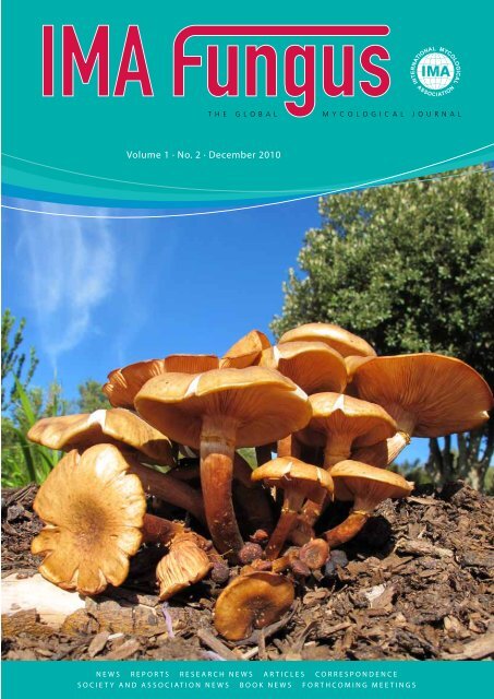
Cover: Sporophores of Armillaria<br />
mellea in Kirstenbosch Botanical<br />
Garden, Cape Town, South Africa<br />
EDITORIAL BOARD<br />
Editor-in-Chief<br />
Prof. dr D.L. Hawksworth CBE, Departamento de Biologia Vegetal II, Facultad de Farmacia, Universidad Comp<strong>lu</strong>tense de<br />
Madrid, Plaza Ramon y Cajal, 28040 Madrid, Spain; and Department of Botany, Natural History Museum, Cromwell<br />
Road, London SW7 5BD, UK; E-mail: d.hawksworth@nhm.ac.uk<br />
Layout Editors<br />
M.J. van den Hoeven-Verweij & M. Vermaas, CBS-KNAW Fungal Biodiversity Centre, P.O. Box 85167, 3508 AD<br />
Utrecht, The Netherlands; E-mail: m.verweij@cbs.knaw.nl<br />
Associate Editors<br />
Dr T.V. Andrianova, M.G. Kholodny Institute of Botany, Tereshchenkivska Street 2, Kiev, MSP-1, 01601, Ukraine;<br />
E-mail: tand@darwin.relc.com<br />
Prof. dr D. Begerow, Lehrstuhl für Evo<strong>lu</strong>tion und Biodiversität der Pflanzen, Ruhr-Universität Bochum, Universitätsstr.<br />
150, Gebäude ND 44780, Bochum, Germany; E-mail: dominik.begerow@rub.de<br />
Dr S. Cantrell, Department of Plant Pathology and Crop Physiology, Louisiana State University, Agricultural Centre, 455 Life<br />
Sciences Bldg., Baton Rouge, LA 70803, USA; E-mail: scantrel@suagm.edu<br />
Prof. dr D. Carter, Discipline of Microbiology, School of Molecular Biosciences, Building G08, University of Sydney,<br />
NSW 2006, Australia; E-mail: d.carter@mmb.usyd.edu.au<br />
Prof. dr P.W. Crous, CBS-KNAW Fungal Biodiversity Centre, P.O. Box 85167, 3508 AD Utrecht, The Netherlands; E-<br />
mail: p.crous@cbs.knaw.nl<br />
Prof. dr J. Dianese, Departamento de Fitopatologia, Universidade de Brasília, 70910-900 Brasília, D.F., Brasil; E-mail:<br />
jcarmine@unb.br<br />
Dr P.S. Dyer, School of Biology, Institute of Genetics, University of <strong>No</strong>ttingham, University Park, <strong>No</strong>ttingham NG7 2RD,<br />
UK; E-mail: paul.dyer@nottingham.ac.uk<br />
Dr M. Gryzenhout, Dept. of Plant Sciences, University of the Free State, P.O. Box 339, Bloemfontein 9300, South Africa;<br />
E-mail: Gryzenhoutm@ufs.ac.za<br />
Prof. dr L. Guzman-Davalos, Instituto de Botánica, Departamento de Botánica y Zoología, Universidad de Guadalajara,<br />
A.P. 1-139 Zapopan, 45101, México; E-mail: lguzman@cucba.udg.mx<br />
Dr K. Hansen, Kryptogambotanik Naturhistoriska Riksmuseet, Box 50007, 104 05 Stockholm, Sweden; E-mail: karen.<br />
hansen@nrm.se<br />
Prof. dr K.D. Hyde, School of Science, Mae Fah Luang University, Tasud, Chiang Rai, Thailand; E-mail: kdhyde3@gmail.<br />
com<br />
Prof. dr L. Lange, Dean of Research, Aalborg University, Denmark; E-mail: lla@adm.aau.dk<br />
Prof. dr L. Manoch, Department o Plant Pathology, Faculty of Agriculture, Kasetsart University, Bangkok 10900, Thailand;<br />
E-mail: agrlkm@ku.ac.th<br />
Prof. dr W. Meyer, Molecular Mycology Research Laboratory, CIDM, ICPMR, Level 3, Room 3114A, Westmead Hospital,<br />
Darcy Road, Westmead, NSW, 2145, Australia; E-mail: w.meyer@usyd.edu.au<br />
Dr D. Minter, CABI Bioservices, Bakeham Lane, Egham, Surrey, TW20 9TY, UK; E-mail: d.minter@cabi.org<br />
Dr L. <strong>No</strong>rvell, Pacific <strong>No</strong>rthwest Mycology Service, LLC, 6720 NW Skyline Boulevard, Portland, Oregon 97229-1309,<br />
USA; E-mail: llnorvell@pnw-ms.com<br />
Dr G. Okada, Microbe Division / Japan Collection of Microorganisms, RIKEN BioResource Center, 2-1 Hirosawa, Wako,<br />
Saitama 351-0198, Japan; E-mail: okada@jcm.riken.jp<br />
Prof. dr N. Read, Fungal Cell Biology Group, Institute of Cell and Molecular Biology, Rutherford Building, University of<br />
Edinburgh, Edinburgh EH9 3JH, UK; E-mail: nick@fungalcell.org<br />
Prof. dr K.A. Seifert, Research Scientist / Biodiversity (Mycology and Botany), Agriculture & Agri-Food Canada, K.W.<br />
Neatby Bldg, 960 Carling Avenue, Ottawa, ON, K1A OC6, Canada; E-mail: seifertk@agr.gc.ca<br />
Prof. dr J. Taylor, Department of Plant and Microbial Biology, University of California, 111 Koshland Hall, Berkeley, CA<br />
94720, USA; E-mail: jtaylor@berkeley.edu<br />
Prof. dr M.J. Wingfield, Forestry and Agricultural Research Institute (FABI), University of Pretoria, Pretoria 0002, South<br />
Africa; E-mail: mike.wingfield@fabi.up.ac.za<br />
Prof. dr W.-Y. Zhuang, Systematic Mycology and Lichenology Laboratory, Institute of Microbiology, Chinese Academy of<br />
Sciences, Beijing 100080, China; E-mail: zhuangwy@sun.im.ac.cn<br />
<br />
i m a f U N G U S
<strong>IMA</strong> vision<br />
My first opportunity to speak to the world’s mycological community through this co<strong>lu</strong>mn must begin with sincere<br />
thanks to the Past President of the International Mycological Association, Pedro Crous of the Centraalbureau voor<br />
Schimmelcultures and the Universities of Utrecht and Wageningen in the Netherlands. Under President Crous the scope<br />
of the <strong>IMA</strong> was broadened well beyond its traditional focus on the quadrennial International Mycological Congress while,<br />
at the same time, sponsoring a most memorable IMC9 in Edinburgh. Regarding IMC9, thanks are especially due to the<br />
IMC9 organizing committee, consisting of chair Nick Read and members Simon Avery, Nick Clipson, Geoff Gadd, and<br />
Neil Gow. Thanks also are due the Elsevier group, led by Nina Cosgrove. I also want to thank Karen Hansen for agreeing<br />
to serve a second term as <strong>IMA</strong> Treasurer and Dominik Begerow for accepting the task of <strong>IMA</strong> Secretary.<br />
EDITORIAL<br />
Over the past four years, the<br />
foundation for a reinvigorated<br />
<strong>IMA</strong> was laid in the form of new<br />
statutes, which were ratified at the General<br />
Assembly of the IMC in Edinburgh and which<br />
can be viewed at the <strong>IMA</strong> website using this<br />
URL: . The new statutes are more explicit<br />
than the old about membership in the <strong>IMA</strong>,<br />
about election of <strong>IMA</strong> officers and members<br />
of the <strong>IMA</strong> Executive Committee, about<br />
proposals to host future IMCs, and about<br />
awards made by the <strong>IMA</strong>.<br />
Given that the mission of the <strong>IMA</strong> is “the<br />
promotion and encouragement of mycology<br />
throughout the world,” the <strong>IMA</strong> website,<br />
initiated by previous <strong>IMA</strong> officers, inc<strong>lu</strong>ding<br />
Gioconda San Blas, Trond Schumacher and<br />
Meredith Blackwell, has assumed increased<br />
importance. The site <br />
has become essential for coordinating the<br />
activities of the six <strong>IMA</strong> Regional Mycological<br />
Member Organizations, which represent<br />
Africa, Asia, Australasia, Europe, <strong>No</strong>rth<br />
America and South America.<br />
A very significant contribution to the<br />
mission of global mycology is the transfer of<br />
responsibility for MycoBank from CBS to<br />
the <strong>IMA</strong>. MycoBank ()<br />
aims to host nomenclatural, genetic and<br />
phenotypic data for all described fungi and<br />
to make that information freely available to<br />
all of science. The <strong>IMA</strong> membership clearly<br />
appreciates the worth of MycoBank, having<br />
voiced 86 % support that “Deposition of key<br />
nomenclatural information in one or more<br />
approved depositories (e.g., MycoBank)<br />
should be made mandatory for the valid<br />
publication of new fungal names.”<br />
A second, significant contribution is<br />
<strong>IMA</strong> <strong>Fungus</strong>, which you, dear reader, clearly<br />
appreciate. Deep thanks are due to Pedro<br />
Crous and <strong>IMA</strong> <strong>Fungus</strong> Editor David<br />
Hawksworth, and to the members of the <strong>IMA</strong><br />
Executive Council, who now also wear the<br />
hats of Associate Editors of <strong>IMA</strong> <strong>Fungus</strong>.<br />
The third major contribution concerns<br />
<strong>IMA</strong> awards. IMC9 witnessed the resurrection<br />
of the two awards traditionally made by<br />
the <strong>IMA</strong>, but absent since 1996 -- the De<br />
Bary Medal for outstanding career research,<br />
awarded at IMC9 to Franz Oberwinkler,<br />
and the Ainsworth Medal for extraordinary<br />
service to world mycology, awarded at IMC9<br />
to both Emory Simmons and Richard Korf.<br />
In addition, <strong>2010</strong> saw the initiation of a new<br />
set of awards, the <strong>IMA</strong> Young Mycologist<br />
Awards, designed to honor the research<br />
accomplishments of young mycologists from<br />
each of the six regions. The first awards, which<br />
we had hoped to make in time for IMC9, will<br />
be determined over the next year and awarded<br />
in 2014, along with the second set of <strong>IMA</strong><br />
Young Mycologist Awards to be determined<br />
early in 2014. The announcement about the<br />
<strong>IMA</strong> Young Mycologist Awards is in this<br />
issue of <strong>IMA</strong> <strong>Fungus</strong> and can also be found at<br />
.<br />
Mention of 2014 brings us to the final<br />
contribution, which is centered on selection<br />
of Thailand as the site for IMC10, with Leka<br />
Manoch as chair of the IMC10 Organizing<br />
Committee. To establish continuity between<br />
IMCs, the new statues tap the experience of the<br />
past chair of the IMC organizing committee<br />
by appointing this person as one of two <strong>IMA</strong><br />
Vice-Presidents, the other being the chair of the<br />
next IMC organizing committee, in this case,<br />
Nick Read and Leka Manoch, respectively.<br />
To further encourage continuity, one to three<br />
members of the <strong>IMA</strong> executive council will<br />
serve on the IMC10 program committee along<br />
with mycologists from host nation and region.<br />
Although the focus of the <strong>IMA</strong> remains<br />
the quadrennial IMC, the meeting now<br />
shares the stage with the other contributions<br />
that the <strong>IMA</strong> makes to global mycology,<br />
through the <strong>IMA</strong> website, MycoBank, <strong>IMA</strong><br />
<strong>Fungus</strong>, and support provided by the <strong>IMA</strong> to<br />
Regional Mycological Member Organizations.<br />
Clearly, the tasks and opportunities have<br />
grown beyond what can be attended to solely<br />
by the officers and Executive Committee of<br />
the <strong>IMA</strong>. Over the next four years, to ensure<br />
that the innovations made over the past four<br />
years succeed, I will be calling on the Regional<br />
Mycological Member Organizations and<br />
their members to work for the <strong>IMA</strong> in all<br />
of these areas. I certainly hope that you, the<br />
membership, will help us move the <strong>IMA</strong> and<br />
mycology forward to 2014.<br />
John Taylor President, <strong>IMA</strong><br />
(Berkeley, CA, USA; jtaylor@berkeley.edu)<br />
v o l u m e 1 · n o . 2 <br />
(1)
News<br />
The home stretch for fungal barcoding<br />
In 2009, the plant research community published<br />
a paper (Hollingsworth et al. 2009)<br />
that designated two genes as a barcode for<br />
all plants. This means that among the major<br />
groups of eukaryotes, only the Fungi remain<br />
without a formal barcode. This may come<br />
as a surprise to many mycologists, who continue<br />
to loosely apply the term ‘barcode’ in<br />
numerous papers and projects. Importantly,<br />
barcodes are intended to be used as identification<br />
markers and may have properties,<br />
such as unalignable hypervariable regions,<br />
that make them inappropriate as phylogenetic<br />
markers. A strict set of criteria for barcodes<br />
has been established ()<br />
and GenBank will only apply barcode tags<br />
to sequences of the genes or loci accepted as<br />
barcodes for a particular taxonomic group<br />
by the Consortium for the Barcode of Life<br />
(CBOL). Despite its long history of DNA<br />
sequencing and diverse ecological interests,<br />
the mycological community has not engaged<br />
with CBOL and associated organisations<br />
with much enthusiasm. At two major<br />
conferences in <strong>2010</strong> (the Mycological Society<br />
of America in Lexington, Kentucky, and<br />
the International Mycological Congress in<br />
Edinburgh), we suggested that it is in mycology’s<br />
best interest to engage and collaborate<br />
with the international barcode community,<br />
and judging by the response after these sessions,<br />
this message was received clearly by a<br />
significant number of mycologists.<br />
The road towards adoption of a fungal<br />
barcode has already had numerous twists<br />
and turns (Seifert 2009). An important<br />
milestone was a meeting of 37 mycologists<br />
from 12 countries at Front Royal, Virginia,<br />
in May 2007. At this meeting, participants<br />
were unanimous that the 5.8S nuclear ribosomal<br />
gene and its two flanking spacer<br />
regions (ITS) was the most likely candidate<br />
for a universal fungal barcode, despite some<br />
promising but mixed results for the default<br />
barcode sanctioned by CBOL, COI (Seifert<br />
et al. 2007). For a gene other than COI to<br />
be granted status as a barcode, CBOL has<br />
established a set protocol for validation.<br />
For mycologists, this process inc<strong>lu</strong>des the<br />
publication of a proposal comparing the ITS<br />
against a set of other candidates. We intend<br />
to publish a paper in 2011 and together<br />
with an international group of interested<br />
mycologists, we are gathering and generating<br />
data for this purpose (Eberhardt <strong>2010</strong>).<br />
Until now we received commitments from<br />
colleagues covering more than 40 c<strong>lu</strong>sters<br />
of five or more sibling species, covering 12<br />
of the main fungal lineages identified in<br />
phylogenies generated from ‘Assembling the<br />
Fungal Tree of Life’ (AFTOL) project. The<br />
final paper will probably involve more than<br />
50 coauthors from more than 12 countries<br />
and we intend to keep participation open to<br />
all interested parties until the data gathering<br />
deadline is passed in February 2011. More<br />
details can be found at the discussion group<br />
‘Fungi’ on connect.barcodeoflife.net and the<br />
project website().<br />
As we set our sites on this publication, it<br />
is also important to look at projects and opportunities<br />
that may follow. We hope that the<br />
energy now building within the community<br />
will accelerate and improve collaborations<br />
between like-minded researchers across<br />
national borders. One pressing need is to<br />
coordinate and accelerate ongoing efforts to<br />
barcode authoritatively identified specimens<br />
and type cultures in herbaria and culture collections.<br />
Equally critical is coping with the<br />
vast amount of sequences generated by environmental<br />
sequencing projects.<br />
Making a dent in this mountain of<br />
Fig 1. A. Zygospores of Zygorhynchus heterogamous. B. The yeast Cryptococcus macerans. C. Basidiomata of Hygrocybe cantharel<strong>lu</strong>s. D. Basidiomata of Tremiscus<br />
helvelloides. E. Xanthoria parietina on Walter Gams’ roof in Bomarzo, Italy. F. Helicoconidia of Helicomyces roseus. G. Tar spot of sugar maple caused by Rhytisma<br />
acerinum. H. Morchella elata ready for further processing. Photos: Keith Seifert.<br />
(2)<br />
<br />
i m a f U N G U S
undescribed fungal diversity will require a<br />
monumental community effort to update<br />
and curate our sequence databases with accurate<br />
sequences tied to well validated samples;<br />
barcoding will be central to this. In addition,<br />
barcoding should provide excellent opportunities<br />
to expand the boundaries of fungal<br />
taxonomy by engaging teachers and amateur<br />
groups to help collect, locate and identify<br />
samples that could eventually be identified<br />
or validated by barcoding. Much is possible,<br />
but first we urgently need to formally declare<br />
a fungal barcode. We must not let this opportunity<br />
slip away to finally clear this hurdle.<br />
Conrad Schoch and Keith Seifert<br />
(schoch2@ncbi.nlm.nih.gov; and keith.<br />
seifert@agr.gc.ca)<br />
NEWS<br />
Eberhardt U (<strong>2010</strong>) A constructive step towards selecting a DNA barcode for fungi. New Phytologist 187: 266–268.<br />
Hollingsworth PM, Forrest LL, Spouge JL, Hajibabaei M, Ratnasingham S, et al. (2009) A DNA barcode for land plants. Proceedings of the National Academy of Sciences,<br />
USA 106: 12794–12797.<br />
Seifert KA (2009) Progress towards DNA barcoding of fungi. Molecular Ecology Resources 9: 83–89.<br />
Seifert KA, Samson RA, Dewaard JR, Houbraken J, Levesque CA, et al. (2007) Prospects for fungus identification using C01 DNA barcodes, with Penicillium as a test<br />
case. Proceedings of the National Academy of Sciences, USA 104: 3901–3906.<br />
Fungi and the Convention on Biological Diversity<br />
Recognizing that fungi are not plants, but need their own conservation strategies, is a first step to proper<br />
protection for species like critically-endangered Pleurotus nebrodensis, the only mushroom currently on the<br />
IUCN red-data list. Photo: David Minter.<br />
The tenth follow-up conference of the parties<br />
to the 1992 Rio Convention on Biological<br />
Diversity was held in Nagoya, Japan on<br />
18–29 October <strong>2010</strong>. For the first time at<br />
these meetings, as a result of lobbying from<br />
the new International Society for Fungal<br />
Conservation (see Reports in this issue), and<br />
the Cup Fungi, Truffles and their Allies specialist<br />
group of the IUCN Species Survival<br />
Commission, fungi were explicitly considered<br />
independently from animals and plants.<br />
The focus of the lobbying was section<br />
E, paragraph 10 of the Global Strategy for<br />
Plant Conservation. The wording of that<br />
paragraph read, “accordingly the Strategy<br />
addresses the Plant Kingdom with main<br />
focus on higher plants, and other welldescribed<br />
groups such as Bryophytes and<br />
Pteridophytes. This does not imply that<br />
these lower groups do not have important<br />
ecological functions, nor that they are not<br />
threatened. Parties may choose on a national<br />
basis to inc<strong>lu</strong>de other taxa, inc<strong>lu</strong>ding algae,<br />
lichens and fungi”. It was pointed out to<br />
delegates of the conference that the final<br />
sentence of this paragraph gave the mistaken<br />
impression that fungi are “lower” plants,<br />
that “lichens” are different from fungi, and<br />
that strategies for fungal conservation could<br />
be treated as an optional extra.<br />
At Nagoya, delegates discussed these<br />
concerns and agreed that the wording needed<br />
to be changed. The revised and agreed<br />
wording reads, “while the Strategy addresses<br />
the plant kingdom with main focus on higher<br />
plants, and other well-described groups<br />
such as bryophytes and pteridophytes; Parties,<br />
other governments and other relevant<br />
stakeholders may consider developing conservation<br />
strategies for other groups such as<br />
algae and fungi (inc<strong>lu</strong>ding lichen-forming<br />
species)”.<br />
The revised wording recognizes that lichens<br />
are fungi, and that fungi are not plants.<br />
It also recognizes the possibility that fungi<br />
should have their own separate strategy. In<br />
1992, the Convention established that all<br />
groups of living organisms have the right to<br />
exist on this planet and to be protected. Up to<br />
now, the Convention has not lived up to that<br />
promise in respect of the fungi. By explicitly<br />
recognizing fungi as different from plants, this<br />
re-wording can be seen as a small but significant<br />
first step towards getting the convention<br />
to honour its promise for the fungi.<br />
The Convention also agreed key targets<br />
for 2020, which inc<strong>lu</strong>ded commitments<br />
to: cut the rate of loss of natural habitats by<br />
at least half; to increase terrestrial nature<br />
reserves from 13 % to 17 % of the world’s<br />
land area; increase marine and coastal nature<br />
reserves from 1 % to 10 % of the world’s<br />
seas; restore at least 15 % of the areas where<br />
biodiversity is classed as ‘degraded’; and<br />
safeguard at least 75 % of plant species in<br />
collections. For further information see<br />
.<br />
David W Minter<br />
(d.minter@cabi.org)<br />
v o l u m e 1 · n o . 2 <br />
(3)
News<br />
Mycologists go political, with success<br />
A critically important message in this<br />
International Year of Biodiversity is to<br />
promote the missing “F” word at every<br />
opportunity – Fauna, Flora, AND<br />
Fungi. This was part of a rallying call to<br />
all mycologists delivered in a recent IMC<br />
paper by Dave Minter, and through the<br />
establishment of the International Society<br />
for Fungal Conservation during IMC9.<br />
Already, the Society’s founding members<br />
have demonstrated the power of global<br />
coordination to inf<strong>lu</strong>ence a bastion of public<br />
broadcasting – the BBC.<br />
On 15 October <strong>2010</strong>, the BBC’s<br />
“The World Tonight” programme focused<br />
attention on the biodiversity crisis ahead<br />
of the COP 10 meeting in Nagoya.<br />
Although an engaging and well-produced<br />
45-min programme, it perpetuated the<br />
simplification of biodiversity to “fauna<br />
and flora”. A call to action went out from<br />
Dave, and mycologists globally responded<br />
by petitioning the BBC. Dave sent e-mails<br />
to 180 people, all the Founder Members of<br />
the new International Society for Fungal<br />
Conservation. At least 62 messages were<br />
sent to the BBC, which came from a<br />
minimum of 29 countries: Argentina,<br />
Australia, Brazil, Canada, China, Cuba,<br />
Ecuador, Egypt, France, Ghana, Greece,<br />
India, Italy, Malaysia, Mexico, New<br />
Zealand, Philippines, Poland, Puerto Rico,<br />
Russia (<strong>No</strong>vosibirsk oblast, St Petersburg<br />
oblast, Tula oblast; i.e. Russia in Asia and<br />
in Europe), Serbia, South Africa, Spain,<br />
Sweden, Ukraine, UK, United Arab<br />
Emirates, USA, and Zimbabwe.<br />
Is this the first ever global political<br />
action by mycologists? That appears to be<br />
the case. The result was a triumph; within<br />
hours the BBC producer Alistair Burnett<br />
by way of the BBC editors’ blog ()<br />
apologised for the omission, stating:<br />
“The UN’s member states are getting<br />
together next week ...... to stop the loss<br />
of plants, animals and fungi species -<br />
or biodiversity - ......Just a note to our<br />
listeners who’ve e-mailed us in the past<br />
day pointing out that we failed to mention<br />
fungi when describing what biodiversity<br />
is, my apologies, we could have been more<br />
explicit.”<br />
The word “fungi” was hyperlinked to an<br />
excellent blog about the fundamental<br />
importance of fungi: .<br />
Peter Buchanan<br />
(BuchananP@landcareresearch.co.nz)<br />
Mushrooms – the new plastic?<br />
Styrofoam is one of the most egregious<br />
offenders in the disposable plastics category,<br />
US $ 20 Bn of this material is produced<br />
annually and used in products as diverse<br />
as building insulation, coffee cups, and<br />
packaging; it is said to occupy around <br />
25 % of landfill sites, and will stay there for<br />
100s of years. In the oceans it can break into<br />
smaller and smaller globules, but these do<br />
not really ever disappear. Gavin MacIntyre<br />
and Eben Bayer (Ecovative Design, Green<br />
Island, NY, USA), with financial support<br />
from the National Science Foundation<br />
(NSF), have been developing a way to<br />
transform agricultural wastes, especially<br />
seed husks, with basidiomycete mycelia to<br />
form an entirely new class of materials,<br />
which perform much like plastics, but as<br />
they are made from crop waste differ in<br />
being totally compostable when no longer<br />
required. The waste material is transformed<br />
into a chitinous polymer, Mycobond that<br />
can be moulded into different shapes and<br />
used in products ranging from packaging<br />
to insulation boards for the construction<br />
industry (Greensulate). The inventors<br />
have developed a continuous system<br />
which cleans, cooks, cools and pasteurizes<br />
the waste materials, while continuously<br />
inoculating them with the mycelium. The<br />
resultant stream of material can be made<br />
into almost any shape which self-assembles<br />
in a mould. This invention is particularly<br />
exciting as there is potential for different<br />
locally available materials to be used<br />
according to what agricultural wastes are<br />
available in a country. The basidiomycete<br />
fungi utilized are not, however, named in<br />
the released information.<br />
For further information on this<br />
most exiting and novel discovery,<br />
which has such enormous economic<br />
potential and environmental benefits,<br />
see the video on , and the NSF report<br />
on .<br />
White truffles still demand a high price<br />
In times of austerity, it might have been<br />
reasonable to expect that <strong>lu</strong>xury and<br />
gourmet foods would drop in va<strong>lu</strong>e.<br />
However, at the Fiera Nazionale del Tartufo<br />
Bianco d’Alba, the annual truffle fair in the<br />
town of Alba in the Piedmont district of<br />
northern Italy, which ran from 9 October to<br />
14 <strong>No</strong>vember <strong>2010</strong>, a single 900 g specimen<br />
of Tuber magnatum, the Alba White Truffle,<br />
was sold to a Hong Kong buyer for a massive<br />
105 000 € (US $ 141 605), i.e. 117 € g -1 . The<br />
previous record paid for a single truffle was<br />
one of 1.5 kg which sold to a Macau casino<br />
owner for £ 165 000 (194 330 €, US $ 262<br />
255) in 2007, equating to 129 € g -1 .<br />
(4)<br />
<br />
i m a f U N G U S
Fungi on German TV: Focus on black fungi<br />
Sybren de Hoog and Bert Gerrits van den<br />
Ende of CBS (Utrecht) featured on Germany’s<br />
second TV network in an episode of<br />
the ZDF science program ‘Abenteuer Wissen’.<br />
The CBS homepage ()<br />
has a link to the original program, as well as<br />
a version with English subtitles. De Hoog<br />
explained on the background of a brain<br />
infection after near-drowning of a child,<br />
published by Mursch et al. (2006). This<br />
concerned a 2-year-old boy who fell into a<br />
rainwater container and aspirated pol<strong>lu</strong>ted<br />
water for several minutes. The child was reanimated<br />
and did well, but later he became<br />
sleepy and fell into a deep coma. Multiple<br />
fungal brain abcesses were observed and<br />
removed by surgery. The etiologic agent<br />
proved to be Scedosporium apiospermum,<br />
one of the very few species consistently associated<br />
with near-drowning fungal encephalitis<br />
(Guarro et al. 2006), which is almost<br />
always fatal. The boy was saved thanks to<br />
timely diagnostics and appropriate therapy.<br />
De Hoog and coworkers recently published<br />
a special issue of Medical Mycology<br />
on Scedosporium (47 (4), 2009: ‘Pseudallescheria<br />
and Scedosporium: emerging<br />
opportunists’, JP Bouchara et al., eds),<br />
stemming from a series of Workshops on<br />
this group of fungi. They are abundantly<br />
present in stagnant, nutrient-rich waters,<br />
as well as in poorly aerated industrial and<br />
agricultural soils. Alkane pol<strong>lu</strong>tion is a promotive<br />
factor. Investigations are ongoing by<br />
the group of Johannes Rainer in Innsbruck<br />
to utilize S. dehoogii for bioremediation of<br />
diesel-pol<strong>lu</strong>ted sites. The taxonomy of the<br />
genus is still under debate (Gilgado et al.<br />
Processing Scedosporium samples, recorded by a ZDF TV team for ‘Abenteur Wissen’, a science program on<br />
Germany’s second network.<br />
2005, Lackner et al. <strong>2010</strong>), but it is clear<br />
that a complex of species is concerned each<br />
occupying different slots in an ecological<br />
continuum. Scedosporium apiospermum has<br />
a bias towards virulence, whereas S. dehoogii<br />
at the opposite end of the spectrum is almost<br />
strictly environmental.<br />
There are more fungi inhabiting oils<br />
and toxic pol<strong>lu</strong>tants. Hans-Michael Fürst<br />
demonstrated frequent isolation of Hormoconis<br />
resinae, not only from hydrocarbonpol<strong>lu</strong>ted<br />
soils, but also as a monoculture in<br />
the oil tanks and machinery of ships (Fürst<br />
2000). Biofouling by this fungus may cause<br />
engines to stop, which potentially is quite a<br />
threat to aviation. But authorities will never<br />
admit that there is a problem, said Dr Fürst.<br />
Growth of Hormoconis is also promoted by<br />
phenol-rich creosote used in wood protection,<br />
in which respect there is a striking resemblance<br />
to black yeasts (Prenafeta-Boldú<br />
et al. 2006). Chaetothyrialean black yeasts<br />
are massively isolated from treated wood by<br />
enrichment with phenolic aromates (Zhao<br />
et al. <strong>2010</strong>). Chaetothyrialean black yeasts<br />
differ by combining this feature with oligotrophy<br />
(Satow et al. 2008), which explains<br />
their abundance in steam baths and dishwashers<br />
(Zalar et al. 2011).<br />
Black yeasts belonging to the orders<br />
Dothideales and Capnodiales are mainly<br />
extremophiles, surviving harsh conditions<br />
of heat, cold, dryness and solar irradiation.<br />
Their evo<strong>lu</strong>tionary origin lies far back, when<br />
they occupied micro-niches in crevices<br />
between crystals in bare rock of deserts,<br />
mountains or the Antarctic (Selbmann et al.<br />
2005). On German TV, Thomas Warscheid<br />
gave a more recent example of their rockinhabiting<br />
life style in the deterioration of<br />
the famous Dom of Cologne, Germany.<br />
Monuments are crumbling under the slow<br />
but effective force of black fungi. Earlier<br />
Warscheid observed the same thing happening<br />
with the temples of Angkor Wat in<br />
Cambodja, as he explained in The New York<br />
NEWS<br />
Blackish discoloured skin of toes and toe webs with scaling. [from Li et al.<br />
(2008) Studies in Mycology 61: 131–136].<br />
Skin biopsy stained with HAE; some fungal elements are visible (arrows). Bar = 5<br />
µm. [from Li et al. (2008) Studies in Mycology 61: 131–136].<br />
v o l u m e 1 · n o . 2 <br />
(5)
News<br />
Times of 24 June 2008. The fungi utilize<br />
hydrocarbon air pol<strong>lu</strong>tants and assimilation<br />
products of algae, acidify their environment,<br />
and finally turn all buildings black. Aspects<br />
of this behaviour were explained in a special<br />
issue of Studies in Mycology (61, 2008:<br />
‘Black Fungal Extremes’, GS de Hoog & M<br />
Grube, eds), while medically significant species<br />
were presented in one of Medical Mycology<br />
(47(1), 2009; ‘The Dark World of Black<br />
Fungi’, Vitale et al., eds). Fungal Biology will<br />
also devote a special issue to black yeasts<br />
shortly, entitled ‘The Emerging Potential of<br />
Black Yeasts’ (Gunde-Cimerman et al., eds).<br />
Black fungi are remarkable for their odd<br />
ecology, which we still have a long way to go<br />
towards understanding. In ‘Abenteuer Wissen’,<br />
Axel Brakhage explained his research on<br />
Aspergil<strong>lu</strong>s fumigatus, a ubiquitous compost<br />
fungus which is hypothesized to express factors<br />
enhancing escape from phagocytosis when<br />
confronted with human alveolar macrophages<br />
(Behnsen et al. 2007). Similar processes are<br />
to be expected when black yeasts invade our<br />
homes and our bodies from the specialized<br />
niches somewhere in nature (Sudhadham et<br />
al. 2008). They differentially adapt to a highly<br />
deviating environment using different combinations<br />
of the essential factors determining<br />
their natural habitat. Gerrits van den Ende et<br />
al. (2011) applied the term ‘ecological fitting’<br />
to this process enabling rapid host and environmental<br />
shifts, eventually leading to sympatric<br />
speciation. Many extremophiles, however,<br />
already occupy their ultimate habitat and don’t<br />
have an adaptive space around them. They are<br />
likely to have entered a dead-end street and are<br />
likely to become extinct when climate changes<br />
dramatically, as it is expected to do.<br />
Sybren de Hoog<br />
(s.hoog@cbs.knaw.nl)<br />
Behnsen J, Narang P, Hasenberg M, Gunzer F, Bilitewski U, et al. (2007) Environmental dimensionality controls the interaction of phagocytes with the pathogenic fungi<br />
Aspergil<strong>lu</strong>s fumigatus and Candida albicans. PLoS Pathogens 3: e13.<br />
Fürst HM (2000) Ökologie des Hyphenpilzes Hormoconis resinae und Eigenschaften seines n-Alkan-induzierten P450-Monooxygenasesystems. Thesis. Technische Universität<br />
Berlin.<br />
Gilgado F, Cano J, Gené J, Guarro J (2005) Molecular phylogeny of the Pseudallescheria boydii species complex: proposal of two new species. Journal of Clinical Microbiology<br />
43: 4930–4942.<br />
Guarro J, Kantarciog<strong>lu</strong> AS, Horré R, Rodriguez-Tudela JL, Cuenca Estrella M, et al. (2006) Scedosporium apiospermum: changing clinical spectrum of a therapy-refractory<br />
opportunist. Medical Mycology 44: 295–327.<br />
Lackner M, Gerrits van den Ende AHG, Hoog GS de, Kaltseis J (<strong>2010</strong>) Barcoding of the therapy-refractory species of Pseudallescheria and Scedosporium. Medical Mycology:<br />
in press.<br />
Mursch K, Trnovec S, Ratz H, Hammer D, Horré R, et al. (2005) Successful treatment of multiple Pseudallescheria boydii brain abscesses and ventriculitis/ependymitis<br />
in a 2-year-old child after a near-drowning episode. Child’s Nervous System 22: 189–192.<br />
Prenafeta-Boldú FX, Summerbell R, Hoog GS de (2006) Fungi growing on aromatic hydrocarbons: biotechnology’s unexpected encounter with biohazard? FEMS<br />
Microbiology Reviews 30: 109–130.<br />
Satow MM, Attili-Angelis D, Hoog GS de, Angelis DF, Vicente VA (2008) Selective factors involved in oil flotation isolation of black yeasts from the environment.<br />
Studies in Mycology 61: 157–163.<br />
Selbmann L, Hoog GS de, Mazzaglia A, Friedmann EI, Onofri S (2005) Fungi at the edge of life: cryptendolithic black fungi from Antarctic desert. Studies in Mycology<br />
51: 1–32.<br />
Sudhadham M, Prakitsin S, Sivichai S, Chaiyarat R, Dorrestein GM, et al. (2008) The neurotropic black yeast Exophiala dermatitidis has a possible origin in the tropical<br />
rain forest. Studies in Mycology 61: 145–155.<br />
Zhao J, Zeng J, Hoog GS de, Attili-Angelis D, Prenafeta-Boldú FX (<strong>2010</strong>) Isolation and identification of black yeasts by enrichment on atmospheres of monoaromatic<br />
hydrocarbons. Microbial Ecology 60: 149–156.<br />
Executive Committee and Officers of the International Mycological Association (2006–<strong>2010</strong>).<br />
(6)<br />
<br />
i m a f U N G U S
REPORTS<br />
IMC9 Edinburgh: a selection of memorable moments featuring some of those who helped to make it such a success.<br />
v o l u m e 1 · n o . 2 <br />
(7)
REPORTS<br />
IMC9: The Biology of Fungi<br />
A personal reflection<br />
The idea of holding IMC9 in Edinburgh in<br />
<strong>2010</strong> started five years ago when I received<br />
a phone call from Ellen Collingsworth at<br />
the Edinburgh Convention Bureau. She<br />
said that the last (and the first) International<br />
Mycological Congress (IMC) to be held<br />
in the UK was in 1971 in Exeter, and<br />
wouldn’t I like to organize the next one?<br />
My response was initially very negative (I<br />
will omit the expletives I used) because I<br />
was very conscious of the profound effect<br />
that organizing an IMC would have on<br />
both my academic and personal life over<br />
the next five years. However, after having<br />
many discussions with senior officers of<br />
the British Mycological Society (BMS),<br />
seeing the proposed Congress venue (the<br />
superb Edinburgh International Conference<br />
Centre, EICC), and giving the matter<br />
considerable thought, I strongly warmed to<br />
the idea of taking on this onerous job. I felt<br />
that it presented some exciting challenges<br />
which ultimately could have a big impact on<br />
global mycology.<br />
Early in 2006, the ECB, BMS, and<br />
myself put together and submitted a bid to<br />
the <strong>IMA</strong> to hold IMC9 in Edinburgh, with<br />
the BMS agreeing to act as host. I received<br />
extremely strong support from the BMS<br />
Council, who generously agreed to commit<br />
£ 100,000 to pump-prime the Congress.<br />
I took on the organization of IMC9<br />
with a clear vision of how I wanted it to<br />
be: (1) the whole of mycology had to be<br />
represented in all of its guises in a very<br />
balanced way across the immense breadth of<br />
the subject -- to try and give a flavour of this<br />
and provide the Congress with an up-todate<br />
image, I subtitled it The Biology of<br />
Fungi which I felt might appeal to a broader<br />
range of scientists working on fungi; (2)<br />
the Congress had to have a stellar scientific<br />
programme with a strong emphasis on where<br />
the excitement of the subject would be at in<br />
<strong>2010</strong>, and where its big areas of impact will<br />
be in the future -- it was essential that the<br />
conference programme and speakers should<br />
inspire young and old mycologists alike; (3)<br />
the scientific programme should evolve by<br />
a very carefully regulated process of natural<br />
selection in which only the best symposia<br />
proposed by the community would be<br />
Delegates being led from the Usher Hall to the EICC following the Opening Ceremony by Scottish pipers.<br />
chosen; (4) it would be compulsory for<br />
every symposium to have young researchers<br />
(postgrads and/or postdocs) giving talks;<br />
(5) the poster sessions would be given a<br />
high profile and be very accessible to the<br />
delegates in areas where they congregated<br />
in the Congress venue (e.g. at <strong>lu</strong>nchtime);<br />
(6) delegates should be able to experience<br />
the delights of the City of Edinburgh and<br />
all that it has to offer as one of the world’s<br />
main cultural heritage sites -- here I felt<br />
that it was important that the timing of<br />
the Congress should be at the beginning of<br />
August just before the Edinburgh Festival<br />
when hotel prices reach their maximum; (7)<br />
the conference party should be at the end of<br />
the conference when people could properly<br />
relax and let their hair down. I openly<br />
declared that this should be the “conference<br />
party to end all conference parties”; and (8)<br />
I made it clear to everyone involved in the<br />
organization that we should try our hardest<br />
to make IMC9 the best IMC ever! From<br />
my perspective, even with my somewhat<br />
biased point of view, I felt that my vision for<br />
IMC9 was largely if not completely fulfilled.<br />
However, this only came about as a result of<br />
the team effort of hundreds of participating<br />
individuals.<br />
Once having won the bid and the dust<br />
from IMC8 in Cairns having settled, I<br />
set about finding a suitable Professional<br />
Conference Organizer (PCO) with the help<br />
of Geoff Robson and Nick Clipson on the<br />
Steering Group. To organize a Congress of<br />
the size of an IMC (our best guesstimate was<br />
we that we would attract between 1200 and<br />
2500 delegates to Edinburgh), it was abso<strong>lu</strong>tely<br />
essential to have an extremely good<br />
PCO to work closely with. Amongst other<br />
things, the PCO is responsible for most of<br />
the conference administration, invitation of<br />
speakers and poster presenters, organization<br />
of delegate registration, communication<br />
with delegates, interfacing with the venues,<br />
organizing the exhibition, obtaining<br />
sponsorship, marketing the conference, etc,<br />
etc. Any naïve notion that I could organize a<br />
conference on this scale without a PCO was<br />
quickly kicked into touch once it became<br />
clear how massive the task of organizing a<br />
conference on this size is. We short-listed<br />
three PCOs and finally took on board<br />
Elsevier who, as well being publishers, have<br />
a big PCO Department. There were several<br />
key issues in Elsevier’s favour over the other<br />
PCOs we interviewed. What was particularly<br />
significant for the BMS was that Elsevier<br />
were the only PCO to agree to take on<br />
the complete financial risk for the Congress<br />
if it went ‘belly up’ (e.g. due to volcanic dust,<br />
acts of terrorism, fears of epidemics). We<br />
were concerned that an ‘act of God’ could<br />
potentially result in bankrupting the BMS.<br />
The cost of organizing IMC9 approached a<br />
£ 1 million! Another aspect strongly in Else-<br />
(8)<br />
<br />
i m a f U N G U S
vier’s favour was that they published four<br />
journals for the BMS, and IMC9 presented<br />
various exciting publishing opportunities.<br />
We also felt that Elsevier, with its experience<br />
in publishing, would be able to market the<br />
Congress well and give it the image we felt<br />
that it should have. We did not want IMC9<br />
to have a boring clinical feel to it, which is<br />
so typical of many conferences. There is no<br />
question that we made the right choice, and<br />
the Elsevier team, led by the inimitable Nina<br />
Cosgrove, were outstanding and a joy for me<br />
to work with over the last four years.<br />
One of my aims was that the scientific<br />
programme should present the whole<br />
breadth of mycology in a very balanced way<br />
without any single topic dominating. In<br />
consultation with the Steering Committee,<br />
I divided the subject into five themes<br />
which I felt represented the main areas of<br />
the subject, and I gave these themes equal<br />
weighting. These five themes were:<br />
1. Cell Biology, biochemistry and<br />
physiology<br />
2. Environment, ecology and interactions<br />
3. Evo<strong>lu</strong>tion, biodiversity and systematics<br />
4. Pathogenesis and disease control<br />
5. Genomics, genetics and molecular<br />
biology<br />
Across all five themes ran applied aspects of<br />
the subject (e.g. fungal biotechnology).<br />
I set up a number of Committees to<br />
bring the Congress organization, and particularly<br />
the scientific programme, to fruition.<br />
First, we had a Steering Group of six individuals<br />
chaired by me that had an advisory<br />
role and oversaw the Congress organization.<br />
Second, we had a Scientific Programme<br />
Committee comprising the chairs of the five<br />
scientific themes, and this committee was<br />
also chaired by me. And then finally we had<br />
the five scientific theme committees each<br />
containing five eminent scientists covering<br />
the breadth of each theme.<br />
We next invited the mycological<br />
community to propose symposia for the<br />
scientific programme. We had decided that<br />
it would be possible to hold 45 symposia,<br />
each 2.5 h long with seven speakers during<br />
five days of the Congress (Monday–Friday).<br />
This equated to nine symposia per theme.<br />
However, we made sure that many of the<br />
symposia were inter-thematic. Amazingly,<br />
and a tribute to the enthusiasm of the<br />
mycological community, we received over<br />
Delegates in session at the EICC.<br />
220 symposium proposals. The five scientific<br />
theme committees then set to work<br />
to prioritize these and the final selection<br />
was made by the Scientific Programme<br />
Committee. As you can imagine, this was<br />
an extremely difficult task because we had<br />
so many outstanding proposals for the<br />
45 symposium topics. Because we had so<br />
many excellent suggestions that didn’t make<br />
the cut as symposia, we decided that these<br />
should be converted into Special Interest<br />
Group meetings to be held on the Sunday<br />
before the Opening Ceremony. Finally, we<br />
were also able to hold three <strong>No</strong>menclature<br />
Sessions during the Congress because this<br />
was going to be a very hot topic in <strong>2010</strong><br />
with potentially major changes in fungal<br />
nomenclature afoot.<br />
About two years before the Congress, I<br />
had the idea of organizing an exhibition of<br />
fungi in the superb new John Hope Gateway<br />
exhibition centre that had just been built<br />
at the Royal Botanic Garden Edinburgh<br />
(RBGE). I went to see the Regius Keeper<br />
of the RBGE and his Deputy and said how<br />
cool it would be if the RBGE could hold<br />
an exhibition for 2 or 3 weeks around the<br />
time of the Congress which would not only<br />
appeal to IMC9 delegates but also to the<br />
general public. They warmed to the idea,<br />
but said that an exhibition on chocolate was<br />
planned for that time. I wasn’t defeated by<br />
their response and went ahead with extra<br />
determination and put forward a proposal<br />
for holding the fungal exhibition. The<br />
RBGE responded by saying that they didn’t<br />
like my idea of a 2–3 week exhibition but<br />
wanted this to be the main exhibition at<br />
RBGE in <strong>2010</strong>, and for it to be four months<br />
long! At this point I got the BMS involved<br />
in its organization, and the exhibition<br />
became the main outreach experience of the<br />
congress, and one of the largest the BMS has<br />
ever been involved in. The exhibition was<br />
called ‘From Another Kingdom: the Amazing<br />
World of Fungi’ and was accompanied by<br />
a coffee-table book aimed at the general<br />
public as well as academics, edited by Lynne<br />
Boddy and Max Coleman. The John Hope<br />
Gateway exhibition centre, together with<br />
the exhibition, also provided a superb venue<br />
for two of the receptions held during IMC9,<br />
and which were sponsored by the BMS,<br />
Mycological Society of America and the<br />
British Society for Plant Pathology.<br />
We worked hard to keep the Registration<br />
costs of the Congress as low as possible,<br />
and certainly these costs were lower than<br />
most equivalent meetings covering six<br />
days held at the EICC. We also realized,<br />
however, that these fees would still be too<br />
high for the majority of potential delegates<br />
from low-middle income countries, so, for<br />
the first time in IMC history, we introduced<br />
a substantially reduced fee for them. We<br />
additionally realized that if we were going<br />
to attract the biggest stars in the field to<br />
speak at the Congress, then we would have<br />
to provide a significant financial incentive<br />
which was greater than has been provided<br />
for invited speakers at previous IMCs. As<br />
a result we were able to contribute over £<br />
100,000 towards 220 invited speakers and<br />
Symposium Organizers. We also set up a<br />
REPORTS<br />
v o l u m e 1 · n o . 2 <br />
(9)
REPORTS<br />
Delegates enjoying the Conference Party at the completely transformed EICC.<br />
bursary scheme in which we able to provide<br />
£ 91,000 in bursaries to 296 delegates from<br />
80 countries as financial assistance to attend<br />
the Congress. The bursary scheme was<br />
primarily managed through the herculean<br />
efforts of Geoff Robson.<br />
Originally, the conference was going<br />
to be held entirely in the EICC because<br />
they had planned to build an extension<br />
which was due to be completed in 2009.<br />
However, two years before the congress<br />
was due to start, the contractors withdrew<br />
from building the extension and as a result<br />
we ended up holding part of the conference<br />
in the somewhat dramatic Usher Hall,<br />
Fortunately this worked well since the two<br />
venues were only a 5 min walk apart, and<br />
delegates were guided between them by fly<br />
agaric mushrooms adorning the pavement.<br />
The only major obstacle was the very busy<br />
Lothian Road which separated them.<br />
However, with the assistance of the police<br />
this potentially nightmarish problem was<br />
overcome. One of my long lasting memories<br />
of the congress was seeing 1200+ mycologists<br />
on each day of the conference bringing<br />
the traffic of Edinburgh to a standstill!<br />
A scary aspect of organizing any<br />
conference is not knowing how many people<br />
would actually register, and then on top of<br />
that, as indicated earlier, there is always the<br />
possibility that some ‘act-of-God’ might<br />
prevent delegates actually getting to the<br />
conference. Indeed, four months before<br />
the IMC9 started all flights in and out of<br />
the UK had come to a standstill because of<br />
volcanic dust drifting across from Iceland.<br />
At the end of the day this nightmare<br />
scenario did not happen.<br />
Ultimately, the success of any conference<br />
lies with the delegates, many of whom have<br />
to travel considerable distances. I am very<br />
proud to say that 1593 delegates from 83<br />
different countries finally registered for<br />
IMC9. The ‘I’ in IMC9 was thus fully<br />
deserved. About 330 delegates gave oral<br />
presentations in Symposia and Special<br />
Interest Group sessions, and some in the<br />
<strong>No</strong>menclature Sessions. In addition, there<br />
were ~ 1,200 posters presented at the<br />
meeting. I am extremely indebted to all of<br />
those who made such a big effort to attend<br />
and participate in the Congress.<br />
After the official opening of the congress<br />
and the handing over of the new IMC<br />
gavel, made of wood from every continent<br />
on the globe (inc<strong>lu</strong>ding Antarctica), John<br />
Taylor (University of California at Berkeley)<br />
kicked off the scientific programme with<br />
an outstanding talk on the “The poetry of<br />
mycological accomplishment and challenge”<br />
whilst kitted out in full Scottish regalia. We<br />
couldn’t have had a better start. Besides integrating<br />
mycology with poetry, John’s major<br />
‘take home’ message was for mycologists<br />
to ‘think big’. Each successive day of the<br />
conference began with a Plenary Lecture by<br />
a mycological superstar, except on the last<br />
day when we were treated to two superstars.<br />
These mycological leading lights were:<br />
• Gero Steinberg (Exeter University,<br />
UK): Organelle transport in fungi -<br />
stochastic or controlled?<br />
• David Hibbett (Clark University,<br />
USA): Knowing and growing the<br />
fungal tree of life<br />
• Joe Heitman (Duke University, USA):<br />
Microbial pathogens in the fungal<br />
kingdom<br />
• Nick Talbot (Exeter University, UK):<br />
(10)<br />
<br />
i m a f U N G U S
Welcome to the pressure dome: investigating<br />
the molecular genetics of plant<br />
infection by the rice blast fungus<br />
• Alastair Fitter (University of York, UK):<br />
A forgotten phy<strong>lu</strong>m?<br />
• Nancy Keller (University of Wisconsin<br />
at Madison, USA): Unlocking the fungal<br />
treasure box<br />
Every one of their talks was truly inspirational<br />
and exceptional in their scope and in the scientific<br />
excitement they each generated.<br />
The Plenary Lectures in the morning were<br />
followed by five parallel sessions of symposia,<br />
with a long break in the middle of the day for<br />
<strong>lu</strong>nch and viewing poster presentations. I was<br />
not able to attend as many of the symposia<br />
as I would have liked, but all the sessions I<br />
did attend were of outstanding quality. The<br />
feedback I received from those attending other<br />
symposia was excellent.<br />
The conference was brought to an<br />
official end on the Friday with the Closing<br />
Ceremony. During this session, the <strong>IMA</strong><br />
General Assembly, a business meeting, was<br />
presided over by the President of the <strong>IMA</strong>,<br />
Pedro Crous, who was highly praised for the<br />
exceptional job that he has done for the <strong>IMA</strong><br />
over the last four years. John Taylor was<br />
announced as the incoming <strong>IMA</strong> President<br />
for the next four years. The <strong>IMA</strong> presented<br />
two medals, the De Bary medal for outstanding<br />
scientific contributions to Franz Oberwinkler,<br />
and Ainsworth Medals for outstanding<br />
services to mycology to ‘Dick’ Korf and<br />
Emory Simmons. It was also announced that<br />
a new series of medals for younger mycologists<br />
in the countries covered by each of the<br />
five <strong>IMA</strong> Regional Committees was being<br />
established, and that the new <strong>IMA</strong> journal<br />
<strong>IMA</strong> FUNGUS was being launched. Twenty<br />
prizes for outstanding poster presentations,<br />
generously provided by Elsevier, were also<br />
made. The formal business closed with a<br />
short presentation by Lekha Manoch inviting<br />
mycologists to IMC10 which is to be held in<br />
Bangkok, Thailand, in 2014.<br />
However, IMC9 did not end there, as after<br />
the Closing Ceremony, we had few hours to<br />
kill and put on our glad rags and dancing shoes<br />
in preparation for the Conference Party. I had<br />
spent a lot of time organizing the party to be an<br />
experience that would be thoroughly enjoyed<br />
and remembered. About 700 delegates attended<br />
the party, which was also held in the EICC,<br />
which was completely transformed from a<br />
scientific venue into a party environment. The<br />
party-goers were entertained by four bands,<br />
ceilidh dancing, karaoke, salsa dancing, whisky<br />
tasting, and food from all over the world that<br />
was provided at different locations around the<br />
EICC. To my mind, the party provided the best<br />
way to finish what had been an amazing week of<br />
science and fun(gi).<br />
There is no question that the organization<br />
of IMC9 took a lot of hard work and<br />
commitment, but I have to say that I really<br />
enjoyed all of it. However, it would never have<br />
happened without extraordinary teamwork. I<br />
started to add up the number of people who<br />
had been involved in different aspects of its<br />
organization, and in making it an unquestionable<br />
success, and after getting up to 150<br />
individuals I gave up! I can’t thank all of these<br />
people anything like enough. It was clear that<br />
all of those attending the Congress all shared<br />
one thing in common – a passion for fungal<br />
biology. I was very struck during the meeting<br />
by the fact that no one was standing around<br />
looking bored. Everyone was either intensely<br />
engaged in the science or in lively communication<br />
with each other. We all know the<br />
importance of communication, interactions,<br />
and networking, not only for mycelia but also<br />
for the progress of any scientific discipline,<br />
inc<strong>lu</strong>ding mycology. IMC9 provided that. My<br />
long lasting feeling about the congress was<br />
that fungal biology in <strong>2010</strong> is in a very healthy<br />
state and there has never been a more exciting<br />
time to be studying the subject.<br />
Nick D. Read<br />
(nick.read@edu.ac.uk)<br />
REPORTS<br />
International Commission on the Taxonomy of Fungi<br />
(ICTF)<br />
The ICTF held a General Meeting on 2<br />
August <strong>2010</strong> during IMC9. The ICTF is<br />
COMCOF of IUMS and a Commission<br />
of the <strong>IMA</strong>. A full record of the meeting<br />
appears on the ICTF website () and<br />
only a synopsis is presented here.<br />
Subcommissions and working groups<br />
Several taxon-specific Subcommissions (SC)<br />
and a Working group (WG) are associated<br />
with the ICTF:<br />
Fusarium SC – (chair: David Geiser).<br />
This group also works under the auspices<br />
of the International Society of Plant<br />
Pathology Commission on Fusarium, and<br />
holds meetings prior to the International<br />
Congress of Plant Pathology (ICPP). The<br />
EF1-alpha DNA sequence database created<br />
by David Geiser with much data from Kerry<br />
O’Donnell (USDA) was augmented with<br />
an RPB2 database to enable identification<br />
of Fusarium strains from a curated, barcodelike<br />
database, and was moved to a new web<br />
platform at . The list of current names<br />
of Fusarium continues to be available at<br />
, but has been integrated<br />
in the MycoBank database () and is maintained at that site. The<br />
Fusarium SC group met at the 10 th International<br />
Fusarium Workshop (Alghero, Italy,<br />
August 2008) after the Torino, Italy ICPP.<br />
Discussions were initiated to organize a<br />
specialist workshop on Fusarium taxonomy<br />
and molecular phylogenetics, to discuss a<br />
community oriented approach to solving<br />
some of the more pressing issues in this<br />
genus. The next meeting is planned for the<br />
ICPP in China in 2013, and the organization<br />
of the workshop is already underway<br />
by Ulf Thrane (Technical University of<br />
Denmark) , the chair of the ISPP Fusarium<br />
Subject Matter Committee.<br />
Trichoderma SC (ISTH) – (chair:<br />
Irina Druzhinina). The barcode identification<br />
system, TrichoKey2, continues to be<br />
maintained on the subcommission website,<br />
. This website also has extensive<br />
literature and additional information on<br />
Trichoderma and its sexual states, Hypocrea.<br />
This group has been active in developing<br />
and publishing collaborative, polyphasic<br />
projects such as the special issue of Studies<br />
in Mycology (56, 2006). They are also active<br />
v o l u m e 1 · n o . 2 <br />
(11)
REPORTS<br />
in organizing and making presentations<br />
at international meetings, and held an<br />
international workshop on Trichoderma in<br />
agriculture in Haifa, Israel, in October <strong>2010</strong>.<br />
International Commission on Penicillium<br />
and Aspergil<strong>lu</strong>s (ICPA) – (chair:<br />
Robert Samson). This commission reports<br />
separately to the IUMS, but the chair also<br />
sits on the ICTF. It organized an international<br />
workshop on “Aspergil<strong>lu</strong>s systematics<br />
in the genomic era” in April 2007, and<br />
published the proceeding in Studies in<br />
Mycology (59, 2007). This Commission<br />
has been very active at IUMS meetings,<br />
and organized a session on “Advances in<br />
molecular phylogenetics/systematics of<br />
Penicillium and Aspergil<strong>lu</strong>s species” at the<br />
2008 Istanbul Congress, and plans a session<br />
at the 2011 Sapporo Congress. Their<br />
website is maintained at .<br />
Ceratocystis/Ophiostoma SC –<br />
(co-chairs: Keith Seifert, Michael Wingfield).<br />
A three day pre-congress symposium<br />
attended by 45 people was organized at<br />
IMC8. The editing of these proceedings is<br />
in progress, but establishment of a formal<br />
structure remains on hold pending potential<br />
members securing permanent positions.<br />
Mycosphaerella SC – (chair: Pedro<br />
Crous). This informal subcommission is<br />
centred around CBS and its collaborators.<br />
A one day symposium on Cercospora beticola<br />
was held at the American Phytopathological<br />
Society/Canadian Plant Pathological<br />
Society/Mycological Society of America<br />
meeting in Quebec City in August 2006.<br />
There will also be a specialist workshop on<br />
Mycosphaerella in Australia in April 2011.<br />
Fungal Barcoding WG (FunBOL) –<br />
This subcommission is a shared committee<br />
with the Consortium for the Barcode of<br />
Life (CBOL, Smithsonian Institution,<br />
Washington DC; ).<br />
Originally organized by Keith Seifert,<br />
Pedro Crous and John Taylor (University<br />
of California Berkeley) at the invitation of<br />
CBOL, it is now chaired by Conrad Schoch<br />
(GenBank). Membership and terms of reference<br />
are currently being finalized, and the<br />
group is expected to be active in initiating the<br />
official recognition of DNA barcode markers<br />
for fungi, lobbying for the inc<strong>lu</strong>sion of fungi<br />
in barcoding projects, and developing large<br />
scale fungal barcoding projects.<br />
Additional SCs – Discussions amongst<br />
interested mycologists had been held with a<br />
view to initiating additional SCs concerned<br />
with Colletotrichum (Peter Johnston) and<br />
Stachybotrys (Keith Seifert) but no formal<br />
arrangements had been made. It was recognized<br />
that most of the active workers in<br />
these groups were graduate students or postdocs<br />
who could not become committed at<br />
this time.<br />
Other activities<br />
In addition to the activities of the subcommissions,<br />
the ICTF has also been involved<br />
in the following activities:<br />
How to Describe a Fungal Species – A<br />
draft document presenting the procedures<br />
necessary to effectively describe a fungal<br />
species was written by Keith Seifert and<br />
circulated to the rest of the commission for<br />
comment, and after revision is published in<br />
this issue of <strong>IMA</strong> <strong>Fungus</strong>. The document is<br />
intended to provide guidance as to formal<br />
requirements and good practice for students<br />
or non-taxonomic mycologists who find<br />
that they need to describe new fungi.<br />
IUMS Congress in Istanbul in 2008<br />
– The ICTF organized a symposium on<br />
“Taxonomic developments in economically<br />
important fungal genera” at the IUMS<br />
Congress in Istanbul. Robert Samson was<br />
the principal organizer, with support from<br />
Irina Druzhinina.<br />
Future directions and activities<br />
New executive officers of the ICTF need<br />
to be elected, but it was felt that, a priori,<br />
the remit and future direction of the<br />
commission should be clarified, beyond<br />
the need to provide a structural framework<br />
for subcommissions and working groups.<br />
Scott Redhead (Agriculture and Agri-Food<br />
Canada) suggested that the ICTF could be<br />
active in developing subcommissions that<br />
could develop consensive nomenclature for<br />
specific taxonomic groups; noting that the<br />
“Names in Current Use in the Trichocomaceae”<br />
project initiated by ICPA had been<br />
granted special protected status at the International<br />
Botanical Congress in Tokyo in<br />
1993 (see the Preamble in the Tokyo Code).<br />
Michael Wingfield felt that problems with<br />
the Botanical Code were pressing issues for<br />
mycologists, as evidenced by the <strong>No</strong>menclature<br />
Session discussions during IMC9,<br />
and that the ICTF could have a role in the<br />
reso<strong>lu</strong>tion of these problems, perhaps even<br />
taking responsibility for drafting an independent<br />
code of mycological nomenclature<br />
should that become necessary in the future.<br />
Rob Samson, stated that the ICTF could<br />
have a positive inf<strong>lu</strong>ence on microbiologists,<br />
by presenting information on changes of<br />
names and refinement of species concepts,<br />
which would help to make fungal taxonomy<br />
visible to applied scientists – something it<br />
had done through a series of publications in<br />
the early days of the ICTF. David Hibbett<br />
saw a need for a broader involvement of<br />
the taxonomic community in the ICTF.<br />
In order to progress matters, David Hawksworth,<br />
who had been the founding Chair<br />
of the ICTF, agreed to prepare a “vision<br />
paper” for discussion by a working group<br />
comprising Rob Samson, Pedro Crous, José<br />
Carmine Dianese, David Hibbett, Peter<br />
Johnston, Michael Wingfield, Keith Seifert,<br />
Gen Okada, and Scott Redhead. This<br />
“vision paper” is inc<strong>lu</strong>ded under Correspondence<br />
in this issue of <strong>IMA</strong> FUNGUS.<br />
A suggestion by Robert Samson and Pedro<br />
Crous that CBS host a symposium with<br />
ICTF in April 2011 around the theme<br />
“One fungus: One name” (1F: 1N) where<br />
the working group’s proposals could also<br />
be considered was warmly accepted. The<br />
Edinburgh meeting also agreed to delegate<br />
to the April symposium the power to elect<br />
the new Commission, which would then<br />
elect its new officers.<br />
Against this background, Keith Seifert and<br />
Gen Okada agreed to continue to act as<br />
Chair and Secretary, respectively to facilitate<br />
any transition to a new executive.<br />
Keith Seifert and Gen Okada<br />
(keith.seifert@agr.gc.ca; okada@jcm.<br />
riken.jp)<br />
(12)<br />
<br />
i m a f U N G U S
REPORTS<br />
IMC9: Moments to reminisce on captured by the camera.<br />
v o l u m e 1 · n o . 2 <br />
(13)
REPORTS<br />
IMC9: Inter<strong>lu</strong>des of ambience, relaxation, and committee-work, amongst the hard-science.<br />
(14)<br />
<br />
i m a f U N G U S
<strong>IMA</strong> Medals awarded at IMC9<br />
The highest honour that the International<br />
Mycological Association can bestow on<br />
any mycologist, is embodied in two highly<br />
prestigious medals, namely the de Bary<br />
Medal for outstanding career research in<br />
mycology, and the Ainsworth Medal for<br />
extraordinary service to world mycology. The<br />
last time these awards were made was in<br />
1996, when the de Bary award was made<br />
to John Corner (for lifetime achievement<br />
in mycological research, particularly,<br />
contributions to ecology and the systematics<br />
of wood-decaying basidiomycetes), and<br />
Terence Ingold (for lifetime achievement in<br />
mycological research, particularly, contributions<br />
to our knowledge of fungal spore<br />
release and dispersal and the recognition of<br />
aquatic fungi as ecological specialists). The<br />
Ainsworth award was made to John Webster<br />
(for extraordinary service to international<br />
mycology through the <strong>IMA</strong>).<br />
In several meetings held by the Executive<br />
Committee (EC) during 2006–<strong>2010</strong>,<br />
these awards were discussed in detail, and<br />
finally a call was posted on the <strong>IMA</strong> website<br />
for proposals for potential candidates to<br />
receive the awards at the IMC9 in Edinburgh.<br />
An <strong>IMA</strong> Awards Committee was<br />
established to eva<strong>lu</strong>ate the proposals. The<br />
committee consisted of the Officers (Mike<br />
Wingfield, Susumu Takamatsu, John Taylor,<br />
Wieland Meyer, Karen Hansen and Geoff<br />
Robson), and up to three members from the<br />
EC (Keith Seifert, David Hawksworth and<br />
Dominik Begerow), and was chaired by the<br />
President (Pedro Crous). In a short period<br />
of time, several excellent applications were<br />
received, and then the more difficult part of<br />
the discussion started.<br />
Three candidates were proposed for<br />
each of the two medals. The committee<br />
debated for several weeks, and actually voted<br />
twice, as it first had to agree on how many<br />
awards could be made, and the secondly,<br />
on who the best candidates were to receive<br />
these awards. Several committee members<br />
felt that all six candidates deserved to be<br />
recognised, but eventually a compromise<br />
was made, and based on the fact that<br />
no awards were made since 1996, the<br />
committee agreed to (as an exception) make<br />
two awards for a lifetime contribution to<br />
mycology (Ainsworth), and one for career<br />
research in mycology (de Bary).<br />
AWARDS AND PERSONALIA<br />
De Bary Medal: Franz Oberwinkler<br />
Franz Oberwinkler receiving the De Bary Medal from <strong>IMA</strong> President Pedro Crous.<br />
Extracts from the nomination letter submitted by Micheal Weiß and<br />
Robert Bauer are the following: “Franz Oberwinkler has contributed to<br />
science for almost 50 years in various fields of fungal research. Besides<br />
his overwhelming scientific output he is a great teacher and many of<br />
his scholars have become inf<strong>lu</strong>ential mycologists as well. Franz Oberwinkler<br />
had a major impact on our understanding of the systematics of<br />
basidiomycetes. Inspired by a deep interest in detailed morphology and<br />
anatomy of basidia and basidiocarp ontogeny and their comparative<br />
analysis his work revo<strong>lu</strong>tionarised basidiomycete systematics in wide<br />
parts. He investigated not only morphology, but inc<strong>lu</strong>ded all relevant<br />
characters ranging from physiology to genetics to understand the<br />
main evo<strong>lu</strong>tionary trends in basidiomycetes. Our modern view on<br />
Basidiomycota is highly inf<strong>lu</strong>enced by his work. He was participant on<br />
all International Mycological Congresses. He was President of the <strong>IMA</strong><br />
from 1994–1998 and holds memberships of many mycological societies<br />
worldwide. Therefore, we would like to nominate Prof. dr Franz<br />
Oberwinkler for the De Bary Medal of the International Mycological<br />
Association in <strong>2010</strong>”.<br />
For me personally, the awards were the highlight of IMC9. It<br />
was truly a privilege to share the stage with such mycological greats,<br />
to congratulate Franz Oberwinkler, to see the movie clip of Dick<br />
Korf (after I upgraded to the Mac platform, thanks to John Taylor!),<br />
and to hear Emory’s parting words “Save your dollars, your Euros,<br />
your Pounds, save whatever currency you have and come to Bangkok<br />
for the IMCX.” See you there!<br />
Ainsworth Medal: Richard P Korf<br />
Extracts from the nomination letter<br />
submitted by Donald Pfister are the<br />
following: “Korf has focused his research<br />
on taxonomy of discomycetes and on<br />
nomenclature. Most of the more than 340<br />
papers he has published over his more than<br />
60 active years concentrate on the taxonomy<br />
of these fungi. His 1973 keys in The Fungi:<br />
an advanced treatise remain the only<br />
comprehensive keys for the inoperculate<br />
discomycetes and has been inf<strong>lu</strong>ential in<br />
training generations of systematists but<br />
also in pointing to problem areas. <strong>No</strong> less<br />
important than his taxonomic papers are<br />
his nomenclatural papers. Here he gives<br />
insight into the intricacies of the rules of<br />
naming and has sought to stabilize the use<br />
and application of names. In his series<br />
<strong>No</strong>menclatural notes, which now number 12<br />
contributions, he has outlined and solved<br />
various issues related to the proper application<br />
of the Code. In 1974 he was motivated<br />
by several realities of the publication scene<br />
to found a new independent, international,<br />
v o l u m e 1 · n o . 2 <br />
(15)
AWARDS AND PERSONALIA<br />
Richard P Korf as "Elias Fries" and Karen Hansen. Inlay: Don Pfister accepting the Award on behalf of Richard<br />
P Korf.<br />
mycological journal – Mycotaxon. Taking<br />
the model of a camera-ready journal of<br />
the time he improved the methods and<br />
established the journal that is now in its<br />
111 th vo<strong>lu</strong>me accounting for about 60 000<br />
published pages. In the early vo<strong>lu</strong>mes he did<br />
all the editorial work and related correspondence<br />
and at the same time was the business<br />
manager. Dick is at heart a teacher and<br />
devoted himself to instruction both in and<br />
out of the classroom. He has been honored<br />
as the State University of New York Chancellor’s<br />
Award for Excellence in Teaching,<br />
given the Distinguished Teaching Award<br />
by Gamma Sigma Delta, and the William<br />
H. Weston Distinguished teaching award.<br />
Teaching and outreach build on his interest<br />
in the theatre. He has had an active career as<br />
an actor participating in local performances<br />
and in professional productions. We mycologists<br />
know him best for his impersonation<br />
of our father in mycology, Elias Fries. He<br />
served the Mycological Society of America<br />
in many ways, as Councilor, as secretarytreasurer,<br />
as vice president and president<br />
in 1970–71. He attended nearly all of<br />
the International Mycological Congresses<br />
and before that he generally attended the<br />
International Botanical Congresses. At these<br />
he was an active participant and looked to<br />
as a leader. His is a long and distinguished<br />
career highlighted by engagement in his<br />
science, excellence in teaching, and service<br />
to the international mycological community.<br />
The Ainsworth medal would be a fitting<br />
recognition of this mycological great”.<br />
Ainsworth Medal: Emory G Simmons<br />
Emory G Simmons receiving the Ainsworth Medal from <strong>IMA</strong> President Pedro Crous.<br />
Extracts from the nomination letter<br />
submitted by Mary E. Palm are the following:<br />
“It is with great pleasure that I write this<br />
letter to nominate Dr Emory G. Simmons<br />
for the <strong>IMA</strong> Ainsworth Medal recognizing<br />
his truly extraordinary service to world<br />
mycology. Throughout his career he has<br />
directly and indirectly, officially and unofficially,<br />
intentionally and unintentionally<br />
been a strong voice and a driving force<br />
for international mycology. In this letter I<br />
will highlight his significant international<br />
mycological activities over the past half<br />
century, especially his tireless efforts in the<br />
development, maintenance and preservation<br />
of biological resource collections. I also<br />
will discuss his significant role in the<br />
establishment and success of the community<br />
to which I write this letter, the International<br />
Mycological Association (<strong>IMA</strong>). From<br />
1953–74 Emory was the Director of the US<br />
Army Quartermaster Culture Collection<br />
of Fungi. The collection was an essential<br />
component of U.S. studies on the resistance<br />
of fabric, glass, wood and other surfaces to<br />
degradation by fungi. Emory also was an<br />
active member, Secretary and part of the<br />
Executive Board of the World Federation<br />
for Culture Collections to which he was<br />
elected an Honorary Life Member. He is a<br />
Centennial Fellow of the British Mycological<br />
Society. He has served in an advisory<br />
capacity to the American Type Culture<br />
Collection, President of the U.S. Federation<br />
for Culture Collections, and numerous<br />
other positions in which he positively<br />
inf<strong>lu</strong>enced the development of collections<br />
and educated administrators, politicians,<br />
and others on the importance of those<br />
collections for industrial, economic, and<br />
biological uses. Dr Simmons played a major<br />
role in the founding and development of<br />
the <strong>IMA</strong>. His involvement started in 1967,<br />
while he was President of the Mycological<br />
(16)<br />
<br />
i m a f U N G U S
Society of America, in the form of correspondence<br />
with G. C. Ainsworth whose<br />
brainchild became the <strong>IMA</strong>. As a testament<br />
to the esteem in which Dr Simmons is<br />
held by the <strong>IMA</strong>, he was elected in 2002 as<br />
Honorary President of the <strong>IMA</strong> for life. It<br />
is impossible to close this nomination letter<br />
Order of Canada: Stanley J Hughes<br />
The Order of Canada is the centrepiece of<br />
Canada’s system of public honours, which<br />
serves to recognize a lifetime of outstanding<br />
achievement, dedication, and service to the<br />
nation. Welshman Stanley ‘Stan’ J Hughes<br />
was amongst those made a Member of the<br />
Order in <strong>2010</strong>. Stan needs little introduction<br />
to mycologists working with microfungi<br />
worldwide. Having cut his mycological teeth<br />
at the Commonwealth Mycological Institute<br />
in 1945–1952, where he came under the<br />
inf<strong>lu</strong>ence of Edmund W Mason, Stan moved<br />
to what is now Agriculture and Agri-Food<br />
Canada in Ottawa in 1952, ‘retiring’ in 1985.<br />
He is best known for his novel and groundbreaking<br />
scheme for distinguishing methods<br />
of conidiogenesis in asexual fungi published<br />
in 1953, the reso<strong>lu</strong>tion of many uncertainties<br />
in the application of generic names of conidial<br />
fungi in 1958, and his masterly overview<br />
of sooty moulds and their asexual stages in<br />
1976.<br />
without mentioning the publication of his<br />
recent tome “Alternaria: An Identification<br />
Manual” in 2007 in the CBS Biodiversity<br />
Series, which he finished several years after<br />
his 80 th birthday. Thank you for considering<br />
this nomination of Dr. Emory Simmons<br />
for the Ainsworth Medal. This would be<br />
In addition he has produced meticulous<br />
accounts of numerous microfungi, especially<br />
ones arising from studies of New Zealand<br />
material, which he embarked on in 1963<br />
and continue . . . . Stan has received numerous<br />
honours and awards over the years,<br />
inc<strong>lu</strong>ding election as a Fellow of the Royal<br />
Society of Canada in 1974, and continues<br />
to help and inspire aspiring mycologists,<br />
not least myself. Although still active at 92<br />
years, Stan donated his personal library to<br />
the National Botanic Garden of Wales and<br />
attended a reception in his honour at the<br />
Garden in April 2009. All mycologists will<br />
wish to not only congratulate Stan on this<br />
additional honour, but also trust he will<br />
continue to enjoy good health and e<strong>lu</strong>cidate<br />
further fascinating aspects of his beloved<br />
‘soots’.<br />
David L Hawksworth<br />
(d.hawksworth@nhm.ac.uk)<br />
the ultimate honor for the man who helped<br />
Ainsworth make an idea become a reality in<br />
the form of the <strong>IMA</strong>”.<br />
Pedro W. Crous<br />
Past-President, <strong>IMA</strong> (p.crous@cbs.knaw.<br />
nl)<br />
Stanley J Hughes.<br />
AWARDS AND PERSONALIA<br />
Obituary: C Terence Ingold (1905–<strong>2010</strong>)<br />
C Terence Ingold.<br />
It is with deep regret that we record the death<br />
of Cecil Terence Ingold on 31 May <strong>2010</strong> at the<br />
age of almost 105 years. Terence is best-known<br />
amongst mycologists for his pioneering work<br />
on spore discharge and dispersal in fungi, the<br />
five editions of his textbook The Biology of<br />
Fungi (1961–84) on which so many students<br />
cut their mycological teeth, and his discovery<br />
of the fascinating helicoid and tetraradiate<br />
spores of aquatic hyphomycetes – of which<br />
over 300 species are now known. After periods<br />
at the University of Reading and what is now<br />
the University of Leicester, from 1944 until<br />
retirement in 1972 he held a professorship<br />
at Birkbeck College of the University of<br />
London – where the laboratory he used to<br />
work in is now named after him. Birkbeck is<br />
unusual in concentrating on part-time mature<br />
students, and there he inspired or supervised<br />
numerous mycologists, inc<strong>lu</strong>ding Richard<br />
Bailey, Hilda Canter, Steve Moss, David Pegler,<br />
Bryan P<strong>lu</strong>nkett, and Guy Willoughby. In his<br />
‘retirement’, Terence conducted numerous<br />
studies from his home in Benson (Oxfordshire)<br />
on spore germination in diverse fungi until<br />
1998, mostly published in The Mycologist,<br />
aided and abetted by supplies organized by Jane<br />
Nicklin who is now responsible for mycological<br />
work at Birkbeck. He also continued to attend<br />
field meetings of the British Mycological<br />
Society whenever he could. In later life he<br />
moved to Wooler (<strong>No</strong>rthumberland) where he<br />
lived with one of his daughters. His numerous<br />
honours and awards inc<strong>lu</strong>de appointment as<br />
a Companion of the Order of St Michael and<br />
St George (CMG) in 1970 for work in higher<br />
education, especially in Africa and Jamaica, but<br />
also in the UK where he was involved in establishing<br />
universities in Belfast and Canterbury.<br />
A special double-issue of the Botanical Journal<br />
v o l u m e 1 · n o . 2 <br />
(17)
AWARDS AND PERSONALIA<br />
of the Linnean Society was published as a tribute<br />
to Terence in 1985, and that inc<strong>lu</strong>des a full list<br />
of his 174 publications to that date; about 100<br />
appeared after that date. The <strong>IMA</strong> is especially<br />
indebted to Terence for agreeing to serve as the<br />
New <strong>IMA</strong> Awards<br />
President of IMC1 in Exeter in 1971, and he<br />
was one of the two first recipients of the <strong>IMA</strong>’s<br />
de Bary medal in 1996. For further information<br />
on this remarkable man and his achievements,<br />
see the references listed below.<br />
David L Hawksworth<br />
(d.hawksworth@nhm.ac.uk)<br />
P<strong>lu</strong>nkett BE (1985) Professor Cecil Terence Ingold C.M.G., D.Sc., F.L.S., Hon. D.Litt. (Ibadan), Hon. DSc. (Exter), Hon. D.C.I. (Kent).<br />
Botanical Journal of the Linnean Society 91: vii–xv.<br />
Webster J (2005) Centenary of a mycologist: C. Terence Ingold. Mycological Research 109: 753–754.<br />
Webster J (<strong>2010</strong>) Obituary: Professor Terence Ingold. The Linnean 26 (3): 38–41.<br />
To honor the accomplishments of those who are the future of our field, the <strong>IMA</strong> Executive Committee has initiated <strong>IMA</strong> Young Mycologist<br />
Awards. Awards such as the <strong>IMA</strong> Young Mycologist Awards do more than honor the recipients, they also promote mycology by helping to<br />
advance young mycologists. The accomplishments that we celebrate can be no greater than the pool of nominees. Therefore, I encourage<br />
everyone in the <strong>IMA</strong> to take the time to consider candidates and initiate nominations. <strong>No</strong>te that there will be two sets of <strong>IMA</strong> Young Mycologist<br />
Awards, one for the past IMC9 and another for the upcoming IMC10. <strong>No</strong>te, also, that the award inc<strong>lu</strong>des 500 euros.<br />
John W Taylor, President, <strong>IMA</strong><br />
(jtaylor@berkeley.edu)<br />
<strong>IMA</strong> Young Mycologist Awards<br />
The <strong>IMA</strong> has established six <strong>IMA</strong> Young Mycologist Awards to mark outstanding research accomplishment by young mycologists from each<br />
of the six <strong>IMA</strong> Regional Mycological Organizations.<br />
• Ethel Mary Doidge Medal - African Regional Mycological Member Organization<br />
• Keisuke Tubaki Medal - Asian Regional Mycological Member Organization<br />
• Daniel McAlpine Medal - Australasian Regional Mycological Member Organization<br />
• Elias Magnus Fries Medal - European Regional Mycological Member Organization<br />
• Carlos Luis Spegazzini Medal - Latin American Regional Mycological Member Organization<br />
• Arthur Henry Reginald Buller Medal - <strong>No</strong>rth American Regional Mycological Member Organization<br />
Eligibility<br />
To be eligible to be nominated for an <strong>IMA</strong><br />
Young Mycologist Award, a member of the<br />
<strong>IMA</strong> must, at the time of the next International<br />
Mycological Congress, be within ten years<br />
of his or her PhD degree, e.g. for IMC10 in<br />
2014, a nominee must have received his or her<br />
PhD degree no earlier than 2004.<br />
Establishment of Award Regional<br />
Committees<br />
The President of each <strong>IMA</strong> Regional Mycological<br />
Member Organization is asked to<br />
establish an <strong>IMA</strong> Young Mycologist Award<br />
Regional Committee consisting of at least<br />
two members (although the appointment of<br />
more members to represent the diversity of<br />
mycology in the region is encouraged) and a<br />
chair, who will serve from one International<br />
Mycological Congress to the next. To ensure<br />
a broad pool of nominees, nine months<br />
prior to the next International Mycological<br />
Congress, this committee will notify the<br />
members of the <strong>IMA</strong> Regional Mycological<br />
Member Organization of the award, solicit<br />
nominations and vote to select a candidate.<br />
To ensure a breadth of experience on the<br />
<strong>IMA</strong> Young Mycologist Award Regional<br />
Committee, the chair and members of this<br />
committee should be senior mycologists<br />
with distinguished records and should<br />
represent the diversity of Mycological<br />
Member Organizations and Sustaining<br />
Mycological Member Organizations from<br />
the region. The chair will vote only in the<br />
event of a tie vote by the other members.<br />
Responsibilities of the Award<br />
Regional Committees<br />
1. Nine months prior to the next International<br />
Mycological Congress and<br />
three months prior to the deadline for<br />
receiving nominations, members of each<br />
<strong>IMA</strong> Regional Mycological Member<br />
Organization shall be notified of the call<br />
for nominations for the relevant <strong>IMA</strong><br />
Young Mycologist Award through the<br />
<strong>IMA</strong> website and the <strong>IMA</strong> Regional<br />
Mycological Member Organization<br />
website and through emails sent to<br />
delegates from the region who attended<br />
the preceding International Mycological<br />
Congress.<br />
2. <strong>No</strong>mination of candidates for each <strong>IMA</strong><br />
Young Mycologist Award shall consist<br />
of a letter of nomination, two letters of<br />
support from mycologists familiar with<br />
the candidate, and a current curricu<strong>lu</strong>m<br />
vitae of the candidate. Candidates<br />
are expected to have contributed<br />
appreciably to mycology and they<br />
should have achieved international<br />
(18)<br />
<br />
i m a f U N G U S
ecognition based on several criteria: (a)<br />
The quality, innovation, thoroughness,<br />
and impact on science of their published<br />
research, with consideration given to<br />
the contribution of the nominee to<br />
multi-authored publications. (b) Service<br />
as editors of journals or as officers of<br />
societies. (c) Membership on national<br />
or international policy committees.<br />
(d) Invitations to present research at<br />
national or international meetings.<br />
3. The committee will forward the nomination<br />
material for each nominee and<br />
the committee’s choice for the award<br />
to the <strong>IMA</strong> Young Mycologist Awards<br />
Committee convened by the <strong>IMA</strong> President<br />
no more than one month following<br />
the deadline for nominations and no less<br />
than five months prior to the next IMC.<br />
The <strong>IMA</strong> Young Mycologist Awards<br />
Committee is charged with ratifying the<br />
choices made by the <strong>IMA</strong> Young Mycologist<br />
Award Regional Committees.<br />
To reiterate, the timeline for the <strong>IMA</strong><br />
Young Mycologist Awards, the notification<br />
to the members of the call for nominations<br />
shall be nine months prior to the IMC, the<br />
deadline for receipt of nominations by the<br />
<strong>IMA</strong> Young Mycologist Award Regional<br />
Committees shall be six months prior to<br />
the IMC, and the receipt of the <strong>IMA</strong> Young<br />
Mycologist Award Regional Committee’s<br />
choice by the <strong>IMA</strong> Young Mycologist<br />
Awards Committee shall be five months<br />
prior to the IMC. In this way, recipients<br />
can be notified of their award at least four<br />
months prior to the IMC to encourage their<br />
attendance at the IMC.<br />
Presentation of the <strong>IMA</strong> Young<br />
Mycologist Awards<br />
The <strong>IMA</strong> Young Mycologist Awards for<br />
IMC9 and IMC10 will be presented by<br />
the President of the <strong>IMA</strong> at the Closing<br />
Ceremony of the International Mycological<br />
Congress. They shall consist of a certificate<br />
and 500 euros.<br />
Special instructions for the IMC9<br />
awards<br />
The <strong>IMA</strong> Executive Committee has voted<br />
to make awards for both IMC9 and the<br />
upcoming IMC10, both of which will be<br />
awarded at IMC10 in Thailand. To be<br />
eligible for the IMC9 awards, the nominee<br />
must have received his or her Ph.D. degree<br />
no earlier than 2000 and, for the IMC10<br />
award, no earlier than 2004.<br />
For the special case of the <strong>IMA</strong><br />
Young Mycologist Awards for IMC9,<br />
each <strong>IMA</strong> Young Mycologist Award<br />
Regional Committee must be organized<br />
and announced to the <strong>IMA</strong> Executive<br />
Committee by May 1, 2011. The<br />
membership of each Regional Mycological<br />
Member Organization must be notified of<br />
the nomination procedure by June 1, 2011.<br />
<strong>No</strong>minations must be received by each<br />
<strong>IMA</strong> Young Mycologist Award Regional<br />
Committee by September 1, 2011. The<br />
<strong>IMA</strong> Young Mycologist Award Regional<br />
Committees must communicate their<br />
nomination materials and their choice<br />
to the <strong>IMA</strong> Young Mycologist Awards<br />
Committee by October 1, 2011. The <strong>IMA</strong><br />
Young Mycologist Awards Committee shall<br />
announce the awards by <strong>No</strong>vember 1, 2011.<br />
The awards will be presented at IMC10 in<br />
Thailand in 2014.<br />
AWARDS AND PERSONALIA<br />
Mycologists attending the general meeting and awards ceremony of the IMC9 in the Usher Hall, Edinburgh.<br />
v o l u m e 1 · n o . 2 <br />
(19)
CORRESPONDENCE<br />
A vision for the future of the ICTF<br />
David Hawksworth searching for a lichenicolous fungus on a Xanthoparmelia in Yosemite National Park,<br />
California, 8 <strong>No</strong>vember <strong>2010</strong>.<br />
The International Commission on the <br />
Taxonomy of Fungi (ICTF) was established<br />
by the IUMS Division of Mycology at the<br />
XII International Microbiology Congress<br />
in Boston (MA, USA) in August 1982. At<br />
IMC5 in Vancouver in 1994, the ICTF decided<br />
that it it was appropriate for it to act<br />
as an inter-union body between IUMS and<br />
the International Union of Biological Sciences<br />
(IUBS) – the body in which the <strong>IMA</strong><br />
is a scientific member; ICTF was accepted<br />
as an inter-union scientific member of IUBS<br />
at the IUBS General Assembly in Paris in<br />
September 1994.<br />
The ICTF was envisioned as a taxonomic<br />
complement to the nomenclatural<br />
Committee for Fungi which is established<br />
by each six-yearly International Botanical<br />
Congress, and which is concerned with formal<br />
nomenclatural proposals. The principle<br />
objectives of the new Commission were, in<br />
contrast, to foster interest and good practice<br />
in fungal taxonomy, to provide a mechanism<br />
to foster collaborative work on critical<br />
groups, and to identify areas of concern on<br />
nomenclatural matters amongst microbiologists<br />
for debate – and where appropriate<br />
transmit those views to the Committee for<br />
Fungi.<br />
The activities have inc<strong>lu</strong>ded the production<br />
of a synopsis of changes in the International<br />
Code of Botanical <strong>No</strong>menclature<br />
relevant to fungi following the Sydney Congress<br />
(Hawksworth 1984), a list of genera in<br />
most need of taxonomic work (Hawksworth<br />
1986), a Code of practice for systematic mycologists<br />
(Sigler & Hawksworth 1987), and<br />
a series of papers drawing attention to (and<br />
explaining the reasons for) name changes<br />
in fungi of microbiological, industrial, and<br />
medical importance (Cannon 1986-90)<br />
– the short-lived name-changes series was<br />
made possible through financial support<br />
received from the IUMS Division of Mycology.<br />
The latest publication is the How to<br />
describe a new fungal species in this issue of<br />
<strong>IMA</strong> FUNGUS (Seifert & Rossman <strong>2010</strong>).<br />
The ICTF has also been responsible for the<br />
organization of symposia during <strong>IMA</strong> and<br />
IUMS congresses on several occasions. The<br />
rôle of the ICTF as an umbrella organization<br />
for international collaborations on<br />
different groups of fungi, normally genera,<br />
has been especially successful with subcommissions<br />
working on various critical groups.<br />
Most productive have been those concerned<br />
with Aspergil<strong>lu</strong>s and Penicillium (formed<br />
1986; later an IUMS commission in its<br />
own right and produced a subsequently<br />
protected list of species names in current<br />
use; Pitt & Samson 1993), Fusarium (with<br />
the International Society for Plant Pathology),<br />
and Trichoderma (formed 1989).<br />
Less active have been those concerned with<br />
Ceratocystis/Ophiostoma, and Cladosporium/<br />
Mycosphaerella. Suggestions for subcommissions<br />
on Alternaria, Colletotrichum, and<br />
Pythium sadly never progressed. However,<br />
the new subcommission on Fungal Barcoding<br />
(formed in 2009; FunBOL) has made a<br />
promising start. A report on recent activities<br />
is inc<strong>lu</strong>ded in this issue of <strong>IMA</strong> FUNGUS<br />
(Seifert & Okada <strong>2010</strong>).<br />
As to the future, the ICTF is currently<br />
considering its role, and how it could or<br />
indeed should develop in the coming years.<br />
As a start to this debate, and to open this<br />
discussion to a wider mycological community<br />
than those nominated at IMC9 to consider<br />
the matter, my vision is that the ICTF<br />
should in future be involved in:<br />
1. Overseeing existing, and being proactive<br />
in the formation of, subcommissions<br />
on a wider spectrum of fungi, inc<strong>lu</strong>ding<br />
macromycetes and lichens – with a<br />
target of 10 being active by 2014, and 20<br />
by 2018.<br />
2. Providing annual summaries and explanations<br />
of the reasons for name changes<br />
in fungi of all groups which are of<br />
agricultural, ecological, environmental,<br />
industrial, or medical importance – in<br />
<strong>IMA</strong> FUNGUS and also on the <strong>IMA</strong><br />
and IUMS Mycology Division websites.<br />
3. Developing a series of pragmatic how-to<br />
guidelines for good practice in systematic<br />
mycology – on the lines of How to<br />
describe a fungus.<br />
4. Developing a set of minimum standards<br />
for the description of new genera and<br />
species in particular groups – for distribution<br />
to all mycological journals.<br />
5. Organizing one or more <strong>No</strong>menclature<br />
Sessions during all future IMCs – to<br />
both debate and vote on formal proposals,<br />
and also to develop acceptable so<strong>lu</strong>tions<br />
to controversial issues.<br />
6. Organizing at least one symposium during<br />
all future IMCs and IUMS Division<br />
of Mycology congresses – these to be on<br />
exciting advances or controversial issues.<br />
7. Providing inputs of views on particular<br />
nomenclatural issues to the Committee<br />
for Fungi – when requested to do so or<br />
proactively.<br />
(20)<br />
<br />
i m a f U N G U S
8. Preparing agreed responses to International<br />
Botanical Congress <strong>No</strong>menclature<br />
Section meetings decisions on<br />
proposals related to mycological issues –<br />
for example the voting on governance of<br />
the Code and the mandatory deposit of<br />
nomenclatural information on new taxa<br />
at the 2011 Melbourne Congress.<br />
9. Developing a co-ordinated mycological<br />
response to the proposals for a BioCode<br />
to cover all groups of living and fossil<br />
organisms – a new draft (updated from<br />
1997), and prepared at the request of<br />
the IUBS General Assembly held in<br />
Cape Town in 2009, is due to be released<br />
by January 2011.<br />
10. Providing a mechanism to decide which<br />
of two or more competing generic<br />
names should be adopted for fungi<br />
with pleomorphic life-cycles – this may<br />
become necessary should proposals to<br />
move towards a single scientific name<br />
for each fungal species be formally<br />
adopted.<br />
11. Preparing reports of its activities annually<br />
to the <strong>IMA</strong> Executive Committee (for<br />
inc<strong>lu</strong>sion in <strong>IMA</strong> FUNGUS), and to<br />
each IMC, IUBS General Assembly, and<br />
IUMS Mycology Division congress –<br />
and making proposals for financial support<br />
whenever the opportunity arises.<br />
12. Advising the <strong>IMA</strong> Executive Committee<br />
on all matters concerned with the<br />
taxonomy of fungi – both at the request<br />
of the Executive Committee and proactively.<br />
13. Extending the number of ICTF members<br />
from 15 to 30 – to provide representation<br />
of a wider spectrum of fungal<br />
groups and with members delegated to<br />
be responsible for the furtherance of<br />
particular activities.<br />
This list is certainly daunting, and made<br />
without prejudice to the final decisions of<br />
the group of ICTF members mandated to<br />
address this issue, but 13 has always been a<br />
<strong>lu</strong>cky number for me. In essence, the points<br />
represents a personal vision of the ICFT<br />
becoming the key over-arching body responsible<br />
for the furtherance of all aspects<br />
of fungal systematics that are not within the<br />
remit of the Committee for Fungi. To fully<br />
achieve this vision will take many years, but<br />
some points could be progressed in the short<br />
term. In this, the 13 th point is of critical importance.<br />
In my opinion, the principle reason<br />
for the ICTF not to have yet achieved<br />
its potential and realized its original vision<br />
has been that its officers and members have<br />
tended to be senior mycologists already<br />
over-committed with heavy personal and<br />
institutional duties. In consequence they,<br />
and not least me, have not been able to accord<br />
ICTF matters the priority they require.<br />
I personally feel guilty that I had not been<br />
able to do so much more during my term as<br />
ICTF Chair.<br />
The future of the ICTF, and the election<br />
of new members and officers, are scheduled<br />
to take place during the One <strong>Fungus</strong>: One<br />
Name (1F: 1N) in Amsterdam on 19–21<br />
April 2011. Do not be backward in coming<br />
forward to express your own views on<br />
the vision presented here, however critical<br />
or controversial you think they may be, to<br />
make new suggestions, or to vo<strong>lu</strong>nteer to<br />
join or work in a rejuvenated Commission<br />
(or one of its less active or proposed Subcommissions).<br />
Responses should be sent<br />
to the current Chair (Keith Seifert; keith.<br />
seifert@agr.gc.ca) and Secretary (Gen<br />
Okada; okada@jcm.riken.jp) by 28 February<br />
2011 – in order to inform the working<br />
group established at IMC9 to develop<br />
its report on the future of the ICTF for the<br />
April meeting.<br />
David L Hawksworth<br />
ICTF Chair 1982–2000<br />
(d.hawksworth@nhm.ac.uk)<br />
CORRESPONDENCE<br />
Cannon PF (1986–90) International Commission on the Taxonomy of Fungi (ICTF): Name changes in fungi of microbiological, industrial and medical importance.<br />
Parts 1–4. Microbiological Sciences 3: 168–171 (1986), 285–287 (1986), 5: 23–26 (1988); Mycopathologia 111: 75–83.<br />
Hawksworth DL (1984) Recent changes in the international rules affecting the nomenclature of fungi. Microbiological Sciences 1: 18–21.<br />
Hawksworth DL (1986) Fungal genera in urgent need of taxonomic work. Microbiological Sciences 3: 58.<br />
Pitt JI, Samson RA (1993) Species names in current use in the Trichocomaceae (Fungi, Eurotiales). Regnum Vegetabile 128: 14–57.<br />
Seifert KA, Okada G (<strong>2010</strong>) International Commission on the Taxonomy of Fungi (ICTF). <strong>IMA</strong> <strong>Fungus</strong> 1: (11)–(12).<br />
Seifert KA, Rossman AY (<strong>2010</strong>) How to describe a new fungal species. <strong>IMA</strong> <strong>Fungus</strong> 1: 109–116.<br />
Sigler L, Hawksworth DL (1987) International Commission on the taxonomy of Fungi (ICTF): Code of practice for systematic mycologists. Microbiological Sciences 4:<br />
83–86; Mycopathologia 99: 3–7; The Mycologist 21: 101–105; Acta Mycologica Sinica 8: 154–159 (1989).<br />
v o l u m e 1 · n o . 2 <br />
(21)
RESEARCH News<br />
Carbonaceous spherules exposed as fungal sclerotia,<br />
are not evidence of the impact of a comet<br />
In the last few years, some palaeoecologists<br />
postulated that the onset of the<br />
Younger Dryas climate interval at ~<br />
12,900 yBP was due to an air-bursting or<br />
impacting comet which caused intensive<br />
wildfires which raged across Europe and<br />
<strong>No</strong>rth America (Firestone et al. 2007; Kennett<br />
et al. 2008); this hypothesis sparked<br />
a controversial debate amongst palaeoclimatologists.<br />
Carbonaceous spherules (and<br />
similar “elongates”) found around the<br />
horizon have been interpreted as evidence<br />
of intensive wildfires that the postulated<br />
comet initiated. However, morphologically<br />
and anatomically identical spherules<br />
have subsequently also been discovered<br />
not only in earlier and later palaeoecological<br />
preparations, but further in modern<br />
heathland sites where there had been recent<br />
fires (notably Thursley Bog in Surrey, UK).<br />
When fractured and studied by scanning<br />
electron microscopy (SEM), the internal<br />
structures of the sphaerules appeared to be<br />
pseudoparenchymatous throughout with<br />
the exception of a cel<strong>lu</strong>larly differentiated<br />
outer cortex-like layer. The structures were<br />
consequently unquestionably biological<br />
in origin, but ascertaining what organism<br />
present today they belonged to proved e<strong>lu</strong>sive.<br />
<strong>No</strong>w, the question has been answered<br />
by the careful SEM studies of Scott et al.<br />
(<strong>2010</strong>) who compared the structure of the<br />
spherules with that of the sclerotia of species<br />
of Cenococcum and Sclerotium – both fresh<br />
and more significantly experimentally charred<br />
by fire. The superb il<strong>lu</strong>strations in the<br />
paper and accompanying online material<br />
leave no doubt that the implicated carbonaceous<br />
spherules are fungal sclerotia, while<br />
the “elongates” were sclerotia or possibly in<br />
some cases coprolites. Especially il<strong>lu</strong>minating<br />
was the discovery that at 350 0C the<br />
outer cortical cells tended to coalesce, and at<br />
450 0C voids developed inside the sclerotia.<br />
Structures that had previously been termed<br />
“nanodiamonds” were also evident in the<br />
charred sclerotia.<br />
The degree of surface reflectance of the<br />
structures and sclerotia was also measured<br />
as that is known to be indicative of the<br />
temperatures to which organic material<br />
has been subjected. As a consequence of<br />
the structural comparisons and reflectance<br />
data, Scott et al. conc<strong>lu</strong>de that these<br />
carbonaceous spaerules and many of the<br />
“elongates” are fungal sclerotia which have<br />
been subjected to low-intensity burning,<br />
and are not indicative of high-intensity fires<br />
as had previously been postulated; i.e. the<br />
putative evidence for the catastrophic explanation<br />
of the onset of cooling and associated<br />
megafaunal extinctions around 12,900 yBP<br />
is unsound and alternative explanations<br />
must be sought.<br />
Margaret E Collinson and Andrew C Scott<br />
kindly prepared the accompanying figure<br />
especially for this item.<br />
A. Modern charred fungal sclerotium (Thursley, Surrey, UK). B. SEM of section through charred fungal<br />
sclerotium from the Younger Dryas, 12 900 Cal BP (Santa Rosa Island, CA, USA). C. SEM of section through<br />
uncharred Cenococcum (Peace River Canada; photo: A Heiss). D. SEM of section through charred sclerotium<br />
of the genus Sclerotium, charred at 350 0C for 5min. Bars: A = 200 µm, B = 50 µm, C–D = 20 µm.<br />
Firestone RB et al. (2007) Evidence for an extraterrestrial impact 12,900 years ago that contributed to the megafaunal extinctions and the<br />
Younger Dryas cooling. Proceedings of the National Acedemy of Sciences, USA 104: 16016–16021.<br />
Kennett DJ, Kennet JP, West GJ, Erlandson JM, Johnson JR, Hemdry IL, West A, Culleton BJ, Jones TL, Stafford TW jr (2008) Wildfire<br />
and abrupt ecosystem disruption on California’s <strong>No</strong>rthern Channel Islands at the Allerød—Younger Dryas boundary (13.0-12.9 ka). <br />
Q uarterly Science Review 27: 2350–2545.<br />
Scott AC, Pinter N, Collinson ME, Hardiman M, Anderson RS, Brain PR, Smith SY, Marone F, Stampanoni M (<strong>2010</strong>) <strong>Fungus</strong>, not<br />
comet or catastrophe, accounts for carbonaceous spherules in the Younger Dryas “impact layer”. Geophysical Research Letters 37: .<br />
(22)<br />
<br />
i m a f U N G U S
Physiological differences between wet- and drydistributed<br />
conidia<br />
The distinction between fungi which<br />
produce conidia in slimy heads<br />
(“wet-spored” and waterborne) and<br />
those which form them in powdery masses<br />
or chains (“dry-spored” and airborne) has<br />
been recognized as of fundamental importance<br />
in the characterization of genera since<br />
the early 20 th century. <strong>No</strong>w, some c<strong>lu</strong>es as<br />
to the basis of this distinction have been generated<br />
by van Leeuwen et al. (<strong>2010</strong>). The<br />
freshly harvested condia of two wet-spored<br />
(Fusarium oxysporum and Lecanicillium<br />
fungicola 1 ) and two dry-spored species (Aspergil<strong>lu</strong>s<br />
niger and Penicillium discolor) were<br />
compared physiologically using electron<br />
spin resonance (ESR) to test for cytoplasmic<br />
viscosity and staining with the f<strong>lu</strong>orescent<br />
dye filipin to indicate the presence of ergosterol.<br />
Striking differences were found; the<br />
wet-spored species had lower cytoplasmic<br />
viscosity than the dry-spored pair, and the<br />
staining showed ergosterol was present in<br />
the plasma membrane only of the wetspored<br />
ones. Whether this correlation can<br />
be used as a generalization must, however,<br />
await studies on a much more diverse range<br />
of fungi.<br />
RESEARCH NEWS<br />
Leeuwen MR van, Doorn TH van, Golovina EA, Stark J, Dijksterhuis J (<strong>2010</strong>) Water- and<br />
air-distributed conidia differ in sterol content and cytoplasmic microviscosity. Applied and<br />
Environmental Microbiology 76: 366–369.<br />
1<br />
The authors used the name Verticillium fungicola, but that species has been transferred to<br />
Lecanicillium (Zare R, Gams W, Mycological Research 112: 818, 2008).<br />
Formation of conidia by Aspergil<strong>lu</strong>s niger (top) and Fusarium oxysporum (bottom) observed by scanning cryo-electron microscopy. Numerous conidia of A. niger are<br />
formed on erect conidiophores while conidia of F. oxysporum are formed within the mycelium. Bars = 10 µm.<br />
Mobile chromosomes: the c<strong>lu</strong>e to pathogenicity in<br />
Fusarium species<br />
Fungal chromosomes are notoriously<br />
difficult to visualize, although a few<br />
mycologists have been able to achieve<br />
this, inc<strong>lu</strong>ding Punithalingam (1975) on<br />
Fusarium. <strong>No</strong>w it appears that fungal cytology<br />
could have provided the c<strong>lu</strong>e to why<br />
some fusaria are pathogenic and some are<br />
not. Ma et al. (<strong>2010</strong>) sequenced genomes in<br />
F. oxysporum f. sp. lycopersici and F. verticilliodes,<br />
and compared the results with those<br />
of the genome of F.graminearum that had<br />
previously been sequenced. The three species<br />
have different numbers of chromosomes:<br />
15 in F. oxysporum, 12 (11 mapped) in F.<br />
verticilloides, and only four in F. graminearum.<br />
It was found that the four additional<br />
chromosomes in F. oxysporum were the ones<br />
which were rich in transposons and genes<br />
related to pathogenicity. That these chromosomes<br />
were indeed the c<strong>lu</strong>e to enhanced<br />
pathogenicity was shown experimentally<br />
v o l u m e 1 · n o . 2 <br />
(23)
RESEARCH News<br />
within F. oxysporum by transferring two of<br />
these chromosomes from a pathogenic to<br />
a non-pathogenic strain by co-incubation;<br />
that whole chromosomes were involved<br />
was demonstrated by electrophoresis.<br />
The non-pathogenic strain then became<br />
pathogenic to tomato. Over 28 % of the<br />
entire F. oxysporum genome was composed<br />
of repetitive sequences, and while only<br />
20 % of those only found in this species<br />
could be functionally classified, they were<br />
evidently rich in ones considered related to<br />
secreted effectors and virulence factors. The<br />
results were also compared with the recently<br />
published genome of F. solani, which has<br />
three “supernumerary” chromosomes<br />
(Coleman et al. 2009) which lacked the<br />
suites of genes involved in the pathogenicity<br />
of F. oxysporum. As F. graminearum and F.<br />
verticilloides have Gibberella teleomorphs, F.<br />
solani one in Haematonectria (not Nectria<br />
as wrongly stated in the Nature paper,<br />
and also by Coleman et al. 2009), and F.<br />
oxysporum f. sp. lycopersici none (though<br />
one in Gibberella would be expected as it<br />
c<strong>lu</strong>sters with F. verticilloides), the difference<br />
with F. solani might have been no surprise<br />
to a taxonomist. Perhaps the most exciting<br />
aspect of this study, but one that should<br />
cause concern to those involved with food<br />
security, is the ease with which pathogenicity<br />
was transferred by whole chromosome<br />
movement between F. oxysporum isolates<br />
growing together. It also shows that when<br />
possible unexpected pathogenicity emerges<br />
in fungi that it could be advantageous to<br />
try pulse-gel electrophoresis to see whether<br />
extra chromosome transfer was involved as<br />
a pre<strong>lu</strong>de to embarking on similar complex<br />
molecular high cost studies – this one<br />
involved 63 co-authors distributed through<br />
25 laboratories . . . .<br />
Coleman JJ et al. (2009) The genome of Nectria haematococca: contribution of supernumerary chromosomes to gene expression. PLos<br />
Genetics 5: e10000618.<br />
Ma L-J et al. (<strong>2010</strong>) Comparative genomics reveals mobile pathogenicity chromosomes in Fusarium. Nature 464: 367–373.<br />
Punithalingham E (1975) Cytology of some Fusarium species. <strong>No</strong>va Hedwigia 26: 275–303.<br />
Ectomycorrhizal symbiosis in basidio- and<br />
ascomycete fungi have different origins<br />
Whole genome studies are<br />
remarkable in what unexpected<br />
information can emerge.<br />
The sequencing of the whole genome of<br />
the Périgord truffle (Tuber melanosporum)<br />
by French and Italian researchers (Martin<br />
et al. <strong>2010</strong>) is no exception, First, at ~ 125<br />
megabases it is the largest by far of all fungal<br />
genomes sequenced to date. Second, the size<br />
is a result of a proliferation of transposable<br />
elements that account for 58 % of the size<br />
and not a consequence of whole-genome<br />
duplication. Third, it lacks large sets of<br />
carbohydrate-cleaving enzymes, but has<br />
some involved with plant cell wall degradation<br />
that are induced in symbiotic tissues.<br />
And fourth, most fascinatingly, comparison<br />
with whole genome data from the ectomycorrhizal<br />
basidiomycete Laccaria bicolor<br />
revealed that strikingly distinct proteomes<br />
were encoded; i.e. that different “molecular<br />
toolkits” had evolved to enable these two<br />
fungi to be become ectomycorrhizal. This<br />
implies that the ectomycorrhizal habit<br />
has evolved independently in asco- and<br />
basidiomycete fungi – just as appears from<br />
phylogenenetic studies to have been the case<br />
with lichenization.<br />
Martin F et al. (<strong>2010</strong>) Périgord black truffle genome uncovers evo<strong>lu</strong>tionary origins and mechanisms of symbiosis. Nature 464: 1033–1038.<br />
A basal bryophilous fungus associates with<br />
cyanobacteria<br />
For almost 40 years, Peter Döbbeler<br />
has, almost single-handedly, been<br />
revealing the enormous variety and<br />
abundance of specialized ascomycetes that<br />
live on bryophytes. The phylogenetic position<br />
of many of the genera he discovered<br />
has been obscure, but now, in collaboration<br />
with Finnish mycological colleagues, a fivegene<br />
phylogeny has been constructed for<br />
an amazing 61 bryosymbionts. DNA was extracted<br />
from cultures prepared from freshly<br />
collected specimens – in itself a major achievement.<br />
As expected, the fungi were found<br />
to be dispersed through different ascomycete<br />
lineages, inc<strong>lu</strong>ding Dothideomycetes, Eurotiomycetes,<br />
Lecanoromycetes, Leotiomycetes,<br />
Pezizomycetes, and Sordariomycetes. Most of<br />
the fungi are commensals, weak parasites, or<br />
saprobes, and “symbiont” is used here in the<br />
original sense of organisms living together 2 ,<br />
whether mutualists or not. The results suggest<br />
that bryophily has arisen at different<br />
times in different lineages of ascomycetes.<br />
However, one of the fungi is of particular<br />
phylogenetic and biological interest: the newly<br />
described Trizodia acrobia which inhabits the<br />
living shoot apices of at least eight Sphagnum<br />
2<br />
The term “symbiosis” is attributed here to an 1879 publication of de Bary, but actually originated from one of AB Frank of 1877 who studied<br />
lichen anatomy; viz Hawksworth (Nature 374: 841, 1995) and Sapp (Evo<strong>lu</strong>tion by Association: a history of symbiosis, 1994). Frank went on to<br />
coin the term “mycorrhiza” in 1887.<br />
(24)<br />
<br />
i m a f U N G U S
Trizodia acrobia, hyphae enveloping cyanobacterial<br />
colonies on the leaf of Sphagnum. Photo: Tomi<br />
Laukka. Bar = 30 µm.<br />
species in Finland. The fungus has minute<br />
convex ascomata which are trans<strong>lu</strong>cent<br />
white to pale pink when fresh, inoperculate<br />
unitunicate asci, and single-celled colourless<br />
ascospores. Trizodia appeared as a basal clade<br />
in Leotiomycetes, and is of special interest<br />
biologically because it is associated with <strong>No</strong>stoc<br />
cyanobacteria as well as Sphagnum. The genus<br />
is not “categorized as a lichen per se [as] it does<br />
not form organized thal<strong>lu</strong>s structures” (p. 16),<br />
but that is also the situation in some of the<br />
fungi traditionally studied by lichenologists,<br />
especially pyrenocarpous species on bark!<br />
Amongst other fungi that would fall in this<br />
category is Nectria phycophora (syn. Calonectria<br />
phycophora), which occurs on the moss<br />
Dawsonia grandis and has pockets of algal<br />
cells in the perithecial wall (Döbbeler 1978,<br />
Hawksworth 1988). Many fungi do not fit<br />
neatly into biological categories conceived by<br />
humans, and the authors describe this as “a<br />
primitive form of a bryosymbiotic pyrenolichen”<br />
– but does that matter? It is the fungi<br />
themselves and the diversity of their intimate<br />
associations that are so fascinating – evidently<br />
not least amongst the bryosymbionts.<br />
This is a most elegant study that now<br />
needs to be emulated for the obligately<br />
lichenicolous fungi – which sadly prove<br />
more recalcitrant to culture and yield their<br />
DNA than those that are bryophilous.<br />
RESEARCH NEWS<br />
Döbbeler P (1978) Moosbewöhnende Ascomyceten I. Die pyrenocarpen, den gametophyten besiedelnden Arten. Mittei<strong>lu</strong>ngenaus der botanischen<br />
Staatsamm<strong>lu</strong>ng, München 14: 1–360.<br />
Hawksworth DL (1988) The variety of fungal-algal symbioses, their evo<strong>lu</strong>tionary significance, and the nature of lichens. Botanical Journal of<br />
the Linnean Society 96: 3–20.<br />
Stenroos S, Laukka T, Huhtinen S, Döbbeler P, Myllys L, Syrjänen, Hyvönen J (<strong>2010</strong>) Multiple origins of symbioses between ascomycetes and<br />
bryophytes suggested by a five-gene phylogeny. Cladistics 26: 281–300.<br />
Molecular clocks and evo<strong>lu</strong>tionary rates<br />
The evo<strong>lu</strong>tion of fungi is attracting<br />
increasing interest as the discovery<br />
of fossils and the production of<br />
phylogenetic trees with representatives of<br />
increasing numbers of families and orders<br />
accelerates – with earlier and earlier dates<br />
being suggested for major divergences. For<br />
example, Ascomycota and Basidiomycota<br />
could have diverged as early as 1.2 Byr ago<br />
(Heckman et al. 2001). Berbee & Taylor<br />
(<strong>2010</strong>) have now revisited this question,<br />
one that has fascinated them since the early<br />
1990s. They used the BEAST program to<br />
apply a relaxed lognormal clock analysis to<br />
a data set comprising 50 loci from 26 taxa<br />
and then endeavoured to ground-truth<br />
nodes on the basis of assumptions made as<br />
to the systematic position of some wellresearched<br />
fungal fossils of known age. The<br />
assumptions on position had a dramatic<br />
effect. In the case of Palaeopyrenomycites<br />
devonicus, if it can be placed in Ascomycota<br />
but no further the minimum age of the<br />
phy<strong>lu</strong>m (and its sister phy<strong>lu</strong>m) would be<br />
just 452 Myr. However, if is considered<br />
to be assignable to Pezizomycotina the<br />
probable date for the divergence to 843<br />
Myr. And most strikingly if it is in Sordariomycetes,<br />
as seems very likely, that pushes<br />
the Ascomycota/Basidiomycota divide back<br />
to 1489 Myr – a result not so different from<br />
the figure computed by Heckman et al. Some<br />
of the most potentially intriguing fossils that<br />
could be pertinent are unfortunately difficult<br />
to interpret in modern terms, for example<br />
the Lower Devonian Prototaxites and the 900<br />
Myr-old Proterozoic Tappania. Further, if<br />
the Si<strong>lu</strong>rian hyphae and especially conidumlike<br />
structures reported by Sherwood-Pike<br />
& Gray (1985) could be pinned-down to<br />
modern taxa, that could materially change<br />
the picture pushing orders even further back.<br />
Berbee & Taylor recognize that the rates<br />
of divergence, i.e. the speed of the molecular<br />
clock, could well vary in different fungal<br />
groups. This issue has been examined by<br />
Wang et al. (<strong>2010</strong>) who studied the rates of<br />
substitution in 21 protein-coding genes in<br />
the lichen-forming Rhizoplaca chrysoleuca<br />
and used them to test for discrepancies in<br />
rate between Ascomycota and Basidiomycota<br />
by comparing them with sequences in<br />
GenBank – in all 299 taxa were considered,<br />
13 of which had data on 105 protein-coding<br />
genes. Significant differences emerged<br />
between the phyla, the higher rate being<br />
in Ascomycota. Within Ascomycota, the<br />
fastest evo<strong>lu</strong>tionary rates detected were in<br />
Sordariomycetes. They speculate that the<br />
high level of speciation in the class is due to<br />
founder-effects rather than the development<br />
of mutualisms, ecological adaptations,<br />
shorter generation times or metabolic rates;<br />
v o l u m e 1 · n o . 2 <br />
(25)
RESEARCH News<br />
i.e. isolated populations undergoing rapid<br />
change as a result of genetic drift.<br />
This is becoming a fascinating area of<br />
enquiry which would benefit from much<br />
more involvement of palaeontologists with<br />
both morpho-mycologists and molecular<br />
systematists. For example, there are enormous<br />
numbers of fossil fungal propagules<br />
now documented and given scientific names<br />
(Kalgutkar & Jansonius 2000), which are<br />
rarely considered by mycologists . . . . .<br />
Berbee ML, Taylor JW (<strong>2010</strong>) Dating the molecular clock in fungi – how close are we? Fungal Biology Reviews 24: 1–16.<br />
Heckman DS, Geiser DM, Eudell BR, Stauffer RL, Kardos NL, Hedges SB (2001) Molecular evidence for the early colonization of land by<br />
fungi and plants. Science 293: 1129–1133.<br />
Kalgutkar RM, Jansonius J (2000) Synopsis of Fossil Fungal Spores, Mycelia and Fructifications. [AASP Contributions Series <strong>No</strong>. 39.] Dallas,<br />
TX: American Association of Stratigraphic Palynologists.<br />
Sherwood-Pike MA, Gray J (1985) Si<strong>lu</strong>rian fungal remains: probable records of the class Ascomycetes. Lethaia 18: 1–20.<br />
Wang H-Y, Guo S-Y, Huang M-R, Lumbsch HT, Wei J-C (<strong>2010</strong>) Ascomycota has a faster evo<strong>lu</strong>tionary rate and higher species diversity than<br />
Basidiomycota. Science China (Life Sciences) 53: 1163–1169.<br />
Numbers of fungi in China<br />
Dai & Zhuang (<strong>2010</strong>) have systematically<br />
scoured publications<br />
dealing with fungi recorded from<br />
China. They located reports of 16 046 species<br />
and 297 varieties from the Chinese territories<br />
as a whole; the mainland had 14 846<br />
species reported, Taiwan 6 207, and Hong<br />
Kong 2 122. The overall total inc<strong>lu</strong>ded 300<br />
chromistan (straminipilous) fungi, and 340<br />
protozoan fungi (myxomycetes). However,<br />
as far as I can gather from the Chinese text<br />
and especially the literature cited, these<br />
totals omit the lichen-forming species. An<br />
unfortunate oversight which would have<br />
added at least 1766 species to the Chinese<br />
mainland total (Wei 1991), 261 to that for<br />
Hong Kong (Aptroot & Seaward 1999),<br />
and 559 to that for Taiwan (Lai 2000); allowing<br />
for species in common to these three<br />
regions the known lichens actually amount<br />
to some 2200 species (J Wei in litt.). This<br />
oversight is most unfortunate as the names<br />
applied to lichen associations strictly refer<br />
to the fungal partners alone!<br />
As to the number of fungi to be<br />
expected in a total inventory for China, as<br />
32 200 plants are known in China according<br />
to the World Resources Institute database<br />
portal (), the now<br />
often cited 6:1 model (Hawksworth 1991)<br />
would suggest the real figure could be as<br />
much as 193 200 species. This would imply<br />
that only around 9 % of the fungi of China<br />
have yet been recognized and catalogued.<br />
The authors note that 2849 species<br />
new to science and 5260 new records of<br />
non-lichenized fungi were added over the<br />
years 1978–<strong>2010</strong>, i.e. the rate of accrual<br />
was 89 and 164 species per year respectively.<br />
However, they suggest that the totals of new<br />
taxa be reduced because of possible synonymy,<br />
proposing a reduction of 10 % – much less<br />
than the 70 % that would be predicted by<br />
the 1:2.6 accepted species to synonym ratio<br />
derived from an analysis of 15 monographs of<br />
fungal genera (Hawksworth 1992). Such an<br />
estimate is perhaps pessimistic as the 1:2.6 ratio<br />
covered the whole period since 1753 when<br />
formal fungal nomenclature is now considered<br />
to have started, and does not allow for the<br />
diligence of mycologists in their endeavours<br />
to discover possible candidate names in often<br />
obscure literature. However, Yu-Chen Dai (in<br />
litt.) informs me that he used the 10 % figure<br />
because “many species are complex, and in fact<br />
more species inside a single name”.<br />
There has been an accelerating interest in<br />
fungal systematics in China since the 1980s,<br />
largely stimulated by the vigorous Systematic<br />
Mycology and Lichenology Laboratory of<br />
the Institute of Microbiology of Academia<br />
Sinica in Beijing. Nevertheless, it is evident<br />
that huge additional resources need to be<br />
devoted to inventorying fungi in China to<br />
raise the level of knowledge to a level approaching<br />
that in European countries. While such<br />
investment would be unthinkable in western<br />
countries today, perhaps that need not be<br />
dismissed out of hand so readily in China,<br />
considering the enormous resources now<br />
being placed on molecular sequencing in the<br />
country through the BGI genomics centre<br />
in Beijing () – established<br />
in 1999 and set to become the world leader<br />
(Cyranoski <strong>2010</strong>). Dai & Zhuang’s synopsis<br />
of the numbers of fungi known in China<br />
deserves to prompt and stimulate discussions<br />
within China as to how the description of the<br />
mycobiota of the country can be accelerated.<br />
I am indebted to Yu-Cheng Dai and<br />
Jiang-chun Wei for the supplementary<br />
information they kindly supplied while this<br />
note was being prepared.<br />
Aptroot A, Seaward MRD (1999) Annotated checklist of Hongkong lichens. Tropical Bryology 17: 57–101.<br />
Cyranoski D (<strong>2010</strong>) The sequence factory. Nature 464: 22–24.<br />
Dai Y-C, Zhuang J-Y (<strong>2010</strong>) Numbers of fungal species hitherto known in China. Mycosystema 29: 625–628.<br />
Hawksworth DL (1991) The fungal dimension of biodiversity: magnitude, significance and conservation. Mycological Research 95: 641–655.<br />
Hawksworth DL (1992) The need for a more effective biological nomenclature for the 21 st century. Botanical Journal of the Linnean Society<br />
109: 543–567.<br />
Lai M-J (2000) Lichen Checklist of Taiwan. Taichung: M-J Lai.<br />
Wei J-C (1991) An Enumeration of Lichens in China. Beijing: International Academic Publishers.<br />
(26)<br />
<br />
i m a f U N G U S
International Society for Fungal Conservation<br />
At a special meeting on 6 August<br />
<strong>2010</strong> at the Royal Botanic Garden,<br />
Edinburgh, mycologists from over forty<br />
countries established the International<br />
Society for Fungal Conservation. For<br />
the conservation movement, this was<br />
an event of enormous and historical<br />
significance. Almost unbelievably, this<br />
new Society appears to be the first<br />
anywhere in the world exc<strong>lu</strong>sively and<br />
explicitly devoted to protecting fungi.<br />
The present note is a short account of<br />
how the Society came to be formed,<br />
and what it hopes to achieve. For information<br />
about how to join the Society,<br />
please visit its website ().<br />
Fungi: the orphans of Rio<br />
The challenges for fungal conservation<br />
are daunting. As nature's recyclers,<br />
fungi are like municipal refuse<br />
collectors employed to take away our<br />
rubbish. We don't notice them until<br />
they go on strike. The well-being of<br />
fungi is necessary for sustainable life<br />
on this planet. Scientists have known<br />
for over 100 years that, like animals<br />
and plants, fungi too are affected by<br />
the destructive activities of mankind.<br />
The impact of air pol<strong>lu</strong>tion on lichenforming<br />
fungi is one particularly well<br />
documented example. Although there<br />
is still insufficient information about<br />
the conservation status of fungi, there<br />
is no reason to suppose that fungi are<br />
any less vulnerable than other groups<br />
of organisms to habitat loss and climate<br />
change. The topic is far too important<br />
to ignore.<br />
Cryptomyces maximus: provisionally assessed globally<br />
as critically endangered. IUCN Species of the Day,<br />
20 September <strong>2010</strong> (). Photo: David<br />
Harries.<br />
Public awareness of their importance<br />
is, however, very low, not least<br />
because biodiversity - the full and<br />
wonderful diversity of life - is still<br />
widely portrayed as "flora and fauna" or<br />
"animals and plants". These are lazy and<br />
misleading descriptions. Those responsible<br />
inc<strong>lu</strong>de a range of major biological<br />
institutions and learned societies which<br />
should and do know better. The five<br />
kingdom classification of life, which<br />
recognizes fungi in a kingdom of their<br />
own, has been generally accepted by<br />
scientists since at least 1970.<br />
The broader conservation movement,<br />
as a result, remains largely<br />
unaware of the need to conserve fungi.<br />
Priority habitats for conservation,<br />
such as biodiversity hotspots, are<br />
almost always defined on the basis<br />
of bird, mammal and flowering plant<br />
diversity. This means that habitats<br />
rich in fungal diversity are missed<br />
and remain unprotected. Most nature<br />
reserve management plans do not take<br />
fungi into account. Fungi (for example<br />
host-specific species known only on<br />
rare endemic plants) are often treated<br />
as part of the problem (a threat to the<br />
plant) rather than recognized as themselves<br />
being in need of protection. In<br />
many countries there is no explicit legal<br />
protection for fungi.<br />
This fai<strong>lu</strong>re to take fungi into<br />
account spilled over into the Rio<br />
Convention on Biological Diversity<br />
[CBD]. Laudibly, it claims to protect<br />
all forms of life, but its text defines<br />
biodiversity as "animals, plants and<br />
micro-organisms", i.e. two taxonomic<br />
kingdoms and a third category by size.<br />
Fungi belong in neither the animal nor<br />
plant kingdom and, as they inc<strong>lu</strong>de in<br />
their number the largest single living<br />
individual known on earth, far bigger<br />
than the b<strong>lu</strong>e whale or any of the great<br />
redwoods, they can hardly be described<br />
as micro-organisms. They simply do<br />
not fit any of the CBD categories. As<br />
a result, many national biodiversity<br />
action plans produced as a result of<br />
that convention fail to consider fungi<br />
at all. The few which do, treat them<br />
as "lower plants" - an obscure corner<br />
of botany. As David Hawksworth,<br />
Poronia punctata: provisionally assessed globally as<br />
vulnerable. IUCN Species of the Day, 21 August<br />
<strong>2010</strong> ().<br />
Photo: David Minter<br />
one of the world's leading mycologists<br />
so eloquently put it, fungi are "the<br />
orphans of Rio".<br />
The start of the modern fungal<br />
conservation movement<br />
Fungal conservation is in its infancy.<br />
The European Council for Conservation<br />
of Fungi (now the fungal conservation<br />
group of the European Mycological<br />
Association) was established in Oslo in<br />
1985, and that event marked the start<br />
of the modern fungal conservation<br />
movement. Thereafter, specialist<br />
groups for "lichens" and "fungi" were<br />
set up in the Species Survival Commission<br />
of the International Union for<br />
Nature Conservation (IUCN), the<br />
Australasian Mycological Society formed<br />
a continental-level fungal conservation<br />
group, and the ground-breaking<br />
vo<strong>lu</strong>me Fungal Conservation: issues and<br />
so<strong>lu</strong>tions (Moore et al., eds, Cambridge<br />
University Press, 2001) drew worldwide<br />
attention to the topic.<br />
Recent developments<br />
To date, mycologists have concentrated<br />
on gathering scientific evidence of<br />
population declines. But fungal conservation<br />
is more than just scientific<br />
evidence. It also has a political dimension:<br />
the movement must use such<br />
scientific evidence ("fungal populations<br />
are declining") in political activities<br />
("something needs to be done about<br />
it") to promote policy changes which<br />
will result in better protection for<br />
fungi. The political dimension makes it<br />
important to keep the science separate<br />
SOCIETY AND ASSOCIATION NEWS<br />
v o l u m e 1 · n o . 2 <br />
(27)
SOCIETY AND ASSOCIATION News<br />
Zeus olympius: provisionally assessed globally as critically endangered. IUCN Species of the Day, 21 February<br />
<strong>2010</strong> (). Photo: David Minter.<br />
from the politics. The science needs to<br />
be protected from the unscrupulous<br />
verbal abuse which is so often an<br />
unfortunate side-effect of lobbying. To<br />
achieve this, the movement for fungal<br />
conservation must have some basic<br />
infrastructure, inc<strong>lu</strong>ding components<br />
independent from learned societies.<br />
There must also be an awareness of<br />
how to work in the political arena. In<br />
this respect, important and exciting<br />
developments have occurred in fungal<br />
conservation over the past few years.<br />
• <strong>No</strong>vember 2005. A pioneering<br />
workshop was organized by the<br />
European Council for Conservation<br />
of Fungi in Córdoba, Spain.<br />
This was one of the earliest uses<br />
(perhaps the first) for fungi of the<br />
IUCN conservation status eva<strong>lu</strong>ation<br />
system.<br />
• September 2007. Three prototype<br />
specialist committees were<br />
established for conservation of<br />
fungi inadequately covered by the<br />
IUCN's specialist groups of that<br />
time:<br />
1. Mildews, Moulds & Myxomycetes<br />
().<br />
2. <strong>No</strong>n-lichen-forming Ascomycetes<br />
().<br />
3. Rusts & Smuts ().<br />
• <strong>No</strong>vember 2007. The Sociedade<br />
Brasileira de Micologia established<br />
a national fungal conservation<br />
group, perhaps the first in South<br />
America.<br />
• <strong>December</strong> 2007. At an international<br />
meeting in Spain on<br />
conservation and sustainable use of<br />
fungi, the Declaration of Córdoba<br />
()<br />
was<br />
published by over 150 mycologists<br />
from 35 countries - a first attempt<br />
to establish global principles for<br />
fungal conservation.<br />
• August 2008. The Mycological<br />
Society of America established a<br />
continental-level fungal conservation<br />
group for <strong>No</strong>rth America.<br />
• <strong>No</strong>vember 2008. The Asociación<br />
Latino-Americana de Micología<br />
established a working party to<br />
set up a continental-level fungal<br />
conservation group for South<br />
America.<br />
• January 2009. The African Mycological<br />
Association established a<br />
continental-level fungal conservation<br />
group for Africa.<br />
• February 2009. The Species<br />
Survival Commission of the IUCN<br />
formally recognized fungi as<br />
needing fully separate representation<br />
within the Commission's<br />
structure. The Commission also<br />
decided to increase the number<br />
of Specialist Groups representing<br />
fungi to five by adopting the<br />
prototype specialist committees<br />
described above, and re-defining<br />
the old "Fungal Specialist Group"<br />
so that it henceforth covered the<br />
larger basidiomycetes.<br />
• October 2009. At an international<br />
meeting in the UK (), the<br />
Chairs of the five IUCN Species<br />
Survival Commission fungal<br />
Specialist Groups, together with<br />
representatives from each continental<br />
level fungal conservation<br />
group, and mycologists from over<br />
20 countries appointed a Steering<br />
Committee to prepare proposals<br />
for a federation of fungal conservation<br />
groups.<br />
• <strong>No</strong>vember 2009. The Mycological<br />
Committee for Asia, at its Asian<br />
Mycological Congress established a<br />
continental-level fungal conservation<br />
group for Asia.<br />
Collectively, these events constituted<br />
huge progress for fungal conservation.<br />
In geographical terms within every<br />
continental-level mycological learned<br />
society, there is now a conservation<br />
group in existence or at least in preparation,<br />
and therefore an identifiable<br />
cadre of people interested in fungal<br />
conservation. Conservation groups<br />
have also started to appear in national<br />
mycological learned societies, and even<br />
in one or two places at a local level.<br />
In terms of taxonomic coverage the<br />
reform and enlargement of the IUCN<br />
Species Survival Commission fungal<br />
specialist groups mean that there are<br />
now identifiable teams responsible for<br />
promoting conservation of all fungal<br />
groups. By the start of <strong>2010</strong>, the one<br />
missing component was a general<br />
society specifically dedicated to fungal<br />
conservation, which could engage<br />
support from the general public, draw<br />
all of these elements together, and take<br />
responsibility for the political lobbying<br />
which conservation always entails.<br />
The new society is established<br />
IMC9 in August <strong>2010</strong>, gathered mycologists<br />
from all over the world. This<br />
provided an excellent opportunity for<br />
meetings beyond the formal and purely<br />
scientific congress programme. The<br />
(28)<br />
<br />
i m a f U N G U S
steering committee mentioned earlier,<br />
appointed in October 2009, produced<br />
proposals for a new society for fungal<br />
conservation, and a special meeting was<br />
convened to discuss those proposals.<br />
The Royal Botanic Garden Edinburgh<br />
generously hosted the event. Nearly 60<br />
mycologists from 21 different countries<br />
attended, with messages of interest<br />
and support from about ninety more,<br />
taking the total number of countries<br />
represented to over forty. In addition,<br />
there were messages of support from a<br />
range of learned societies, NGOs and<br />
national representatives of the CBD<br />
Subsidiary Body on Scientific, Technical<br />
and Technological Advice (the scientists<br />
who advise the Rio Convention).<br />
After animated discussion, there was<br />
overwhelming support to set up the<br />
International Society for Fungal Conservation.<br />
Time constraints made it impossible<br />
to agree all of the draft constitution<br />
which had been circulated, but<br />
enough was agreed to determine the<br />
name of the Society, its status, address,<br />
objectives and activities, and to establish<br />
English as the Society's working<br />
language. A new steering committee<br />
was appointed to carry out the remaining<br />
work needed to bring the Society<br />
to an operational state. The composition<br />
of that Steering Committee<br />
is as follows: Peter Buchanan (New<br />
Zealand), Sharon Cantrell (Puerto<br />
Rico), Marieka Gryzenhout (South<br />
Africa), Greg Mueller (USA), David<br />
Minter (UK, co-ordinator), and<br />
Tatyana Svetasheva (Russia). The<br />
Steering Committee is now occupied<br />
in revising the draft constitution so<br />
that it can be circulated to Founder<br />
Members for comment and, hopefully,<br />
eventual adoption. As soon as possible,<br />
the minimum officers necessary for the<br />
Society to function will be appointed,<br />
and activities will begin. Even at this<br />
early stage - particularly at this early<br />
stage - new members are very welcome.<br />
Likely activities of the new society<br />
Until the constitution is adopted, and<br />
a system of governing the Society has<br />
been set up, there can be no formal<br />
policy within the Society. The following<br />
comments are therefore merely<br />
speculative and tentative ideas on<br />
general directions. They are far from<br />
exhaustive, but it is already clear that<br />
the Society will need to work in at<br />
least four general areas: infrastructure,<br />
education, science and politics.<br />
Infrastructure. The Society will<br />
need to start fund-raising. Little can be<br />
achieved without money. The Society<br />
must promote and support efforts to<br />
establish a network of conservation<br />
societies for fungi working at different<br />
levels (continental, national and local).<br />
At present, fungal conservation is<br />
supported mainly by field mycologists<br />
and taxonomists. The Society needs to<br />
raise awareness among other mycologists,<br />
for example experimental mycologists,<br />
that their work is relevant to<br />
the movement, and that they too have a<br />
responsibility to promote conservation.<br />
The Society should also seek to raise<br />
awareness among curators of fungal<br />
culture collections of the important<br />
role these bodies have for ex situ<br />
fungal conservation. The Society will<br />
also seek to establish links with other<br />
organizations promoting conservation<br />
of overlooked and under-va<strong>lu</strong>ed groups<br />
of organisms, so that experience and<br />
resources can be pooled.<br />
Education. The Society will work<br />
with learned mycological societies to<br />
raise general public awareness of the<br />
importance of fungi, to promote the<br />
teaching of mycology at all levels in<br />
education, and to develop educational<br />
websites and other resources appropriate<br />
for that objective.<br />
Science. The Society will work to<br />
identify, classify and publicise threats<br />
to fungi, and to identify important<br />
areas for fungi (fungal hotspots<br />
and coldspots), important fungal<br />
associated organisms, and impacts on<br />
human society which may occur as a<br />
result of fungal population declines<br />
and extinctions. The Society will<br />
furthermore promote the message that,<br />
without taking fungi into account, the<br />
ecosystem approach to conservation<br />
is so severely compromised as to be<br />
invalid. This will entail raising awareness<br />
that fungi are essential components<br />
of ecosystems.<br />
Politics. The Society will develop<br />
policy, and will develop political expertise<br />
where possible by learning from<br />
the experiences of other conservation<br />
societies. The Society will seek to raise<br />
awareness of fungi among the CBD<br />
National Focus Points, and will also<br />
seek to engage governments which are<br />
not signatories to the CBD, making<br />
them aware of the importance of fungal<br />
conservation. The Society will seek<br />
to raise the profile of fungi, in part<br />
through a campaign to encourage biological<br />
institutions and learned societies<br />
to ensure that the language used in<br />
their promotional material properly<br />
reflects the true importance of fungi.<br />
This will, for example, entail discouraging<br />
language which results in confusion<br />
of fungi with plants ("botany" does<br />
not inc<strong>lu</strong>de "mycology", fungi are not<br />
"lower plants", they are not part of a<br />
"flora" etc.). Use of "biodiversity" as<br />
shorthand for "animals and plants" will<br />
also be discouraged.<br />
The Society will also work to<br />
promote representation by mycologists<br />
on bodies concerned with biodiversity<br />
and conservation. If fungi are the<br />
"orphans of Rio", then mycology,<br />
like an orphan, enjoys little of the<br />
family wealth (mycologists are usually<br />
hidden away in obscure departments<br />
of botanical institutions, getting a very<br />
small share of resources), and mycology<br />
is rarely consulted on family matters<br />
by the biological sciences. Biodiversity<br />
initiatives should as a matter of course<br />
involve mycologists as equal playes<br />
from their inception. At present, in<br />
general, they don't.<br />
Conc<strong>lu</strong>sions<br />
As already stated, it is almost unbelievable<br />
that up to now there has been no<br />
society explicitly devoted to protecting<br />
fungi. Fungal conservation is far too<br />
important to be left to chance. The<br />
challenges are daunting. Establishing<br />
this new Society was an important<br />
and historic event in the conservation<br />
world. The Society needs enthusiastic<br />
support from all those who understand<br />
the pressing need to protect the<br />
"orphans of Rio".<br />
David W. Minter President, European<br />
Mycological Association<br />
(Whitby, UK; d.minter@cabi.org)<br />
SOCIETY AND ASSOCIATION NEWS<br />
v o l u m e 1 · n o . 2 <br />
(29)
SOCIETY AND ASSOCIATION News<br />
Mycological Society of America<br />
The Mycological Society of America had its <strong>2010</strong> annual meeting<br />
among the gently rolling hills of the inner B<strong>lu</strong>egrass Region of the<br />
eastern United States - Lexington, KY, the “horse capital of the<br />
world.” This was a joint meeting with the International Symposium<br />
on Fungal Endophytes of Grasses (ISFEG), a society that we had not<br />
met with in recent years. Both societies have many common interests<br />
and it was an excellent opportunity for the two groups to interact.<br />
The theme of the meeting was “Symbiosis.”<br />
The MSA pre-meeting foray took mycologists to the Bernheim<br />
Arboretum and Research Forest in Western Kentucky. This is a<br />
private educational facility dedicated to the restoration and preservation<br />
of Kentucky’s native ecosystems, inc<strong>lu</strong>ding forests, wetlands and<br />
grasslands. A wide array of fungi was found despite the dry weather.<br />
It was wonderful to visit with friends and colleagues among the trees.<br />
The invitational Karling Lecturer was Sally Smith, an excellent<br />
speaker and powerful researcher in the field of mycorrhizal symbioses.<br />
She gave a charming overview of her work on phosphorous<br />
transfer in arbuscular mycorrhizae. The keynote speaker of ISFEG<br />
was Charles W. Bacon who gave a review of the basic biology of<br />
forage grass fungal endophytes. Symposia inc<strong>lu</strong>ded “Impacts of<br />
endophytes on host plant ecology and biotechnology,” “The role of<br />
stress response mechanisms in symbiotic and pathogenic associations<br />
of fungi,” “Advances in DNA barcoding for fungi,” and “Molecular<br />
insights into the fungus:host plant interface.” An informal meeting<br />
was held for mycologists interested in the status of white-nose<br />
syndrome, a devastating fungal disease of bats in eastern <strong>No</strong>rth<br />
America.<br />
Several prominent mycologists received awards at the meeting.<br />
These inc<strong>lu</strong>ded: Gary Samuels (USDA-ARS Systematic Mycology<br />
and Microbiology Laboratory, Beltsville, MD) – Distinguished<br />
Mycologist; Joseph Spatafora (Oregon State University) – Weston<br />
Teaching Award; Anne Pringle (Harvard University) – Alexopou<strong>lu</strong>s<br />
Prize for Outstanding Early-Career Mycologist; Steven Stephenson<br />
(University of Arkansas) – MSA Fellow; and José Carmine Dianese<br />
(Universidade de Brasilia) – Honorary Member. Twenty-five<br />
students and young professionals received awards for travel and<br />
research excellence. Eight students received International Travel<br />
Awards to help defray expenses for their participation in IMC9.<br />
The annual banquet was held at the famous Kentucky Horse Park<br />
() and inc<strong>lu</strong>ded local cuisine and entertainment.<br />
The annual auction featured a wide array of literature and<br />
mycological curiosities so that everyone left with something new!<br />
Preparations are already underway for next year’s annual meeting<br />
at the University of Alaska – Fairbanks. The theme of that meeting<br />
will be “High latitude fungi in a changing climate.” More information<br />
on this meeting can be found throughout 2011 at . MSA has not previously met in Alaska, so this<br />
should be an excellent opportunity to explore boreal fungi in the<br />
“Land of the Midnight Sun.”<br />
Jessie A. Glaeser<br />
(msasec1@yahoo.com)<br />
Silent auction at MSA <strong>2010</strong> annual meeting. Photo: Thomas J. Volk.<br />
MSA pre-meeting foray at the Bernheim Arboretum and Research Forest, near Bardstown, KY. Photo: Thomas J. Volk.<br />
(30)<br />
<br />
i m a f U N G U S
Iranian Mycological Society<br />
It gives me great pleasure to announce that the Iranian Mycological<br />
Society is now established. The calls for the establishment of such<br />
society first came some 15 years ago, but the major move was made<br />
two years ago at the 18 th Iranian Plant Protection Congress in<br />
Hamedan (Iran)in 2008 where about 70 mycologists from universities<br />
and research institutions in the country gathered and registered<br />
to become members of this society; the society was subsequently<br />
officially registered at the high commissions for scientific societies<br />
of the Iranian Ministry of Science, Research and Technology. With<br />
respect to the number of mycologists, the need for this society was<br />
approved by this commission. Consequently, the first general meeting<br />
was announced and took place on 15 September <strong>2010</strong>. Members<br />
of the executive committee were elected by those present in the<br />
general meeting and five members were elected to officially follow<br />
up on various tasks for the Iranian Mycological Society. A website is<br />
dedicated to this society () with a permanent office at the<br />
Iranian Research Institute of Plant Protection, Tehran. The following<br />
mycologists were elected for the 3-year period <strong>2010</strong>-2013 -- the first<br />
officers of the executive committee of the Iranian Mycological Society:<br />
President: Djafar Ershad (Emeritus Professor, Iranian Res. Inst.<br />
of Plant Protection)<br />
Vice-President: Mohammad Javan-Nikkhah (Associate Professor,<br />
University of Tehran)<br />
Secretary: Rasoul Zare (Professor, Iranian Research Institute of<br />
Plant Protection)<br />
Treasurer: Mehrdad Abbasi (Associate Professor, Iranian<br />
Research Institute of Plant Protection)<br />
Newsletter editor: Akbar Khodaparast (Associate Professsor,<br />
University of Guilan)<br />
Rasoul Zare<br />
(simplicillium@yahoo.com)<br />
SOCIETY AND ASSOCIATION NEWS<br />
Mycologists at the end of the first general meeting of the Iranian Mycological Society. Left to right: D. Ershad, S. Rezaee, A. Khodaparast, H. Azimi, M.J. Soleimani, A.<br />
Abbasi-Moghaddam, Gh. Hedjaroude, M. Abbasi, M. Javan-Nikkhah, V. Minassian, Z. Banihashemi, Gh. Balali, B. Sharifnabi, and R. Zare.<br />
v o l u m e 1 · n o . 2 <br />
(31)
BOOK NEWS<br />
From Another Kingdom: the amazing world of fungi. Edited by Lynne Boddy and Max Coleman. <strong>2010</strong>. ISBN 978-1-906129-67-5.<br />
Pp. 176, il<strong>lu</strong>str. Edinburgh: Royal Botanic Garden. Price: £ 20.<br />
What a superbly presented book!<br />
Should you ever require just one to<br />
capture the imagination, or to get<br />
friends or family to appreciate why<br />
you are a fungal fan, this is it. The<br />
large format, striking colour photographs<br />
on virtually every page (and<br />
in some cases a whole page), cannot<br />
but enchant. It really lives up to the<br />
subtitle, proclaiming that there is an<br />
“amazing world of fungi”. Aimed at<br />
the general public, this production<br />
has been a team effort, involving 21<br />
authors (all based in the UK) – and<br />
72 suppliers of photographs.<br />
The Introduction by Nick Read<br />
and Lynne Boddy, designed to catch<br />
the imagination, is followed by a<br />
characteristic ‘tour de force’ overview<br />
by Roy Watling (Ch 1). In this,<br />
amongst many other things, Roy il<strong>lu</strong>strates<br />
the difficulty in inventorying<br />
fungi by his experience at Dawyck<br />
Botanic Garden; although recording<br />
started there in 1993, the rate of<br />
discovery of additional species still<br />
showed no sign of reduction in 2009<br />
(p. 30). The first group of chapters<br />
have an ecological and exploitive<br />
theme: ‘Recycling the World’ (Ch<br />
2), ‘Plant Pests and Perfect Partners’<br />
(Ch 3) which covers symbiotic associations,<br />
‘Animal Slayers, Saviours and<br />
Socialists’ (Ch 4) with some stunning<br />
pictures of entomopathogens, and<br />
‘Amazing Chemists’ (Ch 5). The<br />
extent to which the potential benefits<br />
of fungi will be realized, however, is<br />
finance dependent; for example, who<br />
will pay for the prophylactic production<br />
and spraying of ‘Green Muscle’<br />
for locust control in poor arid regions<br />
of the world (p. 77)?<br />
I really enjoyed the chapters<br />
on ‘Fungi and Humanity’ (Ch 6)<br />
and ‘Fungal Monsters in Science<br />
Fiction’ (Ch 7). However, I must<br />
admit to being surprised not to see<br />
mentions of Aldous Huxley’s last<br />
novel Island (1964) where the demeanour<br />
of the indigens is attributed<br />
to hal<strong>lu</strong>cinogenic mushrooms they<br />
cultivate, nor any reference to Eleanor<br />
Cameron’s much collected and<br />
delightful Mushroom Planet five-book<br />
series (1954-67); the first was The<br />
Wonderful Flight to the Mushroom<br />
Planet (1954) – which was appropriately<br />
named Basidium! ‘Growing Edible<br />
Fungi’ (Ch 8) inc<strong>lu</strong>des commercial<br />
and personal approaches, but I do wish<br />
anyone coaxing Lentinula edodes p<strong>lu</strong>gs<br />
to produce on logs has a less frustrating<br />
and more productive experience<br />
than mine! Perhaps citing Coprinus<br />
comatus spawn kits for growth in<br />
lawns instead would have been<br />
preferable. ‘The Fungal Forager’ (Ch<br />
9) on collecting for food in the wild,<br />
rightly also highlights the issue of the<br />
mixed messages in current UK legislation<br />
designed to control collecting and<br />
promote conservation. ‘Safeguarding<br />
the Future’ (Ch 10), devoted to<br />
conservation, is politically charged<br />
and reiterates that it is first necessary<br />
to conserve mycologists with identification<br />
skills; this chapter should<br />
be compulsory reading for those in<br />
government agencies and departments<br />
responsible for science management<br />
and the environment. Then follows a<br />
section with six recipes using different<br />
wild mushrooms; the dishes look great,<br />
but I do wonder at the appropriateness<br />
of inc<strong>lu</strong>ding one using Hygrophorus<br />
marzuo<strong>lu</strong>s as it is unlikely to be discovered<br />
north of Spain – though I have<br />
to admit it does have a rather special<br />
taste! What did seem something of an<br />
omission in this series of chapters, was<br />
any hints as to reliably identify fungi,<br />
whether for food or curiosity – or the<br />
wealth of information on distributions<br />
and ecologies now accumulated in the<br />
Fungal Records Database of Britain<br />
and Ireland (FRDBI) and the Association<br />
of British <strong>Fungus</strong> Group’s CATE.<br />
I was at first pleased to see lichens<br />
had due mention, but then a little<br />
disappointed to see them repeatedly<br />
referred to as “organisms” (pp. 16, 52,<br />
169) rather than associations, and also<br />
by the oxymoron “fungi and lichens”<br />
(p. 84); at least on p. 27 we read<br />
that they were “once thought to be<br />
independent organisms”! Some myths<br />
are also slain, for example presenting<br />
Amanita muscaria as a hal<strong>lu</strong>cinogen<br />
rather than a poisoner (p. 103), but<br />
others are perpetuated, such as that<br />
of spores being “spread in the wind”<br />
(p.16-17) – something that actually<br />
holds for only a rather limited number<br />
of species. There are inevitably small<br />
slips, for example, it is not Psilocybe<br />
semilanceata itself which is now<br />
categorized as a Class A drug (p.97),<br />
but rather the compounds psilocyin<br />
and psilocybin in whatever fungi they<br />
occur. And while the nomenclature<br />
is generally up-to-date, it would have<br />
been good to have see Gliocladium<br />
roseum replaced by Clonostachys rosea<br />
(p. 90).<br />
A feature of all the main chapters I<br />
particularly enjoyed was the separately<br />
authored half-page species profiles;<br />
these are wonderfully eclectic, for<br />
instance who would have expected to<br />
see features on Coniochaeta polymegasperma<br />
(p. 49) or Phellinus ferreus<br />
(p. 91)? – access the book to discover<br />
why!<br />
The one-page Bibliography unfortunately<br />
has a strong mushroom identification<br />
and cultivation bias, with<br />
not a single text on fungal biology<br />
cited, nor even a mention of Martin<br />
and Pam Ellis’ (1997) Microfungi<br />
on Land Plants. The few websites<br />
mentioned could usefully have been<br />
extended, and an opportunity to<br />
draw attention to Field Mycology,<br />
(32)<br />
<br />
i m a f U N G U S
The Forayer, and courses was missed.<br />
A suitably enthused reader could be<br />
frustrated and left floundering as<br />
to how to reveal even more amazing<br />
facts! Four pages are then devoted<br />
to portraits and biographies of 14 of<br />
the authors, and two to a glossary;<br />
the need for the latter could have<br />
been made superf<strong>lu</strong>ous by a policy of<br />
inc<strong>lu</strong>ding definitions in the text and<br />
just avoiding unnecessary jargon such<br />
as ‘coprophile’ or ‘mycobiont’.<br />
The book was launched to accompany<br />
an extravaganza of an exhibition<br />
with the same name, which ran from<br />
31 July to 21 <strong>No</strong>vember <strong>2010</strong> at the<br />
Royal Botanic Garden in Edinburgh<br />
which many of the delegates to IMC9<br />
were able to enjoy. The modest cost<br />
was made possible by a generous donation<br />
from the Wellcome Trust, and<br />
makes the work easily accessible to the<br />
public at large. However, it is packed<br />
with tit-bits that are sure to also fascinate,<br />
or perhaps even amaze, the most<br />
hardened of mycologists. Secure a copy<br />
while you can, enjoy dipping into it,<br />
and do leave it lying around open to<br />
help ensnare the next generation of<br />
organismal mycologists.<br />
BOOK NEWS<br />
The Kingdom Fungi: the biology of mushrooms, molds, and lichens. By Steven L Stephenson. <strong>2010</strong>. ISBN 978-0-88192-891-4. Pp.<br />
271, col. pl. 124. Portland, ORE: Timber Press. Price: US$ 34.95, £ 20.<br />
The flyleaf indicates that this book is aimed<br />
at “the general naturalist, amateur mycologist,<br />
or interested lay-person who simply<br />
wants to become more familiar with, and<br />
more appreciative of, the fascinating world<br />
of fungi”. The arrangement is primarily<br />
systematic, but with chapter titles designed<br />
not to deter. In the case of the agarics, there<br />
are entries even for families, which are<br />
grouped by spore colour, and throughout<br />
there is information on collecting and<br />
hands-on c<strong>lu</strong>es to identification intermixed<br />
with notes on ecology and biology. As<br />
might be expected from a specialist in myxomycetes,<br />
they have their own chapter, and so<br />
do lichens – although the trap of calling the<br />
latter “organisms” is not avoided (p. 21) and<br />
they are hardly “traditionally grouped with<br />
fungi” as the integration of lichen-forming<br />
fungi into the overall fungal system has<br />
only really been achieved over the last three<br />
decades. There are final chapters on the role<br />
of fungi in nature, interactions of fungi and<br />
animals, fungi and humans, and fossil fungi.<br />
I was pleased to see the myth that fungal<br />
species in general are widely distributed and<br />
that long-distance dispersal is a common<br />
phenomenon knocked down (p. 24). There<br />
are also numerous pieces of information that<br />
may be unfamiliar and sure to interest many<br />
professional mycologists. For example, that<br />
Pacific <strong>No</strong>rthwest <strong>No</strong>rthern flying squirrels<br />
rely on Bryoria species as their primary<br />
winter food source, the appreciation of the<br />
“Asa Gray Disjunction” between eastern<br />
Asia and <strong>No</strong>rth American closely allied<br />
fungi (e.g. Ciboria carunculoides and C.<br />
shiraiana), and a synopsis of what is known<br />
and postulated on regarding the enigmatic<br />
6 m tall fungal Lower Devonian fossil<br />
Prototaxites – inc<strong>lu</strong>ding a hypothetical<br />
reconstruction in colour (pl 122).<br />
The book is well-presented and I did<br />
not detect many proofing slips, although<br />
the legend to the plate showing Arcyria<br />
sulcata in Baltic amber (pl 124) gives a<br />
date of “35 to 59 years old” having lost the<br />
million (which is correctly inc<strong>lu</strong>ded on p.<br />
237). The il<strong>lu</strong>strations comprise 124 superb<br />
colour plates on coated paper, arranged in<br />
two tipped-in signatures, which are sure to<br />
capture the interest of anyone chancing to<br />
open it. There are no line il<strong>lu</strong>strations, but<br />
the writing style renders the whole most<br />
readable and engrossing – and the author’s<br />
enthusiasm for all things fungal permeates<br />
throughout. It is perfectly tailored to<br />
its intended target audience, and can be<br />
unhestitatingly recommended to the general<br />
naturalist and citizen scientist.<br />
Mycorrhizal Biotechnology. Edited by Devarajan Thangadurai, Carlos Alberto Busso & Mohamed Hijri. <strong>2010</strong>. ISBN 978-1-57808-<br />
691-7. Pp. x + 216. Enfield, NJ: Science Publishers. Price: £ 57.99.<br />
Knowledge of the operation of plant-fungus<br />
interactions in arbuscular-mycorrhizal<br />
(AM) fungi at the molecular level has<br />
developed dramatically in the last decade,<br />
and on seeing this title I expected the focus<br />
to be some synthesis of our this new understanding<br />
and discussions of the potential<br />
for exploitation. The emphasis is indeed on<br />
AM fungi, as only one of the 14 chapters<br />
deals with ectomycorrhizas (Ch 2) -- and<br />
that concentrates on general issues and not<br />
technologies for exploitation. But the thrust<br />
of the AM fungal chapters is on pragmatic<br />
low technological exploitation rather<br />
than b<strong>lu</strong>e-skies visions, with some original<br />
studies thrown in. The interplay with<br />
bacteria, especially nitrogen-fixing rhizobia,<br />
is emphasized in Ch 1, and this theme is<br />
developed further in Ch 3.<br />
Low-cost production of enough<br />
inocu<strong>lu</strong>m is a limiting factor to commercial<br />
exploitation, especially in tropical countries<br />
where there is the greatest need to increase<br />
crop yields; the methods tried are covered<br />
in Ch 4, though no mass culture system<br />
seems yet to have been developed. There<br />
is an account of a research project carried<br />
out on a single grassland site in Argentina<br />
which was weeded and then planted with<br />
two grasses; AM-fungal colonization was<br />
assessed and biomass production measured<br />
(Ch 5). There was a negative correlation<br />
between the development of mycorrhizal<br />
associations and biomass production and it<br />
is conc<strong>lu</strong>ded that the associations may not<br />
be beneficial where the soil has high levels<br />
v o l u m e 1 · n o . 2 <br />
(33)
BOOK NEWS<br />
of available phosphorous – I cannot but<br />
think that such a contribution would have<br />
been better-placed in a primary journal. In<br />
contrast, in the case of horticultural crops,<br />
an increased yield of 20–40 % is claimed (p.<br />
137) but not referenced and the information<br />
is stated to be limited (Ch 9).<br />
Mechanisms of reduced resistance to<br />
pathogens in plants are reviewed, and the<br />
importance of bacteria, inc<strong>lu</strong>ding inoculated<br />
Pseudomonas strains, is stressed; although<br />
some promising results with Glomus species<br />
are mentioned, the incidence of leaf pathogens<br />
can be increased (Ch 6)! Rhizosphere<br />
management is clearly a complex issue, but<br />
bioaugmentation by AM-fungi is again<br />
frustrated by difficulties in securing enough<br />
biomass for field inoculations (Ch 8).<br />
Perhaps of greatest potential is “mycobization”,<br />
a term coined in 2005 for the novel<br />
process of co-inoculating plants with different<br />
functional groups of fungi, for example<br />
phosphate-mobilizing Glomus mosseae and<br />
so<strong>lu</strong>bilizer Aspergil<strong>lu</strong>s niger (along with<br />
rock phosphate) in tomato cultivation (Ch<br />
10). As to the future, the need to inoculate<br />
plants being used in bioremediation,<br />
actually so-called “phytoremediation”, of<br />
pol<strong>lu</strong>ted soils devoid of AM fungi is pointed<br />
out (Ch 7); Glomus mosseae can apparently<br />
withstand 1200 mg kg -1 zinc. This theme<br />
is also the topic of a second chapter (Ch<br />
11), stressing enhancing the functionality<br />
of the 400 or so known “hyperaccumulator”<br />
plants in “phytoextraction” where pol<strong>lu</strong>ting<br />
compounds are taken up into harvestable<br />
plant tissues.<br />
A survey of the actual diversity of<br />
AM-fungi, surprisingly, does not feature until<br />
almost the end of the book, where there is<br />
one chapter providing a general overview of<br />
the families and genera of Glomeromycota<br />
with a summary of the PCR-based studies<br />
conducted (Ch 12) – but without il<strong>lu</strong>strations<br />
il<strong>lu</strong>strating critical characters used in<br />
their morphological identification. More<br />
pragmatic for many of those working with<br />
these fungi is the survey of molecular tools<br />
for detection (Ch 13) which is especially to<br />
be commended in ranging from RFLP to<br />
sequence data, but more so in its focus on the<br />
composition of the different primers available<br />
which occupy seven tables. The final chapter<br />
(Ch 14) considers the topical and pertinent<br />
issue of possible effects of climate change on<br />
AM fungi, which respond to elevated soil<br />
temperatures; yet the experimental results<br />
to date are “contradictory and do not allow<br />
general conc<strong>lu</strong>sions” (p. 205). One complication<br />
is surely that a single patch of vegetation<br />
may support 30–40 AM fungal species,<br />
which I presume would not necessarily all<br />
respond in a regimented manner.<br />
The editors are clearly mycorrhizal<br />
chauvinists as they claim “mycorrhizae<br />
are the most common symbiotic species<br />
on earth” (p. vii).Yet a “mycorrhiza” is<br />
not a species but a mutualistic association<br />
between a plant root and a fungus, and<br />
I suspect that there would be bacterial<br />
contenders for the commonest association<br />
title, and possibly also claims from the lichenologists.<br />
I was amused to see that the oldest<br />
fossil evidence of a bryophyte-like land plant<br />
was stated to be from just 100 years ago (p.<br />
103). Five of the chapters are contributed<br />
by staff of the Tamil Nadhu Agricultural<br />
University in Coimbatore, India, where I<br />
was privileged to have lectured in 1991; I<br />
was much-impressed by the mycological<br />
work being carried out there at that time,<br />
and am so-pleased to see them feature so<br />
strongly here. I enjoyed the delightful and<br />
sometimes amusingly phrased Indian-<br />
English that features throughout but do feel<br />
more rigorous editing in content was needed<br />
– how many times do readers wish to be<br />
told that Frank introduced the term “mycorrhiza”<br />
in 1885? Further, that date was for<br />
what later became termed ectomycorrhizal<br />
and not endomycorrhizal fungi, two terms<br />
he introduced in 1887! Author citations<br />
appended to scientific names are of course<br />
quite inappropriate for such an applied<br />
work, but do crop up in two chapters (Chs<br />
10 and 12), and in one the citations do not<br />
even follow the recommended system. And<br />
there are some misspellings, such as “Dydimella”<br />
for “Didymella” which are difficult to<br />
excuse . . . .<br />
There are two main audiences that I feel<br />
would profit from digesting this book: (1)<br />
those in less-developed countries endeavouring<br />
to exploit AM-fungi in increasing crop<br />
yields; and (2) those researching AM-fungal<br />
mechanisms at the molecular level that<br />
need to be aware of the practical difficulties<br />
in exploitation. Sadly, the price is likely to<br />
prohibit access by the first group, and the<br />
lack of state-of the art molecular work deter<br />
the second from buying a copy.<br />
Oomycete Genetics and Genomics: diversity, interactions and research tools. Edited by Kurt Lamour and Sophien Kamoun.<br />
2009. ISBN 978-0-470-25567-4. Pp. xvii + 574. John Wiley & Sons, Hoboken, NJ, USA. Price: £100.00, € 120.00.<br />
Although this book was published in 2009,<br />
it only just came to my attention and I<br />
am much impressed by the coverage and<br />
caliber of the 67 contributors drawn from<br />
14 countries. <strong>No</strong> less than 27 chapters are<br />
accommodated, which leads to chapters with<br />
a clarity and pertinence too often absent in<br />
multi-authored works. The title understates<br />
the contents. As Francine Govers states in her<br />
Foreword, this “ . . . is the first time that the<br />
existing knowledge on oomycetes has been<br />
brought together in one vo<strong>lu</strong>me” (p. ix).<br />
While it is true that genetic and<br />
genomic studies are authoritatively considered,<br />
and these are incorporated where<br />
appropriate, there is much basic information<br />
on these fungal analogues that is not otherwise<br />
easily accessible. This is especially true<br />
for the first five chapters which address the<br />
phylogeny, ecology, and life-cycles of these<br />
fungi. I found the first (by Gordon Beakes<br />
and Satoshi Sekimoto) on the overall phylogeny<br />
especially useful as these fungal analogues<br />
are rarely considered in fungal systems<br />
that embrace only the members of the<br />
kingdom Fungi. The major part of the work,<br />
14 chapters, are concerned with succinct<br />
(34)<br />
<br />
i m a f U N G U S
accounts of studies of particular genera and<br />
species. These inc<strong>lu</strong>de many groups of major<br />
economic importance as pathogens of crops,<br />
fish, and even humans, for example different<br />
species of Phytophthora, Saprolegnia on fish,<br />
various downy mildews on particular crops,<br />
and Pythium insidiosum in mammals (inc<strong>lu</strong>ding<br />
humans!). This last chapter on P. insidiosum<br />
I found to be an especially va<strong>lu</strong>able<br />
synopsis with fine SEMs of appressoria. The<br />
Phytophthora species selected for particular<br />
attention are, not unsurprisingly, P. brassicae,<br />
P. capsici, P. infestans, P. ramorum, and P.<br />
sojae – but I missed a chapter devoted to P.<br />
cinnammoni which causes such devastation<br />
of native forests in Australia in particular.<br />
The last six chapters focus on some of<br />
the cutting-edge molecular approaches to<br />
work on these fungi, which from the title<br />
might have been expected to dominate.<br />
These cover transformations, expression<br />
systems, the promise of gene silencing,<br />
proteomics, and the strategy towards<br />
genome sequencing (the last with a useful<br />
glossary for the non-geneticist).<br />
These fungal analogues tend to be increasingly<br />
not, or rather poorly, represented in<br />
mycological treatises and textbooks, since<br />
it became recognized that they were not<br />
real Fungi, but merely fungi (i.e. organisms<br />
studied by mycologists). This book would<br />
thus help address this situation in mycological<br />
libraries, and should be seen in that<br />
context and purchased for that reason. Its<br />
scope would have been better reflected in<br />
a title on the lines of “Oomycete diversity,<br />
interactions, and molecular biology” – and<br />
it is unfortunately that the actual title<br />
may deter many mycologists and plant<br />
pathologists that would actually find much<br />
to interest them here.<br />
The whole is extremely well-produced,<br />
and I especially liked the tipped-in signature<br />
of colour plates on coated paper comprising<br />
colour versions of eight half-tone figures<br />
from various chapters. It also seems as well<br />
up-to-date as can be expected in such multiauthored<br />
works, with many papers from<br />
2008 being cited. And the price is reasonable<br />
by current standards for a book of this<br />
quality. The editors are to be congratulated<br />
on marshalling such a work, which clearly<br />
merits wide circulation amongst the broader<br />
mycological community.<br />
BOOK NEWS<br />
Lichens. By William Purvis. <strong>2010</strong>. ISBN 978-0-565-09153-8. Pp. 112, il<strong>lu</strong>str. London: Natural History Museum. Price: £ 12.99.<br />
First published in 2000, and reprinted<br />
in 2007 with a different cover, this wellil<strong>lu</strong>strated<br />
introduction to lichen ecology<br />
and biology has now been re-issued “with<br />
updates”. The updates have been rather<br />
modest, and will be difficult to spot; even<br />
the cover is identical to the 2007 reprint,<br />
though there is a different il<strong>lu</strong>stration on<br />
the title page inside. Even the list of selected<br />
books (p. 111) has not been revised from<br />
the 2000 edition despite two of the listed<br />
titles having subsequent new editions or<br />
replacements; that would hardly have been<br />
a costly exercise. It is unfortunate that the<br />
opportunity was not taken to prepare a<br />
thorough revision to accommodate at least<br />
some of the new information now available,<br />
and revise sections such as that on pol<strong>lu</strong>tion<br />
effects where the inf<strong>lu</strong>ence of nitrogenous<br />
compounds might have had a higher profile.<br />
Nevertheless, with no equivalent full-colour<br />
book aimed at the general naturalist now on<br />
the market, it is pleasing that this little book<br />
is available once more – but if you have one<br />
of the earlier editions, do not put acquiring<br />
this one high on your list of desiderata.<br />
v o l u m e 1 · n o . 2 <br />
(35)
FORTHCOMING MEETINGS<br />
FORTHCOMING MEETINGS<br />
International and regional meetings which are entirely mycological or have a mycological content<br />
2011<br />
Biosystematics Berlin<br />
[inc<strong>lu</strong>ding ICSEB7–7 th International Congress of Systematic and Evo<strong>lu</strong>tionary Biology]<br />
21–27 February 2011<br />
Seminaris CampusHotel, Science & Conference Center, Berlin, Germany<br />
<br />
4 th Pan African Medical Mycology Society Conference<br />
5–6 March 2011<br />
Assiut University Mycological Centre (AUMC), Assiut, Egypt<br />
Contact: Ahmad Mohammad Moharran; e-mail: ahmadmhrrm@yahoo.com<br />
The dynamics of zygomycete research in a changing world - A workshop on Zygomycete biodiversity<br />
3–5 March 2011<br />
CBS-KNAW, Utrecht, The Netherlands<br />
Contact: Kerst Voigt; e-mail: Kerstin.Voigt@uni-jena.de<br />
26 th Fungal Genetics Conference<br />
15–20 March 2011<br />
Asilomar Conference Center, Monterey, California, USA<br />
Contact: Anne Marie Mahoney; e-mail: mahoney@genetics-gsa.org<br />
<br />
VAAM 2011: Annual Conference of the Association for General and Applied Microbiology<br />
3–6 April 2011<br />
Karsruhe, Germany<br />
Contact: Conventus Congressmanagement and Marketing; e-mail: vaam2011@conventus.de<br />
<br />
One <strong>Fungus</strong>: One Name (1F= 1N) [International Commission on the Taxonomy of Fungi, ICTF]<br />
19–21 April 2011<br />
Amsterdam, The Netherlands<br />
Contact: Robert A. Samson: e-mail: r.samson@cbs.knaw.nl<br />
Ecology of Soil Microrganisms<br />
27 April–1 May 2011<br />
Institute of Molecular Genetics and Institute of Microbiology, Prague, Czech Republic<br />
Contact: Peter Baldrian; e-mail: info@soilmicrobes.org<br />
<br />
8 th International Conference on Cryptococcus and Cryptococcosis<br />
1–5 May 2011<br />
Francis Marion Hotel, Charleston, South Carolina, USA<br />
Contact: e-mail: benjamin@mus.edu<br />
<br />
4 th Congress of European Microbiologists [Federation of European Microbiological Societies]<br />
26–30 June 2011<br />
Palexpo Geneva Congress Center, Geneva, Switzerland<br />
Contact: fems@kenes.com<br />
<br />
(36)<br />
<br />
i m a f U N G U S
62 nd Annual Applied Microscopy Conference<br />
11–15 July 2011<br />
McCrone Research Institute, Chicago, Illinois, USA<br />
Contact: Therese Newman; e-mail: intermicro@mcri.org<br />
<br />
VII Congresso Latinoamericano de Micología [Latin American Mycological Congress]<br />
18–21 July 2011<br />
University of Costa Rica, San José, Costa Rica<br />
almic.org/alm/eng/meeting.html<br />
XVIII International Botanical Congress<br />
23–30 July 2011 [<strong>No</strong>menclature Section 18–22 July 2011]<br />
Melbourne Convention & Exhibition Centre Southbank, Melbourne, Victoria, Australia<br />
<br />
FORTHCOMING MEETINGS<br />
Asian Mycological Congress<br />
[with the 12 th International Marine and Freshwater Mycology Symposium]<br />
7–11 August 2011<br />
University of Incheon, Incheon, Korea<br />
Contacts: Youn Su Lee; e-mail: younslee@kangwon.ac.kr; or The Korean Society of Mycology; e-mail: ksm@mycology.or.kr<br />
IUMS XIII International Congress of Mycology<br />
[with the Congresses of Bacteriology and Applied Microbiology]<br />
6–10 September 2011<br />
Sapporo, Japan<br />
<br />
XVI Congress of European Mycologists<br />
19–23 September 2011<br />
Porto Carras, Thessaloniki, Greece<br />
Contact: Symvoli Conference and Event Organizers; e-mail: mycologists2011@symvoli.gr<br />
<br />
6 th International Medicinal Mushrooms Conference<br />
25–29 September 2011<br />
Zagreb, Croatia<br />
Contact: Myko San; e-mail: immc6.loc@gmail.com<br />
<br />
5 th Trends in Medical Mycology<br />
2–5 October 2011<br />
Valencia Conference Centre, Valencia, Spain<br />
Contact: Congress Care; e-mail: infor@congresscare.com<br />
<br />
2012<br />
International Association for Lichenology (IAL7) – Lichens: from genome to ecosystems in a changing world<br />
9–13 January 2012<br />
Bangkok, Thailand<br />
Contact: Lichen Research Unit, Ramkhamhaeng University; e-mail: lichen.ial7@gmail.com<br />
<br />
11 th European Congress of Fungal Genetics<br />
30 March–2 April 2012<br />
Marburg, Germany<br />
v o l u m e 1 · n o . 2 <br />
(37)
FORTHCOMING MEETINGS<br />
18 th International Society for Human and Animal Mycology (ISHAM)<br />
11–15 June 2012<br />
Berlin, Germany<br />
<br />
2013<br />
10 th International Congress of Plant Pathology<br />
25–31 August 2013<br />
Beijing, China<br />
<br />
2014<br />
IUMS XIV International Congress of Mycology<br />
[with the Congresses of Bacteriology and Applied Microbiology, and also Virology]<br />
27 July–1 August 2014<br />
Montreal, Canada<br />
Contact: e-mail: iums3014@nrc-cnrc.gc.ca<br />
<br />
10 th International Mycological Congress (IMC10)<br />
3–8 August 2014<br />
Bangkok Convention Center, Bangkok, Thailand<br />
Contact: Leka Manoch; e-mail:agrlkm@ku.ac.th<br />
NOTICE<br />
<strong>IMA</strong> <strong>Fungus</strong> is compiled and edited by David L Hawksworth (Facultad de Farmacia, Universidad Comp<strong>lu</strong>tense de Madrid) on<br />
behalf of the Executive Committee of the International Mycological Association.<br />
Items for consideration for inc<strong>lu</strong>sion in future issues should be sent to him at d.hawksworth@nhm.ac.uk.<br />
Books for possible coverage in the Book News section should be mailed to: <strong>IMA</strong> <strong>Fungus</strong>, Milford House, The Mead,<br />
Ashtead, Surrey KT21 2LZ, UK.<br />
(38)<br />
<br />
i m a f U N G U S
<strong>IMA</strong> <strong>Fungus</strong> · vo<strong>lu</strong>me 1 · no 2: 101–108<br />
The enigma of Calonectria species occurring on leaves of Ilex<br />
aquifolium in Europe<br />
Christian Lechat 1 , Pedro W. Crous 2 and Johannes Z. Groenewald 2<br />
ARTICLE<br />
1<br />
AscoFrance, 64 route de Chizé, F-79360, Villiers en Bois, France; corresponding author e-mail: lechat@ascofrance.fr<br />
2<br />
CBS-KNAW Fungal Biodiversity Centre, Uppsalalaan 8, 3584 CT Utrecht, The Netherlands<br />
Abstract: Species of Calonectria are common saprobes and plant pathogens on a wide range of hosts<br />
occurring in subtropical to tropical regions of the world. The aim of the present study was to resolve<br />
the status of new Calonectria collections obtained on Ilex leaves from France. Based on DNA sequence<br />
data of their b-tubulin and histone gene regions, as well as morphology, the new collections matched<br />
the ex-type strain of Cylindrocladium ilicicola. On the host and in culture, yellow to brownish-yellow<br />
perithecia were observed that did not strain red in 3 % KOH. Based on these results, C. ilicicola and<br />
its purported teleomorph, Ca. pyrochroa, were shown to represent two distinct species, as the latter<br />
has bright red perithecia that strain purple in KOH. A new combination, Ca. lauri, based on Tetracytum<br />
lauri, is subsequently proposed for C. ilicicola. Calonectria lauri is distinct from Ca. ilicicola, a pathogen<br />
commonly associated with Cylindrocladium black rot of peanut. Finally, Ca. canadiana is proposed as<br />
new name for Cy. canadiense, which is a nursery pathogen involved with root rot of several tree genera<br />
in Quebec, Canada.<br />
Key words:<br />
Hypocreales<br />
Calonectria<br />
Cylindrocladium<br />
Ilex aquifolium<br />
TUB<br />
HIS<br />
systematics<br />
Article info: Submitted: 1 September <strong>2010</strong>; Accepted: 29 September <strong>2010</strong>; Published: 2 <strong>No</strong>vember <strong>2010</strong>.<br />
Introduction<br />
Species of Calonectria are members of Nectriaceae<br />
(Hypocreales, Ascomycetes) (Lombard <strong>2010</strong>a–c).<br />
Nectriaceae are characterised by having uniloculate,<br />
orange to purple, superficial ascomata (Rossman<br />
et al. 1999). Calonectria is easily distinguished<br />
from other members of the family based on its<br />
Cylindrocladium anamorphs. Formerly Cylindrocladium<br />
also inc<strong>lu</strong>ded members of Cylindrocladiella, a genus<br />
that accommodates Cylindrocladium-like species with<br />
small conidia (Boesewinkel 1982, Victor et al. 1998) and<br />
Nectricladiella teleomorphs (Schoch et al. 2000). Other<br />
morphologically similar genera that have also since been<br />
separated from this complex inc<strong>lu</strong>de Xenocylindrocladium<br />
(Decock et al. 1997), Curvicladiella (Crous et al. 2006a)<br />
and Dematiocladium (Crous et al. 2005). Following the<br />
approach of Crous et al. (2006b, 2008, 2009a, b) with<br />
other fungal groups, Lombard et al. (2009, <strong>2010</strong>a–d)<br />
chose to use the older Calonectria name for the genus,<br />
irrespective whether the teleomorph or Cylindrocladium<br />
anamorph, unnamed microconidial, megaconidial, or<br />
chlamydospore-like synanamorph was observed. All<br />
taxa are since accommodated in Calonectria, which is a<br />
monophyletic genus (Lombard et al. <strong>2010</strong>a–c).<br />
Most species of Calonectria occur commonly in<br />
soil, especially in subtropical to tropical regions of<br />
the world. Although the genus was originally regarded<br />
as saprobic (Graves 1915), taxa have since been<br />
proven to be important plant pathogens, associated<br />
with a wide host range of plants, causing disease<br />
symptoms ranging from leaf spots to stem cankers,<br />
damping off, cutting rot, root and fruit rot (Crous et al.<br />
2004b, 2006a, Lombard et al. 2009, <strong>2010</strong>a, d). Major<br />
diseases attributed to Calonectria infections inc<strong>lu</strong>de<br />
Cylindrocladium black rot of Arachis hypogea (peanut),<br />
and red crown rot of Glycine max (soybean) (Crous et<br />
al. 1993, Wright et al. <strong>2010</strong>), as well as root rot and<br />
leaf diseases of numerous diverse hosts (Crous et al.<br />
2004b, 2006a).<br />
Over the past few years, a species of Calonectria<br />
was collected from leaves of Ilex aquifolium in<br />
France. Presently four species of Calonectria have<br />
been described from Ilex (Aquifoliaceae), namely<br />
Calonectria morganii on Ilex paraguayensis in<br />
Argentina, and Ilex vomitoria in Florida (USA);<br />
Calonectria avesiculata on Ilex spp. in Georgia and<br />
Florida (USA), Cylindrocladium ilicicola (as Calonectria<br />
pyrochroa) on Ilex aquifolium on Clare Island (Ireland),<br />
and Calonectria spathulata on Ilex paraguariensis in<br />
© <strong>2010</strong> International Mycological Association<br />
You are free to share - to copy, distribute and transmit the work, under the following conditions:<br />
Attribution:<br />
You must attribute the work in the manner specified by the author or licensor (but not in any way that suggests that they endorse you or your use of the work).<br />
<strong>No</strong>n-commercial: You may not use this work for commercial purposes.<br />
<strong>No</strong> derivative works: You may not alter, transform, or build upon this work.<br />
For any reuse or distribution, you must make clear to others the license terms of this work, which can be found at http://creativecommons.org/licenses/by-nc-nd/3.0/legalcode. Any of the above conditions can be waived if you get<br />
permission from the copyright holder. <strong>No</strong>thing in this license impairs or restricts the author’s moral rights.<br />
v o l u m e 1 · n o . 2 <br />
101
Lechat et al.<br />
ARTICLE<br />
Brazil (Crous 2002). Hawksworth & Sivanesan (1976)<br />
also reported a Calonectria species on Ilex aquifolium<br />
from Slapton, South Devon, England, which appears<br />
to be undescribed, with ascospores 3-septate, 14–22<br />
×3–4 µm. The collection obtained from France and<br />
treated in this study, is morphologically distinct from<br />
taxa presently reported from Ilex.<br />
In recent years there have been several revisions<br />
focused on either Calonectria or its anamorph<br />
genus, Cylindrocladium (Rossman 1979, Peerally<br />
1991, Crous & Wingfield 1994, Crous 2002). The<br />
first attempt to provide a molecular phylogeny of the<br />
genus was that of Schoch et al. (2001) based on<br />
b-tubulin DNA sequences. This gene region, however,<br />
proved insufficiently variable to reliably distinguish all<br />
species complexes in the genus (Kang et al. 2001a,<br />
b, Henricot & Culham 2002, Crous et al. 2004b,<br />
2006a). Since then, a concerted effort has been made<br />
to generate a multi-gene phylogeny for taxa in the<br />
genus, and identify the best suited gene for species<br />
delimitation (Lombard et al. 2009, <strong>2010</strong>a–d). Based<br />
on these findings, a combination of b-tubulin DNA<br />
sequence data, supplemented with either calmodulin<br />
or elongation factor 1-a, proved the most effective in<br />
distinguishing all known taxa.<br />
The aim of the present study was to compare the<br />
new collections on Ilex from France to all species known<br />
in the genus, using morphology and DNA sequence<br />
analysis of their b-tubulin and histone gene regions in<br />
order to determine if it represented a novel taxon.<br />
Materials and methods<br />
Isolates<br />
Single ascospore isolates were obtained from leaves<br />
of Ilex aquifolium as explained in Crous & Wingfield<br />
(1994). Isolates were incubated on plates of 2 % malt<br />
extract agar (MEA), 2 % potato-dextrose agar (PDA)<br />
and oatmeal agar (OA) (Crous et al. 2009c) for 7 d<br />
at 25 °C under continuous near-UV light, to promote<br />
sporulation. Reference strains are maintained in the<br />
CBS-KNAW Fungal Biodiversity Centre (CBS) Utrecht,<br />
The Netherlands.<br />
DNA isolation, amplification and analyses<br />
Genomic DNA was isolated from fungal mycelium<br />
grown on MEA, using the UltraCleanTM Microbial DNA<br />
Isolation Kit (MoBio Laboratories, Inc., Solana Beach,<br />
CA, USA) according to the manufacturer’s protocol.<br />
Two loci were amplified and sequenced as explained<br />
in Crous et al. (2004b) and Lombard et al. (<strong>2010</strong>c),<br />
namely, part of the β-tubulin gene (TUB), amplified with<br />
primers T1 (O’Donnell & Cigelnik 1997) and CYLTUB1R<br />
(Crous et al. 2004b); and part of the histone H3 gene<br />
(HIS) using primers CYLH3F and CYLH3R (Crous et al.<br />
2004b). Part of the nuclear rDNA operon spanning the<br />
3’ end of the 18S nrRNA gene (SSU), the first internal<br />
transcribed spacer (ITS1), the 5.8S nrRNA gene, the<br />
second ITS region (ITS2) and the 5’ end of the 28S<br />
nrRNA gene (LSU) was amplified for some isolates as<br />
explained in Lombard et al. (<strong>2010</strong>c). The generated<br />
sequences were compared with other fungal DNA<br />
sequences from NCBI’s GenBank sequence database<br />
using a blastn search; TUB sequences with high<br />
similarity were added to the alignment and the result of<br />
sequences of the other loci were used as confirmation<br />
(not shown). The additional GenBank sequences were<br />
manually aligned using Sequence Alignment Editor<br />
v. 2.0a11 (Rambaut 2002). Phylogenetic analyses<br />
of the aligned sequence data were performed using<br />
PAUP (Phylogenetic Analysis Using Parsimony) v.<br />
4.0b10 (Swofford 2003) and consisted of neighbourjoining<br />
analyses with the uncorrected (“p”), the Kimura<br />
2-parameter and the HKY85 substitution models.<br />
Alignment gaps were treated as missing data and all<br />
characters were unordered and of equal weight. Any<br />
ties were broken randomly when encountered. For<br />
parsimony analyses, alignment gaps were treated as a<br />
fifth character state and all characters were unordered<br />
and of equal weight. Maximum parsimony analysis<br />
was performed using the heuristic search option with<br />
100 random (ITS) or simple (LSU) taxa additions and<br />
tree bisection and reconstruction (TBR) as the branchswapping<br />
algorithm. Branches of zero length were<br />
collapsed and all multiple, equally parsimonious trees<br />
were saved. The robustness of the trees obtained<br />
was eva<strong>lu</strong>ated by 1 000 bootstrap replications (Hillis<br />
& Bull 1993). Tree length (TL), consistency index<br />
(CI), retention index (RI) and rescaled consistency<br />
index (RC) were calculated. Sequences derived in this<br />
study were lodged at GenBank (),<br />
the alignment in TreeBASE (), and taxonomic novelties in MycoBank<br />
(; Crous et al. 2004a).<br />
Morphology<br />
Characteristics in culture were determined after 7 d on<br />
MEA, PDA and OA (Crous et al. 2009c). Morphological<br />
descriptions were based on sporulating cultures on<br />
synthetic nutrient-poor agar (SNA) (Nirenburg 1981,<br />
Lombard et al. 2009) and carnation leaf agar (CLA)<br />
(Crous et al. 2009c). Slide preparations were made<br />
from sporulating cultures (SNA for anamorph, CLA for<br />
teleomorph) in clear lactic acid, with 30 measurements<br />
determined per structure, and observations made with<br />
a Nikon SMZ1500 dissecting microscope, and with<br />
a Zeiss Axioscope 2 microscope using differential<br />
interference contrast (DIC) il<strong>lu</strong>mination. Colony<br />
characters and pigment production were noted after<br />
7 d of growth on MEA, PDA and OA (Crous et al.<br />
2009c) incubated at 25 ºC. Colony colours (surface<br />
and reverse) were rated according to the colour charts<br />
of Rayner (1970).<br />
102 <br />
i m a f U N G U S
Calonectria lauri sp. nov.<br />
Table 1. Collection details and GenBank accession numbers of isolates of Calonectria lauri inc<strong>lu</strong>ded in this study.<br />
Strain <strong>No</strong>. 1 Substrate Country Collector(s) GenBank Accession <strong>No</strong>. (TUB, HIS, ITS) 2<br />
CPC 15683 Leaves of Ilex aquifolium Netherlands W. Gams FR694682, FR694676, FR694679<br />
CBS 126269 = CPC 17978 Leaves of I. aquifolium France A. Gardiennet FR694683, FR694677, FR694680<br />
CBS 553.69 = IMI 299390 Root of Buxus sempervirens Belgium — FR694684, FR694678, —<br />
CBS 749.70 I. aquifolium Netherlands H.A. van der Aa FR694685, GQ267250, GQ280584<br />
1<br />
CBS: CBS Fungal Biodiversity Centre, Utrecht, The Netherlands; CPC: Culture collection of P.W. Crous, housed at CBS; IMI: International<br />
Mycological Institute, CABI-Bioscience, Egham, Bakeham Lane, UK.<br />
2<br />
TUB: partial beta-tubulin gene; HIS: partial histone H3 gene; ITS: Internal transcribed spacers 1 and 2 together with 5.8S nrDNA.<br />
ARTICLE<br />
Results<br />
Phylogeny<br />
Approximately 600, 480 and 680 bases were determined<br />
for the isolates indicated in Table 1 for TUB, HIS and<br />
ITS, respectively. Of the β-tubulin gene, 522 bases<br />
were used for phylogenetic analyses in the manually<br />
adjusted alignment containing 32 isolates (inc<strong>lu</strong>ding<br />
the outgroup sequence). Of these 522 characters<br />
(inc<strong>lu</strong>ding alignment gaps), 180 were parsimonyinformative,<br />
47 were variable and parsimonyuninformative,<br />
and 295 were constant. Neighbourjoining<br />
analysis using the three substitution models,<br />
as well as the parsimony analysis, yielded trees with<br />
exactly the same topologies. Parsimony analysis of<br />
the alignment yielded a single most parsimonious tree<br />
(TL = 381 steps; CI = 0.816; RI = 0.953; RC = 0.778),<br />
which is shown in Fig. 1.<br />
Taxonomy<br />
Calonectria lauri (Vanderw.) Lechat & Crous,<br />
comb. nov.<br />
MycoBank MB517423<br />
(Fig. 2)<br />
Basionym: Tetracytum lauri Vanderw., Parasitica 1:<br />
145. 1945. (as “laurii”).<br />
= Candelospora ilicicola Hawley, Proc. Roy. Irish<br />
Acad. 31: 11. 1912. [non Calonectria ilicicola Boedijn &<br />
Reitsma, 1950]<br />
= Cylindrocladium ilicicola (Hawley) Boedijn & Reitsma,<br />
Reinwardtia 1: 57. 1950.<br />
region, ostiole papillate, composed of palisade-like,<br />
cylindrical to narrowly ellipsoidal cells. Ascomatal wall<br />
50–65 µm thick of two regions; outer region comprising<br />
warts 50–55 µm thick, composed of globose to nearly<br />
angular, thick-walled cells, 10–30 × 5–16 µm, yellow,<br />
wall 1.5–2 µm thick; inner region 5–10 µm thick,<br />
composed of flattened, ellipsoidal cells, 12–18 × 3–5<br />
µm, hyaline; warts globose to subglobose 25–40 × 15–<br />
30 µm, yellow. Asci clavate, long stipitate, 110–130 ×<br />
17–22 µm, 8-spored, multiseriate. Ascospores narrowly<br />
fusiform with rounded ends, lightly curved, guttulate,<br />
hyaline, smooth, (53–)60–86(–89) × 6.5–8(–9) µm,<br />
3-septate, not conctricted at the septa or constricted<br />
when overmature. Conidiophores consisting of a<br />
stipe bearing a penicillate arrangement of fertile<br />
branches, a stipe extension, and a terminal vesicle;<br />
stipe septate, hyaline, smooth, 40–150 × 3–5 µm; stipe<br />
extensions septate, straight to flexuous, 120–200 µm<br />
long, 2.5–3 µm wide at the apical septum, terminating<br />
in an obpyriform to ellipsoid vesicle, (5–) 7–8(–10)<br />
µm diam. Conidiogenous apparatus with primary<br />
branches aseptate or 1-septate, 15–20 × 4–5 µm;<br />
secondary branches aseptate, 8–15 × 4–5 µm; tertiary<br />
branches aseptate, 10–15 × 4–5 µm, each terminal<br />
branch producing 2–4 phialides; phialides doliiform<br />
to reniform, hyaline, aseptate, 6–12 × 2.5–4 µm; apex<br />
with minute periclinal thickening and inconspicuous<br />
collarette. Conidia cylindrical, rounded at both ends,<br />
straight, (45–) 55–68(–73) × (4–)5–6(–7) µm (av. = 60<br />
× 5.5 µm), (1–)3-septate, lacking a visible abscission<br />
scar, held in parallel cylindrical c<strong>lu</strong>sters by colourless<br />
slime. Megaconidia and microconidia unknown.<br />
Typus: Ireland, Clare Island, Ilex aquifolium, Hawley, K (M)<br />
61269!, holotype of Cy. ilicicola, IMI 76542 isotype. Netherlands,<br />
South-East Limburg, Vijlenerbos, Vijlen, Ilex aquifolium, Aug.<br />
1970, H.A van der Aa, epitype CBS H-15110, ex-epitype culture<br />
CBS 749.70.<br />
Ascomata perithecial, solitary, scattered, subglobose<br />
to ovoid, 450–550 µm high × 380–420 µm diam,<br />
superficial, not obviously stromatic but difficult to<br />
remove from the subtratum because basal cells of<br />
ascomata remain immersed in the substratum, yellow to<br />
brownish-yellow, dark-red at base, not changing colour<br />
in 3 % KOH or lactic acid, warted except at ostiolar<br />
Culture characteristics: Colonies on MEA sienna to<br />
brick on the surface, and sienna in reverse; sienna on<br />
OA (surface); sienna to umber on PDA (surface), and<br />
umber in reverse; chlamydospores on MEA moderate,<br />
occurring throughout the medium, with sparse to<br />
moderate sporulation on aerial mycelium.<br />
Additional specimens examined: Netherlands, Hilversum,<br />
on leaves of Ilex aquifolium, 11 <strong>No</strong>v. 2008, W. Gams, CPC<br />
15683 = CBS 128031, CPC 15684, CPC 15685. France,<br />
Pressigny (52), on leaves of Ilex aquifolium, 05 Dec. 2009, A.<br />
Gardiennet, AG09308, CBS H-20476, culture CPC 17978 =<br />
CBS 126269; Forêt de Chizé, Villiers en Bois (79) on leaves<br />
v o l u m e 1 · n o . 2 103
Lechat et al.<br />
ARTICLE<br />
Cylindrocladiella lageniformis AY725652<br />
100<br />
10 changes<br />
66<br />
53<br />
90<br />
62<br />
77<br />
99<br />
100<br />
100<br />
62<br />
100<br />
76<br />
68<br />
DQ190564<br />
DQ190557<br />
DQ190560<br />
Calonectria pauciramosa AY078119<br />
51<br />
AF308466<br />
AF308465<br />
Calonectria citri AF333393<br />
CPC 15683<br />
CBS 749.70<br />
CPC 17978<br />
AY078122<br />
CBS 553.69<br />
84<br />
AY078120<br />
Calonectria “ilicicola” AY725641<br />
Calonectria colombiensis GQ267207<br />
Calonectria curvispora AF333395<br />
GQ267213<br />
AY725630<br />
GQ267209<br />
GQ267208<br />
AY725642<br />
GU256586<br />
GU256594<br />
AY725631<br />
AY725636<br />
AY725637<br />
AY725639<br />
AY725643<br />
AY725647<br />
AY725646<br />
AY725645<br />
100<br />
Calonectria colhounii<br />
100<br />
Calonectria pacifica<br />
Calonectria kyotensis<br />
Calonectria ilicicola<br />
Calonectria leucothoës<br />
Calonectria lauri<br />
Fig. 1. Single most parsimonious trees obtained from a heuristic search with 100 random taxon additions of the β-tubulin<br />
sequence alignment. The scale bar shows 10 changes and bootstrap support va<strong>lu</strong>es from 1000 replicates are shown at the<br />
nodes. The tree was rooted to Cylindrocladiella lageniformis (GenBank AY725652).<br />
104 <br />
i m a f U N G U S
Calonectria lauri sp. nov.<br />
ARTICLE<br />
Fig. 2. Calonectria lauri and its Cylindrocladium anamorph. A, B. Yellowish perithecia in vivo (A), and in vitro (B). C.<br />
Cylindrocladium anamorph. D. Vertical section through perithecium, showing wall anatomy. E. Ascospores. F–H. Conidiophores.<br />
I–L. Conidiogenous apparatus with phialides. M. Ellipsoid to obpyriform vesicles. N. Three-septate conidia. Scale bars: A, B =<br />
200 µm, C, E–H, M = 8 µm, D, J–L, N = 10 µm, I = 5.5 µm.<br />
v o l u m e 1 · n o . 2 105
Lechat et al.<br />
ARTICLE<br />
of Ilex aquifolium, 19 Sept. 2006, C. Lechat, CLL696. Belgium,<br />
Gent, on roots of Buxus sempervirens, July 1969, A. Roos,<br />
IMI 299390 = CBS 553.69.<br />
<strong>No</strong>tes: The name Calonectria ilicicola is already<br />
occupied, and thus the next available epithet for this<br />
species in Calonectria is that of Tetracytum lauri.<br />
Calonectria lauri is phylogenetically closely related to<br />
Ca. citri (known on Citrus from Florida). Morphologically<br />
the two species can be separated in that Ca. citri has<br />
ellipsoid to pyriform or obovoid vesicles, and 3-septate<br />
conidia that are slightly shorter and narrower, (25–)53–<br />
60(–65) × 3–4(–5) µm (Crous 2002).<br />
Discussion<br />
collection (on the host and on CLA in culture), forms<br />
yellow to brownish yellow perithecia that do not<br />
discolour in KOH (except at the perithecial base).<br />
The teleomorph of C. ilicicola could therefore not be<br />
Ca. pyrochroa as currently accepted (Lombard et al.<br />
<strong>2010</strong>c). Because the name Ca. ilicicola is already<br />
occupied by the pathogen causing Cylindrocladium<br />
black rot of peanut (Crous et al. 1993), a new name,<br />
Ca. lauri, is proposed for this species, which appears<br />
to occur commonly on Laurus, Ilex, as well as several<br />
other hosts in Europe (Brayford & Chapman 1987).<br />
Presently no cultures are available of Ca. pyrochroa,<br />
and further collections will have to be made from<br />
Platanus leaf litter in France to help clarify the<br />
morphology of its Cylindrocladium anamorph.<br />
The genus Calonectria is based upon Calonectria<br />
pyrochroa (on Platanus leaf litter, France,<br />
lectotype BPI), which Rossman (1979) found to be<br />
indistinguishable from Ca. daldiniana (on Magnolia<br />
grandiflora leaf litter, Italy, holotype RO). A separate<br />
collection from decaying leaves of Pittosporum<br />
undulatum collected in Madeira (CUP-MM 2407)<br />
produced a Cylindrocladium anamorph with clavate<br />
vesicles, which later led Rossman (1983) to conc<strong>lu</strong>de<br />
that the oldest anamorph epithet that could be linked<br />
to Ca. pyrochroa was C. ilicicola.<br />
Brayford & Chapman (1987) reported a wilting<br />
disease of Laurus nobilis in nurseries on the Isles<br />
of Scilly, and later on Arbutus andrachnoides and<br />
Gaultheria shallon in West Devon, UK. The causal<br />
organism was identified as C. ilicicola, but incorrectly<br />
linked to the teleomorph name, Ca. ilicicola. Based<br />
on a molecular comparison of ex-type strains, Crous<br />
et al. (1993) showed Ca. ilicicola was the teleomorph<br />
of C. parasiticum, a major pathogen associated with<br />
Cylindrocladium black rot of peanut. In a later study,<br />
Crous & Wingfield (1994) accepted the relationship<br />
between Ca. pyrochroa and C. ilicicola, as there<br />
were no cultures available at the time to refute this<br />
proposed link (Crous 2002). Following a revision of<br />
Cylindrocladium strains in the CBS culture collection,<br />
Crous et al. (2006a) discovered a strain linked to a<br />
specimen that closely matched the type of C. ilicicola,<br />
and subsequently designated CBS 749.70 (on Ilex<br />
aquifolium, the Netherlands) as ex-epitype strain for<br />
C. ilicicola. Sequence data derived from the ex-epitype<br />
strain, and morphology, proved to be identical to that<br />
of the new collection obtained from France (Figs 1–2),<br />
confirming it to be C. ilicicola.<br />
However, isolate CBS 126269 produced a<br />
Calonectria teleomorph in culture, which is clearly<br />
distinct from Ca. pyrochroa. The latter species (and<br />
its synonyms) have scarlet-red perithecia, which turn<br />
purple in 2 % KOH (Rossman 1979). The present<br />
APPENDIX<br />
In the recent treatment of the genus Calonectria,<br />
Lombard et al. (<strong>2010</strong>c) allocated the name<br />
Cylindrocladium canadense to Calonectria as Ca.<br />
canadensis (J.C. Kang, Crous & C.L. Schoch) L.<br />
Lombard, M.J. Wingf. & Crous, but overlooked the<br />
older existing name, Ca. canadensis (Ellis & Everh.)<br />
Berl. & Voglino. A new combination is required to<br />
resolve this homonym as follows:<br />
Calonectria canadiana L. Lombard, M.J. Wingf.<br />
& Crous, nom. nov.<br />
MycoBank MB517424<br />
Basionym: Cylindrocladium canadense J.C. Kang,<br />
Crous & C.L. Schoch, Syst. Appl. Microbiol. 24: 210.<br />
2001.<br />
= Calonectria canadensis (J.C. Kang, Crous & C.L.<br />
Schoch) L. Lombard, M.J. Wingf. & Crous, Stud. Mycol.<br />
66: 56. <strong>2010</strong>, non Calonectria canadensis (Ellis & Everh.)<br />
Berl. & Voglino, Addendum to Syll. Fung. 4: 212. 1886.<br />
Acknowledgements<br />
The authors thank the technical staff, Arien van Iperen<br />
(cultures), Marjan Vermaas (photo plates), and Mieke Starink-<br />
Willemse (DNA isolation, amplification and sequencing) for<br />
their inva<strong>lu</strong>able assistance. Drew Minnis (USDA, Beltsville,<br />
USA) is also thanked for bringing the homonym associated<br />
with epithet “canadensis” to our attention. Finally, we thank<br />
Alain Gardiennet for the supply of specimens.<br />
References<br />
Boesewinkel HJ (1982) Cylindrocladiella, a new genus to<br />
accommodate Cylindrocladium parvum and other smallspored<br />
species of Cylindrocladium. Canadian Journal of<br />
Botany 60: 2288–2294.<br />
106 <br />
i m a f U N G U S
Calonectria lauri sp. nov.<br />
Brayford D, Chapman AU (1987) Cylindrocladium ilicicola on<br />
cuttings of evergreen ornamental shrubs in the UK. Plant<br />
Pathology 36: 413–414.<br />
Crous PW (2002) Taxonomy and pathology of Cylindrocladium<br />
(Calonectria) and allied genera. APS Press, St. Paul.<br />
Crous PW, Allegrucci N, Arambarri AM, Cazau MC,<br />
Groenewald JZ, Wingfield MJ (2005) Dematiocladium<br />
celtidis gen. sp. nov. (Nectriaceae, Hypocreales), a new<br />
genus from Celtis leaf litter in Argentina. Mycological<br />
Research 109: 833–840.<br />
Crous PW, Gams W, Stalpers JA, Robert V, Stegehuis G<br />
(2004a) MycoBank: an online initiative to launch mycology<br />
into the 21st century. Studies in Mycology 50: 19–22.<br />
Crous PW, Groenewald JZ, Risède J-M, Simoneau P, Hyde<br />
KD (2006a) Calonectria species and their Cylindrocladium<br />
anamorphs: species with clavate vesicles. Studies in<br />
Mycology 55: 213–226.<br />
Crous PW, Groenewald JZ, Risède J-M, Simoneau P, Hywel-Jones<br />
NL (2004b) Calonectria species and their Cylindrocladium<br />
anamorphs: species with sphaeropedunculate vesicles.<br />
Studies in Mycology 50: 415–430.<br />
Crous PW, Slippers B, Wingfield MJ, Rheeder J, Marasas<br />
WFO, Phillips AJL, Alves A, Burgess T, Barber P,<br />
Groenewald JZ (2006b) Phylogenetic lineages in the<br />
Botryosphaeriaceae. Studies in Mycology 55: 235–253.<br />
Crous PW, Summerell BA, Carnegie AJ, Wingfield<br />
MJ, Groenewald JZ (2009a) <strong>No</strong>vel species of<br />
Mycosphaerellaceae and Teratosphaeriaceae. Persoonia<br />
23: 119–146.<br />
Crous PW, Summerell BA, Carnegie AJ, Wingfield MJ, Hunter<br />
GC, Burgess TI, Andjic V, Barber PA, Groenewald JZ<br />
(2009b) Unravelling Mycosphaerella: do you believe in<br />
genera? Persoonia 23: 99–118.<br />
Crous PW, Verkley GJM, Groenewald JZ, Samson RA (eds)<br />
(2009c) Fungal Biodiversity. [CBS Laboratory Manual<br />
Series 1]. Centraalbureau voor Schimmelcultures,<br />
Utrecht.<br />
Crous PW, Wingfield MJ (1994) A monograph of<br />
Cylindrocladium, inc<strong>lu</strong>ding anamorphs of Calonectria.<br />
Mycotaxon 51: 341–435.<br />
Crous PW, Wingfield MJ, Alfenas A (1993) Cylindrocladium<br />
parasiticum sp. nov., a new name for C. crotalariae.<br />
Mycological Research 97: 889–896.<br />
Crous PW, Wood AR, Okada G, Groenewald JZ (2008)<br />
Foliicolous microfungi occurring on Encephalartos.<br />
Persoonia 21: 135–146.<br />
Decock C, Hennebert GL, Crous PW (1997) Nectria serpens sp.<br />
nov. and its hyphomycetous anamorph Xenocylindrocladium<br />
gen. nov. Mycological Research 101: 786–790.<br />
Graves AH (1915) Root rot of coniferous seedlings.<br />
Phytopathology 5: 213–217.<br />
Hawksworth DL, Sivanesan A (1976) New and Interesting<br />
microfungi from Slapton, South Devonshire: Ascomycotina II.<br />
Transactions of the British Mycological Society 67: 39–49.<br />
Henricot B, Culham A (2002) Cylindrocladium buxicola, a new<br />
species affecting Buxus spp., and its phylogenetic status.<br />
Mycologia 94: 980–997.<br />
Hillis DM, Bull JJ (1993) An empirical test of bootstrapping<br />
as a method for assessing confidence in phylogenetic<br />
analysis. Systematic Biology 42: 182–192.<br />
Kang JC, Crous PW, Old KM, Dubzinski MJ (2001a) <strong>No</strong>nconspecificity<br />
of Cylindrocladium quinqueseptatum and<br />
Calonectria quinqueseptata based on a β-tubulin gene<br />
phylogeny and morphology. Canadian Journal of Botany<br />
79: 1241–1247.<br />
Kang JC, Crous PW, Schoch CL (2001b) Species concepts<br />
in the Cylindrocladium floridanum and Cy. spathiphylli<br />
complexes (Hypocreaceae) based on multi-allelic<br />
sequence data, sexual compatibility and morphology.<br />
Systematic and Applied Microbiology 24: 206–217.<br />
Lombard L, Crous PW, Wingfield BD, Wingfield MJ (<strong>2010</strong>a)<br />
Species concepts in Calonectria (Cylindrocladium).<br />
Studies in Mycology 66: 1–14.<br />
Lombard L, Crous PW, Wingfield BD, Wingfield MJ (<strong>2010</strong>b)<br />
Multigene phylogeny and mating tests reveal three cryptic<br />
species related to Calonectria pauciramosa. Studies in<br />
Mycology 66: 15–30.<br />
Lombard L, Crous PW, Wingfield BD, Wingfield MJ (<strong>2010</strong>c)<br />
Phylogeny and systematics of the genus Calonectria.<br />
Studies in Mycology 66: 31–69.<br />
Lombard L, Rodas CA, Crous PW, Wingfield BD, Wingfield MJ<br />
(2009) Calonectria (Cylindrocladium) species associated<br />
with dying Pinus cuttings. Persoonia 23: 41–47.<br />
Lombard L, Zhou XD, Crous PW, Wingfield BD, Wingfield MJ<br />
(<strong>2010</strong>d) Calonectria species associated with cutting rot of<br />
Eucalyptus. Persoonia 24: 1–11.<br />
Nirenburg HI (1981) A simplified method for identifying<br />
Fusarium spp. occurring on wheat. Canadian Journal of<br />
Botany 59: 1599–1609.<br />
O’Donnell K, Cigelnik E (1997) Two divergent intragenomic<br />
rDNA ITS2 types within a monophyletic lineage of<br />
the fungus Fusarium are nonorthologous. Molecular<br />
Phylogenetics and Evo<strong>lu</strong>tion 7: 103–116.<br />
Peerally A (1991) The classification and phytopathology of<br />
Cylindrocladium species. Mycotaxon 40: 367–366.<br />
Rambaut A (2002) Sequence Alignment Editor. Version 2.0.<br />
Department of Zoology, University of Oxford, Oxford,<br />
UK. Software distributed by author ().<br />
Rayner RW (1970) A Mycological Colour Chart. Commonwealth<br />
Mycological Institute, Kew.<br />
Rossman AY (1979) Calonectria and its type species, C.<br />
daldiniana, a later synonym of C. pyrochroa. Mycotaxon<br />
8: 321–328.<br />
Rossman AY (1983) The phragmosporous species of Nectria<br />
and related genera. Mycological Papers 150: 1–164.<br />
Rossman AY, Samuels GJ, Rogerson CT, Lowen R (1999)<br />
Genera of Bionectriaceae, Hypocreaceae and Nectriaceae<br />
(Hypocreales, Ascomycetes). Studies in Mycology 42:<br />
1–248.<br />
Schoch CL, Crous PW, Wingfield BD, Wingfield MJ (2001)<br />
Phylogeny of Calonectria based on comparisons of<br />
β-tubulin DNA sequences. Mycological Research 105:<br />
1045–1052.<br />
ARTICLE<br />
v o l u m e 1 · n o . 2 107
Lechat et al.<br />
ARTICLE<br />
Schoch CL, Crous PW, Wingfield MJ, Wingfield BD (2000)<br />
Phylogeny of Calonectria and selected hypocrealean<br />
genera with cylindrical macroconidia. Studies in Mycology<br />
45: 45–62.<br />
Swofford DL (2003) PAUP*. Phylogenetic Analysis Using<br />
Parsimony (* and Their Methods). Version 4.0b.10 Sinauer<br />
Associates, Sunderland, Massachusetts.<br />
Victor D, Crous PW, Janse BJH, Zyl WH van, Wingfield<br />
MJ, Alfenas AC (1998) Systematic appraisal of<br />
species complexes within the hyphomycete genus<br />
Cylindrocladiella. Mycological Research 102: 273–279.<br />
Wright LP, Davis AJ, Wingfield BD, Crous PW, Brenneman<br />
T, Wingfield MJ (<strong>2010</strong>) Population structure of<br />
Cylindrocladium parasiticum infecting peanuts (Arachis<br />
hypogaea) in Georgia, USA. European Journal of Plant<br />
Pathology 127: 199–206.<br />
.<br />
108 <br />
i m a f U N G U S
<strong>IMA</strong> <strong>Fungus</strong> · vo<strong>lu</strong>me 1 · no 2: 109–116<br />
How to describe a new fungal species<br />
Keith A. Seifert 1 and Amy Y. Rossman 2<br />
1<br />
Biodiversity (Mycology), Eastern Cereal and Oilseed Research Centre, Agriculture & Agri-Food Canada, Ottawa, Ontario K1A<br />
0C6 Canada; corresponding author e-mail: Keith.Seifert@AGR.GC.CA<br />
2<br />
Systematic Mycology & Microbiology Laboratory, USDA-ARS, Rm. 246, B0101A, 10300 Baltimore Ave., Beltsville, MD 20705,<br />
USA<br />
ARTICLE<br />
for the International Commission on the Taxonomy of Fungi, www.fungaltaxonomy.org<br />
Abstract: The formal requirements and best practices for the publication<br />
of descriptions of new fungal species are discussed. Expectations for DNA<br />
sequences and cultures are considered. A model manuscript offers one<br />
possible approach to writing such a paper.<br />
Key words:<br />
Culture Collections<br />
Herbaria<br />
International Code of Botanical <strong>No</strong>menclature<br />
Latin diagnoses<br />
Molecular phylogenetics<br />
Article info: Submitted: 9 October <strong>2010</strong>; Accepted: 15 October <strong>2010</strong>; Published: 2 <strong>No</strong>vember <strong>2010</strong>.<br />
Introduction<br />
Every fungal species is unique. Therefore, every<br />
description of a fungal species is also unique.<br />
The morphological, physiological, ecological, and<br />
molecular diversity in fungi means that descriptions<br />
and il<strong>lu</strong>strations differ from one taxonomic group<br />
to another. There are no formal standards for the<br />
description and il<strong>lu</strong>stration of species, but there are<br />
some formal (or ‘legal’) requirements for proposing<br />
names that are imposed by the International Code of<br />
Botanical <strong>No</strong>menclature (ICBN; McNeill et al. 2006).<br />
Furthermore, community standards of scientific rigor<br />
are enforced by editors and reviewers.<br />
For the beginner, it is useful to have models to assist<br />
with the preparation of descriptions and il<strong>lu</strong>strations. In<br />
this paper, formal requirements and best practices that<br />
should be considered for any description are outlined,<br />
and a model manuscript for describing a new species<br />
is provided. Several ‘tricks of the trade’ and cautionary<br />
notes are also inc<strong>lu</strong>ded.<br />
Additional hints can found in the now somewhat<br />
dated Code of Practice developed by the ICTF (Sigler &<br />
Hawksworth 1987), and the guidebook for mycologists<br />
by Hawksworth (1974). Although not exc<strong>lu</strong>sively<br />
concerned with fungi, the book by Winston (1999) also<br />
provides a va<strong>lu</strong>able perspective.<br />
Formal requirements<br />
The International Code of Botanical <strong>No</strong>menclature<br />
(ICBN) governs the naming of fungi. This is a complex<br />
document, but you should read the relevant articles of<br />
the Code for exact wording of the regulations. The ICBN<br />
is updated every six years, after each International<br />
Botanical Congress, and is available on the World<br />
Wide Web (see references). The most recently<br />
published code must be followed, and previous Codes<br />
are considered obsolete. Although there have been<br />
discussions about a possible independent Mycological<br />
Code, or a BioCode covering all organisms, these are<br />
still in the dialogue stage. The Phylocode (Cantino &<br />
de Queiroz <strong>2010</strong>) promotes phylogenetically based<br />
non-Linnaean nomenclature and is not relevant for the<br />
description of new species as presented here.<br />
In taxonomic language, species must be ‘effectively’,<br />
‘legitimately’ and ‘validly’ published. These three words<br />
have special meanings in taxonomic terminology (as<br />
do ‘illegitimate’ and ‘invalid’), and they should not be<br />
used in other ways in taxonomic manuscripts.<br />
1. To be effectively published (Arts. 29–31), i.e. to<br />
be made available, a description of a new species<br />
must be published in a journal that can be read<br />
by the scientific community. Species published<br />
in newspaper articles, or mentioned in oral<br />
presentations at scientific meetings, for example,<br />
are not considered effectively published. At present,<br />
© <strong>2010</strong> International Mycological Association<br />
You are free to share - to copy, distribute and transmit the work, under the following conditions:<br />
Attribution:<br />
You must attribute the work in the manner specified by the author or licensor (but not in any way that suggests that they endorse you or your use of the work).<br />
<strong>No</strong>n-commercial: You may not use this work for commercial purposes.<br />
<strong>No</strong> derivative works: You may not alter, transform, or build upon this work.<br />
For any reuse or distribution, you must make clear to others the license terms of this work, which can be found at http://creativecommons.org/licenses/by-nc-nd/3.0/legalcode. Any of the above conditions can be waived if you get<br />
permission from the copyright holder. <strong>No</strong>thing in this license impairs or restricts the author’s moral rights.<br />
v o l u m e 1 · n o . 2 <br />
109
Seifert & Rossman<br />
ARTICLE<br />
descriptions of new species cannot be published<br />
exc<strong>lu</strong>sively on electronic media such as CD-ROMs,<br />
DVDs or on the Internet. Effective publication is<br />
only accepted by the ICBN when at least two paper<br />
copies are archived in a scientific library or other<br />
despositary; however, we feel that many more<br />
printed copies should be deposited, preferably on<br />
each continent. Several mycological journals now<br />
publish articles inc<strong>lu</strong>ding new species online and<br />
deposit printed copies in permanent libraries to<br />
meet effective publication requirements; the date<br />
of publication of the paper copy remains the official<br />
date, not the often earlier date of publication online.<br />
Academic PhD or other theses presented to<br />
universities as part of degree requirements are not<br />
considered effective publications, even if copies<br />
are distributed to other universities, unless they<br />
have an ISBN number or clearly state that they are<br />
to be intended effective publications (Art. 30.5).<br />
2. To be legitimately published (Art. 6), i.e. legally<br />
acceptable, a new species must have a unique<br />
binomial, i.e. it cannot have the same species<br />
epithet as another species validly published in the<br />
same genus.<br />
3. To be validly published (Arts 32–45), a new species<br />
must be clearly designated as a new species, have<br />
a Latin diagnosis, and a single, clearly designated<br />
and permanently preserved ‘type’, which fixes<br />
the application of the name. Usually, the type is<br />
preserved in a public herbarium (Holmgren et<br />
al. 1990) that will make the material available to<br />
interested scientists, has an on-line database of<br />
holdings, and that will assign a unique accession<br />
number, which you will then quote to clearly identify<br />
the type specimen. If there duplicates of the type<br />
specimen, or cultures derived from it, these must<br />
be explicitly distinguished from the holotype; the<br />
others are referred to as ‘isotypes’ or as ‘ex-type<br />
cultures’. To safeguard against loss and to facilitate<br />
access by other mycologists, isotypes should<br />
be deposited in several herbaria, on different<br />
continents if possible.<br />
There are many nuances to the concept of a type<br />
(Arts 7–10). For a new species, you will normally<br />
propose a ‘holotype’. The holotype is usually a<br />
dried, physiologically inert specimen (or a dried<br />
culture) that inc<strong>lu</strong>des all diagnostic morphological<br />
characters of the species. For microscopic fungi,<br />
several separate individuals can be present as long<br />
as they are part of one sample collection, i.e. made<br />
at one time in a precise locality. Living cultures are<br />
now allowed as holotypes (Art. 8.4), but only if they<br />
are preserved in a metabolically inactive state (i.e.<br />
by lyophilization or in liquid nitrogen), ideally in an<br />
internationally recognized culture collection (see<br />
World Federation for Culture Collections, s.d.).<br />
This practice is not widely used in mycology except<br />
for yeasts. Cultures can be dried for use as type<br />
specimens (Constantinescu 1983); take care to dry<br />
an uncontaminated, optimally developed culture,<br />
not an old one that has started to degenerate. If<br />
you wish to designate a microscope slide as a type,<br />
or to inc<strong>lu</strong>de one with the type, it is worth the effort<br />
to make a permanent preparation using the method<br />
described by Kohlmeyer & Kohlmeyer (1972).<br />
Informal requirements<br />
To successfully describe a new species, the author<br />
must convince readers (especially reviewers and<br />
editors) that:<br />
1. The species is really undescribed.<br />
2. The species is being described in the most<br />
appropriate genus, and if molecular data are<br />
available, the genus inc<strong>lu</strong>ding the new species<br />
remains monophyletic.<br />
3. The species is described, il<strong>lu</strong>strated or otherwise<br />
characterized adequately so that it can be<br />
recognized again by subsequent workers.<br />
4. A sufficient number of cultures or specimens were<br />
examined. Ideally, new species should be described<br />
based on more than one specimen or culture, and<br />
some journals demand this. With limited material<br />
but clear taxonomic novelty, the author may be able<br />
to write a convincing argument for the proposal of<br />
a new species that is acceptable to editors and<br />
reviewers.<br />
Manuscripts that do not satisfy these criteria should<br />
not be published until they can be met. <strong>No</strong>rmally, peer<br />
reviewers and editors assess whether these criteria<br />
are satisfied.<br />
In recent decades, partly as a result of the spirit<br />
of the UN Convention on Biological Diversity (CBD),<br />
taxonomists are encouraged, and sometimes legally<br />
required by national laws, to deposit type specimens<br />
in public reference collections in the country where<br />
the specimens were originally collected. If cultures<br />
were isolated, there may be a similar requirement<br />
or expectation from the originating country. Cultures<br />
of new species should be deposited in two or three<br />
internationally recognized public culture collections,<br />
which agree to make them available to other<br />
researchers. This latter practice is a condition for valid<br />
publication of new bacterial species (Lapage et al.<br />
1992), and is enforced as an editorial policy by some<br />
journals that publish new fungal species.<br />
It is critical that type specimens and cultures are<br />
available to other taxonomists who want to study<br />
and compare them with other material. The ICBN<br />
110<br />
<br />
i m a f U N G U S
How to describe a new fungal species<br />
recommends (Rec. 7A), but cannot enforce, that type<br />
specimens be deposited in public institutions with<br />
a policy to allow scientific researchers to examine<br />
material. A frequent problem is the unavailability<br />
of type or other specimens from under-resourced<br />
collections, or collections not curated by a mycologist.<br />
Some historical collections may never be sent on loan<br />
because of their fragility and extreme importance.<br />
Some nations forbid specimens or cultures from being<br />
sent abroad, under their interpretations of the CBD. A<br />
parallel situation is the re<strong>lu</strong>ctance of some industrial<br />
researchers (e.g. pharmaceutical companies) to<br />
allow access to cultures that they own. Balancing the<br />
question of open access to specimens or cultures<br />
against the legal or proprietary interests attached to<br />
that material is complicated, but must be considered<br />
when depositing type specimens. The scientific process<br />
demands reproducibility, and if this cannot be assured,<br />
responsible journals will not allow publication. The risk<br />
for the authors of species that cannot be re-examined<br />
or studied by other taxonomists is that the species<br />
will not be accepted by future scientists, and that the<br />
efforts and work of the authors of such species will be<br />
wasted and ignored.<br />
Almost all mycological journals now require<br />
that names and certain nomenclatural information<br />
for all newly proposed fungal taxa, inc<strong>lu</strong>ding new<br />
combinations, be deposited in MycoBank (Crous et al.<br />
2004), and that the MycoBank accession number be<br />
inc<strong>lu</strong>ded as part of the description. While the minimum<br />
requirements are the deposit of the Latin diagnosis and<br />
information on the type specimen, it is good practice<br />
to inc<strong>lu</strong>de as much information as possible, inc<strong>lu</strong>ding<br />
il<strong>lu</strong>strations, the English description, and links to<br />
molecular data, because this critical information will<br />
then be freely available to all scientists.<br />
genera should not be described in the absence of<br />
cultures or sequences. For other ascomycetes, singlespore<br />
cultures may yield unexpected anamorphs that<br />
will allow the description of a more complete life cycle.<br />
DNA sequences can be usually obtained from all<br />
but the most recalcitrant materials (such as fossils).<br />
There is a growing expectation that descriptions of<br />
all new species should be accompanied by molecular<br />
data, driven in part by the need for DNA sequence data<br />
to integrate new species into molecular phylogenies.<br />
The growth of molecular ecology, which relies on<br />
databases of reference sequences for identification<br />
of environmental sequences, has also highlighted the<br />
importance of sequencing all newly described species.<br />
Therefore, an increasing number of journals, editors<br />
or reviewers insist on cultures or DNA sequences<br />
before a manuscript is accepted for publication. If<br />
you do not have cultures or DNA sequences, your<br />
new species can only be published in a journal with<br />
different policies. We encourage mycologists who<br />
lack resources for culturing or DNA sequencing to<br />
collaborate with colleagues who can assist with this,<br />
often in return for co-authorship.<br />
ARTICLE<br />
Requirements for cultures and<br />
molecular data<br />
At present, there are no formal requirements that you<br />
must have cultures or DNA sequences of a fungus<br />
before you can describe a new species. Nevertheless,<br />
DNA sequences and cultures significantly enhance the<br />
va<strong>lu</strong>e of a species description and you should make<br />
every effort to generate these resources.<br />
Mycologists describing new species should<br />
indicate whether they have tried to obtain cultures<br />
and what methods were attempted. <strong>No</strong>t all species<br />
can be cultured using currently available methods,<br />
but for most groups, culturing should be relatively<br />
straightforward after consulting the literature on related<br />
species. Cultures are essential for some groups where<br />
the modern morphological taxonomy is based entirely<br />
on in vitro characters, especially hyphomycetes such<br />
as Alternaria, Aspergil<strong>lu</strong>s, Cladosporium, Fusarium,<br />
Penicillium and Trichoderma; new species in these<br />
v o l u m e 1 · n o . 2 <br />
111
Seifert & Rossman<br />
ARTICLE<br />
Model Manuscript<br />
Title: Genus species sp. nov., an undescribed fungus (Taxonomic<br />
group) from habit in country with interesting biological properties<br />
Abstract: If your title is sufficiently engaging, a prospective reader will probably look next at<br />
the Abstract. The abstract should inc<strong>lu</strong>de all details necessary for the reader who does not<br />
have access to the whole article (i.e. someone looking at the abstract only on-line or in an<br />
abstract journal) so that they will know whether it is worth their time or money to obtain the<br />
full article. When describing a new species, you should inc<strong>lu</strong>de a summary of the diagnostic<br />
characters of the new species, especially the spore characters and dimensions. Make sure<br />
to inc<strong>lu</strong>de information about where your fungus was found and what it was growing on. If you<br />
have molecular data, it is useful to mention what genes you have sequenced, and what this<br />
information tells us about the fungus, such as what family or order it belongs to, and what are<br />
the most closely related species. Mention if a key to related species or comparative synoptic<br />
table is inc<strong>lu</strong>ded, a feature that will increase potential readership.<br />
Key words:<br />
These should not<br />
reproduce words in the<br />
title. It is useful to list<br />
special techniques used<br />
in the description, e.g.<br />
electron microscopy, DNA<br />
sequencing, or chemotaxonomic<br />
methods.<br />
Article info: Submitted: dd month yyyy; Accepted: dd month yyyy; Published: dd month yyyy.<br />
Introduction<br />
The paragraphs of the introduction should be<br />
presented in a logical order, i.e. how they tell the best<br />
story. Remember, most people reading a scientific<br />
paper will only read the Introduction and Discussion,<br />
so the account of your discovery should be complete<br />
and complementary between these two sections.<br />
<strong>No</strong>rmally, you will tell the reader about the larger<br />
projects (if any) that led to the discovery of the new<br />
species, provide information on its ecological niche<br />
and associated organisms, and give references to<br />
complementary publications where appropriate.<br />
Information should be provided about the taxonomy<br />
of genus in which the species is being described,<br />
such as the number of species already known, a brief<br />
review of recent revisions or monographs, perhaps<br />
discussion of controversies about the generic concept,<br />
and something about the biology of the species. Be<br />
diligent about citing all relevant literature.<br />
If you have DNA sequence data, usually one<br />
paragraph will provide a brief review of the existing<br />
state of molecular knowledge for the group your<br />
species belongs to, and explain to the reader what<br />
experiments you have done with your own species to<br />
fit it into the existing context.<br />
Another paragraph gives information about why<br />
the new species is suspected to be undescribed.<br />
This should be basic information that leads into the<br />
formal part of the paper. Some of this information<br />
may be repeated and presented in more detail in the<br />
discussion.<br />
Some papers may require a longer introduction.<br />
Manuscripts inc<strong>lu</strong>ding molecular or physiological data<br />
are often longer. Situations where the new fungus<br />
could be described in one of several different genera<br />
also may require a longer introduction. Sometimes<br />
this will inc<strong>lu</strong>de a more extensive review of historical<br />
literature, or discussions of taxonomic characters of<br />
particular significance. You must judge whether this<br />
information is most suitable in the introduction, which<br />
the reader will read before the description itself, or if it<br />
is more logical to place it in the discussion.<br />
Usually the Introduction conc<strong>lu</strong>des with a statement<br />
like, “therefore, we decided that our fungus represents<br />
an undescribed species, which is described and<br />
il<strong>lu</strong>strated here as Genus species sp. nov.”<br />
Materials and methods<br />
This section is often omitted from taxonomic papers<br />
that inc<strong>lu</strong>de only morphological data, but it is preferable<br />
to inc<strong>lu</strong>de as many details as possible. Some of the<br />
following subheadings and paragraphs might be<br />
appropriate.<br />
Collecting and field sites<br />
How the specimens were collected and transported<br />
to the laboratory and preserved or incubated prior to<br />
examination may be relevant. Information about specific<br />
field sites is usually given in the ‘Material examined’<br />
section, but it might be appropriate to provide more<br />
details here if they are relevant to understanding the<br />
species.<br />
Cultures and media<br />
Give recipes for the isolation media employed, or cite a<br />
reference for the media. Give brand names for extracts<br />
used, such as malt extract, yeast extract, and the agar<br />
used for the media. Describe the inoculation methods,<br />
incubation conditions such as temperature and lighting<br />
112<br />
<br />
i m a f U N G U S
How to describe a new fungal species<br />
regime, and length of incubation before examination.<br />
Determine the cardinal growth temperatures (minimum,<br />
optimum and maximum) if possible. List the culture<br />
collections where the cultures are maintained, with<br />
accession numbers, either here or in a table.<br />
Isolation methods<br />
Explain all isolation methods used, such as explants<br />
from sporophores or infected host tissues, removal of<br />
spores directly from sporulating structures, transfer<br />
of actively discharged spores from Petri dish lids or<br />
spore prints, etc. If the substrate was treated before<br />
isolation, e.g. by some form of surface sterilization,<br />
these methods should be explained. Give the recipes<br />
for the isolation media employed inc<strong>lu</strong>ding any<br />
antibacterial compound added, or cite a reference<br />
for those media. Describe the incubation conditions<br />
such as temperature and lighting conditions. If singlespore<br />
cultures were prepared, a good practice to be<br />
undertaken when possible (Choi 1999, Crous 2002),<br />
describe how this was done.<br />
Microscopy<br />
Give details of the kind of microscope used, inc<strong>lu</strong>ding<br />
the il<strong>lu</strong>mination systems (e.g. phase contrast,<br />
differential interference contrast), the mounting media<br />
and stains employed for routine examination and for<br />
making measurements, and how many structures<br />
of each microscopic character were measured. A<br />
similar section, with details on dehydration protocols,<br />
fixation, staining, etc. should be given for electron<br />
microscopy methods, if these were used. Permanent<br />
microscopic preparations should be deposited with<br />
the type if possible, which will make your observations<br />
reproducible to later taxonomists, and limit the amount<br />
of material they might use on specimens in future<br />
studies.<br />
Techniques used for il<strong>lu</strong>strations are often given<br />
here, but may also be briefly mentioned in the figure<br />
legends. For example, whether drawings were made<br />
with a drawing tube, a camera <strong>lu</strong>cida, or by freehand,<br />
and the type of camera used for photography, may<br />
be relevant. It is essential to describe techniques<br />
used for image enhancement of digital photographs,<br />
such as the sharpening filters of PhotoShop or other<br />
imaging software, whether separate photographs were<br />
combined into one image, or whether colours have<br />
been altered (Microscopy Society of America, 2003).<br />
Modification of contrast has always been standard<br />
practice in photography and need not be mentioned.<br />
Physiological tests or chemotaxonomic<br />
methods<br />
For yeasts, substrate utilization and other physiological<br />
tests are standard parts of taxonomic descriptions. For<br />
some lichen groups, spot-tests with a standard set of<br />
chemical reagents are essential. These methods must<br />
be described carefully. If chemotaxonomic methods,<br />
such as isozyme analysis or secondary metabolite<br />
profiling, were employed, complete methods should be<br />
given to allow the resulting data to be reproduced.<br />
DNA extraction, PCR amplification, DNA<br />
sequencing, and phylogenetic analysis<br />
<strong>No</strong>te what kind of material was used for DNA<br />
extraction, such as cultures, single spores, or naturally<br />
occurring tissues, and give the details of the kits and<br />
methods used for DNA extraction. For DNA isolations<br />
from natural tissues, note whether procedures were<br />
repeated to reduce the chance of sequencing a<br />
contaminant or associated organism. Give the details<br />
of PCR amplification profile used, the concentrations of<br />
reagents used in the reactions, and information on the<br />
brand and model of thermocycler used. Provide details<br />
of any methods used to clean or otherwise process<br />
the PCR products before sequencing. If PCR products<br />
or other DNA fragments were cloned for sequencing,<br />
provide the relevant information for this procedure.<br />
For the DNA sequencing, provide details of the cycle<br />
sequencing profile used, the concentration of reagents<br />
used, the brand and model of the thermocycler, the<br />
relevant information about the sequencing chemistry<br />
used, and the brand and model of DNA sequencer<br />
employed. If you used a DNA sequencing service, list<br />
it here. Cite the literature where PCR and sequencing<br />
primers were first published. If you designed the<br />
primers yourself, give details of how you did this.<br />
Inc<strong>lu</strong>de details about how you did your phylogenetic<br />
analyses, inc<strong>lu</strong>ding literature citations for sequences<br />
originating from published or unpublished work of<br />
colleagues, the software used for analysis, and the<br />
details of the parameters used for the analysis. Diverse<br />
methods of phylogenetic analysis are available. While<br />
the choice of methods is largely a matter of preference,<br />
there is a general agreement that it is critical to employ<br />
measures of confidence, such as the bootstrap,<br />
Bayesian posterior probabilities, the ‘decay index’ or<br />
congruence among independent data sets. If you have<br />
used several sequences from a previously published<br />
study, you should cite that study so that it is properly<br />
credited (Seifert et al. 2008).<br />
Results<br />
Many descriptions of new species will not have a<br />
Results section because all of the data are inc<strong>lu</strong>ded in<br />
the Taxonomy section. However, if some experiments<br />
were done with the fungus, such as physiological tests<br />
or tests of antibiotic resistance, then these data should<br />
be presented in the Results section, in the same order<br />
as the methods are described in the Materials and<br />
Methods section. Often, the results of physiological<br />
tests are given in a Table.<br />
ARTICLE<br />
v o l u m e 1 · n o . 2 <br />
113
Seifert & Rossman<br />
ARTICLE<br />
General results of DNA sequencing analyses are<br />
usually given in this section. These can inc<strong>lu</strong>de details<br />
of the length and composition of the DNA fragments, and<br />
the results of comparisons with other sequences (e.g.<br />
BLAST searches). If phylogenetic trees are presented,<br />
the tree statistics may be given in this section, or<br />
otherwise in the figure legends. Describe what the data<br />
shows, e.g. that the sequence is similar to those in a<br />
particular genus, family or order, or that the sequence<br />
is apparently unique, but leave the conc<strong>lu</strong>sions derived<br />
from this for the Discussion section. Mention support<br />
va<strong>lu</strong>es for the critical nodes in your tree based on<br />
bootstrap frequencies or other measures of confidence.<br />
If you have done analyses using different phylogenetic<br />
methods, or have analyzed several genes, then<br />
comparison of the results is appropriate here, but leave<br />
the conc<strong>lu</strong>sions for the Discussion section.<br />
TAXONOMY<br />
Genus species Authors 1 , sp. nov.<br />
MycoBank: MBxxxxxxx.<br />
Figs xxx–xxx<br />
The Latin diagnosis comes first and is essential for<br />
valid publication. It should list the diagnostic characters<br />
only, i.e. those that separate it from similar ones, and<br />
not be a complete translation of the description. Many<br />
journals now restrict the Latin to a few lines. Use<br />
published Latin diagnoses for models, then if possible<br />
have yours checked by a mycologist or botanist<br />
competent in scientific Latin. If there is no such expert<br />
in your own department, consult with colleagues in<br />
other institutions. Stearn’s Botanical Latin (1992) is a<br />
va<strong>lu</strong>able resource for preparing Latin diagnoses.<br />
Holotypus: Collection acronym, accession number.<br />
Immediately after the Latin diagnosis, clearly and<br />
explicitly indicate the details of the single accession<br />
that will serve as holotype. If you wish to list isotypes<br />
or extype cultures here, be certain that they are clearly<br />
distinguished from the holotype or you may have<br />
problems with the validity of your new name (Art. 37.7).<br />
and moves towards the finest. Macroscopic characters<br />
are described next. Use a colour standard, such as<br />
Ridgway (1912), Rayner (1970), Kornerup & Wancsher<br />
(1984), or Munsell (1905 and many subsequent editions)<br />
to accurately describe colours. Most details will be<br />
visual, but sometimes texture and odour are useful<br />
additions. When describing microscopic characters, be<br />
as complete as possible about shape, colour, texture<br />
and size for every component of the fungus. There are<br />
standard terminologies for shape; check Ainsworth &<br />
Bisby’s Dictionary of the Fungi (Kirk et al. 2008, and<br />
earlier editions, see under ‘shapes’) as a starting<br />
point. Be aware that the terminology for describing<br />
three dimensional shapes sometimes differs from the<br />
terminology used to describe two dimensional shapes.<br />
The full range of observed dimensions should be given<br />
for all structures. Means should be calculated for all<br />
dimensions in the description (at least the spores),<br />
along with a statistical measure of variation in these<br />
measurements, such as standard error, standard<br />
deviation, confidence intervals or percentile ranges.<br />
If you have isolated a culture, its features are<br />
usually inc<strong>lu</strong>ded in a separate paragraph. For some<br />
fungi, colony characters are described first; for others,<br />
this information follows the morphological description.<br />
At a minimum, give the growth rates on a specified<br />
medium and explain the temperature and light regime,<br />
and give a general impression of the colour and texture<br />
of the colonies. If the fungus sporulates in culture, it<br />
can be very helpful to compare the sizes and shapes<br />
of the microscopic structures to what occurred on<br />
the natural specimen. The detail employed in culture<br />
descriptions varies considerably from one taxonomic<br />
group to another, and you should consult published<br />
descriptions for the group you are working with.<br />
If you have done any physiological tests, or<br />
determined cardinal temperatures, this information is<br />
normally put in a separate paragraph in the description.<br />
It can also be put into a table or in a graph, which may<br />
be easier for a reader to follow.<br />
Substrate or Host: Provide a summary of the known<br />
hosts or substrates as a separate paragraph, especially<br />
if you have more than one specimen.<br />
A full description follows. Think of the descriptions and<br />
il<strong>lu</strong>strations together as providing a b<strong>lu</strong>e-print for your<br />
new species. If someone wanted to build an exact scalemodel<br />
of the fungus, they should be able to do so using<br />
your paper. Where both asexual and sexual states (i.e.<br />
anamorph and teleomorph) occur, the description of<br />
the sexual state is traditionally given first. In general,<br />
a taxonomic description begins at the broadest scale<br />
1<br />
Names of author or authors, and their abbreviations, should<br />
follow the standards of Kirk & Ansell (1992).<br />
Distribution: Summarize the known disttibution, by<br />
continent, country (and by province or state for larger<br />
countries), along with relevant information on the<br />
biome, climactic or geological conditions.<br />
Etymology: Explain the meaning or derivation of the<br />
species epithet, and note the language of origin of the<br />
word(s) used for constructing the epithet. Avoid species<br />
epithets with more than five syllables and those similar<br />
to other epithets in the same genus. Epithets that are<br />
descriptive are most helpful, but names can be derived<br />
from any source, inc<strong>lu</strong>ding acronyms or the name of a<br />
114<br />
<br />
i m a f U N G U S
How to describe a new fungal species<br />
ARTICLE<br />
Fig. 1. Genus species (specimen or culture number, noting whether it is type). A series of photographs of the fungus, showing the field habit,<br />
the appearance under the dissecting microscope, and microscopic photographs showing taxonomically relevant structures and preferably some<br />
developmental sequence. All il<strong>lu</strong>strations should inc<strong>lu</strong>de scale bars. In this particular example:<br />
Fig. 1. Sarcinella questierii (DAOM 235813). A. Black growth on living leaves of Cornus sp. B. Black conidia on leaf surface. C. Development of<br />
dictyoconidia from conidiogenous hyphae, with hyphopodia (h) arising from the same hyphae (differential interference contrast). Bars: A = 1 cm,<br />
B = 25 µm, C = 10 µm. B–C, composite images created with CombineZ (Hadley 2006).<br />
person, usually someone involved in the discovery of<br />
the fungus or a mycologist who has made a significant<br />
contribution to the subject.<br />
Additional material examined: Most journals have<br />
a specific format for this part of the paper. For all<br />
specimens and cultures, inc<strong>lu</strong>ding isotypes and extype<br />
cultures, list Country: Province/State/Territory/<br />
County/Township, City/Town/Park, Specific location<br />
details (GPS coordinates, inc<strong>lu</strong>ding altitude), Substrate<br />
or host, Date of collection and/or isolation, Collector’s<br />
name, Collector’s number (if any), Herbarium or culture<br />
collection abbreviations and accession numbers<br />
where the material is preserved. Often, some of this<br />
information is instead provided in a table inc<strong>lu</strong>ding<br />
GenBank accession numbers for DNA sequences.<br />
Discussion<br />
The discussion completes the story that began in the<br />
Introduction. There are many ways to write this section,<br />
but one rule is not to introduce new data that should<br />
have been introduced in the ‘Results’ or ‘Taxonomy’<br />
sections. Mixing of the Results and Discussion in one<br />
section is generally frustrating for the reader, unless<br />
the section is very short.<br />
It is often useful to start the Discussion by<br />
summarizing the diagnostic features of the fungus you<br />
have described.<br />
In a separate paragraph, you should compare your<br />
fungus to other similar species of the same genus,<br />
stating clearly how they differ. Many papers will inc<strong>lu</strong>de<br />
either a diagnostic key or synoptic table (or both), either<br />
inc<strong>lu</strong>ding all species of a smaller genus, or only the<br />
most similar species of a larger genus, to assist the<br />
reader in understanding why the new species is distinct.<br />
If you have DNA sequence data, there are often<br />
several paragraphs of discussion relating to what they<br />
show or do not show. Discuss how the data support<br />
the classification of your fungus and its recognition as<br />
a distinct species. If you have done analyses using<br />
different phylogenetic methods, or have analyzed<br />
several genes, compare the results and explain your<br />
conc<strong>lu</strong>sions carefully, especially if contradictory<br />
evidence occurs in the different data sets or analyses.<br />
It is often useful to conc<strong>lu</strong>de the paper with<br />
discussion of the biology of the new species, either<br />
demonstrated by the experiments done in the paper or<br />
as an extension of field observations. A limited amount<br />
of speculation on this topic is usually tolerated by<br />
reviewers and editors.<br />
Il<strong>lu</strong>strations<br />
The ICBN does not require that new species descriptions<br />
have il<strong>lu</strong>strations (Fig. 1), but few journals allow<br />
the description of a new species without them, with<br />
the exception of yeasts. Usually, at least some of<br />
the il<strong>lu</strong>strations will be of the holotype specimen or<br />
culture. The package of il<strong>lu</strong>strations should present the<br />
complete concept of the species to the reader so that<br />
they can confidently identify your fungus. Provide visual<br />
information at several different size scales, from general<br />
habitat to the most detailed microscopy. Expectations vary<br />
among different taxonomic groups, but often a mixture of<br />
photographs and line drawings are inc<strong>lu</strong>ded. Individual<br />
photographs are visual data that are proof of observations.<br />
Electron micrographs are generally unhelpful to facilitate<br />
identification, but scanning electron micrographs may<br />
be necessary for documenting spore ornamentation or<br />
tissue types, and transmission electron micrographs<br />
may be necessary to prove ultrastructural observations<br />
v o l u m e 1 · n o . 2 <br />
115
Seifert & Rossman<br />
ARTICLE<br />
of spore production (especially types of conidiogenesis).<br />
Line drawings are interpretations; they are not proof,<br />
but can present a complex concept in one image and<br />
be extremely helpful for someone trying to identify your<br />
species. With colour photographs now published with<br />
increasing frequency, and the relative economy and<br />
ease of digital photography and computerized imaging,<br />
the preparation of informative il<strong>lu</strong>strations is one of the<br />
most exciting aspects of describing a new species. Find<br />
the best model il<strong>lu</strong>strations for the group of organisms<br />
where your species fits, and then do better!<br />
Acknowledgements<br />
In addition to the usual acknowledgements for a scientific<br />
paper (e.g. mentors, sources of funding), it is traditional<br />
to acknowledge any taxonomic specialists that you<br />
have consulted during your decision to describe a new<br />
species. Similarly, it is customary to acknowledge the<br />
curators of any collections who have provided specimens<br />
or cultures that was used in your study. For this paper,<br />
we wish to thank the other members of the International<br />
Commission on the Taxonomy of Fungi, especially David<br />
Hibbett and David Hawksworth, and the peer reviewers,<br />
for va<strong>lu</strong>able comments on earlier drafts of this paper. The<br />
final sentence usually identifies funders of the research.<br />
References<br />
Do your best to cite the relevant historical and<br />
modern literature, inc<strong>lu</strong>ding revisions, monographs,<br />
identification keys, and molecular studies that you<br />
have consulted. Reviewers often see submitted papers<br />
in some fields of taxonomy that only cite literature<br />
more than 25 years old. Study the guidelines of the<br />
journal carefully for citation formats.<br />
Cantino PD, Queiroz K de (<strong>2010</strong>) International Code of<br />
Phylogenetic <strong>No</strong>menclature. Version 4c. <br />
Choi Y-W, Hyde KD, Ho WH (1999) Single spore isolation of<br />
fungi. Fungal Diversity 3: 29–38.<br />
Constantinescu O (1983) Dried reference fungal cultures.<br />
A review and a simpler technique. Bulletin of the British<br />
Mycological Society 17: 139–143.<br />
Crous PW (2002) Adhering to good cultural practice (GCP).<br />
Mycological Research 106: 1377–1378.<br />
Crous PW, Gams W, Stalpers JA, Robert V, Stegehuis G<br />
(2004) MycoBank: an online initiative to launch mycology<br />
into the 21st century. Studies in Mycology 50: 19–22.<br />
Hadley A (2006) CombineZ. Version 5. Published by the<br />
author. <br />
Hawksworth DL (1974) Mycologist’s Handbook: an introduction<br />
to the principles of taxonomy and nomenclature in the fungi<br />
and lichens. Kew: Commonwealth Mycological Institute.<br />
Holmgren, PK, Holmgren NH, Barnett LC (1990) Index<br />
Herbariorum. Part I. The Herbaria of the World. 8 th edn.<br />
[Regnum Vegetabile vol. 120.] Utrecht: Bohn, Scheltema<br />
& Holkema. <br />
Kirk PM, Ansell AE (1992) Authors of Fungal Names:<br />
A list of authors of scientific names of fungi, with<br />
recommended standard forms of their names, inc<strong>lu</strong>ding<br />
abbreviations. [Index of Fungi Supplement.] Wallingford,<br />
UK: CAB International. <br />
Kirk PM, Cannon PF, Minter DW, Stalpers JA, eds (2008)<br />
Ainsworth and Bisby’s Dictionary of the Fungi. 10 th edn.<br />
Wallingford, UK: CAB International.<br />
Kohlmeyer J, Kohlmeyer E (1972) Permanent microscopic<br />
mounts. Mycologia 64: 666–669.<br />
Kornerup A, Wanscher JH (1984) Methuen Handbook of<br />
Color. 3 rd edn. London, UK: Methuen.<br />
Lapage SP, Sneath PHA, Lessel EF, Skerman VDB, Seeliger<br />
HPR, Clark WA (eds) (1992) International Code of<br />
<strong>No</strong>menclature of Bacteria and Satutes of the International<br />
Committee on Systematic Bacteriology and Statutes<br />
of the Bacteriology and Applied Microbiology Section<br />
of the International Union of Microbiological Societies.<br />
Bacteriological Code (1990 Revision). Washington, DC,<br />
USA: American Society for Microbiology.<br />
McNeill J, Barrie FR, Burdet HM, Demoulin V, Hawksworth<br />
DL, Marhold K, Nicolson DH, Prado J, Silva PC, Skog<br />
JE, Wiersema JH, Turland NJ, eds (2006) International<br />
Code of Botanical <strong>No</strong>menclature (Vienna Code). [Regnum<br />
Vegetabile vol. 146.] Liechtenstein: Gantner Verlag,<br />
Ruggell. <br />
Microscopy Society of America (2003) MSA policy on digital<br />
imaging. www.microscopy.org/resources/digital_imaging.<br />
cfm<br />
Munsell AH (1905) A Color <strong>No</strong>tation. Boston: G. H. Ellis.<br />
(for a pop-up computer<br />
version).<br />
Rayner RW (1970) A Mycological Colour Chart. Kew:<br />
Commonwealth Mycological Institute.<br />
Ridgway R (1912) Color Standards and Color <strong>No</strong>menclature.<br />
Washington, DC, USA: published by the author. <br />
Seifert KA, Crous PW, Frisvad JC (2008) ACT: Appropriate<br />
Citation of Taxonomy. Inocu<strong>lu</strong>m 59(3): 4; Persoonia 20:<br />
105.<br />
Sigler L, Hawksworth DL (1987) International Commission on the<br />
Taxonomy of Fungi (ICTF): Code of practice for systematic<br />
mycologists. Mycologist 1: 101–105; Mycopathologia 99:<br />
3–7; Microbiological Sciences 4: 83–86.<br />
Stearn WT (1992) Botanical Latin: history, grammar, syntax,<br />
terminology and vocabulary. 4 th edn. Newton Abbot: David<br />
& Charles.<br />
Winston JE (1999) Describing Species: practical taxonomic<br />
procedure for biologists. New York: Co<strong>lu</strong>mbia University<br />
Press.<br />
World Federation for Culture Collections. s.d. <br />
116 <br />
i m a f U N G U S
<strong>IMA</strong> <strong>Fungus</strong> · vo<strong>lu</strong>me 1 · no 2: 117–122<br />
What is Johansonia?<br />
Pedro W. Crous 1 , Robert W. Barreto 2 , Acelino C. Alfenas 2 , Rafael F. Alfenas 2 and Johannes Z. Groenewald 1<br />
1<br />
CBS-KNAW Fungal Biodiversity Centre, Uppsalalaan 8, 3584 CT Utrecht, The Netherlands; corresponding author e-mail:<br />
p.crous@cbs.knaw.nl<br />
2<br />
Departemento de Fitopatologia, Universidade Federal de Viçosa, 36.570 Viçosa, MG, Brazil<br />
ARTICLE<br />
Abstract: The bitunicate ascomycete genus Johansonia is presently treated as a member of<br />
Saccardiaceae, a family regarded as incertae sedis within the Ascomycota. Recent collections on<br />
leaves of a leguminous host, Dimorphandra mollis, in Mato Grosso, Brazil, led to the discovery of a<br />
new species of Johansonia, described here as J. chapadiensis. Based on DNA sequence data of the<br />
nuclear ribosomal DNA (LSU), Johansonia is revealed to represent a member of Dothideomycetes,<br />
Capnodiales. Although its family could not be resolved, it c<strong>lu</strong>stered basal to Schizothyriaceae and<br />
Mycosphaerellaceae, and could well represent a species of Saccardiaceae. DNA sequence data of<br />
other members of Saccardiaceae would be required, however, to confirm this classification.<br />
Key words:<br />
Dothideomycetes<br />
Johansoniella<br />
ITS<br />
LSU<br />
systematics<br />
Article info: Submitted: 19 October <strong>2010</strong>; Accepted: 24 October <strong>2010</strong>; Published: 2 <strong>No</strong>vember <strong>2010</strong>.<br />
Introduction<br />
The genus Johansonia is based on J. setosa (Saccardo<br />
1889), a species known from leaves of Sapindaceae<br />
collected in South America (Müller & von Arx 1962).<br />
Due on its superficial discoid ascomata, bitunicate asci<br />
and hyaline, 1-septate ascospores, Müller & von Arx<br />
(1962) were of the opinion that the genus belonged<br />
to Schizothyriaceae. In a later study, however, von<br />
Arx & Müller (1975) again placed it in Saccardiaceae,<br />
suborder Dothideaceae in Dothideales, based on<br />
the ascomata having an epithecium of branched<br />
hyphal elements. Barr (1993) again placed it in<br />
Phillipsiellaceae in Loculoascomycetes, while<br />
Lumbsch & Huhndorf (2007) conc<strong>lu</strong>ded that it was<br />
a member of Saccardiaceae, a family they regarded<br />
as incertae sedis in Ascomycota. In recent studies<br />
on Dothideomycetes (Schoch et al. 2006, 2009), no<br />
mention is made of Johansonia. As there are presently<br />
no DNA sequence data represented for any species<br />
of Johansonia in GenBank, its taxonomic position<br />
remains obscure.<br />
During a recent visit to Brazil, we collected fresh<br />
material of a species of Johansonia on leaves of a<br />
legume. The aims of the present study were, therefore,<br />
to identify the species of Johansonia, and at the same<br />
time to see if the taxonomic position of the genus could<br />
not be resolved.<br />
Materials and methods<br />
Isolates<br />
Leaves bearing ascomata were soaked in water for<br />
approximately 2 h, after which they were placed in<br />
the bottom of Petri dish lids, with the top half of the<br />
dish containing 2 % malt extract agar (MEA; Crous<br />
et al. 2009c). Ascospore germination patterns were<br />
examined after 24 h, and single ascospore and conidial<br />
cultures established as described earlier (Crous et<br />
al. 1991, Crous 1998). Colonies were subcultured<br />
onto potato-dextrose agar (PDA), oatmeal agar (OA),<br />
MEA (Crous et al. 2009c), and incubated at 25 °C<br />
under continuous near-ultraviolet light to promote<br />
sporulation. Reference strains are maintained in the<br />
CBS-KNAW Fungal Biodiversity Centre (CBS) Utrecht,<br />
The Netherlands.<br />
DNA isolation, amplification and<br />
analyses<br />
Genomic DNA was isolated from fungal mycelium<br />
grown on MEA, using the UltraCleanTM Microbial DNA<br />
Isolation Kit (MoBio Laboratories, Inc., Solana Beach,<br />
CA, USA) according to the manufacturer’s protocols.<br />
The primers V9G (de Hoog & Gerrits van den Ende 1998)<br />
and LR5 (Vilgalys & Hester 1990) were used to amplify<br />
part of the nuclear rDNA operon spanning the 3’ end<br />
of the 18S rRNA gene (SSU), the internal transcribed<br />
spacer 1, the 5.8S rRNA gene, the internal transcribed<br />
spacer 2 (ITS) and the first 900 bases at the 5’ end of<br />
the 28S rRNA gene (LSU). The primers ITS4 (White et<br />
© <strong>2010</strong> International Mycological Association<br />
You are free to share - to copy, distribute and transmit the work, under the following conditions:<br />
Attribution:<br />
You must attribute the work in the manner specified by the author or licensor (but not in any way that suggests that they endorse you or your use of the work).<br />
<strong>No</strong>n-commercial: You may not use this work for commercial purposes.<br />
<strong>No</strong> derivative works: You may not alter, transform, or build upon this work.<br />
For any reuse or distribution, you must make clear to others the license terms of this work, which can be found at http://creativecommons.org/licenses/by-nc-nd/3.0/legalcode. Any of the above conditions can be waived if you get<br />
permission from the copyright holder. <strong>No</strong>thing in this license impairs or restricts the author’s moral rights.<br />
v o l u m e 1 · n o . 2 <br />
117
Crous et al.<br />
ARTICLE<br />
Phaeobotryosphaeria visci DQ377868<br />
10 changes<br />
100<br />
56<br />
86<br />
64<br />
81<br />
70<br />
52<br />
89<br />
74<br />
98<br />
100<br />
56<br />
97<br />
63<br />
92<br />
93<br />
68<br />
50<br />
Devriesia strelitziicola GU214417<br />
Devriesia strelitziae EU436763<br />
Devriesia lagerstroemiae GU214415<br />
Devriesia hilliana GU214414<br />
Teratosphaeria knoxdavesii EU707865<br />
Pseudoramichloridium henryi GQ303320<br />
91<br />
Staninwardia suttonii DQ923535<br />
Devriesia americana EU040227<br />
Devriesia pseudoamericana CPC 16174<br />
Penidiella pseudotasmaniensis GQ852625<br />
Penidiella tasmaniensis DQ246233<br />
Pseudocercosporella fraxini GU214682<br />
100<br />
Penidiella kurandae EU040214<br />
Devriesia thermodurans EU040229<br />
Devriesia shelburniensis EU040228<br />
100 Devriesia acadiensis EU040226<br />
95 Devriesia staurophora EF137359<br />
Penidiella strumelloidea EU019277<br />
100<br />
Microcyclospora malicola GU570549<br />
Microcyclospora sp. 1 FJ147168<br />
Microcyclospora malicola GU570550<br />
Microcyclospora tardicrescens GU570552<br />
Microcyclospora pomicola GU570551<br />
Microcyclospora sp. 2 FJ147169<br />
Johansonia chapadiensis CPC 18474<br />
Zygophiala cryptogama FJ147157<br />
Zygophiala wisconsinensis FJ147158<br />
Zygophiala sp. FJ147159<br />
Zygophiala sp. FJ147156<br />
Schizothyrium pomi GU570554<br />
Schizothyrium pomi EF134949<br />
Schizothyrium pomi EF134948<br />
Mycosphaerella intermedia DQ246248<br />
Mycosphaerella marksii GQ852614<br />
Mycosphaerella madeirae DQ204756<br />
63<br />
61<br />
73<br />
93<br />
74<br />
72 55<br />
76<br />
Pseudocercosporella sp. FJ031991<br />
Microcyclosporella mali GU570547<br />
Microcyclosporella sp. FJ031995a<br />
Microcyclosporella sp. FJ031995c<br />
Microcyclosporella sp. FJ031995b<br />
Microcyclosporella mali GU570545<br />
Microcyclosporella mali GU570546<br />
Microcyclosporella sp. FJ031990<br />
Microcyclosporella sp. FJ031994<br />
Microcyclosporella sp. FJ031988<br />
Microcyclosporella mali GU570548<br />
Microcyclosporella sp. FJ031989<br />
90<br />
100<br />
100<br />
54<br />
55<br />
100<br />
62<br />
76<br />
54<br />
62<br />
74<br />
Pseudocercospora vitis DQ073923<br />
Pseudocercospora natalensis DQ267576<br />
Pseudocercospora paraguayensis GQ852634<br />
Cercosporella virgaureae GQ852585<br />
Ramulispora sorghi GQ852653<br />
Pseudocercosporella capsellae GU214662<br />
Pseudocercosporella sp. GU214683<br />
83<br />
65<br />
Septoria leucanthemi GQ852677<br />
Septoria senecionis GQ852678<br />
Septoria apiicola GQ852674<br />
Septoria convolvuli GQ852675<br />
Pseudocercosporella sp. GU214685<br />
Pseudocercosporella sp. GU214684<br />
Pseudocercosporella sp. GU214686<br />
Mycosphaerella endophytica DQ246255<br />
Mycosphaerella pseudoendophytica DQ246253<br />
Mycosphaerella endophytica GQ852603<br />
Passalora daleae EU040236<br />
Mycosphaerella pini GQ852597<br />
Passalora fulva DQ008163<br />
Dothistroma pini GQ852596<br />
Pseudocercosporella bakeri GU570553<br />
Passalora bellynckii GQ852618<br />
98<br />
Mycosphaerella microsora EU167599<br />
Passalora brachycarpa GQ852619<br />
Mycosphaerella africana DQ246258<br />
Mycosphaerella ellipsoidea GQ852602<br />
Mycosphaerella buckinghamiae EU707856<br />
Mycosphaerella aurantia DQ246256<br />
Mycosphaerellaceae Schizothyriaceae<br />
Teratosphaeriaceae<br />
Incertae<br />
sedis<br />
Fig. 1. The first of 1000 equally most parsimonious trees obtained from a heuristic search with 100 random taxon additions of the LSU sequence<br />
alignment. The scale bar shows 10 changes, and bootstrap support va<strong>lu</strong>es from 1000 replicates are shown at the nodes. The novel sequence<br />
generated for this study is shown in bold. Branches present in the strict consensus tree are thickened and important lineages are colour-coded.<br />
The tree was rooted to a sequence of Phaeobotryosphaeria visci (GenBank accession DQ377868).<br />
118<br />
<br />
i m a f U N G U S
Johansonia<br />
al. 1990) and LSU1Fd (Crous et al. 2009b) were used<br />
as internal sequence primers to ensure good quality<br />
sequences over the entire length of the amplicon. The<br />
PCR conditions, sequence alignment and subsequent<br />
phylogenetic analysis followed the methods of Crous<br />
et al. (2006, 2009a). Sequences were compared with<br />
the sequences available in NCBI’s GenBank nucleotide<br />
(nr) database using a megablast search and results<br />
are discussed in the relevant species notes where<br />
applicable. Based on the Blast results, the novel<br />
sequence was added to the alignment of Frank et al.<br />
<strong>2010</strong> (TreeBASE study S10547). Alignment gaps were<br />
treated as new character states. Sequences derived in<br />
this study were lodged at GenBank, the alignment in<br />
TreeBASE (), and<br />
taxonomic novelties in MycoBank (; Crous<br />
et al. 2004).<br />
Morphology<br />
The morphological description is based on preparations made<br />
from host material in clear lactic acid, with 30 measurements<br />
determined per structure, and observations made with a Nikon<br />
SMZ1500 dissecting microscope, and with a Zeiss Axioscope<br />
2 microscope using differential interference contrast (DIC)<br />
il<strong>lu</strong>mination. Colony characters and pigment production were<br />
noted after 2 wk of growth on MEA, PDA and OA (Crous et<br />
al. 2009c) incubated at 25 ºC. Colony colours (surface and<br />
reverse) were rated according to the colour charts of Rayner<br />
(1970). Growth characteristics were studied on MEA plates<br />
incubated for 2 wk in the dark at 25 °C.<br />
RESULTS<br />
Phylogeny<br />
Approximately 1700 bases, spanning the ITS and LSU<br />
regions, were obtained from the sequenced culture. The<br />
LSU region was used in the phylogenetic analysis for the<br />
generic placement (Fig. 1) and ITS to determine specieslevel<br />
relationships (see notes under species descriptions).<br />
The manually adjusted LSU alignment contained 77 taxa<br />
(inc<strong>lu</strong>ding the Phaeobotryosphaeria visci outgroup sequence)<br />
and, of the 731 characters used in the phylogenetic analysis,<br />
171 were parsimony-informative, 96 were variable and<br />
parsimony-uninformative and 464 were constant. Only the<br />
first 1000 equally most parsimonious trees were retained<br />
from the heuristic search, the first of which is shown in<br />
Fig. 1 (TL = 776, CI = 0.485, RI = 0.839, RC = 0.407). The<br />
phylogenetic tree of the LSU region (Fig. 1) show that the<br />
obtained sequence c<strong>lu</strong>sters basal to the Schizothyriaceae.<br />
Etymology: Named after the location where the holotype was<br />
collected, Chapada dos Guimarães, Mato Grosso, Brazil.<br />
Johansoniae brasiliensis morphologice similis, sed ascosporis<br />
minoribus, (13–)15–19(–24) × (5–)6–7 mm, discernitur.<br />
Typus: Brazil: Mato Grosso, Chapada dos Guimarães, on leaves<br />
of Dimorphandra mollis (Leguminosae; False Barbatimao), 18 Aug.<br />
<strong>2010</strong>, P.W. Crous, A.C. Alfenas & R. Alfenas, (CBS H-20484 –<br />
holotypus, cultures ex-holotype CPC 18475, 18474 = CBS 128068).<br />
(GenBank accession numbers: ITS, HQ423449; LSU, HQ423450).<br />
Leaves with brown spots, but ascomata also occurring on<br />
dead and green leaf areas. Mycelium superficial, consisting of<br />
septate, branched, medium brown, verruculose to warty, 2–5<br />
µm wide hyphae. Ascomata on lower leaf surface, superficial,<br />
situated on a hyphal stroma (occurring loosely on surface),<br />
discoid, dark brown, up to 300 µm diam, 200 µm high. Exciple<br />
15–20 µm diam, consisting of 3–6 layers of brown textura<br />
angularis to textura globulosa. Asci in parallel layer, bitunicate<br />
with ocular chamber, sessile, narrowly ellipsoid to subcylindrical<br />
or clavate, 8-spored, 32–45 × 11–19 µm. Paraphyses<br />
intermingled among asci, hyaline, branched, septate, 1.5–2.5<br />
µm wide, becoming somewhat darkened and branched towards<br />
the apical region, forming an epithecium. Ascospores hyaline,<br />
thick-walled, medianly 1-septate, thick-walled, constricted at the<br />
septum, prominently guttulate, (13–)15–19(–24) × (5–)6–7 µm.<br />
Ascospores after 24 h on MEA germinating from both ends, with<br />
germ tubes parallel to the long axis of the spore, developing<br />
lateral branches; ascospores remaining hyaline, prominently<br />
constricted, not distorting, 5–7 µm wide. Setae brown, erect,<br />
straight to curved, separate and surrounding ascomata, thickwalled,<br />
brown, smooth, with basal T-cell devoid of rhizoids, with<br />
slight taper towards apical cell, which is thin-walled, pale brown,<br />
and acutely to obtusely rounded, 5–10-septate, 130–260 × 4–5<br />
µm; 2.5–3 µm wide at apical septum.<br />
Culture characteristics: Colonies spreading, erumpent, with<br />
sparse aerial mycelium and diffuse, submerged margins.<br />
On PDA surface pale mouse-grey (centre), olivaceous-grey<br />
(middle) with smoke-grey to cream outer region; reverse<br />
olivaceous-grey; colonies reaching 5 mm diam. On OA<br />
smooth, somewhat slimy, surface umber to dark mousegrey;<br />
margin diffuse, reaching 8 mm diam. On MEA, surface<br />
smoke-grey; reverse greyish-sepia, reaching 10 mm diam<br />
after 2 wk.<br />
Additional specimen examined: Brazil: Pernambuco: Poço do<br />
Macaco, on Inga sp., 18 Sept. 1960, Osvaldo Soares de Silva (CBS<br />
H-5029 – isotype of Johansonia brasiliensis).<br />
ARTICLE<br />
Taxonomy<br />
Johansonia chapadiensis Crous, R.W. Barreto,<br />
Alfenas & R.F. Alfenas, sp. nov.<br />
MycoBank MB517452<br />
(Fig. 2)<br />
<strong>No</strong>tes: The generic name Johansonia is based on J. setosa,<br />
a species described from living leaves of Sapindaceae<br />
collected in South America. The genus is characterised by<br />
having loose, superficial, discoid ascomata situated on a<br />
hyphal stroma, and an exciple covering the bitunicate asci.<br />
Paraphyses, which are intermingled among asci, are hyaline,<br />
v o l u m e 1 · n o . 2 <br />
119
Crous et al.<br />
ARTICLE<br />
Fig. 2. Johansonia chapadiensis (CBS H-20484 – holotype). A. leaves colonised with J. chapadiensis. B–E. Ascomata on leaf surface from<br />
above (B, C), below (D), and a vertical section though an ascoma (E). F, L. Germinating ascospores. G. Vertical section through ascoma. H–J.<br />
Asci. K. Ascospores. Bars: B, C = 300 µm; D, E = 150 µm; G, H = 20 µm; F, I–L = 10 µm.<br />
branched, septate, and become somewhat darkened and<br />
branched towards the apical epithecium. Ascospores are<br />
hyaline and 1-septate. Ascomata are surrounded by brown,<br />
erect, straight to curved, septate setae (Müller & von Arx<br />
1962). Based on these features, J. chapadiensis is a typical<br />
member of the genus Johansonia.<br />
Morphologically, J. chapadiensis closely resembles J.<br />
brasiliensis (Fig. 3). The two species can be distinguished in<br />
that ascospores of J. chapadiensis are smaller, (13–) 15–19(–<br />
24) × (5–)6–7 µm, than those of J. brasiliensis, (18–24 × 6–7<br />
µm). Furthermore, asci of J. chapadiensis are narrowly ellipsoid<br />
to subcylindrical or clavate, 32–45 × 11–19 µm, while those of<br />
J. brasiliensis are broadly ellipsoid, obovoid to subcylindrical,<br />
never clavate, and larger, 40–58 × 15–23 µm. Finally, setae<br />
in J. chapadiensis are more acutely rounded, 2.5–3 µm diam<br />
at the apical septum, while those of J. brasiliensis are b<strong>lu</strong>ntly<br />
rounded, and wider at the apical septum, 4–6 µm diam.<br />
Discussion<br />
Although there are only 12 species of Johansonia listed in<br />
Index Fungorum, von Arx & Müller (1975) were of the opinion<br />
that Johansoniella maranhensis represented a further species<br />
of Johansonia. Batista et al. (1966) introduced the monotypic<br />
generic name Johansoniella (Schizothyriaceae), based<br />
on J. maranhensis, which they regarded as closely related<br />
to Johansonia. Morphologically, the description appears<br />
120<br />
<br />
i m a f U N G U S
Johansonia<br />
ARTICLE<br />
Fig. 3. Johansonia brasiliensis (CBS H-5029 – isotype). A. Ascoma on leaf. B–D. Asci and ascospores. Bars: A = 300 µm; B–D = 10 µm.<br />
somewhat different, as the ascomata are described as having<br />
an upper wall layer (though this may be an epithecium), and<br />
setae around the ascomata, as well as on top of the ascomata.<br />
Regardless of these supposed differences, von Arx & Müller<br />
(1975) treated Johansoniella in synonym with Johansonia. A<br />
re-examination of the holotype specimen (URM 47621) found<br />
it to be depauperate, and hence the status of Johansoniella<br />
could not be resolved in the present study.<br />
An attempt to make a key to the species described to date<br />
based on published descriptions has not proven feasible, as<br />
too many species either have similar ascospore dimensions,<br />
or are insufficiently known. Based on published descriptions,<br />
most taxa only seem distinct if aspects such as dimenions<br />
of the ascospores, asci and setae are combined with host<br />
and distribution. However, as most taxa have been recorded<br />
once only, the va<strong>lu</strong>e of these characters seems unreliable,<br />
and hence a key would only be feasible once the specimens<br />
of all described taxa have been re-examined to help resolve<br />
possible species synonymies.<br />
Recent studies focused on e<strong>lu</strong>cidating the higher order<br />
phylogeny of Dothideomycetes (Schoch et al. 2009) and<br />
Capnodiales (Crous et al. 2009b) did not treat Johansonia, as<br />
the present collection represents the first known cultures of<br />
this genus. Von Arx & Müller (1975) were of the opinion that<br />
Johansonia belonged to Saccardiaceae, a treatment accepted<br />
by Lumbsch & Huhndorf (2007), though they regarded it as<br />
a family incertae sedis within Ascomycota. Based on the<br />
DNA phylogeny generated in the present study (Fig. 1), we<br />
can reveal that Johansonia belongs to the Dothideomycetes<br />
(Capnodiales), and is closely related to Schizothyriaceae and<br />
Mycosphaerellaceae. However, whether it is a member of the<br />
Saccardiaceae (von Arx & Müller 1975, Lumbsch & Huhndorf<br />
2007), could not be confirmed, as presently there are no<br />
known cultures of this family available for comparison. Parts<br />
of Saccardiaceae have been transferred to Schizothyriaceae<br />
von Arx & Müller (1975), and thus its close relationship to the<br />
Schizothyriaceae suggests Saccardiaceae a likely family for<br />
this genus, pending further collections and study.<br />
Acknowledgements<br />
We thank the technical staff, Arien van Iperen (cultures), Marjan<br />
Vermaas (photo plates), and Mieke Starink-Willemse (DNA isolation,<br />
amplification and sequencing) for their inva<strong>lu</strong>able assistance. The<br />
curator of URM (Recife, Brazil), Dr Leonor M. Maia, is acknowledged<br />
for making the type species of Johansoniella available for study.<br />
References<br />
Arx JA von, Müller E (1975) A re-eva<strong>lu</strong>ation of the bitunicate<br />
ascomycetes with keys to families and genera. Studies in<br />
Mycology 9: 1–159.<br />
Barr ME (1993) Redisposition of some taxa described by J.B. Ellis.<br />
Mycotaxon 46: 45–76.<br />
Batista AC, Bezerra JL, Cavalcanti AAAS da Silva (1966)<br />
Johansoniella, um novo gênero de Schyzothyriaceae. Atas do<br />
Instituto de Micologia, Universidade Federal de Pernambuco,<br />
Recife 3: 84–90.<br />
Crous PW (1998) Mycosphaerella spp. and their anamorphs<br />
associated with leaf spot diseases of Eucalyptus. Mycologia<br />
Memoir 21: 1–170.<br />
Crous PW, Gams W, Stalpers JA, Robert V, Stegehuis G (2004)<br />
MycoBank: an online initiative to launch mycology into the 21st<br />
century. Studies in Mycology 50: 19–22.<br />
Crous PW, Groenewald JZ, Risède J-M, Simoneau P, Hyde KD (2006)<br />
Calonectria species and their Cylindrocladium anamorphs:<br />
species with clavate vesicles. Studies in Mycology 55: 213–226.<br />
Crous PW, Groenewald JZ, Summerell BA, Wingfield BD, Wingfield<br />
MJ (2009a) Co-occurring species of Teratosphaeria on<br />
Eucalyptus. Persoonia 22: 38–48.<br />
v o l u m e 1 · n o . 2 <br />
121
Crous et al.<br />
ARTICLE<br />
Crous PW, Schoch CL, Hyde KD, Wood AR, Gueidan C, Hoog<br />
GS de, Groenewald JZ (2009b) Phylogenetic lineages in the<br />
Capnodiales. Studies in Mycology 64: 17–47.<br />
Crous PW, Verkley GJM, Groenewald JZ, Samson RA (eds)<br />
(2009c) Fungal Biodiversity. [CBS Laboratory Manual Series 1.]<br />
Centraalbureau voor Schimmelcultures, Utrecht.<br />
Crous PW, Wingfield MJ, Park RF (1991) Mycosphaerella nubilosa a<br />
synonym of M. molleriana. Mycological Research 95: 628–632.<br />
Frank J, Crous PW, Groenewald JZ, Oertel B, Hyde KD, Phengsintham<br />
P, Schroers H-J (<strong>2010</strong>) Microcyclospora and Microcyclosporella:<br />
novel genera accommodating epiphytic fungi causing sooty<br />
blotch on apple. Persoonia 24: 93–105.<br />
Hoog GS de, Gerrits van den Ende AHG (1998) Molecular diagnostics<br />
of clinical strains of filamentous basidiomycetes. Mycoses 41:<br />
183–189.<br />
Lumbsch HT, Huhndorf SM (eds) (2007) Outline of Ascomycota -<br />
2007. Myconet 13: 1–58.<br />
Müller E, Arx JA von (1962) Die Gattungen der didymosporen<br />
Pyrenomyceten. Beitrage zur Kryptogamenflora der Schweiz 11<br />
(2): 1–992.<br />
Rayner RW (1970) A Mycological Colour Chart. Commonwealth<br />
Mycological Institute, Kew.<br />
Saccardo PA (1889) Johansonia setosa. Sylloge Fungorum (Abellini)<br />
8: 785.<br />
Schoch CL, Shoemaker RA, Seifert KA, Hambleton S, Spatafora JW,<br />
Crous PW (2006) A multigene phylogeny of the Dothideomycetes<br />
using four nuclear loci. Mycologia 98: 1041–1052.<br />
Schoch CL, Crous PW, Groenewald JZ, Boehm EWA, Burgess<br />
TI, et al. (2009). A class-wide phylogenetic assessment of<br />
Dothideomycetes. Studies in Mycology 64: 1–15.<br />
Vilgalys R, Hester M (1990) Rapid genetic identification and<br />
mapping of enzymatically amplified ribosomal DNA from several<br />
Cryptococcus species. Journal of Bacteriology 172: 4238–4246.<br />
White TJ, Bruns T, Lee J, Taylor J (1990) Amplification and direct<br />
sequencing of fungal ribosomal RNA genes for phylogenetics.<br />
In: Innis MA, Gelfand DH, Sninsky JJ, White TJ (eds), PCR<br />
Protocols: a guide to methods and applications: 315–322.<br />
Academic Press, San Diego.<br />
122 <br />
i m a f U N G U S
<strong>IMA</strong> <strong>Fungus</strong> · vo<strong>lu</strong>me 1 · no 2: 123–142<br />
The history, fungal biodiversity, conservation, and future<br />
perspectives for mycology in Egypt<br />
Ahmed M. Abdel-Azeem<br />
ARTICLE<br />
Botany Department, Faculty of Science, University of Suez Canal, Ismailia 41522, Egypt; e-mail: zemo3000@yahoo.com<br />
Abstract: Records of Egyptian fungi, inc<strong>lu</strong>ding lichenized fungi, are scattered through a wide array<br />
of journals, books, and dissertations, but preliminary annotated checklists and compilations are not<br />
all readily available. This review documents the known available sources and compiles data for more<br />
than 197 years of Egyptian mycology. Species richness is analysed numerically with respect to the<br />
systematic position and ecology. Va<strong>lu</strong>es of relative species richness of different systematic and<br />
ecological groups in Egypt compared to va<strong>lu</strong>es of the same groups worldwide, show that our knowledge<br />
of Egyptian fungi is fragmentary, especially for certain systematic and ecological groups such as<br />
Agaricales, Glomeromycota, and lichenized, nematode-trapping, entomopathogenic, marine, aquatic<br />
and coprophilous fungi, and also yeasts. Certain groups have never been studied in Egypt, such as<br />
Trichomycetes and black yeasts. By screening available sources of information, it was possible to<br />
delineate 2281 taxa belonging to 755 genera of fungi, inc<strong>lu</strong>ding 57 myxomycete species as known from<br />
Egypt. Only 105 taxa new to science have been described from Egypt, one belonging to Chytridiomycota,<br />
47 to Ascomycota, 55 to anamorphic fungi and one to Basidiomycota.<br />
Key words:<br />
checklist<br />
distribution<br />
fungal diversity<br />
lichens<br />
mycobiota<br />
species numbers<br />
Article info: Submitted: 10 August <strong>2010</strong>; Accepted: 30 October <strong>2010</strong>; Published: 10 <strong>No</strong>vember <strong>2010</strong>.<br />
INTRODUCTION<br />
Biological diversity (biodiversity) encompasses the variety<br />
of life forms occurring in nature, from the ecosystem to the<br />
genetic level, as a result of evo<strong>lu</strong>tionary history (Wilson<br />
1992). The idea that fungi form a kingdom distinct from plants<br />
and animals gradually became accepted only after Whittaker<br />
(1969). Presently, the “fungi” as a mega-diverse group span<br />
three kingdoms, most belonging to the Fungi (Eumycota),<br />
while others are classified in the Protozoa and Chromista<br />
(Straminipila) (Cavalier-Smith 1998, James et al. 2006b).<br />
The word “fungi”, lower case and not in italics, is commonly<br />
used as a collective term for organisms traditionally studied<br />
by mycologists from all three kingdoms (Hawksworth 1991).<br />
The myxomycetes have also been traditionally studied by<br />
mycologists (Everhart & Keller 2008, Rojas & Stephenson<br />
2008), and are inc<strong>lu</strong>ded here.<br />
Estimates for the number of fungi in the world range up to<br />
ca. 13.5 M species (McNeely et al. 1990, Hawksworth 1991,<br />
2001, Hawksworth & Kalin-Arroyo 1995, Hyde 1996, Hyde et<br />
al. 1997, Tangley 1997, Groombridge & Jenkins 2002, Brusca<br />
& Brusca 2003, Rossman 2003, Crous et al. 2006, Adl et al.<br />
2007, Kirk et al. 2008). It might be expected that the predicted<br />
numbers of fungi on Earth would have been considerably<br />
greater than the 1.5 M suggested by Hawksworth (1991),<br />
which is currently accepted as a working figure although<br />
recognized as conservative (Hawksworth 2001).<br />
The 10 th edition of Ainsworth & Bisby’s Dictionary of the<br />
Fungi (Kirk et al. 2008) provided a total of 98 998 for the<br />
number of fungal species accepted to date (exc<strong>lu</strong>ding taxa<br />
treated under Chromista and Protozoa). Kirk et al. (2008)<br />
reported 1 039 species chromistan fungal analogues and<br />
1 165 as protozoan in which 1 038 are regarded as protozoan<br />
fungal analogues: Percolozoa (Acrasida), Amoebozoa<br />
(Dictyostelia, Myxogastria, Protostelia), Cercozoa<br />
(Plasmodiophorida) which were previously treated as<br />
Myxomycota and Plasmodiophoromycota.<br />
Egypt’s geographical position at the junction between two<br />
large continents (Africa and Asia), and its inc<strong>lu</strong>sion as part<br />
of the Mediterranean basin, has indelibly inf<strong>lu</strong>enced both the<br />
people and the biota of the country socially, economically<br />
and biologically. Egypt is part of the Sahara of <strong>No</strong>rth Africa,<br />
it has an area of about 1 M km 2 , divided by the River Nile<br />
into a western part inc<strong>lu</strong>ding the Libyan Desert (681 000<br />
km 2 ) and an eastern part comprising the Eastern Desert (223<br />
000 km 2 ), and the Sinai Peninsula (61 000 km 2 ). The Nile<br />
basin, comprising the valley in the south (Upper Egypt) and<br />
Nile delta in the north (Lower Egypt), forms a riparian oasis<br />
(40 000 km 2 ) that constitutes the densely inhabited farmlands<br />
of Egypt.<br />
© <strong>2010</strong> International Mycological Association<br />
You are free to share - to copy, distribute and transmit the work, under the following conditions:<br />
Attribution:<br />
You must attribute the work in the manner specified by the author or licensor (but not in any way that suggests that they endorse you or your use of the work).<br />
<strong>No</strong>n-commercial: You may not use this work for commercial purposes.<br />
<strong>No</strong> derivative works: You may not alter, transform, or build upon this work.<br />
For any reuse or distribution, you must make clear to others the license terms of this work, which can be found at http://creativecommons.org/licenses/by-nc-nd/3.0/legalcode. Any of the above conditions can be waived if you get<br />
permission from the copyright holder. <strong>No</strong>thing in this license impairs or restricts the author’s moral rights.<br />
v o l u m e 1 · n o . 2 <br />
123
Ahmed M. Abdel-Azeem<br />
ARTICLE<br />
Kassas (2002) mentioned four gaps related to biodiversity<br />
knowledge: the number of species on Earth; the diversity<br />
of the less conspicuous organisms such as fungi, bacteria,<br />
algae, and protozoa; the role played by each species among<br />
biotic elements of ecosystems; and the human ability to<br />
assess and forecast bio-ecological degradation.<br />
Documentation of the Egyptian fungi may be dated<br />
back to 4500 B.C., when ancient Egyptians produced a<br />
number of hieroglyphic depictions of plants (many of which<br />
are psychedelic) on walls and within texts throughout<br />
Egypt. Temples with countless pillars are shaped like huge<br />
mushrooms with tall stems, umbrella caps, and mushroom<br />
engravings distributed all over the country (Fig. 1). These<br />
are shaped like Amanita sporophores, and some like<br />
Psilocybe. Others look like bracket fungi and are decorated<br />
with pictures of an incredible variety of plants (Arthur 2000).<br />
In the Egyptian Book of the Dead, the Papyrus of Ani<br />
(Budge 1967), mushrooms are called “the food of the gods,”<br />
or “celestial food” and “the flesh of the gods.”<br />
Studies on fungi in Egypt started at the beginning of the<br />
19 th century on lichens (e.g. Delile 1813a, b, Nylander 1864,<br />
1876, Müller 1880a–c, 1884, Stizenberger 1890, 1891). In<br />
the early 20 th century, Sickenberger (1901) and Steiner<br />
(1893, 1916) provided information for collections of lichens<br />
from Egypt in the 19 th and early 20 th Century. In the Flore<br />
d’Egypte, Delile (1813a) presented a scientific study of<br />
Egyptian fungi into the early19 th century (Mouchacca 2008), in<br />
which he described the gastromycete now known as Itajahya<br />
rosea (syn. Phal<strong>lu</strong>s roseus; Fig. 1) which he had collected in<br />
Damietta and Assiut in 1798 and 1799, respectively. It should<br />
be noted that some early works repeat previous records,<br />
sometimes ambiguously as a result of the misinterpretation<br />
of synonyms and erratic use of infraspecific ranks; further,<br />
in the case of Sickenberger, misspellings of scientific names<br />
(Seaward & Sipman 2006).<br />
By the beginning of the 20 th century, special attention<br />
was being given to phytopathogenic fungi on wild and<br />
domesticated plants of economic importance (e.g. Fletcher<br />
1902, Reichert 1921, Fahmy 1923, Shearer 1924, Briton-<br />
Jones 1922, 1923, 1925, Bishara 1928, Melchers 1931, Sirag<br />
El-Din 1931, Abdel-Salam 1933).<br />
Fig. 1. A. Giant mushroom-like pillars (upper part), which are<br />
common in Egyptian temples. B. Description of Phal<strong>lu</strong>s roseus by<br />
Delile (1813a).<br />
Both Reichert and Melchers are considered the pioneer<br />
scientists in the documentation of Egyptian fungi. Israel<br />
Reichert (1891–1975) went to study in Germany. Here he<br />
obtained his doctorate on Die Pilzflora Ägypten in which 237<br />
species were recognized, of which 42 were new to science.<br />
Unfortunately, none of his specimens were retained in<br />
Egypt, or if they were, there is no record of their whereabouts<br />
today. However, earlier material collected before 1914 was<br />
present in the Botanisches Museum in Berlin-Dahlem,<br />
which Reichert used when compiling his list of 1921, but it is<br />
not known if these specimens survived World War II.<br />
In 1927 Leo E. Melchers went to Egypt at the invitation of<br />
the Egyptian Minister of Agriculture as chief mycologist for 18<br />
months. He met a series of difficulties such as there being no<br />
records available on the occurrence, distribution, or dates of<br />
any mycological observations conducted previously by any<br />
investigator in Egypt, and no mycological reference collection<br />
existing in the country. His checklist, however, inc<strong>lu</strong>ded 345<br />
species of fungi, especially those causing plant diseases<br />
(Melchers 1931).<br />
<strong>No</strong> studies were carried out on the soil fungi until the<br />
1930s, yet it was to be expected that, in such a country with<br />
rich agricultural traditions, knowledge of these fungi should<br />
have attracted considerable interest. Research on Egyptian<br />
soil fungi was probably commenced by Younis Salem Sabet<br />
(1898–1977). Sabet graduated in 1921 from the High School of<br />
Agriculture (now the Faculty of Agriculture of Cairo University),<br />
and soon after was sent to England to study botany at the<br />
University of London, where he obtained a BSc (Hons) in 1925.<br />
After his return, he joined the Ministry of Agriculture in the Plant<br />
Breeding Section. In 1927 he was appointed lecturer in Botany<br />
in the faculty of Science of the newly established Egyptian<br />
University, and in 1935 published his pioneering study, which<br />
was followed by many other publications (Sabet 1936, 1938,<br />
1939a). His exploration led to the discovery of three taxa which<br />
were described later as new to science.<br />
Sabet took the initiative in the establishment of some<br />
scientific organisations, and served as a member and<br />
president for several years in some others. Particularly of note<br />
were the Egyptian Academy of Sciences, Egyptian Botanical<br />
Society, Egyptian Science Union, Egyptian Association for<br />
Scientific Culture, Society of Applied Microbiology, Egyptian<br />
Phytopathological Society, Society for the History of Science,<br />
and Society of Atomic Energy.<br />
Near the end of the 1930s, new aspects of mycological<br />
research were introduced into Egypt by several investigators<br />
such as mycorrhizal fungi (Mostafa 1938, Sabet 1939b,<br />
1940, 1945, Yousef 1946); biocontrol (Mostafa & Gayed<br />
1953), rhizosphere (Montasir et al. 1956, Naim et al. 1957),<br />
air (Saad 1958, Zaki 1960), and stored seeds and grains<br />
(Assawah & El-Arosi 1960).<br />
In 1956 late Magdy A. Ragab (Department of Botany,<br />
Faculty of Agriculture, University of Cairo) isolated 16 new<br />
species for the first time from soil, water and some plant<br />
hosts (Ragab 1956).<br />
However, the credit for initiating real research concerned<br />
with Egyptian fungi must be given to Abdel-Al H. Moubasher<br />
124<br />
<br />
i m a f U N G U S
Mycology in Egypt<br />
ARTICLE<br />
Fig. 2. A selection of prominent mycologists who have contributed greatly to our knowledge of mycology in Egypt. A. Abdel-Al H. Moubasher. B.<br />
Samy M. El-Abyad. C. Jean Mouchacca. D. Abdul-Wahid F. Moustafa. E. Farida T. El-Hissy. F. Younis S. Sabet. G. Israel Reichert. H. Youssef<br />
A. Youssef.<br />
(Botany Department, Faculty of Science, Assiut University;<br />
Fig. 2). In the early 1960s, with colleagues and students, he<br />
broadened the scope of mycological research in Egypt by<br />
conducting many studies on fungi. These inc<strong>lu</strong>ded aspects<br />
such as: cel<strong>lu</strong>lose-decomposition, thermophily, osmophily,<br />
seed and grain mycobiota, phylloplane fungi, mycotoxins, and<br />
aquatic fungi. Moubasher, with his colleagues and students,<br />
have published more than 150 scientific papers to date, and<br />
in 1993 he published his major contribution to mycology in<br />
the Arabic World, the lavishly il<strong>lu</strong>strated Soil fungi of Qatar<br />
and other Arab Countries (Moubasher 1993). He also invited<br />
outside specialists to run courses from the 1980s and trained<br />
many PhDs students. Specialists inc<strong>lu</strong>ded Colin Booth and<br />
David Hawksworth in the 1980s.<br />
El-Abyad & Abu-Taleb (1993) summarized the habitat<br />
diversity of Egyptian fungi, and in 1997 the late Samy M.<br />
El-Abyad (Botany Department, Faculty of Science, Cairo<br />
University; Fig. 2) presented his pioneering attempt to update<br />
the checklist of Egyptian fungi: 1 246 species were recorded<br />
of which 173 were referred to Mastigomycotina, 41 to<br />
Zygomycotina, 222 to Ascomycotina, 143 to Basidiomycotina,<br />
and 667 to Deuteromycotina. Different ecological and<br />
taxonomic groups were not separated cited in the checklist,<br />
such as protozoan fungal analogues (Myxomycota,<br />
Plasmodiophoromycota), lichens, yeasts, aquatic and marine<br />
fungi, entomopathogenic fungi, nematophagous fungi, and<br />
mycorrhizal fungi. A large numbers of taxa, either reported<br />
in routine isolations or as novel taxa, are completely absent<br />
from this list. This may be due to his inability to trace the<br />
majority of references, which is actually the main reason<br />
why updated information documenting the fungi of Egypt<br />
was needed today. Amongst records lacking in the El-Abyad<br />
(1997) checklist are seven Podaxis species (Melchers 1931),<br />
Chaetomium gelasinosporum and C. uniporum (Aue & Müller<br />
1967), C. mareoticum (Besada & Yusef 1969), Zygopleurage<br />
faiyumensis (Lundqvist 1969), Podospora aegyptiaca<br />
(Lundqvist 1970), Thermoascus aegyptiacus (Udagawa &<br />
Ueda 1983), and Gelasinospora hippopotama (Krug et al.<br />
1994).<br />
In addition to the previous efforts of Reichert (1921),<br />
Melchers (1931), El-Abyad & Abu-Taleb (1993), and El-<br />
Abyad (1997), several other studies have added to the<br />
documentation of Egyptian fungi: Moubasher (1993), Lado<br />
(1994), Mouchacca (1995, 1999, 2001a, b, 2003a, b, 2004,<br />
2005, 2008, 2009a, b; Fig. 2), Moustafa & Abdel-Azeem<br />
(2005a, b, 2006, <strong>2010</strong>), Moustafa (2006), and Seaward &<br />
Sipman (2006).<br />
The late Abdel Razak Abo-Sedah organized the<br />
Second African Regional Mycological Congress, in Cairo<br />
in 1992, under the auspices of the <strong>IMA</strong> Committee for the<br />
v o l u m e 1 · n o . 2 <br />
125
Ahmed M. Abdel-Azeem<br />
ARTICLE<br />
Development of Mycology in Africa. Then in 1993 he founded<br />
the Regional Center for Mycology and Biotechnology (RCMB)<br />
in Al-Azhar University, Cairo. The major tasks of this centre<br />
were the establishment of a fungal culture collection, the<br />
application of fungi in public health, agriculture, environment<br />
and industry, and supporting researchers as well as research<br />
projects. The centre actively participated in organizing further<br />
African regional and international conferences and meetings<br />
in Cairo in 1994, Vancouver in 1994, Zimbabwe in 1995,<br />
Cairo in 1996 on “Regulations of fungal activities”, and again<br />
in Cairo in 1999 on “Fungi and the Environment”. The center<br />
had collaborative agreements with the former International<br />
Mycological Institute (IMI) in the UK, and collaborative<br />
activities with Egyptian universities as well as with others<br />
in the UK, South Africa, Mauritius, Zimbabwe and Austria.<br />
The centre also initiated and published The African Journal<br />
of Mycology and Biotechnology from 1993 to 2001, which<br />
contained numerous contributions by Egyptian authors, and<br />
also a mycological newsletter in Arabic.<br />
From the beginning of 2005 to the end of 2007, the<br />
Biodiversity Monitoring and Assessment Project (BioMap) had<br />
as its primary objective to develop and strengthen biodiversity<br />
research, monitoring and assessment across Egypt. In this<br />
project an extensive e-database was established to map the<br />
distribution of species across Egypt, and document up to<br />
50 % of the Egyptian fungi ().<br />
As mentioned above, the information concerning the fungi<br />
of Egypt is still incomplete and cannot be fully documented<br />
without an updated checklist of all taxa reported for the<br />
country. The present contribution assesses the diversity of<br />
fungi in Egypt. In addition, major groups of fungi are discussed<br />
briefly to highlight the extent of their diversity, followed by<br />
examples of habitats that are unique and deserve greater<br />
attention. These data show that the present contribution is<br />
a preliminary one concerning the diversity of Egyptian fungi,<br />
and therefore this summation is intended to enhance our<br />
knowledge of, and stimulate research into, the fungi of Egypt.<br />
MATERIALS AND METHODS<br />
The present contribution is based on an exhaustive revision of<br />
the available literature and sources of the Egyptian fungi reported<br />
from the 19 th century to the present, inc<strong>lu</strong>ding dissertations,<br />
published papers, compilations and checklists. Name corrections,<br />
authorities, and taxonomic assignments of all taxa reported in<br />
this article were checked against the Index Fungorum database<br />
(). In addition, websites of international<br />
mycological centres such as the ATCC (USA) (),<br />
CABI (UK) (), CBS (The<br />
Netherlands) (), MUCL (Belgium) () and the catalogue of the culture<br />
collection of the Assiut University Mycological Center (AUMC<br />
<strong>2010</strong>) were also consulted. The systematic arrangement in the<br />
present article follows Kirk et al. (2008).<br />
This study extended to more than eight years in<br />
documenting and updating the information on Egyptian<br />
fungi. All results of the present study can be checked against<br />
the last updated checklist (El-Abyad 1997).<br />
RESULTS<br />
General features of Egyptian fungi<br />
The number of fungi recorded in Egypt is 2 281 species,<br />
out of which 105 taxa have been described from Egypt as<br />
new to science: one in Chytridiomycota, 47 in Ascomycota,<br />
56 in anamorphic fungi, and one in Basidiomycota. Reichert<br />
introduced 24 of the new taxa, representing 24.7 % of the<br />
novel taxa, followed by Jean Mouchacca and his colleagues<br />
(Laboratoire de Cryptogamie, Muséum National d’Histoire<br />
Naturelle, Paris), who described 18 new species (17.1 %<br />
of the total), and Abdul-Wahid F. Moustafa (Fig. 2) and his<br />
colleagues and students at the Suez Canal University who<br />
contributed 11 new taxa.<br />
Protozoan fungal analogues<br />
The kingdom Protozoa contains 115 000 known species.<br />
They are extremely diverse in their cell structure, patterns<br />
of nutrition, metabolic needs, reproduction, and habitat. This<br />
kingdom contains a grab-bag of organisms that do not fit<br />
into the other kingdoms. Protozoa are extremely difficult to<br />
classify so for the purpose of this survey, they are grouped<br />
by their nutritional patterns. Protozoan fungal analogues<br />
are heterotrophic and most are decomposers that feed on<br />
dead plants and animals by endocytosis (Kendrick 2000).<br />
According to Kirk et al. (2008) there are about 1 165 fungal<br />
protozoan analogues described. Slime moulds are a small<br />
and relatively homogenous group of eukaryotic organisms,<br />
and these are referred to as Myxomycota (Mycetozoa). In<br />
Egypt the slime moulds have never been the target of any<br />
widescale study (Lado 1994, Stephenson & Stempen 1994),<br />
except for the pioneer study of Abdel-Raheem (2002) on<br />
those of Upper Egypt (Ndiritu et al. 2009).<br />
Abdel-Raheem (2002) reported 20 species belonging<br />
to 17 genera in his first inventory of the protozoan fungal<br />
analogues (Myxomycota) of Upper Egypt from wood,<br />
bark of living and dead trees and leaf litter. Exhaustive<br />
examination of all available literature concerning protozoan<br />
fungal analogues in Egypt led to the discovery of reports of<br />
Protostelium irregulare (as “irregularis”; Olive & Stoianovitch<br />
1969) and Eidamella spinosa (Kowalik & Sadurska 1973).<br />
The protozoan fungal analogues occurring on decaying<br />
wood, bark, leaf litter and papyrus papers presently amount<br />
to 57 species belonging to 25 genera. For more details<br />
refer to the PBI: Global Biodiversity of Eumycetozoans<br />
() and<br />
Farghaly (2008). In addition, three species representing three<br />
genera of Cercozoa (previously Plasmodiophoromycota)<br />
have been recorded: Plasmodiophora, Spongospora, and<br />
Woronina. <strong>No</strong> dictyostelid cel<strong>lu</strong>lar slime moulds are so far<br />
known from Egypt (Cavender et al. <strong>2010</strong>).<br />
126<br />
<br />
i m a f U N G U S
Mycology in Egypt<br />
Chromistan fungal analogues<br />
The kingdom Chromista (Straminipila) is a collection of<br />
eukaryotic, walled microorganisms that produce heterokont,<br />
wall-less cells in their life-cycles, inc<strong>lu</strong>ding some fungallike<br />
groups that are not considered to be ancestors of<br />
any members of the Fungi (Lutzoni et al. 2004). Kirk et al.<br />
(2008) estimated the Chromistan fungal analogues as 1 039<br />
known species and inc<strong>lu</strong>ded the phyla Hyphochytriomycota,<br />
Labyrinthista, and Oomycota along with some taxa of<br />
uncertain position (incertae sedis).<br />
The late Farida T. El-Hissy (Botany Department, Faculty of<br />
Science, Assiut University; Fig. 2) was the founder of aquatic<br />
mycology research in Egypt. El-Hissy and her students<br />
published more than 60 papers on this topic. However, the<br />
plant pathogenic Oomycota have been the target of many<br />
research investigations since 1921, and in the present study,<br />
186 taxa of chromistan fungal analogues were recorded, of<br />
which 172 belong to 40 genera of Oomycota. Four species<br />
and two genera of Labyrinthista were recorded, while<br />
Hyphochytriomycota are represented by six species within<br />
three genera. For more details refer to El-Helaly et al. (1963,<br />
1966), Ali Hassanein et al. (1972), Khallil et al. (1995), and<br />
El-Hissy et al. (1990, 1992, 1997, 2004)<br />
Fungi (Eumycota)<br />
Blastocladiomycota<br />
This phy<strong>lu</strong>m was once considered part of the chytrids.<br />
However, most of the true chytrids (Chytridiomycota)<br />
produce a limited mycelium while Blastocladiomycota<br />
usually make extensive mycelia. Thus, they superficially<br />
resemble the water moulds to which they were thought to<br />
have been affiliated. Like the chytrids, Blastocladiomycota<br />
and Neocallimastigomycota are the only members of the<br />
fungi in which motility has been retained. In overall growth<br />
habit, the blastocladiomycetes tend to be eucarpic, in which<br />
there is an extensive vegetative growth habit in which some<br />
part of the organism participates in reproduction (asexual<br />
and sexual). Members of this phy<strong>lu</strong>m do exhibit a complete<br />
alternation of generation between a haploid gametophyte<br />
and a diploid sporophyte (Barr 1990, James et al. 2006a).<br />
Kirk et al. (2008) give a world total for Blastocladiomycota of<br />
179 species. In Egypt, 27 species and one variety belonging<br />
to seven genera of Blastocladiomycota were found in this<br />
study. For more details see Ragab (1956), Yusef (1964), Gad<br />
et al. (1967), Gad & Sadek (1968), El-Hissy (1974), El-Hissy<br />
et al. (1997), El-Abyad (1997), Shoulkamy et al. (2001), and<br />
Abdel-Moneim (<strong>2010</strong>).<br />
Chytridiomycota<br />
Chytridiomycota are a phy<strong>lu</strong>m of fungi that reproduce<br />
through the production of motile spores (zoospores), typically<br />
propelled by a single, posteriorly directed flagel<strong>lu</strong>m. These<br />
organisms, often referred to as chytrid fungi or chytrids,<br />
have a global total of approximately 1 000 described species<br />
(James et al. 2006a). Based on biochemical characteristics,<br />
inc<strong>lu</strong>ding chitin in cell walls, the α-aminoadipic acid lysine<br />
synthetic pathway, and storage carbohydrates (i.e. glycogen),<br />
Bartnicki-Garcia (1970) classified Chytridiomycota as true<br />
fungi and this is supported by current molecular studies<br />
(Hibbett et al. 2007). In the past some authors considered the<br />
chytrids as a transitional group between protists and fungi<br />
because of their production of motile zoospores (Barr 1990).<br />
Kirk et al. (2008) give the number known Chytridiomycota as<br />
706.<br />
The study of Gaertner (1954) on Chytridiomycota of<br />
Africa is considered one of the pioneer mycological studies<br />
in Egypt. However, the real start of research on chytrids in<br />
Egypt must be credited to Samy Kamel Mohamed Hassan<br />
(Minia University) who obtained his PhD from the University<br />
of Warsaw for work on chytrids and aquatic fungi in 1982.<br />
Later, Hassan and Mohamed Abdel-Wahab El-Naghy (Minia<br />
University) made a series of studies on chytrids in Egypt.<br />
Intensive revision of the nomenclature showed that 84<br />
species belonging to 32 genera of Chytridiomycota were<br />
recorded in Egypt. For more details see El-Naghy et al. (1985,<br />
1987), Hassan (1991a-d, 1993), Hassan & Fadl-Allah (1991),<br />
Hassan & Shoulkamy (1991), and Hassan & Shaban (1991).<br />
Zygomycota<br />
Zygomycota are a particularly ecologically diverse group<br />
of fungi, occurring as saprobes (Mucorales), harmless<br />
inhabitants of arthropod guts (Harpellales), plant mutualists<br />
forming ectomycorrhizas (Endogonales), and pathogens of<br />
animals, plants, amoebae, and especially other fungi (all<br />
Dimargaritales and some Zoopagales are mycoparasites)<br />
(James & O’Donnell 2007). Conversely, some Mucorales<br />
have a negative economic impact as they cause storage rots<br />
or plant diseases, while others can cause life-threatening<br />
opportunistic infections in diabetic, immuno-suppressed, and<br />
immuno-compromised patients. In addition, several species<br />
of Microsporidia cause serious human infections (de Hoog et<br />
al. 2000, James & O’Donnell 2007).<br />
According to Kirk et al. (2008) the total world number of<br />
Zygomycota is 1 065 species. Data collected from previous<br />
studies show the Zygomycota in Egypt to be fragmentary<br />
because members belonging to this group either have long<br />
been overlooked or simply reported as rare taxa during<br />
routine isolations.<br />
Abdel-Kader (1973) carried out a pioneering study in<br />
which he was able to isolate 11 species from a range of soils<br />
collected from various Egyptian localities. The second most<br />
relevant study is probably that of Al-Alfy (1995) who reported<br />
21 species from various substrates, inc<strong>lu</strong>ding soil, dung,<br />
stored seeds and grains, and the phyllosphere. In his recent<br />
contribution on Zygomycetes in Egypt, Moustafa (2006)<br />
reported 33 species, out of which nine were considered new<br />
Egyptian records.<br />
Revision of all available data showed that Zygomycota in<br />
Egypt comprises 70 taxa inc<strong>lu</strong>ding eight varieties and seven<br />
special forms within 35 genera. In addition, Absidia aegyptiaca<br />
(Sartory et al. 1939) is omitted from the list, as no living or<br />
other type of authentic material is apparently preserved;<br />
furthermore the name was not validly published as it lacked a<br />
ARTICLE<br />
v o l u m e 1 · n o . 2 <br />
127
Ahmed M. Abdel-Azeem<br />
ARTICLE<br />
Latin diagnosis (Mouchacca 1995). For more information on<br />
Egyptian Zygomycota refer to Kharboush (1969a, b), Besada &<br />
Yusef (1968), Abdel-Rahman et al. (1990), Moubasher (1993),<br />
El-Abyad & Abu-Taleb (1993), Swelim et al. (1994), Mouchacca<br />
(1995), El-Abyad (1997), Abdel-Azeem (2003), Moustafa<br />
(2006), Ali & Ibrahim (2008), Afify et al. (2009), and Moubasher<br />
et al. (<strong>2010</strong>).<br />
Glomeromycota<br />
The Glomeromycota currently comprises 169 described<br />
species (Kirk et al. 2008). The phy<strong>lu</strong>m is not as diverse as<br />
other phyla of fungi with only three families and such a modest<br />
number of species. However, they make up for this uniformity<br />
by being among the most abundant and widespread of all<br />
fungi. As far as we know, all species of Glomeromycota are<br />
mutualistic with plants, forming endomycorrhizas. Although<br />
there are various types of mycorrhizas, involving different<br />
fungal and plant symbionts, the arbuscular mycorrhiza type<br />
is the most widespread occurring in around 80 % of plant<br />
species (Redecker & Raab 2006).<br />
The pioneering work of Mostafa (1938) and Sabet (1939b,<br />
1940, 1945; Fig. 2) is now accepted as the starting point of<br />
research on Egyptian Glomeromycota (Kelley 1950, Abdel-<br />
Moneim & Abdel-Azeem 2009). These studies were followed by<br />
many other investigations concerned mainly with the ecology<br />
and physiology of endomycorrhizas in Egypt, viz. Fares<br />
(1986), Ishac et al. (1986), Abdel-Fattah (1991), Aboulkhair &<br />
El-Sokkary (1994), Mankarios & Abdel-Fattah (1994), Abdel-<br />
Fattah & Mankarios (1995), Abdel-Fattah & Rabie (1995),<br />
Abdel-Fattah et al. (1996), Abdalla & Abdel-Fattah (2000),<br />
Abdel-Fattah (2001), and Abdel-Azeem et al. (2007).<br />
However, surveys of Egyptian Glomeromycota are limited,<br />
and had never been the sole target of any study until Fares<br />
(1986) conducted a survey of vesicular arbuscular mycorrhizas,<br />
followed by Agwa (1990) on mycorrhizas and nodulation in<br />
some Egyptian plants. After 10 years, Agwa (2000) studied the<br />
arbuscular mycorrhizal fungi associated with medicinal plants as<br />
Glomales in Egypt (I)”. Agwa & Abdel-Fattah (2002) followed up<br />
their work “Glomales in Egypt (II)” as an ecological view of some<br />
saline affected plants in the delta of the Mediterranean coast. A<br />
study of the distribution of Glomales in the Egyptian Protectorates<br />
was published by Agwa & Al-Sodany (2003) as “Glomales in<br />
Egypt (III)”, which surveyed the distribution and ecology in some<br />
plants in the El-Omayed Biosphere Reserve. Later, other relevant<br />
studies were carried out by several investigators such as El-<br />
Zayat et al. (2007) and Abdel-Moneim & Abdel-Azeem (2009) on<br />
the Wadi Allaqi and Saint Katherine Protectorate, respectively.<br />
Recently, Mansour (<strong>2010</strong>) screened 71 soil and root samples for<br />
endomycorrhizas in <strong>No</strong>rth Sinai and adopted some of them as<br />
biocontrol agents against fusarium-wilt of tomato.<br />
Eight genera and 19 species have been recorded in<br />
Egypt since 1938: Acaulospora, Entrophospora, Gigaspora,<br />
Glomus, Paraglomus, Sclerocysti, Scutellospora and<br />
Rhizophagus. Both Paraglomus occultum and Rhizophagus<br />
were recorded and never cited in any publication related<br />
to Egyptian Glomeromycota. For more details see Sabet<br />
(1939b) and Morton & Redecker (2001).<br />
Lichen-forming fungi<br />
Lichens are unique associations composed of two to three<br />
different organisms living together in a mutualistic relationship<br />
in which the fungal partner forms the external structure. The<br />
name used is that of the fungal parter, and the photosynthetic<br />
partner or partners have independent scientific names.<br />
Estimates for the number of lichen fungi worldwide vary, but<br />
a draft global checklist has 18 882 names of lichen-forming<br />
and allied fungi (Feuerer & Hawksworth 2007).<br />
Egyptian lichens have received the attention of many<br />
researchers since the early 1800s (Delile 1813a, b, Nylander<br />
1864, 1876, Müller 1880a–c, 1884, Stizenberger 1890,<br />
1891, Sickenberger 1901, Steiner 1893, 1916, Werner 1966,<br />
Ga<strong>lu</strong>n & Garty 1972, Temina et al. 2004, 2005, Seaward &<br />
Sipman 2006). Egyptian investigators have participated in a<br />
few studies of lichens, namely in <strong>No</strong>rth Sinai (Khalil 1995)<br />
and on trees (Koriem 2003), and there have also been some<br />
physiological studies on the bionts (Koriem 2006). Khalil<br />
(1995) recorded 43 species belonging to 18 genera, all of<br />
which are ascolichens without any basidiolichens at all, and<br />
only one of these had a perithecioid ascoma (Gonohymenia<br />
sinaica).<br />
Seaward & Sipman (2006) reported 157 taxa of<br />
lichenized fungi (149 species and 8 infraspecific taxa) and<br />
six lichenicolous fungi (fungi obligately growing on lichens).<br />
Foliose lichens are very scarce, only being represented by the<br />
genera Xanthoria (7 species) and Physcia (1 species). The<br />
fruticose growth form is better represented, with members of<br />
the genera Ramalina, Roccella, Seirophora and Tornabea.<br />
At the family level, Teloschistaceae accommodated the most<br />
taxa (39), followed by Roccellaceae (16), and Physciaceae<br />
(12). For more information concerning Egyptian lichens<br />
please see: check-lists of Lichens and Lichenicolous<br />
Fungi (), the Tel Aviv University<br />
Herbarium (TELA) (),<br />
Ga<strong>lu</strong>n & Garty (1972), Khalil (1995), Koriem (2003, 2006),<br />
Temina et al. (2004, 2005), and Seaward & Sipman 2006).<br />
Ascomycota (non-lichenized)<br />
Numerically Ascomycota constitute by far the largest group<br />
of fungi so far known, accommodating a relatively large<br />
assemblage of taxa estimated to be 65 % of all described<br />
fungi (Kirk et al. 2008) occurring in various habitats; aquatic<br />
or terrestrial, under moderate or stress conditions (Kodsueb<br />
et al. 2008a, b, Kruys & Ericson 2008, Thongkantha et<br />
al. 2008). A large number of Ascomycota species are<br />
economically important (e.g. Fusarium spp., Kvas et al.<br />
2009; Colletotrichum spp., Damm et al. 2009, Hyde et al.<br />
2009; Mycosphaerella spp., Crous 2009), while few are<br />
edible (morels and truffles), and some are used also in<br />
the production of food (inc<strong>lu</strong>ding bread), drinks, organic<br />
acids, mycofungicides, fungal biofertilizers, cosmetics and<br />
hormones (Kaewchai et al. 2009, Hyde et al. <strong>2010</strong>).<br />
This phy<strong>lu</strong>m encompasses biologically diverse forms.<br />
Many are free-living saprobes inc<strong>lu</strong>ding species which may<br />
be cel<strong>lu</strong>lose decomposers, chitinolytic, keratinolytic, or<br />
128<br />
<br />
i m a f U N G U S
Mycology in Egypt<br />
coprophilous, others are parasitic forms inc<strong>lu</strong>ding species<br />
which cause very serious plant diseases like powdery-mildew,<br />
wood-canker, ergot, rot, blight, scab, leaf curl, and leaf-spots<br />
(e.g. Alves et al. 2008, Aveskamp et al. 2008, Simonis et al.<br />
2008, Wulandari et al. 2009). Others that are considered<br />
symbiotic forms contain species which live in association with<br />
insects or algae (lichens) or roots of plants (mycorrhizas).<br />
Ascomycota characteristically, when reproducing sexually,<br />
produce non-motile spores (ascospores) in a distinctive<br />
“ascus”. However, some members of the Ascomycota do<br />
not reproduce sexually and do not form asci or ascospores<br />
(anamorphic Ascomycota). These asexual members are<br />
assigned to Ascomycota based upon morphological and/<br />
or physiological similarities to ascus-bearing taxa, and in<br />
particular by phylogenetic comparisons of DNA sequences.<br />
In old classification systems these were often placed in<br />
a separate artificial phy<strong>lu</strong>m, the deuteromycota (or “fungi<br />
imperfecti”). Molecular analyses can now place these genera<br />
and species among ascus-bearing taxa, or more rarely in<br />
other phyla such as Basidiomycota.<br />
The first reports of mutualistic non-lichenized ascusforming<br />
fungi from Egypt were those of Terfezia, Tirmania,<br />
and Morchella by Reichert (1921), and then Melchers (1931)<br />
who recorded six species. Later, Sabet (1935) recorded some<br />
saprobic Chaetomium species. The saprobic Ascomycota<br />
did not receive attention, and therefore information remained<br />
limited until the early 1970s, when some research on the group<br />
was initiated by Moubasher and his co-workers during their<br />
studies on soil fungi. Since then, fragmentary information has<br />
been accumulating, but these fungi had never been the main<br />
objective of any Egyptian study focusing on their ecology,<br />
distribution, and substrate preferences, untill the study of<br />
Abdel-Azeem (2003).<br />
Three hundred and three species of teleomorphic<br />
saprobic Ascomycota (inc<strong>lu</strong>ding ascosporic yeasts) have<br />
been recorded from all terricolous substrates of Egypt<br />
(Moustafa & Abdel-Azeem <strong>2010</strong>, and unpubl.). In their<br />
studies, 10 species of edible Ascomycota were recorded from<br />
Egypt within the genera Morchella, Terfezia, and Tirmania. In<br />
total, 328 taxa were recorded in this survey, of which only<br />
32 species are ascosporic yeasts. Binyamini (1973) reported<br />
Peziza vesiculosa as a coprophilous fungus from occupied<br />
Palestine, and some samples were even collected from north<br />
Sinai during the occupation in 1967, but never cited as an<br />
Egyptian record in any checklist. In addition, Byssonectria<br />
tetraspora was recorded for the first time in Egypt by El-<br />
Saadawi & Shabbara (1999) as an association between a<br />
fungus and a moss.<br />
For more details see Sabet (1936, 1939a), Binyamini<br />
(1973), El-Saadawi & Shabbara (1999), Abdel-Hafez et al.<br />
(1995), Ibrahim (1995), El-Abyad (1997), Zaki et al. (2005),<br />
Moustafa & Abdel-Azeem (2005a, b, 2006, 2008, <strong>2010</strong>), and<br />
Abdel-Azeem (2009).<br />
Records of phytopathogenic fungi in Egypt were scattered<br />
through the literature until 1921, when Israel Reichert (Fig.<br />
2) carried out his pioneer study of Egyptian fungi. This was<br />
followed by a comprehensive checklist of plant diseases<br />
and fungi occurring in Egypt by Melchers (1931). Records<br />
concerning aspects of plant pathology in Egypt continued to<br />
be accumulated during many decades until El-Helaly et al.<br />
(1963, 1966) started to update the information, and another<br />
updated bibliography of agricultural studies conducted<br />
in Egypt between the period 1900 to 1970 appeared (Ali<br />
Hassanein et al. 1972). This revealed records of 82 species<br />
of teleomorphic plant pathogenic Ascomycota. For more<br />
details please check, Natrass (1933), Abou El-Seood (1968),<br />
Ghoniem (1985), El-Desouky & El-Wakil (2003), Phillips et al.<br />
(2006), and Hafez (2008).<br />
Anamorphic genera are gradually disappearing into the<br />
overall ascomycete system, though it will take many years<br />
before anamorph genera have fully integrated.<br />
A school of medical mycology in Egypt was formed at the<br />
beginning of 1967, when the late Youssef A. Youssef (Ain<br />
Shams University, Faculty of Science; Fig. 2) published two<br />
papers on fungus infection of the human ear. Youssef and<br />
his students and colleagues became interested in medical<br />
mycology, serology and fungi affecting human health. For<br />
more details see Youssef & Abdou (1967a, b), Hassan et al.<br />
(1980a–e, 1981), Youssef & Karam El-Din (1988a, b), Karam<br />
El-Din et al. (1994 a–c, 1995, 1996), and Youssef et al. (1989,<br />
1992, 1993).<br />
In 1979 Ismail Abdel-Razak M. El-Kady received credit<br />
as the Egyptian mycologist working on mycotoxin producing<br />
fungi in Egypt. El-Kady and his coworkers studied the majority<br />
of aspects related to toxinogenic fungi, e.g. factors affecting<br />
mycotoxin production, toxinogenic taxa in food and feed, and<br />
mutagenic effects of fungal toxins. For more details see El-<br />
Kady & Moubasher (1982 a, b), and El-Kady et al. (1989,<br />
1994).<br />
In 1987, Mamdouh S. Haridy (Minia University, Faculty of<br />
Science) became the pioneer Egyptian mycologist in yeast<br />
identification and taxonomy, having completed his PhD thesis<br />
on the taxonomy of yeasts (“Taxonomie milchwirtschaftlich<br />
wichtiger Hefen”, Technical University, Munich). He conducted<br />
a series of extensive studies on the Egyptian saprobic yeasts<br />
from different ecological habitats and sources (Haridy 1992a,<br />
b, 1993a, b, 1994a, b, 2002).<br />
Recently, other areas of Egyptian mycology have<br />
been established, such as on the identification of human<br />
and plant pathogens by molecular techniques. Youssuf A.<br />
Gherbawy (Botany Department, Faculty of Science, South<br />
Valley University) focused on the identification of plant<br />
pathogens and saprobic fungi of food by means of molecular<br />
techniques (Gherbawy 2004, Gherbawy & Abdelzaher 2002,<br />
Gherbawy & Farghaly 2002, Gherbawy & Voigt <strong>2010</strong>),<br />
and Sherif M. Zaki (Microbiology Department, Faculty of<br />
Science, Ain Shams University) extended the research<br />
of Youssef A. Youssef using the molecular techniques in<br />
species identification of human pathogens (Zaki et al. 2005,<br />
2009, Zaki 2008).<br />
About 905 filamentous or yeast-like anamorphic fungi<br />
have been reported from Egypt. These taxa colonize, survive<br />
and multiply in air, litter, soil, plant surfaces, the human<br />
body and other substrates. Of these, only 28 are species<br />
ARTICLE<br />
v o l u m e 1 · n o . 2 <br />
129
Ahmed M. Abdel-Azeem<br />
ARTICLE<br />
of anamorphic ascomycetous yeasts, which belong to three<br />
genera. Furthermore, five genera of basidiomycetous yeasts<br />
and 18 species are recorded from all habitats in Egypt.<br />
For more information consult Al-Doory (1968), Abdel-<br />
Fattah (1985), Sherief (1985), Bagy & Abdel-Hafez (1985),<br />
Khater (1989), Shalouf (1989), Shindia (1990), Abdel-Mallek<br />
et al. (1995), Abdul Wahid et al. (1996), Hamdi & Hassanein<br />
(1996), El-Tanash (1997), Shalaby (1999), Mahmoud (1999),<br />
Ismail & Sabreen (2001), Teramoto et al. (2001), Abdel-<br />
Wahab (2002), Farghaly et al. (2004), <strong>No</strong>fal & Haggag (2006),<br />
Haggag et al. (2007), Abdel-Hamed (2008), and Kottb (2008).<br />
Aquatic and marine fungi<br />
Marine fungi form an ecological, and not a taxonomic group<br />
(Raghukumar 2008, Jones et al. 2009, Hyde et al. 2000).<br />
Among these, the obligate marine fungi grow and sporulate<br />
exc<strong>lu</strong>sively in seawater, and their spores are capable of<br />
germinating in seawater (Hyde et al. 1998). On the other hand,<br />
facultative marine fungi are those obtained from freshwater<br />
or a terrestrial milieu, and have undergone physiological<br />
adaptations that allow them to grow and possibly also sporulate<br />
in the marine environment (Kohlmeyer & Kohlmeyer 1979).<br />
These fungi belong mostly to ascomycetes, their anamorphs,<br />
and a few basidiomycetes. Among the straminipilan fungi,<br />
those belonging to Labyrinthulomycetes, comprising the<br />
thraustochytrids, aplanochytrids, and labyrinthulids are<br />
obligate marine fungi (Raghukumar 2002), and those<br />
belonging to the oomycetes are also fairly widespread in the<br />
marine environment.<br />
About 3000 fungi (exc<strong>lu</strong>sive of yeasts) have been reported<br />
from aquatic habitats of which Ascomycota (1 527 spp.) and<br />
anamorphic taxa (785 spp.) are the most diverse groups,<br />
followed by Chytridiomycota (576 spp.) with Basidiomycota<br />
(21 spp.) as the least diverse group (Vijaykrishna et al. 2006,<br />
Shearer et al. 2007).<br />
Anwar Abdel Aleem (Faculty of Science, University of<br />
Alexandria), or Peripatetic Aleem as he was known among his<br />
colleagues, is one of the most brilliant Arab marine botanists<br />
and oceanographer extraordinaire. He is considered one of<br />
the pioneer marine Egyptian mycologists, with studies on<br />
marine fungi dating back to 1950 (Aleem 1950a–c, 1952a–c,<br />
1953, 1962, 1974, 1975, 1978, 1980a, b, Aleem & Mailbari<br />
1981).<br />
In Egypt, obligate and facultative marine fungi are<br />
considered as forgotten fungi (Jones 2001) because they<br />
never featured in research topics until 1993, which is<br />
considered the starting point of marine mycology research<br />
in Egypt. This provided Mohamed Abdel-Wahab (Botany<br />
Department, Faculty of Science, South Valley University,<br />
Sohag, Egypt) the possibility to publish his pioneering study<br />
on the Egyptian obligate mangrove-inhabiting fungi of the<br />
Red Sea in 1996. Three contributions of El-Sharouny et al.<br />
(1998, 1999) and Abd-Elaah (1998) shed light on the ecology<br />
and taxonomy of mangrovicolous, algicolous and aquatic<br />
fungi of the Red Sea in Upper Egypt. Abdel-Wahab (2000)<br />
obtained his PhD on the biodiversity of fungi in subtropical<br />
mangroves; he recorded 25 fungi on intertidal wood of<br />
Avicennia marina collected from three mangrove stands of<br />
the Red Sea coast of Egypt. Abdel-Wahab et al. (2001a,<br />
b) published three new species, Halosarpheia unicel<strong>lu</strong>laris,<br />
Swampomyces aegyptiacus and S. clavatispora, from Red<br />
Sea mangroves. Pang et al. (2002) erected Jahnulales as a<br />
new lignicolous freshwater ascomycete order with the new<br />
species Patescospora separans from Egypt. Abdel-Raheem<br />
(2004) studied the effect of different techniques on diversity<br />
of freshwater hyphomycetes in the River Nile (Upper Egypt).<br />
Abdel-Wahab (2005) examined the diversity of marine fungi<br />
on intertidal decayed wood of A. marina and on decayed<br />
prop roots of Rhizophora mucronata in mangrove stands in<br />
the southern part of the Egyptian Red Sea coast; 39 species<br />
were identified on decayed wood of A. marina, of which 19<br />
were new records for Egypt and the Red Sea. Freshwater<br />
fungi are those relying on freshwater for at least part of their<br />
life-cycle (Wong et al. 1998, Raja et al. 2009). Abdel-Aziz<br />
(2008) studied the diversity of aquatic fungi in Lake Manzala,<br />
which was the first report of aquatic fungi from the lake. Sixty<br />
taxa inc<strong>lu</strong>ding 26 ascomycetes and 34 anamorphic fungi<br />
were recorded, of which 19 species were new records for<br />
Egypt. El-Sharouny et al. (2009) studied the fungal diversity in<br />
brackish and saline lakes in Egypt; 97 fungi (40 ascomycetes,<br />
55 anamorphic fungi and 2 basidiomycetes) were identified<br />
from 764 collections, obtained from 545 samples, of which 70<br />
were new records for Egypt.<br />
The revision of all available data sources reveals that the<br />
total number of marine and aquatic fungi known in Egypt is<br />
207 taxa (87 Ascomycota, 117 anamorphic taxa, and 3 Basidiomycota).<br />
There is no checklist of aquatic Egyptian fungi so<br />
far. For more details on these fungi see the website (), search mangrove fungi (), and check relevant studies<br />
(Khallil 2001, Abdel-Aziz 2004, Abdel-Wahab et al. 2009,<br />
<strong>2010</strong>).<br />
Entomopathogenic fungi<br />
The taxonomy of the entomopathogenic fungi has received<br />
much attention since the 1970s. More than 700 species of<br />
fungi are associated with insects, spiders, and mites (Samson<br />
et al. 1988, Hajek & St. Leger 1994, Sung et al. 2007, Aung<br />
et al. 2008).<br />
The invertebrate pathogenic fungi can be classified<br />
in the Mastigomycota, Zygomycota, Ascomycota, and<br />
allied anamorphic fungi; no truly entomopathogenic<br />
basidiomycetes have been documented (Samson et al.<br />
1988). Entomopathogenic fungi range from commensals<br />
or mutualists, through ectoparasites which do not seriously<br />
affect their hosts, to pathogens which are lethal and inc<strong>lu</strong>de<br />
representatives of all the groups of fungi (Hawksworth et al.<br />
1995).<br />
Few records appeared reporting the occurrence of<br />
entomogenous fungi in Egypt until Natrass (1932) published<br />
preliminary notes on some of these fungi in Egypt. He<br />
recorded five species: Empusa grylli (= Entomophaga grylli),<br />
Beauveria bassiana, Aspergil<strong>lu</strong>s flavus, Mucor racemosum,<br />
and Metarhizium anisopliae. In the beginning of the 1960s,<br />
130<br />
<br />
i m a f U N G U S
Mycology in Egypt<br />
Egypt started to apply biocontrol methods to insects by<br />
entomopathogenic fungi, and Gad et al. (1967) studied the<br />
occurrence of Coelomomyces indicus in Egypt.<br />
There are several studies on this ecological group of fungi<br />
in Egypt, such as Badran & Aly (1995), Shoulkamy et al. (1997),<br />
Shoulkamy & Lucarotti (1998), Hafez et al. (1997), Sewify<br />
(1997), Abdel-Baky (2000), Sewify Hashem (2001), Abdel-Sater & Eraky (2002),<br />
Ali (2003), Abdel-Mallek<br />
et al. (2003 a, b), El-Hady (2004), Mourad et al. (2005),<br />
Abdel-Mallek & Abdel-Rahman (2006), El-Maraghy et al.<br />
(2006), and Moubasher et al. (<strong>2010</strong>). As a result of these<br />
only 18 species belonging to 13 genera were recorded as<br />
entomopathogenic fungi of Egypt. For more details please<br />
refer to this site ().<br />
Nematophagous fungi<br />
In Egypt, the study of nematophagous fungi dated back to<br />
1963 when Hamdy Aboul-Eid (Department of Plant Pathology,<br />
Nematology Laboratory, National Research Centre, Dokki,<br />
Cairo) isolated and il<strong>lu</strong>strated four species belonging to two<br />
genera. Various studies on the biocontrol of nematodes by<br />
fungi have been the target of many studies in Egypt; the most<br />
relevant are: Ali (1994, 1995), Ali & Barakat (1994), Aboul-Eid<br />
et al. (1997a, b, 2006), Ashour & Moustafa (1999), and Amin<br />
& Moustafa (2000). Out of these various data and information<br />
only 10 species belonging to seven genera were recorded as<br />
nematophagous fungi of Egypt.<br />
Basidiomycota<br />
The Basidiomycota contains about 31 503 described species,<br />
which represents 31.8 % of the known species of true Fungi<br />
(Kirk et al. 2008). This group inc<strong>lu</strong>des mushrooms, puffballs,<br />
bracket fungi and some yeasts (Petersen et al. 2008,<br />
Wannathes et al. 2009). Many Basidiomycota decay dead<br />
organic matter, inc<strong>lu</strong>ding wood and leaf litter symbiotic lifestyles<br />
(intimate mutually beneficial or harmful associations with other<br />
living organisms) are well developed in the Basidiomycota. They<br />
inc<strong>lu</strong>de major plant pathogens, such as “rusts” (Uredinales) and<br />
“smuts” (Ustilaginales), which attack wheat and other crops,<br />
and some human and animal pathogens. <strong>No</strong>t all symbiotic<br />
Basidiomycota cause harm to their partners. Indeed, some form<br />
ectomycorrhizas with the roots of plants, principally forest trees<br />
such as oaks, pines, dipterocarps, and eucalypts (Smith & Read<br />
1997, Rinaldi et al. 2008). Other symbiotic Basidiomycota form<br />
associations with insects, inc<strong>lu</strong>ding leaf-cutter ants, termites,<br />
scale insects, wood wasps, and bark beetles (Wheeler &<br />
Blackwell 1984, Mueller et al. 1998).<br />
Macro-Basidiomycota<br />
The first information on hyphenate macro-basidiomycota<br />
(phytopathogenic or saprobic) in Egypt dates back to Delile<br />
(1813a), Melchers (1931), and Morse (1933). In her study on<br />
the genus Podaxis, Morse referred to some samples collected<br />
from Egypt. After six decades more information about macrobasidiomycota<br />
came to light through a series of studies<br />
carried out by several investigators, such as Mouchacca<br />
(1977), Zakhary (1979), Salem & Michail (1980), Zakhary et<br />
al. (1983), MalenÇon (1984), Assawah (1991), Chen (1999),<br />
Abu El-Souod et al. (2000), El-Fallal (2003), El-Fallal & Khedr<br />
(n. dat.), El-Fallal & El-Diasty (2006), Kim et al. (2006), and<br />
Abdel-Azeem (2009).<br />
An exhaustive revision of all the available literature and<br />
sources mentioned since 1931 shows that 108 taxa belonging<br />
to 65 genera, 104 species, and 4 varieties of Egyptian macrobasidiomycotese<br />
had been recorded up to the present time.<br />
Plant pathogenic Basidiomycota<br />
Though many basidiomycetes are saprobes or wood-rotters,<br />
the Basidiomycota contains two common and destructive<br />
groups of plant pathogens: rusts and smuts. Rust fungi<br />
are the largest group of fungal plant pathogens, containing<br />
7 000 species that possess the most complex life-cycles in<br />
the kingdom fungi (Sert 2009). They are obligate biotrophs<br />
and cause disease on most crops, ornamentals, and many<br />
other plants (Hawksworth et al. 1995). In addition to basidia<br />
and basidiospores, rusts produce other types of spores such<br />
as teliospores spermatia, aeciospores, and uredospores.<br />
Rusts that produce all five types of spores are referred to as<br />
macrocyclic, while rusts that lack one or more spore type are<br />
referred to as microcyclic. Unlike rusts, smuts produce only<br />
basidiospores and teliospores which can survive in the soil<br />
away from a host plant. Smuts commonly infect the ovaries<br />
of grains and are easily recognized by the formation of galls<br />
which contain masses of black spores (Agrios 2005).<br />
The initial research and documentation of rust and smut<br />
diseases in Egypt was by Reichert (1921), Briton-Jones<br />
(1922), Philp & Selim (1941), Abdel-Hak & Abdel-Rehim<br />
(1950), Ragab & Mahdi (1966), and Assawah (1969). Later,<br />
in-depth research was carried out by Egyptian and other<br />
investigators, with different targets such as taxonomy,<br />
pathogenicity, biocontrol and serology. The most relevant<br />
studies are: Sherif et al. (1991), El-Shamy (1996), Baka &<br />
Gjaerum (1996), Mennicken et al. (2005), Abd El Fattah et al.<br />
(2009), Abd EL-Ghany (2009) and Ismail et al. (2009). Baka<br />
& Gjaerum (1996) gave the first serious modern taxonomic<br />
treatment of local rusts, reporting 23 rust species on various<br />
monocotyledonous and dicotyledonous plants in the Nile<br />
Valley (see Mouchacca 2003b). As a result of these studies,<br />
112 species of plant pathogenic Basidiomycota belonging to<br />
21 genera were recorded from Egypt.<br />
Total recorded species<br />
After the omission of duplicate names, name correction,<br />
allowance for synonyms and taxonomic assignments of all<br />
reported taxa from Egypt, the number of the Egyptian fungi<br />
recorded is 2 281 taxa belonging to 755 genera (Table 1).<br />
At the generic level, some genera exhibit an extraordinary<br />
high species richness such as Aspergil<strong>lu</strong>s (100 spp.) and<br />
Penicillium (83). Other genera show moderate richness such<br />
as Chaetomium (53 spp.), Fusarium (49), Puccinia (41),<br />
Pythium (30) and Alternaria (27).<br />
ARTICLE<br />
v o l u m e 1 · n o . 2 <br />
131
Ahmed M. Abdel-Azeem<br />
ARTICLE<br />
Table 1. Numbers of recorded Egyptian fungi.<br />
Groups and Phyla El-Abyad (1997) Present survey<br />
Amoebozoa 0 25<br />
Protozoan fungal analogues<br />
Cercozoa 0 3<br />
Hyphochytriomycota 1 3<br />
Labyrinthista 0 2<br />
Chromistan fungal analogues<br />
Oomycota 25 40<br />
Incertae sedis 0 1<br />
Blastocladiomycota 3 7<br />
Chytridiomycota 21 32<br />
Zygomycota 17 35<br />
Glomeromycota 0 8<br />
Teleomorphic genera 80 251<br />
Ascomycota<br />
Anamorphic genera 181 261<br />
Basidiomycota 32 87<br />
Total no. of genera recorded in Egypt 360 755<br />
Total no. of species recorded in Egypt 1246 2281<br />
DISCUSSION<br />
It is generally accepted that only about 7 % of all fungi<br />
have so far been discovered, and about 93 % still wait to<br />
be discovered. Fungi are neglected organisms and they<br />
are not well protected, but like animals and plants, they<br />
are endangered by human activities. Although the 1992<br />
Convention on Biological Diversity extends protection to<br />
all groups of organisms, it is worded in terms of “animals,<br />
plants and microorganisms” and fungi do not fit well into<br />
these categories. In Egypt and up to now fungal biodiversity<br />
and conservation topics have been overlooked. As a<br />
result, countries which signed the Convention have almost<br />
universally overlooked fungi in preparing their biodiversity<br />
conservation plans: fungi are truly the orphans of Rio (Minter<br />
<strong>2010</strong>).<br />
Threats to fungi throughout the globe are of concern<br />
since they are not only beautiful but also play a significant<br />
role in human welfare. Three steps were suggested by Moore<br />
et al. (2001) for fungal conservation: (1) conservation of<br />
habitats; (2) in situ conservation of non-mycological reserves/<br />
ecological niches; and (3) ex situ conservation especially for<br />
saprobic species growing in culture. To help collections of<br />
fungal cultures to maintain appropriate standards, the World<br />
Federation for Culture Collections (WFCC) has formulated<br />
guidelines which outline the necessary requirements<br />
(Hawksworth 1991, Smith et al. 2001, Smith 2003). There are<br />
573 microbial culture collections in 68 countries registered<br />
in the World Directory of Collections of Microorganisms<br />
(DCM) (). In Egypt only two<br />
centers are recorded: EMCC (WDCM583) Egypt Microbial<br />
Culture Collection, Cairo Microbiological Resources Centre<br />
(Cairo MIRCEN), Ain Shams University, and NODCAR<br />
WDCM822 Marwa Mokhtar Abd Rabo, National Organization<br />
of Drug Control and research. However, Moubasher and his<br />
colleagues founded the Assiut University Mycological Centre<br />
(AUMC) in 1999 where more than 6 000 fungal isolates<br />
belonging to more than 500 species are being preserved<br />
under low temperature (5 °C), deep-freezed (-80 °C), and<br />
lyophilized; this is the biggest reference culture collection in<br />
the Arab countries. The centre also has a collection of dried<br />
specimens (i.e. a fungarium) which is rare in Arab countries.<br />
In spite of this the AUMC is not yet registered with the WFCC.<br />
The number of habitats that potentially support<br />
specialized fungi is enormous. The fungi described as new to<br />
science during 1981 to 1990 were associated with 1 982 host<br />
genera or substrata (Hawksworth & Rossman 1997). Some<br />
unexplored substrata and habitats from which these fungi<br />
were found inc<strong>lu</strong>de the rumens of herbivorous mammals,<br />
algae, lichens, mosses, marine plants, inc<strong>lu</strong>ding mangroves<br />
and driftwood, rocks and insect scales.<br />
The Egyptian fungi are presently represented by 2 281<br />
taxa (1 035 species and 395 genera) out of the 101 202<br />
world estimate. In comparing the fungal diversity recorded<br />
in Egypt with other countries, it is important to mention that<br />
some ecological groups of fungi are completely ignored or<br />
have never been studied in a comprehensive way in Egypt,<br />
such as Trichomycetes (a group of enigmatic fungi occurring<br />
in the hindguts of insects and other invertebrates; Lichtwardt<br />
2002), in addition to hypersaline and black yeasts. Other<br />
groups needing more exploration such as algicolous fungi,<br />
invertebrate associated fungi, mycorrhizas, endophytic fungi,<br />
lichens, wood deteriorating, and coprophilous fungi. The<br />
potential fungal resources of Egypt are globally important<br />
and there are vast areas that are still unexplored. At present,<br />
Egypt needs more investigators and funds to explore and<br />
develop this research field and, therefore, the extensive<br />
collection of fungi in unexplored areas remains a priority.<br />
This review will be followed by an updated checklist of<br />
all recorded Egyptian fungi up to the present, a bibliographic<br />
study of Egyptian mycological research, and a book on the<br />
fungi of Egypt, supplemented with provisional keys to all<br />
species listed.<br />
132<br />
<br />
i m a f U N G U S
Mycology in Egypt<br />
ACKNOWLEDGEMENTS<br />
All my thanks to the late Samy M El-Abyad (Botany Department,<br />
Faculty of Science, Cairo University) for his courage in documenting<br />
the Egyptian fungi in 1997. I express my appreciation to Paul M Kirk<br />
(CABI Europe, UK) for data on species names in the Index Fungorum<br />
database; the late John C Krug (Centre for Biodiversity and<br />
Conservation Biology, Royal Ontario Museum, Toronto) for providing<br />
some unavailable papers. I am further indebted to Jean Mouchacca<br />
(Laboratoire de Cryptogamie, Muséum National d’Histoire Naturelle,<br />
Paris), Yaacov Katan (Department of Plant Pathology and<br />
Microbiology, Faculty of Agriculture, Food and Environmental Quality<br />
Sciences, The Hebrew University of Jerusalem, Israel) and Hussien<br />
M Rashad (Ashtoum El-Gamil Protectorate, Port Said, Egypt) for<br />
their unfailing help during this work. I also owe thanks to Robert A<br />
Blanchette (Forest Pathology and Wood Microbiology Research<br />
laboratory, Minnesota University) and El-Sayeda M Gaml El-Din<br />
(Botany Department, Faculty of Science, Suez Canal University) for<br />
critical reading the manuscript. I also thank Pedro W Crous (CBS-<br />
KNAW Fungal Biodiversity Centre, Utrecht) who worked closely with<br />
me in preparing and editing the manuscript and photoplates, and the<br />
two anonymous reviewers for their constructive comments.<br />
REFERENCES<br />
Abdalla ME, Abdel-Fattah GM (2000) Inf<strong>lu</strong>ence of the endomycorrhizal<br />
fungus Glomus mosseae on the development of peanut pod rot<br />
disease in Egypt. Mycorrhiza 10: 29–35.<br />
Abd-Elaah GA (1998) The occurrence of fungi along the Red Sea<br />
coast and variability among isolates of Fusarium as revealed by<br />
isozyme analysis. Journal of Basic Microbiology 38: 303–311.<br />
Abdel-Azeem AM (2003) Ecological and taxonomical studies on<br />
ascospore-producing fungi in Egypt. PhD thesis, Faculty of<br />
Science, Suez Canal University, Egypt.<br />
Abdel-Azeem AM (2009) Operation Wallacea in Egypt. I- A<br />
preliminary study on diversity of fungi in the world heritage site<br />
of Saint Katherine, Egypt. Assiut University Journal of Botany<br />
38: 29–54.<br />
Abdel-Azeem AM, Abdel-Moneim TS, Ibrahim ME, Hassan MAA,<br />
Saleh MY (2007) Effect of long-term heavy metal contamination<br />
on diversity of terricolous fungi and nematodes in Egypt - a case<br />
study. Water, Air, and Soil Pol<strong>lu</strong>tion 186: 233–254.<br />
Abdel-Aziz FA (2004) Biodiversity of aquatic fungi, from the River<br />
Nile to the Sea. PhD thesis, South Valley University, Egypt.<br />
Abdel-Aziz FA (2008) Diversity of aquatic fungi on Phragmites<br />
australis at Lake Manzala, Egypt. Sydowia 60: 1–14.<br />
Abdel-Baky NF (2000) Cladosporium spp. an entomopathogenic<br />
fungus for controlling whiteflies and aphids in Egypt. Pakistan<br />
Journal of Biological Sciences 3: 1662–1667.<br />
Abd El Fattah AI, Alamri S, Abou-Shanab RAI, Hafez EE (2009)<br />
Fingerprinting of Ustilago Scitaminea (Sydow) in Egypt Using<br />
Differential Display Technique: Chitinase Gene the Main Marker.<br />
Research Journal of Agriculture and Biological Sciences 5: 674–679.<br />
Abdel-Fattah GM (1985) Studies on thermophilic xylan-decomposing<br />
fungi in humus. MSc thesis Faculty of Science, Mansoura<br />
University, Egypt.<br />
Abdel-Fattah GM (1991) Some physiological and ecological studies<br />
on vesicular-arbuscular (VA) mycorrhizal fungi. PhD thesis,<br />
Mansoura University, Egypt.<br />
Abdel-Fattah GM (2001) Measurement of the viability of AM fungi<br />
colonized in roots using three different stains and its relation to<br />
growth and metabolic activities of soybean plants. Microbiological<br />
Research 156: 359–367.<br />
Abdel-Fattah GM, Mankarios AT (1995) Functional activity of<br />
Glomus mosseae in the protection of soybean from infection<br />
by the pathogenic fungus Chalara elegans. Egyptian Journal of<br />
Microbiology 30: 207–305.<br />
Abdel-Fattah GM, Rabie GH (1995) Improved growth and tolerance of<br />
cowpea to irrigation with waste eff<strong>lu</strong>ents from fertilizer’s factories<br />
using mycorrhizal fungus (Glomus fasiculatum). Zagazig Journal<br />
of Pharmaceutical Sciences, Zagazig University 4: 87–97.<br />
Abdel-Fattah GM, Abo-Hamed SA, Mohamed ZA (1996) The role<br />
of VA mycorrhizal fungus (Glomus mosseae) and kinetin in<br />
alleviation of salinity stress in Pisum sativum plants. 1- Plant<br />
growth, photosynthetic pigments, nodulation, proline and nutrient<br />
contents. 1 st International conference on fungi: Hopes and<br />
Challenges. Cairo, 2–5 September 1996, Al-Azhar University,<br />
Egypt: 67–81.<br />
Abd EL-Ghany TM, El-Taher EM, El-Sheikh HH (2009) Efficacy of<br />
fungal rust disease on Willow plant in Egypt. Australian Journal<br />
of Basic and Applied Sciences 3: 1527–1539.<br />
Abdel-Hafez SII, El-Said AHM, Maghraby TA (1995) Studies on fungi<br />
isolated from skin diseases and associated fungi of students<br />
in Qena and Red Sea Governorates, Egypt. Assiut Bulletin of<br />
Faculty of Science, Assiut University 24 (2-D): 181–209.<br />
Abdel-Hak T, Abdel Rehim MF (1950) Studies on long smut of<br />
sorghum in Egypt. Laboratory Research Committee Monthly<br />
Report, Ministry of Agriculture, Egypt. <strong>No</strong>vember vo<strong>lu</strong>me: 226–<br />
229 (in Arabic).<br />
AbdEl-Hamed NA (2008) Ecological, physiological and taxonomical<br />
studies on the genus Fusarium in Egypt. MSc thesis, Faculty of<br />
Science, Assiut University, Egypt.<br />
Abdel-Kader MI (1973) Mucorales in Egyptian soil. MSc thesis.<br />
Faculty of Science, Assiut University, Egypt.<br />
Abdel-Mallek AY, Abdel-Rahman MAA (2006) Mycopathogens of the<br />
corn leaf aphid, Rhoplosiphum maidis (Fitch.) infesting wheat<br />
plants in Assiut, Egypt. Ninth Arab Congress of Plant Protection,<br />
19–23 <strong>No</strong>vember 2006, Damascus, Syria.<br />
Abdel-Mallek AY, Omar SA, Bagy M K (1995) Inf<strong>lu</strong>ence of licid on<br />
fungi of human hair and keratin degradation. Journal of Islamic<br />
Academy of Sciences 8: 119–126.<br />
Abdel-Mallek AY, Abdel-Rahman MAA, Hamam GHA (2003a)<br />
Survey of entomopathogenic fungi naturally infecting cereal<br />
aphids (Homoptera: Aphididae) in southern Egypt. Proceedings<br />
of Abstracts of the 9 th European Meeting of the IOBC/WPRS<br />
Working group “Insect Pathogens and Entomopathogenic<br />
Nematodes”, University Kiel, Kiel, Germany: 2.<br />
Abdel-Mallek AY, Abdel-Rahman MAA, Omar SA, Hamam GHA<br />
(2003b) Survey of entomopathogenic fungi naturally infecting<br />
cereal aphids infesting wheat plants in Assiut. Bulletin of Faculty<br />
of Science, Assiut University 32(2-D): 29–43.<br />
Abdel-Moneim TS (<strong>2010</strong>) Occurrence of nematophagous fungi in<br />
Ismailia Governorate, Egypt. The first international conference<br />
ARTICLE<br />
v o l u m e 1 · n o . 2 <br />
133
Ahmed M. Abdel-Azeem<br />
ARTICLE<br />
on basic and applied mycology, 9–11 March <strong>2010</strong>, Assiut, Egypt,<br />
Abstract book: 59.<br />
Abdel-Moneim TS, Abdel-Azeem AM (2009) Operation Wallacea in<br />
Egypt. II- Diversity of arbuscular mycorrhizal fungi in different<br />
elevation wadis in Saint Katherine Protectorate, Egypt. Assiut<br />
University Journal of Botany 38: 55–74.<br />
Abdel-Raheem AM (2002) Myxomycetes from Upper Egypt.<br />
Microbiological Research 157: 47–67.<br />
Abdel-Raheem AM (2004) Study of the effect of different techniques<br />
on diversity of freshwater hyphomycetes in the River Nile (Upper<br />
Egypt). Mycopathologia 157: 59–72.<br />
Abdel-Rahman TMA, Salama AM, Ali MIA, Tharwat HA (1990)<br />
Fibrinolytic activity of some fungi isolated from self-heated<br />
composted fertilizer. Journal of Plant Research 103: 313–324.<br />
Abdel-Salam MM (1933) Damping-off and other allied diseases of<br />
lettuce. Journal of Pomology and Horticultural Science 11: 259.<br />
Abdel-Sater MA, Eraky SA (2002) Bulbs mycoflora and their relation<br />
with three stored product mites. Mycopathologia 153: 33–39.<br />
Abdel-Wahab GHF (2002) Studies on the mycoflora of stored food.<br />
MSc thesis, Faculty of Science, Mansoura University, Egypt.<br />
Abdel-Wahab MA (1996) Studies on mangrove-inhabiting fungi<br />
of the Red Sea. MSc thesis, Faculty of Science, South Valley<br />
University, Sohag, Egypt.<br />
Abdel-Wahab MA (2000) Diversity of fungi in subtropical mangroves.<br />
PhD thesis, Faculty of Science, South Valley University, Sohag,<br />
Egypt.<br />
Abdel-Wahab MA (2005) Diversity of higher marine fungi from<br />
Egyptian Red Sea mangroves. Botanica Marina 48: 348–355.<br />
Abdel-Wahab MA, El-Sharouny HM, Jones EBG (2001a) Two new<br />
intertidal lignicolous Swampomyces species from Red Sea<br />
mangroves in Egypt. Fungal Diversity 8: 35–40.<br />
Abdel-Wahab MA, El-Sharouny HM, Jones EBG (2001b) Halosarpheia<br />
unicel<strong>lu</strong>laris sp. nov. (Halosphaeriales, Ascomycota) based on<br />
morphological and molecular evidence. Mycoscience 42: 255–<br />
260.<br />
Abdel-Wahab MA, Nagahama T, Abdel-Aziz FA (2009) Two new<br />
Corollospora species and one new anamorph based on<br />
morphological and molecular data. Mycoscience 50: 147–<br />
155.<br />
Abdel-Wahab MA, Pang KL, Nagahama T, Abdel-Aziz FA, Jones<br />
EBG (<strong>2010</strong>) Phylogenetic eva<strong>lu</strong>ation of anamorphic species of<br />
Cirrenalia and Cumulospora with the description of eight new<br />
genera and four new species. Mycological Progress: .<br />
Abdul Wahid OA, Moustafa AF, Moustafa AM (1996) Fungal<br />
population in the atmosphere of Ismailia City. Aerobiologia 12:<br />
249–255.<br />
Abou El-Seood MS (1968) Survey of some fungal diseases of weeds<br />
and their relation to some economic crops. MSc thesis, Faculty<br />
of Agriculture, Assiut University, Egypt.<br />
Aboul-Eid HZ (1963) Studies on some aspects of nematode<br />
biological control. MSc thesis, Faculty of Agriculture, Cairo<br />
University, Egypt.<br />
Aboul-Eid HZ, Abdel-Bari AM, Korayem HA, <strong>No</strong>weer EA (1997a)<br />
Concomitant occurrence of nematode-antagonistic fungi and<br />
bacteria associated with prevalent nematodes in Mansouria<br />
region soils. Egyptian Journal of Agronematology 1: 37–58.<br />
Aboul-Eid HZ, Abdel-Bari AM, Ameen HA, <strong>No</strong>weer EA (1997b)<br />
The morphological identify of tweleve nematode-antagonistic<br />
fungi and the bacterium Pasteuria penterans isolated from<br />
El-Mansouria region soils (Giza, Egypt). Egyptian Journal of<br />
Agronematology 1: 59–76.<br />
Aboul-Eid HZ, Hasabo SA, <strong>No</strong>weer EMA (2006) Effect of a nematodetrapping<br />
fungus Dactylaria brochopaga on Meloidogyne incognita<br />
infesting olives and coconut palms in Egypt. International Journal<br />
of Nematology 16: 65–69.<br />
Aboulkhair KS, El-Sokkary IH (1994) Effect of salinity, boron and<br />
sodium on the growth and root infection by VAM, Rhizobium and<br />
Frankia of seedlings of three tree species. Journal of Agricultural<br />
Sciences, Egypt 19: 2969–2980.<br />
Abu El-Souod SM, Assawah S, Bedaiwy M (2000) Survey of<br />
mushrooms and polypores fungi in Delta region of Egypt.<br />
Proceeding of 1st International Conference of Biological<br />
Sciences (ICBS) Faculty of Science, Tanta University 7–8 May,<br />
2000.<br />
Adl SM, Leander BS, Simpson AGB, Archibald JM, et al. (2007)<br />
Diversity, nomenclature, and taxonomy of Protists. Systematic<br />
Biology 56: 684–689.<br />
Afify AS, Mahmoud MA, Emara HA, Abdelkreem KI (2009) Phenolic<br />
Compounds and COD Removal from Olive Mill Wastewater by<br />
Chemical and Biological Procedures. Australian Journal of Basic<br />
and Applied Sciences 3: 1087–1095.<br />
Agrios GN (2005) Plant Pathology. 5 th edn. Elsevier Academic Press,<br />
London, UK.<br />
Agwa HE (1990) Vesicular arbuscular mycorrhizae and nodulation in<br />
some Egyptian plants. PhD thesis, Tanta University, Egypt.<br />
Agwa HE (2000) Arbuscular mycorrhizal fungi (Glomales) in Egypt.<br />
I. A field survey of Arbuscular mycorrhizal fungi associated<br />
with medicinal plants and effect of inoculation on growth of<br />
some plants. African Journal of Mycology and Biotechnology<br />
8: 1–12.<br />
Agwa HE, Abdel-Fattah GM (2002) Arbuscular mycorrhizal fungi<br />
(Glomales) in Egypt II. An ecological view of some saline affected<br />
plants in the Deltatic Mediterranean coastal land. Acta Botanica<br />
Hungarica 44: 1–17.<br />
Agwa HE, Al-Sodany YM (2003) Arbuscular-mycorrhizal fungi<br />
(Glomales) in Egypt. III: Distribution and ecology in some plants<br />
in El-Omayed Biosphere Reserve. Egyptian Journal of Biology<br />
5: 19–26.<br />
Al-Alfy SM (1995) Ecological studies on Mucorales fungi in Ismailia<br />
Governorate. MSc thesis. Faculty of Science, Suez Canal<br />
University, Egypt.<br />
Al-Doory Y (1968) Theisolation of keratinophilic fungi from African<br />
soils. Mycopathologia 36: 113–116.<br />
Aleem AA (1950a) A fungus in Ectocarpus granulosus C. Agardh<br />
near Plymouth. Nature 165: 119–120.<br />
Aleem AA (1950b) Phycomycetes marins de diatomees et d’algues<br />
dans la region de Banyuls-sur-Mer (Pyrenees-Orientales). Vie et<br />
Milieu 1: 421–440.<br />
Aleem AA (1950c) Phycomycetes marins parasites de diatomees<br />
et d’algues. Comptes Rendus Hebdomadaires des Seances de<br />
l’Academie des Sciences 231: 713–721.<br />
Aleem AA (1952a) Marine fungi from the west coast of Sweden. Arkiv<br />
för botanik 2 (3): 1–31.<br />
134<br />
<br />
i m a f U N G U S
Mycology in Egypt<br />
Aleem AA (1952b) Olpidiopsis feldmanni sp. nov. champignon<br />
marin parasite d’algues de la famille des Bonnemaisoniacees.<br />
Comptes Rendus Hebdomadaires des Seances de l’Academie<br />
des Sciences 235: 1250–1252.<br />
Aleem AA (1952c) Sur la présence de Melanopsamma tregoubovii<br />
Ollivier (Pyrénomycète) dans la Manche Occidentales. Parasite<br />
de Dilophus fasciola (Roth) Howe. Bulletin du Laboratoire<br />
Maritime de Dinard 36: 21–24.<br />
Aleem AA (1953) Marine fungi from the west-coast of Sweden. Arkiv<br />
för Botanik 2, 3: 1–33, 2 pls.<br />
Aleem AA (1962) Marine fungi from the White Sea. Botanicheskii<br />
Zhurnal SSSR 47: 1582–1595. Akademia Nauk, Moscow and<br />
Leningrad, USSR.<br />
Aleem AA (1974) Marine fungi from Romania. Cercetari Marine,<br />
Institutul Roman de Cercetari Marine, Constanta 7: 27–55.<br />
Aleem AA (1975) Marine fungi from Rumania. Rapport Commission<br />
International pour l’exploration scientifique de la Mer<br />
Mediterranee 23: 73–74.<br />
Aleem AA (1978) New records of marine fungi from the Red Sea.<br />
Bulletin Faculty of Science, King Abdel-Aziz University, Jeddah<br />
2: 131–132.<br />
Aleem AA (1980a) Pythium marinum Sparrow (Phycomycetes)<br />
infesting Porphyra leucosticta Thuret in the Mediterranean Sea.<br />
Botanica Marina 23: 405–407.<br />
Aleem AA (1980b) Distribution and ecology of marine fungi in Sierra<br />
Leone (tropical West Africa). Botanica Marina 23: 679–688.<br />
Aleem AA , Malibari AA (1981) Studies in tropical marine fungi<br />
I. Mangrove fungi from Sierra Leone (West Africa). 1.<br />
Deuteromycetes. Bulletin Faculty of Science, King Abdul Aziz<br />
University, Jeddah 5: 51–58.<br />
Ali Hassanein MDE, Morsi AAE, El-Sherif MAE (1972) Annotated<br />
bibliography of agricultural studies conducted in Egypt. Part one<br />
(1900–1970).<br />
Ali AHH (1994) Studies on some fungal nematode antagonists in<br />
relation to nematode egg hatching and reproduction. Egyptian<br />
Journal of Biological Pest Control 4: 57–65.<br />
Ali AHH (1995) Chaetomium spirale a potential fungus as a biocontrol<br />
agent for the reniform nematode on cotton. Egyptian Journal of<br />
Biological Pest Control 5: 55–60.<br />
Ali AHH, Barakat MLE (1994) Utilization of Trichoderma harzianum<br />
as a biological agent against root-knot nematode Meloidogyne<br />
incognita. Egyptian Journal of Biological Pest Control 4: 67–77.<br />
Ali FU, Ibrahim ZM (2008) Production and some properties of<br />
fibrinolytic enzyme from Rhizomucor miehei (Cooney & Emerson)<br />
Schipper. Journal of Applied Sciences Research 4: 892–899.<br />
Ali MAM (2003) Biological and ecological studies of certain pets<br />
of honey bee. PhD thesis. Faculty of Agriculture, Ain Shams<br />
University, Cairo, Egypt.<br />
Alves A, Crous PW, Correia, A, Phillips AJL (2008) Morphological<br />
and molecular data reveal cryptic speciation in Lasiodiplodia<br />
theobromae. Fungal Diversity 28: 1–13.<br />
Amin WA, Mostafa FAM (2000) Management of Meloidogyne<br />
incognita infecting sunflower by integration of Glomus mosseae<br />
with Trichoderma viride, T. harzianum and Arthrobotrys<br />
oligospora. Egyptian Journal of Agronematology 4: 21–30.<br />
Arthur J (2000) Mushrooms and mankind: the impact of mushrooms<br />
on human consciousness and religion. Book tree, Escondido,<br />
California, USA.<br />
Ashour EH, Mostafa FAM (1999) Effect of certain heavy metals on<br />
Meloidogyne spp. on the growth effect of the nematophagous<br />
fungus, Arthrobotrys oligospora, trap formation, root-knot<br />
nematode infection and enzymes production. Pakistan Journal<br />
of Biological Sciences 2: 515–522.<br />
Assawah MW (1969) Pileolaria terebinthis on pistachio in UAR<br />
(Egypt). Phytopathologia Mediterranea 8: 157–161.<br />
Assawah MW, El-Arosi H (I960) Fungi associated with wheat, barley<br />
and maize grains. Egyptian Journal of Botany 3: 64–79.<br />
Assawah WS (1991) Biochemical studies on some mushrooms in<br />
Egypt. PhD thesis. Faculty of Science, Tanta University, Tanta,<br />
Egypt.<br />
Aue R, Müller E (1967) Vergleichende Untersuchungen an einigen<br />
Chaetomium Arten. Berichte der Schweizerischen Botanischen<br />
Gesellschaft 77: 187–207.<br />
AUMC (<strong>2010</strong>) Catalogue of Culture Collection. 2 nd edn. Assiut<br />
University Mycological Center, Assiut.<br />
Aung OM, Soytong K, Hyde KD (2008) Diversity of entomopathogenic<br />
fungi in rainforests of Chiang Mai Province, Thailand. Fungal<br />
Diversity 30: 15–22.<br />
Aveskamp MM, Gruyter J de, Crous PW (2008) Biology and recent<br />
developments in the systematics of Phoma, a complex genus of<br />
major quarantine significance. Fungal Diversity 31: 1–18.<br />
Badran RAM, Aly MZY (1995) Studies on the mycotic inhabitants<br />
of Culex pipiens collected from fresh water ponds in Egypt.<br />
Mycopathologia 132: 105–110.<br />
Bagy MMK, Abdel-Hafez AAI (1985) Mycoflora of camel and goat<br />
hairs from AI-Arish, Egypt. Mycopathologia 92: 125–128.<br />
Baka ZA, Gjaerum HG (1996) Egyptian Uredinales. I. Rusts on wild<br />
plants from the Nile Valley. Mycotaxon 60: 291–303.<br />
Barr DJS (1990) Phy<strong>lu</strong>m Chytridiomycota. In: Margulis L, Corliss<br />
JO, Melkonian M, Chapman DJ (eds), Handbook of Protoctista:<br />
454–466. Jones & Bartlett, Boston, USA.<br />
Bartnicki-Garcia S (1970) Cell wall composition and other<br />
biochemical markers in fungal phylogeny. In: Harborne JB (ed.)<br />
Phytochemical Phylogeny, 81-103, Academic Press, London,<br />
UK.<br />
Besada WH, Yusef HM (1968) On the mycoflora of UAR soil.<br />
Proceedings of the Egyptian Academy of Science 21: 103–109.<br />
Besada WH, Yusef HM (1969) Chaetomium mareoticum sp. nov.<br />
Transactions of the British Mycological Society 52: 502–504.<br />
Binyamini N (1973) Coprophilous Fungi of Israel. III. Israel Journal of<br />
Botany 22: 159–165.<br />
Bishara I (1928) Cotton insects. Ministry of Agriculture, Egypt,<br />
Monthly Report 1: 4.<br />
Briton-Jones HR (1922) The smuts of millet. Ministry of Agriculture,<br />
Egypt. Technical Series Bulletin 18: 1–9.<br />
Briton-Jones HR (1923) A wound parasite of cotton bolls. Ministry of<br />
Agriculture, Egypt.Technical Series Bulletin 19: 1–8.<br />
Briton-Jones HR (1925) Mycological work in Egypt during the period<br />
1920–1922. Ministry of Agriculture, Egypt. Technical Series<br />
Bulletin 49: 1–129.<br />
Brusca RC, Brusca GJ (2003) Invertebrates. 2 nd edn. Sinauer<br />
Associates, Sunderland, Massachusetts, USA.<br />
Budge, EAW (1967) The Egyptian Book of the Dead (the papyrus of<br />
Ani). Dover Publications, Mineola, New York, USA.<br />
ARTICLE<br />
v o l u m e 1 · n o . 2 <br />
135
Ahmed M. Abdel-Azeem<br />
ARTICLE<br />
Cavalier-Smith T (1998) A revised six-kingdom system of life.<br />
Biological Review 73: 203–266.<br />
Cavender JC, Landolt JC, Ndiritu GG, Stephenson SL (<strong>2010</strong>)<br />
Dictyostelid cel<strong>lu</strong>lar slime moulds from Africa. Mycosphere 1:<br />
147–152.<br />
Chen C (1999) Genetical and molecular systematic study on the<br />
genus Montagnea Fr., a desert adapted Gasteromycete. MSc<br />
thesis Virginia Polytechnic Institute and State University, USA.<br />
Crous PW (2009) Taxonomy and phylogeny of the genus<br />
Mycosphaerella and its anamorphs. Fungal Diversity 38: 1–24.<br />
Crous PW, Rong IH, Wood A, Lee S, Glen H, Botha W, Slippers B,<br />
Beer WZ de, Wingfield MJ, Hawksworth DL (2006) How many<br />
species of fungi are there at the tip of Africa? Studies in Mycology<br />
55: 13–33.<br />
Damm U, Woudenberg JHC, Cannon PF, Crous PW (2009)<br />
Colletotrichum species with curved conidia from herbaceous<br />
hosts. Fungal Diversity 39: 45–87.<br />
Delile AR (1813a) Flore d’Egypte. Paris.<br />
Delile AR (1813b) Florae aegyptiacae il<strong>lu</strong>stratio. Paris.<br />
El-Abyad MS (1997) Biodiversity of Fungal Biota in Egypt. Up-dated<br />
check-list. [Publication of National Biodiversity Unit <strong>No</strong>. 7.]<br />
Egyptian Environmental Affairs Agency.<br />
El-Abyad MS, Abu-Taleb A (1993) II-Soil Fungi. Ecology of fungal<br />
flora. In: Publication of National Biodiversity Unit. <strong>No</strong>. 1. Habitat<br />
Diversity: 237–262. Egyptian Environmental Affairs Agency,<br />
Egypt.<br />
El-Desouky SM, El-Wakil AA (2003) Occurrence of Monosporascus<br />
root rot and vine decline of cantaloupe and watermelon in Egypt.<br />
Egyptian Journal of Phytopathology 31: 141–150.<br />
El-Fallal AA (2003) Agaricales from the countryside and grasslands<br />
of East Delta region, Egypt. Journal of Environmental Sciences<br />
26: 339–352.<br />
El-Fallal AA, Khedr AA [undated] Podaxis pistillaris (Lin. ex. Press)<br />
Fr., an edible gasteromycete, from Zaranik protected area, <strong>No</strong>rth<br />
Sinai, Egypt. .<br />
El-Fallal AA, El-Diasty GG (2006) Eva<strong>lu</strong>ation of Egyptian whiterot<br />
fungi for their ability to produce ligninolytic enzymes and<br />
decolorization of Poly R. Bulletin of Faculty of Science, El-Minia<br />
University 17: 1–44.<br />
El-Hady MM (2004) Susceptibility of the citrus brown mite,<br />
Eutetranychus orientalis (Klein) to the entomopathogenic fungi,<br />
Verticillium lecanii and Metarhizium anisopliae. Egyptian Journal<br />
for Biological Pest Control 14: 409–410.<br />
El-Helaly A, Ibrahim I, Assawah M, El-Arosi H, Abou El-Dahab M,<br />
Michael S, Abdel-Rahim M (1963) General Survey of plant<br />
diseases and pathogenic organisms in the UAR (Egypt) until<br />
1962. Alexandria Journal of Agricultural Research 8: 1–107.<br />
El-Helaly A, Ibrahim I, Assawah M, El-Arosi H, Abou El-Dahab M,<br />
Michael S, Abdel-Rahim M, Wasfy EH, El-Goorani MA (1966)<br />
General Survey of plant diseases and pathogenic organisms in<br />
the UAR (Egypt) until 1965. Alexandria Journal of Agricultural<br />
Research 15: 1–153.<br />
El-Hissy FT (1974) On freshwater fungi in Egypt. Egyptian Journal of<br />
Botany 17: 187–189.<br />
El-Hissy FT, Khallil AM, El-Nagdy MA (1990) Fungi associated with<br />
some aquatic plants collected from freshwater areas at Assiut<br />
(Upper Egypt). Journal of Islamic Academy of Sciences 3: 298–<br />
304.<br />
El-Hissy FT, Khallil AM, Abdel-Raheem AA (1992) Occurrence and<br />
distribution of zoosporic fungi and aquatic hyphomycetes in<br />
Upper Egypt. Bulletin of Faculty of Science, Assiut University<br />
21(2-D): 45–64.<br />
El-Hissy FT, El-Zayat S, Khallil AM, Massoud MS (1997) Aquatic<br />
fungi from the submerged mud of Aswan High Dam Lake.<br />
Microbiological Research 152: 27–32.<br />
El-Hissy FT, Ali EH, Abdel-Raheem AM (2004) Diversity of zoosporic<br />
fungi recovered from the surface water of four Egyptian lakes.<br />
Ecohydrology and Hydrobiology 4: 77–87.<br />
El-Kady IA, Moubasher MH (1982a). Toxigenicity and toxins of<br />
Stachybotrys isolates from wheat straw samples in Egypt.<br />
Experimental Mycology, 6: 25–30.<br />
El-Kady IA, Moubasher MH (1982b). Some cultural conditions that<br />
control biosynthesis of roridin E and satratoxin H by Stachybotrys<br />
chartarum. Cryptogamie Mycologie 3: 151–162.<br />
El-Kady IA, Moubasher AH, El-Maraghy SSM (1989). Zearalenone<br />
production by several genera of fungi other than Fusarium.<br />
Egyptian Journal of Botany 31: 99–108.<br />
El-Kady IA, El-Maraghy SSM, Zohri AA (1994). Mycotoxin producing<br />
potential of some isolates of Asperigl<strong>lu</strong>s flavus and Eurotium<br />
group from meat products. Microbiolgical Research 149: 297–<br />
307.<br />
El-Maraghy SS, Abdel-Rahman MAA, Abdel-Malek AY, Hussein KA<br />
(2006) Natural incidence of entomopathogenic fungi isolated from<br />
soil in Assiut, Egypt. Ninth Arab Congress of Plant Protection,<br />
19–23 <strong>No</strong>vember 2006, Damascus, Syria.<br />
El-Naghy MA, Hassan SKM, El-Komy HM (1985) Some chytrids<br />
from water streams in Minia Governorate. Bulletin of Faculty of<br />
Science, Assiut University 14: 17–32.<br />
El-Naghy MA, Hassan SKM, El-Komy HM (1987) Saprophytic<br />
rhizophlyctoid and cladochytrioid fungi from water canals in<br />
Upper Egypt. Bulletin of Faculty of Science, El-Minia University<br />
1: 77–102.<br />
El-Saadawi WE, Shabbara HM (1999) The first report on a mossfungus<br />
association in Egypt. Arab Gulf Journal of Scientific<br />
Research 17: 221–229.<br />
El-Shamy MMG (1996) Studies on what rust diseases in Egypt. PhD<br />
thesis, Faculty of Agriculture, Kafr El-Sheikh, Tanta University,<br />
Egypt.<br />
El-Sharouny HM, Raheem AM, Abdel-Wahab MA (1998) Manglicolous<br />
fungi of the Red Sea in Upper Egypt. Microbiological Research<br />
153: 81–96.<br />
El-Sharouny HM, Abd-Elaah GA, Abdel-Wahab MA (1999) Fungal<br />
species isolated from algae and decayed leaves and the combined<br />
effect on salinity and temperature on three marine fungi. Proceedings<br />
of the second International Conference on Fungi: Hopes and<br />
Challenges. Cairo, 29 th Sept. – 1 st Oct. 1999. Vol. I: 39–52.<br />
El-Sharouny HM, Gherbawy YAMH, Abdel-Aziz FA (2009) Fungal<br />
diversity in brackish and saline lakes in Egypt. <strong>No</strong>va Hedwigia<br />
89: 437–450.<br />
El-Tanash AB (1997) Studies on some fungi degrading tannins. MSc<br />
thesis, Faculty of Science, Mansoura University, Egypt.<br />
El-Zayat SA, Abdel-Azeem AM, Abdel-Moneim TS, Deyab AS (2007)<br />
Association of arbuscular mycorrhizal fungi in Southern Eastern<br />
136<br />
<br />
i m a f U N G U S
Mycology in Egypt<br />
Desert (Wadi El-Alaqi, biosphere reserve) Aswan, Egypt. El-<br />
Minia Science Bulletin 18: 158–182.<br />
Everhart SE, Keller HW (2008) Life history strategies of corticolous<br />
myxomycetes: the life cycle, fruiting bodies, plasmodial types,<br />
and taxonomic orders. Fungal Diversity 29: 1–16.<br />
Fahmy T (1923) The production by Fusarium solani of a toxic<br />
excretory substance capable of causing wilting in plants.<br />
Phytopathology 13: 543.<br />
Fares CA (1986) Studies on vesicular arbuscular mycorrhizal in Egypt.<br />
MSc thesis, Faculty of Agriculture, Ain Shams University, Egypt.<br />
Farghaly MSEB (2008) Biodiversity and some physiological studies<br />
of Myxomycetes in Upper Egypt. MSc thesis, Faculty of Science,<br />
Sohag University, Egypt.<br />
Farghaly RM, Gherbawy YAMH, Yousef MS (2004) Contamination<br />
of meat stored in home refrigerators in Qena (Egypt). Czech<br />
Mycolology 56: 53–62.<br />
Feuerer T, Hawksworth DL (2007) Biodiversity of lichens, inc<strong>lu</strong>ding<br />
a world-wide analysis of checklist data based on Takhatajan’s<br />
floristic regions. Biodiversity and Conservation 16: 85–98.<br />
Fletcher F (1902) <strong>No</strong>tes on two diseases of cotton. Journal of Khedive<br />
Agricultural Society and School of Agriculture Giza, Egypt 4: 2.<br />
Gad MA, Sadek S (1968) Experimental infection of Anopheles<br />
pharoensis larvae with Coelomomyces indicus. Journal of the<br />
Egyptian Public Health Association 43: 387–391.<br />
Gad MA, Sadek S, Fateen AY (1967) The occurrence of<br />
Coelomomyces indicus in Egypt. United Arab Republic Mosquito<br />
News 27: 201–202.<br />
Gaertner A (1954) Über das Vorkommen neiderer Erdphycomyceten<br />
in Afrika, Schweden und an einigen mitteleuropäischen<br />
Standorten. Archiv für Mikrobiologie 21: 4–56.<br />
Ga<strong>lu</strong>n M, Garty J (1972) Lichens of north and central Sinai. Israel<br />
Journal of Botany 21: 243–254.<br />
Gherbawy YAMH (2004) Genetic variation among isolates of<br />
Alternaria spp. from some Egyptian crops. Archives of<br />
Phytopathology and Plant Protection 28: 77–89.<br />
Gherbawy YAMH, Abdelzaher HMA (2002) Using of RAPD-PCR for<br />
Separation of Pythium spinosum Sawada into two varieties: var.<br />
spinosum and var. sporangiferum. Cytologia 67: 83–94.<br />
Gherbawy YAMH, Farghaly RM (2002) Mycological studies on<br />
chicken-viscera with the aid of RAPD-PCR techniques as a tool<br />
for confirmation. Mycobiology 30: 5–12.<br />
Gherbawy YAMH, Voigt K (eds) (<strong>2010</strong>) Molecular Identification of<br />
Fungi. Springer, Heidelberg, Germany.<br />
Ghoniem KEA (1985) Studies on Powdery Mildew diseases in<br />
Egypt. MSc thesis, Faculty of Agriculture, Kafr El-Sheikh, Tanta<br />
University, Egypt.<br />
Groombridge B, Jenkins MD (2002) World Atlas of Biodiversity:<br />
Earth’s Living Resources in the 21 st Century. University of<br />
California Press, Los Angeles, USA.<br />
Hafez M, Zaki FN, Moursy A, Sabbour M (1997) Biological effects<br />
of the entomopathogenic fungus Beauveria bassiana on the<br />
potato tuber moth Phthorimaea operculella (Seller). Anzeiger für<br />
Schädlingskunde 70: 158–159.<br />
Hafez YM (2008) Effectiveness of the antifungal black seed oil<br />
against powdery mildews of cucumber (Podosphaera xanthii)<br />
and barley (B<strong>lu</strong>meria graminis f.sp. hordei). Acta Biologica<br />
Szegediensis 52: 17–25.<br />
Haggag MW, Saker MM, Ibrahim AM (2007) Biocontrol activity and<br />
molecular characterization of three Tilletiopsis spp. against grape<br />
powdery mildew. Plant Protection Bulletin 49: 39–56.<br />
Hajek AE, St. Leger RJ (1994) Interactions between fungal pathogens<br />
and insect hosts. Annual Review of Entomology 39: 293–322.<br />
Hamdi A, Hassanein AM (1996) Survey of Fungal Diseases in <strong>No</strong>rth<br />
Egypt. LENS Newsletter 23(1/2): 52–56.<br />
Haridy MSA (1992a) A survey of yeasts found in the air of El-Minia<br />
city, Egypt. The Korian Journal of Mycology 20: 269–272.<br />
Haridy MSA (1992b) Yeast flora of raw milk in El-Minia city, Egypt.<br />
Cryptogamie Mycologie 13: 321–326.<br />
Haridy MSA (1993a) Yeast spoilage of some soft fruits in El-Minia<br />
city, Egypt. Bulletin of the Faculty of Science, Assiut University<br />
22: 13–29.<br />
Haridy MSA (1993b) Yeast microflora of some aquatic habitats in El-<br />
Minia governorate, Egypt. Korian Journal of Mycology 21:127–<br />
132.<br />
Haridy MSA (1994a) A survey of yeasts found in some plant flowers.<br />
El-Minia Science Bulletin 7: 77–88.<br />
Haridy MSA (1994b) Yeasts flora of bakeries in El-Minia city, Egypt.<br />
El-Minia Science Bulletin 7: 89–98.<br />
Haridy MSA (2002) Occurrence of yeast in cultivated soils in El-Minia<br />
city, Egypt. Mycobiology 30: 27–30.<br />
Hassan HY, Waraki SE, Madkour M, Hussein HA, Awad M, Safwat<br />
T, Youssef YA, Taha M, Hassan MN, Hammad HA (1980a)<br />
The study of mycoflora of sputum in patients with various<br />
bronchopulmonary diseases in Egypt. Egyptian Journal of Chest<br />
Diseases and Tuberculosis 23: 5–18.<br />
Hassan HY, Refai M, Youssef YA, Taha M, Hassan MN, Hammad<br />
HA, Waraki SE, Madkour M, Hussein HA, Awad M, Safwat<br />
T (1980b) Incidence of yeasts in the sputum of patients with<br />
bronchopulmonary diseases in Egypt. Egyptian Journal of Chest<br />
Diseases and Tuberculosis 23: 27–32.<br />
Hassan HY, Youssef YA, Abogamra M, Hammad HA, Refai M<br />
(1980c) Mycological and serological studies on grain worker in<br />
mill factories in Cairo Egypt. Egyptian Journal of Chest Diseases<br />
and Tuberculosis 23: 33–48.<br />
Hassan HY, El-Sarangawy A, Youssef YA, Hassan MN, Taha M,<br />
Hammad HA, Refai M (1980d) Preliminary study of mycoflora<br />
in sputum of poultry workers and screening of their sera for<br />
antifungal precipitins. Egyptian Journal of Chest Diseases and<br />
Tuberculosis 23: 61–70.<br />
Hassan HY, Youssef YA, Refai M, Taha M, Barakat E (1980e) Fungi<br />
in the air of different departments of Ain Shams Hospitals. Ain<br />
Shams Medical Journal 31: 345–355.<br />
Hassan HY, Awad M, Taha M, Youssef YA, Refai M, Hammad H,<br />
Salem A (1981) The search of fungi in pleural effusion. Egyptian<br />
Journal of Chest Diseases and Tuberculosis 24: 9–18.<br />
Hassan SKM (1991a) Chytrids in Egypt: I- Saprophytic species of<br />
the cladchytriaceae from water streams. Cryptogamie Mycologie<br />
12: 211–225.<br />
Hassan SKM (1991b) Chytrids in Egypt: II– New records of<br />
species of Entophlyctidaceae. Cryptogamie Mycologie 12:<br />
227–240.<br />
Hassan SKM (1991c) Chytrids in Egypt: III– Cel<strong>lu</strong>lolytic rhizophlyctoid<br />
fungi from water canals. Bulletin of Faculty of Science, Assiut<br />
University 19 (2–D): 283–298.<br />
ARTICLE<br />
v o l u m e 1 · n o . 2 <br />
137
Ahmed M. Abdel-Azeem<br />
ARTICLE<br />
Hassan SKM (1991d) Chytrids in Egypt: V- Additional species and<br />
parasitic behaviour of some members. Bulletin of Faculty of<br />
Science, El-Minia University 4: 89–104.<br />
Hassan SKM (1993) Occurrence and distribution of Chytridiales<br />
related to some physical and chemical factors. Acta Mycologica<br />
28: 31–38.<br />
Hassan SKM, Fadl-Allah EM (1991) Studies of some zoosporic fungi<br />
in soils of Upper Egypt. Acta Mycologia 27: 157–170.<br />
Hassan SKM, Shaban JM (1991) Common chytrids in moist soil in<br />
El–Minia during different seasons. Bulletin of Faculty of Science,<br />
El-Minia University 4: 131–144.<br />
Hassan SKM, Shoulkamy MA (1991) Chytridiaceous fungi from<br />
water streams in Upper Egypt. Zentralblatt Mikrobiologie 146:<br />
509–523.<br />
Hawksworth DL (1991) The fungal dimension of biodiversity:<br />
magnitude, significance, and conservation. Mycological<br />
Research 95: 641–655.<br />
Hawksworth DL (2001) The magnitude of fungal diversity: the 1.5<br />
million species estimate revisited. Mycological Research 105:<br />
1422–1432.<br />
Hawksworth DL, Kalin-Arroyo MT (1995) Magnitude and Distribution of<br />
Biodiversity. In: Heywood V. (ed.), Global biodiversity assessment,<br />
107–191. Cambridge, UK: Cambridge University Press.<br />
Hawksworth DL, Rossman AY (1997) Where are all the undescribed<br />
fungi? Phytopathology 87: 888–891.<br />
Hawksworth DL, Kirk PM, Sutton BC, Pegler DN (1995) Ainsworth<br />
and Bisby’s Dictionary of the Fungi (8 th Ed.). CAB International,<br />
Wallingford, UK.<br />
Hibbett DS, Binder M, Bischoff JF, et al. (2007). A higher-level<br />
phylogenetic classification of the Fungi. Mycological Research<br />
111: 509-547.<br />
Hoog GS de, Guarro J, Gene J, Figueras MJ (2000) Atlas of Clinical<br />
Fungi. 2 nd edn. Centraalbureau voor Schimmelcultures, Baarn,<br />
The Netherlands.<br />
Hyde KD (1996) Measuring biodiversity: diversity of microfungi in<br />
north Queensland. In: Measuring and Monitoring Biodiversity in<br />
Tropical and Temperate Forests (Boyle TJB, Boontawee B, eds).<br />
CIFOR, Bogor: 271–286.<br />
Hyde KD, Sarma VV, Jones EBG (2000) Morphology and taxonomy<br />
of higher marine fungi. In: Marine Mycology- A Practical Approach<br />
(eds KD Hyde, SB Pointing): 172–204. [Fungal Diversity<br />
Research Series <strong>No</strong>. 1.] Fungal Diversity Press, Hong Kong.<br />
Hyde KD, Fröhlich J, Taylor JE (1997) Diversity of ascomycetes on<br />
palms in the tropics. In: Biodiversity of Tropical Microfungi (ed.<br />
KD Hyde): 141–156. Hong Kong University Press, Hong Kong<br />
Hyde KD, Gareth-Jones EB, Leaño E, Ponting AD, Vrijmoed LLP<br />
(1998) Role of fungi in marine ecosystems. Biodiversity and<br />
Conservation 7: 1147–1161.<br />
Hyde KD, Cai L, Cannon PF, et al. (2009) Colletotrichum – names in<br />
current use. Fungal Diversity 39: 147–182.<br />
Hyde KD, Bahkali AH, Moslem MA (<strong>2010</strong>) Fungi – an unusual source<br />
of cosmetics. Fungal Diversity 43: 1–9.<br />
Ibrahim RA (1995) Studies on desert truffles. MSc thesis, Faculty of<br />
Science, Cairo University, Cairo, Egypt.<br />
Ishac YZ, El-Haddad ME, Daft MJ, Ramadan EM, El-Demerdash<br />
ME (1986) Effect of seed inoculation, mycorrhizal infection and<br />
organic amendment on wheat growth. Plant and Soil 90: 373–382.<br />
Ismail AE, Ghoneem KM, Elwakil MA (2009) Puccinia pimpinellae, a<br />
new pathogen on anise seed in Egypt. Plant Pathology Journal<br />
8: 165–169.<br />
Ismail MA, Sabreen MS (2001) Associated mycobiota of some types<br />
of cheese and cooking butter. Assiut Veterinary Medical Journal<br />
44: 176–190.<br />
James TY, O’Donnell K (2007) Zygomycota. Microscopic ‘Pin’ or<br />
‘Sugar’ Molds. Version 13 July 2007 (under construction). http://<br />
tolweb.org/Zygomycota/20518/2007.07.13 in The Tree of Life<br />
Web Project, <br />
James TY, Letcher PM, Longcore JE, Mozley-Standridge SE, Porter<br />
D, Powell MJ, Griffith GW, Vilgalys R (2006a) A molecular<br />
phylogeny of the flagellated fungi (Chytridiomycota) and<br />
description of a new phy<strong>lu</strong>m (Blastocladiomycota). Mycologia<br />
98: 860–871.<br />
James TY, Kauff F, Schoch CL, et al. (2006 b) Reconstructing the<br />
early evo<strong>lu</strong>tion of the fungi using a six gene phylogeny. Nature<br />
443: 818–822.<br />
Jones EBG (2001) The forgotten fungi: facultative marine fungi. In:<br />
8 th International Marine and Freshwater Mycology Symposium,<br />
Hurghada, Egypt: 13.<br />
Jones EBG, Sakayaroj J, Suetrong S, Somrithipol S, Pang KL (2009)<br />
Classification of marine Ascomycota, anamorphic taxa and<br />
Basidiomycota. Fungal Diversity 35: 1–187.<br />
Kaewchai S, Soytong K, Hyde KD (2009) Mycofungicides and fungal<br />
biofertilizers. Fungal Diversity 38: 25–50.<br />
Karam El-Din AA, Youssef AY, Soliman AK, Elias M (1994a)<br />
Comparative serological study on Cryptococcosis. African<br />
Journal of Mycology and Biotechnology 2: 125–138.<br />
Karam El-Din AA, Youssef AY, El-Kholy EM (1994b) Prevalence of<br />
fungi in clinical specimens from patients with fever of unknown<br />
origin, leukemia and cancer. African Journal of Mycology and<br />
Biotechnology 2: 139–154.<br />
Karam El-Din AA, Youssef AY, Hassan Y (1994c) Prevalence of pathogenic<br />
yeast fungi in clinical specimens from patients with candidiasis.<br />
African Journal of Mycology and Biotechnology 3: 37–47.<br />
Karam El-Din AA, Youssef AY, Taher EM (1995) Mycotic keratitis:<br />
Seasonal, Etiological and Biochemical study. African Journal of<br />
Mycology and Biotechnology 3: 133–152.<br />
Karam El-Din AA, Youssef AY, Zaki Sh (1996) Distribution of pathogenic<br />
and potentially pathogenic fungi among soil fungal flora in Egypt.<br />
African Journal of Mycology and Biotechnology 4: 23–39.<br />
Kassas M (2002) Biodiversity: gaps in knowledge. The<br />
Environmentalist 22: 43–49.<br />
Kelley A (1950) Mycotrophy in plants. Chronica Botanica, Waltham,<br />
Massachsetts, USA.<br />
Kendrick B (2000) The Fifth Kingdom. 3 rd edn. Focus Publishing,<br />
Newbury. Massachsetts, USA.<br />
Khalil HME (1995) The effect of microenvironmental variation on the<br />
distribution of the macrolichens in Gebel Dalfa, <strong>No</strong>rth Sinai, Egypt.<br />
MSc thesis, Faculty of Science, Suez Canal University, Egypt.<br />
Khallil AM (2001) Ingoldian and other filamentous fungi of hot<br />
springs. Bulletin of the Faculty of Science, Assiut University 30<br />
(1-D): 21–31.<br />
Khallil AM, El-Hissy FT, Ali E (1995) Seasonal f<strong>lu</strong>ctuations of aquatic<br />
fungi recovered from Egyptian soil (Delta Region). Journal of<br />
Basic Microbiology 35: 93–102.<br />
138<br />
<br />
i m a f U N G U S
Mycology in Egypt<br />
Kharboush MAM (1969a) On the ecology of soil fungi. I-Distribution<br />
of the soil fungal flora in the different types of soil in the UAR.<br />
Bulletin of Faculty of Science Alexandria University 9: 683–698.<br />
Kharboush MAM (1969b) On the ecology of soil fungi. II-Some<br />
ecological aspects of the soil fungal flora in the UAR. Bulletin of<br />
Faculty of Science Alexandria University 9: 699–716.<br />
Khater HM (1989) Survey of soil from different sites at Cairo for yeast<br />
fungi. MSc thesis. Faculty of Science, Ain Shams University.<br />
Kim HM, Paik S, Ra KS, Koo KB, Yun JW, Choi JW (2006) Enhanced<br />
production of exopolysaccharides by fed-batch culture of<br />
Ganoderma resinaceum DG-6556. The Journal of Microbiology<br />
44: 233–242.<br />
Kirk P, Cannon PF, Minter DW, Stalpers JA (2008) Ainsworth &<br />
Bisby’s Dictionary of the Fungi. 10 th edn. CAB International,<br />
Wallingford, UK.<br />
Kodsueb R, McKenzie EHC, Lumyong S, Hyde KD (2008a) Diversity<br />
of saprobic fungi on Magnoliaceae. Fungal Diversity 30: 37–53.<br />
Kodsueb R, McKenzie EHC, Lumyong S, Hyde KD (2008b) Fungal<br />
succession on woody litter of Magnolia liliifera (Magnoliaceae).<br />
Fungal Diversity 30: 55–72.<br />
Kohlmeyer J, Kohlmeyer E (1979) Marine Mycology. The Higher<br />
Fungi. Academic Press, New York, U.S.A.<br />
Koriem AM (2003) Light and electron microscopic study of the<br />
lichen Xanthoria Parietina infecting fruit trees in Egypt. 8 th Arab<br />
Congress of Plant Protection, 12–16 October 2003, El-Beida,<br />
Libya.<br />
Koriem AM (2006) First isolation and cultivation of the fungus and<br />
the alga of the lichen Xanthoria parietina infecting mango trees<br />
in Egypt. 9 th Arab Congress of Plant Protection, 19–23 <strong>No</strong>vember<br />
2006, Damascus, Syria.<br />
Kottb MR (2008) Survey and characterization of Trichoderma and<br />
Gliocladium species and specification of their biocontrol ability.<br />
MSc thesis, Faculty of Science, Suez Canal University, Egypt.<br />
Kowalik R, Sadurska I (1973) Microflora of papyrus from samples of<br />
Cairo Museum. Studies in Conservation 18: 1–24.<br />
Krug JC, Khan RS, Jeng RS (1994) A new species of Gelasinospora<br />
with multiple germ pores. Mycologia 86: 250–253.<br />
Kruys Å, Ericson L (2008) Species richness of coprophilous<br />
ascomycetes in relation to variable food intake by herbivores.<br />
Fungal Diversity 30: 73–81.<br />
Kvas M, Marasas WFO, Wingfield BD, Wingfield MJ, Steenkamp<br />
ET (2009) Diversity and evo<strong>lu</strong>tion of Fusarium species in the<br />
Gibberella fujikuroi complex. Fungal Diversity 34: 1–21.<br />
Lado C (1994) A checklist of myxomycetes of Mediterranean<br />
countries. Mycotaxon LII: 117–185.<br />
Lichtwardt R (2002) Biogeographical implications of trichomycete<br />
distributions. (Abstract). IMC7 Book of Abstracts, p. 48.<br />
Lundqvist N (1969) Zygopleurage and Zygospermella (Sordariaceae<br />
s. lat., Pyrenomycetes). Botanska <strong>No</strong>tiser 122: 353–374.<br />
Lundqvist N (1970) New Podospora (Sordariaceae s. lat.,<br />
Pyrenomycetes). Svensk Botanisk Tidskrift 64: 409–420.<br />
Lutzoni FF, Kauff CJ, Cox D, et al. (2004) Assembling the fungal tree<br />
of life: progress, classification, and evo<strong>lu</strong>tion of subcel<strong>lu</strong>lar traits.<br />
American Journal of Botany 91(10): 1446–1480.<br />
Mahmoud YAG (1999) First environmental isolation of Cryptococcus<br />
neoformans var. neoformans and var. gatti from the Gharbia<br />
Governorate, Egypt. Mycopathologia 148: 83–86.<br />
Malençon G (1984) Phal<strong>lu</strong>s roseus A. Delile 1813, alias Itajahya rosea<br />
(Delile) Ed. Fischer 1929. Bulletin de la Société Mycologique de<br />
France 100: 15–33.<br />
Mankarios AT, Abdel-Fattah GM (1994) Ecology of VA-mycorrhiza in<br />
Some Egyptian soils. Egyptian Journal of Botany 34: 135–152.<br />
Mansour MAI (<strong>2010</strong>) Endomycorrhizal fungi as a biocontrol mean<br />
of tomato-fusarial –wilt in <strong>No</strong>rth Sinai. MSc thesis, Faculty of<br />
Education, University of Suez Canal, Al-Arish, Egypt.<br />
McNeely JA, Miller KR, Mittermeier R, Werner TB (1990)<br />
Conserving the World’s Biological Diversity. International Union<br />
for Conservation of Nature and Natural Resources, Gland,<br />
Switzerland.<br />
Melchers LE (1931) A check list of plant diseases and fungi occurring<br />
in Egypt. Transactions of the Kansas Academy of Science 34:<br />
41–106.<br />
Mennicken M, Maier W, Oberwinkler F (2005) A contribution to the<br />
rust flora (Uredinales) on Zygophylloideae (Zygophyllaceae) in<br />
Africa. Mycotaxon 91: 39–48.<br />
Minter DW (<strong>2010</strong>) Conservation of fungi: the orphans of the Rio De<br />
Janeiro Convention. In: The First International Conference on<br />
Basic and applied Mycology, 9–11 March <strong>2010</strong>, Assiut, Egypt:<br />
22–23.<br />
Montasir AH, Mostafa MA, Elwan SH (1956) Development of<br />
soil microflora under Zygophy<strong>lu</strong>m album L. and Zygophy<strong>lu</strong>m<br />
coccinum L. Ain Shams Science Bulletin, <strong>No</strong>. 1.<br />
Moore D, Nauta MN, Evans SE, Rotheroe M (eds) (2001) Fungal<br />
Conservation, Issues and So<strong>lu</strong>tions. Cambridge University<br />
Press, Cambridge, UK.<br />
Morse EE (1933) A study of the genus Podaxis. Mycologia 25: 1–33.<br />
Morton JB, Redecker D (2001) Two new families of Glomales,<br />
Archaeosporaceae and Paraglomaceae, with two new<br />
genera Archaeospora and Paraglomus, based on concordant<br />
molecular and morphological characters. Mycologia 93: 181–<br />
195.<br />
Mostafa MA (1938) Mycorrhiza in Tropaeo<strong>lu</strong>m majus L. and Phlox<br />
drummondii Hook. Annals of Botany 2: 481–490.<br />
Mostafa MA, Gayed SK (1953) Interaction between Trichoderma<br />
viride and Fusarium vasinfectum and its possible bearing on the<br />
biological control of the cotton-wilt disease in Egypt. Extrait du<br />
Bulletin de l’Institut d’Égypte 35: 191–211.<br />
Moubasher AH (1993) Soil fungi of Qatar and other Arab Countries.<br />
The Scientific and Applied Research Centre, University of Qatar.<br />
Moubasher AH , Abdel-Rahman MA, Abdel-Mallek AY, Hammam<br />
GHA (<strong>2010</strong>) Biodiversity of entomopathogenic fungi infecting<br />
cereal and cabbage aphids in Assiut. In: The First International<br />
Conference on Basic and applied Mycology, 9–11 March <strong>2010</strong>,<br />
Assiut, Egypt: 34.<br />
Mouchacca J (1977) Les champignons de la momie de Ramsès.<br />
Comptes Rendus de l’Academie des Sciences de Paris 285:<br />
515–517.<br />
Mouchacca J (1995) Check-list of novel fungi from the Middle East<br />
described mainly from soil since 1930. Sydowia 47: 240–257.<br />
Mouchacca J (1999) A list of novel fungi described from the Middle<br />
East, mostly from non-soil substrata. <strong>No</strong>va Hedwigia 68: 149–174.<br />
Mouchacca J (2001a) Biodiversité des récentes découvertes<br />
fongiques, dans les états arides de l’est méditerranéen (Moyen-<br />
Orient). Bocconea 13: 131–143.<br />
ARTICLE<br />
v o l u m e 1 · n o . 2 <br />
139
Ahmed M. Abdel-Azeem<br />
ARTICLE<br />
Mouchacca J (2001b) New fungi described from north east Africa<br />
and other Arab countries since 1940. What conc<strong>lu</strong>sions could<br />
be drawn from this scientific activity? Cairo University African<br />
Studies Review 23: 49–84.<br />
Mouchacca J (2003a) Annotated basic references of novel fungal<br />
taxa introduced from the Middle East (1940–2000). Mycotaxon<br />
88: 19–40.<br />
Mouchacca J (2003b) A selection of bibliography on the biodiversity<br />
and phytopathology of African fungi (–1994). Cryptogamie<br />
Mycologie 24: 213–263.<br />
Mouchacca J (2004) <strong>No</strong>vel fungal taxa from the arid Middle East<br />
(1940–2000): omissions from previous notes. Cryptogamie<br />
Mycologie 25: 149–171.<br />
Mouchacca J (2005) Mycobiota of the arid Middle East: check-list of<br />
novel fungal taxa introduced from 1940 to 2000 and major recent<br />
biodiversity. Journal of Arid Environments 60: 359–387.<br />
Mouchacca J (2008) <strong>No</strong>vel fungal taxa from the arid Middle East<br />
introduced prior to the year 1940. I - <strong>No</strong>n lichenized Ascomycetes.<br />
Cryptogamie Mycologie 29: 365–388.<br />
Mouchacca J (2009a) <strong>No</strong>vel fungal taxa from the arid Middle<br />
East introduced prior to the year 1940. II – Anamorphic fungi:<br />
Hyphomycetes. Cryptogamie Mycologie 30: 199–222.<br />
Mouchacca J (2009b) <strong>No</strong>vel fungal taxa from the arid Middle<br />
East introduced prior to the year 1940. III – Anamorphic fungi:<br />
Coelomycetes. Cryptogamie Mycologie 30: 377–403.<br />
Moustafa AF (2006) Fungi of Egypt. Zygomycetes. AUMC<br />
Descriptions, <strong>No</strong>. 1. Assiut.<br />
Moustafa AF, Abdel-Azeem AM (2005a) Zygopleurage zygospora<br />
(Sepg.) Boedijn, a new record to the Egyptian Ascomycetes.<br />
Assiut University Journal of Botany 34: 165–169.<br />
Moustafa AF, Abdel-Azeem AM (2005b) The genus Chaetomium in<br />
Egypt. El-Minia Science Bulletin 16: 235–256.<br />
Moustafa AF, Abdel-Azeem AM (2006) Some new records to<br />
the Egyptian Ascomycetes with a provisional key to their<br />
identification. Assiut University Journal of Botany 35: 87–103.<br />
Moustafa AF, Abdel-Azeem AM (2008) Thielavia gigaspora, a<br />
new thermotolerant ascomycete from Egypt. Microbiological<br />
Research 163: 441–444.<br />
Moustafa AF, Abdel-Azeem AM (<strong>2010</strong>) An annotated check-list of<br />
Egyptian Ascomycota reported from soil and other terricolous<br />
substrates. [Unpublished data.]<br />
Mourad AK, Zaghloul OA, Kady EL, Nemat FM, Morsy ME (2005) A<br />
novel approach for the management of the chalkbrood disease<br />
infesting honeybee Apis mellifera L. (Hymenoptera: Apidae)<br />
colonies in Egypt. Communications in Agricultural and Applied<br />
Biological Sciences 70: 601–611.<br />
Mueller UG, Rehner SA, Schultz TR (1998) The evo<strong>lu</strong>tion of<br />
agriculture in ants. Science 281: 2034–2038.<br />
Müller J (1880a) Les lichens d’Egypte. Revue Mycologique 2: 38–40.<br />
Müller J (1880b) Enumeratio lichenum aegyptiacorum hucusque<br />
cognitorum [1]. Revue Mycologique 2: 40–44.<br />
Müller J (1880b) Enumeratio lichenum aegyptiacorum hucusque<br />
cognitorum [2]. Revue Mycologique 2: 73–83.<br />
Müller J (1884) Enumerationis lichenum aegyptiacorum. Supplement<br />
I. Revue Mycologique, Toulouse 6: 15–20.<br />
Naim MS, Mahmoud SAZ, Hussein AM (1957) Qualitative and<br />
quantitative studies on the rhizosphere microflora of some<br />
Egyptian cotton varieties. Ain Shams Science Bulletin 2: 65–83.<br />
Natrass RM (1932) Preliminary notes on some entomogenous fungi<br />
in Egypt. Ministry of Agriculture, Egypt. Technical Series Bulletin<br />
120: 1–9.<br />
Natrass RM (1933) A new species of Hendersonula (H. toruloidea) on<br />
deciduous trees in Egypt. Transactions of the British Mycological<br />
Society 18: 189–198.<br />
Ndiritu GG, Winsett KE, Spiegel FW, Stephenson SL (2009) A<br />
checklist of African myxomycetes. Mycotaxon 107: 353–356.<br />
<strong>No</strong>fal MA, Haggag WM (2006) Integrated management of powdery<br />
mildew of mango in Egypt. Crop Protection 25: 480–486.<br />
Nylander W (1864) Lichenes in Aegypto a cel. Ehrenberg collecti.<br />
Actes de la Société Linnéenne de Bordeaux 25: 63–70.<br />
Nylander W (1876) Lichenes in Aegypto a cel. Larbalestier collecti.<br />
Flora 59: 281–285.<br />
Olive LS, Stoianovitch C (1969) Monograph of the genus Protostelium.<br />
American Journal of Botany 56: 979–988.<br />
Pang KL, Abdel-Wahab MA, Sivichai S, El-Sharouny HM, Jones EBG<br />
(2002) Jahnulales (Dothideomycetes, Ascomycota): a new order<br />
of lignicolous freshwater ascomycetes. Mycological Research<br />
106: 1031–1042.<br />
Petersen RH, Desjardin DE, Krüger D (2008) Three type specimens<br />
designated in Oudemansiella. Fungal Diversity 32: 81–96.<br />
Phillips AJL, Oudemans PV, Correia A, Alves A (2006) Characterisation<br />
and epitypification of Botryosphaeria corticis, the cause of<br />
b<strong>lu</strong>eberry cane canker. Fungal Diversity 21: 141–155.<br />
Philp J, Selim AG (1941) Rust-resistant wheats for Egypt. Nature<br />
147: 209–209.<br />
Ragab MA (1956) A contribution to the fungi of Egypt. Mycologia 48:<br />
167–168.<br />
Ragab MA, Mahdi MT (1966) Studies on Tolyposporium ehrenbergii<br />
the cause of long smut of Sorghum in Egypt (UAR). Mycologia<br />
58: 184–191.<br />
Raghukumar C (2002) Bioremediation of coloured pol<strong>lu</strong>tants by<br />
terrestrial versus facultative marine fungi. In: Fungi in the<br />
Marine Environment (ed. KD Hyde): 317–344. [Fungal Diversity<br />
Research Series no.7.] Fungal Diversity Press, Hong Kong.<br />
Raghukumar C (2008) Marine fungal biotechnology: an ecological<br />
perspective. Fungal Diversity 31: 19–35.<br />
Raja HA, Ferrer A, Shearer CA (2009) Freshwater ascomycetes: a<br />
new genus, Ocala scalariformis gen. et sp. nov, and two new<br />
species, Ayria nubispora sp. nov. and Rivulicola cygnea sp. nov.<br />
Fungal Diversity 34: 79–86.<br />
Redecker D, Raab P (2006) Phylogeny of the Glomeromycota<br />
(arbuscular mycorrhizal fungi): recent developments and new<br />
gene markers. Mycologia 98: 885–895.<br />
Reichert I (1921) DiePilzflora Äegypten. Engler’s Botanischen<br />
Jahrbüchen 56: 595–727.<br />
Rinaldi AC, Comandini O, Kuyper TW (2008) Ectomycorrhizal fungal<br />
diversity: separating the wheat from the chaff. Fungal Diversity<br />
33: 1–45.<br />
Rojas C, Stephenson SL (2008) Myxomycete ecology along an<br />
elevation gradient on Cocos Island, Costa Rica. Fungal Diversity<br />
29: 117–127.<br />
Rossman AY (2003) A strategy for an All Taxa Inventory of Fungal<br />
Biodiversity. In: Biodiversity and Terrestrial Ecosystems( eds CI<br />
Peng, CH): 169–194. Taipei.<br />
140<br />
<br />
i m a f U N G U S
Mycology in Egypt<br />
Saad SI (1958) Studies in atmospheric pollen grains and fungus<br />
spores at Alexandria. Annals of Allergy 21: 471.<br />
Sabet YS (1935) A preliminary study of Egyptian soil fungi. Bulletin of<br />
the Faculty of Science, Egyptian University, Cairo 5: 1–29.<br />
Sabet YS (1936) Preliminary study of Penicillium egyptiacum v.<br />
Beyma. Zentralblatt für Bakteriologie 94: 97–102.<br />
Sabet YS (1938) Contributions to the study of Penicillium egyptiacum<br />
v. Beyma. Transactions of the British Mycological Society 21:<br />
198–210.<br />
Sabet YS (1939a) On some fungi isolated from soil in Egypt. Bulletin<br />
of the Faculty of Science, Fouad I University, Egyptian University,<br />
Cairo 19: 1–112.<br />
Sabet YS (1939b) Cotton mycorrhiza. Nature 144: 37.<br />
Sabet YS (1940) Mycorrhizal habit in the date palm (Phoenix<br />
dactyliferea L.). Nature 145: 782.<br />
Sabet YS (1945) Reactions of citrus mycorrhiza to manurial treatment.<br />
Proceeding of Egyptian Acadamy of Science 1: 21–28.<br />
Salem MA, Michail SH (1980) Inonotus psuedohisbidus on Populous<br />
nigra in Egypt. Transactions of the British Mycological Society<br />
74: 107–110.<br />
Samson RA, Evans HC, Latg JP (1988) Atlas of Entomopathogenic<br />
fungi. Springer, Berlin Heidelberg New York.<br />
Sartory A, Meyer J, Tawfik Z (1939) Contribution à l’étude d’une<br />
Mucoracée, Absidia aegyptiacum n. sp., ferment alcoolique de la<br />
Bouza d’Egypte. Comptes rendus hebdomadaires des Séances<br />
de l’Académie des Sciences, Paris 208: 1842–1843.<br />
Seaward MRD, Sipman HJM (2006) An updated checklist of<br />
lichenized and lichenicolous fungi for Egypt. Willdenowia 36:<br />
537–555.<br />
Sert HB (2009) Additions to rust and smut fungi of Turkey.<br />
Phytoparasitica 37: 189-192.<br />
Sewify G (1997) Occurrence and pathogenicity of entomopathogenic<br />
fungi in Egypt. In: 7 th National conference of pest and diseases of<br />
vegetables and fruits in Egypt. Ismailia, Suez Canal University,<br />
Egypt 25–26 <strong>No</strong>vember 1997: 380–395.<br />
Sewify G, Hashem YM (2001) Effect of the entomopathogenic<br />
fungus Metarhizium anisopliae (Metsch.) Sorokin on cel<strong>lu</strong>lar<br />
defence response and oxygen uptake of the wax moth Galleria<br />
mellonella L. (Lep., Pyralidae). Journal of Applied Entomology<br />
125: 533–536.<br />
Shalaby KEEM (1999) Physiological studies on some chitin-degrading<br />
fungi. MSc thesis, Faculty of Science, Zagazig University, Egypt.<br />
Shalouf HMS (1989) Studies on fungal leaf spots of some plants<br />
in Egypt. MSc thesis Faculty of Science, Mansoura University,<br />
Egypt.<br />
Shearer CA, Descals E, Volkmann-Kohlmeyer B, Kohlmeyer J,<br />
Marvanová L, Padgett D, Porter D, Thorton HA, Voglmayr<br />
H, Raja HA, Schmit JP (2007) Fungal biodiversity in aquatic<br />
habitats. Biodiversity and Conservation 19: 49–67.<br />
Shearer A (1924) Cotton wilt 3 rd Annual Report, Cotton Research<br />
Board, Ministry of Agriculture. Egypt: 37.<br />
Sherief AA (1985) Studies on xylan-decomposing fungi Egypt. PhD<br />
thesis, Faculty of Science, Mansoura University, Egypt.<br />
Sherif S, Ghanem EH, Shafik I, Mustafa EE, Abdel-Aleem MM (1991)<br />
Integrated control of wheat loose smut in Egypt. Assiut Journal of<br />
Agricultural Science 22: 153–163.<br />
Shindia AAE (1990) Studies on fungal degradation of composts<br />
in Egypt. PhD thesis, Faculty of Science, Zagazig University,<br />
Egypt.<br />
Shoulkamy MA, Lucarotti CJ (1998) Pathology of Coelomomyces<br />
stegomyiae in larval Aedes aegypti. Mycologia 90: 559–564.<br />
Shoulkamy MA, Lucarotti CJ, El-Ktatny MST, Hassan SKM (1997)<br />
Factors affecting Coelomomyces stegomyiae infections in adult<br />
Aedes aegypti. Mycologia 89: 830–836.<br />
Shoulkamy MA, Abdelzaher HMA, Shahin AAB (2001) Ultrastructural<br />
changes in the muscles, midgut, hemopoietic organ, imaginal<br />
discs and Malpighian tubules of the mosquito Aedes aegypti<br />
larvae infected by the fungus Coelomomyces stegomyiae.<br />
Mycopathologia 149: 99–106.<br />
Sickenberger E (1901) Lichenes. Memoires de l’Institut d’Egypte 4:<br />
319–331.<br />
Sirag El-Din A (1931) The citrus twig gum disease in Egypt. Ministry<br />
of Agriculture, Egypt. Technical Series Bulletin 109: 1–16.<br />
Simonis JL, Raja HA, Shearer CA (2008) Extracel<strong>lu</strong>lar enzymes and<br />
soft rot decay: Are ascomycetes important degraders in fresh<br />
water? Fungal Diversity 31: 135–146.<br />
Smith D (2003) Culture collections over the world. International<br />
Microbiology 6: 95–100.<br />
Smith D, Ryan MJ, Day JG (eds) (2001) The UK National Culture<br />
Collection Biological Resource: properties, maintenance and<br />
management. UK National Culture Collection, Egham.<br />
Smith SE, Read DJ (1997) Mycorrhizal Symbiosi. 2 nd ed. Academic<br />
Press, London.<br />
Steiner J (1893) Beiträge zur Lichenenflora Griechenlands und<br />
Egyptens. Sitzungsberichte der Kaiserlichen Akademie der<br />
Wissenschaften 102: 152–176.<br />
Steiner J (1916) Aufzäh<strong>lu</strong>ng der von J. Bornmüller im Oriente<br />
gesammelten Flechten. Annals Naturhistorische Museum Wien<br />
30: 24–39.<br />
Stephenson SL, Stempen H (1994) Myxomycetes: a hand book of<br />
slime molds. Timber Press. Portland, Oregon, USA.<br />
Stizenberger E (1890) Lichenaea Africana. Berichte über die Tätigkeit<br />
der St. Gallischen Naturwissenschaftlichen Gesellschaft 1888–<br />
89: 105–249.<br />
Stizenberger E (1891) Lichenaea Africana. Berichte über die Tätigkeit<br />
der St. Gallischen Naturwissenschaftlichen Gesellschaft 1889–<br />
90: 133–268.<br />
Sung GH, Hywel-Jones NL, Sung JM, Luangsa-Ard JJ, Shrestha B,<br />
Spatafora JW (2007) Phylogenetic classification of Cordyceps<br />
and the clavicipitaceous fungi. Studies in Mycology 57: 5–59.<br />
Swelim MA, Baka ZAM, El-Dohlob SM, Hazzaa MM, El-Sayed TI<br />
(1994) Mycoflora of stored poultry fodder in Egypt and their ability<br />
to produce aflatoxins. Microbiological Research 149: 435–442.<br />
Tangley L (1997) How many species are there? US News and World<br />
Report Aug. 18, 1997. <br />
Temina M, Wasser SP, Nevo E (2004) New records of lichenized<br />
fungi from the Near East, Mycologia Balcanica 1: 139–151.<br />
Temina M, Nevo E, Wasser SP (2005) The lichen genus Acarospora<br />
in Israel and its vicinity. <strong>No</strong>va Hedwigia 80: 433–451.<br />
Teramoto Y, Yoshida S, Ueda S (2001) Characteristics of Egyptian<br />
boza and a fermentable yeast strain isolated from the wheat<br />
bread. World Journal of Microbiology and Biotechnology 17:<br />
241–243.<br />
ARTICLE<br />
v o l u m e 1 · n o . 2 <br />
141
Ahmed M. Abdel-Azeem<br />
ARTICLE<br />
Thongkantha S, Lumyong S, McKenzie EHC, Hyde KD (2008) Fungal<br />
saprobes and pathogens occurrence on tissues of Dracaena<br />
loureiri and Pandanus spp. Fungal Diversity 30: 149–179.<br />
Udagawa S, Ueda S (1983) Thermoascus aegyptiacus. Transactions<br />
of the Mycological Society of Japan 24: 135.<br />
Vijaykrishna D, Jeewon R, Hyde KD (2006) Molecular taxonomy,<br />
origins and evo<strong>lu</strong>tion of freshwater ascomycetes. Fungal<br />
Diversity 23: 351–390.<br />
Wannathes N, Desjardin DE, Hyde KD, Perry BA, Lumyong S<br />
(2009) A monograph of Marasmius (Basidiomycota) from<br />
<strong>No</strong>rthern Thailand based on morphological and molecular (ITS<br />
sequences). Fungal Diversity 37: 209–306.<br />
Werner RG (1966) <strong>No</strong>tes de lichenologie libano-syrienne, VIII et<br />
egyptienne. Bulletin de la Societe Botanique de France 113:<br />
74–83.<br />
Wheeler Q, Blackwell M (1984) <strong>Fungus</strong>-insect Relationships.<br />
Co<strong>lu</strong>mbia University Press, New York.<br />
Whittaker RH (1969) New concepts of kingdoms of organisms.<br />
Science 163: 150–160.<br />
Wilson EO (1992) The Diversity of Life. Harvard University Press,<br />
Cambridge, Massachusetts, USA.<br />
Wong KMK, Goh TK, Hodgkiss IJ, Hyde KD, Ranghoo VM, Tsui CKM,<br />
Ho WH, Wong SW, Yeun TK (1998) Role of fungi in freshwater<br />
ecosystems. Biodiversity and Conservation 7: 1187–1206<br />
Wulandari NF, To-anun C, Hyde KD, Duong LM, Gruyter J de, Meffert<br />
JP, Groenewald JZ, Crous PW (2009) Phyllosticta citriasiana<br />
sp. nov., the cause of Citrus tan spot of Citrus maxima in Asia.<br />
Fungal Diversity 34: 23–39.<br />
Yousef HM (1946) The mycorrhizae of Iris germanica Lange and<br />
Asparagus sperengeri Regel. Proceedings of Egyptian Academy<br />
of Science 20: 45–61.<br />
Youssef YA, Abdou MH (1967a) Studies on fungus infection of the<br />
external ear. I. Mycological and clinical observations. Journal of<br />
Laryngology and Otology 81: 401–412.<br />
Youssef YA, Abdou MH (1967b) Studies on fungus infection of the<br />
external ear. II. On the chemotherapy of Otomycosis. Journal of<br />
Laryngology and Otology 81: 1005–1012.<br />
Youssef YA, Karam El-Din A (1988a) Airborne spore of opportunistic<br />
fungi in the atmosphere of Cairo, Egypt. I. Mould Fungi. Grana<br />
27: 89–92.<br />
Youssef YA, Karam El-Din A (1988b) Airborne spores of opportunistic<br />
fungi in the atmosphere of Cairo Egypt. II. Yeast Fungi. Grana<br />
27: 247–250.<br />
Youssef YA, Karam El-Din A, Mohamed A (1989) Survey of soil<br />
for human pathogenic fungi from Ismailia Governorate. Egypt.<br />
Bulletin of Faculty of Science, Mansoura University 16: 153–163.<br />
Youssef YA, Karam El-Din A, Hassanein SM (1992) Occurrence of<br />
keratinolytic fungi and related dermatophytes in soils in Cairo,<br />
Egypt. Zentralblatt für Mikrobiologie 147: 80–85.<br />
Youssef YA, Karam El-Din A, Ismail ThFM (1993) Occurrence of<br />
Cryptococcus meningitis in Cairo, Egypt. African Journal of<br />
Mycology and Biotechnology 1: 107–115.<br />
Yusef HM (1964) Observations on phytopathogenic fungi new to<br />
UAR (Egypt). Journal of Botany of the United Arabic Republic<br />
7: 87–94.<br />
Zakhary JW (1979) Studies on edible mushrooms in Egypt. MSc<br />
Thesis, Faculty of Agriculture, Alexandria University, Egypt.<br />
Zakhary JW, Abo-Bakr TM, El-Mahdy AR, El-Tabery SAM (1983)<br />
Chemical composition of wild mushrooms collected from<br />
Alexandria, Egypt. Food Chemistry 11: 31–41.<br />
Zaki MK (1960) Studies on dissemination of pollen grains and spores<br />
in Cairo area. MSc thesis, Cairo University, Egypt.<br />
Zaki SM (2008) Molecular Identification of Phaeohyphomycosis<br />
Agents Inhabiting Natural environments in Egypt. International<br />
Journal of Biotechnology and Biochemistry 4: 293–301.<br />
Zaki SM, Mikami Y, Karam El-Din AA, Youssef YA (2005)<br />
Keratinophilic fungi recovered from muddy soil in Cairo vicinities,<br />
Egypt. Mycopathologia 160: 245–251.<br />
Zaki SM, Ibrahim N, Aoyama K, Shetaia YM, Abdel-Ghany K,<br />
Mikami Y (2009) Dermatophyte Infections in Cairo, Egypt.<br />
Mycopathologia 67: 133–137.<br />
142 <br />
i m a f U N G U S
<strong>IMA</strong> <strong>Fungus</strong> · vo<strong>lu</strong>me 1 · no 2: 143–147<br />
IMC9 Edinburgh <strong>No</strong>menclature Sessions<br />
Lorelei L. <strong>No</strong>rvell 1 , David L. Hawksworth 2 , Ronald H. Petersen 3 and Scott A. Redhead 4<br />
1<br />
Pacific <strong>No</strong>rthwest Mycology Service, Portland OR 97229-1309, USA; corresponding author e-mail: llnorvell@pnw-ms.com<br />
2<br />
Departamento de Biología Vegetal II, Facultad de Farmacia, Universidad Comp<strong>lu</strong>tense de Madrid, Plaza Ramón y Cajal, Madrid<br />
28040, Spain and Department of Botany, Natural History Museum, Cromwell Road, London, SW7 5BD, UK<br />
3<br />
Department of Biology, University of Tennessee, Knoxville TN 37920, USA<br />
4<br />
Biodiversity (Mycology and Botany), Agriculture & Agri-Food Canada, Ottawa, Ontario K1A 0C6, Canada<br />
ARTICLE<br />
Abstract: The proceedings of the 3–5 August <strong>2010</strong>, IMC9 Edinburgh <strong>No</strong>menclature<br />
Sessions are briefly summarized. The final reso<strong>lu</strong>tion approved by the General Assembly<br />
endorses the recommendations by the <strong>No</strong>menclature Sessions regarding transfer of the<br />
governance of fungal nomenclature from botanical to mycological congresses, mandatory<br />
pre-publication deposit of nomenclatural information for valid publication of new fungal<br />
names, and the acceptability of English as an alternative to Latin in the valid publication<br />
of fungal names. Complete results from the IMC9 nomenclature questionnaire are also<br />
provided.<br />
Key words:<br />
dual nomenclature<br />
electronic publication<br />
governance<br />
International Code of Botanical <strong>No</strong>menclature<br />
Latin<br />
MycoBank<br />
Article info: Submitted: 25 October <strong>2010</strong>; Accepted: 7 <strong>No</strong>vember <strong>2010</strong>; Published: 12 <strong>No</strong>vember <strong>2010</strong>.<br />
Introduction<br />
Three successive groundbreaking two-hour long<br />
nomenclatural sessions were held August 3–5, <strong>2010</strong>,<br />
during this summer’s International Mycological Congress<br />
(IMC9) in Edinburgh, Scotland. Convener/Rapporteur David<br />
Hawksworth (Spain/UK), who supervised preparation of the<br />
IMC9 nomenclatural booklet + questionnaire, was assisted<br />
by Chair Ron Petersen (USA), Vice-Chair Scott Redhead<br />
(Canada), <strong>No</strong>menclature Committee for Fungi (NCF)<br />
Secretary Lorelei <strong>No</strong>rvell (USA), and Advisor & International<br />
Botanical Congress Rapporteur-général John McNeill<br />
(UK). IMC delegates attending each day’s session voted<br />
on nomenclatural proposals to recommend actions to next<br />
year’s International Botanical Congress (IBC) <strong>No</strong>menclature<br />
Section in Melbourne. Attendance was relatively high,<br />
particularly in view of the conflict caused by scheduling the<br />
three nomenclature and three (of four) poster sessions for<br />
the same 2–4 pm time periods. As each poster session<br />
presented authors and posters for only one day, this was an<br />
unfortunate conflict that inf<strong>lu</strong>enced attendance numbers at<br />
the nomenclatural sessions. However, the questionnaires,<br />
distributed to all IMC9 delegates for return to the registration<br />
desk by the end of the Congress, permitted each delegate a<br />
chance to express an opinion, even if unable to attend any or<br />
all of the <strong>No</strong>menclature Sessions.<br />
Originally the entire proceedings, which proved to be<br />
lively, informative, and often amusing, were to be recorded.<br />
Due to an unfortunate communications fai<strong>lu</strong>re, no recordings<br />
survive. The overly brief summary below has therefore been<br />
extracted from secretarial notes, the nomenclature booklet,<br />
and the returned questionnaires.<br />
Background<br />
When initially formed in 1971, the International Mycological<br />
Association (<strong>IMA</strong>) established a <strong>No</strong>menclature Secretariat to<br />
address issues of concern to mycologists. This led to a series<br />
of proposals on starting points and other matters that were<br />
adopted by the International Botanical Congress in Sydney<br />
in 1981, after which it was disbanded, having completed its<br />
tasks. Since that time, discussions of nomenclatural issues at<br />
IMCs have been confined to occasional debates on particular<br />
topical issues. However, at IMC8 in Cairns in 2006, some<br />
delegates spoke strongly in favour over a separate Code<br />
for fungi. Subsequently, proposals that could fundamentally<br />
change aspects of fungal nomenclature have been published;<br />
these are to be voted on at the forthcoming International<br />
Botanical Congress (IBC) in Melbourne in July, 2011. As<br />
IBCs occur only every six years, and decisions made there<br />
generally come into force 1–2 years later, any issues not<br />
decided in 2011 would have to wait until 2018 or 2019 to be<br />
implemented. The <strong>No</strong>menclature Sessions at IMC9 were<br />
convened to: (1) enable a broad spectrum of mycologists to<br />
express their views on a wide range of topics and also to vote<br />
on proposals already made; and (2) establish that IMCs can<br />
incorporate effective <strong>No</strong>menclatural Sessions.<br />
© <strong>2010</strong> International Mycological Association<br />
You are free to share - to copy, distribute and transmit the work, under the following conditions:<br />
Attribution:<br />
You must attribute the work in the manner specified by the author or licensor (but not in any way that suggests that they endorse you or your use of the work).<br />
<strong>No</strong>n-commercial: You may not use this work for commercial purposes.<br />
<strong>No</strong> derivative works: You may not alter, transform, or build upon this work.<br />
For any reuse or distribution, you must make clear to others the license terms of this work, which can be found at http://creativecommons.org/licenses/by-nc-nd/3.0/legalcode. Any of the above conditions can be waived if you get<br />
permission from the copyright holder. <strong>No</strong>thing in this license impairs or restricts the author’s moral rights.<br />
v o l u m e 1 · n o . 2 <br />
143
<strong>No</strong>rvell et al.<br />
ARTICLE<br />
Session 1: Governance of fungal<br />
nomenclature<br />
Approximately 100 delegates attended the first session<br />
convened by Hawksworth at 2 pm on August 3. After Chair<br />
Petersen set forth the “rules of engagement” for audience<br />
participation during all sessions, two introductory background<br />
presentations were given. Vincent Demoulin (Belgium,<br />
Chairman of the Committee for Fungi) spoke in defense of<br />
retaining governance of fungi within the Botanical Code and<br />
Hawksworth reported on the progress being made toward<br />
one unified code for all organisms. (See Appendix 1, below.)<br />
The floor was then opened to discussion of the formal<br />
proposals for the governance of fungal nomenclature, the<br />
composition of the <strong>No</strong>menclature Committee for Fungi,<br />
and a (very) brief discussion of the proposed exc<strong>lu</strong>sion of<br />
Microsporidia from the ICBN. At the close of the two-hour<br />
session, those remaining in the auditorium were polled as to<br />
their preferences, summarized as follows:<br />
Props. 016–020 (Hawksworth et al. 2009) all passed.<br />
Votes were actually counted for the first two proposals: both<br />
Prop. 016 (to amend the current Botanical Code to establish<br />
more clearly that it covers fungi, inc<strong>lu</strong>ding changing the<br />
name to the “International Code of Botanical and Mycological<br />
<strong>No</strong>menclature”) & Prop. 017 (to replace “plants” by “plant(s)<br />
or fungus/fungi” throughout) passed with 87 yes and 4 no<br />
votes. Thereafter, due to time pressures, only the ‘no’ votes<br />
(out of 91 total) were counted, with 3 voting against Prop. 018<br />
(to provide for the election of the Permanent <strong>No</strong>menclature<br />
Committee for Fungi by an International Mycological<br />
Congress), 3 voting against Prop. 019 (to relegate decisionmaking<br />
on proposals relating solely to organisms treated as<br />
fungi to an IMC), and 1 against Prop. 020 (to insert a new Div.<br />
III.5 requiring the presence of the Secretary for the Committee<br />
for Fungi or Committee alternate on the Editorial Committee).<br />
Unanimous support was given to retaining the current<br />
members of the Committee for Fungi until the 2014 IMC10<br />
in Bangkok, provided that the 2011 International Botanical<br />
Congress in Melbourne accepts the fungal governance<br />
proposals above.<br />
Props. 048–051 (to exc<strong>lu</strong>de the governance of the<br />
phy<strong>lu</strong>m Microsporidia from the Code; Redhead et al. 2009)<br />
passed with only one dissenting vote, but as the vote<br />
was held as delegates were leaving the session, it may<br />
not accurately reflect the wishes of the majority. Demoulin<br />
has since submitted Prop. 190 to limit Art. 45.4 (Demoulin<br />
<strong>2010</strong>).<br />
Session 2: Mandatory pre-publication deposit<br />
in a nomenclatural repository, electronic<br />
publication, type cultures, and il<strong>lu</strong>strations<br />
After opening introductions, Paul Kirk (UK) provided an<br />
overview of the current strides made in data-basing taxonomic<br />
names of all organisms worldwide. (See Appendix 1, below.)<br />
A f<strong>lu</strong>ctuating audience (estimated at 97 total for the 2-hour<br />
session) discussed at length and eventually recommended<br />
Props. 117–119 (Hawksworth et al. <strong>2010</strong>). Prop. 117 (to<br />
require deposition of names and required nomenclatural<br />
information in a recognized repository (such as MycoBank) for<br />
valid publication) received 58 yes, 5 no, and 1 abstaining votes.<br />
Props. 118 (to recommend deposit of minimal information<br />
elements, accession identifiers, and bibliographical details for<br />
valid publication) and 119 (to require citation of a repository<br />
identifier for valid publication) received almost universal<br />
support, with 1 and 2 abstentions respectively. Kirk also<br />
announced that it would be possible to deposit names via<br />
Index Fungorum, although the mechanism (still in progress)<br />
was not detailed.<br />
An informal poll showed no clear consensus for or against<br />
valid electronic publication of names.<br />
Prop. 138 (Nakada <strong>2010</strong>), which seeks to add Rec. 8B.3,<br />
inc<strong>lu</strong>ding the phrase “permanently preserved in a metabolically<br />
inactive state” or its equivalent when designating a culture as<br />
a type) likewise showed no clear consensus with the majority<br />
abstaining.<br />
The session conc<strong>lu</strong>ded with a second informal poll<br />
(showing 4 for, 25 against, and the majority abstaining)<br />
regarding the addition of il<strong>lu</strong>strations as a requirement for<br />
valid publication.<br />
Session 3: Moving to one name for one<br />
fungus and ending the requirement of Latin<br />
diagnoses for valid publication<br />
Approximately 145 delegates attended the final (and most<br />
controversial) “Article 59” session on August 5. Background<br />
on attempts to modify dual nomenclature was provided by<br />
Redhead (Secretary for the Special Committee on Names<br />
of Fungi with a Pleomorphic Life History), followed by a<br />
presentation by Walter Gams (Netherlands), who spoke on<br />
the limitations of “teleotypifying” fungal names according to<br />
Art. 59.7. (See also Appendix 1, below.)<br />
Emotions ran high in this session, and discussion was<br />
lively, entertaining, lengthy — and inconc<strong>lu</strong>sive. <strong>No</strong> formal<br />
proposals were before the Session, so no vote was scheduled<br />
on Art. 59. It was assumed that Congress participants would<br />
mark their opinions on their questionnaires.<br />
Due to the lengthy Art. 59 debate, the scheduled<br />
discussion and vote on whether to end the requirement of<br />
a Latin diagnosis for the valid publication of scientific names<br />
(also to be considered in 2011 at Melbourne) became a side<br />
issue. Entrants crowding the doors for the next scheduled<br />
mycological session dictated Chair Petersen’s decree for<br />
adjournment, which drowned out the plaintive cry from the<br />
back of the hall, “Why can’t we vote to abolish Latin?” and a<br />
call to hold a vote on Art. 59.<br />
Final reso<strong>lu</strong>tion approved by the General<br />
Assembly — and a note of caution<br />
At the close of the first <strong>No</strong>menclature Session, 103<br />
questionnaires had already been returned. By the evening of<br />
the final session, Hawksworth and <strong>No</strong>rvell had tabulated 167<br />
results and identified three clear preferences for presentation<br />
to the delegates during the IMC9 closing ceremonies on<br />
August 6. The General Assembly voted by acclamation to<br />
approve the reso<strong>lu</strong>tion below:<br />
144<br />
<br />
i m a f U N G U S
IMC9 Edinburgh <strong>No</strong>menclature Sessions<br />
This General Assembly of the <strong>IMA</strong> endorses the decisions<br />
of the <strong>No</strong>menclature Session convened during IMC9 with<br />
respect to<br />
— the transference of the governance of the nomenclature<br />
of fungi from the International Botanical to International<br />
Mycological Congresses,<br />
— the mandatory pre-publication deposit of nomenclatural<br />
information in a recognized depository for the<br />
valid publication of new fungal names,<br />
— the acceptability of English as an alternative to Latin in<br />
the valid publication of fungal names,<br />
and requests the permanent <strong>No</strong>menclature Committee for<br />
Fungi, the special Committee on the names of Pleomorphic<br />
Fungi, the International Commission on the Taxonomy of<br />
Fungi, and the next International Botanical Congress to<br />
take note of the results of the questionnaire completed by<br />
delegates of IMC9.<br />
In summary, we must emphasize that these are<br />
recommendations and not approved changes. Currently<br />
fungal names are still governed by the International Code<br />
of Botanical <strong>No</strong>menclature, and — until changed — a<br />
Latin description or diagnosis is still required, as are other<br />
established requirements for valid publication as set forth in<br />
the current International Code of Botanical <strong>No</strong>menclature<br />
(McNeill et al. 2006). <strong>No</strong>netheless the interest shown in<br />
nomenclature at IMC9 was gratifying, and we are optimistic<br />
that many of the innovations supported by most mycologists<br />
will be made.<br />
Appendix 1: IMC9 <strong>No</strong>menclature Session<br />
presentation abstracts<br />
Fewer nomenclatural codes, not more, is what we need<br />
(Demoulin): At the first IMC (Exeter, 1971) the idea of a<br />
nomenclature code especially for fungi was discussed and a<br />
nomenclature committee was created under the auspices of<br />
the <strong>IMA</strong>. This committee reported at the 2 nd IMC in Tampa, Fl.<br />
1977. At that congress, the idea of a mycological code was<br />
abandoned in favour of more involvement by mycologists in<br />
the elaboration of the Botanical Code, which has ruled the<br />
nomenclature of fungi since its origin. A consequence was<br />
the important change in the starting point system adopted<br />
at the 13 th International Botanical Congress (Sydney, 1981).<br />
Progress towards a BioCode (Hawksworth): In October<br />
2009, the General Assembly of the International Union of<br />
Biological Sciences (IUBS) decided to re-activate the initiative<br />
to produce a unified Code of nomenclature for all organisms,<br />
by updating the Draft BioCode (Greuter et al.1998). This<br />
is being taken forward by the International Committee for<br />
Bionomenclature of the IUBS/IUMS (International Union of<br />
Microbiological Societies). The need for, and route towards,<br />
a revised and agreed BioCode is reviewed as a background<br />
to the Session’s deliberations.<br />
A web of data for fungal biology research — the<br />
registration question (Kirk): Why do we give names to fungi?<br />
It’s a simple question with a simple answer - to allow us to<br />
effectively communicate about the fungi, for the name is the<br />
link to all that is known about the organism. But in this answer<br />
the word ‘us’ is already of secondary importance. The web is<br />
the primary means of communication today and increasingly<br />
that means computer to computer communication. In addition,<br />
the current version of the web - a web of information - is<br />
rapidly being replaced by a web of data (the Semantic Web,<br />
especially Linked Data using RDF triples of entity-attributeva<strong>lu</strong>e)<br />
which will allow more rapid (real time) advances in<br />
synthesis, analysis, hypothesis, etc. The founder of the web<br />
Tim Berners-Lee, amongst others, is pushing for this to happen<br />
and we can be part of this effort. This short presentation will<br />
describe how name registration can operate, how associated<br />
data can be made available, what the barriers are, and how<br />
it all fits into existing and developing major global initiatives.<br />
It will indicate how fungal taxonomist and nomenclaturalists<br />
can be part of this with respect to the names we give to fungi.<br />
How do mycologists wish to treat names based on<br />
anamorphs? (Redhead): Fungal nomenclature dates back to<br />
Linnaeus (1753) when the use of microscopes was limited<br />
and the existence of sexual life cycles amongst them was<br />
unknown. Nearly 200 years later (1935) mycologists realized<br />
they had been naming different parts of fungal life-cycles as<br />
new species or genera, and formalized nomenclature rules<br />
giving priority to names for pleomorphic fungi based upon<br />
perfect states. Exceptions and refinements were instituted in<br />
1950 and continue today. Many fungi only produce anamorphs,<br />
many generic names are based upon anamorphs, and many<br />
fungi are better known under anamorph names. However,<br />
complications in merging and then prioritizing names have<br />
created a nightmare situation that has divided the mycological<br />
community and now acts as a roadblock. Proposals to block<br />
the deliberate generation of alternative names and smooth the<br />
transition to normal nomenclature were partially approved for<br />
Article 59 in the International Code of Botanical <strong>No</strong>menclature<br />
(McNeill et al. 2006) while remaining issues were referred to a<br />
Special Committee by the IBC. After >4 years this Committee<br />
was unable to reach consensus upon changes. Some<br />
mycologists have decided to ignore existing rules or to take<br />
nomenclatural risks. Genetic sequence phylogenetic analyses<br />
have revealed many new relationships leading to binomial<br />
recombinations and even a PhyloCode. Having reached an<br />
impasse it can be asked if mycologists wish to eliminate dual<br />
nomenclature? If the answer is yes, it may be asked how to<br />
resolve conflicts, and then to create a process or body capable<br />
of dealing with such conflicts.<br />
Teleotypification of fungal names and its limitations<br />
(Gams): This presentation was submitted without a formal<br />
abstract and too late to be inc<strong>lu</strong>ded in the printed program.<br />
Gams discussed the effects of ‘teleotypification,’ which<br />
permits — after a teleomorph discovered for a fungus<br />
previously known only as an anamorph (and for which<br />
there is no existing legitimate name for the holomorph) —<br />
designation of an epitype exhibiting the teleomorph stage for<br />
the hitherto anamorphic name, even when there is no hint<br />
of the teleomorph in the protologue of that name. Several<br />
examples were forwarded to show that teleotypification is not<br />
the same as ordinary epitypification. For further information,<br />
see Props. (172–174; Gams et al. <strong>2010</strong>).<br />
ARTICLE<br />
v o l u m e 1 · n o . 2 <br />
145
<strong>No</strong>rvell et al.<br />
ARTICLE<br />
Appendix 2: IMC9 <strong>No</strong>menclature<br />
questionnaire results<br />
From August 1–10, IMC9 delegates returned questionnaires<br />
in which they were to circle a Y (yes) or N (no) to 24 questions<br />
on 4 topics. We discovered during our first tabulation that one<br />
number (#19) appeared twice, bringing the actual number<br />
of questions to 25, and have renumbered the text below<br />
accordingly. Of the 174 questionnaires received, 7 were<br />
declared ‘spoiled’ as the respondents had placed an X over<br />
an option so that we could not determine whether agreement<br />
or rejection was intended. Both raw numbers and majority<br />
percentages are shown. We note that protocols followed at the<br />
2005 International Botanical Congress in Vienna with respect<br />
to the preliminary mail-in ballots decreed that proposals<br />
receiving 60 % or higher support merited further discussion<br />
by the attending <strong>No</strong>menclature Section, while 75 % support<br />
virtually ensured passage for all but the most controversial<br />
proposals. In the results reported below, opinions showing<br />
60 % (or greater) support are highlighted in bold.<br />
A. Codes of nomenclature<br />
(Fungal names are now governed by the International Code of Botanical <strong>No</strong>menclature)<br />
1 One code for the future nomenclature of all organism names would be ideal<br />
y-72 n-71 ...............................................................................................................................................................50 % (tie)<br />
2 Fungi should continue to be covered under the Botanical Code (ICBN)<br />
y-54 n-76 ................................................................................................................................................................ 58 % no<br />
3 Fungi should continue to be covered under the ICBN provided it is renamed the “Botanical and Mycological Code”<br />
y-97 n-40 ............................................................................................................................................................... 71 % yes<br />
4 Fungi should be covered by a separate mycological Code (ICMN)<br />
y-51 n-91 ................................................................................................................................................................ 61 % no<br />
5 Under either ICBN or ICMN, decisions on fungal nomenclature should be voted at an International Mycological<br />
Congress (and not an International Botanical Congress), guided by a secure advanced web publication and<br />
mail/email votes<br />
y-133 n-21 ............................................................................................................................................................. 86 % yes<br />
B. Language requirements for valid publication of names<br />
6 Latin diagnoses/descriptions should continue to be required<br />
y-49 n-91 ................................................................................................................................................................ 65 % no<br />
7 English diagnoses/descriptions rather than Latin should be required<br />
y-69 n-69 .............................................................................................................................................................. 50 % (tie)<br />
8 Either Latin or English diagnoses/descriptions should be required<br />
y-88 n-56 ............................................................................................................................................................... 61 % yes<br />
9 Diagnoses/descriptions in any language should be permitted<br />
y-4 n-135 ................................................................................................................................................................ 97 % no<br />
C. <strong>No</strong>menclatural information databasing<br />
10 Deposition of key nomenclatural information in one or more approved depositories (e.g. MycoBank) should be made<br />
mandatory for the valid publication of new fungal names<br />
y-134 n-21 ............................................................................................................................................................. 86 % yes<br />
11 Historic names not inc<strong>lu</strong>ded in Index Fungorum (after a set date) should no longer be treated as validly published<br />
y-55 n-68 ................................................................................................................................................................ 55 % no<br />
12 Deposited names should be automatically protected against any unlisted names after a date to be agreed<br />
y-90 n-39 ............................................................................................................................................................... 70 % yes<br />
13 An accurate and free list should be prepared of names in use or available for use<br />
y-126 n-19 ............................................................................................................................................................. 87 % yes<br />
14 Names with key information deposited (e.g. in MycoBank) should be automatically available provided other Code<br />
requirements are met<br />
y-105 n-22 ............................................................................................................................................................. 83 % yes<br />
15 Electronic on-line only publication should be accepted without restriction<br />
y-24 n-126 .............................................................................................................................................................. 84 % no<br />
16 Electronic on-line only publication should be accepted only when key nomenclatural information has been deposited<br />
(e.g. in MycoBank)<br />
y-113 n-36 ............................................................................................................................................................. 76 % yes<br />
146<br />
<br />
i m a f U N G U S
IMC9 Edinburgh <strong>No</strong>menclature Sessions<br />
17 For journals publishing online and printed copies, the dates of names should be those when the works are available in<br />
final form on-line<br />
y-101 n-40 ............................................................................................................................................................. 72 % yes<br />
18 For journals publishing online and printed copies, the dates of names should be those when the works are distributed<br />
in printed form<br />
y-63 n-73 ................................................................................................................................................................ 54 % no<br />
19 Special Group Committees should be empowered to create lists of acceptable and rejected names in particular groups<br />
(e.g. Fusarium, Trichocomaceae, yeasts)<br />
y-102 n-31 ............................................................................................................................................................. 77 % yes<br />
ARTICLE<br />
D. Names for pleomorphic fungi (anamorphs, teleomorphs)<br />
20 The established system allowing dual nomenclature for anamorphs and teleomorphs should continue via Art. 59<br />
y-67 n-71 ................................................................................................................................................................ 51 % no<br />
21 Article 59 should revert back to its status prior to changes in the 2006 Vienna Code, i.e. keeping separate anamorph<br />
and teleomorph names<br />
y-43 n-82 ................................................................................................................................................................ 66 % no<br />
22 A system of progressively establishing one name for each fungus should be enacted via modification of existing<br />
Articles (e.g. Art. 59)<br />
y-101 n-38 ............................................................................................................................................................. 73 % yes<br />
23 The historical practice of allowing valid names for different morphs of a species should be prohibited in the future via<br />
modification of existing Articles<br />
y-74 n-45 ............................................................................................................................................................... 62 % yes<br />
24 The ability to select a “teleotype” (a type of epitypification) with a sexual state for a fungus previously only known in<br />
the asexual state should be continued<br />
y-88 n-31 ............................................................................................................................................................... 74 % yes<br />
25 Article 59 (that permits the dual system) should be deleted provided other changes ensure this would not retroactively<br />
invalidate existing names<br />
y-66 n-47 ............................................................................................................................................................... 58 % yes<br />
Acknowledgments<br />
We thank John McNeill (Royal Botanic Garden Edinburgh) for his<br />
perennially wise counsel and cheerful guidance. We further thank<br />
special presenters Vincent Demoulin, Paul Kirk, and Walter Gams;<br />
José Dianese (Brazil) for assisting in tabulating questionnaire results<br />
on 3 August; and all those who participated in the nomenclatural<br />
discussions at IMC9 Edinburgh and/or completed questionnaires.<br />
References<br />
Demoulin V (<strong>2010</strong>) Proposals to amend Articles 15, 36, and 45.<br />
Taxon 59: 1627–1628.<br />
Gams W, Jaklitsch WM, Kirschner R, Réblová M (<strong>2010</strong>) Three<br />
proposals to amend Article 59 of the Code concerning<br />
teleotypification of fungal names. Taxon 59: 1297.<br />
Greuter W, Hawksworth DL, McNeill J, Mayo MA, Minelli A, Sneath<br />
PHA, Tindall BJ, Trehane P, Tubbs P (eds) (1998) Draft BioCode<br />
(1997): the prospective international rules for the scientific<br />
names of organisms. Taxon 47: 127–150.<br />
Hawksworth DL, Crous PW, Dianese JC, Gryzenhout M, <strong>No</strong>rvell<br />
LL, Seifert KA (2009) Proposals to amend the Code to make it<br />
clear that it covers the nomenclature of fungi, and to modify the<br />
governance with respect to names of organisms treated as fungi.<br />
Taxon 58: 658–659; and Mycotaxon 108: 1–4.<br />
Hawksworth DL, Cooper JA, Crous PW, Hyde KD, Iturriaga T, Kirk PM,<br />
Lumbsch HT, May TW, Minter DW, Misra JK, <strong>No</strong>rvell L, Redhead<br />
SA, Rossman AY, Seifert KA, Stalpers JA, Taylor JW, Wingfield<br />
MJ (<strong>2010</strong>) Proposals to make the pre-publication deposit of<br />
key nomenclatural information in a recognized repository a<br />
requirement for valid publication of organisms treated as fungi<br />
under the Code. Taxon 59: 660–662; Mycotaxon 111: 514–519.<br />
McNeill J, Barrie FR, Burdet HM, Demoulin V, Hawksworth DL,<br />
Marhold K, Nicolson DH, Prado J, Silva PC, Skog JE, Wiersema<br />
JH, Turland NJ (eds) (2006) International Code of Botanical<br />
<strong>No</strong>menclature (Vienna Code) adopted by the Seventeenth<br />
International Botanical Congress Vienna, Austria, July 2005.<br />
[Regnum Vegetabile no. 146.] Ruggell: A.R.G. Ganter Verlag.<br />
Nakada T (<strong>2010</strong>) A proposal on the designation of cultures of fungi<br />
and algae as types. Taxon 59: 983.<br />
Redhead SA, Kirk PM, Keeling PJ, Weiss LM (2009) Proposals to<br />
exc<strong>lu</strong>de the phy<strong>lu</strong>m Microsporidia from the Code. Mycotaxon<br />
108: 505–507; Taxon 58: 669.<br />
[Reproduced with minor amendments from Mycotaxon 113: 503–511<br />
(<strong>2010</strong>).]<br />
v o l u m e 1 · n o . 2 <br />
147
ARTICLE<br />
<br />
i m a f U N G U S
<strong>IMA</strong> <strong>Fungus</strong> · vo<strong>lu</strong>me 1 · no 2: 149–153<br />
Fungal phoenix rising from the ashes?<br />
Michael J. Wingfield 1 , Martin P.A. Coetzee 1 , Pedro W. Crous 2 , Diana Six 3 and Brenda D. Wingfield 1<br />
1<br />
Department of Genetics, Forestry and Agricultural Biotechnology Institute (FABI), University of Pretoria, Pretoria, 0002, South Africa;<br />
corresponding author e-mail: mike.wingfield@fabi.up.ac.za<br />
2<br />
CBS-KNAW Fungal Biodiversity Centre, Uppsalalaan 8, 3584 CT Utrecht, The Netherlands<br />
3<br />
Department of Ecosystem and Conservation Sciences, College of Forestry and Conservation, University of Montana, Missoula, MT 59812, USA<br />
ARTICLE<br />
Abstract: During May <strong>2010</strong>, sporocarps of what appeared to be an Armillaria sp. were found in large c<strong>lu</strong>mps in historic<br />
Kirstenbosch Botanical Gardens on the foot of Table Mountain, Cape Town, South Africa. These sporocarps could be<br />
physically linked to the roots of unidentified dead trees and Protea spp. The aim of this study was to identify the Armillaria<br />
sp. found fruiting in Kirstenbosch. To achieve this goal isolates were made from the mycelium under the bark of dead roots<br />
linked to sporocarps. The ITS and IGS-1 regions were sequenced and compared to sequences of Armillaria spp. available<br />
on GenBank. Cladograms were generated using ITS sequences to determine the phylogenetic relationship of the isolates<br />
with other Armillaria spp. Sequence comparisons and phylogenetic analyses showed that the isolates represented A.<br />
mellea. They were also identical to isolates of this species previously discovered in the Company Gardens in South Africa<br />
and introduced from Europe apparently by the early Dutch Settlers. Armillaria mellea is alien and apparently invasive in<br />
Cape Town, fruits profusely and has the potential to spread to sensitive native forests on the foothills of the City.<br />
Key words:<br />
Armillaria mellea<br />
Armillaria root rot<br />
fungal introduction<br />
Proteaceae<br />
Article info: Submitted: 30 October <strong>2010</strong>; Accepted: 4 <strong>No</strong>vember <strong>2010</strong>; Published: 15 <strong>No</strong>vember <strong>2010</strong>.<br />
INTRODUCTION<br />
Species of Armillaria are some of the most important<br />
pathogens of woody plants in the world. These fungi have<br />
been known as tree pathogens since their first discovery by<br />
Danish botanist Martin Vahl. While the taxonomy of these<br />
Armillaria spp. has been controversial and widely debated<br />
over an extended period of time, application of the biological<br />
species concept (Korhonen 1978, Anderson & Ullrich 1979,<br />
Ota et al. 1998, Qin et al. 2007) and more recently DNA<br />
sequence comparisons (Coetzee et al. 2000a, 2003a, 2005,<br />
Gezahgne et al. 2004, Keča et al. 2006, Mwenje et al. 2006,<br />
Hasegawa et al. <strong>2010</strong>) have resolved many problems relating<br />
to the delineation of species. At least 40 species are now<br />
recognised in Armillaria (Volk & Burdsall 1995, Lima et al.<br />
2008, Pildain et al. <strong>2010</strong>) and it is likely that other species will<br />
emerge from under-sampled areas in the future.<br />
Armillaria root rot, the disease caused by pathogenic<br />
Armillaria spp. can result in serious losses to productivity in<br />
tree plantations, fruit tree orchards and in gardens (Gregory<br />
et al. 1991, Hood et al. 1991). In native forests, Armillaria<br />
spp. cause disease but this is most typically a natural process<br />
(Kile et al. 1991). Interestingly, species of these fungi exist as<br />
clones covering huge areas of land and in these situations<br />
they are considered to be amongst the largest and oldest<br />
living organisms (Gould 1992, Smith et al. 1992).<br />
Only a single native Armillaria sp. occurs in South<br />
Africa (Coetzee et al. 2000a). This fungus, A. fuscipes is<br />
occasionally found on native trees (Kotzé 1935, referred<br />
to as A. mellea). In contrast, it can be a serious pathogen<br />
in plantations of non-native Pinus spp. and on fruit trees<br />
planted in moist areas that have been cleared of native<br />
forest (Lundquist 1986, 1987, Coetzee et al. 2000a). A more<br />
intriguing Armillaria sp. in South Africa is A. mellea that was<br />
discovered in the Company (Dutch East India Company)<br />
Gardens in the centre of Cape Town (Coetzee et al. 2001).<br />
The fungus in that situation represents a single genetic entity<br />
that was shown to be at least 358 years old. It was most<br />
likely introduced into the city when gardens were established<br />
to provide sailors travelling to the East with fresh produce<br />
(Coetzee et al. 2001).<br />
Some years after the discovery of the A. mellea clone in<br />
Cape Town, Armillaria root rot was found killing Protea plants<br />
in the historic Kirstenbosch Botanical Gardens () on the foothills of South Africa’s iconic Table Mountain<br />
(Coetzee et al. 2003b). The fungus in that situation was never<br />
seen fruiting but isolates were identified as those of A. gallica,<br />
and it was suggested that the fungus was introduced into the<br />
gardens with plants brought from Asia (Coetzee et al. 2003b).<br />
A few of the isolates collected on the Protea plants were also<br />
thought to represent A. mellea, but the identification was<br />
tentative and based only on RFLP comparisons, without<br />
comparison of DNA sequence data against other Armillaria<br />
spp.<br />
During May <strong>2010</strong>, sporocarps of what appeared to be an<br />
Armillaria sp. were found in large c<strong>lu</strong>mps in the upper corner<br />
© <strong>2010</strong> International Mycological Association<br />
You are free to share - to copy, distribute and transmit the work, under the following conditions:<br />
Attribution:<br />
You must attribute the work in the manner specified by the author or licensor (but not in any way that suggests that they endorse you or your use of the work).<br />
<strong>No</strong>n-commercial: You may not use this work for commercial purposes.<br />
<strong>No</strong> derivative works: You may not alter, transform, or build upon this work.<br />
For any reuse or distribution, you must make clear to others the license terms of this work, which can be found at http://creativecommons.org/licenses/by-nc-nd/3.0/legalcode. Any of the above conditions can be waived if you get<br />
permission from the copyright holder. <strong>No</strong>thing in this license impairs or restricts the author’s moral rights.<br />
v o l u m e 1 · n o . 2 <br />
149
Wingfield et al.<br />
ARTICLE<br />
Fig. 1. Armillaria root rot in Kirstenbosch Botanical Gardens. A. Native woody shrubs and forest deeper in the valleys common on the Cape<br />
Peninsula. B, C. C<strong>lu</strong>sters of fruiting bodies found on a stump. D–G. Robust fruiting bodies of Armillaria sp. showing a yellow cap, prominent<br />
annu<strong>lu</strong>s and stipe tapering down to the base. H. Rhizomorphs produced in culture.<br />
of Kirstenbosch Botanical Gardens and close to Rycroft’s<br />
Gate. These sporocarps could be physically linked to the<br />
roots of unidentified dead trees and Protea spp. (Fig. 1B,<br />
C). Upon removal of the bark from the dead roots, sheets<br />
of white mycelium typical of Armillaria root rot were found.<br />
The aim of this study was to identify the Armillaria sp. found<br />
fruiting in Kirstenbosch using data that were not available at<br />
the time of the discovery of Armillaria root rot in Cape Town<br />
(Coetzee et al. 2003b).<br />
MATERIALS AND METHODS<br />
Isolates<br />
Isolation and purification of isolates followed the methods<br />
outlined in Coetzee et al. (2003). Cultures were maintained<br />
on malt extract yeast agar (MYA) (15 g/L malt extract, 2<br />
g/L yeast extract, 15 g/L agar). Cultures are stored in the<br />
culture collection (CMW) of the Forestry and Agricultural<br />
Biotechnology Institute (FABI), University of Pretoria and with<br />
the Centraalbureau voor Schimmelcultures (CBS), Utrecht,<br />
Netherlands.<br />
150<br />
<br />
i m a f U N G U S
Fungal phoenix<br />
Molecular methods<br />
Isolates for DNA extractions were grown at 25 °C in the dark for<br />
3 wk in conical flasks containing liquid malt extract yeast (MY).<br />
Mycelium was harvested using a tea strainer, freeze-dried and<br />
lyophilised. DNA extractions followed the protocol of Coetzee<br />
et al. (2000b). A NanoDrop spectrophotometer (Thermo Fisher<br />
Scientific, USA) was used to quantify the DNA. The IGS-1 and<br />
ITS regions of the rDNA operon were amplified using primer<br />
pairs P-1 / O-1 and ITS-1 / ITS-4, respectively. PCR reaction<br />
mixture and conditions were the same as those published<br />
by Coetzee et al. (2000b), except that FastStart Tag DNA<br />
polymerase was used instead of an Expand High Fidelity PCR<br />
System. The PCR products were purified prior to sequencing<br />
using a MSB® Spin PCRapace kit (Invitek, Germany). DNA<br />
sequences for the IGS-1 and ITS-1 regions were obtained<br />
in both directions using the same primers employed for their<br />
amplification. The sequence reactions were carried out using<br />
an ABI PRISM Dye Terminator Cycle Sequencing Ready kit<br />
with AmpliTaq DNA Polymerases FS (Applied Biosystems)<br />
following the manufacturer’s instructions. Chromatographs<br />
were analysed and contigs assembled in CLC Main Workbench<br />
v. 5.7 (CLC bio, Denmark).<br />
DNA sequence comparisons and phylogenetic<br />
analyses<br />
DNA sequences were compared against those available in<br />
the NCBI GenBank database using a BlastN search. IGS-1<br />
and ITS sequences generated in this study were aligned<br />
against those of isolates CMW 3975 and CMW 3978 available<br />
on GenBank and originating from the Company Gardens in<br />
Cape Town. This was done to determine nucleotide variation<br />
between the isolates from the Company Gardens and those<br />
from Kirstenbosch Botanical Gardens.<br />
Phylogenetic analysis was conducted with a sub-set of<br />
the ITS-1 dataset generated by Coetzee et al. (2003). The<br />
dataset was amended with DNA sequences for A. fuscipes<br />
from South Africa and A. mellea from Europe, Asia, western<br />
USA, eastern USA and the Company Gardens, South Africa.<br />
Sequences were re-aligned using MAFFT v. 6 (Katoh & Toh<br />
2008). Cladograms were generated using a heuristic tree<br />
search algorithm in PAUP v. 4 with branch swapping set<br />
to TBR and random addition of sequences (10 replicates).<br />
Trees were rooted to A. fuscipes. Bootstrap analysis (1000<br />
replicates) was done to gain support for the grouping of<br />
taxa using the same settings as above but with addition of<br />
sequences set to nearest.<br />
RESULTS<br />
Isolates<br />
The macro-morphology of basidiocarps produced by the<br />
fungus was similar to that described for A. mellea (Watling et<br />
al. 1982) (Fig. 1D–G). The cap colour of the basidiocarps was<br />
distinctly yellow and they had thick annuli and stipes tapering<br />
towards the base. The basidiocarps also had a caespitose<br />
growth habit typical of A. mellea.<br />
Two isolates (CMW 36264 and CMW 36265) were<br />
retrieved from infected roots and these produced rhizomorphs<br />
typical of Armillaria spp. in culture (Fig. 1H). The rhizomorphs<br />
displayed a dichotomous growth habit and were produced<br />
in abundance. White aerial mycelium was observed on the<br />
surface of the rhizomorphs at areas that had grown out of<br />
the medium.<br />
DNA sequence comparisons and phylogenetic<br />
analysis<br />
The IGS-1 and ITS DNA sequences of isolates from<br />
Kirstenbosch were most similar to sequences of A. mellea<br />
in GenBank. Comparisons of IGS-1 and ITS sequences<br />
revealed the absence of nucleotide variation between<br />
isolates from Kirstenbosch and A. mellea from the Company<br />
Gardens.<br />
The ITS dataset inc<strong>lu</strong>ded 925 characters of which 183<br />
characters were parsimoniously informative. A heuristic<br />
search generated 6 trees with tree lengths of 250 steps (Fig.<br />
2). The consistency index was 0.864, and retention index<br />
0.941. The isolates from Kirstenbosch formed a monophyletic<br />
group with A. mellea from the Company Gardens with strong<br />
bootstrap support and together these were placed in a clade<br />
that inc<strong>lu</strong>ded sequences of A. mellea from Europe (99 %<br />
bootstrap support).<br />
DISCUSSION<br />
Sporocarps linked to infected roots from which cultures were<br />
made in this study were morphologically similar to those of A.<br />
mellea previously found in the Cape Town city centre. DNA<br />
sequence comparisons also showed that the cultures were<br />
those of A. mellea and the sequences were identical to those<br />
from the Company Gardens. Although vegetative compatibility<br />
tests were not done to test whether these represent the same<br />
clone as those in the City Centre, there was no IGS-1 or ITS<br />
nucleotide variation between isolates from the two locations<br />
and they most likely are the same.<br />
There are three possible means of introduction of A.<br />
mellea into Kirstenbosch Botanical Gardens, via air-dispersed<br />
basidiospores, on infected plant material or on infested wood<br />
mulch. Armillaria mellea in the Company Gardens fruits<br />
profusely every year at the onset of the first rains in autumn.<br />
Although the fungus clone is entirely surrounded by roads<br />
and buildings, the basidospore cloud is likely to easily spread<br />
within the city and at least up the foothills of Table Mountain,<br />
on which Kirstenbosch is situated. While A. mellea might<br />
have been introduced into Kirstenbosch separately to that of<br />
the clone found in the Company Gardens and as A. gallica<br />
must have been, it would perhaps more easily have spread to<br />
this nearby location via basidiospores. One further possible<br />
route of introduction to consider relates to the cultivation<br />
practices used in the garden. Flowerbeds and paths are<br />
frequently covered with wood and bark mulch. As Armillaria<br />
spp. are common wood rotting fungi, it is possible that A. mellea<br />
was introduced into the Gardens through this substrate.<br />
ARTICLE<br />
v o l u m e 1 · n o . 2 <br />
151
Wingfield et al.<br />
ARTICLE<br />
Fig. 2. Cladogram generated from ITS DNA sequence data. Bootstrap va<strong>lu</strong>es are indicated above the tree branches. The year during which<br />
isolates from Kirstenbosch and the Company Gardens were reported are indicated next to the taxon name. Isolates obtained during this study<br />
are shown in the rectangle.<br />
Armillaria sporocarps have not previously been found in<br />
Kirstenbosch. This may simply be related to the fact that they<br />
are ephemeral and have not been present when mycologists<br />
or plant pathologists might have been visiting the botanical<br />
garden. When these sporocarps were discovered, they were<br />
relatively widespread and all were morphologically similar.<br />
The infected roots from which isolates were made were<br />
also from a number of locations, none of which had been<br />
associated with the infection by A. gallica. It is possible that<br />
A. gallica also fruits in the garden, but at a different time to A.<br />
mellea, or it is less prone to fruiting. Regular observations will<br />
be needed to resolve this question.<br />
Peripheral surveys of the native forest on the foothills of<br />
Table Mountain and that extending out of the Kirstenbosch<br />
Botanical Gardens have not revealed evidence of Armillaria<br />
root rot. The fact that A. mellea is able to fruit profusely in<br />
the gardens suggests that it may spread to native forests<br />
in the vicinity (Fig. 1A) and more careful surveys should be<br />
undertaken to determine whether this is already occurring.<br />
Certainly this invasive alien fungus has the capacity to result<br />
in serious disease problems in the native environment as<br />
has been true with the introduced invasive Phytophthora<br />
cinnamomi on Leucodendron argenteum (Silver Trees) in<br />
Kirstenbosch (van Wyk 1973, Linde et al. 1997). This potential<br />
risk to Kirstenbosch and the native forest associated with it<br />
deserve consideration.<br />
The Dutchman Jan Van Riebeeck was the founder and first<br />
commander of Cape Town between 1652 and 1662. One of his<br />
tasks was to establish a vegetable and fruit garden to provide<br />
ships of the Dutch East India Company sailing between The<br />
Netherlands and East Asia with fresh produce and to offset<br />
serious problems due to vitamin C deficiency [for a fascinating<br />
account of the ship’s surgeons of the Dutch East India Company<br />
see Bruijn (2009)]. This is the origin of the Company Gardens<br />
and the historic avenue of oak (Quercus robur) trees that<br />
line Government Avenue, the death of which prompted the<br />
discovery of A. mellea in Cape Town (Coetzee et al. 2001).<br />
At the time of this discovery, popular press took an interest in<br />
the problem () and referred to the<br />
tragic death of historic trees as “Van Riebeeck’s curse”. The<br />
appearance of A. mellea fruiting profusely in Kirstenbosch,<br />
another historic garden of great national importance, suggests<br />
that the fungal “Van Riebeeck’s curse” remains not only present<br />
but is growing in importance. It further il<strong>lu</strong>strates the devastating<br />
impact that invasive alien pathogens can have on natural woody<br />
ecosystems many years after their introduction.<br />
152<br />
<br />
i m a f U N G U S
Fungal phoenix<br />
ACKNOWLEDGEMENTS<br />
We thank the Department of Science and Technology (DST)/ National<br />
Research Foundation (NRF) Centre of Excellence in Tree Health<br />
Biotechnology (CTHB) for funding that made this study possible.<br />
REFERENCES<br />
Anderson JB, Ullrich RC (1979) Biological species of Armillaria<br />
mellea in <strong>No</strong>rth America. Mycologia 71: 402–414.<br />
Bruijn I (2009) Ship’s surgeons of the Dutch East India Company.<br />
Leiden University Press, The Netherlands.<br />
Coetzee MPA, Wingfield BD, Coutinho TA, Wingfield MJ (2000a)<br />
Identification of the causal agent of Armillaria root rot of Pinus<br />
species in South Africa. Mycologia 92: 777–785.<br />
Coetzee MPA, Wingfield BD, Harrington TC, Dalevi D, Coutinho TA,<br />
Wingfield MJ (2000b) Geographical diversity of Armillaria mellea<br />
s. s. based on phylogenetic analysis. Mycologia 92: 105–113.<br />
Coetzee MPA, Wingfield BD, Harrington TC, Steimel J, Coutinho<br />
TA, Wingfield MJ (2001) The root rot fungus Armillaria mellea<br />
introduced into South Africa by early Dutch settlers. Molecular<br />
Ecology 10: 387–396.<br />
Coetzee MPA, Wingfield BD, Bloomer P, Ridley GS, Wingfield MJ<br />
(2003a) Molecular identification and phylogeny of Armillaria<br />
isolates from South America and Indo-Malaysia. Mycologia 95:<br />
285–293.<br />
Coetzee MPA, Wingfield BD, Roux J, Crous PW, Denman S,<br />
Wingfield MJ (2003b) Discovery of two northern hemisphere<br />
Armillaria species on Proteaceae in South Africa. Plant Pathology<br />
52: 604–612.<br />
Coetzee MPA, Wingfield BD, Kirisits T, Chhetri DB, Bloomer P,<br />
Wingfield MJ (2005) Identification of Armillaria isolates from<br />
Bhutan based on DNA sequence comparisons. Plant Pathology<br />
54: 36–45.<br />
Gezahgne A, Coetzee MPA, Wingfield BD, Wingfield MJ, Roux<br />
J (2004) Identification of the Armillaria root rot pathogen in<br />
Ethiopian plantations. Forest Pathology 34: 133–145.<br />
Gould SJ (1992) A humongous fungus among us. Natural History 7:<br />
10–16.<br />
Gregory SC, Rishbeth J, Shaw CG (1991) Pathogenicity and<br />
virulence. In: Armillaria Root Disease. (CG Shaw & GA Kile, eds):<br />
76–87. Forest Service United States, Department of Agriculture,<br />
USA.<br />
Hasegawa E, Ota Y, Hattori T, Kikuchi T (<strong>2010</strong>) Sequence-based<br />
identification of Japanese Armillaria species using the elongation<br />
factor-1 alpha gene. Mycologia 102: 898–910.<br />
Hood IA, Redfern DB, Kile GA (1991) Armillaria in planted hosts. In:<br />
Armillaria Root Disease. (CG Shaw & GA Kile, eds): 122–149.<br />
Forest Service United States, Department of Agriculture, USA.<br />
Katoh K, Toh H (2008) Recent developments in the MAFFT multiple<br />
sequence alignment program. Briefings in Bioinformatics 9:<br />
286–298.<br />
Keča N, Bodles WJA, Woodward S, Karadzic D, Bojovic S (2006)<br />
Molecular-based identification and phylogeny of Armillaria<br />
species from Serbia and Montenegro. Forest Pathology 36:<br />
41–57.<br />
Kile GA, McDonald GI, Byler JW (1991) Ecology and disease in<br />
natural forests. In: Armillaria Root Disease. (CG Shaw & GA<br />
Kile, eds): 102–121. Forest Service United States, Department<br />
of Agriculture, USA.<br />
Korhonen K (1978) Interfertility and clonal size in the Armillariella<br />
mellea complex. Karstenia 18: 31–42.<br />
Kotzé JJ (1935) Forest fungi: The position in South Africa. In:<br />
Papers and statements on exotics. 4 th British Empire Forestry<br />
Conference: 12. South Africa.<br />
Lima MLA, Asai T, Capelari M (2008) Armillaria paulensis: a new<br />
South American species. Mycological Research 112: 1122–1128.<br />
Linde C, Drenth A, Wingfield MJ, Broembsen SL von (1997)<br />
Population structure of Phytophthora cinnamomi in South Africa.<br />
Phytopathology 87: 822–827.<br />
Lundquist JE (1986) Fungi associated with Pinus in South Africa.<br />
Part I. The Transvaal. South African Forestry Journal 138: 1–14.<br />
Lundquist JE (1987) Fungi associated with Pinus in South Africa, Part<br />
III, Natal, the Orange Free State and the Republic of Transkei.<br />
South African Forestry Journal 143: 11–19.<br />
Mwenje E, Wingfield BD, Coetzee MPA, Nemato H, Wingfield MJ<br />
(2006) Armillaria species on tea in Kenya identified using<br />
isozyme and DNA sequence comparisons. Plant Pathology 55:<br />
343–350.<br />
Ota Y, Matsushita N, Nagasawa E, Terashita T, Fukuda K, Suzuki K<br />
(1998) Biological species of Armillaria in Japan. Plant Disease<br />
82: 537–543.<br />
Pildain MB, Coetzee MPA, Wingfield BD, Wingfield MJ, Rajchenberg<br />
M (<strong>2010</strong>) Taxonomy of Armillaria in the Patagonian forests of<br />
Argentina. Mycologia 102: 392–403.<br />
Qin GF, Zhao J, Korhonen K (2007) A study on intersterility groups of<br />
Armillaria in China. Mycologia 99: 430–441.<br />
Smith ML, Bruhn JN, Anderson JB (1992) The fungus Armillaria<br />
bulbosa is among the largest and oldest living organisms. Nature<br />
356: 428–431.<br />
Volk TJ, Burdsall HH (1995) A nomenclatural study of Armillaria<br />
and Armillariella species (Basidiomycotina, Tricholomataceae).<br />
Fungiflora, <strong>No</strong>rway.<br />
Watling R, Kile GA, Gregory NM (1982) The genus Armillaria -<br />
nomenclature, typification, the identity of Armillaria mellea and<br />
species differentiation. Transactions of the British Mycological<br />
Society 78: 271–285.<br />
Wyk PS van (1973) Root and crown rot of silver trees. South African<br />
Journal of Botany 39: 255–260.<br />
ARTICLE<br />
v o l u m e 1 · n o . 2 <br />
153
ARTICLE<br />
<br />
i m a f U N G U S
<strong>IMA</strong> <strong>Fungus</strong> · vo<strong>lu</strong>me 1 · no 2: 155–159<br />
Modelling fungal colonies and communities: challenges and opportunities<br />
Ruth E. Falconer 1* , James L. Bown 2 , Eilidh McAdam 1 , Paco Perez-Reche 3 , Adam T. Sampson 2 , Jan van den Bulcke 4 and Nia A.<br />
White 1<br />
1<br />
SIMBIOS Centre, University of Abertay Dundee, DD1 1HG, Dundee Scotland UK; corresponding author e-mail: r.falconer@abertay.ac.uk<br />
2<br />
Institute for Arts, Media and Computer Games, University of Abertay Dundee, DD1 1HG, Dundee Scotland UK<br />
3<br />
Department of Chemistry, University of Cambridge, Lensfield Road, Cambridge, CB2 1EW, UK<br />
4<br />
Laboratory of Wood Technology, Department of Forest and Water Management, Ghent University and UGCT, University Ghent Centre for X-ray<br />
Tomography, Ghent University, Coupure Links 653, BE-9000 Gent, Belgium<br />
ARTICLE<br />
Abstract: This contribution, based on a Special Interest Group session held during IMC9, focuses on physiological based<br />
models of filamentous fungal colony growth and interactions. Fungi are known to be an important component of ecosystems,<br />
in terms of colony dynamics and interactions within and between trophic levels. We outline some of the essential components<br />
necessary to develop a fungal ecology: a mechanistic model of fungal colony growth and interactions, where observed<br />
behaviour can be linked to underlying function; a model of how fungi can cooperate at larger scales; and novel techniques<br />
for both exploring quantitatively the scales at which fungi operate; and addressing the computational challenges arising from<br />
this highly detailed quantification. We also propose a novel application area for fungi which may provide alternate routes for<br />
supporting scientific study of colony behaviour. This synthesis offers new potential to explore fungal community dynamics<br />
and the impact on ecosystem functioning.<br />
Key words:<br />
foraging<br />
fungal growth<br />
interactions<br />
invasions<br />
mycelia<br />
networks<br />
Article info: Submitted: 30 October <strong>2010</strong>; Accepted: 15 <strong>No</strong>vember <strong>2010</strong>; Published: 18 <strong>No</strong>vember <strong>2010</strong>.<br />
Introduction<br />
Fungi are a central component of the biosphere, essential for<br />
the growth of over 90 % of all vascular plants (Allen 1993),<br />
play an essential role in ecosystem services (Boumans<br />
2002) and it is estimated that there may be as many as 1.5<br />
million species of fungi globally (Hawksworth 1991). Fungi<br />
can impact on the outcome of plants and their enemies<br />
(Bennett et al. 2006) and are a life support network for most<br />
plants (Bardgett et al. 2006). Van der Heijden et al. (1995)<br />
stress the importance of understanding the structure and<br />
function of fungal communities is an important contributor to<br />
the maintenance of plant biodiversity. Development of such<br />
a fungal ecology requires an understanding of the spatiotemporal<br />
growth and interaction dynamics both within fungal<br />
communities and between fungi and plant systems. We<br />
therefore propose that an essential step in understanding the<br />
ecology of fungi is to combine: (1) a physiologically-based<br />
model of fungal community dynamics, capturing colony<br />
growth and interactions, in heterogeneous environments;<br />
(2) non-destructive quantification of community growth<br />
patterns through instrumentation; (3) models linking fungal<br />
communities to plant systems; and (4) next-generation<br />
computational approaches to simulate complex systems at<br />
scales consistent with that instrumentation. Here, we report<br />
on a Special Interest Group meeting held during IMC9 that<br />
considered this agenda and the four components needed to<br />
progress our understanding of fungal ecology. Additionally<br />
the Group considered the notion of fungi as a biological<br />
metaphor for complex management problems.<br />
Modelling fungal community<br />
dynamics<br />
In Falconer et al. (2005, 2007, 2008) we demonstrated the<br />
use of a physiologically-based model to explore the factors<br />
that inf<strong>lu</strong>ence the nature of fungal community diversity and<br />
the link between individual behaviour and the structure and<br />
function of fungal communities. The model is individual-based<br />
and incorporates the essential physiological processes of<br />
nutrient absorption, within colony biomass transport and<br />
recycling, inhibitor production and growth, and these occur<br />
differentially within a single mycelium as a consequence<br />
of local and non-local context. This differential behaviour<br />
permits different parts of the mycelium to expand and senesce<br />
concurrently. This framework was developed to capture the<br />
minimal set of physiological processes required to reproduce<br />
the observed range in phenotypic response in real colonies:<br />
uptake, redistribution of biomass, remobilisation of biomass,<br />
and growth which are known to be important for vegetative<br />
growth of fungi but have not collectively been incorporated<br />
© <strong>2010</strong> International Mycological Association<br />
You are free to share - to copy, distribute and transmit the work, under the following conditions:<br />
Attribution:<br />
You must attribute the work in the manner specified by the author or licensor (but not in any way that suggests that they endorse you or your use of the work).<br />
<strong>No</strong>n-commercial: You may not use this work for commercial purposes.<br />
<strong>No</strong> derivative works: You may not alter, transform, or build upon this work.<br />
For any reuse or distribution, you must make clear to others the license terms of this work, which can be found at http://creativecommons.org/licenses/by-nc-nd/3.0/legalcode. Any of the above conditions can be waived if you get<br />
permission from the copyright holder. <strong>No</strong>thing in this license impairs or restricts the author’s moral rights.<br />
v o l u m e 1 · n o . 2 <br />
155
Falconer et al.<br />
ARTICLE<br />
into previous modelling frameworks (Falconer et al. 2005). We<br />
have also investigated the consequences of environmental<br />
heterogeneity for biomass distribution (Falconer et al. 2007),<br />
identifying which trait sets allowed individuals to persist in<br />
given environmental contexts. The model has been used<br />
to explore the effect of different soil management strategies<br />
on fungal invasion and interactions (Fig. 1; Kravchenko et<br />
al. <strong>2010</strong>). The enhancement of this model to incorporate<br />
inhibitor production that impacts inter-colony interactions is<br />
described in Falconer et al. (2008). The model was used to<br />
generate mycelial distribution maps that emerge from fungal<br />
interactions among a community of intrinsically different<br />
individuals (Falconer et al. <strong>2010</strong>). This is the first attempt to<br />
model (physiologically) the dynamics of a fungal community<br />
in terms of a fungal ecology. We introduced the concept of<br />
a biomass-based abundance distribution function, described<br />
the form of that curve, and made the first attempt to identify the<br />
traits that affect the form of that curve. Ongoing developments<br />
are to apply the model to soil systems to understand the<br />
effect of physical and chemical processes on fungal diversity.<br />
It has been shown by experiments that the fungal colony<br />
exhibits a remarkably complex cooperative behaviour (Ritz<br />
1995, Hughes & Boddy 1996), and a linked experimentaltheoretical<br />
approach by Bown et al. (1999) demonstrated<br />
that community scale dynamics are a consequence of nonindependent<br />
local interactions. In our development of fungal<br />
ecology, we must also consider such cooperation, and here<br />
we consider as an example fungal pathogen invasion.<br />
Linking fungal communities to plant<br />
systems<br />
In Perez-Reche et al. (<strong>2010</strong>), we used probabilistic models<br />
to determine how cooperation at the individual scale led to<br />
epidemic spread at the community scale. To investigate this<br />
phenomenon we constructed a model of fungal invasion that<br />
is spatially explicit and considers heterogeneous, discrete<br />
resource distribution. In general, the transmission of the<br />
fungus from a colonised donor host (d) to a healthy recipient<br />
host (r) does not only depend on the d-r pair but it is inf<strong>lu</strong>enced<br />
by the environment of the d-r pair. On the one hand, the rate of<br />
transmission of the pathogen may be enhanced (constructive<br />
synergy) if the fungus is using resources from several<br />
colonised hosts. On the other hand, the rate can be diminished<br />
(interfering synergy) because of several factors arising from<br />
the competition between different parts of the colony. We have<br />
addressed the question of whether synergistic effects occurring<br />
at the individual scale play an important role in epidemics<br />
spreading at the community-scale. We have also investigated<br />
the effect of synergy on properties such as the foraging strategy<br />
followed by the pathogen, the probability of epidemic invasion,<br />
and the efficiency of invasion. The approach is based on an<br />
extension of a spatial model for SIR (susceptible-infectedremoved)<br />
processes (Grassberger 1983) to incorporate<br />
synergistic effects in the transmission rate between pairs of<br />
hosts depending on the number of infected neighbours to the<br />
pair. Analysis of the model by means of numerical simulations<br />
has shown that synergy at the host level has non-trivial<br />
consequences at the population level. The foraging strategy<br />
of the pathogen changes from being explorative for interfering<br />
synergy to being exploitative for constructive synergy. The<br />
invasion in the exploitative regime is temporally more efficient,<br />
i.e., it is quicker, than invasion using an explorative strategy.<br />
However, explorative epidemics are spatially more efficient<br />
than exploitative epidemics because they can lead to invasion<br />
by infecting fewer hosts. The modelling carried out so far is<br />
based on simple assumption such as equal intrinsic infectivity<br />
and susceptibility for all the hosts in the population. Extensions<br />
of the model to account for heterogeneity in transmission of<br />
infection and perhaps other factors will be essential to provide<br />
quantitative predictions for possibly invasive epidemics in real<br />
populations. While it is possible to validate models of infection<br />
spread because the domain may be directly observed, such<br />
as in infected plants where the number of lesions can be<br />
determined via direct or indirect methods (Jeger 1987), this<br />
is much more challenging for opaque soil and wood systems.<br />
For these systems, in order to obtain the experimental data<br />
for model calibration and validation information regarding<br />
the spatial distribution and biomass amounts is required.<br />
One technique that has been used to determine the physical<br />
architecture of the soil and wood systems is X-ray Computed<br />
Tomography and progress is being made in quantifying and<br />
visualising fungal biomass in situ.<br />
<strong>No</strong>n-destructive quantification of<br />
community growth patterns<br />
Fig. 1. Effect of soil structure on fungal invasions and interactions<br />
for a single soil management practice. B<strong>lu</strong>e and red isosurfaces<br />
correspond to the two boundaries of different fungal species.<br />
X-ray computed tomography enables a non-destructive<br />
view of the internal structure of an object and is therefore<br />
an extremely va<strong>lu</strong>able technique in many research fields.<br />
The continuously improving performance of equipment,<br />
156<br />
<br />
i m a f U N G U S
Modelling fungal colonies and communities: challenges and opportunities<br />
rapidly increasing computing power, and faster algorithms<br />
for reconstruction and data processing make large vo<strong>lu</strong>me<br />
scanning at high reso<strong>lu</strong>tions feasible. The state-of-the-art<br />
equipment at the UGCT (Centre for X-ray Tomography at<br />
Ghent University) is highly flexible, with in-house developed<br />
software for scanner control, sample reconstruction,<br />
analysis, and visualisation. This set-up allows scanning<br />
with a reso<strong>lu</strong>tion of 0.2 mm for samples of 37 cm in<br />
diameter down to approximately 400 nm for objects about<br />
the size of a splinter. As such, apart from visualisation, 3D<br />
quantitative information can be retrieved from objects with<br />
a broad range of sizes. Sub-micron reso<strong>lu</strong>tion scanning<br />
should enable the visualisation of fungal hyphae and by<br />
using time-lapse tomography the growth of these tubular<br />
structures could be monitored (van den Bulcke et al. 2009).<br />
The latter procedure however has associated challenges.<br />
First, fungal growth can interfere with scanning during<br />
moderately long scan times. Second, with lab-based X-ray<br />
sources, polychromatic X-rays, scattering, f<strong>lu</strong>orescence and<br />
noise disturb the ideal acquisition (Vidal et al. 2005). Third,<br />
at sub-micron reso<strong>lu</strong>tion phase contrast emerges especially<br />
at sharp edges, complicating thresholding and segmentation.<br />
Fourth, tube shift during long scans at sub-micron reso<strong>lu</strong>tion<br />
can reduce image quality. Fifth, hyphal tubes are hollow<br />
thin-walled structures, as such having a very low X-ray<br />
attenuation. A drastic so<strong>lu</strong>tion to some of the problems is the<br />
use of synchrotron radiation, having a monochromatic X-ray<br />
bundle, allowing faster scanning with less heating of the<br />
samples, but access to such facilities is a major bottleneck.<br />
Especially the available beam time is limited and as such<br />
this is not an option for long-running experiments, of the<br />
order of days to weeks, and for repeated experiments. Many<br />
of the aforementioned problems are handled at the UGCT<br />
facility. Post-processing can contribute to the enhancement<br />
of image quality; the phase contrast phenomenon can<br />
be solved using dedicated filtering (Boone et al. 2009, De<br />
Witte et al. 2009); and tube shift can be counteracted with<br />
correction software. Proper scanning and processing can<br />
result in the visualization of fungal hyphae as il<strong>lu</strong>strated in<br />
Fig. 2, obtained after scanning of a piece of Pinus sylvestris<br />
subjected to white-rot. In order to study pigmented species<br />
with rather large hyphal structures, such as Aureobasidium<br />
pul<strong>lu</strong>lans (van den Bulcke et al. 2008), visualization is easier<br />
due to X-ray interference of the pigment. Apart from individual<br />
hypha tracking, processing of X-ray vo<strong>lu</strong>mes should enable<br />
the quantification of the effects of material degradation on<br />
different spatial scales, which might be an important concept<br />
to implement a degradation monitoring system. With the<br />
existing scanners, frequent scanning and quantification<br />
of degradation or hyphal biomass on a larger spatial scale<br />
will be a very va<strong>lu</strong>able tool for non-destructive time-lapse<br />
analysis. Advanced algorithms implementing X-ray physics<br />
during reconstruction will increase image quality, whereas<br />
more advanced image processing code will improve<br />
quantitative results. The field of X-ray tomography, both hardand<br />
software, is rapidly evolving and therefore is promising<br />
for in situ fungal monitoring and quantification in wood and<br />
perhaps soil systems in the near future, in addition to other<br />
modalities such as confocal laser microscopy (Hickey et al.<br />
2005) and magnetic resonance imaging (Müller et al. 2002).<br />
Next-generation computational<br />
approaches<br />
Our ability to exploit the experimental advantages described<br />
above is currently constrained by the limited scales at which<br />
existing simulation technologies are able to operate. For<br />
example, in spite of data at larger scales, in Falconer et al.<br />
(<strong>2010</strong>) we use a domain size for the soil/fungal interactions<br />
of approx 1 cm 3 with a voxel reso<strong>lu</strong>tion of 30 microns; for<br />
predictions to be useful we need to work at, at the very<br />
ARTICLE<br />
Fig. 2. (a) Three dimensional<br />
rendering of hyphal tubes of a white<br />
rot fungus winding around a small<br />
piece of Pinus sylvestris in contact<br />
with malt extract agar (filling some<br />
cell <strong>lu</strong>mina). (b) Cross-sectional and<br />
(c) longitudinal view il<strong>lu</strong>strating the<br />
high anatomical detail. Bar = 200<br />
µm; voxel size 0.79 µm.<br />
v o l u m e 1 · n o . 2 <br />
157
Falconer et al.<br />
ARTICLE<br />
least, core scale (10 cm 3 ). We need simulations to operate<br />
at realistic scales in order to accurately reproduce the<br />
observed, often emergent behaviours we wish to study.<br />
Emergent behaviours are often scale-dependent, a simulation<br />
that inc<strong>lu</strong>des only a restricted region of the system may<br />
demonstrate different emergent behaviours, and we may not<br />
know in advance what scale is appropriate. One approach to<br />
scaling up simulations is to simplify the underlying model, for<br />
example the homogenisation approach proposed by Roose<br />
& Schnepf (2008), where the environmental heterogeneity is<br />
carefully coarse-grained, and by model reduction, for example<br />
Gibbons et al. (2009), where stripping out unnecessary model<br />
components reduces computational demands. Both raise<br />
considerable difficulties. In each case, simplification requires<br />
identification of the important model components, which are<br />
unlikely to be obvious in a complex system. An alternative<br />
approach is to increase the computational power available<br />
to the simulation by taking advantage of multicore CPUs,<br />
GPUs and c<strong>lu</strong>sters. Parallelisation is widely regarded as an<br />
experts-only programming problem, and one that is strongly<br />
tied to the particular computational platform in use. However,<br />
complex biological systems simulation is a problem with an<br />
inherently high degree of concurrency in the natural system:<br />
a complex system fundamentally consists of a large number<br />
of independent, but interacting, agents and processes. Most<br />
approaches to parallel programming focus on highly regular<br />
numerical problems, and make building such a simulation<br />
difficult. Simulation becomes much more straightforward<br />
with the use of concurrent programming techniques, in which<br />
the concurrent activities in a system and their relationships<br />
are specified, and the execution of those activities in the<br />
most efficient manner across the available resources is<br />
managed automatically by software. With careful design,<br />
a simulation built this way can be truly scalable, meaning<br />
that its complexity can be increased in a near-linear fashion<br />
by dividing it across more computational resources. This<br />
approach has become particularly interesting with the rise of<br />
grid and cloud computing: researchers can now gain access<br />
as required for a short period of time to a very large number<br />
of nodes upon which to execute their simulation, rather than<br />
relying upon in-house resources. Using cloud resources,<br />
we can potentially scale up by a few orders of magnitude.<br />
Approaches to scalability and validation in complex systems<br />
simulation are currently being investigated by CoSMoS<br />
(www.cosmos-research.org), drawing on expertise in<br />
modelling, highly-dependable software engineering and<br />
concurrent programming to develop and document reusable<br />
techniques for complex systems modelling and simulation.<br />
CoSMoS techniques for parallel, distributed simulation of<br />
agent-based spatial models have been successfully applied<br />
to problems inc<strong>lu</strong>ding those in the fields of immunology (insilico<br />
experimentation with lymphocyte migration (Andrews et<br />
al. 2008) and granuloma formation (Flügge et al. 2009)) and<br />
mycology (scaling up of the Falconer et al 2005 model, in<br />
preparation).<br />
Fungi as a metaphor<br />
Fungal colonies are a highly successful organism,<br />
demonstrating pervasive growth through harsh environments.<br />
They achieve this through their capacity to operate in a<br />
decentralised manner, reacting locally to changes in context<br />
while interoperating at the colony scale and with other<br />
organisms. Fungi have the capacity to recycle biomass,<br />
effecting dynamic reallocation of investment, capitalise on<br />
f<strong>lu</strong>xes in available resource and self-heal. These properties<br />
make them attractive metaphors for managing large,<br />
complex, distributed artificial networks. Other researchers<br />
have used other biological metaphors for similar problems.<br />
For example, ant colonies have been used as a metaphor for<br />
telecommunication routing algorithms. Typically, stigmergic<br />
pheromone trails are used to profile rates of flow of ants<br />
and other network traffic and the local strength of the trail<br />
is coupled to the va<strong>lu</strong>es in local routing tables (e.g. Di Caro<br />
& Dorigo 1998). Similarly, in the field of artificial immune<br />
systems, the properties of the immune system have been<br />
used to inspire so<strong>lu</strong>tions in the areas of anomaly detection<br />
and data mining (Timmis et al. 2008). We have been<br />
exploring the potential for fungi as a metaphor for protecting<br />
society’s critical infrastructures. Modern societies are heavily<br />
dependent upon a number of critical infrastructure networks<br />
that allow our societies to function, inc<strong>lu</strong>ding water, power<br />
and transportation. These networks are open to fai<strong>lu</strong>re<br />
through a range of processes inc<strong>lu</strong>ding shortage of essential<br />
resources, breakdowns at key nodes and surges in demand<br />
and this means that effective management of such networks<br />
is challenging (Schulman et al. 2004). Bebber et al. (2007)<br />
recognise that understanding how fungi grow may inform the<br />
design of man-made networks and through image analysis,<br />
have characterised fungal colony growth patterns in terms of<br />
nodes and edges of a graph. They show that fungal colonies<br />
are efficient transport networks that are robust to damage<br />
and react to local variation in resource. In order to translate<br />
the concept into a working so<strong>lu</strong>tion, we have developed a<br />
graph-based implementation of Falconer et al. (2005). We<br />
are now exploring the capacity of our model to provide robust<br />
and resilient management so<strong>lu</strong>tions to resource limitations,<br />
node fai<strong>lu</strong>res, and demand surges.<br />
References<br />
Allen M (1993) The Ecology of Mycorrhizae. Cambridge:<br />
Cambridge University Press.<br />
Andrews PS, Polack F, Sampson AT, Timmis J, Scott L, Coles M<br />
(2008) Simulating biology: towards understanding what the<br />
simulation shows. In: Stepney S, Polack F, Welch P (eds)<br />
Proceedings of the 2008 Workshop on Complex Systems<br />
Modelling and Simulation, York, UK, September 2008: 93–<br />
123. York, UK: Luniver Press.<br />
Bardgett R, Smith RS, Shiel RS, Peacock S, Simkin JM, Quirk<br />
H (2006) Parasitic plants indirectly regulate below-ground<br />
properties in grassland ecosystems. Nature 493: 969–972.<br />
158<br />
<br />
i m a f U N G U S
Modelling fungal colonies and communities: challenges and opportunities<br />
Bebber DP, Hynes J, Darrah PR, Boddy L, Fricker MD (2007)<br />
Biological so<strong>lu</strong>tions to transport network design. Proceedings<br />
of the Royal Society, B 274: 2307–2315.<br />
Bennett A, Alers-Garcia J, Bever J (2006) Three-way interactions<br />
among mutualistic mycorrhizal fungi, plants, and plant<br />
enemies: hypotheses and synthesis. American Naturalist<br />
167: 141–152.<br />
Boone M, Witte Y de, Dierick M, Bulcke J van den, Vlassenbroeck<br />
J, Hoorebeke L van (2009) Practical use of the modified<br />
Bronnikov algorithm in micro-CT. Nuclear Instruments and<br />
Methods in Physics Research Section B: Beam Interactions<br />
with Materials and Atoms 267: 1182–1186.<br />
Boumans R (2002) Modelling the dynamics of the integrated earth<br />
system and the va<strong>lu</strong>e of global ecosystem services using the<br />
GUMBO model. Ecological Economics 41: 529–560.<br />
Bown JL, Sturrock CJ, Samson WB, Staines HJ, Palfreyman<br />
JW, White NA, Ritz K, Crawford JW (1999) Evidence for<br />
emergent behaviour in the community scale dynamics of a<br />
fungal microcosm. Proceedings of the Royal Society, B 266:<br />
1947–1952.<br />
Bulcke J van den, Boone M, Acker J van, Hoorebeke L van (2009)<br />
Three-dimensional X-ray imaging and analysis of fungi on<br />
and in wood. Microscopy and Microanalysis 15: 395–402.<br />
Bulcke J van den, Masschaele B, Dierick M, Acker J van, Stevens<br />
M, Hoorebeke L van (2008) Three-dimensional imaging<br />
and analysis of infested coated wood with X-ray submicron<br />
CT. International Biodeterioration and Biodegradation 61:<br />
278–286.<br />
Caro G di, Dorigo M (1998) AntNet: Distributed stigmergetic<br />
control for communications networks. Journal of Artifcial<br />
Intelligence Research 9: 317–365<br />
Falconer RE, Bown JL, White NA, Crawford JW (2005) Biomass<br />
Recycling and the origin of phenotype in fungal mycelia.<br />
Proceedings of the Royal Society, B 272: 1727-1734.<br />
Falconer RE, Bown JL, White NA, Crawford JW (2007) Biomass<br />
recycling: a key to efficient foraging. Oikos 116: 1558–1568.<br />
Falconer RE, Bown JL, White NA, Crawford JW (2008) Modelling<br />
interactions in fungi. Royal Society Interface 5: 23.<br />
Falconer RE, Bown JL, White NA, Crawford JW (<strong>2010</strong>) Linking<br />
fungal individuals to community scale patterns. Fungal<br />
Ecology: in press.<br />
Flügge AJ, Timmis J, Andrews P, Moore J, Kaye P (2009)<br />
Modelling and simulation of granuloma formation in visceral<br />
leishmaniasis. In: 2009 IEEE Congress on Evo<strong>lu</strong>tionary<br />
Computation (CEC 2009): 3052–3059. Trondheim, <strong>No</strong>rway,<br />
IEEE Press.<br />
Gibbons JM, Wood ATA, Craigon J, Ramsde SJ, Crout NMJ<br />
(2009) Semi-automatic reduction and upscaling of large<br />
models: A farm management example. Ecological Modelling<br />
221: 590–598.<br />
Grassberger P (1983) On the critical behavior of the general<br />
epidemic process and dynamical percolation. Mathematical<br />
Bioscience 63: 157–172.<br />
Hawksworth DL (1991) The fungal dimension of biodiversity:<br />
magnitude, significance, and conservation. Mycological<br />
Research 95: 641–655.<br />
Heijden MGA van der, Klironomos JN, Ursic M, Moutoglis P,<br />
Streitwolf-Engel R, Boller T, Wiemken A, Sanders I (1995)<br />
Mycorrhizal fungal diversity determines plant biodiversity,<br />
ecosystem variability and productivity. Nature 296: 69–72.<br />
Hickey PC, Swift SR, Roca MG, Read ND (2005) Live-cell<br />
imaging of filamentous fungi using vital f<strong>lu</strong>orescent dyes.<br />
Methods in Microbiology 34: 63–87.<br />
Hughes CL, Boddy L (1996) Sequential encounter of wood<br />
resources by mycelial cords of Phanerochaete ve<strong>lu</strong>tina:<br />
effect of growth patterns and phosphorus allocation, New<br />
Phytologist 133: 713–726.<br />
Jeger MJ (1987) The inf<strong>lu</strong>ence of root-growth and inocu<strong>lu</strong>m<br />
density on the dynamics of root disease epidemics –<br />
theoretical analysis. New Phytologist 107: 459–478.<br />
Kravchenko AR, Falconer RE, Grinev D, Otten W (<strong>2010</strong>) Fungal<br />
colonization in soils of contrasting managements: modelling<br />
fungal growth in 3D pore vo<strong>lu</strong>mes of undisturbed soil<br />
samples. Ecological Applications: in press.<br />
Muller U, Bammer R, Teischinger A (2002) Detection of incipient<br />
fungal attack in wood using magnetic resonance parameter<br />
mapping. Holzforschung 56: 529–534.<br />
Ritz K (1995) Growth responses of some soil fungi to spatially<br />
heterogeneous nutrients. FEMS Microbial Ecology 16:<br />
269–280.<br />
Roose T, Schnepf A (2008) Mathematical models of plant-soil<br />
interaction. Philosophical Transactions of the Royal Society,<br />
A 366, 4597–4611.<br />
Schulman P, Roe E, Eeten M van, Bruijne M de (2004) High<br />
reliability and the management of critical infrastructures.<br />
Journal of Contingencies and Crisis Management 12: 14–28.<br />
Timmis J, Hart E, Hone A, Neal M, Robins A, Stepney S,<br />
Tyrell A (2008) Immuno-Engineering. In: Hinchey M,<br />
Pagnoni A, Rammig J, Schmeck H (eds) Biologically<br />
Inspired Collaborative Computing: 3–17. [IFIP International<br />
Federation for Information Processing, no. 268.] Boston,<br />
Springer Verlag.<br />
Vidal FP, Letang JM, Peix G, Cloetens P (2005) Investigation of<br />
artefact sources in synchrotron microtomography via virtual<br />
X-ray imaging. Nuclear Instruments and Methods in Physics<br />
Research, B: Beam Interactions with Materials and Atoms<br />
234: 333–348.<br />
Witte Y de, Boone M, Vlassenbroeck J, Dierick M, Hoorebeke L<br />
van (2009) Bronnikov-aided correction for X-ray computed<br />
tomography. Journal of the Optical Society of America, A 26:<br />
890–894.<br />
ARTICLE<br />
v o l u m e 1 · n o . 2 <br />
159
ARTICLE<br />
<br />
i m a f U N G U S
<strong>IMA</strong> <strong>Fungus</strong> · vo<strong>lu</strong>me 1 · no 2: 161–165<br />
Colletotrichum: species, ecology and interactions<br />
Ulrike Damm 1 , Riccardo Baroncelli 2 , Lei Cai 3 , Yasuyuki Kubo 4 , Richard O’Connell 5 , Bevan Weir 6 , Kae Yoshino 7 and Paul F.<br />
Cannon 8<br />
1<br />
CBS-KNAW Fungal Biodiversity Centre, Uppsalalaan 8, 3584 CT Utrecht, The Netherlands; corresponding author e-mail: u.damm@cbs.<br />
knaw.nl<br />
2<br />
School of Life Sciences, University of Warwick, Wellesbourne, Warwick, CV35 9EF, UK<br />
3<br />
Key Laboratory of Systematic Mycology & Lichenology, Institute of Microbiology, Chinese Academy of Sciences, <strong>No</strong>. 10, <strong>No</strong>rth 4 th Ring Road<br />
West, Beijing 100190, P.R. China<br />
4<br />
Graduate School of Life and Environmental Sciences, Kyoto Prefectural University, 1-5 Hangi-cho, Shimogamo, Sakyo-ku, Kyoto, Japan<br />
5<br />
Department of Plant Breeding and Genetics, Max Planck Institute for Plant Breeding Research, 50829 Köln, Germany<br />
6<br />
Landcare Research, Private Bag 92170 Auckland, New Zealand<br />
7<br />
Graduate School of Agriculture, Kyoto University, Sakyo-ku, Kyoto 606-8502, Japan<br />
8<br />
CABI Europe-UK, Bakeham Lane, Egham, Surrey TW20 9TY, UK; and Royal Botanic Gardens, Kew, Surrey TW9 3AB, UK<br />
ARTICLE<br />
Abstract: The presentations of the Special Interest Group meeting Colletotrichum: species, ecology and<br />
interactions, held on 1 August <strong>2010</strong> during IMC9 in Edinburgh, UK, are outlined. Seven research projects,<br />
ranged from systematics and population genetics to host-pathogen interactions and genome projects were<br />
presented. The meeting revealed that currently major species complexes in the genus Colletotrichum are<br />
being revised and the identities of many pathogens clarified on the basis of molecular phylogenies, and that<br />
the genomes of four species are sequenced and decoded providing an enormous amount of data that are<br />
used to increase our understanding of the biology of Colletotrichum species.<br />
Key words:<br />
genome sequencing<br />
host-pathogen interaction<br />
identification<br />
pathogenicity<br />
population genetics<br />
systematics<br />
Article info: Submitted: 27 October <strong>2010</strong>; Accepted: 20 <strong>No</strong>vember <strong>2010</strong>; Published: 23 <strong>No</strong>vember <strong>2010</strong>.<br />
INTRODUCTION<br />
A Special Interest Group meeting Colletotrichum: species,<br />
ecology and interactions, was held on 1 August <strong>2010</strong> at the<br />
9 th International Mycological Congress (IMC9) in Edinburgh,<br />
UK. The meeting, organised by Paul Cannon (UK) and Ulrike<br />
Damm (The Netherlands), brought together 23 scientists<br />
from 12 countries working in different fields of mycology, but<br />
with a common interest in the genus Colletotrichum. Seven<br />
presentations, covering a wide range of topics, ranged<br />
from systematics and population genetics to host-pathogen<br />
interactions and genome projects. This contribution provides<br />
a synopsis of the presentations made at that meeting.<br />
Systematics and identification<br />
The first four presentations dealt with systematics and<br />
identification of major Colletotrichum species complexes<br />
containing various important anthracnose pathogens<br />
worldwide. Four of these species are il<strong>lu</strong>strated in Fig. 1.<br />
While the identity of many important species still require<br />
revision (Hyde et al. 2009), molecular techniques improve the<br />
delimitation of species that are hard to distinguish based on<br />
morphology alone and reveal their phylogenetic relationships<br />
(Cai et al. 2009, Crouch et al. 2009, Damm et al. 2009). This<br />
will inevitably result in name changes but has implications<br />
for everyone working with this genus, especially plant<br />
pathologists, and will improve our understanding of the role<br />
these species play in nature.<br />
Ulrike Damm gave an overview of her ongoing<br />
collaboration with Paul Cannon about the phylogeny of three<br />
species complexes. The aim of this project is to delimitate<br />
species within these complexes, characterise known and<br />
new species and designate epitypes to provide the basis for<br />
accurate identifications of Colletotrichum species. This goal<br />
has so far been achieved for species with curved conidia from<br />
herbaceous hosts (Damm et al. 2009), which in the past were<br />
mostly identified as C. dematium. Multi-gene analyses and<br />
morphological characterisation revealed several diverse and<br />
distantly related species, inc<strong>lu</strong>ding four new species. Seven<br />
species were epitypified, inc<strong>lu</strong>ding C. dematium and the type<br />
species of the genus, C. lineola. A second study confirmed<br />
most of the previously recognised groups (Sreenivasaprasad<br />
& Talhinhas 2005) within the C. acutatum species complex.<br />
Most of these could be defined on the basis of type strains or<br />
strains suitable for epitypification. Literature reports (Lubbe et<br />
al. 2004, Johnston et al. 2005) and preliminary studies using<br />
ITS sequence data indicated that C. boninense represents a<br />
species complex as well. A multilocus molecular phylogenetic<br />
© <strong>2010</strong> International Mycological Association<br />
You are free to share - to copy, distribute and transmit the work, under the following conditions:<br />
Attribution:<br />
You must attribute the work in the manner specified by the author or licensor (but not in any way that suggests that they endorse you or your use of the work).<br />
<strong>No</strong>n-commercial: You may not use this work for commercial purposes.<br />
<strong>No</strong> derivative works: You may not alter, transform, or build upon this work.<br />
For any reuse or distribution, you must make clear to others the license terms of this work, which can be found at http://creativecommons.org/licenses/by-nc-nd/3.0/legalcode. Any of the above conditions can be waived if you get<br />
permission from the copyright holder. <strong>No</strong>thing in this license impairs or restricts the author’s moral rights.<br />
v o l u m e 1 · n o . 2 <br />
161
Damm et al.<br />
ARTICLE<br />
Fig. 1. Conidiomata and conidia of four Colletotrichum species. A, E. C. acutatum (CBS 112996, ex-paratype strain). B, F. C. lineola (CBS<br />
125337, ex-epitype strain). C, G. C. truncatum (CBS 151.35, ex-epitype strain). D, H. C. gloeosporioides (CBS 112999, ex-epitype strain). A, C,<br />
F, H. Conidia on SNA. B, G. Conidiomata on SNA. D, E. Conidiomata on Anthriscus stem. Scale bars: A = 10 μm, B = 100 μm, E = 200 μm. A<br />
applies to A, C, F, H. B applies to B, D, G.<br />
analysis of strains previously identified as C. boninense<br />
resulted in clades that could be recognised as separate<br />
species with differences in host range, distribution and<br />
morphology, inc<strong>lu</strong>ding C. boninense sensu stricto, Glomerella<br />
phyllanthi, C. hippeastri and several presumably new species.<br />
Most of the species in the C. boninense complex and some<br />
in the C. acutatum species complex form teleomorph states<br />
in culture. Publications on the C. acutatum and C. boninense<br />
species complexes will appear in a 2011 issue of Studies in<br />
Mycology.<br />
Colletotrichum gloeosporioides sensu lato is a species<br />
complex with broad genetic and biological diversity grouped<br />
together by similar conidial morphology and ITS sequences.<br />
Bevan Weir and Peter Johnston (Landcare Research,<br />
Auckland, New Zealand) presented their research on this<br />
species complex and possible approaches to species<br />
delimitation through the Genealogical Concordance<br />
Phylogenetic Species Recognition (GCPSR). It was shown<br />
that that the taxa C. musae, C. kahawae, C. xanthorrhoeae,<br />
C. nupharicola, C. fragariae, C. gloeosporioides sensu<br />
stricto, C. horii, C. theobromicola, C. ignotum, C. tropicale, C.<br />
asianum, C. siamense, C. fructicola and C. hymenocallidis,<br />
as well as many putative undescribed species are part of<br />
the C. gloeosporioides sensu lato complex. They recently<br />
characterised and neotypified one of these species, C. horii<br />
(Weir & Johnston <strong>2010</strong>). The GCPSR concept was used<br />
to delimit taxa within C. gloeosporioides sensu lato. This<br />
concept considers that phylogenetic trees of different genes<br />
show discordance within a species due to gene flow between<br />
individuals. The common node where different gene trees<br />
show concordance is considered the speciation point.<br />
They applied the GCPSR with eight genes using recently<br />
developed Bayesian analysis tool, BUCKy (Ané et al. 2007).<br />
The GCPSR concept worked well for species delimitation<br />
along currently recognised lines, except for C. kahawae,<br />
which was insufficiently distinct from several genetically<br />
similar non-Coffee Berry Disease causing taxa. It was<br />
suggested that this may be due to the recent emergence<br />
(1920) of C. kahawae as a pathogen and that insufficient<br />
time had passed for ecological niche specialisation to show<br />
as mutations in the genes used. They suggested that C.<br />
kahawae be recognised at the subspecific rank. A publication<br />
on their work on the C. gloeosporioides sensu lato species<br />
complex will appear in the 2011 Studies in Mycology issue<br />
on Colletotrichum as well.<br />
Colletotrichum acutatum causes economically significant<br />
losses of temperate, subtropical and tropical crops. Globally,<br />
C. acutatum populations display considerable genotypic<br />
and phenotypic diversity. Riccardo Baroncelli (University<br />
of Warwick, Wellesbourne, UK) presented his research on<br />
evo<strong>lu</strong>tionary relationships in C. acutatum populations in<br />
collaboration with Charles Lane (FERA, Sand Hutton, York,<br />
UK) and Prasad Sreenivasaprasad (University of Warwick,<br />
Wellesbourne, UK). The overall objective is to understand the<br />
evo<strong>lu</strong>tionary relationships within the species with particular<br />
reference to the pathogen populations associated with the<br />
strawberry production systems in the UK. More than 150<br />
C. acutatum isolates related to different hosts worldwide<br />
have been assembled. Phylogenetic analysis of sequence<br />
data from the rDNA block, Mat1-2 and βtubulin-2 genes<br />
shows eight distinct genetic groups within C. acutatum. The<br />
subsets of isolates represented within these genetic groups<br />
corresponded to the previously identified groups A1 to A8.<br />
Almost all of the homothallic isolates capable of sexual<br />
reproduction comprise a single genetic group, A7. Isolates<br />
representing populations capable of heterothallic sexual<br />
162<br />
<br />
i m a f U N G U S
Colletotrichum: species, ecology and interactions<br />
reproduction belong to two distinct genetic groups A3 and<br />
A5. Molecular characterisation of C. acutatum populations<br />
representing the introduction and spread of the pathogen<br />
in the strawberry production systems in the UK showed the<br />
presence of three genetic groups (A2, A3 and A4). Their<br />
results suggest the existence of C. acutatum populations<br />
potentially undergoing speciation processes, related to their<br />
reproductive behaviour and host association patterns. Further<br />
molecular and phenotypic characterisation is in progress.<br />
Lei Cai (Key Laboratory of Systematic Mycology &<br />
Lichenology, Institute of Microbiology, Beijing, China)<br />
and Kevin Hyde (School of Science, Mae Fah Luang<br />
University, Chiang Rai, Thailand) presented their research<br />
on Colletotrichum species from Asian fruits and leaves.<br />
Fruit rots (anthracnose) were previously often attributed to<br />
C. gloeosporioides and C. acutatum. Identifications were,<br />
however, based on morphological characters or, if gene<br />
sequence data were used, comparisons were often made<br />
with wrongly applied names. Colletotrichum gloeosporioides<br />
was recently epitypified (Cannon et al. 2008) so that living<br />
cultures and sequence data are, for the first time available<br />
for comparison with fresh collections. Analysis of sequence<br />
data of 25 isolates (selected from 140 obtained strains based<br />
on diversity of host and morphology) from eight tropical fruits<br />
are compared with the C. gloeosporioides epitype. Contrary<br />
to previous assumptions, none of these isolates from tropical<br />
fruits was C. gloeosporioides sensu stricto (Phoulivong et<br />
al. <strong>2010</strong>). The five gene regions used in this study resolved<br />
C. asianum, C fructicola, C. horii, C. kahawae and C.<br />
gloeosporioides in the C. gloeosporioides species complex<br />
as distinct phylogenetic lineages with high statistical support.<br />
Many tested strains could not be assigned to any known taxa<br />
in this analysis. They also reported Colletotrichum species<br />
from Amaryllidaceae, Orchidaceae, Cordyline fruticosa and<br />
Jasminum sambac, with the latter inc<strong>lu</strong>ding two new species<br />
(Wikee et al. <strong>2010</strong>), and updated the typifications of C.<br />
coccodes, C. falcatum and C. musae.<br />
Genomic studies<br />
Colletotrichum species provide excellent models for studying<br />
fungal-plant interactions (Perfect et al. 1999). Several largescale<br />
genome projects are in progress for Colletotrichum<br />
species aiming to produce high-quality assemblies of the<br />
genome sequences to provide resources for comparative<br />
genomics and the molecular analysis of fungal pathogenicity,<br />
which allows the identification of genes and proteins relevant<br />
to each stage of plant infection.<br />
Colletotrichum graminicola is a destructive pathogen of<br />
maize, causing stalk rot and leaf blight, while C. higginsianum<br />
attacks many cultivated forms of Brassica as well as<br />
Arabidopsis thaliana, providing a model pathosystem in which<br />
both partners can be genetically manipulated (O’Connell et<br />
al. 2004). Both pathogens employ a hemibiotrophic infection<br />
strategy, but while the biotrophic phase of C. graminicola<br />
extends into many host cells, that of C. higginsianum is<br />
confined to single epidermal cells. Richard O’Connell (Max<br />
Planck Institute for Plant Breeding Research, Cologne,<br />
Germany) gave an overview of the C. higginsianum and C.<br />
graminicola genome research, which he is conducting in<br />
collaboration with Lisa Vaillancourt (University of Kentucky,<br />
USA), Li-Jun Ma (MIT-Broad Institute, USA) and Mike Thon<br />
(CIALE-University of Salamanca, Spain). Comparing the<br />
genomes of two species with contrasting pathogenic lifestyles<br />
and host specificities will allow them to study lineage-specific<br />
expansions and contractions of gene families and identify<br />
genes undergoing rapid evo<strong>lu</strong>tion (diversifying selection),<br />
which may be involved in interactions with the host plant,<br />
e.g. those encoding secreted effector proteins. The 57.4 Mb<br />
genome of C. graminicola comprises 13 chromosomes and<br />
was sequenced at the Broad Institute (8X Sanger, 11X pairedend<br />
454) giving an assembly of 1,151 contigs in 653 scaffolds<br />
(). The 52.5 Mb genome of C.<br />
higginsianum comprises 11 chromosomes and was<br />
sequenced with a combination of 454 (24X), Il<strong>lu</strong>mina pairedend<br />
(60X) and Sanger fosmid sequencing (0.2X). These data<br />
assembled into 8,301 contigs in 367 scaffolds (). For<br />
genome annotation, 890,000 ESTs were generated from C.<br />
higginsianum by 454-sequencing cDNA libraries representing<br />
appressoria formed in vitro, appressoria penetrating leaf<br />
epidermis, biotrophic hyphae and necrotrophic mycelium.<br />
In addition, ~22,000 Sanger ESTs were obtained from the in<br />
vitro mycelium of C. graminicola. Both genomes have now<br />
been annotated by the Broad Institute, with 12,006 proteincoding<br />
genes predicted for C. graminicola and ~15,900 for<br />
C. higginsianum.<br />
Colletotrichum orbiculare is an anthracnose fungus which<br />
infects Cucurbitaceae. Yasuyuki Kubo (Kyoto Prefectural<br />
University, Japan) gave an update of the C. orbiculare<br />
genome project conducted in collaboration with Yoshitaka<br />
Takano (Kyoto University, Japan) and Ken Shirasu (RIKEN<br />
Plant Science Center, Yokohama, Japan). Pathogenicity,<br />
morphology and the hemibiotrophic infection strategy of<br />
strain 104-T (=MAFF 240422) are well studied, some of the<br />
rationales to select this strain for the genome sequencing<br />
project. Moreover, strain 104-T has been very stable for<br />
pathogenesis and morphogenesis for many years and a<br />
variety of insertional mutants are available. Gene manipulation<br />
techniques such as Agrobacterium tumefaciens-mediated<br />
transformation or protoplast transformation are established<br />
(Tsuji et al. 2003) and for host parasite interaction studies,<br />
a model plant Nicotiana benthamiana is being used as a<br />
susceptible host. So far, several factors involved in infection<br />
related morphogenesis have been identified in C. orbiculare<br />
104-T. Signal transduction pathways, such as cAMP<br />
dependent pathway and MAP kinase pathway are essential<br />
for germination, appressorium development, infection<br />
hyphae formation and invasive growth, while the melanin<br />
biosynthesis pathway is essential for appressorium function.<br />
Several structural and regulatory genes involved in these<br />
pathways were identified (Takano et al. 1997, Tanaka et al.<br />
2009). Peroxisome function is essential for pathogenesis as<br />
well; and two peroxisome biogenese gens, PEX6 and PEX13<br />
ARTICLE<br />
v o l u m e 1 · n o . 2 <br />
163
Damm et al.<br />
ARTICLE<br />
were functionally analysed (Kimura et al. 2001, Fujihara et<br />
al. <strong>2010</strong>). Recently, it was reported that pexophagy factor<br />
ATG26 is essential for appressorium function (Asakura et al.<br />
2009). The genome analysis of C. orbiculare is now going to<br />
be completed, which allows comparisons of genomic data of<br />
three Colletotrichum species, C. higginsianum, C. graminicola<br />
and C.orbiculare that belong to different phylogenetic clades<br />
within the genus. These data will provide comprehensive<br />
basis for studying the biology of the different Colletotrichum<br />
species.<br />
A study on host-pathogen interaction between C.<br />
orbiculare and N. benthamiana was presented by Kae<br />
Yoshino (Graduate School of Agriculture, Kyoto University,<br />
Kyoto, Japan). This project is conducted together with Fumie<br />
Sugimoto (Graduate School of Agriculture, Kyoto University,<br />
Kyoto, Japan), Hirofumi Yoshioka (Graduate School of<br />
Bioagricultural Sciences, Nagoya University, Nagoya, Japan),<br />
Tetsuro Okuno, and Yoshitaka Takano (both Graduate School<br />
of Agriculture, Kyoto University, Kyoto, Japan). By functional<br />
screening of C. orbiculare cDNAs using N. benthamiana-A.<br />
tumefaciens in planta expression system they identified a<br />
novel effector gene NIS1 whose product induces cell death<br />
in N. benthamiana. Deletion of the signal peptide of Nis1<br />
abolished its cell death activity, indicating Nis1 recognition<br />
in apoplasts of N. benthamiana. The GFP-based localization<br />
study together with biochemical analysis revealed that Nis1<br />
with the inducting activity is secreted from C. orbiculare cells at<br />
fungal invasion phase inside plant tissue. Nis1-mediated cell<br />
death was cancelled by virus-induced gene silencing (VIGS)<br />
of SGT1 or HSP90 in N. benthamiana, whereas VIGS of<br />
RAR1 had no effects. These indicate that SGT1 and HSP90,<br />
the components of R-gene mediated defences, are involved<br />
in the Nis1-inducing cell death. Surprisingly, the deletion of<br />
NIS1 has no significant effects on the infection behaviour of<br />
C. orbiculare on N. benthamiana. Overexpression of Nis1 in<br />
Arabidopsis thaliana caused growth reduction, implying that<br />
the NIS1-encoded effector has an activity to interfere plant<br />
development. These data indicate that C. orbiculare secretes<br />
the effector that can be recognized by N. benthamiana, but<br />
this potential effector-triggered immunity might be repressed<br />
in this susceptible interaction.<br />
Alan Buddie (CABI Europe-UK, Egham, UK) announced<br />
a further genome project for C. gloeosporioides. The project<br />
is a collaboration between CABI and The Genome Analysis<br />
Centre (TGAC), <strong>No</strong>rwich, UK (), to carry<br />
out a whole genome sequence of the ex-epitype strain of C.<br />
gloeosporioides (IMI 356878). The DNA sequencing phase is<br />
nearing completion and they are going to start soon with the<br />
assembly.<br />
The meeting provided good evidence of the rapidity<br />
with which our understanding is improving of Colletotrichum<br />
genomics, and nicely complemented the outputs of another<br />
Colletotrichum workshop that was held earlier in <strong>2010</strong>, in<br />
conjunction with the 10 th European Conference on Fungal<br />
Genetics in Leeuwenhorst, the Netherlands. It was particularly<br />
exciting to witness the increasing power of genomic research<br />
tools, and their potential impact on our understanding of fungal<br />
systematics and speciation. It provided good opportunities to<br />
coordinate research programmes, exchange data and share<br />
experiences.<br />
Acknowledgements<br />
Alan Buddie (CABI Europe-UK, Egham, UK) is thanked for sharing<br />
the news about the new Colletotrichum genome project.<br />
References<br />
Ané C, Larget B, Baum DA, Smith SD, Rokas A (2007) Bayesian<br />
estimation of concordance among gene trees. Molecular Biology<br />
and Evo<strong>lu</strong>tion 24: 412–426.<br />
Asakura M, Ninomiya S, Sugimoto M, Oku M, Yamashita S, Okuno<br />
T, Sakai Y, Takano Y (2009) Atg26-Mediated Pexophagy Is<br />
Required for Host Invasion by the Plant Pathogenic <strong>Fungus</strong><br />
Colletotrichum orbiculare. Plant Cell 21: 1291–1304.<br />
Cai L, Hyde KD, Taylor PWJ, Weir BS, Waller J, Abang MM, Zhang<br />
JZ, Yang YL, Phoulivong S, Liu ZY, Prihastuti H, Shivas RG,<br />
McKenzie EHC, Johnston PR (2009) A polyphasic approach for<br />
studying Colletotrichum. Fungal Diversity 39: 183–204.<br />
Cannon PF, Buddie AG, Bridge PD (2008) The typification of<br />
Colletotrichum gloeosporioides. Mycotaxon 104: 189–204.<br />
Crouch JA, Clarke BB, White JW, Hillman BI (2009a) Systematic<br />
analysis of the falcate-spored graminicolous Colletotrichum and<br />
a description of six new species of the fungus from warm-season<br />
grasses. Mycologia 101: 717–732.<br />
Damm U, Woudenberg JHC, Cannon PF, Crous, PW (2009)<br />
Colletotrichum species with curved conidia from herbaceous<br />
hosts. Fungal Diversity 39: 45–87.<br />
Fujihara N, Sakaguchi A, Tanaka S, Fujii S, Tsuji G, Shiraishi T,<br />
O’Connell R, Kubo Y (<strong>2010</strong>) Peroxisome biogenesis factor<br />
PEX13 is required for appressorium-mediated plant infection<br />
by the anthracnose fungus Colletotrichum orbiculare. Molecular<br />
Plant-Microbe Interactions 23: 436–445.<br />
Hyde KD, Cai L, Cannon PF, Crouch JA, Crous PW, Damm U, Goodwin<br />
PH, Chen H, Johnston PR, Jones EBG, Liu ZY, McKenzie EHC,<br />
Moriwaki J, <strong>No</strong>ireung P, Pennycook SR, Pfenning LH, Prihastuti<br />
H, Sato T, Shivas RG, Tan YP, Taylor PWJ, Weir BS, Yang YL,<br />
Zhang JZ (2009) Colletotrichum – names in current use. Fungal<br />
Diversity 39: 147–182.<br />
Johnston PR, Pennycook SR, Manning MA (2005) Taxonomy of fruitrotting<br />
fungal pathogens: what’s really out there? New Zealand<br />
Plant Protection 58: 42–46.<br />
Kimura A, Takano Y, Furusawa I, Okuno T (2001) Peroxisomal<br />
Metabolic Function Is Required for Appressorium-Mediated<br />
Plant Infection by Colletotrichum lagenarium. Plant Cell 13:<br />
1945–1957.<br />
Lubbe CM, Denman S, Cannon, PF, Groenewald JZ, Lamprecht SC,<br />
Crous PW (2004) Characterization of Colletotrichum species<br />
associated with diseases of Proteaceae. Mycologia 96: 1268–1279.<br />
O’Connell R, Herbert C, Sreenivasaprasad S, Khatib M, Esquerré-<br />
Tugayé M-T, Dumas B (2004) A novel Arabidopsis-Colletotrichum<br />
pathosystem for the molecular dissection of plant-fungal<br />
interactions. Molecular Plant-Microbe Interactions 17: 272–282.<br />
164<br />
<br />
i m a f U N G U S
Colletotrichum: species, ecology and interactions<br />
Perfect SE, Hughes HB, O’Connell RJ, Green JR (1999) Colletotrichum:<br />
a model genus for studies on pathology and fungal–plant<br />
interactions. Fungal Genetics and Biology 27:186–198.<br />
Phoulivong S, Cai L, Chen H, McKenzie EHC, Abdelsalam K,<br />
Chukeatirote E, KD (<strong>2010</strong>) Colletotrichum gloeosporioides is<br />
not a common pathogen on tropical fruits. Fungal Diversity 44:<br />
33–43.<br />
Sreenivasaprasad S, Talhinhas P (2005) Genotypic and phenotypic<br />
diversity in Colletotrichum acutatum, a cosmopolitan pathogen<br />
causing anthracnose on a wide range of hosts. Molecular Plant<br />
Pathology 6: 361–378.<br />
Takano Y, Kubo Y, Kuroda I, Furusawa I (1997) Temporal<br />
transcriptional pattern of three melanin biosynthesis genes,<br />
PKS1, SCD1, and THR1, in appressorium-differentiating and<br />
nondifferentiating conidia of Colletotrichum lagenarium. Applied<br />
and Environmental Microbiology 63: 351–354.<br />
Tanaka S, Ishihama N, Yoshioka H, Huser A, O’Connell R, Tsuji G,<br />
Tsuge S, Kubo Y (2009) Colletotrichum orbiculare ssd1 mutant<br />
enhances plant basal resistance through activation of a mitogenactivated<br />
protein kinase pathway. Plant Cell 21: 2517–2526.<br />
Tsuji G, Fujii S, Fujihara N, Hirose C, Tsuge S, Shiraishi T, Kubo Y<br />
(2003) Agrobacterium tumefaciens-mediated transformation for<br />
random insertional mutagenesis in Colletotrichum lagenarium.<br />
Journal of General Plant Pathology 69: 230–239.<br />
Weir BS, Johnston PR (<strong>2010</strong>) Characterisation and neotypification<br />
of Gloeosporium kaki Hori as Colletotrichum horii nom. nov.<br />
Mycotaxon 111: 209–219.<br />
Wikee S, Cai L, Pairin N, McKenzie EHC, Su Y-Y, Chukeatirote E, Thi<br />
HN, Bahkali AH, Moslem, MA, Abdelsalam K, Hyde KD (<strong>2010</strong>)<br />
Colletotrichum species from Jasmine (Jasminum sambac).<br />
Fungal Diversity: DOI 10.1007/s13225-010-0049-x.<br />
ARTICLE<br />
v o l u m e 1 · n o . 2 <br />
165
Damm et al.<br />
ARTICLE<br />
166 <br />
i m a f U N G U S
<strong>IMA</strong> <strong>Fungus</strong> · vo<strong>lu</strong>me 1 · no 2: 167–170<br />
Cryptic species in lichen-forming fungi<br />
Ana Crespo 1 and H. Thorsten Lumbsch 2<br />
1<br />
Departamento de Biología Vegetal II,Facultad de Farmacia, Universidad Comp<strong>lu</strong>tense de Madrid, E-28040 Madrid, Spain; corresponding author<br />
e-mail: acrespo@farm.ucm.es<br />
2<br />
Department of Botany, The Field Museum, 1400 South Lake Shore Drive, Chicago, Illinois 60605, USA<br />
ARTICLE<br />
Abstract: This contribution provides a synopsis of the presentations and discussions during the SIG session on<br />
cryptic speciation in lichen-forming fungi held during IMC9. In several cases, a re-examination of morphology against<br />
the background of molecular phylogenetic evidence revealed, sometimes subtle, morphological and/or chemical<br />
characters, supporting the distinction of particular clades at species level. However, there are also examples of cryptic<br />
species in which no morphological characters could be identified to distinguish between lineages. Several cases were<br />
presented in which distinct lineages are correlated with biogeographical patterns. When and how to name cryptic<br />
species was debated, and the use of terms such as “complex” or “aggregate” commended where the taxa formed part<br />
of a single lineage.<br />
Key words:<br />
Ascomycota<br />
biogeography<br />
lichens<br />
Parmeliaceae<br />
phylogeny<br />
species concepts<br />
Article info: Submitted: 5 <strong>No</strong>vember <strong>2010</strong>; Accepted: 15 <strong>No</strong>vember <strong>2010</strong>; Published: 23 <strong>No</strong>vember <strong>2010</strong>.<br />
Introduction<br />
The presentations at the session demonstrated that<br />
current species recognition in lichen forming-fungi vastly<br />
underestimates the true number of species. Based on<br />
phylogenetic and population studies, many cases were<br />
presented showing that numerous distinct lineages are<br />
hidden under a single species name. The issues raised<br />
can be grouped under the following headings: naming<br />
cryptic species, numbers of cryptic species, recognition<br />
of cryptic species, supporting species separations,<br />
and phylogeographic correlations. Collectively, these<br />
presentations provide a synopsis of the current state of<br />
knowledge of cryptic speciation in lichen-forming fungi.<br />
Naming cryptic species<br />
The recognition and naming of cryptic species from cryptic<br />
lineages was discussed and approaches and options were<br />
suggested. Hawksworth (<strong>2010</strong>) examined different groups<br />
as foraminifera, plant-pathogenic fungi, insects, and plants.<br />
The main species concepts were reviewed, and a pragmatic<br />
concept was proposed, defining a species as “groups of<br />
individuals separated by inheritable discontinuities and<br />
which it is useful to give a species name to” (Hawksworth<br />
1996, <strong>2010</strong>). The term cryptic species was circumscribed<br />
as “populations which are phylogenetically distinct and able<br />
to reproduce themselves, by sexual means or otherwise,<br />
but which are distinguished by molecular or other features<br />
that are either not evident macroscopically or generally<br />
overlooked” (Hawksworth <strong>2010</strong>).<br />
An increasing number of lichen-forming species are used<br />
as biomonitors or bioindicators of pol<strong>lu</strong>tants, environmental<br />
disturbance, or ecological continuity. Consequently there was<br />
the issue of how to proceed when cryptic species or lineages<br />
are found in taxa used in such studies where identifications<br />
need to be made quickly during field assessments – and<br />
access to a modern molecular laboratory is impractical. An<br />
acceptable way of referring to such groups of species was<br />
commended by Hawksworth (<strong>2010</strong>). The term “complex” or<br />
“aggregate” was supported as used when the populations are<br />
closely related, i.e. have a recent shared common ancestor.<br />
This practice is already familiar to and regularly used by<br />
botanists, citizen scientists, and ecologists dealing with<br />
complexes in plants, for example the Rubus fruticosus aggr.<br />
and the Taraxacum officinale aggr.<br />
In some situations, however, the option of recognizing<br />
subspecies was suggested as perhaps the most appropriate<br />
so<strong>lu</strong>tion, for example in paraphyletic populations (Figs 1 and<br />
2) such as that of Parmelina pastillifera and P. tiliacea s. str.<br />
(Núñez-Zapata et al. <strong>2010</strong>) (Fig. 1). In contrast, in cases<br />
where the cryptic taxa are not closely related but a result of<br />
convergence, i.e. they do not either occupy the same clade or<br />
have a recent common ancestor, it has to be recognized that<br />
the “complex” approach could give a misleading impression<br />
of affinity, as in Parmelina cryptotiliacea (Núñez Zapata et al.<br />
<strong>2010</strong>) or lineages in Parmelia saxatilis (Divakar et al. <strong>2010</strong>b).<br />
© <strong>2010</strong> International Mycological Association<br />
You are free to share - to copy, distribute and transmit the work, under the following conditions:<br />
Attribution:<br />
You must attribute the work in the manner specified by the author or licensor (but not in any way that suggests that they endorse you or your use of the work).<br />
<strong>No</strong>n-commercial: You may not use this work for commercial purposes.<br />
<strong>No</strong> derivative works: You may not alter, transform, or build upon this work.<br />
For any reuse or distribution, you must make clear to others the license terms of this work, which can be found at http://creativecommons.org/licenses/by-nc-nd/3.0/legalcode. Any of the above conditions can be waived if you get<br />
permission from the copyright holder. <strong>No</strong>thing in this license impairs or restricts the author’s moral rights.<br />
v o l u m e 1 · n o . 2 <br />
167
Crespo and Lumbsch<br />
ARTICLE<br />
Fig. 1. Phylogenetic tree of Parmelina (Parmeliaceae). Majority rule consensus tree based on 18000 trees from B/MCMC tree sampling<br />
procedure from a combined data set of nuITS rDNA and mtLSU rDNA sequences. Posterior probabilities ≥0.95 in the Bayesian analysis are<br />
indicated above the branches and MP boostrap va<strong>lu</strong>es ≥0.75 below branches. Branches with significative support in both analyses are in<br />
bold. (AU=Austria, CI=Canary Islands, FR=France, GE=Germany, IN=India, IT=Italy, MO=Morocco, SP=Spain, SV=Slovenia, TK=Turkey, TN=<br />
Tunisia, USA=United States of America). Figure provided by Nuñez-Zapata et al.<br />
Numbers of cryptic species<br />
There is a growing body of evi dence that the approach to<br />
current species recognition in lichenized fungi, which is largely<br />
based on morphology and chemistry, vastly underestimates<br />
the number of phylogenetic species. Phylogenetic studies<br />
repeatedly indicate that numerous distinct lineages can be<br />
hidden under a single species name (Arguello et al. 2007,<br />
Baloch & Grube 2009, Grube & Kroken 2000, Kroken & Taylor<br />
2001, Molina et al. 2004, Wirtz et al. 2008). In a number of<br />
cases, morphological or chemical differences have been<br />
inter preted as intraspeci fic variability. Re-examination of<br />
morphology against the background of a molecular phylogeny<br />
often reveals, sometimes subtle, and previously overlooked<br />
or viewed as unimportant, morphological and/or chemical<br />
characters, supporting the dis tinction of these clades at<br />
species level (Arguello et al. 2007, Divakar et al. 2005a,<br />
2005b, Molina et al. 2004, Wirtz et al. 2008). However, there<br />
are also cases of cryptic species in which no morpho logical<br />
characters have yet been identified to distinguish distinct<br />
lineages. In several cases, distinct lineages are correlated<br />
with distinct bio geographical patterns (Arguello et al. 2007,<br />
Crespo et al. <strong>2010</strong>, Molina et al. 2004, Wirtz et al. 2008).<br />
Phylogenetic studies identified distinct lineages that occur in<br />
different geo graphic regions, such as continents.<br />
Recognition of cryptic species<br />
The large and increasing number of cryptic lineages detected<br />
in fungi means that the recognition of these lineages as<br />
separate taxa is a major issue of current fungal taxonomy<br />
168<br />
<br />
i m a f U N G U S
Cryptic species in lichen-forming fungi<br />
those taxa are common. Thus, morphological identification<br />
of a lichen-forming species, sometimes even a genus, can<br />
be difficult. Therefore, cryptic taxa have been recognised<br />
historically in lichens, although not necessarily by that term.<br />
“The recognition and characterization of cryptic species is<br />
a burgeoning and exciting activity in current systematics,<br />
and a major challenge for mycologists of all kinds, not least<br />
lichenologists” (Hawksworth <strong>2010</strong>). Suggestions for when to<br />
formally recognise species within cryptic lineages that are<br />
found in molecular studies were discussed (Muggia <strong>2010</strong>,<br />
Pérez-Ortega & Printzen <strong>2010</strong>), and a consensus of the session<br />
was to recognise species formally when the phylogeny was<br />
unequivocal and other evidence supported their separation,<br />
whether ultramicroscopic, “new” morphological, ecological<br />
(Muggia <strong>2010</strong>) or geographical (Parnmen et al. <strong>2010</strong>) were<br />
discussed as examples for complementary evidence.<br />
ARTICLE<br />
Supporting species separations<br />
Fig. 2. Parmelina pastillifera (MAF 16473; upper) and P. tiliacea<br />
(MAF 16632; lower) both showing isidia, but in P. pastillifera they are<br />
peltate while in P. tiliacea are cylindrical. Bars = 5 mm.<br />
(Crespo & Pérez-Ortega 2009, Hawksworth 2001). However,<br />
cryptic species in lichen-forming fungi may be compared to<br />
fungi with other biologies where morphological characters<br />
are almost absent, thus the pertinence of using this concept<br />
in lichens was discussed (Hawksworth <strong>2010</strong>, Pérez-Ortega &<br />
Printzen <strong>2010</strong>). Unlike many microscopic fungi, some groups<br />
of lichens form distinctive macroscopic structures, frequently<br />
with a foliose or fruticose form, and with easily observable<br />
phenotypical differences. Despite these structures, the<br />
plasticity of morphological and chemical characters in these<br />
fungi results in a relatively high number of lichens, species or<br />
genera, being “difficult” for identification, often accompanied<br />
by a frequent lack of generative characters (Divakar et al.<br />
<strong>2010</strong>b) or the frequency of homoplasy and convergence of<br />
characters (Grube & Hawksworth 2007, Muggia <strong>2010</strong>, Muggia<br />
et al. <strong>2010</strong>, Parnmen et al. <strong>2010</strong>, Divakar et al. <strong>2010</strong>b).<br />
Although only relatively few lichens have yet been<br />
identified as comprising cryptic species using molecular data<br />
(Grube & Kroken 2000, Kroken & Taylor 2001, Crespo et al.<br />
2002, Feuerer & Thell 2002, Printzen et al. 2003, Molina et<br />
al. 2004, Argüello et al. 2007, Wirtz et al. 2008, Fehrer et al.<br />
2009, Divakar et al. <strong>2010</strong>a), assemblages of morphologically<br />
similar species where identification remains dubious due to<br />
variability or ambiguity of key characters used to distinguish<br />
Recent molecular phylogenies have supported some species<br />
separations that were previously based on subtle characters:<br />
for example, Parmelina carporrhizans and P. quercina<br />
(Argüello et al. 2007, Divakar et al. <strong>2010</strong>b), Caloplaca alociza<br />
and C. albopruinosa (Muggia <strong>2010</strong>). It is also frequently found<br />
that distantly related major lineages show a surprising degree<br />
of morphological convergence. Examples of this phenomenon<br />
can be found within large families such as Parmeliaceae .<br />
For example, Parmelina and Austroparmelina were recently<br />
separated as independent genera based on geography and<br />
phylogeny. However, all species of Austroparmelina were<br />
previously inc<strong>lu</strong>ded in concept of the genus Parmelina (Crespo<br />
et al. <strong>2010</strong>, Divakar et al. <strong>2010</strong>b). Also there are examples in<br />
microlichens, as in Capnodiales where the morphologically<br />
similar genera Racodium and Cystocoleus belong to<br />
independent lineages in recent phylogenetic studies (Muggia<br />
et al. 2008, Muggia <strong>2010</strong>).<br />
Phylogeographic correlations<br />
A number of lichen-forming species were historically thought<br />
to have wide distributions, inc<strong>lu</strong>ding cosmopolitan and<br />
pantropical species. However, while that may be so for some<br />
species, molecular analyses have repeatedly demonstrated<br />
that many lineages can be hidden under a similar morphology.<br />
Several examples were discussed in the symposium (Divakar<br />
<strong>2010</strong>, Muggia <strong>2010</strong>, Parnmen et al. <strong>2010</strong>). Divakar et al..<br />
(<strong>2010</strong>) also found a correlation between reproductive modes<br />
and distribution patterns. In fertile species, cryptic lineages<br />
were frequently found, and geographically disjunct populations<br />
were discovered to represent different lineages (Divakar et<br />
al. <strong>2010</strong>a). Several examples of this type were presented,<br />
inc<strong>lu</strong>ding Melanelixia glabra and Parmelina quercina, two<br />
species distributed in areas with winter rain (Mediterranean<br />
climate) in <strong>No</strong>rth Africa, Europe and <strong>No</strong>rth America (Argüello<br />
et al. 2007, Divakar et al. <strong>2010</strong>a, b). In sorediate taxa, cryptic<br />
v o l u m e 1 · n o . 2 <br />
169
Crespo and Lumbsch<br />
ARTICLE<br />
lineages have also been found, but in this case the lineages<br />
can inc<strong>lu</strong>de specimens from different geographical regions;<br />
examples inc<strong>lu</strong>de Flavoparmelia caperata, Parmotrema<br />
reticulatum, and P. tinctorum (Divakar et al. 2005, <strong>2010</strong>).<br />
Acknowledgments<br />
We acknowledge the participation of David L. Hawksworth (University<br />
Comp<strong>lu</strong>tense of Madrid, Spain) in finalizing this article; his revision<br />
enriched the manuscript with important suggestions improving the<br />
text. This work was supported by the Spanish Ministerio de Ciencia<br />
e Innovación (CGL <strong>2010</strong>-21646/BOS, CGL2007-64652/BOS) and a<br />
grant of the National Science Foundation to H.T.L. (“Hidden diversity<br />
in parmelioid lichens”, DEB-0949147).<br />
References<br />
Argüello A, del Prado R, Cubas P, Crespo A (2007) Parmelia quercina<br />
(Parmeliaceae, Lecanorales) inc<strong>lu</strong>des four phylogenetically<br />
supported morphospecies. Biological Journal of the Linnean<br />
Society 91: 455–467.<br />
Baloch E, Grube M (2009) Pronounced genetic diversity in tropical<br />
epiphyllous lichen fungi. Molecular Ecology 18: 2185–2197.<br />
Crespo A, Molina MC, Blanco O, Schroeter B, Sancho LG,<br />
Hawksworth DL (2002) rDNA ITS and β-tubulin gene sequence<br />
analyses reveal two monophyletic groups within the cosmopolitan<br />
lichen Parmelia saxatilis. Mycological Research 106: 788–795.<br />
Crespo A, Pérez-Ortega S (2009) Cryptic species and species pairs<br />
in lichens: A discussion on the relationship between molecular<br />
phylogenies and morphological characters. Anales del Jardín<br />
Botánico de Madrid 66 (S1): 71–81.<br />
Crespo A, Ferencova Z, Pérez-Ortega S, Elix JA, Divakar PK (<strong>2010</strong>)<br />
Austroparmelina, a new Australasian lineage in parmelioid lichens<br />
(Parmeliaceae, Ascomycota). Systematics and Biodiversity 8:<br />
209–221.<br />
Divakar PK, Molina MC, Lumbsch HT, Crespo A (2005a) Parmelia<br />
barrenoae, a new lichen species related to Parmelia sulcata<br />
(Parmeliaceae) based on molecular and morphological data.<br />
Lichenologist 37: 37–46.<br />
Divakar PK, Blanco O, Hawksworth DL, Crespo A (2005b) Molecular<br />
phylogenetic studies on the Parmotrema reticulatum (syn.<br />
Rimelia reticulata) complex, inc<strong>lu</strong>ding the confirmation of P.<br />
pseudoreticulatum as a distinct species. Lichenologist 37: 55–65.<br />
Divakar PK, Figueras G, Hladun N, Crespo A (<strong>2010</strong>a) Morphological<br />
versus phylogenetic species: An example from Melanelixia<br />
glabra (Parmeliaceae, Ascomycota). Fungal Diversity 42: 47–55.<br />
Divakar PK, Cubas P, Blanco O, Del-Prado R, Núñez-Zapata J,<br />
Roca-Valiente B, Lumbsch HT, Crespo A (<strong>2010</strong>b) An overview<br />
on hidden diversity in lichens: Parmeliaceae. <br />
Fehrer J, Slavíková-Bayerová Š, Orange A (2008) Large genetic<br />
divergence of new, morphologically similar species of sterile<br />
lichens from Europe (Lepraria, Stereocaulaceae, Ascomycota):<br />
concordance of DNA sequence data with secondary metabolites.<br />
Cladistics 24: 443–458.<br />
Feuerer T, Thell A (2002) Parmelia ernstiae – a new macrolichen from<br />
Germany. Mittei<strong>lu</strong>ngen aus dem Institut für Allgemeine Botanik in<br />
Hamburg 30–32: 49–60.<br />
Grube M, Hawksworth DL (2007) Trouble with lichen: the re-eva<strong>lu</strong>ation<br />
and re-interpretation of thal<strong>lu</strong>s form and fruit body types in the<br />
molecular era. Mycological Research 111: 1116–1132.<br />
Grube M, Kroken S (2000) Molecular approaches and the concept of<br />
species and species complexes in lichenized fungi. Mycological<br />
Research 104: 1284–1294.<br />
Hawksworth DL (1991) The fungal dimension of biodiversity:<br />
magnitude, significance, and conservation. Mycological<br />
Research 95: 641–655.<br />
Hawksworth DL (1996) Microbial collections as a tool in biodiversity<br />
and biosystematic research. In Culture Collections to Improve<br />
the Quality of Life (Samson RA, Stalpers JA, Mei D van der,<br />
Stouthamer AH, eds): 26–35. Baarn: Centraalbureau voor<br />
Schimmelcultures.<br />
Hawksworth DL (2001) The magnitude of fungal diversity: the 1.5<br />
million species estimate revisited. Mycological Research 105:<br />
1422–1432.<br />
Hawksworth DL (<strong>2010</strong>) Cryptic speciation: how common is it and how<br />
should it be handled taxonomically? <br />
Kroken S, Taylor JW (2001) A gene genealogical approach to<br />
recognize phylogentic species boundaries in the lichenized<br />
fungus Letharia. Mycologia 93: 38–53.<br />
Molina MC, Crespo A, Blanco O, Lumbsch HT, Hawksworth DL (2004)<br />
Phylogenetic relationships and species concepts in Parmelia s.<br />
str. (Parmeliaceae) inferred from nuclear ITS rDNA and β-tubulin<br />
sequences. Lichenologist 36: 37–54.<br />
Muggia L (<strong>2010</strong>) The Implications of Morphological and Phylogenetic<br />
Species Concepts in Lichens. <br />
Muggia L, Hafellner J, Wirtz N, Hawksworth DL, Grube M (2008) The<br />
sterile microfilamentous lichenized fungi Cystocoleus ebeneus<br />
and Racodium rupestre are relatives of plant pathogens and<br />
clinically important dothidealean fungi. Mycological Research<br />
112: 51–57.<br />
Núñez-Zapata J, Divakar P, Del-Prado R, Cubas P, Crespo A (<strong>2010</strong>)<br />
Cryptic species in the lichen forming fungus Parmelina tiliacea<br />
(Ascomycota). <br />
Parnmen S, Rangsiruji A, Mongkolsuk P, Boonpragob K, Lumbsch<br />
HT (<strong>2010</strong>) Species Delimitations and Evo<strong>lu</strong>tionary History of the<br />
Cladia aggregata aggregate (Lecanorales, Ascomycota). <br />
Pérez-Ortega S, Printzen C (<strong>2010</strong>) Species delimitation in lichens:<br />
using morphology and molecular markers to find species<br />
boundaries. <br />
Vinuesa M, Sanches-Puelles JM, Tibell L (2001) Intraspecific<br />
variation in Mycocalicium subtile (Mycocaliciaceae) e<strong>lu</strong>cidated<br />
by morphology and the sequences of the ITS1-5.8S-ITS2 region<br />
of rDNA. Mycological Research 105: 323–330.<br />
Wirtz N, Printzen C, Lumbsch HT (2008) The delimitation of Antarctic<br />
and bipolar species of neuropogonoid Usnea (Ascomycota,<br />
Lecanorales): a cohesion approach of species recognition for<br />
the Usnea perpusilla complex. Mycological Research 112:<br />
472–484.<br />
170 <br />
i m a f U N G U S
<strong>IMA</strong> <strong>Fungus</strong> · vo<strong>lu</strong>me 1 · no 2: 171–180<br />
Sex in Penicillium series Roqueforti<br />
Jos Houbraken 1 , Jens C. Frisvad 2 and Robert A. Samson 1<br />
1<br />
CBS-KNAW Fungal Biodiversity Centre, Uppsalalaan 8, NL-3584 CT Utrecht, the Netherlands; corresponding author e-mail: j.houbraken@cbs.<br />
knaw.nl<br />
2<br />
Department of Systems Biology, Building 221, Søltofts Plads, Technical University of Denmark, DK-2800 Kgs. Lyngby, Denmark<br />
ARTICLE<br />
Abstract: Various fungi were isolated during the course of a survey in a cold-store of apples in the Netherlands. One<br />
of these fungi belongs to the genus Penicillium and produces cleistothecia at 9 and 15 °C. A detailed study using a<br />
combination of phenotypic characters, sequences and extrolite patterns showed that these isolates belong to a new<br />
species within the series Roqueforti. The formation of cleistothecia at low temperatures and the inability to produce<br />
roquefortine C, together with a unique phylogenetic placement, make these isolates a novel entity in the Roqueforti<br />
series. The name Penicillium psychrosexualis sp. nov. (CBS 128137 T ) is proposed here for these isolates.<br />
Key words:<br />
Penicillium<br />
roqueforti<br />
P. carneum<br />
P. paneum<br />
taxonomy<br />
phylogeny<br />
Article info: Submitted: 28 October <strong>2010</strong>; Accepted: 19 <strong>No</strong>vember <strong>2010</strong>; Published: 23 <strong>No</strong>vember <strong>2010</strong>.<br />
Introduction<br />
Penicillium species are commonly occurring worldwide, and<br />
have been isolated from various substrates inc<strong>lu</strong>ding air, soil,<br />
various food and feed products and indoor environments (Pitt<br />
1979, Samson et al. <strong>2010</strong>, Houbraken et al. <strong>2010</strong>). Penicillium<br />
roqueforti is a member of this genus is and this species has both<br />
adverse and beneficial properties. The main beneficial property<br />
of this species is its role in the production of b<strong>lu</strong>e-veined cheeses,<br />
such as Roquefort, Danish b<strong>lu</strong>e, and Gorgonzola (Nichol<br />
2000). However, this species is also frequently encountered<br />
as a spoilage organism, and is able to damage a vast array of<br />
food and feed products, due to its ability to grow under harsh<br />
conditions. These conditions inc<strong>lu</strong>de growth at low oxygen and<br />
high carbon dioxide levels, in the presence of preservatives and/<br />
or at low temperatures (Samson et al. <strong>2010</strong>).<br />
The taxonomy of series Roqueforti was studied by Samson<br />
& Frisvad (2004) using a polyphasic approach, combining<br />
partial β-tubulin sequences, extrolite patterns, phenotypic<br />
and physiological data. They showed that P. paneum and P.<br />
carneum are closely related to P. roqueforti, together forming<br />
the series Roqueforti. This series shares certain characters,<br />
such as a fast growth rate on agar media, the ability to grow<br />
on malt extract agar supplemented with acetic acid and the<br />
production of the extrolite roquefortine C. Despite the various<br />
shared characters, also various features are known to<br />
differentiate between these species (Frisvad & Samson 2004,<br />
Karlshøj & Larsen 2005, O’Brien et al. 2008). These inc<strong>lu</strong>de<br />
the growth rate at 30 °C, reverse colours on Czapek yeast agar<br />
and yeast extract agar, extrolite patterns and Ehrlich reaction<br />
(Samson & Frisvad 2004, Samson et al. <strong>2010</strong>).<br />
Various fungi were isolated during the course of a<br />
survey in a cold-store of apples in The Netherlands. The<br />
apples were stored in wooden crates, which were covered<br />
by a white fungal growth of Fubulorhizoctonia psychrophila.<br />
The latter species only grows at temperatures below<br />
20 °C, and during the isolation of this species growth of an<br />
ascospore-forming Penicillium species was detected. This<br />
species appeared to be related to the series Roqueforti and<br />
a detailed study was performed on these isolates using a<br />
polyphasic approach. For the phylogenetic analysis, ITS,<br />
partial β-tubulin and calmodulin sequences were used, and<br />
these data were combined with extrolite analysis and macroand<br />
microscopical characteristics. The combination of these<br />
datasets show that this species is new and is here described<br />
as Penicillium psychrosexualis.<br />
Material and methods<br />
Strains and morphological examination<br />
All examined strains belong to the Penicillium series<br />
Roqueforti. The strains (Table 1) were grown for 7 d as three<br />
point inoculations on Czapek yeast agar (CYA), malt extract<br />
agar, yeast extract sucrose agar (YES), creatine sucrose<br />
agar (CREA) and oatmeal agar (OA). The effect of various<br />
incubation temperatures (9–36 °C with intervals of 3 °C) on<br />
the growth was studied on CYA and OA.<br />
Molecular analysis<br />
Genomic DNA was isolated using the Ultraclean Microbial<br />
DNA Isolation Kit (MoBio, Solana Beach, CA, USA) according<br />
© <strong>2010</strong> International Mycological Association<br />
You are free to share - to copy, distribute and transmit the work, under the following conditions:<br />
Attribution:<br />
You must attribute the work in the manner specified by the author or licensor (but not in any way that suggests that they endorse you or your use of the work).<br />
<strong>No</strong>n-commercial: You may not use this work for commercial purposes.<br />
<strong>No</strong> derivative works: You may not alter, transform, or build upon this work.<br />
For any reuse or distribution, you must make clear to others the license terms of this work, which can be found at http://creativecommons.org/licenses/by-nc-nd/3.0/legalcode. Any of the above conditions can be waived if you get<br />
permission from the copyright holder. <strong>No</strong>thing in this license impairs or restricts the author’s moral rights.<br />
v o l u m e 1 · n o . 2 <br />
171
Houbraken et al.<br />
ARTICLE<br />
Table 1. Overview of Penicillium strains used in this study.<br />
CBS no. Other no. Name Substrate, locality<br />
449.78 IBT 21509=IBT 3473=IBT 6753 P. carneum Cheddar cheese<br />
466.95 ATCC 46837=IBT 6885 P. carneum Cured meat, Germany<br />
467.95 IBT 3466 P. carneum Hotwater tank, <strong>No</strong>rth Sealand, Denmark<br />
112297 T IBT 6884 P. carneum Type, mouldy rye bread, Denmark<br />
463.95 IBT 12392 P. paneum Chocolate sauce, <strong>No</strong>rway<br />
464.95 IBT 11839 P. paneum Rye bread (non preserved), Odense, Denmark<br />
465.95 IBT 13929 P. paneum Mouldy baker’s yeast, Vangede, Denmark<br />
101032 T IBT 21541=IBT 12407 P. paneum Type, mouldy rye bread, Denmark<br />
112296 IBT 21729 P. paneum Cassava chips, Africa<br />
112294 IBT 16402=NRRL 1168 P. paneum Unknown substrate, Ottawa , Canada<br />
128137 T DTO 70G9 = IBT 29551 P. psychrosexualis Type, wooden crate in cold-store of apples, the Netherlands<br />
128136 DTO 70H7 P. psychrosexualis Wooden crate in cold-store of apples, the Netherlands<br />
128035 DTO 70H4 P. psychrosexualis Wooden crate in cold-store of apples, the Netherlands<br />
128036 DTO 70H9 P. psychrosexualis Wooden crate in cold-store of apples, the Netherlands<br />
135.67 IBT 19475=MUCL 8491 P. roqueforti B<strong>lu</strong>e veined cheese, Germany<br />
221.30 NT ATCC 10110=ATCC 1129=CECT P. roqueforti Neotype, French Roquefort cheese, USA<br />
2905=IBT 6754=IFO 5459=IMI<br />
024313=NRRL 849<br />
234.38 IBT 19781=IMI 291202 P. roqueforti B<strong>lu</strong>e Cheshire cheese<br />
479.84 IBT 21543 P. roqueforti Mouldy baker’s yeast, Denmark<br />
498.73 ATCC 24720=FRR 1480=IBT 19476=IMI<br />
174718=IMI 291199=VKM F-1748<br />
P. roqueforti Fruit of Ma<strong>lu</strong>s sylvestris (apple), Russia<br />
the manufacturer’s instructions. The ITS regions (ITS), a part of<br />
the β-tubulin (BenA) or calmodulin (Cmd) gene were amplified<br />
and sequenced according the method described in Houbraken<br />
et al. (2007). Each dataset was aligned using the C<strong>lu</strong>stal W<br />
program in MEGA5 (Tamura et al. 2007), and subsequently<br />
manually optimised. The evo<strong>lu</strong>tionary history was inferred<br />
by using the Maximum Likelihood (ML) method based on<br />
the Tamura-Nei model (Tamura & Nei 1993). The bootstrap<br />
consensus tree inferred from 1 000 replicates is taken to<br />
represent the evo<strong>lu</strong>tionary history of the taxa analysed (Tamura<br />
et al. 2007). The percentage of replicate trees in which the<br />
associated taxa c<strong>lu</strong>stered together in the bootstrap test (1 000<br />
replicates) is shown next to the branches. Initial tree(s) for the<br />
heuristic search were obtained automatically as follows. When<br />
the number of common sites is < 100 or less than one fourth of<br />
the total number of sites, the maximum parsimony method was<br />
used; otherwise BIONJ method with MCL distance matrix was<br />
used. The tree is drawn to scale, with branch lengths measured<br />
in the number of substitutions per site. All positions containing<br />
gaps and missing data were eliminated. Evo<strong>lu</strong>tionary analyses<br />
were conducted in MEGA5 (Felsenstein 1985, Tamura et al.<br />
2007). All phylograms were rooted with Penicillium egyptiacum<br />
CBS 244.32 NT . The newly obtained sequences were deposited<br />
in GenBank under accession numbers HQ442319–HQ442359.<br />
Extrolite analysis<br />
P<strong>lu</strong>gs with mycelium and agar were extracted from 7 d old<br />
cultures grown on CYA and YES. Extracts were prepared<br />
using the method described by Smedsgaard (1997).<br />
Each extract was filtrated through a 0.45 PTFE filter and<br />
subsequently analysed using HPLC with diode array<br />
detection (DAD) detection. The UV spectrum and the RI<br />
va<strong>lu</strong>e, and comparison with authentic chemical standards,<br />
were used to characterise the extrolites produced (Frisvad<br />
& Thrane 1987).<br />
Results<br />
Phylogeny<br />
The ITS regions and parts of the β-tubulin (BenA) and<br />
calmodulin (Cmd) gene were sequenced and analysed. The<br />
BenA alignment inc<strong>lu</strong>ded 432 positions, and 35 positions<br />
were parsimony informative. The bootstrap consensus tree<br />
based on the results of the maximum likelihood analysis<br />
of this alignment is shown in Fig. 1. The total length of the<br />
calmodulin alignment was 500 positions long, and 27 sites<br />
were parsimony informative. The bootstrap consensus tree<br />
derived from the maximum likelihood analysis is shown in<br />
Fig. 2. The phylogram in Fig. 3 is based on the ITS regions<br />
(incl 5.8S rDNA), and 585 bases were used in the maximum<br />
likelihood analysis. Of these 585 characters, 16 were<br />
parsimony informative (inc<strong>lu</strong>ding alignment gaps).<br />
The result of the analysis of the three datasets shows<br />
that P. psychrosexualis belongs to the series Roqueforti. The<br />
species is related to P. carneum and P. roqueforti in all three<br />
analysed loci, and P. paneum is basal to these three species.<br />
Penicillium carneum is the closest relative of P. psychrosexualis<br />
in the tubulin phylogram (99 %, Fig. 1), and P. roqueforti is<br />
basal to these two species. However, this relationship is not<br />
supported in the phylograms based on the calmodulin and ITS<br />
sequences. In these datasets, P. carneum and P. roqueforti are<br />
172 <br />
i m a f U N G U S
Sex in Penicillium series Roqueforti<br />
93<br />
86<br />
CBS 467.95 P. carneum<br />
CBS 112297 T P. carneum<br />
CBS 466.95 P. carneum<br />
CBS 128137 T P. psychrosexualis<br />
ARTICLE<br />
87<br />
CBS 128136 P. psychrosexualis<br />
CBS 128035 P .psychrosexualis<br />
CBS 449.78 P .carneum<br />
CBS 234.38 P. roqueforti<br />
49<br />
CBS 498.73 P. roqueforti<br />
CBS 135.67 P. roqueforti<br />
85<br />
CBS 479.84 P. roqueforti<br />
CBS 112294 P. paneum<br />
CBS 101032 T P. paneum<br />
CBS 221.30 NT P. roqueforti<br />
0.01<br />
92<br />
CBS 464.95 P. paneum<br />
CBS 463.95 P. paneum<br />
CBS 112296 P. paneum<br />
CBS 244.32 P. egyptiacum<br />
Fig. 1. Bootstrap consensus tree from a maximum likelihood<br />
analysis of partial β-tubulin sequences. The bootstrap va<strong>lu</strong>es<br />
from 1 000 replicates are shown at the nodes, the branches in<br />
bold have a bootstrap support higher than 95 %. The tree was<br />
rooted with Penicillium egyptiacum CBS 244.32 NT .<br />
CBS 449.78 P. carneum<br />
64<br />
CBS 112297 T P. carneum<br />
CBS 466.95 P. carneum<br />
CBS 467.95 P. carneum<br />
CBS 479.84 P. roqueforti<br />
CBS 135.67 P. roqueforti<br />
77<br />
69<br />
63<br />
CBS 498.73 P. roqueforti<br />
CBS 221.30 NT P. roqueforti<br />
CBS 128035 P. psychrosexualis<br />
CBS128136 P. psychrosexualis<br />
CBS 128137 T P. psychrosexualis<br />
CBS 101032 T P. paneum<br />
94<br />
CBS 465.95 P. paneum<br />
CBS 464.95 P. paneum<br />
0.01<br />
CBS 112294 P. paneum<br />
CBS 112296 P. paneum<br />
CBS 244.32 NT P.egyptiacum<br />
Fig. 2. Bootstrap consensus tree from a maximum likelihood<br />
analysis of partial calmodulin sequences. The bootstrap va<strong>lu</strong>es<br />
from 1 000 replicates are shown at the nodes, the branches in<br />
bold have a bootstrap support higher than 95 %. The tree was<br />
rooted with Penicillium egyptiacum CBS 244.32 NT .<br />
v o l u m e 1 · n o . 2 <br />
173
Houbraken et al.<br />
ARTICLE<br />
72<br />
CBS 221.30 NT P. roqueforti<br />
CBS 498.73 P. roqueforti<br />
CBS 234.38 P. roqueforti<br />
CBS 135.67 P. roqueforti<br />
CBS 479.84 P. roqueforti<br />
85<br />
CBS 467.95 P. carneum<br />
CBS 112297 T P. carneum<br />
69<br />
CBS 466.95 P. carneum<br />
CBS 449.78 P. carneum<br />
CBS 128035 P. psychrosexualis<br />
CBS 128136 P. psychrosexualis<br />
CBS 128137 T P. psychrosexualis<br />
86<br />
68<br />
CBS 101032 T P. paneum<br />
CBS 465.95 P. paneum<br />
CBS 112294 P. paneum<br />
CBS 464.95 P. paneum<br />
CBS 112296 P. paneum<br />
0.01<br />
CBS 244.32 P. egyptiacum<br />
Fig. 3. Bootstrap consensus tree from a maximum<br />
likelihood analysis of ITS sequences. The bootstrap va<strong>lu</strong>es<br />
from 1 000 replicates are shown at the nodes, the branches<br />
in bold have a bootstrap support higher than 95 %. The<br />
tree was rooted with Penicillium egyptiacum CBS 244.32 NT .<br />
sister species and in both cases P. psychrosexualis is basal to<br />
these two species. Two isolates (CBS 449.78 and CBS 112296)<br />
warrant further attention. Penicillium carneum CBS 449.78,<br />
an isolate from cheddar cheese, has a unique position in the<br />
tubulin and calmodulin phylograms (Figs 1, 2). In addition, this<br />
strain is morphologically slightly deviating from the majority of<br />
examined P. carneum isolates. Isolate CBS 449.78 is creambrown<br />
in reverse on CYA, more restricted colonies on creatine<br />
agar and slightly slower growth rate at 30 °C. The other isolate<br />
which warrants attention is P. paneum CBS 112296. This strain<br />
has a unique β-tubulin, calmodulin and ITS sequence. However,<br />
extrolite analysis shows that this strain produces a typical array<br />
of P. paneum extrolites. More strains of these two types should<br />
be collected and examined to determine whether these strains<br />
should be raised to species level.<br />
Taxonomy<br />
Penicillium psychrosexualis Houbraken & Samson,<br />
sp. nov.<br />
MycoBank MB519086<br />
(Fig. 4)<br />
In Penicillium subgenus Penicillium sect. Roqueforti ser.<br />
Roqueforti<br />
Coloniis in MEA cum 0.5 % acore acetica crescentibus et item in agaro<br />
MEA, CYS et YES celeriter crescentibus, et formatione cleistotheciorum<br />
ad temperationem exiguam. Roquefortino C haud producenti.<br />
Typus: The Netherlands: wooden crate in cold-store of apples<br />
covered by growth of Fubulorhizoctonia psychrophila, 3 Apr. 2008,<br />
J. Houbraken & F. van der Geijn (CBS H-20501 holotype; cultures ex<br />
type – CBS 128137 = IBT 29551 = DTO 70G9).<br />
Colony diameter at 7 d (in mm): CYA, 25 °C, 47–55; CYA, 15<br />
°C, 35–46; CYA, 30 °C, 14–27; no growth on CYA at 37 °C;<br />
MEA >60; YES >60; DG18, 40–50; ratio CYAS : CYA 1.2–<br />
1.4; creatine agar 15–25, good growth and no or weak acid<br />
production (under colony), delayed base production.<br />
Strong sporulation on CYA, velvety, slightly floccose<br />
in centre, dull green or dark dull green conidia, mycelium<br />
inconspicuous, exudates absent, so<strong>lu</strong>ble pigment absent,<br />
radial sulcate, reverse warm brown. Good sporulation on<br />
YES, conidia dull-green, so<strong>lu</strong>ble pigments absent, reverse<br />
mustard-yellow, none sporulating edge 6–10 mm. Good<br />
sporulation on DG18, conidia dull-green, reverse pale.<br />
Colonies on MEA dull-green towards pure-green, velvety,<br />
so<strong>lu</strong>ble pigments absent. <strong>No</strong> reaction with an Ehrlich test.<br />
Cleistothecia on OA at 25 °C sparsely produced and not<br />
visible due to the presence of a layer of conidia, formation<br />
of cleistothecia induced and sporulation reduced at low<br />
temperatures (9–15 °C, Fig. 5), cleistothecia white, soft and<br />
sterile when young, maturing slowly and becoming pale<br />
orange-brown after 3–4 mo of incubation, (50–)100–175 µm<br />
diam. Ascospores ellipsoidal, 4–5 × 3–4 µm, with two distinct<br />
equatorial ridges, often with additional secondary ridges, one<br />
174 <br />
i m a f U N G U S
Sex in Penicillium series Roqueforti<br />
ARTICLE<br />
Fig. 4. Penicillium psychrosexualis (CBS 128036 , ex wooden crate in cold-store of apples, the Netherlands). (A–C) Colonies grown at 25 °C for<br />
7 d on (A) CYA, (B) MEA, and (C) YES; (D) cleistothecium; (E–F) ascospores; (G) conidiophores on DG18 with warted stipes; (H) conidiophore<br />
with smooth stipe; (I) conidia. Bar = 10 μm, except (F) = 1 μm.<br />
v o l u m e 1 · n o . 2 <br />
175
Houbraken et al.<br />
ARTICLE<br />
Fig. 5. Growth of Penicillium psychrosexualis CBS 128036 on oatmeal agar at various incubation temperatures. A–F: 9, 12, 18, 24, 27 and 33 °C.<br />
on either side of the main ones, suggesting the presence<br />
of four ridges when observed with light microscopy, valves<br />
slightly roughened when viewed with SEM. Conidiophores<br />
terverticillate, slightly reduced conidiophores with smooth<br />
walled stipes on MEA and other agar media (PDA, PCA),<br />
on DG18 robust conidiophores with warted stipes, 3–4 µm.<br />
Metulae 10–15 × 3–4 µm. Conidiogenous cells (phialides)<br />
ampulliform, 8–10 × 3–4 µm. Conidia globose, smooth, 3.5–4<br />
µm.<br />
Extrolites: Penicillium psychrosexualis produces the<br />
extrolites andrastin A, mycophenolic, patulin, roquefortine C<br />
and the uncharacterized extrolite tentatively named “fumu”.<br />
Furthermore, P. psychrosexualis produces the same odour<br />
as P. roqueforti.<br />
Diagnostic features: The growth on MEA containing 0.5 %<br />
acetic acid, the formation of cleistothecia at relatively low<br />
temperatures for the genus (9 °C) and fast growth rate on MEA,<br />
CYA and YES are diagnostic features of P. psychrosexualis.<br />
An overview of characteristics of P. psychrosexualis in<br />
comparison with other members of the series Roqueforti is<br />
shown in Table 2.<br />
Similar species and taxonomy: Phylogenetically P.<br />
psychrosexualis belongs to series Roqueforti. This species<br />
shares a fast growth rate on agar media, the ability to grow<br />
on MEA supplemented with 0.5 % acetic acid and forms<br />
conidiophores with warted stipes on DG18. This species<br />
produces the extrolites andrastin A, mycophenolic, patulin and<br />
roquefortine C and is chemically close to P. carneum. However,<br />
P. carneum also produces penitrem A, isofumigaclavine A<br />
and cyclopaldic aicd, while P. psychrosexualis produces the<br />
uncharacterised extrolite “fumu”. Penicillium psychrosexualis<br />
produces the same odour as P. roqueforti, and is thus very<br />
different from the strong odour of P. carneum. Another<br />
difference between P. psychrosexualis and the other<br />
members of the Roqueforti series is the production of<br />
cleistothecia by the former species. The growth rate on<br />
CYA at 30 °C is a diagnostic tool to differentiate between P.<br />
roqueforti and P. carneum on one hand and P. paneum on the<br />
other. Penicillium psychrosexualis has similar growth rates<br />
at 30 °C as P. roqueforti and P. carneum. This observation is<br />
concordant with the phylogeny, which also shows that these<br />
three species are closely related and that P. paneum is basal<br />
to these species. An overview of growth rates on CYA at<br />
various temperatures is shown in Fig. 6.<br />
<strong>No</strong>menclature: Although the new species produces cleistothecia,<br />
we decided to describe the taxon in Penicillium rather than<br />
Eupenicillium in accordance with the recommendations of<br />
Hawksworth (<strong>2010</strong>) on best-practice in such instances in a<br />
period when the rules of nomenclature that permit the dual<br />
naming of pleomorphic fungi are under revision.<br />
176 <br />
i m a f U N G U S
Sex in Penicillium series Roqueforti<br />
ARTICLE<br />
Fig. 6. Overview of growth rates of the members of Penicillium series Roqueforti on CYA at various temperatures. Row, top to bottom: 9, 12, 18,<br />
24, 24 (reverse), 30 °C; co<strong>lu</strong>mns, left to right: P. roqueforti DTO 81D6, P. paneum DTO 28G8, P. carneum DTO 128A9 and P. psychrosexualis<br />
CBS 128036.<br />
v o l u m e 1 · n o . 2 <br />
177
Houbraken et al.<br />
ARTICLE<br />
Table 2. Overview of selected characters of members of Penicillium series Roqueforti (after Frisvad & Samson 2004, Sumarah et al. 2005,<br />
Nielsen et al. 2006, O’Brien et al. 2006, Månsson et al. <strong>2010</strong>). See Karlshøj & Larsen (2005) for further differences in volatiles.<br />
Species Ehrlich reaction Reverse on YES Cleistothecia/ Growth rate on Extrolites**<br />
sclerotia CYA30 °C (mm)<br />
P. carneum Violet Cream-beige - 15–30 Roquefortine C, isofumigaclavine A&B,<br />
mycophenolic acid, patulin, cyclopaldic acid,<br />
penitrem A, andrastin A, (penicillic acid in<br />
CBS 449.78)<br />
P. paneum Negative Cream yellow/<br />
beige*<br />
- 30–45 Roquefortine C, marcfortin A, patulin,<br />
andrastin A, citreoisocoumarin,<br />
(botryodiploidin)<br />
P. psychrosexualis Negative Mustard-yellow + 15–25 Andrastin A, mycophenolic, patulin and<br />
roquefortine C and the uncharacterized<br />
extrolite tentatively named “fumu”<br />
P. roqueforti Violet Blackish green -/(+) (0–)5–15 Roquefortine C, isofumigaclavine A&B,<br />
PR-toxin, andrastin A, citreoisocoumarin,<br />
(mycophenolic acid)<br />
*Often turning strawberry-red with age; with colour diffusing into the medium.<br />
**The extrolites mentioned between brackets are not produced by the majority of isolates.<br />
Distribution and ecology: This species has been isolated<br />
from wood and apples (Elstar) stored in a cold-store in the<br />
Netherlands. The conditions in the cold-store were 1.5–2.0<br />
°C in combination with an oxygen level of 1.0–1.5 %, a<br />
carbon dioxide level of 2.0 % and a relative humidity of 92–95<br />
%. These conditions strongly inhibit the growth of most fungi;<br />
however, a low temperature and microaerophilic conditions<br />
do not prevent growth of members of the Roqueforti series<br />
(Samson et al. <strong>2010</strong>).<br />
Discussion<br />
The taxonomy of Penicillium series Roqueforti has been<br />
studied extensively in the past, mainly due to its role in cheese<br />
manufacture. These studies were based on phenotypic<br />
characters (Thom 1906, 1910, Raper & Thom 1949, Pitt 1980,<br />
Samson et al. 1977), extrolite patterns (Frisvad & Filtenborg<br />
1989, Boysen et al. 1996, Samson & Frisvad 2004, Smedsgaard<br />
et al. 2004) and/or molecules (Boysen et al. 1996, Skouboe et<br />
al. 1999, Samson et al. 2004). This is the first study using a<br />
multigene approach to determine the relationship of species<br />
belonging to the Roqueforti series. All three studied loci are<br />
suitable for species recognition. Even the ITS regions, normally<br />
not recommended for species identification in Penicillium,<br />
have enough variation in this series (Skouboe et al. 1999,<br />
Houbraken et al. <strong>2010</strong>, Samson et al. <strong>2010</strong>). Incongruence was<br />
detected during the phylogenetic analysis of the calmodulin,<br />
β-tubulin and ITS loci. Penicillium psychrosexualis was, with<br />
high bootstrap support, basal to P. carneum and P. roqueforti in<br />
the ITS and calmodulin dataset, while P. roqueforti was basal<br />
to P. carneum and P. psychrosexualis in the β-tubulin dataset.<br />
The use of β-tubulin in taxonomy was debated by Peterson<br />
(2008) and he exc<strong>lu</strong>ded this locus in his study due to his doubt<br />
about the homology of this locus between members of sections<br />
in Aspergil<strong>lu</strong>s. Furthermore, Hubka & Kolařík (<strong>2010</strong>) showed<br />
that the commonly used primers Bt2a and Bt2b could amplify<br />
the β-tubulin paralog tubC in Aspergilli. The interpretation of<br />
paralogous genes with non-homologous function in the same<br />
phylogenetic analysis posses a great risk and might create<br />
incongruence within and between datasets (Hubka & Kolařík<br />
<strong>2010</strong>).<br />
A limited number of penicillia are able to produce<br />
cleistothecia and ascospores, and these species were<br />
referred to the genus Eupenicillium in a number of studies.<br />
Only a limited number of penicillia known to reproduce<br />
sexually belong in subgenus Penicillium. Samson & Frisvad<br />
(2004) omitted these species in their monograph of this<br />
subgenus, and they recommended that a multigene study<br />
needs to be conducted to resolve the placement of these<br />
teleomorphic penicillia within the subgenus Penicillium.<br />
Peterson (2000) inc<strong>lu</strong>ded various Eupenicillium species in<br />
his phylogenetic study of Penicillium, and showed that E.<br />
crustaceum, E. egyptiacum, E. baarnense, E. tularense,<br />
and Hemicarpenteles paradoxus belonged to Group 6. This<br />
group largely corresponds with the subgenus Penicillium<br />
as circumscribed by Samson & Frisvad (2004). Until now,<br />
only homothallic species are described in this subgenus;<br />
however, recent studies indicated that various species<br />
belonging to this subgenus are heterothallic. Hoff et al. (2008)<br />
178 <br />
i m a f U N G U S
Sex in Penicillium series Roqueforti<br />
showed that P. chrysogenum is heterothallic, and analysis<br />
of 12 P. chrysogenum isolates showed an equal mating<br />
type distribution, indicating the potential of this species to<br />
reproduce sexually. In addition, Eagle (2009) detected either<br />
MAT1-1-1 or MAT1-2-1 gene fragments in isolates of P.<br />
camemberti, P. roqueforti and P. verrucosum, also indicating<br />
heterothallism. Although various trials were undertaken to<br />
inducing mating in P. chrysogenum (Hoff et al. 2008, Eagle<br />
2009, Houbraken unpubl. data) none of them have been<br />
successful. In addition, mating trials with P. roqueforti under<br />
conditions known to induce sex in Aspergil<strong>lu</strong>s fumigatus<br />
were unsuccessful and no cleistothecia were detected after<br />
6 mo of incubation (Eagle 2009). Various growth factors<br />
induce formation of cleistothecia, such as temperature,<br />
light, nutrients and oxygen levels (Han et al. 2003). In this<br />
study, we show that P. psychrosexualis, a species related<br />
to P. roqueforti, produces cleistothecia abundantly at 9 °C.<br />
The production of a sexual stage at low temperatures might<br />
be more widespread in Penicillium, and mating experiments<br />
with P. roqueforti at this temperature might result in a sexual<br />
stage. Furthermore, P. psychrosexualis might be a good<br />
model species for comparison purposes in sex induction<br />
experiments or expression studies of genes required for<br />
sex in P. roqueforti. There are also indications of a sexual<br />
stage in P. roqueforti. Sclerotia were observed in cultures<br />
in P. roqueforti (Samson et al. 1977, Shimada & Ichinoe<br />
1998) and it was postulated that similar structures have<br />
a dual function in the life-cycle in Aspergil<strong>lu</strong>s sect. Flavi.<br />
Survival of adverse conditions is one of them; the other is<br />
providing genetic variation in populations through sexual<br />
reproduction as a cleistothecium (McAlpin & Wicklow 2005,<br />
Horn et al. 2009). The possible discovery of the sexual<br />
stage in P. roqueforti could have consequences for the<br />
stability of starter cultures and might have advantages in<br />
strain improvement programs using conventional genetical<br />
approaches.<br />
The effect of temperature on sexual reproduction in species<br />
belonging to the subgenus Penicillium is poorly studied. Many<br />
of these species are capable to grow at low temperatures and<br />
are therefore common spoilage organisms in refrigerators.<br />
McCulloch & Cain (1928) found an effect of the temperature on<br />
the formation of sclerotia of Penicillium gladioli. This species<br />
produces b<strong>lu</strong>e-green conidial structures abundantly when<br />
incubated at 14–15 °C, but produced comparatively a high<br />
number of sclerotia and only a few conidial structures, when<br />
incubated at 22 °C or higher. This observation is opposite to<br />
the results reported here, if the assumption is followed that<br />
sclerotia are immature cleistothecia. On the other hand, large<br />
white sclerotia are occasionally seen in P. italicum, a species<br />
related to P. psychrosexualis and also belonging to the<br />
subgenus Penicillium. These structures have been observed<br />
in cultures incubated in darkness at 0 °C for 3 mo (Raper &<br />
Thom 1949, Samson & Frisvad 2004), also suggesting the<br />
induction of a sexual cycle at low temperatures.<br />
Members of series Roqueforti have a worldwide<br />
distribution, mainly related to human environments, and<br />
occur on various substrates. Penicillium roqueforti, P.<br />
paneum, and P. carneum occur on (preserved) food and<br />
silage, and only P. roqueforti has been frequently isolated<br />
as a saprobe in nature. Reports of the occurrence of P.<br />
carneum and P. paneum in nature are rare, and recently P.<br />
paneum has been found in stone tombs in Japan (An et al.<br />
2009). Penicillium psychrosexualis is the second saprobic<br />
species in this series and has also been isolated from wood.<br />
Several reports are made on the occurrence of P. roqueforti<br />
on woods such as sawn wood (logs), wood stakes in soil,<br />
wood in sea, cut <strong>lu</strong>mber, Quercus robur, and very wet wood<br />
in indoor environments (Picci 1966, Pitt 1980, Land et al.<br />
1985, Kubátová 2000, Seifert & Frisvad 2000, Sumarah et<br />
al. 2005).<br />
Acknowledgements<br />
We thank Frank van de Geijn (Agrotechnology & Food Innovations<br />
BV, Wageningen, the Netherlands) for collecting the wood and apples<br />
samples. Dae-Hoo Kim is greatly acknowledged for the preparations<br />
of the SEM images of the ascospores, and we thank Uwe Braun for<br />
providing the Latin diagnosis.<br />
References<br />
An K-D, Kiyuna T, Kigawa R, Sano C, Miura S, Sygiyama J (2009)<br />
The identity of Penicillium sp. 1, a major contaminant of the stone<br />
chambers in the Takamatsuzuka and Kitora Tumuli in Japan, is<br />
Penicillium paneum. Antonie van Leeuwenhoek 96: 579–592.<br />
Boysen M, Skouboe P, Frisvad J, Rossen L (1996) Reclassification<br />
of the Penicillium roqueforti group into three species on the basis<br />
of molecular genetic and biochemical profiles. Microbiology 142:<br />
541–549.<br />
Eagle CE (2009) Mating-type genes and sexual potential in the<br />
Ascomycete genera Aspergil<strong>lu</strong>s and Penicillium. PhD thesis,<br />
University of <strong>No</strong>ttingham.<br />
Felsenstein J (1985) Confidence limits on phylogenies: An approach<br />
using the bootstrap. Evo<strong>lu</strong>tion 39: 783–791.<br />
Frisvad JC, Filtenborg O (1989) Terverticillate penicillia:<br />
chemotaxonomy and mycotoxin production. Mycologia 81: 837–<br />
861.<br />
Frisvad JC, Samson RA (2004) Polyphasic taxonomy of Penicillium<br />
subgenus Penicillium: a guide to identification of food and airborne<br />
terverticillate penicillia and their mycotoxins. Studies in<br />
Mycology 49: 1–173.<br />
Frisvad JC, Thrane U (1987) Standardized High‐Performance Liquid<br />
Chromatography of 182 mycotoxins and other fungal metabolites<br />
based on alkylphenone indices and UV‐VIS spectra (diode‐array<br />
detection). Journal of Chromatography 404: 195–214.<br />
Han K-H, Lee D-B, Kim J-H, Kim M-S, Han K-Y, Kim W-S, Park<br />
Y-S, Kim H-B, Han D-M (2003) Environmental factors affecting<br />
development of Aspergil<strong>lu</strong>s nidulans. Journal of Microbiology 41:<br />
34–40.<br />
Hawksworth DL (<strong>2010</strong>) Naming Aspergil<strong>lu</strong>s species: progress<br />
towards one name for each species. Medical Mycology: DOI:<br />
10.3109/13693786.<strong>2010</strong>.504753.<br />
ARTICLE<br />
v o l u m e 1 · n o . 2 <br />
179
Houbraken et al.<br />
ARTICLE<br />
Hoff B, Pöggeler S, Kück U (2008) Eighty years after its discovery,<br />
Fleming’s Penicillium strain discloses the secret of its sex.<br />
Eukaryotic Cell 7: 465–470.<br />
Horn BW, Ramirez-Prado JH, Carbone I (2009) The sexual state of<br />
Aspergil<strong>lu</strong>s parasiticus. Mycologia 101: 275–280.<br />
Houbraken J, Due M, Varga J, Meijer M, Frisvad JC, Samson RA<br />
(2007) Polyphasic taxonomy of Aspergil<strong>lu</strong>s section Usti. Studies<br />
in Mycology 59: 107–128.<br />
Houbraken J, Frisvad JC, Samson RA (<strong>2010</strong>) Taxonomy of Penicillium<br />
citrinum and related species. Fungal Diversity 44: 117–133.<br />
Hubka V, Kolařík M (<strong>2010</strong>). Beta-tubulin paralog tubC – risk of<br />
taxonomy? - In: The Biology of Fungi, 9 th International Mycological<br />
Congress (IMC9), Edinburgh, UK.<br />
Karlshøj K, Larsen TO (2005) Differentiation of species from the<br />
Penicillium roqueforti group by volatile metabolite profiling.<br />
Journal of Agricultural and Food Chemistry 53: 708–715.<br />
Kubátová A (2000) Neglected Penicillium spp. associated with<br />
declining trees. – In: Samson RA, Pitt JI (eds), Integration of<br />
Modern Taxonomic Methods for Penicillium and Aspergil<strong>lu</strong>s<br />
Classification: 299–307. Amsterdam: Harwood Academic<br />
Publishers.<br />
Land CJ, Banhidi ZG, Albertson AC (1985) Surface discoloring and<br />
b<strong>lu</strong>e stain in cold-tolerant filamentous fungi on outdoor softwood<br />
in Sweden. Material und Organismen 20: 133–156.<br />
Månsson M, Phipps RK, Gram L, Munro MHG, Larsen TO, Nielsen<br />
KF (<strong>2010</strong>) Explorative solid-phase extraction (E-SPE) for<br />
accelerated microbial natural product discovery, dereplication,<br />
and purification. Journal of Natural Products 73: 1126–1132.<br />
McAlpin CE, Wicklow DT (2005) Culture media and sources of<br />
nitrogen promoting the formation of stromata and ascocarps<br />
in Petromyces alliaceus (Aspergil<strong>lu</strong>s section Flavi). Canadian<br />
Journal Microbiology 51: 765–771.<br />
McCulloch L, Thom C (1928) A corm rot of gladio<strong>lu</strong>s caused by a<br />
Penicillium. Science 67: 216–217.<br />
Nichol AW (2000) Cheese/mould-ripened varieties. In: Robinson RK<br />
(ed.), Encyclopedia of Food Microbiology: 387–393. San Diego:<br />
Academic Press.<br />
Nielsen KF, Sumarah MW, Frisvad JC, Miller JD (2006) Production<br />
of metabolites from the Penicillium roqueforti complex. Journal of<br />
Agricultural and Food Chemistry 54: 3756–3763.<br />
O’Brien M, Egan D, O’Kiely P, Forristal PD, Doohan FM, Fuller<br />
HT (2008) Morphological and molecular characterisation of<br />
Penicillium roqueforti and P. paneum isolated from baled grass<br />
silage. Mycological Research 112: 921–932.<br />
O’Brien M, Nielsen KF, O’Kiely P, Forristal PD, Fuller HT, Frisvad JC<br />
(2006) Mycotoxins and other secondary metabolites produced<br />
in vitro by Penicillium paneum Frisvad and Penicillium roqueforti<br />
Thom isolated from baled grass silage in Ireland. Journal of<br />
Agricultural and Food Chemistry 54: 9268–9276.<br />
Peterson SW (2000) Phylogenetic analysis of Penicillium species<br />
based on ITS and LSU-rDNA nucleotide sequences. In: Samson<br />
RA, Pitt JI (eds), Integration of modern taxonomic methods for<br />
Penicillium and Aspergil<strong>lu</strong>s classification: 163–178. Plenum<br />
Press, New York.<br />
Peterson SW (2008) Phylogenetic analysis of Aspergil<strong>lu</strong>s species<br />
using DNA sequences from four loci. Mycologia 100: 205–226.<br />
Picci G (1966) Sulla microflora presente nelle strutture in legno<br />
soggette all’azione dell’acqua de mare. La Ricera scientifica 36:<br />
153–157.<br />
Pitt JI (1980) [‘1979’] The genus Penicillium and its teleomorphic<br />
states Eupenicillium and Talaromyces. Academic Press, London.<br />
Raper KB, Thom C (1949) Manual of the Penicillia. Baltimore:<br />
Williams & Wilkins.<br />
Samson RA, Eckardt C, Orth R (1977) The taxonomy of Penicillium<br />
species from fermented cheeses. Antonie van Leeuwenhoek 43:<br />
341–350.<br />
Samson RA, Frisvad JC (2004) Penicillium subgenus Penicillium:<br />
new taxonomic schemes and mycotoxins and other extrolites.<br />
Studies in Mycology 49: 1–266.<br />
Samson RA, Houbraken J, Thrane U, Frisvad JC, Andersen<br />
B (<strong>2010</strong>) Food and Indoor Fungi. CBS Laboratory Manual<br />
Series 2. Centraalbureau voor Schimmelcultures, Utrecht, The<br />
Netherlands.<br />
Samson RA, Seifert KA, Kuijpers AFA, Houbraken JAMP, Frisvad JC<br />
(2004) Phylogenetic analysis of Penicillium subgenus Penicillium<br />
using partial β-tubulin sequences. Studies in Mycology 49: 175–<br />
200.<br />
Seifert KA, Frisvad JC (2000) Penicillium on solid wood products.<br />
In: Samson RA, Pitt JI (eds), Integration of Modern Taxonomic<br />
Methods for Penicillium and Aspergil<strong>lu</strong>s Classification: 285–292.<br />
Amsterdam: Harwood Academic Publishers.<br />
Shimada T, Ichinoe M (1998) Fungal species from imported and<br />
domestic mold-ripened cheese. Journal of the Food Hygienic<br />
Society of Japan 39: 350–356.<br />
Skouboe P, Frisvad JC, Taylor JW, Lauritsen D, Boysen M, Rossen<br />
L (1999) Phylogenetic analysis of nucleotide sequences from<br />
the ITS region of terverticillate Penicillium species. Mycological<br />
Research 103: 873–881.<br />
Smedsgaard J (1997) Micro-scale extraction procedure for<br />
standardized screening of fungal metabolite production in<br />
cultures. Journal of Chromatography A 760: 264–270.<br />
Smedsgaard J, Hansen ME, Frisvad JC (2004) Classification of<br />
terverticillate Penicillia by electrospray mass spectrometric<br />
profiling. Studies in Mycology 49: 235–251.<br />
Sumarah MW, Miller JD, Blackwell BA (2005) Isolation and metabolite<br />
production by Penicillium roqueforti, P. paneum and P. crustosum<br />
isolated in Canada. Mycopathologia 159: 571–577.<br />
Tamura K, Dudley J, Nei M, Kumar S (2007) MEGA4: Molecular<br />
Evo<strong>lu</strong>tionary Genetics Analysis (MEGA) software version 4.0.<br />
Molecular Biology and Evo<strong>lu</strong>tion 24: 1596–1599.<br />
Tamura K, Nei M (1993) Estimation of the number of nucleotide<br />
substitutions in the control region of mitochondrial DNA in<br />
humans and chimpanzees. Molecular Biology and Evo<strong>lu</strong>tion 10:<br />
512–526.<br />
Thom C (1906) Fungi in cheese ripening: Camembert and Roquefort.<br />
USDA Bureau of Animal Industry Bulletin 82: 1–39.<br />
Thom C (1910) Cultural studies of species of Penicillium. USDA<br />
Bureau of Animal Industry Bulletin 118: 1–109.<br />
180 <br />
i m a f U N G U S
<strong>IMA</strong> <strong>Fungus</strong> · vo<strong>lu</strong>me 1 · no 2: 181–185<br />
Anaerobic fungi: Neocallimastigomycota<br />
Gareth W Griffith 1 , Scott Baker 2 , Kate Fliegerova 3 , Audra Liggenstoffer 4 , Mark van der Giezen 5 , Kerstin Voigt 6 and Gordon<br />
Beakes 7<br />
1<br />
IBERS, Aberystwyth University, Aberystwyth SY23 3DD, Wales; corresponding author e-mail: gwg@aber.ac.uk<br />
2<br />
Chemical and Biological Process Development Group, Energy and Environment Directorate, Pacific <strong>No</strong>rthwest National Laboratory, 902<br />
Battelle Boulevard, P.O. Box 999, MSIN P8-60 Richland, WA 99352 USA<br />
3<br />
Institute of Animal Physiology and Genetics, Academy of Sciences of Czech Republic, Videnska 1083, Prague 4 - Krc, 142 20 Czech Republic<br />
4<br />
Department of Microbiology and Molecular Genetics, Oklahoma State University, Stillwater, OK, USA<br />
5<br />
Centre for Eukaryotic Evo<strong>lu</strong>tionary Microbiology, Biosciences, College of Life & Environmental Sciences, University of Exeter, Stocker Road,<br />
Exeter EX4 4QD, UK<br />
6<br />
Friedrich Schiller University Jena, Institute of Microbiology, Department of Microbiology and Molecular Biology, Jena Microbial Research<br />
Collection, Neugasse 25, 07743 Jena, Germany<br />
7<br />
School of Biology, Newcastle University, Newcastle upon Tyne NE1 7RU, UK<br />
ARTICLE<br />
Abstract: This contribution is based on the six oral presentations given at the Special Interest Group<br />
session on anaerobic fungi held during IMC9. These fungi, recently elevated to the status of a separate<br />
phy<strong>lu</strong>m (Neocallimastigomycota), distinct from the chytrid fungi, possess several unique traits that make<br />
their study both fascinating yet challenging to mycologists. There are several genome sequencing programs<br />
underway in the US but these are hampered by the highly AT-rich genomes. Next-generation sequencing<br />
has also allowed more detailed investigation of the ecology and diversity of these fungi, and it is apparent<br />
that several new taxa beyond the six genera already named exist within the digestive tracts of mammalian<br />
herbivores, with others potentially inhabiting other anaerobic niches. By increased collaboration between<br />
the various labs studying these fungi, it is hoped to develop a stable taxonomic backbone for these fungi<br />
and to facilitate exchange of both cultures and genetic data.<br />
Key words:<br />
chytrid<br />
basal fungi<br />
genome sequencing<br />
phylogenetics<br />
next-generation sequencing<br />
hydrogenosome<br />
Article info: Submitted: 27 October <strong>2010</strong>; Accepted: 20 <strong>No</strong>vember <strong>2010</strong>; Published: 23 <strong>No</strong>vember <strong>2010</strong>.<br />
Introduction<br />
Since their discovery by Colin Orpin in the 1970s, the<br />
anaerobic fungi, now members of the recently erected<br />
phy<strong>lu</strong>m Neocallimastigomycota, have aroused the curiosity<br />
of mycologists, not only due to their distinctive physiology<br />
but also because of their biotechnological potential, for more<br />
efficient animal nutrition and also biomass conversion/biofuel<br />
production. This contribution, based on a Special Interest<br />
Group session held during IMC9, brought together mycologists<br />
interested in anaerobic fungi to share recent discoveries and<br />
to discuss how best to move forward research in this area. Six<br />
speakers kindly agreed to speak at the session, with a further<br />
seven non-speakers in attendance. Given the small numbers<br />
of mycologists now engaged in research on anaerobic fungi,<br />
this was a pleasing quorum, with expertise in genomic,<br />
phylogenetic, morphological, physiological and ecological<br />
aspects of the biology of these fungi being represented.<br />
This synopsis of the session amounts to a mini-review of the<br />
current state of research on these fascinating fungi.<br />
Contributions<br />
The session began with a presentation from Scott Baker on<br />
the status of two genome sequencing projects (Piromyces E2<br />
and Orpinomyces SR2). The AT richness of these genomes<br />
(approaching 80 % in non-coding regions) has caused<br />
significant technical problems for genomic sequencing and<br />
assembly. The Joint Genome Initiative (JGI) is working through<br />
technical issues with assembling the genome, but does plan<br />
to release the sequence in the near future. In contrast, the<br />
Expressed Sequence Tag (EST) library sequencing project<br />
did not encounter any technical issues and will be made<br />
available concurrent with the eventual release of the genome<br />
sequence. Until then, contact Scott for information on<br />
accessing ESTs (scott.baker@pnl.gov). Much of the interest<br />
in these fungi relates to the genes/enzymes important for<br />
biorefining and biofuel production, notably xylose isomerases<br />
and glycosyl hydrolases (xylanases, cel<strong>lu</strong>lases).<br />
Kate Fliegerova, who also presented a poster (4.015) at<br />
the main IMC9 congress, further explored the biotechnological<br />
© <strong>2010</strong> International Mycological Association<br />
You are free to share - to copy, distribute and transmit the work, under the following conditions:<br />
Attribution:<br />
You must attribute the work in the manner specified by the author or licensor (but not in any way that suggests that they endorse you or your use of the work).<br />
<strong>No</strong>n-commercial: You may not use this work for commercial purposes.<br />
<strong>No</strong> derivative works: You may not alter, transform, or build upon this work.<br />
For any reuse or distribution, you must make clear to others the license terms of this work, which can be found at http://creativecommons.org/licenses/by-nc-nd/3.0/legalcode. Any of the above conditions can be waived if you get<br />
permission from the copyright holder. <strong>No</strong>thing in this license impairs or restricts the author’s moral rights.<br />
v o l u m e 1 · n o . 2 <br />
181
Griffith et al.<br />
ARTICLE<br />
potential of these fungi. Some of the cel<strong>lu</strong>lases of anaerobic<br />
fungi originated via horizontal gene transfer from bacteria,<br />
so these are the only fungi known to possess cel<strong>lu</strong>losomes,<br />
cell-wall associated multienzyme complexes (Garcia-Vallve<br />
et al. 2000, Steenbakkers et al. 2001). Combined with<br />
their anaerobic metabolism and ability grow at elevated<br />
temperatures (39 ºC), they have great biotechnological<br />
potential. In Prague, Kate and her colleagues have explored<br />
the use of anaerobic fungi to improve the hydrolytic phase of<br />
biogas production. They have also investigated which fungi<br />
are present in the cow manure used to prime the biogas<br />
fermentations (Fliegerova et al. <strong>2010</strong>), finding members of<br />
the genus Cyllamyces, the sixth and most recently discovered<br />
group of anaerobic fungi (Ozkose et al. 2001) to be dominant,<br />
with the ‘most famous’ (and type) genus Neocallimastix<br />
comprising only a small proportion of the population. This<br />
imbalance is also reflected in the literature, possibly a result<br />
of the widespread use of wheat straw for the culture-based<br />
isolation of these organisms (Griffith et al. 2009).<br />
The application of culture-independent approaches to<br />
assessing the diversity of anaerobic fungi was the subject of<br />
Audra Liggenstoffer’s presentation. Her PhD project at the<br />
Oklahoma State University used barcoded 454 sequencing<br />
to determine the fungal symbionts present in the faeces of 30<br />
species of larger herbivores, many from Oklahoma City Zoo.<br />
Her findings have recently been published (Liggenstoffer<br />
et al. <strong>2010</strong>) and demonstrated not only confirmation of the<br />
occurrence of anaerobic fungi in a non-mammal host (green<br />
iguana) but also the existence of eight novel groups of<br />
fungi, with these new taxa (likely to represent new genera)<br />
comprising almost 40 % of the >250,000 ITS sequences<br />
obtained. Whilst it can be difficult to be certain that zoo animals<br />
have not acquired new symbionts whilst in captivity, some of<br />
these novel groups (NG) did show some host specificity, for<br />
example with NG6 comprising nearly all the fungi in kudu and<br />
NG8 being found only in Somali wild ass (Fig. 1).<br />
With such a high rate of taxon discovery, the<br />
Neocallimastigomycota, the newest of the fungal phyla<br />
Fig. 1. Network graph highlighting shared OTUs between different anaerobic fungal communities in different animal hosts. The graph is colourcoded<br />
by animal host phylogeny (family). Circular nodes indicate animal data sets, whereas smaller square, grey nodes represent individual<br />
OTUs. Data sets with a higher proportion of shared OTUs are pulled to the middle, whereas data sets with a high proportion of unique OTUs<br />
remain on the periphery. The distance between any two data sets is a function of the number of shared OTUs between the two. Figure supplied<br />
by Audra Liggenstoffer.<br />
182<br />
<br />
i m a f U N G U S
Anaerobic fungi: Neocallimastigomycota<br />
(Hibbett et al. 2007) clearly has more taxonomic gems<br />
awaiting discovery, notably in habitats beyond the digestive<br />
tracts of vertebrates. Anaerobic fungal sequences do appear<br />
in environmental clone libraries (Lockhart et al. 2006) but this<br />
attests more to the resilience of their resting spores (Ozkose<br />
2001) than to their active metabolism in these habitats.<br />
However, it is already clear that a robust taxonomic scheme<br />
based on gene sequence data rather than the meagre<br />
morphological traits is needed. Kerstin Voigt specialises in the<br />
phylogenetics of the lower fungi and questions the acceptability<br />
of the phy<strong>lu</strong>m Neocallimastigomycota (Ebersberger et<br />
al. <strong>2010</strong>). In collaboration with Kate Fliegerova and Ingo<br />
Ebersberger from Vienna, Kerstin has taken a phylogenomic<br />
approach (concatenated supermatrices of data) to generate<br />
more robust phylogenies. A key element of this approach is<br />
the use of orthologous genes for comparisons, requiring first<br />
the identification of the original member of any gene family<br />
within an organism, prior to any interspecific comparisons.<br />
Application of this more robust approach for the anaerobic<br />
fungi requires sequence data from genes other than the<br />
rRNA locus, however, Kerstin’s initial analyses, like Audra’s<br />
454 data, revealed the presence of four novel genera (Fig.<br />
2). It was also clear that some GenBank accessions are<br />
mis-labelled, highlighting the difficulty in morphological<br />
classification of these fungi.<br />
If there is one feature of the Neocallimastigomycota that<br />
intrigues microbiologists more generally, it is their obligately<br />
anaerobic metabolism. Prior to their “official” discovery by Colin<br />
ARTICLE<br />
Fig. 2. Phylogenetic tree based on a maximum likelihood analysis using RAxML v. 7.2.6 (Stamatakis 2006) with the aligned ITS1-5.8SITS2<br />
region out of a total of 186 chytrids.<br />
v o l u m e 1 · n o . 2 <br />
183
Griffith et al.<br />
ARTICLE<br />
Orpin (1974), these organism had been reported by several<br />
rumen microbiologists, and even named by Liebetanz (1910)<br />
as the flagellate protozoan Callimastix. During the 1960's,<br />
Hungate and Prins had also noted these organisms but had<br />
dismissed them as contaminants. Thus the dogma that there<br />
are no anaerobic fungi was not easily overturned. Despite<br />
the near-absence of any useful fossil record, increasingly<br />
accurate molecular clock approaches consistently show<br />
that the fungi diverged from the more primitive metazoans<br />
(animals) some 1000 million years ago. At that time, between<br />
the two great oxygenation events (at ca. 2400 and 600<br />
Myr), primitive eukaryotes (inc<strong>lu</strong>ding the earliest fungi) were<br />
exposed to low atmospheric oxygen levels (
Anaerobic fungi: Neocallimastigomycota<br />
Liebetanz E (1910) Die parasitischen Protozoen des<br />
Wiederkäuermagens. Archiv für Protistenkunde 19: 19–80.<br />
Liggenstoffer AS, Youssef NH, Couger MB, Elshahed MS (<strong>2010</strong>)<br />
Phylogenetic diversity and community structure of anaerobic<br />
gut fungi (phy<strong>lu</strong>m Neocallimastigomycota) in ruminant and nonruminant<br />
herbivores. ISME Journal 4: 1225–1235.<br />
Lockhart RJ, Dyke MI van, Beadle IR, Humphreys P, McCarthy<br />
AJ (2006) Molecular biological detection of anaerobic gut<br />
fungi (Neocallimastigales) from landfill sites. Applied and<br />
Environmental Microbiology 72: 5659–5661.<br />
Orpin CG (1974) Rumen flagellates Callimastix frontalis and Monas<br />
communis - Zoospores of phycomycete fungi. Journal of Applied<br />
Bacteriology 37: R9–R10.<br />
Ozkose E (2001) Morphology and molecular ecology of anaerobic<br />
fungi. PhD thesis, University of Wales, Aberyswyth.<br />
Ozkose E, Thomas BJ, Davies DR, Griffith GW, Theodorou MK<br />
(2001) Cyllamyces aberensis gen.nov sp.nov., a new anaerobic<br />
gut fungus with branched sporangiophores isolated from cattle.<br />
Canadian Journal of Botany 79: 666–673.<br />
Rezaeian M, Beakes GW, Parker DS (2004) Distribution and<br />
estimation of anaerobic zoosporic fungi along the digestive tracts<br />
of sheep. Mycological Research 108: 1227–1233.<br />
Stamatakis A (2006) RaxML-VI-HPC: maximum likelihood-based<br />
phylogenetic analyses with thousands of taxa and mixed models.<br />
Bioinformatics 22: 2688–2690.<br />
Steenbakkers PJM, Li XL, Ximenes EA, Arts JG, Chen HZ, et<br />
al. (2001) <strong>No</strong>ncatalytic docking domains of cel<strong>lu</strong>losomes of<br />
anaerobic fungi. Journal of Bacteriology 183: 5325–5333.<br />
ARTICLE<br />
v o l u m e 1 · n o . 2 <br />
185
ARTICLE<br />
<br />
i m a f U N G U S
<strong>IMA</strong> <strong>Fungus</strong> · vo<strong>lu</strong>me 1 · no 2: 187–195<br />
Polyphasic taxonomy of Aspergil<strong>lu</strong>s section Sparsi<br />
János Varga 1,2 , Jens C. Frisvad 3 and Robert A. Samson 1<br />
1<br />
CBS-KNAW Fungal Biodiversity Centre, Uppsalalaan 8, NL-3584 CT Utrecht, The Netherlands<br />
2<br />
Department of Microbiology, Faculty of Science and Informatics, University of Szeged, H-6726 Szeged, Közép fasor 52, Hungary; corresponding<br />
author e-mail: jvarga@bio.u-szeged.hu<br />
3<br />
Centre for Microbial Biotechnology, Department of Systems Biology, Building 221, Technical University of Denmark, DK-2800 Kgs. Lyngby,<br />
Denmark<br />
ARTICLE<br />
Abstract: Aspergil<strong>lu</strong>s section Sparsi inc<strong>lu</strong>des species which have large globose conidial heads with colours<br />
ranging from light grey to olive-buff. In this study, we examined isolates of species tentatively assigned to section<br />
Sparsi using a polyphasic approach. The characters examined inc<strong>lu</strong>de sequence analysis of partial b-tubulin,<br />
calmodulin and ITS sequences of the isolates, morphological and physiological tests, and examination of the<br />
extrolite profiles. Our data indicate that the revised section Sparsi inc<strong>lu</strong>des 10 species: A. anthodesmis, A.<br />
biplanus, A. conjunctus, A. diversus, A. funiculosus, A. implicatus, A. panamensis, A. quitensis, A. sparsus,<br />
and the new taxon A. haitiensis. The recently described A. quitensis and A. ecuadorensis are synonyms of<br />
A. amazonicus based on both molecular and physiological data. The white-spored species A. implicatus has<br />
also been found to belong to this section. Aspergil<strong>lu</strong>s haitiensis sp. nov. is characterised by whitish colonies<br />
becoming reddish brown due to the production of conidial heads, and dark coloured smooth stipes. The taxon<br />
produces gregatins, siderin and several unknown but characteristic metabolites.<br />
Key words:<br />
Aspergil<strong>lu</strong>s section Sparsi<br />
b-tubulin<br />
calmodulin<br />
Eurotiales<br />
extrolites<br />
ITS<br />
polyphasic taxonomy<br />
Article info: Submitted: 4 <strong>No</strong>vember <strong>2010</strong>; Accepted: 22 <strong>No</strong>vember <strong>2010</strong>; Published: 26 <strong>No</strong>vember <strong>2010</strong>.<br />
Introduction<br />
The Aspergil<strong>lu</strong>s sparsus species group (Aspergil<strong>lu</strong>s section<br />
Sparsi; Gams et al. 1985) was established by Raper & Fennell<br />
(1965) to accommodate four species isolated from tropical or<br />
subtropical soils. Species assigned to this group have large<br />
globose conidial heads, which irregularly split with age, with<br />
colours ranging from light grey to olive-buff. Samson (1979)<br />
suggested that A. gorakhpurensis should also be placed<br />
to this section. However, phylogenetic analysis of parts of<br />
the ribosomal RNA gene c<strong>lu</strong>ster indicated that this species<br />
belongs to Aspergil<strong>lu</strong>s section Cremei (Peterson 1995, 2000).<br />
According to the recent data of Peterson et al. (2008) and<br />
Peterson (2008), the monophyletic section Sparsi belongs<br />
to subgenus Nidulantes, and in addition to A. sparsus, A.<br />
biplanus, A. diversus and A. funiculosus, originally placed to<br />
this section by Raper & Fennell (1965), it also inc<strong>lu</strong>des A.<br />
panamensis and A. conjunctus previously assigned to section<br />
Usti, and A. anthodesmis which was previously placed in the<br />
A. wentii group (Raper & Fennell 1965).<br />
In this study, we examined available isolates of the species<br />
proposed to belong to section Sparsi to clarify the taxonomic<br />
status of this section. The methods used inc<strong>lu</strong>de sequence<br />
analysis of the ITS region (inc<strong>lu</strong>ding internal transcribed<br />
spacer regions 1 and 2, and the 5.8 S rRNA gene of the<br />
rRNA gene c<strong>lu</strong>ster), and parts of the b-tubulin and calmodulin<br />
genes, analysis of macro- and micromorphological characters<br />
and extrolite profiles.<br />
Materials and methods<br />
Morphological examinations<br />
The strains examined are listed in Table 1. The strains were<br />
grown for 7 d as three-point inoculations on Czapek agar,<br />
Czapek yeast autolysate agar (CYA), malt extract agar<br />
(MEA), and oatmeal agar (OA) at 25 °C and 37 °C (medium<br />
compositions in Samson et al. <strong>2010</strong>).<br />
Analysis for Extrolites<br />
The cultures were analysed according to the HPLC-diode<br />
array detection method of Frisvad & Thrane (1987, 1993) as<br />
modified by Smedsgaard (1997). The isolates were analysed<br />
on CYA and YES agar using three agar p<strong>lu</strong>gs (Smedsgaard<br />
1997). The secondary metabolite production was confirmed<br />
by identical UV spectra with those of standards and by<br />
comparison to retention indices and retention times for pure<br />
compound standards (Frisvad & Thrane 1993, Rahbaek et<br />
al. 2000).<br />
© <strong>2010</strong> International Mycological Association<br />
You are free to share - to copy, distribute and transmit the work, under the following conditions:<br />
Attribution:<br />
You must attribute the work in the manner specified by the author or licensor (but not in any way that suggests that they endorse you or your use of the work).<br />
<strong>No</strong>n-commercial: You may not use this work for commercial purposes.<br />
<strong>No</strong> derivative works: You may not alter, transform, or build upon this work.<br />
For any reuse or distribution, you must make clear to others the license terms of this work, which can be found at http://creativecommons.org/licenses/by-nc-nd/3.0/legalcode. Any of the above conditions can be waived if you get<br />
permission from the copyright holder. <strong>No</strong>thing in this license impairs or restricts the author’s moral rights.<br />
v o l u m e 1 · n o . 2 <br />
187
Varga et al.<br />
ARTICLE<br />
Table 1. The Aspergil<strong>lu</strong>s section Sparsi isolates examined in this study.<br />
Species Strain <strong>No</strong>. Origin<br />
A. amazonicus CBS 124228 T = E19D Soil, Makas, Ecuador<br />
A. anthodesmis CBS 552.77 T = NRRL 22884 Soil, Ivory Coast<br />
A. biplanus CBS 468.65 T = NRRL 5071 Soil, Tilaran, Costa Rica<br />
A. biplanus CBS 469.65 = NRRL 5073 Soil, Tilaran, Costa Rica<br />
A. biplanus NRRL 5072 Soil, Tilaran, Costa Rica<br />
A. conjunctus CBS 476.65 T = NRRL 5080 Forest soil, Palmar, Province of Punteras, Costa Rica<br />
A. diversus CBS 480.65 T = NRRL 5074 Soil, Esparta, Costa Rica<br />
A. ecuadorensis CBS 124229 T =E19F Soil, Makas, Ecuador<br />
A. funiculosus CBS 116.56 T = NRRL 4744 Soil, Ibadan, Nigeria<br />
A. haitiensis CBS 464.91 T = NRRL 4569 Soil under sage and cactus, Haiti<br />
A. haitiensis CBS 468.91 = NRRL 4568 Desert soil, Haiti<br />
A. implicatus CBS 484.95 T Soil, Ivory Coast<br />
A. panamensis CBS 120.45 T = NRRL 1785 Soil, Panama<br />
A. panamensis NRRL 1786 Soil, Panama<br />
A. quitensis CBS 124227 T = E19C Soil, Makas, Ecuador<br />
A. sparsus CBS 139.61 T = NRRL 1933 Soil, Costa Rica<br />
A. sparsus NRRL 1937 Soil, San Antonio, Texas, USA<br />
Isolation and analysis of nucleic acids<br />
The cultures used for the molecular studies were grown on<br />
malt peptone (MP) broth using 1 % (w/v) of malt extract (Oxoid)<br />
and 0.1 % (w/v) bacto peptone (Difco), 2 mL of medium in 15<br />
mL tubes. The cultures were incubated at 25 °C for 7 d. DNA<br />
was extracted from the cells using the Masterpure yeast<br />
DNA purification kit (Epicentre Biotechnologies) according to<br />
the instructions of the manufacturer. Fragments containing<br />
the ITS region were amplified using primers ITS1 and ITS4 as<br />
described previously (White et al. 1990). Amplification of part<br />
of the b-tubulin gene was performed using the primers Bt2a<br />
and Bt2b (Glass & Donaldson 1995). Amplifications of the<br />
partial calmodulin gene were set up as described previously<br />
(Hong et al. 2005). Sequence analysis was performed with<br />
the Big Dye Terminator Cycle Sequencing Ready Reaction<br />
Kit for both strands, and the sequences were aligned with<br />
the MT Navigator software (Applied Biosystems). All the<br />
sequencing reactions were purified by gel filtration through<br />
Sephadex G-50 (Amersham Pharmacia Biotech, Piscataway,<br />
NJ) equilibrated in double-distilled water and analyzed on<br />
the ABI PRISM 310 Genetic Analyzer (Applied Biosystems).<br />
The unique ITS, b-tubulin, and calmodulin sequences were<br />
deposited at the GenBank nucleotide sequence database<br />
under accession numbers FJ491645–FJ491675, and<br />
FJ943936–FJ943941.<br />
Data analysis<br />
The sequence data was optimised using the software package<br />
Seqman from DNAStar Inc. Sequence alignments were<br />
performed by MEGA v. 4.0 (Tamura et al. 2007) and improved<br />
manually. For parsimony analysis, the PAUP v. 4.0 software<br />
was used (Swofford 2002). Alignment gaps were treated as a<br />
fifth character state and all characters were unordered and of<br />
equal weight. Maximum parsimony analysis was performed<br />
for all data sets using the heuristic search option with 100<br />
random taxa additions and tree bisection and reconstruction<br />
(TBR) as the branch-swapping algorithm. Branches of zero<br />
length were collapsed and all multiple, equally parsimonious<br />
trees were saved. The robustness of the trees obtained<br />
was eva<strong>lu</strong>ated by 1000 bootstrap replications (Hillis & Bull<br />
1993). An A. ochraceoroseus isolate belonging to section<br />
Ochraceorosei of subgenus Nidulantes (Peterson et al. 2008)<br />
was used as outgroup in these experiments. The alignments<br />
were deposited in TreeBASE ()<br />
under accession number S11028.<br />
Results and discussion<br />
Phylogeny<br />
We examined the genetic relatedness of section Sparsi<br />
isolates using sequence analysis of the ITS region of the<br />
ribosomal RNA gene c<strong>lu</strong>ster, and parts of the calmodulin<br />
and b-tubulin genes. The calmodulin data set inc<strong>lu</strong>ded 566<br />
characters, with 288 parsimony informative characters.<br />
One of the 56 MP trees is shown in Fig. 1 (tree length: 741,<br />
consistency index: 0.7247, retention index: 0.8903). During<br />
analysis of a part of the b-tubulin gene, 494 characters were<br />
analysed, among which 196 were found to be parsimony<br />
informative. The single MP tree based on partial b-tubulin<br />
genes sequences is shown in Fig. 2 (length: 507 steps,<br />
consistency index: 0.7179, retention index: 0.8938). The<br />
ITS data set inc<strong>lu</strong>ded 559 characters with 58 parsimony<br />
informative characters. One of the 702 MP trees is presented<br />
in Fig. 3 (tree length: 180, consistency index: 0.8056,<br />
retention index: 0.8763).<br />
Phylogenetic analysis of b-tubulin, calmodulin and ITS<br />
sequence data indicated that Aspergil<strong>lu</strong>s section Sparsi<br />
inc<strong>lu</strong>des 10 species. Aspergil<strong>lu</strong>s biplanus and A. diversus<br />
are closely related to each other on all trees, while another<br />
188<br />
<br />
i m a f U N G U S
Polyphasic taxonomy of Aspergil<strong>lu</strong>s section Sparsi<br />
A. conjunctus NRRL 5080<br />
A. conjunctus CBS 476.65<br />
A. anthodesmis CBS 552.77<br />
A. panamensis NRRL 1785<br />
A. panamensis CBS 120.45<br />
A. amazonicus CBS 124228<br />
A. ecuadorensis CBS 124229<br />
A. quitensis CBS 124227<br />
A. diversus NRRL 5074<br />
A. diversus CBS 480.65<br />
A. biplanus NRRL 5072<br />
A. biplanus NRRL 5071<br />
A. biplanus NRRL 5073<br />
A. biplanus CBS 468.65<br />
A. biplanus CBS 469.65<br />
A. implicatus CBS 484.95<br />
A. funiculosus NRRL 4744<br />
A. sparsus NRRL 1933<br />
A. sparsus CBS 139.61<br />
A. sparsus NRRL 1937<br />
A. hai6ensis NRRL 4569<br />
A. hai6ensis CBS 464.91<br />
Fig. 1. One of the MP trees obtained based on<br />
A. hai6ensis NRRL 4568<br />
phylogenetic analysis of calmodulin sequence data of<br />
A. hai6ensis CBS 468.91<br />
Aspergil<strong>lu</strong>s section Sparsi. Numbers above branches<br />
A. ochraceoroseus CBS 550.77<br />
are bootstrap va<strong>lu</strong>es. Only va<strong>lu</strong>es above 70 % are<br />
indicated.<br />
ARTICLE<br />
A. funiculosus NRRL 4744<br />
A. diversus NRRL 5074<br />
A. diversus CBS 480.65<br />
A. panamensis NRRL 1785<br />
A. panamensis CBS 120.45<br />
A. panamensis NRRL 1786<br />
A. anthodesmis CBS 552.77<br />
A. conjunctus CBS 476.65<br />
A. amazonicus CBS 124228<br />
A. ecuadorensis CBS 124229<br />
A. quitensis CBS 124227<br />
A. implicatus CBS 484.95<br />
A. sparsus NRRL 1933<br />
A. sparsus CBS 139.61<br />
A. sparsus NRRL 1937<br />
A. hai6ensis NRRL 4569<br />
A. hai6ensis CBS 464.91<br />
A. hai6ensis NRRL 4568<br />
A. hai6ensis CBS 468.91<br />
A. ochraceoroseus CBS 550.77<br />
A. biplanus NRRL 5073<br />
A. biplanus CBS 469.65<br />
A. biplanus NRRL 5071<br />
A. biplanus NRRL 5072<br />
A. biplanus CBS 468.65<br />
Fig. 2. The single MP tree obtained based on<br />
phylogenetic analysis of b-tubulin sequence data of<br />
Aspergil<strong>lu</strong>s section Sparsi. Numbers above branches<br />
are bootstrap va<strong>lu</strong>es. Only va<strong>lu</strong>es above 70 % are<br />
indicated.<br />
v o l u m e 1 · n o . 2 <br />
189
Varga et al.<br />
ARTICLE<br />
A. diversus NRRL 5074<br />
A. diversus CBS 480.65<br />
A. biplanus NRRL 5071<br />
A. biplanus NRRL 5072<br />
A. biplanus NRRL 5073<br />
A. biplanus CBS 468.65<br />
A. biplanus CBS 469.65<br />
A. amazonicus CBS 124228<br />
A. ecuadorensis CBS 124229<br />
A. quitensis CBS 124227<br />
A. panamensis CBS 120.45<br />
A. panamensis NRRL 1786<br />
A. conjunctus CBS 476.65<br />
A. anthodesmis CBS 552.77<br />
A. anthodesmis NRRL 22884<br />
A. implicatus CBS 484.95<br />
A. sparsus NRRL 1937<br />
A. sparsus NRRL 1933<br />
A. sparsus CBS 139.61<br />
A. hai6ensis NRRL 4569<br />
A. hai6ensis CBS 464.91<br />
A. hai6ensis NRRL 4568<br />
A. hai6ensis CBS 468.91<br />
A. ochraceoroseus CBS 550.77<br />
A. funiculosus NRRL 4744<br />
Fig. 3. One of the MP trees obtained based on<br />
phylogenetic analysis of ITS sequence data<br />
of Aspergil<strong>lu</strong>s section Sparsi. Numbers above<br />
branches are bootstrap va<strong>lu</strong>es. Only va<strong>lu</strong>es<br />
above 70 % are indicated.<br />
clade inc<strong>lu</strong>des A. panamensis, A. anthodesmis, A. conjunctus,<br />
and the recently described A. amazonicus, A. quitensis and<br />
A. ecuadorensis isolates on the trees based on b-tubulin and<br />
ITS sequence data (Figs 2, 3; Mares et al. 2008). Although<br />
Mares et al. (2008) found that these three isolates have<br />
identical ITS sequences, they were suggested to represent<br />
distinct species based on morphological data (length of talks,<br />
diameter of vesicles, morphology of conidia and number of<br />
phialides), and were placed in Aspergil<strong>lu</strong>s section Wentii.<br />
However, these three isolates could not be distinguished from<br />
each other based on molecular, morphological or extrolite data<br />
in our study, and clearly belong to section Sparsi (Figs 1–4).<br />
Aspergil<strong>lu</strong>s amazonicus is chosen as the correct name for the<br />
taxon and A. quitensis and A. ecuadorensis are considered<br />
synonyms. Aspergil<strong>lu</strong>s implicatus, a white-spored species<br />
originally assigned to Aspergil<strong>lu</strong>s section Candidi (Maggi &<br />
Persiani 1994), also belongs to this section. This species was<br />
described to produce conidiophores surrounded by sterile<br />
hyphae, not yet seen in any other species of the Aspergil<strong>lu</strong>s<br />
genus. Unfortunately the ex-type culture showed only poor<br />
sporulation and only a few conidiophores with sterile outgrowth<br />
could be observed (Fig. 5).<br />
Phylogenetic analysis of sequence data indicated that<br />
the four examined A. sparsus isolates fall into two closely<br />
related clades. The three phylogenies were concordant,<br />
with no conflict between the topologies of the gene trees,<br />
in accordance with the phylogenetic species recognition<br />
concept detailed by Taylor et al. (2000). The ex-type strain<br />
of A. sparsus (CBS 139.61 T ) together with an isolate from<br />
Texas, USA form one clade, while two isolates came from soil<br />
from Haiti form another clade on all trees (Figs 1–3). Both<br />
of the latter isolates were found by Raper & Fennell (1965)<br />
to differ from the ex-type strain of A. sparsus in producing<br />
more restrictedly growing colonies in shades of reddish<br />
brown on MEA plates, while one of the isolates (CBS 464.91<br />
= NRRL 4569) also produced “small fragmentary sporulating<br />
structures adjacent to the agar surface that bear conidia<br />
similar to those of normal heads” (Raper & Fennell 1965).<br />
Here we describe this new species as Aspergil<strong>lu</strong>s haitiensis.<br />
Regarding the va<strong>lu</strong>e of the different loci for species<br />
delimitation in section Sparsi, all species could be<br />
distinguished using either ITS, b-tubulin or calmodulin<br />
sequence data. However, the resolving power was much<br />
higher for the protein coding genes than for the ITS region.<br />
The situation is more difficult in other sections of Aspergilli,<br />
inc<strong>lu</strong>ding for example sections Nigri (Samson et al. 2007),<br />
Clavati (Varga et al. 2007), and Cervini (J. Varga, unpubl.<br />
observ.), where the ITS region cannot be used reliably to<br />
distinguish all species assigned to the given section.<br />
Extrolite profiles<br />
Among the species assigned to Aspergil<strong>lu</strong>s section Sparsi,<br />
A. panamensis produces cyclogregatin and gregatins (also<br />
called graminins or aspertetronins; Anke et al. 1980a, b,<br />
190<br />
<br />
i m a f U N G U S
Polyphasic taxonomy of Aspergil<strong>lu</strong>s section Sparsi<br />
ARTICLE<br />
Fig. 4. Aspergil<strong>lu</strong>s amazonicus (CBS 124228). A–C. Colonies of 7 d grown at 25 °C; A on CYA, B on MEA, C on CREA. D–I. Conidiophores and<br />
conidia. Bars = 10 µm.<br />
v o l u m e 1 · n o . 2 <br />
191
Varga et al.<br />
ARTICLE<br />
Table 2. Extrolites produced by species of Aspergil<strong>lu</strong>s section Sparsi. The structures of the extrolites in brackets have not yet been e<strong>lu</strong>cidated.<br />
Species<br />
Extrolites<br />
A. amazonicus an aszonalenin, (dob-indol, fot, Vurs1, vurs2, stan)<br />
A. anthodesmis gregatins, siderin (alk-769gl; AMF1, AMF2, AMF3, ANTW, kota, met k, tidmyco1, tidmyco2, senmyco1, senmyco2,<br />
senmyco3, UNTW)<br />
A. biplanus auroglaucin, (BLØDO, CUR-678, KONI, OKSI-1121, RAI-701, RAI-843, SKOT, VERN-652, VERN-655, VERN-<br />
661, VERN-673, vers-965, vers-979, vers-1049, vers-1107)<br />
A. conjunctus auroglaucin, siderin?, (alk-1538, alk-1756, blæam, CONJ1, CONJ2, CONJ3, DUTS, INSUX,JON1, JON2, JON3,<br />
JON4, kola, kola2, SVIF1, SVIF2, UT, verruc1, verruc2, vers-1049, vers-1107), a falconensin (? by A. conjunctus<br />
SRRC 423)<br />
A. diversus auroglaucin, mycophenolic acid?, (alka-704, CONJ1, kola2, OKSI-1, OKSI-2, vers-965, vers-979, vers-1049,<br />
vers-1107; OKSI-3, OKSI-4, OKSI-5, OKSI-6 by NRRL 5075)<br />
A. funiculosus arugosin E, ethericin A, funicin = ethericin B, terrein?, (AQ-798, AQ-1456, bianthron-1396, DERH, DRI, emon,<br />
hæms, NOL, RAI-921, RAI-972, storå, SULTI-1, SULTI-2, vers-818, vers-856)<br />
A. haitiensis NRRL 4568 (ATROV, GYLA, NIDU, tidmyco1, tidmyco2, tidmyco3, spar1, spar2, spar3)<br />
A. haitiensis NRRL 4569 gregatins, siderin, (AMF1, AMF2, AMF3, senmyco1, senmyco2, senmyco3, tidmyco1, tidmyco2, tidmyco3)<br />
A. implicatus a versicolorin, an austalide derivative (?)<br />
A. panamensis gregatins, siderin, (AQ-1456, OTTO),<br />
A. sparsus (NIDU, senmyco1, senmyco2, senmyco3, spar1, spar2)<br />
Fig. 5. Aspergil<strong>lu</strong>s implicatus (CBS 484.95). A–B. Conidal heads showing sterile outgrowths. Bars = 100 µm.<br />
1988), while A. funiculosus has been found to produce<br />
ethericin A (also called violaceol I or aspermutarubrol), and<br />
ethericin B (or funicin; König et al. 1978, 1980, Nakamura et<br />
al. 1983) (Table 2). Ethericin A was first isolated and called<br />
aspermutarubrol from A. sydowii, causing the red colouration<br />
of the medium, as this unstable compound will turn into a<br />
red dye by oxidation (Shibata et al. 1978). The ethericins (or<br />
violaceols) are also produced by A. versicolor and several<br />
Emericella species (Fremlin et al. 2009). Gregatins are also<br />
produced by A. anthodesmis and one of the A. haitiensis<br />
isolates (Table 2). Siderin is related to kotanins produced by<br />
some black Aspergilli and A. clavatus (Samson et al. 2007,<br />
Varga et al. 2007), and is also produced by A. panamensis,<br />
A. anthodesmis, A. conjunctus and by an A. haitiensis isolate<br />
(NRRL 4569). Auraglaucin production is shared by A. biplanus,<br />
A. conjunctus and A. diversus, and is also produced by some<br />
Eurotium species (Gould & Raistrick 1934, Quilico et al. 1949).<br />
Aspergil<strong>lu</strong>s implicatus (Fig. 5) has been found to produce a<br />
versicolorin derivative. The two A. haitiensis isolates produced<br />
quite distinct extrolite profiles, but shared the production of<br />
several unknown compounds inc<strong>lu</strong>ding those tentatively<br />
named tidmyco1-3. Several of the other extrolites produced by<br />
species assigned to Aspergil<strong>lu</strong>s section Sparsi have also been<br />
detected in other species assigned to sections Nidulantes, Usti<br />
and Versicolores, justifying the assignment of section Sparsi to<br />
Aspergil<strong>lu</strong>s subgenus Nidulantes (Peterson et al. 2008).<br />
192<br />
<br />
i m a f U N G U S
Polyphasic taxonomy of Aspergil<strong>lu</strong>s section Sparsi<br />
ARTICLE<br />
Fig. 6. Aspergil<strong>lu</strong>s haitiensis (CBS 464.91). A–C. Colonies of 7 d grown at 25 °C; A on CYA, B on MEA, C on CREA. D–I. Conidiophores and<br />
conidia. Bars = 10 µm.<br />
v o l u m e 1 · n o . 2 <br />
193
Varga et al.<br />
ARTICLE<br />
Taxonomy<br />
Aspergil<strong>lu</strong>s haitiensis Varga, Frisvad & Samson, sp.<br />
nov.<br />
MycoBank MB517384<br />
(Fig. 6)<br />
Speciebus Aspergilli sect. Sparsi similes, sed coloniis porphyreis et<br />
stipitibus fuscatis, laevibus distinguitur.<br />
Typus: Haiti: isolated from soil under sage and cactus, W.<br />
Scott (as 113a) (CBS H-20503 -- holotypus, cultures exholotype<br />
CBS 464.91 = NRRL 4569).<br />
Colonies on MEA 50–60 mm, on CYA 30–35 mm, after 14<br />
d at 25 °C, moderate growth on MEA after 7 d at 37 °C.<br />
Conidial heads produced sparsely on CYA, colony colour first<br />
white then reddish brown, colony texture floccose, reverse<br />
creamish to light brown. Conidial heads radiate; stipes<br />
5–9 µm, thick-walled, dark brown in colour; vesicles 10–25<br />
µm wide, biseriate; metulae covering the whole vesicle,<br />
measuring 2.5–4 × 5–7 µm. Conidiogenous cells (phialides)<br />
2–2.5 × 7–8 µm. Conidia globose to ellipsoidal 4–5.6 × 5–6<br />
µm, smooth. Fragmentary sporulating structures in addition<br />
to the normal conidial heads are also present.<br />
Additional isolate studied: Haiti: Port de Paix, from desert<br />
soil, W. Scott (as 103b) (CBS 468.91 = NRRL 4568).<br />
Diagnostic features: Thin whitish colonies turning to reddish<br />
brown colour on CYA, brown-coloured smooth stipes,<br />
and production of unknown extrolites tentatively called<br />
tidmyco1-3.<br />
Acknowledgements<br />
We are grateful to Tineke van Doorn who helped with the<br />
morphological data, Uwe Braun with the Latin diagnosis, and our<br />
referees.<br />
References<br />
Anke H, Casser I, Schrage M, Steglich W (1988) Cyclogregatin,<br />
a new metabolite from Aspergil<strong>lu</strong>s panamensis. Journal of<br />
Antibiotics 41: 1681–1684.<br />
Anke H, Schwab H, Achenbach H (1980a) Tetronic acid derivatives<br />
from Aspergil<strong>lu</strong>s panamensis. Planta Medica 39: 195.<br />
Anke H, Schwab H, Achenbach H (1980b) Tetronic acid<br />
derivatives from Aspergil<strong>lu</strong>s panamensis. Production, isolation,<br />
characterization and biological activity. Journal of Antibiotics 33:<br />
931–939.<br />
Fremlin LJ, Piggott AM, Lacey E, Cappon RJ (2009) Cottequinazoline<br />
A and cotteslosins A and B, metabolites from an Austsralian<br />
marine-derived strain of Aspergil<strong>lu</strong>s versicolor. Journal of Natural<br />
Products 72: 666–670.<br />
Frisvad JC, Thrane U (1987) Standardized high performance liquid<br />
chromatography of 182 mycotoxins and other fungal metabolites<br />
based on alkylphenone retention indices and UV-VIS spectra (diode<br />
array detection). Journal of Chromatography A 404: 195–214.<br />
Frisvad JC, Thrane U (1993) Liquid chromatography of mycotoxins.<br />
Journal of Chromatography Library 54: 253–372.<br />
Gams W, Christensen M, Onions AH, Pitt JI, Samson RA (1985)<br />
Infrageneric taxa of Aspergil<strong>lu</strong>s. In: Samson RA, Pitt JI, (eds),<br />
Advances in Penicillium and Aspergil<strong>lu</strong>s Systematics: 55–62.<br />
New York: Plenum Press.<br />
Glass NL, Donaldson GC (1995) Development of primer sets<br />
designed for use with the PCR to amplify conserved genes<br />
from filamentous ascomycetes. Applied and Environmental<br />
Microbiology 61: 1323–1330.<br />
Gould A, Raistrick H (1934) The biochemistry of microrganisms. XL.<br />
The crystalline pigments of species in the Aspergil<strong>lu</strong>s glaucus<br />
series. Biochemical Journal 28: 1640.<br />
Hamasaki T, Kimura Y, Hatsuda Y, Sugawara S (1980) Isolation<br />
and structure of funicin, antimicrobial substance, produced by<br />
Aspergil<strong>lu</strong>s funiculosus. Agricultural and Biological Chemistry<br />
44: 1357–1360.<br />
Hillis DM, Bull JJ (1993) An empirical test of bootstrapping as a<br />
method for assessing confidence in phylogenetic analysis.<br />
Systematic Biology 42: 182–192.<br />
Hong SB, Cho HS, Shin HD, Frisvad JC, Samson RA (2006) <strong>No</strong>vel<br />
Neosartorya species isolated from soil in Korea. International<br />
Journal of Systematic and Evo<strong>lu</strong>tionary Microbiology 56: 477–<br />
486.<br />
Johnson GT, Gould BS (1953) Pigment production in certain of the<br />
Aspergil<strong>lu</strong>s glaucus group. Mycologia 45: 172–193.<br />
König WA, Krause R, Loeffler W, Schanz D (1980) Metabolic products<br />
of microorganisms. 196. The structure of ethericin B, a new<br />
diphenylether antibiotic. Journal of Antibiotics 33: 1270–1273.<br />
König WA, Pfaff KP, Loeffler W, Schanz D, Zähner H (1978)<br />
Stoffwechselprodukte von Mikroorganismen, 171. Ethericin<br />
A; Isolierung, Charakterisierung und Strukturaufklärung eines<br />
neuen, antibiotisch wirksamen Diphenylethers. Justus Liebigs<br />
Annalen der Chemie 1978: 1289–1296.<br />
Maggi O, Persiani M (1994) Aspergil<strong>lu</strong>s implicatus, a new species<br />
isolated from Ivory Coast forest soil. Mycological Research 98:<br />
869–873.<br />
Mares D, Andreotti E, Maldonado ME, Pedrini P, Colalongo C,<br />
Romagnoli C (2008) Three new species of Aspergil<strong>lu</strong>s from<br />
Amazonian forest soil (Ecuador). Current Microbiology 57: 222–<br />
229.<br />
Nakamura M, Fukuyama K, Tsukihara T, Katsube Y, Hamasaki<br />
T (1983) Structure of funicin, antimicrobial substance from<br />
Aspergil<strong>lu</strong>s funiculosus, C 17<br />
H 18<br />
O 5.<br />
Acta Crystallographica Section<br />
C. Crystal Structure Communications 39: 268–270.<br />
Peterson SW (1995) Phylogenetic analysis of Aspergil<strong>lu</strong>s sections<br />
Cremei and Wentii, based on ribosomal DNA sequences.<br />
Mycological Research 99: 1349–1355.<br />
Peterson SW (2000) Phylogenetic relationships in Aspergil<strong>lu</strong>s<br />
based on rDNA sequence analysis. In: Samson RA, Pitt JI<br />
(eds), Integration of modern taxonomic methods for Penicillium<br />
and Aspergil<strong>lu</strong>s classification: 323–355. Amsterdam: Harwood<br />
Academic Publishers.<br />
194<br />
<br />
i m a f U N G U S
Polyphasic taxonomy of Aspergil<strong>lu</strong>s section Sparsi<br />
Peterson SW (2008) Phylogenetic analysis of Aspergil<strong>lu</strong>s species<br />
using DNA sequences from four loci. Mycologia 100: 205–226.<br />
Peterson SW, Varga J, Frisvad JC, Samson RA (2008) Phylogeny<br />
and subgeneric taxonomy of Aspergil<strong>lu</strong>s. In: Varga J, Samson<br />
RA (eds), Aspergil<strong>lu</strong>s in the Genomic Era: 33–56. Wageningen:<br />
Wageningen Academic Publishers.<br />
Quilico A, Panizzi L, Mugnaini E (1949) Structure of flavoglaucin and<br />
auroglaucin. Nature 164: 26.<br />
Raper KB, Fennell DI (1965) The genus Aspergil<strong>lu</strong>s. Williams &<br />
Wilkins, Baltimore, USA.<br />
Samson RA (1979) A compilation of the Aspergilli described since<br />
1965. Studies in Mycology 18: 1–38.<br />
Samson RA, Houbraken J, Thrane, U, Frisvad JC, Andersen B<br />
(<strong>2010</strong>) Food and Airborne Fungi. [CBS Laboratory Manual<br />
Series no. 2.] Utrecht: CBS-KNAW Fungal Biodiversity Centre.<br />
Samson RA, <strong>No</strong>onim P, Meijer M, Houbraken J, Frisvad JV, Varga<br />
J (2007) Diagnostic tools to identify black Aspergilli. Studies in<br />
Mycology 59: 129–146.<br />
Shibata K, Kamihawa T, Kaneda N, Taniguchi M (1978) New<br />
metabolite aspermutarubrol, from Aspergil<strong>lu</strong>s sydowii. Chemistry<br />
Letters 1978: 797–798.<br />
Smedsgaard J (1997) Micro-scale extraction procedure for<br />
standardized screening of fungal metabolite production in<br />
cultures. Journal of Chromatography A 760: 264–270.<br />
Swofford T (2000) PAUP*: phylogenetic analysis using parsimony.<br />
Version 4.0. Sunderland, MA: Sinauer Associates.<br />
Tamura K, Dudley J, Nei M, Kumar S (2007) MEGA4: Molecular<br />
evo<strong>lu</strong>tionary genetics analysis (MEGA) software version 4.0.<br />
Molecular Biology and Evo<strong>lu</strong>tion 24: 1596–1599.<br />
Taylor JW, Jacobson DJ, Kroken S, Kasuga T, Geiser DM, Hibbett<br />
DS, Fisher MC (2000) Phylogenetic species recognition and<br />
species concepts in fungi. Fungal Genetics and Biology 31:<br />
21–32.<br />
Varga J, Due M, Frisvad JC, Samson RA (2007) Taxonomic revision<br />
of Aspergil<strong>lu</strong>s section Clavati based on molecular, morphological<br />
and physiological data. Studies in Mycology 59: 89–106.<br />
White TJ, Bruns T, Lee S, Taylor J (1990) Amplification and direct<br />
sequencing of fungal ribosomal RNA genes for phylogenetics.<br />
In: Innis MA, Gelfand DH, Sninsky JJ, White TJ (eds), PCR<br />
Protocols: a guide to methods and applications: 315–322. New<br />
York: Academic Press.<br />
ARTICLE<br />
v o l u m e 1 · n o . 2 <br />
195
Varga et al.<br />
ARTICLE<br />
196 <br />
i m a f U N G U S
<strong>IMA</strong> <strong>Fungus</strong> · vo<strong>lu</strong>me 1 · no 2: 197–205<br />
Aspergil<strong>lu</strong>s sect. Aeni sect. nov., a new section of the genus for A.<br />
karnatakaensis sp. nov. and some allied fungi<br />
János Varga 1,2 , Jens C. Frisvad 3 and Robert A. Samson 1<br />
1<br />
CBS-KNAW Fungal Biodiversity Centre, Uppsalalaan 8, NL-3584 CT Utrecht, The Netherlands<br />
2<br />
Department of Microbiology, Faculty of Science and Informatics, University of Szeged, Közép fasor 52, H-6726 Szeged, Hungary;<br />
corresponding author e-mail: jvarga@bio.u-szeged.hu<br />
3<br />
Department of Systems Biology, Building 221, Søltofts Plads, Technical University of Denmark, DK-2800 Kgs. Lyngby, Denmark<br />
ARTICLE<br />
Abstract: The new species Aspergil<strong>lu</strong>s karnatakaensis sp. nov. is described and il<strong>lu</strong>strated. All three<br />
isolates of this species were isolated from Indian soil; two from soil under a coconut palm in a coffee<br />
plantation in Karnataka, and one from soil in the Machrar river bed in Bansa district. This species is<br />
closely related to, but clearly distinct, from A. aeneus based on b-tubulin or calmodulin sequence data.<br />
Sequences of the ITS region of these two species are identical. Aspergil<strong>lu</strong>s karnatakaensis produced<br />
terrein, gregatins, asteltoxin, karnatakafurans A and B and the unknown metabolite, provisionally<br />
named NIDU. Aspergil<strong>lu</strong>s karnatakaensis belongs to a well-defined clade within Aspergil<strong>lu</strong>s subgenus<br />
Nidulantes together with eight other species inc<strong>lu</strong>ding A. aeneus, A. crustosus, A. eburneocremeus, A.<br />
heyangensis, and the teleomorph producing-species Emericella bicolor, E. discophora, E. spectabilis,<br />
and E. foeniculicola. This clade is placed in a new section, Aspergil<strong>lu</strong>s sect. Aenei sect. nov. All<br />
teleomorph species assigned to this section are able to produce sterigmatocystin.<br />
Key words:<br />
Aspergil<strong>lu</strong>s subgen. Nidulantes<br />
b-tubulin<br />
calmodulin<br />
Eurotiales<br />
extrolites<br />
ITS<br />
polyphasic taxonomy<br />
Article info: Submitted: 8 <strong>No</strong>vember <strong>2010</strong>; Accepted: 22 <strong>No</strong>vember <strong>2010</strong>; Published: 26 <strong>No</strong>vember <strong>2010</strong>.<br />
Introduction<br />
Aspergil<strong>lu</strong>s subgenus Nidulantes is one of the largest<br />
subgenera of the genus Aspergil<strong>lu</strong>s, inc<strong>lu</strong>ding about 80<br />
species (Peterson 2008, Peterson et al. 2008). Several<br />
species of this subgenus have a teleomorph assigned to<br />
Emericella (Pitt et al. 2000, Samson 2000, Frisvad & Samson<br />
2004). Species of subgenus Nidulantes are important as<br />
opportunistic human pathogens (Verweij et al. 2008, Varga<br />
et al. 2008), as producers of various secondary metabolites<br />
which are useful for the pharmaceutical industry (e.g.<br />
penicillin, echinocandins, ophiobolins), and mycotoxins<br />
which are harmful to animals and humans (e.g. aflatoxins,<br />
sterigmatocystin; Frisvad et al. 2004, 2005, Frisvad &<br />
Samson 2004, Zalar et al. 2008).<br />
During surveys of Aspergil<strong>lu</strong>s isolates from soil samples<br />
from subtropical regions, two interesting isolates were<br />
recovered which did not match any known species of the<br />
genus. We used the polyphasic approach, inc<strong>lu</strong>ding sequence<br />
analysis of parts of the b-tubulin and calmodulin genes and the<br />
ITS nrDNA region, macro- and micromorphological analyses,<br />
and examination of the extrolite profiles of the isolates to<br />
differentiate the new species Aspergil<strong>lu</strong>s karnatakaensis sp.<br />
nov. We also analysed strains of species which appeared<br />
to be closely related to the new species for the production<br />
of extrolites and found sterigmatocystin in all species<br />
with a teleomorphic state studied and also in Aspergil<strong>lu</strong>s<br />
ebureocremeus.<br />
MATERIALS AND METHODS<br />
Isolates<br />
The strains used in this study are listed in Table 1.<br />
Morphological analysis<br />
For macromorphological observations, Czapek yeast<br />
autolysate (CYA), malt extract autolysate (MEA) agar, Yeast<br />
Extract Sucrose agar (YES), creatine sucrose agar (CREA),<br />
and oatmeal agar (OA) were used (Samson et al. <strong>2010</strong>). The<br />
isolates were inoculated at three points on each plate of each<br />
medium and incubated at 25 °C and 37 °C in the dark for 7<br />
d. For micromorphological observations, microscopic mounts<br />
were made in lactic acid from MEA and OA colonies and a<br />
drop of alcohol was added to remove air bubbles and excess<br />
conidia.<br />
Extrolite analysis<br />
The isolates were grown on CYA and YES at 25 °C for 7 d.<br />
Extrolites were extracted after incubation. Five 6 mm p<strong>lu</strong>gs<br />
© <strong>2010</strong> International Mycological Association<br />
You are free to share - to copy, distribute and transmit the work, under the following conditions:<br />
Attribution:<br />
You must attribute the work in the manner specified by the author or licensor (but not in any way that suggests that they endorse you or your use of the work).<br />
<strong>No</strong>n-commercial: You may not use this work for commercial purposes.<br />
<strong>No</strong> derivative works: You may not alter, transform, or build upon this work.<br />
For any reuse or distribution, you must make clear to others the license terms of this work, which can be found at http://creativecommons.org/licenses/by-nc-nd/3.0/legalcode. Any of the above conditions can be waived if you get<br />
permission from the copyright holder. <strong>No</strong>thing in this license impairs or restricts the author’s moral rights.<br />
v o l u m e 1 · n o . 2 <br />
197
Varga et al.<br />
ARTICLE<br />
Table 1. Isolates of Aspergil<strong>lu</strong>s and Emericella spp. examined in this study.<br />
Species Strain <strong>No</strong>. a Substratum, country, location GenBank <strong>No</strong>.<br />
β-tubulin calmodulin ITS<br />
A. aeneus CBS 128.54 T = NRRL 4769 Forest soil, Modilen, Somalia EF652298 EF652386 EF652474<br />
A. crustosus CBS 478.65 T = NRRL 4988 Skin scrapings, man, Kankakee, Illinois, USA EF652313 EF652401 EF652489<br />
A. eburneocremeus CBS 130.54 T = NRRL 4773 Forest soil, Modien Forest, Somalia EF652300 EF652388 EF652476<br />
A. heyangensis CBS 101751 T Placentae of Gossypium sp., Heyang, Shaanxi FJ491520 FJ491521 FJ491522<br />
Province, China<br />
A. karnatakaensis CBS 102800 T = IBT 22153 Soil under coconut palm in coffee plantation, EU482438 EU482431 EU482441<br />
India, Karnataka<br />
A. karnatakaensis CBS 102799 = IBT 22154 Soil under coconut palm in coffee plantation, EU482436 EU482430 EU482443<br />
India, Karnataka<br />
A. karnatakaensis NRRL 4649 Soil in the Machrar river bed located in district EF652292 EF652380 EF652468<br />
Bansa, state Madhya Pradesh, India<br />
E. bicolor CBS 425.77 T Soil from Artemisia grassland, USA, Wyoming, EF652335 EF652423 EF652511<br />
Teton Basin<br />
E. discophora CBS 469.88 T = IBT 21910 Soil, Spain AY339999 EU443970 EU448272<br />
E. discophora CBS 470.88 = IBT 21911 Forest soil, Spain AY340000 EU443969 EU448266<br />
E. foeniculicola CBS 156.80 T Foenicu<strong>lu</strong>m vulgare seed, China EU443990 EU443968 EU448274<br />
E. heterothallica CBS 489.65 T Soil, Costa Rica EU076369 EU076361 AB248987<br />
E. spectabilis CBS 429.77 T Coal mine spoil material, Wyoming, USA,<br />
Seminole no. 1 mine<br />
EU482437 EU482429 EU482442<br />
a<br />
Cultures are deposited in/were obtained from the following collections: CBS, CBS-KNAW Fungal Biodiversity Centre, Utrecht, the Netherlands;<br />
IBT, Culture Collection of Fungi, Mycology Group, BioCentrum-DTU, Technical University of Denmark, Lyngby, Denmark; NRRL, Agricultural<br />
Research Service Culture Collection, Peoria, IL, USA.<br />
of each agar medium were taken and pooled together into<br />
the same vial for extraction with 0.75 mL of a mixture of ethyl<br />
acetate/dichloromethane/methanol (3:2:1) (v/v/v) with 1 %<br />
(v/v) formic acid. The extracts were filtered and analyzed by<br />
HPLC using alkylphenone retention indices and diode array<br />
UV-VIS detection as described by Frisvad & Thrane (1987,<br />
1993), with minor modifications as described by Smedsgaard<br />
(1997). The co<strong>lu</strong>mn used was a 50 × 2 mm Luna C-18 (II)<br />
reversed phase co<strong>lu</strong>mn (Phenomenex, CA, USA) fitted with<br />
a 2 × 2 mm guard co<strong>lu</strong>mn.<br />
Genotypic analysis<br />
The cultures used for the molecular studies were grown on<br />
malt peptone (MP) broth using 1 % (w/v) of malt extract (Brix<br />
10) and 0.1 % (w/v) bacto peptone (Difco), 2 mL of medium<br />
in 15 mL tubes. The cultures were incubated at 25 °C for 7<br />
d. DNA was extracted from the cells using the Masterpure<br />
yeast DNA purification kit (Epicentre Biotechnology.)<br />
according to the instructions of the manufacturer. The ITS<br />
region and parts of the b-tubulin and calmodulin genes were<br />
amplified and sequenced as described previously (Varga et<br />
al. 2007a–c).<br />
Data analysis<br />
The sequence data was optimised using the software package<br />
Seqman from DNAStar Inc. Sequence alignments were<br />
performed by MEGA v. 4.0 (Tamura et al. 2007) and improved<br />
manually. For parsimony analysis, PAUP v. 4.0b10 software<br />
was used (Swofford 2003). Alignment gaps were treated as a<br />
fifth character state and all characters were unordered and of<br />
equal weight. Maximum parsimony analysis was performed<br />
for all data sets individually using the heuristic search option<br />
with 100 random taxa additions and tree bisection and<br />
reconstruction (TBR) as the branch-swapping algorithm.<br />
Branches of zero length were collapsed and all multiple,<br />
equally parsimonious trees were saved. The robustness<br />
of the trees obtained was eva<strong>lu</strong>ated by 1000 bootstrap<br />
replications (Hillis & Bull 1993). Eurotium heterothallica was<br />
used as outgroup in these analyses (Houbraken et al. 2007).<br />
The alignments were deposited in TreeBASE () under accession number<br />
S11027.<br />
RESULTS AND DISCUSSION<br />
Phylogeny<br />
Of the aligned β-tubulin sequences, a portion with 438<br />
positions, inc<strong>lu</strong>ding 107 parsimony informative characters,<br />
was selected for the analysis; MP analysis of the sequence<br />
data resulted in two similar, equally most parsimonious<br />
trees (tree length = 289 steps, consistency index = 0.7855,<br />
retention index = 0.7919), one of which is shown in Fig. 1. The<br />
calmodulin data set consisted of 492 characters, inc<strong>lu</strong>ding 188<br />
198<br />
<br />
i m a f U N G U S
Aspergil<strong>lu</strong>s karnatakaensis sp. nov.<br />
“A. aeneus” NRRL 4649<br />
A. karnatakaensis CBS 102799<br />
A. karnatakaensis CBS 102800<br />
A. aeneus NRRL 4769<br />
A. aeneus CBS 128.54<br />
ARTICLE<br />
E. discophora CBS 469.88<br />
E. discophora CBS 470.88<br />
A. eburneocremeus CBS 130.54<br />
A. eburneocremeus NRRL 4773<br />
E. foeniculicola CBS 156.80<br />
E. bicolor CBS 425.77<br />
E. spectabilis CBS 429.77<br />
A. heyangensis CBS 101751<br />
A. crustosus CBS 478.65<br />
A. crustosus NRRL 4988<br />
E. heterothallica CBS 489.65<br />
Fig. 1. One of the two equally MP trees obtained based on phylogenetic analysis of b-tubulin sequence data of Aspergil<strong>lu</strong>s sect. Aenei. Numbers<br />
above branches are bootstrap support va<strong>lu</strong>es. Only va<strong>lu</strong>es above 70 % are indicated.<br />
A. karnatakaensis CBS 102799<br />
A. karnatakaensis CBS 102800<br />
“A. aeneus” NRRL 4649<br />
A. aeneus NRRL 4769<br />
A. aeneus CBS 128.54<br />
E. discophora CBS 469.88<br />
E. discophora CBS 470.88<br />
E. heterothallica CBS 489.65<br />
A. eburneocremeus CBS 130.54<br />
E. bicolor CBS 425.77<br />
E. spectabilis CBS 429.77<br />
E. foeniculicola CBS 156.80<br />
A. heyangensis CBS 101751<br />
A. crustosus CBS 478.65<br />
A. crustosus NRRL 4988<br />
Fig. 2. The single MP tree obtained based on phylogenetic analysis of calmodulin sequence data of Aspergil<strong>lu</strong>s sect. Aenei. Numbers above<br />
branches are bootstrap support va<strong>lu</strong>es. Only va<strong>lu</strong>es above 70 % are indicated.<br />
v o l u m e 1 · n o . 2 <br />
199
Varga et al.<br />
ARTICLE<br />
“A. aeneus” NRRL 4649<br />
A. karnatakaensis CBS 102799<br />
A. karnatakaensis CBS 102800<br />
A. aeneus NRRL 4769<br />
A. aeneus CBS 128.54<br />
A. eburneocremeus CBS 130.54<br />
A. eburneocremeus NRRL 4773<br />
E. discophora CBS 469.88<br />
E. discophora CBS 470.88<br />
E. foeniculicola CBS 156.80<br />
E. spectabilis CBS 429.77<br />
E. bicolor CBS 425.77<br />
A. heyangensis CBS 101751<br />
A. crustosus CBS 478.65<br />
E. heterothallica CBS 489.65<br />
Fig. 3. One of four equally MP trees obtained based on phylogenetic analysis of ITS sequence data of Aspergil<strong>lu</strong>s sect. Aenei. Numbers above<br />
branches are bootstrap support va<strong>lu</strong>es. Only va<strong>lu</strong>es above 70 % are indicated.<br />
parsimony informative sites; MP analysis resulted in a single<br />
most parsimonious tree (length = 485, consistency index =<br />
0.7402, retention index = 0.8040), which is presented in Fig.<br />
2. The ITS data set consisted of 451 characters, inc<strong>lu</strong>ding<br />
43 parsimony informative sites; MP analysis resulted in four<br />
equally most parsimonious trees (length = 105, consistency<br />
index = 0.8190, retention index = 0.8541), one of which is<br />
presented in Fig. 3.<br />
The two isolates from Karnataka, India were found to be<br />
closely related to Aspergil<strong>lu</strong>s aeneus based on phylogenetic<br />
analysis of protein coding sequences (Figs 1, 2), and had<br />
identical ITS sequences to A. aeneus (Fig. 3). One additional<br />
isolate also from India, “A. aeneus” NRRL 4649 (= IMI 086833)<br />
was found to be conspecific with these two isolates. This<br />
isolate was obtained from soil of the Machrar river bed in<br />
the district of Bansa, Madhya Pradesh (Rai et al. 1964), and<br />
is morphologically similar to the other two Indian isolates.<br />
The three isolates are described here as a new taxon, A.<br />
karnatakaensis sp. nov. A typical characteristic is the formation<br />
of a crust of Hülle cells. The strains were incubated on various<br />
media for ascoma production, but in none of the strains were<br />
ascomata or ascospores found. Also, a mating experiment<br />
with the three strains did not induce ascoma production.<br />
Aspergil<strong>lu</strong>s karnatakaensis formed a well-supported<br />
clade together with four Emericella species, E. foeniculicola,<br />
E. bicolor, E. spectabilis and E. discophora, and four species<br />
known to reproduce only asexually, inc<strong>lu</strong>ding A. aeneus,<br />
A. eburneocremeus, A. crustosus and A. heyangensis<br />
on the trees based on calmodulin (Fig. 4), β-tubulin, and<br />
ITS sequence data (data not shown). Based on these<br />
observations, we describe Aspergil<strong>lu</strong>s sect. Aenei sect. nov.<br />
to accommodate these species within subgenus Nidulantes.<br />
This group of species was originally assigned to section<br />
Nidulantes (Raper & Fennell 1965, Christensen et al. 1978,<br />
Samson 1979, Udagawa & Muroi 1979, Sun & Qi 1994).<br />
Extrolites<br />
Aspergil<strong>lu</strong>s karnatakaensis isolates were found to produce<br />
karnatakafurans A and B (Manniche et al. 2004), terrein,<br />
gregatins, asteltoxin (until now only detected in CBS 102799)<br />
and the partially characterised metabolite NIDU. Both<br />
gregatins and NIDU are also produced by A. granulosus,<br />
while karnatakafurans are produced in common with A.<br />
aeneus and A. multicolor. However, phylogenetic analysis<br />
of sequence data of A. multicolor (Peterson 2008) and A.<br />
granulosus (Houbraken et al. 2007) indicated that they are<br />
not closely related to A. karnatakaensis, while A. aeneus is.<br />
Among the other species found to belong to the same<br />
clade as A. karnatakaensis, Emericella bicolor produces<br />
sterigmatocystin, versicolorins, some anthraquinones, and a<br />
polar extrolite with end-absorption; E. foeniculicola produces<br />
sterigmatocystin (and many other sterigmatocystin and<br />
versicolorin-related compounds), xanthocillin derivatives,<br />
and the partially characterized (but common) metabolite DRI;<br />
E. spectabilis produces two members of the shamixanthone<br />
biosynthetic family (both more polar than shamixanthone itself)<br />
and a member of the sterigmatocystin biosynthetic family;<br />
A. heyangensis produces a decaturin in common with A.<br />
aeneus and A. karnatakaensis and NIDU, while E. discophora<br />
produces sterigmatocystin and versicolorins (Zalar et al.<br />
200<br />
<br />
i m a f U N G U S
Aspergil<strong>lu</strong>s karnatakaensis sp. nov.<br />
E. gemmata CBS 853.96<br />
E. quadrilineata CBS 591.65<br />
E. nidulans CBS 589.65<br />
E. corrugata CBS 191.77<br />
E. falconensis CBS 271.91<br />
E. striata NRRL 4699<br />
ARTICLE<br />
E. cleistominuta CBS 200.75<br />
E. rugulosa CBS 171.77<br />
E. echinulata CBS 120.55<br />
E. violacea NRRL 2240<br />
E. violacea NRRL 4178<br />
E. foveolata CBS 279.81<br />
E. desertorum CBS 654.73<br />
E. navahoensis CBS 351.81<br />
E. fruticulosa CBS 989.72<br />
A. recurvatus NRRL 4902<br />
E. unguis CBS 132.55<br />
E. variecolor CBS 598.65<br />
E. astellata CBS 135.55<br />
A. caespitosus NRRL 1929<br />
E. undulata CBS 261.88<br />
A. aureolatus NRRL 5126<br />
A. asperescens CBS 110.51<br />
E. purpurea CBS 754.74<br />
A. sydowii CBS 593.65<br />
Sec%on<br />
Nidulantes <br />
A. versicolor NRRL 4642<br />
A. versicolor NRRL 227<br />
A. tabacinus NRRL 4791<br />
A. versicolor CBS 583.65<br />
A. protuberus CBS 602.74<br />
A. protuberus CBS 110382<br />
A. heyangensis CBS 101751<br />
A. crustosus CBS 478.65<br />
E. bicolor CBS 425.77<br />
E. spectabilis CBS 429.77<br />
E. foeniculicola CBS 156.80<br />
E. discophora CBS 469.88<br />
A. eburneocremeus CBS 130.54<br />
A. aeneus CBS 128.54<br />
A. karnatakaensis NRRL 4649<br />
A. karnatakaensis CBS 102799<br />
E. heterothallica CBS 489.65<br />
Fig. 4. Phylogenetic affinities of Aspergil<strong>lu</strong>s section Aenei to section Nidulantes based on neighbor-joining analysis of calmodulin sequence data<br />
of selected species assigned to these sections. Numbers above branches are bootstrap va<strong>lu</strong>es. Only va<strong>lu</strong>es above 70 % are indicated.<br />
2008). Decaturins are antiinsectan metabolites which have<br />
previously been identified in Penicillium species inc<strong>lu</strong>ding P.<br />
thiersii and P. decaturense (Zhang et al. 2003, Li et al. 2005).<br />
Aspergil<strong>lu</strong>s eburneocremeus has both sterigmatocystin and<br />
mer NF-8054X in common with E. heterothallica. Aspergil<strong>lu</strong>s<br />
crustosus is different from all these species in producing only<br />
PR-toxin and related mycotoxins, and has no extrolites in<br />
common with the other species in sect. Aenei. All Emericella<br />
species in sect. Aenei produce sterigmatocystin, while the<br />
Aspergil<strong>lu</strong>s species without a known teleomorph apparently<br />
cannot produce it, with the exception of A. eburneocremeus.<br />
However, sterigmatocystin is common throughout the different<br />
sections of subgenus Nidulantes, and has even been found<br />
in sections Ochraceorosei and Flavi (Frisvad et al. 2005).<br />
Other extrolites such as shamixanthones, mer NF-8054X and<br />
the related emesterones, and terrein have also been found in<br />
other species in section Nidulantes. Aspergil<strong>lu</strong>s heyangensis<br />
is only known from ex-type cultures and re-examination of<br />
the cultures showed that the taxon has great similarities with<br />
the species mentioned above, inc<strong>lu</strong>ding its inability to grow<br />
at 37 °C, and the shape of the conidial heads and vesicles,<br />
although this species does not produce Hülle cells (Fig. 5).<br />
That species also produces the unknown metabolite NIDU,<br />
as do A. karnatakaensis and E. discophora (Table 2).<br />
v o l u m e 1 · n o . 2 <br />
201
Varga et al.<br />
ARTICLE<br />
Fig. 5. Aspergil<strong>lu</strong>s heyangensis (CBS 101751). A–C. Colonies incubated at 25 °C for 7 d; A on CYA, B on MEA, C on CREA. D–I. Conidiophores<br />
and conidia. Bars = 10 µm.<br />
202<br />
<br />
i m a f U N G U S
Aspergil<strong>lu</strong>s karnatakaensis sp. nov.<br />
Table 2. Extrolites produced by members of Aspergil<strong>lu</strong>s section Aenei.<br />
Species Culture collection number Extrolites<br />
A. aeneus IMI 069855ii = CBS 128.54 asteltoxin, fumitremorgin B, karnatakafurans, a decaturin, GUUM*<br />
A. crustosus IMI 135819 = CBS 478.65 PR-toxin<br />
A. eburneocremeus IMI 069856 = CBS 130.54 mer-NF 8054X, sterigmatocystin<br />
A. heyangensis CBS 101751 = IBT 29634 a decaturin, NIDU*<br />
A. karnatakaensis IBT 22154 = CBS 102799 asteltoxin, gregatins, karnatakafuran A and B, quinolactacin, terrein, NIDU*,<br />
GUUM*<br />
A. karnatakaensis IBT 22153 = CBS 102800 asteltoxin, gregatins, karnatakafuran A and B, physcion, quinolactacin, terrein,<br />
NIDU*, GUUM*<br />
A. karnatakaensis IMI 086833ii = WB 4649 a decaturin, karnatakafuran A and B, terrein, GUUM*<br />
E. bicolor CBS 425.77 = IBT 22833 sterigmatocystin<br />
E. discophora CBS 469.88 = IBT 21910 sterigmatocystin<br />
E. discophora CBS 470.88 = IBT 21911 sterigmatocystin, NIDU*<br />
E. foeniculicola CBS 156.80 = IBT 22831 DRI, sterigmatocystin, xanthocillin FA<br />
E. heterothallica WB 5097 = IBT 22604 DRI, emeheteron, sterigmatocystin, mer-NF 8054X, stellatin<br />
E. heterothallica CBS 489.65 = WB 5096 = IBT 22607 DRI*, NIDU*, versicolorins, mer-NF 8054X<br />
E. heterothallica WB 4981 = IBT 22605 DRI*, sterigmatocystin, mer-NF 8054X<br />
E. heterothallica WB 4983 = IBT 22606 DRI*, sterigmatocystin, mer-NF 8054X<br />
E. spectabilis CBS 429.77 = IBT 22891 extrolites with shamixanthone chromophore, trace of sterigmatocystin<br />
ARTICLE<br />
*NIDU, GUUM, and DRI are common extrolites with a characteristic UV chromophore. Their structure has not been e<strong>lu</strong>cidated yet.<br />
Aspergil<strong>lu</strong>s karnatakaensis Varga, Frisvad &<br />
Samson, sp. nov.<br />
MycoBank MB517549<br />
(Fig. 6)<br />
Coloniis Emericellae similibus. Conidiophoris cum stipitibus laevibus,<br />
conidiis subglobosis vel late ellipsoideis. Aggregationibus insignibus<br />
cum tegumento ex cel<strong>lu</strong>lis globosis efferentibus.<br />
Typus: India: Karnataka, near Chickmaga<strong>lu</strong>r, Netraconda<br />
Estate, isolated from soil under coconut palm (Cocos nucifera)<br />
in coffee plantation, 20 Dec. 1996, J.C. Frisvad (CBS H-20502<br />
-- holotypus, culture ex-holotype CBS 102800).<br />
Colonies on CYA, at 25 °C: 31–37 mm diam after 7 d, reverse<br />
orange; on MEA, at 25 °C: 12–19 mm, reverse yellow; on<br />
YES, at 25 °C: 33–45 mm, reverse pink to raspberry-red<br />
reverse; on OAT, at 25 °C: 16–23 mm, Hülle cells present; on<br />
CYA, at 37 °C: no growth to micro-colony (
Varga et al.<br />
ARTICLE<br />
Fig. 6. Aspergil<strong>lu</strong>s karnatakaensis (CBS 102800). A, B. Colonies incubated at 25 °C for 7 d, A on CYA, B on MEA. C, Crusts of Hülle cells,. D,<br />
E, and G–I. Conidiophores and conidia. F. Hülle cells. Bars = 10 µm, except F = 100 µm.<br />
204<br />
<br />
i m a f U N G U S
Aspergil<strong>lu</strong>s karnatakaensis sp. nov.<br />
Acknowledgements<br />
We are grateful to Dr R Naidu for permission to sample soil for<br />
mycological examinations in the Coffee Research Station and<br />
associated estates near Chickmaga<strong>lu</strong>r, Karnataka, India. Tineke van<br />
Doorn helped with the morphological data and Uwe Braun kindly<br />
provided the Latin diagnosis. We are also indebted to our referees.<br />
References<br />
Christensen M, Raper KB, States JS (1978) Two new Aspergil<strong>lu</strong>s<br />
nidulans group members from Wyoming soils. Mycologia 70:<br />
332–342.<br />
Frisvad JC (1985) Secondary metabolites as an aid to Emericella<br />
classification. In: Samson RA, Pitt JI (eds), Advances in<br />
Penicillium and Aspergil<strong>lu</strong>s Systematics: 437–443. New York.<br />
Frisvad JC, Samson RA (2004) Emericella venezuelensis, a new<br />
species with stellate ascospores producing sterigmatocystin<br />
and aflatoxin B 1<br />
. Systematic and Applied Microbiology 27:<br />
672–680.<br />
Frisvad JC, Samson RA, Smedsgaard J (2004) Emericella astellata,<br />
a new producer of aflatoxin B 1<br />
, B 2<br />
and sterigmatocystin. Letters<br />
in Applied Microbiology 38: 440–445.<br />
Frisvad JC, Skouboe P, Samson RA (2005) Taxonomic comparison<br />
of three different groups of aflatoxin producers and a new<br />
efficient producer of aflatoxin B 1<br />
, sterigmatocystin and<br />
3-O-methylsterigmatocystin, Aspergil<strong>lu</strong>s rambellii sp. nov.<br />
Systematic and Applied Microbiology 28: 442–453.<br />
Frisvad JC, Thrane U (1993) Liquid chromatography of mycotoxins.<br />
Journal of Chromatography Library 54: 253–372.<br />
Hillis DM, Bull JJ (1993) An empirical test of bootstrapping as a<br />
method for assessing confidence in phylogenetic analysis.<br />
Systematic Biology 42: 182–192.<br />
Houbraken J, Due M, Varga J, Meijer M, Frisvad JC, Samson RA<br />
(2007) Polyphasic taxonomy of Aspergil<strong>lu</strong>s section Usti. Studies<br />
in Mycology 59: 107–128.<br />
Li C, Gloer JB, Wicklow DT, Dowd PF (2005) Antiinsectan decaturin<br />
and oxalicine analogues from Penicillium thiersii. Journal of<br />
Natural Products 68: 319–322.<br />
Manniche S, Sprogoe K, Dalsgaard PW, Christophersen C, Larsen<br />
TO (2004) Karnatakafurans A and B: Two dibenzofurans isolated<br />
from the fungus Aspergil<strong>lu</strong>s karnatakaensis. Journal of Natural<br />
Products 67: 2111–2112.<br />
Peterson SW (2008) Phylogenetic analysis of Aspergil<strong>lu</strong>s species<br />
using DNA sequences from four loci. Mycologia 100: 205–226.<br />
Peterson SW, Varga J, Frisvad JC, Samson RA (2008) Phylogeny<br />
and subgeneric taxonomy of Aspergil<strong>lu</strong>s. In: Varga J, Samson<br />
RA (eds), Aspergil<strong>lu</strong>s in the genomic era: 33–56. Wageningen,<br />
Wageningen Academic Publishers.<br />
Pitt JI, Samson RA, Frisvad JC (2000) List of accepted species<br />
and their synonyms in the family Trichocomaceae. In: Samson<br />
RA, Pitt JI (eds), Integration of Modern Taxonomic Methods for<br />
Penicillium and Aspergil<strong>lu</strong>s Classification: 9–49. Amsterdam:<br />
Harwood Academic Publishers.<br />
Rai JN, Tewari JP, Murekji KG (1964) Cultural and taxonomic<br />
studies on two rare species of Aspergil<strong>lu</strong>s – A. paradoxus and<br />
A. aeneus, and an interesting strain of A. variecolor from Indian<br />
soils. Mycopathologia et Mycologia Applicata 24: 369–376.<br />
Raper KB, Fennell DI (1965) The genus Aspergil<strong>lu</strong>s. Williams &<br />
Wilkins, Baltimore, USA.<br />
Samson RA (2000) List of names of Trichocomaceae published<br />
between 1992 and 1999. In: Samson RA, Pitt JI (eds). Integration<br />
of Modern Taxonomic Methods for Penicillium and Aspergil<strong>lu</strong>s<br />
Classification: 73–79. Amsterdam: Harwood Academic<br />
Publishers.<br />
Samson RA, Houbraken J, Frisvad JC, Thrane U, Andersen B (<strong>2010</strong>)<br />
Food and Indoor fungi. [CBS Laboratory Manual no. 2.] Utrecht:<br />
CBS-KNAW Fungal Diversity Centre.<br />
Smedsgaard J (1997) Micro-scale extraction procedure for<br />
standardized screening of fungal metabolite production in<br />
cultures. Journal of Chromatography A 760: 264–270.<br />
Sun ZM, Qi ZT (1994) New taxa and a new record of Aspergil<strong>lu</strong>s and<br />
Eurotium. Acta Mycologica Sinica 13: 81–87.<br />
Swofford T (2003) PAUP*: phylogenetic analysis using parsimony<br />
(*and other methods). Version 4.0. Sunderland, MA: Sinauer<br />
Associates.<br />
Tamura K, Dudley J, Nei M, Kumar S (2007) MEGA4: Molecular<br />
Evo<strong>lu</strong>tionary Genetics Analysis (MEGA) software version 4.0.<br />
Molecular Biology and Evo<strong>lu</strong>tion 24: 1596–1599.<br />
Udagawa S, Muroi T (1979) Some interesting species of ascomycetes<br />
from imported spices. Transactions of the Mycological Society of<br />
Japan 20: 13–22.<br />
Varga J, Due M, Frisvad JC, Samson RA (2007c) Taxonomic revision<br />
of Aspergil<strong>lu</strong>s section Clavati based on molecular, morphological<br />
and physiological data. Studies in Mycology 59: 89–106.<br />
Varga J, Frisvad JC, Samson RA (2007b) Polyphasic taxonomy of<br />
Aspergil<strong>lu</strong>s section Candidi based on molecular, morphological<br />
and physiological data. Studies in Mycology 59: 75–88.<br />
Varga J, Houbraken J, Lee HAL van der, Verweij PE, Samson RA<br />
(2008) Aspergil<strong>lu</strong>s calidoustus sp. nov., causative agent of<br />
human infections previously assigned to Aspergil<strong>lu</strong>s ustus.<br />
Eukaryotic Cell 7: 630–638.<br />
Varga J, Kocsubé S, Tóth B, Frisvad JC, Perrone G, Susca A,<br />
Meijer M, Samson RA (2007a) Aspergil<strong>lu</strong>s brasiliensis sp.<br />
nov., a biseriate black Aspergil<strong>lu</strong>s species with world-wide<br />
distribution. International Journal of Systematic and Evo<strong>lu</strong>tionary<br />
Microbiology 57: 1925–1932.<br />
Verweij PE, Varga J, Houbraken J, Rijs AJMM, Verduyn<strong>lu</strong>nel FM,<br />
Blijlevens NMA, Shea YR, Holland SM, Warris A, Melchers WJG,<br />
Samson RA (2008) Emericella quadrilineata as cause of invasive<br />
aspergillosis. Emerging Infectious Diseases 14: 566–572.<br />
Zalar P, Frisvad JC, Gunde-Cimerman N, Varga J, Samson RA<br />
(2008) Four new species of Emericella from the Mediterranean<br />
region of Europe. Mycologia 100: 779–795.<br />
Zhang Y, Li C, Swenson DC, Gloer JB, Wicklow DT, Dowd PF (2003)<br />
<strong>No</strong>vel antiinsectan oxalicine alkaloids from two undescribed<br />
fungicolous Penicillium spp. Organic Letters 5: 773–776.<br />
ARTICLE<br />
v o l u m e 1 · n o . 2 <br />
205
All submitted materials must be digitized and submitted electronically to d.hawksworth@nhm.ac.uk and p.crous@cbs.knaw.nl, with the<br />
manuscript ideally in Microsoft Word. Il<strong>lu</strong>strations (Line drawings (600 dpi or higher) and half tone pictures (300 dpi or higher) should be<br />
submitted separately, never embedded in Word files. Phylogenetic trees will only be accepted when submitted as Powerpoint files, or in Adobe<br />
(never as pictures).<br />
The corresponding author should confirm that: (a) all named authors have agreed to publication of the work; and (b) the manuscript does not<br />
infringe any personal or other copyright or property rights.<br />
Papers cited as “in press” should be provided for the benefit of the referees.<br />
The content should be structured as follows: ABSTRACT, INTRODUCTION, MATERIALS AND METHODS, RESULTS, TAXON-<br />
OMY, DISCUSSION, ACKNOWLEDGEMENTS and REFERENCES. Key words and a Running Head should also be provided.<br />
English-English is preferred, and used for all non-article material. Authors of articles can use American-English, provided that it is consistent<br />
within their contributions. Words of non-English origin, like bona fide, prima facie, in vitro, in situ, should be placed in italic, together with<br />
scientific names of any rank (e.g. Ascomycota, Dothideales, Mycosphaerellaceae, Mycosphaerella nubilosa).<br />
Common abbreviations are as follows: h, min, s, mL, µL, mg/L, °C, Fig., d, wk, but also ITS, RPD, RFLP, rDNA, 18S etc.<br />
Authorities of fungal taxa should be omitted from the general text, unless novelties and synonymies are listed, or nomenclatural issues<br />
discussed. In these cases, authorities for taxa should follow the list of authors’ names, see http://www.speciesfungorum.org/AuthorsOf-<br />
FungalNames.htm, followed by the year of publication of the name. Journal abbreviations in the text (species synonymies, descriptions,<br />
etc.) should follow the International Plant Name Index (see http://www.ipni.org/index.html).<br />
Experimental procedures must be reproducible and must follow Good Cultural Practice (Mycological Research 106: 1378–1379), with sequences<br />
lodged at GenBank, alignments in TreeBASE, voucher specimens in a public reference collection recognized in Index, or another recognized<br />
on-line herbarium (Index Herbariorum or the World Directory of Collections and Cultures of Microorganisms acronym, with accession<br />
numbers where allocated, and accompanying ex-type and other cultures in long-term culture collections. For new scientific names a MycoBank<br />
number is required (see www.MycoBank.org), and should be obtained on acceptance for publication.<br />
Collections must be cited as:<br />
Specimens examined. COUNTRY, location, substratum, date (e.g. 10 Dec. 1993), collector (e.g. S. Uper & F. Ungus) (COLLECTION<br />
ACRONYM and ACCESSION NUMBER -- holotypus; cultures ex-holotype COLLECTION ACRONYM(S) AND ACCESSION<br />
NUMBER(S)) (the country in small caps and specimen collector italics; everything else in Roman type).<br />
Reference citation in text: References in the text should be chronological, and given in the following form: “Smith & Jones (1965) have<br />
shown ...”, or, “some authors (Zabetta 1928, Taylor & Palmer 1970, Zabetta 1970) consider that ...”. The names of collaborating authors are<br />
joined by an ampersand (&). Where there are three or more authors, names should be cited by the first name only, adding “et al.”, e.g. “Bowie,<br />
Black & White (1964)” are given as “Bowie et al. (1964)” or “(Bowie et al. 1964)”. Where authors have published more than one work in a<br />
year, to which reference is made, they should be distinguished by placing a, b, etc. immediately after the date, e.g. “Dylan (1965a, b)”. Reference<br />
citations in text should be in ascending order of year first, followed by authors’ names. Each reference should inc<strong>lu</strong>de the full title of the<br />
paper and journal, vo<strong>lu</strong>me number, and the final as well as the first page number. In the case of chapters in books, the names of editors, first<br />
and last page numbers of the articles, publisher and place of publication are needed.<br />
Examples:<br />
Black JA, Taylor JE (1999a) Article title. Studies in Mycology 13: 1–10.<br />
Black JA, Taylor JE (In press) Article title. Fungal Biology.<br />
Black JA, Taylor JE, White DA (1981) Article title. In: Book title (KA Seifert, W Jones & P. Jones, eds): 11–30. Town (and country if not<br />
well-known): Press.<br />
Simpson H, Seifert KA (2000) Book Title. 2nd edn. Town (and country if not well-known): Press.<br />
White DA (2001) Dissertation Title. PhD thesis, Department, University, Country.<br />
Copyright / Proprietary Rights <strong>No</strong>tice:<br />
Unless otherwise noted, the <strong>IMA</strong> holds the copyright on all materials published in <strong>IMA</strong> <strong>Fungus</strong>, whether in print or electronic form, both as<br />
a compilation and as individual articles. All Journal content is subject to ‘fair use’ provisions of U.S. or applicable international copyright laws<br />
(see http://www.copyright.gov/title17/92chap1.html). The following guidelines apply to individual users:<br />
A. Individuals may view, download, print, or save Journal content for the purposes of research, teaching, and/or private study. Systematic<br />
downloading (by robots or other automatic processes) is prohibited without explicit Publisher approval.<br />
B. Individuals may not reproduce, post, redistribute, sell, modify or create a derivative work of any Journal content, without prior,<br />
express written permission of the Publisher. Permission is granted, however, to provide a limited amount of print or electronic Journal<br />
content for purposes of regulatory approval, patent and/or trademark applications or other legal or regulatory purposes.<br />
C. Any use and/or copies of this Journal in whole or in part, must inc<strong>lu</strong>de the customary bibliographic citation, inc<strong>lu</strong>ding author<br />
attribution, date, article title, the Journal name and its URL (web site address) and MUST inc<strong>lu</strong>de the copyright notice. Individuals<br />
may not remove, cover, overlay, obscure, block, or change any copyright notices, legends, or terms of use.<br />
D. For permissions to reprint or copy Journal content beyond that permitted by Section 107 or 108 of the U.S. Copyright Law, contact<br />
the Copyright Clearance Center. The fee code for users of the Transactional Reporting Service appears in each abstract and full<br />
text article<br />
INSTRUCTIONS TO AUTHORS<br />
EDITORIAL BOARD
Editorial (1)<br />
News<br />
The home stretch for fungal barcoding (2)<br />
Fungi and the Convention on Biological Diversity (3)<br />
Mycologists go political, with success (4)<br />
Mushrooms – the new plastic? (4)<br />
White truffles still demand a high price (4)<br />
Fungi on German TV: focus on black fungi (5)<br />
Reports<br />
IMC9: The Biology of Fungi. A personal reflection (8)<br />
International Commission on the Taxonomy of Fungi (ICTF) (11)<br />
Awards and Personalia<br />
<strong>IMA</strong> Medals awarded at IMC9 (15)<br />
Order of Canada: Stanley J Hughes (17)<br />
Obituary: C Terence Ingold (1905–<strong>2010</strong>) (17)<br />
<strong>IMA</strong> Young Mycologist Awards (18)<br />
Correspondence<br />
A vision for the future of the ICTF (20)<br />
Research News<br />
Carbonaceous spherules exposed as fungal sclerotia, are not evidence of the impact of a comet (22)<br />
Physiological differences between wet- and dry-distributed conidia (23)<br />
Mobile chromosomes: the c<strong>lu</strong>e to pathogenicity in Fusarium species (23)<br />
Ectomycorrhizal symbiosis in basidio- and ascomycete fungi have different origins (24)<br />
A basal bryophilous fungus associates with cyanobacteria (24)<br />
Molecular clocks and evo<strong>lu</strong>tionary rates (25)<br />
Numbers of fungi in China (26)<br />
Society and Association News<br />
International Society for Fungal Conservation (27)<br />
Mycological Society of America (30)<br />
Iranian Mycological Society (31)<br />
Book News (32)<br />
Forthcoming Meetings (36)<br />
Articles<br />
“The enigma of Calonectria species occurring on leaves of Ilex aquifolium in Europe” by Christian Lechat, Pedro W. Crous and Johannes 101<br />
Z. Groenewald<br />
“How to describe a new fungal species” by Keith A. Seifert and Amy Y. Rossman 109<br />
“What is Johansonia?” by Pedro W. Crous, Robert W. Barreto, Acelino C. Alfenas, Rafael F. Alfenas and Johannes Z. Groenewald 117<br />
“The history, fungal biodiversity, conservation, and future perspectives for mycology in Egypt” by Ahmed M. Abdel-Azeem 123<br />
“IMC9 Edinburgh <strong>No</strong>menclature Sessions” by Lorelei L. <strong>No</strong>rvell, David L. Hawksworth, Ronald H. Petersen and Scott A. Redhead 143<br />
“Fungal phoenix rising from the ashes?” by Michael J. Wingfield, Martin P.A. Coetzee, Pedro W. Crous, Diana Six and Brenda D. Wingfield<br />
149<br />
“Modelling fungal colonies and communities: challenges and opportunities” by Ruth E. Falconer, James L. Bown, Eilidh McAdam, Paco 155<br />
Perez-Reche, Adam T. Sampson, Jan van den Bulcke and Nia A. White<br />
“Colletotrichum: species, ecology and interactions” by Ulrike Damm, Riccardo Barroncelli, Lei Cai, Yasuyuki Kubo, Richard O’Connell, 161<br />
Bevan Weir, Kae Yoshino and Paul F. Cannon<br />
“Cryptic species in lichen-forming fungi” by Ana Crespo and H. Thorsten Lumbsch 167<br />
“Sex in Penicillium series Roqueforti” by Jos Houbraken, Jens C. Frisvad and Robert A. Samson 171<br />
“Anaerobic fungi: Neocallimastigomycota” by Gareth W Griffith, Scott Baker, Kate Fliegerova, Audra Liggenstoffer, Mark van der<br />
181<br />
Giezen, Kerstin Voigt and Gordon Beakes<br />
“Polyphasic taxonomy of Aspergil<strong>lu</strong>s section Sparsi” by János Varga, Jens C. Frisvad and Robert A. Samson 187<br />
“Aspergil<strong>lu</strong>s sect. Aeni sect. nov., a new section of the genus for A. karnatakaensis sp. nov. and some allied fungi” by János Varga, Jens C. 197<br />
Frisvad and Robert A. Samson


