Phylogeny and taxonomy of obscure genera of microfungi - Persoonia
Phylogeny and taxonomy of obscure genera of microfungi - Persoonia
Phylogeny and taxonomy of obscure genera of microfungi - Persoonia

Create successful ePaper yourself
Turn your PDF publications into a flip-book with our unique Google optimized e-Paper software.
<strong>Persoonia</strong> 22, 2009: 139–161<br />
www.persoonia.org<br />
RESEARCH ARTICLE<br />
doi:10.3767/003158509X461701<br />
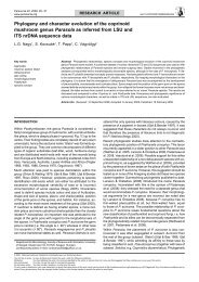
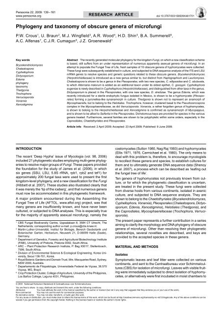
<strong>Phylogeny</strong> <strong>and</strong> <strong>taxonomy</strong> <strong>of</strong> <strong>obscure</strong> <strong>genera</strong> <strong>of</strong> micr<strong>of</strong>ungi<br />
P.W. Crous 1 , U. Braun 2 , M.J. Wingfield 3 , A.R. Wood 4 , H.D. Shin 5 , B.A. Summerell 6 ,<br />
A.C. Alfenas 7 , C.J.R. Cumagun 8 , J.Z. Groenewald 1<br />
Key words<br />
Brycekendrickomyces<br />
Chalastospora<br />
Cyphellophora<br />
Dictyosporium<br />
Edenia<br />
phylogeny<br />
<strong>taxonomy</strong><br />
Thedgonia<br />
Trochophora<br />
Verrucisporota<br />
Vonarxia<br />
Xenostigmina<br />
Abstract The recently <strong>genera</strong>ted molecular phylogeny for the kingdom Fungi, on which a new classification scheme<br />
is based, still suffers from an under representation <strong>of</strong> numerous apparently asexual <strong>genera</strong> <strong>of</strong> micr<strong>of</strong>ungi. In an<br />
attempt to populate the Fungal Tree <strong>of</strong> Life, fresh samples <strong>of</strong> 10 <strong>obscure</strong> <strong>genera</strong> <strong>of</strong> hyphomycetes were collected.<br />
These fungi were subsequently established in culture, <strong>and</strong> subjected to DNA sequence analysis <strong>of</strong> the ITS <strong>and</strong> LSU<br />
nrRNA genes to resolve species <strong>and</strong> generic questions related to these <strong>obscure</strong> <strong>genera</strong>. Brycekendrickomyces<br />
(Herpotrichiellaceae) is introduced as a new genus similar to, but distinct from Haplographium <strong>and</strong> Lauriomyces.<br />
Chalastospora is shown to be a genus in the Pleosporales, with two new species, C. ellipsoidea <strong>and</strong> C. obclavata,<br />
to which Alternaria malorum is added as an additional taxon under its oldest epithet, C. gossypii. Cyphellophora<br />
eugeniae is newly described in Cyphellophora (Herpotrichiellaceae), <strong>and</strong> distinguished from other taxa in the genus.<br />
Dictyosporium is placed in the Pleosporales, with one new species, D. streliziae. The genus Edenia, which was<br />
recently introduced for a sterile endophytic fungus isolated in Mexico, is shown to be a hyphomycete (Pleosporales)<br />
forming a pyronellea-like synanamorph in culture. Thedgonia is shown not to represent an anamorph <strong>of</strong><br />
Mycosphaerella, but to belong to the Helotiales. Trochophora, however, clustered basal to the Pseudocercospora<br />
complex in the Mycosphaerellaceae, as did Verrucisporota. Vonarxia, a rather forgotten genus <strong>of</strong> hyphomycetes,<br />
is shown to belong to the Herpotrichiellaceae <strong>and</strong> Xenostigmina is confirmed as synanamorph <strong>of</strong> Mycopappus,<br />
<strong>and</strong> is shown to be allied to Seifertia in the Pleosporales. Dichotomous keys are provided for species in the various<br />
<strong>genera</strong> treated. Furthermore, several families are shown to be polyphyletic within some orders, especially in the<br />
Capnodiales, Chaetothyriales <strong>and</strong> Pleosporales.<br />
Article info Received: 2 April 2009; Accepted: 23 April 2009; Published: 9 June 2009.<br />
Introduction<br />
The recent ‘Deep Hypha’ issue <strong>of</strong> Mycologia (vol. 98, 2006)<br />
included 21 phylogenetic studies employing multi-gene phylogenies<br />
to resolve major groups <strong>of</strong> Fungi. These papers provided<br />
the foundation for the study <strong>of</strong> James et al. (2006), in which<br />
six genes (SSU, LSU, 5.8S rRNA, rpb1, rpb2 <strong>and</strong> tef1) for<br />
approximately 200 fungal taxa were used to present the first<br />
kingdom-level phylogeny, <strong>and</strong> a new classification for the Fungi<br />
(Hibbett et al. 2007). These studies also illustrated clearly that<br />
it was merely the ‘tip <strong>of</strong> the iceberg’, <strong>and</strong> that numerous <strong>genera</strong><br />
must now be accommodated in this phylogenetic framework.<br />
A major problem encountered during the Assembling the<br />
Fungal Tree <strong>of</strong> Life (AFTOL, www.aftol.org) project, was that<br />
many <strong>genera</strong> are insufficiently known, <strong>and</strong> have never been<br />
cultured, or subjected to DNA analyses. This is especially true<br />
for the majority <strong>of</strong> apparently asexual micr<strong>of</strong>ungi, namely the<br />
1<br />
CBS Fungal Biodiversity Centre, Uppsalalaan 8, 3584 CT Utrecht, The<br />
Netherl<strong>and</strong>s; corresponding author e-mail: p.crous@cbs.knaw.nl.<br />
2<br />
Martin-Luther-Universität, Institut für Biologie, Bereich Geobotanik und<br />
Botanischer Garten, Herbarium, Neuwerk 21, D-06099 Halle (Saale),<br />
Germany.<br />
3<br />
Department <strong>of</strong> Genetics, Forestry <strong>and</strong> Agricultural Biotechnology Institute<br />
(FABI), University <strong>of</strong> Pretoria, Pretoria 0002, South Africa.<br />
4<br />
ARC – Plant Protection Research Institute, P. Bag X5017, Stellenbosch,<br />
7599, South Africa.<br />
5<br />
Division <strong>of</strong> Environmental Science & Ecological Engineering, Korea University,<br />
Seoul 136-701, Korea.<br />
6<br />
Royal Botanic Gardens <strong>and</strong> Domain Trust, Mrs. Macquaries Road, Sydney,<br />
NSW 2000, Australia.<br />
7<br />
Departamento de Fitopatologia, Universidade Federal de Viçosa, 36.570<br />
Viçosa, MG, Brazil.<br />
8<br />
Crop Protection Cluster, College <strong>of</strong> Agriculture, University <strong>of</strong> the Philippines,<br />
Los Baños College, Laguna 4031, Philippines.<br />
coelomycetes (Sutton 1980, Nag Raj 1993) <strong>and</strong> hyphomycetes<br />
(Ellis 1971, 1976, Carmichael et al. 1980). The only means to<br />
deal with this problem is, therefore, to encourage mycologists<br />
to recollect these <strong>genera</strong> <strong>and</strong> species, to establish cultures for<br />
them <strong>and</strong> to ultimately <strong>genera</strong>te DNA sequence data (Shenoy<br />
et al. 2007), a process which can be described as ‘leafing out<br />
the fungal tree <strong>of</strong> life’.<br />
Ten <strong>genera</strong> <strong>of</strong> hyphomycetes not previously known from culture,<br />
or for which the phylogenetic classification is uncertain,<br />
are treated in the present study. These fungi were collected<br />
from diverse hosts from various continents, isolated in axenic<br />
culture, <strong>and</strong> subjected to DNA sequence analysis. They are<br />
shown to belong to the Chaetothyriales (Brycekendrickomyces,<br />
Cyphellophora, Vonarxia), Pleosporales (Chalastospora, Dictyosporium,<br />
Edenia, Xenostigmina), Helotiales (Thedgonia), <strong>and</strong><br />
the Capnodiales, Mycosphaerellaceae (Trochophora, Verrucisporota).<br />
The present paper represents a further contribution in a series<br />
aiming to clarify the morphology <strong>and</strong> DNA phylogeny <strong>of</strong> <strong>obscure</strong><br />
<strong>genera</strong> <strong>of</strong> micr<strong>of</strong>ungi. Other than resolving their phylogenetic<br />
relationships, several novelties are described, <strong>and</strong> keys are<br />
provided to the accepted species in these <strong>genera</strong>.<br />
Material <strong>and</strong> Methods<br />
Isolates<br />
Symptomatic leaves <strong>and</strong> leaf litter were collected on various<br />
continents, <strong>and</strong> sent to the Centraalbureau voor Schimmelcultures<br />
(CBS) for isolation <strong>of</strong> micr<strong>of</strong>ungi. Leaves with visible fruiting<br />
were immediately subjected to direct isolation <strong>of</strong> hyphomycetes,<br />
or alternatively were first incubated in moist chambers to<br />
© 2009 Nationaal Herbarium Nederl<strong>and</strong> & Centraalbureau voor Schimmelcultures<br />
You are free to share - to copy, distribute <strong>and</strong> transmit the work, under the following conditions:<br />
Attribution:<br />
You must attribute the work in the manner specified by the author or licensor (but not in any way that suggests that they endorse you or your use <strong>of</strong> the work).<br />
Non-commercial: You may not use this work for commercial purposes.<br />
No derivative works: You may not alter, transform, or build upon this work.<br />
For any reuse or distribution, you must make clear to others the license terms <strong>of</strong> this work, which can be found at http://creativecommons.org/licenses/by-nc-nd/3.0/legalcode. Any <strong>of</strong> the above conditions can be<br />
waived if you get permission from the copyright holder. Nothing in this license impairs or restricts the author’s moral rights.
140 <strong>Persoonia</strong> – Volume 22, 2009<br />
Table 1 Collection details <strong>and</strong> GenBank accession numbers for fungal species included in this study.<br />
Species Strain no. 1 Substrate Country Collector(s) GenBank Accession no. 2<br />
ITS LSU<br />
Brycekendrickomyces acaciae CBS 124104; CPC 15078 Acacia auriculiformis Indonesia M.J. Wingfield FJ839606 FJ839641<br />
Chalastospora cetera CBS 121340; E.G.S. 41.072 Elymus scabrus Australia R.G. Rees FJ839607 FJ839642<br />
Chalastospora ellipsoidea CBS 121331; E.G.S. 22.060 Triticum sp. Australia H.L. Harvey & S. Perth FJ839608 FJ839643<br />
Chalastospora gossypii CBS 112844; CPC 4571 Bromus tectorum USA F.M. Dugan AY251081 AY251081<br />
CBS 114005; CPC 4572 Festuca idahoensis USA F.M. Dugan AY251079 AY251079<br />
CBS 114809; MAF 943 Leaves <strong>of</strong> Anethum graveolens (dill) along with Itersonilia perplexans New Zeal<strong>and</strong> J. Pike FJ839609 FJ839644<br />
CBS 114810; MAF 954 Quercus robur (oak) leaves in association with Tubakia dryina New Zeal<strong>and</strong> H. Nettleton FJ839610 FJ839645<br />
CBS 148.66; CPC 3690; NRRL W 52-29 — USA C.W. Hesseltine FJ839611 FJ839646<br />
CBS 173.80; ATCC 200939; CPC 3685 Agricultural soil Syria M.I.A. Abdel-Kader — FJ839647<br />
CBS 216.65; NRRL A-13702 Triticum aestivum grain USA C.W. Hesseltine FJ839612 DQ008142<br />
CBS 266.75; ATCC 28332; CPC 3680; IMI 165252; PRE 44703 Wheat stubble South Africa W.F.O. Marasas FJ839613 FJ839648<br />
CBS 900.87; ATCC 200938 Soil Lebanon F. Seigle-Mur<strong>and</strong>i FJ839614 FJ839649<br />
CPC 15567; C.F. Hill 2008/3899 Wood <strong>and</strong> wallpaper from inside walls <strong>of</strong> a dwelling New Zeal<strong>and</strong> D. De Vanny FJ839615 FJ839650<br />
Chalastospora gossypii var. polymorpha CBS 112048; CPC 4570 Dormant buds (overwintered) <strong>of</strong> Vitis vinifera USA F.M. Dugan AY251080 AY251080<br />
Chalastospora obclavata CBS 124120; E.G.S. 12.128 Air USA C.T. Rogerson FJ839616 FJ839651<br />
Cyphellophora eugeniae CBS 124105; CPC 15172 Living leaves <strong>of</strong> Stenocalyx uniflorus Brazil A.C. Alfenas FJ839617 FJ839652<br />
Dictyosporium strelitziae CBS 123359; CPC 15359 Dead leaves <strong>of</strong> Strelitzia nicolai South Africa A. Wood FJ839618 FJ839653<br />
Edenia gomezpompae CBS 124106; CPC 15689 Senna alata Phillippines C.J.R. Cumagun FJ839619 FJ839654<br />
Haplographium catenatum CBS 196.73 Decaying wood Germany W. Gams FJ839620 FJ839655<br />
CBS 482.67; CMW 754 Decaying wood Germany W. Gams FJ839621 FJ839656<br />
CBS 739.68; CMW 755 Decaying wood Netherl<strong>and</strong>s H.A. van der Aa FJ839622 FJ839657<br />
Lauriomyces bellulus CBS 517.93 Cupule <strong>of</strong> Castanea sativa Switzerl<strong>and</strong> P.W. Crous FJ839623 FJ839658<br />
Lauriomyces heliocephalus CBS 112054; INIFAT CO2/59 Decaying leaf Brazil A. Stchigel & J. Guarro FJ839624 FJ839659<br />
Mycopappus aceris CBS 124109; CPC 14379 Fallen leaves <strong>of</strong> Acer macrophyllum Canada B. Callan FJ839625 FJ839660<br />
Mycosphaerella lupini CPC 1661 Lupinus sp. USA W. Kaiser AF362050 FJ839661<br />
Stenella anthuriicola CBS 118742 Anthurium leaf Thail<strong>and</strong> C. F. Hill FJ839626 FJ839662<br />
Stigmina platani CBS 110755; CPC 4299; IMI 136770 Platanus orientalis India — AY260090 FJ839663<br />
Thedgonia ligustrina CPC 10019 Ligustrum ovalifolium South Korea H.-D. Shin FJ839627 FJ839664<br />
CPC 10530 Ligustrum ovalifolium Netherl<strong>and</strong>s P.W. Crous FJ839628 FJ839665<br />
CPC 10861 Ligustrum ovalifolium South Korea H.-D. Shin FJ839629 FJ839666<br />
CPC 14754 Ligustrum obtusifolium South Korea H.-D. Shin FJ839630 FJ839667<br />
CPC 4298; W1877 Ligustrum sp. Asia H. Evans EU040242 EU040242<br />
Trochophora fasciculata CPC 10281 Leaves <strong>of</strong> Daphniphyllum macropodum South Korea H.-D. Shin FJ839631 —<br />
CPC 10282 Leaves <strong>of</strong> Daphniphyllum macropodum South Korea H.-D. Shin FJ839632 FJ839668<br />
Verrucisporota daviesiae CBS 116002; VPRI 31767 Living leaves <strong>of</strong> Daviesia mimosoides Australia V. & R. Beilharz FJ839633 FJ839669<br />
Verrucisporota grevilleae CBS 124107; CPC 14761 Leaves <strong>of</strong> Grevillia decurrens Australia B. Summerell FJ839634 FJ839670<br />
Verrucisporota proteacearum CBS 116003; VPRI 31812 Grevillea sp. Australia V. Beilharz FJ839635 FJ839671<br />
Vonarxia vagans CBS 123533; CPC 15151 Stenocalyx uniflorus Brazil A.C. Alfenas FJ839636 FJ839672<br />
CPC 15152 Stenocalyx uniflorus Brazil A.C. Alfenas FJ839637 FJ839673<br />
Xenostigmina zilleri CBS 115685; CPC 4011 Living leaves <strong>of</strong> Acer sp. Canada K.A. Seifert FJ839638 FJ839674<br />
CBS 124108; CPC 14376 Fallen leaves <strong>of</strong> Acer macrophyllum Canada B. Callan FJ839639 FJ839675<br />
CBS 115686; CPC 4010 Living leaves <strong>of</strong> Acer sp. Canada K.A. Seifert FJ839640 FJ839676<br />
1<br />
ATCC: American Type Culture Collection, Virginia, USA; C.F. Hill: Culture collection <strong>of</strong> C.F. Hill, housed at MAF, New Zeal<strong>and</strong>; CBS: CBS Fungal Biodiversity Centre, Utrecht, The Netherl<strong>and</strong>s; CMW: Culture collection <strong>of</strong> M.J. Wingfield, housed at FABI, Pretoria, South Africa; CPC:<br />
Culture collection <strong>of</strong> P.W. Crous, housed at CBS; E.G.S.: Culture collection <strong>of</strong> E.G. Simmons, Indiana USA; IMI: International Mycological Institute, CABI-Bioscience, Egham, Bakeham Lane, U.K.; INIFAT: Alex<strong>and</strong>er Humboldt Institute for Basic Research in Tropical Agriculture,<br />
Ciudad de La Habana, Cuba; MAF: Ministry <strong>of</strong> Agriculture <strong>and</strong> Forestry, New Zeal<strong>and</strong>; NRRL: National Center for Agricultural Utilization Research, Peoria, USA; PRE: National collection <strong>of</strong> fungi, Pretoria, South Africa; VPRI: Victorian Department <strong>of</strong> Primary Industries, Knoxfield,<br />
Australia.<br />
2<br />
ITS: Internal transcribed spacers 1 <strong>and</strong> 2 together with 5.8S nrDNA; LSU: 28S nrDNA.
P.W. Crous et al.: Obscure <strong>genera</strong> <strong>of</strong> micr<strong>of</strong>ungi<br />
141<br />
stimulate sporulation. Single-conidial isolates were established<br />
on malt extract agar (MEA; 20 g/L Biolab malt extract, 15 g/L<br />
Biolab agar) using the technique outlined in Crous (1998).<br />
Cultures were later plated on fresh MEA, 2 % water agar (WA)<br />
supplemented with sterile pine needles, 2 % potato-dextrose<br />
agar (PDA), synthetic nutrient agar (SNA) <strong>and</strong>/or oatmeal agar<br />
(OA) (Crous et al. 2009), <strong>and</strong> subsequently incubated at 25 °C<br />
under near-ultraviolet light to promote sporulation. Reference<br />
strains are maintained in the culture collection <strong>of</strong> the CBS,<br />
Utrecht, the Netherl<strong>and</strong>s (Table 1). Descriptions, nomenclature,<br />
<strong>and</strong> illustrations were deposited in MycoBank (www.mycobank.<br />
org, Crous et al. 2004b).<br />
DNA isolation, amplification <strong>and</strong> analyses<br />
Genomic DNA was isolated from fungal mycelium grown on<br />
MEA, using the UltraClean TM Microbial DNA Isolation Kit (Mo<br />
Bio Laboratories, Inc., Solana Beach, CA, USA) according to the<br />
manufacturer’s protocols. The Primers V9G (de Hoog & Gerrits<br />
van den Ende 1998) <strong>and</strong> LR5 (Vilgalys & Hester 1990) were<br />
used to amplify part <strong>of</strong> the nuclear rDNA operon spanning the 3’<br />
end <strong>of</strong> the 18S rRNA gene (SSU), the first internal transcribed<br />
spacer (ITS1), the 5.8S rRNA gene, the second ITS region<br />
(ITS2) <strong>and</strong> the first 900 bases at the 5’ end <strong>of</strong> the 28S rRNA<br />
gene (LSU). The primers ITS4 (White et al. 1990) <strong>and</strong> LR0R<br />
(Rehner & Samuels 1994) were used as internal sequence<br />
primers to ensure good quality sequences over the entire length<br />
<strong>of</strong> the amplicon. The PCR conditions, sequence alignment<br />
<strong>and</strong> subsequent phylogenetic analysis followed the methods<br />
<strong>of</strong> Crous et al. (2006b). Alignment gaps were treated as new<br />
character states. Sequence data were deposited in GenBank<br />
(Table 1) <strong>and</strong> alignments in TreeBASE (www.treebase.org).<br />
The ITS sequences were compared with those sequences available<br />
in NCBI’s GenBank nucleotide database using a megablast<br />
search <strong>and</strong> the results are discussed where applicable under<br />
the taxonomic notes. Because the genus Chalastospora is relatively<br />
novel, species in this genus were supported by a separate<br />
phylogenetic tree.<br />
Morphology<br />
Fungal descriptions were based on cultures sporulating in<br />
vitro (media indicated). Wherever possible, 30 measurements<br />
(× 1 000 magnification) were made <strong>of</strong> structures mounted in<br />
lactic acid, with the extremes <strong>of</strong> spore measurements given in<br />
parentheses. Colony colours (surface <strong>and</strong> reverse) were assessed<br />
after 2–4 wk on different media at 25 °C in the dark,<br />
using the colour charts <strong>of</strong> Rayner (1970).<br />
Results<br />
Phylogenetic analysis<br />
Amplification products <strong>of</strong> approximately 1 700 bases were obtained<br />
for the isolates listed in Table 1. The LSU region <strong>of</strong> the<br />
sequences was used to obtain additional sequences from Gen-<br />
Bank, which were added to the alignment. Due to the inclusion<br />
<strong>of</strong> the shorter LSU sequences <strong>of</strong> Dictyosporium alatum (Gen-<br />
Bank accession DQ018101), Dictyosporium elegans (GenBank<br />
accession DQ018100) <strong>and</strong> Dictyosporium toruloides (GenBank<br />
accession DQ018104) in the alignment, it was not possible to<br />
subject the full length <strong>of</strong> the determined LSU sequences (Table<br />
1) to analyses. The manually adjusted LSU alignment contained<br />
115 sequences (including the two outgroup sequences) <strong>and</strong>, <strong>of</strong><br />
the 568 characters used in the phylogenetic analyses, 267 were<br />
parsimony informative, 30 were variable <strong>and</strong> parsimony uninformative,<br />
<strong>and</strong> 271 were constant. Neighbour-joining analyses<br />
using three substitution models on the sequence data yielded<br />
trees supporting the same tree topology to one another but<br />
differed from the most parsimonious tree shown in Fig. 1 with<br />
regard to the placement <strong>of</strong> the clade containing Ochroconis <strong>and</strong><br />
Fusicladium (in the distance analyses, this clade moves to a<br />
more basal position). Forty equally most parsimonious trees<br />
(TL = 1 039 steps; CI = 0.477; RI = 0.833; RC = 0.397), the first<br />
<strong>of</strong> which is shown in Fig. 1, were obtained from the parsimony<br />
analysis <strong>of</strong> the LSU alignment.<br />
The manually adjusted ITS alignment contained 28 sequences<br />
(including the outgroup sequence) <strong>and</strong>, <strong>of</strong> the 521 characters<br />
used in the phylogenetic analyses, 97 were parsimony informative,<br />
91 were variable <strong>and</strong> parsimony uninformative, <strong>and</strong> 333<br />
were constant. Neighbour-joining analyses using three substitution<br />
models on the sequence data yielded trees supporting the<br />
same tree topology to one another but differed from the most<br />
parsimonious tree shown in Fig. 2 with regard to the placement<br />
<strong>of</strong> Chalastospora ellipsoidea (in the distance analyses, this<br />
taxon moves to a more basal position in Chalastospora). Six<br />
equally most parsimonious trees (TL = 253 steps; CI = 0.913;<br />
RI = 0.938; RC = 0.856), the first <strong>of</strong> which is shown in Fig. 2,<br />
were obtained from the parsimony analysis <strong>of</strong> the ITS alignment.<br />
The results <strong>of</strong> the phylogenetic analyses are highlighted<br />
below under the taxonomic notes, or in the Discussion, where<br />
applicable.<br />
Taxonomy<br />
Brycekendrickomyces Crous & M.J. Wingf., gen. nov. —<br />
MycoBank MB509515<br />
Mycelium ex hyphis ramosis, septatis, laevibus, pallide brunneis, 1–2 µm<br />
latis compositum. Conidiophora solitaria, erecta, cylindrica, recta vel leviter<br />
flexuosa, cellula basali bulbosa, sine rhizoideis, stipite modice brunneo vel<br />
atro-brunneo, laevi, transverse euseptato, ad apicem cum (1–)2–4(–6)<br />
cellulis conidiogenis. Cellulae conidiogenae subcylindricae, allontoides vel<br />
doliiformes, rectae vel leviter curvatae, pallide brunneae, polyblasticae,<br />
sympodialiter proliferantes. Conidia hyalina, mucilagine aggregata (sed non<br />
catenata), ellipsoidea, apice subobtuso, basi subtruncata.<br />
Type species. Brycekendrickomyces acaciae Crous & M.J. Wingf.<br />
Etymology. Named for Bryce Kendrick, husb<strong>and</strong> <strong>of</strong> Laurie Kendrick, for<br />
which Lauriomyces was named <strong>and</strong> that resembles the current genus.<br />
Mycelium consisting <strong>of</strong> branched, septate, smooth, pale brown,<br />
1–2 µm wide hyphae. Conidiophores solitary, erect, cylindrical,<br />
straight to somewhat flexuous, basal cell bulbous, without<br />
rhizoids; stalk medium to dark brown, smooth, transversely<br />
euseptate; upper cell giving rise to (1–)2–4(–6) conidiogenous<br />
cells. Conidiogenous cells subcylindrical to allantoid or doliiform,<br />
straight to gently curved, pale brown, polyblastic, proliferating<br />
sympodially. Conidia hyaline, aggregating in slimy mass (never<br />
in chains), ellipsoid, apex subobtuse, base subtruncate.<br />
Brycekendrickomyces acaciae Crous & M.J. Wingf., sp. nov.<br />
— MycoBank MB509517; Fig. 3<br />
Maculae modice brunneae vel atro-brunneae, margine elevato, rubro-purpureo,<br />
oblongae vel ellipticae, ad 7 mm diam, in consortione ‘Phaeotrichoconis’<br />
crotalariae. In vitro (MEA): Mycelium ex hyphis ramosis, septatis,<br />
laevibus, pallide brunneis, 1–2 µm latis compositum. Conidiophora ex<br />
hyphis oriunda, solitaria, erecta, cylindrica, recta vel leviter flexuosa, cellula<br />
basali bulbosa, sine rhizoideis, 4–6 µm lata, ad basim 10–15 µm lata,<br />
stipite modice brunneo vel atro-brunneo, laevi, transverse 2–5-euseptato,<br />
(15–)30–50(–60) µm longo, (3–)4(–5) µm lato, ad apicem cum (1–)2–4(–6)<br />
cellulis conidiogenis. Cellulae conidiogenae subcylindricae, allontoides vel<br />
doliiformes, rectae vel leviter curvatae, pallide brunneae, 5–8 × 2–2.5 µm,<br />
polyblasticae, sympodialiter proliferantes. Conidia hyalina, mucilagine aggregata<br />
(sed non catenata), ellipsoidea, apice subobtuso, basi subtruncata,<br />
latitudine maxima in parte centrali vel in parte supra centrum, saepe leviter<br />
asymmetrica, (3.5–)4(–4.5) × 2(–2.5) µm.<br />
Etymology. Named after the host genus on which the fungus occurs,<br />
Acacia.
142 <strong>Persoonia</strong> – Volume 22, 2009<br />
Kluyveromyces lodderae AY048161<br />
Saccharomyces cerevisiae Z73326<br />
82<br />
10 changes<br />
62<br />
1<br />
100<br />
2<br />
99<br />
Mycosphaerella punctiformis EU167569<br />
Ramularia pratensis var. pratensis EU019284<br />
100<br />
99<br />
74<br />
56<br />
64<br />
97<br />
76<br />
97<br />
51<br />
Trochophora fasciculata CPC 10282<br />
Mycosphaerella crystallina DQ204747<br />
Mycosphaerella waimeana AY260083<br />
Mycosphaerella heimii DQ204751<br />
Mycosphaerella heimioides DQ204753<br />
Pseudocercospora vitis DQ073923<br />
Pseudocercospora pseudoeucalyptorum DQ204766<br />
Pseudocercospora robusta DQ204767<br />
Stigmina platani CBS 110755<br />
Pseudocercospora natalensis DQ267576<br />
Pseudocercospora basitruncata DQ204759<br />
Mycosphaerella lupini CPC 1661<br />
Mycosphaerella marksii DQ246250<br />
Pseudocercospora epispermogonia DQ204758<br />
100<br />
Schizothyrium pomi EF134947<br />
Schizothyrium pomi EF134949 SCH<br />
69 Schizothyrium pomi EF134948<br />
Cibiessia minutispora EU019259<br />
82 100 Teratosphaeria readeriellophora DQ246238<br />
88 Batcheloromyces leucadendri EU019246<br />
96 Cibiessia nontingens EU019260<br />
Teratosphaeria nubilosa DQ246228<br />
95 Teratosphaeria fibrillosa EU019282<br />
Mycosphaerella madeirae DQ204756<br />
Mycosphaerella parkii DQ246245<br />
Ramichloridium biverticillatum EU041853<br />
Verrucisporota grevilleae CPC 14761<br />
97 99 Verrucisporota proteacearum CBS 116003<br />
83 Periconiella arcuata EU041836<br />
Verrucisporota daviesiae CBS 116002<br />
Periconiella velutina EU041840<br />
MYC (2)<br />
54<br />
Ramichloridium musae EU041857<br />
Ramichloridium strelitziae EU041860<br />
54 Stenella anthuriicola CBS 118742<br />
Ramichloridium cerophilum EU041855<br />
77<br />
85<br />
Zasmidium cellare EU041878<br />
Cyphellophora laciniata EU035416 HER (1)<br />
Vonarxia vagans CPC 15151<br />
80 100 Vonarxia vagans CPC 15152<br />
CHA<br />
Ceramothyrium carniolicum AY004339<br />
Cyphellophora Cyphellophora eugeniae hylomeconis CPC EU035415<br />
15172<br />
78<br />
Exophiala eucalyptorum EU035417<br />
71 Exophiala pisciphila DQ823101<br />
Capronia mansonii AY004338<br />
100 Capronia munkii EF413604<br />
97 Brycekendrickomyces acaciae CPC 15078<br />
63 Phaeococcomyces chersonesos AJ507323<br />
65 Cladophialophora proteae EU035411<br />
100 Exophiala placitae EU040215<br />
Phaeococcomyces catenatus AF050277<br />
88<br />
70<br />
MYC (1)<br />
Fig. 1 The first <strong>of</strong> 1 000 equally most parsimonious trees obtained from a heuristic search with 100 r<strong>and</strong>om taxon additions <strong>of</strong> the LSU sequence alignment.<br />
The scale bar shows 10 changes, <strong>and</strong> bootstrap support values from 1 000 replicates are shown at the nodes. Novel sequences <strong>genera</strong>ted for this study are<br />
shown in bold. Branches present in the strict consensus tree are thickened. Orders <strong>and</strong> families are coded as indicated in the legends. The tree was rooted<br />
to a sequence <strong>of</strong> Kluyveromyces lodderae (GenBank accession AY048161) <strong>and</strong> Saccharomyces cerevisiae (GenBank accession Z73326).<br />
Abbreviations used: Families: AMO = Amorphothecaceae, CHA = Chaetothyriaceae, HEL = Helotiaceae, HER = Herpotrichiellaceae, HYA = Hyaloscyphaceae,<br />
IC = Incertae cedis: LEP = Leptosphaeriaceae, LOP = Lophiostomataceae, MEL = Melanommataceae, MYC = Mycosphaerellaceae, PHA = Phaeosphaeriaceae,<br />
PLE = Pleosporaceae, PSE = Pseudeurotiaceae, RHY = Rhytismataceae, SCH = Schizothyriaceae, TER = Teratosphaeriaceae. Orders: 1 = Capnodiales,<br />
2 = Chaetothyriales, 3 = Incertae cedis, 4 = Rhytismatales, 5 = Helotiales, 6 = Hypocreales, 7 = Pezizales, 8 = Pleosporales.<br />
TER<br />
HER (2)<br />
Leaf spots medium to dark brown, margin raised, red-purple,<br />
oblong to ellipsoid, up to 7 mm diam, associated with ‘Phaeotrichoconis’<br />
crotalariae. Description based on culture on MEA:<br />
Mycelium consisting <strong>of</strong> branched, septate, smooth, pale brown,<br />
1–2 µm wide hyphae. Conidiophores arising from mycelium,<br />
solitary, erect, cylindrical, straight to somewhat flexuous; basal<br />
cell bulbous, without rhizoids, 4–6 µm wide in upper part, but<br />
becoming 10–15 µm wide at basal part; stalk medium to dark<br />
brown, smooth, transversely 2–5-euseptate, (15–)30–50(–60)<br />
µm tall, (3–)4(–5) µm wide in the middle part; upper cell giving<br />
rise to (1–)2–4(–6) conidiogenous cells. Conidiogenous cells<br />
subcylindrical to allantoid or doliiform, straight to gently curved,<br />
pale brown, 5–8 × 2–2.5 µm; polyblastic, proliferating sympodially.<br />
Conidia hyaline, aggregating in slimy mass (never in<br />
chains), ellipsoid, apex subobtuse, base subtruncate, widest in<br />
the middle or upper third <strong>of</strong> the conidium, frequently somewhat<br />
asymmetrical, (3.5–)4(–4.5) × 2(–2.5) µm.<br />
Characteristics in culture — Colonies on MEA erumpent,<br />
spreading, with moderate aerial mycelium; surface folded,<br />
margin lobate, smooth; surface olivaceous-grey, outer margin<br />
iron-grey; reverse iron-grey; colonies reaching up to 20 mm<br />
after 1 mo. Colonies fertile on SNA, OA <strong>and</strong> MEA.<br />
Specimen examined. Indonesia, Pelalawan, living leaves <strong>of</strong> Acacia auriculiformis,<br />
Mar. 2008, leg. M.J. Wingfield, isol. P.W. Crous, holotype CBS<br />
H-20198, culture ex-type CPC 15078 = CBS 124104.<br />
Notes — Castañeda & Kendrick (1990) established the genus<br />
Lauriomyces, characterised by dark brown conidiophores,
P.W. Crous et al.: Obscure <strong>genera</strong> <strong>of</strong> micr<strong>of</strong>ungi<br />
143<br />
Fig. 1 (cont.)<br />
3 Lauriomyces bellulus CBS 517.93<br />
IC<br />
4 100 Lauriomyces heliocephalus CBS 112054<br />
Potebniamyces pyri DQ470949<br />
RHY<br />
5 Crinula caliciiformis AY544680<br />
HEL (1)<br />
100<br />
Holwaya mucida DQ257356<br />
Leuconeurospora pulcherrima AF096193<br />
61 6<br />
IC<br />
Pseudeurotium zonatum AF096198<br />
933<br />
PSE<br />
Pseudeurotium zonatum DQ470988<br />
3 97<br />
Amorphotheca resinae EU040230<br />
Amorphotheca resinae EU040231<br />
AMO<br />
100 Hyalodendriella betulae EU040232<br />
IC<br />
Phialea strobilina EF596821<br />
5 7 100 Haplographium catenatum CBS 482.67<br />
Haplographium catenatum CBS 196.73<br />
HYA (1)<br />
Haplographium catenatum CBS 739.68<br />
3 Clathrosporium intricatum AY616235<br />
66 Hydrocina chaetocladia AY789412<br />
Rhyzoscyphus ericae AM887699<br />
IC<br />
HEL (2)<br />
89<br />
Protoventuria alpina EU035444<br />
Thedgonia ligustrina CPC 10019<br />
100<br />
Thedgonia ligustrina EU040242<br />
Thedgonia ligustrina CPC 10861 IC<br />
Thedgonia ligustrina CPC 14754<br />
3<br />
Thedgonia ligustrina CPC 10530<br />
Trichoconis echinophila EU107315<br />
91 Trematosphaeria pertusa DQ678072<br />
10 changes<br />
8<br />
100<br />
100<br />
88<br />
Aposphaeria populina EU754130<br />
Herpotrichia juniperi DQ678080<br />
Wettsteinina macrotheca AY849969<br />
Seifertia azaleae EU030276<br />
Mycopappis aceris CPC 14379<br />
70<br />
Xenostigmina zilleri CBS 115685<br />
Xenostigmina zilleri CBS 115686<br />
Xenostigmina zilleri CPC 14376<br />
100<br />
Dictyosporium toruloides DQ018104<br />
Dictyosporium strelitziae CPC 15359<br />
Dictyosporium elegans DQ018100<br />
Dictyosporium alatum DQ018101<br />
91<br />
Pseudodiplodia sp. EU754201<br />
Coniothyrium concentricum EU754152<br />
Leptospora rubella DQ195792<br />
Phaeosphaeriopsis musae DQ885894<br />
HYA (2)<br />
MEL<br />
LOP<br />
PLE (1)<br />
60<br />
LEP<br />
IC<br />
PHA<br />
Phaeosphaeria nodorum EF590318<br />
Edenia gomezpompae CPC 15689<br />
Chalastospora gossypii CBS 112844<br />
94 Chalastospora gossypii DQ008142<br />
Chalastospora gossypii CBS 114810<br />
Chalastospora gossypii CBS 114005<br />
Chalastospora gossypii CBS 900.87<br />
Chalastospora gossypii var. polymorpha AY251080<br />
Chalastospora gossypii AY251081<br />
100 Chalastospora gossypii AY251079<br />
Chalastospora gossypii CPC 3685<br />
Chalastospora gossypii CPC 3680<br />
Chalastospora obclavata CBS 124120<br />
Chalastospora gossypii CPC 3690<br />
Chalastospora gossypii CPC 15567<br />
Chalastospora gossypii CBS 114809<br />
Chalastospora cetera CBS 121340<br />
64<br />
Chalastospora ellipsoidea CBS 121331<br />
IC<br />
PLE (2)<br />
<strong>and</strong> a series <strong>of</strong> branches, giving rise to chains <strong>of</strong> hyaline conidia<br />
via sympodial conidiogenesis. Brycekendrickomyces is morphologically<br />
similar to Lauriomyces, which in turn resembles<br />
Haplographium. The genus Haplographium is based on H. delicatum.<br />
Its confused history is discussed in detail by Zucconi<br />
& Pagano (1993). Haplographium delicatum was originally<br />
described by Berkeley & Broome as having conidia in chains<br />
(Mason 1933), which Saccardo (1886) also reported for the<br />
type species. Hughes (1958) noted that Stilbum catenatum<br />
was an older name for H. delicatum, which led Holubová-Jechová<br />
(1973) to place this species in Haplographium, while<br />
Castañeda & Kendrick (1990) placed it in Lauriomyces. If<br />
Haplographium <strong>and</strong> Lauriomyces are synonymous, the older<br />
name, Haplographium, would have preference. However, as<br />
shown here, ‘Lauriomyces’ catenatus is not congeneric with<br />
other species <strong>of</strong> Lauriomyces, such as L. heliocephalus (Rao<br />
& de Hoog 1986, Castañeda & Kendrick 1990) <strong>and</strong> L. bellulus<br />
(Crous & Wingfield 1994), suggesting that the two <strong>genera</strong> are<br />
distinct, <strong>and</strong> that the name Haplographium catenatum should<br />
be resurrected. Data from this study, furthermore, suggest that<br />
the strains <strong>of</strong> H. catenatum included here, probably represent<br />
a species complex.<br />
Brycekendrickomyces differs from Haplographium <strong>and</strong> Lauriomyces<br />
by the absence <strong>of</strong> an intricate conidiophore branching<br />
system, <strong>and</strong> in having conidia produced in slimy heads rather<br />
than in chains. Furthermore, it is not phylogenetically related<br />
to species <strong>of</strong> Lauriomyces or Haplographium presently known<br />
from culture (Fig. 1). Brycekendrickomyces is somewhat similar<br />
to Argopericonia (Sutton & Pascoe 1987), although the latter<br />
fungus produces hyaline, apical conidiogenous heads, <strong>and</strong> it<br />
has ellipsoidal, single to short catenate conidia, each with a<br />
prominent, globose guttule.
144 <strong>Persoonia</strong> – Volume 22, 2009<br />
Phaeosphaeriopsis musae DQ885894<br />
100<br />
100<br />
Alternaria alternata ATCC 52170 FJ545250<br />
Alternaria alternata EGS 34-016 AF347031<br />
62<br />
10 changes<br />
99<br />
99<br />
100<br />
100<br />
75<br />
94<br />
59<br />
78<br />
100<br />
99<br />
77<br />
70<br />
82<br />
90<br />
62<br />
Alternaria triticina EGS 17-061 AY762948<br />
Alternaria triticina MUCL 44210 AY714476<br />
Lewia ethzedia AY278833<br />
Lewia ethzedia EGS 37-143 AF392987<br />
Alternaria conjuncta EGS 37-139 AF392988<br />
Alternaria conjuncta FJ266475<br />
Chalastospora cetera CBS 121340<br />
Chalastospora obclavata CBS 124120<br />
Chalastospora ellipsoidea CBS 121331<br />
Chalastospora gossypii STE-U 4571 AY251081<br />
Chalastospora gossypii var. polymorpha AY251080<br />
Chalastospora gossypii STE-U 4572 AY251079<br />
100<br />
86<br />
62<br />
Chalastospora gossypii ATCC 28332 AF393680<br />
Chalastospora gossypii CPC 3690<br />
Chalastospora gossypii CPC 3680<br />
Chalastospora gossypii CBS 216.65<br />
Chalastospora gossypii ATCC 96020 AF393713<br />
Chalastospora gossypii ATCC 36953 AF393715<br />
Chalastospora gossypii ATCC 200938 AF393721<br />
Chalastospora gossypii CBS 900.87<br />
Chalastospora<br />
Fig. 2 The first <strong>of</strong> 6 equally most parsimonious trees obtained from a heuristic<br />
search with 100 r<strong>and</strong>om taxon additions <strong>of</strong> the ITS sequence alignment.<br />
The scale bar shows 10 changes, <strong>and</strong> bootstrap support values (blue are<br />
from the parsimony analysis <strong>and</strong> red from the distance analysis using the<br />
HKY85 substitution model) from 1 000 replicates are shown at the nodes.<br />
Branches present in the strict consensus tree are thickened. The tree was<br />
rooted to a sequence <strong>of</strong> Phaeosphaeriopsis musae (GenBank accession<br />
DQ885894).<br />
65<br />
63<br />
61<br />
50<br />
Chalastospora gossypii ATCC 200939 AF393722<br />
Chalastospora gossypii ATCC 38025 AF393714<br />
Chalastospora gossypii CPC 15567<br />
Chalastospora gossypii CBS 114809<br />
Chalastospora gossypii CBS 114810<br />
Chalastospora E.G. Simmons, Alternaria. An identification<br />
manual: 668. 2007<br />
Type species. Chalastospora cetera (E.G. Simmons) E.G. Simmons.<br />
Conidiophores solitary, brown, smooth, arising from surface hyphae<br />
or as short, lateral branches from ropes <strong>of</strong> aerial hyphae;<br />
short, subcylindrical to flask-shaped, 0–2-transversely euseptate,<br />
seldom once geniculate or branched. Conidiogenous cells<br />
integrated, terminal or conidiophores reduced to conidiogenous<br />
cells, monotretic, determinate to polytretic, sympodial, conidiogenous<br />
loci visible as minute pores, without or with somewhat<br />
darkened <strong>and</strong> slightly thickened rim. Conidia in acropetal,<br />
branched chains, narrowly ellipsoid to narrowly ovoid, pale to<br />
medium brown, rarely 1–3 transversely euseptate, <strong>genera</strong>lly<br />
lacking longitudinal or oblique septa; conidial apex functioning<br />
as secondary conidiophore, proliferating laterally.<br />
Chalastospora gossypii (Jacz.) U. Braun & Crous, comb. nov.<br />
— MycoBank MB509518; Fig. 4<br />
Basionym. Cladosporium gossypii Jacz., Holopkovoe Delo 1929, 5–6:<br />
564. 1929 <strong>and</strong> Trudy Byuro Priklad. Bot. 24 (5): 181–182. 1931.<br />
= Cladosporium malorum Rühle, Phytopathology 21: 1146. 1931.<br />
≡ Alternaria malorum (Rühle) U. Braun, Crous & Dugan, Mycol. Progr. 2:<br />
5. 2003.<br />
= Phaeoramularia kellermaniana Marasas & I.H. Bredell, Bothalia 11:<br />
217. 1974.<br />
= Cladophialophora kellermaniana (Marasas & I.H. Bredell) U. Braun &<br />
Feiler, Microbiol. Res. 150: 83, 1995.<br />
≡ Pseudocladosporium kellermanianum (Marasas & I.H. Bredell) U. Braun,<br />
A monograph <strong>of</strong> Cercosporella, Ramularia <strong>and</strong> allied <strong>genera</strong> 2: 393. 1998.<br />
= Cladosporium porophorum Matsush., Icones Micr<strong>of</strong>ungorum a Matsushima<br />
lectorum: 36. 1975.<br />
Characteristics in culture — See Braun et al. (2003).<br />
Specimens examined. Canada, Saskatchewan, Matador, from grass litter,<br />
27 May 1968, G.C. Bhatt 255, IMI 144487 = ATCC 38025 = CBS 597.69; from<br />
(?) soil, 18 Sept. 1973, H.A.H. Wallace, IMI 179345; Alberta, from Bromus<br />
inermis, 1994, R.J. Howad 397, IMI 360655, HAL. – Central Asia (without<br />
detailed locality), on fibres <strong>of</strong> Gossypium sp., 1927 <strong>and</strong> 1928, V.S. Fedorov,<br />
LEP, syntypes <strong>of</strong> Cladosporium gossypii. – Lebanon, from soil, July 1987,<br />
F. Seigle-Mur<strong>and</strong>i, ATCC 200938 = CBS 900.87. – Libya, from Prunus persica,<br />
April 1975, Casay, IMI 194863. – New Zeal<strong>and</strong>, Wellington, 40 Epuni Street,<br />
Te Aro Valley, wood <strong>and</strong> wallpaper from inside walls <strong>of</strong> a dwelling, 5 Sept.<br />
2008, leg. D. De Vanny, isol. C.F. Hill 2008/3899, CPC 15567; Auckl<strong>and</strong>,<br />
Henderson Valley Road, Henderson, leaves <strong>of</strong> Anethum graveolens (dill)
P.W. Crous et al.: Obscure <strong>genera</strong> <strong>of</strong> micr<strong>of</strong>ungi<br />
145<br />
a<br />
b<br />
c<br />
f<br />
d<br />
e<br />
Fig. 3 Brycekendrickomyces acaciae (CBS 124104). a. Colonies sporulating on MEA; b. colonies on SNA; c–e, g. conidiophores with conidiogenous apparatus;<br />
f. conidia. — Scale bars = 10 µm.<br />
g<br />
along with Itersonilia perplexans, 1 Dec. 2003, leg. J. Pike, isol. C.F. Hill,<br />
MAF 943 = CBS 114809; Auckl<strong>and</strong>, 90 Aberdeen Road, Castor Bay, isolated<br />
from Quercus robur (oak) leaves in association with Tubakia dryina,<br />
5 Sept. 2008, leg. H. Nettleton, isol. C.F. Hill, MAF 954 = CPC 15567 = CBS<br />
114810. – Pakistan, Karachi, from stored grains, 5 Jan. 1967, S.S. Hussain,<br />
IMI 124270. – South Africa, Western Cape Province, Kopgat, Calvinia,<br />
from wheat stubble, Feb. 1972, W.F.O. Marasas OP-76, PREM 44703, IMI<br />
165252, cultures ATCC 28332 = IMI 165252 = PRE 44703 = CPC 3680 =<br />
CBS 266.75, ex-type cultures <strong>of</strong> Phaeoramularia kellermaniana. – Syria,<br />
from agricultural soil, Feb. 1980, M.I.A. Abdel-Kader, CPC 3685 = ATCC<br />
200939 = CBS 173.80. – Turkey, Manisa, from Hordeum sp., 16 June 1971,<br />
Maksu & Selçuc, IMI 159198; Gossypium seeds, M. Esentepe, CBS 540.75.<br />
– USA, New Mexico, Red River, from a polypore on Picea sp., 4 Sept. 1996,<br />
D. Wicklow, IMI 386094; Washington State, from Bing cherry fruit, June 1992,<br />
F.M. Dugan, ATCC 96020; from fruits <strong>of</strong> Malus domestica, F.D. Heald, ATCC<br />
36953; Washington State, Festuca idahoensis, F.M. Dugan, STE-U 4572 =<br />
CBS 114005; Pacific Northwest, Feb. 1966, C.W. Hesseltine, NRRL W 52-<br />
29 = CPC 3690 = CBS 148.66; Oregon, Portl<strong>and</strong>, Triticum aestivum grain,<br />
June 1965, C.W. Hesseltine, NRRL A-13702 = CBS 216.65; Malus sylvestris<br />
fruit, Jan. 1931, F.D. Head, ATCC 36953 = MUCL 10096 = CBS 135.31;<br />
Washington State, Bromus tectorum, F.M. Dugan, CPC 4571 = CBS 112844;<br />
Washington State, Roza Canal near Prosser, isolated from dormant buds<br />
(overwintered) <strong>of</strong> Vitis vinifera, Mar. 2001, F.M. Dugan, holotype WSP 70286,<br />
cultures ex-type STE-U 4570 = CBS 112048 (var. polymorpha). – Uzbekistan,<br />
Bukhara, Experiment Station, on fibres <strong>of</strong> Gossypium hirsutum, 1928, V.S.<br />
Zelenetzi, LEP, lectotype <strong>of</strong> Cladosporium gossypii (selected here) (isolectotype<br />
in LEP); Bukhara, Shafrikanskoje, on fibres <strong>of</strong> Gossypium hirsutum,<br />
1928, V.S. Zelenetzi, LEP, syntype <strong>of</strong> Cladosporium gossypii.<br />
Notes — The genus Chalastospora appears to represent<br />
an anamorph lineage in the Pleosporales (Fig. 1). Chalastospora<br />
cetera <strong>and</strong> C. gossypii are clearly congeneric (Fig. 2).<br />
Based on the ITS data, there are some point mutations among<br />
strains <strong>of</strong> C. gossypii, suggesting that other genes need to be<br />
sequenced to fully elucidate the variation within this species<br />
(Fig. 2). On SNA, ramoconidia <strong>of</strong> CBS 114810 were 10–17<br />
× 3–5 µm, <strong>and</strong> conidia narrowly ellipsoid-ovoid, cylindrical<br />
to fusiform, 6–10 × 2–2.5 µm, thus much smaller than that<br />
reported by Braun et al. (2003) on PDA. Jaczewski introduced<br />
the name Cladosporium gossypii in 1929, <strong>and</strong> provided a brief<br />
Russian description, including shape <strong>and</strong> size <strong>of</strong> conidia. This<br />
description, published before 1935, is, however, valid. In his<br />
paper <strong>of</strong> 1931, he re-introduced C. gossypii together with a<br />
Latin description <strong>and</strong> a micrograph <strong>of</strong> conidia. Type material <strong>of</strong><br />
C. gossypii was re-examined <strong>and</strong> it is identical to C. malorum.<br />
However, C. gossypii is an older name than C. malorum, which<br />
was published in 1931, <strong>and</strong> has priority.<br />
Chalastospora ellipsoidea Crous & U. Braun, sp. nov. —<br />
MycoBank MB509519; Fig. 5<br />
Chalastosporae gossypii similis, sed conidiis ellipsoideis, longioribus et leviter<br />
latioribus, (8–)10–15(–17) × 3(–3.5) µm.<br />
Etymology. Named after its ellipsoid conidia.
146 <strong>Persoonia</strong> – Volume 22, 2009<br />
a<br />
b<br />
c<br />
d<br />
e<br />
f<br />
Fig. 4 Chalastospora gossypii (CBS 114810). a–f. Superficial mycelium on SNA showing conidiophores with branched conidial chains. — Scale bars = 10 µm.<br />
On SNA: Conidiophores arising singly from aerial <strong>and</strong> creeping<br />
hyphae; subcylindrical, erect, medium brown, smooth, up<br />
to 25 × 3 µm, frequently reduced to conidiogenous cells, 5–13<br />
× 3 µm; seldom once geniculate, mostly straight, with a slight<br />
swelling in the apical conidiogenous region; conidiogenous loci<br />
1–3 per conidiogenous cell, medium brown, slightly thickened,<br />
darkened, up to 1 µm wide. Ramoconidia (0–)1–3-septate, ellipsoid-ovoid,<br />
subcylindrical or fusiform, smooth, medium brown,<br />
(12–)15–18(–30) × 3(–4) µm; apex at times with short beak,<br />
giving rise to lateral branch. Conidia ellipsoid to fusoid, medium<br />
brown, smooth, in long acropetal chains, simple, or branched<br />
with short apical or basal, lateral branches, (8–)10–15(–17)<br />
× 3(–3.5) µm, 0–1(–2)-septate; hila thickened <strong>and</strong> darkened,<br />
0.5–1 µm wide.<br />
Characteristics in culture — Colonies on OA spreading, with<br />
moderate, flattened aerial mycelium, smoke-grey. On MEA cinnamon<br />
with patches <strong>of</strong> hazel on surface <strong>and</strong> reverse. On PDA<br />
olivaceous-grey, with moderate aerial mycelium; iron-grey in<br />
reverse.<br />
Specimen examined. Australia, on Triticum, H.L. Harvey & S. Perth, holotype<br />
CBS H-20199, culture ex-type E.G.S. 22.060 = CBS 121331.<br />
Notes — The most characteristic features <strong>of</strong> this species<br />
are its short lateral branches, <strong>and</strong> ellipsoid conidia. It is clearly<br />
distinct from C. cetera <strong>and</strong> C. gossypii based on ITS sequence<br />
data (Fig. 2).<br />
Chalastospora obclavata Crous & U. Braun, sp. nov. —<br />
MycoBank MB509520; Fig. 6<br />
Differt ab omnibus specibus Chalastosporae conidiis intercalaribus obclavatis.<br />
Etymology. Named after its obclavate conidia.<br />
Sporulating poorly on SNA. Conidiophores 17–30 × 3–4 µm,<br />
arising singly from aerial <strong>and</strong> creeping hyphae; subcylindrical,<br />
somewhat clavate near apex <strong>of</strong> conidiogenous region, erect,<br />
straight to once geniculate, medium brown, smooth, frequently<br />
reduced to conidiogenous cells, 5–10 × 3–4 µm; conidiogenous<br />
loci medium brown, slightly thickened, darkened, 1–1.5 µm<br />
wide. Ramoconidia medium brown, smooth, developing short<br />
lateral beaks at apex that give rise to lateral chains (verticillate-like<br />
appearance), obclavate, widest at base, 0–3-septate,<br />
(28–)30–35 × (3.5–)4–5(–6) µm. Conidia obclavate, widest<br />
at base, (23–)26–30(–35) × (3.5–)4 µm, 0–3-septate; hila<br />
thickened, darkened, 1–1.5 µm wide.<br />
Characteristics in culture — Colonies on OA spreading, with<br />
moderate, white aerial mycelium, grey-olivaceous to smoke<br />
grey; reverse grey-olivaceous. On MEA cream with dense aerial<br />
mycelial mat.<br />
Specimen examined. USA, Kansas, Manhattan, ex air, Jan. 1958, C.T.<br />
Rogerson, holotype CBS H-20200, culture ex-type E.G.S. 12.128 = CBS<br />
124120.
P.W. Crous et al.: Obscure <strong>genera</strong> <strong>of</strong> micr<strong>of</strong>ungi<br />
147<br />
a<br />
b<br />
c<br />
d<br />
e<br />
f<br />
g h i j<br />
Fig. 5 Chalastospora ellipsoidea (CBS 121331). a–f. Superficial mycelium on SNA showing conidiophores with conidial chains; g, h. microconidiophores;<br />
i, j. conidia in chains. — Scale bars = 10 µm.<br />
Notes — The most characteristic features <strong>of</strong> this species are<br />
its conidial branching pattern <strong>and</strong> conidial shape. This strain<br />
was discussed by Simmons under Alternaria cetera (Simmons<br />
1996), <strong>and</strong> under Chalastospora in Simmons (2007). It is clearly<br />
distinct from C. cetera (ex-type CBS 121340, Fig. 7), C. ellipsoidea<br />
<strong>and</strong> C. gossypii based on ITS sequence data (Table 1,<br />
Fig. 2).<br />
Key to species <strong>of</strong> Chalastospora 1<br />
1. Intercalary conidia usually longer than 20 µm . . . . . . . . . 2<br />
1. Intercalary conidia shorter than 20 µm . . . . . . . . . . . . . . 3<br />
2. Intercalary conidia narrowly ellipsoid to narrowly ovoid, widest<br />
in middle or lower third, (10–)19–24(–30) × 3(–4) µm,<br />
0–3-septate . . . . . . . . . . . . . . . . . . . . . . . . . . . . . C. cetera<br />
2. Intercalary conidia obclavate, widest at base, (23–)26–30<br />
(–35) × (3.5–)4 µm, 0–3-septate . . . . . . . . . C. obclavata<br />
3. Intercalary conidia narrowly ellipsoid-ovoid to cylindrical or<br />
fusiform, 6–10 × 2–2.5 µm, mostly aseptate. . C. gossypii<br />
3. Intercalary conidia ellipsoid, not cylindrical nor fusiform,<br />
(8–)10–15(–17) × 3(–3.5) µm, 0(–2)-septate C. ellipsoidea<br />
1<br />
Colonies cultivated on SNA.<br />
Cyphellophora G.A. de Vries, Mycopathol. Mycol. Appl. 16:<br />
47. 1962<br />
Type species. Cyphellophora laciniata G.A. de Vries.<br />
Hyphae fertile, pale brown, 1.5–3 µm wide, at times constricted<br />
at septa. Conidiogenous cells phialidic, intercalary, at times on<br />
short lateral branches, with a prominent to indistinct collarette.<br />
Conidia sickle-shaped, brown, smooth-walled, 1–3-septate,<br />
adhering in bundles.<br />
Cyphellophora eugeniae Crous & Alfenas, sp. nov. — Myco-<br />
Bank MB509521; Fig. 8<br />
Cyphellophorae taiwanensis similis, sed conidiis valde longioribus, (40–)60–<br />
75(–90) × 2–2.5(–3) µm.<br />
Etymology. Named after the host on which it occurs, Eugenia.<br />
On PDA. Mycelium consisting <strong>of</strong> branched, greenish brown,<br />
septate, smooth, 3–5 µm wide hyphae, constricted at septa.<br />
Conidiogenous cells phialidic, intercalary, inconspicuous to subdenticulate,<br />
1 µm wide, with minute collarettes, with several loci<br />
aggregated at hyphal swellings. Conidia subcylindrical, tapering<br />
towards obtuse ends, curved, smooth, hyaline to olivaceous,
148 <strong>Persoonia</strong> – Volume 22, 2009<br />
a<br />
b<br />
c<br />
d<br />
Fig. 6 Chalastospora obclavata (CBS 124120). a, b. Superficial mycelium on SNA showing conidiophores with branched conidial chains; c–e. conidia in<br />
chains. — Scale bar = 10 µm.<br />
e<br />
a<br />
b<br />
c<br />
d<br />
e<br />
f<br />
g<br />
Fig. 7 Chalastospora cetera (CBS 121340). a–g. Superficial mycelium on SNA showing conidiophores with conidial chains. — Scale bars = 10 µm.
P.W. Crous et al.: Obscure <strong>genera</strong> <strong>of</strong> micr<strong>of</strong>ungi<br />
149<br />
a<br />
b<br />
c<br />
d<br />
e<br />
f<br />
g h i j<br />
Fig. 8 Cyphellophora eugeniae (CBS 124105). a, b. Colonies sporulating on OA; c–e. conidia attached to conidiogenous cells (arrows denote loci); f–j.<br />
conidia. — Scale bars = 10 µm.<br />
finely guttulate, 4–6(–10)-septate, prominently constricted at<br />
septa, widest in the middle <strong>of</strong> conidium, (40–)60–75(–90) ×<br />
2–2.5(–3) µm; conidia also anastomose <strong>and</strong> undergo microcyclic<br />
conidiation in culture.<br />
Characteristics in culture — Colonies on PDA erumpent,<br />
with sparse aerial mycelium <strong>and</strong> even margins; surface olivaceous-grey,<br />
with patches <strong>of</strong> iron-grey; reverse iron-grey. On<br />
MEA erumpent, with folded surface <strong>and</strong> smooth, lobate margin,<br />
<strong>and</strong> sparse aerial mycelium; surface pale olivaceous-grey to<br />
olivaceous-grey; reverse iron-grey. On OA spreading, flat, with<br />
even, smooth margins <strong>and</strong> sparse aerial mycelium, olivaceousgrey.<br />
Colonies reaching 15 mm diam after 1 mo at 25 °C, fertile,<br />
sporulating in slimy sporodochial masses.<br />
Specimen examined. Brazil, Rio Gr<strong>and</strong>e do Sul, Guaiba, living leaves <strong>of</strong><br />
Stenocalyx uniflorus, 1 Apr. 2008, leg. A.C. Alfenas, isol. P.W. Crous, holotype<br />
CBS H-20201, culture ex-type CPC 15172 = CBS 124105.<br />
Notes — The indistinct conidiogenous loci <strong>of</strong> C. eugeniae are<br />
reminiscent <strong>of</strong> those <strong>of</strong> C. taiwanensis (Matsushima 1985). The<br />
two species can be distinguished by the much longer conidia in<br />
C. eugeniae. Based on the key provided by Decock et al. (2003),<br />
C. eugeniae appears to represent a new species. Further collections<br />
<strong>of</strong> this complex are required to confirm the synonymy<br />
<strong>of</strong> the <strong>genera</strong> Cyphellophora with Pseudomicrodochium <strong>and</strong><br />
Kumbhayama (Decock et al. 2003, Crous et al. 2007b), which<br />
were originally distinguished based on the absence <strong>of</strong> conidial<br />
pigmentation. The ITS sequence <strong>of</strong> C. eugeniae has 89 %<br />
similarity to that <strong>of</strong> Cyphellophora hylomeconis (GenBank accession<br />
EU035415).
150 <strong>Persoonia</strong> – Volume 22, 2009<br />
Key to species <strong>of</strong> Cyphellophora<br />
(adapted from Decock et al. 2003)<br />
1. Phialides intercalary, reduced to a sessile locus with collarette<br />
. . . . . . . . . . . . . . . . . . . . . . . . . . . . . . . . . . . . . . . . 2<br />
1. Phialides prominent, cylindrical, flask-shaped, sessile or with<br />
an elongated base . . . . . . . . . . . . . . . . . . . . . . . . . . . . . . 6<br />
2. Conidia 1–3-septate . . . . . . . . . . . . . . . . . . . . . . . . . . . . 3<br />
2. Conidia usually more than 3-septate . . . . . . . . . . . . . . . . 4<br />
3. Conidia up to 2.5 µm wide (11–20 × 2–2.5 µm), 1(–2)-septate<br />
. . . . . . . . . . . . . . . . . . . . . . . . . . . . . . . . C. fusarioides<br />
3. Conidia up to 5 µm wide (11–25 × 2–5 µm), 1–3-septate<br />
. . . . . . . . . . . . . . . . . . . . . . . . . . . . . . . . . . . . . C. laciniata<br />
4. Conidia up to 2 µm wide, 3–6-septate, sigmoid (16–35 ×<br />
1.5–2 µm) . . . . . . . . . . . . . . . . . . . . . . . . . C. taiwanensis<br />
4. Conidia wider than 2 µm . . . . . . . . . . . . . . . . . . . . . . . . . 5<br />
5. Conidia subcylindrical, 4–6(–10)-septate, (40–)60–75(–90)<br />
× 2–2.5(–3) µm . . . . . . . . . . . . . . . . . . . . . . . C. eugeniae<br />
5. Conidia sigmoid, 1–5-septate, (15–)25–35(–55) × (2.5–)<br />
3(–4) µm . . . . . . . . . . . . . . . . . . . . . . . . . . C. hylomeconis<br />
6. Phialides short to long <strong>and</strong> cylindrical; conidia 1–1.2 µm<br />
wide, 2–3-septate . . . . . . . . . . . . . . . . . . . . . . . C. suttonii<br />
6. Phialides prominent, flask-shaped, sessile or with an elongated<br />
base . . . . . . . . . . . . . . . . . . . . . . . . . . . . . . . . . . . . 7<br />
7. Conidia mainly straight, on average smaller than 20 µm, 1–5-<br />
septate . . . . . . . . . . . . . . . . . . . . . . . . . . . . .C. pluriseptata<br />
7. Conidia straight to more commonly falcate, curved, or sigmoid,<br />
on average longer than 20 µm . . . . . . . . . . . . . . . . 8<br />
8. Conidia (1–)3-septate, wider than 3 µm, 25–40 × 3.5–5.5<br />
µm; phialides commonly with an elongated base C. indica<br />
8. Conidia 2–8-septate, narrower than 3 µm; phialides without<br />
elongated base . . . . . . . . . . . . . . . . . . . . . . . . . . . . . . . . 9<br />
9. Conidia vermiform, mostly curved, mostly 4–8-septate, 30–<br />
55 × 1.2–1.5 µm. . . . . . . . . . . . . . . . . . . . . . C. vermispora<br />
9. Conidia straight, falcate or slightly sigmoid, (2–)3–6-septate,<br />
(18–)19.5–28(–29) × 1.5–2 µm . . . . . . . . . C. guyanensis<br />
Dictyosporium Corda, in Weitenweber, Beitr. Gesammten<br />
Natur-Heilwiss., Prag 1: 87. 1836<br />
Type species. Dictyosporium elegans Corda.<br />
Conidiomata sporodochial, black, scattered. Mycelium predominantly<br />
immersed, consisting <strong>of</strong> branched, septate, smooth, thinwalled<br />
hyphae. Conidiophores micronematous, mononematous,<br />
pale brown, smooth to finely verruculose, thin-walled, septate,<br />
cylindrical. Conidiogenous cells monoblastic, integrated, pale<br />
to medium brown, smooth to finely verruculose, cylindrical,<br />
determinate; at times remaining attached to released conidium.<br />
Conidia cheiroid, medium to dark brown, smooth, euseptate,<br />
one cell-layer thick, cells arranged in 1–2 planes, fan-shaped;<br />
cell rows originating from a central basal cell; rows usually attached<br />
along their length; outer rows usually shorter than inner<br />
rows, at times paler in colour than central rows, <strong>and</strong> with or<br />
without hyaline, thin-walled, 1–2-celled appendages that are<br />
allantoid, clavate to globose, or fusoid to cylindrical.<br />
Dictyosporium strelitziae Crous & A.R. Wood, sp. nov. —<br />
MycoBank MB509522; Fig. 9<br />
Dictyosporii bulbosi valde simile, sed conidiis leviter longioribus, (30–)40–<br />
46(–55), et phylogenetice manifeste divergens.<br />
Etymology. Named after the host genus Strelitzia, on which it occurs.<br />
Leaf spots absent, colonies occurring on dead leaf tissue.<br />
Description based on colonies sporulating on WA with pine nee-<br />
a<br />
b<br />
c<br />
d<br />
e<br />
f<br />
Fig. 9 Dictyosporium strelitziae (CBS 123359). a. Colony sporulating on PDA; b, c. conidia attached to conidiogenous cells; d–h. conidia with hyaline, apical<br />
appendages. — Scale bars = 10 µm.<br />
g<br />
h
P.W. Crous et al.: Obscure <strong>genera</strong> <strong>of</strong> micr<strong>of</strong>ungi<br />
151<br />
dles (colonies also sporulate well on OA <strong>and</strong> MEA): Mycelium<br />
predominantly internal in host tissue, consisting <strong>of</strong> branched,<br />
septate, smooth, brown, 2–2.5 µm wide hyphae. Conidiomata<br />
sporodochial, scattered, black, up to 170 µm diam. Conidiophores<br />
subcylindrical, darker brown than hyphae, at times<br />
slightly verruculose, irregularly curved to geniculate-sinuous,<br />
1–3-septate, 10–25 × 2–2.5 µm; older conidiophores curved<br />
like sheperd’s crook. Conidiogenous cells terminal, medium<br />
brown, verruculose, subcylindrical, curved (semi-circular), 5–10<br />
× 2–2.5 µm. Conidia solitary, complanante, cheiroid, smoothwalled,<br />
uniformly pale brown, becoming uniformly medium<br />
brown at maturity; cells arranged in (4–)5(–6) rows, meeting at<br />
basal cell; outer rows with 8–10 cells, with a hyaline, globose,<br />
apical appendage, 5–10 µm diam; outer rows shorter than inner<br />
rows; inner rows with 7–11 cells; central row with 6–10 cells;<br />
conidia (30–)40–46(–55) × (20–)21–23(–25) µm.<br />
Characteristics in culture — Colonies on OA flat, spreading,<br />
without aerial mycelium, <strong>and</strong> with regular, even margin; on MEA<br />
flat, spreading, with moderate aerial mycelium <strong>and</strong> regular,<br />
smooth margin; surface buff, reverse cinnamon; colonies on<br />
both media reaching 30 mm diam after 1 mo at 25 °C.<br />
Specimen examined. South Africa, KwaZulu-Natal, Skyline Nature<br />
Reserve, Uvongo, on dead leaves <strong>of</strong> Strelitzia nicolai, 29 May 2008, leg.<br />
A. Wood, isol. P.W. Crous, CBS H-20202 holotype, cultures ex-type CPC<br />
15359–15361, CBS 123359.<br />
Notes — The genus Dictyosporium is well defined, <strong>and</strong> separated<br />
from similar <strong>genera</strong> by having smooth-walled, euseptate<br />
conidia produced from determinate conidiogenous cells (Sutton<br />
et al. 1996, Tsui et al. 2006). Based on the key provided by Cai<br />
et al. (2003b), D. strelitziae is morphologically most similar to<br />
D. bulbosum (conidia 27–46 × 11–30 µm), but its conidia are<br />
somewhat longer, <strong>and</strong> there is a 10 bp difference between the<br />
ITS sequences <strong>of</strong> D. strelitziae <strong>and</strong> D. bulbosum (DQ018086).<br />
Phylogenetically, D. strelitziae is closest to D. elegans (conidia<br />
44–80 × 24–36 µm; appendages absent) (5 bp difference in<br />
the ITS sequence, DQ018087), but it has smaller conidia than<br />
the latter species. Furthermore, it also appears distinct from all<br />
species not occurring in the key <strong>of</strong> Cai et al. (2003b) (Arambarri<br />
et al. 2001, Cai et al. 2003a, Zhao & Zhang 2003, Kodsueb et<br />
al. 2006, Cai & Hyde 2007, McKenzie 2008).<br />
Key to species <strong>of</strong> Dictyosporium<br />
(adapted from Cai et al. 2003b)<br />
1. Conidia with appendages. . . . . . . . . . . . . . . . . . . . . . . . . 2<br />
1. Conidia lacking appendages . . . . . . . . . . . . . . . . . . . . . 13<br />
2. Appendages apical . . . . . . . . . . . . . . . . . . . . . . . . . . . . . 3<br />
2. Appendages not apical . . . . . . . . . . . . . . . . . . . . . . . . . . 4<br />
3. Apical appendages aseptate . . . . . . . . . . . . . . . . . . . . . . 6<br />
3. Apical appendages frequently 1-septate, cylindrical, 24–51<br />
× 6–10.5 µm; conidia 27.5–47.5 × 20–25 µm, complanate,<br />
with 4–5 rows <strong>of</strong> cells . . . . . . . . . . . . . . . . D. canisporum<br />
4. Appendages subapical, cylindrical to clavate; conidia 52.5–<br />
72.5 × 18.5–26.5 µm, not complanate, with 5 rows <strong>of</strong> cells<br />
. . . . . . . . . . . . . . . . . . . . . . . . . . . . . . . . . . D. tetraploides<br />
4. Appendages not subapical, but central or basal . . . . . . . 5<br />
5. Appendages central, hyaline, thin-walled, clavate to obovoid;<br />
conidia 36–45 × 16–21 µm, not complanate, mostly 7 rows<br />
<strong>of</strong> cells . . . . . . . . . . . . . . . . . . . . . . . . . . . . . . . . D. musae<br />
5. Appendages basal, fusoid to cylindrical; conidia 22–28 × 12.5–<br />
18 µm, complanate, with 3 rows <strong>of</strong> cells . . D. manglietiae<br />
6. Conidia with 3 rows <strong>of</strong> cells, (27–)31–43 × 10–12 µm . . .<br />
. . . . . . . . . . . . . . . . . . . . . . . . . . . . . . . . . . D. freycinetiae<br />
6. Conidia with more than 3 rows <strong>of</strong> cells . . . . . . . . . . . . . . 7<br />
7. Conidia mostly with 4 rows <strong>of</strong> cells . . . . . . . . . . . . . . . . . 8<br />
7. Conidia with 5 or more rows <strong>of</strong> cells . . . . . . . . . . . . . . . 10<br />
8. Conidia with darker colour at apex <strong>of</strong> inner rows; apical<br />
cells <strong>of</strong> outer rows each bearing a hyaline, cylindrical appendage.<br />
. . . . . . . . . . . . . . . . . . . . . . . . . . D. nigroapice<br />
8. Conidia concolorous . . . . . . . . . . . . . . . . . . . . . . . . . . . 9<br />
9. Conidia 24–40 × 14–20 µm; appendages clavate . . . . .<br />
. . . . . . . . . . . . . . . . . . . . . . . . . . . . . . . . . . D. tetraseriale<br />
9. Conidia 36–45 × 16–21 µm; appendages tapering . . . .<br />
. . . . . . . . . . . . . . . . . . . . . . . . . . . . . . . . . . . . . D. palmae<br />
10. Conidia mostly comprising 5 rows <strong>of</strong> cells. . . . . . . . . . 11<br />
10. Conidia mostly comprising 6–8 rows, 46–88 × 26–46 µm;<br />
appendages hyaline, curved . . . . . . . . . . . . D. digitatum<br />
11. Conidia longer than 32 µm, appendages globose to obovoid<br />
. . . . . . . . . . . . . . . . . . . . . . . . . . . . . . . . . . . . . . 12<br />
11. Conidia shorter than above, 26–32 × 15–24 µm; appendages<br />
cylindrical to clavate . . . . . . . . . . . . . . . . D. alatum<br />
12. Conidia up to 46 µm long, <strong>and</strong> 30 µm wide, 27–46 × 11–30<br />
µm; appendages globose to obovoid . . . . . D. bulbosum<br />
12. Conidia longer than 46 µm, but not wider than 25 µm,<br />
(30–)40–46(–55) × (20–)21–23(–25) µm; appendages<br />
globose . . . . . . . . . . . . . . . . . . . . . . . . . . . . D. strelitziae<br />
13. Conidia complanate, one cell layer thick . . . . . . . . . . . 14<br />
13. Conidia not complanate, more than one cell layer thick 24<br />
14. Conidia regularly consisting <strong>of</strong> 3 rows <strong>of</strong> cells. . . . . . . 15<br />
14. Conidia consisting <strong>of</strong> at least 4 rows <strong>of</strong> cells . . . . . . . . 16<br />
15. Conidia 15–22.5 × 10–16.5 µm . . . . D. lakefuxianensis<br />
15. Conidia 26–32 × 16–18 µm . . . . . . . . . . . . . D. triseriale<br />
16. Conidia curved, with 5–7 rows <strong>of</strong> cells, each curving in the<br />
same direction, 34–56 × 20–38 µm . . . . . . . . D. foliicola<br />
16. Conidia not curved . . . . . . . . . . . . . . . . . . . . . . . . . . . 17<br />
17. Conidia less than 25 µm long . . . . . . . . . . . . . . . . . . . 18<br />
17. Conidia more than 25 µm long . . . . . . . . . . . . . . . . . . 19<br />
18. Conidia 18–24 × 13–19 µm . . . . . . D. brahmaswaroopii<br />
18. Conidia 15–17 × 11–12 µm . . . . . . D. schizostachyfolium<br />
19. Conidia with paler outer rows . . . . . . . . . . . . . . . . . . . 20<br />
19. Conidia concolorous . . . . . . . . . . . . . . . . . . . . . . . . . . 21<br />
20. Conidia 25–45 × 22–38 µm, with (5–)6(–7) rows . . . . .<br />
. . . . . . . . . . . . . . . . . . . . . . . . . . . . . . . . D. yunnanensis 1<br />
20. Conidia 26–40 × 13–25 µm, mostly with 5 rows. . . . . . .<br />
. . . . . . . . . . . . . . . . . . . . . . . . . . . . . . . . . . D. zeylanicum<br />
21. Conidia with 4 rows, 23.5–40 × 16–21.5 µm . . . . . . . . .<br />
. . . . . . . . . . . . . . . . . . . . . . . . . . . . . . . . . D. tetrasporum<br />
21. Conidia with more than 4 rows . . . . . . . . . . . . . . . . . . 22<br />
22. Conidia 40–80 × 24–36 µm, mostly with 5 rows, slightly<br />
constricted at septa . . . . . . . . . . . . . . . . . . . . D. elegans<br />
22. Conidia mostly with more than 5 rows, strongly constricted<br />
at septa . . . . . . . . . . . . . . . . . . . . . . . . . . . . . . . . . . . . 23<br />
23. Conidia 26–34 × 23–34 µm, mostly with 7–9 rows <strong>of</strong> cells;<br />
conidiomata sporodochial . . . . . . . . . . . . D. polystichum<br />
23. Conidia 38–56 × 25–32 µm, mostly 6–8 rows <strong>of</strong> cells;<br />
conidiomata not sporodochial . . . . . . . . . . . D. toruloides<br />
24. Conidia campaniform, with a darker base; with 12–16 rows<br />
<strong>of</strong> cells, 22–40 × 20–30 µm . . . . . . . . D. campaniforme<br />
24. Conidia more or less cylindrical, concolorous, comprising<br />
3–7 rows <strong>of</strong> cells . . . . . . . . . . . . . . . . . . . . . . . . . . . . . 25<br />
25. Conidia regularly with 3 rows <strong>of</strong> cells; usually 13.5 µm or<br />
less wide . . . . . . . . . . . . . . . . . . . . . . . . . . . . . . . . . . . 26<br />
25. Conidia mostly with 4–7 rows <strong>of</strong> cells; more than 13.5 µm<br />
wide . . . . . . . . . . . . . . . . . . . . . . . . . . . . . . . . . . . . . . . 28<br />
1<br />
Appearing morphologically similar to D. taishanensis, also described<br />
from China; conidia with (3–)5(–7) cell layers, 27–43 × 15–30 µm (Zhao<br />
& Zhang 2003). Dictyosporium taishanensis (22 February 2003) is older<br />
than D. yunnanensis (March 2003), <strong>and</strong> would have priority if these fungi<br />
are shown to be synonymous.
152 <strong>Persoonia</strong> – Volume 22, 2009<br />
26. Conidia 40–60 × 10–13.5 µm . . . . . . . . . . D. triramosum<br />
26. Conidia shorter than 43 µm . . . . . . . . . . . . . . . . . . . . . 27<br />
27. Conidia 36–43 × 11–12 µm; sporodochia usually covered<br />
with gelatinous matrix . . . . . . . . . . . . . . . D. australiense<br />
27. Conidia 20–30 × 10–12 µm; sporodochia not as above<br />
. . . . . . . . . . . . . . . . . . . . . . . . . . . . . . . . D. micronesicum<br />
28. Conidia 40–50 × 18–25 µm, with 4–6 rows <strong>of</strong> cells, muriform,<br />
with hyaline, subglobose conidiogenous cell remaining<br />
attached as basal appendage . . . . . . . . . . D. gauntii<br />
28. Conidial morphology not as above . . . . . . . . . . . . . . . 29<br />
29. Conidia with rows <strong>of</strong> cells that are distinctly incurved or<br />
hook-like at the apex . . . . . . . . . . . . . . . . . . . . . . . . . . 30<br />
29. Conidia with more or less straight rows <strong>of</strong> cells at the apex<br />
. . . . . . . . . . . . . . . . . . . . . . . . . . . . . . . . . . . . . . . . . . . 32<br />
30. Conidia 105–121 × 25–32 µm . . . . . . . . . D. giganticum<br />
30. Conidia up to 80 µm long . . . . . . . . . . . . . . . . . . . . . . 31<br />
31. Conidia 50–80 × 20–30 µm . . . . . . . . . D. heptasporum<br />
31. Conidia 33–42 × 16–20 µm . . . . . . . . D. subramanianii<br />
32. Colonies effuse, not sporodochial; conidia irregularly cylindrical<br />
or oblong, strongly constricted at septa; 30–50 ×<br />
12–30 µm . . . . . . . . . . . . . . . . . . . . . . . . . . D. oblongum<br />
32. Colonies sporodochial; conidia more or less cylindrical,<br />
slightly constricted at septa, 53–76 × 19–22 µm . . . . . .<br />
. . . . . . . . . . . . . . . . . . . . . . . . . . . . . . . . . . D. cocophilum<br />
Edenia M.C. González, Anaya, Glenn, Saucedo & Hanlin,<br />
Mycotaxon 101: 254. 2007.<br />
Type species. Edenia gomezpompae M.C. González, Anaya, Glenn,<br />
Saucedo & Hanlin.<br />
Conidiophores fasciculate, subcylindrical, medium brown,<br />
finely roughened, 3–15-septate, straight to variously curved<br />
or geniculate-sinuous, irregular in width, constricted at some<br />
septa, with percurrent rejuvenation in upper part, situated on<br />
a submerged, brown stroma. Conidiogenous cells terminal,<br />
integrated, becoming paler brown towards apex, tapering to<br />
a subtruncate tip, with several lateral loci that are somewhat<br />
thickened <strong>and</strong> protruding (pimple-like), giving rise to conidia<br />
a<br />
b<br />
c<br />
d<br />
h<br />
e<br />
f<br />
g<br />
i<br />
j k l<br />
m<br />
Fig. 10 Edenia gomezpompae (CBS 124106). a. Hyphal tufts visible when cultivated on MEA; b. leaf spot with conidiophores; c. fasciculate conidiophores;<br />
d. conidiophores arising from conidioma; e–g. conidiophores <strong>and</strong> conidiogenous cells; h. conidia; i. conidiomata forming on OA; j. conidioma with ostiolar<br />
setae; k, l. conidiogenous cells; m. conidia. — Scale bars = 10 µm.
P.W. Crous et al.: Obscure <strong>genera</strong> <strong>of</strong> micr<strong>of</strong>ungi<br />
153<br />
via sympodial proliferation near apex. Conidia 11–16 × 3.5–6<br />
µm, subhyaline, smooth, thin-walled, finely guttulate, fusoidellipsoidal<br />
with obtuse apex <strong>and</strong> tapering from its widest point<br />
in the middle towards a subtruncate base, 1–1.5 µm wide.<br />
Edenia gomezpompae M.C. González, Anaya, Glenn, Saucedo<br />
& Hanlin, Mycotaxon 101: 254. 2007 — Fig. 10<br />
Leaf spots subcircular, 3–12 mm diam, grey-brown, with a<br />
dark brown, raised border, surrounded by a diffuse, black halo<br />
(absent in smaller spots). Conidiophores in fascicles <strong>of</strong> 5–30,<br />
subcylindrical, medium brown, finely roughened, 3–15-septate,<br />
straight to variously curved or geniculate-sinuous, 50–170 ×<br />
4–6 µm, irregular in width, constricted at some septa, with percurrent<br />
rejuvenation in upper part; fascicles r<strong>and</strong>omly distributed<br />
over lesion, amphigenous, visible as erect, dark brown to black<br />
tufts on lesions, situated on a submerged, brown stroma, up to<br />
60 µm wide <strong>and</strong> 40 µm high, intermingled among leaf trichomes<br />
(fruiting structures <strong>of</strong> a Ramularia sp. <strong>and</strong> ascomata <strong>of</strong> another<br />
fungus also present in some lesions). Conidiogenous cells<br />
15–30 × 3–4 µm, terminal, integrated, becoming paler brown<br />
towards apex, tapering to a subtruncate tip, with several lateral<br />
loci that are somewhat thickened <strong>and</strong> protruding (pimple-like),<br />
up to 1 µm diam, giving rise to conidia via sympodial proliferation<br />
near apex, but some conidiogenous cells also show<br />
signs <strong>of</strong> percurrent proliferation, but this appears to be linked<br />
to rejuvenation, not conidiogenesis. Conidia (11–)13–15(–16)<br />
× (3.5–)4.5–5.5(–6) µm, subhyaline, smooth, thin-walled,<br />
finely guttulate, fusoid-ellipsoidal with obtuse apex <strong>and</strong> tapering<br />
from its widest point in the middle towards a subtruncate<br />
base, 1–1.5 µm wide.<br />
Characteristics in culture — Colonies fluffy, with white hyphal<br />
str<strong>and</strong>s that turn brown with age; surface woolly with abundant<br />
aerial mycelium; margins uneven. On MEA buff to rosy-buff<br />
(surface), brick to dark brick (reverse); on PDA fluffy, cream<br />
to buff (surface), dark brick to buff (reverse); on OA brick with<br />
patches <strong>of</strong> cream to buff. Colonies reaching 25 mm diam after<br />
2 wk at 25 °C, becoming fertile on OA.<br />
Specimens examined. Mexico, Quintana Roo, Isla Mujeres Municipality, El<br />
Eden Ecological reserve, from leaves <strong>of</strong> Callicarpa acuminata (Lamiaceae),<br />
May 2002, A. Saucedo-García & A.L. Anaya, holotype MEXU 25346. – Philippines,<br />
on Senna alata (≡ Cassia alata) (Caesalpiniaceae), Oct. 2008, leg.<br />
C.J.R. Cumagun, isol. P.W. Crous, epitype designated here CBS H-20203,<br />
cultures CPC 15689 = CBS 124106, CPC 15690, 15691.<br />
Notes — The genus Edenia was originally introduced for<br />
a sterile fungus (suspected to be a member <strong>of</strong> the Pleosporaceae),<br />
isolated as an endophyte from leaves <strong>of</strong> Callicarpa<br />
acuminata in Mexico (González et al. 2007). The genus was<br />
characterised by producing numerous sterile, whitish mycelial<br />
str<strong>and</strong>s <strong>and</strong> coils on PDA. The present collection from Cassia<br />
alata in the Philippines has the same colony characteristics,<br />
<strong>and</strong> based on its identical DNA sequence data (GenBank<br />
EF565744.1), we believe that this is the same fungus. What<br />
is interesting, however, is the fact that the latter collection was<br />
made from conidia <strong>of</strong> a dematiaceous hyphomycete sporulating<br />
on leaf spots <strong>of</strong> C. alata. As other fungi were also present on<br />
these spots, its potential role as pathogen remains uncertain.<br />
On host tissue, however, some conidiophores were associated<br />
with a weakly developed layer <strong>of</strong> pale brown stromatic cells. On<br />
OA, cultures became fertile, <strong>and</strong> conidiophores were arranged<br />
around well-developed ostioles <strong>of</strong> submerged pycnidia (with a<br />
similar pale brown stromatic wall to that observed on the host).<br />
It is possible, therefore, that if the field material had been placed<br />
in moist chambers, the pycnidial state would have developed.<br />
The latter state resembles species that are pyronellea-like in<br />
morphology.<br />
Morphologically, the hyphomycete state <strong>of</strong> Edenia resembles<br />
<strong>genera</strong> such as Digitopodium, although species <strong>of</strong> this genus<br />
have rhizoids, <strong>and</strong> 1-septate, pale brown conidia that can also<br />
occur in short chains (Heuchert et al. 2005). It also shares some<br />
similarities with Blastophorum (Matsushima 1971), although<br />
the latter fungus is distinct in having solitary conidiophores with<br />
rhizoids, <strong>and</strong> a hyaline, upper conidiogenous region.<br />
Thedgonia B. Sutton, Trans. Brit. Mycol. Soc. 61: 426. 1973<br />
Type species. Thedgonia ligustrina (Boerema) B. Sutton.<br />
Conidiomata fasciculate, punctiform. Mycelium internal, hyphae<br />
subhyaline, septate, branched, forming substomatal stromata,<br />
hyaline to pale brown. Conidiophores fasciculate, arising from<br />
stromata, simple, rarely branched, subcylindrical, straight to<br />
geniculate-sinuous, continuous to septate, smooth, hyaline to<br />
pale yellowish green. Conidiogenous cells integrated, terminal,<br />
occasionally conidiophores reduced to conidiogenous cells,<br />
sympodial, conidiogenous loci more or less planate, unthickened,<br />
non-pigmented. Conidia in disarticulating chains, rarely<br />
in branched chains, subcylindrical to obclavate, with one to<br />
several transverse eusepta, hyaline or almost so, apex rounded<br />
to truncate, base truncate, hila flat, unthickened, hyaline.<br />
Thedgonia ligustrina (Boerema) B. Sutton, Trans. Brit. Mycol.<br />
Soc. 61: 428. 1973 — Fig. 11<br />
Basionym. Cercospora ligustrina Boerema, Tijdschr. Plantenziekten 68:<br />
117. 1962.<br />
≡ Cercoseptoria ligustrina (Boerema) Arx, Genera <strong>of</strong> Fungi Sporulating<br />
in Pure Culture, ed. 3: 306, Lehre 1981.<br />
Characteristics in culture — On MEA erumpent, slow growing,<br />
5–8 mm after 2 wk, with moderate, white aerial mycelium<br />
<strong>and</strong> smooth, lobate margins; umber in reverse. On OA 5–8 mm<br />
diam after 2 wk, submerged to flattened on surface, sparse aerial<br />
mycelium, <strong>and</strong> smooth, even margins; umber on surface.<br />
Specimens examined. Asia, on Ligustrum sp., H. Evans, CPC 4296 =<br />
W2072, CPC 4297 = W 2073, CPC 4298 = W 1877. – Netherl<strong>and</strong>s, Eefde,<br />
on Ligustrum ovalifolium, 23 Mar. 1959, G.H. Boerema, holotype L, ex-type<br />
culture CBS 148.59; Bilthoven, on L. ovalifolium, 2003, P.W. Crous, CPC<br />
10530 = CBS 124332, CPC 10532, 10533. – South Korea, Namyangju, on<br />
L. ovalifolium, 9 Oct. 2002, leg. H.D. Shin, isol. P.W. Crous, CBS H-20204,<br />
CPC 10019, 10861–10863; Suwon, on L. obtusifolium, 2 Oct. 2007, leg. H.D.<br />
Shin, isol. P.W. Crous, CBS H-20207, CPC 14754–14756.<br />
Notes — Kaiser & Crous (1998) linked ‘Thedgonia’ lupini as<br />
anamorph to Mycosphaerella lupini, <strong>and</strong> thus suggested that<br />
Thedgonia belongs in the Mycosphaerellaceae. Results <strong>of</strong> this<br />
study (Fig. 1), however, show that Thedgonia s.str. belongs to<br />
the Helotiales, <strong>and</strong> is unrelated to the Mycosphaerellaceae.<br />
Furthermore, there is presently no separate anamorph genus<br />
in the Mycosphaerellaceae to accommodate ‘T.’ lupini. Although<br />
‘T.’ lupini resembles species <strong>of</strong> Pseudocercosporella (Braun<br />
1995), it appears to represent a separate phylogenetic lineage.<br />
Trochophora R.T. Moore, Mycologia 47: 90. 1955<br />
Type species. Trochophora fasciculata (Berk. & M.A. Curtis) Goos (syn.<br />
T. simplex (Petch) R.T. Moore).<br />
Colonies hypophyllous, medium to dark brown, consisting <strong>of</strong><br />
numerous synnemata. Stroma absent, but a superficial network<br />
<strong>of</strong> hyphae linking the various synnemata. Conidiophores synnematous,<br />
mostly unbranched <strong>and</strong> straight, or with 1–2 short<br />
branches, straight or curved, cylindrical, individual conidiophores<br />
tightly aggregated, but separating near the apex, pale<br />
to medium brown, smooth. Conidiogenous cells polyblastic,<br />
integrated, terminal, determinate to sympodial, with visible
154 <strong>Persoonia</strong> – Volume 22, 2009<br />
a<br />
b<br />
c<br />
d<br />
e<br />
f<br />
g<br />
Fig. 11 Thedgonia ligustrina (CBS 124332). a, b. Leaf spots on Ligustrum; c. fasciculate conidiophores; d, e. conidiophores; f, g. conidia. — Scale bars =<br />
10 µm.<br />
unthickened scar, clavate. Conidia solitary, terminal or lateral<br />
on conidiogenous cells, prominently curved to helicoid, pale to<br />
medium brown, smooth, transversely septate with a darkened,<br />
thickened b<strong>and</strong> at the septa.<br />
Trochophora fasciculata (Berk. & M.A. Curtis) Goos (as ‘fasciculatum’),<br />
Mycologia 78: 759. 1986 — Fig. 12<br />
Basionym. Helicoma fasciculatum Berk. & M.A. Curtis, U.S. North Pacific<br />
Exped.: 142. (1853–1856) 1853.<br />
≡ Helicosporium fasciculatum (Berk. & M.A. Curtis) Sacc., Syll. fung. 4:<br />
560. 1886.<br />
≡ Helicomyces fasciculatus (Berk. & M.A. Curtis) Pound & Clem., Minnesota<br />
Bot. Stud. 1: 658. 1896.<br />
= Helicosporium simplex Syd., Mém. Herb. Boissier 4: 7. 1900.<br />
≡ Helicoma simplex (Syd.) Linder, Ann. Missouri Bot. Gard. 16: 315.<br />
1929.<br />
= Helicostilbe simplex Petch, Ann. Roy. Bot. Gard. Peradeniya 7: 321.<br />
1922.<br />
≡ Trochophora simplex (Petch) R.T. Moore, Mycologia 47: 90. 1955.<br />
Specimen examined. Korea, Pusan, on leaves <strong>of</strong> Daphniphyllum macropodum,<br />
13 Nov. 2002, leg. H.D. Shin, isol. P.W. Crous, KUS-F19414, cultures<br />
CPC 10280–10282.<br />
Notes — Two species have been described in the genus,<br />
namely T. fasciculata <strong>and</strong> T. simplex; the latter recognised as<br />
a synonym <strong>of</strong> the former (Zhao et al. 2007). Within the Mycosphaerellaceae,<br />
pseudocercospora-like species cluster in two<br />
well-defined clades, namely the P. vitis clade (Pseudocercospora<br />
s.str.), <strong>and</strong> the P. heimii clade (pseudocercospora-like).<br />
Based on LSU DNA phylogeny (Fig. 1), Trochophora clusters<br />
basal to the pseudocercospora-like clade. Although it is tempting<br />
to use the name Trochophora for this clade, further collections<br />
<strong>of</strong> Trochophora are required to clarify the morphological<br />
variation among taxa with this unique conidial morphology.<br />
Using sequence data <strong>of</strong> the ITS gene, the closest taxa obtained<br />
from a BLAST search is the Mycosphaerella heimii species<br />
complex (96 % similarity).<br />
Zhao et al. (2007) consider T. fasciculata as a pathogen <strong>of</strong> Daphniphyllum,<br />
<strong>and</strong> report it from this host in several Asian countries,<br />
namely Sri Lanka, China (incl. Hong Kong <strong>and</strong> Taiwan)<br />
<strong>and</strong> India.<br />
Verrucisporota D.E. Shaw & Alcorn, Austral. Syst. Bot. 6: 273.<br />
1993<br />
≡ Verrucispora D.E. Shaw & Alcorn, Proc. Linn. Soc. New South Wales<br />
92: 171. 1967. (nom. illegit.).<br />
Type species. Verrucisporota proteacearum (D.E. Shaw & Alcorn) D.E.<br />
Shaw & Alcorn.<br />
Mycelium consisting <strong>of</strong> pale brown, septate, verrucose hyphae.<br />
Stroma forming in substomatal cavities, cells brown-walled,<br />
pseudoparenchymatous. Conidiophores macronematous,<br />
mononematous, simple, flexuous, <strong>of</strong>ten geniculate, septate,<br />
mainly smooth, pale to dark brown, tapering towards the apex,<br />
but <strong>of</strong>ten becoming more swollen, <strong>and</strong> also verruculose to verrucose<br />
at the apex. Conidiogenous cells cylindrical, becoming<br />
geniculate, integrated, terminal, becoming intercalary, polyblastic,<br />
proliferating sympodially, cicatrised; conidiogenous loci<br />
planate, conspicuous, protuberant, thickened <strong>and</strong> darkened.<br />
Conidia cylindrical, narrowing slightly to an obtuse apex <strong>and</strong><br />
with a truncate base with a distinctly thickened hilum, medium<br />
brown, straight or curved, transversely septate, verrucose to<br />
verruculose.
P.W. Crous et al.: Obscure <strong>genera</strong> <strong>of</strong> micr<strong>of</strong>ungi<br />
155<br />
a<br />
b<br />
c<br />
d<br />
e<br />
f<br />
g<br />
Fig. 12 Trochophora fasciculata (CPC 10280). a. Leaf spots on Daphniphyllum; b. colony on MEA; c. fasciculate conidiophores; d. conidiophores <strong>and</strong> conidiogenous<br />
cells; e–g. conidia. — Scale bars = 10 µm.<br />
Verrucisporota daviesiae (Cooke & Massee) Beilharz & Pascoe,<br />
Mycotaxon 82: 360. 2002<br />
Basionym. Cercospora daviesiae Cooke & Massee, Grevillea 18: 7.<br />
1889.<br />
Teleomorph. Mycosphaerella daviesiicola Beilharz & Pascoe, Mycotaxon<br />
82: 364. 2002.<br />
Characteristics in culture — On MEA erumpent, spreading<br />
with folded surface, <strong>and</strong> sparse aerial mycelium <strong>and</strong> even,<br />
lobate margin; surface iron-grey to olivaceous-grey; reverse<br />
iron-grey; colonies reaching 7 mm diam after 2 wk. On PDA<br />
erumpent, spreading, with moderate aerial mycelium <strong>and</strong><br />
uneven margins; surface white in middle, olivaceous-grey in<br />
outer region, iron-grey underneath; colonies reaching 8 mm<br />
diam after 2 wk. On OA erumpent, spreading, with moderate<br />
aerial mycelium <strong>and</strong> uneven margin; surface white in middle,<br />
olivaceous-grey in outer region; colonies reaching 8 mm diam<br />
after 2 wk.<br />
Specimen examined. Australia, Victoria, on living leaves <strong>of</strong> Daviesia<br />
mimosoides (≡ D. cormybosa var. mimosoides), V. & R. Beilharz, VPRI 31767<br />
= CBS 116002.<br />
Notes — The type species <strong>of</strong> the genus Stenella, S. araguata,<br />
clusters in the Teratosphaeriaceae (Crous et al. 2007a),<br />
<strong>and</strong> thus the majority <strong>of</strong> the stenella-like anamorphs in the<br />
Mycosphaerellaceae, will need to be placed in another genus.<br />
One option would be Zasmidium (Arzanlou et al. 2007), which<br />
clusters in the Mycosphaerellaceae, along with Verrucisporota<br />
(Fig. 1). This clade, however, is neither morphologically nor<br />
phylogenetically well resolved, <strong>and</strong> taxa need to be added to<br />
improve the phylogeny before a reasonable assessment can<br />
be made. The ITS sequence <strong>of</strong> this species is distinct from the<br />
other two species <strong>of</strong> this genus treated in this paper (Table 1).<br />
Verrucisporota grevilleae Crous & Summerell, sp. nov. —<br />
MycoBank MB509523; Fig. 13<br />
Differt a Verrucisporota protearum conidiis angustioribus et longioribus, (30–)<br />
50–65(–80) × (5–)6–7 µm, et conidiophoris brevioribus, (35–)80–120(–160)<br />
× (5–)6–7 µm.<br />
Etymology. Named after the host genus on which it occurs, Grevillea.<br />
Leaf spots angular, elongated, amphigenous, 1–2 mm wide,<br />
3–10 mm long, medium to dark brown to black, discrete.<br />
Mycelium immersed <strong>and</strong> superficial, hyphae medium brown,<br />
septate, verrucose, 1.5–3 µm wide. Stroma up to 60 µm wide<br />
<strong>and</strong> 40 µm high, forming in substomatal cavities, becoming<br />
erumpent, cells brown, thick-walled, pseudoparenchymatous.<br />
Conidiophores macronematous, mononematous, caespitose,<br />
emerging through the stomata, simple, flexuous, <strong>of</strong>ten geniculate-sinuous,<br />
4–7-septate, mainly smooth, dark brown, from<br />
a bulbous base tapering towards the apex, but <strong>of</strong>ten becoming<br />
more swollen, <strong>and</strong> also verrucose at the apex, (35–)80–120<br />
(–160) × (5–)6–7 µm. Conidiogenous cells cylindrical, becoming<br />
geniculate, integrated, terminal, polyblastic, proliferating<br />
sympodially, 20–45 × 5–7 µm, with conspicuous, cicatrised,<br />
protuberant, conidiogenous loci, 3 µm diam. Conidia subcylindrical,<br />
narrowing slightly to an obtuse apex (frequently<br />
swollen), <strong>and</strong> with a truncate base with a distinctly thickened,<br />
darkened, somewhat refractive hilum, 3 µm wide, red-brown,<br />
straight or curved, with 3–7(–12) mainly unconstricted eusepta,<br />
thick-walled, verrucose, (30–)50–65(–80) × (5–)6–7 µm. Conidiophores<br />
frequently arising from brown, erumpent spermatogonia,<br />
up to 150 µm wide. Spermatia hyaline, smooth, bacilliform,<br />
4–6 × 1–1.5 µm.<br />
Characteristics in culture — Colonies on MEA erumpent,<br />
with sparse aerial mycelium; margins feathery, crenate; surface
156 <strong>Persoonia</strong> – Volume 22, 2009<br />
a<br />
b<br />
c<br />
d<br />
e<br />
f<br />
g<br />
h<br />
i<br />
Fig. 13 Verrucisporota grevilleae (CBS 124107). a. Leaf spots on Grevillea; b. conidiophores; c, d. conidiophores <strong>and</strong> conidiogenous cells; e–h. conidia;<br />
i. colony on PDA; j. colony on SNA. — Scale bars = 10 µm.<br />
j<br />
folded, with zones <strong>of</strong> salmon or smoke-grey mycelium; outer<br />
region <strong>and</strong> reverse olivaceous-grey; colonies reaching 10 mm<br />
diam after 1 mo.<br />
Specimen examined. Australia, Northern Territory, Emerald Springs<br />
(13°37'13.3" 131°36'40"), on leaves <strong>of</strong> Grevillea decurrens, 22 Sept. 2007,<br />
leg. B. Summerell, isol. P.W. Crous, CBS H-20205, cultures CPC 14761 =<br />
CBS 124107, CPC 14762, 14763.<br />
Notes — Conidia <strong>of</strong> V. grevilleae are narrower <strong>and</strong> longer,<br />
<strong>and</strong> conidiophores shorter than those <strong>of</strong> V. protearum (conidia<br />
23–51 × 5.6–10.5 µm, conidiophores up to 290 µm long, 4.5–<br />
8.5 µm wide; Shaw & Alcorn 1967). South African specimens<br />
from the genus Protea have conidia that are (20–)31–36(–49)<br />
× (7–)8.5–9.5(–12) µm (Crous et al. 2004a). These findings<br />
suggest that the fungus treated as V. protearum on Proteaceae<br />
(Shaw & Alcorn 1967, 1993, Beilharz & Pascoe 2002, Crous et<br />
al. 2004a), probably represents a complex <strong>of</strong> several taxa.
P.W. Crous et al.: Obscure <strong>genera</strong> <strong>of</strong> micr<strong>of</strong>ungi<br />
157<br />
Verrucisporota proteacearum (D.E. Shaw & Alcorn)<br />
D.E. Shaw & Alcorn, Austral. Syst. Bot. 6: 273. 1993<br />
Basionym. Verrucispora proteacearum D.E. Shaw & Alcorn, Proc. Linn.<br />
Soc. New South Wales 92: 171. 1967.<br />
Characteristics in culture — On MEA erumpent with sparse<br />
aerial mycelium; surface cream to pale olivaceous-grey, folded,<br />
with smooth, even margin; reverse brown-vinaceous; reaching<br />
8 mm diam after 2 wk. On PDA erumpent with sparse aerial<br />
mycelium <strong>and</strong> smooth to feathery margin; surface cream to pale<br />
olivaceous-grey; reverse olivaceous-grey, reaching 8 mm diam<br />
after 2 wk. On OA erumpent, with moderate aerial mycelium<br />
<strong>and</strong> uneven margin, pale white in middle, pale olivaceous-grey<br />
in outer region; reaching 10 mm diam after 2 wk.<br />
Specimen examined. Australia, Grevillea sp., V. Beilharz, VPRI31812 =<br />
CBS 116003.<br />
Notes — Because V. proteacearum was originally described<br />
from Finschia (conidia 23–51 × 5.6–10.5 µm; Shaw & Alcorn<br />
1967), there is a strong possibility that the strain listed here from<br />
Grevillea (conidia 30–45 × 10–12 µm on OA) may represent a<br />
different taxon to the one occurring on Finschia. Although apparently<br />
identical based on the LSU phylogeny (see Fig. 1), the<br />
ITS sequence <strong>of</strong> this isolate is different to that <strong>of</strong> V. grevilleae<br />
(95 % similarity <strong>and</strong> 4 % gaps).<br />
Key to species <strong>of</strong> Verrucisporota<br />
1. Conidia wider than 4.5 µm . . . . . . . . . . . . . . . . . . . . . . . . 2<br />
1. Conidia narrower than 4.5 µm . . . . . . . . . . . . . . . . . . . . . 3<br />
2. Conidia up to 56 µm long. . . . . . . . . . . . . . . . . . . . . . . . . 4<br />
2. Conidia longer than 56 µm, 3–7(–12)-septate, (30–)50–<br />
65(–80) × (5–)6–7 µm; on Grevillea . . . . . . . V. grevilleae<br />
3. Conidia mostly up to 30 µm long, (0–)2–3(–7)-septate,<br />
13–30(–70) × 2.75–4 µm; on Capparis . V. kimberleyana<br />
3. Conidia longer, mostly up to 77 µm long, 1–11-septate,<br />
(10–)27–77(–108) × 3–4.5 μm; on Struthanthus . . . . . . .<br />
. . . . . . . . . . . . . . . . . . . . . . . . . . . . . . . . V. struthanthicola<br />
4. Conidia up to 3-septate, obclavate, 1–3-septate, 32.5–55<br />
× 7–10.5 µm; on Celastrus . . . . . . . . . . . . . . . . . V. indica<br />
4. Conidia more than 3 septa . . . . . . . . . . . . . . . . . . . . . . . 5<br />
5. Conidia up to 32 µm long; (1–)3–4(–5)-septate, 20–32 ×<br />
6–10 µm; on Bridelia . . . . . . . . . . . . . . . . . . . . V. brideliae<br />
5. Conidia frequently longer than above . . . . . . . . . . . . . . . 6<br />
6. Conidia 0–6-septate, 18–56 × 4.5–7 µm; on Daviesia (Beilharz<br />
& Pascoe 2002) . . . . . . . . . . . . . . . . . . . .V. daviesiae<br />
6. Conidia 3–7-septate, 23–51 × 5.6–10.5 µm; on Finschia<br />
. . . . . . . . . . . . . . . . . . . . . . . . . . . . . . . . . V. proteacearum<br />
Vonarxia Bat., Publ. Inst. Micol. Univ. Fed. Pernambuco 283:<br />
5. 1960<br />
Type species. Vonarxia anacardii Bat. & J.L. Bezerra.<br />
Mycelium immersed <strong>and</strong> superficial, composed <strong>of</strong> branched,<br />
septate, pale to medium brown, smooth to finely roughened<br />
hyphae. Conidiomata sporodochial; basal stroma composed<br />
<strong>of</strong> globose-ellipsoidal, brown, slightly roughened cells. Setae<br />
irregularly scattered throughout colony, simple, subulate with a<br />
bulbous base, straight to slightly curved, dark brown, smooth to<br />
slightly roughened, thick-walled, 5–16-euseptate, septa rather<br />
thick, but becoming thinner towards apex. Conidiogenous cells<br />
arise from upper cells <strong>of</strong> the stroma, tightly aggregated, doliiform<br />
to ellipsoid, pale brown to subhyaline or hyaline, smooth, giving<br />
rise to a cluster <strong>of</strong> conidia by means <strong>of</strong> sympodial proliferation,<br />
with successive conidia forming at a higher level. Conidia<br />
hyaline, smooth-walled, tetraradiate, basal cell subcylindrical to<br />
clavate to doliiform, 0–1-septate; upper three arms arise from<br />
the apical part <strong>of</strong> the basal cell, 3–10-septate, subcylindrical<br />
to cylindrical, apex subobtuse.<br />
Vonarxia vagans (Speg.) Aa, <strong>Persoonia</strong> 13: 128. 1986 — Fig.<br />
14<br />
Basionym. Ypsilonia vagans Speg., Revista Mus. La Plata, Secc. Bot.<br />
15: 35. 1908.<br />
≡ Kazulia vagans (Speg.) Nag Raj, Canad. J. Bot. 55: 1621. 1977.<br />
On PDA. Mycelium immersed <strong>and</strong> superficial, composed <strong>of</strong><br />
branched, septate, pale to medium brown, smooth to finely<br />
roughened, 3–5 µm wide hyphae. Conidiomata sporodochial,<br />
flattened to erect <strong>and</strong> globose (especially on WA, not so on MEA<br />
or PDA, tending to be more flattened, <strong>and</strong> more hemispherical<br />
on OA), up to 300 µm diam; basal stroma up to 70 µm thick,<br />
composed <strong>of</strong> globose-ellipsoidal, brown, slightly roughened<br />
cells, 5–10 µm diam. Setae irregularly scattered throughout<br />
colony, simple, subulate with a bulbous base, straight to<br />
slightly curved, dark brown, smooth to slightly roughened,<br />
thick-walled (1–1.5 µm diam), (5–)10–12(–16)-septate, septa<br />
rather thick, but becoming thinner towards apex, basal cell<br />
10–13 µm wide, with slight taper towards bluntly rounded, obtuse<br />
apex, (120–)150–200(–220) µm; width at basal septum<br />
(5–)6(–7) µm; width at apical septum, 2–3(–5) µm; apical<br />
two cells frequently pale brown; individual cells 10–25 µm<br />
long. Conidiogenous cells arise from upper cells <strong>of</strong> the stroma,<br />
tightly aggregated, doliiform to ellipsoid, pale brown to subhyaline<br />
or hyaline, smooth, 8–10 × 3–5 µm, giving rise to a<br />
cluster <strong>of</strong> conidia by means <strong>of</strong> sympodial proliferation, with<br />
successive conidia forming at a higher level. Conidia hyaline,<br />
smooth-walled, tetraradiate, basal cell subcylindrical to clavate<br />
to doliiform, 0–1-septate, 10–15 × (1.5–)2–3 µm (10–18 µm<br />
long on OA); upper three arms arise from the apical part <strong>of</strong> the<br />
basal cell, 3–5-septate (prominently constricted at septa on WA<br />
<strong>and</strong> MEA, up to 10-septate on these media), subcylindrical to<br />
cylindrical, apex subobtuse, arms 20–55 µm long (20–90 µm<br />
on OA), 1.5–2 µm wide (2–3 µm wide on OA).<br />
Characteristics in culture — Colonies on OA spreading,<br />
with sparse aerial mycelium, <strong>and</strong> uneven, striate surface, with<br />
crenate margin; surface black, with patches <strong>of</strong> mouse-grey,<br />
reaching up to 25 mm diam after 1 mo; on PDA spreading,<br />
with sparse aerial mycelium <strong>and</strong> crenate margins; surface<br />
pale mouse-grey, outer region grey-olivaceous; reverse greyolivaceous,<br />
reaching up to 25 mm diam after 1 mo; on MEA<br />
spreading, erumpent with sparse aerial mycelium; surface<br />
prominently striate, margin crenate; centre black, outer region<br />
mouse-grey; reverse black; colonies reaching up to 20 mm<br />
diam after 1 mo.<br />
Specimens examined. Brazil, São Paulo Horto Botanico, leaves <strong>of</strong> Spiraea<br />
cantoniensis, Sept. 1905, leg. Usteri no. 15 bis, holotype LPS 12280;<br />
Rio Gr<strong>and</strong>e do Sul, Guaiba, living leaves <strong>of</strong> Stenocalyx uniflorus, 1 Apr. 2008,<br />
leg. A.C. Alfenas, isol. P.W. Crous, epitype designated here CBS H-20206,<br />
culture ex-type CPC 15151 = CBS 123533, CPC 15152.<br />
Notes — The holotype specimen (LPS 12280) was described<br />
<strong>and</strong> illustrated in detail by Nag Raj (1977). The species was<br />
originally described from leaves <strong>of</strong> Spiraea cantoniensis collected<br />
in the São Paulo Botanical Garden, where it occurred<br />
on leaves <strong>of</strong> several tree species, suggesting that it is not host<br />
specific. The present collection was obtained by incubating<br />
Eugenia leaves with leaf spots <strong>of</strong> Phaeophleospora eugeniae<br />
in moist chambers, which resulted in a few conidiophores <strong>of</strong><br />
Vonarxia vegans developing.<br />
Nag Raj (1977) erected Kazulia for a genus <strong>of</strong> hyphomycetes<br />
with dark brown, septate setae, <strong>and</strong> tetraradiate conidia,<br />
which he regarded as morphologically distinct, <strong>and</strong> a probable<br />
anamorph <strong>of</strong> the Chaetothyriaceae. The fact that he did<br />
not compare Kazulia with Vonarxia is not surprising, because
158 <strong>Persoonia</strong> – Volume 22, 2009<br />
a<br />
b<br />
c<br />
d<br />
e<br />
f<br />
g<br />
h<br />
j<br />
k<br />
l<br />
i<br />
m<br />
Fig. 14 Vonarxia vagans (CBS 123533). a, b. Colony on PDA; c. colony with setae; d, e. setae with rounded apices <strong>and</strong> swollen bases, lacking rhizoids; f–i.<br />
conidiogenous cells giving rise to conidia; j–o. conidia. — Scale bars = 10 µm.<br />
n<br />
o<br />
Batista et al. (1960) who initially described Vonarxia, showed<br />
setae on the outside <strong>of</strong> the pycnidia, <strong>and</strong> thus this fungus was<br />
regarded as a coelomycete. Later comments from Nag Raj<br />
(1977) (as Kazulia) suggest, however, that these bodies are<br />
perithecia <strong>of</strong> a probable teleomorph. In a subsequent study<br />
Van der Aa & Van Oorschot (1985) <strong>and</strong> Van der Aa & Von Arx<br />
(1986) showed that Kazulia is a synonym <strong>of</strong> Vonarxia. Wu &<br />
Sutton (1995) were not convinced <strong>of</strong> the distinction between<br />
Vonarxia <strong>and</strong> another hyphomycete genus, Fumagopsis, due<br />
to insufficient material, <strong>and</strong> chose to use the name Fumagopsis<br />
for F. complexa, which they described from Eugenia leaves collected<br />
in India. Based on the present collection <strong>of</strong> V. vagans, it
P.W. Crous et al.: Obscure <strong>genera</strong> <strong>of</strong> micr<strong>of</strong>ungi<br />
159<br />
is apparent, that these are two distinct <strong>genera</strong>. In Fumagopsis<br />
the setae are aseptate, arranged around the sporodochium,<br />
<strong>and</strong> taxa have rhizoid-like structures. In contrast, the setae <strong>of</strong><br />
Vonarxia are septate, irregularly distributed <strong>and</strong> do not surround<br />
the sporodochium, <strong>and</strong> have a simple, bulbous base.<br />
Key to species <strong>of</strong> Vonarxia<br />
1. Setae 87–155 µm long; apical conidial arms 12–35 µm<br />
long . . . . . . . . . . . . . . . . . . . . . . . . . . . . . . . . . V. anacardii<br />
1. Setae <strong>and</strong> conidial arms longer; setae 120–220 µm long;<br />
apical conidial arms 20–55(–90) µm long . . . . V. vagans<br />
Xenostigmina Crous, Mycol. Mem. 21: 154. 1998<br />
Type species. Xenostigmina zilleri (A. Funk) Crous.<br />
Associated with leaf spots. Mycelium internal, consisting <strong>of</strong><br />
hyaline to pale brown, septate, branched, smooth hyphae.<br />
Conidiomata sporodochial, brown to black. Conidiophores<br />
densely aggregated, arising from the upper cells <strong>of</strong> a pale<br />
brown stroma, finely verruculose, hyaline to pale brown, multiseptate,<br />
subcylindrical, straight to variously curved, branched.<br />
Conidiogenous cells terminal <strong>and</strong> intercalary, hyaline to pale<br />
brown, finely verruculose, doliiform to subcylindrical, tapering<br />
to flat tipped loci, proliferating sympodially <strong>and</strong> percurrent; loci<br />
not thickened or conspicuous. Conidia solitary, pale to medium<br />
brown, with pale brown apical <strong>and</strong> basal regions, finely verruculose,<br />
mostly straight, ellipsoidal, apex subobtuse, frequently<br />
extending into a beak; base truncate at dehiscence, inner part<br />
extending later to form a short, subobtuse basal appendage;<br />
septation muriform; basal marginal frill present.<br />
Xenostigmina zilleri (A. Funk) Crous, Mycol. Mem. 21: 155.<br />
1998 — Fig 15<br />
Basionym. Stigmina zilleri A. Funk, Canad. J. Bot. 65: 482. 1987.<br />
Synanamorph. Mycopappus aceris (Dearn. & Barthol.) Redhead & G.P.<br />
White, Canad. J. Bot. 63: 1430. 1985.<br />
Basionym. Cercosporella aceris Dearn. & Barthol., Mycologia 9: 362.<br />
1917.<br />
Teleomorph. ? Didymella mycopappi (A. Funk & Dorworth) Crous, Mycol.<br />
Mem. 21: 152. 1998.<br />
Basionym. Mycosphaerella mycopappi A. Funk & Dorworth, Canad. J.<br />
Bot. 66: 295. 1988.<br />
Characteristics in culture — Colonies spreading on PDA with<br />
moderate to abundant aerial mycelium, <strong>and</strong> feathery margins;<br />
olivaceous-grey with patches <strong>of</strong> iron-grey <strong>and</strong> pale olivaceousgrey;<br />
iron-grey in reverse. On OA spreading, with abundant<br />
aerial mycelium, olivaceous-grey with patches <strong>of</strong> pale olivaceous-grey.<br />
On MEA erumpent, spreading, with abundant aerial<br />
mycelium, pale olivaceous-grey with patches <strong>of</strong> olivaceous-grey<br />
<strong>and</strong> iron-grey; reverse iron-grey.<br />
Specimens examined. Canada, British Columbia, 15 km east <strong>of</strong> Sardis, on<br />
living leaves <strong>of</strong> Acer macrophyllum, 22 Oct. 1985, A. Funk & C.E. Dorworth,<br />
holotype DAVFP 23272; British Columbia, on living leaves <strong>of</strong> Acer sp., 2002,<br />
leg. K.A. Seifert, isol. P.W. Crous, CBS 115686 = CPC 4010, CBS 115685 =<br />
CPC 4011; Victoria BC, 48°30'25.63"N, 123°30'46.99"W, 115 m, fallen leaves<br />
<strong>of</strong> Acer macrophyllum, 6 Sept. 2007, leg. B. Callan, isol. P.W. Crous, CBS<br />
H-20208, CPC 14376 = CBS 124108, CPC 14377, 14378 (Xenostigmina zilleri),<br />
CPC 14379 = CBS 124109, CPC 14380, 14381 (Mycopappus aceris).<br />
Notes — Although Stigmina s.str. has been shown to reside in<br />
Pseudocercospora s.str. (Crous et al. 2006a, Braun & Crous 2006,<br />
2007), this is not the case for Xenostigmina (Crous 1998), which<br />
appears to be related to Seifertia (Seifert et al. 2007) in the<br />
Dothideomycetes. Isolates <strong>of</strong> the Xenostigmina state are shown<br />
here (Fig. 1) to be identical to those <strong>of</strong> the Mycopappus state,<br />
which proves that these two <strong>genera</strong> are indeed synanamorphs.<br />
No ascospore isolates were obtained, however, to confirm their<br />
relationship to ‘Mycosphaerella’ mycopappi, though this species<br />
is clearly not a member <strong>of</strong> the Mycosphaerellaceae. Xenostigmina<br />
wolfii (Crous & Corlett 1998), which is the anamorph <strong>of</strong><br />
Mycosphaerella stigmina-platani, <strong>and</strong> a Pseudocercospora<br />
synanamorph, is not congeneric with X. zilleri, <strong>and</strong> would be<br />
better accommodated in Pseudocercospora (Crous et al. 2006a)<br />
than in Xenostigmina.<br />
a<br />
b<br />
c<br />
d<br />
e<br />
f<br />
g<br />
h<br />
Fig. 15 Xenostigmina zilleri (CBS 124108). a–c. Conidial propagules <strong>of</strong> Mycopappus aceris; d. setae on the surface <strong>of</strong> conidial propagules; e. colony <strong>of</strong><br />
Xenostigmina zilleri; f, g. fasciculate conidiophores; h. conidia. — Scale bars = 10 µm.
160 <strong>Persoonia</strong> – Volume 22, 2009<br />
Acknowledgements We acknowledge Drs B.E. Callan (Canadian Forest<br />
Service, Natural Resources Canada), F. Dugan (Washington State University,<br />
Pullman), H. Evans (CABI, UK) <strong>and</strong> V. Beilharz (Department <strong>of</strong> Primary Industries,<br />
Knoxfield, Victoria, Australia), who provided valuable specimens for<br />
study. We are grateful to the technical staff including A. van Iperen, M. Vermaas,<br />
<strong>and</strong> M. Starink for providing assistance with cultures, photoplates <strong>and</strong> DNA<br />
sequencing, respectively. Dr E.G. Simmons (Crawfordsville, Indiana, USA) is<br />
acknowledged for bringing to our attention the problem <strong>of</strong> Alternaria malorum<br />
<strong>and</strong> for providing strains representing related novel taxa known to him, for<br />
inclusion in this study.<br />
REFERENCES<br />
Aa HA van der, Arx JA von. 1986. On Vonarxia, Kazulia <strong>and</strong> other fungi with<br />
stauroconidia. <strong>Persoonia</strong> 13: 127–128.<br />
Aa HA van der, Oorschot CAN van. 1985. A redescription <strong>of</strong> some <strong>genera</strong><br />
with staurospores. <strong>Persoonia</strong> 12: 415–425.<br />
Arambarri AM, Cabello MN, Cazau MC. 2001. Dictyosporium triramosum, a<br />
new hyphomycete from Argentina. Mycotaxon 78: 185–189.<br />
Arzanlou M, Groenewald JZ, Gams W, Braun U, Shin HD, Crous PW. 2007.<br />
Phylogenetic <strong>and</strong> morphotaxonomic revision <strong>of</strong> Ramichloridium <strong>and</strong> allied<br />
<strong>genera</strong>. Studies in Mycology 58: 57–93.<br />
Batista AC, Bezerra JL, Maia, Silva H da. 1960. Vonarxia n. gen. e outros<br />
imperfecti fungi. Publicaçao Instituto de Micologia Universidade de Recife<br />
283: 1–32.<br />
Beilharz V, Pascoe I. 2002. Two additional species <strong>of</strong> Verrucisporota, one with<br />
a Mycosphaerella teleomorph, from Australia. Mycotaxon 82: 357–365.<br />
Braun U. 1995. A monograph <strong>of</strong> Cercosporella, Ramularia <strong>and</strong> allied <strong>genera</strong><br />
(phytopathogenic hyphomycetes). Vol. 1. IHW-Verlag, Eching, Germany.<br />
Braun U, Crous PW. 2006. (1732) Proposal to conserve the name Pseudocercospora<br />
against Stigmina <strong>and</strong> Phaeoisariopsis (Hyphomycetes). Taxon<br />
55: 803.<br />
Braun U, Crous PW. 2007. The diversity <strong>of</strong> cercosporoid hyphomycetes<br />
– new species, combinations, names <strong>and</strong> nomenclatural clarifications.<br />
Fungal Diversity 26: 55–72.<br />
Braun U, Crous PW, Dugan F, Groenewald JZ, Hoog SG de. 2003. <strong>Phylogeny</strong><br />
<strong>and</strong> <strong>taxonomy</strong> <strong>of</strong> Cladosporium-like hyphomycetes, including Davidiella<br />
gen. nov., the teleomorph <strong>of</strong> Cladosporium s.str. Mycological Progress<br />
2: 3–18.<br />
Cai L, Hyde KD. 2007. Anamorphic fungi from freshwater habitats in China:<br />
Dictyosporium tetrasporum <strong>and</strong> Exserticlava yunnanensis spp. nov., <strong>and</strong><br />
two new records for Pseud<strong>of</strong>uscophialis lignicola <strong>and</strong> Pseudobotrytis terrestris.<br />
Mycoscience 48: 290–296.<br />
Cai L, Zhang K, McKenzie EHC, Hyde KD. 2003a. New species <strong>of</strong> Dictyosporium<br />
<strong>and</strong> Digitodesmium from submerged wood in Yunnan, China.<br />
Sydowia 55: 129–135.<br />
Cai L, Zhang K, McKenzie EHC, Lumyong S, Hyde KD. 2003b. New species<br />
<strong>of</strong> Canalisporium <strong>and</strong> Dictyosporium from China <strong>and</strong> a note on the differences<br />
between these <strong>genera</strong>. Cryptogamie Mycologie 24: 3–11.<br />
Carmichael JW, Kendrick BW, Conners IL, Sigler L. 1980. Genera <strong>of</strong> Hyphomycetes.<br />
Edmonton, University <strong>of</strong> Alberta Press, Canada.<br />
Castañeda RF, Kendrick B. 1990. Conidial fungi from Cuba II. University <strong>of</strong><br />
Waterloo Biological Series 33: 1–61.<br />
Crous PW. 1998. Mycosphaerella spp. <strong>and</strong> their anamorphs associated with<br />
leaf spot diseases <strong>of</strong> Eucalyptus. Mycologia Memoir 21: 1–170.<br />
Crous PW, Braun U, Groenewald JZ. 2007a. Mycosphaerella is polyphyletic.<br />
Studies in Mycology 58: 1–32.<br />
Crous PW, Corlett M. 1998. Reassessment <strong>of</strong> Mycosphaerella spp. <strong>and</strong><br />
their anamorphs occurring on Platanus. Canadian Journal <strong>of</strong> Botany 76:<br />
1523–1532.<br />
Crous PW, Denman S, Taylor JE, Swart L, Palm ME. 2004a. Cultivation <strong>and</strong><br />
diseases <strong>of</strong> Proteaceae: Leucadendron, Leucospermum <strong>and</strong> Protea. CBS<br />
Biodiversity Series 2: 1–228.<br />
Crous PW, Gams W, Stalpers JA, Cannon PF, Kirk PM, David JC, Triebel D.<br />
2004b. An online database <strong>of</strong> names <strong>and</strong> descriptions as an alternative to<br />
registration. Mycological Research 108: 1236–1238.<br />
Crous PW, Liebenberg MM, Braun U, Groenewald JZ. 2006a. Re-evaluating<br />
the taxonomic status <strong>of</strong> Phaeoisariopsis griseola, the causal agent <strong>of</strong><br />
angular leaf spot <strong>of</strong> bean. Studies in Mycology 55: 163–173.<br />
Crous PW, Schubert K, Braun U, Hoog GS de, Hocking AD, Shin H-D,<br />
Groenewald JZ. 2007b. Opportunistic, human-pathogenic species in the<br />
Herpotrichiellaceae are phenotypically similar to saprobic or phytopathogenic<br />
species in the Venturiaceae. Studies in Mycology 58: 185–217.<br />
Crous PW, Slippers B, Wingfield MJ, Rheeder J, Marasas WFO, Philips AJL,<br />
Alves A, Burgess T, Barber P, Groenewald JZ. 2006b. Phylogenetic lineages<br />
in the Botryosphaeriaceae. Studies in Mycology 55: 235–253.<br />
Crous PW, Verkley GJM, Groenewald JZ, Samson RA (eds). 2009. Fungal<br />
Biodiversity. CBS Laboratory Manual Series 1. Centraalbureau voor Schimmelcultures,<br />
Utrecht, Netherl<strong>and</strong>s.<br />
Crous PW, Wingfield MJ. 1994. Sporendocladia fumosa <strong>and</strong> Lauriomyces<br />
bellulus sp. nov. from Castanea cupules in Switzerl<strong>and</strong>. Sydowia 46:<br />
193–203.<br />
Decock C, Delgado-Rodríguez G, Buchet S, Seng JM. 2003. A new species<br />
<strong>and</strong> three new combinations in Cyphellophora, with a note on the taxonomic<br />
affinities <strong>of</strong> the genus, <strong>and</strong> its relation to Kumbhamaya <strong>and</strong> Pseudomicrodochium.<br />
Antonie van Leeuwenhoek 84: 209–216.<br />
Ellis MB. 1971. Dematiaceous hyphomycetes. Commonwealth Mycological<br />
Institute, Kew, UK.<br />
Ellis MB. 1976. More dematiaceous hyphomycetes. Commonwealth Mycological<br />
Institute, Kew, UK.<br />
González MC, Anaya AL, Glenn AE, Saucedo-García A, Macías-Rubalcava<br />
ML, Hanlin RT. 2007. A new endophytic ascomycete from El Eden Ecological<br />
reserve, Quintana Roo, Mexico. Mycotaxon 201: 251–260.<br />
Heuchert B, Braun U, Schubert K. 2005. Morphotaxonomic revision <strong>of</strong> fungicolous<br />
Cladosporium species (Hyphomycetes). Schlechtendalia 13: 1–78.<br />
Hibbett DS, Binder M, Bisch<strong>of</strong>f JF, Blackwell M, Cannon PF, et al. 2007. A<br />
higher-level phylogenetic classification <strong>of</strong> the Fungi. Mycological Research<br />
111: 509–547.<br />
Holubová-Jechová V. 1973. Lignicolous hyphomycetes from the Netherl<strong>and</strong>s.<br />
Koninklijke Nederl<strong>and</strong>se Akademie van Wetenschappen, Ser. C,<br />
76: 297–302.<br />
Hoog GS de, Gerrits van den Ende AHG. 1998. Molecular diagnostics <strong>of</strong><br />
clinical strains <strong>of</strong> filamentous Basidiomycetes. Mycoses 41: 183–189.<br />
Hughes SJ. 1958. Revisiones hyphomycetum aliquot cum appendice de<br />
nominibus rejiciendis. Canadian Journal <strong>of</strong> Botany 36: 727–836.<br />
James TY, Kauff F, Schoch CL, Matheny PB, H<strong>of</strong>stetter V, et al. 2006. Reconstructing<br />
the early evolution <strong>of</strong> the fungi using a six gene phylogeny.<br />
Nature 443: 818–822.<br />
Kaiser W, Crous PW. 1998. Mycosphaerella lupini sp. nov., a serious leaf<br />
spot disease <strong>of</strong> perennial lupin in Southcentral Idaho, USA. Mycologia<br />
90: 726–731.<br />
Kodsueb R, Lumyong S, Hyde KD, Lumyong P, McKenzie EHC. 2006. Acrodictys<br />
micheliae <strong>and</strong> Dictyosporium manglietiae, two new anamorphic<br />
fungi from woody litter <strong>of</strong> Magnoliaceae in northern Thail<strong>and</strong>. Cryptogamie<br />
Mycologie 27: 111–119.<br />
McKenzie EHC. 2008. Two new dictyosporous hyphomycetes on P<strong>and</strong>anaceae.<br />
Mycotaxon 104: 23–28.<br />
Mason EW. 1933. Annotated account <strong>of</strong> fungi received at the Imperial Mycological<br />
Institute. Mycological Papers 3: 61–63.<br />
Matsushima T. 1971. Micr<strong>of</strong>ungi <strong>of</strong> the Solomon Isl<strong>and</strong>s <strong>and</strong> Papua New<br />
Guinea. Matsushima, Kobe, Japan.<br />
Matsushima T. 1985. Matsushima Mycological Memoirs No. 4. Matsushima,<br />
Kobe, Japan.<br />
Nag Raj TR. 1977. Ypsilonia, Acanthotheciella, <strong>and</strong> Kazulia gen. nov. Canadian<br />
Journal <strong>of</strong> Botany 55: 1599–1622.<br />
Nag Raj TR. 1993. Coelomycetous anamorphs with appendage-bearing<br />
conidia. Mycologue Publications, Waterloo, Ontario, Canada.<br />
Rao V, Hoog GS de. 1986. New or critical Hyphomycetes from India. Studies<br />
in Mycology 28: 1–84.<br />
Rayner RW. 1970. A mycological colour chart. Commonwealth Agricultural<br />
Bureau, Kew, UK.<br />
Rehner SA, Samuels GJ. 1994. Taxonomy <strong>and</strong> phylogeny <strong>of</strong> Gliocladium<br />
analysed from nuclear large subunit ribosomal DNA sequences. Mycological<br />
Research 98: 625–634.<br />
Saccardo PA. 1886. Sylloge fungorum omnium hucusque cognitorum. Vol.<br />
IV. Pavia, Italy.<br />
Seifert KA, Hughes SJ, Boulay H, Louis-Seize G. 2007. Taxonomy, nomenclature<br />
<strong>and</strong> phylogeny <strong>of</strong> three cladosporium-like hyphomycetes, Sorocybe<br />
resinae, Seifertia azalea <strong>and</strong> the Hormoconis anamorph <strong>of</strong> Amorphotheca<br />
resinae. Studies in Mycology 58: 235–245.<br />
Shaw DE, Alcorn JL. 1967. The genus Verrucispora gen. nov. (Fungi Imperfecti)<br />
on Proteaceae in New Guinea <strong>and</strong> Queensl<strong>and</strong>. Proceedings <strong>of</strong> the<br />
Linnean Society <strong>of</strong> New South Wales 92: 171–173.<br />
Shaw DE, Alcorn JL. 1993. New names for Verrucispora <strong>and</strong> its species.<br />
Australian Systematic Botany 6: 273–276.<br />
Shenoy BD, Jeewon R, Hyde KD. 2007. Impact <strong>of</strong> DNA sequence-data on<br />
the <strong>taxonomy</strong> <strong>of</strong> anamorphic fungi. Fungal Diversity 26: 1–54.<br />
Simmons EG. 1996. Alternaria themes <strong>and</strong> variations (145–149). Mycotaxon<br />
57: 391–409.<br />
Simmons EG. 2007. Alternaria. An identification manual. CBS Biodiversity<br />
Series 6: 1–775.<br />
Sutton BC. 1980. The coelomycetes. Fungi imperfecti with pycnidia, acervuli<br />
<strong>and</strong> stromata. Commonwealth Mycological Institute, Kew, UK.
P.W. Crous et al.: Obscure <strong>genera</strong> <strong>of</strong> micr<strong>of</strong>ungi<br />
161<br />
Sutton BC, Carmarán CC, Romero AI. 1996. Ramoconidiifera, a new genus <strong>of</strong><br />
hyphomycetes with cheiroid conidia from Argentina. Mycological Research<br />
100: 1337–1340.<br />
Sutton BC, Pascoe IG. 1987. Argopericonia <strong>and</strong> Thyssglobulus, new hyphomycete<br />
<strong>genera</strong> from Banksia leaves. Transactions <strong>of</strong> the British Mycological<br />
Society 88: 41–46.<br />
Tsui CKM, Berbee ML, Jeewon R, Hyde KD. 2006. Molecular phylogeny<br />
<strong>of</strong> Dictyosporium <strong>and</strong> allied <strong>genera</strong> inferred from ribosomal DNA. Fungal<br />
Diversity 21: 157–166.<br />
Vilgalys R, Hester M. 1990. Rapid genetic identification <strong>and</strong> mapping <strong>of</strong> enzymatically<br />
amplified ribosomal DNA from several Cryptococcus species.<br />
Journal <strong>of</strong> Bacteriology 172: 4238–4246.<br />
White TJ, Bruns T, Lee S, Taylor J. 1990. Amplification <strong>and</strong> direct sequencing<br />
<strong>of</strong> fungal ribosomal RNA genes for phylogenetics. In: Innis MA, Gelf<strong>and</strong><br />
DH, Sninsky JJ, White TJ (eds), PCR Protocols: a guide to methods <strong>and</strong><br />
applications: 315–322. Academic Press, USA.<br />
Wu WP, Sutton BC. 1995. Fumagopsis complexa sp. nov., a species with<br />
complicated conidial morphology. Mycological Research 99: 1450–1452.<br />
Zhao GZ, Liu XZ, Wu WP. 2007. Helicosporous hyphomycetes from China.<br />
Fungal Diversity 26: 313–524.<br />
Zhao GZ, Zhang TY. 2003. Notes on dictyosporic hyphomycetes from China<br />
1. The genus Dictyosporium. Mycosystema 22: 19–22.<br />
Zucconi L, Pagano S. 1993. Concerning the generic limits in Haplographium.<br />
Mycotaxon 46: 11–18.