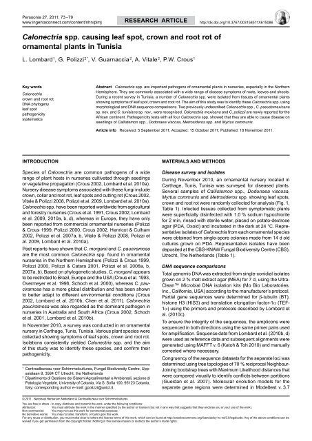
Calonectria spp. causing leaf spot, crown and root rot of ... - Persoonia
Calonectria spp. causing leaf spot, crown and root rot of ... - Persoonia
Calonectria spp. causing leaf spot, crown and root rot of ... - Persoonia

You also want an ePaper? Increase the reach of your titles
YUMPU automatically turns print PDFs into web optimized ePapers that Google loves.
<strong>Persoonia</strong> 27, 2011: 73–79<br />
www.ingentaconnect.com/content/nhn/pimj<br />
RESEARCH ARTICLE<br />
http://dx.doi.org/10.3767/003158511X615086<br />
<strong>Calonectria</strong> <strong>spp</strong>. <strong>causing</strong> <strong>leaf</strong> <strong>spot</strong>, <strong>crown</strong> <strong>and</strong> <strong>root</strong> <strong>rot</strong> <strong>of</strong><br />
ornamental plants in Tunisia<br />
L. Lombard 1 , G. Polizzi 2* , V. Guarnaccia 2 , A. Vitale 2 , P.W. Crous 1<br />
Key words<br />
<strong>Calonectria</strong><br />
<strong>crown</strong> <strong>and</strong> <strong>root</strong> <strong>rot</strong><br />
DNA phylogeny<br />
<strong>leaf</strong> <strong>spot</strong><br />
pathogenicity<br />
systematics<br />
Abstract <strong>Calonectria</strong> <strong>spp</strong>. are important pathogens <strong>of</strong> ornamental plants in nurseries, especially in the Northern<br />
Hemisphere. They are commonly associated with a wide range <strong>of</strong> disease symptoms <strong>of</strong> <strong>root</strong>s, leaves <strong>and</strong> shoots.<br />
During a recent survey in Tunisia, a number <strong>of</strong> <strong>Calonectria</strong> <strong>spp</strong>. were isolated from tissues <strong>of</strong> ornamental plants<br />
showing symptoms <strong>of</strong> <strong>leaf</strong> <strong>spot</strong>, <strong>crown</strong> <strong>and</strong> <strong>root</strong> <strong>rot</strong>. The aim <strong>of</strong> this study was to identify these <strong>Calonectria</strong> <strong>spp</strong>. using<br />
morphological <strong>and</strong> DNA sequence comparisons. Two previously undescribed <strong>Calonectria</strong> <strong>spp</strong>., C. pseudomexicana<br />
sp. nov. <strong>and</strong> C. tunisiana sp. nov., were recognised. <strong>Calonectria</strong> mexicana <strong>and</strong> C. polizzii are newly reported for the<br />
African continent. Pathogenicity tests with all four <strong>Calonectria</strong> <strong>spp</strong>. showed that they are able to cause disease on<br />
seedlings <strong>of</strong> Callistemon <strong>spp</strong>., Dodonaea viscosa, Metrosideros <strong>spp</strong>. <strong>and</strong> Myrtus communis.<br />
Article info Received: 5 September 2011; Accepted: 15 October 2011; Published: 18 November 2011.<br />
INTRODUCTION<br />
Species <strong>of</strong> <strong>Calonectria</strong> are common pathogens <strong>of</strong> a wide<br />
range <strong>of</strong> plant hosts in nurseries cultivated through seedings<br />
or vegetative propagation (Crous 2002, Lombard et al. 2010a).<br />
Nursery disease symptoms associated with these fungi include<br />
<strong>crown</strong>, collar <strong>and</strong> <strong>root</strong> <strong>rot</strong>, <strong>leaf</strong> <strong>spot</strong>s <strong>and</strong> cutting <strong>rot</strong> (Crous 2002,<br />
Vitale & Polizzi 2008, Polizzi et al. 2009, Lombard et al. 2010a).<br />
<strong>Calonectria</strong> <strong>spp</strong>. have been reported worldwide from agricultural<br />
<strong>and</strong> forestry nurseries (Crous et al. 1991, Crous 2002, Lombard<br />
et al. 2009, 2010a, b, d), whereas in Europe, they have only<br />
been reported from commercial ornamental nurseries (Polizzi<br />
& Crous 1999, Polizzi 2000, Crous 2002, Henricot & Culham<br />
2002, Polizzi et al. 2007a, b, Vitale & Polizzi 2008, Polizzi et<br />
al. 2009, Lombard et al. 2010a).<br />
Past reports have shown that C. morganii <strong>and</strong> C. pauciramosa<br />
are the most common <strong>Calonectria</strong> <strong>spp</strong>. found in ornamental<br />
nurseries in the Northern Hemisphere (Polizzi & Crous 1999,<br />
Polizzi 2000, Polizzi & Catara 2001, Polizzi et al. 2006a, b,<br />
2007a, b). Based on phylogenetic studies, C. morganii appears<br />
to be restricted to Brazil, Europe <strong>and</strong> the USA (Crous et al. 1993,<br />
Overmeyer et al. 1996, Schoch et al. 2000), whereas C. pauciramosa<br />
has a more global distribution <strong>and</strong> has been shown<br />
to better adapt to different environmental conditions (Crous<br />
2002, Lombard et al. 2010b, Chen et al. 2011). <strong>Calonectria</strong><br />
pauciramosa was also regarded as the dominant pathogen in<br />
nurseries in Australia <strong>and</strong> South Africa (Crous 2002, Schoch<br />
et al. 2001, Lombard et al. 2010b).<br />
In November 2010, a survey was conducted in an ornamental<br />
nursery in Carthage, Tunis, Tunisia. Various plant species were<br />
collected showing symptoms <strong>of</strong> <strong>leaf</strong> <strong>spot</strong>s, <strong>crown</strong> <strong>and</strong> <strong>root</strong> <strong>rot</strong>.<br />
Isolations consistently yielded <strong>Calonectria</strong> <strong>spp</strong>. <strong>and</strong> the aim<br />
<strong>of</strong> this study was to identify these species, <strong>and</strong> confirm their<br />
pathogenicity.<br />
1<br />
Centraalbureau voor Schimmelcultures, Fungal Biodiversity Centre, Uppsalalaan<br />
8, 3584 CT Utrecht, the Netherl<strong>and</strong>s<br />
2<br />
Dipartimento di Gestione dei Sistemi Agroalimentari e Ambientali, sezione di<br />
Patologia Vegetale, University <strong>of</strong> Catania, Via S. S<strong>of</strong>ia 100, 95123 Catania,<br />
Italy; corresponding author e-mail: gpolizzi@unict.it.<br />
MATERIALS AND METHODS<br />
Disease survey <strong>and</strong> isolates<br />
During November 2010, an ornamental nursery located in<br />
Carthage, Tunis, Tunisia was surveyed for diseased plants.<br />
Several samples <strong>of</strong> Callistemon <strong>spp</strong>., Dodonaea viscosa,<br />
Myrtus communis <strong>and</strong> Metrosideros <strong>spp</strong>. showing <strong>leaf</strong> <strong>spot</strong>s,<br />
<strong>crown</strong> <strong>and</strong> <strong>root</strong> <strong>rot</strong> were r<strong>and</strong>omly collected for analysis (Fig. 1,<br />
Table 1). Infected tissues collected from symptomatic plants<br />
were superficially disinfected with 1.0 % sodium hypochlorite<br />
for 2 min, rinsed with sterile water, placed on potato-dextrose<br />
agar (PDA, Oxoid) <strong>and</strong> incubated in the dark at 24 °C. Representative<br />
isolates <strong>of</strong> <strong>Calonectria</strong> from each ornamental species<br />
were obtained from single-spore colonies made from 14 d old<br />
cultures grown on PDA. Representative isolates have been<br />
deposited at the CBS-KNAW Fungal Biodiversity Centre (CBS),<br />
Utrecht, The Netherl<strong>and</strong>s (Table 1).<br />
DNA sequence comparisons<br />
Total genomic DNA was extracted from single-conidial isolates<br />
grown on 2 % malt extract agar (MEA) for 7 d, using the Ultra<br />
Clean Microbial DNA isolation kits (Mo Bio Laboratories,<br />
Inc., California, USA) according to the manufacturer’s p<strong>rot</strong>ocol.<br />
Partial gene sequences were determined for β-tubulin (BT),<br />
histone H3 (HIS3) <strong>and</strong> translation elongation factor-1α (TEF-<br />
1α) using the primers <strong>and</strong> p<strong>rot</strong>ocols described by Lombard et<br />
al. (2010c).<br />
To ensure the integrity <strong>of</strong> the sequences, the amplicons were<br />
sequenced in both directions using the same primer pairs used<br />
for amplification. Sequence data from Lombard et al. (2010b, d)<br />
were used as reference data <strong>and</strong> subsequent alignments were<br />
generated using MAFFT v. 6 (Katoh & Toh 2010) <strong>and</strong> manually<br />
corrected where necessary.<br />
Congruency <strong>of</strong> the sequence datasets for the separate loci was<br />
determined using tree topologies <strong>of</strong> 70 % reciprocal Neighbour-<br />
Joining bootstrap trees with Maximum Likelihood distances that<br />
were compared visually to identify conflicts between partitions<br />
(Gueidan et al. 2007). Molecular evolution models for the<br />
separate gene regions were determined in Modeltest v. 3.7<br />
© 2011 Nationaal Herbarium Nederl<strong>and</strong> & Centraalbureau voor Schimmelcultures<br />
You are free to share - to copy, distribute <strong>and</strong> transmit the work, under the following conditions:<br />
Attribution:<br />
You must attribute the work in the manner specified by the author or licensor (but not in any way that suggests that they endorse you or your use <strong>of</strong> the work).<br />
Non-commercial: You may not use this work for commercial purposes.<br />
No derivative works: You may not alter, transform, or build upon this work.<br />
For any reuse or distribution, you must make clear to others the license terms <strong>of</strong> this work, which can be found at http://creativecommons.org/licenses/by-nc-nd/3.0/legalcode. Any <strong>of</strong> the above conditions can be<br />
waived if you get permission from the copyright holder. Nothing in this license impairs or restricts the author’s moral rights.
74 <strong>Persoonia</strong> – Volume 27, 2011<br />
a<br />
b<br />
d<br />
e<br />
c<br />
f<br />
g h i<br />
Fig. 1 Symptoms <strong>of</strong> <strong>leaf</strong> <strong>spot</strong> caused by <strong>Calonectria</strong> <strong>spp</strong>. on several ornamental plants. a. Myrtus communis; b. Metrosideros thomasii; c. Callistemon sp.;<br />
d. Callistemon sp.; e. Dodonaea viscosa; f. Callistemon viminalis; g. Metrosideros excelsa; h. Metrosideros excelsa cv. Aurea; i. Metrosideros sp.<br />
(Posada & Cr<strong>and</strong>all 1998) <strong>and</strong> bootstrap analyses were run<br />
for 10 000 replicates.<br />
PAUP (Phylogenetic Analysis Using Parsimony, v. 4.0b10,<br />
Sw<strong>of</strong>ford 2002) was used to analyse the DNA sequence datasets.<br />
Phylogenetic relationships were estimated by heuristic<br />
searches with 1 000 r<strong>and</strong>om addition sequences <strong>and</strong> tree bisection-reconnection<br />
was used, with the branch swapping option<br />
set on ‘best trees’ only. All characters were weighted equally<br />
<strong>and</strong> alignment gaps were treated as missing data. Measures<br />
calculated for parsimony included tree length (TL), consistency<br />
index (CI), retention index (RI) <strong>and</strong> rescaled consistence<br />
index (RC). Bootstrap analysis (Hillis & Bull 1993) was based<br />
on 1 000 replications.<br />
A second phylogenetic analysis using a Markov Chain Monte<br />
Carlo (MCMC) algorithm was done to generate trees with Bayesian<br />
probabilities in MrBayes v. 3.1.1 (Ronquist & Huelsenbeck<br />
2003). Nucleotide substitution models were determined using<br />
MrModeltest (Nyl<strong>and</strong>er 2004) for each gene region <strong>and</strong> included<br />
in the analyses. Two analyses <strong>of</strong> four MCMC chains were run<br />
from r<strong>and</strong>om trees for one million generations <strong>and</strong> sampled<br />
every 100 generations. All runs converged on the same likelihood<br />
score <strong>and</strong> tree topology <strong>and</strong> therefore the first 800 trees<br />
were discarded as the burn-in phase <strong>of</strong> each analysis <strong>and</strong><br />
posterior probabilities determined from the remaining trees.<br />
The phylogenetic analyses included 46 partial gene sequences<br />
for each gene region, representing 20 <strong>Calonectria</strong> <strong>spp</strong>. (Table<br />
1). <strong>Calonectria</strong> colombiensis (CBS 112221) <strong>and</strong> C. chinensis<br />
(CBS 112744) were used as outgroup taxa in both analyses<br />
(Lombard et al. 2009). All novel sequences were deposited<br />
in GenBank <strong>and</strong> the alignments in TreeBASE (http://www.<br />
treebase.org).<br />
Taxonomy<br />
Morphological characterisation <strong>of</strong> the <strong>Calonectria</strong> isolates was<br />
done using single conidial cultures prepared on MEA <strong>and</strong> synthetic<br />
nutrient-poor agar (SNA; Nirenburg 1981, Lombard et al.<br />
2009). Inoculated plates were incubated at room temperature<br />
<strong>and</strong> examined after 7 d. Gross morphological characteristics<br />
<strong>of</strong> the anamorph state were determined by mounting fungal<br />
structures in clear lactic acid <strong>and</strong> 30 measurements at ×1 000<br />
magnification were made for each isolate using a Zeiss Axioscope<br />
2 microscope with interference contrast (DIC) illumination.<br />
The 95 % confidence levels were determined <strong>and</strong> extremes<br />
<strong>of</strong> conidial measurements are given in parentheses. For other<br />
structures, only extremes are presented. Colony characteristics<br />
were noted after 7 d <strong>of</strong> growth on MEA at 24 °C <strong>and</strong> colony<br />
colours determined using the colour charts <strong>of</strong> Rayner (1970).<br />
Descriptions, nomenclature <strong>and</strong> illustrations were deposited in<br />
MycoBank (Crous et al. 2004).<br />
Pathogenicity<br />
In order to test the pathogenicity <strong>of</strong> the <strong>Calonectria</strong> <strong>spp</strong>.<br />
collected in this study, seven isolates representing different<br />
<strong>Calonectria</strong> species identified by morphology <strong>and</strong> DNA sequence<br />
comparisons were selected for inoculation trials (Table<br />
1). A conidial suspension (1.0 × 10 5 conidia/mL) was prepared<br />
for each isolate by adding sterile water to plates <strong>of</strong> carnation <strong>leaf</strong><br />
agar (CLA; Fisher et al. 1982) 7 d after inoculation <strong>and</strong> dislodging<br />
the conidia. The conidial suspension was sprayed onto the<br />
canopy (until run-<strong>of</strong>f) <strong>of</strong> potted 2–6 mo old plants <strong>of</strong> Callistemon<br />
citrinus cv. Splendens, C. laevis, C. viminalis, Dodonaea viscose,<br />
Metrosideros excelsa, M. excelsa cv. Aurea, M. thomasii,<br />
Myrtus communis, M. communis subsp. tarentina. The conidial<br />
suspension <strong>of</strong> the isolate CBS 130351 was also applied to the<br />
<strong>crown</strong> <strong>of</strong> M. communis plants (10 mL/plant). All plants were<br />
subsequently covered with plastic bags for 48 h <strong>and</strong> maintained
L. Lombard et al.: <strong>Calonectria</strong> in Tunisia<br />
75<br />
Table 1 <strong>Calonectria</strong> isolates used in the phylogenetic analyses <strong>and</strong> pathogenicity trials.<br />
Species Isolate number 1 β-tubulin 2 Histone H3 2 TEF-1α 2 Host /substrate<br />
<strong>Calonectria</strong> brasiliensis CBS 230.30 4 GQ267241 GQ267259 GQ267328 Eucalyptus sp.<br />
CBS 114257 GQ267242 GQ267260 GQ267329 Leaf litter<br />
C. cerciana CBS 123693 4 FJ918510 FJ918528 FJ918559 E. gr<strong>and</strong>is × urophylla<br />
CBS 123695 FJ918511 FJ918529 FJ918560 E. gr<strong>and</strong>is × urophylla<br />
C. chinensis CBS 112744 AY725618 AY725660 AY725709 Soil<br />
C. colombiana CBS 115127 4 FJ972423 FJ972442 FJ972492 Soil<br />
CBS 115638 FJ972422 FJ972441 FJ972491 Soil<br />
C. colombiensis CBS 112220 4 GQ267207 AY725662 AY725711 Soil<br />
C. hawksworthii CBS 111870 4 AF333407 DQ190649 FJ918558 Nelumbo nucifera<br />
C. insularis CBS 114558 4 AF210861 FJ918526 FJ918556 Soil<br />
CBS 114559 AF210862 FJ918525 FJ918555 Soil<br />
C. leucothoës CBS 109166 FJ918508 FJ918523 FJ918553 Leucothoë axillaris<br />
C. mexicana CBS 110918 4 AF210863 FJ972460 FJ972526 Soil<br />
CBS 130353 3 JN607280 JN607265 JN607295 Dodonaea viscosa<br />
C. morganii CBS 110666 FJ918509 FJ918527 FJ918557 Ilex vomitoria<br />
CBS 119669 DQ521599 DQ521601 GQ421796 Pistacia lentiscus<br />
C. pauciramosa CPC 971 FJ918514 FJ918531 FJ918565 E. gr<strong>and</strong>is<br />
CPC 416 FJ918515 FJ918532 FJ918566 E. gr<strong>and</strong>is<br />
C. polizzii CBS 123402 4 FJ972419 FJ972438 FJ972488 Arbutus unedo<br />
CBS 125270 FJ972417 FJ972436 FJ972486 Callistemon citrinus<br />
CBS 130351 3 JN607270 JN607255 JN607285 Myrtus communis<br />
CBS 130352 3 JN607275 JN607260 JN607290 Metrosideros thomasii<br />
DISTEF-TMC2 JN607269 JN607254 JN607284 Myrtus communis<br />
DISTEF-TMEA1 JN607272 JN607257 JN607287 Metrosideros excelsa cv. Aurea<br />
DISTEF-TMN3 JN607274 JN607259 JN607289 Metrosideros sp.<br />
C. pseudomexicana CBS 130354 3,4 JN607281 JN607266 JN607496 Callistemon sp. (rouge)<br />
CBS 130355 3 JN607282 JN607267 JN607497 Callistemon sp. (rouge)<br />
DISTEF-TCROU4 JN607283 JN607268 JN607498 Callistemon sp. (rouge)<br />
C. pseudoscoparia CBS 125256 GQ267228 GQ267277 GQ267348 E. gr<strong>and</strong>is<br />
CBS 125257 4 GQ267229 GQ267278 GQ267349 E. gr<strong>and</strong>is<br />
C. scoparia CPC 1675 FJ972426 FJ972476 FJ972525 Eucalyptus sp.<br />
CPC 1679 FJ972427 GQ267246 GQ267298 Eucalyptus sp.<br />
C. spathulata CBS 112689 AF308463 FJ918524 FJ918554 E. viminalis<br />
CBS 555.92 4 GQ267215 GQ267261 GQ267331 Araucaria angustifolia<br />
C. sulawesiensis CBS 125248 GQ267223 GQ267272 GQ267343 Eucalyptus sp.<br />
CBS 125253 GQ267220 GQ267269 GQ267340 Eucalyptus sp.<br />
C. tunisiana CBS 130356 3 JN607277 JN607262 JN607292 Callistemon sp. (rouge)<br />
CBS 130357 3,4 JN607276 JN607261 JN607291 Callistemon laevis<br />
DISTEF-TCV1 JN607278 JN607263 JN607293 Callistemon viminalis<br />
DISTEF-TCROS4 JN607279 JN607264 JN607294 Callistemon sp. (rosè)<br />
DISTEF-TME1 JN607271 JN607256 JN607286 Metrosideros excelsa<br />
DISTEF-TMN1 JN607273 JN607258 JN607288 Metrosideros sp.<br />
C. variabilis CBS 112691 GQ267240 GQ267264 GQ267335 Eucalyptus sp.<br />
CBS 114677 AF333424 GQ267263 GQ267334 Eucalyptus sp.<br />
C. zuluensis CBS 125268 4 FJ972414 FJ972433 FJ972483 Eucalyptus sp.<br />
CBS 125272 FJ972415 FJ972434 FJ972484 Eucalyptus sp.<br />
1<br />
CBS: CBS-KNAW Fungal Biodiversity Centre, Utrecht, The Netherl<strong>and</strong>s; CPC: Pedro Crous working collection housed at CBS; DISTEF: Dipartimento di Scienze e Tecnologie Fitosanitarie,<br />
Catania, Italy.<br />
2<br />
GenBank accession numbers.<br />
3<br />
Isolates used for the pathogenicity trials.<br />
4<br />
Ex-type cultures; Isolates in bold obtained during survey.<br />
in a growth chamber at 25 ± 1 °C for 14 d. Five plants for each<br />
isolate <strong>and</strong> host were used <strong>and</strong> the same number <strong>of</strong> control<br />
plants were treated using sterile water. Pathogenicity tests were<br />
evaluated 5, 10 <strong>and</strong> 25 d after inoculation.<br />
Results<br />
Disease survey <strong>and</strong> isolates<br />
During the survey, a total <strong>of</strong> 46 <strong>Calonectria</strong> isolates were collected<br />
from ornamental hosts sampled. Majority <strong>of</strong> the isolates<br />
(41) were associated with <strong>leaf</strong> <strong>spot</strong>s or <strong>leaf</strong> blight <strong>of</strong> Callistemon<br />
<strong>spp</strong>. (18), D. viscosa (1), Metrosideros <strong>spp</strong>. (17) <strong>and</strong> Myrtus<br />
communis (5), <strong>and</strong> the remaining (5) with <strong>crown</strong> <strong>and</strong> <strong>root</strong> <strong>rot</strong><br />
<strong>of</strong> M. communis. Leaves showed minute brown <strong>spot</strong>s, which<br />
<strong>of</strong>ten enlarged, forming a nec<strong>rot</strong>ic centre surrounded by a dark<br />
purple halo (Fig. 1). Young, non-lignified terminal shoots <strong>of</strong>ten<br />
exhibited dieback or lesions similar to those on the leaves. Severe<br />
defoliation was observed on M. communis <strong>and</strong> M. excelsa<br />
cv. Aurea. Several seedlings <strong>of</strong> M. communis had <strong>crown</strong> <strong>and</strong><br />
<strong>root</strong> <strong>rot</strong>, <strong>and</strong> fungal sporulation occurred on the lower part <strong>of</strong><br />
the <strong>crown</strong>. Initial symptoms were brown lesions that exp<strong>and</strong>ed<br />
rapidly to girdle the stem at the seedling <strong>crown</strong>, above <strong>and</strong><br />
below the soil line, resulting in plant death.<br />
DNA sequence comparisons<br />
Amplicons <strong>of</strong> approximately 450 bases for HIS3 <strong>and</strong> 500 bases<br />
each for BT <strong>and</strong> TEF-1α were generated. The 70 % reciprocal<br />
bootstrap trees showed no conflict in tree topologies for the<br />
three gene regions <strong>and</strong> therefore they were combined in a dataset<br />
consisting <strong>of</strong> 1 532 characters including gaps. Of these<br />
characters, 1 187 were constant <strong>and</strong> parsimony uninformative.<br />
Analysis <strong>of</strong> the 345 parsimony informative characters yielded<br />
16 equally most parsimonious trees (TL = 814, CI = 0.721,<br />
RI = 0.923, RC = 0.666), <strong>of</strong> which the first tree is presented (Fig.<br />
2). A HKY+I model for BT, a GTR+I+G model for HIS3 <strong>and</strong> a<br />
GTR+G model for TEF-1α was selected for Bayesian analysis.<br />
The Bayesian consensus tree confirmed the tree topology obtained<br />
with maximum parsimony including bootstrap support.<br />
The phylogenetic tree illustrates a number <strong>of</strong> well-supported<br />
clades containing the <strong>Calonectria</strong> isolates obtained during the<br />
survey. Some <strong>of</strong> the isolates clustered in a clade representing<br />
C. polizzii with a bootstrap value (BP) <strong>of</strong> 97 <strong>and</strong> a Bayesian<br />
posterior probability (PP) value <strong>of</strong> 1.00. Several isolates also<br />
grouped with <strong>and</strong> close to C. mexicana in two separate wellsupported<br />
clades (BP = 68, PP = 0.95 <strong>and</strong> BP = 78, PP = 0.98,<br />
respectively), which could represent novel phylogenetic species.
76 78/1.00 DISTEF-TCV1<br />
C. tunisiana sp. nov <strong>Persoonia</strong> – Volume 27, 2011<br />
DISTEF-TCROS4<br />
100/1.00<br />
100/1.00<br />
Fig. 2 One <strong>of</strong> 16 most parsimonious trees obtained from a heuristic search<br />
with 1 000 r<strong>and</strong>om addition sequences <strong>of</strong> the combined sequences <strong>of</strong> β-tubulin,<br />
histone H3 <strong>and</strong> translation elongation factor 1α sequence alignments<br />
<strong>of</strong> the <strong>Calonectria</strong> isolates obtained during the survey <strong>and</strong> other closely<br />
related species. Scale bar shows 10 changes. Bootstrap support values (in<br />
bold) <strong>and</strong> Bayesian posterior probability values are shown at the nodes.<br />
Thickened lines indicate branches in the strict consensus tree <strong>and</strong> the consensus<br />
tree <strong>of</strong> the Bayesian analyses. The tree was <strong>root</strong>ed to C. chinensis<br />
(CBS 112744) <strong>and</strong> C. colombiensis (CBS 112220). Isolates in bold were<br />
10 changes<br />
obtained during the survey.<br />
72/0.99<br />
68/0.88<br />
63/0.98<br />
DISTEF-TME1<br />
CBS 130357<br />
C. colombiensis CBS 112220<br />
C. chinensis CBS 112744<br />
CBS 110918<br />
C. leucothoes CBS 109166<br />
73/0.69<br />
C. mexicana C. hawksworthii CBS 111870<br />
52/1.00<br />
CBS 130353<br />
CBS 130355 100/1.00<br />
CBS 230.51<br />
C. brasiliensis<br />
100/1.00<br />
99/1.00CBS 13035479/0.76<br />
CBS 114257<br />
C. pseudomexicana sp. nov<br />
68/0.95<br />
99/1.00 CBS 125248<br />
72/0.99<br />
DISTEF-TCROU4<br />
C. sulawesiensis<br />
CBS 125253<br />
68/0.88 CBS 130356 82/1.00<br />
CBS 114677<br />
DISTEF-TMN1<br />
C. variabilis<br />
78/1.00<br />
63/0.98<br />
100/1.00<br />
CBS 112691<br />
DISTEF-TCV1<br />
C. tunisiana sp. nov<br />
DISTEF-TCROS4 53/0.73 CBS 110666<br />
C. morganii<br />
98/1.00<br />
DISTEF-TME1<br />
CBS 130357<br />
CBS 119669<br />
CBS 114558<br />
C. insularis<br />
100/1.00<br />
CBS 114559<br />
C. leucothoes CBS 109166<br />
C. 100/1.00<br />
CBS 123693<br />
hawksworthii CBS 111870 C. cerciana<br />
52/1.00<br />
CBS 123695<br />
100/1.00<br />
CBS 230.51<br />
CPC 1675<br />
C. brasiliensis<br />
99/1.00 79/0.76 C. scopariaCBS 114257<br />
100/1.00 CPC 1679<br />
99/1.00 CBS 125248<br />
CBS 125257<br />
C. sulawesiensis<br />
68/0.98<br />
C. pseudoscoparia CBS 125253<br />
CBS 125256 82/1.00<br />
CBS 114677<br />
98/1.00<br />
C. variabilis<br />
100/1.00<br />
CBS 115127<br />
100/1.00 C. colombiana CBS 112691<br />
50/0.98 CBS 115638<br />
53/0.73 CBS 110666<br />
C. morganii<br />
100/1.00 CBS 112689<br />
98/1.00<br />
CBS 119669 C. spathulata<br />
CBS 555.92<br />
CBS 114558<br />
C. insularis<br />
94/1.00 CPC 971<br />
100/1.00 C. pauciramosa CBS 114559<br />
CPC 416<br />
100/1.00<br />
CBS 123693<br />
CBS 125268<br />
C. cerciana<br />
98/0.97<br />
C. zuluensis<br />
CBS 123695<br />
63/1.00<br />
CBS 125272<br />
CPC 1675<br />
100/1.00<br />
CBS 123402 C. scoparia<br />
CPC 1679<br />
CBS 125270<br />
CBS 125257<br />
68/0.98 DISTEF-TMC2 C. pseudoscoparia<br />
CBS 125256<br />
98/1.00<br />
CBS 130351 C. polizzii<br />
97/1.00 CBS 115127<br />
100/1.00<br />
DISTEF-TMEA1 C. colombiana<br />
50/0.98 CBS 115638<br />
DISTEF-TMN3<br />
100/1.00 CBS 112689<br />
10 changes<br />
CBS 130352<br />
CBS 555.92<br />
C. spathulata<br />
98/0.97<br />
63/1.00<br />
DISTEF-TCROU4<br />
CBS 130356<br />
DISTEF-TMN1<br />
94/1.00 CPC 971<br />
97/1.00<br />
CPC 416<br />
CBS 125268<br />
CBS 125272<br />
CBS 123402<br />
CBS 125270<br />
DISTEF-TMC2<br />
CBS 130351<br />
DISTEF-TMEA1<br />
DISTEF-TMN3<br />
CBS 130352<br />
C. pauciramosa<br />
C. zuluensis<br />
C. polizzii<br />
Taxonomy<br />
DNA sequence <strong>and</strong> morphological comparisons <strong>of</strong> the <strong>Calonectria</strong><br />
isolates obtained during the survey show that these isolates<br />
belong to C. mexicana <strong>and</strong> C. polizzii <strong>and</strong> also constitute<br />
two previously undescribed taxa. Based on morphological<br />
comparisons, isolate CBS 130353 agrees with C. mexicana<br />
(Schoch et al. 1999) <strong>and</strong> isolates DISTEF-TMC2, CBS 130351,<br />
DISTEF-TMEA1, DISTEF-TMN3 <strong>and</strong> CBS 130352 represent<br />
C. polizzii (Lombard et al. 2010b). The remaining isolates are<br />
newly described as follows:<br />
<strong>Calonectria</strong> pseudomexicana L. Lombard, G. Polizzi & Crous,<br />
sp. nov. — MycoBank MB563138; Fig. 3<br />
Teleomorph unknown.<br />
<strong>Calonectria</strong> mexicana morphologice similes sed minus ramis conidiophorae.<br />
Etymology. Name reflects the fact that this species closely resembles<br />
C. mexicana.<br />
Conidiophores with a stipe bearing penicillate suites <strong>of</strong> fertile<br />
branches, stipe extensions, <strong>and</strong> terminal vesicles; stipe septate,<br />
hyaline, smooth, 38–69 × 5–9 µm; stipe extensions septate,<br />
straight to flexuous, 175–251 µm long, 3–6 µm wide at the apical<br />
septum, terminating in a fusiform to broadly ellipsoidal vesicle
L. Lombard et al.: <strong>Calonectria</strong> in Tunisia<br />
77<br />
a<br />
b<br />
c<br />
d<br />
e<br />
f<br />
g<br />
Fig. 3 <strong>Calonectria</strong> pseudomexicana. a, b. Macroconidiophores; c–e. fusiform to broadly ellipsoidal vesicles with papillate apices; f, g. conidiogenous apparatus<br />
with conidiophore branches <strong>and</strong> doliiform to reniform phialides; h. 1-septate macroconidia. — Scale bars = 10 μm.<br />
h<br />
9–14 µm diam with papillate apex. Conidiogenous apparatus<br />
38–68 µm long, 32–64 µm wide; primary branches aseptate<br />
or 1-septate, 21–43 × 4–7 µm; secondary branches aseptate,<br />
13–26 × 4–7 µm; tertiary branches <strong>and</strong> additional branches<br />
(–4), aseptate, 10–18 × 2–6 µm, each terminal branch producing<br />
2–6 phialides; phialides doliiform to reniform, hyaline,<br />
aseptate, 6–14 × 2–6 µm; apex with minute periclinal thickening<br />
<strong>and</strong> inconspicuous collarette. Conidia cylindrical, rounded<br />
at both ends, straight, (40–)43–48(–49) × (4–)5–6 µm (av. =<br />
45 × 5 µm), 1-septate, lacking a visible abscission scar, held<br />
in parallel cylindrical clusters by colourless slime. Megaconidia<br />
<strong>and</strong> microconidia not seen.<br />
Culture characteristics — Colonies fast growing at 24 ºC on<br />
MEA, sienna to bay on surface, reverse sienna after 7 d; moderate<br />
white aerial mycelium with sparse to moderate sporulation;<br />
chlamydospores extensive throughout medium.<br />
Specimens examined. Tunisia, Carthage, Tunis, from Callistemon sp.,<br />
Nov. 2010, G. Polizzi, (CBS H-20685, holotype <strong>of</strong> C. pseudomexicana,<br />
culture ex-type CBS 130354 = DISTEF-TCROU1); Carthage, Tunis, from<br />
Callistemon sp., Nov. 2010, G. Polizzi, culture CBS 130355 = DISTEF-<br />
TCROU3; Carthage, Tunis, from Callistemon sp., Nov. 2010, G. Polizzi,<br />
culture DISTEF-TCROU4.<br />
Notes — <strong>Calonectria</strong> pseudomexicana is morphologically<br />
similar to C. mexicana. <strong>Calonectria</strong> pseudomexicana has four<br />
or less conidiophore branches while C. mexicana has five as<br />
reported by Schoch et al. (1999).<br />
<strong>Calonectria</strong> tunisiana L. Lombard, G. Polizzi & Crous, sp.<br />
nov. — MycoBank MB563139; Fig. 4<br />
Teleomorph unknown.<br />
<strong>Calonectria</strong> mexicana morphologice similes sed minus conidiophorae ramis<br />
et fructibus breviores sunt stipe augue.<br />
Etymology. Name refers to the country Tunisia, where the fungus was<br />
collected.<br />
Conidiophores with a stipe bearing penicillate suites <strong>of</strong> fertile<br />
branches, stipe extensions, <strong>and</strong> terminal vesicles; stipe septate,<br />
hyaline, smooth, 42–95 × 7–11 µm; stipe extensions septate,<br />
straight to flexuous, 147–199 µm long, 4–5 µm wide at the<br />
apical septum, terminating in a fusiform to broadly ellipsoidal<br />
vesicle 8–14 µm diam with papillate apex. Conidiogenous apparatus<br />
40–68 µm long, 30–66 µm wide; primary branches<br />
aseptate or 1-septate, 17–41 × 5–7 µm; secondary branches<br />
aseptate, 10–22 × 4–7 µm; tertiary branches aseptate, 9–18<br />
× 4–5 µm, each terminal branch producing 2–6 phialides;<br />
phialides doliiform to reniform, hyaline, aseptate, 8–13 × 3–5<br />
µm; apex with minute periclinal thickening <strong>and</strong> inconspicuous<br />
collarette. Conidia cylindrical, rounded at both ends, straight,
78 <strong>Persoonia</strong> – Volume 27, 2011<br />
a<br />
b<br />
c<br />
d<br />
e<br />
f<br />
g<br />
h<br />
Fig. 4 <strong>Calonectria</strong> tunisiana. a, b. Macroconidiophores; c–e. fusiform to broadly ellipsoidal vesicles with papillate apices; f, g. conidiogenous apparatus with<br />
conidiophore branches <strong>and</strong> doliiform to reniform phialides; h. 1-septate macroconidia. — Scale bars = 10 μm.<br />
(43–)47–51(–53) × 4–6 µm (av. = 49 × 5 µm), 1-septate, lacking<br />
a visible abscission scar, held in parallel cylindrical clusters by<br />
colourless slime. Megaconidia <strong>and</strong> microconidia not seen.<br />
Culture characteristics — Colonies fast growing at 24 °C on<br />
MEA, sienna to bay on surface, <strong>and</strong> reverse sienna after 7 d;<br />
sparse white aerial mycelium with sparse sporulation; chlamydospores<br />
extensive throughout the medium.<br />
Specimens examined. Tunisia, Carthage, Tunis, from Callistemon laevis,<br />
Nov. 2010, G. Polizzi, (CBS H-20684, holotype <strong>of</strong> C. tunisiana, culture ex-type<br />
CBS 130357 = DISTEF-TCL1); Carthage, Tunis, from Callistemon sp., Nov.<br />
2010, G. Polizzi, culture CBS 130356 = DISTEF-TCROU2; Carthage, Tunis,<br />
from Metrosideros excelsus, Nov. 2010, G. Polizzi, culture DISTEF-TME1.<br />
Notes — Morphologically, C. tunisiana is similar to C. mexicana<br />
<strong>and</strong> C. pseudomexicana, but can be distinguished from<br />
both taxa by its shorter stipe extensions. The conidiophores <strong>of</strong><br />
C. tunisiana (–3) also form fewer fertile branches than C. mexicana<br />
(–5) <strong>and</strong> C. pseudomexicana (–4) (Schoch et al. 1999).<br />
Pathogenicity<br />
All plants inoculated with the <strong>Calonectria</strong> <strong>spp</strong>. in this study developed<br />
<strong>leaf</strong> <strong>spot</strong>, <strong>leaf</strong> blight or <strong>crown</strong> <strong>and</strong> <strong>root</strong> <strong>rot</strong> symptoms.<br />
The first symptoms <strong>of</strong> <strong>leaf</strong> <strong>spot</strong> <strong>and</strong> <strong>leaf</strong> blight were observed 5 d<br />
after inoculation on all test plants inoculated with the <strong>Calonectria</strong><br />
<strong>spp</strong>., resembling the symptoms observed during the survey. Isolates<br />
<strong>of</strong> C. pseudomexicana (CBS 130354, 130355), C. tunisiana<br />
(CBS 130356, 130357) as well as the single isolate <strong>of</strong> C. mexicana<br />
(CBS 130353) produced the most severe symptoms. Isolates<br />
<strong>of</strong> C. polizzii (CBS 130351, 130352) also caused <strong>leaf</strong> <strong>spot</strong><br />
<strong>and</strong> <strong>leaf</strong> blight on all inoculated plants, but less severe than the<br />
other three <strong>Calonectria</strong> <strong>spp</strong>. tested. Ten days after inoculation,<br />
severe or moderate defoliation <strong>of</strong> M. communis <strong>and</strong> M. excelsa<br />
cv. Aurea plants was observed.<br />
All inoculated plants <strong>of</strong> M. communis developed <strong>crown</strong> <strong>rot</strong>, basal<br />
stem <strong>rot</strong> <strong>and</strong> <strong>root</strong> <strong>rot</strong> 25 d after inoculation with the isolate<br />
representing C. polizzii (CBS 130351). All un-inoculated control<br />
plants remained healthy <strong>and</strong> re-isolations from the test plants<br />
consistently yielded the test fungi.<br />
DISCUSSION<br />
During a survey <strong>of</strong> diseased plants at an ornamental nursery in<br />
Tunis, Tunisia, a number <strong>of</strong> <strong>Calonectria</strong> <strong>spp</strong>. were isolated from<br />
plants exhibiting <strong>crown</strong>, <strong>root</strong> <strong>rot</strong> <strong>and</strong> <strong>leaf</strong> <strong>spot</strong>s. DNA sequence<br />
<strong>and</strong> morphological comparisons allowed the identification <strong>of</strong><br />
two <strong>of</strong> these isolates as C. mexicana <strong>and</strong> C. polizzii as well<br />
as the description <strong>of</strong> two new species, C. pseudomexicana<br />
<strong>and</strong> C. tunisiana, both in the C. scoparia complex (Schoch et<br />
al. 1999).
L. Lombard et al.: <strong>Calonectria</strong> in Tunisia<br />
79<br />
<strong>Calonectria</strong> mexicana resides in the C. scoparia complex<br />
(Schoch et al. 1999) <strong>and</strong> can be distinguished from the other<br />
seven <strong>Calonectria</strong> <strong>spp</strong>. in the complex based on their unique<br />
papillate vesicles (Schoch et al. 1999, Lombard et al. 2010b,<br />
c, Chen et al. 2011). Until now, C. mexicana has only been<br />
reported from soil samples collected in Mexico, <strong>and</strong> its pathogenicity<br />
was unknown (Schoch et al. 1999, Crous 2002). This<br />
study represents the first report <strong>of</strong> this fungus outside Mexico,<br />
<strong>and</strong> also demonstrates its pathogenicity on some plant hosts.<br />
<strong>Calonectria</strong> polizzii has previously been reported from ornamental<br />
plants collected at a nursery in Sicily, Italy (Schoch et<br />
al. 2001, Lombard et al. 2010b), although its pathogenicity was<br />
not confirmed. This study represents the first confirmation <strong>of</strong> the<br />
pathogenicity <strong>of</strong> C. polizzii <strong>and</strong> widens its distribution to Tunisia.<br />
<strong>Calonectria</strong> polizzii is a member <strong>of</strong> the C. scoparia complex <strong>and</strong><br />
can be distinguished from the other members by its smaller<br />
macroconidial dimensions (Lombard et al. 2010b).<br />
The description <strong>of</strong> C. pseudomexicana <strong>and</strong> C. tunisiana adds<br />
two more species to the C. scoparia complex. This complex is<br />
characterised by 1-septate macroconidia <strong>and</strong> the formation <strong>of</strong><br />
ellipsoidal to obpyriform terminal vesicles on the stipe extensions<br />
(Schoch et al. 1999, Crous 2002, Lombard et al. 2010b).<br />
Based on phylogenetic inference, both these newly described<br />
species are closely related to C. mexicana, which they also resemble<br />
in morphology. They can be distinguished from C. mexicana<br />
<strong>and</strong> each other by the number <strong>of</strong> fertile branches produced<br />
on the conidiophores. <strong>Calonectria</strong> tunisiana (av. = 49 × 5 µm)<br />
has slightly larger macroconidia than both C. mexicana (av. =<br />
45 × 4 µm; Schoch et al. 1999) <strong>and</strong> C. pseudomexicana (av.<br />
= 45 × 5 µm).<br />
The pathogenicity tests with isolates <strong>of</strong> C. mexicana, C. polizzii,<br />
C. pseudomexicana <strong>and</strong> C. tunisiana clearly showed that they<br />
are able to cause symptoms similar to those observed during<br />
the survey. <strong>Calonectria</strong> polizzii was less virulent than the other<br />
three species, but should still be regarded as an important<br />
nursery pathogen. This supports the view that most <strong>Calonectria</strong><br />
<strong>spp</strong>. can induce <strong>leaf</strong> <strong>spot</strong>s if the environmental conditions are<br />
favourable (Crous 2002). All four species caused similar disease<br />
symptoms on the nine inoculated plant species, suggesting<br />
that little is known about the host specificity <strong>and</strong> mechanisms<br />
<strong>of</strong> infection <strong>of</strong> this group <strong>of</strong> plant pathogens.<br />
This study stresses the importance <strong>of</strong> <strong>Calonectria</strong> <strong>spp</strong>. as<br />
nursery pathogens. Their soil-borne nature has contributed to<br />
their ease <strong>of</strong> movement globally <strong>and</strong> little is known about their<br />
origins. Furthermore, it is not known if these fungal pathogens<br />
originated from Tunisia or were introduced, <strong>and</strong> more isolates<br />
are needed for a study <strong>of</strong> their population dynamics.<br />
References<br />
Chen SF, Lombard L, Roux J, Xie YJ, Wingfield MJ, Zhou XD. 2011. Novel<br />
species <strong>of</strong> <strong>Calonectria</strong> associated with Eucalyptus <strong>leaf</strong> blight in Southeast<br />
China. <strong>Persoonia</strong> 26: 1–12.<br />
Crous PW. 2002. Taxonomy <strong>and</strong> pathology <strong>of</strong> Cylindrocladium (<strong>Calonectria</strong>)<br />
<strong>and</strong> allied genera. APS Press, St. Paul, Minnesota, USA.<br />
Crous PW, Alfenas AC, Wingfield MJ. 1993. <strong>Calonectria</strong> scoparia <strong>and</strong> <strong>Calonectria</strong><br />
morganii sp. nov., <strong>and</strong> variation among isolates <strong>of</strong> their Cylindrocladium<br />
anamorphs. Mycological Research 97: 701–708.<br />
Crous PW, Gams W, Stalpers JA, Robert V, Stegehuis G. 2004. MycoBank:<br />
an online initiative to launch mycology into the 21st century. Studies in<br />
Mycology 50: 19–22.<br />
Crous PW, Phillips AJL, Wingfield MJ. 1991. The genera Cylindrocladium <strong>and</strong><br />
Cylindrocladiella in South Africa, with special reference to forest nurseries.<br />
South African Forestry Journal 157: 69–89.<br />
Fisher NL, Burgess LW, Toussoun TA, Nelson PE. 1982. Carnation leaves<br />
as a substrate <strong>and</strong> for preserving cultures <strong>of</strong> Fusarium species. Phytopathology<br />
72: 151–153.<br />
Gueidan C, Roux C, Lutzoni F. 2007. Using multigene phylogeny analysis<br />
to assess generic delineation <strong>and</strong> character evolution in Verrucariaceae<br />
(Verrucariales, Ascomycota). Mycological Research 111: 1145–1168.<br />
Henricot B, Culham A. 2002. Cylindrocladium buxicola, a new species affecting<br />
Buxus <strong>spp</strong>., <strong>and</strong> its phylogenetic status. Mycologia 94: 980–997.<br />
Hillis DM, Bull JJ. 1993. An empirical test <strong>of</strong> bootstrapping as a method for<br />
assessing confidence in phylogenetic analysis. Systematic Biology 42:<br />
182–192.<br />
Katoh K, Toh H. 2010. Parallelization <strong>of</strong> the MAFFT multiple sequence alignment<br />
program. Bioinformatics 26: 1899–1900.<br />
Lombard L, Crous PW, Wingfield BD, Wingfield MJ. 2010a. Species concepts<br />
in <strong>Calonectria</strong> (Cylindrocladium). Studies in Mycology 66: 1–14.<br />
Lombard L, Crous PW, Wingfield BD, Wingfield MJ. 2010b. Multigene phylogeny<br />
<strong>and</strong> mating tests reveal three cryptic species related to <strong>Calonectria</strong><br />
pauciramosa. Studies in Mycology 66: 15–30.<br />
Lombard L, Crous PW, Wingfield BD, Wingfield MJ. 2010c. Phylogeny <strong>and</strong><br />
systematics <strong>of</strong> the genus <strong>Calonectria</strong>. Studies in Mycology 66: 31–69.<br />
Lombard L, Rodas CA, Crous PW, Wingfield BD, Wingfield MJ. 2009. <strong>Calonectria</strong><br />
(Cylindrocladium) species associated with dying Pinus cuttings.<br />
<strong>Persoonia</strong> 23: 41–47.<br />
Lombard L, Zhou XD, Crous PW, Wingfield BD, Wingfield MJ. 2010d. <strong>Calonectria</strong><br />
species associated with cutting <strong>rot</strong> <strong>of</strong> Eucalyptus. <strong>Persoonia</strong> 24:<br />
1–11.<br />
Nirenburg HI. 1981. A simplified method for identifying Fusarium <strong>spp</strong>. occurring<br />
on wheat. Canadian Journal <strong>of</strong> Botany 59: 1599–1609.<br />
Nyl<strong>and</strong>er JAA. 2004. MrModeltest v. 2. Programme distributed by the author.<br />
Evolutionary Biology Centre, Uppsala University.<br />
Overmeyer C, Lünneman S, Wallburnn C von, Meinhardt F. 1996. Genetic<br />
variability among isolates <strong>and</strong> sexual <strong>of</strong>fspring <strong>of</strong> the plant pathogenic<br />
fungus <strong>Calonectria</strong> morganii on the basis <strong>of</strong> r<strong>and</strong>om amplification <strong>of</strong><br />
polymorphic DNA (RAPD) <strong>and</strong> restriction fragment length polymorphism<br />
(RFLP). Current Microbiology 33: 249–255.<br />
Polizzi G. 2000. Prime esperienze di lotta chimica nei confronti del marciume<br />
del colletto e delle radici di Polygala myrtifolia causato da Cylindrocladium<br />
pauciramosum. Informatore Fitopatologico 11: 39–47.<br />
Polizzi G, Catara V. 2001. First report <strong>of</strong> <strong>leaf</strong> <strong>spot</strong> caused by Cylindrocladium<br />
pauciramosum on Acacia retinodes, Arbutus unedo, Feijoa sellowiana <strong>and</strong><br />
Dodonaea viscosa in southern Italy. Plant Disease 85: 803.<br />
Polizzi G, Crous PW. 1999. Root <strong>and</strong> collar <strong>of</strong> milkwort caused by Cylindrocladium<br />
pauciromosum, a new record for Europe. European Journal <strong>of</strong><br />
Plant Pathology 105: 407–411.<br />
Polizzi G, Grasso FM, Vitale A, Aiello D. 2007a. First occurrence <strong>of</strong> <strong>Calonectria</strong><br />
<strong>leaf</strong> <strong>spot</strong> on mexican blue palm in Italy. Plant Disease 91: 1057.<br />
Polizzi G, Vitale A, Aiello D, Castello I, Guarnaccia V, Parlavecchio G. 2009.<br />
First record <strong>of</strong> <strong>crown</strong> <strong>and</strong> <strong>root</strong> <strong>rot</strong> caused by Cylindrocladium pauciramosum<br />
on brush cherry in Italy. Plant Disease 93: 547.<br />
Polizzi G, Vitale A, Aiello D, Dimartino MA, Parlavecchio G. 2007b. First report<br />
<strong>of</strong> damping-<strong>of</strong>f <strong>and</strong> <strong>leaf</strong> <strong>spot</strong> caused by Cylindrocladium scoparium on different<br />
accessions <strong>of</strong> bottlebrush cuttings in Italy. Plant Disease 91: 769.<br />
Polizzi G, Vitale A, Aiello D, Parlavecchio G. 2006a. First record <strong>of</strong> <strong>crown</strong><br />
<strong>and</strong> <strong>root</strong> <strong>rot</strong> caused by Cylindrocladium pauciramosum on California lilac<br />
in Italy. Plant Disease 90: 1459.<br />
Polizzi G, Vitale A, Castello I, Groenewald JZ, Crous PW. 2006b. Cylindrocladium<br />
<strong>leaf</strong> <strong>spot</strong>, blight <strong>and</strong> <strong>crown</strong> <strong>rot</strong>, new diseases <strong>of</strong> mastic tree seedlings<br />
caused by Cylindrocladium scoparium. Plant Disease 90: 1110.<br />
Posada D, Cr<strong>and</strong>all KA. 1998. Modeltest: testing the model <strong>of</strong> DNA substitution.<br />
Bioinformatics 14: 817–818.<br />
Rayner RW. 1970. A mycological colour chart. Commonwealth Mycological<br />
Institute, Kew, Surry. British Mycological Society.<br />
Ronquist F, Heulsenbeck JP. 2003. MrBayes 3: Bayesian phylogenetic inference<br />
under mixed models. Bioinformatics 19: 1572–1574.<br />
Schoch CL, Crous PW, Cronwright G, Witthuhn RC, El-Gholl NE, Wingfield<br />
BD. 2000. Recombination in <strong>Calonectria</strong> morganii <strong>and</strong> phylogeny with other<br />
hete<strong>rot</strong>hallic small-spored <strong>Calonectria</strong> species. Mycologia 92: 665–673.<br />
Schoch CL, Crous PW, Polizzi G, Koike ST. 2001. Female fertility <strong>and</strong> single<br />
nucleotide polymorphism comparisons in Cylindrocladium pauciramosum.<br />
Plant Disease 85: 941–946.<br />
Schoch CL, Crous PW, Wingfield BD, Wingfield MJ. 1999. The Cylindrocladium<br />
c<strong>and</strong>elabrum species complex includes four distinct mating populations.<br />
Mycologia 91: 286–298.<br />
Sw<strong>of</strong>ford DL. 2002. PAUP*. Phylogenetic analysis using parsimony (* <strong>and</strong><br />
other methods), 4.0b10. Computer programme. Sinauer Associates, Sunderl<strong>and</strong>,<br />
Massachusetts, USA.<br />
Vitale A, Polizzi G. 2008. First record <strong>of</strong> <strong>leaf</strong> <strong>spot</strong>s <strong>and</strong> stem lesions on Pistacia<br />
lentiscus caused by Cylindrocladium pauciramosum <strong>and</strong> C. scoparium in<br />
Italy. Plant Pathology 57: 384.