Valsalnicola oxystoma - Fungal Planet
Valsalnicola oxystoma - Fungal Planet
Valsalnicola oxystoma - Fungal Planet

Create successful ePaper yourself
Turn your PDF publications into a flip-book with our unique Google optimized e-Paper software.
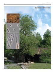
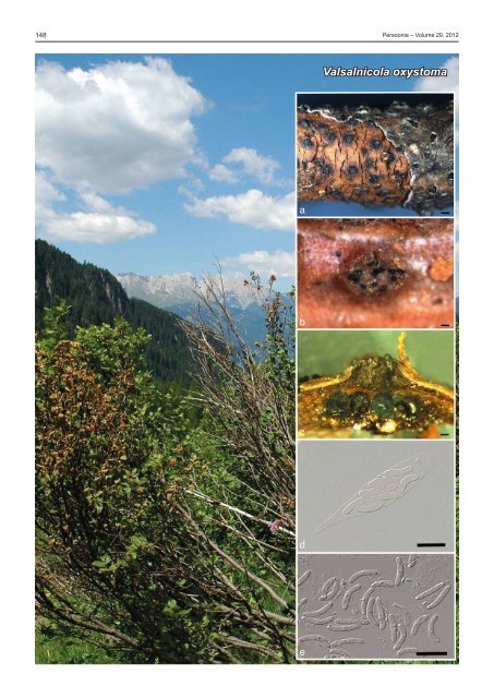
148 Persoonia – Volume 29, 2012<br />
<strong>Valsalnicola</strong> <strong>oxystoma</strong><br />
a<br />
b<br />
c<br />
d<br />
e
<strong>Fungal</strong> <strong>Planet</strong> description sheets<br />
149<br />
<strong>Fungal</strong> <strong>Planet</strong> 128 – 20 December 2012<br />
<strong>Valsalnicola</strong> D.M. Walker & Rossman, gen. nov.<br />
Etymology. Named for its valsa-like appearance and occurrence on<br />
species of Alnus.<br />
Causing linear cankers and lesions. Ectostromata well-developed,<br />
brown to black, thick disc from which perithecial necks<br />
emerge. Ascomata perithecial, immersed beneath ectostroma,<br />
aggregated in groups of 13–23, converging into 5–20 necks.<br />
Asci fusiform, with indistinct apical ring. Ascospores allantoid<br />
with rounded ends, 1-septate, hyaline.<br />
Type species. <strong>Valsalnicola</strong> <strong>oxystoma</strong>.<br />
MycoBank MB801277.<br />
<strong>Valsalnicola</strong> <strong>oxystoma</strong> (Rehm) D.M. Walker & Rossman, comb. nov.<br />
Basionym. Valsa <strong>oxystoma</strong> Rehm, Ber. Naturhist. Vereins Augsburg 26:<br />
70. 1881.<br />
≡ Cryptodiaporthe <strong>oxystoma</strong> (Rehm) Z. Urb., Preslia 29: 395. 1957.<br />
Twig lesions in surface view (511–)591–890(–893) µm diam<br />
(mean = 654, S.D. 122, n = 13). Ectostroma well-developed,<br />
brown to black, thick disc from which perithecial necks emerge.<br />
Ascomatal cavity (690–)765–909(–950) µm high × (1610–<br />
)1710–2346(–3947) µm diam (mean = 816 × 2198, S.D. 109,<br />
703, n1 = 5, n2 = 9). Ascomata perithecial, immersed beneath<br />
ectostroma, causing host tissue to swell and rupture, perithecia<br />
converging into 5–20 necks, emerging at surface through<br />
ecto stromatic disc, perithecia grouped 13–23. Ascomata<br />
glossy black, subglobose to globose (240–)266–298(–320)<br />
µm high × (253–)260–335(–337) µm diam (mean = 282 × 294,<br />
S.D. 25, 36, n1 = 7, n2 = 13); necks central, straight to curved,<br />
length (426–)428–550(–563) µm (mean = 476, S.D. 54,<br />
n = 9). Asci fusiform, (38–)39–48(–49) × (8–)9–12(–13) µm<br />
(mean = 44 × 11, S.D. 4, 1.2, n1 = 17, n2 = 18), apex broadly<br />
rounded, with indistinct apical ring, stipe acute, rounded, or tapering<br />
to a point, ascospores arranged irregularly multiseriate.<br />
Ascospores allantoid with rounded ends, mostly curved, rarely<br />
straight, (9–)10–11(–12) × 2–3 µm (mean = 11 × 2, S.D. 0.9,<br />
0.5, n = 30), 1-septate, median, slightly constricted or not at<br />
septum, each cell with several small guttules, hyaline. Cultures<br />
slow-growing, 3–6 mm in 10 d on potato-dextrose agar, mycelium<br />
low, pale brown to greyish brown, reverse dark brown.<br />
In culture on synthetic nutrient-poor agar — Dimorphic,<br />
forming a synanamorph. Conidiomata pycnidial, exuding<br />
masses of brown conidia. Conidiophores reduced to conidiogenous<br />
cells, or one supporting cell, proliferating percurrently.<br />
Conidia cylindrical, brown, finely verruculose, apex obtuse,<br />
base truncate, 3–5-euseptate, 15–23 × 4–5 µm. Conidia of<br />
synanamorph intermingled in same conidioma, but conidiogenous<br />
cells proliferating percurrently or sympodially; conidia<br />
hyaline to subhyaline, narrowly obclavate, apex subobtuse,<br />
base truncate, straight to curved, 25–80 × 2.5–3 µm, up to<br />
11-septate. Synanamorph also developing in aerial mycelium<br />
(on PNA); conidiophores subcylindrical, straight to curved,<br />
0–2-septate, hyaline to subhyaline, 8–15 × 2–3 µm, proliferating<br />
sympodially at apex. Conidiophores solitary or fasciculate<br />
or on a reduced stroma.<br />
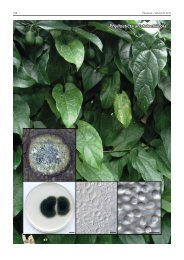
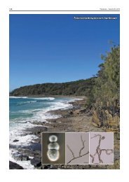
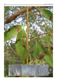
Colour illustrations. Italy, Trentino, Val Sadole, showing trees of Alnus<br />
viridis with green alder decline (Giorgio Maresi). a. Rehm: Ascomyceten 280,<br />
scale bar = 500 µm. b–d. BPI 884137, scale bars of perithecia = 100 µm,<br />
scale bar of ascus = 10 µm. e. Rehm: Ascomyceten 280, scale bar = 10 µm.<br />
Typus. Austria, Tyrol, Längenfeld, on dead branch of Alnus viridis,<br />
c. 3 500 ft., Aug. 1874, coll. Rehm. This type specimen was issued as<br />
Rehm, Ascomyceten no. 280. Of the two specimens at BPI, the more plentiful<br />
one is in the bound set of Rehm, Ascomyceten, and is herein designated<br />
as Lectotype BPI 884138. Isolectotypes examined BPI 738235 and NY,<br />
MycoBank MB801277.<br />
Additional specimens examined. Belgium, Brussels, Soignes, on branch<br />
of Alnus glutinosa, Oct. 1899, P. Nypels, comm. H. Rehm, Vestergren,<br />
Micro mycetes rariores selecti 409 as Valsa <strong>oxystoma</strong> (BPI 574854). – Canada,<br />
British Columbia, Yoho National Park, Chancellor Mountain Camp, on<br />
Alnus sp., 11 Aug. 1962, R.F. Cain, TRTC 40116 (NY); Ontario, Kenora<br />
District, Tustin Township, Gordon Lake, Rd., on Alnus sp., 26 Sept. 1959,<br />
coll. D. Bowen, det. J. Reid as Valsa <strong>oxystoma</strong> (BPI 574855). – Italy, Trento,<br />
Monte Bondone Trento, E11°03'51" N46°02'20", on Alnus viridis, Apr.<br />
2011, G. Maresi, isol. A. Rossman AR 4833 = CBS 133337, ITS sequence<br />
JX519559, and LSU sequence JX519563 (BPI 884137); Trento, val Sadole<br />
(E11.60, N46.15), 2009, G. Maresi & C.M.O. Longa (BPI 884136). – Sweden,<br />
Umea, on dead branch of Alnus ‘borealis’, Sept. 1910, Vlengel, det.<br />
F. Bubak (BPI 574856). – USA, Alaska, near Fairbanks, Moose Creek, Environmental<br />
Monitoring Plot 316 MC UM11 MRC, N64.72, W147.23, elev.<br />
150 m, on Alnus incana var. tenuifolia, May 2010, G.C. Adams, culture AR<br />
5137 = CBS 133329, ITS sequence JX519561 (BPI 884135).<br />
Habitat — Alnus viridis ssp. viridis, causing a twig colonization<br />
and canker disease involved in green alder decline (Pisetta<br />
et al. 2012); also known from Alnus glutinosa, A. incana,<br />
A. incana var. tenuifolia, A. rubra, A. viridis ssp. fruticosa and<br />
A. viridis ssp. maximowiczii.<br />
Distribution — Asia: Japan (Kobayashi 2007); Europe: Austria,<br />
Belgium, Italy, Sweden, also United Kingdom (Cannon et<br />
al. 1985); North America: Canada (Ontario); USA: Alaska.<br />
Notes — <strong>Valsalnicola</strong> is based on a species that was described<br />
in the genus Valsa. Although it resembles Valsa in<br />
having allantoid ascospores, the ascospores of <strong>Valsalnicola</strong><br />
are 1-septate while the majority of species of Valsa and closely<br />
related Leucostoma and Valsella have aseptate ascospores.<br />
However, one species of Valsa, V. melanodiscus, also has<br />
1-septate ascospores, occurs on Alnus spp., and produces linear<br />
cankers on the host. A distinguishing feature of <strong>Valsalnicola</strong><br />
is the lack of a black line surrounding stromata in the ascomatal<br />
cavity, which is characteristic of Valsa melanodiscus. In<br />
addition, the growth rate of cultures of <strong>Valsalnicola</strong> <strong>oxystoma</strong><br />
is considerably slower than species of Valsa. Molecular sequence<br />
data place this new genus within the Gnomoniaceae-<br />
Melanconidaceae complex. Allantoid, 1-septate ascospores<br />
have not previously been reported in the Gnomoniaceae or<br />
Melanconidiaceae. ITS sequences of specimens from Alaska<br />
and Italy are identical. The basionym has been cited as Rehm:<br />
Ascomyceten 270 (1875) in ‘Index Fungorum’ reflecting an error<br />
in Saccardo (1882) but the correct number is Rehm: Ascomyceten<br />
280, which does not include a description.<br />
Donald M. Walker, Department of Natural Sciences, The University of Findlay, Findlay, OH 45840, USA; e-mail: walkerd@findlay.edu<br />
Amy Y. Rossman, Systematic Mycology & Microbiology Laboratory, USDA-ARS, Beltsville, MD 20705, USA; e-mail: Amy.Rossman@ars.usda.gov<br />
Gerard C. Adams, Department of Plant Pathology, University of Nebraska, Lincoln, NE 68583, USA; e-mail: gadams3@unl.edu<br />
Claudia Maria Oliveira Longa, FEM-IASMA - Research and Innovation Centre, Sustainable Agro-Ecosystems and Bioresources Department. Via E. Mach 1,<br />
38010 San Michele all’Adige (TN), Italy; e-mail: claudia.longa@fmach.it<br />
Giorgio Maresi, IASMA - Centre for Technology Transfer, Via E. Mach 1, 38010 San Michele all’Adige (TN), Italy; e-mail: giorgio.maresi@fmach.it<br />
© 2012 Nationaal Herbarium Nederland & Centraalbureau voor Schimmelcultures