Fusicladium peltigericola - Fungal Planet
Fusicladium peltigericola - Fungal Planet
Fusicladium peltigericola - Fungal Planet

Create successful ePaper yourself
Turn your PDF publications into a flip-book with our unique Google optimized e-Paper software.
128 Persoonia – Volume 25, 2010<br />
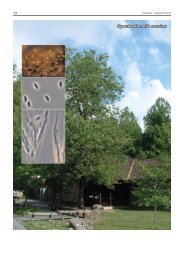
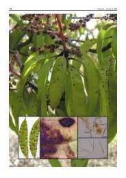
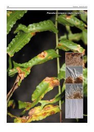
<strong>Fusicladium</strong> <strong>peltigericola</strong>
Persoonial Reflections<br />
129<br />
<strong>Fungal</strong> <strong>Planet</strong> 54 – 23 December 2010<br />
<strong>Fusicladium</strong> <strong>peltigericola</strong> Crous & Diederich, sp. nov.<br />
Conidiophora solitaria, erecta, subcylindrica, recta vel geniculata-sinuosa,<br />
non ramosa, 1–4(–7)-septata, 10–40(–90) × 3–4 µm, brunnea, laevia.<br />
Cellulae conidiogenae terminales, brunneae, laeviae, sympodialiter proliferantes,<br />
subcylindricae, 10–30 × 3–4 µm; cicatrices conidiales applanatae,<br />
inconspicuae vel leniter fuscatae, sed non refractivae et non incrassatae,<br />
2–2.5 µm diam. Ramoconidia in 1–3 seriebus, subcylindrica, in medio<br />
unieuseptata, (27–)33–40(–65) × 4(–5) µm; conidia intercalaria et terminalia,<br />
subcylindrica, mediobrunnea, subtile verruculosa, 0–1-euseptata,<br />
(18–)25–33(–40) × (3.5–)4(–5) µm.<br />
Etymology. Named after the lichen host from which it was collected,<br />
Peltigera rufescens.<br />
Mycelium consisting of smooth, branched, septate, brown, 2–3<br />
µm diam hyphae. Conidiophores solitary, erect, subcylindrical,<br />
straight to geniculous-sinuous, unbranched, 1–4(–7)-septate,<br />
10–40(–90) × 3–4 µm, brown, smooth. Conidiogenous cells<br />
terminal, brown, smooth, proliferating sympodially, subcylindrical,<br />
rarely straight, mostly geniculate-sinuous, 10–30 × 3–4<br />
µm; scars flattened, inconspicuous to somewhat darkened,<br />
but not refractive, not appearing thickened, 2–2.5 µm wide.<br />
Ramoconidia in 1–3 series, subcylindrical, medianly 1-euseptate,<br />
relatively thick-walled, medium brown, finely verruculose,<br />
basal hilum flattened, somewhat darkened, 2–2.5 µm wide,<br />
with one to several sympodial, apical loci; frequently with lateral<br />
branch up to 10 µm long, 3–4 µm wide, (27–)33–40(–65) ×<br />
4(–5) µm; older ramoconidia at times developing up to 3 septa;<br />
intercalary and terminal conidia subcylindrical, medium brown,<br />
finely verruculose, apex obtusely rounded or flattened, proliferating<br />
in sympodial fashion to form short chains of conidia,<br />
0–1-euseptate; septum mostly in upper third of conidium,<br />
(18–)25–33(–40) × (3.5–)4(–5) µm; hila flattened, 2–2.5 µm<br />
wide, somewhat darkened, not thickened.<br />
Culture characteristics — (in the dark, 25 °C): Colonies on<br />
oatmeal agar spreading, with moderate aerial mycelium; surface<br />
smooth, fuscous-black, margin lobate, smooth; reaching<br />
15 mm diam after 1 mo. Colonies on cornmeal agar erumpent,<br />
spreading with dense, moderate aerial mycelium and lobate,<br />
smooth to feathery margins; colonies reaching 15 mm diam<br />
after 1 mo; surface olivaceous-grey to fuscous-black.<br />
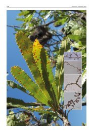
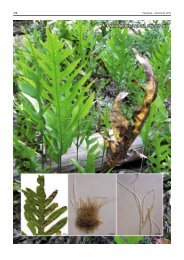
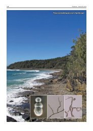
Typus. Luxembourg, Lamadelaine, in a disused quarry, on terricolous<br />
Peltigera rufescens, over galls induced by Hawksworthiana <strong>peltigericola</strong>, May<br />
2008, P. Diederich, CBS-H 20487 holotype, culture ex-type CPC 15252 =<br />
CBS 128206, ITS sequence GenBank HQ599579, MycoBank MB517532.<br />
Notes — The genus <strong>Fusicladium</strong> is recognised as anamorph<br />
of Venturia 1–3 . Presently no <strong>Fusicladium</strong> species are known from<br />
lichens, nor are there any DNA sequence data of similar species<br />
currently deposited in GenBank. The closest sister taxa in Gen-<br />
Bank based on the ITS sequence are <strong>Fusicladium</strong> betulae (Gen-<br />
Bank FJ839641; Identities = 459/464 (99 %), Gaps = 2/464<br />
(0 %)), Venturia tremulae var. tremulae (GenBank EU035475;<br />
Identities = 704/712 (99 %), Gaps = 4/712 (0 %)) and Venturia<br />
ditricha (GenBank EU035456; Identities = 704/712 (99 %), Gaps<br />
= 4/712 (0 %)). Morphologically F. <strong>peltigericola</strong> is distinct from<br />
all taxa treated in the recent monograph by Schubert et al. 2<br />
based on the combination of characters, namely its large,<br />
subcylindrical ramoconidia that become up to 3-septate, and its<br />
terminal conidia that become 1-septate in the upper third of the<br />
conidium. Although F. <strong>peltigericola</strong> was isolated from a Peltigera<br />
thallus colonised with Hawksworthiana <strong>peltigericola</strong> (which<br />
could not be cultivated), there was no conclusive macroscopic<br />
proof that F. <strong>peltigericola</strong> is lichenicolous. However, conidia<br />
isolated from the thallus took up to 2 wk to germinate, and grew<br />
extremely slowly for the first few months, suggesting that there<br />
may be an association with P. rufescens. Further collections<br />
would be required, however, to clarify its ecology.<br />
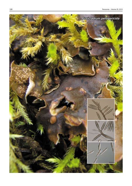
Acknowledgement The authors acknowledge G. Marson for the background<br />
photograph.<br />
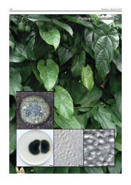
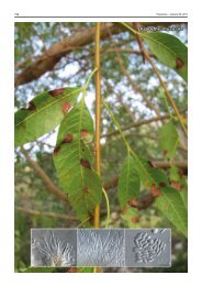
Colour illustrations. Peltigera rufescens; conidiophores with conidiogenous<br />
cells giving rise to conidia. Scale bars = 10 µm.<br />
References. 1 Beck A, Ritschel A, Schubert K, Braun U, Triebel D. 2005.<br />
Phylogenetic relationships of the anamorphic genus <strong>Fusicladium</strong> s. lat. as<br />
inferred by ITS nrDNA data. Mycological Progress 4: 111–116. 2 Schubert<br />
K, Ritschel A, Braun U. 2003. A monograph of <strong>Fusicladium</strong> s. lat. (hyphomycetes).<br />
Schlechtendalia 9: 1–132. 3 Crous PW, Schubert K, Braun U,<br />
Hoog GS de, Hocking AD, Shin H-D, Groenewald JZ. 2007. Opportunistic,<br />
human-pathogenic species in the Herpotrichiellaceae are phenotypically<br />
similar to saprobic or phytopathogenic species in the Venturiaceae. Studies<br />
in Mycology 58: 185–217.<br />
Pedro W. Crous & Johannes Z. Groenewald, CBS-KNAW <strong>Fungal</strong> Biodiversity Centre, P.O. Box 85167, 3508 AD Utrecht, The Netherlands;<br />
e-mail: p.crous@cbs.knaw.nl & e.groenewald@cbs.knaw.nl<br />
Paul Diederich, Musée national d’histoire naturelle, 25 rue Munster, L-2160 Luxembourg, Luxembourg; e‐mail: paul.diederich@education.lu<br />
© 2010 Nationaal Herbarium Nederland & Centraalbureau voor Schimmelcultures