Pseudocercospora microsori - Fungal Planet
Pseudocercospora microsori - Fungal Planet
Pseudocercospora microsori - Fungal Planet

Create successful ePaper yourself
Turn your PDF publications into a flip-book with our unique Google optimized e-Paper software.
156 Persoonia – Volume 25, 2010<br />
<strong>Pseudocercospora</strong> <strong>microsori</strong>
Persoonial Reflections<br />
157<br />
<strong>Fungal</strong> <strong>Planet</strong> 68 – 23 December 2010<br />
<strong>Pseudocercospora</strong> <strong>microsori</strong> R.G. Shivas, A.J. Young & B.C. McNeil, sp. nov.<br />
Frondium maculae amphigenae, sparsae ad confluentes, saepe tegentes<br />
multum paginae frondium, circulares ad irregulares, marginibus distinctis,<br />
inaequalibus et areis chloroticis, finitis venis principibus, 5–15 mm diametro,<br />
fuscis rubellis-brunneis, centris cinerascentibus. Conidiomata rubella-brunnea,<br />
amphigena, fasciculata, orientia ex stromate maturo substomatali 20–60<br />
µm lato. Conidiophora 5–30 in fasciculis densis vel laxis, geniculata ad sinuosa,<br />
inramosa, rubella-brunnea, pallidiora ad apicem, 1–5-septata 30–65<br />
× 3–5 µm. Cellulae conidiogenae terminales in conidiophoro, integratae,<br />
subcylindraceae, brunneolae, leves, 10–35 × 2.5–4 µm. Conidia obclavata<br />
ad subcylindracea, curvata ad flexuosa, apice rotundato, basi truncata vel<br />
paulum obconice truncata, 2–12-septata, 50–110 × 2.5–4 µm, brunneola,<br />
levia; hila nec crassata nec fuscata.<br />
Etymology. From the name of the host plant Microsorum (Polypodiaceae).<br />
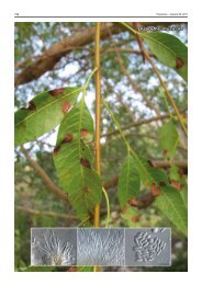
Frond spots amphigenous, scattered to confluent, often covering<br />
much of the frond surface, circular to irregular with distinct,<br />
uneven margins and chlorotic haloes, limited by the main veins,<br />
5–15 mm diam, dark reddish brown with centres becoming grey.<br />
Conidiomata reddish brown, amphigenous, fasciculate, arise<br />
from a well-developed substomatal stroma, 20–60 µm wide.<br />
Conidiophores 5–30 in dense or loose fascicles, geniculate<br />
to sinuous, unbranched, reddish brown, paler towards apex,<br />
1–5-septate 30–65 × 3–5 µm. Conidiogenous cells terminal on<br />
conidiophore, integrated, subcylindrical, pale brown, smooth,<br />
10–35 × 2.5–4 µm. Conidia obclavate to subcylindrical, curved<br />
to flexuous, apex rounded, base truncate to slightly obconically<br />
truncate, 2–12-septate, 50–110 × 2.5–4 µm, pale brown,<br />
smooth; hila not thickened nor darkened.<br />
Typus. Australia, Queensland, Brisbane, West End, Doris Street, on<br />
fronds of Microsorum pustulatum, 6 Aug. 2010, B.C. McNeil, BRIP 53617,<br />
holotype; cultures ex-type BRIP 53617, ITS sequence GenBank HQ624985,<br />
MycoBank MB517678; Indooroopilly Research Centre, Indooroopilly, 26 Aug.<br />
2010, B.C. McNeil, BRIP 53618, paratype.<br />
Notes — Although several <strong>Pseudocercospora</strong> spp. have<br />
been recorded on ferns, P. <strong>microsori</strong> is the first on the genus<br />
Microsorum. Other <strong>Pseudocercospora</strong> spp. with fasciculate<br />
conidiophores that have been recorded on ferns include:<br />
P. adianti 1 , P. arachniodis 2 , P. athyrii 2 , P. christellae 3 , P. cyatheae<br />
4 , P. lonchitidis 1 , P. nephrolepidis 5 , P. phyllitidis 1 , P. plagiogyriae<br />
2 , P. pteridicola 6 , P. pteridophytophila 2 and P. thelypteridis 2 .<br />
<strong>Pseudocercospora</strong> <strong>microsori</strong> is morphologically distinct from<br />
these species with its combination of moderately wide (2.5–4<br />
µm) and curved to flexuous conidia. A megablast search of<br />
NCBIs GenBank nucleotide database using the ITS sequence<br />
revealed high identity to P. lythri (GenBank EF535713; Identities<br />
= 496/498 (99 %), Gaps = 0/498 (0 %)), P. humuli (GenBank<br />
EF535685; Identities = 495/498 (99 %), Gaps = 1/498 (0 %)),<br />
P. crousii (GenBank GQ852756; Identities = 497/502 (99 %),<br />
Gaps = 2/502 (0 %)) and P. araliae (GenBank EF535717; Identities<br />
= 495/499 (99 %), Gaps = 1/499 (0 %)). Genomic DNA of<br />
P. <strong>microsori</strong> (holotype) is stored in the Australian Biosecurity<br />
Bank (www.padil.gov.au/pbt/).<br />
96<br />
<strong>Pseudocercospora</strong> humuli humuli voucher voucher KACC 42529 KACC (EF535685) 42529 (EF535685)<br />
68<br />
<strong>Pseudocercospora</strong> <strong>microsori</strong> <strong>microsori</strong> BRIP 53617 BRIP ( ) 53617 (HQ624985)<br />
56<br />
<strong>Pseudocercospora</strong> balsaminae balsaminae voucher voucher KACC 42643 KACC (EF535715) 42643 (EF535715)<br />
53<br />
<strong>Pseudocercospora</strong> lythri lythri voucher voucher KACC 42641 KACC (EF535713) 42461 (EF535713)<br />
95<br />
<strong>Pseudocercospora</strong> araliae araliae voucher voucher KACC 42645 KACC (EF535717) 42645 (EF535717)<br />
<strong>Pseudocercospora</strong> kaki CPC kaki 10636 CPC 10636 (GU214677) (GU214677)<br />
<strong>Pseudocercospora</strong> mangifericola mangifericola BRIP 52776 BRIP (GU188048) 52776 (GU188048)<br />
<strong>Pseudocercospora</strong> avicenniae avicenniae BRIP 52764 BRIP (GU188047) 52764 (GU188047)<br />
Cercospora zebrinae CBS CBS 118790 118790 (GU214657) (GU214657)<br />
0.005<br />
Maximum Likelihood Tree obtained using the General Time<br />
Reversible Model from an ITS sequence alignment generated<br />
with MUSCLE in MEGA4. The bootstrap support values from<br />
1 000 replicates are shown at the nodes. Bar represents number<br />
of substitutions per site. The species described here is printed<br />
in bold face. The tree was rooted to Cercospora zebrinae CBS<br />
118790 (GU214657).<br />
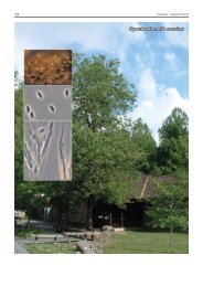
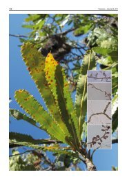
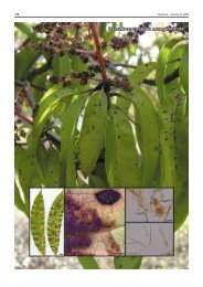
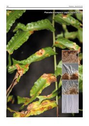
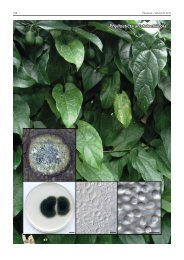
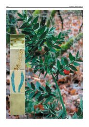
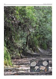
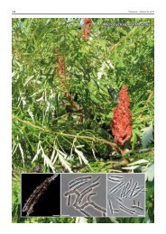
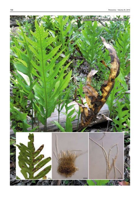
Colour illustrations. Microsorum pustulatum in a garden, Brisbane with a<br />
frond (right) severely infected with P. <strong>microsori</strong>; frond with lesions caused by<br />
P. <strong>microsori</strong>; stroma with conidiophores; conidia. Scale bars (left to right) =<br />
1 cm, 10 µm, 10 µm.<br />
References. 1 Crous PW, Braun U. 2003. Mycosphaerella and its anamorphs:<br />
1. Names published in Cercospora and Passalora. CBS Biodiversity<br />
Series 1: 1–571. 2 Guo YL, Liu XL, Hsieh WH. 1998. <strong>Pseudocercospora</strong>.<br />
Flora Fungorum Sinicorum, Vol. 9. Science Press, Beijing. 3 Phengsintham P,<br />
Chukeatirote E, McKenzie EHC, Moslem MA, Hyde KD, Braun U. 2010. Two<br />
new species and a new record of cercosporoids from Thailand. Mycosphere<br />
1: 205–212. 4 Nakashima C, Inaba S, Park JY, Ogawa Y. 2006. Addition and<br />
re-examination of Japanese species belonging to the genus Cercospora and<br />
allied genera. IX. Newly recorded species from Japan (4). Mycoscience 47:<br />
48–52. 5 Kirschner R, Chen CJ. 2007. Foliicolous hyphomycetes from Taiwan.<br />
<strong>Fungal</strong> Diversity 26: 219–239. 6 Braun U, Melnik VA. 1997. Cercosporoid fungi<br />
from Russia and adjacent countries. Proceedings of the Komarov Botanical<br />
Institute, Russian Academy of Sciences.<br />
Roger G. Shivas, Anthony J. Young & Bradley C. McNeil, Agri-Science Queensland, Ecosciences Precinct, Dutton Park 4102, Queensland, Australia;<br />
e-mail: roger.shivas@deedi.qld.gov.au, anthony.young@deedi.qld.gov.au & brad.mcneil@deedi.qld.gov.au<br />
© 2010 Nationaal Herbarium Nederland & Centraalbureau voor Schimmelcultures