

Leaf-inhabiting genera of the Gnomoniaceae, Diaporthales - CBS
Leaf-inhabiting genera of the Gnomoniaceae, Diaporthales - CBS
Leaf-inhabiting genera of the Gnomoniaceae, Diaporthales - CBS

Create successful ePaper yourself
Turn your PDF publications into a flip-book with our unique Google optimized e-Paper software.
Studies in Mycology 62 (2008)<br />
<strong>Leaf</strong>-<strong>inhabiting</strong> <strong>genera</strong> <strong>of</strong> <strong>the</strong> <strong>Gnomoniaceae</strong>,<br />
<strong>Diaporthales</strong><br />
M.V. Sogonov, L.A. Castlebury, A.Y. Rossman, L.C. Mejía and J.F. White<br />
<strong>CBS</strong> Fungal Biodiversity Centre,<br />
Utrecht, The Ne<strong>the</strong>rlands<br />
An institute <strong>of</strong> <strong>the</strong> Royal Ne<strong>the</strong>rlands Academy <strong>of</strong> Arts and Sciences
<strong>Leaf</strong>-<strong>inhabiting</strong> <strong>genera</strong> <strong>of</strong> <strong>the</strong> <strong>Gnomoniaceae</strong>, <strong>Diaporthales</strong><br />
St u d i es in My c o l o g y 62, 2008
Studies in Mycology<br />
The Studies in Mycology is an international journal which publishes systematic monographs <strong>of</strong> filamentous fungi and yeasts, and in rare<br />
occasions <strong>the</strong> proceedings <strong>of</strong> special meetings related to all fields <strong>of</strong> mycology, biotechnology, ecology, molecular biology, pathology and<br />
systematics. For instructions for authors see www.cbs.knaw.nl.<br />
Ex e c u t i v e Ed i t o r<br />
Pr<strong>of</strong>. dr Robert A. Samson, <strong>CBS</strong> Fungal Biodiversity Centre, P.O. Box 85167, 3508 AD Utrecht, The Ne<strong>the</strong>rlands.<br />
E-mail: r.samson@cbs.knaw.nl<br />
Lay o u t Ed i t o r<br />
Marianne de Boeij, <strong>CBS</strong> Fungal Biodiversity Centre, P.O. Box 85167, 3508 AD Utrecht, The Ne<strong>the</strong>rlands.<br />
E-mail: m.deboeij@cbs.knaw.nl<br />
Scientific Ed i t o rs<br />
Pr<strong>of</strong>. dr Uwe Braun, Martin-Lu<strong>the</strong>r-Universität, Institut für Geobotanik und Botanischer Garten, Herbarium, Neuwerk 21, D-06099 Halle, Germany.<br />
E-mail: uwe.braun@botanik.uni-halle.de<br />
Pr<strong>of</strong>. dr Pedro W. Crous, <strong>CBS</strong> Fungal Biodiversity Centre, P.O. Box 85167, 3508 AD Utrecht, The Ne<strong>the</strong>rlands.<br />
E-mail: p.crous@cbs.knaw.nl<br />
Pr<strong>of</strong>. dr David M. Geiser, Department <strong>of</strong> Plant Pathology, 121 Buckhout Laboratory, Pennsylvania State University, University Park, PA, U.S.A. 16802.<br />
E-mail: dgeiser@psu.edu<br />
Dr Lorelei L. Norvell, Pacific Northwest Mycology Service, 6720 NW Skyline Blvd, Portland, OR, U.S.A. 97229-1309.<br />
E-mail: llnorvell@pnw-ms.com<br />
Dr Erast Parmasto, Institute <strong>of</strong> Zoology & Botany, 181 Riia Street, Tartu, Estonia EE-51014.<br />
E-mail: e.parmasto@zbi.ee<br />
Pr<strong>of</strong>. dr Alan J.L. Phillips, Faculdade de Ciências e Tecnologia, Universidade Nova de Lisboa, Quinta de Torre, 2829-516 Caparica, Portugal.<br />
E-mail: alp@mail.fct.unl.pt<br />
Dr Amy Y. Rossman, Rm 304, Bldg 011A, Systematic Mycology and Microbiology Laboratory, Beltsville, MD, U.S.A. 20705.<br />
E-mail: amy@nt.ars-grin.gov<br />
Dr Keith A. Seifert, Research Scientist / Biodiversity (Mycology and Botany), Agriculture & Agri-Food Canada, KW Neatby Bldg, 960 Carling Avenue,<br />
Ottawa, ON, Canada K1A OC6.<br />
E-mail: seifertk@agr.gc.ca<br />
Pr<strong>of</strong>. dr Jeffrey K. Stone, Department <strong>of</strong> Botany & Plant Pathology, Cordley 2082, Oregon State University, Corvallis, OR, U.S.A. 97331-2902.<br />
E-mail: stonej@bcc.orst.edu<br />
Dr Richard C. Summerbell, 27 Hillcrest Park, Toronto, Ont. M4X 1E8, Canada.<br />
E-mail: summerbell@aol.com<br />
Copyright 2008 <strong>CBS</strong> Fungal Biodiversity Centre, P.O. Box 85167, 3508 AD Utrecht, The Ne<strong>the</strong>rlands.<br />
You are free to share — to copy, distribute and transmit <strong>the</strong> work, under <strong>the</strong> following conditions:<br />
Attribution:<br />
You must attribute <strong>the</strong> work in <strong>the</strong> manner specified by <strong>the</strong> author or licensor (but not in any way that suggests that <strong>the</strong>y endorse you<br />
or your use <strong>of</strong> <strong>the</strong> work).<br />
Non-commercial: You may not use this work for commercial purposes.<br />
No derivative works: You may not alter, transform, or build upon this work.<br />
For any reuse or distribution, you must make clear to o<strong>the</strong>rs <strong>the</strong> license terms <strong>of</strong> this work, which can be found at http://creativecommons.org/licenses/bync-nd/3.0/legalcode.<br />
Any <strong>of</strong> <strong>the</strong> above conditions can be waived if you get permission from <strong>the</strong> copyright holder. Nothing in this license impairs or restricts<br />
<strong>the</strong> author"s moral rights.<br />
Publication date: 29 December 2008<br />
Published and distributed by <strong>CBS</strong> Fungal Biodiversity Centre, P.O. Box 85167, 3508 AD Utrecht, The Ne<strong>the</strong>rlands. Internet: www.cbs.knaw.nl.<br />
E-mail: info@cbs.knaw.nl.<br />
ISBN/EAN : 978-90-70351-74-8<br />
Online ISSN : 1872-9797<br />
Print ISSN : 0166-0616<br />
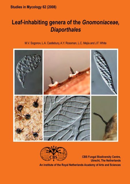
Cover: From left to right: Asci <strong>of</strong> Gnomonia neognomon BPI 877465C. Peri<strong>the</strong>cia <strong>of</strong> Ambarignomonia petiolorum on decaying petioles <strong>of</strong> Liquidambar<br />
styraciflua BPI 844274. Asci <strong>of</strong> Gnomonia alnea BPI 877462A. Bottom from left to right: Peri<strong>the</strong>cia <strong>of</strong> Gnomonia gnomon on overwintered fallen leaves<br />
on Corylus avellana BPI 844273. Asci <strong>of</strong> Apiognomonia acerina BPI 877677. Peri<strong>the</strong>cium <strong>of</strong> Plagiostoma euphorbia-verrucosae on overwintered stems <strong>of</strong><br />
Euphorbia verrucosa BPI 877685.
<strong>Leaf</strong>-<strong>inhabiting</strong> <strong>genera</strong> <strong>of</strong> <strong>the</strong> <strong>Gnomoniaceae</strong>,<br />
<strong>Diaporthales</strong><br />
M.V. Sogonov<br />
Department <strong>of</strong> Plant Biology and Pathology, Rutgers University, New Brunswick, NJ 08901, U.S.A. and Systematic Mycology & Microbiology Laboratory,<br />
USDA Agricultural Research Service, Beltsville, Maryland 20705–2350, U.S.A.<br />
L.A. Castlebury<br />
Systematic Mycology & Microbiology Laboratory, USDA Agricultural Research Service, Beltsville, Maryland 20705–2350, U.S.A.<br />
A.Y. Rossman<br />
Systematic Mycology & Microbiology Laboratory, USDA Agricultural Research Service, Beltsville, Maryland 20705–2350, U.S.A.<br />
L.C. Mejía<br />
Department <strong>of</strong> Plant Biology and Pathology, Rutgers University, New Brunswick, NJ 08901, U.S.A. and Systematic Mycology & Microbiology Laboratory,<br />
USDA Agricultural Research Service, Beltsville, Maryland 20705–2350, U.S.A.<br />
J.F. White<br />
Department <strong>of</strong> Plant Biology and Pathology, Rutgers University, New Brunswick, NJ 08901, U.S.A.<br />
<strong>CBS</strong> Fungal Biodiversity Centre,<br />
Utrecht, The Ne<strong>the</strong>rlands<br />
An institute <strong>of</strong> <strong>the</strong> Royal Ne<strong>the</strong>rlands Academy <strong>of</strong> Arts and Sciences
CONTENTS<br />
Abstract ............................................................................................................................................................................................................ 1<br />
Introduction ...................................................................................................................................................................................................... 1<br />
Materials and methods ..................................................................................................................................................................................... 3<br />
Collection and observation <strong>of</strong> herbarium specimens ................................................................................................................................. 3<br />
Culture preparation and morphology ......................................................................................................................................................... 3<br />
Measurements and data management ...................................................................................................................................................... 4<br />
DNA amplification and sequencing ............................................................................................................................................................ 4<br />
Phylogenetic analyses .................................................................................................................................................................................4<br />
Results and discussion .................................................................................................................................................................................... 8<br />
Phylogenetic analyses ............................................................................................................................................................................... 8<br />
Revised concepts <strong>of</strong> accepted <strong>genera</strong> ....................................................................................................................................................... 8<br />
Evaluation <strong>of</strong> morphological and host characteristics .............................................................................................................................. 12<br />
Taxonomy ....................................................................................................................................................................................................... 14<br />
Key to <strong>the</strong> species <strong>of</strong> <strong>Gnomoniaceae</strong> in this study .................................................................................................................................. 14<br />
Descriptions <strong>of</strong> <strong>genera</strong> and species <strong>of</strong> <strong>the</strong> <strong>Gnomoniaceae</strong> ........................................................................................................................... 18<br />
GNOMONIA ............................................................................................................................................................................................. 18<br />
Gnomonia gnomon ............................................................................................................................................................................ 18<br />
Gnomonia alnea ................................................................................................................................................................................ 21<br />
New and revised species <strong>of</strong> Gnomonia ................................................................................................................................................... 21<br />
Gnomonia incrassata ......................................................................................................................................................................... 21<br />
Gnomonia monodii ............................................................................................................................................................................ 21<br />
Gnomonia neognomon ...................................................................................................................................................................... 22<br />
Gnomonia orcispora .......................................................................................................................................................................... 25<br />
Gnomonia ostryae ............................................................................................................................................................................. 25<br />
Gnomonia pendulorum ...................................................................................................................................................................... 26<br />
Gnomonia rodmanii ........................................................................................................................................................................... 30<br />
Gnomonia skokomishica ................................................................................................................................................................... 30<br />
Gnomonia virginianae ........................................................................................................................................................................ 32<br />
Additional species accepted in Gnomonia ............................................................................................................................................... 32<br />
Gnomonia amoena ............................................................................................................................................................................ 32<br />
Gnomonia arnstadtiensis ................................................................................................................................................................... 33<br />
Gnomonia carpinicola ........................................................................................................................................................................ 34<br />
Gnomonia pseudoamoena ................................................................................................................................................................ 34<br />
AMBARIGNOMONIA ............................................................................................................................................................................... 35<br />
Ambarignomonia petiolorum .............................................................................................................................................................. 36<br />
APIOGNOMONIA ..................................................................................................................................................................................... 36<br />
Apiognomonia veneta ........................................................................................................................................................................ 36<br />
Additional species <strong>of</strong> Apiognomonia ........................................................................................................................................................ 38<br />
Apiognomonia acerina ....................................................................................................................................................................... 38<br />
Apiognomonia borealis ...................................................................................................................................................................... 38<br />
Apiognomonia errabunda .................................................................................................................................................................. 38<br />
Apiognomonia hystrix ........................................................................................................................................................................ 40<br />
GNOMONIOPSIS .................................................................................................................................................................................... 41<br />
Type species <strong>of</strong> Gnomoniopsis ................................................................................................................................................................ 41<br />
Gnomoniopsis chamaemori ............................................................................................................................................................... 41<br />
New and revised species <strong>of</strong> Gnomoniopsis ............................................................................................................................................. 44<br />
Gnomoniopsis clavulata .................................................................................................................................................................... 44<br />
Gnomoniopsis paraclavulata ............................................................................................................................................................. 44<br />
Additional species accepted in Gnomoniopsis ......................................................................................................................................... 47<br />
Gnomoniopsis comari ........................................................................................................................................................................ 47<br />
Gnomoniopsis fructicola .................................................................................................................................................................... 47<br />
Gnomoniopsis macounii .................................................................................................................................................................... 48<br />
Gnomoniopsis racemula .................................................................................................................................................................... 48<br />
Gnomoniopsis tormentillae ................................................................................................................................................................ 48<br />
OPHIOGNOMONIA .................................................................................................................................................................................. 48<br />
Type species <strong>of</strong> Ophiognomonia ............................................................................................................................................................. 48<br />
Ophiognomonia melanostyla ............................................................................................................................................................. 48<br />
New species <strong>of</strong> Ophiognomonia .............................................................................................................................................................. 51<br />
Ophiognomonia balsamiferae ............................................................................................................................................................ 51
Ophiognomonia pseudoclavulata ...................................................................................................................................................... 51<br />
Ophiognomonia vasiljevae ................................................................................................................................................................ 53<br />
Additional species accepted in Ophiognomonia ...................................................................................................................................... 55<br />
Ophiognomonia alni-viridis ................................................................................................................................................................ 55<br />
Ophiognomonia gei-montani ............................................................................................................................................................. 58<br />
Ophiognomonia intermedia ............................................................................................................................................................... 58<br />
Ophiognomonia ischnostyla .............................................................................................................................................................. 59<br />
Ophiognomonia leptostyla ................................................................................................................................................................. 62<br />
Ophiognomonia micromegala ............................................................................................................................................................ 63<br />
Ophiognomonia nana ........................................................................................................................................................................ 63<br />
Ophiognomonia padicola ................................................................................................................................................................... 63<br />
Ophiognomonia rosae ....................................................................................................................................................................... 64<br />
Ophiognomonia rubi-idaei ................................................................................................................................................................. 64<br />
Ophiognomonia sassafras ................................................................................................................................................................. 64<br />
Ophiognomonia setacea .................................................................................................................................................................... 64<br />
Ophiognomonia trientensis ................................................................................................................................................................ 64<br />
PLAGIOSTOMA ....................................................................................................................................................................................... 66<br />
Type species <strong>of</strong> Plagiostoma and synonymous genus, Cryptodiapor<strong>the</strong> ................................................................................................. 67<br />
Plagiostoma euphorbiae .................................................................................................................................................................... 67<br />
Plagiostoma aesculi ........................................................................................................................................................................... 69<br />
New species <strong>of</strong> Plagiostoma .................................................................................................................................................................... 69<br />
Plagiostoma barriae ........................................................................................................................................................................... 69<br />
Additional species accepted in Plagiostoma ............................................................................................................................................ 70<br />
Plagiostoma amygdalinae ................................................................................................................................................................. 70<br />
Plagiostoma devexum ....................................................................................................................................................................... 70<br />
Plagiostoma euphorbiaceum ............................................................................................................................................................. 72<br />
Plagiostoma euphorbiae-verrucosae ................................................................................................................................................. 72<br />
Plagiostoma fraxini ............................................................................................................................................................................ 72<br />
Plagiostoma geranii ........................................................................................................................................................................... 72<br />
Plagiostoma petiolophilum ................................................................................................................................................................. 72<br />
Plagiostoma rhododendri ................................................................................................................................................................... 72<br />
Plagiostoma robergeanum ................................................................................................................................................................ 73<br />
Plagiostoma salicellum ...................................................................................................................................................................... 73<br />
Genera not included in this study or excluded from <strong>the</strong> <strong>Gnomoniaceae</strong> ................................................................................................. 74<br />
Acknowledgements ........................................................................................................................................................................................ 75<br />
References ..................................................................................................................................................................................................... 76<br />
Index .............................................................................................................................................................................................................. 78
available online at www.studiesinmycology.org<br />
doi:10.3114/sim.2008.62.01<br />
St u d i es in My c o l o g y 62: 1–79. 2008.<br />
<strong>Leaf</strong>-<strong>inhabiting</strong> <strong>genera</strong> <strong>of</strong> <strong>the</strong> <strong>Gnomoniaceae</strong>, <strong>Diaporthales</strong><br />
M.V. Sogonov 1,2 , L.A. Castlebury 2 , A.Y. Rossman 2 , L.C. Mejía 1,2 and J.F. White 1<br />
1<br />
Department <strong>of</strong> Plant Biology and Pathology, Rutgers University, New Brunswick, NJ 08901, U.S.A.; 2 Systematic Mycology & Microbiology Laboratory, USDA Agricultural<br />
Research Service, Beltsville, Maryland 20705-2350, U.S.A.<br />
*Correspondence: A.Y. Rossman, Amy.Rossman@ars.usda.gov<br />
Abstract: The <strong>Gnomoniaceae</strong> are characterised by ascomata that are <strong>genera</strong>lly immersed, solitary, without a stroma, or aggregated with a rudimentary stroma, in herbaceous<br />
plant material especially in leaves, twigs or stems, but also in bark or wood. The ascomata are black, s<strong>of</strong>t-textured, thin-walled, and pseudoparenchymatous with one or<br />
more central or eccentric necks. The asci usually have a distinct apical ring. The <strong>Gnomoniaceae</strong> includes species having ascospores that are small, mostly less than 25<br />
μm long, although some are longer, and range in septation from non-septate to one-septate, rarely multi-septate. Molecular studies <strong>of</strong> <strong>the</strong> <strong>Gnomoniaceae</strong> suggest that <strong>the</strong><br />
traditional classification <strong>of</strong> <strong>genera</strong> based on characteristics <strong>of</strong> <strong>the</strong> ascomata such as position <strong>of</strong> <strong>the</strong> neck and ascospores such as septation have resulted in <strong>genera</strong> that are<br />
not monophyletic. In this paper <strong>the</strong> concepts <strong>of</strong> <strong>the</strong> leaf-<strong>inhabiting</strong> <strong>genera</strong> in <strong>the</strong> <strong>Gnomoniaceae</strong> are reevaluated using multiple genes, specifically nrLSU, translation elongation<br />
factor 1-alpha (tef1-α), and RNA polymerase II second largest subunit (rpb2) for 64 isolates. ITS sequences were <strong>genera</strong>ted for 322 isolates. Six <strong>genera</strong> <strong>of</strong> leaf-<strong>inhabiting</strong><br />
<strong>Gnomoniaceae</strong> are defined based on placement <strong>of</strong> <strong>the</strong>ir type species within <strong>the</strong> multigene phylogeny. The new monotypic genus Ambarignomonia is established for an unusual<br />
species, A. petiolorum. A key to 59 species <strong>of</strong> leaf-<strong>inhabiting</strong> <strong>Gnomoniaceae</strong> is presented and 22 species <strong>of</strong> <strong>Gnomoniaceae</strong> are described and illustrated.<br />
Taxonomic novelties: New genus: Ambarignomonia. New species: Gnomonia incrassata, G. monodii, G. neognomon, G. orcispora, G. pendulorum, G. rodmanii, G. skokomishica,<br />
G. virginianae, Gnomoniopsis paraclavulata, Ophiognomonia balsamiferae, O. pseudoclavulata, O. vasiljevae, Plagiostoma barriae. New combinations: Ambarignomonia<br />
petiolorum; Apiognomonia hystrix; Gnomonia alnea, G. carpinicola, Gnomoniopsis clavulata, G. comari, G. fructicola, G. macounii, G. racemula, G. tormentillae; Ophiognomonia<br />
alni-viridis, O. gei-montani, O. intermedia, O. ischnostyla, O. leptostyla, O. micromegala, O. nana, O. rubi-idaei, O. setacea, O. trientensis; Plagiostoma aesculi, P. amygdalinae,<br />
P. robergeanum, and P. salicellum.<br />
Key words: Foliicolous fungi, multilocus phylogenetics, polyphasic taxonomy, species identification, species recognition.<br />
INTRODUCTION<br />
The ascomycete order <strong>Diaporthales</strong> includes a number <strong>of</strong> plant<br />
pathogenic fungi. The most notorious <strong>of</strong> <strong>the</strong>se is <strong>the</strong> chestnut blight<br />
fungus [Cryphonectria parasitica (Murrill) M.E. Barr] that killed all<br />
<strong>of</strong> <strong>the</strong> American chestnut trees [Castanea dentata (Marsh.) Borkh.]<br />
in a few decades and thus altered <strong>the</strong> landscape <strong>of</strong> eastern North<br />
America (Anagnostakis 1987). Additional tree diseases are caused<br />
by members <strong>of</strong> <strong>the</strong> <strong>Diaporthales</strong> particularly in <strong>the</strong> <strong>Gnomoniaceae</strong><br />
G. Winter. These include oak anthracnose [Apiognomonia<br />
errabunda (Roberge ex Desm.) Höhn.], cherry leaf scorch [A.<br />
erythrostoma (Pers.) Höhn.], sycamore canker [A. veneta (Sacc.<br />
& Speg.) Höhn.] (Sinclair & Lyon 2005) and ash anthracnose<br />
[Gnomoniella fraxini Redlin & Stack, now Plagiostoma fraxini<br />
(Redlin & Stack) Sogonov, anamorph Discula fraxinea Redlin &<br />
Stack]. Dogwood anthracnose, a disease that has killed dogwood<br />
trees (Cornus florida L., C. nuttallii Audubon ex Torr. & A. Gray)<br />
on both <strong>the</strong> east and west coasts <strong>of</strong> North America, is caused<br />
by Discula destructiva Redlin (1991), an asexually reproducing<br />
species in <strong>the</strong> <strong>Gnomoniaceae</strong> for which no sexual state is known<br />
(Zhang & Blackwell 2001, Castlebury et al. 2002). Recently it<br />
was discovered that <strong>the</strong> cause <strong>of</strong> butternut canker (Sirococcus<br />
clavigignenti-juglandacearum Nair et al.), a fungus that threatens<br />
to destroy ano<strong>the</strong>r North American tree species (Juglans cinerea<br />
L.) belongs in <strong>the</strong> <strong>Gnomoniaceae</strong> (Ostry 1996, Mejia et al. 2008).<br />
The <strong>Diaporthales</strong> are a well-defined order <strong>of</strong> <strong>the</strong><br />
Sordariomycetes, Sordariomycetidae, as demonstrated using<br />
a four-gene phylogeny (Zhang et al. 2006). Diaporthalean fungi<br />
are characterised morphologically by brown to black peri<strong>the</strong>cial<br />
fruiting bodies immersed in a stroma or <strong>the</strong> substrate, lack <strong>of</strong> true<br />
paraphyses at maturity, and unitunicate asci that float free within<br />
<strong>the</strong> centrum at maturity and <strong>of</strong>ten have a conspicuous ring in <strong>the</strong><br />
apex (Barr 1978, Samuels & Blackwell 2001). The ascospores<br />
vary from non-septate to multi-septate or muriform, ellipsoidal to<br />
elongate, and hyaline or pale yellow to dark brown, rarely black.<br />
The asexual states <strong>of</strong> <strong>Diaporthales</strong> are <strong>genera</strong>lly coelomycetous,<br />
producing phialidic, <strong>of</strong>ten annellidic conidiogenous cells, and<br />
mostly non- or one-septate conidia in acervuli or pycnidia with<br />
or without a well-developed stroma, although some anamorphic<br />
states produce dark brown, multi-septate conidia.<br />
Within <strong>the</strong> <strong>Diaporthales</strong> up to eight variously conceived<br />
families have been included over <strong>the</strong> past 30 years. These familial<br />
classifications <strong>of</strong> <strong>the</strong> <strong>Diaporthales</strong> were summarised by Zhang &<br />
Blackwell (2001) comparing Wehmeyer (1975), Barr (1978, 1990),<br />
and Kirk et al. (2001). In a molecular study Castlebury et al. (2002)<br />
analysed nuclear large subunit ribosomal DNA sequence data and<br />
outlined six major lineages, mostly recognised as families, within<br />
<strong>the</strong> <strong>Diaporthales</strong>. Since <strong>the</strong>n three families have been added.<br />
A recent review discusses <strong>the</strong> definition <strong>of</strong> <strong>the</strong> nine currently<br />
accepted families included in <strong>the</strong> <strong>Diaporthales</strong> (Rossman et al.<br />
2007).<br />
The family <strong>Gnomoniaceae</strong> based on <strong>the</strong> genus Gnomonia<br />
has been variously conceived since it was established by Winter<br />
(1886). This name was proposed for conservation by Hawksworth<br />
Copyright 2008 <strong>CBS</strong> Fungal Biodiversity Centre, P.O. Box 85167, 3508 AD Utrecht, The Ne<strong>the</strong>rlands.<br />
You are free to share - to copy, distribute and transmit <strong>the</strong> work, under <strong>the</strong> following conditions:<br />
Attribution:<br />
You must attribute <strong>the</strong> work in <strong>the</strong> manner specified by <strong>the</strong> author or licensor (but not in any way that suggests that <strong>the</strong>y endorse you or your use <strong>of</strong> <strong>the</strong> work).<br />
Non-commercial: You may not use this work for commercial purposes.<br />
No derivative works: You may not alter, transform, or build upon this work.<br />
For any reuse or distribution, you must make clear to o<strong>the</strong>rs <strong>the</strong> license terms <strong>of</strong> this work, which can be found at http://creativecommons.org/licenses/by-nc-nd/3.0/legalcode. Any <strong>of</strong> <strong>the</strong> above conditions can be waived if you get<br />
permission from <strong>the</strong> copyright holder. Nothing in this license impairs or restricts <strong>the</strong> author’s moral rights.<br />
1
So g o n o v e t a l.<br />
& Eriksson (1988) against Obryzaceae Körber and <strong>the</strong> proposal was<br />
accepted (McNeill et al. 2006). The concept <strong>of</strong> <strong>the</strong> <strong>Gnomoniaceae</strong><br />
as monographed by Monod (1983) is in <strong>genera</strong>l agreement with<br />
results <strong>of</strong> molecular studies that include Gnomonia and its many<br />
segregate <strong>genera</strong> (Castlebury et al. 2002, DeSilva et al. 2008, Mejia<br />
et al. 2008). O<strong>the</strong>r concepts <strong>of</strong> <strong>the</strong> family such as those proposed<br />
by Kobayashi (1970), Barr (1978, 1990), Vasilyeva (1998), and<br />
Eriksson et al. (2001) differ significantly from <strong>the</strong>se results. The<br />
most commonly accepted concept <strong>of</strong> <strong>the</strong> <strong>Gnomoniaceae</strong> prior to<br />
<strong>the</strong> molecular studies cited above was that <strong>of</strong> Barr (1978). She<br />
recognised <strong>the</strong> suborder Gnomoniineae with <strong>the</strong> two families<br />
<strong>Gnomoniaceae</strong> and Valsaceae Tul. & C. Tul. distinguished by <strong>the</strong><br />
placement <strong>of</strong> <strong>the</strong> neck. The <strong>Gnomoniaceae</strong> was defined as having<br />
“peri<strong>the</strong>cia upright; necks central, rarely eccentric, erumpent<br />
separately” and included three subfamilies, one <strong>of</strong> which was <strong>the</strong><br />
Gnomonioideae that included four <strong>genera</strong> now recognised within <strong>the</strong><br />
<strong>Gnomoniaceae</strong>, i.e. Apiognomonia Höhn., Gnomonia, Gnomoniella<br />
Sacc., and Ophiognomonia (Sacc.) Sacc. The Valsaceae was<br />
defined as having “peri<strong>the</strong>cia oblique or horizontal; necks oblique<br />
or lateral, erumpent separately or converging through stromatic<br />
disc” with <strong>the</strong> subfamily Plagiostomoideae that included four <strong>genera</strong><br />
now recognised in <strong>the</strong> <strong>Gnomoniaceae</strong>, i.e. Apioplagiostoma M.E.<br />
Barr, Plagiosphaera Petr., Plagiostoma Fuckel, and Pleuroceras<br />
Riess. Kobayashi (1970) followed Höhnel (1917) in placing all<br />
<strong>genera</strong> <strong>of</strong> <strong>the</strong> <strong>Diaporthales</strong> in one family, Diaporthaceae Höhn.<br />
The family Cryptosporellaceae Arx & E. Müll. (Von Arx & Müller<br />
1954) was established for <strong>the</strong> genus Cryptosporella Sacc. but this<br />
family name is considered invalid because <strong>of</strong> <strong>the</strong> lack <strong>of</strong> a Latin<br />
description (ICBN Art. 36.1). Mejia et al. (2008) demonstrated<br />
that Cryptosporella belongs to <strong>the</strong> <strong>Gnomoniaceae</strong> as outlined by<br />
Castlebury et al. (2002), thus <strong>the</strong> name Cryptosporellaceae is a<br />
synonym <strong>of</strong> <strong>the</strong> much older <strong>Gnomoniaceae</strong>.<br />
Species in <strong>the</strong> <strong>Gnomoniaceae</strong> are characterised by ascomata<br />
that are immersed, rarely erumpent or superficial, solitary, without<br />
a stroma, or aggregated with a rudimentary stroma, in herbaceous<br />
plant material, especially in leaves, twigs or stems, but also in<br />
bark or wood. The ascomata are dark brown to black, <strong>genera</strong>lly<br />
s<strong>of</strong>t-textured, thin-walled, and pseudoparenchymatous with ei<strong>the</strong>r<br />
central or eccentric necks. Generally <strong>the</strong> asci have a distinct apical<br />
ring although this is not <strong>the</strong> case for species having long ascospores<br />
as in Crytosporella. The ascospores are <strong>genera</strong>lly small, mostly<br />
less than 25 μm long, although some are longer especially those<br />
<strong>of</strong> Cryptosporella, and range in septation from non-septate to oneseptate,<br />
ei<strong>the</strong>r in median or eccentric position. The asexual states<br />
<strong>of</strong> members <strong>of</strong> <strong>the</strong> <strong>Gnomoniaceae</strong> are acervular or pycnidial with a<br />
broad opening; conidiogenous cells are phialidic, and conidia are<br />
pallid, non-septate (Monod, 1983).<br />
The <strong>Gnomoniaceae</strong> sensu Monod (1983) included 22 <strong>genera</strong>,<br />
some <strong>of</strong> which were excluded from this family by Castlebury et al.<br />
(2002). According to <strong>the</strong> latter authors, <strong>the</strong> family comprised <strong>the</strong><br />
teleomorph <strong>genera</strong> Apiognomonia, Apioplagiostoma, Ditopella De<br />
Not., Gnomonia, Gnomoniella, Gnomoniopsis (Sacc.) Berl, Linospora<br />
Fuckel, Ophiognomonia, Phragmopor<strong>the</strong> Petr., Plagiostoma, and<br />
Pleuroceras as well as species <strong>of</strong> <strong>the</strong> anamorph <strong>genera</strong> Discula<br />
Sacc. and Sirococcus Preus. Some <strong>genera</strong> previously placed<br />
in <strong>the</strong> <strong>Gnomoniaceae</strong> sensu Monod (1983) have been removed<br />
such as Mazzantia Mont., now placed within <strong>the</strong> Diaporthaceae,<br />
and Sydowiella Petr., type <strong>of</strong> <strong>the</strong> Sydowiellaceae Lar.N. Vassiljeva<br />
(Rossman et al. 2007). Two <strong>genera</strong>, namely Cryptodiapor<strong>the</strong> and<br />
Cryptosporella with its synonym Ophiovalsa on woody substrates,<br />
were placed in <strong>the</strong> Valsaceae by Barr (1978) and not considered by<br />
Monod (1983); however, Castlebury et al. (2002) determined that<br />
<strong>the</strong>se <strong>genera</strong> belong in <strong>the</strong> <strong>Gnomoniaceae</strong>.<br />
Considerable confusion exists about <strong>the</strong> generic concepts in <strong>the</strong><br />
<strong>Diaporthales</strong> including <strong>the</strong> <strong>Gnomoniaceae</strong> such that one species<br />
may have been placed in several different <strong>genera</strong>. For example,<br />
Ophiognomonia melanostyla, originally described in Sphaeria,<br />
was transferred to Cryptoderis Auersw., Gnomonia, Gnomoniella,<br />
and Pleuroceras, all before 1899 when it was designated <strong>the</strong> type<br />
species <strong>of</strong> <strong>the</strong> genus Ophiognomonia.<br />
The genus Gnomonia includes nearly 280 specific and<br />
subspecific names. The type species, Gnomonia gnomon, and G.<br />
setacea (Pers. : Fr.) Ces. & De Not. were recently re-described<br />
by Sogonov et al. (2005). Species <strong>of</strong> Gnomonia typically have<br />
solitary, thin-walled, immersed peri<strong>the</strong>cia with long necks and lack<br />
any stroma. In most species ascospores have one median septum.<br />
Species <strong>of</strong> Gnomonia <strong>genera</strong>lly occur on overwintered leaves and<br />
are relatively commonly collected in temperate regions. Recent data<br />
show that <strong>the</strong> genus Gnomonia is not monophyletic (Sogonov et al.<br />
2005); some species have been transferred to <strong>the</strong> Sydowiellaceae<br />
(Moročko & Fatehi 2007, Rossman et al. 2007).<br />
The genus Apiognomonia has been distinguished from<br />
Gnomonia by unequally septate ascospores (Barr 1978, Monod<br />
1983). Most <strong>of</strong> <strong>the</strong> 28 species and subspecific names placed in<br />
Apiognomonia were originally described in Gnomonia. Results <strong>of</strong><br />
a molecular study demonstrated that <strong>the</strong> type species, A. veneta,<br />
is closely related but distinct from A. errabunda (Sogonov et al.<br />
2007). Both have a Discula asexual state. In molecular studies A.<br />
errabunda has previously grouped with Cryptodiapor<strong>the</strong> aesculi<br />
and Plagiostoma (Mejia et al. 2008).<br />
Cryptodiapor<strong>the</strong> Petr. is based on C. aesculi (Fuckel) Petr. that<br />
occurs on branches <strong>of</strong> Aesculus hippocastanum. Unlike typical<br />
members <strong>of</strong> <strong>the</strong> <strong>Gnomoniaceae</strong>, this genus occurs on woody plant<br />
parts as do species <strong>of</strong> Cryptosporella. Both <strong>genera</strong> were placed<br />
in <strong>the</strong> Valsaceae by Barr (1978) and Monod (1983) based on <strong>the</strong><br />
presence <strong>of</strong> stromatic tissues. Castlebury et al. (2002) demonstrated<br />
that C. aesculi belongs in <strong>the</strong> <strong>Gnomoniaceae</strong>. At present 56<br />
species names have been placed in Cryptodiapor<strong>the</strong>. Pathogenic<br />
species in Cryptodiapor<strong>the</strong> include C. populi (Sacc.) Butin, cause<br />
<strong>of</strong> Cryptodiapor<strong>the</strong> canker <strong>of</strong> poplar, and C. salicella (Fr.) Petr.,<br />
cause <strong>of</strong> Cryptodiapor<strong>the</strong> canker <strong>of</strong> willow (Sinclair & Lyon 2005).<br />
Cryptodiapor<strong>the</strong> corni, cause <strong>of</strong> golden canker <strong>of</strong> alternate leaf<br />
dogwood, Cornus alternifolia L. f. (Redlin & Rossman 1991) has<br />
been excluded from <strong>the</strong> <strong>Gnomoniaceae</strong> and shown to belong in<br />
<strong>the</strong> Cryphonectriaceae (Castlebury et al. 2002, Gryzenhout et al.<br />
2006).<br />
The genus Plagiostoma was established for Gnomonia-like fungi<br />
having eccentric necks that result in horizontal or oblique ascomata<br />
and one-septate ascospores. Barr (1978) included this genus in <strong>the</strong><br />
Valsaceae based on <strong>the</strong>se characteristics <strong>of</strong> <strong>the</strong> ascomata, while<br />
Monod (1983) placed Plagiostoma in <strong>the</strong> <strong>Gnomoniaceae</strong>. The type<br />
species, P. euphorbiae (Fuckel) Fuckel, is known from dead stems<br />
<strong>of</strong> Euphorbia in Europe and has been included in molecular studies<br />
(Castlebury et al. 2002). At present about 32 additional species<br />
are included in Plagiostoma, most <strong>of</strong> which occur on overwintered<br />
herbaceous and woody plant parts <strong>of</strong> diverse dicotyledonous plants<br />
including hardwood trees.<br />
The genus Cryptosporella based on C. hypodermia (Fr.) Sacc.,<br />
now includes <strong>the</strong> genus Ophiovalsa Petr., type species O. suffusa<br />
(Fr.) Petr., and occurs exclusively on woody substrates as recently<br />
monographed by Mejia et al. (2008). Species <strong>of</strong> Cryptodiapor<strong>the</strong><br />
have traditionally been defined as having one-septate ascospores. At<br />
present, Cryptosporella is a distinct genus within <strong>the</strong> <strong>Gnomoniaceae</strong><br />
and includes nine species (Mejia et al. 2008). Unlike most o<strong>the</strong>r<br />
members <strong>of</strong> <strong>the</strong> <strong>Gnomoniaceae</strong>, Cryptosporella is characterised<br />
by a distinctly valsoid arrangement <strong>of</strong> ascomata. However,<br />
2
Le a f-i n h a b i t i n g g e n e r a o f t h e Gn o m o n i a c e a e, Di a p o r t h a l e s<br />
Cryptosporella is similar to o<strong>the</strong>r members <strong>of</strong> <strong>the</strong> <strong>Gnomoniaceae</strong> in<br />
having stromatal tissues that are prosenchymatous, forming small<br />
ectostromatic discs between <strong>the</strong> erumpent cluster <strong>of</strong> necks. This<br />
genus is not considered in detail here.<br />
The type species <strong>of</strong> Ditopella, D. ditopa (Fr.) J. Schröt., is common<br />
on woody branches <strong>of</strong> Alnus. In addition to being morphologically<br />
similar to <strong>the</strong> phragmosporous Phragmopor<strong>the</strong> conformis (Berk.<br />
& Broome) Petr., Castlebury et al. (2002) showed <strong>the</strong>ir close<br />
phylogenetic relationship using LSU sequences. Species <strong>of</strong> Ditopella<br />
and Phragmopor<strong>the</strong> are morphologically similar to Gnomonia except<br />
that <strong>the</strong>ir necks are individually surrounded by a rudimentary stroma<br />
and thus were placed in <strong>the</strong> tribe Ditopelleae <strong>of</strong> <strong>the</strong> Pseudovalsaceae<br />
M.E. Barr (Barr 1978). Thirteen species were described in Ditopella,<br />
<strong>of</strong> which two were excluded from <strong>the</strong> <strong>Diaporthales</strong> by Barr (1978).<br />
Ditopella is characterised by having one-septate, rarely non-septate<br />
ascospores in polysporous asci, while Phragmopo<strong>the</strong> differs from<br />
Ditopella by ascosporses having more than one septum in eightspored<br />
asci. In addition to <strong>the</strong> type, two o<strong>the</strong>r species are placed in<br />
Phragmopor<strong>the</strong>, P. ploettneriana (Henn.) Petr. and P. pseudotsugae<br />
A. Funk. Two species placed in Phragmopor<strong>the</strong> by Monod (1983)<br />
belong in Magnapor<strong>the</strong> outside <strong>the</strong> <strong>Diaporthales</strong> (Kraus & Webster<br />
1972, Barr 1978).<br />
The genus Gnomoniella was established for Gnomonia-like<br />
species having non-septate ascospores. The type species, G.<br />
tubaeformis (Fr.) Sacc., occurs on overwintered leaves and petioles <strong>of</strong><br />
Alnus in Europe and North America (Barr 1978). Gnomoniella fraxini<br />
was recognised as a member <strong>of</strong> <strong>the</strong> <strong>Gnomoniaceae</strong> by Castlebury et<br />
al. (2002). At present 85 species and subspecific names are included<br />
in Gnomoniella, most <strong>of</strong> which are poorly known.<br />
Gnomoniopsis was originally described as a subgenus within<br />
Gnomonia for species having ascospores that develop additional<br />
septa. The type species is Gnomoniopsis chamaemori (Fr.) Berl. Barr<br />
(1978) suggested that <strong>the</strong> development <strong>of</strong> additional septa was “<strong>of</strong><br />
only occasional occurrence” and thus considered Gnomoniopsis to be<br />
a synonym <strong>of</strong> Gnomonia. The only o<strong>the</strong>r species in Gnomoniopsis, G.<br />
devexa (Desm.) Moesz & Smarods, was recognised as Plagiostoma<br />
devexum (Desm.) Fuckel by Barr 1978.<br />
The genus Ophiognomonia was based on Gnomoniella<br />
subgenus Ophiognomonia Sacc. for species having elongate, <strong>of</strong>ten<br />
septate ascospores. The type species, O. melanostyla (DC. : Fr.)<br />
Sacc., occurs on overwintered leaves and petioles <strong>of</strong> Tilia spp. in<br />
temperate regions. About 15 additional species are currently included<br />
in this genus but most <strong>of</strong> <strong>the</strong>se are obscure. Two <strong>of</strong> <strong>the</strong>se species<br />
are known as endophytes <strong>of</strong> woody plants, O. cryptica D. Wilson<br />
& M.E. Barr isolated from leaves <strong>of</strong> Quercus emoryi (Wilson et al.<br />
1997) and O. elasticae (Koord.) M. Monod on Ficus (Paulus et al.<br />
2007). Although O. cryptica is a dominant endophyte with interesting<br />
ecological implications, no living isolates <strong>of</strong> this species have been<br />
preserved (Wilson et al. 1997).<br />
With collection and culturing <strong>of</strong> fresh specimens it has become<br />
possible to re-evaluate <strong>the</strong> generic concepts in <strong>the</strong> <strong>Gnomoniaceae</strong><br />
by analyzing <strong>the</strong> phylogenetic relationship <strong>of</strong> many species using<br />
multiple genes. Phylogenetic affinities <strong>of</strong> uncultureable species can<br />
be determined by sequencing multicopy genes and analyzing <strong>the</strong>se<br />
sequences in relation to phylogenetically circumscribed <strong>genera</strong>. This<br />
study was undertaken to accurately define <strong>the</strong> leaf-<strong>inhabiting</strong> <strong>genera</strong><br />
<strong>of</strong> <strong>the</strong> <strong>Gnomoniaceae</strong> including <strong>the</strong> type and additional species <strong>of</strong><br />
as many <strong>genera</strong> as possible. In <strong>the</strong> course <strong>of</strong> this project many new<br />
species were collected and are described herein.<br />
MATERIALS AND METHODS<br />
Collection and observation <strong>of</strong> herbarium specimens<br />
Fresh specimens were collected by <strong>the</strong> first author in Canada (British<br />
Columbia, Ontario), Russia (Novgorod, Nizhniy Novgorod, Tver<br />
oblasts), Switzerland, and <strong>the</strong> United States (District <strong>of</strong> Columbia,<br />
Georgia, Hawaii, Louisiana, Maine, Maryland, Mississippi, New<br />
Jersey, New York, North Carolina, Pennsylvania, Tennessee,<br />
Virginia, Washington) in 2004–2007. Living and dead, attached<br />
or fallen, overwintered leaves, and overwintered dead parts <strong>of</strong><br />
herbaceous plants were examined for <strong>the</strong> presence <strong>of</strong> ascomata<br />
or conidiomata. Those containing seemingly gnomoniaceous fungi<br />
were air dried and stored in paper bags or envelopes. Additional<br />
fresh material was collected by o<strong>the</strong>rs and sent for use in this study<br />
from Austria, Bulgaria, Finland, Lithuania, Russia (Primorsky Kray),<br />
and <strong>the</strong> United Kingdom (Scotland). All specimens were deposited<br />
in <strong>the</strong> U.S. National Fungus Collections (BPI).<br />
Additional herbarium specimens were examined from <strong>the</strong> U.S.<br />
National Fungus Collections (BPI) as well as <strong>the</strong> Museum Botanicum<br />
Berolinense (B), Centraalbureau voor Schimmelcultures (<strong>CBS</strong>),<br />
Farlow Reference Library and Herbarium <strong>of</strong> Cryptogamic Botany<br />
in Harvard University (FH), Conservatoire et Jardin botaniques de<br />
la Ville de Genève (G), Royal Botanic Gardens at Kew (K), Leiden<br />
University branch <strong>of</strong> <strong>the</strong> Nationaal Herbarium Nederland (L), Musée<br />
et Jardins Botanique Cantonaux in Lausanne (LAU), Botanische<br />
Staatssammlung München (M), New York State Museum<br />
Mycological Collections Herbarium (NYS), Muséum National<br />
d’Histoire Naturelle (PC), Mycology Herbarium <strong>of</strong> Royal Ontario<br />
Museum (TRTC), Uppsala University (UPS), and Eidgenössische<br />
Technische Hochschule in Zürich (ZT).<br />
Fresh and herbarium specimens were first examined on natural<br />
substrates using a Wild M5A (Wild Heerbrugg Ltd., Heerbrugg,<br />
Switzerland) or Leica MZ APO (Leica Microsystems AG, Weitzlar,<br />
Germany) dissecting microscope and photographed with a DXM<br />
1200 digital camera (Nikon Instruments Inc., Melville, NY, U.S.A.).<br />
Peri<strong>the</strong>cia and pycnidia-like conidiomata were extracted from<br />
leaf tissue using a sterile surgical scalpel under a dissecting<br />
microscope, placed into a drop <strong>of</strong> 3 % aqueous KOH, 7 % aqueous<br />
sodium acetate solution or water on a clean microscope slide. After<br />
rehydration, peri<strong>the</strong>cia were examined and measured. Peri<strong>the</strong>cia<br />
and pycnidia-like conidiomata were crushed to release <strong>the</strong>ir<br />
contents, which were transferred with an attenuated glass capillary,<br />
a scalpel or a micropipette to a clean area <strong>of</strong> <strong>the</strong> slide. For acervular<br />
conidiomata, a small part <strong>of</strong> <strong>the</strong> conidial mass with <strong>the</strong> underlying<br />
hyphal mat intermixed with leaf tissue was extracted to a slide.<br />
The material was covered with a cover slip and examined under<br />
Nomarski differential interference contrast (DIC) with an Axioplan2<br />
microscope (Carl Zeiss, New York, NY, U.S.A.) and photographed.<br />
Culture preparation and morphology<br />
For preparation <strong>of</strong> pure cultures, fresh material was rehydrated and<br />
crushed in sterile 7 % sodium acetate solution or water. Ascospores<br />
and asci or conidia were removed by means <strong>of</strong> an attenuated<br />
glass capillary or a micropipette and transferred to cornmeal agar<br />
(CMA, Sigma®, Sigma Chemical Co., St. Louis, MO, U.S.A.) plates<br />
containing 1 % (v/v) <strong>of</strong> an antibiotics solution (0.2 % streptomycin<br />
sulfate and 0.2 % neomycin sulfate in sterile distilled water). Plates<br />
were incubated at room temperature and periodically examined<br />
for germination <strong>of</strong> ascospores or conidia with a dissecting<br />
www.studiesinmycology.org<br />
3
So g o n o v e t a l.<br />
microscope in transmitted light or <strong>the</strong> Axioplan2 microscope with<br />
low-magnification (×2.5–20) objectives. Germinated ascospores<br />
or conidia were transferred to fresh CMA or potato dextrose agar<br />
(PDA, Difco, Becton, Dickinson & Co., Sparks, MD, U.S.A.) and<br />
incubated at room temperature. Most cultures obtained in this study<br />
were deposited at <strong>the</strong> Centraalbureau voor Schimmelcultures<br />
(<strong>CBS</strong>, Utrecht, The Ne<strong>the</strong>rlands). For macroscopic descriptions<br />
<strong>of</strong> colonies, strains were grown on PDA, malt extract agar (MEA)<br />
containing 3 % malt extract (Bacto) and 1.5 % agar (Bacto),<br />
and malt yeast agar (MYA) containing MEA supplemented with 0.3<br />
% yeast extract (Bacto). Cultures were placed in an incubator<br />
with a 12 h light/dark cycle with blacklight (near UV) and cool white<br />
fluorescent light at 23 °C presented as (23 °C l/d) in <strong>the</strong> descriptions.<br />
In order to stimulate sporulation and/or peri<strong>the</strong>cial formation by<br />
imitating natural conditions, some cultures were incubated on <strong>the</strong><br />
same media as follows: 4 h blacklight/white fluorescent light at 2 °C,<br />
10 h same light at 10 °C, 1 h darkness at 10 °C, and 9 h darkness<br />
at 2 °C. This cycle is presented as 2/10 °C l/d in <strong>the</strong> descriptions.<br />
Cultures were observed for up to five mo. Colours were determined<br />
according to Kornerup & Wanscher (1978) with only colour names<br />
used herein.<br />
Measurements and data management<br />
Measurements in descriptions are given as minimum and maximum<br />
values in paren<strong>the</strong>ses and ranges as intervals between <strong>the</strong> first and<br />
third quartile. Arithmetic means, standard deviations and number<br />
<strong>of</strong> measurements are given in paren<strong>the</strong>ses. Thus, measurements<br />
are provided as length × width = (min–)Q 1<br />
–Q 3<br />
(–max) × (min–)Q 1<br />
–<br />
Q 3<br />
(–max) µm (mean1 × mean2, SD1, SD2, n). Measurement <strong>of</strong><br />
microstructures are rounded to <strong>the</strong> nearest 0.5 μm. Images were<br />
processed with Adobe Photoshop 5.0 (Adobe Systems, Inc., San<br />
Jose, CA, U.S.A.). Original s<strong>of</strong>tware (Sogonov 2005) built on MS<br />
Access 2000 (Micros<strong>of</strong>t Corporation, Bellevue, WA, U.S.A.) was<br />
used for collecting and storing data and images <strong>of</strong> <strong>the</strong> samples and<br />
for statistical evaluations.<br />
DNA amplification and sequencing<br />
Genomic DNA was extracted directly from actively growing surface<br />
mycelium scraped from PDA plates with <strong>the</strong> PUREGENE Cell and<br />
Tissue kit (Gentra Systems, Minneapolis, MN, U.S.A.) according to<br />
<strong>the</strong> manufacturer’s instructions using approximately 50 mg fresh<br />
mycelium. For some collections, ribosomal genes were amplified<br />
directly from peri<strong>the</strong>cial or conidiomatal contents in one <strong>of</strong> two<br />
ways. A small amount <strong>of</strong> ascal or conidial masses was extracted<br />
from a peri<strong>the</strong>cium or conidioma with a sterile scalpel under <strong>the</strong><br />
dissecting microscope and placed on <strong>the</strong> inner sidewall <strong>of</strong> a 0.2<br />
mL PCR tube cap. Approximately 5 µL <strong>of</strong> PCR-grade water were<br />
added to <strong>the</strong> mass <strong>of</strong> spores with a micropipette. Alternatively,<br />
a peri<strong>the</strong>cium or conidioma was placed in a drop <strong>of</strong> PCR-grade<br />
water on a fresh microscope slide and squeezed using a scalpel.<br />
Then approximately 5 µL <strong>of</strong> <strong>the</strong> water containing a cloud <strong>of</strong> asci<br />
or conidia was transferred ei<strong>the</strong>r with a micropipette to <strong>the</strong> inner<br />
sidewall <strong>of</strong> a 0.2 mL PCR tube as above. PCR tubes containing<br />
spore suspensions were stored at -18 °C until amplification. The<br />
spore suspension was <strong>the</strong>n spun to <strong>the</strong> bottom <strong>of</strong> <strong>the</strong> tube in a<br />
microcentrifuge (~30 s) after <strong>the</strong> PCR mix had been added to <strong>the</strong><br />
tube. Before amplification, <strong>the</strong> spore suspensions were incubated<br />
for 5 min at 95 °C.<br />
The genes coding for <strong>the</strong> internal transcribed spacer regions<br />
1 and 2, including <strong>the</strong> 5.8S rDNA (ITS) and a region <strong>of</strong> <strong>the</strong> large<br />
ribosomal subunit (nrLSU), a fragment <strong>of</strong> <strong>the</strong> translation elongation<br />
factor 1-alpha (tef1-α) containing introns 4 and 5,and RNA polymerase<br />
II (rpb2) were amplified in 25 or 50 µL reactions on a GeneAmp<br />
9700 <strong>the</strong>rmal cycler (Applied Biosystems, Foster City, CA, U.S.A.)<br />
under <strong>the</strong> following conditions: 0.2–0.3 ng/μL <strong>of</strong> genomic DNA, 4<br />
mM/μL each dNTP, 0.05 units/μL DNA polymerase (AmpliTaq®,<br />
Applied Biosystems, Foster City, CA, U.S.A. or GeneChoice®, Cat.<br />
No. T-12, GeneChoice, Inc., Frederick, MD, U.S.A.), 0.5 pmoles/<br />
μL each primer and 10 % vol. <strong>of</strong> <strong>the</strong> manufacturer’s supplied 10×<br />
PCR buffer containing 15 mM MgCl 2<br />
. The <strong>the</strong>rmal cycler program<br />
was as follows: 2 min at 95 °C followed by 35 cycles <strong>of</strong> 30 s at 94<br />
°C, 30 s at 55 °C, 1 min at 72 °C, with a final extension period <strong>of</strong><br />
10 min at 72 °C. If no amplicon was obtained from a reaction under<br />
<strong>the</strong>se conditions, <strong>the</strong> annealing temperature was decreased to 50<br />
or 52 °C and/or 4 % <strong>of</strong> DMSO (v/v) was added to <strong>the</strong> reaction<br />
mix. Following amplification, <strong>the</strong> PCR products were purified with<br />
ExoSAP-IT (USB Corporation, Cleveland, OH, U.S.A.) according to<br />
<strong>the</strong> manufacturer’s instructions. Internal transcribed spacer regions<br />
1 and 2, including <strong>the</strong> 5.8S rDNA, were amplified and sequenced<br />
using <strong>the</strong> primers ITS5 and ITS4 (White et al. 1990). A region <strong>of</strong><br />
<strong>the</strong> tef1-α gene was amplified using primers EF1–728F designed<br />
by Carbone & Kohn (1999) and EF1–1567R designed by Rehner<br />
(2001). The tef1-α fragment was sequenced using primers EF1–<br />
983F and EF1–1567R (Rehner 2001).<br />
Phylogenetic analyses<br />
Sequences were edited using Sequencher v. 4.2 for Windows<br />
(Gene Codes Corporation, Ann Arbor, MI, U.S.A.). Alignments were<br />
manually adjusted using BioEdit v. 7.0.5.2 (Hall, http://www.mbio.<br />
ncsu.edu/BioEdit/) or JalView (Clamp et al. 2004). Sequences<br />
were deposited in GenBank and listed in Table 1 or as specimens<br />
sequenced for those not used in <strong>the</strong> phylogenetic analysis.<br />
Genes were aligned individually and concatenated in a text<br />
editor. The alignment consisted <strong>of</strong> nrLSU (791 bp), tef1-α (470<br />
bp), and rpb2 (1089 bp) sequences for a total <strong>of</strong> 2350 and 75<br />
taxa. Of <strong>the</strong>se, 64 belonged to <strong>the</strong> <strong>Gnomoniaceae</strong>, three to <strong>the</strong><br />
Melanconidaceae, and eight to o<strong>the</strong>r representatives <strong>of</strong> <strong>the</strong><br />
<strong>Diaporthales</strong>. The alignment was partitioned by gene and by codon<br />
position for tef1-α and rpb2. Partitions were analysed for conflict<br />
using <strong>the</strong> 70 % reciprocal NJ bootstrap analysis as in Reeb et al.<br />
(2004) using distance settings identified by ModelTest (Posada<br />
& Crandall 1998) for <strong>the</strong> maximum likelihood analysis detailed<br />
below. Trees were inferred by maximum parsimony (MP) using<br />
<strong>the</strong> heuristic search option with random sequence addition (1000<br />
replications), MULTREES on and <strong>the</strong> branch swapping (tree<br />
bisection-reconnection) option <strong>of</strong> PAUP v. 4.0b10 (Sw<strong>of</strong>ford 2002).<br />
All characters were unordered and ei<strong>the</strong>r given equal weight during<br />
<strong>the</strong> analysis or weighted according to a scheme <strong>of</strong> weight=3 for first<br />
and second codon positions, weight=1 for third codon positions and<br />
weight=2 for nrLSU. Gaps were treated as missing in <strong>the</strong> parsimony<br />
analysis. Relative support <strong>of</strong> branches was estimated with 1000<br />
bootstrap replications (Felsenstein 1985) with MULTREES and<br />
TBR on and 10 random sequence additions for <strong>the</strong> MP bootstraps.<br />
Bootstrap values are indicated on Fig.1 under <strong>the</strong> respective<br />
branches.<br />
Trees were also inferred using maximum likelihood as<br />
implemented in PAUP v. 4.0b10. ModelTest v. 3.7 (Posada &<br />
Crandall 1998) was used to determine <strong>the</strong> model used for <strong>the</strong><br />
analysis. Likelihood settings were as follows: base=(0.2419 0.2900<br />
4
Le a f-i n h a b i t i n g g e n e r a o f t h e Gn o m o n i a c e a e, Di a p o r t h a l e s<br />
Table 1. Specimens and cultures <strong>of</strong> <strong>Gnomoniaceae</strong> sequenced for this study.*<br />
GenBank Accession Numbers<br />
Taxon Specimen Culture Country Host Collector tef1-α ITS nrLSU rpb2<br />
Ambarignomonia petiolorum BPI 844274 <strong>CBS</strong> 121227 U.S.A. : VA Liquidambar styraciflua M.V. Sogonov EU221898 EU254748 EU255070 EU219307<br />
Amphipor<strong>the</strong> hranicensis BPI 843515 <strong>CBS</strong> 119289 Austria Tilia platyphylla W. Jaklitsch EU221890 EU199178 EU199122 EU199137<br />
Apiognomonia borealis NA <strong>CBS</strong> 799.79 Norway Geranium sylvaticum M. Monod EU221971 EU255000 EU255169 EU219275<br />
Apiognomonia errabunda NA <strong>CBS</strong> 109747 Switzerland Fagus sylvatica M. Monod EU221914 DQ313525 AF408334 EU219261<br />
Apiognomonia hystrix <strong>CBS</strong>H 11343 <strong>CBS</strong> 911.79 Switzerland Acer pseudoplatanus M. Monod EU221986 DQ313549 EU255180 EU219260<br />
Apiognomonia veneta NA <strong>CBS</strong> 897.79 Switzerland Platanus orientalis M. Monod EU221910 DQ313532 EU255195 EU219259<br />
“Apioplagiostoma” aceriferum NA <strong>CBS</strong> 778.79 Switzerland Acer campestre M. Monod EU221970 EU254750 EU255072 EU219316<br />
Cryphonectria cubensis BPI 841768 <strong>CBS</strong> 101281 Cameroon Eucalyptus urophylla I. Gibson EU222012 NS AF408338 DQ862016<br />
Cryphonectria nitschkei BPI 747935 <strong>CBS</strong> 109758 Russia Quercus mongolica L. Vasilyeva DQ862031 NS AF408335 DQ862015<br />
Cryphonectria parasitica NA ATCC 38755 U.S.A.: CT Castanea dentata N. DePalma EU222014 NS EU199123 DQ862017<br />
Cryptosporella alnicola NA <strong>CBS</strong> 121074 U.S.A.: MN Corylus cornuta L. Vasilyeva EU221960 EU199204 EU255076 EU199160<br />
Cryptosporella betulae BPI 748448 <strong>CBS</strong> 109763 Austria Betula alba W. Jaklitsch EU221884 EU199180 AF408375 EU199139<br />
Cryptosporella confusa BPI 843580 <strong>CBS</strong> 121063 U.S.A.: TN Betula papyrifera W. Jaklitsch EU221958 EU199219 EU255079 EU199175<br />
Cryptosporella femoralis BPI 872326 <strong>CBS</strong> 121076 U.S.A.: NY Alnus rugosa L. Vasilyeva EU221951 EU199220 EU255080 EU199176<br />
Cryptosporella hypodermia NA <strong>CBS</strong> 171.69 The Ne<strong>the</strong>rlands Ulmus campestris H.A. van der Aa EU221881 EU199225 DQ862028 DQ862018<br />
Cryptosporella suffusa BPI 871231 <strong>CBS</strong> 121077 Austria Alnus incana W. Jaklitsch EU221891 EU199184 EU199124 EU199142<br />
Cryptosporella wehmeyeriana BPI 843485 <strong>CBS</strong> 121085 U.S.A.: NC Tilia sp. L. Vasilyeva EU221959 EU199205 EU255082 EU199161<br />
Diapor<strong>the</strong> phaseolorum NA ATCC 64802 U.S.A.: MS Stokesia laevis F. Uecker EU222020 NS EU255083 EU219348<br />
Discula destructiva BPI 1107757 <strong>CBS</strong> 109771 U.S.A.: WA Cornus nuttallii J. Ammirati EU221897 EU199186 AF408359 EU199144<br />
Ditopella ditopa BPI 748439 <strong>CBS</strong> 109748 Austria Alnus glutinosa W. Jaklitsch EU221943 DQ323526 AF408360 EU219297<br />
Gnomonia amoena BPI 877469 <strong>CBS</strong> 121262 U.S.A.: TN Carpinus caroliniana M.V. Sogonov EU221983 EU254771 EU255091 EU219293<br />
Gnomonia gnomon NA <strong>CBS</strong> 199.53 Italy Corylus avellana M. Ribaldi? EU221885 AY818956 AF408361 EU219295<br />
Gnomonia neognomon BPI 877526C <strong>CBS</strong> 121265 Canada: BC Corylus californica M.V. Sogonov EU221982 EU254787 EU255098 EU219336<br />
Gnomonia orcispora BPI 877465C <strong>CBS</strong> 121247 U.S.A.: WA Corylus californica M.V. Sogonov EU221922 EU254788 EU255099 EU219314<br />
Gnomonia pseudoamoena BPI 877518 <strong>CBS</strong> 121261 Canada: BC Corylus californica M.V. Sogonov EU221984 EU254795 EU255102 EU219305<br />
Gnomonia rodmanii BPI 878211A <strong>CBS</strong> 121909 U.S.A.: GA Carpinus caroliniana M.V. Sogonov NS EU254796 NS EU219337<br />
Gnomonia skokomishica BPI 877465B <strong>CBS</strong> 121245 U.S.A.: WA Corylus californica M.V. Sogonov EU221929 EU254797 EU255103 EU219291<br />
Gnomonia virginianae BPI 844264 <strong>CBS</strong> 121913 U.S.A.: MD Ostrya virginiana M.V. Sogonov EU221900 EU254801 EU255105 EU219309<br />
Gnomoniopsis chamaemori NA <strong>CBS</strong> 803.79 Finland Rubus chamaemorus M. Monod NS EU254808 EU255107 NS<br />
Gnomoniopsis comari <strong>CBS</strong>H 12997 <strong>CBS</strong> 806.79 Finland Comarum palustre M. Monod NS EU254821 EU255114 EU219286<br />
www.studiesinmycology.org<br />
5
So g o n o v e t a l.<br />
Table 1. (Continued).<br />
GenBank Accession Numbers<br />
Taxon Specimen Culture Country Host Collector tef1-α ITS nrLSU rpb2<br />
Gnomoniopsis fructicola NA <strong>CBS</strong> 208.34 France Fragaria sp. G. Arnaud EU221968 EU254826 EU255116 EU219284<br />
Gnomoniopsis macounii BPI 871008 <strong>CBS</strong> 121468 U.S.A.: NY Spiraea sp. L. Vasilyeva EU221979 EU254762 EU255087 EU219243<br />
Gnomoniopsis paraclavulata BPI 877448 <strong>CBS</strong> 121263 U.S.A.: TN Quercus alba M.V. Sogonov EU221939 EU254839 EU255120 EU219248<br />
Gnomoniopsis racemula BPI 871003 <strong>CBS</strong> 121469 U.S.A.: MN Chamerion<br />
angustifolium<br />
L. Vasilyeva EU221889 EU254841 EU255122 EU219241<br />
Gnomoniopsis tormentillae NA <strong>CBS</strong> 904.79 Switzerland Potentilla erecta M. Monod NS EU254856 EU255133 NS<br />
Leucostoma niveum BPI 748232 <strong>CBS</strong> 109489 Russia Populus sp. L. Vasilyeva EU222015 NS AF362558 EU219343<br />
Mazzantia napelli BPI 748443 <strong>CBS</strong> 109769 Austria Aconitum vulparia W. Jaklitsch EU222017 NS AF408368 NS<br />
Melanconis alni BPI 748444 <strong>CBS</strong> 109773 Austria Alnus viridis W. Jaklitsch EU221896 DQ323523 AF408371 EU219300<br />
Melanconis marginalis BPI 748446 <strong>CBS</strong> 109744 Canada: BC Alnus rubra M.E. Barr EU221991 EU199197 AF408373 EU219301<br />
Melanconis stilbostoma BPI 748447 <strong>CBS</strong> 109778 Austria Betula alba W. Jaklitsch EU221886 DQ323524 AF408374 EU219299<br />
Ophiognomonia alni-viridis NA <strong>CBS</strong> 782.79 Switzerland Alnus viridis M. Monod EU221974 EU254864 EU255138 EU219333<br />
Ophiognomonia balsamiferae BPI 877606 <strong>CBS</strong> 121266 Canada:BC Populus balsamifera M.V. Sogonov EU221955 EU254870 EU255140 EU219322<br />
Ophiognomonia intermedia NA <strong>CBS</strong> 119194 United Kingdom Betula pubescens S. Green EU222008 EU254873 DQ323520 EU219321<br />
Ophiognomonia ischnostyla NA <strong>CBS</strong> 837.79 Switzerland Corylus avellana M. Monod EU221972 EU254890 EU255142 EU219334<br />
Ophiognomonia leptostyla NA <strong>CBS</strong> 844.79 Switzerland Juglans regia M. Monod EU221996 EU254910 EU255149 EU219338<br />
Ophiognomonia micromegala BPI 877615A <strong>CBS</strong> 121910 U.S.A.: DC Carya tomentosa M.V. Sogonov EU221944 EU254918 EU255150 EU219332<br />
Ophiognomonia nana NA <strong>CBS</strong> 883.79 Finland Betula nana M. Monod EU221949 DQ323534 DQ323522 EU219326<br />
Ophiognomonia nervisequa BPI 877467B <strong>CBS</strong> 121908 U.S.A.: NC Carpinus americana M.V. Sogonov EU221930 EU254902 EU255147 EU219330<br />
Ophiognomonia padicola NA <strong>CBS</strong> 845.79 Switzerland Prunus padus M. Monod EU221946 EU199192 EU255152 EU199150<br />
Ophiognomonia pseudoclavulata BPI 844280 <strong>CBS</strong> 121236 U.S.A.: PA Carya tomentosa M.V. Sogonov EU222004 EU254923 EU255153 EU219317<br />
Ophiognomonia rosae BPI 877636 <strong>CBS</strong> 121267 U.S.A.: ME Rosa sp. M.V. Sogonov EU221956 EU254936 EU255158 EU219319<br />
Ophiognomonia sassafras BPI 877639 <strong>CBS</strong> 121243 U.S.A.: PA Sassafras albidum M.V. Sogonov EU221941 EU254941 EU255159 EU219327<br />
Ophiognomonia setacea BPI 843499 <strong>CBS</strong> 116850 U.S.A.: TN Quercus sp. L. Vasilyeva EU222007 AY818953 AY818959 EU219339<br />
Ophiognomonia vasiljevae BPI 877671 <strong>CBS</strong> 121253 U.S.A.: TN Juglans nigra M.V. Sogonov EU221999 EU254977 EU255162 EU219331<br />
Phragmopor<strong>the</strong> conformis BPI 748450 <strong>CBS</strong> 109783 Canada: BC Alnus rubra M.E. Barr EU221993 DQ323527 AF408377 NS<br />
Plagiostoma aesculi BPI 748430 <strong>CBS</strong> 109765 Austria Aesculus<br />
hippocastanum<br />
Plagiostoma amygdalinae NA <strong>CBS</strong> 791.79 Switzerland Euphorbia<br />
amygdaloides<br />
W. Jaklitsch EU221913 EU199179 AF408342 EU199138<br />
M. Monod NS EU254995 EU255165 NS<br />
Plagiostoma apiculatum BPI 843527 <strong>CBS</strong> 121466 Austria Salix alba W. Jaklitsch EU221957 EU254996 EU255166 EU219278<br />
Plagiostoma barriae BPI 877717B <strong>CBS</strong> 121249 U.S.A.: WA Acer macrophyllum M.V. Sogonov EU221947 EU254997 EU255167 EU219270<br />
Plagiostoma devexum BPI 843489 <strong>CBS</strong> 123201 U.S.A.: NY Polygonum sp. L. Vasilyeva EU221933 EU255001 EU255170 EU219258<br />
6
Le a f-i n h a b i t i n g g e n e r a o f t h e Gn o m o n i a c e a e, Di a p o r t h a l e s<br />
Table 1. (Continued).<br />
GenBank Accession Numbers<br />
Taxon Specimen Culture Country Host Collector tef1-α ITS nrLSU rpb2<br />
Plagiostoma euphorbiae NA <strong>CBS</strong> 340.78 The Ne<strong>the</strong>rlands Euphorbia palustris W. Gams EU219234 EU199198 AF408382 EU219292<br />
Plagiostoma fraxini BPI 746412 <strong>CBS</strong> 109498 U.S.A.: MD Fraxinus pennsylvanica S. Redlin EU221987 AY455810 AF362552 EU219263<br />
Plagiostoma geranii NA <strong>CBS</strong> 824.79 Switzerland Geranium sylvaticum M. Monod NS EU255009 NS EU219273<br />
“Plagiostoma” inclinatum NA <strong>CBS</strong> 772.79 Switzerland Acer campestre M. Monod NS EU255034 EU255183 EU219315<br />
Plagiostoma petiolophilum BPI 863769 AR 3821 U.S.A.: NY Acer sp. L. Vasilyeva EU221988 EU255039 EU255185 EU219257<br />
Plagiostoma rhododendri NA <strong>CBS</strong> 847.79 Switzerland Rhododendron<br />
hirsutum<br />
M. Monod NS EU255044 EU255187 EU219272<br />
Plagiostoma robergeana BPI 843593 <strong>CBS</strong> 121472 Austria Staphylea pinnata W. Jaklitsch EU221908 EU255046 EU255188 EU219262<br />
Plagiostoma salicellum BPI 747938 <strong>CBS</strong> 109775 Austria Salix sp. W. Jaklitsch EU221916 DQ323529 AF408345 EU199141<br />
Pleuroceras oregonense BPI 877719 <strong>CBS</strong> 121260 Canada: BC Salix sitchensis M.V. Sogonov EU221931 EU255060 EU255196 EU219313<br />
Pleuroceras pleurostylum NA <strong>CBS</strong> 906.79 Switzerland Salix helvetica M. Monod EU221962 EU255061 EU255197 EU219311<br />
Pleuroceras tenellum BPI 871059 <strong>CBS</strong> 121082 U.S.A.: NC Acer rubrum M.V. Sogonov EU221907 EU199199 EU255202 EU199155<br />
Sirococcus clavigignentijuglandacearum<br />
NA <strong>CBS</strong> 121081 U.S.A.: MN Juglans cinerea M. Ostry EU221998 EU199200 EU199133 EU199156<br />
Sirococcus conigenus BPI 871248 <strong>CBS</strong> 101225 Austria Picea abies R. Schneider EU221927 EU199201 EU199134 EU199157<br />
Valsa ceratosperma BPI 748459 <strong>CBS</strong> 109777 Austria Quercus robur W. Jaklitsch EU222016 NS EU255209 EU219344<br />
Valsella salicis BPI 748461 <strong>CBS</strong> 109754 Austria Salix fragilis W. Jaklitsch EU222018 NS EU255210 AF408389<br />
*ATCC = American Type Culture Collection, Manassas, VA U.S.A.; BPI = U.S. National Fungus Collections, USDA ARS, Beltsville, MD U.S.A.; <strong>CBS</strong> = Centraalbureau voor Schimmelcultures, Utrecht, The Ne<strong>the</strong>rlands; NA = None available; NS = Not<br />
Sequenced.<br />
www.studiesinmycology.org<br />
7
So g o n o v e t a l.<br />
0.2534), nst=6, rmat=(1.0000 3.8112 1.0000 1.0000 7.4314),<br />
rates=gamma, shape=0.7954 pinvar=0.5555. A heuristic search<br />
was performed with 10 random addition sequences using <strong>the</strong> MP<br />
tree as <strong>the</strong> starting tree. Maximum likelihood bootstrap analysis<br />
was not performed.<br />
MrModeltest (v. 2.2) was used to estimate <strong>the</strong> model that best<br />
fit <strong>the</strong> data for <strong>the</strong> alignment. Each gene was analysed individually<br />
and <strong>the</strong> entire alignment was analysed unpartitioned. All analyses<br />
resulted in <strong>the</strong> same model. A Bayesian analysis using <strong>the</strong> resulting<br />
GTR+I+G model was applied to <strong>the</strong> three partitions (genes) was<br />
conducted. Three hot and one cold chain with Markov Chain Monte<br />
Carlo 2 million <strong>genera</strong>tions in MrBayes v. 3.1.2 (Huelsenbeck &<br />
Ronquist 2001) was used for <strong>the</strong> analyses. Trees were sampled<br />
every 100 <strong>genera</strong>tions and <strong>the</strong> first 1 000 000 <strong>genera</strong>tions were<br />
eliminated (burn in period) after comparison in Excel when<br />
determining posterior probabilities (PP) for clades. Two separate<br />
runs were performed and posterior probabilities were pooled and<br />
indicated on Fig. 1 above <strong>the</strong> branches. Only probabilities greater<br />
than 95 % are shown.<br />
In this study, 322 ITS sequences were obtained from specimens<br />
or cultures and deposited in GenBank as EU254748—EU255069<br />
with host information, voucher specimen/culture and locality data.<br />
The ITS sequences could not be aligned across <strong>the</strong> whole family<br />
and were <strong>the</strong>refore not used in multigene phylogenetic analysis.<br />
However, ITS sequences were used to place taxa within <strong>genera</strong><br />
through <strong>the</strong> use <strong>of</strong> a local BLAST server in BioEdit. The BLAST<br />
database contained 426 ITS sequences from taxa within <strong>the</strong><br />
<strong>Gnomoniaceae</strong> and included previously sequenced isolates from<br />
GenBank. Taxa used in <strong>the</strong> multigene phylogenetic analysis were<br />
used as reference taxa for this database in determining generic<br />
placements. All <strong>genera</strong> are represented by <strong>the</strong>ir respective type<br />
species in <strong>the</strong> multigene analysis.<br />
RESULTS AND DISCUSSION<br />
Phylogenetic analyses<br />
Results <strong>of</strong> <strong>the</strong> 70 % reciprocal NJ bootstrap analyses showed<br />
no conflict among <strong>the</strong> genes (trees not shown). However, tef1-α<br />
and nrLSU individually did not resolve all <strong>of</strong> <strong>the</strong> <strong>genera</strong> identified<br />
in <strong>the</strong> multigene analysis. Combined analysis <strong>of</strong> <strong>the</strong> tef1-α and<br />
nrLSU partitions did resolve all <strong>genera</strong> with >70 % support. The<br />
rpb2 individual NJ bootstrap analysis resolved all <strong>genera</strong> with >70<br />
% support indicating that rpb2 is providing most <strong>of</strong> <strong>the</strong> signal for<br />
resolution <strong>of</strong> <strong>the</strong> <strong>genera</strong>.<br />
Maximum parsimony analyses resulted in 24 equally<br />
parsimonious trees (score= 5095) and six equally parsimonious<br />
trees (score= 7047) for <strong>the</strong> unweighted and weighted analyses,<br />
respectively. Strict consensus trees calculated for each parsimony<br />
analysis did not differ in <strong>the</strong> identification <strong>of</strong> <strong>the</strong> clades at <strong>the</strong><br />
genus level, with minor backbone differences (trees not shown).<br />
Maximum likelihood analysis resulted in one tree with a –lnL score<br />
<strong>of</strong> 26702.18332 (Fig.1) with Bayesian PP and MP bootstraps shown<br />
above and below <strong>the</strong> branches Seven <strong>genera</strong> with multiple species<br />
were strongly supported by <strong>the</strong> multi-gene phylogenetic analysis at<br />
a Bayesian PP level <strong>of</strong> 95 % or greater and MP bootstrap support<br />
<strong>of</strong> 70 % or greater: Apiognomonia, Cryptosporella, Gnomoniopsis,<br />
Gnomonia, Ophiognomonia, Plagiostoma, and Pleuroceras.<br />
Ambarignomonia is newly recognised with a single species, A.<br />
petiolorum, and Amphipor<strong>the</strong> is recognised with <strong>the</strong> type species,<br />
A. hranicensis, in <strong>the</strong> <strong>Gnomoniaceae</strong>. The type species <strong>of</strong><br />
Ditopella and Phragmopor<strong>the</strong> are morphologically and biologically<br />
similar differing primarily in <strong>the</strong> number <strong>of</strong> ascospores in <strong>the</strong> asci<br />
and appear to be congeneric in this analysis, but are not fur<strong>the</strong>r<br />
considered due to <strong>the</strong> small number <strong>of</strong> isolates sampled.<br />
Bayesian analyis and MP boostrapping both supported a<br />
group containing Amphipor<strong>the</strong>, Apiognomonia, Cryptosporella,<br />
Ditopella/Phragmopor<strong>the</strong>, and Plagistoma, with Ambarignomonia,<br />
Ophiognomonia, and Pleuroceras forming a closely related<br />
second group <strong>of</strong> <strong>genera</strong>. The type species <strong>of</strong> Ophiognomonia, O.<br />
melanostyla, could not be sequenced for <strong>the</strong> multigene analysis.<br />
However, an ITS sequence was obtained directly from a specimen<br />
and was found by BLAST analysis to be very closely related to<br />
O. sassafras, which has been used as <strong>the</strong> reference taxon for<br />
Ophiognomonia in <strong>the</strong> multigene analysis. All o<strong>the</strong>r <strong>genera</strong> are<br />
represented by type species in <strong>the</strong> multigene analysis. In all<br />
analyses Gnomoniopsis is basal to o<strong>the</strong>r gnomoniaceaous taxa<br />
in this alignment with <strong>the</strong> Melanconidaceae forming a very closely<br />
related sister group to <strong>the</strong> Gmononiaceae. Sirococcus clavigignentijuglandacearum,<br />
<strong>the</strong> butternut canker pathogen with no known<br />
sexual state, is strongly supported as within Ophiognomonia.<br />
However, Sirococcus conigenus, <strong>the</strong> type species <strong>of</strong> <strong>the</strong> anamorph<br />
genus Sirococcus, is placed with Gnomoniopsis in <strong>the</strong>se analyses.<br />
The dogwood anthracnose pathogen, Discula destructiva, is not<br />
strongly supported as belonging to any <strong>of</strong> <strong>the</strong> <strong>genera</strong> present in this<br />
tree, but it forms a consistent relationship with Ambarignomonia<br />
and Pleuroceras. ITS data suggest a close relationship with<br />
Pleuroceras (trees not shown).<br />
Revised concepts <strong>of</strong> accepted <strong>genera</strong><br />
Based on <strong>the</strong> molecular data presented here <strong>the</strong> previously<br />
established concepts for many <strong>of</strong> <strong>the</strong> <strong>genera</strong> in <strong>the</strong> <strong>Gnomoniaceae</strong><br />
must be rejected. These were based primarily on characteristics <strong>of</strong><br />
stromal development, peri<strong>the</strong>cial neck orientation, and ascospore<br />
septation (Barr 1978, Monod 1983). The new concepts <strong>of</strong> <strong>the</strong><br />
<strong>genera</strong> in <strong>the</strong> <strong>Gnomoniaceae</strong> presented here cannot be defined<br />
based on a single morphological characteristic; however, some<br />
<strong>genera</strong>lisations can be made about <strong>the</strong> characteristics <strong>of</strong> each<br />
genus as presented in Table 2.<br />
This study presents a revised concept <strong>of</strong> <strong>the</strong> genus Gnomonia.<br />
It includes relatively few species, some <strong>of</strong> which are newly<br />
described, that group with <strong>the</strong> type species G. gnomon in <strong>the</strong><br />
multigene phylogeny (Fig. 1) or that ITS sequences show to be<br />
congeneric with G. gnomon. Gnomonina alnea, <strong>the</strong> type <strong>of</strong> <strong>the</strong><br />
genus Gnomonina, is placed with Gnomonia based on ITS data<br />
and <strong>the</strong>refore Gnomonina is considered a synonym <strong>of</strong> Gnomonia.<br />
This revised concept <strong>of</strong> Gnomonia correlates with a number <strong>of</strong><br />
morphological and host characteristics. All species occur on<br />
decaying leaves <strong>of</strong> woody trees and shrubs. The peri<strong>the</strong>cia lack a<br />
stroma. Unlike most o<strong>the</strong>r species in <strong>the</strong> <strong>Gnomoniaceae</strong> except for<br />
a few species in Ophiognomonia that become partially erumpent,<br />
those species <strong>of</strong> Gnomonia without a neck become erumpent.<br />
If remaining immersed, <strong>the</strong> peri<strong>the</strong>cia <strong>of</strong> species <strong>of</strong> Gnomonia<br />
have a short neck and lack a collar. A few species <strong>of</strong> Gnomonia<br />
have a collar, specifically G. amoena and G. pseudoamoena. The<br />
peri<strong>the</strong>cia become concave or collapse from <strong>the</strong> top when dry,<br />
as illustrated in Figs 2, 5, 9, and 12, unlike o<strong>the</strong>r <strong>genera</strong> in <strong>the</strong><br />
<strong>Gnomoniaceae</strong> that collapse from <strong>the</strong> base. The ascospores are<br />
ellipsoidal to fusiform, rarely acerose, bicellular with a median,<br />
occasionally supra-median, septum, or rarely non-septate, with<br />
8
Le a f-i n h a b i t i n g g e n e r a o f t h e Gn o m o n i a c e a e, Di a p o r t h a l e s<br />
100<br />
100<br />
100<br />
100<br />
100<br />
100<br />
100<br />
87<br />
100<br />
100<br />
100<br />
100<br />
100<br />
100<br />
99<br />
100<br />
100<br />
100<br />
100<br />
100<br />
98<br />
100<br />
100<br />
94<br />
100<br />
100<br />
100<br />
100<br />
100<br />
100<br />
100<br />
100<br />
100<br />
100<br />
100<br />
100<br />
100<br />
100<br />
Leucostoma niveum <strong>CBS</strong>109489<br />
Valsella salicis <strong>CBS</strong>109754<br />
Valsa ambiens <strong>CBS</strong>109777<br />
Diapor<strong>the</strong> phaseolorum ATCC64802<br />
Mazzantia napelli <strong>CBS</strong>109769<br />
Plagiostoma amygdalinae <strong>CBS</strong>791.79<br />
Plagiostoma euphorbiae <strong>CBS</strong>340.78<br />
Plagiostoma fraxini <strong>CBS</strong>109498<br />
Plagiostoma devexum <strong>CBS</strong>123201<br />
Plagiostoma rhododendri <strong>CBS</strong>847.79<br />
Plagiostoma salicellum <strong>CBS</strong>109775<br />
Plagiostoma apiculatum <strong>CBS</strong>121466<br />
Plagiostoma barriae <strong>CBS</strong>121249<br />
Plagiostoma geranii <strong>CBS</strong>824.79<br />
Plagiostoma petiolophilum AR3821<br />
Plagiostoma aesculi <strong>CBS</strong>109765<br />
Plagiostoma robergeana <strong>CBS</strong>121472<br />
Apiognomonia errabunda <strong>CBS</strong>109747<br />
Apiognomonia veneta <strong>CBS</strong>897.79<br />
Apiognomonia hystrix <strong>CBS</strong>911.79<br />
Apiognomonia borealis <strong>CBS</strong>799.79<br />
Amphipor<strong>the</strong> hranicensis <strong>CBS</strong>119289<br />
Gnomonia amoena <strong>CBS</strong>121262<br />
Gnomonia pseudoamoena <strong>CBS</strong>121261<br />
Gnomonia gnomon <strong>CBS</strong>199.53<br />
Gnomonia skokomishica <strong>CBS</strong>121245<br />
Gnomonia orcispora <strong>CBS</strong>121247<br />
Gnomonia virginianae <strong>CBS</strong>121913<br />
Gnomonia neognomon <strong>CBS</strong>121265<br />
Gnomonia rodmanii <strong>CBS</strong>121909<br />
Ditopella ditopa <strong>CBS</strong>109748<br />
Phragmopor<strong>the</strong> conformis <strong>CBS</strong>109783<br />
Cryptosporella alnicola <strong>CBS</strong>121074<br />
Cryptosporella wehmeyeriana <strong>CBS</strong>121085<br />
Plagiostoma<br />
Apiognomonia<br />
Gnomonia<br />
Cryptosporella confusa <strong>CBS</strong>121063<br />
Cryptosporella betulae <strong>CBS</strong>109763<br />
Cryptosporella hypodermia <strong>CBS</strong>17169<br />
Cryptosporella femoralis <strong>CBS</strong>121076<br />
Cryptosporella suffusa <strong>CBS</strong>121007<br />
Ophiognomonia micromegala <strong>CBS</strong>121910<br />
Ophiognomonia pseudoclavulata <strong>CBS</strong>121236<br />
Ophiognomonia vasiljevae <strong>CBS</strong>121253<br />
Ophiognomonia padicola <strong>CBS</strong>845.79<br />
Ophiognomonia rosae <strong>CBS</strong>121267<br />
Ophiognomonia sassafras <strong>CBS</strong>121243<br />
Ophiognomonia alni-viridis <strong>CBS</strong>782.79<br />
Cryptosporella<br />
Ophiognomonia nana <strong>CBS</strong>883.79<br />
Ophiognomonia intermedia <strong>CBS</strong>119194<br />
Ophiognomonia balsamiferae <strong>CBS</strong>121266<br />
Sirococcus clavigignenti-juglandacearum AR3791<br />
Ophiognomonia ischnostyla <strong>CBS</strong>837.79<br />
Ophiognomonia nervisequa <strong>CBS</strong>121908<br />
Ophiognomonia setacea <strong>CBS</strong>116850<br />
Ophiognomonia leptostyla <strong>CBS</strong>844.79<br />
Pleuroceras oregonense <strong>CBS</strong>121260<br />
Pleuroceras pleurostylum <strong>CBS</strong>906.75<br />
Pleuroceras tenellum <strong>CBS</strong>121082<br />
Apioplagiostoma aceriferum <strong>CBS</strong>778.79<br />
Plagiostoma inclinatum <strong>CBS</strong>772.79<br />
Ambarignomonia petiolorum <strong>CBS</strong>121227<br />
Discula destructiva <strong>CBS</strong>109771<br />
Gnomoniopsis comari <strong>CBS</strong>806.79<br />
Gnomoniopsis tormentillae <strong>CBS</strong>904.79<br />
Gnomoniopsis fructicola <strong>CBS</strong>121226<br />
Gnomoniopsis chamaemori <strong>CBS</strong>803.79<br />
Gnomoniopsis racemula <strong>CBS</strong>121469<br />
Gnomoniopsis macounii <strong>CBS</strong>121468<br />
Gnomoniopsis paraclavulata <strong>CBS</strong>121263<br />
Sirococcus conigenus <strong>CBS</strong>101225<br />
Melanconis alni <strong>CBS</strong>109773<br />
Melanconis marginalis <strong>CBS</strong>109744<br />
Melanconis stilbostoma <strong>CBS</strong>109778<br />
Cryphonectria nitschkei AR3446<br />
Cryphonectria parasitica ATCC38755<br />
Cryphonectria cubensis <strong>CBS</strong>101281<br />
Amphipor<strong>the</strong><br />
Gnomoniopsis<br />
Ophiognomonia<br />
Pleuroceras<br />
Ambarignomonia<br />
Fig. 1. ML phylogenetic analysis (ML score = -lnL 26702.18832) <strong>of</strong> sequences for <strong>the</strong> tef1-α, nrLSU and rpb2 multigene analysis <strong>of</strong> <strong>genera</strong> in <strong>the</strong> <strong>Gnomoniaceae</strong> for 64<br />
gnomoniaceous taxa and 11 outgroup diaporthalean taxa. Bayesian posterior probabilities greater than 95 % are shown above each branch. MP bootstrap values greater than<br />
70 % are shown below each branch.<br />
www.studiesinmycology.org<br />
9
So g o n o v e t a l.<br />
Table 2. Characteristics <strong>of</strong> <strong>genera</strong> in <strong>the</strong> <strong>Gnomoniaceae</strong>.<br />
Habit <strong>of</strong><br />
peri<strong>the</strong>cia<br />
Stroma<br />
Peri<strong>the</strong>cia<br />
Ascospores<br />
Colony<br />
growth rate<br />
Conidiomata<br />
formation in<br />
culture<br />
Host<br />
Gnomonia<br />
Single on leaves <strong>of</strong> trees<br />
and shrubs.<br />
Without stroma. Some<br />
species with collar<br />
around neck.<br />
Erumpent, concave<br />
when dry; or remaining<br />
immersed but <strong>the</strong>n with<br />
very short necks or with<br />
collar around neck.<br />
One median or supramedian<br />
septum, rarely<br />
non-septate; ellipsoidal<br />
to fusiform or acerose,<br />
appendages short or<br />
long.<br />
Without stroma.<br />
With collar<br />
around neck.<br />
One median<br />
septum, fusiform,<br />
appendages<br />
medium.<br />
Without stroma<br />
or with weak<br />
stroma if on<br />
twigs.<br />
One septum,<br />
variable from<br />
submedian,<br />
median to<br />
supramedian,<br />
ellipsoidal, appendages<br />
absent<br />
or present.<br />
Cryptosporella<br />
In groups on twigs.<br />
Ambarignomonia<br />
Single on leaves<br />
<strong>of</strong> trees and<br />
shrubs.<br />
Apiognomonia<br />
Single on leaves<br />
<strong>of</strong> trees and<br />
shrubs and on<br />
herbaceous<br />
plants. In groups<br />
on twigs.<br />
Gnomoniopsis<br />
Single on<br />
leaves <strong>of</strong> trees<br />
and shrubs.<br />
Single or in<br />
groups on herbaceous<br />
plants<br />
or on twigs.<br />
Ophiognomonia<br />
Single on leaves <strong>of</strong><br />
trees and shrubs and<br />
on herbaceous plants.<br />
Plagiostoma<br />
Single on leaves<br />
<strong>of</strong> trees and<br />
shrubs and on<br />
herbaceous<br />
plants. In groups<br />
on twigs.<br />
With weak stroma. Without stroma. Without stroma. Without stroma or<br />
with weak stroma<br />
if on twigs.<br />
Peri<strong>the</strong>cia remaining immersed, convex when dry.<br />
Two species may<br />
have some irregularly<br />
shrunk or concave<br />
peri<strong>the</strong>cia when dry,<br />
partly erumpent.<br />
Usually non-septate,<br />
rarely with one<br />
median septum,<br />
ellipsoidal, fusiform,<br />
femoroid to vermiculate.<br />
One submedian<br />
or median septum,<br />
ellipsoidal,<br />
slightly broader<br />
in <strong>the</strong>ir upper<br />
part with no<br />
apendages.<br />
One median septum,<br />
rarely submedian,<br />
supramedian in filiform<br />
ascospores, or absent,<br />
ellipsoidal or fusiform<br />
(acerose), rarely<br />
filiform, appendages<br />
short or long but not<br />
stout.<br />
Slow–moderate. Slow. Fast. Slow–moderate. Moderate–fast. Moderate–fast. Fast.<br />
Rarely. Never. Often, sometimes<br />
abundant.<br />
Strictly family Betulaceae,<br />
mostly subfamily<br />
Coryloideae.<br />
Known only from<br />
Liquidambar<br />
styraciflua<br />
(Hamamelidaceae).<br />
Diverse<br />
taxonomic<br />
groups (mostly<br />
Aceraceae,<br />
Fagaceae,<br />
Geraniaceae,<br />
Platanaceae,<br />
occasionally<br />
Anacardiaceae,<br />
Hippocastanaceae,<br />
Juglandaceae,<br />
Onagraceae,<br />
Rosaceae,<br />
Tiliaceae).<br />
In some species<br />
none, in some<br />
abundant.<br />
Betulaceae, Tiliaceae,<br />
Ulmaceae.<br />
Usually abundant.<br />
Diverse taxonomic<br />
groups<br />
(Ericaceae,<br />
Fagaceae,<br />
Rosaceae,<br />
Tiliaceae).<br />
Rarely.<br />
Mostly Fagales (Betulaceae,<br />
Fagaceae,<br />
Juglandaceae), a few<br />
species on Lauraceae,<br />
Rosaceae, Salicaceae,<br />
Tiliaceae.<br />
One median<br />
septum, rarely<br />
submedian or absent,<br />
ellipsoidal,<br />
appendages absent<br />
or present.<br />
Often, sometimes<br />
abundant.<br />
Diverse<br />
taxonomic groups<br />
(Aceraceae,<br />
Euphorbiaceae,<br />
Geraniaceae,<br />
Hippocastanaceae,<br />
Oleaceae,<br />
Polygonaceae,<br />
Salicaceae,<br />
Staphyleaceae).<br />
short to long appendages. The colonies in culture grow at a slow to<br />
moderate rate and rarely form conidiomata in culture. Similar to <strong>the</strong><br />
stromatic Cryptosporella, <strong>the</strong> genus Gnomonia occurs primarily on<br />
members <strong>of</strong> <strong>the</strong> Betulaceae.<br />
The genus Ambarignomonia is established for <strong>the</strong> distinctive<br />
species, A. petiolorum, that is common on Liquidambar styraciflua<br />
(Hamamelidaceae), native to North America. Easy to recognise<br />
because <strong>of</strong> <strong>the</strong> white collar around <strong>the</strong> relatively long neck <strong>of</strong> <strong>the</strong><br />
peri<strong>the</strong>cia, Ambarignomonia is o<strong>the</strong>rwise similar to members <strong>of</strong><br />
<strong>the</strong> <strong>Gnomoniaceae</strong> in <strong>the</strong>ir occurrence on fallen leaves, lack <strong>of</strong><br />
stromatic development, and peri<strong>the</strong>cia that remain immersed in <strong>the</strong><br />
substrate and collapse from <strong>the</strong> top when dry. The ascospores are<br />
fusiform, have one median septum, and bear appendages at both<br />
ends. The colonies are relatively slow-growing and do not produce<br />
conidiomata in cultures. In all analyses A. petiolorum appears to be<br />
unique among species in <strong>the</strong> <strong>Gnomoniaceae</strong>.<br />
The type species <strong>of</strong> Apiognomonia, A. veneta, and a second<br />
species, A. errabunda, were redescribed by Sogonov et al. (2007).<br />
In <strong>the</strong> present work three additional species have been determined<br />
to be congeneric with <strong>the</strong>se species including A. hystrix, on woody<br />
substrates. Apiognomonia includes species producing solitary<br />
peri<strong>the</strong>cia without a stroma or with a weakly developed stroma<br />
on decaying leaves and twigs. The peri<strong>the</strong>cia remain immersed<br />
and become convex or collapse from <strong>the</strong> base when dry. The<br />
ascospores have one septum that is variable in placement ranging<br />
from median to supramedian. They are ellipsoidal with or without<br />
appendages. In culture species <strong>of</strong> Apiognomonia are relatively<br />
fast-growing and <strong>of</strong>ten produce abundant conidiomata. Species <strong>of</strong><br />
Apiognomonia occur on a wide variety <strong>of</strong> woody plant hosts in <strong>the</strong><br />
Aceraceae, Fagaceae, and Plantanaceae as well as herbaceous<br />
families such as <strong>the</strong> Anacardiaceae, Geraniaceae, Onagraceae,<br />
and Rosaceae.<br />
10
Le a f-i n h a b i t i n g g e n e r a o f t h e Gn o m o n i a c e a e, Di a p o r t h a l e s<br />
Fig. 2. Morphology on natural substrates, peri<strong>the</strong>cia. A, B. Gnomonia gnomon. A. Epitype BPI 844273. B. BPI 596632. C–E. G. alnea. C, E. Epitype BPI 877462A. D. BPI<br />
799019. F, G. G. incrassata, holotype BPI 611818A. H, I. G. monodii, holotype BPI 877499A. A, C, D, F, H. Intact air-dry peri<strong>the</strong>cia on leaves. B, E, G, I. Extracted and rehydrated<br />
peri<strong>the</strong>cia. Scale 200 μm.<br />
The concept <strong>of</strong> Gnomoniopsis is herein expanded to include<br />
<strong>the</strong> type, G. chamaemori, and six additional species. Peri<strong>the</strong>cia are<br />
<strong>genera</strong>lly single, rarely in groups, on decaying leaves or twigs <strong>of</strong><br />
woody trees, shrubs or herbaceous plants. No stromatic tissues<br />
are associated with <strong>the</strong> peri<strong>the</strong>cia. The peri<strong>the</strong>cia remain immersed<br />
in <strong>the</strong> substrate and become convex collapsing from <strong>the</strong> base<br />
when dry. The ascospores are ellipsoidal, slightly broader in <strong>the</strong><br />
upper portion, have one submedian or median septum, and lack<br />
appendages. In cultures <strong>the</strong>se fungi are moderately fast growing<br />
and usually produce abundant conidiomata on PDA. Species <strong>of</strong><br />
Gnomoniopsis occur on a vast range <strong>of</strong> plant families including<br />
<strong>the</strong> Ericaceae, Fagaceae, Rosaceae, and Tiliaceae. Ditopellopsis<br />
racemula is herein placed in Gnomoniopsis.<br />
Many species previously regarded as belonging to Gnomonia<br />
are now placed in Ophiognomonia. The peri<strong>the</strong>cia occur singly on<br />
leaves <strong>of</strong> woody trees and shrubs as well as herbaceous plants.<br />
They lack a stroma and remain immersed becoming convex upon<br />
drying, although two species, O. balsamiferae and O. melanostyla,<br />
are partially erumpent and were found to collapse irregularly upon<br />
drying. The ascospores are ellipsoidal to fusiform with pointed ends,<br />
rarely filiform, have one median septum, with or without appendages<br />
<strong>of</strong> variable length. The cultures are moderately fast growing, rarely<br />
producing conidiomata. Most species <strong>of</strong> Ophiognomonia occur on<br />
members <strong>of</strong> <strong>the</strong> Fagales including <strong>the</strong> Betulaceae, Fagaceae, and<br />
Juglandaceae, but some also have been reported from o<strong>the</strong>r plant<br />
families.<br />
The genus Plagiostoma is herein recognised to include <strong>the</strong> type<br />
species P. euphorbiae, one new species, and eleven additional<br />
www.studiesinmycology.org<br />
11
So g o n o v e t a l.<br />
species transferred from o<strong>the</strong>r <strong>genera</strong>. The type species <strong>of</strong> <strong>the</strong><br />
genus Cryptodiapor<strong>the</strong>, C. aesculi, groups with P. euphorbiae and<br />
its relatives, thus Cryptodiapor<strong>the</strong> is considered a synonym <strong>of</strong><br />
Plagiostoma. Peri<strong>the</strong>cia <strong>of</strong> Plagiostoma occur singly or in groups on<br />
leaves and twigs <strong>of</strong> woody trees and shrubs as well as herbaceous<br />
plants. Often <strong>the</strong> peri<strong>the</strong>cia lack a stroma. Like all <strong>genera</strong> <strong>of</strong> <strong>the</strong><br />
<strong>Gnomoniaceae</strong> dealt with in this study except Ambarignomonia<br />
and Gnomonia, <strong>the</strong> peri<strong>the</strong>cia remain immersed in <strong>the</strong> substrate<br />
becoming convex with <strong>the</strong> base collapsing upward when dry. The<br />
ascospores are ellipsoidal, have one median septum that is rarely<br />
submedian or absent, and may or may not bear appendages. The<br />
species grow relatively fast in culture and <strong>of</strong>ten produce abundant<br />
conidiomata on PDA. Species <strong>of</strong> Plagiostoma occur on a diverse<br />
range <strong>of</strong> woody and herbaceous hosts.<br />
Evaluation <strong>of</strong> morphological and host characteristics<br />
The groupings <strong>of</strong> species based on <strong>the</strong> multigene phylogeny<br />
presented here suggest that <strong>the</strong> morphological characters<br />
previously used to define <strong>genera</strong> must be re-evaluated. The<br />
generic classification proposed by Barr (1978) and Monod (1983)<br />
are presented as rectangular tables, referred to as a “pigeon-hole”<br />
system in which columns and rows show <strong>genera</strong> corresponding<br />
to ascospore and peri<strong>the</strong>cial/stromatal characteristics. The<br />
phylogenies resulting from <strong>the</strong> analysis <strong>of</strong> multiple genes do not<br />
agree with such a rigid definition <strong>of</strong> <strong>genera</strong> based on one or two<br />
characteristics. Little congruence can be found between <strong>the</strong> newly<br />
defined <strong>genera</strong> based on molecular data and <strong>the</strong> <strong>genera</strong> based on<br />
a single morphological characteristic.<br />
Host specificity is an important character in circumscription <strong>of</strong><br />
<strong>genera</strong> and species <strong>of</strong> <strong>the</strong> <strong>Gnomoniaceae</strong>. The genus Gnomonia is<br />
almost strictly associated with plant hosts in <strong>the</strong> Betulaceae, mostly<br />
in <strong>the</strong> subfamily Coryloideae. Likewise, most <strong>of</strong> <strong>the</strong> species <strong>of</strong><br />
Gnomonia are limited in <strong>the</strong>ir host range to a single genus and <strong>of</strong>ten<br />
to a single plant species. For example, <strong>the</strong> type species, G. gnomon,<br />
is restricted to species <strong>of</strong> Corylus except for one collection reported<br />
from a Populus seedling that may be an accidental colonisation<br />
<strong>of</strong> a non-specific host. The monotypic Ambarignomonia with A.<br />
petiolorum is likewise restricted to one plant host, Liquidambar<br />
styraciflua. The o<strong>the</strong>r <strong>genera</strong> <strong>of</strong> <strong>the</strong> <strong>Gnomoniaceae</strong> do not show<br />
such consistency in host associations. The genus Apiognomonia<br />
has <strong>the</strong> most diverse range <strong>of</strong> hosts that include hardwood trees<br />
as well as herbaceous plants. Species <strong>of</strong> Apiognomonia exhibit a<br />
diversity <strong>of</strong> host specificity with some species such as A. veneta<br />
occurring on plants in at least 10 different plant families while<br />
o<strong>the</strong>rs such as A. acerina are restricted to one plant host. Species<br />
<strong>of</strong> Gnomoniopsis are mostly associated with ei<strong>the</strong>r Fagaceae or<br />
Rosaceae also with <strong>the</strong> range <strong>of</strong> host specificity varying among<br />
species. While <strong>the</strong> type species G. chamaemori appears to be<br />
restricted to Rubus chamaemorus, o<strong>the</strong>r species are specific at <strong>the</strong><br />
level <strong>of</strong> host genus such as G. clavulata on Quercus spp. Species<br />
<strong>of</strong> Ophiognomonia occur predominantly on members <strong>of</strong> <strong>the</strong> Fagales<br />
with some exceptions such as <strong>the</strong> type species O. melanostyla that<br />
infects overwintered leaves <strong>of</strong> Tilia spp. The genus Plagiostoma<br />
shows a broad host range with species occurring on a variety <strong>of</strong><br />
hosts such as P. devexum on Persicaria, Polygonum, and Rumex<br />
(Polygonaceae) while o<strong>the</strong>rs are species specific such as P.<br />
euphorbiae is known only from Euphorbia palustris. Few members<br />
<strong>of</strong> <strong>the</strong> <strong>Gnomoniaceae</strong> are known to infect hosts outside <strong>of</strong> <strong>the</strong><br />
dicotyledonous plants; <strong>the</strong> asexual genus Sirococcus on conifers<br />
provides one exception (Rossman et al. 2007).<br />
Members <strong>of</strong> <strong>the</strong> <strong>Gnomoniaceae</strong> occur most commonly on<br />
fallen or still attached, overwintered leaves including petioles or<br />
herbaceous stems although some occur on woody substrates such<br />
as species <strong>of</strong> Cryptosporella but also, for example, Apiognomonia<br />
hystrix and Plagiostoma salicellum. When ascomata develop<br />
on woody substrates, <strong>the</strong>se are <strong>of</strong>ten found on relatively small<br />
branches, one or two years old, that are dead but still attached<br />
to <strong>the</strong> host tree. A number <strong>of</strong> species <strong>of</strong> <strong>Gnomoniaceae</strong> have<br />
been reported as endophytes <strong>of</strong> woody plants (Viret & Petrini<br />
1994, Cohen 1999, 2004, Danti et al. 2002, Vujanovic & Britton<br />
2002, Green 2004, Moricca &Ragazzi 2008) but <strong>the</strong>se are <strong>of</strong>ten<br />
not accurately identified. In addition, some species are pathogenic<br />
such as Apiognomonia veneta, cause <strong>of</strong> sycamore anthracnose,<br />
and Gnomoniopsis fructicola, cause <strong>of</strong> strawberry stem rot (Maas<br />
1998).<br />
One morphological character that was emphasised in earlier<br />
classification systems is <strong>the</strong> type and extent <strong>of</strong> stroma (Kobayashi<br />
1970, Barr 1978, Monod 1980, Vasilyeva, 1998). No members<br />
<strong>of</strong> <strong>the</strong> <strong>Gnomoniaceae</strong> have a well-developed stroma. Species <strong>of</strong><br />
Cryptosporella as well as Amphipor<strong>the</strong> hranicensis, Apiognomonia<br />
hystrix, and Plagiostoma salicellum, i.e. species that occur on<br />
woody substrates, produce limited stromatic tissues.<br />
These stromatic tissues may be associated with <strong>the</strong> rupture<br />
through <strong>the</strong> surface <strong>of</strong> <strong>the</strong> substrate. In addition, three species <strong>of</strong><br />
Linospora, not considered in this study, produce a layer <strong>of</strong> tissue<br />
that covers <strong>the</strong> aggregated ascomata that are superficial or slightly<br />
immersed on leaves (Barr 1978, Monod 1983). In o<strong>the</strong>r families <strong>of</strong><br />
<strong>the</strong> <strong>Diaporthales</strong> such as <strong>the</strong> Cryphonectriaceae, Diaporthaceae,<br />
Pseudovalsaceae, and Valsaceae, stromata are <strong>of</strong>ten welldeveloped<br />
(Castlebury et al., 2002, Gryzenhout et al. 2006,<br />
Voglmayer & Jaklitsch 2008).<br />
Most members <strong>of</strong> <strong>the</strong> <strong>Gnomoniaceae</strong> including all species<br />
<strong>of</strong> Ambarignomonia, Gnomonia, and Ophiognomonia, produce<br />
ascomata singly, immersed, although some species <strong>of</strong> Gnomonia<br />
become erumpent. Similar to <strong>the</strong> development <strong>of</strong> a rudimentary<br />
stroma, <strong>the</strong> formation <strong>of</strong> grouped peri<strong>the</strong>cia appears to be more<br />
common in species that develop on woody substrates. This is<br />
exemplified by Apiognomonia hystrix. Apiognomonia hystrix as<br />
Cryptodiapor<strong>the</strong> hystrix was traditionally placed in <strong>the</strong> Valsaceae<br />
(Barr 1978) because <strong>of</strong> its occurrence on woody substrates with<br />
ascomata developing a rudimentary stroma. One <strong>of</strong> its synonyms,<br />
Gnomonia cerastis, was traditionally placed in <strong>the</strong> <strong>Gnomoniaceae</strong><br />
due to its occurrence on overwintered leaves with ascomata<br />
lacking any stroma. As suggested by Monod (1983), specimens <strong>of</strong><br />
A. hystrix are known to occur both on woody substrates as well<br />
as on overwintered leaves. Some species <strong>of</strong> Gnomoniopsis and<br />
Plagiostoma, i.e. P. salicellum, produce grouped peri<strong>the</strong>cia on<br />
woody substrates. All species <strong>of</strong> Cryptosporella occur on woody<br />
substrates and produce ascomata in groups (Mejia et al. 2008).<br />
Ascomata <strong>of</strong> species <strong>of</strong> <strong>Gnomoniaceae</strong> are peri<strong>the</strong>cial, i.e. no<br />
cleisto<strong>the</strong>cial members are known, dark brown to black, smooth,<br />
with or without an elongated neck. In a few species, <strong>the</strong> neck is<br />
surrounded by a distinct, powdery collar. The peri<strong>the</strong>cial walls are<br />
thin-walled, less than 30 μm diam, composed <strong>of</strong> only one or two<br />
regions. The outer region is composed <strong>of</strong> textura angularis with cell<br />
walls dark brown, slightly thickened, 1–2 μm. The inner region is<br />
composed <strong>of</strong> hyaline, elongate cells. The structure <strong>of</strong> <strong>the</strong> ascomata<br />
is relatively constant within <strong>the</strong> family. However, one characteristic <strong>of</strong><br />
<strong>the</strong> ascomata that has some taxonomic significance is <strong>the</strong> collapse<br />
upon drying. In species <strong>of</strong> Ambarignomonia and Gnomonia <strong>the</strong><br />
ascomata collapse from <strong>the</strong> top becoming concave when dry while<br />
in o<strong>the</strong>r members <strong>of</strong> <strong>the</strong> <strong>Gnomoniaceae</strong> <strong>the</strong> ascomata collapse from<br />
12
Le a f-i n h a b i t i n g g e n e r a o f t h e Gn o m o n i a c e a e, Di a p o r t h a l e s<br />
<strong>the</strong> base becoming convex when dry. This difference in collapse<br />
appears to be associated with <strong>the</strong> structure <strong>of</strong> <strong>the</strong> ascomata in<br />
which <strong>the</strong> basal wall is relatively thin compared to <strong>the</strong> side walls<br />
(Klebahn 1918).<br />
One <strong>of</strong> <strong>the</strong> morphological characters formerly thought to be<br />
taxonomically significant at <strong>the</strong> generic level is <strong>the</strong> position <strong>of</strong><br />
<strong>the</strong> neck categorised as ei<strong>the</strong>r lateral or central. This character is<br />
sometimes difficult to assess in terms <strong>of</strong> discrete categories. Many<br />
species have necks that are centrally located. Necks may also<br />
be eccentric, e.g. not arising from <strong>the</strong> centre <strong>of</strong> <strong>the</strong> peri<strong>the</strong>cium,<br />
in which case <strong>the</strong>y can be ei<strong>the</strong>r marginal and lateral. The term<br />
“lateral” is used herein only if <strong>the</strong> neck emerges from <strong>the</strong> margin<br />
<strong>of</strong> <strong>the</strong> peri<strong>the</strong>cium and is oriented horizontal to <strong>the</strong> peri<strong>the</strong>cium,<br />
at least at <strong>the</strong> base. Necks rising vertically from <strong>the</strong> margin <strong>of</strong> <strong>the</strong><br />
peri<strong>the</strong>cium are described as “marginal”. Necks <strong>of</strong>ten are eccentric<br />
i.e. not positioned in <strong>the</strong> exact centre <strong>of</strong> <strong>the</strong> peri<strong>the</strong>cium, but nei<strong>the</strong>r<br />
are <strong>the</strong>y truly central. We intentionally use <strong>the</strong> term “marginal”<br />
for <strong>the</strong> position <strong>of</strong> <strong>the</strong> neck in <strong>the</strong>se <strong>genera</strong> to distinguish it from<br />
“lateral” in <strong>the</strong> narrow sense. This character may vary even within a<br />
single species e.g. Ophiognomonia setacea and does not correlate<br />
with <strong>the</strong> phylogenetically defined <strong>genera</strong>. Never<strong>the</strong>less, tendencies<br />
exist for some <strong>genera</strong> to have <strong>the</strong> neck in a certain position on <strong>the</strong><br />
peri<strong>the</strong>cium. For example, most species in Ophiognomonia have<br />
a central or slightly eccentric neck whereas eccentric to marginal<br />
necks are more common in o<strong>the</strong>r <strong>genera</strong>. Truly lateral necks do not<br />
occur in <strong>the</strong> <strong>genera</strong> treated in this paper although <strong>the</strong>y are common<br />
in Pleuroceras (Barr 1978, Monod 1983). Most species have only<br />
one neck but one species, Gnomonia carpinicola, has peri<strong>the</strong>cia<br />
each with 2–3 necks emerging from both sides <strong>of</strong> a leaf blade.<br />
In <strong>the</strong> <strong>Gnomoniaceae</strong> <strong>the</strong> asci are <strong>genera</strong>lly broadly clavate<br />
to broadly cylindric with a conspicuous ascal ring always present.<br />
The width <strong>of</strong> <strong>the</strong> ascal ring may vary and, for Gnomonia orcispora,<br />
<strong>the</strong> ascal ring that is over 4.5 μm diam is diagnostic. The shape <strong>of</strong><br />
<strong>the</strong> ascus base varies from being rounded to narrowing to <strong>the</strong> base<br />
with a distinct stalk. Asci may accumulate at <strong>the</strong> top <strong>of</strong> <strong>the</strong> <strong>of</strong>ten<br />
elongated necks. All species <strong>of</strong> <strong>Gnomoniaceae</strong> have eight-spored<br />
asci except Ditopella ditopa which has 32 ascospores in each<br />
ascus. The arrangement <strong>of</strong> <strong>the</strong> ascospores in <strong>the</strong> asci varies with<br />
<strong>the</strong> shape <strong>of</strong> <strong>the</strong> ascospores from obliquely distichous for shorter<br />
ascospores to irregularly parallel for elongated ascospores.<br />
Within <strong>the</strong> <strong>Gnomoniaceae</strong> ascospores are hyaline but vary<br />
considerably in shape and septation including location <strong>of</strong> <strong>the</strong><br />
septum, characters that traditionally been important for defining<br />
<strong>genera</strong> (Barr 1978, Monod 1983, Vasilyeva 1998). Ascospore<br />
shape and septation including <strong>the</strong> placement <strong>of</strong> <strong>the</strong> septum within<br />
<strong>the</strong> ascospore is no longer considered an important character for<br />
defining <strong>genera</strong> within <strong>the</strong> <strong>Gnomoniaceae</strong>. Ascospores range in<br />
shape from oval or short-fusiform to long cylindric and most species<br />
<strong>of</strong> <strong>Gnomoniaceae</strong> have non- or one-septate ascospores although<br />
Phragmopor<strong>the</strong> conformis and three species <strong>of</strong> Pleuroceras have<br />
multi-septate ascospores. Species now known to be congeneric<br />
differ in ascospore shape and septation. Species with elongate,<br />
one-septate ascospores such as Ophiognomonia melanostyla and<br />
O. sassafras are congeneric with species having oval or fusiform<br />
ascospores such as O. intermedia and O. pseudoclavulata.<br />
Previously all species with non-septate ascospores were placed<br />
in <strong>the</strong> genus Gnomoniella; however, based on this study, species<br />
having non-septate ascospores are placed Ophiognomonia, O.<br />
nana, and Plagiostoma, P. euphorbiae-verrucosae and P. fraxini.<br />
Considerable variation in ascospore morphology occurs in <strong>the</strong><br />
wood-<strong>inhabiting</strong> genus Cryptosporella that range from ellipsoid or<br />
fusiform to femoroid to elongated cylindric although most <strong>of</strong> <strong>the</strong><br />
ascospores are non-septate (Mejia et al. 2008).<br />
The location <strong>of</strong> <strong>the</strong> septum within <strong>the</strong> ascospore is variable in<br />
<strong>the</strong> <strong>Gnomoniaceae</strong> ranging from central i.e. in <strong>the</strong> middle <strong>of</strong> <strong>the</strong><br />
ascospore, to below <strong>the</strong> septate, referred to as submedian or above<br />
<strong>the</strong> septum, referred to as supramedian. In this study <strong>the</strong> location <strong>of</strong><br />
<strong>the</strong> septum was measured as <strong>the</strong> percent <strong>of</strong> <strong>the</strong> total length above<br />
<strong>the</strong> base <strong>of</strong> <strong>the</strong> ascospore, thus centrally located septa are <strong>genera</strong>lly<br />
45–55 %. Within <strong>genera</strong>, location <strong>of</strong> <strong>the</strong> ascospore septum may<br />
vary from usually median in Ophiognomonia and Plagiostoma to<br />
median and supramedian in Gnomonia or median and submedian<br />
in Gnomoniopsis, and submedian, median or supremedian in<br />
Apiognomonia. At <strong>the</strong> species level, <strong>the</strong> location <strong>of</strong> <strong>the</strong> septum is<br />
consistent.<br />
Many species <strong>of</strong> <strong>Gnomoniaceae</strong> bear appendages at both ends<br />
<strong>of</strong> <strong>the</strong> ascospore. These appendages vary in length from very short,<br />
stout to ra<strong>the</strong>r long, filiform but <strong>the</strong> appendages never envelop<br />
<strong>the</strong> entire ascospore. Within <strong>genera</strong>, <strong>the</strong> presence or absence <strong>of</strong><br />
appendages is variable, although no species <strong>of</strong> Gnomoniopsis are<br />
known to have appendages. At <strong>the</strong> species level, <strong>the</strong> presence or<br />
absence <strong>of</strong> appendages is a useful diagnostic character although<br />
this may vary within species.<br />
Anamorphs <strong>of</strong> members <strong>of</strong> <strong>the</strong> <strong>Gnomoniaceae</strong> have been placed<br />
in a number <strong>of</strong> <strong>genera</strong> including Cylindrosporella, Discula, Disculina,<br />
Gloeosporium and Neomarssonina but in <strong>genera</strong>l <strong>the</strong> anamorphs<br />
are similar within <strong>genera</strong>. All anamorphs in Cryptosporella for which<br />
anamorphs are known have been placed in Disculina (Sutton<br />
1980). The conidia <strong>of</strong> species in <strong>the</strong> <strong>Gnomoniaceae</strong> are primarily<br />
non-septate, hyaline, and slimy. The asexual state appears in leaf<br />
spots <strong>of</strong>ten on <strong>the</strong> surface opposite <strong>the</strong> sexual state in late summer<br />
prior to leaf fall. Then, <strong>the</strong> sexual state develops on overwintered<br />
leaves in <strong>the</strong> spring as noted by Klebahn (1918). The nomenclature<br />
for <strong>the</strong> asexual states is complicated and is not dealt with here.<br />
Cultures <strong>of</strong> species <strong>of</strong> <strong>Gnomoniaceae</strong> usually produce pale to<br />
dark grey, brown or black pigments, <strong>of</strong>ten with o<strong>the</strong>r colors such as<br />
yellow and orange that diffuse into <strong>the</strong> media. This pigmentation<br />
may vary considerably within a single species. Peri<strong>the</strong>cia develop<br />
in isolates <strong>of</strong> some species relatively quickly within 2 wk while<br />
o<strong>the</strong>rs will do so after several mo at <strong>the</strong> warm/cold light/dark regime<br />
described in <strong>the</strong> Materials and Methods. Some species never<br />
produced peri<strong>the</strong>cia or conidia in culture.<br />
www.studiesinmycology.org<br />
13
So g o n o v e t a l.<br />
TAXONOMY<br />
Following is a key to <strong>the</strong> 59 species <strong>of</strong> <strong>Gnomoniaceae</strong> included in this study. These represent <strong>the</strong> commonly encountered species in <strong>the</strong> five<br />
<strong>genera</strong> treated here.<br />
Key to <strong>the</strong> species <strong>of</strong> <strong>Gnomoniaceae</strong> in this study<br />
1. Peri<strong>the</strong>cia immersed in woody substrates, developing in groups, with peri<strong>the</strong>cial necks oriented toward <strong>the</strong> centre,<br />
<strong>of</strong>ten on dead, still attached, one–two year old branches .......................................................................................................................... 2<br />
1’. Peri<strong>the</strong>cia immersed or erumpent on overwintered, fallen or attached leaves or on dead herbaceous stems;<br />
not grouped with necks oriented toward <strong>the</strong> centre ................................................................................................................................... 7<br />
2. Ascospores non-septate, ellipsoid to cylindric including femoroid, with broadly rounded ends ........ Cryptosporella (see Mejia et al. 2008)<br />
2’. Ascospores one-septate, ellipsoid to fusiform ........................................................................................................................................... 3<br />
3. On overwintered twigs and branches <strong>of</strong> Acer spp. .................................................................................................................................... 4<br />
3’. On overwintered twigs and branches <strong>of</strong> woody hosts o<strong>the</strong>r than Acer, specifically Aesculus, Salix and Spiraea ..................................... 5<br />
4. Ascospores 14–20 × 2–2.5 µm fide Barr (1978). On Acer pseudoplatanus and various o<strong>the</strong>r hardwoods .............. Apiognomonia hystrix<br />
4’. Ascospores 7–12 × 1–2.5 µm fide Barr (1978). On Acer saccharum and A. spicatum .................................... Plagiostroma petiolophilum<br />
5. Ascospores 7.5–10 × 1.5–2.5 µm fide Barr (1978). On Spiraea .......................................................................... Gnomoniopsis macounii<br />
5’. Ascospores greater than 10 µm long ........................................................................................................................................................ 6<br />
6. On Aesculus; ascospores 14–23 × 4.5–7 µm fide Barr (1978) ................................................................................... Plagiostoma aesculi<br />
6’. On Salix; ascospores 11–20 × 4.5–6 µm fide Barr (1978) ...................................................................................... Plagiostoma salicellum<br />
7. Individual necks surrounded with whitish powdery collars. Peri<strong>the</strong>cia concave when dry, immersed in <strong>the</strong><br />
substrate, on petioles or basal parts <strong>of</strong> major leaf veins ............................................................................................................................ 8<br />
7’. Lacking a collar around peri<strong>the</strong>cial neck or neck lacking. Peri<strong>the</strong>cia convex or concave when dry, in <strong>the</strong><br />
latter case (partly) erumpent upon maturation ......................................................................................................................................... 10<br />
8. Ascospores (9–)11–12.5(–15) × 1.5–2 µm. On Liquidambar ......................................................................... Ambarignomonia petiolorum<br />
8’. Ascospores 2.5–3.2 μm wide. On Betulaceae ........................................................................................................................................... 9<br />
9. Ascospores 17–23 × 2.5–3 μm fide Monod (1983). On Carpinus betulus in Europe and U.S.A. (TN) ......................... Gnomonia amoena<br />
9’. Ascospores 11–14.5 × 2.7–3.2 μm fide Monod (1983). On Corylus avellana in Europe and Canada .............. Gnomonia pseudoamoena<br />
10. Peri<strong>the</strong>cia small, to 200 μm diam, with 2–3 necks opening on both sides <strong>of</strong> a leaf blade. Ascospores<br />
septum submedian, 12–15 × 2.7–4 μm fide Monod (1983). On Carpinus betulus in Europe ................................... Gnomonia carpinicola<br />
10’. Peri<strong>the</strong>cia with one ostiole ....................................................................................................................................................................... 11<br />
11. Peri<strong>the</strong>cia concave when dry, (partly) erumpent upon maturation. Ascopores oval to fusiform with septum<br />
median or supramedian but ascospores always widest close to <strong>the</strong>ir middle. On Betulaceae ................................................................ 12<br />
11’. Peri<strong>the</strong>cia convex when dry, remaining immersed in <strong>the</strong> substrate. If peri<strong>the</strong>cia irregularly dented or concave<br />
when dry and erumpent with maturation, <strong>the</strong>n ascospores filiform and upper cell wider than <strong>the</strong> lower, and septum supramedian ....... 23<br />
12. Lacking elongated peri<strong>the</strong>cial necks. On Alnus, Carpinus or Ostrya ....................................................................................................... 13<br />
12’. Elongated peri<strong>the</strong>cial necks present, at least on fully mature peri<strong>the</strong>cia; elongated necks may be absent<br />
in immature peri<strong>the</strong>cia on Corylus. On Carpinus, Corylus or Ostrya ....................................................................................................... 14<br />
13. On Alnus. Ascospores (13.5–)16–17.5(–20.5) × (3.5–)4–4.5(–5) µm .............................................................................. Gnomonia alnea<br />
13’. On Carpinus or Ostrya. Ascospores 17–23 × 3.5–4.5 μm fide Monod (1983) ..................................................... Gnomonia arnstadtiensis<br />
14. Ascospore septum median to slightly supramedian. Ascospores fusiform. Necks central, occasionally eccentric ................................. 15<br />
14’. Ascospore septum distinctly supramedian. Ascospores oval to fusiform. Necks eccentric to marginal except<br />
in G. incrassata and G. monodii ............................................................................................................................................................... 19<br />
15. Ascospores (13.5–)15–16.5(–18.5) × 2–2.5 µm. On Carpinus caroliniana in Georgia, U.S.A. ................................... Gnomonia rodmanii<br />
15’. On Corylus ............................................................................................................................................................................................... 16<br />
14
Le a f-i n h a b i t i n g g e n e r a o f t h e Gn o m o n i a c e a e, Di a p o r t h a l e s<br />
16. Ascospore septum slightly supramedian, located at 56 %. Ascospores (16.5–)17.5–19(–20.5) × 2–2.5(–3) µm.<br />
On Corylus californica in Washington, U.S.A. ....................................................................................................... Gnomonia skokomishica<br />
16’. Ascospore septum median, located at 48–50 %, mean ascospore length exceeding 19 μm .................................................................. 17<br />
17. Ascospores (20–)21–22(–23) × (2.5–)3(–3.5) µm. On Corylus californica in North America ................................. Gnomonia pendulorum<br />
17’. Ascospores narrower than 2.5 μm ........................................................................................................................................................... 18<br />
18. Ascospores (17–)19.5–21(–24.5) × 1.5–2(–2.5) µm. Peri<strong>the</strong>cial necks central. On Corylus avellana in Europe ......... Gnomonia gnomon<br />
18’. Ascospores (18.5–)20–22.5(–24.5) × 2–2.5 µm. Peri<strong>the</strong>cial necks eccentric. On Corylus californica in North America ........ Gnomonia neognomon<br />
19. Ascospores (14–)15.5–17.5(–19.5) × (4.5–)5–5.5(–6) µm, oval, ends blunt, distinct hila on bases <strong>of</strong><br />
evanescent appendages; two large guttules per cell. Apical ring exceeding 4.5 μm diam. Necks marginal.<br />
On Corylus californica in North America ....................................................................................................................... Gnomonia orcispora<br />
19’. Ascospores oval to fusiform or lanceolate, tapering toward ends or ends blunt, no hila present. Apical<br />
ring less than 4 μm diam. Necks marginal, eccentric to central. On Corylus avellana in Europe or Ostrya<br />
in Europe or North America ...................................................................................................................................................................... 20<br />
20. Ascospores mostly lanceolate or oval, broadened in upper part and distal ends broadly rounded or<br />
nearly truncated. Ascospores (12.5–)15–17(–20.5) × (3–)3.5–4.5(–6) µm. On Ostrya carpinifolia in Europe ............. Gnomonia ostryae<br />
20’. Ascospore fusiform, tapering to both ends. Mean ascospore width smaller than 3.5 μm.<br />
On Corylus avellana in Europe or Ostrya virginiana in North America .................................................................................................... 21<br />
21. Necks marginal. Ascospores (12–)13–14(–14.5) × (2–)2.5–3 µm. On Ostrya virginiana in North America ............... Gnomonia virginianae<br />
21’. Necks eccentric or central. Mean ascospore length exceeding 14.5 μm. On Corylus avellana in Europe .............................................. 22<br />
22. Ascospores (13.5–)15.5–17(–18.5) × (2.5–)3–3.5(–4) µm, strongly constricted at septum ...................................... Gnomonia incrassata<br />
22’. Ascospores (14–)15–16(–18.5) × (2–)2.5(–3) µm, not or slightly constricted at septum ............................................... Gnomonia monodii<br />
23. Ascospores one-celled ............................................................................................................................................................................ 24<br />
23’. Ascospores two-celled ............................................................................................................................................................................. 26<br />
24. Ascospores 20–22.5 × 5.3–6 μm fide Monod (1983), with pointed ends. On Euphorbia in Europe.... Plagiostoma euphorbiae-verrucosae<br />
24’. Ascospores smaller, ends rounded .......................................................................................................................................................... 25<br />
25. Necks shorter than 200 μm. Ascospores (7.7–)8.6–12.7(–13.8) × (2.2–)2.8–5.9(–6.6) μm fide<br />
Redlin & Stack (1988). On Chionanthus and Fraxinus (Oleaceae) .............................................................................. Plagiostoma fraxini<br />
25’. Necks longer than 400 μm. Ascospores 8–10 × 2.5–4 μm fide Monod (1983). On Betula nana in Europe .............. Ophiognomonia nana<br />
26. Ascospores filiform, upper cell wider than lower cell. Necks long ............................................................................................................ 27<br />
26’. Ascospores oval, clavate or fusiform with variable septum position, or, if ascospores filiform, <strong>the</strong>n<br />
septum median and cells <strong>of</strong> equal width. Necks short or long ................................................................................................................. 28<br />
27. Ascospores (30–)37–42.5(–44) × 1.5–2 µm. On Tilia spp., known from Europe and North America ................ Ophiognomonia melanostyla<br />
27’. Ascospores 38–65 × 1 μm fide Barr (1978). On Sassafras ............................................................................... Ophiognomonia sassafras<br />
28. Ascospore septum supramedian, 15–17 × 3.7–4.5 μm fide Monod (1983). On Geranium spp. in Europe ............ Apiognomonia borealis<br />
28’. Ascospore septum median or submedian ................................................................................................................................................ 29<br />
29. Ascospore septum submedian, if septum nearly median, than upper cell wider than lower, ascospores clavate ................................... 30<br />
29’. Ascospore septum median ...................................................................................................................................................................... 39<br />
30. Ascospores 13–16 × 4–5 μm fide Monod (1983), lower cell <strong>of</strong> ascospore conical, upper cell rounded.<br />
On overwintered but still attached pedicels and branches <strong>of</strong> Rhododendron spp. in Europe .............................. Plagiostoma rhododendri<br />
30’. Both cells <strong>of</strong> ascospores <strong>of</strong> similar shape ................................................................................................................................................ 31<br />
31. Majority <strong>of</strong> asci clavate with base tapering to long, narrow stalk. Apical ring exceeding 1.8 μm diam,<br />
bluntly hexagonal in side view. Ascospores equally wide in upper and lower parts or wider in lower part .............................................. 32<br />
31’. Majority <strong>of</strong> asci allantoid or obpyriform with bases rounded or shortly tapering. Apical ring less than<br />
2.5 μm diam, more or less circular in side view. Ascospores usually slightly wider in upper part ............................................................ 35<br />
www.studiesinmycology.org<br />
15
So g o n o v e t a l.<br />
32. Ascospores straight or slightly curved, 10–13 × 2–2.5 μm fide Monod (1983).<br />
On Geum montanum in <strong>the</strong> Alps (Europe) ...........................................................................................................................Ophiognomonia gei-montani<br />
32’. Ascospores curved, wider than 3.5. On o<strong>the</strong>r hosts ................................................................................................................................ 33<br />
33. Peri<strong>the</strong>cia exceeding 250 μm diam on average. Ascospores 15–24 × 4.5–5.5 μm fide Monod (1983). On Acer spp. in Europe ..........Apiognomonia acerina<br />
33’. Peri<strong>the</strong>cia less than 250 μm diam on average. Ascospores shorter than 17 μm and narrower than<br />
6 μm on average. Mostly on Fagus, Platanus, Quercus, and Tilia ........................................................................................................... 34<br />
34. Ascospores (13–)15.5–17.5(–23) × (3.5–)5–5.5(–7.5) μm fide Sogonov et al. (2007).<br />
On Platanus spp. in temperate regions ..................................................................................................................... Apiognomonia veneta<br />
34’. Ascospores (10.5–)15–16.5(–19.5) × (3.5–)4.5–5.5(–6.5) μm fide Sogonov et al. (2007).<br />
On various hosts in temperate regions ............................................................................................................... Apiognomonia errabunda<br />
35. Peri<strong>the</strong>cia in groups <strong>of</strong> 3–9. Ascospores 7.5–11 × 2–3.5 μm fide Barr (1978).<br />
On Chamerion angustifolium in North America ..................................................................................................... Gnomoniopsis racemula<br />
35’. Peri<strong>the</strong>cia scattered singly ....................................................................................................................................................................... 36<br />
36. Mean l:w <strong>of</strong> ascospores exceeding 3.5, upper ascospore cell slightly wider than lower. On Rosaceae .................................................. 37<br />
36’. Mean l:w <strong>of</strong> ascospores smaller than 3.5, upper ascospore cell distinctly wider than lower. On Fagaceae ............................................ 38<br />
37. Necks marginal. Ascospores 6.5–9 × 1.5–2 μm fide Monod (1983). On Potentilla spp. in Europe and North America .... Gnomoniopsis tormentillae<br />
37’. Necks central. Ascospores (10–)10.5–11.5(–13) × (2–)2.5(–3) µm ................... Gnomoniopsis chamaemori, G. comari, and G. fructicola<br />
(These three species are morphologically similar. Additional work is required to clarify <strong>the</strong>ir morphological limits.)<br />
38. Septum located above 37 % <strong>of</strong> ascospore length in <strong>the</strong> ascus. Ascospores (5–)8.5–9.5(–11) × (2–)3.5–4(–5.5) µm ....... Gnomoniopsis clavulata<br />
38’. Septum located below 37 % <strong>of</strong> ascospore length in <strong>the</strong> ascus. Ascospores (8–)9–10(–11) × (3–)3.5–4 µm . Gnomoniopsis paraclavulata<br />
39. Ascospores with l:w smaller than 3 .......................................................................................................................................................... 40<br />
39’. Ascospores with l:w exceeding 3.5 .......................................................................................................................................................... 41<br />
40. Necks 140–250 μm. Ascospores (6.5–)7.5–8(–9) × (2.5–)3–3.5 µm. On Carya in North America ............ Ophiognomonia pseudoclavulata<br />
40’. Necks 300–500 μm long. Ascospores 8–11 × 2.2–3 μm fide Monod (1983).<br />
On Alnus viridis in Europe and Canada (BC) .................................................................................................... Ophiognomonia trientensis<br />
41. Ascospores filiform, 39–51 × 1 μm fide Monod (1983). Necks central. On Prunus padus .................................. Ophiognomonia padicola<br />
41’. Ascospores shorter or ascospore width exceeding 4.2 μm ..................................................................................................................... 42<br />
42. Ascospores 26–36 × 5.5–10 μm fide Barr (1978). Necks eccentric or lateral, stout. On Carya ................... Ophiognomonia micromegala<br />
42’. Ascospores shorter .................................................................................................................................................................................. 43<br />
43. On Rosaceae. Necks typically tapering to pointed ends. Asci with long, narrow stipe at <strong>the</strong> base.<br />
Ascospores 13–22 × 1–1.5 μm fide Monod (1983), with long, filamentous appendages ............. Ophiognomonia rosae, Ophiognomonia<br />
rubi-idaei<br />
(These species cannot be reliably distinguished based on morphology. O. rubi-idaei occurs only on Rubus while O. rosae is found on various<br />
Rosaceae.)<br />
43’. Not on Rosaceae. Necks cylindrical or slightly tapering, <strong>the</strong>ir width exceeding 25 μm below apex ........................................................ 44<br />
44. Necks marginal. Ascospores 8–10 × 2–3 μm fide Monod (1983) On Persicaria and Polygonum, rarely on Rumex and<br />
Vitis .......................................................................................................................................................................... Plagiostoma devexum<br />
44’. Necks central or eccentric, few in a collection marginal. Ascospores <strong>genera</strong>lly longer than 10 μm. On o<strong>the</strong>r hosts ............................... 45<br />
45. Necks shorter than 170 μm. Ascospores shorter than 18 μm .................................................................................................................. 46<br />
45’. Necks mostly longer or, if necks shorter than 170 μm, <strong>the</strong>n ascospores longer than 18 μm on average ................................................ 49<br />
46. Ascospores (11.5–)14–15.5(–17.5) × (2.5–)3.5–4(–4.5) µm. On Acer in Washington, U.S.A. .................................... Plagiostoma barriae<br />
46’. On Euphorbia in Europe .......................................................................................................................................................................... 47<br />
47. Necks shorter than 100 μm. Ascospores (12–)13–13.5(–15.5) × (3–)3.5(–4) µm ................................................ Plagiostoma euphorbiae<br />
47’. Necks longer than 100 μm ....................................................................................................................................................................... 48<br />
48. Ascospores 14–17.5 × 3.5–4.5 μm fide Monod (1983) .................................................................................. Plagiostoma euphorbiaceum<br />
48’. Ascospores 13–15.5 × 2.3–3 μm fide Monod (1983) .......................................................................................... Plagiostoma amygdalinae<br />
16
Le a f-i n h a b i t i n g g e n e r a o f t h e Gn o m o n i a c e a e, Di a p o r t h a l e s<br />
49. On dead stems <strong>of</strong> herbaceous plants, Geranium spp. in Europe. Ascospores 13–18 × 1.8–2.5 μm fide Monod (1983) .... Plagiostoma geranii<br />
49’. On overwintered leaves, one species also on twigs <strong>of</strong> trees and shrubs ................................................................................................ 50<br />
50. Necks shorter than 250 μm. Ascospores19–23 × 3.5 μm fide Monod (1983). On Juglans spp. in Europe and<br />
North America .................................................................................................................................................... Ophiognomonia leptostyla<br />
50’. Necks typically longer than 250 μm. If necks shorter than 250 μm, <strong>the</strong>n ascospores shorter than 19 μm on average ........................... 51<br />
51. Necks 940–1150 μm. Ascospores (15–)18–19(–21) × 2.5–3(–3.5) µm. On Populus ....................................Ophiognomonia balsamiferae<br />
51’. Necks shorter; if longer than 900 μm, <strong>the</strong>n ascospores shorter than 15 μm ........................................................................................... 52<br />
52. Ascospores (17.5–)18.5–19.5(–21) × (2.5–)3(–3.5) µm. Ascal apical ring 2.7–3.2 μm diam. On Juglans..........Ophiognomonia vasiljevae<br />
52’. Ascospores shorter than 17.5 μm on average ......................................................................................................................................... 53<br />
53. Ascal apical ring 3–3.5 μm diam. Ascospores 11–18 × 1.5–2.5 μm fide Monod (1983). On twigs <strong>of</strong><br />
broad range <strong>of</strong> hosts, predominately on Acer ............................................................................................................ Apiognomonia hystrix<br />
53’. Ascal apical ring less than 2.7 μm diam. Ascospores mostly narrower than 2.6 μm. If ascospores wider<br />
than 2.6 μm, <strong>the</strong>n ascospores shorter than 12 μm .................................................................................................................................... 54<br />
54. Ascospores (10–)11.5–13.5(–17) × (1.5–)2–2.5 µm with or without cuneiform appendages.<br />
On Castanea, Fagus and Quercus (Fagaceae) .................................................................................................... Ophiognomonia setacea<br />
54’. On Betulaceae, occasionally on o<strong>the</strong>r hosts ............................................................................................................................................ 55<br />
55. Ascospores (12.5–)13.5–15.5(–18.5) × (1.5–)2(–2.5) µm with cuneiform or long appendages.<br />
On Alnus, Betula, Corylus and Carpinus, possibly also Ostrya ....................................................................... Ophiognomonia ischnostyla<br />
55’. Ascospores <strong>genera</strong>lly smaller than 12 µm .............................................................................................................................................. 56<br />
56. Ascospores (9.5–)10–11(–13.5) × (2.2–)2.5–3(–3.6) µm without appendages. On Alnus and Betula ............. Ophiognomonia intermedia<br />
56’. Ascospores (10–)11–11.5(–12.5) × 2–2.5 µm with long appendages. On Alnus and Betula,<br />
occasionally Myricaria and Vaccinium............................................................................................................... Ophiognomonia alni-viridis<br />
www.studiesinmycology.org<br />
17
So g o n o v e t a l.<br />
DESCRIPTIONS OF GENERA AND SPECIES OF THE<br />
GNOMONIACEAE<br />
Following are <strong>the</strong> descriptions <strong>of</strong> five <strong>genera</strong> and <strong>the</strong>ir generic<br />
synonyms representing <strong>the</strong> <strong>the</strong> most common leaf-<strong>inhabiting</strong><br />
species in <strong>the</strong> <strong>Gnomoniaceae</strong>. The genus Cryptosporella is not<br />
included as it has been dealt with elsewhere (Mejia et al. 2008).<br />
A description for each <strong>of</strong> <strong>the</strong> five <strong>genera</strong> is included along with<br />
a description <strong>of</strong> <strong>the</strong> type species and <strong>the</strong> type species <strong>of</strong> any<br />
synonymous <strong>genera</strong>. Thirteen new species are described in full. In<br />
addition, many new combinations are made for taxa that should be<br />
placed in <strong>the</strong> phylogenetically defined <strong>genera</strong>.<br />
GNOMONIA Ces. & De Not., Comment. Soc. Crittog. Ital. 1: 231.<br />
1863. Type: G. gnomon (Tode : Fr.) J. Schröt., designated by<br />
Höhnel (1917).<br />
= Gnomonina Höhn., Ber. Deutsch. Bot. Ges. 35: 635. 1917. Type: Gnomonina<br />
alnea (Fr.) Höhn., herein recognised as Gnomonia alnea (Fr.) Sogonov<br />
[≡ Laestadia Auersw., Hedwigia 8: 177. 1869 non Kunth ex Lessing,<br />
1832.]<br />
Peri<strong>the</strong>cia solitary, without stroma, on overwintered, fallen or<br />
attached leaves <strong>of</strong> trees and shrubs, usually epiphyllous or on<br />
petioles, rarely hypophyllous. Peri<strong>the</strong>cia black, immersed at<br />
first, later erumpent, rarely partly erumpent or with wide opening<br />
with white or pink powdery collar surrounding <strong>the</strong> neck, powdery<br />
substance not dissolving in water or 3 % KOH solution. Peri<strong>the</strong>cia<br />
oblate-spheroidal to oblate when moist, concave when dry, circular<br />
in top view, rarely peri<strong>the</strong>cia convex, nearly flat, with one neck,<br />
occasionaly with two or three necks emerging on both sides <strong>of</strong> a<br />
leaf blade. Necks central to marginal, never truly lateral, slightly<br />
curved to distinctly curved, <strong>the</strong>ir length mostly 1–2, sometimes<br />
to 5 times <strong>the</strong> peri<strong>the</strong>cial diam, sometimes absent. Asci oval to<br />
fusiform, with an apical ring, with eight spores arranged unevenly<br />
parallel or irregularly multiseriate, occasionally obliquely uniseriate.<br />
Ascospores two-celled, fusiform to acerose, l:w 3–15, ends rounded;<br />
appendages cuneiform with diffuse ends or ovoid, subulate, or<br />
acicular, rarely absent.<br />
Cultures: Colonies reaching (0.5–)1–4 cm diam after 2 wk at 23<br />
°C dark/light on MEA/MYA and PDA. Colony surface velvety, pale<br />
grey, yellowish grey, greyish yellow, brownish grey, or brown. In<br />
some species fertile peri<strong>the</strong>cia formed after 5–6 mo at 2/10 °C l/d<br />
on MEA, MYA, and PDA, rarely sterile peri<strong>the</strong>cia noted within one<br />
month at 23 °C l/d. Conidiogenous structures not produced.<br />
Hosts: Limited to family Betulaceae, mostly in <strong>the</strong> subfamily<br />
Coryloideae. Individual fungal species are host specific at plant<br />
species level or, less commonly, at genus level.<br />
Gnomonia gnomon (Tode : Fr.) J. Schröt. in Cohn’s Krypt. Fl.<br />
Schles. 3(2): 390. 1897. Figs 2A,B; 3A–C; 4A–M.<br />
≡ Sphaeria gnomon Tode : Fr., Fungi Mecklenb. 2: 50. 1791: Syst. Mycol.<br />
2: 517. 1823.<br />
≡ Cryptosphaeria gnomon (Tode : Fr.) Grev., Fl. edin.: 360. 1824.<br />
≡ Gnomonia vulgaris Ces. & De Not., Comment. Soc. Crittog. Ital. 1: 232.<br />
1863.<br />
≡ Gnomoniella vulgaris (Ces. & De Not.) Sacc., Syll. Fung. 1: 416. 1882.<br />
≡ Gnomoniella gnomon (Tode : Fr.) Magnus, Pilze von Tirol: 490. 1906.<br />
Anamorph: Unknown.<br />
Peri<strong>the</strong>cia hypophyllous, scattered randomly over leaf blade,<br />
immersed at first, erumpent at maturity, black, oblate when moist,<br />
(58–)163–206(–239) µm high × (144–)206–262(–318) µm diam<br />
(mean = 179 × 230, SD 35, 40, n1=51, n2=96), concave when dry.<br />
Necks central, occasionally eccentric, straight or slightly sinuous,<br />
(179–)250–335(–561) µm long (mean = 296, SD 70, n=71), (18–)<br />
27–37.5(–53) µm wide at base, (14.5–)23.5–33(–42.5) µm wide<br />
at apex. Asci fusiform with narrow tapering stipe, (28.5–)35.5–<br />
43(–56.5) × (5.5–)7–9.5(–11.5) µm (mean = 39.5 × 8, SD 6, 1.5,<br />
n=70), apical ring 1.5–2.5 µm diam, with eight ascospores more or<br />
less parallel. Ascospores fusiform, straight to slightly curved, (17–)<br />
19.5–21(–24.5) × 1.5–2(–2.5) µm (mean = 20.5 × 1.5, SD 1.5, 0.2,<br />
n=276), l:w (8.5–)11–13(–16.5), two-celled, slightly constricted at<br />
septum; septum located at (36–)47–51(–61) % (mean = 49, SD 3,<br />
n=266) <strong>of</strong> ascospore length; cells tapering to blunt, rounded ends,<br />
4–8 guttules per cell, usually one large guttule close to septum;<br />
appendages 2.5–4 µm long, cuneiform, sometimes whip-shaped,<br />
up to 27 µm long, sometimes absent.<br />
Cultures: Colony diam after 14 d at 23 °C 25–30 mm diam on PDA,<br />
5–25 mm on MEA, 10–20 mm on MYA. Colonies flat, radially or<br />
irregularly furrowed, velvety or with few loose felty tufts, pale grey<br />
to orange-grey, yellow-brown or dark brown; margin even or lobate;<br />
reverse yellow-brown to dark brown. On MEA colonies flat, orangebrown<br />
or greyish orange, aerial mycelium nearly lacking or whitish<br />
dendroid tufts formed; margin wavy; reverse greyish orange. On<br />
MYA colonies greyish orange to brownish orange, aerial mycelium<br />
scant; margin irregular; reverse brownish orange, pale brown or<br />
dark brown. Peri<strong>the</strong>cia typically produced on PDA, MEA and MYA<br />
in cultures incubated at 2/10 °C dark/light regime, fertile or sterile<br />
depending on <strong>the</strong> strain, best sporulation observed on PDA. Asci<br />
and ascospores in culture not significantly different from those<br />
produced on natural substrates, although ascospores sometimes<br />
shorter, swollen, and with more guttules.<br />
Habitat: On overwintered leaves <strong>of</strong> Corylus spp., most common<br />
on C. avellana L. (Betulaceae), one collection on Populus nigra L.<br />
(Salicaeae).<br />
Distribution: Europe (Austria, Bulgaria, Czech Republic, Finland,<br />
Germany, Russia, Slovakia, Sweden, Switzerland, Ukraine, United<br />
Kingdom).<br />
Lectotype: Germany, Mecklenburg, Corylus avellana, 1791, H.I.<br />
Tode, illustration in ‘Fungi mecklenb.’, Tab. 16, Figs 125a–125f<br />
designated by Sogonov et al. (2005).<br />
Epitype: Finland, Helsinki, Helsinki University Botanical Garden,<br />
overwintered fallen leaves <strong>of</strong> C. avellana, 19 Apr 2004, D.S.<br />
Shchigel MS0036 (BPI 844273; ex-type culture AR 4062 = <strong>CBS</strong><br />
116383) designated by Sogonov et al. (2005).<br />
Additional specimens examined: All on dead leaves <strong>of</strong> Corylus avellana except<br />
where noted. Austria, Lower Austria, Krems, Apr. 1871, Thümen (BPI 611815);<br />
Vienna, 1 May 2004, W. Jaklitsch WJ2501 (BPI 844279, culture <strong>CBS</strong> 116384); Czech<br />
Republic, Bohemia, Žlutice (Luditz), Krašov (Krasch), May 1913, R. Steppan (BPI<br />
611818); Bohemia, Turnov, 02 May 1907, J. M. Kabat (BPI 611820); Hranice, 1913,<br />
F. Petrak (BPI 611817); France, Haute-Savoie, Petit-Saleve, near Geneve, Feb.<br />
1852, collector unknown (BPI 596635). Germany, Brandenburg, Prignitz, Triglitz,<br />
14 Apr. 1906, O. Jaap, Fungi selecti exsiccati 220 (BPI 596638); Brandenburg,<br />
Prignitz, Triglitz, Willd., 06 Apr. 1911, O. Jaap (Jaap, Fungi selecti exsiccati 519,<br />
BPI 596287, BPI bound); Dillkreis, 02 Apr. 1934, collector unknown (BPI 596633);<br />
same details (BPI 596634); Dillkreis, Langenaubach, Apr. 1923, collector unknown<br />
(BPI 611435); Hessen, Oestrich, near Dillkreis, Langenaubach, 30 Apr. 1933, A.<br />
Ludwig (BPI 611808); Nossen, 24 Apr. 1889, W. Krieger (BPI 611809); Leipzig, May<br />
18
Le a f-i n h a b i t i n g g e n e r a o f t h e Gn o m o n i a c e a e, Di a p o r t h a l e s<br />
Fig. 3. Morphology on natural substrates, asci and ascospores. A–C. Gnomonia gnomon. A. Epitype BPI 844273. B. BPI 871054A. C. BPI 844279. D–F. G. alnea, epitype BPI<br />
877462A. G. G. incrassata, holotype BPI 611818A. H, I. G. monodii, holotype BPI 877499A. Scale 10 μm.<br />
www.studiesinmycology.org<br />
19
So g o n o v e t a l.<br />
Fig. 4. Gnomonia gnomon, cultures. A–F, M. Ex-type <strong>CBS</strong> 116383. G–L. <strong>CBS</strong> 121233. A–H. Colony habit, 40 d, 23 ºC. A, C, E, G. Surface. B, D, F, H. Reverse. I, J. Peri<strong>the</strong>cia,<br />
4.5 mo, 2/10 ºC. K–M. Ascospores and asci, 4.5 mo, 2/10 ºC. A, B, G, H, J, M. PDA. C, D, K, L. MEA. E, F, I. MYA. Scale: A–H. 1 cm. I, J. 200 μm. K–M. 10 μm.<br />
1871, G. Winter (BPI 611810); same location, May 1874, G. Winter (BPI 611811);<br />
Schleussig, near Leipzig, May 1871, G. Winter (BPI 611812); Thüringen, Steiger<br />
near Erfurt, 13 May 1905, collector unknown (BPI 596632); locality unknown, date<br />
unknown, J.C. Schmidt & G. Kunze, Deutschlands Schwämme 57, BPI (611814);<br />
Russia, Nizhniy Novgorod oblast, Pil’na, birch park close to <strong>the</strong> river P’yana, 20 May<br />
2004, G.M. Sogonova MS0103 (BPI 863598); Novgorod oblast, Kholm, Dendropark,<br />
Jun. 2005, M.V. Sogonov MS0274b (BPI 877514C); Novgorod oblast, Kholm, ulitsa<br />
Naberezhnaya Reki Lovat’, 07 Jun. 2005, D.N. Borisov MS0275b (BPI 877517C,<br />
); Slovakia, Prenčov, 28 Mar. 1887, A. Kmet (BPI 611821); Sweden, E.M. Fries,<br />
Scleromyceti Sueciae 285, BPI (bound); Switzerland, Bisch<strong>of</strong>szell, date unknown,<br />
H. Wegelin (ZT); Oberbuchsiten, 01 Mar. 1946, coll. J.A. von Arx (ZT); Changins,<br />
1 March 1976, M. Monod, No. 2 (LAU); Vaud, Bex, Le Bévieux, 13 May 1976, M.<br />
Monod, No. 47 (LAU); Valais, Populus nigra, 13 May 1977, A. Bolay, No. 267 (LAU,<br />
culture <strong>CBS</strong> 829.79); Misox, Grono, 17 May 1988 E. Müller (ZT); Domleschg, Rodel,<br />
04 May 1988, E. Müller (ZT); Albulatal, Filisur, Solis, 20 May 1988, E. Müller (ZT);<br />
Schanfigg, Lüen, 24 May 1989, E. Müller (ZT); Vorderrheintal, Panix, 13 June 1989,<br />
E. Müller (ZT); Valais, vicinity <strong>of</strong> Martigny, overwintered but still hanging leaves, 21<br />
May 2005, M. Monod MS0335a (BPI 877499B); Ticino, Monte San Salvatore, 28<br />
May 2005, M.V. Sogonov MS0205 (BPI 871054A, culture AR4189 = <strong>CBS</strong> 121233)<br />
GenBank EU254779; Vaud, St-Cergue, 20 May 2005, M.V. Sogonov MS0336 (BPI<br />
877492); same data, MS0392 (BPI 877490A) GenBank EU254780; Ukraine, Ivano-<br />
Frankivsk oblast, Kalkhingel, near Podluze, 12 May 1918, F. Petrak (BPI 611816);<br />
United Kingdom, England, 1873?, Plowright (BPI 611813).<br />
Notes: Gnomonia gnomon, <strong>the</strong> type species <strong>of</strong> <strong>the</strong> genus Gnomonia,<br />
was described and illustrated by Sogonov et al. (2005). This species<br />
20
Le a f-i n h a b i t i n g g e n e r a o f t h e Gn o m o n i a c e a e, Di a p o r t h a l e s<br />
is <strong>genera</strong>lly restricted to Corylus spp. in Europe although <strong>the</strong> one<br />
specimen on Populus exists for which <strong>the</strong> host identification has<br />
been verified. Species previously reported as G. gnomon from<br />
North America have been determined to be G. neognomon or G.<br />
pendulorum.<br />
Gnomonia alnea (Fr.) Sogonov, comb. nov. MycoBank MB<br />
512161. Figs 2C–E; 3D–F.<br />
Basionym: Sphaeria alnea Fr., Syst. Mycol. 2: 520. 1823.<br />
≡ Sphaeriella alnea (Fr.) Auersw. in Gonn. & Rabenh., Mycol. Europ. 5/6:<br />
Tab. 2, Fig. 15. 1869.<br />
≡ Laestadia alnea (Fr.) Auersw., Hedwigia 8: 177. 1869.<br />
≡ Guignardia alnea (Fr.) Schröt., Pilze Schlesiens 2: 330. 1894.<br />
≡ Gnomonina alnea (Fr.) Höhn., Ber. Deutsch. Bot. Ges. 35: 628. 1917.<br />
≡ Plagiostoma alneum (as alnea) (Fr.) Arx, Antonie van Leeuwenhoek 17:<br />
264. 1951.<br />
= Gnomonia perversa Rehm, Hedwigia 24: 70. 1885.<br />
Anamorph: Unknown.<br />
Peri<strong>the</strong>cia solitary, without stroma, hypophyllous, scattered<br />
randomly over leaf blade, immersed at first, erumpent or partly<br />
erumpent at maturity, black, oblate when moist, 100–140 µm high<br />
× 140–240 µm diam, collapsed concave or convex, occasionally<br />
irregularly wrinkled or flat when dry. Neck absent, ostiole marginal.<br />
Asci fusiform, (47.5–)58–67.5(–78) × (9.5–)12–13(–16) µm (mean<br />
= 63 × 12.5, SD 7, 1.5, n=19) , apical ring 4–4.5 µm diam, with eight<br />
ascospores arranged obliquely uniseriate or irregularly multiseriate.<br />
Ascospores fusiform (13.5–)16–17.5(–20.5) × (3.5–)4–4.5(–5) µm<br />
(mean = 17 × 4.5, SD 1.5, 0.5, n=89), l:w (3.1–)3.7–4.3(–5), twocelled<br />
with septum located at (44–)48–51(–55) % (mean = 49, SD<br />
3, n=60) <strong>of</strong> ascospore length, ends blunt, rounded, each cell with<br />
two large guttules; appendages ovoid to cuneiform, ca. 2 µm long,<br />
or absent.<br />
Habitat: On fallen overwintered leaves <strong>of</strong> Alnus glutinosa (L.) Gaertn.,<br />
A. incana (L.) Moench and A. viridis (Chaix) DC. (Betulaceae).<br />
Distribution: Europe (Bulgaria, Czech Republic, Germany, Sweden,<br />
Ukraine).<br />
Lectotype (designated here): Sweden, date unknown, E.M.<br />
Fries, Scleromyceti Sueciae 59, specimen bound in Shear’s Types<br />
and Rarities (BPI 799019).<br />
Additional specimens examined: Bulgaria, Sredna Gora Mt (western), Lozenska<br />
Planina, along <strong>the</strong> track to Barbeka Lake locality, near river, 21 May 2005, D.<br />
Stoykov MS0310 (BPI 877462A) GenBank EU 254767. Czech Republic, Bohemia,<br />
1913, J.A. Stevenson (BPI 611541); Bohemia, Žlutice (Luditz), Krašov (Krasch),<br />
Jun. 1913, R. Steppan (BPI 611543); Germany, Bielatal near Königstein, Apr.<br />
1884, W. Krieger (BPI 611540, lectotype <strong>of</strong> Gnomonia perversa designated<br />
here); Ukraine, Ivano-Frankivsk oblast, Czarny Las, near Rybno, 08 Jun. 1918, F.<br />
Petrak (BPI 611542).<br />
Notes: Gnomonia alnea is restricted to species <strong>of</strong> Alnus in Europe.<br />
The synonymy <strong>of</strong> G. perversa is based on an examination <strong>of</strong> its type<br />
specimen as listed above. The taxonomic synonymy <strong>of</strong> Gnomonia<br />
vleugelii Kleb. listed by Monod (1983) is rejected because Monod’s<br />
description disagrees with <strong>the</strong> original one. Klebahn (1918)<br />
described <strong>the</strong> species as having necks up to 1 mm long while<br />
necks are absent in G. alnea. Gnomonia alnea is unlike <strong>the</strong> o<strong>the</strong>r<br />
species <strong>of</strong> <strong>Gnomoniaceae</strong> on Alnus in <strong>the</strong> lack <strong>of</strong> an elongated<br />
neck. Ophiognomonia alni-viridis, O. ischnostyla and O. trientensis<br />
all have elongated necks on <strong>the</strong> peri<strong>the</strong>cium.<br />
New and revised species <strong>of</strong> Gnomonia<br />
Gnomonia incrassata Sogonov, sp. nov. MycoBank MB 512162,<br />
Figs 2F,G; 3G.<br />
Peri<strong>the</strong>cia 130–310 µm alta × 190–380 µm diam. Rostrum 220–<br />
420 µm longum, basi 40–65 µm diam, apice 30–50 µm diam.<br />
Ascosporae fusiformes, leviter curvatae, (13.5–)15.5–17(–18.5)<br />
× (2.5–)3–3.5(–4) µm, L:l (4–)4.5–5(–6). Differt a speciebus aliis<br />
Gnomoniae ascosporis insigniter constrictis et septatis supra<br />
medio, et rostris crassis. Holotypus: BPI 611818A.<br />
Anamorph: Unknown.<br />
Etymology: Refers to thickened ascospore cells separated by <strong>the</strong><br />
septum and thick necks.<br />
Peri<strong>the</strong>cia solitary, without stroma, hypophyllous, in irregular<br />
groups, immersed at first, erumpent at maturity, black, oblate to<br />
spheroidal when moist, 130–310 µm high × 190–380 µm wide,<br />
concave when dry. Necks eccentric or central, slightly sinuous,<br />
220–420 µm long, 40–65 µm wide at base, 30–50 µm wide at<br />
apex, compressed when dry. Asci fusiform, (46–)49–51(–61.5) ×<br />
(9.5–)12.5–13.5(–15) µm (mean = 51.5 × 12.5, SD 5, 1.5, n=10),<br />
apical ring 2.5–3.5 µm diam, with eight ascospores arranged<br />
irregularly multiseriate. Ascospores fusiform, slightly curved (13.5–)<br />
15.5–17(–18.5) × (2.5–)3–3.5(–4) µm (mean = 16 × 3.5, SD 1, 0.5,<br />
n=72), l:w (3.9–)4.5–5.1(–6.2), two-celled, strongly constricted at<br />
septum, septum located at (55–)60–65(–75) % (mean = 63, SD 4,<br />
n=72) <strong>of</strong> ascospore length, ends blunt, rounded, each cell with 2<br />
(–3), large and sometimes several smaller guttules; appendages<br />
absent or ovoid to subulate to 7 µm long.<br />
Cultures: Not observed.<br />
Habitat: On overwintered fallen leaves <strong>of</strong> Corylus avellana<br />
(Betulaceae).<br />
Distribution: Europe (Czech Republic, France, Germany,<br />
Switzerland).<br />
Holotype: Czech Republic, Bohemia, Žlutice, Krašov, May 1913,<br />
R. Steppan (BPI 611818A).<br />
Additional specimen examined: France, Petit Saleve, Geneve, 05 Apr. 1851, J.<br />
Müller (BPI 596639); Germany, Thüringen, Steiger near Erfurt, 13 May 1905, H.<br />
Diedicke Myco<strong>the</strong>ca Germanica 482 (BPI bound); Switzerland, Calancatal, ob<br />
Buseno, 17 Jun. 1989, E. Müller (ZT).<br />
Notes: Some <strong>of</strong> <strong>the</strong> specimens here regarded as Gnomonia<br />
incrassata were identified as Apiognomonia ostryae by Monod<br />
(1983).<br />
Gnomonia monodii Sogonov, sp. nov. MycoBank MB 512163,<br />
Figs 2H, I; 3H, I.<br />
Peri<strong>the</strong>cia 180–220 µm high × 270–310 µm diam. Rostrum 320–<br />
420 µm longum, basi 40–45 µm diam, apice 35–40 µm diam.<br />
Ascosporae fusiformes, rectae vel inaequilaterae, (14–)15–16(–<br />
18.5) × (2–)2.5(–3) µm, L:l (5–)5.5–6.5(–7.5). Ad aliis cum rostro<br />
www.studiesinmycology.org<br />
21
So g o n o v e t a l.<br />
Fig. 5. Morphology on natural substrates, peri<strong>the</strong>cia. A, B. Gnomonia neognomon. A. BPI 877466A. B. BPI 877526C. C–H. G. orcispora. C, D. BPI 877526A. E–H. Holotype BPI<br />
877465C. I–L. G. ostryae. I–K. BPI 611536. L. BPI 871051. A, C–F, I, J. Intact air-dry peri<strong>the</strong>cia on leaves. B, H, K, L. Extracted and rehydrated peri<strong>the</strong>cia. G. Semi-rehydrated<br />
peri<strong>the</strong>cium on a small fragment <strong>of</strong> a leaf. Scale 200 μm.<br />
eccentrico Gnomoniae speciebus ascosporae magnitudine differt.<br />
Holotypus: BPI 877499A.<br />
Anamorph: Unknown.<br />
Etymology: Named after Michel Monod, Lausanne, Switzerland,<br />
in recognition <strong>of</strong> his contribution to <strong>the</strong> taxonomy <strong>of</strong> <strong>the</strong><br />
<strong>Gnomoniaceae</strong>.<br />
Peri<strong>the</strong>cia solitary, without stroma, hypophyllous, on leaf blade or<br />
veins, in loose irregular groups, immersed at first, erumpent, black,<br />
oblate or suboblate when moist, concave when dry, 180–220 µm<br />
high × 270–310 µm diam. Necks eccentric, straight or slightly<br />
curved, 320–420 µm long, 40–45 µm wide at base, 35–40 µm<br />
wide at apex. Asci fusiform, (42.5–)45.5–46.5(–51) × 9–11(–13)<br />
µm (mean = 46 × 10.5, SD 3, 1.5, n=6), apical ring 2–2.5 µm diam,<br />
with eight ascospores arranged irregularly multiseriate. Ascospores<br />
fusiform, straight or inequilateral (14–)15–16(–18.5) × (2–)2.5(–3)<br />
µm (mean = 15.5 × 2.5, SD 1, 0.2, n=54), l:w (4.9–)5.7–6.4(–7.3)<br />
(mean = 6, SD 0.6, n=54), two-celled, septum located at (53–)60–<br />
64(–70) % (mean = 62, SD 4, n=54) <strong>of</strong> ascospore length, not or<br />
slightly constricted at septum, ends blunt, rounded, each cell with<br />
2(–3) large, some smaller, guttules; appendages absent or ovoid,<br />
cuneiform or subulate to whip-shaped, to 20 µm long.<br />
Habitat: On overwintered leaves <strong>of</strong> Corylus avellana (Betulaceae).<br />
Distribution: Europe (Denmark, Switzerland).<br />
Holotype: Switzerland, Valais, vicinity <strong>of</strong> Martigny, on overwintered<br />
but still attached leaves, 21 May 2005, M. Monod & M.V. Sogonov<br />
MS0335 (BPI 877499A).<br />
Additional specimen examined: Denmark, Jylland, Krabbesholm Skov, on leaves <strong>of</strong><br />
Corylus avellana, 07 Apr. 1901, J. Lind (BPI 611819); Switzerland, Valais, La Tallaz,<br />
vallon de Gueuroz, 05 Jun. 1979, M. Monod, No. 741 (LAU).<br />
Notes: Two <strong>of</strong> <strong>the</strong> specimens here regarded as Gnomonia monodii<br />
were identified as Apiognomonia ostryae by Monod (1983).<br />
Gnomonia neognomon Sogonov, sp. nov. MycoBank MB 512254,<br />
Figs 5A,B; 6A–C; 7A–O.<br />
Peri<strong>the</strong>cia 170–250 µm alta × 220–370 µm diam. Rostrum<br />
400–1075 µm longum, basi 35–52 µm diam, apice 31–40 µm<br />
diam. Ascosporae fusiformes, leviter curvatae, (18.5–)20–22.5(–<br />
24.5)×(2–)2(–2.5) µm, L:l (8–)9.5–11(–12.5). Ad alteris Gnomoniae<br />
speciebus ascosporae longitudine latitudineque differt. Ascosporae<br />
22
Le a f-i n h a b i t i n g g e n e r a o f t h e Gn o m o n i a c e a e, Di a p o r t h a l e s<br />
Fig. 6. Morphology on natural substrates, asci and ascospores. A–C. Gnomonia neognomon, holotype BPI 877465A. D–F. G. orcispora. D. BPI 877466B. E, F. Holotype BPI<br />
877465C. G, H. G. ostryae. G. BPI 611536. H. BPI 871051. Scale 10 μm.<br />
www.studiesinmycology.org<br />
23
So g o n o v e t a l.<br />
Fig. 7. Culture morphology. A–O. Gnomonia neognomon. A–F. <strong>CBS</strong> 121244. G–L. <strong>CBS</strong> 121265. M–O. Ex-type <strong>CBS</strong> 121246. P–X. G. orcispora. P–S. AR 4286. T–X. <strong>CBS</strong><br />
121247. A–L, P–S, U–X. Colony habit, 40 d, 23 ºC. A, C, E, G, I, K, P, R, U, W. Surface. B, D, F, H, J, L, Q, S, U, X. Reverse. M–O, T. Peri<strong>the</strong>cia, 2/10 ºC. M, N, T. 4.5 mo. O. 8<br />
mo. A, B, G, H, P, Q, U, V. PDA. C, D, I, J, N, R, S, W, X. MEA. E, F, K– M, O, T. MYA. Scale: A–L, P–S, U–X. 1 cm. M–O, T. 200 μm.<br />
longitudo latitudoque similares G. gnomon, sed peri<strong>the</strong>cii rostris<br />
longioribus et semper eccentricis differt. Holotypus: BPI 877465A.<br />
Anamorph: Unknown.<br />
Etymology: Refers to <strong>the</strong> morphological similarity with G. gnomon.<br />
Peri<strong>the</strong>cia solitary, without stroma, hypophyllous, loosely scattered<br />
on blades and veins, immersed at first, erumpent at maturity, black,<br />
suboblate when moist, 170–250 µm high × 220–370 µm diam,<br />
concave when dry. Necks eccentric, straight or slightly sinuous,<br />
400–1075 µm long, 35–52 µm wide at base, 31–40 µm wide at<br />
apex. Asci fusiform, (39.5–)41–44.5(–48.5) × (8–)9–11(–12.5) µm<br />
(mean = 43.5 × 10, SD 3, 1.5, n=13), apical ring 1.5–3 µm diam, with<br />
eight parallel ascospores. Ascospores fusiform, slightly curved, (18.5–<br />
)20–22.5(–24.5) × 2–2.5 µm (mean = 21.5 × 2, SD 1.5, 0.2, n=29), l:w<br />
(7.9–)9.7–10.8(–12.7) (mean = 10.2, SD 1, n=29), two-celled, slightly<br />
constricted at septum, septum located at (45–)49–52(–57) % (mean<br />
= 50, SD 3, n=29) <strong>of</strong> ascospore length, cells tapering, at ends blunt,<br />
rounded, each cell with 4–7, guttules, usually one large guttule close<br />
to septum; appendages usually 2.5–4 µm long, cuneiform, sometimes<br />
absent, sometimes whip-shaped, to 16 µm long.<br />
Cultures: Colonies on PDA attaining 35 mm diam after 40 d at 23 °C,<br />
radially furrowed, velvety, greyish orange to brownish orange, with<br />
droplets <strong>of</strong> clear exudate; margin well-defined, wavy; reverse dark<br />
24
Le a f-i n h a b i t i n g g e n e r a o f t h e Gn o m o n i a c e a e, Di a p o r t h a l e s<br />
brown. Colonies on MEA attaining 60 mm diam after 40 d at 23 °C,<br />
flat, pale brown to dark brown, smooth with scant aerial mycelium;<br />
margin diffuse, wavy; reverse pale brown to dark brown. Colonies<br />
on MYA attaining 40 mm after 40 d at 23 °C, flat or radially furrowed,<br />
greyish brown or pale brown to dark brown, velvety, with scant<br />
droplets <strong>of</strong> clear exudate; margin well-defined, even or broadly<br />
wavy; reverse dark brown. Cultures incubated at 2/10 °C dark/light<br />
regime produce sterile peri<strong>the</strong>cia after 4.5 mo on MEA and MYA but<br />
not on PDA. Peri<strong>the</strong>cia remain sterile.<br />
Habitat: On overwintered fallen or hanging leaves <strong>of</strong> Corylus<br />
californica (A. DC.) Rose (Betulaceae).<br />
Distribution: Canada (British Columbia) and U.S.A. (WA).<br />
Holotype: U.S.A., Washington, Mason Co., Potlatch State Park,<br />
next to U.S. route 101, on overwintered fallen leaves, 16 May 2006,<br />
M.V. Sogonov MS0364 (BPI 877465A, ex-type culture AR 4287 =<br />
<strong>CBS</strong> 121246) GenBank EU254786.<br />
Additional specimens examined: Canada, British Columbia, Vancouver Island,<br />
Goldstream Provincial Park, on veins <strong>of</strong> overwintered but still hanging leaves, 11<br />
May 2006, M.V. Sogonov MS0408b (BPI 877526C, culture AR 4336 = <strong>CBS</strong> 121265);<br />
U.S.A., Washington, Mason Co., Potlatch State Park, on overwintered leaves, 16<br />
May 2006, M.V. Sogonov MS0363 (BPI 877466A, culture AR 4285 = <strong>CBS</strong> 121244).<br />
Notes: Specimens previously identified as Gnomonia gnomon on<br />
Corylus in North America may actually be G. neognomon or G.<br />
pendulorum.<br />
Gnomonia orcispora Sogonov, sp. nov. MycoBank MB 512164.<br />
Figs 5C–H; 6D–F; 7P–X.<br />
Peri<strong>the</strong>cia 150–280 µm alta × 160–310 µm diam. Rostrum usque<br />
ad 350 µm longum, basi 32–42 µm diam, apice 24–29 µm diam.<br />
Ascosporae ovales, inaequilaterae, (14–)15.5–17.5(–19.5) × (4.5–)<br />
5–5.5(–6) µm. Ad plerumque Gnomoniae speciebus ascosporae<br />
longitudine latitudineque differt. Ascosporae longitudo latitudoque<br />
similares G. ostryae, sed ascosporis in extremitatibus hilo terminatis<br />
et guttulis majoribus in ascosporae cellulis differt. Holotypus: BPI<br />
877465C.<br />
Anamorph: Unknown.<br />
Etymology: Refers to <strong>the</strong> appearance <strong>of</strong> spores from Latin orca<br />
(barrel-shaped) and spora (spore).<br />
Peri<strong>the</strong>cia solitary, without stroma, hypophyllous, loosely scattered<br />
on blades and veins, immersed at first, erumpent at maturity, black,<br />
spherical when moist, 150–280 µm high × 160–310 µm diam,<br />
concave when dry. Necks marginal, straight or slightly sinuous,<br />
absent at ascospore formation, growing at maturity <strong>of</strong> ascospores,<br />
reaching 350 µm long, 32–42 µm wide at base, 24–29 µm wide<br />
at apex. Asci oval to fusiform, (53–)58.5–62.5(–69.5) × (19.5–)<br />
20–20.5(–22) µm (mean = 61 × 20.5, SD 5.9, 0.9, n=5), apical<br />
ring 5–5.5 µm diam, with eight ascospores with eight ascospores<br />
evenly or slightly unevenly parallel. Ascospores oval, inequilateral,<br />
(14–)15.5–17.5(–19.5) × (4.5–)5–5.5(–6) µm (mean = 16.5 × 5, SD<br />
1.1, 0.4, n=45), l:w (2.8–)3–3.3(–3.7) (mean = 3.2, SD 0.2, n=45),<br />
two-celled, constricted at septum, septum located at (54–)59–63(–<br />
66) % (mean = 61, SD 3, n=45) <strong>of</strong> ascospore length, ends blunt,<br />
rounded or somewhat truncated, each cell with two large guttules;<br />
appendages navicular, large to 15 μm long, 8 μm wide but very thin<br />
and <strong>of</strong>ten indistinct, with base surrounded by a pronounced hilum<br />
on ascospore wall.<br />
Cultures: Two cultures (<strong>CBS</strong> 121247 and AR 4286) differ significantly<br />
in <strong>the</strong>ir morphology. <strong>CBS</strong> 121247: Colonies on PDA attaining 18<br />
mm diam after 40 d at 23 °C, irregularly furrowed, shortly velvety,<br />
dark brown and dark grey to almost black, with whitish submerged<br />
mycelium at margin; margin diffuse, irregular; reverse dark brown,<br />
almost black with orange-brown to whitish margins. Colonies on<br />
MEA attaining 17 mm diam after 40 d at 23 °C, flat, glabrous with<br />
scant aerial mycelium, black with dark brown margins; margin clear,<br />
slightly wavy; black with dark brown margins. AR 4286: Colonies on<br />
PDA attaining 33 mm diam after 40 d at 23 °C, radially furrowed,<br />
velvety, whitish to greyish orange, with droplets <strong>of</strong> clear exudate;<br />
margin well-defined, slightly wavy; reverse reddish orange to dark<br />
brown; agar stained with orange pigment. Colonies on MEA attaining<br />
15 mm diam after 40 d at 23 °C, slightly radially furrowed, greyish<br />
orange overlaid by white short felty aerial mycelium; margin welldefined,<br />
slightly wavy; reverse reddish orange. Sterile peri<strong>the</strong>cia<br />
observed in <strong>CBS</strong> 121247 incubated at 2/10 °C dark/light regime for<br />
4.5 mo on MYA.<br />
Habitat: On overwintered fallen or still hanging leaves <strong>of</strong> Corylus<br />
californica (Betulaceae).<br />
Distribution: Canada (British Columbia) and U.S.A. (WA).<br />
Holotype: U.S.A., Washington, Mason Co., Potlatch State Park,<br />
next to U.S. route 101, on overwintered fallen leaves, 16 May<br />
2006, M.V. Sogonov MS0364b (BPI 877465C, ex-type culture <strong>CBS</strong><br />
121247).<br />
Additional specimen examined: Canada, British Columbia, Vancouver Island,<br />
Goldstream Provincial Park, on overwintered but still hanging leaves, 11 May 2006,<br />
M.V.Sogonov MS0408 (BPI 877526A) GenBank EU254789; U.S.A., Washington,<br />
Mason Co., Potlatch State Park, on overwintered fallen leaves, 16 May 2006, M.V.<br />
Sogonov MS0363a culture AR 4286 (BPI 877466B).<br />
Notes: Peri<strong>the</strong>cia <strong>of</strong> Gnomonia orcispora have marginal necks and<br />
are thus distinct from o<strong>the</strong>r species <strong>of</strong> Gnomonia on Corylus in<br />
North America.<br />
Gnomonia ostryae De Not., Sfer. Ital. cent. 1, fasc. 1: 42. 1863.<br />
5. 8I–L; 6G–H; 8A–H.<br />
≡ Apiognomonia ostryae (De Not.) M. Monod, Beih. Sydowia 9: 50 1983.<br />
= Gnomonia veneta Speg., Michelia 1: 457. 1879.<br />
Peri<strong>the</strong>cia solitary, without stroma, hypophyllous, on leaf blade or<br />
veins, in loose irregular groups, immersed at first, erumpent later,<br />
black, oblate or suboblate when moist, 130–200 µm high × 170–260<br />
µm diam, concave when dry. Necks eccentric to marginal, slightly<br />
curved, 175–290 µm long, 25–45 µm wide at base, 25–45 µm<br />
wide at apex. Asci fusiform, 49–57 × 12–16 µm, apical ring 2.9–3.8<br />
µm diam, with eight ascospores arranged irregularly multiseriate<br />
to obliquely uniseriate. Ascospores varying from fusiform with both<br />
ends gradually tapering although rounded at tips to oblanceolate<br />
or obovoid, broadened in upper part, with distal end extensively<br />
rounded or smoothly truncated, inequilateral, (12.5–)15–17(–20.5)<br />
× (3–)3.5–4.5(–6) µm (mean = 16 × 4, SD 1.5, 0.5, n=90), l:w<br />
(3–)3.6–4.1(–4.9) (mean = 3.9, SD 0.4), two-celled, constricted at<br />
www.studiesinmycology.org<br />
25
So g o n o v e t a l.<br />
Fig. 8. Culture morphology. A–H. Gnomonia ostryae <strong>CBS</strong> 121242. I, J. G. pendulorum ex-type <strong>CBS</strong> 121264. K–P. G. rodmanii ex-type <strong>CBS</strong> 121909. A–F, I–P. Colony habit, 40<br />
d, 23 ºC. A, C, E, I, K, M, O. Surface. B, D, F, J, L, N, P. Reverse. G. Conidiomata, 4 mo, 2/10 ºC. H. Conidia, 4.5 mo, 2/10 ºC. A, B, G, H, K, L. PDA. C, D, M, N. MEA. E, F, I,<br />
J, O, P. MYA. Scale: A–F, I–P. 1 cm. G. 200 μm. H. 10 μm.<br />
septum, septum located at (55–)62–65(–71) % (mean = 63, SD 3)<br />
<strong>of</strong> ascospore length, ascospore cells usually with two large or/and<br />
several small guttules; appendages subulate 2–5 µm long.<br />
Cultures: Colonies on PDA attaining 30 mm diam after 40 d at<br />
23 °C, radially furrowed, felty orange-white to greyish orange in<br />
central part, velvety, dark brownish grey at margin; droplets <strong>of</strong> clear<br />
exudate present all over <strong>the</strong> colony but more abundant at margin;<br />
margin well-defined, crenate; reverse dark brown with pale brown<br />
patterns. Colonies on MEA attaining 50 mm diam after 40 d at 23<br />
°C, flat, superficial, with almost no aerial mycelium, greyish orange,<br />
with tufts <strong>of</strong> orange-white aerial mycelium in central part; margins<br />
submerged; margin irregular; reverse greyish orange. Colonies on<br />
MYA attaining 30 mm diam after 40 d at 23 °C, radially furrowed,<br />
short felty orange-grey in central part, velvety, dark brownish grey<br />
at margin; droplets <strong>of</strong> clear exudate present all over <strong>the</strong> colony but<br />
more abundant at margin; margin well-defined, crenate; reverse<br />
dark brown with pale brown patterns. No peri<strong>the</strong>cia observed in<br />
cultures at 2/10 °C dark/light regime. Cultures on PDA after 8 mo<br />
at 2/10°C regime produce amorphous slimy conidiomata. Conidia<br />
oval or allantoid, 4.9–7.1 × 1.9–2.7 µm.<br />
Habitat: On overwintered leaves <strong>of</strong> Ostrya carpinifolia Scop.<br />
(Betulaceae).<br />
Distribution: Europe (Bulgaria, Italy, Switzerland).<br />
Specimens examined: Italy, Varone near Riva (Valsesia), Garda-See, May 1900, H.<br />
Rehm (BPI 611536). Switzerland, Ticino, Monte San Salvatore, 28 May 2005, M.V.<br />
Sogonov MS0204, culture <strong>CBS</strong> 121242 (BPI 871051) GenBank EU254790.<br />
Notes: The asexual state <strong>of</strong> Gnomonia ostryae has been placed<br />
in Cylindrosporella (Monod, 1983). The varieties <strong>of</strong> G. ostryae<br />
as Apiognomonia ostryae defined by Monod (1983) are herein<br />
recognised as distinct species.<br />
Gnomonia pendulorum Sogonov, sp. nov. MycoBank MB 512165,<br />
Figs 8I,J; 9A,B; 10A,B.<br />
Peri<strong>the</strong>cia 230–280 µm alta × 470–580 µm diam. Rostrum<br />
770–970 µm longum, basi 65–70 µm diam, apice 40–55 µm<br />
diam. Ascosporae fusiformes, leviter curvatae, (20–)21–22(–<br />
23)×(2.5–)3(–3.5) µm, L:l (6–)7–7.5(–8). Similares G. gnomon,<br />
sed ascosporae latioribus et peri<strong>the</strong>cii rostris longioribus differt.<br />
Holotypus: BPI 877526B.<br />
Anamorph: Unknown.<br />
Etymology: Refers to <strong>the</strong> fact that peri<strong>the</strong>cia <strong>of</strong> this species were<br />
found on hanging, overwintered leaves.<br />
Peri<strong>the</strong>cia solitary, without stroma, hypophyllous, mostly on major<br />
veins, immersed at first, later erumpent, black, suboblate when<br />
moist, 230–280 µm high × 470–580 µm diam, concave when dry.<br />
26
Le a f-i n h a b i t i n g g e n e r a o f t h e Gn o m o n i a c e a e, Di a p o r t h a l e s<br />
Fig. 9. Morphology on natural substrate, peri<strong>the</strong>cia. A, B. Gnomonia pendulorum, holotype BPI 877526B. C, D. G. rodmanii, holotype BPI 878211A. E–G. G. skokomishica. E,<br />
G. Holotype BPI 877465B. F. BPI 877535. A, C, E, F. Intact air-dry peri<strong>the</strong>cia on leaves. B, D, G. Extracted and rehydrated peri<strong>the</strong>cia. Scale 200 μm.<br />
www.studiesinmycology.org<br />
27
So g o n o v e t a l.<br />
Fig. 10. Morphology on natural substrate, asci and ascospores. A, B. Gnomonia pendulorum, holotype BPI 877526B. C, D. G. rodmanii, holotype BPI 878211A. E, F. G.<br />
skokomishica, holotype BPI 877465B. Scale 10 μm.<br />
28
Le a f-i n h a b i t i n g g e n e r a o f t h e Gn o m o n i a c e a e, Di a p o r t h a l e s<br />
Fig. 11. Gnomonia skokomishica, culture morphology. A–L. Ex-type <strong>CBS</strong> 121245. M–Q. <strong>CBS</strong> 121398. A–F. Colony habit, 14 d, 23 ºC. G–L. Colony habit, 40 d, 23 ºC. A, C, E,<br />
G, I, K. Surface. B, D, F, J, L. Reverse. M–P. Peri<strong>the</strong>cia, 2/10 ºC. M. 40 d. N. 40 d. O. 4.5 mo. P. 8 mo, ×1/2. Q. Asci and ascospores, 8 mo, 2/10 ºC. A, B, G, H, M. PDA. C, D,<br />
I, J. MEA. E, F, K, L, N–Q. MYA. Scale: A–L. 1 cm. M–O. 200 μm. P. 500 μm. Q. 10 μm.<br />
www.studiesinmycology.org<br />
29
So g o n o v e t a l.<br />
Necks central, straight or slightly sinuous, 770–970 µm long, 65–70<br />
µm wide at base, 40–55 µm wide at apex. Asci fusiform, 55–60 ×<br />
10–11 µm, apical ring 2.5 µm diam, with eight ascospores arranged<br />
unevenly parallel or irregularly multiseriate. Ascospores fusiform,<br />
slightly curved (20–)21–22(–23) × (2.5–)3(–3.5) µm (mean = 21.5<br />
× 3, SD 0.5, 0.3, n=16), l:w (6–)6.8–7.4(–8.2) (mean = 7.1, SD 0.6),<br />
two-celled, constricted at septum, septum located at (47–)49–51<br />
% (mean = 49, SD 1.5) <strong>of</strong> ascospore length, ends blunt, rounded,<br />
each cell with 2–10 guttules, usually one large guttule close to<br />
septum; appendages whip-shaped, 7–32 µm long.<br />
Cultures: Not observed on PDA and MEA. Colonies on MYA<br />
attaining 30 mm diam after 40 d at 23 °C, thick, radially furrowed,<br />
short felty, orange-white; margin well-defined, even; reverse orange<br />
to brownish orange. Nei<strong>the</strong>r peri<strong>the</strong>cia nor conidiomata observed in<br />
cultures at 2/10 °C after 4.5 mo.<br />
Habitat: On overwintered dead but still attached leaves <strong>of</strong> Corylus<br />
californica (Betulaceae).<br />
Distribution: Canada (British Columbia).<br />
Holotype: Canada, British Columbia, Vancouver Island, Goldstream<br />
Provincial Park, 11 May 2006, M.V. Sogonov MS0408a (BPI<br />
877526B; ex-type culture <strong>CBS</strong> 121264) GenBank EU254791.<br />
Notes: Among species <strong>of</strong> Gnomonia on Corylus, G. pendulorum in<br />
North America is similar to Gnomonia gnomon in Europe in having<br />
a central neck on <strong>the</strong> peri<strong>the</strong>cia.<br />
Gnomonia rodmanii Sogonov, sp. nov. MycoBank MB 512166,<br />
Figs 8K–P; 9C,D; 10C,D.<br />
Peri<strong>the</strong>cia 180–260 µm alta × 220–330 µm diam. Rostrum 300–530<br />
µm longum, basi 41–48 µm diam, apice 22–28 µm diam. Ascosporae<br />
fusiformes, rectae, interdum leviter curvatae, (13.5–)15–16.5(–18.5)<br />
× 2–2.5 µm, L:l (6–)6.5–7.5(–8.5). Abd aliis cum rostro eccentrico<br />
Gnomoniae speciebus ascosporae longitudine latitudineque differt.<br />
Ascosporae longitudo latitudoque similares G. monodii et G.<br />
virginianae, sed septi positione et hospitis genere differt. Holotypus:<br />
BPI 878211A.<br />
Anamorph: Unknown.<br />
Etymology: Named after Dr. James Rodman in recognition <strong>of</strong> his<br />
promotion <strong>of</strong> taxonomic research. The holotype specimen <strong>of</strong> this<br />
species was collected during <strong>the</strong> 6th meeting <strong>of</strong> National Science<br />
Foundation Partnership for Enhancing Expertise in Taxonomy<br />
program that was initiated by Dr. Rodman.<br />
Peri<strong>the</strong>cia solitary, without stroma, hypophyllous, mostly next to midrib<br />
but some scattered over leaf blade, immersed at first, erumpent at<br />
maturity, black, suboblate when moist, 180–260 µm high × 220–330<br />
µm diam, concave when dry. Necks eccentric, slightly sinuous,<br />
300–530 µm long, 41–48 µm wide at base, 22–28 µm wide at apex.<br />
Asci fusiform with narrow tapering stipe, (40.5–)43.5–46(–50.5) ×<br />
(11–)13–14(–14.5) µm (mean = 45 × 13, SD 2.4, 1.1, n=11), apical<br />
ring 2–2.5 µm diam, with eight ascospores arranged irregularly<br />
multiseriate to obliquely uniseriate. Ascospores fusiform, straight,<br />
occasionally slightly curved, (13.5–)15–16.5(–18.5) × 2–2.5 µm<br />
(mean = 16 × 2.5, SD 1.1, 0.2, n=64), l:w (5.8–)6.6–7.3(–8.3) (mean<br />
= 6.9, SD 0.5), two-celled, not constricted at septum, septum located<br />
at (41–)48–50(–55) % (mean = 49, SD 2) <strong>of</strong> ascospore length, cells<br />
tapering, at ends blunt, rounded, each cell with 2–4 guttules, usually<br />
one large guttule close to septum; appendages whip-shaped, to 30<br />
µm long.<br />
Cultures: Colonies on PDA attaining 85 mm diam after 40 d at 23<br />
°C, flat, loosely velvety, granular from young peri<strong>the</strong>cia in central<br />
part, dark brown; margin diffuse; reverse black. Colonies on MEA<br />
attaining 90 mm after 40 d at 23 °C, flat, dark brown to black, smooth<br />
with scant appressed aerial mycelium, with black dots <strong>of</strong> submerged<br />
young peri<strong>the</strong>cia; margin diffuse; reverse black. Colonies on MYA<br />
attaining 85 mm diam after 40 d at 23 °C, shallowly and irregularly<br />
furrowed in centre, flat at marginal parts, velvety, greyish dark<br />
brown to black; margin diffuse; reverse dark brown to black.<br />
Habitat: On overwintered fallen leaves <strong>of</strong> Carpinus caroliniana<br />
Walter (Betulaceae).<br />
Distribution: U.S.A. (GA).<br />
Holotype: U.S.A., Georgia, Clarke Co., A<strong>the</strong>ns, Botanical Garden,<br />
Orange Trail, 28 Mar. 2007, M.V. Sogonov MS0535 (BPI 878211A,<br />
ex-type culture <strong>CBS</strong> 121909).<br />
Notes: Among species <strong>of</strong> Gnomonia on Carpinus, G. rodmanii<br />
has an elongated, eccentric neck on each peri<strong>the</strong>cium unlike G.<br />
arnstaeditensis in which an elongated neck is lacking. In addition,<br />
<strong>the</strong> necks <strong>of</strong> G. rodmanii lack a collar unlike G. amoena that has a<br />
central neck surrounded by a collar and is unlike G. carpinicola, a<br />
species having two or three necks on each peri<strong>the</strong>cium.<br />
Gnomonia skokomishica Sogonov, sp. nov. MycoBank<br />
MB512167, Figs 9E–G; 10E,F; 11A–L.<br />
Peri<strong>the</strong>cia 190–340 µm alta × 260–480 µm diam. Rostrum 610–930<br />
µm longum, basi (34.5–)36.5–42(–44) µm diam, apice (22.5–)29.5–<br />
34.5(–37) µm diam. Ascosporae fusiformes, rectae, (16.5–)17.5–<br />
19(–20.5) × 2–2.5(–3) µm, L:l (7–)7.5–8.5(–9.5). Similares G.<br />
gnomon, sed ascosporae latioribus et ascosporarum septis leviter<br />
sed semper supra medio differt. Holotypus: BPI 877465B.<br />
Anamorph: Unknown.<br />
Etymology: Refers to <strong>the</strong> Native American tribe near whose<br />
reservation <strong>the</strong> holotype was collected.<br />
Peri<strong>the</strong>cia solitary, without stroma, hypophyllous, scattered loosely on<br />
midribs or in dense groups on petioles, immersed at first, erumpent,<br />
partly erumpent or immersed at maturity, black, oblate when moist,<br />
190–340 µm high × 260–480 µm diam, concave when dry. Necks<br />
central, curved, 610–930 µm long (mean = 722, SD 134, n=6),<br />
(34.5–)36.5–42(–44) µm wide at base, (22.5–)29.5–34.5(–37) µm<br />
wide at apex. Asci fusiform with tapering stipe, (38.5–)40–46.5(–51)<br />
× (8.5–)10–10.5(–11.5) µm (mean = 43.5 × 10, SD 4, 1, n=12),<br />
apical ring 2–2.5 µm diam, with eight ascospores arranged evenly<br />
or unevenly parallel. Ascospores fusiform, straight (16.5–)17.5–<br />
19(–20.5) × 2–2.5(–3) µm (mean = 18.5 × 2.5, SD 1, 0.1, n=37), l:w<br />
(6.8–)7.5–8.5(–9.6) (mean = 8, SD 0.7, n=37), two-celled, constricted<br />
at septum, septum located at (51)54–58(–62) % (mean = 56, SD 3)<br />
<strong>of</strong> ascospore length, each cell with 2–3 large or one large and 3–5,<br />
30
Le a f-i n h a b i t i n g g e n e r a o f t h e Gn o m o n i a c e a e, Di a p o r t h a l e s<br />
Fig. 12. Morphology on natural substrate, peri<strong>the</strong>cia. A–C. Gnomonia virginianae. A. BPI 878210. B. BPI 877565A. C. BPI 878209. D–F. G. amoena. D, E. BPI 877469. F.<br />
BPI 877468. G. G. arnstadiensis, BPI 877470. H. G. pseudoamoena, BPI 877516. A, B, D, F–H. Intact air-dry peri<strong>the</strong>cia on leaves and petioles. C. Extracted and rehydrated<br />
peri<strong>the</strong>cia. E. Air-dry peri<strong>the</strong>cium on fragment <strong>of</strong> petiole with removed outer tissue. Scale 200 μm.<br />
smaller guttules, largest guttule close to septum; appendages whipshaped<br />
to 27 µm long.<br />
Cultures: Colonies on PDA and MYA at 23 °C initially forming velvety<br />
pale brown or brown surface, later overgrown by orange-grey to<br />
whitish (PDA) or snow-white (MYA) short felty mycelium expanding<br />
from <strong>the</strong> centre <strong>of</strong> colonies, attaining 80 mm diam after 40 d; margin<br />
well-defined, low, wavy or irregularly serrate; reverse greyish brown<br />
to dark brown. Colonies on MEA attaining 90 mm diam after 40<br />
d at 23 °C, flat, thin, greyish orange for <strong>the</strong> most part, with dark<br />
brown areas or almost entirely dark brown, thin, smooth with scant<br />
appressed aerial mycelium, with black dots <strong>of</strong> submerged young<br />
peri<strong>the</strong>cia; margin diffuse; reverse <strong>of</strong> same colours as front side.<br />
Cultures at 2/10 °C produce sterile peri<strong>the</strong>cia after 4.5 mo on PDA<br />
and MYA, asci and ascospores were observed only on MYA in <strong>CBS</strong><br />
121245 after 8 mo at 2/10 C. Peri<strong>the</strong>cia produced in culture <strong>of</strong>ten<br />
with 2–3, rarely 4 necks; necks <strong>of</strong>ten hairy. Ascospores similar to<br />
those on natural substrates.<br />
Habitat: On overwintered fallen leaves <strong>of</strong> Corylus californica<br />
(Betulaceae).<br />
Distribution: U.S.A. (WA).<br />
www.studiesinmycology.org<br />
31
So g o n o v e t a l.<br />
Holotype: U.S.A., Washington, Mason Co., Potlatch State Park,<br />
next to U.S. route 101, on overwintered fallen leaves, 16 May<br />
2006, M.V. Sogonov MS0364a (BPI 877465B, ex-type culture <strong>CBS</strong><br />
121245).<br />
Additional specimen examined: U.S.A., Washington, King Co., Tiger Mountain State<br />
Forest, near U.S. route 18, 16 May 2006, M.V. Sogonov MS0393 (BPI 877535,<br />
culture <strong>CBS</strong> 121398) GenBank EU254798.<br />
Notes: This new taxon is similar to Gnomonia californica described<br />
by Monod (1983) as compared with <strong>the</strong> description and illustrations<br />
only. According to Monod <strong>the</strong> type specimen <strong>of</strong> G. californica is<br />
deposited in TRTC from where it was requested but was not found.<br />
Gnomonia skokomishica lacks a dark red collar as described for<br />
G. californica by Monod (1983) as “collum rubrum rostrum cingens<br />
ex substrato”, “le substrat forme une couronne rougeâtre, non<br />
pulvérulente à la sortie du bec”. In addition G. skokomishica has<br />
a submedian ascospore septum while G. californica is described<br />
as having a median septum (“ascosporae… septatae dimidio<br />
longitudinis”, “ascospores… cloisonnées à mi-longueur”) (Monod<br />
1983).<br />
Gnomonia virginianae Sogonov, sp. nov. MycoBank MB 512168.<br />
Figs 12A–C; 13A–C; 14A–F.<br />
Peri<strong>the</strong>cia 115–160 µm high × 150–260 µm diam. Rostrum 200–<br />
430 µm longum, basi 21–33 µm diam, apice 18–28 µm diam.<br />
Ascosporae fusiformes, leviter curvatae, (12–)13–14(–14.5) × (2–)<br />
2.5–3 µm, L:l (4–)4.5–5(–6). Ad plerumque cum rostro eccentrico<br />
Gnomoniae speciebus ascosporae longitudine latitudineque differt.<br />
Ascosporae longitudo latitudoque similares G. romanii, sed septi<br />
positione et hospitis genere differt. Holotypus: BPI 844264.<br />
Anamorph: Unknown.<br />
Etymology: Refers to species epi<strong>the</strong>t <strong>of</strong> <strong>the</strong> host plant.<br />
Peri<strong>the</strong>cia hypophyllous, on leaf blade or veins, in loose irregular<br />
groups, immersed at first, erumpent later, black, suboblate to oblate<br />
spheroidal when moist, 115–160 µm high × 150–260 µm diam,<br />
concave when dry. Necks marginal, straight or slightly sinuous,<br />
200–430 µm long, 21–33 µm wide at base, 18–28 µm wide at apex.<br />
Asci fusiform with narrow tapering stipe, (33–)34.5–42(–47.5) ×<br />
(8–)9.5–11(–14) µm (mean = 38.5 × 10.5, SD 5, 1.5, n=14) , apical<br />
ring 2–2.5 µm diam, with eight ascospores arranged unevenly<br />
parallel, less commonly in evenly parallel or irregularly multiseriate.<br />
Ascospores fusiform, slightly curved (12–)13–14(–14.5) × (2–)2.5–<br />
3 µm (mean = 13.5 × 2.5, SD 0.5, 0.2, n=47), l:w (4.2–)4.7–5.3(–<br />
5.8) (mean = 5, SD 0.4), two-celled, slightly constricted at septum,<br />
septum located at (55–)59–62(–66) % (mean = 61.5, SD 3) <strong>of</strong><br />
ascospore length, cells tapering, at ends blunt, rounded, distal cell<br />
usually with two, basal cell with two or three large guttules, several<br />
smaller guttules may be present in each cell; appendages whipshaped,<br />
7–45 µm long.<br />
Cultures: Colonies on PDA attaining 30 mm diam after 40 d at<br />
23 °C, with knobbed surface, velvety, greyish orange, brownish<br />
orange, pale brown or brownish grey, with droplets <strong>of</strong> clear exudate;<br />
margin well-defined, irregular; reverse pale brown to dark brown.<br />
Colonies on MEA attaining 30 mm diam after 40 d at 23 °C, radially<br />
wrinkled and furrowed, velvety, orange-grey to brownish orange;<br />
margins with two brims differing from <strong>the</strong> rest <strong>of</strong> colony, 1–2 mm<br />
wide each, a dark brown velvety inner brim and greyish orange<br />
waxy outer brim; margin well-defined, even; reverse greyish orange<br />
and orange-grey to brownish orange and brown. Colonies on MYA<br />
attaining 22 mm diam after 40 d at 23 °C, densely radially wrinkled,<br />
velvety to felty, orange-white with pale brown 3 mm brim; margin<br />
well-defined, even; reverse greyish brown to pale brown. Nei<strong>the</strong>r<br />
peri<strong>the</strong>cia nor conidiomata observed in cultures on PDA, MEA and<br />
MYA after 8 mo at 2/10 C. Scarce fertile peri<strong>the</strong>cia were observed<br />
in cultures on CMA after 5 mo at 15 °C in darkness.<br />
Habitat: On overwintered fallen leaves <strong>of</strong> Ostrya virginiana<br />
(Betulaceae).<br />
Distribution: U.S.A. (AR, GA, MD, NC).<br />
Holotype: U.S.A., Maryland, Montgomery Co., Chesapeake & Ohio<br />
Canal National Historic Park, 10 April 2004, M.V. Sogonov MS0016<br />
(BPI 844264, ex type culture <strong>CBS</strong> 121913).<br />
Additional specimens examined: U.S.A., Arkansas, Ozark Natural Science Center, 21<br />
June 2006, L.N. Vasilyeva (BPI 877565A) GenBank EU254804; Georgia, Clarke Co.,<br />
A<strong>the</strong>ns, Oconee Forest Park, 30 March 2007, M.V. Sogonov MS0532 (BPI 878209)<br />
GenBank EU254805; Georgia, Clarke Co., A<strong>the</strong>ns, Botanical Garden, Orange Trail,<br />
28 March 2007, M.V. Sogonov MS0534 (BPI 878210); Maryland, Mongomery Co.,<br />
Chesapeake & Ohio Canal National Historic Park, 10 April 2004, M.V. Sogonov<br />
MS0023 (BPI 877564) GenBank EU254802; North Carolina, Wake Co., Raleigh,<br />
William B. Umstead State Park, hardwood forest, 03 April 2005, M.V. Sogonov MS0169<br />
(BPI 877566) GenBank EU254803; same collecting data MS0170 (BPI 877422).<br />
Notes: Gnomonia virginianae is <strong>the</strong> only species <strong>of</strong> Gnomonia<br />
on Ostrya in North America and is distinct from <strong>the</strong> two European<br />
species on Ostrya, G. arnestadtiensis and G. ostryae. Gnomonia<br />
arnstadtiensis has peri<strong>the</strong>cia lacking an elongated neck while<br />
ascospores <strong>of</strong> G. ostryae are longer and wider than those <strong>of</strong> G.<br />
virginianae. Many specimens <strong>of</strong> Gnomonia virginianae were<br />
identified as Apiognomonia ostryae variété 2 by Monod (1983).<br />
Additional species accepted in Gnomonia<br />
Gnomonia amoena (Nees : Fr.) Ces. & De Not., Comment. Soc.<br />
Crittog. Ital. 1: 232. 1863.<br />
Figs 12D–F; 13D,E; 14G–L.<br />
≡ Sphaeria amoena Nees : Fr., Nova Acta Acad. Caes. Leop.-Carol. Nat.<br />
Cur. 9: 257. 1818 : Syst. Mycol. 2: 517. 1823.<br />
≡ Gnomoniella amoena (Nees : Fr.) Sacc., Syll. Fung. 1: 414. 1882.<br />
Habitat: On dead leaves and petioles <strong>of</strong> Carpinus betulus and C.<br />
caroliniana (Betulaceae).<br />
Distribution: Europe (Germany, Switzerland) and U.S.A. (TN).<br />
Specimens examined: Switzerland, Lausanne, on overwintered leaves <strong>of</strong> Carpinus<br />
betulus, 25 May 2005, coll. M.V. Sogonov (BPI 877468) GenBank EU254770; Vaud,<br />
near hospital St-Loup, on overwintered leaves <strong>of</strong> Carpinus betulus, 24 May 2005,<br />
EU254769.<br />
Notes: Gnomonia amoena is unique among species <strong>of</strong> Gnomonia<br />
on Carpinus in having a distinct collar around <strong>the</strong> central neck.<br />
Monod (1983) provides a detailed description <strong>of</strong> this species.<br />
Barr (1978) erred in reporting this species on Corylus based on<br />
specimens later identified by Monod (1983) as G. californica M.<br />
32
Le a f-i n h a b i t i n g g e n e r a o f t h e Gn o m o n i a c e a e, Di a p o r t h a l e s<br />
Fig. 13. Morphology on natural substrate, asci and ascospores. A–C. Gnomonia viriginianae. A. BPI 877565A. B. Holotype BPI 844264. C. BPI 878209. D, E. G. amoena, BPI<br />
877469. F, G. G. pseudoamoena. F. BPI 877518. G. Stirpes Cryptogamae Vogeso-Rhenanae 1251, BPI bound. Scale 10 μm.<br />
Monod. Specimens <strong>of</strong> G. amoena on Liquidambar styraciflua L. are<br />
reidentified as Ambarignomonia petiolorum.<br />
Gnomonia arnstadtiensis Auersw. in Gonn. & Rabenh., Mycol.<br />
Europ. 5/6: 22. 1869. Fig. 12G.<br />
≡ Plagiostoma arnstadtiense (Auersw.) M. Monod, Beih. Sydowia 9: 143.<br />
1983.<br />
= Hypospila rehmii Sacc., Syll. Fung. 2: 189. 1883 fide Monod 1983.<br />
≡ Gnomonina rehmii (Sacc.) Höhn., Ann. Mycol. 16: 52. 1918.<br />
≡ Plagiostoma rehmii (Sacc.) Arx, Antonie van Leeuwenhoek 17: 264.<br />
1951.<br />
Habitat: On fallen leaves <strong>of</strong> Carpinus betulus and Ostrya carpinifolia<br />
(Betulaceae).<br />
Distribution: Europe (Bulgaria, Germany, Switzerland,<br />
“Yugoslavia”)<br />
Specimens examined: Bulgaria, Mt Belasitsa, nearby Belasitsa challet, alt. ca. 750<br />
m, on overwintered leaves <strong>of</strong> Ostrya carpinifolia, 30 Apr 2005, Stoykov, D. (BPI<br />
877470) GenBank EU254772; Balkan foot-hill region, Golyama Zhelyazna village,<br />
Promkombinat locality, on overwintered leaves <strong>of</strong> Ostrya carpinifolia, 4 Apr 2005,<br />
Stoykov, D. (BPI 877472B) GenBank EU254773.<br />
Notes: Gnomonia arnstadtiensis is unique among species <strong>of</strong><br />
www.studiesinmycology.org<br />
33
So g o n o v e t a l.<br />
Fig. 14. Culture morphology. A–F. Gnomonia virginianae ex-type <strong>CBS</strong> 121913. G–L. G. amoena <strong>CBS</strong> 121262. M–T. G. pseudoamoena <strong>CBS</strong> 121261. A–R. Colony habit. A–N,<br />
Q, R. 40 d, 23 ºC. O, P. 14 d, 23 ºC. S. A, C, E, G, I, K, M, O, Q. Surface. B, D, F, H, J, L, N, P, R. Reverse. Peri<strong>the</strong>cium, 4.5 mo, 2/10 ºC. T. Asci, 4.5 mo, 2/10 ºC. A, B, G, H,<br />
M, N. PDA. C, D, I, J, O, P, S, T. MEA. E, F, K, L, Q, R. MYA. Scale: A–R. 1 cm. S. 200 μm. T. 10 μm.<br />
Gnomonia on Carpinus and Ostrya in lacking an elongated neck<br />
on <strong>the</strong> peri<strong>the</strong>cium.<br />
having two or three necks emerging on both sides <strong>of</strong> <strong>the</strong> leaf blades.<br />
In addition <strong>the</strong> ascospores have a submedian septum.<br />
Gnomonia carpinicola (Höhn.) Sogonov, comb. nov. MycoBank<br />
MB 512169.<br />
Basionym: Plagiostomella carpinicola Höhn., Ann. Mycol. 16: 52. 1918.<br />
≡ Apioplagiostoma carpinicola (Höhn.) M.E. Barr, Mycol. Mem. 7: 103.<br />
1978.<br />
= Gnomonia stahlii Kleb., Haupt- und Nebenfruchtformen der Ascomyzeten:<br />
279. 1918 fide Monod 1983.<br />
= Apiospora carpinea Rehm, Ber. naturalist. Ver. Augburg 26: 119. 1881 non<br />
Gnomonia carpinea (Fr.) Kleb. 1918 fide Monod 1983.<br />
Habitat: On fallen leaves <strong>of</strong> Carpinus betulus L. (Betulaceae).<br />
Distribution: Europe (Bulgaria, Germany, Switzerland).<br />
Notes: The peri<strong>the</strong>cia <strong>of</strong> Gnomonia carpinicola are unusual in <strong>of</strong>ten<br />
Gnomonia pseudoamoena M. Monod, Beih. Sydowia 9: 86. 1983.<br />
Figs 12H; 13F,G; 14M–T.<br />
Habitat: On fallen leaves <strong>of</strong> Corylus avellana and C. californica<br />
(Betulaceae).<br />
Distribution: Canada (British Columbia) and Europe (Bulgaria,<br />
Germany, Sweden, Switzerland)<br />
Specimens examined: Switzerland, Vaud, near hospital St-Loup, on overwintered<br />
leaves <strong>of</strong> Corylus avellana, 24 May 2005, coll. M.V. Sognov (BPI 877512) GenBank<br />
EU254794; Flühli, on overwintered leaves <strong>of</strong> Corylus avellana, 27 May 2005, coll.<br />
M.V. Sogonov (BPI 877513) GenBank EU254793; same as above (BPI 877516)<br />
GenBank EU254792.<br />
34
Le a f-i n h a b i t i n g g e n e r a o f t h e Gn o m o n i a c e a e, Di a p o r t h a l e s<br />
Fig. 15. Morphology on natural substrate, peri<strong>the</strong>cia. Ambarignomonia petiolorum. A, B. Epitype BPI 844274. C, D. BPI 877511. A, C. Intact air-dry peri<strong>the</strong>cia on petioles. B.<br />
Air-dry peri<strong>the</strong>cium on fragment <strong>of</strong> petiole with removed outer tissue. D. Extracted and rehydrated peri<strong>the</strong>cia. Scale 200 μm.<br />
Fig. 16. Morphology on natural substrate, asci and ascospores. Ambarignomonia petiolorum. A. Epitype BPI 844274. B, C. BPI 877509. Scale 10 μm.<br />
Fig. 17. Ambarignomonia petiolorum ex-type <strong>CBS</strong> 121227, culture morphology, colony habit. A, C, E. Surface. B, D, F. Reverse. A, B. PDA. C, D. MEA. E, F. MYA. Scale 1 cm.<br />
Notes: Gnomonia pseudoamoena is unique among species <strong>of</strong><br />
Gnomonia on Corylus in having a distinct collar around <strong>the</strong> central<br />
neck. Stoykov & Denchev (2006) reported this species from<br />
Bulgaria as G. amoena.<br />
AMBARIGNOMONIA Sogonov gen. nov. MycoBank MB 512170.<br />
Peri<strong>the</strong>cia solitaria, sine stromate, immerse, in foliis caducis, in sicco<br />
concava. Rostri centralia, recti, apice contracti, basi circumcincti<br />
collo albo pulveraceo. Ascosporae fusiformes, bicellulares.<br />
Holotypus: Ambarignomonia petiolorum (Schwein. : Fr.) Sogonov,<br />
comb. nov.<br />
Peri<strong>the</strong>cia solitary, without stroma, on fallen leaves, on petioles and<br />
basal parts <strong>of</strong> major veins <strong>of</strong> fallen leaves, immersed, black, suboblate<br />
when moist, concave when dry, round in top view. Necks<br />
central, straight, tapering to <strong>the</strong>ir ends, at base surrounded by white<br />
powdery collar not soluble in water or 3 % KOH, length 3–4 times<br />
greater than peri<strong>the</strong>cial diam. Asci fusiform, with apical ring, with<br />
eight spores arranged irregularly fasciculate. Ascospores fusiform,<br />
l:w ca. 7, two-celled; appendages ovoid to subulate. Colonies<br />
usually slowly to moderately growing, reaching 0.5 cm diam or less<br />
in 2 wk at 23 °C dark/light. Colony surface glabrous, sometimes<br />
velvety in central part, yellowish brown. Conidiogenous structures<br />
or peri<strong>the</strong>cia never formed in culture. Known only from Liquidambar<br />
styraciflua (Hamamelidaceae).<br />
www.studiesinmycology.org<br />
35
So g o n o v e t a l.<br />
Ambarignomonia petiolorum (Schwein. : Fr.) Sogonov, comb.<br />
nov. MycoBank MB 512171. Figs 15A–D; 16A–C; 17A–F.<br />
Basionym: Sphaeria petiolorum Schwein. : Fr., Schr. Naturf. Ges. Leipzig 1: 41.<br />
1822 : Syst. Mycol. 2: 517. 1823.<br />
≡ Gnomonia petiolorum (Schwein. : Fr.) Cooke, Grevillea 7: 54. 1878.<br />
≡ Gnomoniella amoena var. petiolorum (Schwein. : Fr.) Sacc., Syll. Fung.<br />
1: 414. 1882.<br />
Peri<strong>the</strong>cia solitary, without stroma, evenly and densely distributed<br />
over petioles <strong>of</strong> fallen leaves, sometimes also on basal parts <strong>of</strong><br />
major veins, immersed, black, suboblate when moist, 180–220 µm<br />
high × 300–420 µm diam, concave when dry, round in top view.<br />
Necks central, straight, 200–700 µm long, 65–80 µm wide at base,<br />
35–45 µm wide at apex. Asci fusiform with narrow tapering stipe,<br />
(24–)27.5–29.5(–33.5) × (6.5–)8–9.5(–11) µm (mean = 29 × 9, SD<br />
2.5, 1.2, n=25), apical ring 1.3–2 µm diam, with eight ascospores<br />
arranged irregularly multiseriate or unevenly parallel. Ascospores<br />
fusiform, slightly curved (9–)11–12.5(–15) × 1.5–2 µm (mean<br />
= 11.5 × 2, SD 1.5, 0.2, n=44), l:w (5–)6.1–6.9(–8) (mean = 6.5,<br />
SD 0.6), two-celled, not constricted at septum located at (36–)45–<br />
50(–54) % (mean = 47, SD 4) <strong>of</strong> ascospore length, each cell with<br />
several (usually 3–5) guttules, usually one large guttule close to<br />
septum; appendages subulate 1–4 µm long, rarely whip-like to 10<br />
µm long.<br />
Cultures: Colonies on PDA attaining 40 mm diam after 40 d at 23<br />
°C, branched, at margin with patches <strong>of</strong> aerial mycelium connected<br />
with each o<strong>the</strong>r, in centre surface knobbed, aerial mycelium<br />
velvety to felty, orange-white, orange-grey or pale orange; margin<br />
well-defined, irregular, in some parts submerged; reverse dark<br />
brown; agar stained with orange-brown pigment. Colonies on MEA<br />
extremely slow growing, forming a brim <strong>of</strong> only 1–2 mm around<br />
original inoculum after 40 d at 23 °C, velvety, orange-grey; margin<br />
clear; reverse dark brown. Colonies on MYA attaining 10 mm diam<br />
after 40 d at 23 °C, mostly submerged, radially stringy, pale brown,<br />
with an entire spot <strong>of</strong> orange-grey felty aerial mycelium, orangewhite<br />
with pale brown 3 mm brim; margin diffuse; reverse pale<br />
brown to dark brown. Nei<strong>the</strong>r peri<strong>the</strong>cia nor conidiomata observed<br />
in cultures on PDA, MEA and MYA after 4.5 mo at 2/10 C.<br />
Habitat: On petioles and basal vein parts <strong>of</strong> fallen leaves <strong>of</strong><br />
Liquidambar styraciflua (Hamamelidaceae).<br />
Distribution: U.S.A. (AL, DE, GA, LA, MD, MS, NC, NJ, SC, TN,<br />
TX, VA).<br />
Lectotype designated here: U.S.A., North Carolina, L.D. Schweinitz<br />
(Shear Study Collection Types & Rarities Series I, BPI 800519).<br />
Epitype designated here: U.S.A., Virginia, Accomack Co.,<br />
sou<strong>the</strong>rn part <strong>of</strong> Assateague Island, 09 May 2004, M.V. Sogonov<br />
MS0037 (BPI 844274, ex-type culture <strong>CBS</strong> 121227).<br />
Additional specimens examined: U.S.A., Alabama, Septent, 2 June 1854, T.M. Peters,<br />
D.A. Watt Herb. 569, Missouri Bot. Gard. Herb. 60514 (BPI 611281); Delaware,<br />
Newark, 25 May 1908, H.S. Jackson 2159 (BPI 611282); Georgia, Darien, date<br />
unknown, H.W. Ravenel, Fungi Americani Exsiccati 374 (BPI 611547); Louisiana,<br />
04 March 1896, collector unknown, Herbarium <strong>of</strong> Rev.A.B. Langlois (BPI 596288);<br />
Louisiana, 22 Dec. 1888, and o<strong>the</strong>rs, collector unknown, Herbarium <strong>of</strong> Rev. A.B.<br />
Langlois (BPI 596290); Louisiana, 23 March 1893, collector unknown, Herbarium<br />
<strong>of</strong> Rev. A.B. Langlois (BPI 596291); Louisiana, St. Martinsville, March 1890, Rev.<br />
A.B. Langlois, Ellis & Everhart 2543 (BPI 596292); same location, 11 Nov. 1890,<br />
Rev. A.B. Langlois, Herb. S.M. Tracy (BPI 596295); Louisiana, near St. Martinsville,<br />
05 Nov. 1899, Rev. A.B. Langlois, Flora Ludoviciana (BPI 596294); Maryland, St.<br />
Mary’s, 29 May 1921, C.L. Shear (BPI 611546); Maryland, Suitland, date unknown,<br />
H.H. Whetzel, R.W. Davidson et al. (BPI 611280); Maryland, Prince George Co.,<br />
Greenbelt, Greenbelt Park, 21 Apr. 2004, M.V. Sogonov MS0028 (BPI 877511);<br />
Maryland, Prince George’s Co., Beltsville, BARC, forest near B011A, 06 Apr. 2005,<br />
M.V. Sogonov MS0178 (BPI 877510); Mississippi, Pike Co., Percy Quinn State<br />
Park, 27 Feb. 2006, M.V. Sogonov MS0331 (BPI 877509); New Jersey, Monmouth<br />
Co., Turkey Swamp Wildlife Management Area, 08 Jan. 1995, G. Bills (BPI 802807);<br />
South Carolina, date unknown, collector unknown, Michener Collection, Shear Study<br />
Collection Types & Rarities Series I (BPI 800520); ibid. (BPI 800521); Tennessee,<br />
Great Smoky Mountains National Park, Cosby Cabin, 13 May 2002, L.N. Vasilyeva<br />
(BPI 843530, culture <strong>CBS</strong> 116866); Tennessee, Great Smoky Mountains National<br />
Park, Tremont, 04 June 2002, L.N. Vasilyeva (BPI 863545); Texas, Houston, year<br />
1869, H.W. Ravenel (BPI 596289).<br />
Additional sequence from GenBank: U.S.A., North Carolina,<br />
Durham, Duke Forest, litter, date unknown, H.E. O’Brien, J.L.<br />
Parrent, J.A. Jackson, J.-M. Moncalvo, R. Vilgalys, nrDNA ITS1–<br />
5.8S–ITS2 (AY969703).<br />
Notes: Ambarignomonia petiolorum is an extremely common<br />
species on petioles <strong>of</strong> overwintered leaves <strong>of</strong> sweetgum<br />
(Liquidambar styraciflua) in eastern North America. This species<br />
is easily identified by <strong>the</strong> whitish powdery collar surrounding <strong>the</strong><br />
central neck.<br />
APIOGNOMONIA Höhn., Ber. Deutsch. Bot. Ges. 35: 635. 1917.<br />
Type species: Apiognomonia veneta (Sacc. & Speg.) Höhn.<br />
Peri<strong>the</strong>cia solitary, on fallen leaves, epiphyllous or on petioles, or<br />
on dead but still attached pedicels <strong>of</strong> trees and shrubs, or on dead<br />
parts <strong>of</strong> herbaceous plants, o<strong>the</strong>rwise in groups <strong>of</strong> 5–15 peri<strong>the</strong>cia<br />
with or without weakly developed stroma on twigs <strong>of</strong> trees and<br />
shrubs. Peri<strong>the</strong>cia black, remaining immersed in substrate, oblate<br />
to spherical when moist, convex, sometimes with some irregular<br />
dents when dry, round in top view, with one neck. Necks central<br />
to marginal, never truly lateral, mostly 0.5–2 peri<strong>the</strong>cial diam long<br />
but varying from almost lacking to length 3–4 times peri<strong>the</strong>cial<br />
diam. Asci fusiform, with an apical ring, with eight spores arranged<br />
irregularly multiseriate or obliquely uniseriate. Ascospores mostly<br />
two-celled, rarely one-celled, oval to fusiform, l:w 2.5–6; ends<br />
mostly rounded, rarely pointed; appendages mostly absent or less<br />
commonly present, subulate, navicular or whip-shaped, to 30 µm<br />
long.<br />
Cultures: Colonies fast growing, <strong>of</strong>ten reaching edges <strong>of</strong> 90 mm Petri<br />
plates after 2 wk at 23 °C l/d or at least 60–70 mm diam. Colonies<br />
floccose or lanose all over surface or in lobes or concentric rings<br />
intermingled with glabrous or velvety areas. Colonies whitish, grey,<br />
orange-grey, brownish orange, dark brown, olive. Some species<br />
produce fertile peri<strong>the</strong>cia in culture after 5–6 mo at 2/10 °C l/d.<br />
Conidiomata <strong>of</strong>ten produced after 2–4 wk at 23 °C l/d.<br />
Hosts: In diverse taxonomic groups (Aceraceae, Ericaceae,<br />
Euphorbiaceae, Fagaceae, Geraniaceae, Hippocastanaceae,<br />
Oleaceae, Platanaceae, Polygonaceae, Salicaceae). Most species<br />
are specific to one host species or genus, however, a few species<br />
are on a diverse range <strong>of</strong> plants.<br />
Apiognomonia veneta (Sacc. & Speg.) Höhn., Ann. Mycol. 16: 51.<br />
1918.<br />
≡ Laestadia veneta Sacc. & Speg., Michelia 1: 351. 1878.<br />
≡ Apiospora veneta (Sacc. & Speg.) Kleb., Z. Pflanzenkrankh. 7: 258.<br />
1902.<br />
36
Le a f-i n h a b i t i n g g e n e r a o f t h e Gn o m o n i a c e a e, Di a p o r t h a l e s<br />
Fig. 18. Morphology on natural substrate, peri<strong>the</strong>cia. A–C. Apiognomonia acerina. A, C. BPI 877677. B. BPI 877678. D, E. A. hystrix. D. Monod 464, LAU bound. E. BPI 877692.<br />
A, B, D, E. Intact air-dry peri<strong>the</strong>cia on stems, twigs, leaves and petioles. C. Extracted and rehydrated peri<strong>the</strong>cia. Scale 200 μm.<br />
Fig. 19. Morphology on natural substrate, asci and ascospores. A, B. Apiognomonia acerina, BPI 877677. C–E. A. hystrix. C. BPI 877697. D. BPI 877698. E. BPI 877692. Scale<br />
10 μm.<br />
www.studiesinmycology.org<br />
37
So g o n o v e t a l.<br />
Fig. 20. Apiognomonia borealis <strong>CBS</strong> 799.79, culture morphology, colony habit, 40 d, 23 ºC. A, C, E. Surface. B, D, F. Reverse. A, B. PDA. C, D. MEA. E, F. MYA. Scale 1 cm.<br />
[≡ Gnomonia veneta (Sacc. & Speg.) Kleb., Jahrb. Wiss. Bot. 41: 533.<br />
1905 non Speg. 1879.]<br />
≡ Gnomonia platani Kleb., Vortr. Ges. Bot. 1: 28. 1914.<br />
Habitat: On overwintered leaves <strong>of</strong> Platanus occidentalis and P.<br />
orientalis (Platanaceae), rarely, Tilia sp. (Tiliaceae).<br />
Distribution: Widespread in temperate regions including Canada<br />
(British Columbia), Europe (Bulgaria, France, Germany,<br />
Switzerland), New Zealand, and U.S.A. (MD, TN).<br />
Notes: A detailed description <strong>of</strong> Apiognomonia veneta and its<br />
distinction from <strong>the</strong> closely related A. errabunda is provided by<br />
Sogonov et. al. (2007).<br />
Additional species <strong>of</strong> Apiognomonia<br />
The following taxa are accepted species <strong>of</strong> Apiognomonia based<br />
on <strong>the</strong>ir inclusion in multigene and ITS phylogeny.<br />
Apiognomonia acerina (Starbäck) M. Monod, Beih. Sydowia 9:<br />
63. 1983. Figs 18A–C; 19A,B.<br />
≡ Gnomonia acerina Starbäck, Bih. K. Svenska Vetensk Akad. Handl. 14,<br />
Afd. 3, n. 5: 17. 1889.<br />
Habitat: On fallen leaves <strong>of</strong> Acer opalus Mill., A. platanoides L., and<br />
A. pseudoplatanus L. (Aceraceae).<br />
Distribution: Europe (Bulgaria, Germany, Switzerland)<br />
Specimen examined: Switzerland, Valais, Salvan/Les Marécottes, Pont du Triège,<br />
1300 m a.s.l., on overwintered leaves <strong>of</strong> Acer pseudoplatanus, May 2005, coll. M.<br />
Monod (BPI 877677) GenBank EU254990.<br />
Notes: Among species <strong>of</strong> <strong>Gnomoniaceae</strong> on Acer, Apiognomonia<br />
acerina is unique in having ascospores that are wider than 3.5 µm<br />
and having a central neck. Barr (1978) considered <strong>the</strong> basionym<br />
Gnomonia acerina to be synonym <strong>of</strong> Apioplagiostoma aceriferum<br />
(Cooke) M.E. Barr but Monod (1983) recognised this name as a<br />
distinct species and provides detailed descriptions <strong>of</strong> both species.<br />
ITS sequences <strong>of</strong> Apiognomonia acerina and Apioplagiostoma<br />
aceriferum show <strong>the</strong>se species to be distinct. Fig. 1 shows<br />
Apioplagiostoma acerifum in Pleuroceras, a genus not detailed in<br />
this study.<br />
Apiognomonia borealis (J. Schröt.) M. Monod, Beih. Sydowia 9:<br />
61. 1983. Figs 20A–F.<br />
≡ Gnomonia borealis J. Schröt., Jahresber. Schles. Ges. Vaterl. Cult. 65:<br />
275. 1888.<br />
= Gnomonia pratensis Svrček, Česká Mykol. 28: 219. 1974 fide Monod 1983.<br />
Habitat: On overwintered leaves and stems <strong>of</strong> Geranium pratense<br />
L., G. sanguineum L., and G. sylvaticum L. (Geraniaceae).<br />
Distribution: Europe (Czech Republic, Finland, Norway, Sweden,<br />
Switzerland).<br />
Specimen examined: Switzerland, Vaud, col du Mollendruz, on Geranium<br />
sylvaticum, Monod 274, <strong>CBS</strong> 796.79, GenBank EU254999.<br />
Notes: Apiognomonia borealis is distinguished from o<strong>the</strong>r species<br />
<strong>of</strong> <strong>Gnomoniaceae</strong> on Geranium by <strong>the</strong> ascospores having a<br />
supramedium septum. Monod (1983) provides a detailed description<br />
<strong>of</strong> this species.<br />
Apiognomonia errabunda (Roberge) Höhn., Ann. Mycol. 16: 51.<br />
1918.<br />
≡ Sphaeria errabunda Roberge in Desm., Ann. Sci. nat. Bot., ser. 3 10:<br />
355. 1848.<br />
≡ Gnomonia errabunda (Roberge) Auersw. in Gonn. & Rabenh., Mycol.<br />
Eur. 5/6, p. 25. 1869.<br />
More synonyms are listed in Sogonov et al. (2007).<br />
Habitat: On overwintered leaves primarily <strong>of</strong> hardwoods trees in <strong>the</strong><br />
Fagaceae, Salicaceae, and Tiliaceae as well as o<strong>the</strong>r woody and<br />
herbaceous plants including Chamerion angustifolium (L.) Holub, Rhus<br />
glabra L. and Sorbus aria (L.) Crantz as listed in Sogonov et al. (2007).<br />
Distribution: Widespread in nor<strong>the</strong>rn temperate regions as listed in<br />
Sogonov et al. (2007).<br />
38
Le a f-i n h a b i t i n g g e n e r a o f t h e Gn o m o n i a c e a e, Di a p o r t h a l e s<br />
Fig. 21. Morphology on natural substrate, peri<strong>the</strong>cia. A–C. Gnomoniopsis chamaemori, BPI 877438. D–G. G. clavulata. D–F. Epitype BPI 877443. G. Lectotype BPI 611339.<br />
H–K. G. paraclavulata, holotype BPI 877448. L, M. G. fructicola. L. BPI 877446. M. BPI 877454. N, O. G. racemula, BPI 871003. P. Q. G. cf. chamaemori. P. BPI 877452A. Q.<br />
BPI 877456. A, B, D, H, I, L–N, P, Q. Intact air-dry peri<strong>the</strong>cia on leaves and stems. C, E–G, J, K, O. Extracted and rehydrated peri<strong>the</strong>cia. Scale 200 μm.<br />
www.studiesinmycology.org<br />
39
So g o n o v e t a l.<br />
Fig. 22. Morphology on natural substrate, asci and ascospores. A–C. Gnomoniopsis chamaemori, neotype BPI 877438. D–F. G. clavulata. D. BPI 877440. E. BPI 877442. F.<br />
BPI 877441. G, H. G. paraclavulata. G. Holotype BPI 877448. H. BPI 877449. I. G. fructicola, BPI 877446. J. G. racemula, BPI 871003. K, L. G. cf. chamaemori. K. BPI 877455.<br />
L. BPI 877456. Scale 10 μm.<br />
Notes: Apiognomonia errabunda is <strong>the</strong> cause <strong>of</strong> oak anthracnose<br />
(Sinclair & Lyon 2005 as A. veneta). A detailed description including<br />
<strong>the</strong> differences between A. errabunda and <strong>the</strong> closely related A.<br />
veneta are provided by Sogonov et. al. (2007). Like many species<br />
in <strong>the</strong> <strong>Gnomoniaceae</strong>, A. errabunda is frequently isolated as an<br />
endophyte in woody plants.<br />
Apiognomonia hystrix (Tode : Fr.) Sogonov, comb. nov.<br />
MycoBank MB512172. Figs 18D,E; 19C–E.<br />
Basionym: Sphaeria hystrix Tode, Fungi Meckl. 2: 53. 1791.<br />
≡ Diatrype hystrix Tode : Fr., Sum. Veg. Scand.: 383. 1846.<br />
≡ Mamiana hystrix (Tode : Fr.) De Not., Comment. Soc. Crittog. Ital. 1:<br />
43. 1863.<br />
≡ Cryptospora hystrix (Tode : Fr.) Fuckel, Jb. Nassau Ver. Naturk. 23–24:<br />
194. 1870.<br />
≡ Diapor<strong>the</strong> hystrix (Tode : Fr.) Sacc., Fung. Ven. 4: 6. 1873.<br />
≡ Chorostate hystrix (Tode : Fr.) Traverso, Fl. Ital. Crypt. 2: 212. 1906.<br />
40
Le a f-i n h a b i t i n g g e n e r a o f t h e Gn o m o n i a c e a e, Di a p o r t h a l e s<br />
Fig. 23. Gnomoniopsis chamaemori <strong>CBS</strong> 803.79, culture morphology, colony habit. A, C, E. Surface. B, D, F. Reverse. A, B. PDA. C, D. MEA. E, F. MYA. Scale 1 cm.<br />
≡ Cryptodiapor<strong>the</strong> hystrix (Tode : Fr.) Petr., Ann. Mycol. 19: 119. 1921.<br />
= Valsa longirostris Tul. & C. Tul., Sel. Fung. Carp. 2: 200. 1863 fide Wehmeyer<br />
1933.<br />
≡ Diapor<strong>the</strong> longirostris (Tul. & C. Tul.) Sacc., Syll. Fung. 1: 609. 1882.<br />
= Diapor<strong>the</strong> mamiania Sacc., Syll. Fung. 1: 609. 1882 fide Wehmeyer 1933.<br />
≡ Chorostate mamiania (Sacc.) Traverso, Fl. Ital. Crypt. 2: 201. 1906.<br />
= Sphaeria cerastis Riess, Hedwigia 1: 24. 1853.<br />
≡ Gnomonia cerastis (Riess) Ces. & De Not., Comment. Soc. Crittog. Ital.<br />
1: 233. 1863.<br />
= Sphaeria petioli Fuckel, Jahrb. Ver. Naturkunde Herzogthume Nassau 15: 68.<br />
1860 fide Monod 1983.<br />
≡ Gnomonia petioli (Fuckel) Cooke in Rabenh., Fungi Europaei exsiccati<br />
927. 1866.<br />
= Gnomoniella brunaudiana Pass. in Brunaud, Champ. Saint. 5, 1, 1891 fide<br />
Monod 1983.<br />
= Gnomoniella hippocastani Brunaud, Bull. Soc. Bot. Fr. 36: 336. 1889 fide<br />
Monod 1983.<br />
= Gnomonia aesculi Oudem., Beih. bot. Centralbl. 11: 527. 1902 fide Monod<br />
1983.<br />
= Gnomonia cerastis Riess f. nedundinis Karakulin, Morbi plant. Script. Lect.<br />
Phyto. Hort. Bot. U.S.S.R. 14: 81. 1925 fide Monod 1983.<br />
Habitat: On overwintered leaves, twigs and branches <strong>of</strong> Acer<br />
pseudoplatanus (Aceraceae) and various o<strong>the</strong>r hardwoods.<br />
Distribution: Europe (Austria, Bulgaria, Czech Republic, Germany,<br />
Switzerland).<br />
Specimens examined: Canada, Ontario, Etobicoke, Dean West Park, on<br />
overwintered leaves <strong>of</strong> Acer saccharum, 1 Apr 2005, coll. M.V. Sogonov (BPI 877696)<br />
GenBank EU255019. Russia, Novgorod province, Kholm, Arboretum (Dendropark),<br />
on overwintered leaves <strong>of</strong> Fraxinus excelsior, 7 Jun 2005, coll. M.V. Sogonov (BPI<br />
877698) GenBank255022. The Ne<strong>the</strong>rlands, Baarn, Garen Eemnersserweg 90, on<br />
leaf spot <strong>of</strong> seedling <strong>of</strong> Acer negundo, Oct 1997, coll. H.A. van der Aa 12406 (<strong>CBS</strong><br />
100566) GenBank EU255032.<br />
Notes: Apiognomonia hystrix as Cryptodiapor<strong>the</strong> hystrix with its<br />
synonym Gnomonia cerastis and its relationship to members <strong>of</strong> <strong>the</strong><br />
<strong>Gnomoniaceae</strong> was recognised by Monod (1983) who provides a<br />
detailed description <strong>of</strong> this species as C. hystrix.<br />
GNOMONIOPSIS Berl., Icon. Fung. 1: 93. 1894.<br />
Type species: Gnomoniopsis chamaemori (Fr.) Berl.<br />
Peri<strong>the</strong>cia solitary or groups up to 5, without stroma, on fallen,<br />
overwintered leaves and twigs <strong>of</strong> trees and shrubs, usually<br />
epiphyllous or on petioles, on dead parts <strong>of</strong> herbaceous plants.<br />
Peri<strong>the</strong>cia black, remaining immersed, spheroidal to suboblate<br />
when moist, convex or irregularly dented when dry, round in top<br />
view, with one neck. Necks central to lateral, slightly curved to<br />
curved, shorter or slightly longer than peri<strong>the</strong>cial diam, sometimes<br />
almost absent. Asci oval to fusiform, with an apical ring, with eight<br />
spores arranged mostly biseriate or obliquely uniseriate, less<br />
commonly irregularly multiseriate. Ascospores two-celled, oval<br />
to fusiform, l:w 1.5–5, usually somewhat ovoid or pyriform; ends<br />
rounded; appendages absent.<br />
Cultures: Colonies fast growing, <strong>of</strong>ten reaching egdes <strong>of</strong> 90 mm<br />
Petri plates after 2 wk at 23 °C l/d, or moderately growing 40–60<br />
mm diam. Colony surface usually glabrous, velvety or lanose.<br />
Colonies whitish, grey, dark brown, olive. Some species produce<br />
fertile peri<strong>the</strong>cia in culture after 5–6 mo at 2/10 Cl/d; rarely fertile<br />
peri<strong>the</strong>cia produced after 2–4 wk at 23 °C l/d. Conidiomata<br />
produced by most species after 2–4 wk at 23 °C l/d.<br />
Hosts: In diverse taxonomic groups (Ericaceae, Fagaceae,<br />
Pinaceae, Rosaceae). Most species are specific at <strong>the</strong> host species<br />
or genus level; however, a few species occur on a diverse range<br />
<strong>of</strong> plant hosts.<br />
Type species <strong>of</strong> Gnomoniopsis<br />
Gnomoniopsis chamaemori (Fr.) Berl., Icon. Fung. 1: 93. 1894.<br />
Figs 21A–C; 22A–C; 23A–F.<br />
≡ Sphaeria chamaemori Fr., Syst. Mycol. 2: 519. 1823.<br />
Peri<strong>the</strong>cia hypophyllous or less commonly epiphyllous, immersed,<br />
subepidermal, mostly on veins, black, oblate spheroidal when moist,<br />
150–220 µm high × 210–320 µm diam, convex when dry. Necks<br />
central, straight, 60–85 µm long, diam 45–65 µm. Asci fusiform or<br />
obclavate, 28–40 × 7–9 µm, apical ring 1.5–2 µm diam, with eight<br />
ascospores arranged irregularly multiseriate or obliquely uniseriate.<br />
Ascospores fusiform, straight to slightly curved, (10–)10.5–11.5(–13)<br />
× (2–)2.5(–3) µm (mean = 11 × 2.5, SD 0.5, 0.3, n=28), l:w (3.3–)4–<br />
4.8(–5.5) (mean = 4.4, SD 0.6), two-celled, not constricted or slightly<br />
constricted at septum, septum located at (27–)33–38(–46) % (mean<br />
= 36, SD 5) <strong>of</strong> ascospore length, ends blunt, rounded, each cell with<br />
two large guttules, or with one big guttule and several small ones, or<br />
several indistinct guttules; appendages absent.<br />
www.studiesinmycology.org<br />
41
So g o n o v e t a l.<br />
Fig. 24. Gnomoniopsis clavulata, culture morphology. A–F. <strong>CBS</strong> 121231. G–L. <strong>CBS</strong> 121257. M–R. Ex-type <strong>CBS</strong> 121259. S–W. G. cf. clavulata, <strong>CBS</strong> 119028. A–T, V, W. Colony<br />
habit, 40 d, 23 ºC. A, C, E, G, I, K, M, O, Q, S, V. Surface. B, D, F, H, J, L, N, P, R, T, W. Reverse. U. Conidiomata, 17 d, 23 ºC. A, B, G, H, M, N, S, T. PDA. C, D, I, J, O, P, U.<br />
MEA. E, F, K, L, Q, R, V, W. MYA. Scale: A–T, V, W. 1 cm. U. 1 mm.<br />
42
Le a f-i n h a b i t i n g g e n e r a o f t h e Gn o m o n i a c e a e, Di a p o r t h a l e s<br />
Fig. 25. Gnomoniopsis clavulata, culture morphology, 40 d, 23 ºC. A–F. Ex-type <strong>CBS</strong> 121259. G–I. <strong>CBS</strong> 121257. A–C. Conidiomata. D–I. Conidia. A, D, G. PDA. B, E, H. MEA.<br />
C, F, I. MYA. Scale: A–C. 1 mm. D–I. 10 μm.<br />
Cultures: Colonies on PDA usually attaining 90 mm after 40 d at 23<br />
°C, flat, velutinous to shortly woolly, dark brown in centre, gradually<br />
lightening to pale reddish grey at margin; margin diffuse; reverse<br />
<strong>of</strong> almost same colours as surface. Colonies on MEA attaining 90<br />
mm after 40 d at 23 °C, flat, almost glabrous, overlaid by loose and<br />
short woolly-like mycelium, pale reddish grey with indistinct pale<br />
orange-brown patterns in centre; margin diffuse; reverse <strong>of</strong> almost<br />
same colours as surface. Colonies on MYA attaining 90 mm after<br />
40 d at 23 °C, flat, dark brown in centre, brown with some shades<br />
<strong>of</strong> red, becoming pale reddish grey at margin, overlaid by whitish<br />
woolly, aerial mycelium; margin diffuse; reverse <strong>of</strong> same colours<br />
as surface.<br />
www.studiesinmycology.org<br />
43
So g o n o v e t a l.<br />
Habitat: On overwintered leaves <strong>of</strong> Rubus chamaemorus L.<br />
(Rosaceae).<br />
Distribution: Europe (Finland, Russia).<br />
Specimen examined: Russia, Novgorod oblast, Kholm raion, Rdeyskiy Zapovednik,<br />
vicinity <strong>of</strong> Fryunino, on overwintered leaves <strong>of</strong> Rubus chamaemorus, 11 Jun. 2005,<br />
M.V. Sogonov & D.A. Maykov MS0273 (BPI 877438) GenBank EU254809.<br />
Additional culture examined: Finland, Oulanka, on overwintered leaves <strong>of</strong> Rubus<br />
chamaemorus, 10 Jul. 1977, M. Monod, No. 345 (culture <strong>CBS</strong> 803.79).<br />
Specimens examined <strong>of</strong> G. aff. chamaemori: Bulgaria, Sredna Gora Mt (western),<br />
Lozenska Planina, above Pancharevo lake, near <strong>the</strong> track from ‘Stenata’ locality<br />
to VEC Kokaljane, on overwintered stems <strong>of</strong> Agrimonia eupatoria, 21 May 2005,<br />
coll. D. Stoykov (BPI 877452A) GenBank EU254812. Russia, Novgorod province,<br />
Kholm, on dead petioles <strong>of</strong> Potentilla anserina, 7 Jun 2005, coll. M.V. Sogonov (BPI<br />
877455), GenBank EU254811; Tver’ province, Toropets district, v. Bubonitsy, on<br />
overwintered stems <strong>of</strong> Potentilla canescens, 14 Jun 2005, coll. M.V. Sogonov (BPI<br />
877456) GenBank254810.<br />
Notes: Monod (1983) provided a detailed description <strong>of</strong> this species<br />
as Gnomonia chamaemori.<br />
New and revised species <strong>of</strong> Gnomoniopsis<br />
Gnomoniopsis clavulata (Ellis) Sogonov, comb. nov. MycoBank<br />
MB 512173. Figs 21D–G; 22D–F; 24A–W; 25A–I.<br />
Basionym: Gnomonia clavulata Ellis, Amer. Nat. 17: 318. 1883.<br />
≡ Didymiella clavulata (Ellis) Sacc., Syll. Fung. 9: 666. 1891.<br />
≡ Cercidospora clavulata (Ellis) Kuntze, Rev. Gen. Pl. 3 (2): 453. 1898.<br />
Peri<strong>the</strong>cia solitary, without stroma, hypophyllous, scarce, mostly in<br />
upper and marginal parts <strong>of</strong> leaf blades, spheroidal when moist,<br />
110–150 µm high × 120–140 µm wide, convex when dry. Necks<br />
central, slightly flexuous, (158–)160–166(–169) µm long (mean =<br />
163, SD 8, n=2), (37–)37.5–39(–39.5) µm wide at base, (34.5–)36–<br />
39(–40.5) µm wide at apex. Asci fusiform to cylindrical, (28–)33.5–<br />
41.5(–47) × (6.5–)7–10(–11) µm (mean = 38 × 8.5, SD 5.5, 1.5,<br />
n=34), apical ring 1.5–2.5 µm diam, with eight ascospores biseriate<br />
or obliquely uniseriate. Ascospores pyriform, inequilateral, (5–)8.5–<br />
9.5(–11) × (2–)3.5–4(–5.5) µm (mean = 9 × 4, SD 1, 0.5, n=149),<br />
l:w (1.8–)2.2–2.4(–3) (mean = 2.3, SD 0.2), two-celled, constricted<br />
at septum; septum located at (29–)37–43(–49) % (mean = 40, SD<br />
4) <strong>of</strong> ascospore length; ends broadly rounded, distal cell with 2–3<br />
and basal cell with 1–2 small guttules, sometimes both cells without<br />
guttules; appendages absent.<br />
Cultures: Colonies on PDA usually attaining 90 mm after 40 d<br />
at 23 °C, in <strong>CBS</strong> 121257 slower growing, attaining 50 mm, flat,<br />
velutinous to woolly, pale brown to brown, overlaid by scant or<br />
abundant orange-grey, greyish orange or brownish orange slimy<br />
conidial mass drops, ex-epitype culture <strong>CBS</strong> 121259 with pale grey<br />
woolly mycelium; margin clear, even to lobate; reverse pale brown<br />
or orange-brown to dark brown; agar stained by yellow soluble<br />
pigment in some strains; conidia oval to oblong, sometimes slightly<br />
obovoid, straight or curved, allantoid or sigmoid, (5–)6–6.5(–8)<br />
× (2–)2.5–3(–4) µm (mean = 6.5 × 3, SD 0.5, 0.3, n=285), l:w<br />
(1.4–)2.1–2.6(–3.7) (mean = 2.4, SD 0.4, n=285). Colonies on MEA<br />
attaining 90 mm after 40 d at 23 °C, flat, thin, semitransparent,<br />
loosely woolly, colourless or whitish, with areas <strong>of</strong> pale orange,<br />
brown or dark brown, with numerous orange-grey or greyish<br />
orange slimy conidial mass drops; margin diffuse; reverse <strong>of</strong> same<br />
colours as surface; conidia oval to oblong, sometimes slightly<br />
obovoid, straight or slightly curved (4.5–)6–7(–8) × (2–)2.5–3(–3.5)<br />
µm (mean = 6.5 × 2.5, SD 0.5, 0.3, n=315), l:w (1.4–)2.2–2.7(–3.8)<br />
(mean = 2.5, SD 0.4). Colonies on MYA attaining 90 mm after 40<br />
d at 23 °C, flat, with areas <strong>of</strong> whitish to greyish orange felty aerial<br />
mycelium and areas <strong>of</strong> brownish yellow, pale brown, brown or<br />
brownish grey woolly, partly fasciculate mycelium; orange-grey or<br />
greyish orange slimy conidial mass drops; margin clear, even or<br />
irregular; reverse greyish orange, orange-brown, greyish brown or<br />
dark brown; conidia oval to oblong, sometimes slightly obovoid,<br />
straight or slightly curved (4.5–)6–6.5(–8) × (2–)2.5–3(–3.5) µm<br />
(mean = 6 × 3, SD 0.6, 0.4, n=151), l:w (1.4–)2–2.5(–3.5) (mean<br />
= 2.2, SD 0.4). No peri<strong>the</strong>cia observed in cultures at 2/10 °C after<br />
8 mo.<br />
Habitat: On overwintered leaves <strong>of</strong> Quercus spp. (Fagaceae).<br />
Distribution: U.S.A. (MD, NC, NJ, TN, VA).<br />
Lectotype designated here: U.S.A., New Jersey, Newfield, on<br />
Quercus nigra, May 1884, J.B. Ellis, North American Fungi 1685<br />
(lectotype BPI 611339; isotype BPI bound).<br />
Epitype designated here: U.S.A., Maryland, Prince George’s Co.,<br />
Riverdale, Anacostia River Park, on Quercus marilandica, 12 Jun.<br />
2006, M.V. Sogonov MS0401 (BPI 877443, ex-epitype culture <strong>CBS</strong><br />
121259) GenBank EU254820.<br />
Additional specimens examined: U.S.A., Maryland, Prince George’s Co., Beltsville,<br />
Beltsville Agricultural Research Center, near building 011A, on Q. falcata, 06 Apr.<br />
2005, M.V. Sogonov MS0181 (BPI 877441); same location, Q. rubra, 29 Jun. 2005,<br />
M.V. Sogonov MS0206 (BPI 877444); same location, Q. prinus, 19 May 2006,<br />
M.V. Sogonov MS0371 (BPI 877477); same location, Q. rubra, 19 May 2006, M.V.<br />
Sogonov MS0434 (BPI 877522); same location, Q. falcata, 08 Jun. 2006, M.V.<br />
Sogonov MS0397 (BPI 877439, culture <strong>CBS</strong> 121255) GenBank EU254818; North<br />
Carolina, Wake Co., Raleigh, Carl Alwin Schenk memorial forest, on Q. ilicifolia, 03<br />
Apr. 2005, M.V. Sogonov MS0161 (BPI 877442, culture <strong>CBS</strong> 121239) GenBank<br />
EU254816; Tennessee, Sevier Co., Greenbrier, University <strong>of</strong> Tennessee field<br />
station, Conley Huskey Way, on Q. falcata, 25 May 2004, M.V. Sogonov MS0399<br />
(BPI 877440, culture <strong>CBS</strong> 121257) GenBank EU254819; Virginia, Albermarle Co.,<br />
Charlottesville, University <strong>of</strong> Virginia campus, between Edgement Road and U.S.<br />
route 29 BYP, on Q. prinus, 02 Mar. 2005, M.V. Sogonov MS0139 (BPI 871056,<br />
culture <strong>CBS</strong> 121231) GenBank EU254815.<br />
Additional cultures examined: U.S.A., Maryland, Prince George’s Co., Patuxent<br />
Wildlife Research Center, on Quercus rubra, S. Cohen (R153 = AR 4123 = <strong>CBS</strong><br />
121911); same data, (R154 = AR 4124) GenBank EU254814.<br />
Additional culture G. cf. clavulata: Switzerland, isol. from Fagus<br />
sylvatica, AR 4183 = <strong>CBS</strong> 119028 (BPI 871052) GenBank<br />
EU254817.<br />
Notes: Gnomoniopsis clavulata is common on overwintered leaves<br />
<strong>of</strong> oak (Quercus spp.) in eastern North America and was frequently<br />
isolated as an endophyte from Quercus rubra (Cohen, 1999;<br />
2004) mistakenly identified as Discula umbrinella. Gnomoniopsis<br />
clavulata and G. paraclavulata are distinct from most species <strong>of</strong><br />
<strong>Gnomoniaceae</strong> on Quercus in having ascospores with a submedian<br />
septum. The ascospores <strong>of</strong> G. clavulata are larger than those <strong>of</strong> G.<br />
paraclavulata.<br />
Gnomoniopsis paraclavulata Sogonov, sp. nov. MycoBank MB<br />
512174. Figs 21H–K; 22G,H; 26A–M.<br />
Peri<strong>the</strong>cia (139–)149–170(–180) µm alta × (156–)188–231(–241)<br />
44
Le a f-i n h a b i t i n g g e n e r a o f t h e Gn o m o n i a c e a e, Di a p o r t h a l e s<br />
Fig. 26. Gnomoniopsis paraclavulata, culture morphology. A–M. Ex-type <strong>CBS</strong> 121263. N–S. G. cf. paraclavulata <strong>CBS</strong> 121269. A–F, N–S. Colony habit, 40 d, 23 ºC. A, C, E,<br />
N, P, R. Surface. B, D, F, O, Q, S. Reverse. G–J. Conidiomata, 40 d, 23 ºC. K–M. Conidia, 40 d, 23 ºC. A, B, G, K, N, O. PDA. C, D, H, I, L, P, Q. MEA. E, F, J, M, R, S. MYA.<br />
Scale: A–F, N–S. 1 cm. G–J. 1 mm. K–M. 10 μm.<br />
www.studiesinmycology.org<br />
45
So g o n o v e t a l.<br />
Fig. 27. Culture morphology. A–N. Gnomoniopsis fructicola <strong>CBS</strong> 121226. O–S. G. macounii <strong>CBS</strong> 121468. T–Y. G. racemula <strong>CBS</strong> 121469. A–F, O, P, R–Y. Colony habit, 40 d,<br />
23 ºC. A, C, E, O, R, T, V, X. Surface. B, D, F, P, Q, U, W, Y. Reverse. G. Peri<strong>the</strong>cia and coniodiomata, 40 d, 23 ºC. I. Peri<strong>the</strong>cia immersed in agar, reverse view, 40 d, 23 ºC. J.<br />
Coniodiomata, 40 d, 23 ºC. K, L. Asci and ascospores, 40 d, 23 ºC. M. Conidia, 40 d, 23 ºC. N. Conidia, 40 d, 2/10 ºC. Q. Conidia, 4.5 mo, 2/10 ºC. A, B, G, K, M–P, T, U. PDA.<br />
C, D, I, L,Q, V, W. MEA. E, F, J, R, S, X, Y. MYA. Scale: A–F, O, P, R–Y. 1 cm. G–J. 1 mm. K–N, Q. 10 μm.<br />
46
Le a f-i n h a b i t i n g g e n e r a o f t h e Gn o m o n i a c e a e, Di a p o r t h a l e s<br />
µm diam. Rostrum (157–)180–21)180–210(–216) µm longum, basi<br />
(37.3–)38.9–41(–41.5)–41.5) µm diam, apice (40.8–)41.1–42.9(–<br />
44.3) µm diam. Ascosporae pyriformes, inaequilaterae (8–)9–10(–<br />
11) × (3–)3.5–4 µm, L:l (2.1–)2.4–2.8(–3.6). Similis to G. clavulatae,<br />
sed ascosporarum Longitudo/latitudo ratione leviter majore et septi<br />
positione inferiore differt. Holotypus: BPI 877448.<br />
Etymology: Refers to similarity and affinity with G. clavulata.<br />
Peri<strong>the</strong>cia solitary, without stroma, hypophyllous, scarce, mostly in<br />
upper and marginal parts <strong>of</strong> leaf blades, spheroidal when moist,<br />
(139–)149–170(–180) µm high × (156–)188–231(–241) µm diam<br />
(mean = 159 × 206, SD 20, 44, n=3), convex when dry. Necks<br />
central, slightly flexuous, (157–)180–210(–216) µm long (mean =<br />
192, SD 31, n=3), (37–)39–41(–42) µm wide at base, 41–43(–45)<br />
µm wide at apex. Asci fusiform to cylindrical, (38–)45–48(–51.5) ×<br />
7–8.5(–10.5) µm (mean = 46 × 8, SD 4, 1.5, n=7), apical ring 2–2.5<br />
µm diam, with eight ascospores biseriate or obliquely uniseriate.<br />
Ascospores pyriform, inequilateral, (8)9–10(–11) × (3–)3.5–4 µm<br />
(mean = 9.5 × 3.5, SD 0.5, 0.3, n=24), l:w (2.1–)2.4–2.8(–3.6)<br />
(mean = 2.6, SD 0.3), two-celled, constricted at septum; septum<br />
located at (25–)31–37(–44) % (mean = 34, SD 4) <strong>of</strong> ascospore<br />
length; distal and basal cells usually with correspondingly 1–5 and<br />
0–2 small guttules; appendages absent.<br />
Cultures: Colonies on PDA usually attaining 90 mm after 40 d at 23<br />
°C, flat, with pale red to greyish orange, smooth to shortly woolly<br />
and whitish to reddish white woolly areas, overlaid by abundant pale<br />
orange to orange-grey slimy conidial masses; margin submerged,<br />
irregular; reverse with areas <strong>of</strong> yellow-grey, pale orange and<br />
brownish orange; conidia oval to oblong, sometimes slightly<br />
obovoid, straight or slightly curved (6–)7.5–8(–9.5) × (2–)3–3(–3.5)<br />
µm (mean = 7.5 × 3, SD 0.5, 0.3, n=108), l:w (1.6–)2.4–2.9(–4.2)<br />
(mean = 2.6, SD 0.4). Colonies on MEA attaining 90 mm after 40 d at<br />
23 °C, flat, thin, semitransparent, colourless with brown centre and<br />
scattered flocks <strong>of</strong> white aerial mycelium, with black conidiomata<br />
which, at maturity, produce orange-grey slimy conidial mass drops;<br />
margin diffuse; reverse greyish orange to orange-grey; conidia oval<br />
to oblong or obovoid, straight or slightly curved (6.5–)7.5–8.5(–9.5)<br />
× (3–)3–3.5 µm (mean = 8 × 3.5, SD 0.5, 0.2, n=75), l:w (2–)2.3–<br />
2.6(–3.2) (mean = 2.5, SD 0.2). Colonies on MYA attaining 90 mm<br />
after 40 d at 23 °C, flat, whitish to orange-white felty, with small<br />
areas <strong>of</strong> shorter brownish grey woolly, partly fasciculate mycelium;<br />
moderate number <strong>of</strong> orange-grey or greyish orange slimy conidial<br />
mass; margin irregular, partly submerged; reverse greyish orange,<br />
orange-brown, greyish brown or brown, conidia obovoid to oblong,<br />
straight or slightly curved (6–)7–7.5(–8.5) × (2.5–)3(–3.5) µm (mean<br />
= 7 × 3, SD 0.5, 0.2, n=60), l:w (1.6–)2.3–2.6(–3.4) (mean = 2.4, SD<br />
0.3). No peri<strong>the</strong>cia observed in cultures at 2/10 °C after 8 mo.<br />
Habitat: On overwintered leaves <strong>of</strong> Quercus alba L. (Fagaceae).<br />
Distribution: U.S.A. (MD, TN).<br />
Holotype: U.S.A., Tennessee, Sevier Co., Greenbrier, University <strong>of</strong><br />
Tennessee field station, Conley Huskey Way, 22 May 2004, M.V.<br />
Sogonov MS0406 (BPI 877448, ex-type culture <strong>CBS</strong>121263)<br />
Additional specimens examined: U.S.A., Maryland, Prince George’s<br />
Co., Beltsville, Beltsville Agricultural Research Center, near building<br />
011A, 14 Feb. 2005, M.V. Sogonov MS0127 (BPI 877450) GenBank<br />
EU 254837; same location, 20 Mar. 2005, M.V. Sogonov MS0152<br />
(BPI 877449) GenBank EU 254838.<br />
Additional cultures examined: U.S.A., Maryland, Prince George’s Co., Patuxent Wild<br />
Life Research Center, S. Cohen W623 = AR 4125; same data W633 = AR 4126,<br />
GenBank EU254835; same data (W645 = AR 4127 = <strong>CBS</strong> 123202).<br />
Notes: Gnomoniopsis clavulata and G. paraclavulata are distinct<br />
from most species <strong>of</strong> <strong>Gnomoniaceae</strong> on Quercus in having<br />
ascospores with submedian septum. The ascospores <strong>of</strong> G.<br />
clavulata are larger than those <strong>of</strong> G. paraclavulata.<br />
Additional species accepted in Gnomoniopsis<br />
Gnomoniopsis comari (Karst.) Sogonov, comb. nov. MycoBank<br />
MB 512175.<br />
Basionym: Gnomonia comari Karst., Mycol. Fenn. 2: 122. 1873.<br />
≡ Gnomoniella comari (Karst.) Sacc., Syll. Fung. 1: 415. 1882.<br />
Habitat: On overwintered leaves <strong>of</strong> Comarum palustre L.<br />
(Rosaceae).<br />
Distribution: Europe (Finland, Germany, Switzerland)<br />
Specimen examined: Finland, on Comarum palustre, Monod<br />
366, <strong>CBS</strong> 806.79, GenBank EU254821; Monod 353, <strong>CBS</strong> 807.79,<br />
GenBank EU 254822.<br />
Notes: The concept <strong>of</strong> Gnomoniopsis comari is conceived here in<br />
a much narrower sense than by Monod (1983), thus <strong>the</strong> numerous<br />
taxonomic synonyms listed by Monod (1983) are not included. The<br />
multigene phylogeny presented here suggests that G. comari is<br />
distinct from G. fructicola (Fig. 1).<br />
Gnomoniopsis fructicola (Arnaud) Sogonov, comb. nov.<br />
MycoBank MB 512176. Figs 21L,M; 22I; 27A–N.<br />
Basionym: Gnomonia fragariae f. fructicola Arnaud, Traité de Pathol. Veg. p. 1558.<br />
1931.<br />
≡ Gnomonia fructicola (Arnaud) Fall., Can. J. Bot. 29: 309. 1951.<br />
Habitat: On overwintered leaves and fruits <strong>of</strong> Fragaria spp.<br />
(Rosaceae), occasionally pathogenic on fruits causing strawberry<br />
stem-end rot. The causal organism has <strong>of</strong>ten been referred to as<br />
Gnomonia comari, now considered Gnomoniopsis comari.<br />
Distribution: Canada (British Columbia), Europe (Belgium, France)<br />
and U.S.A. (MD, NY).<br />
Specimens examined: Belgium, on Fragaria sp., <strong>CBS</strong> 255.61, GenBank EU254828.<br />
Canada, Ontario, on Fragaria sp., <strong>CBS</strong> 275.51, GenBank EU254829. France, on<br />
Fragaria sp. coll. G. Arnaud, <strong>CBS</strong> 208.34, GenBank EU254826. U.S.A., New York,<br />
Sullivan Co., Roscoe, area around Campbell Inn, on dead petioles <strong>of</strong> Fragaria sp.,<br />
Jul 2005, coll. M.V. Sogonov (BPI 877446) GenBank EU254830.<br />
Specimens examined G. cf. fructicola: Russia, Novgorod province, Kholm, valley <strong>of</strong><br />
Lovat’ river, on dead petioles <strong>of</strong> Geum rivale, 10 Jun 2005, coll. M.V. Sogonov (BPI<br />
877454) GenBank EU254832.<br />
Notes: Considerable confusion has existed among <strong>the</strong> species<br />
<strong>of</strong> Gnomoniopsis on Fragaria. Gnomonipsis fructicola is herein<br />
recognised to be distinct from G. comari. “Gnomonia” fragariae<br />
Kleb. causes ano<strong>the</strong>r disease <strong>of</strong> strawberry in Europe called leaf<br />
www.studiesinmycology.org<br />
47
So g o n o v e t a l.<br />
blotch, root rot and petiole blight (Maas 1998, Moročko et al. 2006).<br />
Moročko & Fatehi (2007) determined that “Gnomonia” fragariae<br />
belongs outside <strong>of</strong> <strong>the</strong> <strong>Gnomoniaceae</strong> in <strong>the</strong> Sydowiellaceae.<br />
Gnomoniopsis macounii (Dearn.) Sogonov, comb. nov.<br />
MycoBank MB 512177. Figs 27O–S.<br />
Basionym: Diapor<strong>the</strong> macounii Dearn., Mycologia 8:100. 1916.<br />
≡ Cryptodiapor<strong>the</strong> macounii (Dearn.) Wehm., The Genus Diapor<strong>the</strong>: 191.<br />
1933.<br />
Habitat: On overwintered branches <strong>of</strong> Spiraea douglasii Hook. var.<br />
menziesii (Hook.) C. Presl and Spiraea sp. (Rosaceae).<br />
Distribution: Canada (British Columbia) and U.S.A. (NH, NY).<br />
Notes: Barr (1978) and Wehmeyer (1933) provide a detailed<br />
description <strong>of</strong> G. macounii as C. macounii.<br />
Gnomoniopsis racemula (Cooke & Peck) Sogonov, comb. nov.<br />
MycoBank MB 512178. Figs 21N,O; 22J; 27T–Y.<br />
Basionym: Sphaeria racemula Cooke & Peck in Peck, Ann. Rep. New York State<br />
Museum 26: 87. 1874<br />
≡ Diapor<strong>the</strong> racemula (Cooke & Peck) Sacc., Syll. Fung. 1: 691. 1882.<br />
≡ Ditopellopsis racemula (Cooke & Peck) M.E. Barr, Mycol. Mem. 7: 91. 1978.<br />
Habitat: On overwintered stalks <strong>of</strong> Chamerion angustifolium<br />
(Onagraceae).<br />
Distribution: Canada (British Columbia) and U.S.A. (ME, MN, NY,<br />
OR).<br />
Notes: Gnomoniopsis racemula is unusual in this genus in having<br />
peri<strong>the</strong>cia in groups <strong>of</strong> 3–9 that occur on fibrous overwintered<br />
stalks. Barr (1978) provided a detailed description <strong>of</strong> G. racemula<br />
as Ditopellopsis racemula.<br />
Gnomoniopsis tormentillae (Lind) Sogonov, comb. nov.<br />
MycoBank MB 512179.<br />
Basionym: Gnomoniella tormentillae Lind, Bot. Tidsskr. 41: 217. 1931.<br />
≡ Plagiostoma tormentillae (Lind) Bolay, Ber. Schweiz. Bot. Ges. 81: 436.<br />
1971.<br />
Habitat: On overwintered petioles, veins and stalks <strong>of</strong> Potentilla<br />
canadensis L. and P. erecta (L.) Raeusch (Rosaceae).<br />
Distribution: Europe (Switzerland) and U.S.A. (MA).<br />
Notes: Both Barr (1978) and Monod (1983) provide a detailed<br />
description <strong>of</strong> Gnomoniopsis tormentillae as P. tormentillae. The<br />
peri<strong>the</strong>cial neck <strong>of</strong> this species is marginal.<br />
OPHIOGNOMONIA (Sacc.) Sacc., Syll. Fung. 14: 613. 1899.<br />
Lectotype species designated by Höhnel (1919): Ophiognomonia<br />
melanostyla (DC. : Fr.) Berl.<br />
≡ Gnomoniella subgenus Ophiognomonia Sacc., Syll. Fung. 1: 419.<br />
1882.<br />
Peri<strong>the</strong>cia solitary, without stroma, on underside <strong>of</strong> leaf blade,<br />
petioles or rachises, occasionally on upper side <strong>of</strong> blade <strong>of</strong><br />
overwintered fallen leaves, rarely on dead but attached pedicels,<br />
and on dead stems <strong>of</strong> herbaceous plants. Peri<strong>the</strong>cia dark brown to<br />
black, remaining immersed or becoming partly erumpent at maturity,<br />
oblate when moist, convex or irregularly shrunk when dry, in some<br />
species, part <strong>of</strong> peri<strong>the</strong>cia may be concave, round in top view, with<br />
one neck. Neck central to eccentric, rarely marginal, never truly<br />
lateral, mostly length 2.5–5 peri<strong>the</strong>cial diam, in some species<br />
shorter, down to length <strong>of</strong> one peri<strong>the</strong>cial diam. Asci oval to almost<br />
filiform, with an apical ring, with eight spores per ascus arranged<br />
mostly unevenly parallel, also irregularly multiseriate or obliquely<br />
uniseriate, occasionally evenly parallel. Ascospores mostly twocelled,<br />
rarely one-celled, oval to filiform, l:w 2.5–25; ends rounded,<br />
with or without appendages, may vary within species.<br />
Cultures: Colonies growing at a moderate rate, reaching 1–6 cm<br />
diam in 2 wk at 23 °C l/d, in some strains reach edges <strong>of</strong> 90 mm<br />
Petri plates in 2 wk on PDA. Colony surface lea<strong>the</strong>ry to coarsely<br />
farinose or velvety, in some species with floccose areas. Colonies<br />
mostly whitish, yellow, greyish yellow, pale orange, olive-brown.<br />
Some species produce fertile peri<strong>the</strong>cia in culture after 5–6 mo at<br />
2/10 °C l/d, rarely sterile peri<strong>the</strong>cia formed within one month at 23<br />
°C l/d. Conidiomata in cultures formed by a few species but <strong>the</strong>n<br />
not requiring long-term cultivation at low temperatures.<br />
Hosts: Mostly on Fagales (Betulaceae, Fagaceae, Juglandaceae),<br />
a few species on Lauraceae, Rosaceae, Salicaceae, and Tiliaceae.<br />
Individual fungal species are host-specific at genus or, less<br />
commonly, at family level.<br />
Type species <strong>of</strong> Ophiognomonia<br />
Ophiognomonia melanostyla (DC. : Fr.) Berl., Icon. Fung. 2: 146.<br />
1899. Figs 28A–C; 29A–C.<br />
≡ Sphaeria melanostyla DC. : Fr., Fl. Franç., 5/6: 129. 1815 : Syst. Mycol.<br />
2: 517. 1823.<br />
≡ Gnomonia melanostyla (DC. : Fr.) Auersw. in Gonn. & Rabenh., Mycol.<br />
Europ. 5/6: 28. 1869.<br />
≡ Gnomoniella melanostyla (DC. : Fr.) Sacc., Syll. Fung. 1: 419. 1882.<br />
≡ Cryptoderis melanostyla (DC.: Fr.) G. Winter, Rabenhorst’s Kryptogamen-<br />
Flora I, Abt. 2: 592. 1887.<br />
Peri<strong>the</strong>cia solitary, without stroma, hypophyllous, evenly distributed<br />
over large areas <strong>of</strong> leaf blades, sometimes on upper part <strong>of</strong> petioles,<br />
immersed at first, partly erumpent at maturity, oblate to suboblate<br />
when moist, 180–220 µm high × 220 350 µm diam, convex,<br />
occasionally irregularly dented or concave when dry. Necks central<br />
or eccentric, usually sinuous, 550–1100 µm long, 30–45 µm wide<br />
at base, 25–33 µm wide at apex. Asci narrowly fusiform, 55–65<br />
× 4.5–5 µm, apical ring 1–1.5 µm diam, with eight ascospores<br />
evenly or slightly unevenly parallel. Ascospores clavately filiform,<br />
slightly sinuous (30–)37–42.5(–44) × 1.5–2 µm (mean = 39 × 1.5,<br />
SD 4, 1, n=16), l:w (20.3–)22.4–26.4(–29.3) (mean = 24.6, SD<br />
2.6), two-celled, slightly constricted at septum, septum located at<br />
(55–)65–68(–71) % (mean = 66, SD 4) <strong>of</strong> ascospore length, ends<br />
blunt, rounded, basal cell is narrower than distal cell, ca. 1.2 µm<br />
wide, each cell with a few small guttules; appendages subulate to<br />
whip-shaped, 5–25 µm long.<br />
Habitat: On fallen overwintered leaves <strong>of</strong> Tilia spp. (Tiliaceae).<br />
48
Le a f-i n h a b i t i n g g e n e r a o f t h e Gn o m o n i a c e a e, Di a p o r t h a l e s<br />
Fig. 28. Morphology on natural substrates, peri<strong>the</strong>cia. A–C. Ophiognomonia melanostyla. A. Lectotype G 00053951. B. Epitype BPI 877610. C. BPI 877611. D–F. O. balsamiferae,<br />
holotype BPI 877606. G, H. O. pseudoclavulata. G. BPI 877615B. H. BPI 877631. A, D, F, G. Intact air-dry peri<strong>the</strong>cia on leaves and petioles. B, C, E, H. Extracted and rehydrated<br />
peri<strong>the</strong>cia. Scale 200 μm.<br />
www.studiesinmycology.org<br />
49
So g o n o v e t a l.<br />
Fig. 29. Morphology on natural substrates, asci and ascospores. A–C. Ophiognomonia melanostyla. A, B. Epitype BPI 877610. C. BPI 877607. D, E. O. balsamiferae, holotype<br />
BPI 877606. F–H. O. pseudoclavulata. F. Holotype BPI 844280. G. BPI 877632. H. BPI 877633A. Scale 10 μm.<br />
50
Le a f-i n h a b i t i n g g e n e r a o f t h e Gn o m o n i a c e a e, Di a p o r t h a l e s<br />
Distribution: Europe (Austria, Bulgaria, Czech Republic, Germany,<br />
Switzerland, Ukraine), Canada (Ontario) and U.S.A. (NY, PA)<br />
Lectotype: Switzerland, vicinity <strong>of</strong> Geneva, Tilia sp., March, year<br />
unknown, M. Chaillet, (G 00053951).<br />
Additional specimens examined: Austria, Sonntagberg, near Rosenau, on Tilia<br />
sp., Apr., year unknown, P.P. Strasser (BPI 596571); Czech Republic, Moravia,<br />
Hranice na Moravě, Teplice, on Tilia platyphyllos, Apr. 1914, F. Petrak (BPI<br />
596581); same location, on Tilia sp., May 1924, F. Petrak (BPI 596572); Germany,<br />
Frankensteinerkopf near Oestrich (Nassau), on Tilia parvifolia, Spr. 1894, L. Fuckel<br />
(BPI 596576, BPI 596577); Oestrich (Nassau), on Tilia parvifolia, 1894, L. Fuckel<br />
(BPI 596575); Switzerland, Vaud, Lausanne, Parc Bourge, on Tilia cordata, 28 May<br />
2005, M.V. Sogonov MS0333 (BPI 877611) GenBank EU254913; Vaud, St. Cergue,<br />
on Tilia cordata, 20 May 2005, M.V. Sogonov MS0197 (BPI 877610) GenBank<br />
EU254911; Ukraine, Lviv oblast, Stryi raion, Pidhirtsi, on Tilia platyphyllos, 27<br />
Mar. 1918, F. Petrak (BPI 596579); U.S.A., New York, Heldenburg Mts., on Tilia<br />
americana, May, year unknown, C.H. Peck (BPI 596574); New York, Ithaca vicinity,<br />
Arnot forest, on Tilia americana, 10 Jul. 2002, L.N. Vasilyeva MS0353 (BPI 877609);<br />
New York, Sullivan Co., Roscoe vicinity, area around Campbell Inn, on Tilia<br />
americana, Jul. 2005, M.V. Sogonov MS0299 (BPI 877608) GenBank EU254912;<br />
Pennsylvania, Franklin Co., Cove Gap, Buchanan Birthplace State Park, on Tilia<br />
americana, 05 May 2006, M.V. Sogonov MS0358 (BPI 877607).<br />
Notes: Ophiognomonia melanostyla is relatively common on<br />
overwintered leaves <strong>of</strong> Tilia spp. The very long ascospores over<br />
35 µm long distinguish this species from o<strong>the</strong>r species <strong>of</strong> <strong>the</strong><br />
<strong>Gnomoniaceae</strong> on Tilia.<br />
New species <strong>of</strong> Ophiognomonia<br />
Ophiognomonia balsamiferae Sogonov, sp. nov. MycoBank MB<br />
512180. Figs 28D–F; 29D,E; 30A–N.<br />
Peri<strong>the</strong>cia 320–390 µm alta × 370–425 µm diam. Rostrum 940–<br />
1150 µm longum, basi 73–90 µm diam, apice 42–55 µm diam.<br />
Ascosporae fusiformes, leviter curvatae, (15–)18–19(–21) ×<br />
2.5–3(–3.5) µm, L:l (4.9–)6.2–7.2(–8.1). Ad aliis Ophiognomoniae<br />
speciebus morphologiae characteribus combinatis differt.<br />
Singularis Ophiognomoniae species lecta in Salicaceis. Holotypus:<br />
BPI 877606.<br />
Etymology: Named after <strong>the</strong> epi<strong>the</strong>t <strong>of</strong> <strong>the</strong> plant host.<br />
Peri<strong>the</strong>cia solitary, without stroma, evenly and densely distributed<br />
over petioles, immersed or partly emerging, dark brown, oblate<br />
spheroidal when moist, 320–390 µm high × 370–425 µm diam,<br />
convex when dry, round from top. Necks central, straight, curved<br />
or flexuous when dry, straight when moist, 940–1150 µm long,<br />
73–90 µm wide at base, 42–55 µm wide at apex. Asci fusiform<br />
with tapering stipe, 36–70 × 9–17 µm, apical ring 2.5–3 µm diam,<br />
with eight ascospores arranged obliquely uniseriate, irregularly<br />
multiseriate or unevenly parallel. Ascospores fusiform, slightly<br />
curved, (15–)18–19(–21) × 2.5–3(–3.5) µm (mean = 18.5 × 3, SD<br />
1.5, 0.3, n=29), l:w (4.9–)6.2–7.2(–8.1) (mean = 6.7, SD 0.7), twocelled,<br />
slightly constricted at septum; septum located at (44–)47–<br />
51(–52) % (mean = 48, SD 2) <strong>of</strong> ascospore length, cells tapering,<br />
at ends blunt, rounded or indistinctly truncated, each cell with 3–5,<br />
guttules, <strong>of</strong>ten one large guttule close to septum; appendages<br />
subulate to navicular, 10–15 µm long.<br />
Cultures: Colonies on PDA attaining 90 mm after 40 d at 23 °C,<br />
flat, short woolly in centre, velvety with loose tufts at margin, pale<br />
brownish grey to brown; margin very irregular; reverse dark brown.<br />
Colonies on MEA attaining 90 mm after 40 d at 23 °C, flat, woolly,<br />
whitish, with scarce dark brown amorphous conidiomata attaining<br />
500 µm diam; margin diffuse; reverse orange-white to orange and<br />
brownish orange; conidia oval, cylindrical, oblanceolate or allantoid,<br />
(6–)8.5–10(–13.5) × (1.5–)2–2.5(–3) µm (mean = 9.5 × 2.5, SD 1.5,<br />
0.5, n=61); l:w (1.8–)3.6–4.5(–7.2) (mean = 4.1, SD 0.9). Colonies<br />
on MYA almost attaining 90 mm after 40 d at 23 °C, flat, felty to<br />
woolly, pale brownish grey to dark brown, with droplets <strong>of</strong> clear<br />
exudate; margin irregular; reverse dark brown. Cultures at 2/10 °C<br />
after 4.5 mo produce dark thick-walled conidiomata with conidia on<br />
MYA, sterile conidioma-like structures on PDA with sparse conidia<br />
after 8 mo, on MEA, sterile after 8 mo.<br />
Distibution: Canada (British Columbia).<br />
Habitat: On overwintered petioles <strong>of</strong> Populus balsamifera L.<br />
(Salicaceae).<br />
Holotype: Canada, British Columbia, Manning Provincial Park,<br />
rest area at West Gate, beginning <strong>of</strong> Engineers Loop Trail, 13 May<br />
2006, M.V. Sogonov, MS0409 (BPI 877606, ex-type culture AR<br />
4320 = <strong>CBS</strong> 121266).<br />
Notes: Ophiognomonia balsamiferae has a central neck on <strong>the</strong><br />
peri<strong>the</strong>cium unlike o<strong>the</strong>r species <strong>of</strong> <strong>Gnomoniaceae</strong> on Populus,<br />
specifically Apioplagiostoma populi and Plagiostoma salicella<br />
in which <strong>the</strong> necks are lateral. Gnomonia gnomon is known to<br />
occur rarely on Populus but has ascospores that are considerably<br />
narrower, 1.5–2 µm wide, than those <strong>of</strong> O. balsamiferae.<br />
Ophiognomonia pseudoclavulata Sogonov, sp. nov. MycoBank<br />
MB 512181. Figs 28G–H; 29F–H; 30O–Z.<br />
Peri<strong>the</strong>cia 170–190 µm alta × 210–280 µm diam. Rostrum 140–250<br />
µm longum, basi 37–54 µm diam, apice 34–44 µm diam. Ascosporae<br />
late ellipsoidal vel ellipsoidal, rectae vel leviter inaequilateralae,<br />
(6.5–)7.5–8(–9) × (2.5–)3–3.5(–3.6) µm (mean = 7.7 × 3.1, SD<br />
0.6, 0.3, n=112), L:l (2.12–)2.3–2.7(–3.4). Ad aliis Ophiognomoniae<br />
speciebus parvis peri<strong>the</strong>ciis et brevibus ascosporis differt. Similis<br />
Gnomoniopsis clavulata et G. paraclavulata, sed ascosporis raro<br />
clavatis et septis ascosporarum fere semper in medio differt.<br />
Holotypus: BPI 844280.<br />
Etymology: Refers to <strong>the</strong> confusion with Gnomoniopsis clavulata.<br />
The oldest specimen <strong>of</strong> O. pseudoclavulata observed in this study<br />
was originally identified as G. clavulata.<br />
Peri<strong>the</strong>cia solitary, without stroma, hypophyllous, mostly on and<br />
next to midrib, or scattered randomly over leaf blade, immersed,<br />
dark brown, oblate when moist, 170–190 µm high × 210–280 µm<br />
diam, convex when dry. Necks curved when dry, slightly curved<br />
when moist, 140–250 µm long, 37–54 µm wide at base, 34–44<br />
µm wide at apex. Asci ellipsoidal to fusiform, with tapering stipe,<br />
(30–)33.5–41.5(–46) × (6.5–)7–9.5(–11.5) µm (mean = 37.5 × 8.5,<br />
SD 4.5, 1.5, n=24), apical ring 2–3 µm diam, with eight ascospores<br />
arranged biseriate or irregularly multiseriate. Ascospores broadly<br />
ellipsoidal to ellipsoidal, <strong>of</strong>ten broader in upper part, straight or<br />
slightly inequilateral, (6.5–)7.5–8(–9) × (2.5–)3–3.5 µm (mean =<br />
7.5 × 3, SD 0.5, 0.3, n=112), l:w (2.1–)2.3–2.7(–3.4) (mean = 2.5,<br />
SD 0.3), two-celled, not constricted at septum; septum located at<br />
www.studiesinmycology.org<br />
51
So g o n o v e t a l.<br />
Fig. 30. Culture morphology. A–N. Ophiognomonia balsamiferae ex-type <strong>CBS</strong> 121266. O–Z. O. pseudoclavulata. O–T. Ex-type <strong>CBS</strong> 121236. U–Z. <strong>CBS</strong> 121232. A–L, O–Z.<br />
Colony habit. A, C, E, G, I, K, O, Q, S, U, W, Y. Surface. B, D, F, H, J, L, P, R, T, V, X, Z. Reverse. M. Conidiomata. N. Conidia. A–F. 14 d, 23 ºC. G–Z. 40 d, 23 ºC. A, B, G, H,<br />
O, P, U, V. PDA. C, D, I, J, M, N, Q, R, W, X. MEA. E, F, K, L, S, T, Y, Z. MYA. Scale: A–L, O–Z. 1 cm. M. 200 μm. N. 10 μm.<br />
52
Le a f-i n h a b i t i n g g e n e r a o f t h e Gn o m o n i a c e a e, Di a p o r t h a l e s<br />
(40–)47–51(–58) % (mean = 49, SD 3) <strong>of</strong> ascospore length; cells<br />
broadly rounded at ends, without guttules or with 2–5 small guttules;<br />
appendages absent or <strong>of</strong> different irregular shapes or filiform to 20<br />
µm long.<br />
Cultures: Two cultures (<strong>CBS</strong> 121232 and <strong>CBS</strong> 121236) differ<br />
significantly in <strong>the</strong>ir morphology, especially on MEA and MYA.<br />
<strong>CBS</strong> 121232: Colonies on PDA attaining 60 mm diam after 40 d at<br />
23 °C, with lobate convex concentric zones, felty to woolly, whitish;<br />
margin well-defined, lobate; reverse dark brown in central part,<br />
<strong>the</strong>n greyish orange, at margin whitish. Colonies on MEA attaining<br />
22 mm diam after 40 d at 23 °C, slightly radially furrowed, velvety<br />
to woolly, orange-white; margin well-defined, slightly wavy; reverse<br />
dark brown in central part, <strong>the</strong>n greyish orange, at margin orangewhite.<br />
Colonies on MYA attaining 30 mm diam after 40 d at 23 °C,<br />
slightly furrowed, in centre glabrous greyish orange with tufts <strong>of</strong><br />
white aerial mycelium, <strong>the</strong>n woolly, whitish, at margin felty, whitish;<br />
margin well-defined, lobate; reverse dark brown in central part, <strong>the</strong>n<br />
greyish orange, at margin orange-white. <strong>CBS</strong> 121236: Colonies on<br />
PDA attaining 65 mm diam after 40 d at 23 °C, flat, felty to woolly,<br />
whitish; margin well-defined, serrately lobate; reverse dark brown<br />
in central part, <strong>the</strong>n greyish orange, at margin whitish. Colonies on<br />
MEA attaining 70 mm diam after 40 d at 23 °C, flat, felty to woolly,<br />
white; margin diffuse, broadly indistinctly lobate; reverse orangewhite<br />
with small greyish orange area in centre. Colonies on MYA<br />
attaining 65 mm diam after 40 d at 23 °C, flat, felty, whitish; margin<br />
well-defined, even; reverse dark brown in central part, <strong>the</strong>n orange.<br />
Nei<strong>the</strong>r peri<strong>the</strong>cia nor conidiomata observed in cultures on PDA,<br />
MEA and MYA after 8 mo at 2/10 C.<br />
Habitat: On overwintered leaves <strong>of</strong> Carya spp., primarily C.<br />
tomentosa (Lam.) Nutt. (mockernut hickory) (Juglandaceae).<br />
Distribution: U.S.A. (DC, IL, IN, MD, NC, NJ, PA, TN, VA).<br />
Holotype: U.S.A., Pennsylvania, Kennett Square Co., vicinity <strong>of</strong><br />
Philadelphia, near Phillips mushroom farm, Carya tomentosa, 17<br />
Apr. 2004, M.V. Sogonov MS0025 (BPI 844280, ex-holotype culture<br />
AR 4059 = <strong>CBS</strong> 121236).<br />
Additional specimens examined: U.S.A., District <strong>of</strong> Columbia, National Arboretum,<br />
Carya tomentosa, 03 May 2005, M.V. Sogonov MS0355a (BPI 877615B); Illinois,<br />
Hancock Co., St. Mary’s Township, Section 27, Apr. 2006, L.C. Castlebury MS0527<br />
(BPI 877520); Indiana, Clark State Forest, Carya sp., 16 Jan 2005, M.V. Sogonov<br />
MS113 (BPI 877630) GenBank EU254925; Maryland, Prince Georges Co., Beltsville,<br />
B.A.R.C., Entomology Rd., Carya tomentosa, 04 Apr 2004, M.V. Sogonov MS0012a<br />
(BPI 877613B) GenBank EU254922; Maryland, Prince George’s Co., Beltsville,<br />
B.A.R.C., forest near Building 011A, Carya sp., 13 Jan 2005, M.V. Sogonov MS0112<br />
(BPI 877631) GenBank EU254924; North Carolina, Wake Co., Raleigh, Carl Alwin<br />
Schenk memorial forest, Carya tomentosa, 03 Apr 2005, M.V. Sogonov MS0165<br />
(BPI 877633A) GenBank EU254927; New Jersey, Newfield, Carya sp., Apr 1891,<br />
J.B. Ellis, North American Fungi 3429 (BPI 611620); Tennessee, Blount Co.,<br />
Great Smoky Mountains National Park, Cades Cove, Anthony Creek Trail, Carya<br />
tomentosa, 24 May 2006, M.V. Sogonov MS0488 (BPI 877521A); Tennessee,<br />
Blount Co., Great Smoky Mountains National Park, Cades Cove, Carya tomentosa,<br />
24 May 2006, A.Y. Rossman, MS0470 (BPI 877632); Tennessee, Sevier Co., Univ.<br />
<strong>of</strong> Tennessee field station, Conley Huskey Way, Carya tomentosa, 23 May 2006,<br />
M.V. Sogonov MS0469 (BPI 877519); same data MS0471a (BPI 877667B); Virginia,<br />
Albermarle Co., Charlottesville, University <strong>of</strong> Virginia Campus, between Edgement<br />
Road and highway US 29 BYP, Carya sp., 02 Mar. 2005, M.V. Sogonov MS0140<br />
(BPI 871057, culture <strong>CBS</strong> 121232) GenBank EU254926.<br />
Notes: Ophiognomonia pseudoclavulata is distinguished from<br />
similar species on Carya in <strong>the</strong> <strong>Gnomoniaceae</strong> by <strong>the</strong> relatively<br />
short ascospores compared to those <strong>of</strong> O. micromegala (Ellis<br />
& Everh.) Sogonov that are 26–36 × 5.5–10 μm and Gnomonia<br />
caryae Wolf that are (16–)22–30(–37) × (2–)3–5.5 µm fide Barr<br />
(1978).<br />
Ophiognomonia vasiljevae Sogonov, sp. nov. MycoBank MB<br />
512182. Figs 31A,B; 32A–B; 33A–I.<br />
Peri<strong>the</strong>cia 310–390 µm alta × 590–690 µm diam. Rostrum 520–640<br />
µm logum, basi 75–85 µm diam, apice 32–42 µm diam. Ascosporae<br />
fusiformes, leviter curvatae (17.5–)18.5–19.5(–21) × (2.5–)3(–3.5)<br />
µm (mean = 19 × 3, SD 1, 0.2, n=31), L:l (5.4–)5.9–6.5(–7.4)<br />
(mean = 6.3, SD 0.5, n=31). Ad aliis Ophiognomoniae speciebus<br />
morphologiae characteribus combinatis differt. Holotypus: BPI<br />
877671.<br />
Etymology: Named after <strong>the</strong> Russian mycologist Larissa<br />
Vasilyeva in recognition <strong>of</strong> her contribution to <strong>the</strong> taxonomy <strong>of</strong> <strong>the</strong><br />
<strong>Gnomoniaceae</strong>.<br />
Peri<strong>the</strong>cia solitary, without stroma, in small loose groups on<br />
compound leaf rachises, immersed, from upper side not easily<br />
detachable from plant tissue, peroblate when moist, 310–390 µm<br />
high × 590–690 µm diam, convex when dry. Necks eccentric to<br />
lateral, slightly flexuous, 520–640 µm long, 75–85 µm wide at base,<br />
32–42 µm wide at apex. Asci fusiform with narrow stipe, (52.5–)<br />
58.5–64.5(–74) × (10–)11.5–13.5(–17) µm (mean = 62 × 12.5, SD<br />
5.5, 1.7, n=13) , apical ring 2.5–3 µm diam, with eight ascospores<br />
arranged obliquely uniseriate or irregularly multiseriate. Ascospores<br />
fusiform, slightly curved, (17.5–)18.5–19.5(–21) × (2.5–)3(–3.5) µm<br />
(mean = 19 × 3, SD 1, 0.2, n=31), l:w (5.4–)5.9–6.5(–7.4) (mean =<br />
6.3, SD 0.5), two-celled, not constricted at septum; septum located<br />
at (42–)48–50(–54) % (mean = 49, SD 3) <strong>of</strong> ascospore length; cells<br />
strongly tapering, at ends blunt, rounded, each cell with 2–3 large<br />
guttules with largest guttule close to septum or with numerous small<br />
guttules; appendages absent.<br />
Cultures: Colonies on PDA attaining 90 mm after 40 d at 23 °C, flat,<br />
short, loose, woolly, orange, in some areas overlaid with whitish<br />
aerial mycelium; margin diffuse; reverse orange to brownish<br />
orange. Colonies on MEA attaining 90 mm after 40 d at 23 °C, flat,<br />
short, loose, woolly, brownish orange, in some areas overlaid with<br />
whitish aerial mycelium; margin diffuse; reverse brownish orange.<br />
Colonies on MYA attaining 80 mm diam after 40 d at 23 °C, flat,<br />
woolly, whitish; margins submerged, orange; margin irregular;<br />
reverse orange. Cultures at 2/10 °C produce sterile peri<strong>the</strong>cia and<br />
dark amorphous bodies on PDA after 4.5 mo, on MEA after 8 mo.<br />
No such structures were observed after 8 mo on MYA.<br />
Habitat: On overwintered leaf rachises <strong>of</strong> Juglans nigra L.<br />
(Juglandaceae).<br />
Holotype: U.S.A., Tennessee, Blount Co., Great Smoky Mountains<br />
National Park, along loop near <strong>the</strong> Methodist Church, 24 May 2006,<br />
M.V. Sogonov MS0388 (BPI 877671, ex-holotype culture <strong>CBS</strong><br />
121253).<br />
Notes: In distinguishing species <strong>of</strong> Ophiognomonia on Juglans, O.<br />
vasiljevae is similar to O. ischnostyla in having a neck longer than<br />
250 µm but, unlike O. leptostyla, which has a neck less than 250<br />
µm long. Ascospores <strong>of</strong> O. vasiljevae are <strong>genera</strong>lly greater than<br />
17.5 µm long while those <strong>of</strong> O. ischnostyla are less than 17.5 µm.<br />
www.studiesinmycology.org<br />
53
So g o n o v e t a l.<br />
Fig. 31. Morphology on natural substrates, peri<strong>the</strong>cia. A, B. Ophiognomonia vasiljevae, holotype BPI 877671. C. O. alni-viridis, BPI 877585A. D. O. gei-montani, BPI 877589.<br />
E–G. O. intermedia. E. BPI 877498. F. BPI 877598. G. BPI 877496. A, C–E, G. Intact air-dry peri<strong>the</strong>cia on leaves and stems. B, F. Extracted and rehydrated peri<strong>the</strong>cia. Scale<br />
200 μm.<br />
54
Le a f-i n h a b i t i n g g e n e r a o f t h e Gn o m o n i a c e a e, Di a p o r t h a l e s<br />
Fig. 32. Morphology on natural substrates, asci and ascospores. A, B. Ophiognomonia vasiljevae, holotype BPI 877671. C. O. alni-viridis, BPI 877600. D. O. gei-montani, BPI<br />
877589. E–I. O. intermedia. E. BPI 877498. F. BPI 877598. G. BPI 877602. H. BPI 877488B. I. BPI 877496. Scale 10 μm.<br />
Additional species accepted in Ophiognomonia<br />
Ophiognomonia alni-viridis (Podlahova & Svrček) Sogonov,<br />
comb. nov. MycoBank MB 512215. Figs 31C; 32C; 33J–S.<br />
Basionym: Gnomonia alni-viridis Podlahova & Svrček, Česká Mykol. 24: 129.<br />
1970.<br />
= Gnomonia intermedia var. alni M.E. Barr, Mycol. Mem. 7: 55. 1978 fide<br />
Monod 1983.<br />
Habitat: On overwintered leaves <strong>of</strong> Alnus viridis (Chaix) DC. (Betulaceae).<br />
Distribution: Canada (British Columbia), Europe (Bulgaria, Czech<br />
Republic, Switzerland) and U.S.A. (WA).<br />
Specimens examined: Switzerland, Valais, vicinity <strong>of</strong> Martigny, on overwintered leaves <strong>of</strong><br />
Alnus viridis, 21 May 2005, coll. M. Monod (BPI 877585A) GenBank EU254866. Canada,<br />
British Columbia, 15 km S from Princeton, near Indian reserve #3, on overwintered leaves<br />
<strong>of</strong> ?Betula papyrifera, 13 May 2006, coll. M.V. Sogonov (BPI 877600) GenBank EU254869.<br />
U.S.A., Washington, King Co., Mount Baker-Snoqualmie National Forest, Snoqualmie<br />
Ranger District, near exit 42 on <strong>the</strong> highway US 90, road to mines, on overwintered leaves<br />
<strong>of</strong> Alnus viridis, 16 May 2006 (BPI 877595) GenBank EU254867.<br />
Notes: Ophiognomonia alni-viridis can be distinquished from <strong>the</strong><br />
o<strong>the</strong>r species <strong>of</strong> <strong>Gnomoniaceae</strong> on Alnus. Gnomonia alnea lacks an<br />
elongated neck unlike O. alni-viridis, O. ischnostyla and O. trientensis.<br />
Ophiognomonia trientense has an ascospore l:w less than 3 while O.<br />
alni-viridis and O. ischnostyla both have an ascospore l:w greater than 3.<br />
The ascospores <strong>of</strong> O. alni-viridis are 10–12.5 × 2–2.5 µm fide Podlahova<br />
& Svrček (1970) while those <strong>of</strong> O. ischnostyla fide Monod (1983) are<br />
longer, 12.5–18.5 × 1.5–2.5 µm. For a more detailed description <strong>of</strong> O.<br />
alni-viridis, see Monod (1983) and Podlahova & Svrček (1970).<br />
Ophiognomonia gei-montani (Ranoj.) Sogonov, comb. nov. MycoBank<br />
MB 512183. Figs 31D; 32D.<br />
Basionym: Gnomonia gei-montani Ranoj., Ann. Mycol. 8: 362. 1910.<br />
www.studiesinmycology.org<br />
55
So g o n o v e t a l.<br />
Fig. 33. Culture morphology. A–I. Ophiognomonia vasiljevae ex-type <strong>CBS</strong> 121253. J–S. O. cf. alni-viridis <strong>CBS</strong> 121250. A–F, J–O. Colony habit, 40 d, 23 ºC. A, C, E, J, L, N.<br />
Surface. B, D, F, K, M, O. Reverse. G–I. Sterile peri<strong>the</strong>cia or peri<strong>the</strong>cium-like bodies, 2/10 ºC. P. Young peri<strong>the</strong>cia, 2/10 ºC. Q, R. Peri<strong>the</strong>cia, 2/10 ºC. G, H, Q, R. 4.5 mo. I. 8<br />
mo. P. 40 d. A, B, G, H, Q¬–S. PDA. C, D, I, P. MEA. E, F. MYA. G, H, Q–S. Scale: A–F, J–O. 1 cm. G–I, P–R. 200 μm. S. 10 μm.<br />
56
Le a f-i n h a b i t i n g g e n e r a o f t h e Gn o m o n i a c e a e, Di a p o r t h a l e s<br />
Fig. 34. Culture morphology, colony habit. A–F. Ophiognomonia cf. intermedia <strong>CBS</strong> 121229. G–L. O. cf. ischnostyla <strong>CBS</strong> 121234. M–R. O. cf. ischnostyla BPI 877467B. S–X.<br />
O. cf. ischnostyla <strong>CBS</strong> 121252. A, C, E, G, I, K, M, O, Q, S, U, W. Surface. B, D, F, H, J, L, N, P, R, T, V, X. Reverse. A, B, G, H, M, N, S, T. PDA. C, D, I, J, O, P, U, V. MEA. E,<br />
F, K, L, Q, R, W, X. MYA. Scale 1 cm.<br />
www.studiesinmycology.org<br />
57
So g o n o v e t a l.<br />
Fig. 35. Morphology on natural substrates, peri<strong>the</strong>cia. A, B. Ophiognomonia ischnostyla. A. BPI 877514B. B. BPI 877620. C–E. O. cf. ischnostyla. C, D. BPI 877605A. E. BPI<br />
877616. A, C, E. Intact air-dry peri<strong>the</strong>cia on leaves and petioles. B, D. Extracted and rehydrated peri<strong>the</strong>cia. Scale 200 μm.<br />
Habitat: On overwintered leaves <strong>of</strong> Geum bulgaricum Panc., G. coccineum<br />
Sm., G. montanum L., and G. rhodopeum Stoj. & Stef. (Rosaceae).<br />
Distribution: Europe (Bulgaria, Switzerland)<br />
Specimen examined: Switzerland, Salvan, La Tendraz, 1600 m a.s.l., on dead leaves <strong>of</strong><br />
Geum montanum, 28 May 2005, coll. M. Monod (BPI 877589) GenBank EU254872.<br />
Notes: See Monod (1983) for a detailed description.<br />
Ophiognomonia intermedia (Rehm) Sogonov, comb. nov.<br />
MycoBank MB 512185. Figs 31E–G; 32E–I; 33A–F.<br />
Basionym: Gnomonia intermedia Rehm, Ann. Mycol. 6: 489. 1908.<br />
Habitat: On overwintered leaves <strong>of</strong> Betula nana L., B. papyrifera<br />
Marshall, B. pendula Roth, and B. pubescens Ehrh. (Betulaceae).<br />
Distribution: Canada (British Columbia), Europe (Germany, Russia,<br />
Scotland, Switzerland, United Kingdom) and U.S.A (MD).<br />
Specimens examined: Canada, British Columbia, 15 km NE from Agassiz, route<br />
#7, on overwintered leaves <strong>of</strong> Betula papyrifera, 13 May 2005, coll. M.V. Sogonov<br />
(BPI 877599) GenBank EU 254884; Burnaby Lake Regional Park, on overwintered<br />
leaves <strong>of</strong> Betula papyrifera, 12 May 2006, coll. M.V. Sogonov (BPI 877602) GenBank<br />
EU254886. . Russia, Tver’ province, Toropets district, v. Kosilovo, on overwintered<br />
leaves <strong>of</strong> Betula pendula, 5 Jun 2005, coll. M.V. Sogonov (BPI 877488B) GenBank<br />
EU254887; Novgorod province, Kholm district, Rdeysky Natural Reserve, vicinity<br />
<strong>of</strong> <strong>the</strong> village Fryunino, on overwintered leaves <strong>of</strong> Betula nana, 11 Jun 2005, coll.<br />
M.V. Sogonov (BPI 877496) GenBank EU254881; Naberezhnaya reki Lovat’ str., 9,<br />
on overwintered leaves <strong>of</strong> Betula pendula, 23 Aug 2004, coll. M.V. Sogonov (BPI<br />
877498) GenBank EU254878. U.S.A., Maryland, Prince George’s Co., Beltsville,<br />
Little Paint Branch Park, on overwintered leaves <strong>of</strong> Betula nigra, 17 Mar 2005, coll.<br />
M.V. Sogonov (BPI 877597) GenBank EU254879; 11 Apr 2005, coll. M.V. Sogonov<br />
(BPI 877598) GenBank EU254880.<br />
Notes: Among o<strong>the</strong>r species <strong>of</strong> Ophiognomonia on Betula,<br />
Ophiognomonia intermedia with two-celled ascospores is distinct<br />
from O. nana with one-celled ascospores. Ophiognomonia<br />
intermedia is similar to O. alni-viridis and O. ischnostyla in ascospore<br />
size except that <strong>the</strong> ascospores <strong>of</strong> O. intermedia lack appendages<br />
and tend to have a length wide ratio <strong>of</strong> less than 5. Ophiognomonia<br />
intermedia causes a foliar disease <strong>of</strong> birch that also causes dieback<br />
<strong>of</strong> young shoots (Green 2004). Green & Castlebury (2007) proved<br />
<strong>the</strong> connection between O. intermedia as G. intermedia with <strong>the</strong><br />
asexual state Discula betulae (Westend.) Pennycook (Pennycook,<br />
2007).<br />
58
Le a f-i n h a b i t i n g g e n e r a o f t h e Gn o m o n i a c e a e, Di a p o r t h a l e s<br />
Fig. 36. Morphology on natural substrates, asci and ascospores. A, B. Ophiognomonia ischnostyla. A. BPI 877514B. B. BPI 877619. C–H. O. cf. ischnostyla. C, D. BPI 877605A.<br />
E. BPI 877605B. F. BPI 877467B. G. BPI 877622. H. BPI 877616. Scale 10 μm.<br />
Ophiognomonia ischnostyla (Desm.) Sogonov, comb. nov.<br />
MycoBank MB 512186. Figs 34G–L; 35A–E; 36A–H.<br />
Basionym: Sphaeria ischnostyla Desm., Annals Sci. nat., Bot., sér. 3. 11: 357.<br />
1849.<br />
≡ Gnomonia ischnostyla (Desm.) Auersw. in Gonn. & Rabenh., Mycol.<br />
Europ. 5/6: 2. 1869.<br />
= Gnomonia setacea f. alni Kleb., Haupt- und Nebenfruchtformen der<br />
Ascomyzeten: 244. 1918 fide Monod 1983.<br />
= Sphaeronema amenticolum Ces., Bot. Z. 15: 173. 1857 fide Monod 1983.<br />
≡ Gnomonia amenticola (Ces.) Prihoda, Česká Mykol. 10:122. 1956 fide<br />
Monod 1983.<br />
Habitat: On overwintered leaves <strong>of</strong> Alnus, Betula, Carpinus<br />
(Betulaceae), Juglans (Juglandaceae), and o<strong>the</strong>r hardwood trees.<br />
Distribution: Europe (Bulgaria, France, Russia, Switzerland) and<br />
U.S.A. (TN).<br />
Specimens examined: Russia, Tver’ province, Toropets district, v. Kosilovo, on<br />
overwintered leaves <strong>of</strong> Alnus glutinosa, 5 Jun 2005, coll. M.V. Sogonov (BPI 877617)<br />
GenGank EU254907; Tver’ province, Toropets district, vicinity <strong>of</strong> v. Bubonitsy,<br />
biological research station “Chisty Les”, on fallen leaves <strong>of</strong> Betula ? pubescens,<br />
31 Aug 2004, coll. M.V. Sogonov (BPI 877616) EU254919; on overwintered leaves<br />
www.studiesinmycology.org<br />
59
So g o n o v e t a l.<br />
Fig. 37. Morphology on natural substrates, peri<strong>the</strong>cia. A, B. Ophiognomonia micromegala. A. BPI 877613A. B. BPI 877634B. C. O. rosae, BPI 877586. D. O. rubi-idaei, BPI<br />
877637. A, C, D. Intact air-dry peri<strong>the</strong>cia on leaves. B. Extracted and rehydrated peri<strong>the</strong>cium. Scale 200 μm.<br />
<strong>of</strong> Alnus glutinosa, 14 Jun 2005, coll. M.V. Sogonov (BPI 877618) GenBank<br />
EU254908; Novgorod province, Kholm district, Rdeysky Natural Reserve, vicinity<br />
<strong>of</strong> <strong>the</strong> village Fryunino, on overwintered leaves <strong>of</strong> Alnus glutinosa, 11 Jun 2005,<br />
coll. M.V. Sogonov (BPI 877619) GenBank EU294900; Arboretum (Dendropark),<br />
near <strong>the</strong> tree #560, on overwintered leaves <strong>of</strong> Corylus avellana, Jun 2005, coll.<br />
M.V. Sogonov (BPI 877514B) GenBank EU254899. Switzerland, Wallis, Mörel, on<br />
overwintered leaves <strong>of</strong> Alnus incana, 28 May 2005, coll. M.V. Sogonov (BPI 877620)<br />
GenBank EU254898.<br />
Specimen examined O. cf. ischnostyla: Russia, Tver’ province, Toropets district,<br />
vicinity, <strong>the</strong> beginning <strong>of</strong> <strong>the</strong> Ecotrail, on fallen leaves <strong>of</strong> Betula pubescens, 31 Aug<br />
2004, coll. M.V. Sogonov (BPI 877605A) GenBank EU254895.<br />
Notes: Ophiognomonia ischnostyla occurs on a variety <strong>of</strong> hardwood<br />
trees especially in <strong>the</strong> Betulaceae. Opiognomonia ischnostyla<br />
has an elongated neck unlike Gnomonia alnea on Alnus. The<br />
ascospores <strong>of</strong> O. alni-viridis and O. ischnostyla have a l:w greater<br />
than 3 while O. trientense has an ascospore l:w less than 3. The<br />
ascospores <strong>of</strong> O. alni-viridis are 10–12.5 × 2–2.5 µm while those <strong>of</strong><br />
O. ischnostyla are (12.5–)13.5–15.5(–18.5) × (1.5–)2(–2.5) µm fide<br />
Monod (1983). For a more detailed description <strong>of</strong> O. ischnostyla,<br />
see Monod (1983).<br />
The name Gnomonia nervisequa (Wallr.) Fuckel based on<br />
Sphaeria nervisequa Wallr., was proposed by Monod (1983) as<br />
<strong>the</strong> correct name for O. ischnostyla but this is rejected. Wallroth’s<br />
(1833) original description <strong>of</strong> <strong>the</strong> basionym is poor, does not<br />
indicate ascospore size, and lacks reference to any type specimen.<br />
The host plant mentioned in <strong>the</strong> description, Salix caprea, has been<br />
never reported in any later study. Never<strong>the</strong>less, Fuckel (1870) used<br />
this epi<strong>the</strong>t creating <strong>the</strong> new combination Gnomonia nervisequa<br />
(Wallr.) Fuckel in reference to his own collection on Carpinus<br />
betulus. None <strong>of</strong> <strong>the</strong> species <strong>of</strong> Ophiognomonia are known to be<br />
associated with hosts in <strong>the</strong> Salicales. The next available basionym<br />
for G. nervisequa sensu Monod (1983) is Sphaeria ischnostyla<br />
Desm. The type specimen <strong>of</strong> Sphaeria ischnostyla on Carpinus<br />
betulus in France was examined at BPI (Desmazieres, Pl. crypt.<br />
France 2084-bound).<br />
60
Le a f-i n h a b i t i n g g e n e r a o f t h e Gn o m o n i a c e a e, Di a p o r t h a l e s<br />
Fig. 38. Morphology on natural substrates, asci and ascospores. A, B. Ophiognomonia micromegala, BPI 877615A. C, D. O. rosae. C. BPI 877636. D. BPI 877588. E, F. O.<br />
rubi-idaei, BPI 877637. Scale 10 μm.<br />
www.studiesinmycology.org<br />
61
So g o n o v e t a l.<br />
Fig. 39. Culture morphology. A–H. Ophiognomonia rosae <strong>CBS</strong> 121263. I–Q. O. sassafras <strong>CBS</strong> 121243. A–F, I–N. Colony habit, 40 d, 23 ºC. A, C, E, I, K, M. Surface. B, D, F,<br />
J, L, N. Reverse. G, O, P. Peri<strong>the</strong>cia. H, Q. Asci. G, H. 40 d, 23 ºC. O–Q. 4.5 mo, 2/10 ºC. A, B, I, J, P. PDA. C, D, G, H, K, L, Q. MEA. E, F, M, O. MYA. Scale: A–F, I–N. 1 cm.<br />
G, O. 500 μm. P. 200 μm. H, Q. 10 μm.<br />
Ophiognomonia leptostyla (Fr.) Sogonov, comb. nov. MycoBank<br />
MB 512187.<br />
Basionym: Sphaeria leptostyla Fr., Syst. Mycol. 2: 517. 1823.<br />
≡ Gnomonia leptostyla (Fr.) Ces. & De Not., Comment. Soc. Crittog. Ital.<br />
1(4): 232. 1863.<br />
Habitat: On overwintered leaves <strong>of</strong> Juglans spp. (Juglandaceae).<br />
Distribution: Canada (Ontario), Europe (Austria, Bulgaria, Germany,<br />
Poland, Russia, Switzerland) and U.S.A. (AL, DE, IA, IL, MA, MD,<br />
NY, PA, VA, WV).<br />
Specimen examined: Switzerland, on Juglans regia, coll. M. Monod 439, GenBank<br />
EU254910.<br />
Notes: Ophiognomonia leptostyla is <strong>the</strong> cause <strong>of</strong> walnut anthracnose<br />
or walnut leaf blotch, a disease that is particularly virulent in <strong>the</strong><br />
midwestern and eastern United States (Neely & Black 1976, Berry<br />
1981, Juhasova et al. 2006). The anamorph <strong>of</strong> O. leptostyla has been<br />
called Marssoniella juglandis (Lib.) Hohn. but that anamorphic genus is<br />
a later homonoym <strong>of</strong> an alga, thus Braun (1991) established <strong>the</strong> genus<br />
Neomarssoniella U. Braun. For more detailed descriptions see Barr<br />
(1978) and Monod (1983).<br />
62
Le a f-i n h a b i t i n g g e n e r a o f t h e Gn o m o n i a c e a e, Di a p o r t h a l e s<br />
Fig. 40. Morphology on natural substrates, peri<strong>the</strong>cia. A–C. Ophiognomonia sassafras. A, B. BPI 877639. C. BPI 611592. D. O. cf. trientensis, BPI 877672. A, D. Intact air-dry<br />
peri<strong>the</strong>cia on leaves. B, C. Extracted and rehydrated peri<strong>the</strong>cia. Scale 200 μm.<br />
Ophiognomonia micromegala (Ellis & Everh.) Sogonov, comb. nov.<br />
MycoBank MB 512188. Figs 37A,B; 38A,B.<br />
Basionym: Diapor<strong>the</strong> micromegala Ellis & Everh., Proc. Acad. nat. Sci. Philad. for<br />
1893: 449. 1894.<br />
≡ Plagiostoma micromegalum (Ellis & Everh.) M.E. Barr, Mycol. Memoir,<br />
7: 112. 1978.<br />
Habitat: On overwintered leaflets and rachises <strong>of</strong> Carya spp.<br />
(Juglandaceae).<br />
Distribution: U.S.A. (DC, DE, GA, MD)<br />
Specimens examined: U.S.A., Maryland, Montogomery Co., Chesapeake & Ohio<br />
Canal National Historic Park, on overwintered leaves <strong>of</strong> Carya tomentosa, 10 Apr<br />
2004, coll. M.V. Sogonov (BPI 877614) GenBank EU254916; Wheaton Regional<br />
Park, on overwintered leaves <strong>of</strong> Carya sp., 6 Mar 2005, coll. M.V. Sogonov (BPI<br />
877612) GenBank EU254917; Prince George’s Co., Beltsville, end <strong>of</strong> Entomology<br />
Road, on overwintered leaves <strong>of</strong> Carya tomentosa, 30 Mar 2004, coll. M.V.<br />
Sogonov (BPI 877634B) GenBank EU254914; 4 Apr 2004 (BPI 877613A) GenBank<br />
EU254915.<br />
Notes: Ophiognomonia micromegala with ascospores 32–45 ×<br />
5.5–8 μm fide Barr (1978 as Plagiostoma micomegalum) is similar<br />
to Gnomonia caryae with ascospores (16–)22–30(–37) × (2–)3–<br />
5.5 μm fide Barr (1978), however, <strong>the</strong> latter species has thinner<br />
ascospores and central necks on <strong>the</strong> peri<strong>the</strong>cia. Ophiognomonia<br />
pseudoclavulata has shorter ascospores than those <strong>of</strong> O.<br />
micromegala. Barr (1978 as Plagistoma micromegalum) provides<br />
a detailed description <strong>of</strong> this species as does Wehmeyer (1933 as<br />
Diapor<strong>the</strong> micromegala).<br />
Ophiognomonia nana (Rehm) Sogonov, comb. nov. MycoBank<br />
MB 512189.<br />
Basionym: Gnomoniella nana Rehm, Hedwigia 42: 349. 1903.<br />
Habitat: On dead leaves <strong>of</strong> Betula nana L. (Betulaceae).<br />
Distribution: Europe (Finland, Germany, Switzerland).<br />
Notes: Ophiognomonia nana is distinguished from o<strong>the</strong>r species<br />
<strong>of</strong> <strong>Gnomoniaceae</strong> on Betula by <strong>the</strong> non-septate ascospores and<br />
beaks longer than 400 μm, and is unlike species <strong>of</strong> Gnomonia in<br />
having peri<strong>the</strong>cia that do not become concave upon drying. For<br />
a more detailed description, see Monod (1983 as Gnomoniella<br />
nana).<br />
Ophiognomonia padicola (Lib.) M. Monod, Beih. Sydowia 9: 158.<br />
1983.<br />
≡ Sphaeria padicola Lib., Plant. Cryptog. Arduenn. Cent. 2, 149. 1832.<br />
≡ Gnomonia padicola (Lib.) Kleb., Z. Pflkrankh. 18:137. 1908.<br />
= Ophiognomonia padi Jaap, Verh. bot. Ver. Prov. Brandenburg 47: 87. 1905<br />
fide Monod 1983.<br />
Habitat: On overwintered leaves <strong>of</strong> Prunus padus L. (Rosaceae).<br />
Distribution: Europe (Germany, Switzerland)<br />
Notes: Ophiognomonia padicola is descried in detailed by Monod<br />
(1983) who placed <strong>the</strong> asexual state in Cylindrosporella. Gnomonia<br />
cerastis, previously reported on this host, is now considered a<br />
synonym <strong>of</strong> Apiognomonia hystrix. Ophiognomonia padicola<br />
has filiform ascospores more than 30 μm much longer than <strong>the</strong><br />
ascospores <strong>of</strong> A. hystrix.<br />
www.studiesinmycology.org<br />
63
So g o n o v e t a l.<br />
Fig. 41. Morphology on natural substrates, asci and ascospores. A. Ophiognomonia sassafras, BPI 877639. B. O. cf. trientensis, BPI 877672. Scale 10 μm.<br />
Ophiognomonia rosae (Fuckel) Kirschst., Ann. Mycol. 37: 129.<br />
1939. Figs 37C; 38C,D; 39A–H.<br />
≡ Gnomonia rosae Fuckel, Jb. Nassau Ver. Naturk. 23–24: 122. 1870.<br />
≡ Gnomoniella rosae (Fuckel) Sacc., Syll. Fung. 1: 416. 1882.<br />
Habitat: On Rosa spp. and possibly o<strong>the</strong>r <strong>genera</strong> in <strong>the</strong> Rosaceae.<br />
Distribution: Europe (Germany, Russia) and U.S.A. (ME).<br />
Specimen examined: Russia, Tver’ province, Toropets district, v. Bubonitsy, near<br />
Bologovs’ house, on dead but attached twigs <strong>of</strong> Rosa sp., 14 Jun 2005, coll. M.V.<br />
Sogonov (BPI 877635) GenBank EU 254933.<br />
Notes: Ophiognomonia rosae having filiform ascospores has been<br />
reported on a number <strong>of</strong> rosaceous hosts; however, many <strong>of</strong> <strong>the</strong>se<br />
specimens were not examined, thus a narrow concept <strong>of</strong> this<br />
species is retained. Monod (1983) provides a detailed description<br />
<strong>of</strong> this species as Gnomonia rosae.<br />
Ophiognomonia rubi-idaei (M. Monod) Sogonov, comb. nov.<br />
MycoBank 512190. Figs 37D; 38E,F.<br />
Basionym: Gnomonia rubi-idaei M. Monod, Beih. Sydowia 9: 106. 1983.<br />
Habitat: On overwintered leaves <strong>of</strong> Rubus idaeus L. (Rosaceae).<br />
Distribution: Canada (British Columbia) and Europe (Switzerland).<br />
Specimens examined: Canada, British Columbia, Manning Provincial Park, on<br />
overwintered leaves <strong>of</strong> Rubus sp., 13 May 2006, coll. M.V. Sogonov (BPI 877559B)<br />
GenBank EU254939; Victoria Island, Route 14, on overwintered leaves <strong>of</strong> Rubus<br />
spectabilis, 10 May 2006, coll. M.V. Sogonov (BPI 877638) GenBank EU 254938.<br />
Switzerland, on overwintered leaves <strong>of</strong> Rubus idaeus, 21 May 2005, coll. M.V.<br />
Sogonov (BPI 877637) GenBank EU254937).<br />
Notes: Ophiognomonia rubi-idaei is distinguished from o<strong>the</strong>r<br />
species in <strong>the</strong> <strong>Gnomoniaceae</strong> on Rubus by <strong>the</strong> filiform ascospores.<br />
Monod (1983) provides a detailed description as G. rubi-idaei.<br />
Ophiognomonia sassafras (Ellis & Everh.) M. Monod, Beih.<br />
Sydowia 9: 86. 1983. Figs 39I–Q; 40A–C; 41A.<br />
≡ Gnomonia sassafras Ellis & Everh., Bull. Torrey Bot. Club 10: 98. 1883.<br />
≡ Pleuroceras sassafras (Ellis & Everh.) M.E. Barr, Mycol. Mem. 7: 122. 1978.<br />
Habitat: On overwintered leaves <strong>of</strong> Sassafras <strong>of</strong>ficinale Nees. &<br />
Eberhm. (Lauraceae).<br />
Distribution: U.S.A. (MD, NJ, OH, PA).<br />
Specimen examined: U.S.A., Maryland, Howard Co., Columbia, Centennial Park,<br />
on overwintered leaves <strong>of</strong> Sassafras albidum, 9 Apr 2005, coll. M.V. Sogonov (BPI<br />
877642) GenBank EU254940.<br />
Notes: Ophiognomonia sassafras is <strong>the</strong> only species <strong>of</strong><br />
<strong>Gnomoniaceae</strong> known on this plant host. For a detailed description,<br />
consult Barr (1978).<br />
Ophiognomonia setacea (Pers. : Fr.) Sogonov, comb. nov.<br />
MycoBank MB 512191. Figs 42A–X.<br />
Basionym: Sphaeria setacea Pers. : Fr., Syn. Method. Fung. p. 62. 1801 : Syst.<br />
Mycol. 2: 517. 1823.<br />
≡ Gnomonia setacea (Pers. : Fr.) Ces. & De Not., Comment. Soc. Crittog.<br />
Ital. 1: 232. 1863.<br />
Habitat: On overwintered leaves <strong>of</strong> Castanea dentata L., Castanea<br />
sp. and Quercus alba L., Q. bicolor Willd., Q. cerris L., Q. macrocarpa<br />
Michx., Q. montana Willd., Q. palustris Münchh., Q. phellos L., Q.<br />
pubescens Willd., Q. robur L., and Quercus sp. (Fagaceae).<br />
Distribution: Canada (Ontario), Europe (Austria, Bulgaria, Germany,<br />
Italy, Montenegro, Sweden, Switzerland) and U.S.A. (LA, MD, NJ,<br />
NY, OH, PA, TN, VA, WV).<br />
Notes: Ophiognomonia setacea was originally retained in<br />
Gnomonia by Sogonov et al. (2005) based on an analysis <strong>of</strong> <strong>the</strong><br />
LSU <strong>of</strong> relatively few species in <strong>the</strong> <strong>Gnomoniaceae</strong>. However,<br />
<strong>the</strong> multigene phylogeny presented in this paper reveals that this<br />
species is allied with species <strong>of</strong> Ophiognomonia. Ophiognomonia<br />
setacea is conspicuous on overwintered leaves <strong>of</strong> chestnut and oak<br />
due to <strong>the</strong> very long, <strong>of</strong>ten over 500 μm, thin, black necks emerging<br />
from <strong>the</strong> leaf surface. Sogonov et al. (2005) provides an epitype<br />
and a detailed description <strong>of</strong> this species.<br />
Ophiognomonia trientensis (M. Monod) Sogonov, comb. nov.<br />
MycoBank MB 512192. Figs 40D; 41B.<br />
Basionym: Gnomonia trientensis M. Monod, Beih. Sydowia 9: 90. 1983.<br />
64
Le a f-i n h a b i t i n g g e n e r a o f t h e Gn o m o n i a c e a e, Di a p o r t h a l e s<br />
Fig. 42. Ophiognomonia setacea, culture morphology, colony habit, 40 d, 23 ºC. A–F. <strong>CBS</strong> 116406. G–L. <strong>CBS</strong> <strong>CBS</strong>116408. M–P. <strong>CBS</strong> 121235. Q, R. <strong>CBS</strong> 121237. S–X. O. cf.<br />
setacea <strong>CBS</strong> 121256. A, C, E, G, I, K, M, O, Q, S, U, W. Surface. B, D, F, H, J, L, N, P, R, T, V, X. Reverse. Scale 1 cm.<br />
Habitat: On overwintered leaves <strong>of</strong> Alnus viridis (Betulaceae).<br />
Distribution: Canada (British Columbia), Europe (Switzerland) and<br />
U.S.A. (WA).<br />
Specimens examined: Canada, British Columbia, Hope, on overwintered leaves <strong>of</strong> Alnus<br />
tenuifolia, 13 May 2006, coll. M.V. Sogonov (BPI 877672) GenBank EU254986; Manning<br />
Provincial Park, Engineers Trail, on overwintered leaves <strong>of</strong> Alnus viridis, 13 May 2006 (BPI<br />
877673) GenBank EU254987. U.S.A., Washington, King Co., Mount Baker-Snoqualmie<br />
National Forest, Snoqualmie Ranger District, near exit 42 on <strong>the</strong> highway US 90, road to<br />
mines, on overwintered but still hanging leaves <strong>of</strong> Alnus viridis, 16 May 2006 (BPI 877674)<br />
GenBank EU254985.<br />
Notes: Ophiognomonia trientensis can be distinquished from <strong>the</strong><br />
o<strong>the</strong>r species <strong>of</strong> <strong>Gnomoniaceae</strong> on Alnus. Gnomonia alnea lacks an<br />
elongated neck unlike O. alni-viridis, O. ischnostyla and O. trientensis.<br />
Ophiognomonia trientense has an ascospore l:w less than 3 while O. alniviridis<br />
and O. ischnostyla both have an ascospore l:w greater than 3.<br />
For a detailed description, see Monod (1983).<br />
www.studiesinmycology.org<br />
65
So g o n o v e t a l.<br />
Fig. 43. Morphology on natural substrates, peri<strong>the</strong>cia. A–C. Plagiostoma euphorbiae, lectotype Fungi Rhenani 863, BPI bound. D–I. P. aesculi. D, E, I. Epitype BPI 748430.<br />
F–H. BPI 840942. J, K. P. barriae, holotype BPI 877717B. A, D, E, J. Intact air-dry peri<strong>the</strong>cia on stems, twigs, leaves and petioles. B. Air-dry peri<strong>the</strong>cium on fragment <strong>of</strong> bark,<br />
bottom view. C, H, I, K. Extracted and rehydrated peri<strong>the</strong>cia. F. Air-dry peri<strong>the</strong>cium on fragment <strong>of</strong> bark, side view. G. Air-dry peri<strong>the</strong>cium on fragment <strong>of</strong> outer layers <strong>of</strong> stem,<br />
bottom view. Scale 200 μm.<br />
PLAGIOSTOMA Fuckel, Jb. Nassau Ver. Naturk. 23–24: 118.<br />
1870.<br />
Lectotype designated by Höhnel (1917): Plagiostoma euphorbiae<br />
Fuckel<br />
= Cryptodiapor<strong>the</strong> Petr., Ann. Mycol. 19: 118. 1921. Lectotype designated<br />
by Clements & Shear (1931): Cryptodiapor<strong>the</strong> aesculi (Fuckel) Petr. now<br />
recognised as Plagiostoma aesculi (Fuckel) Sogonov, comb. nov.<br />
= Rostrocoronophora Munk, Dansk Bot. Arkiv 15: 98. 1953. Type: R. geranii<br />
Munk, now recognised as Plagiostoma geranii (Hollos) Sogonov, comb. nov.<br />
Peri<strong>the</strong>cia solitary, on fallen leaves, epiphyllous or on petioles, on<br />
dead but still attached pedicels <strong>of</strong> trees and shrubs, or on dead parts<br />
<strong>of</strong> herbaceous plants, in groups <strong>of</strong> 5–15 peri<strong>the</strong>cia with or without a<br />
rudimentary stroma on twigs <strong>of</strong> trees and shrubs. Peri<strong>the</strong>cia black,<br />
remaining immersed in substrate, oblate to globose when moist,<br />
convex, sometimes with irregular dents when dry, round in top<br />
view, with one neck. Necks central to marginal, never truly lateral,<br />
mostly length 0.5–2 times peri<strong>the</strong>cial diam but varying from almost<br />
lacking to 3–4 peri<strong>the</strong>cial diam long. Asci fusiform, with an apical<br />
ring, with eight spores arranged irregularly multiseriate or obliquely<br />
uniseriate. Ascospores mostly two-celled, rarely one-celled,<br />
oval to fusiform, l:w 2.5–6; ends mostly rounded, rarely pointed;<br />
appendages mostly absent or less commonly present, subulate,<br />
navicular or whip-shaped, to 30 µm long.<br />
Cultures: Colonies fast growing, <strong>of</strong>ten reaching edges <strong>of</strong> 90 mm Petri<br />
plates after 2 wk at 23 °C l/d or at least 60–70 mm diam. Colonies<br />
floccose or lanose all over surface or in lobes or concentric rings<br />
intermingled with glabrous or velvety areas. Colonies whitish, grey,<br />
orange-grey, brownish orange, dark brown, olive. Some species<br />
produce fertile peri<strong>the</strong>cia in culture after 5–6 mo at 2/10 Cl/d.<br />
Conidiomata <strong>of</strong>ten produced after 2–4 wk at 23 °C l/d.<br />
Hosts: In diverse taxonomic groups (Aceraceae, Ericaceae,<br />
Euphorbiaceae, Fagaceae, Geraniaceae, Hippocastanaceae,<br />
Oleaceae, Platanaceae, Polygonaceae, Salicaceae). Most species<br />
are specific at <strong>the</strong> level <strong>of</strong> plant host species or genus, however, a<br />
few species occur on a wide diversity <strong>of</strong> plants.<br />
66
Le a f-i n h a b i t i n g g e n e r a o f t h e Gn o m o n i a c e a e, Di a p o r t h a l e s<br />
Fig. 44. Morphology on natural substrates, asci and ascospores. A. Plagiostoma euphorbiae, lectotype Fungi Rhenani 863, BPI bound. B, C. P. aesculi, epitype BPI 748430.<br />
D–F. P. barriae, holotype BPI 877717B. Scale 10 μm.<br />
Type species <strong>of</strong> Plagiostoma and synonymous<br />
genus, Cryptodiapor<strong>the</strong><br />
Plagiostoma euphorbiae (Fuckel) Fuckel, Jb. Nassau Ver. Naturk.<br />
23–24: 118. 1870. Figs 43A–C; 44A; 45A–F.<br />
≡ Sphaeria euphorbiae Fuckel, Enumeratio fungorum Nassoviae: 69.<br />
1860.<br />
≡ Gnomonia euphorbiae (Fuckel) Sacc., Michelia 2: 312. 1881.<br />
≡ Gnomoniella euphorbiae (Fuckel) Sacc., Syll. Fung. 1: 418. 1882.<br />
= Gnomonia tithymalina Sacc. & Briard, Revue mycol. 7: 209. 1885 fide Monod<br />
1983.<br />
Peri<strong>the</strong>cia solitary, without stroma, randomly scattered on dead<br />
stems, black, suboblate to oblate-spheroidal when moist, 230–350<br />
µm high × 290–430 µm diam, convex when dry. Necks central or<br />
eccentric, short, only slightly projecting from plant tissue, 70–95 µm<br />
long, 80–90 µm diam. Asci oval, (33–)37.5–41(–52.5) × 10.5–12.5(–<br />
13.5) µm (mean = 41 × 12, SD 7.5, 1.5, n=5), apical ring 2.5–3 µm<br />
diam, with eight ascospores arranged obliquely uniseriate, obliquely<br />
biseriate ot irregularly multiseriate. Ascospores ellipsoidal, straight<br />
or inequilateral, (12–)13–13.5(–15.5) × (3–)3.5(–4) µm (mean<br />
= 13.5 × 3.5, SD 0.5, 0.2, n=33), l:w (3.1–)3.7–4(–4.5) (mean =<br />
3.9, SD 0.3), two-celled, not constricted at septum; septum located<br />
at (42–)45–49(–56) % (mean = 47, SD 3) <strong>of</strong> ascospore length;<br />
cells with parallel walls, rounded at ends, each cell with two large<br />
guttules; appendages absent.<br />
Cultures: Colonies on PDA attaining 60 mm diam after 40 d at 23<br />
°C, flat, glabrous to velvety, dark brown to greyish brown; margins<br />
submerged, orange-grey; margin irregular; reverse dark brown to<br />
brownish orange. Colonies on MEA attaining 90 mm after 40 d at 23<br />
°C, flat, superficial and partly submerged, with no aerial mycelium,<br />
thin, consisting <strong>of</strong> dendroid branches, shades <strong>of</strong> orange-grey,<br />
brownish orange, brownish grey; margin irregular; reverse <strong>of</strong> same<br />
colours as surface. Colonies on MYA attaining 90 mm after 40 d<br />
at 23 °C, flat, short felty orange-grey in central part, glabrous to<br />
velvety, dark brown; margin submerged, greyish orange, irregular;<br />
reverse dark brown to brownish orange.<br />
Habitat: On dead stems <strong>of</strong> Euphorbia palustris L. and E. pannonica<br />
Host (Euphorbiaceae).<br />
www.studiesinmycology.org<br />
67
So g o n o v e t a l.<br />
Fig. 45. Culture morphology, colony habit. A–F. Plagiostoma euphorbiae <strong>CBS</strong> 817.79. G–R. P. barriae <strong>CBS</strong> 121249. S–X. P. devexum BPI 843489. A–F, M–X. 40 d, 23 ºC. G–L.<br />
14 d, 23 ºC. A, C, E, G, I, K, M, O, Q. Surface. B, D, F, H, J, L, N, P, R. Reverse. A, B, G, H, M, N. PDA. C, D, I, J, O, P. MEA. E, F, K, L, Q, R. MYA. Scale 1 cm.<br />
68
Le a f-i n h a b i t i n g g e n e r a o f t h e Gn o m o n i a c e a e, Di a p o r t h a l e s<br />
Distribution: Europe (Germany, Hungary, The Ne<strong>the</strong>rlands, Russia,<br />
Switzerland).<br />
Lectotype designated here: Germany, Freienweinheim, 1860 or<br />
before, K.W.G.L. Fuckel, Fungi Rhenani 863 (BPI bound).<br />
Additional cultures examined: The Ne<strong>the</strong>rlands, Baarn, 12 May<br />
1978, W. Gams, H.A. van der Aa 6449 (<strong>CBS</strong> 340.78); Switzerland,<br />
Vaud, lake shore between Yverdon and Yvonand, 14 Jun. 1978, M.<br />
Monod 466 (<strong>CBS</strong> 817.79).<br />
Notes: Plagiostoma euphorbiae is distinguished from <strong>the</strong> o<strong>the</strong>r<br />
species <strong>of</strong> <strong>Gnomoniaceae</strong> on Euphorbia by <strong>the</strong> short neck, less<br />
than 100 μm long, on each peri<strong>the</strong>cium.<br />
Plagiostoma aesculi (Fuckel) Sogonov, comb. nov. MycoBank<br />
MB 512193. Figs 43D–I; 44B,C.<br />
Basionym: Cryptospora aesculi Fuckel, Jb. Nassau Ver. Naturk. 23–24: 193. 1870.<br />
≡ Cryptosporella aesculi (Fuckel) Sacc., Michelia 1: 30. 1877.<br />
≡ Diapor<strong>the</strong> aesculi (Fuckel) Höhn., Ann. Mycol. 16: 116. 1918.<br />
≡ Cryptodiapor<strong>the</strong> aesculi (Fuckel) Petr., Ann. Mycol. 19: 119. 1921.<br />
Peri<strong>the</strong>cia in groups <strong>of</strong> 3–10, with loose stroma, on fresh dead<br />
twigs. Peri<strong>the</strong>cia black, oblate spheroidal when moist, convex,<br />
usually with irregular dents on top when dry, 300–450 µm high ×<br />
380–600 µm diam. Necks converged with o<strong>the</strong>rs in group, eccentric<br />
to marginal, slightly curved, 420–700 µm long, 100–150 µm wide<br />
at base, 60–150 µm wide at apex. Asci fusiform, (45.5–)48.5–67(–<br />
78.5) × (10–)12.5–16(–21.5) µm (mean = 58.5 × 14.5, SD 11, 3,<br />
n=16), apical ring absent, with eight ascospores arranged obliquely<br />
biseriate to irregularly multiseriate. Ascospores variable in size and<br />
shape, ellipsoidal to fusiform, (13–)17.5–20(–23.5) × (3.5–)4–5(–<br />
6.5) µm (mean = 18.5 × 4.5, SD 2, 0.7, n=109), l:w (2.6–)3.5–4.4(–<br />
5.7) (mean = 4, SD 0.7), two-celled, constricted or not constricted<br />
at septum, ends rounded to tapering, distal cell <strong>of</strong>ten slightly wider<br />
than basal, septum located at (37–)46–50(–57) % (mean = 48,<br />
SD 4, n=98) <strong>of</strong> ascospore length; appendages usually absent, if<br />
present, subulate, length to 5 µm.<br />
Cultures: Not observed.<br />
Habitat: On overwintered twigs <strong>of</strong> Aesculus hippocastanum L.<br />
(Sapindaceae).<br />
Distribution: Europe (Austria, Czech Republic, Germany, United<br />
Kingdom).<br />
Lectotype designated here: Germany, Reichartshausen, on dry<br />
twigs <strong>of</strong> Aesculus hippocastanum, winter 1894 or before, K.W.G.L.<br />
Fuckel, Fungi Rhenani 2003 (BPI 601244).<br />
Epitype designated here: Austria, Vienna, 19 th district,<br />
Krapfenwaldgasse, Grinzing, Aesculus hippocastanum, 11 Nov.<br />
2000, W. Jaklitsch 1695 (BPI 748430, ex-type epiculture <strong>CBS</strong><br />
109765).<br />
Additional specimens examined: Austria, Trieblach, St. Margareten im Rosental,<br />
Kaertnen, on dead twigs <strong>of</strong> A. hippocastanum, 14 Apr. 2001, W. Jaklitsch 1732 (BPI<br />
840942, culture <strong>CBS</strong>121905) GenBank EU254994; Czech Republic, Moravia, Hranice<br />
na Moravě, Aesculus sp., March 1913, F. Petrak (Flora Moravica, Missouri Bot. Gard.<br />
Herb. 43417 (BPI 617579); Moravia, Hranice na Moravě, Teplice, A. hippocastanum,<br />
May 1914, F. Petrak (BPI 617580); Germany, Saxony, near Köningstein, A.<br />
hippocastanum, 04 May 1907, W. Krieger Fungi Saxonici 2022 (BPI bound).<br />
New species <strong>of</strong> Plagiostoma<br />
Plagiostoma barriae Sogonov, sp. nov. MycoBank MB 512194.<br />
Figs 43J,K; 44D–F; 45G–R.<br />
Peri<strong>the</strong>cia 140–170 µm alta × 180–240 µm diam, in sicco convexae.<br />
Rostrum 800–130 µm longum, basi 45–52 µm diam, apice 30–38<br />
µm diam. Ascosporae ellipsoideae vel ovales, rectae, (11.5–)14–<br />
15.5(–17.5) × (2.5–)3.5–4(–4.5) µm, L:l (3–)3.6–4.1(–5.1). Ad aliis<br />
Plagiostomae speciebus morphologiae characteribus combinatis<br />
differt. Holotypus: BPI 877717B.<br />
Etymology: Names for <strong>the</strong> Canadian mycologist Margaret E. Barr<br />
Bigelow in recognition <strong>of</strong> her contribution to <strong>the</strong> taxonomy <strong>of</strong> <strong>the</strong><br />
<strong>Diaporthales</strong>.<br />
Peri<strong>the</strong>cia solitary, without stroma, randomly scattered on overwintered<br />
petioles, black, suboblate when moist, 140–170 µm high × 180–240<br />
µm diam, convex when dry. Necks central, straight, 80–130 µm<br />
long, 45–52 µm wide at base, 30–38 µm wide at apex. Asci fusiform,<br />
(47.5–)48.5–53(–56.5) × (8.5–)9.5–10.5(–11) µm (mean = 51 × 10,<br />
SD 4, 1.2, n=4), apical ring 2.5–4 µm diam, with eight ascospores<br />
arranged biserate to irregularly multiseriate. Ascospores ellipsoidal<br />
to oval, straight to oval, (11.5–)14–15.5(–17.5) × (2.5–)3.5–4(–4.5)<br />
µm (mean = 15 × 4, SD 1.5, 0.5, n=58), l:w (3–)3.5–4(–5) (mean<br />
= 4, SD 0.5), two-celled, constricted at septum; septum located at<br />
(44–)47–49(–55) % (mean = 48, SD 3, n=20) <strong>of</strong> ascospore length;<br />
cells slightly tapering, at ends blunt, rounded, each cell with 2 large<br />
and sometimes 1–2 small guttules where <strong>the</strong> largest guttule close to<br />
septum; appendages absent or indistinct.<br />
Cultures: Colonies on PDA and MYA attaining 90 mm after 40 d<br />
at 23 °C, flat, woolly to floccose, with indistinct concentric zones,<br />
with areas <strong>of</strong> tint <strong>of</strong> orange in central part, with scarce-grey s<strong>of</strong>t<br />
sclerotium-like bodies; reverse pale brown in centre to greyish<br />
orange at margin. Colonies on MEA attaining 90 mm after 40 d at<br />
23 °C, flat, thin, semitransparent, shortly felty, colourless or whitish<br />
with tint <strong>of</strong> brown in central part, with scattered flocks <strong>of</strong> white<br />
aerial mycelium, with dark brown matt or covered with pale grey<br />
felty mycelium s<strong>of</strong>t sclerotium-like bodies; margin diffuse; reverse<br />
brownish orange to brown-grey. Nei<strong>the</strong>r peri<strong>the</strong>cia nor conidiomata<br />
observed in cultures at 2/10 °C after 8 mo.<br />
Habitat: On overwintered petioles <strong>of</strong> Acer macrophyllum Pursh<br />
(Aceraceae).<br />
Holotype: U.S.A., Washington, Pierce Co., Gig Harbor, Narrows<br />
Park, 16 May 2006, M.V. Sogonov MS0367a (BPI 877717B, exholotype<br />
culture <strong>CBS</strong> 121249).<br />
Additional GenBank nucleotide sequence: U.S.A., Washington,<br />
Klickitat, young non-mildewed leaf <strong>of</strong> Acer macrophyllum, date<br />
unknown, C. Nischwitz, G. Newcombe, nrDNA ITS1–5.8S–ITS2<br />
(AY961407).<br />
Notes: Plagiostoma barriae having central necks on <strong>the</strong> peri<strong>the</strong>cia<br />
differs from o<strong>the</strong>r species <strong>of</strong> Plagiostoma on Acer, specifically P.<br />
inclinatum (Desm.) Barr, P. petiolophilum, and P. pseudobavarica<br />
M. Monod, all species that have lateral necks.<br />
www.studiesinmycology.org<br />
69
So g o n o v e t a l.<br />
Fig. 46. Morphology on natural substrates, peri<strong>the</strong>cia. A–E. Plagiostoma devexum. A, B, E. Plantes Cryptogames de France 367, BPI bound. C, D. BPI 843489. F, G. P.<br />
euphorbiae-verrucosae, BPI 877685. H. P. euphorbiaceum, BPI 871053. I. P. fraxini, BPI 877686. J. P. rhododendri, BPI 877701. A, C, D, F, H, J. Intact air-dry peri<strong>the</strong>cia on<br />
stems, twigs, petioles and pedicels. B. Extracted air-dry peri<strong>the</strong>cia. E, G, I. Extracted and rehydrated peri<strong>the</strong>cia. Scale 200 μm.<br />
Additional species accepted in Plagiostoma<br />
Plagiostoma amygdalinae (Fuckel) Sogonov, comb. nov.<br />
MycoBank MB 512195.<br />
Basionym: Gnomonia amygdalinae Fuckel, Jb. Nassau Ver. Naturk. 23–24: 121.<br />
1870.<br />
≡ Gnomoniella amygdalinae (Fuckel) Sacc., Syll. Fung. 1: 418. 1882.<br />
= Gnomoniella amygdalinae (Fuckel) Sacc. f. euphorbiae-stepposae Sandu-<br />
Ville, Studii Cerc. Biol., Bot. 18: 18. 1966 fide Monod 1983.<br />
Habitat: On overwintered leaves <strong>of</strong> Euphorbia amygdaloides L. and<br />
E. stepposa Zoz (Euphorbiaceae).<br />
Distribution: Europe (Bulgaria, France, Germany, Romania,<br />
Switzerland).<br />
Notes: Among <strong>the</strong> species <strong>of</strong> Plagiostoma on Euphorbia, Plagiostoma<br />
amygdalinae as well as P. euphorbiaceum and P. euphorbiae-verrucosae<br />
have peri<strong>the</strong>cial necks longer than 100 µm and thus are distinct from<br />
P. euphorbiae having a shorter neck. Plagiostoma amygdalinae and<br />
P. euphorbiaceum have one-septate ascospores while those <strong>of</strong> P.<br />
euphorbiae-verrucosae are non-septate. Plagiostoma amygdalinae<br />
has ascospores that are 13–15.5 × 2.3–3 µm that are narrower than<br />
those <strong>of</strong> P. euphorbiae-verrucosae. See Monod (1983) for a detailed<br />
description <strong>of</strong> P. amygdalinae as Gnomonia amygdalinae.<br />
Plagiostoma devexum (Desm.) Fuckel, Jb. Nassau Ver. Naturk.<br />
23–24: 119. 1870. Figs 45S–X; 46A–E; 47A,B.<br />
≡ Sphaeria devexa Desm., Cryptog. de France, Edit. II, Sér. II, No 367.<br />
1856.<br />
≡ Gnomonia devexa (Desm.) Auersw. in Gonn. & Rabenh., Mycol. Europ.<br />
5/6: 23. 1869.<br />
≡ Gnomoniella devexa (Desm.) Sacc., Syll. Fung. 1: 417. 1881.<br />
≡ Gnomonopsis devexa (Desm.) Moesz & Smarods, Bot. Közl. 38: 68.<br />
1941.<br />
70
Le a f-i n h a b i t i n g g e n e r a o f t h e Gn o m o n i a c e a e, Di a p o r t h a l e s<br />
Fig. 47. Morphology on natural substrates, asci and ascospores. A, B. Plagiostoma devexum, BPI 843489. C. P. euphorbiae-verrucosae, BPI 877685. D. P. euphorbiaceum,<br />
BPI 871053. E. P. fraxini, BPI 877687. F. P. rhododendri, BPI 877701. Scale 10 μm.<br />
= Sphaeria euphorbiae f. polygoni Fuckel, Fungi Rhenani 864. 1864 fide<br />
Monod 1983.<br />
= Sphaeria excentrica Cooke & Peck, Ann. Rep. New York State Museum 25:<br />
105. 1873 fide Monod 1983.<br />
≡ Gnomoniella excentrica (Cooke & Peck) Sacc., Syll. Fung. 1: 418.<br />
1881.<br />
= Diapor<strong>the</strong> sechalinensis Sacc., Atti del Congr. bot. di Palermo 1902: 52. 1902<br />
fide Monod 1983.<br />
= Ceriosporella polygoni A.L. Sm. & Ramsb., Trans. Br. mycol. Soc. 4: 325.<br />
1914 fide Monod 1983.<br />
Habitat: On overwintered stalks and leaves <strong>of</strong> Persicaria amphibium<br />
(L.) Delarbre, P. lapathifolia (L.) Gray, P. maculosa Gray., Polygonum<br />
sp. and Rumex longifolius DC. (Polygonaceae), and Vitis vitifera L.<br />
(Vitaceae).<br />
Distribution: Europe (Denmark, France, Germany, Sweden,<br />
Switzerland, United Kingdom) and U.S.A. (NY).<br />
Notes: Barr (1978) and Monod (1983) provide detailed descriptions<br />
<strong>of</strong> this species.<br />
www.studiesinmycology.org<br />
71
So g o n o v e t a l.<br />
Plagiostoma euphorbiaceum (Sacc. & Briard) Sogonov, comb.<br />
nov. MycoBank MB 512196. Figs 46H; 47D; 48A–F.<br />
Basionym: Gnomonia euphorbiacea Sacc. & Briard, Revue Mycol. 7: 208. 1885.<br />
Habitat: On dead branches <strong>of</strong> Euphorbia amygdaloides L. and E.<br />
palustris L. (Euphorbiaceae).<br />
Distribution: Europe (Germany, Switzerland).<br />
Specimen examined: Switzerland, Vaud, Arzier, on overwintered stems <strong>of</strong> Euphorbia<br />
amygdaloides, 25 May 2005, coll. M.V. Sogonov (BPI 871053) GenBank EU255004.<br />
Notes: Among <strong>the</strong> species <strong>of</strong> Plagiostoma on Euphorbia, P.<br />
euphorbiaceum as well as P. amydalinae and P. euphorbiaeverrucosae<br />
have peri<strong>the</strong>cial necks longer than 100 µm and thus<br />
are distinct from P. euphorbiae having a shorter neck. Plagiostoma<br />
euphorbiaceum and P. amygdalinae have one-septate ascospores<br />
while those <strong>of</strong> P. euphorbiae-verrucosae are non-septate.<br />
Plagiostoma euphorbiaceum has ascospores that are 14–17.5 ×<br />
3.5–4.5 µm and wider than those <strong>of</strong> P.amygdalinae. For a detailed<br />
description, see Monod (1983 as G. euphorbiacea).<br />
Plagiostoma euphorbiae-verrucosae (M. Monod) Sogonov,<br />
comb. nov. MycoBank MB 512197. Figs 15F,G; 29D.<br />
Basionym: Gnomoniella euphorbiae-verrucosae M. Monod, Beih. Sydowia 9: 42.<br />
1983.<br />
Habitat: On overwintered stalks <strong>of</strong> Euphorbia verrucosa L.<br />
(Euphorbiaceae).<br />
Distribution: Europe (Switzerland).<br />
Specimen examined: Switzerland, Les Plans sur Bex, Pont de<br />
Nant, on overwintered stems <strong>of</strong> Euphorbia verrucosa, 29 May<br />
2005, coll. M.V. Sogonov (BPI 877685) GenBank EU255006.<br />
Notes: Plagiostoma euphorbiae-verrucosae as well as P.<br />
amygdalinae and P. euphorbiaceum have peri<strong>the</strong>cial necks longer<br />
than 100 µm and thus are distinct from P. euphorbiae having a<br />
shorter neck. The ascospores <strong>of</strong> P. euphorbiae-verrucosae<br />
are non-septate while those Plagiostoma amygdalinae and P.<br />
euphorbiaceum are one-septate. For a detailed description <strong>of</strong> P.<br />
euphorbiae-verrucosae, see Monod (1983 as G. euphorbiaeverrucosae).<br />
Plagiostoma fraxini (Redlin & Stack) Sogonov, comb. nov.<br />
MycoBank MB 512198. Figs 46I; 47E; 48G–L.<br />
Basionym: Gnomoniella fraxini Redlin & Stack, Mycotaxon 32: 185. 1988.<br />
Habitat: On living and overwintered leaves <strong>of</strong> Chionanthus retusus<br />
Lindl. & Paxton, Fraxinus americana L., and F. pennsylvanica<br />
Marshall (Oleaceae)<br />
Distribution: Canada (Manitoba, Saskatchewan) and U.S.A. (CA,<br />
DE, IA, IL, LA, KY, MI, MD, MS, NC, ND, NY, OK, OR, SD, VA, WI)<br />
Specimens examined: U.S.A., Maryland, Prince George’s<br />
Co., Riverdale, Anacostia, on overwintered leaves <strong>of</strong> Fraxinus<br />
americana, 12 Jun 2006, coll. M.V. Sogonov (BPI 877687) GenBank<br />
EU255008; Howard Co. Centennial Park, on overwintered leaves <strong>of</strong><br />
Fraxinus americana, 9 Apr 2005, coll. M.V. Sogonov (BPI 877686)<br />
GenBank EU255007.<br />
Notes: The anamorph <strong>of</strong> Plagiostoma fraxini is Discula fraxinea<br />
(Peck) Redlin & Stack under which this species has been reported<br />
to cause ash anthracnose (Holcomb 1998, Rossman et al. 2004)<br />
and anthracnose <strong>of</strong> fringetree (Gregory et al. 2004). For a detailed<br />
description, see Redlin & Stack (1988 as G. fraxini).<br />
Plagiostoma geranii (Hollós) Sogonov, comb. nov. MycoBank<br />
MB 512199.<br />
Basionym: Gnomonia geranii Hollós, Annls Mus. nat. hung. 7: 52. 1909.<br />
= Rostrocoronophora geranii Munk, Dansk Bot. Arkiv 15(2): 98. 1953.<br />
Habitat: On overwintered stalks <strong>of</strong> Geranium sanguineum L., G.<br />
sylvaticum L. (Geraniaceae).<br />
Distribution: Europe (Bulgaria, Denmark, Germany, Hungary,<br />
Sweden, Switzerland)<br />
Specimen examined: Bulgaria, Sredna Gory Mt (western), Lozenska Planina, along<br />
track to Vlakovete, on overwintered petioles and stems <strong>of</strong> Geranium sanguineum, 2<br />
May 2005, coll. D. Stoykov (BPI 877688) GenBank EU255010.<br />
Note: For a detailed description see Monod (1983) and Műller & Arx<br />
(1962) as G. geranii.<br />
Plagiostoma petiolophilum (Peck) Sogonov, comb. nov.<br />
MycoBank MB 512200.<br />
Basionym: Sphaeria petiolophila Peck, Ann. Rep. New York State Museum 35: 144.<br />
1884.<br />
≡ Gnomonia petiolophila (Peck) Berl. & Voglino, Syll. Fung. Addit. 1–4:<br />
90. 1886.<br />
≡ Cryptodiapor<strong>the</strong> petiolophila (Peck) Barr, Mycol. Mem. 7: 136. 1978.<br />
Habitat: On overwintered leaves, petioles, and twigs <strong>of</strong> Acer<br />
negundo, A. pensylvanicum, A. rubrum, A. saccharum, A. spicatum,<br />
and Acer sp. (Aceraceae)<br />
Distribution: Canada (Ontario) and U.S.A. (GA, MD, MI, NH, NY,<br />
TN).<br />
Specimens examined: U.S.A., Maryland, Prince George’s Co., Paint Branch Park,<br />
on overwintered petioles <strong>of</strong> Acer rubrum, 17 Mar 2006, coll. M.V. Sogonov (BPI<br />
877699) GenBank EU255040; Tennessee, Great Smoky Mountain National Park,<br />
on overwintered petioles <strong>of</strong> Acer sp., 10 May 2006, coll. L. Vasilyeva (BPI 878448,<br />
culture <strong>CBS</strong> 121254) GenBank EU255050.<br />
Notes: Plagiostoma petiolophilum has lateral necks on <strong>the</strong><br />
peri<strong>the</strong>cia unlike P. barriae with one central neck. Barr (1978 as C.<br />
petiolophila) provides a detailed description <strong>of</strong> this species.<br />
Plagiostoma rhododendri (Auersw.) Sogonov, comb. nov.<br />
MycoBank MB 512201. Figs 46J; 47F; 48M–R.<br />
Basionym: Gnomonia rhododendri Auersw. in Gonn. & Rabenh., Mycol. Europ. 5/6:<br />
26. 1869.<br />
≡ Apiognomonia rhododendri (Auersw.) Remler, Bibio<strong>the</strong>ca Mycologica<br />
68: 74. 1979.<br />
Habitat: On overwintered branches, flowers, and leaves <strong>of</strong><br />
Rhododendron ferrugineum L., R. hirsutum L. (Ericaceae)<br />
Distribution: Europe (Austria, Germany, Italy, Switzerland)<br />
Specimens examined: Switzerland, Vaud, Pont de Nant, Botanical Garden, on dead<br />
inflorescenes <strong>of</strong> Rhododendron hirsutum, 29 May 2005, M. Monod (BPI 877701)<br />
GenBank EU255045.<br />
72
Le a f-i n h a b i t i n g g e n e r a o f t h e Gn o m o n i a c e a e, Di a p o r t h a l e s<br />
Fig. 48. Culture morphology. A–F. P. euphorbiaceum <strong>CBS</strong> 121241. G–L. P. fraxini <strong>CBS</strong> 121258. M–R. P. rhododendri <strong>CBS</strong> 847.79. A, C, E, G, I, K, M, O, Q. Surface. B, D, F,<br />
H, J, L, N, P, R. Reverse. A, B, G, H, M, N. PDA. C, D, I, J, O, P. MEA. E, F, K, L, Q, R. MYA. Scale 1 cm.<br />
Note: Remler (1979 as A. rhododendri) and Monod (1983 as G.<br />
rhododendri) provide detailed descriptions <strong>of</strong> this species.<br />
Note: Wehmeyer (1933) provides a detailed description <strong>of</strong> this<br />
species as Cryptodiapor<strong>the</strong> robergeana.<br />
Plagiostoma robergeanum (Desm.) Sogonov, comb. nov.<br />
MycoBank MB 512202.<br />
Basionym: Sphaeria robergeana Desm., Ann. sci. nat., Ser. 3, 16: 306. 1851.<br />
≡ Diapor<strong>the</strong> robergeana (Desm.) Niessl in Rabenh., Fungi Europ. 2222.<br />
1882.<br />
≡ Cryptodiapor<strong>the</strong> robergeana (Desm.) Wehm., The Genus Diapor<strong>the</strong>:<br />
200. 1933.<br />
Habitat: On overwintered, still attached branches <strong>of</strong> Staphylea<br />
colchica Steven and S. pinnata L. (Staphyleaceae)<br />
Distribution: Europe (Austria, Czech Republic, France, Germany,<br />
Poland, Russia, Switerland, United Kingdom)<br />
Plagiostoma salicellum (Fr.) Sogonov, comb. nov. MycoBank<br />
MB 512203.<br />
Basionym: Sphaeria salicella Fr., Syst. Mycol. 2: 377. 1823.<br />
[≡ Cryptodiapor<strong>the</strong> salicella (Fr.) Wehm., The Genus Diapor<strong>the</strong>, p. 193.<br />
1933 non Petrak 1921].<br />
= Diapor<strong>the</strong> spina Fuckel, Jb. Nassau Ver. Naturk. 23–24: 210. 1870 fide<br />
Wehmeyer 1933.<br />
≡ Gnomonia spina (Fuckel) Feltg., Vorst. Pilz. Lux., Nachtr. I: 214. 1899.<br />
= Valsa populina Fuckel, Jb. Nassau. Ver. Naturk. 25–26: 314. 1871 fide<br />
Wehmeyer 1933.<br />
≡ Cryptodiapor<strong>the</strong> populina (Fuckel) Petr., Ann. Mycol. 19: 119. 1921 fide<br />
Wehmeyer 1933.<br />
= Cryptodiapor<strong>the</strong> apiculata (Wallr.) Petr., Ann. Mycol. 19: 177. 1921 fide<br />
Wehmeyer 1933.<br />
Habitat: On overwintered, still attached branches <strong>of</strong> Populus nigra<br />
www.studiesinmycology.org<br />
73
So g o n o v e t a l.<br />
L., P. tremula L. Salix appendiculata Vill., S. aurita L., S. caprea L.,<br />
S. fragilis L., S. triandra L., and S. vitellina L., (Salicaceae).<br />
Distribution: Canada (Ontario, Quebec), Europe (Austria, Belgium,<br />
Bulgaria, Czech Republic, Germany, Poland, Sweden, Switzerland,<br />
United Kingdom) and U.S.A. (MA, NY).<br />
Notes: Wehmeyer (1933) suggests <strong>the</strong> relationship <strong>of</strong> this species<br />
to <strong>the</strong> <strong>Gnomoniaceae</strong> and provides a detailed description as<br />
Cryptodiapor<strong>the</strong> salicella.<br />
Genera not included in this study or excluded from<br />
<strong>the</strong> <strong>Gnomoniaceae</strong><br />
The genus Anisogramma Theiss. & Syd. includes two species<br />
pathogenic on woody plants, A. virgultorum (Fr. : Fr.) Theiss. &<br />
Syd., <strong>the</strong> type <strong>of</strong> <strong>the</strong> genus and cause <strong>of</strong> a disease <strong>of</strong> Betula in<br />
Europe (Froidevaux & Műller 1976), and A. anomala (Peck) E.<br />
Müll., cause <strong>of</strong> eastern filbert blight, a serious disease <strong>of</strong> Corylus<br />
avellana in North America (Gottwald & Cameron 1979, Johnson et<br />
al. 1996). Recent research on <strong>the</strong> phylogeny <strong>of</strong> <strong>the</strong>se species has<br />
demonstrated that Anisogramma is sister to <strong>the</strong> <strong>Gnomoniaceae</strong><br />
(DeSilva et al. 2008).<br />
The genus Apioplagiostoma M.E. Barr was established for<br />
species similar to Plagiostoma in having peri<strong>the</strong>cia bearing an<br />
eccentric or lateral neck but with unequally septate ascospores<br />
(Barr 1978). Included in Apioplagiostoma is <strong>the</strong> type species, A.<br />
populi (E.K. Cash & Waterman) M.E. Barr, a species not included<br />
in this study. One species, Apioplagiostoma carpinicola, is herein<br />
transferred to Gnomonia carpinicola. One isolate <strong>of</strong> A. aceriferum<br />
was sequenced and determined to belong in Pleuroceras (Fig. 1).<br />
One additional species, A. hilberovae Schmid-Heckel (Schmid-<br />
Heckel 1988) is included in this genus.<br />
The genus Bagcheea E. Müll. & Menon includes only <strong>the</strong> type<br />
species, B. albomaculans (Fukui) Hino & Katumoto (= B. castaneae<br />
E. Müll. & Menon). It occurs on living leaves <strong>of</strong> Castaneopsis in<br />
India and Japan. Kobayashi (1970) collected this species but was<br />
unable to obtain it in culture.<br />
Clypeopor<strong>the</strong>lla Petr. is based on C. brencklei Petr. occurring<br />
on Solidago in North America. However, based on <strong>the</strong> associated<br />
Phomopsis asexual state, it appears likely to be a synonym <strong>of</strong><br />
Diapor<strong>the</strong>. A specimen identified as this taxon (BPI 843482), grown<br />
in culture, and sequenced was determined to be a Diapor<strong>the</strong>, thus<br />
Clypeopor<strong>the</strong>lla is considered a synonym <strong>of</strong> Diapor<strong>the</strong>.<br />
Dicarpella P. Syd. & Syd. based on D. bina (Harkn.) P. Syd.<br />
& Syd. (≡ Physalospora bina Harkn.) is known only from <strong>the</strong> type<br />
collection on Quercus agrifolia Nee in California. The anamorphic<br />
state <strong>of</strong> Dicarpella, Tubakia B. Sutton is relatively common on oak<br />
leaves.<br />
Diplacella Syd., D. paullinae (Frag. & Cif.) Syd. is parasitic on<br />
leaves <strong>of</strong> Paullinia and o<strong>the</strong>r members <strong>of</strong> <strong>the</strong> Sapindaceae in Central<br />
and South America. Arx & Műller (1954) provide a description <strong>of</strong><br />
this species.<br />
The genus Gaeumannomyces Arx & D.L. Olivier was included<br />
in <strong>the</strong> <strong>Gnomoniaceae</strong> by a number <strong>of</strong> authors such as Barr (1978)<br />
and Monod (1983), and considered as a synonym <strong>of</strong> Linocarpon by<br />
Kobayashi (1970). However, molecular data obtained from <strong>the</strong> type<br />
species and serious pathogen G. graminis (Sacc.) Arx & D.L. Olivier<br />
confirmed that this genus is not a member <strong>of</strong> <strong>the</strong> <strong>Diaporthales</strong> but<br />
that it belongs to <strong>the</strong> Magnaporthaceae (Castlebury et al. 2002,<br />
Zhang et al. 2008).<br />
The genus Gnomoniella Sacc., based on <strong>the</strong> type species, G.<br />
tubaeformis (Fr. : Fr. ) Sacc., was not included in this study for<br />
lack <strong>of</strong> living material. The type species occurs on overwintered<br />
leaves and petioles <strong>of</strong> Alnus spp. in Europe and North America.<br />
Several fresh collections were obtained <strong>of</strong> <strong>the</strong> non-type species G.<br />
alnobetulae Volkart, a species having four-spored asci but o<strong>the</strong>rwise<br />
morphologically close to G. tubaeformis. Attempts to prepare a<br />
pure culture were not successful. Based on <strong>the</strong> ITS sequence <strong>of</strong> G.<br />
alnobetulae obtained by direct amplication from asci, this species<br />
may represent a distinct genus in <strong>the</strong> <strong>Gnomoniaceae</strong>.<br />
Hypospilina bifrons (DC. : Fr.) Traverso, type <strong>of</strong> <strong>the</strong> genus<br />
Hypospilina (Sacc.) Traverso, occurs on dead leaves <strong>of</strong> Quercus<br />
spp. in Europe and was not collected for this study.<br />
Lambro Racib. based on L. insignis Racib. produces necrotic<br />
spots in leaves <strong>of</strong> Sterculia subpeltata Blume in Indonesia (Műller<br />
& Arx 1962). Monod (1983) examined <strong>the</strong> type specimen and<br />
suggested that this species is related to Stegophora, thus it may<br />
belong in <strong>the</strong> Sydowiellaceae.<br />
The genus Linospora Fuckel based on L. capreae (DC. : Fr.)<br />
Fuckel) was distinguished by ascomata covered with a rudimentary<br />
stroma and elongate ascospores. Linospora capreae groups with<br />
Pleuroceras. Because Linospora was published after Pleuroceras,<br />
Linospora is considered a synonym <strong>of</strong> Pleuroceras. Fifty-seven<br />
names have been placed in Linospora but most <strong>of</strong> <strong>the</strong>m have been<br />
little studied and are infrequently encountered.<br />
Mamiania Ces. & De Not. based on M. fimbriata (Fr.) Ces. & De<br />
Not. occurs on living leaves <strong>of</strong> Carpinus spp. in Europe, Asia, and<br />
North America. This species has a conspicuous stroma surrounding<br />
<strong>the</strong> peri<strong>the</strong>cia and ascospores with a distinct, submedian septum.<br />
The genus Mamianiella Höhn., based on <strong>the</strong> species, M. coryli<br />
(Batsch : Fr.) Höhn., occurs on living leaves <strong>of</strong> Corylus. We have<br />
not been able to grow this species in culture.<br />
Mazzantia Mont. (syn. Clypeocarpus Kirschst., Paramazzantia<br />
Petr.) is based on M. galii (Fr.) Mont. Paramazzantia is based on<br />
Laestadia biennis Dearness on overwintered leaves <strong>of</strong> Solidago in<br />
North America. Clypeocarpus is based on C. alpinus Kirschst. occurs<br />
on Veratrum album and is considered a synonym <strong>of</strong> M. napelli by<br />
Von Arx & Műller (1954). Mazzantia napelli was sequenced and<br />
determined to belong in <strong>the</strong> Diaporthaceae by Castlebury et al.<br />
(2002).<br />
Phyllopor<strong>the</strong> Syd., based on P. vernoniae Syd., is parasitic on<br />
living leaves <strong>of</strong> Vernonia triflosculosa H.B.K. in Costa Rica. It is<br />
only known from <strong>the</strong> type specimen. A second species, P. orbiculata<br />
(Syd.) E. Műll. is now placed in Uleopor<strong>the</strong> (Cannon 2001).<br />
Plagiosphaera Petr. based on P. immersa (Trail) Petr. (≡<br />
Ophiobolus immersus Trail) occurs on overwintered stalks <strong>of</strong><br />
Campanula and Urtica in Europe and was not collected during <strong>the</strong><br />
course <strong>of</strong> this study.<br />
The genus Pleuroceras Riess includes Gnomonia-like fungi<br />
having ascomata with eccentric, lateral necks and elongated<br />
ascospores and was placed in <strong>the</strong> Valsaceae by Barr (1978).<br />
The type species is P. cryptoderis (Lév.) Höhn., which occurs on<br />
overwintered leaves <strong>of</strong> Populus alba. Many <strong>of</strong> <strong>the</strong> 23 species<br />
currently recognised in Pleuroceras were transferred from Gnomonia<br />
by Barr (1978) and Monod (1983). Most <strong>of</strong> <strong>the</strong> species included in<br />
Pleuroceras are found on overwintered leaves <strong>of</strong> hardwood trees in<br />
temperate regions.<br />
Although several species <strong>of</strong> Pleuroceras are included in <strong>the</strong><br />
multigene phylogeny in this paper (Fig. 1), this genus is not treated<br />
74
Le a f-i n h a b i t i n g g e n e r a o f t h e Gn o m o n i a c e a e, Di a p o r t h a l e s<br />
in detail, because its type species, P. cryptoderis, was not available<br />
for sequencing. Several species <strong>of</strong> Pleuroceras were included in this<br />
study and form a well-supported monophyletic genus. One species<br />
<strong>of</strong> Pleuroceras, P. sassafras, is transferred to Ophiognomonia.<br />
Sphaerognomonia Potebnia based on S. carpinea (Fr.)<br />
Potebnia includes <strong>the</strong> synonym, Apiosporopsis (Traverso) Mariani<br />
based on <strong>the</strong> same type species. Sequences <strong>of</strong> this species (<strong>CBS</strong><br />
617.72 and <strong>CBS</strong> 738.6) place this genus in <strong>the</strong> <strong>Diaporthales</strong> but<br />
outside <strong>of</strong> <strong>the</strong> <strong>Gnomoniaceae</strong> and Melanconidaceae (Castlebury,<br />
unpublished).<br />
Stegophora P. Syd. & Syd. based on S. ulmea (Schw. : Fr.)<br />
P. Syd. & Syd. is parasitic on living leaves <strong>of</strong> Ulmus spp. in North<br />
America. A second species, S. oharana (Y. Nisik. & H. Matsumoto)<br />
Petr. occurs in Japan. Molecular data suggests that S. ulmea<br />
belongs to <strong>the</strong> Sydowiellaceae (Castlebury, unpub. data) as<br />
circumscribed by Rossman et al. (2007).<br />
Uleopor<strong>the</strong> Petr., typified by U. orbiculata (Syd.) Petr., was<br />
redescribed by Cannon (2001) based on a fresh specimen from<br />
Guyana. His redesciption, <strong>the</strong> occurrence <strong>of</strong> this species as a leaf<br />
parasite, and <strong>the</strong> presence <strong>of</strong> a distinct well-developed stroma<br />
suggest an affiliation with <strong>the</strong> Sydowiellaceae.<br />
ACKNOWLEDGEMENTS<br />
This work was funded by NSF PEET grant 0328634 for research on<br />
<strong>the</strong> systematics <strong>of</strong> <strong>the</strong> <strong>Gnomoniaceae</strong>, <strong>Diaporthales</strong> for which we<br />
are most grateful. The following individuals contributed significantly<br />
to this project: Michel Monod, Lausanne, Switzerland, was generous<br />
in sharing his knowledge and providing useful advice and comments<br />
as well as arranging <strong>the</strong> logistics for collecting in Switzerland.<br />
Jean-Louis Moret, Lausanne, Switzerland, allowed <strong>the</strong> first author<br />
access to specimens housed in <strong>the</strong> Botanical Garden Museum <strong>of</strong><br />
University <strong>of</strong> Lausanne. Margaret Barr, recently deceased, Sidney,<br />
Canada, and Larissa Vassiljeva, Vladivostok, Russia, both experts<br />
in <strong>the</strong> <strong>Gnomoniaceae</strong>, willingly assisted in identifications and helped<br />
with collecting and providing fresh specimens. Walter Jaklitsch,<br />
Vienna, Austria, and Dimitar Stoykov, Bulgaria, sent numerous<br />
accurately identified fresh collections from which cultures were<br />
obtained. Dmitriy Maykov, Kholm, Novgorod oblast, Russia, also<br />
provided fresh collections and assistance with logistics while in<br />
Russia. Gary Samuels, Drew Minnis, John Wiersema, and Joseph<br />
Kirkbride, USDA-ARS, Beltsville, Maryland, provided advice on<br />
nomenclatural issues. Christian Feuillet kindly provided <strong>the</strong> Latin<br />
diagnoses. David Farr assisted with use <strong>of</strong> <strong>the</strong> microscope, digital<br />
photography, and computer aspects <strong>of</strong> this research. A number <strong>of</strong><br />
technicians at <strong>the</strong> Systematic Mycology & Microbiology Laboratory<br />
were essential to this work, specifically Brandon Dyson, Franklin<br />
Hendrick, Aimee Hyten, Cindy Park, Suganda Patibanda, and<br />
Tunesha Phipps. Erin McCray, Collections Manager <strong>of</strong> <strong>the</strong> U.S.<br />
National Fungus Collections (BPI), was invaluable in locating,<br />
obtaining and accessioning <strong>the</strong> many specimens from o<strong>the</strong>r<br />
herbaria as well as those at BPI. The curators and directors <strong>of</strong> <strong>the</strong><br />
numerous herbaria listed in <strong>the</strong> Material and Methods are thanked<br />
for providing reference collections essential for this research. The<br />
Centraalbureau voor Schimmelcultures especially Pedro Crous,<br />
Gerard Verkley and Trix Merkx were extremely cooperative in<br />
providing and accessioning <strong>the</strong> numerous cultures derived from<br />
specimens used in this study. Finally, <strong>the</strong> authors would like to<br />
acknowledge <strong>the</strong> thorough scrutiny given to this manuscript by two<br />
anonymous reviewers who provided many useful suggestions for<br />
improvements.<br />
www.studiesinmycology.org<br />
75
So g o n o v e t a l.<br />
REFERENCES<br />
Anagnostakis SL (1987). Chestnut blight: The classical problem <strong>of</strong> an introduced<br />
pathogen. Mycologia 79: 23–37.<br />
Arx JA von, Müller E (1954). Die Gattungen der amerosporen Pyrenomyceten.<br />
Beiträge Kryptogamenflora der Schweiz 11(1): 1–434.<br />
Barr ME (1978). The <strong>Diaporthales</strong> in North America with emphasis on Gnomonia<br />
and its segregates. Mycologia Memoirs 7: 1–232.<br />
Barr ME (1990). Prodromus to nonlichenised, pyrenomycetous members <strong>of</strong> class<br />
hymenoascomycetes. Mycotaxon 39: 43–184.<br />
Berry, Frederick H. 1981. Walnut Anthracnose Forest Insect & Disease <strong>Leaf</strong>let<br />
85. U.S. Dept. <strong>of</strong> Agriculture, Forest Service, Nor<strong>the</strong>rn Area State & Private<br />
Forestry, [Broomall, PA].<br />
Braun, U. 1991. Studies on Ramularia and allied <strong>genera</strong> (IV). Nova Hedwigia 53:<br />
291–305.<br />
Cannon PF (2001). Rediscovery and redescription <strong>of</strong> <strong>the</strong> genus Uleopor<strong>the</strong><br />
(Melanconidaceae). Fungal Diversity 7: 17–25.<br />
Carbone I, Kohn LM (1999). A method for designing primer sets for speciation<br />
studies in filamentous ascomycetes. Mycologia 91: 553–556.<br />
Castlebury LA, Rossman AY, Jaklitsch WJ, Vasilyeva LN (2002). A preliminary<br />
overview <strong>of</strong> <strong>the</strong> <strong>Diaporthales</strong> based on large subunit nuclear ribosomal DNA<br />
sequences. Mycologia 94: 1017–1031.<br />
Clamp M, Cuff J, Searle SM, Barton GJ (2004). The Jalview Java Alignment Editor.<br />
Bioinformatics 20: 426–427.<br />
Clements FE, Shear C (1931). The Genera <strong>of</strong> Fungi. H.W. Wilson Company.<br />
Cohen SD (1999). Technique for large scale isolation <strong>of</strong> Discula umbrinella and<br />
o<strong>the</strong>r foliar endophytic fungi from Quercus species. Mycologia 91: 917–922.<br />
Cohen SD (2004). Endophytic-host selectivity <strong>of</strong> Discula umbrinella on Quercus<br />
alba and Quercus rubra characterised by infection, pathogenicity and mycelial<br />
compatability. European Journal <strong>of</strong> Plant Patholology 110: 713–721.<br />
Danti R, Sieber TN, Sanguineti G (2002). Endophytic mycobiota in bark <strong>of</strong> European<br />
beech (Fagus sylvatica) in <strong>the</strong> Apennines. Mycological Research 106: 1343–<br />
1348.<br />
De Silva H, Castlebury LA, Green S, Stone J (2008). The phylogenetic relationship<br />
between Anisogramma virgultorum and A. anomala within <strong>the</strong> <strong>Diaporthales</strong><br />
(Ascomycota). Mycological Research: in press.<br />
Eriksson OE, Baral H-O, Currah RS, Hansen K, Kurtzman CP, Rambold G, Laessøe<br />
T (eds.) (2001). Outline <strong>of</strong> Ascomycota-2001. Myconet 7: 1–88.<br />
Felsenstein, J. (1985). Confidence limits on phylogenies: an approach using <strong>the</strong><br />
bootstrap. Evolution 6: 227–242.<br />
Froidevaux L, Műller E (1972). [Anisogramma virgultorum, a pathogenic ascomycete<br />
on Betula pubescens]. European Journal <strong>of</strong> Forest Patholology 2: 185–187.<br />
Fuckel KWGL (1869) [1870]. Symbolae mycologicae. Jahrbücher des Nassauischen<br />
Vereins für Naturkunde. 23–24: 1–459. (Reprint Cramer 1966).<br />
Gottwald TR, Cameron HR (1979). Studies in <strong>the</strong> morphology and life history <strong>of</strong><br />
Anisogramma anomala. Mycologia 71: 1107–1126.<br />
Green S (2004). Fungi associated with shoots <strong>of</strong> silver birch (Betula pendula) in<br />
Scotland. Mycological Research 108: 1327–1336.<br />
Green S, Castlebury LA (2007). Connection <strong>of</strong> Gnomonia intermedia to Discula<br />
betulina and its relationship to o<strong>the</strong>r taxa in <strong>Gnomoniaceae</strong>. Mycological<br />
Research 111: 62–69.<br />
Gregory NF, Mulrooney RP, Rossman AY, Castlebury LA (2004). Anthracnose<br />
caused by Discula fraxinea on <strong>the</strong> new host Chinese fringetree and white ash<br />
in Delaware. Plant Disease 88: 427.<br />
Gryzenhout M, Myburg H, Wingfield BD, Wingfield MJ (2006). Cryphonectriaceae<br />
(<strong>Diaporthales</strong>), a new family including Cryphonectria, Chrysopor<strong>the</strong>, Endothia<br />
and allied <strong>genera</strong>. Mycologia 98: 239–249.<br />
Hawksworth DL, Eriksson O (1988). (895) – (906) Proposal to conserve 11 family<br />
names in <strong>the</strong> Ascomycotina (Fungi). Taxon 37: 190–193.<br />
Höhnel F von (1917). System der Diapor<strong>the</strong>en. Berichte der Deutschen Botanischen<br />
Gesellschaft 35: 631–638.<br />
Holcomb GE (1998). First report <strong>of</strong> anthracnose <strong>of</strong> velvet ash caused by Discula<br />
fraxinea in Louisiana. Plant Disease 82: 1401.<br />
Huelsenbeck JP, Ronquist F (2001). MRBAYES: Bayesian inference <strong>of</strong> phylogeny.<br />
Bioinformatics 17: 754–755.<br />
Johnson KB, Mehlenbacher SA, Stone JK, Pscheidt JW, Pinkerton JN (1996).<br />
Eastern filbert blight <strong>of</strong> European hazelnut. Plant Disease 80: 1308–1316.<br />
Juhasova G, Ivanova H, Spisak J (2006). Biology <strong>of</strong> fungus Gnomonia leptostyla<br />
in agro-ecological environments <strong>of</strong> Slovakia. Mikologica Fitopatologie 40:<br />
538–547.<br />
Kirk PM, Cannon PF, David JC, Stalpers JA (eds) (2001). Ainsworth and Bisby’s<br />
Dictionary <strong>of</strong> <strong>the</strong> Fungi. Ninth Edition. CAB International, Kew.<br />
Klebahn H (1918). Haupt- und Nebenfruchtformen der Askomyzeten. Verlag von<br />
Gebruder Borntraeger.<br />
Kobayashi T (1970). Taxonomic studies <strong>of</strong> Japanese Diaporthaceae with special<br />
reference to <strong>the</strong>ir life-histories. Bulletin <strong>of</strong> <strong>the</strong> Government Forest Experiment<br />
Station 226: 1–242.<br />
Kornerup A, JH Wanscher (1978). Methuen Handbook <strong>of</strong> Colour. 3rd Ed. Methuen<br />
London Ltd, London.<br />
Krause RA, Webster RK (1972). The morphology, taxonomy, and sexuality <strong>of</strong> <strong>the</strong><br />
rice stem rot fungus, Magnapor<strong>the</strong> salvinii (Leptosphaeria salvinii). Mycologia<br />
64: 103–114.<br />
Maas JL (1998). Compendium <strong>of</strong> Strawberry Diseases, Second Edition. American<br />
Phytopathology Society, St. Paul, Minnesota.<br />
McNeill J, Barrie FR, Burdet HM, Demoulin V, Hawksworth DL, Marhold K, Nicolson<br />
DH, Prado J, Silva PC, Skog JE, Wiersema JH, Turland NJ (eds.) (2006).<br />
International Code <strong>of</strong> Botanical Nomenclature (Vienna Code). A. R. G. Gantner<br />
Verlag, Ruggell, Liechtenstein.<br />
Mejia LC, Castlebury LA, Rossman AY, Sogonov MV, White JF (2008). Phylogenetic<br />
placement and taxonomic review <strong>of</strong> <strong>the</strong> genus Cryptosporella and its synonyms<br />
Ophiovalsa and Winterella (<strong>Gnomoniaceae</strong>, <strong>Diaporthales</strong>). Mycological<br />
Research 112: 23–35.<br />
Monod M (1983). Monographie taxonomique des <strong>Gnomoniaceae</strong>. Beihefte sur<br />
Sydowia 9: 1–315.<br />
Moricca S, Ragazzi A (2008). Fungal endophytes in Mediterranean oak forests: A<br />
lesson from Discula quercina. Phytopathology 98: 380–386.<br />
Moročko I, Fatehi J (2007). Molecular characterisation <strong>of</strong> strawberry pathogen<br />
Gnomonia fragariae and its genetic relatedness to o<strong>the</strong>r Gnomonia species<br />
and members <strong>of</strong> <strong>Diaporthales</strong>. Mycological Research 111: 603–614.<br />
Moročko I, Fatehi J, Gerhardson B (2006). Gnomonia fragariae, a cause <strong>of</strong><br />
strawberry root rot and petiole blight. European Journal <strong>of</strong> Plant Pathology 114:<br />
235–244.<br />
Műller E, Arx JA von (1962). Die Gattungen der didymosporen Pyrenomyceten.<br />
Beiträge zur Kryptogamenflora der Schweiz 11(2): 1–922.<br />
Neely D, Black WM (1976). Anthracnose <strong>of</strong> black walnuts in <strong>the</strong> midwest. Plant<br />
Disease Reporter 60: 519–521.<br />
Ostry ME (1996). Butternut canker: history, biology, impact, and resistance.<br />
Unknown publisher: 1–9.<br />
Paulus B, Gadek P, Hyde K (2007). Successional patterns <strong>of</strong> micr<strong>of</strong>ungi in fallen<br />
leaves <strong>of</strong> Ficus pleurocarpa (Moraceae) in an Australian tropical rain forest.<br />
Biotropica 38: 42–51.<br />
Pennycook SR (2007). Discula betulae comb. nov., correct name for <strong>the</strong> anamorph<br />
<strong>of</strong> Gnomonia intermedia. Mycotaxon 101: 361–364.<br />
Podlahova R, Svrček M (1970). Three new species <strong>of</strong> Pyrenomycetes from alders.<br />
Česka Mykologie. 24: 129–133.<br />
Posada D, Crandall KA (1998). Modeltest: testing <strong>the</strong> model <strong>of</strong> DNA substitution.<br />
Bioinformatics 14 (9): 817–818.<br />
Redlin SC (1991). Discula destructiva sp. nov., cause <strong>of</strong> dogwood anthracnose.<br />
Mycologia 83: 633–642.<br />
Redlin SC, Rossman AY (1991). Cryptodiapor<strong>the</strong> corni (<strong>Diaporthales</strong>) cause <strong>of</strong><br />
Cryptodiapor<strong>the</strong> canker <strong>of</strong> pagoda dogwood. Mycologia 83: 200–209.<br />
Redlin SC, Stack RW (1988). Gnomoniella fraxini sp. nov. teleomorph <strong>of</strong> <strong>the</strong><br />
ash anthracnose fungus and its connection to Discula fraxinea comb. nov.<br />
Mycotaxon 32: 175–198.<br />
Reeb V, Lutzoni F, Roux C (2004). Contribution <strong>of</strong> RPB2 to multilocus phylogenetic<br />
studies <strong>of</strong> <strong>the</strong> Pezizomycotina (euascomycetes, Fungi) with special emphasis<br />
on <strong>the</strong> lichen-forming Acarosporaceae and evolution <strong>of</strong> polyspory. Molecular<br />
Phylogenetics and Evolution 32: 1036–1060.<br />
Rehner S (2001) Primers for Elongation Factor 1-a (EF1-a). Available from: http://<br />
ocid.nacse.org/research/deephyphae/EF1primer.pdf<br />
Remler P (1979). Ascomyceten auf Ericaceen in den Ostalpen. Biblio<strong>the</strong>ca<br />
Mycologica 68: 1–321.<br />
Rossman AY, Farr DF, Castlebury LA (2007). A review <strong>of</strong> <strong>the</strong> phylogeny and biology<br />
<strong>of</strong> <strong>the</strong> <strong>Diaporthales</strong>. Mycoscience 48: 135–144.<br />
Rossman AY, Castlebury LA, Farr DF, Stanosz GR (2007). Sirococcus conigenus,<br />
Sirococcus piceicola sp. nov. and Sirococcus tsugae sp. nov. on conifers:<br />
anamorphic fungi in <strong>the</strong> <strong>Gnomoniaceae</strong>, <strong>Diaporthales</strong>. Forest Pathology 38:<br />
47–60.<br />
Rossman AY, Castlebury LA, Putnam ML (2004). First report <strong>of</strong> ash anthracnose<br />
caused by Discula fraxinea in Oregon. Plant Disease 88: 222.<br />
Samuels GJ, Blackwell M (2001). Pyrenomycetes – fungi with peri<strong>the</strong>cia. In:<br />
McLaughlin D, McLaughlin E. The Mycota VII Part A. Systematics and<br />
Evolution. Springer-Verlag, Berlin, 221–255.<br />
Schmid-Heckel H (1988). Pilze in den Berchtesgadener Alpen. Forschungsberichte<br />
Nationalpark Berchtesgaden 15: 1–136.<br />
Sinclair WA, Lyon HH (2005). Diseases <strong>of</strong> Trees and Shrubs, 2 nd Edition. Cornell<br />
University, Ithaca, NY.<br />
Sogonov MV (2005). S<strong>of</strong>tware for morphological and molecular taxonomic studies<br />
<strong>of</strong> fungi. Inoculum 56(4): 54.<br />
Sogonov MV, Castlebury LA, Rossman AY, Farr DF, White JF (2005). The type<br />
species <strong>of</strong> genus Gnomonia, G. gnomon, and <strong>the</strong> closely related G. setacea.<br />
Sydowia 57: 102–119.<br />
76
Le a f-i n h a b i t i n g g e n e r a o f t h e Gn o m o n i a c e a e, Di a p o r t h a l e s<br />
Sogonov MV, Castlebury LA, Rossman A, White JF (2007). The type <strong>of</strong> species<br />
<strong>of</strong> Apiognomonia, Apiognomonia veneta, with its Discula anamorph is distinct<br />
from Apiognomonia errabunda. Mycological Research 111: 693–709.<br />
Stoykov DY, Denchev CM (2006). Current knowledge <strong>of</strong> <strong>Diaporthales</strong> (Ascomycota)<br />
in Bulgaria. Mycologia Balanica 3: 179–185.<br />
Sutton BC (1980). The Coelomycetes. Fungi Imperfecti with Pycnidia, Acervuli and<br />
Stromata. Commonwealth Mycological Institute, Kew, Surrey, England.<br />
Sw<strong>of</strong>ford D L (2002). PAUP v. 4.0b10. Phylogenetic Analysis Using Parsimony. –<br />
Sinauer Associates, Sunderland, Massachusetts.<br />
Vasilyeva LN (1998). Pyrenomycetidae et Loculomycetidae. Plantae non Vasculares,<br />
Fungi et Bryopsidae. Orientis extremi Rossica. Fungi 4: 1–418 (in Russian).<br />
Viret O, Petrini O (1994). Colonisation <strong>of</strong> beech leaves (Fagus sylvatica) by <strong>the</strong><br />
endophyte Discula umbrinella (teleomorph: Apiognomonia errabunda).<br />
Mycological Research 98: 423–432.<br />
Voglmayr H, Jaklitsch WM (2008). Pros<strong>the</strong>cium species with Stegonsporium<br />
anamorphs on Acer. Mycological Research 112: 885–905.<br />
Vujanovic V, Britton J (2002). A comparative study <strong>of</strong> endophytic mycobiota in<br />
leaves <strong>of</strong> Acer saccharum in eastern North America. Mycological Progress 1:<br />
147–154.<br />
Wallroth KFW (1833). Flora Cryptogamica Germaniae. Vol. 2. Nuremburg.<br />
Wehmeyer LE (1933). The genus Diapor<strong>the</strong> Nitschke and its segregates. University<br />
<strong>of</strong> Michigan Studies, Science Series 9: 1–349.<br />
Wehmeyer LE (1975). The pyrenomycetous fungi. Mycologia Memoirs 6: 1–250.<br />
White TJ, T Bruns S Lee, J Taylor (1990). Amplification and direct sequencing <strong>of</strong><br />
fungal ribosomal RNA genes for phylogenetics. In: Innis MA & al. (eds.). PCR<br />
Protocols: A Guide to Methods and Applications. Academic Press, San Diego:<br />
315–322.<br />
Wilson D, Barr ME, Faeth SH (1997). Ecology and description <strong>of</strong> a new species <strong>of</strong><br />
Ophiognomonia endophytic in <strong>the</strong> leaves <strong>of</strong> Quercus emoryi. Mycologia 89:<br />
537–546.<br />
Winter G (1886). Die Pilze Deutschlands, Osterreichs und der Schweiz. II. Abteilung:<br />
Ascomyceten: Gymnoasceen. Rabenhorst's Kryptogamen-Flora, 1(2). Verlag<br />
E. Kummer, Leipzig.<br />
Zhang N, Blackwell M (2001). Molecular phylogeny <strong>of</strong> dogwood anthracnose fungus<br />
(Discula destructiva) and <strong>the</strong> <strong>Diaporthales</strong>. Mycologia 93: 355–365.<br />
Zhang N, Castlebury LA, Miller AN, Schoch C, Seifert KA, Rossman AY, Rogers<br />
JD, Kohlmeyer J, Volkmann-Kohlmeyer B, Sung G-H (2006). An overview<br />
<strong>of</strong> <strong>the</strong> systematics <strong>of</strong> <strong>the</strong> Sordariomycetes based on a four-gene phylogeny.<br />
Mycologia 98: 1076–1087.<br />
www.studiesinmycology.org<br />
77
So g o n o v e t a l.<br />
index<br />
A<br />
Acer 7, 14, 16-17, 38, 69, 72<br />
— saccharum 14, 41<br />
Aceraceae 10, 36, 38, 41, 66, 69, 72<br />
acerina, Gnomonia 38<br />
aesculi 2, 12, 14, 66-7<br />
Aesculus hippocastanum 2, 6, 69<br />
alnea 11, 19, 21<br />
alni-viridis 54-5, 58, 60, 65<br />
Alnus 3, 14, 17, 21, 55, 59, 65<br />
— glutinosa 5, 21, 59-60<br />
— rubra 6<br />
— viridis 6, 55, 64-5<br />
Ambarignomonia 1, 8-10, 12, 35<br />
— petiolorum 1, 5, 14, 33, 35-6<br />
Amphipor<strong>the</strong> 8-9<br />
amygdalinae 1, 70, 72<br />
Anacardiaceae 10<br />
Apiognomonia 2, 8-10, 12-13, 38<br />
— acerina 16, 37-8<br />
— borealis 5, 15, 38<br />
— errabunda 1, 5, 16, 38, 40<br />
— hystrix 1, 5, 12, 14, 17, 40-1, 63<br />
— ostryae 21-2, 25-6<br />
— veneta 5, 12, 16, 36, 38<br />
Apioplagiostoma 2, 5, 74<br />
— aceriferum 38<br />
appendages 10-13, 17-18, 21-2, 24, 26, 30-2, 36, 41, 43-4, 47-<br />
8, 51, 53, 66-7, 69<br />
B<br />
balsamiferae 11, 49-51<br />
barriae 66-7, 72<br />
Betula 17, 58-9, 63, 74<br />
— nana 6, 58, 63<br />
— papyrifera 5, 55, 58<br />
— pendula 58<br />
Betulaceae 10-12, 14, 17-18, 21-2, 25-6, 30-4, 48, 55, 58-60,<br />
63-4<br />
C<br />
californica 32, 34<br />
Carpinus 14, 17, 30, 32, 34, 59<br />
— betulus 14, 32, 34, 60<br />
— caroliniana 5, 14<br />
Carya sp 53, 63<br />
— tomentosa 6, 53, 63<br />
chamaemori 11-12, 39-40, 44<br />
clavulata 12, 39-40, 42, 44, 47, 51<br />
Comarum palustre 5, 47<br />
Corylus 12, 14, 17, 30, 32, 35, 74<br />
— avellana 5-6, 14-5, 18, 21-2, 34, 60, 74<br />
— californica 5, 15, 25, 30-1<br />
Cryptoderis 74-5<br />
Cryptodiapor<strong>the</strong> 2, 12, 67<br />
— hystrix 12, 41<br />
— robergeana 73<br />
v salicella 73-4<br />
Cryptosporella 2-3, 8-9, 12, 13-14<br />
D<br />
Diaporthalean fungi 1<br />
<strong>Diaporthales</strong> 1-5, 7, 9, 11-13, 15, 17, 19, 21, 23, 25, 27, 29, 31,<br />
33, 69, 73-5<br />
Diapor<strong>the</strong> 48, 73-4<br />
Dicarpella 74<br />
Discula 2<br />
Disculina 13<br />
Ditopella 2-3, 8<br />
Ditopellopsis racemula 11, 48<br />
E<br />
epiphyllous 18, 36, 41, 66<br />
Ericaceae 10-11, 36, 41, 66, 72<br />
errabunda 2, 10, 38, 40<br />
Euphorbia 2, 15-16, 69-70, 72<br />
— amygdaloides 6, 70, 72<br />
— verrucosa 72<br />
Euphorbiaceae 10, 36, 66-7, 70, 72<br />
euphorbiaceum 70-2<br />
euphorbiae 2, 11-12, 70, 72<br />
euphorbiae-verrucosae 13, 70-2<br />
F<br />
Fagaceae 10-12, 16-17, 36, 38, 41, 44, 47-8, 64, 66<br />
Fagales 11-12, 48<br />
falcata 44<br />
femoroid 10, 13-14<br />
Fragaria sp 6, 47<br />
fraxini 13, 15, 70-2<br />
Fraxinus americana 72<br />
fructicola 1, 16, 39-40, 47<br />
G<br />
gei-montani 1, 54-5<br />
Geraniaceae 10, 36, 38, 66, 72<br />
Geranium sanguineum 72<br />
Gnomonia 1-3, 8-9, 10-13, 18, 20-1, 25, 30, 32, 34-5, 63-4, 74<br />
— alnea 1, 14, 18, 21, 55, 65<br />
— amoena 5, 14, 32<br />
— amygdalinae 70<br />
— arnstadtiensis 14, 32-3<br />
— carpinicola 13-14, 34, 74<br />
— euphorbiacea 72<br />
— fragariae 47-8<br />
— gnomon 2, 5, 11, 15, 18-20, 30, 51<br />
— incrassata 21<br />
— monodii 15, 21-2<br />
— neognomon 5, 15, 22-4<br />
— orcispora 25<br />
— ostryae 15, 25-6<br />
— pendulorum 15, 26-8<br />
— pseudoamoena 5, 14, 34-5<br />
— rodmanii 30<br />
— rosae 64<br />
— skokomishica 30<br />
— tithymalina 67<br />
— virginianae 5, 15, 31-2<br />
78
Le a f-i n h a b i t i n g g e n e r a o f t h e Gn o m o n i a c e a e, Di a p o r t h a l e s<br />
<strong>Gnomoniaceae</strong> 1-5, 7-15, 17-19, 21-3, 25, 27, 29, 31, 37-41, 44,<br />
47-9, 51, 53, 55, 63-5, 69, 73-5<br />
Gnomoniella 2-3, 74<br />
— amygdalinae 70<br />
Gnomoniella subgenus Ophiognomonia Sacc 3, 48<br />
Gnomoniopsis 2-3, 8-9, 11-13, 41, 44, 47<br />
— chamaemori 3, 5, 16, 39-41<br />
— clavulata 1, 16, 42-4, 47, 51<br />
— comari 5, 47<br />
— paraclavulata 44<br />
— racemula 6, 16, 48<br />
— tormentillae 6, 16, 48<br />
H<br />
Hamamelidaceae 10, 35-6<br />
Hippocastanaceae 10, 36, 66<br />
hippocastanum 69<br />
I<br />
ischnostyla 1, 5, 21, 53, 55, 58-60, 65<br />
J<br />
Juglandaceae 10-11, 48, 53, 59, 62-3<br />
L<br />
<strong>Leaf</strong>-<strong>inhabiting</strong> <strong>genera</strong> 1, 3, 5, 7, 9, 11, 13, 15, 17, 19, 21, 23,<br />
25, 27, 29, 31<br />
Linospora 12, 74<br />
Liquidambar styraciflua 5, 10, 12, 33, 35-6<br />
M<br />
melanostyla 3, 8, 11-12<br />
MycoBank MB 21-2, 25-6, 30, 32, 34-6, 40, 44, 47-8, 51, 53, 55,<br />
58-9, 62-4, 69-70, 72-3<br />
N<br />
nervisequa 60<br />
O<br />
Onagraceae 10, 48<br />
Ophiognomonia 2-3, 8-9, 11-13, 48, 51, 53, 55, 57-8, 60, 64, 75<br />
— alni-viridis 1, 6, 17, 21, 55<br />
— balsamiferae 51<br />
— intermedia 6, 17, 58<br />
— ischnostyla 6, 17, 58-60<br />
— leptostyla 6, 17, 62<br />
— melanostyla 2, 13, 15, 48-51<br />
— micromegala 6, 16, 60-1, 63<br />
— nana 6, 15, 63<br />
— padicola 6, 16, 63<br />
— pseudoclavulata 51<br />
— rosae 6, 16, 64<br />
— rubi-idaei 16, 64<br />
— sassafras 6, 15, 63-4<br />
— setacea 6, 13, 17, 64-5<br />
— trientensis 16, 64-5<br />
— vasiljevae 6, 17, 53-5<br />
Ostrya 14-15, 17<br />
— carpinifolia 33<br />
— virginiana 15<br />
ostryae 22-3, 25-6, 32<br />
P<br />
paraclavulata 39-40, 44, 47, 51<br />
Phragmopor<strong>the</strong> 3<br />
Plagiostoma 2, 7-9, 11-13, 67, 69-70, 72, 74<br />
— amygdalinae 6, 16, 70, 72<br />
— barriae 1, 6, 16, 69<br />
— devexum 3, 6, 16, 70-1<br />
— euphorbiaceum 16, 72<br />
— euphorbiae 7, 16, 66-7, 69<br />
— euphorbiae-verrucosae 15, 72<br />
— fraxini 1, 7, 15, 72<br />
— petiolophilum 7, 14, 72<br />
Platanaceae 10, 36, 38, 66<br />
Pleuroceras 13, 74-5<br />
populi 2, 17, 21, 51, 74<br />
Populus nigra 18, 20, 74<br />
pseudoclavulata 1, 13, 49-52<br />
Q<br />
Quercus 16-17, 44, 47<br />
— rubra 44<br />
— spp 12, 44, 74<br />
R<br />
racemula 1, 39-40, 48<br />
rhododendri 70-1, 73<br />
rodmanii 1, 27-8, 30<br />
Rosaceae 10-12, 16, 41, 44, 47-8, 58, 63-4<br />
rosae 16, 60-1<br />
Rubus chamaemorus 5, 12, 44<br />
— idaeus 64<br />
S<br />
Salicaceae 10, 36, 38, 48, 51, 66, 74<br />
Salix 14<br />
Sydowiellaceae 2, 48, 74-5<br />
T<br />
Tilia americana 51<br />
— cordata 51<br />
— parvifolia 51<br />
— platyphyllos 51<br />
— sp 3, 5, 12, 15, 38, 48, 51<br />
Tiliaceae 10-11, 38, 48<br />
trientensis 1, 21, 55, 63-5<br />
U<br />
ulmea 75<br />
uncultureable species 3<br />
V<br />
Valsaceae 2, 12, 74<br />
vasiljevae 1, 53<br />
veneta 1-2, 10, 12, 40<br />
www.studiesinmycology.org<br />
79
Studies in Mycology end <strong>of</strong> 2007 and 2008<br />
Studies in Mycology 61 (2008)<br />
Black fungal extremes<br />
Edited by G.S. de Hoog and M. Grube<br />
Studies in Mycology 61: Black fungal extremes<br />
Edited by G.S. de Hoog and M. Grube<br />
Black fungi tend to be ecologically remarkable. Members <strong>of</strong> Dothideales / Capnodiales thrive under harsh environmental conditions, such as dryness,<br />
solar irradiation, high osmolarity or very low pH. Environments investigated range between Antarctic rock, Spitsbergen glaciers, Mediterranean saltpans,<br />
and even outer space. Mechanisms <strong>of</strong> stress tolerance are described, and new <strong>genera</strong>, species and varieties <strong>of</strong> extremotolerant fungi are reported.<br />
The human ‘infection’ tinea nigra by <strong>the</strong>se species can be explained by halotolerant colonization. Members <strong>of</strong> <strong>the</strong> Chaetothyriales have ancestors with<br />
rock-dwelling life styles, but derived species are frequently found in human infections. Essential factors for ecology and evolution are described, such<br />
as oligotrophism, assimilation <strong>of</strong> aromatic hydrocarbons, and meristematic growth, and methods for selective isolation based on <strong>the</strong>se features are<br />
proposed. Novel species are introduced, and tools for molecular identification are provided.<br />
<strong>CBS</strong> Fungal Biodiversity Centre,<br />
Utrecht, The Ne<strong>the</strong>rlands<br />
An institute <strong>of</strong> <strong>the</strong> Royal Ne<strong>the</strong>rlands Academy <strong>of</strong> Arts and Sciences<br />
198 pp., fully illustrated with colour pictures (A4 format), paperback, 2008. €60<br />
Studies in Mycology 60: Neotropical Hypocrella (anamorph Aschersonia), Moelleriella, and<br />
Samuelsia<br />
Priscila Chaverri, Miao Liu and Kathie T. Hodge<br />
The present taxonomic revision deals with Neotropical species <strong>of</strong> three entomopathogenic <strong>genera</strong> that were once included in Hypocrella s. l.: Hypocrella<br />
s. str. (anamorph Aschersonia), Moelleriella (anamorph aschersonia-like), and Samuelsia gen. nov (anamorph aschersonia-like). Species <strong>of</strong> Hypocrella,<br />
Moelleriella, and Samuelsia are pathogens <strong>of</strong> scale insects (Coccidae and Lecaniidae, Homoptera) and whiteflies (Aleyrodidae, Homoptera) and are<br />
common in tropical regions. Phylogenetic analyses <strong>of</strong> DNA sequences from nuclear ribosomal large subunit (28S), translation elongation factor 1-α (TEF<br />
1-α), and RNA polymerase II subunit 1 (RPB1) and analyses <strong>of</strong> multiple morphological characters demonstrate that <strong>the</strong> three segregated <strong>genera</strong> can be<br />
distinguished by <strong>the</strong> disarticulation <strong>of</strong> <strong>the</strong> ascospores and shape and size <strong>of</strong> conidia. Moelleriella has filiform multi-septate ascospores that disarticulate<br />
at <strong>the</strong> septa within <strong>the</strong> ascus and aschersonia-like anamorphs with fusoid conidia. Hypocrella s. str. has filiform to long-fusiform ascospores that do not<br />
disarticulate and Aschersonia s. str. anamorphs with fusoid conidia. The new genus proposed here, Samuelsia, has filiform to long-fusiform ascospores<br />
that do not disarticulate and aschersonia-like anamorphs with small allantoid conidia. In addition, <strong>the</strong> present study presents and discusses <strong>the</strong> evolution<br />
<strong>of</strong> species, morphology, and ecology in Hypocrella, Moelleriella, and Samuelsia based on multigene phylogenetic analyses.<br />
66 pp., illustrated with colour pictures (A4 format), paperback, 2008. € 40<br />
Studies in Mycology 59 (2007)<br />
Aspergillus systematics in <strong>the</strong> genomic era<br />
Studies in Mycology 59: Aspergillus systematics in <strong>the</strong> genomic era<br />
editors Robert A. Samson and János Varga<br />
Robert A. Samson and János Varga<br />
Since Pier Antonio Micheli described Aspergillus is his Nova Plantarum Genera in 1729 <strong>the</strong> genus attracted an immense interest.<br />
Many species were found as spoilage agents, or responsible for human and animal diseases. On <strong>the</strong> o<strong>the</strong>r hand Aspergilli were also<br />
found as beneficial micro organisms in <strong>the</strong> fermentation <strong>of</strong> Asian food and beverages. With <strong>the</strong> discovery <strong>of</strong> aflatoxins, <strong>the</strong> interest and<br />
research <strong>of</strong> <strong>the</strong> Aspergilli increased even more. In <strong>the</strong> present days Aspergillus research has grown to such a level, that it could be<br />
stated that Aspergilli might be <strong>the</strong> most studied fungi.<br />
This issue comprises 14 papers and is a summary <strong>of</strong> presentations and discussions <strong>of</strong> <strong>the</strong> international workshop entitled<br />
“Aspergillus systematics in <strong>the</strong> genomics era” with a multidisciplinary audience held in Utrecht, The Ne<strong>the</strong>rlands (12-14 April, 2007).<br />
The papers discuss topics such as <strong>the</strong> current species concept; what can comparative genomics tell us about species concepts<br />
in Aspergillus; sexual and vegetative compatibility genes in <strong>the</strong> aspergilli; secondary metabolite (including mycotoxins) pr<strong>of</strong>iling,<br />
<strong>CBS</strong> Fungal Biodiversity Centre,<br />
growth pr<strong>of</strong>iles and o<strong>the</strong>r tools for species recognition; identification in <strong>the</strong> clinical setting; Aspergillus strain typing in <strong>the</strong> genomics<br />
Utrecht, The Ne<strong>the</strong>rlands<br />
An institute <strong>of</strong> <strong>the</strong> Royal Ne<strong>the</strong>rlands Academy <strong>of</strong> Arts and Sciences<br />
era and <strong>the</strong> biodiversity <strong>of</strong> Aspergillus species in some important agricultural products. Nomenclatural considerations in naming<br />
species <strong>of</strong> Aspergillus and its teleomorphs were discussed in a separate paper, while <strong>the</strong> recommendations <strong>of</strong> an international panel<br />
are included. Additionally, <strong>the</strong> polyphasic methods applied recently on aspergilli resulted in four monographs included in this issue:<br />
Aspergillus sections Candidi, Clavati, Fumigati and Usti. Diagnostic tools developed for <strong>the</strong> identification <strong>of</strong> <strong>the</strong> economically extremely important but taxonomically problematic<br />
black aspergilli (Aspergillus section Nigri) are also covered in a separate paper. This issue contains many colour illustrations, particularly in <strong>the</strong> four monographs.<br />
206 pp., fully illustrated with colour pictures (A4 format), paperback, 2007. € 65<br />
Studies in Mycology 58: The genus Cladosporium and similar dematiaceous hyphomycetes<br />
Pedro W. Crous, Uwe Braun, Konstanze Schubert and Johannes Z. Groenewald<br />
Species <strong>of</strong> Cladosporium are common and widespread, and interact with humans in every phase <strong>of</strong> life, from producing allergens in <strong>the</strong><br />
indoor environment, to causing fruit decay and plant disease, or being associated with human mycoses. Although Cladosporium is one<br />
<strong>of</strong> <strong>the</strong> largest and most heterogeneous <strong>genera</strong> <strong>of</strong> hyphomycetes, only a mere fraction <strong>of</strong> <strong>the</strong>se species are known from culture, and<br />
few have been characterised based on molecular data. The present volume consists <strong>of</strong> nine research papers, and introduces 71 new<br />
combinations, 53 new species, 15 <strong>genera</strong>, and one family. Specific conditions and media are recommended to study Cladosporium and<br />
allied <strong>genera</strong>, while <strong>the</strong> genus is circumscribed, and separated from morphologically similar taxa with which it has been confused in <strong>the</strong><br />
past. Generic issues related to <strong>the</strong> anamorph <strong>genera</strong> and associated Dothideomycete teleomorph <strong>genera</strong> (Davidiella, Mycosphaerella,<br />
Venturia) are addressed, and multi-allelic data sets provided to facilitate species recognition. The volume is richly illustrated, with more<br />
than 100 colour plates, numerous line drawings, and scanning electron micrographs.<br />
253 pp., illustrated with colour pictures (A4 format), paperback, 2007. € 65<br />
Studies in Mycology 58 (2007)<br />
The genus Cladosporium and similar<br />
dematiaceous hyphomycetes<br />
Pedro W. Crous, Uwe Braun, Konstanze Schubert<br />
and Johannes Z. Groenewald<br />
Centraalbureau voor Schimmelcultures,<br />
Utrecht, The Ne<strong>the</strong>rlands<br />
An institute <strong>of</strong> <strong>the</strong> Royal Ne<strong>the</strong>rlands Academy <strong>of</strong> Arts and Sciences
<strong>CBS</strong> Biodiversity Series<br />
No. 8: The <strong>genera</strong> <strong>of</strong> <strong>the</strong> Parmulariaceae<br />
Carlos Antonio Inácio and Paul F. Cannon<br />
The morphologically variable family Parmulariaceae (Fungi, Ascomycota, Dothideomycetes) is widespread in <strong>the</strong> tropics. The family now includes 34 accepted<br />
<strong>genera</strong>, with 24 fur<strong>the</strong>r synonyms, and more than 100 species. The study was organized using a suite <strong>of</strong> computer databases, focusing on nomenclatural,<br />
geographical (floristic) and bibliographic information. More than 1000 scientific names were considered, and more than 1100 records <strong>of</strong> individual observations<br />
<strong>of</strong> <strong>the</strong>se fungi have been ga<strong>the</strong>red. All <strong>genera</strong> are fully redescribed and illustrated with drawings and microphotographs. A new key for identification <strong>of</strong> <strong>genera</strong><br />
was constructed. A new formal taxonomic concept <strong>of</strong> <strong>the</strong> Parmulariaceae is introduced and two new <strong>genera</strong> Mintera and Viegasella have been re-described,<br />
Parmulariella is now included in <strong>the</strong> Parmulariaceae. Kentingia and Chaetaspis are now considered <strong>of</strong> uncertain family in <strong>the</strong> Dothideomycetes. The genus<br />
Apoa, previously placed as a synonym <strong>of</strong> Pachypatella, was found to be a distinct genus.<br />
196 pp., illustrated with 111 pictures including 15 colour plates (A4 format). Hardbound, 2008. € 55<br />
No. 7: Micr<strong>of</strong>ungi occurring on Proteaceae in <strong>the</strong> fynbos<br />
Seonju Marincowicz, Pedro W. Crous, Johannes Z. Groenewald and Michael J. Wingfield<br />
The fynbos is a shrubland characterised by a fire-prone ecosystem and Mediterranean climate. Although it is extremely rich in plant species, and has a high<br />
degree <strong>of</strong> floral endemism, very little is known regarding <strong>the</strong> fungi in this unique and fascinating environment. The present study investigated <strong>the</strong> saprobic<br />
micr<strong>of</strong>ungi that colonise and utilise leaf and twig litter and senescent flowerheads <strong>of</strong> Proteaceae. A total <strong>of</strong> 29 species and sub-species belonging to four<br />
<strong>genera</strong> <strong>of</strong> Proteaceae were sampled from 12 sites in <strong>the</strong> Western Cape Province <strong>of</strong> South Africa spanning a period <strong>of</strong> two years (2000–2001). An attempt<br />
was made to culture all fungi encountered, and where successful, <strong>the</strong> ITS and partial 28S nrDNA, and in some cases <strong>the</strong> translation elongation factor 1-α or<br />
<strong>the</strong> β-tubulin gene regions were sequenced. A total <strong>of</strong> 62 bags <strong>of</strong> litter yielded 316 individuals, consisting <strong>of</strong> 141 fungal species residing in 103 <strong>genera</strong> and<br />
43 families. Of <strong>the</strong>se, 59 species, including eight species that had been previously published, represented novel taxa. Thirty-eight species reflected new<br />
records for South Africa, and 48 species were new reports on Proteaceae. Two new <strong>genera</strong> and one new combination were also introduced. Seventy-three<br />
species were represented by teleomorphs and 68 species by anamorphs, which were made up <strong>of</strong> 30 hyphomycetes and 38 coelomycetes. Eighty percent<br />
<strong>of</strong> <strong>the</strong> species occurred on only one type <strong>of</strong> substrate. The fungal community found on twig litter had <strong>the</strong> highest species richness, while flowerhead-styles<br />
yielded <strong>the</strong> highest percentage (100 %) <strong>of</strong> unique species. These results showed that <strong>the</strong> species richness for <strong>the</strong> fynbos Mycota was moderately high with<br />
every 2.2 collections representing a different fungal species. The percentage <strong>of</strong> new fungal taxa (43 % <strong>of</strong> <strong>the</strong> total species) was exceptionally high, and<br />
most <strong>of</strong> <strong>the</strong>se probably host-specific. More than 80 % <strong>of</strong> <strong>the</strong> fungi collected in this study had hard and closed fruiting structures, indicating an adaptation to<br />
<strong>the</strong> constraints <strong>of</strong> <strong>the</strong> harsh fynbos environment. O<strong>the</strong>r than providing a foundation for fur<strong>the</strong>r studies, this investigation highlights a disturbing paucity <strong>of</strong><br />
knowledge regarding <strong>the</strong> fynbos Mycota in one <strong>of</strong> <strong>the</strong> world’s most threatened and unusual floral Kingdoms.<br />
Micr<strong>of</strong>ungi occurring on Proteaceae<br />
in <strong>the</strong> fynbos<br />
Seonju Marincowitz<br />
Pedro W. Crous<br />
Johannes Z. Groenewald<br />
Michael J. Wingfield<br />
166 pp., illustrated with 93 colour pictures and 6 B&W pictures (A4 format). Hardbound, 2008. € 50<br />
No. 6: Alternaria An Identification Manual<br />
Emory G. Simmons<br />
This book will fill a very large void in <strong>the</strong> scientific literature and it is quite certain that <strong>the</strong> volume will become <strong>the</strong> standard<br />
reference for those needing to have critical access to Alternaria literature and taxonomic information. There are many scientists,<br />
both research and regulatory, who are in desperate need <strong>of</strong> resources like this book to facilitate routine identification. More than 1<br />
100 published names are associated with taxa that must be considered in <strong>the</strong> Alternaria context. Of <strong>the</strong>se, 276 species with validly<br />
published names are maintained here as currently identifiable; <strong>the</strong>se are keyed in <strong>the</strong> main text <strong>of</strong> <strong>the</strong> volume. An additional 16<br />
named taxa, although requiring expanded information and comparison, also are accepted. A few species that have been associated<br />
with <strong>the</strong> genus for years but which now are considered anomolous in <strong>the</strong> genus have been removed to o<strong>the</strong>r <strong>genera</strong>. Chapters <strong>of</strong><br />
species and genus characterisations are followed by a comprehensive list <strong>of</strong> all <strong>the</strong> nearly 1 200 names involved historically with<br />
Alternaria taxonomy in <strong>the</strong> period 1796-2007. Each name is listed with its source, type, and an opinion on its validity and taxonomic<br />
disposition. A host index to all accepted species is followed by a comprehensive list <strong>of</strong> literature cited and a <strong>genera</strong>l index. Within<br />
<strong>the</strong> context <strong>of</strong> <strong>the</strong> manual, 88 names are assigned to newly described species and <strong>genera</strong> and to taxa whose epi<strong>the</strong>ts appear in<br />
new combinations.<br />
775 pp., with more than 288 line drawings (A4 format). Hardbound, 2007. € 170<br />
No. 5: Mycosphaerella and its anamorphs: 2. Conspectus <strong>of</strong> Mycosphaerella<br />
André Aptroot<br />
A revision <strong>of</strong> <strong>the</strong> species described in Mycosphaerella and Sphaerella is presented, toge<strong>the</strong>r with observations on <strong>the</strong> types <strong>of</strong> most<br />
species or <strong>the</strong>ir disposition. The genus Stigmidium is expanded to encompass fungicolous species and internal parasites <strong>of</strong> algae,<br />
and includes <strong>the</strong> genus Mycophycias.<br />
Mycosphaerella and its anamorphs:<br />
2. Conspectus <strong>of</strong> Mycosphaerella<br />
André Aptroot<br />
173 pp., 115 plates (A4 format), paperback with spiral binding, 2005. € 50
<strong>CBS</strong> Biodiversity Series<br />
No. 4: Hypocreales <strong>of</strong> <strong>the</strong> Sou<strong>the</strong>astern United States: An Identification Guide<br />
Hypocreales <strong>of</strong> <strong>the</strong> Sou<strong>the</strong>astern<br />
United States:<br />
An Identification Guide<br />
Gary J. Samuels, Amy Y. Rossman, Priscila Chaverri,<br />
Barrie E. Overton and Kadri Põldmaa<br />
Gary J. Samuels, Amy Y. Rossman, Priscila Chaverri, Barrie E. Overton and Kadri Põldmaa<br />
An illustrated guide is presented to <strong>the</strong> members <strong>of</strong> <strong>the</strong> ascomycete order Hypocreales that are known to occur in <strong>the</strong> sou<strong>the</strong>astern<br />
states <strong>of</strong> <strong>the</strong> United States, including North and South Carolina, Tennessee and Georgia. Species were selected mainly based<br />
on records in <strong>the</strong> United States National Fungus Collections (BPI). These states include or surround <strong>the</strong> Great Smoky Mountains<br />
National Park. Species <strong>of</strong> <strong>the</strong> Hypocreales are among <strong>the</strong> most numerous, and certainly most conspicuous, <strong>of</strong> <strong>the</strong> micr<strong>of</strong>ungi. The<br />
order also includes some <strong>of</strong> <strong>the</strong> most economically important fungi. This guide is intended for individuals who are participating in<br />
All Taxa Biological Diversity studies <strong>of</strong> <strong>the</strong> Great Smoky Mountains National Park as well as o<strong>the</strong>r interested pr<strong>of</strong>essionals and<br />
amateurs. Short descriptions and colour illustrations <strong>of</strong> one-hundred and one species and two varieties in twenty <strong>genera</strong> are<br />
provided. Keys to <strong>genera</strong> and species are included. The new combination Neonectria ditissima is proposed.<br />
145 pp., over 120 colour pictures (A4 format), paperback with spiral binding, 2006. € 70<br />
No. 3: An illustrated guide to <strong>the</strong> coprophilous Ascomycetes <strong>of</strong> Australia<br />
Ann Bell<br />
Descriptions, keys and illustrations (many in colour). Ann Bell's observations <strong>of</strong> her own collections and some 2 000 microscope<br />
slides and assorted notebooks on Australian coprophilous fungi made by <strong>the</strong> late Major Harry Dade during his retirement years in<br />
Victoria.<br />
173 pp., 115 plates (A4 format), paperback with spiral binding, 2005. € 55<br />
No. 2: Cultivation and Diseases <strong>of</strong> Proteaceae: Leucadendron, Leucospermum and Protea<br />
Pedro W. Crous, Sandra Denman, Joanne E. Taylor, Lizeth Swart and Mary E. Palm<br />
The Proteaceae represent one <strong>of</strong> <strong>the</strong> Sou<strong>the</strong>rn Hemisphere"s most prominent flowering plant families, <strong>the</strong> cultivation <strong>of</strong> which forms <strong>the</strong><br />
basis <strong>of</strong> a thriving export industry. Diseases cause a loss in yield and also limit <strong>the</strong> export <strong>of</strong> <strong>the</strong>se flowers due to strict phytosanitary<br />
regulations. In this publication <strong>the</strong> fungi that cause leaf, stem and root diseases on Leucadendron, Leucospermum and Protea are<br />
treated. Data are provided pertaining to <strong>the</strong> taxonomy, identification, host range, distribution, pathogenicity and control <strong>of</strong> <strong>the</strong>se<br />
pathogens. Taxonomic descriptions and illustrations are provided and keys are included. Desease symptoms are illustrated with colour<br />
photographs.<br />
510 pp., (17 x 25 cm), paperback, 2004. € 55<br />
No. 1: Mycosphaerella and its anamorphs: 1. Names published in Cercospora and Passalora<br />
Pedro W. Crous and Uwe Braun<br />
This book contains a compilation <strong>of</strong> more than 3000 names that have been published or proposed in Cercospora, <strong>of</strong> which 659 are<br />
presently recognised in this genus, with a fur<strong>the</strong>r 281 being referred to C. apii s.lat. Approximately 550 names <strong>of</strong> Passalora emend. (incl.<br />
Mycovellosiella, Phaeoramularia, Tandonella and Phaeoisariopsis p.p.) are treated in a second list. In total 5720 names are treated. 553<br />
taxonomic novelties are proposed.<br />
571 pp., 31 figures (17 x 25 cm), hard cover, 2003. € 80
Selection <strong>of</strong> o<strong>the</strong>r <strong>CBS</strong> publications<br />
Introduction to food- and airborne fungi<br />
Robert A. Samson, Ellen S. Hoekstra and Jens C. Frisvad<br />
Seventh edition with updated taxonomy and addition <strong>of</strong> some important species. The keys to <strong>the</strong> taxa were improved. The taxonomy<br />
and <strong>the</strong> nomenclature <strong>of</strong> Fusarium is revised and <strong>the</strong> number <strong>of</strong> Penicillium species has been increased because <strong>the</strong>y are frequently<br />
encountered on food and indoor environments. The identification <strong>of</strong> Penicillium based on morphological characters remains difficult and<br />
<strong>the</strong>refore synoptic keys and tables are added to assist with <strong>the</strong> identification.<br />
389 pp., 120 plates (A4 format), paperback, 2004. € 55<br />
Identification <strong>of</strong> Common Aspergillus Species<br />
Maren A. Klich<br />
Descriptions and identification keys to 45 common Aspergillus species with <strong>the</strong>ir teleomorphs (Emericella, Eurotium, Neosartorya and<br />
Sclerocleista). Each species is illustrated with a one page plate and three plates showing <strong>the</strong> most common colony colours.<br />
116 pp., 45 black & white and 3 colour plates (Letter format), paperback, 2002. € 45<br />
A revision <strong>of</strong> <strong>the</strong> species described in Phyllosticta<br />
Huub A. van der Aa and Simon Vanev<br />
2936 taxa are enumerated, based on <strong>the</strong> original literature and on examination <strong>of</strong> numerous herbarium (mostly type) specimens<br />
and isolates. 203 names belong to <strong>the</strong> genus Phyllosticta s.str., and are classified in 143 accepted species. For seven <strong>of</strong> <strong>the</strong>m new<br />
combinations are made and for six new names are proposed. The great majority, 2733 taxa, were redisposed to a number <strong>of</strong> o<strong>the</strong>r<br />
<strong>genera</strong>. A complete list <strong>of</strong> <strong>the</strong>se novelties, as included in <strong>the</strong> book"s abstract, can also be consulted on <strong>the</strong> web-site <strong>of</strong> <strong>CBS</strong>.<br />
510 pp. (17 x 25 cm), paperback, 2002. € 55<br />
Atlas <strong>of</strong> Clinical Fungi<br />
G.S. de Hoog, J. Guarro, J. Gené and M.J. Figueras (eds)<br />
The second fully revised and greatly expanded edition <strong>of</strong> <strong>the</strong> Atlas <strong>of</strong> Clinical Fungi appeared in 2000. The modest and very competitive<br />
price <strong>of</strong> this standard work has certainly contributed to <strong>the</strong> popularity <strong>of</strong> <strong>the</strong> first edition. In recent years <strong>the</strong> application <strong>of</strong> molecular<br />
biology has become within reach for many routine laboratories. The new Atlas will provide ample molecular data for <strong>the</strong> majority <strong>of</strong><br />
clinically relevant fungi. It will set a standard for innovative techniques in medical mycology. In addition, antifungal<br />
susceptibility data will be given for most species, which will provide essential knowledge for <strong>the</strong> clinician in view<br />
<strong>of</strong> adequate <strong>the</strong>rapy.<br />
1126 pp., fully illustrated with line drawings and black & white photo plates (A4 format), hard cover, 2004.<br />
€ 140.<br />
Interactive CD-ROM v. <strong>of</strong> <strong>the</strong> Atlas € 65, Book plus CD-ROM € 180
Studies in Mycology (ISSN 0166-0616)<br />
The <strong>CBS</strong> taxonomy series “Studies in Mycology” is issued as individual booklets. Regular subscribers receive each issue automatically. Prices <strong>of</strong> backvolumes<br />
are specified below.<br />
For more information and ordering <strong>of</strong> o<strong>the</strong>r <strong>CBS</strong> books and publications see www.cbs.knaw.nl. and www.studiesinmycology.org<br />
62 Sogonov MV, Castlebury LA, Rossman AY, Mejía LC, White JF(2008). <strong>Leaf</strong>-<strong>inhabiting</strong> <strong>genera</strong> <strong>of</strong> <strong>the</strong> <strong>Gnomoniaceae</strong>, <strong>Diaporthales</strong>. 79 pp. € 40,00<br />
61 Hoog GS de, Grube M (eds) (2008). Black fungal extremes. 198 pp., € 60.00<br />
60 Chaverri P, Liu M, Hodge KT (2008). Neotropical Hypocrella (anamorph Aschersonia), Moelleriella, and Samuelsia. 68 pp., € 40.00<br />
59 Samson RA, Varga J (eds) (2007). Aspergillus systematics in <strong>the</strong> genomic era. 206 pp., € 65.00<br />
58 Crous PW, Braun U, Schubert K, Groenewald JZ (eds) (2007). The genus Cladosporium and similar dematiaceous hyphomycetes. 253 pp., € 65.00<br />
57 Sung G-H, Hywel-Jones NL, Sung J-M, Luangsa-ard JJ, Shrestha B, Spatafora JW (2006). Phylogenetic classification <strong>of</strong> Cordyceps and <strong>the</strong><br />
clavicipitaceous fungi. 63 pp., € 40.00<br />
56 Gams W (ed.) (2006). Hypocrea and Trichoderma studies marking <strong>the</strong> 90 th birthday <strong>of</strong> Joan M. Dingley. 179 pp., € 60.00<br />
55 Crous PW, Wingfield MJ, Slippers B, Rong IH, Samson RA (2006). 100 Years <strong>of</strong> Fungal Biodiversity in sou<strong>the</strong>rn Africa. 305 pp., € 65.00<br />
54 Mostert L, Groenewald JZ, Summerbell RC, Gams W, Crous PW (2006). Taxonomy and Pathology <strong>of</strong> Togninia (<strong>Diaporthales</strong>) and its Phaeoacremonium<br />
anamorphs. 115 pp., € 55.00<br />
53 Summerbell RC, Currah RS, Sigler L (2005). The Missing Lineages. Phylogeny and ecology <strong>of</strong> endophytic and o<strong>the</strong>r enigmatic root-associated fungi.<br />
252 pp., € 65.00<br />
52 Adams GC, Wingfield MJ, Common R, Roux J (2005). Phylogenetic relationships and morphology <strong>of</strong> Cytospora species and related teleomorphs<br />
(Ascomycota, <strong>Diaporthales</strong>, Valsaceae) from Eucalyptus. 147 pp., € 55.00<br />
51 Hoog GS de (ed.) (2005). Fungi <strong>of</strong> <strong>the</strong> Antarctic, Evolution under extreme conditions. 82 pp., € 40.00<br />
50 Crous PW, Samson RA, Gams W, Summerbell RC, Boekhout T, Hoog GS de, Stalpers JA (eds) (2004). <strong>CBS</strong> Centenary: 100 Years <strong>of</strong> Fungal Biodiversity<br />
and Ecology (Two parts). 580 pp., € 105.00<br />
49 Samson RA, Frisvad JC (2004). Penicillium subgenus Penicillium: new taxonomic schemes, mycotoxins and o<strong>the</strong>r extrolites. 253 pp., € 55.00<br />
48 Chaverri P, Samuels GJ (2003). Hypocrea/Trichoderma (Ascomycota, Hypocreales, Hypocreaceae): species with green ascospores. 113 pp., € 55.00<br />
47 Guarro J, Summerbell RC, Samson RA (2002). Onygenales: <strong>the</strong> dermatophytes, dimorphics and keratin degraders in <strong>the</strong>ir revolutionary context. 220<br />
pp., € 55.00<br />
46 Schroers HJ (2001). A monograh <strong>of</strong> Bionectria (Ascomycota, Hypocreales, Bionectriaceae) and its Clonostachys anamorphs. 214 pp., € 55.00<br />
45 Seifert KA, Gams W, Crous PW, Samuels GJ (eds) (2000). Molecules, morphology and classification: Towards monophyletic <strong>genera</strong> in <strong>the</strong> Ascomycetes.<br />
200 pp., € 55.00<br />
44 Verkley GJM (1999). A monograph <strong>of</strong> <strong>the</strong> genus Pezicula and its anamorphs. 180 pp., € 55.00<br />
43 Hoog GS de (ed.) (1999). Ecology and evolution <strong>of</strong> black yeasts and <strong>the</strong>ir relatives. 208 pp., € 55.00<br />
42 Rossman AY, Samuels GJ, Rogerson CT, Lowen R (1999). Genera <strong>of</strong> Bionectriaceae, Hypocreaceae and Nectriaceae (Hypocreales, Ascomycetes). 248<br />
pp., € 55.00<br />
41 Samuels GJ, Petrini O, Kuhls K, Lieckfeldt E, Kubicek CP (1998).The Hypocrea schweinitzii complex and Trichoderma sect. Longibrachiatum. 54 pp., €<br />
35.00<br />
40 Stalpers JA (1996). The aphyllophoraceous fungi II. Keys to <strong>the</strong> species <strong>of</strong> <strong>the</strong> Hericiales. 185 pp., € 25.00<br />
39 Rubner A (1996). Revision <strong>of</strong> predacious hyphomycetes in <strong>the</strong> Dactylella-Monacrosporium complex. 134 pp., € 25.00<br />
38 Boekhout T, Samson RA (eds) (1995). Heterobasidiomycetes: Systematics and applied aspects. 190 pp., € 35.00<br />
37 Aptroot A (1995). A monograph <strong>of</strong> Didymosphaeria. 160 pp., € 30.00<br />
36 Swertz CA (1994). Morphology <strong>of</strong> germlings <strong>of</strong> urediniospores and its value for <strong>the</strong> identification and classification <strong>of</strong> grass rust fungi. 152 pp., € 30.00<br />
35 Stalpers JA (1993). The aphyllophoraceous fungi I. Keys to <strong>the</strong> species <strong>of</strong> <strong>the</strong> Thelephorales. 168 pp. (out <strong>of</strong> stock).<br />
34 Reijnders AFM, Stalpers JA (1992). The development <strong>of</strong> <strong>the</strong> hymenophoral trama in <strong>the</strong> Aphyllophorales and <strong>the</strong> Agaricales. 109 pp., € 25.00<br />
33 Boekhout T (1991). A revision <strong>of</strong> ballistoconidia-forming yeasts and fungi. 194 pp. (out <strong>of</strong> stock).<br />
32 Gams W, Seifert KA, Aa van der HA, Samson RA (1990). Developments in <strong>the</strong> taxonomy <strong>of</strong> anamorphic fungi. Invited papers presented at <strong>the</strong> Symposium:<br />
Taxonomy <strong>of</strong> Fungi imperfecti during <strong>the</strong> Fourth International Mycological Congress, Regensburg. 104 pp. (out <strong>of</strong> stock).<br />
31 Aa HA van der, Gams W, Hoog GS de, Samson RA (1988). Memorial issue dedicated to J.A. von Arx. 212 pp., € 50.00<br />
30 Hoog GS de, Smith M Th, Weijman ACM (eds) (1987). The expanding realm <strong>of</strong> yeast-like fungi. (only available from Elsevier).<br />
29 Hoog GS de, Smith M Th, Guého E (1986). A revision <strong>of</strong> Geotrichum. 131 pp., € 25.00<br />
28 Rao V, Hoog GS de (1986). New or critical Hyphomycetes from India. 84 pp., € 20.00<br />
27 Seifert KA (1985). A monograph <strong>of</strong> Stilbella and some allied Hyphomycetes. 235 pp., € 35.00<br />
26 Hoog GS de (ed.) (1985). Taxonomy <strong>of</strong> <strong>the</strong> Dactylaria complex, IV–VI. 124 pp., € 20.00<br />
25 Schipper MAA, Stalpers JA (1984). A revision <strong>of</strong> <strong>the</strong> genus Rhizopus. 34 pp., € 10.00<br />
24 Stalpers JA (1984). A revision <strong>of</strong> <strong>the</strong> genus Sporotrichum. 105 pp., € 20.00<br />
23 Stolk AC, Samson RA (1983). The ascomycete genus Eupenicillium and related Penicillium anamorphs. 149 pp., € 25.00<br />
22 Hoog GS de (ed.) (1982). Character analyses <strong>of</strong> selected red yeasts. 74 pp. (out <strong>of</strong> stock).<br />
21 Plaats-Niterink AJ van der (1981). Monograph <strong>of</strong> <strong>the</strong> genus Pythium. 244 pp. (out <strong>of</strong> stock).<br />
20 Oorschot CAN van (1980). A revision <strong>of</strong> Chrysosporium and allied <strong>genera</strong>. 89 pp., € 20.00<br />
For a complete list <strong>of</strong> <strong>the</strong> Studies in Mycology see www.cbs.knaw.nl.


