

Studies in Mycology - CBS Home
Studies in Mycology - CBS Home
Studies in Mycology - CBS Home

Create successful ePaper yourself
Turn your PDF publications into a flip-book with our unique Google optimized e-Paper software.
available onl<strong>in</strong>e at www.studies<strong>in</strong>mycology.org<br />
<strong>Studies</strong> <strong>in</strong> <strong>Mycology</strong> 75: 37–114.<br />
Phylogenetic l<strong>in</strong>eages <strong>in</strong> Pseudocercospora<br />
P.W. Crous 1,2,3 , U. Braun 4 , G.C. Hunter 1,5,6 , M.J. W<strong>in</strong>gfield 5 , G.J.M. Verkley 1 , H.-D. Sh<strong>in</strong> 7 , C. Nakashima 8 , J.Z. Groenewald 1<br />
1<br />
<strong>CBS</strong>-KNAW Fungal Biodiversity Centre, Uppsalalaan 8, 3584 CT, Utrecht, the Netherlands; 2 Microbiology, Department of Biology, Utrecht University, Padualaan 8, 3584 CH<br />
Utrecht, the Netherlands; 3 Wagen<strong>in</strong>gen University and Research Centre (WUR), Laboratory of Phytopathology, Droevendaalsesteeg 1, 6708 PB Wagen<strong>in</strong>gen, The Netherlands;<br />
4<br />
Mart<strong>in</strong>-Luther-Universität, FB. Biologie, Institut für Geobotanik und Botanischer Garten, Neuwerk 21, D-06099 Halle (Saale), Germany;<br />
5<br />
Forestry and Agricultural Biotechnology Institute (FABI), University of Pretoria, Pretoria 0002, South Africa; 6 Present address: Forest Research, Alice Holt Lodge, Farnham,<br />
Surrey GU10 4LH, UK; 7 Division of Environmental Science and Ecological Eng<strong>in</strong>eer<strong>in</strong>g, Korea University, Seoul 136-701, Korea; 8 Laboratory of Plant Pathology, Graduate<br />
School of Bioresources, Mie University, Kurima-Machiya 1577, Tsu 514-8507, Japan<br />
*Correspondence: P.W. Crous, p.crous@cbs.knaw.nl<br />
Abstract: Pseudocercospora is a large cosmopolitan genus of plant pathogenic fungi that are commonly associated with leaf and fruit spots as well as blights on a wide range<br />
of plant hosts. They occur <strong>in</strong> arid as well as wet environments and <strong>in</strong> a wide range of climates <strong>in</strong>clud<strong>in</strong>g cool temperate, sub-tropical and tropical regions. Pseudocercospora<br />
is now treated as a genus <strong>in</strong> its own right, although formerly recognised as either an anamorphic state of Mycosphaerella or hav<strong>in</strong>g Mycosphaerella-like teleomorphs. The aim<br />
of this study was to sequence the partial 28S nuclear ribosomal RNA gene of a selected set of isolates to resolve phylogenetic generic limits with<strong>in</strong> the Pseudocercospora<br />
complex. From these data, 14 clades are recognised, six of which cluster <strong>in</strong> Mycosphaerellaceae. Pseudocercospora s. str. represents a dist<strong>in</strong>ct clade, sister to Passalora<br />
eucalypti, and a clade represent<strong>in</strong>g the genera Scolecostigm<strong>in</strong>a, Trochophora and Pallidocercospora gen. nov., taxa formerly accommodated <strong>in</strong> the Mycosphaerella heimii<br />
complex and characterised by smooth, pale brown conidia, as well as the formation of red crystals <strong>in</strong> agar media. Other clades <strong>in</strong> Mycosphaerellaceae <strong>in</strong>clude Sonderhenia,<br />
Microcyclosporella, and Paracercospora. Pseudocercosporella resides <strong>in</strong> a large clade along with Phloeospora, Miuraea, Cercospora and Septoria. Additional clades represent<br />
Dissoconiaceae, Teratosphaeriaceae, Cladosporiaceae, and the genera Xenostigm<strong>in</strong>a, Strelitziana, Cyphellophora and Thedgonia. The genus Phaeomycocentrospora is<br />
<strong>in</strong>troduced to accommodate Mycocentrospora cantuariensis, primarily dist<strong>in</strong>guished from Pseudocercospora based on its hyal<strong>in</strong>e hyphae, broad conidiogenous loci and hila.<br />
Host specificity was considered for 146 species of Pseudocercospora occurr<strong>in</strong>g on 115 host genera from 33 countries. Partial nucleotide sequence data for three gene loci, ITS,<br />
EF-1α, and ACT suggest that the majority of these species are host specific. Species identified on the basis of host, symptomatology and general morphology, with<strong>in</strong> the same<br />
geographic region, frequently differed phylogenetically, <strong>in</strong>dicat<strong>in</strong>g that the application of European and American names to Asian taxa, and vice versa, was often not warranted.<br />
<strong>Studies</strong> <strong>in</strong> <strong>Mycology</strong><br />
Key words: Capnodiales, Cercospora, cercosporoid, Mycosphaerella, Mycosphaerellaceae, Paracercospora, Pseudocercosporella, Multi-Locus Sequence Typ<strong>in</strong>g (MLST),<br />
systematics.<br />
Taxonomic novelties: New genera - Pallidocercospora Crous, Phaeomycocentrospora Crous, H.D. Sh<strong>in</strong> & U. Braun; New species - Cercospora eucommiae Crous, U. Braun<br />
& H.D. Sh<strong>in</strong>, Microcyclospora querc<strong>in</strong>a Crous & Verkley, Pseudocercospora ampelopsis Crous, U. Braun & H.D. Sh<strong>in</strong>, Pseudocercospora cercidicola Crous, U. Braun & C.<br />
Nakash., Pseudocercospora crispans G.C. Hunter & Crous, Pseudocercospora crocea Crous, U. Braun, G.C. Hunter & H.D. Sh<strong>in</strong>, Pseudocercospora haiweiensis Crous & X.<br />
Zhou, Pseudocercospora humulicola Crous, U. Braun & H.D. Sh<strong>in</strong>, Pseudocercospora marg<strong>in</strong>alis G.C. Hunter, Crous, U. Braun & H.D. Sh<strong>in</strong>, Pseudocercospora ocimi-basilici<br />
Crous, M.E. Palm & U. Braun, Pseudocercospora plectranthi G.C. Hunter, Crous, U. Braun & H.D. Sh<strong>in</strong>, Pseudocercospora proteae Crous, Pseudocercospora pseudostigm<strong>in</strong>aplatani<br />
Crous, U. Braun & H.D. Sh<strong>in</strong>, Pseudocercospora pyracanthigena Crous, U. Braun & H.D. Sh<strong>in</strong>, Pseudocercospora ravenalicola G.C. Hunter & Crous, Pseudocercospora<br />
rhamnellae G.C. Hunter, H.D. Sh<strong>in</strong>, U. Braun & Crous, Pseudocercospora rhododendri-<strong>in</strong>dici Crous, U. Braun & H.D. Sh<strong>in</strong>, Pseudocercospora tibouch<strong>in</strong>igena Crous & U.<br />
Braun, Pseudocercospora xanthocercidis Crous, U. Braun & A. Wood, Pseudocercosporella koreana Crous, U. Braun & H.D. Sh<strong>in</strong>; New comb<strong>in</strong>ations - Pallidocercospora<br />
acaciigena (Crous & M.J. W<strong>in</strong>gf.) Crous & M.J. W<strong>in</strong>gf., Pallidocercospora crystall<strong>in</strong>a (Crous & M.J. W<strong>in</strong>gf.) Crous & M.J. W<strong>in</strong>gf., Pallidocercospora heimii (Crous) Crous,<br />
Pallidocercospora heimioides (Crous & M.J. W<strong>in</strong>gf.) Crous & M.J. W<strong>in</strong>gf., Pallidocercospora holualoana (Crous, Joanne E. Taylor & M.E. Palm) Crous, Pallidocercospora<br />
konae (Crous, Joanne E. Taylor & M.E. Palm) Crous, Pallidoocercospora irregulariramosa (Crous & M.J. W<strong>in</strong>gf.) Crous & M.J. W<strong>in</strong>gf., Phaeomycocentrospora cantuariensis<br />
(E.S. Salmon & Wormald) Crous, H.D. Sh<strong>in</strong> & U. Braun, Pseudocercospora hakeae (U. Braun & Crous) U. Braun & Crous, Pseudocercospora leucadendri (Cooke) U. Braun &<br />
Crous, Pseudocercospora snelliana (Reichert) U. Braun, H.D. Sh<strong>in</strong>, C. Nakash. & Crous, Pseudocercosporella chaenomelis (Y. Suto) C. Nakash., Crous, U. Braun & H.D. Sh<strong>in</strong>;<br />
Typifications: Epitypifications - Pseudocercospora angolensis (T. Carvalho & O. Mendes) Crous & U. Braun, Pseudocercospora araliae (Henn.) Deighton, Pseudocercospora<br />
cercidis-ch<strong>in</strong>ensis H.D. Sh<strong>in</strong> & U. Braun, Pseudocercospora corylopsidis (Togashi & Katsuki) C. Nakash. & Tak. Kobay., Pseudocercospora dovyalidis (Chupp & Doidge)<br />
Deighton, Pseudocercospora fukuokaensis (Chupp) X.J. Liu & Y.L. Guo, Pseudocercospora humuli (Hori) Y.L. Guo & X.J. Liu, Pseudocercospora kiggelariae (Syd.) Crous &<br />
U. Braun, Pseudocercospora lyoniae (Katsuki & Tak. Kobay.) Deighton, Pseudocercospora lythri H.D. Sh<strong>in</strong> & U. Braun, Pseudocercospora sambucigena U. Braun, Crous &<br />
K. Schub., Pseudocercospora stephanandrae (Tak. Kobay. & H. Horie) C. Nakash. & Tak. Kobay., Pseudocercospora viburnigena U. Braun & Crous, Pseudocercosporella<br />
chaenomelis (Y. Suto) C. Nakash., Crous, U. Braun & H.D. Sh<strong>in</strong>, Xenostigm<strong>in</strong>a zilleri (A. Funk) Crous; Lectotypification - Pseudocercospora ocimicola (Petr. & Cif.) Deighton;<br />
Neotypifications - Pseudocercospora kiggelariae (Syd.) Crous & U. Braun, Pseudocercospora lonicericola (W. Yamam.) Deighton, Pseudocercospora zelkovae (Hori) X.J. Liu<br />
& Y.L. Guo.<br />
Published onl<strong>in</strong>e: 22 May 2012; doi:10.3114/sim0005.<br />
INTRODUCTION<br />
Until recently, Pseudocercospora was treated as an anamorphic<br />
genus l<strong>in</strong>ked to Mycosphaerella (Mycosphaerellaceae,<br />
Capnodiales), along with approximately 30 other anamorphic<br />
Copyright <strong>CBS</strong>-KNAW Fungal Biodiversity Centre, P.O. Box 85167, 3508 AD Utrecht, The Netherlands.<br />
genera (Crous 2009). The separation of the Mycosphaerella<br />
complex <strong>in</strong>to families (Crous et al. 2007a, 2009b) and genera (Crous<br />
et al. 2009c) based on DNA sequence data and morphology had<br />
substantial implications for Pseudocercospora. Pseudocercospora<br />
is now recognised as a holomorphic genus <strong>in</strong> its own right, several<br />
You are free to share - to copy, distribute and transmit the work, under the follow<strong>in</strong>g conditions:<br />
Attribution:<br />
You must attribute the work <strong>in</strong> the manner specified by the author or licensor (but not <strong>in</strong> any way that suggests that they endorse you or your use of the work).<br />
Non-commercial: You may not use this work for commercial purposes.<br />
No derivative works: You may not alter, transform, or build upon this work.<br />
For any reuse or distribution, you must make clear to others the license terms of this work, which can be found at http://creativecommons.org/licenses/by-nc-nd/3.0/legalcode. Any of the above conditions can be waived if you get<br />
permission from the copyright holder. Noth<strong>in</strong>g <strong>in</strong> this license impairs or restricts the author’s moral rights.<br />
37
Crous et al.<br />
species of which have Mycosphaerella-like teleomorphs, for<br />
example, Pseudocercospora fijiensis and its Mycosphaerella-like<br />
teleomorph that cause black leaf streak of banana (Arzanlou et<br />
al. 2008). The name Mycosphaerella is restricted to species with<br />
Ramularia anamorphs (Verkley et al. 2004, Crous et al. 2009c,<br />
Koike et al. 2011), with Ramularia be<strong>in</strong>g an older name than<br />
Mycosphaerella. A s<strong>in</strong>gle generic name is now used for species<br />
of Pseudocercospora (Hawksworth et al. 2011, W<strong>in</strong>gfield et al.<br />
2011), <strong>in</strong> compliance with the recently accepted changes to the<br />
International Code of Nomenclature for algae, fungi and plants<br />
(ICN) adoped dur<strong>in</strong>g the Botanical Congress <strong>in</strong> Sydney <strong>in</strong> 2011, <strong>in</strong><br />
particular, the abolishment of Article 59 deal<strong>in</strong>g with pleomorphic<br />
fungi.<br />
Species of Pseudocercospora are well recognised as plant<br />
pathogens, endophytes or saprobes, with some used as biological<br />
control agents of weeds (Den Breeÿen et al. 2006). They occur on<br />
a large number of plants, many of which are important ornamentals<br />
or food crops <strong>in</strong>clud<strong>in</strong>g fruits, cereals and commercially propagated<br />
forest trees (Fig. 1). An early hypothesis was that the majority of<br />
Pseudocercospora species were strictly host specific. Later studies<br />
have reported that a few species occur on different hosts belong<strong>in</strong>g<br />
to a s<strong>in</strong>gle plant family (Deighton 1976, 1979), although DNA data<br />
or <strong>in</strong>oculation studies to support wider host ranges has often been<br />
lack<strong>in</strong>g.<br />
The classic monograph of the hyphomycete genus Cercospora<br />
(Chupp 1954) considered morphological features, <strong>in</strong>clud<strong>in</strong>g the<br />
structure of conidiomata as well as conidial pigmentation, septation,<br />
wall thickness, length, width, and shape as valuable features<br />
to def<strong>in</strong>e species with<strong>in</strong> the genus. Chupp’s circumscription of<br />
Cercospora was rather broadly def<strong>in</strong>ed, and the genus was later<br />
shown to be extremely heterogenous (Deighton 1976). Deighton<br />
(1976) dist<strong>in</strong>guished different groups with<strong>in</strong> Cercospora based on<br />
characters such as superficial mycelium (and the texture thereof),<br />
conidial scar type, conidiophore and conidium pigmentation,<br />
septation, and conidial catenulation. These additional features<br />
resulted <strong>in</strong> many Cercospora species be<strong>in</strong>g transferred to several<br />
alternative genera such as Cercosporella, Mycocentrospora,<br />
Mycovellosiella, Phaeoramularia, Paracercospora, Passalora,<br />
Pseudocercospora, Ramularia, Stenella and Stigm<strong>in</strong>a (Deighton<br />
1971, 1976, 1979, 1987, Braun 1995, 1998). A subsequent<br />
morphological treatment of names published <strong>in</strong> Cercospora (Crous<br />
& Braun 2003) provided some rationalisation, with the follow<strong>in</strong>g<br />
concepts proposed for the taxonomic treatment of cercosporoid<br />
fungi: structure of conidiogenous loci (scars) and hila, as either<br />
unthickened (or almost so, but slightly darkened or refractive) or<br />
unthickened; presence or absence of pigmentation <strong>in</strong> conidiophores<br />
and conidia.<br />
Pseudocercospora was orig<strong>in</strong>ally <strong>in</strong>troduced by Spegazz<strong>in</strong>i<br />
(1910) based on the type species Pseudocercospora vitis, a<br />
foliar pathogen of grapev<strong>in</strong>es. The majority of Pseudocercospora<br />
species known to date are regarded as pathogens on a wide variety<br />
of plants, predom<strong>in</strong>antly <strong>in</strong> tropical and sub-tropical environments<br />
where they cause leaf spots, blights, fruit spot and fruit rot (Chupp<br />
1954, Deighton 1976, von Arx 1983, Pons & Sutton 1988). Some<br />
important plant pathogens <strong>in</strong>clude the species associated with<br />
Sigatoka disease on banana (Arzanlou et al. 2007, 2008, 2010,<br />
Churchill 2010), angular leaf spot of bean (Crous et al. 2006),<br />
husk spot of macadamia (Beilharz et al. 2003), Cercospora leaf<br />
spot of olive (Ávila et al. 2005), cactus (Ayala-Escobar et al. 2005),<br />
avocado (Deighton 1976), and eucalypts (Braun & Dick 2002).<br />
The importance of these diseases is also reflected <strong>in</strong> quarant<strong>in</strong>e<br />
regulations, e.g. for Pseudocercospora angolensis the cause of<br />
fruit and leaf spot disease on citrus (Pretorius et al. 2003) (Fig.<br />
2), and P. p<strong>in</strong>i-densiflorae the cause of brown needle blight of p<strong>in</strong>e<br />
(Evans 1984, Crous et al. 1990).<br />
Pseudocercospora was established to accommodate<br />
synnematal analogues of Cercospora, as well as species that<br />
produce pigmented conidiogenous structures and conidia with<br />
neither thickened nor darkened conidial hila (Deighton 1976, Braun<br />
1995) (Fig. 3). It was proposed that Pseudocercospora be divided<br />
<strong>in</strong>to several genera (Deighton 1976) based on morphological<br />
differences, a view later supported by several authors (Pons<br />
& Sutton 1988, Braun 1995, Crous & Braun 1996). S<strong>in</strong>ce the<br />
first study applied DNA phylogenetic analyisis to species <strong>in</strong> the<br />
Mycosphaerella complex (Stewart et al. 1999), Pseudocercospora<br />
has been shown to be heterogenous, accommodat<strong>in</strong>g hundreds of<br />
species (Crous et al. 2000, 2001, Crous & Braun 2003).<br />
There are very few morphological features that are <strong>in</strong>formative<br />
at the generic level with<strong>in</strong> the Pseudocercospora complex.<br />
Deighton (1983) found it difficult to dist<strong>in</strong>guish Cercoseptoria from<br />
Pseudocercospora on the basis of conidial shape, with conidia<br />
<strong>in</strong> the former genus acicular and those <strong>in</strong> the latter obclavate to<br />
cyl<strong>in</strong>drical. In delimit<strong>in</strong>g Pseudocercospora as an anamorph of<br />
Mycosphaerella, von Arx (1983) considered Pseudocercospora<br />
together <strong>in</strong> a group of related genera characterised by hyal<strong>in</strong>e or<br />
subhyal<strong>in</strong>e conidiogenous structures and unthickened, truncate,<br />
flat and broad conidiogenous loci. Later, Braun (1992) and Crous<br />
et al. (2000) argued that the arrangement of the conidiophores<br />
did not dist<strong>in</strong>guish between sections with<strong>in</strong> Pseudocercospora<br />
due to transitions from solitary to fasciculate to subsynnematal<br />
conidiophores. Crous et al. (2001) also regarded the slight<br />
thicken<strong>in</strong>g of conidial scars as a taxonomically un<strong>in</strong>formative<br />
generic character.<br />
DNA sequence data for various gene regions have <strong>in</strong> recent<br />
years provided substantial <strong>in</strong>formation to support the generic<br />
circumscription of Pseudocercospora. Several studies have<br />
employed DNA sequence data from the Internal Transcribed<br />
Spacer (ITS) region of the rDNA operon for Pseudocercospora<br />
species from various hosts. Crous et al. (2000) exam<strong>in</strong>ed isolates<br />
of Pseudocercospora from Eucalyptus and found that they could<br />
be separated <strong>in</strong>to two clades with<strong>in</strong> Mycosphaerella. Another clade<br />
of Pseudocercospora species occurred on banana, <strong>in</strong>dicat<strong>in</strong>g that<br />
Pseudocercospora could be polyphyletic with<strong>in</strong> the Mycosphaerella<br />
complex. Further evidence support<strong>in</strong>g this view emerged <strong>in</strong><br />
subsequent studies that <strong>in</strong>cluded many Pseudocercospora isolates<br />
(Crous et al. 2001). These phylogenetic studies have shown that<br />
several other genera are congeneric with Pseudocercospora<br />
and thus Cercostigm<strong>in</strong>a, Paracercospora, Phaeoisariopsis<br />
and Pseudophaeoramularia were reduced to synonymy with<br />
Pseudocercospora (Stewart et al. 1999, Crous et al. 2001, Braun &<br />
Hill 2002, Crous et al. 2006). Based on these studies, the necessity<br />
arose to conserve Pseudocercospora over Stigm<strong>in</strong>a, which<br />
represented an older generic name (Braun & Crous 2006).<br />
Extensive DNA-based phylogenetic research has <strong>in</strong> recent<br />
years been conducted on Mycosphaerella and many of its<br />
anamorphic genera. These studies have not provided substantial<br />
resolution of Pseudocercospora. The aims of this study were to<br />
def<strong>in</strong>e phylogenetic l<strong>in</strong>eages (reflect<strong>in</strong>g genera) with<strong>in</strong> what is<br />
perceived to be Pseudocercospora. An additional aim was to use<br />
the molecular data to <strong>in</strong>fer host range and thus to consider the<br />
importance of host specificity <strong>in</strong> this important genus.<br />
38
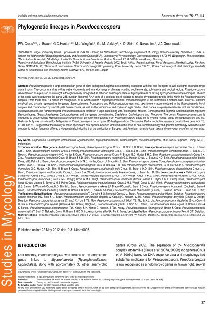
Phylogenetic l<strong>in</strong>eages <strong>in</strong> Pseudocercospora<br />
Fig. 1. Leaf spot symptoms associated with various species from the Pseudocercospora complex. A. P. fatouae on Fatoua villosa. B. P. clematidis on Clematis apiicola. C. P.<br />
griseola on Phaseolus vulgaris. D. P. rhododendron-<strong>in</strong>dici on Rhododendron <strong>in</strong>dicum. E. P. pyracanthae on Pyracantha angustifolia. F. P. lonicericola on Lonicera japonica. G.<br />
Scolecostigm<strong>in</strong>a mangiferae on Mangifera <strong>in</strong>dica. H. P. frax<strong>in</strong>ites on Frax<strong>in</strong>us rhynchophylla. I. Pseudocercosporella potentillae on Potentilla kle<strong>in</strong>iana. J. Pseudocercospora<br />
udagawana on Hovenia dulcis.<br />
Fig. 2. Pseudocercospora species of quarant<strong>in</strong>e importance. A. P. fijiensis on Musa (Black Leaf Streak or Black Sigatoka) (Photo G.H.J. Kema). B, C. P. angolensis on Citrus<br />
(Phaeoramularia Fruit and Leaf Spot).<br />
www.studies<strong>in</strong>mycology.org<br />
39
Crous et al.<br />
Fig. 3. Morphological structures of Pseudocercospora spp. A. Synnematous conidiophore. B. Densely aggregated fascicle of conidiophores with well-developed brown stroma.<br />
C, D. Loosely branched fascicles of conidiophores with moderate (C) and poorly (D) developed brown stroma. E. Fascicle reduced to conidiogenous cells. F. Conidiophore<br />
fascicles aris<strong>in</strong>g from stomata. G, H. Solitary conidiogenous cells on superficial hyphae. I. Geniculate conidiophore (arrow) with truncate apical locus. J, K. Conidiophores<br />
branched below (arrows). L. Conidiogenous cells with percurrent proliferations (arrows). M, N. Conidiophores with sympodial proliferation. O. Conidiophores with conidiogenous<br />
cells (note m<strong>in</strong>utely thickened scars, arrows). P. Subcyl<strong>in</strong>drical conidium with subacute apex and truncate base. Q. Conidia with constrictions at septa. R. Conidium with guttules.<br />
S. Cyl<strong>in</strong>drical conidium with obtuse apex, and truncate base. T. Undulate conidia. U. Curved conidium. Aseptate to 1-septate conidia. V. 1-septate conidia. W, X. Obclavate<br />
conidia with obconical base. Y. Obclavate conidium with short obconical base. Z. Dark brown, muriformly euseptate conidia (thick-walled, not distoseptate).<br />
40
Phylogenetic l<strong>in</strong>eages <strong>in</strong> Pseudocercospora<br />
MATERIALS AND METHODS<br />
Isolates<br />
Direct isolations were made from fascicles of conidiophores on<br />
leaves. Some leaves were <strong>in</strong>cubated <strong>in</strong> moist chambers for up to<br />
1 wk to enhance sporulation before s<strong>in</strong>gle conidial colonies were<br />
established on 2 % malt extract agar (MEA) (Crous 2002). Leaf<br />
spots bear<strong>in</strong>g ascomata were soaked <strong>in</strong> water for approximately<br />
2 h, after which they were attached to the <strong>in</strong>ner surface of Petri<br />
dish lids over plates conta<strong>in</strong><strong>in</strong>g MEA. Ascospore germ<strong>in</strong>ation<br />
patterns were exam<strong>in</strong>ed after 24 h, and s<strong>in</strong>gle ascospore and<br />
conidial cultures established as described previously (Crous et<br />
al. 1991, Crous 1998). Colonies were sub-cultured onto synthetic<br />
nutrient-poor agar (SNA), potato-dextrose agar (PDA), oatmeal<br />
agar (OA), and MEA (Crous et al. 2009d), and <strong>in</strong>cubated at 25 °C<br />
under cont<strong>in</strong>uous near-ultraviolet light to promote sporulation.<br />
Isolates were also sourced from the culture collections of the<br />
<strong>CBS</strong>-KNAW Fungal Biodiversity Centre (<strong>CBS</strong>), the work<strong>in</strong>g<br />
collection of Pedro Crous (CPC), Chiharu Nakashima (CNS) and<br />
the culture collection of the laboratory of plant pathology, Mie<br />
University, Japan (MUCC), and the mycological herbarium of Mie<br />
University (MUMH). Furthermore, isolates represent<strong>in</strong>g fungal<br />
species from genera allied to Pseudocercospora, e.g. Cercospora,<br />
Cercostigm<strong>in</strong>a, Cyphellophora, Davidiella, Dissoconium, Miuraea,<br />
Mycocentrospora, Passalora, Phaeoisariopsis, Phleospora,<br />
Septoria, Strelitziana, Stigm<strong>in</strong>a, Teratosphaeria, Thedgonia,<br />
Trochophora, and Xenostigm<strong>in</strong>a, were <strong>in</strong>cluded <strong>in</strong> this study (Table<br />
1).<br />
DNA isolation<br />
Mycelium from actively grow<strong>in</strong>g fungal cultures was scraped from<br />
the surface of MEA or PDA plates us<strong>in</strong>g a sterile scalpel blade.<br />
Harvested mycelium was ground to a f<strong>in</strong>e powder us<strong>in</strong>g liquid<br />
nitrogen and DNA was isolated us<strong>in</strong>g the CTAB extraction protocol<br />
as outl<strong>in</strong>ed by Crous et al. (2009d) or the UltraClean TM Microbial<br />
DNA Isolation Kit (MoBio Laboratories, Inc., Solana Beach, CA,<br />
USA) follow<strong>in</strong>g the manufacturers’ protocols. Isolated DNA was<br />
visualised by electrophoresis <strong>in</strong> 1 % agarose gels (w/v) sta<strong>in</strong>ed<br />
with ethidium bromide and viewed under near ultra-violet light. DNA<br />
concentrations were determ<strong>in</strong>ed by measur<strong>in</strong>g electrophoresed<br />
DNA samples aga<strong>in</strong>st a HyperLadder TM I molecular marker<br />
(BIOLINE) or alternatively by a NanoDrop quantification as outl<strong>in</strong>ed<br />
by the manufacturer.<br />
PCR amplification<br />
DNA isolated from fungal isolates was used as template for further<br />
Polymerase Cha<strong>in</strong> Reaction (PCR) amplifications. Four nuclear<br />
gene regions were targeted for PCR amplification and subsequent<br />
sequenc<strong>in</strong>g. These regions <strong>in</strong>cluded the Internal Transcribed<br />
Spacer regions ITS-1, ITS-2 and the 5.8S nrRNA gene regions<br />
(ITS), the first 900 bp of the Large Subunit (28S, LSU) (doma<strong>in</strong>s<br />
D1–D3) of the rDNA operon and partial gene regions of the<br />
translation elongation factor 1-alpha (EF-1α) and the act<strong>in</strong> (ACT)<br />
genes.<br />
The ITS region was amplified us<strong>in</strong>g primers ITS-1 or ITS-5 and<br />
ITS-4 (White et al. 1990) while primers used for amplification of<br />
the LSU region were LR0R (Rehner & Samuels 1994) or LSU1Fd<br />
(Crous et al. 2009b) and LR5 or LR7 (Vilgalys & Hester 1990).<br />
Primers employed for the amplification of EF-1α <strong>in</strong>cluded EF1-<br />
728F and EF1-986R (Carbone & Kohn 1999) or EF-2 (O’Donnell et<br />
al. 1998) while ACT-512F and ACT-783R (Carbone & Kohn 1999)<br />
were used to amplify a portion of the ACT gene. All PCR reaction<br />
mixtures and conditions followed those outl<strong>in</strong>ed by Hunter et al.<br />
(2006b). Follow<strong>in</strong>g PCR amplification, amplicons were visualized<br />
on 1.5 % agarose gels sta<strong>in</strong>ed with ethidium bromide and viewed<br />
under ultra-violet light and sizes of amplicons were determ<strong>in</strong>ed<br />
aga<strong>in</strong>st a HyperLadder TM I molecular marker (BIOLINE). The PCR<br />
amplicons for the four loci were subsequently diluted 1 to 10 times<br />
<strong>in</strong> preparation for further DNA sequenc<strong>in</strong>g reactions.<br />
DNA sequenc<strong>in</strong>g and phylogenetic <strong>in</strong>ference<br />
PCR amplicons of the four gene regions targeted <strong>in</strong> this study<br />
served as templates for DNA sequenc<strong>in</strong>g reactions with the<br />
BigDye® Term<strong>in</strong>ator Cycle Sequenc<strong>in</strong>g Kit v. 3.1 (Applied<br />
Biosystems Life Technologies, Carlsbad, CA, USA) follow<strong>in</strong>g the<br />
protocol of the manufacturer. DNA sequenc<strong>in</strong>g reactions used the<br />
same primers as those for the PCR reactions. However, additional<br />
<strong>in</strong>ternal primers LR3R (http://www.biology.duke.edu/fungi/mycolab/<br />
primers.htm), LR16 (Moncalvo et al. 1993) and LR5 were used to<br />
sequence the LSU <strong>in</strong> order to obta<strong>in</strong> reliable sequences spann<strong>in</strong>g<br />
the entire D1-D3 region. DNA sequenc<strong>in</strong>g amplicons were purified<br />
through Sephadex® G-50 Superf<strong>in</strong>e columns (Sigma Aldrich, St.<br />
Louis, MO) <strong>in</strong> MultiScreen HV plates (Millipore, Billerica, MA).<br />
Purified sequence reactions were run on an ABI Prism 3730xl DNA<br />
Sequencer (Life Technologies, Carlsbad, CA, USA).<br />
Generated DNA sequence electropherograms were analysed<br />
us<strong>in</strong>g MEGA (Molecular Evolutionary Genetics Analysis) v. 4.0<br />
(Tamura et al. 2007), 4Peaks v. 1.7.2 (http://www.mekentosj.com/)<br />
and SeqMan v. 8.0.2. from the DNASTAR Lasergene® software<br />
package. Consensus sequences were generated and imported<br />
<strong>in</strong>to MEGA for <strong>in</strong>itial alignment and the construction of sequence<br />
datasets. DNA sequences represent<strong>in</strong>g isolates of closely allied<br />
genera, for which material could not be obta<strong>in</strong>ed were downloaded<br />
from the NCBI GenBank nucleotide database (www.ncbi.nlm.nih.<br />
gov) and added to the DNA sequence datasets generated <strong>in</strong> this<br />
study. Sequence datasets for the four genomic loci were aligned <strong>in</strong><br />
MAFFT (“Multiple alignment program for am<strong>in</strong>o acids or nucleotide<br />
sequences”) v. 6.0 (Katoh & Toh 2006, Katoh et al. 2005; http://<br />
mafft.cbrc.jp/alignment/server/<strong>in</strong>dex.html) us<strong>in</strong>g the Auto alignment<br />
strategy with the 200PAM/ K=2 scor<strong>in</strong>g matrix and a gap open<strong>in</strong>g<br />
penalty of 1.53 with an offset value of 0.0. Result<strong>in</strong>g sequence<br />
alignments were manually evaluated and adjusted <strong>in</strong> MEGA,<br />
MacClade v.4.08 (Maddison & Maddison 2000) or Sequence<br />
Alignment Editor v. 2.0a11 (Rambaut 2002).<br />
A phylogenetic re-construction was conducted for the aligned<br />
LSU data set to determ<strong>in</strong>e generic relationships us<strong>in</strong>g MrBayes<br />
v. 3.1.2 (Ronquist & Huelsenbeck 2003). Subsequently, a species<br />
level phylogeny was derived from the comb<strong>in</strong>ed ITS, ACT and EF-<br />
1α alignment of Pseudocercospora s. str. sequences us<strong>in</strong>g PAUP<br />
v. 4.0b10 (Swofford 2003). For the LSU alignment, MrModeltest<br />
v. 2.2 (Nylander 2004) was used to determ<strong>in</strong>e the best nucleotide<br />
substitution model sett<strong>in</strong>gs for MrBayes. Based on the results of the<br />
MrModeltest, a phylogenetic analysis was performed with MrBayes<br />
v. 3.1.2 apply<strong>in</strong>g a general time-reversible (GTR) substitution<br />
model with <strong>in</strong>verse gamma rates and dirichlet base frequencies<br />
and a heat<strong>in</strong>g parameter set at 0.3. The Markov Cha<strong>in</strong> Monte Carlo<br />
(MCMC) analysis of 4 cha<strong>in</strong>s started <strong>in</strong> parallel from a random tree<br />
topology and had 8 000 000 generations. Trees were saved each<br />
www.studies<strong>in</strong>mycology.org<br />
41
Crous et al.<br />
Table 1. Pseudocercospora and Pseudocercospora-like isolates <strong>in</strong>cluded <strong>in</strong> the morphological and/or phylogenetic analyses.<br />
Species Culture accession numbers 1 Collector Host Family Country GenBank accession numbers 2<br />
LSU ITS EF-1α ACT<br />
Cercospora eucommiae CPC 10047 H.D. Sh<strong>in</strong> Eucommia ulmoides Eucommiaceae South Korea GU253741 GU269702 GU384418 GU320406<br />
CPC 10802; <strong>CBS</strong> 131932 H.D. Sh<strong>in</strong> Eucommia ulmoides Eucommiaceae South Korea GU214674 GU269851/<br />
GU214674<br />
GU384563 GU320555<br />
CPC 11508; <strong>CBS</strong> 132026 H.D. Sh<strong>in</strong> Eucommia ulmoides Eucommiaceae South Korea GU253742 GU269703 GU384419 GU320407<br />
Cercospora soj<strong>in</strong>a CPC 12322; <strong>CBS</strong> 132018 H.D. Sh<strong>in</strong> Glyc<strong>in</strong>e soja Fabaceae South Korea GU253861 GU214655 JQ324984 JQ325008<br />
Cyphellophora eucalypti <strong>CBS</strong> 124764; CPC 13412 P.W. Crous Eucalyptus sp. Myrtaceae Australia GQ303305 GQ303274 GU384510 JQ325009<br />
Dissoconium dekkeri <strong>CBS</strong> 110748; CPC 825; CMW 14906 G. Kemp Eucalyptus grandis Myrtaceae South Africa GU214422 AF173315 JQ324985 DQ147651<br />
Microcyclospora querc<strong>in</strong>a CPC 10712; <strong>CBS</strong> 130827 G. Verkley Quercus sp. Fagaceae Netherlands GU214681 GU269789 GU384499 GU320490<br />
Miuraea persicae CPC 10069; <strong>CBS</strong> 132307 H.D. Sh<strong>in</strong> Prunus persica Rosaceae South Korea GU253859 GU269843 GU384556 GU320546<br />
CPC 10828; <strong>CBS</strong> 131935 H.D. Sh<strong>in</strong> Prunus armeniaca Rosaceae South Korea JQ324939 GU269844 GU384557 GU320547<br />
“Mycosphaerella” laric<strong>in</strong>a <strong>CBS</strong> 326.52 E. Müller Larix decidua P<strong>in</strong>aceae Switzerland GU253693 GU269643 GU384361 GU320353<br />
“Mycosphaerella” madeirae <strong>CBS</strong> 112895; CPC 3745 S. Denman Eucalyptus globulus Myrtaceae Portugal DQ204756 AY725553 DQ211672 DQ147641<br />
“Mycosphaerella” marksii <strong>CBS</strong> 110920; CPC 935; CMW 5150 A.J. Carnegie Eucalyptus botryoides Myrtaceae Australia DQ246250/<br />
GU253694<br />
AF309588/<br />
GU269644<br />
DQ235134 DQ147625<br />
Pallidocercospora acaciigena <strong>CBS</strong> 112516; CPC 3838 M.J. W<strong>in</strong>gfield Acacia mangium Fabaceae Venezuela GU214661/<br />
GU253697<br />
GU269648 GU384366 GU320356<br />
<strong>CBS</strong> 120740; CPC 13290 B. Summerell Eucalyptus sp. Myrtaceae Australia GU253698 EF394822/<br />
GU269649<br />
GU384367 GU320357<br />
Pallidocercospora crystall<strong>in</strong>a <strong>CBS</strong> 681.95; <strong>CBS</strong> 116158; CPC 802;<br />
CMW 3033<br />
M.J. W<strong>in</strong>gfield Eucalyptus bicostata Myrtaceae South Africa DQ204747 AY490757 DQ147636/<br />
DQ211662<br />
DQ147636<br />
Pallidocercospora heimii <strong>CBS</strong> 110682; CPC 760; CMW 4942 P.W. Crous Eucalyptus sp. Myrtaceae Madagascar DQ204751 AF309606 DQ211667 DQ147638<br />
Pallidocercospora heimioides <strong>CBS</strong> 111190; CPC 1312; CMW 3046 M.J. W<strong>in</strong>gfield Eucalyptus sp. Myrtaceae Indonesia DQ204753 AF309609 DQ211669 DQ147633<br />
Pallidocercospora irregulariramosa <strong>CBS</strong> 114774; <strong>CBS</strong> 114777; CPC 1360;<br />
CMW 4943<br />
M.J. W<strong>in</strong>gfield Eucalyptus saligna Myrtaceae South Africa DQ204754 AF309607 DQ211670 DQ147634<br />
Pallidocercospora konae <strong>CBS</strong> 120748; CPC 13469 W. Himaman Eucalyptus camaldulensis Myrtaceae Thailand GU253852 EF394842 GU384549 GU320538<br />
Paracercospora egenula <strong>CBS</strong> 485.81 N. Ponnapa Solanum melongena Solanaceae India JQ324940 GU269699 GU384415 GU320403<br />
CPC 12537; <strong>CBS</strong> 132030 H.D. Sh<strong>in</strong> Solanum melongena Solanaceae South Korea GU253738 GU269698 GU384414 GU320402<br />
MUCC 883 T. Mikami Solanum melongena Solanaceae Japan GU253739 GU269700 GU384416 GU320404<br />
Passalora eucalypti <strong>CBS</strong> 111318; CPC 1457 P.W. Crous Eucalyptus saligna Myrtaceae Brazil GU253860 GU269845 GU384558 GU320548<br />
Phaeomycocentrospora cantuariensis CPC 10157 H.D. Sh<strong>in</strong> Humulus scandens Cannabaceae South Korea GU253712 GU269664 GU384381 GU320370<br />
CPC 10762; <strong>CBS</strong> 131928 H.D. Sh<strong>in</strong> Luffa cyl<strong>in</strong>drica Cucurbitaceae South Korea GU253713 GU269665 GU384382 GU320371<br />
42
Phylogenetic l<strong>in</strong>eages <strong>in</strong> Pseudocercospora<br />
Table 1. (Cont<strong>in</strong>ued).<br />
Species Culture accession numbers 1 Collector Host Family Country GenBank accession numbers 2<br />
LSU ITS EF-1α ACT<br />
CPC 11646; <strong>CBS</strong> 132013 H.D. Sh<strong>in</strong> Acalypha australis Euphorbiaceae South Korea GU253715 GU269667 GU384384 GU320373<br />
CPC 11694; <strong>CBS</strong> 132014 H.D. Sh<strong>in</strong> Humulus scandens Cannabaceae South Korea GU253716 GU269668 GU384385 GU320374<br />
Phloeospora ulmi <strong>CBS</strong> 344.97 W. Gams Ulmus glabra Ulmaceae Austria GU253841 JQ324974 JQ324986 GU320528<br />
<strong>CBS</strong> 613.81 H.A. Van der Aa Ulmus sp. Ulmaceae Austria GU253842 GU269825 JQ324987 GU320529<br />
Pseudocercospora abelmoschi CPC 14478; <strong>CBS</strong> 132103 H.D. Sh<strong>in</strong> Hibiscus syriacus Malvaceae South Korea GU253696 GU269647 GU384365 GU320355<br />
Pseudocercospora acericola <strong>CBS</strong> 122279 R. Kirschner Acer albopurpurascens Aceraceae Taiwan GU253699 GU269650 GU384368 GU320358<br />
Pseudocercospora ampelopsis CPC 11680; <strong>CBS</strong> 131583 H.D. Sh<strong>in</strong> Ampelopsis brevipenduncula<br />
var. heterophylla<br />
Vitaceae South Korea GU253846 GU269830 GU384542 GU320534<br />
Pseudocercospora angolensis <strong>CBS</strong> 112933; CPC 4118 M.C. Pretorius Citrus sp. Rutaceae Zimbabwe GU214470 AY260063/<br />
GU269836<br />
GU384548 JQ325010<br />
<strong>CBS</strong> 149.53 T. de Carvalho & O. Mendes Citrus s<strong>in</strong>ensis Rutaceae Angola JQ324941 JQ324975 JQ324988 JQ325011<br />
Pseudocercospora araliae CPC 10154 H.D. Sh<strong>in</strong> Aralia elata Araliaceae South Korea GU253701 GU269652 GU384370 GU320360<br />
MUCC 873 T. Kobayashi & C. Nakashima Aralia elata Araliaceae Japan GU253702 GU269653 GU384371 GU320361<br />
Pseudocercospora arecacearum <strong>CBS</strong> 118406 C.F. Hill Rhopalostylis sapidis Arecaceae New Zealand GU253704 GU269655 GU384373 GU320363<br />
<strong>CBS</strong> 118792 C.F. Hill Howea forsteriana Arecaceae New Zealand GU253703 GU269654 GU384372 GU320362<br />
Pseudocercospora assamensis <strong>CBS</strong> 122467 I. Buddenhagen Musa cultivar Musaceae India GU253705 GU269656 GU384374 GU320364<br />
Pseudocercospora atromarg<strong>in</strong>alis <strong>CBS</strong> 114640 C.F. Hill Solanum sp. Solanaceae New Zealand GU253706 GU269658 GU384376 GU320365<br />
CPC 11372; <strong>CBS</strong> 132010 H.D. Sh<strong>in</strong> Solanum nigrum Solanaceae South Korea GU214671 GU269657 GU384375 —<br />
Pseudocercospora balsam<strong>in</strong>ae CPC 10044; <strong>CBS</strong> 131882 H.D. Sh<strong>in</strong> Impatiens textori Balsam<strong>in</strong>aceae South Korea GU253708 GU269660 GU384379 GU320367<br />
Pseudocercospora basiramifera <strong>CBS</strong> 111072; CPC 1266 M.J. W<strong>in</strong>gfield Eucalyptus pellita Myrtaceae Thailand GU253709 GU269661 DQ211677 GU320368<br />
<strong>CBS</strong> 114757; CPC 1267 M.J. W<strong>in</strong>gfield Eucalyptus pellita Myrtaceae Thailand GU253802 GU269781 GU384492 GU320484<br />
Pseudocercospora basitruncata <strong>CBS</strong> 114664; CPC 1202 M.J. W<strong>in</strong>gfield Eucalyptus grandis Myrtaceae Colombia GU253710/<br />
DQ204759<br />
DQ267600/<br />
GU269662<br />
DQ211675 DQ147622<br />
Pseudocercospora callicarpae MUCC 888 T. Kobayashi Callicarpa japonica Verbenaceae Japan GU253711 GU269663 GU384380 GU320369<br />
Pseudocercospora catalpigena MUCC 743 C. Nakashima & I. Araki Catalpa ovata Bignoniaceae Japan GU253731 GU269690 GU384406 GU320395<br />
Pseudocercospora catappae MUCC 809 C. Nakashima & T. Akashi Term<strong>in</strong>alia catappa Combretaceae Japan GU253717 GU269669 GU384386 GU320375<br />
Pseudocercospora cercidicola MUCC 896 T. Kobayashi & Y. Kobayashi Cercis ch<strong>in</strong>ensis Fabaceae Japan GU253719 GU269671 GU384388 GU320377<br />
Pseudocercospora cercidis-ch<strong>in</strong>ensis CPC 14481; <strong>CBS</strong> 132109 H.D. Sh<strong>in</strong> Cercis ch<strong>in</strong>ensis Fabaceae South Korea GU253718 GU269670 GU384387 GU320376<br />
Pseudocercospora cf. cruenta <strong>CBS</strong> 117232 R. Kirschner Phaseolus vulgaris Fabaceae Taiwan GU253730 GU269689 GU384405 GU320394<br />
Pseudocercospora cf. kaki CPC 10636; <strong>CBS</strong> 131921 H.D. Sh<strong>in</strong> Diospyros lotus Ebenaceae South Korea GU214677 GU269728 GU384441 GU320430<br />
www.studies<strong>in</strong>mycology.org<br />
43
Crous et al.<br />
Table 1. (Cont<strong>in</strong>ued).<br />
Species Culture accession numbers 1 Collector Host Family Country GenBank accession numbers 2<br />
LSU ITS EF-1α ACT<br />
Pseudocercospora chengtuensis CPC 10696; <strong>CBS</strong> 131924 H.D. Sh<strong>in</strong> Lycium ch<strong>in</strong>ense Solanaceae South Korea JQ324942 GU269673 GU384390 GU320379<br />
MUCC 828 I. Araki & M. Harada Lycium ch<strong>in</strong>ense Solanaceae Japan JQ324943 — — —<br />
Pseudocercospora chionanthi-retusi CPC 14683; <strong>CBS</strong> 132110 H.D. Sh<strong>in</strong> Chionanthus retusus Oleaceae South Korea GU253721 GU269674 GU384391 GU320380<br />
Pseudocercospora chrysanthemicola CPC 10633; <strong>CBS</strong> 131888 H.D. Sh<strong>in</strong> Chrysanthemum sp. Asteraceae South Korea GU253722 GU269675 GU384392 GU320381<br />
Pseudocercospora cladosporioides <strong>CBS</strong> 117482; CPC 10913 P.W. Crous Olea europaea Oleaceae Tunisia JQ324944 GU269678 GU384395 GU320383<br />
“Pseudocercospora” colombiensis <strong>CBS</strong> 110969; CPC 1106; CMW 4944 M.J. W<strong>in</strong>gfield Eucalyptus urophylla Myrtaceae Colombia DQ204744 AY752149 DQ211660 DQ147639<br />
Pseudocercospora contraria CPC 14714; <strong>CBS</strong> 132108 H.D. Sh<strong>in</strong> Dioscorea qu<strong>in</strong>queloba Dioscoreaceae South Korea JQ324945 GU269677 GU384394 GU320385<br />
Pseudocercospora coprosmae <strong>CBS</strong> 114639 C. F. Hill Coprosma robusta Rubiaceae New Zealand JQ324946 GU269680 GU384397 GU320386<br />
Pseudocercospora cordiana <strong>CBS</strong> 114685; CPC 2552 P.W. Crous & R.L. Benchimol Cordia goeldiana Borag<strong>in</strong>aceae Brazil GU214472 AF362054/<br />
GU269681<br />
GU384398 GU320387<br />
Pseudocercospora coriariae MUCC 840 I. Araki & M. Harada Coriaria japonica Coriariaceae Japan GU253725 GU269682 GU384399 GU320388<br />
Pseudocercospora cornicola MUCC 909 C. Nakashima & E. Imaizumi Cornus alba var. sibirica Cornaceae Japan GU253726 GU269683 GU384400 GU320389<br />
Pseudocercospora corylopsidis MUCC 874 T. Kobayashi & C. Nakashima Hamamelis japonica Hamamelidaceae Japan GU253757 GU269721 GU384437 GU320425<br />
MUCC 908 C. Nakashima & E. Imaizumi Corylopsis spicata Hamamelidaceae Japan GU253727 GU269684 GU384401 GU320390<br />
Pseudocercospora cotoneastri MUCC 876 T. Kobayashi & C. Nakashima Cotoneaster salicifolius Rosaceae Japan GU253728 GU269685 GU384402 GU320391<br />
Pseudocercospora crispans CPC 14883; <strong>CBS</strong> 125999 P.W.Crous Eucalyptus sp. Myrtaceae South Africa GU253825 GU269807 GU384518 GU320510<br />
Pseudocercospora crocea CPC 11668; <strong>CBS</strong> 126004 H.D. Sh<strong>in</strong> Pilea hamaoi Urticaceae South Korea JQ324947 GU269792 GU384502 GU320493<br />
Pseudocercospora crousii <strong>CBS</strong> 119487 C.F. Hill Eucalyptus sp. Myrtaceae New Zealand GU253729 GU269686 GU384403 GU320392<br />
Pseudocercospora cruenta CPC 10846; <strong>CBS</strong> 132021 H. Booker Vigna sp. Fabaceae Tr<strong>in</strong>idad GU214673 GU269688 GU384404 JQ325012<br />
Pseudocercospora cydoniae CPC 10678; <strong>CBS</strong> 131923 H.D. Sh<strong>in</strong> Chaenomeles speciosa Rosaceae South Korea GU253732 GU269691 GU384407 GU320396<br />
Pseudocercospora cymbidiicola <strong>CBS</strong> 115132 C.F. Hill Cymbidium sp. Orchidaceae New Zealand GU253733 GU269692 GU384408 GU320397<br />
Pseudocercospora davidiicola MUCC 296 C. Nakashima & I. Araki Davidia <strong>in</strong>volucrata Nyssaceae Japan GU253734 GU269693 GU384409 GU320398<br />
Pseudocercospora dendrobii MUCC 596 C. Nakashima & K. Motohashi Dendrobium sp. Orchidaceae Japan GU253737 GU269696 GU384412 GU320401<br />
Pseudocercospora destructiva MUCC 870 S. Uematsu & C. Nakashima Euonymus japonicus Celastraceae Japan GU253735 GU269694 GU384410 GU320399<br />
Pseudocercospora dianellae <strong>CBS</strong> 117746 C.F. Hill Dianella caerulae Liliaceae New Zealand GU253736 GU269695 GU384411 GU320400<br />
Pseudocercospora dodonaeae <strong>CBS</strong> 114647 C.F. Hill Dodonaea viscosa Sap<strong>in</strong>daceae New Zealand JQ324948 GU269697 GU384413 JQ325013<br />
Pseudocercospora dovyalidis CPC 13771; <strong>CBS</strong> 126002 P.W. Crous Dovyalis zeyheri Flacourtiaceae South Africa GU253818 GU269800 GU384513 GU320503<br />
Pseudocercospora elaeocarpi MUCC 925 C. Nakashima Elaeocarpus sp. Elaeocarpaceae Japan GU253740 GU269701 GU384417 GU320405<br />
“Pseudocercospora” epispermogonia <strong>CBS</strong> 110750; CPC 822 G. Kemp Eucalyptus grandis Myrtaceae South Africa DQ204757 DQ267596 DQ211673 DQ147629<br />
44
Phylogenetic l<strong>in</strong>eages <strong>in</strong> Pseudocercospora<br />
Table 1. (Cont<strong>in</strong>ued).<br />
Species Culture accession numbers 1 Collector Host Family Country GenBank accession numbers 2<br />
LSU ITS EF-1α ACT<br />
Pseudocercospora eucalyptorum <strong>CBS</strong> 110777; CPC 16; CMW 5228 P.W. Crous Eucalyptus nitens Myrtaceae South Africa DQ204762 AF309598 DQ211678 DQ147614<br />
<strong>CBS</strong> 114242; CPC 10390; CMW 14908 J.P. Mansilla Eucalyptus globulus Myrtaceae Spa<strong>in</strong> GU214481 AY725526 DQ211681 DQ147613/<br />
GU320465<br />
<strong>CBS</strong> 116359; CPC 3751 P.W. Crous Eucalyptus sp. Myrtaceae Madeira GU253829 GU269812 GU384524 GU320514<br />
CPC 10500; <strong>CBS</strong> 114243 P.W. Crous Eucalyptus nitens Myrtaceae New Zealand JQ324949 AY725527 GU384474 JQ325014<br />
CPC 10507; <strong>CBS</strong> 116371 P.W.Crous Eucalyptus nitens Myrtaceae New Zealand JQ324950 GU269687 JQ324989 GU320393<br />
CPC 10916 P.W. Crous Eucalyptus sp. Myrtaceae South Africa GU253788 GU269763 GU384475 GU320464<br />
CPC 11713; <strong>CBS</strong> 132015 P. Mansilla Eucalyptus globulus Myrtaceae Spa<strong>in</strong> JQ324951 GU269811 GU384523 JQ325015<br />
CPC 12406; <strong>CBS</strong> 132029 I. Smith Eucalyptus globulus Myrtaceae Australia GU253811 GU269793 GU384503 GU320494<br />
CPC 12568; <strong>CBS</strong> 132309 C. Mohammed Eucalyptus nitens Myrtaceae Australia GU253814 GU269796 GU384506 GU320497<br />
CPC 12802; <strong>CBS</strong> 132032 A. Phillips Eucalyptus globulus Myrtaceae Portugal GU253789 JQ324976 JQ324990 GU320466<br />
CPC 12957; <strong>CBS</strong> 132033 B. Summerell Eucalyptus deanei Myrtaceae Australia GU253815 GU269797 JQ324991 JQ325016<br />
CPC 13455; <strong>CBS</strong> 132034 P.W. Crous Eucalyptus sp. Myrtaceae Portugal GU253816 GU269798 GU384511 GU320501<br />
CPC 13769; <strong>CBS</strong> 132035 P.W. Crous Eucalyptus punctata Myrtaceae South Africa GU253707 GU269659 GU384378 GU320366<br />
CPC 13816; <strong>CBS</strong> 132114 S. Denman Eucalyptus glaucescens Myrtaceae UK GU253819 GU269801 JQ324992 GU320504<br />
CPC 13926; <strong>CBS</strong> 132105 S. Denman Eucalyptus sp. Myrtaceae USA GU253820 GU269802 JQ324993 GU320505<br />
Pseudocercospora eupatoriella <strong>CBS</strong> 113372 M.J. Morris Chromolaena odorata Asteraceae Jamaica GU253743 GU269704 GU384420 GU320408<br />
Pseudocercospora eustomatis <strong>CBS</strong> 110822 G. Dal Bello Eustroma grandiflorum Gentianaceae Argent<strong>in</strong>a GU253744 GU269705 GU384421 GU320409<br />
Pseudocercospora exosporioides MUCC 893 T. Kobayashi Sequoia sempervirens Taxodiaceae Japan GU253746 GU269707 GU384423 GU320411<br />
Pseudocercospora fijiensis <strong>CBS</strong> 120258; CIRAD 86 J. Carlier Musa sp. Musaceae Cameroon JQ324952 EU514248 Genome 3 Genome 3<br />
MUCC 792 T. Kobayashi & C. Nakashima Musa sp. Musaceae Japan GU253776 GU269748 JQ324994 GU320450<br />
Pseudocercospora flavomarg<strong>in</strong>ata <strong>CBS</strong> 118841; CMW 13586 M.J. W<strong>in</strong>gfield Eucalyptus camaldulensis Myrtaceae Thailand DQ153306 DQ155657 DQ156548 DQ166513<br />
<strong>CBS</strong> 124990; CPC 13492 W. Himaman Eucalyptus camaldulensis Myrtaceae Thailand GU253817 GU269799 GU384512 GU320502<br />
CPC 14142; <strong>CBS</strong> 126001 X. Zhou Eucalyptus sp. Myrtaceae Ch<strong>in</strong>a GU253822 GU269804 GU384515 GU320507<br />
Pseudocercospora fori <strong>CBS</strong> 113285; CMW 9095 G.C. Hunter Eucalyptus grandis Myrtaceae South Africa DQ204748 AF468869 DQ211664 DQ147618<br />
CPC 14880; <strong>CBS</strong> 132113 P.W. Crous Eucalyptus sp. Myrtaceae South Africa GU253824 GU269806 GU384517 GU320509<br />
Pseudocercospora frax<strong>in</strong>ites CPC 10743; <strong>CBS</strong> 131927 H.D. Sh<strong>in</strong> Fontanesia phillyraeoides Oleaceae South Korea GU253720 GU269672 GU384389 GU320378<br />
MUCC 891 T. Kobayashi Frax<strong>in</strong>us excelsior Oleaceae Japan GU253748 GU269710 GU384426 GU320414<br />
Pseudocercospora fukuokaensis CPC 14689; <strong>CBS</strong> 132111 H.D. Sh<strong>in</strong> Styrax japonicus Styracaceae South Korea GU253750 GU269713 GU384429 GU320417<br />
MUCC 887 T. Kobayashi Styrax japonicus Styracaceae Japan GU253751 GU269714 GU384430 GU320418<br />
www.studies<strong>in</strong>mycology.org<br />
45
Crous et al.<br />
Table 1. (Cont<strong>in</strong>ued).<br />
Species Culture accession numbers 1 Collector Host Family Country GenBank accession numbers 2<br />
LSU ITS EF-1α ACT<br />
Pseudocercospora fuligena CPC 12296; <strong>CBS</strong> 132017 Z. Mersha Lycopersicon sp. Solanaceae Thailand JQ324953 GU269711 GU384427 GU320415<br />
MUCC 533 C. Nakashima Lycopersicon esculentum Solanaceae Japan GU253749 GU269712 GU384428 GU320416<br />
Pseudocercospora glauca CPC 10062; <strong>CBS</strong> 131884 H.D. Sh<strong>in</strong> Albizzia julibriss<strong>in</strong> Fabaceae South Korea GU253752 GU269715 GU384431 GU320419<br />
Pseudocercospora gracilis <strong>CBS</strong> 243.94; CPC 730 P.W. Crous Eucalyptus urophylla Myrtaceae Indonesia DQ204750 DQ267582 DQ211666 DQ147616<br />
Pseudocercospora griseola f. griseola <strong>CBS</strong> 119112; CPC 10460 F.S. Ngulu & C. Mushi Phaseolus vulgaris Fabaceae Tanzania GU253753 GU269717 GU384433 GU320421<br />
<strong>CBS</strong> 194.47 — Phaseolus vulgaris Fabaceae Portugal JQ324954 DQ289801 JQ324995 DQ289868<br />
<strong>CBS</strong> 880.72 H.A. van Kesteren Phaseolus vulgaris Fabaceae Netherlands GU214476 GU269716 GU384432 GU320420<br />
CPC 10462 M.M. Liebenberg Phaseolus vulgaris Fabaceae South Africa GU253865 GU269849 GU384562 GU320553<br />
CPC 10480; <strong>CBS</strong> 131887 M.M. Liebenberg Phaseolus vulgaris Fabaceae South Africa GU253864 GU269848 GU384561 DQ289882<br />
CPC 10779; <strong>CBS</strong> 131929 H.D. Sh<strong>in</strong> Phaseolus vulgaris Fabaceae South Korea GU253862 GU269846 GU384559 DQ289885<br />
CPC 12239 G. Mahuku Phaseolus vulgaris Fabaceae Colombia GU253863 GU269847 GU384560 DQ289887<br />
Pseudocercospora guianensis MUCC 855 C. Nakashima & T. Akashi Lantana camara Verbenaceae Japan GU253755 GU269719 GU384435 GU320423<br />
MUCC 879 C. Nakashima Lantana camara Verbenaceae Japan GU253756 GU269720 GU384436 GU320424<br />
Pseudocercospora haiweiensis CPC 14084; <strong>CBS</strong> 131584 X. Zhou Eucalyptus sp. Myrtaceae Ch<strong>in</strong>a GU253821 GU269803 GU384514 GU320506<br />
Pseudocercospora hakeae <strong>CBS</strong> 112226; CPC 3145 P.W. Crous & B. Summerell Grevillea sp. Proteaceae Australia GU253805 GU269784 GU384495 JQ325017<br />
Pseudocercospora humuli MUCC 742 C. Nakashima & I. Araki Humulus lupulus var. lupulus Cannabaceae Japan GU253758 GU269725 GU384439 GU320428<br />
Pseudocercospora humulicola CPC 10049; <strong>CBS</strong> 131883 H.D. Sh<strong>in</strong> Humulus scandens Cannabaceae South Korea JQ324955 GU269724 JQ324996 JQ325018<br />
CPC 11358; <strong>CBS</strong> 131585 H.D. Sh<strong>in</strong> Humulus scandens Cannabaceae South Korea JQ324956 GU269723 GU384438 GU320427<br />
Pseudocercospora <strong>in</strong>donesiana <strong>CBS</strong> 122473 I.W. Buddenhagen Musa sp. Musaceae Sumatra GU253765 GU269735 GU384448 GU320437/<br />
EU514340<br />
<strong>CBS</strong> 122474 I.W. Buddenhagen Musa sp. Musaceae Indonesia JQ324957 EU514283 JQ324997 JQ325019<br />
Pseudocercospora ixorae <strong>CBS</strong> 118760 R. Kirschner Ixora sp. Rubiaceae Taiwan GU253759 GU269726 GU384440 GU320429<br />
Pseudocercospora jussiaeae CPC 14625; <strong>CBS</strong> 132117 H.D. Sh<strong>in</strong> Ludwigia prostrata Onagraceae South Korea JQ324958 JQ324977 JQ324998 JQ325020<br />
Pseudocercospora kaki MUCC 900 S. Uematsu & C. Nakashima Diospyros kaki Ebenaceae Japan GU253761 GU269729 GU384442 GU320431<br />
Pseudocercospora kiggelariae CPC 11853; <strong>CBS</strong> 132016 W. Gams Kiggelaria africana Flacourtiaceae South Africa GU253762 GU269730 GU384443 GU320432<br />
Pseudocercospora latens MUCC 763 C. Nakashima & T. Akashi Lespedeza wilfordii Fabaceae Japan GU253763 GU269732 GU384445 GU320434<br />
Pseudocercospora leucadendri CPC 1869 S. Denman & P.W. Crous Leucadendron sp. Proteaceae South Africa GU214480 GU269842 GU384555 GU320545<br />
Pseudocercospora libertiae <strong>CBS</strong> 114643 C.F. Hill Libertia ixioides Iridaceae New Zealand JQ324959 GU269733 GU384446 GU320435<br />
Pseudocercospora lilacis CPC 12767; <strong>CBS</strong> 132031 C. Hodges Ligustrum japonicum Oleaceae USA GU253767 GU269737 GU384449 GU320439<br />
46
Phylogenetic l<strong>in</strong>eages <strong>in</strong> Pseudocercospora<br />
Table 1. (Cont<strong>in</strong>ued).<br />
Species Culture accession numbers 1 Collector Host Family Country GenBank accession numbers 2<br />
LSU ITS EF-1α ACT<br />
Pseudocercospora longispora <strong>CBS</strong> 122470 D.R. Jones Musa sp. Musaceae Malaysia GU253764 GU269734 GU384447 GU320436/<br />
EU514342<br />
Pseudocercospora lonicericola MUCC 889 T. Kobayashi Lonicera gracilipes var. glabra Caprifoliaceae Japan GU253766 GU269736 JQ324999 GU320438<br />
Pseudocercospora luzardii CPC 2556 A.C. Alfenas Hancornia speciosa Apocynaceae Brazil GU214477 AF362057/<br />
GU269738<br />
GU384450 GU320440<br />
Pseudocercospora lyoniae MUCC 910 C. Nakashima & E. Imaizumi Lyonia ovalifolia var. elliptica Ericaceae Japan GU253768 GU269739 GU384451 GU320441<br />
Pseudocercospora lythracearum CPC 10707; <strong>CBS</strong> 131925 H.D. Sh<strong>in</strong> Lagerstroemia <strong>in</strong>dica Lythraceae South Korea GU253769 GU269740 GU384452 GU320442<br />
MUCC 890 T. Kobayashi Lagerstroemia <strong>in</strong>dica Lythraceae Japan GU253770 GU269741 GU384453 GU320443<br />
Pseudocercospora lythri CPC 14588; <strong>CBS</strong> 132115 H.D. Sh<strong>in</strong> Lythrum salicaria Lythraceae South Korea GU253771 GU269742 GU384454 GU320444<br />
MUCC 865 I. Araki & M. Harada Lythrum salicaria Lythraceae Japan GU253772 GU269743 GU384455 GU320445<br />
Pseudocercospora macrospora <strong>CBS</strong> 114696; CPC 2553 P.W. Crous & R.L. Benchimol Bertholletia excelsa Lecythidaceae Brazil GU214478 AF362055/<br />
GU269745<br />
GU384457 GU320447<br />
Pseudocercospora mali MUCC 886 T. Kobayashi Malus sieboldii Rosaceae Japan GU253773 GU269744 GU384456 GU320446<br />
Pseudocercospora marg<strong>in</strong>alis CPC 12497; <strong>CBS</strong> 131582 H.D. Sh<strong>in</strong> Frax<strong>in</strong>us rhynchophylla Oleaceae South Korea GU253812 GU269794 GU384504 GU320495<br />
Pseudocercospora melicyti <strong>CBS</strong> 115023 M. Fletcher Melicytus macrophyllus Violaceae New Zealand JQ324968 GU269769 GU384481 GU320472<br />
Pseudocercospora metrosideri <strong>CBS</strong> 118795 C.F. Hill Metrosideros coll<strong>in</strong>a Myrtaceae New Zealand GU253774 GU269746 GU384458 GU320448<br />
Pseudocercospora musae <strong>CBS</strong> 116634 J. Carlier Musa sp. Musaceae Cuba GU253775 GU269747 GU384459 GU320449<br />
Pseudocercospora myrticola MUCC 632 C. Nakashima & K. Motohashi Myrtus communis Myrtaceae Japan GU253777 GU269749 GU384460 GU320451<br />
Pseudocercospora nand<strong>in</strong>ae <strong>CBS</strong> 117745 C.F. Hill Nand<strong>in</strong>a domestica Berberidaceae New Zealand GU253778 GU269750 GU384461 GU320452<br />
Pseudocercospora natalensis <strong>CBS</strong> 111069; CPC 1263 T. Cout<strong>in</strong>ho Eucalyptus nitens Myrtaceae South Africa DQ267576 DQ303077 JQ325000 DQ147620<br />
<strong>CBS</strong> 111071; CPC 1265 T. Cout<strong>in</strong>ho Eucalyptus nitens Myrtaceae South Africa GU253801 GU269780 GU384491 GU320483<br />
Pseudocercospora nephrolepidis <strong>CBS</strong> 119121 R. Kirschner Nephrolepis auriculata Oleandraceae Taiwan GU253779 GU269751 GU384462 GU320453<br />
Pseudocercospora nogalesii <strong>CBS</strong> 115022 C.F. Hill Chamaecytisus proliferus Fabaceae New Zealand JQ324960 GU269752 GU384463 GU320454<br />
Pseudocercospora norchiensis <strong>CBS</strong> 114641 C.F. Hill Rubus sp. Rosaceae New Zealand GU253794 GU269772 GU384484 GU320475<br />
<strong>CBS</strong> 120738; CPC 13049 W. Gams Eucalyptus sp. Myrtaceae Italy GU253780 EF394859/<br />
GU269753<br />
GU384464 GU320455<br />
Pseudocercospora ocimi-basilici CPC 10283 M.E. Palm Ocimum basilicum Lamiaceae Mexico GU214678 GU269754 GU384465 GU320456<br />
Pseudocercospora oenotherae CPC 10290; <strong>CBS</strong> 131885 H.D. Sh<strong>in</strong> Oenothera odorata Onagraceae South Korea JQ324961 GU269856 GU384567 GU320559<br />
CPC 10630; <strong>CBS</strong> 131920 H.D. Sh<strong>in</strong> Oenothera odorata Onagraceae South Korea GU253781 GU269755 GU384466 GU320457<br />
Pseudocercospora paederiae CPC 10007 H.D. Sh<strong>in</strong> Paederia foetida Rubiaceae South Korea GU253783 GU269757 GU384468 —<br />
Pseudocercospora palleobrunnea <strong>CBS</strong> 124771; CPC 13387 P.W. Crous Syzygium sp. Myrtaceae Australia GQ303319 GQ303288 GU384509 GU320500<br />
www.studies<strong>in</strong>mycology.org<br />
47
Crous et al.<br />
Table 1. (Cont<strong>in</strong>ued).<br />
Species Culture accession numbers 1 Collector Host Family Country GenBank accession numbers 2<br />
LSU ITS EF-1α ACT<br />
Pseudocercospora pallida CPC 10776; <strong>CBS</strong> 131889 H.D. Sh<strong>in</strong> Campsis grandiflora Bignoniaceae South Korea GU214680 GU269758 GU384469 GU320459<br />
Pseudocercospora pancratii <strong>CBS</strong> 137.94 R.F. Castaneda — — Cuba GU253784 GU269759 GU384470 GU320460<br />
Pseudocercospora paraguayensis <strong>CBS</strong> 111286; CPC 1459 P.W. Crous Eucalyptus nitens Myrtaceae Brazil GU214479/<br />
DQ204764<br />
DQ267602 DQ211680 DQ147606<br />
<strong>CBS</strong> 111317; CPC 1458 P.W. Crous Eucalyptus nitens Myrtaceae Brazil GQ852634 JQ324978 GU384522 JQ325021<br />
Pseudocercospora p<strong>in</strong>i-densiflorae MUCC 534 Y. Tokushige P<strong>in</strong>us thunbergii P<strong>in</strong>aceae Japan GU253785 GU269760 GU384471 GU320461<br />
Pseudocercospora plecthranthi CPC 11462; <strong>CBS</strong> 131586 H.D. Sh<strong>in</strong> Plectranthus sp. Lamiaceae South Korea JQ324962 GU269791 GU384501 GU320492<br />
Pseudocercospora pouzolziae <strong>CBS</strong> 122280 R. Kirschner Gonostegia hirta Urticaceae Taiwan GU253786 GU269761 GU384472 GU320462<br />
Pseudocercospora profusa CPC 10042 H.D. Sh<strong>in</strong> Acalypha australis Euphorbiaceae South Korea GU253808 GU269787 GU384497 GU320488<br />
CPC 10055; <strong>CBS</strong> 132306 H.D. Sh<strong>in</strong> Acalypha australis Euphorbiaceae South Korea GU253787 GU269762 GU384473 GU320463<br />
Pseudocercospora proteae CPC 15217; <strong>CBS</strong> 131587 F. Roets Protea mundii Proteaceae South Africa GU253826 GU269808 GU384519 GU320511<br />
Pseudocercospora prunicula CPC 14511; <strong>CBS</strong> 132107 H.D. Sh<strong>in</strong> Prunus x yedoensis Rosaceae South Korea GU253723 GU269676 GU384393 GU320382<br />
Pseudocercospora pseudostigm<strong>in</strong>aplatani<br />
CPC 11726; <strong>CBS</strong> 131588 H.D. Sh<strong>in</strong> Platanus occidentalis Platanaceae South Korea JQ324963 GU269857 GU384568 GU320560<br />
Pseudocercospora puderi MUCC 906 S. Maruyama Rosa sp. Rosaceae Japan GU253790 GU269764 GU384476 GU320467<br />
Pseudocercospora punctata CPC 14734; <strong>CBS</strong> 132116 P.W. Crous Syzygium sp. Myrtaceae Madagascar GU253791 GU269765 GU384477 GU320468<br />
Pseudocercospora purpurea <strong>CBS</strong> 114163; CPC 1664 P.W. Crous Persea americana Lauraceae Mexico GU253804 GU269783 GU384494 GU320486<br />
Pseudocercospora pyracanthae MUCC 892 T. Kobayashi & C. Nakashima Pyracantha angustifolia Rosaceae Japan GU253792 GU269767 GU384479 GU320470<br />
Pseudocercospora pyracanthigena CPC 10808; <strong>CBS</strong> 131589 H.D. Sh<strong>in</strong> Pyracantha angustifolia Rosaceae South Korea — GU269766 GU384478 GU320469<br />
Pseudocercospora ranjita CPC 11141; <strong>CBS</strong> 126005 M.J. W<strong>in</strong>gfield Gmel<strong>in</strong>a sp. Verbenaceae Indonesia GU253810 GU269790 GU384500 GU320491<br />
Pseudocercospora ravenalicola <strong>CBS</strong> 122468 M. Arzanlou & W. Gams Ravenala madagascariensis Strelitziaceae India GU253828 GU269810 GU384521 GU320513<br />
Pseudocercospora rhabdothamni <strong>CBS</strong> 114872 M. Fletcher Rhabdothamnus solandri Gesneriaceae New Zealand JQ324964 GU269768 GU384480 GU320471<br />
Pseudocercospora rhamnellae CPC 12500; <strong>CBS</strong> 131590 H.D. Sh<strong>in</strong> Rhamnella frangulioides Rhamnaceae South Korea GU253813 GU269795 GU384505 GU320496<br />
Pseudocercospora rhapisicola <strong>CBS</strong> 282.66 K. Tubaki Rhapis flabellifornis Arecaceae Japan GU253793 GU269770 GU384482 GU320473<br />
Pseudocercospora rhododendri-<strong>in</strong>dici CPC 10822; <strong>CBS</strong> 131591 H.D. Sh<strong>in</strong> Rhododendron <strong>in</strong>dicum Ericaceae South Korea JQ324965 GU269722 — GU320426<br />
Pseudocercospora rho<strong>in</strong>a CPC 11464; <strong>CBS</strong> 131891 H.D. Sh<strong>in</strong> Rhus ch<strong>in</strong>ensis Anacardiaceae South Korea JQ324966 GU269771 GU384483 GU320474<br />
Pseudocercospora robusta <strong>CBS</strong> 111175; CPC 1269; CMW 5151 M.J. W<strong>in</strong>gfield Eucalyptus robur Myrtaceae Malaysia DQ204767 AY309597 DQ211683 DQ147617<br />
Pseudocercospora rubi MUCC 875 T. Kobayashi & C. Nakashima Rubus allegheniensis Rosaceae Japan GU253795 GU269773 GU384485 GU320476<br />
Pseudocercospora rumohrae <strong>CBS</strong> 117747 C.F. Hill Marattia salic<strong>in</strong>a Marattiaceae New Zealand GU253796 GU269774 GU384486 GU320477<br />
Pseudocercospora sambucigena CPC 10292; <strong>CBS</strong> 131886 H.D. Sh<strong>in</strong> Sambucus williamsii Caprifoliaceae South Korea GU253809 GU269788 GU384498 GU320489<br />
48
Phylogenetic l<strong>in</strong>eages <strong>in</strong> Pseudocercospora<br />
Table 1. (Cont<strong>in</strong>ued).<br />
Species Culture accession numbers 1 Collector Host Family Country GenBank accession numbers 2<br />
LSU ITS EF-1α ACT<br />
CPC 14397; <strong>CBS</strong> 126000 P.W. Crous Sambucus nigra Caprifoliaceae Netherlands GU253823 GU269805 GU384516 GU320508<br />
Pseudocercospora sawadae <strong>CBS</strong> 115024 C.F. Hill Psidium guajava Myrtaceae New Zealand JQ324967 GU269775 — GU320478<br />
Pseudocercospora secur<strong>in</strong>egae CPC 10793; <strong>CBS</strong> 131930 H.D. Sh<strong>in</strong> Flueggea suffruticosa Euphorbiaceae South Korea GU253797 GU269776 GU384487 GU320479<br />
Pseudocercospora snelliana CPC 11654; <strong>CBS</strong> 131592 H.D. Sh<strong>in</strong> Morus bombycis Moraceae South Korea — GU269731 GU384444 GU320433<br />
Pseudocercospora sordida MUCC 913 C. Nakashima & E. Imaizumi Campsis radicans Bignoniaceae Japan GU253798 GU269777 GU384488 GU320480<br />
Pseudocercospora sp. <strong>CBS</strong> 110993; CPC 1057 M.J. W<strong>in</strong>gfield Populus sp. Salicaceae South Africa GU253800 GU269779 GU384490 GU320482<br />
<strong>CBS</strong> 110998; CPC 1054 M.J. W<strong>in</strong>gfield Eucalyptus grandis Myrtaceae South Africa GU253799 GU269778 GU384489 GU320481<br />
<strong>CBS</strong> 111373; CPC 1493 M.J. W<strong>in</strong>gfield Eucalyptus globulus Myrtaceae Uruguay GU253803 GU269782 GU384493 GU320485<br />
<strong>CBS</strong> 112725; CPC 3961 K.A. Seifert Melilotus alba Fabaceae Canada GU253806 GU269785 — —<br />
<strong>CBS</strong> 113387 A. den Breeyen Lantana camara Verbenaceae Jamaica GU253754 GU269718 GU384434 GU320422<br />
CPC 10058 H.D. Sh<strong>in</strong> Potentilla kle<strong>in</strong>iana Rosaceae South Korea — JQ324979 JQ325001 JQ325022<br />
CPC 10645; <strong>CBS</strong> 131922 P.W. Crous — — Brazil GU253700 GU269651 GU384369 GU320359<br />
CPC 14711; <strong>CBS</strong> 132102 H.D. Sh<strong>in</strong> Pyracantha angustifolia Rosaceae South Korea — JQ324980 JQ325002 JQ325023<br />
CPC 15116; NC1 37A1a J. Batzer Malus sp. cv. Golden Delicious Rosaceae USA: North<br />
Carol<strong>in</strong>a<br />
JQ324969 JQ324981 JQ325003 JQ325024<br />
Pseudocercospora stahlii <strong>CBS</strong> 117549 R. Kirschner Passiflora foetida Passifloraceae Taiwan GU253830 GU269813 GU384525 GU320515<br />
Pseudocercospora stephanandrae MUCC 914 C. Nakashima & E. Imaizumi Stephanandra <strong>in</strong>cisa Rosaceae Japan GU253831 GU269814 GU384526 GU320516<br />
Pseudocercospora subsessilis <strong>CBS</strong> 136.94 R.F. Castaneda — — Cuba GU253832 GU269815 GU384527 GU320517<br />
Pseudocercospora subtorulosa <strong>CBS</strong> 117230 R. Kirschner Melicope sp. Rutaceae Taiwan GU253833 GU269816 GU384528 GU320518<br />
Pseudocercospora subulata <strong>CBS</strong> 118489; CPC 10849 M. Dick Eucalyptus botryoides Myrtaceae New Zealand JQ324970 DQ303090 JQ325004 GU320519<br />
Pseudocercospora tereticornis <strong>CBS</strong> 124996; CPC 12960 A.J. Carnegie Eucalyptus nitens Myrtaceae Australia GQ852647 JQ324982 GU384377 JQ325025<br />
CPC 13299; <strong>CBS</strong> 125214 P.W. Crous Eucalyptus tereticornis Myrtaceae Australia GQ852649 GQ852770 GU384508 GU320499<br />
“Pseudocercospora” thailandica <strong>CBS</strong> 116367; CPC 10547 K. Pongpanich Acacia mangium Fabaceae Thailand GU253837 — DQ835102/<br />
GU384533<br />
GU320523/<br />
AY752217<br />
CPC 10548; <strong>CBS</strong> 116367 K. Pongpanich Acacia mangium Fabaceae Thailand GU253853 AY752157 AY840477 GU320539<br />
Pseudocercospora theae <strong>CBS</strong> 128.30 M. Curzi Camelia s<strong>in</strong>ensis Theaceae Italy GU253838 GU269821 GU384534 GU320524<br />
“Pseudocercospora” tibouch<strong>in</strong>igena <strong>CBS</strong> 116462 C.F. Hill Tibouch<strong>in</strong>a sp. Melastomataceae New Zealand GU253839 GU269822 GU384535 GU320525<br />
Pseudocercospora timorensis MUCC 819 C. Nakashima & T. Akashi Ipomoea <strong>in</strong>dica Convolvulaceae Japan GU253840 GU269823 GU384536 GU320526<br />
Pseudocercospora udagawana CPC 10799; <strong>CBS</strong> 131931 H.D. Sh<strong>in</strong> Hovenia dulcis Rhamnaceae South Korea — GU269824 GU384537 GU320527<br />
www.studies<strong>in</strong>mycology.org<br />
49
Crous et al.<br />
Table 1. (Cont<strong>in</strong>ued).<br />
Species Culture accession numbers 1 Collector Host Family Country GenBank accession numbers 2<br />
Pseudocercospora variicolor MUCC 746 C. Nakashima & I. Araki Paeonia lactiflora var.<br />
trichocarpa<br />
LSU ITS EF-1α ACT<br />
Paeoniaceae Japan GU253843 GU269826 GU384538 GU320530<br />
Pseudocercospora viburnigena CPC 15249; <strong>CBS</strong> 125998 M.L. Crous Viburnum davidii Caprifoliaceae Netherlands GU253827 GU269809 GU384520 GU320512<br />
Pseudocercospora viticicola MUCC 777 C. Nakashima Vitex trifolia Verbenaceae Japan GU253845 GU269828 GU384540 GU320532<br />
Pseudocercospora vitis CPC 11595; <strong>CBS</strong> 132012 H.D. Sh<strong>in</strong> Vitis v<strong>in</strong>ifera Vitaceae South Korea GU214483 DQ289829/<br />
GU269829<br />
GU384541 GU320533<br />
CPC 14661; <strong>CBS</strong> 132112 H.D. Sh<strong>in</strong> Vitis v<strong>in</strong>ifera Vitaceae South Korea GU253844 GU269827 GU384539 GU320531<br />
Pseudocercospora weigelae MUCC 899 T. Kobayashi & Y. Kobayashi Weigela coraeensis Caprifoliaceae Japan GU253847 GU269831 GU384543 GU320535<br />
Pseudocercospora xanthocercidis CPC 11665; <strong>CBS</strong> 131593 A.R. Wood Xanthocercis zambesiaca Fabaceae South Africa JQ324971 JQ324983 JQ325005 JQ325026<br />
Pseudocercospora xanthoxyli CPC 10065 H.D. Sh<strong>in</strong> Xanthoxylum ailanthoides Rutaceae South Korea GU253848 GU269832 GU384544 GU320536<br />
Pseudocercospora zelkovae CPC 14484; <strong>CBS</strong> 132106 H.D. Sh<strong>in</strong> Zelkova serrata Ulmaceae South Korea GU253849 GU269833 GU384545 JQ325027<br />
CPC 14717; <strong>CBS</strong> 132118 H.D. Sh<strong>in</strong> Zelkova serrata Ulmaceae South Korea GU253850 GU269834 GU384546 JQ325028<br />
MUCC 872 T. Kobayashi & C. Nakashima Zelkova serrata Ulmaceae Japan GU253851 GU269835 GU384547 GU320537<br />
Pseudocercosporella arcuata CPC 10050 H.D. Sh<strong>in</strong> Rubus oldhamii Rosaceae South Korea GU214685 GU269850 JQ325006 GU320554<br />
Pseudocercosporella capsellae CPC 14773; <strong>CBS</strong> 131896 H.D. Sh<strong>in</strong> Raphanus sativus Brassicaceae South Korea GU253714 GU269666 GU384383 GU320372<br />
Pseudocercosporella chaenomelis CPC 14795; <strong>CBS</strong> 131897 H.D. Sh<strong>in</strong> Chaenomeles speciosa Rosaceae South Korea GU253834 GU269817 GU384530 GU320520<br />
MUCC 1510; <strong>CBS</strong> 132131 C. Nakashima Chaenomeles s<strong>in</strong>ensis Rosaceae Japan — JQ793663 — JQ793664<br />
Pseudocercosporella frax<strong>in</strong>i CPC 11509 H.D. Sh<strong>in</strong> Frax<strong>in</strong>us rhynchophylla Oleaceae South Korea GU214682 GU269709 GU384425 GU320413<br />
Pseudocercosporella koreana CPC 11414 H.D. Sh<strong>in</strong> Vicia amurensis Fabaceae South Korea GU214683 GU269852 GU384564 GU320556<br />
Pseudocercosporella oxalidis <strong>CBS</strong> 118758 R. Kirschner Oxalis debilis Oxalidaceae Taiwan GU253782 GU269756 GU384467 GU320458<br />
Pseudocercosporella sp. CPC 10864; <strong>CBS</strong> 131890 H.D. Sh<strong>in</strong> Trigonotis peduncularis Borag<strong>in</strong>aceae South Korea JQ324972 GU269858 GU384569 JQ325029<br />
Pseudocercosporella zelkovae CPC 11592; <strong>CBS</strong> 132011 H.D. Sh<strong>in</strong> Zelkova serrata Ulmaceae South Korea GU214482 GU269853 — GU320557<br />
Scolecostigm<strong>in</strong>a mangiferae <strong>CBS</strong> 125467; CPC 17351 P.W. Crous Mangifera <strong>in</strong>dica Anacardiaceae Australia GU253877 GU269870 GU384578 GU320566<br />
CPC 17352; <strong>CBS</strong> 125467 P.W. Crous Mangifera <strong>in</strong>dica Anacardiaceae Australia GU253878 GU269871 GU384579 GU320567<br />
Septoria cerastii CPC 12343; <strong>CBS</strong> 132028 H.D. Sh<strong>in</strong> Cerastium holosteoides var.<br />
hallasanense<br />
Caryophyllaceae South Korea GU253869 GU269859 GU384570 JQ325030<br />
Septoria chelidonii CPC 12337; <strong>CBS</strong> 132027 H.D. Sh<strong>in</strong> Chelidonium majus var.<br />
asiaticum<br />
Papaveraceae South Korea GU253870 GU269860 GU384571 GU320561<br />
Septoria crepidis CPC 12539; <strong>CBS</strong> 131895 H.D. Sh<strong>in</strong> Crepis japonica Asteraceae South Korea GU253871 GU269861 GU384572 GU320562<br />
Septoria dysentericae CPC 12328; <strong>CBS</strong> 131892 H.D. Sh<strong>in</strong> Inula britannica var. ch<strong>in</strong>ensis Asteraceae South Korea GU253866 GU269854 GU384565 GU320558<br />
Septoria erigerontis CPC 12340; <strong>CBS</strong> 131893 H.D. Sh<strong>in</strong> Erigeron annuus Asteraceae South Korea GU253872 GU269862 GU384573 JQ325031<br />
50
Phylogenetic l<strong>in</strong>eages <strong>in</strong> Pseudocercospora<br />
Table 1. (Cont<strong>in</strong>ued).<br />
Species Culture accession numbers 1 Collector Host Family Country GenBank accession numbers 2<br />
LSU ITS EF-1α ACT<br />
Septoria eucalyptorum CPC 11282; <strong>CBS</strong> 118505 W. Gams Eucalyptus sp. Myrtaceae India GU253873 GU269863 GU384574 GU320563<br />
Septoria justiciae CPC 12509; <strong>CBS</strong> 131894 H.D. Sh<strong>in</strong> Justicia procumbens Acanthaceae South Korea GU253874 GU269864 GU384575 GU320564<br />
Septoria quercicola <strong>CBS</strong> 663.94 H.A. van der Aa Quercus robur Fagaceae Netherlands GU253867 GU269855 GU384566 JQ325032<br />
Septoria rubi CPC 12331; <strong>CBS</strong> 132022 H.D. Sh<strong>in</strong> Rubus crataegifolius Rosaceae South Korea GU253875 GU269865 GU384576 —<br />
Stigm<strong>in</strong>a platani <strong>CBS</strong> 336.33 R.M. Nattrass Platanus orientalis Platanaceae India GU253868 — JQ325007 —<br />
Strelitziana australiensis <strong>CBS</strong> 124778; CPC 13421 P.W. Crous Eucalyptus sp. Myrtaceae Australia GQ303326 GQ303295 GU384362 —<br />
CPC 13556; <strong>CBS</strong> 132310 P.W. Crous Eucalyptus sp. Myrtaceae Australia GU253695 GU269645 GU384363 GU320354<br />
Teratosphaeria alcornii <strong>CBS</strong> 313.76; CPC 3632 J.L. Alcorn Eucalyptus tessellaris Myrtaceae Australia GU253876 GU269866 GU384577 GU320565<br />
Teratosphaeria dimorpha CPC 14132; <strong>CBS</strong> 124051 B.A. Summerell Eucalyptus caesia Myrtaceae Australia FJ493215 FJ023537 — —<br />
Teratosphaeria stellenboschiana <strong>CBS</strong> 124989; CPC 13767 P.W. Crous Eucalyptus punctata Myrtaceae South Africa GQ852715 GQ852823 — —<br />
Thedgonia ligustr<strong>in</strong>a CPC 10019 H.D. Sh<strong>in</strong> Ligustrum ovalifolium Oleaceae South Korea GU253854 GU269837 GU384550 GU320540<br />
CPC 10530; <strong>CBS</strong> 132130 P.W.Crous Ligustrum sp. Oleaceae Netherlands GU253855 GU269838 GU384551 GU320541<br />
CPC 10861; <strong>CBS</strong> 132025 H.D. Sh<strong>in</strong> Ligustrum ovalifolium Oleaceae South Korea GU253856 GU269839 GU384552 GU320542<br />
Trochophora fasciculata CPC 10282 H.D. Sh<strong>in</strong> Daphniphyllum macropodum Daphniphyllaceae South Korea FJ839668 FJ839632 — —<br />
Trochophora simplex <strong>CBS</strong> 124744 H.D. Sh<strong>in</strong> Daphniphyllum macropodum Daphniphyllaceae South Korea GU253880 GU269872 GU384580 GU320568<br />
MUCC 952 C. Nakashima & I. Araki Daphniphyllum teijsmannii Daphniphyllaceae Japan GU253879 — — —<br />
Xenostigm<strong>in</strong>a zilleri <strong>CBS</strong> 115685 K.A. Seifert Acer sp. Aceraceae Canada GU253857 GU269840 GU384553 GU320543<br />
<strong>CBS</strong> 115686 K.A. Seifert Acer sp. Aceraceae Canada FJ839676/<br />
GU253858<br />
GU269841 GU384554 GU320544<br />
Zasmidium nabiacense <strong>CBS</strong> 125010; CPC 12748 A.J. Carnegie Eucalyptus sp. Myrtaceae Australia GQ852734 GQ852841 GU384507 GU320498<br />
1 <strong>CBS</strong>: <strong>CBS</strong>-KNAW Fungal Biodiversity Centre, Utrecht, The Netherlands; CIRAD: Centre de Coopération Internationale en Recherche Agronomique pour le Développement, UMR-BGPI, Montpellier, France; CMW: Culture Collection of the Forestry and<br />
Agricultural Biotechnology Institute (FABI) of the University of Pretoria, Pretoria, South Africa; CPC: Culture collection of Pedro Crous, housed at <strong>CBS</strong>; MUCC: Culture Collection, Laboratory of Plant Pathology, Mie University, Tsu, Mie Prefecture, Japan.<br />
2 LSU: partial 28S nrRNA gene; ITS: <strong>in</strong>ternal transcribed spacer regions 1 & 2 <strong>in</strong>clud<strong>in</strong>g 5.8S nrRNA gene; EF-1α: partial translation elongation factor 1-alpha gene; ACT: partial act<strong>in</strong> gene.<br />
3 Sequence for this locus obta<strong>in</strong>ed from: http://genome.jgi-psf.org/Mycfi1/Mycfi1.home.html<br />
www.studies<strong>in</strong>mycology.org<br />
51
Crous et al.<br />
1 000 generations, result<strong>in</strong>g <strong>in</strong> 8 001 saved trees <strong>in</strong> each of the<br />
two tree files. Burn-<strong>in</strong> was set at 2 000 000 generations after which<br />
the likelihood values were stationary. For parsimony analysis of the<br />
comb<strong>in</strong>ed ITS, ACT and EF-1α alignment, alignment gaps were<br />
treated as a fifth character state and all characters were unordered<br />
and of equal weight. Maximum parsimony analysis was performed<br />
<strong>in</strong> PAUP us<strong>in</strong>g the heuristic search option with 100 random taxon<br />
additions and tree bisection and reconnection (TBR) as the branchswapp<strong>in</strong>g<br />
algorithm. Branches of zero length were collapsed<br />
and all multiple, equally most parsimonious trees were saved.<br />
The robustness of the trees was evaluated by 1 000 bootstrap<br />
replicates (Hillis & Bull 1993). Tree length (TL), consistency <strong>in</strong>dex<br />
(CI), retention <strong>in</strong>dex (RI) and rescaled consistency <strong>in</strong>dex (RC) were<br />
calculated and the result<strong>in</strong>g trees were pr<strong>in</strong>ted with Geneious v.<br />
5.5.4 (Drummond et al. 2011). Sequences derived <strong>in</strong> this study<br />
were deposited <strong>in</strong> GenBank (Table 1), the alignments <strong>in</strong> TreeBASE<br />
(www.treebase.org/treebase/<strong>in</strong>dex.html), and taxonomic novelties<br />
<strong>in</strong> MycoBank (www.MycoBank.org; Crous et al. 2004b).<br />
Taxonomy<br />
All taxonomic descriptions were based on structures on<br />
herbarium material. Diseased leaf tissue was viewed under a<br />
Nikon® SMZ1500 stereoscopic zoom microscope and relevant<br />
morphological structures were lifted from lesions with a sterile<br />
dissect<strong>in</strong>g needle and mounted on glass slides <strong>in</strong> clear lactic acid.<br />
For measurements, 30–50 replicates of all relevant morphological<br />
features were made at ×1 000 magnification us<strong>in</strong>g a Carl Zeiss®<br />
Axioskop 2 plus light microscope. High-resolution photographic<br />
images of diseased material, leaf lesions and microscopic fungal<br />
structures were captured with a Nikon® digital sight DS-fi1 high<br />
def<strong>in</strong>ition colour camera mounted on the light microscope or a<br />
Nikon® digital sight DS-5M camera mounted on a stereoscopic<br />
zoom microscope. Images of morphological structures were<br />
captured, and measurements taken, us<strong>in</strong>g the Nikon® software<br />
NIS-Elements v. 2.34 while Adobe Photoshop was used for the f<strong>in</strong>al<br />
edit<strong>in</strong>g of acquired images and photographic preparations. Novel<br />
Pseudocercospora taxa were plated onto MEA and <strong>in</strong>cubated at<br />
24 °C for 2–4 wk <strong>in</strong> the dark <strong>in</strong> duplicate. The mycological colour<br />
charts of Rayner (1970) were used to def<strong>in</strong>e colours of the fungal<br />
colonies.<br />
RESULTS<br />
DNA sequenc<strong>in</strong>g and phylogenetic analyses<br />
Large Subunit (LSU) phylogeny: The f<strong>in</strong>al aligned LSU dataset<br />
conta<strong>in</strong>ed 316 <strong>in</strong>group taxa with a total of 1305 characters and<br />
Saccharomyces cerevisiae (GenBank Accession: Z73326) served<br />
as the outgroup taxon. From this alignment 827 characters<br />
were used for the Bayesian analysis; the consensus trees and<br />
posterior probabilities were calculated (Fig. 4) from the 12 002<br />
trees left after discard<strong>in</strong>g those used for burn-<strong>in</strong>. The result<strong>in</strong>g<br />
LSU phylogeny resolved several clades (Clades 1–14) group<strong>in</strong>g<br />
species of Pseudocercospora and allied genera (Fig. 4). Clade 1<br />
(Posterior Probability (PP) value of 1.0) <strong>in</strong>clud<strong>in</strong>g Cyphellophora<br />
and Strelitziana represented by one of the two basal l<strong>in</strong>eages.<br />
Thedgonia ligustr<strong>in</strong>a (100 %) represented the second basal clade<br />
(PP = 1.0). In the Pleosporales, Clade 3 <strong>in</strong>cluded Xenostigm<strong>in</strong>a<br />
zilleri (PP = 1.0) and Clade 4 Pseudocercospora cantuariensis (PP<br />
= 1.0), the latter be<strong>in</strong>g described below as Phaeomycocentrospora<br />
cantuariensis. Clade 5 conta<strong>in</strong>ed Cladosporium species belong<strong>in</strong>g<br />
to the teleomorph genus Davidiella (PP = 1.0). Clade 6 (PP = 1.0)<br />
represented species belong<strong>in</strong>g to Teratosphaeria and <strong>in</strong>clud<strong>in</strong>g<br />
the recently established genus Microcyclospora. Clade 7 (PP =<br />
1.0) accommodated species of Dissoconium. Clade 8 (PP = 1.0)<br />
<strong>in</strong>clud<strong>in</strong>g species represent<strong>in</strong>g Mycosphaerella, Pseudocercospora<br />
and Zasmidium, as well as the recently established genus<br />
Microcyclosporella. Clade 9 (PP = 1.0) <strong>in</strong>cluded Pseudocercospora<br />
tibouch<strong>in</strong>igena, Pseudocercospora egenula described below as<br />
Paracercospora egenula and the Mycosphaerella ellipsoidea<br />
complex. Clade 10 (PP = 1.0) accommodated species of other<br />
genera namely Pseudocercosporella, Mycosphaerella ulmi<br />
(Phleospora), Muiraea, Cercospora and Septoria. Clade 11<br />
(PP = 1.0) <strong>in</strong>cluded Mycosphaerella species with Sonderhenia<br />
anamorphs. Clade 12 (PP = 1.0) is sister to Clade 11 and <strong>in</strong>cluded<br />
species represent<strong>in</strong>g taxa of Mycosphaerella and their associated<br />
Pseudocercospora-like anamorphs, appeared to represent a<br />
novel genus. Other genera <strong>in</strong> this clade <strong>in</strong>cluded Scolecostigm<strong>in</strong>a<br />
and Trochophora. The isolates represent<strong>in</strong>g Trochophora are<br />
accommodated at a basal position <strong>in</strong> this clade with no PP support.<br />
The three isolates of Scolecostigm<strong>in</strong>a mangiferae resided <strong>in</strong> a<br />
well-supported sub-clade (PP = 1.0) close to isolates regarded<br />
as part of the Mycosphaerella heimii complex (P. acaciigena, M.<br />
irregulariramosa, M. colombiensis, P. thailandica, M. heimii, M.<br />
heimioides, M. konae), described below <strong>in</strong> Pallidocercospora.<br />
Clade 13 (PP = 1.0) accommodated Passalora eucalypti. The<br />
rema<strong>in</strong>der of the phylogeny encompassed Clade 14 (PP = 1.0),<br />
represent<strong>in</strong>g Pseudocercospora s. str., and accommodated the<br />
majority of Pseudocercospora species from many different hosts.<br />
The type species of Pseudocercospora, P. vitis was <strong>in</strong>cluded <strong>in</strong><br />
this clade. Interest<strong>in</strong>gly, P. vitis was basal <strong>in</strong> this clade with the<br />
majority of Pseudocercospora species radiat<strong>in</strong>g out from the basal<br />
Pseudocercospora isolates. The LSU phylogeny provided a wellsupported<br />
sub-clade (PP = 1.0) represent<strong>in</strong>g the second half of<br />
the sensu stricto clade (Clade 14). Several isolates represent<strong>in</strong>g<br />
species from genera morphologically allied to Pseudocercospora<br />
were also grouped <strong>in</strong> Clade 14. These <strong>in</strong>cluded Stigm<strong>in</strong>a platani,<br />
Cercostigm<strong>in</strong>a protearum var. leucadendri (as Pseudocercospora<br />
leucadendri, see below), Cercostigm<strong>in</strong>a protearum var. hakeae<br />
(as Pseudocercospora hakea, see below), Phaeoisariopsis<br />
griseola f. griseola (as Pseudocercospora griseola f. griseola,<br />
see Crous et al. 2006) and Pseudophaeoramularia angolensis<br />
(as Pseudocercospora angolensis, see below), which supports<br />
previous proposals to <strong>in</strong>clude these genera <strong>in</strong> Pseudocercospora<br />
s. str.<br />
Pseudocercospora s. str. phylogeny: A further analysis was<br />
conducted on Clade 14 (Fig. 4), represent<strong>in</strong>g Pseudocercospora<br />
s. str. For this analysis, DNA sequence data from the ITS, ACT<br />
and EF-1α gene regions were comb<strong>in</strong>ed <strong>in</strong> the parsimony analysis.<br />
For this dataset, there was a total of 194 taxa, each represent<strong>in</strong>g<br />
1 029 characters. Passalora eucalypti (<strong>CBS</strong> 111318) served as the<br />
outgroup taxon for this analysis. From the comb<strong>in</strong>ed alignment of<br />
1 029 characters, 414 were constant, 124 were variable and 491<br />
characters were parsimony un<strong>in</strong>formative. Only the first 1 000<br />
equally most parsimonious trees were saved, the first of which is<br />
shown (Fig. 5) (TL = 4315, CI = 0.312, RI = 0.819, RC = 0.256).<br />
The phylogeny result<strong>in</strong>g from the comb<strong>in</strong>ed sequence data<br />
was more structured towards the term<strong>in</strong>al nodes than the LSU<br />
phylogeny. Similar to the LSU phylogeny, a split was observed<br />
with<strong>in</strong> Pseudocercospora s. str., with at least two ma<strong>in</strong> clades<br />
be<strong>in</strong>g evident. Although present <strong>in</strong> the strict consensus tree, this<br />
52
Phylogenetic l<strong>in</strong>eages <strong>in</strong> Pseudocercospora<br />
Saccharomyces cerevisiae Z73326<br />
Cyphellophora eucalypti <strong>CBS</strong> 124764<br />
Cyphellophora lac<strong>in</strong>iata FJ358239<br />
Phialophora europaea FJ358248<br />
Fonsecaea pedrosoi AF356666<br />
Capronia peltigerae HQ613813<br />
Chaetothyriales<br />
Glyphium elatum AF346420<br />
Coniosporium perforans FJ358237<br />
Sarc<strong>in</strong>omyces petricola FJ358249<br />
Strelitziana australiensis CPC 13556<br />
Strelitziana australiensis GQ303326<br />
Thedgonia ligustr<strong>in</strong>a EU040242<br />
Thedgonia ligustr<strong>in</strong>a CPC 10019<br />
Thedgonia ligustr<strong>in</strong>a CPC 10530<br />
Helotiales<br />
Thedgonia ligustr<strong>in</strong>a CPC 10861<br />
Mycopappus aceris FJ839660<br />
Pleosporales,<br />
Xenostigm<strong>in</strong>a zilleri GU253857<br />
Xenostigm<strong>in</strong>a zilleri FJ839676<br />
Phaeosphaeriaceae<br />
Neottiospor<strong>in</strong>a paspali EU754172<br />
Pleurophoma pleurospora EU754200<br />
Byssothecium circ<strong>in</strong>ans AY016357<br />
Corynespora olivacea GU301809<br />
Phoma carteri GQ387594<br />
Phoma violicola GU238156<br />
Ascochyta hordei var. hordei EU754134<br />
Phaeomycocentrospora cantuariensis CPC 10157<br />
Phaeomycocentrospora cantuariensis CPC 10762 Pleosporales<br />
Phaeomycocentrospora cantuariensis CPC 11646<br />
Phaeomycocentrospora cantuariensis CPC 11694<br />
Cladosporium cladosporioides EU019262<br />
Cladosporium herbarum DQ678074<br />
Cladosporium allic<strong>in</strong>um EU019263<br />
Cladosporium allic<strong>in</strong>um EU019261<br />
Teratosphaeria molleriana EU167583<br />
Teratosphaeria dimorpha FJ493215<br />
Teratosphaeria fibrillosa GU214506<br />
Teratosphaeria stellenboschiana CPC 13767<br />
Teratosphaeria alcornii <strong>CBS</strong> 313.76<br />
Teratosphaeria nubilosa DQ246228<br />
Penidiella columbiana EU019274<br />
Penidiella eucalypti EU882145<br />
Catenulostroma eucalyptorum JF951174<br />
Phaeothecoidea eucalypti EU019280 Capnodiales,<br />
Readeriella nont<strong>in</strong>gens FJ493201<br />
Readeriella mirabilis EU019291 Teratosphaeriaceae<br />
Readeriella novaezelandiae DQ246239<br />
Teratosphaeria flexuosa FJ493216<br />
Teratosphaeria ohnowa EU019305<br />
Catenulostroma abietis EU019249<br />
Catenulostroma germanicum EU019253<br />
Pseudocercosporella frax<strong>in</strong>i GU214682<br />
Microcyclospora tardicrescens GU570552<br />
“Pseudocercospora” sp. GU214681<br />
Microcyclospora pomicola GU570551<br />
Microcyclospora malicola GU570549<br />
Microcyclospora malicola GU570550<br />
Dissoconium aciculare EU019266<br />
Dissoconium dekkeri GU214422<br />
Dissoconium dekkeri DQ204768<br />
Dissoconium commune DQ246262<br />
Dissoconium commune EU019267<br />
Posterior probability values<br />
= 1.0<br />
= 0.95 to 0.99<br />
= 0.90 to 0.94<br />
= 0.80 to 0.89<br />
= 0.70 to 0.79<br />
Capnodiales,<br />
Cladosporiaceae<br />
Capnodiales,<br />
Dissoconiaceae<br />
1<br />
2<br />
3<br />
4<br />
5<br />
6<br />
7<br />
0.5<br />
Fig. 4. Consensus phylogram (50 % majority rule) of 12 002 trees result<strong>in</strong>g from a Bayesian analysis of the LSU sequence alignment us<strong>in</strong>g MrBayes v. 3.1.2. Bayesian posterior<br />
probabilities are <strong>in</strong>dicated with colour-coded branches (see legend) and the scale bar represents the expected changes per site. Important clades are <strong>in</strong>dicated <strong>in</strong> coloured<br />
blocks and numbered. The tree was rooted to Saccharomyces cerevisiae (GenBank Z73326).<br />
www.studies<strong>in</strong>mycology.org<br />
53
Crous et al.<br />
Capnodiales, Mycosphaerellaceae<br />
Zasmidium nabiacense GQ852734<br />
Mycosphaerella marksii DQ246250<br />
Pseudocercospora epispermogonia DQ204757<br />
Mycosphaerella madeirae DQ204756<br />
Microcyclosporella mali GU570545<br />
Microcyclosporella mali GU570548<br />
Mycosphaerella laric<strong>in</strong>a <strong>CBS</strong> 326.52<br />
Paracercospora egenula CPC 12537<br />
Paracercospora egenula MUCC 883<br />
Paracercospora egenula <strong>CBS</strong> 485.81<br />
Passalora brachycarpa GU214664<br />
“Pseudocercospora” tibouch<strong>in</strong>igena <strong>CBS</strong> 116462<br />
Mycosphaerella ellipsoidea GU214434<br />
Mycosphaerella africana DQ246257<br />
Mycosphaerella aurantia DQ246256<br />
Cercospora soj<strong>in</strong>a CPC 12322<br />
Pseudocercosporella oxalidis <strong>CBS</strong> 118758<br />
Cercospora zebr<strong>in</strong>ae GU214657<br />
Cercospora capsici GU214654<br />
Cercospora apii GU214653<br />
Cercospora rodmanii GQ884186<br />
Pseudocercosporella arcuata GU214685<br />
Pseudocercosporella sp. CPC 10864<br />
Septoria eucalyptorum CPC 11282<br />
Septoria justiciae CPC 12509<br />
Cercospora eucommiae CPC 10047<br />
Cercospora eucommiae CPC 11508<br />
Cercospora eucommiae CPC 10802<br />
Septoria cerastii CPC 12343<br />
Septoria chelidonii CPC 12337<br />
Septoria crepidis CPC 12539<br />
Septoria dysentericae CPC 12328<br />
Septoria erigerontis CPC 12340<br />
Septoria rubi CPC 12331<br />
Pseudocercosporella zelkovae GU214482<br />
Pseudocercosporella capsellae CPC 14773<br />
Phloeospora ulmi <strong>CBS</strong> 344.97<br />
Phloeospora ulmi <strong>CBS</strong> 613.81<br />
Pseudocercosporella koreana GU214683<br />
Septoria quercicola <strong>CBS</strong> 663.94<br />
Pseudocercosporella cydoniae CPC 14795<br />
Miuraea persicae CPC 10069<br />
Miuraea persicae CPC 10828<br />
Mycosphaerella swartii DQ923536<br />
Mycosphaerella walkeri DQ267574<br />
Trochophora fasciculata FJ839668<br />
Trochophora simplex MUCC 952<br />
Trochophora simplex <strong>CBS</strong> 124744<br />
Pallidocercospora holualoana GU214440<br />
Pallidocercospora holualoana JF770467<br />
Pallidocercospora acaciigena GU214661<br />
Pallidocercospora acaciigena <strong>CBS</strong> 120740<br />
Pallidocercospora crystall<strong>in</strong>a DQ204747<br />
Pallidocercospora irregulariramosa DQ204754<br />
“Pseudocercospora” colombiensis DQ204744<br />
“Pseudocercospora” thailandica CPC 10548<br />
“Pseudocercospora” thailandica <strong>CBS</strong> 116367<br />
Pallidocercospora konae <strong>CBS</strong> 120748<br />
Pallidocercospora heimii DQ204751<br />
Pallidocercospora heimioides DQ204753<br />
Scolecostigm<strong>in</strong>a mangiferae stra<strong>in</strong> 3<br />
Scolecostigm<strong>in</strong>a mangiferae CPC 17352<br />
Scolecostigm<strong>in</strong>a mangiferae <strong>CBS</strong> 125467<br />
8<br />
9<br />
10<br />
11<br />
12<br />
Posterior probability values<br />
= 1.0<br />
= 0.95 to 0.99<br />
= 0.90 to 0.94<br />
= 0.80 to 0.89<br />
= 0.70 to 0.79<br />
0.5<br />
Fig. 4. (Cont<strong>in</strong>ued).<br />
54
Phylogenetic l<strong>in</strong>eages <strong>in</strong> Pseudocercospora<br />
Fig. 4. (Cont<strong>in</strong>ued).<br />
Passalora eucalypti <strong>CBS</strong> 111318<br />
Passalora eucalypti DQ246244<br />
Pseudocercospora vitis CPC 14661<br />
Pseudocercospora vitis CPC 11595<br />
Pseudocercospora dianellae <strong>CBS</strong> 117746<br />
Pseudocercospora lyoniae MUCC 910<br />
Pseudocercospora metrosideri <strong>CBS</strong> 118795<br />
Pseudocercospora myrticola MUCC 632<br />
Pseudocercospora pouzolziae <strong>CBS</strong> 122280<br />
Pseudocercospora rumohrae <strong>CBS</strong> 117747<br />
Pseudocercospora luzardii CPC 2556<br />
Pseudocercospora nephrolepidis <strong>CBS</strong> 119121<br />
Pseudocercospora profusa CPC 10055<br />
Pseudocercospora proteae CPC 15217<br />
Pseudocercospora balsam<strong>in</strong>ae CPC 10044<br />
Pseudocercospora crousii <strong>CBS</strong> 119487<br />
Pseudocercospora cymbidiicola <strong>CBS</strong> 115132<br />
Pseudocercospora viburnigena CPC 15249<br />
Pseudocercospora fori <strong>CBS</strong> 113285<br />
Pseudocercospora natalensis <strong>CBS</strong> 111069<br />
Pseudocercospora gracilis <strong>CBS</strong> 243.94<br />
Pseudocercospora robusta <strong>CBS</strong> 111175<br />
Pseudocercospora eucalyptorum <strong>CBS</strong> 110777<br />
Pseudocercospora eucalyptorum CPC 12406<br />
Pseudocercospora eucalyptorum CPC 12568<br />
Pseudocercospora eucalyptorum CPC 12802<br />
Pseudocercospora eucalyptorum CPC 13455<br />
Pseudocercospora eucalyptorum CPC 13816<br />
Pseudocercospora eucalyptorum CPC 13926<br />
Pseudocercospora eucalyptorum <strong>CBS</strong> 116359<br />
Pseudocercospora eucalyptorum CPC 13769<br />
Pseudocercospora eucalyptorum <strong>CBS</strong> 114242<br />
Pseudocercospora eucalyptorum CPC 12957<br />
Pseudocercospora humulicola CPC 11358<br />
Pseudocercospora ixorae <strong>CBS</strong> 118760<br />
Pseudocercospora natalensis <strong>CBS</strong> 111071<br />
Pseudocercospora eucalyptorum CPC 10916<br />
Pseudocercospora plecthranthi CPC 11462<br />
Pseudocercospora croc<strong>in</strong>us CPC 11668<br />
Pseudocercospora eucalyptorum CPC 10507<br />
Pseudocercospora humulicola CPC 10049<br />
Pseudocercospora cladosporioides <strong>CBS</strong> 117482<br />
Pseudocercospora rhabdothamni <strong>CBS</strong> 114872<br />
Pseudocercospora sawadae <strong>CBS</strong> 115024<br />
Pseudocercospora eucalyptorum CPC 11713<br />
Pseudocercospora eucalyptorum CPC 10500<br />
Pseudocercospora angolensis <strong>CBS</strong> 112933<br />
Pseudocercospora angolensis <strong>CBS</strong> 149.53<br />
Pseudocercospora cf. kaki CPC 10636<br />
Pseudocercospora palleobrunnea <strong>CBS</strong> 124771<br />
Pseudocercospora sambucigena CPC 10292<br />
Pseudocercospora sambucigena CPC 14397<br />
Stigm<strong>in</strong>a platani <strong>CBS</strong> 336.33<br />
Pseudocercospora pseudostigm<strong>in</strong>a-platani CPC 11726<br />
Pseudocercospora arecacearum <strong>CBS</strong> 118406<br />
Pseudocercospora arecacearum <strong>CBS</strong> 118792<br />
Pseudocercospora sp. <strong>CBS</strong> 113387<br />
Pseudocercospora coprosmae <strong>CBS</strong> 114639<br />
Pseudocercospora griseola f. griseola <strong>CBS</strong> 194.47<br />
Pseudocercospora griseola f. griseola CPC 12239<br />
Pseudocercospora griseola f. griseola <strong>CBS</strong> 119112<br />
Pseudocercospora griseola f. griseola CPC 10462<br />
Pseudocercospora griseola f. griseola CPC 10480<br />
Pseudocercospora griseola f. griseola CPC 10779<br />
Pseudocercospora griseola f. griseola <strong>CBS</strong> 880.72<br />
13<br />
14<br />
Posterior probability values<br />
= 1.0<br />
= 0.95 to 0.99<br />
= 0.90 to 0.94<br />
= 0.80 to 0.89<br />
= 0.70 to 0.79<br />
0.5<br />
www.studies<strong>in</strong>mycology.org<br />
55
Crous et al.<br />
Ps. macrospora <strong>CBS</strong> 114696<br />
Ps. norchiensis <strong>CBS</strong> 120738<br />
Ps. norchiensis <strong>CBS</strong> 114641<br />
Ps. sordida MUCC 913<br />
Pseudocercospora sp. <strong>CBS</strong> 111373<br />
Ps. purpurea <strong>CBS</strong> 114163<br />
Ps. nogalesii <strong>CBS</strong> 115022<br />
Ps. ocimi-basilici CPC 10283<br />
Ps. rhododendri-<strong>in</strong>dici CPC 10822<br />
Ps. punctata CPC 14734<br />
Ps. leucadendri CPC 1869<br />
Ps. fori CPC 14880<br />
Ps. theae <strong>CBS</strong> 128.30<br />
Ps. basitruncata <strong>CBS</strong> 114664<br />
Ps. subulata <strong>CBS</strong> 118489<br />
Ps. libertiae <strong>CBS</strong> 114643<br />
Ps. melicyti <strong>CBS</strong> 115023<br />
Ps. dendrobii MUCC 596<br />
Ps. jussiaeae CPC 14625<br />
Ps. lythri CPC 14588<br />
Ps. lythri MUCC 865<br />
Ps. araliae CPC 10154<br />
Ps. araliae MUCC 873<br />
Pseudocercospora sp. CPC 15116<br />
Ps. abelmoschi CPC 14478<br />
Pseudocercospora sp. CPC 10645<br />
Ps. assamensis <strong>CBS</strong> 122467<br />
Ps. basiramifera <strong>CBS</strong> 111072<br />
Ps. callicarpae MUCC 888<br />
Ps. catappae MUCC 809<br />
Ps. cercidicola MUCC 896<br />
Ps. cercidis-ch<strong>in</strong>ensis CPC 14481<br />
Fig. 4. (Cont<strong>in</strong>ued).<br />
Ps. chengtuensis MUCC 828<br />
Ps. chionanthi-retusi CPC 14683<br />
Ps. chrysanthemicola CPC 10633<br />
Ps. contraria CPC 14714<br />
Ps. coriariae MUCC 840<br />
Ps. cornicola MUCC 909<br />
Ps. corylopsidis MUCC 908<br />
Ps. cotoneastri MUCC 876<br />
Ps. cf. cruenta <strong>CBS</strong> 117232<br />
Ps. catalpigena MUCC 743<br />
Ps. davidiicola MUCC 296<br />
Ps. destructiva MUCC 870<br />
Ps. elaeocarpi MUCC 925<br />
Ps. eupatoriella <strong>CBS</strong> 113372<br />
Ps. exosporioides MUCC 893<br />
Ps. frax<strong>in</strong>ites CPC 10743<br />
Ps. fukuokaensis MUCC 887<br />
Ps. fuligena MUCC 533<br />
Ps. fukuokaensis CPC 14689<br />
Ps. glauca CPC 10062<br />
Ps. guianensis MUCC 855<br />
Ps. guianensis MUCC 879<br />
Ps. corylopsidis MUCC 874<br />
Ps. humuli MUCC 742<br />
Ps. kaki MUCC 900<br />
Ps. latens MUCC 763<br />
Ps. lilacis CPC 12767<br />
Ps. <strong>in</strong>donesiana <strong>CBS</strong> 122473<br />
Ps. lonicericola MUCC 889<br />
Ps. lythracearum CPC 10707<br />
Ps. lythracearum MUCC 890<br />
Posterior probability values<br />
= 1.0<br />
= 0.95 to 0.99<br />
= 0.90 to 0.94<br />
= 0.80 to 0.89<br />
= 0.70 to 0.79<br />
Ps. = Pseudocercospora<br />
14<br />
cont.<br />
Ps. mali MUCC 886<br />
Ps. nand<strong>in</strong>ae <strong>CBS</strong> 117745<br />
Ps. oenotherae CPC 10630<br />
Ps. pancratii <strong>CBS</strong> 137.94<br />
Ps. paraguayensis <strong>CBS</strong> 111317<br />
Ps. p<strong>in</strong>i-densiflorae MUCC 534<br />
Ps. puderi MUCC 906<br />
Ps. pyracanthae MUCC 892<br />
Ps. rhapisicola <strong>CBS</strong> 282.66<br />
Ps. secur<strong>in</strong>egae CPC 10793<br />
Pseudocercospora sp. <strong>CBS</strong> 110993<br />
Pseudocercospora sp. <strong>CBS</strong> 110998<br />
Ps. basiramifera <strong>CBS</strong> 114757<br />
Ps. ranjita CPC 11141<br />
Ps. dovyalidis CPC 13771<br />
Ps. haiweiensis CPC 14084<br />
Ps. stahlii <strong>CBS</strong> 117549<br />
Ps. stephanandrae MUCC 914<br />
Ps. timorensis MUCC 819<br />
Ps. variicolor MUCC 746<br />
Ps. viticicola MUCC 777<br />
Ps. weigelae MUCC 899<br />
Ps. zelkovae CPC 14484<br />
Ps. zelkovae CPC 14717<br />
Ps. acericola <strong>CBS</strong> 122279<br />
Ps. cydoniae CPC 10678<br />
Ps. eustomatis <strong>CBS</strong> 110822<br />
Ps. marg<strong>in</strong>alis CPC 12497<br />
Ps. flavomarg<strong>in</strong>ata <strong>CBS</strong> 118841<br />
Ps. flavomarg<strong>in</strong>ata <strong>CBS</strong> 124990<br />
Ps. flavomarg<strong>in</strong>ata CPC 14142<br />
Ps. frax<strong>in</strong>ites MUCC 891<br />
Ps. ravenalicola <strong>CBS</strong> 122468<br />
Ps. subsessilis <strong>CBS</strong> 136.94<br />
Ps. crispans CPC 14883<br />
Ps. rubi MUCC 875<br />
Ps. rhamnellae CPC 12500<br />
Ps. zelkovae MUCC 872<br />
Ps. atromarg<strong>in</strong>alis CPC 11372<br />
Ps. paederiae CPC 10007<br />
Ps. subtorulosa <strong>CBS</strong> 117230<br />
Ps. longispora <strong>CBS</strong> 122470<br />
Ps. xanthoxyli CPC 10065<br />
Ps. ampelopsis CPC 11680<br />
Ps. paraguayensis <strong>CBS</strong> 111286<br />
Ps. circumscissa CPC 14511<br />
Ps. atromarg<strong>in</strong>alis <strong>CBS</strong> 114640<br />
Ps. cordiana <strong>CBS</strong> 114685<br />
Ps. cruenta CPC 10846<br />
Ps. pallida CPC 10776<br />
Ps. kiggelariae CPC 11853<br />
Ps. xanthocercidis CPC 11665<br />
Ps. chengtuensis CPC 10696<br />
Ps. <strong>in</strong>donesiana <strong>CBS</strong> 122474<br />
Ps. fuligena CPC 12296<br />
Ps. oenotherae CPC 10290<br />
Ps. tereticornis CPC 13299<br />
Ps. tereticornis <strong>CBS</strong> 124996<br />
Pseudocercospora sp. CPC 3961<br />
Ps. rho<strong>in</strong>a CPC 11464<br />
Ps. fijiensis MUCC 792<br />
Ps. fijiensis <strong>CBS</strong> 120258<br />
Ps. hakeae <strong>CBS</strong> 112226<br />
Ps. musae <strong>CBS</strong> 116634<br />
Ps. dodonaeae <strong>CBS</strong> 114647<br />
0.5<br />
14<br />
cont.<br />
56
Phylogenetic l<strong>in</strong>eages <strong>in</strong> Pseudocercospora<br />
Passalora eucalypti <strong>CBS</strong> 111318<br />
100 Ps. vitis CPC 11595 Vitis, Vitaceae<br />
Ps. vitis CPC 14661 Vitis, Vitaceae<br />
Ps. luzardii CPC 2556 Hancornia, Apocynaceae<br />
100 66 Ps. purpurea <strong>CBS</strong> 114163 Persea, Lauraceae<br />
Ps. sordida MUCC 913 Campsis, Bignoniaceae<br />
95<br />
Ps. macrospora <strong>CBS</strong> 114696 Bertholletia, Lecythidaceae<br />
Ps. nogalesii <strong>CBS</strong> 115022 Chamaecytisus, Fabaceae<br />
57<br />
Pseudocercospora sp. <strong>CBS</strong> 111373 Eucalyptus, Myrtaceae<br />
100<br />
85 Ps. norchiensis <strong>CBS</strong> 120738 Eucalyptus, Myrtaceae<br />
100 Ps. norchiensis <strong>CBS</strong> 114641 Rubus, Rosaceae<br />
100<br />
Pseudocercospora sp. <strong>CBS</strong> 113387 Lantana, Verbenaceae<br />
100 Ps. sambucigena CPC 10292 Sambucus, Caprifoliaceae<br />
96<br />
Ps. sambucigena CPC 14397 Sambucus, Caprifoliaceae<br />
Ps. pseudostigm<strong>in</strong>a-platani CPC 11726 Platanus, Platanaceae<br />
67<br />
Ps. griseola f. griseola CPC 10480 Phaseolus, Fabaceae<br />
100 Ps. griseola f. griseola CPC 10779 Phaseolus, Fabaceae<br />
Ps. griseola f. griseola CPC 12239 Phaseolus, Fabaceae<br />
83<br />
Ps. griseola f. griseola <strong>CBS</strong> 194.47 Phaseolus, Fabaceae<br />
Ps. griseola f. griseola <strong>CBS</strong> 880.72 Phaseolus, Fabaceae<br />
Ps. griseola f. griseola <strong>CBS</strong> 119112 Phaseolus, Fabaceae<br />
Ps. griseola f. griseola CPC 10462 Phaseolus, Fabaceae<br />
Ps. basitruncata <strong>CBS</strong> 114664 Eucalyptus, Myrtaceae<br />
100 Ps. angolensis <strong>CBS</strong> 149.53 Citrus, Rutaceae<br />
85 Ps. angolensis CPC 4118 Citrus, Rutaceae<br />
10 changes<br />
99<br />
Ps. ocimi-basilici CPC 10283 Ocimum, Lamiaceae<br />
84<br />
Ps. punctata CPC 14734 Syzygium, Myrtaceae<br />
Ps. udagawana CPC 10799 Hovenia, Rhamnaceae<br />
Ps. cladosporioides <strong>CBS</strong> 117482 Olea, Oleaceae<br />
81<br />
Pseudocercospora sp. CPC 10058 Potentilla, Rosaceae<br />
Ps. jussiaeae CPC 14625 Ludwigia, Onagraceae<br />
Ps. dendrobii MUCC 596 Dendrobium, Orchidaceae<br />
88<br />
62 Ps. araliae CPC 10154 Aralia, Araliaceae<br />
76 98 Ps. araliae MUCC 873 Aralia, Araliaceae<br />
100 Ps. lythri CPC 14588 Lythrum, Lythraceae<br />
68<br />
94<br />
99<br />
Ps. lythri MUCC 865 Lythrum, Lythraceae<br />
Ps. nephrolepidis <strong>CBS</strong> 119121 Nephrolepis, Oleandraceae<br />
100<br />
Ps. pouzolziae <strong>CBS</strong> 122280 Gonostegia, Urticaceae<br />
Ps. rumohrae <strong>CBS</strong> 117747 Marattia, Marattiaceae<br />
Ps. plecthranthi CPC 11462 Plectranthus, Lamiaceae<br />
99 Ps. balsam<strong>in</strong>ae CPC 10044 Impatiens, Balsam<strong>in</strong>aceae<br />
81 Ps. dianellae <strong>CBS</strong> 117746 Dianella, Liliaceae<br />
Ps. rhabdothamni <strong>CBS</strong> 114872 Rhabdothamnus, Gesneriaceae<br />
95<br />
Ps. humulicola CPC 10049 Humulus, Cannabaceae<br />
Ps. humulicola CPC 11358 Humulus, Cannabaceae<br />
Ps. crocea CPC 11668 Pilea, Urticaceae<br />
95 57<br />
96<br />
Ps. profusa CPC 10055 Acalypha, Euphorbiaceae<br />
Ps. profusa CPC 10042 Acalypha, Euphorbiaceae<br />
Ps. robusta <strong>CBS</strong> 111175 Eucalyptus, Myrtaceae<br />
100<br />
100<br />
Ps. gracilis <strong>CBS</strong> 243.94 Eucalyptus, Myrtaceae<br />
Ps. eucalyptorum CPC 12957 Eucalyptus, Myrtaceae<br />
Ps. eucalyptorum CPC 12406 Eucalyptus, Myrtaceae<br />
Ps. eucalyptorum CPC 12568 Eucalyptus, Myrtaceae<br />
100 Ps. eucalyptorum CPC 13926 Eucalyptus, Myrtaceae<br />
64<br />
Ps. eucalyptorum CPC 10507 Eucalyptus, Myrtaceae<br />
Ps. eucalyptorum CPC 10500 Eucalyptus, Myrtaceae<br />
63 Ps. eucalyptorum CPC 13816 Eucalyptus, Myrtaceae<br />
Ps. eucalyptorum <strong>CBS</strong> 116359 Eucalyptus, Myrtaceae<br />
Ps. eucalyptorum <strong>CBS</strong> 110777 Eucalyptus, Myrtaceae<br />
Ps. eucalyptorum CPC 10390 Eucalyptus, Myrtaceae<br />
87<br />
63 Ps. eucalyptorum CPC 10916 Eucalyptus, Myrtaceae<br />
Ps. eucalyptorum CPC 11713 Eucalyptus, Myrtaceae<br />
Ps. eucalyptorum CPC 12802 Eucalyptus, Myrtaceae<br />
Ps. eucalyptorum CPC 13769 Eucalyptus, Myrtaceae<br />
Ps. eucalyptorum CPC 13455 Eucalyptus, Myrtaceae<br />
Fig. 5. The first of 1 000 equally most parsimonious trees obta<strong>in</strong>ed from a heuristic search with 100 random taxon additions of the comb<strong>in</strong>ed ITS, ACT and EF-1α sequence<br />
alignment us<strong>in</strong>g PAUP v. 4.0b10. The scale bar shows 10 changes, and bootstrap support values from 1 000 replicates are shown at the nodes. Thickened l<strong>in</strong>es <strong>in</strong>dicate those<br />
branches present <strong>in</strong> the strict consensus tree and the tree was rooted to Passalora eucalypti stra<strong>in</strong> <strong>CBS</strong> 111318 (GenBank GU269845, GU320548 and GU384558, respectively).<br />
www.studies<strong>in</strong>mycology.org<br />
57
Crous et al.<br />
Ps. rhododendri-<strong>in</strong>dici CPC 10822 Rhododendron, Ericaceae<br />
Ps. coprosmae <strong>CBS</strong> 114639 Coprosma, Rubiaceae<br />
83 100<br />
Ps. arecacearum <strong>CBS</strong> 118406 Rhopalostylis, Arecaceae<br />
Ps. arecacearum <strong>CBS</strong> 118792 Howea, Arecaceae<br />
Ps. cf. kaki CPC 10636 Diospyros, Ebenaceae<br />
55<br />
Ps. leucadendri CPC 1869 Leucadendron, Proteaceae<br />
Ps. myrticola MUCC 632 Myrtus, Myrtaceae<br />
83<br />
Ps. proteae CPC 15217 Protea, Proteaceae<br />
Ps. ixorae <strong>CBS</strong> 118760 Ixora, Rubiaceae<br />
Ps. cymbidiicola <strong>CBS</strong> 115132 Cymbidium, Orchidaceae<br />
76 Ps. lyoniae MUCC 910 Lyonia, Ericaceae<br />
Ps. theae <strong>CBS</strong> 128.30 Camelia, Theaceae<br />
Ps. fori <strong>CBS</strong> 113285 Eucalyptus, Myrtaceae<br />
Ps. fori CPC 14880 Eucalyptus, Myrtaceae<br />
89<br />
Ps. natalensis <strong>CBS</strong> 111069 Eucalyptus, Myrtaceae<br />
59<br />
Ps. natalensis <strong>CBS</strong> 111071 Eucalyptus, Myrtaceae<br />
Ps. libertiae <strong>CBS</strong> 114643 Libertia, Iridaceae<br />
Ps. metrosideri <strong>CBS</strong> 118795 Metrosideros, Myrtaceae<br />
Ps. melicyti <strong>CBS</strong> 115023 Melicytus, Violaceae<br />
Ps. palleobrunnea CPC 13387 Syzygium, Myrtaceae<br />
Ps. subulata CPC 10849 Eucalyptus, Myrtaceae<br />
76<br />
62 Ps. crousii <strong>CBS</strong> 119487 Eucalyptus, Myrtaceae<br />
Ps. sawadae <strong>CBS</strong> 115024 Psidium, Myrtaceae<br />
Ps. viburnigena CPC 15249 Viburnum, Caprifoliaceae<br />
Ps. rho<strong>in</strong>a CPC 11464 Rhus, Anacardiaceae<br />
100 Ps. xanthoxyli CPC 10065 Xanthoxylum, Rutaceae<br />
Ps. snelliana CPC 11654 Morus, Moraceae<br />
Ps. secur<strong>in</strong>egae CPC 10793 Flueggea, Euphorbiaceae<br />
100 Ps. fijiensis MUCC 792 Musa, Musaceae<br />
Ps. fijiensis <strong>CBS</strong> 120258 Musa, Musaceae<br />
Ps. acericola <strong>CBS</strong> 122279 Acer, Aceraceae<br />
81<br />
Ps. subtorulosa <strong>CBS</strong> 117230 Melicope, Rutaceae<br />
60 Pseudocercospora sp. CPC 10645<br />
100 Ps. eustomatis <strong>CBS</strong> 110822 Eustroma, Gentianaceae<br />
94 Ps. oenotherae CPC 10630 Oenothera, Onagraceae<br />
65 Ps. oenotherae CPC 10290 Oenothera, Onagraceae<br />
Ps. cruenta CPC 10846 Vigna, Fabaceae<br />
100 Ps. atromarg<strong>in</strong>alis <strong>CBS</strong> 114640 Solanum, Solanaceae<br />
Ps. atromarg<strong>in</strong>alis CPC 11372 Solanum, Solanaceae<br />
100 Ps. chengtuensis CPC 10696 Lycium, Solanaceae<br />
Ps. fuligena CPC 12296 Lycopersicon, Solanaceae<br />
86<br />
Ps. fuligena MUCC 533 Solanum, Solanaceae<br />
91 Ps. nand<strong>in</strong>ae <strong>CBS</strong> 117745 Nand<strong>in</strong>a, Berberidaceae<br />
Ps. cordiana CPC 2552 Cordia, Borag<strong>in</strong>aceae<br />
82<br />
Pseudocercospora sp. <strong>CBS</strong> 110998 Eucalyptus, Myrtaceae<br />
51 Ps. paraguayensis <strong>CBS</strong> 111286 Eucalyptus, Myrtaceae<br />
Ps. paraguayensis CPC 1458 Eucalyptus, Myrtaceae<br />
89<br />
Ps. p<strong>in</strong>i-densiflorae MUCC 534 P<strong>in</strong>us, P<strong>in</strong>aceae<br />
68<br />
Ps. pyracanthigena CPC 10808 Pyracantha, Rosaceae<br />
Ps. fukuokaensis CPC 14689 Styrax, Styracaceae<br />
94<br />
Ps. fukuokaensis MUCC 887 Styrax, Styracaceae<br />
64<br />
Ps. mali MUCC 886 Malus, Rosaceae<br />
Ps. cercidis-ch<strong>in</strong>ensis CPC 14481 Cercis, Fabaceae<br />
Ps. glauca CPC 10062 Albizzia, Fabaceae<br />
Ps. chrysanthemicola CPC 10633 Chrysanthemum, Asteraceae<br />
100 Ps. marg<strong>in</strong>alis CPC 12497 Frax<strong>in</strong>us, Oleaceae<br />
100 Ps. chionanthi-retusi CPC 14683 Chionanthus, Oleaceae<br />
10 changes<br />
Fig. 5. (Cont<strong>in</strong>ued).<br />
split was not well-supported <strong>in</strong> the phylogeny. Deeper nodes of<br />
the backbone were poorly supported. There were high levels of<br />
support for several of the smaller sub-clades <strong>in</strong> this tree, which are<br />
discussed <strong>in</strong> the Taxonomy section below.<br />
Taxonomy<br />
Isolates represent<strong>in</strong>g 146 species of Pseudocercospora were<br />
subjected to DNA analysis and morphological comparison.<br />
Phylogenetic analyses based on the LSU gene resolved a total of<br />
14 clades <strong>in</strong> the Pseudocercospora complex.<br />
58
Phylogenetic l<strong>in</strong>eages <strong>in</strong> Pseudocercospora<br />
69<br />
69<br />
10 changes<br />
Ps. ampelopsis CPC 11680 Ampelopsis, Vitaceae<br />
Ps. hakeae <strong>CBS</strong> 112226 Grevillea, Proteaceae<br />
100 Ps. longispora <strong>CBS</strong> 122470 Musa, Musaceae<br />
Ps. musae <strong>CBS</strong> 116634 Musa, Musaceae<br />
99 Ps. assamensis <strong>CBS</strong> 122467 Musa, Musaceae<br />
Ps. <strong>in</strong>donesiana <strong>CBS</strong> 122474 Musa, Musaceae<br />
99 Ps. <strong>in</strong>donesiana <strong>CBS</strong> 122473 Musa, Musaceae<br />
Ps. cydoniae CPC 10678 Chaenomeles, Rosaceae<br />
Ps. cornicola MUCC 909 Cornus, Cornaceae<br />
99<br />
Ps. eupatoriella <strong>CBS</strong> 113372 Chromolaena, Asteraceae<br />
95<br />
66 Ps. lilacis CPC 12767 Ligustrum, Oleaceae<br />
97 Pseudocercospora sp. CPC 15116 Malus, Rosaceae<br />
100 Ps. crispans CPC 14883 Eucalyptus, Myrtaceae<br />
Pseudocercospora sp. <strong>CBS</strong> 110993 Populus, Salicaceae<br />
Ps. cf. cruenta <strong>CBS</strong> 117232 Phaseolus, Fabaceae<br />
55<br />
Ps. xanthocercidis CPC 11665 Xanthocercis, Fabaceae<br />
Ps. catappae MUCC 809 Term<strong>in</strong>alia, Combretaceae<br />
82<br />
Ps. destructiva MUCC 870 Euonymus, Celastraceae<br />
95 Ps. frax<strong>in</strong>ites CPC 10743 Fontanesia, Oleaceae<br />
100 Ps. frax<strong>in</strong>ites MUCC 891 Frax<strong>in</strong>us, Oleaceae<br />
Ps. ravenalicola <strong>CBS</strong> 122468 Ravenala, Strelitziaceae<br />
Ps. dovyalidis CPC 13771 Dovyalis, Flacourtiaceae<br />
87<br />
Ps. stahlii <strong>CBS</strong> 117549 Passiflora, Passifloraceae<br />
Ps. kiggelariae CPC 11853 Kiggelaria, Flacourtiaceae<br />
Ps. basiramifera <strong>CBS</strong> 111072 Eucalyptus, Myrtaceae<br />
100<br />
Ps. basiramifera <strong>CBS</strong> 114757 Eucalyptus, Myrtaceae<br />
Ps. humuli MUCC 742 Humulus, Cannabaceae<br />
84<br />
Ps. puderi MUCC 906 Rosa, Rosaceae<br />
Ps. cercidicola MUCC 896 Cercis, Fabaceae<br />
Ps. abelmoschi CPC 14478 Hibiscus, Malvaceae<br />
60<br />
Ps. coriariae MUCC 840 Coriaria, Coriariaceae<br />
Ps. callicarpae MUCC 888 Callicarpa, Verbenaceae<br />
Ps. davidiicola MUCC 296 Davidia, Nyssaceae<br />
Ps. dodonaeae <strong>CBS</strong> 114647 Dodonaea, Sap<strong>in</strong>daceae<br />
Ps. ranjita CPC 11141 Gmel<strong>in</strong>a, Verbenaceae<br />
Ps. timorensis MUCC 819 Ipomoea, Convolvulaceae<br />
100 Ps. tereticornis <strong>CBS</strong> 124996 Eucalyptus, Myrtaceae<br />
Ps. tereticornis CPC 13299 Eucalyptus, Myrtaceae<br />
100 Ps. flavomarg<strong>in</strong>ata CPC 13492 Eucalyptus, Myrtaceae<br />
Ps. flavomarg<strong>in</strong>ata CMW 13586 Eucalyptus, Myrtaceae<br />
Ps. flavomarg<strong>in</strong>ata CPC 14142 Eucalyptus, Myrtaceae<br />
Ps. pancratii <strong>CBS</strong> 137.94 Hippeastrum, Liliaceae<br />
100<br />
Ps. viticicola MUCC 777 Vitex, Verbenaceae<br />
Pseudocercospora sp. CPC 14711 Pyracantha, Rosaceae<br />
Ps. exosporioides MUCC 893 Sequoia, Taxodiaceae<br />
Ps. kaki MUCC 900 Diospyros, Ebenaceae<br />
Ps. lythracearum CPC 10707 Lagestroemia, Lythraceae<br />
78 Ps. lythracearum MUCC 890 Lagestroemia, Lythraceae<br />
Ps. lonicericola MUCC 889 Lonicera, Caprifoliaceae<br />
99<br />
Ps. variicolor MUCC 746 Paeonia, Paeoniaceae<br />
89 Ps. paederiae CPC 10007 Paederia, Rubiaceae<br />
Ps. subsessilis <strong>CBS</strong> 136.94 Melia, Meliaceae<br />
Ps. guianensis MUCC 855 Lantana, Verbenaceae<br />
100<br />
Ps. guianensis MUCC 879 Lantana, Verbenaceae<br />
100 Ps. pyracanthae MUCC 892 Pyracantha, Rosaceae<br />
61 Ps. weigelae MUCC 899 Weigela, Caprifoliaceae<br />
Ps. stephanandrae MUCC 914 Stephanandra, Rosaceae<br />
Ps. prunicola CPC 14511 Prunus, Rosaceae<br />
63 Ps. corylopsidis MUCC 908 Corylopsis, Hamamelidaceae<br />
Ps. corylopsidis MUCC 874 Hamamelis, Hamamelidaceae<br />
Ps. haiweiensis CPC 14084 Eucalyptus, Myrtaceae<br />
99 Ps. cotoneastri MUCC 876 Cotoneaster, Rosaceae<br />
Ps. elaeocarpi MUCC 925 Elaeocarpus, Elaeocarpaceae<br />
89<br />
Ps. latens MUCC 763 Lespedeza, Fabaceae<br />
72<br />
Ps. rhamnellae CPC 12500 Rhamnella, Rhamnaceae<br />
Ps. catalpigena MUCC 743 Catalpa, Bignoniaceae<br />
61<br />
Ps. pallida CPC 10776 Campsis, Bignoniaceae<br />
68<br />
Ps. rhapisicola <strong>CBS</strong> 282.66 Rhapis, Arecaceae<br />
58 Ps. contraria CPC 14714 Dioscorea, Dioscoreaceae<br />
Ps. rubi MUCC 875 Rubus, Rosaceae<br />
Ps. zelkovae MUCC 872 Zelkova, Ulmaceae<br />
88<br />
Ps. zelkovae CPC 14484 Zelkova, Ulmaceae<br />
95<br />
Ps. zelkovae CPC 14717 Zelkova, Ulmaceae<br />
Fig. 5. (Cont<strong>in</strong>ued).<br />
www.studies<strong>in</strong>mycology.org<br />
59
Crous et al.<br />
Clade 1 represented Strelitziana (Pseudocercospora-like<br />
but with a separat<strong>in</strong>g cell between conidia and conidiogenous<br />
cells) and Cyphellophora (Pseudocercospora-like but phialides<br />
with flar<strong>in</strong>g collarettes, situated directly on hyphae). Thedgonia<br />
ligustr<strong>in</strong>a (Pseudocercosporella-like, but conidia <strong>in</strong> cha<strong>in</strong>s)<br />
represented Clade 2. Clade 3 <strong>in</strong>cluded several isolates of<br />
Pseudocercospora cantuariensis, which represents a novel genus,<br />
dist<strong>in</strong>guished from Pseudocercospora based on its broad conidial<br />
hila and scars, as well hyal<strong>in</strong>e mycelium, and the presence of<br />
hyphopodia-like structures. Xenostigm<strong>in</strong>a zilleri, characterised<br />
as be<strong>in</strong>g Stigm<strong>in</strong>a-like, but also hav<strong>in</strong>g sympodial proliferation<br />
of the conidiogenous cells, clustered <strong>in</strong> Clade 4, which was<br />
basal to Cladosporium (Cladosporiaceae; Clade 5). Clade 6<br />
represented several members of Teratosphaeriaceae, known<br />
to have a wide range of anamorphs, <strong>in</strong>clud<strong>in</strong>g Microcyclospora.<br />
Clade 7 represented species of Dissoconium (Dissoconiaceae),<br />
dist<strong>in</strong>ct due to their dimorphic conidia that are actively discharged.<br />
Clade 8 rema<strong>in</strong>s unresolved, and was represented by disjunct<br />
elements appear<strong>in</strong>g Zasmidium- and Pseudocercosporalike<br />
<strong>in</strong> morphology, <strong>in</strong>clud<strong>in</strong>g Microcyclosporella. Clade 9 was<br />
represented by several Mycosphaerella species such as M. laric<strong>in</strong>a<br />
(anamorph Pseudocercospora sp.), and Paracercospora egenula.<br />
Paracercospora was separated from Pseudocercospora based on<br />
a comb<strong>in</strong>ation of characters, <strong>in</strong>clud<strong>in</strong>g pale olivaceous conidia, and<br />
a m<strong>in</strong>ute thicken<strong>in</strong>g along the rim of its conidial hila and scars. Clade<br />
10 <strong>in</strong>cluded a diverse assemblage of genera. Two genera that differ<br />
ma<strong>in</strong>ly based on their conidiomatal structure, Pseudocercosporella<br />
and Septoria, clustered <strong>in</strong> this clade. Miuraea, a genus <strong>in</strong>termediate<br />
between Cercospora and Pseudocercospora, also resided with<strong>in</strong><br />
this clade. Clade 11 was represented by two coelomycetous species<br />
of Sonderhenia that clustered basal to Clade 12. The latter <strong>in</strong>cluded<br />
a new genus with Pseudocercospora-like anamorphs, mostly<br />
dist<strong>in</strong>guished from Pseudocercospora s. str. by hav<strong>in</strong>g species<br />
with smooth, pale brown conidia, and the frequent production<br />
of red crystals <strong>in</strong> agar (previously referred to <strong>in</strong> literature as the<br />
Mycosphaerella heimii complex). Scolecostigm<strong>in</strong>a (based on S.<br />
mangiferae), which is characterised by verruculose conidia and<br />
percurrently proliferat<strong>in</strong>g conidiogenous cells, clustered alongside<br />
to Trochophora, characterised by brown sickle-shaped conidia with<br />
three thick, dark septa. Passalora eucalypti formed a separate<br />
l<strong>in</strong>eage <strong>in</strong> Clade 13 that was adjacent to Pseudocercospora s. str.<br />
<strong>in</strong> Clade 14. This clade <strong>in</strong>cluded the type species, P. vitis that is<br />
basal <strong>in</strong> this cluster. Although there was structure with<strong>in</strong> the clade,<br />
we regard it as represent<strong>in</strong>g a s<strong>in</strong>gle genus, <strong>in</strong>clud<strong>in</strong>g Stigm<strong>in</strong>a<br />
platani, the type of Stigm<strong>in</strong>a, Phaeoisariopsis (P. griseola), and<br />
Pseudophaeoramularia (P. angolensis). Several isolates identified<br />
from different countries as represent<strong>in</strong>g the same species based on<br />
host, disease symptoms and general morphology, clustered apart<br />
from one another. These collections were found to represent novel<br />
cryptic species.<br />
DISCUSSION<br />
Several novel taxa were identified <strong>in</strong> this study on the basis<br />
of phylogenetic analyses of the various gene regions together<br />
with morphological exam<strong>in</strong>ation of the specimens and isolates.<br />
Recognised clades, as well as novel species and genera, are<br />
described and discussed below. Where descriptions of known taxa<br />
are freely available onl<strong>in</strong>e <strong>in</strong> MycoBank or journals, they are not<br />
repeated here, other than their generic circumscriptions.<br />
Clade 1: Strelitziana and Cyphellophora<br />
Strelitziana M. Arzanlou & Crous, Fungal Planet No. 8: 2006.<br />
Conidiophores erect, solitary, aris<strong>in</strong>g from aerial and submerged<br />
mycelium, subcyl<strong>in</strong>drical, straight to geniculate-s<strong>in</strong>uous, pale<br />
brown. Conidiogenous cells term<strong>in</strong>al, <strong>in</strong>tegrated, rejuvenat<strong>in</strong>g<br />
percurrently, proliferat<strong>in</strong>g apically via several short, conspicuous<br />
denticles; conidiogenesis holoblastic with rhexolytic conidial<br />
secession. Conidia solitary, pale brown, smooth, long obclavate,<br />
multi-euseptate; microcyclic conidiation present <strong>in</strong> culture.<br />
Type species: Strelitziana africana M. Arzanlou & Crous, Fungal<br />
Planet No. 8. 2006.<br />
Notes: The genus Strelitziana presently accommodates four species<br />
that are primarily dist<strong>in</strong>guished based on their conidial dimensions.<br />
These <strong>in</strong>clude S. africana, S. australiensis, S. eucalypti and S. mali<br />
(Arzanlou & Crous 2006, Cheewangkoon et al. 2009, Zhang et al.<br />
2009, Crous et al. 2010).<br />
Cyphellophora G.A. de Vries, Mycopathol. Mycol. Appl. 16:<br />
47. 1962.<br />
Colonies (on OA) with moderate to rapid growth, velvety to lanose,<br />
<strong>in</strong> various shades of grey; reverse black. Fertile hyphae pale brown,<br />
sometimes with constrictions at the septa. Conidiogenous cells<br />
phialidic, <strong>in</strong>tercalary, sometimes on short side branches, each with<br />
a short, lateral or term<strong>in</strong>al collarette. Conidia sickle-shaped, brown,<br />
smooth-walled, transversely septate, adher<strong>in</strong>g <strong>in</strong> small bundles<br />
(from de Vries 1962).<br />
Type species: Cyphellophora lac<strong>in</strong>iata G.A. de Vries, Mycopathol.<br />
Mycol. Appl. 16: 47. 1962.<br />
Notes: The genus Cyphellophora, which is based on C. lac<strong>in</strong>iata<br />
(isolated from human sk<strong>in</strong>; De Vries et al. 1986), appears to be<br />
heterogeneous (Decock et al. 2003, Crous et al. 2007a, 2009a,<br />
Cheewangkoon et al. 2009) and requires further study.<br />
Clade 2: Thedgonia<br />
Thedgonia B. Sutton, Trans. Brit. Mycol. Soc. 61: 426. 1973.<br />
Foliicolous, phytopathogenic, caus<strong>in</strong>g discrete leaf spots.<br />
Conidiomata fasciculate, punctiform. Mycelium <strong>in</strong>ternal, hyphae<br />
subhyal<strong>in</strong>e, septate, branched, form<strong>in</strong>g substomatal stromata,<br />
hyal<strong>in</strong>e to pale brown. Conidiophores fasciculate, aris<strong>in</strong>g from<br />
stromata, simple, rarely branched, subcyl<strong>in</strong>drical, straight to<br />
geniculate-s<strong>in</strong>uous, cont<strong>in</strong>uous to septate, smooth, hyal<strong>in</strong>e to<br />
pale yellowish green. Conidiogenous cells <strong>in</strong>tegrated, term<strong>in</strong>al,<br />
60
Phylogenetic l<strong>in</strong>eages <strong>in</strong> Pseudocercospora<br />
occasionally conidiophores reduced to conidiogenous cells,<br />
holoblastic-thalloblastic, sympodial, conidiogenous loci more or<br />
less planate, unthickened, non-pigmented. Conidia <strong>in</strong> disarticulat<strong>in</strong>g<br />
cha<strong>in</strong>s, rarely <strong>in</strong> branched cha<strong>in</strong>s, subcyl<strong>in</strong>drical to obclavate, with<br />
one to several transverse eusepta, hyal<strong>in</strong>e or almost so, apex<br />
rounded to truncate, base truncate, hila flat, unthickened, hyal<strong>in</strong>e<br />
(Crous et al. 2009a).<br />
Type species: Thedgonia ligustr<strong>in</strong>a B. Sutton, Trans. Brit. Mycol.<br />
Soc. 61: 426. 1973.<br />
Thedgonia ligustr<strong>in</strong>a (Boerema) B. Sutton, Trans. Brit.<br />
Mycol. Soc. 61: 428. 1973.<br />
Basionym: Cercospora ligustr<strong>in</strong>a Boerema, Tijdschr. Plantenziekten<br />
68: 117. 1962.<br />
≡ Cercoseptoria ligustr<strong>in</strong>a (Boerema) Arx, Genera of Fungi Sporulat<strong>in</strong>g <strong>in</strong><br />
Pure Culture, ed. 3: 306, Lehre 1981.<br />
Specimens exam<strong>in</strong>ed: Asia, on Ligustrum sp., H. Evans, CPC 4296 = W2072,<br />
CPC 4297 = W 2073, CPC 4298 = W 1877. Netherlands, Eefde, on Ligustrum<br />
ovalifolium, 23 Mar. 1959, G.H. Boerema, holotype L, ex-type culture <strong>CBS</strong> 148.59;<br />
Bilthoven, on L. ovalifolium, 2003, P.W. Crous, CPC 10530 = <strong>CBS</strong> 124332, CPC<br />
10532, 10533. South Korea, Namyangju, on L. ovalifolium, 9 Oct. 2002, leg. H.D.<br />
Sh<strong>in</strong>, isol. P.W. Crous, <strong>CBS</strong> H-20204, CPC 10019, 10861–10863; Suwon, on L.<br />
obtusifolium, 2 Oct. 2007, leg. H.D. Sh<strong>in</strong>, isol. P.W. Crous, <strong>CBS</strong> H-20207, CPC<br />
14754–14756.<br />
Notes: Contrary to the earlier hypothesis that Thedgonia belonged<br />
to the Mycosphaerellaceae (Kaiser & Crous 1998), Crous et<br />
al. (2009a) showed that it resides <strong>in</strong> Helotiales. Consequently,<br />
Thedgonia-like anamorphs that occur <strong>in</strong> the Mycosphaerellaceae<br />
must be accommodated elsewhere.<br />
Clade 3: Xenostigm<strong>in</strong>a<br />
Xenostigm<strong>in</strong>a Crous, Mycol. Mem. 21: 154. 1998.<br />
Foliicolous, phytopathogenic, caus<strong>in</strong>g discrete leaf spots. Mycelium<br />
<strong>in</strong>ternal, consist<strong>in</strong>g of hyal<strong>in</strong>e to pale brown, septate, branched,<br />
smooth hyphae. Conidiomata sporodochial, brown to black.<br />
Conidiophores densely aggregated, aris<strong>in</strong>g from the upper cells<br />
of a pale brown stroma, f<strong>in</strong>ely verruculose, hyal<strong>in</strong>e to pale brown,<br />
multiseptate, subcyl<strong>in</strong>drical, straight to variously curved, branched.<br />
Conidiogenous cells term<strong>in</strong>al and <strong>in</strong>tercalary, hyal<strong>in</strong>e to pale brown,<br />
f<strong>in</strong>ely verruculose, doliiform to subcyl<strong>in</strong>drical, taper<strong>in</strong>g to flat tipped<br />
loci, mono- to polyblastic, proliferat<strong>in</strong>g sympodially and percurrent;<br />
loci not thickened or conspicuous. Conidia solitary, pale to medium<br />
brown, with pale brown apical and basal regions, f<strong>in</strong>ely verruculose,<br />
mostly straight, ellipsoidal, apex subobtuse, frequently extend<strong>in</strong>g<br />
<strong>in</strong>to a beak; base truncate at dehiscence, <strong>in</strong>ner part extend<strong>in</strong>g later<br />
to form a short, subobtuse basal appendage; septation muriform;<br />
basal marg<strong>in</strong>al frill present (Crous et al. 2009a).<br />
Type species: Xenostigm<strong>in</strong>a zilleri (A. Funk) Crous, Mycol. Mem.<br />
21: 155. 1998.<br />
Specimens exam<strong>in</strong>ed: Canada, British Columbia, 15 km east of Sardis, on liv<strong>in</strong>g<br />
leaves of Acer macrophyllum, 22 Oct. 1985, A. Funk & C.E. Dorworth, holotype<br />
DAVFP 23272; British Columbia, on liv<strong>in</strong>g leaves of Acer sp., 2002, leg. K.A. Seifert,<br />
isol. P.W. Crous, <strong>CBS</strong> 115686 = CPC 4010, <strong>CBS</strong> 115685 = CPC 4011; Victoria BC,<br />
48°30’25.63”N, 123°30’46.99”W, 115 m, fallen leaves of A. macrophyllum, 6 Sep.<br />
2007, leg. B. Callan, isol. P.W. Crous, epitype designated here <strong>CBS</strong> H-20208,<br />
cultures ex-epitype CPC 14376 = <strong>CBS</strong> 124108, CPC 14377, 14378 (Xenostigm<strong>in</strong>a<br />
zilleri), CPC 14379 = <strong>CBS</strong> 124109, CPC 14380, 14381 (Mycopappus aceris).<br />
Notes: Xenostigm<strong>in</strong>a with its Mycopappus synanamorph is dist<strong>in</strong>ct<br />
from Stigm<strong>in</strong>a s. str., which is a synonym of Pseudocercospora<br />
s. str. (Crous et al. 2006, Braun & Crous 2006, 2007). The genus<br />
Xenostigm<strong>in</strong>a (Crous 1998) appears related to Seifertia (Seifert et<br />
al. 2007) <strong>in</strong> the Dothideomycetes (Crous et al. 2009b).<br />
Clade 4: Phaeomycocentrospora<br />
Phaeomycocentrospora Crous, H.D. Sh<strong>in</strong> & U. Braun, gen.<br />
nov. MycoBank MB564813.<br />
Etymology: Name reflects the pale brown appearance of conidia<br />
and the superficial similarity to Mycocentrospora.<br />
Foliicolous, phytopathogenic, caus<strong>in</strong>g discrete leaf spots.<br />
Mycelium <strong>in</strong>ternal and external, consist<strong>in</strong>g of hyal<strong>in</strong>e, septate,<br />
branched, smooth, 3–5 μm diam hyphae; hyphopodium-like<br />
structures present. Caespituli amphigenous. Conidiophores <strong>in</strong><br />
loose fascicles, aris<strong>in</strong>g from a poorly developed stroma, or from<br />
superficial hyphae emerg<strong>in</strong>g from stomata, or erumpent through<br />
the cuticle; erect on superficial hyphae, olivaceous-brown, straight<br />
to slightly curved, unbranched, not geniculate, obconically truncate<br />
at apex; conidiogenous cells <strong>in</strong>tegrated, term<strong>in</strong>al or conidiophores<br />
reduced to conidiogenous cells, mono- to polyblastic, proliferat<strong>in</strong>g<br />
sympodially, transversely septate; conidiogenous loci broad, more<br />
or less planate, neither thickened nor darkened. Conidia solitary,<br />
filiform to cyl<strong>in</strong>drical, straight to moderately curved, subhyal<strong>in</strong>e to<br />
pale olivaceous, transversely euseptate, usually not constricted<br />
at septa, taper<strong>in</strong>g somewhat towards an obtuse apex, truncate at<br />
base; hilum unthickened, not darkened, broad.<br />
Type species: Phaeomycocentrospora cantuariensis (E.S. Salmon<br />
& Wormald) Crous, H.D. Sh<strong>in</strong> & U. Braun, comb. nov.<br />
Notes: Phaeomycocentrospora is similar to Pseudocercospora <strong>in</strong><br />
that its conidia and conidiophores appear to be pigmented and its<br />
conidiogenous loci are unthickened and not darkened. It is dist<strong>in</strong>ct<br />
from Pseudocercospora <strong>in</strong> that its mycelium is hyal<strong>in</strong>e, hyphopodialike<br />
structures are present, and conidia are hyal<strong>in</strong>e with a pale<br />
brown <strong>in</strong>ner wall layer, giv<strong>in</strong>g the impression of pigmented conidia.<br />
This fungus also has extremely broad conidial loci and scars that<br />
are untypical of Pseudocercospora. Chupp (1954) commented that<br />
Cercospora cantuariensis represented an unusual species that should<br />
be transferred to a genus of its own. Based on its unique phylogenetic<br />
placement (Fig. 4) and morphology, Phaeomycocentrospora gen.<br />
nov. is established for this taxon. Deighton (1971, 1972) assigned<br />
this species to Mycocentrospora, but the type species M. acer<strong>in</strong>a is<br />
phylogenetically dist<strong>in</strong>ct from other genera morphologically similar to<br />
it and differs <strong>in</strong> hav<strong>in</strong>g conidia with filiform appendages and often with<br />
strongly swollen <strong>in</strong>tercalary cells.<br />
Phaeomycocentrospora cantuariensis (E.S. Salmon<br />
& Wormald) Crous, H.D. Sh<strong>in</strong> & U. Braun, comb. nov.<br />
MycoBank MB564814. Fig. 6.<br />
Basionym: Cercospora cantuariensis E.S. Salmon & Wormald, J.<br />
Bot. (London) 61: 134. 1923.<br />
≡ Centrospora cantuariensis (E.S. Salmon & Wormald) Deighton, Mycol.<br />
Pap. 124: 8. 1971.<br />
≡ Mycocentrospora cantuariensis (E.S. Salmon & Wormald) Deighton,<br />
Taxon 21: 716. 1972.<br />
≡ Pseudocercospora cantuariensis (E.S. Salmon & Wormald) U. Braun,<br />
Mycotaxon 48: 281. 1993.<br />
www.studies<strong>in</strong>mycology.org<br />
61
Crous et al.<br />
Fig. 6. Phaeomycocentrospora cantuariensis (CPC 11691–11693). A. Leaf spots on upper and lower leaf surface. B, C. Sporulation of leaf surface. D–I. Conidiophores and<br />
conidiogenous cells. J–M. Conidia. Scale bars = 10 μm.<br />
Leaf spots amphigenous, scattered, often confluent, subcircular<br />
to irregular, 1–5 mm diam, becom<strong>in</strong>g up to 10 mm diam when<br />
confluent, greyish to white, centre reddish brown with yellowish<br />
brown zone on upper surface; greyish brown to grey on lower<br />
surface. Caespituli amphigenous, but predom<strong>in</strong>antly hypophyllous.<br />
Mycelium <strong>in</strong>ternal and external; <strong>in</strong>ternal hyphae hyal<strong>in</strong>e, septate,<br />
branched, smooth, 3–4 μm diam; external hyphae plagiotropous,<br />
branched, septate, smooth, hyal<strong>in</strong>e, 3–5 μm diam. Conidiophores<br />
<strong>in</strong> loose fascicles, aris<strong>in</strong>g from a poorly developed stroma, or from<br />
superficial hyphae emerg<strong>in</strong>g from stomata, or erumpent through<br />
the cuticle; erect on superficial hyphae, olivaceous-brown, straight<br />
to slightly curved, unbranched, not geniculate, obconically truncate<br />
at apex, proliferat<strong>in</strong>g sympodially, 0–3-septate, 30–140 × 7–20 μm.<br />
Conidiogenous cells term<strong>in</strong>al, unbranched, pale brown, smooth,<br />
taper<strong>in</strong>g to flat-tipped apical loci, with scars neither thickened nor<br />
darkened, 4–7 μm diam; at times proliferat<strong>in</strong>g percurrently, with<br />
1–3 percurrent proliferations at apex, 12–45 × 5–8 μm. Conidia<br />
solitary, filiform to cyl<strong>in</strong>drical, straight to moderately curved,<br />
subhyal<strong>in</strong>e to pale olivaceous, smooth, 3–15(–21)-septate, usually<br />
not constricted at septa, taper<strong>in</strong>g somewhat towards obtuse apex,<br />
truncate at base, or long obconically subtruncate, (100–)140–200(–<br />
500) × (5–)7–12(–20) μm; hilum unthickened, not darkened, 4–7<br />
μm diam; conidia appear to have an <strong>in</strong>ner wall layer that is pale<br />
brown when studied <strong>in</strong> culture (adapted from Sh<strong>in</strong> & Kim 2001).<br />
Specimens exam<strong>in</strong>ed: South Korea, Hoengseong, on Humulus scandens (= H.<br />
japonicus), 4 Sep. 2005, H.D. Sh<strong>in</strong>, <strong>CBS</strong> H-20830; Suwon, Acalypha australis, 5<br />
Nov. 2004, H.D. Sh<strong>in</strong>, cultures CPC 11691–11693; Suwon, H. scandens , 5 Nov.<br />
2004, H.D. Sh<strong>in</strong>, <strong>CBS</strong> H-20831, cultures CPC 11694–11696; Hoengseong, on H.<br />
62
Phylogenetic l<strong>in</strong>eages <strong>in</strong> Pseudocercospora<br />
scandens, 11 Oct. 2004, H.D. Sh<strong>in</strong>, <strong>CBS</strong> H-20832, cultures CPC 11646, 11647;<br />
Wonju, on H. scandens, 18 Oct. 2002, H.D. Sh<strong>in</strong>, <strong>CBS</strong> H-20833, cultures 10157,<br />
10158; Namyangju, on Luffa aegyptica (= L. cyl<strong>in</strong>drica), 22 Oct. 2003, H.D. Sh<strong>in</strong>,<br />
<strong>CBS</strong> H-20834, cultures CPC 10762–10766.<br />
Clade 5: Cladosporium (Cladosporiaceae)<br />
Cladosporium L<strong>in</strong>k, Ges. Naturf. Freunde Berl<strong>in</strong> Mag.<br />
Neuesten Entdeck. Gesammten Naturk. 7: 37. 1816.<br />
Teleomorph: Davidiella Crous & U. Braun, Mycol. Progr. 2: 8. 2003.<br />
Saprobic or phytopathogenic. Ascomata pseudothecial, black<br />
to red-brown, globose, <strong>in</strong>conspicuous and immersed beneath<br />
stomata to superficial, situated on a reduced stroma, with 1(–3)<br />
short, periphysate ostiolar necks; periphysoids frequently grow<strong>in</strong>g<br />
down <strong>in</strong>to cavity; wall consist<strong>in</strong>g of 3–6 layers of textura angularis.<br />
Asci fasciculate, short-stalked or not, bitunicate, subsessile,<br />
obovoid to broadly ellipsoid or subcyl<strong>in</strong>drical, straight to slightly<br />
curved, 8-spored. Pseudoparaphyses frequently present <strong>in</strong> mature<br />
ascomata, hyal<strong>in</strong>e, septate, subcyl<strong>in</strong>drical. Ascospores bi- to<br />
multiseriate, hyal<strong>in</strong>e, obovoid to ellipsoid-fusiform, with irregular<br />
lum<strong>in</strong>ar <strong>in</strong>clusions, mostly thick-walled, straight to slightly curved;<br />
frequently becom<strong>in</strong>g brown and verruculose <strong>in</strong> asci; at times covered<br />
<strong>in</strong> mucoid sheath (from Schubert et al. 2007). Mycelium superficial,<br />
loosely branched, septate, sometimes constricted at septa, hyal<strong>in</strong>e,<br />
subhyal<strong>in</strong>e to pale brown, smooth or almost so to verruculose or<br />
irregularly rough-walled, sometimes appear<strong>in</strong>g irregular <strong>in</strong> outl<strong>in</strong>e due<br />
to small swell<strong>in</strong>gs and constrictions, walls unthickened to somewhat<br />
thickened. Conidiophores both macro- and micronematous, aris<strong>in</strong>g<br />
laterally from plagiotropous hyphae or term<strong>in</strong>ally from ascend<strong>in</strong>g<br />
hyphae. Macronematous conidiophores erect, straight to flexuous,<br />
somewhat geniculate-s<strong>in</strong>uous, nodulose or not, unbranched or<br />
occasionally branched, pluriseptate, pale to medium brown, older<br />
ones almost dark brown, walls thickened, sometimes even twolayered.<br />
Conidiogenous cells <strong>in</strong>tegrated, term<strong>in</strong>al or <strong>in</strong>tercalary,<br />
mono- to usually polyblastic, nodulose to nodose or not, proliferation<br />
sympodial, with several conidiogenous loci, mostly situated on<br />
small lateral shoulders, more or less protuberant, characteristically<br />
coronate (SEM), i.e. with a convex central dome surrounded<br />
by a low to dist<strong>in</strong>ctly raised rim, appear<strong>in</strong>g to be thickened and<br />
somewhat darkened-refractive. Micronematous conidiophores<br />
hardly dist<strong>in</strong>guishable from hyphae, sometimes only as short lateral<br />
outgrowth with a s<strong>in</strong>gle apical scar, short, conical to almost filiform<br />
or narrowly cyl<strong>in</strong>drical, pluriseptate, usually short, subhyal<strong>in</strong>e to pale<br />
brown, almost smooth to m<strong>in</strong>utely verruculose or irregularly roughwalled,<br />
0–3-septate. Conidiogenous cells <strong>in</strong>tegrated, term<strong>in</strong>al or<br />
conidiophores reduced to conidiogenous cells, narrowly cyl<strong>in</strong>drical or<br />
filiform, with a s<strong>in</strong>gle or two loci. Conidia solitary (<strong>in</strong> Heterosporiumlike<br />
species) to usually catenate, <strong>in</strong> unbranched or loosely branched<br />
cha<strong>in</strong>s, straight to slightly curved; small term<strong>in</strong>al conidia without distal<br />
hilum, obovoid to ellipsoid to subcyl<strong>in</strong>drical, aseptate, subhyal<strong>in</strong>e to<br />
pale brown; <strong>in</strong>tercalary conidia with a s<strong>in</strong>gle or sometimes up to<br />
three distal hila, limoniform, ellipsoid to subcyl<strong>in</strong>drical, 0–1-septate;<br />
secondary ramoconidia with up to four distal hila, ellipsoid to<br />
cyl<strong>in</strong>drical-oblong, 0–1(–2)-septate, pale greyish brown or brown<br />
to medium brown, smooth to m<strong>in</strong>utely verruculose to verrucose,<br />
walls slightly to dist<strong>in</strong>ctly thickened, apex obtuse or slightly truncate,<br />
towards the base sometimes dist<strong>in</strong>ctly attenuated with hila situated<br />
on short stalk-like prolongations, hila slightly to dist<strong>in</strong>ctly protuberant,<br />
coronate structure as <strong>in</strong> conidiogenous loci, somewhat thickened and<br />
darkened-refractive; microcyclic conidiogenesis occurr<strong>in</strong>g; primary<br />
ramoconidia similar to secondary ramoconidia, except base truncate,<br />
uniform with conidiogenous cell, and more subcyl<strong>in</strong>drical <strong>in</strong> shape<br />
(adapted from Schubert et al. 2007).<br />
Type species: Cladosporium herbarum (Pers. : Fr.) L<strong>in</strong>k, Ges.<br />
Naturf. Freunde Berl<strong>in</strong> Mag. Neuesten Entdeck. Gesammten<br />
Naturk. 7: 37. 1816.<br />
Notes: Cladosporium is well-def<strong>in</strong>ed by hav<strong>in</strong>g Davidiella<br />
teleomorphs and conidiophores that give rise to conidial cha<strong>in</strong>s with<br />
unique coronate scars (David 1997, Braun et al. 2003a, Schubert et<br />
al. 2007, Bensch et al. 2010, 2012), which easily dist<strong>in</strong>guish it from<br />
a range of other morphologically similar genera (Crous et al. 2007a,<br />
b; Braun & Crous, <strong>in</strong> Seifert et al. 2011).<br />
Clade 6: Teratosphaeriaceae<br />
Teratosphaeria Syd. & P. Syd., Ann. Mycol. 10: 39. 1912.<br />
Phytopathogenic, commonly associated with leaf spots, but also<br />
on fruit, or caus<strong>in</strong>g cankers on stems. Ascomata pseudothecial,<br />
superficial to immersed, frequently situated <strong>in</strong> a stroma of brown<br />
pseudoparenchymatal cells, globose, unilocular, papillate,<br />
ostiolate, canal periphysate, with periphysoids frequently present;<br />
wall consist<strong>in</strong>g of several layers of brown textura angularis; <strong>in</strong>ner<br />
layer of flattened, hyal<strong>in</strong>e cells. Pseudoparaphyses frequently<br />
present, subcyl<strong>in</strong>drical, branched, septate, anastomos<strong>in</strong>g. Asci<br />
fasciculate, 8-spored, bitunicate, frequently with multi-layered<br />
endotunica. Ascospores ellipsoid-fusoid to obovoid, 1-septate,<br />
hyal<strong>in</strong>e, but becom<strong>in</strong>g pale brown and verruculose, frequently<br />
covered <strong>in</strong> mucoid sheath (from Crous et al. 2007a).<br />
Type species: Teratosphaeria fibrillosa Syd. & P. Syd., Ann. Mycol.<br />
10: 40. 1912.<br />
Notes: Teratosphaeria accommodates a group of plant pathogenic<br />
fungi that can cause serious leaf spot, blotch and canker diseases<br />
of a range of hosts (Crous 2009, Crous et al. 2007a, 2009b, Hunter<br />
et al. 2009, 2011). The Teratosphaeriaceae rema<strong>in</strong>s to be clearly<br />
resolved, and several different genera are presently recognised <strong>in</strong><br />
the family. Some are plant-associated such as Batcheloromyces,<br />
Baudo<strong>in</strong>ea, Capnobotryella, Catenulostroma, Davisoniella,<br />
Devriesia, Hortea, Penidiella, Phaeothecoidea, Pseudotaeniol<strong>in</strong>a,<br />
Readeriella, Stan<strong>in</strong>wardia, and Stenella s. str. (Crous et al. 2007a,<br />
2009a, 2011b), and others <strong>in</strong>clud<strong>in</strong>g Cystocoleus, Racodium,<br />
Friedmanniomyces, Elasticomyces, Recurvomyces (Selbmann et<br />
al. 2008) and Xanthoriicola (Ruibal et al. 2011) are lichenicolous or<br />
rock <strong>in</strong>habit<strong>in</strong>g.<br />
Microcyclospora Jana Frank, Schroers & Crous, Persoonia<br />
24: 99. 2010.<br />
Epiphytic and endophytic, occurr<strong>in</strong>g on leaves and fruit. Mycelium<br />
consist<strong>in</strong>g of branched, septate, pale brown, smooth, 2–3 μm wide<br />
hyphae. Conidiophores reduced to conidiogenous cells, <strong>in</strong>tegrated<br />
<strong>in</strong> hyphae, giv<strong>in</strong>g rise to peg-like lateral protuberances, 1 µm<br />
wide, 1–2 µm tall, mono- to polyblastic. Conidia scolecosporous,<br />
cyl<strong>in</strong>drical, straight to variously curved, flexuous, apex obtuse,<br />
base truncate, 1–multi-septate, somewhat constricted at septa,<br />
smooth, pale brown, guttulate, aggregated <strong>in</strong> mucoid masses; hila<br />
not thickened or darkened; microcyclic conidiation observed <strong>in</strong><br />
culture.<br />
www.studies<strong>in</strong>mycology.org<br />
63
Crous et al.<br />
Type species: Microcyclospora pomicola Jana Frank, B. Oertel,<br />
Schroers & Crous, Persoonia 24: 100. 2010.<br />
Notes: Microcyclospora was recently <strong>in</strong>troduced <strong>in</strong><br />
Teratosphaeriaceae for three taxa associated with sooty blotch of<br />
apple (Frank et al. 2010). The species described here resembles<br />
others presently known <strong>in</strong> Microcyclospora by hav<strong>in</strong>g pigmented<br />
structures and undergo<strong>in</strong>g microcyclic condiation. Other than<br />
hav<strong>in</strong>g dist<strong>in</strong>ct conidial dimensions, it differs from other genera <strong>in</strong><br />
that its conidiogenous cells are annellidic (not mono- to polyblastic),<br />
and its conidia are darker brown and verruculose to warty, not pale<br />
brown and smooth.<br />
Microcyclospora querc<strong>in</strong>a Crous & Verkley, sp. nov.<br />
MycoBank MB564815. Figs 7, 8.<br />
Etymology: Name reflects its host, Quercus.<br />
Foliicolous, endophytic. Mycelium consist<strong>in</strong>g of branched, septate,<br />
brown, 1.5–3 μm diam hyphae, guttulate, smooth to verruculose<br />
or warty, with or without mucoid sheath. Conidiophores reduced to<br />
conidiogenous cells. Conidiogenous cells lateral on hyphae, brown,<br />
solitary, not aggregated, 1.5–2 μm diam, with 1–4 percurrent<br />
proliferations and flar<strong>in</strong>g collarettes. Conidia solitary, subcyl<strong>in</strong>drical<br />
(rarely obclavate), gently curved, apex obtuse (rarely subobtuse),<br />
base truncate or long obconically truncate, with slight basal taper<br />
to hilum that is 2 μm diam, unthickened, nor darkened, frequently<br />
with small marg<strong>in</strong>al frill, brown, guttulate to granular, smooth,<br />
appear<strong>in</strong>g warty or roughened due to external mucoid layer which<br />
is sometimes present, transversely (1–)3–4(–11)-euseptate,<br />
becom<strong>in</strong>g constricted at septa with age, (12–)30–45(–70) × (2–)<br />
2.5–3 μm; microcyclic conidiation commonly observed.<br />
Culture characteristics: Colonies after 2 wk <strong>in</strong> the dark up to 15 mm<br />
diam, with sparse aerial mycelium, folded surface and uneven to<br />
somewhat feathery, lobate marg<strong>in</strong>s, exud<strong>in</strong>g copious amounts of<br />
slime on PDA, but less so on MEA and OA; colonies olivaceousblack<br />
on all media.<br />
Specimen exam<strong>in</strong>ed: Netherlands, endophytic <strong>in</strong> leaves of Quercus robur, Sep.<br />
2003, G.J.M. Verkley, holotype <strong>CBS</strong> H-20835, culture ex-type CPC 10712 = <strong>CBS</strong><br />
130827.<br />
Clade 7: Dissoconium (Dissoconiaceae)<br />
Dissoconium de Hoog, Oorschot & Hijwegen, Proc. K. Ned.<br />
Akad. Wet., Ser. C, Biol. Med. Sci. 86(2): 198. 1983.<br />
Hyperparasitic, but also reported to be phytopathogenic. Ascomata<br />
pseudothecial, immersed, globose, unilocular, papillate, ostiolate,<br />
canal periphysate; wall consist<strong>in</strong>g of 3–4 layers of brown textura<br />
angularis; <strong>in</strong>ner layer of flattened, hyal<strong>in</strong>e cells. Pseudoparaphyses<br />
absent. Asci fasciculate, 8-spored, bitunicate. Ascospores ellipsoidfusoid,<br />
1-septate, hyal<strong>in</strong>e, with or without mucoid sheath. Mycelium<br />
<strong>in</strong>ternal and external, consist<strong>in</strong>g of branched, septate, smooth,<br />
hyal<strong>in</strong>e to pale brown hyphae. Conidiophores separate, aris<strong>in</strong>g<br />
from hyphae, subcyl<strong>in</strong>drical, subulate or lageniform to cyl<strong>in</strong>drical,<br />
taper<strong>in</strong>g to a bluntly rounded or truncate apex, straight to once<br />
geniculate, smooth, medium brown, 0–multi-septate; conidiogenous<br />
cells polyblastic, with term<strong>in</strong>al and lateral conidiogenous loci, visible<br />
as slightly thickened, darkened scars on a rachis. Conidia solitary,<br />
pale olivaceous-brown, smooth, ellipsoid to obclavate or globose,<br />
Fig. 7. Microcyclospora querc<strong>in</strong>a (CPC 10712). L<strong>in</strong>e draw<strong>in</strong>g show<strong>in</strong>g conidiogenous<br />
cells and conidia formed <strong>in</strong> culture. Scale bar = 10 μm.<br />
0–1-septate; hila somewhat darkened. Secondary conidia present<br />
or absent; develop<strong>in</strong>g adjacent to primary conidia, pale olivaceous<br />
to subhyal<strong>in</strong>e, aseptate, pyriform; conidium discharge active or<br />
passive (from Crous et al. 2009b).<br />
Type species: Dissoconium aciculare de Hoog, Oorschot &<br />
Hijwegen, Proc. K. Ned. Akad. Wet., Ser. C, Biol. Med. Sci. 86(2):<br />
198. 1983.<br />
Notes: Dissoconium has Mycosphaerella-like teleomorphs (Crous<br />
1998, Crous et al. 2004c) and was recently shown to represent a<br />
dist<strong>in</strong>ct family, Dissoconiaceae (Crous et al. 2009b). Species are<br />
different from other taxa <strong>in</strong> Capnodiales <strong>in</strong> that they form primary<br />
and secondary conidia that are actively discharged and anastomose<br />
on the agar surface shortly after germ<strong>in</strong>ation (De Hoog et al. 1991).<br />
64
Phylogenetic l<strong>in</strong>eages <strong>in</strong> Pseudocercospora<br />
Fig. 8. Microcyclospora querc<strong>in</strong>a (CPC 10712). A, B. Colony on oatmeal and potato-dextrose agar, respectively. C–E. Conidiogenous cells giv<strong>in</strong>g rise to conidia (arrows). F.<br />
Microcyclic conidiation. G, H. Conidia. Scale bars = 10 μm.<br />
Clade 8: Microcyclosporella and Zasmidium-like<br />
Microcyclosporella Jana Frank, Schroers & Crous,<br />
Persoonia 24: 101. 2010.<br />
Epiphytic on leaves and fruit. Mycelium consist<strong>in</strong>g of pale brown,<br />
smooth to f<strong>in</strong>ely verruculose, branched, septate, 2–3.5 μm wide<br />
hyphae, at times covered <strong>in</strong> a mucoid layer, with <strong>in</strong>tegrated, lateral,<br />
truncate conidiogenous loci. Conidiophores mostly reduced to<br />
conidiogenous cells. Conidiogenous cells <strong>in</strong>tegrated, <strong>in</strong>tercalary<br />
on hyphae, rarely term<strong>in</strong>al, cyl<strong>in</strong>drical to doliiform, pale brown,<br />
but hyal<strong>in</strong>e if occurr<strong>in</strong>g <strong>in</strong> yeast-like sectors of colonies, smooth,<br />
mono- or polyblastic, proliferat<strong>in</strong>g sympodially; loci <strong>in</strong>conspicuous,<br />
truncate, unthickened, not darkened, pale brown to hyal<strong>in</strong>e.<br />
Conidia hyal<strong>in</strong>e, smooth, subcyl<strong>in</strong>drical to narrowly obclavate or<br />
narrowly fusoid with acutely rounded apex and obconically truncate<br />
base, guttulate, transversely 0–6-septate; microcyclic conidiation<br />
common.<br />
Type species: Microcyclosporella mali Jana Frank, Schroers &<br />
Crous, Persoonia 24: 101. 2010.<br />
Notes: Microcyclosporella was treated as part of the<br />
Pseudocercosporella generic complex (Batzer et al. 2005), but has<br />
s<strong>in</strong>ce been shown to be polyphyletic with<strong>in</strong> Mycosphaerellaceae<br />
(Crous 2009, Crous et al. 2003, 2009b, c, Frank et al. 2010). The<br />
clade accommodat<strong>in</strong>g Microcyclosporella conta<strong>in</strong>s many disjunct<br />
elements that vary <strong>in</strong> morphology from Microcyclosporella s. str.<br />
(hyal<strong>in</strong>e structures) to pigmented structures, namely Zasmidiumlike<br />
(verrucuclose conidia) to Pseudocercospora-like (smooth<br />
conidia) (see Crous et al. 2009b). We suspect that these groups<br />
may eventually be recognised as dist<strong>in</strong>ct genera, but more taxa<br />
need to be exam<strong>in</strong>ed to resolve this issue.<br />
Clade 9: Paracercospora and Pseudocercosporalike<br />
Paracercospora Deighton, Mycol. Pap. 144: 47. 1979.<br />
Foliicolous, phytopathogenic, caus<strong>in</strong>g leaf spots. Mycelium<br />
<strong>in</strong>ternal, hyal<strong>in</strong>e to pale olivaceous. Stromata absent to poorly<br />
developed. Conidiophores fasciculate, smooth, subhyal<strong>in</strong>e to pale<br />
olivaceous. Conidiogenous cells <strong>in</strong>tegrated, term<strong>in</strong>al, mono- to<br />
usually polyblastic, proliferat<strong>in</strong>g sympodially; conidiogenous loci<br />
moderately conspicuous, with narrow thicken<strong>in</strong>g along the rim.<br />
Conidia solitary, subcyl<strong>in</strong>drical to obclavate-cyl<strong>in</strong>drical, smooth,<br />
subhyal<strong>in</strong>e to pale olivaceous, with a narrow thicken<strong>in</strong>g along the<br />
rim of the hilum.<br />
Type species: Paracercospora egenula (Syd.) Deighton, Mycol.<br />
Pap. 144: 48. 1979.<br />
Specimens exam<strong>in</strong>ed: Japan, Shimane, on leaves of Solanum melongena, 5 Aug.<br />
1998, T. Mikami, CNS-415, cultures MUCC 883, MAFF 237766. South Korea,<br />
Hongcheon, on leaves of S. melongena, 26 Oct. 2005, H.D. Sh<strong>in</strong>, <strong>CBS</strong> H-20836,<br />
culture CPC 12537.<br />
Notes: Stewart et al. (1999) conducted the first phylogenetic<br />
analysis of the Mycosphaerellaceae and concluded that the<br />
marg<strong>in</strong>al thicken<strong>in</strong>g that occurs along the rims of conidial<br />
scars and hila, orig<strong>in</strong>ally thought to be the ma<strong>in</strong> character to<br />
dist<strong>in</strong>guish Paracercospora from Pseudocercospora, was not<br />
taxonomically significant and suggested that Paracercospora<br />
be reduced to synonymy with Pseudocercospora. The current<br />
study provides new evidence that Paracercospora is not a<br />
synonym of Pseudocercospora, but no consistent morphological<br />
characters that dist<strong>in</strong>guish it from Pseudocercospora s. str.<br />
have been identified. Conidia of Paracercospora egenula are<br />
subhyal<strong>in</strong>e to pale olivaceous with m<strong>in</strong>imal marg<strong>in</strong>al thicken<strong>in</strong>g<br />
of the conidiogenous loci (Fig. 9). Conidial scars and hila of Ps.<br />
fijiensis (Arzanlou et al. 2008) and Ps. basiramifera (Crous 1998)<br />
are marg<strong>in</strong>ally thickened. Both of the latter species, which belong<br />
to Pseudocercospora s. str., have pale to medium brown conidia.<br />
At present Paracercospora may be def<strong>in</strong>ed by a comb<strong>in</strong>ation of<br />
the m<strong>in</strong>imal marg<strong>in</strong>al thicken<strong>in</strong>g of the conidiogenous loci and its<br />
subhyal<strong>in</strong>e conidia.<br />
The taxonomic placement of Paracercospora is complicated<br />
by two other taxa that resolve <strong>in</strong> the clade together with it. These<br />
are Passalora brachycarpa (pale olivaceous, catenate conidia, and<br />
www.studies<strong>in</strong>mycology.org<br />
65
Crous et al.<br />
Fig. 9. Paracercospora egenula (CPC 12537). A. Leaf spots on upper and lower leaf surface. B. Close-up of lesion. C–F. Fascicles with conidiogenous cells. G. Conidia. Scale<br />
bars = 10 μm.<br />
Fig. 10. Pseudocercospora tibouch<strong>in</strong>igena (<strong>CBS</strong> 116462). A. Leaf spots on upper and lower leaf surface. B, C. Close-up of lesions. D–G. Fascicles with conidiogenous cells.<br />
H. Conidia. Scale bar = 10 μm.<br />
66
Phylogenetic l<strong>in</strong>eages <strong>in</strong> Pseudocercospora<br />
prom<strong>in</strong>ent, thickened, darkened scars; also visible when sporulat<strong>in</strong>g<br />
<strong>in</strong> culture), and Pseudocercospora tibouch<strong>in</strong>igena described below,<br />
which has subhyal<strong>in</strong>e conidia, and unthickened hila and scars. This<br />
<strong>in</strong>dicates that it is neither a species of Pseudocercospora s. str.<br />
(subhyal<strong>in</strong>e conidia), nor Paracercospora (lack<strong>in</strong>g any form of scar<br />
thicken<strong>in</strong>g). As a temporary solution, the species on Tibouch<strong>in</strong>a<br />
is described <strong>in</strong> Pseudocercospora, although taxa <strong>in</strong> this subclade<br />
may eventually be shown to represent a dist<strong>in</strong>ct genus.<br />
Pseudocercospora tibouch<strong>in</strong>igena Crous & U. Braun, sp.<br />
nov. MycoBank MB564816. Fig. 10.<br />
Etymology: Name is derived from Tibouch<strong>in</strong>a, the host on which it<br />
was collected.<br />
Leaf spots amphigenous, angular to irregular, 1–3 mm diam, up to 10<br />
mm long, medium brown, with raised, dark brown border. Mycelium<br />
<strong>in</strong>ternal, hyal<strong>in</strong>e, smooth, consist<strong>in</strong>g of septate, branched, smooth,<br />
1.5–2 μm diam hyphae. Caespituli fasciculate, predom<strong>in</strong>antly<br />
hypophyllous, hyal<strong>in</strong>e to pale olivaceous on leaves, up to 60 μm<br />
wide and 40 μm high. Conidiophores aggregated <strong>in</strong> dense fascicles,<br />
aris<strong>in</strong>g from the upper cells of a hyal<strong>in</strong>e to subhyal<strong>in</strong>e stroma,<br />
up to 50 μm wide and 20 μm high; conidiophores subcyl<strong>in</strong>drical<br />
to ampulliform, 0–3-septate, straight to variously curved or<br />
geniculate-s<strong>in</strong>uous, unbranched, 15–25 × 3–5 μm. Conidiogenous<br />
cells term<strong>in</strong>al, unbranched, hyal<strong>in</strong>e, smooth, taper<strong>in</strong>g to flat-tipped<br />
apical loci, proliferat<strong>in</strong>g sympodially, 5–10 × 2.5–3.5 μm. Conidia<br />
solitary, subhyal<strong>in</strong>e, smooth, guttulate or not, subcyl<strong>in</strong>drical or<br />
narrowly obclavate, apex subobtuse, base obconically truncate,<br />
straight to variously curved, 3–10-septate, (15–)30–40(–60) ×<br />
(1.5–)2–2.5(–3) μm; hila unthickened, not darkened nor refractive,<br />
1–1.5 μm diam; prom<strong>in</strong>ent microcyclic conidiation observed <strong>in</strong> vivo.<br />
Culture characteristics: Colonies after 1 mo at 24 ºC <strong>in</strong> the dark<br />
on MEA; erumpent, spread<strong>in</strong>g, with moderate aerial mycelium, and<br />
smooth, lobate marg<strong>in</strong>s. Surface pale olivaceous-grey; reverse<br />
olivaceous-grey. Colonies reach<strong>in</strong>g 30 mm diam.<br />
Specimen exam<strong>in</strong>ed: New Zealand, Auckland, Pr<strong>in</strong>ces Street, Auckland University<br />
Campus, on leaves of Tibouch<strong>in</strong>a sp. (Melastomataceae), 9 Aug. 2004, C.F. Hill<br />
1061, holotype HAL 2359F, culture ex-type <strong>CBS</strong> 116462.<br />
Notes: Pseudocercospora tibouch<strong>in</strong>igena was <strong>in</strong>itially reported<br />
from New Zealand as P. tibouch<strong>in</strong>a (Braun et al. 2006), which is<br />
hitherto known only from Brazil. It differs from P. tibouch<strong>in</strong>ae <strong>in</strong> that<br />
the latter species has narrowly subcyl<strong>in</strong>drical conidia that are larger,<br />
40–120 × 2–3 μm (Viégas 1945), than those of P. tibouch<strong>in</strong>igena.<br />
The subhyal<strong>in</strong>e conidia of P. tibouch<strong>in</strong>igena are not typical of<br />
Pseudocercospora s. str., but for the present, we choose to name<br />
it <strong>in</strong> Pseudocercospora until the clade <strong>in</strong> which it resides has been<br />
more fully resolved (Fig. 5).<br />
Clade 10: Cercospora, Miuraea, Phloeospora,<br />
Pseudocercosporella, Septoria, Xenocercospora<br />
Cercospora Fresen., <strong>in</strong> Fuckel, Hedwigia 1(15): 133. 1863<br />
and <strong>in</strong> Fuckel, Fungi Rhen. Exs., Fasc. II, No. 117. 1863.<br />
Mostly phytopathogenic produc<strong>in</strong>g conspicuous lesions, but also<br />
<strong>in</strong>clud<strong>in</strong>g saprobes. Mycelium <strong>in</strong>ternal, rarely also external; hyphae<br />
colourless or almost so to pigmented, branched, septate, smooth to<br />
fa<strong>in</strong>tly rough-walled. Stromata lack<strong>in</strong>g to well-developed, subhyal<strong>in</strong>e<br />
to usually pigmented, substomatal to <strong>in</strong>traepidermal. Conidiophores<br />
mononematous, macronematous, solitary to fasciculate, aris<strong>in</strong>g from<br />
<strong>in</strong>ternal hyphae or stromata, emerg<strong>in</strong>g through stomata or erumpent,<br />
very rarely aris<strong>in</strong>g from superficial hyphae, erect, cont<strong>in</strong>uous to<br />
pluriseptate, subhyal<strong>in</strong>e to pigmented, smooth to fa<strong>in</strong>tly rough-walled,<br />
th<strong>in</strong>- to moderately thick-walled. Conidiogenous cells <strong>in</strong>tegrated,<br />
term<strong>in</strong>al or <strong>in</strong>tercalary or conidiophores reduced to conidiogenous<br />
cells, monoblastic, determ<strong>in</strong>ate to usually polyblastic, sympodial,<br />
rarely with a few enteroblastically percurrent rejuvenations which<br />
are not connected with conidiogenesis; conidiogenous loci (scars)<br />
conspicuous, thickened and darkened, planate. Conidia solitary,<br />
very rarely catenate, scolecosporous, obclavate, cyl<strong>in</strong>drical-filiform,<br />
acicular, hyal<strong>in</strong>e or subhyal<strong>in</strong>e (with a pale greenish t<strong>in</strong>ge), mostly<br />
pluriseptate, euseptate, rarely with 0–1 or few septa, smooth or<br />
almost so, hila thickened and darkened, planate (from Crous & Braun<br />
2003).<br />
Type species: Cercospora penicillata (Ces.) Fresen., Beiträge zur<br />
Mykologie 3: 93. 1863. [= C. depazeoides (Desm.) Sacc.].<br />
Cercospora soj<strong>in</strong>a Hara, Nogyo Sekai, Tokyo 9: 28. 1915.<br />
Fig. 11.<br />
≡ Passalora soj<strong>in</strong>a (Hara) H.D. Sh<strong>in</strong> & U. Braun, Mycotaxon 58: 163.<br />
1996.<br />
Specimen exam<strong>in</strong>ed: South Korea, Hongcheon, on Glyc<strong>in</strong>e soja (= G. max subsp.<br />
soja), 20 Jul. 2004, H.D. Sh<strong>in</strong>, <strong>CBS</strong> H-20837, culture CPC 12322.<br />
Notes: Despite spar<strong>in</strong>gly septate and broadly obclavatecyl<strong>in</strong>drical<br />
conidia that tend to be subhyal<strong>in</strong>e, this species is better<br />
accommodated <strong>in</strong> Cercospora than Passalora (Sh<strong>in</strong> & Braun 1996)<br />
based on phylogenetic analysis.<br />
Cercospora eucommiae Crous, U. Braun & H.D. Sh<strong>in</strong>, sp.<br />
nov. MycoBank MB564817. Fig. 12.<br />
Etymology: Name derived from Eucommia, the host on which it<br />
occurs.<br />
Leaf spots amphigenous, irregular to subcircular, 2–5 mm<br />
diam; surface grey-brown to brown with diffuse border; reverse<br />
olivaceous-brown with diffuse border. Mycelium <strong>in</strong>ternal, hyal<strong>in</strong>e,<br />
consist<strong>in</strong>g of septate, branched, smooth, 2–3 μm diam hyphae.<br />
Caespituli fasciculate, pale brown, amphigenous, up to 40 μm<br />
diam and 50 μm high (conidial mass white on leaf surface).<br />
Conidiophores aggregated <strong>in</strong> loose fascicles aris<strong>in</strong>g from the<br />
upper cells of a weakly developed brown stroma, up to 30 μm diam<br />
and 20 μm high, conidiophores pale brown, smooth, 1–3-septate,<br />
subcyl<strong>in</strong>drical, straight to variously curved, unbranched, 20–50 ×<br />
4–5 μm. Conidiogenous cells term<strong>in</strong>al, unbranched, pale brown,<br />
smooth, taper<strong>in</strong>g to flat-tipped apical loci that are thickened,<br />
somewhat darkened, slightly refractive, 2 μm diam, 15–25 ×<br />
4–5 μm, proliferat<strong>in</strong>g sympodially at apex. Conidia solitary, or<br />
<strong>in</strong> unbranched short cha<strong>in</strong>s, hyal<strong>in</strong>e to pale olivaceous (with<br />
age), smooth, guttulate, obclavate, apex obtuse to subobtuse or<br />
clavate, base obconically subtruncate, straight to mildly curved,<br />
3–8-septate, (35–)60–75(–80) × (4–)5–6(–8) μm; hila thickened<br />
along the rim, but not darkened or planate, 1.5–2 μm diam.<br />
Culture characteristics: Colonies after 2 wk at 24 ºC <strong>in</strong> the dark<br />
on MEA; erumpent, spread<strong>in</strong>g, with sparse aerial mycelium, and<br />
www.studies<strong>in</strong>mycology.org<br />
67
Crous et al.<br />
Fig. 11. Cercospora soj<strong>in</strong>a (CPC 12322). A. Leaf spots on upper and lower leaf surface. B. Close-up of lesion. C–G. Fascicles with conidiophores and conidiogenous cells. H.<br />
Conidia. Scale bars = 10 μm.<br />
smooth, lobate marg<strong>in</strong>s. Surface folded, dark mouse-grey with<br />
patches of dirty white; reverse fuscous black becom<strong>in</strong>g greyish<br />
sepia at marg<strong>in</strong>. Colonies reach<strong>in</strong>g 12 mm diam.<br />
Specimens exam<strong>in</strong>ed: South Korea, Chuncheon, on Eucommia ulmoides, 7 Oct.<br />
2003, H.D. Sh<strong>in</strong>, holotype <strong>CBS</strong> H-20839, cultures ex-type CPC 10802 = <strong>CBS</strong><br />
131932, CPC 10803, 10804; Chuncheon, on E. ulmoides, 11 Oct. 2002, H.D. Sh<strong>in</strong>,<br />
<strong>CBS</strong> H-20838, culture CPC 10047.<br />
Notes: In the Korean material C. eucommiae occurred <strong>in</strong> mixed<br />
<strong>in</strong>fections with a Pseudocercospora species (conidia 22–160 ×<br />
4–7 μm) that resembles P. eucommiae (conidia 15–75 × 2–4 um),<br />
which is known from this host <strong>in</strong> Ch<strong>in</strong>a (Guo & Hsieh 1995). The<br />
description of C. eucommiae reveals the genus Cercospora to be<br />
paraphyletic. Morphologically C. eucommiae is dist<strong>in</strong>ct from other<br />
species <strong>in</strong> Cercospora <strong>in</strong> that the conidial hila and conidiogenous<br />
scars are different (thickened along the rim, not darkened and<br />
planate), and conidia also tend to occur <strong>in</strong> unbranched cha<strong>in</strong>s,<br />
which is not typical of Cercospora. Interest<strong>in</strong>gly, it does not cluster<br />
with C. eremochloae, which also forms conidia <strong>in</strong> cha<strong>in</strong>s (Crous et<br />
al. 2011a). Although this species is not part of Cercospora s. str.,<br />
we name it <strong>in</strong> this genus until further taxa are collected and studied<br />
to resolve the status of this subclade <strong>in</strong> relation to Cercospora s. str.<br />
Miuraea Hara, Byochugai-Hoten (Manual of Pests and<br />
Diseases): 779. 1948.<br />
Synonyms: See Braun (1995).<br />
Foliicolous, phytopathogenic, caus<strong>in</strong>g leaf spots. Mycelium <strong>in</strong>ternal<br />
and external, consist<strong>in</strong>g of septate, branched, hyal<strong>in</strong>e to subhyal<strong>in</strong>e<br />
hyphae. Conidiophores semi-macronematous, mononematous,<br />
reduced to a s<strong>in</strong>gle conidiogenous cell, <strong>in</strong>tegrated on hyphae, with<br />
small lateral peg-like protuberances; conidiogenesis holoblastic,<br />
monoblastic, determ<strong>in</strong>ate, occasionally polyblastic, proliferation<br />
sympodial or percurrent; conidiogenous loci more or less truncate,<br />
<strong>in</strong>conspicuous, unthickened, not darkened. Conidia solitary,<br />
ellipsoid-ovoid, subcyl<strong>in</strong>drical-vermiform, obclavate, subclavate,<br />
somewhat asymmetrical, euseptate, transversely pluriseptate to<br />
muriformly septate, hyal<strong>in</strong>e to fa<strong>in</strong>tly pigmented, th<strong>in</strong>-walled; hila<br />
truncate to somewhat convex, unthickened, not darkened (adapted<br />
from Braun 1995).<br />
Type species: Miuraea degenerans (Syd. & P. Syd.) Hara,<br />
Byochugai-Hoten (Manual of Pests and Diseases): 260. 1948.<br />
Notes: Morphologically Miuraea is <strong>in</strong>termediate between<br />
Pseudocercospora and Pseudocercosporella, which expla<strong>in</strong>s<br />
its phylogenetic position <strong>in</strong> this clade (Fig. 4). It differs from<br />
Pseudocercosporella <strong>in</strong> hav<strong>in</strong>g superficial mycelium, and very<br />
broad, muriformly septate conidia.<br />
Miuraea persicae (Sacc.) Hara, Byochugai-Hoten (Manual<br />
of pests and diseases): 224. 1948. Fig. 13.<br />
Basionym: Cercospora persicae Sacc., Hedwigia 15: 119. 1876.<br />
Teleomorph: “Mycosphaerella” pruni-persicae Deighton, Trans.<br />
Brit. Mycol. Soc. 50: 328. 1967.<br />
Specimens exam<strong>in</strong>ed: South Korea, Chuncheon, Prunus persica, 11 Oct. 2002,<br />
H.D. Sh<strong>in</strong>, <strong>CBS</strong> H-20841, culture CPC 10069; Chuncheon, 7 Oct. 2003, P.<br />
armeniaca, H.D. Sh<strong>in</strong>, <strong>CBS</strong> H-20840, CPC 10828–10830.<br />
68
Phylogenetic l<strong>in</strong>eages <strong>in</strong> Pseudocercospora<br />
Fig. 12. Cercospora eucommiae (CPC 10047). A. Leaf spots on upper and lower leaf surface. B. Close-up of lesion. C–G. Fascicles with conidiophores and conidiogenous<br />
cells. H, I. Conidia. Scale bars = 10 μm.<br />
Fig. 13. Miuraea persicae (CPC 10069). A. Leaf spots on upper and lower leaf surface. B, C. Close-up of fruit<strong>in</strong>g (rather <strong>in</strong>conspicuous). D, E. Fascicles with conidiophores and<br />
conidiogenous cells. F. Conidia (note septation). Scale bars = 10 μm.<br />
www.studies<strong>in</strong>mycology.org<br />
69
Crous et al.<br />
Fig. 14. Pseudocercosporella arcuata (CPC 10050). A. Leaf spots on upper and lower leaf surface. B–D. Close-up of lesions. E, F. Fascicles with conidiophores and<br />
conidiogenous cells. G. Conidia. Scale bars = 10 μm.<br />
Phloeospora Wallr., Flora Cryptogamica Germaniae 2: 176.<br />
1833.<br />
Phytopathogenic, commonly associated with leaf spots, occurr<strong>in</strong>g<br />
on leaves and fruit. Mycelium immersed, consist<strong>in</strong>g of hyal<strong>in</strong>e,<br />
septate, branched hyphae. Conidiomata acervular, subepidermal,<br />
erumpent; wall of th<strong>in</strong>-walled textura angularis, open<strong>in</strong>g by means<br />
of an irregular split. Conidiophores reduced to conidiogenous<br />
cells. Conidiogenous cells hyal<strong>in</strong>e, smooth, cyl<strong>in</strong>drical, discrete,<br />
<strong>in</strong>determ<strong>in</strong>ate, proliferat<strong>in</strong>g via percurrent proliferations, or<br />
sympodially, formed from the upper cells of the acervulus. Conidia<br />
solitary, hyal<strong>in</strong>e, smooth, septate, cyl<strong>in</strong>drical, apex subobtuse to<br />
obtuse, base truncate, straight to curved.<br />
Type species: Phloeospora ulmi (Fr.) Wallr., Flora Cryptogamica<br />
Germaniae 2: 177. 1833.<br />
Specimens exam<strong>in</strong>ed: Austria, Ulmus sp., H.A. van der Aa, <strong>CBS</strong> 613.81; Ulmus<br />
glabra, G. Verkley, <strong>CBS</strong> 344.97. Netherlands, Ulmus sp., H.A. van der Aa, <strong>CBS</strong><br />
101564.<br />
Notes: Phloeospora is dist<strong>in</strong>guished from Septoria by the production<br />
of conidia <strong>in</strong> acervuli, whereas conidiomata <strong>in</strong> the latter genus are<br />
pycnidial. Both genera are known to be polyphyletic (Verkley &<br />
Priest 2000, Quaedvlieg et al. 2011) and require further revision.<br />
Pseudocercosporella Deighton, Mycol. Pap. 133: 38. 1973.<br />
Foliicolous, phytopathogenic, caus<strong>in</strong>g discrete leaf spots. Mycelium<br />
mostly consistently <strong>in</strong>ternal, <strong>in</strong> some species with <strong>in</strong>ternal as well<br />
as external hyphae, hyal<strong>in</strong>e to pale brown, septate, branched,<br />
smooth or almost so; stromata lack<strong>in</strong>g or weakly to well-developed,<br />
substomatal to <strong>in</strong>traepidermal, usually colourless. Conidiophores<br />
solitary to fasciculate, emerg<strong>in</strong>g through stomata or erumpent<br />
through the cuticle, aris<strong>in</strong>g from <strong>in</strong>ner hyphae or from stromata,<br />
sometimes formed as lateral branches of superficial hyphae, or<br />
aggregated <strong>in</strong> crustose to subglobose sporodochia; conidiophores<br />
simple, rarely branched, straight and subcyl<strong>in</strong>drical to geniculates<strong>in</strong>uous,<br />
hyal<strong>in</strong>e, occasionally fa<strong>in</strong>tly pigmented at the base, rarely<br />
throughout, one-celled or septate. Conidiogenous cells <strong>in</strong>tegrated,<br />
term<strong>in</strong>al, or reduced to conidiogenous cells, mono- to polyblastic,<br />
sympodial; conidiogenous loci <strong>in</strong>conspicuous, unthickened, neither<br />
darkened nor conspicuously refractive. Conidia formed s<strong>in</strong>gly, rarely<br />
<strong>in</strong> simple or branched cha<strong>in</strong>s, subcyl<strong>in</strong>drical, filiform, somewhat<br />
obclavate, 1–multi-euseptate, hyal<strong>in</strong>e, th<strong>in</strong>-walled, mostly smooth,<br />
apex obtuse to subacute, base subtruncate, hilum unthickened,<br />
neither darkened, nor refractive (adapted from Braun 1995).<br />
Type species: Pseudocercosporella ipomoeae Deighton, Mycol.<br />
Pap. 133: 39. 1973. [= P. bakeri (Syd. & P. Syd.) Deighton, Mycol.<br />
Pap. 133: 41. 1973].<br />
Note: Pseudocercosporella is polyphyletic (see Frank et al. 2010,<br />
Crous et al. 2011b) and new taxonomically useful morphological<br />
features will need to be determ<strong>in</strong>ed to del<strong>in</strong>eate all the genera<br />
presently accommodated <strong>in</strong> this clade.<br />
Pseudocercosporella arcuata S.K. S<strong>in</strong>gh, P.N. S<strong>in</strong>gh &<br />
Bhalla, Mycol. Res. 101: 542. 1997. Fig. 14.<br />
Specimen exam<strong>in</strong>ed: South Korea, Chuncheon, on Rubus oldhamii (≡ R. pungens<br />
var. oldhamii), 11 Oct. 2002, H.D. Sh<strong>in</strong>, <strong>CBS</strong> H-20842, culture CPC 10050.<br />
Pseudocercosporella capsellae (Ellis & Everh.) Deighton,<br />
Mycol. Pap. 133: 42. 1973.<br />
Basionym: Cyl<strong>in</strong>drosporium capsellae Ellis & Everh., J. Mycol.<br />
3(11): 130. 1887.<br />
Additional synonyms <strong>in</strong> Braun (1995).<br />
Teleomorph: “Mycosphaerella” capsellae A.J. Ingman & Sivan.,<br />
Mycol. Res. 95: 1339. 1991.<br />
Specimen exam<strong>in</strong>ed: South Korea, Namyangju, Raphanus sativus, 22 Oct. 2007,<br />
H.D. Sh<strong>in</strong>, <strong>CBS</strong> H-20843, cultures CPC 14773 = <strong>CBS</strong> 131896.<br />
Pseudocercosporella chaenomelis (Y. Suto) C. Nakash.,<br />
Crous, U. Braun & H.D. Sh<strong>in</strong>, comb. nov. MycoBank<br />
MB564818. Fig. 15.<br />
Basionym: Cercosporella chaenomelis Y. Suto, Mycoscience 40:<br />
513. 1999.<br />
= Mycosphaerella chaenomelis Y. Suto, Mycoscience 40: 513. 1999.<br />
Leaf spots amphigenous, irregular to angular, 5–20 mm diam,<br />
brown, delimited by leaf ve<strong>in</strong>s. Mycelium <strong>in</strong>ternal, hyal<strong>in</strong>e, consist<strong>in</strong>g<br />
of septate, branched, smooth, 1.5–2 μm diam hyphae. Caespituli<br />
70
Phylogenetic l<strong>in</strong>eages <strong>in</strong> Pseudocercospora<br />
Fig. 15. Pseudocercosporella chaenomelis (CPC 14795). A. Leaf spots on upper and lower leaf surface. B. Close-up of leaf spot with white fruit<strong>in</strong>g (rather <strong>in</strong>conspicuous). C–F.<br />
Fascicles with conidiophores and conidiogenous cells. G. Conidia. Scale bars = 10 μm.<br />
fasciculate to sporodochial, white, predom<strong>in</strong>antly epiphyllous, up<br />
to 200 μm diam and 120 μm high. Conidiophores aggregated <strong>in</strong><br />
dense fascicles, aris<strong>in</strong>g from the upper cells of a hyal<strong>in</strong>e stroma,<br />
up to 180 μm diam and 100 μm high; conidiophores hyal<strong>in</strong>e,<br />
smooth, subcyl<strong>in</strong>drical to ampulliform, straight to variously curved,<br />
unbranched, reduced to conidiogenous cells, 5–12 × 3–4 μm,<br />
proliferat<strong>in</strong>g sympodially at apex. Conidia solitary, hyal<strong>in</strong>e, smooth,<br />
guttulate to granular, subcyl<strong>in</strong>drical to obclavate, apex subobtuse,<br />
base obconically truncate, straight to variously curved, 1–4-septate,<br />
(10–)30–38(–50) × (2–)2.5–3(–4) μm; hila unthickened, not<br />
darkened nor refractive, 1.5–2 μm diam; undergo<strong>in</strong>g microcyclic<br />
conidiation on the host. Description based on CPC 14795.<br />
Culture characteristics: Colonies after 1 mo at 24 ºC <strong>in</strong> the dark on<br />
MEA; Colonies erumpent, spread<strong>in</strong>g, with aerial mycelium sparse<br />
to absent, marg<strong>in</strong>s smooth, lobate. Surface irregularly folded, with<br />
a prom<strong>in</strong>ent network of ridges; folds appear<strong>in</strong>g c<strong>in</strong>namon, with<br />
surround<strong>in</strong>g areas and border brown-v<strong>in</strong>aceous; reverse sepia to<br />
chestnut, reach<strong>in</strong>g up to 35 mm diam.<br />
Specimens exam<strong>in</strong>ed: Japan, Shimane Pref., Matsue, on leaves of Chaenomeles<br />
s<strong>in</strong>ensis, Y. Suto, 6 Nov. 1983, holotype SFH-917, <strong>in</strong> Herbarium of SPFRC;<br />
Mie Pref., Tsu, on leaves of C. s<strong>in</strong>ensis, C. Nakashima, 29 Oct. 2011, epitype<br />
designated here TFM: FPH-8101, culture ex-epitype MUCC 1510 = <strong>CBS</strong> 132131.<br />
South Korea, Kimhae, C. speciosa (= C. lagenaria), 14 Nov. 2007, H.D. Sh<strong>in</strong>, <strong>CBS</strong><br />
H-20844, culture CPC 14795 = <strong>CBS</strong> 131897.<br />
Notes: Suto (1999) established the connection between<br />
Pseudocercosporella chaenomelis (as Cercosporella) and<br />
Mycosphaerella chaenomelis, which is the cause of a serious<br />
leaf spot disease referred to as frosty mildew on Chaenomeles<br />
s<strong>in</strong>ensis <strong>in</strong> Japan. The fungus was found to overw<strong>in</strong>ter by means<br />
of ascomata on fallen leaves, which provided the primary <strong>in</strong>oculum<br />
for new <strong>in</strong>fections (April to June). S<strong>in</strong>ce the disease was previously<br />
known <strong>in</strong> Japan to be caused by a species of Cercosporella, Suto<br />
(1999) chose the latter genus to accommodate the anamorph.<br />
The hyal<strong>in</strong>e conidia with unthickened conidial hila <strong>in</strong>dicate that the<br />
fungus is better placed <strong>in</strong> Pseudocercosporella, and hence a new<br />
comb<strong>in</strong>ation is proposed. Based on DNA sequence data from the<br />
ITS and ACT gene regions, stra<strong>in</strong>s from Japan and Korea appear<br />
identical (unpubl. data).<br />
Pseudocercosporella chaenomelis occurs <strong>in</strong> mixed <strong>in</strong>fections<br />
with Pseudocercospora cydoniae. Pseudocercosporella<br />
chaenomelis is morphologically comparable only with Ps. gei,<br />
known on Geum spp. <strong>in</strong> North America and the Far East of Russia<br />
(Braun 1995). The latter species differs <strong>in</strong> hav<strong>in</strong>g smaller stromata<br />
(20–45 μm diam) and much longer filiform-acicular conidia, 20–120<br />
× 1–3(–4) μm (Braun 1995). Pseudocercosporella crataegi on<br />
Crataegus spp. <strong>in</strong> North America is dist<strong>in</strong>ct, form<strong>in</strong>g superficial<br />
hyphae with solitary conidiophores, and its much smaller stromata<br />
and much longer conidia, and P. potentillae on Potentilla sp. <strong>in</strong><br />
Russia also differs by hav<strong>in</strong>g very long conidia (Braun 1995).<br />
Pseudocercosporella koreana Crous, U. Braun & H.D.<br />
Sh<strong>in</strong>, sp. nov. MycoBank MB564819. Fig. 16.<br />
Etymology: Name derived from the country where it was collected.<br />
Leaf spots amphigenous, <strong>in</strong>dist<strong>in</strong>ct, irregular, chlorotic, up to 6 mm<br />
diam. Mycelium <strong>in</strong>ternal, hyal<strong>in</strong>e, consist<strong>in</strong>g of septate, branched,<br />
smooth, 1.5–2.5 μm diam hyphae. Caespituli fasciculate, white,<br />
amphigenous, up to 60 μm diam and 90 μm high. Conidiophores<br />
aggregated <strong>in</strong> dense fascicles, on the upper cells of a pale brown<br />
to hyal<strong>in</strong>e, usually substomatal stroma, up to 45 μm diam and 20<br />
www.studies<strong>in</strong>mycology.org<br />
71
Crous et al.<br />
Fig. 16. Pseudocercosporella koreana (CPC 11414). A. Leaf spots on upper and lower leaf surface. B. Close-up of leaf spot with white fruit<strong>in</strong>g. C. Substomatal stroma. D–H.<br />
Fascicles with conidiophores and conidiogenous cells. I. Conidia. Scale bars = 10 μm.<br />
μm high; conidiophores hyal<strong>in</strong>e or pale brown at base, smooth,<br />
0–2-septate, but frequently reduced to conidiogenous cells,<br />
subcyl<strong>in</strong>drical, straight to variously curved or geniculate-s<strong>in</strong>uous,<br />
unbranched or branched below, 15–25 × 4–5 μm, proliferat<strong>in</strong>g<br />
sympodially at apex. Conidia solitary, hyal<strong>in</strong>e, smooth, prom<strong>in</strong>ently<br />
guttulate, narrowly obclavate, apex obtuse to subobtuse, base<br />
obconically subtruncate, straight to variously curved, 3–13-septate,<br />
(40–)60–80(–130) × (2.5–)3(–4) μm; hila unthickened, neither<br />
darkened nor refractive, 2 μm diam.<br />
Culture characteristics: Colonies after 2 wk at 24 ºC <strong>in</strong> the dark on<br />
MEA; surface folded with a prom<strong>in</strong>ent network of ridges, erumpent,<br />
spread<strong>in</strong>g, with sparse aerial mycelium, and smooth, lobate<br />
marg<strong>in</strong>s. Surface olivaceous-grey to iron-grey; reverse iron-grey to<br />
greenish black. Colonies reach<strong>in</strong>g 6 mm diam.<br />
Specimen exam<strong>in</strong>ed: South Korea, Hoengseong, on Vicia amurensis, 4 Aug. 2004,<br />
H.D. Sh<strong>in</strong>, holotype <strong>CBS</strong> H-20845, isotype HAL 1850 F, culture ex-holotype CPC<br />
11414.<br />
Notes: Braun (1995) listed several species of Pseudocercosporella<br />
on Fabaceae. None of these occur on Vicia, and only one, P.<br />
tephrosiae (on Tephrosia, Africa), has conidia of similar length (40–<br />
110 × 3–4.5 μm), although they are wider, subcyl<strong>in</strong>drical-acicular,<br />
and have 3–6 septa.<br />
Pseudocercosporella oxalidis (Goh & W.H. Hsieh) U.<br />
Braun, Nova Hedwigia 55: 218. 1992.<br />
Basionym: Pseudocercospora oxalidis Goh & W.H. Hsieh, Bot.<br />
Bull. Acad. S<strong>in</strong>ica 30: 127. 1989.<br />
Specimen exam<strong>in</strong>ed: Taiwan, Taipei, Wulai, on liv<strong>in</strong>g leaves of Oxalis debilis (= O.<br />
corymbosa), R. Kirschner, 2258, 22 Feb. 2005, culture <strong>CBS</strong> 118758.<br />
Septoria Sacc., Syll. Fung. 3: 474. 1884.<br />
Synonyms: See Sutton (1980).<br />
Phytopathogenic and endophytic, occurr<strong>in</strong>g on leaves, fruit<br />
and stems, caus<strong>in</strong>g discrete lesions. Conidiomata pycnidial,<br />
immersed, separate or aggregated, globose, papillate or not,<br />
brown, with a th<strong>in</strong> wall of brown textura angularis. Ostiole s<strong>in</strong>gle,<br />
circular, central, sometimes papillate. Conidiophores reduced<br />
to conidiogenous cells. Conidiogenous cells hyal<strong>in</strong>e, smooth,<br />
ampulliform, doliiform or lageniform to short cyl<strong>in</strong>drical, holoblastic,<br />
determ<strong>in</strong>ate or <strong>in</strong>determ<strong>in</strong>ate, proliferat<strong>in</strong>g sympodially and/or<br />
percurrently; conidiogenous loci unthickened. Conidia solitary,<br />
hyal<strong>in</strong>e, multiseptate, guttulate or not, th<strong>in</strong>-walled, filiform, smooth,<br />
cont<strong>in</strong>uous or constricted at the septa; hila unthickened.<br />
Type species: Septoria cytisi Desm. Ann. Sci. Nat., Bot., Sér. 3, 8:<br />
24. 1847.<br />
Note: Septoria is polyphyletic (Quaedvlieg et al. 2011).<br />
Clade 11: Sonderhenia<br />
Sonderhenia H.J. Swart & J. Walker, Trans. Brit. Mycol. Soc.<br />
90: 640. 1988.<br />
Foliicolous, phytopathogenic, caus<strong>in</strong>g discrete leaf spots. Leaf<br />
spots amphigenous, round to confluent and irregular, surrounded<br />
by a purple border when young, which becomes dark red to brown<br />
and raised with age. Ascomata pseudothecial, amphigenous, on<br />
one side of each lesion, often 1–3, <strong>in</strong>term<strong>in</strong>gled with conidiomata,<br />
immersed, black, punctiform, globose to subglobose; apical<br />
ostiole substomatal; wall olive-brown, of 3–4 layers of textura<br />
angularis, subhymenium of 1–2 layers of colorless cells. Asci<br />
72
Phylogenetic l<strong>in</strong>eages <strong>in</strong> Pseudocercospora<br />
Fig. 17. Pallidocercospora heimii (CPC 1395). Asci, ascospores, germ<strong>in</strong>at<strong>in</strong>g ascospores (after 24 h on malt extract agar), hyphae with conidiogenous loci, and conidia. Scale<br />
bar = 10 μm.<br />
fasciculate, bitunicate, subsessile, 8-spored, ovoid to obclavate,<br />
straight to <strong>in</strong>curved. Ascospores 2–3-seriate, hyal<strong>in</strong>e, guttulate,<br />
straight or slightly curved, fusiform, 1-septate, widest just above<br />
median septum, slightly constricted at septum. Conidiomata<br />
pycnidial, amphigenous, subepidermal with central non-project<strong>in</strong>g<br />
ostiole, scattered, black, globose; wall of 2–3 layers of brown<br />
cells. Conidiogenous cells m<strong>in</strong>ute, olivaceous, proliferat<strong>in</strong>g<br />
enteroblastically and percurrently, l<strong>in</strong><strong>in</strong>g the <strong>in</strong>ner pycnidial wall<br />
layer. Conidia ellipsoid to cyl<strong>in</strong>drical or ovoid, straight or bent,<br />
brown, 3-distoseptate, not constricted, verruculose, apex obtuse,<br />
base truncate with marg<strong>in</strong>al frill (adapted from Crous 1998).<br />
Type species: Sonderhenia eucalyptorum H.J. Swart & J. Walker,<br />
Trans. Brit. Mycol. Soc. 90: 640. 1988.<br />
Notes: Sonderhenia <strong>in</strong>cludes taxa with Mycosphaerella-like<br />
teleomorphs and pycnidial anamorphs that form brown, transversely<br />
distoseptate conidia on brown, percurrently proliferat<strong>in</strong>g<br />
conidiogenous cells. Only two species, S. eucalypticola and S.<br />
eucalyptorum are known.<br />
Clade 12: Pallidocercospora, Scolecostigm<strong>in</strong>a,<br />
Trochophora and Pseudocercospora-like<br />
Pallidocercospora Crous, gen. nov. MycoBank MB564820.<br />
Fig. 17.<br />
Etymology: The name reflects the pale brown Cercospora-like<br />
conidia <strong>in</strong> this genus.<br />
Foliicolous, phytopathogenic, caus<strong>in</strong>g discrete leaf spots.<br />
Ascomata s<strong>in</strong>gle, black, immersed, globose, glabrous; wall of<br />
3–4 layers of medium brown textura angularis. Asci fasciculate,<br />
bitunicate, aparaphysate, subsessile, 8-spored, ellipsoid to<br />
obclavate or cyl<strong>in</strong>drical, straight or curved, numerous. Ascospores<br />
2–multi-seriate, oblique, overlapp<strong>in</strong>g, straight ellipsoidal to obovoid,<br />
colourless, smooth, 1-septate. Mycelium predom<strong>in</strong>antly immersed,<br />
consist<strong>in</strong>g of olivaceous-brown hyphae, smooth, branched, septate,<br />
2–4 μm diam. Conidiophores <strong>in</strong> vivo fasciculate, or occurr<strong>in</strong>g<br />
s<strong>in</strong>gly on superficial mycelium as lateral projections, unbranched<br />
or branched, septate, cyl<strong>in</strong>drical, straight to geniculate–s<strong>in</strong>uous,<br />
olivaceous-brown. Conidiogenous cells <strong>in</strong>tegrated, term<strong>in</strong>al,<br />
www.studies<strong>in</strong>mycology.org<br />
73
Crous et al.<br />
cyl<strong>in</strong>drical, straight to geniculate-s<strong>in</strong>uous, olivaceous-brown,<br />
proliferat<strong>in</strong>g sympodially or percurrently; conidiogenous loci<br />
unthickened, not darker than the surround<strong>in</strong>g conidiogenous<br />
cell. Conidia solitary, straight to irregularly curved, guttulate,<br />
pale olivaceous to olivaceous-brown, subcyl<strong>in</strong>drical to narrowly<br />
obclavate, multiseptate; hila neither thickened nor darkened.<br />
Type species: Pallidocercospora heimii (Crous) Crous, comb. nov.<br />
Notes: Species of Pallidocercospora have pale olivaceous,<br />
smooth conidia (generally referred to as the Mycosphaerella heimii<br />
complex; Crous et al. 2004c), and form red crystals when cultivated<br />
<strong>in</strong> agar (on WA, SNA, PDA, MEA), which dist<strong>in</strong>guishes them from<br />
Pseudocercospora. Pseudocercospora has several synonyms<br />
(see Seifert et al. 2011). Cercoseptoria with its mostly acicular<br />
conidia, was correctly treated as synonym of Pseudocercospora<br />
by Deighton (1976). Other synonyms <strong>in</strong>clude Ancylospora Sawada<br />
(based on A. costi), now treated as P. cost<strong>in</strong>a; Cercocladospora G.P.<br />
Agarwal & S.M. S<strong>in</strong>gh (based on C. ad<strong>in</strong>ae, nom. non rite publ.),<br />
now treated as P. ad<strong>in</strong>icola; and Helicom<strong>in</strong>ia L.S. Olive (based on<br />
H. caperonia), now P. caperoniae, and Pantospora Cif. (based on P.<br />
guazumae) (see Ellis 1971, Deighton 1976), the muriformly septate<br />
conidia of the latter are similar to those of Pseudocercospora<br />
pseudostigm<strong>in</strong>a-platani, though Pantospora has been shown to be<br />
a genus <strong>in</strong> its own right (M<strong>in</strong>nis et al. 2011).<br />
Pallidocercospora acaciigena (Crous & M.J. W<strong>in</strong>gf.) Crous<br />
& M.J. W<strong>in</strong>gf., comb. nov. MycoBank MB564821.<br />
Basionym: Pseudocercospora acaciigena Crous & M.J. W<strong>in</strong>gf.,<br />
Stud. Mycol. 50: 464. 2004.<br />
Teleomorph: “Mycosphaerella” acaciigena Crous & M.J. W<strong>in</strong>gf.,<br />
Stud. Mycol. 50: 463. 2004.<br />
Specimen exam<strong>in</strong>ed: Venezuela, Acarigua, on leaves of Acacia mangium, May<br />
2000, M.J. W<strong>in</strong>gfield, <strong>CBS</strong> H-9873, holotype of M. acaciigena and P. acaciigena;<br />
cultures ex-type <strong>CBS</strong> 115432, 112515, 112516 = CPC 3836–3838.<br />
Pallidocercospora crystall<strong>in</strong>a (Crous & M.J. W<strong>in</strong>gf.) Crous<br />
& M.J. W<strong>in</strong>gf., comb. nov. MycoBank MB564822.<br />
Basionym: Pseudocercospora crystall<strong>in</strong>a Crous & M.J. W<strong>in</strong>gf.,<br />
Mycologia 88: 451. 1996.<br />
Teleomorph: “Mycosphaerella” crystall<strong>in</strong>a Crous & M.J. W<strong>in</strong>gf.,<br />
Mycologia 88: 451. 1996.<br />
Specimens exam<strong>in</strong>ed: South Africa, Kwazula-Natal Prov<strong>in</strong>ce,Umvoti, on leaves<br />
of Eucalyptus bicostata, Oct. 1994, M.J. W<strong>in</strong>gfield (holotypes PREM 51922,<br />
teleomorph; PREM 51923, anamorph, cultures ex-type CPC 800–802); Kwazula-<br />
Natal Prov<strong>in</strong>ce, leaf litter of E. grandis × camaldulensis, Jun. 1995, M.J. W<strong>in</strong>gfield<br />
(PREM 51937, cultures CPC 1178–1180).<br />
Pallidocercospora heimii (Crous) Crous, comb. nov.<br />
MycoBank MB564823. Fig. 17.<br />
Basionym: Pseudocercospora heimii Crous, S. African For. J. 172:<br />
4. 1995.<br />
Teleomorph: “Mycosphaerella” heimii Crous, S. African For. J. 172:<br />
2. 1995.<br />
≡ “Mycosphaerella” heimii Bouriquet, Encycl. Mycol. 12: 418. 1946. nom.<br />
nud.<br />
Specimens exam<strong>in</strong>ed: Madagascar, Moramanga, on leaves of Eucalyptus sp., Apr.<br />
1994, P.W. Crous, PREM 51749, holotype of teleomorph; PREM 51748, holotype<br />
of anamorph, cultures ex-type CPC 760–761 = <strong>CBS</strong> 110682.<br />
Pallidocercospora heimioides (Crous & M.J. W<strong>in</strong>gf.) Crous<br />
& M.J. W<strong>in</strong>gf., comb. nov. MycoBank MB564824.<br />
Basionym: Pseudocercospora heimioides Crous & M.J. W<strong>in</strong>gf.,<br />
Can. J. Bot. 75: 787. 1997.<br />
Teleomorph: “Mycosphaerella” heimioides Crous & M.J. W<strong>in</strong>gf.,<br />
Can. J. Bot. 75: 787. 1997.<br />
Specimens exam<strong>in</strong>ed: Indonesia, N. Sumatra, Lake Toba area, leaves of Eucalyptus<br />
sp., Mar. 1996, M.J. W<strong>in</strong>gfield, holotype of teleomorph PREM 54966; holotype of<br />
anamorph PREM 54967; cultures ex-type CPC 1311, 1312 = <strong>CBS</strong> 111190).<br />
Pallidocercospora holualoana (Crous, Joanne E. Taylor &<br />
M.E. Palm) Crous, comb. nov. MycoBank MB564825.<br />
Basionym: “Mycosphaerella” holualoana Crous, Joanne E. Taylor &<br />
M.E. Palm, Mycotaxon 78: 458. 2001.<br />
Specimen exam<strong>in</strong>ed: USA, Hawaii, Kona district, Holualoa, on a liv<strong>in</strong>g leaf of<br />
Leucospermum sp., P.W. Crous & M.E. Palm, 17 Nov. 1998, holotype PREM<br />
56926, cultures ex-type CPC 2126–2128).<br />
Pallidocercospora irregulariramosa (Crous & M.J. W<strong>in</strong>gf.)<br />
Crous & M.J. W<strong>in</strong>gf., comb. nov. MycoBank MB564826.<br />
Basionym: Pseudocercospora irregulariramosa Crous & M.J.<br />
W<strong>in</strong>gf., Can. J. Bot. 75: 785. 1997.<br />
Teleomorph: “Mycosphaerella” irregulariramosa Crous & M.J.<br />
W<strong>in</strong>gf., Can. J. Bot. 75: 785. 1997.<br />
Specimens exam<strong>in</strong>ed: South Africa, Northern Prov<strong>in</strong>ce, Tzaneen, on leaves of<br />
Eucalyptus saligna, Mar. 1996, M.J. W<strong>in</strong>gfield, holotype of teleomorph PREM<br />
54964; holotype of anamorph PREM 54965; cultures ex-type CPC 1360 = <strong>CBS</strong><br />
114777).<br />
Pallidocercospora konae (Crous, Joanne E. Taylor & M.E.<br />
Palm) Crous, comb. nov. MycoBank MB564827.<br />
Basionym: “Mycosphaerella” konae Crous, Joanne E. Taylor &<br />
M.E. Palm, Mycotaxon 78: 459. 2001.<br />
Specimen exam<strong>in</strong>ed: USA, Hawaii, Kona district, Holualoa, on a liv<strong>in</strong>g leaf on<br />
Leucadendron cv. Safari Sunset, 17 Nov. 1998, P.W. Crous & M.E. Palm, holotype<br />
PREM 56921; ex-type cultures CPC 2123–2125.<br />
Scolecostigm<strong>in</strong>a U. Braun, N. Z. J. Bot. 37: 323. 1999. Fig.<br />
18.<br />
Foliicolous, phytopathogenic, associated with leaf spots. Mycelium<br />
immersed, consist<strong>in</strong>g of septate, branched, pigmented hyphae.<br />
Sporodochia immersed to erumpent; stromata subglobose to<br />
applanate, composed of brown, angular to subglobose cells.<br />
Conidiophores numerous, densely aggregated, aris<strong>in</strong>g from stroma,<br />
subcyl<strong>in</strong>drical or somewhat tapered towards the apex, occasionally<br />
ampulliform, cont<strong>in</strong>uous or septate, pigmented, wall somewhat<br />
thickened, usually verruculose; conidiogenous cells <strong>in</strong>tegrated,<br />
term<strong>in</strong>al or at times conidiophores reduced to conidiogenous cells,<br />
holoblastic, proliferat<strong>in</strong>g percurrently via conspicuous annellations.<br />
Conidia solitary, scolecosporous, usually subcyl<strong>in</strong>drical-obclavate,<br />
transversely pluriseptate, occasionally with few longitud<strong>in</strong>al or<br />
oblique septa, euseptate, rarely with few <strong>in</strong>termixed distosepta,<br />
thick-walled, pigmented, dark, smooth to verrucose, apex obtuse<br />
to subacute, base truncate or obconically truncate; secession<br />
schizolytic (adapted from Braun et al. 1999).<br />
Type species: Scolecostigm<strong>in</strong>a mangiferae (Koord.) U. Braun &<br />
Mouch., N. Z. J. Bot. 37: 323. 1999.<br />
74
Phylogenetic l<strong>in</strong>eages <strong>in</strong> Pseudocercospora<br />
Fig. 18. Scolecostigm<strong>in</strong>a mangiferae (<strong>CBS</strong> 125467). A. Leaf spots on upper and lower leaf surface. B. Close-up of leaf spot with fruit<strong>in</strong>g. C–E. Fascicles with conidiophores and<br />
conidiogenous cells (note rough percurrent proliferations). F. Conidia. Scale bars = 10 μm.<br />
Fig. 19. Trochophora simplex (<strong>CBS</strong> 124744). A. Leaf spots on upper and lower leaf surface. B, C. Close-up of leaf spot with fruit<strong>in</strong>g. D–G. Fascicles with conidiophores and<br />
conidiogenous cells. H, I. Conidia. Scale bars = 10 μm.<br />
Specimen exam<strong>in</strong>ed: Australia, Queensland, Mareeba, S16º58’75.5”<br />
E145º20’60.8”, leaves of Mangifera <strong>in</strong>dica, 10 Aug. 2009, P.W. Crous & R.G. Shivas,<br />
<strong>CBS</strong> H-20846, culture CPC 17352, 17351 = <strong>CBS</strong> 125467.<br />
Trochophora R.T. Moore, Mycologia 47: 90. 1955. Fig. 19.<br />
Foliicolous, but pathogenicity unproven. Colonies hypophyllous,<br />
medium to dark brown, consist<strong>in</strong>g of numerous synnemata. Stroma<br />
absent, but with a superficial network of hyphae l<strong>in</strong>k<strong>in</strong>g the various<br />
www.studies<strong>in</strong>mycology.org<br />
75
Crous et al.<br />
synnemata. Conidiophores synnematous, mostly unbranched and<br />
straight, or with 1–2 short branches, straight or curved, cyl<strong>in</strong>drical,<br />
<strong>in</strong>dividual conidiophores tightly aggregated, but separat<strong>in</strong>g near<br />
the apex, pale to medium brown, smooth. Conidiogenous cells<br />
polyblastic, <strong>in</strong>tegrated, term<strong>in</strong>al, determ<strong>in</strong>ate to sympodial, with<br />
visible unthickened scar, clavate. Conidia solitary, term<strong>in</strong>al or<br />
lateral on conidiogenous cells, prom<strong>in</strong>ently curved to helicoid, pale<br />
to medium brown, smooth, transversely euseptate with a darkened,<br />
thickened band at the septa (adapted from Crous et al. 2009a).<br />
Type species: Trochophora simplex (Petch) R.T. Moore, Mycologia,<br />
47: 90. 1955.<br />
Specimens exam<strong>in</strong>ed: Japan, Shimane, on Daphniphyllum teijsmannii, 26 April<br />
2008, C. Nakashima & I. Araki, MUMH 11134, culture MUCC 952. South Korea,<br />
Jeju, Halla arboretum, on D. macropodum, 29 Oct. 2005, H.D. Sh<strong>in</strong>, <strong>CBS</strong> H-20847,<br />
culture <strong>CBS</strong> 124744.<br />
Notes: Other Pseudocercospora-like species found <strong>in</strong> this clade are<br />
P. colombiensis (foliar pathogen of Eucalyptus; Crous 1998), and P.<br />
thailandica (foliar pathogen of Acacia; Crous et al. 2004d), both<br />
also hav<strong>in</strong>g Mycosphaerella-like teleomorphs. Morphologically,<br />
these taxa appear typical members of Pseudocercospora s.<br />
str. so it would be difficult to identify these as different from<br />
Pseudocercospora without the aid of DNA sequence comparisons.<br />
Clade 13: Passalora-like<br />
Notes: This clade is represented by Passalora eucalypti, which was<br />
orig<strong>in</strong>ally described as a leaf spot pathogen of Eucalyptus saligna<br />
<strong>in</strong> Brazil (Crous 1998, Crous & Braun 2003). Recently, a second<br />
species was found to belong to this clade, namely Passalora<br />
leptophlebiae, which was described from Eucalyptus leptophlebia<br />
leaves collected <strong>in</strong> Brazil (Crous et al. 2011a). Both species are<br />
charaterised by fasciculate conidiophores and catenate, pale brown<br />
conidia, with thickened, darkened and refractive scars and hila.<br />
Clade 14: Pseudocercospora s. str.<br />
Pseudocercospora Speg., Anales Mus. Nac. Hist. Nat.<br />
Buenos Aires, Ser. 3, 20: 437. 1910.<br />
Foliicolous, chiefly phytopathogenic, but also endophytic;<br />
commonly associated with leaf spots, but also occurr<strong>in</strong>g on fruit.<br />
Mycelium <strong>in</strong>ternal and external, consist<strong>in</strong>g of smooth, septate,<br />
subhyal<strong>in</strong>e to brown, branched hyphae. Stroma absent to welldeveloped.<br />
Conidiophores <strong>in</strong> vivo arranged <strong>in</strong> loose to dense<br />
fascicles, sometimes form<strong>in</strong>g dist<strong>in</strong>ct synnemata or sporodochia,<br />
emerg<strong>in</strong>g through stomata or erumpent through the cuticle, often<br />
aris<strong>in</strong>g from substomatal or subcuticular to <strong>in</strong>traepidermal stromata,<br />
or occurr<strong>in</strong>g s<strong>in</strong>gly on superficial hyphae, short to long, septate or<br />
cont<strong>in</strong>uous, i.e. conidiophores may be reduced to conidiogenous<br />
cells, simple to branched and straight to geniculate-s<strong>in</strong>uous, pale<br />
to dark brown, smooth to f<strong>in</strong>ely verruculose. Conidiogenous cells<br />
<strong>in</strong>tegrated, term<strong>in</strong>al, occasionally <strong>in</strong>tercalary, polyblastic, sympodial,<br />
or monoblastic, proliferat<strong>in</strong>g percurrently via <strong>in</strong>conspicuous or<br />
darkened, irregular annellations, at times denticulate, pale to dark<br />
brown; scars <strong>in</strong>conspicous, or only thickened along the rim, or flat,<br />
and slightly thickened and darkened, but never pronounced. Conidia<br />
solitary, rarely <strong>in</strong> simple cha<strong>in</strong>s, subhyal<strong>in</strong>e, olivaceous, pale to<br />
dark brown, usually scolecosporous, i.e. obclavate–cyl<strong>in</strong>drical,<br />
filiform, acicular, and transversely plurieuseptate, occasionally<br />
also with oblique to longitud<strong>in</strong>al septa, conidia rarely amero- to<br />
phragmosporous, short subcyl<strong>in</strong>drical or ellipsoidal-ovoid, aseptate<br />
or only with few septa, apex subacute to obtuse, base obconically<br />
truncate to truncate, or bluntly rounded, with or without a m<strong>in</strong>ute<br />
marg<strong>in</strong>al frill, straight to curved, rarely sigmoid, smooth to f<strong>in</strong>ely<br />
verruculose; hila usually unthickened, not darkened, at most<br />
somewhat refractive, occasionally slightly thickened along the<br />
rim, or rarely flat, and slightly thickened and darkened, but never<br />
pronounced.<br />
Type species: P. vitis (Lév.) Speg., Anales Mus. Nac. Hist. Nat.<br />
Buenos Aires, Ser. 3, 20: 438. 1910.<br />
Specimens exam<strong>in</strong>ed: South Korea, Namyangju, on Vitis v<strong>in</strong>ifera, 30 Sep. 2004,<br />
H.D. Sh<strong>in</strong>, <strong>CBS</strong> H-20848, CPC 11595 = <strong>CBS</strong> 132012; V. v<strong>in</strong>ifera, 1 Oct. 2007, H.D.<br />
Sh<strong>in</strong>, CPC 14661 = <strong>CBS</strong> 132112.<br />
Pseudocercospora abelmoschi (Ellis & Everh.) Deighton,<br />
Mycol. Pap. 140: 138. 1976. Fig. 20.<br />
Basionym: Cercospora abelmoschi Ellis & Everh., J. Inst. Jamaica<br />
1: 347. 1893.<br />
= Cercospora hibisci Tracy & Earle, Bull. Torrey Bot. Club 22: 179. 1895.<br />
= Cercospora hibisci-manihotis Henn., Hedwigia 43: 146. 1904.<br />
Specimen exam<strong>in</strong>ed: South Korea, Suwon, on Hibiscus syriacus, 2 Oct. 2007, H.D.<br />
Sh<strong>in</strong>, <strong>CBS</strong> H-20849, CPC 14478 = <strong>CBS</strong> 132103.<br />
Pseudocercospora ampelopsis Crous, U. Braun & H.D.<br />
Sh<strong>in</strong>, sp. nov. MycoBank MB564828. Fig. 21.<br />
Etymology: Name derived from the host Ampelopsis, from which it<br />
was collected.<br />
Leaf spots amphigenous, irregular to subcircular, 2–8 mm diam,<br />
dark brown on upper surface, dull brownish green on lower surface.<br />
Mycelium <strong>in</strong>ternal and external, pale brown to brown, consist<strong>in</strong>g of<br />
septate, branched, smooth, 1.5–4 μm diam hyphae, anastomos<strong>in</strong>g<br />
on surface. Caespituli fasciculate, brown, amphigenous, emerg<strong>in</strong>g<br />
through stomata (but stromata lack<strong>in</strong>g). Conidiophores aggregated<br />
<strong>in</strong> loose fascicles, or solitary, aris<strong>in</strong>g from superficial mycelium,<br />
medium to dark brown, smooth to f<strong>in</strong>ely verruculose, 3–6-septate,<br />
subcyl<strong>in</strong>drical, straight to variously curved, unbranched, 20–80<br />
× (2.5–)3–5(–6) μm. Conidiogenous cells term<strong>in</strong>al, unbranched,<br />
brown, f<strong>in</strong>ely verruculose, taper<strong>in</strong>g to flat-tipped apical loci,<br />
proliferat<strong>in</strong>g sympodially, 10–15 × 4–5 μm. Conidia solitary, dark<br />
brown, f<strong>in</strong>ely verruculose, guttulate, obclavate-cyl<strong>in</strong>drical, apex<br />
obtuse, base obconically subtruncate, straight to gently curved,<br />
3–12-septate, (35–)40–90(–110) × 3–5(–6) μm; hila unthickened,<br />
neither darkened nor refractive, 2 μm diam.<br />
Culture characteristics: Colonies after 2 wk at 24 ºC <strong>in</strong> the dark<br />
on MEA; surface folded, erumpent, spread<strong>in</strong>g, with sparse aerial<br />
mycelium, and smooth, lobate marg<strong>in</strong>s. Surface olivaceous-grey;<br />
reverse iron-grey. Colonies reach<strong>in</strong>g 7 mm diam.<br />
Specimen exam<strong>in</strong>ed: South Korea, Hongcheon, on Ampelopsis glandulosa var.<br />
heterophylla, 24 Oct. 2004, H.D. Sh<strong>in</strong>, holotype <strong>CBS</strong> H-20850, isotype HAL 1866<br />
F, culture ex-type CPC 11680 = <strong>CBS</strong> 131583.<br />
Notes: Pseudocercospora brachypus, which also occurs on<br />
Ampelopsis, has much shorter and narrower conidia, 25–60 ×<br />
2–3.5 μm (Guo & Hsieh 1995). Pseudocercospora ampelopsis is<br />
morphologically close to P. riachuelii var. horiana on Ampelocissus,<br />
76
Phylogenetic l<strong>in</strong>eages <strong>in</strong> Pseudocercospora<br />
Fig. 20. Pseudocercospora abelmoschi (CPC 14478). A. Leaf spots on upper and lower leaf surface. B. Close-up of leaf spot with fruit<strong>in</strong>g. C–G. Hyphae giv<strong>in</strong>g rise to<br />
conidiogenous cells and conidia. H. Conidia. Scale bars = 10 μm.<br />
Fig. 21. Pseudocercospora ampelopsis (CPC 11680). A. Leaf spots on upper and lower leaf surface. B. Close-up of leaf spot with fruit<strong>in</strong>g. C. Conidiophores and conidiogenous<br />
cells. D. Conidia. Scale bar = 10 μm.<br />
Cissus and Parthenocissus species (Crous & Braun 2003). The<br />
two are similar <strong>in</strong> that conidiophores are solitary and form <strong>in</strong><br />
fascicles and arise from superficial hyphae, and conidia of the two<br />
taxa are similar <strong>in</strong> size. Pseudocercospora ampelopsis differs <strong>in</strong><br />
hav<strong>in</strong>g much longer pluriseptate conidiophores whereas those of P.<br />
riachuelii var. horiana are much shorter and 0–1-septate.<br />
Pseudocercospora angolensis (T. Carvalho & O. Mendes)<br />
Crous & U. Braun, Sydowia 55: 301. 2003.<br />
Basionym: Cercospora angolensis T. Carvalho & O. Mendes, Bol.<br />
Soc. Brot. 27: 201. 1953.<br />
≡ Phaeoramularia angolensis (T. Carvalho & O. Mendes) P.M. Kirk,<br />
Mycopathologia 94: 177. 1986.<br />
≡ Pseudophaeoramularia angolensis (T. Carvalho & O. Mendes) U.<br />
Braun, Cryptog. Mycol. 20: 171. 1999.<br />
Specimens exam<strong>in</strong>ed: Angola, Mozambique Prov<strong>in</strong>ce, on leaves of Citrus ×<br />
aurantium (= ×s<strong>in</strong>ensis), Dec. 1951, Carvalho & O. Mendes, BPI 432660, BPI<br />
442839 (paratypes), BPI 442837 (holotype), IMI 56597 (isotype). Camaroon,<br />
Yaoundé, on leaves of C. × aurantium, 17 Mar. 1978, E. Milla, IMI 252792. Ethiopia,<br />
on leaves of Citrus sp., IMI 361170. Kenya, on leaves of C. × aurantium, 15 Nov.<br />
1991, A. Seif W3753, IMI 351626. Uganda, on leaves of C. × aurantium, 14 Jun.<br />
1991, W.T.H. Peregr<strong>in</strong>e, IMI 384297. West Africa, <strong>in</strong>tercepted at San Pedro,<br />
California, USA, on leaves of Citrus sp., 2 Oct. 1953, L.A. Hart, BPI 432661, BPI<br />
432659. Zambia, on leaves of Citrus sp., 18 Jun. 1973, R.H. Raemakers 7837,<br />
IMI 176562; Chilanga, on leaves of C. × aurantium, 28 Sep. 1983, D.M. Naik, IMI<br />
280618; Chilanga, on leaves of Citrus sp., 18 Jul. 1975, B.K. Patel, IMI 196889;<br />
Lusaka, on leaves of Citrus sp., 17 June 1977, I. Javaid, IMI 214501. Zimbabwe,<br />
www.studies<strong>in</strong>mycology.org<br />
77
Crous et al.<br />
Fig. 22. Pseudocercospora araliae (CPC 10154). A. Leaf spots on upper and lower leaf surface. B. Close-up of leaf spot with fruit<strong>in</strong>g. C–E. Fascicles with conidiophores and<br />
conidiogenous cells. F. Conidia. Scale bars = 10 μm.<br />
Fig. 23. Pseudocercospora atromarg<strong>in</strong>alis (CPC 11372–11374). A. Leaf spots on upper and lower leaf surface. B. Close-up of leaf spot with fruit<strong>in</strong>g. C, D. Fascicles with<br />
conidiophores and conidiogenous cells. E. Conidia. Scale bar = 10 μm.<br />
B<strong>in</strong>dura, on leaves of Citrus sp., 13 Aug. 1979, A. Rothwell, IMI 240682; on leaves<br />
of Citrus sp., Sep. 2000, M.C. Pretorius, epitype designated here <strong>CBS</strong> H-20851,<br />
culture ex-epitype CPC 4112–4118, 4111 = <strong>CBS</strong> 112933.<br />
Pseudocercospora araliae (Henn.) Deighton, Mycol. Pap.<br />
140: 19. 1976. Fig. 22.<br />
Basionym: Cercospora araliae Henn., Bot. Jahrb. Syst. 31: 742.<br />
1902; also 37: 165. 1906.<br />
≡ Cercosporiopsis araliae (Henn.) Miura, Fl. Manchuria & E. Mongolia,<br />
27, 3: 533. 1928.<br />
= Cercospora atromaculans auct., non Ellis & Everh.<br />
Specimens exam<strong>in</strong>ed: Japan, Tosa, Ushioe-yama, on Aralia elata var. glabrescens,<br />
Aug. 1901, T. Yosh<strong>in</strong>aga, holotype B 700015014; A. elata, T. Kobayashi & C.<br />
Nakashima, epitype designated here TFM: FPH-8094, ex-epitype cultures MUCC<br />
873, MAFF 238192. South Korea, Jeju, Halla Arboretum, on A. elata, 14 Sep. 2002,<br />
H.D. Sh<strong>in</strong>, <strong>CBS</strong> H-20852, culture CPC 10154; Wonju, on A. elata, 21 Sep. 2003,<br />
H.D. Sh<strong>in</strong>, <strong>CBS</strong> H-20853, cultures CPC 10782–10784.<br />
Pseudocercospora atromarg<strong>in</strong>alis (G.F. Atk.) Deighton,<br />
Mycol. Pap. 140: 139. 1976. Fig. 23.<br />
Basionym: Cercospora atromarg<strong>in</strong>alis G.F. Atk. (atramarg<strong>in</strong>alis), J.<br />
Elisha Mitchell Sci. Soc. 8: 59. 1892.<br />
= Cercospora rigospora G.F. Atk., J. Elisha Michell Sci. Soc. 8: 65. 1892.<br />
= Cercospora tosensis Henn., Bot. Jahrb. Syst. 34: 605. 1905.<br />
= Cercospora nigri Tharp, Mycologia 9: 112. 1917.<br />
= Cercospora solani-biflori Sawada, Formosan Agric. Rev. 39: 701. 1942<br />
(nom. <strong>in</strong>val.).<br />
Specimens exam<strong>in</strong>ed: Japan, Prov. Tosa, Aki-machi, on Solanum nigrum, Oct.<br />
1903, Yosh<strong>in</strong>aga No. 43, (holotype of C. tosensis, B 700015016). South Korea,<br />
Namyangju, on S. nigrum, 27 Jul. 2004, H.D. Sh<strong>in</strong>, <strong>CBS</strong> H-20854, CPC 11372–<br />
11374. New Zealand, Auckland, Jan. 2004, C.F. Hill 970, <strong>CBS</strong> 114640.<br />
Notes: Pseudocercospora atromarg<strong>in</strong>alis was described from<br />
Solanum collected <strong>in</strong> Auburn Alabama, USA. Material studied here<br />
from New Zealand and Korea represents the same species, which<br />
might be authentic for the name. Fresh material from Solanum <strong>in</strong> the<br />
USA, and a detailed study of the synonyms listed by Chupp (1954)<br />
would resolve this issue. An isolate identified as P. chengtuensis<br />
(on Lycium, Solanaceae) appears identical to Pseudocercospora<br />
atromarg<strong>in</strong>alis.<br />
78
Phylogenetic l<strong>in</strong>eages <strong>in</strong> Pseudocercospora<br />
Fig. 24. Pseudocercospora balsam<strong>in</strong>ae (CPC 10044). A. Leaf spots on upper and lower leaf surface. B. Close-up of leaf spot with fruit<strong>in</strong>g. C–F. Fascicles and solitary<br />
conidiophores with conidiogenous cells. G. Conidia. Scale bars = 10 μm.<br />
Pseudocercospora balsam<strong>in</strong>ae (Syd.) Deighton, Mycol.<br />
Pap. 140: 139. 1976. Fig. 24.<br />
Basionym: Cercoseptoria balsam<strong>in</strong>ae Syd., Ann. Mycol. 33: 69.<br />
1935.<br />
Specimens exam<strong>in</strong>ed: South Korea, Chuncheon, on Impatiens textorii, 11 Oct.<br />
2002, H.D. Sh<strong>in</strong>, <strong>CBS</strong> H-20856, CPC 10044 = <strong>CBS</strong> 131882; Dongducheon, on I.<br />
textorii, 11 Oct. 2004, H.D. Sh<strong>in</strong>, <strong>CBS</strong> H-20855, CPC 10699–10701.<br />
Pseudocercospora callicarpae (Cooke) Y.L. Guo & W.X.<br />
Zhao, Acta Mycol. S<strong>in</strong>. 8: 118. 1989.<br />
Basionym: Cercospora callicarpae Cooke, Grevillea 6: 140. 1878.<br />
= ? Cercospora callicarpicola Naito, Mem. Coll. Agric. Kyoto Imp. Univ. 47: 49.<br />
1940.<br />
Specimen exam<strong>in</strong>ed: Japan, Ibaraki, on Callicarpa japonica, 11 Sep. 1998, T.<br />
Kobayashi, MUCC 888, MAFF 237784, CNS-442.<br />
Pseudocercospora catalpigena U. Braun & Crous, Mycol.<br />
Progr. 2: 198. 2003.<br />
Specimen exam<strong>in</strong>ed: Japan, Wakayama, on Catalpa ovata, 30 Oct. 2007, C.<br />
Nakashima & I. Araki, MUMH 10868, culture MUCC 743.<br />
Pseudocercospora catappae (Henn.) X.J. Liu & Y.L. Guo,<br />
Mycosystema 2: 230. 1989.<br />
Basionym: Cercospora catappae Henn., Bot. Jahrb. Syst. 34: 56.<br />
1905.<br />
= Pseudocercospora catappae Goh & W.H. Hsieh, <strong>in</strong> Hsieh & Goh, Cercospora<br />
and similar fungi from Taiwan: 57. 1990, homonym of P. catappae (Henn.) X.J.<br />
Liu & Y.L. Guo, 1989.<br />
= Ramularia catappae Racib., Paras. Algen u. Pilze Javas II, Batavia: 41.<br />
1900.<br />
= Cercospora term<strong>in</strong>aliae Sawada (term<strong>in</strong>ariae), Taiwan Agric. Rev. 38: 701.<br />
1942 (nom. illeg.), homonym of C. term<strong>in</strong>aliae Syd. 1929.<br />
Specimens exam<strong>in</strong>ed: Tanzania, Zanzibar, Dar-es-Salam, on Term<strong>in</strong>alia catappa,<br />
26 Oct. 1901, Stuhlmann holotype B 700015015. Japan, Ok<strong>in</strong>awa, on T. catappa,<br />
17 Nov. 2007, C. Nakashima & T. Akashi, MUMH 10913, culture MUCC 809.<br />
Pseudocercospora cercidicola Crous, U. Braun & C.<br />
Nakash., sp. nov. MycoBank MB564829. Fig. 25.<br />
Etymology: Name reflects the host Cercis, from which it was<br />
collected.<br />
Leaf spots amphigenous, irregular to angular, 1–5 mm diam,<br />
conf<strong>in</strong>ed by leaf ve<strong>in</strong>s, brown on upper surface, with raised, dark<br />
brown border, on lower surface medium brown, with <strong>in</strong>dist<strong>in</strong>ct<br />
borders. Mycelium <strong>in</strong>ternal, consist<strong>in</strong>g of pale brown, smooth,<br />
septate, branched, 2–3 μm diam hyphae. Caespituli fasciculate to<br />
sporodochial, amphigenous, but predom<strong>in</strong>antly epiphyllous, greybrown<br />
on leaves, up to 130 μm wide and 150 μm high. Conidiophores<br />
aggregated <strong>in</strong> dense fascicles aris<strong>in</strong>g from the upper cells of a<br />
brown stroma up to 80 μm wide and 60 μm high; conidiophores<br />
brown, f<strong>in</strong>ely verruculose, 2–6-septate, subcyl<strong>in</strong>drical, straight<br />
to variously curved, unbranched or branched above, 20–50 ×<br />
3–5 μm. Conidiogenous cells term<strong>in</strong>al or lateral, unbranched,<br />
medium brown, f<strong>in</strong>ely verruculose, taper<strong>in</strong>g to flat-tipped apical<br />
loci, proliferat<strong>in</strong>g sympodially, 10–20 × 2–3 μm. Conidia solitary,<br />
medium brown, smooth, guttulate, subcyl<strong>in</strong>drical to narrowly<br />
obclavate, apex subobtuse, base long obconically subtruncate,<br />
straight to variously curved, (0–)3–6-septate, (27–)30–50(–60) ×<br />
(2.5–)3(–3.5) μm; hila neither thickened, nor darkened-refractive,<br />
1.5–2 μm diam.<br />
Culture characteristics: Colonies on MEA 10–15 mm after 2 wk at<br />
20 °C <strong>in</strong> the dark, restricted, with marg<strong>in</strong> mildly lobed, felty, pale<br />
olivaceous or greyish olivaceous, surrounded by greyish marg<strong>in</strong>;<br />
reverse olivaceous.<br />
Specimens exam<strong>in</strong>ed: Japan, Ibaraki, on Cercis ch<strong>in</strong>ensis, 10 Sep. 1998, T. & Y.<br />
Kobayashi, holotype <strong>CBS</strong> H-20895, culture ex-type MUCC 896, MAFF 237791<br />
= <strong>CBS</strong> 132041; Tokyo, Koishikawa Botanical Garden, on Cercis ch<strong>in</strong>ensis, 10 Nov.<br />
2007, I. Araki & M. Harada, MUMH 11108, culture MUCC 937; Japan, Kanagawa,<br />
on Cercis ch<strong>in</strong>ensis, May 1992, K.Kishi, culture MAFF 237128.<br />
Notes: Asian collections of cercosporoid fungi on Cercis ch<strong>in</strong>ensis<br />
were considered as representative of Cercospora chionea by<br />
Chupp (1954). The latter species was shown to be a member of<br />
Passalora by Braun (1993). Sh<strong>in</strong> & Braun (2000) <strong>in</strong>troduced a new<br />
species of Pseudocercospora for the taxon occurr<strong>in</strong>g on Cercis<br />
<strong>in</strong> Asia, namely P. cercidis-ch<strong>in</strong>ensis, based on material collected<br />
<strong>in</strong> Korea. Phylogenetic data obta<strong>in</strong>ed <strong>in</strong> the present study (Fig.<br />
5) show that the Japanese collections are dist<strong>in</strong>ct. As the name<br />
www.studies<strong>in</strong>mycology.org<br />
79
Crous et al.<br />
Fig. 25. Pseudocercospora cercidicola (<strong>CBS</strong> H-20895). A. Leaf spots on upper and lower leaf surface. B. Close-up of leaf spot with fruit<strong>in</strong>g. C, D. Fascicles with conidiophores<br />
and conidiogenous cells. E, F. Conidiophores on superficial hyphae. G. Conidia. Scale bars = 10 μm.<br />
Fig. 26. Pseudocercospora cercidis-ch<strong>in</strong>ensis (CPC 14481). A. Leaf spots on upper and lower leaf surface. B. Close-up of leaf spot with fruit<strong>in</strong>g. C, D. Fascicles with<br />
conidiophores and conidiogenous cells. E. Conidia. Scale bars = 10 μm.<br />
Cercospora cercidis Nishikado is illegitimate, a new name, P.<br />
cercidicola is <strong>in</strong>troduced for the species occurr<strong>in</strong>g on Cercis <strong>in</strong><br />
Japan. Pseudocercospora cercidicola is morphologically very<br />
close to P. cercidis-ch<strong>in</strong>ensis but superficial hyphae with solitary<br />
conidiophores are not formed and the conidia are shorter.<br />
Pseudocercospora cercidis-ch<strong>in</strong>ensis H.D. Sh<strong>in</strong> & U.<br />
Braun, Mycotaxon 74: 109. 2000. Fig. 26.<br />
Specimens exam<strong>in</strong>ed: South Korea, Kyeongju, on Cercis ch<strong>in</strong>ensis, 26 Aug. 1998,<br />
H.D. Sh<strong>in</strong>, holotype KUS-F 14914, isotype HAL; Suwon, C. ch<strong>in</strong>ensis, 2 Oct. 2007,<br />
H.D. Sh<strong>in</strong>, epitype designated here <strong>CBS</strong> H-20857, culture ex-epitype CPC 14481<br />
= <strong>CBS</strong> 132109.<br />
Note: See P. cercidicola.<br />
Pseudocercospora chengtuensis (F.L. Tai) Deighton,<br />
Mycol. Pap. 140: 141. 1976. Fig. 27.<br />
Basionym: Cercospora chengtuensis F.L. Tai, Lloydia 11: 40. 1948.<br />
Specimens exam<strong>in</strong>ed: Ch<strong>in</strong>a, Szechuan, Chengtu, Lycium ch<strong>in</strong>ense, Lee L<strong>in</strong>g No.<br />
126, 1943, holotype (not seen). South Korea, Dongducheon, Lycium ch<strong>in</strong>ense, 28<br />
Sep. 2003, H.D. Sh<strong>in</strong>, <strong>CBS</strong> H-20858, culture CPC 10696–10698.<br />
Notes: The isolate identified here as P. chengtuensis appears to<br />
be identical to P. atromarg<strong>in</strong>alis (also on Solanaceae) based on<br />
phylogenetic analysis and the two are morphologically similar.<br />
Study of of additional collections of both are needed to determ<strong>in</strong>e<br />
whether they are synonymous or dist<strong>in</strong>ct species.<br />
Pseudocercospora chionanthi-retusi Goh & W.H. Hsieh,<br />
<strong>in</strong> Hsieh & Goh, Cercospora and similar fungi from Taiwan:<br />
249. 1990. Fig. 28.<br />
80
Phylogenetic l<strong>in</strong>eages <strong>in</strong> Pseudocercospora<br />
Fig. 27. Pseudocercospora chengtuensis (CPC 10696–10698). A. Leaf spots on upper and lower leaf surface. B. Close-up of leaf spot with fruit<strong>in</strong>g. C–G. Fascicles with<br />
conidiophores and conidiogenous cells. H. Conidia. Scale bars = 10 μm.<br />
Fig. 28. Pseudocercospora chionanthi-retusi (CPC 14683). A. Leaf spots on upper and lower leaf surface. B. Close-up of leaf spot with fruit<strong>in</strong>g. C–H. Fascicles and solitary<br />
conidiophores with conidiogenous cells. I. Conidia. Scale bars = 10 μm.<br />
= Cercospora chionanthi-retusi Togashi & Katsuki, Sci. Rep. Yokohama Nat.<br />
Univ. Sect. II, 1: 1. 1952.<br />
≡ Pseudocercospora chionanthi-retusi (Togashi & Katsuki) Nishijima,<br />
C. Nakash. & Tak. Kobay., Mycoscience 40: 270. 1999 (nom. illeg.),<br />
homonym of P. chionanthi-retusi Goh & Hsieh, 1990.<br />
= Pseudocercospora chionanthicola C. Nakash. & Tak. Kobay.,<br />
Mycoscience 43: 98. 2002.<br />
Specimen exam<strong>in</strong>ed: South Korea, Osan, on Chionanthus retusus, 30 Oct. 2007,<br />
H.D. Sh<strong>in</strong>, <strong>CBS</strong> H-20859, culture CPC 14683 = <strong>CBS</strong> 132110.<br />
Pseudocercospora chrysanthemicola (J.M. Yen) Deighton,<br />
Mycol. Pap. 140: 141. 1976.<br />
Basionym: Cercospora chrysanthemicola J.M. Yen, Rev. Mycol. 29:<br />
216. 1964.<br />
Specimen exam<strong>in</strong>ed: South Korea, Seoul, on Chrysanthemum sp., 6 Sep. 2003,<br />
H.D. Sh<strong>in</strong>, CPC 10633.<br />
Pseudocercospora contraria (Syd. & P. Syd.) Deighton,<br />
Mycol. Pap. 140: 30. 1976. Fig. 29.<br />
Basionym: Cercospora contraria Syd. & P. Syd., Ann. Mus. Congo,<br />
Bot., Ser. V, 3: 21. 1909.<br />
= Cercospora wildemanii Syd. & P. Syd., Ann. Mus. Congo, Bot., Ser. V, 3:<br />
21. 1909.<br />
= Mycosphaerella contraria Hansf., Proc. L<strong>in</strong>n. Soc. London 153: 22. 1941.<br />
Specimen exam<strong>in</strong>ed: South Korea, Bukjeju, Jeolmul recreation forest, on<br />
Dioscorea qu<strong>in</strong>queloba, 2. Nov. 2007, H.D. Sh<strong>in</strong>, <strong>CBS</strong> H-20861, CPC 14714<br />
= <strong>CBS</strong> 132108.<br />
www.studies<strong>in</strong>mycology.org<br />
81
Crous et al.<br />
Fig. 29. Pseudocercospora contraria (CPC 14714). A. Leaf spots on upper and lower leaf surface. B. Close-up of leaf spot with fruit<strong>in</strong>g. C–E. Fascicles with conidiophores, and<br />
solitary loci on hyphae. F. Conidia. Scale bars = 10 μm.<br />
Notes: This fungus was first reported from Korea by Sh<strong>in</strong> & Kim<br />
(2001). Conidial measurements (16–75 × 2.5–4.5 μm) are smaller<br />
than those of the type collected <strong>in</strong> the Democratic Republic of the<br />
Congo (20–120 × 5–8 μm, Chupp 1954), and the Korean material<br />
may eventually be shown to represent a dist<strong>in</strong>ct species.<br />
Pseudocercospora coriariae (Chupp) X.J. Liu & Y.L. Guo,<br />
Mycosystema 2: 232. 1989.<br />
Basionym: Cercospora coriariae Chupp, J. Dept. Agric. Puerto Rico<br />
14: 285. 1930.<br />
= Cercospora coriariae F.L. Tai, Lloydia 11: 43. 1948 (nom. illeg.),<br />
homonym of C. coriariae Chupp, 1930.<br />
Specimen exam<strong>in</strong>ed: Japan, Tokyo, on Coriaria japonica, 10 Nov. 2007, I. Araki &<br />
M. Harada, MUMH 10942, culture MUCC 840.<br />
Pseudocercospora cornicola (Tracy & Earle) Y.L. Guo &<br />
X.J. Liu, Mycosystema 2: 232. 1989.<br />
Basionym: Cercospora cornicola Tracy & Earle, Bull. Torrey Bot.<br />
Club 23: 205. 1896.<br />
Specimen exam<strong>in</strong>ed: Japan, Tokyo, Cornus alba var. sibirica, 7 Nov. 1998, C.<br />
Nakashima & E. Imaizumi, CNS-494, culture MUCC 909, MAFF 237773.<br />
Pseudocercospora corylopsidis (Togashi & Katsuki) C.<br />
Nakash. & Tak. Kobay., Mycoscience 40: 270. 1999.<br />
≡ Cercospora corylopsidis Togashi & Katsuki, Bot. Mag. (Tokyo) 65: 20.<br />
1952.<br />
= Cercospora hamamelidis auct.; sensu Togashi & Katsuki, Bot Mag. (Tokyo)<br />
65: 21. 1952, non (Peck) Ellis & Everh.<br />
Specimens exam<strong>in</strong>ed: Japan, Kagoshima, on Corylopsis pauciflora, 26 Oct. 1949,<br />
S. Katsuki, holotype YNU, Isotype TNS-F-243824; Ibaraki, Tsukuba Botanical<br />
Garden, on C. pauciflora, Oct. 1996, T. Kobayashi; Ibaraki, on C. pauciflora, 9 Nov.<br />
1998, T. Kobayashi; Tokyo, Todori, on C. pauciflora, 12 Oct. 1979, M. Kusunoki,<br />
TFM:FPH-6152; Tokyo, J<strong>in</strong>dai Bot. Park, on C. spicata, 7 Nov. 1998, C. Nakashima<br />
& E. Imaizumi, epitype designated here TFM: FPH-8095, ex-epitype cultures<br />
MUCC 908, MAFF 237795; Saitama, isolated from C. pauciflora, Nov. 1995,<br />
MUCC1249, MAFF 237302; Kagoshima, 26 Oct. 1949, on Hamamelis japonica,<br />
S. Katsuki, SK2077; Shizuoka, 2 Nov. 1996, on H. japonica, T. Koboyashi & C.<br />
Nakashima, CNS-114, cultures MAFF 237632, MUCC 874.<br />
Notes: Isolate MUCC 874, which was isolated from Hamamelis<br />
japonica (Hamamelidaceae), appears to be phylogenetically<br />
identical to P. corylopsidis. Based on morphology, there is little<br />
difference between these specimens other than the presence or<br />
absence of external mycelium.<br />
Togashi & Katsuki (1952) reported a fungus on Hamamelis<br />
japonica as Cercospora hamamelidis (Peck) Ellis & Everh. based<br />
on a specimen collected <strong>in</strong> Kagoshima (SK2077). Recently, C.<br />
hamamelidis was transferred to the genus Passalora (Crous &<br />
Braun 2003). The Japanese specimens of C. hamamelidis are<br />
morphologically and phylogenetically identical to Pseudocercospora<br />
corylopsidis. We conclude that the fungus on Corylopsis and<br />
Hamamelis <strong>in</strong> Japan represents P. corylopsidis. In addition, a species<br />
of Pseudocercospora collected <strong>in</strong> Tokyo (TFM:FPH-4348, isolate<br />
MAFF 410032) was recognised as a dist<strong>in</strong>ct taxon on Corylopsis<br />
plants, based on its longer and narrower conidia, and DNA phylogeny.<br />
Pseudocercospora cotoneastri (Katsuki & Tak. Kobay.)<br />
Deighton, Trans. Brit. Mycol. Soc. 88: 389. 1987.<br />
Basionym: Cercospora cotoneastri Katsuki & Tak. Kobay.<br />
(cotoneasteris), Trans. Mycol. Soc. Japan 17: 276. 1976.<br />
Specimens exam<strong>in</strong>ed: Japan, Tokyo, Asakawa Experimental Forest Station, on<br />
Cotoneaster dammeri, 13 Aug. 1974, T. Kobayashi, holotype TFM:FPH-4185, exholotype<br />
culture MAFF 410089; Tokyo, Tokyo Agric. Exp. Stn., on C. franchetii, 27<br />
Sep. 1978, T. Kobayashi, TFM:FPH-4924; Tokyo, J<strong>in</strong>dai Bot. Park, on C. horizontalis,<br />
4 Sep. 1975, H. Horie, TFM: FPH-4417; Tokyo, on C. horizontalis, 23 Oct. 1975,<br />
K. Sasaki, TFM:FPH-4798; Tokyo, culture isolated from Cotoneater sp., 1977, H.<br />
Horie, culture MAFF 305633; Fukuoka, Kitakyushu, on C. horizontalis,4 Oct. 1975,<br />
S. Ogawa (TFM:FPH-4401); Shizuoka, Hamamatsu, on C. salicifolius, 1 Nov. 1996,<br />
T. Kobayashi & C. Nakashima, CNS-126, culture MUCC 876, MAFF 237629.<br />
Note: Three isolates <strong>in</strong>clud<strong>in</strong>g the ex-holotype, MAFF 410089,<br />
305633 and 237629, were identical based on ACT gene sequence<br />
data (data not shown).<br />
Pseudocercospora crispans G.C. Hunter & Crous, sp.<br />
nov. MycoBank MB564830. Fig. 30.<br />
Etymology: Name reflects the characteristic curl<strong>in</strong>g or undulate<br />
nature of the conidia produced by this fungus.<br />
Leaf spots amphigenous, angular to irregular, predom<strong>in</strong>antly<br />
occurr<strong>in</strong>g next to or close to the mid-rib, 2–15 mm diam, pale<br />
82
Phylogenetic l<strong>in</strong>eages <strong>in</strong> Pseudocercospora<br />
Fig. 30. Pseudocercospora crispans (CPC 14883). A. Leaf spots on upper and lower leaf surface. B. Close-up of leaf spot with fruit<strong>in</strong>g. C–F. Fascicles with conidiophores and<br />
conidiogenous cells. G, H. Conidia. Scale bars = 10 μm.<br />
brown on the upper side of the leaf, and pale to darker brown<br />
on the bottom side of the lesion, surrounded by a raised, dark<br />
brown border with a diffuse red pigment emanat<strong>in</strong>g away from<br />
the border; s<strong>in</strong>gle, discrete lesions may coalesce to form larger<br />
lesions. Mycelium smooth, septate, guttulate, thick-walled,<br />
branched, <strong>in</strong>ternal and external, pale brown, 2–4 mm wide.<br />
Caespituli amphigenous, sparsely scattered over lesion, floccose,<br />
whitish. Stromata hypophyllous, brown, well-developed, immersed,<br />
globular to irregular, 40–120 mm diam. Conidiophores brown at<br />
base, becom<strong>in</strong>g paler toward apex, aris<strong>in</strong>g from cells of brown<br />
stroma; arranged <strong>in</strong> loose fascicles, smooth, thick-walled, guttulate,<br />
unbranched, straight to curved, 0–4-septate, straight to geniculate–<br />
s<strong>in</strong>uous, (14–)17–31(–42) × (2–)3–4(–5) μm. Conidiogenous cells<br />
term<strong>in</strong>al, unbranched, smooth, guttulate, pale brown, straight to<br />
geniculate to geniculate–s<strong>in</strong>uous, proliferat<strong>in</strong>g sympodially and<br />
percurrently, taper<strong>in</strong>g toward apex; apex obtuse to truncate, (8–)9–<br />
15(–19) × (2–)3(–4) μm. Conidia solitary, smooth, guttulate, curved<br />
to undulate, pale brown, 3–9-septate, apex acute to subacute, base<br />
truncate, (40–)65–96(–102) × (2–)3(–4) μm; hila unthickened, not<br />
darkened.<br />
Culture characteristics: Colonies on MEA reach<strong>in</strong>g 54 mm diam<br />
after 30 d at 24 °C. Colonies circular, flat to slightly convex, with<br />
a feathery marg<strong>in</strong> and profuse aerial mycelium; lavender-grey to<br />
glaucous-grey (surface) and olivaceous-grey (reverse).<br />
Specimen exam<strong>in</strong>ed: South Africa, Western Cape Prov<strong>in</strong>ce, Knysna, on leaves<br />
of Eucalyptus sp., Jan. 2008, P.W. Crous, holotype <strong>CBS</strong> H-20392, culture ex-type<br />
CPC 14883 = <strong>CBS</strong> 125999.<br />
Notes: Pseudocercospora crispans is phylogenetically dist<strong>in</strong>ct<br />
from other taxa described from Eucalyptus (Crous et al. 1989,<br />
Crous & Alfenas 1995, Crous & W<strong>in</strong>gfield 1997, Crous 1998,<br />
Braun & Dick 2002, Hunter et al. 2006a), and can be dist<strong>in</strong>guished<br />
morphologically by its prom<strong>in</strong>ently curled conidia.<br />
Pseudocercospora crocea Crous, U. Braun, G.C. Hunter &<br />
H.D. Sh<strong>in</strong>, sp. nov. MycoBank MB564831. Fig. 31.<br />
Etymology: Name reflects the typical diffuse yellow border<br />
surround<strong>in</strong>g leaf lesions caused by this fungus.<br />
Leaf spots dist<strong>in</strong>ct, scattered and at the leaf marg<strong>in</strong>, pale brown<br />
to brown, circular to irregular, 2–5 mm diam, <strong>in</strong>def<strong>in</strong>ite border,<br />
with a pale yellow diffuse halo. Mycelium, <strong>in</strong>ternal and external,<br />
subhyal<strong>in</strong>e, septate, branched, smooth, 2–5 mm wide. Caespituli<br />
amphigenous, grey, scattered over the lesion surface, arachnoid.<br />
Stromata well-developed, 40–100 mm diam, subimmersed,<br />
globular, dark brown. Conidiophores fasciculate, brown, becom<strong>in</strong>g<br />
paler toward the apex, 0–1-septate, smooth, unbranched, straight<br />
to curved, apex truncate to subtruncate, 0–1-septate, (14–)17–24(–<br />
32) × (3–)4(–5) μm. Conidiogenous cells term<strong>in</strong>al, unbranched, pale<br />
www.studies<strong>in</strong>mycology.org<br />
83
Crous et al.<br />
Fig. 31. Pseudocercospora crocea (CPC 11668). A, B. Leaf spots on upper and lower leaf surface. C. Close-up of leaf spot with fruit<strong>in</strong>g. D, E. Fascicles with conidiophores and<br />
conidiogenous cells. F–I. Conidia. Scale bars = 10 μm.<br />
brown, smooth to slightly verruculose, proliferat<strong>in</strong>g percurrently, (9–)<br />
13–18(–21) × (3–)4(–5) μm. Conidia solitary, 4–10-septate, straight<br />
to curved, sparsely guttulate, narrowly obclavate, apex subobtuse,<br />
base obconically truncate to long obconically truncate, smooth,<br />
subhyal<strong>in</strong>e, (67–)79–94(–104) × (3–)4(–5)μm, hila unthickened not<br />
darkened.<br />
Culture characteristics: Colonies on MEA reach<strong>in</strong>g 53 mm diam<br />
after 30 d at 24 °C. Colonies circular with feathery marg<strong>in</strong>, flat<br />
to slightly convex, some fold<strong>in</strong>g occurs, with a darker radial r<strong>in</strong>g<br />
toward the colony marg<strong>in</strong>, aerial mycelium medium; iron-grey to<br />
olivaceous-grey (surface) and iron-grey (reverse).<br />
Specimen exam<strong>in</strong>ed: South Korea, Suwon, on leaves of Pilea hamaoi (≡ P. pumila<br />
var. hamaoi), 5 Nov. 2004, H.D. Sh<strong>in</strong>, holotype <strong>CBS</strong> H-20387, isotype HAL 1860 F,<br />
cultures ex-type CPC 11668 = <strong>CBS</strong> 126004.<br />
Notes: S<strong>in</strong>gh et al. (1996) provide an account of the<br />
Pseudocercospora spp. present on members of Urticaceae.<br />
Of these, P. crocea is most similar to P. pileae as it also has a<br />
well-developed stroma. Pseudocercospora pileae is dist<strong>in</strong>ct from<br />
P. crocea, which lacks stromata and has conidiophores that are<br />
consistently solitary, aris<strong>in</strong>g from superficial hyphae.<br />
Pseudocercospora cydoniae (Ellis & Everh.) Y.L. Guo &<br />
X.J. Liu, Mycosystema 5: 103. 1992. Fig. 32.<br />
Basionym: Cercospora cydoniae Ellis & Everh., J. Mycol. 8: 72.<br />
1902.<br />
≡ Cercospor<strong>in</strong>a cydoniae (Ellis & Everh.) Sacc., Syll. Fung. 25: 915. 1931.<br />
≡ Pseudocercospora cydoniae (Ellis & Everh.) U. Braun & H.D. Sh<strong>in</strong>,<br />
Mycotaxon 49: 356. 1993.<br />
Specimens exam<strong>in</strong>ed: South Korea, Seoul, on Chaenomeles speciosa (= C.<br />
lagenaria), 17 Sep. 2003, H.D. Sh<strong>in</strong>, cultures CPC 10678 = <strong>CBS</strong> 131923; Jeonju,<br />
C. s<strong>in</strong>ensis, 15 Oct. 2003, H.D. Sh<strong>in</strong>, <strong>CBS</strong> H-20863.<br />
Pseudocercospora dovyalidis (Chupp & Doidge) Deighton,<br />
Mycol. Pap. 140: 143. 1976. Fig. 33.<br />
Basionym: Cercospora dovyalidis Chupp & Doidge, Bothalia 4:<br />
885. 1948.<br />
≡ Pseudocercosporella dovyalidis (Chupp & Doidge) B. Sutton, Mycol.<br />
Pap. 138: 99. 1975.<br />
Leaf spots amphigenous, dist<strong>in</strong>ct, 1–3 lesions per leaf, scattered<br />
over the leaf, 3–10 mm diam, pale brown surrounded by a<br />
dark brown to black border. Mycelium <strong>in</strong>ternal, consist<strong>in</strong>g of<br />
pale brown, septate, smooth, 2–6 μm diam hyphae. Caespituli<br />
hypophyllous, evenly distributed over the leaf spot, floccose<br />
to punctiform, olivaceous to black. Stromata well-developed,<br />
subimmersed to erumpent, globular, dark brown, 40–100 mm<br />
diam. Conidiophores fasciculate, emerg<strong>in</strong>g from the upper cells<br />
of stromata, brown, becom<strong>in</strong>g paler toward the apex, smooth,<br />
0–2-septate, straight to variously curved, guttulate, apex<br />
84
Phylogenetic l<strong>in</strong>eages <strong>in</strong> Pseudocercospora<br />
Fig. 32. Pseudocercospora cydoniae (CPC 10678). A. Leaf spots on upper and lower leaf surface. B. Close-up of leaf spot with fruit<strong>in</strong>g. C. Fascicle with conidiophores and<br />
conidiogenous cells. D, E. Conidiogenous cells. F. Conidia. Scale bars = 10 μm.<br />
Fig. 33. Pseudocercospora dovyalidis (CPC 13771–13773). A. Leaf spots on upper and lower leaf surface. B. Close-up of leaf spot with fruit<strong>in</strong>g. C, D. Fascicles with conidiophores<br />
and conidiogenous cells. E, F. Conidiogenous cells. G–K. Conidia. Scale bars = 10 μm.<br />
www.studies<strong>in</strong>mycology.org<br />
85
Crous et al.<br />
Fig. 34. Pseudocercospora flavomarg<strong>in</strong>ata (CPC 14142). A. Leaf spots on upper and lower leaf surface. B. Close-up of leaf spot with fruit<strong>in</strong>g. C. Fascicle with conidiophores<br />
and conidiogenous cells. D–F. Conidiogenous cells. G–J. Conidia. Scale bars = 10 μm.<br />
rounded, conidiophores rarely branched below, (12–)13–22(–34)<br />
× (3–)3–5(–6) μm. Conidiogenous cells term<strong>in</strong>al, pale brown,<br />
smooth, guttulate, proliferat<strong>in</strong>g percurrently, (4–)6–12(–15) × (2–)<br />
3–4(–5) μm. Conidia solitary, pale brown or subhyal<strong>in</strong>e, smooth,<br />
dist<strong>in</strong>ctively guttulate, 1–10-septate, thick-walled, straight to<br />
curved, broadly filliform to cyl<strong>in</strong>drical, apex rounded to subacute,<br />
base long obconically truncate, (20–)30–70(–84) × (3–)3–5(–6)<br />
μm; hila neither thickened nor darkened.<br />
Culture characteristics: Colonies on MEA reach<strong>in</strong>g 32 mm diam<br />
after 30 d at 24 °C. Colonies circular with a smooth marg<strong>in</strong>,<br />
either flat with excessive fold<strong>in</strong>g <strong>in</strong>to the media or convex, aerial<br />
mycelium moderate, marg<strong>in</strong> of colony darker than colony <strong>in</strong>terior;<br />
greenish glaucous to olivaceous-grey (surface) and olivaceousgrey<br />
(reverse).<br />
Specimens exam<strong>in</strong>ed: South Africa, Gauteng, Pretoria, Groenkloof, on Dovyalis<br />
zeyheri, 18 Feb. 1914, E.M. Doidge, holotype PREM 7398; Gauteng, Walter<br />
Susulu Botanical Garden, on leaves of D. zeyheri, 2 Mar. 2007, P.W. Crous, epitype<br />
designated here <strong>CBS</strong> H-20389, culture ex-type CPC 13771 = <strong>CBS</strong> 126002.<br />
Pseudocercospora eucalyptorum Crous, M.J. W<strong>in</strong>gf.,<br />
Marasas & B. Sutton, Mycol. Res. 93: 394. 1989.<br />
= Pseudocercospora pseudoeucalyptorum Crous, Stud. Mycol. 50: 210. 2004.<br />
Specimens exam<strong>in</strong>ed: South Africa, Western Cape Prov<strong>in</strong>ce, Stellenbosch,<br />
Stellenbosch Mounta<strong>in</strong>, on leaves of E. nitens, 21 Dec. 1987, P.W. Crous, holotype<br />
of P. eucalyptorum PREM 49112, cultures ex type CPC16 = <strong>CBS</strong> 110777. Spa<strong>in</strong>,<br />
Pontevedra, Lourizán, Areeiro, on leaves of E. globulus, 2003, J.P. Mansilla,<br />
holotype of P. pseudoeucalyptorum <strong>CBS</strong> H-9893, culture ex-type CPC 10390<br />
= <strong>CBS</strong> 114242.<br />
Note: Pseudocercospora pseudoeucalyptorum is reduced to<br />
synonymy with P. eucalyptorum on the basis of the phylogeny<br />
obta<strong>in</strong>ed here and similarity <strong>in</strong> pigmentation (Crous et al. 2004c).<br />
Pseudocercospora exosporioides (Bubák) B. Sutton &<br />
Hodges, Mycologia 82: 320. 1990.<br />
Basionym: Cercospora exosporioides Bubák, Ann. Mycol. 13: 33.<br />
1915.<br />
Specimen exam<strong>in</strong>ed: Japan, Ibaraki, on Sequoia sempervirens, 11 Sep. 1998, T.<br />
Kobayashi, CNS-448, cultures MUCC 893, MAFF 237788.<br />
Pseudocercospora flavomarg<strong>in</strong>ata G.C. Hunter, Crous &<br />
M.J. W<strong>in</strong>gf., Fungal Diversity 22: 80. 2006. Fig. 34.<br />
Specimens exam<strong>in</strong>ed: Thailand, Chang Gao Prov<strong>in</strong>ce near Pratch<strong>in</strong>buri, on<br />
leaves of Eucalyptus camaldulensis, 2004, M.J. W<strong>in</strong>gfield, holotype PREM 58952,<br />
cultures ex-type <strong>CBS</strong> 118841, 118823, 118824; Chachoengsao Prov<strong>in</strong>ce, on leaves<br />
of E. camaldulensis, 2001, W. Himaman, <strong>CBS</strong> H-20388, culture CPC 13492–13494.<br />
86
Phylogenetic l<strong>in</strong>eages <strong>in</strong> Pseudocercospora<br />
Fig. 35. Pseudocercospora fukuokaensis (CPC 14689). A. Leaf spots on upper and lower leaf surface. B. Close-up of leaf spot with fruit<strong>in</strong>g. C, D. Fascicles with conidiophores<br />
and conidiogenous cells. E. Conidia. Scale bars = 10 μm.<br />
Ch<strong>in</strong>a, on leaves of Eucalyptus sp., 2003, X. Zhou, <strong>CBS</strong> H-20390, culture ex-type<br />
CPC 14142 = <strong>CBS</strong> 126001.<br />
Notes: Pseudocercospora flavomarg<strong>in</strong>ata was described as the<br />
causal agent of a prom<strong>in</strong>ent leaf spot disease of E. camaldulensis<br />
<strong>in</strong> Thailand (Hunter et al. 2006a). Based on this study it appears<br />
that it is present also on this host <strong>in</strong> Ch<strong>in</strong>a.<br />
Pseudocercospora fukuokaensis (Chupp) X.J. Liu & Y.L.<br />
Guo, Mycosystema 5: 103. 1992. Fig. 35.<br />
Basionym: Cercospora fukuokaensis Chupp, Sci. Rep. Yokahama<br />
Natl. Univ., Sect. II, Biol. Sci. 1: 2. 1952.<br />
Specimens exam<strong>in</strong>ed: Japan, Fukuoka, Futsukaichi-machi, on Styrax japonicus,<br />
5 Sep. 1951, S. Katsuki, holotype TNS-F243813; Ibaraki, on S. japonicus, 11<br />
Sep. 1998, T. Kobayashi & C. Nakashima, epitype designated here TFM: FPH-<br />
8096, ex-epitype cultures MUCC 887, MAFF 237768; Ibaraki, Ibaraki Nat. Mus.,<br />
on S. japonicus, 10 Sep. 1998, T. & Y. Kobayashi; Fukuoka, Fukuoka For. Exp.<br />
Stn., on S. japonicus, 30 Jul. 1975, S. Ogawa (TFM: FPH-4356); Kaogshima,<br />
Tanegashima Is., on S. japonicus, 18 Oct. 1997, T. Kobayashi & C. Nakashima<br />
(culture: MAFF238203); Kagoshima, Tokunoshima Is., on S. japonicus, 8 Nov.<br />
1993, T. Kobayashi & T. Hosoya (Culture: MAFF236995); Ok<strong>in</strong>awa, Kunigami, on<br />
S. japonicus, 18 Nov. 1999, T. Kobayashi & C. Nakashima; Fukuoka, Fukuoka For.<br />
Exp. Stn., on S. obassia, 14 Sep. 1978, S. Ogawa (TFM: FPH -4941); Fukuoka,<br />
on S. grandiflora (= S. japonicus var. kotoensis), Oct. 2001, T. Kobayashi (MAFF<br />
238480); Yamaguchi, on S. japonicus, Dec. 1996, T. Kobayashi (MAFF 237634);<br />
Saitama, on S. japonicus, Sep. 2002, T. Kobayashi & Y.Ono (MAFF 239411). South<br />
Korea, Osan, S. japonicus, 30 Oct. 2007, H.D. Sh<strong>in</strong>, culture CPC 14689 = <strong>CBS</strong><br />
132111.<br />
Notes: DNA sequence data for different isolates from Styrax<br />
japonica collected <strong>in</strong> Japan are identical, and dist<strong>in</strong>ct from the stra<strong>in</strong><br />
collected <strong>in</strong> Korea, suggest<strong>in</strong>g that the Korean material represents<br />
a different taxon.<br />
Pseudocercospora fuligena (Roldan) Deighton, Mycol.<br />
Pap. 140: 144. 1976.<br />
Basionym: Cercospora fuligena Roldan, Philipp. J. Sci. 66: 8. 1938.<br />
Holotype: Philipp<strong>in</strong>es, Luzon, Laguna, College of Agriculture<br />
Campus, on Solanum lycopersicum (≡ Lycopersicon esculentum),<br />
E.F. Roldan No 32, holotype (not seen).<br />
Specimens exam<strong>in</strong>ed: Thailand, on Solanum lycopersicum (variety FMMT260), 28<br />
Aug. 2005, Z. Mersha, <strong>CBS</strong> H-20864, culture CPC 12296 = <strong>CBS</strong> 132017. Japan,<br />
Mie, on Lycopersicon esculentum, 6 Feb. 2007, C. Nakashima, MUCC 533.<br />
Notes: DNA sequence data (ITS and EF-1α) for 40 Japanese<br />
isolates revealed variation <strong>in</strong> only one position (data not shown) and<br />
the culture from Thailand is very similar genetically. The collections<br />
of P. fuligena treated <strong>in</strong> this study are also morphologically similar<br />
to the description of the holotype specimen, which was collected<br />
<strong>in</strong> the Philipp<strong>in</strong>es. Chupp (1954) did not see the holotype, nor did<br />
Deighton (1976) refer to it. Fresh collections from the type location<br />
are needed to resolve this apparent species complex.<br />
Pseudocercospora glauca (Syd.) Y.L. Guo & X.J. Liu, Acta<br />
Mycol. S<strong>in</strong>. 11: 132. 1992. Fig. 36.<br />
Basionym: Cercospora glauca Syd., Ann. Mycol. 27: 432. 1929.<br />
Specimen exam<strong>in</strong>ed: South Korea, Wando, Wando arboretum, on Albizzia<br />
julibriss<strong>in</strong>, 9 Nov. 2002, H.D. Sh<strong>in</strong>, <strong>CBS</strong> H-20865, culture CPC 10062 = <strong>CBS</strong><br />
131884.<br />
Pseudocercospora guianensis (F. Stevens & Solheim)<br />
Deighton, Mycol. Pap. 140: 145. 1976.<br />
Basionym: Cercospora guianensis F. Stevens & Solheim, Mycologia<br />
23: 375. 1931.<br />
Specimen exam<strong>in</strong>ed: Japan, Tateyama, Chiba, on Lantana camara, 4 June 1997,<br />
C. Nakashima CNS-162, cultures MUCC 879, MAFF 238239.<br />
Pseudocercospora haiweiensis Crous & X. Zhou, sp. nov.<br />
MycoBank MB564832. Fig. 37.<br />
Etymology: Name is derived from Hai Wei, Ch<strong>in</strong>a, where this<br />
fungus was collected.<br />
Leaf spots amphigenous, irregular to subcircular or angular, 2–4<br />
mm diam, brown, with raised border, and at times with a red-purple<br />
marg<strong>in</strong>. Mycelium <strong>in</strong>ternal, subhyal<strong>in</strong>e, consist<strong>in</strong>g of septate,<br />
branched, smooth, 2–3 μm diam hyphae. Caespituli fasciculate to<br />
sporodochial, amphigenous, break<strong>in</strong>g through epidermis, appear<strong>in</strong>g<br />
www.studies<strong>in</strong>mycology.org<br />
87
Crous et al.<br />
Fig. 36. Pseudocercospora glauca (CPC 10062). A. Leaf spots on upper and lower leaf surface. B. Close-up of leaf spot with fruit<strong>in</strong>g. C. Fascicle with conidiophores and<br />
conidiogenous cells. D. Conidiophores. E, F. Conidia. Scale bars = 10 μm.<br />
Fig. 37. Pseudocercospora haiweiensis (CPC 14084). A. Leaf spots on upper and lower leaf surface. B. Close-up of leaf spot with fruit<strong>in</strong>g. C. Fascicle with conidiophores and<br />
conidiogenous cells. D–F. Conidiophores. G. Conidia. Scale bars = 10 μm.<br />
almost acervular, grey-brown on leaves, up to 90 μm wide and 50<br />
μm high. Conidiophores aggregated <strong>in</strong> dense fascicles aris<strong>in</strong>g from<br />
the upper cells of a brown stroma up to 60 μm wide and 30 μm high;<br />
conidiophores brown, smooth to f<strong>in</strong>ely verruculose, 0–2-septate,<br />
subcyl<strong>in</strong>drical, straight to variously curved or geniculate-s<strong>in</strong>uous,<br />
unbranched, 10–25 × 3–4 μm. Conidiogenous cells term<strong>in</strong>al,<br />
unbranched, brown, subcyl<strong>in</strong>drical, smooth to f<strong>in</strong>ely verruculose,<br />
taper<strong>in</strong>g to flat-tipped apical loci, proliferat<strong>in</strong>g sympodially, rarely<br />
percurrently near apex, 10–15 × 2.5–3.5 μm. Conidia solitary,<br />
brown, f<strong>in</strong>ely verruculose, guttulate, subcyl<strong>in</strong>drical, apex obtuse,<br />
base obconically subtruncate to truncate, straight to gently curved,<br />
3(–5)-septate, (25–)30–40(–45) × 3(–4) μm; hila unthickened,<br />
neither darkened nor refractive, 1.5 μm wide.<br />
Culture characteristics: Colonies after 2 wk at 24 ºC <strong>in</strong> the dark on<br />
MEA; surface folded, erumpent, spread<strong>in</strong>g, with moderate aerial<br />
mycelium, and smooth, lobate marg<strong>in</strong>s. Surface olivaceous-grey<br />
with patches of pale olivaceous-grey; reverse olivaceous-grey.<br />
Colonies reach<strong>in</strong>g 12 mm diam.<br />
Specimen exam<strong>in</strong>ed: Ch<strong>in</strong>a, Hai Wei, on leaves of Eucalyptus sp. (APP 21), 3 June<br />
2007, X. Zhou, holotype <strong>CBS</strong> H-20866, culture ex-type CPC 14084 = <strong>CBS</strong> 131584.<br />
Notes: A comb<strong>in</strong>ation of relatively short conidia (1–3-septate,<br />
25–45 × 3–4 μm) that are subcyl<strong>in</strong>drical <strong>in</strong> shape, the absence<br />
of superficial mycelium, and dense fascicles with well-developed<br />
stromata, dist<strong>in</strong>guish this new species on Eucalyptus from other<br />
taxa known from this host (Crous 1998, Braun & Dick 2002).<br />
Pseudocercospora hakeae (U. Braun & Crous) U. Braun &<br />
Crous, comb. et stat. nov. MycoBank MB564833.<br />
Basionym: Cercostigm<strong>in</strong>a protearum var. hakeae U. Braun &<br />
Crous, Sydowia 46: 206. 1994.<br />
≡ Pseudocercospora protearum var. hakeae (U. Braun & Crous) U. Braun<br />
& Crous, Mycol. Progr. 1: 22. 2002.<br />
88
Phylogenetic l<strong>in</strong>eages <strong>in</strong> Pseudocercospora<br />
Fig. 38. Pseudocercospora humulicola (CPC 11358). A. Leaf spots on upper and lower leaf surface. B. Close-up of leaf spot with fruit<strong>in</strong>g. C–E. Fascicles with conidiophores and<br />
conidiogenous cells. F. Conidia. Scale bars = 10 μm.<br />
Specimens exam<strong>in</strong>ed: South Africa, Northern Prov<strong>in</strong>ce, Louis Trichardt, Hangklip<br />
Forest Station, on leaves of Hakea salicifolia (= H. saligna), Apr. 1988, C. Roux,<br />
holotype PREM 51117. Australia, New South Wales, Mount Annan Botanic<br />
Gardens, on leaves of Grevillea sp., Aug. 1999, P.W. Crous & B. Summerell, JT<br />
926, DAR 74861, CPC 2968; Mount Tomah Botanic Gardens, on leaves of Grevillea<br />
sp., Aug. 1999, P.W. Crous & B. Summerell, JT 873, DAR 74862, CPC 3145 = <strong>CBS</strong><br />
112226.<br />
Note: No culture from Hakea is presently available, and thus<br />
the position of this taxon on Hakea and Grevillea has yet to be<br />
confirmed based on DNA sequence comparisons.<br />
Pseudocercospora humuli (Hori) Y.L. Guo & X.J. Liu, Acta<br />
Mycol. S<strong>in</strong>., Suppl. 1: 345. (1986) 1987.<br />
Basionym: Cercospora humuli Hori, <strong>in</strong> S. Takimoto, Trans. Agric.<br />
Assoc. Chosen 13(12): 34. 1918.<br />
≡ Cercospora humuli Hori, <strong>in</strong> Salmon & Wormald. J. Bot. (London) 61:<br />
135. 1923.<br />
= Cercospora humuli-japonici Sawada, Taiwan Agric. Rev. 38: 697. 1942<br />
(nom. <strong>in</strong>val.).<br />
≡ Pseudocercospora humuli-japonici Sawada ex Goh & W.H. Hsieh, <strong>in</strong><br />
Hsieh & Goh, Cercospora and similar fungi from Taiwan: 239. 1990.<br />
Specimens exam<strong>in</strong>ed: Japan, Tokyo, Nishigahara, on Humulus scandens, 28 Sep.<br />
1915, S. Hori, holotype NIAES herbarium C-487; Wakayama, on H. lupulus var.<br />
lupulus, 30 Oct. 2007, C. Nakashima & I. Araki, epitype designated here TFM:<br />
FPH-8097, ex-epitype culture MUCC 742.<br />
Pseudocercospora humulicola Crous, U. Braun & H.D.<br />
Sh<strong>in</strong>, sp. nov. MycoBank MB564834. Fig. 38.<br />
Etymology: Name derived from Humulus, the plant on which it was<br />
collected.<br />
Leaf spots amphigenous, irregular to angular, 0.5–1.5 mm diam,<br />
brown, with raised border and wide chlorotic halo. Mycelium<br />
<strong>in</strong>ternal, subhyal<strong>in</strong>e, consist<strong>in</strong>g of septate, branched, smooth,<br />
2–3 μm diam hyphae. Caespituli fasciculate to sporodochial,<br />
amphigenous, predom<strong>in</strong>antly epiphyllous, pale brown on leaves,<br />
up to 90 μm wide and 200 μm high. Conidiophores aggregated<br />
<strong>in</strong> dense fascicles aris<strong>in</strong>g from the upper cells of a brown stroma<br />
up to 80 μm wide and 30 μm high; conidiophores pale brown,<br />
smooth, 2–5-septate, subcyl<strong>in</strong>drical, straight to variously curved or<br />
geniculate-s<strong>in</strong>uous, unbranched, 40–90 × 3–4 μm. Conidiogenous<br />
cells term<strong>in</strong>al, unbranched, subhyal<strong>in</strong>e to pale brown, subcyl<strong>in</strong>drical,<br />
smooth, taper<strong>in</strong>g to flat-tipped apical conidiogenous loci, 2 μm<br />
diam, proliferat<strong>in</strong>g sympodially, 10–30 × 3–4 μm. Conidia solitary,<br />
subhyal<strong>in</strong>e, smooth, f<strong>in</strong>ely granular, subcyl<strong>in</strong>drical, apex obtuse,<br />
base truncate, straight to gently curved, 3–12-septate, (70–)80–<br />
95(–120) × 2.5(–3) μm; hila unthickened, neither darkened nor<br />
refractive, 2–3 μm wide.<br />
Culture characteristics: Colonies after 2 wk at 24 ºC <strong>in</strong> the dark<br />
on MEA; surface folded, erumpent, spread<strong>in</strong>g, with sparse aerial<br />
mycelium, and smooth, lobate marg<strong>in</strong>s. Surface pale olivaceousgrey;<br />
reverse olivaceous-grey. Colonies reach<strong>in</strong>g 10 mm diam.<br />
Specimens exam<strong>in</strong>ed: South Korea, Hongchon, on leaves of Humulus scandens,<br />
9 Jul. 2004, H.D. Sh<strong>in</strong>, holotype <strong>CBS</strong> H-20867, culture ex-type CPC 11358 = <strong>CBS</strong><br />
131585; Chuncheon, on H. scandens, 11 Oct. 2002, H.D. Sh<strong>in</strong>, <strong>CBS</strong> H-20868,<br />
culture CPC 10049 = <strong>CBS</strong> 131883; Cheongju, on H. scandens, 4 June 2004, H.D.<br />
Sh<strong>in</strong>, <strong>CBS</strong> H-20869, culture CPC 10002.<br />
Notes: Pseudocercospora humulicola is very similar to P. humuli,<br />
orig<strong>in</strong>ally described from Japan, but it is dist<strong>in</strong>ct based on DNA<br />
sequence comparisons. In P. humuli conidia are obclavatecyl<strong>in</strong>drical,<br />
35–120 × 2.5–4 μm (Chupp 1954), while conidia of P.<br />
humulicola are subcyl<strong>in</strong>drical, and on average longer than 80 μm.<br />
Furthermore, P. humuli has shorter conidiophores (10–55 μm long,<br />
0–2-septate) than those of P. humulicola, which are 2–5-septate,<br />
and 40–90 μm long.<br />
Pseudocercospora jussiaeae (G.F. Atk.) Deighton, Mycol.<br />
Pap. 140: 146. 1976. Fig.39.<br />
Basionym: Cercospora jussiaeae G.F. Atk., J. Elisha Mitchell Sci.<br />
Soc. 8: 50. 1892.<br />
= Cercospora ludwigiae G.F Atk., J. Elisha Mitchell Sci. Soc. 8: 58. 1892.<br />
Specimen exam<strong>in</strong>ed: South Korea, Hongcheon, on Ludwigia prostrata, 9 Oct.<br />
2007, H.D. Sh<strong>in</strong>, KUS-F22981, <strong>CBS</strong> H-20870, culture CPC 14625 = <strong>CBS</strong> 132117.<br />
www.studies<strong>in</strong>mycology.org<br />
89
Crous et al.<br />
Fig. 39. Pseudocercospora jussiaeae (CPC 14625). A. Leaf spots on upper and lower leaf surface. B. Close-up of leaf spot with fruit<strong>in</strong>g. C, D. Fascicles with conidiophores and<br />
conidiogenous cells. E. Conidia. Scale bars = 10 μm.<br />
Fig. 40. Pseudocercospora kaki (CPC 10837–10839). A. Leaf spots on upper and lower leaf surface. B. Close-up of leaf spot with fruit<strong>in</strong>g. C, D. Fascicles with conidiophores<br />
and conidiogenous cells. E, F. Conidiogenous cells. G. Conidia. Scale bars = 10 μm.<br />
Pseudocercospora kaki Goh & W.H. Hsieh, <strong>in</strong> Hsieh & Goh,<br />
Cercospora and similar fungi from Taiwan: 109. 1990. Fig.<br />
40.<br />
Specimens exam<strong>in</strong>ed: Japan, Toyama, Kureha, on Diospyros kaki, 25 Sep. 1998,<br />
T. Kobayashi & E. Imaizumi, CNS-472, culture MAFF 238214; Chiba, on D. kaki,<br />
18 Sep. 1998, S. Uematsu & C. Nakashima, CNS-464, cultures MUCC 900, MAFF<br />
238238; Chiba, on D. kaki, Nov. 1993, T. Kobayashi, cultures MAFF 237013. South<br />
Korea, Gongju, on D. lotus, 28 Oct. 2003, H.D. Sh<strong>in</strong>, <strong>CBS</strong> H-20871, cultures CPC<br />
10837–10839.<br />
Additional isolates exam<strong>in</strong>ed (represent<strong>in</strong>g a different l<strong>in</strong>eage): Japan, Kagoshima,<br />
Oshima Is., on D. kaki, 11 Nov. 1993, T. Kobayashi, CNS-993, culture MAFF 236999;<br />
Chiba, on D. kaki, Oct. 1991, T. Kobayashi, culture MAFF 235880.<br />
Notes: The type specimen of this species is from Taiwan but<br />
the type was not cultured or sequenced. It may be synonymous<br />
with Cercospora kaki, which is based on material from the USA.<br />
The Japanese material studied here is different from the Korean<br />
material based on DNA sequence data. Act<strong>in</strong> sequences generated<br />
for additional Japanese isolates resolved two different l<strong>in</strong>eages,<br />
one of which may be attributed to Cercospora kakivora, but this<br />
can only be resolved once fresh collections from Taiwan and the<br />
USA have been obta<strong>in</strong>ed.<br />
Pseudocercospora kiggelariae (Syd.) Crous & U. Braun,<br />
Sydowia 46: 215. 1994.<br />
Basionym: Cercospora kiggelariae Syd., Ann. Mycol. 22: 434. 1924.<br />
Holotype: South Africa, Western Cape Prov<strong>in</strong>ce, Stellenbosch, on<br />
leaves of Kiggelaria africana, May 1924, C.K. Bra<strong>in</strong> No 1449 (not<br />
preserved).<br />
Specimens exam<strong>in</strong>ed: South Africa, Gauteng, Walter Susulu Botanical Garden,<br />
on leaves of K. africana, Jan. 2005, W. Gams, neotype designated here <strong>CBS</strong><br />
H-20872, cultures ex-neotype CPC 11853 = <strong>CBS</strong> 132016; Western Cape Prov<strong>in</strong>ce,<br />
Hermanus, Fernkloof Botanical Garden, S34º23’52.1” E19º15’58.5”, K. africana, 2<br />
May 2010, P.W. Crous, <strong>CBS</strong> H-20873, CPC 18286, 18287.<br />
90
Phylogenetic l<strong>in</strong>eages <strong>in</strong> Pseudocercospora<br />
Fig. 41. Pseudocercospora lythracearum (CPC 10707). A. Leaf spots on upper and lower leaf surface. B. Close-up of leaf spot with fruit<strong>in</strong>g. C, D. Fascicles with conidiophores<br />
and conidiogenous cells. E. Conidiophore with conidiogenous cells. F. Conidia. Scale bars = 10 μm.<br />
Pseudocercospora latens (Ellis & Everh.) Y.L. Guo & X.J.<br />
Liu, Mycosystema 2: 236. 1989.<br />
Basionym: Cercospora latens Ellis & Everh., J. Mycol. 4: 3. 1888.<br />
≡ Pseudocercospora latens (Ellis & Everh.) U. Braun, Trudy Bot. Inst. im.<br />
V.L. Komarova 20: 67. 1997 (comb. superfl.).<br />
Specimen exam<strong>in</strong>ed: Japan, Ok<strong>in</strong>awa, on Lespedeza wilfordii (= L. thunbergii<br />
subsp. formosa), 18 Nov. 2007, C. Nakashima & T. Akashi, MUMH 10815, culture<br />
MUCC 763.<br />
Pseudocercospora leucadendri (Cooke) U. Braun & Crous,<br />
comb. et stat. nov. MycoBank MB564835.<br />
Basionym: Cercospora protearum var. leucadendri Cooke,<br />
Grevillea 12: 39. 1883.<br />
≡ Stigm<strong>in</strong>a protearum var. leucadendri (Cooke) M.B. Ellis, Mycol. Pap.<br />
131: 7. 1972.<br />
≡ Cercostigm<strong>in</strong>a protearum var. leucadendri (Cooke) U. Braun & Crous,<br />
<strong>in</strong> Crous & Braun, Sydowia 46: 206. 1994.<br />
≡ Pseudocercospora protearum var. leucadendri (Cooke) U. Braun &<br />
Crous, Mycol. Progr. 1: 22. 2002.<br />
= Passalora protearum Kalchbr. & Cooke, Grevillea 19: 6. 1890.<br />
Specimen exam<strong>in</strong>ed: South Africa, Western Cape Prov<strong>in</strong>ce, Stellenbosch, Devon<br />
Valley, Protea Heights, on Leucadendron sp., 3 Apr. 1998, S. Denman & P.W. Crous,<br />
specimen JT-178, culture CPC 1869 (no longer viable).<br />
Note: Pseudocercospora protearum has three varieties on<br />
Proteaceae, viz. protearum, leucadendri and hakeae (Braun & Hill<br />
2002), that should be recognised as dist<strong>in</strong>ct species (Crous et al.<br />
2004a) as shown here (Fig. 5).<br />
Pseudocercospora lonicericola (W. Yamam.) Deighton,<br />
Mycol. Pap. 140: 146. 1976.<br />
Basionym: Cercospora lonicericola W. Yamam. J. Soc. Trop. Agric.<br />
6: 604. 1934.<br />
Holotype: Taiwan, Taihoku, on Lonicera japonica var. sempervillosa,<br />
3 Nov. 1933, W. Yamamoto (holotype could not be located, and is<br />
probably lost).<br />
Specimens exam<strong>in</strong>ed: Japan, Tokyo, J<strong>in</strong>dai Bot. Park, on L. japonica, 21 Oct. 1976,<br />
T. Kobayashi, TFM: FPH-4479; Chiba, Matsudo, on L. japonica, 14 Sep. 1951, E.<br />
Kurosawa, SK -2207; Fukuoka, Yame, on L. japonica, 29 Nov. 1949, S. Katsuki,<br />
SK -2206; Kagoshima, Yaku Is., on L. japonica, 29 Dec. 1952, S. Katsuki, SK -392;<br />
Ibaraki, L. gracilipes var. glabra, 11 Sep. 1998, T. Kobayashi, neotype designated<br />
here TFM: FPH-8098, ex-neotype cultures MUCC 889, MAFF 237785.<br />
Pseudocercospora lyoniae (Katsuki & Tak. Kobay.)<br />
Deighton, Trans. Brit. Mycol. Soc. 88: 389. 1987.<br />
Basionym: Cercospora lyoniae Katsuki & Tak. Kobay., Trans.<br />
Mycol. Soc. Japan 16: 3. 1975.<br />
Specimens exam<strong>in</strong>ed: Japan, Tokyo, Asakawa Experimental Forest, Government<br />
Forest Experimental Station, on Lyonia ovalifolia var. elliptica, 21 Sep. 1973, H.<br />
Horie, holotype TFM: FPH-3999; Tokyo, J<strong>in</strong>dai Bot. Garden, on L. ovalifolia var.<br />
elliptical, 25 Sep. 1974, T. Kobayashi, TFM: FPH -4202; Tokyo, J<strong>in</strong>dai Bot. Garden,<br />
on L. ovalifolia var. elliptica, 7 Nov. 1998, C. Nakashima & E. Imaizumi, epitype<br />
designated here TFM: FPH-8100, ex-epitype cultures MUCC 910, MAFF 237775.<br />
Pseudocercospora lythracearum (Heald & F.A. Wolf) X.J.<br />
Liu & Y.L. Guo, Acta Mycol. S<strong>in</strong>. 11: 294. 1992. Fig. 41.<br />
Basionym: Cercospora lythracearum Heald & F.A. Wolf, Mycologia<br />
3: 18. 1911.<br />
≡ Cercospor<strong>in</strong>a lythracearum (Heald & F.A. Wolf) Sacc., Syll. Fung. 25:<br />
909. 1931.<br />
= Cercospora lagerstroemiae Syd. & P. Syd., Ann. Mycol. 12: 203. 1914.<br />
= Cercospora lagerstroemiae-subcostatae Sawada, Taiwan Agric. Res. Inst.<br />
Rept. 51: 129. 1931.<br />
≡ Pseudocercospora lagerstroemiae-subcostatae (Sawada) Goh & W.H.<br />
Hsieh, <strong>in</strong> Hsieh & Goh, Cercospora and similar fungi from Taiwan: 212.<br />
1990.<br />
= Cercospora lagerstroemiicola Sawada, Taiwan Agric. Res. Inst. Rept. 85:<br />
112. 1943 (nom. <strong>in</strong>val.).<br />
Specimens exam<strong>in</strong>ed: Japan, Ibaraki, on Lagerstroemia <strong>in</strong>dica, 11 Sep. 1998, T.<br />
Kobayashi, CNS-444, cultures MUCC 890, MAFF 237786; Kanagawa, isolated<br />
from L. subcostata, collection date unknown, T. Kobayashi, MAFF 410017; Ibaraki,<br />
isolated from L. subcostata, Oct. 1994, T. Nishijima, MAFF 237185; Chiba, isolated<br />
from L. subcostata, Oct. 1993, T. Kobayashi, MAFF 236964. South Korea, J<strong>in</strong>ju, L.<br />
<strong>in</strong>dica, 15 Oct. 2003, H.D. Sh<strong>in</strong>, <strong>CBS</strong> H-20874, KUS-F 19899, culture CPC 10707<br />
= <strong>CBS</strong> 131925.<br />
Notes: The material collected from Korea is genetically similar to<br />
that from Japan (Fig. 5). However, fresh collections from the USA<br />
are required to determ<strong>in</strong>e if the Asian material is the same as<br />
that from the USA. The synonyms cited by Chupp (1954) could<br />
represent different species.<br />
www.studies<strong>in</strong>mycology.org<br />
91
Crous et al.<br />
Fig. 42. Pseudocercospora lythri (CPC 14588). A. Leaf spots on upper and lower leaf surface. B. Close-up of leaf spot with fruit<strong>in</strong>g. C, D. Fascicles with conidiophores and<br />
conidiogenous cells. E. Conidia. Scale bars = 10 μm.<br />
Pseudocercospora lythri H.D. Sh<strong>in</strong> & U. Braun, Mycotaxon<br />
74: 111. 2000. Fig. 42.<br />
Specimens exam<strong>in</strong>ed: Japan, Tokyo, on Lythrum salicaria (<strong>in</strong>cl. L. anceps) 10<br />
Nov. 2007, I. Araki & M. Harada, MUMH 11104, culture MUCC865. South Korea,<br />
Chuncheon, on L. salicaria, 21 Sep. 1991, H.D. Sh<strong>in</strong>, holotype KUS-F 11109;<br />
Yangku, on L. salicaria, 28 Sep. 2007, H.D. Sh<strong>in</strong>, epitype designated here <strong>CBS</strong><br />
H-20875, culture ex-epitype CPC 14588 = <strong>CBS</strong> 132115.<br />
Pseudocercospora marg<strong>in</strong>alis G.C. Hunter, Crous, U.<br />
Braun & H.D. Sh<strong>in</strong>, sp. nov. MycoBank MB564836. Fig. 43.<br />
Etymology: Margo, marg<strong>in</strong>alis, referr<strong>in</strong>g to border or marg<strong>in</strong>;<br />
<strong>in</strong>dicat<strong>in</strong>g leaf spots that extend along the leaf marg<strong>in</strong>.<br />
Leaf spots dist<strong>in</strong>ct, 2–5 mm diam, also predom<strong>in</strong>antly form<strong>in</strong>g<br />
larger blotches extend<strong>in</strong>g along the length of the leaf marg<strong>in</strong>,<br />
brown, irregular; border <strong>in</strong>def<strong>in</strong>ite. Mycelium <strong>in</strong>ternal and external,<br />
septate, smooth, subhyal<strong>in</strong>e, branched, 2–4 μm wide. Caespituli<br />
epiphyllous, aggregated along leaf ve<strong>in</strong>s, floccose, olivaceous,<br />
emerg<strong>in</strong>g from stomata. Stromata well-developed, subimmersed<br />
to erumpent, globular to elongated, brown, 20–75 μm diam.<br />
Conidiophores fasciculate, pale brown to brown, straight to curved<br />
to undulate, cyl<strong>in</strong>drical, unbranched, apex rounded to subtruncate,<br />
smooth, f<strong>in</strong>ely guttulate, 0–4-septate, (15–)18–31(–41) × (3–)4(–5)<br />
μm. Conidiogenous cells term<strong>in</strong>al, unbranched, smooth, f<strong>in</strong>ely<br />
guttulate, pale brown, straight to curved, cyl<strong>in</strong>drical, apex rounded<br />
to subtruncate, proliferat<strong>in</strong>g sympodially or percurrently, (5–)8–11(–<br />
14) × 3(–4) μm. Conidia solitary, smooth, cyl<strong>in</strong>drical to narrowly<br />
obclavate, guttulate, thick-walled, straight to curved, pale brown<br />
to pale olivaceous, apex rounded to obtuse, base obconic to long<br />
obconically truncate, 1–7-septate, (19–)30–48(–58) × (3–)4(–5)<br />
μm; hila neither thickened nor darkened.<br />
Culture characteristics: Colonies after 2 wk at 24 ºC <strong>in</strong> the dark<br />
on MEA; erumpent, spread<strong>in</strong>g, with moderate aerial mycelium,<br />
and smooth, even marg<strong>in</strong>s. Surface pale olivaceous-grey; reverse<br />
olivaceous-grey. Colonies reach<strong>in</strong>g 10 mm diam.<br />
Specimen exam<strong>in</strong>ed: South Korea, Jeju, Halla arboretum, on leaves of Frax<strong>in</strong>us<br />
rhynchophylla (≡ F. ch<strong>in</strong>ensis subsp. rhynchophylla), 29 Oct. 2005, H.D. Sh<strong>in</strong>, holotype<br />
<strong>CBS</strong> H-20397, culture ex-type CPC 12497 = <strong>CBS</strong> 131582, CPC 12498, 12499.<br />
Specimens exam<strong>in</strong>ed of P. frax<strong>in</strong>ites: South Korea, J<strong>in</strong>ju, on Fontanesia<br />
phillyreoides, 15 Oct. 2003, H.D. Sh<strong>in</strong>, <strong>CBS</strong> H-20876, cultures CPC 10743–10745.<br />
Japan, Ibaraki, on Frax<strong>in</strong>us excelsior, 11 Sep. 1998, T. Kobayashi, CNS-445,<br />
cultures MUCC 891, MAFF 237787.<br />
Notes: Although similar to P. frax<strong>in</strong>ites (conidia 20–60 × 1.5–3<br />
μm; Chupp 1954) (Fig. 44), conidia of P. marg<strong>in</strong>alis are wider<br />
and cluster apart from isolates of P. frax<strong>in</strong>ites on Fontanesia from<br />
Korea (CPC 10743–10745) and Frax<strong>in</strong>us from Japan (MUCC 891).<br />
Pseudocercospora frax<strong>in</strong>ites was orig<strong>in</strong>ally described from Frax<strong>in</strong>us<br />
<strong>in</strong> the USA. Morphological and molecular characterisation of new<br />
collections and cultures from this host <strong>in</strong> the USA are needed to<br />
clarify the limits of P. frax<strong>in</strong>ites and P. marg<strong>in</strong>alis.<br />
Pseudocercospora melicyti U. Braun & C.F. Hill, Australas.<br />
Pl. Pathol. 33: 489. 2004.<br />
Specimen exam<strong>in</strong>ed: New Zealand, Auckland, Waiatarua, on Melicytus<br />
macrophyllus, 13 Mar. 2003, C.F. Hill, holotype HAL 1787 F (isotype PDD 77567),<br />
culture ex-type ICMP 14984 = <strong>CBS</strong> 115023.<br />
Pseudocercospora myrticola (Speg.) Deighton, Mycol.<br />
Pap. 140: 148. 1976.<br />
Basionym: Cercospora myrticola Speg., Anales Soc. Ci. Argent. 16:<br />
167. 1883.<br />
= Cercospora myrti Erikss., Bidrag Känn. om vara odlade Vaxters s jukdomar,<br />
Stockholm 8: 79. 1885 and Rev. Mycol. 8: 60. 1886.<br />
= Cercospora saccardoana Scalia, Atti Accad. Gioenia Sci. Nat. Catania, Ser.<br />
4, 14: 35. 1901.<br />
= Cercospora amadelpha Syd., Ann. Mycol. 30: 89. 1932.<br />
= Fusariella cladosporioides P. Karst., Hedwigia 30: 248. 1891.<br />
Specimen exam<strong>in</strong>ed: Japan, Kagoshima, on Myrtus communis, 29 May 2007, C.<br />
Nakashima & K. Motohashi, MUMH 10572, culture MUCC 632.<br />
Pseudocercospora ocimi-basilici Crous, M.E. Palm & U.<br />
Braun, sp. nov. MycoBank MB564837. Fig. 45.<br />
Etymology: Name derived from Ocimum basilicum, the host from<br />
which it was collected.<br />
92
Phylogenetic l<strong>in</strong>eages <strong>in</strong> Pseudocercospora<br />
Fig. 43. Pseudocercospora marg<strong>in</strong>alis (CPC 12497). A, B. Leaf spots on upper and lower leaf surface. C, D. Fascicles with conidiophores and conidiogenous cells. E. Close-up<br />
of leaf spot with fruit<strong>in</strong>g. F–J. Conidia. Scale bars = 10 μm.<br />
Fig. 44. Pseudocercospora frax<strong>in</strong>ites (CPC 10743–10745). A. Leaf spots on upper and lower leaf surface. B. Close-up of leaf spot with fruit<strong>in</strong>g. C–E. Fascicles with conidiophores<br />
and conidiogenous cells. F. Conidiogenous cells. G. Conidia. Scale bars = 10 μm.<br />
Leaf spots amphigenous, subcircular, circular or somewhat irregular,<br />
2–10 mm diam, greyish green, dull grey to dark brown, border<br />
<strong>in</strong>dist<strong>in</strong>ct, at times raised. Mycelium <strong>in</strong>ternal, pale brown, consist<strong>in</strong>g<br />
of septate, branched, smooth, 2–3 μm diam hyphae. Caespituli<br />
fasciculate to sporodochial, brown, predom<strong>in</strong>antly hypophyllous, up<br />
to 90 μm diam and 70 μm high. Conidiophores aggregated <strong>in</strong> mostly<br />
www.studies<strong>in</strong>mycology.org<br />
93
Crous et al.<br />
Fig. 45. Pseudocercospora ocimi-basilici (CPC 10283–10285). A. Leaf spots on upper and lower leaf surface. B. Close-up of leaf spot with fruit<strong>in</strong>g. C–E. Fascicles with<br />
conidiophores and conidiogenous cells. F. Conidia. Scale bars = 10 μm.<br />
dense, small to large, sometimes almost sporodochial fascicles,<br />
emerg<strong>in</strong>g through stomata or erumpent through the cuticle,<br />
aris<strong>in</strong>g from the upper cells of a brown, substomatal to mostly<br />
<strong>in</strong>traepidermal stroma, 10–80 μm; conidiophores pale to medium<br />
brown or olivaceous-brown, smooth, th<strong>in</strong>-walled, 0–2-septate,<br />
subcyl<strong>in</strong>drical or attenuated towards the tip, straight to moderately<br />
geniculate-s<strong>in</strong>uous, unbranched or branched above, 5–35 × 2–5<br />
μm. Conidiogenous cells <strong>in</strong>tegrated, term<strong>in</strong>al or conidiophores<br />
reduced to conidiogenous cells, pale olivaceous-brown, smooth,<br />
taper<strong>in</strong>g to flat-tipped apical loci, 1–2 μm wide, proliferat<strong>in</strong>g<br />
sympodially, 5–20 × 2–4 μm. Conidia solitary, subhyal<strong>in</strong>e to pale<br />
olivaceous-brown, smooth, guttulate, shape and size variable,<br />
small conidia short obclavate-cyl<strong>in</strong>drical to fusiform, longer conidia<br />
narrowly obclavate-filiform, sometimes acicular, apex subacute to<br />
subobtuse, base short to long obconically truncate to truncate <strong>in</strong><br />
acicular conidia, straight to curved, 3–12-septate, (25–)30–120(–<br />
130) × (2–)2.5–5(–5.5) μm; hila unthickened, neither darkened nor<br />
refractive, 1.5–2.5 μm diam.<br />
Specimens exam<strong>in</strong>ed: Fiji (<strong>in</strong>tercepted at the Auckland International Airport, on basil<br />
foliage imported from Fiji), on Ocimum basilicum, 24 Feb. 2002, C.F. Hill 529, HAL.<br />
Mexico, on O. basilicum, Dec. 2001, without collector (cultured as MEP 1515), BPI<br />
841445; (<strong>in</strong>tercepted at Los Angeles), 2 Nov. 2002, L.C. Lastra 1395 A, BPI 747831;<br />
6 Dec. 2002, M.E. Palm, holotype <strong>CBS</strong> H-20877, culture ex-type CPC 10283–<br />
10285 (unfortunately no longer viable). New Zealand, Auckland, Botanical Garden,<br />
on O. basilicum, 9 Mar. 2002, C.F. Hill 546, HAL. Vanuatu, Efate, Vanuatu Tropical<br />
Products, on O. basilicum, 25 Oct. 1996, E. McKenzie, PDD 66438; Ra<strong>in</strong>bow<br />
Garden, on O. basilicum, 22 Oct. 1996, E. McKenzie, PDD 66537.<br />
Notes: Braun et al. (2003b) exam<strong>in</strong>ed Pseudocercospora<br />
collections on Ocimum basilicum from Fiji, New Zealand, and<br />
Vanuatu and identified those collections as P. ocimicola, <strong>in</strong> spite<br />
of some morphological differences observed. Pseudocercospora<br />
ocimicola differs from collections on Ocimum basilicum, here<strong>in</strong><br />
described as P. ocimi-basilici, <strong>in</strong> hav<strong>in</strong>g shorter conidia (about<br />
25–80 μm long), conidiophores <strong>in</strong> small, loose fascicles as well as<br />
solitary conidiophores aris<strong>in</strong>g from superficial hyphae, and lack<strong>in</strong>g<br />
or almost lack<strong>in</strong>g stromata.<br />
The description of Cercospora ocimicola provided by Chupp<br />
(1954) covers type material of this species as well as material on<br />
O. basilicum. Based on type material and additional collections, C.<br />
ocimicola is redescribed as P. ocimicola <strong>in</strong> the current study (see<br />
below).<br />
Pseudocercospora ocimicola (Petr. & Cif.) Deighton,<br />
Mycol. Pap. 140: 149. 1976.<br />
Basionym: Cercospora ocimicola Petr. & Cif., Ann. Mycol. 30: 324.<br />
1932.<br />
= C. hyptidicola (“hypticola”) Chupp & A.S. Mull., Bol. Soc. Venez. Ci. Nat. 8:<br />
47. 1942. nom. <strong>in</strong>val.<br />
Leaf spots lack<strong>in</strong>g or almost so to <strong>in</strong>dist<strong>in</strong>ct or angular-irregular,<br />
yellowish ochraceous, olivaceous to brownish, centre f<strong>in</strong>ally<br />
sometimes paler, d<strong>in</strong>gy greyish brown to grey, 1–10 mm diam.,<br />
marg<strong>in</strong> <strong>in</strong>def<strong>in</strong>ite. Mycelium <strong>in</strong>ternal and external, superficial,<br />
hyphae emerg<strong>in</strong>g through stomata, spar<strong>in</strong>gly branched, septate,<br />
subhyal<strong>in</strong>e to olivaceous-brown, 1–3 μm wide, th<strong>in</strong>-walled, smooth.<br />
Stromata lack<strong>in</strong>g or small, mostly substomatal, occasionally<br />
<strong>in</strong>traepidermal, 10–30 μm diam. Caespituli amphigenous, usually<br />
not very conspicuous, olivaceous-brown, f<strong>in</strong>ely punctiform to<br />
subeffuse. Conidiophores <strong>in</strong> small, loose to moderately large and<br />
denser fascicles, aris<strong>in</strong>g from stromata or <strong>in</strong>ternal hyphae, through<br />
stomata or erumpent through the cuticle, or conidiophores solitary,<br />
aris<strong>in</strong>g from superficial hyphae, lateral or occasionally term<strong>in</strong>al,<br />
straight and subcyl<strong>in</strong>drical to conical or usually geniculates<strong>in</strong>uous,<br />
unbranched or occasionally branched, pale olivaceous to<br />
olivaceous-brown, 0–3-septate, th<strong>in</strong>-walled, smooth, 5–50 × (2–)3–<br />
5 μm. Conidiogenous cells <strong>in</strong>tegrated, term<strong>in</strong>al or conidiophores<br />
reduced to conidiogenous cells, 5–20 × 2–4 μm, proliferat<strong>in</strong>g<br />
sympodially, with a s<strong>in</strong>gle or several <strong>in</strong>conspicus to flat-tipped<br />
conidiogenous loci, 1–2 μm wide. Conidia solitary, subhyal<strong>in</strong>e<br />
to pale olivaceous or olivaceous-brown, th<strong>in</strong>-walled, smooth,<br />
obclavate-subcyl<strong>in</strong>drical, apex obtuse to subacute, base truncate<br />
to obconically truncate, 1–8-septate, (15–)25–75(–85) × 2–4 μm,<br />
hila unthickened, neither darkened nor refractive, 1–2 μm diam.<br />
Specimens exam<strong>in</strong>ed: Brazil, State of Ceará, Pentecoste County, on Ocimum<br />
sp., 2 Mar. 2001, F. Freire, HAL; State of Ceará, Cascavel County, Preaoca, on<br />
Marsypianthes chamaedrys; 12 June 1999, F. Freire, HAL. Cuba, Habana, Santiago<br />
de las Vegas, on Ocimum gratissimum, 6 Sep. 1988, R.F. Castañeda [C88/316],<br />
HAL; Habana, Santiago de las Vegas, on O. sanctum, 28 Dec. 1987, R.F. Castañeda<br />
[C87/382], HAL. Dom<strong>in</strong>ican Republic, Santiago, Valle del Cibao, Prov. Santiago, Hato<br />
94
Phylogenetic l<strong>in</strong>eages <strong>in</strong> Pseudocercospora<br />
Fig. 46. Pseudocercospora oenotherae (CPC 10290, 10041). A. Leaf spots on upper and lower leaf surface. B. Close-up of leaf spot with fruit<strong>in</strong>g. C, D. Fascicles with<br />
conidiophores and conidiogenous cells. E. Conidia. Scale bars = 10 μm.<br />
Fig. 47. Pseudocercospora paederiae (CPC 10007). A. Leaf spots on upper and lower leaf surface. B. Close-up of leaf spot with fruit<strong>in</strong>g. C. Broken base of detached fascicle.<br />
D–G. Synnematal fascicles with conidiophores and conidiogenous cells. H. Conidia. Scale bars = 10 μm.<br />
del Yonque, on O. campechianum (= O. micranthum), 26 Nov. 1930, E.L. Ekman, Cif.,<br />
Mycofl. Dom<strong>in</strong>g. Exs. 359, lectotype designated here BPI 845245 and isolectotype<br />
BPI 438987. India, Midnapur, Daspur, on O. sanctum, 3 Dec. 1967, M. Mandal, BPI<br />
438988. Venezuela, Les T<strong>in</strong>cheras, Edo Carabobo, on Hyptis sp., 24 Feb. 1940, M.F.<br />
Barrus & A.S. Muller, type of Cercospora hyptidicola, CUP-VZ 3863; La Cuchilla, Río<br />
Claro, Lara, on Hyptis suaveolens, June 2007, R. Urtiaga, HAL.<br />
Notes: Chupp (1954) reduced C. hyptidicola, described from<br />
Venezuela on Hyptis sp., to synonymy with C. lycopodis, and<br />
Crous & Braun (2003) followed this treatment. Braun & Urtiaga<br />
(2008) exam<strong>in</strong>ed type material of this species and an additional<br />
new collection from Venezuela and considered C. hyptidicola a<br />
synonym of C. ocimicola s<strong>in</strong>ce the two species are morphologically<br />
<strong>in</strong>dist<strong>in</strong>guishable. Both also occur on two closely related plants,<br />
Hyptis and Ocimum, <strong>in</strong> the Lamiaceae subfam. Ocimoideae.<br />
Pseudocercospora collections on Marsypianthes (subfam.<br />
Ocimoideae) <strong>in</strong> Brazil, is morphologically also <strong>in</strong>dist<strong>in</strong>guishable<br />
from collections on Ocimum spp. and was assigned to P. ocimicola<br />
by Braun & Freire (2002).<br />
Pseudocercospora oenotherae (Ellis & Everh.) Y.L. Guo &<br />
X.J. Liu, Acta Mycol. S<strong>in</strong>. 11: 297. 1992. Fig. 46.<br />
Basionym: Cercospora oenotherae Ellis & Everh., Proc. Acad. Nat.<br />
Sci. Philadelphia 46: 380. 1894.<br />
Specimens exam<strong>in</strong>ed: South Korea, Seoul, Oenothera odorata, 6 Sep. 2003, H.D.<br />
Sh<strong>in</strong>, KUS-F 19606, CPC 10630 = <strong>CBS</strong> 131920; O. odorata, 2 Oct. 2002, H.D.<br />
Sh<strong>in</strong>, <strong>CBS</strong> H-20878, cultures CPC 10290 = <strong>CBS</strong> 131885, CPC 10041.<br />
Pseudocercospora paederiae Goh & W.H. Hsieh,<br />
Cercospora and similar fungi from Taiwan: 291. 1990. Fig.<br />
47.<br />
Leaf spots amphigenous, irregular to subcircular, 3–7 mm diam,<br />
pale brown <strong>in</strong> centre, with raised, dark brown border, at times<br />
with concentric zones delimited by dark borders. Mycelium<br />
<strong>in</strong>ternal, occasionally <strong>in</strong> addition with a few external hyphae<br />
www.studies<strong>in</strong>mycology.org<br />
95
Crous et al.<br />
Fig. 48. Pseudocercospora pallida (CPC 10776–10778). A. Leaf spots on upper and lower leaf surface. B. Close-up of leaf spot with fruit<strong>in</strong>g. C, D. Fascicles with conidiophores<br />
and conidiogenous cells. E. Conidia. Scale bars = 10 μm.<br />
emerg<strong>in</strong>g through stomata, pale to medium brown, consist<strong>in</strong>g of<br />
septate, branched, smooth to f<strong>in</strong>ely verruculose, 3–4 μm diam<br />
hyphae. Caespituli predom<strong>in</strong>antly hypophyllous, synnematous,<br />
dark brown on leaves, 25–50 μm wide and 100–200 μm high.<br />
Conidiophores aggregated <strong>in</strong> dense synnemata aris<strong>in</strong>g from<br />
the upper cells of a brown substomatal stroma 20–40 μm diam;<br />
<strong>in</strong>dividual conidiophores subhyal<strong>in</strong>e to olivaceous-brown, smooth,<br />
multiseptate, subcyl<strong>in</strong>drical-filiform, straight to gently curved,<br />
unbranched, 80–200 × 3–5 μm. Conidiogenous cells term<strong>in</strong>al,<br />
unbranched, brown, subcyl<strong>in</strong>drical to clavate, smooth, taper<strong>in</strong>g to<br />
flat-tipped apical loci, neither thickened nor darkened, proliferat<strong>in</strong>g<br />
sympodially, or rarely percurrently near apex, 20–35 × 2–5 μm.<br />
Conidia solitary, subhyal<strong>in</strong>e, greenish yellow to pale brown, smooth<br />
to f<strong>in</strong>ely verruculose, guttulate, obclavate, short conidia sometimes<br />
cyl<strong>in</strong>drical or fusiform, apex obtuse to subobtuse, base obconically<br />
truncate, straight to curved, 1–10-septate, (20–)40–60(–70) × 3–7<br />
μm; hila not thickened nor darkened or refractive, 1–2 μm diam.<br />
Specimen exam<strong>in</strong>ed: South Korea, Pocheon, National Arboretum, Paederia foetida<br />
(= P. scandens), 23 Oct. 2002, H.D. Sh<strong>in</strong>, <strong>CBS</strong> H-20879, culture CPC 10007<br />
(unfortunately no longer viable).<br />
Notes: A brown leaf spot on P. scandens was reported from the<br />
Keryong Mounta<strong>in</strong> <strong>in</strong> Chungnam district, South Korea, <strong>in</strong>clud<strong>in</strong>g<br />
the southern districts, Chonnam, Kyeongnam, and Jeju Island<br />
by Lee et al. (2001). The associated fungus was identified as<br />
Pseudocercospora paederiae. Characteristics of the Korean<br />
material are consistent with the orig<strong>in</strong>al description of P. paederiae<br />
(from Taiwan), except for longer conidiophores and shorter conidia<br />
that are up to 10-septate. All characterstics overlap, and the<br />
Korean collections are tentatively assigned to P. paederiae. New<br />
collections from Taiwan, together with cultures and sequence<br />
data are necessary to reassess Pseudocercospora on Paederia<br />
scandens <strong>in</strong> Asia.<br />
Pseudocercospora pallida (Ellis & Everh.) H.D. Sh<strong>in</strong> & U.<br />
Braun, Mycotaxon 74: 114. 2000. Fig. 48.<br />
Basionym: Cercospora pallida Ellis & Everh., J. Mycol. 3: 21.<br />
1887.<br />
≡ Cercospora langloisii Sacc., Syll. Fung. 10: 647. 1892 (nom. superfl.).<br />
= Cercospora duplicata Ellis & Everh., J. Mycol. 5: 70. 1889.<br />
= Cercospora capreolata Ellis & Everh., J. Mycol. 8: 70. 1902.<br />
Specimen exam<strong>in</strong>ed: South Korea, Suwon, on Campsis grandiflora, 14 Oct. 2003,<br />
H.D. Sh<strong>in</strong>, KUS-F 19888, <strong>CBS</strong> H-20880, CPC 10776 = <strong>CBS</strong> 131889.<br />
Pseudocercospora paraguayensis (Kobayashi) Crous,<br />
Mycotaxon 57: 270. 1996.<br />
Basionym: Cercospora paraguayensis Kobayashi, Trans. Mycol.<br />
Soc. Japan 25: 263. 1984.<br />
Specimen exam<strong>in</strong>ed: Brazil, São Paulo, Susano clonal orchard, leaves of<br />
Eucalyptus nitens, Jun. 1996, P.W. Crous, CPC 1458 = <strong>CBS</strong> 111317.<br />
Pseudocercospora p<strong>in</strong>i-densiflorae (Hori & Nambu)<br />
Deighton, Trans. Brit. Mycol. Soc. 88: 390. 1987.<br />
Basionym: Cercospora p<strong>in</strong>i-densiflorae Hori & Nambu, J. Pl.<br />
Protect. (Tokyo) 4: 353. 1917.<br />
≡ Cercoseptoria p<strong>in</strong>i-densiflorae (Hori & Nambu) Deighton, Mycol. Pap.<br />
140: 167. 1976.<br />
Teleomorph: “Mycosphaerella” gibsonii H.C. Evans, Mycol. Pap.<br />
153: 61. 1984.<br />
Specimens exam<strong>in</strong>ed: Japan, C-511, NIAES herbarium; Shizuoka, Kanaya, on P.<br />
densiflora, 6 Mar. 1976, K. Kasai, TFM: FPH-4544; Kumamoto, isolated from P.<br />
thunbergii, 24 April 1964, Y. Tokushige, MUCC 534.<br />
Pseudocercospora plectranthi G.C. Hunter, Crous, U.<br />
Braun & H.D. Sh<strong>in</strong>, sp. nov. MycoBank MB564839. Fig. 49.<br />
Etymology: Name derived from the host genus Plectranthus, from<br />
which it was collected.<br />
Leaf spots dist<strong>in</strong>ct, scattered over leaf surface and along leaf<br />
border, amphigenous, subcircular to irregular, 2–12 mm diam,<br />
brown to pale brown. Mycelium <strong>in</strong>ternal and external, pale<br />
brown to hyal<strong>in</strong>e, branched, smooth, 1.5–4 mm diam. Caespituli<br />
amphigenous, predom<strong>in</strong>antly epiphyllous, black, distributed<br />
evenly over the leaf spot, punctiform. Stromata almost absent,<br />
weakly developed, subimmersed, globular, olivaceous-brown,<br />
20–70 μm diam. Conidiophores fasciculate, brown to pale brown,<br />
straight to curved, smooth, unbranched, apex rounded to truncate,<br />
0–2-septate, (18–)22–35(–45) × (3–)4(–5) μm. Conidiogenous cells<br />
<strong>in</strong>tegrated, term<strong>in</strong>al, unbranched, brown to pale brown, smooth,<br />
proliferat<strong>in</strong>g sympodially, (9–)14–21(–25) × (2–)3–4(–5) μm.<br />
Conidia solitary, pale brown to subhyal<strong>in</strong>e, guttulate, 2–10-septate,<br />
slightly constricted at septa, filiform, apex obtuse to subobtuse,<br />
base obconic to long obconic, (41–)62–98(–112) × (3–)4(–5) μm,<br />
hila unthickened, not darkened.<br />
96
Phylogenetic l<strong>in</strong>eages <strong>in</strong> Pseudocercospora<br />
Fig. 49. Pseudocercospora plectranthi (CPC 11462). A. Leaf spots on lower leaf surface. B. Close-up of leaf spot with fruit<strong>in</strong>g. E. Fascicle with conidiophores and conidiogenous<br />
cells. C, D, F–H. Conidia. Scale bars = 10 μm.<br />
Fig. 50. Pseudocercospora profusa (CPC 10055). A. Leaf spots on upper and lower leaf surface. B. Close-up of leaf spot with fruit<strong>in</strong>g. C, D. Conidiophores and conidiogenous<br />
cells. E. Conidia. Scale bars = 10 μm.<br />
Culture characteristics: Colonies after 2 wk at 24 ºC <strong>in</strong> the dark<br />
on MEA; erumpent, spread<strong>in</strong>g, with moderate aerial mycelium, and<br />
smooth, lobate marg<strong>in</strong>s. Surface pale olivaceous-grey; reverse<br />
iron-grey. Colonies reach<strong>in</strong>g 8 mm diam.<br />
Specimen exam<strong>in</strong>ed: South Korea, Jeonju, on leaves of Plectranthus sp., 1 July<br />
2004, H.D. Sh<strong>in</strong>, holotype <strong>CBS</strong> H-20396, cultures ex-type CPC 11462 = <strong>CBS</strong><br />
131586, CPC 11463.<br />
Notes: No species of Pseudocercospora are presently known from<br />
Plectranthus and allied genera, and as P. plectranthi does not<br />
correspond to any sequences available <strong>in</strong> GenBank at present,<br />
it is described as a new species. Numerous Pseudocercospora<br />
species have been described from hosts <strong>in</strong> the Lamiaceae, e.g. P.<br />
anisomelicola, P. colebrookiae, P. colebrookiicola, P. lamiacearum,<br />
P. leucadis, P. lycopodis, P. ocimicola, P. perillulae, P. pogostemonis,<br />
P. salvia, and P. scutellariae, but all of them are morphologically<br />
easily dist<strong>in</strong>guishable from P. plectranthi by hav<strong>in</strong>g different conidial<br />
shapes (mostly obclavate-cyl<strong>in</strong>drical), smaller or no stromata<br />
or abundant superficial mycelium with solitary conidiophores.<br />
Pseudocercospora salvia has filiform conidia similar to those of P.<br />
plectranthi but <strong>in</strong> the former they are narrower (Hsieh & Goh 1990)<br />
and conidiophores are not fasciculate.<br />
Pseudocercospora profusa (Syd. & P. Syd.) Deighton,<br />
Trans. Brit. Mycol. Soc. 88: 388. 1987. Fig. 50.<br />
Basionym: Cercospora profusa Syd. & P. Syd., Ann. Mycol. 7(2):<br />
175. 1909.<br />
≡ Cercosporiopsis profusa (Syd. & P. Syd.) Miura, <strong>in</strong>: M. Miura, Flora<br />
of Manchuria and East Mongolia. Part III. Cryptogams, fungi 3: 530.<br />
1928.<br />
Specimens exam<strong>in</strong>ed: South Korea, Seoul, Acalypha australis, 17 Sep. 2003, H.D.<br />
Sh<strong>in</strong>, <strong>CBS</strong> H-20882, culture CPC 10713–10715; Wonju, A. australis, 18 Oct. 2002,<br />
H.D. Sh<strong>in</strong>, <strong>CBS</strong> H-20881, culture CPC 10055.<br />
www.studies<strong>in</strong>mycology.org<br />
97
Crous et al.<br />
Fig. 51. Pseudocercospora proteae (CPC 15217). A. Fascicle with conidiophores and conidiogenous cells. B. Conidiogenous cell giv<strong>in</strong>g rise to a conidium. C–F. Conidia. Scale<br />
bars = 10 μm.<br />
Fig. 52. Pseudocercospora prunicola (CPC 14511). A. Leaf spots on upper and lower leaf surface. B. Close-up of leaf spot with fruit<strong>in</strong>g. C, D. Fascicles with conidiophores with<br />
conidiogenous cells. E. Hypha with conidiogenous loci. F. Conidia. Scale bars = 10 μm.<br />
Pseudocercospora proteae Crous, sp. nov. MycoBank<br />
MB564840. Fig. 51.<br />
Etymology: Name derived from Protea, the host genus from which<br />
it was collected.<br />
Leaf spots absent, with sporulation on adaxial leaf surface,<br />
prom<strong>in</strong>ent among leaf hairs. Mycelium <strong>in</strong>ternal and external, pale<br />
brown, consist<strong>in</strong>g of septate, branched, smooth, 1.5–2 μm diam<br />
hyphae. Caespituli fasciculate, brown, hypophyllous, up to 120<br />
μm diam and 40 μm high. Conidiophores aggregated <strong>in</strong> dense<br />
fascicles, aris<strong>in</strong>g from the upper cells of a brown stroma, up to<br />
100 μm diam and 20 μm high; conidiophores pale brown to brown,<br />
smooth, 0–2-septate, subcyl<strong>in</strong>drical to somewhat doliiform at base,<br />
straight to geniculate-s<strong>in</strong>uous, unbranched or branched above,<br />
15–40 × 3–6 μm. Conidiogenous cells term<strong>in</strong>al, unbranched, pale<br />
brown to brown, smooth, proliferat<strong>in</strong>g sympodially near apex, with<br />
flat-tipped loci, 10–15 × 2.5–5 μm. Conidia solitary, pale brown,<br />
smooth, guttulate, subcyl<strong>in</strong>drical, straight to curved, apex obtuse,<br />
base truncate, (3–)8–12-septate, (35–)70–85(–100) × 3(–3.5)<br />
μm; hila unthickened, neither darkened nor refractive, 2.5–3 μm<br />
diam.<br />
Culture characteristics: Colonies after 2 wk at 24 ºC <strong>in</strong> the dark<br />
on MEA; erumpent, spread<strong>in</strong>g, with sparse aerial mycelium, and<br />
smooth, even marg<strong>in</strong>s. Surface olivaceous-grey; reverse iron-grey.<br />
Colonies reach<strong>in</strong>g 10 mm diam.<br />
Specimen exam<strong>in</strong>ed: South Africa, Western Cape Prov<strong>in</strong>ce, Stellenbosch,<br />
Assegaaibos, on leaves of Protea mundii, 16 Apr. 2008, F. Roets, holotype <strong>CBS</strong><br />
H-20883, culture ex-type CPC 15216 = <strong>CBS</strong> 131587, CPC 15218, 15217.<br />
Notes: The long, multi-septate, subcyl<strong>in</strong>drical conidia of P. proteae<br />
are dist<strong>in</strong>ct from those of P. stromatosa (25–40 × 2.5–3 μm), and<br />
from the shorter, verruculose conidia of P. protearum (Taylor &<br />
Crous 2000, Crous et al. 2004a).<br />
Pseudocercospora prunicola (Ellis & Everh.) U. Braun, <strong>in</strong>:<br />
Braun & Mel’nik, Trudy Bot. Inst. Im. V.L. Komarova 20: 82.<br />
1997. Fig. 52.<br />
Basionym: Cercospora prunicola Ellis & Everh., J. Mycol. 3: 17.<br />
1887.<br />
≡ Cercoseptoria prunicola (Ellis & Everh.) J.M. Yen, Bull. Trimest. Soc.<br />
Mycol. France 97: 92. 1981.<br />
= Cercospora pruni-yedoensis Sawada, Rep. Gov. Agric. Res. Inst. Taiwan 85:<br />
120. 1943, nom. <strong>in</strong>val.<br />
98
Phylogenetic l<strong>in</strong>eages <strong>in</strong> Pseudocercospora<br />
Fig. 53. Pseudocercospora pseudostigm<strong>in</strong>a-platani (CPC 11726). A. Leaf spots on upper and lower leaf surface. B. Close-up of leaf spot with fruit<strong>in</strong>g. C, E, F. Fascicles with<br />
conidiophores and conidiogenous cells, giv<strong>in</strong>g rise to dimorphic conidia. D. Pseudocercospora conidia. G. Conidia of Stigm<strong>in</strong>a-like synanamorph. Scale bars = 10 μm.<br />
≡ Pseudocercospora pruni-yedoensis Goh & W.H. Hsieh, <strong>in</strong> Hsieh & Goh,<br />
Cercospora and similar genera from Taiwan: 282. 1990.<br />
= Cercospora pruni-persicae J.M. Yen, Bull. Trimest. Soc. Mycol. France 94:<br />
61. 1978 and Rev. Mycol. 42: 59. 1978.<br />
≡ Cercoseptoria pruni-persicae (J.M. Yen) J. M. Yen, Bull. Trimest. Soc.<br />
Mycol. France 97: 92. 1981.<br />
Misapplied name: Pseudocercospora circumscissa (Sacc.) Y.L.<br />
Guo & X.J. Liu, Mycosystema 2: 231. 1989.<br />
Descriptions: Hsieh & Goh (1990: 282–283, as Pseudocercospora<br />
pruni-yedoensis), Braun & Mel’nik (1997: 82–83).<br />
Illustrations: Hsieh & Goh (1990: 283, fig. 216, as Pseudocercospora<br />
pruni-yedoensis), Braun & Mel’nik (1997: 121, fig. 48).<br />
Specimens exam<strong>in</strong>ed: South Korea, Suwon, on Prunus yedoensis (≡ Cerasus<br />
yedoensis), 2 Oct. 2007, H.D. Sh<strong>in</strong>, <strong>CBS</strong> H-20860, CPC 14511 = <strong>CBS</strong> 132107.<br />
Taiwan, Taipei, on Prunus yedoensis, 30 Nov. 1930, K. Sawada, holotype of<br />
Pseudocercospora pruni-yedoensis, NTU-PPE. USA, Louisiana, Po<strong>in</strong>t a la Hache,<br />
Langlois 542, holotype of Cercospora prunicola, NY (also Ellis & Everh., North<br />
American Fungi 1771, NY, isotype).<br />
Notes: Braun & Mel’nik (1997) discussed the <strong>in</strong>tricate taxonomy<br />
of Passalora and Pseudocercospora on species of Prunus s. lat.<br />
<strong>in</strong> detail and demonstrated, based on type material and other<br />
collections, that two dist<strong>in</strong>ct species are <strong>in</strong>volved. Cercospora<br />
circumscissa is a true Passalora with somewhat thickened and<br />
darkened conidiogenous loci and hila. Its placement <strong>in</strong> Passalora<br />
s. str. has recently been confirmed based on molecular data<br />
(unpubl.). Superficial mycelium with solitary conidiophores is<br />
lack<strong>in</strong>g, and the conidia are mostly somewhat rough-walled.<br />
Passalora circumscissa is also known from Asia, e.g. Ch<strong>in</strong>a, Iran<br />
and Japan. Some Ch<strong>in</strong>ese collections deposited at HMAS have<br />
been exam<strong>in</strong>ed and proved to be true Passalora circumscissa<br />
(e.g. on Prunus mandshurica º Armeniaca mandshurica, Yanji,<br />
Jil<strong>in</strong>, HMAS 55845). Other collections belong to Pseudocercospora<br />
prunicola (e.g. on Prunus yedoensis, Nanj<strong>in</strong>g, Jiangsu, HMAS<br />
06632, and Changshan, Hunan, HMAS 55847). The Ch<strong>in</strong>ese<br />
authors misapplied the name Pseudocercospora circumscissa.<br />
The published descriptions of “Pseudocercospora circumscissa”<br />
<strong>in</strong> Guo & Hsieh (1995) and Guo & Liu (1998) cover both species,<br />
namely Passalora circumscissa as well as Pseudocercospora<br />
prunicola, but the illustrations seem to be based on material of the<br />
true Pseudocercospora on Prunus. Pseudocercospora prunicola is<br />
morphologically easily dist<strong>in</strong>guishable from Passalora circumscissa<br />
by its <strong>in</strong>conspicuous, unthickened, not darkened conidiogenous<br />
loci and hila, well-developed superficial hyphae with solitary<br />
conidiophores and smooth conidia. The position of P. prunicola<br />
with<strong>in</strong> the Pseudocercospora clade has been confirmed on the<br />
basis of sequence data retrieved from the present Korean culture.<br />
Pseudocercospora pseudostigm<strong>in</strong>a-platani Crous, U.<br />
Braun & H.D. Sh<strong>in</strong>, sp. nov. MycoBank MB564841. Fig. 53.<br />
Etymology: Name reflects its morphological similarity to the<br />
Pseudocercospora anamorph of Mycosphaerella stigm<strong>in</strong>a-platani.<br />
Leaf spots amphigenous, irregular to subcircular, 5–10 mm diam,<br />
medium brown with a wide chlorotic marg<strong>in</strong>. Mycelium predom<strong>in</strong>antly<br />
<strong>in</strong>ternal, pale brown, consist<strong>in</strong>g of septate, branched, smooth, 2–3<br />
μm diam hyphae. Caespituli fasciculate to sporodochial, brown,<br />
predom<strong>in</strong>antly hypophyllous, up to 60 μm diam and 30 μm high.<br />
Conidiophores aggregated <strong>in</strong> loose to dense fascicles, aris<strong>in</strong>g from<br />
the upper cells of a brown stroma, up to 50 μm diam and 20 μm high;<br />
conidiophores brown, verruculose, 0–1-septate, subcyl<strong>in</strong>drical to<br />
somewhat doliiform, straight to slightly curved, unbranched, 10–20<br />
× 7–10 μm. Conidiogenous cells term<strong>in</strong>al, unbranched, brown,<br />
verruculose, proliferat<strong>in</strong>g percurrently near apex, with 1–4 irregular<br />
www.studies<strong>in</strong>mycology.org<br />
99
Crous et al.<br />
Fig. 54. Pseudocercospora pyracanthigena (CPC 10808). A. Leaf spots on upper and lower leaf surface. B. Close-up of leaf spot with fruit<strong>in</strong>g. C, D. Fascicles with conidiophores<br />
and conidiogenous cells. E. Conidiogenous cell giv<strong>in</strong>g rise to a conidium. F. Conidia. Scale bars = 10 μm.<br />
proliferations, 8–20 × 5–8 μm. Conidia dimorphic: Cercostigm<strong>in</strong>alike<br />
conidia fusoid-ellipsoidal to obclavate, straight to curved,<br />
apex obtuse, base obconically subtruncate, brown, verruculose,<br />
3–5-septate, at times constricted at septa, (28–)30–35(–38) ×<br />
(5–)7–8(–9) μm; Stigm<strong>in</strong>a-like conidia broadly ellipsoid, straight<br />
to curved, apex obtuse, base obconically subtruncate, brown,<br />
verruculose, 3-septate, at times constricted at septa, which can also<br />
be darkened, and wall can appear thick though not distoseptate<br />
sensu stricto, (17–)21–25(–28) × (9–)10–12 μm; hila unthickened,<br />
neither darkened nor refractive, 3–3.5 μm diam.<br />
Culture characteristics: Colonies after 2 wk at 24 ºC <strong>in</strong> the dark<br />
on MEA; surface folded, erumpent, spread<strong>in</strong>g, with sparse aerial<br />
mycelium, and smooth, lobate marg<strong>in</strong>s. Surface pale olivaceousgrey,<br />
with th<strong>in</strong>, olivaceous-grey marg<strong>in</strong>; reverse iron-grey. Colonies<br />
reach<strong>in</strong>g 7 mm diam.<br />
Specimen exam<strong>in</strong>ed: South Korea, Suwon, on leaves of Platanus occidentalis, 7<br />
Nov. 2007, H.D. Sh<strong>in</strong>, holotype <strong>CBS</strong> H-20884, culture ex-type CPC 11726 = <strong>CBS</strong><br />
131588.<br />
Notes: Pseudocercospora pseudostigm<strong>in</strong>a-platani resembles the<br />
Pseudocercospora/Stigm<strong>in</strong>a synanamorphs of Mycosphaerella<br />
stigm<strong>in</strong>a-platani on Platanus <strong>in</strong> the USA, although its conidia<br />
are larger <strong>in</strong> size. The Stigm<strong>in</strong>a-like anamorph has conidia that<br />
are 3–6-septate, (15–)23–30(–45) × (6–)8–9(–10) μm, and the<br />
Pseudocercospora conidia are 3–7-septate, (35–)45–60(–100) ×<br />
(4–)4.5–6(–6.5) μm (Crous & Corlett 1998). Based on DNA sequence<br />
comparisons, the genus Stigm<strong>in</strong>a was treated as synonym of<br />
Pseudocercospora (Crous et al. 2006). The two species occurr<strong>in</strong>g<br />
on Platanus both with Pseudocercospora/Stigm<strong>in</strong>a synanamorphs<br />
treated here, further support this synonymy.<br />
Pseudocercospora pyracanthae (Katsuki) C. Nakash. &<br />
Tak. Kobay., Ann. Phytopathol. Soc. Japan 63: 313. 1997.<br />
Basionym: Cercospora pyracanthae Katsuki, Bull. Agric. Improv.<br />
Sect. Econ. Dept. Fukuoka Pref. 1: 19. 1949.<br />
Specimens exam<strong>in</strong>ed: Japan, Fukuoka, Kurume, on Pyracantha angustifolia, 6<br />
Nov. 1947, S Katsuki, holotype TNS-F-243829; Chiba, Sanbu, October 1976, E.<br />
Ishizawa, TFM: FPH-4432; Okayama, Okayama, on P. angustifolia, 20 Nov. 1960, H.<br />
Tanaka, TFM: FPH-3247; P. angustifolia, T. Koboyashi & C. Nakashima, CNS-446,<br />
culture MUCC892; Ibaraki, on P. angusti, Nov. 1994, T. Nishijima, culture MAFF<br />
237140; Kumamoto, on P. crenulata, 1973, T. Kobayashi, culture MAFF 410022.<br />
Notes: DNA sequence data obta<strong>in</strong>ed for Japanese isolates of this<br />
species <strong>in</strong>dicate at least two different taxa. Further research is<br />
required to select a specimen and isolate that is authentic for the<br />
name, while other collections probably represent a novel species.<br />
Pseudocercospora pyracanthigena Crous, U. Braun &<br />
H.D. Sh<strong>in</strong>, sp. nov. MycoBank MB564842. Fig. 54.<br />
Etymology: Name derived from the host plant Pyracantha, from<br />
which it was collected.<br />
Leaf spots amphigenous, irregular to angular, up to 7 mm diam,<br />
brown, with <strong>in</strong>conspicuous border. Mycelium <strong>in</strong>ternal, hyal<strong>in</strong>e<br />
to pale brown, consist<strong>in</strong>g of septate, branched, smooth, 2–3 μm<br />
diam hyphae. Caespituli fasciculate to sporodochial, amphigenous,<br />
but predom<strong>in</strong>antly epiphyllous, olivaceous on leaves, up to 150<br />
μm wide and 60 μm high. Conidiophores aggregated <strong>in</strong> dense<br />
fascicles aris<strong>in</strong>g from the upper cells of a brown stroma up to 120<br />
μm wide and 35 μm high; conidiophores medium brown, smooth,<br />
0–1-septate, subcyl<strong>in</strong>drical to ampulliform, straight, unbranched,<br />
mostly reduced to conidiogenous cells, taper<strong>in</strong>g to flat-tipped apical<br />
loci, proliferat<strong>in</strong>g sympodially or percurrently near apex, 7–15 × 2–3<br />
μm. Conidia solitary, brown, smooth, guttulate, subcyl<strong>in</strong>drical to<br />
narrowly obclavate, apex subobtuse, base obconically subtruncate<br />
to truncate, straight to gently curved, 1–4-septate, (30–)35–40(–<br />
45) × (2.5–)3(–3.5) μm; hila unthickened, neither darkened nor<br />
refractive, 1.5 μm wide.<br />
Culture characteristics: Colonies after 2 wk at 24 ºC <strong>in</strong> the dark<br />
on MEA; surface folded, erumpent, spread<strong>in</strong>g, with sparse aerial<br />
mycelium, and smooth, lobate marg<strong>in</strong>s. Surface smoke-grey;<br />
reverse olivaceous-grey. Colonies reach<strong>in</strong>g 15 mm diam.<br />
Specimen exam<strong>in</strong>ed: South Korea, Jeju, Halla arboretum, on leaves of Pyracantha<br />
angustifolia, 1 Nov. 2007, M.J. Park, holotype <strong>CBS</strong> H-20885, culture ex-type CPC<br />
10808 = <strong>CBS</strong> 131589.<br />
100
Phylogenetic l<strong>in</strong>eages <strong>in</strong> Pseudocercospora<br />
Fig. 55. Pseudocercospora ranjita (CPC 11141). A. Leaf spots on upper leaf surface. B, C. Close-up of leaf spots with fruit<strong>in</strong>g. D–F. Fascicles with conidiophores and<br />
conidiogenous cells. H. Branched conidiophore. G, I–K. Conidia. Scale bars = 10 μm.<br />
Notes: Pseudocercospora pyracanthigena is dist<strong>in</strong>ct from P.<br />
pyracanthae (conidia 25–65 × 2.4–4 μm, conidiophores 15–40 ×<br />
2.5–3 μm; Chupp 1954) <strong>in</strong> hav<strong>in</strong>g shorter conidia and conidiophores.<br />
A second species has been recorded on Pyracantha angustifolia <strong>in</strong><br />
Korea (CPC 14711–14713), for which a new name is required.<br />
Pseudocercospora ranjita (S. Chowdhury) Deighton,<br />
Mycol. Pap. 140: 151. 1976. Fig. 55.<br />
Basionym: Cercospora ranjita S. Chowdhury, Lloydia 21: 155.<br />
1958.<br />
Leaf spots epiphyllous, dist<strong>in</strong>ct, scattered, white to pale brown,<br />
irregular, 1–4 mm diam, def<strong>in</strong>ite raised brown border, surrounded<br />
www.studies<strong>in</strong>mycology.org<br />
101
Crous et al.<br />
Fig. 56. Pseudocercospora ravenalicola (<strong>CBS</strong> 122468). A. Leaf spots on upper leaf surface. B, C. Close-up of leaf spots. D–G. Fascicles with conidiophores and conidiogenous<br />
cells. H–L. Conidia. Scale bars = 10 μm.<br />
entirely or partly by brown to dark brown irregular halo. Mycelium<br />
<strong>in</strong>ternal and external, 2–5 mm wide, branched, smooth, septate,<br />
subhyal<strong>in</strong>e to pale brown. Caespituli epiphyllous, few <strong>in</strong> number,<br />
distributed over the leaf spot, dark brown to black. Stromata<br />
well-developed, <strong>in</strong>traepidermal to subimmersed, brown, globular<br />
to irregular, 40–90 μm diam. Conidiophores fasciculate, aris<strong>in</strong>g<br />
from the upper cells of stromata, pale brown, straight to curved,<br />
unbranched and branched, 1–4-septate, irregular <strong>in</strong> width, apex<br />
truncate, (20–)27–38(–42) × (3–)3.5–4.5(–5) μm. Conidiogenous<br />
cells term<strong>in</strong>al, unbranched, pale brown, smooth to f<strong>in</strong>ely verrucose,<br />
proliferat<strong>in</strong>g percurrently, (8–)9–15(–19) × 3(–4) μm. Conidia<br />
solitary, cyl<strong>in</strong>drical to obclavate, 2–9-septate, subhyal<strong>in</strong>e to pale<br />
brown, smooth, apex rounded to subobtuse, base obconically to<br />
long obconically truncate, (26–)44–67(–84) × (3–)4–5(–6) μm; hila<br />
unthickened nor darkened.<br />
Culture characteristics: Colonies on MEA reach<strong>in</strong>g 27 mm diam after<br />
30 d at 24 °C on MEA. Colonies circular with a smooth marg<strong>in</strong>, that is<br />
darker than the colony centre, slight fold<strong>in</strong>g; aerial mycelium moderate;<br />
greyish blue to olivaceous-grey (surface) and iron-grey (reverse).<br />
Specimen exam<strong>in</strong>ed: Indonesia, Northern Sumatra, on leaves of Gmel<strong>in</strong>a sp., Mar.<br />
2004, M.J. W<strong>in</strong>gfield, <strong>CBS</strong> H-20386, culture CPC 11141 = <strong>CBS</strong> 126005.<br />
Note: The present collection closely matches the morphological<br />
description of the type specimen, which was collected from India<br />
(Chowdhury 1958).<br />
Pseudocercospora ravenalicola G.C. Hunter & Crous, sp.<br />
nov. MycoBank MB564843. Fig. 56.<br />
Etymology: Name derived from the plant host Ravenala, from which<br />
this fungus was isolated.<br />
102
Phylogenetic l<strong>in</strong>eages <strong>in</strong> Pseudocercospora<br />
Fig. 57. Pseudocercospora rhamnellae (CPC 12500–12502). A. Leaf spots on upper leaf surface. B, C. Close-up of leaf spots with fruit<strong>in</strong>g. D, E. Fascicles with conidiophores<br />
and conidiogenous cells. F–J. Conidia. Scale bars = 10 μm.<br />
Leaf spots amphigenous, dist<strong>in</strong>ct, brown to pale, predom<strong>in</strong>antly<br />
at leaf marg<strong>in</strong>, but smaller spots are scattered over the whole<br />
leaf, elongated to irregular; border def<strong>in</strong>ite, raised, with dark<br />
brown to black border. Caespituli amphigenous, sparsely<br />
scattered over the leaf spot and aggregated toward the lesion<br />
marg<strong>in</strong>, flocculose, pale to pale olivaceous. Stromata erumpent<br />
to superficial, globular, pale to dark brown, 30–80 μm diam.<br />
Conidiophores fasciculate, aris<strong>in</strong>g from the stromata, brown,<br />
becom<strong>in</strong>g paler toward the apex, smooth, 0–3-septate, straight to<br />
curved, apex subtruncate to rounded, predom<strong>in</strong>antly unbranched,<br />
sometimes branched below, (14–)17–25(–32) × (3–)4–5(–6)<br />
μm. Conidiogenous cells term<strong>in</strong>al, pale brown, smooth, straight<br />
to geniculate, taper<strong>in</strong>g to a truncate to blunt apex, proliferat<strong>in</strong>g<br />
sympodially and percurrently, (7–)13(–15) × (3–)3.5(–4) μm.<br />
Conidia solitary, cyl<strong>in</strong>drical, straight to curved, smooth, subhyal<strong>in</strong>e<br />
to pale brown, 1–6-septate, <strong>in</strong>frequently constricted at the septa,<br />
apex obtuse to narrowly rounded, base obconically truncate to<br />
long obconically truncate, (16–)25–47(–60) × (3–)4(–5) μm; hila<br />
unthickened, nor darkened.<br />
Culture characteristics: Colonies after 1 mo at 24 ºC <strong>in</strong> the dark<br />
on MEA; erumpent, spread<strong>in</strong>g, with moderate aerial mycelium,<br />
and smooth, lobate marg<strong>in</strong>s. Surface smoke-grey <strong>in</strong> centre, pale<br />
olivaceous-grey <strong>in</strong> outer region; reverse olivaceous-grey. Colonies<br />
reach<strong>in</strong>g 35 mm diam.<br />
Specimen exam<strong>in</strong>ed: India, Chandigarh, on leaves of Ravenala madagascariensis,<br />
2 Mar. 2004, W. Gams, holotype <strong>CBS</strong> H-20394, culture ex-type <strong>CBS</strong> 122468.<br />
Note: Pseudocercospora ravenalicola represents the first species<br />
of Pseudocercospora known from this host and the Strelitziaceae.<br />
Pseudocercospora rhabdothamni U. Braun & C.F. Hill,<br />
Australas. Plant Pathol. 33: 489. 2004.<br />
Specimen exam<strong>in</strong>ed: New Zealand, Auckland, University Campus, Pr<strong>in</strong>ces Street,<br />
on Rhabdothamnus solanderi, 9 Nov. 2003, C.F. Hill, holotype HAL 1790 F, isotype<br />
PDD 80279, culture ex-isotype <strong>CBS</strong> 114872, ICMP 15289.<br />
Note: Two stra<strong>in</strong>s have been deposited <strong>in</strong> <strong>CBS</strong> under the name P.<br />
rabdothamni.<br />
Pseudocercospora rhamnellae G.C. Hunter, H.D. Sh<strong>in</strong>, U.<br />
Braun & Crous, sp. nov. MycoBank MB564844. Fig. 57.<br />
Etymology: Name derived from the plant host Rhamnella, from<br />
which this fungus was isolated.<br />
Leaf spots dist<strong>in</strong>ct, amphigenous, subcircular to irregular, pale to<br />
dark brown, dark brown to black raised border with effuse spread<strong>in</strong>g<br />
pale to dark brown halo, solitary or sometimes coalesc<strong>in</strong>g, 2–11<br />
www.studies<strong>in</strong>mycology.org<br />
103
Crous et al.<br />
Fig. 58. Pseudocercospora rhododendri-<strong>in</strong>dici (CPC 10822–10824). A. Leaf spots on upper and lower leaf surface. B. Close-up of leaf spot with fruit<strong>in</strong>g. C, D. Fascicles with<br />
conidiophores and conidiogenous cells. E. Conidia. Scale bars = 10 μm.<br />
mm diam. Mycelium smooth, branched, <strong>in</strong>ternal and external, pale<br />
brown, septate 2–4 μm diam. Caespituli amphigenous, on adaxial<br />
surface s<strong>in</strong>gle, scattered to slightly aggregated, pale to light brown,<br />
on abaxial surface significantly more dense, mostly aggregated<br />
over the lesions surface, light brown to light olive-green. Stromata<br />
medium to large, well-developed, superficial to <strong>in</strong>traepidermal, pale<br />
to dark brown, 30–85 μm diam. Conidiophores fasciculate, straight<br />
to curved, brown, becom<strong>in</strong>g paler to the apex, unbranched, smooth<br />
to f<strong>in</strong>ely verruculose, subcyl<strong>in</strong>drical, 0–1-septate, (10–)13–19(–23)<br />
× (2–)3–4(–5) μm. Conidiogenous cells term<strong>in</strong>al, unbranched, pale<br />
brown, smooth to slightly verruculose, proliferat<strong>in</strong>g sympodially or<br />
percurrently near apex, (3–)5–10(–15) × (2–)3–4(–5) μm. Conidia<br />
solitary, guttulate, straight to curved, apex obtusely rounded, base<br />
truncate, solitary, pale brown, th<strong>in</strong>-walled, smooth, subcyl<strong>in</strong>drical<br />
to narrowly obclavate, 1–12-septate, (17–)33–57(–80) × (2–)3(–4)<br />
μm, hila neither thickened, nor darkened or refractive, 2–3 μm diam.<br />
Culture characteristics: Colonies after 2 wk at 24 ºC <strong>in</strong> the dark<br />
on MEA; surface folded, erumpent, spread<strong>in</strong>g, with sparse aerial<br />
mycelium, and smooth, lobate marg<strong>in</strong>s. Surface olivaceous-grey<br />
with patches of pale olivaceous-grey; reverse iron-grey. Colonies<br />
reach<strong>in</strong>g 10 mm diam.<br />
Specimen exam<strong>in</strong>ed: South Korea, Jeju, Halla arboretum, on leaves of Rhamnella<br />
franguloides, 29 Oct. 2005, H.D. Sh<strong>in</strong>, holotype <strong>CBS</strong> H-20395, culture ex-type CPC<br />
12500 = <strong>CBS</strong> 131590, CPC 12501, 12502.<br />
Notes: No species of Pseudocercospora are presently known<br />
to occur on Rhamnella (Rhamnaceae). Pseudocercospora<br />
rhamnellae is dist<strong>in</strong>ct from P. rhamnaceicola (on Paliurus, Rhamnus<br />
and Zizyphus; conidia 18–85 × 1.5–2.5 μm, apex po<strong>in</strong>ted, base<br />
obconically truncate, Hsieh & Goh 1990) by hav<strong>in</strong>g wider conidia,<br />
which are subcyl<strong>in</strong>drical-obclavate with an obtusely rounded apex<br />
and truncate base. The conidiophores are also shorter and wider.<br />
Further collections are needed to determ<strong>in</strong>e whether isolates from<br />
other hosts <strong>in</strong> the Rhamnaceae all represent P. rhamnaceicola.<br />
Pseudocercospora rhododendri-<strong>in</strong>dici Crous, U. Braun &<br />
H.D. Sh<strong>in</strong>, sp. nov. MycoBank MB564845. Fig. 58.<br />
Etymology: Name derived from the plant host Rhododendron<br />
<strong>in</strong>dicum, from which it was collected.<br />
Leaf spots amphigenous, subcircular to circular, 2–3 mm diam,<br />
medium brown with a raised, dark brown border. Mycelium <strong>in</strong>ternal,<br />
pale brown, consist<strong>in</strong>g of septate, branched, smooth, 2–3 μm diam<br />
hyphae. Caespituli fasciculate to sporodochial, olivaceous-brown,<br />
predom<strong>in</strong>antly epiphyllous, up to 100 μm diam and 80 μm high.<br />
Conidiophores aggregated <strong>in</strong> dense fascicles, aris<strong>in</strong>g from the<br />
upper cells of a brown stroma, up to 80 μm diam and 40 μm high;<br />
conidiophores pale brown, smooth, 0–2-septate, subcyl<strong>in</strong>drical,<br />
straight to geniculate-s<strong>in</strong>uous, unbranched, 10–30 × 3–4 μm.<br />
Conidiogenous cells term<strong>in</strong>al, pale brown, smooth, taper<strong>in</strong>g to<br />
flat-tipped apical loci, proliferat<strong>in</strong>g sympodially, 10–15 × 3–3.5 μm.<br />
Conidia solitary, pale brown, smooth, guttulate, subcyl<strong>in</strong>drical, apex<br />
subobtuse, base truncate, straight to variously curved, 1–4-septate,<br />
(35–)40–55(–65) × (2–)3 μm; hila unthickened, neither darkened<br />
nor refractive, 2–3 μm diam.<br />
Culture characteristics: Colonies after 2 wk at 24 ºC <strong>in</strong> the dark<br />
on MEA; erumpent, spread<strong>in</strong>g, with moderate aerial mycelium,<br />
and smooth, lobate marg<strong>in</strong>s. Surface olivaceous-grey <strong>in</strong> centre,<br />
pale olivaceous-grey <strong>in</strong> outer region; reverse iron-grey. Colonies<br />
reach<strong>in</strong>g 14 mm diam.<br />
Specimen exam<strong>in</strong>ed: South Korea, Seoul, on Rhododendron <strong>in</strong>dicum, 27 Oct.<br />
2003, H.D. Sh<strong>in</strong>, holotype <strong>CBS</strong> H-20886, cultures ex-type CPC 10822 = <strong>CBS</strong><br />
131591, CPC 10823, 10824.<br />
Notes: Of the species occurr<strong>in</strong>g on Rhododendron, P. rhododendri<strong>in</strong>dici<br />
differs from P. handelii (conidia narrowly l<strong>in</strong>ear to obclavate,<br />
<strong>in</strong>dist<strong>in</strong>ctly multiseptate, 12–140 × 1.5–3 μm; Chupp 1954) by its<br />
subcyl<strong>in</strong>drical, 1–4-septate conidia with truncate base and obtuse<br />
apex, and phylogentic position (Fig. 5). The description and<br />
illustration of P. handelii based on Ch<strong>in</strong>ese material (Guo & Hsieh<br />
1995) agrees well with Chupp’s (1954) description. The identity of<br />
Korean collections on Rhododendron <strong>in</strong>dicum described <strong>in</strong> Sh<strong>in</strong> &<br />
Kim (2001), characterised by much longer acicular-filiform conidia<br />
with truncate base, is unclear. Pseudocercospora rhododendri<strong>in</strong>dici<br />
differs from P. rhododendricola (conidia 54–96 × 2–2.5 μm;<br />
Yen 1966) by its shorter conidia. Beside epiphyllous colonies,<br />
P. rhododendricola forms hypophyllous colonies composed of<br />
small, loose fascicles of conidiophores that emerge through<br />
stomata, together with superficial hyphae that give rise to solitary<br />
conidiophores. The hypophyllous fruit<strong>in</strong>g was neither mentioned <strong>in</strong><br />
the orig<strong>in</strong>al description nor <strong>in</strong> Yen & Lim (1980). It was observed<br />
dur<strong>in</strong>g the re-exam<strong>in</strong>ation of type material (S<strong>in</strong>gapore, Botanic<br />
Gardens, on Rhododendron sp., 13 Apr. 1965, S.H. Yen No. 112,<br />
holotype PC).<br />
Pseudocercospora rho<strong>in</strong>a (Cooke & Ellis) Deighton, Mycol.<br />
Pap. 140: 152. 1976. Fig. 59.<br />
Basionym: Cercospora rho<strong>in</strong>a Cooke & Ellis, Grevillea 6: 89. 1878.<br />
= Cercospora copall<strong>in</strong>a Cooke, Grevillea 12: 31. 1883.<br />
= Cercospora rho<strong>in</strong>a var. nigromaculans Peck, Rep. (Annual) New York State<br />
Mus. Nat. Hist. 42: 129. 1889.<br />
Specimen exam<strong>in</strong>ed: South Korea, Namhae, on Rhus ch<strong>in</strong>ensis, 30 Jun. 2004,<br />
H.D. Sh<strong>in</strong>, <strong>CBS</strong> H-20887, KUS-F 20367, CPC 11464 = <strong>CBS</strong> 131891.<br />
104
Phylogenetic l<strong>in</strong>eages <strong>in</strong> Pseudocercospora<br />
Fig. 59. Pseudocercospora rho<strong>in</strong>a (CPC 11464–11465). A. Leaf spots on upper and lower leaf surface. B. Close-up of leaf spot with fruit<strong>in</strong>g. C–E. Fascicles with conidiophores<br />
and conidiogenous cells. F. Conidia. Scale bars = 10 μm.<br />
Fig. 60. Pseudocercospora sambucigena (CPC 14397–14399). A, B. Leaf spots on upper and lower leaf surface. C, D. Close-up of leaf spots with fruit<strong>in</strong>g. E, F. Fascicles with<br />
conidiophores and conidiogenous cells. G. Conidiogenous cells. H–L. Conidia. Scale bars = 10 μm.<br />
Pseudocercospora sambucigena U. Braun, Crous & K.<br />
Schub., Mycotaxon 92: 400. 2005. Fig. 60.<br />
Leaf spots dist<strong>in</strong>ct, scattered over leaf surface, amphigenous,<br />
upper surface pale brown to grey, with def<strong>in</strong>ite border that is raised<br />
and dark brown <strong>in</strong> colour; lower surface pale grey to pale brown,<br />
www.studies<strong>in</strong>mycology.org<br />
105
Crous et al.<br />
Fig. 61. Pseudocercospora secur<strong>in</strong>egae (CPC 10793). A. Leaf spots on upper and lower leaf surface. B. Close-up of leaf spot with fruit<strong>in</strong>g. C, D. Fascicles with conidiophores<br />
and conidiogenous cells. E. Conidia. Scale bars = 10 μm.<br />
with dist<strong>in</strong>ctly raised, brown border, 2–10 mm diam. Mycelium<br />
smooth, <strong>in</strong>ternal and external, consist<strong>in</strong>g of branched, subhyal<strong>in</strong>e,<br />
2–4 μm diam hyphae. Caespituli amphigenous, predom<strong>in</strong>antly<br />
occurr<strong>in</strong>g on the abaxial lesion surface, evenly distributed over the<br />
lesion, punctiform, grey to dark brown. Stromata well-developed,<br />
subimmersed becom<strong>in</strong>g erumpent, globular, dark brown, 45–100<br />
mm diam. Conidiophores fasciculate, emerg<strong>in</strong>g from stomata,<br />
brown, becom<strong>in</strong>g paler toward the apex, unbranched, straight<br />
to curved, cyl<strong>in</strong>drical, uniform or irregular <strong>in</strong> width, rounded<br />
apex, <strong>in</strong>dist<strong>in</strong>ctly 0–3-septate, (25–)35–51(–60) × (4–)5(–7) μm.<br />
Conidiogenous cells term<strong>in</strong>al, unbranched, smooth, pale brown,<br />
proliferat<strong>in</strong>g sympodially and percurrently, conidiogenous loci<br />
(scars) unthickened to slightly thickened, but not darkened, (10–)<br />
19–34(–46) × (3–)5 μm. Conidia solitary, pale olivaceous to pale<br />
brown, smooth, guttulate, apex obtuse, base long obconically<br />
truncate, shape variable from cyl<strong>in</strong>drical to obclavate, 1–7-septate,<br />
(40–)68–117(–156) × (4–)5–6(–7) μm; hila unthickened to slightly<br />
thickened, but not darkened.<br />
Culture characteristics: Colonies on MEA reach<strong>in</strong>g 16 mm diam<br />
after 30 d <strong>in</strong> the dark at 24 °C. Colonies circular to subcircular,<br />
smooth to slightly irregular marg<strong>in</strong>, prom<strong>in</strong>ently convex, moderate<br />
aerial mycelium; pale greenish grey to pale olivaceous-grey<br />
(surface) and olivaceous-black (reverse).<br />
Specimens exam<strong>in</strong>ed: Italy, Parma, on leaves of Sambucus nigra, G. Passer<strong>in</strong>i,<br />
paratype B 70-6710. Netherlands, Mil<strong>in</strong>gerwaard on leaves of Sambucus nigra,<br />
2007, P.W. Crous, epitype designated here <strong>CBS</strong> H-20391, cultures ex-epitype<br />
CPC 14397 = <strong>CBS</strong> 126000. USA, Pennsylvania, Dauph<strong>in</strong> Co., on leaves of<br />
Sambucus pubens, 21 Aug. 1921, O.E. Jenn<strong>in</strong>gs, Acc. 6736, holotype NY.<br />
Pseudocercospora secur<strong>in</strong>egae (Togashi & Katsuki)<br />
Deighton, Mycol. Pap. 140: 152. 1976. Fig. 61.<br />
Basionym: Cercospora secur<strong>in</strong>egae Togashi & Katsuki, Ann.<br />
Phytopathol. Soc. Japan 17: 7. 1952.<br />
Specimen exam<strong>in</strong>ed: South Korea, Yangpyong, on Flueggea suffruticosa<br />
(≡ Secur<strong>in</strong>ega suffruticosa), 30 Sep. 2003, H.D. Sh<strong>in</strong>, <strong>CBS</strong> H-20888, culture CPC<br />
10793 = <strong>CBS</strong> 131930.<br />
Pseudocercospora snelliana (Reichert) U. Braun, H.D.<br />
Sh<strong>in</strong>, C. Nakash. & Crous, comb. nov. MycoBank MB564846.<br />
Figs 62, 63.<br />
Basionym: Cercospora snelliana Reichert, Bot. Jahrb. Syst. 56:<br />
724. 1921.<br />
= Clasterosporium mori Syd. & P. Syd., Mem. Herb. Boiss. 4: 6. 1900.<br />
≡ Sirosporium mori (Syd. & P. Syd.) M.B. Ellis, Mycol. Pap. 87: 7. 1963.<br />
≡ Cercospora kusanoi Sawada, Rep. Dept. Agric. Gov. Res. Inst.<br />
Formosa 35: 109. 1928 (nom. nov.), non Cercospora mori Hara, 1918.<br />
= Cercospora bremeri Petr., Sydowia 2: 312. 1948.<br />
= Cercospora flexuosa Tanaka, unknown, nom. nud., non Tracy & Earle, 1895.<br />
Leaf spots lack<strong>in</strong>g or amphigenous, but <strong>in</strong>conspicuous on upper<br />
leaf surface, chlorotic, irregular, as small speckles, up to 8<br />
mm diam, or effuse and much larger, form<strong>in</strong>g large blotches or<br />
cover<strong>in</strong>g large portions of the hypophyllous surface with blackish<br />
colonies. Mycelium <strong>in</strong>ternal and external; <strong>in</strong>ternal hyphae pale<br />
olivaceous to pale brown, smooth, 3–4 μm diam, aris<strong>in</strong>g through<br />
stomata, giv<strong>in</strong>g rise to external mycelium that is pale yellowish<br />
green, olivaceous to brown, smooth, th<strong>in</strong>-walled, 1.5–5 μm<br />
diam. Conidiophores aris<strong>in</strong>g s<strong>in</strong>gly from superficial mycelium<br />
and <strong>in</strong> small, divergent fascicles from a few substomatal swollen<br />
hyphal cells, 2–8 μm diam., emerg<strong>in</strong>g through stomata, brown,<br />
smooth, becom<strong>in</strong>g roughened towards apex, wall up to 1 μm<br />
thick, 1–12-septate, subcyl<strong>in</strong>drical to often subclavate, i.e. width<br />
somewhat <strong>in</strong>creas<strong>in</strong>g towards the apex, straight to variously<br />
curved or geniculate-s<strong>in</strong>uous, unbranched or branched above,<br />
15–100 × 3–6 μm. Conidiogenous cells term<strong>in</strong>al or lateral,<br />
unbranched, brown, becom<strong>in</strong>g paler towards the tip, roughened,<br />
taper<strong>in</strong>g towards flat-tipped loci, 2–3 μm diam, proliferat<strong>in</strong>g<br />
sympodially (lateral scars as illustrated by Ellis 1971 observed),<br />
or percurrently near apex, 10–30 × 4–7 μm. Conidia solitary,<br />
medium to dark olivaceous-brown or brown, small young conidia<br />
sometimes subhyal<strong>in</strong>e to pale olivaceous, wall up to 1 μm thick,<br />
smooth or almost so to verruculose, guttulate, smaller conidia<br />
ellipsoid-ovoid, subcyl<strong>in</strong>drical, larger conidia usually dist<strong>in</strong>ctly<br />
obclavate, apex obtuse, base obconically truncate, subtruncate<br />
or sometimes rounded, straight to gently curved, 1–10-septate<br />
(septa somewhat refractive, at times also 1(–2) oblique or<br />
vertical septa present), (15–)30–70(–80) × (3–)4–6(–7) μm;<br />
106
Phylogenetic l<strong>in</strong>eages <strong>in</strong> Pseudocercospora<br />
Fig. 62. Pseudocercospora snelliana (B 700014740, holotype). Sparse fascicles, and solitary conidiophores on superficial mycelium giv<strong>in</strong>g rise to muriformly septate, thickwalled<br />
conidia. Scale bar = 10 μm.<br />
Fig. 63. Pseudocercospora snelliana (CPC 11654–11656). A. Leaf spots on the lower leaf surface. B. Close-up of leaf spot with fruit<strong>in</strong>g. C–E. Solitary conidiophores and<br />
conidiogenous cells. F–H. Conidia. Scale bars = 10 μm.<br />
www.studies<strong>in</strong>mycology.org<br />
107
Crous et al.<br />
hila neither thickened, nor darkened or refractive, 1–1.5(–2) μm<br />
diam.<br />
Culture characteristics: Colonies after 2 wk at 24 ºC <strong>in</strong> the dark<br />
on MEA; erumpent, spread<strong>in</strong>g, with sparse aerial mycelium, and<br />
smooth, lobate marg<strong>in</strong>s. Surface pale olivaceous-grey; reverse<br />
olivaceous-grey. Colonies reach<strong>in</strong>g 7 mm diam.<br />
Specimens exam<strong>in</strong>ed: Egypt, Kahirahm, near Bahtim, on Morus alba, Nov. 1913,<br />
Snell, holotype B 700014740. South Korea, Hoengseong, on Morus bombycis,<br />
11 Oct. 2004, H.D. Sh<strong>in</strong>, <strong>CBS</strong> H-20889, HAL 1867 F, culture CPC 11654 = <strong>CBS</strong><br />
131592, CPC 11655, 11656.<br />
Notes: Cercospora kusanoi is based on the same type specimen<br />
used by Sydow to describe Clasterosporium mori. Sawada (1928)<br />
considered this fungus a species of Cercospora. He <strong>in</strong>troduced the<br />
name Cercospora kusanoi because the species epithet mori was<br />
occupied <strong>in</strong> Cercospora. The Korean material we studied closely<br />
resembles the description of the type, which was orig<strong>in</strong>ally described<br />
on Morus alba from Japan (Sawada 1928). Pseudocercospora<br />
mori is also already occupied so type material of P. snelliana, the<br />
next available epithet, was re-exam<strong>in</strong>ed. We determ<strong>in</strong>ed it to be<br />
conspecific with C. kusanoi, so P. snelliana is <strong>in</strong>troduced as a new<br />
comb<strong>in</strong>ation.<br />
Pseudocercospora stephanandrae (Tak. Kobay. & H.<br />
Horie) C. Nakash. & Tak. Kobay., Mycoscience 41: 27. 2000.<br />
Basionym: Cercospora stephanandrae Tak. Kobay. & H. Horie,<br />
Trans. Mycol. Soc. Japan 20: 331. 1979.<br />
Specimens exam<strong>in</strong>ed: Japan, Tokyo, J<strong>in</strong>dai Bot. Park, on Stephanandra <strong>in</strong>cisa, 21<br />
Oct. 1976, T. Kobayashi & H. Horie TFM: FPH-4712; Tokyo, J<strong>in</strong>dai Botanical Park,<br />
Chofu-City, on S. <strong>in</strong>cisa, 26 Oct. 1974, H. Horie, holotype TFM: FPH 4411; Tokyo,<br />
J<strong>in</strong>dai Bot. Park, on S. <strong>in</strong>cisa, 7 Nov. 1998, C. Nakashima & E. Imaizumi, epitype<br />
designated here TFM: FPH-8099, ex-epitype cultures MUCC 914, MAFF 237799.<br />
Pseudocercospora timorensis (Cooke) Deighton, Mycol.<br />
Pap. 140: 154. 1976.<br />
Basionym: Cercospora timorensis Cooke, Grevillea 12: 38. 1883.<br />
= Ramularia batatae Racib., Paras. Algen Pilze Javas, Batavia 1: 35. 1900.<br />
= Cercospora batatae A. Zimmerm., Ber. Land.-Forstw. Deutsch Ostafrikas 2:<br />
28. 1904.<br />
= Cercospora batatae Henn., Bot. Jahrb. Syst. 38: 118. 1907 (nom. illeg.),<br />
homonym of C. batatae A. Zimmerm., 1904.<br />
= Cercospora ipomoeae-purpureae J.M. Yen, Rev. Mycol. 30: 173. 1965.<br />
≡ Pseudocercospora ipomoea-purpureae (J.M. Yen) J.M. Yen, <strong>in</strong> Yen &<br />
Lim, Gard. Bull., S<strong>in</strong>gapore 33: 177. 1980.<br />
Specimen exam<strong>in</strong>ed: Japan, Ok<strong>in</strong>awa, Ipomoea <strong>in</strong>dica, 19 Nov. 2007, C. Nakashima<br />
& T. Akashi, MUMH 10923, culture MUCC 819.<br />
Pseudocercospora udagawana (Katsuki) X.J. Liu & Y.L.<br />
Guo, Mycosystema 2: 238. 1989. Fig. 64.<br />
Basionym: Cercospora udagawana Katsuki, Ann. Phytopathol.<br />
Soc. Japan 20(2–3): 72. 1955.<br />
Specimen exam<strong>in</strong>ed: South Korea, Dongducheon, on Hovenia dulcis, 28 Sep.<br />
2003, H.D. Sh<strong>in</strong>, <strong>CBS</strong> H-20890, CPC 10799 = <strong>CBS</strong> 131931.<br />
Pseudocercospora viburnigena U. Braun & Crous, Mycol.<br />
Progr. 1: 23. 2002. Fig. 65.<br />
Basionym: Cercospora t<strong>in</strong>ea Sacc., Michelia 1(2): 268. 1878 (non<br />
P. t<strong>in</strong>ea Y.L. Guo & W.H. Hsieh, 1994).<br />
≡ Cercoseptoria t<strong>in</strong>ea (Sacc.) Deighton, Mycol. Pap. 140: 167. 1976.<br />
≡ Cercostigm<strong>in</strong>a t<strong>in</strong>ea (Sacc.) U. Braun, Cryptog. Bot. 4: 108. 1993.<br />
Leaf spots dist<strong>in</strong>ct, scattered, amphigenous, 4–15 mm diam,<br />
lesions on abaxial surface dark to pale brown, subcircular to<br />
irregular, surrounded by a slightly raised dark brown border,<br />
lesions on adaxial surface dark to pale brown, surrounded by<br />
a dark brown border with a light red diffuse pigment extend<strong>in</strong>g<br />
outward from the border <strong>in</strong> older lesions. Mycelium <strong>in</strong>ternal<br />
and external, smooth, subhyal<strong>in</strong>e, branched, 1.5–4 μm wide.<br />
Caespituli amphigenous, but predom<strong>in</strong>antly hypophyllous,<br />
evenly distributed over the leaf spot, velvety, olivaceous.<br />
Stromata well-developed, subimmersed, globular, dark<br />
brown, 30–80 μm diam. Conidiophores fasciculate, smooth,<br />
0–2-septate, emerg<strong>in</strong>g from the upper cells of the stroma, pale<br />
brown, straight to curved, irregular <strong>in</strong> width, apex subtruncate<br />
to rounded, (14–)17–24(–30) × (3–)4–5(–6) μm. Conidiogenous<br />
cells <strong>in</strong>tegrated, term<strong>in</strong>al, <strong>in</strong>conspicuously proliferat<strong>in</strong>g<br />
percurrently, cyl<strong>in</strong>drical, straight, pale brown, at times slightly<br />
verruculose, (5–)9–15(–19) × (2–) 3(–4) μm. Conidia solitary,<br />
pale brown, smooth, guttulate, apex obtusely rounded, base<br />
narrowly truncate, narrowly ellipsoidal to acicular, curved or<br />
sigmoid, 5–11-septate, (68–)87–110(–120) × (2–)3–4(–5) μm,<br />
hila unthickened.<br />
Culture characteristics: Colonies on MEA reach<strong>in</strong>g 23 mm diam<br />
after 30 d at 24 °C <strong>in</strong> the dark. Colonies circular, convex, smooth<br />
marg<strong>in</strong> that is dist<strong>in</strong>ctly darker than the rest of the colony, slight<br />
fold<strong>in</strong>g occurs toward the edge of the colony, moderate to profuse<br />
aerial mycelium; olivaceous-grey (surface) and greenish black<br />
(reverse).<br />
Specimens exam<strong>in</strong>ed: Italy, Padova, Viburnum t<strong>in</strong>us, Oct. 1877, Bizzozera, Sacc.,<br />
Mycoth. Venet. 1252, syntype HAL. Netherlands, Bilthoven, Sweel<strong>in</strong>cklaan 87, on<br />
leaves of Viburnum davidii, 26 May 2008, M.K. Crous, epitype designated here<br />
<strong>CBS</strong> H-20393, culture ex-epitype CPC 15249 = <strong>CBS</strong> 125998.<br />
Note: The epitype closely matches the morphology of the holotype<br />
(Braun & Hill 2002), represent<strong>in</strong>g a species that is common on<br />
Viburnum <strong>in</strong> Europe.<br />
Pseudocercospora viticicola (J.M. Yen & Lim) J.M. Yen,<br />
Gardens Bullet<strong>in</strong>, S<strong>in</strong>gapore 33: 190. 1980.<br />
Basionym: Cercospora viticicola J.M. Yen & Lim, Cah. Pacifique<br />
17: 104. 1973.<br />
= Cercospora viticis Ellis & Everh. (as viteae), J. Mycol. 3: 18. 1887, non<br />
Pseudocercospora viticis Goh & W.H. Hsieh, 1989.<br />
≡ Pseudocercosporella viticis (Ellis & Everh.) B.K. Gupta & Kamal, Indian<br />
Phytopathol. 42: 388. 1989 (nom. <strong>in</strong>val.).<br />
≡ Pseudocercospora viticicola U. Braun, Mycotaxon 48: 296. 1993 (nom.<br />
illeg.), homonym of P. viticicola (J.M. Yen & Lim) J.M. Yen, 1980.<br />
= Cercospora viticis Sawada, Rep. Gov. Agric. Res. Inst. Taiwan 87: 90. 1944<br />
(nom. illeg.), homonym of C. viticis Ellis & Everh., 1887.<br />
≡ Pseudocercospora viticis Goh & W.H. Hsieh, Trans. Mycol. Soc.<br />
Republ. Ch<strong>in</strong>a 4: 11. 1989.<br />
= Cercospora viticis-qu<strong>in</strong>atae J.M. Yen, Bull. Trimestriel Soc. Mycol. France<br />
93: 158. 1977.<br />
≡ Pseudocercospora viticis-qu<strong>in</strong>atae (J.M. Yen) J.M. Yen, Bull. Trimestriel<br />
Soc. Mycol. France 94: 388. (1978) 1979.<br />
= Pseudocercospora viticigena J.M. Yen, A.K. Kar & B.K. Das, Mycotaxon 16:<br />
68. 1982.<br />
Specimens exam<strong>in</strong>ed: Japan, Ok<strong>in</strong>awa, Ok<strong>in</strong>awa Is, on Vitex trifolia, 19 Nov. 2007,<br />
C. Nakashima, MUMH 10828, culture MUCC 777; Chiba, Matsudo, on V. agnuscastus,<br />
7 Nov. 1987, M. Nagashima & T. Kobayashi, TFM: FPH-6912; Shizuoka,<br />
Kanzanji, on V. agnus-castus, 1 Nov. 1996, T. Kobayashi & C. Nakashima, CNS-<br />
101, culture MUCC 1069, MAFF 237866; Kuroki, Fukuoka, on V. cannabifolia (≡ V.<br />
negundo var. cannabifolia), 25 Sep. 1974, S. Ogawa, TFM: FPH-4193.<br />
108
Phylogenetic l<strong>in</strong>eages <strong>in</strong> Pseudocercospora<br />
Fig. 64. Pseudocercospora udagawana (CPC 10799–10801). A. Leaf spots on upper and lower leaf surface. B. Close-up of leaf spot with fruit<strong>in</strong>g. C, D. Fascicles with<br />
conidiophores and conidiogenous cells. E. Solitary conidiogenous cell on superficial hypha. F. Conidia. Scale bar = 10 μm.<br />
Fig. 65. Pseudocercospora viburnigena (CPC 15249). A. Leaf spots on upper leaf surface. B, C. Close-up of leaf spots with fruit<strong>in</strong>g. D, E. Fascicles with conidiophores and<br />
conidiogenous cells. F–H. Conidia. Scale bars = 10 μm.<br />
www.studies<strong>in</strong>mycology.org<br />
109
Crous et al.<br />
Fig. 66. Pseudocercospora xanthocercidis (CPC 11665–11667). A. Leaf spots on upper and lower leaf surface. B. Close-up of leaf spot with fruit<strong>in</strong>g. C–E. Fascicles with<br />
conidiophores and conidiogenous cells. F. Conidia. G. Colony on malt extract agar. H, I. Conidia formed <strong>in</strong> culture. Scale bars = 10 μm.<br />
Pseudocercospora weigelae (Ellis & Everh.) Deighton,<br />
Trans. Brit. Mycol. Soc. 88: 389. 1987.<br />
Basionym: Cercospora weigelae Ellis & Everh., Proc. Acad. Nat.<br />
Sci. Philadelphia 45: 170. 1893.<br />
Specimen exam<strong>in</strong>ed: Japan, Ibaraki, on Weigela coraeensis, 10 Sep. 1998, T. & Y.<br />
Kobayashi, CNS-455, culture MUCC 899, MAFF 237794.<br />
Pseudocercospora xanthocercidis Crous, U. Braun & A.<br />
Wood, sp. nov. MycoBank MB564847. Fig. 66.<br />
Etymology: Name derived from the plant host Xanthocercis, from<br />
which it was collected.<br />
Leaf spots amphigenous, irregular to subcircular, 3–8 mm diam,<br />
pale to medium brown, with <strong>in</strong>dist<strong>in</strong>ct border. Mycelium <strong>in</strong>ternal,<br />
pale brown, consist<strong>in</strong>g of septate, branched, smooth, 2–3 μm diam<br />
hyphae. Caespituli sporodochial, hypophyllous, also occurr<strong>in</strong>g<br />
on green leaf tissue, prom<strong>in</strong>ent, appear<strong>in</strong>g like <strong>in</strong>sect galls,<br />
olivaceous-brown on leaves, up to 400 μm wide and 300 μm high.<br />
Conidiophores aggregated <strong>in</strong> dense sporodochial fascicles aris<strong>in</strong>g<br />
from the upper cells of a brown stroma up to 300 μm wide and<br />
250 μm high; conidiophores brown, f<strong>in</strong>ely verruculose, 1–2-septate,<br />
subcyl<strong>in</strong>drical, straight to slightly curved, 20–30 × 5–7 μm.<br />
Conidiogenous cells term<strong>in</strong>al, unbranched, brown, subcyl<strong>in</strong>drical,<br />
f<strong>in</strong>ely verruculose, proliferat<strong>in</strong>g percurrently near apex, with<br />
several irregular, rough proliferations, 7–12 × 5–6 μm. Conidia<br />
solitary, brown, f<strong>in</strong>ely verruculose, guttulate, narrowly obclavate,<br />
apex obtuse, base obconically subtruncate to truncate, straight to<br />
gently curved, 5–8-septate, (25–)28–36(–40) × (5–)6–7 μm; hila<br />
unthickened, neither darkened nor refractive, 3–4 μm diam, with<br />
m<strong>in</strong>ute marg<strong>in</strong>al frill visible.<br />
Culture characteristics: Colonies after 2 wk at 24 ºC <strong>in</strong> the dark on<br />
MEA; surface irregular, folded, erumpent, spread<strong>in</strong>g, with sparse<br />
aerial mycelium, and smooth, irregularly lobate marg<strong>in</strong>s. Surface<br />
olivaceous-grey, with patches of iron-grey; reverse iron-grey.<br />
Colonies reach<strong>in</strong>g 5 mm diam.<br />
Specimen exam<strong>in</strong>ed: South Africa, Mpumalanga, Nelspruit, Lowveld National<br />
Botanical Garden, on Xanthocercis zambesiaca, 14 Sep. 2004, A. Wood, holotype<br />
HAL 1859 F, isotype <strong>CBS</strong> H-20891, culture ex-type CPC 11665 = <strong>CBS</strong> 131593,<br />
CPC 11666, 11667.<br />
Notes: No other species of Pseudocercospora are known<br />
from this host. Pseudocercospora xanthocercidis differs from<br />
other Pseudocercospora species on legumes by its very<br />
large sporodochial conidiomata with percurrently proliferat<strong>in</strong>g<br />
conidiogenous cells and verruculose conidia with visible marg<strong>in</strong>al<br />
frill at the base. There is no comparable species on legumes.<br />
Pseudocercospora xanthoxyli (Cooke) Y.L. Guo & X.J. Liu,<br />
Mycosystema 4: 115. 1991. Fig. 67.<br />
Basionym: Cercospora xanthoxyli Cooke, Grevillea 12: 30. 1883.<br />
= Cercospora fagaricola Sawada (fagariae), Rep. Gov. Agric. Res. Inst. Taiwan<br />
85: 105. 1943 (nom. <strong>in</strong>val.).<br />
≡ Pseudocercospora fagaricola Goh & W.H. Hsieh, <strong>in</strong> Hsieh & Goh,<br />
Cercospora and similar species from Taiwan: 294. 1990.<br />
Specimen exam<strong>in</strong>ed: South Korea, Wando, Wando Arboretum, on Xanthoxylum<br />
ailanthoides, 9 Nov. 2002, H.D. Sh<strong>in</strong>, <strong>CBS</strong> H-20892, CPC 10009, 10064–10065.<br />
Pseudocercospora zelkovae (Hori) X.J. Liu & Y.L. Guo,<br />
Acta Mycol. S<strong>in</strong>. 12: 33. 1993. Fig. 68.<br />
Basionym: Cercospora zelkowae Hori, Nambu N. Jour. Plant<br />
Protection 8: 492. 1921.<br />
110
Phylogenetic l<strong>in</strong>eages <strong>in</strong> Pseudocercospora<br />
Fig. 67. Pseudocercospora xanthoxyli (CPC 10009, 10064–10065). A. Leaf spots on upper and lower leaf surface. B. Close-up of leaf spot with fruit<strong>in</strong>g. C–E. Fascicles with<br />
conidiophores and conidiogenous cells. F. Close-up of conidiogenous cells. G. Conidia. Scale bars = 10 μm.<br />
Fig. 68. Pseudocercospora zelkovae (CPC 14484). A. Leaf spots on upper and lower leaf surface. B. Close-up of leaf spot with fruit<strong>in</strong>g. C–E. Fascicles with conidiophores and<br />
conidiogenous cells. F. Conidia. Scale bar = 10 μm.<br />
Holotype: Japan, Tokyo, Forest Experimental Station, on Zelkova<br />
serrata, Jun. 1920 (not preserved).<br />
Specimens exam<strong>in</strong>ed: Japan, Yamagata, Kamabuchi, on Z. serrata, 5 July 1956, K.<br />
Ito, neotype designated here TFM:FPH169, cultures ex-neotype MAFF 410008,<br />
MUCC 1398. South Korea, Suwon, on Z. serrata, 2 Oct. 2007, H.D. Sh<strong>in</strong>, <strong>CBS</strong><br />
H-20893, culture CPC 14484 = <strong>CBS</strong> 132106; Osan, on Z. serrata, 30 Oct. 2007,<br />
H.D. Sh<strong>in</strong>, <strong>CBS</strong> H-20894, CPC 14717 = <strong>CBS</strong> 132118.<br />
DISCUSSION<br />
This study provides a broad framework and phylogeny for the<br />
genus Pseudocercospora. These fungi are very common and<br />
the foundation that has been set will form the basis for additional<br />
species to be described and for specific groups to be more<br />
thoroughly <strong>in</strong>vestigated. Although the results clarify several issues<br />
relat<strong>in</strong>g to the taxonomy of Pseudocercospora s. str., the study<br />
also highlights many rema<strong>in</strong><strong>in</strong>g taxonomic questions relat<strong>in</strong>g to<br />
this complex. To resolve these issues many species will need to<br />
be recollected, cultured, and sequenced so that they can be placed<br />
<strong>in</strong>to this phylogenetic backbone. This is especially true for species<br />
described <strong>in</strong> some of the obscure genera treated by Braun (1995)<br />
and Crous & Braun (2003), many of which (or their type species)<br />
are not currently known from culture, and thus DNA sequence<br />
comparisons and phylogenetic <strong>in</strong>ference has not been possible.<br />
Amongst the cercosporoid fungi, it appears possible and even<br />
probable that the approximately 1 500 names <strong>in</strong> Pseudocercospora<br />
represent the tip of the iceberg <strong>in</strong> terms of biodiversity. Indeed it<br />
seems likely that this could emerge as the largest genus of<br />
cercosporoid fungi known. A significant result of this study was<br />
the determ<strong>in</strong>ation that names based on American or European<br />
type specimens could <strong>in</strong> most cases not be used when identify<strong>in</strong>g<br />
www.studies<strong>in</strong>mycology.org<br />
111
Crous et al.<br />
identical diseases on the same hosts <strong>in</strong> Asia, Africa or South<br />
America. In this regard, it was surpris<strong>in</strong>g to f<strong>in</strong>d diversity even<br />
with<strong>in</strong> a region such as Asia, where isolates from the same host<br />
and disease symptoms from Korea frequently differed from similar<br />
collections made <strong>in</strong> Japan. These important issues, which have<br />
significant ramifications perta<strong>in</strong><strong>in</strong>g to plant health and quarant<strong>in</strong>e,<br />
will only be resolved when fresh collections from the American<br />
and European type locations have been made, thus allow<strong>in</strong>g<br />
DNA sequence based comparisons. Furthermore, it emphasises<br />
the need to ensure that a DNA sequence has been provided for<br />
all novel taxa <strong>in</strong> this complex and that an authentic DNA barcode<br />
(Schoch et al. 2012) is available. The ITS gene region was found to<br />
be capable of differentiat<strong>in</strong>g only 25 of the 146 Pseudocercospora<br />
taxa (17 %) to species level <strong>in</strong> the present study. Where the ITS<br />
locus fails to provide acceptable resolution, it can be supplemented<br />
with sequences from the ACT or EF-1α gene regions (Fig. 5),<br />
though these loci still proved relatively conserved, and 57 taxa had<br />
less than 1% variation from their closest neighbours, suggest<strong>in</strong>g<br />
that additional loci still have to be found to provide a more robust<br />
identification of Pseudocercospora species.<br />
Focused studies on specific crops such as those on Eucalyptus<br />
(Crous 1998, Hunter et al. 2006b), Musa (Arzanlou et al. 2007,<br />
2008, 2010), Chromolaena (Den Breeÿen et al. 2006) and Citrus<br />
(Pretorius et al. 2003) will undoubtedly confirm the already<br />
emerg<strong>in</strong>g view that many plant species are <strong>in</strong>fected by a complex<br />
of Pseudocercospora spp. Some of these will clearly be specific<br />
to the host from which they were isolated, while others reflect<br />
chance occurrences or <strong>in</strong>fections or broader host ranges (Crous<br />
& Groenewald 2005). In some <strong>in</strong>stances, these chance <strong>in</strong>fections<br />
may be caused by fungi that are major pathogens of other,<br />
completely unrelated hosts (Crous & Groenewald 2005, Arzanlou et<br />
al. 2008). Although the present study has succeeded <strong>in</strong> del<strong>in</strong>eat<strong>in</strong>g<br />
Pseudocercospora with<strong>in</strong> the Mycosphaerellaceae, and <strong>in</strong> the<br />
process has also del<strong>in</strong>eated several other Pseudocercospora-like<br />
genera, the question relat<strong>in</strong>g to host specificity still rema<strong>in</strong>s largely<br />
unanswered.<br />
The taxa <strong>in</strong>vestigated dur<strong>in</strong>g this study represent the largest<br />
collection of Pseudocercospora and Pseudocercospora-like taxa<br />
ever subjected to DNA sequence analysis. Of these, the vast<br />
majority appear to be host-specific. Of the 146 taxa subjected to<br />
multi-gene analysis, only four were found to occur on more than<br />
one host. These <strong>in</strong>clude P. norchiensis (Myrtaceae and Rosaceae),<br />
P. frax<strong>in</strong>ites (Oleaceae), P. atromarg<strong>in</strong>alis (Solanaceae) and P.<br />
corylopsidis (Hamamelidaceae). In the latter three examples,<br />
the same species was found on different host genera with<strong>in</strong> the<br />
same plant family, but never on unrelated hosts. This result was<br />
somewhat surpris<strong>in</strong>g as we <strong>in</strong>itially expected to f<strong>in</strong>d at least some<br />
examples where species are generalists and occur on many hosts<br />
which are unrelated such as those <strong>in</strong> the Cercospora apii complex<br />
(Groenewald et al. 2006, 2007). The occurrence of P. norchiensis<br />
(a foliar pathogen of Eucalyptus <strong>in</strong> Italy; Crous et al. 2007c) on<br />
Rubus <strong>in</strong> New Zealand (<strong>CBS</strong> 114641), was highly unexpected,<br />
and further collections on Rubus from New Zealand will have to<br />
be made to resolve if this was a mere chance occurrence (Crous &<br />
Groenewald 2005), or true <strong>in</strong>dication of its host range.<br />
In future studies of Pseudocercospora, additional taxa should<br />
be <strong>in</strong>cluded <strong>in</strong> the analyses, and further loci screened to obta<strong>in</strong> a<br />
better separation of species. There is an urgent need to conduct<br />
<strong>in</strong>oculation tests to confirm <strong>in</strong>ferences from taxonomic studies<br />
about host specificity <strong>in</strong> this important group of predom<strong>in</strong>antly plant<br />
pathogenic fungi. For example, it rema<strong>in</strong>s to be shown whether<br />
isolates from different hosts with identical DNA barcodes and<br />
similar morphology have the ability to cross-<strong>in</strong>fect hosts under<br />
natural conditions <strong>in</strong> the field. It appears that for the most part, F.C.<br />
Deighton was correct <strong>in</strong> his statement “If a sparrow flies to a cherry<br />
tree, it’s a cherry tree sparrow. If the same sparrow sits <strong>in</strong> an apple<br />
tree, it is an apple tree sparrow”.<br />
ACKNOWLEDGEMENTS<br />
We thank the technical staff, Arien van Iperen (cultures), Marjan Vermaas<br />
(photographic plates), and Mieke Star<strong>in</strong>k-Willemse (DNA isolation, amplification and<br />
sequenc<strong>in</strong>g) for their <strong>in</strong>valuable assistance.<br />
REFERENCES<br />
Arx JA von (1983). Mycosphaerella and its anamorphs. Proceed<strong>in</strong>gs of the<br />
Kon<strong>in</strong>klijke Nederlandse Akademie van Wetenschappen, Series C 86:<br />
15–54.<br />
Arzanlou M, Abeln ECA, Kema GHJ, Waalwijk C, Carlier J, et al. (2007). Molecular<br />
diagnostics for the Sigatoka disease complex of banana. Phytopathology 97:<br />
1112–1118.<br />
Arzanlou M, Crous PW (2006). Strelitziana africana. Fungal Planet No. 8. <strong>CBS</strong>,<br />
Utrecht, Netherlands.<br />
Arzanlou M, Crous PW, Zwiers L-H (2010). Evolutionary dynamics of mat<strong>in</strong>g-type<br />
loci of Mycosphaerella spp. occurr<strong>in</strong>g on banana. Eukaryotic Cell 9: 164–172.<br />
Arzanlou M, Groenewald JZ, Fullerton RA, Abeln ECA, Carlier J, et al. (2008).<br />
Multiple gene genealogies and phenotypic characters differentiate several<br />
novel species of Mycosphaerella and related anamorphs on banana. Persoonia<br />
20: 19–37.<br />
Ávila A, Groenewald JZ, Trapero A, Crous PW (2005). Characterisation and<br />
epitypification of Pseudocercospora cladosporioides, the causal organism of<br />
Cercospora leaf spot of olives. Mycological Research 109: 881–888.<br />
Ayala-Escobar V, Yañez-Morales MJ, Braun U, Groenewald JZ, Crous PW (2005).<br />
Cercospora agavicola – a new foliar pathogen of Agave tequilana var. azul from<br />
Mexico. Mycotaxon 93: 115–121.<br />
Batzer JC, Gleason ML, Harr<strong>in</strong>gton TC, Tiffany LH (2005). Expansion of the sooty<br />
blotch and flyspeck complex on apples based on analysis of ribosomal DNA<br />
gene sequences and morphology. Mycologia 97: 1268–1286.<br />
Beilharz V, Mayers PE, Pascoe IG (2003). Pseudocercospora macadamiae sp.<br />
nov., the cause of husk spot of macadamia. Australasian Plant Pathology 32:<br />
279–282.<br />
Bensch K, Braun U, Groenewald JZ, Crous PW. (2012). The genus Cladosporium.<br />
<strong>Studies</strong> <strong>in</strong> <strong>Mycology</strong> 72: 1–401.<br />
Bensch K, Groenewald JZ, Dijksterhuis J, Star<strong>in</strong>k-Willemse M, Andersen B,<br />
et al. (2010). Species and ecological diversity with<strong>in</strong> the Cladosporium<br />
cladosporioides complex (Davidiellaceae, Capnodiales). <strong>Studies</strong> <strong>in</strong> <strong>Mycology</strong><br />
67: 1–94.<br />
Braun U (1992). Taxonomic notes on some species of the Cercospora-complex.<br />
Nova Hedwigia 55: 211–221.<br />
Braun U (1993). Taxonomic notes on some species of the Cercospora complex (III).<br />
Mycotaxon 48: 275–298.<br />
Braun U (1995). A monograph of Cercosporella, Ramularia and allied genera<br />
(Phytopathogenic Hyphomycetes). Vol. 1. IHW Verlag, Ech<strong>in</strong>g, Germany.<br />
Braun U (1998). A monograph of Cercosporella, Ramularia and allied genera<br />
(phytopathogenic hyphomycetes). Vol. 2. Ech<strong>in</strong>g, Germany: IHW-Verlag.<br />
Braun U, Crous PW (2006). (1732) Proposal to conserve the name Pseudocercospora<br />
aga<strong>in</strong>st Stigm<strong>in</strong>a and Phaeoisariopsis (Hyphomycetes). Taxon 55: 803.<br />
Braun U, Crous PW (2007). The diversity of cercosporoid hyphomycetes – new<br />
species, comb<strong>in</strong>ations, names and morphological clarifications. Fungal<br />
Diversity 26: 55–72.<br />
Braun U, Crous PW, Dugan F, Groenewald JZ, Hoog GS de (2003a). Phylogeny and<br />
taxonomy of Cladosporium-like hyphomycetes, <strong>in</strong>clud<strong>in</strong>g Davidiella gen. nov.,<br />
the teleomorph of Cladosporium s. str. Mycological Progress 2: 3–18.<br />
Braun U, Dick MA (2002). Leaf spot diseases of eucalypts <strong>in</strong> New Zealand caused<br />
by Pseudocercospora species. New Zealand Journal of Forestry Science 32:<br />
221–234.<br />
Braun U, Freire FCO (2002). Some cercosporoid hyphomycetes from Brazil – II.<br />
Cryptogamie Mycologie 23(4): 295–328.<br />
Braun U, Hill CF (2002). Some new micromycetes from New Zealand. Mycological<br />
Progress 1: 19–30.<br />
Braun U, Hill F, Dick M (2003b). New cercosporoid leaf spot diseases from New<br />
Zealand. Australasian Plant Pathology 32: 87–97.<br />
112
Phylogenetic l<strong>in</strong>eages <strong>in</strong> Pseudocercospora<br />
Braun U, Hill F, Schubert K (2006). New species and new records of biotrophic<br />
micromycetes from Australia, Fiji, New Zealand and Thailand. Fungal Diversity<br />
22: 13–35.<br />
Braun U, Mel’nik VA (1997). Cercosporoid fungi from Russia and adjacent countries.<br />
Trudy Botaniceskogo Instituta imeni V. L. Komarova 20: 1–130.<br />
Braun U, Mouchacca J, McKenzie EHC (1999). Cercosporoid hyphomycetes from<br />
New Caledonia and some other South Pacific islands. New Zealand Journal of<br />
Botany 37: 297–327.<br />
Braun U, Urtiaga R (2008). New species and new records of cercosporoid<br />
hyphomycetes from Venezuela. Feddes Repertorium 119(5–6): 484–506.<br />
Carbone I, Kohn LM (1999). A method for design<strong>in</strong>g primer sets for speciation<br />
studies <strong>in</strong> filamentous ascomycetes. Mycologia 91: 553–556.<br />
Cheewangkoon R, Crous PW, Hyde KD, Groenewald JZ, To-anan C (2008). Species<br />
of Mycosphaerella and related anamorphs on Eucalyptus leaves from Thailand.<br />
Persoonia 21: 77–91.<br />
Cheewangkoon R, Groenewald JZ, Summerell BA, Hyde KD, To-anun C, Crous PW<br />
(2009). Myrtaceae, a cache of fungal biodiversity. Persoonia 23: 55–85.<br />
Chowdhury S (1958). Notes on fungi from Assam, III. Lloydia 21: 152–156.<br />
Chupp C (1954). A monograph of the fungus genus Cercospora. Published by the<br />
author, Ithaca, New York.<br />
Churchill ACL (2010). Mycosphaerella fijiensis, the black leaf streak pathogen of<br />
banana: progress towards understand<strong>in</strong>g pathogen biology and detection,<br />
disease development, and the challenges of control. Molecular Plant Pathology<br />
12: 307–328.<br />
Crous PW (1998). Mycosphaerella spp. and their anamorphs associated with leaf<br />
spot diseases of Eucalyptus. Mycologia Memoir 21: 1–170.<br />
Crous PW (2002). Taxonomy and pathology of Cyl<strong>in</strong>drocladium (Calonectria) and<br />
allied genera. APS Press, M<strong>in</strong>nesota, St. Paul, USA.<br />
Crous PW (2009). Taxonomy and phylogeny of the genus Mycosphaerella and its<br />
anamorphs. Fungal Diversity 38: 1-24.<br />
Crous PW, Alfenas AC (1995). Mycosphaerella gracilis and other species of<br />
Mycosphaerella associated with leaf spots of Eucalyptus <strong>in</strong> Indonesia.<br />
Mycologia 87: 121–126.<br />
Crous PW, Aptroot A, Kang JC, Braun U, W<strong>in</strong>gfield MJ (2000). The genus<br />
Mycosphaerella and its anamorphs. <strong>Studies</strong> <strong>in</strong> <strong>Mycology</strong> 45: 107–121.<br />
Crous PW, Braun U (1996). Cercosporoid fungi from South Africa. Mycotaxon 57:<br />
233–321.<br />
Crous PW, Braun U (2003). Mycosphaerella and its anamorphs. 1. Names<br />
published <strong>in</strong> Cercospora and Passalora. <strong>CBS</strong> Biodiversity Series 1: 1–571.<br />
Centraalbureau voor Schimmelcultures, Utrecht, Netherlands.<br />
Crous PW, Braun U, Groenewald JZ (2007a). Mycosphaerella is polyphyletic.<br />
<strong>Studies</strong> <strong>in</strong> <strong>Mycology</strong> 58: 1–32.<br />
Crous PW, Braun U, Schubert K, Groenewald JZ (2007b). Delimit<strong>in</strong>g Cladosporium<br />
from morphologically similar genera. <strong>Studies</strong> <strong>in</strong> <strong>Mycology</strong> 58: 33–56.<br />
Crous PW, Braun U, W<strong>in</strong>gfield MJ, Wood AR, Sh<strong>in</strong> HD, et al. (2009a). Phylogeny<br />
and taxonomy of obscure genera of microfungi. Persoonia 22: 139–161.<br />
Crous PW, Corlett M (1998). Reassessment of Mycosphaerella spp. and their<br />
anamorphs occurr<strong>in</strong>g on Platanus. Canadian Journal of Botany 76: 1523–1532.<br />
Crous PW, Denman S, Taylor JE, Swart L, Palm ME (2004a). Cultivation and<br />
diseases of Proteaceae: Leucadendron, Leucospermum and Protea. <strong>CBS</strong><br />
Biodiversity Series 2: 1–228. Centraalbureau voor Schimmelcultures, Utrecht,<br />
Netherlands.<br />
Crous PW, Gams W, Stalpers JA, Robert V, Stegehuis G (2004b). MycoBank: an<br />
onl<strong>in</strong>e <strong>in</strong>itiative to launch mycology <strong>in</strong>to the 21st century. <strong>Studies</strong> <strong>in</strong> <strong>Mycology</strong><br />
50: 19–22.<br />
Crous PW, Groenewald JZ (2005). Hosts, species and genotypes: op<strong>in</strong>ions versus<br />
data. Australasian Plant Pathology 34: 463–470.<br />
Crous PW, Groenewald J Z, Mansilla JP, Hunter GC, W<strong>in</strong>gfield MJ (2004c).<br />
Phylogenetic reassessment of Mycosphaerella spp. and their anamorphs<br />
occurr<strong>in</strong>g on Eucalyptus. <strong>Studies</strong> <strong>in</strong> <strong>Mycology</strong> 50: 195–214.<br />
Crous PW, Groenewald JZ, Gams W (2003). Eyespot of cereals revisited: ITS<br />
phylogeny reveals new species relationships. European Journal of Plant<br />
Pathology 109: 841–850.<br />
Crous PW, Groenewald JZ, Pongpanich K, Himaman W, Arzanlou M, W<strong>in</strong>gfield MJ<br />
(2004d). Cryptic speciation and host specificity among Mycosphaerella spp.<br />
occurr<strong>in</strong>g on Australian Acacia species grown as exotics <strong>in</strong> the tropics. <strong>Studies</strong><br />
<strong>in</strong> <strong>Mycology</strong> 50: 457–469.<br />
Crous PW, Groenewald JZ, Sh<strong>in</strong> HD (2010). Strelitziana albiziae. Fungal Planet No.<br />
56. Persoonia 25: 132–133.<br />
Crous PW, Groenewald JZ, Shivas RG, Edwards J, Seifert KA, et al. (2011a). Fungal<br />
Planet Description Sheets: 69–91. Persoonia 26: 108–156.<br />
Crous PW, Kang JC, Braun U (2001). A phylogenetic redef<strong>in</strong>ition of anamorph<br />
genera <strong>in</strong> Mycosphaerella based on ITS rDNA sequence and morphology.<br />
Mycologia 93: 1081–1101.<br />
Crous PW, Liebenberg MM, Braun U, Groenewald JZ (2006). Re-evaluat<strong>in</strong>g the<br />
taxonomic status of Phaeoisariopsis griseola, the causal agent of angular leaf<br />
spot of bean. <strong>Studies</strong> <strong>in</strong> <strong>Mycology</strong> 55: 163–173.<br />
Crous PW, Schoch CL, Hyde KD, Wood AR, Gueidan C, et al. (2009b). Phylogenetic<br />
l<strong>in</strong>eages <strong>in</strong> the Capnodiales. <strong>Studies</strong> <strong>in</strong> <strong>Mycology</strong> 64: 17–47.<br />
Crous PW, Summerell BA, Carnegie AJ, Mohammed C, Himaman W, Groenewald<br />
JZ (2007c). Foliicolous Mycosphaerella spp. and their anamorphs on Corymbia<br />
and Eucalyptus. Fungal Diversity 26: 143–185.<br />
Crous PW, Summerell BA, Carnegie AJ, W<strong>in</strong>gfield MJ, Hunter GC, et al. (2009c).<br />
Unravell<strong>in</strong>g Mycosphaerella: do you believe <strong>in</strong> genera? Persoonia 23: 99–<br />
118.<br />
Crous PW, Tanaka K, Summerell BA, Groenewald JZ. (2011b). Additions to the<br />
Mycosphaerella complex. IMA Fungus 2: 49–64.<br />
Crous PW, Verkley GJM, Groenewald JZ, Samson RA (eds) (2009d). Fungal<br />
Biodiversity. <strong>CBS</strong> Laboratory Manual Series 1: 1–269. Centraalbureau voor<br />
Schimmelcultures, Utrecht, Netherlands.<br />
Crous PW, W<strong>in</strong>gfield MJ (1997). New species of Mycosphaerella occurr<strong>in</strong>g on<br />
Eucalyptus leaves <strong>in</strong> Indonesia and Africa. Canadian Journal of Botany 75:<br />
781–790.<br />
Crous PW, W<strong>in</strong>gfield MJ, Marasas WFO, Sutton BC (1989). Pseudocercospora<br />
eucalyptorum sp. nov. on Eucalyptus leaves. Mycological Research 93: 394–<br />
398.<br />
Crous PW, W<strong>in</strong>gfield MJ, Park RF (1991). Mycosphaerella nubilosa, a synonym of<br />
M. molleriana. Mycological Research 95: 628–632.<br />
Crous PW, W<strong>in</strong>gfield MJ, Swart WJ (1990). Shoot and needle diseases of p<strong>in</strong>es <strong>in</strong><br />
South Africa. South African Forestry Journal 154: 60–66.<br />
David JC (1997). A contribution to the systematics of Cladosporium. Revision of the<br />
fungi previously referred to Heterosporium. Mycological Papers 172: 1–157.<br />
Decock C, Delgado-Rodríguez G, Buchet S, Seng JM (2003). A new species and<br />
three new comb<strong>in</strong>ations <strong>in</strong> Cyphellophora, with a note on the taxonomic aff<strong>in</strong>ities<br />
of the genus, and its relation to Kumbhamaya and Pseudomicrodochium.<br />
Antonie van Leeuwenhoek 84: 209–216.<br />
Deighton FC (1971). <strong>Studies</strong> on Cercospora and allied genera. III. Centrospora.<br />
Mycological Papers 124: 1–13.<br />
Deighton FC (1972). Mycocentrospora, a new name for Centrospora Neerg. Taxon<br />
21: 716–716.<br />
Deighton FC (1976). <strong>Studies</strong> on Cercospora and allied genera. VI. Pseudocercospora<br />
Speg., Pantospora Cif. and Cercoseptoria Petr. Mycological Papers 140:<br />
1–168.<br />
Deighton FC (1979). <strong>Studies</strong> on Cercospora and allied genera. VII. New species<br />
and redispositions. Mycological Papers 144: 1–56.<br />
Deighton FC (1983). <strong>Studies</strong> on Cercospora and allied genera. VIII. Further notes<br />
on Cercoseptoria and some new species and redispositions. Mycological<br />
Papers 151: 1–13.<br />
Deighton FC (1987). New species of Pseudocercospora and Mycovellosiella, and<br />
new comb<strong>in</strong>ations <strong>in</strong>to Pseudocercospora and Phaeoramularia. Transactions<br />
of the British Mycological Society 88: 365–391.<br />
Den Breeÿen A, Groenewald JZ, Verkley GJM, Crous PW (2006). Morphological and<br />
molecular characterisation of Mycosphaerellaceae associated with the <strong>in</strong>vasive<br />
weed, Chromolaena odorata. Fungal Diversity 23: 89–110.<br />
Drummond AJ, Ashton B, Buxton S, Cheung M, Cooper A, Heled J, Kearse M, Moir<br />
R, Stones-Havas S, Sturrock S, Thierer T, Wilson A (2011). Geneious v5.5.<br />
Available from http://www.geneious.com.<br />
Ellis MB (1971). Dematiaceous Hyphomycetes. Commonwealth Mycological<br />
Institute, Kew, Surrey, UK.<br />
Evans HC (1984). The genus Mycosphaerella and its anamorphs Cercoseptoria,<br />
Dothistroma and Lecanosticta on p<strong>in</strong>es. Mycological Papers 153: 1–102.<br />
Frank J, Crous PW, Groenewald JZ, Oertel B, Hyde KD, et al. (2010). Microcyclospora<br />
and Microcyclosporella: novel genera accommodat<strong>in</strong>g epiphytic fungi caus<strong>in</strong>g<br />
sooty blotch on apple. Persoonia 24: 93–105.<br />
Groenewald M, Groenewald JZ, Braun U, Crous PW (2006). Host range of<br />
Cercospora apii and C. beticola, and description of C. apiicola, a novel species<br />
from celery. Mycologia 98: 275–285.<br />
Groenewald M, Groenewald JZ, L<strong>in</strong>de CC, Crous PW (2007). Development of<br />
polymorphic microsatellite and s<strong>in</strong>gle nucleotide polymorphism markers for<br />
Cercospora beticola (Mycosphaerellaceae). Molecular Ecology Notes 7: 890–<br />
892.<br />
Guo Y-L, Hsieh W-H (1995). The genus Pseudocercospora <strong>in</strong> Ch<strong>in</strong>a. International<br />
Academic Publishers, Beij<strong>in</strong>g, Ch<strong>in</strong>a.<br />
Guo YL, Liu XJ (1989). <strong>Studies</strong> on the genus Pseudocercospora <strong>in</strong> Ch<strong>in</strong>a I.<br />
Mycosystema 2: 225–240.<br />
Hawksworth DL, Crous PW, Redhead SA, Reynolds DR, Samson RA, et al.<br />
(2011). The Amsterdam Declaration on Fungal Nomenclature. IMA Fungus<br />
2: 105–112.<br />
Hillis DM, Bull JJ (1993). An empirical test of bootstrapp<strong>in</strong>g as a method for assess<strong>in</strong>g<br />
confidence <strong>in</strong> phylogenetic analysis. Systematic Biology 42: 182–192.<br />
Hoog GS de, Hijwegen T, Batenburg van der Vegte WH (1991). A new species of<br />
Dissoconium. Mycological Research 95: 679–682.<br />
Hsieh WH, Goh TK (1990). Cercospora and similar fungi from Taiwan. Maw Chang<br />
Book. Co., Taipei.<br />
www.studies<strong>in</strong>mycology.org<br />
113
Crous et al.<br />
Hunter GC, Crous PW, Carnegie AJ, Burgess TI, W<strong>in</strong>gfield MJ (2011).<br />
Mycosphaerella and Teratosphaeria diseases of Eucalyptus; easily confused<br />
and with serious consequences. Fungal Diversity 50: 145–166.<br />
Hunter GC, Crous PW, Carnegie AJ, W<strong>in</strong>gfield, MJ (2009). Teratosphaeria nubilosa,<br />
a serious leaf disease pathogen of Eucalyptus spp. <strong>in</strong> native and <strong>in</strong>troduced<br />
areas. Molecular Plant Pathology 10: 1–14.<br />
Hunter GC, Crous PW, W<strong>in</strong>gfield BD, Pongpanich K, W<strong>in</strong>gfield MJ (2006a).<br />
Pseudocercospora flavomarg<strong>in</strong>ata sp. nov., from Eucalyptus leaves <strong>in</strong><br />
Thailand. Fungal Diversity 22: 71–90.<br />
Hunter GC, W<strong>in</strong>gfield BD, Crous PW, W<strong>in</strong>gfield MJ (2006b). A multi-gene phylogeny<br />
for species of Mycosphaerella occurr<strong>in</strong>g on Eucalyptus leaves. <strong>Studies</strong> <strong>in</strong><br />
<strong>Mycology</strong> 55: 147–161.<br />
Kaiser W, Crous PW (1998). Mycosphaerella lup<strong>in</strong>i sp. nov., a serious leaf spot<br />
disease of perennial lup<strong>in</strong> <strong>in</strong> Southcentral Idaho, USA. Mycologia 90: 726–731.<br />
Katoh K, Kuma K, Toh H, Miyata T (2005). MAFFT version 5: improvement <strong>in</strong><br />
accuracy of multiple sequence alignment. Nucleic Acids Research 33: 511–518.<br />
Katoh K, Toh H (2007). PartTree: an algorithm to build an approximate tree from a<br />
large number of aligned sequences. Bio<strong>in</strong>formatics 23: 372–372.<br />
Koike SK, Baameur A, Groenewald JZ, Crous PW (2011). Cercosporoid leaf<br />
pathogens from whorled milkweed and sp<strong>in</strong>eless safflower <strong>in</strong> California. IMA<br />
Fungus 2: 7–12.<br />
Lee HB, Yu SH, Kim C-J (2001). First report of leaf spot of Paederia scandens<br />
caused by Pseudocercospora paederiae <strong>in</strong> Korea. New Disease Reports 4: 7.<br />
Maddison DR, Maddison WP (2000). MacClade 4. Analysis of phylogeny and<br />
character evolution. S<strong>in</strong>auer Associates, Inc.<br />
M<strong>in</strong>nis AM, Kennedy AH, Grenier DB, Rehner SA, Bischoff JF (2011). Asperisporium<br />
and Pantospora (Mycosphaerellaceae): epitypifications and phylogenetic<br />
placement. Persoonia 27: 1–8.<br />
Moncalvo J-M, Rehner SA, Vilgalys R (1993). Systematics of Lyophyllum section<br />
Difformia based on evidence from culture studies and ribosomal DNA<br />
sequences. Mycologia 85: 788–794.<br />
Nylander JAA (2004). MrModeltest v2.2. Program distributed by the author.<br />
Evolutionary Biology Centre, Uppsala University.<br />
O’Donnell K, Kistler HC, Cigelnik E, Ploetz RC (1998). Multiple evolutionary orig<strong>in</strong>s<br />
of the fungus caus<strong>in</strong>g Panama disease of banana: concordant evidence from<br />
nuclear and mitochondrial gene genealogies. Proceed<strong>in</strong>gs of the National<br />
Academy of Sciences of the United States of America 95: 2044–2049.<br />
Pons N, Sutton BC (1988). Cercospora and similar fungi on Yams (Dioscorea<br />
species). Mycological Papers 160: 1–78.<br />
Pretorius MC, Crous PW, Groenewald JZ, Braun U (2003). Phylogeny of some<br />
cercosporoid fungi from Citrus. Sydowia 55: 286–305.<br />
Quaedvlieg W, Kema GHJ, Groenewald JZ, Verkley GJM, Seifbarghi S, Razavi M,<br />
Mirzadi Gohari A, Mehrabi R, Crous PW (2011). Zymoseptoria gen. nov.: a<br />
new genus to accommodate Septoria-like species occurr<strong>in</strong>g on gram<strong>in</strong>icolous<br />
hosts. Persoonia 26: 57–69.<br />
Rambaut A (2002). Sequence Alignment Editor. Version 2.0. Department of Zoology,<br />
University of Oxford, Oxford.<br />
Rayner RW (1970). A mycological colour chart. CMI and British Mycological Society,<br />
Kew, Surrey, England.<br />
Rehner SA, Samuels GJ (1994). Taxonomy and phylogeny of Gliocladium analysed<br />
from nuclear large subunit ribosomal DNA sequences. Mycological Research<br />
98: 625–634.<br />
Ronquist F, Huelsenbeck JP (2003). MrBayes 3: Bayesian phylogenetic <strong>in</strong>ference<br />
under mixed models. Bio<strong>in</strong>formatics 19: 1572–1574.<br />
Ruibal C, Millanes AM, Hawksworth DL (2011). Molecular phylogenetic studies on<br />
the lichenicolous Xanthoriicola physciae reveal Antarctic rock-<strong>in</strong>habit<strong>in</strong>g fungi<br />
and Piedraia species among closest relatives <strong>in</strong> the Teratosphaeriaceae. IMA<br />
Fungus 2: 97–103.<br />
Sawada K (1928). Descriptive catalogue of the Formosan fungi IV. Report of the<br />
Department of Agriculture, Government Research Institute of Formosa 35:<br />
1–162.<br />
Schoch CL, Seifert KA, Huhndorf S, Robert V, Spouge JL, Levesque CA, Chen W,<br />
et al. (2012). Nuclear ribosomal <strong>in</strong>ternal transcribed spacer (ITS) region as a<br />
universal DNA barcode marker for Fungi. Proceed<strong>in</strong>gs of the National Academy<br />
of Sciences of the United States of America 109: 6241–6246.<br />
Schubert K, Groenewald JZ, Braun U, Dijksterhuis J, Star<strong>in</strong>k M, Hill CF, Zalar P,<br />
de Hoog GS, Crous PW (2007). Biodiversity <strong>in</strong> the Cladosporium herbarum<br />
complex (Davidiellaceae, Capnodiales), with standardisation of methods<br />
for Cladosporium taxonomy and diagnostics. <strong>Studies</strong> <strong>in</strong> <strong>Mycology</strong> 58:<br />
105–156.<br />
Seifert KA, Hughes SJ, Boulay H, Louis-Seize G (2007). Taxonomy, nomenclature<br />
and phylogeny of three cladosporium-like hyphomycetes, Sorocybe res<strong>in</strong>ae,<br />
Seifertia azaleae and the Hormoconis anamorph of Amorphotheca res<strong>in</strong>ae.<br />
<strong>Studies</strong> <strong>in</strong> <strong>Mycology</strong> 58: 235–245.<br />
Seifert KA, Morgan-Jones G, Gams W, Kendrick B (2011). The Genera of<br />
Hyphomycetes. <strong>CBS</strong> Biodiversity Series 9: 1–997. Centraalbureau voor<br />
Schimmelcultures, Utrecht, Netherlands.<br />
Selbmann L, Hoog GS de, Zucconi L, Isola D, Ruisi S, et al. (2008). Drought meets<br />
acid: three new genera <strong>in</strong> a dothidealean clade of extremotolerant fungi.<br />
<strong>Studies</strong> <strong>in</strong> <strong>Mycology</strong> 61: 1–20.<br />
Sh<strong>in</strong> HD, Braun U (1996). Notes on Korean Cercosporae and allied genera (II).<br />
Mycotaxon 58: 157–166.<br />
Sh<strong>in</strong> HD, Braun U (2000). Notes on Korean Cercosporae and allied genera (III).<br />
Mycotaxon 74: 105–118.<br />
Sh<strong>in</strong> HD, Kim JD (2001). Cercospora and allied genera from Korea. Plant Pathogens<br />
of Korea 7: 1–302. National Institute of Agricultural Science and Technology.<br />
Suwon, Korea.<br />
S<strong>in</strong>gh PN, S<strong>in</strong>gh SK, Tripathi SC (1996). New species of Pseudocercospora caus<strong>in</strong>g<br />
leaf spots of forest plants <strong>in</strong> Nepal. Mycological Research 100: 1129–1132.<br />
Spegazz<strong>in</strong>i C (1910). Mycetes Argent<strong>in</strong>enses (Series V). Anales del Museo Nacional<br />
de Historia Natural, Buenos Aires 20: 329–467.<br />
Stewart EL, Liu Z, Crous PW, Szabo L (1999). Phylogenetic relationships among<br />
some cercosporoid anamorphs of Mycosphaerella based on rDNA sequence<br />
analysis. Mycological Research 103: 1491–1499.<br />
Suto Y (1999). Mycosphaerella chaenomelis sp. nov.: the teleomorph of<br />
Cercosporella sp., the causal fungus of frosty mildew <strong>in</strong> Chaenomeles s<strong>in</strong>ensis,<br />
and its role as the primary <strong>in</strong>fection source. Mycoscience 40: 509–516.<br />
Sutton BC (1980). The Coelomycetes. Fungi imperfecti with pycnidia, acervuli and<br />
stromata. Commonwealth Mycological Institute, Kew, Surrey, UK.<br />
Swofford DL (2003). PAUP*. Phylogenetic Analysis Us<strong>in</strong>g Parsimony (*and Other<br />
Methods). Version 4. S<strong>in</strong>auer Associates, Sunderland, Massachusetts.<br />
Tamura K, Dudley J, Nei M, Kumar S (2007). MEGA 4: Molecular Evolutionary<br />
Genetics Analysis (MEGA) software version 4.0. Molecular Biology and<br />
Evolution 24: 1596–1599.<br />
Taylor JE, Crous PW (2000). Fungi occurr<strong>in</strong>g on Proteaceae. New anamorphs for<br />
Teratosphaeria, Mycosphaerella and Lembosia, and other fungi associated<br />
with leaf spots and cankers of Proteaceous hosts. Mycological Research 104:<br />
618–636.<br />
Togashi K, Katsuki S (1952). New or noteworthy Cercosporae from Japan. Botanical<br />
Magaz<strong>in</strong>e Tokyo 65: 18–26.<br />
Verkley GJM, Crous PW, Groenewald JZ, Braun U, Aptroot A (2004). Mycosphaerella<br />
punctiformis revisited: morphology, phylogeny, and epitypification of the type<br />
species of the genus Mycosphaerella (Dothideales, Ascomycota). Mycological<br />
Research 108: 1271–1282.<br />
Verkley GJM, Priest MJ (2000). Septoria and similar coelomycetous anamorphs of<br />
Mycosphaerella. <strong>Studies</strong> <strong>in</strong> <strong>Mycology</strong> 45: 123–128.<br />
Viégas AP (1945). Alguns fungos do Brasil – Cercosporae. Boletim de Sociedade<br />
Brasileira de Agronomia 8: 1–160.<br />
Vilgalys R, Hester M (1990). Rapid genetic identification and mapp<strong>in</strong>g of<br />
enzymatically amplified ribosomal DNA from several Cryptococcus species.<br />
Journal of Bacteriology 172: 4238–4246.<br />
Vries GA de (1962). Cyphellophora lac<strong>in</strong>iata nov. gen., nov. sp. and Dactylium<br />
fusarioides Fragoso et Ciferri. Mycopathologia et Mycologia Applicata 16:<br />
47–54.<br />
Vries GA de, Elders MC, Luykx MH (1986). Description of Cyphellophora pluriseptata<br />
sp. nov. Antonie van Leeuwenhoek 52: 141–143.<br />
White TJ, Bruns T, Lee S, Taylor J (1990). Amplification and direct sequenc<strong>in</strong>g of<br />
fungal ribosomal RNA genes for phylogenetics. In: PCR Protocols: a guide to<br />
methods and applications (Innis MA, Gelfand DH, Sn<strong>in</strong>sky JJ, White TJ, eds).<br />
Academic Press, San Diego, California: 315–322.<br />
W<strong>in</strong>gfield MJ, De Beer ZW, Slippers B, W<strong>in</strong>gfield BD, Groenewald JZ, Lombard<br />
L, Crous PW (2012). One fungus, one name promotes progressive plant<br />
pathology. Molecular Plant Pathology DOI: 10.1111/J.1364-3703.2011.00768.X<br />
Yen JM (1966). Etude sur les champignons parasites du Sud-Est asiatique IV.<br />
Troisieme note sur quelques nouvelles especes de Cercospora de S<strong>in</strong>gapour.<br />
Revue Mycologique 31: 109–149.<br />
Yen JM, Lim G (1980). Cercospora and allied genera of S<strong>in</strong>gapore and the Malay<br />
Pen<strong>in</strong>sula. Garden’s Bullet<strong>in</strong> S<strong>in</strong>gapore 33: 151–263.<br />
Zhang Y, Schoch CL, Fournier J, Crous PW, Gruyter J de, et al. (2009). Multi-locus<br />
phylogeny of Pleosporales: a taxonomic, ecological and evolutionary reevaluation.<br />
<strong>Studies</strong> <strong>in</strong> <strong>Mycology</strong> 64: 85–102.<br />
114


