

Familial Nasopharyngeal Carcinoma 6
Familial Nasopharyngeal Carcinoma 6
Familial Nasopharyngeal Carcinoma 6
- No tags were found...

You also want an ePaper? Increase the reach of your titles
YUMPU automatically turns print PDFs into web optimized ePapers that Google loves.
MEDICAL RADIOLOGYRadiation OncologyEditors:L.W. Brady, PhiladelphiaH.-P. Heilmann, HamburgM. Molls, MünchenC. Nieder, Bodø
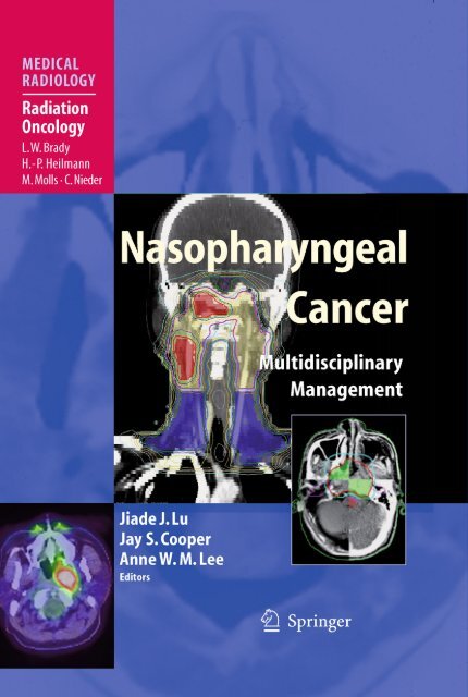
J. J. Lu · J. S. Cooper · A. W. M. Lee (Eds.)<strong>Nasopharyngeal</strong> CancerMultidisciplinary ManagementWith Contributions byR. R. Allison · I. Ayan · K. B. Tan · S. Cao · A. T. C. Chan · C.-J. Chen · Y.-C. ChienV. F. H. Chong · D. T. Chua · J. S. Cooper · B. S. Glisson · B. C. Goh · V. Grégoire · Y. GuoW.-L. Hsu · L. Kong · Q.-T. Le · A. W. M. Lee · N. Lee · J.-C. Lin · S. Lin · S. S. Lo · P.-J. LouJ. J. Lu · B. B.Y. Ma · J. Ma · C. K. Ong · B. O’Sullivan · R. Ove · E. Ozyar · T. C. Putti ·K. S. Loh · I. W. K. Tham · K.-B. Tam J. Wee · W. I. Wei · E. Yu · M.-S. Zeng · Y.-X. ZengForeword byL.W. Brady, H.-P. Heilmann, M. Molls, and C. NiederWith 84 Figures in 117 Separate Illustrations, 52 in Color and 51 Tables
Jiade J. Lu, MD, MBAAssociate Professor and ConsultantDepartment of Radiation OncologyNational University Cancer Institute of SingaporeNational University Health SystemNational University of Singapore5 Lower Kent Ridge RoadSingapore 119074Republic of SingaporeandDistinguished Clinical ProfessorFudan University Shanghai Cancer Center270 Dong An RoadShanghai 200232P.R. ChinaJay S. Cooper, MDProfessor and ChairmanDepartment of Radiation OncologyMaimonides Cancer Center6300 Eighth AvenueBrooklyn, NY 11220USAAnne W.M. Lee, MDDepartment of Clinical OncologyPamela Youde Nethersole Eastern Hospital3 Lok Man RoadChai WanHong Kong SARP.R. ChinaMedical Radiology · Diagnostic Imaging and Radiation OncologySeries Editors:A. L. Baert · L. W. Brady · H.-P. Heilmann · M. Knauth · M. Molls · C. Nieder · K. SartorContinuation of Handbuch der medizinischen RadiologieEncyclopedia of Medical RadiologyISBN: 978-3-540-92809-6 e-ISBN: 978-3-540-92810-2DOI: 10.1007/978-3-540-92810-2Springer Heidelberg Dordrecht London New YorkMedical Radiology · Diagnostic Imaging and Radiation Oncology ISSN 0942-5373Library of Congress Control Number: 2009931698© Springer-Verlag Berlin Heidelberg 2010This work is subject to copyright. All rights are reserved, whether the whole or part of the material is concerned,specifically the rights of translation, reprinting, reuse of illustrations, recitation, broadcasting, reproduction onmicrofilms or in any other way, and storage in data banks. Duplication of this publication or parts thereof ispermitted only under the provisions of the German Copyright Law of September 9, 1965, in its current version,and permission for use must always be obtained from Springer-Verlag. Violations are liable for prosecutionunder the German Copyright Law.The use of general descriptive names, registered names, trademarks, etc. in this publication does not imply,even in the absence of a specific statement, that such names are exempt from the relevant protective laws andregulations and therefore free for general use.Product liability: The publishers cannot guarantee the accuracy of any information about dosage and applicationcontained in this book. In every individual case the user must check such information by consulting therelevant literature.Cover design: Publishing Services Teichmann, 69256 Mauer, GermanyPrinted on acid-free paper9 8 7 6 5 4 3 2 1Springer is part of Springer Science+Business Media (www.springer.com)
Foreword<strong>Carcinoma</strong>s of the nasopharynx represent a relatively uncommon disease process inWestern countries, but are more frequently diagnosed as a head and neck malignancy inSoutheast Asia. Most of these tumors are epithelial in origin. The nonkeratinizing, poorlyor undifferentiated squamous cell carcinomas are the more commonly diagnosedpathologies in Asia, accounting for almost 95% of all cases. However, 75% of cases areWorld Health Organization Type 1 in North America, whereas those in Southeast Asiaare Types 2 and 3. Radiation therapy is the primary treatment for nasopharyngeal carcinomas,and since these tumors tend to present with regional metastasis, combinedchemotherapy and radiation therapy is commonly pursued. This is particularly appropriatein patients who have locally advanced disease.This book, edited by Lu, Cooper, and Lee, discusses the recommendations for diagnosisand staging procedures for nasopharyngeal cancer, staging systems and prognosticfactors, and management using radiation therapy for early-stage disease and combinedtreatment modalities with radiation and cytotoxic chemotherapy for more advanceddisease. The supporting scientific evidence clearly indicates that these are the appropriateapproaches for the treatment of carcinomas of the nasopharynx.The volume also deals in detail with techniques of radiation therapy, including intensity-modulatedradiation therapy, and outlines appropriate follow-up care and surveillancefor those individuals who survive.Even though nasopharyngeal cancer represents a relatively uncommon tumor in theWestern world, it is a common tumor in Southeast Asia and the book by Lu, Cooper, andLee constitutes a landmark volume identifying the appropriate approaches for the managementof this disease process.PA, USAHamburg, GermanyMünich, GermanyBodø, NorwayLuther W. BradyHans-Peter HeilmannMichael MollsCarsten Nieder
Preface<strong>Nasopharyngeal</strong> cancer is a unique type of head and neck malignancy. Essentially unresectablebecause of proximity to the skull base, nasopharyngeal cancer historically hadbeen treated by radiation therapy alone. Although cure rates for early-stage disease havebeen relatively good, the substantially worse outcome for locoregionally advanced diseaseand the not insubstantial risk of disseminated disease clearly indicated that a moreeffective therapeutic strategy was needed for more advanced tumors.The use of concurrent chemotherapy with radiation therapy, popularized by thelandmark Intergroup trial (INT0099), has significantly improved the outcome ofadvanced nasopharyngeal cancers. This trial can be therefore viewed not only as a proofof principal, but also as a starting point for the refinement of chemotherapy-enhancedradiation therapy in the management of this disease, a quest that continues to the presenttime.Similarly, technical advances in radiation therapy, particularly the development ofintensity modulated radiation therapy (IMRT) and image guided radiation therapy(IGRT), have also improved our abilities to place the radiation dose precisely in threedimensionalspace, ensuring adequate coverage of the gross tumor and clinical targetvolumes while simultaneously sparing normal tissues. As the anatomic location of thenasopharynx is in close proximity to critical organs at risk, appropriate beam shapingand placement previously had been (at times insurmountable) challenges for the radiationoncologist.However, as occurs in any rapidly evolving field, numerous unanswered questionsand controversies remain. The optimal schedule, timing, and specific chemotherapyregimen (both concurrent and adjuvant) are still unknown. The delineation of idealtarget volumes for IMRT is both an opportunity and a challenge for radiation oncologistswho are specialized in the management of this malignancy. Similarly, recent developmentsin molecular biotechnology herald the prospect of better diagnosis and/orindividualized treatment of the disease. Yet, the practicing physician cannot wait forthese answers and must make crucial decisions on his/her patients’ behalf today, basedon the information available. Clearly, with all these opportunities and challenges, soundunderstanding of the updated current knowledge of nasopharyngeal cancer isessential.Hence, we initiated this international collaborative effort to provide a comprehensivereview of all key knowledge practicing physicians currently need to know about themanagement of nasopharyngeal cancer, arranged in four sections. The first part of the
VIIIPrefacebook (Chaps. 1–9) discusses the biologic concepts: epidemiology/etiology, pathogenesis, clinically pertinentmolecular biology, clinical presentation, diagnosis, and staging. The second part (Chaps. 10–17)details the current concepts of definitive (often multidisciplinary) therapy for nondisseminated nasopharyngealcarcinoma. Critical analyses of the clinical trials that form the basis of currently available evidence-basedmedicine, current state-of-the art treatment strategies, and novel approaches that promisefurther improvements in outcome are explained in the chapters of this section. In the third section(Chaps. 18–21), management of more desperate situations, failure after initial treatment, and palliationof distant metastasis are discussed. Patients’ long-term quality of life after treatment (Chap. 22), thefortunately rare occurrence of nasopharyngeal cancer in early life, and the staging of the disease (Chap.24) are reviewed as well. We consider that such an arrangement not only provides appropriate coverageof the core of knowledge and discussions that are crucial to clinical management of nasopharyngealcarcinoma, but also facilitates a structural and systemic way of studying and understanding thisknowledge.We greatly appreciate the expertise and authoritative contributions of all of the included authors,each reflecting their dedication to improve the outcome of care of future patients. Consequently, we haveintentionally allowed the authors to address some of the same key issues in different chapters to providedifferent perspectives of unresolved issues. In the end, the success of this publication must be measuredprimarily by how well we elicit ideas and provoke thoughts for future research in the clinical managementof nasopharyngeal carcinoma.SingaporeNY, USAHong Kong SAR, P.R. ChinaJiade J. LuJay S. CooperAnne W. M Lee
Contents1 The Epidemiology of <strong>Nasopharyngeal</strong> <strong>Carcinoma</strong> . . . . . . . . . . . . . . . . . . . . . . . . 1Jun Ma and Sumei Cao2 Pathogenesis and Etiology of <strong>Nasopharyngeal</strong> <strong>Carcinoma</strong> . . . . . . . . . . . . . . . . . 9Mu-Sheng Zeng and Yi-Xin Zeng3 Molecular Signaling Pathways in <strong>Nasopharyngeal</strong> Cancer . . . . . . . . . . . . . . . . . 27Quynh-Thu Le and Jiade J. Lu4 Natural History, Presenting Symptoms,and Diagnosis of <strong>Nasopharyngeal</strong> <strong>Carcinoma</strong> . . . . . . . . . . . . . . . . . . . . . . . . . . . . 41Simon S. Lo and Jiade J. Lu5 Screening and Early Diagnosis of <strong>Nasopharyngeal</strong> <strong>Carcinoma</strong> . . . . . . . . . . . . . 53Pei-Jen Lou, Wan-Lun Hsu, Yin-Chu Chien, and Chien-Jen Chen6 <strong>Familial</strong> <strong>Nasopharyngeal</strong> <strong>Carcinoma</strong> . . . . . . . . . . . . . . . . . . . . . . . . . . . . . . . . . . . 65Kwok Seng Loh7 Pathology of <strong>Nasopharyngeal</strong> <strong>Carcinoma</strong> . . . . . . . . . . . . . . . . . . . . . . . . . . . . . . . 71Thomas Choudary Putti and Kong-Bing Tan8 Imaging in the Diagnosis and Staging of <strong>Carcinoma</strong> of Nasopharynx . . . . . . . 81Cheng Kang Ong and Vincent Fook Hin Chong9 Prognostic Factors in <strong>Nasopharyngeal</strong> Cancer . . . . . . . . . . . . . . . . . . . . . . . . . . 9 95Jin-Ching Lin10 Early Stage <strong>Nasopharyngeal</strong> Cancer:A Highly Curative Disease with Radiation Therapy . . . . . . . . . . . . . . . . . . . . . . . 137Roger Ove, Ron R. Allison, and Jiade J. Lu11 Drug Therapy for <strong>Nasopharyngeal</strong> <strong>Carcinoma</strong>:Cytotoxic and Targeted Therapy . . . . . . . . . . . . . . . . . . . . . . . . . . . . . . . . . . . . . . . 149Brigette B. Y. Ma and Anthony T. C. Chan12 The Intergroup 0099 Trial for <strong>Nasopharyngeal</strong>Cancer: History, Perceptions, and Transitions . . . . . . . . . . . . . . . . . . . . . . . . . . . . 161Jay S. Cooper
XContents13 Concurrent Chemotherapy-Enhanced Radiation:Trials and Conclusions . . . . . . . . . . . . . . . . . . . . . . . . . . . . . . . . . . . . . . . . . . . . . . . . 167Joseph Wee14 Neoadjuvant Chemotherapy:Trials and Conclusions . . . . . . . . . . . . . . . . . . . . . . . . . . . . . . . . . . . . . . . . . . . . . . . . 183Anne W. M. Lee15 Adjuvant Chemotherapy for <strong>Nasopharyngeal</strong> <strong>Carcinoma</strong> . . . . . . . . . . . . . . . . . . 193Boon Cher Goh16 Advances in the Technology of RadiationTherapy for <strong>Nasopharyngeal</strong> <strong>Carcinoma</strong> . . . . . . . . . . . . . . . . . . . . . . . . . . . . . . . . 197Lin Kong, Jiade J. Lu, and Nancy Lee17 Selection and Delineation of Target Volumes in Intensity-ModulatedRadiation Therapy for <strong>Nasopharyngeal</strong> Cancer . . . . . . . . . . . . . . . . . . . . . . . . . . . 213Jiade J. Lu, Vincent Grégoire, and Shaojun Lin18 Post-treatment Follow-Up of Patients with <strong>Nasopharyngeal</strong> Cancer . . . . . . . . . 233Ivan W.K. Tham and Jiade J. Lu19 Management of Patients with Failure FollowingDefinitive Radiation Therapy: Reirradiation in Patientswith Locally Recurrent <strong>Nasopharyngeal</strong> <strong>Carcinoma</strong> . . . . . . . . . . . . . . . . . . . . . . 241Daniel T. Chua20 Surgery for Recurrent <strong>Nasopharyngeal</strong> <strong>Carcinoma</strong> . . . . . . . . . . . . . . . . . . . . . 253William Ignace Wei21 Systemic Treatment for Incurable Recurrent|and/or Metastatic <strong>Nasopharyngeal</strong> <strong>Carcinoma</strong> . . . . . . . . . . . . . . . . . . . . . . . . . . 267Ye Guo and Bonnie S. Glisson22 Long-Term Complications in the Treatment of <strong>Nasopharyngeal</strong> <strong>Carcinoma</strong> . . 275Simon S. Lo, Jiade J. Lu, and Lin Kong23 <strong>Nasopharyngeal</strong> Cancer in Pediatric and Adolescent Patients . . . . . . . . . . . . . . 295Enis Ozyar and Inci Ayan24 Staging of <strong>Nasopharyngeal</strong> <strong>Carcinoma</strong> . . . . . . . . . . . . . . . . . . . . . . . . . . . . . . . . . . 309Brian O’Sullivan and Eugene YuSubject Index . . . . . . . . . . . . . . . . . . . . . . . . . . . . . . . . . . . . . . . . . . . . . . . . . . . . . . . . . . . . 323List of Contributors . . . . . . . . . . . . . . . . . . . . . . . . . . . . . . . . . . . . . . . . . . . . . . . . . . . . . . . 331
The Epidemiology of <strong>Nasopharyngeal</strong> <strong>Carcinoma</strong> 1Jun Ma and Sumei CaoCONTENTS1.1 Introduction 11.2 Regional and Spatial Distribution 11.3 Gender and Age Distribution 21.4 Racial Distribution 21.5 <strong>Familial</strong> Aggregation 31.6 Time Tendency 31.7 Risk Factors 41.7.1 Epstein–Barr Virus 41.7.2 Salty Fish and Pickled Food 41.7.3 Smoking and Drinking 41.7.4 Effect of Hereditary Susceptibility 51.7.5 Traditional Chinese Medicine 51.7.6 Professional Exposure 51.7.7 Chronic Upper Respiratory Disease 61.7.8 Trace Element 61.8 Summary 6References 61.1Introduction<strong>Nasopharyngeal</strong> carcinoma (NPC) is a malignanttumor of nasopharyngeal epithelium. It is the maintype in nasopharyngeal malignant tumors in bothendemic areas and regions with low incidence. Epidemiologicalstudies in NPC with the focus on etiologyand biological behavior of the disease werestrongly encouraged as a result of the InternationalUnion against Cancer (UICC) Symposium on Cancerof Nasopharynx held in Singapore in 1964 (Muiret al. 1967), and investigations in the past four decadeshave produced many important findings in thoseaspects. NPC has unique epidemiological features,including obvious regional, racial, and familial aggregation.The aim of this chapter is to detail the incidenceand distribution of NPC, as well as risk factorsof the development of the disease.1.2Regional and Spatial DistributionJun Ma, MDSumei Cao, MDDepartment of Radiation Oncology, Cancer Center of SunYat-sen University, 651 Dongfeng Road, East, Guangzhou,Guangdong 510060, P.R. China<strong>Nasopharyngeal</strong> cancer is a type of tumor withextremely unbalanced endemic distribution. It can beseen in many countries and areas of the five continents.However, the incidence of NPC is lower than1/10 5 in most areas. High-incidence areas are centralizedin the southern part of China (includingHongkong). The highest incidence is found inGuangdong province, and the incidence in male canreach 20–50/100000. According to the data of InternationalAgency for Research on Cancer (IARC),approximately 80,000 cases of NPC were newly diagnosedworldwide in 2002, and about 50,000 casesdeceased, with Chinese accounting for 40%. Intermediaterates were seen in local inhabitants of
2 J. Ma and S. CaoTable 1.1. Incidence of nasopharyngeal carcinoma (NPC) insome cancer registries of five continents in 1998–2002 (N, 1/10 5 )Region and populationAge-standardincidencerateMaleFemaleChinaChina, Zhongshan 26.9 10.1China, Guangzhou 22.2 9.8China, Hong Kong 17.8 6.7China, Shanghai 4.1 1.5China, Nangang District,1.1 0.5Harbin CitySoutheast AsiaMalaysia, Sarawak 15.0 6.5Malaysia, Penang 9.3 3.3Singapore 11.0 3.6Singapore: Chinese 12.8 4.1Singapore: Indian 1.8 0.1Singapore: Malay 5.5 2.0Philippines, Manila 5.8 2.4Thailand, Chiang Mai 3.9 1.5Thailand, Songkhla 2.7 0.9Thailand, Lampang 2.5 1.5USAUSA, Hawaii: Chinese 9.9 1.1USA, Hawaii: Filipino 3.3 1.3USA, Hawaii: Hawaiian 2.0 0.2USA, San Francisco: Chinese 8.1 4.0USA, San Francisco: Filipino 3.1 1.0USA, Los Angeles: Chinese 6.0 1.9USA, Los Angeles: Filipino 3.8 0.8Middle East/North AfricaAlgeria, Setif 5.4 1.7Tunisia, Sousse 4.6 1.9Uganda, Kyadondo 2.3 1.3Kuwait: Kuwaitis 1.7 0.8EuropeAustria 0.4 0.2Finland 0.3 0.1ArcticCanada, Northwest Territories 4.3 1.3USA, Alaska 1.5 0.9Southeast Asia, Eskimos from the Arctic area, andinhabitants from North Africa and the Middle East(Table 1.1) (Parkin et al. 1992, 2002; Waterhouseet al. 1982; Muir et al. 1987).There is prominent difference in the incidence ofNPC between the Northern and Southern parts ofChina. High-incidence areas are centralized in fiveSouthern provinces (Guangdong, Guangxi, Hunan,Fujian, Jiangxi). The incidence is highest in Guangdongprovince, so NPC is also called “Canton tumor.” WithinGuangdong province, the Pearl River delta and XijiangRiver basin, especially Zhaoqing, Foshan, andGuangzhou, form a high-incidence core region.1.3Gender and Age DistributionThe incidence of NPC is higher in males than that infemales, and the ratio is 2–3:1 (Parkin et al. 1992,2002; Waterhouse et al. 1982; Muir et al. 1987).The predominance in male gender is observed inboth endemic and low-incidence areas. However,the distribution of the age of patients diagnosedwith NPC differs substantially in areas with variousincidences. In low-incidence areas, the incidence ofNPC is increased with age, while in the endemicareas, the incidence is increased obviously after30-years, peaked at 40–59-years, and decreasedthereafter (Zong et al. 1983). It has also beenreported that in low- to medium- incidence area, theincidence of NPC has a relatively small peak amongadolescents and young adults (Burt et al. 1992).1.4Racial DistributionThe incidence of NPC is highest in Xanthoderm, thenin Melanoderm, and lowest in Caucasian. Highincidenceareas are mostly habitations of Xanthoderm,such as Southern China, Hong Kong, and SoutheastAsia. Eskimos in the Arctic area also belong toXanthoderm.In the same area, the incidence of NPC is differentbetween different races. For example, the incidenceis twofold higher in people speaking Cantonese thanin people speaking other dialects, such as Hakka,Hokkien, and Chiu Chau (Li et al. 1985). Even afterimmigrating to other countries in Southeast Asia,
The Epidemiology of <strong>Nasopharyngeal</strong> <strong>Carcinoma</strong> 3the incidence in Cantonese speaking people is stilltwofolds higher than in other people from SouthernChina (Lee et al. 1988). In the United States, the incidenceis highest in Chinese people living abroad,then in Filipinos, Japanese, black people, Spaniards,and lowest in white people (Burt et al. 1992).Migrant epidemiological data showed that peoplefrom high-incidence areas of Southern China stillkept high incidence even after they immigrate toAmerica, Australia, Malaysia, or Japan (Parkin et al.2002; Armstrong et al. 1979; Mccredie et al. 1999).Similarly, the incidence of NPC in immigrants andtheir offsprings from North Africa, where the incidencewas relatively high, was still higher than localinhabitants after they immigrated to Israel, a lowincidencearea (Parkin and Iscovich 1997).However, the incidence in second and third generationof immigrants was decreased to only half of thatbefore immigration (Warnakulasuriya et al. 1999).On the contrary, the incidence of NPC in Caucasianwho were born in China or Philippines is obviouslyincreased, when compared with those born in NorthAmerica, (Buell 1973) and the incidence in Frenchwho were born in North Africa was also obviouslyhigher than that in inhabitants from Southern France(Jeannel et al. 1993).The results of migrant epidemiology suggest thatboth genetic factors and environmental factors mayplay an important role in the pathogenesis of NPC. Inaddition, the distribution of pathological type is alsodifferent in different races. Ninety percent of the NPCin Southern China, Hong Kong, Taiwan, and Singaporeare undifferentiated or differentiated nonkeratinizing<strong>Carcinoma</strong> (Zong et al. 1983). While in nonendemicareas, keratinizing squamous cell carcinoma is predominant,it is concluded that the etiological factorsmay be different in high- and low-incidenceareas (Vaughan et al. 1996).5.9%, respectively. In Greenland, 27% of the patientswith NPC have cancer family history, and most areNPC (Albeck et al. 1993). In the city of Shanghai ineastern China, a medium incidence area, the ratio ofNPC family history is 1.85% (Yuan et al. 2000). Incancer family, most patients with NPC are first-degreerelatives of the probands. The incidence in firstdegreerelatives of the patients with NPC is 4–10-foldsof that in control population. The reason for familialaggregation of NPC may be similar hereditary susceptibilityor living environment of the family members.Complex segregation analysis on NPC family inSouthern China shows that NPC belongs to multigenichereditary tumor (Jia et al. 2005).1.6Time TendencyRecently published data demonstrated that the incidenceof newly diagnosed NPC has been decreased incertain high-incidence areas. For example, the incidenceand mortality of NPC has clearly decreased inHong Kong from 1970s, in Taiwan from 1980s, andin Singapore from 1990s. The decreased incidence inChinese people living in North America was also obvious.However, obvious ascending tendency has beenobserved in a few areas or populations such as Malaypeople living in Singapore (Wang et al. 2004). Theincidence of NPC was stable or slightly increased inTable 1.2. Comparison between the average annual agestandardized(world population) incidence rates of nasopharyngealcancer (per 100,000 person-years) in Hong Kong andSihui City, Guangdong, ChinaPeriodAverage annual incidence1.5<strong>Familial</strong> AggregationNPC is a disease with obvious familial aggregation.There have been reports of high-incidence familiesin high-, medium-, and low-incidence areas.Furthermore, the ratio of cancer family history inhigh-incidence areas is higher than that in lowincidenceareas. For example, the ratio of NPC familyhistory reported in Hong Kong (Yu et al. 1986) andGuangzhou of China (Yu et al. 1990) is 7.2% andHong Kong ChineseSihui City ofGuangdong, ChinaMale Female Male Female1973–1977 32.9 14.41978–1982 30.0 12.9 28.1 12.31983–1987 28.5 11.2 28.7 14.81988–1992 24.3 9.5 28.7 13.41993–1997 21.5 8.3 28.0 11.81998–2002 17.8 6.7 30.9 13.0
4 J. Ma and S. Caoendemic areas of Southern China including Guangdongand Guangxi provinces (Jia et al. 2006) (Table 1.2).The change in epidemiologic tendency of NPC inthese areas may be related with the change in exposureof corresponding population to risk factors. It is generallyconsidered that changes in smoking, consumptionof pickled food, and immigration may have great influenceson the incidence in Hong Kong, Singapore, andTaiwan. Epidemiologic studies found that smoking wasthe main reason for keratinizing squamous cell carcinoma,while it had little relationship with nonkeratinizingsquamous cell carcinoma. Further investigationsfound that the decrease of incidence in Hong Kong andNorth America was due to the decrease of keratinizingsquamous cell carcinoma, while the incidence ofnonkeratinizing squamous cell carcinoma kept stable(Jia et al. 2006; Sun et al. 2005; Tse et al. 2006). Thedecrease in smoking frequency in these areas mayaccount for the decrease in incidence of NPC.In the past 30 years, rapid economy developmenthas been observed in high-incidence areas of NPC inSouthern China, such as Guangdong and Guangxi.The eating and living habit has greatly changed.However, the stable incidence rate of NPC in theseendemic areas indicate that the risk factors seemunchanged, as more than 90% of NPC cases belong tononkeratinizing carcinoma.1.7Risk Factors1.7.1Epstein–Barr VirusAntibodies to Epstein–Barr virus (EBV) were detectedin the serum of patients with NPC by Old et al. in 1966(Old et al. 1966). Subsequent studies showed that thelevel of anti-EBV antibodies was significantly increasedin NPC from different races and areas, compared withthe control. Viral DNA can be detected in the nucleusof epitheliums using in situ hybridization technique,while it was not obvious in infiltrating lymphocytes.In prospective population studies, it was foundthat increased IgA antibody to Epstein–Barr (EB)viral capsid antigen (VCA) and neutral antibody toEBV DNAse were specific markers of NPC in highincidencearea, since they can effectively predict thedevelopment of NPC. For example, Chien et al.(2001) found that risk factor of people in Taiwan whowere positive for both of the above. For example, inpopulation studies, Chien et al. found that elevatedIgA antibody against VCA and EBV DNAse are highlyspecific markers for NPC cases in Taiwan area, predictinga 32.8-fold (95% CI: 7.3-147.2) increase forthose with both markers positive. Recently, Ji et al.conformed that there was a window phase of about 3years from seropositivity of EBV to the developmentof NPC (Ji et al. 2007).Although globally most people were infected byEBV, only a small portion of them developed NPC,which meant that the genesis of NPC was multifactorial.Currently, it was confirmed that many factors canresult in the activation of EBV, such as environmentalcarcinogens and/or immune deficiency (Friborget al. 2007; Stowe et al. 2001). It is still unclear aboutthe mechanism for EBV entrance into epithelial tissue.The pathogenesis in NPC is detailed in Chap. 2.1.7.2Salty Fish and Pickled FoodOne of the most potent and confirmed risk factors forNPC is salty fish consumption. Fish and other foodthat conserved in salt are heavily consumed inSouthern China and areas with moderate risk factorsfor NPC such as Southeast Asia and North Africa. Thesefoods contain a known carcinogen N-nitrosamineand its precursor. In different populations, the relativerisk for developing NPC in people who eat saltyfish on daily basis after adulthood was estimated tobe 1.8–7.5, compared with those who did not or onlyeat a little salty fish (Yu et al. 1989; Lee et al. 1994). Inaddition, the relative risk for developing NPC in peoplewho eat salty fish on daily or weekly basis duringweaning period or infancy was estimated to be 1.1–37.7, compared with those who never eat or only eat alittle salty fish (Yu 1991). On the contrary, eating morefresh fruits and vegetables can reduce the risk of NPCby 30%–50%, which may be due to the effect of antioxidantand antinitrosamine components in VitaminC and E. However, evidences on the relationship ofsalty fish and pickled food with NPC from perspectivestudies are still lacking.1.7.3Smoking and DrinkingResults from a number of prospective epidemiologicalstudies revealed that chronic smoking was a risk factorfor NPC. In addition, the development of NPC
The Epidemiology of <strong>Nasopharyngeal</strong> <strong>Carcinoma</strong> 5depended on the severity of smoking measured bypack-year. In general, risk of NPC in smokers was 2–6-fold of that in nonsmokers (Friborg et al. 2007; Hsuet al. 2009). The risk of NPC potentially induced bycigarette smoking was lower than the risk for lung canceror squamous cell carcinoma of the larynx. However,smoking was the main risk factor for squamous cellcarcinoma NPC, while its association with undifferentiatedor nonkeratinizing NPC has not been demonstrated(Vaughan et al. 1996). This finding indicatedthat the decrease in morbidity of NPC in NorthAmerica and Hongkong might result from reducedsmoking frequency. Smoking-induced NPC was lesssignificant than lung cancer or laryngeal cancer, whichmay be due to the lower sensitivity of nasopharyngealepitheliums to carcinogens in tobaccos than epitheliumsin other regions, or the lower content of tobaccosin pharynx nasalis than the respiratory tract.Most studies performed in China and the UnitedStates showed that alcohol drinking was unrelated tothe development of NPC. Results from a perspectivestudy performed in Singapore also confirmed thisviewpoint (Friborg et al. 2007). However, the resultswere not completely consistent, since positive resultswere found at least in two case control studies(Vaughan et al. 1996; Nam et al. 1992). The inconsistencymay be caused by experimental design, susceptibilityof different people, and other confoundingfactors.1.7.4Effect of Hereditary SusceptibilityThe epidemic features of NPC suggested that geneticfactors contribute a lot to the genesis and developmentof NPC. Many investigations have focused onthe possible pathogenic effect of human leucocyteantigen (HLA), which is involved in the presentationof foreign antigens, including viral polypeptide, so asto facilitate their directive lysis by immune system.Since EBV can be found in almost all patients withthe disease, the risk factor for NPC may be increasedin individuals who inherited HLA allele with weakerpresenting ability of EBV antigen. On the contrary,the risk factor for NPC was relatively low in individualswho inherited HLA allele with effective presentingability of EBV antigen (Hildesheim et al. 2002).It was reported currently that HLA-A2-Bw46 and B17can increase the risk factor for NPC by 2–3-fold.HLA-A11, B13, and A2 can reduce the risk factor forNPC by 1/3–1/2.Epidemiologic studies also determined the correlationbetween polymorphism of some genes withthe risk of NPC, including homozygous variantderived from cytochrome P4502E1 (CYP2E1), nullallele of glutathion S-transferase M1 (GSTM1),(Kongruttanachok et al. 2001; Hildesheim et al.1995; Nazar-Stewart et al. 1999) T cell receptorpolymorphism (TCR), poly immunoglobulin receptor(PIGR), candidate tumor suppressing gene GX6,DNA repair gene hOGG1, and XRCC1. The relativerisk was estimated to be 2.0–5.0. CYP2E1 and GSTM1are involved in the metabolism of nitrosamine andcigarette smoke, respectively. Their etiological effectsmay be different because of the difference in environment;in other words, they may have differentbiological interaction with environmental factors,such as salty fish and smoking.Studies on chromosomal abnormality, heterozygotedeficiency, and gene expression, as well as mainNPC susceptible sites on No.4 chromosome found bywhole genome scanning in familial study performedin Southern China also provided potential opportunitiesto determine NPC susceptible genes (Fenget al. 2002).1.7.5Traditional Chinese MedicineIn certain epidemiological studies performed inSoutheast Asia and Southern China, use of traditionalChinese medicine has been associated with anincrease in NPC by 2–4-fold (Zheng et al. 1994;Hildesheim et al. 1992). Certain plants and medicinalmaterials in Chinese traditional medicine caninduce the activation of latent EBV. Such features canbe attributed to tetradecanoylphorbol acetate (TPA)–like substances in plants and earth. TPA-like substances,in combination with N-butyrate, a productof anoxybiontic bacteria found in pharynx nasalis,can induce the synthesis of EBV antigen in mice,increase EBV-mediated B cell transformation, andpromote the genesis of NPC (Tang et al. 1988).1.7.6Professional ExposureFormaldehyde is a well-known carcinogen that caninduce carcinoma of nasal cavity in rodents. Meta analysison more than 30 epidemiological studies showedthat exposure to formaldehyde was significantly
6 J. Ma and S. Caoassociated with the genesis of NPC, and a dose–response relationship was demonstrated (Partanen1993). In 1995, formaldehyde was suggested to be anetiological factor for NPC by IARC.Middle-sized (5–10 mm) dust particles are easilyabsorbed to pharynx nasalis. Several epidemiologicalstudies have found that the risk factor for NPC wasincreased in people exposed in wood dust, and it wasdependent on exposure time and dose (Luce et al. 2002).In addition, the risk factor for NPC was reported to beincreased in people exposed to extreme temperatureand work environment with combustibles. However,the exposure ratio for these professional exposures wasrelatively low in most endemic areas, so it cannot beconfirmed whether these factors are independentlyimportant in the high incidence in these areas.1.7.7Chronic Upper Respiratory DiseaseMost epidemiological studies showed that the riskfor NPC was increased by about twofold in peoplewith chronic ear, nose, throat, and upper respiratorydisease (Zheng et al. 1994). It may be due to the conversionof nitrate to nitrite by bacteria present inthese regions, since nitrite is the component of carcinogenN-nitroso.1.7.8Trace ElementNickel is one of the carcinogens to human. Surveysperformed in high-incidence areas found that thecontent of nickel in rice, drinking water, and hair oflocal inhabitants was significantly higher than that inlow-incidence areas. In high-incidence areas, nickelcontent in NPC patients was also higher than inhealthy population. Epidemiological surveys alsofound that trace elements zinc and cadmium werepositively related with the genesis of NPC, whilemagnesium, calcium, and strontium were negativelyrelated (Bolviken et al. 1997).1.8SummaryNPC is a disease with unique epidemiological features.The distribution of the disease demonstrates aclear regional, racial, and gender prevalence. Theincidence of the disease is relatively high amonglocal inhabitants of Southern China, Southeast Asia,Eskimos from the Arctic area, and inhabitants fromNorth Africa and the Middle East, with the highestincidence found in Guangdong province of China,and the incidence in male reaching 20–50/10 5 . NPC isassociated with a number of risk factors. It appearsthat individuals with hereditary susceptibility wereinfected by EBV in the early period of life, then EBVwas activated under the synthetic action of multipleenvironmental factors, and eventually NPC wasdeveloped. In addition to EBV, other environmentalfactors such as trace elements and dietary habits maybe associated with the initiation and development ofthe disease. However, changes in lifestyle in the pastseveral decades in Southern China had little associationwith the etiology of NPC. Further epidemiologicalinvestigations will be needed to detect the changesin the trend of NPC and the underlying risk factors,so that prevention and/or early detection of the diseasecan be realized.ReferencesAlbeck H, et al (1993) <strong>Familial</strong> clusters of nasopharyngeal carcinomaand salivary gland carcinomas in Greenlandnatives. Cancer 72(1):196–200Armstrong RW, et al (1979) Incidence of nasopharyngeal carcinomain Malaysia, 1968–1977. Br J Cancer 40(4):557–567Bolviken B, Flaten TP, Zheng C (1997) Relations betweennasopharyngeal carcinoma and magnesium and otheralkaline earth elements in soils in China. Med Hypotheses48(1):21–25Buell P (1973) Race and place in the etiology of nasopharyngealcancer: a study based on California death certificates.Int J Cancer 11(2):268–272Burt RD, Vaughan TL, McKnight B (1992) Descriptive epidemiologyand survival analysis of nasopharyngeal carcinomain the United States. Int J Cancer 52(4):549–556Chien YC, et al (2001) Serologic markers of Epstein-Barr virusinfection and nasopharyngeal carcinoma in Taiwanesemen. N Engl J Med 345(26):1877–1882Feng BJ, et al (2002) Genome-wide scan for familial nasopharyngealcarcinoma reveals evidence of linkage to chromosome4. Nat Genet 31(4):395–399Friborg JT, et al (2007) A prospective study of tobacco andalcohol use as risk factors for pharyngeal carcinomas inSingapore Chinese. Cancer 109(6):1183–1191Hildesheim A, et al (1992) Herbal medicine use, Epstein-Barrvirus, and risk of nasopharyngeal carcinoma. Cancer Res52(11):3048–3051Hildesheim A, et al (1995) Cytochrome P4502E1 genetic polymorphismsand risk of nasopharyngeal carcinoma: results
The Epidemiology of <strong>Nasopharyngeal</strong> <strong>Carcinoma</strong> 7from a case-control study conducted in Taiwan. CancerEpidemiol Biomarkers Prev 4(6):607–610Hildesheim A, et al (2002) Association of HLA class I and IIalleles and extended haplotypes with nasopharyngeal carcinomain Taiwan. J Natl Cancer Inst 94(23):1780–1789Hsu WL, et al (2009) Independent effect of EBV and cigarettesmoking on nasopharyngeal carcinoma: a 20-year followupstudy on 9,622 males without family history in Taiwan.Cancer Epidemiol Biomarkers Prev 18(4):1218–1226Jeannel D, et al (1993) Increased risk of nasopharyngeal carcinomaamong males of French origin born in Maghreb(north Africa). Int J Cancer 54(4):536–539Jia WH, et al (2005) Complex segregation analysis of nasopharyngealcarcinoma in Guangdong, China: evidence for amultifactorial mode of inheritance (complex segregationanalysis of NPC in China). Eur J Hum Genet 13(2):248–252Jia WH, et al (2006) Trends in incidence and mortality ofnasopharyngeal carcinoma over a 20–25 year period(1978/1983–2002) in Sihui and Cangwu counties in southernChina. BMC Cancer 6:178Ji MF, et al (2007) Sustained elevation of Epstein-Barr virusantibody levels preceding clinical onset of nasopharyngealcarcinoma. Br J Cancer 96(4):623–630Kongruttanachok N, et al (2001) Cytochrome P450 2E1 polymorphismand nasopharyngeal carcinoma development inThailand: a correlative study. BMC Cancer 1:4Lee HP, et al (1988) Recent trends in cancer incidence amongSingapore Chinese. Int J Cancer 42(2):159–166Lee HP, et al (1994) Preserved foods and nasopharyngeal carcinoma:a case-control study among Singapore Chinese.Int J Cancer 59(5):585–590Li CC, Yu MC, Henderson BE (1985) Some epidemiologic observationsof nasopharyngeal carcinoma in Guangdong, People’sRepublic of China. Natl Cancer Inst Monogr 69:49–52Luce D, et al (2002) Sinonasal cancer and occupational exposures:a pooled analysis of 12 case-control studies. CancerCauses Control 13(2):147–157McCredie M, Williams S, Coates M (1999) Cancer mortality inEast and Southeast Asian migrants to New South Wales,Australia, 1975–1995. Br J Cancer 79(7–8):1277–1282Muir C, Waterhouse J, Mack T, et al (eds) (1987) Cancer incidencein five continents, vol. V. IARC scientific publications,Lyon, IARCMuir CS, Shanmugaratnam K (eds) (1967) Cancer of thenasopharynx. UICC Monograph Series, No.1. Munksgaard,CopenhagenNam JM, McLaughlin JK, Blot WJ (1992) Cigarette smoking,alcohol, and nasopharyngeal carcinoma: a case-controlstudy among U.S. whites. J Natl Cancer Inst 84(8):619–622Nazar-Stewart V, et al (1999) Glutathione S-transferase M1and susceptibility to nasopharyngeal carcinoma. CancerEpidemiol Biomarkers Prev 8(6):547–551Old LJ, et al (1966) Precipitating antibody in human serum toan antigen present in cultured Burkitt’s lymphoma cells.Proc Natl Acad Sci U S A 56(6):1699–1704Parkin DM, Muir CS, Whelan SL, et al (eds) (1992) Cancer incidencein five continents Vol VI. IARC Scientific Publication,Lyon, IARCParkin DM, Whelan SL, et al (eds) (2002) Cancer incidence infive continents, vol. VIII. IARC scientific publications, Lyon,IARCParkin DM, Iscovich J (1997) Risk of cancer in migrants andtheir descendants in Israel: II. <strong>Carcinoma</strong>s and germ-celltumours. Int J Cancer 70(6):654–660Partanen T (1993) Formaldehyde exposure and respiratorycancer–a meta-analysis of the epidemiologic evidence.Scand J Work Environ Health 19(1):8–15Stowe RP, Pierson DL, Barrett AD (2001) Elevated stress hormonelevels relate to Epstein-Barr virus reactivation inastronauts. Psychosom Med 63(6):891–895Sun LM, et al (2005) Trends in the incidence rates of nasopharyngealcarcinoma among Chinese Americans living in LosAngeles County and the San Francisco metropolitan area,1992–2002. Am J Epidemiol 162(12):1174–1178Tang WP, et al (1988) Wikstroemia indica promotes developmentof nasopharyngeal carcinoma in rats initiated bydinitrosopiperazine. J Cancer Res Clin Oncol 114(4):429–431Tse LA, et al (2006) Incidence rate trends of histological subtypesof nasopharyngeal carcinoma in Hong Kong. BrJ Cancer 95(9):1269–1273Vaughan TL, et al (1996) <strong>Nasopharyngeal</strong> cancer in a low-riskpopulation: defining risk factors by histological type.Cancer Epidemiol Biomarkers Prev 5(8):587–593Wang H, Seow A, Lee HP (2004) Trends in cancer incidenceamong Singapore Malays: a low-risk population. Ann AcadMed Singapore 33(1):57–62Warnakulasuriya KA, et al (1999) Cancer of mouth, pharynxand nasopharynx in Asian and Chinese immigrants residentin Thames regions. Oral Oncol 35(5):471–475Waterhouse J, Muir C, Mack T, et al (eds) (1982) Cancer incidencein five continents, vol. IV. IARC scientific publications,LyonYuan JM, et al (2000) Non-dietary risk factors for nasopharyngealcarcinoma in Shanghai, China. Int J Cancer 85(3):364–369Yu MC (1991) <strong>Nasopharyngeal</strong> carcinoma: epidemiology anddietary factors. IARC Sci Publ (105):39–47Yu MC, et al (1986) Cantonese-style salted fish as a cause ofnasopharyngeal carcinoma: report of a case-control studyin Hong Kong. Cancer Res 46(2):956–961Yu MC, et al (1990) Occupational and other non-dietary riskfactors for nasopharyngeal carcinoma in Guangzhou,China. Int J Cancer 45(6):1033–1039Yu MC, Huang TB, Henderson BE (1989) Diet and nasopharyngealcarcinoma: a case-control study in Guangzhou,China. Int J Cancer 43(6):1077–1082Zheng YM, et al (1994) Environmental and dietary risk factorsfor nasopharyngeal carcinoma: a case-control study inZangwu County, Guangxi, China. Br J Cancer 69(3):508–514Zong YS, et al (1983) Histopathologic types and incidence ofmalignant nasopharyngeal tumors in Zhongshan County.Chin Med J 96(7):511–516
Pathogenesis and Etiology of <strong>Nasopharyngeal</strong> 2<strong>Carcinoma</strong>Mu-Sheng Zeng and Yi-Xin ZengCONTENTS2.1 Introduction 92.2 Histological Subtypes of NPC 102.3 Etiologies and Pathogenesis 102.3.1 Genetic Factors 102.3.2 Environmental Factors 112.3.3 Epstein–Barr Virus 122.3.3.1 EBV Structure 122.3.3.2 EBV Infection in NPC 122.3.3.3 EBV Subtype in NPC 132.3.3.4 EBV Expression in NPC 142.3.3.4 The Role of EBV in the Pathogenesisof NPC 142.4 NPC Stem Cells 172.5 Molecular Alterations 182.6 Summary 19References 20Mu-Sheng Zeng, PhD, MDState Key Laboratory of Oncology in Southern China, CancerCenter of Sun Yat-sen University, 651 Dongfeng Road East,Guagnzhou 510060, P.R. ChinaYi-Xin Zeng, MDCancer Center of Sun Yat-sen University, 651 East DongfengRoad, Yuexiu District Guangzhou, Guangdong 510060,P.R. China2.1Introduction<strong>Nasopharyngeal</strong> carcinoma (NPC) is a squamous cellcarcinoma (SCC) that usually develops around theostium of the Eustachian tube in the lateral wall of thenasopharynx (Sham et al. 1990). This disease was initiallyreported in 1901 and characterized clinically in1922 (Wei et al. 2005). NPC is a disease with a remarkablegeographic and racial distribution worldwide.This is a rare human malignancy with an incidencebelow 1/100,000 populations per year in Caucasiansfrom North America and other Western countries. Incontrast, the highest incidence is noted in theSouthern Chinese population of Guangdong, Inuitsof Alaska, and native Greenlanders (Chou et al. 2008;Parkin et al. 1992); particularly, among the Cantonesewho inhabit the central region of Guangdong Provincein Southern China, the incidence is 15–25 cases per100,000. NPC is also called the “Canton tumor” inGuangdong Province (Hepeng 2008; Yu et al. 2002).Southern Chinese migrants, irrespective of theircountry of migration, also exhibit high rates of NPC(Yu et al. 2002), but the rate of NPC among ethnicChinese born in North America is considerably lowerthan those born in China (Buell 1974). An intermediateincidence has been reported in Alaskan Eskimosand in the Mediterranean basin (North Africa,Southern Italy, Greece, and Turkey), ranging from 15to 20 cases per 100,000 persons (Chan et al. 2002).Independence of race– ethnicity, the rates of NPC inmen are two to three folds higher than those in womenfor most populations (Yu et al. 2002). Overall, NPCcan occur in all age groups, but has a bimodal age distribution.The incidence peaks at 50–60 years of age,and a small peak is observed during late childhood(Jeyakumar et al. 2006).The distinct difference in the incidence amonggeographic and population area implies that both
10 M-S. Zeng and Y-X. Zengenvironmental factors and genetic susceptibility playroles in the development of NPC (Lo et al. 2004). Theearly age incidence peak noted among SouthernChinese may suggest that exposure to the putativecarcinogens occur very early in life (Yu et al. 2002).Epidemiological studies have linked childhood intakeof locally consumed preserved foods to NPC developmentin all four groups of populations exhibitingincreased risk of NPC – Chinese, natives of SoutheastAsia, natives of Arctic region, and Arabs of NorthAfrica (Yu et al. 2002). Moreover, environmental factorsmay also accelerate to the development of NPC.For example, exposure to smoke or chemical pollutants,including trace elements (e.g., nickle), have beenreported to be associated with the development ofNPC (Wu et al. 1986; Yu et al. 1981). Therefore, thedevelopment and progression of NPC disease ismultifactorial with geographic areas, genetics, diet,and environmental exposure.2.2Histological Subtypes of NPCThe World Health Organization (WHO) classifiesNPC into three histopathological types based on thedegree of differentiation. Type 1, SCC, is seen in5%–10% of cases of NPC and is characterized bywell-differentiated cells that produce keratin anddemonstrated the presence of intracellular bridgeswhen observed under the electron microscope. Type2, nonkeratinizing squamous carcinoma, varies incell differentiation (from mature to anaplastic cells)but does not produce keratin. Type 3 or undifferentiatedNPC constitutes the bulk of the tumors seen inpatients with NPC, is also nonkeratinizing, but is lessdifferentiated, with highly variable cell types (clearcell, spindle cell, anaplastic) (Shanmugaratnam1978).Types 2 and 3 NPC are Epstein–Barr virus (EBV)associated and have better prognoses than type 1;EBV infection is generally absent in type 1, especiallyin nonendemic areas (Marks et al. 1998). However,more recent data suggest that almost all NPC tumorsin the endemic areas, regardless of histologic subtype,have comorbid EBV infections, which is a strong evidencefor EBV as the etiology of NPC (Vasefet al. 1997). Undifferentiated NPC or type 3 was frequentlycharacterized as lymphoepithelioma owingto the heavy infiltration of the primary tumor withlymphocytes. In endemic areas such as SouthernChina, WHO Type 3 accounts for more than 97%,while keratinizing SCC is more common in theWestern countries (up to 75%) (Marks et al. 1998).There is no uniform morphological characteristic ofNPC-affected tissues; thus, diagnosis of undifferentiatedNPC is usually based on the location of the tumorin the nasopharynx and the presence of EBV transcriptsin the tumor cells (Gullo et al. 2008). ClonalEBV genome is present in the early preinvasive dysplasticlesion or carcinoma in situ, illuminating thatthe development of malignant invasive tumor dropbehind the infection of EBV (Pathmanathan et al.1995). This close association with EBV is what makesNPC unique from other head and neck cancers.2.3Etiologies and PathogenesisIn endemic regions, NPC presents as a complex diseasecaused by an interaction of the oncogenic gammaherpesvirusEBV chronic infection, environmental,and genetic factors, in a multistep carcinogenic process.The highest incidence of NPC in SouthernChinese strongly indicates that both genetic susceptibilityand environmental factors contribute to thetumorigenesis of NPC in its development and progression.In addition, a small population of cellssharing properties of normal stem cells (NSC) withintumor has been suggested to be involved in the etiologyof NPC. This chapter will focus mainly on threemajor etiological factors including genetic, environmental,and viral factors.2.3.1Genetic FactorsWhile nasopharyngeal carcinoma is a rare malignancyin most parts of the world, it is one of the mostcommon cancers in Southeast Asia including areassuch as Southern China, Hong Kong, Singapore,Malaysia, and Taiwan. The reported incidence inthese countries ranges from 10 to 53 cases per 100,000persons. The incidence is also high among Eskimosin Alaska and Greenland and in Tunisians, rangingfrom 15 to 20 cases per 100,000 persons (Chan et al.2002). <strong>Familial</strong> clustering of NPC has been widelyobserved in both the Chinese population (Jia et al.2004; Zeng et al. 2002), and non-Chinese patientcohort (Levine et al. 1992). The familial risk of NPC
Pathogenesis and Etiology of <strong>Nasopharyngeal</strong> <strong>Carcinoma</strong> 11is among the highest of any malignancy (Suarezet al. 2006). The described relative risk of NPC infirst-degree relatives is about 8.0 (Friborg et al.2005). The high risk of NPC to Cantonese populationand the people with familial NPC history suggest thatgenetically determined susceptibility may play animportant role in the etiology of NPC.An important characteristic of familial cancers isthe early age onset of NPC (Zeng et al. 2002). Severallinkage analyses studies suggested the association ofsusceptibility human leukocyte antigen (HLA) haplotypeswith NPC development. Most studies conductedamong the Chinese population demonstrated anincreased risk of NPC for individuals with HLA-A2. Arecent study detected a consistent association betweenNPC and the prevalent Chinese HLA-A2 subtype(HLA-A * 0207), but not the prevalent Caucasian subtype(HLA-A * 0201) (Hildesheim et al. 2002). TheHLA types of AW19, BW46, and B17 have also beenreported to be associated with an increased risk,whereas HLA-A11 is associated with a decreased risk(Liebowitz 1994). Significant complex multiple chromosomeaberrations are often demonstrated in NPC,as well as in other solid tumors. The finding of translocation,amplification, and deletion of 3p, 5p, and 3qindicates that a minimal region of breakpoints is possiblefor contributing to NPC (Shih-Hsin Wu 2006;Tjia et al. 2005). Breakpoints have been frequentlyobserved in 1p11–31, 3p12–21, 3q25, 5q31, 11q13,12q13, and Xq25 (Lo et al. 1997). Inactivation of tumorsuppressor genes on 3p, 9p, 11q, 13q, 14q, and 16q andalteration of oncogenes on chromosomes 8 and 12 areimportant in the development of NPC (Hui et al. 1999;Lo et al. 2000). A recent study provides evidence forthe linkage of NPC to chromosome 3p and a fine mapof NPC susceptibility locus to a 13.6 cM region on3p21.31–21.2 (Xiong et al. 2004). These results are inagreement with several previous studies that suggestthat the deletion of chromosomes 3p is a commongenetic event in NPC (Deng et al. 1998; Lo et al. 2000).Many tumor suppressor candidate genes such asCACNA2D2, DLC1, FUS1, H37, HYAL1, RASSF1A,SEMA3B, and SEMA3F and tumor susceptibility genessuch as hMLH1 have been isolated from the region(Xiong et al. 2004). These studies indicate that genesin the 3p21 may play a critical role in tumorigenesis offamilial NPC.Some studies suggested that genetic polymorphismsin genes that metabolize carcinogens areassociated with NPC susceptibility. CytochromeP450 2E1 (CYP2E1) is one of the cytochrome P450sand is responsible for the metabolic activation ofnitrosamines and the related carcinogens. Casecontrolstudies have shown a strong association of thevariant form of CYP2E1 (c2 allele) with increased riskof this disease in Chinese populations (Hildesheimet al. 1997; Hildesheim et al. 1995). Other nitrosaminemetabolizing genes, such as Cytochrome P450 2A6(CYP2A6), have also been suggested to play a role inNPC susceptibility (Tiwawech et al. 2006). Phase IIdetoxification enzyme, glutathione S-transferase M1(GSTM1), was found to be a synergistic risk factor forNPC (Friborg et al. 2007; Nazar-Stewart et al.1999). The association of other DNA repair genes withNPC susceptibility has also been implied. While areduced risk for NPC was observed with polymorphismof the XRCC1 gene (Arg280His), polymorphismof the hOGG1 gene (Ser326Cys) was shown to be associatedwith an increased risk for NPC in the Taiwanpopulation (Cho et al. 2003).2.3.2Environmental FactorsA large number of case-control studies conducted indiverse populations (Cantonese, other SouthernChinese, Northern Chinese, and Thais) residing indifferent parts of Asia and North America have confirmedthat Cantonese-style salted fish and otherpreserved foods containing large amounts ofnitrosodimethyamine (NDMA), N-nitrospyrrolidene(NPYR), and N-nitrospiperidine (NPIP) may be carcinogenicfactors for NPC (Armstrong et al. 1998;Ning et al. 1990; Sriamporn et al. 1992; Yu et al.1988; Yuan et al. 2000). Earlier age at exposure insalted fish has been shown to a high risk of NPC inSouthern Chinese (Yu et al. 2002). In animal studies,nasal and nasopharyngeal tumors could be inducedin rats by feeding them Chinese salted fish (Ninget al. 1990; Yu et al. 1988). Moreover, cigarette smokingand occupational exposure to formaldehyde andwood dust are recognized risk factors as well (Yuet al. 2002). Several studies conducted in high- andlow-risk populations during the past decade haveobviously implicated the nasopharynx as a tobaccosusceptiblecancer site (Yu et al. 2002). Ever smokersexhibit a roughly 30%–100% excess risk relative tolife-long nonsmokers (Yuan et al. 2000). Formaldehydeis a recognized nasal cavity carcinogen in rodents.Smoke particles from incomplete combustion of coal,wood, and other materials are also of the size andweight to be deposited mostly in the nasopharynx(Armstrong et al. 2000). The use of certain Chinese
12 M-S. Zeng and Y-X. Zengmedicinal herbs has been suggested to increase therisk for NPC by reactivating EBV infection in thehost (Zeng et al. 1994).2.3.3Epstein–Barr VirusIt was in 1966 when Old et al. first discovered the relationshipbetween EBV and NPC, using in situ hybridizationand the anticomplement immunofluorescent(ACIF) assay (Old et al. 1966). Subsequent studies byothers demonstrated the expression of EBV latentgenes – Epstein–Barr virus nuclear antigen (EBNA),latent membrane protein-1 (LMP-1), LMP-2, and EBVencodedsmall RNAs (EBER) – in NPC cells (Baumforthet al. 1999) confirming the infection of tumor cells byEBV. Intriguingly, expression of EBV early antigen (EA)is positively correlated with the consumption of saltedand preserved food, suggesting that development ofEBV-positive NPC could be related to dietary habits(Shao et al. 1988), and provides another link to theepidemiological studies with NPC. Approximately 90%of the adult population undifferentiated nasopharyngealcarcinomas (UNPC) all over the world are EBVpositiveby serology. In most NPC patients, higher EBVantibody titers, especially of IgA EBV-associated cancer,are observed. So, measuring patients’ EBV-specificIgA antibodies is a useful method in screening for earlydetection of NPC (Cohen 2000). EBV infection is anearly, possibly initiating event in the development ofnasopharyngeal carcinoma (Pathmanathan et al.1995). A current hypothesis proposes that EBV plays acritical role in transforming nasopharyngeal epithelialcells into invasive cancer (Lo et al. 2004). Wu et al. (2003)found that EBV-positive tumors grew faster than EBVnegativetumors, and also had clonal EBV terminalrepeat sequences. Preinvasive lesions of the nasopharynxare infected with EBV. Since EBV infection indeedprecedes clonal expansion of malignant cells (Raab-Traub et al. 1986), EBV is thought to contribute, at leastin part, to the overall pathogenesis of NPC. Many studieshave demonstrated that UNPC are invariably EBVpositive,regardless of geographical origin (Niedobiteket al. 1991; Weiss et al. 1989).2.3.3.1EBV StructureIn 1964, EBV was identified in tumor tissue from apatient who had African Burkitt lymphoma, a fatalmalignancy of the B lymphocyte (Epstein et al.1964). EBV is a g-herpes virus (Wan et al. 2004) presentin over 90% of adults worldwide. It is a memberof the Lymphocryptovirus genus – viruses that areclosely related members of herpesvirus family. TheEBV genome is large exceeding 172 kb pairs of lineardouble-stranded DNA, as in other herpesviruses, themolecule is divided into unique, internal repeat, andterminal repeat domains. EBV was the first herpesvirusto have its genome completely cloned andsequenced (Baer et al. 1984). During growth transformation,the virus does not replicate and produceprogeny virions, but rather is replicated by the hostDNA polymerase as an extra chromosomal episome(Raab-Traub 2002).2.3.3.2EBV Infection in NPCEBV was the first human virus identified to be associatedwith human cancers, including lymphomas aswell as epithelial tumors (Epstein et al. 1966). Theassociation between EBV infection and NPC is welldocumented and particularly close with EBV genomepresent in virtually all NPC cells. Primary EBV infectionnormally occurs in early childhood, is usuallyasymptomatic, and results in life-long virus persistence,but when exposure is delayed until adolescence,infection mononucleosis often ensues provoking aninfection during early adulthood. EBV has a strongtropism for human lymphocytes and for the epitheliumof the upper respiratory tract, where it canremain latent (Borza et al. 2002). This virus has beenassociated with different neoplastic diseases, like polyclonalB lymphoproliferation in immunosuppressedpatients, Burkitt lymphoma, or Hodgkin’s disease(Niedobitek et al. 1994). However, the tumor showingthe strongest worldwide association with EBV isnasopharyngeal carcinoma (Liebowitz 1994;Pathmanathan et al. 1995). Elevated titers of IgAantibody to EBV viral capsid antigen (VCA) are usuallyfound in patients with NPC. The rise in IgA titersto these antigens can be noticed before the developmentof UNPC and correlates with tumor burden,remission, and recurrence (Mazeron 1996; Zhenget al. 1994). Therefore, this method of measuringpatients’ EBV-specific IgA antibodies is useful inscreening for early detection of NPC (Cohen 2000).In almost all cases of EBV infection, the oropharynxis the primary site of infection, as well as the siteof viral replication. EBV infects primary resting B
Pathogenesis and Etiology of <strong>Nasopharyngeal</strong> <strong>Carcinoma</strong> 13lymphocytes to establish a latent infection and yieldproliferating, growth-transformed B cells in vitro. Invitro studies demonstrate that EBV infects andpotentially activates B cells by binding to the type 2complement receptor (CR2, or CD21), the putativeEBV receptor (Baumforth et al. 1999). Hence, EBVappears to home to the oropharynx, and more specifically,the B cells within the oropharynx. The strainB95-8 can be found in EBV-positive cell lines such asRaji, Namalwa, and CA 46 (Chang et al. 1990). Thesecell lines are all of B-lymphocyte lineage. This strainhas been used as a benchmark to check for EBV positivelyin NPC.In vitro, EBV infects resting human B lymphocytesand transforms them into lymphoblastoid celllines (LCLs), a process that is termed growth transformationand a hallmark of this virus (Altmannet al. 2005). vBcl-2 genes are essential for the initialevasion of apoptosis in cells in vivo in which thevirus establishes a latent infection or causes cellulartransformation or both (Altmann et al. 2005). Invivo and in vitro EBV’s latent state is characterizedby the absence of virus synthesis and maintenance ofthe viral genome as plasmids in the infected cell. EBVgenome delivery to the nucleus as a key rate-limitingstep in B-cell transformation, and highlights theremarkable efficiency with which a single virusgenome, having reached the nucleus, then drives thetransformation program (Shannon-Lowe et al.2005). NK cell activation by DCs can limit primaryEBV infection in tonsils until adaptive immunityestablishes immune control of this persistent andoncogenic human pathogen (Strowig et al. 2008).Four different models have been proposed toexplain the transition of EBV from the latent reservoirof infection in blood-borne B lymphocytes tosites of productive replication in oral epithelium.Model 1 proposes that B lymphocytes carrying latentEBV infection migrate from the blood to the epithelium,where the EBV reactivates and infects adjacentepithelial cells (Imai et al. 1998); Model 2 proposesthat EBV virions produced by B lymphocytes in theoral submucosa bind submucosal EBV-specificdimeric immunoglobulin A (IgA) and enter basaloral epithelial cells by endocytosis via the polymericIg receptor (Sixbey et al. 1992); Model 3 proposesthat EBV virions produced by B lymphocytes in orallymphoid tissues gain access to and infect middleandupper-layer oral epithelial cells as a result ofmicroscopic traumatic epithelial injury (Niedobitek2000); Model 4 proposes that blood-borne pre-LC arelatently infected with EBV and that oral epitheliumcells are likely to be LC harbor EBV infection that canreactivate into productive EBV replication (Wallinget al. 2007). However, the process of EBV entry intokeratinocytes and NPC cells is more complex, as bothkeratinocytes and NPC cells express only low levelsof CR2 receptor (Billaud et al. 1989). In addition,the relevance of serological tests for EBV infection inpredicting the occurrence of NPC is presently stillunclear.2.3.3.3EBV Subtype in NPCEBV ubiquitously infects more than 95% of adultpopulation worldwide, but NPC is an endemic disease.One possibility is that there are malignancyassociatedEBV subtypes, which are prevalent in NPCendemic area. The first full-length sequence analysisof an NPC-derived EBV strain from a patient withNPC in Guangdong, China, has been analyzed. ThisEBV strain was termed GD1 (Guangdong strain 1).Compared with prototypical strain B95.8, there aremany sequence variations in GD1 when comparedwith 43 deletion sites, 44 insertion sites, and 1,413point mutations. The selected GD1 mutationsdetected with high frequency in Cantonese NPCpatients suggest that GD1 is highly representative ofthe EBV strains isolated from NPC patients inGuangdong, China, an area with the highest incidenceof NPC in the world (Zeng et al. 2005).EBV can be classified as type 1 (A) and type 2 (B)based on sequence divergence in EBNA2, 3A, 3B, and3C genes (Adldinger et al. 1985; Sample et al. 1990).Type 1 EBV contains an extra BamHI site in theBamHI F region (“f” variant) and loss of a BamHIsite at the BamHI W//I boundary (“c” variant)(Bouzid et al. 1998; Lo et al. 2007; Lung et al. 1991).Type 1 EBV is more prevalent in most SouthernChinese patients with NPC or other head and necktumor patients, while Type 2 EBV or the coexistenceof type 1 and 2 EBV are seen only occasionally(Abdel-Hamid et al. 1992; Chen et al. 1992; Choiet al. 1993; Sung et al. 1998; Zimber et al. 1986). Butan early report stated that type 2 virus occurs mainlyin Africa, and that type 1 is distributed widely in theworld (Young et al. 1987). It was found that “f” variantmight have an association with the developmentand/or maintenance of NPC among Southern Chinese(Lung et al. 1991). More evidence showed that thesetypes were just related with EBV geographical distribution(Sandvej et al. 1997). An XhoI restriction site
14 M-S. Zeng and Y-X. Zengloss and a 30 bp deletion within the LMP1 gene definean EBV strain that is associated with increased tumorigenicityor with disease among particular geographicalpopulations (Li et al. 1996; Sandvej et al.1997; Trivedi et al. 1994). However, these notionshave recently been challenged by the fact that viruswith a deleted version of LMP1 is present in the generalpopulation in endemic regions, which can alsobe found in nonendemic areas within Asia (Itakuraet al. 1996) and in Western countries (Sandvej et al.1997). Moreover, it has been shown that the LMP1gene from nonendemic Russian NPC harbors a nondeletedversion of LMP1, whereas the gene with adeletion can be found in healthy subjects from thesame area (Hahn et al. 2001). According to LMP1C-terminal variants, Edwards et al. (1999) havedefined EBV into at least seven substrains: Ch1, Ch2,Med, Ch3, Alaskan, NC, and B95.8. They found amongAsian isolates, the Ch1 and B95.8 strains were in normalspecimens and the Ch1 and Ch2 strains in NPC.They also found Ch1 strain prevalent in the NPCpatients from areas of endemicity and nonendemicity(Edwards et al. 1999). It was also proved theCantonese population is susceptible to the predominantCh1 strain in the nasopharyngeal carcinomaendemic region of China, but its relationship with thehost remains to be characterized further (Li et al.unpublished data). EBNA-1 can also be classified intofive subtypes: P-ala, P-thr, V-val, V-leu, and V-probased on the polymorphism of amino acids at position487 (Bhatia et al. 1996; Gutierrez et al. 1997;Snudden et al. 1995), which has been associated withgeographical location or disease status (Imai et al.1998; Zhang et al. 2004). LMP1 is an EBV oncogeneexpressed frequently in EBV-associated malignancies(Eliopoulos et al. 1997; Kilger et al. 1998; Wang etal. 1985). A new typing standard based on 155849nt inRPMS1 of EBV may represent a specific EBV subtype(A type) in the NPC endemic region, and could serveas a valuable indicator for a high risk of NPC inSouthern China (Li et al., unpublished data).2.3.3.4EBV Expression in NPCIn nonkeratinizing NPCs, the virus is detectable inalmost all cancer cells, where it is present as a monoclonalepisome (Niedobitek et al. 1996; Raab-Traub et al. 1986). Moreover, monoclonal viralgenomes have also been detected in in situ NPCs(Pathmanathan et al. 1995). Expression of the viralgenome in nonkeratinizing NPC has been studiedextensively. During latency, up to 11 viral genes areexpressed that encode up to nine proteins and EBVinfection in NPC is classified as latency type II, whichis characterized by expression of the EBERs, EBNA1,LMP1, LMP2A, and the Bam H1 A transcripts, despiteLMP1 is only expressed in up to approximately 65%of nasopharyngeal carcinoma tumors (Niedobiteket al. 2000; Young et al. 2000). While remaining latent,EBV can induce RNA and protein production. Recentevidence demonstrated the association of distinctlytic promoter sequence variation with NPC inSouthern Chinese and suggested the participation ofa lytic-latent switch of EBV in NPC carcinogenesis(Tong et al. 2003).In NPC cells, the virus is in the form of episomeand not integrated into the host genome. LMP1 andBARF1 have profound effects on cellular gene expressionand may contribute to EBV-mediated tumorigenesis.In vitro, LMP1 expression in epithelial cells caninduce or upregulate the expression of intercellularadhesion molecule 1 (ICAM-1), CD40, and cytokinessuch as interleukin 6 (IL-6) and IL-8 (Dawson et al.1990; Eliopoulos et al. 1997). Expression of LMP1 inimmortalized nasopharyngeal epithelial cells inducesan array of genes involved in growth stimulation,enhanced survival, and increased invasive potentials(Lo et al. 2004). Blocking the important signalingactivities of LMP1 abrogates transformation and testifiesto the importance of such events in the transformationprocess (Murray et al. 2000). BARF1 is able toimmortalize primate epithelial cells and enhancegrowth rate of the transfected cells (Wei et al. 1997). Afamily of rightward transcripts from the BamHI Aregion was initially identified in cDNA libraries fromNPC where they are abundantly and consistentlyexpressed (Gilligan et al. 1991).2.3.3.5The Role of EBV in the Pathogenesis of NPCBiology of EBV InfectionApproximately 90% of the adult population throughoutthe world is EBV-positive by serology (Lo et al.2004). After primary infection at early age, persistentEBV latent infection is found in some resting B cells,but has not been detected in the nasopharyngeal epitheliaof healthy individuals (Babcock et al. 1998;Tao et al. 1995). However, EBV infection has beendemonstrated in situ carcinomas of the nasophar-
Pathogenesis and Etiology of <strong>Nasopharyngeal</strong> <strong>Carcinoma</strong> 15ynx, which are presumed precursor lesions of NPC(Niedobitek et al. 1996). These findings seem tosuggest that EBV infection takes place before invasivegrowth begins, but probably does not representthe first step in the pathogenesis of NPC. The EBVlatent proteins include the six nuclear antigens(EBNAs 1, 2, 3A, 3B and 3C, and EBNA-LP) and thethree latent membrane proteins (LMPs 1, 2A, 2B).EBNA-LP is transcribed from variable numbers ofrepetitive exons. LMP2A and LMP2B are composedof multiple exons located on either side of the terminalrepeats (TR) region, which is formed during thecircularization of the linear DNA to produce the viralepisome. EBER1 and EBER2 are highly transcribednonpolyadenylated RNAs and their transcription is aconsistent feature of latent EBV infection. A pivotalbiologic property of the virus is the ability to alterB-lymphocyte growth in vitro, leading to permanentgrowth transformation.EBV entering in B lymphocytes is mainly mediatedby binding of the major viral envelope glycoprotein,gp350/220 (gp350), to CD21 receptor on thesurface of B cells (Nemerow et al. 1987), and throughthe binding of a second glycoprotein, gp42, to HLAclass II molecules as a coreceptor (Borza et al. 2002).EBV encodes more than 100 genes, at least ten genesare for EBV replication, and viral genes include threeintegral membrane proteins. Apart from latent membraneproteins 1, 2A, and 2B (LMP) and six EBNAs(EBNA1, 2, 3A,3B, 3C, and EBNA-LP), two smalluntranslated EBV RNAs (EBERs) (Rickinson 2002;Rowe 2001; Roy et al. 2004; Smith 2001) are alsoencoded by EBV. These viral proteins profit to cellimmortalization and malignant transformation byvarious signaling mechanisms. For example, LMP1encoded by EBV is a membrane protein that activatesmultiple signaling pathways and transcription factors,including nuclear factor-kappaB (NF-kB). NF-kBactivation is necessary for Hodgkin/Reed-Sternberg(HRS) cells to proliferate and inhibit apoptosis(Bargou et al. 1997). Furthermore, activation ofNF-kB is essential for B-cell immortalization by EBVand LMP1-mediated transformation of fibroblasts(He et al. 2000). To understand the pathogenic propertiesof EBV in NPC, the state of EBV infection andviral gene expression have been determined in biopsysamples of NPC (Raab-Traub 2002).The EBV-Encoded Nuclear AntigensCells infected EBV express a group of nuclear proteins,which influence both viral and cellular transcription.EBNA1 is expressed in all transformedlymphoid cell lines and EBV-associated tumors. Thisprotein is required for the replication and maintenanceof the episomal EBV genome through bindingto the plasmid origin of viral replication, OriP. EBNA1can also interact with certain viral promoters as atranscriptional transactivator and has been shown toupregulate the LMP1 promoter (Lin et al. 2002).Humme et al. has reported that EBNA1 does not havea crucial function in in vitro B-cell transformationbeyond the maintenance of the viral genome byGene-knockout method (Humme et al. 2003). EBNA2is an acidic phosphoprotein, which transcriptionallyactivates both cellular and viral genes, thus upregulatingthe expression of certain B-cell antigens, CD21and CD23, as well as LMP1 and LMP2 (Lin et al.2002). Two types of EBNA2 have been identified,encoded by divergent DNA sequences, EBNA2A or2B, that distinguish two types of EBV, EBV1 or 2(Dambaugh et al. 1984). The three members of theEBNA3 family, EBNA3A, 3B, and 3C, all appear tohave a common origin and encode hydrophilicnuclear proteins, which contain heptads repeats ofleucine, isoleucine, or valine that can act as dimerizationdomains (Lin et al. 2002). The EBNA2 and 3 proteinsare the major targets of cytotoxic T-lymphocytesthat eliminate latently infected, growth-transformedB-cells (Murray et al. 1992). EBNA-LP is encoded bythe leader of each of the EBNA mRNAs and encodesa protein of variable size depending on the numberof BamHI W repeats contained by a particular EBVisolate (Lin et al. 2002). A role for EBNA-LP inenhancing EBNA2-mediated transcriptional activationhas been proposed (Lin et al. 2002).The EBV-Encoded Latent Membrane ProteinsLMP1 is an integral membrane protein with oncogenicpotential encoded by the BNLF-1 gene (alsocalled LMP1 gene) of EBV (Hudson et al. 1985), andis the first EBV latent gene found to transform establishedrodent cell lines and alter the phenotype ofboth lymphoid and epithelial cells (Martin et al.1993; Moorthy et al. 1993; Wang et al. 1985). LMP1is often present in nasopharyngeal carcinomas andwas detected in preinvasive lesions of the nasopharynx(Pathmanathan et al. 1995).EBV-encoded LMP1, expressed in most of NPC,has been suggested to have an important role in thepathogenesis and development of NPC and its expressioncorrelates with poor prognosis (Gullo et al.2008). The high percentage of detection of LMP1 in
16 M-S. Zeng and Y-X. ZengNPC samples, independent of histological type ortumor location (Burgos 2005), supports a role forEBV in the pathogenesis of different types of NPC,where LMP1 overexpression could be an importantfactor in the development of the disease. LMP1 is anintegral membrane protein containing a cytoplasmicamino terminus, six transmembrane domains, and along cytoplasmic carboxy terminal portion (Raab-Traub 2002). LMP1 functions as a constitutivelyactive tumor necrosis factor receptor (TNFR), activatinga number of signaling pathways in a ligandindependentmanner (Eliopoulos et al. 1997; Kilgeret al. 1998). Functionally, LMP1 resembles CD40 –another member of the TNFR superfamily – and canpartially substitute for CD40 in vivo, providing bothgrowth and differentiation signals to B cells (Uchidaet al. 1999). The two distinct functional domains,C-terminal activation regions 1 and 2 (CTAR1 andCTAR2), have been identified within the cytoplasmiccarboxy terminus of LMP1, which both can activatethe NF-kB transcription factor (Liebowitz et al.1992). The consequences of NF-kB activation arenumerous and include the upregulation of antiapoptoticgene products. These roles suggest that LMP1 is,directly or indirectly, a key modulator of the apoptosisprocess. Expression of LMP1 plays an essentialrole in immortalization of human B cells through theactivation of a number of cellular signaling pathways,including NFkB, JNK, JAK/STAT, p38/MAP, and Ras/MAPK (Young et al. 2004). In human epithelial cells,LMP1 alters many functional properties that mayinvolve in tumor progression and invasions.Unlike LMP1, the LMP2 protein is not essentialfor B-cell transformation in vitro (Longneckeret al. 1992). However, the constant expression of thisviral gene in EBV-carrying memory B cells fromhealthy individuals indicates that LMP2 may play animportant role in mediating virus persistence(Babcock et al. 1998). The LMP2 proteins areencoded by highly spliced mRNAs that contain exonslocated at both ends of the linear EBV genome (Lauxet al. 1988). LMP2A and 2B are the two forms ofLMP2. The LMP2A protein contains a unique 119-amino acid N-terminal cytoplasmic tail that is absentfrom LMP2B. LMP2A is shown to inhibit the switchfrom latency to lytic EBV replication induced byBCR triggering (Longnecker 2000). This effect hasbeen related to the ability of LMP2A to interfere withBCR signaling and possibly plays a major role inmediating EBV persistence in the infected host(Dolcetti et al. 2003). It has been approved that theinteraction of epithelial cells with extracellularmatrix proteins triggers LMP2A phosphorylation,indicating that this EBV protein is involved in signalingpathways activated by cell adhesion (Scholleet al. 1999). Ectopic expression of LMP2A in a humankeratinocyte cell line resulted in enhanced proliferation,clonogenicity in soft agar, and inhibition of differentiation(Dolcetti et al. 2003). LMP2B mightfunction by increasing the spacing between LMP2AN-terminals, causing the release of the Src and Sykprotein tyrosine kinases and restoring BCR signaltransduction (Baumforth et al. 1999).The EBV-Encoded Noncoding RNAsViral microRNAs (miRNAs) were first shown to existfollowing the cloning of small RNAs from a B cell linelatently infected with EBV (Pfeffer et al. 2004). Arecently appreciated property of EBV is that itencodes about 30 mature miRNAs from 20 premiRNAs,which, given the size of its genome (approximately165 kb), represents a 1,000-fold enrichmentof this class of genes relative to those in its humanhost (Cai et al. 2006; Grundhoff et al. 2006;Landgraf et al. 2007; Pfeffer et al. 2004). EBV’smiRNAs have been detected by Northern blotting orcloning (Cai et al. 2006; Grundhoff et al. 2006;Landgraf et al. 2007; Pfeffer et al. 2004). Amongthese, BART7, 10, and 12 have been most frequentlydetected and BART15 and 20–5p rarely or not at all(Cai et al. 2006; Edwards et al. 2008; Grundhoff etal. 2006; Kim Do et al. 2007; Lo et al. 2007). A recentreport has shown that by using quantitative, stemloop,real-time PCR to measure the expression ofEBV’s miRNAs and found them to differ nearly 50-and 25-fold among all tested cell lines and amongEBV-positive Burkitt’s lymphomas, respectively(Pratt et al. 2009). There is little or no increasedexpression of its miRNAs when EBV’s lytic cycle isinduced (Pratt et al. 2009). EBV does not regulate itsproductive cycle by altering the levels of its miRNAsexception to BART2 (Barth et al. 2008).EBV encodes multiple miRNAs from two primarytranscripts, the BHRF1 and the BARTs. The expressionof BHRF1 miRNAs is dependent on the type ofviral latency, whereas the BART miRNAs are expressedin cells during all forms of latency (Pratt et al. 2009).EBV was found to encode five miRNAs clusteredwithin two genomic regions. miR-BHRF1–1, 2, and 3are located within the untranslated region (UTR) ofBHRF1, an antiapoptosis Bcl-2 homolog. miR-BHRF1–1 is located within the 5′UTR with miR-BHRF1–2 and 3 encoded within the 3′UTR.
Pathogenesis and Etiology of <strong>Nasopharyngeal</strong> <strong>Carcinoma</strong> 17miR-BART-1 and 2 are located within intronic regionsof the BART family of transcripts, which are extensivelyspliced. The BART family of transcripts hasbeen suggested to encode a number of proteins,although it remains to be convincingly demonstratedthat these proteins are expressed during viral infection(Smith et al. 2000). Subsequent investigationidentified a further 22 EBV miRNAs not identified inthe original study, due to a large deletion in the B95–8strain used for the initial cloning strategy (Cai et al.2006; Grundhoff et al. 2006). Three miRNAs encodedwithin the BART region, miRBART16, miR-BART17–5p, and miR-BART1–5p, have since been shown totarget sequences within the 3′UTR of the viral latentmembrane protein 1 (LMP1) following the transienttransfection of an LMP1 expression plasmid and variousBART-derived miRNAs (Lo et al. 2007). In latentinfection, EBV also expresses at least 14 BART miR-NAs, which are encoded by a genomic region, whichalso encodes the noncoding BamHI A rightward transcripts(BARTs) (Cai et al. 2005; Grundhoff et al.2006; Pfeffer et al. 2004). Seven of EBV’s microRNAsare closely related to microRNAs of an EBV-relatedmonkey virus (Rhesus lymphocryptovirus) and providea first example of miRNA conservation withinthe herpesvirus family (Cai et al. 2006).EBV may also use cellular miRNAs to regulate geneexpression. There are at least two cellular miRNAswith pleiotropic cellular effects that may be inducedby EBV proteins. LMP1 activates the promoter formir-146a, a cellular miRNA, which downregulates alarge number of interferon-responsive genes (Lo et al.2007). Thus, EBV may cooperate a cellular miRNApathway involved in modulating the interferonresponse to enhance EBV replication in vivo. miR-155is a cellular miRNA derived from BIC, a noncodingRNA whose expression is upregulated in a variety ofB cell malignancies including diffuse large B cell lymphomas,CLL, and Hodgkin’s lymphoma (Eis et al.2005; Fulci et al. 2007; Kluiver et al. 2005). Consistentwith an important role for miR-155 in B cell malignancy,transgenic mice carrying an miR-155 transgenedevelop B cell lymphomas (Costinean et al.2006). This indicates that EBV induces cell miRNAswith effects on immune responses and oncogenesis.The most abundant RNAs in EBV-infected cells aresmall nuclear EBER RNAs that are present at approximately10 5 copies per cells but are not necessary forlymphocyte transformation (Arrand et al. 1982;Swaminathan et al. 1991). EBER1 and EBER2 have167 and 172 nucleotides, respectively. The EBERs areexpressed in many of the malignancies linked to EBVand presumably contribute in some way to the maintenanceof latency in vivo (Raab-Traub 2002). The evolutionaryconservation of EBERs among primatehomologs of EBV and their ubiquitous expression suggeststhat they play a critical role in EBV biology, suchas apoptosis, lymphomagenesis, cell transformation,and some genes expression (Swaminathan 2008).In nasopharyngeal carcinoma cells, the EBER2promoter was stronger than the H1 and U6 promotersin shRNA synthesis, leading to more effectiveknockdown of the target genes. The EBER promotersfundamentally different from those of H1 and U6 canbe used to drive the intracellular expression of shR-NAs for effective silencing of target genes in mammaliancells and particularly in EBV-infected cells(Choy et al. 2008).2.4NPC Stem CellsIn normal organs, stem cells are defined as a subsetof cells with the capacity of self-renewal to maintainthe stem cell reservoir and of differentiation to generatevarious types of cells in the tissue. By selfrenewal,stem cells divide symmetrically andperpetuate themselves by generating daughter cellswith identical stem cell abilities of parent. By differentiation,stem cells give rise to a hierarchy of limitedlyproliferative but functional mature cells. Thismodel was first established in hematopoietic system,in which a small number of donor cells identifiedwith stem cell characteristics can reconstitute thebone marrow by transplantation. After that, tissuespecificstem cells were isolated from multiple organsincluding lung, skin, liver, and brain. The stem populationprincipally stays quiescent in specializedniches under physiological conditions while activelyentering a proliferation state and differentiating inresponse to specific stimuli. The balance of selfrenewaland differentiation is accurately orchestratedto maintain the tissue homeostasis. Zhang andcoworkers first described the identification of stemlikecells in normal mouse nasopharyngeal epitheliumwith the well-established label-retaining cell(LRC) approach, which is based on the evidence thatstem cells are able to retain nucleoside analog includingbromodeoxyuridine (BrdU) (Tumbar et al. 2004;Zeng et al. 2007). In mouse nasopharyngeal stratifiedsquamous epithelia, less than 3% of cells werelong-term BrdU LRCs, of which 64.12% localized in
18 M-S. Zeng and Y-X. Zengthe basal layer and 35.88% were in the superbasallayer. Besides, approximately 12% of LRCs wererecruited into cell cycle progression, demonstratedby double-label with BrdU and 3H-TdR. These findingssuggested the existence of stem cells in normalnasopharynx.Borrowed from the view of NSCs, cancer stem cells(CSC) are proposed to be a small population of cellswithin the tumors, which are capable of self-renewaland generating heterogeneous progeny to constitutethe tumor bulk. The idea of CSC is not new but hasreceived increasing attention and enthusiasm in thefields of oncology. This enthusiasm is justified by theidentification and characterization of rare tumorinitiatingcells analogous to NSCs within hematopoieticmalignancy and solid tumors including those ofbreast, lung, colon, and brain. Several lines of evidencesupport the existence of CSCs in NPC. First,LRCs were also found in the mouse NPC xenograftsin addition to normal mouse pharynx, with a similarpercentage of less than 0.5% in mouse xenograftsgenerated by three human NPC cell lines. Second,Wang and coworkers isolated side population (SP)cells fitting the criteria of CSCs from human NPC celllines by using a cell-permeable DNA-specific bisbenzimidazoledye Hoechst 33342 and further investigatedbiological characteristics of SP cells includingproliferation, self-renewal, and tumor- initiation. Inone of the poorly differentiated NPC cell line CNE-2,SP cells accounted for less than 3% of the whole population.After being sorted in vitro, SP cells showedmore extensive proliferative potential and generatedsignificantly more clones than non-SP cells. In addition,SP cells can give rise to non-SP cells, whereasnon-SP cells were unlikely able to generate SP cells.Furthermore, increased resistance to conventionaltherapies including chemotherapy and radiotherapywas observed in SP cells. More importantly, in vivoassays revealed that SP and non-SP cells were differentin the ability to initiated tumors. Ten thousand SPcells were sufficient to form tumors while 200,000non-SP cells were required (Friborg et al. 2007).If the hypothesis is proved to be true about theexistence of stem cells in normal nasopharynx andNPC, it will open a new frontier for exploring theoncogensis, progression, and treatment-resistance ofNPC. This concept challenges the more traditionalview about tumor development, by which tumor cellsare assumed equal as for tumorigenesis, and the heterogeneityof tumor evolves from random mutationsor an environmental selection progress. WhereasCSC hypothesis proposes that tumor is initiated andmaintained by a minority of tumor cells and the heterogeneityof tumor comes from the aberrant “differentiation”of these cells.Cancer stem cell hypothesis sheds much light onthe origin of NPC. Self-renewal is crucial in stemnessof CSCs. If the CSC proliferates but fails to self-renew,the CSC pool will be inevitably exhausted. In considerationof the observed similarities between CSCs andits normal counterpart in terms of self-renewal andcell surface markers, it is reasonable to assume thatCSCs origin from a mutated NMS, which escapes fromproliferation control but spare the self-renewal.Actually, several lines of evidences indicate that thismay be the case. For example, Kim et al. 2005 isolatedputative bronchioalveolar stem cells (BASC) frommouse lung. Oncogenic K-ras was found to activatethe expansion of BASCs and transform these cells intoadenocarcinoma precursors. Moreover, PTEN deletionin mouse hematopoietic stem cells has been suggestedto result in a myeloproliferative disorder andfollowed by acute T-lymphoblastic leukemia (Guo etal. 2008). Besides, there is another possibility that arestricted progenitor or terminally differentiated cell,which experiences a serial of mutations and acquiresthe ability of self-renewal, gives rise to the subset oftumor cells with some features of NMSs. Actually,many pathways important for the maintenance ofNMSs are found disregulated in a variety of cancers.For example, Bmi-1, a member of Polycomb group(PcG) genes, is required for the maintenance and selfrenewalof embryonic and somatic stem cells. In thecontext of NPC, Bmi-1 is found highly expressed inboth nasopharyngeal carcinoma cell lines as well asNPC samples, which is negatively related to the prognosisof NPC patients. Experimentally, overexpressionof Bmi-1 sufficiently immortalizes normal nasopharyngealepithelial cells by induction of telomerasereverse transcriptase activity and inhibition of p16(Ink 4a) expression (Song et al. 2006).2.5Molecular AlterationsThe genetic, environmental, and viral causative factors,either acting alone or in combination, wouldlead to multiple genetic and epigenetic alterations(Lo et al. 2004). The development of NPC involvesaccumulation of multiple genetic and epigeneticchanges leading to the evolution of clonal cellpopulation that possesses growth advantages over
Pathogenesis and Etiology of <strong>Nasopharyngeal</strong> <strong>Carcinoma</strong> 19other cells (Lo and Huang 2002). By comparativegenomic hybridization (CGH) analyses, a large numberof primary NPC have recently been examined forthe gain and loss of genetic material in the genome,including gain at chromosome 1q, 3q, 8q, 12 and lossat 3p, 9p, 11q, and 14q (Chen et al. 1999; Chien et al.2001; Fang et al. 2001). The highest frequencies ofallelic loss were found on 3p (75%) and 9p (87.0%).Using array-based CGH, frequent amplificationswere detected for several oncogene loci, includingMYCL1 at 1p34.3 (66.7%), TERC at 3q26.3 (46.7%),ESR at 6q25.1 (46.7%), and PIK3CA at 3q26.3 (40%)(Hui et al. 2002). Loss of heterozygosity analysis(LOH) on 9q, 11q, 13q, and 14q was found in over50%% of the NPC samples (Lo and Huang 2002).The most frequent LOH were observed at chromosome3p, 9p, and 14q, which is in agreement with theCGH-based findings. In addition, some karyotypingbasedstudies have been performed on NPC, wheremany structural and numerical alterations found on1p, 3p, 3q, 5q, 9p, 12, 11q, 13q, 14q, 16q, and X(Bernheim et al. 1993; Chang et al. 1989; Hui et al.1998; Lin et al. 1993; Zhang et al. 1982). Amongthese alterations, deletion of 3p and gain of 3q arethe most frequent events (Lo et al. 1997; Lo andHuang 2002). A recent spectral karyotyping (SKY)analysis on NPC cell lines confirmed most of theabnormalities identified previously by CGH andLOH and illustrated additional breakpoints on anumber of apparently balanced chromosomes,including 3p21, 3q26, 5q31, 6p21-p25, 7p14-p22, and8q22 (Wong et al. 2003).Genes located on chromosomes 9p21 (p14, p16)and 3p21.3 (RASSF1A) were found to be defectiveowing to deletion or promoter hypermethylation(Kwong et al. 2002; Lo et al. 1996). Frequent promoterhypermethylation of cancer genes is an importantfeature of NPC (Lo et al. 2004). The tumorsuppressor properties of p16 and RASSF1A have alsobeen demonstrated in NPC cells (Chow et al. 2004;Wang et al. 1999). Induction of epigenetic alterationsof cellular genes was proposed as one of themechanisms for enhancing the transformation ofnasopharyngeal epithelial cells by EBV infection (Loand Huang 2002).In NPC cells, the apoptosis process may be interferedby multiple genetic changes. Overexpression ofBcl-2 and inactivation of p53 pathway is believed to bethe major mechanisms for the reduction in apoptosisin this cancer (Lo and Huang 2002). Bcl-2 producthas a high degree of homology with BHRF-1, an openreading frame product in the EBV genome (Clearyet al. 1986) that disturbs epithelial cell differentiation(Dawson et al. 1995). The high prevalence of Bcl-2detected in NPC is consistent with the frequent immunoreactivityof this oncoprotein exhibited by the basallayer cells of nasopharyngeal normal mucosa, at thesame time that Bcl-2 protein produces a significantextension of cell survival may be considered a keyevent either in cell transformation or in tumor growth(Korsmeyer 1992). EBV can use viral proteins influencethe expression of Bcl-2, as LMP1 (Burgos 2005).However, studies on the value of p53 in NPC are controversial;some studies showed that p53 protein accumulationmay be a common event in carcinogenesis(Niedobitek et al. 1993; Niedobitek et al. 1994;Sakai et al. 1992), but in the last few years strong evidenceindicates the low incidence in p53 modificationsin this cancer (Kouvidou et al. 1997; Nasrin etal. 1994; Niemhom et al. 2000; Spruck et al. 1992; Sunet al. 1992). To reveal the tumorigenesis pathway, severalstudies have examined the early events in NPCdevelopment. Some reports have demonstrated clonalproliferation, overexpression of Bcl-2, telomerase activation,and EBV infection in the precancerous lesions(Chang et al. 2000; Jiang et al. 1996; Pathmanathanet al. 1995; Sheu et al. 1997).From the view of tumor development, emergingdata suggest that several pathways essential in development,such as Sonic hedgehog and Wnt/b-catenin,disregulated in CSCs. In NPC, Smo is activated in SPcells and the resistance of SP cells to radiotherapy ispartially reversed by cyclopamine, which blocks SHHsignaling pathway through binding to Smo (Friborget al. 2007). Gene expression profiling indicated thatmultiple components of Wnt/b-catenin pathway wereupregulated (Zeng et al. 2007). EBV latent membraneprotein 2A (LMP2A) and epigenetic inactivation ofWnt inhibitory factor 1 were suggested to contributeto the aberrant activation of Wnt/b-catenin pathway(Chan et al. 2007; Morrison et al. 2005). These dataindicate that Wnt/b-catenin pathway may play animportant role in the tumorigenesis and progressionof NPC.2.6SummaryThe geographically constrained distribution of EBVassociatedNPC in Southeast Asian populations suggeststhat both viral and host genetics may influencedisease risk (Gullo et al. 2008). NPC undergoes a
20 M-S. Zeng and Y-X. Zengmultistep carcinogenesis. The consistent expressionof specific viral genes and the detection of latentmembrane protein in every cell in NPC samples andin premalignant lesions suggest that these viral geneproducts contribute to the abnormal proliferation,as they may serve as specific tumor markers and targetsfor novel therapy strategies (Lo et al. 2004).Moreover, the induction of EBV replication inlatently infected cells is being evaluated as a therapeuticapproach to stop malignant cell proliferation(Ambinder et al. 1999). The high frequencies of epigeneticalterations in this cancer suggest the potentialapplication of novel inhibitors targeting DNAmethylation and histone acetylation. Additionally,the identification of cancer stem cells in NPC providesimportant insight into understanding NPCbiology and may also help develop new strategies tofight NPC more efficiently, which will ultimatelyimprove the prognosis of NPC patients.ReferencesAbdel-Hamid M, Chen JJ, Constantine N, et al (1992) EBVstrain variation: geographical distribution and relation todisease state. Virology 190(1):168–175Adldinger HK, Delius H, Freese UK, et al (1985) A putativetransforming gene of Jijoye virus differs from that ofEpstein–Barr virus prototypes. Virology 141(2):221–234Altmann M, Hammerschmidt W (2005) Epstein–Barr virusprovides a new paradigm: a requirement for the immediateinhibition of apoptosis. PLoS Biol 3(12):e404Ambinder RF, Robertson KD, Tao Q (1999) DNA methylationand the Epstein–Barr virus. Semin Cancer Biol 9(5):369–375Armstrong RW, Imrey PB, Lye MS, et al (1998) <strong>Nasopharyngeal</strong>carcinoma in Malaysian Chinese: salted fish and otherdietary exposures. Int J Cancer 77(2):228–235Armstrong RW, Imrey PB, Lye MS, et al (2000) <strong>Nasopharyngeal</strong> carcinomain Malaysian Chinese: occupational exposures to particles,formaldehyde and heat. Int J epidemiol 29(6):991–998Arrand JR, Rymo L (1982) Characterization of the majorEpstein–Barr virus-specific RNA in Burkitt lymphomaderivedcells. J Virol 41(2):376–389Babcock GJ, Decker LL, Volk M, et al (1998) EBV persistence inmemory B cells in vivo. Immunity 9(3):395–404Baer R, Bankier AT, Biggin MD, et al (1984) DNA sequence andexpression of the B95–8 Epstein–Barr virus genome.Nature 310(5974):207–211Bargou RC, Emmerich F, Krappmann D, et al (1997) Constitutivenuclear factor-kappaB-RelA activation is required for proliferationand survival of Hodgkin’s disease tumor cells.J Clin Invest 100(12):2961–2969Barth S, Pfuhl T, Mamiani A, et al (2008) Epstein–Barr virusencodedmicroRNA miR-BART2 down-regulates the viralDNA polymerase BALF5. Nucleic acids Res 36(2):666–675Baumforth KR, Young LS, Flavell KJ, et al (1999) The Epstein–Barr virus and its association with human cancers. MolPathol 52(6):307–322Bernheim A, Rousselet G, Massaad L, et al (1993) Cytogeneticstudies in three xenografted nasopharyngeal carcinomas.Cancer genet cytogenet 66(1):11–15Bhatia K, Raj A, Guitierrez MI, et al (1996) Variation in thesequence of Epstein–Barr virus nuclear antigen 1 in normalperipheral blood lymphocytes and in Burkitt’s lymphomas.Oncogene 13(1):177–181Billaud M, Busson P, Huang D, et al (1989) Epstein–Barr virus(EBV)-containing nasopharyngeal carcinoma cells expressthe B-cell activation antigen blast2//CD23 and low levels ofthe EBV receptor CR2. J Virol 63(10):4121–4128Borza CM, Hutt-Fletcher LM (2002) Alternate replication in Bcells and epithelial cells switches tropism of Epstein–Barrvirus. Nat med 8(6):594–599Bouzid M, Sheng W, Buisson M, et al (1998) Different distributionof H1-H2 Epstein–Barr virus variant in oropharyngealvirus and in biopsies of Hodgkin’s disease and in nasopharyngealcarcinoma from Algeria. Int J Cancer 77(2):205–210Buell P (1974) The effect of migration on the risk of nasopharyngealcancer among Chinese. Cancer res 34(5):1189–1191Burgos JS (2005) Involvement of the Epstein–Barr virus in thenasopharyngeal carcinoma pathogenesis. Med oncol 22(2):113–121Cai X, Lu S, Zhang Z, et al (2005) Kaposi’s sarcoma-associatedherpesvirus expresses an array of viral microRNAs in latentlyinfected cells. Proc Natl Acad Sci U S A 102(15):5570–5575Cai X, Schafer A, Lu S, et al (2006) Epstein–Barr virus microRNAsare evolutionarily conserved and differentially expressed.PLoS pathog 2(3):e23Chan AT, Teo PM, Johnson PJ (2002) <strong>Nasopharyngeal</strong> carcinoma.Ann Oncol 13(7):1007–1015Chang JT, Liao CT, Jung SM, et al (2000) Telomerase activity isfrequently found in metaplastic and malignant humannasopharyngeal tissues. Br J Cancer 82(12):1946–1951Chang YS, Lin SY, Lee PF, et al (1989) Establishment and characterizationof a tumor cell line from human nasopharyngealcarcinoma tissue. Cancer Res 49(23):6752–6757Chang YS, Tyan YS, Liu ST, et al (1990) Detection of Epstein–Barr virus DNA sequences in nasopharyngeal carcinomacells by enzymatic DNA amplification. J Clin Microbiol28(11):2398–2402Chan SL, Cui Y, van Hasselt A, et al (2007) The tumor suppressorWnt inhibitory factor 1 is frequently methylated innasopharyngeal and esophageal carcinomas. Lab invest87(7):644–650Chen HL, Lung MM, Sham JS, et al (1992) Transcription ofBamHI-A region of the EBV genome in NPC tissues and Bcells. Virology 191(1):193–201Chen YJ, Ko JY, Chen PJ, et al (1999) Chromosomal aberrations innasopharyngeal carcinoma analyzed by comparative genomichybridization. Genes Chromosomes Cancer 25(2):169–175Chien G, Yuen PW, Kwong D, et al (2001) Comparative genomichybridization analysis of nasopharygeal carcinoma: consistentpatterns of genetic aberrations and clinicopathologicalcorrelations. Cancer Genet Cytogenet 126(1):63–67Cho EY, Hildesheim A, Chen CJ, et al (2003) <strong>Nasopharyngeal</strong>carcinoma and genetic polymorphisms of DNA repairenzymes XRCC1 and hOGG1. Cancer Epidemiol BiomarkersPrev 12(10):1100–1104
Pathogenesis and Etiology of <strong>Nasopharyngeal</strong> <strong>Carcinoma</strong> 21Choi PH, Suen MW, Huang DP, et al (1993) <strong>Nasopharyngeal</strong>carcinoma: genetic changes, Epstein–Barr virus infection,or both. A clinical and molecular study of 36 patients.Cancer 72(10):2873–2878Chou J, Lin YC, Kim J, et al (2008) <strong>Nasopharyngeal</strong> carcinoma– review of the molecular mechanisms of tumorigenesis.Head Neck 30(7):946–963Chow LS, Lo KW, Kwong J, et al (2004) RASSF1A is a targettumor suppressor from 3p21.3 in nasopharyngeal carcinoma.Int J Cancer 109(6):839–847Choy EY, Kok KH, Tsao SW, et al (2008) Utility of Epstein–Barrvirus-encoded small RNA promoters for driving theexpression of fusion transcripts harboring short hairpinRNAs. Gene ther 15(3):191–202Cleary ML, Smith SD, Sklar J (1986) Cloning and structuralanalysis of cDNAs for bcl-2 and a hybrid bcl-2//immunoglobulintranscript resulting from the t (14;18) translocation.Cell 47(1):19–28Cohen JI (2000) Epstein–Barr virus infection. N Engl J Med343(7):481–492Costinean S, Zanesi N, Pekarsky Y, et al (2006) Pre-B cell proliferationand lymphoblastic leukemia//high-grade lymphomain E (mu)-miR155 transgenic mice. Proc Natl AcadSci U S A 103(18):7024–7029Dambaugh T, Hennessy K, Chamnankit L, et al (1984) U2 regionof Epstein–Barr virus DNA may encode Epstein–Barr nuclearantigen 2. Proc Natl Acad Sci U S A 81(23):7632–7636Dawson CW, Eliopoulos AG, Dawson J, et al (1995) BHRF1, aviral homologue of the Bcl-2 oncogene, disturbs epithelialcell differentiation. Oncogene 10(1):69–77Dawson CW, Rickinson AB, Young LS (1990) Epstein–Barrvirus latent membrane protein inhibits human epithelialcell differentiation. Nature 344(6268):777–780Deng L, Jing N, Tan G, et al (1998) A common region of allelicloss on chromosome region 3p25.3–26.3 in nasopharyngealcarcinoma. Genes Chromosomes Cancer 23(1):21–25Dolcetti R, Masucci MG (2003) Epstein–Barr virus: inductionand control of cell transformation. J Cell Physiol 196(2):207–218Edwards RH, Marquitz AR, Raab-Traub N (2008) Epstein–Barrvirus BART microRNAs are produced from a large intronprior to splicing. J Virol 82(18):9094–9106Edwards RH, Seillier-Moiseiwitsch F, Raab-Traub N (1999)Signature amino acid changes in latent membrane protein 1distinguish Epstein–Barr virus strains. Virology 261(1):79–95Eis PS, Tam W, Sun L, et al (2005) Accumulation of miR-155and BIC RNA in human B cell lymphomas. Proc Natl AcadSci U S A 102(10):3627–3632Eliopoulos AG, Stack M, Dawson CW, et al (1997) Epstein–Barrvirus-encoded LMP1 and CD40 mediate IL-6 productionin epithelial cells via an NF-kappaB pathway involving TNFreceptor-associated factors. Oncogene 14(24):2899–2916Epstein MA, Achong BG, Barr YM (1964) Virus particles in culturedlymphoblasts from Burkitt’s lymphoma. Lancet1(7335):702–703Epstein MA, Barr YM, Achong BG (1966) Preliminary observationson new lymphoblast strains (EB4, EB5) from Burkitttumours in a British and a Ugandan patient. Br J Cancer20(3):475–479Fang Y, Guan X, Guo Y, et al (2001) Analysis of genetic alterationsin primary nasopharyngeal carcinoma by comparativegenomic hybridization. Genes Chromosomes Cancer30(3):254–260Friborg JT, Yuan JM, Wang R, et al (2007) A prospective study oftobacco and alcohol use as risk factors for pharyngeal carcinomasin Singapore Chinese. Cancer 109(6):1183–1191Friborg J, Wohlfahrt J, Koch A, et al (2005) Cancer susceptibilityin nasopharyngeal carcinoma families – a populationbasedcohort study. Cancer Res 65(18):8567–8572Fulci V, Chiaretti S, Goldoni M, et al (2007) Quantitative technologiesestablish a novel microRNA profile of chroniclymphocytic leukemia. Blood 109(11):4944–4951Gilligan KJ, Rajadurai P, Lin JC, et al (1991) Expression of theEpstein–Barr virus BamHI A fragment in nasopharyngealcarcinoma: evidence for a viral protein expressed in vivo.J Virol 65(11):6252–6259Grundhoff A, Sullivan CS, Ganem DA (2006) combined computationaland microarray-based approach identifies novelmicroRNAs encoded by human gamma-herpesviruses.RNA 12(5):733–750Gullo C, Low WK, Teoh G (2008) Association of Epstein–Barrvirus with nasopharyngeal carcinoma and current statusof development of cancer-derived cell lines. Ann Acad MedSingapore 37(9):769–777Guo W, Lasky JL, Chang CJ, et al (2008) Multi-genetic eventscollaboratively contribute to Pten-null leukaemia stem-cellformation. Nature 453(7194):529–533Gutierrez MI, Raj A, Spangler G, et al (1997) Sequence variationsin EBNA-1 may dictate restriction of tissue distributionof Epstein–Barr virus in normal and tumour cells.J Gen Virol 78(Pt 7):1663–1670Hahn P, Novikova E, Scherback L, et al (2001) The LMP1 geneisolated from Russian nasopharyngeal carcinoma has no30-bp deletion. Int J Cancer 91(6):815–821Hepeng J (2008) Zeng Yi profile. A controversial bid to thwartthe ‘Cantonese cancer’. Science 321(5893):1154–1155He Z, Xin B, Yang X, et al (2000) Nuclear factor-kappaB activationis involved in LMP1-mediated transformation and tumorigenesisof rat-1 fibroblasts. Cancer Res 60(7):1845–1848Hildesheim A, Anderson LM, Chen CJ, et al (1997) CYP2E1genetic polymorphisms and risk of nasopharyngeal carcinomain Taiwan. J Natl Cancer Inst 89(16):1207–1212Hildesheim A, Apple RJ, Chen CJ, et al (2002) Association ofHLA class I and II alleles and extended haplotypes withnasopharyngeal carcinoma in Taiwan. J Natl Cancer Inst94(23):1780–1789Hildesheim A, Chen CJ, Caporaso NE, et al (1995) CytochromeP4502E1 genetic polymorphisms and risk of nasopharyngealcarcinoma: results from a case-control study conductedin Taiwan. Cancer Epidemiol Biomarkers Prev4(6):607–610Hudson GS, Farrell PJ, Barrell BG (1985) Two related but differentiallyexpressed potential membrane proteins encodedby the EcoRI Dhet region of Epstein–Barr virus B95–8.J Virol 53(2):528–535Hui AB, Cheung ST, Fong Y, et al (1998) Characterization of anew EBV-associated nasopharyngeal carcinoma cell line.Cancer Genet Cytogenet 101(2):83–88Hui AB, Lo KW, Leung SF, et al (1999) Detection of recurrentchromosomal gains and losses in primary nasopharyngealcarcinoma by comparative genomic hybridisation. IntJ Cancer 82(4):498–503
22 M-S. Zeng and Y-X. ZengHui AB, Lo KW, Teo PM, et al (2002) Genome wide detection ofoncogene amplifications in nasopharyngeal carcinoma byarray based comparative genomic hybridization. IntJ Oncol 20(3):467–473Humme S, Reisbach G, Feederle R, et al (2003) The EBV nuclearantigen 1 (EBNA1) enhances B cell immortalization severalthousandfold. Proc Natl Acad Sci U S A 100(19):10989–10994Imai S, Nishikawa J, Takada K (1998) Cell-to-cell contact as anefficient mode of Epstein–Barr virus infection of diversehuman epithelial cells. J Virol 72(5):4371–4378Itakura O, Yamada S, Narita M, et al (1996) High prevalence of a30-base pair deletion and single-base mutations within thecarboxy terminal end of the LMP-1 oncogene of Epstein–Barrvirus in the Japanese population. Oncogene 13(7):1549–1553Jeyakumar A, Brickman TM, Jeyakumar A, et al (2006) Reviewof nasopharyngeal carcinoma. Ear Nose Throat J 85(3):168–170, 172–163, 184Jiang X, Yao KT (1996) The clonal progression in the neoplasticprocess of nasopharyngeal carcinoma. Biochem BiophysRes Commun 221(1):122–128Jia WH, Feng BJ, Xu ZL, et al (2004) <strong>Familial</strong> risk and clusteringof nasopharyngeal carcinoma in Guangdong, China.Cancer 101(2):363–369Kilger E, Kieser A, Baumann M, et al (1998) Epstein–Barrvirus-mediated B-cell proliferation is dependent uponlatent membrane protein 1, which simulates an activatedCD40 receptor. EMBO J 17(6):1700–1709Kim CF, Jackson EL, Woolfenden AE, et al (2005) Identificationof bronchioalveolar stem cells in normal lung and lungcancer. Cell 121(6):823–835Kim do N, Chae HS, Oh ST, et al (2007) Expression of viralmicroRNAs in Epstein–Barr virus-associated gastric carcinoma.J Virol 81(2):1033–1036Kluiver J, Poppema S, de Jong D, et al (2005) BIC and miR-155are highly expressed in Hodgkin, primary mediastinal anddiffuse large B cell lymphomas. J Pathol 207(2):243–249Korsmeyer SJ (1992) Bcl-2: an antidote to programmed celldeath. Cancer Surv 15:105–118Kouvidou C, Stefanaki K, Dai Y, et al (1997) P21//waf1 proteinexpression in nasopharyngeal carcinoma. Comparativestudy with PCNA, p53 and MDM-2 protein expression.Anticancer Res 17(4A):2615–2619Kwong J, Lo KW, To KF, et al (2002) Promoter hypermethylationof multiple genes in nasopharyngeal carcinoma. ClinCancer Res 8(1):131–137Landgraf P, Rusu M, Sheridan R, et al (2007) A mammalianmicroRNA expression atlas based on small RNA librarysequencing. Cell 129(7):1401–1414Laux G, Perricaudet M, Farrell PJ (1988) A spliced Epstein–Barr virus gene expressed in immortalized lymphocytes iscreated by circularization of the linear viral genome. EMBOJ 7(3):769–774Levine PH, Pocinki AG, Madigan P, et al (1992) <strong>Familial</strong>nasopharyngeal carcinoma in patients who are not Chinese.Cancer 70(5):1024–1029Liebowitz D (1994) <strong>Nasopharyngeal</strong> carcinoma: the Epstein–Barr virus association. Semin Oncol 21(3):376–381Liebowitz D, Mannick J, Takada K, et al (1992) Phenotypes ofEpstein–Barr virus LMP1 deletion mutants indicate transmembraneand amino-terminal cytoplasmic domains necessaryfor effects in B-lymphoma cells. J Virol 66(7):4612–4616Lin CT, Chan WY, Chen W, et al (1993) Characterization ofseven newly established nasopharyngeal carcinoma celllines. Lab invest 68(6):716–727Lin J, Johannsen E, Robertson E, et al (2002) Epstein–Barrvirus nuclear antigen 3C putative repression domainmediates coactivation of the LMP1 promoter with EBNA-2.J Virol 76(1):232–242Li SN, Chang YS, Liu ST (1996) Effect of a 10-amino acid deletionon the oncogenic activity of latent membrane protein1 of Epstein–Barr virus. Oncogene 12(10):2129–2135Lo AK, Huang DP, Lo KW, et al (2004) Phenotypic alterationsinduced by the Hong Kong-prevalent Epstein–Barr virusencodedLMP1 variant (2117-LMP1) in nasopharyngealepithelial cells. Int J Cancer 109(6):919–925Lo AK, To KF, Lo KW, et al (2007) Modulation of LMP1 proteinexpression by EBV-encoded microRNAs. Proc Natl AcadSci U S A 104(41):16164–16169Lo K, Huang PW, Lee CK (1997) Genetic changes in nasopharyngealcarcinoma. Chin Med J 110(7):548–549Lo KW, Cheung ST, Leung SF, et al (1996) Hypermethylation ofthe p16 gene in nasopharyngeal carcinoma. Cancer Res56(12):2721–2725Lo KW, Huang DP (2002) Genetic and epigenetic changes innasopharyngeal carcinoma. Semin Cancer Biol 12(6):451–462Lo KW, Teo PM, Hui AB, et al (2000) High resolution allelotypeof microdissected primary nasopharyngeal carcinoma.Cancer Res 60(13):3348–3353Lo KW, To KF, Huang DP (2004) Focus on nasopharyngeal carcinoma.Cancer cell 5(5):423–428Longnecker R (2000) Epstein–Barr virus latency: LMP2, a regulatoror means for Epstein–Barr virus persistence? AdvCancer Res 79:175–200Longnecker R, Miller CL, Miao XQ, et al (1992) The onlydomain which distinguishes Epstein–Barr virus latentmembrane protein 2A (LMP2A) from LMP2B is dispensablefor lymphocyte infection and growth transformationin vitro; LMP2A is therefore nonessential. J Virol 66(11):6461–6469Lung ML, Lam WP, Sham J, et al (1991) Detection and prevalenceof the “f” variant of Epstein–Barr virus in southernChina. Virology 185(1):67–71Marks JE, Phillips JL, Menck HR (1998) The National CancerData Base report on the relationship of race and nationalorigin to the histology of nasopharyngeal carcinoma.Cancer 83(3):582–588Martin JM, Veis D, Korsmeyer SJ, et al (1993) Latent membraneprotein of Epstein–Barr virus induces cellular phenotypesindependently of expression of Bcl-2. J Virol 67(9):5269–5278Mazeron MC (1996) Value of anti-Epstein–Barr antibodydetection in the diagnosis and management of undifferentiatedcarcinoma of the nasopharynx. Bull CancerRadiother 83(1):3–7Moorthy RK, Thorley-Lawson DA (1993) All three domains ofthe Epstein–Barr virus-encoded latent membrane proteinLMP-1 are required for transformation of rat-1 fibroblasts.J Virol 67(3):1638–1646Morrison JA, Raab-Traub N (2005) Roles of the ITAM and PYmotifs of Epstein–Barr virus latent membrane protein 2Ain the inhibition of epithelial cell differentiation and activationof {beta}-catenin signaling. J Virol 79(4):2375–2382
Pathogenesis and Etiology of <strong>Nasopharyngeal</strong> <strong>Carcinoma</strong> 23Murray PG, Young LS (2000) Themed issue: the biology andpathology of the Epstein–Barr virus. Mol Pathol53(5):219–221Murray RJ, Kurilla MG, Brooks JM, et al (1992) Identification oftarget antigens for the human cytotoxic T cell response toEpstein–Barr virus (EBV): implications for the immune controlof EBV-positive malignancies. J Exp Med 176(1):157–168Nasrin N, Taiba K, Hannan N, et al (1994) A molecular study ofEBV DNA and p53 mutations in nasopharyngeal carcinomaof Saudi Arab patients. Cancer Lett 82(2):189–198Nazar-Stewart V, Vaughan TL, Burt RD, et al (1999) GlutathioneS-transferase M1 and susceptibility to nasopharyngeal carcinoma.Cancer Epidemiol Biomarkers Prev 8(6):547–551Nemerow GR, Mold C, Schwend VK, et al (1987) Identificationof gp350 as the viral glycoprotein mediating attachment ofEpstein–Barr virus (EBV) to the EBV//C3d receptor of Bcells: sequence homology of gp350 and C3 complementfragment C3d. J Virol 61(5):1416–1420Niedobitek G (2000) Epstein–Barr virus infection in the pathogenesisof nasopharyngeal carcinoma. Mol Pathol 53(5):248–254Niedobitek G, Agathanggelou A, Barber P, et al (1993) P53overexpression and Epstein–Barr virus infection in undifferentiatedand squamous cell nasopharyngeal carcinomas.J Pathol 170(4):457–461Niedobitek G, Agathanggelou A, Nicholls JM (1996) Epstein–Barr virus infection and the pathogenesis of nasopharyngealcarcinoma: viral gene expression, tumour cellphenotype, and the role of the lymphoid stroma. SeminCancer Biol 7(4):165–174Niedobitek G, Agathanggelou A, Steven N, Young LS (2000)Epstein–Barr virus (EBV) in infectious mononucleosis:detection of the virus in tonsillar B lymphocytes but not indesquamated oropharyngeal epithelial cells. Mol Pathol53(1):37–42Niedobitek G, Hansmann ML, Herbst H, et al (1991) Epstein–Barr virus and carcinomas: undifferentiated carcinomasbut not squamous cell carcinomas of the nasopharynx areregularly associated with the virus. J Pathol 165(1):17–24Niedobitek G, Young LS (1994) Epstein–Barr virus persistenceand virus-associated tumours. Lancet 343(8893):333–335Niemhom S, Kitazawa S, Murao S, et al (2000) Co-expression ofp53 and bcl-2 may correlate to the presence of Epstein–Barr virus genome and the expression of proliferating cellnuclear antigen in nasopharyngeal carcinoma. Cancer Lett160(2):199–208Ning JP, Yu MC, Wang QS, et al (1990) Consumption of saltedfish and other risk factors for nasopharyngeal carcinoma(NPC) in Tianjin, a low-risk region for NPC in the People’sRepublic of China. J Natl Cancer Inst 82(4):291–296Old LJ, Boyse EA, Oettgen HF, et al (1966) Precipitating antibodyin human serum to an antigen present in cultured Burkitt’slymphoma cells. Proc Natl Acad Sci U S A 56(6):1699–1704Parkin DM, Muir CS (1992) Cancer incidence in five continents.Comparability and quality of data. IARC sci publ(120):45–173Pathmanathan R, Prasad U, Sadler R, et al (1995) Clonal proliferationsof cells infected with Epstein–Barr virus in preinvasivelesions related to nasopharyngeal carcinoma. N EnglJ Med 333(11):693–698Pfeffer S, Zavolan M, Grasser FA, et al (2004) Identification ofvirus-encoded microRNAs. Science 304(5671):734–736Pratt ZL, Kuzembayeva M, Sengupta S, et al (2009) The microR-NAs of Epstein–Barr virus are expressed at dramaticallydiffering levels among cell lines. VirologyRaab-Traub N (2002) Epstein–Barr virus in the pathogenesisof NPC. Semin Cancer Biol 12(6):431–441Raab-Traub N, Flynn K (1986) The structure of the termini ofthe Epstein–Barr virus as a marker of clonal cellular proliferation.Cell 47(6):883–889Rickinson A (2002) Epstein–Barr virus. Virus Res 82(1–2):109–113Rowe M (2001) Cell transformation induced by Epstein–Barrvirus – living dangerously. Semin Cancer Biology 11(6):403–405Roy NS, Nakano T, Keyoung HM, et al (2004) Telomeraseimmortalization of neuronally restricted progenitor cellsderived from the human fetal spinal cord. Nat Biotechnol22(3):297–305Sakai E, Tsuchida N (1992) Most human squamous cell carcinomasin the oral cavity contain mutated p53 tumorsuppressorgenes. Oncogene 7(5):927–933Sample J, Young L, Martin B, et al (1990) Epstein–Barr virustypes 1 and 2 differ in their EBNA-3A, EBNA-3B, andEBNA-3C genes. J Virol 64(9):4084–4092Sandvej K, Gratama JW, Munch M, et al (1997) Sequence analysisof the Epstein–Barr virus (EBV) latent membraneprotein-1 gene and promoter region: identification of fourvariants among wild-type EBV isolates. Blood 90(1):323–330Scholle F, Longnecker R, Raab-Traub N (1999) Epithelial celladhesion to extracellular matrix proteins induces tyrosinephosphorylation of the Epstein–Barr virus latent membraneprotein 2: a role for C-terminal Src kinase. J Virol73(6):4767–4775Sham JS, Wei WI, Zong YS, et al (1990) Detection of subclinicalnasopharyngeal carcinoma by fibreoptic endoscopy andmultiple biopsy. Lancet 335(8686):371–374Shanmugaratnam K (1978) Histological typing of nasopharyngealcarcinoma. IARC Sci Publ (20):3–12Shannon-Lowe C, Baldwin G, Feederle R, et al (2005) Epstein–Barr virus-induced B-cell transformation: quantitatingevents from virus binding to cell outgrowth. J Gen Virol86(Pt 11):3009–3019Shao YM, Poirier S, Ohshima H, et al (1988) Epstein–Barrvirus activation in Raji cells by extracts of preserved foodfrom high risk areas for nasopharyngeal carcinoma.Carcinogenesis 9(8):1455–1457Sheu LF, Chen A, Meng CL, et al (1997) Analysis of bcl-2 expressionin normal, inflamed, dysplastic nasopharyngeal epithelia,and nasopharyngeal carcinoma: association withp53 expression. Hum Pathol 28(5):556–562Shih-Hsin Wu L (2006) Construction of evolutionary treemodels for nasopharyngeal carcinoma using comparativegenomic hybridization data. Cancer Genet Cytogenet168(2):105–108Sixbey JW, Yao QY (1992) Immunoglobulin A-induced shiftof Epstein–Barr virus tissue tropism. Science 255(5051):1578–1580Smith P (2001) Epstein–Barr virus complementary strandtranscripts (CSTs//BARTs) and cancer. Semin Cancer Biol11(6):469–476Smith PR, de Jesus O, Turner D, et al (2000) Structure and codingcontent of CST (BART) family RNAs of Epstein–Barrvirus. J Virol 74(7):3082–3092
24 M-S. Zeng and Y-X. ZengSnudden DK, Smith PR, Lai D, et al (1995) Alterations in thestructure of the EBV nuclear antigen, EBNA1, in epithelialcell tumours. Oncogene 10(8):1545–1552Song LB, Zeng MS, Liao WT, et al (2006) Bmi-1 is a novel molecularmarker of nasopharyngeal carcinoma progression andimmortalizes primary human nasopharyngeal epithelialcells. Cancer Res 66(12):6225–6232Spruck CH III, Tsai YC, Huang DP, et al (1992) Absence of p53gene mutations in primary nasopharyngeal carcinomas.Cancer Res 52(17):4787–4790Sriamporn S, Vatanasapt V, Pisani P, et al (1992) Environmentalrisk factors for nasopharyngeal carcinoma: a case-controlstudy in northeastern Thailand. Cancer EpidemiolBiomarkers Prev 1(5):345–348Strowig T, Brilot F, Arrey F, et al (2008) Tonsilar NK cells restrictB cell transformation by the Epstein–Barr virus via IFNgamma.PLoS pathog 4(2):e27Suarez C, Rodrigo JP, Ferlito A, et al (2006) Tumours of familialorigin in the head and neck. Oral Oncol 42(10):965–978Sung NS, Edwards RH, Seillier-Moiseiwitsch F, et al (1998)Epstein–Barr virus strain variation in nasopharyngeal carcinomafrom the endemic and non-endemic regions ofChina. Int J Cancer 76(2):207–215Sun Y, Hegamyer G, Cheng YJ, et al (1992) An infrequent pointmutation of the p53 gene in human nasopharyngeal carcinoma.Proc Natl Acad Sci U S A 89(14):6516–6520Swaminathan S (2008) Noncoding RNAs produced by oncogenichuman herpesviruses. J Cell Physiol 216(2):321–326Swaminathan S, Tomkinson B, Kieff E (1991) RecombinantEpstein–Barr virus with small RNA (EBER) genes deletedtransforms lymphocytes and replicates in vitro. Proc NatlAcad Sci U S A 88(4):1546–1550Tao Q, Srivastava G, Chan AC, et al (1995) Evidence for lyticinfection by Epstein–Barr virus in mucosal lymphocytesinstead of nasopharyngeal epithelial cells in normal individuals.J Med Virol 45(1):71–77Tiwawech D, Srivatanakul P, Karalak A, et al (2006) CytochromeP450 2A6 polymorphism in nasopharyngeal carcinoma.Cancer Lett 241(1):135–141Tjia WM, Sham JS, Hu L, et al (2005) Characterization of 3p, 5p,and 3q in two nasopharyngeal carcinoma cell lines, usingregion-specific multiplex fluorescence in situ hybridizationprobes. Cancer Genet Cytogenet 158(1):61–66Tong JH, Lo KW, Au FW, et al (2003) Re: discrete alterations inthe BZLF1 promoter in tumor and non-tumor-associatedEpstein–Barr virus. J Natl Cancer Inst 95(13):1008–1009;author reply 1009Trivedi P, Hu LF, Chen F, et al (1994) Epstein–Barr virus (EBV)-encoded membrane protein LMP1 from a nasopharyngealcarcinoma is non-immunogenic in a murine model system,in contrast to a B cell-derived homologue. Eur J Cancer30A(1):84–88Tumbar T, Guasch G, Greco V, et al (2004) Defining the epithelialstem cell niche in skin. Science 303(5656):359–363Uchida J, Yasui T, Takaoka-Shichijo Y, et al (1999) Mimicry ofCD40 signals by Epstein–Barr virus LMP1 in B lymphocyteresponses. Science 286(5438):300–303Vasef MA, Ferlito A, Weiss LM (1997) <strong>Nasopharyngeal</strong> carcinoma,with emphasis on its relationship to Epstein–Barrvirus. Ann Otol Rhinol Laryngol 106(4):348–356Walling DM, Ray AJ, Nichols JE, et al (2007) Epstein–Barr virusinfection of Langerhans cell precursors as a mechanism oforal epithelial entry, persistence, and reactivation. J Virol81(13):7249–7268Wang D, Liebowitz D, Kieff E (1985) An EBV membrane proteinexpressed in immortalized lymphocytes transformsestablished rodent cells. Cell 43(3 Pt 2):831–840Wang GL, Lo KW, Tsang KS, et al (1999) Inhibiting tumorigenicpotential by restoration of p16 in nasopharyngeal carcinoma.Br J Cancer 81(7):1122–1126Wan J, Sun L, Mendoza JW, et al (2004) Elucidation of the c-JunN-terminal kinase pathway mediated by Estein-Barr virusencodedlatent membrane protein 1. Mol Cell Biol 24(1):192–199Wei MX, de Turenne-Tessier M, Decaussin G, et al (1997)Establishment of a monkey kidney epithelial cell line withthe BARF1 open reading frame from Epstein–Barr virus.Oncogene 14(25):3073–3081Weiss LM, Movahed LA, Butler AE, et al (1989) Analysis of lymphoepitheliomaand lymphoepithelioma-like carcinomasfor Epstein–Barr viral genomes by in situ hybridization.Am J Surg Pathol 13(8):625–631Wei WI, Sham JS (2005) <strong>Nasopharyngeal</strong> carcinoma. Lancet365(9476):2041–2054Wong N, Hui AB, Fan B, et al (2003) Molecular cytogenetic characterizationof nasopharyngeal carcinoma cell lines andxenografts by comparative genomic hybridization and spectralkaryotyping. Cancer Genet Cytogenet 140(2):124–132Wu HC, Lin YJ, Lee JJ, et al (2003) Functional analysis of EBV innasopharyngeal carcinoma cells. Lab Invest 83(6):797–812Wu YT, Luo HL, Johnson DR (1986) Effect of nickel sulfate oncellular proliferation and Epstein–Barr virus antigen expressionin lymphoblastoid cell lines. Cancer Lett 32(2):171–179Xiong W, Zeng ZY, Xia JH, et al (2004) A susceptibility locus atchromosome 3p21 linked to familial nasopharyngeal carcinoma.Cancer Res 64(6):1972–1974Young LS, Dawson CW, Eliopoulos AG (2000) The expressionand function of Epstein–Barr virus encoded latent genes.Mol Pathol 53(5):238–247Young LS, Rickinson AB (2004) Epstein–Barr virus: 40 yearson. Nat Rev 4(10):757–768Young LS, Yao QY, Rooney CM, et al (1987) New type B isolatesof Epstein–Barr virus from Burkitt’s lymphoma and fromnormal individuals in endemic areas. J Gen Virol 68 (Pt11):2853–2862Yuan JM, Wang XL, Xiang YB, et al (2000) Preserved foods inrelation to risk of nasopharyngeal carcinoma in Shanghai,China. Int J Cancer 85(3):358–363Yu MC, Ho JH, Ross RK, et al (1981) <strong>Nasopharyngeal</strong> carcinomain Chinese – salted fish or inhaled smoke? Prev Med10(1):15–24Yu MC, Mo CC, Chong WX, et al (1988) Preserved foods andnasopharyngeal carcinoma: a case-control study inGuangxi, China. Cancer Res 48(7):1954–1959Yu MC, Yuan JM (2002) Epidemiology of nasopharyngeal carcinoma.Semin Cancer Biol 12(6):421–429Zeng MS, Li DJ, Liu QL, et al (2005) Genomic sequence analysisof Epstein–Barr virus strain GD1 from a nasopharyngealcarcinoma patient. J Virol 79(24):15323–15330Zeng YX, Jia WH (2002) <strong>Familial</strong> nasopharyngeal carcinoma.Semin Cancer Biol 12(6):443–450Zeng Y, Zhong JM, Ye SQ, et al (1994) Screening of Epstein–Barrvirus early antigen expression inducers from Chinesemedicinal herbs and plants. Biomed Environ Sci 7(1):50–55
Pathogenesis and Etiology of <strong>Nasopharyngeal</strong> <strong>Carcinoma</strong> 25Zeng ZY, Zhou YH, Zhang WL, et al (2007) Gene expressionprofiling of nasopharyngeal carcinoma reveals the abnormallyregulated Wnt signaling pathway. Hum Pathol 38(1):120–133Zhang S, Wu Y, Zeng Y, et al (1982) Cytogenetic studies on anepithelioid cell line derived from nasopharyngeal carcinoma.Hereditas 97(1):23–28Zhang XS, Wang HH, Hu LF, et al (2004) V-val subtype ofEpstein–Barr virus nuclear antigen 1 preferentially existsin biopsies of nasopharyngeal carcinoma. Cancer Lett211(1):11–18Zheng X, Yan L, Nilsson B, et al (1994) Epstein–Barr virusinfection, salted fish and nasopharyngeal carcinoma. Acase-control study in southern China. Acta oncol33(8):867–872Zimber U, Adldinger HK, Lenoir GM, et al (1986) Geographicalprevalence of two types of Epstein–Barr virus. Virology154(1):56–66
Molecular Signaling Pathways 3in <strong>Nasopharyngeal</strong> CancerQuynh-Thu Le and Jiade J. LuCONTENTS3.1 Introduction 273.2 Epidermal Growth Factor Receptor and<strong>Nasopharyngeal</strong> <strong>Carcinoma</strong> 283.2.1 Epidermal Growth Factor Receptor 283.2.2 EGFR Ligands and the EffectorPathways 293.2.3 Interactions Between EGFR SignalingPathways and Radiotherapy 293.2.4 EGFR in <strong>Nasopharyngeal</strong> <strong>Carcinoma</strong> 303.2.5 Targeting EGFR for <strong>Nasopharyngeal</strong> CancerTreatment 323.3 Angiogenesis and <strong>Nasopharyngeal</strong><strong>Carcinoma</strong> 323.3.1 Angiogenesis 323.3.2 Vascular Endothelial Growth Factorsand Receptors 333.3.3 Angiogenesis in <strong>Nasopharyngeal</strong> Cancer 343.3.4 Targeting VEGF and VEGFRin <strong>Nasopharyngeal</strong> Cancer Treatment 353.4 Wnt Pathway in NPC 363.5 MicroRNA in NPC 363.6 Summary 37References 38Quynh T. Le, MDDepartment of Radiation Therapy, 875 Blake Wilbur Dr,MC 5847, Stanford, CA 94350-5847, USAJiade J. Lu, MD, MBADepartment of Radiation Oncology, National University CancerInstitute, National University Health System, National Universityof Singapore, 5 Lower Kent Ridge Road, Singapore 119074,Republic of Singapore3.1Introduction<strong>Nasopharyngeal</strong> carcinoma (NPC) is a unique typeof head and neck malignancy that shows a clearregional and racial prevalence. It occurs most commonlyin Asians who inhabit in the Southern Chineseprovinces and Southeast Asia (Ma and Cao 2009).The biological behavior of NPC differs significantlyfrom other types of squamous cell carcinoma of thehead and neck, which are abbreviated here as SCCHN:NPC has the highest propensity of regional and distantmetastases when compared with SCCHN. At thesame time, NPC is more sensitive to ionizing radiationand chemotherapy.The underlying reason for the differences in thebiologic and clinical behaviors between NPC andother head and neck malignancies is of particularinterest. One underlying factor mediating the biologicalbehavior of a malignancy is the mechanismstransmitting the extracellular signals to the intracellularcompartments. Such signaling pathways arecritical for cell survival, growth, and metastasis. Betterunderstanding of the molecular signaling path waysin SCCHN has provided a substantial opportunity fornovel and individualized treatment in patients withthese cancers. For example, the epidermal growth factorreceptor (EGFR), which mediates multiple cellularprocesses including proliferation, survival, and angiogenesis,has been found to be highly expressed inSCCHN and is associated with worse prognosis inthese tumors (Ang et al. 2002). Therefore, studieswere conducted to target this pathway in combinationwith either radiation or chemotherapy in patientswith locally advanced or metastatic SCCHN (Bonneret al. 2006; Vermorken et al. 2008). These studieshave shown that EGFR targeting could enhance theeffect of traditional therapies and resulted in improvedsurvival in these patients. In addition to the EGFR
28 Q-T. Le and J. J. Lupathway, several molecular pathways have also beenevaluated in SCCHN and other solid cancers. Theseinclude pathways mediating angiogenesis, metabolism,survival, migration, and invasion (Le and Raben2009; Bozec et al. 2009). Although the clinical datafor targeting these other pathways are less maturethan that for the EGFR molecule in SCCHN, preliminarystudies have indicated that these approaches,especially those targeting angiogenesis, are feasibleand promising (Seiwert et al. 2008; Cohenet al. 2009).However, research on molecular signaling pathwaysand their clinical implications in NPC is in itsinfancy, when compared with other solid tumorssuch as lung cancer, breast cancer, colorectal cancer,and SCCHN. Similarly, clinical data supporting thesignificance of these molecular markers in NPC isscarce. In this framework, this chapter intends tosummarize what is known in the literature regardingthe molecular signaling pathways in NPC to clinicians,with a focus on the two pathways with readily availabletargeted therapy: the EGFR pathway and theangiogenesis pathway. We will also discuss briefly certainmolecular signaling pathways that are more NPCspecific,such as the Wnt pathways and microRNA,which can serve as potential target for future emergingtherapies.3.2Epidermal Growth Factor Receptorand <strong>Nasopharyngeal</strong> <strong>Carcinoma</strong>3.2.1Epidermal Growth Factor ReceptorThe epidermal growth factor receptor (EGFR) ispart of a receptor tyrosine kinase (RTK) family,which consists of four transmembrane tyrosinekinases including HER-1/EGFR (erbB-1), HER-2(erbB-2), HER-3 (erbB-3), and HER-4 (erbB-4).All four receptors of the EGFR family have similarphysical structures. HER-1/EGFR is a 170 kDaprotein that has a ligand-binding extracellulardomain, a hydrophobic transmembrane domain,and a tyrosine kinase intracellular cytoplasmicdomain (Herbst 2004). It is the most pertinentmember of the erbB RTK family in head and neckmalignancies. EGFR exists on normal cells and itssignaling is crucial for the survival, development,and homeostasis of these cells (Miettinen et al.1995). However, EGFR also plays an important rolein the development, progression, and metastasis ofseveral neoplasms. Approximately 70%–100% ofthe SCCHN overexpress EGFR; a similar percentageof overexpression was also reported for NPC(Sheen et al. 1999; Chua et al. 2004; Pan et al. 2008).The number of receptors on the cancer cell surfacecan be as high as millions and elevated expressionof EGFR has been associated with poor prognosisin SCCHN.The mechanisms involved in the overexpressionof EGFR in cancer cells are not fully understood, butmay involve gene amplification, tumor microenvironmentalstressors, and viral oncogenes (Normannoet al. 2006; Nishi et al. 2002; Yarden 2001a). Althoughgene amplification had been proposed to be the mainreason for increased EGFR expression, high expressionof EGFR has been observed without gene amplification(Normanno et al. 2006). Hypoxia, which isthe condition of low oxygen, is a common phenomenonduring solid tumor progression. It has beendemonstrated to induce the early growth responsefactor 1 (Egr-1) that directly enhances the synthesisof EGFR, thus conferring hypoxic cancer cells agrowth advantage (Nishi et al. 2002). As the Epstein–Barr virus (EBV) has long been recognized as a causativeagent of NPC, viral oncogene is of particularinterest to NPC. It has been suggested that the expressionof the EBV latent membrane protein 1 (LMP1),which plays an important role in carcinoma genesisof the disease, is associated with increased expressionof EGFR (Zheng et al. 1994; Miller et al. 1998).LMP1 also causes endocytosis and nuclear accumulationof EGFR, which can act as transcriptional factorto increase cell proliferation (Mainou et al. 2005).Therefore, it appears that EGFR can act as botha transcriptional factor as well as a signal tranducerin NPC.In addition to being overexpressed, activity ofEGFR and its downstream targets can be enhancedby activating mutations in the receptors themselves,resulting in the receptors being “on” all the time.These included activating mutation on the ligandbindingdomain (EGFR-vIII), that has been shownto be common in glioblastoma multiforme(Pedersen et al. 2001), and activating mutationsin the ATP-binding pocket, which are commonlyfound in nonsmall cell lung cancers arising fromfemale gender, Asians, and nonsmokers (Eberhardet al. 2008). Such mutations, although have beenreported in SCCHN, are rare, and unlikely to contributeto the effect of targeting this pathway
Molecular Signaling Pathways in <strong>Nasopharyngeal</strong> Cancer 29(Schwentner et al. 2008; Sok et al. 2006). However,the prevalence of these mutations in NPC has notbeen evaluated.3.2.2EGFR Ligands and the Effector PathwaysMembers of the epidermal growth factor (EGF) familyhave similar structural and functional characteristics.Besides EGF, other family members includeheparin-binding EGF-like growth factor (HB-EGF),transforming growth factor-a (TGF-a), amphiregulin,epiregulin, epigen, betacellulin, and neuregulin-1–4(NRG1–4) (Dreux et al. 2006). EGF issynthesized in membrane-bound precursor formsprior to being released by proteolytic cleavage onactivation (Herbst 2004). EGF is a potent mitogenwith ability to stimulate cell growth and proliferationand is important for many developmental processesincluding promoting mitogenesis and differentiationof mesenchymal and epithelial cells (Normannoet al. 2006).All EGF family members contain one or morecommon structural motif, the EGF domain, whichconsists of six conserved cysteine residues formingthree intramolecular disulfide bonds (Herbst 2004).The binding of a ligand such as EGF to the extracellulardomain initiates the homo- and hetero-dimerizationof the receptor and induces the juxtaposition of cytoplasmicand catalytic domains, enabling activation ofthe kinase activity. The kinase domain of the receptorsis subsequently activated, initiating signalingcascades of phosphorylation of downstream cytoplasmicmolecule, such as in the Ras and mitogenactivatedprotein kinase (MAPK) pathways, leadingto a cascade of processes that affect cell growth andproliferation, and the phosphatidylinositol-3 kinase(PI3K) and Akt pathway that drives cell cycle progressionand cell survival (Yarden and Sliwkowski2001b) (Fig. 3.1). The cognate ligands of EGFR can beproduced exogenously from normal cells as well asfrom the cancer cell themselves, creating self-activatingloops (Singh and Harris 2005).The activity of EGF family members is mediatedby the EGFR/ErbB RTKs. EGF and TGF-a are oftenoverexpressed in many types of cancers including90% of SCCHN (Ford and Grandis 2003). The cooverexpressionof EGFR and its cognate ligands maybe associated with poor prognosis and the developmentof resistance to treatment in cancers (Hsiehet al. 2000; Volante et al. 2007; DiGiovanna et al.2005). However, such association has not been thoroughlyinvestigated in NPC.3.2.3Interactions Between EGFR SignalingPathways and RadiotherapyResults from preclinical studies have indicated aninverse relationship between EGFR expression andthe radiocurability of tumors (Akimoto et al. 1999;Milas et al. 2004). For SCCHN other than NPC, theassociation between poor prognoses after definitiveradiation therapy and EGFR overexpression has beenrepeatedly demonstrated in clinical studies (Bentzenet al. 2005; Eriksen et al. 2005a, b; Chen et al. 2003,Ang et al. 2002). Similar associations between EGFRoverexpression and poor prognoses such as loweroverall survival and time to progression have beensuggested in late-stage NPC (Ma et al. 2003, Chuaet al. 2004).Furthermore, it has been demonstrated that radiationtherapy can induce the activation of EGFR signalingwithout ligand binding (Bowers et al. 2001).Such activity may result in activation of the PI3K/Akt and Ras/MAPK pathways and enhance radiationresistance of cancer cells, a proposed mechanismfor tumor accelerated repopulation during radiationtherapy (Yarden and Sliwkowski 2001b). However,whether radiation therapy is able to activate EGFRsignaling in NPC specifically has not been demonstratedclinically.The results from a large randomized trial byBonner et al. (2006) confirmed the synergistic effectbetween radiation therapy and anti-EGFR monoclonalantibody cetuximab on SCCHN. Patients withlocoregionally advanced SCCHN (excluding NPC)were randomized and received definitive dose ofradiation therapy with or without cetuximab (C-225).At a 54 months of median follow-up, the medianoverall survival and duration of locoregional controlwas superior for the cetuximab arm (median survivalof 49 vs. 29.3 months, p = 0.03, and median durationof locoregrional control 24.4 vs. 14.9 months, p =0.005, both favoring the radiation plus cetuximabarm). Another study compared platinum-based chemotherapyalone to the same chemotherapy withcetuximab in patients with recurrent or metastaticSCCHN. The addition of cetuximab to chemotherapysignificantly prolonged the median survival from 7.4to 10.4 months (p = 0.04) and median progressionfreesurvival from 3.3 to 5.6 months (p < 0.001)
30 Q-T. Le and J. J. LuFig. 3.1. ErbB/HER Signaling Network (Pathway diagram reproduced courtesy of Cell Signaling Technology, Inc. [www.cellsignal.com])(Vermorken et al. 2008). Since the pathogenesis andbiological behavior of NPC are substantially differentfrom other head and neck malignancies, it is notsurprising that NPC was not included in the abovementionedtrials and the results of these studiestherefore cannot be directly extrapolated to NPC.3.2.4EGFR in <strong>Nasopharyngeal</strong> <strong>Carcinoma</strong>Similar to SCCHN, overexpression of EGFR in NPC isquite frequent and has been reported to be as high as80% in primary tumor biopsies (Sheen et al. 1999;Chua et al. 2004; Soo et al. 2005; Pan et al. 2008).Similarly, clinical studies have shown that EGFRoverexpression is a negative prognostic factor forNPC as for other SCCHN: The expression of EGFR(as well as that of cytoplasmic VEGF and COX-2) hasbeen shown to correlate with tumor AJCC staging inpatients with stage II to IV NPC, with higher expressionfound in more advanced stage tumors (Zhenget al. 1994; Pan et al. 2008). Overexpression of EGFRwas also shown to be an independent prognostic factorfor treatment outcome in patients with locoregionallyadvanced NPC. In a prospective study fromthe Prince of Wales Hospital in Hong Kong, strongpretreatment EGFR expression in the primary tumorby immunohistochemical (IHC) staining was significantlylinked to shorter overall survival and time to
Molecular Signaling Pathways in <strong>Nasopharyngeal</strong> Cancer 31Fig. 3.2. The Wnt/b-Catenin Signaling Pathways. (Diagram reproduced courtesy of Cell Signaling Technology, Inc. [www.cellsignal.com])progression in 78 NPC patients treated with definitiveradiation therapy on multivariate analysis (Maet al. 2003). These findings were similarly confirmedin a more recently published series. Chua et al. (2004)reported on the association between EGFR expressionand disease-specific survival, relapse-free survival,locoregional relapse-free, and distantmetastasis-free rates in 54 patients with stage III–IVNPC treated with induction chemotherapy and radiationtherapy: Those rates for patients with EGFRextent of > 25% were 48%, 36%, 60%, and 55%,respectively, when compared with 86%, 80%, 93%,and 86%, respectively, for EGFR extent
32 Q-T. Le and J. J. LuInterestingly, the above-illustrated relationshipbetween EGFR and prognoses of NPC was not demonstratedin pediatric patients. In a small series of 20juvenile NPC patients of < 20 years old, overexpressionof EGFR was observed in 65% of the cases.However, no significant relationship was identifiedbetween EGFR overexpression and disease stage,overall survival, and disease-free survival (Fanget al. 2007). The underlying reason for this discrepancybetween pediatric and adult NPC patients is notclear. However, considering the small sample size, thevariability in treatment strategy for the differentpatients (~50% received radiation alone whereasthe remainder received combined chemoradiationtherapy), and potential intrinsic differences in thebiology of adult and pediatric tumors, further investigationsare needed to address the effect of EGFR inpediatric NPC patients.3.2.5Targeting EGFR for <strong>Nasopharyngeal</strong>Cancer TreatmentThe association between overexpression of EGFRand poor prognoses after definitive radiation therapy,the potential activation of EGFR signaling pathwayby radiation, the availability of EGFR targetingdrugs, and prior successes in targeting this pathwayin other SCCHN together make EGFR targeting anideal treatment strategy in NPC management. Severalstrategies have been used to target EGFR clinically.The two relatively mature approaches include (1) theuse of monoclonal antibodies (MAbs) against theextracellular domain to block ligand binding andsignal transduction such as cetuximab and panatunumaband (2) the use of tyrosine kinase inhibitors(TKI) to compete and prevent ATP-bindingintracellularly, thereby inhibiting tyrosine autophosphorylationan downstream intracellular signaling.Although the effect of anti-EGFR MAbs and TKIon tumor cell cytotoxicity, angiogenesis, invasion,and metastasis are well studied in several solid tumorsincluding nonsmall cell lung cancer, colorectal cancer,and SCCHN (Vokes and Chu 2006), little isknown about the effect of these drugs in NPC preclinicallyor clinically. Only one study showed thatCetuximab has single agent activity in certain NPCcell lines and has additive cytotoxic effect with cisplatinand paclitaxel in vitro (Sung et al. 2005). Nostudy has evaluated the effect of this drug in combinationwith radiation in NPC and there is no publishedreport on the effect of TKIs on NPC cells eitherin vitro or in vivo setting.Nevertheless, clinical trials have started to employthese drugs, specifically cetuximab and gefitinib, incombination with chemotherapy in patients withrecurrent/metastatic NPC. In a multi institutional singlearm phase II trial, cetuximab in combination withcarboplatin was demonstrated to yield a response rateof 11.7% and a disease stabilization rate of 48%, whichis promising in a group of heavily pretreated patients(Chan et al. 2005). However, gefitinib, an anti-EGFRTKI, provided no significant efficacy with no observedresponse in a similar setting according to two otherphase II trials (Ma et al. 2008a, Chua et al. 2008).The utilization of either anti-EGFR MAbs or TKIconcurrently with radiation therapy in the definitivetreatment of NPC has not been fully addressed.A recently published abstract from Hong Kong onan ongoing phase II trial indicated that neoadjuvantcetuximab followed by concurrent cetuximab–cisplatin and intensity-modulated radiotherapy (IMRT)yielded an 83% complete response and a 17% partialresponse rate at 3 months post-radiation therapy(Ma et al. 2008b). However, long-term outcomes fromthis treatment strategy are not available.A thorough discussion of the clinical utilization ofanti-EGFR agents as well as their associated treatmentoutcome in NPC is out of the scope of thischapter, and is detailed in Chap. 11.3.3Angiogenesis and <strong>Nasopharyngeal</strong><strong>Carcinoma</strong>3.3.1AngiogenesisAngiogenesis is the process of the development andformation of new blood vessels from an existing vascularnetwork to support tissue growth. It is animportant physiological phenomenon in the embryonicphase of human development. However, it isnormally suppressed in adulthood and is a rare physiologicalevent (Folkman 1995a; Risau 1997).The process of angiogenesis involves degradationof basement membranes, migration and proliferationof endothelial cells, lumen formation, and stabilizationof neovasculatures, which are crucial for thedevelopment, invasion, and metastasis of tumor. Ithas been demonstrated that without neovasculatures,transportation of oxygen and nutrients are largely via
Molecular Signaling Pathways in <strong>Nasopharyngeal</strong> Cancer 33diffusion and solid tumor growth is limited to 2–3 mmin diameter (Folkman 1990, 1995a, 1995b, 2002).Angiogenesis is controlled by pro- and antiangiogenicfactors for endothelial cells. However, thetight suppression of angiogenesis that is present inphysiologic state is absent in tumor tissues (Ferrara2002). Any one step from the expression of ligandsand receptors, ligand–receptor engagement, or efferentsignal transduction cascade can be upregulatedin tumors. As such, not only the process of angiogenesisis active, the characteristics of tumoral endothelialcells and perivascular structures are substantiallydifferent from their normal counterparts.3.3.2Vascular Endothelial Growth Factorsand ReceptorsVascular endothelial growth factor (VEGF) is a specificgrowth factor group for endothelial cells, and is consideredto be the most cardinal vascular growth factorprompting tumor angiogenesis. The VEGF family consistsof seven ligands derived from distinct genes:VEGF A–E, as well as placenta growth factor (PGF) 1and PGF 2 (Ferrara et al. 2003). Among all familymembers of VEGF, VEGF-A is the most potent andspecific growth factor for endothelial cells (Ferrara2004). VEGF-A has been shown to stimulate endothelialcell mitogenesis and migration. It is also a vasodilatorand increases microvascular permeability;it was originally referred to as vascular permeabilityfactor and eventually renamed VEGF (Ferrara2009; Verheul and Pinedo 2007). Figure 3.3 illustratedthe biological functions of VEGF (Fig. 3.3).VEGF is associated with most steps in angiogenesis.In addition to the functions described above, itcan also induce proteinases leading to remodeling ofthe extracellular matrix and suppress dendritic cellmaturation. (Pepper et al. 1991; Gerber et al. 1998;Mandriota et al. 1995). The production of VEGF andsubsequent angiogenesis can be triggered by a numberof cellular and microenvironmental factors, includinghypoxia, balance between oncogenes and tumor suppressorgenes, expression of certain cellular receptors,and circulating levels of other growth factors andcytokines (Hicklin and Ellis 2005). Among theknown promoting factors, hypoxia is of particularimportance in tumor growth. Hypoxia remains animportant trigger of VEGF expression even after formationof neovascularization in tumor tissues (Levyet al. 1997; Maxwell et al. 1999; Ferrara et al. 2003).Activation ofcoagulation cascadeTissue factorvWF releaseAngiogenesisEC proliferationMigrationTube formationHyperpermeabilityVascular homeostasisEC survivalVascular integrityKidney functionProtein filtrationPodocyte survivalVascular EndothelialGrowth FactorImmunomodulationDendritic cell functionThyroid functionStimulation ofthryroid cellsBlood pressureVasodilation(NO and PGI 2 release)Baroreceptor responseBone marrow functionHaematopoiesis and/ormyelopoiesisFig. 3.3. The various biological functions of VEGF EC endothelial cell; NO nitric oxide; PGI2 prostacyclin; VEGF vascularendothelial growth factor; vWF von Willebrand factor
34 Q-T. Le and J. J. LuThe growth of tumors usually outgrows its existingblood supply, thus leaving a center of necrotic andhypoxic tissue. The tumor responds by upregulatingVEGF gene expression via the hypoxia-inducible factor(HIF), which is a major transcriptional factorregulating gene expression under hypoxic stress(Giaccia et al. 2003). Such positive feedback loopkeeps the angiogenesis cascade perpetually active(Bergers and Benjamin 2003). Interestingly, a recentstudy suggested that in NPC, the EBV oncoproteinLMP1 can induce VEGF expression via HIF, independentof hypoxia. Under normoxia, the HIF protein ismarked for degradation by certain proteins known asprolyl HIF hydroxylases (PHDs). These PHDs arethemselves degraded by interacting with certain proteinsknown as Siah proteins. LMP1 has been shownto stabilize one of the Siah protein family member,specifically Siah-1, resulting in increased degradationof PHDs, therefore, leaving HIF protein present andactive at high level even under normal oxygen condition(Kondo et al. 2006). Such aberrant HIF functionresulted in overexpression of VEGF and enhancedangiogenesis even in the absence of hypoxia.Action of VEGF is primarily mediated throughbinding to the RTKs, VEGF receptors (VEGFR). VEGFRhave an extracellular portion consisting of sevenimmunoglobulin-like domains, a single transmembranespanning region, and an intracellular portioncontaining a split tyrosine-kinase domain. The bindingof VEGF ligands to their corresponding tyrosinekinase receptor (VEGFR) causes dimerization of thereceptors and activation through transphosphorylation.VEGF-A binds to VEGFR-1 (Flt-1) and VEGFR-2(KDR/Flk-1). VEGFR-2 appears to mediate mostknown cellular responses to VEGF (Dvorak 2002).The function of VEGFR-1 is less well defined,although it is thought to modulate VEGFR-2 signaling(Autiero et al. 2003), and is critical for physiologicand developmental angiogenesis (Fong et al.1995). VEGF-C and VEGF-D, but not VEGF-A, areligands for a third receptor (VEGFR-3), which mediateslymphangiogenesis (Mandriota et al. 2001;Stacker et al. 2001) (Fig. 3.4).The extent of angiogenesis can be evaluated by measuringthe microvessel density (MVD) in the tumor tissue.VEGF levels in the primary tumor tissue or inserum have been suggested to be a useful marker forpredicting prognosis of cancer patients including NPC.3.3.3Angiogenesis in <strong>Nasopharyngeal</strong> CancerAngiogenesis plays a major role in the development,progression, as well as metastasis in SCCHNincluding NPC. VEGF and its receptor VEGFR2 andVEGFR2 are expressed in close to 90% of SCCHN,and approximately two-third of all NPC have positiveVEGF-B 167VEGF-B 186PIGF-1,2VEGF-A 121VEGF-A 145VEGF-A 165VEGF-A 189VEGF-A 206VEGF-EVEGF-CVEGF-Ds.ss.sFig. 3.4. Binding specificityof various VEGFfamily members and theirreceptors. Adapted fromHicklin and Ellis (2005).(Used with permissionfrom Journal of ClinicalOncology)VEGF-FR1(Flt-1)NRP-1VasculogenesisAngiogenesisVEGF-FR2(Flk-1/KDR)VEGF-FR3(Flt-4)LymphagiogenesisNRP-2
Molecular Signaling Pathways in <strong>Nasopharyngeal</strong> Cancer 35expression of VEGF in the primary tumor (Krishnaet al. 2006; Pan et al. 2008). Overexpression ofVEGF-A is associated with poor prognosis in headand neck cancers. A meta-analysis of 12 studiesand 1002 SCCHN patients (7% of cases were NPC)revealed that positive VEGF-A staining by IHC wasassociated with a doubled death rate at 2 years aftertreatment (Kyzas et al. 2005). However, the metaanalysisdid not demonstrate an association betweenVEGF staining and cervical nodal metastasis inSCCHN, possibly due to the heterogeneity of theincluded patient population and the lack of directassessment of VEGF-C expression, which plays alarger role on lymphangiogesis and possibly nodalspread. In an NPC-specific study, Wakisaka et al.(1999) found that the VEGF intensity by IHC stainingcorrelated with the microvessel count in the tumortissue and regional nodal spread in 29 NPC patients,indicating that VEGF-induced angiogenesis may berelated to nodal metastasis in this disease (Wakisakaet al. 1999). A different study of 73 NPC patients (49with locoregional disease and 24 with metastatic disease)demonstrated a significant increase of MVDand VEGF expression in tumor tissue when comparedwith normal nasopharyngeal mucosa. Similarly,both MVD and VEGF expression were significantlyelevated in metastatic NPC when compare with nonmetastaticdisease (Guang-Wu et al. 2000). In thepreviously mentioned study by Krishna et al. (2006),higher expression of VEGF in EBV positive tumorswas associated with a higher rate of tumor recurrence,regional nodal involvement, and poorer survival.Similar results were reported in the study byPan et al. (2008). The intensity of VEGF expressioncorrelated tumor extent by the TNM staging.Interestingly, in an attempt to validate prior smallinstitutional reports, the RTOG (Radiation TherapyOncology Group) evaluate MVD in 123 NPC patientstreated with definitive radiation therapy and foundno significant association between the two parameters(Foote et al. 2005). Measurements of tumorangiogenesis are dependent on tumor size, the size ofthe sampled specimens, location of measurement(tumor edge “hot spots” vs. average), immunostainingtechnique, the scoring system, and method ofmeasurement. The difference in any of the measurementparameters, treatment techniques, and patientcohort sample size can account for the differentresults noted from this and the above-mentionedstudies. A large prospective study using samples froma homogenously treated group of patients is warrantedto address this important question.The serum VEGF level as an indicator of angiogenesishas also been studied in SCCHN includingNPC. In a study of 65 male patients with NPC, serumVEGF was found to be significantly associated withmetastatic disease when compared with healthyindividuals and patients with nonmetastatic NPC,although no significant differences in the levels ofVEGF were detected among various T classifications,N classifications, and clinical stages of nonmetastaticdisease (Qian et al. 2000). However, the significanceof the serum level of VEGF in predicting the outcomeincluding overall survival, disease- or progressionfreesurvival, and local/regional control rates havenot been thoroughly investigated in clinical studies.3.3.4Targeting VEGF and VEGFR in <strong>Nasopharyngeal</strong>Cancer TreatmentPotential treatment strategies for targeting tumorangiogenesis include VEGF ligand-targeted therapy,VEGFR-2 receptor inhibitor, endothelial cytotoxicmedication, inhibitors of enzymes that degrade theextracellular matrix, and integrin antagonists. Antiangiogenictherapies have been extensively studied,and their efficacy has been confirmed in the treatmentof several malignancies including colorectalcancer, lung cancer, renal cell carcinoma, and livercancer. However, their use in head and neck cancersis relatively limited, especially when used with definitiveradiotherapy (Mauceri et al. 1998).The feasibility of combining antiangiogenic treatmentand chemoradiation therapy to the head andneck cancers has been reported in a phase I trial,which combined bevacizumab, a recombinant humanizedmonoclonal antibody to VEGFs, dosed at 10 mg/kg IV q2 weeks and fluorouracil- and hydroxyureabased(FXH) chemoradiotherapy. The results demonstratedno major additive toxicities and the feasibilityof such combination (Seiwert et al. 2008).A number of phase II clinical trials are currentlyongoing to test the efficacy of other anti-angiogenictherapies such as small molecule TKIs in the treatmentof SCCHN.With the technological advances in the diagnosisand treatment of NPC, local and regional controlrates exceed 90% after IMRT or combinedchemotherapy and IMRT. Distant metastasis hasbecome the most commonly observed mode of treatmentfailure. As higher level of VEGF is noted in tumortissue and serum of patients with metastatic disease,
36 Q-T. Le and J. J. Luit is reasonable to postulate that the addition of antiangiogenictreatment to chemoradiation therapy mayresult in less distant failure rates and improved survivalin NPC patients. A prospective phase II trialfrom the RTOG, Protocol 0615, evaluating the toxicityand efficacy of adding bevacizumab to concurrentchemoradiation (IMRT based) and adjuvant chemotherapy,has just completed accrual (Lee et al. 2006).Analysis of the results of this study is ongoing. Theseresults, if positive, will pave the way for a future multiinstitutionrandomized phase III study to address therole of anti- angionesis therapy in NPC (Nancy Leeand Kian Ang, Personal communication).Since antiangiogenic treatment has been shown tonormalize the tumor vasculature (Jain 2008), we alsotheorize that bevacizumab treatment will decreasetumor hypoxia and improve chemotherapy delivery.A phase II study evaluating bevacizumab deliveredwith three cycles of TPF induction chemotherapy(taxotere, 5-fluorouracil, cisplatin), followed by bevacizumaband concurrent cisplatin-based chemoradiation,has recently been activated for NPC patientstreated at Stanford University. Patients will undergoserial hypoxia PET imaging with F-misonidazole andthe changes in imaging hypoxia will be correlated totreatment response and EBV DNA level in the blood.This study will also address the theoretical concernthat antiangiogenesis could induce hypoxia withinthe tumor and may counteract the effect of radiationtherapy.Aside from bevacizumab, other antiangiogenesisstrategies such as the use of VEGFR TKIs, drugs thatinhibit endothelial cell function, drugs that blockbreakdown of extracellular matrix, and vascular disruptingtherapies have not yet been investigated innasopharyngeal cancer. Since different targeting strategiesmay have different toxicity and efficacy profiles,clinical studies to evaluate the role of these drugs inNPC are sorely needed.3.4Wnt Pathway in NPCThe Wnt signaling pathway is critical for normaldevelopment; however, it is often aberrantly activatedin cancer. Secreted signaling proteins of the Wnt familybind to cell surface receptors of the Frizzled (Fzd)family. In the absence of a Wnt signal, the transcriptionalactivator b-catenin is actively degraded by theactions of a protein complex known as the “destructionbox,” whose scaffold consists of Axin and the adenomatouspolyposis coli (APC) protein. These proteinsfacilitate phosphorylation of b-catenin by caseinkinase1a (CK1a) and glycogen synthase kinase 3b(GSK3b). Phosphorylated b-catenin is then ubiquitinylatedand targeted for proteosomal degradation.Levels of free b-catenin consequently remain lowunder normal condition (Fig. 3.2.). Binding of Wnt toFzd receptor complexes at the membrane results in thedissolution of the “destruction box” due to relocationof Axin to the cell membrane. This allows b-catenin toaccumulate and enter the nucleus, where it interactswith various other transcriptional factors to activateWnt target genes including C-myc (Barker andClevers 2006). Cytoplasmic b-catenin also binds toE-cadherin in normal cells to maintain cellular adhesion;therefore, removal of such b-catenin can result inloss of cell–cell anchoring process (Jou et al. 1995).Abnormal Wnt signaling has been implicated inthe development and progression in several solidtumors including SCCHN, liver tumors, lung cancers,and colorectal cancers (Barker and Clevers 2006).Similary, there exist several strong pieces of evidenceto suggest that the Wnt pathway also plays an importantrole in NPC development. Over 90% of NPCtumors showed increased Wnt protein expressionand approximately 75% exhibited decreased expressionof Wnt inhibitory factor (WIF), an endogenousWnt antagonist (Shi et al. 2006; Zeng et al. 2007). WIFexpression has also been found to be silent by promoterhypermethylation in several NPC cell lines (Linet al. 2006). A gene expression study confirmed upregulationof Fzd7 receptor and downregulation of Axin2in NPC tumors (Sriuranpong et al. 2004). Nuclearb-catenin level is increased in over 90% of NPCtumors and NPC cells show increased phosphorylationof GSK3b (Morrison et al. 2004). Downstreamtargets of b-catenin have also been found to be upregulatedin NPC. These include IL8, a highly pro-angiogenicfactor and C-myc, a well-known oncogene (Renet al. 2004; Chou et al. 2008). Taken together, thesedates suggest that Wnt signaling pathway is a likelycontributor to NPC development and a new potentialtherapeutic target for this cancer.3.5MicroRNA in NPCRecently, microRNAs (miRNA) have emerged as anew class of noncoding genes involved in regulating
Molecular Signaling Pathways in <strong>Nasopharyngeal</strong> Cancer 37cell proliferation, differentiation, and viability(Bartel 2004; Stefani and Slack, 2008). MiRNAare 22–24 nucleotide noncoding RNAs that are processedfrom a primary transcript, or pri-miRNA,usually found in introns or other noncoding regions.Pri-RNA are cleaved in the nucleus by an enzymecalled Drosha to yield hairpin pre-miRNA, which aretransported to the cytoplasm where it is cleaved furtherby Dicer to yield the final miRNA. These RNAfragments perform their regulatory role as a componentof the RISC complex, which either inhibits proteintranslation or promote mRNA degradation(Ambros, 2004; Bartel, 2004). Close to 40 miRNAhave been found to be expressed in the differentregions of the EBV genome (Cosmopoulos et al.2009) and the expression pattern of these miRNA isdependent on cell type and the overall pattern ofEBV gene expression. For example, while one clusterof miRNA from BamHI-A regions (BART miRNAs)is robustly expressed in NPC, a second clusterencoded in the HamHI-H region known as BHRFImiRNA is not detected in these cells at all. Thereverse, on the other hand, is true for EBV-relatedlymphoma (Swaminathan 2008). One of the majortargets of EBV-encoded BART miRNA is LMP1,which is a major EBV-derived oncogene (Lo et al.2007). Although LMP1 has transforming properties,overexpression of this protein may inhibit cell proliferationand increased susceptibility to apoptoticstress (Kaykas and Sugden 2000). There fore, suppressionof excessive LMP1 production on NPC byBART miRNA appeared to protect LMP1 expressingNPC cells from apoptotic stimuli and enhance cisplatinresistance (Lo et al. 2007). BART miRNA regulationof LMP1 protein synthesis also explains forthe observed discrepancies between LMP1 transcriptsand protein expression that are often notedin NPC tumor tissues (Lo et al. 2007).In addition to EBV-derived miRNA, several cellularmiRNAs, which are encoded in the host cells, arealso found to be aberrantly expressed in NPC andsuch aberrant expression can promote aggressivetumor phenotype through changes in the expressionof their downstream targets. For examples, miR-29cis consistently downregulated in primary NPC tumorswhen compared with normal nasopharyngeal mucosa.On further investigation, most of miR-29c’s targetgenes encodes for extracellular matrix proteins suchas laminin-g1, which are associated with tumor cellinvasiveness and metastatic protential (Senguptaet al. 2008,). Another large-scale miRNA profilingstudy of 13 NPC tumor samples and 9 adjacentnormal tissues yielded 35 cellular miRNA with alteredexpression in the tumor. Changes included upregulationof oncogenic miRNA such as miR-155 and downregulationof tumor suppressive miRNA such asmiR-34 and miR-143. Computational analysis of the22 downregulated miRNAs showed that these potentiallytarget several signaling pathways that areknown to play an important role in NPC developmentand progression. These include the previously discussedWnt and VEGF pathways, cell cycle regulators,and survival pathways (Chen et al. 2009).These data together indicate that the miRNA fromboth the virus and the host cells play a critical role inmediating NPC development and spread. However,our understanding of the function of these miRNAsand their dowstream targets is still at the infancylevel. A better knowledge of the complex networkinvolving miRNAs and their targets leading to a coordinatedpattern of gene expression in NPC willundoubtedly provide important tools to develop noveltherapeutic strategies for these tumors. In addition,selective regulation of particular miRNAs targetingtumor cell survival, invasion, and angiogenesis is apromising prospect for future antitumor therapy.3.6SummaryThe highly integrated and complex circuitry of cellularmolecular signaling in NPC remains only partiallyunderstood. However, it is certain that molecularsignaling pathways such as EGFR, VEGF, the Wntpathway, and miRNA regulation are vitally importantin the understanding of the biological behavior of thedisease and its treatment. Expression of EGFR andVEGF appear to be predictive for the prognosis ofNPC. Although still in early phases of investigation,therapeutic strategies directed against EGFR and angiogenesissignaling pathways, using existing drugs, representpromising advances in the management ofNPC. Preclinical and clinical data indicate that boththe Wnt signaling pathways and miRNA play a majorrole in NPC development and progression. Activeresearch is ongoing in several laboratories to identifythe best approaches to mediate the function of thesemolecules. Better understanding of the Wnt signalingnetwork, their protein structural function, miRNAregulation, and the function of their respective targetswill offer new therapeutic strategies in the managementof nasopharyngeal cancer.
38 Q-T. Le and J. J. LuReferencesAkimoto T, Hunter NR, Buchmiller L, et al (1999) Inverse relationshipbetween epidermal growth factor receptor expressionand radiocurability of murine carcinomas. Clin CancerRes 5:2884–2890Ambros V (2004) The functions of animal microRNAs. Nature431:350–355Ang KK, Berkey BA, Tu X, et al (2002) Impact of epidermalgrowth factor receptor expression on survival and patternof relapse in patients with advanced head and neck carcinoma.Cancer Res 62:7350–7356Autiero M, Waltenberger J, Communi D, et al (2003) Role ofPlGF in the intra- and intermolecular cross talk betweenthe VEGF receptors Flt1 and Flk1. Nat Med 9:936–943Barker N, Clevers H (2006) Mining the Wnt pathway for cancertherapeutics. Nat Rev Drug Discov 5:997–1014Bartel DP (2004) MicroRNAs: genomics, biogenesis, mechanism,and function. Cell 116:281–297Bentzen SM, Atasoy BM, Daley FM, et al (2005) Epidermalgrowth factor receptor expression in pretreatment biopsiesfrom head and neck squamous cell carcinoma as a predictivefactor for a benefit from accelerated radiation therapy in arandomized controlled trial. J Clin Oncol 23: 5560–5567Bergers G, Benjamin LE (2003) Tumorigenesis and the angiogenicswitch. Nat Rev Cancer 3:401–410Bonner JA, Harari PM, Giralt J, et al (2006) Radiotherapy pluscetuximab for squamous-cell carcinoma of the head andneck. N Engl J Med 354:567–578Bowers G, Reardon D, Hewitt T, et al (2001) The relative role ofErbB1-4 receptor tyrosine kinases in radiation signal transductionresponses of human carcinoma cells. Oncogene20:1388–1397Bozec A, Peyrade F, Fischel JL, et al (2009) Emerging moleculartargeted therapies in the treatment of head and neck cancer.Expert Opin Emerg Drugs 14:299–310Chan AT, Hsu MM, Goh BC, et al (2005) Multicenter, phase IIstudy of cetuximab in combination with carboplatin inpatients with recurrent or metastatic nasopharyngeal carcinoma.J Clin Oncol 23:3568–3576Chen HC, Chen GH, Chen YH, et al (2009) MicroRNA deregulationand pathway alterations in nasopharyngeal carcinoma.Br J Cancer 100:1002–1011Chen IH, Chang JT, Liao CT, et al (2003) Prognostic significanceof EGFR and Her-2 in oral cavity cancer in betel quid prevalentarea cancer prognosis. Br J Cancer 89:681–686Chou J, Lin YC, Kim J, et al (2008) <strong>Nasopharyngeal</strong> carcinoma –review of the molecular mechanisms of tumorigenesis.Head Neck 30:946–963Chua DT, Nicholls JM, Sham JS (2004) Prognostic value of epidermalgrowth factor receptor expression in patients withadvanced stage nasopharyngeal carcinoma treated withinduction chemotherapy and radiotherapy. Int J RadiatOncol Biol Phys 59:11–20Chua DT, Wei WI, Wong MP, et al (2008) Phase II study of gefitinibfor the treatment of recurrent and metastatic nasopharyngealcarcinoma. Head Neck 30:863–867Cohen EE, Davis DW, Karrison TG, et al (2009) Erlotinib andbevacizumab in patients with recurrent or metastaticsquamous-cell carcinoma of the head and neck: a phase I/II study. Lancet Oncol 10:247–257Cosmopoulos K, Pegtel M, Hawkins J, et al (2009) Comprehensiveprofiling of Epstein–Barr virus microRNAs in nasopharyngealcarcinoma. J Virol 83:2357–2367DiGiovanna MP, Stern DF, Edgerton SM, et al (2005) Relationshipof epidermal growth factor receptor expression toErbB-2 signaling activity and prognosis in breast cancerpatients. J Clin Oncol 23:1152–1160Dreux AC, Lamb DJ, Modjtahedi H, et al (2006) The epidermalgrowth factor receptors and their family of ligands: theirputative role in atherogenesis. Atherosclerosis 186(1):38–53Dvorak HF (2002) Vascular permeability factor/vascularendothelial growth factor: a critical cytokine in tumorangiogenesis and a potential target for diagnosis and therapy.J Clin Oncol 20:4368–4380Eberhard DA, Giaccone G, Johnson BE (2008) Biomarkers ofresponse to epidermal growth factor receptor inhibitors inNon-Small-Cell Lung Cancer Working Group: standardizationfor use in the clinical trial setting. J Clin Oncol 26: 983–994Eriksen JG, Steiniche T, Overgaard J; Danish Head and NeckCancer study group (DAHANCA) (2005a) The influence ofepidermal growth factor receptor and tumor differentiationon the response to accelerated radiotherapy of squamouscell carcinomas of the head and neck in the randomizedDAHANCA 6 and 7 study. Radiother Oncol 74:93–100Eriksen JG, Steiniche T, Overgaard J; Danish Head and NeckCancer study group (DAHANCA) (2005b) The role of epidermalgrowth factor receptor and E-cadherin for the outcomeof reduction in the overall treatment time ofradiotherapy of supraglottic larynx squamous cell carcinoma.Acta Oncol 44:50–58Fang FM, Li CF, Chien CY, et al (2007) Immunohistochemicalexpression of epidermal growth factor receptor and cyclooxygenase-2in pediatric nasopharyngeal carcinomas: no significantcorrelations with clinicopathological variables andtreatment outcomes. Int J Pediatr Otorhi nolaryngol71:447–455Ferrara N (2002) Role of vascular endothelial growth factor inphysiologic and pathologic angiogenesis: therapeutic implications.Semin Oncol 29:10–14Ferrara N (2004) Vascular endothelial growth factor: Basic scienceand clinical progress. Endocr Rev 25:581–611Ferrara N (2009) Vascular endothelial growth factor. ArteriosclerThromb Vasc Biol 29:789–791Ferrara N, Gerber HP, LeCouter J (2003) The biology of VEGFand its receptors. Nat Med 9:669–676Folkman J (1990) What is the evidence that tumors are angiogenesisdependent? J Natl Cancer Inst 82:4–6Folkman J (1995a) Angiogenesis in cancer, vascular, rheumatoidand other disease. Nat Med 1:27–31Folkman J (1995b) Seminars in medicine of the Beth Israelhospital, Boston. Clinical applications of research on angiogenesis.N Engl J Med 333:1757–1763Folkman J (2002) Role of angiogenesis in tumor growth andmetastasis. Semin Oncol 29:15–18Fong GH, Rossant J, Gertsenstein M, et al (1995) Role of theFlt-1 receptor tyrosine kinase in regulating the assembly ofvascular endothelium. Nature 376:66–70Foote RL, Weidner N, Harris J, et al (2005) Evaluation of tumorangiogenesis measured with microvessel density (MVD) asa prognostic indicator in nasopharyngeal carcinoma:results of RTOG 9505. Int J Radiat Oncol Biol Phys 61:745–753
Molecular Signaling Pathways in <strong>Nasopharyngeal</strong> Cancer 39Ford AC, Grandis JR (2003) Targeting epidermal growth factorreceptor in head and neck cancer. Head Neck 25:67–73Gerber HP, McMurtrey A, Kowalski J, et al (1998) Vascularendothelial growth factor regulates endothelial cell survivalthrough the phosphatidylinositol 3′-kinase/Akt signaltransduction pathway. Requirement for Flk-1/KDRactivation. J Biol Chem 273:30336–30343Giaccia A, Siim BG, Johnson RS (2003) HIF-1 as a target fordrug development. Nat Rev Drug Discov 2:803–811Guang-Wu H, Sunagawa M, Jie-En L, et al (2000) The relationshipbetween microvessel density, the expression of vascularendothelial growth factor (VEGF), and the extensionof nasopharyngeal carcinoma. Laryngoscope. 110: 2066–9Herbst RS (2004) Review of epidermal growth factor receptorbiology. Int J Radiat Oncol Biol Phys 59:21–26Hicklin DJ, Ellis LM (2005) Role of the vascular endothelialgrowth factor pathway in tumor growth and angiogenesis.J Clin Oncol 23:1011–1027Hsieh ET, Shepherd FA, Tsao MS (2000) Co-expression of epidermalgrowth factor receptor and transforming growthfactor-alpha is independent of ras mutations in lung adenocarcinoma.Lung Cancer 29:151–157Jain RK (2008) Lessons from multidisciplinary translationaltrials on anti-angiogenic therapy of cancer. Nat Rev Cancer8:309–316Jou TS, Stewart DB, Stappert J, et al (1995) Genetic and biochemicaldissection of protein linkages in the cadherincatenincomplex. Proc Natl Acad Sci USA 92:5067–5071Kaykas A, Sugden B (2000) The amino-terminus and membrane-spanningdomains of LMP1 inhibit cell proliferation.Oncogene 19:1400–1410Kondo S, Seo SY, Yoshizaki T, et al (2006) EBV latent membraneprotein 1 up-regulates hypoxia-inducible factor 1alphathrough Siah1-mediated down-regulation of prolyl hydroxylases1 and 3 in nasopharyngeal epithelial cells. CancerRes 66:9870–9877Krishna SM, James S, Balaram P (2006) Expression of VEGF asprognosticator in primary nasopharyngeal cancer and itsrelation to EBV status. Virus Res 115:85–90Kyzas PA, Cunha IW, Ionnidis JPA (2005) Prognostic significanceof vascular endothelial growth factor immunohistochemicalexpression in head and neck squamouscell carcinoma: A meta-analysis. Clin Cancer Res 11:1434–1440Lee N, Pfister DG, Garden A, et al RTOG 0615, A phase II studyof concurrent chemoradiotherapy using three-dimentionalconformal radiotherapy (3D-CRT) or intensity-modulatedradiation therapy (IMRT) + bevacizumab (BV) for locallyor regionally advanced nasopharyngeal cancer. http://www.rtog.org/members/protocols/0615/0615.pdf. Accessed 19Jun 2009Le QT, Raben D (2009) Integrating biologically targeted therapyin head and neck squamous cell carcinomas. SeminRadiat Oncol 19:53–62Levy AP, Levy NS, Iliopoulos O, et al (1997) Regulation of vascularendothelial growth factor by hypoxia and its modulationby the von Hippel–Lindau tumor suppressor gene.Kidney Int 51:575–578Lin YC, et al (2006) Wnt signaling activation and WIF-1 silencingin nasopharyngeal cancer cell lines. Biochem BiophysRes Commun 341:635–40Lo AK, To KF, Lo KW, et al (2007) Modulation of LMP1 proteinexpression by EBV-encoded microRNAs. Proc Natl AcadSci U S A 104:16164–16169Ma BB, Hui EP, King A, et al (2008a) A phase II study of patientswith metastatic or locoregionally recurrent nasopharyngealcarcinoma and evaluation of plasma Epstein–Barrvirus DNA as a biomarker of efficacy. Cancer ChemotherPharmacol 62:59–64Ma BB, Leung SF, Kam MK, et al (2008b) Phase II study of concurrentcetuximab-cisplatin and intensity-modulatedradiotherapy (IMRT) in locoregionally advanced nasopharyngealcarcinoma (NPC) with correlation using dynamiccontrast-enhanced magnetic resonance imaging (DCE-MRI). J Clin Oncol 26:abstract 6055Ma BB, Poon TC, To KF, et al (2003) Prognostic significance oftumor angiogenesis, Ki 67, p53 oncoprotein, epidermalgrowth factor receptor and HER2 receptor protein expressionin undifferentiated nasopharyngeal carcinoma – aprospective study. Head Neck 25:864–872Mainou BA, Everly DN Jr, Raab-Traub N (2005) Epstein–Barrvirus latent membrane protein 1 CTAR1 mediates rodentand human fibroblast transformation through activationof PI3K. Oncogene 24:6917–6924Ma J, Cao S (2009) Epidemiology and etiology. In: Lu JJ, CooperJS, Lee AW (eds) <strong>Nasopharyngeal</strong> carcinoma: multidisciplinarymanagement. Springer, New YorkMandriota SJ, Jussila L, Jeltsch M, et al (2001) Vascular endothelialgrowth factor-C-mediated lymphangiogenesis promotestumour metastasis. EMBO J 20:672–682Mandriota SJ, Seghezzi G, Vassalli JD, et al (1995) Vascularendothelial growth factor increases urokinase receptorexpression in vascular endothelial cells. J Biol Chem 270:9709–9716Mauceri HJ, Hanna NN, Beckett MA, et al (1998) Combinedeffects of angiostatin and ionizing radiation in antitumourtherapy. Nature 394:287–291Maxwell PH, Wiesener MS, Chang GW, et al (1999) The tumoursuppressor protein VHL targets hypoxia-inducible factors foroxygen-dependent proteolysis. Nature 399:271–275Miettinen PJ, Berger JE, Meneses J, et al (1995) Epithelialimmaturity and multiorgan failure in mice lacking epidermalgrowth factor receptor. Nature 376:337–341Milas L, Fan Z, Andratschke NH, et al (2004) Epidermal growthfactor receptor and tumor response to radiation: in vivopreclinical studies. Int J Radiat Oncol Biol Phys 58:966–971Miller WE, Cheshire JL, Baldwin AS Jr, et al (1998) The NPCderived C15 LMP1 protein confers enhanced activation ofNF-kappa B and induction of the EGFR in epithelial cells.Oncogene 16:1869–1877Morrison JA, Gulley ML, Pathmanathan R, et al (2004)Differential signaling pathways are activated in the Epstein–Barr virus-associated malignancies nasopharyngeal carcinomaand Hodgkin lymphoma. Cancer Res 64: 5251–5260Nishi H, Nishi KH, Johnson AC (2002) Early growth response-1gene mediates up-regulation of epidermal growth factorreceptor expression during hypoxia. Cancer Res 62:827–834Normanno N, De Luca A, Bianco C, et al (2006) Epidermalgrowth factor receptor (EGFR) signaling in cancer. Gene366:2–16
40 Q-T. Le and J. J. LuPan J, Kong L, Lin S, et al (2008) The clinical significance of coexpressionof cyclooxygenases-2, vascular endothelial growthfactors, and epidermal growth factor receptor in nasopharyngealcarcinoma. Laryngoscope 118:1970–1975Pedersen MW, Meltorn M, Damstrup L, et al (2001) The typeIII epidermal growth factor receptor mutation. Biologicalsignificance and potential target for anti-cancer therapy.Ann Oncol 12:745–760Pepper MS, Ferrara N, Orci L, et al (1991) Vascular endothelialgrowth factor (VEGF) induces plasminogen activators andplasminogen activator inhibitor-1 in microvascular endothelialcells. Biochem Biophys Res Commun 181: 902–906Qian CN, Zhang CQ, Guo X, et al (2000) Elevation of serum vascularendothelial growth factor in male patients with metastaticnasopharyngeal carcinoma. Cancer 88:255–61Ren Q, Sato H, Murono S, et al (2004) Epstein–Barr virus (EBV)latent membrane protein 1 induces interleukin-8 throughthe nuclear factor-kappa B signaling pathway in EBVinfectednasopharyngeal carcinoma cell line. Laryngoscope114:855–859Risau W (1997) Mechanisms of angiogenesis. Nature 386(6626):671–674Schwentner I, Witsch-Baumgartner M, Sprinzl GM, et al (2008)Identification of the rare EGFR mutation p.G796S as somaticand germline mutation in white patients with squamous cellcarcinoma of the head and neck. Head Neck 30:1040–1044Seiwert TY, Haraf DJ, Cohen EE, et al (2008) Phase I study ofbevacizumab added to fluorouracil- and hydroxyureabasedconcomitant chemoradiotherapy for poor-prognosishead and neck cancer. J Clin Oncol 26:1732–1741Sengupta S, den Boon JA, Chen IH, et al (2008) MicroRNA 29cis down-regulated in nasopharyngeal carcinomas, upregulatingmRNAs encoding extracellular matrix proteins.Proc Natl Acad Sci U S A 105:5874–5878Sheen TS, Huang YT, Chang YL, et al (1999) Epstein–Barrvirus-encoded latent membrane protein 1 co-expresseswith epidermal growth factor receptor in nasopharyngealcarcinoma. Jpn J Cancer Res 90:1285–1292Shi W, Bastianutto C, Li A, Perez-Ordonez B, et al (2006)Multiple dysregulated pathways in nasopharyngeal carcinomarevealed by gene expression profiling. Int J Cancer119:2467–2475Singh AB, Harris RC (2005) Autocrine, paracrine and juxtacrinesignaling by EGFR ligands. Cell Signal 17: 1183–1193Sok JC, Coppelli FM, Thomas SM, et al (2006) Mutant epidermalgrowth factor receptor (EGFRvIII) contributes to headand neck cancer growth and resistance to EGFR targeting.Clin Cancer Res 12:5064–5073Soo R, Putti T, Tao Q, et al (2005) Overexpression of cyclooxygenase-2in nasopharyngeal carcinoma and associationwith epidermal growth factor receptor expression. ArchOtolaryngol Head Neck Surg 131:147–152Sriuranpong V, Mutirangura A, Gillespie JW, et al (2004) Globalgene expression profile of nasopharyngeal carcinoma bylaser capture microdissection and complementary DNAmicroarrays. Clin Cancer Res 10:4944–4958Stacker SA, Caesar C, Baldwin ME, et al (2001) VEGF-D promotesthe metastatic spread of tumor cells via the lymphatics.Nat Med 7:186–191Stefani G, Slack FJ (2008) Small non-coding RNAs in animaldevelopment. Nat Rev Mol Cell Biol 9:219–230Sung FL, Poon TC, Hui EP, et al (2005) Antitumor effect andenhancement of cytotoxic drug activity by cetuximab innasopharyngeal carcinoma cells. In Vivo 19: 237–245Swaminathan S (2008) Noncoding RNAs produced by oncogenichuman herpesviruses. J cell physiol 216:321–6Verheul HM, Pinedo HM (2007) Possible molecular mechanismsinvolved in the toxicity of angiogenesis inhibition.Nat Rev Cancer 475–485Vermorken JB, Mesia R, Rivera F, et al (2008) Platinum-basedchemotherapy plus cetuximab in head and neck cancer. NEngl J Med 359:1116–1127Vokes EE, Chu E (2006) Anti-EGFR therapies: clinical experiencein colorectal, lung, and head and neck cancers.Oncology (Williston Park) 20:15–25Volante M, Saviozzi S, Rapa I, et al (2007) Epidermal growthfactor ligand/receptor loop and downstream signaling activationpattern in completely resected nonsmall cell lungcancer. Cancer 110:1321–1328Wakisaka N, Wen QH, Yoshizaki T, et al (1999) Association ofvascular endothelial growth factor expression with angiogenesisand lymph node metastasis in nasopharyngeal carcinoma.Laryngoscope 109:810–814Yarden Y (2001a) The EGFR family and its ligands in humancancer. signalling mechanisms and therapeutic opportunities.Eur J Cancer 37:S3–8Yarden Y, Sliwkowski MX (2001b) Untangling the ErbB signallingnetwork. at Rev Mol Cell Biol 2:127–137Zeng ZY, Zhou YH, Zhang WL, et al (2007) Gene expressionprofiling of nasopharyngeal carcinoma reveals the abnormallyregulated Wnt signaling pathway. Hum Pathol 38:120–133Zheng X, Hu L, Chen F, et al (1994) Expression of Ki67 antigen,epidermal growth factor receptor and Epstein–Barr virusencodedlatent membrane protein (LMP1) in nasopha ryngealcarcinoma. Eur J Cancer B Oral Oncol 30B:290–295
Natural History, Presenting Symptoms, 4and Diagnosis of <strong>Nasopharyngeal</strong> <strong>Carcinoma</strong>Simon S. Lo and Jiade J. LuCONTENTS4.1 Introduction 414.2 Natural History 424.3 Presenting Symptoms and Signs 424.3.1 Cervical Mass 424.3.2 Nasal Symptoms 424.3.3 Ear Symptoms 424.3.4 Neurological Symptoms 424.3.5 Eye Symptoms 434.3.6 Headache 434.3.7 Paraneoplastic Syndrome 434.3.8 Miscellaneous Symptoms 434.4 Neurological Syndromes 444.5 Routes of Tumor Spread 444.5.1 Clinical Anatomy and LymphaticDrainage 444.5.2 Local Spread 454.5.3 Regional Nodal Spread 464.5.4 Distant Spread 464.6 Diagnosis 464.6.1 History and Physical Examination 474.6.2 Differential Diagnosis 484.6.3 Biopsy 484.6.4 Diagnostic Imaging Studies Required 494.6.5 Serology 494.7 Algorithm of a StandardizedDiagnostic Procedure 494.8 Summary 49References 50Simon S. Lo, MDDepartment of Radiation Medicine, Arthur G. James CancerHospital, Ohio State University Medical Center, 300 West 10thAvenue, Columbus, OH 43210, USAJiade J. Lu, MD, MBADepartment of Radiation Oncology, National UniversityCancer Institute, National University Health System, NationalUniversity of Singapore, 5 Lower Kent Ridge Road, Singapore119074, Republic of Singapore4.1IntroductionEarly diagnosis has been demonstrated to correlatewith favorable treatment outcomes in patients withnasopharyngeal carcinoma (NPC). Unfortunately, theclinical manifestation can often be very deceptive andconfusing until the disease progresses into a relativelyadvanced stage. Coupled with the difficulty associatedwith thorough examination of the nasopharynx,this disease presents a diagnostic challenge to physicians.Only about 10% of all new patients present withearly disease despite recent advances in diagnostictechniques such as fiberoptic examination, diagnosticimaging, and tumor serology. The presentingsymptoms are closely related to the location of thetumor in nasopharynx, the extent of local tumor invasion,and the degree of regional nodal metastasis.Because early symptoms are frequently minimal innature, they are easily ignored by the physician andthe patient. A high index of suspicion is crucial in atimely diagnosis of nasopharyngeal carcinoma, especiallyin endemic areas like Southeast Asia and areaswith a large number of Chinese immigrants. As mentionedearlier, the treatment outcomes of NPC dependon the stage of the disease, and delaying the diagnosisof NPC can potentially be detrimental.Apart from a correct diagnosis, detailed interpretationof the presenting symptoms can facilitate accuratedelineation of the extent of disease. Certain symptomsand signs will direct further detailed review of areas ofsuspected involvement, especially in the base of skullarea, on diagnostic imaging studies such as computerizedtomography (CT) and magnetic resonance imaging(MRI). A geographic miss can potentially occur ifthe target volume is underestimated, especially in themodern era where highly conformal radiation therapytechniques such as intensity-modulated radiationtherapy are used to minimize toxicities. This chapter
42 S. S. Lo and J. J. Lureviews the natural history, presenting symptoms, anddiagnosis of NPC.4.2Natural HistoryAs originally described by Ho from Hong Kong, thereare three clinical types of NPC (Ho 1970). The typesinclude: (1) the mainly invasive type, (2) the mainlymetastatic type, and (3) mixed type. In the mainlyinvasive type, the main pattern of disease progressionis local invasion. Metastasis to cervical lymph nodes iseither absent or insignificant. However, hematogenousspread to distant sites such as the spine, the lungs, andthe liver can occur when the tumor invades the basalvenous sinus. In the mainly metastatic type, thereis early metastasis to the cervical lymph nodes.Hematogenous spread usually occurs after cervicallymph node metastasis. The most common sites ofmetastases are bone, lung, liver, and superior mediastinaland hilar lymph nodes. In the mixed type,invasion of adjacent structures by the NPC and metastasesmay occur concurrently or sequentially.4.3Presenting Symptoms and SignsThe presenting symptoms and signs of NPC can beclassified according to the involved anatomic regionsand they include cervical mass, nasal symptoms,ear symptoms, neurologic symptoms, eye symptoms,headache, paraneoplastic syndrome, and miscellaneoussymptoms.4.3.1Cervical MassThis is the most common presenting symptom promptingthe patient to seek medical evaluation and 43% ofpatients present with unilateral or bilateral cervicalmass on physical examination (Skinner et al. 1991). Innearly all cases, the upper cervical nodes enlarge beforethe middle and lower cervical nodes (Sham et al. 1990).The upper cervical nodes are usually more bulkythan the lower cervical nodes, indicative of an orderlyspread in the craniocaudal direction. In most cases, thecervical node enlargement is unilateral, but it is notuncommon to see bilateral cervical adenopathy. Thecervical node enlargement is usually painless unlessit is accompanied by concurrent inflammatory orinfectious process.4.3.2Nasal SymptomsThe most common nasal symptoms include bloodstainednasal discharge, unilateral or bilateral nasalobstruction, and posterior nasal discharge, which isblood-stained, and approximately 30% of patientspresent with these symptoms (Skinner et al. 1991).Some patients may experience epistaxis or hawkingand coughing up of blood-stained sputum in themorning secondary to postnasal dripping of bloodstaineddischarge down the airway at night. Becauseof the nasal blockage, patients frequently speak witha nasal twang. Many of these symptoms can be confusedwith those caused by sinusitis or rhinitis. Whensuch symptoms are present, it is crucial to examinethe nasopharynx thoroughly.4.3.3Ear SymptomsThe most common ear symptom is conductive hearingloss as a result of middle ear effusion causedby the blockage of the Eustachian tube. The hearingloss is usually unilateral (Skinner et al. 1991). Sinceserious otitis media is relatively rare in adults whencompared with that in children, the new onset of unilateralconductive hearing loss can be a warning signfor nasopharyngeal carcinoma. Therefore, a thoroughexamination of the nasopharynx is necessary ifserious otitis media does not clear out in 2–3 weeksin an adult patient. Other common symptoms includetinnitus, which can occur in approximately 1/3 ofpatients with NPC. Again, tinnitus in the setting ofNPC is usually unilateral. In cases where there isinfiltration of the glossopharyngeal nerve by NPC,severe unilateral otalgia can occur.4.3.4Neurological SymptomsNeurological symptoms are usually indicative oflocally advanced disease. Depending on the extent ofinvolvement by the primary tumor, any of the cranialnerves, the upper cervical sympathetic nerves, thelesser occipital nerve, and the greater auricular nerve
Natural History, Presenting Symptoms, and Diagnosis of <strong>Nasopharyngeal</strong> <strong>Carcinoma</strong> 43Table 4.1. Incidence of cranial nerve deficit from selectedseriesSeries Country N Percentageof patientswith cranialnerve deficitTurgut et al. (1998) Turkey 124 39Sham et al. (1991) Hong Kong 262 13Leung et al. (1990) Hong Kong 564 12Lee et al. (1992) Hong Kong 5037 18Chang et al. (2005) Taiwan 3871 10Heng et al. (1999) Singapore 677 12Sanguineti et al.(1997)Perez et al. (1992)USA(Houston,TX)USA (St.Louis, MO)378 8143 23can be involved. The reported rates of cranial nervepalsy vary among studies (Chang et al. 2005; Henget al. 1999; Lee et al. 1992; Leung et al. 1990; Perez etal. 1992; Sanguineti et al. 1997; Sham et al. 1991;Turgut et al. 1998). Cranial nerve deficits can occuras isolated or multiple cranial nerve palsies. Table 4.1summarizes the incidence of cranial nerve deficitfrom selected series.The most commonly involved cranial nerves arethe V and the VI cranial nerves (Li et al. 2006; Stanleyand Fong 1997). Impairment of the function of thesenerves will result in paresthesia or numbness of theface and diplopia, respectively. Their involvement is aresult of tumor invasion of the skull base into the cavernoussinus (the V and the VI nerves are locatedinferiorly in the cavernous sinus and are of the closestproximity to the roof of the nasopharynx). Othercranial nerves in the cavernous sinus such as the IIInerve may be involved in more advanced cases but anisolated III nerve palsy will never occur alone withoutassociated involvement of the V and the VI nerves(Leung et al. 1990).The involvement of cranial nerves IX, X, XI, andXII can occur in locally advanced disease. Horner’ssyndrome may be present in conjunction with deficitsof one or more of the last four cranial nerves, butrarely occurs alone. In rare circumstances where thesympathetic nerve fibers surrounding the internalcarotid artery inside the carotid canal are involved,an isolated Horner’s syndrome can occur.4.3.5Eye SymptomsDiplopia on lateral gaze is a manifestation of VI nervepalsy as a result of tumor infiltration of the cavernoussinus. In very rare circumstances, proptosis canoccur as a result of tumor infiltration of the orbitthrough the orbital fissures.4.3.6HeadacheHeadache can occur in patients of NPC and usuallyindicates the presence of skull base involvement. Theheadache is usually unilateral and temporoparietalin location. The headache associated with NPC isusually neuralgic in nature and is due to the irritationof the meningeal branch of the second divisionof the V nerve. The patient may experience paincaused by lifting of head as a result of posterior infiltrationof the prevertebral muscles or retropharyngeallymphadenopathy.4.3.7Paraneoplastic SyndromeIn rare circumstances, the patients with NPC maypresent with a condition called dermatomyositis asthe initial manifestation. In one of the large seriesfrom Hong Kong, 1% of the 1,154 patients with NPChad associated dermatomyositis (Teo et al. 1989).Three of the ten patients with dermatomyositis hadthe presentation of paraneoplastic syndrome priorto the clinical detection of their NPC.The skin lesions consist of distinctive hyperkeratotic,follicular, erythematous papules (Teo et al.1989). The first lesions usually appear on the face andeyelids and eventually, the neck, shoulders, and upperextremities are involved. Muscular weakness alwaysfollows the skin manifestation. Figure 4.1 shows thetypical skin rash associated with dermatomyositis.4.3.8Miscellaneous SymptomsTrismus can occur when there is involvement of thepterygoid muscles. The patient will usually be able tolocate the site of the tightness approximately 3–4 cmanterior to the temporal mandibular joint. It is very
44 S. S. Lo and J. J. LuFig. 4.1. This was a patient diagnosed with NPC who presentedwith dermatomyositis; his rash improved after treatmentwith steroid. (Courtesy of Department of ClinicalOncology, Queen Elizabeth Hospital, Hong Kong)uncommon to see patients with NPC presenting withsymptoms related to distant metastases. In moststudies, approximately 1–5% of the patients have distantmetastasis at the time of first presentation (Leeet al. 1992; Lee and Fu 2004; Skinner et al. 1991;Stanley and Fong 1997). Bone, specifically the thoracolumbarspine, is the most common site of metastasis(Stanley and Fong 1997). Other sites includelung and liver. The presenting symptoms depend onthe site and extent of the metastatic lesions.4.4Neurological SyndromesA few syndromes associated with neurologic deficitscaused by NPC have been described (Lee and Fu2004):1. Petrosphenoidal syndrome of Jacod – This is aresult of direct intracranial extension of the NPCwith involvement of the II to the VI nerves. This willcause unilateral trigeminal type neuralgia, unilateralptosis (caused by III nerve involvement), completeophthalmoplegia (caused by involvement ofthe III, IV, and VI nerves), and amaurosis (caused byinvolvement of the optic nerve).2. The syndrome of the retroparotid space of Villaret– This is a result of the involvement of the last fourcranial nerves and cervical sympathetic nervecaused by lateral retropharyngeal lymph nodemetastasis in the retroparotid space. Patients presentingwith this syndrome will have difficulty inswallowing (as a result of the involvement of theIX and X nerves), perversion of taste in the posterior1/3 of the tongue (caused by involvementof the IX nerve), hyperesthesia, hypoesthesia, oranesthesia of the mucous membranes of the softpalate, pharynx, and larynx, and problems withrespiration and salivation (caused by the involvementof the X nerve), paralysis and atrophy of thetrapezius and sternocleidomastoid muscles andunilateral weakness of the soft palate (caused bythe involvement of the XI nerve), and unilateralparalysis and atrophy of the tongue (caused by theinvolvement of the XII nerve). When the cervicalsympathetic nerves are also involved, the patientmay present with Horner’s syndrome.3. Trotter’s syndrome – This is a clinical trait of unilateraldeafness, neuralgia affecting the branches ofthe trigeminal nerve, and defective mobility of thesoft palate as a result of the NPC involving the fossaof Rosenmüller. It can masquerade as dental masticatorypain. Trismus may also occur as a result ofinvolvement of the medial pterygoid muscle.4.5Routes of Tumor SpreadAs radiation therapy is the mainstay of treatmentfor NPC, accurate target delineation is of utmostimportance. To facilitate proper target delineation,the knowledge of the anatomy of the nasopharynxand the patterns of spread of NPC is crucial.4.5.1Clinical Anatomy and Lymphatic DrainageThe nasopharynx is a space without many naturalbarriers to spread of tumor growth, but with manycritical structures in the immediate vicinity. It islocated below the base of skull and behind the nasalcavity. The nasopharynx is in continuity with thenasal cavity near the posterior choanae. The first twocervical vertebral bodies constitute the posteriorwall. The basisphenoid, basiocciput, and the anterior
Natural History, Presenting Symptoms, and Diagnosis of <strong>Nasopharyngeal</strong> <strong>Carcinoma</strong> 45arch of the atlas constitute the roof of the nasopharynx.The roof houses abundant lymphoid tissue(adenoids) and if it persists in adulthood, it maybe confused with tumor during examination of thenasopharynx. The Eustachian tube opening is locatedin each of the lateral walls of the nasopharynx andis surrounded by the torus tubarius. The fossa ofRosenmüller, which is the most common site of occurrenceof NPC, is a recess located behind the torustubarius. It is bounded superiorly by the base of theskull with the foramen lacerum medially, the petrousapex and carotid canal posteriorly, and the foraminaovale and spinosum anterolaterally; anterolaterallyby the levator veli palatini muscle, posteriorly by theretropharyngeal space, inferiorly by the upper edgeof the superior constrictor muscle, and laterally bythe tensor veli palatini muscle and pharyngeal space.The proximity of the nasopharynx to foramen lacerumand foramen ovale provide easy access of NPCto the cranium including the cavernous sinus. Thehypoglossal canal is located posterior to the posteriornasopharyngeal wall. The upper surface of thesoft palate forms the floor of the nasopharynx. Thenasopharynx is separated from the oropharynx bythe pharyngeal isthmus.The posterior wall of the nasopharynx consists offour anatomical layers including the mucous membrane,the pharyngeal aponeurosis, the superiorconstrictor muscle of the pharynx, and the buccopharyngealfascia. The sinus of Morgagni is anarea of muscular deficiency in the upper nasopharynxwhere the cartilaginous part of the Eustachiantube and levator veli palatine muscle enter the pharyngealwall. This area of potential weakness allowseasy invasion of the NPC into the parapharyngealarea.There is abundant lymphoid tissue in thena sopharynx and this accounts for the high rate ofnodal metastases found at diagnosis. There are threemain groups of submucosal collecting lymphaticchannels draining the pharynx, the superior, middle,and inferior pathways. The superior pathway providesthe primary drainage of the nasopharynx alongwith a small contribution by the middle pathway andis divided into medial and lateral groups. The medialgroup provides lymphatic drainage of the roof andposterior border of the nasopharynx into the lateralretropharyngeal node of Röuviere. However, the lymphaticdrainage may bypass this node and goesdirectly to the upper deep cervical lymph node chain(jugulodigastric nodes or level II nodes) located nearthe internal jugular vein. The lateral group provideslymphatic drainage of the lateral nasopharynx,including the fossa of Rosenmüller directly into thedeep nodes of the posterior triangle (the spinal accessorynodes or upper level V nodes), which are locatedbeneath the sternocleidomastoid muscle at the tip ofthe mastoid process, or into the lateral retropharyngealnode of Röuviere. Further drainage of theseprimary groups of nodes proceed down the jugularand spinal chains in the cranio-caudal direction.Lymphatic channels can cross the midline and thisaccounts for contralateral or bilateral nodal involvement.In rare circumstances, lymphatics can drain tothe submandibular, submental, and parotid nodes.4.5.2Local SpreadBecause of the lack of natural barriers to tumorspread, the structures surrounding the nasopharynxare at risk of invasion by NPC. The spread of NPCtypically follows several well-established routes. Thedirection of spread is determined by the site of originof the NPC. The modes of local spread by NPC canbe classified according to the relationships to thenasopharynx:1. Anterior spread – It is common to see tumorinvasion into the nasal cavity through the choanae.Invasion of the paranasal sinuses includingthe posterior ethmoid and maxillary sinuses canoccur although it is less common. In very advancedcases, one or both orbits can be involved.2. Lateral spread – As mentioned before, the sinus ofMorgagni represents an area of weakness in theupper pharyngeal wall and NPC can invade theparapharyngeal space through it. Lateral spreadinto the parapharyngeal space leading to invasionof the levator and tensor veli palatini muscles canoccur early in the disease process. Compressionor invasion of the last four cranial nerves and cervicalsympathetic nerves can occur as a result ofdirect tumor extension or lateral retropharyngeallymph node of Röuviere metastasis. Invasion ofthe internal carotid artery, pterygoid muscles, andthe middle ear (through the Eustachian tube) canoccur in more advanced cases.3. Superior and posterior spread – The superior andposterior aspects of the nasopharynx are boundedby the base of skull and the first two cervical vertebralbodies. Superior extension of NPC can resultin erosion of the base of skull, the sphenoid sinus,
46 S. S. Lo and J. J. Luand the clivus. There are multiple foramina in thebase of skull that can serve as the portals of entryinto the cranium. The tumor can gain access to thecavernous sinus and the middle cranial fossa, invadingcranial nerves II–VI through the foramenlacerum, which is located immediately above thefossa of Rosenmüller. The tumor can gain accessto the middle cranial fossa, the petrous temporalbone, and the cavernous sinus through the foramenovale. Posterior extension of NPC can resultin direct invasion of the prevertebral muscles.4. Inferior spread – Inferior extension of NPC intothe oropharynx can occur. However, direct involvementof the soft palate is relatively uncommon.4.5.3Regional Nodal SpreadLymphatic spread to the ipsilateral neck occurs in85–90% of cases and to bilateral neck in 50% of thecases (Lee and Fu 2004; Lindberg 1972; Mao et al.2008; Ng et al. 2004). It is uncommon to see contralateralcervical nodal metastasis alone. The lateraland medial retropharyngeal lymph nodes areregarded as the first echelon nodal stations forNPC. Because of their deep location, retropharyngealnodal metastases are not palpable. Metastaticinvolvement of jugulodigastric (Level II) and superior/posteriorcervical nodes (upper Level V) is alsovery common. Some oncologists also consider levelII as first echelon nodes. Further nodal metastasisproceeds in the craniocaudal direction. In patientswith very extensive cervical nodal metastases, spreadto unusual lymph node locations like the submentaland occipital regions can occur due to lymphaticchannel obstruction. Mediastinal lymph node metastasiscan occur in patients with supraclavicularnodal metastasis. Because of the high incidence ofcervical nodal metastasis, the entire neck includingthe retropharyngeal nodes and Level I–V lymphnodes are considered at risk for involvement. Forthis reason, all these nodal regions are to be includedin the clinical target volume for radiation therapy.However, results from a number of recently reportedseries questioned the necessity of encompassing thelower neck in patients with no cervical adenopathy,as nodal spread usually occurs in an orderly fashion(Gao et al. 2009; Tang et al. 2009). Table 4.2 summarizesthe distribution of metastatic nodes basedon MRI ± 18-fluorodeoxyglucose positron-emissiontomography ( 18 FDG PET).Table 4.2. Distribution of metastatic nodes in NPCNodal sites4.5.4Distant SpreadDistant metastasis occurs in 3% of the cases at presentationand may occur in a much higher percentage(ranging from 18% to above 50%) of the cases inthe disease course (Ahmad and Stefani 1986;Bedwinek et al. 1980; Chu et al. 1984; Hoppe et al.1976; Lee and Fu 2004; Moench and Phillips 1972).The incidence of distant metastases correlate withthe presence of advanced nodal metastasis, especiallyin the supraclavicular region (Lee et al. 1996a, b; Teoet al. 1991a,b, 1992). The skeleton is the most commonsite of distant metastasis. Other common sitesof metastases include the lungs and the liver.4.6DiagnosisPercentage (NGet al. 2004) aRetropharyngeal nodes 82 86.4Level I nodesa:–b:2.2 c a:0b:3.1Level II nodes 95.5 a:42.7b:65Level III nodes 60.7 28.8Level IV nodes 34.8 7.1Level V nodes 27 11.1Supraclavicular nodes 22.5 3.9Level VI nodes 2.2 –Level VII nodes 1.1 –Parotid nodes 3.4 –Mediastinal nodes 4.5 –Abdominal nodes 3.4 –Retrostyloid nodes – 0Percentage (MAOet al. 2008) baStudy was done based on the findings on MRI and 18 FDG PETof 101 patientsbStudy was done based on the findings on MRI of 924 patientscDesignated as submandibular nodes in the studySince the stage of disease is the most importantdeterminant of prognosis in NPC, a delay in diagnosiscan be detrimental to the patient in terms of local
Natural History, Presenting Symptoms, and Diagnosis of <strong>Nasopharyngeal</strong> <strong>Carcinoma</strong> 47regional tumor control and survival. The early presentingsymptoms of NPC can be confused withbenign conditions such as upper respiratory tractinfection, sinusitis, and allergies. As stated above, themost common presenting symptoms is a neck massas a result of cervical nodal metastasis. A good understandingof the epidemiology of NPC and its presentingsymptoms and signs and a high index of suspicionin individuals with high risk of developing NPC arenecessary for a prompt diagnosis. In all patients withsymptoms and signs suspicious for NPC, a very thoroughexamination of nasopharynx is necessary. Ifindicated, a biopsy is necessary to provide pathologicdiagnosis. This should be supplemented with furtherdiagnostic tests including diagnostic imaging studiesand serology test.4.6.1History and Physical ExaminationThe symptoms of NPC can mimic other benign conditionsand can vary tremendously among patients.It is important to inquire about a family history ofNPC. Patients with early stage disease typically presentwith nasal and/or ear symptoms including nasalblockage (speech with nasal twang), blood-stainednasal discharge, postnasal drip, hearing loss, and tinnitus,and they are of greater concern to the physicianif the presentation is unilateral. In more advanceddisease, the above symptoms will become more obviousand there may be presence of cervical nodalmetastasis, cranial nerve involvement, and distantmetastasis. Patients with dermatomyositis should bescreened for NPC in endemic areas. The clinical presentationof NPC has been described in detail in theprevious sections of this chapter.In all patients suspected to have NPC, a full headand neck examination including a nasopharyngoscopyshould be performed. On the general head andneck examination, the neck is carefully palpated todetect any cervical nodal involvement. The levelsinvolved should be carefully documented. It is alsoimportant to document whether the cervical lymphadenopathyis mobile, partially fixed, or fixed. Theoral cavity and oral pharynx should be inspected tolook out for tumor invasion of the oral pharynx andtrismus. The nasal cavity is inspected with a nasalspeculum to detect any tumor extension to the nasalcavity. The cranial nerves and cervical sympatheticnerves should be examined systematically and anydeficit present should be carefully documented.In the clinic, the nasopharynx is best examinedusing a flexible fiberoptic endoscope under local anesthesia.Alternatively, a rigid Hopkins rod endoscopecan be used to visualize the nasopharynx. An indirectmirror examination may be used if an endoscope isnot available. The most important aspect in the detectionof NPC during an endoscopic examination is thefamiliarity with its appearance and usual subsites ofinvolvement. Figure 4.2 shows a normal nasopharynxunder the endoscope. Most NPC arise from the fossaof Rosenmüller. However, owing to the variation of itsanatomy, some early tumors may be obscured. As thetumor progresses, it will encroach on the torus tubaris.Occasionally, NPC may arise from the superior posteriorwall, appearing as a well-circumscribed or anulcerated mass (Woo 1999). Figure 4.3 shows an NPCunder endoscopic view. In more advanced tumors, asa result of more extensive involvement of the nasophar-Fig. 4.2. This is an endoscopic view of a normal nasopharynxFig. 4.3. This is an endoscopic view of a nasopharyngealcarcinoma
48 S. S. Lo and J. J. Luynx, it may not be possible to determine where thetumor arises from locally. Most tumors are pale-lookingand have moderate vascularity. It is not uncommonto see contact bleeding when the tip of the scopetouches the tumor. The mucosal surface of thenasopharynx may appear to be normal despiteinvolvement by NPC (submucosally) and the only signof involvement may just be some asymmetry in thenasopharynx. Random biopsies should be consideredin such cases.Apart from the head and neck examination, a generalphysical examination is essential to screen fordistant metastasis. This should include percussionand ausculatation of the chest, palpation of the abdomenfor an enlarged liver, which may be indicative ofliver metastasis, and percussion of bones includingthe spine to detect any bony tenderness that mayindicate bony metastasis.4.6.2Differential DiagnosisGiven the complex relationships of the nasopharynxwith various structures in the base of skull and theneck and the propensity of NPC to invade surroundingstructures, the clinical presentation can be veryvariable. Early disease manifesting as nasal symptomscan be confused with benign conditions likerhinitis, sinusitis, deviated nasal septum, or nasalpolyp. Ear symptoms such as unilateral hearing lossas a result of serous otitis media can occur withoutthe existence of NPC, but the occurrence in any adultpatients should raise high suspicion of NPC, especiallyin individuals from endemic areas.NPC associated with cranial nerve palsies withoutaccompanying nasal or ear symptoms may be confusedwith other neurological conditions. As mentionedpreviously, the most commonly involvedcranial nerves are V and VI nerves in NPC associatedwith skull base involvement. The III nerve may beinvolved in more advanced cases, but it is rarely thefirst cranial nerve involved by NPC. Some patientswith parapharyngeal involvement may present withswallowing difficulty, hoarseness of voice, or Horner’ssyndrome as a result of invasion of the last four cranialnerves and cervical sympathetic plexus. It isimportant to perform a complete examination of thenasopharynx in patients with unexplained cranialnerve deficits, especially in high-risk individuals.During the endoscopic examination, if a mass isdetected in the nasopharynx, the location and theappearance of the lesion should be carefully notedbecause in some circumstances, a diagnosis can bemade with confidence just based on the examinationfindings (Woo 1999). For instance, a large adenoid,which may be present in some adults, usually has asmooth surface with some longitudinal furrows and isusually located centrally in the nasopharynx. The diagnosisof juvenile angiofibroma, which occurs in adolescentmale, is usually made based on the appearance onendoscopic examination and this is confirmed by diagnosticimaging such as MRI. A biopsy of the angifibromashould be avoided because of the risk of massive hemorrhagefrom this vascular tumor. A pulsatile smoothcystic lesion in nasopharynx should only be biopsiedafter MRI excludes the possibility of a meningiocele ormeningio-encephalocele. Other tumors that can arisefrom the nasopharynx include lymphoma, sinonasalundifferentiated carcinoma, chordoma, salivary glandtumor, rhabdomyosarcoma, melanoma, teratoma,fibrous tumor, and giant cell tumors (Woo 1999).In a patient presenting with a cervical neck mass,a biopsy of the node should be deferred until a thoroughexamination of the upper aerodigestive tract iscompleted. Conditions that can manifest as a cervicalmass include inflammatory or infectious condition,lymphoma, and malignant tumors of the head andneck region and other parts of the body.4.6.3BiopsyA biopsy of the nasopharyngeal tumor is necessaryin the definitive diagnosis of NPC. With direct visualizationof the nasopharynx under the endoscope, it ispossible to obtain representative biopsies from suspiciousareas. Biopsies are usually performed underlocal anesthesia. The endoscopy should enter throughthe nostril opposite to the site of the suspected tumorto allow clear passage for the biopsy forceps. It isimportant to obtain adequate tissue for microscopicexamination. The specimens obtained should be sentfresh for microscopic examination. The biopsy isusually painless if it is directed at the tumor, which isfriable and without sensation. However, if the biopsyis taken from the normal mucosa, the patient mayexperience some discomfort. Although some degreeof bleeding may occur from the biopsy sites, massivehemorrhage is seldom seen.In patients where there is a clinical suspicion ofNPC but with no tumor visible or in patients with anegative biopsy under local anesthesia, a repeat biopsy
Natural History, Presenting Symptoms, and Diagnosis of <strong>Nasopharyngeal</strong> <strong>Carcinoma</strong> 49of the nasopharynx under general anesthesia isrecommended. The fossa of Rosenmüller, which is theonly region of the nasopharynx that in some circumstancescannot be adequately visualized on endoscopicexamination, should be examined in detail under generalanesthesia. Multiple deep biopsies and/or curettageshould be obtained from bilateral fossae of Rosenmüllerand from the superior/posterior wall of nasopharynx.4.6.4Diagnostic Imaging Studies RequiredIn the past when CT and MRI were less available,plain radiographs of the nasopharynx were used forthe evaluation of the nasopharynx and skull base. Inthe modern era, CT and MRI of the nasopharynx andbilateral neck are recommended for accurate delineationof the disease extent. Bony erosion of the skullbase can readily be detected with CT scan using thebone window. However, MRI is more sensitive thanCT in detecting marrow replacement by the tumor inthe skull base. It can also detect perineural and intracranialtumor extension. In patients who present withcranial nerve deficits, attention should be directed tothe relevant areas of the skull base to evaluate diseaseextent. 18 FDG PET/CT is increasingly used in the diagnosisof NPC. Some studies show that 18 FDG–PET/CTis superior to other conventional studies in terms ofdetection of cervical nodal and distant metastases,though other studies do not show any benefit. In general,MRI is superior to PET/CT in the delineation ofthe primary tumor, especially in the skull base andintracranial regions. Diagnostic imaging for NPC iscovered in details in a separate chapter.4.6.5SerologyEpstein–Barr virus specific serology is useful in thediagnosis of NPC. The IgA antibody to the viral capsidantigen (VCA) of EBV is a highly sensitive diagnostictest for NPC although it has a much lower specificity.Data in the literature showed that approximately75%–100% of the patients with NPC had elevated IgAVCA levels (Tam 1999). On the other hand, the IgAantibody to the early antigen (EA) has a high specificityand a raised titer almost certainly indicated thepresence of NPC. However, it is a much less sensitivetest when compared with IgA VCA. In some circumstances,the IgA EA titer may return to a normal levelduring the later phase of the disease process. Thesetests are particularly useful to physicians managingNPC in endemic area because raised titers of the antibodieswill raise the suspicion of the presence of NPC.In patients with an apparently normal clinical examinationbut with raised titers of IgA antibodies to VCAand EA, further detailed examination of nasopharynxis warranted to avoid a missed diagnosis of NPC.4.7Algorithm of a StandardizedDiagnostic ProcedureIn patients suspected to have NPC, a thorough examinationof the posterior nasal space, along with acomplete physical examination including a neckexamination, should be the initial step. In patients withan obvious NPC, endoscopically guided biopsy underlocal anesthesia should be performed to obtain tissuefor definitive diagnosis. If the biopsy does not yield apathologic diagnosis, a repeat biopsy under local anesthesiaor deep biopsies/curettage under general anesthesiawould be necessary. In patients with suspiciousclinical features such as unilateral serous otitis media,persistently elevated IgA to EBV, impairment of V andVI nerve function, or suspicious skull base lesionon diagnostic imaging, but with a normal lookingnasopharynx, endoscopically guided biopsies of thenasopharynx including bilateral fossae of Rosenmüllerand the superior/posterior wall under local anesthesiashould be performed. If the first biopsy does not yieldthe diagnosis, a repeat biopsy under local anesthesia ordeep biopsies/curettage under general anesthesia isrecommended. In patients who have suspected NPCwith cervical adenopathy but with a normal nasopharynx,pathologic diagnosis can be obtained from one ofthe enlarged cervical nodes using fine needle aspiration.If squamous cell carcinoma or undifferentiatedcarcinoma is confirmed, endoscopically guided biopsiesof the nasopharynx under local anesthesia or deepbiopsies/curettage of the nasopharynx under generalanesthesia should be performed. Figure 4.4 summarizesthe diagnostic algorithm.4.8Summary<strong>Nasopharyngeal</strong> carcinoma (NPC) is uncommon inmost parts of the world and often presents a diagnosticchallenge to physicians who have limited
50 S. S. Lo and J. J. LuFig. 4.4. Diagnosticalgorithm for NPCHistory and PhysicalExamination*Unilateral otitis media,cranial nerve deficits(especially V and VI),radiological evidence of askull base lesion, or markedelevation of lgA VCAProvendiagnosis ofNPCObvious NPtumorEndoscopicallyguidedbiopsy (LA)+ -Repeatbiopsyunder LA ordeepbiopsies/curettageunder GA-Normal NPbut withsuspiciousclinicalfeatures*Cervical adenopathyFNAC+Undifferentiated orsquamous cellcarcinomaexperience with this disease. As the prognosis of NPCis dependent on the stage of the disease, a timelydiagnosis leading to appropriate treatment is crucial.An awareness of the presenting symptoms and signsof NPC and a high index of suspicion in individualswith high risk of developing NPC are necessary for aprompt diagnosis. Secondary to its lack of naturalbarriers to tumor invasion and its proximity to variouscritical structures, NPC can exhibit multipleroutes of spread leading to different combinations ofclinical manifestation. A good understanding of theroutes of spread of NPC is crucial in the managementof this disease because radiotherapy is the mainstayof treatment and an accurate target delineation isimportant to avoid a geographic miss, especially inthe modern radiotherapy era, where highly conformaltechniques are often used.ReferencesAhmad A, Stefani S (1986) Distant metastases of nasopharyngealcarcinoma: a study of 256 male patients. J Surg Oncol33: 194–197Bedwinek JM, Perez CA, Keys DJ (1980) Analysis of failuresafter definitive irradiation for epidermoid carcinoma ofthe nasopharynx. Cancer 45: 2725–2729Chang JT, Lin CY, Chen TM, et al (2005) <strong>Nasopharyngeal</strong> carcinomawith cranial nerve palsy: the importance of MRI forradiotherapy. Int J Radiat Oncol Biol Phys 63: 1354–1360Chu AM, Flynn MB, Achino E, et al (1984) Irradiation ofnasopharyngeal carcinoma: correlations with treatment factorsand stage. Int J Radiat Oncol Biol Phys 10: 2241–2249Gao Y, Zhu G, Lu JJ, et al (2009) Is prophylactic irradiation tothe lower neck necessary for nasopharyngeal cancerpatients with N0 disease? Int J Radiat Oncol Biol PhysHeng DM, Wee J, Fong KW, et al (1999) Prognostic factors in677 patients in Singapore with nondisseminated nasopharyngealcarcinoma. Cancer 86: 1912–1920Ho JH (1970) The natural history and treatment of nasopharyngealcarcinoma (NPC). In: Lee-Clark R (ed) Oncology,yearbook (Proceedings of the tenth International CancerCongress, Chicago), pp 1–14Hoppe RT, Goffinet DR, Bagshaw MA (1976) <strong>Carcinoma</strong> of thenasopharynx. Eighteen years’ experience with megavoltageradiation therapy. Cancer 37: 2605–2612Lee AW, Foo W, Law CK, et al (1996a) N-staging of nasopharyngealcarcinoma: discrepancy between UICC/AJCC andHo systems. Union Internationale Contre le Cancer.American Joint Committee for Cancer. Clin Oncol (R CollRadiol) 8: 155–159Lee AW, Foo W, Poon YF, et al (1996b) Staging of nasopharyngealcarcinoma: evaluation of N-staging by Ho and UICC/AJCC systems. Union Internationale Contre le Cancer.American Joint Committee for Cancer. Clin Oncol (R CollRadiol) 8: 146–154Lee AW, Poon YF, Foo W, et al (1992) Retrospective analysis of5037 patients with nasopharyngeal carcinoma treated during1976–1985: overall survival and patterns of failure. IntJ Radiat Oncol Biol Phys 23: 261–270Lee N, Fu KK (2004) Cancer of the nasopharynx. In: Leibel SA,Phillips TL (eds) Textbook of radiation oncology. Saunders,Philadelphia, pp 579–600Leung SF, Tsao SY, Teo P, et al (1990) Cranial nerve involvementby nasopharyngeal carcinoma: response to treatment
Natural History, Presenting Symptoms, and Diagnosis of <strong>Nasopharyngeal</strong> <strong>Carcinoma</strong> 51and clinical significance. Clin Oncol (R Coll Radiol) 2:138–141Li JC, Mayr NA, Yuh WT, et al (2006) Cranial nerve involvementin nasopharyngeal carcinoma: response to radiotherapy andits clinical impact. Ann Otol Rhinol Laryngol 115: 340–345Lindberg R (1972) Distribution of cervical lymph node metastasesfrom squamous cell carcinoma of the upper respiratoryand digestive tracts. Cancer 29: 1446–1449Mao YP, Liang SB, Liu LZ, et al (2008) The N staging system innasopharyngeal carcinoma with radiation therapy oncologygroup guidelines for lymph node levels based on magneticresonance imaging. Clin Cancer Res 14: 7497–7503Moench HC, Phillips TL (1972) <strong>Carcinoma</strong> of the nasopharynx.Review of 146 patients with emphasis on radiationdose and time factors. Am J Surg 124: 515–518Ng SH, Chang JT, Chan SC, et al (2004) Nodal metastases ofnasopharyngeal carcinoma: patterns of disease on MRIand FDG PET. Eur J Nucl Med Mol Imaging 31: 1073–1080Perez CA, Devineni VR, Marcial-Vega V, et al (1992) <strong>Carcinoma</strong>of the nasopharynx: factors affecting prognosis. Int J RadiatOncol Biol Phys 23: 271–280Sanguineti G, Geara FB, Garden AS, et al (1997) <strong>Carcinoma</strong> ofthe nasopharynx treated by radiotherapy alone: determinantsof local and regional control. Int J Radiat Oncol BiolPhys 37: 985–996Sham JS, Cheung YK, Choy D, et al (1991) Cranial nerveinvolvement and base of the skull erosion in nasopharyngealcarcinoma. Cancer 68: 422–426Sham JS, Choy D, Wei WI (1990) <strong>Nasopharyngeal</strong> carcinoma:orderly neck node spread. Int J Radiat Oncol Biol Phys 19:929–933Skinner DW, Van Hasselt CA, Tsao SY (1991) <strong>Nasopharyngeal</strong>carcinoma: modes of presentation. Ann Otol RhinolLaryngol 100: 544–551Stanley RE, Fong KW (1997) Clinical presentation and diagnosis.In: Chong VF (ed) <strong>Nasopharyngeal</strong> carcinoma. Armour,Singapore, pp 29–41Tam JS (1999) Epstein–Barr virus serological markers. In: vanHasselt CA, Gibb AG (eds) Nasophryngeal carcinoma. TheChinese University of Hong Kong Press && GreenwichMedical Media, Hong Kong, pp 161–176Tang L, Mao Y, Liu L, et al (2009) The volume to be irradiatedduring selective neck irradiation in nasopharyngeal carcinoma:analysis of the spread patterns in lymph nodes bymagnetic resonance imaging. Cancer 115: 680–688Teo P, Shiu W, Leung SF, et al (1992) Prognostic factors innasopharyngeal carcinoma investigated by computertomography – an analysis of 659 patients. Radiother Oncol23: 79–93Teo P, Tai TH, Choy D (1989) <strong>Nasopharyngeal</strong> carcinoma withdermatomyositis. Int J Radiat Oncol Biol Phys 16: 471–474Teo PM, Leung SF, Yu P, et al (1991a) A comparison of Ho’s,International Union Against Cancer, and American JointCommittee stage classifications for nasopharyngeal carcinoma.Cancer 67: 434–439Teo PM, Tsao SY, Ho JH, et al (1991b) A proposed modificationof the Ho stage-classification for nasopharyngeal carcinoma.Radiother Oncol 21: 11–23Turgut M, Erturk O, Saygi S, et al (1998) Importance of cranialnerve involvement in nasopharyngeal carcinoma. A clinicalstudy comprising 124 cases with special reference toclinical presentation and prognosis. Neurosurg Rev 21:243–248Woo JK (1999) Clinical Diagnosis. In: van Hasselt CA, Gibb AG(eds) Nasophryngeal carcinoma. The Chinese University ofHong Kong Press & Greenwich Medical Media, Hong Kong,pp 111–125
Screening and Early Diagnosis of <strong>Nasopharyngeal</strong> 5<strong>Carcinoma</strong>Pei-Jen Lou, Wan-Lun Hsu, Yin-Chu Chien, and Chien-Jen ChenCONTENTS5.1 Introduction 535.2 Potential Advantage of EarlyDiagnosis 545.3 Screening of NPC by Epstien–BarrVirus Seromarkers 545.3.1 Anti-EBV Antibodies for the EarlyDetection and Prognostic Predictionof NPC 545.3.2 Discovery of Circulating EBV DNAand mRNA in NPC Patients 555.3.3 Case-Control Studies on NPCand Anti-EBV Seromarkers 555.3.4 Case-Control Studies on NPCand EBV DNA in Serum 565.3.5 Cohort Studies on NPC and Anti-EBVSeromarkers 565.3.6 Sensitivity and Specificity of EBVSeromarkers for NPC Screening 565.4 Screening for Family Membersof Patients with NPC 575.5 Clinical Methods for Screeningand Early Diagnosis of NPC 575.5.1 Early Symptoms 605.5.2 <strong>Nasopharyngeal</strong> Examination 605.5.3 <strong>Nasopharyngeal</strong> Swab for EBV DNAin the Screening of NPC 615.6 Summary 61References 62Pei-Jen Lou, MD, PhDDepartment of Otolaryngology, College of Medicine, NationalTaiwan University, Taipei 100, Taiwan, ROCWan-Lun Hsu, PhDYin-Chu Chien, PhDChien-Jen Chen, ScDGenomics Research Center, Academia Sinica, Academia RoadSection 2, Nankang, Taipei 115, Taiwan, ROC5.1Introduction<strong>Nasopharyngeal</strong> carcinoma (NPC) is a relatively raredisease in most parts of the world, but is the most commonlydiagnosed head and neck cancer in certainregions of Southeast Asia. The age-standardized incidencerate is less than 1 per 100,000 persons per year foreither gender worldwide, while the annual incidence insouthern China and Southeast Asia approaches 20 per100,000 persons. In most populations, the age-standardizedannual incidence rate of NPC is higher in malesthan in females. The male-to-female ratio is around2–3:1 in high- and moderate-risk areas. Although it hasbeen reported in NPC patients ranging from 4 to 91years of age in various countries, it is uncommon underthe age of 20 years (
54 P-J. Lou, W-L. Hsu, Y-C. Chien et al.factors, and other genetic and environmental cofactorsalso play contributory roles in the development of NPC.Results from published literatures indicated that dietaryintake of nitrite and nitrosamine, especially inCantonese salted fish and preserved foods, occupationalexposures to wood dust and formaldehyde,familial tendency, and host genetic susceptibilities aresignificantly associated with an increased risk of NPC(Chien and Chen, 2003). Therefore, certain subgroupsof population might be at higher risk for NPC, andscreening of NPC in those patients may be of value toimprove patients’ prognoses by early detection. Theaim of this chapter is to discuss the tumor markers thatare clinically relevant to NPC and their potential use inscreening and early diagnosis.5.2Potential Advantage of Early DiagnosisThe relative survival rate for NPC at 1 year in endemicregions such as Taiwan after conventional radiationtherapy was 89% for males and 91% for females in2002; the rate at 5 years was 63% for males and 68%for females (http://crs.cph.ntu.edu.tw). With the utilizationof intensity-modulated radiation therapy,the long-term survival rate of 90% for early-stageNPC has been reported (Lee et al. 2002; Lin et al.2009). In addition to gender, age is also an importantpredictor of NPC prognosis. The older the age at thediagnosis of NPC, the lower was the relative survivalrate. Patients affected with keratinizing squamouscell carcinoma have poorer prognosis than thoseaffected with differentiated nonkeratinizing carcinoma,and undifferentiated carcinoma.NPC relatively has an overall good prognosis comparedto other types of human cancer. Radiation is thestandard therapeutic modality for NPC, and radiationalone is usually sufficient for patients in early-stageNPC. Stage is one of the most important prognosticpredictor of NPC (Fleming et al. 1997). Althoughcombined chemoradiation therapy can substantiallyimprove the treatment outcome of locoregionallyadvanced NPC, the overall prognosis of patients, especiallydisease-free survival, metastasis-free survival,and overall survival are usually better in patientsdiagnosed and treated in earlier stages.There is a striking difference in prognosis betweenNPC at early and late stages; therefore, early diagnosisis considered a key factor to assure the good prognosisof NPC.5.3Screening of NPC by Epstein–Barr VirusSeromarkersThe association of EBV and NPC has been demonstratedfor decades. Through salivary infection, restingmemory B-cells are thought to be the site ofpersistence of EBV within the body. EBV-associateddiseases generally show viral gene expression limitedto three patterns of latency. Latency II was first recognizedin a proportion of NPC, and is also common inmost EBV-carrying non-B-cell tumors and Hodgkin’sdisease. In this latent type, expression of EBV-encodedRNA (EBER), LMP1, LMP 2, and EBNA1 was detected(IARC 1997; Baumforth et al. 1999; Niedobitek2000; Raab-Traub 2002). EBNA1 and EBER geneswere found to be expressed in all EBV-positive NPCs,and LMP1 was present in up to 65% of NPCs (Younget al. 1988; Niedobitek et al. 1992). Associations withNPC development for EBNA1, LMP1, and LMP2 werealso observed in other studies. (Brooks et al. 1992;Busson et al. 1992; Lennette et al. 1995; Lin et al.2001).5.3.1Anti-EBV Antibodies for the Early Detectionand Prognostic Prediction of NPCAfter wide application of molecular biologicalapproaches, it has been demonstrated that EBV isharbored in almost every NPC tumor, irrespective ofhistological differentiation or geographical distribution(Chang et al. 1990, Chen et al. 1993, Wu et al.1991, Lung and Chang, 1992). A variety of methodshave been used to detect various antibodies againstEBV antigens (anti-EBV) in patients with the disease(Gratama and Ernberg 1995, Cohen 2000).Numerous studies have documented the utility ofanti-EBV antibodies for the early detection andprognostic prediction of NPC. The anti-EBV antibodiesfrequently studied include IgA antibodiesagainst EBV capsid antigen (anti-EBV VCA IgA),antibodies against EBV DNase (anti-EBV DNase),antibodies against nuclear antigen-1 (anti-EBNA1),and antibodies against early antigen (anti-EBV EA)(de-The et al. 1975; Wara et al. 1975; Henle andHenle 1976; Lynn et al. 1976; Lin et al. 1977; Lanieret al. 1981; Zeng et al. 1982; Chen et al. 1987, 1989;De-Vathaire et al. 1988). Based on the findings ofseveral case-control studies in which a significantly
Screening and Early Diagnosis of <strong>Nasopharyngeal</strong> <strong>Carcinoma</strong> 55increased seropositive prevalence of anti-EBV seromarkerswas consistently observed in NPC casesthan unaffected controls, EBV was classified by theIARC as a Class I carcinogen for NPC in 1997. Therehave been several case-control and cohort studies onthe association between EBV and NPC since then.5.3.2Discovery of Circulating EBV DNA and mRNAin NPC PatientsSince EBV DNA is identified in the tumor cells of tissuesamples, it is reasonable to speculate that theseDNA could be detected in the circulation of NPCpatients. Mutirangura et al. (1998) showed thatcirculating extracellular EBV DNA could be detectedin the sera of 31% (13/42) of NPC patients by PCRamplification of the EBNA-2. All the 82 controls wereseronegative for EBV DNA. The detection rate wasfurther improved by the same group using a modifiednested PCR approach. The EBV DNA (EBNA-2)was detected in plasma/serum of 98 of 167 (59%)NPC patients and 10 of 77 (13%) samples derivedfrom healthy blood donors (Shotelersuk et al.2000). Hsiao et al. (2002) used similar PCR approachto detect EBV DNA (EBNA-1) from sera of NPCpatients, and the seropositive rate increased from39% to 75% through increasing the number of PCRcycles from 35 to 50. The major drawback of thesestudies that use PCR and gel electrophoresis assays isthat the test per se is only qualitative and provides noquantitative information. Thus, the detection rateswere highly variable in both NPC patients and healthycontrols among these studies.Lo et al. (1999b) developed a real-time quantitativePCR assay for the detection of EBV DNA in theplasma of NPC patients. Cell-free EBV DNA (BamHI-W and EBNA-1) was detectable in the plasma of96% (55 of 57) of NPC patients and 7% (3 of 43) ofcontrols. The EBV DNA concentrations were positivelycorrelated with the clinical staging of the NPCpatients. Advanced-stage NPC patients had higherplasma EBV DNA levels than those with early-stagedisease. At 1 month after completion of radiotherapy,plasma EBV DNA was undetectable in 7 of 15 subjects(47%). These findings suggest that the concentrationof circulating EBV DNA may reflect the tumorload in NPC patients, and that quantitative analysisof plasma EBV DNA may be a useful tool in thescreening and monitoring of NPC patients. Comparedto the gel electrophoresis assay, real-time quantificationusing fluorescence detection appears to be amore sensitive method in the detection of circulatingEBV DNA. This quantitative assay also has the advantageof greater flexibility because the threshold ofEBV DNA can be adjusted to optimize the sensitivityand specificity required for a particular diagnosis ormonitoring purpose (Chan and Lo 2002).Following the discovery of EBV DNA in the circulationof NPC patients, Lo et al. (1999a) demonstratedthat EBV RNA could also be detected in the plasma ofNPC patients. EBER-1 mRNA was detectable in theplasma of 88% of NPC patients and 21% of healthycontrols. Given that the RNase is identified in theblood of both healthy individuals and cancer patients(Reddi and Holland 1976), the presence of EBVRNA in the plasma of NPC patients is an interestingfinding and the mechanism by which EBV RNAescapes from RNase digestion still awaits elucidation.5.3.3Case-Control Studies on NPC and Anti-EBVSeromarkersThe association between EBV seromarkers and NPChas been reported in several case-control studiessince 1997. In a cross-sectional case-control study of139 NPC patients and 178 healthy controls in HongKong (Leung et al. 2004), the seroprevalence of anti-EBV VCA IgA was 81% for NPC patients and 4% forhealthy controls, showing a relative risk as high as400. Patients affected with Stage III and IV NPC hadhigher seroprevalence than those with Stage I and IINPC. In another cross-sectional case-control studyof 124 NPC patients (93 pretreatment, 13 relapsed,and 18 in remission) and 40 controls in Hong Kong(Fan et al. 2004), the seroprevalence of anti-EBV EAIgA was 73% for untreated NPC patients, 88% forrelapsed NPC patients, 44% for remission patients,and 0% for health controls. The seroprevalence ofanti-EBV EA IgG was 93% for untreated NPC patients,88% for relapsed NPC patients, 94% for remissionpatients, and 40% for health controls.In the other cross-sectional case-control study of314 NPC patients, 244 hospital controls and 263 communitycontrols in Taiwan (Chen et al. 2001), theseroprevalence of anti-EBNA-1 was 78.7%, 11.5%,and 3.8%, respectively, for these three groups. Theanti-EBV VCA IgA seroprevalence was 77.1% for NPCcases and 5.1% for community controls, while theanti-EBV DNase seroprevalence was 84.5% for NPCcases and 11.7% for community controls. The crude
56 P-J. Lou, W-L. Hsu, Y-C. Chien et al.relative risk comparing NPC cases and communitycontrols was 176, 63, and 41, respectively, for anti-EBNA-1, anti-EBV VCA IgA, and anti-EBV DNase.5.3.4Case-Control Studies on NPC and EBV DNAin SerumSeveral recent studies have compared EBV DNA inserum/plasma samples of NPC patients and theircontrols. In a cross-sectional case-control study of 42NPC patients and 82 normal controls in Thailand(Mutirangura et al. 1998), the seroprevalence ofEBV DNA (EBNA-2) was 31% for NPC patients and0% for healthy controls. In another cross-sectionalcase-control study of 57 NPC patients and 43 familycontrols in Hong Kong (Lo et al. 1999b), seroprevalenceof EBV DNA (Bam HI-W and EBNA-1) was96% for cases and 7% (3/43) for controls. In the othercross-sectional case-control study of 139 NPCpatients and 178 healthy controls in Hong Kong(Leung et al. 2004), the seroprevalence of EBV DNAwas 95% for NPC patients and 2% for healthy controlsshowing a crude relative risk of 180.In a cross-sectional case-control study of 99patients with advanced NPC, 20 cured NPC patientsand 40 healthy controls in Taiwan (Lin et al. 2004),the seroprevalence of EBV DNA (Bam HI-W) was94% for advanced NPC patients and 0% for curedNPC patients and healthy controls. The moreadvanced the NPC stage, the higher the plasma levelsof EBV DNA. In another cross-sectional case-controlstudy of 124 NPC patients (93 pretreatment, 13relapsed, and 18 in remission) and 40 controls inHong Kong (Fan et al. 2004), the seroprevalence ofEBV DNA was 69% for untreated NPC patients, 85%for relapsed NPC patients, 0% for remission patients,and 2.5% for healthy controls. The more advancedthe NPC stage, the higher the positive prevalence andthe mean level of EBV DNA.5.3.5Cohort Studies on NPC and Anti-EBVSeromarkersIn a 13-year follow-up study of 9,699 men in Taiwan(Chien et al. 2001), both anti-EBV VCA IgA and anti-EBV DNase tested at study entry were significantlyassociated with the risk of NPC developed duringthe follow-up. After adjustment for age and familyNPC history, the relative risk (95% CI) of developingNPC was 22.0 (7.3–66.9) for anti-EBV VCA IgAseropositivityand 3.5 (1.4–8.7) for anti-EBV DNaseseropositivity.Compared with those who wereseronegative for both anti-EBV markers as the referencegroup, the adjusted relative risk (95% CI) was32.8 (7.3–147.2) for those who were seropositive forboth anti-EBV markers. In another cohort study of3,093 anti-EBV VCA IgA-seropositive and 38,955seronegative participants in southern China (Ji et al.2007), seropositivity of anti-EBV VCA IgA was associatedwith an increased risk of NPC during followup,showing a crude relative risk of 9.4. However, theschedule and method for follow-up and NPC ascertainmentwere different between anti-EBV- seropositivesand seronegatives.5.3.6Sensitivity and Specificity of EBV Seromarkersfor NPC ScreeningAnti-EBV VCA IgA was reported to be a sensitive andspecific seromarker for the screening of NPC inendemic areas like Taiwan (Zeng et al. 1982, 1983,1985). The sensitivity and specificity of EBV seromarkersfor the screening of NPC in studies publishedafter 1997 are shown in Table 5.1. Both thesensitivity and specificity vary by seromarker type,laboratory method, cutoff point, geographical area,NPC stage, and control group. The sensitivity of anti-EBV VCA IgA (FA test) and anti-EBV DNase (ELISAtest) for the screening of NPC in Taiwan was 74 and72%, respectively (Liu et al. 1997). The combinationof these two seromarkers increased the sensitivity to88%. In another study in Taiwan (Hsu et al. 2001), thecombination of IgA antibodies against EBV EA andEBNA1 (ELISA test) had the sensitivity and specificityof 98%, and 82%, respectively.In a study comparing the sensitivity and specificityfor NPC screening among anti-EBV Rta, anti-EBVVCA IgA, and anti-EBV EA IgA in Singapore (Fenget al. 2001), the sensitivity was highest for anti-EBVEA IgA (96%) and the specificity was highest for anti-EBV VCA IgA (98%). However, another study comparingthe sensitivity and specificity for NPCscreening among anti-EBV TK, anti-EBV VCA IgA,and anti-EBV EA IgA in China (Connolly et al.2001), anti-EBV TK had the highest sensitivity (96%)and both anti-EBV VCA IgA and anti-EBV EA IgAhad higher specificity (98%). In another study inChina (Cheng et al. 2002), the combination of
Screening and Early Diagnosis of <strong>Nasopharyngeal</strong> <strong>Carcinoma</strong> 57anti-EBNA1 IgA, anti-EBNA1 IgG, and anti-EBV ZtaIgA using ELISA method had a sensitivity of 92%and a specificity of 93% when any two seropositivitywas used as the cutoff point.The sensitivity of anti-EBV VCA IgA and anti-EBVEA IgA was found to vary more than the respectivespecificity when different cutoff points were used(Tsang et al. 2004). The sensitivity and specificity ofanti-EBV VCA IgA and anti-EBV EA IgA were foundto vary by laboratory methods. The specificity ofanti-EBV VCA IgA was higher by using the ELISA testthan the FA test, while the sensitivity of anti-EBV EAIgA was lower by using the ELISA test than the FAtest for NPC screening. Recently, Chang et al. (2008)reported the anti-EBV EA/EBNA1 (ELISA) had thebest sensitivity of 94% and EBV DNA in serum hadthe best specificity of 97% among three EBV seromarkersincluding anti-EBV VCA IgA (FA), anti-EBVEA/EBNA1m, and EBV DNA. The area under ROCcurve (AUC) was best for anti-EBV EA/EBNA1.The sensitivity and specificity of DNA-based andantibody-based assays in the diagnosis of NPC havebeen compared in several studies. Chan et al. (2003)reported the highest sensitivity of 93% for anti-EBVVCA IgA and the highest specificity of 97% for anti-EBV EA IgA for the screening of NPC in Hong Kongamong four anti-EBV seromarkers against VCA (IgA),EA (IgA), EBNA1 (IgA), and Zta (IgG). The sensitivityand specificity of EBV DNA in serum were 75 and98%, respectively, in this study. On the other hand,another study in Hong Kong (Leung et al. 2004)showed the EBV DNA had a better sensitivity (95%)and a similar specificity (96%) for the NPC screeningthan anti-EBV VCA IgA (sensitivity 81 and specificity98%). However, in a study in China, the sensitivity forNPC screening was not different between anti-EBVVCA IgA and EBV DNA in serum (both around 96%),but with a better specificity for EBV DNA in serum(89%) than anti-EBV VCA IgA (85%) (Shao et al.2004).5.4Screening for Family Members of Patientswith NPCIt has been observed that individuals with a familyhistory of NPC are at a higher risk of developing thedisease (Williams and de The 1974). Screening forfamily members of patients with NPC may providean opportunity for early detection and thus improvethe outcome of the subjects who are at high risk ofdeveloping NPC. Ng et al. (2005) screened 929 familymembers of NPC with EBV seromarkers (anti-EBVVCA IgA and anti-EBNA-1 IgA), physical examination(to exclude cervical lymphadenopathy and cranialnerve palsy), and endoscopic examination of thenasopharyngeal region. Twelve cases of NPC wereidentified, of whom 5 (41%) had Stage I disease (notethat only 2% of patients referred for primary treatmentpresented with such early disease), during amedian follow-up of 29 months. Based on the initialserology results, the sensitivity and specificity of EBVserology was 75% and 92%, respectively. There is asupplementary role for the use of routine endoscopy:one additional case of NPC with negative EBV serologywas diagnosed among the 12 diseased patients.On the other hand, the DNA-based assays have notbeen tested in this setting.Taken together, results from earlier studies suggestthat both EBV DNA and anti-EBV antibodies aresensitive markers in the screening and diagnosis ofNPC. However, the validity for prediction of NPCusing multiple seromarkers has not been extensivelyinvestigated partly due to the lack of longitudinalfollow-up data, and also because only one or twoseromarkers have been examined in most of the earlierstudies. Further evaluation of this topic is warrantedin larger studies or those with longer follow-upto better define the specific performance characteristicsof using repeated screening for specific EBVmarkers for NPC prediction. Feasibility and costeffectivenessof using these seromarkers in EBVscreening programs aimed at the prevention of NPCin endemic areas also require evaluation.5.5Clinical Methods for Screening and EarlyDiagnosis of NPCThe majority of NPCs arise in the lateral walls, especiallyfrom the fossa of Rosenmüller and Eustachiancushions. Few initial symptoms are directly referableto NPC. Neck mass, blood tinged sputum and/orrhinorrhea are common clinical presentations.Sometimes the tumor can present a variety of local,regional, and distant signs and symptoms, such asnasal symptoms (nasal obstruction, and increasingnasal discharge), aural symptoms (tinnitus, stuffiness,and hearing loss), headache, facial pain or paresthesia,diplopia, and weight loss. The nasopharynx
58 P-J. Lou, W-L. Hsu, Y-C. Chien et al.Table 5.1. Sensitivity and specificity of various Epstein–Barr virus seromarkers for the screening of nasopharyngeal carcinomaAuthor and year Area Case and controls Method ResultChang et al. (2008) Taiwan 156 NPC patients264 healthy volunteers97 patients of headand neck squamous cellcarcinomas (HNSCC)Tang et al. (2007) Hong Kong 163 NPC patients98 medical studentsTsang et al. (2004) Hong-Kong 215 NPC patients448 nonNPC patientsShao et al. (2004) China 120 primary NPC cases8 locally recurrent NPC cases21 metastatic NPC cases47 healthy controls38 nonNPC tumor patientsVCA IgA (FA)EA/EBNA1 (ELISA)EBV DNA (PCR)VCA IgA (ELISA,commercial FA, IN-houseFA)EA IgA (ELISA,commercial FA, IN-houseFA)VCA IgA (FA)EA IgA (FA)VCA IgA (ELISA)EBV DNA (PCR)VCA IgA (1:40)Sen: 86%Spe: 86% (controls)84% (HNSCC)AUC: 0.89 (controls)0.88 (HNSCC)EA/EBNA1 (≥3.0(EU/ml) )Sen: 94%Spe: 83% (controls)89% (HNSCC)AUC: 0.95 (controls)0.96 (HNSCC)DNA (>0/ml)Sen: 81%Spe: 97% (controls)AUC: 0.90 (controls)VCA IgA (ELISA)Sen: 93%Spe: 95%AUC: 0.98VCA IgA (commercial FA)Sen: 97%Spe: 42%AUC: 0.95VCA IgA (in-house FA)Sen: 98%Spe: 72%AUC: 0.97EA IgA (ELISA)Sen: 47%Spe: 100%AUC: 0.85EA IgA (commercial FA)Sen: 77%Spe: 100%AUC: 0.89EA IgA (in-house FA)Sen: 78%Spe: 99%AUC: 0.89VCA IgASen Spe1:5 89% 80%1:10 83% 89%1:20 82% 91%EA IgASen Spe1:5 63% 97%1:10 61% 98%1:20 56% 99%VCA IgA DNA PrimaryNPC 96% 96%Recurrent NPC 100% 100%Metastatic NPC 100% 100%Normal controls 15% 11%NonNPC tumor 21% 13%
Screening and Early Diagnosis of <strong>Nasopharyngeal</strong> <strong>Carcinoma</strong> 59Table 5.1. (cont.) Sensitivity and specificity of various antibodies against Epstein–Barr virus for screening of nasopharyngealcarcinomaAuthor and year Area Case and controls Method ResultLeung et al. (2004) Hong-Kong 139 NPC cases178 healthy controlsChan et al. (2003) Hong-Kong 218 samples51 NPC + 4 LELCof the lung23 other cancer + 140did not have tumorCheng et al. (2002)Hong-Kongand Zhongshan121 NPC cases332 healthycontrolsConnolly et al. (2001) China 52 NPC cases52 nonNPC patientsmatched by ageand genderFeng et al. (2001) Singapore 51 NPC cases115 nonNPC ENTcontrolsHsu et al. (2001) Taiwan 123 NPC cases149 nonNPC ENTcontorlsLiu et al. (1997) Taiwan 100 NPC outpatients7 pre-NPC patientsVCA IgA (FA)EBV DNA (PCR)VCA IgA (FA)EA IgA (FA)EBNA1 IgA (ELISA)Zta IgG (ELISA)EBV DNA (PCR)EBNA1 IgG (ELISA)EBNA1 IgA (ELISA)Zta IgG (ELISA)VCA IgA (FA)TK(ELISA)VCA IgA (FA)EA IgA (FA)Rta (ELISA)VCA IgA (FA)EA IgA (FA)VCA IgA (FA)VCA IgG (FA)EA IgA (FA)EA IgG (FA)EA + EBNA1IgA (ELISA)VCA IgA (FA)DNAse (NT and ELISA)MDBP (ELISA)DP (NT)VCA IgASen: 81% Spe: 98%DNA (≥60 copies/ml)Sen: 95% Spe: 96%Combined marker panel:Sen: 99%VCA IgA (1:40)Sen: 93% Spe: 60%EA IgA (1:10)Sen: 73% Spe: 97%EBNA1 IgASen: 84% Spe: 87%Zta IgGSen: 75% Spe: 83%DNASen: 76% Spe: 98%EBNA IgGSen: 83% Spe: 86%EBNA IgASen: 85% Spe: 86%Zta IgGSen: 79% Spe: 80%VCA IgA (1:10)Sen: 93% Spe: 87%3 ELISA combined(any two seropositivity)Sen: 92% Spe: 93%TKSen: 96% Spe: 96%VCA IgASen: 85% Spe: 98%EA IgASen: 72% Spe: 98%RtaSen: 82% Spe: 85%VCA IgASen: 84% Spe:98%EA IgASen: 96% Spe: 69%VCA IgASen: 87% Spe: 81%VCA IgGSen: 85% Spe: 54%EA IgASen: 58% Spe: 94%EA IgGSen: 70% Spe: 78%EA + EBNA1Sen: 98% Spe: 82%NPC pre-NPCVCA 74% 71%DNase (NT) 72% 86%DNase (ELISA) 82% 86%MDBP 82% 57%DP 71% 86%Sen Sensitivity; Spe specificity; AUC area under ROC curve; FA Fluorescence assay; ELISA enzyme-linked immunosorbant assay;NT neutralization test; PCR polymerase chain reaction
60 P-J. Lou, W-L. Hsu, Y-C. Chien et al.has abundant supply of regional lymphatic vessels,so metastasis is frequently found in NPC rather thanin other head-and-neck cancers. Any type of NPCmost commonly metastasizes to regional lymphnodes. Cervical lymphadenopathy is often the onlyclinical manifestation of NPC patients. The usualsites of distant metastasis are bone, lung, and liver.Metastasis to the brain, breast, or other parts of thebody is occasionally found.5.5.1Early SymptomsFor early diagnosis of NPC, it is important to recognizethe early symptoms of this disease. Discussionon symptoms from NPC and their implication to thediagnosis has been presented in the previous chapter.Therefore, only symptoms related to early diagnosisand screening is detailed here.The nasopharynx is situated at the skull base withclose proximity to the surrounding head and neckspaces. It is a clinical blind spot in many aspects.Tumors arising here may masquerade their symptomsto regions other than the primary site. Themarked invasive and metastatic powers of the NPCare responsible for the symptomatology. Most patientshave multiple symptoms which are insidious at theonset, and are sometimes disregarded by the patientsand doctors. This has often led to delayed diagnosisand treatment. However, in endemic areas, patientswith the following symptoms should be presumed tohave NPC, until proven otherwise.Blood-stained rhinorrhea or saliva. Blood-stainednasal mucus and/or saliva on hawking are frequentlyencountered as an early symptom of NPC. Epistaxis,on the other hand, is more commonly seen inadvanced NPC.Tinnitus and aural symptoms. Unilateral tinnitus,aural stuffiness, and mild hearing loss that are causedby serous otitis media are not an uncommon presentationof NPC. These symptoms are related to thedysfunctional Eustachian tube due to peritubal tumorinfiltration.Painless neck lumps. NPC has a tendency for earlylymphatic spread. Statistically, cervical lymphadenopathyis the most frequent presenting symptom ofNPC by many centers. The lateral retropharyngeallymph node (of Röuviere) is the first lymphatic filter,but is not palpable. The first common palpable nodeis the jugulodigastric node under the sternocleidomastoideusmuscle.Headache, neurological symptoms such as diplopiaand facial numbness are also clinical symptomsof NPC, but are considered as late symptoms of thedisease.5.5.2<strong>Nasopharyngeal</strong> ExaminationMirror examination is the quickest and most commonlyused method to assess the nasopharynx.However, it is restricted by the pharyngeal reflex,patient cooperation, and inability to open the mouth.With the introduction of transnasal fiberopticnasopharyngoscopes, close-up, end-on viewing ofthe nasopharynx becomes possible. Any tiny growthwhich escapes detection with routine mirror examinationcan be identified. Biopsy can also be performedunder direct visual guidance. Care must betaken that the tumors in the early stage may be indistinguishablefrom the surrounding nasopharyngeallymphoid tissues. This is especially true in youngsubjects whose adenoid tissues have not completelydegenerated (Fig. 5.1). In some cases, tumors in earlystage are so insignificant that they cannot even beidentified by the nasopharyngoscope. In such cases,the vascular pattern in the nasopharynx becomes animportant clue in assisting the diagnosis of NPC(Fig. 5.2). A gentle touch with a cotton stick may beFig. 5.1. Endoscopic view of a T1 tumor in the nasopharynx.Arrowheads indicate the normal adenoid tissue in thenasopharynx. Arrows indicate the tumor
Screening and Early Diagnosis of <strong>Nasopharyngeal</strong> <strong>Carcinoma</strong> 61Fig. 5.2. Endoscopic view of an insignificant tumor in thenasopharynx. Arrows indicate the areas of hypervascularitywith mild touch bleeding. Biopsy from the hypervascularareas confirmed the diagnosis of NPCapplied to the hypervascular areas. If there is anytouch bleeding of these lesions, biopsies should betaken to rule out the possibility of NPC.The diagnosis of NPC depends on the histologicalexamination of tissue biopsies. Although endoscopicexamination enables the clinicians to have directvisualization of the nasopharynx, and allows tissuebiopsy for pathological diagnosis, this invasive procedureis not ideal for mass screening. In patientswith relevant symptoms but without significanttumors in the nasopharynx, it is also difficult for theclinicians to decide which patient should receivebiopsy. Efforts have been taken in the past decades todevelop cheap, fast, and reliable methods to assistclinical decision making, and to facilitate massscreening in the community. Implementation of EBVDNA in nasal swab as a tumor marker for NPC is animportant example.5.5.3<strong>Nasopharyngeal</strong> Swab for EBV DNA in theScreening of NPCThe EBV DNA can be identified in the nasopharyngealswabs of NPC patients. Sheen et al. (1998) investigatedthe presence of EBV DNA (Bam HI-W) byPCR and gel electrophoresis in 133 different tissuesfrom nasopharynx, nose, and sinus and found thatEBV DNA was present in 91% (85/93) of NPC tissuesand in 25% (10/40) of nonNPC tissues. <strong>Nasopharyngeal</strong>swabs were subsequently performed in 55cases to check the presence of EBV DNA and to investigatethe feasibility of using such approach in thescreening of NPC patients. Anti-EBV VCA IgA andIgG serostatus were tested for comparison. EBV DNAwas present in 87% (26/30) of NPC patients and 42%(8/19) of nonNPC controls. Its sensitivity (87%) andspecificity (58%) were similar to those of anti-EBVseromarkers (88% and 44%). Lin et al. (2001) used amodified nasopharyngeal swab method to check thepresence of EBV DNA (LMP1) in NPC patients byPCR and gel electrophoresis. There were 95% (36/38)of the NPC swab samples and none of 28 controlsamples positive for EBV DNA, showing a sensitivityof 95% and a specificity of 100%. Using nasopharyngealswabs, NPC was diagnosed with a sensitivity of87% and a specificity of 98% by detection of LMP1(Hao et al. 2003). The combination of EBV LMP1 andEBNA could detect NPC with a sensitivity of 91% anda specificity of 98% (Hao et al. 2004). The combinationof VCA-BALF4 and EBNA1 was found to have asensitivity of 90% and a specificity of 93% (Hu et al.2007).The EBV DNA seems to be a good tumor markerfor the diagnosis of NPC. For the benefit of furtherdevelopment in this field, the standardization ofspecimen collection, sample preparation protocol,target DNA region (EBNA-1 or Bam HI-W or LMP-1),and assay methodology (PCR and gel electrophoresisor real-time quantitative PCR) is necessary.Furthermore, a well-designed large-scale epidemiologicstudy is needed to confirm the reliability andfeasibility of EBV DNA in the screening and diagnosisof NPC.5.6Summary<strong>Nasopharyngeal</strong> carcinoma (NPC) is a commonlydiagnosed head and neck malignancy in SoutheastAsia, and is associated with a number of potentialcausative factors. The EBV seromarkers and EBVDNA have been demonstrated to be associated withthe presence of the malignancy.NPC in its early stages are highly curable withradiation therapy, thus early diagnosis of the diseaseis critical for the malignancy. As individuals with afamily history of NPC are at higher risk for the
62 P-J. Lou, W-L. Hsu, Y-C. Chien et al.disease, screening for early diagnosis of the diseasein high-risk group population may improve overalloutcome after treatment. The EBV DNA seems to bea good tumor marker for early diagnosis of NPC,supplemented with physical (include endoscopic)examination. However, further investigation isneeded to validate the findings in currently availableresearch results and optimize the testing techniquebefore clinical screening can be advocated for NPC inthe endemic area.ReferencesBaumforth KR, Young LS, Flavell KJ, Constandinou C, MurrayPG (1999) The Epstein–Barr virus and its association withhuman cancers. Mol Pathol 52:307–322Brooks L, Yao QY, Rickinson AB, Young LS (1992) Epstein–Barrvirus latent gene transcription in nasopharyngeal carcinomacells: coexpression of EBNA1, LMP1, and LMP2 transcripts.J Virol 66:2689–2697Busson P, McCoy R, Sadler R, Gilligan K, Tursz T, Raab-Traub N(1992) Consistent transcription of the Epstein–Barr virusLMP-2 gene in nasopharyngeal carcinoma. J Virol 66:3257–3262Chang KP, Hsu CL, Chang YL, Tsang NM, Chen CK, Lee TJ, TsaoKC, Huang CG, Chang YS, Yu JS, Hao SP (2008)Complementary serum test of antibodies to Epstein–Barrvirus nuclear antigen-1 and early antigen: a possible alternativefor primary screening of nasopharyngeal carcinoma.Oral Oncol 44:784–792Chang YS, Tyan YS, Liu ST, Tsai MS, Pao CC (1990) Detection ofEpstein–Barr virus DNA sequences in nasopharyngeal carcinomacells by enzymatic DNA amplification. J ClinMicrobiol 28:2398–2402Chan KCA, Lo YMD (2002) Circulating EBV DNA as a tumormarker for nasopharyngeal carcinoma. Semin Cancer Biol12:489–496Chan KH, Gu YL, Ng F, Ng PS, Seto WH, Sham JS, Chua D, WeiW, Chen YL, Luk W, Zong YS, Ng MH (2003) EBV specificantibody-based and DNA-based assays in serologic diagnosisof nasopharyngeal carcinoma. Int J Cancer 105:706–709Chen CL, Wen WN, Chen JY, Hsu MM, Hsu HC (1993) Detectionof Epstein–Barr virus genome in nasopharyngeal carcinomaby in situ DNA hybridization. Intervirology 36:91–98Cheng WM, Chan KH, Chen HL, Luo RX, Ng SP, Luk W, Zheng BJ,Ji MF, Liang JS, Sham JS, Wang DK, Zong YS, Ng MH (2002)Assessing the risk of nasopharyngeal carcinoma on the basisof EBV antibody spectrum. Int J Cancer 97:489–492Chen JY, Chen CJ, Liu MY, Cho SM, Hsu MM, Lynn TC, Shieh T,Tu SM, Lee HH, Kuo SL (1987) Antibodies to Epstein–Barrvirus-specific DNase in patients with nasopharyngeal carcinomaand control groups. J Med Virol 23:11–21Chen JY, Chen CJ, Liu MY, Cho SM, Hsu MM, Lynn TC, Shieh T,Tu SM, Beasley RP, Hwang LY (1989) Antibody to Epstein–Barr virus specific DNase as a marker for field survey ofpatients with nasopharyngeal carcinoma in Taiwan. J MedVirol 27:269–273Chen MR, Liu MY, Hsu SM, Fong CC, Chen CJ, Chen IH, HsuMM, Yang CS, Chen JY (2001) Use of bacterially expressedEBNA-a protein cloned from a nasopharyngeal carcinoma(NPC) biopsy as a screening test for NPC patients. J MedVirol 64:51–57Chien YC, Chen CJ (2003) Epidemiology and etiology ofnasopharyngeal carcinoma: gene-environment interaction.Cancer Rev Asia-Pacific 1:1–19Chien YC, Chen JY, Liu MY, Yang HI, Hsu MM, Chen CJ, Yang CS(2001) Serologic markers of Epstein–Barr virus infectionand nasopharyngeal carcinoma in Taiwanese men. N EnglJ Med 345:1877–1882Cohen JI (2000) Epstein–Barr virus infection. N Engl J Med343:481–492Connolly Y, Littler E, Sun N, Chen X, Huang PC, Stacey SN,Arrand JR (2001) Antibodies to Epstein–Barr virus thymidinekinase: a characteristic marker for the serological detectionof nasopharyngeal carcinoma. Int J Cancer 91:692–697de-The G, Ho JHC, Ablashi DV, Day NE, Macario AJ, Martin-Berthelon MC, Pearson G, Sohier R (1975) <strong>Nasopharyngeal</strong>carcinoma. IX. Antibodies to EBNA and correlation withresponse to other EBV antigens in Chinese patients. IntJ Cancer 16:713–721De-Vathaire F, Sancho-Garner H, De-The H, Pieddeloup C,Schwaab G, Ho JH, Ellouz R, Micheau C, Cammoun M,Cachin Y (1988) Prognostic value of EBV markers in theclinical management of nasopharyngeal carcinoma (NPC):a multicenter follow-up study. Int J Cancer 42:176–181Fan H, Nicholls J, Chua D, Chan KH, Sham J, Lee S, Gulley ML(2004) Laboratory markers of tumor burden in nasopharyngealcarcinoma: a comparison of viral load and serologictests for Epstein–Barr virus. Int J Cancer 112:1036–1041Feng P, Chan SH, Soo MY, Liu D, Guan M, Ren EC, Hu H (2001)Antibody response to Epstein–Barr virus Rta protein inpatients with nasopharyngeal carcinoma: a new serologicparameter for diagnosis. Cancer 92:1872–1880Fleming ID, Cooper JS, Henson DE, et al. (1997) AJCC cancerstaging manual, 5th edn. Lippincott-Raven, PhiladelphiaGratama JW, Ernberg I (1995) Molecular epidemiology ofEpstein–Barr virus infection. Adv Cancer Res 67:197–255Hao SP, Tsang NM, Chang KP (2003) Screening nasopharyngealcarcinoma by detection of the latent membrane protein 1 (LMP-1) gene with nasopharyngeal swabs. Cancer 97:1909–1913Hao SP, Tsang NM, Chang KP, Ueng SH (2004) Molecular diagnosisof nasopharyngeal carcinoma: detecting LMP-1 andEBNA by nasopharyngeal swab. Otolaryngol Head NeckSurg 131:651–654Henle G, Henle W (1976) Epstein–Barr virus-specific IgAserum antibodies as an outstanding feature of nasopharyngealcarcinoma. Int J Cancer 17:1–7Hsiao JR, Jin YT, Tsai ST (2002) Detection of cell free Epstein–Barr virus DNA in sera from patients with nasopharyngealcarcinoma. Cancer 94:723–729Hsu MM, Hsu WC, Sheen TS, Kao CL (2001) Specific IgA antibodiesto recombinant early and nuclear antigens ofEpstein–Barr virus in nasopharyngeal carcinoma. ClinOtolaryngol 26:334–338Hu B, Hong G, Li Z, Xu J, Zhu Z, Li L (2007) Expression of VCA(viral capsid antigen) and EBNA1 (Epstein–Barr-virusencodednuclear antigen 1) genes of Epstein–Barr virusin Pichia pastoris and application of the products in a
Screening and Early Diagnosis of <strong>Nasopharyngeal</strong> <strong>Carcinoma</strong> 63screening test for patients with nasopharyngeal carcinoma.Biotechnol Appl Biochem 47:59–69IARC monographs on the evaluation of carcinogenic risks tohumans (1997) Epstein–Barr virus and Kaposi’s sarcomaherpesvirus/human herpesvirus 8, Vol. 70. IARC Press,Lyon, France, pp 47–373Ji MF, Wang DK, Yu YL, Guo YQ, Liang JS, Cheng WM, Zong YS,Chan KH, Ng SP, Wei WI, Chua DT, Sham JS, et al. (2007)Sustained elevation of Epstein–Barr virus antibody levelspreceding clinical onset of nasopharyngeal carcinoma. BrJ Cancer 96:623–630Lanier AP, Bornkamm GW, Henle W, Henle G, Bender TR,Talbot ML, Dohan PH (1981) Association of EBV with NPCin Alaskan native patients: serum antibodies and tissueEBNA and DNA. Int J Cancer 28:301–305Lee N, Xia P, Quivey JM, et al. (2002) Intensity-modulatedradiotherapy in the treatment of nasopharyngeal carcinoma:an update of the UCSF experience. Int J Radiat OncolBiol Phys 53(1):12–22Lennette ET, Winberg G, Yadav M, Enbald G, Klein G (1995)Antibodies to LMP2A/2B in EBV-carrying malignancies.Eur J Cancer 31:1875–1878Leung SF, Tam JS, Chan AT, Zee B, Chan LY, Huang DP, VanHasselt A, Johnson PJ, Lo YM (2004) Improved accuracy ofdetection of nasopharyngeak carcinoma by combinedapplication of circulation Epstein–Barr virus DNA andanti-Epstein Barr virus casid antigen IgA antibody. ClinChem 50:339–345Lin S, Pan J, Lu H, et al. (2009) <strong>Nasopharyngeal</strong> carcinomatreated with reduced-volume intensity-modulated radiationtherapy: report on the 3-year outcome of a prospectiveseries. Int J Radiat Oncol Biol Phys 2009 Apr 11 [Epub aheadof print]Lin SY, Tsang NM, Kao SC, Hsieh YL, Chen YP, Tsai CS, Kuo TT,Hao SP, Chen IH, Hong JH (2001) Presence of Epstein–Barrvirus latent membrane protein 1 gene in the nasopharyngealswabs from patients with nasopharyngeal carcinoma.Head Neck 23:194–200Lin JC, Wang WY, Chen KY, Wei YH, Liang WM, Jan JS, Jiang RS(2004) Quantification of plasma Epstein-Barr virus DNA inpatients with advanced nasopharyngeal carcinoma. N englJ Med 350:2461-70Lin TM, Yang CS, Chiou JF, Tu SM, Chen TY, Tu YC, Lin PJ,Kawamura A Jr, Hirayama T (1977) Antibodies to Epstein–Barr virus capsid antigen and early antigen in nasopharyngealcarcinoma and comparison groups. Am J Epidemiol106:336–339Liu MY, Chang YL, Ma J, Yang HL, Hsu MM, Chen CJ, Chen JY,Yang CS (1997) Evaluation of multiple antibodies toEpstein–Barr virus as markers for detecting patients withnasopharyngeal carcinoma. J Med Virol 52:262–269Lo KW, Lo YMD, Leung SF, Tsang YS, Chan LY, Johnson PJ,Hjelm NM, Lee JC, Huang DP (1999a) Analysis of cell-freeEpstein–Barr virus associatedRNAin the plasma of patientswith nasopharyngeal carcinoma. Clin Chem 45: 1292–1294Lo YMD, Chan LY, Lo KW, Leung SF, Zhang J, Chan AT, Lee JC,Hjelm NM, Johnson PJ, Huang DP (1999b) Quantitativeanalysis of cell-free Epstein–Barr virus DNA in plasma ofpatients with nasopharyngeal carcinoma. Cancer Res9:1188–1191Lo YMD, Chan LY, Chan AT, Leung SF, Lo KW, Zhang J, Lee JC,Hjelm NM, Johnson PJ, Huang DP (1999c) Quantitative andtemporal correlation between circulating cell-free Epstein–Barr virus DNA and tumour recurrence in nasopharyngealcarcinoma. Cancer Res 59:5452–5455Lung ML, Chang GC (1992) Detection of distinct Epstein–Barrvirus genotypes in NPC biopsies from southern Chineseand Caucasians. Int J Cancer 52:34–37Lynn TC, Hsu MM, Hsieh T, Tu SM, Hamajima K (1976)<strong>Nasopharyngeal</strong> carcinoma and Epstein–Barr virus. III.The detection of anti-nuclear antibodies. Jpn J Exp Med46:289–295Mutirangura A, Pornthanakasem W, Theamboonlers A,Sriuranpong V, Lertsanguansinchi P, Yenrudi S, Voravud N,Supiyaphun P, Poovorawan Y (1998) Epstein–Barr viralDNA in serum of patients with nasopharyngeal carcinoma.Clin Cancer Res 4:665–669Ng WT, Yau TK, Yung RWH, Sze WM, Tsang AHL, Law ALY,Lee AWM (2005) Screening for family members of patientswith nasopharyngeal carcinoma. Int J Cancer 113:998–1001Niedobitek G (2000) Epstein–Barr virus infection in the pathogenesisof nasopharyngeal carcinoma. Mol Pathol 53:248–254Niedobitek G, Young LS, Sam CK, Brooks L, Prasad U, RickinsonAB (1992) Expression of Epstein–Barr virus genes and oflymphocyte activation molecules in undifferentiatednasopharyngeal carcinomas. Am J Pathol 140:879–887Raab-Traub N (2002) Epstein–Barr virus in the pathogenesisof NPC. Semin Cancer Biol 12:431–441Reddi KK, Holland JF (1976) Elevated serum ribonuclease inpatients with pancreatic cancer. Proc Natl Acad Sci USA73:2308–2310Shao JY, Li YH, Gao HY, Wu QL, Cui NJ, Zhang L, Cheng G, Hu LF,Ernberg I, Zeng YX (2004) Comparison of plasma Epstein–Barr virus (EBV) DNA levels and serum EBV immunoglobulinA/virus capsid antigen antibody titers in patients withnasopharyngeal carcinoma. Cancer 100:1162–1170Sheen TS, Ko JY, Chang YL, et al. (1998) <strong>Nasopharyngeal</strong> swaband PCR for the screening of nasopharyngeal carcinoma inthe endemic area: a good supplement to the serologicscreening. Head Neck 20:732–738Shotelersuk K, Khorprasert C, Sakdikul S, Pornthanakasem W,Voravud N, Mutirangura A (2000) Epstein–Barr virus DNAin serum/plasma as a tumour marker for nasopharyngealcancer. Clin Cancer Res 6:1046–1051Tang JW, Rohwäder E, Chu IM, Tsang RK, Steinhagen K, YeungAC, To KF, Chan PKJ (2007) Evaluation of Epstein–Barrvirus antigen-based immunoassays for serological diagnosisof nasopharyngeal carcinoma. J Clin Virol 40:284–288Tsang RK, Vlantis AC, Ho RW, Tam JS, To KF, van Hasselt CA(2004) Sensitivity and specificity of Epstein–Barr virusIGA titer in the diagnosis of nasopharyngeal carcinoma:a three-year institutional review. Head Neck 26:598–602Wara WM, Wara DW, Phillips TL, Ammann AJ (1975) ElevatedIgA in carcinoma of the nasopharynx. Cancer 35:1313–1315Williams EH, de The G (1974) Letter: familial aggregation innasopharyngeal carcinoma. Lancet 2:295–296Wu TC, Mann RB, Epstein JI, et al. (1991) Abundant expressionof EBER1 small nuclear RNA in nasopharyngeal carcinoma.Am J Pathol 138:1461–1469
64 P-J. Lou, W-L. Hsu, Y-C. Chien et al.Young LS, Dawson CW, Clark D, et al. (1988) Epstein–Barrvirus gene expression in nasopharyngeal carcinoma. J GenVirol 69:1051–1065Zeng Y, Zhang LG, Li HY, Jan MG, Zhang Q, Wu YC, Wang YS, SuGR (1982) Serological mass survey for early detection ofnasopharyngeal carcinoma in Wuzhou city, China. IntJ Cancer 29:139–141Zeng Y, Zhong JM, Li LY, Wang PZ, Tang H, Ma YR, Zhu JS, PanWJ, Lin YX, Wei ZN (1983) Follow-up studies on Epstein–Barrvirus IgA/VCA antibody-positive persons in Zangwucounty, China. Intervirology 20:190–194Zeng Y, Zhang LG, Wu YC, Huang YS, Huang NQ, Li JY, WangYB, Jiang MK, Fang Z, Meng NN (1985) Prospective studieson nasopharyngeal carcinoma in Epstein–Barr virus IgA/VCA antibody-positive persons in Wuzhou city, China IntJ Cancer. 36:545–547
<strong>Familial</strong> <strong>Nasopharyngeal</strong> <strong>Carcinoma</strong> 6Kwok Seng LohCONTENTS6.1 Introduction 656.2 Definition 666.3 Causal Associations in <strong>Familial</strong> NPC 666.3.1 Risks of NPC in First-DegreeRelatives 666.3.2 Genetic Associations in <strong>Familial</strong> NPC 676.3.3 Non-Genetic Associations in<strong>Familial</strong> NPC 676.4 Clinical Manifestation of<strong>Familial</strong> NPC 686.5 Clinical Significance of <strong>Familial</strong> NPC 696.6 Summary 69References 69<strong>Familial</strong> nasopharyngeal carcinoma (NPC) is definedas NPC occurring in two or more first-degree relatives.The rate of familial NPC is likely to be about8% in any cohort of NPC patients in a high-riskregion. The risk of a first-degree relative of an NPCpatient being diagnosed with NPC is 2–15 times thatof the general population in the endemic regions.Genetic factors are most likely the major causalassociations for familial NPC, though environmentalfactors cannot be excluded. The current evidenceshows that familial NPC is not a clinical syndrome.Treatment outcomes of familial NPC patients are notestablished and hence the protocols used for managingthese patients should not be different from thesporadic NPC patients. Screening the first-degreerelatives of NPC patients may increase the proportionof early NPC in a high-risk population.6.1IntroductionKwok Seng Loh, MD, FRCSDepartment of Otolaryngology Head & Neck Surgery,National University Health System, 5 Lower Kent Ridge Road,Singapore 119074, Republic of Singapore<strong>Nasopharyngeal</strong> carcinoma (NPC) is the most commonlydiagnosed head and neck cancer in South EastAsia. The vast majority of the newly diagnosed casesare sporadic without family history of NPC. However,it is well recognized that NPC does occur in familiesin the endemic regions (Loh et al. 2006; Jia et al.2004; Ng et al. 2009; Zeng 2002). In addition, familialNPC is a well-reported entity in the nonendemicregions such as United States, though most of theseare case reports of small number of familial clusters(Nevo et al. 1971; Williams and De-The 1974;Brown et al. 1976; Lanier et al. 1979; Gajwani et al.1980; Fischer et al. 1984; Coffin et al. 1991; Levineet al. 1992; Albeck et al. 1993). Although informationon NPC that exists in multiple members of the samefamily began to emerge in the past two decades,knowledge on familial NPC remains limited. One will
66 K. S. Lohobviously surmise that with multiple members in afamily diagnosed with NPC, genetic factors must playa leading role. However, epidemiological and clinicalevidences have demonstrated that genetic factorsalone might be just one of the several causative factorsfor familial NPC (Jia et al. 2005). It is generallyaccepted that the carcinogenesis of NPC is the interplaybetween genetic and environmental factors andthe Epstein–Barr virus. Thus, shared environmentalfactors may also be responsible for NPC occurring infamilies.NPC observed in multiple members of a singlefamily is not uncommon. Comparing with other commonlydiagnosed malignancies such as colorectalcancer, the probability of NPC diagnosed in siblingsis higher. Clearly, familial NPC is not likely to be arandom entity. Understanding of the pathogenesis aswell as clinical manifestation may lead to early detectionand the development of an effective and efficientscreening program in high-risk populations.6.2DefinitionThe National Cancer Institute (NCI) of the UnitedStates defined familial cancers as cancers occurringin families more often than would be expected bychance. Classic examples of cancers fulfilling thisdefinition include retinoblastoma, familial medullarycarcinoma, and certain forms of breast, ovarian,and colon cancers. The connotation of familial canceris that inheritable factors are responsible for itsoccurrence. However, even in the stated list of examplesof familial cancers, not all are absolutely familialbut may occur in a sporadic manner. Furthermore,there is no internationally accepted definition forfamilial NPC.<strong>Familial</strong> cancers are best described as cancersthat occur within a family related by shared geneticcomponents. For simplicity, familial NPC may bedefined as NPC occurring in two or more first-degreerelatives of a family. First-degree relatives within afamily include parents, children (offspring of theparents), and siblings from the same parents. Firstdegreerelatives are not only genetically related butalso more likely to share common environmentalfactors. Most cases reported in the literature definefamilial NPC as a disease entity if two or more firstdegreerelatives within a family are affected by NPC(Loh et al. 2006; Jia et al. 2004; Zeng et al. 2002;Chen et al. 1990; Ung et al. 1999).6.3Causal Associations in <strong>Familial</strong> NPC6.3.1Risks of NPC in First-Degree RelativesIt is tempting to attribute familial NPC to genetic factorsalone; however, such an assumption could onlybe true if most first- or second-degree relatives in thesame family are also diagnosed with NPC. In othercommonly diagnosed malignancies with a predilectionfor familial aggregation such as colorectal andbreast cancers, the incidence of the same pathologydiagnosed in the same family is relatively high(Hemminki et al. 2004). The rate of familial NPC inany cohort is very likely to fall between 2 and 15%,and most reports suggest a rate varying from 6 to 8%(Table 6.1) (Loh et al. 2006; Chen and Huang 1997;Yu et al. 1990; Yuan et al. 2000). Although such rate ishigher than many other types of familial cancers, theincidence of 2–15% is not sufficient to suggest thatgenetic factors play the only role. It is suggested thatshared environmental factors such as diet can alsoexplain the rate of familial aggregation in NPC (Yuet al. 1986).The discrepancies between the incidences reportedin the above-mentioned studies need further discussion.The incidence of familial NPC reported in certainendemic regions such as Singapore was substantiallyhigher than other countries or regions. The underlyingreason for this high incidence is not clear. However,a study reported from China for patients with intermediate-riskfor NPC, i.e., patients of nonendemicareas of China origin demonstrated a low incidence offamilial NPC (Yuan et al. 2000). If only the series ofpatients from high-risk regions were reviewed, morethan 8.3% of NPC are familial (Table 6.1). Therefore,the risk of familial NPC probably is associated withTable 6.1. <strong>Familial</strong> NPC rates in different studiesStudy Region Cohort RateLoh et al. (2006) Singapore 200 31/200, 15.5%Yuan et al. (2000) Shanghai 918 17/918, 1.9%Ung et al. (1999) Taiwan 375 25/375, 6.7%Chen et al. (1997) Hong Kong 104 8/104, 7.7%Yu et al. (1990) Guangzhou 306 18/306, 5.9%
<strong>Familial</strong> <strong>Nasopharyngeal</strong> <strong>Carcinoma</strong> 67the underlying risk of NPC in the general populationin the endemic area.The reported standardized incidence ratio (SIR)was 2.09 in a high-risk cohort of 13,833 first-degreerelatives (Jia et al. 2004). The SIRs for brothers, sisters,father, and mother were 2.17, 2.91, 1.46, and 2.76,respectively. These data demonstrated a higher riskamongst siblings of an NPC patient compared withother direct relatives. A similar study reported by Yuet al. (2009) analyzed 358 high-risk families with twoor more NPC cases. Over a 10-year period, thereported SIR of NPC was 15 (95% CI 10, 23). Guoet al. (2009) reported that the attributable risk of havinga first-, second-, or third-degree relative withNPC is 6% (OR = 3.1 [95% C.I. 2.0, 4.9]). The lattertwo studies provide clear evidence for the risk ofNPC in first-degree relatives of NPC patients.6.3.2Genetic Associations in <strong>Familial</strong> NPCGenetic changes associated with familial NPC havebeen well documented. In 2002, Feng et al. (2002) hadreported that genome wide scans of 20 high-risk familieswith multiple members having NPC identifiedlinkage to chromosome 4. On average, there weremore than three family members with NPC in thesefamilies, although the number of the first- or seconddegreerelatives affected was not clearly reported.However, no further reports are available in the literatureto confirm similar findings of linkage to chromosome4 in familial NPC. A study reported in 2004 byXiong et al. (2004) indicated that familial NPC waslinked to a susceptibility locus on chromosome 3p21.This study involved 18 families with two or moremembers involved with NPC, and did not demonstratelinkage to any other chromosomes. Fine mappinglocated the locus to a region on 3p21.31–21.2where a cluster of tumor suppressor genes resided.In a study of 15 families with two or more membersdiagnosed with NPC, Hu et al. (2008) reportedlinkage of chromosome 5 to familial NPC. A possiblelinkage was suggested and mapped to chromosome5p13.1. However, the study did not confirm any linkageto chromosomes 3 or 4.The association of HLA haplotypes with NPC hadbeen established for more than 30 years (Simonset al. 1975, 1976). The HLA loci are located on chromosome6. It was because of this strong associationbetween HLA haplotypes and NPC that the HLAregion was thought of being a possible region wheretumor susceptibility genes were present (Lu et al.1990). Ooi et al. (1997) had reported that in an analysisof a large pool of sib-pairs, a susceptibility regionwas localized to the major histocompatibility complex(MHC) region of chromosome 6. Subsequentstudies have also corroborated this finding (Lu et al.2003) and it is clear that there is an association in theregion of HLA-A with NPC. Despite the reportedassociation between chromosome 6 and the developmentof NPC, no study has supported a significantlinkage to the familial type of NPC.6.3.3Non-Genetic Associations in <strong>Familial</strong> NPCSince a consistent association between a genetic factorwith familial NPC has not been identified, sharedenvironment factors may play a role in the developmentof NPC including its familial type. A number ofnongenetic factors including dietary and nondietaryfactors have been postulated. The dietary factorsassociated with NPC have mainly been the use ofsalted fish, especially during a young age (Yu et al.1986, 1989; Ning et al. 1990). Other preserved foodshave also been reported to be associated with NPC(Yu et al. 1988). Like in other types of squamous cellcarcinoma of the head and neck areas (SCCHN), cigarettesmoking is associated with the development ofNPC, although the magnitude of the risk is not assubstantial as in other SCCHN (Yu et al. 1990; Yuanet al. 2000). Lending further weight to the role thatenvironmental and lifestyle factors play in the carcinogenesisof NPC, Luo et al. (2007) had studied thetrends of NPC in Singapore, Hong Kong, and LosAngeles. There was a clear reduction in incidence ofNPC over the period from 1973 to 1997. In addition,cohorts born in the 1940s in Hong Kong and 1958 inSingapore showed significant reduction in NPC. Theauthors concluded that the reductions in the incidenceof NPC over time in the endemic regions suggesta strong role for environmental factors. However,all the above-mentioned risk factors of NPC werelargely associated with the sporadic form, and theirlink with familial NPC has not been reported.Nevertheless, it is reasonable to postulate that theoccurrences of familial NPC, at least in part, were dueto environmental exposures by the first-degree familymembers to both dietary and/or nondietary factors.Well-designed case control studies are needed todetermine the nongenetic risk factors in the developmentof familial NPC. However, sufficient power
68 K. S. LohTable 6.2. Causal associations in familial NPCStudy Region Cohort(families)LinkageFeng et al. (2002) Guangdong 20 4p15.1-q12Xiong et al. (2004) Hunan 18 3p21.31-21.2Hu et al. (2008) Guangdong 15 5p13.1needed to detect small but significant risk factors insuch setting may require a vast number of cases ofpatients fulfilling the criteria of familial NPC. Inaddition, a good proportion of affected family membersmay not be alive, the parents of the proband maybe deceased, or the affected first-degree memberscould have succumbed owing to the progression oftheir disease. Furthermore, reliability of data recallof specific dietary habits and/or occupational exposuresmay not be accurate. Controlling for the multiplevariables is a mammoth task in case controlstudies like these. Determining causal relationship ofnongenetic factors for familial NPC seems to be aninsurmountable task.Current evidence suggests that genetic factors playa more significant role in the development of NPC inmultiple first-degree relatives of a family than environmentalfactors. The genetic associations in familialNPC are summarized Table 6.2. It is important toremember that no consistent linkage between familialNPC and a single chromosome has been confirmed inthe reported literature. The fact that chromosomes 3, 4,5, and 6 have been reported as possible associations infamilial NPC suggests that substantial heterogeneitymay exist among familial clusters with NPC. Based onthese findings, a number of tumor susceptibility genesmay be responsible for NPC. Furthermore, it is alsopossible that NPC patients from different geographicalregions may possess different susceptibility genes. Andeven different families from the same region may havediffering susceptibility locus. However, prediction forthe likelihood of NPC among first-degree relatives of apatient is not currently possible using genetic markersdue to such heterogeneity.6.4Clinical Manifestation of <strong>Familial</strong> NPCThe available data in the current literature confirmsthat the clinical characteristics of familial NPC doesnot present any differently from sporadic NPC (Lohet al. 2006). Hence, it is expected that patients who fitthe definition of familial NPC will most likely presentwith typical NPC presenting symptoms like nodalmetastasis, blood stained saliva, and/or otitis mediawith effusion. In addition, no specific features arepresent on imaging studies of the primary tumor andnodal disease. There is also no significant differencein the levels of IgA viral capsid antigen (VCA) andIgA early antigen (Ea) titers between familial andsporadic NPC patients. The rate of distant metastasisis not higher than in patients with sporadic NPC, andthe stage of disease is expected to be similar in bothgroups of patients, with patients predominantlydiagnosed with stage III or stage IV NPC. Interestingly,some authors believe that patients with familial NPCtend to be diagnosed at an earlier age (Ng et al. 2009;Zeng et al. 2002). The pattern of earlier onset of disease,if it is confirmed, will lend weight to the beliefthat familial NPC is strongly associated with geneticpredisposition. However, other authors do not reportany differences in the age at diagnosis of familialNPC patients (Loh et al. 2006; Chen et al. 1990). It isalso thought that first-degree relatives have a higherrisk if the proband was diagnosed at age 40 years orless (Zeng et al. 2002). None of the reported seriesconclude any gender difference between familialNPC and the sporadic form, and the majority of NPCpatients are male, like in its sporadic form.Clinically, familial NPC does not seem to be a distinctdisease entity, as clinical behavior does not differfrom the more commonly diagnosed sporadicNPC. The significance of this lack of a clear clinicalpattern of familial NPC is that one is unable to useobjective clinical criteria to define familial NPC. Infact, it suggests that the factors that cause both sporadicand familial forms of NPC produce the samephenotypic results. The more obvious features offamilial NPC are in Table 6.3.Although all first-degree family members are atrisk of NPC once a new diagnosis is made, the availabledata suggest a clear predominance of the diseasein siblings over other first-degree relatives of thefamily. It is estimated that 30% of the affected firstdegreefamily members are parents and 70% are siblingsin the affected families. The period from anaffected parent to a child being diagnosed with NPCis reported to be about 25 years; however, the periodbetween affected siblings is about 5 years (Loh et al.2006). These findings provided significant implicationsfor screening protocols for first-degree relatives,which suggested that siblings might need to be
<strong>Familial</strong> <strong>Nasopharyngeal</strong> <strong>Carcinoma</strong> 69Table 6.3. Salient characteristics of familial NPCRate of familial NPC inhigh-risk populationsRisk of first-degree relativeof NPC patients being diagnosedwith NPCLikely 8%, ranging from 6to 15%2–15 times of general populationGenetic associations Chromosomes 3, 4, 5reported. Possible chromosome6Environmental factors associatedwith familial NPCGender differenceAge at diagnosisTNM stageSpecific radiological featuresSpecific EBV antibody levelsOutcomes of familial NPCafter definitive treatmentNone reportedNo gender difference<strong>Familial</strong> NPC patients maybe diagnosed younger thansporadic patientsNo difference from sporadicNPCNone reportedNone reportedNo evidence outcomes differentfrom sporadic NPCscreened once an NPC patient is diagnosed. Such asuggestion is relatively practical as the life expectancyof most siblings of the newly diagnosed patientscould be well over 5 years.6.5Clinical Significance of <strong>Familial</strong> NPCThe relatively high incidence indicated that screeningfor early diagnosis in the first-degree relative ofNPC patients might be beneficial. Available datareported by Ng et al. and Friborg et al. supportedscreening first-degree relatives, especially in highriskpopulations, mostly due to the higher incidenceof the condition when compared with other commonlydiagnosed malignancies (Ng et al. 2005;Friborg et al. 2005). Both authors suggested thatscreening should be performed in the siblings andchildren of NPC patients. And when a case of NPC isdiagnosed, screening should be offered to the siblingsas soon as possible. The children of NPCpatients can be screened from 25 to 30 years old.However, whether screening among first-degreefamily members of NPC patients would result inearly diagnosis of the disease and the long-termeffect of such screening programs remain to beconfirmed. Furthermore, specifics such as the testutilized and the frequency of an optimal screeningprotocol are open to debate. A detailed discussion ofsuch protocols is out of the scope of this chapter;however, no evidence supports one protocol overanother currently.The outcomes after definitive treatment of familialNPC have not been reported; however, as the biologicalbehavior of NPC in familial NPC is similar tothe sporadic form, significant differences in treatmentoutcome are not likely to be identified. Presently,standard management strategy using radiotherapyor chemoradiation is suggested for patients withfamilial NPC as in the sporadic cases.6.6SummaryApproximately 2%–15% of all nasopharyngeal cancercases are familial with two or more first-degree membersof the same family diagnosed with the malignancy.Clinical evidence suggests that first-degreerelatives especially siblings are at a higher risk ofdeveloping NPC than the general population.Although genetic factors are very likely to be responsiblefor the majority of the familial NPC cases, noclear-cut chromosomal changes have been confirmed.Clinically, familial NPC does not represent a distinctentity regarding its presentation and treatment strategy.Cancer syndromes associated with familial NPChave yet to be reported. It is suggested that firstdegreerelatives of newly diagnosed NPC patients inthe endemic population be targeted for screening.ReferencesAlbeck H, Bentzen J, Ockelmann HH, et al (1993) <strong>Familial</strong> clustersof nasopharyngeal carcinoma and salivary gland carcinomasin Greenland natives. Cancer 72:196–200Brown TM, Heath Jr CW, Lang RM, et al (1976) <strong>Nasopharyngeal</strong>cancer in Bermuda. Cancer 37:1464–1468Chen CJ, Liang KY, Chang YS, et al (1990) Multiple risk factorsof nasopharyngeal carcinoma: Epstein–Barr virus, malarialinfection, cigarette smoking and familial tendency.Anticancer Res 10:547–554Chen DL, Huang TB (1997) A case-control study of risk factorsof nasopharyngeal carcinoma. Cancer Lett 117:17–22Coffin CM, Rich SS, Dehner LP (1991) <strong>Familial</strong> aggregation ofnasopharyngeal carcinoma and other malignancies: a clinicopathologicdescription. Cancer 68:1323–1328
Pathology of <strong>Nasopharyngeal</strong> <strong>Carcinoma</strong> 7Thomas Choudary Putti and Kong-Bing TanCONTENTS7.1 Introduction 717.2 Histopathological Types of<strong>Nasopharyngeal</strong> <strong>Carcinoma</strong> NPC 727.2.1 Undifferentiated <strong>Carcinoma</strong> 727.2.2 Differentiated Nonkeratinizing<strong>Carcinoma</strong> 727.2.3 Keratinizing Squamous Cell<strong>Carcinoma</strong> 737.2.4 In Situ <strong>Nasopharyngeal</strong> Cancer 757.3 Adjunctive Pathological Proceduresin the Diagnosis of <strong>Nasopharyngeal</strong><strong>Carcinoma</strong> 767.3.1 Immunohistochemistry 767.3.2 Detection of EBV 767.3.3 Role of Fine Needle AspirationCytology 777.3.4 Diagnosis of Metastases 787.4 Pathological Differential Diagnosis 787.5 Pathological Aspects of Post-treatmentChanges 797.6 Summary 79References 80Thomas Choudary Putti, MDKong-Bing Tan, MDDepartment of Pathology, National University Health System,National University of Singapore, 5 Lower Kent Ridge Road,Singapore 119074, Republic of Singapore7.1Introduction<strong>Nasopharyngeal</strong> carcinoma (NPC) features prominentlyin the field of head and neck cancers (Chanet al. 2005). In centers with an active otolaryngologyservice, in particular those servicing NPCendemicpopulations, nasopharyngeal biopsiesform a significant part of a histopathologist’s workload.In the vast majority of such biopsies, the diagnosisis straightforward – either benign (reactivelymphoid hyperplasia) or malignant (NPC). In aminority of cases, diagnostic challenges may arise:either a reactive process that mimics malignancy orthe unusual occurrence of other forms of malignancythat histologically resemble NPC. A correctdiagnosis is required for the institution of theappropriate follow-up or therapy.The archetypical histological type of NPC, theundifferentiated carcinoma, has a histologically distinctiveappearance featuring cohesively arrangedsyncytial tumor cells occurring in association withadmixed lymphocytes. Such a tumor has been termed“lymphoepithelial carcinoma.” Although such a histologicalphenotype has been reported in tumorsarising in other body sites as diverse as the salivaryglands, skin, lung, and the gastrointestinal tract, thenasopharynx is by far the major origin of this type oftumor. This site is thus the priority for evaluation asa source of a lymphoepithelial carcinoma when sucha tumor presents in the context of a metastatic carcinomaof uncertain origin. The aim of this chapter isto discuss the histological varieties of NPC, the use ofancillary techniques in the diagnostic evaluation ofthe tumor, the salient histological differential diagnoses,the posttreatment histological alterations, andpotential diagnostic pitfalls.
72 T. C. Putti and K-B. Tan7.2Histopathological Typesof <strong>Nasopharyngeal</strong> <strong>Carcinoma</strong> NPCMost cases of NPC can broadly be classified asnonkeratinizing and keratinizing. The nonkeratinizinggroup can further be separated into undifferentiatedcarcinoma and differentiated carcinoma group.7.2.1Undifferentiated <strong>Carcinoma</strong>Undifferentiated carcinoma is the major histopathologicaltype of NPC, although the exact percentage indifferent populations varies. In endemic populations,undifferentiated carcinoma takes up between 47% and92% of all cases of NPC (Shanmugaratnam et al.1979; Tan and Putti 2005). In a major series on a western(non-endemic) population, this subtype of NPCconstituted only 44% of all NPCs (Al-Sarraf et al.1998). Undifferentiated carcinoma is characterizedmicroscopically by tumor cells with spindle-to-ovalvesicular or hyperchromatic nuclei bearing prominentnucleoli and which also feature scattered mitotic activity(Figs. 7.1–7.3). Variable numbers of intermixed lymphocytesand plasma cells are seen. There are twoclassically described patterns of growth: the “Regaud”pattern denotes a solid sheet-like tumor cell growthpattern, while the “Schmincke” pattern is typified by thepresence of apparently separated or loosely attachedtumor cells (sometimes described as a reticulatedpattern) with prominent intermixed lymphocytes(Glanzmann et al. 1976). This latter pattern exemplifiesthe previous terms for NPC: “lymphoepithelial carcinomaor lymphoepithelioma” (vide supra). Thesehistological patterns have no prognostic significance.7.2.2Differentiated Nonkeratinizing <strong>Carcinoma</strong>Differentiated nonkeratinizing NPC is very similar histopathologicallyto undifferentiated carcinoma, exceptthat the tumor cells have a stratified or pavementedarrangement with cell borders being readily discernable(Chan et al. 2005) (Fig. 7.4). The tumor cells may have aplexiform arrangement, a growth pattern akin to that oftransitional cell carcinoma of the urinary tract. In seriesfrom Singapore, this tumor subtype constitutes between7% and 49% of cases of NPC (Shanmugaratnam et al.1979; Tan and Putti 2005). Nonkeratinizing NPC canrarely have a papillary architecture with tumor epitheliumcovering fibrovascular tissue cores (Fig. 7.5). Apartfrom histological similarities, differentiated nonkeratinizingNPC and undifferentiated NPC have comparableprognosis and their distinction is not thought to haveclinical significance (Chan et al. 2005).Fig. 7.1. Undifferentiatedcarcinoma featuringcohesive sheet of tumorcells with few intermixedlymphocytes (Hematoxylinand Eosin, original magnification×200)
Pathology of <strong>Nasopharyngeal</strong> <strong>Carcinoma</strong> 73Fig. 7.2. Undifferentiatedcarcinoma featuringscattered loosely attachedtumor cells with plentifulintermixed lymphocytes.(Hematoxylin and Eosin,original magnification×600)Fig. 7.3. Not uncommonly,undifferentiated carcinomais composed of spindleshapedtumor cells (hematoxylinand eosin, originalmagnification ×400)7.2.3Keratinizing Squamous Cell <strong>Carcinoma</strong>Keratinizing squamous cell carcinoma (SCC) isuncommon in NPC-endemic regions. In endemicareas such as Singapore, it constituted between 1%and 20% of all cases of NPC (Shanmugaratnam etal. 1979; Tan and Putti 2005). In contrast, the proportionof this subtype in non-endemic westernpopulations has been reported in up to 67% of cases
74 T. C. Putti and K-B. TanFig. 7.4. Differentiatednonkeratinizing carcinomashowing cohesive tumorcells with well-defined cellborders (hematoxylin andeosin, original magnification×400)Fig. 7.5. NonkeratinizingNPC, papillary type (hematoxylinand eosin, originalmagnification ×200)of NPC (Shedd et al. 1967). Histologically, the tumorshows prominent features of keratinization, includingpresence of squamous pearl formation and evidentintercellular bridges (Fig. 7.6). The tumorislands infiltrate within a desmoplastic stroma andthe tumor can be graded as well, moderately andpoorly differentiated, as in SCCs of other body sites.This subtype of NPC has been described to havethe most guarded prognosis of all NPC subtypes,probably contributed by its relative radioresistance
Pathology of <strong>Nasopharyngeal</strong> <strong>Carcinoma</strong> 75Fig. 7.6. Keratinizingsquamous cell carcinomashowing clusters of malignantsquamous cells infiltratingwithin desmoplasticstroma (hematoxylin andeosin, original magnification×200)(Weiland 1985; Wenig 2007). The 5-year survivalis reportedly 20%–40% as compared with about 65%for nonkeratinizing NPC subtypes (Wenig 2007).Basaloid SCC is the most unusual variant of NPCwith only a handful of cases reported in the literature(Wan et al. 1995; Müller et al. 2000). It is histologicallyvery similar to other basaloid SCCs thatoccur in the rest of the upper aerodigestive tract anddisplays tumor cells with hyperchromatic nuclei,increased nuclear–cytoplasmic ratio with peripheralpalisading of tumor nuclei.7.2.4In Situ <strong>Nasopharyngeal</strong> CancerIn keeping with precursor lesions seen in other carcinomas,in situ NPC as a precursor to invasive NPCis conceptually sound, although it is not easily recognizablein diagnostic practice. Several synonymshave been used: atypia, dysplasia, carcinoma in situ,and nasopharyngeal intraepithelial neoplasia (Chanet al. 1992; Pathmanathan et al. 1995; Lee 1991).Various series of NPC have reported the incidenceof these lesions present in nasopharyngeal biopsiesto be between 3% and 73% (Pathmanathan et al.1995; Lee 1991), a variability that may be partiallyattributed to the previous lack of widely acceptablecriteria in this tumor. Current consensus is that thislesion is typified by cytological and architecturalatypia that affects the whole thickness of thenasopharyngeal epithelium. The cytological atypiaincorporates nuclear enlargement and irregularity,with prominence of nucleoli (Chan et al. 2005)(Fig. 7.7). Intraepithelial lymphocytes are oftenpresent, although the usefulness of this is questionableas non-neoplastic nasopharyngeal epitheliumalso commonly shows this feature.In common with invasive NPC, in situ lesionsare characteristically positive for the presence ofEpstein–Barr virus (EBV), while non-neoplasticnasopharyngeal epithelium is negative (Pak et al.2002). Other studies of protein marker expressionin NPC have shown an intermediate level ofexpression of antiapoptotic proteins, Bcl-2 andCyclooxygenase-2, in in situ NPC when comparedwith non-neoplastic epithelium and invasive tumors(Sheu et al. 1997; Tan and Putti 2005). Such biomarkerfindings tend to support the relationship ofin situ NPC to its invasive counterpart. While mostcases of in situ NPC are identified as being coexistentwith an invasive NPC, a few cases of progressionof pure in situ NPC to invasive NPC have beenrecorded (Pak et al. 2002).
76 T. C. Putti and K-B. TanFig. 7.7. In situ NPC featuringloss of polarity of thenasopharyngeal epitheliumwith nuclear atypia andprominent nucleoli (hematoxylinand eosin, originalmagnification ×600)7.3Adjunctive Pathological Procedures in theDiagnosis of <strong>Nasopharyngeal</strong> <strong>Carcinoma</strong>7.3.1ImmunohistochemistryAlthough the evaluation of routine Hematoxylinand Eosin-stained tissue sections is the mainstay ofthe histological diagnosis of NPC, cytokeratinimmunohistochemistry has become a very usefulancillary technique to help elucidate the diagnosisin challenging cases. Immunoperoxidase reactionusing antibodies to broad spectrum cytokeratins(e.g., AE1/3, MNF-116) is commonly used withgood effect. In non-neoplastic nasopharyngealbiopsies, the regular and predictable surface orcrypt epithelium is highlighted with such immunohistochemistry.In biopsies bearing a small quantityof NPC, the irregular clusters of tumor cellsmay be better seen with cytokeratin immunohistochemistry(Fig. 7.8). Nonkeratinizing undifferentiatedcarcinoma shows cytokeratin-positive cellsin cohesive groups or in a reticulated pattern intermingledwith lymphocytes while the nonkeratinizingdifferentiated NPC demonstrates cellular sheetscomposed of tightly associated polygonal tumorcells. The use of an immunohistochemical panel forreacting against cytokeratins and with other relevantprotein markers is especially useful to helpdistinguish NPC from other differential diagnoses,both neoplastic and non-neoplastic (vide infra).7.3.2Detection of EBVThe etiological association of latent EBV infectionwith NPC carcinogenesis has provided the opportunityfor EBV detection to be used as a diagnosticadjunct for this tumor. Epstein–Barr virus encodedearly RNA (EBER) in situ hybridization (ISH) orEBER-ISH has emerged as another useful histologicalancillary technique that allows for the elucidationof diagnostically challenging cases usingthe convenience of conventional light microscopy.EBER-ISH is almost invariably positive (Fig. 7.9) innonkeratinizing NPC, while it is less often positivein keratinizing SCC (Hwang et al. 1998; Inoue et al.2002; Ertan et al. 2008). Hence, in a nasopharyngealbiopsy, the greatest diagnostic utility of EBER-ISH is to help distinguish reactive or inflammatoryatypia of the nasopharynx (negative) from NPC(invasive or in situ) (positive) in diagnostically difficultcases. Furthermore, in metastatic undifferentiatedcarcinoma in which the primary is notapparent, EBER-ISH positivity lends support to thelikelihood of a nasopharyngeal origin of the tumor(Chan et al. 2005).
Pathology of <strong>Nasopharyngeal</strong> <strong>Carcinoma</strong> 77Fig. 7.8. Undifferentiatedcarcinoma showingpositivity for cytokeratin(AE1/3) (immunoperoxidase,original magnification×200)Fig. 7.9. Undifferentiatedcarcinoma showing EBERpositivity by in situ hybridization(original magnification×400)7.3.3Role of Fine Needle Aspiration CytologyFine needle aspiration cytology (FNAC) is a convenient,minimally invasive, low-cost, and fairly accuratemethod to assess for the presence of NPC tumor cells(Viguer et al. 2005). The technique helps sample cellsfor diagnostic evaluation from sites of suspectedtumor involvement, most often the cervical lymphnodes. Hence, its functions are threefold: for the diagnosisof NPC, as an aid to initial tumor staging, and forthe confirmation of tumor recurrence or metastasis.
78 T. C. Putti and K-B. TanCytological smears are prepared and conventionallystained with Papanicolaou (alcohol-fixed) andGiemsa (air-dried). The former helps highlightnuclear features of the cells, while the latter betterdelineates the cytoplasmic detail; hence the two arecomplementary. Nonkeratinizing NPC features cohesivegroups of tumor cells, although some dispersedsingle tumor cells are often present as well. The almostobligatory presence of associated lymphocytes is alsonoted. The hyperchromatic nuclei of NPC tumor cellsare elongated-to-oval in configuration, are frequentlyoverlapping, and often bear prominent nucleoli(Fig. 7.10). The cytoplasm is scanty in amount. Thekeratinizing SCC subtype of NPC discloses tumorcells with keratinized (dense) cytoplasm with hyperchromaticnuclei; nucleoli are usually not prominent.In assessing a suspected case of nonkeratinizingNPC, the cytological features may occasionally bevery challenging and may mimic those seen in othertumors, notably small cell neuroendocrine carcinomaand large cell lymphoma. In such instances, a cellblock prepared at the time of FNAC can be very usefulas it provides a source of cellular material onwhich ancillary studies can be performed. NPC tumorcells are positive for cytokeratin immunhistochemistryas well as EBER-ISH (vide supra). Large cell lymphomais negative for cytokeratins and positive forleukocyte common antigen (LCA), while small cellcarcinoma is negative for EBER-ISH and positive forneuroendocrine markers such as Synaptophysin andCD56.7.3.4Diagnosis of MetastasesThe cervical lymph nodes are generally the first siteof tumor metastases. Extranodal distant metastasescan occur in bone, lung, and liver. For confirmationof metastases in the cervical lymph nodes, FNAC ispreferred (vide supra). In those cases with inadequatematerial, an open biopsy can be performed. Thetumor cells generally form cohesive islands intermixedwith variable number of lymphocytes, plasmacells, and eosinophils. In a small percentage of metastatictumors, epithelioid granulomas can be noted,some even with caseous necrosis. A diligent search toexclude the possibility of mycobacterial infection isimperative, especially following radiation therapy(Chan et al. 2004).7.4Pathological Differential DiagnosisCrush artifacts: Sometimes, severe crush artifacts ofsmall nasopharyngeal biopsies may give rise to amistaken diagnosis of NPC, especially in the presenceFig. 7.10. Cytologicalsmear showing cohesivespindle-shaped undifferentiatedcarcinoma cells(papanicolaou, originalmagnification ×600)
Pathology of <strong>Nasopharyngeal</strong> <strong>Carcinoma</strong> 79of clinical symptoms and a mass. It is prudent not tomake a firm diagnosis of carcinoma in such cases.Depending on the morphological features, immunohistochemicalstaining for cytokeratin may or maynot be helpful. A crushed crypt with irregular bordersand cytokeratin positivity may mimic carcinoma.A repeat biopsy is a better alternative over afalse-positive diagnosis.Reactive germinal centers: <strong>Nasopharyngeal</strong> mucosais rich in lymphoid tissue and may contain severalreactive lymphoid follicles. Germinal center cells caneasily mimic malignant cells, as they contain large cellswith vesicular nuclei and prominent nucleoli. This is aproblem when the mantle zone is indistinct and thebiopsy sample is small, compounded by crush artifacts.A useful clue is identification of tingible bodymacrophages. Immunohistochemistry for LCA (positive)and cytokeratins (negative) can be used for confirmationof follicular center cells in difficult cases.Lymphoma: Occasionally, a large cell lymphomaof nasopharyngeal tissue can be difficult to differentiatefrom carcinoma on morphological featuresalone. Generally, carcinomatous cells form cohesivegroups and have ill-defined cell borders. Again, animmunohistochemistry panel (LCA and cytokeratin)would be of great value.Sinonasal carcinoma: Sinonasal undifferentiatedcarcinoma is a rare aggressive neoplasm arising in thenasal cavity and rarely nasopharyngeal region. Thesetumors may resemble undifferentiated NPC but areconsistently negative for EBER-ISH (Jeng et al. 2002).Rhabdomyosarcoma: These tumors can resembleundifferentiated NPC but are most commonly seenin younger individuals while NPC occurs mostly inadults and the elderly population. They present withaural symptoms, nasal obstruction, and pain.Immunohistochemical stains are strongly positivefor desmin and myoglobin. Embryonal and spindlecell variants are the common subtypes (Nascimentoet al. 2005).Melanoma: Melanoma can uncommonly beencountered in the nasopharyngeal location. Thepresence of melanin pigment, multinucleate tumorgiant cells, or positivity for S-100, HMB-45, and melan-A excludes NPC. Histologically, these tumors can beepithelioid, spindled, or undifferentiated (Thompsonet al. 2003).Olfactory neuroblastoma: Olfactory neuroblastoma(Esthesioneuroblastoma) is a malignant neuroendocrinetumor originating in the olfactorymucosa. It is a small blue cell neoplasm with a characteristiclobular architecture, neuroendocrine immunophenotype,and a sustentacular S-100-positivestaining pattern (Ingeholm et al. 2002).7.5Pathological Aspectsof Post-treatment ChangesPostradiation changes – Radiation therapy is themainstay treatment modality for nonmetastatic NPC.Generally, a reasonable tumor response occurs in10–12 weeks following radiation therapy. Residualtumor cells may display vacuolated cytoplasm andbizarre nuclei. Normal epithelium, either surface orcryptal, may sometimes show changes resemblingcarcinoma. A negative EBER-ISH would in most casesrule out malignancy. The stromal cells also exhibitatypical changes in the form of enlarged abnormalnuclei with prominent nucleoli, but these are negativefor cytokeratin.Secondary malignancies – Second malignanciesafter definitive treatment of NPC is a rare but devastatingcomplication. The two most commonly diagnosedmalignancies following treatment for NPCinclude SCC and sarcomas of varying types includingosteosarcoma, malignant fibrous histiocytoma,malignant schwannoma, extraskeletal chondrosarcoma,and angiosarcoma. Of about 3,000 patientstreated with radiotherapy for NPC over a 10-yearperiod in Singapore, only one patient developedpostradiation sarcoma of the sphenoid bone (Teoet al. 2006). The latency period can be anytime from2 to 40 years.7.6Summary<strong>Nasopharyngeal</strong> carcinoma (NPC) has important andunique clinicopathological features in the field of headand neck pathology. NPC is histopathologically classifiedas nonkeratinizing or keratinizing. The former isfurther subdivided into undifferentiated carcinomaand differentiated carcinoma subgroups and is thepredominant form of NPC encountered worldwide,particularly in NPC-endemic regions. Microscopically,nonkeratinizing NPC is characterized by tumor cellsgrowing in either a cohesive or reticulated patternwith a typical admixture of lymphocytes, while keratinizingNPC shows the features of a well-differentiatedSCC. While light microscopy of H and E-stained
80 T. C. Putti and K-B. Tansections remains the cornerstone of NPC diagnosis,EBV encoded early RNA in situ hybridization (EBER-ISH) and cytokeratin immunohistochemistry are usefuladjuncts in diagnostically challenging cases.EBER-ISH is almost always positive in nonkeratinizingNPC, while it is negative in benign nasopharyngealepithelium and most other malignant differentialdiagnoses of NPC. Cytokeratin immunohistochemistryhelps highlight the irregular infiltrative nature ofNPC, contrasting with the regular surface and cryptalepithelium of normal biopsies. Malignant differentialdiagnoses that need to be distinguished from NPCinclude lymphoma, sinonasal carcinoma, and melanoma,while benign mimics include reactive lymphoidgerminal centers, crush artifacts, and postradiationchanges. For the assessment of suspected metastaticsites, FNAC has been shown to be a convenient andfairly accurate technique.ReferencesAl-Sarraf M, LeBlanc M, Giri PG, et al (1998) Chemotherapyversus radiotherapy in patients with advanced nasopharyngealcancer: phase III randomized intergroup study0099. J Clin Oncol 16:1310–1317Chan AB, Ma TK, Yu BK, et al (2004) <strong>Nasopharyngeal</strong> granulomatousinflammation and tuberculosis complicatingundifferentiated carcinoma. Otolaryngol Head Neck Surg130:125–130Chan CW, Nicholls JM, Sham JST, et al (1992) <strong>Nasopharyngeal</strong>carcinoma in situ in nasopharyngeal carcinoma. J ClinPathol 45:898–901Chan JKC, Pilch BZ, Kuo TT, et al (2005) <strong>Nasopharyngeal</strong> carcinoma.In: Barnes L, Eveson JW, Reichart P, Sidransky D(eds) World Health Organization classification of tumors.Pathology and genetics. Head and neck tumors. IARCPress, Lyon, pp 83–97Ertan Y, Hekimgil M, Karaarslan S, et al (2008) Expression ofEpstein–Barr-virus-encoded small nuclear RNA innasopharyngeal carcinomas of Aegean Turkish patients.Virchows Arch 452:411–414Glanzmann C, Aberle HG, Horst W (1976). Radiotherapyresults of nasopharyngeal carcinomas (41 patients)].Strahlentherapie 152:310–315Hwang TZ, Jin YT, Tsai ST (1998) EBER in situ hybridizationdifferentiates carcinomas originating from the sinonasalregion and the nasopharynx. Anticancer Res 18:4581–4584Ingeholm P, Theilgaard SA, Buchwald C, et al (2002)Esthesioneuroblastoma: a Danish clinicopathological studyof 40 consecutive cases. APMIS 110:639–645Inoue H, Sato Y, Tsuchiya B, et al (2002) Expression of Epstein–Barr virus-encoded small nuclear RNA 1 in Japanesenasopharyngeal carcinomas. Acta Otolaryngol Suppl 547:113–117Jeng YM, Sung MT, Fang CL, et al (2002) Sinonasal undifferentiatedcarcinoma and nasopharyngeal-type undifferentiatedcarcinoma: two clinically, biologically, and histopathologicallydistinct entities. Am J Surg Pathol 26: 371–376Lee JCK (1991) Precancerous changes. In: van Hasselt CA (ed)<strong>Nasopharyngeal</strong> carcinoma. Chinese University Press,Hong Kong, pp 37–45Müller E, Beleites E (2000) The basaloid squamous cell carcinomaof the nasopharynx. Rhinology 38:208–211Nascimento AF, Fletcher CD (2005) Spindle cell rhabdomyosarcomain adults. Am J Surg Pathol 29:1106–1113Pak MW, To KF, Lo YM, et al (2002) <strong>Nasopharyngeal</strong> carcinomain situ (NPCIS)-pathologic and clinical perspectives. HeadNeck 24:989–995Pathmanathan R, Prasad U, Sadler R, et al (1995) Clonal proliferationsof cells infected with Epstein–Barr virus in preinvasivelesions related to nasopharyngeal carcinoma. N EnglJ Med 333:693–698Shanmugaratnam K, Chan SH, de-Thé G, et al (1979)Histopathology of nasopharyngeal carcinoma: correlationswith epidemiology, survival rates and other biologicalcharacteristics. Cancer 44:1029–1044Shedd DP, von Essen CF, Eisenberg H (1967) Cancer of thenasopharynx in Connecticut, 1935 through 1959. Cancer20:508–511Sheu LF, Chen A, Meng CL, et al (1997) Analysis of bcl-2 expressionin normal, inflamed, dysplastic nasopharyngeal epithelia,and nasopharyngeal carcinoma: association withp53 expression. Hum Pathol 28:556–562Tan KB, Putti TC (2005) Cyclooxygenase 2 expression innasopharyngeal carcinoma: immunohistochemical findingsand potential implications. J Clin Pathol 58:535–538Teo WY, Tan HK, Goh BC, et al (2006) Postirradiation sarcomaof the sphenoid bone – a case report. Ann Acad MedSingapore 35:104–107Thompson LD, Wieneke JA, Miettinen M (2003) Sinonasaltract and nasopharyngeal melanomas: a clinicopathologicstudy of 115 cases with a proposed staging system. AmJ Surg Pathol 27:594–611Viguer JM, Jiménez-Heffernan JA, López-Ferrer P, et al (2005)Fine-needle aspiration cytology of metastatic nasopharyngealcarcinoma. Diagn Cytopathol 32:233–237Wan SK, Chan JK, Lau WH, et al (1995) Basaloid-squamous carcinomaof the nasopharynx. An Epstein–Barr virus-associatedneoplasm compared with morphologically identicaltumors occurring in other sites. Cancer 76:1689–1693Weiland LH (1985) <strong>Nasopharyngeal</strong> carcinoma. In: Barnes L(ed) Surgical pathology of the head and neck. MarcelDekker, New York, pp 453–466Wenig BM (2007) Tumors of the upper respiratory tract: partA: nasal cavity, paranasal sinuses and nasopharynx. In:Fletcher CDM (ed) Diagnostic Histopathology of Tumors,3rd edn. Elsevier, London, pp 83–149
Imaging in the Diagnosis and Staging 8of <strong>Carcinoma</strong> of NasopharynxCheng Kang Ong and Vincent Fook Hin ChongCONTENTS8.1 Introduction 818.2 Imaging Anatomy of the Nasopharynx 818.3 Imaging in Diagnosis and Stagingof <strong>Nasopharyngeal</strong> <strong>Carcinoma</strong> 838.3.1 Imaging Issues and Features 838.3.2 Staging: Tumor Spread 848.3.2.1 Anterior Spread 848.3.2.2 Lateral Spread 868.3.2.3 Posterior Spread 878.3.2.4 Inferior Spread 878.3.2.5 Superior Spread 888.3.3 Staging: Tumor Volume 888.3.4 Staging: Nodal Metastasis 898.3.5 Staging: Systemic Metastasis 908.3.6 Differential Diagnosis 908.4 Conclusion 92References 928.1IntroductionEndoscopy often allows direct visualization of anasopharyngeal carcinoma (NPC) that involves thepharyngeal mucosal surface, with a definitive histopathologicdiagnosis achieved through biopsy of thelesion. However, staging of the disease is primarilydependent on imaging, which delineates the full extentof the submucosal, osseous, or intracranial tumor infiltration,as well as the nodal and visceral metastases.Accurate tumor mappings on imaging enable preciseradiotherapy treatment planning and administration,which in turn effectively improve the outcomes ofpatients (Cellai et al. 1990; Emami et al. 2003).This chapter first presents the imaging anatomyof the nasopharynx, followed by the specific imagingfeatures of nasopharyngeal carcinoma includingtumor spread, tumor volume, nodal and systemicmetastasis. The differential diagnosis of nasopharyngealcarcinoma is also briefly discussed.8.2Imaging Anatomy of the NasopharynxDr. Cheng Kang Ong, MBBS (Hons), MRCS (Eng), FRCRVincent Fook Hin Chong, MBBS, MBA, FRCRDepartment of Diagnostic Radiology, National UniversityHealth System, National University of Singapore, 5 LowerKent Ridge Road, Singapore 119074, Republic of SingaporeThe nasopharynx is essentially an inverted J-shapedmuscular-aponeurotic sling suspended from the centralskull base (Teresi et al. 1987). Anteriorly, it mergeswith the nasal cavity. Inferiorly, the mobile soft palateseparates the nasopharynx from the oropharynx. Onimaging studies, the C1/C2 junction is also generallyaccepted as the level of demarcation between nasopharynxand oropharynx (Dubrulle et al. 2007).The roof of the nasopharynx abuts the basisphenoid(sphenoid sinus floor), and slopes posteroinferiorlyalong the clivus/basiocciput to the upper
82 C. K. Ong and V. F. H. ChongabcFig. 8.1. Normal anatomy of the nasopharynx. (a) SagittalT1-weighted MR image shows the nasopharynx (N) with itsroof abutting the sphenoid sinus (S) and more posteriorly,the clivus (arrow). (b) Axial T1-weighted MR image showsthe right lateral pharyngeal recess (long arrow), torus tubarius(t), and eustachian tube opening (short arrow). Thehyperintense fat of the parapharyngeal space (asterisk) lieslaterally. Note asymmetry of the lateral pharyngeal recesses,a common finding. (c) Coronal T1-weighted MR imageshows the right lateral pharyngeal recess (long arrow), torustubarius (t), and eustachian tube opening (short arrow).The hyperintense parapharyngeal fat (asterisk) is again seenlaterallycervical vertebrae (Fig. 8.1). The adenoids occupythe roof of the nasopharynx. These lymphoid tissuesinvolute with age but may persist as tags of tissueinto adulthood.The pharyngobasilar fascia, a tough aponeurosisthat connects the superior constrictor muscle to theskull base, provides the principal framework formaintaining the configuration of the nasopharynx(Dillon et al. 1984). This fascia attaches to the medialplate of the pterygoid process anteriorly and extendsposteriorly to the occipital pharyngeal tuber, beforereflecting medially over the prevertebral muscles.The foramen lacerum (the fibrocartilagous floor inthe anterior aspect of the horizontal carotid canal) iswithin the confines of the pharyngobasilar fascia andis thus part of the nasopharyngeal roof. This foramenpresents a route for nasopharyngeal tumors toaccess the cavernous sinus and intracranial cavity.The eustachian tube and levator veli palatini muscleenter the nasopharynx through a posterolateral defect
Imaging in the Diagnosis and Staging of <strong>Carcinoma</strong> of Nasopharynx 83in the pharyngobasilar fascia, known as the sinus ofMorgagni. The eustachian tube opens anterior (onaxial images) and inferior (on coronal images) to thetorus tubarius (distal cartilaginous end of the eustachiantube). The lateral pharyngeal recess (fossa ofRosenmuller) is seen posterior (on axial images) andsuperior (on coronal images) to the torus tubarius,partly due to the inverted J-configuration of the torustubarius (Fig. 8.1). The lateral pharyngeal recess is themost common site of origin of NPC. However, theserecesses can be notoriously asymmetric in normalindividuals, which should not be mistaken as tumors.The parapharyngeal space is located lateral to thenasopharynx, separating it from the masticator space(Fig. 8.1). Displacement or obliteration of the parapharyngealfat serves as an important marker of tumorinfiltration. The carotid space is located posterior tothe parapharyngeal space and forms the posterolateralborder of the nasopharynx. Between the nasopharyngealmucosal space and the prevertebral musclesis the retropharyngeal space, within which are themedial and lateral retropharyngeal nodes. The lateralretropharyngeal nodes (nodes of Röuviere) constitutethe first echelon nodes in the lymphatic drainage ofthe nasopharynx. The medial retropharyngeal nodesare less often visible on imaging.8.3Imaging in Diagnosis and Staging of<strong>Nasopharyngeal</strong> <strong>Carcinoma</strong>8.3.1Imaging Issues and FeaturesMulti-planar magnetic resonance imaging (MRI),using a dedicated head and neck coil, is the modalityof choice for evaluation of NPC. Axial and coronalcontrast-enhanced T1-weighted images with fat saturationallow precise mapping of the tumor (notablyperineural and intracranial spreads), and thus accuratestaging of the disease (Tables 8.1 and 8.2) (Chonget al. 1999; Lau et al. 2004). Bone-algorithm computedtomography (CT) is valuable in depicting early skullbase cortical bone erosions.NPC is generally isodense to muscle on nonenhancedCT. It is usually hypo- to isointense and relativelyhyperintense to muscle on T1-weighted andT2-weighted MR images, respectively. Mild to moderatetumor enhancement is evident following intravenouscontrast on both CT and MRI.Table 8.1 <strong>Nasopharyngeal</strong> carcinoma: sixth edition TNM classification(2002)T – primary tumorT1T2T2aT2bT3T4N – regional lymph nodesNxN0N1N2N3N3aN3bTumor confined to nasopharynxTumor extends to soft tissueof oropharynx and/or nasal fossaWithout parapharyngeal extensionWith parapharyngeal extensionTumor invades bony structures and/orparanasal sinusesTumor with intracranial extensionand/or involvement of cranial nerves,infratemporal fossa, hypopharynx or orbitRegional lymph nodes cannot be assessedNo regional lymph node metastasisUnilateral lymph node(s) metastasis,6 cm or less in greatest dimension, abovesupraclavicular fossaBilateral lymph nodes metastasis,6 cm or less in greatest dimension,above supraclavicular fossaLymph node(s) metastasis greater than6 cm in dimensionLymph node(s) metastasis in thesupraclavicular fossaTable 8.2 <strong>Nasopharyngeal</strong> carcinoma: stage groupingStage 0 Tis N0 M0Stage I T1 N0 M0Stage IIA T2a N0 M0Stage IIB T1 N1 M0T2a N1 M0T2b N0, N1 M0Stage III T1 N2 M0T2a, T2b N2 M0T3 N0, N1, N2 M0Stage IVA T4 N0, N1, N2 M0Stage IVB Any T N3 M0Stage IVC Any T Any N M1
84 C. K. Ong and V. F. H. Chong8.3.2Staging: Tumor SpreadMost NPC originate in the lateral pharyngeal recess.The tumors tend to spread submucosally with earlyinfiltration of the deeper neck spaces. Serous otitismedia secondary to eustachian tube dysfunction isfrequently encountered, owing to tumor involvementof the adjacent levator veli palatini or eustachiantube orifice (Fig. 8.2) (Hsu et al. 1995). Parapharyngealtumor extension may also affect eustachian tubalfunction (Sham et al. 1992).When NPC extends beyond the nasopharynx, itspreads along well-defined routes and the followingpatterns of spread may be seen in various combinations(Sham et al. 1991a; Chong and Fan 1998).8.3.2.1Anterior SpreadNPC often extends anteriorly into the nasal cavity, andsubsequently through the sphenopalatine foramen intothe pterygopalatine fossa (Chong and Fan 1997). TheaabbFig. 8.2. 49-year-old man with left-sided conductive hearingloss. (a) Axial fat-saturated T2-weighted MR image revealsan early nasopharyngeal carcinoma arising from the left lateralpharyngeal recess (asterisk). Hyperintense fluid is notedin the left mastoid air-cells (arrow) owing to underlying eustachiantube dysfunction. (b) Coronal contrast-enhancedfat-saturated T1-weighted MR image shows moderate enhancementof the left nasopharyngeal tumor (asterisk)Fig. 8.3. (a) Axial T1-weighted MR image shows a nasopharyngealcarcinoma (asterisk) extending into the left pterygopalatinefossa (long arrow). Note the contralateral pterygopalatinefossa with its normal hyperintense fat (short arrow).(b) Axial contrast-enhanced fat-saturated T1-weighted MRimage shows the tumor encroaching on the left masticatorspace through the pterygomaxillary fissure (arrow)
Imaging in the Diagnosis and Staging of <strong>Carcinoma</strong> of Nasopharynx 85earliest imaging sign of pterygopalatine fossa involvementis obliteration of its normal fat content (Fig. 8.3).The pterygopalatine fossa is a major crossroad withinthe deep face, from where tumors may infiltrate:●The maxillary nerve and extend perineurallythrough the foramen rotundum into the intracranialcavity (Chong and Fan 1996a).●●●Along the vidian canal posteriorly through theforamen lacerum into the petrous carotid canal.Laterally through the pterygomaxillary fissureinto the masticator space (Fig. 8.3).Superiorly through the inferior orbital fissureinto the orbital apex, with subsequent intracranialextension via the superior orbital fissure(Fig. 8.4).abcFig. 8.4. A series of three axial contrast-enhanced CT imagesfrom inferior to superior. Image (a) shows a nasopharyngealcarcinoma invading the left pterygopalatine fossa and masticatorspace (asterisk) through an enlarged sphenopalatineforamen (arrow). Image (b) shows that the tumor infiltratessuperiorly through the inferior orbital fissure into the left orbitalapex (arrow). Image (c) shows that the tumor (asterisk)extends via the superior orbital fissure into the intracranialcavity and involves the left cavernous sinus (arrow)
86 C. K. Ong and V. F. H. Chong8.3.2.2Lateral SpreadNPC readily gain access into the parapharyngealspace through the sinus of Morgagni, essentially a gapin the barrier that is the pharyngobasilar fascia.Further laterally, the tumor may spread into the masticatorspace (Fig. 8.5). When the muscles of mastication(notably the medial and lateral pterygoidmuscles) are involved, the patient often complains oftrismus (Chong 1997). The mandibular nerve withinthe masticator space may also be infiltrated, resultingin denervation atrophy of the muscles of mastication.However, MRI features of denervation atrophy ofthese muscles (T2 hyperintensity and postcontrastenhancement) are more commonly seen duringtumor recurrence rather than at primary staging(Chong and Ong 2008). Perineural extension alongthe mandibular nerve through the foramen ovale is acommon route of intracranial tumor spread (Fig. 8.6)(Chong et al. 1996; Su and Lui 1996).Posterolateral extension of tumor often involvesthe nasopharyngeal carotid space and compromisesand IX, X, XI, and XII cranial nerves. Superior perineuralor perivascular spread may then invade the jugularforamen and gain access into the posterior cranialfossa (Fig. 8.7) (Chong and Fan 1996b, c).aabbFig. 8.5. (a) Axial T1-weighted MR image shows a right-sidednasopharyngeal carcinoma (asterisk) spreading laterally, effacingthe parapharyngeal fat (arrow). (b) Axial contrastenhancedfat-saturated T1-weighted MR image in anotherpatient shows a more advanced tumor invading the left masticatorspace (asterisk). Tumor infiltration of the clivus isalso demonstrated (arrow)Fig. 8.6. <strong>Nasopharyngeal</strong> carcinoma with perineural spread.(a) Coronal T1-weighted MR image shows a left-sided nasopharyngealcarcinoma (asterisk). Obliteration of the normallyhyperintense fat in the region of the left foramen ovale(arrow) is indicative of underlying perineural tumor spreadalong the mandibular nerve. (b) Coronal contrast-enhancedfat-saturated T1-weighted MR image reveals intracranial tumorspread via the left foramen ovale, infiltrating the leftcavernous sinus (arrow)
Imaging in the Diagnosis and Staging of <strong>Carcinoma</strong> of Nasopharynx 87aabbFig. 8.7. <strong>Nasopharyngeal</strong> carcinoma with lower cranial neuropathies.(a) Axial contrast-enhanced MR image shows anadvanced nasopharyngeal carcinoma involving the left carotidspace, encircling the internal carotid artery (arrow).(b) Coronal contrast-enhanced T1-weighted image revealsfurther superior spread of the tumor into the left jugularforamen (arrow). Note tumor infiltration of the left occipitalcondyle (asterisk)8.3.2.3Posterior SpreadPosteriorly, NPC may infiltrate the retropharyngealspace and the prevertebral muscles of the perivertebralspace (Fig. 8.8a). In advanced diseases, the vertebraeare destructed with tumor extension into thespinal canal.Fig. 8.8. (a) Axial contrast-enhanced T1-weighted MR imageshows posterior spread of a nasopharyngeal carcinoma,with infiltration of the prevertebral muscles (black arrow).The tumor has also invaded the right carotid space, surroundingthe internal carotid artery (white arrow). (b) Axialcontrast-enhanced T1-weighted MR image more inferiorly atthe level of C3 shows tumor extension into the oropharynx(asterisk)8.3.2.4Inferior SpreadMany NPC show preferential spread inferiorly alongthe submucosal plane, which may not be apparenton clinical inspection or endoscopic examination.Inferior tumor extension into the oropharynx isreadily appreciated on coronal or sagittal MR images.On axial sections, the oropharynx is deemed involvedwhen the tumor is seen inferior to the C1/C2 junction(Fig. 8.8b).
88 C. K. Ong and V. F. H. ChongabcFig. 8.9. <strong>Nasopharyngeal</strong> carcinoma with superior spreadthrough the foramen lacerum. (a) Axial contrast-enhancedfat-saturated T1-weighted MR image shows a left-sided nasopharyngealcarcinoma with predominantly posterolateralspread (arrow). (b) Coronal contrast-enhanced fat-saturatedT1-weighted MR image reveals further superior extensionthrough the left foramen lacerum (black arrow). Note theadjacent petrous internal carotid artery (white arrow). (c)High-resolution bone algorithm CT image shows no evidenceof skull base erosion8.3.2.5Superior SpreadAs noted earlier, foramen lacerum is within the confinesof the pharyngobasilar fascia and for a longtime, it was believed to provide an unimpeded routeof tumor infiltration into the intracranial cavity(Fig. 8.9). However, many studies have since showedthat more NPC spread intracranially via direct erosionof the skull base (Fig. 8.10) (Sham et al. 1991b).Skull base invasion is seen in up to one-third of thepatients, 12% on CT, and 31% on MRI (Chong et al.1996; Roh et al. 2004). MRI is able to detect earlymarrow signal changes secondary to osseous infiltrationwithout cortical bone erosion.8.3.3Staging: Tumor VolumePrimary tumor volume represents a significant independentprognostic factor in the treatment of malignanttumors, including NPC (Wei and Sham 2005).Larger tumors are related to increased number oftumor clonogens, as well as other adverse radiobiologicfactors including tumor hypoxia and thus theirrelative radioresistance (Johnson et al. 1995; Lartigauet al. 1993; Bentzen et al. 1991). There is an estimated1% increase in risk of local control failure with every1 cm 3 increase in primary tumor volume (Sze et al.2004). Such observations have prompted suggestionsto incorporate tumor volume into the TNM staging
Imaging in the Diagnosis and Staging of <strong>Carcinoma</strong> of Nasopharynx 89ait from being used routinely in daily practice. Themeasurement of tumor volume is tedious and involvesoperator-dependent tracing of the tumor outline.Several semi-automated systems of tumor volumemeasurement have been developed and are now availablefor NPC, reducing inter-operator as well as intraoperatorvariability (Clarke et al. 1995; Rasch et al.1997; Chong et al. 2004).8.3.4Staging: Nodal MetastasisbFig. 8.10. (a) Coronal contrast-enhanced fat-saturated T1-weighted MR image shows a large nasopharyngeal carcinomawith superior extension into the intracranial cavity(arrow). (b) Coronal bone-algorithm CT image reveals underlyingskull base erosion (arrow)system. In the current system, a small tumor involvinga critical area is often assigned a higher T-classificationwhen compared with a larger tumor confined within adefined anatomic site (Willner et al. 1999; Chen et al.2004; Chua et al. 1997). Early studies have indeedshowed a positive relationship between NPC tumorvolume and the TNM classification system (Chonget al. 2006).However, technical difficulty in standardizingaccurate tumor volume measurement has preventedCervical lymphadenopathy is very common inNPC, and is usually the initial presenting complaint(Fig. 8.11). Enlarged cervical nodes are evident in upto 75% of patients at presentation, 80% of who havebilateral lymphadenopathy (Chong and Ong 2008).Although retropharyngeal nodes are generally consideredas the first echelon nodes of NPC, they areonly seen in 65% of patients with nodal involvement(Chong et al. 1995). In the other 35% of patients, themetastases bypass the retropharyngeal nodes andspread directly to the internal jugular nodes.On imaging, the diagnosis of metastatic cervicallymphadenopathy in NPC (as well as other head andneck cancers) relies on the size and morphology ofthe lymph node. Generally, nodes along the jugularchain are considered malignant if their shortest axesare greater than 10 mm. A group of three or morelymph nodes that are borderline in size may also bedeemed malignant (Van Der Brekel et al. 1990). Inaddition, a lymph node is considered involved wherethere is imaging evidence of central necrosis or extracapsularextension (Van Der Brekel et al. 1990;Som et al. 1992).On MRI, the short axes of normal lateral retropharyngealnodes are usually less than 4.5 mm. Any lateralretropharyngeal node with short axis of 5 mm ormore should be regarded as malignant (Lam et al.1997; King et al. 2000). As medial retropharyngealnodes are usually not visible, any medial retropharyngealnodes detected on MRI are highly suspicious ofmetastatic involvement (Wang et al. 2009). Note thatretropharyngeal lymphadenopathy is not included inthe current TNM classification, as its prognostic significanceremains inconclusive and controversial (Maet al. 2007; Tang et al. 2008; Wang et al. 2009).Cervical nodal metastases in NPC, as a rule, showan orderly inferior spread and the affected nodes inthe upper neck are generally larger than those moreinferiorly. This reflects the route of spread of the
90 C. K. Ong and V. F. H. ChongabFig. 8.11. 39-year-old female presented with left cervicallymphadenopathy. (a) Axial contrast-enhanced fat-saturatedT1-weighted MR image shows a small nasopharyngeal carcinomaarising from the left lateral pharyngeal recess (blackarrow). An adjacent necrotic retropharyngeal node is evident(white arrow). (b) Axial contrast-enhanced fat-saturated T1-weighted MR image more inferiorly shows multiple enlargedcervical nodes (arrows), despite the small and seemingly localizedprimary tumor. One of the nodes is necrotictumor cells along the lymphatic system and hasprognostic significance. The internal jugular nodesmay be viewed as successive defensive barriers, withthe supraclavicular nodes being the last line ofdefense. Failure of supraclavicular nodes to containthe malignant cells results in them spilling into thethoracic duct and eventually the systemic circulation.Supraclavicular lymphadenopathy (N3 disease) istherefore associated with a high risk of systemicmetastasis.8.3.5Staging: Systemic MetastasisNPC has a relatively high incidence of systemic metastasis(up to 41%) when compared with the other headand neck tumors (5%–24%). The most common sitesof metastases are bone (20%), lung (13%), and liver(9%) (Sham et al. 1990).The risk of systemic metastasis increases in individualswith more advanced primary tumors and/orextensive lymph node involvement. Advanced tumorsinfiltrating the parapharyngeal space are related tohigher incidence of systemic metastasis (Xiao et al.2002). Tumor invasion of the parapharyngeal veins(which drain via the retropharyngeal and facial veinsinto the internal jugular vein) results in disseminationof disease through the systemic circulation. Inaddition, (Kumar et al. 2004) showed that there wasa direct correlation between the prevalence of distantmetastasis and the N-classification as well as theoverall stage of NPC. The positive yield of their metastaticwork-up (chest X-ray, liver ultrasound, andwhole body bone scan) was 0, 1.8%, 4.8%, and 14.3%for N0, N1, N2, and N3 disease, respectively.Imaging surveillance (which may include chestX-ray or CT of the thorax, ultrasound or CT of theabdomen, and whole body bone scan) is indicated inpatients with advanced disease who are more susceptibleto metastasis. On the other hand, as the probabilityof systemic metastasis is relatively low inpatients with early NPC (stage I and II) diseases, thevalidity of metastatic work-up in every single patientdiagnosed with NPC is debatable.There is some evidence that fludeoxyglucosepositron emission tomography (FDG-PET) or PET/CT has a higher sensitivity and specificity in detectingvisceral and skeletal metastases when comparedwith CT thorax and abdomen and whole body radioisotopebone scan, respectively (Liu et al. 2007;Chua et al. 2009; Ng et al. 2009). Nevertheless, therole of PET or PET/CT in the detection of systemicmetastasis and staging of NPC is yet to be fullyestablished.8.3.6Differential DiagnosisSeveral other tumors, albeit much less common, mayarise from the nasopharynx. At times, it may be impossibleto differentiate these tumors from NPC on imagingalong, and histologic confirmation is necessary.
Imaging in the Diagnosis and Staging of <strong>Carcinoma</strong> of Nasopharynx 91aabbcFig. 8.12. <strong>Nasopharyngeal</strong> lymphoma. (a) Axial contrastenhancedT1-weighted MR image shows a left nasopharyngealtumor (arrow). (b) Coronal fat-saturated T2-weightedimage demonstrates inferior spread of the tumor along theleft pharyngeal mucosal space (arrows). No infiltration of thedeep neck spaces is evidentThe principal differential diagnoses of NPC includenasopharyngeal non-Hodgkin’s lymphoma (NHL) andminor salivary gland carcinoma. <strong>Nasopharyngeal</strong>NHL, in contrast to NPC, usually shows no or minimalinfiltration of the deep neck spaces (Fig. 8.12)(King et al. 2003). Skull base erosion is also rare. Thelymph nodes in NHL are usually non-necrotic, exceptin immunocompromised patients. Nodal metastasis israre in nasopharyngeal minor salivary gland malignancy,which is otherwise often indistinguishable fromNPC on imaging (Jones et al. 1998; Sigal et al. 1992).<strong>Nasopharyngeal</strong> plasmacytoma, hemangiopericytoma,melanoma, and rhabdomyosarcoma have also beenreported (King et al. 2003; Palacios et al. 2005; Morteleet al. 2001; Nikolidakis et al. 2000) (Figs. 8.13 and 8.14).Fig. 8.13. <strong>Nasopharyngeal</strong> hemangiopericytoma. (a) AxialT1-weighted MR image shows a fairly well-delineated,isointense tumor (asterisk) arising from the left pharyngealrecess protruding into the nasopharyngeal airway. (b) Axialfat-saturated T2-weighted MR image reveals serpentineflow-voids (arrow) within the hyperintense tumor, owing toprominent intratumoral vessels. (c) Axial contrast-enhancedT1-weighted MR image shows intense tumor enhancement.Infiltration of the adjacent left lateral pterygoid muscle isnoted (arrow)
92 C. K. Ong and V. F. H. ChongaNPC commonly arises from the lateral pharyngealrecess and spreads widely into the surroundingsalong well-defined routes. Primary tumor volume is asignificant independent prognostic factor of the disease,but technical complexity often limits its use indaily practice. Cervical lymphadenopathy is very commonin NPC and is usually the initial presenting complaint.NPC has a relatively high incidence of systemicmetastasis, and the risk increases in individuals withparapharyngeal tumor extension and supraclavicularlymphadenopathy.Diagnosis of NPC is often achievable throughendoscopic examination and biopsy, but imaging isessential for accurate staging of the tumor, delineatingthe full scale of submucosal, osseous, or intracranialtumor spread, the detection of which eludesclinical and endoscopic examinations. Multi-planarcontrast-enhanced MRI is the best tool for full evaluationof the disease extent, while high-resolutionbone algorithm CT is of value in assessing corticalbone erosion. A thorough understanding of the complexanatomy of the nasopharynx and the naturalhistory of the disease facilitates accurate tumor mappingand treatment planning, which are crucial forfavorable therapeutic outcome.bFig. 8.14. Extramedullary plasmacytoma of the nasopharynx.(a) Axial contrast-enhanced CT image shows a mild tomoderately enhancing tumor (asterisk) arising from the nasopharyngealmucosal wall, occluding the nasopharyngealairway. (b) Axial contrast-enhanced CT image following radiotherapyshows significant disease resolution8.4SummaryReferencesBentzen SM, Johansen LV, Overgaard J, et al (1991) Clinicalradiobiology of squamous cell carcinoma of the oropharynx.Int J Radiat Oncol Biol Phys 206:1197–1206Cellai E, Olmi P, Chiavacci A, et al (1990) Computed tomographyin nasopharyngeal carcinoma – part II: impact on survival.Int J Radiat Oncol Biol Phys 19:1177–1182Chen MK, Chen TH, Liu JP, et al (2004) Better prediction ofprognosis for patients with nasopharyngeal carcinomausing primary tumor volume. Cancer 100:2160–2166Chong VFH (1997) Masticator space in nasopharyngeal carcinoma.Ann Otol Rhinol Laryngol 106:979–982Chong VFH, Fan YF (1996a) Pictorial essay: maxillarynerve involvement in nasopharyngeal carcinoma. AJR 167:1309–1312Chong VFH, Fan YF (1996b) Radiology of the carotid space.Clin Radiol 51:762–768Chong VFH, Fan YF (1996c) Jugular foramen involvement innasopharyngeal carcinoma. J Laryngol Otol 110:897–900Chong VFH, Fan YF (1997) Pterygopalatine fossa and maxillarynerve infiltration in nasopharyngeal carcinoma. HeadNeck 19:121–125Chong VFH, Fan YF (1998) <strong>Nasopharyngeal</strong> carcinoma. SeminUltrasound CT MR 19:449–462Chong VFH, Fan YF, Khoo JBK (1995) Retropharyngeal lymphadenopathyin nasopharyngeal carcinoma. Eur J Radiol21:100–105Chong VFH, Fan YF, Khoo JBK (1996) <strong>Nasopharyngeal</strong> carcinomawith intracranial spread: CT and MRI characteristics.J Comput Assist Tomogr 20:563–639Chong VFH, Ong CK (2008) <strong>Nasopharyngeal</strong> carcinoma. Eur JRadiol 66:437–447Chong VFH, Zhou JY, Khoo JBK, et al (2004) Tumor volumemeasurement in nasopharyngeal carcinoma. Radiology231:914–921Chong VFH, Zhou JY, Khoo JBK, et al (2006) Correlation betweenMR imaging-derived nasopharyngeal carcinoma tumor-volumeand TNM system. Int J Radiat Oncol Biol Phys 64:72–76Chong VF, Mukherji SK, Ng SH, et al (1999) <strong>Nasopharyngeal</strong>carcinoma: review of how imaging affects staging. J ComputAssist Tomogr 23:984–993Chua DT, Sham JS, Kwong DL, et al (1997) Volumetric analysisof tumor extent in nasopharyngeal carcinoma and correlationwith treatment outcome. Int J Radiat Oncol Biol Phys39:711–719
Imaging in the Diagnosis and Staging of <strong>Carcinoma</strong> of Nasopharynx 93Chua ML, Ong SC, Wee JT, et al (2009) Comparison of 4 modalitiesfor distant metastasis staging in endemic nasopharyngealcarcinoma. Head Neck 31:346–354Clarke LP, Velthuizen RP, Camacho MA, et al (1995) MRI segmentation:methods and applications. Magn Reson Imaging13:343–368Dillon WP, Mills CM, Kjos B, et al (1984) Magnetic resonanceimaging of the nasopharynx. Radiology 152:731–738Dubrulle F, Souillard R, Hermans R (2007) Extension patternsof nasopharyngeal carcinoma. Eur Radiol 17:2622–2630Emami B, Sethi A, Petruzzelli GJ (2003) Influence of MRI on targetvolume delineation and IMRT planning in nasopharyngealcarcinoma. Int J Radiat Oncol Biol Phys 57: 481–488Hsu MM, Young YH, Lin KL (1995) Eustachian tube function ofpatients with nasopharyngeal carcinoma. Ann Otol RhinolLaryngol 104:453–455Johnson CR, Thames HD, Huang DT, et al (1995) The tumorvolume and clonogens number relationship: tumor controlpredictions based upon tumor volume estimates derivedfrom computed tomography. Int J Radiat Oncol Biol Phys332:281–337Jones AS, Beasley NJ, Houghton DJ, et al (1998) Tumors of theminor salivary glands. Clin Otolaryngol Allied Sci 23: 27–33King AD, Ahuja AT, Leung SF, et al (2000) Neck node metastasesfrom nasopharyngeal carcinoma: MR imaging of patternsof disease. Head Neck 22:275–281King AD, Lei KI, Richards PS, et al (2003) Non-Hodgkin’s lymphomaof the nasopharynx: CT and MRI imaging. ClinRadiol 58:621–625Kumar MB, Lu JJ, Loh KS, et al (2004) Tailoring distant metastaticimaging for patients with clinically localized undifferentiatednasopharyngeal carcinoma. Int J Radiat OncolBiol Phys 58:688–693Lam WW, Chan YL, Leung SF, et al (1997) Retropharyngeallymphadenopathy in nasopharyngeal carcinoma. HeadNeck 19:176–181Lartigau E, Le Ridant AM, Lambin P, et al (1993) Oxygenationof head and neck tumors. Cancer 717:2319–2325Lau KY, Kan WK, Sze WM, et al (2004) Magnetic resonance forT-staging of nasopharyngeal carcinoma, the most informativepair of sequences. Jpn J Clin Oncol 34:171–175Liu FY, Lin CY, Chang JT, et al (2007) 18F-FDG PET can replaceconventional work-up in primary M staging of nonkeratinizingnasopharyngeal carcinoma. J Nucl Med 48: 1614–9161Ma J, Liu L, Tang L, et al (2007) Retropharyngeal lymphadenopathyin nasopharyngeal carcinoma: prognostic valueand staging categories. Clin Cancer Res 13:1445–1452Mortele B, Lemmerling M, Seynaeve P, et al (2001)Hemangiopericytoma of the parotid gland: CT and MRfeatures. Eur Radiol 11:1073–1075Ng SH, Chan SC, Yen TC, et al (2009) Staging of untreatednasopharyngeal carcinoma with PET/CT: comparison withconventional imaging work-up. Eur J Nucl Med Mol Imaging36:12–22Nikolidakis A, Skoulakis C, Papadakis C, et al (2000) Solitaryextramedullary plasmacytomas of the nasopharynx.J Otolaryngol 29:254–257Palacios E, Restrepo S, Mastrogiovanni L, et al (2005) Sinonasalhemangiopericytomas: clinicopathologic and imagingfindings. Ear Nose Throat J 84:99–102Rasch C, Keus R, Pameijer FA, et al (1997) The potential impactof CT-MRI matching on tumor volume delineation inadvanced head and neck cancer. Int J Radiat Oncol BiolPhys 394:841–848Roh JL, Sung MW, Kim KH, et al (2004) <strong>Nasopharyngeal</strong> carcinomawith skull base invasion: a necessity of staging subdivision.Am J Otolaryngol 25:26–32Sham JST, Cheung YK, Chan FL, et al (1990) <strong>Nasopharyngeal</strong>carcinoma: pattern of skeletal metastases. Br J Radiol 63:202–205Sham JST, Cheung YK, Choy D, et al (1991a) <strong>Nasopharyngeal</strong>carcinoma: CT evaluation of patterns of tumor spread.AJNR 12:265–270Sham JST, Cheung YK, Choy D, et al (1991b) Cranial nerveinvolvement and base of skull erosion in nasopharyngealcarcinoma. Cancer 68:422–426Sham JST, Wei WI, Lau SK, et al (1992) Serous otitis media andparanasopharyngeal extension of nasopharyngeal carcinoma.Head Neck 14:19–23Sigal R, Monnet O, de Baere T, et al (1992) Adenoid cystic carcinomaof the head and neck: evaluation with MR imagingand clinical-pathologic correlation in 27 patients. Radiology184:95–101Som PM (1992) Detection of metastasis in cervical lymphnodes: CT and MR criteria and differential diagnosis. AJRAm J Roentgenol 158:961–969Su CY, Lui CC (1996) Perineural invasion of the trigeminalnerve in patients with nasopharyngeal carcinoma. Cancer78:2063–2069Sze WM, Lee AW, Yau TK, et al (2004) Primary tumor volume ofnasopharyngeal carcinoma: prognostic significance forlocal control. Int J Radiat Oncol Biol Phys 59:21–27Tang L, Li L, Mao Y, et al (2008) Retropharyngeal lymph nodemetastasis in nasopharyngeal carcinoma detected by magneticresonance imaging – prognostic value and stagingcategories. Cancer 113:347–354Teresi LM, Lufkin RB, Vinuela F, et al (1987) MR imaging of thenasopharynx and floor of the middle cranial fossa – part I:Normal anatomy. Radiology 164:811–816van der Brekel MW, Stel HV, Castelijns JA, et al (1990) Cervicallymph node metastasis: assessment of radiologic criteria.Radiology 177:379–384Wang XS, Hu CS, Ying HM, et al (2009) Patterns of retropharyngealnode metastasis in nasopharyngeal carcinoma. IntJ Radiat Oncol Biol Phys 73:194–201Wei WI, Sham JST (2005) <strong>Nasopharyngeal</strong> carcinoma. Lancet365:2041–2054Willner J, Baier K, Pfreunder L, et al (1999) Tumor volume andlocal control in primary radiotherapy of nasopharyngealcarcinoma using primary tumor volume. Acta Oncol 38:1025–1030Xiao GL, Gao L, Xu GZ (2002) Prognostic influence of parapharyngealspace involvement in nasopharyngeal carcinoma.Int J Radiat Oncol Biol Phys 52:957–963
Prognostic Factors in <strong>Nasopharyngeal</strong> Cancer 9Jin-Ching LinCONTENTS9.1 Introduction 959.2 Presenting Stage as a PrognosticFactor 969.3 Predictive Value of the Volumes of thePrimary Disease and Cervical LymphAdenopathy on Prognosis 979.4 Predictive Value of Tumor Markers andMolecular Indicators on Prognosis 1039.4.1 Anti-EBV Antibody 1039.4.2 Tumor-Associated Genesin Peripheral Blood Cells 1069.4.3 Plasma/Serum EBV DNA 1079.4.4 Other Serum/Plasma Biomarkers 1099.4.5 Tissue Prognostic Biomarkers 1119.5 Other Implicated Prognostic Factors 1119.5.1 Patient-Related Factors 1119.5.1.1 Age 1119.5.1.2 Gender 1169.5.1.3 Ethnic Background 1169.5.1.4 Performance Status, WeightLoss, and Anemia 1179.5.2 Pathology 1189.5.3 Factors Associated withDiagnostic Procedures 1209.5.3.1 Parapharyngeal Space Invasionor RetropharyngealLymphadenopathy 1209.5.3.2 Other Adjacent StructuresInvolvement 1229.5.3.3 Implication of MRI and PET Scan 1229.5.4 Treatment-Related Factors 1239.5.4.1 Radiotherapy-Related Factors 1239.5.4.2 Chemotherapy-Related Factors 1279.6 Summary 128References 128Jin-Ching Lin, MD, PhDDepartment of Radiation Oncology, Taichung VeteransGeneral Hospital, Taiwan, No. 160, Sec. 3, Taichung-Kang Rd.,Taichung 407, Taiwan, ROC9.1IntroductionThe prognosis of a cancer depends on the biologicalaggressiveness of the tumor, the characteristic of thehost, and the therapeutic interventions. Prognosticfactors of nasopharyngeal carcinoma (NPC) have beenone of the most important research foci, and a largenumber of investigations in this area have been performedand published. The extent of local invasion,regional lymphatic spread, and distant metastasis,as reflected by the TNM staging, are the most importantprognostic factors in NPC. Generally, advancedT-classification is associated with worse local controland overall survival; advanced N-classification predictsincreased risk of both distant failure and suboptimalsurvival; and patients with distant metastasis,that is, M1 disease usually are not curable and havelimited life expectancy. In addition, advances in imagingmodalities, such as computerized tomography(CT) scan, magnetic resonance imaging (MRI), andpositron emission tomography (PET) scan, make itpossible to measure tumor volume and extension moreprecisely. A number of radiological criteria other thanthose required by the TNM staging have been reportedto affect treatment outcome significantly. Furthermore,NPC has been proven as an EBV-associated disease.Recently, cell-free circulating EBV DNA has beendetected in plasma and serum of NPC patients, andhas been shown as a reliable prognosticator.Because of the anatomic constraints of NPC andits high radiosensitivity, NPC is traditionally treatedby radiation therapy. Advances in radiation technologyand fractionation schedule have significantlyimproved the treatment results. NPC is a chemosensitivemalignancy as well, and chemotherapy hasbeen incorporated into the management of advanceddisease. Various strategies of combined chemoradiationtherapy have demonstrated a different impact
96 J-C. Linon the outcome. The aim of this chapter is to presenta systemic discussion of the above-mentioned disease-and treatment-related prognosticators, as wellas other factors, such as pathological type, age, gender,race, performance status, and potential biomarkersin prediction of the clinical outcome.9.2Presenting Stage as a Prognostic FactorThe extent of a malignancy after a confirmed diagnosisis usually measured by clinical or pathologicalstaging. However, as NPC is usually treated in a nonsurgicalfashion, it is usually staged clinically based onresults of physical and radiological examinations.Although staging for NPC is out of the scope of thischapter and is detailed in Chapter 24, it is importantto recognize that clinical staging is not only importantin the reporting of the extent of the disease and predictingoutcome of patients, but also crucial in determiningthe proper management strategy.A number of clinical staging systems, includingthe American Joint Committee on Cancer (AJCC)staging, the International Union Against Cancer(UICC) staging, the Ho’s staging from Hong Kong,the Fuzhou 1992 staging from Mainland China,Huang’s or Hsu’s staging from Taiwan had beendeveloped for NPC. Each system had different criteriafor T and N classification. The staging for NPCremained relatively confused until 1987 when twolarge staging systems – the AJCC and UICC – merged.The fourth edition of the AJCC/UICC cancer stagingsystem in 1992 offered no changes to the guidelinesfor staging NPC. In 1997, the AJCC/UICC revised thefifth edition of the NPC staging classification byincorporating data from both Western and Easternstudies, particularly those from high incidence areasin Southern Asia. An ideal staging system should correctlydistinguish patients with different prognoses,assign them to stage groups of relatively uniformsize, and guide appropriate treatment. Several studiesconfirm the superiority of the 1997 staging systemover the 1992 system in terms of more even distributionof cases across stage and accurate prediction ofpatients’ survival (Cooper et al. 1998; Heng et al.1999; Ozyar et al. 1999; Lee et al. 1999, 2004; Honget al. 2000; Chua et al. 2001; Ma et al. 2001a). Thesixth edition of the AJCC/UICC cancer staging systemin 2002 used the same criteria as in the 1997 versionfor NPC staging. The significance of TNM stagingin the prediction of prognosis was demonstrated in alarge retrospective series from Hong Kong. In thisreport which studied 2687 patients, more than 2000cases were treated with conventional radiation alone,and demonstrated that the presenting stage was themost important prognostic factor for both overallsurvival and distant failure-free survival with locoregionalcontrol (Lee et al. 2005b).In general, T-classification predominantly affectslocal control, whereas N-classification significantlypredicts neck and distant control. Thus, the overallstage derived from both the T- and N-classificationscan affect local, regional, and distant control rates.Table 9.1 presents major series published recentlyregarding staging on patient’s prognoses. All studiesincluded used the 1997 AJCC/UICC staging criteria.The local control rate decreases as the T-classificationincreases (Ma et al. 2001a; Au et al. 2003; Lee et al.2005b; Leung et al. 2005); and the regional control ratedecreases as the N-classification increases (Au et al.2003; Lee et al. 2005b; Leung et al. 2005). T-classificationalso affects locoregional failure-free survival (Ozyaret al. 1999; Cheng et al. 2006a, b), overall survival(Heng et al. 1999; Yeh et al. 2005), disease-free survival(Yeh et al. 2005; Lu et al. 2006), distant metastasis-freesurvival (Au et al. 2003), and disease-specificsurvival (Hong et al. 2000). The N-classification alsoaffects distant metastasis-free survival (Ozyar et al.1999; Ma et al. 2001a; Au et al. 2003), overall survival(Heng et al. 1999; Yeh et al. 2005), disease-free survival(Yeh et al. 2005; Lu et al. 2006), and diseasespecificsurvival (Hong et al. 2000). Among differentendpoints of outcome analysis, overall survival isalways the most important one. The overall stage hasbeen shown as the most significant prognostic factorfor overall survival and also affects other survivalendpoints. Several studies using multivariate analysisalso confirmed the results of aforementioned univariateanalysis (Lee et al. 2004; Tang et al. 2008; Liuet al. 2008; Mao et al. 2009).Despite the proven accuracy of the 1997 version ofthe TNM-staging system, one must note that it isderived based on patients treated prior to the mid1990s, when conventional radiation therapy was thegold standard treatment modality. Significantchanges in the management of nonmetastatic NPChave occurred in the past decade. However, updatedstaging classification accommodating the advancesin the management of NPC has not been developed.Among all the improvement in NPC treatment, theuse of combined chemoradiotherapy and intensitymodulatedradiation therapy (IMRT) can be
Prognostic Factors in <strong>Nasopharyngeal</strong> Cancer 97considered revolutionary, and has significantlychanged the paradigm of the management of NPC.Recently, several studies have proposed some modificationsfor T-classification, N-classification, andoverall stage grouping (Au et al. 2003; Lu et al. 2006;Ma et al. 2007; Ng et al. 2007; Liu et al. 2008; Maoet al. 2008, 2009; Tang et al. 2008). Each modificationwas based on their survival data and showed betterprognostic discrimination between patients with differentsubgroup. Future revision of the staging systemshould incorporate these suggestions.It is reasonable to question the magnitude of thepredictive value of the 1997 version of staging system(or its 2002 updates) in the new era of combinedchemoradiotherapy and IMRT for NPC. Nevertheless,the follow-up periods of literatures on IMRT for NPCwere limited. The long-term effect of these new treatmenttechnology or strategy on the patients’ outcomeawaits further investigation. We expect that the effectof presenting staging on prognosis will change moreor less under different treatment modality in thefuture. Since the locoregional control has been greaterthan 90% using modern radiation technique, distantfailure rate will continue to be the critical pointaffecting patient’s prognosis. Future staging modificationshould focus on risk grouping according tothe distant failure rate. For patients with high risk ofdistant failure, more intensive systemic treatmentstrategy deserves to be investigated.9.3Predictive Value of the Volumesof the Primary Disease and CervicalLymph Adenopathy on PrognosisTumor volume or size has been well recognized asone of the major prognostic factors in the managementof most malignancies. The main purpose ofstaging systems in cancer is to segregate patients intosubgroups with different prognosis, and to guideappropriate treatment. Thus, tumor bulk has beenadopted in the staging systems of several malignancies,which often employed a crude measurement oftumor diameter as well as assessment of tumor extension.For resectable tumors, tumor size may be of lessimportance to predict local control. However, for diseasestreated with radiation therapy or chemotherapy,tumor bulk will have a great impact in local controlbecause bulky tumor usually have increasing numbersof tumor clonogens to be sterilized. In addition,larger tumor volume is also likely to contain hypoxicregions thus more resistant to radiation therapy. Anumber of publications have suggested a positivecorrelation between tumor volume and treatmentoutcome (Hamilton et al. 2004; Plataniotis et al.2004; Cheng SW et al. 2006).NPC is a highly infiltrative tumor, with propensityto spread along adjacent soft tissues as well as the skullbase and foramina (Sham and Choy 1991). Tumor volumeof NPC cannot be easily assessed clinically withoutCT scan and MRI. However, accurate assessment oftumor volume in NPC requires delineation of thetumor extent from series imaging slides and calculationof tumor volume from a 3D perspective. Withthe prevailing utilization of computerized treatmentplanningsystem in radiation therapy, the calculationof tumor volume becomes easier. As the T-classificationof NPC is based on the anatomical location and cranialnerve involvement, a large variation in tumor extensionor tumor volume exists within the same T-classification,especially in advanced primary diseases. Althoughadvanced T-disease is usually associated with poorerlocal control and survival, patients with different tumorvolume within the same T-category may possess differentprognosis. Several studies have addressed the prognosticimpact of tumor volume in NPC (Chua et al.1997b, 2004b; Chang et al. 2002; Chen et al. 2004, 2009;Sze et al. 2004; Kim and Lee 2005; Lee et al. 2008; Shenet al. 2008). Overall, the results of these studies showeda large variation in primary tumor volume in the sameT-category, irrespective of the staging systems or imagingmodalities used. Although the median and rangeof primary tumor volume differs largely in differentstudies, most studies reported a positive correlationbetween primary tumor volume and various survivals(Table 9.2).In a study reported by Chua et al. (1997b) from theQueen Mary Hospital of Hong Kong, the primary andnodal tumor of 290 NPC patients were contoured followedby summation of areas in sequential CT scanslides. All patients received radiation therapy and 67patients also received neoadjuvant chemotherapybefore radiotherapy. On univariate analyses, largeprimary disease (>60 ml) was associated with a significantlypoorer local control (p < 0.001) and disease-specificsurvival (p < 0.001). In patients with asmall primary disease (£20 ml), there were no significantdifferences in local control among differentT-classification. Large nodal volume was significantlyassociated with poor distant failure rate (p = 0.002),nodal control rate (p = 0.001), and disease-specificsurvival (p < 0.001). In multivariate analyses, primary
98 J-C. LinTable 9.1. Summary of prognostic impact of presenting stage in nasopharyngeal carcinomaSeries No. of cases Treatment T-stageCategory T1 T2 T3 T4Ma et al. (2001a) 621 RT 5-year LFFS 93% 84% 71% 58%Au et al. (2003) 1294 RT 5-year LFFS 82% 82%–77% a 70% 66%5-year MFS 85% 82%–72% 72% 63%Lee et al. (2005b) 2687 RT ± Ch 5-year LFFS 91% 87% 80% 77%Leung et al. (2005) 1070 RT ± Ch 5-year LFFS 88% 87%–82% 69% 69%Ozyar et al. (1999) 90 RT ± Ch 3-year LRFFS 92% 82% 100% 86%Cheng et al. (2006a, b) 630 RT ± Ch 5-year LRFFS 95% 91% 87% 81%Lee et al. (1999) 4515 RT ± ChHong et al. (2000) 411 RT 5-year DSS 83% 82%–65% 48% 22%Chua et al. (2001) 324 RT ± ChHeng et al. (1999) 677 RT 5-year OS 76% 56%–66% 63% 31%Yeh et al. (2005) 849 RT 5-year OS5-year DFSLu et al. (2006) 159 RT 5-year DFS 91% 77% 65% 61%Liu et al. (2008) 749 RT ± Ch OS (HR)LFFS (HR)Mao et al. (2009) 924 RT ± Ch DFS (HR)LFFS (HR)74%69%0.48 *0.270.45 *0.9564%57%1 (T2b)1 (T2b)1 (T2b)1 (T2b)47%39%41%30%1.30 2.43 *1.83 * 3.22 *RT radiotherapy; y year; LFFS local failure-free survival; MFS distant metastasis failure-free survival; OS overall survival; RFSrelapse-free survival; Ch chemotherapy; NFFS neck failure-free survival; DSS disease-specific survival; PFS progression-failuresurvival; CSS cancer-specific survival; LRFFS locoregional failure-free survival; DFS disease-free survival; HR hazard ratioaWhen two numbers appear in one field, it represents data of T2a and T2b for T-classification, N3a and N3b for N-stage, IIAand IIB for overall stage II, IVA and IVB for overall stage IV*Indicates statistically significant difference0.981.962.52 *3.92 a
Prognostic Factors in <strong>Nasopharyngeal</strong> Cancer 99N-stageOverall stageCategory N0 N1 N2 N3 Category I (%) II (%) III (%) IV (%)5-year MFS 90% 81% 62% 51% 5-year OS5-year RFS89847069535137365-year NFFS 96% 84% 86% 59%–70% a 5-year DSS 82 80–72 a 69 395-year MFS 87% 76% 70% 73%–54%5-year NFFS 98% 95% 92% 83% 5-year OS5-year DSS5-year PFS5-year MFS909285938487738775796280586544675-year NFFS 97% 96% 90% 75%–83% 5-year OS5-year CSS5-year PFS5-year MFS8591849392–7895–8287–7195–846267577644–43 a50–4642–4358–563-year MFS 93% 76% 80% 40% 3-year OS3-year LRFFS1008872906589558510-year DSS 77 65 54 295-year DSS 66% 53% 40% 26% 5-year DSS 96 74 50 245-year OS5-year RFS5-year LFFS5-year NFFS5-year MFS5-year OS 73% 60% 43% 43%–20% 5-year OS 88 75–74 60 35–285-year OS5-year DFS68%62%61%54%54%44%35%30%5-year DFS 83% 66% 69% 53%OS (HR)MFS (HR)OS (HR)MFS (HR)11112.13 * 3.70 * 4.01–5.60 *2.89 * 5.66 * 7.85–9.49 *1.371.722.46 * 5.20–4.91 *2.86 a 6.43–6.52 a5-year OS5-year DFS98969898988276796583987772–7172–6580557686785547614169757142–3931–30
100 J-C. LinTable 9.2. Summary of prognostic impact of tumor volume in nasopharyngeal carcinomaSeriesNo. ofcasesStage Treatment Image RPLNincludedTumor volumeUnivariate analysisChua et al. (1997b) 290 I–IV RT ± Ch CT Yes TV-NP£20 (n = 142)20–40 (n = 78)40–60 (n = 34)>60 (n = 36)TV-Neck£ 10 (n = 202)10–20 (n = 45)20–30 (n = 22)>30 (n = 21)LFFSp < 0.00188%80%78%56%–––––NFFS–––––p = 0.000196%87%81%81%Chua et al. (2004b) 116 I–II RT CT Yes TV-NP£15>15TV-Neck£4>4p = 0.03393%82%–––––––––Chen et al. (2004) 129 I–IV RT ± Ch CT – TV-NP – –Chang et al. (2002) 76 T3–4 RT ± Ch CT – TV-NP60 (n = 28)Shen et al. (2008) 154 I–IV RT CT Yes TV-NP£20 (n = 53)20–40 (n = 64)40–60 (n = 22)>60 (n = 15)TV-NP£60 (n = 139)>60 (n = 15)Kim and Lee (2005) 60 I–IV RT ± Ch CT Yes TV-NP£30 (n = 46)>30 (n = 14)TV-Neck£5 (n = 27)>5 (n = 33)Lee et al. (2008) 91 I–IV RT ± Ch CT Yes TV-NP£ 2020–40>40Sze et al. (2004) 308 I–IV RT ± Ch MRI Yes TV-NP
Prognostic Factors in <strong>Nasopharyngeal</strong> Cancer 101Multivariate analysisMFS DSS OS LFFS NFFS MFS DSS OSp = 0.0183%79%64%64%p = 0.00281%74%75%54%p < 0.00187%80%68%53%p < 0.00182%90%62%40%––––––––––p < 0.001––p = 0.003–p < 0.001p < 0.001––––––p = 0.011n.s.89%83%p = 0.038–––n.s.––n.s.n.s.n.s.n.s.n.s.––90%72%94%76%––– – p < 0.001 – – – – p < 0.001––––p = 0.000178%66%44%17%p = 0.000167%17%–––––––––––––p = 0.3779%66%p = 0.4565%76%–p < 0.0001– – – p < 0.001 –10% a –80% a46% a––p = 0.001–p = 0.035 – p = 0.355 p = 0.325 –65%–57%–35%–0%–p = 0.001–56%–0%–p = 0.27–p = 0.015 ––n.s.–65%–44%–p = 0.046––p = 0.039 –p = 0.012 –74%–49%–p < 0.0001 –– – – p < 0.05 –20% a –90% a–65% a–p < 0.01p = 0.13 p < 0.05 – – n.s. n.s.65% b 80% b85% b93% b–p = 0.90 p = 0.75 – – – p = 0.89–70%–74%–p = 0.94–73%–72%
102 J-C. Lintumor volume was found to be the only independentfactor in predicting local control and one of independentfactors in predicting disease-specific survival;nodal volume was the only independent factor inpredicting neck control. In a subsequent study on116 patients with stage I–II NPC using similarmethod, Chua et al. (2004b) found that patients witha primary disease volume of >15 ml had significantlyworse 5-year local control rates (82% vs. 93%, p =0.033), but no statistically significant difference wasnoted in survival (5-year disease-specific survivalrates, 83% vs. 89%, p = 0.30). Multivariate analyses,however, demonstrated that only parapharyngealextension (T2b) and N1 stage were independent factorsthat predicted locoregional control and survival,and N1 stage was the only factor that predicted distantfailure. The authors concluded that the pretreatmentvolume of the primary disease has a limitedprognostic value in early-stage NPC compared withthe usual T- and N-classification, with stage T2b andN1 as independent factors that predicted treatmentoutcome.Willner et al. (1999) tried to estimate the correlationbetween CT scan-derived tumor volume and adose-response relation. The authors demonstratedthat tumor volume was an important factor influencinglocal control of NPC, and suggested that tumorvolume larger than 64 ml are unlikely to be controlledwith a total dose of 72 Gy.Four groups from Mainland China, Taiwan, andKorea using CT scan-derived measurement consistentlydemonstrated results similar to the aforementionedtrials (Shen et al. 2008; Chen et al. 2004; Changet al. 2002; Lee et al. 2008; Kim and Lee 2005). Shenet al. (2008) analyzed 154 NPC patients treated byaccelerated hyperfractionated radiation alone. After amedian follow-up of 61 months, the 5-year local failure-freerate, disease-free survival, and distant failurefreesurvival rates were 89.4% vs. 48.9% (p = 0.002),56.6% vs. 0% (p = 0.001), and 66.9% vs. 16.5% (p =0.0001), respectively, for patients whose primarytumor volume were £60 cm 3 and >60 cm 3 . Multivariateanalysis revealed that primary tumor volume is anindependent prognostic factor for local control (hazardratio = 3.568, p = 0.035). Two studies from Taiwanincluded 129 NPC patients with Stage I–IV (Chenet al. 2004) or 76 patients with Stage III–IV (Changet al. 2002) showed that tumor volume was a significantprognostic factor in both univariate and multivariateanalyses. The validation results with receiveroperating characteristic (ROC) curves also revealedthat, in predicting patient outcome, primary tumorvolume (area under the ROC = 83.33%) was superiorto T-classification (area under the ROC = 66.53%), andoverall stage (area under the ROC = 58.61%). Anotherstudy of 91 NPC patients treated by radiation alone orconcurrent chemoradiotherapy from Taiwan demonstratedthat primary tumor volume could separatedisease-specific survival more clearly than overallstage or T-classification and was the only independentprognostic factor by multivariate analysis (Lee et al.2008). In addition, Kim and Lee (2005) from Koreainvestigated 60 NPC patients with Stage I–IV andfound that large primary disease of >30 ml in volumewas associated with a significantly lower local control(46.9% vs. 84.2%, p = 0.004) and large cervical nodaldisease of >5 ml was associated with a significantlylower nodal control (64% vs. 100%, p = 0.019) andlower disease-specific survival (49.0% vs. 73.6%, p =0.046). In multivariate analysis, the primary tumorvolume and neck nodal volume were found to be independentfactors in predicting the local (p = 0.015) andnodal (p = 0.039) control, respectively.MRI has good soft tissue contrast resolution andthe capacity of obtaining different multiplanar imaging(Casselman 1994). It has been proven to besuperior to CT scan in determining the primarytumor extent of NPC in many studies (Jian et al.1998; Chong et al. 1999; King et al. 1999, 2000;Sakata et al. 1999; Chung et al. 2004). The studiesregarding volumetric analysis for NPC patients usingMRI, however, were sparse (Sze et al. 2004; Zhouet al. 2007; Chen et al. 2009). Zhou et al. (2007) demonstratedthat mean primary tumor volume increasedsignificantly with advanced T-classification; however,the authors did not analyze the impact of primarytumor volume on prognosis. Two studies addressedthe prognostic impact of tumor volume measured byMRI. Sze et al. (2004) collected 308 NPC patients inHong Kong with stage I–IV disease and retrospectivelymeasured the primary tumor volume includingretropharyngeal nodes on MRI. The 3-year ratesof local failure-free survival (97% vs. 82%, p < 0.01),progression-free survival (85% vs. 65%, p < 0.01),and overall survival (93% vs. 80%, p = 0.13) weremore superior for patients with a volume
Prognostic Factors in <strong>Nasopharyngeal</strong> Cancer 103with a uniform protocol of 10-weekly neoadjuvantchemotherapy followed by radiation (Lin et al.2003a) with a minimal follow-up time of 5 years.There were 3 local recurrences, 16 distant failures,and 4 combined local and distant failures. Theauthors retrospectively delineated the tumor contourwithout retropharyngeal nodes on pretreatmentT2-weighted axial MRI and calculated theprimary tumor volume in radiation treatment planningsystem. All survival analyses showed no statisticallysignificant difference between patients withlarge or small (cutoff values by either median, ormean) primary tumor volume. The comparisonsbetween patients with primary tumor volume ³ 25and
104 J-C. Lin(Neel et al. 1984a; Neel and Taylor 1990; deVathaire et al. 1988; Yip et al. 1994; Hsu et al. 1988;Twu et al. 2007) and are summarized in Table 9.3. Bythe antibody-dependent cellular cytotoxicity (ADCC)assay, which measures antibodies to an EBV-inducedmembrane antigen component, Neel et al. (1984a)demonstrated that patients with low ADCC titers atdiagnosis had significantly worse progression-freesurvival and overall survival for World HealthOrganization (WHO) Types 2 and 3 NPC patients.Table 9.3. Summary of prognostic impact of serum anti-EBV antibody in nasopharyngeal carcinomaSeries No. Of cases Stage Histology Sampling AssayNeel et al. (1984a) 27I–IVWHO 1Pre-TxADCC107I–IVWHO 2–3Pre-TxADCCNeel and Taylor (1990) 93–WHO 1–3Pre-TxADCC110–WHO 1–3Pre-TxIFMultiple after Txde Vathaire et al. (1988) 373 – – Pre-Tx IF132 – – 1-year after Tx IFYip et al. (1994) 110 I–IV WHO 2–3 Pre-Tx10 m after TxELISAELISAHsu et al. (1988) 179 I–IV WHO 2–3 Multiple IFTwu et al. (2007) 114 II–IV WHO 1–3 Pre-TxIFPost-TxIFPFS progression-free survival; OS overall survival; Tx treatment; ADCC antibody-dependent cellular cytotoxicity; n.s. not statisticallysignificant difference; ncwp No correlation with prognosis; IF indirect immunofluorescence; cwrel correlated withrelapse; ELISA enzyme-linked immunosorbent assay; m month; cwm correlated with distant metastasis; cwrec correlated withlocal recurrence
Prognostic Factors in <strong>Nasopharyngeal</strong> Cancer 105Conventional antibody (VCA-IgG, EA-IgA, and EA-IgG)assays before treatment could not reflect the prognosis.After long-term follow-up, they also found that theposttreatment sequential measurements did not accuratelypredict the outcome (Neel and Taylor 1990). Amulticenter follow-up study on 319 NPC patientsshowed no prognostic values of initial serology, includingIgG and IgA antibodies to VCA, EA, or EBNA (deVathaire et al. 1988). However, increasing titers ofEA-IgA and EA-IgG, one year after completion ofAntibody Cutoff PFS OS CommentsEBV-MAEBV-MAEBV-MAVCA-IgAVCA-IgGEA-IgGvariousVCA-IgGEA-IgGVCA-IgAEA-IgAEBNA-IgGVCA-IgGEA-IgG³vs.
106 J-C. Linradiotherapy became highly significant for predictionof relapse, regardless of the initial titers. Yip et al. (1994)investigated the prognostic role of the IgG antibodyagainst EBV BZLF-1 replication activators and foundno significant difference in patients’ survival betweenthe low and high titers for sera taken before treatment(p = 0.331). In contrast, for sera taken 10 months afterradiation therapy, the high titer group predicted significantlyworse overall survival rates (p = 0.0019). Hsuet al. (1988) investigated the prognostic impact of twoantibodies (VCA-IgA and anti-DNase) measured atdifferent time-points on 179 NPC patients. The resultsshowed that the last anti-DNase titer correlated withlocal recurrence and distant metastasis, the last VCA-IgA and initial anti-DNase predicted distant metastasis,the initial VCA-IgA had no correlation with localrecurrence or distant metastasis. A recent study comparedthe prognostic impact of serum IgA and IgGantibodies titer against VCA and plasma EBV DNAlevel in 114 advanced NPC patients (Twu et al. 2007)and showed that both pre and posttreatment VCA antibodiestiter could not predict overall and relapse-freesurvivals. Because of lack of evidence regarding pretreatmentantibody titers on the prognosis prediction,longitudinal follow-up data of clinical results and seriesblood tests when disease status changed for the samepatient groups, the prognostic role of anti-EBV antibodiesin NPC seems to be less clear, and the precisepoint of blood sampling has not been well established.The lack of prognostic effect of antibody assays isthought to be attributable to individual differences inthe immune responses to various antigens. This maybe explained by the fact that antibody is a markerwhich essentially is a host response to viral tumor antigens.This kind of marker inevitably lags behind withregard to kinetics upon treatment. Also, the dynamicrange of antibody responses is fairly limited in general,with only weak correlations observed between theextent of exposure and the level (titer) of the response.On the other hand, unlike conventional protein markerssuch as CEA or PSA, released mostly from tumorcells, EBV can be found in patients with EBV-relateddiseases or even in normal population in high concentration,especially after an EBV infection.9.4.2Tumor-Associated Genesin Peripheral Blood CellsDistant metastasis is associated with worse prognosis.Cancer cells usually enter into circulation beforeestablishment of a metastatic lesion in distant organs.Micrometastasis refers as occult disseminated tumorcells in peripheral blood, bone marrow, or lymphnodes that cannot be detected by conventional histopathologicalmethods. Smith et al. (1991) first developeda nested reverse transcriptase-polymerase chainreaction (rt-PCR) assay for the detection of circulatingmelanoma cells by the amplification of tyrosinasemRNA, which is exclusively expressed in melanocytesand melanoma cells. Since then, this method has beenwidely applied in the early detection of occult metastasisfrom different specimens. The most importantissue of an rt-PCR assay is to select a target moleculewith high specificity and sensitivity. Cytokeratin 19(CK19) is an intermediate filament protein expressedby all simple epithelia, their corresponding malignancies,and a small number of other tissues (Moll et al.1982). It has been reported earlier that CK19 is notexpressed by lymphoid or hematopoietic cells (Dattaet al. 1994; Schoenfeld et al. 1997) and, therefore,may be a useful marker of occult metastasis in theperipheral blood, bone marrow, and lymph nodes ofpatients with cancer of epithelial origin. So far, CK19mRNA is one of the most frequently studied targetgenes in the early detection of micrometastasis. Bymeticulous design of a nested rt-PCR system to detectcirculating CK19 mRNA expression cells, Lin et al.(2002) found that 33.3% (19/57) clinically nonmetastaticNPC patients had CK19 mRNA in their pretreatmentblood. The positive detection rates of CK19mRNA in the peripheral blood increase as clinicalstage increase (20.0% for Stage II, 31.6% Stage III, and43.5% Stage IV, p = 0.1335). After a median follow-uptime of 35 months, 9 of 19 (47.4%) CK19 mRNA positivepatients and 5 of 38 (13.2%) CK19 mRNA negativepatients developed distant metastasis (p = 0.00826).The 3-year metastasis-free survival rates were 49.9%for patients with detectable CK19 mRNA, and 85.9%for those with undetectable CK19 mRNA (p = 0.0089).They concluded that the presence of circulating CK19mRNA positive cells before treatment indicated occurrenceof micrometastasis and was a poor prognosticsign for NPC.The EBV DNA has been shown in tumor cells ofnearly all patients with NPC. Circulating peripheralblood cells that carry EBV DNA may be a sign of disseminatedtumor cells for NPC. Based on this hypothesis,Lin et al. (2001) explored whether the presence ofEBV DNA in the peripheral blood cells can serve as aprognostic indicator for NPC. The DNA extractedfrom peripheral blood cells of 124 previously untreatedNPC patients with no evidence of distant metastasis
Prognostic Factors in <strong>Nasopharyngeal</strong> Cancer 107was assayed by a nested PCR using primer specific toEBV nuclear Antigen 1 (EBNA-1). The characteristicsof NPC patients with EBNA-1 positive and EBNA-1negative showed no significant difference. After amedian follow-up of 38 months, 29 of 88 EBNA-1 positivepatients developed distant metastasis and onlyone distant metastasis was observed in the patients ofthe EBNA-1 negative group (p = 0.00015). Overall survival,metastasis-free survival, and progression-freesurvival were all significantly lower for the patients inthe EBNA-1 positive group. Multivariate Cox analysisalso confirmed the same results. The authors concludedthat the presence of EBNA-1 DNA in peripheralblood cells of NPC patients was a novel biomarkerin predicting a significantly higher distant failure anda lower survival rate. Consistent sequence variation ofEBNA-1 in primary tumors and peripheral blood cellsin addition to the results of clinical observations dosupport that the EBV DNA that appeared in peripheralblood cells of NPC patients may have arisen fromthe disseminated cancer cells (Wang et al. 2002).Table 9.4 lists prognostic effects of peripheral bloodcells expressing tumor-associated genes in NPC.9.4.3Plasma/Serum EBV DNAWith recent advances in molecular biology, the PCRbasedtechnique makes it possible to detect a verysmall amount of biomolecules in a wide array of biologicalsamples. Cell-free circulating DNA could bedetected in plasma and serum. A previous reportindicated that the cancer patients had significantlyhigher mean free DNA in serum than did healthy controls(Leon et al. 1977). Studies on the characteristicsof DNA found in the plasma and serum of cancerpatients highly suggested that much of the circulatingDNA originated from the tumor cells and exhibitedtumor-associated alterations. The mechanisms underlyingthe release of cell-free DNA from the tumor tissuesinto the circulation have not yet been completelyelucidated. Some studies indicated that such cell-freeDNA was largely accountable through the release ofapoptotic and necrotic cancer cells (Fournie et al.1995; Jahr et al. 2001). As a consequence, circulatingcell-free DNA may serve as an attractive alternativefor protein-based tumor markers.Besides peripheral blood cells, plasma/serum sampleswere used for EBV DNA detection in NPC(Mutirangura et al. 1998; Shotelersuk et al. 2000;Hsiao et al. 2002). Using EBNA-2 as a target gene and40 cycles PCR, Mutirangura et al. (1998) demonstratedthat 13 of 42 NPC patients (31%) had detectableEBV DNA in their sera but not in all 82 normalcontrols. In a subsequent study, the detection rate ofserum/plasma EBV DNA increased to 58.7% in 167NPC patients studied by using nested PCR, but falsepositive was found in 10 of 77 (13%) normal blooddonors (Shotelersuk et al. 2000). Using EBNA-1 as atarget and conventional PCR of 35 and 50 cycles,Hsiao et al. (2002) reported that 50-cycle PCR assayenhanced detection sensitivity from 38.9% to 75% forpatients with newly untreated patients, from 27.8% to88.9% for patients with locoregional recurrence, andfrom 71.4% to 100% for patients with distant metasta-Table 9.4. Prognostic effects of peripheral blood cells expressing tumor-associated genes in nasopharyngeal carcinomaSeriesNo. ofcasesstage Treatment Timing andsampleAssay Target Results 3-y OS 3-y MFS 3-y PFSLin et al.(2001)124 I–IV RT ± Ch Pretreatment,PBCnestedPCREBNA-1 positive 65.5% 64.6% 59.2%negative 97.2% 97.1% 94.3%p = 0.001 p = 0.0004 p = 0.0004Lin et al.(2002)57 II–IV RT ± Ch Pretreatment,PBCnestedrt- PCRCK19mRNApositive – 49.9% –negative – 85.9% –p = 0.00893-y 3-year; OS overall survival; MFS metastasis-free survival; PFS progression-free survival; RT radiotherapy; Ch chemotherapy;PBC peripheral blood cells; PCR polymerase chain reaction; EBNA-1 Epstein–Barr Virus nuclear antigen 1; rt reverse transcriptase;CK19 cytokeratin 19; – not available
108 J-C. Linsis. However, the false positive rates also increasedfrom 3.5% to 10.7% for normal controls, and from7.1% to 36.5% for patients with disease in clinicalremission. Based on the aforementioned data, thequalitative PCR of EBV genes cannot serve as a usefultool in detection and monitoring of NPC patients.The most important contributors in the field ofcirculating EBV DNA research are Lo et al. from HongKong (Lo et al. 1999a, b, 2000a, b, c; Chan et al. 2002a,2003; Leung et al. 2003, 2006). They first used a newtechnique, real-time quantitative PCR, and detectedcell-free EBV DNA in plasma of 96% (55/57) NPCpatients and 7% (3/43) controls (Lo et al. 1999a). In aseries studies, they demonstrated that circulatingEBV DNA levels highly correlated with tumor staging(Lo et al. 2000c), tumor recurrence (Lo et al.1999b), patient’s survival (Lo et al. 2000a) and posttreatmentresidual tumor load (Chan et al. 2002) inNPC patients receiving radiotherapy. By using DNasedigestion and ultracentrifugation, they illustratedthat circulating EBV DNA molecules are naked DNAfragments and most (87%) of them are shorter than181 base pairs (Chan et al. 2003). Although differentsegments of viral DNA might result in different sensitivitiesin PCR assay, similar results and the detectionlimit were obtained using BamHI-W region orEBNA-1 gene (Lo et al. 1999a).The levels of plasma EBV DNA before treatmenthas been shown to be correlated with the tumor burdenin NPC patients (Ma et al. 2006). Patients withadvanced-stage disease would have higher concentrationsof plasma EBV DNA. Thus, it is logical to correlatehigh circulating EBV DNA concentration withpoor prognosis. By multivariate binary logistic regressionanalysis in a group of 91 NPC patients, Lo et al.(2000a) illustrated that the pretreatment plasma EBVDNA level was a powerful prognosticator for earlydisease recurrence, independent of stage, within oneyear of treatment. The relative risk of every 10-foldincrease in plasma EBV DNA level was 3.8. It is especiallystriking that once circulating EBV DNA hasbeen taken into account, even stage was found to be ofborderline prognostic significance. In a second cohortof 139 NPC patients followed-up for a median periodof 2027 days, serum EBV DNA was found to be a significantvariable associated with NPC-related death inboth univariate (p = 0.001) and multivariate (p = 0.007)Cox’s regression analyses (Lo et al. 2000a). In a prospectivestudy of 170 NPC patients treated by a uniformradiation therapy protocol, Chan et al. (2002) found(1) levels of posttreatment plasma EBV DNA dominatedthe effect of levels of pretreatment EBV DNAfor progression–free survival, the risk ratio of NPCrecurrence was 11.9 (95% CI = 5.53–25.43) for patientswith higher posttreatment EBV DNA and 2.5 (95% CI= 1.14–5.70) for patients with higher pretreatmentEBV DNA; (2) higher levels of posttreatment EBVDNA were statistically significantly associated withoverall survival (p < 0.001). They concluded that levelsof posttreatment plasma EBV DNA in patientswith NPC appear to strongly predict progression-freeand overall survival and to accurately reflect the posttreatmentresidual tumor load. Leung et al. (2003)from the same hospital explored the prognostic effectof plasma EBV DNA on 90 patients with early-stagedisease treated by radiation therapy alone. After amedian follow-up of 45 months, the researchersobserved that the rate of distant failure was significantlyhigher for patients with pretherapy plasmaEBV DNA ³ 4000 copies/ml compared with those ofplasma EBV DNA
Prognostic Factors in <strong>Nasopharyngeal</strong> Cancer 109(p = 0.02) were significantly lower in the patients withpretreatment plasma EBV DNA ³ 1500 copies/ml thanin those with plasma EBV DNA < 1500 copies/ml.Fourth, the plasma EBV DNA load before, during, andat the end of neoadjuvant chemotherapy declined significantly(p < 0.001). In addition, the plasma EBVDNA remained persistently low or undetectable inpatients with clinically complete remission. Fifth, allpatients with tumor relapse showed a rebound ofplasma EBV DNA concentrations, which might bedetected six months before clinical evidence of relapse.In addition, the presence or absence of plasma EBVDNA one week after the whole treatment showed significantcorrelation with subsequent tumor relapse.Survival analyses demonstrated that the pre and posttreatmentplasma EBV DNA levels were the mostimportant prognostic factors, and dominated all otherclinical parameters, including age, gender, performancestatus, histological type, T- and N-classifications,and overall stage. These results were confirmed byextending analysis from 99 patients to 149 patientswith longer follow-up.In order to evaluate the long-term prognosticimpact of plasma EBV DNA in NPC, stock plasma of152 NPC patients who received a uniform scheduleof concurrent chemoradiation with adequate followuptime (median 78 months) were retrospectivelyassayed with a real-time quantitative PCR using theminor groove binder-probe (Lin et al. 2007). Theyconfirmed the superior effects of plasma EBV DNAcompared to other clinical parameters (includingage, gender, performance status, pathological type,T-classification, N-classification, and overall stage) inprognosis prediction of NPC by multivariate Coxanalysis. A recent study of direct comparison ofserum anti-EBV antibody titer and plasma EBV DNAlevel on 114 previously untreated NPC patientsclearly demonstrated that plasma EBV DNA is superiorto serum anti-EBV antibody in prognostic predictions(Twu et al. 2007). Table 9.5 lists major studiesof plasma/serum EBV DNA in NPC.Plasma EBV DNA level can be a very useful molecularmarker in the screening, monitoring, and prognosticprediction of NPC. Of note, the circulating DNAmeasured by PCR-based techniques as a tumor markerhas some issues to be addressed. First, different segmentsof the same viral DNA or different viral genesmight result in varying sensitivities in PCR assay. Twogroups reported similar results and detection limitsusing BamHI-W region or EBNA-1 gene by Lo et al.(1999a), or using polymerase-1, latent membraneprotein-2, and BamHI-W by Le et al. (2005) in detectingplasma EBV DNA in NPC. BamHI-W assay yieldedthe highest concentrations due to multiple repeats indifferent EBV genomes compared to single-copy genes(EBNA-1, polymerase-1 or latent membrane protein-2)assay. Second, a great discrepancy exists in differentgroups using the same primer/probe sets andexperimental conditions (Lo et al. 1999a,b, 2000a, b, c;Chan et al. 2002; Lin et al. 2004; Leung et al. 2006). InLin’s study (2004), the median concentration (1461copies/ml) of 99 previously untreated NPC patientswith Stage III/IV disease was much lower than theHong Kong group (Lo et al. 1999a,b, 2000a, b, c; Chanet al. 2002). Even in the same group’s reports, the medians(2,353–41,756 copies/ml) and ranges of pretreatmentcirculating EBV-DNA levels in newly diagnosedNPC patients varied considerably from study to studyusing the same assay conditions (Lo et al. 1999a,b,2000a, b, c; Chan et al. 2002a, b; Leung et al. 2006).Third, circulating DNA levels declined substantially intwo assessments at an average rate of 30% per year(Sozzi et al. 2005). Thus, degradation rate of DNAfrom stored blood samples at different time intervalsmay produce some difficulty in data correction. Theseissues should be appropriately resolved before theEBV DNA assay becomes a universal clinical routinein NPC management.9.4.4Other Serum/Plasma BiomarkersTable 9.6 lists some other potential serum/plasmabiomarkers with prognostic impact on NPC. Amongthese markers, serum lactate dehydrogenase (LDH)level has been reported as a prognostic indicator inmalignant lymphoma and other carcinomas of epithelialorigin. High level of serum LDH usually indicateslarger tumor burden or close association withdistant metastasis, resulting in a poor prognosis.Liaw et al. (1997) retrospectively investigated 465NPC patients and demonstrated that higher serumLDH levels were found in patients with distant metastasisor relapse than those in remission or normalcontrols. For 118 Stage IV M0 patients, pretreatmentserum LDH affected overall survival significantly.Hong et al. (2001) confirmed that pretreatmentserum LDH level was an independent prognostic factorof overall survival in 111 Stage III–IV NPCpatients by multivariate analysis. Cheng et al. (2006a)proposed a prognostic scoring system for locoregional
110 J-C. LinTable 9.5. Summary of prognostic impact of plasma/serum EBV DNA measured by quantitative polymerase chain reaction innasopharyngeal carcinomaSeries No. of cases Stage Treatment Sampling Target CutoffLo et al. (2000a) 91 I–IV RT Pre-Tx BamHI-W Continuous variable139 I–IV RT Pre-Tx BamHI-W ³vs.
Prognostic Factors in <strong>Nasopharyngeal</strong> Cancer 111Univariate analysisMultivariate analysisOS RFS/PFS/DSS MFS OS RFS/PFS/DSS MFS– p
112 J-C. LinTable 9.6. Summary of other serum/plasma biomarkers on the prognosis of nasopharyngeal carcinomaSeries No. of cases Stage Treatment Markers Cutoff Univariate analysisOSLiaw et al. (1997) 118 IV, M0 RT ± Ch LDH >vs.£140 U/l p = 0.008Hong et al. (2001) 111 III–IV NeoCh+RT LDH >vs.£419 U/l –Lee et al. (2005a) 348 III–IV RT ± Ch LDH continous variable –Cheng SH et al.(2006a)630 II–IV RT+Ch LDH ³vs.vs.£460 U/l p = 0.01Chang et al. (2008) 155 I–IV RT ± Ch MIP-3a >vs.vs.
Prognostic Factors in <strong>Nasopharyngeal</strong> Cancer 113Multivariate analysisLRFFS MFS DFS/PFS OS LRFFS MFS DFS/PFS– – – – – – –– – – p = 0.01 – – –– – – p < 0.001 n.s. p = 0.005 p = 0.005p = 0.0003 – – – p = 0.002 – –– – p = 0.047 p = 0.03 – – p = 0.08– p = 0.007 – p = 0.022 – p = 0.029 –– p = 0.031 – – – p = 0.033 ––n.s.n.s.–n.s.n.s.–n.s.p = 0.008p < 0.0001–n.s.––––––n.s.–n.s.demonstrated better prognosis for younger patients(Johansen et al. 2001; August et al. 2001; Chowet al. 2002; Farias et al. 2003; Khademi et al. 2006;Corry et al. 2006). Chow et al. (2002) collected 172Stage III/IV NPC patients from Canada and showedthat age
114 J-C. LinTable 9.7. Summary of tissue biomarkers on the prognosis of nasopharyngeal carcinomaSeries No. of cases stage Treatment Assay TargetChua et al. (2004c) 54 III–IV NeoCh + RT IHC EGFRMa et al. (2003) 78 RT ± Ch IHC EGFRp53HER-2Ki67MVDRoychowdhury et al. (1996) 30 I–IV RT IHC EGFRKi67p53MVDc-erbB 2Rubio et al. (2000) 55 I–IV ? IHC MVDGenc et al. (2000) 35 I–IV ? IHC Ki67p53PCNAFujii et al. (2002) 53 I–IV NeoCh + RT IHC EGFRFang et al. (2007) 20 II–IV RT ± Ch IHC EGFRCOX-2Fang et al. (2009) 233 I–IV RT IHC Skp2Chan et al. (2007) 78 II–IV RT ± Ch IHC COX-2H1F-1aVEGFFujieda et al. (1999) 21 II–IV RT + Ch IHC IL-10Huang et al. (2000) 73 I–IV ? IHC VEGFMVDLee et al. (2005) 66 I–IV RT ± Ch IHC Ku70DNA-PKcsFoote et al. (2005) 123 ? RT ± misonidazole IHC MVDBar-Sela et al. (2006) 46 I–IV ? IHC HeparanaseKrishna et al. (2006) 103 ? ? IHC VEGFMasuda et al. (1998) 22 II–IV RT ± Ch IHC p53bcl-2Ki67Ho et al. (2001) 68 I–IV RT IHC p53Hwang et al. (2002) 66 I–IV RT IHC cyclin D1p16pRbMakitie et al. (2003) 84 I–IV RT ± Ch IHCIHCISHP16pRbEBERPorter et al. (1994) 51 ? ? IHC c-mycrasBar-Sela et al. (2005) 46 I–IV ? IHC c-kitLiao et al. (2007) 160 I–IV ? IHC CENP-HYip et al. (1998) 51 I–IV RT FC DNA ploidySPF, PFHsu et al. (2000) 70 ? ? FC DNA ploidySPFNeoCh neoadjuvant chemotherapy; RT radiotherapy; IHC immunohistochemical stain; EGFR epidermal growth factor receptor; DSS diseasespecificsurvival; RFS relapse-free survival; LRFFS locoregional failure-free survival; Ch chemotherapy; OS overall survival; DFS disease-freesurvival; TTP time to progression; MVD microvessel density; PCNA proliferating cell nuclear antigen; TATE tumor-associated tissue eosinophilia;COX-2 Cyclooxygenase-2; Skp2 S-phase kinase-associated protein 2; H1F-1a hypoxia inducible factor-1a; VEGF vascular endothelial growthfactor; IL-10 interleukin-10; ? not mentioned; DNA-PKcs DNA-dependent protein kinase catalytic subunit; ISH in situ hybridization; EBER EBVencodedsmall nuclear RNA; CENP-M centromere protein H; FC flow cytometry; SPF S-phase fraction; PF proliferation fraction
Prognostic Factors in <strong>Nasopharyngeal</strong> Cancer 115Results1. No significant impact of EGFR intensity on survival2. EGFR extent ³25% predicts poorer DSS, RFS, LRFFS (p < 0.05)3. Multivariate analysis: EGFR extent is the only independent factor for disease relapse, locoregional failure, cancer death1. Positive correlation of EGFR expression and poorer OS (p = 0.0001), DFS (p = 0.01), TTP (p = 0.0001)2. p53 expression predicts poor DFS (p = 0.01), TTP (p = 0.06)3. No prognostic effect of HER-2, Ki67, and MVD4. Multivariate analysis: only EGFR expression is significantly linked to shorter OS and TTP1. No significant impact of EGFR, Ki67, p53 on survival2. Intense MVD correlates with higher distant failure (p = 0.03), shorter OS (p = 0.02) and DFS (p = 0.05)3. Strong c-erbB 2expression correlated with shorter OS (p = 0.02) and DFS (p = 0.02)High MVD correlated with poor OS (p = 0.01)Ki67 0.05Expression of p53 had poor OS (p = 0.05)No significant correlation between p53 expression and radiotherapy response1. Loss of cyclin D1 (p = 0.015) and p16 (p = 0.047) correlated with poor local control2. No correlation of pRb with local control1. Complete absence of p16 expression predicts poor OS by multivariate analysis (p = 0.022)2. No correlation of pRb with survival3. Patients with EBER-positive have better OS by multivariate analysis (p = 0.005)1. Overexpression of c-myc correlated with a poor survival (p < 0.05)2. No correlation of ras with survivalPatients with positive c-kit predict better survival (p = 0.029)Higher CENP-H expression correlated with shorter OS by univariate (p = 0.021) and multivariate (p = 0.04) analysis1. Patients with aneuploidy tumors have significantly poor OS (p < 0.01)2. Patients with high SPF and PF correlated with poor OS (p < 0.004 and p < 0.0009)1. Aneuploidy tumors predict poor OS by univariate (p = 0.042) and multivariate (p = 0.052) analysis2. Tumors with high SPF have poor OS (p = 0.057)
116 J-C. LinTable 9.8. Summary of prognostic impact of age in nasopharyngeal carcinomaSeries No. of cases cutoff Univariate analysis Multivariate analysis5-y OS 5-y DSS OS DSSTeo et al. (1996) 903 50 p < 0.0001 p < 0.0001 p = 0.000 p < 0.001Yi et al. (2006) 905 £vs.>48 p = 0.00 p = 0.00 p = 0.062 p = 0.003Liu et al. (2008) 749 £vs.>50 – – p < 0.001 –Mao et al. (2009) 924 £vs.>50 – – – n.s.5-y 5-year; OS overall survival; DSS disease-specific survival; n.s. not statistically significant differencethat the treatment benefit of concomitant chemotherapyand altered fractionated radiation decreases withincreasing age (Pignon et al. 2007). The proportion ofdeaths not due to head-and-neck cancer increased withage, from 18% at age 50 years to 41% at age ³ 71 years inthe meta-analyses of chemotherapy in head-and-neckcancer, and from 15% to 39% in the meta-analysis ofradiation therapy in carcinoma of head and neck.et al. 2001a, 2007; Au et al. 2003; Yeh et al. 2005; Liuet al. 2008). Some studies show that gender may affectother endpoints of prognostic analyses significantly,such as local failure-free survival (Mao et al. 2009),regional failure-free survival (Au et al. 2003), metastasisfailure-free survival (Ma et al. 2001a; Au et al. 2003;Lee et al. 2005b; Yeh et al. 2005; Liu et al. 2008), anddisease failure (Tang et al. 2008).9.5.1.2GenderThe incidence rates in male populations are commonlytwo to threefold that of female population in NPC.There are no studies focusing specifically on the prognosticimpact of gender in NPC. In addition, manyreports with studying the main purpose of other prognosticfactors have shown controversial results withregard to the prognostic impact of gender. Largerseries from the SEER database or EUROCARE databaserevealed that there was no (Jiong et al. 1998; Ouet al. 2007) or a borderline (Burt et al. 1992; Sun et al.2007) statistically significant difference between genderin overall survival by multivariate analysis. Studiescontaining several hundred Chinese NPC patientsreported no significant difference of overall survivalbetween gender (Lee et al. 2005b; Leung et al. 2005; Yiet al. 2006). Female gender was reported as a favorablefactor on overall survival in some Chinese series (Ma9.5.1.3Ethnic BackgroundNPC is a rare disease in the Western world; however,it is particularly common in Southern China,especially in Guangtong, Guangxi, Fujian, HongKong, and Taiwan. Results from survival studies ofNPC are inconsistent, as some studies have and othershave not found differences by race. Different raceshave different genetic susceptibility, and may alsohave different prognosis. Most of the populationbasedstudies in NPC are from endemic countrieswhere the ethnic makeup of the population was fairlyhomogeneous and the undifferentiated/nonkeratinizingcarcinoma is the predominant histology. Thus,it remains unknown whether ethnicity is truly anindependent prognostic factor for survival in NPC.In the United States, every WHO histological typeis diagnosed in each of the major racial groups. Thus,epidemiological data from the SEER of the United
Prognostic Factors in <strong>Nasopharyngeal</strong> Cancer 117States provide the best source to investigate whetherrace is a prognostic factor in NPC. Several studygroups examined potential survival differencesamong NPC patients from various ethnicities in theUnited States by extraction different case numbersduring different time periods from the database ofthe SEER (Levine et al. 1980; Burt et al. 1992;Bhattacharyya 2004; Lee and Ko 2005; Sun et al.2007; Ou et al. 2007). These studies consistently foundthat Chinese patients had the best overall survivalthan other ethnic groups. In the most recent studyreported by Ou et al. (2007), 2436 newly diagnosedNPC patients were analyzed and it was found thatChinese had the best 5-year overall survival rate(63.8%) followed by Caucasian (48.2%), African-American (45.4%), Hispanic (44.0%), and others(41%) (p < 0.0001). Multivariate analysis using Coxproportional hazards model confirmed that Chineserace was a significant independent factor affectingoverall survival (vs. Caucasian; HR = 0.784, 95% CI =0.678–0.907, p = 0.0010). In order to avoid the effectof some unadjusted confounders, Bhattacharyya(2004) used a matched analysis to investigate theimpact of race on survival in NPC from the SEERdatabase from 1988 to 2000. Each Chinese patientwas matched to a Caucasian patient according to ageat diagnosis, gender, histological type, stage, andtreatment modality. In total, 171 patients were successfullymatched. The author found that overall survivalwas substantially better for Chinese patients(mean survival, 94 months; median survival, 95months) compared with Caucasian patients (81 and64 months, p = 0.037), but that there was no differencein cause-specific survival (mean survival forChinese vs. White patients = 116 vs. 117 months, p =0.99). There were no differences in disease-specificsurvival which were identified for race when stratifiedby stage. Sun et al. (2007) analyzed 3952 patientsfrom SEER database of 1973–2002 and found similarresults. Chinese patients had lower risks of overallmortality compared with non-Hispanic Whitepatients (hazard ratio = 0.73, 95% CI = 0.64–0.84),but no differences in cause-specific mortality by race.The superior survival of Chinese/Asian-AmericanNPC patients is generally attributed to the muchhigher prevalence of the favorable nonkeratinizingcarcinoma histology diagnosed among Chinese/Asian-American NPC patients (Marks et al. 1998).However, Ou et al. (2007) report statistically significantimproved survival of Chinese keratinizingsquamous cell carcinoma NPC patients comparedwith keratinizing squamous cell carcinoma NPCpatients from other ethnicities even after adjustmentfor other prognostic factors. This survival advantageof Chinese NPC patients with keratinizing squamouscell carcinoma largely contributed to the Chineserace being an independent and favorable prognosticfactor in the multivariate analyses.In contrast to the aforementioned studies, theresults from some hospital-based studies suggest a differentimpact of race on survival in NPC (Flores et al.1986, Bailet et al. 1992, Su and Wang 2002). One largeretrospective study from the Massachusetts GeneralHospital found that the rate of distant metastases wassignificantly higher for Chinese patients comparedwith non-Chinese patients by univariate analysis(5-year distant metastasis rate = 33% vs. 16%, p < 0.01)and multivariate analysis (relative risk = 4.0, p = 0.007).Local control rate at 5-year was similar (70% vs. 68%)between Chinese and non-Chinese patients. In theanalysis of overall survival, Chinese patients had worse5-year rate (49% vs. 56%) and multivariate analysisshowed that race was a borderline significant predictor(p = 0.06) (Su and Wang 2002). Another largestudy from the Cancer Control Agency of BritishColumbia assembled 167 Chinese patients and 119Caucasian patients. They found no differences in overallsurvival by race (Flores et al. 1986). A retrospectiveanalysis of 103 NPC patients treated by RT alone atThe University of California, Los Angles, from 1955 to1990 revealed that there was no differences in diseasefreesurvival rates between Asian and non-Asianpatients (Bailet et al. 1992).Only one study outside North America addressedthe significance of ethnicity on prognoses in NPCpatients (Corry et al. 2006). After excluding 23 patientswith WHO Type 1 histology, a total of 158 patientswith WHO Type 2/3 histology at the Peter MacCallumCancer Center, Australia from April 1984 to December1999 were analyzed. The results demonstrated no significantdifferences in failure-free survival and overallsurvival between Asian and non-Asian patients.However, higher rate of late primary failures (offset bya lower rate of distant failure) in the Asian populationwas established.9.5.1.4Performance Status, Weight Loss, and AnemiaPerformance status, weight loss, and anemia at presentationhave been shown as prognostic factors forsome malignancies. However, these are usually rareconditions at the diagnosis of NPC. A recent study
118 J-C. Lincontaining 59 NPC patients reported that the comorbidityburden did not affect prognosis independent ofthe TNM staging (Ramakrishnan et al. 2007). Areport from Australia failed to demonstrate significanteffect of performance status on failure-free survival(Corry et al. 2006). Yi et al. (2006) retrospectivelyreviewed clinical outcome of 905 NPC patients treatedby radiation alone during 1990 to 1999 from MainlandChina. Karnofsky performance status, weight loss, andhemoglobin level had no significant effect on overalland disease-free survival by multivariate analysis.Chow et al. (2002) from Canada reported weight lossas a poor prognostic factor on overall and disease-freesurvival. Johansen et al. (2001) from Denmark demonstratedthat patients with higher pretreatmenthemoglobin level had higher 5-year local control ratethan those with lower hemoglobin level (75% vs. 58%,p < 0.05). Chua et al. (2004a) investigated the impactof hemoglobin levels on treatment outcome by retrospectivelyreviewing the data from a Phase III randomizedtrial of patients treated with inductionchemotherapy followed by radiation or radiationalone, and found no significant difference in treatmentoutcome according to baseline or preradiotherapyhemoglobin levels. However, a mid-radiation hemoglobinlevel of 98% of all patients). Thus it is difficultto validate the findings of nonendemic regions.The distinction between WHO Types 2 and 3 is notclear. NPC is not a surgically treated disease. Thespecimen by punch biopsy for pathological diagnosisis only a tiny part from a big tumor. In our experience,tumors of most patients showed mixed morphologyof Type 2 and 3 in different portions of thesame specimen. In addition, some controversy existedbetween different pathologists to differentiate theType 2 and 3 because a large variation between theproportions of Type 2 and 3 from different hospitalsof the same regions was reported.Epidemiological studies containing more than 1000patients from the SEER of the United States orEUROCARE database have identified WHO histologicaltype as an independent prognostic factor for survivalin NPC (Levine et al. 1980, Burt et al. 1992;Marks et al. 1998; Lee and Ko 2005; Ou et al. 2007;Jiong et al. 1998). Patients with nonkeratinizing orundifferentiated carcinoma of the nasopharynx havesignificantly better survival than those with keratinizingcarcinoma. Marks et al. (1998) reported that the5-year rates of overall survival for Type 1, 2, and 3patients were 37%, 65%, and 64%, respectively (p
Prognostic Factors in <strong>Nasopharyngeal</strong> Cancer 119rate (p < 0.0001), poor disease-specific survival(p = 0.006), and higher distant failure rate (p = 0.03)for keratinizing squamous cell carcinoma, comparedwith other histologies by univariate analyses(Sanguineti et al. 1997). Multivariate analyses alsoshowed that histology (lymphoepithelioma vs. otherpathologies) is an independent prognostic factor forlocal control (relative risk = 0.29, 95% CI = 0.18–0.46),neck control (relative risk = 0.37, 95% CI = 0.20–0.69),and disease-specific survival (relative risk = 0.48, 95%CI = 0.35–0.65). Reddy et al. (1995) investigated theprognostic significance of keratinization in 50 NPCpatients and found all evaluated endpoints, includingrates of local control, neck control, and distant metastasis,and overall survival were significantly worse forpatients with keratinizing squamous cell carcinoma(p = 0.001). The 5-year overall survival rates were 6%for those with keratinizing squamous cell carcinomaand 51% for nonkeratinizing or undifferentiated carcinoma,respectively. Santos et al. (1995) from Spainreported their 30 years experience on 228 NPC patients.They divided patients into two groups—well differentiatedsquamous cell carcinoma (131 cases) and undifferentiatedcarcinoma (97 cases). The 5-year rates ofoverall survival were 39% and 48%, respectively (p =0.02). However, multivariate analysis revealed that histologywas not a significant factor on overall survival.The study from the Mallinckrodt Institute of Radiologyconsisting 143 NPC patients found no prognosticimpact between different histologies (Perez et al.1992). The p-values by multivariate analyses for locoregionalcontrol, disease-free survival, and overall survivalwere 0.262, 0.789, and 0.824, respectively.The effect of histology on the prognosis of NPCpatients in endemic regions was rarely addressed.One study from the Chinese University of Hong Kong(Chan et al. 1998) tried to focus on and solve thisproblem. The histological slides of 693 patientstreated under a uniform protocol between 1984 and1989 were standardized reviewed by the same seniorpathologist and classified into two distinct groups ofWHO Type 1 (n = 13) or WHO Types 2 and 3 (n =662). The remaining 18 patients were excludedbecause of difficulty in assignment of the pathologicaltype. The patient characteristics and clinical outcomeof the two groups were not statistically orsignificantly different. After adequate follow-up, theyfound there were no significant differences betweenthe two groups in terms of actuarial survival, diseasefreesurvival, freedom from local failure rate, andfreedom from distant metastasis rate. The hazardratios were 1.19 (95% CI = 0.53–2.6) for actuarialsurvival, 1.30 (95% CI = 0.65–2.6) for disease-freesurvival, 1.81 (95% CI = 0.74–4.3) for freedom fromlocal failure, and 1.05 (95% CI = 0.39–2.8) for freedomfrom distant failure, respectively. Hsu et al.(1982) collected 1555 NPC patients from the NationalTaiwan University Hospital and investigated theeffect of pathological classification on survival. Theyonly divided patients into two histological types: differentiatedsquamous cell carcinoma (54 patients,3.5%) and anaplastic carcinoma (1,501 patients,96.5%). The 5- and 10-year actuarial survival ratesbetween differentiated squamous cell carcinoma andanaplastic carcinoma were 43% vs. 48% (p = 0.5156)and 37% vs. 40% (p = 0.7872). They concluded thatpathological classification in NPC played a minorrole in prognosis. A third study from Mainland Chinawhich included 1,302 NPC patients also found no statisticaldifference in 10-year survival rate betweenpatients of undifferentiated type (43 cases, 39.5%),and those of clear vesicular nuclear cell type (96 cases,40.6%) or poorly differentiated squamous cell carcinoma(1,012 cases, 33.4%) (Zhang et al. 1989). Theprognosis of patients with well-differentiatedsquamous cell carcinoma (21 cases) and adenocarcinoma(1 case) was not better than those of the anaplastictype. Again, the authors concluded that thepathological classification did not affect prognosis.A unique report by pathologists from the NationalTaiwan University Hospital (Hsu et al. 1987) analyzedthe prognostic impact of different histology. Theresearchers performed a thorough histological reviewof 494 NPC patients who received complete course ofradiation therapy with a minimal follow-up of 5 yearsand correlated with patient prognosis. The analysesbetween outcome and WHO classification was firstperformed. Seventy-eight patients (15.8%) were WHOType 1, and their 5-year survival rate was significantlyworse than those of WHO Types 2/3 (21.2% vs. 48.3%,p < 0.0000). The 5-year survival rates of WHO Type 2and 3 were similar (47.3% and 50.4% respectively).The authors further divided patients into four groupsaccording to the main cell types: keratinizingsquamous cell carcinoma (KSCC, 78 cases, 15.8%),spindle cell carcinoma (SP, 167 cases, 33.8%), roundcell of undifferentiated carcinoma (RC, 84 cases, 17%),and mixed cell carcinoma (Mix, 165 cases, 33.4%). The5-year survival rate of patients with SP (41%) was significantlybetter than that of KSCC (21.2%, p < 0.002),but worse than that (53.1%) of other nonkeratinizingvariants (RC and Mix), p < 0.002. The 5-year survivalrates between RC (51.2%) and Mix (54.1%), however,did not differ significantly. They further defined
120 J-C. Linpatients of three nonkeratinizing carcinomas (SP, RC,and Mix) into two subgroups each, according to thedegree of anaplasia and/or pleomorphism: Type A(with marked anaplasia and/or pleomorphism), andType B (with moderate or little anaplasia). They foundthat Type B had significant better survival than thoseof Type A in each three nonkeratinizing carcinomas(60.5% vs. 35% for SP, p < 0.005; 71.8% vs. 33.3% forRC, p < 0.0005; and 60% vs. 38.6% for Mix, p < 0.02).Based on these results, the authors proposed that thehistology of NPC could be divided into three grades ofmalignancy: high-grade malignancy (KSCC, 5-yearsurvival rate of 20%), intermediate malignancy (TypeA carcinomas, 5-year survival rates of 30%–40%), andlow-grade malignancy (Type B carcinomas, 5-yearsurvival rates of 60%–72%).Cheng et al. (2006a) proposed a prognostic scoringsystem for locoregional control in NPC followingconformal radiation therapy with or without chemotherapy.Histology was found to be one of the fourimportant factors in their model. Among 630 patientsincluded in that study, 17, 131, and 482 patients werejustified as WHO Types 1, 2, and 3, respectively. The5-year locoregional control rates for patients withWHO Type 1, 2, and 3 were 81%, 81%, and 91%,respectively. Multivariate analysis confirmed WHOType 3 as a favorable independent factor (hazardratio = 2.3, 95% CI = 1.4–3.8, p = 0.002).In summary, histological type is a significantprognostic factor for NPC patients in nonendemicregions with worse results in keratinizing sqaumouscell carcinoma. Due to the small number of patientswith keratinizing squamous cell carcinoma histologyin endemic areas, the histological impact on survivalis still unknown and deserves to be studied furtherwith multicenter cooperation. In addition, consensusbetween different pathologists should be reached toset uniform criteria for histological classification.9.5.3Factors Associated with Diagnostic Procedures9.5.3.1Parapharyngeal Space Invasionor Retropharyngeal LymphadenopathyParapharyngeal space (PPS) invasion and/or retropharyngeallymph nodes (RPLN) metastasis are twocommon routes of NPC spreading. Precise definitionof the extent of PPS and/or RPLN invasion is not possibleuntil the utilization of CT scan. The use of MRIfurther improves the discrimination between PPSinvasion and RPLN metastasis. Studies exploring theprognostic impact of PPS invasion and RPLN metastasissuggest that both conditions are significant factorsaffecting various survival endpoints, besidesTNM staging.Sham and Choy (1991) performed a thoroughinvestigation regarding PPS invasion on local controland short-term survival in 262 NPC patients who hadpretreatment CT scan. Three reference lines to gradethe extent of PPS invasion were proposed: The firstline was defined from the free edge of the medialpterygoid plate posterolaterally to the lateral borderof the carotid artery, the second line extending fromthe scaphoid fossa at the base of the medial pterygoidplate posterolaterally to the styloid process, and thethird line extending from the free edge of the lateralpterygoid plate posterolaterally to the posterior borderof the ascending ramus of the mandible. Thetumor is considered confined to the nasopharynx,with no PPS invasion, if the tumor is confined medialto the first line. Tumor on each side extending into theretrostyloid space, prestyloid space, and anterior partof the masticator space, by reaching or extendingbeyond the first, second, and third lines, respectively,were designated Grade 1, 2, and 3 PPS invasion,respectively. In this study, 84.4% (221/262) patientshad PPS invasion and 40.2% (105/262) of patients hadbilateral involvement. PPS invasion was shown as oneof the significant factors affecting local control andsurvival by both univariate (p = 0.0001 and p = 0.0001)and multivariate (p = 0.0001 and p = 0.0335) analyses.In a subsequent study of another group of 364NPC patients by the same research team and definitionof PPS invasion with a longer follow-up time,PPS invasion was confirmed to be a significantlyindependent prognostic factor for relapse-free survival,local failure-free survival, and metastasis-freesurvival by both univariate and multivariate analyses(Chua et al. 1996). The 5-year relapse-free, local failure-free,and metastasis-free survival rates for Grade0/1 and Grade 2/3 PPSI were 72% vs. 45% (p < 0.0001),86% vs. 72% (p < 0.0001), and 87% vs. 68% (p =0.0002), respectively.Three other studies used the same definition ofSham and Chua grading system to evaluate the prognosticimpact of PPS invasion (Heng et al. 1999; Maet al. 2001a; Kalogera-Fountzila et al. 2006).Kalogera-Fountzila et al. (2006) studied 162 NPCpatients with Stage II–IV disease from Greece andobserved the same results. PPS invasion has significanteffect on overall survival by both univariate
Prognostic Factors in <strong>Nasopharyngeal</strong> Cancer 121(p = 0.007) and multivariate (p = 0.02) analyses. Twolarger series containing more patient numbers fromSingapore (677 cases) and Mainland China (621cases), further divided PPS invasion into two subgroups:paranasopharyngeal space and paraoropharyngealspace invasion with distinction at the C1/C2interspace (Heng et al. 1999; Ma et al. 2001a). Bothstudies found that the PPS invasion was not a significantprognostic factor. Whereas, the paraoropharyngealspace invasion itself was an independent variableon overall survival (p = 0.02) in Heng’s study and onoverall (p = 0.0028), local failure-free (p = 0.0116),and metastasis-free (p = 0.0050) survivals in Ma’sstudy. Using similar definition of PPS invasion, severallarge studies from endemic regions (Xiao et al.2002; Yeh et al. 2005; Cheng et al. 2005) consistentlyshowed that PPS invasion was an important prognosticfactor for various survival analyses. Amongseries using CT scan as a detection tool for PPS invasion,two studies failed to support the prognosticvalue of PPS invasion. Teo et al. (1996) from HongKong reported that PPS invasion had no survivalimpact for 903 patients as a whole when a parapharyngealboost radiation was given, except for the Ho’sstage T2N0M0 subgroup. An additional study of 1294patients by Au et al. (2003) also illustrated that PPSinvasion was not a significant factor for survival orfailure at any site on multivariate analysis.Two recent studies used MRI and 3D conformalradiation technique as a diagnostic and therapeuticmodality. Of the 364 Stage I–III NPC patients enrolledin the study reported by Cheng et al. (2005), 201 (55.2%)had PPS invasion or skull base involvement. The 5-yeardistant metastasis-free survival rates in Stage I–IIA (30cases), Stage II–III without PPS invasion/T3 disease(133 cases), and Stage IIB-III with PPS invasion/T3 diseasewere 100%, 95.8%, and 83.0%, respectively (p =0.004). They suggest that PPS invasion/T3 is a poorprognostic factor for distant failure in Stage I–III NPCpatients. In addition, their data also demonstrate significantimprovement of overall (p = 0.005) and recurrence-free(p = 0.01) survival using adjuvantchemotherapy for Stage II–III NPC patient with PPSinvasion/T3 disease. With the advancement of 3D conformalradiation therapy, better dosimetric coverageof the PPS can be achieved. Ng et al. (2008) postulatedthat the poor clinical outcome of PPS invasion waspredominantly related to the suboptimal dose. Theyretrospectively analyzed the prognostic value of PPSinvasion after conformal radiation therapy for 700NPC patients. In their series, the whole incidence ofPPS invasion was high (74%) by MRI detection. Onunivariate analysis, the degree of PPS invasion seemedto predict for overall, local failure-free, metastasis-freesurvivals. However, after stratification according toT-Classification, the prognostic value of PPS invasionwas lost for local failure-free and metastasis-free survivals,and it only reached statistical significance inpredicting overall survival for T3 patients. MultivariateCox regression analysis revealed that the extent of PPSinvasion was not an independent prognostic factor foroverall, local failure-free, and metastasis-free survivals.The authors suggested that PPS invasion per se nolonger predicts disease outcome.The significance of RPLN metastasis in clinicalstaging and prognosis has not been defined clearlyfor NPC. The current version of AJCC/UICC stagingsystem does not include the status of RPLN. The reasonsmay be (1) very rare reports addressed the prognosticimpact of RPLN metastasis and results werecontroversial; (2) most previous studies used CT scanfor RPLN detection, which was less sensitive thanMRI; (3) the criteria of RPLN metastasis differed indifferent studies. MRI has proven to be superior to CTscan in delineating primary soft tissue invasion, subtleintracranial invasion, and RPLN metastasis andhas become more valuable in both staging and treatmentplanning for NPC management. The reporteddetection rates of RPLN in NPC were 11.2%–51.8%with CT scan (Chong et al. 1995; Chua et al. 1997a;Xiao et al. 2002; Kalogera-Fountzila et al. 2006;Ma et al. 2007) and 51.8%–89% with MRI (Chonget al. 1995; Lam et al. 1997; Sakata et al. 1999; Kinget al. 2000; Ng et al. 2004, 2007; Liu et al. 2006; Lu et al.2006; Tang et al. 2008; Wang et al. 2009). Six studiesreported the prognostic value of RPLN metastasis inNPC. Using CT scan and size of 10 mm or more aspositive for RPLN metastasis, Chua et al. (1997a)observed no significant difference in treatment outcomebetween patients with or without RPLN metastasis.Without mention of the cutoff size of positiveRPLN metastasis, Kalogera-Fountzila et al. (2006)reported no significant effect of RPLN on overall survivalanalysis. Using CT scan and nodal size 5 mm ormore, Ma et al. (2007) found 51.5% of 749 NPCpatients had RPLN metastasis. The 5-year overall survivaland metastasis-free survival rates were 58.7% vs.72.2% (p < 0.001) and 75.0% vs. 84.6% (p < 0.001) forpatient with or without RPLN metastasis. Afteradjusting for T-calssification and N-calssification, theprognostic value became less significant for overallsurvival (p = 0.118) and metastasis-free survival (p =0.079). One of the three MRI-based studies reportedthat RPLN metastasis was an independent prognostic
122 J-C. Linfactor for metastasis-free survival (Tang et al. 2008).The other two studies failed to show significant effectof RPLN metastasis on treatment outcome (Lu et al.2006; Ng et al. 2007).Based on the aforementioned discussion, PPSinvasion and RPLN metastasis seem to be a potentialprognostic factor in NPC. However, these two factorshave been embedded in or associated with existingT- and N-classifications, which are the most importantprognostic factor and usually override the effectof other factors. Thus, PPS invasion and RPLN metastasismay become less relevant to treatment outcomein the era using MRI staging and IMRT techniqueunless both signs themselves have different malignantpotential of biological aspect. Table 9.9 summarizesimportant reports regarding prognostic impactof PPSI and RPLN metastasis.9.5.3.2Other Adjacent Structures InvolvementSkull-base erosion/destruction or cranial-nerve palsywas occasionally evaluated as a risk factor besidesT-classification for outcome analysis. On univariateanalysis, both factors were usually shown as a significantparameter on overall survival (Sham and Choy1991; Teo et al. 1996; Heng et al. 1999; Ma et al. 2001a;Au et al. 2003, Yeh et al. 2005). Some studies alsodemonstrated that skull-base erosion/destruction orcranial-nerve palsy could affect local failure-free survival(Sham and Choy 1991) and disease-free survival(Yeh et al. 2005). On multivariate analysis,cranial-nerve palsy still keeps as an independent factoraffecting overall survival (Sham and Choy 1991;Teo et al. 1996; Heng et al. 1999; Ma et al. 2001a),local failure-free survival (Sham and Choy 1991; Teoet al. 1996; Ma et al. 2001a, Au et al. 2003), metastasisfreesurvival (Teo et al. 1996; Ma et al. 2001a; Au et al.2003; Yeh et al. 2005), and disease-free survival (Teoet al. 1996; Au et al. 2003). But the prognostic impactof skull-base erosion/destruction usually became lesssignificant. Two other studies from the Koo FoundationSun Yat-Sen Cancer Center, Taiwan reported that infiltrationof the clivus by the tumor is an independentand useful prognostic factor on metastasis-free survival(Cheng et al. 1998) and prevertebral muscle involvementis an independent poor prognostic sign for anyrecurrence (adjusted relative risk = 2.01, p < 0.001),locoregional recurrence (adjusted relative risk = 2.69,p < 0.001) and distant metastasis (adjusted relativerisk = 2.25, p < 0.01), and a borderline significantincreased risk for overall survival (Feng et al. 2006).One study from Mainland China found that prognosisof patient with skull-base bone abnormality oftwo or more sites was significantly different fromthose with none or one site involvement by both univariateand multivariate analyses (Lu et al. 2004).Table 9.10 summarizes some series investigating theprognostic effect of adjacent structures involvementor crania-nerve palsy. Based on the results of thesestudies, we concluded that cranial-nerve palsy is adefinite poor prognostic sign for NPC, the effect ofskull-base erosion/destruction is still controversial.9.5.3.3Implication of MRI and PET ScanMRI has become the most preferred imaging modalityin NPC staging. It is also a helpful reference forradiation planning. However, the benefit gained bythe use of MRI in the clinical practice was rarely analyzed.Two recent studies addressed this problem andconsistently demonstrated a significant improvementof tumor control and survival. Chang et al. (2005)investigated the impact of imaging modalities in 330NPC patients with cranial nerve palsy who weretreated during 1979–2000. Imaging modalities variedover the period, and included conventional tomographyfor 47 patients, CT scan for 195 patients, and MRIfor 88 patients. All patients received external radiotherapywith a median dose of 70.2 Gy, 156 patientsalso received brachytherapy boost, and 139 patientsreceived chemotherapy. Patients who had an MRIhad a significantly improved tumor control rate thanthose evaluated with CT or conventional tomography,with a 15%–30% improvement in local tumorcontrol and survival. Lee et al. (2005b) retrospectivelyreviewed 2687 consecutive patients from five oncologycenters in Hong Kong during 1996–2000. Thirtytwopercent patients were imaged by MRI, and 68%by CT scan. Multivariate analysis revealed that theMRI group had significant lower hazard of local failurethan the CT-scan group (HR = 0.73, 95% CI =0.56–0.94, p = 0.013). The corresponding data forprogression, cancer-specific deaths, and all deathswere [HR = 0.84, 95% CI = 0.72–0.97, p = 0.018],[HR = 0.70, 95% CI = 0.56–0.87, p < 0.001], and[HR = 0.67, 95% CI = 0.55–0.81, p < 0.01] respectively.Previous studies have shown that (18)F-fluorodeoxyglucosePET scan is a useful technique for earlydetection and staging in many malignant diseases. Itis also more accurate in the differential diagnosis
Prognostic Factors in <strong>Nasopharyngeal</strong> Cancer 123of local recurrence/persistent tumor or distantmetastasis than other conventional staging work-up.Studies regarding the prognostic impact of PET scanin NPC were very rare. Yen et al. (2006) evaluatedwhether PET-scan findings could predict localresponse in 39 NPC patients with T4 disease treatedby concurrent chemoradiotheapy at Chang GungMemorial Hospital, Taiwan. They found that neitherthe pretreatment standard uptake value (SUV) northe changes of SUV between pretreatment and posttreatmentwere significant predictors for localresponse. The SUV at three months after treatmentfor local responders and nonresponders were 2.1 ±0.8 and 5.5 ± 3.2, respectively (p = 0.001). Using anSUV < 4.0 as the criterion for a negative PET result,the negative and positive predictive values were both100%. They concluded that the cutoff of 4.0 for SUVat three months after treatment could predict patientswith recurrent or residual tumor accurately. Anotherstudy by the same investigators on 65 NPC patientsreported that pretreatment SUV > 12 of the primarytumor was a significant and independent predictorfor subsequent distant failure by both univariate (p =0.013) and multivariate (p = 0.037) analyses (Chanet al. 2009). Yen et al. (2005) from the National TaiwanUniversity Hospital, Taiwan explored the utility ofPET scan in monitoring the tumor response and inpredicting prognosis. They collected 50 NPC patientswith Stage IVA-IVB disease who received inductionchemotherapy followed by concurrent chemoradiationtherapy. Results of PET scan performed afterfirst or second cycle induction chemotherapy couldpredict therapeutic response and final outcome. Therecurrence-free survival and overall survival in majorresponders (56.4 ± 9.2 and 58.1 ± 2.2 months) weresignificantly better than those in nonmajor responders(33.7 ± 23.2 and 44.7 ± 20.0 months), with p
124 J-C. LinTable 9.9. Prognostic impact of parapharyngeal space invasion or retropharyngeal lymphadenopathy in nasopharyngealcarcinomaSeriesNo. ofcasesStage Treatment Image Risk factor Univariate analysisOSLFFSSham and Choy (1991) 262 I–IV RT CT PPSI p = 0.001 p = 0.0001Chua et al. (1996) 364 I–IV RT ± Ch CT PPSI – p < 0.0001Heng et al. (1999) 677 I–IV RT CT PPSI p = 0.07 –PNSI p = 0.08 –POSI p < 0.0001 –Ma et al. (2001a) 621 I–IV RT CT PPSI – –POSI – –Kalogera-Fountzilaet al. (2006)162 II–IV RT ± Ch CT PPSI p = 0.007 –Teo et al. (1996) 903 I–IV RT ± Ch CT PPSI n.s. n.s.102 subgroup(T 2N 0)RT ± Ch CT PPSI p < 0.05 n.s.Xiao et al.(2002 ) 197 I–IV RT CT PPSI p = 0.0115 p = 0.0367Au et al. (2003) 1294 I–IV RT CT PPSI – –Yeh et al. (2005) 849 I–IV RT CT PPSI p = 0.0002 –Cheng et al. (2005) 364 I–III RT ± Ch MRI PPSI/SBMI – –Ng et al. (2008) 700 I–IV RT ± Ch MRI PPSI – –Chua et al. (1997a) 364 I–IV RT ± Ch CT RPLN ³ 10 mm – p = 0.31Kalogera-Fountzilaet al. (2006)162 II–IV RT ± Ch CT RPLN ³ ?mm p = 0.411 –Ma et al. (2007) 749 I–IV RT ± Ch CT RPLN ³ 5 mm p < 0.001 p = 0.173Lu et al. (2006) 159 I–IV RT MRI RPLN ³ 5 mm – –Ng et al. (2007) 202 I–IV RT ± Ch MRI RPLN ³ 5 mm – –Tang et al. (2008) 924 I–IV RT ± Ch MRI RPLN ³ 5 mm – –OS overall survival; LFFS local failure-free survival; NFFS neck failure-free survival; MFS metastasis failure-free survival; DFSdisease-free survival; RT radiotherapy; CT computerized tomography scan; PPSI parapharyngeal space invasion; PNSI paranasopharyngealspace invasion; POSI paraoropharyngeal space invasion; n.s not statistically significant difference; Ch chemotherapy;SBMI skull base marrow involvement; RPLN retropharyngeal lymph node; ? not mentionedfailures in brachytherapy group by multivariateanalysis (p = 0.0328). Another trial from the QueenMary Hospital and Tuen Mun Hospital (Leung et al.2008) compared 145 patients with brachytherapyboost with 142 patients without brachytherapy boost.The 5-year local failure-free survival, distant metastasisfailure-free survival, progression-free survival,cancer-specific survival and overall survival were95.8% vs. 88.3% (p = 0.020), 95.0% vs. 83.2% (p =0.0045), 89.2% vs. 74.8% (p = 0.0021), 94.5% vs. 83.4%(p = 0.0058), and 91.1% vs. 79.6% (p = 0.0062), respectively.Although one study containing T1–T4 diseasesfrom Turkey (Ozyar et al. 2002) revealed no significantdifference in local control between patients with(106 cases) or without (38 cases) brachytherapy boost,brachytherapy boost is generally considered to providesignificant impact on local control for early-stageNPC.
Prognostic Factors in <strong>Nasopharyngeal</strong> Cancer 125Multivariate analysisNFFS MFS DFS OS LFFS NFFS MFS DFS– – – p = 0.035 p = 0.0001 – – –– p < 0.0001 p < 0.0002 – p = 0.0177 – p = 0.0003 p = 0.0002– – – p = 0.91 – – – –– – – p = 0.92 – – – –– – – p = 0.02 – – – –– – – n.s. n.s. – n.s. –– – – p = 0.0028 p = 0.0116 – p = 0.0050 –– – – p = 0.02 – – – –n.s. n.s. n.s. n.s. n.s. n.s. n.s. n.s.– p = 0.0166 – p = 0.0166 – – p = 0.0499 –– p = 0.0153 p = 0.0035 p = 0.0213 p = 0.0710 – n.s. p = 0.0289– – – n.s. n.s. n.s. n.s. n.s.– – p = 0.0001 n.s. – – – n.s.– p = 0.004 – – – – – –– – – p = 0.08 p = 0.26 – p = 0.73 –p < 0.01 p = 0.30 p = 0.07 – n.s. n.s. n.s. n.s.– – – n.s. – – – –p = 0.173 p < 0.001 – p = 0.118 – – p = 0.079 –– – p = 0.22 – – – – –– – – p = 0.09 – p = 0.94 p = 0.20 –– – – – – – p = 0.040 n.s.The SBRT boost could be delivered to early-stagetumors as well as advanced-stage diseases. Two groupsused routine SBRT boost after external-beam radiationtherapy for newly diagnosed NPC with Stage I–IVand one reported a 3-year local control rate of 93.1%(Chen et al. 2006) and the other 5-year local controlrate of 98% (Hara et al. 2008). In addition, IMRT havebeen shown as an effective technique for dose escalationin NPC. Nearly all studies reported so far weresingle-arm treatment study with limited case numbersand showed short-term local control rates around91%–97% for all-stage patients (Kwong et al. 2006;Lee et al. 2002; Kam et al. 2004; Wolden et al. 2006)and 100% for early-stage patients (Kwong et al. 2004a).In 2008 ASTRO meeting, four abstracts reported fromthe RTOG0225 trial (68 cases); the Koo FoundationSYS Cancer Center, Taiwan (158 cases); National CancerCenter, Singapore (195 cases); and Sun Yat-Sen CancerCenter, Guangzhou, China (419 cases) also provided91%–94% local control rate. In the same annual meeting,a randomized trial from Queen Mary Hospitalenrolled patients with Stage II NPC into IMRT (42
126 J-C. LinTable 9.10. Prognostic impact of adjacent structures involvement or cranial nerve palsy in nasopharyngeal carcinomaSeries No. of cases Stage Treatment Image Risk factor Univariate analysisSham and Choy (1991) 262 I–IV RT CT SBECNPTeo et al. (1996) 903 I–IV RT ± Ch CT SBECNPHeng et al. (1999) 677 I–IV RT CT SBECNPMa et al. (2001a) 621 I–IV RT CT SBECNPAu et al. (2003) 1294 I–IV RT CT SBECNPYeh et al. (2005) 849 I–IV RT CT SBECNPCheng et al. (1998) 74 III–IV CCRT MRI or CT CI –OSp = 0.0005p = 0.0001––p < 0.0001p < 0.0001––––p < 0.0001p < 0.0001Feng et al. (2006) 521 I–IV RT ± Ch MRI PVMI p < 0.001Lu et al. (2004) 122 I–IV RT MRI SBE ³2 sites p = 0.0427OS overall survival; LFFS local failure-free survival; MFS, metastasis failure-free survival; DFS disease-free survival; RT radiotherapy;CT computerized tomography scan; SBE skull base erosion; CNP cranial nerve palsy; Ch chemotherapy; n.s not statisticallysignificant difference; CCRT concurrent chemoradiotherapy; MRI magnetic resonance imaging; CI clivus infiltration;PVMI prevertebral muscle involvementaindicated locoregional recurrencecases) or 2D radiotherapy (40 cases). The trial was terminatedearly because preliminary results showed thatthe 4-year rates of local control were 90.5% vs. 71.7%(p = 0.019). Therefore, treatment technique could be asignificant prognostic factor for local control.Altered fractionated radiation schedule has apotential to improve treatment outcome in severalmalignancies. A recent large meta-analysis demonstratedthat altered fractionated radiation enhancessurvival in patient with head and neck squamous cellcarcinoma (Bourhis et al. 2006). Comparison of thedifferent types of altered fractionated radiotherapysuggests that hyperfractionation has the greatestbenefit. Altered fractionated radiotherapy for NPCwas rare in literature. Wang (1989) first reportedincreased tumor control by using 1.6 Gy/fraction, twofractions per day for NPC. Ang et al. (1990) created anew altered fractionated protocol, called concomitantboost radiation and illustrated that this schedulewas feasible and effective for NPC and oropharyngealcancer. Two studies from Taiwan showed a 3-yearlocal control rate of 89.1% for 63 Stage III–IV NPCpatients using partially hyperfractionated acceleratedschedule plus concomitant chemotherapy (Linet al. 1996) or 3-year locoregional control rate of 93%for 48 T3–T4 patients using hyperfractionated RTplus conconurrent/adjuvant chemotherapy (Jianet al. 2002). So far, there were two studies to comparealtered fractionated and conventional radiation inNPC, but inconsistent results were obtained. Teo et al.(2000b) from the Prince of Wales Hospital, HongKong randomly assigned 159 patients with Ho’s stageN0 or N1 and nodal size < 4 cm into conventionalradiation or a mixed conventional and hyperfractionatedradiation therapy in the last two-thirds partof treatment. After a median follow-up of 59.2months, the authors observed significantly increasedradiation-induced damage to the central nervoussystem and no difference in 5-year local control rate(85.3% vs. 88.9%). A retrospective analysis on 325NPC patients from Pamela Youde Nethersole EasternHospital, Hong Kong reported that accelerated fractionation(6 fractions per week) had significantlyhigher progression-free survival of 74% vs. 63%, p =0.02, than conventional fractionation (5 fractions perweek) without increased late toxicity (Lee et al. 2001).
Prognostic Factors in <strong>Nasopharyngeal</strong> Cancer 127Multivariate analysisLFFS MFS DFS OS LFFS MFS DFSp = 0.0001p = 0.0001––––––––––––––––––––––––––––––––p < 0.0001p < 0.0001p = 0.8555p = 0.0001p = 0.0001p = 0.0001n.s.p = 0.0001p = 0.0001p = 0.0001––n.s.n.s.p = 0.7107p = 0.0006p = 0.0001p = 0.0001––p < 0.0001p = 0.0001p = 0.014p = 0.004p = 0.002n.s.––p = 0.0001p = 0.0021––n.s.p < 0.0001n.s.p = 0.001n.s.p = 0.023– – – – – p = 0.0004 –––p = 0.0001p = 0.0001––––p = 0.008p < 0.001p < 0.001 a p < 0.001 p < 0.001 p = 0.10 p < 0.001 a p < 0.001 p < 0.001– – – p = 0.0385 – – –n.s.n.s.Of note, all studies mentioned earlier used conventionaltechnique. Thus, whether altered fractionatedschedule could be a treatment-related prognosticfactor for NPC is largely unknown in the modern eraof conformal radiation therapy or IMRT.Tumor regression at the primary site and theinvolved cervical lymph nodes are usually observedduring the radiation course. It is intuitive to postulatethe extent of tumor regression during irradiation asproportional to the radiosensitivity, and outcomeafter the completion of radiation may be favorablyassociated with more substantial tumor volumeregression during treatment. However, the resultsfrom the experience of 101 patients with locoregionallyadvanced NPC indicated otherwise: No statisticallysignificant differences for local/regional controlor overall survival rates could be found amongpatients with slow, moderate, or rapid response to45 Gy of radiation therapy at the primary or nodaldisease (Fang et al. 2001). Furthermore, T-classificationwas the only significant prognostic factor for locoregionalcontrol after multivariate analysis.Bony destruction of the skull base is frequentlyseen in NPC and believed to be a significant prognosticfactor as discussed in an earlier section. Theimplication of bony regeneration after radiationtherapy on local control was rarely investigated. Fanget al. (1999) retrospectively reviewed 90 patients withskull-base destruction on CT scan. Bony regenerationin postradiation CT scan was noted in 57 patients.The 3-year rates of local control were 77% vs. 21% forpatients with or without bony regeneration (p
128 J-C. Linthat it may trigger the accelerated repopulation andcross-resistance during subsequent radiation therapy.The dose intensity of concurrent chemotherapy thatcan be delivered safely during 7–8 weeks radiationtherapy is usually lower than neoadjuvant or adjuvantchemotherapy. This may compromise its efficacy ineradication of micrometastasis. Poor compliance andcompromised blood supply are the two major problemsof adjuvant chemotherapy.So far, there are 17 randomized trials to investigatecombined chemoradiotherapy versus radiotherapyalone in NPC (Rossi et al. 1988; Chan et al.1995, 2002b, 2005; Cvitkovic 1996; Al-Sarraf et al.1998; Chua et al. 1998; Ma et al. 2001b; Chi et al.2002; Hareyama et al. 2002; Lin et al. 2003b; Kwonget al. 2004b; Lee et al. 2005a, 2006; Wee et al. 2005;Zhang et al. 2005; Chen et al. 2008; Hui et al. 2009).Summaries of three meta-analyses (Huncharekand Kupelnick 2002; Langendijk et al. 2004;Baujat et al. 2006) from parts of these randomizedtrials indicate that concurrent chemotherapy hasthe largest benefit and adjuvant chemotherapy theleast. A detailed discussion of the application of chemotherapyin the management of NPC is out of thescope of this chapter, and is detailed in Chaps 11–15,and 21.Treatment failures in advanced NPC included bothhigh rates of local recurrence and distant metastasis.Because of recent advances in radiation oncology andthe combined use of chemotherapy, the patterns offailure have been predominantly due to distantmetastasis. Chemotherapy may become an importantprognostic factor in the management of NPC. Infuture trials, researchers may need to differentiatethe risk groups of patients and identify the potentialmode of treatment failure in different subgroup ofpatients to guide the designing of chemotherapystrategy (neoadjuvant, concurrent, adjuvant, or theircombination). For example, patients with persistentlydetectable plasma EBV DNA after chemoradiotherapyor radiation therapy alone will have a very highrisk of distant failure. Effective adjuvant chemotherapyfor micrometastases may be indicated for thisgroup of patients. A Phase III randomized trial toaddress this problem has been ongoing using adjuvantchemotherapy consists of gemcitabine + cisplatinin Hong Kong. It is important to recognize that anegative result may just indicate an ineffective regimen,and the high incidence of distant metastasisindicates that the strategy of adjuvant therapy usingan effective regimen is urgently needed for specialsubgroup patients.9.6SummaryAmong all suggested disease-related prognosticators,stage of NPC at diagnosis, plasma/serum EBV DNA,serum LDH, tumor volume, cranial nerve palsy, andthe presence of tumor-associated genes in peripheralblood cells are proven significant prognostic factors.PPS invasion, RPLN metastasis, and skull base erosion/destructionare probably significant factorsaffecting prognosis. Serum anti-EBV antibodies, otherserum tumor markers, and tissue biomarkers arepotential prognostic factors which need further studieswith well-designed and larger patient numbers tovalidate. Pathological types or features have highpotential to affect prognosis, but are hampered bythree factors to be verified – consensus of objectiveclassification criteria between different pathologists,representative of sampling (too small specimen bypunch biopsy), and the presence of different pathologicaltypes or features in different parts of the specimenin the same patients.Patients’ age at diagnosis is a proven and significantpatient-related prognosticator, and young age is a definitefavorable prognostic factor for patients with nonmetastaticNPC. The significance of gender, race,performance status, weight loss, and anemia at diagnosisor during the course of radiation therapy on predictionof prognosis after treatment is debatable. Amongtreatment-related prognosticators, MRI-staged patientshave a significantly improved outcome than patientsstaged using CT scan. However, the prevailing use ofMRI in the management of NPC makes the diagnosisand staging of NPC more precise. Last but not least,treatment itself is also a determinant prognostic factorfor NPC. Advances in radiation therapy technique suchas the use of IMRT and brachytherapy, and combinedchemoradiotherapy have significantly improvedpatients’ prognoses. Future researches should be orientedto tailor various treatment strategies accordingto predictive factors of treatment failure to realize individualizedmanagement for patients with NPC.ReferencesAl-Sarraf M, LeBlanc M, Giri PG, Fu KK, Cooper J, Vuong T,Forastiere AA, Adams G, Sakr WA, Schuller DE, Ensley JF(1998) Chemoradiotherapy versus radiotherapy in patientswith advanced nasopharyngeal cancer: phase III randomizedintergroup study 0099. J Clin Oncol 16:1310–1317
Prognostic Factors in <strong>Nasopharyngeal</strong> Cancer 129Ang KK, Peters LJ, Weber RS, Maor MH, Morrison WH, WendtCD, Brown BW (1990) Concomitant boost radiotherapyschedules in the treatment of carcinoma of the oropharynxand nasopharynx. Int J Radiat Oncol Biol Phys 19:1339–1345Au JS, Law CK, Foo W, Lau WH (2003) In-depth evaluation ofthe AJCC/UICC 1997 staging system of nasopharyngealcarcinoma: prognostic homogeneity and proposed refinements.Int J Radiat Oncol Biol Phys 56:413–426August M, Dodson TB, Nastri A, Chuang SK (2001)<strong>Nasopharyngeal</strong> carcinoma: clinical assessment and reviewof 176 cases. Oral Surg Oral Med Oral Pathol Oral RadiolEndod 91:205–214Bailet JW, Mark RJ, Abemayor E, Lee SP, Tran LM, Juillard G,Ward PH (1992) <strong>Nasopharyngeal</strong> carcinoma: treatmentresults with primary radiation therapy. Laryngoscope102:965–972Barnes L, Eveson JW, Reichart P, Sidransky D (2005) Worldhealth organization classification of tumors: pathologyand genetics of head and neck tumors. IARC Press, Lyon,FranceBar-Sela G, Ben Arush MW, Sabo E, Kuten A, Minkov I, Ben-Izhak O (2005) Pediatric nasopharyngeal carcinoma: betterprognosis and increased c-Kit expression as comparedto adults. Pediatr Blood Cancer 45:291–297Bar-Sela G, Kaplan-Cohen V, Ilan N, Vlodavsky I, Ben-Izhak O (2006)Heparanase expression in nasopharyngeal carcinoma inverselycorrelates with patient survival. Histopathology 49:188–193Baujat B, Audry H, Bourhis J, Chan AT, Onat H, Chua DT, KwongDL, Al-Sarraf M, Chi KH, Hareyama M, Leung SF,Thephamongkhol K, Pignon JP; MAC-NPC CollaborativeGroup (2006) Chemotherapy in locally advanced nasopharyngealcarcinoma: an individual patient data meta-analysisof eight randomized trials and 1753 patients. Int J RadiatOncol Biol Phys 64:47–56Bhattacharyya N (2004) The impact of race on survival innasopharyngeal carcinoma: a matched analysis. Am JOtolaryngol 25:94–97Bourhis J, Overgarrd J, Audry H, Ang KK, Saunders M, BernierJ, Horiot JC, Le Maître A, Pajak TF, Poulsen MG, O’SullivanB, Dobrowsky W, Hliniak A, Skladowski K, Hay JH, PintoLH, Fallai C, Fu KK, Sylvester R, Pignon JP; Meta-Analysisof Radiotherapy in <strong>Carcinoma</strong>s of Head and neck (MARCH)Collaborative Group (2006) Hyperfractionated or acceleratedradiotherapy in head and neck cancer: a meta-analysis.Lancet 68:843–854Burt RD, Vaughan TL, McKnight B (1992) Descriptive epidemiologyand survival analysis of nasopharyngeal carcinomain the United States. Int J Cancer 52:549–556Casselman JW (1994) The value of MRI in the diagnosis andstaging of nasopharynx tumors. J Belge Radiol 77:67–71Chan AT, Teo PM, Leung TW, Leung SF, Lee WY, Yeo W, ChoiPH, Johnson PJ (1995) A prospective randomized study ofchemotherapy adjunctive to definitive radiotherapy inadvanced nasopharyngeal carcinoma. Int J Radiat OncolBiol Phys 33:569–577Chan AT, Teo ML, Lee WY, Kwan WH, Choi PH, Johnson PJ(1998) The significance of keratinizing squamous cell histologyin Chinese patients with nasopharyngeal carcinoma.Clin Oncol (R Coll Radiol) 10:161–164Chan AT, Lo YM, Zee B, Chan LY, Ma BB, Leung SF, Mo F, Lai M,Ho S, Huang DP, Johnson PJ (2002a) Plasma Epstein–Barrvirus DNA and residual disease after radiotherapy forundifferentiated nasopharyngeal carcinoma. J Natl CancerInst 94:1614–1619Chan AT, Teo PM, Ngan RK, et al (2002b) Concurrent chemotherapy–radiotherapycompared with radiotherapy alonein locoregionally advanced nasopharyngeal carcinoma:progression-free survival analysis of a phase III randomizedtrial. J Clin Oncol 20:2038–2044Chan AT, Leung SF, Ngan RK, Teo PM, Lau WH, Kwan WH, HuiEP, Yiu HY, Yeo W, Cheung FY, Yu KH, Chiu KW, Chan DT,Mok TS, Yau S, Yuen KT, Mo FK, Lai MM, Ma BB, Kam MK,Leung TW, Johnson PJ, Choi PH, Zee BC (2005) Overall survivalafter concurrent cisplatin-radiotherapy compared withradiotherapy alone in locoregionally advanced nasopharyngealcarcinoma. J Natl Cancer Inst 97:536–539Chan CM, Ma BB, Hui EP, Wong SC, Mo FK, Leung SF, Kam MK,Chan AT (2007) Cyclooxygenase-2 expression in advancednasopharyngeal carcinoma–a prognostic evaluation andcorrelation with hypoxia inducible factor 1a and vascularendothelial growth factor. Oral Oncol 43:373–378Chan KC, Zhang J, Chan AT, Lei KI, Leung SF, Chan LY, ChowKC, Lo YM (2003) Molecular characterization of circulatingEBV DNA in the plasma of nasopharyngeal carcinoma andlymphoma patients. Cancer Res 63:2028–2032Chan SC, Chang JT, Wang HM, Lin CY, Ng SH, Fan KH, Chin SC,Liao CT, Yen TC (2009) Prediction for distant failure inpatients with stage M0 nasopharyngeal carcinoma: the roleof standardized uptake value. Oral Oncol 45:52–58Chang CC, Chen MK, Liu MT, Wu HK (2002) The effect of primarytumor volumes in advanced T-staged nasopharyngealtumors. Head Neck 24:940–946Chang JT, See LC, Tang SG, Lee SP, Wang CC, Hong JH (1996)The role of brachytherapy in early-stage nasopharyngealcarcinoma. Int J Radiat Oncol Biol Phys 36:1019–1024Chang JT, Lin CY, Chen TM, Kang CJ, Ng SH, Chen IH, WangHM, Cheng AJ, Liao CT (2005) <strong>Nasopharyngeal</strong> carcinomawith cranial nerve palsy: the importance of MRI for radiotherapy.Int J Radiat Oncol Biol Phys 63:1354–1360Chang KP, Hao SP, Chang JH, Wu CC, Tsang NM, Lee YS, HsuCL, Ueng SH, Liu SC, Liu YL, Wei PC, Liang Y, Chang YS, YuJS (2008) Macrophage inflammatory protein-3a is a novelserum marker for nasopharyngeal carcinoma detectionand prediction of treatment outcomes. Clin Cancer Res14:6979–6987Chang YS, Tyan YS, Liu ST, Tsai MS, Pao CC (1990) Detection ofEpstein–Barr virus DNA sequences in nasopharyngeal carcinomacells by enzymatic DNA amplification. J ClinMicrobiol 28:2398–2402Chen CL, Wen WN, Chen JY, Hsu MM, Hsu HC (1993) Detectionof Epstein–Barr virus genome in nasopharyngeal carcinomaby in situ DNA hybridization. Intervirology 36:91–98Chen HH, Tsai ST, Wang MS, Wu YH, Hsueh WT, Yang MW, YehIC, Lin JC (2006) Experience in fractionated stereotactic bodyradiation therapy boost for newly diagnosed nasopharyngealcarcinoma. Int J Radiat Oncol Biol Phys 66:1408–1414Chen KW, Lin JF, Jan JS, Chao JY, Lin JC (2009) The effect ofprimary tumor volume measured by MR imaging onT-stage, local control, and survival in patients withadvanced nasopharyngeal carcinoma. Therapeut RadiolOncol 16:1–13Chen MK, Chen TH, Liu JP, Chang CC, Chie WC (2004) Betterprediction of prognosis for patients with nasopharyngeal carcinomausing primary tumor volume. Cancer 100:2160–2166
130 J-C. LinChen Y, Liu MZ, Liang SB, Zong JF, Mao YP, Tang LL, Guo Y, LinAH, Zeng XF, Ma J (2008) Preliminary results of a prospectiverandomized trial comparing concurrent chemoradiotherapyplus adjuvant chemotherapy with radiotherapyalone in patients with locoregionally advanced nasopharyngealcarcinoma in endemic regions of China. Int J RadiatOncol Biol Phys 71:1356–1364Cheng SH, Jian JJ, Tsai SY, Chan KY, Yen LK, Chu NM, Tan TD,Tsou MH, Huang AT (1998) Prognostic features and treatmentoutcome in locoregionally advanced nasopharyngealcarcinoma following concurrent chemotherapy and radiotherapy.Int J Radiat Oncol Biol Phys 41:755–762Cheng SH, Tsai SY, Yen KL, Jian JJ, Feng AC, Chan KY, Hong CF,Chu NM, Lin YC, Lin CY, Tan TD, Hsieh CY, Chong V, HuangAT (2005) Prognostic significance of parapharyngeal spacevenous plexus and marrow involvement: potential landmarksof dissemination for stage I–III nasopharyngeal carcinoma.Int J Radiat Oncol Biol Phys 61:456–465Cheng SH, Tsai SY, Horng CF, Yen KL, Jian JJ, Chan KY, Lin CY,Terng SD, Tsou MH, Chu NM, Chen HH, Chen PL, ChungYL, Hsieh CI, Tan TD, Huang AT (2006a) A prognostic scoringsystem for locoregional control in nasopharyngeal carcinomafollowing conformal radiotherapy. Int J RadiatOncol Biol Phys 66:992–1003Cheng SW, Yang SN, Liang JA, Tsai MH, Shiau AC, Lin FJ (2006b)Value of computed tomography-based tumor volume as apredictor of outcomes in hypopharyngeal cancer aftertreatment with definitive radiotherapy. Laryngoscope 116:2012–2017Chi KH, Chang YC, Guo WY, Leung MJ, Shiau CY, Chen SY,Wang LW, Lai YL, Hsu MM, Lian SL, Chang CH, Liu TW,Chin YH, Yen SH, Perng CH, Chen KY (2002) A phase IIIstudy of adjuvant chemotherapy in advanced nasopharyngealcarcinoma patients. Int J Radiat Oncol Biol Phys52:1238–1244Chien YC, Chen JY, Liu MY, Yang HI, Hsu MM, Chen CJ, Yang CS(2001) Serologic markers of Epstein–Barr virus infectionand nasopharyngeal carcinoma in Taiwanese men. N EnglJ Med 345:1877–1882Chong VF, Fan YF, Khoo JBK (1995) Retropharyngeal lymphadenopathyin nasopharyngeal carcinoma. Eur J Radiol21:100–105Chong VF, Mukherji SK, Ng SH, Ginsberg LE, Wee JT, Sham JS,O’Sullivan B (1999) <strong>Nasopharyngeal</strong> carcinoma: review ofhow imaging affects staging. J Comput Assist Tomogr 23:984–993Chow E, Payne D, O’Sullivan B, Pintilie M, Liu FF, Waldron J,Warde P, Cummings B (2002) Radiotherapy alone inpatients with advanced nasopharyngeal cancer: comparisonwith an intergroup study. Is combined modality treatmentreally necessary? Radiother Oncol 63:269–274Chua DT, Sham JS, Kwong DL, Choy DT, Au GK, Wu PM (1996)Prognostic value of parapharyngeal extension of nasopharyngealcarcinoma. A significant factor in local control anddistant metastasis. Cancer 78:202–210Chua DT, Sham JS, Kwong DL, Au GK, Choy DK (1997a)Retropharyngeal lymphadenopathy in patients withnasopharyngeal carcinoma. A computed tomographybasedstudy. Cancer 79:869–877Chua DT, Sham JS, Kwong DL, Tai KS, Wu PM, Lo M, Yung A,Choy D, Leong L (1997b) Volumetric analysis of tumor extentin nasopharyngeal carcinoma and correlation with treatmentoutcome. Int J Radiat Oncol Biol Phys 39:711–719Chua DT, Sham JS, Choy D, Lorvidhaya V, Sumitsawan Y,Thongprasert S, Vootiprux V, Cheirsilpa A, Azhar T,Reksodiputro AH (1998) Preliminary report of the Asian-Oceanian Clinical Oncology Association randomized trialcomparing cisplatin and epirubicin followed by radiotherapyversus radiotherapy alone in the treatment of patientswith locoregionally advanced nasopharyngeal carcinoma.Cancer 83:2270–2283Chua DT, Sham JS, Wei WI, Ho WK, Au GK (2001) The predictivevalue of the 1997 American Joint Committee on Cancerstage classification in determining failure patterns innasopharyngeal carcinoma. Cancer 92:2845–2855Chua DT, Sham JS, Choy DT (2004a) Prognostic impact ofhemoglobin levels on treatment outcome in patients withnasopharyngeal carcinoma treated with sequential chemoradiotherapyor radiotherapy alone. Cancer 101:307–316Chua DT, Sham JS, Leung LH, Tai KS, Au GK (2004b) Tumorvolume is not a independent prognostic factor in earlystagenasopharyngeal carcinoma treated by radiotherapyalone. Int J Radiat Oncol Biol Phys 58:1437–1444Chua DT, Nicholls JM, Sham JS, Au GK (2004c) Prognosticvalue of epidermal growth factor receptor expression inpatients with advanced stage nasopharyngeal carcinomatreated with induction chemotherapy and radiotherapy.Int J Radiat Oncol Biol Phys 59:11–20Chung NN, Ting LL, Hsu WC, Lui LT, Wang PM (2004) Impactof magnetic resonance imaging versus CT on nasopharyngealcarcinoma: primary tumor target delineation forradiotherapy. Head Neck 26:241–246Cooper JS, Cohen R, Stevens RE (1998) A comparison of stagingsystems for nasopharyngeal carcinoma. Cancer 83:213–219Corry J, Fisher R, Rischin D, Peters LJ (2006) Relapse patternsin WHO 2/3 nasopharyngeal cancer: is there a differencebetween ethnic Asian vs. non-Asian patients? Int J RadiatOncol Biol Phys 64:63–71Cvitkovic E; the International Nasopharynx Cancer StudyGroup (1996) Preliminary results of a randomized trialcomparing neoadjuvant chemotherapy (cisplatin, epirubicin,bleomycin) plus radiotherapy vs. radiotherapy alone instage IV (³N2, M0) undifferentiated nasopharyngeal carcinoma:a positive effect on progression-free survival. Int JRadiat Oncol Biol Phys 35:463–469Datta YH, Adams PT, Drobyski WR, Ethier SP, Terry VH, RothMS (1994) Sensitive detection of occult breast cancer bythe reverse-transcriptase polymerase chain reaction. J ClinOncol 12:475–482de-Vathaire F, Sancho-Garnier H, de-Thé H, Pieddeloup C,Schwaab G, Ho JH, Ellouz R, Micheau C, Cammoun M,Cachin Y, et al (1988) Prognostic value of EBV markers in theclinical management of nasopharyngeal carcinoma (NPC):a multicenter follow-up study. Int J Cancer 42:176–181Fan H, Nicholls J, Chua D, Chan KH, Sham J, Lee S, GulleyML (2004) Laboratory markers of tumor burden innasopharyngeal carcinoma: a comparison of viral loadand serologic tests for Epstein–Barr virus. Int J Cancer112:1036–1041Fang FM, Leung SW, Wang CJ, Su CY, Lui CC, Chen HC, Sun M,Lin TM (1999) Computed tomography findings of bonyregeneration after radiotherapy for nasopharyngeal carcinomawith skull base destruction: implications for localcontrol. Int J Radiat Oncol Biol Phys 44:305–309Fang FM, Tsai WL, Go SF, Ho MW, Wu JM, Wang CJ, Su CY,Chen WC, Huang EY (2001) Implications of quantitative
Prognostic Factors in <strong>Nasopharyngeal</strong> Cancer 131tumor and nodal regression rates for nasopharyngeal carcinomasafter 45 Gy of radiotherapy. Int J Radiat Oncol BiolPhys 50:961–969Fang FM, Li CF, Chien CY, Rau KM, Huang HY (2007)Immunohistochemical expression of epidermal growthfactor receptor and cyclooxygenase-2 in pediatric nasopharyngealcarcinomas: no significant correlations with clinicopathologicalvariables and treatment outcomes. Int JPediatr Otorhinolaryngol 71:447–455Fang FM, Chien CY, Li CF, Shiu WY, Chen CH, Huang HY (2009)Effect of S-phase kinase-associated protein 2 expression ondistant metastasis and survival in nasopharyngeal carcinomapatients. Int J Radiat Oncol Biol Phys 73:202–207Farias TP, Dias FL, Lima RA, Kligerman J, de Sá GM, BarbosaMM, Gonçalves FB Jr (2003) Prognostic factors and outcomefor nasopharyngeal carcinoma. Arch OtolaryngolHead Neck Surg 129:794–799Feng AC, Wu MC, Tsai SY, Chan KY, Cheng SH, Wang A, ChenSS, Jian JJ, Terng SD, Huang AT (2006) Prevertebral muscleinvolvement in nasopharyngeal carcinoma. Int J RadiatOncol Biol Phys 65:1026–1035Flores AD, Dickson RI, Riding K, Coy P (1986) Cancer of thenasopharynx in British Columbia. Am J Clin Oncol 9:281–291Foote RL, Weidner N, Harris J, Hammond E, Lewis JE, Vuong T,Ang KK, Fu KK (2005) Evaluation of tumor angiogenesismeasured with microvessel density (MVD) as a prognosticindicator in nasopharyngeal carcinoma: results of RTOG9505. Int J Radiat Oncol Biol Phys 61:745–753Fournié GJ, Courtin JP, Laval F, Chalé JJ, Pourrat JP, PujazonMC, Lauque D, Carles P (1995) Plasma DNA as a marker ofcancerous cell death. Investigations in patients sufferingfrom lung cancer and in nude mice bearing human tumors.Cancer Lett 91:221–227Fujieda S, Lee K, Sunaga H, Tsuzuki H, Ikawa H, Fan GK,Imanaka M, Takenaka H, Saito H (1999) Staining of interleukin-10predicts clinical outcome in patients withnasopharyngeal carcinoma. Cancer 85:1439–1445Fujii M, Yamashita T, Ishiguro R, Tashiro M, Kameyama K(2002) Significance of epidermal growth factor receptorand tumor associated tissue eosinophilia in the prognosisof patients with nasopharyngeal carcinoma. Auris NasusLarynx 29:175–181Genç E, Hoşal AS, Gediko lu G, Ozyar E, Sözeri B (2000)Prognostic value of p53, proliferating cell nuclear antigen,and Ki-67 expression in undifferentiated nasopharyngealcarcinomas. Otolaryngol Head Neck Surg 122:868–873Hamilton S, Venkatesan V, Matthews TW, Lewis C, Assis L(2004) Computed tomographic volumetric analysis as apredictor of local control in laryngeal cancers treated withconventional radiotherapy. J Otolaryngol 33:289–294Hara W, Loo BW Jr, Goffinet DR, Chang SD, Adler JR, Pinto HA,Fee WE, Kaplan MJ, Fischbein NJ, Le QT (2008) Excellentlocal control with stereotactic radiotherapy boost afterexternal beam radiotherapy in patients with nasopharyngealcarcinoma. Int J Radiat Oncol Biol Phys 71:393–400Hareyama M, Sakata K, Shirato H, Nishioka T, Nishio M,Suzuki K, Saitoh A, Oouchi A, Fukuda S, Himi T (2002) Aprospective, randomized trial comparing neoadjuvant chemotherapywith radiotherapy alone in patients withadvanced nasopharyngeal carcinoma. Cancer 94:2217–2223Heng DM, Wee J, Fong KW, Lian LG, Sethi VK, Chua ET, Yang TL,Khoo Tan HS, Lee KS, Lee KM, Tan T, Chua EJ (1999)Prognostic factors in 677 patients in Singapore with nondisseminatednasopharyngeal carcinoma. Cancer 86:1912–1920Henle G, Henle W (1976) Epstein–Barr virus-specific IgAserum antibodies as an outstanding feature of nasopharyngealcarcinoma. Int J Cancer 17:1–7Henle W, Henle G, Ho HC, Burtin P, Cachin Y, Clifford P, deSchryver A, de-Thé G, Diehl V, Klein G (1970) Antibodies toEpstein–Barr virus in nasopharyngeal carcinoma, otherhead and neck neoplasms and control groups. J Natl CancerInst 44:225–231Ho HC, Ng MH, Kwan HC, Chau JC (1976) Epstein–Barr virusspecificIgA and IgG serum antibodies in nasopharyngealcarcinoma. Br J Cancer 34:655–660Ho KY, Kuo WR, Chai CY, Tsai SM, Sheu SH, Wu SC, Juan KH(2001) A prospective study of p53 expression and its correlationwith clinical response of radiotherapy in nasopharyngealcarcinoma. Laryngoscope 111:131–136Hong MH, Mai HQ, Min HQ, Ma J, Zhang EP, Cui NJ (2000) Acomparison of the Chinese 1992 and fifth-editionInternational Union Against Cancer staging systems forstaging nasopharyngeal carcinoma. Cancer 89:242–247Hong RL, Ting LL, Ko JY, Hsu MM, Sheen TS, Lou PJ, Wang CC,Chung NN, Lui LT (2001) Induction chemotherapy withmitomycin, epirubicin, cisplatin, fluorouracil, and leucovorinfollowed by radiotherapy in the treatment of locoregionallyadvanced nasopharyngeal carcinoma. J Clin Oncol19:4305–4313Hsiao JR, Jin YT, Tsai ST (2002) Detection of cell free Epstein–Barr virus DNA in sera from patients with nasopharyngealcarcinoma. Cancer 94:723–729Hsu HC, Chen CL, Hsu MM, Lynn TC, Tu SM, Huang SC (1987)Pathology of nasopharyngeal carcinoma. Proposal of anew histologic classification correlated with prognosis.Cancer 59:945–951Hsu MM, Chen JY, Chen CJ (1988) A prospective study of antibodiesto Epstein–Barr virus DNase and viral capsid antigenfor prognostication of patients with nasopharyngealcarcinoma. J Formosan Med Assoc 87:984–988Hsu MM, Huang SC, Lynn TC, Hsieh T, Tu SM (1982) The survivalof patients with nasopharyngeal carcinoma.Otolaryngol Head Neck Surg 90:289–295Hsu MM, Chiou CR, Ko JY, Sheen TS, Hong RL, Ting LL(2000) DNA content of nasopharyngeal carcinoma: anindependent prognostic indicator. Cancer Detect Prev 24:119–126Huang GW, Sunagawa M, Jie-En L, Shimada S, Gang Z, TokeshiY, Kosugi T (2000) The relationship between microvesseldensity, the expression of vascular endothelial growth factor(VEGF), and the extension of nasopharyngeal carcinoma.Laryngoscope 110:2066–2069Hui EP, Sung FL, Yu BK, Wong CS, Ma BB, Lin X, Chan A, WongWL, Chan AT (2008) Plasma osteopontin, hypoxia, andresponse to radiotherapy in nasopharyngeal cancer. ClinCancer Res 14:7080–7087Hui EP, Ma BB, Leung SF, King AD, Mo F, Kam MK, Yu BK, Chiu SK,Kwan WH, Ho R, Chan I, Ahuja AT, Zee BC, Chan AT (2009)Randomized phase II trial of concurrent cisplatin-radiotherapywith or without neoadjuvant decetaxel and cisplatin inadvanced nasopharyngeal carcinoma. J Clin Oncol 27:242–249Huncharek M, Kupelnick B (2002) Combined chemoradiationversus radiation therapy alone in locally advanced nasopharyngealcarcinoma: results of a meta-analysis of 1528 patientsfrom six randomized trials. Am J Clin Oncol 25:219–223
132 J-C. LinHwang CF, Cho CL, Huang CC, Wang JS, Shih YL, Su CY, ChangHW (2002) Loss of cyclin D1 and p16 expression correlateswith local recurrence in nasopharyngeal carcinoma followingradiotherapy. Ann Oncol 13:1246–1251Jahr S, Hentze H, Englisch S, Hardt D, Fackelmayer FO, Hesch RD,Knippers R (2001) DNA fragments in the blood plasma ofcancer patients: quantitations and evidence for their originfrom apoptotic and necrotic cells. Cancer Res 61:1659–1665Jian JJ, Cheng SH, Prosnitz LR, Tsai SY, Tsai MJ, Huang AT(1998) T classification and clivus margin as risk factors fordetermining locoregional control by radiotherapy ofnasopharyngeal carcinoma. Cancer 82:261–267Jian JJ, Cheng SH, Tsai SY, Yen KC, Chu NM, Chan KY, Tan TD,Cheng JC, Lin YC, Leu SY, Hsieh CI, Tsou MH, Lin CY, HuangAT (2002) Improvement of local control of T3 and T4nasopharyngeal carcinoma by hyperfractionated radiotherapyand concomitant chemotherapy. Int J Radiat OncolBiol Phys 53:344–352Jiong L, Berrino F, Coebergh JW (1998) Variation in survivalfor adults with nasopharyngeal cancer in Europe, 1978–1989. Eur J Cancer 34:2162–2166Johansen LV, Grau C, Overgaard J (2001) Squamous cell carcinomaof the nasopharynx – an analysis of treatment resultsin 149 consecutive patients. Acta Oncol 40:801–809Kalogera-Fountzila A, Karanikolas D, Katodritis N, Samantas E,Sarafopoulos A, Ikonomou I, Zamboglou N, Tselis N,Dimitriadis AS, Fountzila G (2006) Prognostic factors andsignificance of the revised 6th edition of the AJCC classificationin patients with locally advanced nasopharyngeal carcinoma.Strahlenther Onkol 182:458–466Kam MK, Teo PM, Chau RM, Cheung KY, Choi PH, Kwan WH,Leung SF, Zee B, Chan AT (2004) Treatment of nasopharyngealcarcinoma with Intensity-modulated radiotherapy:the Hong Kong experience. Int J Radiat Oncol Biol Phys60:1440–1450Khademi B, Mahmoodi J, Omidvari S, Mohammadianpanah M(2006) Treatment results of nasopharyngeal carcinoma: a15-year single institutional experience. J Egypt Natl CancerInst 18:147–155Kim JH, Lee JK (2005) Prognostic value of tumor volume innasopharyngeal carcinoma. Yonsei Med J 46:221–227King AD, Lam WW, Leung SF, Chan YL, Teo P, Metreweli C(1999) MRI of local disease in nasopharyngeal carcinoma:tumor extent vs tumor stage. Br J Radiol 72:734–741King AD, Teo P, Lam WW, Leung SF, Metreweli C (2000)Paranasopharyngeal space involvement in nasopharyngealcancer: detection by CT and MRI. Clin Oncol (R CollRadiol) 12:397–402Krishna SM, James S, Balaram P (2006) Expression of VEGF asprognosticator in primary nasopharyngeal cancer and itsrelation to EBV status. Virus Res 115:85–90Kwong DL, Pow EH, Sham JS, McMillan AS, Leung LH, LeungWK, Chua DT, Cheng AC, Wu PM, Au GK (2004a) Intensitymodulatedradiotherapy for early-stage nasopharyngealcarcinoma: a prospective study on disease control andpreservation of salivary function. Cancer 101:1584–1593Kwong DL, Sham JS, Au GK, Chua DT, Kwong PW, Cheng AC,Wu PM, Law MW, Kwok CC, Yau CC, Wan KY, Chan RT,Choy DD (2004b) Concurrent and adjuvant chemotherapyfor nasopharyngeal carcinoma: a factorial study. J ClinOncol 22:2643–2653Kwong DL, Sham JS, Leung LH, Cheng AC,Kwong PW, Lui WM,Yau CC, Wu PM, Wei W, Au G (2006) Preliminary results ofradiation dose escalation for locally advanced nasopharyngealcarcinoma. Int J Radiat Oncol Biol Phys 64:374–381Lam WWM, Chan YL, Leung SF, Metreweli C (1997)Retropharyngeal lymphadenopathy in nasopharyngealcarcinoma. Head Neck 19:176–181Langendijk JA, Leemans CR, Buter J, Berkhof J, Slotman BJ(2004) The additional value of chemotherapy to radiotherapyin locally advanced nasopharyngeal carcinoma: ameta-analysis of the published literature. J Clin Oncol22:4604–4612Le QT, Jones CD, Yau TK, Shirazi HA, Wong PH, Thomas EN,Patterson BK, Lee AW, Zehnder JL (2005) A comparisonstudy of different PCR assays in measuring circulatingplasma Epstein–Barr virus DNA levels in patients withnasopharyngeal carcinoma. Clin Cancer Res 11:5700–5707Lee AW, Foo W, Law SC, Poon YF, SK O, Tung SY, Sze WM,Chappell R, Lau WH, Ho JH (1999) Staging of nasopharyngealcarcinoma: from Ho’s to the new UICC system. Int JCancer 84:179–187Lee AW, Sze WM, Yau TK, Yeung RMW, Chappell R, Fowler JF(2001) Retrospective analysis of treating nasopharyngealcarcinoma with accelerated fractionation (6 fractions perweek) in comparison with conventional fractionation (5fractions per week): report on 3-year tumor control andnormal tissue toxicity. Radiother Oncol 58:121–130Lee AW, Au JS, Teo PM, Leung TW, Chua DT, Sze WM, Zee BC,Law SC, Leung SF, Tung SY, Kwong DL, Lau WH (2004)Staging of nasopharyngeal carcinoma: suggestions forimproving the current UICC/AJCC staging system. ClinOncol 16:269–276Lee AW, Lau WH, Tung SY, Chua DT, Chappell R, Xu L, Siu L,Sze WM, Leung TW, Sham JS, Ngan RK, Law SC, Yau TK, AuJS, O’Sullivan B, Pang ES, SK O, Au GK, Lau JT; Hong Kong<strong>Nasopharyngeal</strong> Cancer Study Group (2005a) Preliminaryresults of a randomized study on therapeutic gain by concurrentchemotherapy for regionally-advanced nasopharyngealcarcinoma: NPC-9901 trial by the Hong Kong<strong>Nasopharyngeal</strong> Cancer Study Group. J Clin Oncol 23:6966–6975Lee AW, Sze WM, Au JS, Leung SF, Leung TW, Chua DT, Zee BC,Law SC, Teo PM, Tung SY, Kwong DL, Lau WH (2005b)Treatment results for nasopharyngeal carcinoma in themodern era: the Hong Kong experience. Int J Radiat OncolBiol Phys 61:1107–1116Lee AW, Tung SY, Chan AT, Chappell R, Fu YT, Lu TX, Tan T,Chua DT, O’sullivan B, Xu SL, Pang ES, Sze WM, Leung TW,Kwan WH, Chan PT, Liu XF, Tan EH, Sham JS, Siu L, LauWH (2006) Preliminary results of a randomized study(NPC-9902 trial) on therapeutic gain by concurrent chemotherapyand/or accelerated fractionation for locallyadvanced nasopharyngeal carcinoma. Int J Radiat OncolBiol Phys 66:142–151Lee CC, Ho HC, Lee MS, Hsiao SH, Hwang JH, Hung SK, Chou P(2008) Primary tumor volume of nasopharyngeal carcinoma:significance for survival. Auris Nasus Larynx 35:376–380Lee JT, Ko CY (2005) Has survival improved for nasopharyngealcarcinoma in the United States? Otolaryngol HeadNeck Surg 132:303–308Lee N, Xia P, Quivey JM, Sultanem K, Poon I, Akazawa C,Akazawa P, Weinberg V, Fu KK (2002) Intensity-modulatedradiotherapy in the treatment of nasopharyngeal carcinoma:an update of the UCSF experience. Int J Radiat OncolBiol Phys 53:12–22
Prognostic Factors in <strong>Nasopharyngeal</strong> Cancer 133Lee SW, Cho KJ, Park JH, Kim SY, Nam SY, Lee BJ, Kim SB, ChoiSH, Kim JH, Ahn SD, Shin SS, Choi EK, Yu E (2005)Expressions of Ku70 and DNA-PKcs as prognostic indicatorsof local control in nasopharyngeal carcinoma. IntJ Radiat Oncol Biol Phys 62:1451–1457Leon SA, Shapiro B, Sklaroff DM, Yaros MJ (1977) Free DNA inthe serum of cancer patients and the effect of therapy.Cancer Res 37:646–650Leung SF, Chan AT, Zee B, Ma BB, Chan LY, Johnson PJ, Lo YM(2003) Pretherapy quantitative measurement of circulatingEpstein–Barr virus DNA is predictive of posttherapy distantfailure in patients with early-stage nasopharyngealcarcinoma of undifferentiated type. Cancer 98:288–291Leung SF, Zee B, Ma BB, Hui EP, Mo F, Lai M, Chan KC, Chan LY,Kwan W, Lo YM, Chan AT (2006) Plasma Epstein–Barr viraldeoxyribonucleic acid quantitation complements tumornode-metastasisstaging prognostication in nasopharyngealcarcinoma. J Clin Oncol 24:5414–5418Leung TW, Tung SY, Sze WK, Wong FC, Yuen KK, Lui CM, LoSH, Ng TY, SK O (2005) Treatment results of 1070 patientswith nasopharyngeal carcinoma: an analysis of survivaland failure patterns. Head Neck 27:555–565Leung TW, Wong VY, Sze WK, Lui CM, Tung SY (2008) Highdose-rateintracavitary brachytherapy boost for early Tstage nasopharyngeal carcinoma. Int J Radiat Oncol BiolPhys 70:361–367Levendag PC, Schmitz PI, Jansen PP, Eijkenboom WM, VisserAG, Kolkman-Deurloo IK, Sipkema D, Visch LL, Senan S(1998) Fractionated high-dose-rate brachytherapy in primarycarcinoma of the nasopharynx. J Clin Oncol 16:2213–2220Levine PH, Connelly RR, Easton JM (1980) Demographic patternsfor nasopharyngeal carcinoma in the United States.Int J Cancer 26:741–748Liao WT, Song LB, Zhang HZ, Zhang X, Zhang L, Liu WL, FengY, Guo BH, Mai HQ, Cao SM, Li MZ, Qin HD, Zeng YX, ZengMS (2007) Centromere protein H is a novel prognosticmarker for nasopharyngeal carcinoma progression andoverall patient survival. Clin Cancer Res 13:508–514Liaw CC, Wang CH, Huang JS, Kiu MC, Chen JS, Chang HK(1997) Serum lactate dehydrogenase level in patients withnasopharyngeal carcinoma. Acta Oncol 36:159–164Lin JC, Chen KY, Jan JS, Hsu CY (1996) Partially hyperfractionatedaccelerated radiotherapy and concurrent chemotherapyfor advanced nasopharyngeal carcinoma. Int J RadiatOncol Biol Phys 36:1127–1136Lin JC, Chen KY, Wang WY, Jan JS, Liang WM, Tsai CS, Wei YH(2001) Detection of Epstein–Barr virus DNA in the peripheral-bloodcells of patients with nasopharyngeal carcinoma:relationship to distant metastasis and survival.J Clin Oncol 19:2607–2615Lin JC, Chen KY, Wang WY, Jan JS, Liang WM, Wei YH (2002)Evaluation of cytokeratin-19 mRNA as a tumor marker inthe peripheral blood of nasopharyngeal carcinoma patientsreceiving concurrent chemoradiotherapy. Int J Cancer 97:548–553Lin JC, Jan JS, Hsu CY, Jiang RS, Wang WY (2003a) Outpatientweekly neoadjuvant chemotherapy followed by radiotherapyfor advanced nasopharyngeal carcinoma: high completeresponse and low toxicity rates. Br J Cancer 88:187–194Lin JC, Jan JS, Hsu CY, Liang WM, Jiang RS, Wang WY (2003b)Phase III study of concurrent chemoradiotherapy versusradiotherapy alone for advanced nasopharyngeal carcinoma:positive effect on overall and progression-free survival.J Clin Oncol 21:631–637Lin JC, Wang WY, Chen KY, Wei YH, Liang WM, Jan JS, Jiang RS(2004) Quantification of plasma Epstein–Barr virus DNAin patients with advanced nasopharyngeal carcinoma. NewEngl J Med 350:2461–2470Lin JC, Wang WY, Liang WM, Chou HY, Jan JS, Jiang RS, Wang JY,Twu CW, Liang KL, Chao J, Shen WC (2007) Long-term prognosticeffects of plasma Epstein–Barr Virus DNA by minorgroove binder-probe real-time quantitative PCR on nasopharyngealcarcinoma patients receiving concurrent chemoradiotherapy.Int J Radiat Oncol Biol Phys 68:1342–1348Liu MY, Chang YL, Ma J, Yang HL, Hsu MM, Chen CJ, Chen JY,Yang CS (1997) Evaluation of multiple antibodies toEpstein–Barr virus as markers for detecting patients withnasopharyngeal carcinoma. J Med Virol 52:262–269Liu MZ, Tang LL, Zong JF, Huang Y, Sun Y, Mao YP, Liu LZ, LinAH, Ma J (2008) Evaluation of sixth edition of AJCC stagingsystem for nasopharyngeal carcinoma and proposedimprovement. Int J Radiat Oncol Biol Phys 70:1115–1123Liu LZ, Zhang GY, Xie CM, Liu XW, Cui CY, Li L (2006) Magneticresonance imaging of retropharyngeal lymph node metastasisin nasopharyngeal carcinoma: patterns of spread. IntJ Radiat Oncol Biol Phys 66:721–730Lo KW, To KF, Huang DP (2004) Focus on nasopharyngeal carcinoma.Cancer Cell 5:423–428Lo YM, Chan LY, Lo KW, Leung SF, Zhang J, Chan AT, Lee JC,Hjelm NM, Johnson PJ, Huang DP (1999a) Quantitativeanalysis of cell-free Epstein–Barr virus DNA in plasma ofpatients with nasopharyngeal carcinoma. Cancer Res 59:1188–1191Lo YM, Chan LY, Chan AT, Leung SF, Lo KW, Zhang J, Lee JC,Hjelm NM, Johnson PJ, Huang DP (1999b) Quantitativeand temporal correlation between circulating cell-freeEpstein–Barr virus DNA and tumor recurrence in nasopharyngealcarcinoma. Cancer Res 59:5452–5455Lo YM, Chan AT, Chan LY, Leung SF, Lam CW, Huang DP,Johnson PJ (2000a) Molecular prognostication of nasopharyngealcarcinoma by quantitative analysis of circulatingEpstein–Barr virus DNA. Cancer Res 60:6878–6881Lo YM, Leung SF, Chan LY, Chan AT, Lo KW, Johnson PJ, HuangDP (2000b) Kinetics of plasma Epstein–Barr virus DNAduring radiation therapy for nasopharyngeal carcinoma.Cancer Res 60:2351–2355Lo YM, Leung SF, Chan LY, Lo KW, Zhang J, Chan AT, Lee JC,Hjelm NM, Johnson PJ, Huang DP (2000c) Plasma cell-freeEpstein–Barr virus DNA quantitation in patients withnasopharyngeal carcinoma. Correlation with clinical staging.Ann NY Acad Sci 906:99–101Lu JC, Wei BQ, Chen WZ, Qian PD, Zhang YQ, Wei Q, Cha WW,Li F, Ni M (2006) Staging of nasopharyngeal carcinomainvestigated by magnetic resonance imaging. RadiotherOncol 79:21–26Lu JC, Wei Q, Zhang YQ, Li F (2004) Influence of MRI abnormalityin skull base bone on prognosis of nasopharyngealcarcinoma. Cancer Radiother 8:230–233Ma BB, Poon TC, To KF, Zee B, Mo FK, Chan CM, Ho S, Teo PM,Johnson PJ, Chan AT (2003) Prognostic significance oftumor angiogenesis, Ki 67, p53 oncoprotein, epidermalgrowth factor receptor and HER2 receptor protein expressionin undifferentiated nasopharyngeal carcinoma – aprospective study. Head Neck 25:864–872
134 J-C. LinMa BB, Leung SF, Hui EP, Mo F, Kwan WH, Zee B, Yuen J, ChanAT (2004) Prospective validation of serum CYFRA 21–1,b-2-microglobulin, and ferritin levels as prognostic markersin patients with nonmetastatic nasopharyngeal carcinomaundergoing radiotherapy. Cancer 101:776–781Ma BB, King A, Lo YM, Yau YY, Zee B, Hui EP, Leung SF, Mo F,Kam MK, Ahuja A, Kwan WH, Chan AT (2006) Relationshipbetween pretreatment level of plasma Epstein–Barr virusDNA, tumor burden, and metabolic activity in advancednasopharyngeal carcinoma. Int J Radiat Oncol Biol Phys66:714–720Ma J, Mai HQ, Hong MH, Cui NJ, Lu TX, Lu LX, Mo HY, Min HQ(2001a) Is the 1997 AJCC staging system for nasopharyngealcarcinoma prognostically useful for Chinese patientpopulations? Int J Radiat Oncol Biol Phys 50:1181–1189Ma J, Mai HQ, Hong MH, Min HQ, Mao ZD, Cui NJ, Lu TX, MoHY (2001b) Results of a prospective randomized trial comparingneoadjuvant chemotherapy plus radiotherapy withradiotherapy alone in patients with locoregionally advancednasopharyngeal carcinoma. J Clin Oncol 19:1350–1357Ma J, Liu L, Tang L, Zong J, Lin A, Lu T, Cui N, Cui C, Li L (2007)Retropharyngeal lymph node metastasis in nasopharyngealcarcinoma: prognostic value and staging categories.Clin Cancer Res 13:1445–1452Mai HQ, Zeng ZY, Zhang CQ, Feng KT, Guo X, Mo HY, DengMQ, Min HQ, Hong MH (2006) Elevated plasma big ET-1 isassociated with distant failure in patients with advancedstagenasopharyngeal carcinoma. Cancer 106:1548–1553Mäkitie AA, MacMillan C, Ho J, Shi W, Lee A, O’Sullivan B,Payne D, Pintilie M, Cummings B, Waldron J, Warde P, IrishJ, Brown D, Gilbert R, Gullane P, Liu FF, Kamel-Reid S (2003)Loss of p16 expression has prognostic significance inhuman nasopharyngeal carcinoma. Clin Cancer Res9:2177–2184Mao YP, Liang SB, Liu LZ, Chen Y, Sun Y, Tang LL, Tian L, LinAH, Liu MZ, Ma J (2008) The N staging system in nasopharyngealcarcinoma with Radiation Therapy OncologyGroup guidelines for lymph node levels based on magneticresonance imaging. Clin Cancer Res 14:7497–7503Mao YP, Xie FY, Liu LZ, Sun Y, Li L, Tang LL, Liao XB, Xu HY,Chen L, Lai SZ, Lin AH, Liu MZ, Ma J (2009) Re-evaluation of6th edition of AJCC staging system for nasopharyngeal carcinomaand proposed improvement based on magnetic resonanceimaging. Int J Radiat Oncol Biol Phys 73:1326–1334Marks JE, Bedwink JM, Lee F, Purdy JA, Perez CA (1982) Doseresponseanalysis for nasopharyngeal carcinoma: a historicalperspective. Cancer 50:1042–1050Marks JE, Phillips JL, Menck HR (1998) The national cancerdata base report on the relationship of race and nationalorigin to the histology of nasopharyngeal carcinoma.Cancer 83:582–588Masuda M, Shinokuma A, Hirakawa N, Nakashima T, KomiyamaS (1998) Expression of bcl-2-, p53, and Ki-67 and outcomeof patients with primary nasopharyngeal carcinomas followingDNA-damaging treatment. Head Neck 20:640–644Moll R, Franke WW, Schiller DL, Geiger B, Krepler R (1982) Thecatalog of human cytokeratins: patterns of expression innormal epithelia, tumor and cultured cells. Cell 31:11–24Mutirangura A, Pornthanakasem W, Theamboonlers A,Sriuranpong V, Lertsanguansinchi P, Yenrudi S, Voravud N,Supiyaphun P, Poovorawan Y (1998) Epstein–Barr viralDNA in serum of patients with nasopharyngeal carcinoma.Clin Cancer Res 4:665–669Neel HB III, Pearson GR, Taylor WF (1984a) Antibodydependentcellular cytotoxicity: relation to stage and diseasecourse in North American patients with nasopharyngealcarcinoma. Arch Otolaryngol 110:742–747Neel HB III, Pearson GR, Taylor WF (1984b) Antibodies toEpstein–Barr virus in patients with nasopharyngeal carcinomaand in comparison groups. Ann Otol Rhinol Laryngol93:477–482Neel HB III, Taylor WF (1990) Epstein–Barr virus-related antibody.Changes in titers after therapy for nasopharyngeal carcinoma.Arch Otolaryngol Head Neck Surg 116:1287–1290Ng SH, Chang JT, Chan SC, Ko SF, Wang HM, Liao CT, ChangYC, Yen TC (2004) Nodal metastases of nasopharyngealcarcinoma: patterns of disease on MRI and FDG PET. Eur JNucl Med Mol Imaging 31:1073–1080Ng WT, Lee AW, Kan WK, Chan J, Pang ESY, Yau TK, Lau KY(2007) N-staging by magnetic resonance imaging for patientswith nasopharyngeal carcinoma: pattern of nodal involvementby radiological levels. Radiother Oncol 82:70–75Ng WT, Chan SH, Lee AW, Lau KY, Yau TK, Hung WM, Lee MC,Choi CW (2008) Parapharyngeal extension of nasopharyngealcarcinoma: still a significant factor in era of modernradiotherapy? Int J Radiat Oncol Biol Phys 72:1082–1089Ou SH, Zell JA, Ziogas A, Anton-Culver H (2007) Epidemiologyof nasopharyngeal carcinoma in the United States: improvedsurvival of Chinese patients within the keratinizingsquamous cell carcinoma histology. Ann Oncol 18:29–35Ozyar E, Yildz F, Akyol FH, Atahan IL (1999) Comparison ofAJCC 1988 and 1997 classifications for nasopharyngealcarcinoma. Int J Radiat Oncol Biol Phys 44:1079–1087Ozyar E, Yildz F, Akyol FH, Atahan IL (2002) Adjuvant highdose-ratebrachytherapy after external beam radiotherapyin nasopharyngeal carcinoma. Int J Radiat Oncol Biol Phys52:101–108Pathmanathan R, Prasad U, Sadler R, Flynn K, Raab-Traub N(1995) Clonal proliferations of cells infected with Epstein–Barr virus in preinvasive lesions related to nasopharyngealcarcinoma. N Engl J Med 333:693–698Perez CA, Devineni VR, Marcial-Vega V, Marks JE, Simpson JR,Kucik N (1992) <strong>Carcinoma</strong> of the nasopharynx: factorsaffecting prognosis. Int J Radiat Oncol Biol Phys 23:271–280Pignon JP, le Maître A, Bourhis J; MACH-NC CollaborativeGroup (2007) Meta-analyses of chemotherapy in head andneck cancer (MACH-NC): an update. Int J Radiat OncolBiol Phys 69:S112–114Plataniotis GA, Theofanopoulou ME, Kalogera-Fountzila A,Haritanti A, Ciuleanou E, Ghilezan N, Zamboglou N,Dimitriadis A, Sofroniadis I, Fountzilas G (2004) Prognosticimpact of tumor volumetry in patients with locally advancedhead-and neck carcinoma (non-nasopharyngeal) treated byradiotherapy alone or combined radiochemotherapy in a randomizedtrial. Int J Radiat Oncol Biol Phys 59:1018–1026Porter MJ, Field JK, Leung SF, Lo D, Lee JC, Spandidos DA, vanHasselt CA (1994) The detection of the c-myc and rasoncogenes in nasopharyngeal carcinoma by immunohistochemistry.Acta Otolaryngol 114:105–109Qian CN, Zhang CQ, Guo X, Hong MH, Cao SM, Mai WY, MinHQ, Zeng YX (2000) Elevation of serum vascular endothelialgrowth factor in male patients with metastatic nasopharyngealcarcinoma. Cancer 88:255–261Qin DX, Hu YH, Yan JH, Xu GZ, Cai WM, Wu XL, Cao DX, GuXZ (1988) Analysis of 1379 patients with nasopharyngealcarcinoma treated by radiation. Cancer 61:1117–1124
Prognostic Factors in <strong>Nasopharyngeal</strong> Cancer 135Raab-Traub N, Flynn K (1986) The structure of the termini ofthe Epstein–Barr virus as a marker of clonal cellular proliferation.Cell 47:883–889Ramakrishnan Y, Paleri V, Shah R, Steen IN, Wight RG, KellyCG (2007) Comorbidity in nasopharyngeal carcinoma: apreliminary communication on the prevalence, descriptivedistribution and impact on outcome. Clin Otolaryngol32:484–488Reddy SP, Raslan WF, Gooneratne S, Kathuria S, Marks JE(1995) Prognostic significance of keratinization innasopharyngeal carcinoma. Am J Otolaryngol 16:103–108Rossi A, Molinari R, Boracchi P, Del Vecchio M, Marubini E,Nava M, Morandi L, Zucali R, Pilotti S, Grandi C, et al (1988)Adjuvant chemotherapy with vincristine, cyclophosphamide,and doxorubicin after radiotherapy in local-regionalnasopharyngeal cancer: results of a 4-year multicenter randomizedstudy. J Clin Oncol 6:1401–1410Roychowdhury DF, Tseng A Jr, Fu KK, Weinburg V, Weidner N(1996) New prognostic factors in nasopharyngeal carcinoma.Tumor angiogenesis and C-erbB2 expression.Cancer 77:1419–1426Rubio L, Burgos JS, Morera C, Vera-Sempere FJ (2000)Morphometric study of tumor angiogenesis as a new prognosticfactor in nasopharyngeal carcinoma patients. PatholOncol Res 6:210–216Sanguineti G, Geara FB, Garden AS, Tucker SL, Ang KK, MorrisonWH, Peters LJ (1997) <strong>Carcinoma</strong> of the nasopharynx treatedby radiotherapy alone: determinants of local and regionalcontrol. Int J Radiat Oncol Biol Phys 37:985–996Santos JA, González C, Cuesta P, de la Fuente I, Carrión JR(1995) Impact of changes in the treatment of nasopharyngealcarcinoma: an experience of 30 years. Radiother Oncol36:121–127Sakata K, Hareyama M, Tamakawa M, Oouchi A, Sido M,Nagakura H, Akiba H, Koito K, Himi T, Asakura K (1999)Prognostic factors of nasopharynx tumors investigated byMR imaging and the value of MR imaging in the newlypublished TNM staging. Int J Radiat Oncol Biol Phys43:273–278Schoenfeld A, Kruger KH, Gomm J, Sinnett HD, Gazet JC, SacksN, Bender HG, Luqmani Y, Coombes RC (1997) The detectionof micrometastases in the peripheral blood and bonemarrow of patients with breast cancer using immunohistochemistryand reverse transcriptase polymerase chainreaction for keratin 19. Eur J Cancer 33:854–861Sham JS, Choy (1991) Prognostic value of parapharyngealextension of nasopharyngeal carcinoma on local controland short-term survival. Head Neck 13:298–310Shanmugaratnam K, Sobin LH (1993) The World HealthOrganization histological classification of tumors of theupper respiratory tract and ear. A commentary on the secondedition. Cancer 71:2689–2697Shao JY, Li YH, Gao HY, Wu QL, Cui NJ, Zhang L, Cheng G, Hu LF,Ernberg I, Zeng YX (2004) Comparison of plasma Epstein–Barr virus (EBV) DNA levels and serum EBV immunoglobulinA/virus capsid antigen antibody titers in patients withnasopharyngeal carcinoma. Cancer 100:1162–1170Shen C, Lu JJ, Gu Y, Zhu G, Hu C, He S (2008) Prognostic impactof primary tumor volume in patients with nasopharyngealcarcinoma treated by definitive radiation therapy.Laryngoscope 118:1206–1210Shotelersuk K, Khorprasert C, Sakdikul S, Pornthanakasem W,Voravud N, Mutirangura A (2000) Epstein–Barr virus DNAin serum/plasma as a tumor marker for nasopharyngealcancer. Clin Cancer Res 6:1046–1051Smith B, Selby P, Southgate J, Pittman K, Bradley C, Blair GE(1991) Detection of melanoma cells in peripheral blood bymeans of reverse transcriptase and polymerase chain reaction.Lancet 338:1227–1229Sozzi G, Roz L, Conte D, Mariani L, Andriani F, Verderio P,Pastorino U (2005) Effects of prolonged storage of wholeplasma or isolated plasma DNA on the results of circulatingDNA quantification assays. J Natl Cancer Inst 97:1848–1850Su CK, Wang CC (2002) Prognostic value of Chinese race innasopharyngeal cancer. Int J Radiat Oncol Biol Phys54:752–758Sun LM, Li CI, Huang EY, Vaughan TL (2007) Survival differencesby race in nasopharyngeal carcinoma. Am J Epidemiol165:271–278Sundram FX, Aw SE (1986) Tissue polypeptide antigen (TPA) andb2-microglobulin (b2-MG) as tumor markers in nasopharyngealcancer. Ann Acad Med Singapore 15:507–510Sze WM, Lee AW, Yau TK, Yeung RM, Lau KY, Leung SK, HungAW, Lee MC, Chappell R, Chan K (2004) Primary tumor volumeof nasopharyngeal carcinoma: prognostic significanceof local control. Int J Radiat Oncol Biol Phys 59:21–27Tang L, Li L, Mao Y, Liu L, Liang S, Chen Y, Sun Y, Liao X, Tian L,Lin A, Liu M, Ma J (2008) Retropharyngeal lymph nodemetastasis in nasopharyngeal carcinoma detected by magneticresonance imaging. Cancer 113:347–354Teo PM, Lee WY, Yu P (1996) The prognostic significance ofparapharyngeal tumor involvement in nasopharyngealcarcinoma. Radiother Oncol 39:209–221Teo PM, Leung SF, Lee WY, Zee B (2000a) Intracavitarybrachytherapy significantly enhances local control of earlyT-stage nasopharyngeal carcinoma: the existence of a dosetumor-controlrelationship above conventional tumoricidaldose. Int J Radiat Oncol Biol Phys 46:445–458Teo PM, Leung SF, Chan AT, Leung TW, Choi PH, Kwan WH, LeeWY, Chay RM, Yu PK, Johnson PJ (2000b) Final report of arandomized trial on altered-fractionated radiotherapy innasopharyngeal carcinoma prematurely terminated by significantincrease in neurologic complications. Int J RadiatOncol Biol Phys 48:1311–1322Tsai ST, Jin YT, Su IJ (1996) Expression of EBER1 in primaryand metastatic nasopharyngeal carcinoma tissues using insitu hybridization. A correlation with WHO histologic subtypes.Cancer 77:231–236Turen S, Ozyar E, Altundag K, Gullu I, Atahan IL (2007) Serumlactate dehydrogenase level is a prognostic factor in patientswith locoregionally advanced nasopharyngeal carcinomatreated with chemoradiotherapy. Cancer Invest 25:315–321Twu CW, Wang WY, Liang WM, Jan JS, Jiang RS, Chao J, Jin YT,Lin JC (2007) Comparison the prognostic impact of serumAnti-EBV antibody and plasma EBV DNA assays in nasopharyngealcarcinoma. Int J Radiat Oncol Biol Phys 67:130–137Uen WC, Luka J, Pearson GR (1988) Development of anenzyme-linked immunosorbent assay (ELISA) for detectingIgA antibodies to the Epstein–Barr virus. Int J Cancer41:479–482Vikram B, Mishra UB, Strong EW, Manolatos S (1985) Patternsof failure in carcinoma of the nasopharynx: I. Failure at theprimary site. Int J Radiat Oncol Biol Phys 11:1455–1459Wang CC (1989) Accelerated hyperfractionation radiationtherapy for carcinoma of the nasopharynx. Techniques andresults. Cancer 63:2461–2467
136 J-C. LinWang CC (1991) Improved local control of nasopharyngealcarcinoma after intracavitary brachytherapy boost. Am JClin Oncol 14:5–8Wang WY, Chien YC, Jan JS, Chueh CM, Lin JC (2002) Consistentsequence variation of Epstein–Barr virus nuclear antigen 1in primary tumor and peripheral blood cells of patients withnasopharyngeal carcinoma. Clin Cancer Res 8:2586–2590Wang XS, Hu CS, Ying HM, Zhou ZR, Ding JH, Feng Y (2009)Patterns of retropharyngeal node metastasis in nasopharyngealcarcinoma. Int J Radiat Oncol Biol Phys 73:194–201Wee J, Tan EH, Tai BC, Wong HB, Leong SS, Tan T, Chua ET,Yang E, Lee KM, Fong KW, Tan HS, Lee KS, Loong S, Sethi V,Chua EJ, Machin D (2005) Randomized trial of radiotherapyversus concurrent chemoradiotherapy followed byadjuvant chemotherapy in patients with American JointCommittee on Cancer/International Union Against Cancerstage III and IV nasopharyngeal cancer of the endemicvariety. J Clin Oncol 23:6730–6738Willner J, Baier K, Pfreundner L, Flentje M (1999) Tumor volumeand local control in primary radiotherapy of nasopharyngealcarcinoma. Acta Oncol 38:1025–1030Wolden SL, Chen WC, Pfister DG, Kraus DH, Berry SL, ZelefskyMJ (2006) Intensity-modulated radiation therapy (IMRT)for nasopharynx cancer: update of the Memorial Sloan-Kettering experience. Int J Radiat Oncol Biol Phys 64:57–62Xiao GL, Gao L, Xu GZ (2002) Prognostic influence of parapharyngealspace involvement in nasopharyngeal carcinoma.Int J Radiat Oncol Biol Phys 52:957–963Yan JH, Xu GZ, Hu YH, Li SY, Lie YZ, Qin DX, Wu XL, Gu XZ (1990)Management of local residual primary lesion of nasopharyngealcarcinoma: II. Results of prospective randomized trial onbooster dose. Int J Radiat Oncol Biol Phys 18:295–298Yeh SA, Tang Y, Lui CC, Huang YJ, Huang EY (2005) Treatmentoutcomes and late complications of 849 patients withnasopharyngeal carcinoma treated with radiotherapy alone.Int J Radiat Oncol Biol Phys 62:672–679Yen RF, Chen TH, Ting LL, Tzen KY, Pan MH, Hong RL (2005)Early restaging whole-body 18 F-FDG PET during inductionchemotherapy predicts clinical outcome in patients withlocoregionally advanced nasopharyngeal carcinoma. EurJ Nucl Med Mol Imaging 32:1152–1159Yen TC, Lin CY, Wang HM, Huang SF, Liao CT, Kang CJ, Ng SH,Chan SC, Fan KH, Chen IH, Lin WJ, Cheng AJ, Chang JT(2006) 18F-FDG-PET for evaluation of the response to concurrentchemoradiation therapy with intensity-modulatedradiation technique for stage T4 nasopharyngeal carcinoma.Int J Radiat Oncol Biol Phys 65:1307–1314Yeung WM, Zong YS, Chiu CT, Chan KH, Sham JS, Choy DT, NgMH (1993) Epstein–Barr virus carriage by nasopharyngealcarcinoma in situ. Int J Cancer 53:746–750Yi JL, Gao L, Huang XD, Li SY, Luo JW, Cai WM, Xiao JP, Xu GZ(2006) <strong>Nasopharyngeal</strong> carcinoma treated by radical radiotherapyalone: Ten-year experience of a single institution.Int J Radiat Oncol Biol Phys 65:161–168Yip TT, Ngan RK, Lau WH, Poon YF, Joab I, Cochet C, Cheng AK(1994) A possible prognostic role of immunoglobulin-Gantibody against recombinant Epstein–Barr virus BZLF-1transactivator protein ZEBRA in patients with nasopharyngealcarcinoma. Cancer 74:2414–2424Yip TT, Lau WH, Chan JK, Ngan RK, Poon YF, Lung CW, Lo TY,Ho JH (1998) Prognostic significance of DNA flow cytometricanalysis in patients with nasopharyngeal carcinoma.Cancer 83:2284–2292Zeng Y, Zhong JM, Li LY, Wang PZ, Tang H, Ma YR, Zhu JS, PanWJ, Liu YX, Wei ZN, et al (1983) Follow-up studies onEpstein–Barr virus IgA/VCA antibody-positive persons inZangwu County, China. Intervirology 20:190–194Zhang EP, Lian PG, Cai KL, Chen YF, Cai MD, Zheng XF,Guang XX (1989) Radiation therapy of nasopharyngealcarcinoma: prognostic factors based on a 10-year followupof 1302 patients. Int J Radiat Oncol Biol Phys 16:301–305Zhang L, Zhao C, Peng PJ, Lu LX, Huang PY, Han F, Wu SX(2005) Phase III study comparing standard radiotherapywith or without weekly oxaliplatin in treatment of locoregionallyadvanced nasopharyngeal carcinoma: preliminaryresults. J Clin Oncol 23:8461–8468Zhou JY, Chong VF, Khoo JB, Chan KL, Huang J (2007) Therelationship between nasopharyngeal carcinoma tumorvolume and TNM T-classification: a quantitative analysis.Eur Arch Otorhinolaryngol 264:169–174
Early Stage <strong>Nasopharyngeal</strong> Cancer: 10A Highly Curative Disease with Radiation TherapyRoger Ove, Ron R. Allison, and Jiade J. LuCONTENTS10.1 Introduction 13710.2 Evolution of Staging 13810.3 Management Strategy for EarlyStage NPC 14010.3.1 Radiotherapy Alone: Historical Data 14010.3.2 Radiotherapy Alone for Early StageDisease 14010.3.3 Chemotherapy and EarlyStage Disease 14110.4 Radiation Therapy Technique in theTreatment of Early Stage NPC 14310.4.1 Altered Fractionation 14310.4.2 Intensity Modulation 14310.4.3 Brachytherapy 14410.4.4 Radiosurgery 14510.5 Conclusions 145References 145Roger Ove, MD, PhDDepartment of Radiation Oncology, Leo Jenkins CancerCenter, The Brody School of Medicine at ECU, 600 Moye Blvd.,Greenville NC, 27834, USARon R. Allison, MDDepartment of Radiation Oncology, The Brody School ofMedicine at ECU, 600 Moye Blvd., Greenville, NC 27834, USAJiade J. Lu, MD, MBADepartment of Radiation Oncology, National UniversityCancer Institute, National University Health System, NationalUniversity of Singapore, 5 Lower Kent Ridge Road, Singapore119074, Republic of Singapore10.1IntroductionHistorically, surgery and radiotherapy were the mainstaysof therapy for nasopharyngeal cancer. Owing tothe difficulty of the surgical approach to the nasopharynx,and the difficulty in excising tumors in this locationwith adequate margins, radiotherapy has beenthe primary modality since 1950 (Wilson 1950).This was also due in part to the good results obtainedwith radiotherapy. More recently, phase III data hasestablished the role of concurrent chemotherapy withor without sequential adjuvant chemotherapy as thecornerstone of treatment for the majority of cases.The landmark trial that established this was theIntergroup 0099 trial, more recently supported bylarger trials from Asia (Chan et al. 1995, 2002, 2004;Al-Sarraf et al. 1998; Chua et al. 1998; Ma et al.2001; Lee et al. 2004; Wee et al. 2004). These trialshave been limited to advanced stage disease, with thedefinition of “advanced” varying based on the stagingsystem used.<strong>Nasopharyngeal</strong> cancer in the western world isrelatively rare and typically presents as advancedstage disease. No randomized data are available toassess the benefit of concurrent chemotherapy forearly stage disease in this population, nor is it likelyto materialize. As a result, in the community it is commonpractice to employ chemoradiation for all stagesof disease. Even in the much larger Asian trials, earlystage disease is not directly addressed. However, theresults with radiotherapy as monotherapy have beenquite good for early stage disease, and the benefit ofadding chemotherapy, either sequential or concurrent,is not at all clear. This issue is confused by thefact that the definition of early stage has migratedover time, and it is important to interpret the resultsof studies in the context of the staging used.
138 R. Ove, R. R. Allison, and J. J. LuAnother confounding factor in analyzing resultsof historical series is that the results of modern seriesshow a substantial improvement in survival. This isquite likely due to improvements in imaging andadvances in radiotherapy technology (intensitymodulatedradiotherapy, IMRT), and increased dosewith techniques such as intracavitary brachytherapy.A substantial portion of this improvement is mostlikely due to stage migration, as more accurate stagingsecondary to improved imaging and the availabilityof positron emission tomography (PET) will tendto upstage patients formerly thought to be low risk.For carefully selected patients, radiotherapy alonecan offer excellent results. However, it is importantnot to neglect the benefits of multidisciplinary managementfor these patients, as for locally advancedcases. Attention to nutrition, swallowing function,dental issues, social support, and potential complicationsof therapy remain important. In addition, themultidisciplinary environment provides an appropriateforum for review of pathology in clinical context,and a mechanism for input from all appropriatespecialties regarding imaging and staging.10.2Evolution of StagingStaging of nasopharyngeal cancer, as with mostmalignancies, is a complex issue that evolves overtime as prognostic factors become apparent. As themanagement of the disease tends to be nonsurgical,staging is clinical rather than pathological. Severalstaging systems have been used over the past 20years, and the prognostic significance of various riskfactors is a complex issue. Early American JointCommittee on Cancer (AJCC) classifications (1977and 1992) for head and neck cancer typically resultedin a stage III or IV grouping for most nasopharyngealmalignancies, owing to the high incidence ofclinically involved neck disease (AJCC 1977, 1992). (Adetailed discussion on the staging of NPC is out ofthe scope of this chapter and is detailed in chapter 24.However, knowledge on evolution of the stagingespecially for early stage disease is partinent)In Asia, the Ho staging system (Ho 1972) is commonlyused, and may result in superior prognosticseparation of patient groups when compared with the1977 AJCC system or the 1967 Uniform InternationalCommittee on Cancer (UICC) system (Ho 1978; Teoet al. 1991). Because of the high incidence of lymphnode involvement with nasopharyngeal cancer, the Hosystem separates patients based on the level of lymphnode involvement (Table 10.1). The 1992 AJCC system,unchanged from AJCC 1977, included some patientswith N1 disease in stage III, but the remaining patientswith nodal involvement and metastases outside theneck were all grouped together into stage IV. Nodalinvolvement appears in stage II of the Ho system, andsupraclavicular metastases are represented in a separatestage IV (Table 10.1). The AJCC system now incorporatessome of the aspects of the Ho system (AJCC1998). The current AJCC system is superior to both theHo system and the previous AJCC system (Cooperet al. 1998; Au et al. 2003). Further refinement may leadto improvement in risk stratification. For example, T4patients with intracranial invasion or involvement ofthe orbit or cranial nerves have a poor prognosis (Auet al. 2003). However, the benefits of such refinementmust be weighed against the benefits of staging stabilityover time, simplicity, and accuracy of reporting.In the mid-1990s, data from several investigatorsindicated that lateral invasion into the parapharyngealspace was associated with higher risk of failure(Chua et al. 1996; Teo et al. 1996; Xiao et al. 2002).Such invasion is of increased prognostic significancein patients without other more significant risk factors,such as cranial nerve invasion or lymph nodepositivity. Parapharyngeal space invasion predicts fordecreased survival, disease-free survival, and localcontrol. The 1997 (and current) AJCC staging systemseparates T2 into T2a and T2b based on this riskstratification, and subsequent chemoradiation trialshave included T2bN0 patients. Between 75 and 90%of patients with T2 disease are stage T2b. Posteriorinvolvement of the parapharyngeal space (posteriorto the styloid process) portends a worse prognosis insome series, but this has not been consistentlyreported (Chua et al. 1996; Xiao et al. 2002). TheChinese staging system adopted in 1992 classifiesparapharyngeal involvement posterior to the styloidprocess as T3, while anterior involvement is T2 (Honget al. 2000; Ma et al. 2001). Retropharyngeal lymphnode involvement does not appear to be an independentrisk factor for recurrence (Chua et al. 1997).In reviewing the various staging systems, withregard to their impact on the definition of early stagedisease, one sees that the current AJCC system is similarto the Ho and Chinese system. The older AJCC systemswould classify patients with nasal cavity ororopharyngeal involvement as T3, while this is classifiedas T2 in the other systems. Thus, patients withsuch involvement treated on older trials using the
Early Stage <strong>Nasopharyngeal</strong> Cancer: A Highly Curative Disease with Radiation Therapy 139Table 10.1. Staging systemsCurrent and 1997 AJCC 1992 AJCC Ho 1992 ChineseT1:confined to nasopharynx T1:one subsite T1:confined to nasopharynx T1:confined to nasopharynxT2:soft tissue invasion (nasalcavity or oropharynx)a: Without parapharyngealextensionb: With parapharyngealextensionT3:bony or paranasal sinusextensionT4:intercranial extension orcranial nerve or infratemporalfossa, hypopharynx or orbitalinvolvementT2:more than one subsiteT3:nasal cavity and/or oropharynxinvolvementT4:invades adjacent structureT2:nasal fossa, oropharynx,muscle or nerves below base ofskullT3a:bone involvement belowbase of skullT3b:involves base of skullT3c:cranial nervesT3d:orbits, laryngopharynx orinfratemporal fossaN1:unilateral, ≤6 cm N1:single ipsilateral ≤3 cm node N1:upper neck above thyroidnotchN2:bilateral, ≤6 cm N2a:single ipsilateral >3, ≤6 cm N2:below thyroid notch aboveline joining end of clavicle andsuperior margin of trapeziusN3a:>6 cm nodeN3b:supraclavicular involvementStage I: T1N0M0N2b:or multiple ipsilateral nodesall ≤6 cmN2c:bilateral or contralateralnodes all ≤6 cmN3:>6 cm nodeN3:supraclavicular or skininvolvementStage IIA: T2aN0M0 M1:metastases M1:metastases M1:metastasesStage IIBT1N1M0T2N1M0T2aN1M0T2bN0M0T2bN1M0Stage IIIT1N2M0T2aN2M0T2bN2M0T3N0M0T3N1M0T3N2M0Stage IVAT4N0M0T4N1M0T4N2M0Stage IVB: any T N3M0IVC: any T any N M1aThe SO (stylo-occipital) line extends from the styloid process to posterior edge of the occipital foramenT2:nasal cavity, oropharynx, softpalate, cervical prevertebral softtissue, parapharyngeal anterior tothe SO a lineT3:posterior to SO a line, eitheranterior or posterior cranialnerves, skull base, pterygoidplates, pterygopalatine fossa.T4:both anterior and posteriorcranial nerves, paranasal sinuses,cavernous sinus, orbit, infratemporalfossa, C1 or C2N1:mobile nodes 7 cmStage I: T1 N0M0 Stage I: T1N0 Stage I: T1N0M0Stage II: T2 N0M0 Stage II: T2 and/or N1 Stage II: T2N0–1M0 orT1–2N1M0Stage IIIT3 N0M0T1N1M0T1N1M0T2N1M0T3N1M0Stage IVT4N0M0T4N1M0Any T N2M0Any T N3M0Any T any N M1Stage III: T3 and/or N2Stage IV: N3 (any T)Stage V: M1Stage III: T3N0–2M0 orT1–3N2M0Stage IVA: T4N0–3M0or T1–4N3M0Stage IVB: M1
140 R. Ove, R. R. Allison, and J. J. LuAJCC system, in particular the Intergroup 0099 trial,included some patients that would be classified as T2with modern staging. Thus, there may well be a benefitto concurrent chemotherapy for such patients, and itmay be reasonable to extrapolate that bulk of diseaseis an indication to consider more aggressive therapy.10.3Management Strategy for Early Stage NPC10.3.1Radiotherapy Alone: Historical DataHistorically, radiotherapy alone was used for bothearly stage and advanced nasopharyngeal carcinoma.Interpreting the results of studies from various partsof the world is difficult, as the prognostic roles ofEpstein–Barr Virus (EBV) and histological classificationsare incompletely understood. Undifferentiatedtumors (WHO type 3) tend to be more prevalent inSouthern China and Hong Kong, and there is a higherincidence of EBV positivity, which is poor prognosticfactor (Yip et al. 1994). Paradoxically, in nasopharyngealcancer the better differentiated keratinizingsquamous histology (WHO type 1) carries the worstprognosis among histological subtypes (Neel andTaylor 1989). Thus, both western and Asian populationsare characterized by differing high-risk factors,with a higher percentage of WHO type 1 than type 2or 3 disease in the west, but a lower incidence of highEBV titers. Early stage disease is also more commonin the Asian population, where nasopharyngeal canceris more common, screening is practical, and cliniciansare more familiar with the disease.It may be these counteracting factors balance oneanother. The results of radiation therapy alone seriesin the United States at MD Anderson Cancer Center(MDACC) (Sanguineti et al. 1997), Denmark(Wilson 1950), and Hong Kong (Lee et al. 1992)achieved roughly similar 10-year survival of 34, 37, and43%, respectively. The Hong Kong series somewhatsuperior results may be due to a higher percentage ofnode negative disease (39%) and relatively few patientswith WHO type 1 histology (0.3%) (Lee et al. 1992).The MDACC series is a classic western series detailingthe long-term outcome in a relatively large population(184) of patients treated with radiotherapyalone (Sanguineti et al. 1997). The 1992 AJCC stagingsystem was used. T-classification, squamous histology,and cranial nerve deficits were poor prognosticfactors for local control. Poorly differentiated orundifferentiated carcinoma, or lymphoepithelioma,experienced only half the local failure rate of betterdifferentiated squamous cell carcinomas. T stage wascorrelated with local control, but not with neck control,and in general actuarial neck control was betterthan control at the primary site. Five-year local controlfor T1, T2, T3, and T4 were 93%, 79%, 68%, and53%, respectively. Twenty percent of the patients wereT1–2N0 or T1–2N1. These results illustrate that radiotherapyalone for early stage lesions can achieve excellentresults, but for more advanced lesions the outcomewas suboptimal, leading to a series of sequential andconcurrent chemoradiotherapy trials.Other western series are similarly very limited inproviding insight into early stage disease, as the diseasetends to be diagnosed late, and the overall incidenceof nasopharyngeal cancer is low. However,sizeable numbers of early stage patients have beenreported in recent years from endemic areas.10.3.2Radiotherapy Alone for Early Stage DiseaseA large series of 362 early stage patients was recentlyreported from China, where chemotherapy for stage Iand II cases is not routinely used, as it is consideredexperimental (Xiao et al. 2009). The Chinese stagingsystem was used for these cases, a system that is similarto the AJCC system with respect to early stage disease(Hong et al. 2000; Ma et al. 2001). In the Chinesesystem, N1 refers to small mobile nodes in the highneck. The majority of these patients were treated withexternal beam radiotherapy alone, with 20% receivinga portion of the dose via an intracavitary brachytherapyboost. Conventional RT was used (not IMRT). Themajority of the patients had poorly differentiatedsquamous cell carcinoma, and three quarters were T2.T2N1 patients were found to have a worse prognosisthan the other stage I and II patients (T1–2N0 andT1N1). Distant disease was the primary pattern of failure,particularly in the case of T2N1 patients.The authors analyzed the failure rates and survivalby specific stage, and found 5 year survival of:T1N0 96.6%T2N0 91.3%T1N1 85.8%T2N1 73.1%The overall 5-year survival was 85%, and local controlwas approximately 90% and did not vary among thegroups. Roughly 20% of the T2N1 patients suffered adistant failure, while distant metastases free survival
Early Stage <strong>Nasopharyngeal</strong> Cancer: A Highly Curative Disease with Radiation Therapy 141Table 10.2. Survival for stage I patients treated with radiotherapy aloneReference Location Patients Staging Brachy f/u (year) SurvivalChang et al. (1996) Taiwan 183 a AJCC77 Yes 5 86%Cooper et al. (1998) US 19 AJCC97 No 3 75%Ozyar et al. (1999) Turkey 8 AJCC97 No 3 100%Cheng et al. (2000) Taiwan 11 AJCC97 No 3 92% DFSChua et al. (2003a, b) Hong Kong 50 AJCC97 No 10 98% DSSLeung et al. (2005) Hong Kong 113 AJCC97 No 5 85%Yen et al. (2005) Taiwan 51 AJCC97 Yes 5 82%Lu et al. (2005) China 35 Chinese No 4 89%Cao et al. (2007) China 53 Chinese Yes 5 91–95%Lu et al. (2004) Singapore 13 AJCC97 Yes 2 100%Kwong et al. (2004) Hong Kong 33 AJCC97 No 3 100%Fang et al. (2008) Taiwan 15 AJCC97 No 3 94–100%Tham et al. (2009) Singapore 21 AJCC97 Yes 3 93%Xiao et al. (2009) China 62 Chinese Yes 5 95%Overall survival is given if available. The majority of the references are from endemic areas, due to the relative rarity of earlystage nasopharyngeal carcinoma in the western worldDFS disease-free survival; Overall survival not reported; DSS disease-specific survival. Overall survival not reportedaT1-T2N0 mixed for this publicationwas 95% for the other groups. The majority (91%) ofthe T2 patients has parapharyngeal space invasion,and would be classified as T2b with the current AJCCstaging system. The authors conclude that chemotherapymay be helpful for T2N1, and suggest that thetopic be explored in a randomized trial.A similar series from Hong Kong reported on 141stage I and II patients treated with radiotherapy alone(Chua et al. 2003a, b). The patients were restaged perthe current AJCC system, and all were treated withradiotherapy alone. Fifty seven were node positive.Stage I patients had outstanding results, with 10-yeardisease specific survival of 98%, recurrence free survival94%, local control 96%, and 2% distant failure.For stage II patients, the numbers were considerablyworse, with 60% disease specific survival and 36%distant metastases, although local control was goodat 93%. N1 status was found to be more importantthan T2 disease, consistent with the recent seriesfrom China (Xiao et al. 2009).In most series that break down stage II into thosewith or without T2b disease (parapharyngeal involvement),the majority of stage II patients have suchprogression. Thus, results for stage II disease is oftenconsiderably worse than stage I. Table 10.1 shows theoverall survival for publications that reported a substantialnumber of stage I patients treated with radiotherapyalone. Table 10.2 shows the correspondingresults for stage II.10.3.3Chemotherapy and Early Stage DiseaseThere have been no randomized studies on the use ofconcurrent chemotherapy with radiotherapy forearly stage (I and II) nasopharyngeal cancer, norwere a substantial number of such patients includedin more general trials. Neoadjuvant chemotherapyhas been more commonly explored in the past, andsome data is available, with inconsistent results.A retrospective series from Taiwan reported on asubset of 44 early stage (I and II) patients among the189 nasopharyngeal patients undergoing concurrentchemoradiation or radiotherapy alone (Cheng et al.2000). These patients were staged per AJCC 1997.
142 R. Ove, R. R. Allison, and J. J. LuTable 10.3. Survival for stage II patients treated with radiotherapy aloneReference Location Patients Staging Brachy f/u (year) SurvivalChang et al. (1996) Taiwan 183 a AJCC77 Yes 5 86%Cooper et al. (1998) US 33 AJCC97 No 3 65%Ozyar et al. (1999) Turkey 21 AJCC97 No 3 72%Chua et al. (2003a, b) Hong Kong 91 AJCC97 No 10 60% DSSLeung et al. (2005) Hong Kong 398 AJCC97 No 5 92%A,78%BYen et al. (2005) Taiwan 325 AJCC97 Yes 5 72%Lu et al. (2005) China 215 Chinese No 4 91%Cao et al. (2007) China 268 Chinese Yes 5 81–93%Fang et al. (2008) Taiwan 78 AJCC97 No 3 80–89%Tham et al. (2009) Singapore 52 AJCC97 Yes 3 88%Xiao et al. (2009) China 300 Chinese Yes 5 85%Two survival numbers appear for one series, which reflects the survival for stage IIA and IIB disease (overall stage II notreported)DSS disease-specific survival. Overall survival not reportedaT1-T2N0 mixed for this publicationOf these 44 patients, 32 received concurrent chemoradiation,to a dose of 70 Gy with standard fractionationand concurrent cisplatin and 5-FU. The patientstreated with radiotherapy alone were primarily stageI. The locoregional control rate and disease-free survivalat 3 years for the stage II chemoradiotherapygroup was no worse than that of the stage I patientstreated with radiotherapy alone, with 100% localcontrol for the chemoradiation stage II group. Patientnumbers in this retrospective data were relativelylow to make firm conclusions (Table 10.3).Neoadjuvant chemotherapy was employed in multipletrials in endemic areas, with mixed results overall,and some of these included early stage patients.The results of two phase III induction trials performedin Hong Kong were pooled, with the earlystage patients combined and analyzed (Chua et al.2006). The subgroup analysis pooled data from trialsincorporating platinum, bleomycin, and 5-FU followedby radiotherapy, compared with radiotherapyalone. The patients were restaged according to theAJCC 1997 system. A total of 784 patients wereincluded. Surprisingly, differences in survival, localcontrol, and metastases were only seen in the stage I/II patients. All the patients in the stage I/II group hadstage IIb disease (parapharyngeal space invasion orN1). The 5-year survival rate was 79% in the combinedmodality arm and 67% in the RT-alone arm (p= 0.048). Local control was unchanged, but distantmetastasis-free survival was 86% when comparedwith 74% (p = 0.0053). A weakness of this study is thelack of concurrent chemotherapy, and the failure tosee any improvement with the addition of chemotherapyfor higher stage patients.A recent analysis from Seoul, Korea, reported onthe outcomes of 60 early stage nasopharyngeal cancerpatients treated with radiotherapy alone or combinationchemotherapy and radiotherapy (Songet al. 2008). Twenty-nine patients received inductionchemotherapy, in most cases with cisplatin and 5-FU.Concurrent chemotherapy was not used. One patientreceived carboplatin, and two received docetaxel aspart of the induction regimen. Of the 60 patients, 17were AJCC 1997 stage I or IIa, and 43 were stage IIb,equally distributed between the groups. There wasno significant difference in survival, local control,metastases, or disease-free survival, and results werenumerically superior in the radiotherapy alonegroup. The patients receiving chemotherapy weretreated at later dates (after 1996) predominantly, butotherwise the groups were relatively well balanced. Inmultivariate analysis, delay in the onset of radiotherapywas significantly associated with locoregionalfailure in the IIb patients (p = 0.044). Weaknesses ofthis study include being retrospective, as selectionbias may have favored adding chemotherapy for
Early Stage <strong>Nasopharyngeal</strong> Cancer: A Highly Curative Disease with Radiation Therapy 143higher risk patients despite that fact that risk factorsappeared balanced, and also the time span over,which these patients were accrued (1986–2004).However, most nasopharyngeal series show improvedoutcomes for patients treated in more modern times,and the opposite is seen here.Locally advanced chemoradiation trials fornasopharyngeal cancer show a clear benefit to concurrentchemotherapy, and less certain benefit tosequential chemotherapy. Trials exploring the benefitof concurrent chemotherapy in the setting of a highriskpopulation of early-stage disease are lacking.10.4Radiation Therapy Technique in theTreatment of Early Stage NPC10.4.1Altered FractionationAltered fractionation regimens, including hyperfractionationand concomitant boost accelerated fractionation,have been explored in multiple head andneck cancer trials, and typically nasopharyngeal primariesare excluded. The U. S. Intergroup Study trial0099 trial (Al-Sarraf et al. 1998) employed oncedaily radiation therapy with or without chemotherapy.There has been some activity exploring nasopharyngealcancer with altered fractionation regimensbecause they have led to local control improvementsin other sites such as the oropharynx (Horiot et al.1992; Fu et al. 2000).Few randomized trials addressing the role of twicea-dayirradiation vs. once-a-day irradiation in headand neck cancer have included tumors of thenasopharynx (Sanchiz et al. 1990; Teo et al. 2000).Sanchiz and colleagues’ (1990) trial included 892locally advanced head and neck cancers (UICC stageT3-T4, N0–3, M0). The patients were randomized todaily radiotherapy (Group A), twice-daily irradiation(Group B), and once daily irradiation with 5-fluorouracil(5-FU) (Group C). This study included 92 patientswith nasopharyngeal cancer, a practice that stoppedonce results of the Intergroup 0099 study determineda standard of care for nasopharyngeal cancer. Diseasefreesurvival and survival were improved with bothhyperfractionation and chemotherapy (Groups B andC), in comparison with Group A. No difference wasseen between the hyperfractionation group and thechemotherapy group. Inadequate details and patientnumbers are available to assess the efficacy of hyperfractionationfor early stage disease.A randomized trial specific to the nasopharynxwas conducted in Hong Kong and accrued 159patients, and compared conventional radiotherapywith accelerated hyperfractionated radiotherapy(Teo et al. 2000). All patients in this study were N0 orN1 (Ho stage). Data at 5 years demonstrated no differencein survival or disease control, but there wasan increased incidence of neurological injury withthe experimental arm, with increased temporal lobenecrosis, optic apparatus injury, and brainstem/cordinjury. Conventional two-dimensional radiotherapytechnique was employed in this study, and it is possiblethat with modern technique and increased interfractioninterval the results could be improved. Anincrease in acute and late complications secondary toaccelerated fractionation for nasopharyngeal carcinoma,without improvement in disease control, hasbeen reported elsewhere (El-Weshi et al. 2001). TheUniversity of Florida employed hyperfractionation in45 of 82 patients undergoing radiotherapy (with orwithout chemotherapy) for nasopharyngeal carcinoma,and reported no increase in efficacy or complications(Mendenhall et al. 2006).10.4.2Intensity ModulationIMRT has essentially become standard of care for themajority of head and neck cancer sites. IMRT is auseful technique for the nasopharynx, due to theproximity of critical structures to the target. Withconventional therapy, there is considerable risk oflate morbidity. Several dosimetric studies have demonstratedan improvement for IMRT over conventionaltechniques and 3D conformal techniques inthe setting of nasopharyngeal cancer (Verhey 1999;Sultanem et al. 2000; Hunt et al. 2001; Munteret al. 2002; Lee et al. 2003). There are several singleinstitution series demonstrating the efficacy of IMRTfor head and neck cancer in general (Chao et al.2003) and for the nasopharynx in particular (Leeet al. 2002), and a phase II multiinstitutional trial hasbeen conducted by the RTOG (RTOG 0225).RTOG 0225 utilized an integrated boost techniqueof the boost, with 2.12 Gy delivered to the primaryPTV while 1.8 Gy is delivered to an intermediate subclinicalPTV, both over 33 fractions. The low neckand regions at lesser risk received lesser doses. Theregimen was based on UCSF experience, showing
144 R. Ove, R. R. Allison, and J. J. Luexcellent results for 67 nasopharynx patients treatedwith IMRT (Lee et al. 2002). At 4 years, local controlwas 97% and regional control 98%. Xerostomia wasremarkably mild, considering the need to cover thebilateral neck.For early stage disease, results with IMRT havebeen promising, as described in a series of 203nasopharyngeal patients from Taiwan treated witheither IMRT or conformal techniques (Fang et al.2008). Roughly half were treated with IMRT. Fortyfiveof the patients were AJCC 1997 stage I or II.Quality of life was superior for the IMRT group, withno significant difference in oncologic measures.Three year survival for the IMRT group was 94 and89% for stage I and II, respectively. The correspondingsurvival numbers for conformal radiotherapywere 100 and 80%. A series of 33 T1N0 patientstreated in Hong Kong established good results at 3year follow-up, with 100% overall survival and localcontrol, with one neck failure (Kwong et al. 2004).Mean parotid dose was high, at 38 Gy, but 85% ofpatients recovered 25% of their baseline parotidfunction at 2 years. The National Cancer Centre inSingapore published its nasopharyngeal experience,which included 72 stage I and II patients (Tham et al.2009). Disease-free survival was excellent, at approximately95% for T1 and 90% for stage II, but detailedmorbidity information was not reported. Somepatients also received an intracavitary boost.It has been argued that IMRT should not be usedfor head and neck cancer if the upper aspects of necklevel II require treatment bilaterally, as sparingparotid function will prevent adequate coverage ofthe upper portions of level II. In the case of nasopharyngealcancers, early stage or otherwise, it is certainlythe case that level II should be treated. Atpresent, it appears that using IMRT in this scenariohas important benefits, with a reduction in the risk ofCNS complications, improved parotid function, andno unusual incidence of neck failures. Although meanparotid doses tend to be higher than for other ipsilateralhead and neck cancers, there does appear to be aclinical reduction in xerostomia and improvement inquality of life (Hsiung et al. 2006; Fang et al. 2008).10.4.3BrachytherapyIntracavitary brachytherapy is often used to boost theprimary site. Owing to the limitation of the effectivetreatment range, brachytherapy is considered moreeffective in early T-classification disease. Technicaldetails and dosing are often omitted from publications,and the technique has been used selectively. Thetypical technique utilizes one or two pediatric endotrachealtubes, with sources placed between the posteriorwall of the maxillary sinus and the free edge ofthe soft palate. Dose is then prescribed to a point orsurface 0.5 cm deep to the vault mucosa, pending normaltissue tolerance of adjacent structures (Wanget al. 1975). Custom applicators are also commerciallyavailable. Both high-dose rate (HDR) and low-doserate brachytherapy have been used.Brachytherapy boosts have been associated withimproved local control, and allow a higher dose to bedelivered than could be safely delivered with conventionalexternal beam techniques (Wang 1991).Retrospective data suggest that early stage patientstreated with conventional radiotherapy techniquebenefit from a brachytherapy boost, with bothimprovement in local control and survival seen(Chang et al. 1996; Cao et al. 2007). The use ofbrachytherapy has declined in recent years as theuse of IMRT has become more widespread, but HDRhas been used with IMRT for nasopharyngeal cancer(Tham et al. 2009). HDR brachytherapy has beenstudied prospectively, with 100% local controlachieved for a series of stage I and II patients (Luet al. 2004). The regimen used was 5 Gy × 2 delivered1 week apart, after delivering 66 Gy to the primarysite with conventional external beam radiotherapy.Dose was prescribed 1 cm superior to the midpointof the sources.HDR brachytherapy is not without risk, particularlylate complications, and care must be taken iftarget tissue is adjacent to the optic apparatus. Forearly stage disease, normal anatomy will most likelylead to acceptable tolerances with standard technique.Complications have been reported, however.The largest published series employing routine HDRbrachytherapy for early stage nasopharyngeal carcinomatreated 133 patients, with 64.8–68.4 Gy externalbeam RT and 1–3 HDR boosts of 5–5.5 Gy each(Chang et al. 1996). The results were compared to asimilar cohort treated with external beam RT alone,dosed slightly higher at 68.4–72 Gy. Brachytherapyuse was associated with improved survival and localcontrol. It was also associated with perforation of thesphenoid sinus floor, necrosis of soft tissue, and othercomplications involving the palate. The authors recommendedlimiting the fraction size, but limitingthe number of fractions to two 5 Gy fractions is verylikely safe and effective.
Early Stage <strong>Nasopharyngeal</strong> Cancer: A Highly Curative Disease with Radiation Therapy 14510.4.4RadiosurgeryRecurrent disease after primary radiotherapy orchemoradiotherapy has been treated with radiosurgery,with promising results (Cmelak et al. 1997;Xiao et al. 2001; Chua et al. 2003a, b; Roh et al. 2008).One of the advantages of this technique overbrachytherapy salvage is that inverse planning optimizationcan be used to spare normal tissues andhopefully reduce complications. This is particularlyattractive in the case of bulky locally advanced recurrenceswith irregular shapes, typically in close proximityto critical structures. Such recurrences areusually not amenable to salvage with brachytherapy.Both single fraction (Cmelak et al. 1997) and fractionated(Xiao et al. 2001; Roh et al. 2008) stereotacticsalvage has been reported. For single fractionradiosurgery, the dose to the optic nerve, optic chiasm,brainstem, and cavernous sinus should be keptunder 8 Gy when the recurrence does not directlyinvolve these structures. Complications have ofcourse been reported, including cranial nerve palsies,bone and soft tissue necrosis, CSF leaks, and trismus(Cmelak et al. 1997; Roh et al. 2008).More recently, radiosurgery as a component ofprimary treatment for nasopharyngeal cancer hasbeen reported (Le et al. 2003; Hara et al. 2008).These patients were locally advanced cases treatedat Stanford University with Cyberknife radiosurgery,now totaling 82 cases. Reported local controlhas been excellent in this series, 98% at 5 years, with70% survival. Temporal lobe necrosis has occurredin ten patients (nine of which were T4 primaries),and retinopathy occurred in three patients. Forearly stage disease, radiosurgery has not yet beenreported, and presumably the risk of complicationswould be less.10.5ConclusionsDespite the excellent results obtained with combinationchemoradiotherapy for locally advancednasopharyngeal carcinoma, radiation therapy aloneremains a viable option for low-risk cases. A reviewof the literature indicates that including all stage Iand II patients in this group is not appropriate, andthat there is a subset of stage II patients for whichchemotherapy is beneficial. In the US population, ithas been common practice to consider T2b patientslocally advanced, and candidates for enrollment inconcurrent chemoradiotherapy trials. In view of thehigher incidence of WHO type 1 histology in thewestern world, and the poorer prognosis of this subgroup,this is appropriate. Recent evidence suggeststhat T2N1 patients in endemic areas also benefitfrom the addition of chemotherapy. In addition, variationsin the staging systems used lead to some ambiguityin the definition of high risk, and as a resultpatients with bulky disease that are not strictly T3should be considered for more aggressive therapy.Randomized trials to establish the benefit of concurrentchemotherapy and refine an appropriate regimenare needed.It has long been known that radiation dose playsan important role in achieving optimal results fornasopharyngeal cancer (Marks et al. 1982; Vikramet al. 1985). Brachytherapy and radiosurgery aremethods that can be effective in delivering this doseappropriately, if performed with appropriate respectfor normal tissue tolerance.ReferencesAJCC (1977) AJCC Cancer Staging Manual. Philadelphia,Lippincott RavenAJCC (1992) AJCC Cancer Staging Manual. Philadelphia,Lippincott RavenAJCC (1998) AJCC Cancer Staging Manual. Philadelphia,Lippencott RavenAl-Sarraf M, LeBlanc M, et al (1998) Chemoradiotherapy versusradiotherapy in patients with advanced nasopharyngealcancer: phase III randomized Intergroup study 0099.J Clin Oncol 16(4):1310–1317Au JS, Law CK, et al (2003) In-depth evaluation of the AJCC/UICC 1997 staging system of nasopharyngeal carcinoma:prognostic homogeneity and proposed refinements. IntJ Radiat Oncol Biol Phys 56(2):413–426Cao XP, Lu TX, et al (2007) Prospective study on long-termefficacy of external plus intracavitary radiotherapy onstage I-II nasopharyngeal carcinoma. Ai Zheng 26(2):204–207Chan AT, Ngan RK, et al (2004) Final results of a phase III randomizedstudy of concurrent weekly cisplatin-RT versusRT alone in locoregionally advanced nasopharyngeal carcinoma(NPC). Proceedings of the American Society ofClinical Oncology. J Clin Oncol 22:493aChan AT, Teo PM, et al (1995) A prospective randomized studyof chemotherapy adjunctive to definitive radiotherapy inadvanced nasopharyngeal carcinoma. Int J Radiat OncolBiol Phys 33(3):569–577Chan AT, Teo PM, et al (2002) Concurrent chemotherapyradiotherapycompared with radiotherapy alone inlocoregionally advanced nasopharyngeal carcinoma:
146 R. Ove, R. R. Allison, and J. J. Luprogression-free survival analysis of a phase III randomizedtrial. J Clin Oncol 20(8):2038–2044Chang JT, See LC, et al (1996) The role of brachytherapy inearly-stage nasopharyngeal carcinoma. Int J Radiat OncolBiol Phys 36(5):1019–1024Chao KS, Ozyigit G, et al (2003) Patterns of failure in patients receivingdefinitive and postoperative IMRT for head-and-neckcancer. Int J Radiat Oncol Biol Phys 55(2):312–321Cheng SH, Tsai SY, et al (2000) Concomitant radiotherapy andchemotherapy for early-stage nasopharyngeal carcinoma.J Clin Oncol 18(10):2040–2045Chua DT, Ma J, et al (2006) Improvement of survival after additionof induction chemotherapy to radiotherapy in patientswith early-stage nasopharyngeal carcinoma: Subgroupanalysis of two Phase III trials. Int J Radiat Oncol Biol Phys65(5):1300–1306Chua DT, Sham JS, et al (1996) Prognostic value of paranasopharyngealextension of nasopharyngeal carcinoma. Asignificant factor in local control and distant metastasis.Cancer 78(2):202–210Chua DT, Sham JS, et al (1997) Retropharyngeal lymphadenopathyin patients with nasopharyngeal carcinoma: acomputed tomography-based study. Cancer 79(5):869–877Chua DT, Sham JS, et al (1998) Preliminary report of the Asian-Oceanian Clinical Oncology Association randomized trialcomparing cisplatin and epirubicin followed by radiotherapyversus radiotherapy alone in the treatment of patientswith locoregionally advanced nasopharyngeal carcinoma.Asian-Oceanian Clinical Oncology Association NasopharynxCancer Study Group. Cancer 83(11):2270–2283Chua DT, Sham JS, et al (2003a) Treatment outcome afterradiotherapy alone for patients with Stage I-II nasopharyngealcarcinoma. Cancer 98(1):74–80Chua DT, Sham JS, et al (2003b) Linear accelerator-based stereotacticradiosurgery for limited, locally persistent, andrecurrent nasopharyngeal carcinoma: efficacy and complications.Int J Radiat Oncol Biol Phys 56(1):177–183Cmelak AJ, Cox RS, et al (1997) Radiosurgery for skull basemalignancies and nasopharyngeal carcinoma. Int J RadiatOncol Biol Phys 37(5):997–1003Cooper JS, Cohen R, et al (1998) A comparison of staging systemsfor nasopharyngeal carcinoma. Cancer 83(2):213–219El-Weshi A, Khafaga Y, et al (2001) Neoadjuvant chemotherapyplus conventional radiotherapy or accelerated hyperfractionationin stage III and IV nasopharyngeal carcinoma—a phase II study. Acta Oncol 40(5):574–581Fang FM, Chien CY, et al (2008) Quality of life and survivaloutcome for patients with nasopharyngeal carcinomareceiving three-dimensional conformal radiotherapy vs.intensity-modulated radiotherapy-a longitudinal study. IntJ Radiat Oncol Biol Phys 72(2):356–364Fu KK, Pajak TF, et al (2000) A radiation therapy oncologygroup (RTOG) phase III randomized study to comparehyperfractionation and two variants of accelerated fractionationto standard fractionation radiotherapy for headand neck squamous cell carcinomas: first report of RTOG9003. Int J Radiat Oncol Biol Phys 48(1):7–16Hara W, Loo BW Jr, et al (2008) Excellent local control withstereotactic radiotherapy boost after external beam radiotherapyin patients with nasopharyngeal carcinoma. IntJ Radiat Oncol Biol Phys 71(2):393–400Ho JH (1972) <strong>Nasopharyngeal</strong> carcinoma (NPC). Adv CancerRes 15:57–92Ho JH (1978) Stage classification of nasopharyngeal carcinoma:a review. IARC Sci Publ (20):99–113Hong MH, Mai HQ, et al (2000) A comparison of the Chinese1992 and fifth-edition International Union Against Cancerstaging systems for staging nasopharyngeal carcinoma.Cancer 89(2):242–247Horiot JC, Lefur R, et al (1992) Status of the experience of theEORTC cooperative group of radiotherapy with hyperfractionatedand accelerated radiotherapy regimen. SeminRadiat Oncol 2:34–37Hsiung CY, Ting HM, et al (2006) Parotid-sparing intensitymodulatedradiotherapy (IMRT) for nasopharyngeal carcinoma:preserved parotid function after IMRT on quantitativesalivary scintigraphy, and comparison with historical dataafter conventional radiotherapy. Int J Radiat Oncol BiolPhys 66(2):454–461Hunt MA, Zelefsky MJ, et al (2001) Treatment planning anddelivery of intensity-modulated radiation therapy for primarynasopharynx cancer. Int J Radiat Oncol Biol Phys49(3):623–632Kwong DL, Pow EH, et al (2004) Intensity-modulated radiotherapyfor early-stage nasopharyngeal carcinoma: a prospectivestudy on disease control and preservation ofsalivary function. Cancer 101(7):1584–1593Lee AW, Lau WH, et al (2004) Prospective randomized study ontherapeutic gain achieved by addition of chemotherapy forT1–4N2–3M0 nasopharyngeal carcinoma (NPC).Proceedings of the American Society of Clinical Oncology.J Clin Oncol 22:5506Lee AW, Poon YF, et al (1992) Retrospective analysis of 5037patients with nasopharyngeal carcinoma treated during1976–1985: overall survival and patterns of failure. IntJ Radiat Oncol Biol Phys 23(2):261–270Lee N, Xia P, et al (2002) Intensity-modulated radiotherapy inthe treatment of nasopharyngeal carcinoma: an update ofthe UCSF experience. Int J Radiat Oncol Biol Phys53(1):12–22Lee N, Xia P, et al (2003) Intensity-modulated radiation therapyfor head-and-neck cancer: the UCSF experience focusingon target volume delineation. Int J Radiat Oncol BiolPhys 57(1):49–60Le QT, Tate D, et al (2003) Improved local control with stereotacticradiosurgical boost in patients with nasopharyngealcarcinoma. Int J Radiat Oncol Biol Phys 56(4):1046–1054Leung TW, Tung SY, et al (2005) Treatment results of 1070patients with nasopharyngeal carcinoma: an analysis ofsurvival and failure patterns. Head Neck 27(7):555–565Lu JJ, Shakespeare TP, et al (2004) Adjuvant fractionated highdose-rateintracavitary brachytherapy after external beamradiotherapy in Tl and T2 nasopharyngeal carcinoma.Head Neck 26(5):389–395Lu TX, Zhao C, et al (2005) Retrospective analysis of 934nasopharyngeal carcinoma patients treated with conventionalexternal beam radiotherapy alone]. ZhonghuaZhong Liu Za Zhi 27(10):620–622Ma J, Mai HQ, et al (2001) Results of a prospective randomizedtrial comparing neoadjuvant chemotherapy plus radiotherapywith radiotherapy alone in patients with locoregionallyadvanced nasopharyngeal carcinoma. J Clin Oncol19(5):1350–1357
Early Stage <strong>Nasopharyngeal</strong> Cancer: A Highly Curative Disease with Radiation Therapy 147Marks JE, Bedwinek JM, et al (1982) Dose-response analysisfor nasopharyngeal carcinoma: an historical perspective.Cancer 50(6):1042–1050Mendenhall WM, Morris CG, et al (2006) Definitive radiotherapyfor nasopharyngeal carcinoma. Am J Clin Oncol29(6):622–627Munter MW, Debus J, et al (2002) Inverse treatment planning andstereotactic intensity-modulated radiation therapy (IMRT)of the tumor and lymph node levels for nasopharyngeal carcinomas.Description of treatment technique, plan comparison,and case study. Strahlenther Onkol 178(9):517–523Neel HB 3 rd , Taylor WF (1989) New staging system for nasopharyngealcarcinoma. Long-term outcome. Arch OtolaryngolHead Neck Surg 115(11):1293–1303Ozyar E, Yildiz F, et al (1999) Comparison of AJCC 1988 and1997 classifications for nasopharyngeal carcinoma.American joint committee on cancer. Int J Radiat OncolBiol Phys 44(5):1079–1087Roh KW, Jang JS, et al (2008) Fractionated stereotactic radiotherapyas reirradiation for locally recurrent head andneck cancer. Int J Radiat Oncol Biol PhysSanchiz F, Milla A, et al (1990) Single fraction per day versustwo fractions per day versus radiochemotherapy in thetreatment of head and neck cancer. Int J Radiat Oncol BiolPhys 19(6):1347–1350Sanguineti G, Geara FB, et al (1997) <strong>Carcinoma</strong> of thenasopharynx treated by radiotherapy alone: determinantsof local and regional control. Int J Radiat Oncol Biol Phys37(5):985–996Song CH, Wu HG, et al (2008) Treatment outcomes forradiotherapy alone are comparable with neoadjuvantchemotherapy followed by radiotherapy in early-stagenasopharyngeal carcinoma. Laryngoscope 118(4):663–670Sultanem K, Shu HK, et al (2000) Three-dimensional intensitymodulatedradiotherapy in the treatment of nasopharyngealcarcinoma: the University of California-San Franciscoexperience. Int J Radiat Oncol Biol Phys 48(3):711–722Teo P, Lee WY, et al (1996) The prognostic significance ofparapharyngeal tumour involvement in nasopharyngealcarcinoma. Radiother Oncol 39(3):209–221Teo PM, Leung SF, et al (1991) A comparison of Ho’s,International Union Against Cancer, and American JointCommittee stage classifications for nasopharyngeal carcinoma.Cancer 67(2):434–439Teo PM, Leung SF, et al (2000) Final report of a randomizedtrial on altered-fractionated radiotherapy in nasopharyngealcarcinoma prematurely terminated by significant increasein neurologic complications. Int J Radiat Oncol Biol Phys48(5):1311–1322Tham IW, Hee SW, et al. (2009) Treatment of <strong>Nasopharyngeal</strong><strong>Carcinoma</strong> Using Intensity-Modulated Radiotherapy-TheNational Cancer Centre Singapore Experience. Int J RadiatOncol Biol Phys 2009 Apr 20. [Epub ahead of print]Verhey LJ (1999) Comparison of three-dimensional conformalradiation therapy and intensity-modulated radiation therapysystems. Semin Radiat Oncol 9(1):78–98Vikram B, Mishra UB, et al (1985) Patterns of failure in carcinomaof the nasopharynx: I. Failure at the primary site. IntJ Radiat Oncol Biol Phys 11(8):1455–1459Wang CC (1991) Improved local control of nasopharyngealcarcinoma after intracavitary brachytherapy boost. AmJ Clin Oncol 14(1):5–8Wang CC, Busse J, et al (1975) A simple afterloading applicatorfor intracavitary irradiation of carcinoma of the nasopharynx.Radiology 115(3):737–738Wee J, Tan EH, et al (2004) Phase III randomized trial of radiotherapyversus concurrent chemotherapy followed byadjuvant chemotherapy in patients with AJCC/UICC (1997)stage 3 and 4 nasopharyngeal cancer of the endemic variety.Proceedings of the American Society of ClinicalOncology. J Clin Oncol 23:487Wilson CP (1950) The approach to the nasopharynx. Proc ResSoc Med 44:353–358Xiao GL, Gao L, et al (2002) Prognostic influence of parapharyngealspace involvement in nasopharyngeal carcinoma.Int J Radiat Oncol Biol Phys 52(4):957–963Xiao J, Xu G, et al (2001) Fractionated stereotactic radiosurgeryfor 50 patients with recurrent or residual nasopharyngealcarcinoma. Int J Radiat Oncol Biol Phys 51(1):164–170Xiao WW, Han F, et al (2009) Treatment outcomes after radiotherapyalone for patients with early-stage nasopharyngealcarcinoma. Int J Radiat Oncol Biol Phys 74:1070–1076Yeh SA, Tang Y, et al (2005) Treatment outcomes and late complicationsof 849 patients with nasopharyngeal carcinomatreated with radiotherapy alone. Int J Radiat Oncol BiolPhys 62(3):672–679Yip TT, Ngan RK, et al (1994) A possible prognostic role ofimmunoglobulin-G antibody against recombinant Epstein-Barr virus BZLF-1 transactivator protein ZEBRA inpatients with nasopharyngeal carcinoma. Cancer 74(9):2414–2424
Drug Therapy for <strong>Nasopharyngeal</strong> <strong>Carcinoma</strong>: 11Cytotoxic and Targeted TherapyBrigette B. Y. Ma and Anthony T. C. ChanCONTENTS11.1 Introduction 14911.2 Clinical Indications for DrugTherapy in NPC 15011.2.1 Palliative 15011.2.2 Concurrent 15011.2.3 Neoadjuvant 15111.2.4 Adjuvant 15111.3 Cytotoxic Chemotherapy 15111.3.1 Platinum-Based Chemotherapy 15111.3.2 Nonplatinum-Based Chemotherapy 15211.3.3 Factors Affecting Response to DrugTherapy 15311.3.3.1 Multi-Drug Regimen vs. Doublets 15311.3.3.2 Dose Intensity and DrugMaintenance 15311.3.3.3 Molecular Factors AffectingCellular Response to Chemotherapyin <strong>Nasopharyngeal</strong> <strong>Carcinoma</strong> 15411.4 The Clinical Development of TargetedTherapy for <strong>Nasopharyngeal</strong><strong>Carcinoma</strong> 15511.4.1 Molecular Abnormalities in<strong>Nasopharyngeal</strong> <strong>Carcinoma</strong> 15511.4.2 Targeting Epidermal GrowthFactor Receptor-Mediated Signaling 15511.4.3 Targeting Tumor Angiogenesis 15611.4.4 Epigenetic Modulation 15611.5 Conclusions 157References 157Anthony T. C. Chan, MDBrigette B. Y. Ma, MDDepartment of Clinical Oncology at the Sir YK Pao Centrefor Cancer, State Key Laboratory in Oncology in South China,The Chinese University of Hong Kong, Shatin, NT, Hong Kong,SAR, P.R. China11.1Introduction<strong>Nasopharyngeal</strong> carcinoma (NPC) occurring inendemic areas such as Southern China and SoutheastAsia are typically nonkeratinizing, undifferentiated,or poorly differentiated squamous cell carcinoma.These histological subtypes are characterized by theirsusceptibility to cytotoxic chemotherapy and associationwith Epstein–Barr virus (EBV) infection. InHong Kong, where the annual incidence rate of NPCis around 20/100,000 persons, the stage distributionat diagnosis is reportedly 48% for early stage NPC(American Joint Committee for Cancer, AJCC stagesI and II (2002) ), and 52% for advanced NPC (stagesIII and IV) (Lee et al. 2005). In a multi-institutionalreview by the Hong Kong <strong>Nasopharyngeal</strong> CancerStudy Group of over 2,600 NPC patients treatedbetween 1996 and 2000, the 5-year overall survivalrate for stage I and II NPC reached 85% followingradiotherapy alone, whereas only 66% of patientswith nonmetastatic stage III and IVB NPC remainedalive 5 years after radical radiotherapy (Lee et al.2005). This poor outcome is mainly attributed to thefact that over 30% of patients with stage III to IV NPCrelapse at distant sites following radiotherapy, wherethe median overall survival is only 12–18 months(Ma et al. 2008). Cytotoxic chemotherapy plays animportant role in the curative and palliative treatmentof advanced NPC. This chapter provides anoverview of the clinical development of cytotoxic andtargeted therapy in endemic NPC, and the impact ofdrug therapy on the clinical outcome of NPC over thelast two decades. Particular attention will be given tothe historical development of platinum and nonplatinumchemotherapy, and also targeted therapiesagainst the epidermal growth factor receptor (EGFR),tumor angiogenesis, and epigenetic mechanisms ofthe control of gene expression in NPC.
150 B. B. Y. Ma and A. T. C. Chan11.2Clinical Indications for DrugTherapy in NPC11.2.1PalliativeThe most common indication for drug therapy in NPCis in the treatment of patients with locoregional recurrenceor metastatic disease that are not amenable topotentially curative local therapies, such as reirradiationand nasopharyngectomy. The survival of patientswith metastatic NPC can vary depending on the anatomicalsite of metastases, and the metastasis-free intervalfrom the time of diagnosis. In general, patients whopresent with synchronous metastases at diagnosis haveworse prognosis than those who developed distant failureafter radiotherapy. Patients with only pulmonarymetastases have a more favorable outlook than thosewith hepatic or skeletal metastases (Hui et al. 2004).Isolated reports of prolonged disease-free survival ofup to 15 years have also been described in patients withnonpulmonary metastases (Fandi et al. 2000). Theimpact of chemotherapy on survival in patients withrecurrent and metastatic NPC has never been directlycompared with supportive care alone in randomizedtrials, and currently available data are derived mainlyfrom retrospective or phase II studies. Successive phaseII studies of platinum-based chemotherapeutic regimenspublished since the 1990s have reported a medianoverall survival of 11–19 months for patients withrecurrent or metastatic NPC who were treatment-naïve(Al-Kourainy et al. 1988; Bachouchi et al. 1990;Boussen et al. 1991; Wang and Tan 1991; Siu et al.1998; Yeo et al. 1996; Ma et al. 2009). The median timeto disease progression reported between 5 and 10months in these studies. These regimens have not beendirectly compared with each other in randomized studies;therefore, there is no universally accepted “standard”regimen. Drug selection is based mainly on patient’sperformance status, medical comorbidities, toxicityprofile, and institutional preference. Nevertheless, platinum-basedcombinations with either 5-fluorouracil,taxanes, or gemcitabine are popularly used in clinicalpractice in Hong Kong and other Asian centers.11.2.2ConcurrentThe use of concurrent chemoradiation therapy innon-metastatic, stage III to IV NPC is based on therationale that chemotherapy may control micrometastasesand can act as a radiosensitizer. To date, atleast eight phase III studies comparing radiotherapyalone vs. concurrent chemoradiation have been publishedfrom centers in both endemic and non-endemicregions (Al-Sarraf et al. 1998; Chan et al. 2002; Linet al. 2003; Wee et al. 2005; Lee et al. 2005, 2006;Kwong et al. 2004; Zhang et al. 2005). The concurrentregimens used in these studies varied in terms ofthe choice of drugs (i.e., platinum (Al-Sarraf et al.1998; Chan et al. 2002; Lin et al. 2003; Wee et al. 2005;Lee et al. 2005; Zhang et al. 2005) or non-platinum(Kwong et al. 2004) ), schedule (e.g., weekly low-dosecisplatin (Chan et al. 2002), 3-weekly high-dose cisplatin(Al-Sarraf et al. 1998; Chan et al. 2002; Weeet al. 2005; Lee et al. 2005, 2006; Kwong et al. 2004;Zhang et al. 2005), or a cisplatin-based two-drugregimen (Lin et al. 2003) ). In spite of these differences,these studies unanimously reported a statisticallysignificant improvement in progression-freesurvival (or failure-free survival) and/or overall survivalfavoring the chemoradiation arm than theradiotherapy alone. Two meta-analyses have reportedthat concurrent chemoradiation reduces the risk ofdeath in non-metastatic stage III and IV NPC byaround 40%–52%, and reduces the risk of distant failureby 28% (Baujat et al. 2006; Langendijk et al.2004). It should be noted that concurrent chemoradiationcan exacerbate the acute and late toxicities ofradiotherapy including radiation mucositis, ototoxicity,and other soft tissue damage (Lee et al. 2005).Researchers from the Prince of Wales Hospitaland Queen Elizabeth Hospital in Hong Kong publishedthe first completed Asian phase III study, where350 patients with non-metastatic, AJCC stage IIB toIV NPC were randomized to radiotherapy (66 Gy plusa 10–20 Gy parapharyngeal boost) with or withoutconcurrent weekly cisplatin (40 mg/m 2 /week) forseven cycles. At a median follow-up of 5.5 years, theconcurrent arm was associated with a non-statisticallysignificant trend toward an improved overall(p = 0.05) and progression-free survival (p = 0.06).However, in a subgroup analysis, a statistically significantimprovement in overall (p = 0.01) and progression-free(p = 0.01) survival was reported inpatients with T3–T4 tumors. No difference in distantand local recurrence rates was identified in this study(Chan et al. 2005). Since the mid-1990s, this protocolhas been adopted for routine practice at the Departmentof Clinical Oncology, Prince of Wales Hospital,where the current standard of care for patients withnon-metastatic NPC that are stage IIB, stage III orIVB, is to offer intensity-modulated radiotherapy
Drug Therapy for <strong>Nasopharyngeal</strong> <strong>Carcinoma</strong>: Cytotoxic and Targeted Therapy 151and concurrent weekly low-dose cisplatin for sevencycles. For the territory as a whole, the introductionof concurrent chemoradiation has contributed to theimproved clinical outcome of patients with locoregionallyadvanced NPC over the last decade. In areport by the Hong Kong NPC Study Group (Leeet al. 2005), the survival rates for NPC have improvedsignificantly during the period from 2000 to 2006,compared with historic series reported in the 1970–1980s (Teo et al. 1989; Lee et al. 1992; Hong et al.2000; Cooper et al. 1998). In the contemporary setting,the 5-year overall and progression-free rates forstage III–IVB NPC are expected to be 66% and 53%,respectively (Lee et al. 2005).11.2.3NeoadjuvantNeoadjuvant chemotherapy with platinum-basedregimens has been used in clinical practice since theearly 1980s in Hong Kong, for the treatment of bulkyprimary tumors where adequate radiation dose andcoverage cannot be given without a significant risk ofdamaging the vital structures, such as the optic nerveor brainstem (Teo et al. 1989). Of all the phase IIIstudies published to date, none has conclusivelyreported a survival benefit of neoadjuvant chemotherapyover radiotherapy alone (Roussy 1996; Maet al. 1998; Chua et al. 1998; Hareyama et al. 2002). Ina pooled analysis of two previously reported negativestudies conducted in Hong Kong (Chua et al. 1998)and China (Ma et al. 1998), an absolute 12.9% improvementin overall survival favoring the neoadjuvant armwas reported at 5-year follow-up (Chua et al. 2005).Since the adoption of concurrent chemoradiationas a standard of care for locoregionally advancedNPC, researchers at the Prince of Wales Hospital evaluatedthe strategy of combining neoadjuvant therapywith taxane-based regimens with chemoradiation. Ina phase II study, the addition of two cycles of carboplatinand paclitaxel were tolerated when given beforechemoradiation (Chan et al. 2004). This strategy wassubsequently evaluated against chemoradiation withweekly low-dose cisplatin in a randomized phase IIstudy of 60 patients (Hui et al. 2009). The 3-year overallsurvival for patients who had neoadjuvant cisplatinand taxotere was 94.1%, compared with 67.7% forthose who had chemoradiation alone (hazard ratio =0.24; 95% confidence interval, CI, 0.078–0.73; p =0.012) (Hui et al. 2009). This strategy is promisingand warrants further evaluation in the setting of awell-powered phase III study.11.2.4AdjuvantBased on the U. S. Intergroup protocol, which firstdemonstrated the superiority of adding 3-weeklyhigh-dose cisplatin during radiotherapy followed bythree cycles of cisplatin and 5-fluorouracil(Al-Sarraf et al. 1998), adjuvant chemotherapy hasbeen used routinely in locoregionally advanced NPCin some Asian centers. However, none of the phase IIIstudies published to date have demonstrated a survivalbenefit of adjuvant chemotherapy over chemoradiationalone (Rossi et al. 1988; Chan et al. 1995;Chi et al. 2002). Similarly, a meta-analysis reportedby the MAC–NPC Collaborative group, based on thedata derived from 1,753 individual participants fromeight clinical trials, did not find any survival benefitwith adjuvant chemotherapy (Baujat et al. 2006).Poor patient compliance has been thought to contributeto this lack of benefit, as up to 15% of patientsdid not receive any planned adjuvant chemotherapyin phase III trials owing to patient refusal or toxicity(Al-Sarraf et al. 1998; Wee et al. 2005).11.3Cytotoxic Chemotherapy11.3.1Platinum-Based ChemotherapyCisplatin and its analogs, carboplatin and oxaliplatin,are alkylating-like agents, which interfere withDNA repair by forming DNA crosslinks and adducts.In preclinical studies on NPC cell lines, cisplatincan enhance cell killing from radiation (Wang et al.2002), induce apoptosis (Cheung et al. 2005) andsensenescent-like growth arrest in the CNE-1 cells(an EBV-negative NPC cell line) (Wang et al. 1998).A preclinical study in NPC cell lines also suggestedthat the synergistic effect of cisplatin on radiationwas best observed when cisplatin was administeredbefore radiation (Wang et al. 2002).Cisplatin is the first-generation platinum that wasfirst used in NPC in Hong Kong during the early1980s for neoadjuvant and palliative indications(Teo et al. 1989). Successive generations of phase IIstudies have focused on optimizing the clinicalresponse to cisplatin, by combining it with one ormore classes of chemotherapy such as the anthracyclines,antimetabolites, alkylating agents, microtubuleinhibitors, and cytotoxic antibiotics. The
152 B. B. Y. Ma and A. T. C. Chanrationale of these combinations was based on tolerabilityand activity observed in other cancers (e.g.,non-NPC squamous cell cancers of the head andneck), rather than preclinical synergism in NPCmodels. Furthermore, most of these non-platinumagents have single-agent activity against NPC. As willbe further discussed in Sect. 11.3.3, although cisplatin-basedregimens with three or more drugs appearto induce higher response rates than doublets, therisk of serious toxicities were also increased and theimpact on survival has yet to be defined in randomizedtrials.Patients with NPC are at risk of long-term sensorineuralhearing loss from a variety of reasons,including radiation-related injury to the inner ear(Ho et al. 1999) and the use of adjunctive cisplatinwith radiotherapy (Low et al. 2006). The secondgenerationplatinum, carboplatin, was first evaluatedin NPC in the late-1990s as a less toxicalternative to cisplatin. It is not nephrotoxic, and isless neurotoxic and ototoxic than cisplatin. In clinicalpractice, carboplatin is often used to substitutecisplatin in patients with borderline performancestatus, renal and hearing impairment, despite thepaucity of randomized data supporting the therapeuticequivalence of carboplatin and cisplatin.Most of the data concerning the activity of carboplatinare derived from phase II studies in recurrentNPC. Carboplatin has a single-agent response rateof 44% (Chi et al. 1997); 38% when combined with5-fluorouracil (Yeo et al. 1996); and 58%–78% withcombined with paclitaxel (Tan et al. 1999; Yeo et al.1998). In the radical treatment of NPC, Parliamentet al. (2001) has shown that carboplatin was well toleratedwhen used concurrently with radiotherapy(70 Gy) in a weekly schedule at 100 mg/m 2 . In thisstudy, where 90% of patients had stage III–IV NPC,the 3-year progression-free and overall survivalwere 58% and 56%, respectively. These survivalrates seem to be inferior to the rates reported in thephase III study led by the Prince of Wales Hospital,where weekly low-dose cisplatin and radiotherapywas associated with an estimated 3-year progression-freeand overall survival of at least 70% (Chanet al. 2002). This observation was supported by anon-randomized study of 75 patients with stage IVNPC who were treated with neoadjuvant chemotherapyfollowed by chemoradiation, where therespective 3-year progression-free and overall survivalwere only 39% and 61% in 18 patients whoreceived carboplatin as a substitute of cisplatin(Yau et al. 2006). The only randomized study publishedwas reported by Chitapanarux et al., where206 patients with locoregionally advanced NPCwere randomized to cisplatin or carboplatin at a3-weekly schedule in combination with radiotherapy(Chitapanarux et al. 2007). The study did notfind any differences in survival after a relativelyshort median follow-up of 2.5 years, except thatcarboplatin was better tolerated and resulted inless mucosal and renal toxicity than cisplatin. Thisstudy may be underpowered if it was intended todemonstrate non-inferiority between the two arms.Also, less than 50% of the patients randomized tothe cisplatin arm actually received the plannedchemotherapy.The third-generation platinum, oxaliplatin, hasno cross-resistance with cisplatin (Raymond et al.1999) and can enhance the anticancer effect of gemcitabinein vitro (Louvet et al. 2005). A phase IIstudy led by researchers at the Prince of WalesHospital evaluated the activity of a bi-weekly regimenof gemcitabine and oxaliplatin (the GEMOXregimen) in 42 previously untreated patients withrecurrent and metastatic NPC (Ma et al. 2009). Inthis cohort where 48% of patients had prior exposureto cisplatin in the curative setting (i.e., adjunctiveto radiotherapy), the overall response rate was56%, with a median time to progression and overallsurvival of 9 and 19.6 months, respectively. Notably,this median overall survival duration is the longestreported to date in phase II trials of similar setting.Treatment was well tolerated with grade 3 oxaliplatin-relatedsensory neuropathy occurring in 10% ofpatients. Oxaliplatin was also tested at a dose of70 mg/m 2 /week in combination with radiation therapy,was compared against radiation alone in a randomizedstudy of 115 patients with locoregionallyadvanced NPC (Zhang et al. 2005). Although thisstudy might be underpowered, a statistically significantdifference in 2-year overall survival, metastasisfreeand relapse-free survival were reported favoringthe oxaliplatin-radiotherapy arm.11.3.2Nonplatinum-Based ChemotherapyMany different classes of non-platinum agents haveactivity against NPC alone or in combination withplatinum as first-line or subsequent lines of therapy.These include the anti-metabolites (e.g., gemcitabine(Foo et al. 2002; Ngan et al. 2002; Leong et al. 2005),5-fluorouracil (Fandi et al. 1997), capecitabine (Chua
Drug Therapy for <strong>Nasopharyngeal</strong> <strong>Carcinoma</strong>: Cytotoxic and Targeted Therapy 153et al. 2008; Li et al. 2008), methotrexate (Molinari1978) ), alkylating agents (e.g., cyclophosphamide(Molinari 1978), ifosfamide (Stein et al. 1996),microtubule inhibitors (e.g., paclitaxel (Tan et al.1999; Yeo et al. 1998; Au et al. 1998; Airoldi et al.2002), docetaxel (Hui et al. 2009; Johnson et al.2004) ), anthracyclines (e.g., epirubicin (Azli et al.1995), doxorubicin (Molinari 1978), mitoxantrone(Dugan et al. 1993) ), vinca alkal oids (vinorelbine(Wang et al. 2006) ), irinotecan (Poon et al. 2005),bleomycin (Boussen et al. 1991; Molinari 1978),and mitomycin C (Hong et al. 1999) (Table 11.1).Some of these agents, such as gemcitabine (Foo et al.2002), capecitabine (Chua et al. 2008), and irinotecan(Poon et al. 2005) are active in the second- or thirdlinetreatment of patients who have progressed afterplatinum-based chemotherapy.11.3.3Factors Affecting Response to Drug Therapy11.3.3.1Multi-Drug Regimen vs. DoubletsTo date, there is no randomized data supporting atherapeutic advantage of multi-drug regimens withthree or more agents, over platinum-based doublets.As shown in Table 11.1, although the reported overallresponse rates of multi-drug regimens (three or moredrugs combination: up to 80% [Siu et al. 1998; Leonget al. 2005]) appear to be slightly higher than platinum-baseddoublets (up to 73% [Ngan et al. 2002])in phase II studies, the survival rates reported for thefirst-line palliative setting are comparable, while theincidence of serious toxicities were higher for themulti-drug regimens. For instance, the respectivemedian progression-free and overall survival rates ofa cisplatin–gemcitabine combination were 10.6 and15 months (Ngan et al. 2002), while the correspondingrates reported in another study where paclitaxel(70 mg/m 2 , days 1 and 8) was added to a carboplatin–gemcitabine backbone were 8.1 and 18.6 months(Leong et al. 2005). Without growth factor support,the respective incidence rates of grade 3–4 neutropeniaand thrombocytopenia were 78% and 41% withthe three-drug regimen (Leong et al. 2005), and only37% and 16% in the two-drug regimen (Ngan et al.2002). Combinations with four or more older agentssuch as mitomycin and bleomycin were associatedwith a higher incidence of toxic deaths in some phaseII studies (Siu et al. 1998; Taamma et al. 1999;Hasbini et al. 1999).11.3.3.2Dose Intensity and Drug MaintenanceAiroldi and De Crescenzo (2001) was the onlygroup that reported their preliminary experiencewith autologous peripheral blood stem cell transplantationin the treatment of six patients withpredominantly locore gional relapse following radiotherapy.Cisplatin and epirubicin were used for stemcell mobilization, followed by high-dose ifosfamide,etoposide, and carboplatin with stem cell rescue. At30 months follow-up, two patients remained alivewithout disease, one was alive with bone metastases,and the rest had died (Airoldi and De Crescenzo2001). There is a paucity of evidence supporting therole of dose intensification in the treatment of NPC,and the available evidence seems to suggest thatsuch strategy only increased toxicity without therapeuticgain. For instance, two groups have independentlypublished their phase II experience withcarboplatin and paclitaxel in metastatic NPC. Bothstudies used carboplatin at AUC of six withoutgrowth factor support, but the study by Tan et al.(1999) used paclitaxel at a modestly higher dose of175 mg/m 2 , while Yeo et al. (1998) used a lower doseat 135 mg/m 2 . Although the response rates reportedby Tan et al. was higher than Yeo et al. (78% vs. 59%),the neutropenic sepsis rate was much higher (28%vs. 3%), and the median overall survival was only 12months when compared with 13.9 months asreported by Yeo et al. (1998).The use of “maintenance” chemotherapy – thecontinuation of chemotherapy once best response isachieved after neoadjuvant chemotherapy – in thetreatment of recurrent and metastatic NPC has notbeen formally evaluated in randomized studies. Theonly published report is a phase II study, whereresponders to four cycles of cisplatin, doxorubicin,and mitomycin C received weekly 5-fluorouraciland leucovorin until disease progression for amedian duration of 38 weeks (Hong et al. 1999).The weekly treatment was well tolerated, and therespective median time to progression and overallsurvival were 11.6 and 18 months, respectively.These data are comparable with those reportedwith phase II studies of platinum-based doubletsdiscussed in Sect. 11.2.1, and further studies arewarranted.
154 B. B. Y. Ma and A. T. C. ChanTable 11.1 Selected cytotoxic chemotherapy with known activity against nasopharyngeal carcinoma in the palliative or neoadjuvantsettingDrug Single agent: RR (%) Combination with other drugs: RRPlatinumCisplatin NA a5-FU: 66% (Au and Ang 1994)bDocetaxel: 76% (Hui et al. 2009)aGemcitabine: 73% (Ngan et al. 2002)Carboplatin a44% (Chi et al. 1997) a5-FU: 38% (Yeo et al. 1996)aPaclitaxel: 59% (Yeo et al. 1998)Oxaliplatin NA aGemcitabine: 56% (Ma et al. 2009)Non-platinumBleomycin a28% (Molinari 1978)aCisplatin, 5-FU: 79% (Boussen et al. 1991)aCisplatin, epirubicin: 48% (Azli et al. 1995)bCisplatin, epirubicin: 98% (Bachouchi et al. 1990)Capecitabine a37% (Chua et al. 2008) aCisplatin: 62% (Li et al. 2008)Cyclophosphamide a38% (Molinari 1978) aCAPABLE: 80% (Siu et al. 1998)Docetaxel NA aCisplatin: 22% (Mccarthy et al. 2002)bCisplatin: 76% (Hui et al. 2009)Doxorubicin 39% (Molinari 1978) aCAPABLE: 80% (Siu et al. 1998)5-FU (infusional) 25% (Fandi et al. 1997) See under “cisplatin” and “carboplatin”Gemcitabine a28% (Foo et al. 2002) aCisplatin: 73% (Ngan et al. 2002)aCarboplatin, paclitaxel: 78% (Leong et al. 2005)Ifosfamide NA aCisplatin: 59% (Stein et al. 1996)Irinotecan c14% (Poon et al. 2005) NAMethotrexate 17% (Molinari 1978) See under “CAPABLE”Paclitaxel 22% (Au et al. 1998) See under “carboplatin”Vinorelbine NA cGemcitabine: 36% (Wang et al. 2006)NA not available; RR response rate (partial and complete response); 5-FU 5-fluorouracil; CAPABLE 5-drug regimen with cyclophosphamide,doxorubicin, cisplatin, methotrexate, and bleomycinaPalliative first-line studybNeoadjuvantcPalliative second- or third-line study11.3.3.3Molecular Factors Affecting Cellular Response toChemotherapy in <strong>Nasopharyngeal</strong> <strong>Carcinoma</strong>Altered expression of cell cycle regulators and EBVrelatedproteins has been implicated as factors affectingcellular response to cisplatin in preclinical studiesof NPC. For instance, NPC cells transfected with theoncogenic EBV-encoded latent membrane protein-1(LMP-1) were up to four times more susceptible tocisplatin-induced cell death than LMP-1 negativeNPC cells (Liu et al. 2002).The cytotoxicity of cisplatin is cell cycle- dependent,such that rapidly proliferating cells are more susceptibleto its action. Researchers have evaluated the associationbetween some cell cycle checkpoint regulatorsand sensitivity to DNA-damaging agents in NPC cells.The mitotic arrest deficient 2 protein (MAD2) regulatesthe mitotic checkpoint, which ensures the accuratesegregation of chromosomes (Wang and Wong 2003).
Drug Therapy for <strong>Nasopharyngeal</strong> <strong>Carcinoma</strong>: Cytotoxic and Targeted Therapy 155Increased expression of the MAD2 gene in MAD2-transfected NPC cells resulted in enhanced sensitivityto cisplatin, possibly via induction of mitotic arrestand activation of apoptosis (Cheung et al. 2005).Likewise, MAD2-induced sensitization of CNE2 cellsto the vinca alkaloid, vincristine, was associated withG2/M mitotic arrest (Wang and Wong 2003).The tumor suppressor p16 gene suppresses cellproliferation primarily by inhibiting G1 cell cycle progression,and is frequently inactivated via promotermethylation in NPC (Lo et al. 1996). Restoration ofp16 function via p16-transfection in CNE1 NPC cellsresulted in a modest increase in sensitivity to 5-fluorouraciland cisplatin in vitro (Chow et al. 2000).TWIST (or TWIST-1) is a basic helix–loop–helix(bHLH) protein implicated in carcinogenesis, thefunction of which is to interfere with p53-mediatedapoptosis and cell differentiation. Using an NPC cellline, HNE1-T3, upregulation of the gene encodingTWIST has been associated with increased resistanceto microtubule-disrupting agents such aspaclitaxel and vincristine (Wang et al. 2004), possiblyby suppressing paclitaxel-induced apoptosis(Zhang et al. 2007). Inactivation of the gene encodingTWIST via small RNA interference resulted insensitization to paclitaxel in HNE1-T3 cells (Zhanget al. 2007).P-glycoprotein (ABCB1, or MDR1) is a member ofthe ATP-binding cassette (ABC) superfamily of multidrugtransporters, and has been implicated in resistanceagainst platinum, paclitaxel, and anthracyclinesin a variety of cancers. MDR1 expression can be foundin up to 12% of NPC samples (Chen et al. 2001; Hsuet al. 2002), and has been associated with shorter survivalin metastatic NPC in two studies (Chen et al.2001; Hsu et al. 2002). No association with responseto doxorubicin-containing chemotherapy was foundin one study (Hsu et al. 2002).11.4The Clinical Development of TargetedTherapy for <strong>Nasopharyngeal</strong> <strong>Carcinoma</strong>11.4.1Molecular Abnormalities in <strong>Nasopharyngeal</strong><strong>Carcinoma</strong>Advances in the understanding of the molecularpathogenesis of NPC have led to the identification ofmany genetic and epigenetic aberrations that arecommon in NPC. Genome-wide microarray studiesof NPC tissues have revealed a high frequency ofaberrant expression of genes controlling apoptosis,cell cycle progression, cell migration and adhesion,growth, and differentiation (Sriuranpong et al.2004). Important oncogenes such as BCL2, CCND1,MDM2, MYC, H-RAS, N-RAS, RAF1, EGFR, andPIK3CA are frequently amplified in NPC tissues and/or cell lines (Hui et al. 2002; Or et al. 2006).Inactivation of tumor suppressor genes (TSGs) areparticularly prevalent in NPC, with chromosomaldeletions occurring in between 85 and 95% ofregions involving 9p21 and 3p (Tao and Chan2007). These regions contain TSGs such as p16 (Loet al. 1995), p15 (Kwong et al. 2002), p14ARF (Kwonget al. 2002), and RASSF1A (Lo et al. 2001), which areinactivated via mechanisms including promotermethylation, mutation, or deletions. EBV latentinfection and epigenetic silencing of immunodominantEBV genes may also contribute to the evasionof host-derived immunosurveillance, while the productionof oncogenic EBV proteins (e.g., latentmembrane protein-1, LMP-1) contributes to NPCcarcinogenesis (Young and Rickinson 2004). Formore comprehensive overview of the genetic andepigenetic abnormalities in NPC, readers may referto several excellent reviews (Tao and Chan 2007;Young and Rickinson 2004; Lo and Huang 2002).11.4.2Targeting Epidermal Growth FactorReceptor-Mediated SignalingInhibitors against the epidermal growth factor receptor(EGFR) were the first targeted therapy evaluatedclinically in recurrent and metastatic NPC. This isbased on the observation that the EGFR gene is amplifiedin 40% (Hui et al. 2002) and EGFR protein isoverexpressed in over 80% of NPC tumors. EGFRoverexpression is also associated with shorter survivalfollowing chemoradiotherapy in locoregionallyadvanced NPC (Ma et al. 2003). In NPC cells, the anti-EGFR monoclonal antibody, cetuximab (Sung et al.2005), and the EGFR tyrosine kinase inhibitor, gefitinib(Hsu et al. 2002), have been shown to inhibit cellgrowth, induce apoptosis, and exert an additive effectwhen combined with cisplatin. Based on these data,researchers at the Prince of Wales Hospital led a multicenterphase II study of cetuximab and carboplatinin 60 patients with metastatic NPC who had failedprevious platinum-based regimens (Chan et al.
156 B. B. Y. Ma and A. T. C. Chan2005). There were seven partial responses with anoverall response rate of 11.7%, and disease stabilizationrate was 48.3%. In this cohort where over 30% ofpatients had three or more lines of prior therapy, themedian time to progression and overall survival were2.6 and 7.7 months, respectively. Treatment was welltolerated, and grade 3–4 cetuximab-related rashoccurred in 12% of patients.Another EGFR inhibitor, gefitinib, was evaluatedin a phase II study at a dose of 500 mg daily inchemotherapy-refractory patients at the Prince ofWales Hospital (Ma et al. 2007). Three patients experienceddisease stabilization lasting up to 8 months, andthe study was terminated after 15 patients were accruedowing to a lack of response. This is probably due to theabsence of activating mutations of the EGFR tyrosinekinase in NPC tumors (Lee et al. 2006).11.4.3Targeting Tumor AngiogenesisHypoxia-inducible factor-1 alpha (HIF-1a) is a keyhypoxia-inducible transcriptional factor, which upregulatesthe expression of important mediators ofangiogenesis such as vascular endothelial growthfactor and its ligands, and of glucose metabolismsuch as carbonic anhydrase 9 (CA-9). Genome-wideexpression analysis of NPC cell lines has demonstratedthat expression of genes encoding HIF-1a,CA-9, VEGF, and other signaling proteins in NPC areupregulated under hypoxic conditions (Sung et al.2007). HIF-1a, CA-9, and VEGF are also overexpressedin over 50% of NPC tumors, while co-expressionof these hypoxic-angiogenic factors predictshorter survival following radiotherapy in patientswith locoregionally advanced NPC (Hui et al. 2002).Therefore, pharmacological inhibition of the downstreamsignaling mediators of HIF-1a may be usefulagainst NPC. Researchers have evaluated the clinicalactivity of several inhibitors of VEGF (e.g., bevacizumab)or its receptors (VEGFR-2 and -3), such assorafenib, sunitinib, and pazopanib in NPC patients.The only reported published to date is a phase IIstudy of sorafenib (400mg bd) in chemo-refractorypatients with either advanced NPC and non-NPChead and neck cancer (Elser et al. 2007). No responsewas reported among the 6 out of 27 who had NPC,and the study was terminated after the first stage ofaccrual. Phase II studies of sunitinib and pazopanibas single agent in patients with previously treatedadvanced NPC are either ongoing or completedaccrual. Results have not yet been reported. The anti-VEGF antibody, bevacizumab, is currently being evaluatedin combination with IMRT in patients withlocoregionally advanced NPC in a Radiation TherapyOncology Group (RTOG)-sponsored study involvingboth North American and Asian centers.The use of anti-angiogenesis agents is associatedwith an increased bleeding tendency. This is of theoreticalconcern in the clinical evaluation of suchagents in NPC; therefore, such agents should beavoided in patients with locally advanced tumorsinvading major blood vessels or venous plexus, andin those presenting with epistaxis.11.4.4Epigenetic ModulationThe EBV exist in a state of latency in undifferentiatedNPC cells where it evades the host’s immune responseby expressing a limited repertoire of EB latent genes,while expression of the more immunodominant EBVnuclear and lytic antigens are silenced. Epigeneticgene silencing via promoter methylation has beenfound to be one of the key mechanisms of silencedexpression of EBV nuclear and lytic antigens, as wellas host-derived tumor suppressor genes (Kwonget al. 2002; Ambinder et al. 1999). It has been postulatedthat pharmacological reversal of promotermethylation may result in re-expression of the EBVimmunodominant antigens, thereby attracting host’simmune response and/or providing target antigensfor cytotoxic T-cell therapy. Researchers of theChinese University of Hong Kong and Johns HopkinsUniversity (Singapore) were able to demonstrate forthe first time in humans that the demethylating agent,azacitadine, could induce expression of silenced EBVgenes in NPC tissues (Chan et al. 2004). In this study,patients with recurrent EBV-associated NPC andlymphoma who had exhausted all treatment optionshad paired tumor biopsies performed before andafter treatment with azacitdine. Treatment was welltolerated by the eight NPC patients, and expressionof the viral regulatory protein, Zta, was re-expressedin the posttreatment biopsy of a patient. Using methylation-specificpolymerase chain reaction andbisulfite genomic sequencing, the postbiopsies offour patients showed partial demethylation at the Cpand Wp promoter of the EBV genome (Chan et al.2004). A phase I study of combining azacitidine anda histone deacetylase inhibitor in NPC is ongoing.
Drug Therapy for <strong>Nasopharyngeal</strong> <strong>Carcinoma</strong>: Cytotoxic and Targeted Therapy 15711.5ConclusionsSystemic chemotherapy has contributed significantlyto the improved clinical outcome of patients withadvanced NPC over the last two decades. The combineduse of neoadjuvant chemotherapy and concurrentchemoradiation, and the role of adjuvantchemotherapy following chemoradiation warrantfurther investigation in a phase III setting. Anincreasing number of cytotoxic agents have nowbeen shown to be active against NPC, and drugs targetingthe EGFR and angiogenesis are shown underphase I and II evaluation. The priorities now are toidentify predictive biomarkers for individualizingdrug therapy in advanced NPC, to evaluate moreeffective drugs for the eradication of micrometastasesfollowing chemoradiation, and also as maintenancetherapy after achieving best response withpalliative chemotherapy in advanced NPC.ReferencesAiroldi M, De Crescenzo A, Pedani F, et al (2001) Feasibilityand long-term results of autologous PBSC transplantationin recurrent undifferentiated nasopharyngeal carcinoma.Head Neck 23:799–803Airoldi M, Pedani F, Marchionatti S, et al (2002) Carboplatin plustaxol is an effective third-line regimen in recurrent undifferentiatednasopharyngeal carcinoma. Tumori 88:273–276Al-Kourainy K, Crissman J, Ensley J, et al (1988) Excellentresponse to cis-platinum-based chemotherapy in patientswith recurrent or previously untreated advanced nasopharyngealcarcinoma. Am J Clin Oncol 11:427–430Al-Sarraf M, LeBlanc M, Giri PG, et al (1998) Chemoradiotherapyversus radiotherapy in patients with advanced nasopharyngealcancer: phase III randomized Intergroup study0099. J Clin Oncol 16:1310–1317Ambinder RF, Robertson KD, Tao Q (1999) DNA methylationand the Epstein–Barr virus. Semin Cancer Biol 9: 369–375American Joint Committee on Cancer. Head and Neck sites (2002)Cancer staging handbook. Springer, New York, 50–52 *Au E, Ang PT (1994) A phase II trial of 5-fluorouracil and cisplatinumin recurrent or metastatic nasopharyngeal carcinoma.Ann Oncol 5:87–89Au E, Tan EH, Ang PT (1998) Activity of paclitaxel by threehourinfusion in Asian patients with metastatic undifferentiatednasopharyngeal cancer. Ann Oncol 9:327–329Azli N, Fandi A, Bachouchi M, et al (1995) Final report of aphase II study of chemotherapy with bleomycin, epirubicin,and cisplatin for locally advanced and metastatic/recurrent undifferentiated carcinoma of the nasopharyngealtype. Cancer J Sci Am 1:222Bachouchi M, Cvitkovic E, Azli N, et al (1990) High completeresponse in advanced nasopharyngeal carcinoma withbleomycin, epirubicin, and cisplatin before radiotherapy.J Natl Cancer Inst 82:616–620Baujat B, Audry H, Bourhis J, et al (2006) MAC-NPCCollaborative Group. Chemotherapy in locally advancednasopharyngeal carcinoma: an individual patient datameta-analysis of eight randomized trials and 1753 patients.Int J Radiat Oncol Biol Phys 64:47–56 *Boussen H, Cvitkovic E, Wendling JL, et al (1991) Chemotherapyof metastatic and/or recurrent undifferentiated nasopharyngealcarcinoma with cisplatin, bleomycin, and fluorouracil.J Clin Oncol 9:1675–1681Chan A, Tao Q, Keith D, et al (2004) Azacitidine induces demethylationof the Epstein–Barr virus genome in tumors.J Clin Oncol 22:1373–1381Chan AT, Hsu MM, Goh BC, et al (2005) Multicenter, phase IIstudy of cetuximab in combination with carboplatin inpatients with recurrent or metastatic nasopharyngeal carcinoma.J Clin Oncol 23:3568–3576Chan AT, Leung SF, Ngan RK, et al (2005) Overall survival afterconcurrent cisplatin-radiotherapy compared with radiotherapyalone in locoregionally advanced nasopharyngealcarcinoma. J Natl Cancer Inst 97:536–539Chan AT, Ma BB, Lo YM, et al (2004) Phase II study of neoadjuvantcarboplatin and paclitaxel followed by radiotherapyand concurrent cisplatin in patients with locoregionallyadvanced nasopharyngeal carcinoma: therapeutic monitoringwith plasma Epstein–Barr virus DNA. J Clin Oncol22:3053–3060Chan AT, Teo PM, Leung TW, et al (1995) A prospective randomizedstudy of chemotherapy adjunctive to definitiveradiotherapy in advanced nasopharyngeal carcinoma. IntJ Radiat Oncol Biol Phys 33:569–577Chan AT, Teo PM, Ngan RK, et al (2002) Concurrent chemotherapy-radiotherapycompared with radiotherapy alonein locoregionally advanced nasopharyngeal carcinoma:progression-free survival analysis of a phase III ranodmizedtrial. J Clin Oncol 20:2038–2044Chen CL, Sheen TS, Lou IU, et al (2001) Expression of multidrugresistance 1 and glutathione-S-transferase-Pi protein innasopharyngeal carcinoma. Hum Pathol 32:1240–1244Cheung HW, Jin DY, Ling MT, et al (2005) Mitotic arrest deficient2 expression induces chemosensitization to a DNAdamagingagent, cisplatin, in nasopharyngeal carcinomacells. Cancer Res 65:1450–1458Chi KH, Chang YC, Chan WK, et al (1997) A phase II study ofcarboplatin in nasopharyngeal carcinoma. Oncology 54:203–207Chi KH, Chang YC, Guo WY, et al (2002) A phase III study ofadjuvant chemotherapy in advanced nasopharyngeal carcinomapatients. Int J Radiat Oncol Biol Phys 52:1238–1244Chitapanarux I, Lorvidhaya V, Kamnerdsupaphon P, et al (2007)Chemoradiation comparing cisplatin versus carboplatin inlocally advanced nasopharyngeal cancer: randomised,non-inferiority, open trial. Eur J Cancer 43:1399–1406Chow LS, Wang X, Kwong DL, et al (2000) Effect of p16INK4aon chemosensitivity in nasopharyngeal carcinoma cells.Int J Oncol 17:135–140Chua D, Ma J, Sham J (2005) Long-term survival after cisplatin-basedinduction chemotherapy and radiotherapy for
158 B. B. Y. Ma and A. T. C. Channasopharyngeal carcinoma: a pooled data analysis of twophase III trials. J Clin Oncol 23:1118–1124Chua DT, Sham JS, Choy D, et al (1998) Preliminary report ofthe Asian-Oceanian Clinical Oncology Association randomizedtrial comparing cisplatin and epirubicin followedby radiotherapy versus radiotherapy alone in the treatmentof patients with locoregionally advanced nasopharyngealcarcinoma. Asian-Oceanian Clinical Oncology AssociationNasopharynx Cancer Study Group. Cancer 83:2270–2283Chua D, Wei WI, Sham JS, et al (2008) Capecitabine monotherapyfor recurrent and metastatic nasopharyngeal cancer.Jpn J Clin Oncol 38:244–249Cooper JS, Cohen R, Stevens RE (1998) A comparison of stagingsystems for nasopharyngeal carcinoma. Cancer 83: 213–219Dugan M, Choy D, Ngai A, et al (1993) Multicenter phase II trialof mitoxantrone in patients with advanced nasopharyngealcarcinoma in Southeast Asia: an Asian-Oceanian ClinicalOncology Association Group study. J Clin Oncol 11:70–76Elser C, Siu LL, Winquist E, et al (2007) Phase II trial of sorafenibin patients with recurrent or metastatic squamous cell carcinomaof the head and neck or nasopharyngeal carcinoma.J Clin Oncol 25:3766–3773Fandi A, Bachouchi M, Azli N, et al (2000) Long-term diseasefreesurvivors in metastatic undifferentiated carcinoma ofnasopharyngeal type. J Clin Oncol 18:1324–1330Fandi A, Taamma A, Azli N, et al (1997) Palliative treatmentwith low-dose continuous infusion 5-fluorouracil in recurrentand/or metastatic undifferentiated nasopharyngealcarcinoma type. Head Neck 19:41–47Foo KF, Tan EH, Leong SS, et al (2002) Gemcitabine in metastaticnasopharyngeal carcinoma of the undifferentiatedtype. Ann Oncol 13:150–156Hareyama M, Sakata K, Shirato H, et al (2002) A prospective,randomized trial comparing neoadjuvant chemotherapywith radiotherapy alone in patients with advancednasopharyngeal carcinoma. Cancer 94:2217–2223Hasbini A, Mahjoubi R, Fandi A, et al (1999) Phase II trial combiningmitomycin with 5-fluorouracil, epirubicin, and cisplatinin recurrent and metastatic undifferentiated carcinoma ofnasopharyngeal type. Ann Oncol 10: 421–425Hong MH, Min H, Min HQ, et al (2000) A comparison of theChinese 1992 and fifth-edition International Union AgainstCancer staging systems for staging nasopharyngeal carcinoma.Cancer 89:242–247Hong RL, Sheen TS, Ko JY, et al (1999) Induction with mitomycinC, doxorubicin, cisplatin and maintenance with weekly 5-fluorouracil,leucovorin for treatment of metastatic nasopharyngealcarcinoma: a phase II study. Br J Cancer 80:1962–1967Ho WK, Kwong DL, Sham JS, et al (1999) Long-term sensorineuralhearing deficit following radiotherapy inpatients suffering from nasopharyngeal carcinoma: a prospectivestudy. Head Neck 21:547–553Hsu CH, Chen CL, Hong RL, et al (2002) Prognostic value ofmultidrug resistance 1, glutathione-S-transferase-pi andp53 in advanced nasopharyngeal carcinoma treated withsystemic chemotherapy. Oncology 62:305–312Hsu CH, Chen CL, Yeh PY, et al (2002) Inhibitors of epidermoidgrowth factor receptor suppress cell growth and enhancechemosensitivity of nasopharyngeal cancer cells in vitro.Oncology 68:538–547Hui AB, Lo KW, Teo PM, et al (2002) Genome wide detection ofoncogene amplifications in nasopharyngeal carcinoma byarray based comparative genomic hybridization. Int J Oncol20:467–473Hui EP, Chan AT, Pezzella F, et al (2002) Coexpression ofhypoxia-inducible factors 1alpha and 2alpha, carbonicanhydrase IX, and vascular endothelial growth factor innasopharyngeal carcinoma and relationship to survival.Clin Cancer Res 8:2595–2604Hui EP, Leung SF, Au JS, et al (2004) Lung metastasis alone innasopharyngeal carcinoma: a relatively favorable prognosticgroup. A study by the Hong Kong <strong>Nasopharyngeal</strong><strong>Carcinoma</strong> Study Group. Cancer 101:300–306Hui EP, Ma BB, Leung SF, et al (2009) Randomized phase II trialof concurrent cisplatin-radiotherapy with or without neoadjuvantdocetaxel and cisplatin in advanced nasopharyngealcarcinoma. J Clin Oncol 27:242–249Johnson FM, Palmer JL, Kies M, et al (2004) A phase II study ofdocetaxel and carboplatin as neoadjuvant therapy fornasopharyngeal carcinoma with early T status and advancedN status. Cancer 100:991–998Kwong DL, Sham J, Au GK, et al (2004) Concurrent and adjuvantchemotherapy for nasopharyngeal carcinoma: a factorialstudy. J Clin Oncol 22:2643–2653Kwong J, Lo KW, To KF, et al (2002) Promoter hypermethylationof multiple genes in nasopharyngeal carcinoma. ClinCancer Res 8:131–137Langendijk JA, Leemans C, Buter J, et al (2004) The additionalvalue of chemotherapy to radiotherapy in locally advancednasopharyngeal carcinoma: a meta-analysis of the publishedliterature. J Clin Oncol 22:4604–4612Lee AW, Lau WH, Tung SY, et al (2005) Preliminary results of arandomized study on therapeutic gain by concurrent chemotherapyfor regionally-advanced nasopharyngeal carcinoma:NPC-9901 trial by the Hong Kong <strong>Nasopharyngeal</strong>Cancer Study Group. J Clin Oncol 23:6966–6975Lee AW, Poon YF, Foo W, et al (1992) Retrospective analysis of5037 patients with nasopharyngeal carcinoma treated during1976–1985: overall survival and patterns of failure. Int JRadiat Oncol Biol Phys 23:261–270Lee AW, Sze WM, Au JS, et al (2005) Treatment results fornasopharyngeal carcinoma in the modern era: the Hong Kongexperience. Int J Radiat Oncol Biol Phys 61:1107–1116Lee AW, Tung S, Chan AT, et al (2006) Preliminary results of arandomized study (NPC-9902 Trial) on therapeutic gain byconcurrent chemotherapy and/or accelerated fractionationfor locally advanced nasopharyngeal carcinoma. Int JRadiat Oncol Biol Phys 66:142–151Lee SC, Lim SG, Soo R, et al (2006) Lack of somatic mutations inEGFR tyrosine kinase domain in hepatocellular and nasopharyngealcarcinoma. Pharmacogenet Genomics 16:73–74Leong SS, Wee J, Tay MH, et al (2005) Paclitaxel, carboplatin, andgemcitabine in metastatic nasopharyngeal carcinoma: a PhaseII trial using a triplet combination. Cancer 103: 569–575Lin JC, Jan JS, Hsu CY, et al (2003) Phase III study of concurrentchemoradiotherapy versus radiotherapy alone for advancednasopharyngeal carcinoma: positive effect on overall andprogression-free survival. J Clin Oncol 21:631–637Liu Y, Wang X, Lo AK, et al (2002) Latent membrane protein-1of Epstein–Barr virus inhibits cell growth and induces sensitivityto cisplatin in nasopharyngeal carcinoma cells.J Med Virol 66:63–69Li YH, Wang FH, Jiang WQ, et al (2008) Phase II study of capecitabineand cisplatin combination as first-line chemotherapy
Drug Therapy for <strong>Nasopharyngeal</strong> <strong>Carcinoma</strong>: Cytotoxic and Targeted Therapy 159in Chinese patients with metastatic nasopharyngeal carcinoma.Cancer Chemother Pharmacol 62:539–544Lo KW, Cheung ST, Leung SF, et al (1996) Hypermethylation ofthe p16 gene in nasopharyngeal carcinoma. Cancer Res56:2721–2725Lo KW, Huang DP (2002) Genetic and epigenetic changes innasopharyngeal carcinoma. Semin Cancer Biol 12:451–462Lo KW, Huang DP, Lau KM (1995) p16 gene alterations innasopharyngeal carcinoma. Cancer Res 55:2039–2043Lo KW, Kwong J, Hui AB, et al (2001) High frequency of promoterhypermethylation of RASSF1A in nasopharyngealcarcinoma. Cancer Res 61:3877–3881Louvet C, Labianca R, Hammel P, et al (2005) Gemcitabine incombination with oxaliplatin compared with gemcitabinealone in locally advanced or metastatic pancreatic cancer:results of a GERCOR and GISCAD phase III trial. J ClinOncol 23:3509–3516Low WK, Toh ST, Wee J, et al (2006) Sensorineural hearing lossafter radiotherapy and chemoradiotherapy: a single,blinded, randomized study. J Clin Oncol 24:1904–1909Ma BB, Hui EP, Chan AT (2008) Systemic approach to improvingtreatment outcome in nasopharyngeal carcinoma: currentand future directions. Cancer Sci 99:1311–1318Ma BB, Hui EP, Wong SCC, et al (2009) Multicenter phase IIstudy of gemcitabine and oxaliplatin in advanced nasopharyngealcarcinoma – correlation with excision repair crosscomplementing-1 polymorphisms Ann Oncol. *Ma B, Hui EP, King A, et al (2007) A phase II study of gefitinibin patients with metastatic or locoregionally recurrentnasopharyngeal carcinoma and evaluation of plasmaEpstein–Barr virus DNA as a biomarker of efficacy. CancerChemother Pharmacol 62:59–64Ma B, Poon T, KF To, et al (2003) Expression and prognostic significanceof epidermal growth factor receptor and HER2 proteinin nasopharyngeal carcinoma. Head Neck 2003; 25:864–872Ma J, Mai HQ, Hong MH, et al (1998) Results of a prospectiverandomized trial comparing neoadjuvant chemotherapyplus radiotherapy with radiotherapy alone in patients withlocoregionally advanced nasopharyngeal carcinoma. J ClinOncol 19:1350–1357McCarthy J, Tannock I, Degendorfer P, et al (2002) A Phase IItrial of docetaxel and cisplatin in patients with recurrent ormetastatic nasopharyngeal carcinoma. Oral Oncol 38:686Molinari R (1978) European Organization for Research onTreatment of Cancer (E.O.R.T.C.). Controlled trials of chemotherapyas an adjuvant or palliative treatment ofnasopharyngeal carcinoma. IARC Sci Publ 20:565–574Ngan RK, Yiu HH, Lau WH, et al (2002) Combination gemcitabineand cisplatin chemotherapy for metastatic or recurrentnasopharyngeal carcinoma: report of a phase II study.Ann Oncol 13:1252–1258Or YY, Hui AB, To KF, et al (2006) PIK3CA mutations innasopharyngeal carcinoma. Int J Cancer 118:1065–1067Parliament M, Jha N, Rapp E, et al (2001) Concurrent weeklycarboplatin and radiotherapy for nasopharyngealcarcinoma: report of a joint phase II study. Radiother Oncol58:131–136sPoon D, Chowbay B, Cheung YB, et al (2005) Phase II study ofirinotecan (CPT-11) as salvage therapy for advancednasopharyngeal carcinoma. Cancer 103:576–581Raymond E, Faivre S, Woynarowski JM, et al (1999) Oxaliplatin:mechanism of action and antineoplastic activity. SeminOncol 25:4–12Rossi A, Molinari R, Boracchi P, et al (1988) Adjuvant chemotherapywith vincristine, cyclophosphamide, and doxorubicinafter radiotherapy in local-regional nasopharyngealcancer: results of a 4-year multicenter randomized study. JClin Oncol 6:1401–1410Roussy InstituteGustave (1996) Preliminary results of a randomizedtrial comparing neoadjuvant chemotherapy (cisplatin,epirubicin, bleomycin) plus radiotherapy vs.radiotherapy alone in stage IV(> or = N2, M0) undifferentiatednasopharyngeal carcinoma: a positive effect on progression-freesurvival. International Nasopharynx CancerStudy Group. VUMCA I trial. Int J Radiat Oncol Biol Phys35:463–469Siu LL, Czaykowski PM, Tannock IF (1998) Phase I-II study of theCAPABLE regimen for patients with poorly differentiatedcarcinoma of the nasopharynx. J Clin Oncol 16: 2514–2521Sriuranpong V, Mutirangura A, Gillespie JW, et al (2004) Globalgene expression profile of nasopharyngeal carcinoma bylaser capture microdissection and complementary DNAmicroarrays. Clin Cancer Res 10:4944–4958Stein ME, Ruff P, Weaving A, et al (1996) A phase II study ofcisplatin/ifosfamide in recurrent/metastatic undifferentiatednasopharyngeal carcinoma among young blacks insouthern Africa. Am J Clin Oncol 19:386–388Sung FL, Hui EP, Tao Q, et al (2007) Genome-wide expressionanalysis using microarray identified complex signalingpathways modulated by hypoxia in nasopharyngeal carcinoma.Cancer Lett 253:74–88Sung FL, Poon TC, Hui EP, et al (2005) Antitumor effect andenhancement of cytotoxic drug activity by cetuximab innasopharyngeal carcinoma cells. In Vivo 19:237–245Taamma A, Fandi A, Azli N, et al (1999) Phase II trial of chemotherapywith 5-fluorouracil, bleomycin, epirubicin, andcisplatin for patients with locally advanced, metastatic, orrecurrent undifferentiated carcinoma of the nasopharyngealtype. Cancer 86:1101–1108Tan EH, Khoo KS, Wee J, et al (1999) Phase II trial of a paclitaxeland carboplatin combination in Asian patients with metastaticnasopharyngeal carcinoma. Ann Oncol 10: 235–237Tao Q, Chan AT (2007) <strong>Nasopharyngeal</strong> carcinoma: molecularpathogenesis and therapeutic developments. Expert RevMol Med 9:1–24Teo P, Tsao SY, Shiu W, et al (1989) A clinical study of 407 casesof nasopharyngeal carcinoma in Hong Kong. Int J RadiatOncol Biol Phys 17:515–530Wang CC, Liu TW, Lin CY, et al (2006) Phase II study of gemcitabineplus vinorelbine in the treatment of cisplatin-resistantnasopharyngeal carcinoma. Head Neck 28: 74–80Wang TL, Tan YO (1991) Cisplatin and 5-fluorouracil continuousinfusion for metastatic nasopharyngeal carcinoma.Ann Acad Med Singapore 20:601–603Wang X, Chow LS, Nicholls JM, et al (2002) Significance ofscheduling on the cytotoxicity of radiation and cisplatincombination treatment in nasopharyngeal carcinoma cells.Anticancer Drugs 13:957–964Wang X, Ling MT, Guan XY, et al (2004) Identification of anovel function of TWIST, a bHLH protein, in the developmentof acquired taxol resistance in human cancer cells.Oncogene 23:474–482Wang X, Wong H, Feng H, et al (2003) MAD2-induced sensitizationto vincristine is associated with mitotic arrest andRaf/Bcl-2 phosphorylation in nasopharyngeal carcinomacells. Oncogene 22:109–116
160 B. B. Y. Ma and A. T. C. ChanWang X, Wong SC, Pan J, et al (1998) Evidence of cisplatininducedsenescent-like growth arrest in nasopharyngealcarcinoma cells. Cancer Res 58:5019–5022Wee J, Tan EH, Tai BC, et al (2005) Randomized trial of radiotherapyversus concurrent chemoradiotherapy followed byadjuvant chemotherapy in patients with American JointCommittee on Cancer/International Union against cancerstage III and IV nasopharyngeal cancer of the endemicvariety. J Clin Oncol 23:6730–6738Yau TK, Lee AW, Wong DH, et al (2006) Treatment ofStage IV(A-B) nasopharyngeal carcinoma by inductionconcurrentchemoradiotherapy and accelerated fractionation:impact of chemotherapy schemes. Int J Radiat OncolBiol Phys 66:1004–1010Yeo W, Leung TW, Chan AT, et al (1998) A phase IIstudy of combination paclitaxel and carboplatin inadvanced nasopharyngeal carcinoma. Eur J Cancer 34:2027–2031Yeo W, Leung TW, Leung SF, et al (1996) Phase II study of thecombination of carboplatin and 5-fluorouracil in metastaticnasopharyngeal carcinoma. Cancer ChemotherPharmacol 38:466–470Young LS, Rickinson A (2004) Epstein–Barr virus: 40 years on.Nat rev Cancer 4:757–768Zhang L, Peng PJ, Lu LX, et al (2005) Phase III study comparingstandard radiotherapy with or without weekly oxaliplatinin treatment of locoregionally advanced nasopharyngealcarcinoma: preliminary results. J Clin Oncol 23:8461–8468Zhang X, Wang Q, Ling MT, et al (2007) Anti-apoptotic role ofTWIST and its association with Akt pathway in mediatingtaxol resistance in nasopharyngeal carcinoma cells. IntJ Cancer 120:1891–1898
The Intergroup 0099 Trial for <strong>Nasopharyngeal</strong> 12Cancer: History, Perceptions, and TransitionsJay S. Cooper“Because the nasopharynx is immediately adjacent tothe base of the skull, surgical resection of the neoplasmwith an acceptable margin is impossible; hence, radiationtherapy has been the sole treatment for carcinoma of thenasopharynxCarlos Perez (1997) Principles and Practice of RadiationOncology, 3rd edn, p 909”CONTENTS12.1 Introduction 16112.2 Traditional Therapy 16112.3 Radiotherapy Evolution: From 2½D to 3Dto IMRT 16212.3.1 Target Localization Limitations 16212.3.2 State of the Art Circa 1980 16212.4 Chemotherapy: A Potential Ally 16312.4.1 Suggestive Evidence of ChemotherapyEffect 16312.5 Intergroup 0099: The Details 16412.5.1 Intergroup 0099: The Good News 16412.5.2 Intergroup 0099: The Limitations 165References 165Jay S. Cooper, MD, FACR, FACRO, FASTRODepartment of Radiation Oncology, Maimonides Cancer Center,6300 Eighth Avenue, Brooklyn, NY 11220, USA12.1IntroductionIn recent years, the management of locoregionallyadvanced nasopharyngeal cancer has evolved quickly.Radiation therapy, delivered alone, was generallyconsidered to be the only potentially curative therapyfor this tumor only a brief time ago. How times havechanged! The volume of data that demonstratespotential contributions of chemotherapy to the managementof nasopharyngeal cancer is now substantial;however, many questions about the precise roleof chemotherapy remain unanswered. The purposeof this chapter is to look back briefly at the state ofclinical practice one to two decades ago, survey thebenefits and limitations of radiation therapy as it wasadministered then, review the essential details of theIntergroup 0099 landmark trial (which providedprospective randomized data supporting the use ofchemotherapy), and evaluate critically what welearned and did not learn from that trial.12.2Traditional TherapyLess than two decades ago, following radiation therapyalone, patients who had stage III or IV nasopharyngealcancer generally had a substantial incidence(50%–80%) of local recurrence (Hoppe et al. 1976;Mesic et al. 1981; Perez et al. 1969; Scanlon et al.1967; Cooper et al. 1983; Ahmad and Stefani 1986;Dimery et al. 1979) and a relatively high (approximately30%) risk of distant dissemination of disease.To some degree, the failure of radiation therapy couldbe attributed to late diagnosis resulting in the discoveryof some tumors that were so large or so resistantto radiation that any attempt would fail and to some
162 J. S. Cooperdegree the failure reflected limitations of the techniquesthen available. Larger tumors of any type aremore difficult to control than smaller ones with radiationtherapy. And, inadequate radiation therapy certainlyhas a direct effect on local and regional disease.Perhaps less obvious, there is data to suggest thatinadequate radiation therapy, which leads to localrecurrence, is also associated with a higher risk ofdistant dissemination, i.e., suggesting that local recurrencescan metastasize (Leibel et al. 1991). It is thereforeappropriate to review the previous limitations ofradiation therapy as a way of understanding how theyinfluenced outcome in the pre-chemotherapy era.12.3Radiotherapy Evolution: From 2½Dto 3D to IMRTRadiation therapy for nasopharyngeal cancer, historically,was delivered from two lateral fieldsdesigned to include the primary tumor and upperneck nodes mated to one anterior (low volumetumor) or opposed anterior and posterior (high volumetumor) lower neck portal(s). The margins of thebeams were defined by normal anatomy and clinicallyvisible and/or palpable disease. With the availabilityof CT scanners (and then MRI scanners), agreater appreciation of the extent of disease was possibleand by the late-1980s, the 3D information fromthese diagnostic scans was routinely incorporatedinto the 2D fluoroscopically designed radiation therapyportals, creating so-called 2½D treatment plans.This was the state of the art when the Intergroup0099 trial was designed.Delivering a relatively cancericidal dose to a pointin the middle of the nasopharynx has been possiblefor many years. The lateral diameter of the face poseslittle challenge to megavoltage beams. Doses ofapproximately 70Gy, still the current standard, couldbe delivered once Cobalt-60 machines became widelyavailable one half-century ago. However, that dosewas not immediately adopted everywhere and as lateas 1988, the journal Cancer considered worthy ofpublication the results of Wang et al. (1988), whichdemonstrated superior overall survival (OS) associatedwith doses greater than 4,000 rad. In fact, somedata suggested that some tumors needed to be treatedmore intensively than 70Gy. In Boston, Wang (1989)used accelerated hyperfractionation to treat 60patients who had nasopharyngeal cancers andreported better local control than was obtained followingconventionally fractionated radiation 5-yearsearlier at the same institution. He also began (Wang1991) to add a 10-15Gy intracavitary boost andreported even better local control in some patients.In the same era, the physicians at Stanford Universitybegan to investigate the use of stereotactic radiosurgeryto boost the dose delivered to the nasopharynxand reported (Cmelak et al. 1997) that 11 of 11patients who received radiosurgery as a nasopharyngealboost after standard fractionation radiotherapyremained locally free of disease with follow-up rangingfrom 2 to 34 months. However, in retrospect, theinterpretation of these findings may have been partiallyincorrect.More important than the 2½D mid-plane dosedelivered in the nasopharynx, the proximity of thebrainstem and the spinal cord sometimes limited thedose that could be delivered homogeneously tothe entire tumor when the tumor approached theserelatively radiosensitive structures. The technology todo true 3D, CT-simulator-based planning and 3D conformaltreatment delivery was not commonly availablewhen the Intergroup study was conducted. Thisraises the possibility that the reported effectiveness oftechniques to make radiation therapy more effectivethan 2½D planned, 70Gy delivered once daily over 7weeks reflects (a) true resistance to that dose, (b) serendipitouslybringing the dose in “cold spots” withinthe tumor to at least 70Gy equivalent, or (c) a combinationof (a) and (b).12.3.1Target Localization LimitationsThe quality of the CT scans (and when available MRIscans) of the late-1980s also was not equal to thosecommonly available today. The number of detectorsper machine was fewer, the scan times longer (andtherefore more susceptible to patient movement) andthe clarity of the scans worse. Yet, that was the bestinformation available.12.3.2State of the Art Circa 1980Hampered by the limitations of the technology (bothfor detection and treatment) that was available, theprospect for locoregionally advanced nasopharyngealcarcinoma treated solely by radiation therapy
The Intergroup 0099 Trial for <strong>Nasopharyngeal</strong> Cancer: History, Perceptions, and Transitions 163was “set up” to fail to reach the levels that radiationtherapy alone could achieve today. Five year OSapproximated 45% for stage III and 30% for stage IV.Five-year disease-free survival approximated 40%for T3 or T4 tumors and 35% for N2 or N3 tumorsand as little as 20% for patients who were unluckyenough to have both T3–4 and N2–3 tumors (Frezzaet al. 1986). In part, this reflected limitations of localcontrol (approximately 70% for T3 and T4 tumors),in part of regional control (approximately 80% forN3 disease) and in part of distant control (approximately70%) (Mesic et al. 1981). But, we truly do notknow how much better results would be withoutthese limitations.12.4Chemotherapy: A Potential AllyAgainst this backdrop, it was appropriate to wonderif the addition of a spatially nontargeted agent, suchas cytotoxic chemotherapy, to radiation therapylikely would be of benefit. To be useful, the additionof chemotherapy would need to (a) be inherentlycancericidal (i.e., it would need to be able to killlocoregional disease that could survive radiationtherapy and/or kill subclinical-size, hematogeneouslyborne emboli of tumor that would otherwise giverise to distant metastases), (b) sensitize tumor to theeffects of radiation therapy (i.e., augment the biologiceffect of radiation therapy to the point thatadditional irradiated tumor cells were killed) or (c)both and it would need to do so without being unacceptablytoxic. And, there was evidence that chemotherapymight be able to do so.By the time that the Intergroup 0099 study waslaunched, there was substantial evidence that chemotherapyproduced a beneficial response inpatients who had metastatic or locoregionally recurrentnasopharyngeal cancer (Al-Kourainy et al.1988; Boussen et al. 1991; Choo and Tannock 1991;Decker et al. 1983). Salutary response rates (approximately20%–25% complete response and 55%–75%total response) were reported. The common denominatorof these chemotherapy trials appeared to becisplatin, often in combination with 5-FU. This logicallyled to a desire to try to combine either cisplatinalone or cisplatin and 5-FU with radiation therapy,giving the chemotherapy before, during, and/or after radiation therapy. Several phase II trials(Atichartakan et al. 1988; Bachouchi et al. 1990;Dimery et al. 1993) began to suggest that chemotherapyand radiation therapy could safely be combinedand further suggested that the concurrentadministration of the two agents was the most effectivestrategy.Furthermore, in vitro, there was evidence of benefitwhen cisplatin was combined with radiationtherapy (Richmond et al. 1977; Douple et al. 1977;Soloway et al. 1979; Szumiel and Niaz 1976).12.4.1Suggestive Evidence of Chemotherapy EffectBut, the most exciting data came from a phase IIRTOG study (RTOG 8117) (Al-Sarraf et al. 1990).Between December 1981 and December 1984, theRTOG conducted a nonrandomized, phase II trial ofconcurrent cisplatin (100 mg/m 2 i.v. bolus administeredevery 3–4 weeks for a total of three courses)and radiation therapy (1.8-2Gy per day 5 days a weekfor a total dose of up to 73.8Gy to the primary tumorsite) in patients who had stage III or IV head andneck cancer. Of the 134 patients enrolled in this trial,28 had nasopharyngeal cancer. One patient had inadequatedata submitted and the published report isbased on the 27 patients who had nasopharyngealcancer (26 of the 27 had stage IV disease) with amedian follow-up of 17.5 months and a rangebetween less than 1 and 95 months. Completeresponse to treatment was observed in 24 of the 27(89%) with 4 of the 24 patients experiencing grade 3or 4 toxicity. All the patients were able to receive atleast 90% of their planned radiation therapy dose;six patients did not receive their third dose of chemotherapybecause of prohibitive toxicity andanother two patients refused their third dose. Withthe typical caveats of the limitations of comparisonwith other historical groups, the authors concluded“superior results are obtained with chemo-radiotherapyas compared with radiotherapy alone inpatients with stage IV” nasopharyngeal cancer. Aprospective phase III trial just for locoregionallyadvanced nasopharyngeal cancer clearly appeared tobe justified and the RTOG, SWOG, and ECOG askedthe American National Cancer Institute for permissionto do so.Design of the prospective trial had to reflect reality.In the United States, there are a limited number ofpatients who are found to have nasopharyngeal cancerannually and the number needed for a prospectivetrial would most likely require the combined
164 J. S. Cooperresources of the RTOG, SWOG, and ECOG. Even then,only a two-arm trial would likely be completed in a“reasonable” timeframe. Therefore, the proposedtrial could test radiation therapy alone vs. concurrentchemoradiotherapy or concurrent chemoradiotherapyfollowed by adjuvant chemotherapy, but notboth. Several proposals were considered and the NCIdesignated SWOG as the lead group and concurrentchemoradiotherapy followed by adjuvant chemotherapyas the experimental arm. The hope was thatthe experimental arm would prove better than thestandard and questions about the relative contributionsof concurrent vs. adjuvant chemotherapy couldbe investigated later.12.5Intergroup 0099: The DetailsDesignated Intergroup #0099, eligibility for the trial(Al-Sarraf et al. 1998) included biopsy-provenstage III or IV cancers (AJCC 4th edition) of thenasopharynx that were bi-dimensionally measurableon a CT or MRI scan and without evidence of systemicmetastasis. Progression-free survival (PFS)and OS were the primary end points of the studywith PFS being defined as the time from registrationto the date progressive disease or death due to anycause was first known. The study planned to accrue atotal of 270 patients, but interim analyses wereplanned after 56 and 78% of the patients were accruedin case the results strongly favored one arm of thetrial.Radiation therapy was relatively crude by currentstandards. CT-based treatment planning was required,but that implies only that all diseases visible on adiagnostic CT scan was included in one or moretreatment portals; 3D conformal treatment planningsimply was not commonly available. Similarly, treatmentequipment did not meet today’s standards. Anyunit with beam energy equal to cobalt-60 (approximately1.25 million volts and more penumbra of thebeam edge than current linacs have) or higher and aminimum source skin distance of at least 80 cm (andtherefore greater divergence of the beam than currenttypical 100 cm linacs) could be used. Thenasopharynx and upper neck were expected to betreated by two opposed lateral fields and all detectabletumor plus a 2-cm margin was intended toreceive at least 90% of the mid-plane central axis dose.A separate anterior treatment portal, with spinal cordshielding, was used to treat the lymph node beds caudad(inferior) to the thyroid notch. Total doses of70Gy were prescribed for the primary tumor andlymph nodes greater than 2 cm in size. The suggestedminimum total dose was 66Gy for nodes up to 2 cmand 50Gy for clinically uninvolved (but presumed tobe microscopically at risk) nodes.Concurrent chemotherapy was prescribed as cisplatinevery 3 weeks (and when toxicity did not preventit) ideally on days 1, 22, and 43 of radiotherapy,given at 100 mg/m 2 as a rapid intravenous infusionover 15–20 min. Subsequent adjuvant chemotherapy,beginning 4 weeks after radiotherapy or the last doseof cisplatin, was prescribed as cisplatin 80 mg/m 2intravenously on days 71, 99, and 127 and 5-FU1,000 mg/m 2 /d by 96 h infusion on days 71–74, 99–102,and 127–130.12.5.1Intergroup 0099: The Good NewsBetween May 1989 and December 1995, 193 patientswere enrolled in the study, 103 by RTOG, 60 by SWOG,and 30 by ECOG. Of these, 147 could be analyzed atthe time of the first planned interim analysis inOctober 1995, when the null-hypothesis test of nodifference in survival for radiotherapy vs. chemoradiotherapywas rejected at the 0.001 level. The mediansurvival of patients treated by radiation therapyalone was 30 months, but had not been reached forpatients treated by chemoradiotherapy. Similarly, thePFS of patients treated by radiation therapy alonewas 15 months, but had not been reached for patientstreated by chemoradiotherapy. The corresponding3-year actuarial OS rates (47% and 78%, p = 0.005)and progression-free survival (24% and 69%, p
The Intergroup 0099 Trial for <strong>Nasopharyngeal</strong> Cancer: History, Perceptions, and Transitions 165concurrent chemoradiotherapy and 55% were able totolerate all three planned courses of subsequent adjuvantchemotherapy. While it must be recognized thatp-values are not proof and that any one trial can producemisleading results, in the absence of data tocontradict the findings of 0099, the trial needs to beviewed as the defining data that changed the paradigmof treatment of locoregionally advancednasopharyngeal carcinoma from radiation therapyalone to combined radiation therapy and chemotherapy.As a proof of the principle that chemotherapycould augment the effectiveness of radiationtherapy (at least radiation therapy of the quality thatcould be delivered in the late 1980s – early 1990s), thetrial justified exploration of other combinations inthe treatment of nasopharyngeal cancer and, byextension of the principle, the treatment of othermalignancies.12.5.2Intergroup 0099: The LimitationsHowever, the trial did not answer many questions. Bydesign, it could not address the relative importance/contribution of the concurrent application of cisplatinchemotherapy with radiation therapy vs. thesequential provision of two drug adjuvant chemotherapyafter radiation therapy. Furthermore, theleadership of SWOG made the decision not to performany subgroup analyses and therefore there is nopublically accessible 0099-related data from whichhypotheses about the relative merits of the treatmentin different cohorts of patients (e.g., T-category,N-category, histologic grade, age, etc.) could bederived.We are also left to ponder the potential influenceof the 2½ D radiation therapy technique that wasused in the trial. Did chemotherapy merely compensatefor inadequate radiation therapy? Would routineMRI assessment of the extent of tumor have mattered?Would coverage of tumors that spread into theparapharyngeal regions have improved and PFS withit? Would CT-based simulation, 3D conformal, orintensity-modulated radiation therapy have influencedthe results? As radiation therapy does a betterjob of eliminating tumor in a greater proportion ofpatients, does the presumably added benefit of chemotherapybecome more or less important?Similarly, what was the influence of the specificmanner in which chemotherapy was administered inthe trial? Would other schedules (weekly, daily) haveimproved or worsened the outcome? Would the inclusionof other drugs that work by different mechanisms,such as bleomycin or tirapazamine, havemattered? Could 5-FU have been omitted? Would theradiation therapy coverage of tumors have been betterif chemotherapy was used in a neo-adjuvant fashionbefore irradiation, or would the added toxicityhave precluded subsequent concurrent chemoradiationin an unacceptable number of patients?These remaining questions form the frameworkof the following chapters. Some of the answers willbe found there, but some remain as yet unknown.ReferencesAhmad A, Stefani S (1986) Distant metastases of nasopharyngealcarcinoma: a study of 256 male patients. J Surg Oncol33:184–197Al-Kourainy K, Crissman J, Ensley J, et al (1988) Excellentresponse to cisplatinum-based chemotherapy in patientswith recurrent or previously untreated advanced nasopharyngealcarcinoma. Am J Clin Oncol 11:427–430Al-Sarraf M, LeBlanc M, Giri PG, et al (1998) Chemoradiotherapyversus radiotherapy in patients with advancednasopharyngeal cancer: phase III randomized Intergroupstudy 0099. J Clin Oncol 16(4):1310–1317Al-Sarraf M, Pajak TF, Cooper JS, et al (1990) Chemoradiotherapyin patients with locally advanced nasopharyngealcarcinoma: a Radiation Therapy Oncology Group Study.J Clin Oncol 8(8):1342–1351Atichartakan V, Kraiphibul P, Clongsusuek P, et al (1988) <strong>Nasopharyngeal</strong>carcinoma: result of treatment with cis-diamminedichloroplatinumII, 5 fluorouracil, and radiation therapy. IntJ Radiat Oncol Biol Phys 14:461–469Bachouchi M, Cvitkovic E, Azli N, et al (1990) High completeresponse in advanced nasopharyngeal carcinoma withbleomycin, epirubicin, and cisplatin before radiotherapy.J Natl Cancer Inst 82:616–620Boussen H, Cvitkovic E, Wendling JL, et al (1991) Chemotherapyof metastatic and/or recurrent undifferentiated nasopharyngealcarcinoma with cisplatin, bleomycin and fluorouracil.J Clin Oncol 9:1675–1681Choo R, Tannock I (1991) Chemotherapy for recurrent or metastaticcarcinoma of the nasopharynx; a review of the PrincessMargaret Hospital experience. Cancer 68:2120–2124Cmelak AJ, Cox RS, Adler JR, et al (1997) Radiosurgery forskull base malignancies and nasopharyngeal carcinoma.Int J Radiat Oncol Biol Phys 37(5):997–1003Cooper JS, DelRowe J, Newall J (1983) Regional stage IV carcinomaof the nasopharynx treated by aggressive radiotherapy.Int J Radiat Oncol Biol Phys 9:1737–1745Decker DA, Drelichman A, Al-Sarraf M, et al (1983) Chemotherapyfor nasopharyngeal carcinoma. A 10 year experience.Cancer 52:602–605Dimery IW, Legha SS, Peters LJ, et al (1979) Adjuvant chemotherapyfor advanced nasopharyngeal carcinoma. Cancer60:943–949
166 J. S. CooperDimery IW, Peters LJ, Goepfert H, et al (1993) Effectiveness ofcombined induction chemotherapy and radiotherapy inadvanced nasopharyngeal carcinoma. J Clin Oncol11:1919–1928Douple EB, Richmond RC, Logan ME (1977) Therapeuticpotentiation in a mouse mammary tumor and an intracerebralrat brain tumor by combined treatment with cisdichlorodiammineplatinum(II) and radiation. J Clin HematolOncol 7:585–603Frezza G, Barbieri E, Emiliani E, et al (1986) Patterns of failurein nasopharyngeal cancer treated with megavoltage irradiation.Radiother Oncol 5(4):287–294Hoppe RI, Goffinet DR, Bagshaw MA (1976) <strong>Carcinoma</strong> of thenasopharynx. Eighteen years experience with megavoltageradiation therapy. Cancer 37:2605–2612Leibel SA, Scott CB, Mohiuddin M, et al (1991) The effect oflocal-regional control on distant metastatic disseminationin carcinoma of the head and neck: results of an analysisfrom the RTOG head and neck database. Int J Radiat OncolBiol Phys 21(3):549–556Mesic JB, Fletcher GH, Goepfert H (1981) Megavoltage irradiationof epithelial tumors of the nasopharynx. Int J RadiatOncol Biol Phys 7:447–453Perez CA, Ackerman LV, Mill WB, et al (1969) Cancer of thenasopharynx. Factors influencing prognosis. Cancer 24:1–17Richmond RC, Zimbrick JD, Hykes DL (1977) Radiationinduced DNA damage and lethality in E coli as modified bythe antitumor agent cis-dichlorodiammineplatinum (II).Radiat Res 71:447–460Scanlon PW, Rhodes RE, Woolner LB, et al (1967) Cancer of thenasopharynx. Am J Roentgenol 99:313–325Soloway MS, Morris CR, Sudderth B (1979) Radiation therapyand cis-diamminedichloroplatinum (II) in transplantableand primary murine bladder cancer. Int J Radiat Oncol BiolPhys 5:1355–1360Szumiel I, Nias AHW (1976) The effect of combined treatmentwith a platinum complex and ionizing radiation on Chinesehamster ovary cells in vitro. Br J Cancer 33:450–458Wang CC (1989) Accelerated hyperfractionation radiationtherapy for carcinoma of the nasopharynx. Techniques andresults. Cancer 63(12):2461–2467Wang CC (1991) Improved local control of nasopharyngealcarcinoma after intracavitary brachytherapy boost. AmJ Clin Oncol 14(1):5–8Wang DC, Cai WM, Hu YH, et al (1988) Long-term survival of 1035cases of nasopharyngeal carcinoma. Cancer 61:2338–2341
Concurrent Chemotherapy-Enhanced Radiation: 13Trials and ConclusionsJoseph WeeCONTENTS13.1 Introduction 16713.2 Historical Perspective 16813.3 The US Intergroup 0099 Trial 16813.4 Singapore, Hong Kong, and GuangzhouTrials 16913.4.1 The Singapore Trial 16913.4.2 The Hong Kong Trials 16913.4.3 The Guangzhou Trial 17013.5 Other Asian Trials 17113.5.1 The Chinese Universityof Hong Kong Trial 17113.5.2 The Guangzhou Oxaliplatin Trial 17113.5.3 The Taiwan: VeteransGeneral Hospital Trial 17113.5.4 The Hong Kong Queen MaryHospital Trial 17213.5.5 The Thai NoninferiorityTrial of Cisplatin vs. Carboplatin 17213.6 Unresolved Issues 17313.6.1 Stage II <strong>Nasopharyngeal</strong> Cancer 17313.6.2 The Difference in Outcomes Betweenthe Hong Kong (HK 99-01) and Singapore(SQNP01) Trials 17613.7 The Future 17613.7.1 Multi-Agent Induction ChemotherapyFollowed by Chemoradiotherapy 17613.7.2 The Concurrent Use of Biologicaland Molecular Targeted Therapy 17613.8 Conclusion 177References 178Joseph Wee, MD, FRCRDepartment of Radiation Oncology, Division of Clinical Trialsand Epidemiological Sciences, The Humphrey Oei Institute ofCancer Research, National Cancer Center, 11 Hospital Drive,Singapore 169610 , Republic of Singapore13.1IntroductionWhen Al-Sarraf et al. (1996) presented the preliminaryresults of the U. S. Intergroup 0099 Trial at theplenary session of the 1996 Annual Meeting of theAmerican Society of Clinical Oncology (ASCO), it signaledthe advent of a new era in head and neck oncologyand a paradigm shift in the role of chemotherapyas an adjunct to radiation therapy (RT) in the curativemanagement of head and neck cancer, nasopharyngealcancer (NPC) in particular. Today, concurrentchemoradiation therapy is considered a standard ofcare for all patients with locally advanced NPC worldwide(NCCN 2008; Chan and Felip 2008).Radiation therapy has been used to treat head andneck cancers since the early twentieth century, whenthe first patient with laryngeal cancer was successfullytreated with radiation. New (1922) first describedthe use of radium for the treatment of NPC,but it was not until the advent of cobalt teletherapyand the linear accelerator that the treatment of NPCimproved dramatically. Fletcher and Million(1965) recognized the better radiosensitivity of NPCwhen compared with the other squamous cell carcinomasof the head and neck (SCCHN).Chemotherapy has been used in conjunction withradiation to improve local control or to reduce distantfailure. As the nasopharynx lies in proximity tomany critical organs, there may be a limit to the toleranceof normal tissue to radiation therapy. But theaddition of chemotherapy with a different toxicityprofile may overcome this limitation and the combinationmay also enhance the radiation response.Chemoradiation therapy may also reduce long-termtreatment morbidity, by reducing the dose of radiationrequired for local control or the use of mutilatingsurgery. On the other hand, chemoradiation therapymay also be associated with enhanced treatment
168 J. Weemorbidity such as reduced swallowing function.Furthermore, chemotherapy may also have an effecton microscopic distant metastases, thus improvingthe distant control rates (Adelstein et al. 1997; Weeet al. 2005).13.2Historical PerspectiveNPC is a very radiosensitive tumor. Radiation therapyalone can produce high cure rates for those withearly stage (stage I and II) disease. However, for thosewith locoregionally advanced stage III and IV NPC,many still suffer local and distant failure.NPC is also sensitive to chemotherapy. Patients forwhom the tumor recurs after radiation therapy canstill achieve high response rates with systemic chemotherapy.Cisplatin-containing regimens appear tobe superior to noncisplatin regimens. The WayneState regimen of cisplatin and 5-FU (Kish et al. 1982)achieved a 90% response rate when used in a group ofpatients with advanced SCCHN and quickly becamethe gold standard regimen for head and neck cancers.In a subsequent trial on NPC patients, Al-Kourainyet al. (1988) reported a 75% overall response rate(with 50% being complete responders) with an inductionregimen of cisplatin and 5-FU. Similar resultshave also been achieved in patients with recurrent ormetastatic NPC in the East (Wang and Tan 1991; Auand Ang 1994).In the 1970s, cisplatin was also found to be synergisticwith radiation (Zák and Drobník 1971;Wodinsky et al. 1974; Richmond et al. 1977; Doupleand Richmond 1979). In a trial of head and neck cancers,the Radiation Therapy Oncology Group (RTOG)found that cisplatin when given concurrent withradiotherapy resulted in an overall response rate of76% for all head and neck sites, but had a completeresponse rate of 98% in cancers that were nonkeratinizing(including NPC) (Crissman et al. 1987).These results prompted the RTOG to perform aPhase II trial of cisplatin concurrent with radiotherapy(RTOG 8117) (Al-Sarraf et al. 1990). Twenty-sevenpatients with stage III and IV NPC were treatedwith standard radiotherapy and cisplatin at a dose of100 mg/m 2 was administered on Day 1 and repeatedevery 3 weeks for three cycles. All patients completedtheir radiotherapy (>64.5 Gy); 70% received the fullthree cycles of concurrent chemotherapy and 30%received two cycles. Twenty-four patients (89%)achieved a complete response (it was 100% for thosewith the undifferentiated histology). Twenty-sixpatients with stage IV disease were compared with 78patients treated with radiotherapy alone in the RTOGdatabase. The disease-free survival, overall survival,and distant metastases rates all appeared to be improvedwith the addition of chemotherapy; and it was concludedthat a Phase III trial of chemoradiotherapy vs.radiaton therapy alone was warranted.13.3The US Intergroup 0099 TrialAs discussed in greater detail in Chapter 12, on 15May, 1989, the Southwest Oncology Group (SWOG)together with the Eastern Cooperative OncologyGroup (ECOG) and the RTOG in the United Statesactivated a Phase III study of radiotherapy with orwithout concurrent cisplatin in patients with NPC(US Intergroup 0099).At the first planned interim analysis, the Data andSafety Monitoring Committee reviewed the interimanalysis and recommended that the trial be stopped andreported early. After elimination of ineligible patients,147 (69 radiation therapy and 78 chemoradiationtherapy) patients were considered for survival and toxicityanalysis. The 3-year overall survival rate (OS) was47% (radiation) vs. 78%, (chemoradiation) (p = 0.005)(Al-Sarraf et al. 1998). This difference was held up at 5years when a subsequent report gave the 5-year overallsurvival rate as 37% vs. 67%, respectively (p = 0.001)(Al-Sarraf et al. 2001). The 3-year progression-freesurvival (PFS) was similarly improved and estimated at26% (radiation) and 66% (chemoradiation), respectively(p < 0.001). There was also significant improvementin local, regional, and distant failure withchemoradiotherapy.The percentage of patients who completed treatmentwas 73% in the chemoradiation therapy and91% in the radiation therapy alone arm. Sixty-threepercent of patients received all three cycles of cisplatinconcurrent with radiotherapy and 55% received allthree cycles of adjuvant chemotherapy. Half of thosein the radiotherapy arm and 76% in the chemoradiotherapyarm developed severe (Grade 3 and 4) toxicityduring the initial treatment. During the adjuvantphase, 53% developed severe (Grade 3 and 4) toxicity.However, Intergroup 0099 was only one trial. Inaddition, concerns were rightly raised because of thelarge number of patients who were deemed “ineligi-
Concurrent Chemotherapy-Enhanced Radiation: Trials and Conclusions 169ble” because of “protocol violations” and the relativelyhigh percentage of patients having keratinizedlesions (Fu 1998).13.4Singapore, Hong Kong,and Guangzhou TrialsBased solely on the results of Intergroup 0099 study,there was hesitancy in adopting chemoradiationtherapy as the standard of care in the endemic NPCregions of East Asia. Chan et al. (1998) summarizedwell the reservations. First, about a third of thepatients in the Intergroup 0099 trial had WHO Type Ihistology, a subtype that had previously been shownto confer a worse prognosis (Hoppe et al. 1978;Shanmugaratnam et al. 1979); whereas in the Eastover 90% of patients have the endemic non-keratinizingform of NPC (i.e., WHO Types II and III). Second,the radiation technique used was relatively lessaggressive with parapharyngeal boosts (Teo et al.1989a) and brachytherapy (Teo et al. 1989b) beingcommonly administered as adjuncts for selectedpatients in the East. Third, the results observed in theradiotherapy alone arm of the 0099 study appearedinferior to those observed in Singapore (Heng et al.1999) and Hong Kong patients (Lee et al. 1992).Nevertheless, one could not ignore the remarkableresults that the Intergroup 0099 trial had achieved.This resulted in several Phase III trials using theAl-Sarraf regimen being initiated in the endemicregions of East and Southeast Asia. The Singaporegroup conducted a Phase II trial of the Al-Sarraf regimento observe the tolerability of this regimen in anAsian population (TAN et al. 1999). Fifty-seven patientswere treated, 75% received all three cycles of cisplatinduring the radiotherapy phase, and 63% received allthree cycles of adjuvant chemotherapy. The protocolhad acceptable tolerability and the group proceeded toa Phase III trial.13.4.1The Singapore TrialThe Singapore trial, SQNP01 (Wee et al. 2005) wasactivated in September 1997 and accrued 221 patientsover the following 5½ years. The aim of the trial was toconfirm the results of the Intergroup 0099 study andthe applicability of that regimen to the endemic formof NPC. While the trial was essentially modeled afterthe Intergroup 0099 study there were some differences.Patients were staged according to the new AmericanJoint Committee on Cancer/International UnionAgainst Cancer (1997) Staging System (AJCC 1997).That meant that some tumors that would have beenpreviously considered as stage III for the Intergroup0099 trial, would now be labeled stage II disease andwould thus not be considered eligible for this newstudy (e.g., patients with N1 disease). Patients alsohad to have the endemic, non-keratinizing histology(WHO Type II or III) and patients with WHO Type Iwere specifically excluded. Cisplatin was administeredon a divided dose basis of 25 mg/m 2 /day over4 days or at 30/30/40 mg/m 2 /day over 3 days on thefirst, fourth, and seventh weeks of radiation therapy.During the adjuvant phase, cisplatin was similarlygiven on a divided dose of 20 mg/m 2 over 4 days concurrentwith 5-FU, which was given at a dose of1,000 mg/m 2 /day for 4 days.Seventy-nine (71%) of the 111 patients on thechemoradiation therapy arm received all three cyclesof concurrent cisplatin. Six patients on this armreceived less then 62 Gy to their primary tumor. Fiftysevenpercent of patients received all three cycles ofadjuvant chemotherapy and 35 patients (32%) didnot receive any adjuvant treatment. The incidence ofGrade 3 and 4 toxicity was higher in the chemoradiationarm, especially with respect to oropharyngealmucositis, anorexia, emesis, and neutropenia. The2-year distant metastases rate was 30% for the radiationtherapy arm and 13% for the chemoradiotherapyarm. The 3-year disease-free survival (DFS) was alsoimproved from 53% to 72% in the experimental arm.Similarly, the 3-year overall survival was also significantlyimproved from 65% (radiation therapy) to 80%with chemoradiation therapy. (p = 0.0061). Wee (2008)updated the results recently and with a median follow-upof 6.6 years, the distant metastases rate, DFS,and overall survival rates of the chemoradiotherapyarm all remain statistically significantly improvedover the radiotherapy alone arm. The 5-year OS was49% vs. 67% with a hazard ratio of 0.60 and p value of0.0077 – all in favor of combined treatment..13.4.2The Hong Kong TrialsThe Hong Kong <strong>Nasopharyngeal</strong> Cancer Study Group(HKNPCSG) took a slightly different approach intheir trials. While their primary objective was toconfirm the findings of the Intergroup 0099 study,
170 J. Weethey also took the opportunity to study the role ofaccelerated radiation therapy in improving local controlin patients with T3–4 disease, but with early or noneck disease (N0–1), where local control was expectedto be the major problem. Thus two parallel trials wereconducted, the HK 99-01 trial for patients withT1-4N2-3M0 disease, and HK 99-02 for patients withT3-4N0-1M0 disease. This group also used the 1997version of TNM (AJCC 1997) as their basis of stagingand eligibility. The dose of radiation therapy dose wasin general higher, as it was routine in Hong Kong toadd a “parapharyngeal boost” when there was tumorextension laterally into the parapharyngeal space.HK 99-01 (Lee et al. 2005a) accrued 348 patientsfrom March 1999 to January 2004, 176 in the radiationtherapy arm and 172 in the chemoradiation arm.About 40% of patients on both arms received a boostradiotherapy dose, and about half the patients onboth arms were treated using 3D radiation techniques.Fifty-two percent of patients on the chemoradiationtherapy arm received all three cycles of concurrentcisplatin and 76% received all three cycles of adjuvantchemotherapy. Patients on the chemoradiation armsuffered more severe acute toxicities (84% vs. 53%;p < 0.001) as well as late toxicities (28% vs. 13%;p = 0.024). The chemoradiation arm achieved significantlyhigher failure-free survival (FFS) (72% vs. 62%at 3-year, p = 0.027), mostly as a result of an improvementin locoregional control (92% vs. 82%, p = 0.005).However, distant control did not improve significantly(76% vs. 73%, p = 0.47), and the overall survival rateswere almost identical (78% vs. 78%, p = 0.97).The HKNPCSG subsequently published the resultsof over 2,500 consecutive patients (Lee et al. 2005b)treated with radiation therapy alone at all public oncologycenters in Hong Kong during the period 1996–2000and found similar 3-year overall survival rate (74%) tothat achieved in the radiation therapy alone arm of theHK 99-01 trial (78%). The group concluded by questioningif the difference in the result of the HK 99-01trial, compared with the results from both theIntergroup and Singapore trials, could be explained byimproved radiation technology, which could havenegated any potential benefit of chemotherapy.HK 99-02 (Lee et al. 2006) the sister trial to HK99-01 was a four arm study comparing radiation therapyvs. chemoradiation therapy and standard 5-fractionsper week radiation vs. accelerated 6-fractionsper week for patients who had T3-4N0-1M0 disease. Ablocked randomization scheme was used to allocatepatients in equal proportions to the four treatmentarms. The accelerated fractionation scheme of 6-fractionsper week was based on the Danish Head andNeck Cancer Study Group (DAHANCA) 6–7 Trials(Overgaard et al. 2003). One hundred and eightyninepatients were accrued to this trial between 1999and 2004 and the trial was terminated early becauseof slower than expected accrual. Prolongation ofoverall treatment time by more than 7 days occurredin 2% of the accelerated 6-fraction/week radiationalone arm and 5% of the accelerated 6-fraction/weekchemoradiation arm. There was no difference in theproportion of patients who received all six cycles ofchemotherapy (55% vs. 57%) in both the conventionaland accelerated fractionation chemoradiation arms.Late toxicity, which just reached statistical significance,was greater in the accelerated fractionationchemo-RT arm when compared with the conventionalfractionation RT alone arm (34% vs. 14%;p = 0.05). The majority of the toxicities were of Grade3 severity and only 2% of late toxicities in the acceleratedfractionation chemoradiation therapy arm wereGrade 4.At a median follow-up period was 2.9 years, significantimprovement in FFS was achieved by the accelerated6-fraction/week chemoradiation threapy regimenwhen compared with the conventional 5-fraction/weekRT alone regimen (94% vs. 70% at 3 years, p = 0.008).Accelerated radiation therapy alone and conventionalfractionation chemoradiation therapy did not produceany improvement in FFS when compared with theconventional fractionation radiation alone arm.13.4.3The Guangzhou TrialThe Guangzhou Trial was the fourth trial to test theAl-Sarraf regimen in an endemic NPC population. Itaccrued 316 patients between 2002 and 2005 (Chen et al.2008). Similar to the other three trials, patients werestaged according to the 1997 AJCC/TNM staging system(AJCC 1997), and only patients with the endemic formof NPC (WHO Type II and III) were eligible. Like theHong Kong cohort, selected patients in this study werealso routinely treated with a parapharyngeal boost.Unlike the other trials, cisplatin was given weekly at adose of 40 mg/m 2 for 7 weeks during the radiotherapyphase and the adjuvant chemotherapy consisted of acombination of cisplatin (80 mg/m 2 intravenously) onDay 1 and 5-FU (800 mg/m 2 intravenously) on Days 1–5by a 120-h infusion. Although the chemoradiotherapygroup experienced significantly more acute toxicity(62.6% vs. 32%, p = 0.000), most patients completed
Concurrent Chemotherapy-Enhanced Radiation: Trials and Conclusions 171therapy; a total of 107 patients (68%) and 97 patients(61%) completed all cycles of concurrent chemotherapyand adjuvant chemotherapy, respectively.With a median follow-up time of 29 months, the2-year overall survival rate (OS), FFS rate, distantfailure-free survival rate (DFFS), and locoregionalfailure-free survival rate (LRFFS) for the chemoradiotherapyvs. radiotherapy alone groups significantlyfavored combined therapy: 89.8% vs. 79.7%(p = 0.003) for OS, 84.6% vs. 72.5% (p = 0.001) forFFS, 86.5% vs. 78.7% (p = 0.024) for DFFS, and 98.0%vs. 91.9% (p = 0.007) for LRFFS, respectively.13.5Other Asian Trials13.5.1The Chinese University of Hong Kong TrialThe Chinese University of Hong Kong (CUHK) groupembarked on its chemoradiotherapy trial (Chanet al. 2002) on the basis of the significantly improvedoutcome observed in the French trial (Bachaudet al. 1991) of adjuvant postoperative concurrentweekly cisplatin-radiotherapy in patients with stageIII and IV squamous cell head and neck cancer aswell as the promising results from the RTOG Phase IItrial of concurrent cisplatin-RT in NPC patients(RTOG 8117) (Al-Sarraf et al. 1990). The weeklycisplatin-RT regimen was chosen over the 3 weeklyAl-Sarraf protocol because of the perceived bettertolerance of the former. Three hundred and fiftypatients were accrued between April 1994 andNovember 1999. Patients were accrued on the basisof their having advanced neck nodal disease by Ho’sstaging criteria (Ho 1978) (Ho’s N2-3 or Ho’s N1 witha node >4 cm). The primary tumor was treated to adose of 66 Gy followed by a parapharyngeal boost inapproximately 70% of patients on both arms.Systemic toxicity was more frequent in the chemoradiotherapyarm, but 78% of patients still completedat least four cycles of concurrent cisplatin duringradiotherapy and there were no treatment-relateddeaths.The 5-year overall survival rate was 58.6% (95%CI = 50.9%–66.2%) for the radiotherapy arm and 70.3%(95% CI = 63.4%–77.3%) for the chemoradiation therapyarm (Chan et al. 2005a). There was, however, nostatistically significant difference in local-regional controlor distant failure between the two groups.13.5.2The Guangzhou Oxaliplatin TrialOxaliplatin, a platinum analog, has less renal and GItoxicity than cisplatin and a better myelosuppressiveprofile when compared with carboplatin. The maindose-limiting toxicity of oxaliplatin is sensory neuropathy.There is also some clinical evidence of synergismbetween oxaliplatin and radiation (Freyer et al. 2001;McMullen and Blackstock 2002). In this trialreported by investigators from Guangzhou, China,oxaliplatin, which was administered at a dose of 70 mg/m 2 weekly for six doses from the first day of radiationtherapy , was used to substitute cisplatin (Zhang et al.2005). A total of 115 patients were randomized betweenJanuary 2001 to January 2003 (56 to chemoradiationtherapy and 59 to chemoradiation therapy). Patientswith AJCC 1997 Stage III and IV (M0) NPC weredeemed eligible. Radiation therapy to the primary wasintended to be 70–74 Gy with an optional boost forselected patients. All patients had the endemic form(WHO Type II or III) of NPC. Compliance with theprotocol was excellent and 97% of patients completedall planned doses of radiotherapy and chemotherapy.Of significance, there was no Grade 4 toxicity in eitherarm on this regimen. Grade 3 toxicity that occurred inthe chemoradiotherapy arm (about 39% overall) wasmainly skin or mucosal associated toxicity. The peripheralneuropathy in the combined chemoradiation armwas reversible. With a median follow-up time of 24months, the overall survival, relapse-free survival,and metastases-free survival were all significantlyimproved in favor of the chemoradiation arm. The2-year overall survival rate was 100% (chemoradiation)vs. 77% (radiation alone) (p = 0.01); 2-year metastases-freesurvival was 92 (chemoradiation) vs. 80%(radiation alone) (p = 0.02), and the 2-year relapse-freesurvival was 96% (chemoradiation) vs. 83% (radiationalone) (p = 0.02).13.5.3The Taiwan:Veterans General Hospital TrialThe Taiwan Veterans General group chose a combinationof cisplatin and 5-FU to be administered concurrentlywith radiotherapy. The two drugs were selectedbecause both drugs had chemotherapeutic as well asradiosensitizing properties. An initial pilot studyinvolving 19 patients with Stage 4 NPC achieved a100% complete response rate and 3-year overall and
172 J. Weedisease-free survival rates of 89.5% and 83.3%,respectively (Lin et al. 1997). This spurred the team onto embark on a Phase III trial (Lin et al. 2003). This trialaccrued 284 patients between December 1993 and April1999. Patients were staged according to the 1988 AJCC/TNM staging system (AJCC 1988) and were eligible ifthey had stage III or IV and M0 disease. Radiotherapyto a dose of 70–74 Gy in 7–8 weeks was administered inboth arms. Chemotherapy consisted of cisplatin at20 mg/m 2 /day and 5-FU at 400 mg/m 2 /day as a 96-hinfusion for 4 days on weeks 1 and 5 of radiotherapy inthe experimental arm. Nine patients did not receive thesecond cycle of chemotherapy and another nine hadtheir chemotherapy delayed by >1 week. The toxicity inthe chemoradiotherapy arm was more severe than thatof the radiotherapy alone arm, but was less than that ofthe chemoradiotherapy arm of Intergroup 0099 trial.The 5-year overall survival rate was significantlyimproved from 54.2% in the radiotherapy alone armto 72.3% in the chemoradiation therapy arm(p = 0.0022). Similarly, the 5-year progression-freesurvival rates were 71.6% for the chemoradiationgroup when compared with 53.0% for the radiationonlygroup (p = 0.0012). Chemoradiation therapy hadalso appeared to improve local-regional control anddistant control, although the differences just failed toachieve statistical significance.13.5.4The Hong Kong Queen Mary Hospital TrialThe Queen Mary group launched a 2 × 2 factorial studybased on the hypothesis that concurrent chemotherapywould improve local control and that adjuvant chemotherapywould tackle microscopic distant metastases.Patients were first randomized to receive radiationtherapy alone or concurrent chemoradiation therapyand then a second randomization after completion ofradiation therapy or chemoradiation therapy to determineif adjuvant chemotherapy would or would not begiven. The drug used during the radiation phase wasthe 5-FU prodrug UFT (uracil and tegafur in a 4:1 molarratio) at a dose of 200 mg three times a day, 7 days aweek for the duration of radiation therapy. Previousstudies had shown that UFT can simulate continuousinfusion 5-FU and enhance the antineoplastic effect of5-FU while reducing the side effects attributed to 5-FUcatabolism (Taguchi 1997). Although there was not alot of data about the use of UFT in NPC when the studywas first conceived, 5-FU was well known to have chemotherapeuticeffects on NPC and was also known tohave radiosensitization properties. Oral administrationalso made UFT an attractive alternative to cisplatinradiosensitization. During the adjuvant phase, an alternatingregimen of cisplatin-5-FU (PF) and VBM (vincristine,bleomycin, and methotrexate) was given every3 weeks for a total of six cycles. Both combinations(Al-Kourainy et al. 1988, Hill et al. 1987) are knownto have activity in NPC and the group was following theprinciples of the Goldie Coldman hypothesis (Goldieand Coldman 1979) and aimed to reduce the developmentof drug resistance and toxicity of individual chemotherapeuticagents, as well as to maximize theadditive effects of different effective cytotoxic drugs.Patients accrued had Ho’s staging T3 or N2–3 or N1with LN >4 cm. The primary tumor received at least68 Gy and selected patients received a 10 Gy boost.Between May 1995 and October 2001, a total of 222patients were recruited of whom 219 were included inthe final analysis. The preliminary results were publishedin 2004 (Kwong et al. 2004) and the final 7-yearresults were presented at ASCO in 2008 (Kwong et al.2008). Concurrent chemoradiation therapy using UFTsignificantly reduced distant metastases and diseasefailure. The 7-year distant metastases-free survivalwas 68.6% (radiation) vs. 82.9% (chemoradiation)(p = 0.014) and the 7-year failure-free survival was52.4% (radiation) vs. 66.6% (chemoradiation)(p = 0.016). The 7-year disease-specific survival was68.9% (radiation) vs. 78.6% (chemoradiation), butthis just missed statistical significance (p = 0.057).Local control was not improved with concurrentchemoradiation therapy. None of these parameterswere improved with adjuvant chemotherapy. Thegroup postulated that a potential action of concurrentUFT could be the antiangiogenic effect from its metronomicscheduling resulting in a reduction in distantmetastases. One potential reason why adjuvant chemotherapydid not work in this study could be attributedto alternating the chemotherapy drug such thattheir relative dose intensity was compromised.13.5.5The Thai NoninferiorityTrial of Cisplatin vs. CarboplatinThe Thai group (Chitapanarux et al. 2007) randomized206 locally advanced NPC tumors to receiveeither cisplatin or carboplatin concurrent with radiotherapyin their Phase III non-inferiority trial. In thecisplatin arm, 59% of patients completed concurrentchemoradiotherapy and 42% completed three cycles
Concurrent Chemotherapy-Enhanced Radiation: Trials and Conclusions 173of adjuvant chemotherapy. On the carboplatin arm,73% completed concurrent treatment and 70% adjuvantther apy. As expected, there was more renal toxicity,leucopenia, and anemia in the cisplatin group,and more thrombocytopenia in the carboplatin arm.The 3-year DFS rates were 63.4% for the cisplatingroup and 60.9% for the carboplatin group(p = 0.9613) (HR 0.70, 95% confidence interval (CI):0.50–0.98). The 3-year overall survival rates were77.7% and 79.2% for cisplatin and carboplatingroups, respectively (p = 0.9884) (HR 0.83, 95% CI:0.63–1.010). It would thus appear from this trial thatcarboplatin was not significantly worse than cisplatinwhen used in conjunction with radiotherapy inthe curative treatment of NPC. This result must, however,be viewed with some caution as they appear todiffer from the results of most other trials comparingcisplatin and carboplatin in other malignancies. Asmall retrospective study from Hong Kong of stageIVb NPC patients receiving platinum analogs undergoinginduction-concurrent accelerated chemoradiationtherapy had also reported a poorer outcomewith the use of carboplatin (Yau et al. 2006).13.6Unresolved IssuesTo date, there are six trials for patients with locallyadvanced NPC (Table 13.1), which have demonstrated astatistically significant overall survival benefit associated with the addition of chemotherapy toradiationtherapy. All six trials have a concurrent chemotherapycomponent and all use a platinum analog as oneof the chemotherapy drugs. There have also been fourmeta-analysis examining the addition of chemotherapyto radiation therapy for NPC (HUNCHAREK and KUPELNICK2002; Langendijk et al. 2004; Baujat et al. 2006; Yanget al. 2008). All four meta-analyses showed that the beneficialeffect of chemotherapy occurred when chemotherapywas administered concurrent with radiation. Today,concurrent chemoradiation therapy needs to be consideredthe standard of care for the treatment of locallyadvanced NPC all over the world (NCCN 2008; CHAN andFELIP 2008). Yet, a few outstanding issues remain.13.6.1Stage II <strong>Nasopharyngeal</strong> CancerWhen both the U. S. Intergroup 0099 and TaiwanVeterans General Trials were being conducted, theAJCC TNM system in use was that of the fourth edition(AJCC 1988). By the time the Singapore, Hong Kong,and Guangzhou trials were conceived, the TNM stagingsystem for NPC had evolved and the AJCC hadcome out with a fifth edition. Some patients who wouldbe classified as “stage III” (for example – those with N1nodes) in the fourth edition AJCC would now becomeclassified as “stage II” under the fourth edition AJCC.The current U. S. National Comprehensive CancerNetwork (NCCN) guidelines (NCCN 2008) recommendthat patients with N1 disease should be treatedwith chemoradiation therapy, whereas in many centersin the endemic regions, patients with stage II NPCcontinue to receive radiation therapy alone. stage I(T1N0) NPC patients have 5-year survival ratesapproaching 90% or better. stage II (T2a-2bN1)patients have a poorer survival of approximately 75%(Heng et al. 1999; Chua et al. 2001). The Singaporegroup also found that patients with T2b NPC receivingintensity modulated radiation therapy (IMRT) aloneappeared to do worse when compared with patientswith T3 NPC who received chemotherapy in concurrentwith IMRT (Tham et al. 2009). This group represents30%–39% of all endemic NPC patients. Severalworkers believe that the cure rate of this group can beimproved with the addition of systemic therapy(Cheng et al. 2000; Chua et al. 2006). A 12% improvementat 5 years in overall survival was observed in aHong Kong study (Chua et al. 2006). However, currentcisplatin-containing regimens exact a significant toxicitycost, with nearly 50% of patients experiencinggreater than Grade 3 acute toxicity.One strategy some groups are considering in thiscohort of patients is the use of “metronomic” schedulingof chemotherapy concurrent with radiation therapy.Experimental studies have suggested that frequentadministration of certain cytotoxic agents at low doses,known as “metronomic” chemotherapy, increases theantiangiogenic activity of the drugs (Browder et al.2000; Klement et al. 2000). As a therapeutic strategy,antiangiogenesis appears to be gaining in popularityand bevacizumab, an anti-angiogenic agent, has beensuccessfully tested in colon, lung, and breast cancers(Hurwitz et al. 2004; Sandler et al. 2006; Millveret al. 2007). Most of the newer multi-targeted agentsalso have anti-angiogenesis as one of their therapeutictargets. Willett et al. (2004) has also shown that bevacizumabcombined with chemoradiation therapy wasfeasible and had acceptable toxicity.While there have been no randomized trials ofmetronomic chemotherapy, previous experiences withmetronomic-like scheduling of drugs have been shown
174 J. WeeTable 13.1. Randomized trials of concurrent chemo-radiotherapy for nasopharyngeal cancerTrial Duration Eligibility Regimen NoIntergroup 0099 May 89–Dec 95 Fourth AJCC RT 69Stage3, 4 ddp-RT® ddp/5-FU 78SQNP01 (Singapore) Sept 97–May 03 Fifth AJCC RT 110Stage 3, 4 ddp-RT® ddp/5-FU 111HK9901 (Hong Kong) March 99–Jan 04 Fifth AJCC RT 176Any T, N2-3 ddp-RT® ddp/5-FU 172HK9902 (Hong Kong) July 99–Apr 04 Fifth AJCC RTT3,4; N0-1ddp-RT® ddp/5-FUaccel-RTddp-accel-RT® ddp/5-FUGuangzhou July 02–Sept 05 Fifth AJCC RT 158Stage III, IV Weekly ddp-RT® ddp/5-FU 158VGH(Taiwan) Dec 93–April 99 Fourth AJCC RT 143ddp/5-FU-RT 141CUHK Apr 94–Nov 99 Ho’s RT 176Weekly-ddp-RT 174Guangzhou Jan 01–Jan 03 RT 56Weekly-oxa-RT 59Queen Mary (HK) May 95–Oct 01 Ho’s RT 109CRT 110No AC 108AC 111RT radiation therapy; CRT chemoradiation therapy; AC accelerated radiation therapy; ddp cisplatin; oxa oxaliplatinto be superior to conventional scheduling. TheEuropean Organisation for Research and Treatment ofCancer (EORTC) trial of Classical CMF vs. bolus CMFis one such example (Engelsman et al. 1991), andmeta-analyses has shown that 5-FU by continuousinfusion is superior to bolus administration (Meta-Analysis Group in Cancer 1998). There have also beenprevious reported trials utilizing a “metronomic-like”scheduling with daily cisplatin at 6 mg/m 2 concurrentwith standard and hyperfractionation radiotherapy,
Concurrent Chemotherapy-Enhanced Radiation: Trials and Conclusions 175Overall survival Disease-free survival Distant metastasis2 year 3year 5year 2 year 3year 5year 2 year 3year 5year47 37 26 2978 67 66 58p = 0.005 p = 0.001 p < 0.001 p < 0.00178 65 49 57 53 4685 80 67 75 72 59p = 0.0077 p = 0.031878 62 7378 72 76p = 0.97 p = 0.027 p = 0.4783 68 8187 73 8973 63 7788 88 97p = 0.65 p = 0.061 p = 0.02979.7 72.5 78.789.8 84.6 86.5p = 0.003 p = 0.001 p = 0.02454.2 53 69.972.3 71.6 78.7p = 0.0022 p = 0.0012 p = 0.057758.6 69 52.170.3 76 60.2p = 0.049 p = 0.1 p = 0.1677 83 80100 96 92p = 0.01 p = 0.02 p = 0.0272.5 54.3 70.780.5 67.7 84.178.5 61.3 78.574.6 60.8 74.6and all with reported success in both non-small celllung (Schaake-Koning et al. 1992) and squamouscell carcinoma of the head and neck (Jeremic et al.1997, 2000). Fu et al. (1987) also reported improvedlocal control as well as relapse-free survival in a trial oftwice weekly low-dose bleomycin (5 mg twice weeklyto a total of 70 mg) concurrent with standard radiotherapyin SCCHN. Kwong et al. (2008) from HongKong have also reported that administering daily oralUFT concurrent with RT has resulted in statistically
176 J. Weesignificant improvements in overall disease-specificand DFSs in patients with locally advanced NPC.The Singapore group has thus launched a Phase I-IItrial looking at the potential role of metronomic oralcyclophosphamide concurrent with IMRT, while theHong Kong group is considering using UFT concurrentwith IMRT in their patients with stage II NPC.13.6.2The Difference in Outcomes Betweenthe Hong Kong (HK 99-01) and Singapore(SQNP01) TrialsWhile both the Hong Kong (HK 99-01) and SingaporeTrials appear superficially to be similar – similar typesof patients, similar chemotherapy regimen – there aresome fundamental differences between the two studies.The Singapore trial, which started earlier, used 2Dradiation therapy techniques, whereas more than halfof the Hong Kong patients were planned using 3Dtechniques; a sizable proportion received a parapharyngealboost, whereas the boost was specifically prohibitedin the Singapore protocol for fear of latemorbidity, especially with the addition of chemotherapy.With further evidence of the reproducibility oftheir RT results in non-trial patients (Lee et al. 2005b),the HKNPCSG thus postulated that the reason for thedifference could be attributed to the advent ofimproved radiotherapy technology, which might havenegated the impact of chemotherapy.A second possibility for the difference in outcomescould also be the shorter period of follow-up time forthe cohort in the HK 99-01 trial, which was activatedonly in 1999. A similar situation occurred in the CUHKTrial, which also did not show any survival benefitwhen it was first reported in 2002 (Chan et al. 2002).A third reason that might be advanced is relatedto the type of patients accrued in the trial. By restrictingthe eligibility criteria to only patients with thehighest probability of distant failure, i.e., advanced Ndisease, the patients on the HK 99-01 trial mightinevitably also have had the highest distant tumorburden for which the chemotherapy regimen beingused may well be inadequate to handle. Evidence tosupport this hypothesis comes from two reports. Thefirst by Lin et al. (2004), reanalyzing the data fromthe Taiwan Veteran’s General Hospital Trial (see Sect.13.5.3) – concluded that concurrent chemoradiotherapywas only effective in patients with early-stagedisease and by definition – an expected lower distanttumor burden. The second report was by Chua et al.(2006) who performed subgroup analysis on twoprevious Phase III trials according to the T and Nclassifications, came to the same conclusion – thatthe induction chemotherapy in those two trials madea difference in only patients with early stage disease.The logical conclusion from these findings wouldsuggest that more effective regimens would berequired, and may have to be administered eitherbefore or after chemoradiotherapy, as it is unlikelythat we can further increase the intensity of chemotherapyduring the concurrent phase.13.7The Future13.7.1Multi-Agent Induction ChemotherapyFollowed by ChemoradiotherapyRecent trials in squamous cell head and neck cancerssuggest that multiagent induction chemotherapy issuperior to the standard cisplatin-5-FU doublet(Posner et al. 2007; Vermorken et al. 2007). Severaltrials in metastatic NPC also suggest that multiagentregimens may be superior to standard doublets (Siuet al. 1998; Hong et al. 1999; Fandi et al. 2000; Leonget al. 2005, 2008). However, most of these studies werePhase II trials with small sample sizes. Nevertheless,the findings previously that more intensive chemotherapymay be needed to tackle patients with higher distanttumor burden supports this line of investigation.Both the Taiwan and Singapore groups are currentlyconducting Phase III trials using multi-agent inductionregimens followed by chemoradiotherapy (“MEPFL”[Mitomycin, Epirubicin, Platinum, 5-Flourouracil,Leucovorin] in Taiwan; and “GPC” [Gemcitabine,Paclitaxel, Carboplatin] in Singapore).13.7.2The Concurrent Use of Biological and MolecularTargeted TherapyPreclinical studies have demonstrated that recombinantadenovirus-p53 (rAd-p53) gene transductionrestores P53 function in cancer cells, and increasesradiosensitivity of NPC cells in vitro and in vivo (Liet al. 1999, 2002). Clinical trials of adenoviral administrationin squamous cell head and neck cancers havepreviously shown feasibility, tolerability, and potential
Concurrent Chemotherapy-Enhanced Radiation: Trials and Conclusions 177for efficacy (Clayman et al. 1998, 1999). A smallrandomizedstudy of recombinant Adenovirusp53(rAd-p53) combined with radiotherapy wasperformed in 42 NPC patients in China and comparedwith a control group of 40 patients treated with radiationtherapy alone (Pan et al. 2009). Patients in thisstudy received an intratumoral injection of rAd-p53 at1 × 10 12 virus particles/mL once a week (on Friday) for8 weeks per tumor via naso-endoscopic guidance orthrough an ultrasound-guided injection for necknodes. Patients on both arms were treated to a tumordose of 70 Gy. Adverse effects of the rAd-p53 administrationwere minimal. Eighty-one percent of patients inthe experimental arm developed a mild and transientfever. At 2 months post radiation therapy, the completeresponse rate of the group receiving rAd-p53 combinedwith radiation was 2.73 times that of the group receivingradiation alone (66.7% vs. 24.4%; p = 0.01). After 6years of follow-up, there was statistically significantdifference in 5-year local-regional failure rate (2.7% vs.28% p = 0.002), but there was no difference in overall ordisease-free survivals or distant metastases rates.NPC is known to overexpress EGFR, COX-2, VEGF,and iNOS (Soo et al. 2005). Newer agents specificallytarget these molecular markers and some of theseagents have already made their way to the clinic.Cetuximab, a monoclonal antibody that targetsthe EGFR receptor, has been shown to improve survivalwhen administered in conjunction with radiotherapyin squamous cell head and neck cancer(Bonner et al. 2006). In NPC, one study has demonstratedthat cetuximab inhibits the growth of NPCcell-lines HK1 and HONE-1 (Sung et al. 2005). TheCUHK group then proceeded to perform a Phase IItrial of cetuximab–cisplatin concurrent with IMRT inpatients with locally advanced NPC in a curative setting(Ma et al. 2008a). Grade 3 mucosal toxicities werecommon but manageable, and the cohort had an 83%complete response rate and a 17% partial responserate when evaluated 3 months post radiotherapy.A small Phase II randomized trial of a humanizedanti-EGFR monoclonal antibody h-R3 administeredconcurrent with radiotherapy vs. radiotherapy alonewas performed in China. While the initial completeresponse rate was significantly higher in the experimentalarm, longer follow-up did not reveal any significantdifferences in 3-year locoregional control,distant metastasis-free survival, and overall survivalrates between the two groups (Wu et al. 2007).The tyrosine kinase inhibitor, Gefitinib, was investigatedin a group of heavily pretreated recurrentNPC patients (Ma et al. 2008b). No objective responsewas seen, and three patients had stable disease for upto 8.5 months. The study was terminated prematurelyat the first stage after 15 patients were accrued.Another trial of sorafinib (Elser et al. 2007), a multitargetedagent, also did not reveal any activity inrecurrent or metastatic NPC patientsIn head and neck squamous cell carcinoma, metaanalysishas shown that positive VEGF staining wasassociated with an almost twofold higher risk of deathat 2 years (Kyzas et al. 2005). In NPC, VEGF has beenshown to play an important role in lymph node metastasisthrough the induction of angiogenesis (Wakisakaet al. 1999). Overexpression of VEGF was seen in 67%of nasopharyngeal cases and the higher expression ofVEGF related to higher rate of recurrence, nodal positivity,and lower survival (Krishna et al. 2006). Arecent pilot study by Druzgal et al (2005). analyzedthe pre- and post-treatment serum levels of angiogenesisfactors as markers for outcome in patients withhead and neck cancer. With a median follow-up of 37months, patients were more likely to remain diseasefreewhen the VEGF level decreased post-treatment vs.those who continued to have increased VEGF levelsafter treatment. In this study, 7% of the patients hadnasopharyngeal carcinoma. The RTOG is currentlyevaluating the current use of bevacizumab withchemoradiotherapy in a Phase II setting.13.8ConclusionThe growing number of trials that have been conductedin a wide variety of locations, including a considerablevariation in patient selection, precise detailsof therapy delivered and selection of endpoints measuredleave little doubt that the addition of chemotherapyconcurrent with radiation therapy has improvedthe outcomes of patients with NPC. Improvements inradiation treatment technology and the adoption ofIMRT has also improved local control and reducedmorbidity. The incorporation of new drugs as well asthe new targeted agents is currently under intenseinvestigation and there is reason to believe that it willfurther improve treatment outcomes. In the horizonare immunotherapeutic strategies, and other treatmentstargeting the Epstein–Barr virus and the NPCepigenome (TAO and Chan 2007). With this proliferationof treatment possibilities, there is potential thatNPC will become a highly curable malignancy in thenear future.
178 J. WeeReferencesAdelstein DJ, Saxton JP, Lavertu P, et al (1997) A phase III randomizedtrial comparing concurrent chemotherapy andradiotherapy with radiotherapy alone in resectable stageIII and IV squamous cell head and neck cancer: preliminaryresults. Head Neck 19(7):567–575American Joint Committee on Cancer (AJCC) (1988) Manualfor staging cancer, 4th edn. JP Lippincott, PhiladelphiaAmerican Joint Committee on Cancer (AJCC) (1997) Manualfor staging cancer, 5th edn. JP Lippincott, PhiladelphiaAl-Kourainy K, Crissman J, Ensley J, et al (1988) Excellentresponse to cis-platinum-based chemotherapy in patientswith recurrent or previously untreated advanced nasopharyngealcarcinoma. Am J Clin Oncol 11(4):427–430Al-Sarraf M, Pajak TF, Cooper JS, et al (1990) Chemoradiotherapyin patients with locally advanced nasopharyngealcarcinoma: a Radiation Therapy Oncology Group study. JClin Oncol 8(8):1342–1351Al-Sarraf M, LeBlanc M, Giri PG, et al (1996) Superiority ofchemoradiotherapy (CT-radiotherapy) vs radiotherapy(radiotherapy) in patients (pts) with locally advancednasopharyngeal cancer (NPC). Preliminary results of intergroup(0099) (SWOG 8892, RTOG 8817, ECOG 2388) randomizedstudy. Proc Am Soc Clin Oncol 15: Abstract 882Al-Sarraf M, LeBlanc M, Giri PG, et al (1998) Chemoradiotherapyversus radiotherapy in patients with advancednasopharyngeal cancer: phase III randomized intergroupstudy 0099. J Clin Oncol 16(4):1310–1317Al-Sarraf M, LeBlanc M, Giri PG, et al (2001) Superiority of fiveyear survival with chemoradiotherapy (CT-radiotherapy)vs radiotherapy in patients (Pts) with locally advancednasopharyngeal cancer (NPC). Intergroup (0099) (SWOG8892, RTOG 8817, ECOG 2388) phase III study: final report.Proc Am Soc Clin Oncol 20:Abstract 905Au E, Ang PT (1994) A phase II trial of 5-fluorouracil and cisplatinumin recurrent or metastatic nasopharyngeal carcinoma.Ann Oncol 5(1):87–89Bachaud JM, David JM, Boussin G, et al (1991) Combined postoperativeradiotherapy and weekly cisplatin infusion forlocally advanced squamous cell carcinoma of the head andneck: preliminary report of a randomized trial. Int J RadiatOncol Biol Phys 20(2):243–246Baujat B, Audry H, Bourhis J, et al;MAC-NPC CollaborativeGroup (2006) Chemotherapy in locally advanced nasopharyngealcarcinoma: an individual patient data meta-analysisof eight randomized trials and 1753 patients. Int J RadiatOncol Biol Phys 64(1):47–56Bonner JA, Harari PM, Giralt J, et al (2006 Feb) Radiotherapyplus cetuximab for squamous-cell carcinoma of the headand neck. N Engl J Med. 9;354(6):567–78Browder T, Butterfield CE, Kräling BM, et al (2000)Antiangiogenic scheduling of chemotherapy improves efficacyagainst experimental drug-resistant cancer. CancerRes 60(7):1878–1886Chan AT, Teo PM, Johnson PJ (1998) Controversies in the managementof locoregionally advanced nasopharyngeal carcinoma.Curr Opin Oncol 10:219–225Chan AT, Teo PM, Ngan RK, et al (2002) Concurrent chemotherapy-radiotherapycompared with radiotherapy alonein locoregionally advanced nasopharyngeal carcinoma:progression-free survival analysis of a phase III randomizedtrial. J Clin Oncol 20(8):2038–2044Chan AT, Leung SF, Ngan RK, et al (2005a) Overall survivalafter concurrent cisplatin-radiotherapy compared withradiotherapy alone in locoregionally advanced nasopharyngealcarcinoma. J Natl Cancer Inst 97(7):536–539Chan AT, Hsu MM, Goh BC, et al (2005b) Multicenter, phase IIstudy of cetuximab in combination with carboplatin inpatients with recurrent or metastatic nasopharyngeal.J Clin Oncol 23(15):3568–3576Chan AT, Felip E; ESMO Guidelines Working Group (2008)<strong>Nasopharyngeal</strong> cancer: ESMO clinical recommendationsfor diagnosis, treatment and follow-up. Ann Oncol 19(Suppl 2):ii81–2Chen Y, Liu MZ, Liang SB, et al (2008) Preliminary results of aprospective randomized trial comparing concurrentchemoradiotherapy plus adjuvant chemotherapy withradiotherapy alone in patients with locoregionallyadvanced nasopharyngeal carcinoma in endemic regionsof China. Int J Radiat Oncol Biol Phys 71(5):1356–1364Cheng SH, Tsai SY, Yen KL, et al (2000) Concomitant radiotherapyand chemotherapy for early-stage nasopharyngeal carcinoma.J Clin Oncol 18(10):2040–2045Chitapanarux I, Lorvidhaya V, Kamnerdsupaphon P, et al(2007) Chemoradiation comparing cisplatin versus carboplatinin locally advanced nasopharyngeal cancer: randomised,non-inferiority, open trial. Eur J Cancer 43(9):1399–1406Chua DT, Sham JS, Wei WI, et al (2001) The predictive value ofthe 1997 American Joint Committee on Cancer StageClassification in determining failure patterns in nasopharyngealcarcinoma. Cancer 92(11):2845–2855Chua DT, Ma J, Sham JS, et al (2006) Improvement of survivalafter addition of induction chemotherapy to radiotherapyin patients with early-stage nasopharyngeal carcinoma:subgroup analysis of two phase III trials. Int J Radiat OncolBiol Phys 65(5):1300–1306Clayman GL, El-Naggar AK, Lippman SM, et al (1998)Adenovirus-mediated p53 gene transfer in patients withadvanced recurrent head and neck squamous cell carcinoma.J Clin Oncol 16:2221–2232Clayman GL, Frank DK, Bruso PA, et al (1999) Adenovirusmediatedwild-type p53 gene transfer as a surgical adjuvantin advanced head and neck cancers. Clin Cancer Res5:1715–1722Crissman JD, Pajak TF, Zarbo RJ, et al (1987) Improvedresponse and survival to combined cisplatin and radiationin non-keratinizing squamous cell carcinomas of the headand neck. An RTOG study of 114 advanced stage tumors.Cancer 59(8):1391–1397Douple EB, Richmond RC (1979) A review of platinum complexbiochemistry suggests a rationale for combined platinumradiotherapy.Int J Radiat Oncol Biol Phys 5(8): 1335–1339Druzgal CH, Chen Z, Yeh NT, et al (2005) A pilot study of longitudinalserum cytokine and angiogenesis factor levels asmarkers of therapeutic response and survival in patientswith head and neck squamous cell carcinoma. Head Neck27(9):771–784Elser C, Siu LL, Winquist E, et al (2007) Phase II trial ofsorafenib in patients with recurrent or metastatic squamouscell carcinoma of the head and neck or nasopharyngealcarcinoma. J Clin Oncol 25(24):3766–3773
Concurrent Chemotherapy-Enhanced Radiation: Trials and Conclusions 179Engelsman E, Klijn JC, Rubens RD, et al (1991) “Classical” CMFversus a 3-weekly intravenous CMF schedule in postmenopausalpatients with advanced breast cancer. An EORTCBreast Cancer Co-operative Group Phase III Trial (10808).Eur J Cancer 27(8):966–970Fandi A, Bachouchi M, Azli N, et al (2000) Long-term diseasefreesurvivors in metastatic undifferentiated carcinoma ofnasopharyngeal type. J Clin Oncol 18(6):1324–1330Fletcher GH, Million RR (1965) Malignant tumours of thenasopharynx. Am J Roentgenol Radium Ther Nucl Med93:44–55Freyer G, Bossard N, Romestaing P, et al (2001) Addition ofoxaliplatin to continuous fluorouracil, l-folinic acid, andconcomitant radiotherapy in rectal cancer: the Lyon R97–03 phase I trial. J Clin Oncol 19(9):2433–2438Fu KK, Phillips TL, Silverberg IJ, et al (1987) Combined radiotherapyand chemotherapy with bleomycin and methotrexatefor advanced inoperable head and neck cancer:update of a Northern California Oncology Group randomizedtrial. J Clin Oncol 5(9):1410–1418Fu KK (1998) Combined radiotherapy and chemotherapy fornasopharyngeal carcinoma. Semin Radiat Oncol 8(4): 247–253Goldie JH, Coldman AJ (1979) A mathematic model for relatingthe drug sensitivity of tumors to their spontaneousmutation rate. Cancer Treat Rep 63(11–12):1727–1733Heng DM, Wee J, Fong KW, et al (1999) Prognostic factors in677 patients in Singapore with nondisseminated nasopharyngealcarcinoma. Cancer 86(10):1912–1920Hill BT, Price LA, MacRae KD (1987) The promising role of safeinitial non-cisplatin-containing combination chemotherapyin nasopharyngeal tumors. Cancer Invest 5(6): 517–522Ho JH (1978) An epidemiologic and clinical study of nasopharyngealcarcinoma. Int J Radiat Oncol Biol Phys 4(3–4):182–198Hong RL, Sheen TS, Ko JY, et al (1999) Induction with mitomycinC, doxorubicin, cisplatin and maintenance with weekly5-fluorouracil, leucovorin for treatment of metastaticnasopharyngeal carcinoma: a phase II study. Br J Cancer80(12):1962–1967Hoppe RT, Williams J, Warnke R, et al (1978) <strong>Carcinoma</strong> of thenasopharynx – the significance of histology. Int J RadiatOncol Biol Phys 4(3–4):199–205Huncharek M, Kupelnick B (2002) Combined chemoradiationversus radiation therapy alone in locally advanced nasopharyngealcarcinoma: results of a meta-analysis of 1,528patients from six randomized trials. Am J Clin Oncol 25(3):219–223Hurwitz H, Fehrenbacher L, Novotny W, et al (2004)Bevacizumab plus irinotecan, fluorouracil, and leucovorinfor metastatic colorectal cancer. N Engl J Med 350(23):2335–2342International NPC Study Group (1996) Preliminary results of arandomized trial comparing neoadjuvant chemotherapy(cisplatin, epirubicin, bleomycin) plus radiotherapy vs.radiotherapy alone in stage IV(> or = N2, M0) undifferentiatednasopharyngeal carcinoma: a positive effect on progression-freesurvival. International Nasopharynx CancerStudy Group. VUMCA I trial. Int J Radiat Oncol Biol Phys35(3):463–469Jeremic B, Shibamoto Y, Stanisavljevic B, et al (1997) Radiationtherapy alone or with concurrent low-dose daily either cisplatinor carboplatin in locally advanced unresectablesquamous cell carcinoma of the head and neck: a prospectiverandomized trial. Radiother Oncol 43(1):29–37Jeremic B, Shibamoto Y, Milicic B, et al (2000) Hyperfractionatedradiation therapy with or without concurrent low-dosedaily cisplatin in locally advanced squamous cell carcinomaof the head and neck: a prospective randomizedtrial. J Clin Oncol 18(7):1458–1464Kish J, Drelichman A, Jacobs J, et al (1982) Clinical trial of cisplatinand 5-FU infusion as initial treatment for advancedsquamous cell carcinoma of the head and neck. CancerTreat Rep 66(3):471–474Klement G, Baruchel S, Rak J, et al (2000) Continuous low-dosetherapy with vinblastine and VEGF receptor-2 antibodyinduces sustained tumor regression without overt toxicity.J Clin Invest 105(8):R15–24Krishna SM, James S, Balaram P (2006) Expression of VEGF asprognosticator in primary nasopharyngeal cancer and itsrelation to EBV status. Virus Res 115(1):85–90Kwong DL, Sham JS, Au GK, et al (2004) Concurrent and adjuvantchemotherapy for nasopharyngeal carcinoma: a factorialstudy. J Clin Oncol 22(14):2643–2653Kwong DL, Sham JS, Au GK, et al (2008) Long-term results ofconcurrent and adjuvant chemotherapy for advancednasopharyngeal carcinoma. J Clin Oncol 26:Abstract 6056Kyzas PA, Cunha IW, Ionnidis JPA (2005) Prognostic significanceof vascular endothelial growth factor immunohistochemicalexpression in head and neck squamous cell carcinoma: ameta-analysis. Clin Cancer Res 11(4): 1434–1440Langendijk JA, Leemans CR, Buter J, et al (2004) The additionalvalue of chemotherapy to radiotherapy in locally advancednasopharyngeal carcinoma: a meta-analysis of the publishedliterature. J Clin Oncol 22(22):4604–4612Lee AW, Poon YF, Foo W, et al (1992) Retrospective analysis of5037 patients with nasopharyngeal carcinoma treated during1976–1985: overall survival and patterns of failure. Int JRadiat Oncol Biol Phys 23(2):261–270Lee AW, Lau WH, Tung SY, et al; Hong Kong <strong>Nasopharyngeal</strong>Cancer Study Group (2005a) Preliminary results of a randomizedstudy on therapeutic gain by concurrent chemotherapyfor regionally-advanced nasopharyngeal carcinoma:NPC-9901 Trial by the Hong Kong Naso pharyngeal CancerStudy Group. J Clin Oncol 23(28): 6966–6975Lee AW, Sze WM, Au JS, et al (2005b) Treatment results fornasopharyngeal carcinoma in the modern era: the HongKong experience. Int J Radiat Oncol Biol Phys 61(4):1107–1116Lee AW, Tung SY, Chan AT, et al (2006) Preliminary results of arandomized study (NPC-9902 trial) on therapeutic gain byconcurrent chemotherapy and/or accelerated fractionationfor locally advanced nasopharyngeal carcinoma. Int J RadiatOncol Biol Phys 66(1):142–151Leong SS, Wee J, Tay MH, et al (2005) Paclitaxel, carboplatin,and gemcitabine in metastatic nasopharyngeal carcinoma:a phase II trial using a triplet combination. Cancer 103(3):569–575Leong SS, Wee J, Rajan S, et al (2008) Triplet combination ofgemcitabine, paclitaxel, and carboplatin followed by maintenance5-fluorouracil and folinic acid in patients withmetastatic nasopharyngeal carcinoma. Cancer 114(6):1332–1337Li JH, Lax SA, Kim J, et al (1999) The effects of combiningionizing radiation and adenoviral p53 therapy in
180 J. Weenasopharyngeal carcinoma. Int J Radiat Oncol Biol Phys43(3): 607–616Li JH, Chia M, Shi W, et al (2002) Tumor-targeted gene therapyfor nasopharyngeal carcinoma. Cancer Res 62(1):171–178Lin JC, Jan JS, Hsu CY (1997) Pilot study of concurrent chemotherapyand radiotherapy for stage IV nasopharyngealcancer. Am J Clin Oncol 20(1):6–10Lin JC, Jan JS, Hsu CY, et al (2003) Phase III study of concurrentchemoradiotherapy versus radiotherapy alone foradvanced nasopharyngeal carcinoma: positive effect onoverall and progression-free survival. J Clin Oncol 21(4):631–637Lin JC, Liang WM, Jan JS, et al (2004) Another way to estimateoutcome of advanced nasopharyngeal carcinoma – is concurrentchemoradiotherapy adequate? Int J Radiat OncolBiol Phys 60(1):156–164Ma B, Leung SF, Kam MK, et al (2008a) A phase II study of concurrentcetuximab-cisplatin and intensity-modulatedradio therapy (IMRT) in locoregionally advanced nasopharyngealcarcinoma (NPC) with correlation using dynamiccontrast-enhanced magnetic resonance imaging (DCE-MRI).J Clin Oncol 26:Abstract 6055Ma B, Hui EP, King A, et al (2008b) A phase II study of patientswith metastatic or locoregionally recurrent nasopharyngealcarcinoma and evaluation of plasma Epstein–Barrvirus DNA as a biomarker of efficacy. Cancer ChemotherPharmacol 62(1):59–64McMullen KP, Blackstock AW (2002) Chemoradiation withnovel agents for rectal cancer. Clin Colorectal Cancer 2(1):24–30Meta-analysis Group In Cancer (1998) Efficacy of intravenouscontinuous infusion of fluorouracil compared with bolusadministration in advanced colorectal cancer. Metaanalysisgroup in cancer. J Clin Oncol 16(1):301–308Miller K, Wang M, Gralow J, et al (2007) Paclitaxel plus bevacizumabversus paclitaxel alone for metastatic breast cancer.N Engl J Med 357(26):2666–2676NCCN (2008) Clinical practice guidelines in oncology: headand neck cancers. v.2.2008, National Comprehensive CancerNetwork, WashingtonNew GB (1922) Syndrome of malignant tumours of thenasopharynx, a report of 79 cases. JAMA 79(1):10–14Overgaard J, Hansen HS, Specht L, et al (2003) Five comparedwith six fractions per week of conventional radiotherapy ofsquamous cell carcinoma of head and neck: DAHANCA6&7 randomized controlled trial. Lancet 362:933–940Pan JJ, Zhang SW, Chen CB, et al (2009) Effect of recombinantadenovirus-p53 combined with radiotherapy on long-termprognosis of advanced nasopharyngeal carcinoma. J ClinOncol 27(5):799–804Posner MR, Hershock DM, Blajman CR, et al; TAX 324 StudyGroup (2007) Cisplatin and fluorouracil alone or with docetaxelin head and neck cancer. N Engl J Med 357(17):1705–1715Richmond RC, Zimbrick JD, Hykes DL (1977) RadiationinducedDNA damage and lethality in E. coli as modified bythe antitumor agent cis-dichlorodiammineplatinum (II).Radiat Res 71(2):447–460Sandler A, Gray R, Perry MC, et al (2006) Paclitaxel-carboplatinalone or with bevacizumab for non-small-cell lung cancer.N Engl J Med 355(24):2542–2550Schaake-Koning C, van den Bogaert W, Dalesio O, et al (1992)Effects of concomitant cisplatin and radiotherapy on inoperablenon-small-cell lung cancer. N Engl J Med 326(8):524–530Shanmugaratnam K, Chan SH, de-Thé G, et al (1979)Histopathology of nasopharyngeal carcinoma: correlationswith epidemiology, survival rates and other biologicalcharacteristics. Cancer 44(3):1029–1044Siu LL, Czaykowski PM, Tannock IF (1998) Phase I-II study ofthe CAPABLE regimen for patients with poorly differentiatedcarcinoma of the nasopharynx. J Clin Oncol 16(7):2514–2521Soo R, Putti T, Tao Q, et al (2005) Overexpression of cyclooxygenase-2in nasopharyngeal carcinoma and associationwith epidermal growth factor receptor expression. ArchOtolaryngol Head Neck Surg 131(2):147–152Sung FL, Poon TC, Hui EP, et al (2005) Antitumor effect andenhancement of cytotoxic drug activity by cetuximab innasopharyngeal carcinoma cells. In Vivo 19(1):237–245Taguchi T (1997) Clinical application of biochemical modulationin cancer chemotherapy: biochemical modulation for5-FU. Oncology 54(Suppl 1):12–18Tan EH, Chua ET, Wee J, et al (1999) Concurrent chemoradiotherapyfollowed by adjuvant chemotherapy in Asianpatients with nasopharyngeal carcinoma: toxicities andpreliminary results. Int J Radiat Oncol Biol Phys 45(3):597–601Teo P, Tsao SY, Shiu W, et al (1989a) A clinical study of 407 casesof nasopharyngeal carcinoma in Hong Kong. Int J RadiatOncol Biol Phys 17(3):515–530Teo P, Tsao SY, Leung SF (1989b) Afterloading intracavitary radiationtreatment of nasopharyngeal carcinoma. Descriptionof a technique and preliminary treatment results. Acta Oncol28(4):525–527Tham IW, Hee SW, Yeo RM, et al (2009 Apr 20) Treatment of<strong>Nasopharyngeal</strong> <strong>Carcinoma</strong> Using Intensity-ModulatedRadiotherapy-The National Cancer Centre SingaporeExperience. Int J Radiat Oncol Biol Phys. [Epub ahead of print]Tao Q, Chan AT (2007) <strong>Nasopharyngeal</strong> carcinoma: molecularpathogenesis and therapeutic developments. Expert RevMol Med 9(12):1–24Vermorken JB, Remenar E, van Herpen C, et al; EORTC 24971/TAX 323 Study Group (2007) Cisplatin, fluorouracil, anddocetaxel in unresectable head and neck cancer. N EnglJ Med 357(17):1695–1704Wakisaka N, Wen QH, Yoshizaki T, et al (1999) Association ofvascular endothelial growth factor expression with angiogenesisand lymph node metastasis in nasopharyngeal carcinoma.Laryngoscope 109(5):810–814Wang TL, Tan YO (1991) Cisplatin and 5-fluorouracil continuousinfusion for metastatic nasopharyngeal carcinoma.Ann Acad Med Singapore 20(5):601–603Wee J, Tan EH, Tai BC, et al (2005) Randomized trial of radiotherapyversus concurrent chemoradiotherapy followed byadjuvant chemotherapy in patients with American JointCommittee on Cancer/International Union against cancerstage III and IV nasopharyngeal cancer of the endemicvariety. J Clin Oncol 23(27):6730–6738Wee J (2008) 4th FY Khoo Memorial Lecture 2008: <strong>Nasopharyngeal</strong>Cancer Workgroup--the past, the present and the future. AnnAcad Med Singapore 37(7):606–614
Concurrent Chemotherapy-Enhanced Radiation: Trials and Conclusions 181Willett CG, Boucher Y, di Tomaso E, et al (2004) Direct evidencethat the VEGF-specific antibody bevacizumab has antivasculareffects in human rectal cancer. Nat Med 10(2): 145–147Wodinsky I, Swiniarski J, Kensler CJ, et al (1974) Combinationradiotherapy and chemotherapy for P388 lymphocytic leukemiain vivo. Cancer Chemother Rep 2 4(1):73–97Wu RR, Wu SX, Zhao C, et al (2007) [Phase II clinical trial of h-R3combined radiotherapy for locoregionally advanced nasopharyngealcarcinoma]. Ai Zheng 26(8):874–879 [Chinese]Yang AK, Liu TR, Guo X, et al (2008) [Concurrent chemoradiotherapyversus radiotherapy alone for locoregionallyadvanced nasopharyngeal carcinoma: a meta-analysis].Zhonghua Er Bi Yan Hou Tou Jing Wai Ke Za Zhi 43(3):218–223 [Chinese]Yau TK, Lee AW, Wong DH, et al (2006) Treatment of stageIV(A–B) nasopharyngeal carcinoma by induction-concurrentchemoradiotherapy and accelerated fractionation:impact of chemotherapy schemes. Int J Radiat Oncol BiolPhys 66(4):1004–1010Zák M, Drobník J (1971) Effect of cis-dichlorodiamine platinum(II) on the post-irradiation lethality in mice afterirradiation with X-rays. Strahlentherapie 142(1):112–115Zhang L, Zhao C, Peng PJ, et al (2005) Phase III study comparingstandard radiotherapy with or without weekly oxaliplatinin treatment of locoregionally advanced nasopharyn gealcarcinoma: preliminary results. J Clin Oncol 23(33):8461–8468
Neoadjuvant Chemotherapy: Trials 14and ConclusionsAnne W. M. LeeCONTENTS14.1 Introduction 18314.2 Nonrandomized Studies on SequentialInduction Chemotherapy 18414.3 Randomized Trials on SequentialInduction Chemotherapy 18414.4 Phase II Clinical Studieson Induction–ConcurrentChemotherapy 18614.5 Ongoing Clinical Trials 19014.6 Summary 190References 190Anne W. M. Lee, MDDepartment of Clinical Oncology, Pamela Youde NethersoleEastern Hospital,3 Lok Man Road, Chai Wan, Hong Kong, SAR,P.R. China14.1IntroductionMegavoltage radiation therapy (RT) has been theprimary treatment modality for nasopharyngealcarcinoma (NPC). With improving technology andoptimization of dose fractionation, the treatmentoutcome has been steadily improving. However,while excellent control can be achieved for patientswith early disease, further improvements of resultsfor the majority of patients presenting with advancedlocoregional diseases are still needed.Retrospective analyses of 2,070 patients treated byRT alone (90% using 2D technique) from 1996 to 2000in Hong Kong showed that the 5-year overall survival(OS) was 85% for stages I–II and 66% for stages III–IVB NPC (Lee et al. 2005b). In particular, the 5-yeardistant failure-free rate (D-FFR) ranged from 93% forstage I to 67% for stage IV. Achievement of locoregionalcontrol was associated with improved distant control(D-FFR being 82 vs. 71%; p < 0.001). However, even forpatients who achieved locoregional control, the metastaticrate remained high (25% at 5-year) for patientspresenting with stages III–IV disease. Incorporation ofeffective systemic treatment is clearly needed.Clinical observation that NPC is highly responsiveto systemic chemotherapy was first reported in themid-1970s. Of 6 patients with NPC included as partof a trial on head and neck cancers, 3 completeresponses (CR) and 2 partial responses (PR) wereachieved using bleomycin (BLEO)-containing regimen(Richman et al. 1976). The first study specific forNPC by Decker et al. (1983) showed regression in53% (9/14) patients; the most effective chemotherapyregimens contained cisplatin (Cisplatin). These datakindled the interests in integrating chemotherapyinto a combined modality approach.
184 A. W. M. Lee14.2Nonrandomized Studies on SequentialInduction ChemotherapyThe effectiveness of induction chemotherapy followedby radiotherapy was first reported in the 1980s.The initial results showed conflicting conclusions.Tannock et al. (1987) using two cycles of cisplatin(60 mg/m 2 ) in combination with BLEO and metrotrexatedemonstrated that high overall response rate(ORR) of 75% could be achieved, but the OS rate in51 patients thus treated (48% at 3-year) was almostidentical to 140 historical controls of similar stagedistribution treated by RT alone (p = 0.8).More promising results were reported by othercenters. In a retrospective review reported by Khouryand Paterson (1987), a high ORR of 86% wasachieved in 14 patients treated with two cycles ofcisplatin-based induction chemotherapy [(3 combinedwith BLEO and 11 with 5-fluorouracil (FU)];furthermore, they showed a substantially higher survivalrate when compared with 52 historical controlstreated by RT alone (86% vs. 35% at 3-year).Among the various cisplatin-based chemotherapyregimens tested, promising response rates havebeen reported using cisplatin (100 mg/m 2 ) in combinationwith FU (Dimery et al. 1993), cisplatin (100 mg/m 2 ) in combination with BLEO and epirubicin (EPI)(Bachouchi et al. 1990), cisplatin (60 mg/m 2 ) incombination with FU, leucovorin, EPI, and mitomycin(Hong et al. 2001).The most widely used regimen prior to definitiveradiation for NPC is cisplatin (100 mg/m 2 ) in combinationwith FU (1,000 mg/m 2 continuous infusionper day for 5 days) every 3-weeks for three cycles. Aprospective study of 47 patients with stage IV disease(as classified by the American Joint Committee onCancer [1983]) showed ORR as high as 93% (21% CRand 73% PR), and a 6-year survival rate of 67%(Dimery et al. 1993).A matched cohort study reported by Geara et al.(1997) showed that 61 patients treated with theabove-mentioned cisplatin/FU induction regimenachieved significantly higher 5-year OS than matchedcontrols treated by radiation alone (69% vs. 48%; p =0.012). The 5-year cumulative incidence of Grade 3 orhigher late toxicities was similar in both groups (5%vs. 8%; p = 0.72). This suggestion of significantimprovement in OS was supported by Hong et al.(1999) comparing 55 patients treated by similar regimenwhen compared with 117 historical controls byRT alone (71% vs. 59% at 5-year; p = 0.04). Both studiesshowed significant reduction of distant metastases(16%–19% vs. 34%; p 0.09).A subsequent study from M.D. Anderson CancerCenter tried to use a combination of docetaxel(80 mg/m 2 ) and carboplatin (to an area under thetime–concentration curve of 6) as induction chemotherapyin 18 patients with T1–2N2–3M0 disease(Johnson et al. 2004). The ORR rate was 89%, butthe CR rate was only 11% and the relapse rate was39% (with a median follow-up of 2 years). Comparedwith the results previously reported by the sameinstitute (Dimery et al. 1993; Geara et al. 1997),it seemed unlikely that this regimen will be superiorto the cisplatin/FU regimen. In addition, neutropeniawas very common: of 53 treatment coursesgiven, 51% incurred Grade 4 and 21% Grade 3neutropenia.14.3Randomized Trials on SequentialInduction ChemotherapyThus far, five randomized trials comparing the efficacyof cisplatin-based induction chemotherapycombined with radiation therapy vs. radiation alonehave been published in the English literature.Table 14.1 summarizes the patient characteristics,the regimens used, and the treatment outcome.The trial by Hareyama et al. (2002) includedpatients of all stages, while the other four trials(Chan et al. 1995; Cvitkovic et al. 1996; Chua et al.1998; Ma et al. 2001) included patients with stagesII–IVB by the current staging systems of AmericanJoint Committee on Cancer and International UnionAgainst Cancer (AJCC/UICC 2002). The histologicaltype in more than 90% of the patients accrued wasthe nonkeratinizing type. Conventional 2D radiationtechnique and conventional fractionation wereused in all trials. The total dose given ranged from65 to 74 Gy.The chemotherapy cycles were scheduled at3-weekly interval in all trials. Four trials focused oninduction chemotherapy, only Chan et al. 1995scheduled four more cycles of adjuvant chemotherapyfollowing completion of RT.
Neoadjuvant Chemotherapy: Trials and Conclusions 185Table 14.1. Randomized trials comparing induction chemoradiotherapy vs. radiation therapy alone for NPCCHAN et al.(1995)Cvitkovicet al. (1996)Chua et al.(1998)Hareyamaet al. (2002)Ma et al.(2001)Patient characteristicsNumber enrolled 82 339 334 80 456Treatment period 1988–1992 1989–1993 1989–1993 1991–1998 1993–1994Stage (AJCC 6th) II–IVB II–IVB II–IVB I–IVB II–IVBNonkeratinizing type (%) All 94 vs. 90 All 97 vs. 95 96 vs. 97RadiotherapyTotal dose (Gy) 66 65–70 66–74 66–68 68–72Chemotherapy scheduleNo. of cycles 2 + 4 adjuvant 3 2–3 2 2–3Cisplatin dose (mg/m 2 ) 100 q3week 100 q3week 60 q3week 80 q3week 100 q3weekOther drugs FU BLEO, EPI EPI FU FU, BLEOTumor control (%): chemoradiotherapy vs. radiotherapy aloneTime point (year) 2 5 3 5 5Locoregional control L: 90 vs. 98NSDistant control 78 vs. 76NSS NS 65 vs. 68NSS NS 74 vs. 56NSL: 82 vs. 74p = 0.0479 vs. 75NsEvent-free survival 68 vs. 72NS40 vs.30p < 0.0148 vs. 42NS55 vs. 43NS59 vs. 49p = 0.05Overall survival 80 vs. 81NS40 vs. 46NS78 vs. 70NS60 vs. 48NS63 vs. 56NSAJCC 6th American Joint Committee on Cancer 6th Edition; FU 5-fluorouracil; BLEO bleomycin; EPI epirubicin; L local; Ssignificant but no detailed data; NS no statistical significanceTwo trials aimed to give two cycles of inductionchemotherapy (Chan et al. 1995; Hareyama et al.2002), the sample sizes were small (n = 80–82); bothused cisplatin/FU combination, but the doses of cisplatinscheduled per cycle and/or FU were lower thanthe common schedule mentioned above. The trial byChua et al. (1998) on behalf of the Asian-OceanianClinical Oncology Association aimed to give 2–3cycles, but the doses of cisplatin scheduled per cyclewas only 60 mg/m 2 . All three trials failed to achievesignificant benefit for all endpoints.The first randomized trial that achieved significantimprovement in event-free survival (EFS) was that byCvitkovic et al. (1996) on behalf of the InternationalNasopharynx Cancer Study Group. From 1989 to 1993,339 patients were accrued from multiple centers inEurope, Asia, Middle East, and North Africa. Threecycles of cisplatin (100 mg/m 2 ) in combination withEPI and BLEO were scheduled. With a median followupof 49 months, they showed significant reduction inboth locoregional and distant failure leading to 10%increase in 5-year EFS (40% vs. 30%; p < 0.01). However,an excess of treatment-related deaths was observed inthe induction chemoradiotherapy (CRT) arm (8%vs.1%), resulting in no benefit for OS (40% vs. 46%).The trial by Ma et al. (2001) studied 456 patientsfrom 1993 to 1994. Two to three cycles of cisplatin(100 mg/m 2 ) in combination with FU and BLEO were
186 A. W. M. Leescheduled. With a median follow-up of 62 months, theyshowed significant improvement in local failure-freerate (L-FFR: 82% vs. 74%; p = 0.04), but no significantdifference in D-FFR (79% vs. 75%; p = 0.40). Again a10% increase in 5-year EFS was observed, this improvementalmost reached statistical significance (59% vs.49%; p = 0.05). No excess of treatment-related deathswas observed, but the improvement in OS was still statisticallynonsignificant (63% vs. 56%; p = 0.11).Subsequent analyses (Chua et al. 2005) of data on784 patients pooled from the trials by Chua et al.(1998) and Ma et al. (2001) revealed that with a medianfollow-up of 67 months, significant improvement wasobserved in locoregional failure-free rate (LR-FFR:67% vs. 60% at 5-year; p = 0.037), but no significantdifference in D-FFR (74% vs. 70%; p = 0.088). Significantimprovement was shown not only for EFS (51% vs.43% p = 0.014), but also for cancer-specific survival(64% vs. 58%; p = 0.029). Although no excess of treatment-relateddeaths was observed, increase in intercurrentdeaths in the induction CRT arm was noted(2.6% vs. 0.5%), the improvement in OS remained statisticallynonsignificant (62% vs. 58%; p = 0.092).Further subgroup analyses (Chua et al. 2006) ofthese pooled data showed that only 208 patients withearly-stage disease (T1–2N0–1M0) as classified by thecurrent AJCC/UICC staging system (2002) achieved significantimprovement in D-FFR (86% vs. 71%; p = 0.005)and OS (79% vs. 67%; p = 0.048).The meta-analysis by Baujat et al. (2006) includedfour trials on induction chemotherapy (Chan et al.1995; Chua et al. 1998; Cvitkovic et al. 1996;Hareyama et al. 2002), and the trial by Ma et al.(2001) was excluded because of concern about therandomness of treatment allocation. Together withfour other trials (Al-Sarraf et al. 1998a; Chan et al.2005; Kwong et al. 2004; Chi et al. 2002), 11 comparisonsbased on updated patient data of 1,753 patientswere reported. The results confirmed that significantsurvival benefit could be achieved by adding chemotherapy:the absolute gain for 5-year EFS was 10%(52% vs. 42%) and OS was 6% (62% vs. 56%). Thetreatment effect was heterogeneous because of significantinteraction with the timing of chemotherapy(p = 0.03). Concurrent chemotherapy was the mostpotent combination, and the only sequence thatachieved significant benefit in OS: hazard ratio(HR) = 0.71; 95% confidence interval (CI), 0.53–094;whereas adjuvant chemotherapy per se failed toachieve significant benefit in any endpoints. Inductionchemotherapy per se, based on 830 patients included,could significantly reduce the risk of locoregionalfailures by 24% and distant failures by 35%. Thisresulted in significant improvement in EFS(HR = 0.82; 95% CI, 0.68–0.97), but this did not translateinto significant benefit in OS (HR = 0.99; 95% CI,0.80–1.21).14.4Phase II Clinical Studieson Induction–Concurrent ChemotherapyOf the three trials on concurrent chemotherapy valid forinclusion in the meta-analyses by Baujat et al. (2006),only the Intergroup 0099 Study (Al-Sarraf et al. 1998a,b) using concurrent cisplatin plus adjuvant cisplatin/FUcombination achieved significant benefit in both EFSand OS. In contrast, the trial by Chan et al. (2005) usingconcurrent cisplatin alone and that by Kwong et al.(2004) using concurrent uracil and tegafur with or withoutadjuvant cisplatin-based combination only demonstratedborderline improvement in OS (p > 0.06) and nosignificant improvement in EFS (p > 0.14).In addition, preliminary reports of three subsequentrandomized trials (Wee et al. 2005; Lee et al.2005a; Chen et al. 2008) using concurrent cisplatinplus adjuvant cisplatin/FU regimens confirmed thatthis strategy could significantly improve EFS, andtwo of these trials (Wee et al. 2005; Chen et al. 2008)also showed significant benefit in OS. Hence, concurrent–adjuvantCRT is currently recommended forpatients with advanced locoregional disease; theIntergroup 0099 regimen (cisplatin 100 mg/m 2 every3 weeks for three cycles in concurrence with RT, followedby cisplatin 80 mg/m 2 and FU 1,000 mg/m 2 /dayfor 96 h every 4 weeks for three cycles), in particular,was most widely used.However, the usefulness of the adjuvant phase isquestionable and chemotherapy during the immediatepost-RT period is often poorly tolerated. Furthermore,there are serious concerns about the efficacyof the Intergroup 0099 regimen for distant control.Preliminary results of the NPC-9901 Trial by Lee et al.(2005a) on behalf of the Hong Kong <strong>Nasopharyngeal</strong>Cancer Study Group showed little improvementin distant control for patients with N2 or N3 disease(76% vs. 73% at 3-year; p = 0.47). Reports fromStanford University (Hara et al. 2008) and Universityof California, San Francisco (Lee et al. 2002) alsoshowed that despite achievement of excellent locoregionalcontrol by new technologies and extensive useof the Intergroup 0099 regimen (>75% of the series),
Neoadjuvant Chemotherapy: Trials and Conclusions 187the incidence of distant failure remained high (>30%).Exploration for a more potent strategy is needed.One logical strategy is to change the sequence ofthe Intergroup 0099 regimen from concurrent–adjuvantto induction–concurrent because the inductionsequence is potent for reduction of failures; substantiallybetter tolerance and patient compliance isoften achievable during this phase. Theoretically,early use of potent combination of cytotoxic drugsat full dose would be more effective for eradicatingmicrometastases. Another possible advantage is thatthis could shrink the primary tumor to give widermargin for irradiation, an advantage that is particularlyneeded for patients with extensive locoregionalinfiltration infiltrating/abutting critical neurologicalstructures.The first study by Rischin et al. (2002) scheduledthree cycles of cisplatin (75 mg/m 2 ) in combinationwith FU and EPI followed by two cycles cisplatin(100 mg/m 2 ) in concurrence with RT at conventionalfractionation. Even though the total radiation doseused was only 60 Gy, excellent 4-year OS rate of 90%,D-FFR of 94%, and LR-FFR of 97% was achieved in35 patients with stages III–IVB by the AJCC 1983 system.However, as only 40% of the series were stage IVwith the current staging AJCC/UICC criteria (2002),more intensive RT is probably needed to maximizethe chance of cure for more advanced group.Thus far, eight Phase II studies on induction–concurrent CRT have been reported in the English literature.Table 14.2 summarizes the patient characteristics,the regimens used, and the treatment outcome. Allexcept that the study by Rischin et al. (2002) aimed ata total radiation dose of 66–70 Gy; six studies usedconventional fractionation, while two studies thatfocused on patients with stage IVA–B disease (Leeet al. 2005c; Yau et al. 2006a) used accelerated fractionation.Incorporation of acceleration was attemptedbecause preliminary results from the NPC-9902 Trial(Lee et al. 2006) and other studies testing acceleratedfractionation plus concurrent–adjuvant CRT (Lin et al.1996; Wolden et al. 2001; Jian et al. 2002) suggestedencouraging results.Different chemotherapy regimens have beentested, all except that by Chan et al. (2004) used cisplatin-basedcombinations for induction chemotherapy,and all except that by Oh et al. (2003) usedcisplatin for concurrent chemotherapy. Incorporationof newer cytotoxic drugs has been attempted, forinstance, paclitaxel (Chan et al. 2004), gemcitabine(GEM) (Yau et al. 2006a), and docetaxel (Hui et al.2009). Cross-series comparison is not possiblebecause of marked variation in patient characteristics,particularly regarding the stage distribution.There are no solid data to suggest, which is morelikely to be the most cost-effective regimen.In the first study from our center (Lee et al. 2005c),we changed the sequence of the Intergroup 0099 regimento give three cycles of cisplatin/FU as inductioninstead of adjuvant chemotherapy. All 49 patientsstudied had stage IVA–B disease with extensivelocoregional infiltration infiltrating/abutting criticalneurological structures. The scheduled dose intensity ofcisplatin and FU were increased to 100 and 5,000 mg/m 2 , respectively, per cycle every 3 weeks (instead of 80and 4,000 mg/m 2 , respectively, per cycle every 4 weeksin the adjuvant schedule of the Intergroup 0099 regimen).This was followed by cisplatin 100 mg/m 2 every3 weeks in concurrence with RT. 3D conformal techniquewas used in this series. A moderate accelerationschedule of 2 Gy per fraction, 6 daily fractions perweek to a total dose of 70 Gy was used. Only 2 cycles ofconcurrent chemotherapy were scheduled unless thepatient has prolongation. Given the grave prognosis ofpatients with such extensive disease, 3-year LR-FFR of77% and OS of 71% was encouraging.Patients did indeed show substantially better toleranceand compliance to induction chemotherapy:98% of patients could complete three cycles of inductionchemotherapy, despite a 20% increase in scheduleddoses, whereas only 55% of patients in theIntergroup 0099 Trial (Al-Sarraf et al. 1998s) completedthree cycles of adjuvant chemotherapy. Only2% of patients had early termination of inductionchemotherapy because of poor response.The induction treatment did not substantiallyjeopardize the tolerance during the concurrent phase,96% of patients could complete the whole course ofRT with a median overall treatment time of 41 days,only 7% failed to complete two cycles of concurrentchemotherapy, and altogether 92% had five or morecycles of chemotherapy.This aggressive treatment did incur a high incidenceof acute toxicities (67% grade 3, 22% grade 4,and 2% grade 5). With the exception of one patientwho died of neutropenic sepsis, the great majorityof toxicities were uneventful and most patientsrecovered rapidly. With a median follow-up of 3.1years, the incidence of late toxicities Grade 3 was22% and Grade 4 was 4%. Hearing loss was the maintoxicity (12 of 13 affected patients). Except for neuroendocrinedysfunction (2%), none of the serieshad radiation-induced neurological damage.However, it must be cautioned that the current fol-
188 A. W. M. LeeTable 14.2. Phase II studies on induction–concurrent chemoradiotherapy for NPCRISCHINet al.(2002)OH et al.(2003)CHANet al.(2004)AL-AMROet al.(2005)JOHNSONet al.(2005)LEEet al.(2005c)YAUet al.(2006a)HUIet al.(2009)Patient characteristicsNumber enrolled 35 27 31 110 44 49 37 34Stage Group (AJCC-6) II–IVB II–IVB III–IVB II–IVB II–IVB IVA–B IVA–B III–IVBStage IVA–B (%) 40 NR 39 74 NR 100 100 44RadiotherapyTotal dose (Gy) 60 70 66 66 70 70 70 66Fractionation CF Split a CF CF CF AF AF CFChemotherapy scheduleInduction phaseNo. of cycles 3 3 2 2 3 3 3 2Cisplatin dose (mg/m 2 ) 75q3week100q3week- 100q3week100q3week100q3week100q3week75q3weekOther drugs EPI, FU FU, L, I J, T EPI FU FU G DConcurrent phaseNo. of cycles 2 7 6–8 3 2 b 2–3 2–3 6–8Cisplatin dose (mg/m 2 ) 100q5week- 40q1week100q3week330q1week100q3week100q3week40q1weekOther drugs - H, FU - - FU - -Tumor control (%)Time point (year) 4 5 2 3 3 3 3 3Locoregional control 97 93 90 c 68 75 c 77 78 NRDistant control 94 92 81 c 74 89 c 75 76 NROverall survival 90 77 92 71 78 71 76 94AJCC 6th American Joint Committee on Cancer 6th Edition; FU 5-fluorouracil; EPI epirubicin; J carboplatin; T paclitaxel; Iinterferon-a; H hydroxyurea; G gemcitabine; D docetaxel; NR not reported; CF conventional fractionation; AF acceleratedfractionationaSplit fractionation (2 Gy/fraction daily × 5 fraction, q 2 week)bLast 2 weeks of RT coursecCrude incidencelow-up is still relatively short for a full assessment oflate toxicities.In a subsequent study (Lee et al. 2008) on another20 patients treated by intensity-modulated radiotherapy,we further showed that induction chemotherapyusing the cisplatin/FU regimen couldachieve significant down-staging of T-category(p = 0.016): 25% of T3–T4 tumors became T1–T2,and another 10% decreased from T4 to T3.Furthermore, this could achieve significant reductionof tumor volume (p < 0.001): 70% of patientshad more than 50% reduction in primary tumor
Neoadjuvant Chemotherapy: Trials and Conclusions 189Fig. 14.1. A patient with nasopharyngeal carcinoma T4 category(with extensive infiltration of bilateral nasal fossae, ethmoidsinuses, left inferior orbital fissure, sphenoid sinus, skull base, leftvolume (GTV-P), the average reduction in GTV-Pwas 61% (Fig. 14.1).This significant reduction in GTV-P before RTresulted in fewer clonogenic tumor cells and hence ahigher chance of complete eradication. In addition,this led to significant improvement in target dose coverage:minimum dose to GTV-P on average increasedfrom 62.3– to 64.5 Gy (p = 0.020), and the volumewithin GTV-P that failed to reach 70 Gy decreasedfrom 10.2% to 3.8% (p = 0.017). The estimated tumorcontrol probability (mean value) increased from 0.83to 0.89 (p = 0.002). With a median follow-up of 14months, only one patient had died of distant failure,while the remaining 95% were alive without locoregionalfailure (though longer follow-up is needed toconfirm the long-term treatment efficacy).Although this strategy using cisplatin/FU is promisingand the treatment toxicity is acceptable, continuousinfusion of FU for 120 hours is very inconvenient,requiring frequent hospitalization or central accesswith insertion of infusion pump for administration.Hence, an effective regimen that is more convenientand less toxic will be desirable. Among the potentialcavernous sinus and pre-pontine cistern at presentation (above),and substantial regression following three cycles of inductionchemotherapy using cisplatin and fluorouracil (below)drugs, GEM, a novel nucleoside antimetabolite withbroad spectrum of activity against various solid cancers,is an attractive alternative. Studies using cisplatin/GEM regimens on patients with metastatic NPCachieved high ORR of 64% (Ma et al. 2002) to 73%(Ngan et al. 2002).By replacing cisplatin/FU with cisplatin (80 mg/m 2 ) in combination with GEM as induction chemotherapyand using the same accelerated CRT scheduleas above, Yau et al. (2006a) showed encouraging3-year OS of 76% in 37 patients with stage IVA–B disease.The quality of life as scored by the FunctionalAssessment of Cancer Therapy – Head and NeckQuestionnaires Version 4 (Webster et al. 2003) wasmaintained up to the end of induction chemotherapy;these scores showed a general worsening at theend of CRT, but recovered gradually afterward.Subsequent retrospective comparison (Yau et al.2006b) of the efficacy of the two induction regimens(cisplatinGEM vs. cisplatin/FU) in 75 patients treatedin our center showed no significant differences in alltumor control endpoints: the 3-year EFS were almostidentical (61% vs. 66%, p = 0.997); although the
190 A. W. M. LeeLR-FFR was slightly higher in the cisplatinGEMGroup (85% vs. 76%, p = 0.310), no improvements inD-FFR (78% vs. 83%, p = 0.310) and OS (70% vs. 85%,p = 0.310) were achieved. It seemed that this regimenis likely to be equally effective, but not superior to thecisplatin/FU regimen.A randomized Phase II trial (Hui et al. 2009) compareda regimen with two cycles of cisplatin (75 mg/m 2 ) in combination with docetaxel as induction chemotherapyfollowed by cisplatin (40 mg/m 2 ) weeklyin concurrence with RT vs. concurrent CRT alone onpatients with stage III–IVB disease. They showedthat the 34 patients treated by induction–concurrentCRT achieved significantly higher OS (94% vs. 68%;p = 0.012) than the 31 patients treated by concurrentCRT alone, though the improvement in EFS did notreach statistical significance (88% vs. 60%; p = 0.12).This induction regimen incurred Grade 3–4 neutropeniain 97% of patients, the rate of febrile neutropeniawas 12%, but no treatment-related death occurred.There were no significant differences in quality of lifeand late toxicities between the two treatment arms.14.5Ongoing Clinical TrialsInduction–concurrent CRT is hence a promisingstrategy for improving tumor control and furtherconfirmation is warranted. There are two ongoingrandomized trials to evaluate this important strategyfor patients with nonkeratinizing NPC.The NPC-0501 Trial by the Hong Kong <strong>Nasopharyngeal</strong>Cancer Study Group focuses on patients withstage III–IVB disease. The standard arm is theIntergroup 0099 regimen using concurrent cisplatinplus adjuvant cisplatin/FU with RT at conventionalfractionation. The aims are to compare the therapeuticbenefits achieved by changing the chemotherapysequence from concurrent–adjuvant to induction–concurrent and changing the RT schedule from conventionalto accelerated fractionation. In addition,this trial attempts to study the possibility of replacingFU with the oral pro-drug capecitabine (XE).Eligible patients are randomly assigned to:Arm 1A: Concurrent cisplatin plus adjuvant cisplatin/FUwith RT at conventional fractionation;Arm 1B: Concurrent cisplatin plus adjuvant cisplatin/FUwith RT at accelerated fractionation;Arm 2A: Induction cisplatin/FU plus concurrent cisplatinwith RT at conventional fractionation;Arm 2B: Induction cisplatin/FU plus concurrent cisplatinwith RT at accelerated fractionation;Arm 3A: Induction cisplatin/XE plus concurrent cisplatinwith RT at conventional fractionation;Arm 3B: Induction cisplatin/XE plus concurrent cisplatinwith RT at accelerated fractionationThe primary endpoints for evaluation of treatmentefficacy include EFS (both preliminary andfinal reports) and OS (final report). Secondary endpointsinclude evaluation of acute and late toxicities.The GORTEC-NPC2006 Trial by Groupe OncologieRadiothérapie Tête et Cou focuses on patients withstage II–IVB disease. The standard arm is cisplatin(40 mg/m 2 ) weekly in concurrence with radiation atconventional fractionation. The aim is to comparethe therapeutic benefits achieved by adding cisplatin(75 mg/m²) in combination with docetaxel and FU(TPF regimen) as induction chemotherapy.Eligible patients are randomly assigned to:Arm 1: Concurrent cisplatin alone (40 mg/m2)weekly during 7 weeks of RT;Arm 2: Induction chemotherapy using TPF regimenplus concurrent cisplatin.The main endpoint is the EFS.14.6Summary<strong>Nasopharyngeal</strong> carcinoma is a chemo sensitivemalignancy, and systemic chemotherapy plays animportant role in the management of nonmetastaticdisease. Induction chemotherapy per se using cisplatin-basedregimen with adequate dosage can achievemodest but significant improvement in tumor control.Adding induction chemotherapy to concurrentchemoradiation therapy is a promising strategy forNPC; however, confirmation for such strategy by randomizedtrials is awaited. Optimal balance of costeffectivenessand toxicities is important, and moreaccurate prognostication for better tailoring of treatmentstrategy for individual patient is vital.ReferencesAl-Amro A, Al-Rajhi N, Khafaga Y, et al (2005) Neoadjuvantchemotherapy followed by concurrent chemo-radiationtherapy in locally advanced nasopharyngeal carcinoma.Int J Radiat Oncol Biol Phys 62:508–513
Neoadjuvant Chemotherapy: Trials and Conclusions 191Al-Sarraf M, LeBlanc M, Giri PG, et al (1998a) Chemoradiotherapyversus radiotherapy in patients with advancednasopharyngeal cancer: phase III randomized Intergroupstudy 0099. J Clin Oncol 16(4):1310–1317Al-Sarraf M, LeBlanc M, Giri PG, et al (1998b) Chemoradiotherapy(CT-RT) vs radiotherapy (RT) in patients(PTS) with advanced nasopharyngeal cancer (NPC).Intergroup (0099) (SWOG 8892, RTOG 8817, ECOG 2388)Phase III Study: progress report. J Clin Oncol 17:385aAmerican Joint Committee on Cancer (AJCC) (1983) Manualfor staging of cancer, 2nd edn. JB Lippincott, Philadelphia,pp 25–38Bachouchi M, Cvitkovic E, Azli N, et al (1990) High completeresponse in advanced nasopharyngeal carcinoma withbleomycin, epirubicin, and cisplatin before radiotherapy.J Natl Cancer Inst 82(7):616–620Baujat B, Audry H, Bourhis J, et al (2006) Chemotherapy inlocally advanced nasopharyngeal carcinoma: an individualpatient data meta-analysis of eight randomized trials and1753 patients. Int J Radiat Oncol Biol Phys 64(1):47–56Chan AT, Leung SF, Ngan RK, et al (2005) Overall survival afterconcurrent cisplatin-radiotherapy compared with radiotherapyalone in locoregionally advanced nasopharyngealcarcinoma. J Natl Cancer Inst 97(7):536–539Chan AT, Ma BY, Lo YM, et al (2004) Phase II study of neoadjuvantcarboplatin and paclitaxel followed by radiotherapyand concurrent cisplatin in patients with locoregionallyadvanced nasopharyngeal carcinoma: therapeutic monitoringwith plasma Epstein–Barr virus DNA. J Clin Oncol22:3053–3060Chan AT, Teo PM, Leung TW, et al (1995) A prospective randomizedstudy of chemotherapy adjunctive to definitiveradiotherapy in advanced nasopharyngeal carcinoma. IntJ Radiat Oncol Biol Phys 33(3):569–577Chen Y, Liu MZ, Liang SB, et al (2008) Preliminary results of aprospective randomized trial comparing concurrent chemoradiotherapyplus adjuvant chemotherapy with radiotherapyalone in patients with locoregionally advancednasopharyn geal carcinoma in endemic regions of China.Int J Radiat Oncol Biol Phys 71(5):1356–1364Chi KH, Chang YC, Guo WY, et al (2002) A phase III study of adjuvantchemotherapy in advanced nasopharyngeal carcinomapatients. Int J Radiat Oncol Biol Phys 52(5): 1238–1244Chua DT, Ma J, Sham JS, et al (2005) Long-term survival aftercisplatin-based induction chemotherapy and radiotherapyfor nasopharyngeal carcinoma: a pooled data analysis oftwo phase III trials. J Clin Oncol 23(6):1118–1124Chua DT, Ma J, Sham JS, et al (2006) Improvement of survivalafter addition of induction chemotherapy to radiotherapyin patients with early-stage nasopharyngeal carcinoma:subgroup analysis of two phase III trials. Int J Radiat OncolBiol Phys 65(5):1300–1306Chua DT, Sham JS, Choy D, et al (1998) Preliminary report ofthe Asian-Oceanian Clinical Oncology Association randomizedtrial comparing cisplatin and epirubicin followedby radiotherapy versus radiotherapy alone in the treatmentof patients with locoregionally advanced nasopharyngealcarcinoma. Asian-Oceanian Clinical Oncology AssociationNasopharynx Cancer Study Group. Cancer 83(11):2270–2283Cvitkovic E, Eschwege F, Rahal M, et al (1996) Preliminaryresults of trial comparing neoadjuvant chemotherapy(cisplatin, epirubicin, bleomycin) plus radiotherapy vsradiotherapy alone in stage IV (³N2, M0) undifferentiatednasopharyngeal carcinoma: a positive effect onprogression free survival. Int J Radiat Oncol Biol Phys35:463–469Decker DA, Drelichman A, Al-Sarraf M, et al (1983) Chemotherapyfor nasopharyngeal carcinoma. A ten-year experience.Cancer 52:602–605Dimery IW, Goepfert JP, Morrison WH, et al (1993) Effectivenessof combined induction chemotherapy and radiotherapyin advanced nasopharyngeal carcinoma. J ClinOncol 11(10): 1919–1928Geara FB, Glisson BS, Sanguineti G, et al (1997) Induction chemotherapyfollowed by radiotherapy versus radiotherapyalone in patients with advanced nasopharyngeal carcinoma.Cancer 79(7):1279–1286Greene F, Page D, Fleming I (eds) (2002) AJCC cancer stagingmanual, 6th edn. Springer, New YorkHara W, Loo BW, Goffinet DR, et al (2008) Excellent local controlwith stereotactic radiotherapy boost after externalbeam radiotherapy in patients with nasopharyngeal carcinoma.Int J Radiat Oncol Biol Phys 71(2):393–400Hareyama M, Sakata K, Shirato H, et al (2002) A prospective,randomized trial comparing neoadjuvant chemotherapywith radiotherapy alone in patients with advancednasopharyngeal carcinoma. Cancer 94(8):2217–2223Hong RL, Ting LL, Ko JY, et al (2001) Induction chemotherapywith mitomycin, epirubicin, cisplatin, fluorouracil, and leucovorinfollowed by radiotherapy in the treatment oflocoregionally advanced nasopharyngeal carcinoma. J ClinOncol 19(23):4305–4313Hong S, Wu HG, Chie EK, et al (1999) Neoadjuvant chemotherapyand radiation therapy compared with radiation therapyalone in advanced nasopharyngeal carcinoma. IntJ Radiat Oncol Biol Phys 45(4):901–905Hui EP, Ma BB, Leung SF, et al (2009) Randomized phase II trialof cisplatin-radiotherapy with and without neoadjuvantdocetaxel and cisplatin in advanced nasopharyngeal carcinoma.J Clin Oncol 27(2):242–249Jian JJ, Cheng SH, Tsai SY, et al (2002) Improvement of localcontrol of T3 and T4 nasopharyngeal carcinoma by hyperfractionatedradiotherapy and concomitant chemotherapy.Int J Radiat Oncol Biol Phys 53:344–352Johnson FM, Garden A, Palmer JL, et al (2004) A phase II study ofdocetaxel and carboplatin as neoadjuvant therapy fornasopharyngeal carcinoma with early T status and advancedN status. Cancer 100(5):991–998Johnson FM, Garden AS, Palmer JL, et al (2005) A phase I-IIstudy of neoadjuvant chemotherapy followed by radiationwith boost chemotherapy for advanced T-classificationnasopharyngeal carcinoma. Int J Radiat Oncol Biol Phys63: 717–724Khoury GG, Paterson ICM (1987) <strong>Nasopharyngeal</strong> carcinoma:a review of cases treated by radiotherapy and chemotherapy.Clin Radiol 38:17–20Kwong DL, Sham JS, Au GK, et al (2004) Concurrent and adjuvantchemotherapy for nasopharyngeal carcinoma: a factorialstudy. J Clin Oncol 22(13):2643–2653Lee AW, Lau KY, Hung WM, et al (2008) Potential improvementof tumor control probability by induction chemotherapyfor advanced nasopharyngeal carcinoma. Radiother Oncol87:204–210
192 A. W. M. LeeLee AW, Lau WH, Tung SY, et al (2005a) Preliminary results of arandomized study on therapeutic gain by concurrent chemotherapyfor regionally-advanced nasopharyngeal carcinoma:NPC-9901 Trial by the Hong Kong <strong>Nasopharyngeal</strong>Cancer Study Group. J Clin Oncol 23: 6966–6975Lee AW, Sze WM, Au SK, et al (2005b) Treatment results fornasopharyngeal carcinoma in the modern era: the HongKong experience. Int J Radiat Oncol Biol Phys 61:1107–1116Lee AW, Tung SY, Chan AT, et al (2006) Preliminary results of arandomized study (NPC-9902 Trial) on therapeutic gain byconcurrent chemotherapy and/or accelerated fractionationfor locally-advanced nasopharyngeal carcinoma. Int J RadiatOncol Biol Phys 66:142–151Lee AW, Yau TK, Wong HM, et al (2005c) Treatment of stageIV(A–B) nasopharyngeal carcinoma by inductionconcurrentchemoradiotherapy and accelerated fractionation.Int J Radiat Oncol Biol Phys 63:1331–1338Lee N, Xia P, Quivey JM, et al (2002) Intensity-modulatedradiotherapy in the treatment of nasopharyngeal carcinoma:an update of the UCSF experience. Int J Radiat OncolBiol Phys 53(1):12–22Lin JC, Chen KY, Jan JS, et al (1996) Partially hyperfractionatedaccelerated radiotherapy and concurrent chemotherapy foradvanced nasopharyngeal carcinoma. Int J Radiat OncolBiol Phys 36:1127–1136Ma BB, Tannock IF, Pond GR, et al (2002) Chemotherapy withgemcitabine-containing regimens for locally recurrent ormetastatic nasopharyngeal carcinoma. Cancer 95: 2516–2523Ma J, Mai HG, Hong MH, et al (2001) Results of a prospectiverandomized trial comparing neoadjuvant chemotherapyplus radiotherapy versus radiotherapy alone in patientswith locoregionally advanced nasopharyngeal carcinoma.J Clin Oncol 19:1350–1357Ngan RK, Yiu HH, Lau WH, et al (2002) Combination gemcitabineand cisplatin chemotherapy for metastatic or recurrentnasopharyngeal carcinoma: report of a phase II study.Ann Oncol 13:1252–1258Oh JL, Vokes EE, Kies MS, et al (2003) Induction chemotherapyfollowed by concomitant chemoradiotherapy in the treatmentof locoregionally advanced nasopharyngeal cancer. AnnOncol 14:564–569Richman SP, Livingston RB, Gutterman JU, et al (1976)Chemotherapy versus chemoimmunotherapy of head andneck cancer: report of a randomized study. Cancer TreatRep 60:535–539Rischin D, Corry J, Smith J, et al (2002) Excellent disease controland survival in patients with advanced nasopharyngeal cancertreated with chemoradiation. J Clin Oncol 20: 1845–1852Sobin L (2002) International Union Against Cancer (UICC):TNM classification of malignant tumors, 6th edn. Wiley,New YorkTannock I, Payne D, Cummings B, et al (1987) Sequential chemotherapyand radiation for nasopharyngeal cancer:absence of long-term benefit despite a high rate of tumorresponse to chemotherapy. J Clin Oncol 5(4):629–634Webster K, Cella D, Yost K (2003) The Functional Assessmentof Chronic Illness Therapy (FACIT) Measurement System:properties, applications, and interpretation. Health QualLife Outcomes 1:79–85Wee J, Tan EH, Tai BC, et al (2005) Randomized trial of radiotherapyversus concurrent chemoradiotherapy followed byadjuvant chemotherapy in patients with American JointCommittee on Cancer/International Union Against CancerStage III and IV nasopharyngeal cancer of the endemicvariety. J Clin Oncol 23:6730–6738Wolden SL, Zelefsky MJ, Kraus DH, et al (2001) Accelerated concomitantboost radiotherapy and chemotherapy for advancednasopharyngeal carcinoma. J Clin Oncol 19: 1105–1110Yau TK, Lee AW, Wong DH, et al (2006b) Treatment of stageIV(A–B) nasopharyngeal carcinoma by induction-concurrentchemoradiotherapy and accelerated fractionation:impact of chemotherapy schemes. Int J Radiat Oncol BiolPhys 66:1004–1010Yau TK, Lee AW, Wong HM, et al (2006a) Induction chemotherapywith cisplatin and gemcitabine followed by acceleratedradiotherapy and concurrent cisplatin in patientswith stage IV(A–B) nasopharyngeal carcinoma. Head Neck28:880–887
Adjuvant Chemotherapy for <strong>Nasopharyngeal</strong> 15<strong>Carcinoma</strong>Boon Cher Goh15.1CONTENTS15.1 Introduction 19315.2 Prospective Randomized Studies 19415.3 Meta-Analysis and Ongoing Studies 19415.4 Difficulty in Applying AdjuvantChemotherapy After ConcurrentChemotherapy and Radiotherapy 19515.5 Summary 195References 195IntroductionDespite the advances in the treatment of locallyadvanced undifferentiated nasopharyngeal cancer,up to 30% of patients with nodal N2-3 disease and25%–50% of T3–T4 disease ultimately developrelapse either in the radiation field or in distant siteslike the liver, lungs, and bone after radiation therapyalone. Advanced local disease and nodal metastases,including bulky nodal disease or supraclavicular disease,are more likely to relapse after concurrent chemotherapyand radiation therapy using the currentprotocols (Chua et al. 2001).The success of concomitant chemotherapy andradiation therapy in the treatment of nasopharyngealcarcinoma (NPC) may improve the local control,but whether distant control is improved remainsBoon Cher Goh, MDDepartment of Medical Oncology, National University CancerInstitute, National University Health System, 5 Lower KentRidge Road, Singapore, 119074, Republic of Singaporecontroversial, given the conflicting results currentlyreported in phase III trials (Wei and Sham 2005; Leeet al. 2005; Al-Sarraf et al. 1998; Wee et al. 2005).Distant metastasis is believed to arise from micrometastasesthat have already been present at the diagnosisof the disease or from dissemination due to thefailure of locoregional control of the disease.Therefore, despite advances in radiation techniquesand disease management strategy such as concurrentchemoradiation, distant disease remains anissue for patients with locally advanced nasopharyngealcancer. Efforts have been made to intensify theuse of chemotherapy during the radiation phase oftreatment, but these have not allowed deployment offull systemic doses of chemotherapy, which potentiallylimits the systemic control of micrometastases.Therefore, adjuvant therapy with full doses of systemicchemotherapy after definitive radiation therapyor chemoradiation therapy has been studied with anattempt to control micrometastatic disease, thusreducing the risk of relapse. The potential attractivenessof this strategy is supported by the sensitivity ofnasopharyngeal cancer to chemotherapy and theproven benefits for adjuvant therapy for other cancersincluding breast, colorectal, and nonsmall cell lungcancer. The most commonly used regimen is cisplatinand 5-fluorouracil, which has good activity againstnasopharyngeal cancer (Al-Kourainy et al. 1988; Auand Ang 1994; Wang and Tan 1991).In the Intergroup 0099 study, Al-Sarraf et al. (1998)compared standard radiotherapy to standard radiotherapywith concomitant single agent cisplatin chemotherapyfollowed by three cycles of adjuvantcisplatin with 5-fluorouracil chemotherapy. Theresults demonstrated a significant improvement inmedian survival, disease-free survival, and overallsurvival rates in favor of the experimental arm.However, 45% of patients in the experimental armdid not complete the planned chemotherapy owingto toxicities and other reasons. This, together with
194 B. C. Gohthe similarly favorable results of concurrent chemoradiotherapyover single modality radiation therapy,suggests that the main benefit of multimodality therapyderives mainly from the concomitant use of chemotherapywith radiation. Subsequent studies withconcomitant chemotherapy and radiotherapy withoutadjuvant chemotherapy compared with radiotherapyalone have shown similar survival benefit forconcurrent chemoradiotherapy, further questioningthe impact of adjuvant chemotherapy (Chan et al.2005; Zhang et al. 2005). The advantage of the utilizationof adjuvant chemotherapy after definitiveradiation therapy or chemoradiation therapy is notclear. The aim of this chapter is to discuss the availableclinical evidences and interpret their results,with an attempt to provide insight for future researchdirection.15.2Prospective Randomized StudiesTwo prospective studies have examined the role ofadjuvant chemotherapy after radiation therapy, specificallyin the treatment of NPC (Rossi et al. 1988;Chi et al. 2002). The results of both trials did notdemonstrate improved clinical outcome with theaddition of chemotherapy in this treatment strategy.The first study was an Italian study conductedbetween 1979 and1983. A total of 229 patients withNPC were randomized between radiotherapy aloneand radiotherapy followed by 6–12 cycles of adriamycin,vincristine, and cyclophosphamide chemotherapy(Rossi et al. 1988). Seventy percent ofpatients presented with undifferentiated histologicalsubtype, and stage of patients ranged from T2N0 toT4N3 using Ho’s staging classification. The relapsefreesurvival and overall survival were similar in bothstudy arms, as were the patterns of relapse of disease.In the light of currently known efficacy of platinumcontainingregimens when compared with the regimenstudied, and more proper selection of high-riskpatients for additional therapy, as well as the applicationof concomitant chemoradiotherapy, the resultsof this study are certainly not applicable to currentpractice. In addition, there was significant delaybetween the end of radiation therapy and start ofadjuvant chemotherapy of 65 days.The second study conducted by the TaiwaneseCooperative Oncology Group (TCOG) accruedpatients with T4 or N2-3 using AJCC 1992 NPC(Chi et al. 2002). The randomized clinical trial comparedradiotherapy alone with radiotherapy followedby 9 weekly cycles of cisplatin, 5-fluorouracil, andleucovorin. Despite this relatively well-tolerated chemotherapyregimen, only 22% of the patients scheduledto receive chemotherapy completed nine cyclesof treatment. Furthermore, 34% of patients randomizedto receive adjuvant chemotherapy changed theirminds after completion of radiotherapy. These reflectthe difficulty in achieving compliance with adjuvantchemotherapy after radiotherapy. There was no differencein 5-year overall survival, which were 54.5%and 60.5% (p = 0.5) for adjuvant chemotherapy andno adjuvant chemotherapy, respectively. Similarly,median relapse-free survival was no differentbetween the two groups (39 months for radiotherapyalone and 40 months for radiotherapy with adjuvantchemotherapy).In a 2 × 2 factorial design study conducted inHong Kong, patients with Ho’s stage T3 or N2/3 orpatients with cervical lymph nodes equal or largerthan 4 cm without distant metastases were randomizedto receive radiotherapy alone or with adjuvantchemotherapy, chemoradiotherapy alone or withchemotherapy. Adjuvant chemotherapy consisted ofalternating cisplatin/5-FU with vincristine, bleomycin,methotrexate (VBM) for a total of six cycles(Kwong et al. 2004). Comparison of 111 with adjuvantchemotherapy with 108 without adjuvant chemotherapyshowed no difference in the 3-year overallsurvival (80.4% adjuvant chemotherapy vs. 83.1% noadjuvant chemotherapy, p = 0.69), failure-free survival(62.5% adjuvant chemotherapy vs. 65% noadjuvant chemotherapy, p = 0.83), 3-year local relapse(19.1% for adjuvant chemotherapy vs. 28.6% for noadjuvant chemotherapy, p = 0.15), or 3-year distantrelapse (24.7% for adjuvant chemotherapy vs. 19.1%for no adjuvant chemotherapy, p = 0.34) rates betweenboth arms. The nonconventional use of VBM chemotherapyadds to the difficulty in interpretation of thisstudy.15.3Meta-Analysis and Ongoing StudiesA meta-analysis examining the role of chemotherapyon improving event-free survival and overall survivalwhen combined with radiotherapy for NPCincluded eight studies with updated individual datafor 1,753 patients. Chemotherapy was found to lead to
Adjuvant Chemotherapy for <strong>Nasopharyngeal</strong> <strong>Carcinoma</strong> 195improve ment of overall survival and event-free survival,but this benefit was contributed mainly by thetiming of chemotherapy with concomitant chemotherapybeing most contributory (Baujat et al. 2006).This further questions the value of adjuvant chemotherapyfor managing locally advanced NPC afterconcurrent use of chemotherapy and radiotherapy.To address this, currently a study of concurrentchemo radiotherapy alone with or without threecycles of adjuvant chemotherapy with cisplatin at80 mg/m 2 day 1 with infusional 5-FU at 800 mg/m 2 /day for 5 days every 4 weeks is in progress at the SunYat-sen University in Mainland China.15.4Difficulty in Applying AdjuvantChemotherapy After ConcurrentChemotherapy and RadiotherapyThe application of chemotherapy post chemoradiotherapyis difficult, and this is highlighted by the poorcompliance to planned adjuvant chemotherapy inthe various studies, and has been the experience ofmany groups in managing locally advanced NPC. Inthe Italian and TCOG studies, similar difficultieswere encountered. This reflects the substantial andprolonged toxicities of concurrent chemoradiotherapyfor NPC and is perhaps the greatest drawback ofthis strategy. Concurrent chemoradiotherapy, whichcarried a higher rate of toxicities than radiotherapy,often leaves significant local mucositis and skin reactionsin the radiotherapy field, as well as nutritionalproblems at the end of the radiotherapy, requiringsignificant delay to the start of any planned adjuvantchemotherapy. In the phase III studies evaluating therole of concurrent chemotherapy followed by adjuvantchemotherapy against radiotherapy alone,a common observation was that a significant proportionof patients in the combined therapy armdid not receive all scheduled adjuvant chemotherapy(Al-Sarraf et al. 1998; Wee et al. 2005).One possible clinical situation that has receivedlittle attention is the role of adjuvant chemotherapyafter nasopharyngectomy or radical neck dissectionfor locoregional relapse of nasopharyngeal cancer.This is relatively uncommon in the era of concomitantchemoradiotherapy, but may be considered inthis situation where further radiotherapy carries ahigh risk of toxicity.15.5SummaryThe available clinical evidence indicated a limited rolefor adjuvant chemotherapy in addition to definitiveradiotherapy or concurrent chemoradiotherapy atthis point. This is partly contributed by the difficultyin applying chemotherapy after intensive radiotherapyto the head and neck region. Additional investigationsusing more tolerable, noncytotoxic agents mayimprove the therapeutic window of such an adjuvantstrategy. Other areas of progress may be identificationof patients at higher risk of relapse based on biomarkers-likeplasma EBV DNA copy number, which wouldhelp one to select patients for further adjuvant therapyto reduce local and distant failure.ReferencesAl-Kourainy K, Crissman J, Ensley J, et al (1988) Excellentresponse to cis-platinum-based chemotherapy in patientswith recurrent or previously untreated advanced nasopharyngealcarcinoma. Am J Clin Oncol 11(4):427–430Al-Sarraf M, LeBlanc M, Giri PG, et al (1998) Chemoradiotherapyversus radiotherapy in patients with advanced nasopharyngealcancer: phase III randomized intergroup study0099. J Clin Oncol 16(4):1310–1317Au E, Ang PT (1994) A phase II trial of 5-fluorouracil and cisplatinumin recurrent or metastatic nasopharyngeal carcinoma.Ann Oncol 5(1):87–89Baujat B, Audry H, Bourhis J, et al (2006) Chemotherapy inlocally advanced nasopharyngeal carcinoma: an individualpatient data meta-analysis of eight randomized trials and1753 patients. Int J Radiat Oncol Biol Phys 64(1):47–56Chan AT, Leung SF, Ngan RK, et al (2005) Overall survival afterconcurrent cisplatin-radiotherapy compared with radiotherapyalone in locoregionally advanced nasopharyngealcarcinoma. J Natl Cancer Inst 97(7):536–539Chi KH, Chang YC, Guo WY, et al (2002) A phase III study ofadjuvant chemotherapy in advanced nasopharyngealcarcinoma patients. Int J Radiat Oncol Biol Phys 52(5):1238–1244Chua DT, Sham JS, Wei WI, et al (2001) The predictive value ofthe 1997 American Joint Committee on Cancer StageClassification in determining failure pattern in nasopharyngealcarcinoma. Cancer 92:2845–2855Kwong DL, Sham JS, Au GK, et al (2004) Concurrent and adjuvantchemotherapy for nasopharyngeal carcinoma: a factorialstudy. J Clin Oncol 22(13):2643–2653Lee AW, Lau WH, Tung SY, et al (2005) Preliminary results of arandomized study on therapeutic gain by concurrent chemotherapyfor regionally-advanced nasopharyngeal carcinoma:NPC-9901 Trial by the Hong Kong <strong>Nasopharyngeal</strong>Cancer Study Group. J Clin Oncol 23:6966–6975
196 B. C. GohLi Zhang, Chong Zhao, Pei-Jian Peng, et al (2005) Phase IIIstudy comparing standard radiotherapy with or withoutweekly oxaliplatin in treatment of locoregionally advancednasopharyngeal carcinoma: preliminary results. J ClinOncol 23:8461–8468Rossi A, Molinari R, Boracchi P, et al (1988) Adjuvant chemotherapywith vincristine, cyclophosphamide, and doxorubicinafter radiotherapy in local-regional nasopharyngealcancer: results of a 4-year multicenter randomized study. JClin Oncol 6(9):1401–1410Wang TL, Tan YO (1991) Cisplatin and 5-fluorouracil continuousinfusion for metastatic nasopharyngeal carcinoma.Ann Acad Med Singapore 20(5):601–603Wee J, Tan EH, Tai BC, et al (2005) Randomized trial ofradiotherapy versus concurrent chemoradiotherapy followedby adjuvant chemotherapy in patients withAmerican Joint Committee on Cancer/InternationalUnion Against Cancer Stage III and IV nasopharyngealcancer of the endemic variety. J Clin Oncol 23:6730–6738Wei WI, Sham JS (2005) <strong>Nasopharyngeal</strong> carcinoma. Lancet365:2041–2054
Advances in the Technology of Radiation 16Therapy for <strong>Nasopharyngeal</strong> <strong>Carcinoma</strong>Lin Kong, Jiade J. Lu, and Nancy LeeCONTENTS16.1 Introduction 19716.2 Technical Advantages of IMRT 19816.3 Implementation of IMRT in NPC 19816.3.1 Patient Setup and Planning CT 19816.3.2 Definition and Dose Specifications ofTarget Volumes 19916.3.3 Delineation and Dose Limitations ofOrgans at Risk 20016.4 Treatment Planning and Delivery 20116.4.1 Treatment Planning 20116.4.2 IMRT Delivery 20316.5 Clinical Outcomes 20616.6 Treatment-Related Toxicity and Qualityof Life 20816.6.1 Acute Toxicity 20816.6.2 Late Toxicity 20816.6.3 Quality of Life 20916.7 Unresolved Issues 20916.8 Summary 210References 210Lin Kong, MDDepartment of Radiation Oncology, Fudan University ShanghaiCancer Center, 270 Dong An Road, Shanghai 200032,P.R. ChinaJiade J. Lu, MD, MBADepartment of Radiation Oncology, National University CancerInstitute, National University Health System,National Universityof Singapore, 5 Lower Kent Ridge Road, Singapore 119074,Republic of SingaporeNancy Lee, MDDepartment of Radiation Oncology, Memorial Sloan KetteringCancer Center, 1275 York Avenue, Box 22, New York, NY 10021,USA16.1Introduction<strong>Nasopharyngeal</strong> carcinoma (NPC) is highly sensitiveto ionizing radiation, and radiation therapy is themainstay treatment modality for nonmetastatic disease.For decades, NPC radiation therapy utilizes conventionaltreatment using two-dimentional and latelythree-dimentional techniques. Both techniques mainlyutilize opposed lateral fields with or without a supplementanterior field focused to the primary tumor todeliver tumoricidal doses of radiation. Disease controlusing conventional radiotherapy techniques has beenacceptable; however, insufficient dose to parts of thetargets owing to the proximity of the primary diseaseto critical structures such as optic chiasm, spinal cord,and/or brainstem may result in reduced disease controlin locally advanced NPC. Although the local controlof T1 and T2 NPC ranges between 76.6% and 93%,the reported overall local control rates were between58% and 79% in patients with locally advanced NPCtreated with conventional radiation (Ma et al. 2001;Leung et al. 2005; Au et al. 2003; Chua et al. 2001; Leeet al. 2005).Furthermore, high-dose irradiation using conventionalradiation has been associated with high probabilityof treatment-induced toxicities. While acuteside effects such as mucositis and skin reaction areusually self-limited and may subside spontaneouslywith supportive care, late-effects such as xerostomia,trismus, hearing loss, and more severely, temporallobe necrosis and spinal cord damage are usuallypermanent with devastating symptoms to patients.The introduction of intensity-modulated radiationtherapy (IMRT) has excited the profession of radiationoncology more than any other new technologysince the introduction of the linear accelerator. IMRTis of particular importance for the treatment of head
198 L. Kong, J. J. Lu, and N. Leeand neck cancers, especially nasopharyngeal carcinoma.As differentiation of dose distribution towardstreatment targets and normal critical tissues/organsadjacent to the nasopharynx significantly improves,IMRT is generally accepted as a more advanced radiationtechnique for NPC. Results from retrospectiveand prospective studies have confirmed the efficacyof IMRT in disease control as well as a benefit in treatmenttoxicity profile. However, a number of issuesrelated to IMRT such as the optimal treatment targets,timing and frequency of replanning during the courseof radiation, and the significance of target volumechange during treatment remain to be addressed.The aim of this chapter is to discuss the rationaleof utilization, planning, and delivery of IMRT in thetreatment of NPC, as well as to review the clinicaloutcome reported in the literature.16.2Technical Advantages of IMRTRadiation beams delivered in conventional radiationtherapy provide universal intensity within each beam.Therefore, dose distribution generated from each radiationbeam is largely determined by the density anddepth of the tissue. In IMRT, each radiation beam issubdivided into numerous small segments of beams(pencil beams). And the intensities of the neighboringpencil beams have different intensity. Collectively, thebeams composed of segments with different intensityproduce dose distributions that conform to the requiredshape of the targets. Generally, IMRT is considered as amore advantageous radiation treatment technique as itcan deliver high-dose irradiation to defined tumor targetswhile minimizing the dose delivered to the surroundingnormal organs and tissues, thereby improvingthe therapeutic ratio of radiation therapy.The utilization of IMRT is of particular importancein nasopharyngeal carcinoma. As detailed below, anumber of critical normal structures are in close proximityto the nasopharynx. Furthermore, the propensityof disease extension to the parapharynx, base ofskull, and intracranially makes irradiation of cochlea,spinal cord, brainstem, pituitary, or optic chiasm inevident.IMRT has been shown to offer superior doseconformity to the tumor target and better sparing ofcritical organs in the treatment of NPC in several studies.And the advantage of IMRT has been demonstratedin patients with all stages (Wu et al. 2004; Hunt et al.2001; Xia et al. 2000; Kam et al. 2003). IMRT offersthe potential for improved tumor control through doseescalating to the tumor targets and high-risk subclinicaldisease regions, while spares normal structuressuch as parotid glands with sharp dose gradients.16.3Implementation of IMRT in NPC16.3.1Patient Setup and Planning CTAlthough physiologic movement is usually not a substantialissue in radiation therapy for NPC, patients’immobilization and setup accuracy are important factorsinfluencing the definition of the planning targetvolumes (PTV). The patient’s head position should beextended position, and immobilization device shouldinclude neck and shoulder (Fig. 16.1). A thermoplasticmask that covers head only may not be sufficient forneck immobilization. In a report by Gilbeau et al.(2001), the setup accuracy of three different thermoplasticmasks used for immobilization of patients withbrain or head and neck tumors was compared. Totaldisplacements were in the range of 2–5 mm (1 SD). Atthe shoulder level, setup variations are reduced whenhead and shoulder masks are used. For isocenters inthe head and in the neck, head mask is as good as headand shoulder mask, while the setup reproducibilitywas found to be significantly worse at the level of theshoulders with the head mask. Therefore, a thermoplastichead and shoulder mask is strongly recommendedfor head and neck immobilization to ensureaccurate patient daily set-up in IMRT for NPC.Treatment planning CT scan is required to definegross target volume(s) (GTV) and clinical target volumes(CTV), as well as determining the planning targetvolumes. All regions to be irradiated must beincluded in the CT scan. CT scan thickness should be0.3 cm or smaller slices through the region that containsthe primary target volumes. The regions aboveand below the target volume may be scanned withslice thickness of 0.5cm (Lee et al. 2006). As MRI issuperior to CT to demonstrate the extension of primarytumor, especially when targets extend near thebase of skull and/or intracranially, MRI fusion with atreatment planning CT is highly recommended duringtarget delineation. Emami et al. (2003) comparedCT and MRI target volumes for NPC using CT, MRI,and fused CT/MRI for defining various target volumes(GTV, CTV and PTV) for eight patients. The
Advances in the Technology of Radiation Therapy for <strong>Nasopharyngeal</strong> <strong>Carcinoma</strong> 199Fig. 16.1. Thermoplastic mask system extending from vertexof scalp to shoulder for immobilization of the patient in thetreatment of nasopharyngeal carcinomaresults revealed that MRI-based targets were 74%larger, more irregularly shaped, and did not alwaysinclude the CT targets when compared with CT-basedcontouring. CT/MRI fusion improved the determinationof target volumes in NPC. If possible, the patientimmobilization device should also be used for theMRI scan.16.3.2Definition and Dose Specificationsof Target VolumesIMRT offers superior dose conformity to the tumortargets with relative sparing of critical organs and tissuesin the treatment of all stage NPC (Wu et al. 2004;Hunt et al. 2001; Xia et al. 2000; Kam et al. 2003).However, as IMRT provides sharp dose fall-off gradientbetween the tumor targets and surroundingnormal tissues/organs, adequate and accurate targetvolume delineation is absolutely essential. Incorrector insufficient delineation can result in tumorrecurrence.A detailed description of delineation of GTV, theselection and delineation of CTV, and the definitionof PTV is beyond the scope of this chapter, and isdetailed in chapter 17.Briefly, GTV is defined as all detectable tumor tissueobserved on imaging studies and physical examination.CTV is defined as a tissue volume that contains ademonstrable GTV and/or subclinical malignant diseasethat must be eliminated. The CTVs of NPC include thesubclinical disease surrounding the GTV and regionallymphatics. At present, CTV is very much based on ourclinical knowledge for potential spread of NPC and patternof failure after treatment; however, no universalagreed guideline is available for clinical use. The RTOGhas set guidelines to use in multiinstitutional protocols.GTV-P (for primary tumor) and GTV-N (for nodal disease)with a margin of >=5mm are called CTV 70-Pand CTV 70-N, respectively. This margin can be reducedto as low as 1 mm for tumors in close proximity to criticalstructures, e.g., tumors abutting the brainstem.Certain regions are at high risk for microscopicdisease. These regions include the areas in closeproximity to the GTV(s) and the more direct draininglymph nodal regions from nasopharynx: Theentire nasopharynx, anterior 1/2 to 2/3 of the clivus(entire clivus, if involved), skull base (including bilateralforamen ovale and rotundum in all cases), pterygoidfossae, parapharyngeal space, inferior sphenoidsinus (in T3–T4 disease, the entire sphenoid sinus),and posterior fourth to third of the nasal cavity andmaxillary sinuses (to ensure pterygopalatine fossaecoverage). The cavernous sinus should be included inhigh-risk patients (T3, T4, bulky disease involvingthe roof of the nasopharynx). Lymph nodal drainagein NPC usually follows an orderly pattern. Therefore,the high-risk lymph nodal regions (CTV 59.4-N)include level II, III, V, and retropharyngeal nodes.Level IB nodes are at higher risk if ipsilateral level IInodes are clinically involved. Once level III nodes areinvolved, the low jugular (level IV) and supraclavicularlymph nodes should be considered at high risk.As higher radiation dose in the range around 59.4 Gyis recommended to treat these subclinical regions, it isknown as CTV for high-risk subclinical disease or CTV59.4. The outermost boundary of CTV 59.4 of the primarytumor should be at least 10 mm from the GTV.The low-risk CTV (CTV 54) includes bilateral uninvolvedlower neck nodal regions for patients with N0disease or with Level II node adenopathy only.The PTV should provide a margin around the CTVsto compensate for the variabilities of treatment set upand internal organ motion. Studies should be implementedby each institution to define the appropriatemagnitude of the uncertain components of the PTV. Aminimum of 3–5 mm around the CTVs is usuallyrequired in all directions to define each respective PTV.Although GTVs and CTVs form the most clinicallyrelevant target volumes, radiation doses are prescribedto PTVs. Various dosing regimens have beenused and reported. Although outcomes reported inliteratures cannot be directly compared, nosubstantial differences regarding tumor control and
200 L. Kong, J. J. Lu, and N. Leetreatment-induced toxicities have been observedwith intermediate-term follow-up (ranging 30–36months) (Lee et al. 2002; Kam et al. 2004; Woldenet al. 2006; Lin et al. 2009; Tham et al. 2009). Currently,the RTOG recommended dose to the PTV 70 (i.e.,CTV 70 with margin) is ∼70 Gy in 33 fractions at2.12 Gy per fraction; the high risk PTV 59.4 (CTV59.4 + margin) should receive 59.4 Gy in 33 fractionsat 1.7–1.8 Gy per fraction; the PTV 54 (CTV54 + margin)should receive 54 Gy at 1.64 Gy per fraction. Ifthe split beam technique is considered, the low neckor supraclavicular field may be treated with conventionalAP or AP/PA fields for a total of 50 Gy at 2.0 Gyper fraction.Treatment should be delivered once daily, fivefractions per week. And all targets should be treatedsimultaneously using the simultaneous integratedboost (SIB) technique.16.3.3Delineation and Dose Limitationsof Organs at RiskOne of the most important reasons that head and neckcancers particularly NPC is an ideal target for IMRT isthat sufficient sparing of critical normal structurescould be achieved. Unlike in other types of head andneck malignancies, radiation therapy for NPC usuallyinvolves a larger and far more superior situated treatmentportal, which could include brain, pituitary, temporomandibularjoints (TMJ), and optic chiasms.Furthermore, encompassing the entire neck lymphnodal chain down to supraclavicular fossa is usuallyrecommended, especially in patients with cervicallymph adenopathy. Therefore, organs at risk includingthe bilateral temporal lobes, optic nerves and chiasm,eyes, lens, pituitary, brainstem, spinal cord, parotidglands, TMJ, middle and inner ears, skin (in the regionof the target volumes), oral cavity, mandible, glottic larynx,brachial plexus, and esophagus (including postcricoidpharynx) should be outlined (Lee et al. 2006).The ICRU Report 62 recommended the use of asafety margin around the OARs for the planningorgan at risk volumes (PRVs). For NPC treatmentusing IMRT, the PRVs of particular importanceinclude spinal cord, brainstem, and optic apparatus,especially when a high-dose gradient around thesetissues is expected (McKenzie et al. 2002). The PRVof spinal cord is determined by adding a 3D marginof at least 5 mm to the delineated spinal cord to ensuresufficient (i.e., 5 mm) margins around the entire cord.The PRVs of the brainstem and optic chiasm aredefined by adding a 3D margin of at least 1 mmaround the delineated structures. The dose constraintsto the OARs and PRVs are provided inTable 16.1.Dose limitations to the salivary glands warrantfurther attention, as xerostomia is the most commonlyobserved late treatment-induced toxicity afterTable 16.1. Dose constraints to the OARs (organs at risk) and planning organ at risk volumes (PRVs) in IMRT for NPCStructure True structure constraint PRV constraintBrainstem 54 Gy max dose No more than 1% to exceed 60 GySpinal cord 45 Gy max dose No more than 1% to exceed 50 GyOptic nerves, chiasm 50 Gy max dose 54 Gy max doseMandible, TM jointBrachial plexusOral cavity (excluding planning targetvolume (PTV) )Each cochleaEyesLensGlottic larynxEsophagus, postcricoid pharynx70 Gy, if not possible then no morethan 1 cc to exceed 75 Gy66 Gy max doseMean dose less than 40 GyNo more than 5% receives 55 Gyor moreMax dose less than 50 GyMax dose less than 25 GyMean dose less than 45 GyMean dose less than 45 Gy
Advances in the Technology of Radiation Therapy for <strong>Nasopharyngeal</strong> <strong>Carcinoma</strong> 201conventional radiation for NPC. As IMRT has the abilityto minimize the dose received by the normal surroundingtissues and organs, parotid gland sparing isof particular importance in the treatment of NPC withIMRT. Approximately 70% of the saliva are producedby the parotid glands in a healthy individual.Xerostomia can be observed after 1–2 weeks of conventionalradiation therapy with a minimal dose of20 Gy or less (Wescott et al. 1978). And permanentsalivary dysfunction is common after a mean dose of26–30 Gy if the entire gland is encompassed in thetreatment volume (Eisbruch et al. 2001). Therefore,the dose constraints of the parotid glands should belimited to 26 Gy or less, which should be achieved in atleast one parotid gland. Alternatively, at least 20 cc ofthe combined volume of both parotid glands shouldreceive 20 Gy or less, or at least 50% of one gland shouldreceive 30 Gy or less.While submental glands are usually spared in mostportals of IMRT definitive treatment of NPC, the submandibularglands are at risk in patients with positivecervical lymph adenopathy. Submandibular glands arein close proximity to the level II neck nodes, which arethe most frequently involved neck nodal group. Forpatients with involved level II neck nodes, encompassingthe submandibular gland(s) are recommended toensure sufficient treatment margins to the gross diseasesin the neck. However, it is recommended that irradiationto the submandibular glands are kept at minimalif possible to reduce the symptoms of xerostomia.by dose calculation, and lastly the display, evaluation,and modification of the dose distributions (Lee et al.2004). A thoroughly prepared “forward planning”treatment plan could provide a relative optimal dosedistribution for the treatment of NPC (Fig. 16.2).However, owing to the complexity of the anatomy inthe head and neck area, particularly adjacent to thenasopharynx, the workload associated with an optimaltreatment planning is usually insurmountable.To fully use the potential of IMRT in nasopharyngealcancer, IP is required (Chui et al. 2001a, b;Nutting 2003). The process of IP initiates with clinicalobjectives that are specified mathematically. It is aresult-oriented paradigm, which is based on thedesired dose distributions to the tumor targets, subclinicalregions, as well as normal organs and tissuesat risk. According to the defined targets and thedesired dose distribution, the computerized optimizationalgorithm subdivides each of the radiationbeams into a number of segments with differentintensity (thus modulates the intensity of radiation).The combined radiation beams composed of segmentswith various intensities then produce a 3Ddose distribution that tailors the irregular shapes ofthe targets (Fig. 16.3 and 16.4).Although forward planning IMRT could provideacceptable dose coverage and normal tissue sparing in16.4Treatment Planning and Delivery16.4.1Treatment PlanningVarious methods of beam intensity modulation havebeen used in an attempt to differentiate the dosebetween tumor targets and the adjacent normal tissuesand organs. Prior to the introduction of inverseplanning (IP), beam modulation using wedge filtersto variably attenuate the radiation beam was used. Inaddition, the use of field-in-field technique to delivermultiple levels of intensity has also been tried. Thesetwo samples represent the simple forms of beam modulationand “forward planning” intensity-modulatedradiation techniques. The planning initiates withactively defining the beam directions and shapes,beam weights, wedges, blocks, and margins, followedFig. 16.2. A representative slice of a forward plan for a patientwith NPC. (From Poon et al. [2007]. Used with permissionfrom Elsevier)
202 L. Kong, J. J. Lu, and N. LeeFig. 16.3. Target volumes and radiation dose distribution of a patient with T2N2M0 NPC. GTV, CTV, and PTC delineatedaccording to the RTOG 0615 protocol.the treatment of NPC, the complex anatomy of the headand neck area usually requires the utilization of asophisticated computerized optimization algorithm forbetter dose distribution. Poon et al. (2007) compareddose–volume histograms of target volumes and organsat risk in 57 patients with NPC using inverse- or forward-plannedIMRT. The results revealed that bothplanning methods provided excellent target coveragewith no statistically significant differences; however, theinverse planning IMRT provided improved sparing to anumber of organs at risk including parotid glands, temporomandibularjoint, brainstem, and spinal cord (Fig.16.5). Furthermore, inverse planning led to a dose reductionto the middle/inner ear in the T1/T2 subgroup.
Advances in the Technology of Radiation Therapy for <strong>Nasopharyngeal</strong> <strong>Carcinoma</strong> 203Fig. 16.3. Continued16.4.2IMRT DeliveryTwo methods of IMRT dose delivery have been used.In the 2-phase plan IMRT, subclinical dose to theentire treatment volume including primary disease(of nasopharynx and involved neck adenopathy)and adjacent regions to the primary disease at riskand clinical negative neck nodal areas is deliveredfirst, followed by a sequential boost dose deliveredto the boost volume including gross diseases andhigh-risk regions. Both phases of treatment useIMRT. In the SIB technique, all planned tumor volumesmay be simultaneously treated by IMRT.Different target regions receive differentiated dosesper fraction daily.Results from a number of dosimetry studies havedemonstrated that IMRT dose distributions are more
204 L. Kong, J. J. Lu, and N. LeeFig. 16.4. Dose-volume histogram on target volumes and OARs of the same patient with T2N2M0 NPCconformal using the simultaneous in-field boost SIBtechnique (Dogan et al. 2003; Mohan et al. 2000; Wuet al. 2003; Chen 2005). Chen et al. (2005) comparedthe target volume coverage and normal tissues sparingof the simultaneous in-field boost SIB IMRTtechnique, and the 2-phase sequential IMRT techniquein 14 NPC patients. Although similar targetcoverages of the primary disease and neck adenopathywere found, the dose distribution in the electivenodal area (i.e., the subclinical regions) with SIB wassuperior to that with sequential-IMRT. Furthermore,SIB-IMRT provides better sparing of parotid glandand inner ear structures: The mean dose to theright/left parotids was 28.65 ±3.2Gy/29.03 ± 4.29 Gyand 35.67 ± 5.34 Gy/34.76 ±4.89 Gy for SIB andsequential-IMRT, respectively (p = 0.0001). In addi-
Advances in the Technology of Radiation Therapy for <strong>Nasopharyngeal</strong> <strong>Carcinoma</strong> 205Fig. 16.4. Continuedtion, SIB-IMRT enables an escalated dose to be deliveredper daily fraction, with as much as 2.12–2.4 Gyfor GTV, conventional dose (1.8–2.0 Gy) to the CTV,and lower dose to nontarget tissues. Such dosingregimen has the advantage of compressing the treatmentduration, which may further increase the biologicaleffectiveness to the tumor while sparing thenormal tissues and organs. The SIB technique inIMRT is a more popular technique in clinical practicefor the treatment of head and neck cancers.A number of IMRT delivery systems are available(Xia et al. 2001). The two commonly used systemsinclude segmental (also called “step-and-shoot”)and dynamic IMRT systems. The multileaf collimator(MLC) is the most common hardware used inIMRT delivery. Segmental mode MLC is used in the
206 L. Kong, J. J. Lu, and N. LeeFig. 16.4. Continued“step-and-shoot” technique, and intensity-modulatedbeams are carried out by superimposing many smallpencil beams using the MLC. The MLC steps to apredesigned configuration then shoots the pencilbeams, the process is repeated using a different configurationuntil the completion of the treatment. Thedynamic IMRT requires dynamic sliding windowmethod (or dynamic MLC). A detailed technicalcomparison is beyond the scope of this chapter. Ingeneral, the clinical outcome assimilates each otherusing either IMRT delivery system in the treatmentof NPC (Alaei et al. 2004; Fogliata et al. 2003).16.5Clinical OutcomesThe effectiveness of IMRT in the treatment of NPC hasbeen repeatedly reported in retrospective studies.Results from recently published single institutionalexperiences are encouraging. IMRT has been demonstratedto improve toxicity profile, especially in preservingparotid function (Lee et al. 2002; Kam et al.2004; Liu et al. 2006; Kwong et al. 2004, 2006; Woldenet al. 2006; Lin et al. 2009). Furthermore, treatment
Advances in the Technology of Radiation Therapy for <strong>Nasopharyngeal</strong> <strong>Carcinoma</strong> 207GTV Target Coverage9876543210876543210%GTV Receiving
208 L. Kong, J. J. Lu, and N. LeeTable 16.2. Treatment outcomes of patients with nonmetastatic NPC after IMRTAuthor (year) No. FU(months)StagesIII + IVChemotherapyLC(years) RC(years) DMFS(years)OS(years)Lee 2002 67 31 47 (70%) 50 (75%) 976% (4) 98% (4) 66% (4) 88% (4)Liu 2003 83 17 52 (63%) 63 (76%) PFS: 61% (3) 88% (3)Kam 2004 63 29 36 (57%) 19 (30%) 92% (3) 98% (3) 79% (3) 90% (3)Kwong 2004 33 24 1 (3%) 0 100% (3) 92.3% (3) 100% (3) 100% (3)Kwong 2006 50 25 50 (100%) 39 (78%) 95.7% (2) 94.2% (2) 92.1% (2)Wolden 2006 74 35 57 (77%) 69 (93%) 91% (3) 93% (3) 78% (3) 83% (3)Tham 2009 195 37 123 (63%) 112 (57%) 90% (3) – 89% (3) 94% (3)Lin 2009 323 30 260 (80%) 295 (91%) a 95% (3) 98% (3) 90% (3) 90% (3)FU Follow-up; LC local control;PFS progression free survival;RC regional control;DMFS distant metastasis free survival; OSoverall survivalaForty-seven (15%) patients received concurrent chemotherapy, and the rest of the group were treated with neoadjuvant chemotherapyfollowed by IMRTlocal control was 90.5% with IMRT vs. 71.7% with2DRT (p = 0.019). Neck control, distant metastasis,failure-free, and disease-specific survival were notsignificantly different between the two arms. Becauseof the improved treatment outcome and toxicity profile,IMRT is recommended for definitive treatmentfor all patients with nasopharyngeal cancer in manyregions including United States, Singapore, and manyinstitutions in Hong Kong and Mainland China. Table16.2 summarizes treatment outcomes of IMRT in themanagement of NPC.16.6Treatment-Related Toxicityand Quality of Life16.6.1Acute ToxicityThe most commonly observed acute toxicity in IMRTfor NPC is radiation-induced mucositis and dermatitis.Owing to the reduction of dose in the nontargettissues, a reduction in acute toxicity during IMRT isexpected and has been observed. Lee et al. (2002)reported a lower incidence of acute toxicity in NPCpatients treated with IMRT. RTOG grade 3 mucositisand pharyngitis were observed in 22% patients.Furthermore, compared with the Intergroup 0099Trial (Al-Sarraf et al. 1998), compliance to adjuvantchemotherapy was improved in patients treated withIMRT. Using IMRT, nearly all patients completedthree cycles of adjuvant chemotherapy consisting of5-fluorouracil and cisplatinum in addition to thethree cycles of cisplatinum during radiotherapy,whereas in the Intergroup trial, only 55% of thepatients completed three courses of the adjuvantchemotherapy and 45% of the patients received twocycles or less adjuvant chemotherapy due to toxicity.However, in Kwong’s series (2006), IMRT does notreduce acute mucositis rates, grade 3 mucositis andskin reaction were observed in 39 patients (78%) and23 patients (46%), respectively.16.6.2Late ToxicityOne of the major complaints from patients treatedwith conventional external beam radiation therapyto the nasopharynx is xerostomia because of a highdoseirradiation to parotid glands bilaterally. As mentionedabove, the probability and severity ofxerostomia is largely dependent on the radiation doseand the volume of the parotid gland. IMRT is capableof reducing the dose to the parotid glands whilesimultaneously delivering high doses to the tumortargets. Reported studies of IMRT for NPC have demonstrateda clear advantage for preserving salivaryfunctions with IMRT (Lee et al. 2002; Kam et al. 2004;Wolden et al. 2006; Lin et al. 2009; Tham et al. 2009).
Advances in the Technology of Radiation Therapy for <strong>Nasopharyngeal</strong> <strong>Carcinoma</strong> 209Most patients complained of xerostomia experiencedmild symptoms (grade 2 or less). Furthermore,patients treated with IMRT recover their salivary flowfaster and more completely than those treated withconventional radiotherapy. Kam et al (2007) has performeda randomized trial to compare the rates ofdelayed xerostomia between two-dimensional radiationtherapy (2DRT) and IMRT in the treatment ofearly-stage NPC. At 1 year after treatment, patients inIMRT arm had lower incidence of observer-ratedsevere xerostomia than patients in the 2DRT arm(39.3 vs. 82.1%; p = 001), parallel with a higher fractionalstimulated parotid flow rate (0.90 vs. 0.05; p
210 L. Kong, J. J. Lu, and N. Leevolume IMRT technique especially in the neck regionseems to produce similar clinical outcome regardinglocal and regional control (Lin et al. 2009). However, asmost reports on IMRT for NPC are of retrospectivenature and short of long-term follow-up, coverage tothe entire neck lymph nodal drainage regions includingthe lower neck and supraclavicular areas should beconsidered until further clinical evidence is available.16.8SummaryRadiotherapy techniques for NPC have substantiallychanged over the past decade. The introduction ofintensity-modulated radiation therapy (IMRT) resultedin superior coverage of the tumor while not exceedingthe tolerance of nearby normal tissues. IMRT is anideal radiation modality for NPC. Dosimetry studiesshowed that IMRT have significant dosimetric advantagesin all stages of NPC. Adequate and correcttarget volume delineation is absolutely essential tocontrol the tumor. IP is required to get better use ofIMRT, and IMRT dose distributions are most conformalwhen designed to be delivered as SIB technique.The published data demonstrated that IMRT hasmuch improved locaregional control, overall survival,and toxicity profile, especially in preservingparotid function.ReferencesAlaei P, Higgins PD, Weaver R, et al (2004) Comparison ofdynamic and step-and-shoot intensity-modulated radiationtherapy planning and delivery. Med Dosim Spring;29(1):1–6Al-Sarraf M, LeBlanc M, Giri PG, et al (1998) Chemoradiotherapyversus radiotherapy in patients with advanced nasopharyngealcancer: phase III randomized Intergroup study0099. J Clin Oncol 16:1310–1317Au JS, Law CK, Foo W, et al (2003) In-depth evaluation of theAJCC/UICC 1997 staging system of nasopharyngeal carcinoma:prognostic homogeneity and proposed refinements.Int J Radiat Oncol Biol Phys 56(2):413–426Chen SW, Yang SN, Liang JA (2005) Comparative dosimetricstudy of two strategies of intensity-modulated radiotherapyin nasopharyngeal cancer. Med Dosim 30(4):219–227Chua DT, Sham JS, Wei WI, et al (2001) The predictive value ofthe 1997 American Joint Committee on Cancer stage classificationin determining failure patterns in nasopharyngealcarcinoma. Cancer 92:2845–2855Chui CS, Spirou SV (2001a) Inverse planning algorithms forexternal beam radiation therapy. Med Dosimetry 26:189–197Chui CS, Chan MF, Yorke E, et al (2001b) Delivery of intensitymodulatedradiation therapy with a conventional multileafcollimator: comparison of dynamic and segmental methods.Med Phys 28:2441–2449Dogan N, King S, Emami B, et al (2003) Assessment of differentIMRT boost delivery methods on target coverage andnormal-tissue sparing. Int J Radiat Oncol Biol Phys 57(5):1480–1491Eisbruch A, Kim HM, Terrell JE. et al (2001) Xerostomia and itspredictors following parotid-sparing irradiation of headand-neckcancer. Int J Radiat Oncol Biol Phys50(3):695–704Emami B, Sethi A, Petruzzelli GJ (2003) Influence of MRI on targetvolume delineation and IMRT planning in nasopharyngealcarcinoma. Int J Radiat Oncol Biol Phys 57(2):481–488Fang FM, Tsai WL, Chen HC, et al (2007) Intensity-modulated orconformal radiotherapy improves the quality of life of patientswith nasopharyngeal carcinoma. Cancer 109:313–321Fogliata A, Bolsi A, Cozzi L (2003) Comparative analysis ofintensity modulation inverse planning modules of threecommercial treatment planning systems applied to headand neck tumour model. Radiother Oncol 66(1):29–40Gao Y, Zhu G, Lu JJ, et al (2009) Is elective irradiation to thelower neck necessary for N0 nasopharyngeal carcinoma?Int J Radiat Oncol Biol Phys: AcceptedGilbeau L, Octave-Prignot M, Loncol T, et al (2001) Comparisonof setup accuracy of three different thermoplastic masksfor the treatment of brain and head and neck tumors.Radiother Oncol 58(2):155–162Hansen EK, Bucci MK, Quivey JM, et al (2006) Repeat CT imagingand replanning during the course of IMRT for headand-neckcancer. Int J Radiat Oncol Biol Phys 64(2):355–362Hsiung CH, Ting HM, Huang HY, et al (2006) Parotid-sparingintensity-modulated radiotherapy (IMRT) for nasopharyngealcarcinoma: preserved parotid function after IMRTon quantitative salivary scintigraphy and comparison withhistorical data after conventional radiotherapy. Int J RadiatOncol Biol Phys 66:454–461Hunt MA, Zelefsky MJ, Wolden S, et al (2001) Treatment planningand delivery of intensity-modulated radiation therapyfor primary nasopharynx cancer. Int J Radiat Oncol BiolPhys 49:623–632Kam MK, Chau RM, Suen J, et al (2003) Intensity-modulatedradiotherapy in nasopharyngeal carcinoma: dosimetricadvantage over conventional plans and feasibility of doseescalation. Int J Radiat Oncol Biol Phys 56:145–157Kam MK, Teo PM, Chau RM, et al (2004) Treatment of nasopharyngealcarcinoma with intensity-modulated radiotherapy:the Hong Kong experience.(pwh) Int J Radiat Oncol BiolPhys 60:1440–1450Kam MK, Leung SF, Zee B, et al (2007) Prospective randomizedstudy of intensity-modulated radiotherapy on salivarygland function in early-stage nasopharyngeal carcinomapatients. J Clin Oncol 25(31):4873–4879Kwong DL, Pow EH, Sham JS, et al (2004) Intensity-modulatedradiotherapy for early-stage nasopharyngeal carcinoma: aprospective study on disease control and preservation ofsalivary function. Cancer 101:1584–1593Kwong DL, Sham JS, Leung LH, et al (2006) Preliminary resultsof radiation dose escalation for locally advanced nasopharyngealcarcinoma. Int J Radiat Oncol Biol Phys 64:374–381
Advances in the Technology of Radiation Therapy for <strong>Nasopharyngeal</strong> <strong>Carcinoma</strong> 211Kwong DL, McMillan A, Pow E, et al (2008) A randomized trialcomparing intensity modulated radiotherapy versus2-dimensional radiotherapy for stage II nasopharyngealcarcinoma. Int J Radiat Oncol Biol Phys 72(Suppl):S98Lee AW, Law SC, Ng SH, et al (1992) Retrospective analysis ofnasopharyngeal carcinoma treated during 1976–1985: latecomplications following megavoltage irradiation. Br J Radiol65(778):918–928Lee AW, Sze WM, Au JS, et al (2005) Treatment results for nasopharyngealcarcinoma in the modern era: the Hong Kongexperience. Int J Radiat Oncol Biol Phys 61(4):1107–1116Lee N, Xia P, Quivey JM, et al (2002) Intensity-modulatedradiotherapy in the treatment of nasopharyngealcarcinoma: an update of the UCSF experience. Int J RadiatOncol Biol Phys 53:12–22Lee N, Akazawa C, Akazawa P, et al (2004) A forward-planned treatmenttechnique using multisegments in the treatment of headand-neckcancer. Int J Radiat Oncol Biol Phys 59: 584–594Lee N, Pfister DG, Garden A, et al (2006) RTOG 0615, A phase IIstudy of concurrent chemoradiotherapy using threedimentionalconformal radiotherapy (3D-CRT) or intensity-modulatedradiation therapy (IMRT) + bevacizumab(BV) for locally or regionally advanced nasopharyngealcancer. http://www.rtog.org/members/protocols/0615/0615.pdf Accessed on August 19 2009Lee N, Harris J, Garden AS et al (2008) Phase II multi-institutionalstudy of intensity modulated radiation therapy(IMRT) +/− chemotherapy for nasopharyngeal (NPC) carcinoma:preliminary clinical results of RTOG 0225. Int JRadiat Oncol Biol Phys 72(Suppl):S98Leung TW, Tung SY, Sze WK, et al (2005) Treatment results of1070 patients with nasopharyngeal carcinoma: an analysisof survival and failure patterns. Head Neck 27(7):555–565Lin S, Pan J, Lu H, et al (2009) Apr 11. <strong>Nasopharyngeal</strong> carcinomatreated with reduced-volume intensity-modulatedradiation therapy: report on the 3-year outcome of a prospectiveseries. Int J Radiat Oncol Biol Phys. 2009 Apr 11[Epub ahead of print]Liu MT, Hsieh CY, Chang TH, et al (2003) Prognostic factorsaffecting the outcome of nasopharyngeal carcinoma. Jpn JClin Oncol 33:501–508Liu WS, Lee SP, Lee JK, et al (2006) Factors influencing theparotid function in nasopharyngeal carcinoma treated withparotid-sparing radiotherapy. Jpn J Clin Oncol 36: 626–631Ma J, Mai HQ, Hong MH, et al (2001) Is the 1997 AJCC stagingsystem for nasopharyngeal carcinoma prognostically usefulfor Chinese patient populations? Int J Radiat Oncol BiolPhys 50(5):1181–1189McKenzie A, van Herk M, Mijnheer B (2002) Margins for geometricuncertainty around organs at risk in radiotherapy.Radiother Oncol 62:299–307Mohan R, Wu Q, Manning M, et al (2000) Radiobiological considerationsin the design of fractionation strategies forintensity modulated radiation therapy of head and neckcancers. Int J Radiat Oncol Biol Phys 46:619–630Nutting C (2003) Intensity-modulated radiotherapy (IMRT):the most important advance in radiotherapy since the linearaccelerator? Br J Radiol 76:673Poon I, Xia P, Weinberg V, et al (2007) A treatment planninganalysis of inverse-planned and forward-planned intensity-modulatedradiation therapy in nasopharyngeal carcinoma.Int J Radiat Oncol Biol Phys 69(5):1625–1633Tham IW, Hee SW, Yeo RM, et al. (2009) Treatment of nasopharyngealcarcinoma using intensity-modulated radio threapy – TheNational Cancer Center Singapore Experience. Int J RadiatOncol Biol Phys. 2009 Apr 20. [Epub ahead of print]Wescott WB, Mira JG, Starcke EN, et al (1978) Alterations inwhole saliva flow rate induced by fractionated radiotherapy.AJR Am J Roentgenol 130(1):145–149Wolden SL, Chen WC, Pfister DG, et al (2006) Intensitymodulatedradiation therapy (IMRT) for nasopharynxcancer: update of the Memorial Sloan-Kettering experience.Int J Radiat Oncol Biol Phys 64:57–62Wu Q, Mohan R, Morris M, et al (2003) Simultaneous integratedboost intensity-modulated radiotherapy for locallyadvanced head-and-neck squamous cell carcinomas. I:dosimetric results. Int J Radiat Oncol Biol Phys 56(2):573–585Wu VW, Kwong DL, Sham JS (2004) Target dose conformity in3-dimensional conformal radiotherapy and intensity modulatedradiotherapy. Radiother Oncol 71:201–206Xia P, Fu KK, Wong GW, et al (2000) Comparison of treatmentplans involving intensity-modulated radiotherapy fornasopharyngeal carcinoma. Int J Radiat Oncol Biol Phys48:329–337Xia P, Verhey LJ (2001) Delivery systems of intensitymodulatedradiotherapy using conventional multileaf collimators.Med Dosimetry 26:169–177
Selection and Delineation of Target Volumes 17in Intensity-Modulated Radiation Therapyfor <strong>Nasopharyngeal</strong> CancerJiade J. Lu, Vincent Grégoire, and Shaojun LinCONTENTS17.1 Introduction 21317.2 Anatomy 21417.2.1 Anatomy of the Nasopharynxand its Surrounding Structures 21417.2.2 Classification of Lymph Node Levelsin the Neck 21517.2.2.1 Retropharyngeal Lymph Nodes 21817.2.2.2 Retrostyloid Space 22117.2.2.3 Supraclavicular Lymph Nodes 22117.3 Target Volume Selection and Delineationof the Primary Disease 22117.3.1 Defining the Gross Tumor Volumeof the Primary Disease 22317.3.2 CTV of the Primary Disease 22517.4 Target Volume Selection and Delineationin the Neck 22617.4.1 Diagnosis of Cervical LymphAdenopathy 22617.4.2 Patterns of Cervical Lymph NodeMetastases 22617.4.2.1 Retropharyngeal Lymph Nodes 22617.4.2.2 Level II Lymph Nodes 22717.4.2.3 Level V Lymph Nodes 22817.4.2.4 Level III and IV Lymph Nodes 22817.4.2.5 Level I Lymph Nodes 22817.4.2.6 Supraclavicular Lymph Nodes 22817.4.2.7 “Skip” Metastasis of Lymph Nodes 22917.4.2.8 Bilateral Cervical Node Involvement 229Jiade J. Lu, MD, MBADepartment of Radiation Oncology, National University CancerInstitute, National University Health System, NationalUniversity of Singapore, 5 Lower Kent Ridge Road, Singapore119074, Republic of SingaporeVincent Grégoire, MD, PhDRadiation Oncology Department and Center for MolecularImaging and Experimental Radiotherapy, UniversitéCatholique de Louvain, St-Luc University Hospital, 10 AvenueHippocrate, 1200 Bruxelles, BelgiumShaojun Lin, MDDepartment of Radiation Oncology, Cancer Hospital of FujianMedical University, 91, Fumalu Maluding Road, Fuzhou, Fujian350014, PR China17.117.4.3 Selecting Clinical Target Volumesin the Neck 22917.4.3.1 Clinical Target Volumesin N0 Disease 23017.4.3.2 Clinical Target Volumes in N+ Diseasewith Extracapsular Extension 23017.5 Conclusions 231References 231IntroductionRadiation therapy is the mainstay therapeuticmodality for nasopharyngeal cancer (NPC), andhigh-dose radiation is required for curative treatmentof the disease. Nasopharynx is in close proximityto the critical structures such as brainstem andbrain (temporal lobes), parotid glands, and spinalcord. Irradiating structures close to the nasopharynxmay cause symptoms that substantially affectthe quality of life of patients after treatment; preciseradiation therapy for this malignancy has alwaysbeen the focus of technology development.The implementation of intensity-modulated radiationtherapy (IMRT) allows a significant improvementin the control of radiation dose distribution andis considered more advantageous for definitive therapyfor nonmetastatic NPC. However, the utilizationof IMRT in the treatment of NPC requires a differentmindset when compared with conventional radiotherapy.One of the most important differences is theselective treatment of structures surrounding the primarydisease and neck nodes required with IMRT.Target volumes harbor gross and subclinical diseasesthat need to be accurately determined and delineatedbefore the planning of IMRT can be initiated. Therefore,the utilization of IMRT in the treatment of NPCrequires full understanding of the criteria of radiologicaldiagnosis of the primary disease and cervical
214 J. J. Lu, V. Grégoire, and S. Linlymph adenopathy, the anatomy of the nasopharynxand its adjacent structures, as well as the pattern oflymph node metastases so that the gross tumor volumes(GTV) and clinical target volumes (CTV) forsubclinical disease can be selected.In this framework, this chapter reviews the anatomyof the head and neck area with a focus onnasopharynx, its surrounding structures, and itsdraining cervical lymph nodes, and discusses theselection and delineation of both GTVs and CTVs inthe treatment of NPC.17.2AnatomyRosenmüller). The lateral walls harbor the pharyngealopening of the eustachian tube, and the cartilaginousextension of the opening with its mucosacoverage, which forms the torus tubarius. The pharyngealfossa lies just posterior to the torus tubariusand is formed by the junction of the lateral and posteriorwall of the nasopharynx (Figs. 17.1–17.3).One of the clinically important features of the anatomyof nasopharynx and its surrounding structurespertinent to NPC is the foramina and fissures locatedat the base of skull. Many of the foramina provide thepassage to the blood vessels and cranial nerves, andNPC may cause damage of the fossa of Rosenmüllerstructures located in these passages at its locallyDirect extension of the disease to the tissues andorgans close to the primary lesion is one of the mostimportant modes of disease spread, and lymph nodemetastases follow certain specific patterns in NPC. Adetailed discussion of imaging-based anatomy is outof the scope of this chapter. However, as the malignancyincluding its primary lesion and gross or subclinicallymph node metastasis is primarily treatedwith radiation therapy, a thorough understanding ofthe computer tomography (CT) or magnetic resonanceimaging (MRI) based anatomy of the head andneck area including cervical lymph node distributionis crucial for the accurate selection and delineationof the target volumes in IMRT for NPC.BA17.2.1Anatomy of the Nasopharynxand its Surrounding StructuresThe nasopharynx is a roughly cuboidal structure andis located below the central skull base. The roof of thenasopharynx is made of the sphenoid sinus and thebasisphenoid. The posterior part of the roof slopesinferiorly and is contiguous with the posterior wallof the nasopharynx, which is made of the clivus,together with the C1 and C2 vertebrae. The nasopharynxis contiguous with the nasal cavity anteriorlyand oropharynx inferiorly. The anterior border ofthe nasopharynx is the posterior nasal apertures andnasal septum, and the soft palate separates thenasopharynx from oropharynx inferiorly. The lateralwalls of the nasopharynx are formed by the medialpterygoid plate, the palatal muscle, the torus tubarius,and the pharyngeal fossa (also called the fossa ofNasopharynxLaryngopharynxOropharynxFig. 17.1. The lateral wall of nasopharynx. (A. Fossa ofRosenmüller; B. torus tubarius;
Selection and Delineation of Target Volumes in Intensity-Modulated Radiation Therapy for <strong>Nasopharyngeal</strong> Cancer 215a b cFig. 17.2. Normal anatomy of the nasopharynx. (a) AxialT1-weighted magnetic resonance imaging (MRI) demonstratesthe left eustachian tumor opening (arrow 1), fossaof Rosenmüller (arrow 2), and torus tubarius (arrow 3). (b)Coronal T1-weighted MRI demonstrates right torus tubarius(arrow 1), eustachian tube opening (arrow 2), and sphenoidsinus (3). (c) Sagittal T1-weighted MRI demonstrates the nasopharynx(N), sphenoid sinus (S), and soft palate (arrows).(Adopted from Chong and Ong [2008]. Used with permissionfrom Elsevier)is formed by the posterior margin of the petrous apexand the superior part of the clivus. Foramen ovale ispositioned lateral to the foramen lacerum (Fig. 17.4).17.2.2Classification of Lymph Node Levels in the NeckFig. 17.3. Transverse section of computer tomography (CT)through the nasopharynx at the level of the mandibularcondyle (k). The lateral pterygoid muscle (m) runs from thelateral pterygoid plate (black arrow) to the insertion alongthe medial aspects of the mandibular neck. Air is seen inthe eustachian tube close to the opening in torus tubarius(open arrow). The fossa of Rosenmüller is partly collapsed(arrowhead). (a internal carotid artery; v internal jugularvein; s styloid process) (Used with permission from Medcyclopaedia )advanced stage. The foramina and fissures also providepotential routes of intracranial extension. Of all theforamina and fissures, the foramen lacerum and foramenovale are the two important routes of intracranialextension from the primary disease. Foramen lacerumis located superolateral to the fossa of Rosenmüller, andThe head and neck region has a rich network of lymphaticvessels, and squamous cell carcinomas originatedfrom the head and neck area including NPCcan metastasize to regional cervical neck lymphnodes even in its early stages. Therefore, understandingof the normal anatomy of the neck lymph nodesis crucial for the treatment of head and neck cancers.To ensure effective communication, a standard terminologyis needed in the discussion of the complexlymph node regions. Various classifications havebeen developed for this purpose. Table 17.1 listed twoof the more commonly used terminologies/classification.For the purpose of radiation therapy for headand neck malignancies, the recommendation advocatedby the Committee for Head and Neck Surgeryand American Academy for Otolaryngology-Headand Neck Surgery (AAO-HNS) is one of the mostwidely utilized systems that is pertinent to radiationtherapy for NPC. This classification of the necklymph nodes (also called the “Robbins classification”)was originally proposed by the MemorialSloan-Kettering Cancer Group, and was adopted bythe AAO-HNS in 1991 and lately revised in 1998. Itsystematically classifies the neck nodes into six levelsaccording to visible structures including bone, muscle,
216 J. J. Lu, V. Grégoire, and S. LinFig. 17.4. Internal and external view of the base of skullTable 17.1. Comparison between the TNM atlas terminology and the Robbins’ classification of the lymph nodes of the neckTNM atlas for lymph nodes of the neckRobbins’ classificationGroup number Terminology Level Terminology1 Submental nodes Ia Submental group2 Submandibular nodes Ib Submandibular group3 Cranial jugular nodes II Upper jugular group4 Medial jugular nodes III Middle jugular group5 Caudal jugular nodes IV Lower jugular group6 Dorsal cervical nodes alongVPosterior triangle groupthe spinal accessory nerve7 Supraclavicular nodes V Posterior triangle group8 Prelaryngeal and paratracheal nodes VI Anterior compartment group9 Retropharyngeal nodes10 Parotid nodes11 Buccal nodes12 Retroauricular and occipital nodes
Selection and Delineation of Target Volumes in Intensity-Modulated Radiation Therapy for <strong>Nasopharyngeal</strong> Cancer 217Fig. 17.5. Schematic representation of the various neck node groups: submental (Ia) and submandibular (Ib); upper jugular(II); mid jugular (III); lower jugular (IV); posterior triangle (V); anterior compartment (VI)blood vessels, and nerves during neck dissection(Fig. 17.5, Table 17.2) (Robbins et al. 1991).The Robbins Classification has been widely acceptedby radiation oncologists for management of head andneck malignancies; nevertheless, it was originallydeveloped for neck dissection, and some of the structuresincluding blood vessels and nerves are not readilyvisible in CT or MRI of the head and neck. Therefore,translation of the anatomic boundaries of the necknode levels on CT or MRI was needed, so accurateidentification, selection, and delineation of lymphnodes for the purpose of radiation planning can beachieved. In 2003, a CT-based neck node classificationguideline proposal originally advocated byscholars from Brussels was reviewed and discussedby representatives of major cooperative groups inEurope and North America including DAHANCA,EORTC, GORTC, RTOG, and NCIC, and a common setof recommendations for delineation of neck node levelswere concluded (the Consensus, Fig. 17.6, Table 17.3)(Grégoire et al. 2003). This consensus has beenaccepted in most major research organizations includingEORTC and RTOG for cervical lymph node classificationin their research protocols.It is important to note that neck extension of patientstreated with NPC may or may not be in the same position(anatomical position) used in the delineation ofthe lymph node groups in the consensus. In addition,lymph adenopathy in the neck may further complicatethe delineation of the CTV in the neck area.The original Robbins Classification provide aclear common nomenclature for most of the lymphnode areas in the cervical neck; however, only lymphnode groups routinely removed during neck dissectionwere considered, and several regions harborlymph nodes pertinent to NPC were not covered bythe Robbins Classification and the Consensus, i.e.,CT-based cervical node classification. These regionsinclude the retropharyngeal space, the retrostyloidspace, and the supraclavicular fossa. However, a clearunderstanding of the positions of these lymph nodesin CT or MRI is of particular importance in definitiveradiation therapy for NPC. As such, in a subsequentsupplement to the Consensus for patients with
218 J. J. Lu, V. Grégoire, and S. LinTable 17.2. Anatomical structures defining the boundaries of the neck levels and sub-levelsLevelBoundarySuperior Interior Anterior (Medial) Posterior (Lateral)IA Symphysis of mandible Body of hyoid Anterior bally of contralateraldigastric muscleIB Body of mandible Posterior belly of muscle Anterior belly of digastricmuscleAnterior belly of jpsilateraldigastric musciaStylohyoid muscleIIA Skull base Horizontal plane definedby the inferior body of thehyoid boneIIB Skull base Horizontal plane definedby the inferior body of thehyoid boneStylohyoid muscleVertical plane defined by thespinal accessory nerveVertical plane defined bythe spinal accessory nerveLateral border of the stemodeidomastoidmuscleIIIHorizontal plane definedby inferior body of hyoidHorizontal plane definedby the inferior border ofthe pricoid cartilageLateral border of the sternohyoidmuscleLateral border of thestemocleidomastoid ofsensory branches of cervicalplexusIVHorizontal plane definedby the inferior border ofthe cricoid cartilageClavicleLateral border of the stemohyoidmuscleLateral border of thestemocleidomastoid orsensory branches of cervicalplexusVAApex of the convergenceof the sternocleidomastoidand trapeziusmusclesHorizontal plane definedby the lower border of thecricoid cartilagePosterior border of the sternoclaidomastoidmuscle orsansory branches of cervicalplexusAnterior border of thetrapexius muscleVBHorizontal plane definedby the lower border of thecricoid cartilageClaviclePosterior border of thestemocleidomastoid muscleor sansory branches of cervicalplexusAnterior border of thetrapezius muscleVI Hyoid bone Suprasternal Common carolid artery Common carolid alterypositive neck lymph adenopathy, Grégoire et al.(2006) addressed these nodal regions.17.2.2.1Retropharyngeal Lymph NodesRetropharyngeal lymph nodes (RLNs) lie within theretropharyngeal space, which is bounded anteriorly bythe pharyngeal constrictor muscles and posteriorly bythe prevertebral fascia and extends cranially from thebase of the skull to the level of C3 caudally (Rouvière1948). Retropharyngeal nodes can be divided intomedial and lateral groups according to their anatomicalpositions. The medial group lies in or near the midline,and consists of 1–2 lymph nodes, and is usuallynot visible on image studies in physiological condition.The lateral group of retropharyngeal nodes lies medialto the carotid artery, and can be found at any levelwithin the retropharyngeal space (i.e., skull of base tothe level of C3). The most superior lymph node of thelateral group is called the lymph node of Rouvière.
Selection and Delineation of Target Volumes in Intensity-Modulated Radiation Therapy for <strong>Nasopharyngeal</strong> Cancer 219Fig. 17.6. Clinical target volume (CTV) of neck nodes of various levels delineated on the image of a laryngeal cancer patientwith T1N0M0 disease
220 J. J. Lu, V. Grégoire, and S. LinTable 17.3. Consensus guidelines for the radiological boundaries of the neck node levelsLevelAnstomical boundariesCranial Caudal Anterior Posterior Lateral MedialIaGeniohyoid m., planetangent to basilaredge of mandiblePlane tangent tobody of hyoidboneSymphysis menti.platysma m.Body of hyoidboneMedial edgeof ant. belly ofdigastric m.n.a. aIbMylohyoid m., cranialedge of submandibularglandPlane throughcentral part ofhyoid boneSymphysis menti,platysma m.Posterior edgeof submandibularglandBasilar edge/innerside ofmandible, platysmam., skinLateral edgeof ant. belly ofdigastric m.IIaCaudal edge of lateralprocess of CICaudal edgeof the body ofhyoid bonePost edge of submandibulargland:ant. edge of int.carotid artery: post.edge of post. bellyof digastric m.Post borderof int. jugularveinMedial edge ofsternocleidomastoidmMedial edgeof int. carouidartery, paraspinal(levatorseapulae) m.IIbCaudal edge of Internalprocess of CICaudal edgeof the body ofhyoid bonePost. border of int.jugular veinPost. borderof the sternocleidomastoidm.Medial edge ofsternocleidomastoidmMedial edgeof int. carotidartery, paraspinal(levatorscapulae) m.IIICaudal edge of thebody of hyoid boneCaudal edge ofcricoid cartilagePostero-lateraledge of the sternohyoidm.: ant.edge of sternocleidomastoidm.Post. edge ofthe sternocleidomastoidm.Medial edge ofsternocleidomastoidmInt. edge ofcaroid artery,paraspinal (scalenins)m.IVCaudal edge of ericoidcartilage2 cm eranial tosternoclavicularjointAnteromedial edgeof sternocleidomastoidmPost. edge ofthe sternocleidomastoidm.Medial edge ofsternocleidomastoidmMedial edge ofInternal carotidartery, paraspinal(scalenius)m.VCranial edge of bodyof hyoid boneCT slice encompassingthetransverse cervicalvessels bPost. edge of thesternocleidomastoidm.Ant-lateralborder of thetrapezius m.Platysma m.,skinParaspinal(levator scapulae,spleniuscapitis) m.VICaudal edge of bodyof thyroid cartilage cSternal manubriumSkin; platysma m.Separationbetweenunchea andesophagus dMedial edges ofthyroid gland,skin and antmedialedge ofsternocleidomastoidm.n.a.RetropharyngealBase of skullCranial edgeof the body ofhyoid boneFascia underthe pharyngealmucosaPrevertebralm. (longuscolli, longuscapitis)Medial edgeof the internalcarotid arteryMidlineaMidline structure lying between the medial borders of the anterior bellies of the digastric musclesbFor NPC, the reader is referred to the original description of the UICC/AJCC 1997 edition of the Ho’s triangle. In essence, thefatty planes below and around the evavicle down to the trapezius musclecFor paratraeheal and recurrent nodes, the cranial border is the caudal edge of the cricoid cartilagedFor pretracheal nodes, trachea and anterior edge of cricoid cartilage
Selection and Delineation of Target Volumes in Intensity-Modulated Radiation Therapy for <strong>Nasopharyngeal</strong> Cancer 221Table 17.4. Radiological boundaries of the retrostyloid space and supraclavicular fossaSpace Cranial Caudal Anterior Posterior Lateral MedialRetrostyoidBase of skull(jugular foramen)Upper limit oflevel IIParapharyngealspaceVertebral body/base of skullParotid spaceLateral edge ofRP notesSupraclavicularfossaLower border oflevel IV/VbSternoclavicularjointSCM m.; skin;clavicleAnterior edgeof posteriorscalenus m.Lateral edge ofposterior scalenusm.Thyroid gland/tractiesSCM sternocleidomastoid; RP retropharyngeal17.2.2.2Retrostyloid SpaceAccording to the Consensus, the cranial border of thelevel II lymph nodes is set at the lateral process of C1.However, the lymph nodes lie in the fatty space aroundthe jugulocarotid vessels up to the jugular foramen,i.e., the retrostyloid space (Table 17.4, Fig. 17.7) mayreceive a retrograde lymph flow from the level IInodes. In addition, direct extension of disease froman infected RLN to the retrostyloid space is notuncommon in NPC. In view of the high probability ofdisease metastasis to the retropharyngeal and level IIlymph nodes in NPC, it is reasonable to consider theretrostyloid space a high-risk region, although thesignificance of direct drainage from nasopharynx tothese nodes is unknown.17.2.2.3Supraclavicular Lymph NodesThe supraclavicular fossa in NPC was originally proposedby Ho et al. and is defined by three points: (1)the superior margin of the sternal end of the clavicle,(2) the superior margin of the lateral end of the clavicle,and (3) the point where the neck meets the shoulder(Ho 1978). This definition was adopted by thelatest version of the AJCC/UICC staging system of NPC(Greene 2002). Metastases to the supraclavicularlymph nodes in NPC is a strong indicator for distantfailure and poor prognosis, and is currently classifiedas N3 disease in the AJCC staging system for NPC. Ho’sdefinition of the supraclavicular fossa is clinical anddepends on the clinical examination. In an attempt toimprove the reliability of detecting the supraclavicularlymph adenopathy, Ng et al. (2007) replaced the clinicalboundaries of the supraclavicular fossa with radiologicallandmarks, and included only level IV and Vbas the supraclavicular nodal region. Such a simplificationremained predictive for both distant control andoverall survival. However, in the Robbins Classificationof cervical lymph node groups and in the Consensus,the supraclavicular fossa is not one of the neck levels(Grégoire et al. 2003). Grégoire et al. (2006) supplementedthe Consensus in the lymph node positive andthe postoperative neck by proposing criteria for thesupraclavicular fossa, which is bounded by the lowerborder of Level IV/Vb cranially, the sternoclavicularjoint caudally, thyroid gland/trachea medially, lateraledge of the posterior scalenus muscle laterally, anterioredge of posterior scalenus muscle posteriorly, andsterno-clado-mastoid muscle, skin, or clavicle anteriorly(Table 17.3, Fig. 17.8).17.3Target Volume Selection and Delineationof the Primary DiseaseThe International Commission on Radiation Unitsand Measurement (ICRU) Report No. 50 differentiatedtreatment planning volumes to GTV, CTV, andplanning target volume (PTV) (Fig. 17.9):GTV: all known gross disease, including abnormallyenlarged regional lymph nodes. To determineGTV, appropriate radiology examination must beused that give the maximum dimension of what isconsidered potential gross disease.CTV: encompass GTV plus regions considered toharbor potential microscopic disease.PTV: provides margin around CTV to allow forvariation in treatment setup and other anatomicmotion during treatment, such as respiration, does notaccount for treatment machine beam characteristics.The ICRU Report 62 introduced the internal targetvolume (ITV) to cover the physiological movementof the GTV and/or CTV. Since physiologicalmotion in the head and neck area is usually not
222 J. J. Lu, V. Grégoire, and S. LinFig. 17.7. Axial CT images of a patient with N0 neck withretropharyngeal and retrostyloid space delineated. The delineatedareas correspond to the CTV, thus no margins forset-up error was included. (Adopted from Grégoire et al.(2006). Used with permission from Elsevier)substantial after immobilization using thermoplasticmask, the significance of ITV in the radiation therapyof NPC is largely unknown.Accurate definition and delineation of the targetvolumes in the precise radiation therapy includingIMRT for the treatment of NPC is the single mostcritical factor for disease control. As the dose distributionbecomes more precise, underdosed irradiationof the diseased areas is detrimental to the diseasecontrol and thus patients’ survival. On the other hand,the nasopharynx is located adjacent to the criticalnormal organs and tissues such as brain, brainstem,spinal cord, pituitary, optic chiasm, parotid glands,etc. Unnecessary irradiation to these organs at risk
Selection and Delineation of Target Volumes in Intensity-Modulated Radiation Therapy for <strong>Nasopharyngeal</strong> Cancer 223Fig. 17.8. Axial CT images of a patient with N0 neck withsupraclavicular fossa delineated. The delineated areas correspondto the CTV, thus no margins for set-up error wasincluded. (Adopted from Grégoire et al. (2006). Used withpermission from Elsevier)(OARs) may cause substantial long-term complications,which may significantly affect the quality of lifeof survivors, and in severe cases their lives.17.3.1Defining the Gross Tumor Volumeof the Primary DiseaseAlthough examination through direct or indirectnasopharyngoscopy provides important clinicalinformation by direct visualization of the lesionoriginated from the nasopharynx, evaluation of theextent of the GTV in NPC cannot be completed withoutradiological imaging studies. The diagnosis andstaging of NPC relies heavily on studies such as CTscan and/or MRI of the head and neck area, andrecently the addition of FDG-PET/CT provided furtherimprovement in the sensitivity and specificity inpretreatment evaluation of NPC.CT has been the mainstay radiological modalityfor diagnosis and staging of NPC. However, numerousstudies comparing MRI and CT for NPC diagnosisand staging have reported a more superiorsensitivity and specificity of MRI in evaluating thelocal and regional extent of disease in NPC. The utilizationof MRI and CT in the diagnosis and staging ofNPC is detailed in Chapter 8.The AJCC staging system recommends MRI as theprimary radiological study for patients with NPC,
224 J. J. Lu, V. Grégoire, and S. LinFig. 17.9. Illustration of thegross tumor volume (GTV),clinical target volume(CTV), planning targetvolume (PTV), and internaltarget volume (ITV)defined by ICRU Report 50and ICRU Report 62.Irradiated VolumeTreated VolumePTVCTVIrradiated VolumeTreated VolumePTVITVCTVGTVGTVICRU 50 ICRU 62and CT scan can be used when MRI is not readilyavailable. In addition, MRI images should be used(through fusion with planning CT) for better definingand delineation of the primary disease. In a studyaimed to compare CT and MRI target volumes forNPC and evaluate the role of IMRT in treating compositeCT + MRI targets, Emami et al. (2003) foundthat that MRI-based targets were 74% larger, moreirregularly shaped, and did not always include the CTtargets. When CT-based plans were compared withthose based on CT + MRI targets, 14% underdosingwas found, and doses to the OARs were significantlysuboptimal. Approximately 20% of dose reductioncould be achieved using targets delineated based onCT and MRI fusion.Functional imaging, especially FDG-PET/CT, hasbeen studied in the diagnosis and staging ofnasopharyngeal carcinoma. However, whether FDG-PET/CT is more superior to MRI or enhancing CTin detecting and staging of NPC is pending for furtherinvestigation. In a study of 52 patients withstage III and IV NPC, King et al. (2008) found thatMRI demonstrated more extensive disease innasopharynx, skull base, brain, and/or orbit whencompared with FDG-PET/CT. The addition of FDG-PET/CT to MRI did not change the overall stage ormanagement strategy in any patient. Similar findingswere reported in a series of 111 patients withhistologically confirmed NPC. PET/CT showed adiscrepancy with head-and-neck MRI in nearly onethirdof the patients, and MRI appeared to be superiorto PET/CT for the assessment of locoregionalinvasion and retropharyngeal nodal metastasis (Nget al. 2009). Whether PET/CT in addition to MRIcould facilitate the definition and delineation of theGTV in NPC treatment planning is largely unknown.Further investigation is needed to study the optimalutilization of PET/CT in the delineation of tumorvolumes and radiation planning in NPC treatment.One intriguing issue in defining and delineationof the primary disease in IMRT is whether the entiremucosa of nasopharynx should be included as theGTV for high-dose irradiation. Because of the invasivenature of the disease and its high probabilityof submucosal extension, the entire nasopharynxwith a safety margin is usually encompassed in thedefinitive dose region in radiotherapy. However,whether to include the mucosa as part of the GTV,or defining the disease area visualized on enhancedCT, MRI, or FDG-PET/CT as GTV but include the“normal” nasopharyngeal mucosa as a high-riskregion to be irradiated in high-dose coverage inIMRT is debatable, and probably possesses no clinicalsignificance. Either method of GTV definitionand delineation has been reported in studies onIMRT for NPC. It seems that substantial differencesin treatment outcome is unlikely after high-doseradiation therapy using IMRT (Lee et al. 2002; Kamet al. 2004; Lin et al. 2009; Wolden et al. 2006;Tham et al. 2009a).
Selection and Delineation of Target Volumes in Intensity-Modulated Radiation Therapy for <strong>Nasopharyngeal</strong> Cancer 225Table 17.5. Differences of delineation of CTV between the “recuded-volume” technique and the RTOG protocols“Reduced-volume” technique RTOG-0225 RTOG-0615Sphenoid sinusInferior part (in sphenoid sinusinvolved disease, the entire sphenoidsinus)Inferior partInferior part (in T3 and T4diseases, the entire sphenoidsinus)Ethmoid sinus Posterior Not included Not includedNasal cavityMaxillary sinus5 mm anterior to posterior nasalaperture5 mm anterior to maxillarymucosaPosterior 1/3 Posterior 1/4 to 1/3Posterior 1/3 Posterior 1/4 to 1/3Clivis Anterior 1/3 Entire Anterior 1/2 to 2/3Retropharyngeal nodesFrom base of skull to cranial edgeof the second cervical vertebraFrom base of skull to cranialedge of the hyoidFrom base of skull to cranialedge of the hyoidUpper deep jugular nodes(retrostyloid space)Not included unless involved Included IncludedLevel Ib Not included unless involved Included Included in node positivepatients17.3.2CTV of the Primary DiseaseIn the reported series of NPC treated with IMRT,methods of CTV delineation varied; however, mostseries reported a superb local control of 90% or abovein the primary disease (Lee et al. 2002; Lin et al. 2009;Wolden et al. 2006; Kam et al. 2004; Tham et al.2009a). The CTV delineated in IMRT for NPC islargely derived from our experience of conventionalradiation therapy planning. Although local control isreportedly superb using the current arrangements ofCTV, whether such an arrangement is necessary andcan be reduced has not been fully addressed.According to the RTOG 0615 protocol, the CTV ofthe primary disease should include the entire nasopharynx,anterior 1/2 to 2/3 of the clivus (entire clivus, ifinvolved), skull base (foramen ovale and rotundumbilaterally must be included for all cases), pterygoidfossae, parapharyngeal space, inferior sphenoid sinus,and posterior fourth to third of the nasal cavity andmaxillary sinuses to ensure pterygopalatine fossaecoverage. The entire sphenoid sinus and cavernoussinus should be included in patients with T3, T4, bulkydisease involving the roof of the nasopharynx (Lee etal. 2006). The outermost boundary of the above-mentionedCTV should be at least 10 mm from the GTV ofthe primary disease. Currently, the RTOG protocol recommends59.4 Gy to the CTVs listed above.Such coverage is largely derived from our previousexperience of conventional radiation therapy forNPC, and typically encompasses a large volume.Although outcomes from such CTV arrangementsproduced superb local and regional control rates,whether it is necessary to encompass all normal adjacentstructures as described above in radiation therapyfor NPC, even in T1 and T2 diseases, is debatable.Isolated marginal local recurrence in the peripheryof conventional treatment portal is minimal in anyreports even in patients with T3 or T4 diseases (Chauet al. 2001, 2007). In a prospective series that included323 NPC patients treated with IMRT, Lin et al. (2009)utilized a “reduced-volume” technique and includeda limited volume in target delineation in both theprimary disease area and to a lesser extent, in theneck lymph node area. The CTVs delineated in thestudy reported by Lin et al. and required by the RTOG0225 and 0615 are listed in Table 17.5. At 30 monthsfollow-up, the treatment outcome including local andregional control, as well as disease-free survival andoverall survival rates were similar to historical data,and were 95%, 98%, 85%, and 90%, respectively (Linet al. 2009). Ten patients experienced local recurrencewithin the delineated GTV, and two patients had
226 J. J. Lu, V. Grégoire, and S. Linmarginal local recurrence with a component ofrecurrent foci in the GTV. Isolated recurrence at theedge of the delineated PTV was not seen. Reduced-CTV described above seems to be acceptable forradiation therapy for NPC using IMRT. However,whether this strategy of CTV delineation was trulysufficient or the superb local control rate was due tohigh collateral dose encompassing the tissues next todelineated CTV is unknown.17.4Target Volume Selection and Delineationin the Neck17.4.1Diagnosis of Cervical Lymph AdenopathyUnlike the primary lesion, cervical lymph nodes involvementis largely determined based on the size (shortestaxis) of the lymph node(s) in head and neck cancers,and nodes are considered metastatic if their shortestaxis is ≥11 mm in the jugulodigastric regions, or >10 mmin other cervical regions. In addition, a group of three ormore lymph nodes of borderline is considered metastatic(van der Brekel et al. 1990). Furthermore, alymph node is considered involved if there is evidenceof central necrosis or extracapsular extension (ECE)(van der Brekel et al. 1990; Som et al. 1992).The diagnosis of retropharyngeal lymphadenopathywarrant additional discussion. RLNs can bedivided into medial and lateral groups, and are exclusivelyexamined through image studies, and pathologicalconfirmation of its status is usually notfeasible. The diagnostic criteria of an involved lateralRLN also depends on the presence of central necrosis,extracapsular disease extension, and the size ofthe lymph node. However, RLNs atrophy with ageand are usually obliterated after 20 (Ogura et al.2004). The short axis of normal RLNs on MRI is usuallyshorter than 4.5 mm, according to large series ofpatients with NPC, and the authors recommendedthat lateral RLN should be considered as involved ifthe shortest axis is 5 mm or more (Lam et al. 1997;King et al. 2000). Literatures addressing medial RLNin NPC are limited, and medial retropharyngeallymphadenopathy are reported sporadically (Lam etal. 1997; Ng et al. 2006). As a medial RLN is usuallynot visible on CT or MRI in a normal individual, it isreasonable to consider any visible medial RLN on CTor MRI abnormal.Although CT or MRI of the head and neck areacan both be used for diagnosis and staging for NPC,MRI provides more superior sensitivity and specificityfor detecting cervical lymph adenopathy, includethose in the retropharyngeal region (Olmi et al. 1995;Ng et al. 1997).The accuracy of FDG-PET/CT in detecting cervicallymph adenopathy has been studied in a numberof clinical trials. Earlier reports indicated slightimprovements regarding sensitivity and specificityof FDG-PET over enhanced CT and MRI (Adams etal. 1998; Kau et al. 1999). However, the results of ameta-analysis recently reported by Kyzas et al. (2008)indicated that the sensitivity and specificity of FDG-PET/CT were 50% and 87% in head and neck cancerpatients with clinically negative neck. In addition,the utilization of FDG-PET/CT in the diagnosis andstaging specifically for nasopharyngeal carcinomahas not been fully addressed. Convincing results thatdemonstrate the additive value of PET/CT on top ofCT and MRI are needed before PET/CT can be routinelyrecommended for nasopharygneal cancer.17.4.2Patterns of Cervical Lymph Node MetastasesNasopharynx is a centrally located structure withextensive submucosal capillary lymphatic plexus.Partly due to this extensive lymphatic existence, NPChas a propensity of lymph node involvement in itsearly stages. Clinical evident cervical lymph adenopathyis seen in more than 85% of the patients withNPC (Tang 2009; Ng 2000). As a centrally locatedstructure, there is a high probability of bilateral necknode metastases, and up to 50% of the patients withcervical lymph node metastasis have bilateralinvolvement (Sham et al. 1990; Tang et al. 2009).Lymph node metastasis in NPC usually follows anorderly pattern, though three specific routes of lymphaticdrainage: the retropharyngeal nodes, jugulodigastricnodes, and deep posterior cervical nodes toother cervical nodal regions.17.4.2.1Retropharyngeal Lymph NodesRLN, especially those in the lateral group, provideone of the most important routes of spread in NPC.The incidence of RLN metastasis in NPC is
Selection and Delineation of Target Volumes in Intensity-Modulated Radiation Therapy for <strong>Nasopharyngeal</strong> Cancer 227Table 17.6. Incidence of retropharyngeal lymph adenopathy in nasopharyngeal cancers (NPC)AuthorsIncidence of retropharyngeal lymph nodes (RLN) (percentage of total number of patients)Overall N− in Levels I-V N+ in Levels I-VTang et al. (2009) a 679/924(73.5)Wang et al. (2009) a 392/618(63.4)Liu et al. (2006) a 175/275(63.6)Ng et al. (2004) b 73/101(82)Chua et al. (1997) c 106/364(29)195/333(58.6)35/110(31.8)40/100(40)4/16(40)21/134(16)484/591(81.9)357/508(70.3)135/175(77.1)69/85(81.2)85/230(37)Chong et al. (1995) c Not stated Not stated 59/91(65)Mclaughlin et al. (1995) c 14/19(74)2/5(40)12/14(86)Numbers within parentheses are in percentageaLymph node of ≥5 mm or with central necrosis irrespective of size on MRIbLymph node of ≥5 mm or with central necrosis irrespective of size on MRI, or increased up FDG uptake on PET/CTcLymph node of ≥10 mm or with central necrosis irrespective of size on MRI and/or CTapproximately 70%, and the incidence of RLN metastasisin patients with N+ disease (include retropharyngealnodes) approaches 86% (Tang 2009; Wang2009; Ng 2006). Although the contribution of retropharyngeallymph adenopathy in staging has notbeen determined, the results from a number ofreports indicated that the prognoses of patients withunilateral or bilateral RLN involvement only (i.e.,otherwise N0 neck) assimilated those with N1 disease(Tham et al. 2009b; Ma et al. 2007).Among patients presented with cervical lymphadenopathy, approximately 25% have isolatedmetastasis in RLN without evidence of other cervicallymph node metastasis (Tang 2009; Wang2009; Ng 2006). The incidence of metastatic RLNshows an orderly decrease from the level of C1 toC3 (Liu et al. 2006), and the majority of metastaticRLNs are located superior to the C2 vertebrallevel (Liu et al. 2006; Chong et al. 1995; Kinget al. 2000).Table 17.6 provides the incidence or retropharyngeallymph adenopathy in NPCs reported inliteratures.17.4.2.2Level II Lymph NodesLevel II is the most commonly involved nodal groupamong all cervical lymph nodes in NPC. In patientswith clinically evident lymph adenopathy, 75%–95%has level II nodal involvement.Although not included in the level II regionaccording to the Consensus, the fatty space aroundthe jugulocarotid vessels up to the jugular foramen,i.e., the retrostyloid space, is considered a high-riskregion in patients with NPC because of its high propensityof level II and RLN metastasis. Retrogradelymph flow may carry cancer cells from level II nodesto the lymph nodes in the retrostyloid space (Fisch1968). In addition, the retrostyloid space lies in closeproximity of RLN, and direct extension of diseasefrom an involved retropharyngeal node to the retropharyngealspace is not uncommon. In the delineationof CTV in nasopharyngeal IMRT, the retrostyloidspace is usually encompassed by expansion of theRLN for set-up error. Therefore, it is reasonable torecommended to extend the upper border of level II
228 J. J. Lu, V. Grégoire, and S. LinTable 17.7. Incidence and distribution of cervical lymph node metastasis in NPC (In MRI Era)Clinical nodal metastasesAuthors Patients with N+ Level Ib a Level II Level III Level IV Level V OtherTang et al. 2009 786(85.1)24(3.1)590(75.1)226(28.8)56(7.1)87(11.1)6 b(0.8)Wang et al. 2009 543(87.8)21(3.9)506(93.2)237(43.7)72(13.3)200(36.8)7 b(1.3)Liu et al. 2006 215(78.2)6(2.8)174(80.9)65(30.2)16(7.4)18(8.4)4 b(1.9)Ng et al. 2004 89(88.1)2(2.2)85(95.5)54(60.7)31(34.8)24(27)13 c(14.6)Numbers within parentheses are in percentageaLevel Ia (submental) lymph adenopathy was not observed in any of these studiesbIncluding parotid lymph nodescIncluding parotid, level VI, mediastinal, and abdominal lymph nodesto include the retrostyloid space up to the base ofskull in NPC (Grégoire et al. 2006).The incidence of RLN metastasis in patients withcervical lymph adenopathy is between 75% and86.4%. And ∼25% has isolated metastasis in RLNwithout level II nodes involvement, and ∼15% ofpatients has isolated level II lymph adenopathywithout ipsilateral RLN. Therefore, metastases tothe retropharyngeal and level II lymph nodes areconsidered to be through two distinct pathways bymany authors (Ng et al. 2004; Wakisaka et al. 2000;Tang 2009) (Table 17.7).17.4.2.3Level V Lymph NodesA distinct drainage may exist between the nasopharynxto the lymph nodes in the posterior triangle ofthe neck. Isolated metastasis to Level V lymph nodesoccurs in about 10% of patients with NPC withoutany evidence of RLN and/or Level II lymph nodemetastases.17.4.2.4Level III and IV Lymph NodesLevel III contains a highly variable number of lymphnodes and receives efferent lymphatics from levels IIand V, and some efferent lymphatics from the retropharyngeal,pretracheal, and recurrent laryngeal nodes.Level IV contains a variable number of nodes andreceives efferent lymphatics primarily from levels IIIand V, some efferent lymphatics from the retropharyngeal,pretracheal, and recurrent laryngeal nodes. Theincidence of metastases to the Levels III and IV lymphnodes range between 30%–60% and 7%–35%, respectively;however, most patients with levels III and IVnodal involvement harbor disease in the retropharyngeal,level II, and/or level V lymph nodes, and isolatedadenopathy at level III and/or level IV are relativelyuncommon in NPC (Ng et al. 2004; Tang et al. 2009).17.4.2.5Level I Lymph NodesMetastasis to Level Ia lymph nodes are exceedinglyrare in NPC, and was not documented in most clinicalreports in the MRI era. However, metastasis to LevelIb lymph nodes is of clinical significance. Althoughisolated metastases to level Ib nodes are uncommon,in patients with Level II lymph adenopathy, approximately2%–4% of patients will harbor metastases inthe submandibular lymph nodes (Tang et al. 2009;Ng et al. 2004; Wang et al. 2008).17.4.2.6Supraclavicular Lymph NodesSupraclavicular region defined by Grégoire et al.(2006) includes the area bounded by the lower
Selection and Delineation of Target Volumes in Intensity-Modulated Radiation Therapy for <strong>Nasopharyngeal</strong> Cancer 229border of Level IV/Vb cranially, the sternoclavicularjoint caudally, thyroid gland/trachea medially, lateraledge of the posterior scalenus muscle laterally, anterioredge of posterior scalenus muscle posteriorly,and sterno-clado-mastoid muscle, skin, or clavicleanteriorly (Table 17.3, Fig. 17.6). Lymph nodes in thesupraclavicular region receives its drainage from levelsIII, IV, and V in most types of head and neck cancersincluding NPC. The probability of supraclavicularlymph node (as defined by Grégoire et al. in their2006 supplement to the Consensus) and level I lymphnode involvement is approximately 4% in NPC (Tanget al. 2009). And patients presented with metastaticsupraclavicular lymph adenopathy usually harborclinical evident disease in level IV or V lymph nodes,and isolated supraclavicular lymph node metastasisis exceedingly uncommon.17.4.2.7“Skip” Metastasis of Lymph Nodes“Skip metastases” are metastases that bypass theorderly progression from one level to a contiguouslevel. As lymph node metastases from the nasopharynxusually initiate at retropharyngeal, level II, andless frequently level V nodes, isolated metastases tolevels I, III, IV, and more distant lymph nodes can beconsidered as “skip metastases.”“Skip metastases” to cervical lymph nodes arenot commonly observed in NPC. In a series reportedby Sham et al., the incidence of skip lymph nodemetastases in the cervical lymph nodal regions was3.9% (Sham et al. 1990). In a more recently reportedseries of 924 patients newly diagnosed with NPC,cervical lymph nodes were evaluated with MRI.Only two patients were found to have levels III and/or V metastases without evidence of lymph adenopathyin retropharyngeal and level II lymph nodes. Inaddition, two patients with lymph adenopathy inretropharyngeal or level II cervical nodes presentedwith level IV or supraclavicular nodal disease withoutinvolvement of level III or level V nodes. Theincidence of “skip metastases” in the cervical lymphnodes was 0.6% in total (Tang et al. 2009). In aseries of 89 patients with NPC reported by Ng andcolleagues, the incidence of “skip metastases”including distant metastases to mediastinal andabdominal lymph nodes was 7.9% when both MRIand FDG-PET/CT were utilized (Ng et al. 2004).Nevertheless, the overall incidence of “skip metastases”in the cervical lymph node chain is less than10%, thus whether elective irradiation of the lowerneck in NPC patients with N0 disease or upper neckadenopathy is unknown.17.4.2.8Bilateral Cervical Node InvolvementNasopharynx is a centrally located structure. In addition,submucosal extension of disease is not uncommonfor disease initiated from an ipsilateral compartment.Therefore, bilateral lymph node metastases in the neckare common in NPC. Overall, 39.2%–50% of patientswith cervical lymph adenopathy had bilateral diseasesbased on radiological diagnosis (Tang et al. 2009; Shamet al. 1990).17.4.3Selecting Clinical Target Volumes in the NeckWhile the definition of the GTV is largely determinedby the anatomical presentation of the disease andlymph adenopathy visualized on imaging studiessuch as CT, MRI, and FDG-PET/CT, the selection ofthe CTVs in the neck, i.e., in the lymph node drainingarea, of the NPC requires not only knowledge on theanatomical position of lymph nodes, but also thelymph drainage pattern in NPC.Lymph node groups at higher risk for regionalmetastases in NPC included bilateral RNLs, Level II,and level V lymph nodes. Level III and level IV (lowerneck) nodes are at high risk, especially when level IInodes are involved. In addition, level Ib in the submandibularregion are at risk and should be includedin the CTV if level II lymph nodes are clinicallyinvolved. Elective irradiation of neck lymph nodes, atleast part of them, in patients with or without evidenceof regional metastases is usually recommended.In a large retrospective study reported by Lee et al.,(1989) the regional recurrence rate of patients treatedwith elective irradiation to cervical lymph nodes was11%, when compared with 40% in those who onlyreceived irradiation to the primary diseases withoutelective neck irradiation.The high-risk lymph node regions defined by theRTOG 0615 protocol include bilateral RLNs, upperdeep jugular lymph nodes, and levels II–V lymphnodes. For patients with subdigastric lymph nodeinvolvement, extensive involvement of the hard palate,nasal cavity, or maxillary antrum, ipsilateral submandibularlymph nodes are also considered as
230 J. J. Lu, V. Grégoire, and S. Linhigh-risk, and should be included in the CTV. Theoutermost boundary of the CTV of the cervical lymphnodes should be at least 10 mm away from the involvedlymph nodes and from the RLNs to ensure sufficientcoverage of metastatic cervical nodes and RLNs bythe high-dose region (Lee et al. 2006).17.4.3.1Clinical Target Volumes in N0 DiseaseIn the above-mentioned study reported by Lee et al.,(1999) the regional recurrence rate of 40% in N0patients who were devoid of elective neck irradiationwas certainly unacceptable. However, as the entirecervical lymph node chain were omitted in the radiationportal, regional recurrence in the high-risknodal regions such as level II and level III is foreseeable.And routine elective irradiation to upper necklymph nodal regions including those in the posteriorneck is necessary. In addition, the chance of level Ib,IV, and IVb nodes are at higher risk if the next nodeechelon, particularly levels II and III, are involved.However, owing to the low risk of “skip metastasis”in NPC (Sham et al. 1990; Tang et al. 2009), it isreasonable to question the necessity of elective irradiationto the lower neck lymph nodes (such as levelsIV and Vb) and supraclavicular nodes in patientswith N0 disease.Table 17.8. A proposed lymph node coverage scheme accordingto the nodal status in the treatment of NPC using intensitymodulatedradiation therapy (IMRT)Nodal classification(AJCC1997)Levels to be included in the CTVIpsilateral neckContralateral neckN0 RP + II-III-Va RP + II-III-VaN1 RP + Ib-II-III-IV-V RP + Ib a -II-III-IV-VN2 RP + Ib-II-III-IV-V RP + Ib-II-III-IV-VN3RP + Ib-II-III-IV-V ± adjacentstructures based onclinical and radiologicalfindings bRP + Ib-II-III-IV-VSpecifics:aThe necessity of encompassing contralateral Ib nodes inpatients with N1 NPC is not clearCTV clinical target volume; RP retropharyngeal nodesbInclusion of supraclaviclar nodes: in case of lymph nodeinvolvement in level IV and Vb, inclusion of supraclavicularnodes is suggestedIn the above-mentioned study reported by Tanget al. (2009), 138 patients had N0 disease and wereirradiated either to the superior border of the cricoidcartilage (37 patients) or the inferior border of thecricoid cartilage (101 patients). No patient experiencedregional failure, although nine patients haddistant failure. Furthermore, in a more recentlyreported series included 410 NPC patients with N0disease by Gao et al. (2009), all patients were treatedwith conventional radiotherapy, but only lymphnodes in the upper neck nodes were electively irradiated.All nodal areas inferior to the inferior margin ofthe thyroid cartilage were spared from the radiationfield. At 5-year follow-up, only one case (0.2%) ofregional recurrence in the lower neck was observed.These results suggested that in patients diagnosedand staged with no cervical lymph adenopathy onphysical examination and CT or MRI, spearing thelower neck nodes from radiation may be acceptablewith a minimal risk of regional recurrence. However,further investigations are needed to confirm theabove-mentioned optimal findings and before thelimited neck field becomes the standard of CTVdelineation in IMRT for NPC patients with N0 disease.Currently, most centers and research organizationsadvocate elective treatment of all lower levelcervical lymph nodes including those in level IV, Vb,and supraclavicular lymph nodes in patients with N0disease.Table 17.8 summarized an evidence-based proposalfor the lymph node coverage scheme in thetreatment of NPC using IMRT.17.4.3.2Clinical Target Volumes in N+ Diseasewith Extracapsular ExtensionIn the node-positive neck, an important issue toconsider is the probability of ECE and the extent ofdisease infiltration into the surrounding tissue, particularlymuscle. As NPC is usually treated withradiation therapy, pathological evaluation of theprobability of ECE and its implication to muscleinvasion has not been performed. However, experiencefrom other head and neck malignancies indicatedthat the risk of ECE is proportional to the sizeof the involved lymph node. For radiologically inevidentbut involved neck lymph nodes, extracapsularinfiltration of cancer cells is usually found within1 cm from the capsule (Apisarnthanarax et al.2006). For lymph node smaller than 1 cm in diameter,
Selection and Delineation of Target Volumes in Intensity-Modulated Radiation Therapy for <strong>Nasopharyngeal</strong> Cancer 231the probability of ECE is approximately 25%, and forbulky lymph nodes of more than 3 cm in diameter,the probability increased to approximately 80%(Chao et al. 2002).Therefore, for patients with lymph nodes with shortestdiameter of more than 3 cm, clear evidence of ECE,it is important to consider the probability and extent ofdisease infiltration into the adjacent muscle duringCTV delineation. And it is reasonable to encompass theadjacent muscle in the CTV, at least on the same CTslide of the lymph adenopathy to ensure sufficient coverageto subclinical disease. However, whether such volumeis sufficient or excessive for disease control in thetreatment of NPC is unknown.17.5ConclusionsThe treatment of NPC is challenging. Accurate definingand delineation of tumor volumes in IMRT ofNPC requires knowledge of the clinical diagnosis ofthe disease, patterns of disease spread and metastases,as well as a thorough understanding of radiologicalanatomy of the head and neck area. The currentstandard volume of IMRT for NPC usually encompassesregions of the primary disease and involvedcervical lymph adenopathy, as well as lymph nodaldrainage areas of nasopharynx for elective irradiation.Reduced-volume CTV in irradiation in the primarydisease region seemed to provide acceptableoutcome according to a large prospective series. Inaddition, omitting elective nodal irradiation in thelower neck seemed to be feasible, especially inpatients with N0 disease. Further investigation isnecessary to define the optimal clinical tumor volumesin the treatment of NPC using IMRT.ReferencesAdams S, Baum RP, Stuckensen T, et al (1998) Prospective comparisonof 18F-FDG PET with conventional imagingmodalities (CT, MRI, US) in lymph node staging of headand neck cancer. Eur J Nucl Med 25(9):1255–1260Apisarnthanarax S, Elliott DD, El-Naggar AK, et al (2006)Determining optimal clinical target volume margins inhead-and-neck cancer based on microscopic extracapsularextension of metastatic neck nodes. Int J Radiat Oncol BiolPhys 64(3):678–683Chao KS, Wippold FJ, Ozyigit G, et al (2002) Determinationand delineation of nodal target volumes for head-and-neckcancer based on patterns of failure in patients receivingdefinitive and postoperative IMRT. Int J Radiat Oncol BiolPhys 53(5):1174–1184Chau RM, Teo PM, Choi PH, et al (2001) Three-dimensionaldosimetric evaluation of a conventional radiotherapy techniquefor treatment of nasopharyngeal carcinoma.Radiother Oncol 58(2):143–153Chau RM, Teo PM, Kam MK, et al (2007) Dosimetric comparisonbetween 2-dimensional radiation therapy and intensitymodulated radiation therapy in treatment of advancedT-classification nasopharyngeal carcinoma: to treat less ormore in the planning organ-at-risk volume of the brainstemand spinal cord. Med Dosim 32(4):263–270Chong VFH, Ong CK (2008) <strong>Nasopharyngeal</strong> carcinoma. EurJ Radiol 66:437–447Chong VF, Fan YF, Khoo JB. Retropharyngeal lymphadenopathyin nasopharyngeal carcinoma. Eur J Radiol. 1995;21(2):100-5Chua DT, Sham JS, Kwong DL, et al. Retropharyngeal lymphadenopathyin patients with nasopharyngeal carcinoma: a computedtomography-based study. Cancer. 1997;79(5):869–77Emami B, Sethi A, Petruzzelli GJ (2003) Influence of MRI on targetvolume delineation and IMRT planning in nasopharyngealcarcinoma. Int J Radiat Oncol Biol Phys 57(2):481–488Fisch U (1968) Lymphography of the cervical lymphatic system.WB Saunders, LondonGao Y, Zhu G, Lu JJ, et al (2009) Is prophylactic irradiation tothe lower neck necessary for nasopharyngeal cancerpatients with N0 disease? Int J Radiat Oncol Biol PhysGrégoire V, Eisbruch A, Hamoir M, et al (2006) Proposal for thedelineation of the nodal CTV in the node-positive and thepost-operative neck. Radiother Oncol 79(1):15–20Grégoire V, Levendag P, Ang KK, et al (2003) CT-based delineationof lymph node levels and related CTVs in the nodenegativeneck: DAHANCA, EORTC, GORTEC, NCIC,RTOGconsensus guidelines. Radiother Oncol 69(3):227–236Ho JHC (1978) Stage classification of nasopharyngeal carcinoma:a review. In: de The G, Ito Y (eds) <strong>Nasopharyngeal</strong>carcinoma: etiology and control, Vol 20. IARC ScientificPublications, Lyon, France, pp 99–113Kam MK, Teo PM, Chau RM, et al (2004) Treatment of nasopharyngealcarcinoma with intensity-modulated radiotherapy:the Hong Kong experience. Int J Radiat Oncol Biol Phys60(5):1440–1450Kau RJ, Alexiou C, Laubenbacher C, et al (1999) Lymph nodedetection of head and neck squamous cell carcinomas bypositron emission tomography with fluorodeoxyglucose F18 in a routine clinical setting. Arch Otolaryngol HeadNeck Surg 125(12):1322–1328King AD, Ahuja AT, Leung SF, et al (2000) Neck node metastasesfrom nasopharyngeal carcinoma: MR imaging of patternsof disease. Head Neck 22(3):275–281King AD, Ma BB, et al (2008) The impact of 18F-FDG PET/CTon assessment of nasopharyngeal carcinoma at diagnosis.Br J Radiol 81(964):291–298Kyzas PA, Evangelou E, Denaxa-Kyza D, et al 18Ffluorodeoxyglucosepositron emission tomography toevaluate cervical node metastases in patients with headand neck squamous cell carcinoma: a meta-analysis. J NatlCancer Inst. 2008;100(10):712–20
232 J. J. Lu, V. Grégoire, and S. LinLam WW, Chan YL, Leung SF, et al (1997) Retropharyngeallymphadenopathy in nasopharyngeal carcinoma. HeadNeck 19(3):176–181Lee N, Pfister DG, Garden A, et al (2006) RTOG 0615, A phase IIstudy of concurrent chemoradiotherapy using threedimentionalconformal radiotherapy (3D-CRT) or intensity-modulatedradiation therapy (IMRT) + bevacizumab(BV) for locally or regionally advanced nasopharyngealcancer. http://www.rtog.org/members/protocols/0615/0615.pdf Accessed on May 19Lee N, Xia P, Quivey JM, et al (2002) Intensity-modulatedradiotherapy in the treatment of nasopharyngeal carcinoma:an update of the UCSF experience. Int J Radiat OncolBiol Phys 53(1):12–22Lee AW, Sham JS, Poon YF, et al Treatment of stage I nasopharyngealcarcinoma: analysis of the patterns of relapse andthe results of withholding elective neck irradiation. Int JRadiat Oncol Biol Phys. 1989;17(6):1183–90Lin S, Pan J, Lu H, et al (2009) <strong>Nasopharyngeal</strong> carcinomatreated with reduced-volume intensity-modulated radiationtherapy: report on the 3-year outcome of a prospectiveseries. Int J Radiat Oncol Biol Phys. 2009 Apr 11. [Epubahead of print]Liu LZ, Zhang GY, Xie CM, et al (2006) Magnetic resonanceimaging of retropharyngeal lymph node metastasis innasopharyngeal carcinoma: patterns of spread. Int J RadiatOncol Biol Phys 66(3):721–730Ma J, Liu L, Tang L, et al (2007) Retropharyngeal lymphadenopathyin nasopharyngeal carcinoma: prognostic valueand staging categories. Clin Cancer Res 13:1445–1452McLaughlin MP, Mendenhall WM, Mancuso AA, et alRetropharyngeal adenopathy as a predictor of outcome insquamous cell carcinoma of the head and neck. Head Neck.1995;17(3):190–8Ng Sh, Chang TC, Ko SF, et al (1997) <strong>Nasopharyngeal</strong> carcinoma:MRI and CT assessment. Neuroradiology 39(10): 741–746Ng SH, Chan SC, Yen TC, et al (2009) Staging of untreatednasopharyngeal carcinoma with PET/CT: comparison withconventional imaging work-up. Eur J Nucl Med Mol Imaging36(1):12–22Ng SH, Chang JT, Chan SC, et al Nodal metastases of nasopharyngealcarcinoma: patterns of disease on MRI and FDGPET. Eur J Nucl Med Mol Imaging. 2004;31(8):1073–80.Ng WT, Lee AW, Kan WK, et al (2007) N-staging by magneticresonance imaging for patients with nasopharyngeal carcinoma:pattern of nodal involvement by radiological levels.Radiother Oncol 82:70–75Ogura I, Kaneda T, Kato M, et al (2004) MR study of lateralretropharyngeal lymph nodes at different ages. Oral SurgOral Med Oral Pathol Oral Radiol Endod 98(3):355–358Olmi P, Fallai C, Colagrande S, et al (1995) Staging and followupof nasopharyngeal carcinoma: magnetic resonanceimaging versus computerized tomography. Int J RadiatOncol Biol Phys 32(3):795–800Robbins KT, Medina JE, Wolfe GT et al (1991) Standardizingneck dissection terminology. Official report of the Academy’sCommittee for Head and Neck Surgery and Oncology. ArchOtolaryngol Head Neck Surg 117: 601–605Robbins KT, Clayman G, Levine PA, et al (2002) Neck dissectionclassification update: revisions proposed by theAmerican Head and Neck Society and the AmericanAcademy of Otolaryngology-Head and Neck Surgery. ArchOtolaryngol Head Neck Surg 128(7):751–758Robbins KT, Shaha AR, Medina JE, et al (2008) Consensus statementon the classification and terminology of neck dissection.Arch Otolaryngol Head Neck Surg 134(5): 536–538Rouvière H. (Tobiar MT, Edwards JW, trans.) Anatomy of theHuman Lymphatic System. Ann Arbor, MI, 1948Sham JS, Choy D, Wei WI (1990) <strong>Nasopharyngeal</strong> carcinoma:orderly neck node spread. Int J Radiat Oncol Biol Phys19(4):929–933Som PM (1992) Detection of metastasis in cervical lymphnodes: CT and MR criteria and differential diagnosis. AJRAm J Roentgenol 158(5):961–969Tang L, Mao Y, Liu L, et al The volume to be irradiated duringselective neck irradiation in nasopharyngeal carcinoma:analysis of the spread patterns in lymph nodes by magneticresonance imaging. Cancer. 2009;115(3):680–8Tang L, Mao Y, Liu L, et al (2009) The volume to be irradiatedduring selective neck irradiation in nasopharyngealcarcinoma: analysis of the spread patterns in lymph nodesby magnetic resonance imaging. Cancer 115(3): 680–688Tham IW, Hee SW, Yeo RM et al. (2009a) Treatment of<strong>Nasopharyngeal</strong> <strong>Carcinoma</strong> Using Intensity-ModulatedRadiotherapy-The National Cancer Centre SingaporeExperience. Int J Radiat Oncol Biol Phys. 2009 Apr 20.[Epub ahead of print]Tham IW, Hee SW, Yap SP, et al (2009b) Retropharyngeal nodalmetastasis related to higher rate of distant metastasis inpatients with N0 and N1 nasopharyngeal cancer. Head andNeck 31(4):468–474van den Brekel MW, Stel HV, Castelijns JA, et al (1990) Cervicallymph node metastasis: assessment of radiologic criteria.Radiology 177(2):379–384Wakisaka M, Mori H, Fuwa N, et al (2000) MR analysis ofnasopharyngeal carcinoma: correlation of the pattern oftumor extent at the primary site with the distribution ofmetastasized cervical lymph nodes. Preliminary results.Eur Radiol 10(6):970–977Wang XS, Hu CS, Ying HM, et al (2009) Patterns of retropharyngealnode metastasis in nasopharyngeal carcinoma. IntJ Radiat Oncol Biol Phys 73(1):194–201Wang X, Li L, Hu C, et al. Patterns of level II node metastasis innasopharyngeal carcinoma. Radiother Oncol. 2008;89(1):28-32Wolden SL, Chen WC, Pfister DG, et al (2006) Intensitymodulatedradiation therapy (IMRT) for nasopharynxcancer: update of the Memorial Sloan-Kettering experience.Int J Radiat Oncol Biol Phys 64(1):57–62
Post-treatment Follow-Up of Patients 18with <strong>Nasopharyngeal</strong> CancerIvan W. K. Tham and Jiade J. Lu18.1CONTENTS18.1 Introduction 23318.2 Pattern of Pathological Responseto Treatment 23418.3 Detection of Local, Regional, and DistantTreatment Failures and Recurrences 23518.3.1 Clinical Examination and Endoscopy 23518.3.2 Imaging for Locoregional Recurrence 23518.3.3 Imaging for Distant Metastases 23618.3.4 Plasma EBV DNA Measurement 23718.3.5 EBV Serology Measurement 23718.4 Assessment of Treatment-RelatedLate Toxicities 23718.4.1 Endocrine Dysfunction 23818.4.2 Dental Care 23818.4.3 Hearing 23818.4.4 Speech and Swallowing 23818.4.5 Patient Support Groups 23818.5 Summary 238References 239Introduction<strong>Nasopharyngeal</strong> carcinoma (NPC) is one of the mostradiosensitive malignancies, and radiation therapy isits mainstay modality for definitive treatment. Withthe advances of diagnostic and treatment technologies,as well as the utilization of concurrent chemoradiationtherapy for locoregionally advanced disease,the majority of patients with non-metastatic NPCIvan W.K. Tham, MDJiade J. Lu, MD, MBADepartment of Radiation Oncology, National University CancerInstitute, National University Health System, National Universityof Singapore, 5 Lower Kent Ridge Road, Singapore 119074,Republic of Singaporesurvive after definitive radiation therapy or combinedchemoradiation therapy. Nevertheless, local,regional, and/or distant metastasis can occur despiteaggressive treatment. Early detection of locoregionaltreatment failure is important in the management ofNPC as limited locoregional recurrent foci can usuallybe effectively salvaged (Chua et al. 1998).High-dose radiation inevitably induces acute andlate toxicities to the normal organs within or adjacentto the irradiation field. Late toxicities from radiationor chemoradiation therapy can emerge months, evenyears after the completion of treatment. Assessmentand prompt management of treatment-related toxicitiesare important for the long-term well-being of survivors.Furthermore, patients may continue to havepsychosocial difficulties secondary to the disease orits therapy after successful treatment of the cancer(Ma 1996), especially in the first year following treatment.Even among those who had resumed normal ornear-normal living, many patients may still note asubdued fear of disease recurrence (Lee et al. 2007).All issues mentioned above make close follow-upcrucial in the management of NPC and need to beaddressed in the context of the long-term physician–patient relationship developed over the follow-upperiod. However, despite an evident necessity, the optimalfollow-up schedule and regimen for patientstreated for non-metastatic NPC have not been thoroughlyaddressed. A number of professional organizationshave proposed guidelines for the management ofNPC, and universally provided follow-up recommendationsafter treatment (Table 18.1). However, some ofthe recommendations were derived from those usedfor non-nasopharyngeal squamous cell carcinoma ofhead and neck (SCCHN) after radiation treatment.Furthermore, differences in treatment strategy andtechnique, at least in part, have caused substantialvariations in those recommendations on follow-upmanagement. As the biological behavior, treatment,and mode of recurrence of NPC differ substantially
234 I. W. K. Tham and J. J. LuTable 18.1. Guidelines of follow-up management of nasopharyngeal cancer after definitive treatmentSummary of Guidelines by Professional OrganizationsNCCN (2009) ESMO (Chan et al. 2008) AHNS (2009)ModalityClinicalPhysical examinationYear 1: every 1–3 monthsYear 2: every 2–4 monthsYear 3–5: every 4–6 months>5 years: every 6–12 monthsSpeech, hearing, swallowing evaluation,and rehabilitation when indicatedPeriodic examination ofthe nasopharynx and neck,cranial nerve function, andsystemic complaintsPeriodic examinations (with optional useof endoscopy) as per NCCN scheduleClinical assessment for pituitary dysfunctionOphthalmic assessment if treatmentportals included the orbit or opticnerves/chiasmLaboratory TSH every 6–12 months Thyroid functionEBV serology (might beuseful)TSH and free T4 in the first year, thenrepeat if clinically indicated.Liver enzymes, annually.EBV titers every 6 months for 5 yearsin those patients whose titers wereelevated before therapy. (optional)ImagingOthersPost-treatment baseline imaging, thenrepeat when clinically indicatedChest imaging when clinically indicatedSmoking cessation counselingDental evaluationMRI with suggested frequencyEvery 4 months: first yearEvery 6 months: second yearYearly: thereafterAnnual chest X-rayPeriodic examination by dentistNCCN National Comprehensive Cancer Network; ESMO European Society for Medical Oncology; AHNS American Head andNeck Society; TSH thyroid-stimulating hormone; T4 thyroxine; EBV Epstein–Barr virus; MRI magnetic resonance imagingfrom SCCHN, adopting a common follow-up strategymay impede effective and efficient patient care, bothmedically and financially.The purpose of this chapter is to discuss the valueof the commonly utilized clinical studies includingphysical examination, laboratory and imaging tests,and their utilization in the follow-up of NPC afterdefinitive radiation, and propose an evidence-basedfollow-up schedule and regimen.18.2Pattern of Pathological Responseto TreatmentAlthough radiation-induced acute or subacuteadverse effects can persist for weeks or months afterthe completion of therapy, and should be addressedduring follow-up sessions, assessment of residualdisease should only be initiated 10–12 weeks afterthe completion of radiation therapy. A number ofclinical trials have addressed the pathologicalresponse of primary nasopharyngeal cancer toradiation therapy, and their results have indicatedthat primary tumor eradication could continue forweeks to months after the completion of treatment.In a prospective study reported by Sham et al.(1990), a series of biopsies performed twice a weekwere performed in patients with nonmetastaticNPC to study the pattern of tumor regression afterradical radiotherapy. The results demonstrated thatdisease in the primary area may be persistent until10 weeks after treatment, suggesting that residualdisease could only be confirmed 10 weeks or moreafter the completion of the radiotherapy. The results
Post-treatment Follow-Up of Patients with <strong>Nasopharyngeal</strong> Cancer 235reported by Kwong et al. (1999) confirmed theabove-mentioned findings. In this larger series consistingof 803 patients with NPC, viable residual diseasewas observed in the postnasal space until 10weeks after the completion of radiation.In a study of 847 local recurrences out of 4,460patients treated radically, Lee et al. (1999) reportedthat 52% of all local recurrences occurred within 2years from commencement of primary radiotherapy,whereas 39% occurred between 2 and 5 years, and 9%more than 5 years after the radiotherapy. Since locoregionalrecurrence is more likely in the first 2–3 yearsafter treatment, more intensive follow-up during thisperiod may be justified to detect early locoregionalrecurrence. At the same time, follow-up should continueindefinitely because late local relapses can occurand may be associated with improved prognosiswhen compared with early local relapse (Lee et al.1999). The National Comprehensive Cancer Network(NCCN) recommends monthly to 3-monthly reviewsin the first year, 2–4 monthly reviews in the secondyear, and more infrequent visits thereafter.18.3Detection of Local, Regional, and DistantTreatment Failures and Recurrences18.3.1Clinical Examination and EndoscopyIn addition to a thorough history and physical examinationduring each follow-up, periodic nasopharyngoscopy(rigid or flexible) has been used to detectearly mucosal recurrence. As nasopharyngoscopyprovides direct visualization of the nasopharyngealmucosa, it can facilitate in the early detection of exophyticor mucosal lesions. Ragab et al. (2008) showedthat rigid endoscopy was useful in the follow-upassessment of NPC patients, demonstrating sensitivity,specificity, positive predictive and negative predictivevalues of 67%, 95%, 67%, and 95%, respectively.In contrast, Kwong et al. (2001) reported the sensitivity,specificity, positive predictive and negativepredictive values of 29%, 86%, 35%, and 82%, respectively,using flexible endoscopy in the posttreatmentsetting, highlighting the difficulty in interpretingmucosal changes after irradiation due to edema,ulceration, and exudation from the radiation effect.In addition, recurrences occurring in the submucosaor deeper structures are usually not accessible byendoscopy. Ng et al. (1999) demonstrated that 28% ofdeep-seated recurrent NPC diagnosed by magneticresonance imaging (MRI) were not detected onendoscopy. Therefore, clinical examination includingendoscopic studies may not be sufficient for earlydetection of local recurrence in nasopharyngeal cancerafter definitive radiation therapy.18.3.2Imaging for Locoregional RecurrenceComputed tomography (CT), MRI, and positron emissiontomography (PET) are the most commonly usedimaging modalities in the diagnosis and staging ofNPC. Although imaging studies utilized for initialdiagnosis and staging can be repeated to documentthe response to treatment, the value of post-radiationradiological studies in nasopharyngeal cancer has notbeen confirmed. In addition, guidelines from variousprofessional bodies for nasopharyngeal cancer differsignificantly regarding the diagnostic imaging studiesto be recommended for detecting recurrence. Forexample, the American Head and Neck Society (AHNS)recommends using MRI during follow-up, whereas theNCCN and the European Society for Medical Oncology(ESMO) do not specify any particular technique.CT scan of the head and neck area is a commondiagnostic modality for follow-up after treatment ofNPC. However, the clinical value of posttreatment CTscan on early detection of local or regional recurrenceis unknown. Furthermore, the timing of CT scan isdebatable. While pathological response to radiationcompletes within 16 weeks posttreatment, no correlationcould be demonstrated between the pathologicaland radiological response on enhanced CT scan,according to a recently reported prospective study(Ma et al. 2006). “Persistent disease,” including radiologicalevidence of disease progression, stable diseaseand partial response, was seen in close to 70% ofpatients on CT scans performed at 4 months afterdefinitive radiation therapy. However, less than 5%had pathologically confirmed residual lesions. Thesensitivity and specificity of posttreatment CT scanswere 67% and 32%, respectively. These results suggestedthat CT scans might not provide sufficient valuefor differentiating persistent or recurrent disease vs.postradiation changes in the post nasal space.MRI has been demonstrated to be superior toenhanced CT in distinguishing recurrent tumor frompostradiation changes (Gong et al. 1991; Fujii andKanzaki 1994; Chong and Fan 1997). The reportedfalse-positive and false-negative rates of MRI were
236 I. W. K. Tham and J. J. Lu17% and 14%, respectively, significantly lower thanthose of CT, which were 71% and 33%. MRI is alsomore sensitive than CT in detecting radiation-inducedcomplications including soft tissue changes, masticatormuscle fibrosis, arteriopathy, bone changes, centralnervous system, and cranial nerve palsies (Ng et al.1998). Nevertheless, it is limited in detecting earlymucosal recurrence and to differentiate local recurrencefrom immature fibrosis, edema, granulation, andinfection on the basis of MR signal intensity (Chongand Ong 2008).The value of FDG-PET or FDG-PET/CT in detectinglocal, regional, and/or distant metastases has been afocus of study recently. A recently published systemicreview (Liu et al. 2007) of 21 studies suggested that PETimaging was significantly more sensitive (95%) whencompared with CT (76%) or MRI (78%) in detectinglocally residual or recurrent tumor. A standard uptakevalues (SUV) cut-off of 4 at 3 months after completionof radiation therapy has been suggested as a diagnosticreference for recurrent or residual tumor (Yen et al.2006). FDG-PET is especially valuable in patients presentedwith equivocal results in follow-up MRI afterradiation treatment. In a study reported by Ng et al.(2004), 37 patients presented with questionable MRIfindings in the primary site underwent FDG-PET. Theresults of the study demonstrated that the sensitivity ofPET for detecting local, regional, and distant recurrencesreached 91.6 %, 90 %, and 100%, respectively;furthermore, the specificities for those recurrenceswere 76%, 89%, and 90.6%, respectively. Despite theadvantages of FDG-PET in detecting disease recurrencein NPC after definitive treatment, the high cost ofthe study at the present time prohibits many centersfrom using it routinely during follow-up. A cost–utilityanalysis in Taiwan (Yen et al. 2009) suggested that theuse of PET only if an MRI showed an uncertain resultprovided the most cost-effective solution for earlydetection of locoregional NPC recurrence. Therefore,PET or PET/CT can be recommended as a complementaryimaging modality to CT or MRI, rather than beingrelied on as the sole method of follow-up imaging inthe head and neck region (Ng et al. 2002).18.3.3Imaging for Distant MetastasesThe prevailing utilization of intensity-modulatedradiation therapy (IMRT) and concurrent chemoradiationtherapy for locoregionally advanced NPC hasassured improved local and regional control of thedisease. As such, distant recurrences have become amore predominant pattern of treatment failure fornon-metastatic NPC at diagnosis (Lee et al. 2002). Forpatients with locally advanced disease receivingchemoradiation therapy, the rate of distant metastasesmay range between 13% and 21% (Kwong et al. 2004;Wee et al. 2005). In addition, it has been estimated thatup to 54% of patients with local recurrence may harborsynchronous distant metastases (Lee et al. 1993).Although the disease could spread to any organ ortissue in the body, the most common sites for distantmetastatic NPC include bone, lung and liver (Al-Sarraf et al. 1998; Lee and Kong 2008). Detection ofmetastatic disease to these organs is largely throughimaging studies. Metastatic foci in the lung can bedetected by chest X-ray and/or CT of the thorax; livermetastases can be diagnosed by ultrasound and CT ofthe abdomen, and suspected bone metastases can beconfirmed by bone scan and/or X-ray.Recently published evidence suggests that PETscan or PET/CT is a sensitive modality for diagnosingrecurrences and metastases in NPC after definitivetherapy (Yen et al. 2005). The sensitivity,specificity, accuracy, positive and negative predictivevalue of FDG-PET images in the diagnosis of NPCrecurrence or metastases and secondary primarycancers were 92%, 90%, 92%, 90%, and 91%, respectively,in patients with suspected disease recurrence.Furthermore, patients with FDG hypermetabolismhad a poorer overall survival when compared withpatients without increased FDG uptake. Similarly, acomparison of various methods to stage distantmetastases showed that PET/CT was the most sensitive,specific, and accurate imaging modality whencompared with conventional work-up consisting ofchest X-ray, liver ultrasound, and skeletal scintigraphy,CT of the thorax, abdomen, and skeletal scintigraphy,and PET alone (Chua et al. 2009). The accuracyof FDG-PET or FDG-PET/CT for detecting distantmetastases both exceed 90%, substantially higherthan conventional work-up or CT with bone scan.However, a number of important issues must beaddressed before PET or PET/CT can be routinelyconsidered in follow-up after definitive treatment ofNPC. Firstly, although hematogenous spread is one ofthe most common modes of treatment failure in NPC,the prevalence of distant metastasis is relativelylow, especially in patients without cervical lymphadenopathy after definitive treatment (Lin et al. 2009).Although distant metastasis has been observed in upto 20% of patients with locoregionally advanced NPCafter chemoradiation therapy, a lower probability of
Post-treatment Follow-Up of Patients with <strong>Nasopharyngeal</strong> Cancer 237detecting a metastatic focus in patients with no clinicalindication such as bone pain, pathological fracture,hemoptysis, excessive cough, or impaired liverfunction is anticipated if all stages are included.Secondly, NPC with distant metastasis is usually consideredan incurable condition. Although NPC is achemo-sensitive disease and usually responds well tocisplatin-based chemotherapy, systemic treatment ishardly curative for NPC at its metastatic stage. PhaseII studies using platinum-based chemotherapy havesuggested a response rate of 50–90% in the metastaticsetting, with median overall survival of 12–15 months(Loong et al. 2008). While long-term survival hasbeen reported in a small subset of patients with metastaticdisease (Fandi et al. 2000), particularly withisolated pulmonary metastasis after aggressive multimodalitytreatment (Cheng et al. 1996), it is unclearwhether earlier detection and treatment of asymptomaticmetastatic disease, whether isolated or multiplesynchronous disease, would lead to improvedsurvival, when compared with palliative treatmentonce active symptoms occur. In addition, the poorcost-effectiveness of PET/CT during routine followupof NPC patients after definitive treatment, inabsence of clear clinical indication of disease recurrence,prevents the routine utilization of PET/CT inNPC follow-up.Early detection of distant recurrence after definitivetreatment of NPC is challenging, and the value of earlydetection and treatment on ultimate disease control,patients’ overall survival, and quality of life is largelyunknown. Currently, the AHNS recommends annualchest radiographs for follow-up, whereas NCCN andESMO advocate imaging only when clinically indicated.Clearly, further investigation on the clinical valueof early diagnosis and treatment of metastatic NPC isnecessary. However, with the absence of clinical evidencesupporting aggressive treatment to metastaticNPC, it may be reasonable to limit radiological investigationssuch as bone scan, liver ultrasound, CT of thoraxor abdomen, or PET/CT to patients with clinicalsymptoms suggestive of metastases (NCCN 2009).18.3.4Plasma EBV DNA MeasurementReal-time quantitative polymerase chain reaction canbe performed to quantify circulating tumor-derivedEBV DNA in the follow-up management of patientswith NPC. In a prospective study of 170 patients withlocally advanced NPC, Chan et al. (2002) reportedthat plasma EBV DNA was a powerful prognosticmarker with a relative risk for recurrence of 11.9 inpatients with elevated levels after radiotherapy. Thistest also had positive and negative predictive valuesof 87 and 83%, respectively. Similarly, Lin et al. (2004)demonstrated that patients with detectable EBV DNAlevels after radiotherapy had a poorer survival whencompared with those with undetectable levels, andthis was the most important prognostic factor forboth overall and relapse-free survival in their study.However, plasma EBV DNA in itself is noted to berelatively insensitive in detecting local recurrenceafter RT (Loong et al. 2008).18.3.5EBV Serology MeasurementSerologic testing for immunoglobulin A (IgA) antibodiesto viral capsid antigen (VCA) and EBV earlyantigen (EA) has been found to be useful as a markerfor NPC in endemic areas (Gan et al. 1996). However,measurement of serum antibody titers of EBV VCA/IgA using the enzyme-linked immunoadsorbentassay method was shown to be less sensitive and specificwhen compared with measurement of plasmaEBV DNA levels in the detection of recurrent disease(Shao et al. 2004). The same study suggested thatchange of the EBV titers after radiation therapy providedno significant association with outcome.Therefore, although measurement of EBV titers hasbeen suggested by ESMO and AHNS, the justificationfor such a test requires further discussion.18.4Assessment of Treatment-RelatedLate ToxicitiesThe treatment of NPC with radiation therapy, with orwithout chemotherapy, has been associated with multiplelate toxicities, including xerostomia, hormonaldysfunction, central nervous system abnormalities,and second malignancies. A detailed discussion on themechanisms, prevalence, and treatment of radiationinducedlong-term adverse effects is out of the scope ofthis discussion, and has been addressed in anotherchapter; however, accurate detection of these adverseeffects is one of the crucial purposes of post-treatmentfollow-up for NPC. Effective preventative and treatmentmeasures are lacking for some of the severe
238 I. W. K. Tham and J. J. Lucomplications such as temporal lobe necrosis and brainstem/spinal cord damage once the complications occur.Nevertheless, other complications, such as pituitaryand thyroid dysfunction, hearing impairment, poordental hygiene, speech and swallowing dysfunction,and psychosocial stress can be effectively managed.18.4.1Endocrine DysfunctionRadiation fields utilized in the definitive treatmentfor NPC usually encompass the pituitary and thyroid;thus radiation therapy is associated with both primaryand secondary hypothyroidism (Tan andKunaratnam 1966; Lam et al. 1991). The incidenceof hypothyroidism can be more than 40% amongpatients receiving a radical radiotherapy dose to thelow neck for head and neck cancer, with a mediantime to the development of hypothyroidism ofbetween 1.4 and 2 years (Colevas et al. 2001;Mercado et al. 2001). Furthermore, patients receivinga high radiation dose to the hypothalamic–pituitaryaxis may suffer from hypopituitarism, especiallyif doses received exceed 40 Gy (Sklar and Constine1995). The severity and frequency of the hormonaldysfunction correlates with the total radiation dosedelivered, and the length of follow-up, with the somatotropicaxis described as being the most vulnerableto radiation damage (Darzy and Shalet 2009).Hypothyroidism can be effectively diagnosed withthyroid function screening tests, and thyroid hormonereplacement treatment using thyroxine is aneffective treatment of radiation-induced hypothyroidism.However, there is considerable variationamong head and neck oncologists in the selection oftests, indication and timing of the laboratory assessmentof hypothyroidism, as evidenced by a recentDutch survey (Lo Galbo et al. 2009). In asymptomaticpatients, testing the thyroid-stimulating hormonewith free T4 as recommended by the AHNS, atan interval of 6–12 months as recommended byNCCN could be considered. Symptomatic patientswould benefit from early directed laboratory testsand an endocrinology consultation if indicated.18.4.2Dental CareLong-term follow-up with a dentist in a multidisciplinarysetting is recommended, because closecollaboration with the oncology team may minimizethe rates of adverse dental events, e.g. osteoradionecrosis (Koga et al. 2008). Periodicity of appointmentscan be individualized.18.4.3HearingSensorineural hearing loss can be expected in manypatients after treatment, especially if platinum-basedchemotherapy was added to radiation therapy (Lowet al. 2006). Early detection and treatment mayimprove the patient’s quality of life.18.4.4Speech and SwallowingContinued follow-up with a speech therapist may bewarranted, especially for patients at a high risk forlate swallowing dysfunction following radiotherapy.Risk factors include bilateral neck irradiation, a largeprimary tumor (T3 or 4), weight loss >10%, additionof concurrent chemotherapy, or usage of acceleratedradiotherapy (Langendijk et al. 2009).18.4.5Patient Support GroupsMany cancer centers have patient support groups forcancer survivors of various cancers, including NPC.These groups may provide a supportive environmentfor peer support, empower patients through knowledge,and provide counseling and psychosocial supportif necessary. Common issues include problemsrelated to disease relapse, late side effects such asxerostomia, and psychological issues including anxietyand depression. As many NPC survivors are intheir 40s and 50s, employment and family issues arealso significant concerns.18.5SummaryAssessment of residual disease should be done onlyabout 10 weeks after the completion of radical radiotherapyor chemoradiotherapy. Locoregional recurrencecommonly occurs within the first 2–3 years aftertreatment and follow-up should be more intense duringthis period. Clinical and radiological investi gations
Post-treatment Follow-Up of Patients with <strong>Nasopharyngeal</strong> Cancer 239Table 18.2. A proposed recommendation for follow-up management of NPC patients after definitive treatmentModality Tests ScheduleClinicalLaboratoryImagingOthersPhysical examination, includingnasoendoscopySpeech, hearing, swallowing evaluationwhen indicatedTSH, free T4Other endocrine tests when clinicallyindicatedEBV DNA monitoring (optional)MRI of the head and neckChest X-ray, CT of the thorax/abdomen,US liver, bone scan, PET/CT scan whenclinically indicatedSmoking cessation counselingDental evaluationPatient support groupYear 1: every 1–3 monthsYear 2: every 2–4 monthsYear 3–5: every 4–6 months>5 years: every 6–12 monthsEvery 6–12 months3 months after RT, then annuallyTSH thyroid-stimulating hormone; EBV Epstein–Barr virus; MRI magnetic resonance imaging; RT radiation therapy; CT computedtomography; US ultrasound; PET positron emission tomographyusing MRI may detect early recurrences amenable tosalvage treatment. Post-treatment measurement ofplasma EBV DNA levels may be used to prognosticatepatients. The role of routine body imaging to detectdistant metastases on follow-up is poorly defined.Hypothyroidism is common and should bescreened with thyroid function tests regularly. Thepatient should continue follow-up with a dentist andother members of the multi-disciplinary team whereindicated. Other investigations and interventions forlate toxicity should be offered if necessary. Thepatient should also continue to be supported in thepsychosocial domain. The proposed recommendationsare summarized in Table 18.2.ReferencesAl-Sarraf M, LeBlanc M, Giri PGS, et al (1998) Chemoradiotherapyversus radiotherapy in patients with advanced nasopharyngealcancer: phase III randomized intergroup study0099. J Clin Oncol 16:1310–1317American Head and Neck Society Practice Guidelines (2009)http://www.headandneckcancer.org/ clinicalresources/ docs/nasopharynx.php. Accessed 23 Feb 2009Chan AT, Felip E; ESMO Guidelines Working Group (2008)<strong>Nasopharyngeal</strong> cancer: ESMO clinical recommendationsfor diagnosis, treatment and follow-up. Ann Oncol 19(Suppl2):ii81–ii82Chan AT, Lo YM, Zee B, et al (2002) Plasma Epstein–Barr virusDNA and residual disease after radiotherapy for undifferentiatednasopharyngeal carcinoma. J Natl Cancer Inst94(21):1614–1619Cheng LC, Sham JS, Chiu CS, et al (1996) Surgical resection ofpulmonary metastases from nasopharyngeal carcinoma.Aust N Z J Surg 66:71–73Chong VFH, Fan YF (1997) Detection of recurrent nasopharyngealcarcinoma: MRI versus CT. Radiology 202: 463–470Chong VFH, Ong CK (2008) <strong>Nasopharyngeal</strong> carcinoma. EurJ Radiol 66:437–447Chua DT, Sham JS, Kwong DL, et al (1998) Locally recurrentnasopharyngeal carcinoma: treatment results for patientswith computed tomography assessment. Int J Radiat OncolBiol Phys 41(2):379–386Chua ML, Ong SC, Wee JT, et al (2009) Comparison of 4 modalitiesfor distant metastasis staging in endemic nasopharyngealcarcinoma. Head Neck 31(3):346–354Colevas AD, Read R, Thornhill J, et al (2001) Hypothyroidismincidence after multimodality treatment for stage III andIV squamous cell carcinomas of the head and neck. IntJ Radiat Oncol Biol Phys 51(3):599–604Darzy KH, Shalet SM (2009) Hypopituitarism following radiotherapyrevisited. Endocr Dev 15:1–24Fandi A, Bachouchi M, Azli N, et al (2000) Long-term disease-freesurvivors in metastatic undifferentiated carcinomaof nasopharyngeal type. J Clin Oncol 18:1324–1330Fujii M, Kanzaki J (1994) The role of MRI for the diagnosis ofrecurrence of nasopharyngeal cancer. Auris Nasus Larynx2(1):32–37
240 I. W. K. Tham and J. J. LuGan YY, Fones-Tan A, Chan SH (1996) Molecular diagnosis ofnasopharyngeal carcinoma: a review. Ann Acad MedSingapore 25(1):71–74Gong QY Zheng GL Zhu HY (1991) MRI differentiation ofrecurrent nasopharyngeal carcinoma from postradiationfibrosis. Comput Med Imaging Graph 15:423–429Koga DH, Salvajoli JV, Alves FA (2008) Dental extractions andradiotherapy in head and neck oncology: review of the literature.Oral Dis 14(1):40–44Kwong DL, Nicholls J, Wei WI, et al (1999) The time course ofhistologic remission after treatment of patients withnasopharyngeal carcinoma. Cancer 85(7):1446–1453Kwong DL, Nicholls J, Wei WI, et al (2001) Correlation of endoscopicand histologic findings before and after treatmentfor nasopharyngeal carcinoma. Head Neck 23(1):34–41Kwong DL, Sham JS, Au GK, et al (2004) Concurrent and adjuvantchemotherapy for nasopharyngeal carcinoma: a factorialstudy. J Clin Oncol 22(13):2643–2653Lam K, Tse V, Wang C, et al (1991) Effects of cranial irradiationon hypothalamic-pituitary function: a 5-year longitudinalstudy in patients with nasopharyngeal carcinoma. Q J Med78:165–176Langendijk JA, Doornaert P, Rietveld DH, et al (2009) A predictivemodel for swallowing dysfunction after curativeradiother apy in head and neck cancer. Radiother Oncol 90(2):189–195Lee AW, Foo W, Law SC, et al (1999) Recurrent nasopharyngealcarcinoma: the puzzles of long latency. Int J Radiat OncolBiol Phys 44(1):149–156Lee AW, Law SC, Foo W, et al (1993) Retrospective analysis ofpatients with nasopharyngeal carcinoma treated during1976–1985: survival after local recurrence. Int J RadiatOncol Biol Phys 26(5):773–782Lee N, Kong L (2008)<strong>Nasopharyngeal</strong> cancer. In: Lu JJ, BradyLW (eds) Radiation oncology: an evidence-based approach.Springer, BerlinLee N, Xia P, Quivey JM, et al (2002) Intensity-modulatedradiotherapy in the treatment of nasopharyngeal carcinoma:an update of the UCSF experience. Int J Radiat OncolBiol Phys 53(1):12–22Lee PW, Kwan TT, Kwong DL, et al (2007) A prospective studyof the impact of nasopharyngeal cancer and radiotherapyon the psychosocial condition of Chinese patients. Cancer109(7):1344–1354Lin JC, Wang WY, Chen KY, et al (2004) Quantification of plasmaEpstein–Barr virus DNA in patients with advanced nasopharyngealcarcinoma. N Engl J Med 350(24): 2461–2470Lin S, Pan J, Lu H, et al (2009) <strong>Nasopharyngeal</strong> carcinomatreated with reduced-volume intensity-modulated radiationtherapy: report on the 3-year outcome of a prospectiveseries. Int J Radiat Oncol Biol Phys , article in press, publishedonline 13 Apr 09Liu T, Xu W, Yan WL, et al (2007) FDG-PET, CT, MRI for diagnosisof local residual or recurrent nasopharyngeal carcinoma,which one is the best? A systematic review. RadiotherOncol 85(3):327–335Lo Galbo AM, de Bree R, Lips P, et al (2009) Detecting hypothyroidismafter treatment for laryngeal or hypopharyngealcarcinomas: a nationwide survey in The Netherlands. EurArch Otorhinolaryngol 266(5):713–718Loong HH, Ma BB, Chan AT (2008) Update on the managementand therapeutic monitoring of advanced nasopharyngealcancer. Hematol Oncol Clin North Am 22(6): 1267–1278Low WK, Toh ST, Wee J, et al (2006) Sensorineural hearing lossafter radiotherapy and chemoradiotherapy: a single,blinded, randomized study. J Clin Oncol 24(12): 1904–1909Ma LC (1996) Psychosocial stresses and adjustment ofnasopharyngeal carcinoma patients in Hong Kong: a panelstudy. Cancer Pract 4(5):258–266Ma X, Lu JJ, Loh KS, et al (2006) Role of computed tomographyimaging in predicting response of nasopharyngeal carcinomato definitive radiation therapy. Laryngoscope116(12):2162–2165Mercado G, Adelstein DJ, Saxton JP, et al (2001) Hypothyroidism:a frequent event after radiotherapy and after radiotherapywith chemotherapy for patients with head and neck carcinoma.Cancer 92:2892–2897National Comprehensive Cancer Center Clinical PracticeGuidelines (2009) http://www.nccn.org/professionals/physician_gls/PDF/head-and-neck.pdf.Accessed 23 Feb 2009Ng SH, Chang TC, Ko SF, et al (1999) MRI in recurrent nasopharyngealcarcinoma. Neuroradiology 41:862–885Ng SH, Joseph CT, Chan SC, et al (2004) Clinical usefulness of18F-FDG PET in nasopharyngeal carcinoma patients withquestionable MRI findings for recurrence. J Nucl Med45(10):1669–1676Ng SH, Liu HM, Ko SF, et al (2002) Posttreatment imaging ofthe nasopharynx. Eur J Radiol 44(2):82–95Ng SH, Wan YL, Ko SF, et al (1998) MRI of nasopharyngeal carcinomawith emphasis on relationship to radiotherapy. JMagn Reson Imaging 8:327–336Ragab SM, Erfan FA, Khalifa MA, et al (2008) Detection of localfailures after management of nasopharyngeal carcinoma: aprospective, controlled trial. J Laryngol Otol 122(11):1230–1234Sham JS, Wei WI, Kwan WH, et al (1990) <strong>Nasopharyngeal</strong> carcinoma.Pattern of tumor regression after radiotherapy.Cancer 65(2):216–220Shao JY, Li YH, Gao HY, et al (2004) Comparison of plasmaEpstein–Barr virus (EBV) DNA levels and serum EBV immunoglobulinA/virus capsid antigen antibody titers in patientswith nasopharyngeal carcinoma. Cancer 100(6): 1162–1170Sklar CA, Constine LS (1995) Chronic neuroendocrinologicalsequelae of radiation therapy. Int J Radiat Oncol Biol Phys31(5):1113–1121Tan BC, Kunaratnam M (1966) Hypopituitary dwarfism followingradiotherapy for nasopharyngeal carcinoma. ClinRadiol 17:302–304Wee J, Tan EH, Tai BC, et al (2005) Randomized trial of radiotherapyversus concurrent chemoradiotherapy followed byadjuvant chemotherapy in patients with American JointCommittee on Cancer/International Union against cancerstage III and IV nasopharyngeal cancer of the endemicvariety. J Clin Oncol 23(27):6730–6738Yen RF, Hong RL, Tzen KY, et al (2005) Whole-body 18F-FDGPET in recurrent or metastatic nasopharyngeal carcinoma.J Nucl Med 46(5):770–774Yen RF, Yen MF, Hong RL, et al (2009) The cost-utility analysisof 18-fluoro-2-deoxyglucose positron emission tomographyin the diagnosis of recurrent nasopharyngeal carcinoma.Acad Radiol 16(1):54–60Yen TC, Lin CY, Wang HM, et al (2006) 18F-FDG-PET for evaluationof the response to concurrent chemoradiation therapywith intensity-modulated radiation technique for stageT4 nasopharyngeal carcinoma. Int J Radiat Oncol Biol Phys65(5):1307–1314
Management of Patients with Failure Following 19Definitive Radiation Therapy: Reirradiationin Patients with Locally Recurrent<strong>Nasopharyngeal</strong> <strong>Carcinoma</strong>Daniel ChuaCONTENTS19.1 Introduction 24119.2 Prognostic Factors 24219.3 Brachytherapy 24219.3.1 Principle of Brachytherapy 24219.3.2 Intracavitary Bracytherapy 24219.3.3 Interstitial Implantation 24319.4 Stereotactic Radiotherapy 24419.4.1 Principle of Stereotactic Radiotherapy 24419.4.2 Stereotactic Radiosurgery 24419.4.3 Stereotactic Radiotherapy 24519.5 External Beam Reirradiation 24619.6 Combined Modality Approach 24819.7 Patient Selection for SalvageTreatment 24819.8 Future Directions 249References 249Daniel Chua, MDDepartment of Clinical Oncology, Queen Mary Hospital,The University of Hong Kong, Rm PB-115, 1/F, ProfessorialBlock, Queen Mary Hospital, Pok Fu Lam Road, Hong KongSAR, P.R. China19.1IntroductionAlthough excellent control rates can be achieved afterprimary treatment for NPC, local relapse still representsa major cause of treatment failure, particularlyin patients presented with advanced primary disease.In managing local failures of NPC, aggressive salvagetreatment with curative intent should always be considered,since a significant proportion of these patientscan still achieve long-term survival after successfulretreatment. In a retrospective review of a largecohort of 275 patients with local failure of NPC,patients who received salvage treatment had a significantlybetter survival compared with those withoutreceiving any salvage treatment (Yu et al. 2005).Although patients with less extensive recurrence andbetter performance were likely to be selected for salvagetreatment, this large series still showed that onlypatients who underwent surgery or reirradiation hada chance of achieving long-term disease control andsurvival. Many salvage treatments are available, andthe choice of method depends on several factorsincluding extent of disease, site of disease, any synchronousnodal relapse, cumulative radiation dosealready received by patient, patient’s general conditionand preference, and expertise available. In general,salvage treatments with curative intent can beclassified into surgery and reirradiation. This chaptercovers different approaches of reirradiation insalvaging local failures of NPC, which includebrachytherapy using intracavitary intubation, mouldapplication or gold grain implantation, stereotacticradiotherapy, and external beam radiation therapy.
242 D. Chua19.2Prognostic FactorsImportant prognostic factors have been identified inpatients receiving reirradiation of NPC. Table 19.1lists these important prognostic factors. Most seriesreported T-classification, time of recurrence and reirradiationdose as significant prognostic factors for localcontrol and/or survival. The most consistent prognosticfactor being reported is recurrent T-classification,and patients treated for advanced recurrent disease inor adjacent to the nasopharynx had poor local controland survival after reirradiation. Patients with recurrentT4 disease had a particularly worse outcome, and successfulsalvage rate was low, and reirradiation oftenresulted in a high incidence of neurological complications.Another important prognostic factor commonlyreported was the time interval from the completion offirst radiotherapy to relapse. It is important to recognizea distinct group of patients with residual tumorthat failed to regress within 6 months of completion ofradiotherapy, commonly termed “persistent disease,”as opposed to recurrent tumors diagnosed beyond 6months, as the former group had a better outcome aftersalvage treatment using brachytherapy (Leung et al.2000a, b; Kwong et al. 2001) or stereotactic radiotherapyTable 19.1. Prognostic factors in nasopharyngeal carcinoma(NPC) patients receiving salvage reirradiationPatient factorsAgePerformance scoreDisease factorsHistologyRecurrent T classificationPersistent vs. recurrent tumorTime interval from first course of radiation therapyTumor volumePresence of synchronous nodal recurrencePrior local failureEpidermal growth factor receptor expressionTreatment factorsReirradiation dose(Chua et al. 2006; Wu et al. 2007). For patients withrecurrent tumors, a longer time from primary radiotherapywas associated with a better outcome, and lateisolated local relapse may actually represent secondprimary (Lee et al. 1999). There appears to be an importantrelationship between reirradiation dose and treatmentoutcome, with most series reporting poor tumorcontrol with a dose below 60 Gy (Wang 1987; Lee et al.1997; Öksüz et al. 2004). In one series that employedstereotactic radiotherapy as salvage treatment, the useof a dose equivalent to conventional fractionation of55 Gy or above was associated with significantly bettertumor control. Tumor volume smaller than 10 cm 3 wasalso reported to be associated with better local controlafter stereotactic radiotherapy using single or multiplefractions (Chua et al. 2006, Wu et al. 2007). Overexpressionof epidermal growth factor receptor inrecurrent tumor tissue was also associated with a pooroutcome in patients treated by external beam reirradiation(Chua et al. 2008).19.3Brachytherapy19.3.1Principle of BrachytherapyThe concept of application of brachytherapy is thatwith the application of the radiation source close tothe tumor, the radiation dosage is the highest at thesite of the radiation source and decreases rapidly asthe distances increases from the radiation sourcetowards the periphery. This enables a high dose ofirradiation to be delivered to the residual or recurrenttumor in the nasopharynx, while the surroundingtissue only receives a much smaller dose at thesame time. Brachytherapy radiation source alsodelivers radiation at a continuous low dose rate,which gives further radiobiological advantage overfractionated doses of external radiation. The proceduralaspects of brachytherapy, especially those ofinterstitial brachytherapy, for the treatment of localrecurrence of nasopharyngeal cancer is out of thescope of this chapter, and are detailed in Chap. 20.19.3.2Intracavitary BracytherapyIntracavitary brachytherapy has been used traditionallyfor NPC s (Wang et al. 1975). With this method,
Management of Patients with Failure Following Definitive Radiation Therapy 243Fig. 19.1. Intracavitary bracytherapy for recurrent nasopharyngealcarcinoma (NPC). X-ray film showing position of intracavitarycatheter inside nasopharynx and the isodose distributionthe radiation source is placed either in a tube or amould, and these devices are then inserted into thenasopharynx for therapy (Fig. 19.1). Intracavitarybrachytherapy have also been reported to salvagelocal failure of NPC with success (Leung et al. 1996;Law et al. 2002), and considerable experience togetherwith patient selection, are the key issues for a favorableoutcome. Syed et al. (2000) analyzed 34 patientswith persistent or recurrent disease managed withinterstitial and intraluminal brachytherapy. Ten-yeardisease specific survival rate was 60%, and 10-yearlocal control rate was 49%. Forty-five patients experiencedsome form of late complications. Using highdose-rateintracavitary intubation, Leung et al. (2000a,b) reported an excellent 5-year local relapse-free survivalrate of 85% in 87 NPC patients with persistentlocal disease after radiotherapy. The same group alsoreported a lower control rate when intracavitarybrachytherapy alone was used in salvaging localrecurrence in eight patients, with a 3-year localrelapse-free survival rate of 42%. Intracavitarybrachytherapy was also used as a boost treatmentafter external beam reirradiation for bulky disease.In the series by Lee et al. (1997), combined intracavitarybrachytherapy and external beam radiotherapyyielded a superior 5-year local control rate of 45%,compared to 32% by external beam radiotherapy and29% by brachytherapy.19.3.3Interstitial ImplantationIn view of the irregular contour of the nasopharynxas well as the variation in dimension and location oftumor, it is usually difficult to position the radiationFig. 19.2. Split palate approach for interstitial implantationof gold grain. The palate was split open to allow direct inspectionof tumor and implantation of gold grain by cliniciansource accurately in the nasopharynx and in closeproximity to the tumor to provide a tumoricidaldose by intracavitary brachytherapy. To circumventthis problem, radioactive interstitial implants havebeen used to treat small localized residual or recurrenttumor in the nasopharynx. At Queen MaryHospital in Hong Kong, we frequently employedradioactive gold grains ( 198 Au) as the radiationsource for this purpose. Gold grains can beimplanted into the tumor either transnasally underendoscopic guidance (Harrison et al. 1987) orusing the split-palate approach (Wei et al. 1990).The latter approach gives the surgeon a direct viewof the tumor, its location and its extent in thenasopharynx. This enables the implant of the goldgrains permanently into the tumor with great precisionusing the introducer. The procedure was carriedout with the patient in supine position. A plasticplate with holes was placed behind his shoulderwhich allows the insertion of the ends of the rods tohold the mouth gag in position. A Dingman’s mouthgag was inserted and the oral cavity rinsed withantiseptic solution. The soft palate was split in themidline to one side of the uvula and the mucoperiosteumover the hard palate was also lifted. Theattachment of the soft palate to the posterior edgeof the hard palate was detached from the hard palateand with retraction of the soft tissue; the tumorin the nasopharynx was exposed (Fig. 19.2). Thesurgeon inserted the endoscope into the nasopharynx,while the oncologist implanted the gold grainsinto the tumor under direct vision with the introducer(Fig. 19.3). The palatal wound was then closedin layers. During the closure, a thick lead shield wasused to reduce the radiation dose to the body of thesurgeon and his eyes were protected with a lead
244 D. Chuaglass. A plain X-ray of the head is then taken to confirmthe site and the number of gold grainsimplanted (Fig. 19.4). As the effectiveness range ofthe delivery of radiation energy with brachytherapyis short, the technique is effective only for shallowtumors localized in the nasopharynx. For tumorextended beyond nasopharynx and bulky disease,brachytherapy should not be used due to incompletecoverage of tumor. Our experience shows thatthe split palate implantation of gold grains providedeffective salvage of NPC with minimal morbidity(Choy et al. 1993). Where gold grain implants wereapplied to treat persistent and recurrent tumorsafter radiotherapy, the 5-year local tumor controlrates were 87% and 63% respectively, and the corresponding5-year disease-free survival rates were68% and 60% respectively (Kwong et al. 2001). Latecomplications were generally mild and includedheadache in 28% and palatal fistula in 19%.19.4Stereotactic Radiotherapy19.4.1Principle of Stereotactic RadiotherapyStereotactic radiosurgery is the technique in which asmall target is stereotactically localized and irradiatedby multiple convergent beams using a large single doseof radiation. The technique was originally developedfor treatment of functional neurological disorder, butwas later found to be useful for vascular malformations,benign intracranial/skull base neoplasm, andcerebral metastases. Stereotactic radiosurgery has alsobeen used in NPC to deliver a boost dose after the secondcourse of radiotherapy or as a salvage treatment oflocal recurrence. Unlike treating intracranial lesions, awider margin is usually necessary during treatmentplanning, due to infiltrative nature of NPC (Fig. 19.5).With this technique, there is rapid dose fall-off beyondthe target, thereby allowing sparing of adjacent tissueswhich is important in retreatment of NPC.19.4.2Stereotactic RadiosurgeryFig. 19.3. Implantation of gold grain under direct visioninto nasopharynx. Gold grains were implanted into thenasopharynx using a special gun and introducer to ensureeven distribution of the radioactive sourceMost early reports of stereotactic radiotherapyemployed single fraction treatment, or stereotacticradiosurgery, in salvaging local failure of NPC. The goalis to deliver an ablative dose of radiation to destroy theFig. 19.4. Gold grain interstitialimplantation. X-rayfilm showing the distributionof gold grains withinthe nasopharynx. Right: PAview. Left: lateral view
Management of Patients with Failure Following Definitive Radiation Therapy 245Fig. 19.5. Stereotactic radiosurgery for recurrent NPC. Right:pretreatment CT scan showing recurrent tumor in the leftside of the nasopharynx extended to parapharyngeal space.Middle: target covered with single isocenter with a dose of12.5 Gy delivered to 80% isodose line. Left: CT scan at 6 monthsafter treatment showing complete resolution of tumorTable 19.2. Results in literature on stereotactic radiosurgery for local failures of NPCStudy No. of patients rT stage Dose (Gy) Tumor control rate (%) Any severe sequelae aMiller et al. (1997) 3 rT3–4 12–20 50 (1/2) NoBuatti et al. (1995) 3 rT2–4 12.5 50 (1/2) Yes (50%, 1/2)Kocher et al. (1998) 5 rT3–4 15–24 67 (2/3) Yes (60%, 3/5)Cmelak et al. (1997) 9 (12 lesions) rT3 15–20 58 (7/12) Yes (11%, 1/9)Chua et al. (2006) 48 rT1–4 8–18 54 (26/48) Yes (19%, 9/48)aSevere sequelae includes brain necrosis, cranial neuropathy and massive hemorrhagerecurrent tumor. Table 19.2 summarizes the reportedoutcome using stereotactic radiosurgery for local failureof NPC. Using stereotactic radiosurgery alone todeliver a dose of 12–24 Gy to tumor periphery yieldeda crude local control rate of 50%–67% for locallyrecurrent NPC (Buatti et al. 1995; Cmelak et al.1997; Miller et al. 1997; Kocher et al. 1998; Chuaet al. 2006). Although a wide range of dose was used inthese early studies, our experience showed that tumorcontrol rate was relatively good with a modest dose of12.5 Gy for persistent disease and tumor confined tonasopharynx (Chua et al. 2003, 2006). External beamradiotherapy and stereotactic radiosurgery have alsobeen combined to retreat NPC, usually in the managementof advanced recurrence. When stereotacticradiosurgery was administered as a boost dose afterreirradiation, the 3-year control rate ranged from 52%to 58%. Chen et al. (2001) reported the outcome of11 patients with rT3–4 NPC after conformal radiotherapyand stereotactic radiosurgery. The radiosurgerydose ranged from 10 to 19 Gy with a median of14 Gy. Significant regression of tumor was noted infive patients and limited regression in another three.Chang et al. (2000) reported 15 patients with locallyrecurrent NPC who received external reirradiationfollowed by radiosurgery using a dose ranged from 8to 15 Gy, and noted a 3-year survival rate of 52%. Paiet al. (2000) reported 36 patients with recurrent NPC,also treated with external reirradiation followed byradiosurgery boost. The radiosurgery dose to targetperiphery ranged from 8 to 20 Gy with a median of12 Gy. A 3-year local control rate of 58% was achieved.19.4.3Stereotactic RadiotherapyThe same technique may also be used to deliver multiplefractions of radiation and is termed stereotacticradiotherapy, and the control rates appeared to be similarto radiosurgery in the treatment of persistent disease(Table 19.3). For recurrent disease, stereotactic
246 D. ChuaTable 19.3. Results in literature on stereotactic radiotherapy for local failures of NPCStudy No. of patients rT stage Dose (Gy) Tumor controlrate (%)Any severesequelae aMitsuhashi et al. (1999) 3 rT1 50–64 100 (3-year) NoOrecchia et al. (1999) 13 rT1–4 24 31 (3-year) NoAhn et al. (2000) 12 Not reported 45–65 92 (2-year) NoXiao et al. (2001) 49 rT1–4 14–35 47 (1-year) Yes (16%)Yau et al. (2004) 21 rT1–4 12–20 82 (3-year) NoLeung et al. (2009) 30 rT1–4 32–57 57 (5-year) Yes (36%)aSevere sequelae includes brain necrosis, cranial neuropathy and massive hemorrhageradiotherapy appears to be superior with a 3-year localcontrol rate of 75% as reported in one large series,probably due to the higher dose delivered (Wu et al.2007). Based on these results, there is strong evidenceindicating that stereotactic radiotherapy is an effectivesalvage treatment for local failures of NPC. Althoughmost series reported a relatively low risk of late complicationsfollowing stereotactic radiotherapy, massivehemorrhage resulting from carotid pseudoaneurysmrepresents one of the severe complications that frequentlyleads to fatal outcome (Xiao et al. 2001).Massive hemorrhage after radiosurgery was usuallydue to radiation damage to the carotid artery as a resultof using large fraction dose and high cumulative dose.To minimize the risk of hemorrhage, use of multiplefractions with smaller dose is recommended in thepresence of direct tumor encasement of carotid artery.19.5External Beam ReirradiationReirradiation of NPC with curative intent is often difficultdue to the large numbers of important structuressituated in the vicinity of the target that was alreadyirradiated to a high dose during the primary course ofradiotherapy. Whenever possible, brachytherapy orstereotactic radiotherapy should be considered asthe first option for reirradiation of nasopharynx.Table 19.4 summarizes the treatment outcome usingdifferent techniques of external beam reirradiationfor NPC, as reported in the literature. The reported5-year survival rates after external beam reirradiationusing conventional 2D planning and treatmentdelivery was poor and ranged from 8% to 36%(Chua et al. 1998; Chang et al. 2000; Öksüz et al.2004). A high incidence of late complication, mostlyneurological damage and soft tissue necrosis/fibrosis,was commonly seen after external beam reirradiation.The use of 3D conformal radiotherapy hasimproved the outcome of patients receiving reirradiation.In one study using 3D conformal radiotherapyfor retreatment of NPC, 5-year local control ratewas 71% but the actuarial incidence of major latetoxicities was still high with 100% developing at leastGrade 3 toxicity and 49% developing Grade 4 toxicityat 5 years (Zheng et al. 2005).In retreatment of NPC, intensity modulated radiotherapy(IMRT) can further improve the dosimetry andpossibly the clinical outcome than 3D conformal radiotherapy.Hsiung et al. (2002) have demonstrated superiordose distribution and sparing of normal tissues withIMRT than 3D conformal radiotherapy for boost or salvagetreatment of NPC. Several preliminary reports usingIMRT for reirradiation of NPC reported good short-termcontrol with a relatively low incidence of severe late toxicities.Using IMRT for high dose (68–70 Gy) reirradiationof NPC, Lu et al. (2004) reported 100% control rate in49 patients without any severe late complications after amedian follow-up of 9 months. Also using IMRT todeliver a median dose of 54 Gy for retreatment of NPCwith or without induction chemotherapy and radiosurgeryboost, we have initially reported 1-year loco-regionalcontrol rate of 56%, and the 1-year actuarial rate of Grade3 late complications was 25% in 31 patients with diseasenot amenable to surgery or brachytherapy (Chua et al.2005a). These preliminary reports using IMRT for reirradiationof NPC suggests good short-term control, butlonger follow-up is needed to assess long-term resultsand late complications. We updated the outcome of ourcohort of 31 patients treated by IMRT recently and
Management of Patients with Failure Following Definitive Radiation Therapy 247Table 19.4. Results in literature on external beam reirradiation for local failure of carcinoma of the nasopharynxStudyNo. ofpatientsRadiationtechniqueTreatment outcomeCumulative incidence ofmajor late complications5-yearLocal control (%)5-yearSurvival (%)Any (%)Brainnecrosis(%)Teo et al. (1998) 123 2D RT rT1: 43rT2: 31rT3–4: 16rT1: 63rT2: 48rT3–4: 31– 20Lee et al. (1997) 654 2D RT rT1: 35rT2: 28rT3–4: 1116 26 3Wang (1987) 51 2D RT – 33 6 2Pryzant et al. (1992) 53 2D RT 35 18 – –Yan et al. (1983) 219 2D RT – 18 >29 >12Chua et al. (1998) 97 2D RT – rT1–2: 57rT3: 42rT4: 17– 16Leung et al. (2000) 91 2D RT 38 30 57 27Chang et al. (2000) 186 2D RT (81%)3D CRT (19%)– rT1: 38.5(3-year)rT2: 23.7rT3: 28.4rT4: 3.72D RT: 233D CRT: 92D RT: 143D CRT: 0Zheng et al. (2005) 86 3D CRT rT1: 92rT2: 81rT3: 68rT4: 41rT1: 70rT2: 52rT3: 32rT4: 1044 16Lu et al. (2004) 49 IMRT 100 (9-month) – – –Chua et al. (2005a) 31 IMRT rT1–3: 100rT4: 35(1-year)63 19 72D RT 2D treatment planning and radiation therapy; 3D CRT 3D conformal radiation therapy; IMRT: Intensity-modulated radiationtherapyobserved a 5-year local control rate of 43% for rT1–3 diseaseand 27% for rT4 disease. Late complications includedcranial neuropathy in 32%, brain necrosis in 13%, softtissue and osteoradionecrosis in 6%, and carotidpseudoaneurysm in 3%. These results suggest goodlong-term control with a relatively low risk of latecomplications compared with other series using conventionaltechnique of reirradiation.The challenging issue in external beam reirradiationis to retreat the target volume to a high dose(>54 Gy) while minimizing dose to adjacent criticaltissues. Neurological tissues including optic apparatus,brain stem, temporal lobe and spinal cord usually representthe dose-limiting structures in treatment planning,and the dose constraint is usually set at 12%–20%of the prescription dose. In our experience, this is oftendifficult to achieve for advanced T stage recurrenceand bulky tumor even with the use of IMRT, and inductionchemotherapy is often used to improve target coverageand sparing of organs at risk. Newer techniquesof radiotherapy allow better sparing of normal tissuesand may further improve the outcome in locally recurrentNPC. Taheri-Kadkhoda et al. (2008) showedthat three-field intensity modulated proton therapy
248 D. Chuawas superior to nine-field IMRT using step and shoottechnique, with respect to tumor coverage and reductionof the integral dose to organs at risk and nonspecificnormal tissues. Lee et al. (2008) also showed thathelical tomotherapy was superior to step and shootIMRT with respect to target coverage and sparing ofcritical structures, with the mean and maximal dose ofmost organs at risk, except chaism, being significantlyreduced with tomotherapy. Although these reportsfocused on the treatment of the newly diagnosed disease,the same advantage will be seen with perhaps abigger impact in retreatment setting. The benefits ofproton therapy and helical tomotherapy in retreatmentof NPC deserve further exploration when thesetechniques become more widely available.19.6Combined Modality ApproachThe application of chemotherapy and radiotherapymay also improve treatment outcome in locally recurrentNPC similar to the setting of newly diagnosedcases. One study employed induction chemotherapywith gemcitabine and cisplatin to shrink the tumorvolume followed by reirradiation using IMRT, andreported 75% local control rate at 1-year (Chua et al.2005b). Another study employed concurrent chemoradiotherapywith cisplatin followed by consolidationcisplatin and 5-FU, and reported 1-year progressionfreerate of 42% (Poon et al. 2004). In patients withadvanced local recurrence in which treatment planningfor reirradiation is difficult, induction chemotherapycarries the advantage of shrinking the tumor,thereby facilitating subsequent radiotherapy planningand target coverage. This is of particular importancein the treatment of recurrent T4 disease with a significantcomponent of intracranial extension. Inductionchemotherapy may also be used to delay the course ofexternal beam reirradiation in patients who relapseafter a short time period following completion of firstcourse of radiotherapy. Addition of molecular targetedagent may also improve the outcome in recurrentdisease setting. In one study that employedinduction chemotherapy with three cycles of gemcitabineand cisplatin followed by reirradiation usingIMRT concurrent with weekly cetuximab in 16 patientswith local failures of NPC, toxicity was manageablewith 7% of patients developing grade 3 skin rash and13% developing grade 3 mucositis. The 2-year localcontrol rate was 47% and 2-year survival rate was 71%(Chua et al. 2009a). Based on these reports, the use ofcombined modality approach is likely to improve theoutcome in local failures of NPC, although evidencefrom prospective phase III trial is lacking.19.7Patient Selection for Salvage TreatmentCurrently, no prospective study that compares the relativeefficacy and complication of different salvage treatmentshas ever been reported. Retrospective data,however, suggest that for local failure confined tonasopharynx, stereotactic radiotherapy appears to becomparable to brachytherapy using split palate goldgrain (Chua et al. 2007). For recurrent disease andtumor extended beyond the nasopharynx, stereotacticradiotherapy using multiple fractions to deliver a highertotal dose appears to be superior to single fraction treatment(Chua et al. 2009b). Stereotactic radiotherapy wasalso found to be superior to intracavitary brachytherapyfor persistent NPC, with a 3-year local control rateof 82% for the former and 72% for the latter (Yau et al.2004). For tumor localized to nasopharynx, surgery,bracytherapy and stereotactic radiotherapy are alleffective salvage options and these treatments, ratherthan external beam reirradiation, should be used if it istechnically feasible to retreat the tumor and with availableexpertise. For tumor extended beyond nasopharynx,the use of external beam reirradiation is oftenrequired, and the use of intensity-modulated radiotherapy,preferably in combination with chemotherapy,is recommended.A multidisciplinary team comprising head andneck surgeon, medical oncologist, otorhinolaryngologist,radiation oncologist, and diagnostic radiologistshould be always involved in joint assessment ofpatients with local failures and decision of salvagetreatment. Table 19.5 summarizes the salvage treatmentscommonly employed at Queen Mary Hospitalin Hong Kong and the treatment outcome. Theseresults should not be compared directly due to differentselection criteria; thus, patients treated by differentsalvage options had different disease characteristics.The results do suggest, however, that with good experienceand careful patient selection, most salvage treatmentscan result in a long-term control rate of 50% orabove. Since randomized trial comparing different salvagetreatments are difficult to perform, one strategyto assist the clinician in selecting a salvage treatment isto define a model that predicts the outcome with a spe-
Management of Patients with Failure Following Definitive Radiation Therapy 249Table 19.5. Summary of Queen Mary Hospital’s experience of using different salvage treatment for local failures of NPCSalvage treatment Time period No. of patients Tumor control rate (%) Survival rate (%)Nasopharyngectomy 1989–2008 236 67 (5-year) 52 (5-year)Gold grain interstitialimplantation1986–1999 106 Persistent disease: 87(5-year)Recurrent disease:63 (5-year)Persistent disease: 79(5-year)Recurrent disease: 54(5-year)2D external beamradiotherapyStereotactic radiosurgery1984–1995 97 32 (5-year) 36 (5-year)1996–2005 48 52 (5-year) 47 (5-year)Intensity modulatedradiotherapy2001–2004 31 rT1–3: 43 (5-year)rT4: 27 (5-year)rT1–3: 38 (5-year)rT4: 14 (5-year)cific type of treatment. Using five prognostic factors(age, recurrent T-classification category, time periodfrom first radiotherapy, tumor volume, prior local failure),we have designed a prognostic scoring system forstereotactic radiosurgery, and the 5-year local controlrates for good, intermediate and poor prognostic scorewere 100%, 43% and 10%, respectively (Chua et al.2008). The scoring system was recently validated usingpublished data in the literature (Chua et al. 2009c).19.8Future DirectionsThe technique of external beam radiation has evolvedsubstantially over the past decade and the utilization ofnewer treatment techniques such as helical tomotherapyand proton therapy as salvage treatment of local failuresof NPC may further improve the outcome. Improvedtarget delineation using biological imaging is alsoimportant in salvage radiation due to the smaller marginthat can be safely used. Integration of chemotherapyand molecular targeted agent may also play a role in themanagement of advanced recurrence, similar to the successobserved in the treatment of newly diagnosed disease.Collaborative efforts in pooling data of salvagetreatment from multiple centers should be encouraged,since it will allow the identification of important prognosticfactors, and useful models may be developed toassist clinician in selecting the patient for a specific typeof salvage treatment. This is of particular importancesince prospective randomized trials comparing differentsalvage treatments are difficult to conduct.ReferencesAhn YC, Lee KC, Kim DY, et al (2000) Fractionated stereotacticradiation therapy for extracranial head and neck tumors.Int J Radiat Oncol Biol Phys 48:501–505Buatti JM, Friedman WA, Bova FJ, et al (1995) Linac radiosurgeryfor locally recurrent nasopharyngeal carcinoma:rationale and technique. Head Neck 17:14–19Chang JT, See LC, Liao CT, et al (2000) Locally recurrentnasopharyngeal carcinoma. Radiother Oncol 54:135–142Chen HJ, Leung SW, Su CY (2001) Linear accelerator basedradiosurgery as a salvage treatment for skull base andintracranial invasion of recurrent nasopharyngeal carcinoma.Am J Clin Oncol 24:255–258Choy D, Sham JS, Wei WI, et al (1993) Transpalatal insertion ofradioactive gold grain for the treatment of persistent andrecurrent nasopharyngeal carcinoma. Int J Radiat OncolBiol Phys 25:505–512Chua D (2009a) Phase II trial of induction chemotherapy followedby external beam re-irradiation and concurrentcetuximab for locoregionally recurrent nasopharyngealcarcinoma. Proceedings of 2nd International Conferenceon Innovative Approaches in Head and Neck Oncology,Abstract 56 *Chua DT, Hung KN, Lee V, et al (2009c) Validation of a prognosticscoring system for locally recurrent nasopharyngeal carcinomatreated by stereotactic radiosurgery. BMC Cancer9: 131Chua DT, Sham JS, Au GK (2005b) Induction chemotherapywith cisplatin and gemcitabine followed by reirradiationfor locally recurrent nasopharyngeal carcinoma. Am J ClinOncol 28:464–471Chua DT, Sham JS, Hung KN, et al (2006) Predictive factors oftumor control and survival after radiosurgery of local failuresof nasopharyngeal carcinoma. Int J Radiat Oncol BiolPhys 66:1415–1421Chua DT, Sham JS, Kwong DL, et al (1998) Locally recurrentnasopharyngeal carcinoma: treatment results for patientswith computed tomography assessment. Int J Radiat OncolBiol Phys 41:379–386
250 D. ChuaChua DT, Sham JS, Kwong PW, et al (2003) Linear acceleratorbasedstereotactic radiosurgery for limited, locally persistent,and recurrent nasopharyngeal carcinoma: efficacy andcomplications. Int J Radiat Oncol Biol Phys 56: 177–183Chua DT, Sham JS, Leung LT, et al (2005a) Reirradiation ofnasopharyngeal carcinoma with intensity-modulatedradiotherapy. Radiother Oncol 77:290–294Chua DT, Wei W, Sham JS, et al (2007) Stereotactic radiosurgeryversus gold gain implantation in salvaging local failuresof nasopharyngeal carcinoma. Int J Radiat Oncol BiolPhys 69:469–474Chua DT, Wu SX, Lee V, et al (2009b) Comparison of single versusfractionated dose of stereotactic radiotherapy for salvaginglocal failures of nasopharyngeal carcinoma: amatched-cohort analysis. Head Neck Oncol 1:13Chua D, Wong M, Wei W (2008) Epidermal growth factor receptorexpression correlates with poor outcome in patientswith locally recurrent nasopharyngeal carcinoma treatedby external beam reirradiation. Proceedings of 50th AnnualMeeting of American Society for Therapeutic Radiologyand Oncology, Abstract 2479 *Cmelak AJ, Cox RS, Adler JR, et al (1997) Radiosurgery forskull base malignancies and nasopharyngeal carcinoma.Int J Radiat Oncol Biol Phys 37:997–1003Harrison LB, Weissberg JB (1987) A technique for interstitialnasopharyngeal brachytherapy. Int J Radiat Oncol Biol Phys13:451–453Hsiung CY, Yorke ED, Chui CS, et al (2002) Intensity-modulatedradiotherapy versus conventional three-dimensional conformalradiotherapy for boost or salvage treatment ofnasopharyngeal carcinoma. Int J Radiat Oncol Biol Phys 53:638–647Kocher M, Voges J, Staar S, et al (1998) Linear acceleratorradiosurgery for recurrent malignant tumors of the skullbase. Am J Clin Oncol 21:18–22Kwong DL, Wei WI, Cheng AC, et al (2001) Long term results ofradioactive gold grain implantation for the treatment ofpersistent and recurrent nasopharyngeal carcinoma.Cancer 91:1105–1113Law SC, Lam WK, Ng MF, et al (2002) Reirradiation of nasopharyngealcarcinoma with intracavitary mold brachytherapy:an effective means of local salvage. Int J Radiat Oncol BiolPhys 54:1095–1113Lee AW, Foo W, Law SC, et al (1997) Reirradiation for recurrentnasopharyngeal carcinoma: factors affecting the therapeuticratio and ways for improvement. Int J Radiat Oncol BiolPhys 38:43–52Lee AW, Foo W, Law SC, et al (1999) Recurrent nasopharyngealcarcinoma: the puzzles of long lantency. Int J Radiat OncolBiol Phys 44:149–156Lee TF, Fang FM, Chao PJ, et al (2008) Dosimetric comparisonsof helical tomotherapy and step-and-shoot intensitymodulatedradiotherapy in nasopharyngeal carcinoma.Radiother Oncol 89:89–96Leung TW, Tung SY, Sze WK, et al (2000a) Salvage brachytherapyfor patients with locally persistent nasopharyngealcarcinoma. Int J Radiat Oncol Biol Phys 47:405–412Leung TW, Tung SY, Sze WK, et al (2000b) Salvage radiationtherapy for locally recurrent nasopharyngeal carcinoma.Int J Radiat Oncol Biol Phys 48:1331–1338Leung TW, Tung SY, Wong VY, et al (1996) High dose rate intracavitarybrachytherapy in the treatment of nasopharyngealcarcinoma. Acta Oncol 35:43–47Leung TW, Wong VY, Tung SY (2009) Stereotactic radiotherapyfor locally recurrent nasopharyngeal carcinoma. Int J RadiatOncol Biol Phys [2009 Mar 25 (Epub)]Lu TX, Mai WY, Teh BS, et al (2004) Initial experience usingintensity-modulated radiotherapy for recurrent nasopharyngealcarcinoma. Int J Radiat Oncol Biol Phys 58: 682–687Miller RC, Foote RL, Coffey RJ, et al (1997) The role of stereotacticradiosurgery in the treatment of malignant skull basetumors. Int J Radiat Oncol Biol Phys 39:977–981Mitsuhashi N, Sakurai H, Katano S, et al (1999) Stereotacticradiotherapy for locally recurrent nasopharyngeal carcinoma.Laryngoscope 109:805–809Öksüz DÇ, Meral G, Uzel Ö, et al (2004) Reirradiation for locallyrecurrent nasopharyngeal carcinoma: treatment results andprognostic factors. Int J Radiat Oncol Biol Phys 60: 388–394Orecchia R, Redda MG, Regona R, et al (1999) Results of hypofractionatedstereotactic re-irradiation on 13 locally recurrentnasopharyngeal carcinoma. Radiother Oncol 53:23–28Pai P, Chuang C, Wei K, et al (2000) Stereotactic radiosurgeryfor locally recurrent nasopharyngeal carcinoma. Head Neck24:748–753Poon D, Yap SP, Wong ZW, et al (2004) Concurrent chemoradiotherapyin locoregionally recurrent nasopharyngealcarcinoma. Int J Radiat Oncol Biol Phys 59:1312–1318Pryzant RM, Wendt CD, Delclos L, et al (1992) Re-treatment ofnasopharyngeal carcinoma in 53 patients. Int J RadiatOncol Biol Phys 22:941–947Syed AM, Puthawala AA, Damore SJ, et al (2000) Brachytherapyfor primary and recurrent nasopharyngeal carcinoma: 20years’ experience at long beach memorial. Int J RadiatOncol Biol Phys 47:1311–1321Taheri-Kadkhoda Z, Björk-Eriksson T, Nill S, et al (2008) Intensitymodulatedradiotherapy of nasopharyngeal carcinoma:a comparative treatment planning study of photons and protons.Radiat Oncol 3:4Teo PM, Kwan WH, Chan AT, et al (1998) How successful ishigh dose (³60 Gy) reirradiation using mainly externalbeams in salvaging local failures of nasopharyngeal carcinoma?Int J Radiat Oncol Biol Phys 40:897–913Wang CC (1987) Re-irradiation of recurrent nasopharyngealcarcinoma. Treatment techniques and results. Int J RadiatOncol Biol Phys 13:953–956Wang CC, Busse J, Gitterman M (1975) A simple afterloadingapplicator for intracavitary irradiation of carcinoma of thenasopharynx. Radiology 115:737–738Wei WI, Sham JS, Choy D, et al (1990) Split-palate approach forgold grain implantation in nasopharyngeal carcinoma.Arch Otolaryngol Head Neck Surg 116:578–582Wu SX, Chua DT, Deng ML, et al (2007) Outcome of fractionatedstereotactic radiotherapy for 90 patients with locallypersistent and recurrent nasopharyngeal carcinoma. Int JRadiat Oncol Biol Phys 69:761–769Xiao JP, Xu GZ, Miao YJ (2001) Fractionated stereotacticradiosurgery for 50 patients with recurrent or residualnasopharyngeal carcinoma. Int J Radiat Oncol Biol Phys51:164–170Yan JH, Hu YH, Gu XZ (1983) Radiation therapy of recurrentnasopharyngeal carcinoma: report on 219 patients. ActaRadiol Oncol 22:23–28Yau TK, Sze WM, Lee WM, et al (2004) Effectiveness ofbrachytherapy and fractionated stereotactic radiotherapyboost for persistent nasopharyngeal carcinoma. Head Neck26:1024–1030
Management of Patients with Failure Following Definitive Radiation Therapy 251Yu KH, Leung SF, Tung SY, et al (2005) Survival outcome ofpatients with nasopharyngeal carcinoma with first local failure:a study by the Hong Kong <strong>Nasopharyngeal</strong> <strong>Carcinoma</strong>Study Group. Head Neck 27:397–405Zheng XK, Ma J, Chen LH, et al (2005) Dosimetric and clinicalresults of three-dimensional conformal radiotherapy forlocally recurrent nasopharyngeal carcinoma. RadiotherOncol 75:197–203
Surgery for Recurrent <strong>Nasopharyngeal</strong> <strong>Carcinoma</strong> 20William Ignace WeiCONTENTS20.1 Introduction 25320.2 Surgical Anatomy 25420.2.1 Surgical Anatomyof the Nasopharynx 25420.2.2 Surgical Anatomy of the Neck 25520.3 Management of Neck Recurrencein <strong>Nasopharyngeal</strong> <strong>Carcinoma</strong> 25520.3.1 Recurrence in the NeckAfter Radiotherapy 25520.3.2 Diagnosis of Neck Recurrence 25620.3.3 Surgical Salvage Procedures 25620.3.4 Pathological Basis of Surgical Salvage 25720.3.5 Salvage of Extensive Neck Recurrence 25820.4 Management of Recurrent <strong>Carcinoma</strong>in the Nasopharynx 25920.4.1 Local Recurrence of <strong>Nasopharyngeal</strong><strong>Carcinoma</strong> 25920.4.2 Brachytherapy 25920.4.3 Nasopharyngectomy 26120.5 Summary 263References 264William Ignace Wei, MS FRCS, FRCSE, FRACS (Hon), FACS,FHKAM (Surg) (ORL)Queen Mary Hospital, The University of Hong Kong, Pok FuLam Road, Hong Kong SAR, P.R. China20.1IntroductionThe utilization of conformal radiation therapy andcombined chemoradiation therapy has substantiallyimproved the outcome of nasopharyngeal carcinoma(NPC) after treatment. However, local and/or regionalrecurrences remain a major concern of treatmentfailure. Therapeutic modalities are usually limited,and the outcome of nasopharyngeal cancer patientswho experience local and/or regional recurrences isusually suboptimal.Surgery plays a relatively limited role in the initialmanagement of nasopharyngeal cancer; however, it isan important treatment option for patients who experiencerecurrence after definitive radiation therapy orchemoradiation therapy. Isolated recurrence in thecervical without evidence of distant metastasis or localrecurrence in the nasopharynx can usually be successfullymanaged by surgery. For local recur rence in oraround the nasopharynx, salvage treatment using reirradiationis a valid option. However, as most patientswith recurrent NPC are previously treated with definitivedose of irradiation to the nasopharynx and itssurrounding structures, high incidence of significantcomplications could result from the second course ofirradiation. Alternative salvage measures have beenintroduced, in the treatment of recurrent disease inthe nasopharynx this include the introduction ofbrachytherapy source into the tumor or surgical resectionof the pathology. These treatment options areapplicable especially when the residual or recurrenttumor is of limited size and is localized in thenasopharynx.The aim of this chapter is to address the anatomicaland pathological basis as well as the procedures ofsurgical management for the recurrent NPC in theneck or nasopharynx. As a thorough understanding ofthe surgical anatomy is crucial for the proper surgical
254 W. I. Weimanagement of recurrent NPC in the nasopharynx orthe neck, the first section of the chapter is devoted todescribe the anatomy of the nasopharynx and neckfrom a surgical perspective.20.2Surgical Anatomy20.2.1Surgical Anatomy of the NasopharynxThe pharynx is separated arbitrarily into three partsand the upper one-third is defined as the nasopharynx.This is the region lying behind the nasal cavities andabove the soft palate. The body of the sphenoid boneand the anterior wall of the sphenoid sinus form theslanting roof of the nasopharynx. The roof extendsdownward to merge with the posterior wall that isformed by the clivus, which in turn is connected byligaments to the arch of the atlas and then the upperpart of body of the axis vertebra. The anterior part ofthe floor of the nasopharynx is formed by the superiorsurface of the soft palate and the posterior part opensinferiorly into the oropharynx at the level of the softpalate (Fig. 20.1). The superior constrictor muscle withthe opening of the Eustachian tube or the auditorytympanic tube forms the lateral wall. The medial portionof Eustachian tube is cartilaginous and situatedbetween the skull base and upper edge of the superiorconstrictor muscle. The cartilage that surrounds theorifice of this tube is an incomplete ring, deficient inthe inferolateral portion. The levator veli palatini muscle,attached to the lower edge of this cartilaginous ring,controls the patency of the aperture. The medial portionof the cartilage elevates the mucosa in thenasopharynx to form the medial crura. The slit-likerecess located medial to the crura is the fossa ofRosenmüller, and its size and depth varies betweenindividuals (Fig. 20.2); early nasopharyngeal carcinoma(NPC) is frequently found in this fossa. The anterioraspect of the nasopharynx is the posterior choanaethat opens into the nasal cavity. The posterior part ofthe nasal septum is the vomer, which is attached to theanterior wall of the sphenoid and divides the nasopharynxincompletely into the right and left portions. Lyingfurther laterally on the anterior aspect are the pterygoidplates with attachment of the pterygoid muscles,and in front of these is the maxillary sinus (Fig. 20.2).SCSMPFBACSpACSFig. 20.1. MRI sagittal view of the nasopharynx showingthe roof which is formed by the sphenoid sinus (S) and theclivus (C) with continues onto the Atlas vertebra (A) formingthe posterior wall. The anterior part of the inferior wallis formed by the soft palate (Sp) and the posterior aspectcontinues into the oropharynx (arrow)Fig. 20.2. Schematic CT showing the lateral boundariesof the nasopharynx. C internal carotid artery; F fossa ofRosenmuller; MP medial pterygoid muscle; S styloid process.The black line is the imaginary line joining the medialpterygoid plate to the styloid process. The space in front ofthe line is the prestyloid space (A) and behind the poststyloidspace (B). The Red line is the pharyngobasilar fascia whichjoins with the fascia of the opposite side to form the medianraphe. This fascia together with the prevertebral fascia (Blueline) encloses the retropharyngeal space
Surgery for Recurrent <strong>Nasopharyngeal</strong> <strong>Carcinoma</strong> 255The mucosal lining of the nasopharynx varies; theposterior and superior wall is lined with stratifiedsquamous cell while the pseudostratified ciliated epitheliumis found in the region of the nasopharynxnear the choana. The epithelium lies on a well-definedbasement membrane and then on the lamina propria,which contains abundant lymphoid tissues. The superiorconstrictor forms the muscular layer of the nasopharynxand the auditory tympanic tubes pierce this musculartube bilaterally at its superior portion. There is athick fascia investing the superior constrictor muscleon the outside and this is the pharyngobasilar fascia.This fascia joins its counterpart from the opposite sideto form the median raphe which extends from the skullbase to the posterior pharyngeal wall at the hypopharynxlevel. The pharyngobasilar fascia together with theprevertebral fascia encloses the retropharyngeal spacewhich harbors the node of Röuviere. This retropharyngealspace is part of the poststyloid space of the paranasopharyngealspace (Fig. 20.2). The last four cranialnerves, the carotid sheath, and the sympathetic trunkare located in this poststyloid space and they can beaffected by direct tumor extension or involved by theenlarged retropharyngeal lymph node.The lymphatic drainage channels of the nasopharynxare found mainly in the submucosal region, whichsubsequently drains into the retropharyngeal lymphnodes. Efferents from these nodes together with somelymphatics that come directly from the nasopharynxdrain to the deep cervical lymph nodes.20.2.2Surgical Anatomy of the NeckThe patterns of lymph node distribution in theneck and lymphatic drainage in nasopharyngealcancer have been detailed in Chap. 17. However, it isimportant to note that the radiological boundariesutilized in clinical tumor volume delineation aresomewhat different from the boundaries describedin surgery. The lymph node distribution pattern inthe neck was originally described to be at five levels(Shah et al. 1981). The anatomical boundaries ofthese levels are as follows. Those lymphnodes situatedin front of the anterior border of the sternomastoidmuscle are in Level I and those lying behindthe posterior edge of the muscle are in Level V. Thenodes under the sternomastoid muscle are Level II,III, and IV lymph nodes. The position of the bifurcationof the carotid artery separates Level II fromLevel III and the lower edge of the thyroid cartilageor the omohyoid muscle marks the separation ofLevel III from Level IV nodes.Surgery treatment of the neck nodes are the neckdissections. Different types of neck dissection havebeen defined following whether the nonlymphaticstructures are removed and which levels of lymphnodes are resected (Robbins et al. 1991). When onlythe lymph nodes and lymphatic tissue are removed,they are the selective neck dissection. When all nodes,lymphatic structures and nonlymphatic structure areremoved, it is the radical neck dissection. When somenon lymphatic structures are preserved, they are themodified neck dissections. When the spinal accessorynerve is preserved, it is the Type I modified neck dissection,when both the spinal accessory nerve and theinternal jugular vein (IJV) are preserved, it is Type II;when the nerve, the vein, and sternomastoid muscle areall preserved, it is Type III modified neck dissection.The lymphatic drainage of the neck in generalgoes in an orderly fashion, from the high neck nodesto the lower ones, and this is also the pattern ofmetastasis of NPC in the cervical lymph nodes (Shamet al. 1990), going from the Levels I, II nodes and thento Levels III, IV, and V.20.3Management of Neck Recurrencein <strong>Nasopharyngeal</strong> <strong>Carcinoma</strong>20.3.1Recurrence in the Neck After RadiotherapyOne of the pathogenesis of nasopharyngeal cancer isits high propensity of metastasis to cervical lymphnodes. In a retrospective evaluation of the clinical featuresof over 4000 patients, enlarged lymph node wereseen in 74.5% of patients (Lee et al. 1997). Thesemetastasis in the cervical lymph nodes and their primarytumor are radiosensitive and in view of the highincidence of occult nodal metastasis, elective radiationof the neck is routinely carried out for all patients,even though no enlarged neck node is detectedthrough clinical examination and imaging studies.This management strategy has shown to improvelocoregional control of the disease (Lee et al. 1989).The radiation dose for elective treatment of theneck is in the rage of 50–60 Gy. The control of disease forthe N0 and N1 neck is 90%, but this dropped to 70%for N2 and N3 diseases (Chua et al. 2001). The applicationof concomitant chemoradiation, employing
256 W. I. Weicisplatin and 5-fluorouracil in recent years, hasimproved locoregional disease control (Lee et al.2005). The application of intensity-modulated radiotherapyalso resolves the target-volume issue anddose uncertainty at the primary tumor and the upperlymph nodes in the neck. They can be treated withone single volume throughout, resulting in bettertumor control (Kam et al. 2003).Despite these measures, recurrent carcinoma in theneck node is still observed. In a report published 20years ago on a large cohort of patients suffering fromall stages of NPC, the incidence of recurrent diseasein the neck following combined chemoradiation wasless than 5% (Hung et al. 1985). Recently, with theapplication of IMRT, isolated recurrence in the neckwas reported to be
Surgery for Recurrent <strong>Nasopharyngeal</strong> <strong>Carcinoma</strong> 257SSMVSMIJVFig. 20.4. Histological slide of a section of the neck dissectionspecimen showing multiple lymph nodes (arrow). Thesternomastoid muscle (SM) and internal jugular vein (IJV)are also shown. (Haematoxylin and eosin × 40)Fig. 20.3. Specimen of a right radical neck dissection showingthe sternomastoid muscle covering the lymph nodes of levelsII, III and IV. The submandibular gland and level I lymphnodes (S) and level V lymph nodes (V) are also showninvolved by the metastatic carcinoma cells, the potentialand incidence of these malignant cells to exhibitextracapsular spread, and the tendency of thesetumor cells to involve the nonlymphatic structures.20.3.4Pathological Basis of Surgical SalvageFig. 20.5. Upper: Tumor seen within the capsule (arrow) ofthe lymph node. Lower: Capsule of the lymph node disintegrated(arrow) and tumor is in the surrounding soft tissue.(Haematoxylin and eosin × 150)These pathological behaviors were determinedthrough a step serial sectioning of 43 specimensobtained following radical neck dissection carriedout for residual or recurrent neck disease after initialradiation or chemoradiation (Wei et al. 1992). Thestudy reported that in over 70% of the specimen studied,there were more lymph nodes harboring carcinomacell than detected clinically or by imagingstudies (Fig. 20.4). The distribution of the tumorbearinglymph nodes was throughout the five levelsin the neck, although most of them were located inLevels II and upper part of V. Among the lymph nodesthat harbored malignant cells, in 60% of them therewere extracapsular tumor extension. In many of thelymph nodes, the capsule had disintegrated (Fig. 20.5)and the tumor cells were infiltrating the surroundingtissue (Fig. 20.6). In 35% of the specimens, the carcinomacells were infiltrating the nonlymphatic structuresin the neck, such as the sternomastoid muscle(Fig. 20.7). In 28% of the specimens, malignant cellswere seen infiltrating or lying close to the spinalaccessory nerve (Fig. 20.8).
258 W. I. WeiTumourAdipose tissueFig. 20.6. Tumor infiltrating the soft tissue in the neck.(Haematoxylin and eosin × 150)TumourFig. 20.9. MRI scan showing the multiple lymph nodes haveinfiltrated the overlying sternomastoid muscle and the muscleof the floor of the posterior triangle (arrows)neck dissection. When a less-extensive salvage procedureis performed, then tumor cells might be leftbehind leading to failure of the salvage effort.FasciaFig. 20.7. Tumor infiltrating the fascia of the sternomastoidmuscle. (Haematoxylin and eosin × 150)NerveTumourFig. 20.8. Tumor lying close to the spinal accessory nerve.(Haematoxylin and eosin × 150)In view of the extensive infiltrative nature of theserecurrent or residual malignant cells in the cervicallymph nodes after radiation of chemoradiation, thesurgical salvage procedure should be at least a radical20.3.5Salvage of Extensive Neck RecurrenceSometimes the recurrent or residual disease mightbe extensive, lying close to or infiltrating the overlyingskin or muscle of the floor of the posterior triangle(Fig. 20.9). Occasionally, during the radicalneck dissection, the malignant lymph nodes could bemacroscopically dissected off vital structures in theneck, such as the vagus nerve, internal carotid artery,or brachial plexus, the resection margins would beclose. Further adjuvant therapeutic measures shouldbe instituted to eradicate the possible microscopicmalignant cells to ensure tumor clearance. As theneck of all these patients has been irradiated, furtherexternal radiation might lead to complications andwould not be advisable.After-loading brachytherapy to the surgical bed,however, could be considered when the clearanceof the deep surgical margin is dubious. During theradical neck dissection, the area where microscopicaltumor might be left behind is marked. Hollow nylontubes are placed on this area; the tubes are placed at aregular distance from one another (Fig. 20.10). Thisdistance depends on the radiation dosimeter of
Surgery for Recurrent <strong>Nasopharyngeal</strong> <strong>Carcinoma</strong> 259DPFig. 20.10. Five empty nylon tubes are placed over the tumorbed (arrows) so that after-loading brachytherapy sourcescan be insertedthe brachytherapy source. The skin overlying thebrachytherapy source is irradiated during the initialtreatment, and might not be able to tolerate this additionalbrachytherapy. The area of neck skin lying onthe brachytherapy source has to be removed with thespecimen during radical neck dissection. The cutaneousdefect should be reconstructed with nonirradiatedskin from the chest wall, such as the deltopectoralflap or the pectoralis major myocutaneous flap (Fig.20.11). Brachytherapy could start on seventh or eighthpostoperative day, when the neck wound has healedand the nylon tubes could be removed after completionof brachytherapy.When this adjuvant therapy was administered forpatients with extensive recurrent or residual neckdisease after radical neck dissection, a similar localtumor control rate could be achieved when comparedto when the radical neck dissection was carried outfor less-extensive neck disease (Wei et al. 2001).20.4Management of Recurrent <strong>Carcinoma</strong>in the Nasopharynx20.4.1Local Recurrence of <strong>Nasopharyngeal</strong> <strong>Carcinoma</strong>As NPC is radiosensitive, the primary treatment ofNPC is radiotherapy or concurrent chemoradiotherapy.Despite the fact that most of the tumor can beeradicated, in some patients the tumor might eitherpersist or recur at the nasopharynx. It is still possibleto manage the residual or recurrent tumor with aFig. 20.11. Deltopectoral flap (DP) is planned to cover thecutaneous defect overlying the neck lymph node (arrows)after resectionsecond course of external radiotherapy, usually at agreater dose than the initial course. A salvage rate of32% together with a cumulative incidence of latepostreirradiation sequelae of 24% and a treatmentmortality of 1.8% has been reported (Lee et al. 1993).The problems associated with the second course ofexternal radiotherapy affects significantly the qualityof life of these patients.To avoid this high incidence of significant complicationsresulting from the second course ofirradiation, alternative salvage measures have beenintroduced. The surgical contributions in this aspectinclude the introduction of brachytherapy sourceinto the tumor or surgical resection of the pathology.These treatment options are applicable when theresidual or recurrent tumor is of limited size and islocalized in the nasopharynx.20.4.2BrachytherapyBrachytherapy is most effective when the radiationsource is inserted directly into the tumor. The
260 W. I. Weiradiation dose is highest at the source and declinesgradually, proportional with increasing distance fromthe tumor. This thus allows the delivery of a high therapeuticradiation dose to the residual or recurrentNPC while the surrounding tissues receive a muchsmaller dose. The radiation source in brachytherapyalso delivers radiation at a continuous rate and this isbiologically more effective than the fractionatedexternal radiation doses. Intracavitary brachytherapyhas been used for the treatment of NPCs either as aboost with the primary treatment or as therapeutictreatment for residual or recurrent disease (Wanget al. 1975). The radiation source was placed either ina tube or a mould and then inserted into the nasopharynxto be placed close to tumor. Good result wasachieved with this form of intracavitary brachytherapy(Law et al. 2002). The limitation of this form ofintracavitary brachytherapy is the irregular contourof the primary tumor located within the nasopharynxthat by itself does not have a uniform outline. It is difficultto apply the radiation source accurately into thevicinity of the whole tumor to obtain a tumoricidaldose. To circumvent this problem, radioactive interstitialimplants inserted directly into the tumor havebeen used to treat small localized residual or recurrenttumor in the nasopharynx (Harrison andWeissber 1987).One of the frequently used brachytherapy sourceis the radioactive gold grains ( 198 Au). These goldgrains can be implanted directly into the tumor eithertransnasally with endoscopic guidance or using thesplit-palate approach (Wei et al. 1990b). The splitpalateapproach provides a direct view of the tumorfor the surgeon and the oncologist (Fig. 20.12). Thisenables the implantation of the appropriate numberof gold grains permanently into the tumor with precision(Fig. 20.13). This enables the exact dosimetryof radiation to be achieved in and around the tumorfor the salvage purpose. For tumors localized in thenasopharynx, without bone invasion, this methodhas provided effective and high rate of salvage withminimal morbidity. The surgical procedure is simpleFig. 20.12. The soft palate was split in the midline and togetherwith the mucoperiosteum over the hard palate were retractedlaterally. The black tip of a flexible endoscope ( arrow) was insertedthrough the nose to provide better illumination. Theedge of a shallow tumor could be seen ( arrow heads)Fig. 20.13. Skull X-ray showing the inserted gold grains (arrows)
Surgery for Recurrent <strong>Nasopharyngeal</strong> <strong>Carcinoma</strong> 261and in less than 10% of patients, they may develop asmall palatal fistula and that can be managed withconservative measures, such as the wearing of a dentalplate or can be repaired subsequently with a palatalflap (Choy et al. 1993). Lead shields however haveto be used in the operating room during surgery toreduce the radiation hazards toward health-careworkers. When gold grain implants were used to treatresidual and recurrent tumors after the initial externalradiotherapy, the 5-year local tumor control rateswere reported to be 87% and 63%, respectively, andthe corresponding 5-year disease-free survival rateswere 68% and 60%, respectively, for these two groupsof patients (Kwong et al. 2001).20.4.3NasopharyngectomyWhen the residual or recurrent tumor in the nasopharynxis of the size that the application of brachytherapymight not be useful, or if the tumor has extended to theparanasopharyngeal space (Fig. 20.14), then the nextpossible salvage option is surgery. Nasopharyngectomy,removing the tumor, the mucosa in the nasopharynx,and the Eustachian tube is effective in the eradicationof localized disease in selected patients.The nasopharynx is anatomically located in thecenter of the head; it is over 10 cm from the skin surfacein all directions. It is difficult to expose the regionadequately to allow an oncological resection to beTuFig. 20.14. MRI scan showing a large recurrent tumor in theright nasopharynx (Tu)carried out, especially for a tumor in the nasopharynxwhich has extended to its vicinity. Resection oflesions in the nasal cavity and the nasopharynx underendoscopic guidance is frequently employed in recentyears. Resection of residual or recurrent NPC with theendoscope is theoretically possible and this has beenreported (Chen et al. 2007). This approach is applicablefor small lesion localized in the central part ofthe posterior wall or superior wall, but not extendingto the fossa of Rosenmüller. For small tumors inthe location that can be removed with endoscopicapproach, the same could also be adequately treatedwith brachytherapy. Most residual or recurrent NPC,however, involves the lateral wall of the nasopharynx,thus limiting the application of this minimal invasivesurgical approach.Over the years, a number of approaches have beenused to expose the nasopharynx for a salvage nasopharyngectomyfor more bulky tumors or those tumorsthat have extended to the paranasopharyngeal space.The brain and the spinal cord eliminated the superiorand posterior routes. The transantral and midfacialdeglove approaches can reach the nasopharynx fromthe front, but do not expose the lateral wall of thenasopharynx. These anterior approaches, even withthe down fracture of the hard palate, only expose thecentral part of the nasopharynx including the superiorand the posterior walls of the nasopharynx andnot the posterolateral aspect of the paranasopharyngealspace. To approach the nasopharynx from the lateralaspect, through the infratemporal fossa, has beendescribed over two decades ago (Fisch 1983). Thisroute of entry started with a radical mastoidectomyand various structures have to be mobilized, theseinclude the internal carotid artery, the cranial nerve,and the floor of the middle cranial fossa. The resultantmorbidities are not negligible and with all these, itmainly exposes the lateral wall of the nasopharynx onthe side of the surgery. It is hard to remove the lesionthat has extended across the midline to the contralateralside. This procedure is also technically demanding,and it takes quite some time in dissection beforethe nasopharynx is exposed.The nasopharynx can also be approached fromthe inferior aspect employing the transpalatal, transmaxillary,and transcervical approach (Fee et al. 1991;Morton et al. 1996). Again, this approach is useful fortumors located in the central part of the nasopharynx.For more extensive tumors, especially for thoseinvolving the lateral wall, the dissection of the paranasopharyngealspace is difficult from the inferioraspect. During the dissection, the internal carotid
262 W. I. Weiartery has to be protected, and it is not easy to controlthe superolateral margin of the tumor from below.The anterolateral approach to the nasopharynx orthe maxillary swing procedure has also been employedsuccessfully for salvage nasopharyngectomy for nearlytwo decades since its first description in 1991 (Wei etal. 1991). Following osteotomies on the anterior wall ofthe maxilla, the hard palate, and between the maxillarytuberosity and the pterygoid plates, the maxilla antrumtogether with the hard palate attaches to the anteriorcheek flap which can be swung laterally as one osteocutaneouscomplex (Figs. 20.15–17). This exposes theentire nasopharynx and the paranasopharyngealspace to allow an oncological surgical resection to becarried out adequately (Fig. 20.18). The entirenasopharynx together with the Eustachian tube on theside of the swing can be removed with the tumor enFig. 20.16. Facial incision, in planning of right maxillaryswing. The right angle of jaw is also markedFig. 20.15. Schematic computed tomography. Upper: the dottedlines mark the osteotomies. Lower: the maxilla is swunglaterally to expose the nasopharynx, it is still attached to theanterior cheek flapFig. 20.17. Right maxilla swung laterally to expose the nasopharynx.The retractor is on the right inferior turbinate
Surgery for Recurrent <strong>Nasopharyngeal</strong> <strong>Carcinoma</strong> 263GzFig. 20.18. The right maxilla is retracted laterally by a pieceof gauz (Gz) to expose the tumor in the nasopharynx (arrowheads)bloc (Fig. 20.19). With the resection of the posteriorpart of the nasal septum, tumor extended across themidline can also be adequately removed up to the edgeof the opposite medial crura of the opening of theauditory tympanic tube. After the maxilla has beenswung laterally, the dissection of the paranasopharyngealLN can be done under direct vision. The internalcarotid artery outside the pharyngobasilar fascia canbe palpated and safe guarded during the dissection.The mortalities associated with these salvage surgicalprocedures have been generally low and acceptable.As all these patients had previously undergone radicalradiotherapy, complete wound healing might takesome time. One of the initial complications associatedwith the maxillary swing approach was the developmentof a palatal fistula (Wei 2001). These patientshave to wear a dental plate for swallowing and speech.Recently, the incision over the palate have beenmodified, the incision on the soft tissue and the osteotomysites were designed to at different sites (Figs. 20.20and 20.21) and this eliminated the formation of palatalfistula associated with this operation (Ng and Wei2005). Many patients developed trismus after thenasopharyngectomy with the anterolateral approach,and this was related to the fibrosis of the pterygoidmuscle following radiotherapy and surgery. This ingeneral responded to passive stretching and did notaffect significantly the quality of life of these patientswho had undergone salvage nasopharyngectomy (Ngand Wei 2006). In general, as long as the persistent orrecurrent tumor can be resected with a clear margin,the long-term results have been satisfactory. The 5-yearactuarial control of tumors in the nasopharynx followingsalvage nasopharyngectomy has been reported tobe around 65% and the 5-year disease-free survivalrate is around 54% (Fee et al. 2002; Wei 2003).Fig. 20.19. The resected nasopharynx with the yellow tubemarking the right Eustachian tube opening. The tumor (T)is resected with marginFig. 20.20. The incision on the hard palate is modified to bea curved one, along the inner border of the upper alveolus20.5SummaryLocal and/or regional recurrence after definitivetreatment of NPC is a major concern of treatmentfailure. Although combined chemotherapy and reirradiationcould be considered to treat the recurrentlesions in both neck and nasopharynx, a second
264 W. I. WeiFig. 20.21. The mucoperiosteum over the hard palate is liftedas a flap (arrow heads) after the division of the greater palatinevessel (arrow). Then the osteotomy between the two incisors(dotted line) will not be in the same plane as incision of thesoft tissuecourse of radiation therapy may be associated withsignificant treatment-induced complications.Surgical salvage is a valid treatment option fornasopharyngeal cancer patients who experiencelocal/regional recurrences. Radical neck dissectionis a commonly utilized modality for patients withmore limited neck recurrence, and radical neck dissectionwith after-loading brachytherapy can beconsidered for patients with more extensive disease,especially when surgical margins are dubious. A66% of disease control and 37% of overall survivalrates can be expected after surgical salvage for isolatedneck recurrences in NPC. For local recurrencein the nasopharynx, treatment associated mortalityafter nasopharyngectomy is low, and the 5-yearactuarial local control and disease-free survivalrates after salvage nasopharyngectomy were 65 and54%, respectively.ReferencesChen MK, Lai JC, Chang CC, Liu MT (2007) Minimally invasiveendoscopic nasopharyngectomy in the treatment of recurrentT1 – 2a nasopharyngeal carcinoma. Laryngoscope117:894–896Choy D, Sham JS, Wei WI, Ho CM, Wu PM (1993) Transpalatalinsertion of radioactive gold grain for the treatment ofpersistent and recurrent nasopharyngeal carcinoma. IntJ Radiat Oncol Biol Phys 25:505–512Chua DT, Sham JS, Wei WI, et al (2001) The predictive value ofthe 1997 American Joint Committee on Cancer stage classificationin determining failure patterns in nasopharyngealcarcinoma. Cancer 92:2854–2855Fee WE Jr, Moir MS, Choi EC, Goffinet D (2002) Nasopharyngectomyfor recurrent nasopharyngeal cancer: a 2- to 17-yearfollow-up. Arch Otolaryngol Head Neck Surg 128: 280–284Fee WE Jr, Roberson JB Jr, Goffinet DR (1991) Long-term survivalafter surgical resection for recurrent nasopharyngealcancer after radiotherapy failure. Arch Otolaryngol HeadNeck Surg 117:1233–1236Fisch U (1983) The infratemporal fossa approach for nasopharyngealtumors. Laryngoscope 93:36–44Harrison LB, Weissber JB (1987) A technique for interstitialnasopharyngeal brachytherapy. Int J Radiat Oncol BiolPhys 13:451–453Hung SC, Lui LT, Lynn TC (1985) <strong>Nasopharyngeal</strong> cancer:study III. A review of 1206 patients treated with combinedmodalities. Int J Radiat Oncol Biol Phys 11:1789–1793Kam MK, Chau RM, Suen J, et al (2003) Intensity-modulatedradiotherapy in nasopharyngeal carcinoma: dosimetricadvantage over conventional plane and feasibility of doseescalation. Int J Radiat Oncol Biol Phys 56:145–157Kwong DL, Wei WI, Cheng AC, Choy DT, Lo AT, Wu PM, ShamJS (2001) Long term results of radioactive gold grainimplantation for the treatment of persistent and recurrentnasopharyngeal carcinoma. Cancer 91:1105–1113Lee AW, Foo W, Law SC, et al (1997) <strong>Nasopharyngeal</strong> carcinoma:presenting symptoms and duration before diagnosis.Hong Kong Med J 3:355–361Lee AW, Lau WH, Tung SY, et al (2005) Preliminary results of arandomized study on therapeutic gain by concurrent chemotherapyfor regionally-advanced nasopharyngeal carcinoma:NPC-9901 Trial by the Hong Kong <strong>Nasopharyngeal</strong>Cancer Study Group. J Clin Oncol 23:6966–6975Lee AW, Law SC, Foo W, Poon YF, Cheung FK, Chan DK, TungSY, Thaw M, Ho JH (1993) Retrospective analysis of patientswith nasopharyngeal carcinoma treated during 1976–1985:survival after local recurrence. Int J Radiat Oncol Biol Phys26:773–782Lee AW, Sham JS, Poon YF, Ho JH (1989) Treatment of stageI nasopharyngeal carcinoma: analysis of the patternsof relapse and the results of withholding elective neckirradiation. Int J Radiat Oncol Biol Phys 17:1183–1190Law SC, Lam WK, Ng MF, Au SK, Mak WT, Lau WH (2002)Reirradiation of nasopharyngeal carcinoma with intracavitarymold brachytherapy: an effective means of localsalvage. Int J Radiat Oncol Biol Phys 54:1095–1113Lui MT, Hsieh CY, Chang TH, et al (2003) Prognostic factorsaffecting the outcome of nasopharyngeal carcinoma. JpnJ Clin Oncol 33:501–508Morton RP, Liavaag PG, McLean M, Freeman JL (1996)Transcervico-mandibulo-palatal approach for surgical salvageof recurrent nasopharyngeal cancer. Head Neck18:352–358Ng RW, Wei WI (2005) Elimination of palatal fistula after themaxillary swing procedure. Head Neck 27:608–612Ng RW, Wei WI (2006) Quality of life of patients with recurrentnasopharyngeal carcinoma treated with nasopharyngectomyusing the maxillary swing approach. Arch OtolaryngolHead Neck Surg 132:309–316Palazzi M, Guzzo M, Bossi P, et al (2004) Regionally advancednasopharyngeal carcinoma: long term outcome after sequentialcheomtherpy and radiotherapy. Tumori 90:60–65
Surgery for Recurrent <strong>Nasopharyngeal</strong> <strong>Carcinoma</strong> 265Robbins KT, Medina JE, Wolfe GT, Levine PA, Sessions RB, PruetCW (1991) Standardizing neck dissection terminology: officialreport of the Academy’s Committee for Head and Neck Surgeryand Oncology. Arch Otolaryngol Head Neck Surg 117:601–605Shah JP, Strong E, Spiro RH, Vikram B (1981) Surgical grandrounds: neck dissection: current status and future possibilities.Clin Bull 11:25–33Sham JS, Choy D (1991) <strong>Nasopharyngeal</strong> carcinoma: treatmentof neck node recurrence by radiotherapy. AustralasRadiol 35:370–373Sham JS, Choy D, Wei WI (1990) <strong>Nasopharyngeal</strong> carcinoma: orderlyneck node spread. Int J Radiat Oncol Biol Phys 19:929–933Wang CC, Busse J, Gitterman M (1975) A simple afterloadingapplicator for intracavitary irradiation of carcinoma of thenasopharynx. Radiology 115:737–738Wei WI, Lam KH, Ho CM, et al (1990a) Efficacy of radical neck dissectionfor the control of cervical metastasis after radiotherapyfor nasopharyngeal carcinoma. Am J Surg 160:439–442Wei WI, Sham JS, Choy D, Ho CM, Lam KH (1990b) Split-palateapproach for gold grain implantation in nasopharyngealcarcinoma. Arch Otolaryngol Head Neck Surg 116: 578–582Wei WI, Lam KH, Sham JS (1991) New approach to thenasopharynx: the maxillary swing approach. Head Neck13:200–207Wei WI (2004) Salvage neck dissection after radiation and/orchemotherapy. Oper Tech Otolaryngol 15:269–272Wei WI, Ho CM, Wong MP, Ng WF, Lau SK, Lam KH (1992)Pathological basis of surgery in the management of postradiotherapycervical metastasis in nasopharyngeal carcinoma.Arch Otolaryngol Head Neck Surg 118:923–929Wei WI (2001) <strong>Nasopharyngeal</strong> cancer: current status of management.Arch Otolaryngol Head Neck Surg 127:766–769Wei WI, Ho WK, Cheng AC, Wu X, Li GK, Nicholls J, Yuen PW,Sham JS (2001) Management of extensive cervical nodalmetastasis in nasopharyngeal carcinoma after radiotherapy:a clinicopathological study. Arch Otolaryngol HeadNeck Surg 127:1457–1462Wei WI (2003) Cancer of the nasopharynx: functional surgicalsalvage. World J Surg 27:844–848Yan RF, Hong RL, Tzen KY, et al (2005) Whole body 18F-FDGPET in recurrent or metastatic nasopharyngeal carcinoma.J Nucl Med 46:770–774
Systemic Treatment for Incurable Recurrent 21and/or Metastatic <strong>Nasopharyngeal</strong> <strong>Carcinoma</strong>Ye Guo and Bonnie S. GlissonCONTENTS21.1 Introduction 26721.2 Chemotherapy 26821.2.1 Platin and Fluoropyrimidines 26821.2.2 Platin-Based Multidrug Regimens 26821.2.3 Taxane-Based Regimens 26921.2.4 Gemcitabine-Based Regimens 27021.2.5 Irinotecan 27121.2.6 Long-Term Survival 27221.3 Targeted Therapy for Recurrent/Metastatic NPC 27221.4 Immunotherapy 27221.5 Summary 273References 273Ye Guo, MDDepartment of Medical Oncology, Fudan University ShanghaiCancer Center, 270 Dong An Road, Shanghai 200032,P.R. ChinaBonnie S. Glisson, MDThoracic/Head & Neck Medical Oncology Department,Unit 432, UTMD Anderson Cancer Center, Houston,TX 77230-1402, USA21.1Introduction<strong>Nasopharyngeal</strong> carcinoma (NPC) is poorly or undifferentiatedin the vast majority of cases (70%–99%depending on geographic area). This characteristichistology combined with the abundant lymphatic networkin its anatomic site of origin is most likely themajor factors contributing to the higher rate ofregional and distant metastasis when compared withsquamous carcinoma arising in other mucosal sites ofthe head and neck (SCCHN).Approximately, 20%–30% of patients with locoregionalNPC will develop locoregional or distantrecurrences, respectively, after definitive treatmentusing conventional radiation therapy (Lin et al. 2003;Lee et al. 2003). Risk of distant metastasis specificallyis directly correlated with nodal stage and level. Theincorporation of chemoradiation and, more recently,intensity-modulated radiation therapy (IMRT) intreatment have resulted in high locoregional controlrates for locally advanced NPC, such that the majorpattern of recurrence is now distant in nature (Kamet al. 2004; Wee et al. 2005).The relatively unique chemosensitivity of NPCwas first noted in the late-1970s when chemotherapywas first studied in patients with recurrent and/ormetastatic SCCHN. Distinguished by higher rates ofresponse, longer progression-free and overall survival,compared with tumors of other primary sites,the NPC data were initially subsumed in reports withmixed patient populations. Since then, patients withmetastatic NPC have largely been segregated into separatetrials or treated outside a study setting. However,no randomized trials have been reported in this particularsetting and, thus, comparative effectiveness ofvarious regimens and approaches is only estimatedby comparing the results of single arm phase II trials.The data extant suggests that chemotherapy, while
268 Y. Guo and B. S. Glissonpalliative in effect for the vast majority of patients,can result in long-term disease control in a smallpercentage of patients, with or without the incorporationof radiation to involved sites. Beyond conventionalchemotherapy, molecularly targeted therapyand Epstein–Barr virus (EBV)-based immunotherapyapproaches hold promise. However, published experienceis minimal currently. Herein, we will review thecurrently available options for systemic treatment ofincurable recurrent and/or metastatic NPC and identifypotential avenues for future research.21.2ChemotherapyOn the basis of extrapolation from studies with neoadjuvantand concurrent regimens for NPC as well asdata from studies in SCCHN of other primary sites,platin-based therapy has been most commonly investigatedand utilized in practice for patients withincurable recurrent or metastatic NPC. While therehave been no direct comparisons of various regimens,overall experience suggests that the singleagent response rates of cisplatin (28%) and carboplatin(22%) (Chan et al. 2002) are augmented by a secondactive drug, but that no major incrementalincrease in response is obtained by adding a third orfourth drug. In addition, these latter regimens havebeen quite toxic and are not commonly utilized. Themost commonly used regimens include a platin with5-fluorouracil (5-FU) or a taxane.21.2.1Platin and FluoropyrimidinesThe preclinical and clinical evidence base for the efficacyof cisplatin and 5-FU (PF regimen) in the treatmentof SCCHN and curative-intent treatment oflocoregional NCP is substantial. Perhaps because it iscommonly used during definitive therapy, its study inthe setting of recurrent/metastatic NPC is not exhaustive.In a small phase II study conducted in Singapore(Au and Ang 1994), 24 chemotherapy-naïve patientswere treated with cisplatin at 100 mg/m 2 , and 5-FU1,000 mg/m 2 daily by continuous infusion days 1–5,every 3 weeks. Responses were observed in 16/24 (66%);three patients responded completely. Median progression-freeand overall survival times were 8 and 11months, respectively. There was no treatment-relatedmortality, and the most common grade 3–4 toxicity wasgranulocytopenia, occurring in 10/24 (41%) patients.With the goal of reducing toxicity related to cisplatin,carboplatin was combined with 5-FU and studied in alarger trial of 42 patients (Yeo et al. 1996). Although theoverall response rate was lower at 38%, the completeresponse rate of 17% and median survival of 12 monthswas similar to that obtained in the smaller trial withcisplatin. Notably, in a randomized trial of patients withnewly diagnosed NPC, equivalent efficacy and reducedtoxicity was observed with concurrent carboplatin andadjuvant carboplatin/5-FU, compared with cisplatin(Chitapanarux et al. 2007). Certainly, in a palliativeintentsetting, it seems reasonable to consider use ofcarboplatin, especially in patients who have had substantialcisplatin exposure during curative-intent therapyand who may have persistent renal or neurologictoxicity, uncontrolled nausea/vomiting, or hearing loss.The oral fluoropyrimidine, capecitabine was givenas monotherapy in a phase II trial of 49 patients withrecurrent/metastatic NPC with partial and completeresponse rates of 31% and 6%, respectively (Chuaet al. 2008). Median progression-free and overall survivalrates were 5 and 14 months, respectively. Hand–foot syndrome (HFS), seen in 86% of patients, wassevere grade in 25%. This toxicity was especially severeat a dose of 1.25 g/m 2 bid for 14 days in the first 37patients. A dose reduction to 1 g/m 2 given in the sameschedule reduced the incidence and severity of HFS inthe final 12 patients. Capecitabine and cisplatin werecombined in a prospective trial of 48 patients withsimilar efficacy outcomes as PF in a recent phase IItrial of patients with metastatic disease (Li et al. 2008).Li et al. reported an overall response rate of 62%(30/48) and median progression-free and overall survivalrates of 7.7 and 13.3 months, respectively. Toxicityprofile was similar to PF and judged manageable withneutropenia (15%), the only grade 3–4 effect observedin greater than 10% of patients. Severe grade HFS wasseen in 4% of patients with capecitabine dose 1 gm/m 2bid for 14 days. The convenience of oral administrationand the avoidance of prolonged intravenous infusionmake this an attractive option for patients whohave a reasonable progression-free interval followingprior fluoropyrimidine therapy.21.2.2Platin-Based Multidrug RegimensBecause NPC is chemosensitive, multiple activeagents have been identified and studied further inplatin-based regimens.
Systemic Treatment for Incurable Recurrent and or Metastatic <strong>Nasopharyngeal</strong> <strong>Carcinoma</strong> 269The addition of bleomycin, a drug devoid of hematologictoxicity, to PF was studied by a European andan Asian group with contrasting results (Boussenet al. 1991; Su et al. 1993). Boussen et al. observed anoverall response rate of 79% (19% complete). However,both the overall and complete response rates weremuch lower in the trial by Su et al. at 40 and 3%,respectively. Further, despite a relatively low rate ofgrade 4 granulocytopenia (36%), 3/24 (12%) patientsin the latter trial died due to sepsis. Toxic deaths werenot observed in the European trial. Survival data werenot reported in either study.Anthracyclines, including doxorubin and epirubicin,and alkylating agents, such as cyclophosphamideand ifosfamide, have also been studied in NPC.Experience with an intense 5-drug regimen includingcyclophosphamide, doxorubicin, cisplatin, methotrexate,and bleomycin (CAPABLE) in a phase I–II trial wasreported by Siu et al. (1998). In the subset of patientswith measurable persistent or recurrent locoregionaldisease, 7/17 (41%) responded; in the case of metastaticdisease, responses were observed in 35/44 (80%).Despite promising survival rates of 16 and 14 months,respectively, in these two groups, the regimen has notbeen studied further due to excessive toxicity frommyelosuppression, mucositis, and reactivation of hepatitisB, resulting in an 8% toxic death rate.Similar results were obtained with the addition ofepirubicin to either PF-bleomycin (Taamma et al.1999) or PF-mitomycin (Hasbini et al. 1999) in trialsof 26 and 44 patients, respectively, with incurable disease.Overall response rates of 78% and 52% werebalanced by 6% and 9% toxic death rates. Although apercentage of patients in both trials had long-termdisease control, these regimens were judged not feasiblefor common use.21.2.3Taxane-Based RegimensThe taxanes, including paclitaxel and docetaxel, arehighly effective in the treatment of a number of solidtumors, including HNSCC where they are the mostactive single agents yet identified. They have bothbeen studied in recurrent/metastatic NPC and theresults of these investigations are detailed in Table 21.1.Only paclitaxel has been studied as monotherapyTable 21.1. Studies using platin and taxaneStudy Schedule ORR (%) CR (%) Median survival(months)Yeo et al. (1998) Paclitaxel 135 mg/m 2 day 1 16/27 (59) 3/27 (11) 13.9Carboplatin AUC 6 day 1Cycles repeated every 3 weeksTan et al. (1999) Paclitaxel 175 mg/m 2 day 1 24/32 (75) 1/32 (3) 12Carboplatin AUC 6 day 1Cycles repeated every 3 weeksAiroldi et al. (2002) Paclitaxel 175 mg/m 2 day 1 3/12 (25) 0/12 (0) 9.5Carboplatin AUC 5.5 day 1Cycles repeated every 3 weeksMcCarthy et al. (2002) Docetaxel 75 mg/m 2 day 1 2/9 (22) 0/9 (0) NRCisplatin 75 mg/m 2 day 1Cycles repeated every 3 weeksChua et al. (2005) Docetaxel 60–75 mg/m 2 day 1 12/19 (62) 1/19 (6) 12.4NR not reportedCisplatin 60–75 mg/m 2 day 1Cycles repeated every 3 weeks
270 Y. Guo and B. S. Glissonresulting in responses in 5/24 (22%) patients andmedian response duration of 7.5 months (Au et al.1998). Because of the problematic overlapping neurotoxicitywith cisplatin, paclitaxel has been studiedin combination with carboplatin in two trials in thefront-line recurrent setting (Yeo et al. 1998; Tanet al. 1999). Rates of response and median survivalwere similar to that obtained with PF. Severe gradeneutropenia observed in approximately 30% ofpatients was associated with a 28% incidence of neutropenicfever and one toxic death in the trial by Tanet al. (1999). This can be contrasted with a 3% rate ofneutropenic fever observed by Yeo et al. (1998) with alower dose of paclitaxel.Activity in refractory patients was demonstratedin a small study performed by Airoldi et al. (2002). Aresponse rate of 33% and median survival of 9.5months were observed in 12 patients treated withpaclitaxel and carboplatin in the third line setting.For patients who have cisplatin-refractory disease,placlitaxel can be considered; the contribution of carboplatinto response is questionable in that setting.Docetaxel is a semi-synthetic taxane that demonstratedmore potent antineoplastic effect than paclitaxelin preclinical models. Its single agent activity inNPC is not documented to our knowledge. It was initiallystudied in patients with recurrent/metastaticNPC in combination with cisplatin by McCarthy et al.(2002). The trial was closed after accrual of ninepatients when responses had been observed in onlytwo patients. Grade 3–4 neutropenia was seen in allnine patients with three episodes of febrile neutropenia.Toxicity was stated to be manageable. Notably 8of 9 patients were Asian. Similar myelosuppressivetoxicity was observed with the same regimen in thestudy by Chua et al. in Chinese patients (Chua et al.2005). In the initial 15 patients treated at 75 mg/m 2 forboth drugs, febrile neutropenia occurred in 42% ofpatients and resulted in two toxic deaths. The secondcohort of four patients was treated with dose reductionto 60 mg/m 2 with no instances of febrile neutropenia.Response rates and survival in this small trialwere again reminiscent of rates with PF or paclitaxeland carboplatin. This experience with docetaxel inAsian patients with NPC is similar to experience withthe drug in Asian patients with other solid tumors,such as non-small cell lung cancer, suggesting pharmacogenomicdifferences in drug disposition betweenAsians and the predominantly Caucasian populationin the North America or Western Europe. In thesegroups, it is generally well tolerated at 75 mg/m 2 asmonotherapy or in combination with a platin. Thisheld true in a trial with docetaxel 75 mg/m 2 and carboplatinat an area under the curve (AUC) of 6 asneoadjuvant therapy for patients with advanced Nstage NPC treated in North America (Johnson et al.2004). In this trial, the incidence of febrile neutropeniawas 22% and there were no toxic deaths.These data in total suggest that a taxane/platincombination is a reasonable front-line therapy forincurable NPC. The regimen has definite advantageslogistically when compared with P5FU, and is devoidof the mucosal toxicity and HFS of the fluoropyrimidines.Because many patients with recurrent diseasewill have had substantial exposure to cisplatin, carboplatincan be substituted and is preferred withpacliltaxel due to neurotoxicity. Docetaxel, which canbe combined with either cisplatin or carboplatin,should be dosed at no greater than 60 mg/m 2 in Asianpatients, especially if marrow growth factor supportis not given. In view of roughly similar efficacy outcomesto platins combined with either a fluoropyrimidineor a taxane, it is unlikely that a randomizedtrial of these two approaches in the recurrent/metastaticsetting will ever be undertaken.21.2.4Gemcitabine-Based RegimensGemcitabine is an antimetabolite with broad-spectrumantineoplastic activity. Although gemcitabine hasnot been intensively studied in SCCHN of other primarymucosal sites, it is an active drug in the treatmentof NPC as reflected by the monotherapy trialsin Table 21.2 (Foo et al. 2002; Ma et al. 2002; Zhanget al. 2008). The efficacy of gemcitabine/cisplatin issimilar to the other platin-based doublets describedabove with a very reasonable safety profile (Ma et al.2002; Ngan et al. 2002; Jiang et al. 2005). The majorityof patients in all of these trials had been previouslyexposed to cisplatin with or without 5-FU, mostcommonly during definitive-intent treatment, andtime to progression from prior chemotherapy andlines of prior treatment in the various trials is heterogeneous.In the trial reported by Wang, patients wererequired to have progressed while receiving cisplatinand thus the efficacy of gemcitabine/vinorelbine inthat setting is notable (Wang et al. 2006).The triplet regimen of gemcitabine, paclitaxel,and carboplatin followed by maintenance with 5-FUand leucovorin produced a high response rate andprolonged survival in patients who were eitherchemotherapy-naïve or had a minimum 6-month
Systemic Treatment for Incurable Recurrent and or Metastatic <strong>Nasopharyngeal</strong> <strong>Carcinoma</strong> 271Table 21.2. Studies with gemcitabineStudy Schedule ORR (%) CR (%) Median survival(MS)Foo et al. (2002) Gemcitabine 1,250 mg/m 2 days 1 and 8 q3 weeks 20/52 (38) 2/52 (4) NRMa et al. (2002) Gemcitabine1,000 mg/m 2 days 1,8,15 q3 weeks 6/18 (34) 1/18 (6) NR (1 year 48%)Zhang et al. (2008) Gemcitabine 1,0000 mg/m 2 days 1, 8, 15 4,414/32 (44) 0/32 (0) 16 (1 year 67%)Cycles repeated every 4 weeksMa et al. (2002) Gemcitabine 1,000 mg/m 2 days 1,8,15 9/14 (64) 2/14 (14) NR (1 year 69%)Cisplatin 70 mg/m 2 day 1 q3 weeksNgan et al. (2002) Gemcitabine 1,000 mg/m 2 days 1, 8, 15 32/44 (73) 9/44 (20) NR (1 year 62%)Cisplatin 50 mg/m 2 days 1, 8Cycles repeated every 4 weeksJiang et al. (2005) Gemcitabine 1,200 mg/m 2 days 1,8 13/14 (93) 5/14 (21) 10.2 (1 year 47%)Cisplatin 30 mg/m 2 days 1–3 q3 weeksWang et al. (2006) Gemcitabine 1,000 mg/m 2 days 1, 8 14/39 (36) 1/39 (3) 11.9 (1 year 46%)Vinorelbine 20 mg/m 2 days 1, 8Cycles repeated every 3 weeksLeong et al. (2005) Gemcitabine 1,250 mg/m 2 days 1, 8 24/28 (78) 3/28 (11) 22 (1 year 83%)NR not reportedPaclitaxel 70 mg/m 2 days 1, 8Carboplatin AUC 5Cycles repeated every 3 weeks (maximum 6)Maintenance 5-FU450 mg/m 2 and leucovorin30 mg/m 2 weeklyprogression-free interval from chemotherapy givenduring curative-intent treatment (Leong et al. 2005)(Table 21.2). The toxicity of this triplet regimen waspronounced. Dose reduction and omissions wererequired in the majority of patients largely due to thehigh rate of grade 3/4 neutropenia (79%). Moreover,42% of patients experienced grade 3/4 anemia andthrombocytopenia. Although the results are promising,clearly this was a highly selected group ofpatients. Further, the contribution of the maintenancetherapy cannot be defined.Gemcitabine clearly has activity in NPC; perhapsone of the greatest advantages over the taxanes andfluoropyrimidines is its therapeutic index. Whencombined with cisplatin, efficacy is similar to otherplatin-based doublets. Although the multidrug regimenstudied by Leong et al. is of interest, its toxicityis reminiscent of the older multidrug regimensdiscussed above. The efficacy outcomes should beconfirmed in additional studies.21.2.5IrinotecanThere is one published report on irinotecan forrecurrent/metastatic NPC (Poon et al. 2005). Twentyeightpatients with metastatic disease that progressedwithin 3 months of platin or taxane-based therapywere treated with up to six cycles of irinotecan(100 mg/m 2 on days 1, 8, and 15, every 4 weeks). Theresponse rate was 14% (4/28) with duration ofresponse ranging from 5.6 to 12.2 months. Medianfollow-up was short at 7.5 months when the data werereported. Median progression-free survival of 3.9months was observed. The median overall survival of
272 Y. Guo and B. S. Glisson11.4 months is in question owing to short follow-up.Although response rate was low in this refractorypopulation, responses were durable and irinotecancould be further studied in this setting.21.2.6Long-Term SurvivalChemotherapy confers short-term palliative benefitfor the majority of patients with metastatic NPC.There is a very small subset of patients, however, whoobtain long-term disease control greater than 3–5years and apparent “cure,” most commonly with multimodaltreatment. In a series reported by Fandi et al.(2000), the most common site of metastatic disease inlong-term survivors was bone followed by lung.Radiation to involved bony sites and surgery, in thecase of pulmonary metastases, were frequently utilizedin addition to chemotherapy. Radiation can alsobe applied to aid in control of mediastinal metastases.Teo et al. evaluated prognostic factors in a case seriesof 289 patients with recurrent/metastatic NPC (Teoet al. 1996). Survival greater than 5 years was observedin four patients, all of whom were
Systemic Treatment for Incurable Recurrent and or Metastatic <strong>Nasopharyngeal</strong> <strong>Carcinoma</strong> 273studies involved ex vivo expansion of EBV-specificCTLs through stimulation with EBV-transformedlymphoblastoid cell lines. Combining data from thetwo trials, infusion of CTLs resulted in two completeand three partial responses in a total of 16 patientswith measureable disease, with response duration of3–23 months. Moreover, two patients with stable diseasewere progression-free for 14 and 15 months. Theonly toxicity identified was grade 1–2 inflammatoryreaction at tumor sites in three patients. Clearly, thisapproach is worthy of additional study and these areongoing. Efficacy may be improved by cytoreducingwith chemotherapy prior to CTL infusion and mightbe best applied to patients at high risk of progressionfollowing definitive therapy. Studies to define the optimalapproach are also ongoing, selecting for dendriticcells, for example, and defining ways to enhance EBVspecificimmunity. Other immunotherapy approachesare also under study (Lin et al. 2002).21.5SummaryCytotoxic chemotherapy remains the treatment ofchoice for patients with recurrent or metastatic NPC.In the past 5–10 years taxanes and gemcitabine havedemonstrated substantial efficacy and have beenadded to the armamentarium with platinating agentsand fluoropyrimidines. At this time, platin-baseddoublets appear to be generally well tolerated and ofpalliative benefit. More aggressive multidrug regimenshave been poorly tolerated and are not recommendedoutside of a trial setting. Monotherapy with anew class of agent can be considered as second- orthird-line therapy in patients with retained performancestatus. Randomized clinical trials for patientswith incurable NPC are needed and should stratifybased on known prognostic factors, considering theheterogeneity in the population. Targeted therapy inNPC is to a great extent uncharted territory, and clinicaltrials, based on sound preclinical rationale, areclearly needed. Given the near universal associationwith EBV, viral-based adoptive immunotherapy isattractive and limited experience suggests promiseand lack of significant toxicity. This approach is thesubject of ongoing and planned investigation.However, current approaches are quite expensive andrequire a high level of technical expertise; this is prohibitiveto broad clinical application or even largescaleclinical trials. Development of an internationalconsortium at centers of excellence may help overcomethese obstacles to progress.ReferencesAiroldi M, Pedani F, Marchionatti S, et al (2002) Carboplatinplus taxol is an effective third-line regimen in recurrentundifferentiated nasopharyngeal carcinoma. Tumori 88(4):273–276Au E, Ang PT (1994) hase II trial of 5-fluorouracil and cisplatinumin recurrent or metastatic nasopharyngeal carcinoma.Ann Oncol 5(1):87–89Au E, Tan EH, Ang PT (1998) Activity of paclitaxel by threehourinfustion in Asian patients with metastatic undifferentiatednasopharyngeal cancer. Ann Oncol 9:327–329Boussen H, Cvitkovic E, Wendling JL, et al (1991) Chemotherapyof metastatic and/or recurrent undifferentiated nasopharyngealcarcinoma with cisplatin, bleomycin, and fluorouracil.J Clin Oncol 9(9):1675–1681Chan ATC, Teo PML, Johnson PJ (2002) <strong>Nasopharyngeal</strong> carcinoma.Ann Oncol 13(7):1007–1015Chan AT, Hsu MM, Goh BC, et al (2005) Multicenter, phase IIstudy of cetuximab in combination with carboplatin inpatients with recurrent or metastatic nasopharyngeal carcinoma.J Clin Oncol 23(15):3568–3576Chan AT, Lo YM, Zee B, et al (2002) Plasma Epstein–Barr virusDNA and residual disease after radiotherapy for undifferentiatednasopharyngeal carcinoma. J Natl Cancer Inst94(21):1614–1619Chitapanarux I, Lorvidhaya V, Kamnerdsupaphon P, et al(2007) Chemoradiation comparing cisplatin versus carboplatinin locally advanced nasopharyngeal cancer: randomized,non-inferiority, open trial. Eur J Cancer 43:1399–1406Chua DT, Nicholls JM, Sham JS, et al (2004) Prognostic value ofepidermal growth factor receptor expression in patientswith advanced stage nasopharyngeal carcinoma treatedwith induction chemotherapy and radiotherapy. Int J RadiatOncol Biol Phys 59(1):11–20Chua DT, Sham JS, Au GK (2005) A phase II study of docetaxeland cisplatin as first-line chemotherapy in patients withmetastatic nasopharyngeal carcinoma. Oral Oncol 41:589–595Chua DT, Wei WI, Wong MP, et al (2008) Phase II study of gefitinibfor the treatment of recurrent and metastaticnasopharyngeal carcinoma. Head Neck 30(7):863–867Chua D, Wei WI, Sham JS, et al (2008) Capecitabine monotherapyfor recurrent and metastatic nasopharyngeal cancer.Jpn J Clin Oncol 38(4):244–249Comoli P, Pedrazzoli P, Maccario R, et al (2005) Cell therapy ofstage IV nasopharyngeal carcinoma with autologousEpstein–Barr virus-targeted cytotoxic T lymphocytes.J Clin Oncol 23:8942–8949Fandi A, bachouchi M, Azli N, et al (2000) Long-term diseasefreesurvivors in metastatic undifferentiated carcinoma ofnasopharyngeal type. J Clin Oncol 18:1324–1330Foo KF, Tan EH, Leong SS, et al (2002) Gemcitabine in metastaticnasopharyngeal carcinoma of the undifferentiatedtype. Ann Oncol 13(1):150–156
274 Y. Guo and B. S. GlissonHasbini A, Mahjoubi R, Fandi A, et al (1999) Phase II trialcombining mitomycin with 5-fluorouracil, epirubicin, andcisplatin in recurrent and metastatic undifferentiated carcinomaof nasopharyngeal type. Ann Oncol 10(4):421–425Jiang Y, Wei WI, Luo F, et al (2005) Gemcitabine and cisplatin inadvanced nasopharyngeal carcinoma: a pilot study. CancerInvest 23(2):123–128Johnson FM, Garden A, Palmer JL, et al (2004) A phase II studyof docetaxel and carboplatin as neoadjuvant therapy fornasopharyngeal carcinoma with early T status andadvanced N status.Cancer 100:991–998Kam MK, Teo PM, Chau RM, et al (2004) Treatment of nasopharyngealcarcinoma with intensity-modulated radiotherapy:the Hong Kong experience. Int J Radiat Oncol Biol Phys60(5):1440–1450Lee AW, Foo W, Mang O, et al (2003) Changing epidemiology ofnasopharyngeal carcinoma in Hong Kong over a 20-yearperiod (1980–99): an encouraging reduction in both incidenceand mortality. Int J Cancer 103(5):680–685Leong JL, Lok KS, Putti TC, et al (2004) Epidermal growth factorreceptor in undifferentiated carcinoma of the nasopharynx.Laryngoscope 114(1):153–157Leong SS, Wee J, Tay MH, et al (2005) Paclitaxel, carboplatin,and gemcitabine in metastatic nasopharyngeal carcinoma:a phase II trial using a triplet combination. Cancer 103(3):569–575Li YH, Wang FH, Jiang WQ, et al (2008) Phase II study of capecitabineand cisplatin combination as first-line chemotherapyin Chinese patients with metastatic nasopharyngeal carcinoma.Cancer Chemother Pharmacol 62(3):539–544Lin CL, Lo WF, Lee TH, et al (2002) Immunization withEpstein–Barr Virus peptide-pulsed dendritic cells inducesfunctional CD8 + T-cell immunity and may lead to tumorregression in patients with EBV-positive nasopharyngealcarcinoma. Cancer Res 62(23):6952–6958Lin JC, Jan JS, Hsu CY, et al (2003) Phase III study of concurrentchemoradiotherapy versus radiotherapy alone foradvanced nasopharyngeal carcinoma: positive effect onoverall and progression-free survival. J Clin Oncol 21(4):631–637Lin JC, Wang WY, Chen KY, et al (2004) Quantification of plasmaEpstein–Barr virus DNA in patients with advanced nasopharyngealcarcinoma. N Engl J Med 350(24):2461–2470Ma BB, Tannock IF, Pond GR, et al (2002) Chemotherapy withgemcitabine-containing regimens for locally recurrent or metastaticnasopharyngeal carcinoma. Cancer 95(12):2516–2523McCarthy J, Tannock I, Degendorfer P, et al (2002) A Phase IItrial of docetaxel and cisplatin in patients with recurrentor metastatic nasopharyngeal carcinoma. Oral Oncol38(7):686Ngan RK, Yiu HH, Lau WH, et al (2002) Combination gemcitabineand cisplatin chemotherapy for metastatic orrecurrent nasopharyngeal carcinoma: report of a phase IIstudy. Ann Oncol 13(8):1252–1258Poon D, Chowbay B, Cheung YB, et al (2005) Phase II study ofirinotecan (CPT-11) as salvage therapy for advancednasopharyngeal carcinoma. Cancer 10(3):576–581Siu LL, Czaykowski PM, Tannock IF (1998) Phase I/II study of theCAPABLE regimen for patients with poorly differentiatedcarcinoma of the nasopharynx. J Clin Oncol 16(7):2514–2521Straathof KC, Bollard CM, Popat U, et al (2005) Treatment ofnasopharyngeal carcinoma with Epstein–Barr virus-specificT lymphocytes. Blood 105:1898–1904Sung FL, Poon TC, Hui EP, et al (2005) Antitumor effect andenhancement of cytotoxic drug activity by cetuximab innasopharyngeal carcinoma cells. In Vivo 19:237–245Su WC, Chen TY, Kao RH, et al (1993) Chemotherapy with cisplatinand continuous infusion of 5-fluorouracil and bleomycinfor recurrent and metastatic nasopharyngeal carcinoma inTaiwan. Oncology 50(4):205–208Taamma A, Fandi A, Azli N, Wibault P, et al (1999) Phase II trialof chemotherapy with 5-fluorouracil, bleomycin, epirubicin,and cisplatin for patients with locally advanced, metastatic,or recurrent undifferentiated carcinoma of thenasopharyngeal type. Cancer 86(7):1101–1108Tan EH, Khoo KS, Wee J, et al (1999) Phase II trial of a paclitaxeland carboplatin combination in Asian patients with metastaticnasopharyngeal carcinoma. Ann Oncol 10(2):235–237Teo PM, Kwan WH, Lee WY, et al (1996) Prognosticators determiningsurvival subsequent to distant metastasis fromnasopharyngeal carcinoma. Cancer 77:2423–2431Toh CK, Heng D, Ong YK, et al (2005) Validation of a new prognosticindex score for disseminated nasopharyngeal carcinoma.Br J Cancer 92(8):1382–1387Wang CC, Liu TW, Lin CY, et al (2006) Phase II study of gemcitabineplus vinorelbine in the treatment of cisplatin-resistantnasopharyngeal carcinoma. Head Neck 28(1):74–80Wee J, Tan EH, Tai BC, et al (2005) Randomized trial of radiotherapyversus concurrent chemoradiotherapy followed byadjuvant chemotherapy in patients with American JointCommittee on Cancer/International Union against cancerstage III and IV nasopharyngeal cancer of the endemicvariety. J Clin Oncol 23(27):6730–6738Yeo W, Leung TW, Chan AT, et al (1998) A Phase II study ofcombination paclitaxel and carboplatin in advancednasopharyngeal carcinoma. Eur J Cancer 34:2027–2031Yeo W, Leung TW, Leung SF, et al (1996) Phase II study of thecombination of carboplatin and 5-fluorouracil in metastaticnasopharyngeal carcinoma. Cancer Chemother Pharmacol38(5):466–470Zhang L, Zhang Y, Huang PY, et al (2008) Phase II clinical study ofgemcitabine in the treatment of patients with advancednasopharyngeal carcinoma after the failure of platinum-basedchemotherapy. Cancer Chemother Pharmacol 61:33–38
Long-Term Complications in the Treatment 22of <strong>Nasopharyngeal</strong> <strong>Carcinoma</strong>Simon S. Lo, Jiade J. Lu, and Lin KongCONTENTS22.1 Introduction 27522.2 Xerostomia 27622.2.1 Pathogenesis of Radiation-InducedSalivary Hypofunction 27622.2.2 Clinical Manifestationsand Diagnosis 27622.2.3 Management of Radiation-InducedXerostomia 27822.2.3.1 Symptomatic Relief Using SalivaryReplacements 27822.2.3.2 Cytoprotectant 27822.2.3.3 Salivary-Sparing Radiotherapy 27822.2.3.4 Medical Treatment of Xerostomia 27922.2.3.5 Management of Infectionand Dental Caries 27922.3 Hearing Deficit 27922.3.1 Pathogenesis 28022.3.2 Clinical Manifestationand Diagnosis 28022.3.3 Management 28122.4 Soft Tissue Fibrosis 28122.4.1 Pathogenesis 28122.4.2 Clinical Manifestationand Diagnosis 28222.4.3 Management 28222.5 Cranial Neuropathy 28322.5.1 Pathogenesis 28322.5.2 Clinical Manifestations 284Simon S. Lo, MDDepartment of Radiation Medicine, Arthur G. James CancerHospital, Ohio State University Medical Center, 300 West 10thAvenue, Ste 088A, Columbus, OH 43210, USAJiade J. Lu., MD, MBADepartment of Radiation Oncology, National UniversityCancer Institute, National University Health System, NationalUniversity of Singapore, 5 Lower Kent Ridge Road, Singapore119074, Republic of SingaporeLin Kong, MDDepartment of Radiation Oncology, Fudan University, ShanghaiCancer Center, 270 Dong An Road, Shanghai 200032, P.R. China22.125.5.2.1 Latent Period of CNP 28522.5.3 Management 28522.6 Brainstem and Spinal Cord Injury(Encephalomyelopathy) 28522.6.1 Pathogenesis 28622.6.2 Clinical Manifestationand Diagnosis 28622.6.3 Management 28622.7 Temporal Lobe Necrosis 28722.7.1 Pathogenesis 28722.7.2 Clinical Manifestationand Diagnosis 28722.7.3 Management 28822.8 Neuroendocrine Deficits 28822.8.1 Pathogenesis 28822.8.2 Clinical Manifestationand Diagnosis 28922.8.3 Management 28922.9 Second Primary Tumor 28922.9.1 Pathogenesis 28922.9.2 Clinical Manifestationsand Diagnosis 29022.9.3 Management 29022.10 Summary 290References 291IntroductionRadiation therapy is the mainstay therapeutic modalityfor nasopharyngeal carcinoma (NPC). In the last twodecades, advances in the diagnostic and treatmenttechniques in cancer management have significantlyimproved prognosis of NPC, including the long-termlocal and regional disease control as well as patients’overall survival. With definitive radiation therapy forearly stage disease and combined chemoradiotherapyfor locoregionally advanced disease, the overall
276 S. S. Lo, J. J. Lu, and L. Kongsurvival of NPC patients exceed 80% at 5 years (Weeet al. 2005; Cheng et al. 2000).However, the advances in the management of NPCand the improved outcome are not without consequences.Although we celebrate the progresses in cancermanagement, better understanding of treatmentrelated,long-term complications is imperative. Amongall treatment-induced side effects in the managementof NPC, late complications including xerostomia, hearingdeficit, and soft-tissue fibrosis are commonlyobserved. Other late complications such as radiationinducedspinal cord myelopathy, brain stem and temporallobe necrosis, and second primary tumor (SPT),although rare, are usually devastating to patients andare associated with severe impairment of quality oflife. Clearly, treatment-induced late complication is animportant entity in the management of NPC. Thischapter aims to discuss the pathogenesis, diagnosis,prevention, and management of the long-term complicationssecondary to the treatment of NPC.22.2XerostomiaSaliva is produced by the major and minor salivaryglands. The average daily production of saliva of ahealthy individual is approximately 1,000–1,500 ml.The three major glands including the parotid, submandibular,and submental glands are responsiblefor 90% of the total saliva production. The parotidglands account for approximately 80% of the totalsaliva from major glands. As radiation therapy is themain treatment modality for nasopharyngeal cancerand parotid glands are positioned close to thenasopharynx, dysfunction of the parotid gland is oneof the major concerns in NPC treatment.22.2.1Pathogenesis of Radiation-InducedSalivary HypofunctionThe serous acinar cells of the parotid glands are verysensitive to ionizing radiation (Stephens et al. 1986),and is affected earlier in the course of radiation therapywhen compared with mucous cells (Parsons 1994).Radiation damage of acinar cells in the forms of apoptosiscan be observed at low dose of irradiation.Clinically, xerostomia has been reported after merely 2or 3 fractions of irradiation at conventional dose. Within1–2 weeks of radiation therapy and a dose of merely20 Gy or less encompassing the major salivary glands,the salivary output declines by up to 60% (Wescott etal. 1978), thereby making saliva more viscous. However,the mechanism of the high sensitivity of salivary glandsin the early phase of radiotherapy with relatively lowdose of radiation is not fully understood.The probability and severity of long-term salivaryhypofunction and xerostomia are directly related tothe volume and dose of parotid gland irradiated (Liuet al. 1990). Once the mean dose to the parotid glandsexceeds 26 Gy, recovery of salivary function is uncommon(Eisbruch et al. 2001). Conventional radiotherapyfor NPC usually causes high-dose irradiation tothe bilateral parotid glands. With a dose approachingcurative dose for NPC, degenerative changes, fibrosis,and/or atrophy of the parotid and/or submandibularglands are nearly universal.The use of chemotherapy in the treatment of NPCmay also cause an increased risk for xerostomia.Certain medications such as 5-FU may have synergisticeffect with ionizing irradiation for xerostomia(Kies et al. 2001). However, whether the most commonlyused chemotherapy drug in NPC, i.e., cisplatinis significantly associated with xerostomia is largelyunknown.The reduction of saliva reduces the wetting mediumfor the function of chemoreceptors on the tongue andpalate. This hypofunction, together with radiationinduceddamage to the taste buds, diminish the stimulifrom foods for salivation, and induce a viciouscycle for salivary dysfunction in radiotherapy for headand neck cancer including NPC.22.2.2Clinical Manifestations and DiagnosisAs both parotid glands are usually irradiated in conventionalradiation therapy for NPC, salivary hypofunctionwith resultant xerostomia is one of the mostcommon acute and late side effects in the management.Although radiation-induced xerostomia is usually discussedas a late complication, reduction of saliva productionusually begins soon after the initiation ofradiotherapy, as mentioned above. Approximately 95%of patients treated with conventional radiation therapyfor their head and neck malignancies experience xerostomia.Furthermore, more than 70% of the symptomswere reported as moderate or severe (Eisbruch et al.2001). Xerostomia may result in dry and burning sensationof the tongue, oral mucosa, and throat. Some othercommonly observed long-term complications such asinfection (particularly oral candidiasis), dental decay,
Long-Term Complication in the Treatment of <strong>Nasopharyngeal</strong> <strong>Carcinoma</strong> 277Fig. 22.1. Radiation-induced xerostomia. (Courtesy of MichaelKahn, DDS)reduction of taste acuity, dysphagia, poor denture fitting,and dysfunction of phonation are also associated,at least in part, with xerostomia (Figs. 22.1 and 22.2).NPC is a highly curative malignancy, especially inits early stages by radiation therapy and the majorityof NPC patients become long-term cancer survivors.The occurrence of moderate to severe xerostomia isnearly universal after convention radiation therapyfor NPC (Lee et al. 1992, 2009). As in any other typesof squamous cell carcinoma (SCC) of the head and necktreated by radiation therapy, the chronic adverseeffectssecondary to radiotherapy affect patients’ qualityof-life(Spielman 1990; Harrison et al. 1997; Epsteinet al. 1999).With significant xerostomia and other complicationssuch as mucositis, substantial loss of appetitewill lead to reduction in dietary intake. Treatmentinducedweight loss in NPC certainly affects patients’quality of life during treatment. In addition, substantialweight loss may cause body contour change thatis significant enough to alter the radiation dose distribution,thus adversely affecting the treatment outcome(Wang et al. 2009).The diagnosis of radiation-induced xerostomia isusually subjective, and the assessment of severity of thesymptoms can be achieved by using self-report instrumentsand visual analog scales. Objective assessmentof the severity of xerostomia utilizes parameters suchas stimulated and unstimulated salivary flow rates. Thescoring criteria for both acute and late salivary glandtoxicity advocated by the Radiation Therapy OncologyGroup and the European Organization for Researchand Treatment of Cancer (RTOG/EORTC) largelydepend on subjective measure of xerostomia(Table 22.1 and 22.2) (Cox et al. 1995). The CommonTerminology Criteria for Adverse Events v3.0 (CTCAEv3.0) that are used for both acute and late effects ofTable 22.1. RTOG scoring for acute radiation-induced salivarygland morbidity (Nicolatou-Galitis et al. 2003)GradeCriteria0 No change over baseline1 Mid dryness, slightly thickened saliva, andslightly altered or metallic taste2 Moderate to complete dryness, thick stickysaliva, and markedly altered tasteFig. 22.2. Dental caries secondary to radiation therapy.(Courtesy of Michael Kahn, DDS) (Permission to use picturegranted by Dr. Michael Kahn, DDS)3 Not defined for acute xerostomia4 Acute salivary gland necrosis
278 S. S. Lo, J. J. Lu, and L. KongTable 22.2. Detal caries secondary to radiation therapyRTOG-EORTC scoring criteria for late radiation-induced salivarygland morbidity (Nicolatou-Galitis et al. 2003)GradeCriteria0 None1 Slight dryness of mouth with good responseto stimulation2 Moderate dryness of mouth with poorresponse to stimulation3 Complete dryness of mouth with noresponse to stimulation4 FibrosisTable 22.3. CTCAE v3.0 scoring criteria for xerostomia( Koukourakis 2002; Eisbruch et al. 1999)GradeCriteria0 None1 Symptomatic (dry or thick saliva) withoutsignificant dietary alteration; unstimulatedsaliva flow >0.2 ml/min2 Symptomatic and significant oral intakealteration (e.g., copious water, other lubricants,diet limited to purees and/or soft, moist foods);unstimulated saliva 0.1–0.2 ml/min3 Symptoms leading to inability to adequatelyaliment orally; IV fluids, tube feedings, or TPNindicated; unstimulated saliva
Long-Term Complication in the Treatment of <strong>Nasopharyngeal</strong> <strong>Carcinoma</strong> 279high-dose irradiation, grade 3 or 4 xerostomia is oneof the most commonly observed long-term complicationsin conventional radiation for NPC (Lee et al.1992; Lee et al. 2009). The utilization of intensitymodulatedradiation therapy (IMRT) allows effectivedifferentiation of dose coverage to the gross, clinical,and planning target volumes vs. organs at risk(OARs). As indicated above, recovery of parotid functionis highly unlikely after a mean dose of 26 Gy(Eisbruch et al. 1999).Experience from United States and Asia unanimouslydemonstrated that limiting irradiation to theparotid glands could effectively preserve the salivaryfunction, if the mean dose to parotid glands isrestricted to 26 Gy or lower. In one of the largest seriesreported by Lin et al. (2009), grade 3 and 4 xerostomiawas not observed during long-term follow-up intheir series of more than 300 NPC patients treatedusing IMRT. In addition, parotid sparing IMRT wasnot associated with a compromised disease control,and no significant increase of marginal local recurrencewas observed. These results paralleled thosereported by Lee et al. (2002).The long-term xerostomia associated quality oflife of patients treated using parotid-sparing radiationsuch as IMRT has also been addressed using avalidated quality of life (QoL) instrument. Subjectivedifferences regarding xerostomia-associated symptomswere reported between patients treated usingparotid- sparing radiation when compared with thosetreated with convention radiotherapy for head andneck cancers (Eisbruch et al. 2001, 2003; Maloufet al. 2003; Henson et al. 2001).22.2.3.4Medical Treatment of XerostomiaEffective treatment options for radiation-inducedxerostomia is relatively limited. Radiation damage ofthe exocrine tissue of salivary glands is not reversibleonce occurred. Therefore, the purpose of medicaltreatment for xerostomia after NPC radiation therapyis to enhance the function of the remaining salivaryfunction.Pilocarpine is a US FDA approved muscarinicagonist for treatment of xerostomia in head and neckcancer patients treated with radiation therapy. Whenindicated, pilocarpine is recommended after thecompletion of radiation therapy for symptomaticrelief. A titrated dosage between 5 and 10 mg administratedorally 3–4 times a day with a maximum dailydose of 30 mg have been shown effective for increasingsalivary output and symptomatic reduction(Johnson et al. 1993; LeVeque et al. 1993; Rieke etal. 1995; Hamlar et al. 1996). The use of pilocarpineafter IMRT for managing mild to moderate xerostomiahas not been thoroughly investigated. However,for those whose dose to parotid glands exceeds thetolerance, and who have developed xerostomia, medicaltreatment should be considered.The use of pilocarpine treatment may cause substantialside effects including excessive perspiration,bowel and bladder irritation, and hot sensation.Furthermore, the use of pilocarpine during radiotherapywith an intention to reduce the probability andseverity of xerostomia has not been proven effective,thus is not routinely recommended (Jacobs and vander Pas 1996; Warde et al. 2002; Fisher et al. 2003).22.2.3.5Management of Infection and Dental CariesAs mentioned above, oral candidiasis is a commonlyobserved complication of xerostomia during radiationtherapy for head and neck cancer. However, the frequencyof candidiasis in long-term follow-up after thecompletion of radiation is not as frequent. In addition,long-term use of antifungal agents is usually not necessary.Once occurred, oral rinse using Nystatin is recommended,and systemic antifungal therapy such asketaconazole is usually not indicated unless refractorycandidiasis is observed. Frequent dental examinationand preventative treatment scheduled every 3–6 monthsand maintenance of oral hygiene are important in managementof infection and preventing dental caries. Inaddition, smoking cessation, low sugar diet, avoidingcaffeinated drinks, frequent teeth brushing using softbristle toothbrush, and antiseptic mouth wash canreduce the risk of infection and dental caries.22.3Hearing DeficitA high dose of radiation is needed to eradicate NPC.However, the proximity of the nasopharynx to theauditory organ subjects NPC patients who receiveradiation therapy to risks of conductive and/orsensorineural hearing loss. Patients with NPC maypresent with unilateral hearing loss from serous otitismedia as a result of the blockage of one of the
280 S. S. Lo, J. J. Lu, and L. KongEustachian tubes by tumor. Sensorineural hearingloss may be induced by cisplatin-based chemotherapy,which is commonly used as a radiosensitizer.The reported incidence of hearing deficits afterradiotherapy for NPC is variable. In a large seriesfrom Hong Kong, the rate of hearing deficit was 19.2%and patients who received accelerated fractionatedradiotherapy had a higher rate of hearing deficit (Leeet al. 2009). Other series reported rates of hearing lossexceeding 50% (Yeh et al. 2005; Chan et al. 2009;Chen et al. 2006). The sensorineural hearing loss istypically in the high frequency range and is related tothe mean radiation dose exposure to the cochlea andthe concurrent cisplatin dose (Chan et al. 2009).22.3.1PathogenesisRadiation can cause late toxicities in the external earincluding skin atrophy, skin ulceration, external canalstenosis, and external otitis (Jereczek-Fossa et al.2003). Decreased wax secretion can occur as a resultof epithelial damage and destruction of sebaceousand apocrine glands. Those changes can potentiallylead to some degree of conductive hearing deficit(Jereczek-Fossa et al. 2003).During head and neck radiation therapy, especiallyin patients with NPC, the components of the middleear including the ossicles, the Eustachian tubes, andthe middle ear cavity frequently receive a significantradiation dose, sometimes close to tumor dose, especiallywhen conventional radiotherapy techniques areused. Otitis media can occur as a result of tumefactionof the mucosa and blockage within the cartilaginouspart or at the pharyngeal orifice of the Eustachiantubes (Jereczek-Fossa et al. 2003). Gas resorptionby the middle ear mucosa coupled with compromisedmechanism of pressure equilibration duringswallowing or yawning can result in reduction ofpressure in the middle ear cavity, retraction of thetympanic membrane, and increased tension in theossicles leading to impairment of sound conduction.If this condition persists, transudation of fluid fromengorged capillaries will lead to chronic effusioninside the middle ear cavity (Jereczek-Fossa et al.2003). This will cause metaplasia of the normalepithelium into mucus-secreting pseudostratified,columnar, ciliated epithelium. A tenacious depositwill then accumulate and this may cause formationof fibrovascular granulation tissue or inflammatorypolyps, which will lead to tympanic drum performation.If atrophic otitis or necrosis of the ossicles occur,the conductive hearing loss will become permanent(Jereczek-Fossa et al. 2003).Sensorineural hearing loss typically occurs severalmonths after completion of radiotherapy and is usuallychronic, progressive, and irreversible. Radiationinducedvascular insufficiency has been implicated inthe mechanism of sensorineural hearing loss(Jereczek-Fossa et al. 2003). The process can lead toprogressive degeneration and atrophy of the inner earsensory structures, fibrosis, and possibly ossificationof the inner ear fluid spaces. Histologic examination ofirradiated temporal bone showed hemorrhage in innerear spaces and edema of membranous labyrinth, lossof cells in the organ of Corti, atrophy of stria vascularis,atrophy of spiral ganglion cells and cochlearnerve, reduced number of capillaries, and degenerationof endothelium in vessels (Chan et al. 2009;Jereczek-Fossa et al. 2003; Gamble et al. 1968).22.3.2Clinical Manifestation and DiagnosisOtologic deficits caused by both tumor and radiationeffects are common in patients with NPC. A significantproportion of patients may present with someconductive hearing loss as a result of otitis mediawith effusion. Otologic evaluation is usually promptedby persistent or new onset of progressive hearingloss. In patients with serous otitis media caused byradiotherapy, they usually start to have some conductivehearing loss during treatment and the deficitdoes not improve. Cisplatin-induced sensorineuralhearing loss usually occurs acutely, with effectsapparent within a few days after drug administration(Chen et al. 2006). The deficit is more severe in higherfrequency range and is proportional to the cumulativecisplatin dose. On the contrary, in patients withradiation-induced sensorineural hearing loss, thereis usually a latent period of 6–12 months. Similar tocisplatin-induced sensorineural hearing loss, thedeficit is also more severe in higher frequency range.Such patterns of hearing deficit will manifest as difficultyin hearing in a noisy environment andincreased susceptibility to masking by low-frequencybackground noise (Chan et al. 2009).A complete baseline otologic evaluation includingmicroscopic otoscopy and functional tests such aspure tone audiometry, tympanometry, and stapedialreflexes is necessary to evaluate patients with otologiccomplications caused by radiation therapy or prior to
Long-Term Complication in the Treatment of <strong>Nasopharyngeal</strong> <strong>Carcinoma</strong> 281treatment (Jereczek-Fossa et al. 2003). Microscopicotoscopy allows for evaluation of different forms ofexternal and middle ear inflammation. Air and boneconduction pure tone audiometry, which is usuallyperformed at frequencies of the speech range, evaluatessubjective hearing thresholds and detects anddefines the nature of hearing loss. Tympanometrymeasures acoustic impedance of the middle ear andevaluates aeration of the middle ear, the mobility of theossicles, and the function of Eustachian tube. The testingof stapedial reflexes provides information on theintegrity of the neural reflex loop (Jereczek-Fossaet al. 2003). If any of the above tests is abnormal, a moreextensive otologic examination is necessary. Radiationinducedinjury to the auditory pathway such as labyrinthitis,neuronitis, inner ear space hemorrhage, andwhite matter changes are readily detectable on magneticresonance imaging (MRI) (Jereczek-Fossaet al. 2003).22.3.3ManagementThe best strategy is to avoid the occurrence of hearingdeficit by limiting the radiation dose to the auditorysystem, particularly the inner ear. With advancesin radiation therapy treatment planning, it is possibleto achieve this goal. With the use of intensity-modulatedradiation therapy technique, it is possible tosteer the radiation dose away from the cochlea whilestill adequately covering the target volume. Data inthe literature shows that when the mean dose to thecochlea exceeds 48 Gy, there is an increased risk ofsensorineural deficit (Chen et al. 2006). With the useof intensity-modulated radiation therapy, the dose tobilateral cochlea can be limited to below the tolerancelevel. In patients receiving concurrent cisplatin,the dose constraint of the cochlea should be furtherlowered because it has been shown that the thresholdcochlear dose for hearing loss is as low as 10 Gy whenconcurrent cisplatin is given (Hitchcock et al.2009).Some investigators recommended the insertion ofventilation tubes prophylactically to decrease therisk of conductive hearing loss. In a study from HongKong, 115 patients with NPC were randomized toundergoing insertion of ventilation tubes or no interventionafter radiotherapy for NPC. There was animprovement in hearing, with a reduction of averagedair-bone gap among patients with ventilationtubes (Chowdhury et al. 1988). However, other studiesdid not show similar benefits of using ventilationtubes (Skinner et al. 1988; Lau et al. 1992).There is no standard therapy for sensorineuralhearing loss. Steroid therapy has been used to treatedema and inflammation of the inner ear caused byradiation (Jereczek-Fossa et al. 2003). However, it isnot consistently effective. Hyperbaric oxygen has alsobeen used with variable success. Classical conductionhearing aid may be used to improve hearing in patientswith sensorineural hearing loss. Successful cochlearimplants have been reported in a patient who developedbilateral deafness as a result of radiation-inducedacoustic neuritis and labyrinthitis (Formanek et al.1998).22.4Soft Tissue FibrosisA high dose of radiation is required to control NPCand as a result, the head and neck region includingthe skull base are typically treated to a high dose.Radiation-induced fibrosis can manifest as neck softtissue fibrosis, cranial nerve entrapment (discussedin Sect. 20.5), trismus, or dysphagia. Cancer centerswith anecdotal experience with the use of hypofractionatedregimens have observed a higher incidenceof symptomatic radiation-induced fibrosis of the softtissue (Lee et al. 1992). In modern series where typicallya more conventional fractionation (£2 Gy) wasused, the reported rates of radiation fibrosis weremuch lower (Lee et al. 2002, 2009, 2005; Sultanem etal. 2000; Kam et al. 2004). In a study by Lee et al.,major late toxicities after conformal radiotherapy forNPC in 422 patients were analyzed. The rate of grade2 or higher soft tissue damage ranged from 0 to 4.3%.The rate of grade 3 or higher trismus ranged from 0to 1% (Lee et al. 2009).22.4.1PathogenesisTraditional teaching attributed late radiation injuryto vascular/microvascular damage that led to tissuehypoxia and nutritional depletion (O’Sullivan andLevin 2003). Subsequently, it was believed that thelinear-quadratic equation could predict the normaltissue response, which was determined by the
282 S. S. Lo, J. J. Lu, and L. Kongradiosensitivity of parenchymal cells. Most recently,molecular pathways are implicated in the developmentof radiation-induced fibrosis. Irradiation of softtissue causes tissue injury, which leads to activationof monocytes, macrophages, and platelets, which inturn causes release cytokine and growth factors(O’Sullivan and Levin 2003). This results in recruitmentand proliferation of fibroblasts, increased synthesisof extracellular matrix, and decreased degradationof extracellular matrix and all these events contributeto eventual fibrosis.22.4.2Clinical Manifestation and DiagnosisFor neck soft tissue fibrosis, the earliest feature isthe loss of tissue elasticity and mild induration(O’Sullivan and Levin 2003). More severe degree offibrosis will result in rigidity of surface layers of tissueand loss of normal surface contours. Other associatedchanges any include hyperpigmentation, skin dryness,epilation, loss of vascularity, and changes in the epidermis.In the most severe case, necrosis or ulceration mayoccur. Trismus is result of fibrosis of the temporomandibularjoints and the muscles of mastication. The dentalgap may be narrowed to an extent such that feedingbecomes difficult (Lee 1999). Dysphagia can be a resultof swallowing dysfunction caused by radiation fibrosis.However, damage to the IX and X nerves from radiotherapyand/or late lymphedema may also contributeto dysphagia (Murphy and Gilbert 2009).The diagnosis of radiation-induced fibrosis ismainly clinical. A combination of a history of radiotherapyfor NPC and one or more of the above symptomsand signs substantiates the diagnosis. For patientswith dysphagia, the physician should have a high indexof suspicion for potential aspiration if there are symptomslike coughing or clearing of the throat prior to,during, and after eating (Murphy and Gilbert 2009).An official swallowing assessment should be performedby certified speech- language pathologists. Themost common tests used include modified bariumswallow study (MBSS) and flexible endoscopic evaluationof swallowing safety (FEES). MBSS allows for evaluationof oral and pharyngeal function whereas FEESallows for direct visualization of the upper aerodigestivetract and evaluation of vital functions includingmanagement of secretions, sensory function, and muscularfunction of the pharyngeal muscles. It also allowsfor direct visualization of the larynx and evaluation ofany dysfunction of the larynx.22.4.3ManagementThere are several strategies that have been used forthe treatment of radiation fibrosis. Several pharmacologicagents have been used. Pentoxifylline hasbeen used alone or together with vitamin E with fairresults (O’Sullivan and Levin 2003). In a study byChua et al., 16 NPC patients who developed severeradiation-induced trismus that resulted in a dentalgap of 25 mm or less. Pentoxifylline 400 mg 2–3 timesa day was given for 8 weeks. Ten (62.5%) patients hadmeasured increase in dental gap ranging from 2 to25 mm and six (37.5%) had an increase of dental gapof 5 mm or more (Chua et al. 2001). In a study byDelanian et al., where 40 patients with radiationinducedfibrosis were treated with pentoxifylline andvitamin D, 24 (60%) patients showed at least 50%regression and the mean surface area regression was53% (Delanian et al. 1999). Liposomal Cu/Zn superoxidedismutase has also been used for the treatmentof radiation-induced fibrosis with impressive results.Among the 34 patients treated with twice weeklyintramuscular injections of 5 mg for a total dose of30 mg, all showed some clinical regression of fibrosisafter 3 weeks with maximal effect observed after 2months (Delanian et al. 1994). However, this agentis not available as an approved treatment. The role ofcorticosteroid in the management of radiationinducedfibrosis is uncertain.Apart from pharmacologic measures, hyperbaricoxygen, physical therapy, and microcurrent therapyhave also been used (O’Sullivan and Levin 2003).Hyperbaric oxygen works by increasing oxygen diffusibility,collagen synthesis, and neo-angiogenesis,thus allowing healing of damaged soft tissue. However,there has not been conclusive evidence that hyperbaricoxygen can significantly affect the degree offibrosis (O’Sullivan and Levin 2003). Physical therapy,namely jaw exercise, may be useful in decreasingthe degree of trismus when it is done during and afterradiotherapy. Microcurrent therapy has been demonstratedto improve range of motion of the neck in81%–92% of 26 patients with established radiationfibrosis of the neck in a study and the effect was sustainedfor more than 3 months (Lennox et al. 2002).However, no other confirmatory studies are available.In patients with radiation-induced dysphagia, thegoals of swallowing therapy are avoiding aspiration,improving swallowing function over time, and modificationsin diet to render safe oral intake possible.The techniques used in swallowing therapy include
Long-Term Complication in the Treatment of <strong>Nasopharyngeal</strong> <strong>Carcinoma</strong> 283postural techniques, sensory techniques, motor exercise,swallowing maneuvers, and change in diet(Murphy and Gilbert 2009).With modern radiotherapy technology such asIMRT, it is possible to decrease the risk of radiationfibrosis. The temporomandibular joints can be contouredas avoidance structures and they can potentiallybe spared using IMRT. Since there is adose–response relationship between dysphagia andthe radiation dose delivered to the superior and middlepharyngeal constrictors, if those structures canbe spared using IMRT without risking a geographicmiss of disease, the risk of dysphagia can be reduced(Murphy and Gilbert 2009).22.5Cranial NeuropathyAmong all long-term complications, radiationinducedcranial neuropathy (CNP) is one of the leaststudied and understood entities. Although the biologicbasis of radiation-induced nerve damage is notwell understood, there is a notion that peripheralnerve is relatively resistant to radiation damage(Janzen and Warren 1942). However, once occurred,peripheral nerve damage can be debilitating andeven life-threatening. The low incidence of cranialneuropathy after the completion of radiation, as wellas the long latent of the onset of the condition, makeprospective studies of the topic not possible.22.5.1PathogenesisCranial nerves are a special group of the peripheralnerves in the cranium. It is generally accepted thatperipheral nerve is highly resistant to radiation damage(Janzen and Warren 1942). However, the mechanismsof radiation-induced neuropathy are not wellstudied and understood. Like other types of peripheralnerves, cranial nerve trunks are composed ofnumerous nerve fascicles. The nerve fascicle containsindividual nerve fibers. The cranial nerve trunks aresurrounded by a fibrous connective tissue epineurium;the nerve fascicles are surrounded by a fibrousperineurium; and the nerve fibers are embedded infibrous endoneurium. Each nerve fiber consists of anaxon, which is surrounded by a myelin sheath orunmyelinated depending on its size. The cell bodiesof the axon may be in the brainstem or peripheralganglia. The epineurium carries major blood supplyto the nerve trunk, and the arteries become arteriolesin the perineurium; the arterioles then become capillariesin the endoneurium.When damage to a peripheral nerve occurs, thedeath of an axonal cell body will cause the death ofthe nerve fiber. However, if only a portion of axonwas damaged, it may form sprouts and regenerate.Schwann cells, the composing cells of the myelinsheath, can regenerate after damage including radiation-induceddamage.The knowledge of anatomy of the peripheral nervesis important in the understanding of radiation-induceddamage. Peripheral neuropathy induced by high-doseirradiation may occur in two phases. Radiation mayhave early and direct effect on nerve fibers that maycause changes in electrophysiology and histochemistry.However, the later phase of radiation damage tothe nerve may be caused by the changes in the surroundingfibrotic structures and its embedded bloodsupply to nerve fibers (Mendes et al. 1991).Radiation-induced fibrosis of neck muscle may bea secondary cause of cranial neuropathy. Lin et al.(2002) reported 19 cases of radiation-induced cranialnerve palsy and found that the most frequentlyinvolved nerves were the hypoglossal nerve, thevagus nerve, and the recurrent laryngeal nerve. Allthree cranial nerves pass through the high-dose irradiatedneck regions. Results of other retrospectiveseries also supported that muscle fibrosis after irradiationmight be the cause of cranial nerve palsy(Huang and Chu 1981; Saunders and Hodgson1979; Marks et al. 1982; Mesic et al. 1981). In a morerecently completed cross-sectional study, Kong andLu et al. (2009 , unpublished data) studied 98 NPCpatients who experienced cranial nerve palsy afterdefinitive radiation therapy, and discovered that theprobability of involvement of lower group of cranialnerves (i.e., CN IX–XII) was significantly higher thanthat of the anterior group. In addition, muscle fibrosiswas a significant predictive factor for cranialnerve palsy.Radiation-induced fibrosis of skeletal muscle maycause nerve entrapment with secondary demyelination.Furthermore, the fibrotic process of muscle,epineurium, perineurium, and endoneurium may alsoinduce damage of the vasculature of the nerve (Mendeset al. 1991; Stryker et al. 1990). The significantlyhigher probability of neuropathy in the posterior groupof cranial nerves suggests that the radiation-inducedchanges particularly fibrosis in the surrounding tissue
284 S. S. Lo, J. J. Lu, and L. Kongmay be a more significant cause of cranial neuropathyin the management of NPC, when compared to directnerve damage caused by radiation.Interestingly, the adjacent anatomic structuressignificantly influence the probability of radiationinducedneuropathy: severe (grade 3 or 4) musclefibrosis is significantly related to the onset of cranialnerve deficits, thus significantly more patients developedposterior than anterior cranial nerve palsy. Theabove findings could also account for the manifestationof the peripheral neuropathy in terms of theirfunction: it seems that motor nerves are more sensitiveto fibrosis of the surrounding soft tissues, andsensory/motor nerves are more sensitive to directradiation damage.The probability and severity of cranial nerve neuropathyare associated with total radiation dose andlarge daily fraction. The incidence of cranial neuropathyhas been addressed in two studies. In an analysisof 4527 NPC patients from Hong Kong, the incidenceof CNP was reportedly 5% (Lee et al. 1992). However,the experience with 1032 patients from Taiwanshowed that the incidence was only 1% after definitiveradiation therapy (Huang and Chu 1981). Thereason for this substantial discrepancy was not fullyunderstood. However, all patients in the Hong Kongseries were treated with 2.5 Gy or 4.2 Gy per dailyfraction, whereas all patients in the Taiwan serieswere treated with conventional fractions. The discrepancyin the occurrence of CNP after radiationmay indicate that dose fractionation plays a majorrole in the development of radiation-induced cranialneuropathy. In the above-mentioned cross-sectionalstudy, total dose of radiation and radiation schemeare both predictive of neuropathy in the anterior cranialnerve, which are not embedded in skeletal muscle(Kong and Lu et al. 2009, unpublished data). Thestudies of radiation- induced peripheral neuropathyin brachial plexus after adjuvant radiation for breastcancer also supported that both higher total doseand hypofractionation were associated with higherincidence of brachial plexopathy (Rubin et al. 2001;Gillette et al. 1995).The threshold dose of radiation for cranial neuropathyhas not been clearly defined. The TD5/5 of radiation-inducedperipheral neuropathy is 60 Gy (Emamiet al. 1991). CNP is uncommon when a total radiationdose is less than 60 Gy delivered using conventionalfractionation (Gillette et al. 1995). Furthermore, theprobability of radiation-induced CNP is significantlyrelated to the total radiation dose delivered to the postnasalspace, and total radiation dose is an independentpredictive factor for CNP (Kong and Lu et al. 2009,unpublished data).Currently, the standard treatment modality forlocoregionally advanced NPC is concurrent chemoradiationtherapy. The addition of concurrent chemotherapymay sensitize cranial nerves to radiationinjury. Although no literature has addressed the synergisticeffect of chemotherapy on cranial neuropathyin the management of NPC, one needs to recognizethat cisplatin-based chemotherapy induces peripheralneuropathy.22.5.2Clinical ManifestationsClinical manifestations of cranial neuropathy secondaryto radiation therapy depend on the cranial nerveinvolved and the nature of the damage. Cranial neuropathyis a descriptive term that does not indicatethe underlying etiology and mechanism of damage.Cranial nerves III, IV, and VI dysfunction resultsin limited extraocular muscle movement leading todiplopia. Dysfunction of the CN VIII results in sensorineuralhearing deficit. However, differentiationbetween sensorineural hearing loss caused by irradiationand that from other etiology such as aging, orconductive hearing loss is difficult. As such, manyclinical studies on cranial nerve damage excludedhearing dysfunction as a reliable indicator for CNVIII palsy. Damage to other cranial nerves is diagnosedby dysfunction of the particular nerve. Forexample, vagus nerve palsy is featured by simultaneousvocal cord paralysis and loss of motor functionof the uvula. And tongue deviation to one side withatrophy is indicative of hypoglossal nerve palsy.The diagnosis of radiation-induced cranial neuropathyis usually by exclusion, and injury of thenerve(s) by recurrent or progressive tumor must beexcluded by radiological studies such as CT or MRI.Once occurred, a 3–6 months observation is recommendedto exclude disease recurrence as the cause ofnerve palsy (Lin et al. 2002; Konga and Lu et al. 2009).Diagnosis of peripheral neuropathy after radiationtherapy by imaging studies has not been wellreported. MRI of patients with plexopathy mayreveal subtle changes in adjacent tissues, but thenerve itself may appear normal (Hoeller et al.2004). In a historical report on the pathologic findingof radiation-induced peripheral neuropathy,Stoll et al. (1966) demonstrated loss of myelin,fibrosis, thickening of the neurolemma sheath, and
Long-Term Complication in the Treatment of <strong>Nasopharyngeal</strong> <strong>Carcinoma</strong> 285hyalinization and obliteration of the vasonervorumto the brachial plexus after radiation. All findingsindicated focal compression of the nerve by fibrosisor chronic nerve ischemia as the causes of neuropathy(Stoll et al. 1966).25.5.2.1Latent Period of CNPIt was reported that the median interval betweencompletion of radiotherapy and occurrence of neurologicsymptoms was 1–4 years (Stoll et al. 1966;Powell et al. 1990), although the symptoms of neuropathycould be progressive after many years posttreatment(Johansson et al. 2000, 2002).The results of the above-mentioned cross-sectionalstudy revealed that the median time to occurrence ofCNP was 8 years, and the incidence of the onset of cranialneuropathy within 5 years, between 5 to 10 years,10–15 years, and 15–20 years postradiation variedbetween 10% and 14%, without substantial differences(Kong and Lu et al. 2009, unpublished data). Therefore,late onset of radiation-induced peripheral neuropathyafter 15 years is not uncommon, and the probabilityremains constant for a considerable portion of thepatients’ life after the completion of radiation. This isof special relevance when the overall treatment outcomefor cancer patients improves, and the long-termsurvival becomes prevailing.Once developed, peripheral neuropathy maysteadily progress or stabilize, and only few patientsexperience improvement (Kong and Lu et al. 2009,Pierce et al. 1992; Harper et al. 1989; Salner et al.1981).at least 6–12 months after the onset of cranial nervepalsy to allow the stabilization of the symptoms.Although neurolysis or neurolysis with omentalgrafting aimed to relief fibrosis of epi- and perineuriumhas been reported to be effective in somepatients with brachial plexopathy (LeQuang 1989;Killer and Hess 1990), such a procedure is usuallynot feasible in the case of nasopharyngeal cancer dueto the critical location of most cranial nerves.Radiation-induced neuropathy in the anteriorgroup of cranial nerves is more likely to be caused bydirect damage from radiotherapy. Unfortunately, noproven effective treatment for radiation-inducednerve damage is available. Limited data supportedthe use of hyperbaric oxygen (HBO) and steroids forsymptomatic control in the acute phase of radiationinducedcranial neuropathy; however, no effectivelong-term control has been demonstrated with anytype of therapeutic efforts (Mihalcea and Arnold2008; Boschetti et al. 2006; Levy and Miller2006).As no effective treatment is available for radiation-inducedperipheral nerve damage, efforts shouldbe directed at preventative measures. Reduction inradiation field size and fraction size has been demonstratedto reduce late complication severity(Stinson et al. 1991). The utilization of IMRT in thetreatment of NPC requires delineation of a numberof neurological OARs including optic nerve, opticalchiasm, and cochlea. Limiting dose to nerves mayhelp in reducing the probability of cranial neuropathy.However, for patients with locally advanced NPC,especially T4 lesions, irradiating the cranial nervesinvolved or in proximity to the primary tumor areusually unavoidable.22.5.3ManagementUnderstanding of the cause of peripheral neuropathyis important for the proper treatment of cranial nervepalsy induced by high-dose irradiation. Radiationinducedposterior cranial neuropathy and brachialplexopathy are proven to be associated with musclefibrosis of the neck after radiation. Thus, symptomaticalleviation of muscle fibrosis by surgical interventionfor releasing cranial nerve from entrapmentcould be therapeutic (Lin et al. 2002; Hoeller et al.2004; Bowen et al. 1996; Fathers et al. 2002). Surgicalintervention for posterior cranial neuropathy causedby cervical muscle fibrosis should be postponed for22.6Brainstem and Spinal Cord Injury(Encephalomyelopathy)Because of the pattern of tumor invasion, the clivusfrequently receives a high dose of radiation duringradiotherapy for nasopharyngeal cancer. Given theproximity of the clivus to the brainstem and uppercervical spinal cord, those two structures are at riskof radiation-induced injury. In cases of extensiveposterior skull base involvement, the risk of injuryto the brain stem and cervical spinal cord is furtherelevated. Brainstem and spinal cord injury is themost devastating complication from head and neck
286 S. S. Lo, J. J. Lu, and L. Kongradiotherapy. Injury to the brainstem and/or spinalcord can result in spastic paraparesis or quadraparesisand more than half of those patients progressrapidly to a debilitated state with a significant proportioneventually dying of the complication (Leeet al. 1992). Fortunately, this complication mainlyoccurred in the old radiotherapy era where twodimensionaltechniques were used, in which shieldingwas based on bony landmarks without the actualstructures volumetrically contoured. Furthermore,without the advantage of three-dimensional computerplanning, the radiation doses delivered to thebrainstem and cervical spinal cord might be underestimated.In cases where matching fields were used,suboptimal treatment set-up accuracy might result inoverdosing of the spinal cord. In a study from HongKong, all the patients who developed brainstem and/or spinal cord injury were patients treated before 1983(Lee et al. 1992). With the utilization of more sophisticatedradiation therapy technology, it is possible toaccurately estimate the radiation doses delivered tothe brainstem and spinal cord and to protect thosestructures without compromising target coverage. Inmost modern series, the occurrence of brainstem andspinal cord injury is extremely rare (Lee et al. 2009,2002, 2005; Sultanem et al. 2000; Kam et al. 2004).22.6.1PathogenesisThe mechanisms of injury to the brainstem and spinalcord are likely similar. According to Schultheiss et al.,the main morphologic features of radiation-inducedmyelopathy are demyelination and necrosis of the spinalcord, although they are not pathognomomic ofradiation injury. Vasculopathies and glial reactioncan also be seen in radiation-induced myelopathy(Schultheiss et al. 1995). Damage to the microvasculaturehas been implicated as one of the mechanisms ofradiation-induced myelopathy. Cytokine network inthe central nervous system may also have a role on thedevelopment of radiation myelopathy (Schultheisset al. 1995).22.6.2Clinical Manifestation and DiagnosisPatients may present with slowly progressive spasticparaparesis or quadraparesis and in patients withbrainstem injury, multiple cranial nerve deficits maycoexist (Lee 1999). Since the posterior parts of thebrainstem and cervical spinal cord are farther awayfrom the target volume, when injury occurs in thebrainstem or the cervical spinal cord, the anteriorlylocated corticospinal tracts are at the highest risk. Asa result, on clinical examination, the predominantdeficits are motor with mild or absent associatedsensory deficits. The majority of patients will progressand develop severe motor debilitation. In oneseries from Hong Kong, 59% of patients with encephalomyelopathybecame debilitated and 34% eventuallysuccumbed to the complication (Lee et al. 1992).To make a definite diagnosis of radiation-inducedbrainstem and spinal cord injury (encephalomyelopathy),it is crucial that certain criteria are fulfilled. Firstof all, the localization of the neurologic findings mustmatch the anatomic region of the brainstem or spinalcord treated with radiation therapy. It is important torule out other causes of encephalomyelopathy such astumor progression as well. In addition, the latent periodbetween the occurrence of the neurologic deficits andthe completion of radiotherapy has to be considered. Inthe classical radiation oncology teaching, a latencyperiod of less than 6 months is rare. In a study by Leeet al. from Hong Kong, the median latent period was3 years (Lee et al. 1992). On MRI, the regions affectedtypically show increased signal intensity on T2-weightedimages and decreased signal intensity on T1-weightedimages. Brainstem auditory evoked potentials mayreveal brainstem dysfunction (Lee 1999).22.6.3ManagementNo effective treatment is currently available for radiation-inducedbrainstem and/or spinal cord injury.Corticosteroid therapy has been used and may beable to temporarily delay progression of the injuriesand symptomatic progression (St Clair et al. 2003).Clinical and radiographic improvement of radiationinducedmyelopathy has been reported after treatmentwith heparin and coumadin (Glantz et al.1994). Hyperbaric oxygen has also been used withoutsubstantial success although clinical improvementhas been reported in a case report where a patientwith radiation myelopathy who was not respondingto steroid therapy was treated with 20 dives of hyperbaricoxygen (Calabro and Jinkins 2000).Because of the lack of effective treatment forbrainstem injury and myelopathy, the best strategyof prevention cannot be overemphasized. With the
Long-Term Complication in the Treatment of <strong>Nasopharyngeal</strong> <strong>Carcinoma</strong> 287prevailing utilization of modern treatment techniquessuch as three-dimensional conformal andIMRT, it is possible to avoid overdosing the brainstemand spinal cord. With the use of IMRT, a highlyconcave isodose distribution can be achieved.Whenever possible, it is important to keep the maximumbrainstem and spinal cord doses to 50–54 Gyand 45 Gy, respectively (Emami et al. 1991).22.7Temporal Lobe NecrosisGiven the proximity of the temporal lobes to the sphenoidsinus, which is typically included in the radiationfield or clinical target volume in NPC, they are at risk forradiation-induced injury including necrosis. Temporallobe necrosis is one of the most feared complications inthe management of NPC because it accounts for 65% ofdeaths from radiation-induced complications (Leeet al. 1992). Fortunately, the reported rates of temporallobe necrosis are relatively low in most series and theyranged from 0% to 6% (Lee et al. 1992, 2009, 2002, 2005;Yeh et al. 2005; Kam et al. 2004).The incidence of temporal lobe necrosis dependson a number of factors including the fractional dose,total dose, and the volume of brain irradiated (Lee1999; Lee et al. 2002 1998, 1999; Jen et al. 2001). Whenaltered fractionation is used, the interfraction timeinterval impacts on the rate of temporal lobe necrosis.Clinical evidence suggested that an interfractioninterval of 6 h may not be adequate for complete repairof radiation damage (Lee et al. 1999). In a study by Leeet al., the incidence of temporal lobe necrosis was 0%for patients who received 66 Gy in 33 fractions (2 Gyper fraction, five times a week), 24% for those whoreceived 59.5 Gy in 17 fractions (3.5 Gy per fraction,three times a week), and 33% for those who received71.2 Gy in 40 fractions over 35 days (Lee et al. 2002).22.7.1PathogenesisIn radiation-induced necrosis of brain parenchyma,the histopathologic changes are typically limited tothe white matter but can occasionally extend to thegray matter. The pathologic changes seen under themicroscope include focal coagulative and fibrinoidnecrosis and demyelination (Schultheiss et al. 1995;Sloan et al. 2003). Recognizable cells and structuresare completely lost. The areas of necrosis are positivein fibrin stains but older lesions may lose the intensestaining for fibrin stain. At the margins of the necrosis,gliosis are present. Occasionally, stromal edema andmicrovascular proliferation are seen. Dystrophic calciumdeposits may be present in the necrotic focus.22.7.2Clinical Manifestation and DiagnosisPatients with temporal lobe necrosis can present withvery subtle symptoms that may be missed if the physiciandoes not have a high index of suspicion. In theseries by Lee et al., 16% of the 102 patients with temporallobe necrosis were asymptomatic and 39% presentedwith vague symptoms such as mild dizziness,memory impairment, or personality change. Only31% of patients with temporal lobe necrosis presentedwith classical temporal lobe epilepsy and 14%with nonspecific neurologic findings such as headache,mental confusion, or general seizures (Lee et al.1988). On physical examination, only a small percentageof patients showed signs of raised intracranialpressure such as papilledema (4%) or 6th nerve palsy(14%) (Lee 1999). The median latent period was 5years (range, 1.5–13 years). Neurocognitive deficitcan occur in patients with temporal lobe necrosis.The general intelligence is usually intact (Cheunget al. 2000). The degree of neurocognitive deficit isdependent on the volume and site of necrosis in thetemporal lobe (Cheung et al. 2003).The diagnosis of temporal lobe necrosis is usuallyconfirmed using imaging studies. Computerizedtomography (CT) of the brain usually demonstratesa small necrotic foci at the inferomedial aspect ofeach of the temporal lobes associated with edema.Irregular “finger-like” hypodense areas in the whitematter are seen (Lee 1999). In a minority of cases,well-defined lesions with central liquefaction maybe obvious. However, in some cases, patients withtemporal lobe necrosis may have a negative brainCT. In such cases, MRI of the brain will be useful.MRI is superior to CT in terms of sensitivity and candemonstrate a small area of necrotic focus in thebrain. On T1-weighted images, temporal lobe necrosisshows up as very low intensity areas; onT2-weighted spin echo sequence images, it shows upas high signal intensity areas (Lee 1999). Liquefactionwithin edematous brain parenchyma is ready demonstratedas a roundish area of hypointensity onproton-density sequence.
288 S. S. Lo, J. J. Lu, and L. Kong22.7.3ManagementIncreased capillary permeability and brain edemaoccur as a result of vascular injury from radiationtherapy before the development of brain parenchymalnecrosis (Sloan et al. 2003). Corticosteroids are frequentlyused for temporal lobe necrosis. In a reportby Lee et al., 72 patients with temporal lobe necrosiswere treated with tapering doses of corticosteroidsand 35% of them achieved a durable response (Leeet al. 1988). Steroid therapy is found to be most effectivewhen it is used in the early stage of temporal lobenecrosis where there is significant reactive edema(Lee 1999). It provides a delaying or retarding effecton the progressive pathologic process during theearly phase of temporal lobe necrosis. Some patientsmay even show radiographic improvement with steroidtherapy. However, long-term use of steroid mayput patients at risk for severe and sometimes fatalinfection (Lee et al. 1988). When temporal lobenecrosis reaches a later stage where there is cysticliquefaction, it is very unlikely that steroid therapywill have any significant effect (Lee 1999). In thestudy by Lee et al., none of the patients with cysticnecrosis responded to steroid therapy (Lee et al.1988). Therefore, once cystic liquefaction develops,the only definitive treatment is surgical resection.However, bilateral temporal lobectomy can be problematicbecause of the risk of Kluver–Bucy syndromeand should be avoided as much as possible (Kluverand Bucy 1997; Lee et al. 1993). Lee et al. reported acase where a patient with temporal lobe necrosisunderwent sequential bilateral temporal lobectomyand subsequently had dramatic response with nosignificant neurologic deficits (Lee 1999). At QueenElizabeth Hospital of Hong Kong, among six patientswho underwent bilateral temporal lobectomy forbilateral temporal lobe necrosis, none had developedKluver–Bucy syndrome as of 2006 (Lee et al. 2008).Unilateral temporal lobectomy may produce shorttermrelief but recurrence of contralateral temporalnecrosis are common (Lee 1999). The effect of aspirationof the cystic necrosis is usually very shortlived.Seizures caused by temporal lobe necrosis istreated with anticonvulsants.Given the difficulty with the management of temporallobe necrosis, prevention is the best strategy ofmanagement. As mentioned above, the most importantfactors predicting the risk of development oftemporal lobe necrosis are fractional dose, total dose,and total brain volume irradiated. With the prevailingutilization of modern radiation therapy technologyespecially with IMRT, it is possible to limit theamount of temporal lobe brain parenchyma irradiated.When three-dimensional conformal radiationtherapy or IMRT planning is used, the temporal lobesshould be contoured as organs at risk, so they can beblocked or spared appropriately. Data from the literatureon altered fractionation for NPC show increasedrisk of development of temporal lobe necrosis (Leeet al. 2002, 1998, 1999; Jen et al. 2001). Given the significantlyincreased risk of temporal lobe necrosisand the lack of significant benefits in terms of diseasecontrol and survival, the use of altered fractionationshould be avoided.22.8Neuroendocrine DeficitsThe sphenoid sinus is typically included in the radiationtreatment field in NPC; therefore, radiationdose delivered to the hypothalamic–pituitary axiscan be substantial, especially when the NPC is locallyadvanced. However, the incidence of symptomaticneuroendocrine disturbance is low overall. In a studyby Lee et al., the reported rate of symptomaticneuroendocrine disturbance was 9.7% (Lee et al.2009). The latency period is reported to be 5 yearsalthough when a detailed endocrine evaluation isperformed, biochemical changes can be detected inas early as 1 year after the completion of radiotherapy(Lee et al. 1992; Lam et al. 1987).22.8.1PathogenesisThe hypothalamus–pituitary axis is considered anintegrated endocrine organ that controls the peripheralendocrine function. Deficiencies of releasing orinhibitory factors are observed in NPC patients afterradiotherapy suggesting that the predominant damageoccurs in the hypothalamus (Lam et al. 1987, 1986).Growth hormone production by the hypothalamus–pituitary axis is most prone to radiation injury.Secretion of gonadotropin is more resistant to damageby radiation. Secretion of corticotrophin andthyrotropin is even more resistant to damage by radiation(Fajardo and Anderson 2001). Microscopicchanges such as cell degeneration, early and latenecrosis, epithelial cell atrophy, and extensive fibrosis
Long-Term Complication in the Treatment of <strong>Nasopharyngeal</strong> <strong>Carcinoma</strong> 289in the anterior pituitary have been described afterparticle beam therapy but the radiation doses givenwere significantly higher than the level of radiationexposure from radiotherapy for NPC.22.8.2Clinical Manifestation and DiagnosisAs mentioned above, the overall incidence of symptomatichypothalamus–pituitary axis deficiency isrelatively low. However, when detailed endocrineevaluation is performed, the 5-year incidence can beas high as 62% (Lam et al. 1991). The most commonlydiagnosed endocrine deficiency is growth hormonedeficiency, followed by gonadotropin, corticotrophin,and thyrotropin deficiencies.The diagnosis of hormonal deficiency is based onclinical and biochemical findings. A strong clinicalsuspicion can lead to early diagnosis asymptomaticneuroendocrine deficiencies. A full endocrine panel,including growth hormone, insulin-like growth factor-1,prolactin, corticotrophin, free cortisol, thyroidstimulatinghormone, free T3 and T4, follicle-stimulatinghormone, and luteinizing hormone, should be performed.The evaluation is best performed by anendocrinologist.In adult patients with growth hormone deficiency,reduction of muscular and adipose tissue of the bodyis observed. Gonadotropin deficiency may presentas secondary amenorrhea and sexual dysfunction.Symptoms of hypoadrenalism and hypothyroidismmay occur in patients with deficiencies of corticotrophinand thyrotropin, respectively. Since the wholeneck is typically irradiated in NPC patients treatedwith conventional radiation therapy, radiationinduceddamage to the thyroid may also contributeto hypothyroidism. Female patients may developamenorrhea and/or galactorrhea as a result of hyperprolactinemia(Lee et al. 2008).22.8.3ManagementThe most effective management of radiation-inducedneuroendocrine deficits is by prevention. With acombination of modern imaging modalities such aspositron emission tomography/CT (PET/CT) andMRI, accurate delineation of the tumor extent inskull base is possible. In early stage nasopharyngealcancer, shielding or sparing (when IMRT is used)of the hypothalamic–pituitary axis can be safelyachieved without risking a geographic miss (Shamet al. 1994).It is advisable that prior to the initiation oftreatment, a complete baseline endocrine profile isobtained. After the completion of radiation therapy,continued follow-up of endocrine profile is necessary.When endocrine deficits are detected, hormonalreplacement therapy should be considered.22.9Second Primary TumorSecond primary tumor (SPT) is probably the mostdevastating complication a cancer patients may faceafter the successful treatment of their first malignancy.Although the incidence of SPT is relatively low,it is a true clinical entity in cancer management. Theunderstanding and study of SPT after treatment ofnasopharyngeal cancer are very limited when comparedwith other types of upper aerodigestive trackcancer, especially squamous cell carcinoma. Therefore,much of the knowledge on this topic for NPC isderived from that of other head and neck cancers.22.9.1PathogenesisSPTs in patients with squamous cell carcinoma of thehead and neck (SCCHN) area are not uncommonafter definitive treatment, with a cumulative incidenceof 10%–20%. The mechanism of SPT in SCCHNis not completely understood. Two mechanisms ofthe occurrence of SPT after treatment of the primarydisease have been proposed: The “condemned mucosasyndrome” and “field cancerization” theories.“Condemned mucosa syndrome” theory was basedon a hypothesis that synchronous transformation ofmultiple cells is rare and that SPTs are caused by widespreadmigration of cancer cells to other tissues ororgans in the aerodigestive tract (Bedi et al. 1996). Incontrast, in the “field cancerization” theory proposedby Slaughter et al. states that the mucosa accumulatesgenetic alterations that result in the induction of multipleand independent, malignant lesions after repeatedcarcinogenic exposure. This theory is widely acceptednow and is supported by research results in molecularbiology (Slaughter et al. 1953; Ronchetti et al.2005; Homann et al. 2001; van Oijen et al. 2000).
290 S. S. Lo, J. J. Lu, and L. KongRadiation is a known weak carcinogen, andit belongs to the common carcinogen group thatmay be responsible for “field cancerization.” However,our understanding of the mechanisms of radiationinducedmalignancy is limited. In a population-basedstudy of 20,074 patients with laryngeal cancer, Gaoet al. (2003) reported an incidence of 17.6% for SPTs,and radiotherapy was associated with a 68% excessrisk of developing a second head and neck cancer inpatients who survived 5 years or longer. However,more than 60% of the SPTs occurred were of upperaerodigestive tract primary and are of squamous cellnature (Dikshit et al. 2005; Yamamoto et al. 2002;Leon et al. 1999). It is not easy to differentiate primarymalignancies from radiation-induced malignanciesof same pathology with current technology.The high incidence of SPT after radiation therapy forlaryngeal cancer could be a manifestation of a suboptimalmanagement of the primary disease.The identified carcinogens such as EBV infection inNPC are not clearly associated with most types ofmalignancy diagnosed in the upper aerodigestivetract. In addition, the most commonly diagnosedpathology in NPC, i.e., undifferentiated carcinoma isrelatively unique for nasopharynx. In the largest serieson SPT in NPC patients after definitive radiotherapy,Kong et al. (2006) intended to study the incidence ofSPT in NPC after radiotherapy and any significant riskfactors responsible for the increase of the incidence ofSPT. Although the cumulative incidence of SPT in NPCafter definitive treatment exceeded 5% at 5 years, theincidence for those occurred within the radiation portal(mostly in the upper aerodigestive tract) was 1.2%.Furthermore, except for an advanced age of >50 years,no other factors was identified to be associated with anincreased of SPT incidence. The cumulative incidenceof SPT at 5-years after treatment in NPC was much lessthan that of SCCHN of other primary. The diagnosedSPT in the radiation therapy portal including SCCs ofthe squamous epithelium, adenoid cystic carcinoma ofthe parotid, epidermoid carcinoma of the hard palate,and pleomorphic sarcoma in the neck. And whether“field cancerization” from common etiologies (includingradiotherapy) can be used to explain the etiologyof SPT in NPC is largely unknown.22.9.2Clinical Manifestations and DiagnosisNot all SPT diagnosed after radiation therapy aretreatment-induced. Three diagnostic criteria needto be met before a diagnosis of a radiation-inducedSPT can be made: the tumor should be of differentpathology from the initial diagnosis, should occurswithin the radiation portal, and should be diagnosedafter 5 years from the completion of radiation therapy(Kong and Lu 2009). Such criteria made thediagnosis of an SPT in NPC after definitive treatmentstraightforward. The majority cases of NPC areof poorly- or un-differentiated nonkeratinizing SCC,and similar pathology in the upper aerodigestivetrack are relatively uncommon. Therefore, a poorlyorun-differentiated carcinoma in the head and neckarea (including the radiation portal) is usually consideredas a recurrence from NPC. Whereas inpatients with keratinizing SCC of the upper aerodigestivesystem, a true second primary SCC with similaretiology such as cigarette smoking and/orexcessive alcohol use or due to field cancerization isnot uncommon.22.9.3ManagementThe treatment of SPT after radiation therapy for anymalignancy depends on the pathology of the disease,patient’s performance status and preference, andprevious treatment utilized for the primary disease.As radiation therapy is the mainstay treatment modalityfor NPC, and definitive treatment of nonmetastaticNPC usually requires a total dose of 66–70 Gy to thehead and neck areas, the use of high-dose re-irradiationis usually limited. Surgical resection is usuallyconsidered as the primary treatment modality fornonmetastatic disease. If surgical resection is not feasible,combined chemotherapy (if indicated) andradiation therapy of advanced technology such asIMRT or stereotactic radiosurgery can be considered.22.10SummaryTreatment-induced late adverse effects are clinicallyimportant and could be dose-limiting for radiationtherapy. Commonly observed late effects includexerostomia, hearing deficit, and soft-tissue fibrosis.Rare complications such as SPT, temporal lobe necrosis,and cranial nerve palsy could be life threatening.Most conditions can substantially affect quality of lifephysiologically or psychologically. The pathogenesis
Long-Term Complication in the Treatment of <strong>Nasopharyngeal</strong> <strong>Carcinoma</strong> 291of treatment-induced, mostly radiation-induced, latecomplications are complex and not well understood.Currently, no effective management is available formost types of radiation-induced late complicationsexcept for symptomatic palliation. Advanced treatmenttechnology and utilization of radioprotectantsuch as amifostine could reduce the probability of theoccurrences of certain late complications.ReferencesAnne PR, Machtay M, Rosenthal DI, et al (2007) A phase II trialof subcutaneous amifostine and radiation therapy in patientswith head-and-neck cancer. Int J Radiat Oncol Biol Phys67(2):445–452Bedi GC, Westra WH, Gabrielson E, Koch W, Sidransky D(1996) Multiple head and neck tumors: evidence for a commonclonal origin. Cancer Res 56(11):2484–2487Boschetti M, De Lucchi M, Giusti M, et al (2006) Partial visualrecovery from radiation-induced optic neuropathy afterhyperbaric oxygen therapy in a patient with Cushing disease.Eur J Endocrinol 154(6):813–818Bowen BC, Verma A, Brandon AH, Fiedler JA (1996) Radiationinducedbrachial plexopathy: MR and clinical findings.AJNR Am J Neuroradiol 17(10):1932–1936Buntzel J, Kuttner K, Frohlich D, Glatzel M (1998) Selectivecytoprotection with amifostine in concurrent radiochemotherapyfor head and neck cancer. Ann Oncol 9(5):505–509Calabro F, Jinkins JR (2000) MRI of radiation myelitis: a reportof a case treated with hyperbaric oxygen. Eur Radiol 10(7):1079–1084Chan SH, Ng WT, Kam KL, et al (2009) Sensorineural hearingloss after treatment of nasopharyngeal carcinoma: a longitudinalanalysis. Int J Radiat Oncol Biol Phys 73(5):1335–1342Cheng SH, Jian JJ, Tsai SY, et al (2000) Long-term survival ofnasopharyngeal carcinoma following concomitant radiotherapyand chemotherapy. Int J Radiat Oncol Biol Phys48(5):1323–1330Chen WC, Jackson A, Budnick AS, et al (2006) Sensorineuralhearing loss in combined modality treatment of nasopharyngealcarcinoma. Cancer 106(4):820–829Cheung MC, Chan AS, Law SC, Chan JH, Tse VK (2003) Impactof radionecrosis on cognitive dysfunction in patients afterradiotherapy for nasopharyngeal carcinoma. Cancer 97(8):2019–2026Cheung M, Chan AS, Law SC, Chan JH, Tse VK (2000) Cognitivefunction of patients with nasopharyngeal carcinoma withand without temporal lobe radionecrosis. Arch Neurol57(9):1347–1352Chowdhury CR, Ho JH, Wright A, Tsao SY, Au GK, Tung Y(1988). Prospective study of the effects of ventilation tubeson hearing after radiotherapy for carcinoma of nasopharynx.Ann Otol Rhinol Laryngol 97(2 Pt 1):142–145Chua DT, Lo C, Yuen J, Foo YC (2001) A pilot study of pentoxifyllinein the treatment of radiation-induced trismus. AmJ Clin Oncol 24(4):366–369Cox JD, Stetz J, Pajak TF (1995) Toxicity criteria of the RadiationTherapy Oncology Group (RTOG) and the European Organizationfor Research and Treatment of Cancer (EORTC). IntJ Radiat Oncol Biol Phys 31(5):1341–1346Delanian S, Baillet F, Huart J, Lefaix JL, Maulard C, Housset M(1994) Successful treatment of radiation-induced fibrosisusing liposomal Cu/Zn superoxide dismutase: clinical trial.Radiother Oncol 32(1):12–20Delanian S, Balla-Mekias S, Lefaix JL (1999) Striking regressionof chronic radiotherapy damage in a clinical trial ofcombined pentoxifylline and tocopherol. J Clin Oncol 17(10):3283–3290Dikshit RP, Boffetta P, Bouchardy C, et al (2005) Risk factors forthe development of second primary tumors among menafter laryngeal and hypopharyngeal carcinoma. Cancer103(11):2326–2333Eisbruch A, Kim HM, Terrell JE, Marsh LH, Dawson LA, ShipJA (2001) Xerostomia and its predictors following parotidsparingirradiation of head-and-neck cancer. Int J RadiatOncol Biol Phys 50(3):695–704Eisbruch A, Ship JA, Dawson LA, et al (2003) Salivary glandsparing and improved target irradiation by conformal andintensity modulated irradiation of head and neck cancer.World J Surg 27(7):832–837Eisbruch A, Ten Haken RK, Kim HM, Marsh LH, Ship JA (1999)Dose, volume, and function relationships in parotid salivaryglands following conformal and intensity-modulatedirradiation of head and neck cancer. Int J Radiat Oncol BiolPhys 45(3):577–587Emami B, Lyman J, Brown A, et al (1991) Tolerance of normaltissue to therapeutic irradiation. Int J Radiat Oncol BiolPhys 21(1):109–122Epstein JB, Emerton S, Kolbinson DA, et al (1999) Quality oflife and oral function following radiotherapy for head andneck cancer. Head Neck 21(1):1–11Fajardo L-G LF, Berthrong M, Anderson RE (eds) (2001)Endocrine glands: hypothalamic-hypophyseal axis. In:Radiation pathology. Oxford University Press, New York, pp331–336Fathers E, Thrush D, Huson SM, Norman A (2002) Radiationinducedbrachial plexopathy in women treated for carcinomaof the breast. Clin Rehabil 16(2):160–165Fisher J, Scott C, Scarantino CW, et al (2003) Phase III qualityof-lifestudy results: impact on patients’ quality of life toreducing xerostomia after radiotherapy for head-and-neckcancer – RTOG 97-09. Int J Radiat Oncol Biol Phys 56(3):832–836Formanek M, Czerny C, Gstoettner W, Kornfehl J (1998) Cochlearimplantation as a successful rehabilitation for radiationinduceddeafness. Eur Arch Otorhinolaryngol 255(4):175–178Gamble JE, Peterson EA, Chandler JR (1968) Radiation effectson the inner ear. Arch. Otolaryngol. 88:64–69Gao X, Fisher SG, Mohideen N, Emami B (2003) Second primarycancers in patients with laryngeal cancer: a populationbasedstudy. Int J Radiat Oncol Biol Phys 56(2):427–435Gillette EL, Mahler PA, Powers BE, Gillette SM, Vujaskovic Z(1995) Late radiation injury to muscle and peripheralnerves. Int J Radiat Oncol Biol Phys 31(5):1309–1318Glantz MJ, Burger PC, Friedman AH, Radtke RA, Massey EW,Schold SC Jr (1994) Treatment of radiation-induced nervoussystem injury with heparin and warfarin. Neurology 44(11):2020–2027Hamlar DD, Schuller DE, Gahbauer RA, et al (1996) Determina tionof the efficacy of topical oral pilocarpine for postirradiation
292 S. S. Lo, J. J. Lu, and L. Kongxerostomia in patients with head and neck carcinoma.Laryngoscope 106(8):972–976Harper CM Jr, Thomas JE, Cascino TL, Litchy WJ (1989)Distinction between neoplastic and radiation-inducedbrachial plexopathy, with emphasis on the role of EMG.Neurology 39(4):502–506Harrison LB, Zelefsky MJ, Pfister DG, et al (1997) Detailedquality of life assessment in patients treated with primaryradiotherapy for squamous cell cancer of the base of thetongue. Head Neck 19(3):169–175Henson BS, Inglehart MR, Eisbruch A, Ship JA (2001) Preservedsalivary output and xerostomia-related quality of life inhead and neck cancer patients receiving parotid-sparingradiotherapy. Oral Oncol 37(1):84–93Hitchcock YJ, Tward JD, Szabo A, Bentz BG, Shrieve DC(2009) Relative contributions of radiation and cisplatinbasedchemotherapy to sensorineural hearing loss in headand-neckcancer patients. Int J Radiat Oncol Biol Phys73(3):779–788Hoeller U, Bonacker M, Bajrovic A, Alberti W, Adam G (2004)Radiation-induced plexopathy and fibrosis. Is magnetic resonanceimaging the adequate diagnostic tool? StrahlentherOnkol 180(10):650–654Homann N, Nees M, Conradt C, et al (2001) Overexpression ofp53 in tumor-distant epithelia of head and neck cancerpatients is associated with an increased incidence of secondprimary carcinoma. Clin Cancer Res 7(2):290–296Huang S, Chu G (1981) <strong>Nasopharyngeal</strong> cancer: study II. IntJ Radiat Oncol Biol Phys 7(6):713–716Jacobs CD, van der Pas M (1996) A multicenter maintenancestudy of oral pilocarpine tablets for radiation-inducedxerostomia. Oncology (Williston Park) 10(3 Suppl):16–20Janzen AH, Warren S (1942) Effect of roentgen rays on theperipheral nerve of the rat. Radiology 38:333–337Jellema AP, Langendijk H, Bergenhenegouwen L, et al (2001)The efficacy of Xialine in patients with xerostomia resultingfrom radiotherapy for head and neck cancer: a pilotstudy.Radiother Oncol 59(2):157–160Jen YM, Hsu WL, Chen CY, et al (2001) Different risks of symptomaticbrain necrosis in NPC patients treated with differentaltered fractionated radiotherapy techniques. IntJ Radiat Oncol Biol Phys 51(2):344–348Jereczek-Fossa BA, Zarowski A, Milani F, Orecchia R (2003)Radiotherapy-induced ear toxicity. Cancer Treat Rev 29(5):417–430Johansson S, Svensson H, Denekamp J (2000) Timescale of evolutionof late radiation injury after postoperative radiotherapyof breast cancer patients. Int J Radiat Oncol Biol Phys48(3):745–750Johansson S, Svensson H, Denekamp J (2002) Dose responseand latency for radiation-induced fibrosis, edema, andneuropathy in breast cancer patients. Int J Radiat OncolBiol Phys 52(5):1207–1219Johnson JT, Ferretti GA, Nethery WJ, et al (1993) Oral pilocarpinefor post-irradiation xerostomia in patients withhead and neck cancer. N Engl J Med 329(6):390–395Kam MK, Teo PM, Chau RM, et al (2004) Treatment of nasopharyngealcarcinoma with intensity-modulated radiotherapy:the Hong Kong experience. Int J Radiat Oncol Biol Phys60(5):1440–1450Kies MS, Haraf DJ, Rosen F, et al (2001) Concomitant infusionalpaclitaxel and fluorouracil, oral hydroxyurea, and hyperfractionatedradiation for locally advanced squamous headand neck cancer. J Clin Oncol 19(7):1961–1969Killer HE, Hess K (1990) Natural history of radiation-inducedbrachial plexopathy compared with surgically treatedpatients. J Neurol 237(4):247–250Kluver H, Bucy PC (1997) Preliminary analysis of functions ofthe temporal lobes in monkeys. J Neuropsychiatry ClinNeurosci Fall;9(4):606–620Kong L, Lu JJ (2009) Second primary tumor. In: Schwab M (ed)Encyclopedia of cancer. Springer, BerlinKong L, Lu JJ, Hu C, Guo X, Wu Y, Zhang Y (2006) The risk ofsecond primary tumors in patients with nasopharyngealcarcinoma after definitive radiotherapy. Cancer 107(6):1287–1293Koukourakis MI (2002) Amifostine in clinical oncology: currentuse and future applications. Anticancer Drugs 13(3):181–209Koukourakis MI, Kyrias G, Kakolyris S, et al (2000) Subcutaneousadministration of amifostine during fractionated radiotherapy:a randomized phase II study. J Clin Oncol 18(11):2226–2233Lam KS, Ho JH, Lee AW, et al (1987) Symptomatic hypothalamic-pituitarydysfunction in nasopharyngeal carcinomapatients following radiation therapy: a retrospective study.Int J Radiat Oncol Biol Phys 13(9):1343–1350Lam KS, Tse VK, Wang C, Yeung RT, Ho JH (1991) Effects ofcranial irradiation on hypothalamic-pituitary function – a5-year longitudinal study in patients with nasopharyngealcarcinoma. Q J Med 78(286):165–176Lam KS, Tse VK, Wang C, Yeung RT, Ma JT, Ho JH (1987) Earlyeffects of cranial irradiation on hypothalamic-pituitaryfunction. J Clin Endocrinol Metab 64(3):418–424Lam KS, Wang C, Yeung RT, et al (1986) Hypothalamic hypopituitarismfollowing cranial irradiation for nasopharyngealcarcinoma. Clin Endocrinol (Oxf) 24(6):643–651Lau SK, Wei WI, Sham J, Hui Y, Choy D (1992) Effect of irradiationon middle ear effusion due to nasopharyngeal carcinoma.Clin Otolaryngol Allied Sci 17(3):246–250Lee A (1999) Complications of radiation therapy. In: VanHasselt CA, Gibb AG (eds) <strong>Nasopharyngeal</strong> carcinoma. 2nded. The Chinese University of Hong Kong Press &Greenwich Medical Media, Hong Kong, pp 255–275Lee AW, Foo W, Chappell R, et al (1998) Effect of time, dose,and fractionation on temporal lobe necrosis followingradiotherapy for nasopharyngeal carcinoma. Int J RadiatOncol Biol Phys 40(1):35–42Lee AW, Kwong DL, Leung SF, et al (2002) Factors affecting riskof symptomatic temporal lobe necrosis: significance offractional dose and treatment time. Int J Radiat Oncol BiolPhys 53(1):75–85Lee AW, Lau WH, Tung SY, et al (2005) Preliminary resultsof a randomized study on therapeutic gain by concurrentchemotherapy for regionally-advanced nasopharyngealcarcinoma: NPC-9901 Trial by the Hong KongNasopha ryngeal Cancer Study Group. J Clin Oncol 23(28):6966–6975Lee AW, Law SC, Ng SH, et al (1992) Retrospective analysis ofnasopharyngeal carcinoma treated during 1976–1985: latecomplications following megavoltage irradiation. Br J Radiol65(778):918–928Lee AWMPC, Law SCK, Chua DTT, Wei WI, Chong V (2008)Nasopharynx. In: Halperin EC, Perez C, Brady LW (eds)
Long-Term Complication in the Treatment of <strong>Nasopharyngeal</strong> <strong>Carcinoma</strong> 293Perez and Brady’s principles and practice of radiationoncology. 5th ed. Lippincott, Philadelphia, pp 820–857Lee AW, Ng SH, Ho JH, et al (1988) Clinical diagnosis of latetemporal lobe necrosis following radiation therapy fornasopharyngeal carcinoma. Cancer 61(8):1535–1542Lee AW, Ng SH, Tse VK, Chiu HM, Thaw M (1993) Bilateral temporallobectomy for necrosis induced by radiotherapy fornasopharyngeal carcinoma. Acta Oncol 32(3):343–344Lee AW, Ng WT, Hung WM, et al (2009) Major late toxicitiesafter conformal radiotherapy for nasopharyngeal carcinoma-patient-and treatment-related risk factors. IntJ Radiat Oncol Biol Phys 73(4):1121–1128Lee AW, Sze WM, Fowler JF, Chappell R, Leung SF, Teo P (1999)Caution on the use of altered fractionation for nasopharyngealcarcinoma. Radiother Oncol 52(3):207–211Lee N, Harris J, Garden AS (2009) Intensity-modulated radiationtherapy with or without chemotherapy for nasopharyngealcarcinoma: Radiation Therapy Oncology GroupPhase II Trial 0225. J Clin OncolLee N, Xia P, Quivey JM, et al (2002) Intensity-modulatedradiotherapy in the treatment of nasopharyngeal carcinoma:an update of the UCSF experience. Int J Radiat OncolBiol Phys 53(1):12–22Lennox AJ, Shafer JP, Hatcher M, Beil J, Funder SJ (2002) Pilotstudy of impedance-controlled microcurrent therapy formanaging radiation-induced fibrosis in head-and-neckcancer patients. Int J Radiat Oncol Biol Phys 54(1):23–34Leon X, Quer M, Diez S, Orus C, Lopez-Pousa A, BurguesJ (1999) Second neoplasm in patients with head and neckcancer. Head Neck 21(3):204–210LeQuang C (1989) Postirradiation lesions of the brachial plexus.Results of surgical treatment. Hand Clin 5(1):23–32LeVeque FG, Montgomery M, Potter D, et al (1993) Amulticenter, randomized, double-blind, placebo-controlled,dose-titration study of oral pilocarpine for treatment ofradiation-induced xerostomia in head and neck cancerpatients. J Clin Oncol 11(6):1124–1131Levine MJ, Aguirre A, Hatton MN, Tabak LA (1987) Artificialsalivas: present and future. J Dent Res 66:Spec No 693–698Levy RL, Miller NR (2006) Hyperbaric oxygen therapy for radiation-inducedoptic neuropathy. Ann Acad Med Singapore35(3):151–157Lin S, Pan J, Han L, Zhang X, Liao X, Lu JJ (2009) <strong>Nasopharyngeal</strong>carcinoma treated with reduced-volume intensity- modulatedradiation therapy: report on the 3-year outcome of a prospectiveseries. Int J Radiat Oncol Biol Phys Apr 11. [Epubahead of print]Lin YS, Jen YM, Lin JC (2002) Radiation-related cranial nervepalsy in patients with nasopharyngeal carcinoma. Cancer95(2):404–409Liu RP, Fleming TJ, Toth BB, Keene HJ (1990) Salivary flow ratesin patients with head and neck cancer 0.5 to 25 years afterradiotherapy. Oral Surg Oral Med Oral Pathol 70(6):724–729Malouf JG, Aragon C, Henson BS, Eisbruch A, Ship JA (2003)Influence of parotid-sparing radiotherapy on xerostomiain head and neck cancer patients. Cancer Detect Prev 27(4):305–310Marks JE, Bedwinek JM, Lee F, Purdy JA, Perez CA (1982) Doseresponseanalysis for nasopharyngeal carcinoma: an historicalperspective. Cancer 50(6):1042–1050Mendes DG, Nawalkar RR, Eldar S (1991) Post-irradiationfemoral neuropathy. A case report. J Bone Joint Surg Am73(1):137–140Mesic JB, Fletcher GH, Goepfert H (1981) Megavoltage irradiationof epithelial tumors of the nasopharynx. Int J RadiatOncol Biol Phys 7(4):447–453Mihalcea O, Arnold AC (2008) Side effect of head and neckradiotherapy: optic neuropathy. Oftalmologia 52(1):36–40Murphy BA, Gilbert J (2009) Dysphagia in head and neck cancerpatients treated with radiation: assessment, sequelae,and rehabilitation. Semin Radiat Oncol 19(1):35–42Nicolatou-Galitis O, Sotiropoulou-Lontou A, Velegraki A, et al(2003) Oral candidiasis in head and neck cancer patientsreceiving radiotherapy with amifostine cytoprotection.Oral Oncol 39(4):397–401O’Sullivan B, Levin W (2003) Late radiation-related fibrosis:pathogenesis, manifestations, and current management.Semin Radiat Oncol 13(3):274–289Parsons J (1994)Time-dose-volume relations in radiation therapy.In: Million RR CN (ed) Management of head and neckcancer a multidisciplinary approach, 2nd edn. Lippincott,Philadelphia, PA, pp 204–244Pierce SM, Recht A, Lingos TI, et al (1992) Long-term radiationcomplications following conservative surgery (CS) andradiation therapy (RT) in patients with early stage breastcancer. Int J Radiat Oncol Biol Phys 23(5):915–923Powell S, Cooke J, Parsons C (1990) Radiation-induced brachialplexus injury: follow-up of two different fractionationschedules. Radiother Oncol 18(3):213–220Rieke JW, Hafermann MD, Johnson JT, et al (1995) Oral pilocarpinefor radiation-induced xerostomia: integrated efficacyand safety results from two prospective randomizedclinical trials. Int J Radiat Oncol Biol Phys 31(3): 661–669Ronchetti D, Arisi E, Neri A, et al (2005) Microsatellite analysesof recurrence or second primary tumor in head and neckcancer. Anticancer Res 25(4):2771–2775Rubin DI, Schomberg PJ, Shepherd RF, Panneton JM (2001)Arteritis and brachial plexus neuropathy as delayed complicationsof radiation therapy. Mayo Clin Proc 76(8):849–852Rudat V, Meyer J, Momm F, et al (2000) Protective effect of amifostineon dental health after radiotherapy of the head andneck. Int J Radiat Oncol Biol Phys 48(5):1339–1343Salner AL, Botnick LE, Herzog AG, et al (1981) Reversible brachialplexopathy following primary radiation therapy forbreast cancer. Cancer Treat Rep 65(9–10):797–802Saunders WH, Hodgson SE (1979) Bilateral hypoglossal nerveparalysis after irradiation therapy. Ann Otol RhinolLaryngol 88(4 Pt 1):515–517Schultheiss TE, Kun LE, Ang KK, Stephens LC (1995). Radiationresponse of the central nervous system. Int J Radiat OncolBiol Phys 31(5):1093–1112Sham J, Choy D, Kwong PW, et al (1994) Radiotherapy fornasopharyngeal carcinoma: shielding the pituitary mayimprove therapeutic ratio. Int J Radiat Oncol Biol Phys29(4):699–704Skinner DW, Lesser TH, Richards SH (1988) A 15 year followupof a controlled trial of the use of grommets in glue ear.Clin Otolaryngol Allied Sci 13(5):341–346Slaughter DP, Southwick HW, Smejkal W (1953) Field cancerizationin oral stratified squamous epithelium; clinicalimplications of multicentric origin. Cancer 6(5):963–968Sloan AE, Arnold SM, St Clair WH, Regine WF (2003) Braininjury: current management and investigations. SeminRadiat Oncol 13(3):309–321Spielman AI (1990) Interaction of saliva and taste. J Dent Res69(3):838–843
294 S. S. Lo, J. J. Lu, and L. KongSt Clair WH, Arnold SM, Sloan AE, et al (2003) Spinal cord andperipheral nerve injury: current management and investigations.Semin Radiat Oncol 13(3):322–332Stephens LC, King GK, Peters LJ, Ang KK, Schultheiss TE,Jardine JH (1986) Unique radiosensitivity of serous cells inrhesus monkey submandibular glands. Am J Pathol124(3):479–487Stinson SF, DeLaney TF, Greenberg J, et al (1991) Acute andlong-term effects on limb function of combined modalitylimb sparing therapy for extremity soft tissue sarcoma. IntJ Radiat Oncol Biol Phys 21(6):1493–1499Stoll BA, Andrews JT (1966) Radiation-induced peripheralneuropathy. Br Med J 1:834–837Stryker JA, Sommerville K, Perez R, Velkley DE (1990) Sacralplexus injury after radiotherapy for carcinoma of cervix.Cancer 66(7):1488–1492Sultanem K, Shu HK, Xia P, et al (2000) Three-dimensional intensity-modulatedradiotherapy in the treatment of nasopharyngealcarcinoma: the University of California-San Franciscoexperience. Int J Radiat Oncol Biol Phys 48(3):711–722Trotti A, Colevas AD, Setser A, et al (2003) CTCAE v3.0: developmentof a comprehensive grading system for the adverseeffects of cancer treatment. Semin Radiat Oncol 13(3):176–181Vacha P, Fehlauer F, Mahlmann B, et al (2003) Randomizedphase III trial of postoperative radiochemotherapy +/-amifostine in head and neck cancer. Is there evidence forradioprotection? Strahlenther Onkol 179(6):385–389van Oijen MG, Leppers Vd Straat FG, Tilanus MG, Slootweg PJ(2000) The origins of multiple squamous cell carcinomasin the aerodigestive tract. Cancer 88(4):884–893Wang X, Lu JJ, Xiong X, et al (2009) Anatomic and dosimetricchanges during the treatment course of intensity-modulatedradiotherapy for locally advanced nasopharyngealcarcinoma? Med DosimWarde P, O’Sullivan B, Aslanidis J, et al (2002) A Phase III placebo-controlledtrial of oral pilocarpine in patients undergoingradiotherapy for head-and-neck cancer. Int J RadiatOncol Biol Phys 54(1):9–13Wee J, Tan EH, Tai BC, et al (2005) Randomized trial of radiotherapyversus concurrent chemoradiotherapy followed byadjuvant chemotherapy in patients with American JointCommittee on Cancer/International Union against cancerstage III and IV nasopharyngeal cancer of the endemicvariety. J Clin Oncol 23(27):6730–6738Wescott WB, Mira JG, Starcke EN, Shannon IL, Thornby JI (1978)Alterations in whole saliva flow rate induced by fractionatedradiotherapy. AJR Am J Roentgenol 130(1): 145–149Yamamoto E, Shibuya H, Yoshimura R (2002) Miura M. Sitespecific dependency of second primary cancer in earlystage head and neck squamous cell carcinoma. Cancer94(7):2007–2014Yeh SA, Tang Y, Lui CC, Huang YJ, Huang EY (2005) Treatmentoutcomes and late complications of 849 patients withnasopharyngeal carcinoma treated with radiotherapyalone. Int J Radiat Oncol Biol Phys 62(3):672–679
<strong>Nasopharyngeal</strong> Cancer in Pediatric 23and Adolescent PatientsEnis Ozyar and Inci AyanCONTENTS23.1 Introduction 29523.2 Etiological and EpidemiologicalFeatures 29523.3 Treatment Strategy 29723.4 Radiation Therapy 29723.5 Systemic Treatments 30023.6 Treatment-Induced Adverse Effects 30523.6.1 Acute Toxicities 30523.6.2 Late Toxicities 30523.7 Summary 306References 30623.1Introduction<strong>Nasopharyngeal</strong> carcinoma (NPC) is a rare disease inchildren with distinct epidemiological and etiologicalfeatures, histopathological characteristics, and clinicalpresentation. The incidence of pediatric NPC varieswidely around the world, and both genetic and environmentalfactors contribute to the development ofthe disease. Children with NPC almost always have theundifferentiated variant of the disease, which is associatedwith advanced locoregional spread and distantmetastases. Currently, cisplatin-based induction chemotherapyfollowed by high-dose radiotherapy is thetreatment of choice. Although multimodality treatmenthas increased the 5-year survival to 70%–90%,late morbidity is a major concern. Immune-modulationwith interferon has resulted in excellent outcome, andstudies have been extended to investigate the impactof immunotherapy on survival, in combination withless toxic chemoradiotherapy.The aim of this chapter is to address the currentmanagement strategy of pediatric NPC, as well asdiscuss the future direction of research for improvedtreatment of the disease.23.2Etiological and Epidemiological FeaturesEnis Ozyar, MDProfessor of Radiation Oncology, Acibadem Maslak HospitalAcibadem University,Department of Radiation OncologyMaslak, 34457, TurkeyInci Ayan MDProfessor of Pediatric Oncology, Acibadem Maslak HospitalAcibadem University, Department of Pediatrics, Maslak,34457, TurkeyNPC is one of the few malignancies in childhood thatemerge from the epithelium, and constitutes 1%–5%of all pediatric cancers and 20%–50% of all primarynasopharyngeal malignant tumors in children (Ayanet al. 2003). Several studies indicated the distinctclinical features of pediatric NPC when comparedwith its adult counterpart (Ayan and Altun 1996).Young patients have a higher rate of lymph node
296 E. Ozyar and I. Ayanmetastasis, fewer WHO Type 1 (keratinizing) tumors,and a better prognosis when compared with adults(Huang 1982).NPC is a malignant tumor with a variable range ofincidence depending on age, ethnicity, and geographiclocalization (Wei and Sham 2005). InSoutheast Asia, where the incidence is the highest inthe world, the incidence of NPC demonstrates a singlepeak at about the age of 50 years (Huang 1982).However, NPC shows a bimodal age distribution innonendemic countries and the first peak is seen inthe second decade in addition to the second peak atmore advanced age (Selek et al. 2005) While the rateof pediatric patients accounts for 6%–18% of all NPCpatients in nonendemic countries like Argentina,Turkey, India, Israel, Morocco, Tunisia, Algeria, andUganda, the rate is reported to be less than 1% of allNPCs in the endemic countries (Zubizarretta et al.2000; Berberoglu et al. 2001; Laskar et al. 2004;Bar-Sela et al. 2005; Cammoun et al. 1974; Ayan Iet al 2003; Sahraoui et al. 1999; Schmauz et al.1972). Although NPC has the highest incidenceworldwide, in Southeast China, first peak does notexist (Sham et al. 1990). The number of patients lessthan 15 years of age is reported to be 53 (0.1%) among54,304 NPC patients treated between 1964 and 1983in Guangdong Province of China, where NPC has thehighest incidence (Huang et al. 1990). However, therate of pediatric cases is reported to be 5% in LiaoningProvince, one of the areas with lowest risk for NPC inChina, from where a retrospective analysis of 117Chinese pediatric or adolescent NPC patients wasreported recently (Cao et al. 2006).Although the incidence of NPC is less frequent inMediterranean basin countries than in Southeast China,there is an additional minor peak between the ages of10 and 20 years besides the second peak at the fifthdecade. There is a significant number of reports in theliterature which reported the treatment results of pediatricand adolescent NPC patients from the Maghrepcountries, Egypt, Israel, and Turkey (Serin et al. 1998;Har-Kedar et al. 1974). A few studies reported a substantialnumber of pediatric NPC in France; however,more than two-thirds of accrued patients were originallyfrom Maghrep countries (Habrand et al. 2004).However, data from Portugal, Italy, and Greece suggestedthat the incidence of pediatric NPC is not as highas in southern and eastern Mediterranean countries(Polychronopoulou et al. 2004).Southern and eastern Mediterranean countrieswith high incidence of pediatric NPC share twoimportant characteristics. They are all developing andare Muslim countries, except Israel, although approximately30% of Israel’s population consists of MuslimArabs. A case-control study was carried out to assessthe genetic and environmental risk factors in Maghrebcountries (Northern Africa countries bordering theMediterranean) by the International Agency forResearch of Cancer (Feng et al. 2007). This study postulatedthat consanguinity (15%–30% of marriages)might have increased the level of genetic homozygozitywhich may have caused a recessive susceptibility.We may partly explain the higher proportion of youngcases observed in the Mediterranean basin countries,especially Muslim countries of this region like Turkey,Morocco, Egypt, Tunisia, and Algeria, when comparedwith the Southeastern Asian countries where consanguinityis socio-culturally rare. However, results ofthis study have shown no significant differencebetween the proportion of cases and controls reportingconsanguinity in parents (18.3% vs. 19.8%, p =0.48) (Feng et al. 2007).In the United States, only 3% of NPC occurs inpatients younger than 19 years of age according toSurveillance, Epidemiology and End Results (SEER)data (Marks et al. 1998), and pediatric NPC is moreprevelant among African-Americans and its geographicdistribution favours southern states (Greene et al.1977). Furthermore, approximately and 38% of thecases in NPC patients younger than 20 years or between20 and 40 years were undifferentiated subtype, respectively,whereas patients older than 40 years were oftendiagnosed with keratinizing squamous cell carcinoma.The randomized study conducted by the PediatricOncology Group (POG) enrolled 18 patients from 59institutions to the POG 9486 study, and majority ofpatients from the United States were from Southeasternor Southern–Midwestern institutions. Among all theaccrued patients, 65% were reportedly of African-American origin (Rodriguez-Galindo et al. 2005).Most of the reports on pediatric NPC have demonstrateda male predominance (Uzel et al. 2001).The largest series in the literature published by theRare Cancer Network (RCN) have shown a male tofemale preponderance of 2:1 in 165 patients (Ozyaret al. 2006).The differences between endemic and nonendemicNPC suggests underlying etiologic variationsbetween children and adults. It is postulated that distinctoncogenic mechanisms exists for the pediatricform of NPC in the literature (Khabir et al. 2003).Major risk factors for NPC include diet, Epstein–Barrvirus (EBV) reactivation, and genetic susceptibility(Jeannel et al. 1999). Common risk factors among
<strong>Nasopharyngeal</strong> Cancer in Pediatric and Adolescent Patients 297Catonese, Arabs, and Inuits for NPC include highconsumption of preserved foods and dried meats inchildhood, when compared with the controls in anearlier study (Hubert and De-The 1982). In theintermediate- to high-risk Chinese populations suchas those from the Southern provinces, consumptionof salted fish, especially during weaning in childhood,has been found to be associated with increased riskof NPC (Yuan et al. 2000).Earlier reports and favorable treatment outcomeshave demonstrated that pediatric form of NPC hasfeatures reminiscent of lymphomas. Recently, a fewresearchers pointed to the role of EBV in the pathogenesisof various neoplasms including Hodgkin’sdisease and NPC, and indicated that both malignancieshave the same type II EBV latency and showbimodal age distribution (Barista et al. 2007).Specific human leukocyte antigens (HLA) andalleles have been associated with adult NPC in severalpopulations including Asian and North Africanpopulations (Goldsmith et al. 2002; Dardari et al.2001). A similar study was presented recently onyoung Turkish patients with an age of less than 30years (Daglikoca et al. 2007). In this study, four ofthe genes (BLU, RASSF1, p16-INK4a, and UBAP1),previously implied on adult NPC cases, were chosenand analyzed, with the hypothesis that these genesmay cause liability to childhood NPC in a recessivemanner. The homozygosity frequency of the patientgroup was found to be twice as high as the frequencyof the control group, and the authors concluded thattheir result provides strong evidence to support thehypothesis that p16-INK4a causes liability to NPC,but it is not sufficient to support the hypothesis thatit causes liability to childhood-specific NPC.Distinct clinical features of pediatric NPC led theinvestigators to search for biological characteristics,which may explain this clinical difference betweenthe pediatric and adult NPCs (Ayan and Altun 2003).Expression of p53, Bcl-2 family, Ki67, c-Kit, cyclooxygenase-2,epidermal growth factor receptor, and EBVlatent membrane protein (EBV LMP1) was evaluatedin a cohort of pediatric NPC patients, and predictionof survival, recurrence, lymph node metastasis wasfound in some (Khabir et al. 2000; Fang et al. 2007).Khabir et al. (2000) previously demonstrated thatp53 accumulation is much less frequent in youngerpatients. The same group of researchers investigatedBcl-2 and Bcl-X expression by immunohistochemistryin patients below 30 years of age or those agedover 30 years. The average Bcl-2 score was found tobe much lower for patients below 30 years of age, andconcluded that this finding strengthened theirhypothesis that oncologic mechanisms may be differentfor pediatric and adult patients. Their comparisonof clinical data revealed a major differencebetween patients below 30 years of age or older interms of frequency of lymph node involvement also.While all patients below 30 years of age had clinicallymph node invasion, the figure was 66% for patientsolder than 30 years of age. The same observation wasmade previously by Maalej et al. (1995), and theyfound that almost all the patients in their series withT4N0 disease were over 30 years of age.23.3Treatment StrategyThe optimal treatment modality for pediatric NPChas not been established; however, any potentialreduction in radiation field and doses is desirable dueto the significant chronic morbidity among long-termsurvivors. While the concomitant chemotherapy andradiation, with or without adjuvant chemotherapy, isthe current standard for adult patients with NPC,neoadjuvant chemotherapy with radiotherapy hasgained popularity parallel to other paediatric treatmentprotocols in various solid tumors (Al-Sarrafet al. 1998; Ayan et al. 2000).23.4Radiation TherapyRadiation therapy (RT) has been the mainstay of thetreatment in both adult and pediatric NPC (Wei andSham 2005; Ozyar et al. 1999). Owing to the rarity ofpediatric NPC, most of the published series are relativelyheterogeneous in terms of patient, tumor, andtreatment characteristics. Moreover, radiotherapydose for disease control in paediatric NPC has reportedlyranged from 50 to 70 Gy in the literature(Fernandez et al. 1976; Gasparini et al. 1988).Literature on RT for pediatric NPC can be classifiedinto the following three groups: historical retrospectiveseries published in the late 1970s and early1980s, more recent retrospective series, and prospectiveseries. Reports in the first group of literaturetypically included few patients, with inadequate staging,and with suboptimal treatment strategies suchas radiotherapy using orthovoltage or Co-60 techniquesand noncisplatin-based chemotherapy regi-
298 E. Ozyar and I. Ayanmens (Lombardi et al. 1982; Deutsch et al. 1978;Jenkin et al. 1981; Jereb et al. 1980; Berry et al.1980). Results of these reports generally proposedthe use of total tumor radiation doses in the range of35–86 Gy and pointed out the need for effective systemictreatment in addition to radiotherapy.The second group of papers were published in the1990s, with improved local and systemic staging ofthe disease and improved treatment parameters(mostly linear accelerator external-beam radiationtherapy (EBRT), including reports with CT treatmentplanning and cisplatin-based chemotherapy) (Paoet al. 1989; Ingersoll et al. 1990; Martin et al. 1994;Ghim et al. 1998; Wolden et al. 2000; Daoud et al.2003; Kupeli et al. 2005). The majority of these retrospectivestudies found the optimum dose to the primarytumor to be between 50 and 70 Gy, with thesuggestion of higher efficacy with doses greater than60–66 Gy in some reports (Laskar et al. 2004; Ozyaret al. 2006). Superior results were reported with theuse of cisplatin-based chemotherapy and neoadjuvantchemotherapy when compared with the regimenswithout cisplatin and adjuvant chemotherapyschedules (Ayan et al. 2000; Kupeli et al. 2005).The RCN study is the largest retrospective analysisin the literature aimed to analyze the results interms of local control, survival, and the possibleprognostic factors in 165 paediatric NPC patientscollected worldwide (Ozyar et al. 2006). The patient’sage at diagnosis ranged from 7 to 17 years. There wasa predominance of males (66.1%). Histopathologicalclassification revealed 23 (13.9%) patients with WHOtype II and 142 (86.1%) patients with WHO type III.All patients were treated with fractionated EBRT to amedian dose of 66 Gy. While 13% of the patients weretreated with radiotherapy alone, 87% of the patientsreceived chemotherapy in addition to radiotherapy.The actuarial overall 5-year survival (OS) was 78%,whereas the actuarial 5-year local relapse-free survival(LRFS), loco-regional relapse free survival(LRRFS), distant metastasis-free survival (DMFS),and disease-free survival (DFS) rates were 88%, 82%,81.5%, and 69%, respectively. In multivariate analysis,statistically significant unfavorable factors wereage older than 14 years for LRC; male gender forDMFS; T3, T4 disease for LRFS; N3 disease for DFSand OS; total nasopharyngeal EBRT dose of less than66 Gy for LRFS and LRRFS; and patients treated withradiotherapy alone for LRFS, LRRFS, and DFS. Thenodal tumor bulk seems to be the major parameteraffecting survival, and nasopharyngeal dose of≥66 Gy turned out to be still important in achievinglocal control according to this retrospective review.As this study is a retrospective comparison betweenexperiences at multiple institutions, one may arguethat patient-selection bias may have impacted theresults. Although a randomized study is always preferable,the results of this retrospective review wouldstill provide valuable contribution to understandingthe basic behavior of pediatric NPC, regarding theoutcome and treatment approach.Recently, retrospective analysis of 74 pediatricNPC patients treated between 1978 and 2004 at theGustave Roussy Institute (IGR) was reported in 2004as an abstract (Habrand et al. 2004). Almost 75% ofthe patients were originally from Maghreb countriesand treated with different cisplatin-based chemotherapymultiregimens. Either low-dose radiotherapy(50 Gy) was administered to good responders to neoadjuvantchemotherapy (54% of the patients) orhigh-dose radiotherapy (65–70 Gy). Despite a similarlocoregional recurrence rate in the two groups, eventfreesurvival (EFS) and OS rates were better in thelow-dose radiotherapy group. In addition, late toxicitywas improved in the low-dose group as well. Thus,the authors concluded that response-adapted dosereduction seems to be possible in selected pediatricNPC patients.Recent data supporting response-adapted dosereducedradiotherapy have been published by InstitutCurie, from France (Orbach et al. 2008). Thirty-fourchildren were treated for 27 years and 20 out of 34were reported to be of North African origin. The cervicalnodal irradiation dose was reduced to less than50 Gy in the case of a good response to chemotherapy.All but one child received neoadjuvant chemotherapywith various regimens, and the overall chemotherapyresponse rate was reported to be 78%. Fifteenpatients had dose-reduced cervical nodal irradiation(range: 45–50 Gy). The primary tumor dose rangedbetween 45 and 66 Gy. Local and distant failure rateswere 10% and 18%, respectively. The 5-year OS was73% and the EFS was 75%. The authors concludedthat cervical nodal failure rate was low despite radiotherapydose reduction in the case of a good responseto neoadjuvant chemotherapy, and they proposed adose reduction for primary disease less than 50 Gy inthe case of good response to initial chemotherapy.There are only two reported prospective studies inthe literature on the management of pediatric NPC,and these constitute the third group of publicationsin the literature (Mertens et al. 1997, 2005;Rodriguez-Galindo et al. 2005). A prospectiveGerman trial, conducted in the early 1990s, treated
<strong>Nasopharyngeal</strong> Cancer in Pediatric and Adolescent Patients 299patients with neoadjuvant chemotherapy with adjuvantrecombinant IFN-b (Mertens et al. 1997). Atotal dose of 59.4 Gy was given to all patients, independentof their T-status, with a response rate of 91%.Definitive results of this study were reported recentlyfor 59 patients treated between 1992 and 2003, with amedian follow-up time of 48 months (Mertens et al.2005). In their study, 58 of 59 patients responded tothree courses of chemotherapy and only one patientprogressed during chemotherapy. Based on thepotentially strong role of EBV infection in the pathogenesisof the disease, all the patients received adjuvantrecombinant IFN-b. Using this approach, anexcellent outcome was reported with only distantrelapse in three and local recurrence in one patient. Anew treatment protocol was initiated by the samegroup by changing several treatment parameterswith the primary aim to further improve the outcomein this patient group. The major changes were as follows:methotrexate was omitted from combinedschedule due to the severe mucositis with this agent,chemoradiation therapy introduced based on adultstudies that showed enhanced survival, and a totalradiotherapy dose reduction to 54 Gy, instead of 59 Gy,was made in patients with complete response (CR)after three courses of chemotherapy, with the intentof reducing late-term fibrosis and bone necrosis.The second prospective study was reportedrecently by the Pediatric Oncology Group (POG9486) (Rodriguez-Galindo et al. 2005). This studyincluded 18 patients less than 22 years of age treatedfrom 1990 to 1994. All the patients were enrolledfrom south eastern or midwestern United States.About 65% of the patients were African-Americans.Stage I–II patients were treated with radiotherapyalone and stage III–IV patients were treated withfour courses of preradiation chemotherapy. The primarytumor and involved lymphatics were treatedto a total tumor dose of 61.2 Gy with a shrinkingfieldtechnique volume. The target volume was theprechemotherapy volume as defined by any combinationof CT/MRI scans or nasopharyngeal examination.The overall response rate to inductionchemotherapy was 93.7% with 4-year EFS and OSrates of 77% and 75%, respectively. The investigatorsinitiated the new ARAR 0331 study, in which theyexplored the use of induction chemotherapy withcisplatin and 5-FU, followed by concurrent chemotherapysimilar to the German protocol.Both the studies revealed that the total primaryradiotherapy dose reduction to 60 Gy is feasible.However, the OS and disease-free survival rates weresuperior in the German study when compared withthe U. S. study, despite the fact that the POG-studypatients received one chemotherapy cycle more anda slightly higher total dose of radiation. The results ofRCN study, by some means, conflict with the abovementionedprospective studies opposing dose reduction;however, both the patients and neoadjuvantchemotherapy schedules in these centers were nothomogeneous. Therefore, this divergence might indicatethat higher doses still play an important role inthe absence of effective chemotherapy regimens andresponse-directed radiotherapy schedules.At this time, the standard treatment of pediatricNPC consists of high-dose radiotherapy and chemotherapy.The primary disease, the structures surroundingnasopharynx and the entire neck should beincluded in the high-dose radiation portal. Althoughpediatric NPC patients usually have an excellentacute tolerability to the treatment, long-term radiation-inducedtoxicities such as dental caries, trismus,xerostomia, hearing impairment, growth retardationof the facial bones, and soft-tissue fibrosis are majorconcerns.Pediatric NPC is one of the pediatric malignancieswhere significant reduction in radiation dosehave not yet been proven, although the results ofthe two prospective studies and retrospective IGRanalysis suggest such measure may be possible.Therefore, every effort should be made to preventthese patients from severe late sequelae withoutjeopardizing the good disease control. These effortsmay include the use of high-tech treatments likeintensity-modulated radiotherapy (IMRT), use ofradioprotectors (e.g., amifostine) during radiotherapy,response-based total radiotherapy dose reduction,radiotherapy field reductions (e.g., omittingsupraclavicular fields, except in patients with N3bdisease), use of brachytherapy, use of new chemotherapeuticagents or modifiying their administrationsequence when managing pediatric NPCs(Chua et al. 2005; Selek et al. 2005).Intracavitary brachytherapy was used to reduceEBRT after 50–55 Gy by several centers in the literature(Ozyar et al. 2002; Nakamura et al. 2005).Although early T-disease can be treated adequatelyby brachytherapy boost, patients with locallyadvanceddiseases are not good candidates forbrachytherapy because of the nonhomogenous dosedistribution. A large cohort of pediatric NPC patientstreated with brachytherapy boost dose was recentlyreported from a center at Brazil (Nakamura et al.2005). A total of 16 patients received neoadjuvant
300 E. Ozyar and I. Ayanchemotherapy, EBRT, and high dose-rate brachytherapyboost and adjuvant chemotherapy. The total doseof 10 Gy was given in two fractions under local anesthesiausing metallic applicators. At the median follow-uptime of 54 months, the local control wasachieved in 15 out of 16 patients. However, one shouldbe cautious when evaluating these results as otherseries had high rates of local control with similarexternal radiotherapy doses.It was shown that IMRT provides superior targetcoverage and normal tissue sparing when comparedwith conventional radiotherapy (CRT) in adultpatients with NPC (Kam et al. 2004). However, whencompared with CRT, a twofold increase in the integraldose has been theoretically estimated with theuse of IMRT due to the larger treatment volumes. Inthe pediatric setting, the risk could be significant dueto a higher inherent susceptibility of tissues. However,as the risk of secondary cancers related with IMRTwas estimated to be 2% when compared with 1% forCRT, it seems logical to utilize IMRT in pediatric NPCto decrease the significant late effects. The efficacy ofIMRT in reducing the acute and late toxicity in childrenwith NPC was recently reported by two centers(Louis et al. 2007; Laskar et al. 2008). A single institutionexperience with five pediatric NPC patientswho were treated with chemotherapy and IMRT wasreported (Louis et al. 2007). With a median follow-upof 6.3 years, all the patients experienced ≥3 of acuteand late toxicities. The most common toxicities werereported to be hypothyroidism, xerostomia, hearingloss, and dental disease. The authors concluded thatthey did not observe a significant decrease in thelong-term toxicities with IMRT plus chemotherapyin their small cohort of pediatric NPC patients. Thesecond study was reported from the Tata MemorialCenter in India (Laskar et al. 2008). A total of 36children were included in this retrospective study. Allthe patients had undifferentiated carcinoma andwere treated with a combination of chemotherapyand radiation therapy. Of the 36 patients, 19 underwentIMRT and 17 underwent CRT. The average meandose to the first and second planning target volumewas 71.8 and 62.5 Gy with IMRT, when comparedwith 66.3 and 64.4 Gy with CRT, respectively. After amedian follow-up of 27 months, the 2-year locoregionalcontrol, DFS, and OS rate was 76.5%, 60.6%,and 71.3%, respectively. A significant reduction inacute Grade 3 toxicities of the skin, mucous membrane,and pharynx was noted with the use of IMRT.The median time to the development of Grade 2 toxicitywas delayed with IMRT (skin, 35 vs. 25 days;mucous-membrane, 39 vs. 27 days; and larynx, 50 vs.28 days). They concluded that IMRT could significantlyreduce and delay the onset of acute toxicity,resulting in improved tolerance and treatment compliancefor children with NPC. However, the numberof studies with IMRT in pediatric NPC was very limitedand other centers’ experiences are required.Integration of new chemotherapeutic agents ormodifiying their administration sequence when managingpediatric NPCs is another way to improveresults. In a randomized phase III study, it was demonstratedthat patients with squamous-cell carcinomaof the head and neck who received docetaxel plus cisplatinand fluorouracil induction chemotherapy pluschemoradiotherapy had a significantly longer survivalthan those who received cisplatin and fluorouracilinduction chemotherapy plus chemoradiotherapy(Posner et al. 2007). This study led the investigatorsto explore the role of docetaxel in pediatric NPC. Aninternational randomized study recently comparedtwo- vs. three-drug regimen including docetaxel in 72pediatric NPC. The results of this study are expectedfor the optimal chemotherapy regimen for neoadjuvantuse.Another recent major modification of treatmentalgorithm is to use concomitant chemoradiotherapyafter the induction chemotherapy in pediatric NPC.This modification was introduced based on adultstudies that revealed improved survival and is currentlyevaluated by the German and U. S. groupswithin randomized studies. However, one should becautious on the synergistic effect of cisplatin on hearingapparatus when used concurrent with radiotherapy,until the mature results of these studies areavailable.The investigations should be encouraged to determinethe optimal radiotherapy dose and field reductionsin selected patients, while investigating theoptimal chemotherapy schedule and timing. Regionalor international collaboration would facilitate toclarify and refine the etiology, pathogenesis, andtreatment of this rare malignancy.23.5Systemic TreatmentsOwing to the rarity of NPC in children and adolescents,the majority of published studies on chemotherapyfor the disease are of retrospective naturewith long-term experiences. This type of literature is
<strong>Nasopharyngeal</strong> Cancer in Pediatric and Adolescent Patients 301usually complicated with high heterogeneous patientpopulation and disease characteristics, staging methods,and treatment modalities. Therefore, it is usuallydifficult to interpret the historical and more recentresults within the same study or to compare themwith other studies.During the past three decades, studies on the treatmentof adults and children with NPC have identifiedseveral factors that can explain the rationale underlyingchemotherapy in the management of childhoodand young adolescence NPC:1. Undifferantiated histopathology associated withadvanced locoregional disease at presentation.2. Propensity to systemic metastasis.3. ltough highly sensitive to high dose radiotherapyinitially, high rate of distant and locoregionalrecurrences might be observed.4. Radiation volume and doses adopted from adultshave serious acute and late effects including secondmalignancies in young patients.5. Undifferantiated NPC is highly chemosensitive.6. Preradiation chemotherapy minimizes the riskof distant recurrence through eradication ofmicrometastases.7. Complete response or marked and rapid reductionin tumor volume can be achieved by preradiationchemotherapy resulting in improvementin locoregional disease control.8. Preradiation chemotherapy may render toreduce radiation dose and volume in children bydecreasing the tumor volume.9. Concurrent administration of chemotherapywith radiation as a radiosensitizer may have arole in improving DMFS, perhaps through betterloco-regional control.10. Better OS and DFS by the integration of chemotherapy.As with other malignancies, attempts on chemotherapyin NPC started in the mid-1960s. Single agents(mostly cyclophosphamide) or different chemotherapeuticcombinations of noncisplatin agents adoptedfrom the adult NPC trails have been employed as anadjuvant treatment, following 35–83 Gy radiotherapy,in patients with advanced diseases. The results ofthese early studies suggested that combined modalitytreatment may be of value (Deutsch et al. 1978;Jenkin et al. 1981). By the 1990s, several long-termand heterogeneous data from single or multiinstitutionalseries have provided valuable information andcontributed to our understanding of childhood andadolescence NPC regarding biological and clinicalcharacteristics, as well as the utilization of chemotherapyfollowing radiotherapy. Various combinationsof cyclophosphamide, vincristin, doxorubicin,actinomycin-D, 5-FU, methotrexate, and bleomycinhave been used in postradiation setting, and 5-yearOS rates of 30%–58% have been reported in thesestudies (Table 23.1).Unfortunately, the postradiation chemotherapystudies did not provide a standard chemotherapyregimen in terms of drug combination, doses, andtreatment duration. Furthermore, these studies didnot result in an improved outcome when comparedwith the trials used for radiotherapy alone in childrenwith NPC. It was suggested that the unsatisfactoryresults might be due to inadequate drug-dose intensity,ineffective chemotherapy combinations, insufficientcourses of treatment, delay in systemic diseasecontrol in the setting of postradiation, and/or the lackof randomized studies because of small sample sizes.One of the early studies on preradiation chemotherapyreported by Lobo-Sanahuja et al. (1986)demonstrated a 100% (12/12) clinical response rateto 6-week trail of preradiation cyclophosphamidealternated with adriamycine. The same chemotherapyregimen was continued following reduced doses(45–60 Gy) of radiotherapy for 30 more weeks, resultingin a 67% OS rate at 5 years. This limited experienceindicated that pre and postradiationchemotherapy was effective in prolonging DFS andmay allow a decrease in the radiation dose. Anotherimproved result in pediatric NPC patients werereported by Gasparini et al. (1988), who, in a smallseries of children, demonstrated a 2-year relapse-freesurvival of 75% with preradiation VAC followed byradiation therapy. Several retrospective studies havebeen published thereafter. In most of these series,cisplatin-based preradiation chemotherapy combinationswere used, and OS rates of 45%–91% havebeen reported (Table 23.1). Some of the chemotherapyregimens used in these studies are provided inTable 23.2.Excellent overall response rates were achieved withthese induction regimens. A single-institution experiencefrom Argentina demonstrated a 100% overallresponse rate in 11 children, 5 (45%) of whom had CRwith three courses of bleomycin, cisplatin, and 5-FU(Zubizarreta et al. 2000). In another single-institutionstudy from Turkey, we have reported 100% overallresponse rate (22% CR, 39% very good partial response(VGPR), and 39% partial response (PR) ) followingthree cycles of bleomycin, epirubicin, and cisplatin in
302 E. Ozyar and I. AyanTable 23.1. Pre and/or postradiation chemotherapy studies in children and adolescents with advance stage nasopharyngealcarcinoma (NPC)Patient Number(age)ChemotherapyRadiotherapy(Gy; tm/nod)Locoregioalcontrol (%)Survival (%)Study-years/Author (Reference)11 Post-RT 45/77 – 45 1965–1980 Lombardi et al. (1982)1012(
<strong>Nasopharyngeal</strong> Cancer in Pediatric and Adolescent Patients 303Table 23.2. Chemotherapy combinations for children andadolescents with NPC aMPFPFBECOr BECPMBMethotrexate: 120 mg/m 2 IVon day 1Cisplatin: 100 mg/m 2 /6 h. IVinfusion on day 25FU: 1,000 mg/m 2 /24 h. IVinfusion on days 1–3 or 5Cisplatin: 100 mg/m 2 /4 h. IVinfusion on day 1Or 20 mg/m 2 /1 h. IV infusionon days 1–55-FU: 1,000 mg/m 2 /24 h. IVinfusion on days 1–5Bleomycin: 15 mg/m 2 IVbolus dose on day 1, and12 mg/m 2 /24 h. IV infusionon days 1–5Epirubicin: 70 mg/m 2 /1 h. IVinfusion on day 1Cisplatin: 100 mg/m 2 /4 h. IVinfusion on day 5Variants with reduced dosesCisplatin: 20 mg/m 2 /1 h. IVinfusion on days 1–5Methotrexate: 50 mg/m 2 IVon days 1, 8, 15Bleomycin: 20U/m 2 IV ondays 1, 8, 15aChemotherapy combinations are given with 3-weekly intervalsfor two to four courses before radiation18 children (Ayan et al. 2000). An updated and combined36 children and 37 adolescents data revealedrapid and high response rates both in the primarytumor and the involved neck nods with the same threecycles combination of bleomycin, epirubicin, and cisplatin.The treatment outcome was relatively high bothfor children and adolescents, with 5- and 10-years OSrates of 86.5% and 71.5% respectively (Altun et al.2008). The same combination with reduced doses ofall the three drugs was used in 22 children (Sahraouiet al. 1999). Although the response rates to preradiatonchemotherapy was not indicated in the study, the5-year OS rate of patients who received chemotherapywas 52% and was significantly better than the 38% OSrate of radiation-only patient population (p < 0.001).Laskar et al. reported 42% CR and 44% PR rates followingtwo cycles of preradiation bleomycin, 5-FU,and cisplatin combination in 57 patients of age lessthan 18 years. Further analyses on prognosis revealedthat patients with CR had signifi cantly better OS (86%vs. 61% vs. 16%, p 0.0001) and DFS (69% vs. 36% vs.25%, p 0.0001) than those with PR and no response(NR) (Laskar et al. 2004).A similar result regarding the prognostic significanceof induction response was reported by Kupeliet al. In this retrospective series, 65 of 84 patients lessthan 17 years of age were given preradiation chemotherapy.According to the time period, noncisplatin andcisplatin-based combinations were used. The assessmentfollowing induction revealed 48.8% CR and 33.3%PR for the entire group; the OS rates (68% vs. 0%, p =0.0003) were significantly better in complete responderswhen compared with NR patients. Although theinduction combinations were heterogeneous in thispediatric series, it was demonstrated that the cisplatinbasedcombinations resulted in a significantly betterOS (80% vs. 63.4% vs. 30.9%, p = 0.001) than noncisplatinregimens and post-RT cyclophasphamide monotherapy,respectively (Kupeli et al. 2005).In the GPOH Study NPC-91, where three cycles ofpreradiation methotrexate, cisplatin, and 5-FU havebeen used, 56 of 58 high-risk patients had good clinicaltumor response (14% CR, 86% PR) (Mertenset al. 2005). In POG study 9486, 16 patients receivedfour courses of methotrexate, 5-FU, and cisplatin. Anoverall response rate of 93.75% with 5 CR (31.25%)and 10 PR (62.5%) were achieved before radiation(Rodriquez-Galindo et al. 2005). Recently, the efficacyof preradiation docetaxel and cisplatin combinationwas investigated in newly diagnosed pediatricand young adult NPC patients. Ten patients receivedfour cycles of cisplatin of 100 mg/m 2 and docetaxel of75 mg/m 2 on day 1 with 3 weekly intervals, followedby 59.4 Gy median dose of radiotherapy. The inductionchemotherapy combination yielded 1 CR, 5 PR,3 stable, and 1 progressive disease. No major chemotherapytoxicity was reported, and the 2-year OS andEFS was 90% and 70%, respectively. It was concludedthat cisplatin and docetaxel combination is safe andefficacious for NPC (Varan et al. 2009). A RCN studyevaluated 165 nonmetastatic NPC patients
304 E. Ozyar and I. Ayanand 127 (96.3%) out of 132 patients treated with preradiationchemotherapy responded either completelyor partially to 2–4 cycles of various combinations ofchemotherapy. The univariate and multivariate analysisdemonstrated statistically significant differencesfor LRFS (92% vs. 65%), LRRFS (85% vs. 65%), andDFS (72% vs. 51%), but not for OS rates, betweenpatients who were treated with combined chemoradiationtherapy to radiation therapy and who weretreated with radiation alone (Ozyar et al. 2006).Although some retrospective studies demonstratedthe superiority of preradiation chemotherapyon survival, such conclusion has not been confirmed.Results on local and locoregional control are veryheterogenous and there is no consensus on theresponse evaluation. Nevertheless, improved local orlocoregional control rates with combined modalitytreatments have been achieved in most retrospectiveseries demonstrated in Table 23.1. The RCN study hascollected the largest data of pediatric NPC. The datarevealed the benefit of chemotherapy on LRFS, LRRS,and DFS, but the conclusive data on the impact ofchemotherapy on survival, drug doses, combinations,and scheduling with RT has to be collected from prospectivecooperative group studies.As mentioned earlier, only two published prospectivemultiinstitutional studies are available on themanagement of pediatric NPC. In GPOH Study NPC-91 from Germany, 58 high-risk patients less than 25years of age were treated with three cycles of preradiationchemotherapy combination of methotrexate,cisplatin, and 5-FU followed by radiation therapy.After irradiation, all the patients were treated withrecombinant IFN-b for 6 months. The rationale forpostradiation immunotherapy was: suppressedimmunological response in patients with NPC atdiagnosis, the strong role of EBV infection in thepathogenesis of disease, antiangiogenic effect ofinterferon, and a 10%–15% response to IFN-b as asingle agent in patients with recurrent NPC. Excellentresults have been achieved from this study with 91%DFS and 95% OS rates. Among 58 high-risk patients,one patient showed tumor progression during chemotherapy,and another patient had only one cycle oftreatment because of acute cardiotoxicity. Distantand local recurrences were reported in three and onepatients, respectively. It was concluded that the combinationof preradiation chemotherapy, relativelylow cumulative radiotherapy dose (59.4 Gy to primarysite, 45 Gy for neck area), and postradiationinterferon improved the outcome of children andadolescents with high-risk NPC. The study group haslaunched out a new treatment protocol with theintent to investigate the role of concurrent chemoradiotherapywith cisplatin followed by 6 months treatmentof interferon after three cycles of preradiationcisplatin and 5-FU.The second prospective multiinstitutional study(i.e., POG 9486) from the U.S., where 16 high-riskpatients less than 22 years of age were treated withfour cycles of preradiation chemotherapy consistedof methotrexate, cisplatin, and 5-FU followed byradiotherapy. Only one patient had progression duringpreradiation chemotherapy and the overallresponse rate to four cycles of induction chemotherapywas 93.7%. The overall 4-year EFS and OS rateswere 77% and 75%, respectively. All treatment failuresoccurred in systemic sites in spite of the four cycles ofpreradiation chemotherapy. It was reported that theseverity of mucositis and the need for nutritional supportwas greater than expected, and it was mainlyrelated to methotrexate. In their new protocol, thesame study group will explore the use of less toxicpreradiation chemotherapy with cisplatin and 5-FU,followed by concurrent chemoradiotherapy. The circulatingEBV-DNA levels will also be measured toevaluate the prognostic significance in an attempt tocreate risk-adapted, less toxic but effective therapies.The results from these two trials indicated thatincorporation of chemotherapy improved the outcomeand allowed radiation dose reduction to 60 Gyin chemo-responsive patients, thus, approving theprevious suggestions. The other important result wasthat the OS and the DFS rates of patients in GPOHstudy was superior to that of POG study, althoughpatients in the POG study had one more cycle of similarchemotherapy and a slightly higher dose ofradiotherapy. The difference may be explained by theincorporation of immunotherapy with interferon inGPOH study and/or the severe toxicity of four cyclesof preradiation chemotherapy used in POG study,which might have reduced the dose intensity and/orcaused delay of radiotherapy.In view of the results of these retrospective andprospective nonrandomized studies, early inductionwith chemotherapy improved the outcome of childhoodand adolescence NPC by increasing the locoregionalcontrol and to some extent, the systemiccontrol. Induction chemotherapy has allowed radiationdose reduction to 60 Gy. Further reductions maybe possible in good responders to chemotherapy, asit was demonstrated that these patients have superiorsurvival and that initial response to inductionchemotherapy is a strong predictor of the outcome.
<strong>Nasopharyngeal</strong> Cancer in Pediatric and Adolescent Patients 305The combination of cisplatin and 5-FU as an inductionregimen gained popularity due to excellentresults of prospective studies, and possibly continueto act as a standard regimen until less toxic combinationstake their place.It is still unclear if an extended preradiation chemotherapyor the addition of postradiation and/orconcurrent chemoradiotherapy may improve survivalwithout jeopardizing the organ functions with toxicity.Immunotherapy with interferon seemed to yield excellentresults when combined with chemoradiotherapy.23.6Treatment-Induced Adverse Effects23.6.1Acute ToxicitiesReports on adverse effects of chemotherapy aremainly related to acute toxicities, and depend on thetype and doses of the chemotherapy agents used.Major side effects include nausea and vomiting,myelosuppression, mucositis, infection due to neutropenia,electrolyte imbalances, and organ toxicities. Intheir study, Mertens et al. (2005) who used threecycles of cisplatin, methotrexate, and 5-FU with leucovorinrescue followed by radiotherapy, nausea andvomiting was reported in all except one patient.Furthermore, leucopenia was reported in 52 out of 57patients, mucositis in all the patients, septicemia in 5,renal toxicity in 5, cardiotoxicity in 2, ototoxicity in 7,xerostomia in 42, and trismus in 5 patients. No secondmalignancy was observed in this study within themedian follow-up time of 47.6 months. Serin et al.(1998) reported two nephrotoxicities following cisplatin-basedchemotherapy which resulted in death(Serin et al. 1998). To maintain high quality of life,efforts have to be made to prevent or minimize theadverse effects of both treatment modalities in childrenand young adults, without jeopardizing theimproved outcome. Radioprotectants like amifostine,imaging-based radiotherapy techniques, immunotherapywith interferon, and vaccination for EBVinfections may promise future effort in the preventionand treatment of NPC in children and adolescents.Acute toxicity of radiotherapy include mild tosevere xerostomia, odynophagia, pharyngitis, mucositis,anorexia, moderate to severe weight loss whichmay necessitate tube feeding or parenteral nutrition,and a high rate of actinic dermatitis.23.6.2Late ToxicitiesKnowledge on the treatment-induced adverse effectsin childhood and adolescence NPC are limited andmainly concentrated on late effects of radiotherapy.Late endocrine effects, soft-tissue fibrosis, bone anddental problems, and second primary malignancieshave been reported as a consequence of high-doseradiotherapy with or without chemotherapy. Reportson long-term experiences demonstrated that up to70% of irradiated children developed hypothyroidismand needed hormone supplementation. Lessfrequentlyencountered endocrine effects includedthe hypothalamo-pituitary dysfunction, late onset ofpuberty, secondary amenorrhea, and stunted growth(Sahraoui et al. 1999; Uzel et al. 2001).Mild and moderate neck fibrosis is another commonlate adverse effect, and occured in about 30%–60% of the cases reported in literature. Neck fibrosisstrongly related to the radiation dose, technique,and the age of patients during the treatment(Zubizarreta et al. 2000; Selek et al. 2005). Uzel et al.(2001) reported skeletal growth retardation as a consequenceof facial, cervical, and clavicular bone damagein 5 of 23 patients alive after treatment, who werefollowed-up with a median of 107 months. Moderateto severe dental damage occurred from 2 to 17 years inseven patients, and two resulted in total dental prosthesisin the same report. Serin et al. reported dentalcaries in 23 children among 41 patients available fortoxicity assessment (Serin et al. 1998). These findingswere supported by two additional reports. Severe teethdamage were observed in 30%–50% of patients aftercombined chemoradiation therapy (Zubizarrettaet al. 2000; Sahraoui et al. 1999).Mild to moderate xerostomia and chronic sinusitisare common side effects of head and neck irradiation,and are reported in high frequencies in pediatric NPCseries. It is reported that medication for xerostomiamay relieve the dryness of the mouth and the symptommay subside as the years pass. Hearing deficitdue to irradiation occurs as a result of eustachiantube dysfunction and influence the low frequencies(
306 E. Ozyar and I. Ayan(Hodgkin’s disease and chondrosarcoma) occurredwithin the radiation field, while the other one (gastriccarcinoma) was outside the field (Altun et al. 2008).Ingersol et al. reported the occurrence of second malignanciesin three patients (two in field, one outside thefield) out of 57 children with NPC, 6–11 years followingtreatment (Ingersoll et al. 1990).In the RCN study, two infield secondary cancershave been reported. One was fibrosarcoma and theother was basal cell carcinoma (Ozyar et al. 2006).Küpeli et al. (2005) reported second malignancies(one dysgerminoma, one renal cell carcinoma), bothof which occurred outside the treatment field.Combined modality treatments may increase the frequencyof second malignancies, thus long-term followup of patients have to be considered.23.7SummaryNPC is a rare disease in children with distinct epidemiologicaland etiological features, histopathologicalcharacteristics, and clinical presentation. Childrenwith NPC almost always have the undifferentiatedvariant of the disease, which is associated withadvanced locoregional spread and distant metastases.While the concomitant chemotherapy and radiation,with or without adjuvant chemotherapy, is thecurrent standard for adult patients with NPC, neoadjuvantchemotherapy with radiotherapy has gainedpopularity parallel to other paediatric treatmentprotocols in various solid tumors. Although multimodalitytreatment has increased the 5-year OS to70%–90%, late morbidity is a major concern. Anypotential reduction in radiation field and doses isdesirable due to the significant chronic morbidityamong the long-term survivors.ReferencesAl-Sarraf M, LeBlanc M, Giri PG, et al (1998) Chemoradiotherapyversus radiotherapy in patients with advanced nasopharyngealcancer: phase III randomized Intergroup study0099. J Clin Oncol 16:1310–1317Altun M, Ayan I, Dizdar Y, et al (2008) <strong>Nasopharyngeal</strong> carcinomaof children, adolescents and young adults. 40thCongress of the International Society of Pediatric Oncology.(2–6 Oct) SIOP Abstract Book 2008; 142, D118 (abstract)Berlin, GermanyAyan I, Altun M (1996) <strong>Nasopharyngeal</strong> carcinoma in children:retrospective review of 50 patients. Int J Radiat Oncol BiolPhys 35:485–492Ayan I, Kaytan E, Ayan N (2003) Childhood nasopharyngeal carcinoma:from biology to treatment. Lancet Oncol 4:13–21Ayan I, Meral R, Kebudi R, et al (2000) Efficacy of cisplatinumbased preradiation chemotherapy in childhood nasopharyngealcarcinoma. Med Pediatr Oncol 35:277Barista I, Varan A, Ozyar E (2007) Bimodal age distribution inHodgkin’s disease and nasopharyngeal carcinoma. MedHypotheses 68(6):1421Bar-Sela G, Ben Arush MW, Sabo E, et al (2005) <strong>Nasopharyngeal</strong>carcinoma: better prognosis and increased c-Kit expressionas compared to adults. Pediatr Blood Cancer 45:291–297Berberoglu S, Ilhan I, Cetindag F, et al (2001) <strong>Nasopharyngeal</strong>carcinoma in Turkish children: review of 33 cases. PediatrHematol Oncol 18:309–315Berry MP, Smith CR, Brown TC, et al (1980) <strong>Nasopharyngeal</strong> carcinomain the young. Int J Radiat Oncol Biol Phys 6:415–421Cammoun M, Hoerner V, Mourali N (1974) Tumors of thenasopharynx in Tunisia. An anatomic and clinical studybased on 143 cases. Cancer 33:184–192Cao KJ, Li Y, Xie GF, et al (2006) Prognostic factors in nasopharyngealcarcinoma in childhood and adolescence.Zhonghua Zhong Liu Za Zhi 28:134–137Chua DT, Sham JS, Au GK (2005) A phase II study of docetaxeland cisplatin as first-line chemotherapy in patients with metastaticnasopharyngeal carcinoma. Oral Oncol 41: 589–595Daglikoca ED, Ozyar E, Atay Ç, et al (2007) Determination oftumor suppressor genes in childhood nasopharyngeal carcinoma:Genetic linkage data of Turkish population.Second Regional Congress of Cancer and Blood Diseasesof Childhood, <strong>Nasopharyngeal</strong> <strong>Carcinoma</strong> Symposium.(Sep 6–8) Amman, JordanDaoud J, Toumi N, Bouaziz M, et al (2003) <strong>Nasopharyngeal</strong> carcinomain childhood and adolescence: analysis of a seriesof 32 patients treated with combined chemotherapy andradiotherapy. Eur J Cancer 39:2349–2354Dardari R., Khyatti M, Jouhadi H, et al (2001) Study of humanleukocyte antigen class I phenotypes in Moroccan patientswith nasopharyngeal carcinoma. Int J Cancer 92:294–297Deutsch M, Mercado R Jr, Parsons JA (1978) Cancer of thenasopharynx in children. Cancer 41:1128–1133Fang FM, Li CF, Chien CY, et al (2007) Immunohistochemicalexpression of epidermal growth factor receptor andcyclooxygenase-2 in pediatric nasopharyngeal carcinomas:no significant correlations with clinicopathologicalvariables and treatment outcomes. Int J PediatrOtorhinolaryngol 71(3):447–455Feng BJ, Jalbout M, Ayoub WB, et al (2007) Dietary risk factorsfor nasopharyngeal carcinoma in Maghrebian countries.Int J Cancer 121:1550–1555Fernandez CH, Cangir A, Samaan NA, et al (1976) <strong>Nasopharyngeal</strong>carcinoma in children. Cancer 37:2787–2791Gasparini M, Lombardi F, Rottoli L, et al (1988) Combined radiotherapyand chemotherapy in stage T3 and T4 nasopharyngealcarcinoma in children. J Clin Oncol 6: 491–494Ghim TT, Briones M, Mason P, et al (1998) Effective adjuvantchemotherapy for advanced nasopharyngeal carcinoma inchildren: a final update of a long-term prospective study ina single institution. J Pediatr Hematol Oncol 20:131–135
<strong>Nasopharyngeal</strong> Cancer in Pediatric and Adolescent Patients 307Goldsmith DB, Morton TM, West R (2002) HLA associationswith nasopharyngeal carcinoma in Southern Chinese: ameta analysis. Clin Otolaryngol 27:61–67Greene MH, Fraumeni JF, Hoover R (1977) <strong>Nasopharyngeal</strong>cancer among young people in the United States:racialvariations by cell type. J Natl Cancer Inst 58:1267–1270Habrand J, Valls DG, Petras S, et al (2004) <strong>Carcinoma</strong> of thenasopharynx in children and adolescents treated with initialchemotherapy followed by adapted doses of radiotherapy.Proceedings of 46 th Annual ASTRO Meeting. IntJ Radiat Oncol Biol Phys S247:191Har-Kedar I, Chaitchik S, Hercberg A (1974) <strong>Nasopharyngeal</strong>carcinoma at the Tel-Hashomer Government Hospital, Israel.A 20 year survey (1951–1970). Clin Radiol 25:403–407Huang TB (1982) Multiple primary malignant tumors. Clinicopathologicstudy of 188 cases. Zhonghua Zhong Liu Za Zhi4(4):290–293Huang TB (1990) Cancer of the nasopharynx in childhood.Cancer 66:968–971Hubert A, De-The G (1982) Dietary behavior, way of life, andnasopharyngeal cancer. Bull Cancer 69:476–482Ingersoll L, Woo SY, Donaldson S, et al (1990) <strong>Nasopharyngeal</strong>carcinoma in the young: a combined M.D. Anderson andStanford experience. Int J Radiat Oncol Biol Phys19:881–887Jeannel D, Bouvier G, Hubbert A (1999) <strong>Nasopharyngeal</strong> carcinoma:an epidemiological approach to carcinogenesis.Cancer Surv 33:125–155Jenkin RD, Anderson JR, Jereb B, et al (1981) <strong>Nasopharyngeal</strong>carcinoma–a retrospective review of patients less thanthirty years of age: a report of Children’s Cancer StudyGroup. Cancer 47:360–366Jereb B, Huvos AG, Steinherz P, et al (1980) <strong>Nasopharyngeal</strong>carcinoma in children review of 16 cases. Int J Radiat OncolBiol Phys 6:487–491Kam MK, Teo PM, Chau RM, et al (2004) Treatment of nasopharyngealcarcinoma with intensity-modulated radiotherapy:the Hong Kong experience. Int J Radiat Oncol Biol Phys60:1440–1450Khabir A, Ghorbel A, Daoud J, et al (2003) Similar BCL-X butdifferent BCL-2 levels in the two age groups of northAfrican nasopharyngeal carcinomas. Cancer Detect Prev27:250–255Khabir A, Sellami A, Sakka M, et al (2000) Contrasted frequenciesof p53 accmulation in the two age groups of North Africannasopharyngeal carcinomas. Clin Cancer Res 6:3932–3936Kupeli S, Varan A, Ozyar E, et al (2005) Treatment results of 84patients with nasopharyngeal carcinoma in childhood.Pediatr Blood Cancer 46(4):454–458Laskar S, Bahl G, Muckaden M, et al (2008) <strong>Nasopharyngeal</strong>carcinoma in children:comparison of conventional andintensity-modulated radiotherapy. Int J Radiat Oncol BiolPhys 72(3):728–736Laskar S, Sanghavi V, Muckaden MA, et al (2004)<strong>Nasopharyngeal</strong> carcinoma in children: ten years’ experienceat the Tata Memorial Hospital, Mumbai. Int J RadiatOncol Biol Phys 58:189–195Lobo-Sanahuja F, Garcia I, Carranza A, et al (1986) Treatmentand outcome of undifferentiated carcinoma of thenasofarenx in children: a 13-year experience. Med PediatrOncol 14:6–11Lombardi F, Gasparini M, Gianni C, et al (1982) <strong>Nasopharyngeal</strong>carcinoma in childhood. Med Pediatr Oncol 10:243–250Louis CU, Paulino AC, Gottschalk S, et al (2007) A single institutionexperience with pediatric nasopharyngeal carcinoma:high incidence of toxicity associated with platinum-basedchemotherapy plus IMRT. J Pediatr Hematol Oncol 29:500–505Maalej M, Daoud J, Bouaouina N, et al (1995) Clinical and evaluativeaspects of nasopharyngeal T4N0 neoplasms. BullCancer 82:728–731Marks JE, Phillips JL, Menck HR (1998) The National CancerData Base report on the relationship of race and nationalorigin to the histology of nasopharyngeal carcinoma.Cancer 83:582–588Martin WD, Shah KJ (1994) <strong>Carcinoma</strong> of the nasopharynx inyoung patients. Int J Radiat Oncol Biol Phys 28:991–999Mertens R, Granzen B, Lassay L, et al (1997) <strong>Nasopharyngeal</strong>carcinoma in childhood and adolescence: concept and preliminaryresults of the cooperative GPOH study NPC-91.Gesellschaft fur Padiatrische Onkologie und Hamatologie.Cancer 80:951–959Mertens R, Granzen B, Lassay L, et al (2005) Treatment ofnasopharyngeal carcinoma in children and adolescence:definitive results of a multicenter study (NPC-91-GPOH).Cancer 104:1083–1089Nakamura RA, Novaes PERS, Antoneli CBG, et al (2005) Highdose-ratebrachytherapy as part of a multidisciplinarytreatment of nasopharyngeal lymphoepithelioma in childhood.Cancer104:535–531Orbach D, Brisse H, Helfre S, et al (2008) Radiation and chemotherapycombination for nasopharyngeal carcinoma inchildren: Radiotherapy dose adaptation after chemotherapyresponse to minimize late effects. Pediatr Blood Cancer50:849–853Ozyar E, Selek U, Laskar S, et al (2006) Treatment results of 165pediatric patients with non-metastatic nasopharyngealcarcinoma: a Rare Cancer Network study. Radiother Oncol81:39–46Ozyar E, Yildiz F, Akyol FH, et al (1999) 1988 and 1997 classificationsfor nasopharyngeal carcinoma. American jointcommittee on cancer. Int J Radiat Oncol Biol Phys 44:1079–1087Ozyar E, Yildz F, Akyol FH, et al (2002) Adjuvant high-doseratebrachytherapy after external beam radiotherapy innasopharyngeal carcinoma.Int J Radiat Oncol Biol Phys52(1):101–108Pao WJ, Hustu HO, Douglass EC, et al (1989) Pediatric nasopharyngealcarcinoma: long term follow-up of 29 patients. IntJ Radiat Oncol Biol Phys 17:299–305Polychronopoulou S, Kostaridou S, Panagiotou JP, et al (2004)<strong>Nasopharyngeal</strong> carcinoma in childhood and adolescence: asingle institution’s experience with treatment modalitiesduring the last 15 years. Pediatr Hematol Oncol 21: 393–402Posner MR, Hershock DM, Blajman CR, et al; TAX 324 StudyGroup (2007) Cisplatin and fluorouracil alone or withdocetaxel in head and neck cancer. N Engl J Med 357:1705–1715Rodriguez-Galindo C, Wofford M, Castleberry RP, et al (2005)Preradiation chemotherapy with methotrexate, cisplatin,5-Fluorouracil, and Leucovorin for pediatric nasopharyngealcarcinoma. Results of Pediatric Oncology Group (Now
308 E. Ozyar and I. AyanChildren’s Oncology Group) Study 9486. Cancer 103:850–857Sahraoui S, Acharki A, Benider A, et al (1999) <strong>Nasopharyngeal</strong>carcinoma in children under 15 years of age: a retrospectivereview of 65 patients. Ann Oncol 10:1499–1502Schmauz R, Templeton AC (1972) <strong>Nasopharyngeal</strong> carcinomain Uganda. Cancer 29:610–621Selek U, Ozyar E, Ozyigit G Varan A, et al (2005) Treatmentresults of 59 young patients with nasopharyngeal carcinoma.Int J Pediatr Otorhinolaryngol 69:201–207Serin M, Erkal H, Elhan AH, et al (1998) <strong>Nasopharyngeal</strong> carcinomain childhood and adolescence. Med Pediatr Oncol31:498–505Sham JS, Poon YF, Wei WI, et al (1990) <strong>Nasopharyngeal</strong> carcinomain young patients. Cancer 65:2606–2610Uzel O, Yoruk SO, Sahinler I, et al (2001) <strong>Nasopharyngeal</strong> carcinomain childhood: long-term results of 32 patients.Radiother Oncol 58:137–141Varan A, Ozyar E, Corapçioğlu F, Köksal Y, Aydin B, Yazici N,Akyüz C, Büyükpamukçu M (2009) Pediatric and youngadult nasopharyngeal carcinoma patients treated with preradiationCisplatin and docetaxel chemotherapy. Int JRadiat Oncol Biol Phys 73(4):1116–20Wei W, Sham JST (2005) <strong>Nasopharyngeal</strong> carcinoma. Lancet365:2041–2054Wolden SL, Steinherz PG, Kraus DH, et al (2000) Improvedlong-term survival with combined modality therapy forpediatric nasopharynx cancer. Int J Radiat Oncol Biol Phys46:859–864Yuan JM, Wang XL, Xiang YB, et al (2000) Preserved foods inrelation to risk of nasopharyngeal carcinoma in Shanghai,China Int J Cancer 85:358–363Zubizarreta PA, D’Antonio G, Raslawski E, et al (2000)<strong>Nasopharyngeal</strong> carcinoma in childhood and adolescence:a single-institution experience with combined therapy.Cancer 89:690–695
Staging of <strong>Nasopharyngeal</strong> <strong>Carcinoma</strong> 24Brian O’Sullivan and Eugene YuCONTENTS24.1 Introduction 30924.2 Investigation of Individual Patients 31024.2.1 The Anatomic Spread of NPC 31024.2.2 Staging to Evaluate Disease Extent 31024.3 A Milestone in the TNMClassification 31124.3.1 The 5th edition TNM 31124.3.2 Background to Changesin the 5th Edition TNM 31124.3.2.1 Modification of the HoT-Classification 31124.3.2.2 Modification of the HoN-Classification 31224.3.3 Validation of the 5thand 6th Edition TNM 31224.4 The Upcoming 7th Edition TNM 31224.4.1 The Significance of ParapharyngealSpace Involvement Today 31224.4.2 Adjustment in T1, T2a,and T2b Categories 31324.4.3 Regional Lymph Nodes(Especially Retropharyngeal Nodes) 31324.5 Anatomic Issues Relevantto Contemporary Imaging 31424.5.1 Lateral Tumor Extension 31424.5.1.1 Definition of the ParapharyngealSpace 31624.5.1.2 Definition of the InfratemporalFossa 31624.5.1.3 Definition of the Masticator Space 31724.5.2 Description of Lymph NodeNeck Levels 31724.5.3 Base of Skull and IntracranialInvasion 318Brian O’Sullivan, MD, FRCPCDepartment of Radiation Oncology, Princess MargaretHospital, University of Toronto, 610 University Avenue,Toronto, ON M5G, 2M9, CanadaEugene Yu, MD, FRCPCDepartment of Medical Imaging, Princess Margaret Hospital,University of Toronto, 610 University Avenue, Toronto, ON,M5G 2M9, Canada24.124.5.4 Prevertebral Space Invasion 31924.6 Surrogates of Disease Burden 31924.6.1 Tumor Volume Assessment 31924.6.2 Assessment of EB Viral CopyNumber 32024.7 Summary 320References 321IntroductionIn oncology, “to stage” a patient implies two intentions.The first uses clinical examination and investigationsto describe the extent of disease to permit a rationaltreatment strategy to be formulated. The secondemploys an agreed classification system to categorizeextent of disease within risk hierarchies that predictoutcome following conventional treatment strategies.For the latter, the foremost priority is given tothe risk of death and is provided by the joint TNMclassification of the International Union AgainstCancer (UICC) and the American Joint Committeeon Cancer (AJCC), a discussion about which willcomprise the majority of this chapter. In the creationof the TNM, a strong emphasis is paid to ensure thatthe classification is both useful for clinicians andresearchers, while also being applicable in regions ofthe world where nasopharyngeal carcinoma (NPC)has its highest incidence. These goals may engenderconflict between the latest scientific discoveries andpractical application in jurisdictions, where suchadvances are difficult to implement. This is particularlyrelevant to NPC and often some compromise isneeded to permit all stakeholders to embrace aninternational classification.In the sections that follow, the accepted guidelinesfor staging investigations for NPC are outlinedbriefly. In addition, attention will be paid to therecent evolution in stage classifications for NPC and
310 B. O’Sullivan and E. Yuthe rationale for their design. This will include discussionof the most recent version of TNM for itsforthcoming 7th edition. We will also discuss additionaldescriptions of anatomic extension of disease,or molecular or volumetric measures that may beconsidered for inclusion in TNM or may modify theclassification in the future.24.2Investigation of Individual Patients24.2.1The Anatomic Spread of NPCThe location of the nasopharynx, in the sanctuary ofthe skull base, provides great opportunity for tumorextension to regions of relative inaccessibility. Distinctissues present themselves early in the natural historyof NPC (Dubrulle et al. 2007). For example, theparapharyngeal space (PPS) separates the wall of thenasopharynx from more extreme degrees of lateralextension to the masticator space (also variouslytermed the infratemporal fossa as discussed later inSect. 24.5.1). Invasion of the PPS is found in approximately70% of cases (Liao et al. 2008) and may lead toinvasion of the masticator muscles or the mandibularnerve (Fig. 24.1). In turn, this may result in tumorextension through the foramen ovale intracraniallywithout bone destruction, as manifested on magneticresonance imaging (MRI), yet may be non-visible oncomputerized tomography (CT). The PPS is alsoimportant because of its particular location withrespect to the local vulnerable anatomy that must beprotected from the full prescribed radiotherapy dose.The consequence with previous radiotherapy techniqueswas potential tumor underdosage in this regionas the PPS is juxtaposed to the spine and brain stem.Cervical adenopathy is very common in NPC(seen in 75%) (Heng et al. 1999). Typically, both theanterior deep cervical lymph nodes (levels II, III, andIV), and level V are at risk. In NPC, low neck diseaseis traditionally described with respect to the supraclavicularzone or fossa (SCF) originally described byHo (1978a), but recent evidence suggests that it mayalso be reasonable to depart from this convention infavor of the more usual topographic convention usedfor other head and neck sites (see discussion in Sect.24.5.2). The retropharyngeal lymph nodes are theprimary echelon but are bypassed in about 35% ofcases. A rich avalvular and well-organized lymphFig. 24.1. Axial T2 weighted magnetic resonance scan withfat saturation. The solid short arrow points to the left normalthird division of the trigeminal (V3) nerve. The V3 nerveis a good radiographic landmark for the masticator space.The tumor mass on the right side (long solid arrow) is seento be invading laterally such as to envelope and obscure theV3 nerve. This is masticator space involvement by virtue ofV3 diseasecapillary network exists in the mucous membrane.Two major lymph collecting pathways converge inthe PPS laterally and posteriorly and enter multiplefirst-tier lymph nodes (Pan et al. 2009). Distantmetastasis may be seen at presentation in approximately8% of patients (Chua et al. 2009), and in M0patients also, it is a discouragingly frequent subsequentevent evident in approximately 20% of patientsin some series (Chan et al. 2009).24.2.2Staging to Evaluate Disease ExtentAmong the published guidelines, those of the NationalComprehensive Cancer Network (NCCN) are mosthelpful, because they are frequently updated throughevidence review and judgment by multidisciplinarypanels from its member institutions. The NCCNguidelines recommend that every NPC patient shouldundergo physical examination that includes
Staging of <strong>Nasopharyngeal</strong> carcinoma 311nasopharyngeal inspection and biopsy as well as chestimaging to exclude distant metastases (NCCN 2009).For imaging of the nasopharynx and base of skull,MRI with gadolinium is normally recommended andwould usually include the regional lymph node regionsdown to the level of clavicles. Alternatively, positronemission tomography–computerized tomography(PET/CT) and CT with contrast may be performed.Imaging for distant metastases is essential, especiallyfor nonkeratinizing lesions and in patients with N2and N3 disease. Evaluation of at risk patients shouldinclude bone scan and CT of the chest and liver.Alternatively, PET/CT may be preferred to exclude distantdisease (NCCN 2009). In general, evidence suggeststhat MRI is superior to PET/CT for the assessmentof locoregional invasion and retropharyngeal nodalmetastasis (Liao et al. 2008). PET/CT seems moreaccurate than MRI in determining cervical lymphnode metastasis, and its sensitivity, specificity, andaccuracy suggest that PET/CT can replace conventionalwork-up in the detection of distant metastasis(Chua et al. 2009). Therefore, a combination of PET/CT and head-and-neck MRI has been suggested forthe initial staging of NPC patients (Ng et al. 2009).24.3A Milestone in the TNM Classification24.3.1The 5th edition TNMSince anatomic features are so important in NPC, arelevant and reproducible system of stage classificationhas always been a priority. The absence of aninternational consensus had previously resulted inthe proliferation of stage classifications (Lee et al.1996b). The majority were derived from the anatomicextent of disease, but one also used nonanatomic factors(Neel and Taylor 1989). Mounting enthusiasmfor the revision of UICC/AJCC TNM commenced inthe early 1990s, in preparation for the publication ofthe 5th edition TNM in 1997. This initiative arosefrom a recognition that the staging system in frequentuse in southeast Asia was that of Ho and that this andother classifications were superior to the UICC andAJCC TNM (Teo et al. 1991a; Teo et al. 1991b; Lee etal. 1996a). The consequence was a complete revisionof the earlier UICC/AJCC 4th edition classification(Fleming et al. 1997; Sobin and Wittekind 1997),establishing a milestone for the classification that wasdeveloped jointly, and by consultation involving aninternational task force comprised mainly of radiationoncologists in south-east Asia and elsewhere, incollaboration with the UICC and the AJCC.24.3.2Background to Changes in the 5th Edition TNM24.3.2.1Modification of the Ho T-ClassificationThe Ho classification had never separated differentsubsites of involvement within the nasopharynx,which the UICC and AJCC had classified as T1 and T2categories, a subdivision that lacked validity (Shamet al. 1992). The absence of attention to PPS involvementin the TNM was considered a weakness, althoughits true place in the stage classification was problematiceven then (Sham and Choy 1991; Chua et al.1996; Teo et al. 1996a). This was, in part, because ofthe changing ability to identify PPS disease with contemporaryimaging (thereby introducing “stage creep”effect), the influence of treatment modifications toaccount for it in many centers (thereby potentiallymodifying its effect), and the problem of definitions(thereby rendering the data difficult to interpret).However, the view at the time of the 5th edition revisionwas that PPS should be included in an intermediateT-category (e.g., within categories T2 or T3 of afour category T system, and identified separately).Complicating the discussions were the differentways of subdividing the involvement of the PPS. Onesystem involved classification based on lateral extensionby NPC across the PPS (Sham and Choy 1991),another separated the pre vs. poststyloid componentsof the PPS (Min et al. 1994), while another subdividedthe PPS into paranasopharynx vs. paraoropharynxat the C1/C2 interspace (Tsao 1993).For the TNM 5th edition revision, much reliancewas given at the time to unpublished data concerningPPS involvement from the Queen Elizabeth Hospital(QEH) in Hong Kong for Ho T2 disease (tumor extensionto adjacent soft tissue). Although cancer specificsurvival was worse in the presence of PPS involvement,this was largely explained on multivariate analysisby the fact that PPS extension was associated withhigher neck stage (William Foo, personal communication).Nevertheless, the need for a consistent classificationovershadowed the problem of discordantbeliefs about the importance of PPS invasion or howit should be weighted or described. It was incorporated
312 B. O’Sullivan and E. Yuin the TNM classification as a subcategory within T2(T2b rather than as T3). The rationale for this decisionis discussed elsewhere (Lee et al. 1999).The T4 category was also amended to includeextreme lateral extension to soft tissue described as theinfratemporal fossa (or what is probably most usuallytermed the masticator space today) or hypopharynx inaddition to gross intracranial extension and/or cranialnerve involvement. Bone involvement alone was alsorelegated to the T3 category in the 5th edition.Kalogera-Fountzila et al. 2006), and in pediatricpatients (Casanova et al. 2001). The consistent viewfrom these studies is that the 5th and 6th editionswere a significant improvement over the 4th edition,carrying superior distribution of cases across groups,and better discrimination between prognostic groups.The acceptance of the classification is further underlinedby the fact that the 5th edition TNM remainedessentially unchanged in the 2002 6th edition, but forsome minor rewording for clarification.24.3.2.2Modification of the Ho N-ClassificationTraditionally, in the Ho classification, the N-categorieswere distinguished by the position of palpable lymphnodes as they related to topographic landmarks in theneck, which create three echelons of levels and takesno account of lymph node size (Ho 1978b; Ho 1978a).N3 also included skin involvement together with disease,wholly or partially in the supraclavicular fossa.Different results have been reported about lymphnode descriptions and whether nodal size was independentlysignificant (Sham et al. 1990; Lee et al.1996b; Teo et al. 1996b) in NPC. To resolve this, adetailed analysis from Hong Kong on almost 5,000cases without distant metastasis formed the basis forthe task force recommendations (Lee et al. 1996b).Lymph node size (greatest diameter ≤6 vs. >6 cm),level (upper-mid vs. supraclavicular), and laterality(unilateral vs. bilateral) constituted the criteria forN-categorization in the 5th edition TNM (Lee et al.1996b). Multiplicity and fixation were not includeddue to combination of factors, including varied statisticalsignificance, absence of definition, or interrelationshipwith other parameters of lymph nodedescription (Johns et al. 1984; Lee et al. 1999).24.3.3Validation of the 5th and 6th Edition TNMA very extensive literature has now emerged on thevalue of the 5th edition (and the virtually unchanged6th edition) TNM and include evidence that it is validin both endemic regions as well as in the regionswhere the disease is less common including NorthAmerica, Australia and Europe (Cooper et al. 1998;Heng et al. 1999; Lee et al. 1999; Ozyar et al. 1999;Sakata et al. 1999; Hong et al. 2000; Chua et al. 2001;Ma et al. 2001; Au et al. 2003; Corry et al. 2004;24.4The Upcoming 7th Edition TNMThe AJCC and UICC are currently reviewing the overallTNM classification for all diseases in preparationfor the 7th edition that is anticipated for the comingyear. An important element in this process is the needto maintain relevance with current managementapproaches and to respond to the availability of newdata that may be considered in the revisions to theclassifications. This process involves collaborationbetween both organizations that is accomplished by aseries of disease specific task forces. In the area of headand neck, this process is led in a joint collaborationchaired by Dr Jatin Shah for the AJCC, and Dr BrianO’Sullivan, the original chair of the international taskforce that developed the 5th edition NPC TNM, for theUICC. A number of resources are available to the taskforces and include, especially, a structured process forintroducing changes to the TNM classification. Theelements of the TNM process include the developmentof unambiguous criteria for the information and documentationrequired to consider changes in the classification,establishment of a well-defined process forthe annual review of relevant literature, formation ofsite-specific expert panels, and the participation ofexperts from all over the world in the TNM reviewprocess (Gospodarowicz et al. 2004). In consideringthe forthcoming 7th edition, a number of areas havebeen considered by the head and neck task forces formodification of the NPC TNM classification and aresummarized below.24.4.1The Significance of ParapharyngealSpace Involvement TodayOver the period since the 5th edition, managementhas changed and perhaps most especially in the area
Staging of <strong>Nasopharyngeal</strong> carcinoma 313of radiotherapy delivery with enhanced spatial targeting,principally related to computerized planningand delivery systems such as intensity modulatedradiotherapy, as well as in more universal use of MRIto evaluate the local region of invasion by the tumor.As noted earlier, the PPS has held a special place inthe staging of NPC. But evidence appears to suggestthat the spatial importance of parapharyngeal extensionis diminishing due to the ability to encompassthis postero-lateral extension with modern radiotherapytechnique (Ng et al. 2008). At the same time,parapharyngeal extension seems also to be associatedwith a unique predisposition for risk of distantmetastases (approximately 11%) potentially mediatedby the passage of tumor through the parapharyngealvenous plexus and seems to be associatedwith risk as great or greater than regional lymphnode involvement (Cheng et al. 2005). Differences inidentifying this risk may also relate to differences inthe imaging techniques used by different investigators(i.e., CT vs. MRI) or to differences in the definitionof the regional anatomy (King et al. 2000; Liaoet al. 2008).24.4.2Adjustment in T1, T2a, and T2b CategoriesIn considering potential modifications to TNM, severalgroups in Asia have reminded us of the contributionand importance of the 5th and 6th edition TNM,but have also identified areas for improvement (Leeet al. 2004; Low et al. 2004; Liu et al. 2008). Thisextends to definitions of some of the elements: forexample within the spectrum of the existing T2a subcategory,different interpretations of nasal fossainvolvement seem to have prevailed with differentincidences of this subcategory between series (Auet al. 2003; Low et al. 2004; Liu et al. 2008). Multivariateanalysis quantifying different hazards of failure anddeath have shown discrepancies in the stage classification,though some of these may relate to smallnumber of patients in some groups and the consequencethis may have on subset analysis and the decisionto modify the classification. Nonetheless, arelatively consistent finding has been the absence ofa difference in outcome between T1 and T2a tumors,other than potentially for very extensive nasalinvolvement (Low et al. 2004), leading to a recommendationfor reclassification of patients with softtissue disease involvement of the oropharynx andnasal fossa to the T1 category (Lee et al. 2004; Liuet al. 2008) This will be included in the forthcomingclassification (Table 24.1).As well, in the substantial series from Hong Kongthat influenced the preceding recommendation concerningthe T2a category, 1,006 patients had T2bdisease (those with parapharyngeal extension).Analysis of this subset indicated that they remain asa distinct group with unfavorable prognosis comparedto the proposed T1 category, exhibiting a significantlyhigher hazard of local and distant failure,with consequent significant impact on cancer-specificdeath (Table 24.2). This effect was even moreapparent when restricted to patients without lymphnode involvement (Lee et al. 2004). An additionallarge series from mainland China that included 309patients with T2b disease confirmed these findings,therefore justifying that this sub-category continueindependently as a T2 category within the TNM (Liuet al. 2008). The latter series and that of Lee et al. fromHong Kong both demonstrated a more even rise inthe hazard ratio for adverse events with the redistributednew T-categories contained in the upcoming 7thedition TNM (i.e., T2a is reassigned to T1, and T2bremains as a T2 category) (Table 24.2).24.4.3Regional Lymph Nodes (EspeciallyRetropharyngeal Nodes)Retropharyngeal nodes have an iconic presence inNPC but have not been classified uniformly. Forexample, heterogeneous approaches among centershave considered them as N1 if unilateral, N2 if bilateral,N1 irrespective of laterality, N1 if discrete, orT2b if abutting adjacent soft tissue tissues, or unclassified(Lee et al. 2004). Some of these approachesreflect historic inadequacies in imaging prior to theera of cross-sectional imaging, especially MRI, andconsistent principles have not been identified inTNM. Recent series have assessed the prognosticimportance of retropharyngeal nodes including themethod by which they should be classified. In general,it seems compelling that MRI is superior to CTand resulted in the identification of abnormal retropharyngealnodes in an excess of 70% of patients in alarge series from Guangzhou, China (Tang et al.2008), compared to approximately 50% in a seriesfrom Singapore, where CT was the predominantimaging modality (Tham et al. 2009). Evidence from
314 B. O’Sullivan and E. YuTable 24.1. UICC Nasopharynx TNM Classification (7th edition, in preparation)T-primary tumorT1T2T3T4Tumor confined to nasopharynx, or tumor extends to soft tissue of oropharynx and/or nasal fossa withoutparapharyngeal extensionTumor with parapharyngeal extensionTumor invades bony structures and/or paranasal sinusesTumor with intracranial extension and/or involvement of cranial nerves, infratemporal fossa, hypopharynx,orbit, or masticator spaceN-regional lymph nodesN0N1N2N3N3aN3bNo regional lymph node metastasisUnilateral metastasis in lymph node(s), 6 cm or less in greatest dimension, above the supraclavicular fossaand/or unilateral or bilateral, retropharyngeal lymph nodes 6 cm or less in greatest dimensionBilateral metastasis in lymph node(s), 6 cm or less in greatest dimension, above the supraclavicular fossaMetastasis in lymph node(s) greater than 6 cm in dimension or in the supraclavicular fossaGreater than 6 cm in dimensionIn the supraclavicular fossaStage I T1 N0 M0Stage IIStage IIIT1T2T1T2T3N1N0, N1N2N2N0, N1, N2Stage IVA T4 N0, N1, N2 M0Stage IVB Any T N3 M0Stage IVC Any T Any N M1UICC TNM Prognostic Factors Committee, Geneva, 2009 (UICC 7th edition, in preparation). Format and wording may differfrom the AJCC version, in preparationM0M0M0M0M0both studies shows that patients with retropharyngealnodes alone have a risk of distant metastasis thatit similar to N1 disease (Tang et al. 2008; Tham et al.2009). In addition, the proposal that they should becorrespondingly classified as N1 disease, and thatthis should be independent of laterality (Tham et al.2009) forms the basis for revision of this element inthe 7th edition of TNM (Table 24.1).Within the more advanced N-categories, potentialareas for improvement may also exist. Thuswhile N3 disease is associated with the least favorableprognosis, this appears to be confined to thecircumstance where regional nodes extend to thesupraclavicular fossa (N3b disease). N3a disease, asubcategory representing less than 5% of cases,appears not to have adverse impact beyond N2 (Leeet al. 2004; Low et al. 2004; Liu et al. 2008), but thishas not been modified for the 7th edition, in partdue to the statistical issues associated with the smallnumber of cases.24.5Anatomic Issues Relevantto Contemporary Imaging24.5.1Lateral Tumor ExtensionAs discussed above, in the forthcoming TNM classification,T2 disease will refer to instances of parapharyngealextension, defined as “posterolateral infiltrationof tumor”, as it was in the 5th and 6th editions.However with contemporary imaging, this definitionis unduly vague. This is especially true since many ofthe main components of the pre and poststyloid
Staging of <strong>Nasopharyngeal</strong> carcinoma 315Table 24.2. Significance of original and modified T-categories by multivariate analysisT-categoryHazard ratio (95% confidence interval)Original 5th and 6th edition TNMLocal recurrence Distant failure Cancer-specific deathT1T2aT2bT3T411.24 (0.75–2.06)1.80 (1.20–2.69)2.51 (1.62–3.87)3.17 (2.08–4.83)10.70 (0.44–1.12)0.33 (0.97–1.84)1.42 (0.99–2.05)2.17 (1.55–3.05)Modified TNM with T2a considered to be T1, and T2b considered to be T2T1T2T3T411.62 (1.18–2.22)2.26 (1.59–3.22)2.86 (2.04–4.01)11.54 (1.17–2.03)1.63 (1.18–2.25)2.50 (1.86–3.36)10.96 (0.59–1.55)1.42 (0.996–2.03)1.83 (1.24–2.71)3.01 (2.09–4.32)11.45 (1.08–1.94)1.86 (1.33–2.60)3.05 (2.26–4.13)Adapted from Lee et al. (2004)Fig. 24.2. Axial T1 magnetic resonance weighted image showstumor extending to involve the right poststyloid parapharyngealspace (PPS) (dashed arrow). The two dark roundedstructures are the carotid and jugular vessels which are encasedby tumor. The normal PPS fat is seen on the left side asa wedge-shaped area of bright T1 signal (solid arrow)compartments of the PPS are radiographically visibleon MRI. For example, infiltration of the fatty componentof the PPS is often visible on T1-weighted imaging(Fig. 24.2). This appears as intermediate signaltumor and invades into the bright fat of the PPS. Also,it is often possible to delineate the pharyngobasilarfascia (PBF), the tensor veli palatini and levator palatinimuscles which are key components in the delineationof the PPS. Violation of the PBF (Fig. 24.3),infiltration of the tensor veli palatini, and breach ofthe pharyngeal portion of the levator veli palatinimuscle are radiographically consistent with PPSinvolvement.The traditional AJCC TNM classification criterionfor defining infratemporal fossa extension is thepresence of disease beyond the anterior surface ofthe lateral pterygoid muscle, or lateral extensionbeyond the postero-lateral wall of maxillary antrumor pterygomaxillary fissure (Fleming et al. 1997).The UICC on the other hand did not specify a definitionand some centers may use an alternative definition,for example employing a line drawn from thefree edge of the medial pterygoid plate to the lateralsurface of the carotid artery for demarcation (Lee etal. 2004). The term masticator space was added as oneof the criteria for T4 in the UICC/AJCC 6th edition,and considered as synonymous with infratemporalfossa and has used the same definition (Greene et al.2002). Both terms are currently used to both acknowledgethe traditional method described by Ho (Ho1978b) and to allow the more favored contemporaryapproach used by radiologists. In reality, most headand neck radiologists would prefer to exclusively usethe term masticator space. As discussed below, it isclear is that they are not synonymous, and resolutionof this controversy is probably needed to avoid
316 B. O’Sullivan and E. YuFig. 24.3. Axial T2 magnetic resonance weighted image inthe superior nasopharynx shows the normal left pharyngobasilarfascia (PBF, solid arrow). The tumor mass on theright is extending to breach the PBF, which is no longer visible(dashed arrow). The masticator space is spared since thelateral pterygoid muscle (LP) remains intact. Please note thatthe medial pterygoid muscles lie inferior to this plane and isalso not invaded by tumorunnecessary confusion, since it is also likely that lesssignificant degrees of lateral tumor extension alsomay not be associated with the significant adverseoutcome found with very bulky disease invadingdeeply into the full masticator space (Lee et al. 2004).24.5.1.1Definition of the Parapharyngeal SpaceThe PPS is also known as the pharyngomaxillary orlateral pharyngeal space. It refers to a fascially definedspace lateral to the pharyngeal mucosal space andmedial to the masticator space. It extends from theskull base lateral to the site of attachment of the PBFbut medial to the foramen ovale down to the level ofthe angle of the mandible. In fact, inferiorly it hasbeen variously described in the literature as extendingto the styloglossus muscle, the hyoid bone andposterior belly of the digastric muscle. The anteriorboundary is generally considered to be the peterygomandibularraphe, which forms the interface betweenthe buccopharyngeal fascia and the interpterygoidfascia (Som and Curtin 2003). The lateral boundaryis the medial fascial boundary of the masticator spaceand the fascia of the deep lobe of the parotid.Posteriorly, the boundary is the prevertebral fascia,and the medial border is the PBF and buccopharyngealfascia that contain the pharynx.The posterior boundary is the most controversialbecause variation exists as to whether the carotidsheath belongs to the PPS, or whether it should betermed the carotid space (Som and Curtin 2003). Afascial layer known as the tensor-vascular-styloid fascia(TVSF) subdivides the PPS into a prestyloid andretrostyloid component (poststyloid or carotid space)(Fig. 24.4). The TVSF incorporates fibers from the tensorveli palatini muscle and extends from the medialpterygoid plate to the styloid process. The resultingprestyloid compartment predominantly contains fattytissue. The deep parotid tissue also extends into thisarea. The poststyloid compartment consists of fattytissue and the carotid sheath structures.24.5.1.2Definition of the Infratemporal FossaThere is considerable variation in the exact definitionof the intratemporal fossa (ITF), partly becauseit lacks fascial containment. It is generally consideredas an irregular space behind the maxilla andlateral to the nasopharynx and pterygomaxillaryfissure and peterygomaxillary fossa. The borders ofthe ITF can be considered as follows: the greaterwing of the sphenoid superiorly with the alveolarborder of the maxilla inferiorly; the anterior demarcationis the posterior wall of the maxilla, while posteriorly,it is the articular tubercle of the temporalisbone and spine of the sphenoid; the ramus of themandible forms the lateral boundary, while the pterygoidplates form the medial limit. Laterally, it communicateswith the deeper aspect of the buccal spacejust anterior to the masticator space. Harnsbergerhas described the ITF as the nasopharyngeal portionof the masticator space (Harnsberger 1995). Mafeehas indicated that the ITF contains the masticatormuscles, the internal maxillary vessels, and themandibular and auriculo-temporal nerves. Theforamen ovale and foramen spinosum open throughthe roof of the fossa (Mafee 2005). While some of
Staging of <strong>Nasopharyngeal</strong> carcinoma 317Fig. 24.4. Schematic axialview with color overlays onleft side at the level of thenasopharynx demonstratingthe fascial boundariesof the masticator (greenoverlay) and PPS (blueoverlay). Note that the tensor-vascular-styloidfasciasubdivides the PPS into aprestyloid and poststyloid(carotid space) (see text foradditional details)the definitions overlap with the masticator space, itlacks the definitional precision of the latter and isless frequently used in radiologic practice todescribe nasopharyngeal disease extension.24.5.1.3Definition of the Masticator SpaceThe masticator space (MS) also describes the anatomicregion below the middle cranial fossa. Thisterm is generally preferred in the radiology communityand has the advantage that there are clear anatomicboundaries defined by fascial planes definingit. The main fascial boundary is related to the superficiallayer of the deep cervical fascia (SLDCF). Thisis also known as the investing fascia. The investingfascia is formed when the SLDCF splits at the lowermargin of the body of the mandible and rises toenclose the muscles of mastication. Medially, the fasciacombines with another fascia, the interpterygoidfascia, and then rises up to the skull base (Som andCurtin 2003) (Fig. 24.4). Laterally, the fascia rises upabove the level of the zygomatic arch and covers thetemporalis muscle. The zygomatic arch is used tosubdivide the MS into a suprazygomatic MS (portionabove the zygomatic arch) and the nasopharyngealMS (portion below the level of the zygomatic arch).The contents of the MS include the mandibular divisonof the fifth cranial nerve, the muscles of mastication,sections of the internal maxillary artery, thepterygoid plexus, and the ramus and coronoid of themandible.24.5.2Description of Lymph Node Neck LevelsThe distribution and the prognostic impact ofregional lymph node spread from nasopharynx cancerdiffer from other head and neck mucosal cancersand have involved the use of a different N classificationscheme as introduced in the 5th edition. In thiscontext, the SCF is based primarily on clinical landmarksand has been described as the triangularregion originally described by Ho (Ho 1978a). It is
318 B. O’Sullivan and E. Yudefined by three points: (1) the superior margin ofthe sternal end of the clavicle, (2) the superior marginof the lateral end of the clavicle, (3) the point wherethe neck meets the shoulder (Greene et al. 2002).As defined this would include caudal portions oflevels IV and VB. All cases with lymph nodes (wholeor part) in the fossa are considered as N3b. Such adivision is difficult to determine on cross-sectionalimaging and potentially may not be described universallyin this manner by radiologists in their interpretationof CT and MRI data sets. In essence, the SCF andthe N staging criteria depend greatly on clinical examination,especially palpation. Furthermore, it is alsodifficult to delineate the supracavicular regions formodern radiotherapy treatment planning, and instead,more conventional lymph node levels are oftenrequired in clinical trial protocols. Recent data suggestthat the N categorization system for NPC could beadapted to be consistent with the international consensusguidelines that are used for other head andneck disease sites (Ng et al. 2007; Mao et al. 2008), andthe traditional nomenclature would not longer beneeded. This would have practical value for diagnosticreporting, radiotherapy planning, and may well bemore consistent (Mao et al. 2008). While the actualterms used to describe low lying neck disease wouldchange if the international consensus guidelines fortopographic lymph node description were adopted, itwould neither change the NPC TNM neck classificationitself nor would it affect the stage grouping.24.5.3Base of Skull and Intracranial InvasionSkull base invasion is apparent in about one-third ofpatients with NPC (Roh et al. 2004) and cranial nervedysfunction has been reported in approximately 10%of patients (Liu et al. 2009). However, invasion of thebase of skull is a heterogeneous condition with arange of potential diagnostic settings. These includeminimal asymptomatic disease detected with highquality MRI, frank gross intracranial disease with apresentation spectrum ranging from a subtle bonyerosion of the skull base to extensive intracranialinvasion that may rarely include brain invasion.Subtle paralysis of cranial nerves, especially theabducens nerve, may accompany minimal diseasebut gross paralysis of multiple cranial nerves in NPCusually indicates significant direct erosion of theskull base, especially in patients with intracranialextension. Frequently, this may result from theFig. 24.5. Coronal contrast enhanced magnetic resonanceimage shows abnormal thickening and enhancement of theright cavernous sinus due to intracranial disease extension(dashed arrow). There is also perineural tumor trackingalong V3 across the widened right foramen ovale (curvedarrow). The solid arrow shows the normal appearing leftcavernous sinusinvolvement of the cavernous sinus directly by theprimary disease, but perineural disease extensionmay also occur, especially along the third division ofthe trigeminal nerve (Fig. 24.5). There is evidencethat minimal invasion of the skull base or minimalcranial nerve involvement is by no means as prognosticallydetrimental as very gross intracranialextension (Nishioka et al. 2000), further emphasizingthe rationale for why the AJCC has emphasizedthe importance of clinical evaluation of cranialnerves in staging assessments (Greene et al. 2002).NPC patients with cranial nerve palsy and intracranialextension are both still classified as T4 by thecurrent TNM, and there is no sufficient data availableyet to create a subclassification of T4 (e.g., T4a vsT4b) such as that introduced in the 6th edition TNMfor other head and neck cancers (Greene et al. 2002;Sobin and Wittekind 2002). Future explorations ofTNM will likely consider this issue, and investigatorsshould attempt to assemble larger and more robustdata sets to permit base of skull and intracranial diseaseto be better characterized. By this means, thisgenerally ominous but heterogeneous situation maybe classified more optimally in the future.
Staging of <strong>Nasopharyngeal</strong> carcinoma 31924.5.4Prevertebral Space InvasionPrevertebral space invasion in NPC suffers from similarproblems as base of skull invasion. Thus againthe entity is heterogeneous and prone to the problemsof “stage creep” resulting from the use of highquality MRI and there may be judgment identifyingthis phenomenon in patients with gross prevertebrallongus muscle invasion (Fig. 24.6). Obviously, theseinvestigations have significant potential to improvethe quality of radiotherapy targeting for affectedpatients. The alternative consequence is that theymake interpretation of the available data problematicfrom a prognostic standpoint, and interseriescomparisons may be challenging. Lee et al. (2008)from Taiwan most recently proposed that prevertebralspace involvement should at least be consideredtogether with the TNM classification to predict prognosisand potentially to influence treatment strategies.In a modest retrospective series where allpatients underwent magnetic imaging of the prevertebralspace (n = 106), these investigators reportedthat 43 patients had baseline prevertebral spaceinvolvement and experienced statistically significantworse overall and metastasis-free survival comparedto the 63 patients without this attribute. In a largerseries, also from Taiwan, Feng et al. reported theexperience of 181 of 521 patients deemed to haveprevertebral muscle involvement. This finding wasassociated with a statistically significant detrimentin loco-regional and distant recurrence, and a borderlinesignificant risk in terms of overall survival(Feng et al. 2006). To what degree the regional anatomyaffects these outcomes needs to be consideredin future studies, and in particular, the significance,if any, of prevertebral muscle vs. space invasion, aswell as the local hematogenous and lymphatic drainagethat may influence the risk of distant metastasis(Feng et al. 2006; Lee et al. 2008). In the same manneras discussed for base of skull and intracranialdisease, the community would benefit from additionalwork in this area to determine if it should beconsidered for inclusion in the future editions of thestage classification.24.6Surrogates of Disease Burden24.6.1Tumor Volume AssessmentFig. 24.6. Axial T2 weighted magnetic resonance imageshows abnormal expansion and signal change in the left longusmusculature (dashed arrow). The contralateral longusmuscle shows a normal slender shape and internal striations(solid arrow)Since the publication of the 5th edition, classificationbased on tumor volume instead of strict anatomicextent alone has been reported as a significant prognosticfactor in the treatment of NPC. In turn, this hasprompted investigators to suggest the incorporationof tumor volume into the TNM staging system.Indeed, an extensive literature has now emerged thataddresses this topic, but will not be discussed exhaustively.Nonetheless, if tumor volume is to be usedas an independent prognostic factor, the methods forvolume measurement need to be standardized(Chong and Ong 2008). Unfortunately, the technicalchallenges to implement this in the clinical settingroutinely need to be resolved if it is to be used to classifypatients using a TNM system. Not only is themeasurement of tumor volume a tedious processrequiring the tumor to be outlined digitally on crosssectionalimaging, but the results are prone to difficultiescreated by both intra and interobserverdiscrepancy. To overcome this problem, several investigatorshave developed semi-automated systems toreduce interoperator as well as intraoperator variability(Chong and Ong 2008). To overcome the technical
320 B. O’Sullivan and E. YuFig. 24.7. Actuarial survivalcurve for early stage (I andII) nasopharyngeal carcinomasegregated accordingto high vs. low Epstein–Barrvirus (EBV) DNA titer priorto treatment. Low DNAdenotes low EBV DNAlevels of less than 4,000copies/mL, and high DNAdenotes high EBV DNAlevels equal or greater than4,000 copies/mL. The subgroupwith high titers haveoutcome that appears inferiorto Stage III disease inthe remainder of the study.Reproduced with permissionfrom reference (Leung et al.2006)Alive (%)1.00.80.60.40.2 Low DNAHigh DNA0No. of patients at risk:24 6 8YearLow DNA 108High DNA 471064294 48316and manpower considerations, alternative more simplemethods have also been suggested, includingstandard bidimensional measurements (King et al.2007; Lee et al. 2009). While there seems to be nodoubt that tumor volume provides a robust predictorof outcome in NPC, the manpower issues and otherproblems have not yet been resolved, including thedetermination of agreed potential cut-points thatmight be used to create a classification that meets theneeds of clinicians and scientists throughout theregions where NPC is prevalent.24.6.2Assessment of EB Viral Copy NumberAmong mucosal head and neck cancers, NPC hasadditional uniqueness in possessing a robust circulatingtumor marker that can be expected to be employedclinically. One of the uses is the correlation of circulatingEBV DNA with disease staging, using quantitativereal-time polymerase chain reaction (PCR) technology(Lo 2001). By means of its production by NPCcells, EBV DNA level has been shown to be more powerfulthan existing staging system in predicting outcomesby providing an index of disease burden in theindividual patient and has been investigated now bynumerous authors (Chan and Lo 2002). In particular,Leung et al. showed that pretherapy circulation ofEBV DNA load is an independent prognostic factorfor overall survival in NPC. Thus patients with earlystage disease can be segregated by EBV DNA levelsinto a poor-risk subgroup with survival similar to thatof stage III disease and a good-risk subgroup withsurvival similar to stage I disease (Leung et al. 2006)(Fig. 24.7). While this provides a very attractive andundeniable concept, it also faces challenges in whetherit can be applied universally at this time, especially inregions where the disease is most prevalent andresources are not as plentiful as in the developedworld. A possibility may be to use it presently as anadditional tool within clinical trials to augment prognosticassessment and disease monitoring.24.7SummaryThe dominant theme of prognosis in NPC continuesto be the complex anatomic issues that occur in thisdisease and in particular, the proximity of disease tocritical adjacent anatomy which may be injured byhigh dose radiotherapy. For this reason, the developmentof a relevant world-wide anatomic staging systemwas an important step forward in the 5th editionof TNM. The classification remained stable for the6th edition but changes are to be included it the 7thedition, largely related to the ability to spatially targetregions that were previously less accessible to
Staging of <strong>Nasopharyngeal</strong> carcinoma 321radiotherapy and their prognostic importance hasdiminished. Additional factors also need clarification,and there is opportunity to improve and agree on definitionsfor some anatomic structures that shouldfacilitate modification of future revisions to the stageclassification. The complexity and expense of assessmentand management continue to provide challengesfor the introduction of improvements in staging in alljurisdictions where this disease is diagnosed. However,it is hoped that the future will bring the promise of abetter understanding of treatment and prognosis,including additional discoveries of molecular alterationsin this rare disease that can also be used to complementthe anatomic TNM classification.ReferencesAu JS, Law CK, Foo W, et al (2003) In-depth evaluation of theAJCC/UICC 1997 staging system of nasopharyngeal carcinoma:prognostic homogeneity and proposed refinements.Int J Radiat Oncol Biol Phys 56:413–426Casanova M, Ferrari A, Gandola L, et al (2001) Undifferentiatednasopharyngeal carcinoma in children and adolescents:comparison between staging systems. Ann Oncol 12:1157–1162Chan KC, Lo YM (2002) Circulating EBV DNA as a tumormarker for nasopharyngeal carcinoma. Semin Cancer Biol12:489–496Chan SC, Chang JT, Wang HM, et al (2009) Prediction for distantfailure in patients with stage M0 nasopharyngeal carcinoma:the role of standardized uptake value. Oral Oncol45:52–58Cheng SH, Tsai SY, Yen KL, et al (2005) Prognostic significanceof parapharyngeal space venous plexus and marrowinvolvement: potential landmarks of dissemination forstage I–III nasopharyngeal carcinoma. Int J Radiat OncolBiol Phys 61:456–465Chong VF, Ong CK (2008) <strong>Nasopharyngeal</strong> carcinoma. EurJ Radiol 66:437–447Chua DT, Sham JS, Kwong DL, et al (1996) Prognostic value ofparanasopharyngeal extension of nasopharyngeal carcinoma.A significant factor in local control and distantmetastasis. Cancer 78:202–210Chua DT, Sham JS, Wei WI, et al (2001) The predictive value ofthe 1997 American Joint Committee on Cancer stage classificationin determining failure patterns in nasopharyngealcarcinoma. Cancer 92:2845–2855Chua ML, Ong SC, Wee JT, et al (2009) Comparison of 4 modalitiesfor distant metastasis staging in endemic nasopharyngealcarcinoma. Head Neck 31:346–354Cooper JS, Cohen R, Stevens RE (1998) A comparison of stagingsystems for nasopharyngeal carcinoma. Cancer 83:213–219Corry J, Fisher R, Rischin D, et al (2004) Clinical verification ofthe superiority of the current International Union AgainstCancer staging criteria in an Australian population ofpatients with nasopharyngeal carcinoma. Australas Radiol48:509–515Dubrulle F, Souillard R, Hermans R (2007) Extension patternsof nasopharyngeal carcinoma. Eur Radiol 17:2622–2630Feng AC, Wu MC, Tsai SY, et al (2006) Prevertebral muscleinvolvement in nasopharyngeal carcinoma. Int J RadiatOncol Biol Phys 65:1026–1035Fleming I, Cooper J, Henson D, Hutter R, Kennedy B, Murphy G,O’Sullivan B, Yarbo J, Sobin L (eds) (1997) AJCC cancerstaging manual, 5th edn. Lippincott-Raven, PhiladelphiaGospodarowicz MK, Miller D, Groome PA, et al (2004) Theprocess for continuous improvement of the TNM classification.Cancer 100:1–5Greene FL, Page D, Norrow M, Balch C, Haller D, Fritz A,Fleming ID (eds) (2002) AJCC cancer staging manual, 6thedn. Springer, New YorkHarnsberger HR (1995) The masticator space. In: HarnsbergerHR (ed) Handbook of head and neck imaging, 2nd edn.Mosby, St. Louis, pp 46–59Heng DM, Wee J, Fong KW, et al (1999) Prognostic factors in677 patients in Singapore with nondisseminated nasopharyngealcarcinoma. Cancer 86:1912–1920Ho J (1978a) An epidemiologic and clinical study of nasopharyngealcarcinoma. Int J Radiat Oncol Biol Phys 4:183–198Ho J (1978b) Stage classification of nasopharyngeal carcinoma:a review in nasopharyngeal carcinoma, etiology andcontrol. IARC Sci Publ 20:99–113Hong MH, Mai HQ, Min HQ, et al (2000) A comparison of theChinese 1992 and fifth-edition International Union AgainstCancer staging systems for staging nasopharyngeal carcinoma.Cancer 89:242–247Johns M, Neal D, Cantrell R (1984) Staging of cervical lymphnode metastasis: comparison of two systems. Ann OtolRhinol Laryngol 93:330–332Kalogera-Fountzila A, Karanikolas D, Katodritis N, et al (2006)Prognostic factors and significance of the revised 6th editionof the AJCC classification in patients with locallyadvanced nasopharyngeal carcinoma. Strahlenther Onkol182:458–466King AD, Teo P, Lam WW, et al (2000) Paranasopharyngealspace involvement in nasopharyngeal cancer: detection byCT and MRI. Clin Oncol (R Coll Radiol) 12:397–402King AD, Zee B, Yuen EH, et al (2007) <strong>Nasopharyngeal</strong> cancers:which method should be used to measure these irregularlyshaped tumors on cross-sectional imaging? Int J RadiatOncol Biol Phys 69:148–154Lee AW, Au JS, Teo PM, et al (2004) Staging of nasopharyngealcarcinoma: suggestions for improving the current UICC/AJCC Staging System. Clin Oncol (R Coll Radiol) 16:269–276Lee AW, Foo W, Law CK, et al (1996a) N-staging of nasopharyngealcarcinoma: discrepancy between UICC/AJCC andHo systems. Clin Oncol (R Coll Radiol) 8:155–159Lee AW, Foo W, Law SC, et al (1999) Staging of nasopharyngealcarcinoma: from Ho’s to the new UICC system. Int J Cancer84:179–187Lee AW, Foo W, Poon YF, et al (1996b) Staging of nasopharyngealcarcinoma: evaluation of N-staging by Ho and UICC/AJCC systems. Clin Oncol (R Coll Radiol) 8:146–154Lee CC, Chu ST, Chou P, et al (2008) The prognostic influenceof prevertebral space involvement in nasopharyngeal carcinoma.Clin Otolaryngol 33:442–449
322 B. O’Sullivan and E. YuLee CC, Ho HC, Su YC, et al (2009) Bidimensional measurementof nasopharyngeal carcinoma: a simple method topredict outcomes. Clin Otolaryngol 34:26–33Leung SF, Zee B, Ma BB, et al (2006) Plasma Epstein–Barr viraldeoxyribonucleic acid quantitation complements tumornode-metastasisstaging prognostication in nasopharyngealcarcinoma. J Clin Oncol 24:5414–5418Liao XB, Mao YP, Liu LZ, et al (2008) How does magnetic resonanceimaging influence staging according to AJCC stagingsystem for nasopharyngeal carcinoma compared withcomputed tomography? Int J Radiat Oncol Biol Phys72:1368–1377Liu L, Liang S, Li L, et al (2009) Prognostic impact of magneticresonance imaging-detected cranial nerve involvement innasopharyngeal carcinoma. Cancer 115:1995–2003Liu MZ, Tang LL, Zong JF, et al (2008) Evaluation of sixth editionof AJCC staging system for nasopharyngeal carcinomaand proposed improvement. Int J Radiat Oncol Biol Phys70:1115–1123Lo YM (2001) Prognostic implication of pretreatment plasma/serum concentration of Epstein-Barr virus DNA innasopharyngeal carcinoma. Biomed Pharmacother 55:362–365Low JS, Heng DM, Wee JT (2004) The question of T2a and N3ain the UICC/AJCC (1997) staging system for nasopharyngealcarcinoma. Clin Oncol (R Coll Radiol) 16:581–583Ma J, Mai HQ, Hong MH, et al (2001) Is the 1997 AJCC stagingsystem for nasopharyngeal carcinoma prognostically usefulfor Chinese patient populations? Int J Radiat Oncol BiolPhys 50:1181–1189Mafee MF (2005) Nasopharynx. In: Mafee MF, Valvassori GE,Becker M (eds) Imaging of the head and neck, 2nd edn.Thieme, Stuttgart, pp 555–578Mao YP, Liang SB, Liu LZ, et al (2008) The N staging system innasopharyngeal carcinoma with radiation therapy oncologygroup guidelines for lymph node levels based on magneticresonance imaging. Clin Cancer Res 14:7497–7503Min H, Hong M, Ma J, et al (1994) A new staging system fornasopharyngeal carcinoma in China. Int J Radiat OncolBiol Phys 30:1037–1042NCCN (2009) National comprehensive cancer network – headand neck. http://www.nccn.org/professionals/physician_gls/PDF/head-and-neck.pdf. Accessed 1 Jun 2009Neel HB, Taylor WF (1989) New staging system for nasopharyngealcarcinoma. Long-term outcome. Arch OtolaryngolHead Neck Surg 115:1293–1303Ng SH, Chan SC, Yen TC, et al (2009) Staging of untreatednasopharyngeal carcinoma with PET/CT: comparison withconventional imaging work-up. Eur J Nucl Med MolImaging 36:12–22Ng WT, Chan SH, Lee AW, et al (2008) Parapharyngeal extensionof nasopharyngeal carcinoma: still a significant factorin era of modern radiotherapy? Int J Radiat Oncol BiolPhys 72:1082–1089Ng WT, Lee AW, Kan WK, et al (2007) N-staging by magneticresonance imaging for patients with nasopharyngeal carcinoma:pattern of nodal involvement by radiological levels.Radiother Oncol 82:70–75Nishioka T, Shirato H, Kagei K, et al (2000) Skull-base invasionof nasopharyngeal carcinoma: magnetic resonance imagingfindings and therapeutic implications. Int J Radiat OncolBiol Phys 47:395–400Ozyar E, Yildiz F, Akyol FH, et al (1999) Comparison of AJCC1988 and 1997 classifications for nasopharyngeal carcinoma.American Joint Committee on Cancer. Int J RadiatOncol Biol Phys 44:1079–1087Pan WR, Suami H, Corlett RJ, et al (2009) Lymphatic drainageof the nasal fossae and nasopharynx: preliminary anatomicaland radiological study with clinical implications. HeadNeck 31:52–57Roh JL, Sung MW, Kim KH, et al (2004) <strong>Nasopharyngeal</strong> carcinomawith skull base invasion: a necessity of staging subdivision.Am J Otolaryngol 25:26–32Sakata K, Hareyama M, Tamakawa M, et al (1999) Prognosticfactors of nasopharynx tumors investigated by MR imagingand the value of MR imaging in the newly publishedTNM staging. Int J Radiat Oncol Biol Phys 43:273–278Sham JS, Choy D (1991) Prognostic value of paranasopharyngealextension of nasopharyngeal carcinoma on local controland short-term survival. Head Neck 13:298–310Sham JS, Choy D, Choi PH (1990) <strong>Nasopharyngeal</strong> carcinoma:the significance of neck node involvement in relation to thepattern of distant failure. Br J Radiol 63:108–113Sham JS, Wei WI, Nicholls J, et al (1992) Extent of nasopharyngealcarcinoma involvement inside the nasopharynx. Lackof prognostic value on local control. Cancer 69:854–859Sobin LH, Wittekind C (eds) (1997) TNM classification ofmalignant tumors, 5th edn. Wiley, New YorkSobin LH, Wittekind C (eds) (2002) TNM classification ofmalignant tumors, 6th edn. Wiley, New YorkSom PM, Curtin HD (2003) Fascia and spaces of the neck. In:Som PM, Curtin HD (eds) Head and neck imaging, 4th edn.Mosby, St Louis, pp 1805–1827Tang L, Li L, Mao Y, et al (2008) Retropharyngeal lymph nodemetastasis in nasopharyngeal carcinoma detected by magneticresonance imaging: prognostic value and staging categories.Cancer 113:347–354Teo P, Lee WY, Yu P (1996a) The prognostic significance ofparapharyngeal tumour involvement in nasopharyngealcarcinoma. Radiother Oncol 39:209–221Teo P, Leung SF, Yu P, et al (1991a) A retrospective comparisonbetween different stage classifications for nasopharyngealcarcinoma. Br J Radiol 64:901–908Teo P, Yu P, Lee WY, et al (1996b) Significant prognosticators afterprimary radiotherapy in 903 nondisseminated nasopharyngealcarcinoma evaluated by computer tomography. IntJ Radiat Oncol Biol Phys 36:291–304Teo PM, Leung SF, Yu P, et al (1991b) A comparison of Ho’s,International Union Against Cancer, and American JointCommittee stage classifications for nasopharyngeal carcinoma.Cancer 67:434–439Tham IW, Hee SW, Yap SP, et al (2009) Retropharyngeal nodalmetastasis related to higher rate of distant metastasis inpatients with N0 and N1 nasopharyngeal cancer. HeadNeck 31:468–474Tsao S (1993) A new working staging system for nasopharyngealcarcinoma (NPC). In: Tursz T, Pagano J, Ablashi D, deThe G, Lenoir G, Pearson G (eds) The Epstein–Barr virusand associated diseases, vol 225. John Libby Eurotext, Paris,pp 727–734
Subject IndexAAcoustic neuritis, 281Acute/subacute adverse effects, 234Acute toxicity, 187, 208, 305Adenoid cystic carcinoma, 290Adenoids, 45Adenovirus-p53 (rAd-p53), 176, 177Adjuvant chemotherapy, 126, 127, 164Adolescent patients, 295–306Adriamycin, 194Age, 111Akt and Ras/MAPK pathways, 29Akt pathway. See Cellular signaling pathwaysAlkylating agents, 153Altered fractionated radiation, 126Altered fractionation, 143American Academy for Otolaryngology-Head and NeckSurgery (AAO-HNS), 215American Head and Neck Society (AHNS), 235American Joint Committee on Cancer (AJCC), 96, 309Amifostine, 278–279, 299Amphiregulin, 29Anatomy, 214–221– imaging, 81–83Anemia, 117–118, 271Angiogenesis, 32–33Anorexia, 305Anterolateral approach. See NasopharyngectomyAnthracyclines, 153, 269Antibody-dependent cellular cytotoxicity (ADCC), 104Anti-DNase, 106Anti-EBV antibody, 103–106Anti-EBV EA IgA. See Early antigenAnti-EBV VCA IgA. See Early antigenAnti-metabolites, 152Audiometry, 281Auditory tympanic tube. See Eustachian tubeBBamHI-A regions (BART miRNAs), 37BamHI A rightward transcripts (BART), 16, 17BART. See BamHI A rightward transcriptsBART miRNA, 37Basal cell carcinoma, 306Basaloid SCC, 75Base of skull, 318Basisphenoid, 81Betacellulin, 29Bevacizumab, 36, 156Biomarkers, 111– plasma tissue, 109–111Biopsies, 61, 234Bleomycin (BLEO), 153, 175, 184, 269, 301Brachytherapy, 123, 144, 242–244, 257, 259–261– HDR, 144– intracavitary, 144– LDR, 144Brainstem, 209, 286Bromodeoxyuridine (BrdU), 17, 18Buccopharyngeal fascia, 45Burkitt lymphoma (BL), 12CCA-9. See Carbonic anhydrase 9Cancer stem cells (CSC), 18, 20Capecitabine, 153, 190, 268Carbonic anhydrase 9 (CA-9), 156Carboplatin, 153, 172–173, 184, 270Carotid sheath, 255b-Catenin, 36Cellular signaling pathways, 16– NF-kB, 15, 16– Ras/MAPK, 16– Wnt pathway, 19Cervical adenopathy, 310Cetuximab, 32, 272Cetuximab (C-225), 29Chemosensitivity, 267Chemotherapy, 141, 163–164, 268–272, 299–301– adjuvant, 151, 193–195– concurrent, 168– maintenance, 153– neoadjuvant, 151– – preradiation chemotherapy, 304– palliative, 150– regimens– – BEC, 302– – CAPABLE, 269– – FXH, 35
324 Subject Index– – hand-foot syndrome, 270– – MPF, 302– – PF, 268, 302– – PMB, 302– – TPF, 36– – VBM, 172Chronic sinusitis, 305Chronic upper respiratory disease, 6Cisplatin, 142, 151, 164, 168, 171, 172, 184, 248, 268, 301Clinical target volume (CTV), 199, 221, 224–225, 230Cochlear nerve, 280Comparative genomic hybridization (CGH), 19Computerized tomography (CT), 214, 222–223, 226,235, 288, 310Concomitant chemotherapy. See Chemotherapy,concurrentConcurrent chemoradiation therapy, 150Concurrent chemotherapy, 126, 127, 164Condemned mucosa syndrome, 290Constrictor muscle, 45Corticosteroids, 283, 285, 288COX-2, 30, 177Cranial-nerve palsy, 122Cranial nerves, 209, 283Cranial neuropathy, 283–286Cricoid cartilage, 220Crush artifacts, 78–79CT scan, 97, 102, 121Cyclooxygenase-2 (COX-2), 114Cyclophosphamide, 153, 194, 269Cytochrome P4502E1 (CYP2E1), 5Cytokeratin 19, 106Cytoprotectant, 278Cytotoxic chemotherapy, 151–155, 163Cytotoxic T lymphocyte (CTL)-basedimmunotherapy, 272D2½D, 162–1633D, 162–1633D conformal radiation therapy, 162Deltopectoral flap, 259Dental care, 238Dental caries, 279–280Dermatitis, 208Dermatomyositis, 43Diagnosis, 41–50, 256– early, 53–62– metastases, 78Differentiated nonkeratinizing carcinoma, 72, 74Digastric m., 220Diplopia, 43Distant metastases, 236DNase, 106Docetaxel, 153, 187, 270Dose-limiting structures, 247Doxorubicin, 153Doxorubin, 269Dynamic MLC, 206Dysphagia, 283EEarly antigen (EA), 12, 54, 68Early diagnosis, 54, 57–61Early growth response factor 1 (Egr-1), 28Early stage chemotherapy, 141–143EBV. See Epstein–Barr virusEBV capsid antigen. See Viral capsid antigenEBV DNA, 103, 106–109, 237, 320EBV DNAse, 4EBV early antigen (EA), 237EBV-encoded RNA (EBER), 54EBV-encoded small RNAs (EBER), 12EBV nuclear antigen 1 (EBNA-1), 107EBV nuclear antigen (EBNA), 105ECE. See Extracapsular extensionEGFR. See Epidermal growth factor receptor (EGFR)Egr-1. See Early growth response factor 1ELISA test, 57Encephalomyelopathy, 286–287Endemic regions, 113Endocrine dysfunction, 238Endoneurium, 284Endoscopic examination, 61, 235– transnasal fiberoptic nasopharyngoscopes, 60Endoscopy, 81Epidemiology, 1–6Epidermal growth factor (EGF), 28–32Epidermal growth factor receptor (EGFR), 28–29, 114,155–156, 177, 272Epidermoid carcinoma, 290Epigen, 29Epineurium, 284Epiregulin, 29Epirubicin, 269Epstein–Barr virus (EBV), 4–6, 10, 12–17, 28, 53, 66, 75,140, 234, 267–273– DNA, 55, 56, 61– seromarkers, 55–57– strains, 13, 14Epstein–Barr virus encoded early RNA (EBER), 76Esthesioneuroblastoma, 79Ethnic background, 116–117EUROCARE study, 112European Organization for the Research and Treatment ofCancer (EORTC), 209European Society for Medical Oncology (ESMO), 235Eustachian tube, 9, 42, 82, 254, 261Extracapsular extension, 230Extramedullary plasmacytoma, 92
Subject Index 325F<strong>Familial</strong> aggregation, 3<strong>Familial</strong> nasopharyngeal carcinoma (NPC), 65–69<strong>Familial</strong> NPC, 57<strong>Familial</strong> risk, 10FDG-PET/CT, 222–224, 226Fibrosarcoma, 306Fibrosis, 305Field cancerization, 290Field-in-field technique, 201Fine needle aspiration cytology (FNAC), 77–78Flexible endoscopic evaluation of swallowing safety(FEES), 282Flexible fiberoptic endoscope, 47Fludeoxyglucose positron emission tomography(FDG-PET), 905-Fluorouracil (5-FU), 142, 152, 164, 170–172, 184, 194,268, 301Foramen lacerum, 82, 85, 215Foramen rotundum, 85Foramina ovale, 45Forward planning, 201Fossa of Rosenmüller, 45, 214, 2155-FU. See 5-FluorouracilGGammaherpesvirus EBV, 10Gefitinib, 32, 177Gemcitabine, 152, 153, 187, 248, 270–271Gender, 116Geniohyoid m., mandible, 220Gold grains (198Au), 243, 260GPOH, 9Granulocytopenia, 268, 269Gross tumor volume (GTV), 199, 221, 225HHand–foot syndrome (HFS), 268Headache, 43Hearing deficit, 280–281, 305Hemangiopericytoma, 91Hemoglobin level, 118Heparin-binding EGF-like growth factor (HB-EGF), 29Hepatitis B, 269HER-2 (erb-2), 28HER-4 (erbB-4), 28HER-1/EGFR (erbB-1), 28Histological type, nonkeratinizing carcinoma, 118Histopathological types, 72–76HLA. See Human leukocyte antigenHo classification, 311Horner’s syndrome, 43Ho’s staging, 96h-R3, 177Human leukocyte antigen (HLA), 5 (The term “leukocyte”is misspelt in this page number. Please verify), 297Human leukocyte antigen (HLA) haplotypes, 67– HLA-A2, 11– HLA-A11, 11– HLA-A*0201, 11– HLA-A*0207, 11Hyoid bone, 220Hyperbaric oxygen (HBO), 281, 283, 285Hyperfractionated RT, 126Hyperfractionation, 174Hypoglossal canal, 45Hypothalamus–pituitary axis, 288, 289Hypothyroidism, 238–239Hypoxia-inducible factor (HIF), 34Hypoxia inducible factor-1a (HIF-1a), 114, 156IICRU 62, 221, 224ICRU Report 50, 224IFN-b, 299, 304Ifosfamide, 153, 269IHC. See ImmunohistochemistryImmunohistochemistry (IHC), 30, 76, 79, 80Immunotherapy, 272–273IMRT. See Intensity modulated radiation therapyIncidence, 1–4, 6– annual, 53– caucasian, 2, 3– melanoderm, 2– xanthoderm, 2Incorporation, 187Infratemporal fossa, 310iNOS, 177In Situ NPC, 75–76Intensity modulated radiation therapy (IMRT), 96, 125,143, 162–163, 173, 197, 236, 246, 299, 300– dose constraints, 201Intercellular adhesion molecule 1 (ICAM-1), 14Intergroup 0099, 164–165, 186, 187Intergroup 0099 study, 193Intergroup 0099 trial, 168, 208Internal carotid artery, 220, 261Internal jugular nodes, 89Internal target volume (ITV), 221International Agency for Research on Cancer (IARC),1, 53, 296International Union Against Cancer (UICC), 309Interstitial implantation, 243–244Interstitial implants, 243Intracavitary brachytherapy, 144, 242–243, 260, 299Intracranial invasion, 318Intratemporal fossa (ITF), 316
326 Subject IndexInverse planning (IP), 201Irinotecan, 153, 271–272ITV. See Internal target volume (ITV)JJacod syndrome. See Petrosphenoidal syndromeof JacodJugulodigastric nodes. See Lymph nodes, jugulodigastricKKarnofsky performance status, 118Keratinizing squamous cell carcinoma, 73–75Kluver–Bucy syndrome, 288LLabyrinthitis, 281Lactate dehydrogenase (LDH), 109Latent membrane protein 1 (LMP1), 28, 154Latent membrane protein (LMP)– LMP-1, 12, 14–17, 19– LMP-2A, 12, 14–16, 19Lateral pharyngeal recess, 84Lateral pterygoid muscle, 315Lateral retropharyngeal lymph node (of Röuviere), 60Late toxicity, 208–209, 233, 237–238, 305Leucovorin, 153, 194Leukocyte common antigen (LCA), 78, 79Leukopenia, 164Levator veli palatini, 84Levator veli palatini muscle, 254Liposomal Cu/Zn superoxide dismutase, 282Local recurrences, 161, 235Long-term complication, nasopharyngeal carcinoma,275–291Longus capitis, 220Longus colli, 220Lymphadenopathy, 60, 89, 225–227Lymphatic drainage, 44–45Lymph nodes, jugulodigastric, 45, 46Lymphoblastoid cell lines (LCLs), 12Lymphocryptovirus, 12Lymphoepithelial carcinoma. See LymphoepitheliomaLymphoepithelioma, 10, 72Lymphoma, 79MMacFee neck incision, 256Magnetic resonance imaging (MRI), 83, 102, 121, 122,214, 222–224, 226, 234, 235, 288, 310Male predominance, 296Mandible, 220MAPK. See Mitogen activated protein kinaseMasticator muscles, 310Masticator space (MA), 85, 315, 317Maxillary antrum, 315Maxillary nerve, 85Maxillary sinus, 254Maxillary swing approach. See NasopharyngectomyMedian overall survival, 271–272Melanoma, 79, 91Membranous labyrinth, 280Metastatic nasopharyngeal carcinoma, 267–273– long-term survival, 272Metronomic chemotherapy, 173Metronomic-like scheduling, 174Metronomic scheduling, 173Metrotrexate, 184Micrometastasis, 128, 150, 193MicroRNAs (miRNA), 36Microscopic disease, 199Microtubule inhibitors, 153Microvessel density (MVD), 34, 35, 114Middle cranial fossa, 261Mirror examination, 60Mitogen activated protein kinase (MAPK), 29.See also Cellular signaling pathwaysMitomycin, 153Mitomycin C, 153Mitotic arrest deficient 2 protein (MAD2), 154Mitoxantrone, 153Mixed cell carcinoma, 119Modified barium swallow study (MBSS), 282Molecular targeted therapy, 176–177Monoclonal antibody (MAb), 32, 272Mortalities, 263Mucositis, 208, 269, 305Mucous membrane, 45Multi-agent induction chemotherapy, 176Multidrug regimens, 268–269Multileaf collimator (MLC), 205Muscarinic agonist, 279Myelopathy, 287Myelosuppression, 269Myelosuppressive toxicity, 270Mylohyoid m., submandibular gland, 220NNasal twang, 47<strong>Nasopharyngeal</strong> lymphoma, 91<strong>Nasopharyngeal</strong> plasmacytoma, 91Nasopharyngectomy, 261–263Nasopharyngoscopy. See Endoscopic examinationNational Comprehensive Cancer Network (NCCN), 310Natural history, 42
Subject Index 327Neck dissection– modified, 256– radical, 256Neoadjuvant chemotherapy, 127, 142, 183–190Neuregulin, 29Neuroendocrine, 288–289Neuroendocrine dysfunction, 187Neurological damage, 187Neurolysis, 285Neutropenia, 153, 270, 271Neutropenic sepsis, 187N-nitrosamine, 4Node of Röuviere, 255Non-Hodgkin’s lymphoma (NHL), 91OOARs. See Organ at risk (OARs)Odynophagia, 305Olfactory neuroblastoma, 79Orbital fissure, 85Organ at risk (OARs), 200, 221Organ of Corti, 280Osteotomies, 262Otologic deficits, 280Otologic evaluation, 281Otoscopy, 281Overall survival (OS), 164Oxaliplatin, 152, 171PPaclitaxel, 153, 187Paranasopharyngeal space and paraoropharyngealspace invasion, 121Paraneoplastic syndrome, 43Parapharyngeal boosts, 169, 170Parapharyngeal space, 83, 86, 312–313Parapharyngeal space (PPS) invasion, 120Paraspinal (scalenius) m., 220Paresthesia, 43Parotid glands, 200, 201, 208, 276Pathological response, 235Pathology, 118–120Patient support groups, 238Pectoralis major myocutaneous flap, 259Pediatric nasopharyngeal cancer, 295– chemotherapy, 297– radiation therapy, 297–300– risk factors, 296–297– – age, 296– – ethnicity, 296– – incidence, 296Pediatric oncology, 299Pediatric patients, 295–306Pencil beams, 198Pentoxifylline, 282Performance status, 117–118Perineurium, 284Peripheral neuropathy, 284Persistent disease, 235Petrosphenoidal syndrome of Jacod, 44Petrous carotid canal, 85PET scan, 122PF regimen, 185, 186, 188, 189P-glycoprotein, 155Pharyngeal aponeurosis, 45Pharyngitis, 305Pharyngobasilar fascia, 82, 254, 263, 315Pharyngomaxillary space. See Parapharyngeal spacePhosphatidylinositol-3 kinase pathway (PI3K), 29.See also Cellular signaling pathwaysPI3K. See Phosphatidylinositol-3 kinase pathwayPilocarpine, 279Pituitary dysfunction, 238Pituitary gland, 209Placenta growth factor (PGF), 33Placlitaxel, 270Planning organ at risk volumes (PRVs), 200Planning target volume (PTV), 199, 221Platysma m., 220Pleomorphic sarcoma, 290Poly immunoglobulin receptor (PIGR), 5Polymerase chain reaction (PCR), 320Positron emission tomography (PET), 235, 256Positron emission tomography-computerized tomography(PET/CT), 90, 289, 311Postradiation changes, 79Poststyloid space, 255Pretracheal nodes, 228Prevertebral space, 319Prognostic factors, 242Progression-free survival, 164, 271Proliferating cell nuclear antigen (PCNA), 114Prolyl HIF hydroxylases (PHDs), 34Pseudoaneurysm, 246Pterygoid muscles, 254Pterygomaxillary fissure, 85, 315Pterygopalatine fossa, 84Pulmonary metastases, 272QQuality of life (QOL), 209RRacial distribution, 9Radiological response, 235Radiosensitivity, 127
328 Subject IndexRadiosensitizer, 150Radiosurgery, 145Randomized trials, 128, 143, 169, 184, 194, 268Rare Cancer Network (RCN), 296Reactive germinal centers, 79Reactive lymphoid hyperplasia, 71Real-time quantitative PCR, 61, 108Receptor tyrosine kinase (RTK), 28Recurrent carcinoma, 256Recurrent laryngeal nodes, 228Recurrent nasopharyngeal carcinoma, 267–273“Regaud” pattern, 72Reirradiation, 241–249Retropharyngeal lymph nodes (RLNs), 217, 226–228Retropharyngeal node of Röuviere, 45Retropharyngeal nodes, 89, 313Retropharyngeal space, 87Retrostyloid space, 217–218Reverse transcriptase-polymerase chain reaction(rt-PCR), 106Rhabdomyosarcoma, 91– differential diagnosis, 79Rhinorrhea, 60Risk factors, 66–67– formaldehyde, 5–6, 11– genetic factors, 67– nickel, 6– pickled food, 4– smoking, 4–5– wood dust, 11RNLs, 229Robbins classification, 215, 217Round cell of undifferentiated carcinoma, 119RTK. See Receptor tyrosine kinaseSSalivary hypofunction. See XerostomiaSalivary replacements, 278Salivary-sparing radiotherapy, 279“Schmincke” pattern, 72Screening, 53–62Second malignancy/second cancer, 306Second primary tumor, 289Sensorineural hearing loss, 238, 281Seromarkers Epstein–Barr virus, 54SIB. See Simultaneous in-field boost (SIB)Side population (SP) cells, 18Simultaneous in-field boost (SIB), 204Simultaneous modulated accelerated radiation therapy(SMART),Sinonasal carcinoma, 79Sinus of Morgagni, 45, 86Skip metastases, 228–229Skull base, 88Skull-base erosion/destruction, 122Soft tissue fibrosis, 281–283Sorafinib, 177Sphenoid bone, 254Sphenopalatine foramen, 84Spinal accessory nerve, 257Spinal cord, 286Spindle cell carcinoma, 119Spinosum, 45SPT. See Second primary tumorStaging, nasopharyngeal carcinoma, 309–321Standardized incidence ratio (SIR), 67Stapedial reflexes, 281Stem cells, 17–18Step-and-shoot, 205Stereotactic body radiation therapy (SBRT), 123Stereotactic radiosurgery, 244–245Stereotactic radiotherapy, 245–246Sternal manubrium, 220Sternocleidomastoid m., 220Sternomastoid muscle, 255Steroid therapy, 281, 288Stria vascularis, 280Submandibular glands, 201Submental glands, 201Submucosal capillary lymphatic plexus, 226Superficial layer of the deep cervical fascia(SLDCF), 317Superior constrictor muscle, 254Supraclavicular lymph nodes, 218–221, 228, 229Surgical anatomy, 254Surgical salvage, 256Sympathetic trunk, 255Symphysis menti., 220Symptoms, 68–69– early, 60– middle ear effusion, 42– nasal obstruction, 42– nasal twang, 42Synaptophysin, 78Syndrome of the retroparotid space of Villaret, 44TTarget localization, 162Taxane-based regimens, 269–270T cell receptor (TCR) polymorphism, 5Temporal lobe necrosis, 287–288Temporal lobes, 209Temporomandibular joint (TMJ), 200, 282Tensor-vascular-styloid fascia (TVSF), 316The National comprehensive cancer network(NCCN), 235Thrombocytopenia, 153, 271Thyroid dysfunction, 238Thyroid function screening tests, 238Thyroid function tests, 239
Subject Index 329Thyroid hormone replacement treatment, 238Thyroid-stimulating hormone (TSH), 234Thyroxine, 238Tinnitus, 60Tissue hypoxia, 282TKI. See Tyrosine kinase inhibitorsTNM staging system, 96, 309Tomotherapy, 248Torus tubarius, 214Toxicity, 169Transcervical approach. See NasopharyngectomyTransforming growth factor-a (TGF-a), 29Transmaxillary approach. See NasopharyngectomyTranspalatal approach. See NasopharyngectomyTreatment failures, 128Trismus, 43Trotter’s syndrome, 44Tumor-associated tissue eosinophilia (TATE), 114Tumor marker, 103Tumor necrosis factor receptor (TNFR), 16Tumor regression, 127Tumor suppressor genes, 11Tumor volume, 97, 319Tympanic membrane, 280Tympanometry, 281Tyrosine kinase inhibitor gefitinib, 272Tyrosine kinase inhibitors (TKI), 32, 177UUFT, 172, 175Undifferentiated carcinoma (UC), 10, 72Union Against Cancer (UICC) staging, 96VEGF receptors (VEGFR), 34Villaret syndrome. See Syndrome of the retroparotidspace of VillaretVinca alkaloids, 153Vincristine, 194Vincristine, bleomycin, and methotrexate, (VBM), 172Vinorelbine, 153Viral capsid antigen (VCA), 4, 54, 68, 237Viral microRNAs (miRNAs), 16Vitamin E, 282WWeight loss, 117–118, 305WHO. See World Health OrganizationWnt/b-catenin pathway. See Cellular signaling pathwaysWnt inhibitory factor (WIF), 36Wnt signaling pathway, 36World Health Organization (WHO), 104, 118World Health Organization Classification, 10, 53XXerostomia, 208, 276–280, 305ZZygomatic arch, 317VVAC, 301Vascular endothelial growth factor (VEGF), 33, 114,156, 177VCA-IgA, 106
List of ContributorsRon R. Allison, MDDepartment of Radiation OncologyThe Brody School of Medicine at ECU600 Moye Blvd.Greenville, NC 27834USAEmail: allisonr@ecu.eduInci Ayan, MDDepartment of PediatricsAcibadem Maslak HospitalAcibadem UniversityMaslak, 34457TurkeyEmail: inci.ayan@acibadem.edu.trKong Bing Tan, MDDepartment of PathologyNational University Health SystemNational University of Singapore5 Lower Kent Ridge RoadSingapore 119074Republic of SingaporeEmail: pattankb@nus.edu.sgSumei Cao, MDDepartment of Radiation OncologyCancer Center of Sun Yat-sen University651 East Dongfeng RoadGuangzhou, Guangdong 510060P.R. ChinaEmail: caosumei@mail.sysu.edu.cnChien-Jen Chen, ScDGenomics Research CenterAcademia SinicaAcademia Road Section 2Nankang, Taipei 115TaiwanROCEmail: chencj@gate.sinica.edu.twGraduate Institute of EpidemiologyCollege of Public HealthNational Taiwan UniversityTaipei 100TaiwanROCYin-Chu Chien, PhDGenomics Research CenterAcademia SinicaAcademia Road Section 2Nankang, Taipei 115TaiwanROCEmail: ycchien219@ntu.edu.twVincent Fook hin Chong, MBBA, MBA, FRCRDepartment of Diagnostic RadiologyNational University Health SystemNational University of Singapore5 Lower Ket Ridge RoadSingapore 119074Republic of SingaporeEmail: dnrcfhv@nus.edu.sgAnthony Chan, MDDepartment of Clinical Oncology at the Sir YK PaoCentre for CancerState Key Laboratory in Oncology in South ChinaThe Chinese University of Hong KongShatin, NTHong Kong SARP. R. ChinaEmail: anthony@clo.cuhk.edu.hkDaniel T. Chua, MDDepartment of Clinical OncologyQueen Mary HospitalThe University of Hong KongRm PB-115, 1/F, Professorial BlockPok Fu Lam RoadHong Kong SARP.R. ChinaEmail: dttchua@hku.hk
332 List of ContributorsJay S. Cooper, MD, FACR, FACRO, FASTRODepartment of Radiation OncologyMaimonides Cancer Center6300 Eighth AvenueBrooklyn, NY 11220USAEmail: jcooper@maimonidesmed.orgWan-Lun Hsu, MDGenomics Research CenterAcademia SinicaAcademia Road Section 2Nankang, Taipei 115, TaiwanROCEmail: lun0112@ms26.hinet.netBonnie S. Glisson, MDThoracic/Head and Neck Medical Oncology DepartmentUnit 432UTMD Anderson Cancer CenterHouston, TX 77230-1402USAFaculty Center Building1400 Holcombe Blvd.Room FC9.3065Houston, TX 77030-4008USAEmail: bglisson@mdanderson.orgBoon Cher Goh, MDDepartment of Medical OncologyNational University Cancer InstituteNational University Health SystemNational University of Singapore5 Lower Kent Ridge RoadSingapore 119074Republic of SingaporeEmail: Goh_Boon_Cher@nuh.com.sgVincent Grégoire, MD, PhDRadiation Oncology Department and Center of MolecularImaging, and Experimental RadiothearpyUniversité Catholique de LouvainSt-Luc Univesity Hospital10 Avenue Hippocrate1200 BruxellesBelgiumEmail: vincent.gregoire@uclouvain.beLin Kong, MDDepartment of Radiation OncologyFudan University Shanghai Cancer Center270 Dong An RoadShanghaia 200032P.R. ChinaEmail: konglinj@gmail.comQuynh-Thu Le, MDDepartment of Radiation OncologyStanford University875 Blake Wilbur Dr, MC 5847Stanford, CA 94350-5847USAEmail: qle@stanford.eduAnne W.M. Lee, MDDepartment of Clinical OncologyPamela Youde Nethersole Eastern Hospital3 Lok Man RoadChai Wan, Hong Kong SARP. R. ChinaEmail: awmlee@ha.org.hkNancy Lee, MDDepartment of Radation OncologyMemorial Sloan Kettering Cancer Center1275 York Avenue, Box 22New York, NY 10021USAEmail: leen2@mskcc.orgYe Guo, MDDepartment of Medical OncologyFudan University Shanghai Cancer Center270 Dong An RoadShanghai, 200032P.R. ChinaEmail: pattrick_guo@msn.comJin-Ching Lin, MD, PhDDepartment of Radiation OncologyTaichung Veterans General HospitalNo. 160, Sec. 3, Taichung-Kang RoadTaichung City 407, TaiwanROCEmail: jclin@vghtc.gov.tw
List of Contributors 333Shaojun Lin, MDDepartment of Radiation OncologyCancer Hospital of Fujian Medical University91, Fumalu Maluding RoadFuzhou, Fujian 350014P.R. ChinaSimon S. Lo, MDDepartment of Radiation MedicineArthur G. James Cancer HospitalOhio State University Medical Center300 West 10th Avenue, Ste 088AColumbus, OH 43210USAEmail: simon.lo@osumc.eduPei-Jen Lou, MD, PhDDepartment of Otolaryngology,College of Medicine,National Taiwan University,Taipei 100, TaiwanROCEmail: pjlou@ha.mc.ntu.edu.twJiade J. Lu, MD, MBADepartment of Radiation OncologyNational University Cancer InstituteNational University Health SystemNational University of Singapore5 Lower Kent Ridge RoadSingapore 119074Republic of SingaporeEmail: jiade.lu.2005@anderson.ucla.eduCheng Kang Ong, MBBS (Hons), MRCS (Eng), FRCRDepartment of Diagnostic RadiologyNational University Health SystemNational University of Singapore5 Lower Kent Ridge RoadSingapore 119074Republic of SingaporeEmail: ongck22@hotmail.comBrian O’Sullivan, MD, FRCPCDepartment if Radiation OncologyPrincess Margaret Hospital610 University AvenueToronto, ON M5G 2M9CanadaEmail: brian.osullivan@rmp.uhn.on.caRoger Ove, MD, PhDDepartment of Radiation OncologyLeo Jenkins Cancer CenterThe Brody School of Medicine at ECU600 Moye Blvd.Greenville, NC 27834USAEmail: OVER@ecu.eduEnis Ozyar, MDDepartment of Radiation OncologyAcibadem Maslak HospitalAcibadem UniversityMaslak, 34457TurkeyEmail: enis.ozyar@acibadem.edu.trBrigette B.Y. Ma, MDDepartment of Clinical Oncology at the Sir YK PaoCentre for Cancer,State Key Laboratory in Oncology in South China,The Chinese University of Hong Kong,Shatin, NT,Hong Kong SARP.R. ChinaEmail: brigette@clo.cuhk.edu.hkJun Ma, MDDepartment of Radiation OncologyCancer Center of Sun Yat-sen University651 East Dongfeng RoadGuangzhou, Guangdong 510060P.R. ChinaEmail: drjunma@hotmail.comThomas C. Putti, MDDepartment of PathologyNational University Health SystemNational University of Singapore5 Lower Kent Ridge RoadSingapore 119074Republic of SingaporeEmail: pattcp@nus.edu.sgKwok Seng Loh, MD, FRCSDepartment of Otolaryngology Head and Neck SurgeryNational University Health SystemNational University of Singapore5 Lower Kent Ridge RoadSingapore 119074Republic of SingaporeEmail: entv5@nus.edu.sg
334 List of ContributorsIvan W.K. Tham, MDDepartment of Radiation OncologyNational University Cancer InstituteNational University Health SystemNational University of Singapore5 Lower Kent Ridge RoadSingapore 119074Republic of SingaporeEugene Yu, MD, FRCPCDepartment if Medical ImagingPrincess Margaret Hospital610 University AvenueToronto, ON M5G 2M9CanadaEmail: Eugene.yu@.uhn.on.caJoseph Wee, MD, FRCRDuke-NUS Graduate Medical School8 College RoadSingapore 169857Republic of Singapore.Department of Radiation OncologyDivision of Clinical Trials and Epidemiological SciencesThe Humphrey Oei Institute of Cancer ResearchNational Cancer Center11 Hospital DriveSingapore 169610Republic of SingaporeEmail: trdwts@nccs.com.sgWilliam I. Wei, MS FRCS, FRCSE, FRACS(Hon), FACS,FHKAM(Surg)(ORL)The University of Hong Kong,Queen Mary HospitalThe University of Hong KongPok Fu Lam RoadHong Kong SARP.R. ChinaEmail: hrmswwi@hkucc.hku.hkMu-Sheng Zeng, PhD, MDState Key Laboratory of Oncology in Southern ChinaCancer Center of Sun Yat-sen University651 East Dongfeng RoadGuangzhou, Guangdong 510060P.R. ChinaEmail: zengmsh@mail.sysu.edu.cnYi-Xin Zeng, MDCancer Center of Sun Yat-sen University651 East Dongfeng RoadGuangzhou, Guangdong 510060P.R. ChinaEmail: zengyix@mail.sysu.edu.cn
Medical RadiologyDiagnostic Imaging and Radiation OncologyTitles in the series already publishedDiagnostic ImagingRadiological Imaging of Sports InjuriesEdited by C. MasciocchiModern Imaging of the Alimentary TubeEdited by A. R. MargulisDiagnosis and Therapy of Spinal TumorsEdited by P. R. Algra, J. Valk andJ. J. HeimansInterventional Magnetic Resonance ImagingEdited by J. F. Debatin and G. AdamAbdominal and Pelvic MRIEdited by A. Heuck and M. ReiserOrthopedic ImagingTechniques and ApplicationsEdited by A. M. Davies and H. PetterssonRadiology of the Female Pelvic OrgansEdited by E. K. LangMagnetic Resonance of the Heartand Great VesselsClinical ApplicationsEdited by J. Bogaert, A. J. Duerinckx,and F. E. RademakersModern Head and Neck ImagingEdited by S. K. Mukherji and J. A. CastelijnsRadiological Imaging of Endocrine DiseasesEdited by J. N. Brunetonin collaboration with B. Padovani andM.-Y. MourouRadiology of the Pancreas2nd Revised EditionEdited by A. L. Baert. Co-edited byG. Delorme and L. Van HoeTrends in Contrast MediaEdited by H. S. Thomsen, R. N. Muller,and R. F. MattreyFunctional MRIEdited by C. T. W. Moonen andP. A. BandettiniEmergency Pediatric RadiologyEdited by H. CartyLiver MalignanciesDiagnostic and Interventional RadiologyEdited by C. Bartolozzi and R. LencioniSpiral CT of the AbdomenEdited by F. Terrier, M. Grossholz, andC. D. BeckerMedical Imaging of the SpleenEdited by A. M. De Schepper andF. VanhoenackerRadiology of Peripheral Vascular DiseasesEdited by E. ZeitlerRadiology of Blunt Trauma of the ChestP. Schnyder and M. WintermarkPortal HypertensionDiagnostic Imaging and Imaging-Guided TherapyEdited by P. Rossi.Co-edited by P. Ricci and L. BrogliaVirtual Endoscopy andRelated 3D TechniquesEdited by P. Rogalla, J. Terwisschavan Scheltinga and B. HammRecent Advances in Diagnostic NeuroradiologyEdited by Ph. DemaerelTransfontanellar Doppler Imagingin NeonatesA. Couture, C. VeyracRadiology of AIDSA Practical ApproachEdited by J. W. A. J. Reeders andP. C. GoodmanCT of the PeritoneumA. Rossi, G. RossiMagnetic Resonance Angiography2nd Revised EditionEdited by I. P. Arlart, G. M. Bongartz,and G. MarchalApplications of Sonographyin Head and Neck PathologyEdited by J. N. Brunetonin collaboration with C. Raffaelli,O. Dassonville3D Image ProcessingTechniques and Clinical ApplicationsEdited by D. Caramella andC. BartolozziImaging of the LarynxEdited by R. HermansPediatric ENT RadiologyEdited by S. J. King and A. E. BoothroydImaging of Orbital andVisual Pathway PathologyEdited by W. S. Müller-ForellRadiological Imaging of the Small IntestineEdited by N. C. GourtsoyiannisImaging of the KneeTechniques and ApplicationsEdited by A. M. Davies andV. N. Cassar-PullicinoPerinatal ImagingFrom Ultrasound to MR ImagingEdited by F. E. AvniDiagnostic and InterventionalRadiology in Liver TransplantationEdited by E. Bücheler, V. Nicolas,C. E. Broelsch, X. Rogiersand G. KrupskiImaging of the PancreasCystic and Rare TumorsEdited by C. Procacci andA. J. MegibowImaging of the Foot & AnkleTechniques and ApplicationsEdited by A. M. Davies,R. W. Whitehouse and J. P. R. JenkinsRadiological Imaging of the UreterEdited by F. Joffre, Ph. Otal andM. SoulieRadiology of the Petrous BoneEdited by M. Lemmerling andS. S. KolliasImaging of the ShoulderTechniques and ApplicationsEdited by A. M. Davies and J. HodlerInterventional Radiology in CancerEdited by A. Adam, R. F. Dondelinger,and P. R. MuellerImaging and Intervention inAbdominal TraumaEdited by R. F. DondelingerRadiology of the Pharynxand the EsophagusEdited by O. EkbergRadiological Imagingin Hematological MalignanciesEdited by A. GuermaziFunctional Imaging of the ChestEdited by H.-U. KauczorDuplex and Color Doppler Imagingof the Venous SystemEdited by G. H. MostbeckMultidetector-Row CT of the ThoraxEdited by U. J. SchoepfRadiology and Imaging of the ColonEdited by A. H. ChapmanMultidetector-Row CT AngiographyEdited by C. Catalano and R. PassarielloFocal Liver LesionsDetection, Characterization, AblationEdited by R. Lencioni, D. Cioni,and C. BartolozziImaging in Treatment Planningfor Sinonasal DiseasesEdited by R. Maroldi and P. NicolaiClinical Cardiac MRIWith Interactive CD-ROMEdited by J. Bogaert, S. Dymarkowski,and A. M. TaylorDynamic Contrast-Enhanced MagneticResonance Imaging in OncologyEdited by A. Jackson, D. L. Buckley, andG. J. M. ParkerContrast Media in UltrasonographyBasic Principles and Clinical ApplicationsEdited by E. Quaia
2 W. (Ken) ZhenPaediatric Musculoskeletal DiseaseWith an Emphasis on UltrasoundEdited by D. WilsonMR Imaging in White Matter Diseasesof the Brain and Spinal CordEdited by M. Filippi, N. De Stefano,V. Dousset, and J. C. McGowanImaging of the Hip & Bony PelvisTechniques and ApplicationsEdited by A. M. Davies, K. Johnson,and R. W. WhitehouseImaging of Kidney CancerEdited by A. GuermaziMagnetic Resonance Imaging in Ischemic StrokeEdited by R. von Kummer and T. BackDiagnostic Nuclear Medicine2nd Revised EditionEdited by C. SchiepersImaging of Occupational andEnvironmental Disorders of the ChestEdited by P. A. Gevenois and P. De VuystVirtual ColonoscopyA Practical GuideEdited by P. Lefere and S. GryspeerdtContrast MediaSafety Issues and ESUR GuidelinesEdited by H. S. ThomsenHead and Neck Cancer ImagingEdited by R. HermansVascular EmbolotherapyA Comprehensive ApproachVolume 1: General Principles, Chest,Abdomen, and Great VesselsEdited by J. Golzarian. Co-edited byS. Sun and M. J. SharafuddinVascular EmbolotherapyA Comprehensive ApproachVolume 2: Oncology, Trauma, Gene Therapy,Vascular Malformations, and NeckEdited by J. Golzarian.Co-edited by S. Sun and M. J. SharafuddinVascular Interventional RadiologyCurrent Evidence in Endovascular SurgeryEdited by M. G. CowlingUltrasound of the Gastrointestinal TractEdited by G. Maconi and G. Bianchi PorroParallel Imaging in Clinical MR ApplicationsEdited by S. O. Schoenberg, O. Dietrich,and M. F. ReiserMRI and CT of the Female PelvisEdited by B. Hamm and R. ForstnerImaging of Orthopedic Sports InjuriesEdited by F. M. Vanhoenacker,M. Maas and J. L. GielenUltrasound of the Musculoskeletal SystemS. Bianchi and C. MartinoliClinical Functional MRIPresurgical Functional NeuroimagingEdited by C. StippichRadiation Dose from Adult and PediatricMultidetector Computed TomographyEdited by D. Tack and P. A. GevenoisSpinal ImagingDiagnostic Imaging of the Spine and Spinal CordEdited by J. Van Goethem,L. van den Hauwe and P. M. ParizelComputed Tomography of the LungA Pattern ApproachJ. A. Verschakelen and W. De WeverImaging in TransplantationEdited by A. BankierRadiological Imaging of the Neonatal Chest2nd Revised EditionEdited by V. DonoghueRadiological Imaging of the Digestive Tractin Infants and ChildrenEdited by A. S. Devos and J. G. BlickmanPediatric Chest ImagingChest Imaging in Infants and Children2nd Revised EditionEdited by J. Lucaya and J. L. StrifeColor Doppler US of the PenisEdited by M. BertolottoRadiology of the Stomach and DuodenumEdited by A. H. Freeman and E. SalaImaging in Pediatric Skeletal TraumaTechniques and ApplicationsEdited by K. J. Johnson and E. BacheImage Processing in RadiologyCurrent ApplicationsEdited by E. Neri, D. Caramella,C. BartolozziScreening and Preventive Diagnosis withRadiological ImagingEdited by M. F. Reiser, G. van Kaick,C. Fink, S. O. SchoenbergPercutaneous Tumor Ablation inMedical RadiologyEdited by T. J. Vogl, T. K. Helmberger,M. G. Mack, M. F. ReiserLiver Radioembolizationwith 90 Y MicrospheresEdited by J. I. Bilbao, M. F. ReiserPediatric Uroradiology2nd Revised EditionEdited by R. FotterRadiology of Osteoporosis2nd Revised EditionEdited by S. GramppGastrointestinal Tract Sonographyin Fetuses and ChildrenA. Couture, C. Baud, J. L. Ferran,M. Saguintaah and C. VeyracIntracranial Vascular Malformations andAneurysms2nd Revised EditionEdited by M. Forsting and I. WankeHigh-Resolution Sonography of thePeripheral Nervous System2nd Revised EditionEdited by S. Peer and G. BodnerImaging Pelvic Floor Disorders2nd Revised EditionEdited by J. Stoker, S. A. Taylor,andJ. O. L. DeLanceyCoronary Radiology2nd Revised EditionEdited by M. Oudkerkand M. F. ReiserIntegrated Cardiothoracic Imagingwith MDCTEdited by M. Rémy-Jardin and J. RémyMultislice CT3rd Revised EditionEdited by M. F. Reiser, C. R. Becker,K. Nikolaou, G. GlazerMRI of the LungEdited by H.-U. KauczorImaging in Percutaneous MusculoskeletalInterventionsEdited by A. Gangi, S. Guth,and A. GuermaziContrast Media. Safety Issues and ESURGuidelines2nd Revised EditionEdited by H. Thomsen, J.A.W. WebbInflammatory Diseases of the BrainEdited by S. HähnelImaging of Bone Tumors and Tumor-LikeLesions - Techniques and ApplicationsEdited by A.M. Davies, M. Sundaram,and S.J. JamesMR Angiography of the bodyTechnique and Clinical ApplicationsEdited by E. Neri, M. Cosottiniand D. CaramellaVirtual ColonoscopyA Practical GuideEdited by P. Lefere and S. GryspeerdtDiffusion-Weighted MR ImagingApplications in the BodyEdited by D.-M. Koh and H.C. ThoenyMRI of the Gastroinestinal TractEdited by J. StokerDigital MammographyEdited by U. Bick and F. Diekmann
Medical RadiologyDiagnostic Imaging and Radiation OncologyTitles in the series already publishedRadiation OncologyLung CancerEdited by C. W. ScarantinoInnovations in Radiation OncologyEdited by H. R. Withers and L. J. PetersRadiation Therapy of Head and Neck CancerEdited by G. E. LaramoreGastrointestinal Cancer – Radiation TherapyEdited by R. R. Dobelbower, Jr.Radiation Exposure and Occupational RisksEdited by E. Scherer, C. Streffer, andK.-R. TrottInterventional RadiationTherapy Techniques – BrachytherapyEdited by R. SauerRadiopathology of Organs and TissuesEdited by E. Scherer, C. Streffer, andK.-R. TrottConcomitant Continuous InfusionChemotherapy and RadiationEdited by M. Rotman and C. J. RosenthalIntraoperative Radiotherapy – ClinicalExperiences and ResultsEdited by F. A. Calvo, M. Santos, andL. W. BradyInterstitial and IntracavitaryThermoradiotherapyEdited by M. H. Seegenschmiedt andR. SauerNon-Disseminated Breast CancerControversial Issues in ManagementEdited by G. H. Fletcher and S. H. LevittCurrent Topics in Clinical Radiobiology ofTumorsEdited by H.-P. Beck-BornholdtPractical Approaches to Cancer Invasionand MetastasesA Compendium of RadiationOncologists’ Responses to 40 HistoriesEdited by A. R. Kagan with theAssistance of R. J. SteckelRadiation Therapy in Pediatric OncologyEdited by J. R. CassadyRadiation Therapy PhysicsEdited by A. R. SmithLate Sequelae in OncologyEdited by J. Dunst, R. SauerMediastinal Tumors. Update 1995Edited by D. E. Wood, C. R. Thomas, Jr.Thermoradiotherapy andThermochemotherapyVolume 1: Biology, Physiology,and PhysicsVolume 2: Clinical ApplicationsEdited by M. H. Seegenschmiedt,P. Fessenden and C. C. Vernon<strong>Carcinoma</strong> of the ProstateInnovations in ManagementEdited by Z. Petrovich, L. Baert, andL. W. BradyRadiation Oncology of GynecologicalCancersEdited by H. W. Vahrson<strong>Carcinoma</strong> of the BladderInnovations in ManagementEdited by Z. Petrovich, L. Baert, andL. W. BradyBlood Perfusion and Microenvironmentof Human TumorsImplications for Clinical RadiooncologyEdited by M. Molls and P. VaupelRadiation Therapy of Benign DiseasesA Clinical Guide2nd Revised EditionS. E. Order and S. S. Donaldson<strong>Carcinoma</strong> of the Kidney and Testis,and Rare Urologic MalignanciesInnovations in ManagementEdited by Z. Petrovich, L. Baert, andL. W. BradyProgress and Perspectives in theTreatment of Lung CancerEdited by P. Van Houtte,J. Klastersky, and P. RocmansCombined Modality Therapy ofCentral Nervous System TumorsEdited by Z. Petrovich, L. W. Brady,M. L. Apuzzo, and M. BambergAge-Related Macular DegenerationCurrent Treatment ConceptsEdited by W. E. Alberti, G. Richard,and R. H. SagermanRadiotherapy of Intraocular andOrbital Tumors2nd Revised EditionEdited by R. H. Sagerman andW. E. AlbertiModification of Radiation ResponseCytokines, Growth Factors, andOther Biolgical TargetsEdited by C. Nieder, L. Milas andK. K. AngRadiation Oncology for Cure and PalliationR. G. Parker, N. A. Janjan andM. T. SelchClinical Target Volumes in Conformal andIntensity Modulated Radiation TherapyA Clinical Guide to Cancer TreatmentEdited by V. Grégoire, P. Scalliet,and K. K. AngAdvances in Radiation Oncologyin Lung CancerEdited by B. Jeremi´cNew Technologies in Radiation OncologyEdited by W. Schlegel, T. Bortfeld, andA.-L. GrosuMultimodal Concepts for Integration ofCytotoxic Drugs and Radiation TherapyEdited by J. M. Brown, M. P. Mehta,and C. NiederTechnical Basis of Radiation TherapyPractical Clinical Applications4th Revised EditionEdited by S. H. Levitt, J. A. Purdy,C. A. Perez, and S. VijayakumarCURED I • LENTLate Effects of Cancer Treatmenton Normal TissuesEdited by P. Rubin, L. S. Constine,L. B. Marks, and P. OkunieffRadiotherapy for Non-Malignant DisordersContemporary Concepts and Clinical ResultsEdited by M. H. Seegenschmiedt,H.-B. Makoski, K.-R. Trott, andL. W. BradyCURED II • LENTCancer Survivorship Research and EducationLate Effects on Normal TissuesEdited by P. Rubin, L. S. Constine,L. B. Marks, and P. OkunieffRadiation OncologyAn Evidence-Based ApproachEdited by J. J. Lu and L. W. BradyPrimary Optic Nerve Sheath MeningiomaEdited by B. Jeremi´c, and S. PitzFunction Preservation and QualityLife in Head and Neck RadiotherapyEdited by P.M. Harari, N.P. Connor,and C. Grau<strong>Nasopharyngeal</strong> CancerEdited by J.J. Lu, J.S. Cooperand A.W.M. Lee


