

A new approach to species delimitation in Septoria - CBS - KNAW
A new approach to species delimitation in Septoria - CBS - KNAW
A new approach to species delimitation in Septoria - CBS - KNAW

Create successful ePaper yourself
Turn your PDF publications into a flip-book with our unique Google optimized e-Paper software.
Studies <strong>in</strong> Mycology 75: 213–305.A <strong>new</strong> <strong>approach</strong> <strong>to</strong> <strong>species</strong> <strong>delimitation</strong> <strong>in</strong> Sep<strong>to</strong>riaG.J.M. Verkley 1* , W. Quaedvlieg 1,2 , H.-D. Sh<strong>in</strong> 3 , and P.W. Crous 1,2,41<strong>CBS</strong>-<strong>KNAW</strong> Fungal Biodiversity Centre, Upssalalaan 8, 3584 CT, Utrecht, the Netherlands; 2 Microbiology, Department of Biology, Utrecht University, Padualaan 8, 3584 CHUtrecht, the Netherlands; 3 Division of Environmental Science and Ecological Eng<strong>in</strong>eer<strong>in</strong>g, Korea University, Seoul 136-701, Korea; 4 Wagen<strong>in</strong>gen University and ResearchCentre (WUR), Labora<strong>to</strong>ry of Phy<strong>to</strong>pathology, Droevendaalsesteeg 1, 6708 PB Wagen<strong>in</strong>gen, the Netherlands*Correspondence: G.J.M. Verkley, g.verkleij@cbs.knaw.nlAbstract: Sep<strong>to</strong>ria is a large genus of asexual morphs of Ascomycota caus<strong>in</strong>g leaf spot diseases of many cultivated and wild plants. Host specificity has long been a decisivecriterium <strong>in</strong> <strong>species</strong> <strong>delimitation</strong> <strong>in</strong> Sep<strong>to</strong>ria, ma<strong>in</strong>ly because of the paucity of useful morphological characters and the high level of variation there<strong>in</strong>. This study aimed atimprov<strong>in</strong>g the <strong>species</strong> <strong>delimitation</strong> of Sep<strong>to</strong>ria by adopt<strong>in</strong>g a polyphasic <strong>approach</strong>, <strong>in</strong>clud<strong>in</strong>g multilocus DNA sequenc<strong>in</strong>g and morphological analyses on the natural substrateand <strong>in</strong> culture. To this end 365 cultures preserved <strong>in</strong> <strong>CBS</strong>, Utrecht, The Netherlands, among which many <strong>new</strong> isolates obta<strong>in</strong>ed from fresh field specimens were sequenced.Herbarium material <strong>in</strong>clud<strong>in</strong>g many types was also studied. Full descriptions of the morphology <strong>in</strong> planta and <strong>in</strong> vitro are provided for 57 <strong>species</strong>. DNA sequences weregenerated for seven loci, viz. nuclear ITS and (partial) LSU ribosomal RNA genes, RPB2, act<strong>in</strong>, calmodul<strong>in</strong>, Btub, and EF. The robust phylogeny <strong>in</strong>ferred showed that thesep<strong>to</strong>ria-like fungi are distributed over three ma<strong>in</strong> clades, establish<strong>in</strong>g the genera Sep<strong>to</strong>ria s. str., Sphaerul<strong>in</strong>a, and Caryophyllosep<strong>to</strong>ria gen. nov. N<strong>in</strong>e <strong>new</strong> comb<strong>in</strong>ationsand one <strong>species</strong>, Sphaerul<strong>in</strong>a tirolensis sp. nov. were proposed. It is demonstrated that some <strong>species</strong> have wider host ranges than expected, <strong>in</strong>clud<strong>in</strong>g hosts from more thanone family. Sep<strong>to</strong>ria protearum, previously only associated with Proteaceae was found <strong>to</strong> be also associated with host plants from six additional families of phanerogams andcryp<strong>to</strong>gams. To our knowledge this is the first study <strong>to</strong> provide DNA-based evidence that multiple family-associations occur for a s<strong>in</strong>gle <strong>species</strong> <strong>in</strong> Sep<strong>to</strong>ria. The distribution ofhost families over the phylogenetic tree showed a highly dispersed pattern for 10 host plant families, provid<strong>in</strong>g <strong>new</strong> <strong>in</strong>sight <strong>in</strong><strong>to</strong> the evolution of these fungi. It is concluded thattrans-family host jump<strong>in</strong>g is a major force driv<strong>in</strong>g the evolution of Sep<strong>to</strong>ria and Sphaerul<strong>in</strong>a.Key words: Evolution, host jump<strong>in</strong>g, host specificity, Multilocus Sequence Typ<strong>in</strong>g (MLST), Mycosphaerella, Mycosphaerellaceae, <strong>new</strong> genus, <strong>new</strong> <strong>species</strong>, Pleosporales,Phloeospora, Sep<strong>to</strong>ria, Sphaerul<strong>in</strong>a, taxonomy, systematics..Taxonomic novelties: New genus – Caryophyllosep<strong>to</strong>ria Verkley, Quaedvlieg & Crous; New <strong>species</strong> – Sphaerul<strong>in</strong>a tirolensis Verkley, Quaedvlieg & Crous; New comb<strong>in</strong>ations– Caryophyllosep<strong>to</strong>ria lychnidis (Desm.) Verkley, Quaedvlieg & Crous, Caryophyllosep<strong>to</strong>ria silenes (Westend.) Verkley, Quaedvlieg & Crous, Caryophyllosep<strong>to</strong>ria spergulae(Westend.) Verkley, Quaedvlieg & Crous, Sphaerul<strong>in</strong>a aceris (Lib.) Verkley, Quaedvlieg & Crous, Sphaerul<strong>in</strong>a cornicola (DC. : Fr.) Verkley, Quaedvlieg & Crous, Sphaerul<strong>in</strong>agei (Roberge ex Desm.) Verkley, Quaedvlieg & Crous, Sphaerul<strong>in</strong>a hyperici (Roberge ex Desm.) Verkley, Quaedvlieg & Crous, Sphaerul<strong>in</strong>a frondicola (Fr.) Verkley, Quaedvlieg& Crous, Sphaerul<strong>in</strong>a socia (Pass.) Quaedvlieg, Verkley & Crous; Epitypifications (basionyms) – Ascochyta lysimachiae Lib., Sep<strong>to</strong>ria astragali Roberge ex Desm., Sep<strong>to</strong>riacerastii Roberge ex Desm., Sep<strong>to</strong>ria clematidis Roberge ex Desm., Sep<strong>to</strong>ria cruciatae Roberge ex Desm., Sep<strong>to</strong>ria spergulae Westend., Sep<strong>to</strong>ria epilobii Westend., Sep<strong>to</strong>riagaleopsidis Westend., Sep<strong>to</strong>ria gei Roberge ex Desm., Sep<strong>to</strong>ria hyperici Roberge ex Desm., Sep<strong>to</strong>ria rubi Westend., Sep<strong>to</strong>ria senecionis Westend., Sep<strong>to</strong>ria urticae Robergeex Desm.doi:10.3114/sim0018. Hard copy: June 2013.Studies <strong>in</strong> MycologyINTRODUCTIONFungi classified <strong>in</strong> the genus Sep<strong>to</strong>ria Sacc. are asexual morphsof Ascomycota caus<strong>in</strong>g leaf spot diseases on many cultivated andwild plants. Some 3000 Sep<strong>to</strong>ria names have been described <strong>in</strong>literature (Verkley et al. 2004a, b). Sexual morphs are unknownfor most taxa, but those reported were mostly classified <strong>in</strong>Mycosphaerella and Sphaerul<strong>in</strong>a (Von Arx 1983, Sut<strong>to</strong>n &Hennebert 1994, Crous et al. 2000, Verkley & Priest 2000, Crous etal. 2001, Aptroot 2006). Several overviews of the taxonomic workdone on these fungi have been provided <strong>in</strong> the literature (Sh<strong>in</strong> &Sameva 2004, Priest 2006, Quaedvlieg et al. 2013). Priest (2006)discussed the complex nomenclatural his<strong>to</strong>ry of Sep<strong>to</strong>ria. The type<strong>species</strong> of Sep<strong>to</strong>ria, S. cytisi, is a fungus occurr<strong>in</strong>g on the woodylegume Cytisus laburnum (= Laburnum anagyroides) and severalother, mostly herbaceous Fabaceae (Farr 1992, Muthumary 1999).The phylogenetic position of this <strong>species</strong> for which no culturesare available has for long been uncerta<strong>in</strong>. However, us<strong>in</strong>g wellidentifiedherbarium material, Quaedvlieg et al. (2011) were ableCopyright <strong>CBS</strong>-<strong>KNAW</strong> Fungal Biodiversity Centre, P.O. Box 85167, 3508 AD Utrecht, The Netherlands.<strong>to</strong> extract DNA and successfully amplify and sequence nuclearribosomal RNA genes <strong>to</strong> determ<strong>in</strong>e its position <strong>in</strong> a comprehensivephylogeny <strong>in</strong>ferred for Mycosphaerellaceae.Most taxonomists adopted a generic concept of Sep<strong>to</strong>ria that<strong>in</strong>cluded fungi form<strong>in</strong>g pycnidial conidiomata with holoblastic,hyal<strong>in</strong>e, smooth-walled conidiogenous cells with sympodial and/orpercurrent proliferation and hyal<strong>in</strong>e, smooth, filiform <strong>to</strong> cyl<strong>in</strong>dricalmulti-septate conidia (Sut<strong>to</strong>n 1980, Constant<strong>in</strong>escu 1984, Sut<strong>to</strong>n &Pascoe 1987, 1989, Farr 1991, 1992). Similar fungi form<strong>in</strong>g acervularconidiomata were classified <strong>in</strong> Phloeospora, with Phloeospora ulmias the type <strong>species</strong>, yet some researchers adopted a broaderconcept <strong>to</strong> <strong>in</strong>clude Phloeospora <strong>in</strong> Sep<strong>to</strong>ria (Jørstad 1965, Von Arx1983, Andrianova 1987, Braun 1995). Recent DNA-sequenc<strong>in</strong>gstudies have shown that the morphological characters that wereused <strong>to</strong> delimit coelomycete genera <strong>in</strong> the past, <strong>in</strong> particular thoseperta<strong>in</strong><strong>in</strong>g <strong>to</strong> conidiomatal structure and conidiogenesis, did notcorrelate well with the sequence-<strong>in</strong>ferred phylogenies (Crous et al.2001, Verkley et al. 2004a, b). Quaedvlieg et al. (2013) present <strong>in</strong>their broad-scope study the results of an <strong>in</strong>-depth morphologicalYou are free <strong>to</strong> share - <strong>to</strong> copy, distribute and transmit the work, under the follow<strong>in</strong>g conditions:Attribution:You must attribute the work <strong>in</strong> the manner specified by the author or licensor (but not <strong>in</strong> any way that suggests that they endorse you or your use of the work).Non-commercial: You may not use this work for commercial purposes.No derivative works: You may not alter, transform, or build upon this work.For any reuse or distribution, you must make clear <strong>to</strong> others the license terms of this work, which can be found at http://creativecommons.org/licenses/by-nc-nd/3.0/legalcode. Any of the above conditions can be waived if you getpermission from the copyright holder. Noth<strong>in</strong>g <strong>in</strong> this license impairs or restricts the author’s moral rights.213
Verkley et al.and multi-gene sequence analyses of the sep<strong>to</strong>ria-like generabased on numerous isolates (<strong>in</strong>clud<strong>in</strong>g S. cytisi). In their study, theyresolve the aff<strong>in</strong>ities and settle the nomenclature of all importantsep<strong>to</strong>ria-like genera <strong>in</strong> the Dothideales and Pleosporales.Host specificity has long been a decisive criterium <strong>in</strong> <strong>species</strong><strong>delimitation</strong> <strong>in</strong> Sep<strong>to</strong>ria, ma<strong>in</strong>ly because of the paucity of usefulmorphological characters and the high level of variation there<strong>in</strong>.Traditionally, <strong>species</strong> of Sep<strong>to</strong>ria that were morphologically verysimilar but found on plants of different host families, were regardedas dist<strong>in</strong>ct taxa. Material from the same genus or from closely relatedhost genera from the same plant family that could be dist<strong>in</strong>guishedby features such as conidial length and/or width and septationwere usually also considered <strong>to</strong> belong <strong>to</strong> separate <strong>species</strong>.Most taxonomists revis<strong>in</strong>g Sep<strong>to</strong>ria lacked facilities <strong>to</strong> thoroughly<strong>in</strong>vestigate host ranges. A number of economically importantSep<strong>to</strong>ria <strong>species</strong> and <strong>species</strong> complexes have been subjected <strong>to</strong><strong>in</strong>fection experiments on various hosts, viz. the pathogens of Apium(Cochran 1932, Sheridan 1968) and cultivated Chrysanthemum(Waddell & Weber 1963, Punithal<strong>in</strong>gam & Wheeler 1965). Theresults of these studies largely seemed <strong>to</strong> confirm the general beliefthat Sep<strong>to</strong>ria <strong>species</strong> have host ranges that are limited <strong>to</strong> a s<strong>in</strong>glegenus of plants and <strong>in</strong> relatively few cases, also <strong>in</strong>clude a few closelyrelated genera from the same plant family (Priest 2006). Molecularphylogenetic studies on Sep<strong>to</strong>ria <strong>species</strong> <strong>in</strong>fect<strong>in</strong>g Asteraceae(Verkley & Star<strong>in</strong>k-Willemse 2004) and woody perennials (Feau etal. 2006) showed that <strong>species</strong> that are capable of <strong>in</strong>fect<strong>in</strong>g hostsof the same plant family do not (always) cluster <strong>in</strong> monophyleticgroups, which is <strong>in</strong>dicative of disjunct evolutionary patterns of thesepathogens and their hosts. To expla<strong>in</strong> these patterns, it has beenpostulated that “host jump<strong>in</strong>g” occurs from typical (susceptible)hosts <strong>to</strong> “non-host” plants through asymp<strong>to</strong>matic tissue <strong>in</strong>fectionand subsequent exploration of <strong>new</strong> susceptible hosts. Examplesof this were found <strong>in</strong> certa<strong>in</strong> Mycosphaerella <strong>species</strong> and theirAcacia hosts (Crous et al. 2004b, Crous & Groe<strong>new</strong>ald 2005), butthe mechanisms driv<strong>in</strong>g host jump<strong>in</strong>g are not yet unders<strong>to</strong>od. Withour study <strong>in</strong> which we <strong>in</strong>vestigate the phylogenetic relationships of<strong>species</strong> from a wider spectrum of host families we hope <strong>to</strong> providemore <strong>in</strong>sight <strong>in</strong><strong>to</strong> the evolution of these fungal pathogens and theirhost plants and <strong>to</strong> contribute <strong>to</strong> understand<strong>in</strong>g such mechanisms.Early molecular phylogenetic studies have confirmed therelationships of sep<strong>to</strong>ria-like fungi with sexual morphs with<strong>in</strong>Mycosphaerellaceae, and that the sep<strong>to</strong>ria-like fungi are of poly- andparaphyletic orig<strong>in</strong>s (Stewart et al. 1999, Crous et al. 2001, Goodw<strong>in</strong>et al. 2001, Verkley et al. 2004a, b, Verkley & Star<strong>in</strong>k-Willemse, 2004).The ITS and/or LSU nrDNA sequence data used <strong>in</strong> those studiesdid not provide sufficient phylogenetic <strong>in</strong>formation <strong>to</strong> discrim<strong>in</strong>ateclosely related <strong>species</strong> nor resolve most of the <strong>in</strong>ternal nodes <strong>in</strong> thetrees. Verkley et al. (2004a, b) already concluded that groups with<strong>in</strong>the then known “Mycosphaerella clade” showed no correlation <strong>to</strong>conidiomatal structure or conidiogenesis, confirm<strong>in</strong>g the conclusionsdrawn by Crous et al. (2001). Feau et al. (2006) sequenced the ITS,partial β-tubul<strong>in</strong> gene, and a proportion of the mi<strong>to</strong>chondrial smallsubunit ribosomal gene (mtSSU) <strong>to</strong> <strong>in</strong>fer a phylogeny for Sep<strong>to</strong>riaassociated with diseases of woody perennials (many of whichare here transferred <strong>to</strong> Sphaerul<strong>in</strong>a). Although their <strong>in</strong>ferred treesprovided improved resolution, it was clear that even more DNA lociwould be needed <strong>to</strong> fully resolve closely related <strong>species</strong> and <strong>species</strong>complexes with<strong>in</strong> Sep<strong>to</strong>ria s. str.The primary goal of our work was <strong>to</strong> improve the taxonomy ofSep<strong>to</strong>ria by adopt<strong>in</strong>g a polyphasic <strong>approach</strong> <strong>to</strong> taxon <strong>delimitation</strong>.To this end we studied cultures preserved <strong>in</strong> <strong>CBS</strong>, Utrecht, theNetherlands and material freshly collected <strong>in</strong> the field, did a fullcharacterisation of the morphology <strong>in</strong> planta and <strong>in</strong> vitro, andsequenced seven DNA loci, viz. nuclear ITS and (partial) LSUribosomal RNA genes, and RPB2, act<strong>in</strong> (Act), calmodul<strong>in</strong> (Cal),β-tubul<strong>in</strong> (Btub), and translation elongation fac<strong>to</strong>r 1-alpha (EF)genes. The obta<strong>in</strong>ed datasets of the seven loci were also evaluatedfor PCR amplification success rates and barcode gaps <strong>in</strong> order <strong>to</strong>determ<strong>in</strong>e which <strong>in</strong>dividual, or comb<strong>in</strong>ation of loci, would be bestsuited for fast and reliable <strong>species</strong> resolution and identification.Most students of Sep<strong>to</strong>ria have focused on material on the naturalsubstrate and did not isolate and deposit cultures <strong>in</strong> public culturecollections. Of all material we were able <strong>to</strong> successfully isolate,cultures were deposited <strong>in</strong> <strong>CBS</strong>-<strong>KNAW</strong> Fungal Biodiversity Centre(<strong>CBS</strong>) <strong>in</strong> Utrecht, The Netherlands. To assess the nomenclaturethis material was compared <strong>to</strong> type material as far as it could beobta<strong>in</strong>ed for study. Where useful <strong>new</strong> material and associated purecultures were designated as epitypes, <strong>to</strong> facilitate future work. Thisstudy supplements the work of Quaedvlieg et al. (2013), who atta<strong>in</strong>a broader perspective and address the complicated taxonomy andpolyphyly of sep<strong>to</strong>ria-like fungi, propos<strong>in</strong>g several <strong>new</strong> genera fortaxa that are distantly related <strong>to</strong> Sep<strong>to</strong>ria cytisi and allied <strong>species</strong>.MATERIAL AND METHODSCollect<strong>in</strong>g, isolat<strong>in</strong>g and morphological comparisonInfected plant material was collected <strong>in</strong> the field and taken<strong>to</strong> the labora<strong>to</strong>ry. Leaves were exam<strong>in</strong>ed directly under astereomicroscope <strong>to</strong> observe sporulat<strong>in</strong>g structures, or when<strong>in</strong>sufficiently developed, <strong>in</strong>cubated <strong>in</strong> a Petri-dish with wetted filterpaper for 1–2 d <strong>to</strong> enhance the development of fruit<strong>in</strong>g bodies.Cirrhi of spores were removed and mounted <strong>in</strong> tapwater for themicroscopic exam<strong>in</strong>ation of conidia. Isolates were obta<strong>in</strong>ed byeither transferr<strong>in</strong>g cirrhi directly on<strong>to</strong> 3 % malt extract agar (MEA,Oxoid) plates with 50 ppm penicill<strong>in</strong> and strep<strong>to</strong>myc<strong>in</strong>, and streakedover the agar surface with an <strong>in</strong>oculation loop and some sterilewater. Sometimes conidia <strong>in</strong> water from slide preparations weretaken with a loop and streaked directly on<strong>to</strong> a plate. After 1–3 dat room temperature, germ<strong>in</strong>ated conidia were transferred on <strong>to</strong>fresh media without antibiotics. New isolates were deposited <strong>in</strong> the<strong>CBS</strong>. Cultures taken from the <strong>CBS</strong> Collection were activated fromlyophilised or cryopreserved material and <strong>in</strong>oculated on oatmeal(OA) and MEA plates. A complete overview of the material used <strong>in</strong>this study is presented <strong>in</strong> Table 1.For the morphological study <strong>in</strong> planta hand sections weremade from <strong>in</strong>fected leaves, mounted <strong>in</strong> water and exam<strong>in</strong>edunder an Olympus BX 50 microscope equipped with brightfield and differential <strong>in</strong>terference contrast (DIC) objectives, andpho<strong>to</strong>graphed us<strong>in</strong>g a mounted Nikon Digital Sight DS-5M camera.Conidial masses were mounted <strong>in</strong> water and 30 spores measured.For culture studies, 7–14-d-old cultures were transferred <strong>to</strong> freshOA, MEA and cherry decoction agar (CHA) plates and placed <strong>in</strong> an<strong>in</strong>cuba<strong>to</strong>r under n-UV light (12 h light, 12 h dark) at 15 ºC <strong>to</strong> promotesporulation (if otherwise, this is <strong>in</strong>dicated <strong>in</strong> the descriptions). Mediawere prepared accord<strong>in</strong>g <strong>to</strong> Crous et al. (2009). Colony colourswere described accord<strong>in</strong>g <strong>to</strong> Rayner (1970). Sporulat<strong>in</strong>g structuresobta<strong>in</strong>ed from cultures were used for the morphological description<strong>in</strong> vitro. Pho<strong>to</strong>graphs of culture plates were taken after 2–3 wkon a pho<strong>to</strong> stand with daylight tubes with a Pentax K110 D digitalcamera. Cultures were <strong>in</strong>cubated up <strong>to</strong> 40 d <strong>to</strong> observe sporulationand other features.214
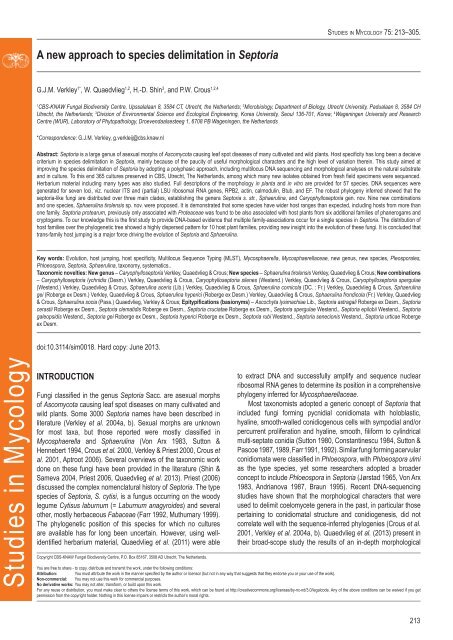
A <strong>new</strong> <strong>approach</strong> <strong>to</strong> <strong>species</strong> <strong>delimitation</strong> <strong>in</strong> Sep<strong>to</strong>riaDNA isolation, PCR and sequenc<strong>in</strong>gGenomic DNA was extracted from fungal mycelium grow<strong>in</strong>g onMEA, us<strong>in</strong>g the UltraClean® Microbial DNA Isolation Kit (MoBio Labora<strong>to</strong>ries, Inc., Solana Beach, CA, USA). Stra<strong>in</strong>s (Table1) were sequenced for seven loci: Act<strong>in</strong> (Act), calmodul<strong>in</strong> (Cal),β-tubul<strong>in</strong> (Btub), <strong>in</strong>ternal transcribed spacer (ITS), Translationelongation fac<strong>to</strong>r 1-alpha (EF) 28S nrDNA (LSU) and RNApolymerase II second largest subunit (RPB2); the primer setslisted <strong>in</strong> Table 2 were used. The PCR amplifications wereperformed <strong>in</strong> a <strong>to</strong>tal volume of 12.5 µL solution conta<strong>in</strong><strong>in</strong>g10–20 ng of template DNA, 1 × PCR buffer, 0.7 µL DMSO(99.9 %), 2 mM MgCl 2, 0.4 µM of each primer, 25 µM of eachdNTP and 1.0 U Taq DNA polymerase (GoTaq, Promega).PCR amplification conditions were set as follows: an <strong>in</strong>itialdenaturation temperature of 96 °C for 2 m<strong>in</strong>, followed by 40cycles at the denaturation temperature of 96 °C for 45 s, primeranneal<strong>in</strong>g at the temperature stipulated <strong>in</strong> Table 2, primerextension at 72 °C for 90 s and a f<strong>in</strong>al extension step at 72 °C for2 m<strong>in</strong>. The result<strong>in</strong>g fragments were sequenced us<strong>in</strong>g the PCRprimers <strong>to</strong>gether with a BigDye Term<strong>in</strong>a<strong>to</strong>r Cycle Sequenc<strong>in</strong>gKit v. 3.1 (Applied Biosystems, Foster City, CA). Sequenc<strong>in</strong>greactions were performed as described by Cheewangkoon et al.(2008). All novel sequences were deposited <strong>in</strong> NCBI’s GenBankdatabase and alignments and phylogenetic trees <strong>in</strong> TreeBASE.Sequence alignement and phylogenetic analysesA basic alignment of the obta<strong>in</strong>ed sequence data was first doneus<strong>in</strong>g MAFFT v. 7 (http://mafft.cbrc.jp/alignment /server/<strong>in</strong>dex. html;Ka<strong>to</strong>h et al. 2002) and if necessary, manually improved <strong>in</strong> BioEditv. 7.0.5.2 (Hall 1999). To check the congruency of the multigenedataset, a 70 % neighbour-jo<strong>in</strong><strong>in</strong>g (NJ) reciprocal bootstrapmethod with maximum likelihood distance was performed (Mason-Gamer & Kellogg 1996, Lombard et al. 2010). Bayesian analyses(critical value for the <strong>to</strong>pological convergence diagnostic set <strong>to</strong>0.01) were performed on the concatenated loci us<strong>in</strong>g MrBayes v.3.2.1 (Huelsenbeck & Ronquist 2001) as described by Crous etal. (2006a) us<strong>in</strong>g nucleotide substitution models that were selectedus<strong>in</strong>g MrModeltest (Table 3) (Nylander 2004).Kimura-2-parameter valuesThe <strong>in</strong>ter-and <strong>in</strong>traspecific distances for each <strong>in</strong>dividual datasetwere calculated us<strong>in</strong>g MEGA v. 4.0 (Tamura et al. 2007) with theKimura-2-parameter (pairwise deletion) model.RESULTSIdentification of the best DNA barcode loci forSep<strong>to</strong>ria <strong>species</strong>Amplification successThe PCR amplification success rates were very high for all sevenloci, vary<strong>in</strong>g from 97 % for RPB2 <strong>to</strong> 100 % for ITS and LSU (Table3). Good amplification reactions of RPB2 required a 2–3 timeshigher DNA <strong>in</strong>put then the other loci and this locus is therefore lessfavorable for easy identification. The other six loci amplified withoutproblems.Kimura-2-parameter valuesThe Kimura-2-parameter (K2P) distribution graphs are depicted <strong>in</strong>Fig. 1. They visualise the <strong>in</strong>ter- and <strong>in</strong>traspecific distances per locus(barcod<strong>in</strong>g gap). A good barcod<strong>in</strong>g locus should have no overlapbetween the <strong>in</strong>ter- and <strong>in</strong>traspecific K2P distances and should havean average <strong>in</strong>terspecific distance that is at least 10 times as high asthe average <strong>in</strong>traspecific distance of that locus (Hebert et al. 2003).The seven loci show a rather constant degree of <strong>in</strong>traspecific variationof 0.01 <strong>in</strong> their K2P distribution graphs, however their <strong>in</strong>terspecificvariations shows considerable differences. The average <strong>in</strong>terspecificvariation <strong>in</strong> both ITS and LSU datasets is very low (0.015) compared<strong>to</strong> their <strong>in</strong>traspecific variation (0.01), lead<strong>in</strong>g <strong>to</strong> a very low <strong>in</strong>ter- <strong>to</strong><strong>in</strong>traspecific variation ratios of 1.5 : 1 for these two loci (Fig. 1).These low ratios are far below the required 10 : 1 ratio, <strong>in</strong>dicat<strong>in</strong>g ageneral lack of natural variation with<strong>in</strong> these two loci, mak<strong>in</strong>g them illsuitedfor effective identification of the <strong>in</strong>dividual <strong>species</strong> used <strong>in</strong> thisdataset. These low K2P results for ITS and LSU are consistent withFrequencyFrequencyFrequency120010008006004002000140012001000800600400200070006000500040003000200010000DistanceDistanceDistanceInter RPB2Inter TubInter EFIntra RPB2Intra TubIntra EFInter ActInter CalIntra ActIntra CalInter ITSInter LSUIntra ITSIntra LSUFig. 1. Frequency distributions of the Kimura-2-parameter distances (barcod<strong>in</strong>ggaps) for the seven PCR loci.www.studies<strong>in</strong>mycology.org215
Verkley et al.Table 1. Isolates used dur<strong>in</strong>g this study.Species Old name Isolate no 1 Host Location Collec<strong>to</strong>r GenBank Accession no 2Caryophyllosep<strong>to</strong>rialychnidisEF Tub RPB2 LSU ITS Act CalSep<strong>to</strong>ria lychnidis <strong>CBS</strong> 109098 Silene pratensis Austria G.J.M. Verkley KF253234 KF252768 KF252292 KF251790 KF251286 KF253595 KF253949Sep<strong>to</strong>ria lychnidis <strong>CBS</strong> 109099 Silene pratensis Austria G.J.M. Verkley KF253235 KF252769 KF252293 KF251791 KF251287 KF253596 KF253950Sep<strong>to</strong>ria lychnidis <strong>CBS</strong> 109101 Silene pratensis Austria G.J.M. Verkley KF253236 KF252770 KF252294 KF251792 KF251288 KF253597 KF253951Sep<strong>to</strong>ria lychnidis <strong>CBS</strong> 109102 Silene pratensis Austria G.J.M. Verkley KF253237 KF252771 KF252295 KF251793 KF251289 KF253598 KF253952Car. pseudolychnidis Sep<strong>to</strong>ria lychnidis <strong>CBS</strong> 128614 Lychnis cognata South Korea H.D. Sh<strong>in</strong> KF253238 KF252772 KF252296 KF251794 KF251290 KF253599 KF253953Sep<strong>to</strong>ria lychnidis <strong>CBS</strong> 128630 Lychnis cognata South Korea H.D. Sh<strong>in</strong> KF253239 KF252773 KF252297 KF251795 KF251291 KF253600 KF253954Car. silenes Sep<strong>to</strong>ria silenes <strong>CBS</strong> 109100 Silene nutans Austria G.J.M. Verkley KF253240 KF252774 KF252298 KF251796 KF251292 KF253601 KF253955Sep<strong>to</strong>ria silenes <strong>CBS</strong> 109103 Silene pratensis Austria G.J.M. Verkley KF253241 KF252775 KF252299 KF251797 KF251293 KF253602 KF253956Car. spergulae Sep<strong>to</strong>ria sp. <strong>CBS</strong> 109010 Spergula morisonii Netherlands A. Aptroot KF253242 KF252776 KF252300 KF251798 KF251294 KF253603 KF253957Sep<strong>to</strong>ria dianthi <strong>CBS</strong> 397.52 Dianthus caryophyllus Netherlands Schouten KF253243 KF252777 KF252301 KF251799 KF251295 KF253604 KF253958Cercospora apii – <strong>CBS</strong> 118712 – Fiji P. Tyler KF253244 KF252778 KF252302 KF251800 KF251296 KF253605 KF253959Cer. arim<strong>in</strong>ensis – <strong>CBS</strong> 137.56 Hedysarum coronarium Italy M. Ribaldi KF253245 KF252779 KF252303 KF251801 KF251297 KF253606 KF253960Cer. beticola – <strong>CBS</strong> 124.31 – Romania E.W. Schmidt KF253246 KF252780 KF252304 KF251802 KF251298 KF253607 KF253961Cercospora sp. – <strong>CBS</strong> 112737 Rhus typh<strong>in</strong>a Canada K.A. Seifert KF253247 KF252781 – KF251803 KF251299 KF253608 KF253962Cer. zebr<strong>in</strong>a – <strong>CBS</strong> 118790 Trifolium subterraneum Australia M.J. Barbetti KF253248 KF252782 KF252305 KF251804 KF251300 KF253609 KF253963Cercosporella– <strong>CBS</strong> 113304 Erigeron annuus South Korea H.D. Sh<strong>in</strong> KF253249 – KF252306 KF251805 KF251301 KF253610 KF253964virgaureaeDothistroma p<strong>in</strong>i – <strong>CBS</strong> 121011 P<strong>in</strong>us palassiana Ukra<strong>in</strong>e A.C. Usichenko KF253250 – KF252307 KF251806 KF251302 KF253611 KF253965Dot. sep<strong>to</strong>sporum – <strong>CBS</strong> 383.74 P<strong>in</strong>us coulteri France M. Morelet KF253251 – KF252308 KF251807 KF251303 KF253612 KF253966Mycosphaerella– <strong>CBS</strong> 228.32 Brassica oleracea Denmark C.A. Jörgensen KF253252 KF252783 KF252309 KF251808 KF251304 KF253613 KF253967brassicicola– <strong>CBS</strong> 267.53 Brassica oleracea Netherlands F. Quak KF253253 KF252784 KF252310 KF251809 KF251305 KF253614 KF253968Myc. capsellae – <strong>CBS</strong> 112033 Brassica sp. UK R. Evans KF253254 KF252785 KF252311 KF251810 KF251306 KF253615 KF253969Mycosphaerella sp. <strong>CBS</strong> 135464; Brassica sp. UK R. Evans – KF252786 KF252312 KF251811 KF251307 KF253616 KF253970CPC 11677Passalora depressa – CPC 14915 Angelica gigas South Korea H.D. Sh<strong>in</strong> KF253256 KF252788 KF252314 KF251813 KF251309 – KF253972Pas. dioscoreae – <strong>CBS</strong> 135460; Dioscorea <strong>to</strong>kora South Korea H.D. Sh<strong>in</strong> KF253257 KF252789 KF252315 KF251814 KF251310 KF253618 –CPC 10855– <strong>CBS</strong> 135463; Dioscorea tenuipes South Korea H.D. Sh<strong>in</strong> KF253258 KF252790 KF252316 KF251815 KF251311 KF253619 –CPC 11513Pas. dissiliens – <strong>CBS</strong> 219.77 Vitis v<strong>in</strong>ifera Iraq M.S.A. Al-Momen KF253259 KF252791 KF252317 KF251816 KF251312 KF253620 –Pas. fusimaculans – CPC 17277 Agrostis sp. Thailand Pheng Pheng KF253260 KF252792 KF252318 KF251817 KF251313 KF253621 KF253973Pas. janseana – <strong>CBS</strong> 145.37 – – E.C. Tullis KF253261 KF252793 – KF251818 KF251314 KF253622 KF253974216
A <strong>new</strong> <strong>approach</strong> <strong>to</strong> <strong>species</strong> <strong>delimitation</strong> <strong>in</strong> Sep<strong>to</strong>riaTable 1. (Cont<strong>in</strong>ued).Species Old name Isolate no 1 Host Location Collec<strong>to</strong>r GenBank Accession no 2EF Tub RPB2 LSU ITS Act CalPassalora sp. – <strong>CBS</strong> 113998 Cajanus cajan South Africa L. van Jaarsveld KF253262 KF252794 KF252319 KF251819 KF251315 KF253623 –Passalora sp. – <strong>CBS</strong> 113999 Cajanus cajan South Africa L. van Jaarsveld KF253263 KF252795 KF252320 KF251820 KF251316 KF253624 –Passalora sp. – <strong>CBS</strong> 114275 Cajanus cajan South Africa L. van Jaarsveld KF253264 KF252796 KF252321 KF251821 KF251317 – –Pseudocercospora – <strong>CBS</strong> 124155 Eucalyptus camaldulensis Madagascar M.J. W<strong>in</strong>gfield KF253265 – KF252322 KF251822 KF251318 KF253625 –madagascariensisPse. pyracanthae – CPC 10808 Pyracantha angustifolia South Korea H.D. Sh<strong>in</strong> KF253266 – KF252323 KF251823 KF251319 KF253626 –Pse. pyracanthigena – <strong>CBS</strong> 112032 Pyracantha angustifolia South Korea M.J. Park KF253267 KF252797 KF252324 KF251824 KF251320 KF253627 KF253975Pse. rho<strong>in</strong>a – CPC 11464 Rhus ch<strong>in</strong>ensis South Korea H.D. Sh<strong>in</strong> KF253268 – KF252325 KF251825 KF251321 – –Pse. schizolobii – <strong>CBS</strong> 120029 Schizolobium parahybum Ecuador M.J. W<strong>in</strong>gfield KF253269 KF252798 KF252326 KF251826 KF251322 KF253628 –– <strong>CBS</strong> 124990 Eucalyptus camaldulensis Thailand W. Himaman KF253270 – KF252327 KF251827 KF251323 KF253629 –Pse. tereticornis – <strong>CBS</strong> 124996 Eucalyptus nitens Australia A.J. Cargenie KF253271 KF252799 KF252328 KF251828 KF251324 KF253630 KF253976C.F. Hill KF253272 KF252800 KF252329 KF251829 KF251325 KF253631 KF253977Pseudocercosporellacapsellae– <strong>CBS</strong> 118412 Brassica sp. NewZealand– <strong>CBS</strong> 127.29 – – K. Togashi KF253273 KF252801 KF252330 KF251830 KF251326 KF253632 KF253978Pella. magnusiana – <strong>CBS</strong> 114735 Geranium silvaticum Sweden E. Gunnerbeck KF253274 KF252802 – KF251831 KF251327 – KF253979Pella. past<strong>in</strong>acae – <strong>CBS</strong> 114116 Laserpitium latifolium Sweden K. & L. Holm KF253275 KF252803 KF252331 KF251832 KF251328 KF253633 KF253980Ramularia endophylla – <strong>CBS</strong> 113265 Quercus robur Netherlands G.J.M. Verkley KF253276 – KF252332 KF251833 KF251329 KF253634 KF253981Ram. eucalypti – <strong>CBS</strong> 120726 Eucalyptus grandiflora Italy W. Gams KF253277 – KF252333 KF251834 KF251330 KF253635 KF253982Ram. lamii – CPC 11312 Leonurus sibiricus South Korea H.D. Sh<strong>in</strong> KF253278 – KF252334 KF251835 KF251331 KF253636 KF253983Readeriella mirabilis – <strong>CBS</strong> 125000 Eucalyptus globulus Australia I.W. Smith KF253279 KF252804 KF252335 KF251836 KF251332 KF253637 KF253984Sep<strong>to</strong>ria abei – <strong>CBS</strong> 128598 Hibiscus syriacus South Korea H.D. Sh<strong>in</strong> KF253280 KF252805 KF252336 KF251837 KF251333 KF253638 KF253985Sep. aegopod<strong>in</strong>a – <strong>CBS</strong> 123740 Aegopodium podagraria Czech G.J.M. Verkley KF253281 KF252806 – KF251838 KF251334 KF253639 KF253986Republic– <strong>CBS</strong> 123741 Aegopodium podagraria Czech G.J.M. Verkley KF253282 KF252807 – KF251839 KF251335 KF253640 KF253987RepublicSep. agrimoniicola – <strong>CBS</strong> 128585 Agrimonia pilosa South Korea H.D. Sh<strong>in</strong> KF253283 KF252808 KF252337 KF251840 KF251336 KF253641 KF253988– <strong>CBS</strong> 128602 Agrimonia pilosa South Korea H.D. Sh<strong>in</strong> KF253284 KF252809 KF252338 KF251841 KF251337 – KF253989Sep. anthrisci – <strong>CBS</strong> 109019 Anthriscus sp. Austria G.J.M. Verkley KF253285 KF252810 KF252339 KF251842 KF251338 KF253642 KF253990– <strong>CBS</strong> 109020 Anthriscus sp. Austria G.J.M. Verkley KF253286 KF252811 KF252340 KF251843 KF251339 KF253643 KF253991Sep. anthurii – <strong>CBS</strong> 148.41 Anthurium sp. – P. Kotthoff KF253287 KF252812 KF252341 KF251844 KF251340 KF253644 KF253992– <strong>CBS</strong> 346.58 Anthurium sp. Germany R. Schneider KF253288 KF252813 KF252342 KF251845 KF251341 KF253645 KF253993Sep. apiicola – <strong>CBS</strong> 116465 Apium graveolens Netherlands R. Munn<strong>in</strong>g KF253289 KF252814 KF252343 KF251846 KF251342 KF253646 KF253994– <strong>CBS</strong> 389.59 Apium graveolens Italy M. Ribaldi KF253290 KF252815 KF252344 KF251847 KF251343 KF253647 KF253995– <strong>CBS</strong> 395.52 Apium sp. Netherlands G. van den Ende KF253291 KF252816 KF252345 KF251848 KF251344 KF253648 KF253996www.studies<strong>in</strong>mycology.org217
Verkley et al.Table 1. (Cont<strong>in</strong>ued).Species Old name Isolate no 1 Host Location Collec<strong>to</strong>r GenBank Accession no 2EF Tub RPB2 LSU ITS Act Cal– <strong>CBS</strong> 400.54 Apium graveolens Netherlands J.A. von Arx KF253292 KF252817 KF252346 KF251849 KF251345 KF253649 KF253997Sep. astericola – <strong>CBS</strong> 128587 Aster tataricus South Korea H.D. Sh<strong>in</strong> KF253293 KF252818 KF252347 KF251850 KF251346 KF253650 KF253998– <strong>CBS</strong> 128593 Aster yomena South Korea H.D. Sh<strong>in</strong> KF253294 KF252819 KF252348 KF251851 KF251347 KF253651 KF253999Sep. astragali – <strong>CBS</strong> 109117 Astragalus glycyphyllos Austria G.J.M. Verkley KF253296 KF252821 KF252350 KF251853 KF251349 KF253653 KF254001– <strong>CBS</strong> 123878 Astragalus glycyphyllos Czech G.J.M. Verkley KF253297 KF252822 KF252351 KF251854 KF251350 KF253654 KF254002Republic– <strong>CBS</strong> 109116 Astragalus glycyphyllos Austria G.J.M. Verkley KF253298 KF252823 KF252352 KF251855 KF251351 KF253655 KF254003Sep. atropurpurea – <strong>CBS</strong> 348.58 Aster canus Germany R. Schneider KF253299 KF252824 KF252353 KF251856 KF251352 KF253656 KF254004Sep. bothriospermi – <strong>CBS</strong> 128592 Bothriospermum tenellum South Korea H.D. Sh<strong>in</strong> KF253300 KF252825 KF252354 KF251857 KF251353 KF253657 KF254005– <strong>CBS</strong> 128599 Bothriospermum tenellum South Korea H.D. Sh<strong>in</strong> KF253301 KF252826 KF252355 KF251858 KF251354 KF253658 KF254006Sep. bupleuricola – <strong>CBS</strong> 128601 Bupleurum longiradiatum South Korea H.D. Sh<strong>in</strong> KF253302 KF252827 KF252356 KF251859 KF251355 KF253659 KF254007– <strong>CBS</strong> 128603 Bupleurum falcatum South Korea H.D. Sh<strong>in</strong> KF253303 KF252828 KF252357 KF251860 KF251356 KF253660 KF254008Sep. calendulae – <strong>CBS</strong> 349.58 Calendula arvensis Italy R. Schneider KF253304 KF252829 KF252358 KF251861 KF251357 KF253661 KF254009Sep. callistephi – <strong>CBS</strong> 128590 Callistephus ch<strong>in</strong>ensis South Korea H.D. Sh<strong>in</strong> KF253305 KF252830 KF252359 KF251862 KF251358 KF253662 KF254010– <strong>CBS</strong> 128594 Callistephus ch<strong>in</strong>ensis South Korea H.D. Sh<strong>in</strong> KF253306 KF252831 KF252360 KF251863 KF251359 KF253663 KF254011Sep. campanulae – <strong>CBS</strong> 128589 Campanula takesimana South Korea H.D. Sh<strong>in</strong> KF253307 KF252832 KF252361 KF251864 KF251360 KF253664 KF254012– <strong>CBS</strong> 128604 Campanula takesimana South Korea H.D. Sh<strong>in</strong> KF253308 KF252833 KF252362 KF251865 KF251361 KF253665 KF254013Sep. cerastii – <strong>CBS</strong> 102323 Cerastium fontanum Netherlands G.J.M. Verkley KF253309 KF252834 KF252363 KF251866 KF251362 KF253666 KF254014– <strong>CBS</strong> 128586 Cerastium holosteoides South Korea H.D. Sh<strong>in</strong> KF253310 KF252835 KF252364 KF251867 KF251363 KF253667 KF254015– <strong>CBS</strong> 128612 Cerastium holosteoides South Korea H.D. Sh<strong>in</strong> KF253311 KF252836 KF252365 KF251868 KF251364 KF253668 KF254016– <strong>CBS</strong> 128626 Cerastium holosteoides South Korea H.D. Sh<strong>in</strong> KF253312 KF252837 KF252366 KF251869 KF251365 KF253669 KF254017– CPC 12343 Cerastium holosteoides South Korea H.D. Sh<strong>in</strong> KF253313 KF252838 KF252367 KF251870 KF251366 KF253670 KF254018Sep. cf. rubi Sep<strong>to</strong>ria sp. CPC 12331 Rubus crataegifolius South Korea H.D. Sh<strong>in</strong> KF253317 KF252842 KF252371 KF251874 KF251370 KF253674 KF254022Sep<strong>to</strong>ria rubi <strong>CBS</strong> 128646 Rubus crataegifolius South Korea H.D. Sh<strong>in</strong> KF253314 KF252839 KF252368 KF251871 KF251367 KF253671 KF254019Sep<strong>to</strong>ria rubi <strong>CBS</strong> 128648 Rubus crataegifolius South Korea H.D. Sh<strong>in</strong> KF253315 KF252840 KF252369 KF251872 KF251368 KF253672 KF254020Sep<strong>to</strong>ria rubi <strong>CBS</strong> 128760 Rubus crataegifolius South Korea H.D. Sh<strong>in</strong> KF253316 KF252841 KF252370 KF251873 KF251369 KF253673 KF254021Sep. cf. sonchi – <strong>CBS</strong> 128757 Sonchus asper South Korea H.D. Sh<strong>in</strong> KF253500 KF253020 KF252546 KF252057 KF251552 KF253855 KF254204Sep. cf. stachydicola Sep<strong>to</strong>ria lycopicola <strong>CBS</strong> 128662 Stachys riederi South Korea H.D. Sh<strong>in</strong> KF253513 KF253034 KF252559 KF252071 KF251566 KF253867 KF254218Sep. chamaecisti – <strong>CBS</strong> 350.58 Helianthemum hybridum Germany R. Schneider KF253318 KF252843 KF252372 KF251875 KF251371 KF253675 KF254023Sep. chelidonii – <strong>CBS</strong> 128607 Chelidonium majus South Korea H.D. Sh<strong>in</strong> KF253319 KF252844 KF252373 KF251876 KF251372 KF253676 KF254024– CPC 12337 Chelidonium majus South Korea H.D. Sh<strong>in</strong> KF253320 KF252845 KF252374 KF251877 KF251373 KF253677 KF254025Sep. chromolaenae – <strong>CBS</strong> 113373 Chromolaena odorata Cuba S. Neser KF253321 KF252846 KF252375 KF251878 KF251374 KF253678 KF254026218
A <strong>new</strong> <strong>approach</strong> <strong>to</strong> <strong>species</strong> <strong>delimitation</strong> <strong>in</strong> Sep<strong>to</strong>riaTable 1. (Cont<strong>in</strong>ued).Species Old name Isolate no 1 Host Location Collec<strong>to</strong>r GenBank Accession no 2Sep. chrysanthemella – <strong>CBS</strong> 128617 ChrysanthemummorifoliumEF Tub RPB2 LSU ITS Act CalSouth Korea H.D. Sh<strong>in</strong> KF253322 KF252847 KF252376 KF251879 KF251375 KF253679 KF254027– <strong>CBS</strong> 128622 Chrysanthemum boreale South Korea H.D. Sh<strong>in</strong> KF253323 KF252848 KF252377 KF251880 KF251376 KF253680 KF254028– <strong>CBS</strong> 483.63 Chrysanthemum sp. Netherlands H.A. van der Aa KF253324 KF252849 KF252378 KF251881 KF251377 KF253681 KF254029– <strong>CBS</strong> 128716 – South Africa E. Oh KF253325 KF252850 KF252379 KF251882 KF251378 KF253682 KF254030– <strong>CBS</strong> 351.58 Chrysanthemum <strong>in</strong>dicum Germany R. Schneider KF253326 KF252851 KF252380 KF251883 KF251379 KF253683 KF254031– <strong>CBS</strong> 354.73 ChrysanthemummorifoliumNewZealandG.F. Laundon KF253327 KF252852 KF252381 KF251884 KF251380 KF253684 KF254032Sep. cirsii – <strong>CBS</strong> 128621 Cirsium setidens South Korea H.D. Sh<strong>in</strong> KF253328 KF252853 KF252382 KF251885 KF251381 KF253685 KF254033Sep. citri (= protearumcomplex)Sep<strong>to</strong>ria orchidearum <strong>CBS</strong> 101013 Masdevallia sp. Netherlands W. Veenbaas-Rijks KF253457 KF252978 KF252504 KF252013 KF251508 KF253812 KF254161Sep<strong>to</strong>ria sp. <strong>CBS</strong> 101354 Gevu<strong>in</strong>a avellana NewZealandS. Ganev KF253458 KF252979 KF252505 KF252014 KF251509 KF253813 KF254162Sep<strong>to</strong>ria lobeliae <strong>CBS</strong> 113392 Lobelia er<strong>in</strong>us – S. Wolcon KF253460 KF252981 KF252507 KF252016 KF251511 KF253815 KF254164Sep<strong>to</strong>ria aciculosa <strong>CBS</strong> 177.77 Fragaria sp. NewZealandH.J. Boesew<strong>in</strong>kel KF253463 KF252984 KF252509 KF252019 KF251514 KF253818 KF254167Sep<strong>to</strong>ria citri <strong>CBS</strong> 315.37 – – L.L. Huillier KF253465 – KF252511 KF252021 KF251516 KF253820 KF254169Sep<strong>to</strong>ria gerberae <strong>CBS</strong> 410.61 Gerbera jamesonii Italy W. Gerlach KF253468 KF252988 KF252514 KF252024 KF251519 KF253823 KF254172Sep<strong>to</strong>ria hederae <strong>CBS</strong> 566.88 Hedera helix France H.A. van der Aa KF253470 KF252990 KF252515 KF252026 KF251521 KF253825 KF254174Sep. citricola – <strong>CBS</strong> 356.36 Citrus s<strong>in</strong>ensis Italy G. Ruggieri KF253329 KF252854 KF252383 KF251886 KF251382 KF253686 KF254034Sep. clematidis – <strong>CBS</strong> 108983 Clematis vitalba Germany G.J.M. Verkley KF253330 KF252855 KF252384 KF251887 KF251383 KF253687 KF254035– <strong>CBS</strong> 108984 Clematis vitalba Germany G.J.M. Verkley KF253331 KF252856 KF252385 KF251888 KF251384 KF253688 KF254036Sep. codonopsidis – <strong>CBS</strong> 128609 Codonopsis lanceolata South Korea H.D. Sh<strong>in</strong> KF253332 KF252857 KF252386 KF251889 KF251385 KF253689 KF254037– <strong>CBS</strong> 128620 Codonopsis lanceolata South Korea H.D. Sh<strong>in</strong> KF253333 KF252858 KF252387 KF251890 KF251386 KF253690 KF254038Sep. convolvuli – <strong>CBS</strong> 102325 Calystegia sepium Netherlands G.J.M. Verkley KF253334 KF252859 KF252388 KF251891 KF251387 KF253691 KF254039– <strong>CBS</strong> 113111 Calystegia sepium NewG.J.M. Verkley KF253335 KF252860 KF252389 KF251892 KF251388 KF253692 KF254040Zealand– <strong>CBS</strong> 128627 Calystegia soldanella South Korea H.D. Sh<strong>in</strong> KF253336 KF252861 KF252390 KF251893 KF251389 KF253693 KF254041Sep. coprosmae – <strong>CBS</strong> 113391 Coprosma robusta NewZealandG.J.M. Verkley KF253255 KF252787 KF252313 KF251812 KF251308 KF253617 KF253971Sep. crepidis – CPC 12539 Crepis japonica South Korea H.D. Sh<strong>in</strong> KF253339 KF252864 KF252393 KF251896 KF251392 KF253696 KF254044– <strong>CBS</strong> 128608 Youngia japonica South Korea H.D. Sh<strong>in</strong> KF253337 KF252862 KF252391 KF251894 KF251390 KF253694 KF254042– <strong>CBS</strong> 128619 Youngia japonica South Korea H.D. Sh<strong>in</strong> KF253338 KF252863 KF252392 KF251895 KF251391 KF253695 KF254043www.studies<strong>in</strong>mycology.org219
Verkley et al.Table 1. (Cont<strong>in</strong>ued).Species Old name Isolate no 1 Host Location Collec<strong>to</strong>r GenBank Accession no 2Sep. cruciatae Sep<strong>to</strong>ria sp. <strong>CBS</strong> 123747 Galium odoratum CzechRepublicSep<strong>to</strong>ria sp. <strong>CBS</strong> 123748 Galium odoratum CzechRepublicEF Tub RPB2 LSU ITS Act CalG.J.M. Verkley KF253340 KF252865 KF252394 KF251897 KF251393 KF253697 KF254045G.J.M. Verkley KF253341 KF252866 KF252395 KF251898 KF251394 KF253698 KF254046Sep. cucubali – <strong>CBS</strong> 102367 Cucubalus baccifer Netherlands G.J.M. Verkley KF253342 KF252867 KF252396 KF251899 KF251395 KF253699 KF254047– <strong>CBS</strong> 102368 Cucubalus baccifer Netherlands G.J.M. Verkley KF253343 KF252868 KF252397 KF251900 KF251396 KF253700 KF254048– <strong>CBS</strong> 102386 Saponaria offic<strong>in</strong>alis Netherlands G.J.M. Verkley KF253344 KF252869 KF252398 KF251901 KF251397 KF253701 KF254049Sep<strong>to</strong>ria sp. <strong>CBS</strong> 124874 Fagus sylvatica Germany M. Unterseher KF253345 KF252870 KF252399 KF251902 KF251398 KF253702 KF254050Sep. cucurbitacearum – <strong>CBS</strong> 178.77 Cucurbita maxima NewZealandH.J. Boesew<strong>in</strong>kel KF253346 – KF252400 KF251903 KF251399 KF253703 KF254051Sep. dearnessii – <strong>CBS</strong> 128624 Angelica dahurica South Korea H.D. Sh<strong>in</strong> KF253347 KF252871 KF252401 KF251904 KF251400 KF253704 KF254052Sep. digitalis – <strong>CBS</strong> 328.67 Digitalis lanata Netherlands H.A. van der Aa KF253348 KF252872 KF252402 KF251905 KF251401 KF253705 KF254053– <strong>CBS</strong> 391.63 Digitalis lanata Czech V. Holubová KF253349 KF252873 KF252403 KF251906 KF251402 KF253706 KF254054RepublicSep. dolichospora – <strong>CBS</strong> 129152 Solidago virgaurea South Korea H.D. Sh<strong>in</strong> KF253350 KF252874 – KF251907 KF251403 KF253707 KF254055Sep. dysentericae – <strong>CBS</strong> 128637 Inula britannica South Korea H.D. Sh<strong>in</strong> KF253351 KF252875 KF252404 KF251908 KF251404 KF253708 KF254056– <strong>CBS</strong> 128638 Inula britannica South Korea H.D. Sh<strong>in</strong> KF253352 KF252876 KF252405 KF251909 KF251405 KF253709 KF254057– <strong>CBS</strong> 131892;CPC 12328Inula britannica South Korea H.D. Sh<strong>in</strong> KF253353 KF252877 KF252406 KF251910 KF251406 KF253710 KF254058Sep. ekmaniana – <strong>CBS</strong> 113385 Chromolaena odorata Mexico M.J. Morris KF253354 KF252878 – KF251911 KF251407 KF253711 KF254059– <strong>CBS</strong> 113612 Chromolaena odorata Mexico M.J. Morris KF253355 KF252879 – KF251912 KF251408 KF253712 KF254060Sep. epambrosiae – <strong>CBS</strong> 128629 Ambrosia trifida South Korea H.D. Sh<strong>in</strong> KF253356 KF252880 KF252407 KF251913 KF251409 KF253713 KF254061– <strong>CBS</strong> 128636 Ambrosia trifida South Korea H.D. Sh<strong>in</strong> KF253357 KF252881 KF252408 KF251914 KF251410 KF253714 KF254062Sep. epilobii – <strong>CBS</strong> 109084 Epilobium fleischeri Austria G.J.M. Verkley KF253358 KF252882 KF252409 KF251915 KF251411 KF253715 KF254063– <strong>CBS</strong> 109085 Epilobium fleischeri Austria G.J.M. Verkley KF253359 KF252883 KF252410 KF251916 KF251412 KF253716 KF254064Sep. erigerontis – <strong>CBS</strong> 109094 Erigeron annuus Austria G.J.M. Verkley KF253360 KF252884 KF252411 KF251917 KF251413 KF253717 KF254065– <strong>CBS</strong> 109095 Erigeron annuus Austria G.J.M. Verkley KF253361 KF252885 KF252412 KF251918 KF251414 KF253718 KF254066– <strong>CBS</strong> 128606 Erigeron annuus South Korea H.D. Sh<strong>in</strong> KF253362 KF252886 KF252413 KF251919 KF251415 KF253719 KF254067– <strong>CBS</strong> 131893;CPC 12340Erigeron annuus South Korea H.D. Sh<strong>in</strong> KF253363 KF252888 KF252414 KF251920 KF251416 KF253720 KF254068Sep<strong>to</strong>ria schnabliana <strong>CBS</strong> 186.93 Erigeron annuus Italy M. Vurro KF253364 KF252887 KF252537 KF252048 KF251543 KF253893 KF254244Sep. eucalyp<strong>to</strong>rum – <strong>CBS</strong> 118505 Eucalyptus sp. India W. Gams KF253365 KF252889 KF252415 KF251921 KF251417 KF253721 KF254069Sep. exotica – <strong>CBS</strong> 163.78 Hebe speciosa NewH.J. Boesew<strong>in</strong>kel KF253366 KF252890 KF252416 KF251922 KF251418 KF253722 KF254070Zealand220
A <strong>new</strong> <strong>approach</strong> <strong>to</strong> <strong>species</strong> <strong>delimitation</strong> <strong>in</strong> Sep<strong>to</strong>riaTable 1. (Cont<strong>in</strong>ued).Species Old name Isolate no 1 Host Location Collec<strong>to</strong>r GenBank Accession no 2Sep. galeopsidis – <strong>CBS</strong> 123744 Galeopsis sp. CzechRepublic– <strong>CBS</strong> 123746 Galeopsis sp. CzechRepublic– <strong>CBS</strong> 123749 Galeopsis sp. CzechRepublicEF Tub RPB2 LSU ITS Act CalG.J.M. Verkley KF253367 KF252891 KF252417 KF251923 KF251419 KF253723 KF254071G.J.M. Verkley KF253368 KF252892 KF252418 KF251924 KF251420 KF253724 KF254072G.J.M. Verkley KF253369 KF252893 KF252419 KF251925 KF251421 KF253725 KF254073– <strong>CBS</strong> 191.26 Galeopsis sp. – C. Killian KF253370 KF252894 KF252420 KF251926 KF251422 KF253726 KF254074– <strong>CBS</strong> 102314 Galeopsis tetrahit Netherlands G.J.M. Verkley KF253371 KF252895 KF252421 KF251927 KF251423 KF253727 KF254075– <strong>CBS</strong> 102411 Galeopsis tetrahit Netherlands G.J.M. Verkley KF253372 KF252896 KF252422 KF251928 KF251424 KF253728 KF254076– <strong>CBS</strong> 123745 Galeopsis sp. CzechRepublicG.J.M. Verkley KF253373 KF252897 KF252423 KF251929 KF251425 KF253729 KF254077Sep. gentianae – <strong>CBS</strong> 128633 Gentiana scabra South Korea H.D. Sh<strong>in</strong> KF253374 KF252898 KF252424 KF251930 KF251426 KF253730 KF254078Sep. gladioli – <strong>CBS</strong> 121.20 – – – KF253375 KF252899 KF252425 KF251931 KF251427 KF253731 KF254079– <strong>CBS</strong> 353.29 – Netherlands J.C. Went KF253376 KF252900 KF252426 KF251932 KF251428 KF253732 KF254080Sep. glyc<strong>in</strong>es – <strong>CBS</strong> 336.53 – Japan H. Kurata KF253377 KF252901 – KF251933 KF251429 KF253733 KF254081Sep. glyc<strong>in</strong>icola – <strong>CBS</strong> 128618 Glyc<strong>in</strong>e max South Korea H.D. Sh<strong>in</strong> KF253378 KF252902 KF252427 KF251934 KF251430 KF253734 KF254082Sep. helianthi – <strong>CBS</strong> 123.81 Helianthus annuus – M. Muntañola KF253379 KF252903 KF252428 KF251935 KF251431 KF253735 KF254083Sep. helianthicola – <strong>CBS</strong> 122.81 Helianthus annuus – M. Muntañola KF253380 KF252904 KF252429 KF251936 KF251432 KF253736 KF254084Sep. hibiscicola – <strong>CBS</strong> 128611 Hibiscus syriacus South Korea H.D. Sh<strong>in</strong> KF253381 KF252905 KF252430 KF251937 KF251433 KF253737 KF254085– <strong>CBS</strong> 128615 Hibiscus syriacus South Korea H.D. Sh<strong>in</strong> KF253382 KF252906 KF252431 KF251938 KF251434 KF253738 KF254086Sep. hippocastani – <strong>CBS</strong> 411.61 Aesculus hippocastanum Germany W. Gerlach KF253383 KF252907 KF252432 KF251939 KF251435 KF253739 KF254087Sep. justiciae – CPC 12509 Justicia procumbens South Korea H.D. Sh<strong>in</strong> KF253386 KF252910 KF252435 KF251942 KF251438 KF253742 KF254090– <strong>CBS</strong> 128610 Justicia procumbens South Korea H.D. Sh<strong>in</strong> KF253384 KF252908 KF252433 KF251940 KF251436 KF253740 KF254088– <strong>CBS</strong> 128625 Justicia procumbens South Korea H.D. Sh<strong>in</strong> KF253385 KF252909 KF252434 KF251941 KF251437 KF253741 KF254089Sep. lactucae – <strong>CBS</strong> 108943 Lactuca sativa Netherlands P. Grooteman KF253387 KF252911 KF252436 KF251943 KF251439 KF253743 KF254091– <strong>CBS</strong> 352.58 Lactuca sativa Germany G. Sörgel KF253388 KF252912 KF252437 KF251944 KF251440 KF253744 KF254092Sep. lamiicola – <strong>CBS</strong> 102328 Lamium album Netherlands G.J.M. Verkley KF253389 KF252913 KF252438 KF251945 KF251441 KF253745 KF254093– <strong>CBS</strong> 102329 Lamium album Netherlands G.J.M. Verkley KF253390 KF252914 KF252439 KF251946 KF251442 KF253746 KF254094– <strong>CBS</strong> 102379 Lamium sp. Netherlands G.J.M. Verkley KF253391 KF252915 KF252440 KF251947 KF251443 KF253747 KF254095– <strong>CBS</strong> 102380 Lamium sp. Netherlands G.J.M. Verkley KF253392 KF252916 KF252441 KF251948 KF251444 KF253748 KF254096– <strong>CBS</strong> 109112 Lamium album Austria G.J.M. Verkley KF253393 KF252917 KF252442 KF251949 KF251445 KF253749 KF254097– <strong>CBS</strong> 109113 Lamium album Austria G.J.M. Verkley KF253394 KF252918 KF252443 KF251950 KF251446 KF253750 KF254098– <strong>CBS</strong> 123882 Lamium sp. CzechRepublicG.J.M. Verkley KF253395 KF252919 KF252444 KF251951 KF251447 KF253751 KF254099www.studies<strong>in</strong>mycology.org221
Verkley et al.Table 1. (Cont<strong>in</strong>ued).Species Old name Isolate no 1 Host Location Collec<strong>to</strong>r GenBank Accession no 2– <strong>CBS</strong> 123883 Lamium sp. CzechRepublic– <strong>CBS</strong> 123884 Lamium sp. CzechRepublicEF Tub RPB2 LSU ITS Act CalG.J.M. Verkley KF253396 KF252920 KF252445 KF251952 KF251448 KF253752 KF254100G.J.M. Verkley KF253397 KF252921 KF252446 KF251953 KF251449 KF253753 KF254101Sep. lepidiicola – <strong>CBS</strong> 128635 Lepidium virg<strong>in</strong>icum South Korea H.D. Sh<strong>in</strong> KF253398 KF252922 KF252447 KF251954 KF251450 KF253754 KF254102Sep. lep<strong>to</strong>stachyae – <strong>CBS</strong> 128613 Phryma lep<strong>to</strong>stachya South Korea H.D. Sh<strong>in</strong> KF253399 KF252923 KF252448 KF251955 KF251451 KF253755 KF254103– <strong>CBS</strong> 128628 Phryma lep<strong>to</strong>stachya South Korea H.D. Sh<strong>in</strong> KF253400 KF252924 KF252449 KF251956 KF251452 KF253756 KF254104Sep. leucanthemi – <strong>CBS</strong> 109083 Chrysanthemumleucanthemum– <strong>CBS</strong> 109086 Chrysanthemumleucanthemum– <strong>CBS</strong> 109090 Chrysanthemumleucanthemum– <strong>CBS</strong> 109091 Chrysanthemumleucanthemum– <strong>CBS</strong> 113112 ChrysanthemumleucanthemumAustria G.J.M. Verkley KF253401 KF252925 KF252450 KF251957 KF251453 KF253757 KF254105Austria G.J.M. Verkley KF253402 KF252926 KF252451 KF251958 KF251454 KF253758 KF254106Austria G.J.M. Verkley KF253403 KF252927 KF252452 KF251959 KF251455 KF253759 KF254107Austria G.J.M. Verkley KF253404 KF252928 KF252453 KF251960 KF251456 KF253760 KF254108NewZealandG.J.M. Verkley KF253405 KF252929 KF252454 KF251961 KF251457 KF253761 KF254109– <strong>CBS</strong> 353.58 Chrysanthemum maximum Germany R. Schneider KF253406 KF252930 KF252455 KF251962 KF251458 KF253762 KF254110Sep. limonum – <strong>CBS</strong> 419.51 Citrus limonium Italy G. Goidánich KF253407 KF252931 KF252456 KF251963 KF251459 KF253763 KF254111Sep. l<strong>in</strong>icola – <strong>CBS</strong> 316.37 L<strong>in</strong>um usitatissimum – H.W. Hollenweber KF253408 KF252932 KF252457 KF251964 KF251460 KF253764 KF254112Sep. lycoc<strong>to</strong>ni – <strong>CBS</strong> 109089 Aconitum vulparia Austria G.J.M. Verkley KF253409 KF252933 KF252458 KF251965 KF251461 KF253765 KF254113Sep. lycopersici – <strong>CBS</strong> 128654 Lycopersicon esculentum South Korea H.D. Sh<strong>in</strong> KF253410 KF252934 KF252459 KF251966 KF251462 KF253766 KF254114– <strong>CBS</strong> 354.49 Lycopersicon esculentum Canada B.H. MacNeil KF253411 KF252935 KF252460 KF251967 KF251463 KF253767 KF254115Sep. lycopicola – <strong>CBS</strong> 128651 Lycopus ramosissimus South Korea H.D. Sh<strong>in</strong> KF253412 KF252936 KF252461 KF251968 KF251464 KF253768 KF254116Sep. lysimachiae – <strong>CBS</strong> 102315 Lysimachia vulgaris Netherlands G.J.M. Verkley KF253413 KF252937 KF252462 KF251969 KF251465 KF253769 KF254117– <strong>CBS</strong> 108998 Lysimachia vulgaris Netherlands G.J.M. Verkley KF253414 KF252938 KF252463 KF251970 KF251466 KF253770 KF254118– <strong>CBS</strong> 108999 Lysimachia vulgaris Netherlands G.J.M. Verkley KF253415 KF252939 KF252464 KF251971 KF251467 KF253771 KF254119– <strong>CBS</strong> 123794 Lysimachia sp. CzechRepublic– <strong>CBS</strong> 123795 Lysimachia sp. CzechRepublicG.J.M. Verkley KF253416 KF252940 KF252465 KF251972 KF251468 KF253772 KF254120G.J.M. Verkley KF253417 KF252941 KF252466 KF251973 KF251469 KF253773 KF254121Sep. malagutii – <strong>CBS</strong> 106.80 Solanum sp. Peru G.H. Boerema KF253418 – KF252467 KF251974 KF251470 KF253774 KF254122Sep. matricariae – <strong>CBS</strong> 109000 Matricaria discoidea Netherlands G.J.M. Verkley KF253419 KF252942 KF252468 KF251975 KF251471 KF253775 KF254123– <strong>CBS</strong> 109001 Matricaria discoidea Netherlands G.J.M. Verkley KF253420 KF252943 KF252469 KF251976 KF251472 KF253776 KF254124Sep. mazi – <strong>CBS</strong> 128656 Mazus japonicus South Korea H.D. Sh<strong>in</strong> KF253421 KF252944 KF252470 KF251977 KF251473 KF253777 KF254125222
A <strong>new</strong> <strong>approach</strong> <strong>to</strong> <strong>species</strong> <strong>delimitation</strong> <strong>in</strong> Sep<strong>to</strong>riaTable 1. (Cont<strong>in</strong>ued).Species Old name Isolate no 1 Host Location Collec<strong>to</strong>r GenBank Accession no 2EF Tub RPB2 LSU ITS Act Cal– <strong>CBS</strong> 128755 Mazus japonicus South Korea H.D. Sh<strong>in</strong> KF253422 KF252945 KF252471 KF251978 KF251474 KF253778 KF254126Sep. melissae – <strong>CBS</strong> 109097 Melissa offic<strong>in</strong>alis Netherlands H.A. van der Aa KF253423 KF252946 KF252472 KF251979 KF251475 KF253779 KF254127Sep. menthae – <strong>CBS</strong> 404.34 – Japan T. Hemmi KF253424 KF252947 – KF251980 KF251476 KF253780 KF254128Sep. napelli – <strong>CBS</strong> 109104 Aconitum napellus Austria G.J.M. Verkley KF253425 KF252948 KF252473 KF251981 KF251477 KF253781 KF254129– <strong>CBS</strong> 109105 Aconitum napellus Austria G.J.M. Verkley KF253426 KF252949 KF252474 KF251982 KF251478 KF253782 KF254130– <strong>CBS</strong> 109106 Aconitum napellus Austria G.J.M. Verkley KF253427 KF252950 KF252475 KF251983 KF251479 KF253783 KF254131Sep. obesa Sep<strong>to</strong>ria artimisiae <strong>CBS</strong> 128588 Artemisia lavandulaefolia South Korea H.D. Sh<strong>in</strong> KF253428 KF252951 KF252476 KF251984 KF251480 KF253784 KF254132Sep<strong>to</strong>ria<strong>CBS</strong> 128623 Chrysanthemum <strong>in</strong>dicum South Korea H.D. Sh<strong>in</strong> KF253429 KF252952 KF252477 KF251985 KF251481 KF253785 KF254133chrysanthemella– <strong>CBS</strong> 128759 ChrysanthemumSouth Korea H.D. Sh<strong>in</strong> KF253430 – KF252478 KF251986 KF251482 KF253786 KF254134morifolium– <strong>CBS</strong> 354.58 Chrysantemum <strong>in</strong>dicum Germany R. Schneider KF253431 – KF252479 KF251987 KF251483 KF253787 KF254135Sep. oenanthis – <strong>CBS</strong> 128667 Cicuta virosa South Korea H.D. Sh<strong>in</strong> KF253432 KF252953 KF252481 KF251989 KF251485 KF253788 KF254136Sep. oenanthicola Sep<strong>to</strong>ria oenanthis <strong>CBS</strong> 128649 Oenanthe javanica South Korea H.D. Sh<strong>in</strong> KF253433 KF252954 KF252480 KF251988 KF251484 KF253789 KF254137Sep. orchidearum Sep<strong>to</strong>ria cyclam<strong>in</strong>is <strong>CBS</strong> 128631 Cyclamen fatrense South Korea H.D. Sh<strong>in</strong> KF253434 KF252955 KF252482 KF251990 KF251486 KF253790 KF254138– <strong>CBS</strong> 457.78 Listera ovata France H.A. van der Aa KF253435 KF252956 KF252483 KF251991 KF251487 KF253791 KF254139Sep. oudemansii – <strong>CBS</strong> 619.72 Poa pratensis Germany R. Schneider KF253436 KF252957 KF252484 KF251992 KF254299 – KF254140Sep. pachyspora – <strong>CBS</strong> 128652 Zyathoxylum sch<strong>in</strong>ifolium South Korea H.D. Sh<strong>in</strong> KF253437 KF252958 KF252485 KF251993 KF251488 KF253792 KF254141Sep. paridis – <strong>CBS</strong> 109111 Paris quadrifolia Austria G.J.M. Verkley KF253438 KF252959 KF252486 KF251994 KF251489 KF253793 KF254142– <strong>CBS</strong> 109110 Paris quadrifolia Austria G.J.M. Verkley KF253439 KF252960 KF252487 KF251995 KF251490 KF253794 KF254143Sep<strong>to</strong>ria violaepalustrisSep<strong>to</strong>ria violaepalustris<strong>CBS</strong> 109108 Viola sp. Austria G.J.M. Verkley KF253440 KF252961 KF252488 KF251996 KF251491 KF253795 KF254144<strong>CBS</strong> 109109 Viola sp. Austria G.J.M. Verkley KF253441 KF252962 KF252489 KF251997 KF251492 KF253796 KF254145Sep. passifloricola Sep. passiflorae <strong>CBS</strong> 102701 Passiflora edulis NewZealandC.F. Hill KF253442 KF252963 KF252490 KF251998 KF251493 KF253797 KF254146– <strong>CBS</strong> 129431 Passiflora edulis South Korea H.D. Sh<strong>in</strong> KF253443 KF252964 – KF251999 KF251494 KF253798 KF254147Sep. perillae – <strong>CBS</strong> 128655 Perilla frutescens South Korea H.D. Sh<strong>in</strong> KF253444 KF252965 KF252491 KF252000 KF251495 KF253799 KF254148Sep. petrosel<strong>in</strong>i – <strong>CBS</strong> 109521 – Netherlands H.A. van der Aa KF253445 KF252966 KF252492 KF252001 KF251496 KF253800 KF254149– <strong>CBS</strong> 182.44 Petrosel<strong>in</strong>um sativum Netherlands S.D. de Wit KF253446 KF252967 KF252493 KF252002 KF251497 KF253801 KF254150Sep. phlogis – <strong>CBS</strong> 102317 Phlox sp. Netherlands G.J.M. Verkley KF253447 KF252968 KF252494 KF252003 KF251498 KF253802 KF254151– <strong>CBS</strong> 128663 Phlox paniculata South Korea H.D. Sh<strong>in</strong> KF253448 KF252969 KF252495 KF252004 KF251499 KF253803 KF254152– <strong>CBS</strong> 577.90 Phlox sp. Netherlands H.A. van der Aa KF253449 KF252970 KF252496 KF252005 KF251500 KF253804 KF254153Sep. polygonorum – <strong>CBS</strong> 102330 Polygonum persicaria Netherlands G.J.M. Verkley KF253450 KF252971 KF252497 KF252006 KF251501 KF253805 KF254154www.studies<strong>in</strong>mycology.org223
Verkley et al.Table 1. (Cont<strong>in</strong>ued).Species Old name Isolate no 1 Host Location Collec<strong>to</strong>r GenBank Accession no 2EF Tub RPB2 LSU ITS Act Cal– <strong>CBS</strong> 102331 Polygonum persicaria Netherlands G.J.M. Verkley KF253451 KF252972 KF252498 KF252007 KF251502 KF253806 KF254155– <strong>CBS</strong> 108982 Polygonum persicaria Germany G.J.M. Verkley KF253452 KF252973 KF252499 KF252008 KF251503 KF253807 KF254156– <strong>CBS</strong> 109834 Polygonum persicaria Netherlands G.J.M. Verkley KF253453 KF252974 KF252500 KF252009 KF251504 KF253808 KF254157– <strong>CBS</strong> 113110 Polygonum persicaria NewZealandC.F. Hill KF253454 KF252975 KF252501 KF252010 KF251505 KF253809 KF254158– <strong>CBS</strong> 347.67 Polygonum persicaria Netherlands H.A. van der Aa KF253455 KF252976 KF252502 KF252011 KF251506 KF253810 KF254159Sep. posoniensis – <strong>CBS</strong> 128645 Chrysosplenium japonicum South Korea H.D. Sh<strong>in</strong> KF253456 KF252977 KF252503 KF252012 KF251507 KF253811 KF254160Sep. protearum Sep<strong>to</strong>ria sp. CPC 19691 Zanthedeschia aethiopica South Africa P.W. Crous KF253474 KF252994 KF252519 KF252030 KF251525 KF253829 KF254178Sep<strong>to</strong>ria sp. <strong>CBS</strong> 113114 Geum sp. NewG.J.M. Verkley KF253459 KF252980 KF252506 KF252015 KF251510 KF253814 KF254163ZealandSep<strong>to</strong>ria sp. <strong>CBS</strong> 119942 Asplenium ruta-muraria Germany G.J.M. Verkley KF253461 KF252982 – KF252017 KF251512 KF253816 KF254165Sep<strong>to</strong>ria sp. <strong>CBS</strong> 135477;CPC 19675Sep<strong>to</strong>ria sp. <strong>CBS</strong> 164.78 Nephrolepis sp. NewZealandSep<strong>to</strong>ria sp. <strong>CBS</strong> 179.77 Myosotis sp. NewZealandZanthedeschia aethiopica South Africa P.W. Crous KF253473 KF252993 KF252518 KF252029 KF251524 KF253828 KF254177H.J. Boesew<strong>in</strong>kel KF253462 KF252983 KF252508 KF252018 KF251513 KF253817 KF254166H.J. Boesew<strong>in</strong>kel KF253464 KF252985 KF252510 KF252020 KF251515 KF253819 KF254168Sep<strong>to</strong>ria sp. <strong>CBS</strong> 364.97 Skimmia sp. Netherlands J. de Gruyter KF253466 KF252986 KF252512 KF252022 KF251517 KF253821 KF254170Sep<strong>to</strong>ria ligustri <strong>CBS</strong> 390.59 Ligustrum vulgare Italy M. Ribaldi KF253467 KF252987 KF252513 KF252023 KF251518 KF253822 KF254171Sep<strong>to</strong>ria pistaciae <strong>CBS</strong> 420.51 Pistacia vera Italy G. Goidánich KF253469 KF252989 – KF252025 KF251520 KF253824 KF254173Sep<strong>to</strong>ria sp. <strong>CBS</strong> 658.77 Boronia denticulata NewZealandH.J. Boesew<strong>in</strong>kel KF253471 KF252991 KF252516 KF252027 KF251522 KF253826 KF254175– <strong>CBS</strong> 778.97 Protea cynaroides South Africa L. Viljoen KF253472 KF252992 KF252517 KF252028 KF251523 KF253827 KF254176Sep. pseudonapelli Sep<strong>to</strong>ria napelli <strong>CBS</strong> 128664 Aconitum pseudolaeve South Korea H.D. Sh<strong>in</strong> KF253475 KF252995 KF252520 KF252031 KF251526 KF253830 KF254179Sep. putrida – <strong>CBS</strong> 109087 Senecio nemorensis Austria G.J.M. Verkley KF253476 KF252996 KF252521 KF252032 KF251527 KF253831 KF254180– <strong>CBS</strong> 109088 Senecio nemorensis Austria G.J.M. Verkley KF253477 KF252997 KF252522 KF252033 KF251528 KF253832 KF254181Sep. rumicum Sep<strong>to</strong>ria ace<strong>to</strong>sae <strong>CBS</strong> 503.76 Rumex ace<strong>to</strong>sa France H.A. van der Aa KF253478 KF252998 KF252523 KF252034 KF251529 KF253833 KF254182Sep. saccardoi – <strong>CBS</strong> 128756 Lysimachia vulgaris South Korea H.D. Sh<strong>in</strong> KF253479 KF252999 KF252524 KF252035 KF251530 KF253834 KF254183Sep. scabiosicola – <strong>CBS</strong> 102333 Knautia arvensis Netherlands G.J.M. Verkley KF253480 KF253000 KF252525 KF252036 KF251531 KF253835 KF254184– <strong>CBS</strong> 102334 Knautia arvensis Netherlands G.J.M. Verkley KF253481 KF253001 KF252526 KF252037 KF251532 KF253836 KF254185– <strong>CBS</strong> 102335 Knautia arvensis Netherlands G.J.M. Verkley KF253482 KF253002 KF252527 KF252038 KF251533 KF253837 KF254186– <strong>CBS</strong> 102336 Knautia arvensis Netherlands G.J.M. Verkley KF253483 KF253003 KF252528 KF252039 KF251534 KF253838 KF254187– <strong>CBS</strong> 108981 Knautia arvensis Germany G.J.M. Verkley KF253484 KF253004 KF252529 KF252040 KF251535 KF253839 KF254188– <strong>CBS</strong> 109021 Knautia arvensis Austria G.J.M. Verkley KF253485 KF253005 KF252530 KF252041 KF251536 KF253840 KF254189224
A <strong>new</strong> <strong>approach</strong> <strong>to</strong> <strong>species</strong> <strong>delimitation</strong> <strong>in</strong> Sep<strong>to</strong>riaTable 1. (Cont<strong>in</strong>ued).Species Old name Isolate no 1 Host Location Collec<strong>to</strong>r GenBank Accession no 2EF Tub RPB2 LSU ITS Act Cal– <strong>CBS</strong> 109092 Knautia dipsacifolia Austria G.J.M. Verkley KF253486 KF253006 KF252531 KF252042 KF251537 KF253841 KF254190– <strong>CBS</strong> 109093 Knautia dipsacifolia Austria G.J.M. Verkley KF253487 KF253007 KF252532 KF252043 KF251538 KF253842 KF254191– <strong>CBS</strong> 109128 Knautia dipsacifolia Austria G.J.M. Verkley KF253488 KF253008 KF252533 KF252044 KF251539 KF253843 KF254192– <strong>CBS</strong> 109129 Knautia dipsacifolia Austria G.J.M. Verkley KF253489 KF253009 KF252534 KF252045 KF251540 KF253844 KF254193– <strong>CBS</strong> 182.93 Succissa pratensis France H.A. van der Aa KF253490 KF253010 KF252535 KF252046 KF251541 KF253845 KF254194– <strong>CBS</strong> 317.37 – – – KF253491 KF253011 KF252536 KF252047 KF251542 KF253846 KF254195Sep. senecionis – <strong>CBS</strong> 102366 Senecio fluviatilis Netherlands G.J.M. Verkley KF253492 KF253012 KF252538 KF252049 KF251544 KF253847 KF254196– <strong>CBS</strong> 102381 Senecio fluviatilis Netherlands G.J.M. Verkley KF253493 KF253013 KF252539 KF252050 KF251545 KF253848 KF254197Sep. siegesbeckiae – <strong>CBS</strong> 128659 Siegesbeckia glabrescens South Korea H.D. Sh<strong>in</strong> KF253494 KF253014 KF252540 KF252051 KF251546 KF253849 KF254198– <strong>CBS</strong> 128661 Siegesbeckia pubescens South Korea H.D. Sh<strong>in</strong> KF253495 KF253015 KF252541 KF252052 KF251547 KF253850 KF254199Sep. sii – <strong>CBS</strong> 102369 Berula erecta Netherlands G.J.M. Verkley KF253496 KF253016 KF252542 KF252053 KF251548 KF253851 KF254200– <strong>CBS</strong> 102370 Berula erecta Netherlands G.J.M. Verkley KF253497 KF253017 KF252543 KF252054 KF251549 KF253852 KF254201– <strong>CBS</strong> 118.96 Berula erecta Netherlands H.A. van der Aa KF253498 KF253018 KF252544 KF252055 KF251550 KF253853 KF254202Sep. sisyr<strong>in</strong>chii – <strong>CBS</strong> 112096 Sysir<strong>in</strong>chium sp. NewZealandC.F. Hill KF253499 KF253019 KF252545 KF252056 KF251551 KF253854 KF254203Sep<strong>to</strong>ria sp. Pseudocercospora sp. CPC 19976 Feijoa sellowiana Italy G. Polizzy KF253509 KF253030 – KF252067 KF251562 KF253863 KF254214Sep<strong>to</strong>ria sp. – CPC 23104 – Italy E. van Agtmaal KF253511 KF253032 KF252557 KF252069 KF251564 KF253865 KF254216Sep<strong>to</strong>ria sp. – <strong>CBS</strong> 109114 Campanula glomerata Austria G.J.M. Verkley KF253501 KF253021 KF252547 KF252058 KF251553 KF253856 KF254205Sep<strong>to</strong>ria sp. – <strong>CBS</strong> 120739 Eucalyptus sp. Italy W. Gams KF253503 KF253023 KF252549 KF252060 KF251555 KF253858 KF254207Sep<strong>to</strong>ria sp. Sep<strong>to</strong>ria taraxaci <strong>CBS</strong> 128650 Taraxacum offic<strong>in</strong>ale South Korea H.D. Sh<strong>in</strong> KF253504 KF253024 KF252550 KF252061 KF251556 KF253859 KF254208Sep<strong>to</strong>ria sp. Sep<strong>to</strong>ria posoniensis <strong>CBS</strong> 128658 Chrysoplenium japonicum South Korea H.D. Sh<strong>in</strong> KF253505 KF253025 KF252551 KF252062 KF251557 KF253860 KF254209Austria P.W. Crous KF253506 KF253026 KF252552 KF252063 KF251558 KF253861 KF254210Sep<strong>to</strong>ria sp. – <strong>CBS</strong> 135472;CPC 19304Sep<strong>to</strong>ria sp. – <strong>CBS</strong> 135474;CPC 19485Sep<strong>to</strong>ria sp. – <strong>CBS</strong> 135478;CPC 19716Sep<strong>to</strong>ria sp. – <strong>CBS</strong> 135479;CPC 19793Sep<strong>to</strong>ria sp. – CPC 23103;MP11Vigna unguiculata ssp.sesquipedalisConyza canadensis Brazil R.W. Barre<strong>to</strong> KF253507 KF253027 KF252553 KF252064 KF251559 KF253862 KF254211Searsia laevigatum South Africa A. Wood KF253508 KF253028 KF252554 KF252065 KF251560 – KF254212Syzygium cordatum South Africa P.W. Crous – KF253029 KF252555 KF252066 KF251561 – KF254213Aesculus sp. Netherlands S.I.R. Videira KF253510 KF253031 KF252556 KF252068 KF251563 KF253864 KF254215Sep. stachydicola – <strong>CBS</strong> 128668 Stachys riederi South Korea H.D. Sh<strong>in</strong> KF253512 KF253033 KF252558 KF252070 KF251565 KF253866 KF254217Sep. stachydis – <strong>CBS</strong> 109115 Campanula glomerata Austria G.J.M. Verkley KF253502 KF253022 KF252548 KF252059 KF251554 KF253857 KF254206– <strong>CBS</strong> 102326 Stachys sylvatica Netherlands G.J.M. Verkley KF253514 KF253035 KF252560 KF252072 KF251567 KF253868 KF254219– <strong>CBS</strong> 102337 Stachys sylvatica Netherlands G.J.M. Verkley KF253515 KF253036 KF252561 KF252073 KF251568 KF253869 KF254220www.studies<strong>in</strong>mycology.org225
Verkley et al.Table 1. (Cont<strong>in</strong>ued).Species Old name Isolate no 1 Host Location Collec<strong>to</strong>r GenBank Accession no 2EF Tub RPB2 LSU ITS Act Cal– <strong>CBS</strong> 109126 Stachys sylvatica Austria G.J.M. Verkley KF253516 KF253037 KF252562 KF252074 KF251569 KF253870 KF254221– <strong>CBS</strong> 109127 Stachys sylvatica Austria G.J.M. Verkley KF253517 KF253038 KF252563 KF252075 KF251570 KF253871 KF254222– <strong>CBS</strong> 123750 Stachys sp. CzechRepublic– <strong>CBS</strong> 123879 Stachys sp. CzechRepublicG.J.M. Verkley KF253518 KF253039 KF252564 KF252076 KF251571 KF253872 KF254223G.J.M. Verkley KF253519 KF253040 KF252565 KF252077 KF251572 KF253873 KF254224– <strong>CBS</strong> 449.68 Stachys sylvatica Netherlands H.A. van der Aa KF253520 KF253041 KF252566 KF252078 KF251573 KF253874 KF254225Sep. astericola <strong>CBS</strong> 347.58 Aster canus Germany R. Schneider KF253295 KF252820 KF252349 KF251852 KF251348 KF253652 KF254000Sep. stellariae – <strong>CBS</strong> 102376 Stellaria media Netherlands G.J.M. Verkley KF253521 KF253042 KF252567 KF252079 KF251574 KF253875 KF254226– <strong>CBS</strong> 102378 Stellaria media Netherlands G.J.M. Verkley KF253522 KF253043 KF252568 KF252080 KF251575 KF253876 KF254227– <strong>CBS</strong> 102410 Stellaria media Netherlands G.J.M. Verkley KF253523 KF253044 KF252569 KF252081 KF251576 KF253877 KF254228Sep. taraxaci – <strong>CBS</strong> 567.75 Taraxacum sp. Armenia H.A. van der Aa KF253524 KF253045 KF252570 KF252082 KF251577 KF253878 KF254229Sep. t<strong>in</strong>c<strong>to</strong>riae – <strong>CBS</strong> 129154 Serratula coronata South Korea H.D. Sh<strong>in</strong> KF253525 KF253046 KF252571 KF252083 KF251578 KF253879 KF254230Sep. <strong>to</strong>rmentillae – <strong>CBS</strong> 128643 Potentilla fragarioides South Korea H.D. Sh<strong>in</strong> KF253526 KF253047 KF252572 KF252084 KF251579 KF253880 KF254231– <strong>CBS</strong> 128647 Potentilla fragarioides South Korea H.D. Sh<strong>in</strong> KF253527 KF253048 KF252573 KF252085 KF251580 KF253881 KF254232Sep. urticae Sep<strong>to</strong>ria glechomatis <strong>CBS</strong> 102316 Glechoma hederacea Netherlands G.J.M. Verkley KF253528 KF253049 KF252574 KF252086 KF251581 KF253882 KF254233– <strong>CBS</strong> 102371 Urtica dioica Netherlands G.J.M. Verkley KF253529 KF253050 KF252575 KF252087 KF251582 KF253883 KF254234– <strong>CBS</strong> 102375 Urtica dioica Netherlands G.J.M. Verkley KF253530 KF253051 KF252576 KF252088 KF251583 KF253884 KF254235Sep. verbascicola – <strong>CBS</strong> 102401 Verbascum nigrum Netherlands G.J.M. Verkley KF253531 KF253052 KF252577 KF252089 KF251584 KF253885 KF254236Sep. verbenae – <strong>CBS</strong> 113438 Verbena offic<strong>in</strong>alis NewG.J.M. Verkley KF253532 KF253053 KF252578 KF252090 KF251585 KF253886 KF254237Zealand– <strong>CBS</strong> 113481 Verbena offic<strong>in</strong>alis NewG.J.M. Verkley KF253533 KF253054 KF252579 KF252091 KF251586 KF253887 KF254238ZealandSep. villarsiae – <strong>CBS</strong> 514.78 Nymphoides peltata Netherlands H.A. van der Aa KF253534 KF253055 KF252580 KF252092 KF251587 KF253888 KF254239– <strong>CBS</strong> 565.88 Nymphoides peltata Netherlands H.A. van der Aa KF253535 KF253056 KF252581 KF252093 KF251588 KF253889 KF254240– <strong>CBS</strong> 604.66 Nymphoides peltata Netherlands L. Marvanová KF253536 KF253057 KF252582 KF252094 KF251589 KF253890 KF254241Sep. violae-palustris – <strong>CBS</strong> 128644 Viola selkirkii South Korea H.D. Sh<strong>in</strong> KF253537 KF253058 KF252583 KF252095 KF251590 KF253891 KF254242– <strong>CBS</strong> 128660 Viola yedoensis South Korea H.D. Sh<strong>in</strong> KF253538 KF253059 KF252584 KF252096 KF251591 KF253892 KF254243Sphaerul<strong>in</strong>a abeliceae Sep<strong>to</strong>ria abeliceae <strong>CBS</strong> 128591 Zelkova serrata South Korea H.D. Sh<strong>in</strong> KF253539 – KF252585 KF252097 KF251592 KF253894 KF254245Sphaerul<strong>in</strong>a aceris Mycosphaerella <strong>CBS</strong> 183.97 Acer pseudoplatanus Netherlands H.A. van der Aa KF253540 – KF252586 KF252098 KF251593 KF253895 KF254246latebrosaMycosphaerella <strong>CBS</strong> 652.85 Acer pseudoplatanus Netherlands H.A. van der Aa KF253541 KF253060 KF252587 KF252099 KF251594 KF253896 KF254300latebrosaMycosphaerella latebrosa<strong>CBS</strong> 687.94 Acer pseudoplatanus Netherlands G.J.M. Verkley KF253542 KF253061 KF252588 KF252100 KF251595 KF253897 KF254247226
A <strong>new</strong> <strong>approach</strong> <strong>to</strong> <strong>species</strong> <strong>delimitation</strong> <strong>in</strong> Sep<strong>to</strong>riaTable 1. (Cont<strong>in</strong>ued).Species Old name Isolate no 1 Host Location Collec<strong>to</strong>r GenBank Accession no 2Sphaerul<strong>in</strong>aamelanchierEF Tub RPB2 LSU ITS Act Cal– <strong>CBS</strong> 135110 Amelanchier sp. Netherlands S.I.R. Videira KF253543 KF253062 KF252589 KF252101 KF251596 KF253898 KF254248Sep<strong>to</strong>ria sp. CPC 23107;MP9Sep<strong>to</strong>ria sp. CPC 23105;MP22– CPC 23106;MP7Betula sp. Netherlands S.I.R. Videira KF253583 KF253098 KF252626 KF252139 KF251634 KF253937 KF254288Quercus sp. Netherlands S.I.R. Videira KF253544 KF253063 KF252590 KF252102 KF251597 KF253899 KF254249Castanea sp. Netherlands S.I.R. Videira KF253545 KF253064 KF252591 KF252103 KF251598 KF253900 KF254250Sphaerul<strong>in</strong>a azaleae Sep<strong>to</strong>ria azaleae <strong>CBS</strong> 128605 Rhododendron sp. South Korea H.D. Sh<strong>in</strong> KF253546 KF253065 KF252592 KF252104 KF251599 KF253901 KF254251Sep<strong>to</strong>ria azaleae <strong>CBS</strong> 352.49 Rhododendron sp. Belgium J. van Holder KF253547 KF253066 KF252593 KF252105 KF251600 KF253902 KF254252Sphaerul<strong>in</strong>a berberidis Mycosphaerellaberberidis<strong>CBS</strong> 324.52 Berberis vulgaris Switzerland E. Müller KF253548 KF253067 KF252594 KF252106 KF251601 KF253903 KF254253Sphaerul<strong>in</strong>a betulae Sep<strong>to</strong>ria betulae <strong>CBS</strong> 116724 Betula pubescens Scotland S. Green KF253549 KF253068 KF252595 KF252107 KF251602 KF253904 KF254254Sep<strong>to</strong>ria betulae <strong>CBS</strong> 128596 Betula platyphylla South Korea H.D. Sh<strong>in</strong> KF253550 KF253069 KF252596 KF252108 KF251603 KF253905 KF254255Sep<strong>to</strong>ria betulae <strong>CBS</strong> 128597 Betula schmidtii South Korea H.D. Sh<strong>in</strong> KF253551 KF253070 KF252597 KF252109 KF251604 KF253906 KF254256Sep<strong>to</strong>ria betulae <strong>CBS</strong> 128600 Betula platyphylla South Korea H.D. Sh<strong>in</strong> KF253552 KF253071 KF252598 KF252110 KF251605 KF253907 KF254257Sphaerul<strong>in</strong>a cercidis Sep<strong>to</strong>ria provencialis <strong>CBS</strong> 118910 Eucalyptus sp. France P.W. Crous KF253553 KF253072 KF252602 KF252114 KF251609 KF253908 KF254258Sep<strong>to</strong>ria cercidis <strong>CBS</strong> 128634 Cercis siliquastrum South Korea H.D. Sh<strong>in</strong> KF253554 KF253073 KF252599 KF252111 KF251606 KF253909 KF254259Sep<strong>to</strong>ria cercidis <strong>CBS</strong> 129151 Cercis siliquastrum South Korea H.D. Sh<strong>in</strong> KF253555 KF253074 KF252600 KF252112 KF251607 KF253910 KF254260Sep<strong>to</strong>ria cercidis <strong>CBS</strong> 501.50 Cercis siliquastrum Netherlands G. van den Ende KF253556 KF253075 KF252601 KF252113 KF251608 KF253911 KF254261Sphaerul<strong>in</strong>a cornicola Sep<strong>to</strong>ria cornicola <strong>CBS</strong> 102324 Cornus sp. Netherlands A. van Iperen KF253557 KF253076 KF252603 KF252115 KF251610 KF253912 KF254262Sep<strong>to</strong>ria comicola <strong>CBS</strong> 102332 Cornus sp. Netherlands A. van Iperen KF253558 KF253077 KF252604 KF252116 KF251611 KF253913 KF254263Sep<strong>to</strong>ria cornicola <strong>CBS</strong> 116778 Cornus sangu<strong>in</strong>ea USA A.Y. Rossman KF253559 KF253078 – KF252117 KF251612 KF253914 KF254264Sphaerul<strong>in</strong>a frondicola Sep<strong>to</strong>ria populi <strong>CBS</strong> 391.59 Populus pyramidalis Germany R. Schneider KF253572 – KF252617 KF252130 KF251625 KF253927 KF254277Sphaerul<strong>in</strong>a gei Sep<strong>to</strong>ria gei <strong>CBS</strong> 102318 Geum urbanum Netherlands G.J.M. Verkley KF253560 KF253079 KF252605 KF252118 KF251613 KF253915 KF254265Sep<strong>to</strong>ria gei <strong>CBS</strong> 128616 Geum japonicum South Korea H.D. Sh<strong>in</strong> KF253561 KF253080 KF252606 KF252119 KF251614 KF253916 KF254266Sep<strong>to</strong>ria gei <strong>CBS</strong> 128632 Geum japonicum South Korea H.D. Sh<strong>in</strong> KF253562 KF253081 KF252607 KF252120 KF251615 KF253917 KF254267Sphaerul<strong>in</strong>a hyperici Sep<strong>to</strong>ria hyperici <strong>CBS</strong> 102313 Hypericum sp. Netherlands G.J.M. Verkley KF253563 KF253082 KF252608 KF252121 KF251616 KF253918 KF254268Sphaerul<strong>in</strong>a menispermi Sep<strong>to</strong>ria menispermi <strong>CBS</strong> 128666 Menispermum dauricum South Korea H.D. Sh<strong>in</strong> KF253564 KF253083 KF252609 KF252122 KF251617 KF253919 KF254269Sep<strong>to</strong>ria menispermi <strong>CBS</strong> 128761 Menispermum dauricum South Korea H.D. Sh<strong>in</strong> KF253565 KF253084 KF252610 KF252123 KF251618 KF253920 KF254270Sphaerul<strong>in</strong>a musiva Sep<strong>to</strong>ria musiva <strong>CBS</strong> 130559 Populus sp. Canada J. LeBoldus KF253566 – KF252611 KF252124 KF251619 KF253921 KF254271Sep<strong>to</strong>ria musiva <strong>CBS</strong> 130562 Populus sp. Canada J. LeBoldus KF253567 KF253085 KF252612 KF252125 KF251620 KF253922 KF254272Sep<strong>to</strong>ria musiva <strong>CBS</strong> 130563 Populus del<strong>to</strong>ides × P.balsamiferaCanada J. LeBoldus KF253568 – KF252613 KF252126 KF251621 KF253923 KF254273www.studies<strong>in</strong>mycology.org227
A <strong>new</strong> <strong>approach</strong> <strong>to</strong> <strong>species</strong> <strong>delimitation</strong> <strong>in</strong> Sep<strong>to</strong>riaprevious results by Verkley et al. (2004a, b) which showed that bothloci could not resolve the lower phylogenetic relationships betweenclosely related Sep<strong>to</strong>ria <strong>species</strong>. Due <strong>to</strong> the presence of <strong>in</strong>tronregions <strong>in</strong> the five rema<strong>in</strong><strong>in</strong>g prote<strong>in</strong> cod<strong>in</strong>g loci, these genes providemuch higher <strong>in</strong>terspecific variation than the more conserved ITS andLSU loci. These prote<strong>in</strong> cod<strong>in</strong>g genes thus have (much) higher K2P<strong>in</strong>ter- <strong>to</strong> <strong>in</strong>traspecific variation ratios: for Cal 14 : 1, RPB2 17 : 1, Act23 : 1, EF 26 : 1 and for Btub 29 : 1 (Fig. 1), mak<strong>in</strong>g them all suitablefor reliable <strong>species</strong> resolution throughout the range of sep<strong>to</strong>ria-likefungi. As the EF and Btub have the largest barcod<strong>in</strong>g gap, these locishould give the highest <strong>species</strong> resolution and preferably be used foridentify<strong>in</strong>g <strong>species</strong>.PhylogenyBasal <strong>to</strong> the seven-locus tree are the outgroup taxon Readeriellamirabilis (<strong>CBS</strong> 125000), and a monophyletic group compris<strong>in</strong>g 11stra<strong>in</strong>s, viz. Dothistroma p<strong>in</strong>i (<strong>CBS</strong> 121011), D. sep<strong>to</strong>spora, (<strong>CBS</strong>383.74), Passalora dissiliens (<strong>CBS</strong> 219.77), three Ramularia<strong>species</strong> (Mycosphaerella s. str., see Quaedvlieg et al. 2013) andthree Zymosep<strong>to</strong>ria <strong>species</strong>, <strong>in</strong>clud<strong>in</strong>g its type <strong>species</strong> Z. tritici (syn.Mycosphaerella gram<strong>in</strong>icola, Sep<strong>to</strong>ria tritici). The basal <strong>in</strong>grouptaxa <strong>in</strong>clude <strong>CBS</strong> 619.72 identified as Sep<strong>to</strong>ria oudemansii, aPseudocercospora clade with six stra<strong>in</strong>s, and Cercosporellavirgaureae (<strong>CBS</strong> 113304). A well-supported cluster of two basall<strong>in</strong>eages (bootstrap support 100 %) comprises a cluster (100 %)of two isolates identified as S. gladioli, and a second cluster(100 %) conta<strong>in</strong><strong>in</strong>g 10 stra<strong>in</strong>s represent<strong>in</strong>g four sep<strong>to</strong>ria-like<strong>species</strong> that are all associated with leaf spots on plants of thefamily Caryophyllaceae, and for which the <strong>new</strong> generic nameCaryophyllosep<strong>to</strong>ria is proposed below. These <strong>in</strong>clude C. silenes(<strong>CBS</strong> 109100, 109103), C. lychnidis (<strong>CBS</strong> 109098–109102), twoisolates orig<strong>in</strong>at<strong>in</strong>g from Lychnis cognata <strong>in</strong> Korea for which the<strong>new</strong> <strong>species</strong> C. pseudolychnidis is proposed by Quaedvlieg et al.(2013) (<strong>CBS</strong> 128614, 128630), and two isolates of C. spergulae(<strong>CBS</strong> 397.52, 109010).The rema<strong>in</strong><strong>in</strong>g <strong>in</strong>group can be devided <strong>in</strong><strong>to</strong> a Sphaerul<strong>in</strong>a clade(100 %, 51 stra<strong>in</strong>s <strong>in</strong>clud<strong>in</strong>g the basal stra<strong>in</strong> of Sph. abeliceae,<strong>CBS</strong> 128591) and ma<strong>in</strong> Sep<strong>to</strong>ria clade (80 %, 259 stra<strong>in</strong>s) with,positioned <strong>in</strong> between smaller groups comprised of “Sep<strong>to</strong>ria”cruciatae (<strong>CBS</strong> 123747, 123748), a small pseudocercosporellalikeclade compris<strong>in</strong>g Passalora fusimaculans (CPC 17277), aclade with Passalora depressa (CPC 14915), “Mycosphaerella”brassicicola and affiliated taxa with Pseudocercosporella asexualmorphs (100 %, 9 stra<strong>in</strong>s), and a miscellaneous clade conta<strong>in</strong><strong>in</strong>g“Passalora” sp. (100 %, <strong>CBS</strong> 113989, 113999, 114275),Passalora dioscoreae (CPC 10855, 11513), Pseudocercosporellamagnusiana (<strong>CBS</strong> 114735), Passalora janseana (<strong>CBS</strong> 145.37),“Sep<strong>to</strong>ria erigerontis” (CPC 19485), and a Cercospora clade(100 %, 4 stra<strong>in</strong>s).The Sphaerul<strong>in</strong>a clade comprises the aforementioned <strong>CBS</strong>128591 identified as S. abelicaea (from Zelkova serrata) andclades 1 and 2. Clade 1 (100 %, 37 stra<strong>in</strong>s) <strong>in</strong>cludes at its basethree stra<strong>in</strong>s of Sph. cornicola, the sister taxa Sph. betulae andS. westendorpii (syn. S. rubi) on Rubus fruticosus (<strong>CBS</strong> 102327,109002, 117478), and Sph. socia (<strong>CBS</strong> 355.58, <strong>CBS</strong> 357.58).The rema<strong>in</strong>der of clade 1 conta<strong>in</strong>s a well-supported cluster of 25stra<strong>in</strong>s with various <strong>species</strong> <strong>in</strong>fect<strong>in</strong>g herbaceous and woody hosts.<strong>CBS</strong> 109017 and 19018, orig<strong>in</strong>at<strong>in</strong>g from Rubus idaeus <strong>in</strong> Austria,represent a <strong>species</strong> for which Sphaerul<strong>in</strong>a tirolensis sp. nov. is<strong>in</strong>troduced below. Furthermore this cluster conta<strong>in</strong>s Sphaerul<strong>in</strong>aberberidis (syn. Mycosphaerella berberidis, S. berberidis Niessl),Sph. azaleae, Sph. hyperici, Sph. menispermi, Sph. patr<strong>in</strong>iae,Sph. cercidis, and Sph. gei. Clade 2 (74 %, 13 stra<strong>in</strong>s) of theSphaerul<strong>in</strong>a clade <strong>in</strong>cludes only <strong>species</strong> <strong>in</strong>fect<strong>in</strong>g tree, the poplarpathogens Sph. populicola (syn. Mycosphaerella populicola,<strong>CBS</strong> 100042), Sph. musiva (syn. Sep<strong>to</strong>ria musiva, four stra<strong>in</strong>s),and Sph. frondicola (syn. Mycosphaerella populi, S. populi, <strong>CBS</strong>391.59), and furthermore Sphaerul<strong>in</strong>a aceris (syn. Mycosphaerellalatebrosa, Phloeospora aceris, asexual morph S. aceris, threestra<strong>in</strong>s), which causes leaf spot on Acer spp., and Sph. quercicola(syn. S. querciola).At the base of the ma<strong>in</strong> Sep<strong>to</strong>ria clade, a well-supportedclade 3 (88 %, 16 stra<strong>in</strong>s) <strong>in</strong>cludes several <strong>species</strong> associatedwith hosts <strong>in</strong> the Apiaceae, viz., S. oenanthis (<strong>CBS</strong> 128667)and S. oenanthicola (<strong>CBS</strong> 128649; a <strong>new</strong> <strong>species</strong> proposed byQuaedvlieg et al. (2013), S. sii (<strong>CBS</strong> 118.96, 102369, 102370),and S. aegopodii (<strong>CBS</strong> 123740, 123741), and associated withother plant families, S. dearnessii (<strong>CBS</strong> 128624), a cluster of twostra<strong>in</strong>s of S. lactucae (<strong>CBS</strong> 352.58, 108943) and S. sonchi (<strong>CBS</strong>128757), S. campanulae (<strong>CBS</strong> 128589, 128604), S. mazi (<strong>CBS</strong>128656, 128755), and S. gentianae (<strong>CBS</strong> 128633). In clade 4(100 %, 183 stra<strong>in</strong>s) S. bupleuricola (<strong>CBS</strong> 128601, 128603) andS. scabiosicola (100 %, 12 stra<strong>in</strong>s) occupy a basal position andsubclades 4a–d can be dist<strong>in</strong>guished. Subclade 4a (100 %, 46stra<strong>in</strong>s) comprises of a group of 13 stra<strong>in</strong>s of miscellaneous hostplants, mostly with smaller conidia, viz., two Solanum pathogens S.lycopersici (<strong>CBS</strong> 354.49, 128654) and S. malagutii (<strong>CBS</strong> 106.80),S. apiicola (4 stra<strong>in</strong>s), S. cucurbitacearum (<strong>CBS</strong> 178.77), and S.aridis (4 stra<strong>in</strong>s), and a second stra<strong>in</strong> identified as S.posonniensis(<strong>CBS</strong> 128658). Subclade 4b (100 %, 33 stra<strong>in</strong>s) harbours severaltaxa <strong>in</strong>fect<strong>in</strong>g Asteraceae, among others S. obesa (four stra<strong>in</strong>s),S. senecionis (three stra<strong>in</strong>s), S. putrida (<strong>CBS</strong> 109087, 109088),S. leucanthemi (6 stra<strong>in</strong>s), S.cirsii (<strong>CBS</strong> 128621), six stra<strong>in</strong>s ofthe S. chrysanthemella complex, S. exotica (<strong>CBS</strong> 163.78), andS. posoniensis (<strong>CBS</strong> 128645). Furthermore this group of 33comprises taxa with relatively large conidia capable of <strong>in</strong>fect<strong>in</strong>gRanunculaceae, viz. S.lycoc<strong>to</strong>ni, S. napelli (<strong>CBS</strong> 109104–109106)from Austria and S. pseudonapelli (<strong>CBS</strong> 128664; a <strong>new</strong> <strong>species</strong>proposed by Quaedvlieg et al. 2013) from Korea. It also <strong>in</strong>cludesS. lycopicola (128651), <strong>CBS</strong> 128662 identified as S. stachydicola(probably misidentified), and two stra<strong>in</strong>s of S. astericola (<strong>CBS</strong>128587, 128593). Subclade 4c (99 %, 15 stra<strong>in</strong>s) conta<strong>in</strong>s S.matricariae (<strong>CBS</strong> 109000, 109001), S. lamiicola (8 stra<strong>in</strong>s), S.anthrisci (<strong>CBS</strong> 109019, 109020), and S. petrosel<strong>in</strong>i (<strong>CBS</strong> 182.44,109521), and subclade 4d (100 %, 103 stra<strong>in</strong>s) shows foursubgroups, 4d-1–4. Basic <strong>to</strong> these are found S. dolichospora (<strong>CBS</strong>129152) and S. helianthi (<strong>CBS</strong> 123.81). Subclade 4d-1 (100 %, 45stra<strong>in</strong>s) conta<strong>in</strong>s S. cf. stachydicola (<strong>CBS</strong> 128668 ; see Quaedvlieget al. 2013), and many other <strong>species</strong> <strong>in</strong>fect<strong>in</strong>g herbaceous plants,among others S. stachydis (n<strong>in</strong>e stra<strong>in</strong>s), S. phlogis (three stra<strong>in</strong>s),S. epambrosiae (<strong>CBS</strong> 128629, 128636), S. cerastii (five stra<strong>in</strong>s),S. galeopsidis (seven stra<strong>in</strong>s), S. stachydis (9 stra<strong>in</strong>s), S. epilobii(<strong>CBS</strong> 109084, 109085) and S. digitalis (<strong>CBS</strong> 391.63, 328.67).Subclade 4d-2 (100 %, 35 stra<strong>in</strong>s) comprises among others S.polygonorum (six stra<strong>in</strong>s), S. urticae and S convolvuli (three stra<strong>in</strong>seach), S.villarsiae, S. crepidis, and S. codonopsidis. Subclade4d-3 (99 %, 11 stra<strong>in</strong>s) conta<strong>in</strong><strong>in</strong>g S. erigerontis (five stra<strong>in</strong>s), S.lysimachii (five stra<strong>in</strong>s), and S. saccardoi (<strong>CBS</strong> 128756). Subclade4d-4 (100 %, 9 stra<strong>in</strong>s) conta<strong>in</strong>s S. bothriospermi (<strong>CBS</strong> 128592,128599), S. t<strong>in</strong>c<strong>to</strong>riae (<strong>CBS</strong> 129154), four stra<strong>in</strong>s identified as S.rubi that need <strong>to</strong> be re-named, and S.agrimoniicola (<strong>CBS</strong> 128585,128602).www.studies<strong>in</strong>mycology.org229
Verkley et al.CaryophyllaceaeRutaceaeAraceaeBrassicaceaeRosaceaePolygonaceaeScrophulariaceaeFabaceaeLamiaceaeRanunculaceaeApiaceaeAsteraceae0.210.820.80.7710.890.9811 Sep<strong>to</strong>ria hippocastani CPC 231030.97 Sep<strong>to</strong>ria hippocastani <strong>CBS</strong> 411.611Sep<strong>to</strong>ria sp. <strong>CBS</strong> 112737Sep<strong>to</strong>ria astragali <strong>CBS</strong> 10911611 Sep<strong>to</strong>ria astragali <strong>CBS</strong> 109117Sep<strong>to</strong>ria astragali <strong>CBS</strong> 123878Sep<strong>to</strong>ria rumicum <strong>CBS</strong> 503.76Sep<strong>to</strong>ria stellariae <strong>CBS</strong> 1023761 Sep<strong>to</strong>ria stellariae <strong>CBS</strong> 102378Sep<strong>to</strong>ria stellariae <strong>CBS</strong> 102410Sep<strong>to</strong>ria helianthicola <strong>CBS</strong> 122.810.95 Sep<strong>to</strong>ria coprosmae <strong>CBS</strong> 113391Sep<strong>to</strong>ria sp. <strong>CBS</strong> 1354721 0.72Sep<strong>to</strong>ria sp. CPC 197931 Sep<strong>to</strong>ria verbenae <strong>CBS</strong> 113438Sep<strong>to</strong>ria verbenae <strong>CBS</strong> 113481Sep<strong>to</strong>ria sp. CPC 199760.58 Sep<strong>to</strong>ria limonum <strong>CBS</strong> 419.51Sep<strong>to</strong>ria sp. <strong>CBS</strong> 120739Sep<strong>to</strong>ria chamaecisti <strong>CBS</strong> 350.58Sep<strong>to</strong>ria citricola <strong>CBS</strong> 356.36Sep<strong>to</strong>ria protearum <strong>CBS</strong> 390.590.911 Sep<strong>to</strong>ria protearum <strong>CBS</strong> 364.97Sep<strong>to</strong>ria protearum <strong>CBS</strong> 119942Sep<strong>to</strong>ria protearum <strong>CBS</strong> 778.97Sep<strong>to</strong>ria protearum <strong>CBS</strong> 164.78Sep<strong>to</strong>ria protearum <strong>CBS</strong> 179.77Sep<strong>to</strong>ria protearum <strong>CBS</strong> 658.77Sep<strong>to</strong>ria protearum <strong>CBS</strong> 1131140.94Sep<strong>to</strong>ria protearum <strong>CBS</strong> 101013Sep<strong>to</strong>ria protearum <strong>CBS</strong> 410.611 Sep<strong>to</strong>ria protearum <strong>CBS</strong> 101354Sep<strong>to</strong>ria protearum <strong>CBS</strong> 1133921 Sep<strong>to</strong>ria protearum <strong>CBS</strong> 420.51Sep<strong>to</strong>ria protearum <strong>CBS</strong> 566.88Sep<strong>to</strong>ria protearum <strong>CBS</strong> 135477Sep<strong>to</strong>ria protearum CPC 19691Sep<strong>to</strong>ria protearum <strong>CBS</strong> 177.77Sep<strong>to</strong>ria protearum <strong>CBS</strong> 315.37Sep<strong>to</strong>ria lepidiicola <strong>CBS</strong> 128635Sep<strong>to</strong>ria cucubali <strong>CBS</strong> 1023670.91 Sep<strong>to</strong>ria cucubali <strong>CBS</strong> 1023861Sep<strong>to</strong>ria cucubali <strong>CBS</strong> 102368Sep<strong>to</strong>ria cucubali <strong>CBS</strong> 124874Sep<strong>to</strong>ria l<strong>in</strong>icola <strong>CBS</strong> 316.37Sep<strong>to</strong>ria justiciae <strong>CBS</strong> 125091 Sep<strong>to</strong>ria justiciae <strong>CBS</strong> 128625Sep<strong>to</strong>ria justiciae <strong>CBS</strong> 128610Sep<strong>to</strong>ria eucalyp<strong>to</strong>rum <strong>CBS</strong> 1185051 Sep<strong>to</strong>ria anthurii <strong>CBS</strong> 148.411 Sep<strong>to</strong>ria anthurii <strong>CBS</strong> 346.581 Sep<strong>to</strong>ria sisyr<strong>in</strong>chii <strong>CBS</strong> 1120961 Sep<strong>to</strong>ria passiflorae <strong>CBS</strong> 1027011 Sep<strong>to</strong>ria passifloricola <strong>CBS</strong> 1294311 Sep<strong>to</strong>ria chromolaenae <strong>CBS</strong> 1133731 Sep<strong>to</strong>ria ekmaniana <strong>CBS</strong> 113385Sep<strong>to</strong>ria ekmaniana <strong>CBS</strong> 1136121 Sep<strong>to</strong>ria abei <strong>CBS</strong> 1285981 Sep<strong>to</strong>ria hibiscicola <strong>CBS</strong> 128615Sep<strong>to</strong>ria hibiscicola <strong>CBS</strong> 128611Sep<strong>to</strong>ria sp. CPC 197161 Sep<strong>to</strong>ria clematidis <strong>CBS</strong> 108983Sep<strong>to</strong>ria clematidis <strong>CBS</strong> 1089841 Sep<strong>to</strong>ria bupleuricola <strong>CBS</strong> 128601Sep<strong>to</strong>ria bupleuricola <strong>CBS</strong> 1286031 Sep<strong>to</strong>ria agrimoniicola <strong>CBS</strong> 128585Sep<strong>to</strong>ria agrimoniicola <strong>CBS</strong> 1286020.97 Sep<strong>to</strong>ria cf. rubi <strong>CBS</strong> 128646Sep<strong>to</strong>ria cf. rubi <strong>CBS</strong> 1286481 1 Sep<strong>to</strong>ria cf. rubi <strong>CBS</strong> 1287601 Sep<strong>to</strong>ria cf. rubi CPC 12331Sep<strong>to</strong>ria t<strong>in</strong>c<strong>to</strong>riae <strong>CBS</strong> 1291541 Sep<strong>to</strong>ria bothriospermi <strong>CBS</strong> 128592Sep<strong>to</strong>ria bothriospermi <strong>CBS</strong> 128599Sep<strong>to</strong>ria saccardoi <strong>CBS</strong> 1287561 Sep<strong>to</strong>ria lysimachiae <strong>CBS</strong> 102315Sep<strong>to</strong>ria lysimachiae <strong>CBS</strong> 1089991 Sep<strong>to</strong>ria lysimachiae <strong>CBS</strong> 108998Sep<strong>to</strong>ria lysimachiae <strong>CBS</strong> 123795Sep<strong>to</strong>ria lysimachiae <strong>CBS</strong> 123794Sep<strong>to</strong>ria erigerontis <strong>CBS</strong> 131893Sep<strong>to</strong>ria erigerontis <strong>CBS</strong> 1286061 Sep<strong>to</strong>ria erigerontis <strong>CBS</strong> 186.93Sep<strong>to</strong>ria erigerontis <strong>CBS</strong> 109094Sep<strong>to</strong>ria erigerontis <strong>CBS</strong> 1090951 Sep<strong>to</strong>ria codonopsidis <strong>CBS</strong> 1286200.62 Sep<strong>to</strong>ria codonopsidis <strong>CBS</strong> 1286090.91Sep<strong>to</strong>ria lep<strong>to</strong>stachyae <strong>CBS</strong> 1286131 Sep<strong>to</strong>ria lep<strong>to</strong>stachyae <strong>CBS</strong> 128628Sep<strong>to</strong>ria pachyspora <strong>CBS</strong> 1286520.87Sep<strong>to</strong>ria crepidis <strong>CBS</strong> 1286191 Sep<strong>to</strong>ria crepidis <strong>CBS</strong> 125390.84 Sep<strong>to</strong>ria crepidis <strong>CBS</strong> 128608Sep<strong>to</strong>ria sp. <strong>CBS</strong> 1286500.58 1 Sep<strong>to</strong>ria <strong>to</strong>rmentillae <strong>CBS</strong> 128643Sep<strong>to</strong>ria <strong>to</strong>rmentillae <strong>CBS</strong> 128647Sep<strong>to</strong>ria dysentericae <strong>CBS</strong> 1286371 Sep<strong>to</strong>ria dysentericae <strong>CBS</strong> 1318920.83 Sep<strong>to</strong>ria dysentericae <strong>CBS</strong> 1286381 Sep<strong>to</strong>ria callistephi <strong>CBS</strong> 128590Sep<strong>to</strong>ria callistephi <strong>CBS</strong> 128594Sep<strong>to</strong>ria villarsiae <strong>CBS</strong> 604.6611 Sep<strong>to</strong>ria villarsiae <strong>CBS</strong> 565.880.73Sep<strong>to</strong>ria villarsiae <strong>CBS</strong> 514.78Sep<strong>to</strong>ria glyc<strong>in</strong>es <strong>CBS</strong> 336.53Sep<strong>to</strong>ria glyc<strong>in</strong>icola <strong>CBS</strong> 1286181 Sep<strong>to</strong>ria perillae <strong>CBS</strong> 128655Sep<strong>to</strong>ria menthae <strong>CBS</strong> 404.34Sep<strong>to</strong>ria convolvuli <strong>CBS</strong> 1131111 Sep<strong>to</strong>ria convolvuli <strong>CBS</strong> 1286271 Sep<strong>to</strong>ria convolvuli <strong>CBS</strong> 1023251 Sep<strong>to</strong>ria urticae <strong>CBS</strong> 102316Sep<strong>to</strong>ria urticae <strong>CBS</strong> 102375Sep<strong>to</strong>ria urticae <strong>CBS</strong> 102371Sep<strong>to</strong>ria polygonorum <strong>CBS</strong> 1023301 Sep<strong>to</strong>ria polygonorum <strong>CBS</strong> 347.67Sep<strong>to</strong>ria polygonorum <strong>CBS</strong> 102331Sep<strong>to</strong>ria polygonorum <strong>CBS</strong> 109834Sep<strong>to</strong>ria polygonorum <strong>CBS</strong> 113110Sep<strong>to</strong>ria polygonorum <strong>CBS</strong> 1089820.650.995B5A4D44D34D2Sep<strong>to</strong>riaFig. 2. Consensus phylogram (50 % majority rule) of 17 222 trees result<strong>in</strong>g from a Bayesian analysis of the comb<strong>in</strong>ed seven loci sequence alignment us<strong>in</strong>g MrBayes v. 3.2.1.Bayesian posterior probabilities values are <strong>in</strong>dicated on their respective branches and the scale bar <strong>in</strong>dicates 0.2 expected changes per site. The tree was rooted <strong>to</strong> Readeriellamirabilis (Tera<strong>to</strong>sphaeriaceae) (<strong>CBS</strong> 125000). The family of the host plant from which the stra<strong>in</strong> was isolated is <strong>in</strong>dicated for 12 most prevalently occurr<strong>in</strong>g host families <strong>in</strong> ourdataset (colour bar accord<strong>in</strong>g <strong>to</strong> the legend).230
A <strong>new</strong> <strong>approach</strong> <strong>to</strong> <strong>species</strong> <strong>delimitation</strong> <strong>in</strong> Sep<strong>to</strong>riaCaryophyllaceaeRutaceaeAraceaeBrassicaceaeRosaceaePolygonaceaeScrophulariaceaeFabaceaeLamiaceaeRanunculaceaeApiaceaeAsteraceae0.20.81110.770.851 Sep<strong>to</strong>ria sp. CPC 231041 Sep<strong>to</strong>ria digitalis <strong>CBS</strong> 328.67Sep<strong>to</strong>ria digitalis <strong>CBS</strong> 391.631 Sep<strong>to</strong>ria epilobii <strong>CBS</strong> 109084Sep<strong>to</strong>ria epilobii <strong>CBS</strong> 10908511Sep<strong>to</strong>ria taraxaci <strong>CBS</strong> 567.75Sep<strong>to</strong>ria verbascicola <strong>CBS</strong> 102401Sep<strong>to</strong>ria stachydis <strong>CBS</strong> 347.580.86Sep<strong>to</strong>ria stachydis <strong>CBS</strong> 449.68Sep<strong>to</strong>ria stachydis <strong>CBS</strong> 1091271 Sep<strong>to</strong>ria stachydis <strong>CBS</strong> 109126Sep<strong>to</strong>ria stachydis <strong>CBS</strong> 123750Sep<strong>to</strong>ria stachydis <strong>CBS</strong> 123879Sep<strong>to</strong>ria stachydis <strong>CBS</strong> 1091150.98Sep<strong>to</strong>ria stachydis <strong>CBS</strong> 102337Sep<strong>to</strong>ria stachydis <strong>CBS</strong> 10232611Sep<strong>to</strong>ria melissae <strong>CBS</strong> 109097Sep<strong>to</strong>ria galeopsidis <strong>CBS</strong> 191.26Sep<strong>to</strong>ria galeopsidis <strong>CBS</strong> 1023141 Sep<strong>to</strong>ria galeopsidis <strong>CBS</strong> 102411Sep<strong>to</strong>ria galeopsidis <strong>CBS</strong> 123744Sep<strong>to</strong>ria galeopsidis <strong>CBS</strong> 123745Sep<strong>to</strong>ria galeopsidis <strong>CBS</strong> 1237461 Sep<strong>to</strong>ria galeopsidis <strong>CBS</strong> 123749Sep<strong>to</strong>ria orchidearum <strong>CBS</strong> 1286310.91 Sep<strong>to</strong>ria orchidearum <strong>CBS</strong> 109114Sep<strong>to</strong>ria orchidearum <strong>CBS</strong> 457.780.75Sep<strong>to</strong>ria cerastii <strong>CBS</strong> 102323Sep<strong>to</strong>ria cerastii <strong>CBS</strong> 128612Sep<strong>to</strong>ria cerastii <strong>CBS</strong> 1285861Sep<strong>to</strong>ria cerastii <strong>CBS</strong> 128626Sep<strong>to</strong>ria cerastii CPC 123431 1 Sep<strong>to</strong>ria epambrosiae <strong>CBS</strong> 1286291 Sep<strong>to</strong>ria epambrosiae <strong>CBS</strong> 1286361 Sep<strong>to</strong>ria phlogis <strong>CBS</strong> 128663Sep<strong>to</strong>ria phlogis <strong>CBS</strong> 577.90Sep<strong>to</strong>ria phlogis <strong>CBS</strong> 102317Sep<strong>to</strong>ria calendulae <strong>CBS</strong> 349.581 1 1 Sep<strong>to</strong>ria siegesbeckiae <strong>CBS</strong> 128661Sep<strong>to</strong>ria siegesbeckiae <strong>CBS</strong> 1286591 1 Sep<strong>to</strong>ria violae-palustris <strong>CBS</strong> 128660Sep<strong>to</strong>ria violae-palustris <strong>CBS</strong> 1286441 Sep<strong>to</strong>ria chelidonii <strong>CBS</strong> 128607Sep<strong>to</strong>ria chelidonii CPC 12337Sep<strong>to</strong>ria stachydicola <strong>CBS</strong> 128668Sep<strong>to</strong>ria helianthi <strong>CBS</strong> 123.81Sep<strong>to</strong>ria dolichospora <strong>CBS</strong> 1291521 Sep<strong>to</strong>ria petrosel<strong>in</strong>i <strong>CBS</strong> 182.44Sep<strong>to</strong>ria petrosel<strong>in</strong>i <strong>CBS</strong> 1095211 Sep<strong>to</strong>ria anthrisci <strong>CBS</strong> 1090191 Sep<strong>to</strong>ria anthrisci <strong>CBS</strong> 109020Sep<strong>to</strong>ria lamiicola <strong>CBS</strong> 102328Sep<strong>to</strong>ria lamiicola <strong>CBS</strong> 102379Sep<strong>to</strong>ria lamiicola <strong>CBS</strong> 1023290.62Sep<strong>to</strong>ria lamiicola <strong>CBS</strong> 1091121 Sep<strong>to</strong>ria lamiicola <strong>CBS</strong> 102380Sep<strong>to</strong>ria lamiicola <strong>CBS</strong> 109113Sep<strong>to</strong>ria lamiicola <strong>CBS</strong> 1238820.93 Sep<strong>to</strong>ria lamiicola <strong>CBS</strong> 1238831 Sep<strong>to</strong>ria lamiicola <strong>CBS</strong> 1238841 Sep<strong>to</strong>ria matricariae <strong>CBS</strong> 109000Sep<strong>to</strong>ria matricariae <strong>CBS</strong> 109001Sep<strong>to</strong>ria pseudonapelli <strong>CBS</strong> 128664Sep<strong>to</strong>ria napelli <strong>CBS</strong> 1091051Sep<strong>to</strong>ria napelli <strong>CBS</strong> 1091041 Sep<strong>to</strong>ria napelli <strong>CBS</strong> 109106Sep<strong>to</strong>ria lycoc<strong>to</strong>ni <strong>CBS</strong> 1090891 Sep<strong>to</strong>ria exotica <strong>CBS</strong> 163.78Sep<strong>to</strong>ria posoniensis <strong>CBS</strong> 1286451 Sep<strong>to</strong>ria chrysanthemella <strong>CBS</strong> 12871610.96 Sep<strong>to</strong>ria chrysanthemella <strong>CBS</strong> 128617Sep<strong>to</strong>ria chrysanthemella <strong>CBS</strong> 354.731 11 Sep<strong>to</strong>ria chrysanthemella <strong>CBS</strong> 351.581 Sep<strong>to</strong>ria chrysanthemella <strong>CBS</strong> 483.63Sep<strong>to</strong>ria chrysanthemella <strong>CBS</strong> 128622Sep<strong>to</strong>ria cirsii <strong>CBS</strong> 1286210.97Sep<strong>to</strong>ria leucanthemi <strong>CBS</strong> 353.581 Sep<strong>to</strong>ria leucanthemi <strong>CBS</strong> 113112Sep<strong>to</strong>ria leucanthemi <strong>CBS</strong> 1090901 Sep<strong>to</strong>ria leucanthemi <strong>CBS</strong> 109091Sep<strong>to</strong>ria leucanthemi <strong>CBS</strong> 1090831 Sep<strong>to</strong>ria leucanthemi <strong>CBS</strong> 1090861 Sep<strong>to</strong>ria putrida <strong>CBS</strong> 1090871 Sep<strong>to</strong>ria putrida <strong>CBS</strong> 109088Sep<strong>to</strong>ria senecionis <strong>CBS</strong> 1023811 Sep<strong>to</strong>ria senecionis <strong>CBS</strong> 102366Sep<strong>to</strong>ria senecionis <strong>CBS</strong> 1023661 Sep<strong>to</strong>ria lycopicola <strong>CBS</strong> 128651Sep<strong>to</strong>ria cf. stachydicola <strong>CBS</strong> 1286621 Sep<strong>to</strong>ria astericola <strong>CBS</strong> 128587Sep<strong>to</strong>ria astericola <strong>CBS</strong> 128593Sep<strong>to</strong>ria obesa <strong>CBS</strong> 1285881 1 Sep<strong>to</strong>ria obesa <strong>CBS</strong> 128623Sep<strong>to</strong>ria obesa <strong>CBS</strong> 354.580.83Sep<strong>to</strong>ria obesa <strong>CBS</strong> 128759Sep<strong>to</strong>ria sp. <strong>CBS</strong> 1286581 Sep<strong>to</strong>ria paridis <strong>CBS</strong> 1091101 Sep<strong>to</strong>ria paridis <strong>CBS</strong> 109111Sep<strong>to</strong>ria paridis <strong>CBS</strong> 109109Sep<strong>to</strong>ria paridis <strong>CBS</strong> 1091081Sep<strong>to</strong>ria apiicola <strong>CBS</strong> 389.591 Sep<strong>to</strong>ria apiicola <strong>CBS</strong> 116465Sep<strong>to</strong>ria apiicola <strong>CBS</strong> 400.540.95Sep<strong>to</strong>ria apiicola <strong>CBS</strong> 395.521 Sep<strong>to</strong>ria cucurbitacearum <strong>CBS</strong> 178.771 Sep<strong>to</strong>ria lycopersici <strong>CBS</strong> 354.491 Sep<strong>to</strong>ria lycopersici <strong>CBS</strong> 128654Sep<strong>to</strong>ria malagutii <strong>CBS</strong> 106.80Sep<strong>to</strong>ria atropurpurea <strong>CBS</strong> 348.58Sep<strong>to</strong>ria scabiosicola <strong>CBS</strong> 182.93Sep<strong>to</strong>ria scabiosicola <strong>CBS</strong> 317.37Sep<strong>to</strong>ria scabiosicola <strong>CBS</strong> 102335Sep<strong>to</strong>ria scabiosicola <strong>CBS</strong> 1023360.78 Sep<strong>to</strong>ria scabiosicola <strong>CBS</strong> 102334Sep<strong>to</strong>ria scabiosicola <strong>CBS</strong> 102333Sep<strong>to</strong>ria scabiosicola <strong>CBS</strong> 1090920.75Sep<strong>to</strong>ria scabiosicola <strong>CBS</strong> 109093Sep<strong>to</strong>ria scabiosicola <strong>CBS</strong> 1091281 Sep<strong>to</strong>ria scabiosicola <strong>CBS</strong> 109129Sep<strong>to</strong>ria scabiosicola <strong>CBS</strong> 109021Sep<strong>to</strong>ria scabiosicola <strong>CBS</strong> 1089814D14C4B4ASep<strong>to</strong>ria(cont.)Fig. 2. (Cont<strong>in</strong>ued).www.studies<strong>in</strong>mycology.org231
Verkley et al.CaryophyllaceaeRutaceaeAraceaeBrassicaceaeRosaceaePolygonaceaeScrophulariaceaeFabaceaeLamiaceaeRanunculaceaeApiaceaeAsteraceae0.880.572x110.990.812x12x110.792x0.581 Sep<strong>to</strong>ria aegopod<strong>in</strong>a <strong>CBS</strong> 1237400.8Sep<strong>to</strong>ria aegopod<strong>in</strong>a <strong>CBS</strong> 1237410.88 Sep<strong>to</strong>ria gentianae <strong>CBS</strong> 1286331Sep<strong>to</strong>ria oenanthicola <strong>CBS</strong> 1286490.61 Sep<strong>to</strong>ria mazi <strong>CBS</strong> 1286561 Sep<strong>to</strong>ria mazi <strong>CBS</strong> 1287551 1 Sep<strong>to</strong>ria sii <strong>CBS</strong> 118.961 Sep<strong>to</strong>ria sii <strong>CBS</strong> 1023691 Sep<strong>to</strong>ria sii <strong>CBS</strong> 102370Sep<strong>to</strong>ria oenanthis <strong>CBS</strong> 1286670.991 Sep<strong>to</strong>ria campanulae <strong>CBS</strong> 128589Sep<strong>to</strong>ria campanulae <strong>CBS</strong> 1286040.881 Sep<strong>to</strong>ria lactucae <strong>CBS</strong> 352.581 Sep<strong>to</strong>ria lactucae <strong>CBS</strong> 108943Sep<strong>to</strong>ria cf. sonchi <strong>CBS</strong> 128757Sep<strong>to</strong>ria dearnessii <strong>CBS</strong> 128624Cercospora apii <strong>CBS</strong> 1187120.99Cercospora zebr<strong>in</strong>a <strong>CBS</strong> 1187901 1Cercospora arim<strong>in</strong>ensis <strong>CBS</strong> 137.560.8Cercospora beticola <strong>CBS</strong> 124.310.8Sep<strong>to</strong>ria sp. <strong>CBS</strong> 135474Passalora janseana <strong>CBS</strong> 145.372x 11 Passalora dioscoreae <strong>CBS</strong> 1354600.62Passalora dioscoreae <strong>CBS</strong> 135463Pseudocercosporella magnusiana <strong>CBS</strong> 114735Passalora sp. <strong>CBS</strong> 1139981 Passalora sp. <strong>CBS</strong> 113999Passalora sp. <strong>CBS</strong> 1142751 Mycosphaerella brassicicola <strong>CBS</strong> 228.32Mycosphaerella brassicicola <strong>CBS</strong> 267.530.94 Mycosphaerella capsellae <strong>CBS</strong> 112032Pseudocercosporella capsellae <strong>CBS</strong> 127.2911 Pseudocercosporella capsellae <strong>CBS</strong> 1184120.79Pseudocercosporella capsellae <strong>CBS</strong> 1120332x1 2xMycosphaerella sp. <strong>CBS</strong> 135464Passalora depressa CPC 149150.8Pseudocercosporella past<strong>in</strong>acae <strong>CBS</strong> 114116“Sep<strong>to</strong>ria” cruciatae <strong>CBS</strong> 123747“Sep<strong>to</strong>ria” cruciatae <strong>CBS</strong> 123748Passalora fusimaculans CPC 17277Sphaerul<strong>in</strong>a populicola <strong>CBS</strong> 1000421 Sphaerul<strong>in</strong>a musiva <strong>CBS</strong> 130559Sphaerul<strong>in</strong>a musiva <strong>CBS</strong> 13056311Sphaerul<strong>in</strong>a musiva <strong>CBS</strong> 130562Sphaerul<strong>in</strong>a musiva <strong>CBS</strong> 1305690.72Sphaerul<strong>in</strong>a frondicola <strong>CBS</strong> 391.59Sphaerul<strong>in</strong>a quercicola <strong>CBS</strong> 663.941 Sphaerul<strong>in</strong>a quercicola <strong>CBS</strong> 1090090.74Sphaerul<strong>in</strong>a quercicola <strong>CBS</strong> 1150162xSphaerul<strong>in</strong>a quercicola <strong>CBS</strong> 115136Sphaerul<strong>in</strong>a aceris <strong>CBS</strong> 183.971 Sphaerul<strong>in</strong>a aceris <strong>CBS</strong> 652.85Sphaerul<strong>in</strong>a aceris <strong>CBS</strong> 687.94Sphaerul<strong>in</strong>a amelanchier CPC 231060.82 Sphaerul<strong>in</strong>a amelanchier <strong>CBS</strong> 1351100.9 Sphaerul<strong>in</strong>a amelanchier CPC 231051 Sphaerul<strong>in</strong>a rhabdocl<strong>in</strong>is <strong>CBS</strong> 102195Sphaerul<strong>in</strong>a sp. <strong>CBS</strong> 1020630.69 Sphaerul<strong>in</strong>a sp. CPC 23107Sphaerul<strong>in</strong>a gei <strong>CBS</strong> 1023181 1Sphaerul<strong>in</strong>a gei <strong>CBS</strong> 128616Sphaerul<strong>in</strong>a gei <strong>CBS</strong> 128632Sphaerul<strong>in</strong>a viciae <strong>CBS</strong> 1318981 Sphaerul<strong>in</strong>a cercidis <strong>CBS</strong> 128634Sphaerul<strong>in</strong>a cercidis <strong>CBS</strong> 1189101 Sphaerul<strong>in</strong>a cercidis <strong>CBS</strong> 12915110.54 Sphaerul<strong>in</strong>a cercidis <strong>CBS</strong> 501.500.65 Sphaerul<strong>in</strong>a sp. <strong>CBS</strong> 1287580.791 Sphaerul<strong>in</strong>a patr<strong>in</strong>iae <strong>CBS</strong> 128653Sphaerul<strong>in</strong>a patr<strong>in</strong>iae <strong>CBS</strong> 12915311 Sphaerul<strong>in</strong>a menispermi <strong>CBS</strong> 1286661Sphaerul<strong>in</strong>a menispermi <strong>CBS</strong> 128761Sphaerul<strong>in</strong>a hyperici <strong>CBS</strong> 1023131 1 Sphaerul<strong>in</strong>a azaleae <strong>CBS</strong> 352.490.89 Sphaerul<strong>in</strong>a azaleae <strong>CBS</strong> 128605Sphaerul<strong>in</strong>a berberidis <strong>CBS</strong> 324.520.831 Sphaerul<strong>in</strong>a tirolensis <strong>CBS</strong> 109018Sphaerul<strong>in</strong>a tirolensis <strong>CBS</strong> 10901711 Sphaerul<strong>in</strong>a socia <strong>CBS</strong> 355.58Sphaerul<strong>in</strong>a socia <strong>CBS</strong> 357.580.82Sphaerul<strong>in</strong>a betulae <strong>CBS</strong> 1167241 Sphaerul<strong>in</strong>a betulae <strong>CBS</strong> 128597Sphaerul<strong>in</strong>a betulae <strong>CBS</strong> 1286001Sphaerul<strong>in</strong>a betulae <strong>CBS</strong> 1285961Sphaerul<strong>in</strong>a westendorpii <strong>CBS</strong> 1090021 Sphaerul<strong>in</strong>a westendorpii <strong>CBS</strong> 117478Sphaerul<strong>in</strong>a westendorpii <strong>CBS</strong> 102327Sphaerul<strong>in</strong>a cornicola <strong>CBS</strong> 102324Sphaerul<strong>in</strong>a cornicola <strong>CBS</strong> 102332Sphaerul<strong>in</strong>a cornicola <strong>CBS</strong> 116778Sphaerul<strong>in</strong>a abeliceae <strong>CBS</strong> 1285911 Caryophyllosep<strong>to</strong>ria spergulae <strong>CBS</strong> 397.520.96 Caryophyllosep<strong>to</strong>ria spergulae <strong>CBS</strong> 1090101 Caryophyllosep<strong>to</strong>ria pseudolychnidis <strong>CBS</strong> 1286300.97 Caryophyllosep<strong>to</strong>ria pseudolychnidis <strong>CBS</strong> 128614Caryophyllosep<strong>to</strong>ria lychnidis <strong>CBS</strong> 1090981 Caryophyllosep<strong>to</strong>ria lychnidis <strong>CBS</strong> 1090991Caryophyllosep<strong>to</strong>ria lychnidis <strong>CBS</strong> 109101Caryophyllosep<strong>to</strong>ria lychnidis <strong>CBS</strong> 1091021 Caryophyllosep<strong>to</strong>ria silenes <strong>CBS</strong> 109100Caryophyllosep<strong>to</strong>ria silenes <strong>CBS</strong> 1091031 “Sep<strong>to</strong>ria” gladioli <strong>CBS</strong> 121.20“Sep<strong>to</strong>ria” gladioli <strong>CBS</strong> 353.29Cercosporella virgaureae <strong>CBS</strong> 113304Pseudocercospora flavomarg<strong>in</strong>ata <strong>CBS</strong> 1249901 Pseudocercospora tereticornis <strong>CBS</strong> 1249961 Pseudocercospora pyracanthigena CPC 108080.97 Pseudocercospora schizolobii <strong>CBS</strong> 120029Pseudocercospora rho<strong>in</strong>a CPC 114640.82Pseudocercospora madagascariensis <strong>CBS</strong> 124155“Sep<strong>to</strong>ria” oudemansii <strong>CBS</strong> 619.721 Dothistroma p<strong>in</strong>i <strong>CBS</strong> 121011Dothistroma sep<strong>to</strong>spora <strong>CBS</strong> 383.74Passalora dissiliens <strong>CBS</strong> 219.771Ramularia endophylla <strong>CBS</strong> 1132651Ramularia eucalypti <strong>CBS</strong> 120726Ramularia lamii CPC 113121 Zymosep<strong>to</strong>ria brevis CPC 181021 Zymosep<strong>to</strong>ria brevis CPC 181071 1 Zymosep<strong>to</strong>ria tritici <strong>CBS</strong> 392.59Zymosep<strong>to</strong>ria tritici CPC 18099Zymosep<strong>to</strong>ria halophila <strong>CBS</strong> 128854Readeriella mirabilis <strong>CBS</strong> 1250000.2321Sep<strong>to</strong>ria(cont.)CercosporaIncertae sedisPseudocercosporapseudocercosporellalikeSphaerul<strong>in</strong>aCaryophyllosep<strong>to</strong>riasep<strong>to</strong>ria-likeCercosporellaIncertae sedisDothistromaIncertae sedisRamulariaZymosep<strong>to</strong>riaReaderiellaFig. 2. (Cont<strong>in</strong>ued).232
A <strong>new</strong> <strong>approach</strong> <strong>to</strong> <strong>species</strong> <strong>delimitation</strong> <strong>in</strong> Sep<strong>to</strong>riaTable 2. Primer comb<strong>in</strong>ations used dur<strong>in</strong>g this study for generic amplification and sequenc<strong>in</strong>g.Locus Primer Primer sequence 5’ <strong>to</strong> 3’: Anneal<strong>in</strong>g Orientation Referencetemperature(°C)Translation elongation fac<strong>to</strong>r-1α EF1-728F CATCGAGAAGTTCGAGAAGG 52 Forward Carbone & Kohn (1999)EF-2 GGARGTACCAGTSATCATGTT 52 Reverse O’Donnell et al. (1998)β-tubul<strong>in</strong> T1 AACATGCGTGAGATTGTAAGT 52 Forward O’Donnell & Cigelnik (1997)β-Sandy-R GCRCGNGGVACRTACTTGTT 52 Reverse Stukenbrock et al. (2012)RNA polymerase II second largest subunit fRPB2-5F GAYGAYMGWGATCAYTTYGG 49 Forward Liu et al. (1999)fRPB2-414R ACMANNCCCCARTGNGWRTTRTG 49 Reverse Quaedvlieg et al. (2011)LSU LSU1Fd GRATCAGGTAGGRATACCCG 52 Forward Crous et al. (2009a)LR5 TCCTGAGGGAAACTTCG 52 Reverse Vilgalys & Hester (1990)ITS ITS5 GGAAGTAAAAGTCGTAACAAGG 52 Forward White et al. (1990)ITS4 TCCTCCGCTTATTGATATGC 52 Reverse White et al. (1990)Act<strong>in</strong> ACT-512F ATGTGCAAGGCCGGTTTCGC 52 Forward Carbone & Kohn (1999)ACT2Rd ARRTCRCGDCCRGCCATGTC 52 Reverse Groe<strong>new</strong>ald et al. (2012)Calmodul<strong>in</strong> CAL-235F TTCAAGGAGGCCTTCTCCCTCTT 50 Forward Quaedvlieg et al. (2012)CAL2Rd TGRTCNGCCTCDCGGATCATCTC 50 Reverse Groe<strong>new</strong>ald et al. (2012)Table 3. Amplification success, phylogenetic data and the substitution models used <strong>in</strong> the phylogenetic analysis, per locus.Locus Act Cal EF1 RPB2 Btub ITS LSUAmplification succes (%) 99 100 100 97 100 100 100Number of characters 304 601 619 354 565 574 853Unique site patterns 234 407 507 198 380 261 147Substitution model used GTR-I-gamma HKY-I-gamma GTR-I-gamma GTR-I-gamma HKY-I-gamma GTR-I-gamma GTR-I-gammaNumber of generations (1000×) 10 197Total number of trees (n) 22 962Sampled trees (n) 17 222In clade 5 (92 %, 63 stra<strong>in</strong>s) of the ma<strong>in</strong> Sep<strong>to</strong>ria clade twoma<strong>in</strong> clusters are found. At the base of the subclade 5a (77 %, 52stra<strong>in</strong>s), two stra<strong>in</strong>s of S. clematidis (<strong>CBS</strong> 108983–4) and Sep<strong>to</strong>riasp. (CPC 19716) orig<strong>in</strong>at<strong>in</strong>g from Searsia laevigatum <strong>in</strong> SouthAfrica. This cluster furthermore comprises three stra<strong>in</strong>s isolatedfrom Hibiscus spp., viz., S. hibiscicola (<strong>CBS</strong> 128611, 128615) and S.abei (<strong>CBS</strong> 128598), and two ma<strong>in</strong> groups, one with S. anthurii (<strong>CBS</strong>148.41, 346.58), S. sisyr<strong>in</strong>chii (<strong>CBS</strong> 112096), the Chromolaenafungi S. chromolaenae (<strong>CBS</strong> 113373) and S. ekmanniana (<strong>CBS</strong>113385, 113612), and S. passiflorae (<strong>CBS</strong> 102701) and S.passifloricola (<strong>CBS</strong> 129431), and a second group compris<strong>in</strong>g atthe base S. eucalyp<strong>to</strong>rum (<strong>CBS</strong> 118505; Crous et al. 2006b), andfurthermore S. justiciae (<strong>CBS</strong> 128610, 128625, and CPC 12509),S. l<strong>in</strong>icola (<strong>CBS</strong> 316.37), S. cucubali (3 stra<strong>in</strong>s, <strong>in</strong>clud<strong>in</strong>g <strong>CBS</strong>124874, an endophytic isolate from Fagus leaf litter), S. lepidiicola(<strong>CBS</strong> 128635) and and a partially unresolved cluster of 23 stra<strong>in</strong>scompris<strong>in</strong>g the plurivorous S. protearum and S. citri complex. Asmall well-supported cluster (100 %) conta<strong>in</strong>s S. verbenae (<strong>CBS</strong>113438, 113481), two unidentified <strong>species</strong> of Sep<strong>to</strong>ria (CPC 19304,from Vigna unguiculata subsp. sesquipedalis and CPC 19793, fromSyzygium cordatum), and M. coacervata (<strong>CBS</strong> 113391). Subclade5b (100 %, 11 stra<strong>in</strong>s) comprises S. helianthicola (<strong>CBS</strong> 122.81),three stra<strong>in</strong>s of S. stellariae, <strong>CBS</strong> 503.76 identified as S. ace<strong>to</strong>sae,three stra<strong>in</strong>s of S. astragali, “Cercospora sp.” (<strong>CBS</strong> 112737), andfurthermore S. hippocastani (<strong>CBS</strong> 411.61 and MP11).Exam<strong>in</strong><strong>in</strong>g the distribution of host families throughout the tree,an <strong>in</strong>terest<strong>in</strong>g disjunct pattern is found for the families that arerepresented by more than a few specimens (see legend <strong>in</strong> Fig. 2).For example, the 28 <strong>species</strong> <strong>in</strong>fect<strong>in</strong>g Asteraceae are found <strong>in</strong> allclades and most subclades of the tree, <strong>in</strong>clud<strong>in</strong>g Sphaerul<strong>in</strong>a; n<strong>in</strong>e<strong>species</strong> <strong>in</strong>fect<strong>in</strong>g Apiaceae are found <strong>in</strong> clade 3 and subclades4a–d of Sep<strong>to</strong>ria; 10 <strong>species</strong> of Rosaceae <strong>in</strong> Sep<strong>to</strong>ria clades 4, 5and Sphaerul<strong>in</strong>a (clades 1 and 2); six <strong>species</strong> <strong>in</strong>fect<strong>in</strong>g Lamiaceaeare dispersed <strong>in</strong> subclades 4b, c, and d-1.TAXONOMYCaryophyllosep<strong>to</strong>ria Verkley, Quaedvlieg & Crous, gen.nov. MycoBank MB804469.Etymology: Named after the plant family on which these taxa occur,Caryophyllaceae.Conidiomata pycnidial, epiphyllous or predom<strong>in</strong>antly epiphyllous,globose <strong>to</strong> subglobose, or slightly depressed, with a centralostiolum. Conidiomatal wall composed of textura angularis orglobulosa-angularis. Conidiogenous cells hyal<strong>in</strong>e, holoblastic,proliferat<strong>in</strong>g percurrently 1–many times with <strong>in</strong>dist<strong>in</strong>ct annellations,www.studies<strong>in</strong>mycology.org233
Verkley et al.Fig. 3. Caryophyllosep<strong>to</strong>ria lychnidis. A. <strong>CBS</strong> 109098, colony on OA. B. Ibid., on CMA. C. Conidia and conidiogenous cells <strong>in</strong> planta (<strong>CBS</strong> 109098). D. Conidia on OA (<strong>CBS</strong>109098). Scale bars = 10 µm.or (<strong>in</strong> addition) proliferat<strong>in</strong>g sympodially. Conidia cyl<strong>in</strong>drical, straight,curved or flexuous, multiseptate, not or somewhat constrictedaround the septa, hyal<strong>in</strong>e, contents with several oil-droplets andgranular material <strong>in</strong> each cell.Type <strong>species</strong>: Caryophyllosep<strong>to</strong>ria lychnidis (Desm.) Verkley,Quaedvlieg & Crous.Caryophyllosep<strong>to</strong>ria lychnidis (Desm.) Verkley, Quaedvlieg& Crous, comb. nov. MycoBank MB804470. Fig. 3.Basionym: Sep<strong>to</strong>ria lychnidis Desm., Annls Sci. Nat., sér. 3, Bot.11: 347. 1849.For extended synonymy see Sh<strong>in</strong> & Sameva (2004).Description <strong>in</strong> planta: Symp<strong>to</strong>ms leaf spots circular, whitish <strong>to</strong> paleyellow, surrounded by a brown border; Conidiomata pycnidial,epiphyllous, several <strong>in</strong> each leaf spot, globose <strong>to</strong> subglobose, darkbrown, semi-immersed, 50–100(–120) µm diam; ostiolum central,<strong>in</strong>itially circular, 25–45 µm wide, later more irregular and up <strong>to</strong>100 µm wide, surround<strong>in</strong>g cells concolorous or somewhat darker;conidiomatal wall 10–20µm thick, composed of textura angulariswithout dist<strong>in</strong>ctly differentiated layers, the cells 3–5 µm diam,the outer cells with brown, somewhat thickened walls, the <strong>in</strong>nercells with hyal<strong>in</strong>e and th<strong>in</strong>ner walls; Conidiogenous cells hyal<strong>in</strong>e,cyl<strong>in</strong>drical and taper<strong>in</strong>g gradually <strong>to</strong>wards the apex, or narrowlyampulliform with a relatively wide and long neck, holoblastic,proliferat<strong>in</strong>g percurrently 1–many times with <strong>in</strong>dist<strong>in</strong>ct annellations,rarely also proliferat<strong>in</strong>g sympodially, 6–17.5(–22) × 3–4(–5) µm.Conidia cyl<strong>in</strong>drical, straight, more often slightly curved or flexuous,with a narrowly <strong>to</strong> broadly rounded, sometimes more dist<strong>in</strong>ctlypo<strong>in</strong>ted apex, <strong>to</strong>wards the broadly truncate base barely attenuated,(0–)3–5(–7)-septate, not constricted around the septa, hyal<strong>in</strong>e,contents with several oil-droplets and m<strong>in</strong>ute granular material <strong>in</strong>each cell <strong>in</strong> the liv<strong>in</strong>g state, with <strong>in</strong>conspicuous oil-droplets andgranular contents <strong>in</strong> the rehydrated state, (22–)39–75(–85) × 2–3µm (rehydrated). Sexual morph unknown.Description <strong>in</strong> vitro: Colonies on OA (3–)4–6 mm diam <strong>in</strong> 12 d (7–11mm <strong>in</strong> 3 wk), with an even, pure yellow <strong>to</strong> straw, glabrous marg<strong>in</strong>,the pigment diffus<strong>in</strong>g <strong>in</strong><strong>to</strong> the surround<strong>in</strong>g medium; coloniesspread<strong>in</strong>g, but <strong>in</strong> the centre quite dist<strong>in</strong>ctly elevated, immersedmycelium pure yellow <strong>to</strong> straw, later locally citr<strong>in</strong>e-green or citr<strong>in</strong>e;after 10–15 d darkened by numerous immersed or superficialpycnidia arranged <strong>in</strong> random patterns, the outer wall of thesuperficial pyc<strong>in</strong>idia entirely covered by white <strong>to</strong> glaucous hyphae,tardily releas<strong>in</strong>g <strong>in</strong>itially buff <strong>to</strong> straw, later salmon conidial slime;reverse pure yellow, but centre olivaceous and citr<strong>in</strong>e <strong>to</strong> greenisholivaceous after 3 wk. After <strong>in</strong>cubation over about 7 wk olivaceousblacksec<strong>to</strong>rs become visible <strong>in</strong> the colony consist<strong>in</strong>g mostly ofimmersed strands of dark-walled hyphae, alternat<strong>in</strong>g with yellowsec<strong>to</strong>rs; some colonies develop wider sec<strong>to</strong>rs that rema<strong>in</strong> yellowabove, but more ochreous on reverse. Colonies on CMA 4–6 mmdiam <strong>in</strong> 12 d (9–12 mm <strong>in</strong> 3 wk), as on OA, but sporulat<strong>in</strong>g earlier.Colonies on MEA 2–4 mm diam <strong>in</strong> 12 d (5–7(–9) mm <strong>in</strong> 3 wk; 17–24mm <strong>in</strong> 7 wk), with an even <strong>to</strong> ruffled, colourless <strong>to</strong> buff, glabrousmarg<strong>in</strong>; no diffus<strong>in</strong>g pigment seen; colonies restricted, irregularlypustulate up <strong>to</strong> 3 mm high, the surface dark, blackish or chestnut,covered by a short, dense mat of white <strong>to</strong> glaucous-grey, after 7wk straw <strong>to</strong> pale yellow, aerial mycelium; conidiomata releas<strong>in</strong>gdroplets, later larger masses of first whitish, then salmon conidialslime; reverse brown-v<strong>in</strong>aceous <strong>in</strong> the centre, surrounded by hazelor c<strong>in</strong>namon areas. Colonies on CHA 4.5 mm diam <strong>in</strong> 3 wk (24 mm<strong>in</strong> 7 wk); colony as on MEA, but the surface almost entirely hiddenunder a dense mat of woolly, white aerial mycelium, locally with apure yellow <strong>to</strong> straw haze which later becomes more <strong>in</strong>tense, anda yellowish pigment diffus<strong>in</strong>g <strong>in</strong><strong>to</strong> the surround<strong>in</strong>g medium; reverse234
A <strong>new</strong> <strong>approach</strong> <strong>to</strong> <strong>species</strong> <strong>delimitation</strong> <strong>in</strong> Sep<strong>to</strong>riaFig. 4. Caryophyllosep<strong>to</strong>ria silenes. A–C. Colonies <strong>CBS</strong> 109100. A. On OA. B. On CHA. C. On MEA. D. Conidia and conidiogenous cells <strong>in</strong> planta (<strong>CBS</strong> H-21160). E. Ibid., onOA (<strong>CBS</strong> 109100). F–G. Conidia on OA (<strong>CBS</strong> 109100). Scale bars = 10 µm.umber <strong>to</strong> sienna; densely aggregated superficial conidiomata <strong>in</strong> thecentre releas<strong>in</strong>g masses of amber <strong>to</strong> pale salmon conidial slime.Conidiomata pycnidial, as <strong>in</strong> planta, but somewhat larger, 70–145µm diam, mostly s<strong>in</strong>gle, sometimes merged <strong>in</strong><strong>to</strong> complexes,without differentiated ostiolum; conidiogenous cells as <strong>in</strong> planta,proliferat<strong>in</strong>g percurrently with dist<strong>in</strong>ct annellations or sympodially,8.5–25 × 3.5–6 µm; conidia cyl<strong>in</strong>drical, straight, slightly curved orflexuous, with a rounded apex, lower part barely attenuated <strong>in</strong><strong>to</strong>a broad truncate base, (0–)1–5-septate, not constricted aroundthe septa, hyal<strong>in</strong>e, with several oil-droplets and m<strong>in</strong>ute granularmaterial <strong>in</strong> each cell, (44–)77–94.5 × (2–)2.5–3 µm.Hosts: Lychnis spp. and Silene spp. (<strong>in</strong>cl. Melandrium).Material exam<strong>in</strong>ed: Austria, Tirol, Inntal, near Telfs, on liv<strong>in</strong>g leaves of Silenepratensis (syn. M. album), 4 Aug. 2000, G. Verkley 1047, <strong>CBS</strong> H-21161, liv<strong>in</strong>gculture <strong>CBS</strong> 109098, 109102; same loc., host, date, G. Verkley 1048, <strong>CBS</strong> H-21162,liv<strong>in</strong>g culture <strong>CBS</strong> 109099, 109101; Netherlands, Hilversum, on liv<strong>in</strong>g leaves ofSilene dioica (syn. Melandrium rubrum), 22 June 1985, H.A. van der Aa 9524, <strong>CBS</strong>H-18112.Notes: This fungus has been reported from several <strong>species</strong> ofLychnis and Silene (<strong>in</strong>clud<strong>in</strong>g Melandrium), and the size rangesof conidia given by various authors differ considerably. In theorig<strong>in</strong>al description by Desmazières, the fungus was characterisedas hav<strong>in</strong>g 5–7-septate conidia, measur<strong>in</strong>g 50–70 ×2.5–3 µm, <strong>in</strong>widely open<strong>in</strong>g pycnidia. Diedicke (1915) gave the same sporemeasurements, but Grove (1935) reported 30–50 × 2–3 µm, whileJørstad (1965) gave different ranges on different hosts (overallextremes 27–72 × 2–3 µm). Radulescu et al. (1973) reported 30–76× 2.2–3.3 µm, and Vanev et al. (1997) 26–93.5 × 1.5–3.2 µm. Thecharacters of the Austrian material studied here generally agreewell with previous records, and the range of conidial sizes agreesbest with that given by Vanev et al. (1997). The authors cited abovehave listed various names as synonyms of S. lychnidis, <strong>in</strong>clud<strong>in</strong>gS. lychnidis var. pusilla (= S. pusilla). Two stra<strong>in</strong>s isolated fromLychnis cognata <strong>in</strong> South Korea (<strong>CBS</strong> 128614, 128630) first alsoidentitifed as S. lychnidis, were shown by sequence analyses <strong>to</strong>belong <strong>to</strong> a dist<strong>in</strong>ct <strong>species</strong>, for which the name C. pseudolychnidisis <strong>in</strong>troduced by Quaedvlieg et al. (2013).Caryophyllosep<strong>to</strong>ria silenes (Westend.) Verkley,Quaedvlieg & Crous, comb. nov. MycoBank MB804471.Fig. 4.Basionym: Sep<strong>to</strong>ria silenes Westend., <strong>in</strong> Westendorp & Wallays,Herb. crypt. Belge, Fasc. 19, no 955. 1854; Bull. Acad. R. Belg. Cl.Sci., Sér. 2, 2: 575. 1857.Description <strong>in</strong> planta: Symp<strong>to</strong>ms leaf spots circular or elliptical,pale yellow <strong>to</strong> pale brown, surrounded by a dark purplish border;Conidiomata pycnidial, amphigenous but predom<strong>in</strong>ately epiphyllous,numerous <strong>in</strong> each leaf spot, globose <strong>to</strong> subglobose, immersed,www.studies<strong>in</strong>mycology.org235
A <strong>new</strong> <strong>approach</strong> <strong>to</strong> <strong>species</strong> <strong>delimitation</strong> <strong>in</strong> Sep<strong>to</strong>riaFig. 5. Caryophyllosep<strong>to</strong>ria spergulae. A, B. Colonies <strong>CBS</strong> 109010. A. On OA. B. On MEA. C. Conidia and conidiogenous cells <strong>in</strong> planta (<strong>CBS</strong> H-21150). D–H. Conidia on OA(<strong>CBS</strong> 109010). Scale bars = 10 µm.granular material and large vacuoles <strong>in</strong> the liv<strong>in</strong>g state, oil-dropletsmerged <strong>in</strong><strong>to</strong> larger guttules <strong>in</strong> the rehydrated state, (18–)24–33(–40) × 2.0–2.5(–3.0) µm (rehydrated). Sexual morph unknown.Description <strong>in</strong> vitro: Colonies on OA less than 2 mm diam after 2wk (6–8 mm <strong>in</strong> 28 d), restricted, though not much elevated, withan even, colourless, glabrous marg<strong>in</strong>; colony surface covered bya dense cont<strong>in</strong>uous or discont<strong>in</strong>uous mat of grey, f<strong>in</strong>ely felted <strong>to</strong>somewhat woolly, low aerial mycelium, agar around the colonyshow<strong>in</strong>g a yellow diffus<strong>in</strong>g pigment; immersed mycelium paleluteous <strong>to</strong> saffron, reverse concolorous, but olivaceous-blackunder areas with well-developed aerial mycelium or conidiomata.Colony sporulat<strong>in</strong>g <strong>in</strong> the centre after about 2 wk, with spores <strong>in</strong>large pale salmon droplets ooz<strong>in</strong>g from pycnidioid complexes.Colonies on CMA 8–10 mm diam <strong>in</strong> 28 d, as on OA, but immersedmycelium soon darken<strong>in</strong>g and olivaceous-black, while the aerialmycelium is somewhat more greenish, and the mat denser andmore cont<strong>in</strong>uous; reverse olivaceous-black. Colonies on MEA 5mm diam <strong>in</strong> 2 wk (8–10 mm <strong>in</strong> 28 d), restricted, with an even, buff,glabrous marg<strong>in</strong>; colony surface black, but with a diffuse mat ofgreyish white, often with some sulphur yellow (centre), woolly aerialmycelium; fruitbodies develop<strong>in</strong>g tardily on the colony surface,sporulat<strong>in</strong>g with large, dirty white <strong>to</strong> pale reddish masses <strong>in</strong> waterydroplets; reverse dark brick <strong>to</strong> olivaceous-black. Colonies on CHA7–9 mm diam <strong>in</strong> 2 wk, as on MEA, but aerial mycelium higher anddenser, <strong>in</strong> the centre also conspicuously yellowish-pale citr<strong>in</strong>e. Nosporulation observed.Conidiomata mostly olivaceous-brown, irregular mergedcomplexes of <strong>in</strong>itially closed, but soon widely open<strong>in</strong>g stromata,only rarely pycnidial and structurally similar <strong>to</strong> those on the naturalsubstratum. Conidiogenous cells hyal<strong>in</strong>e, ampuliform, or elongatedampulliform with a relatively long neck, hyal<strong>in</strong>e or very pale brownwww.studies<strong>in</strong>mycology.org237
Verkley et al.near the base, holoblastic, proliferat<strong>in</strong>g percurrently 1–many timeswith <strong>in</strong>dist<strong>in</strong>ct annellations, also sympodially, mostly after one ormore percurrent proliferations, 7– 14(–22) × 3–5 µm. Conidia onOA hyal<strong>in</strong>e, pale salmon <strong>in</strong> mass, cyl<strong>in</strong>drical and regularly curved,or abruptly bent <strong>in</strong> the lower or upper cell, gradually attenuated<strong>to</strong> the rounded apex, more abruptly attenuated <strong>in</strong><strong>to</strong> a truncatebase, contents granular with large vacuoles, 1(–3)-septate, not or<strong>in</strong>dist<strong>in</strong>ctly constricted around the septa, contents rich <strong>in</strong> m<strong>in</strong>uteguttulae and granular material, 25.5–41 × (2.0–)2.5–3.0(–4.5) µm.Hosts: On dead leaves and stems of Spergula spp.Material exam<strong>in</strong>ed: Belgium, Beverloo, on dry leaves and stems of Spergulaarvensis, M. Torqu<strong>in</strong>et s.n., isotype BR-MYCO 159328-54, also distributed<strong>in</strong> Westendorp & Wallay, Herb.crypt.Belg., Fasc. 23-24: no.1155. Germany,Brandenburg, Kreis Nieder-Barnim, near Prenden, on leaves and stems of Spergulavernalis, 24 July 1920, H. & P. Sydow s.n., distributed <strong>in</strong> Sydow, Mycothecagermanica 1688, <strong>CBS</strong> H-4765. Netherlands, on Dianthus caryophyllus, Schoutens.n., <strong>CBS</strong> 397.52 (sub S. dianthi Desm.); Prov. Gelderland, ‘t Harde, DoornspijkseHeide, De Zanden, on decay<strong>in</strong>g leaves of Spergula morisonii, A. Aptroot 48300, 13June 2000, epitype designated here <strong>CBS</strong> H-21150 “MBT175350”, liv<strong>in</strong>g cultureex-epitype <strong>CBS</strong> 109010.Notes: This fungus was orig<strong>in</strong>ally described from dry leavesand stems of Spergula arvensis by Westendorp, who describedthe conidia as 30 × 2.5 µm. The type from BR is well-preservedand rich <strong>in</strong> fruitbodies on leaves and stems, where conidia are1(–2)-septate, 20–38 × 2–2.5(–3) µm. The collection Aptroot48300 from Spergula morisonii agrees <strong>in</strong> morphology and can beidentified as conspecific, although it conta<strong>in</strong>s a larger proportionof 2-septate conidia (that are mostly 30–40 µm long) than <strong>in</strong> thetype. The material on Spergula vernalis that was distributed asMycotheca germanica 1688 morphologically also agrees with thesecollections.Other names were later <strong>in</strong>troduced for Sep<strong>to</strong>ria on members ofthe plant genus Spergularia (= Als<strong>in</strong>e), which is closely related <strong>to</strong>Spergula: S. als<strong>in</strong>es Rostr. 1903 from Spergularia sp., conidia 20–31 × 2–3 µm formed <strong>in</strong> 55–120 µm wide pycnidia (Teterevnikova-Babayan 1987; conidia 20–25 × 2–3 µm and 3-septate, <strong>in</strong> theorig<strong>in</strong>al diagnosis of Rostrup 1903, based on material fromAls<strong>in</strong>e verna non Spergula vernalis), S. spergulariae 1903, onSpergularia rubra (conidia 30–45 × 2.5–3 µm, “multiseptate”), S.vandasii 1906, on Als<strong>in</strong>e glomerata, and S. spergular<strong>in</strong>a 1945,on Spergularia longipes (no conidial measurements available).Some of these names could be synonymous with S. spergulae orperhaps S. als<strong>in</strong>es, but <strong>in</strong> order <strong>to</strong> corroborate this, <strong>new</strong> materialneeds <strong>to</strong> be collected and compared <strong>to</strong> the types. Accord<strong>in</strong>g <strong>to</strong>Teterevnikova-Babayan (1987), S. als<strong>in</strong>es differs from S. spergulae<strong>in</strong> conidial shape <strong>in</strong> that the conidial base is more truncate than<strong>in</strong> S. spergulae, and <strong>in</strong> that it is capable of also kill<strong>in</strong>g M<strong>in</strong>uartiaglomerata. Rhabdospora als<strong>in</strong>es Mont. 1892, which was describedfrom dead stems of Als<strong>in</strong>e tenuifolia, is unlikely <strong>to</strong> be conspecificwith S. spergulae, as its conidia were described as 16–18 × 2 µmand 1-septate.Muthumary (1999) studied type material of S. dianthi 1849 (PC344) and by the draw<strong>in</strong>gs he made of it the conidia of this fungusand those of S. spergulae appear very similar <strong>in</strong> shape. Muthumaryreported that the conidia of S. dianthi were 32–48 (av. 40) × 3–4 (av.3) µm, and mostly 1-, rarely 2-septate. Given these measurements,on average, the conidia <strong>in</strong> the type of S. dianthi are clearly longerthan <strong>in</strong> S. spergulae (on average below or around 30). Moreover,S. dianthi is a fungus caus<strong>in</strong>g leaf spots on several Dianthus spp.,while S. spergulae is only known from dry and dead host tissues,and is therefore believed <strong>to</strong> be saprobic (and possibly endophytic).<strong>CBS</strong> 109010 and the only stra<strong>in</strong> available for S. dianthi (<strong>CBS</strong>397.52) show 100 % sequence homology of the LSU, ITS, Btuband Cal, while there are only m<strong>in</strong>or differences <strong>in</strong> Act (99.25 %),EF (97.54 %), and RPB2 (99.42 %). Further work is required <strong>to</strong>establish that S. dianthi and S. spergulae are truely dist<strong>in</strong>ct taxa.Sep<strong>to</strong>ria Sacc., Syll. Fung. 3 : 474. 1884. nom. cons.Type <strong>species</strong> : S. cytisi Desm.A generic description is provided by Quaedvlieg et al. (2013, thisvolume).Sep<strong>to</strong>ria aegopodii Desm. ex J. Kickx, Pl. Crypt. Fland. 1:427. 1876 [Annls Sci. Nat., sér. 6, 7: no 616. 1878?]. Fig. 6.= Sep<strong>to</strong>ria podagrariae Lasch, <strong>in</strong> Rabenh., Herb. mycol. I, no 458. 1843.nomen nudum.= Sphaeria podagrariae Roth, Catal. Bot. 1: 230. 1797.≡ Mycosphaerella podagrariae (Roth : Fr.) Petr., Annls mycol. 19 (3/4):203. 1921.= Cryp<strong>to</strong>sporium aegopodii Preuss, L<strong>in</strong>naea 24: 719 (Fungi Hoyersw., no.322). 1853.≡ Phloeospora aegopodii (Preuss) Grove, British Stem- and Leaf-fungi(Coelomycetes) 1: 434. 1935.≡ Sep<strong>to</strong>ria aegopodii (Preuss) Sacc., Syll. Fung. 3: 529. 1884 [non Desm.1878].?= Sep<strong>to</strong>ria podagrariae var. pimp<strong>in</strong>ellae-magnae Kabát & Bubák, <strong>in</strong> Bubák &Kabát, Ber. naturw.-med. Ver. Innsbruck 30: 19-36 (extr. 11). 1906.= Mycosphaerella aegopodii Potebnia, Annls mycol. 8(1): 49. 1910.Description <strong>in</strong> planta: Symp<strong>to</strong>ms leaf spots numerous but small,angular and delimited by ve<strong>in</strong>lets, visible on both sides of the leaf,white <strong>to</strong> pale yellow. Conidiomata pycnidial, develop<strong>in</strong>g soon afterfirst discolouration of the host tissue, predom<strong>in</strong>antly epiphyllous,mostly also visible from the underside of the lesion, severalscattered <strong>in</strong> each leaf spot, globose <strong>to</strong> subglobose, pale <strong>to</strong> darkbrown (dry<strong>in</strong>g black), immersed, 125–190 µm diam, releas<strong>in</strong>gconidia <strong>in</strong> white cirrhi; ostiolum central, <strong>in</strong>itially circular and 17–35µm wide, later becom<strong>in</strong>g more irregular and up <strong>to</strong> 100 µm wide,surround<strong>in</strong>g cells dark brown, with thickened cell walls; conidiomatalwall except for the part surround<strong>in</strong>g the ostiolum poorly developed,about 10–20 µm thick, composed of pale brown <strong>to</strong> hyal<strong>in</strong>eangular cells 3.5–8 µm diam with th<strong>in</strong> walls. Conidiogenous cellshyal<strong>in</strong>e, discrete, cyl<strong>in</strong>drical <strong>to</strong> narrowly or broadly ampulliform,holoblastic, proliferat<strong>in</strong>g sympodially, 8–15(–18) × 2.5–4.5 µm.Conidia filiform-cyl<strong>in</strong>drical, straight, curved <strong>to</strong> somewhat flexuous,attenuated gradually <strong>to</strong> a relatively broadly rounded apex andbroadly truncate base often provided with a collar of gelat<strong>in</strong>ousmaterial, (0–)1–2(–3)-septate (second and later septa very th<strong>in</strong>and easily overlooked), not constricted around the septa, hyal<strong>in</strong>e,contents with numerous m<strong>in</strong>ute oil-droplets and granular material<strong>in</strong> each cell <strong>in</strong> the liv<strong>in</strong>g state, with m<strong>in</strong>ute oil-droplets and granularcontents <strong>in</strong> the rehydrated state, (30–)55–95(–115) × 3.5–4 µm(liv<strong>in</strong>g; 30–72(–80) × 2.5–4 µm, rehydrated).Description <strong>in</strong> vitro: All attempts <strong>to</strong> grow the isolates from conidiafailed. Some conidia germ<strong>in</strong>ated at the apical cells, but myceliadied with<strong>in</strong> 1–2 d after germ<strong>in</strong>ation.Hosts: Aegopodium podagraria and Pimp<strong>in</strong>ella sp.Material exam<strong>in</strong>ed: Austria, Tirol, Ötztal, Ötz near Habichen, on liv<strong>in</strong>g leaves ofPimp<strong>in</strong>ella sp., 24 July 2000, G. Verkley 1001, <strong>CBS</strong> H-21187. Netherlands, Prov.Overijssel, Losser, <strong>in</strong> garden at Mollenbergstraat, on liv<strong>in</strong>g leaves of Aegopodiumpodagraria, June 1999, G. Verkley 800, <strong>CBS</strong> H-21192; same substr., Prov. Overijssel,238
A <strong>new</strong> <strong>approach</strong> <strong>to</strong> <strong>species</strong> <strong>delimitation</strong> <strong>in</strong> Sep<strong>to</strong>riaFig. 6. Sep<strong>to</strong>ria aegopodii. A–E. Conidia <strong>in</strong> planta. A–C. <strong>CBS</strong> H-21262. D, E. <strong>CBS</strong> H-21199. Scale bars = 10 µm.Losser, Arboretum Poort-Bulten, June 1999, G. Verkley 801, <strong>CBS</strong> H-21193; samesubstr., Prov. Utrecht, ‘s Graveland, Gooilust, 5 Sep. 1999, G. Verkley 916, <strong>CBS</strong>H-21199; same substr., Prov. Limburg, St. Jansberg, near Plasmolen, 9 Sep.1999, G. Verkley 931, <strong>CBS</strong> H-21211; same substr., Prov. Zeeland, Zuid-Beveland,Community of Borsele, Schouwersweel near Nisse, 27 Aug. 2001, G. Verkley 1116,<strong>CBS</strong> H-21165; same substr., Prov. Utrecht, Soest, 29 July 2008, G. Verkley 5020,<strong>CBS</strong> H-21262.Notes: This <strong>species</strong> is common on Aegopodium podagraria,especially on plants grow<strong>in</strong>g under less favourable conditions.Jørstad (1965) noted that <strong>in</strong> autumn the pycnidia are commonlyaccompanied by immature perithecia (or by “sclerotia”) ofMycosphaerella aegopodii <strong>in</strong> Sweden, but we have not found any<strong>in</strong> The Netherlands. Accord<strong>in</strong>g <strong>to</strong> van der Aa (pers. comm.), thesexual morph only matures <strong>in</strong> montane habitats. Aptroot (2006),who studied herbarium specimens collected at high altitudes<strong>in</strong> several localities <strong>in</strong> Europe also did not observe any matureascomata. Type material of M. podagrariae could not be located(Aptroot 2006). Simon et al. (2009) studied the cellular <strong>in</strong>teractionsbetween M. podagrariae and Aegopodium podagraria based onGerman material (no cultures preserved).We have not seen the type of S. podagrariae var. pimp<strong>in</strong>ellaemagnae1906 described from Pimp<strong>in</strong>ella magna (= P. major?) <strong>in</strong>Tirol, but s<strong>in</strong>ce the conidial characters given by Saccardo & Trotter(1913, 45–60 × 2.5–4 µm, 3-septate) are well with<strong>in</strong> the range of S.aegopodii, it is placed here tentatively as a synonym. On Pimp<strong>in</strong>ella,eight other Sep<strong>to</strong>ria <strong>species</strong> or varieties have been described <strong>in</strong> theliterature, but these could not be studied here. The oldest availablename would be S. pimp<strong>in</strong>ellae Ellis 1893 (later homonyms Laubert1920 and Hollós 1926). Accord<strong>in</strong>g <strong>to</strong> the diagnoses the conidial sizesdescribed for these taxa largely overlap, and range from 15–35 ×1–1.5(–2) µm, thus all considerably smaller than <strong>in</strong> S. aegopodii.Sep<strong>to</strong>ria aegopod<strong>in</strong>a Sacc., Michelia 1: 185. 1878. Fig. 7.= Sep<strong>to</strong>ria aegopod<strong>in</strong>a var. villosa Gonz. Frag., Assoc. españ. Progr. Cienc.Congr. Opor<strong>to</strong>, 6. Cienc. natur.: 47. 1921.= Sep<strong>to</strong>ria aegopod<strong>in</strong>a var. trailii Grove, British Stem-and Leaf-Fungi(Coelomycetes) 1: 396. 1935.Description <strong>in</strong> planta: Symp<strong>to</strong>ms leaf spots numerous, <strong>in</strong>def<strong>in</strong>iteand soon cover<strong>in</strong>g large parts of the leaf lam<strong>in</strong>a, visible on bothsides of the leaf, first yellow then pale orange-brown. Conidiomatapycnidial, predom<strong>in</strong>antly hypophyllous, scattered or gregarious,globose <strong>to</strong> subglobose, pale <strong>to</strong> dark brown, immersed, 90–160 µmdiam, releas<strong>in</strong>g conidia <strong>in</strong> white cirrhi; ostiolum central, circular and15–25 µm wide, surrounded by cells with dark brown <strong>to</strong> almostblack, thickened walls; conidiomatal wall 10–28 µm thick, composedof an outer cell layer of pale brown <strong>to</strong> hyal<strong>in</strong>e isodiametric angularor globose cells, 3.5–8 µm diam with thickened walls, and an <strong>in</strong>nerlayer of one or more hyal<strong>in</strong>e cells with not or only slightly thickenedwalls. Conidiogenous cells hyal<strong>in</strong>e, discrete, mostly broadlyampulliform, holoblastic, rarely proliferat<strong>in</strong>g sympodially, possiblyalso percurrently but no annellations visible, 4–7(–8) × 3–4.5 µm.Conidia filiform <strong>to</strong> filiform-cyl<strong>in</strong>drical, straight or curved, attenuatedgradually <strong>to</strong> a narrowly rounded <strong>to</strong> somewhat po<strong>in</strong>ted apex, andattenuated gradually or more abruptly <strong>to</strong> a narrowly truncate base,(0–)1–3-septate, not constricted around the septa, hyal<strong>in</strong>e, withnumerous m<strong>in</strong>ute and several larger oil-droplets <strong>in</strong> each cell <strong>in</strong> theliv<strong>in</strong>g state, and m<strong>in</strong>ute oil-droplets and granular contents <strong>in</strong> therehydrated state, (22–)30–42.5 × 1.5–2(–2.5) µm (rehydrated).Sexual morph unknown.Description <strong>in</strong> vitro (20 ºC, diffuse daylight): Colonies on OA7–10 mm diam <strong>in</strong> 2 wk, with a very narrow, glabrous and rosybuffmarg<strong>in</strong>; colony restricted, somewhat elevated, immersedmycelium colourless <strong>to</strong> fa<strong>in</strong>tly brick, or much darker, brownv<strong>in</strong>aceous,but mostly hidden under a dense, woolly mat of purewhite <strong>to</strong> fa<strong>in</strong>tly yellow aerial mycelium; reverse olivaceous-black<strong>to</strong> dark brick; a v<strong>in</strong>aceous pigment diffus<strong>in</strong>g <strong>in</strong><strong>to</strong> the surround<strong>in</strong>gmedium. Colonies on MEA 8–15 mm diam <strong>in</strong> 2 wk, the marg<strong>in</strong>covered by pure white aerial hyphae; colony restricted, irregularlypostulate <strong>in</strong> the central area, mostly covered by a dense woollyfloccosemat of smoke grey aerial mycelium, but after 2 wknumerous glabrous, black conidiomata appear on the colonysurface <strong>in</strong> the centre, releas<strong>in</strong>g milky white conidial slime.Reverse of colony olivaceous-black.www.studies<strong>in</strong>mycology.org239
Verkley et al.Fig. 7. Sep<strong>to</strong>ria aegopod<strong>in</strong>a. A, B. Colonies <strong>CBS</strong> 123740. A. On OA. B. On MEA. C–F. Conidia <strong>in</strong> planta (<strong>CBS</strong> H-21249). G. Conidia and conidiogenous cells <strong>in</strong> planta (<strong>CBS</strong>H-21249). H, I. Conidia on OA (<strong>CBS</strong> 123741). J. Conidia on MEA (<strong>CBS</strong> 123740). Scale bars = 10 µm.Conidia on MEA elongated ellipsoidal <strong>to</strong> cyl<strong>in</strong>drical, straight<strong>to</strong> dist<strong>in</strong>ctly curved, rounded <strong>to</strong> narrowly po<strong>in</strong>ted at the apex,attenuated gradually <strong>to</strong> a narrowly truncate base, 0–1-septate,0-septate 8–12 × 2–2.5(–3), 1-septate 10–21 × 2–2.5 μm; Conidiaon OA cyl<strong>in</strong>drical, straight or slightly <strong>to</strong> dist<strong>in</strong>ctly curved, narrowlyrounded <strong>to</strong> slightly po<strong>in</strong>ted at the apex, attenuated gradually <strong>to</strong> anarrowly truncate base, 1–3-septate, (16–)20–32 × 1.5–2 μm.Hosts: Aegopodium podagraria and Pimp<strong>in</strong>ella spp.240
A <strong>new</strong> <strong>approach</strong> <strong>to</strong> <strong>species</strong> <strong>delimitation</strong> <strong>in</strong> Sep<strong>to</strong>riaFig. 8. Sep<strong>to</strong>ria anthrisci. A. Conidia and conidiogenous cells <strong>in</strong> planta (<strong>CBS</strong> H-21185). B. Conidia on OA (<strong>CBS</strong> 109020). Scale bars = 10 µm.Material exam<strong>in</strong>ed: Czech Republic, Moravia, Veltice, Forest of Rendez Vous,on liv<strong>in</strong>g leaves of Aegopodium podagraria, 16 Sep. 2008, G. Verkley 6013, <strong>CBS</strong>H-21249, liv<strong>in</strong>g cultures <strong>CBS</strong> 123740, 123741.Notes: Morphologically, the material from the Czech Republicavailable here agrees well with S. aegopod<strong>in</strong>a as described byVanev et al. (1997) and Sh<strong>in</strong> & Sameva (2004), although thepycnidia are larger than described by these authors (55–85 μmdiam). The <strong>species</strong> can easily be dist<strong>in</strong>guished from S. aegopodiioccurr<strong>in</strong>g on the same host plant, as the conidia of that fungusare considerably larger (30–115 × 3.5–4 µm), and appearpredom<strong>in</strong>antly 1-septate. The conidia more closely resemblethose of S. anthrisci. The diagnoses of S. aegopod<strong>in</strong>a var. trailiibased on material on Pimp<strong>in</strong>ella saxifraga, and of S. aegopod<strong>in</strong>avar. villosa on Pimp<strong>in</strong>ella villosa, agree with the description of thetype variety. Both varieties are therefore considered synonyms ofS. aegopod<strong>in</strong>a. In the multigene phylogeny S. aegopod<strong>in</strong>a groupsfairly closely with S. oenanthicola, S. sii and S. oenanthis from thesame host family (Apiaceae), but other taxa from that family likeS. anthrisci are relative distant and belong elsewhere the Sep<strong>to</strong>riaclade (Fig. 2). Other isolates group<strong>in</strong>g with S. aegopodii <strong>in</strong>cludethose of S. mazi from Mazus japonicus (Scrophulariaceae), S.campanulae from Campanula takesimana (Campanulaceae), andS. gentianae from Gentiana scabra var. buergeri (Gentianaceae).Sep<strong>to</strong>ria anthrisci Pass. & Brunaud, Rev. Mycol. (Toulouse)5: 250. 1883 [non P. Karst., Meddn Soc. Fauna Flora fenn.13: 10. 1884]. Fig. 8.Description <strong>in</strong> planta: Symp<strong>to</strong>ms leaf spots numerous but small,circular <strong>to</strong> elliptical, visible on both sides of the leaf, the centre white<strong>to</strong> pale ochreous, surrounded by a relatively narrow, somewhatelevated, dark reddish brown <strong>to</strong> black marg<strong>in</strong>. Conidiomatapycnidial, epiphyllous, sometimes also visible from the undersideof the lesion, mostly one, rarely up <strong>to</strong> three <strong>in</strong> each leaf spot,subglobose <strong>to</strong> lenticular, sometimes becom<strong>in</strong>g cupulate, brown<strong>to</strong> black, immersed, 115–190 µm diam; ostiolum central, <strong>in</strong>itiallycircular and 30–55 µm wide, later becom<strong>in</strong>g more irregular andup <strong>to</strong> 100 µm wide, surround<strong>in</strong>g cells concolorous; conidiomatalwall about 12–20 µm thick, composed of an outer layer of palebrown angular cells 4.5–7 µm diam with somewhat thickenedwalls, and an <strong>in</strong>ner layer of th<strong>in</strong>-walled, pale yellow angular<strong>to</strong> globose cells 2.5–5 µm diam. Conidiogenous cells hyal<strong>in</strong>e,discrete, rarely <strong>in</strong>tegrated <strong>in</strong> 1-septate conidiophores, globoseor narrowly or broadly ampulliform, holoblastic, mostly with arelatively narrow elongated neck, proliferat<strong>in</strong>g percurrently severaltimes with dist<strong>in</strong>ct annellations, often also sympodially after or <strong>in</strong>between a few percurrent proliferations, 6–14(–18) × 2.5–5(–6)µm. Conidia filiform, straight, curved <strong>to</strong> flexuous, attenuatedgradually <strong>to</strong> a narrowly po<strong>in</strong>ted apex and narrowly truncate base,(0–)1–3(–4)-septate (septa very th<strong>in</strong> and easily overlooked), notconstricted around the septa, hyal<strong>in</strong>e, contents with several m<strong>in</strong>uteoil-droplets and granular material <strong>in</strong> each cell <strong>in</strong> the liv<strong>in</strong>g state, withm<strong>in</strong>ute oil-droplets and granular contents <strong>in</strong> the rehydrated state,(18–)25–59 (–65) × 1–2 µm (liv<strong>in</strong>g; rehydrated, 1–1.8 µm wide).Sexual morph unknown.Description <strong>in</strong> vitro: Colonies on OA 4–6(–9) mm diam <strong>in</strong> 1 wk (18–22 mm <strong>in</strong> 22 d), with an even, glabrous, peach, later coral marg<strong>in</strong>,with a concolorous pigment diffus<strong>in</strong>g beyond the colony marg<strong>in</strong>;colonies after 1 wk restricted, dist<strong>in</strong>ctly elevated <strong>in</strong> the centre,immersed mycelium first peach <strong>to</strong> pale coral, then deep coral, thecolony already appear<strong>in</strong>g darker <strong>in</strong> the centre after 1 wk due <strong>to</strong>numerous almost black pycnidial conidiomata <strong>in</strong> part merg<strong>in</strong>g <strong>in</strong><strong>to</strong>large complexes, releas<strong>in</strong>g pale whitish or rosy-buff droplets ofconidial slime from one <strong>to</strong> several short-papillate or more elongatedneck-like open<strong>in</strong>gs; reverse <strong>in</strong> the centre blood colour, surroundedby a first <strong>in</strong>tense peach, later scarlet or coral area. Colonies onCMA 7–8(–9) mm diam <strong>in</strong> 1 wk (18–21 mm <strong>in</strong> 22 d), as on OA.Colonies on MEA 6–11 mm diam <strong>in</strong> 1 wk (24–29 mm <strong>in</strong> 22 d), withwww.studies<strong>in</strong>mycology.org241
Verkley et al.an even, almost glabrous, buff marg<strong>in</strong>, without a diffus<strong>in</strong>g pigment;colonies restricted, irregularly pustulate <strong>to</strong> hemispherical, alreadyup <strong>to</strong> 4 mm high after 1 wk, immersed mycelium leaden grey <strong>to</strong>olivaceous-grey, covered by well-developed white <strong>to</strong> greyish,appressed, woolly aerial mycelium; conidiomata abundantlydevelop<strong>in</strong>g at the surface <strong>in</strong> the central area, releas<strong>in</strong>g cirrhi of buff<strong>to</strong> pale luteous <strong>to</strong> rosy-buff conidial slime; reverse fuscous black <strong>to</strong>brown-v<strong>in</strong>aceous, surrounded by a narrow pale luteous marg<strong>in</strong>alzone. Colonies on CHA 7–12 mm diam <strong>in</strong> 1 wk (29–31 mm <strong>in</strong> 22 d),as on MEA, but the surface more glaucous <strong>to</strong> glaucous blue green,the marg<strong>in</strong> rosy-buff, and the conidial slime pale flesh.Conidiomata pycnidial, s<strong>in</strong>gle, brown <strong>to</strong> black, 100–250 µmdiam, conidiogenous cells as <strong>in</strong> planta; conidia as <strong>in</strong> planta, 25–55(–69) × 1.2–2 µm.Hosts: Anthriscus spp., and also Chaerophyllum spp.(Teterevnikova-Babayan 1987; Vanev et al. 1997).Material exam<strong>in</strong>ed: Austria, Tirol, Ötztal, Sautens, on liv<strong>in</strong>g leaves of Anthriscus sp.,30 July 2000, G. Verkley 1022, <strong>CBS</strong> H-21185, liv<strong>in</strong>g culture <strong>CBS</strong> 109019, 109020.Notes: Accord<strong>in</strong>g <strong>to</strong> the short and <strong>in</strong>complete orig<strong>in</strong>al diagnosis,the conidia of S. anthrisci are cont<strong>in</strong>uous, 40–50 µm long. The typehost is Anthriscus vulgaris. The description of the <strong>species</strong> on thehost agrees well with those provided by Vanev et al. (1997) andTeterevnikova-Babayan (1987), although the latter reported conidiaup <strong>to</strong> 75 µm long. The <strong>species</strong> is close <strong>to</strong> S. petrosel<strong>in</strong>i (<strong>CBS</strong>182.44 and <strong>CBS</strong> 109521), from which it cannot be dist<strong>in</strong>guished byITS sequence, but the EF and Act sequences proved <strong>to</strong> differ by 4and 27 %, respectively.Of other Sep<strong>to</strong>ria <strong>species</strong> found on the family Apiaceae, onlyS. petrosel<strong>in</strong>i is relatively closely related. Sep<strong>to</strong>ria petrosel<strong>in</strong>i canbe dist<strong>in</strong>guished from S. anthrisci by the larger conidia (29-80 ×1.9-2.5 mm) with up <strong>to</strong> 7 septa on the host plant, usually <strong>species</strong> ofPetrosel<strong>in</strong>um or Coriandrum.Sep<strong>to</strong>ria apiicola Speg., Boln Acad. nac. Cienc. Córdoba11: 294. 1888. Fig. 9.≡ Rhabdospora apiicola (Speg.) Kuntze, Revisio generum plantarum 3(2): 509. 1898.= Sep<strong>to</strong>ria apii Chester, Bull. Torrey Bot. Club 18: 371. 1891 [non Rostr.,Gartn. Tidende 180. 1893, later homonym].= Sep<strong>to</strong>ria petrosel<strong>in</strong>i var. apii Briosi & Cavara, I funghi parassiti delle piantecoltivate de utili essicati, del<strong>in</strong>eati e descritti, Fasc. 6, no 144. 1891.= Sep<strong>to</strong>ria apii-graveolentis Dorog<strong>in</strong>, Mater. Mikol. Fi<strong>to</strong>pat. Ross. 1 (4): 72.1915.Description <strong>in</strong> planta: Symp<strong>to</strong>ms on leaves numerous spots,scattered, separate but not well-delimited, circular <strong>to</strong> elliptical, orconfluent, yellowish or pale brown and <strong>in</strong> dry conditions also with awhite centre, visible on both sides of the leaf. Conidiomata pycnidial,amphigenous, s<strong>in</strong>gle, numerous <strong>in</strong> each lesion, scattered, <strong>in</strong> smallclusters or <strong>in</strong> more or less dist<strong>in</strong>ct concentric patterns, globose<strong>to</strong> subglobose, dark brown <strong>to</strong> black, immersed, (60–)75–170 µmdiam; ostiolum circular, central, somewhat papillate, 15–45(–55) µm wide, surrounded by darker cells with thickened walls;conidiomatal wall composed of textura angularis, 12.5–20 µmthick, with an outer layer of cells, 4–6.5(–8) µm diam with brown,thickened walls, and an <strong>in</strong>ner layer of hyal<strong>in</strong>e and th<strong>in</strong>-walledcells 3.5–4 µm diam. Conidiogenous cells cyl<strong>in</strong>drical, or broadly<strong>to</strong> elongated ampulliform mostly without dist<strong>in</strong>ct neck, hyal<strong>in</strong>e,holoblastic, proliferat<strong>in</strong>g percurrently, annellations <strong>in</strong>dist<strong>in</strong>ct, rarelyalso sympodially, 4–8(–10) × 3.5–5 µm. Conidia filiform, straight,curved, or flexuous, gradually attenuated <strong>to</strong> a narrowly rounded <strong>to</strong>more or less po<strong>in</strong>ted apex, more or less abruptly attenuated <strong>in</strong><strong>to</strong>a truncate base, (1–)2–3(–5)-septate, not or only <strong>in</strong>conspicuouslyconstricted around the septa <strong>in</strong> the liv<strong>in</strong>g state, hyal<strong>in</strong>e, conta<strong>in</strong><strong>in</strong>gone <strong>to</strong> several relatively small oil-droplets <strong>in</strong> each cell, <strong>in</strong> therehydrated state with larger oil-masses, 20–48(–56) × 2–2.5 µm(liv<strong>in</strong>g; rehydrated, NT 1.5–2 µm wide). Sexual morph unknown.Description <strong>in</strong> vitro (based on <strong>CBS</strong> 400.54): Colonies on OA12–18 mm diam <strong>in</strong> 2 wk, with an even <strong>to</strong> slightly ruffled, glabrous,colourless marg<strong>in</strong>; colonies spread<strong>in</strong>g, rema<strong>in</strong><strong>in</strong>g almost plane,immersed mycelium dull green <strong>to</strong> dark herbage green; aerialmycelium moderately <strong>to</strong> well-developed, woolly-floccose, white;dark brown <strong>to</strong> black s<strong>in</strong>gle globose pycnidia develop<strong>in</strong>g after7–10 d scattered over the agar surface, more rarely immersed <strong>in</strong>the agar, 70–100(–140) µm diam, ostioli often reduced or absent,releas<strong>in</strong>g droplets of milky white conidial slime; reverse dark bluishgreen <strong>to</strong> black, diffus<strong>in</strong>g pigment absent. Conidiogenous cells as <strong>in</strong>planta, but more often proliferat<strong>in</strong>g sympodially, 4–12.5 × 3.5–4.5µm. Conidia as <strong>in</strong> planta, mostly 30–55(–68) × 2–2.5 µm.Hosts: Apium australe, A. graveolens var. graveolens (celery), A.graveolens var. rapaceum (celeriac), A. prostratum.Material exam<strong>in</strong>ed: Italy, Perugia, culture ex leaf of Apium graveolens, depositedJune 1959, M. Ribaldi s.n., <strong>CBS</strong> 389.59; Netherlands, culture ex Apium sp.,deposited Aug. 1952, isolated by G. van den Ende s.n., <strong>CBS</strong> 395.52; Prov. Utrecht,Baarn, Can<strong>to</strong>nspark, culture ex liv<strong>in</strong>g leaves of A.graveolens, 1953, depositedOct. 1954, J.A. von Arx s.n., <strong>CBS</strong> 400.54 = IMI 092628; Prov. Limburg, Venray,Vreedepeel, on liv<strong>in</strong>g leaves of A. graveolens var. graveolens, Aug. 2004, collec<strong>to</strong>runknown (G. Verkley 3046), <strong>CBS</strong> H-21261; same substr., Noord-Brabant, betweenZevenbergen and Zevenbergschen Hoek, 26 Aug. 2004, R. Munn<strong>in</strong>g (G. Verkley3048), <strong>CBS</strong> H-21163, liv<strong>in</strong>g culture <strong>CBS</strong> 116465.Notes: Accord<strong>in</strong>g <strong>to</strong> Priest (2006), it is apparent that at least two<strong>species</strong> of Sep<strong>to</strong>ria occur on Apium spp. worldwide. Earlier studiesdemonstrated considerable variation <strong>in</strong> the dimensions of conidia<strong>in</strong> material on Apium spp. especially <strong>in</strong> conidial width, along withother m<strong>in</strong>or morphological differences, and differences <strong>in</strong> leaf spottype (Cochran 1932, Sheridan 1968). Gabrielson & Grogan (1964)concluded that there was just one <strong>species</strong> <strong>in</strong>volved, characterisedby pycnidia 55–190 µm diam and conidia 10–72 × 0.9–3.0 µm.They accepted the name S. apiicola, and placed S. apii and S.apii-graveolentis <strong>in</strong> its synonymy. Jørstad (1965) placed S. apii <strong>in</strong>the synonymy of S. petrosel<strong>in</strong>i, while Sut<strong>to</strong>n & Waters<strong>to</strong>n (1966)followed Gabrielson & Grogan but described the conidia as 22–56× 2–2.5 µm. As was the case <strong>in</strong> the material from Australia studiedmore recently by Priest (2006; conidia 30–48 × 2–2.5 µm), mostconidia <strong>in</strong> the collections available for the present study are 2–2.5µm wide. These collections proved highly homogenous <strong>in</strong> DNAsequences of the genes <strong>in</strong>vestigated and <strong>in</strong> most morphologicalcharacters. However, morphological and molecular <strong>in</strong>vestigationsof more material on Apium from various host <strong>species</strong> andgeographical regions is required before conclusions can be drawnabout the number of taxa <strong>in</strong>volved on this host genus.Accord<strong>in</strong>g <strong>to</strong> Sut<strong>to</strong>n & Waters<strong>to</strong>n (1966) and also Priest (2006),the conidiogenous cells of S. apiicola are phialidic, produc<strong>in</strong>gseveral conidia enteroblastically and seced<strong>in</strong>g at the same level,and these authors did not report sympodial proliferation. In thematerial we were able <strong>to</strong> exam<strong>in</strong>e however, percurrent proliferationwas mostly seen and rarely also sympodial <strong>in</strong> planta, whilesympodially proliferat<strong>in</strong>g conidiogenous cells were more common<strong>in</strong> vitro. The difference may result from the fact that here we studied242
A <strong>new</strong> <strong>approach</strong> <strong>to</strong> <strong>species</strong> <strong>delimitation</strong> <strong>in</strong> Sep<strong>to</strong>riaFig. 9. Sep<strong>to</strong>ria apiicola. a. Colony on OA (<strong>CBS</strong> 400.54). B, C. Conidia <strong>in</strong> planta (<strong>CBS</strong> H-21261). D. Conidia on OA (<strong>CBS</strong> 400.54). E. Conidia and conidiogenous cells on OA(<strong>CBS</strong> 400.54). F. Ibid., <strong>in</strong> planta (<strong>CBS</strong> H-21261). Scale bars = 10 µm.liv<strong>in</strong>g material, as we noted that after rehydration of the herbariumvouchers it is <strong>in</strong>deed very difficult <strong>to</strong> still see the details, <strong>in</strong> particularprogressive annellations.Sep<strong>to</strong>ria astragali Roberge ex Desm., Annls Sci. Nat., sér.2, Bot. 19: 345. 1843. Fig. 10.?= Sep<strong>to</strong>ria astragali var. brencklei Sacc., Atti Memorie Accad. patav<strong>in</strong>a 33:171 (as ‘br<strong>in</strong>klei’). 1917.Description <strong>in</strong> planta: Symp<strong>to</strong>ms leaf spots circular or moreirregular, often <strong>in</strong>def<strong>in</strong>ite or delimited by a dark brown border,white, pale ochreous <strong>to</strong> yellowish brown, usually several on eachleaflet. Conidiomata pycnidial, often visible on both sides of theleaf, amphigenous, but either predom<strong>in</strong>antly hypo- (V6023) orepiphyllous (V1036), scattered, globose, immersed <strong>to</strong> semiimmersed,125–170 µm diam; ostiolum circular, central, 20–55 µmwide, surround<strong>in</strong>g cells somewhat darker; conidiomatal wall up <strong>to</strong>30 μm thick, composed of an outer layer of isodiametric <strong>to</strong> irregularcells 3.5–8.5 μm diam with brown walls which are thickened up <strong>to</strong>1 μm, and an <strong>in</strong>ner layer of hyal<strong>in</strong>e, th<strong>in</strong>-walled cells 3–7 μm diam.Conidiogenous cells hyal<strong>in</strong>e, ampuliform, or elongated ampulliformwith a dist<strong>in</strong>ct neck, hyal<strong>in</strong>e, holoblastic, proliferat<strong>in</strong>g sympodially,and sometimes (also) percurrently 1–2 times with <strong>in</strong>dist<strong>in</strong>ctannellations, 10–17 × 5–8 µm. Conidia cyl<strong>in</strong>drical, straight,curved, or flexuous, gradually attenuated <strong>to</strong> a narrowly rounded <strong>to</strong>somewhat po<strong>in</strong>ted apex and a truncate base, (5–)7–9(–11)-septate,somewhat constricted around the septa <strong>in</strong> the liv<strong>in</strong>g state (“T”), notconstricted <strong>in</strong> the rehydrated state, hyal<strong>in</strong>e, contents granular orwith numerous small and a few larger oil-droplets <strong>in</strong> each cell, (85–)105–145 × 3.5–4 µm (liv<strong>in</strong>g; rehydrated, 3–3.5 μm wide). Sexualmorph unknown.Description <strong>in</strong> vitro: Colonies on OA 2–4 mm diam <strong>in</strong> 10 d (34–37mm <strong>in</strong> 7 wk), with an even or irregular, glabrous, colourless marg<strong>in</strong>;colonies spread<strong>in</strong>g, the surface plane, immersed mycelium mostlycolourless <strong>to</strong> buff with very diffuse, short, whitish aerial mycelium,the centre of the colony darkened by numerous superficial andimmersed, separate or confluent pycnidial conidiomata, the outerwww.studies<strong>in</strong>mycology.org243
Verkley et al.Fig. 10. Sep<strong>to</strong>ria astragali, <strong>CBS</strong> 109116. A–C. Colonies (15 C, nUV). A. On OA. B. On CHA. C. On MEA. D. Conidia and conidiogenous cells on OA (<strong>CBS</strong> 109116); E–G. Conidia<strong>in</strong> planta (<strong>CBS</strong> H-21258). H. Conidiogenous cells on OA (<strong>CBS</strong> 123878). I. Conidia on OA (<strong>CBS</strong> 123878). Scale bars = 10 µm.walls covered with short mycelial outgrowth, with a s<strong>in</strong>gle open<strong>in</strong>greleas<strong>in</strong>g a s<strong>to</strong>ut cirrhus of pale whitish <strong>to</strong> rosy-buff conidial slime;reverse mostly olivaceous-black due <strong>to</strong> the conidiomata; after<strong>in</strong>cubation of 5–7 wk, more of the immersed mycelium darkens<strong>to</strong> olivaceous-black, with traces of a red pigment especially nearthe marg<strong>in</strong>, and the aerial mycelium becomes more dom<strong>in</strong>ant,white or grey. Colonies on CMA 2–3 mm diam <strong>in</strong> 10 d (27–28 mm<strong>in</strong> 7 wk), as on OA, but the reddish pigment at the marg<strong>in</strong> moreconspicuous <strong>in</strong> old cultures. Colonies on MEA 1.5–3 mm diam <strong>in</strong> 10d ((8–)14–17 mm <strong>in</strong> 7 wk), with an even <strong>to</strong> irregular, glabrous, buffmarg<strong>in</strong>; colonies first restricted, while later faster grow<strong>in</strong>g hyphalstrands colonize the medium underneath the surface of the agar,pustulate <strong>to</strong> hemispherical, the surface first ochreous or amber,later olivaceous-grey or black covered by fairly dense, short,white aerial mycelium; some superficial or immersed pycnidialconidiomata formed, releas<strong>in</strong>g cirrhi of pale buff conidial slime;reverse dark umber <strong>to</strong> brown-v<strong>in</strong>aceous. Colonies on CHA 1.5–3mm diam <strong>in</strong> 10 d (15–17 mm <strong>in</strong> 7 wk), with an irregular marg<strong>in</strong>which is hardly visible from above; colonies restricted, irregularlypustulate <strong>to</strong> hemispherical, the surface dark brick <strong>to</strong> dark slate244
A <strong>new</strong> <strong>approach</strong> <strong>to</strong> <strong>species</strong> <strong>delimitation</strong> <strong>in</strong> Sep<strong>to</strong>riablue, covered by a diffuse, very short, felty, white aerial mycelium;abundant superficial conidiomata releas<strong>in</strong>g s<strong>to</strong>ut cirrhi of rosy-buffconidial slime; reverse blood colour.Hosts: Astragalus spp.Material exam<strong>in</strong>ed: Austria, Tirol, Ötztal, Ötz, near Habichen W of Ötztaler Aache,1 Aug. 2000, on liv<strong>in</strong>g leaves of Astragalus glycyphyllos, G. Verkley 1036, epitypedesignated here <strong>CBS</strong> H-21151 “MBT175673”, liv<strong>in</strong>g cultures ex-epitype <strong>CBS</strong>109116, 109117; Car<strong>in</strong>thia, near Töschl<strong>in</strong>g at Wörthersee, on liv<strong>in</strong>g leaves of A.glycyphyllos, July (year not <strong>in</strong>dicated), Keissler, distributed <strong>in</strong> Keissler, Kryp<strong>to</strong>gam.exsicc. 1331, PC 0084566. Czech Republic, Moravia, Pavlov, forest around ru<strong>in</strong>, 18Sep. 2008, on liv<strong>in</strong>g leaves of A. glycyphyllos, G. Verkley 6023, <strong>CBS</strong> H-21258, liv<strong>in</strong>gculture <strong>CBS</strong> 123878. France, Lower Normandy, Calvados, Baynes near Forêt deCerisy, 20-21 Sep. 1842, on leaves of A. glycyphyllos, Roberge, “Col. Desmazieres1863, no. 8, 59”, isotype PC 0084563; Côte-d’Or, Montagne de Bard, same substr.,June 1901, Fautrey, PC 0084565 (herb. Mussat); same substr., P<strong>in</strong>sguel, nearToulouse, 30 Aug. 1935, Moesz, PC 0084564. Poland, Puszcza Bialowieska, Aug.1922, on liv<strong>in</strong>g leaves of A. glycyphyllos, W. Siemaszko, distributed <strong>in</strong> W. Siemaszko,Fung. Bialowiezenses exsicc. 73, PC 0084569. Romania, Transsilvania, distr.Istriţa-Năsăud, Arcalia Arboretum, 1 July 1966, on liv<strong>in</strong>g leaves of A. glycyphyllos,A. Crişan, distributed <strong>in</strong> Flora Romania exsicc 3127, PC 0084567; same substr.,Muntenia, distr. Ilfov, Pantelimon, 18 July 1926, T. Săvulescu & C. Sandhu,distributed <strong>in</strong> Săvulescu, Herb. Mycol. Romanicum 4, 166, PC 0084568 (sub S.astragali f. san<strong>to</strong>nensis).Notes: The type specimen <strong>in</strong> PC of S. astragali conta<strong>in</strong>s severalmounted leaves and is provided with a hand-written description <strong>in</strong>French. Conidia observed <strong>in</strong> this material are mostly 7–9-septate,85–130 × 2.5–3.5 μm. The type thus agrees well with the orig<strong>in</strong>aldescription which <strong>in</strong>dicated conidia 120 × 3 µm, with 9–10 septa.Of the other collections available for this study that generallyall agree with the type <strong>in</strong> morphology and leaf symp<strong>to</strong>ms, 1036from Tirol is chosen as epitype. Various authors have reportedcomparable conidial measurements for this large-spored Sep<strong>to</strong>ria.Jørstad (1965) reported conidial measurements 48–128 × 3–3.5µm, Teterevnikova-Babayan (1987) 60–140 × 3–4 µm, Vanev et al.(1997), 58–112 × 2.5–3.5 µm. Accord<strong>in</strong>g <strong>to</strong> the orig<strong>in</strong>al diagnosis,S. astragali var. brencklei, described from Lathyrus venosus <strong>in</strong>North Dakota, has 8–10-septate conidia, 130–150 × 4–5 µm,and Teterevnikova-Babayan (1987) placed it <strong>in</strong> synonymy with S.astragali. Sep<strong>to</strong>ria astragali is one of the first of over 200 Sep<strong>to</strong>riathat were described from plants of the family Fabaceae.Sep<strong>to</strong>ria campanulae (Lév.) Sacc., Syll. Fung. 3: 544. 1884.Fig. 11.Basionym: Ascochyta campanulae Lév., Annls Sci. Nat., sér. 3, Bot.5: 277. 1846.Description <strong>in</strong> planta: Symp<strong>to</strong>ms def<strong>in</strong>ite, circular <strong>to</strong> irregular, pale<strong>to</strong> dark brown leaf spots, epigenous, usually delimited by blackenedve<strong>in</strong>lets. Conidiomata pycnidial, predom<strong>in</strong>antly ephiphyllous, rarelyhyphyllous, scattered, globose <strong>to</strong> subglobose, immersed <strong>to</strong> semiimmersed,40–125 µm diam; ostiolum circular, central, 10–20 µmwide, surround<strong>in</strong>g cells darker; conidiomatal wall 10–20 µm diam,composed of an outer layer of brown-walled cells 3.5–10 µm diam,and an <strong>in</strong>ner layer of hyal<strong>in</strong>e cells 3.5–6 µm diam. Conidiogenouscells discrete or <strong>in</strong>tegrated <strong>in</strong> 1–2-septate conidiophores, cyl<strong>in</strong>drical,or ampuliform, sometimes with an elongated neck, hyal<strong>in</strong>e,holoblastic, proliferat<strong>in</strong>g sympodially, and often <strong>in</strong> the same cellalso percurrently show<strong>in</strong>g <strong>in</strong>dist<strong>in</strong>ct annellations, 5–15 × 3–5 µm.Conidia filiform, straight or slightly curved, gradually attenuated <strong>to</strong>a narrowly rounded or somewhat po<strong>in</strong>ted apex, gradually or moreabruptly attenuated <strong>in</strong><strong>to</strong> a narrowly truncate base, 0–1(–3)-septate,not or <strong>in</strong>dist<strong>in</strong>ctly constricted around the septa, hyal<strong>in</strong>e, contentsFig. 11. Sep<strong>to</strong>ria campanulae. A. Conidia and conidiogenous cells <strong>in</strong> planta (<strong>CBS</strong>H-21178). B. Ibid., on CHA (<strong>CBS</strong> 109114). Scale bars = 10 µm.with small oil-droplets and m<strong>in</strong>utely granular material <strong>in</strong> theliv<strong>in</strong>g state and rehydrated state, (12.5–)15–25(–32) × 1.5–2 µm(rehydrated). Sexual morph unknown.Description <strong>in</strong> vitro: Colonies on OA 6–9 mm diam <strong>in</strong> 10 d (28–32mm <strong>in</strong> 3 wk; > 65 mm <strong>in</strong> 7 wk), with an even, somewhat undulat<strong>in</strong>g,glabrous, colourless marg<strong>in</strong>; colonies spread<strong>in</strong>g, the surface plane,immersed mycelium pale luteous <strong>to</strong> ochreous, but radiat<strong>in</strong>g greenishor olivaceous hyphal strands soon develop<strong>in</strong>g, which later dom<strong>in</strong>atethe olivaceous-black colonies, then also a dist<strong>in</strong>ct red pigment isproduced which diffuses beyond the colony marg<strong>in</strong>; scattered, mostlysuperficial pycnidial conidiomata, which are first dark olivaceous, thenwww.studies<strong>in</strong>mycology.org245
Verkley et al.almost black, glabrous, with a s<strong>in</strong>gle or up <strong>to</strong> 5 ostioli placed on shortpapillae or more elongated necks, that release pale whitish conidialslime; aerial mycelium scanty, diffuse, woolly-floccose, white; reverse<strong>in</strong> the centre most dark slate blue, first surrounded and <strong>in</strong>termixedwith ochreous <strong>to</strong> rust, later more coral. Colonies on CMA 5–9 mmdiam <strong>in</strong> 10 d (24–28 mm <strong>in</strong> 3 wk; > 70 mm <strong>in</strong> 7 wk), with an even,glabrous marg<strong>in</strong>; as on OA but immersed mycelium with a greenishhaze throughout, later almost entirely olivaceous-black; aerialmycelium even more scanty, but higher and reverse darker, darkslate blue throughout most of the colony; conidiomata similar as onOA, but necks shorter or absent. Colonies on MEA 7–9 mm diam <strong>in</strong> 2wk (24–30 mm <strong>in</strong> 3 wk; > 70 mm <strong>in</strong> 7 wk), with an even, undulat<strong>in</strong>g <strong>to</strong>ruffled, glabrous, buff <strong>to</strong> honey marg<strong>in</strong>; colonies first more restricted,pustulate <strong>to</strong> almost conical, but later grow<strong>in</strong>g faster with a planesubmarg<strong>in</strong>al area; immersed mycelium rather dark, near the marg<strong>in</strong>covered by woolly <strong>to</strong> felty white aerial mycelium; mostly composedof spherical conidiomatal <strong>in</strong>itials, superficial mature conidiomatareleas<strong>in</strong>g milky white conidial slime; reverse first dark brick <strong>in</strong> thecentre, near the marg<strong>in</strong> locally grey-olivaceous or c<strong>in</strong>namon, latersepia <strong>to</strong> brown-v<strong>in</strong>aceous, the marg<strong>in</strong> honey. Colonies on CHA 4–10mm diam <strong>in</strong> 10 d (17–32 mm <strong>in</strong> 3 wk; 45–65 mm <strong>in</strong> 7 wk), with anirregular or even, buff marg<strong>in</strong> covered by a diffuse, felty white, latergrey aerial mycelium; further as on MEA, but the colony surfaceless elevated and especially near the marg<strong>in</strong> with greyish, felty <strong>to</strong>tufty aerial mycelium; <strong>in</strong> the centre numerous conidiomata developat the surface, after 3 wk releas<strong>in</strong>g milky white <strong>to</strong> rosy-buff dropletsof conidial slime; reverse <strong>in</strong> the centre blood colour, dark brick <strong>to</strong>c<strong>in</strong>namon at the marg<strong>in</strong>.Conidiogenous cells as <strong>in</strong> planta, but often with relativelylonger necks due <strong>to</strong> repetitive percurrent proliferation. Conidia as <strong>in</strong>planta, but more often 2 and also 3-septate, and mostly 18–34.5 ×1.5–2 µm (OA), 13–32 × 1.5–2 µm (CHA).Hosts: Campanula glomerata, C. takesimana.Material exam<strong>in</strong>ed: Austria, Tirol, Ötztal, Sulztal, Gries, along the river <strong>in</strong> thevillage, on liv<strong>in</strong>g leaves of Campanula glomerata, 1 Aug. 2000, G. Verkley 1034,<strong>CBS</strong> H-21178, liv<strong>in</strong>g cultures <strong>CBS</strong> 109114, 109115. Korea, Taean, on liv<strong>in</strong>g leavesof C. takesimana, H.D. Sh<strong>in</strong>, liv<strong>in</strong>g culture SMKC 21949 = KACC 42622 = <strong>CBS</strong>128589; Daejeon, same substr., H.D. Sh<strong>in</strong>, liv<strong>in</strong>g culture SMKC 24476 = KACC44787 = <strong>CBS</strong> 128604.Notes: The first <strong>species</strong> described on Campanula is S. campanulae,for which Sh<strong>in</strong> & Sameva (2004) provided a detailed descriptionbased on material occurr<strong>in</strong>g <strong>in</strong> Korea on C. punctata and C.takeshimana (conidia mostly 1-septate, 13–24 × 1.5–2 µm). Sh<strong>in</strong>& Sameva summerised the his<strong>to</strong>ry of the Sep<strong>to</strong>ria <strong>species</strong> on thegenus Campanula. Of the three <strong>species</strong> most often accepted, viz.,S. campanulae, S. obscura, and S. trachelii, S. campanulae fits thecurrent material best. Sep<strong>to</strong>ria arcautei was not mentioned by Sh<strong>in</strong>& Sameva. This <strong>species</strong> was described from C. glomerata <strong>in</strong> Spa<strong>in</strong>,and accord<strong>in</strong>g <strong>to</strong> the orig<strong>in</strong>al description by Unanumo, the pycnidiaare predom<strong>in</strong>antly epiphyllous, 55.8–74.8 µm diam, and the conidiacont<strong>in</strong>uous, 20–25.7 × 0.8 µm. Sep<strong>to</strong>ria campanulae is closely related<strong>to</strong> several <strong>species</strong> from hosts <strong>in</strong> Apiaceae, <strong>in</strong>clud<strong>in</strong>g S. aegopod<strong>in</strong>a,S. oenanthis, and S. sii (Fig. 2). Sequenc<strong>in</strong>g results of <strong>CBS</strong> 109114and 109115 were puzzl<strong>in</strong>g, suggest<strong>in</strong>g possible contam<strong>in</strong>ation.Sep<strong>to</strong>ria cerastii Roberge ex Desm., Annls Sci. Nat., sér. 3,Bot. 11: 347. 1849. Fig. 12.Description <strong>in</strong> planta: Symp<strong>to</strong>ms <strong>in</strong>def<strong>in</strong>ite, yellow <strong>to</strong> brown leafspots, but more often on wither<strong>in</strong>g parts of leaves, stems and bracts.Conidiomata pycnidial, on leaves amphigenous but predom<strong>in</strong>atelyepiphyllous, scattered or aggregated, globose, semi-immersed,80–125(–150) µm diam; ostiolum circular, central, 20–45 µm wide,surround<strong>in</strong>g cells somewhat darker; conidiomatal wall composedof textura angularis without dist<strong>in</strong>ctly differentiated layers, theouter cells with brown, somewhat thickened walls and 4–6.5 µmdiam, the <strong>in</strong>ner cells hyal<strong>in</strong>e and th<strong>in</strong>-walled and 3.5–6 µm diam.Conidiogenous cells ampulliform, or elongated ampulliform with adist<strong>in</strong>ct neck, hyal<strong>in</strong>e, holoblastic, proliferat<strong>in</strong>g percurrently 1–manytimes with <strong>in</strong>dist<strong>in</strong>ct annellations, also sympodially, 5–10 × 3–5 µm.Conidia filiform <strong>to</strong> filiform-cyl<strong>in</strong>drical, straight, curved, or flexuous,gradually attenuated <strong>to</strong> a rounded or more or less po<strong>in</strong>ted apex,abruptly attenuated <strong>in</strong><strong>to</strong> a truncate base, (1–)2–4(–5)-septate,not or <strong>in</strong>dist<strong>in</strong>ctly constricted around the septa, hyal<strong>in</strong>e, contentsmoderately rich <strong>in</strong> small guttulae, m<strong>in</strong>utely granular material andlarge vacuoles <strong>in</strong> the liv<strong>in</strong>g state, <strong>in</strong> the rehydrated state with<strong>in</strong>conspicuous contents and no oil-droplets, (21–)30–52(–57) ×1.5–2 µm (rehydrated). Sexual morph unknown.Description <strong>in</strong> vitro: Colonies on OA 2–4 mm diam <strong>in</strong> 2 wk (10–13mm <strong>in</strong> 6 wk), the marg<strong>in</strong> irregular <strong>to</strong> ruffled, almost as dark as res<strong>to</strong>f the colony, covered by diffuse, grey aerial mycelium; the colonyspread<strong>in</strong>g, almost plane <strong>to</strong> somewhat irregularly lifted and pustulate,immersed mycelium olivaceous-black <strong>to</strong> black, covered with dense,grey, woolly aerial mycelium; conidiomata start<strong>in</strong>g <strong>to</strong> develop at thesurface after 10–15 d; reverse olivaceous-black. Colonies on CMA2–5 mm diam <strong>in</strong> 2 wk (13–17 mm <strong>in</strong> 6 wk), as on OA; conidialslime milky white; reverse greenish grey <strong>to</strong> almost black. Colonieson MEA 0.5–1.5 mm diam <strong>in</strong> 2 wk (4–6 mm <strong>in</strong> 6 wk), as on OA,with equally dense and long, woolly, grey aerial mycelium; colonyhemispherical, with scarce pycnidial conidiomata develop<strong>in</strong>gtardily; reverse dark slate blue <strong>to</strong> black. Colonies on CHA 1–3 mmdiam <strong>in</strong> 2 wk (8–12 mm <strong>in</strong> 6 wk), as on OA, but colonies moredist<strong>in</strong>ctly lifted above the agar surface, hemispherical, and aerialmycelium denser but shorter; conidiomata develop<strong>in</strong>g scarcely atthe surface.Conidiomata pycnidial and similar as <strong>in</strong> planta, 100–150 µmdiam, or merged <strong>in</strong><strong>to</strong> larger complexes especially on the agarsurface, dark olivaceous-black <strong>to</strong> black, up <strong>to</strong> 250 µm diam; ostiolumas <strong>in</strong> planta, or absent; Conidiogenous cells hyal<strong>in</strong>e, ampuliform, orelongated ampulliform <strong>to</strong> cyl<strong>in</strong>drical, with a dist<strong>in</strong>ct neck, holoblastic,proliferat<strong>in</strong>g percurrently 1–many times with <strong>in</strong>dist<strong>in</strong>ct scars(annellations), also sympodially, 5–12(–15) × 3–5(–6.5) µm. Conidiaon OA similar as <strong>in</strong> planta, 1–3(–5)-septate, <strong>in</strong>dist<strong>in</strong>ctly constrictedaround the septa, hyal<strong>in</strong>e, contents moderately rich <strong>in</strong> small guttulae,m<strong>in</strong>utely granular material and large vacuoles <strong>in</strong> the liv<strong>in</strong>g state,(26–)35–50(–57) × 1.5–2.5 µm (T), released from superficialconidiomata <strong>in</strong> whitish cirrhi or slimy masses.Hosts: In leaf spots and on wither<strong>in</strong>g leaves, stems and bracts ofCerastium spp. Accord<strong>in</strong>g <strong>to</strong> Markevičius & Treigienė (2003), alsoon Stellaria holostea.Material exam<strong>in</strong>ed: Korea, Hoengseong, on C. holosteoides var. hallaisanense,14 May 2006, H.D. Sh<strong>in</strong>, <strong>CBS</strong> 128586 = KACC 42367 = SMKC 21781; sameloc., substr., H.D. Sh<strong>in</strong>, <strong>CBS</strong> 128612 = KACC 42831 = SMKC 22609; Jeju, onC. holosteoides, 1 Nov. 2007, H.D. Sh<strong>in</strong>, <strong>CBS</strong> 128626 = KACC 43220 = SMKC23137. Netherlands, prov. Utrecht, Baarn, on liv<strong>in</strong>g leaves of Cerastium sp., 9 Aug.1968, H.A. van der Aa 731, <strong>CBS</strong> H-18069; same loc., substratum, 18 Oct. 1962,H.A. van der Aa, <strong>CBS</strong> H-18070, and 19 Oct. 1963, <strong>CBS</strong> H-18071; Prov. NoordHolland, Amsterdamse Waterleid<strong>in</strong>gdu<strong>in</strong>en, near Ruigeveld, on wither<strong>in</strong>g leavesof Cerastium fontanum subsp. vulgare, 31 Aug. 1999, G. Verkley & A. van Iperen915, epitype designated here <strong>CBS</strong> H-21158 “MBT175351”, liv<strong>in</strong>g culture ex-epitype<strong>CBS</strong> 102323. Romania, distr. Ilfov, Malu-Spart, on liv<strong>in</strong>g leaves of C. fontanum246
A <strong>new</strong> <strong>approach</strong> <strong>to</strong> <strong>species</strong> <strong>delimitation</strong> <strong>in</strong> Sep<strong>to</strong>riaFig. 12. Sep<strong>to</strong>ria cerastii, <strong>CBS</strong> 102323. A–C. Colonies (15 ºC, nUV). A. On OA. B. On CHA. C. On MEA. D. Conidia and conidiogenous cells <strong>in</strong> planta (<strong>CBS</strong> H-21158, epitype).E. Conidia on OA (<strong>CBS</strong> 102323). Scale bars = 10 µm.subsp. triviale, 20 May 1973, G. Negrean, <strong>CBS</strong> H-18072, distributed <strong>in</strong> Herb. Mycol.Romanicum, fasc. 50, no. 2475.Notes: The material on Cerastium fontanum exam<strong>in</strong>ed hereagrees <strong>in</strong> morphology with the detailed description of Muthumary(1999), who studied type material of S. cerastii (PC 1324) and alsoprovided excellent illustrations. The type host was identified asC. vulgatum, which is a synonym of C. fontanum subsp. vulgare(and C. holosteoides). Accord<strong>in</strong>g <strong>to</strong> Muthumary, no def<strong>in</strong>ite spotsare on the leaves <strong>in</strong> this collection, but the fungus is nonetheless<strong>in</strong>terpreted as parasitic. We have the impression from our collectionthat it may be endophytic or a very weak pathogen, but <strong>in</strong> Korea thefungus causes very characteristic symp<strong>to</strong>ms on C. holosteoidesvar. hallaisanense (Sh<strong>in</strong> & Sameva 2004).This <strong>species</strong> and S. stellariae occur on two very closely relatedhost genera, Cerastium and Stellaria (Smissen et al. 2002), butthe two can be dist<strong>in</strong>guished morphologically by conidiogenesisand conidial morphology <strong>in</strong> planta, and the cultures also differconsiderably <strong>in</strong> pigmentation and growth speed especially on OA.DNA sequence data also support the hypothesis that S. cerastiiand S. stellariae are dist<strong>in</strong>ct <strong>species</strong>, as they differ for example by6 base positions on ITS 1, and the distance <strong>in</strong> the multilocus treeis considerable. Jørstad (1965) also regarded S. cerastii and S.stellariae as dist<strong>in</strong>ct <strong>species</strong>, <strong>in</strong>dicat<strong>in</strong>g that on average the spores<strong>in</strong> the latter were much longer (22–96 µm) than <strong>in</strong> the former(20–43 µm). He mentioned that <strong>in</strong> two collections of S. cerastiifrom Iceland the conidia reached lengths of 57–60 µm, whereas<strong>in</strong> collections from Norway attributed <strong>to</strong> the same <strong>species</strong> conidiawww.studies<strong>in</strong>mycology.org247
A <strong>new</strong> <strong>approach</strong> <strong>to</strong> <strong>species</strong> <strong>delimitation</strong> <strong>in</strong> Sep<strong>to</strong>riaFig. 13. Sep<strong>to</strong>ria clematidis. A, B. Colonies <strong>CBS</strong> 108983 (15 ºC, nUV). A. On OA. B. On MEA. C. Conidia and conidiogenous cells <strong>in</strong> planta (<strong>CBS</strong> H-21182, epitype). D. Ibid.,<strong>CBS</strong> 108983 on OA. Scale bars = 10 µm.on sterile parts of the colony. Numerous pycnidial conidiomatadevelop<strong>in</strong>g after 2 wk <strong>in</strong> the agar, on its surface, and also <strong>in</strong> theaerial mycelium, but no fertile ones observed. Colonies on MEA4.5–7 mm diam <strong>in</strong> 3 wk (11–18(–22) mm <strong>in</strong> 7 wk), with a barelyvisible marg<strong>in</strong>; colony restricted, hemispherical, the surface verydark or black, covered by short, diffuse <strong>to</strong> dense white or greyaerial hyphae; pycnidial conidiomata at the surface releas<strong>in</strong>g cleardroplets without conidial slime after 3 wk, and later first buff, thendirty luteous droplets with conidia; reverse dark slate blue <strong>to</strong> black,marg<strong>in</strong> pale luteous or buff. Colonies on CHA 4.5–7 mm diam <strong>in</strong> 3www.studies<strong>in</strong>mycology.org249
Verkley et al.wk (15–18 mm <strong>in</strong> 7 wk), as on MEA, but aerial mycelium denserwith longer hyphae; conidiomatal <strong>in</strong>itials develop<strong>in</strong>g scarcely at thesurface, still sterile after 3 wk, but later on releas<strong>in</strong>g dirty buff <strong>to</strong>pale ochreous droplets of conidial slime. In older colonies on MEAand CHA a grey or greyish white, dense mat of aerial hyphae maycover small or larger sec<strong>to</strong>rs.Conidiomata as <strong>in</strong> vitro, pycnidial, often merged <strong>to</strong> complexstromata, first brownish, then black, glabrous or the surface coveredby short white hyphae; conidiogenous cells as <strong>in</strong> planta, but larger,7.5–20 × 3–5(–6) µm, holoblastic, proliferat<strong>in</strong>g sympodially, nopercurrent proliferation observed; conidia similar <strong>in</strong> shape as <strong>in</strong>planta but mostly 3–7-septate, (45–)55–85(–105) × 4–5(–7) µm.Hosts: Clematis spp.Material exam<strong>in</strong>ed: Austria, Tirol, Ötztal, Brunau, on liv<strong>in</strong>g leaves of Clematisvitalba, 30 July 2000, G. Verkley 1025, epitype designated here <strong>CBS</strong> H-21182“MBT175353”, liv<strong>in</strong>g cultures ex-epitype <strong>CBS</strong> 108983, 108984; same loc.,substr., date, G. Verkley 1026, <strong>CBS</strong> H-21183; same substr., S. Tirol, Eggenthal,Birchabruck, 23 July 1904, J. Kabát, distributed <strong>in</strong> Kabát & Bubák, Fungi imperfectiexsicc. 163, PC 0084599. France, Parc de Lébisey, 27 July 1848, Roberge (?), ‘Col.Desmazieres 1863, no. 8, 448’, isotype PC 0084593; same loc., substr., June 1848,Roberge, PC 0084596; same substr., Paris, Parc de St Cloud, Aug. 1908, Ludwig,PC 0084607; same substr., Fonta<strong>in</strong>ebleau forest, Aug. 1885, PC 0084604; samesubstr., Clères, 27 Aug. 1896 (herb. Mussat), PC 0084598; same substr., Se<strong>in</strong>e-et-Oise, Meudon, 15 Nov. 1844, Roussel (Herb. Roussel), PC 0084594, PC 0084595.Romania, distr. Iaşi, Moldova, Bârnova, same substr., 30 Aug. 1934, T. Săvulescu& C. Sandhu, distributed <strong>in</strong> Săvulescu, Herb. Mycol. Romanicum 24, 1160, PC0084603, 0084608, 0084597.Notes: This is one of the large-spored <strong>species</strong> of Sep<strong>to</strong>ria fromthe genus Clematis. Teterevnikova-Babayan (1987), who studiedcollections from several <strong>species</strong> of Clematis observed, 4–6-septateconidia 60–90 × 3–5 µm. Vanev et al. (1997) reported conidiaas 39–100 × 2.5–4 µm. The type of S. clematidis <strong>in</strong> PC showed4-7-septate conidia 52–78 × 3–3.5 µm, <strong>in</strong> good agreement with theones observed <strong>in</strong> the Austrian material (<strong>CBS</strong> H-21182), which isdesignated above as epitype.The taxonomy of the 15 described <strong>species</strong> of Sep<strong>to</strong>ria onClematis is still unresolved (Sh<strong>in</strong> & Sameva 2004), and wouldcerta<strong>in</strong>ly benefit from study of additional fresh material and cultureswhich could be compared with type material. Sep<strong>to</strong>ria clematidisRoberge is probably dist<strong>in</strong>ct from S. clematidis Pandotra & K.S.M.Sastry, a taxon described on Clematis grata <strong>in</strong> India that should berenamed because it is a later homonym. Accord<strong>in</strong>g <strong>to</strong> Muthumary(1999), the conidia <strong>in</strong> the type of S. clematidis Pan. & Sastry are1–3-septate, 38–66 × 2.5–3 µm, whereas <strong>in</strong> the orig<strong>in</strong>al diagnosisthe conidia are described as “ septate”, 25.6–44.8 (av. 36.3) × 2.3–3.2 (av. 2.7). Two other large-spored <strong>species</strong> are S. jackmanii Ellis& Everh. 1892, which was described from Clematis jackmanii <strong>in</strong>Geneva, New York and, accord<strong>in</strong>g <strong>to</strong> the diagnosis, has conidia 40–70 × 2.5–3 µm (number of septa not given), and also S. williamsiaePriest, based on material on C. aristata <strong>in</strong> Australia, which has(1–)3(–4)-septate conidia 20–45(–55) × (1.5–)2 µm (Priest 2006).Sep<strong>to</strong>ria convolvuli Desm., Annls Sci. Nat., sér. 2, Bot.17:108. 1842. Fig. 14.Description <strong>in</strong> planta: Symp<strong>to</strong>ms leaf lesions circular, s<strong>in</strong>gle orconfluent <strong>to</strong> form irregular extended lesions, pale <strong>to</strong> dark brown,show<strong>in</strong>g one <strong>to</strong> several concentric l<strong>in</strong>es and a dark brown,slightly raised l<strong>in</strong>e or zone delimit<strong>in</strong>g the lesion, visible on bothsides of the leaf. Conidiomata pycnidial, epiphyllous, several <strong>in</strong>each lesion, immersed, subglobose <strong>to</strong> globose, brown <strong>to</strong> black,(65–)90–120(–145) µm diam; ostiolum central, circular <strong>to</strong> irregular,<strong>in</strong>itially 20–40 µm wide, later becom<strong>in</strong>g more irregular and up <strong>to</strong>70 µm wide, surround<strong>in</strong>g cells somewhat darker; conidiomatalwall 10–15 µm thick, composed of a homogenous tissue ofhyal<strong>in</strong>e, angular cells, 2.5–4.5 µm diam, the outermost cells palebrown with slightly thickened walls, the <strong>in</strong>ner cells th<strong>in</strong>-walled.Conidiogenous cells hyal<strong>in</strong>e, discrete, rarely <strong>in</strong>tegrated <strong>in</strong> 1-septateconidiophores, narrowly <strong>to</strong> broadly ampulliform, holoblastic,proliferat<strong>in</strong>g percurrently several times, with <strong>in</strong>dist<strong>in</strong>ct annellationson a relatively elongated neck, or sympodially, 6–10(–17) ×2.5–3.5(–4) µm. Conidia filiform <strong>to</strong> filiform-cyl<strong>in</strong>drical, slightly <strong>to</strong>strongly curved, often elegantly flexuous, attenuated <strong>in</strong> the uppercell <strong>to</strong> a narrowly rounded <strong>to</strong> po<strong>in</strong>ted tip, narrowly truncate at thebase, 1–3(–4)-septate, not constricted around the septa, hyal<strong>in</strong>e,contents m<strong>in</strong>ute oil-droplets and granular material <strong>in</strong> the rehydratedstate, (15–)23–42(–50) × 1.5–2 µm (rehydrated). Sexual morphunknown.Description <strong>in</strong> vitro: Colonies on OA 3–5 mm diam <strong>in</strong> 1 wk (16–20mm <strong>in</strong> 25 d; 40–48 mm <strong>in</strong> 33 d), with an even, glabrous marg<strong>in</strong>,which is colourless, or fa<strong>in</strong>tly salmon due <strong>to</strong> a diffusable pigmentalready visible after 1 wk (but fad<strong>in</strong>g after 3 wk); colonies firstrestricted, conical <strong>to</strong> irregularly pustulate, but later spread<strong>in</strong>g,immersed mycelium <strong>in</strong> the centre becom<strong>in</strong>g first yellowish or citr<strong>in</strong>e,then herbage green or darker olivaceous, surrounded by a morepalid, rosy-buff or pale salmon, later hazel outer zone; pycnidiaalready develop<strong>in</strong>g <strong>in</strong> clusters or radiat<strong>in</strong>g rows at the colonysurface, but they rema<strong>in</strong> scarce, later releas<strong>in</strong>g pale rosy-buff orwhitish droplets of conidial slime; aerial mycelium rema<strong>in</strong><strong>in</strong>g scanty,but <strong>in</strong> the centre it may be well-developed, white, woolly; reverse<strong>in</strong> the centre olivaceous-black <strong>to</strong> olivaceous-grey, surrounded bya first salmon or rosy-buff zone where the diffusable pigment isformed, but this becomes hazel. Colonies on CMA 3–5 mm diam<strong>in</strong> 1 wk [(15–)18–21 mm <strong>in</strong> 25 d; 38–40 mm <strong>in</strong> 33 d], as on OA, butsalmon pigment only fa<strong>in</strong>tly visible after 20 d, the marg<strong>in</strong> becom<strong>in</strong>grosy-buff; centre much darker earlier on, entirely olivaceous-black,numerous black papillate <strong>to</strong> rostrate pycnidia develop<strong>in</strong>g after 21 d,releas<strong>in</strong>g pale whitish <strong>to</strong> buff droplets of conidial slime. Colonies onMEA 2–5 mm diam <strong>in</strong> 1 wk [5–11 mm <strong>in</strong> 25 d; 16–18(– 23) mm <strong>in</strong> 33d], with a ruffled, mostly colourless marg<strong>in</strong> already covered by whiteaerial hyphae after 1 wk; a halo of a diffus<strong>in</strong>g pigment is visible after1 wk, which fades later on; colonies restricted, irregularly pustulateand up <strong>to</strong> 3 mm high after 1 wk, immersed mycelium dark, butmostly <strong>in</strong>visible from above due <strong>to</strong> well-developed, white <strong>to</strong> greyish,dense and short-felted aerial mycelium; black conidiomata alreadydevelop<strong>in</strong>g after 1 wk, releas<strong>in</strong>g large masses of buff conidialslime; reverse mostly sepia <strong>to</strong> isabell<strong>in</strong>e. Some colonies may showa more spread<strong>in</strong>g growth after 2 wk <strong>in</strong> sec<strong>to</strong>rs, that are glabrous,immersed mycelium almost black. Colonies on CHA 3–5 mm diam<strong>in</strong> 1 wk (18–30 mm <strong>in</strong> 25 d; 30–34 <strong>in</strong> 33 d), with an even, glabrous,colourless marg<strong>in</strong>; colonies irregularly pustulate, up <strong>to</strong> 3 mm highafter 1 wk, immersed mycelium colourless <strong>to</strong> pale ochreous, but <strong>in</strong>the centre the surface may be already almost black, while after 25d the entire colony atta<strong>in</strong>s that colour, the larger part covered bywell-developed, low, dense, pure white, later smoke-grey <strong>to</strong> greyolivaceous,felty <strong>to</strong> woolly-floccose, aerial mycelium; conidiomatal<strong>in</strong>itials develop<strong>in</strong>g ma<strong>in</strong>ly <strong>in</strong> the centre after 1 wk; reverse mostlyfawn, but later almost entirely brown-v<strong>in</strong>aceous.Conidiomata s<strong>in</strong>gle, 60–150 µm diam, or merged <strong>to</strong> smallclusters of up <strong>to</strong> 350 µm diam, olivaceous <strong>to</strong> brown, formed mostlyon the agar surface; conidiogenous cells as <strong>in</strong> planta, 6–20 ×2.5–4(–5) µm; conidia as <strong>in</strong> planta, but often some conidia with250
A <strong>new</strong> <strong>approach</strong> <strong>to</strong> <strong>species</strong> <strong>delimitation</strong> <strong>in</strong> Sep<strong>to</strong>riaFig. 14. Sep<strong>to</strong>ria convolvuli. A, B. Conidia <strong>in</strong> planta (<strong>CBS</strong> H-21244). C. Conidia and conidiogenous cells on OA (<strong>CBS</strong> 102325). Scale bars = 10 µm.cells that are somewhat <strong>in</strong>flated, and constricted around septa,(22–)30–45(–55) × 1.8–2.5 µm.Hosts: Calystegia spp. and Convolvulus spp.Material exam<strong>in</strong>ed: Germany, Eiffel, Schalkenmehren near Maar, Daun, on liv<strong>in</strong>gleaves of Convolvulus arvensis, 16 Sep. 1970, H.A. van der Aa 2276, <strong>CBS</strong> H-18082.Netherlands, Prov. Hoord-Holland, Laren, on liv<strong>in</strong>g leaves of Calystegia sepium, 18July 1970, H.A. van der Aa 2198, <strong>CBS</strong> H-18081; Prov. Flevoland, Erkemeder beach,<strong>in</strong> edge of marshland border<strong>in</strong>g the lake, on liv<strong>in</strong>g leaves of Ca. sepium, 8 Sep.1999, G. Verkley 927, <strong>CBS</strong> H-21209, liv<strong>in</strong>g culture <strong>CBS</strong> 102325. New Zealand,North Island, Coromandel, Tairua Forest, along roadside of St. Hway 25, nearcross<strong>in</strong>g 25A, on liv<strong>in</strong>g leaves of Ca. sepium, 21 Jan. 2003, G. Verkley 1844, <strong>CBS</strong>H-21244, liv<strong>in</strong>g culture <strong>CBS</strong> 113111; same substr., North Isl., Waika<strong>to</strong>, Taupiri, BobByrne Memorial Park, 27 Jan. 2003, G. Verkley 1896, <strong>CBS</strong> H-21248; same substr.,North Isl., Northland, Russell, 30 Jan. 2003, G. Verkley 2014, <strong>CBS</strong> H-21245. SouthKorea, Kangnung, isolated from Ca. soldanella, H.D. Sh<strong>in</strong>, 8 Nov. 2007, KACC43226 = <strong>CBS</strong> 128627.Notes: Morphologically and genetically the collections availableproved highly homogeneous. Muthumary (1999) and Priest (2006)both reported sympodial conidiogenesis for this <strong>species</strong>, but didnot observe annellidic conidiogenesis. Accord<strong>in</strong>g <strong>to</strong> Sh<strong>in</strong> & Sameva(2004), the conidia can be up <strong>to</strong> 68 µm long and 7-septate. Jørstad(1965) listed several Sep<strong>to</strong>ria names that were based on materialfrom Convolvulaceae <strong>in</strong> the synonymy of S. convolvuli, <strong>in</strong>clud<strong>in</strong>gS. septulata. Beach (1919) reported physiological differences forthe <strong>species</strong> on Convolvulus arvensis, but whether this correlateswith genetic differences still rema<strong>in</strong>s <strong>to</strong> be <strong>in</strong>vestigated. Moreover,as already po<strong>in</strong>ted out by Priest (2006), a number of <strong>species</strong> onCalystegia and Convolvulus still have <strong>to</strong> be critically re-exam<strong>in</strong>ed,which would have <strong>to</strong> <strong>in</strong>clude studies <strong>in</strong> culture.Sep<strong>to</strong>ria coprosmae Cooke, Grevillea 14: 129. 1886.Description <strong>in</strong> vitro: Colonies on OA 32 mm diam <strong>in</strong> 28 d (45 mm <strong>in</strong>38 d), with a glabrous, colourless, even marg<strong>in</strong>; colony spread<strong>in</strong>g,the surface glabrous with only a few tufts of pure white aerialmycelium near the centre, immersed mycelium mostly c<strong>in</strong>namon,but brick <strong>in</strong> the centre, reverse concolorous; no diffus<strong>in</strong>g pigmentsobserved. Conidiomata formed after 3–10 d, on the agar surface orsubmerged, simple or complex, with dark, first reddish-brown, thenblack walls, preformed open<strong>in</strong>g undifferentiated or lack<strong>in</strong>g, tardilyreleas<strong>in</strong>g pale salmon <strong>to</strong> whitish conidial slime (after 30 d or later).Colonies on MEA (Oxoid, 3 %) 35 mm diam <strong>in</strong> 28 d (45 mm <strong>in</strong> 38d), spread<strong>in</strong>g but slightly elevated <strong>in</strong> the centre, with a colourless<strong>to</strong> rosy-buff, glabrous, even marg<strong>in</strong>; colony surface leaden-grey <strong>to</strong>black, but with a f<strong>in</strong>e felt coverage of m<strong>in</strong>ute, white aerial hyphae,reverse mostly dark brick <strong>to</strong> sepia, surrounded by c<strong>in</strong>namon nearthe marg<strong>in</strong>; no diffus<strong>in</strong>g pigments observed. Conidiomata formedfrom 10 d onwards, mostly superficial, complex, open<strong>in</strong>g bytear<strong>in</strong>g of the upper wall and releas<strong>in</strong>g milky white conidial slime.Sperma<strong>to</strong>gonia of an Asteromella-state also formed.www.studies<strong>in</strong>mycology.org251
Verkley et al.Conidiomata simple or complex, with several merg<strong>in</strong>g cavities,lack<strong>in</strong>g a differentiated ostiolum, open<strong>in</strong>g by tear<strong>in</strong>g of the wall;conidiomatal wall composed of a s<strong>in</strong>gle layer of isodiametric cells,6–13 μm diam. Conidiogenous cells discrete, or <strong>in</strong>tegrated <strong>in</strong>short, 1–2-septate conidiophores, hyal<strong>in</strong>e, cyl<strong>in</strong>drical, holoblastic,sympodial; conidia cyl<strong>in</strong>drical, hyal<strong>in</strong>e, smooth-walled, mostlycurved, rounded at the tip, attenuated <strong>to</strong> a truncate base,(0–)1–3-septate , not or only slightly constricted around the septa,with m<strong>in</strong>ute oil-droplets near the ends and the septa, 9–31 × 1.8–2.2 μm (MEA), 17–30 × 1.7–2.0(–2.5) µm (OA); Spermatia hyal<strong>in</strong>e,ellipsoid, with rounded ends and m<strong>in</strong>utely granular contents, 3–5 ×0.8–1.2 μm.Hosts: Coprosma robusta, Coprosma sp.Material exam<strong>in</strong>ed: New Zealand, North Island, Bay of Islands area, N. of Russell,mycosphaerella-like sexual morph on liv<strong>in</strong>g leaves of Coprosma robusta, G. Verkley2020, <strong>CBS</strong> H-21246, liv<strong>in</strong>g s<strong>in</strong>gle ascospore isolate <strong>CBS</strong> 113391.Notes: <strong>CBS</strong> 113391 was obta<strong>in</strong>ed from rehydrated spotted leavesof Coprosma robusta collected <strong>in</strong> New Zealand that conta<strong>in</strong>ed amycosphaerella-like sexual morph. No mature asci were observed<strong>in</strong> this material, nor a sep<strong>to</strong>ria-like morph, but the isolate obta<strong>in</strong>eddeveloped pycnidia agree<strong>in</strong>g with conidia described for S.coprosmae (30 × 2 µm). In the multilocus phylogeny <strong>CBS</strong> 113391groups with CPC 19304, orig<strong>in</strong>at<strong>in</strong>g from Vigna unguiculata subsp.sesquipedalis <strong>in</strong> Australia, and CPC 19793, isolated from Syzygiumcordatum <strong>in</strong> Australia, and is also relatively closely related <strong>to</strong> S.verbenae (<strong>CBS</strong> 113438, 113481) isolated from Verbena offic<strong>in</strong>alis<strong>in</strong> New Zealand. Aptroot (2006) <strong>in</strong>vestigated an isotype ofMycosphaerella coacervata from BPI and could only f<strong>in</strong>d “variouscoelomycetes”. It is unclear whether it conta<strong>in</strong>ed a Sep<strong>to</strong>ria. Sydow(1924) provided a description of the sexual morph of M. coacervataand an associated spermatial state, but not of a Sep<strong>to</strong>ria.Sep<strong>to</strong>ria cruciatae Roberge ex Desm., Annls Sci. Nat., sér.3, Bot. 8: 20. 1847. Fig. 15.= Sep<strong>to</strong>ria urens Pass., Atti Soc. crit<strong>to</strong>g. ital. 2: 31. 1879.= Sep<strong>to</strong>ria apar<strong>in</strong>es Ellis & Kellerm., J. Mycol. 5: 143. 1889.≡ Rhabdospora apar<strong>in</strong>es (Ellis & Kellerm.) Kuntze, Revisio generumplantarum 3 (2): 509. 1898.= Sep<strong>to</strong>ria asperulae Bäumler, Verh. zool.-bot. Ges. Wien 40: 142. 1890.= Sep<strong>to</strong>ria galii-borealis Henn., Bot. Jahrb. Syst. 37: 163. 1905 [non Bubák& Kabát].= Sep<strong>to</strong>ria galii-borealis Bubák & Kabát, Hedwigia 52: 350. 1912 [non Henn.,later homonym].?= Phleospora bresadolae Allesch., Ber. bot. Ver. Landshut 12: 60. 1892.?= Sep<strong>to</strong>ria relicta Bubák, Annls mycol. 4: 116. 1906.For more synonyms see Jørstad (1965).Description <strong>in</strong> planta. Symp<strong>to</strong>ms leaf lesions <strong>in</strong>def<strong>in</strong>ite, usually as<strong>in</strong>gle one on each leaf expand<strong>in</strong>g <strong>to</strong> ultimately cover the entirelam<strong>in</strong>a, brown. Conidiomata pycnidial, epiphyllous, numerous,semi-immersed <strong>to</strong> immersed, subglobose <strong>to</strong> globose, dark brown<strong>to</strong> black, 170–240 µm diam; ostiolum central, circular, <strong>in</strong>itially25–55 µm wide, later becom<strong>in</strong>g more irregular and up <strong>to</strong> 90 µmwide, surround<strong>in</strong>g cells concolourous; conidiomatal wall 20–35 µmthick, composed of an <strong>in</strong>ner layer of isodiametric <strong>to</strong> irregular cellsmostly 2.5–4.5 µm diam with hyal<strong>in</strong>e cell walls up <strong>to</strong> 2 μm thick,and an outer layer of hyphal cells, 8–15 × 5–6.5 μm with orangebrown walls thickened up <strong>to</strong> 2 μm, well developed and up <strong>to</strong> 15μm thick <strong>in</strong> the upper part of the pycnidium wall. Conidiogenouscells hyal<strong>in</strong>e, discrete, rarely <strong>in</strong>tegrated <strong>in</strong> 1-septate conidiophores,cyl<strong>in</strong>drical, or narrowly <strong>to</strong> broadly ampulliform, holoblastic,proliferat<strong>in</strong>g rarely percurrently show<strong>in</strong>g 1–2 <strong>in</strong>dist<strong>in</strong>ct annellations,sometimes (also) proliferat<strong>in</strong>g sympodially, 10–15(–22) × 3–5.5(–6) µm. Conidia filiform, curved <strong>to</strong> flexuous, rounded <strong>to</strong> somewhatpo<strong>in</strong>ted at the apex, attenuated modestly <strong>to</strong>wards the truncatebase, (0–)2–3-septate, not constricted around the septa, hyal<strong>in</strong>e,conta<strong>in</strong><strong>in</strong>g several large oil-droplets and granular material <strong>in</strong> theliv<strong>in</strong>g state and rehydrated state, (30–)42–54(–60) × 2.5–3.2 µm(liv<strong>in</strong>g; rehydrated, 2.0–2.5 µm wide), released <strong>in</strong> white cirrhi.Description <strong>in</strong> vitro (20 ºC, diffuse daylight). Colonies on OA 8–12mm diam <strong>in</strong> 2 wk, with a glabrous, colourless, even marg<strong>in</strong>; colonyrestricted, the surface mostly covered by pure white, woolly-floccoseaerial mycelium, immersed mycelium mostly bright or darkerherbage-green, brick <strong>in</strong> the centre, reverse dark green <strong>to</strong> black; ared pigment diffuses <strong>in</strong><strong>to</strong> the medium. Conidiomata develop<strong>in</strong>g <strong>in</strong>the centre on the surface of the colony or <strong>in</strong> the aerial mycelium,releas<strong>in</strong>g pale milky white <strong>to</strong> rosy-buff conidial slime. Colonies onMEA 5–7 mm diam <strong>in</strong> 2 wk, with a barely visible, irregularly ruffledmarg<strong>in</strong>; colony restricted, hemispherical <strong>to</strong> irregularly pustulate,the surface entirely covered by a dense felty <strong>to</strong> woolly mat ofpale olivaceous-grey, locally reddish, aerial mycelium, immersedmycelium almost black; reverse olivaceous-black <strong>to</strong> black;conidiomata develop<strong>in</strong>g on the surface <strong>in</strong> the centre of colonies,releas<strong>in</strong>g milky white <strong>to</strong> rosy-buff conidial slime. Conidiomata onOA olivaceous-brown <strong>to</strong> olivaceous, globose, s<strong>in</strong>gle or aggregated,200–380 μm diam, on the agar mostly without a well-developedostiolum, the wall composed of a rather undifferentiated outer layerof loosely <strong>in</strong>terwoven, pale brown hyphae with barely thickenedwalls, and an <strong>in</strong>ner layer of globose <strong>to</strong> angular cells with hyali<strong>new</strong>alls up <strong>to</strong> 2 μm thick. Conidia as <strong>in</strong> planta, mostly 3-septate, 35–65 × 2–2.5(–3) µm (OA).Hosts: Galium spp.Material exam<strong>in</strong>ed: Czech Republic, Moravia, Milovice, forest Milovika stran, 15Sep. 2008, on liv<strong>in</strong>g or decay<strong>in</strong>g leaves of Galium odoratum, G. Verkley 6007,epitype designated here <strong>CBS</strong> H-21250 “MBT175354”, liv<strong>in</strong>g cultures ex-epitype<strong>CBS</strong> 123747, 123748. France, Libisey near Caen, on liv<strong>in</strong>g leaves of G. cruciatum,Jul.-Sep. 1844, M. Roberge, “Col. Desmazieres 1863, no. 8, 200”, isotype PC0084552, with handwritten description <strong>in</strong> French; Libisey near Caen, on liv<strong>in</strong>g leavesof G. cruciatum, July 1844, M. Roberge, PC 0084551; Puy-de-Dôme, Ambert, on G.cruciatum, 23 Aug. 1903, L. Brevière, PC 0084553. Germany, Thür<strong>in</strong>gen, Berka a.Ilm, on leaves of G. rotundifolium, 21 July 1912, H. Diedicke, distributed <strong>in</strong> Sydow,Mycotheca germanica 1132, PC 0084548. Iran, Pass Ghaleh, on G. coronatum, 10July 1968, Sharif, PC 0084549. Romania, Bucharest, on G. mollugo, 4 Oct. 1974,G. Negrean, distributed <strong>in</strong> Herb. Mycol. Romanicum 50, 2476, PC 0084550.Notes: The description given above is based on the collections onGalium odoratum and G. cruciatum, <strong>in</strong>clud<strong>in</strong>g the well-preservedtype specimen from PC and the collection V6007, which agreeswell with this type material. Although the latter is from CzechRepublic and another host <strong>species</strong> than the type, it is selected hereas epitype as two cultures derived from it are also preserved <strong>in</strong><strong>CBS</strong>. Accord<strong>in</strong>g <strong>to</strong> Jørstad (1965), on G. boreale conidia are 23–73× (1–)1.5–2(–2.5) µm (with mostly 3 septa), and on G. apar<strong>in</strong>e37–88 × 1–1.5 µm (with up <strong>to</strong> 5 septa). Jørstad placed five names<strong>in</strong> the synonymy of S. cruciatae, <strong>in</strong>clud<strong>in</strong>g S. asperulae from G.odoratum. He reported limited differences between material ondifferent <strong>species</strong> of Galium, and it is not unlikely that there is just one<strong>species</strong> capable of <strong>in</strong>fect<strong>in</strong>g several <strong>species</strong> of Galium. In addition<strong>to</strong> the names he listed as synonyms of S. cruciatae, S. relicta andPhleospora bresadolae, both described from G. odoratum (syn.Asperula odorata) <strong>in</strong> Czech Republic and Germany, respectively,252
A <strong>new</strong> <strong>approach</strong> <strong>to</strong> <strong>species</strong> <strong>delimitation</strong> <strong>in</strong> Sep<strong>to</strong>riaFig. 15. Sep<strong>to</strong>ria cruciatae. A, B. Colonies <strong>CBS</strong> 123747. A. On OA. B. On MEA. C, D. Conidia <strong>in</strong> planta (<strong>CBS</strong> H-21250, epitype). E. Conidia on OA (<strong>CBS</strong> 123748). F. Conidia <strong>in</strong>planta (<strong>CBS</strong> H-21250). G. Conidia and conidiogenous cells <strong>in</strong> planta (<strong>CBS</strong> H-21250). H. Conidia on OA (<strong>CBS</strong> 123747). Scale bars = 10 µm.may also be regarded as synonyms, but we have not studied typematerial for those (conidia reported 38–60 × 3–3.5 μm and 40–60× 2.5–3.5 μm for these two respectively). The multigene phylogenyshows that the epitype of S. cruciatae is not part of the ma<strong>in</strong>Sep<strong>to</strong>ria clade (Fig 1), but basal <strong>to</strong> a clade of pseudocercosporellalikefungi. A <strong>new</strong> genus may have <strong>to</strong> be proposed for it <strong>in</strong> future.Sep<strong>to</strong>ria cucubali Lebedeva, Materialy po mikol. obsled.Rossii 5, 3: 3. 1921. Fig. 16.Description <strong>in</strong> planta: Symp<strong>to</strong>ms <strong>in</strong>def<strong>in</strong>ite colourless <strong>to</strong> paleyellowish brown lesions, both on the lam<strong>in</strong>a and along the leafmarg<strong>in</strong>s. Conidiomata pycnidial, epiphyllous, mostly gregarious,globose, black, semi-immersed, 50 –95 µm diam; osiolum central,circular, 20–35 µm wide, provided with slightly darker cells;conidiomatal wall relatively th<strong>in</strong>, composed of textura angularis,the outer cells 3.5–5 µm diam, with brown, somewhat thickenedwalls, the <strong>in</strong>ner cells 2.5–4.5 µm diam, with hyal<strong>in</strong>e and th<strong>in</strong> walls.Conidiogenous cells ampulliform <strong>to</strong> cyl<strong>in</strong>drical, without a dist<strong>in</strong>ctneck, hyal<strong>in</strong>e, holoblastic, appear<strong>in</strong>g <strong>to</strong> be phialidic, but proliferat<strong>in</strong>gpercurrently with <strong>in</strong>dist<strong>in</strong>ct and close annellations, rarely alsoproliferat<strong>in</strong>g sympodially, 5–8(–10) × 2–3 µm. Conidia fusiformcyl<strong>in</strong>drical<strong>to</strong> cyl<strong>in</strong>drical, weakly curved, gradually attenuated <strong>to</strong> arounded or more or less po<strong>in</strong>ted apex, abruptly attenuated <strong>in</strong><strong>to</strong> anarrow, truncate base, mostly 0–1(–3)-septate, not or <strong>in</strong>dist<strong>in</strong>ctlyconstricted around the septa, hyal<strong>in</strong>e, contents m<strong>in</strong>utely granular<strong>in</strong> the liv<strong>in</strong>g state, <strong>in</strong> the rehydrated state with no dist<strong>in</strong>ct contents,(9–)15–42(–52) × 2–2.5 µm (rehydrated). Sexual morph unknown.Description <strong>in</strong> vitro: Colonies on OA 13–18 mm diam <strong>in</strong> 2 wk (50–55mm <strong>in</strong> 6 wk), with an even, glabrous, first colourless marg<strong>in</strong>; colonyspread<strong>in</strong>g, immersed mycelium <strong>in</strong> the centre pale ochreous <strong>to</strong>www.studies<strong>in</strong>mycology.org253
Verkley et al.Fig. 16. Sep<strong>to</strong>ria cucubali. A–C. Colonies. A. <strong>CBS</strong> 102367, on OA. B. Ibid., on CHA. C, D. <strong>CBS</strong> 102386. C. On MEA. D. On OA. E, F. Conidia on OA (<strong>CBS</strong> 102386). G. Conidiaand spermatia on OA (<strong>CBS</strong> 102367). H. Conidia and conidiogenous cells <strong>in</strong> planta (<strong>CBS</strong> H-21159). I. Conidia on OA (<strong>CBS</strong> 102386). Scale bars = 10 µm.sienna with a dist<strong>in</strong>ct citr<strong>in</strong>e <strong>to</strong> olivaceous <strong>to</strong>ne especially <strong>to</strong>wardsthe marg<strong>in</strong>, or a fa<strong>in</strong>t salmon haze; aerial mycelium scanty <strong>to</strong> welldeveloped,woolly-floccose, greyish white, gradually atta<strong>in</strong><strong>in</strong>g areddish haze; reverse rust <strong>to</strong> bay, with olivaceous-black areas.Surface of the colony first plane, but later irregularly lifted, withblackish stromata develop<strong>in</strong>g on the surface and immersed <strong>in</strong> theagar, first spherical, closed, later open<strong>in</strong>g widely <strong>to</strong> expose a milkywhite <strong>to</strong> luteous conidial slime. Colonies on CMA 9–15 mm diam254
A <strong>new</strong> <strong>approach</strong> <strong>to</strong> <strong>species</strong> <strong>delimitation</strong> <strong>in</strong> Sep<strong>to</strong>ria<strong>in</strong> 2 wk (43–45 mm <strong>in</strong> 6 wk), with an even, glabrous, colourless<strong>to</strong> buff marg<strong>in</strong>; further as on OA, but immersed mycelium only <strong>in</strong>the centre sienna, for the most olivaceous <strong>to</strong> almost dull green;aerial mycelium similar <strong>in</strong> colour and texture, but scarcer; reverseolivaceous-black, with dist<strong>in</strong>ct rust central areas; conidiomata lessdeveloped. Colonies on MEA 9–16 mm diam <strong>in</strong> 2 wk, with an even,buff or peach <strong>to</strong> scarlet marg<strong>in</strong>, mostly hidden under tufts of aerialmycelium; colonies hemispherical, sometimes radially striate,immersed mycelium dark ochreous <strong>to</strong> greyish brown or olivaceousblack,mostly covered by f<strong>in</strong>ely felty or floccose-tufty, white, greyishor scarlet aerial mycelium; luteous <strong>to</strong> reddish diffusable pigmentsometimes present; reverse rust <strong>to</strong> chestnut, marg<strong>in</strong> apricot;stromata scarcely develop<strong>in</strong>g, releas<strong>in</strong>g milky white <strong>to</strong> rosy-buffconidial slime. Colonies on CHA (4–)6–9 mm diam <strong>in</strong> 2 wk [(30–)40–46 mm <strong>in</strong> 6 wk], as on MEA, conidial slime first rosy-buff, laterochreous.Conidiomata pycnidial, as <strong>in</strong> planta but often larger, 100–175µm, or merg<strong>in</strong>g <strong>in</strong><strong>to</strong> larger complexes; conidiogenous cells as<strong>in</strong> planta, but annellations more dist<strong>in</strong>ct. Conidia fusiform-cyl<strong>in</strong>drical<strong>to</strong> cyl<strong>in</strong>drical, straight or weakly curved, gradually attenuated <strong>to</strong> arounded or more or less po<strong>in</strong>ted apex, abruptly attenuated <strong>in</strong><strong>to</strong>a narrow, truncate base, (0–)1–3(–4)-septate, not or <strong>in</strong>dist<strong>in</strong>ctlyconstricted around the septa, hyal<strong>in</strong>e, contents m<strong>in</strong>utely granularwith small oil-droplets, (9–)15–29(–52) × 2–2.5 µm.Both on the plant and <strong>in</strong> culture sperma<strong>to</strong>gonia of an Asteromellastate were produced, <strong>in</strong> which 0-septate, ellipsoid spermatia wereformed 2–3 × 1–1.5 µm. No sexual morph was observed.Hosts: on liv<strong>in</strong>g leaves of Cucubalus baccifer and Saponariaoffic<strong>in</strong>alis.Material exam<strong>in</strong>ed: Germany, isolated from leaf litter of Fagus sylvatica, M.Unterseher, liv<strong>in</strong>g culture <strong>CBS</strong> 124874. Netherlands, Prov. Gelderland, Mill<strong>in</strong>genaan de Rijn, Mill<strong>in</strong>gerwaard, on liv<strong>in</strong>g leaves of Cucubalus baccifer, 6 Oct. 1999, G.Verkley 941, <strong>CBS</strong> H-21159, liv<strong>in</strong>g cultures <strong>CBS</strong> 102367, 102368; same loc., date,brown leaf marg<strong>in</strong> on liv<strong>in</strong>g leaves of Saponaria offic<strong>in</strong>alis, 6 Oct. 1999, G. Verkley938, <strong>CBS</strong> H-21218, liv<strong>in</strong>g culture <strong>CBS</strong> 102386.Notes: The material on Cucubalus available for this study showedconidia (9–)15–19(–23) × 2–2.5 µm, thus much shorter andsomewhat narrower than reported for S. cucubali <strong>in</strong> the orig<strong>in</strong>aldiagnosis (34–50 × 1.5–2 µm; based on material collected <strong>in</strong>July), and by Teterevnikova-Babayan (1987). This Dutch materialwas collected much later <strong>in</strong> the season than the type, and underrelatively dry conditions. Averages of conidial width and especiallylengths seen <strong>in</strong> specimens collected under adverse conditionssuch as drought or cold can be lower as compared <strong>to</strong> materialcollected under optimal conditions. The isolates obta<strong>in</strong>ed from thismaterial were, however, capable of produc<strong>in</strong>g conidia up <strong>to</strong> 52 µm<strong>in</strong> length. This would be <strong>in</strong> good agreement with S. cucubali, as arethe morphology of the pycnidia, the shape and width of the conidia,as well as the symp<strong>to</strong>ms on the plant described by Teterevnikova-Babayan (1987) for S. cucubali. Markevičius & Treigiene (2003)reported S. dimera on Cucubalus, and that <strong>species</strong> is characterisedby conidia that are wider (21–35 × 3.2–4.3 µm; Vanev et al. 1997report 26–65 × 2.5–4 µm for that <strong>species</strong>).The isolates from Cucubalus were also very similar <strong>to</strong> thoseobta<strong>in</strong>ed from the material collected <strong>in</strong> the same area on Saponaria,and the sequences obta<strong>in</strong>ed <strong>in</strong>dicate that these isolates all belong<strong>to</strong> a s<strong>in</strong>gle <strong>species</strong>. The material on the plant studied here differsfrom the description of S. saponariae provided by Teterevnikova-Babayan (1987), who describes conidia as 1–3-septate, 25–59 ×3.3–4.5 µm. That <strong>species</strong> thus has much wider conidia. Host rangeof S. cucubali <strong>in</strong> literature only mentions Cucubalus, but it is clearfrom the present study that it also <strong>in</strong>cludes Saponaria offic<strong>in</strong>alis.The stra<strong>in</strong> isolated from beech leaf litter may be an accidentaldweller and orig<strong>in</strong>ate from a Caryophyllaceae host grow<strong>in</strong>g <strong>in</strong> thevic<strong>in</strong>ity. That the fungus would be capable of <strong>in</strong>fect<strong>in</strong>g Fagus leavesas an endophyte seems unlikely but cannot be excluded.Sep<strong>to</strong>ria cucurbitacearum Sacc., Nuovo G. bot. ital. 8: 205.1876.Description <strong>in</strong> vitro: Colonies on OA 38 mm diam <strong>in</strong> 5 wk, with aneven, or slightly undulat<strong>in</strong>g, colourless, glabrous marg<strong>in</strong>; coloniesrestricted <strong>to</strong> moderately spread<strong>in</strong>g, almost entirely olivaceousblack,due <strong>to</strong> brown-walled immersed hyphae, the surface mostlyglabrous, yet <strong>in</strong> the centre and around pycnidia often with greyishwhite, pru<strong>in</strong>ose aerial hyphae. Conidiomata numerous, scattered orgregarious, black, pycnidial, with a s<strong>in</strong>gle often quite long ostiolateneck, but fruitbodies often burst<strong>in</strong>g somewhere <strong>in</strong> the lower wall,conidial slime pale white; reverse concolourous. Conidiogenouscells hyal<strong>in</strong>e, discrete, ampulliform <strong>to</strong> cyl<strong>in</strong>drical, holoblastic, with1–3 percurrent proliferations, 8–16 × 3.5–5 µm. Conidia filiform,curved or flexuous, hyal<strong>in</strong>e, 3–5(–7)-septate, not constrictedaround the septa, narrowly rounded at the <strong>to</strong>p, slighty attenuat<strong>in</strong>g<strong>to</strong> a narrowly truncate base, with m<strong>in</strong>ute oil-droplets, (30–)35–55(–72) × 1.5–2(–2.5) µm.Hosts: Cucurbita spp., Cucumis spp. and Citrullus vulgaris.Material exam<strong>in</strong>ed: New Zealand, culture isolated from liv<strong>in</strong>g leaves of Cucurbitamaxima, date of collection and isolation unknown (deposited <strong>in</strong> Feb. 1977), H. J.Boesew<strong>in</strong>kel s.n., <strong>CBS</strong> 178.77.Notes: No specimens on plant material were available for this study.A description based on specimens from Cucumis, Cucurbita andCitrullus collected <strong>in</strong> Australia is provided by Priest (2006), and thesporulat<strong>in</strong>g structures observed <strong>in</strong> <strong>CBS</strong> 178.77 on OA agree wellwith that description. Sep<strong>to</strong>ria cucurbitacearum is the oldest nameon plants of the family Cucurbitaceae, and Punithal<strong>in</strong>gam (1982)discussed the relationship with the other taxa on the host generaCucurbita and Cucumis. On the basis of the multilocus sequenceanalysis it can be concluded that S. cucurbitacearum is closelyrelated <strong>to</strong> S. lycospersici (<strong>CBS</strong> 354.49 and 128654), S. malagutii(<strong>CBS</strong> 106.80), and S. apiicola.Sep<strong>to</strong>ria digitalis Pass., Atti Soc. crit<strong>to</strong>g. ital. 2: 36. 1879.Fig. 17.Description <strong>in</strong> planta (based on <strong>CBS</strong> H-18090): Symp<strong>to</strong>ms leafspots hologenous, scattered, circular <strong>to</strong> elliptical, pale yellowishbrown, def<strong>in</strong>ite with a dark brown border, or <strong>in</strong>def<strong>in</strong>ite, surroundedby a larger area of the leaf which turns reddish purple. Conidiomatapycnidial, epiphyllous, numerous scattered <strong>in</strong> each leaf spot,subglobose <strong>to</strong> globose, immersed, brown <strong>to</strong> black, (70–)85–130µm diam; ostiolum central, <strong>in</strong>itially circular and 20–45 µm wide,later more irregular and up <strong>to</strong> 60 µm wide, surround<strong>in</strong>g cellsundifferentiated; conidiomatal wall about 12.5–20 µm thick,composed of an outer layer of isodiametric cells 4.5–8(–10) µmdiam or more irregular cells with brown walls 1–2 µm thick, andan <strong>in</strong>ner layer of angular <strong>to</strong> globose cells 2.5–4(–6) µm diam withrelatively th<strong>in</strong>, hyal<strong>in</strong>e walls. Conidiogenous cells hyal<strong>in</strong>e, discrete,rarely <strong>in</strong>tegrated <strong>in</strong> 1-septate conidiophores, globose, doliiform orwww.studies<strong>in</strong>mycology.org255
Verkley et al.Fig. 17. Sep<strong>to</strong>ria digitalis. A, B. Colonies <strong>CBS</strong> 328.67 (15 ºC, nUV). A. On OA. B. On MEA. C, D. Colonies <strong>CBS</strong> 391.63 (15 ºC, nUV). C. On OA. D. On MEA. E. Conidia on OA(<strong>CBS</strong> 328.67). Scale bars = 10 µm.ampulliform, holoblastic, proliferat<strong>in</strong>g sympodially and often alsopercurrently, with close <strong>in</strong>dist<strong>in</strong>ct annellations on an elongatedneck, 3–8.5(–10) × 2–3.5(–4.5) µm. Conidia filiform-cyl<strong>in</strong>drical <strong>to</strong>cyl<strong>in</strong>drical, straight <strong>to</strong> slightly curved, rarely somewhat flexuous,attenuated gradually <strong>to</strong> a narrowly rounded <strong>to</strong> po<strong>in</strong>ted apex, andattenuated gradually or more abruptly <strong>to</strong> a narrowly truncatebase, 1–3(–4)-septate, not constricted around the septa, hyal<strong>in</strong>e,contents with m<strong>in</strong>ute oil-droplets and granular contents <strong>in</strong> therehydrated state, (16.5–)22–44 × 1.5–2(–2.5) µm (rehydrated).Sexual morph unknown.Description <strong>in</strong> vitro (18 ºC, near UV light) <strong>CBS</strong> 328.67: Colonieson OA 12–13 mm diam <strong>in</strong> 2 wk, with an even <strong>to</strong> slightly ruffled,glabrous marg<strong>in</strong>; colonies restricted <strong>to</strong> spread<strong>in</strong>g, with some irregularpustulate elevations <strong>in</strong> the centre, immersed mycelium dark rust <strong>to</strong>chestnut, mostly covered by a more or less dense mat of low, woolly<strong>to</strong> woolly-floccose, greyish <strong>to</strong> somewhat reddish aerial mycelium,with scattered higher tufts, reverse blood colour; produc<strong>in</strong>g a redpigment diffus<strong>in</strong>g <strong>in</strong><strong>to</strong> the surround<strong>in</strong>g agar medium. Colonies onMEA 10–13 mm diam <strong>in</strong> 2 wk, with an even marg<strong>in</strong> which is mostlycovered by aerial mycelium; colonies restricted, irregularly pustulateand up <strong>to</strong> 2 mm high <strong>in</strong> the centre, immersed mycelium dark, entirelycovered by a dense mat of appressed, f<strong>in</strong>ely felted, grey <strong>to</strong> ochreousor rust aerial mycelium, the surface show<strong>in</strong>g numerous sterile blackstromata; reverse dark brick or sepia <strong>in</strong> the centre, surrounded bydark violet slate. No sporulation or diffus<strong>in</strong>g pigment observed. <strong>CBS</strong>391.63: Colonies on OA 23–25 mm diam <strong>in</strong> 2 wk, with an even,glabrous marg<strong>in</strong>; colonies spread<strong>in</strong>g, immersed mycelium fulvous<strong>to</strong> rust, or some brown-v<strong>in</strong>aceous, glabrous, or with barely anyaerial mycelium, no sporulation observed; reverse blood colour <strong>in</strong>centre, fad<strong>in</strong>g <strong>to</strong> red or coral <strong>to</strong>wards the marg<strong>in</strong>; produc<strong>in</strong>g somered pigment diffus<strong>in</strong>g <strong>in</strong><strong>to</strong> the surround<strong>in</strong>g agar medium. Colonieson MEA 25–30 mm diam <strong>in</strong> 2 wk, with an even, undulat<strong>in</strong>g, glabrous,buff marg<strong>in</strong>; colonies restricted <strong>to</strong> spread<strong>in</strong>g, radially striate, up <strong>to</strong> 2mm high <strong>in</strong> the centre, immersed mycelium dark, entirely covered bya dense mat of appressed, f<strong>in</strong>ely felted, rosy v<strong>in</strong>aceous <strong>to</strong> flesh aerialmycelium with greysih or white zones ; reverse brown-v<strong>in</strong>aceous <strong>to</strong>blood colour. No sporulation or diffus<strong>in</strong>g pigment observed.Conidia (OA) as <strong>in</strong> planta, 20–48(–52) × 1.5–2.5 µm.Hosts: Digitalis spp.Material exam<strong>in</strong>ed: Czech Republic, South Bohemia, Písek, on Digitalis lanata,Sep. 1962 V. Holubová-Jechová, liv<strong>in</strong>g culture <strong>CBS</strong> 391.63. Netherlands,Doornspijk, herbal garden, <strong>in</strong> leaf spot on D. lanata, 22 June 1967, H.A. van der Aa72, <strong>CBS</strong> H-18090, and dried culture on OA <strong>CBS</strong> H-18092, liv<strong>in</strong>g culture <strong>CBS</strong> 328.67.Notes: The two stra<strong>in</strong>s <strong>in</strong>vestigated here showed some notabledifferences <strong>in</strong> colony features, and they are therefore describedseparately above. Nonetheless, these stra<strong>in</strong>s showed highlyhomologous sequences of all loci <strong>in</strong>vestigated here. The stra<strong>in</strong>sare relatively distant from the closest relatives <strong>in</strong> the Sep<strong>to</strong>riaclade,viz., among others, S. epilobii (<strong>CBS</strong> 109084, 109085), S.verbascicola (<strong>CBS</strong> 102401), and the stra<strong>in</strong>s of S. stachydis and S.galeopsidis. Accord<strong>in</strong>g <strong>to</strong> the orig<strong>in</strong>al diagnosis, based on materialon Digitalis lutea, the conidia of S. digitalis are cont<strong>in</strong>uous, 25–30× 1.5 µm (also <strong>in</strong> Radulescu et al. 1973, Teterevnikova-Babayan1983). Although conidia observed <strong>in</strong> the material on D. lanatastudied here are up <strong>to</strong> 44 µm long and provided with up <strong>to</strong> 4 septa,it is concluded that the name S. digitalis can be applied <strong>to</strong> thismaterial.Sep<strong>to</strong>ria epilobii Westend., Bull. Acad. r. Belg., Cl. Sci., Sér.2, 19: 120. 1852 [non Roberge ex Desm. 1853]. Fig. 18.= S. epilobii Roberge ex Desm., Annls Sci. Nat., ser. 3, 20 : 94. 1853 [Nom.illeg., later homonym].?= S. epilobii Westend. var. durieui Unamuno, Boln R. Soc. esp. Hist. nat. 34:250. 1934.Description <strong>in</strong> planta: Symp<strong>to</strong>ms leaf lesions sparse <strong>to</strong> numerous,s<strong>in</strong>gle, circular <strong>to</strong> irregular, rarely entended <strong>to</strong> the marg<strong>in</strong> of the leaf,brown, often with a greyish centre, well-delimited by a dark brownelevated l<strong>in</strong>e, visible on both sides of the leaf. Conidiomata pycnidial,epiphyllous, several <strong>in</strong> each lesion, subglobose <strong>to</strong> globose, brown<strong>to</strong> black, 48–75 µm diam; ostiolum central, circular, <strong>in</strong>itially 15–24µm wide, later becom<strong>in</strong>g more irregular and up <strong>to</strong> 40 µm wide,surround<strong>in</strong>g cells dark brown; conidiomatal wall 12–20 µm thick,composed of a homogenous tissue of hyal<strong>in</strong>e, angular cells, 3–6.5µm diam, the outermost cells pale brown with slightly thickenedwalls, the <strong>in</strong>ner cells hyal<strong>in</strong>e and th<strong>in</strong>-walled. Conidiogenous cells256
A <strong>new</strong> <strong>approach</strong> <strong>to</strong> <strong>species</strong> <strong>delimitation</strong> <strong>in</strong> Sep<strong>to</strong>riaFig. 18. Sep<strong>to</strong>ria epilobii. A–C. Colonies <strong>CBS</strong> 109084 (15 ºC, nUV). A. On OA. B. On CHA. C. On MEA. D. Conidia <strong>in</strong> planta (<strong>CBS</strong> H-21171, epitype). E. Conidia andconidiogenous cells on OA (<strong>CBS</strong> 109094). Scale bars = 10 µm.hyal<strong>in</strong>e, discrete, rarely also <strong>in</strong>tegrated <strong>in</strong> 1-septate conidiophores,cyl<strong>in</strong>drical, or narrowly <strong>to</strong> broadly ampulliform, holoblastic,proliferat<strong>in</strong>g sympodially, sometimes with a relatively narrow andelongated neck (no annellations seen), 5–14 × 3.5–6 µm. Conidiacyl<strong>in</strong>drical or filiform-cyl<strong>in</strong>drical, straight <strong>to</strong> slightly curved, narrowly<strong>to</strong> broadly rounded at the apex, narrow<strong>in</strong>g slightly or more dist<strong>in</strong>ctly<strong>to</strong> a truncate base, (0–)1–3-septate, not or slightly constrictedaround the septa, hyal<strong>in</strong>e, contents with few m<strong>in</strong>ute oil-droplets andgranular material <strong>in</strong> each cell <strong>in</strong> the rehydrated state, 25–35(–40) ×1.5–2(–2.5) µm (rehydrated). Sexual morph unknown.Description <strong>in</strong> vitro: Colonies on OA 12–15(–17) mm diam <strong>in</strong> 3 wk(45–48 mm <strong>in</strong> 7 wk), with an even, glabrous, colourless or vaguelybuff marg<strong>in</strong>; colonies spread<strong>in</strong>g, plane, <strong>in</strong> the centre olivaceousblack,surrounded by olivaceous radiat<strong>in</strong>g hyphal strands; reverseconcolourous; aerial mycelium absent, or a tuft of white or greywoolly aerial mycelium <strong>in</strong> the centre; abundant olivaceous <strong>to</strong>brown, then black, pycnidial conidiomata develop<strong>in</strong>g after 3 wk,releas<strong>in</strong>g milky white droplets of conidial slime. Colonies on CMA12–14(–16) mm diam <strong>in</strong> 3 wk (45–50 mm <strong>in</strong> 7 wk), as on OA, butcentre more homogeneous olivaceous-black after 3 wk; after 7 wklarger outer area saffron <strong>to</strong> pale ochreous, marg<strong>in</strong> buff; reverseconcolourous; sporulation as on OA, but older conidial slime palesaffron. Colonies on MEA (7–)10–16 mm diam <strong>in</strong> 3 wk (46–50 mm<strong>in</strong> 7 wk), with an even, glabrous, rosy-buff or buff marg<strong>in</strong>; coloniesrestricted, conical, <strong>in</strong> the centre with more irregular pustulateprotruberances, after about 4 wk becom<strong>in</strong>g more spread<strong>in</strong>g, thesurface brown-v<strong>in</strong>aceous <strong>to</strong> almost black, locally ochreous <strong>to</strong> dirtypeach, covered by a diffuse, low, m<strong>in</strong>utely felty whitish <strong>to</strong> grey aerialmycelium; reverse brown-v<strong>in</strong>aceous <strong>to</strong> dark slate blue, locallyc<strong>in</strong>namon <strong>to</strong> ochreous; conidiomatal <strong>in</strong>itials develop<strong>in</strong>g from 3 wkonwards <strong>in</strong> most of the colonies, but sporulation occurs sparsely <strong>in</strong>submarg<strong>in</strong>al pycnidia after 7 wk <strong>in</strong> dirty white <strong>to</strong> rosy-buff droplets.Colonies on CHA 7–12(–16) mm diam <strong>in</strong> 3 wk (34–38 mm <strong>in</strong> 7 wk),as on MEA, <strong>in</strong>clud<strong>in</strong>g sporulation.Conidiomata as <strong>in</strong> planta, s<strong>in</strong>gle or merged, with a s<strong>in</strong>gle or afew papillate open<strong>in</strong>gs, which can be positioned on an elongatedneck; conidiogenous cells as <strong>in</strong> planta, proferat<strong>in</strong>g sympodially andpossibly also percurrently, but the presence of annellations couldnot be confirmed, 5–18 × 3.5–6 µm; conidia as <strong>in</strong> planta, 24–41 ×1.8–2.5 µm.Hosts: Chamaenerion angustifolium and Epilobium spp.Material exam<strong>in</strong>ed: Austria, Tirol, Ober Inntal, Samnaun Gruppe, Zanderstal nearSpiss, alt. 1800 m, on rocky bank of Zandersbach, on liv<strong>in</strong>g leaves of Epilobiumfleischeri, 11 Aug. 2000, G. Verkley 1068, epitype designated here <strong>CBS</strong> H-21171“MBT175355”, liv<strong>in</strong>g cultures ex-epitype <strong>CBS</strong> 109084, 109085. Belgium, on thebank of river Wépion, near Namur, on leaves of E. spicatum (= E. angustifolium,Chamaenerion angustifolium), 1829, Bellynck, “Westendorp & Wallay Herb. Crypt.no. 727”, isotype BR-MYCO 158690-95. Netherlands, prov. Utrecht, Baarn,www.studies<strong>in</strong>mycology.org257
Verkley et al.Baarnsche bos, ex leaf spot of E. angustifolium, 17 Sep. 1967, L. Marvanová s.n.,liv<strong>in</strong>g culture <strong>CBS</strong> 435.67 no longer available (<strong>in</strong>fected with basidiomycete).Notes: In the type specimen of S. epilobii on Epilobium angustifolium(= Chamaenerion angustifolium), from BR, 1–3-septate conidia, 20–40 × 1–1.5 µm are observed. Although the collection of S. epilobiifrom Tirol was collected on another host <strong>species</strong>, E. fleischeri, itagrees morphologically well with the type material, and thereforethis Austrian collection is chosen here as epitype. It is consideredlikely that a s<strong>in</strong>gle taxon is capable of <strong>in</strong>fect<strong>in</strong>g various membersof the genera Epilobium and its sister-genus Chamaenerion. Theconcept of S. epilobii ma<strong>in</strong>ta<strong>in</strong>ed here concurs with that of mostauthors (Radulescu et al. 1973, Teterevnikova-Babayan 1987),except Vanev et al. (1997), who gave a much wider length range ofconidia, viz., 12–72 ×1–2 µm, but their concept of S. epilobii mayerroneously have been based <strong>in</strong> part on specimens of S. alpicola.Sep<strong>to</strong>ria epilobii is very dist<strong>in</strong>ct from S. alpicola Sacc. 1897, a<strong>species</strong> caus<strong>in</strong>g systemic <strong>in</strong>fections <strong>in</strong> Epilobium spp. <strong>in</strong> alp<strong>in</strong>eand boreal regions (type host E. alp<strong>in</strong>um), develop<strong>in</strong>g pycnidia onsymp<strong>to</strong>mless leaves as well as stems that produce conidia, 24–95× 0.7–1.5(–2) µm, with up <strong>to</strong> 7 septa (Jørstad 1965).Sep<strong>to</strong>ria epilobii var. durieui Unamuno, which has beendescribed from E. duriaei <strong>in</strong> Spa<strong>in</strong>, with conidia 30–55 × 1.5 µm, istentatively placed here <strong>in</strong> the synonymy of S. epilobi.As can be seen <strong>in</strong> the multilocus phylogeny (Fig. 2), the stra<strong>in</strong>sof Sep<strong>to</strong>ria epilobii are closely related <strong>to</strong> <strong>CBS</strong> 102401, which wasisolated from Verbascum nigrum, and prelim<strong>in</strong>arily identified as S.verbascicola Berk. & M.A. Curtis. This name is a nomen nudum andthe type should be studied. Other closely related <strong>species</strong> <strong>in</strong>clude S.taraxaci (<strong>CBS</strong> 567.75), S. stachydis, S. galeopsidis, and S. digitalis.Sep<strong>to</strong>ria erigerontis Peck, Rep. N.Y. St. Mus. nat. Hist. 24:87. 1872 [non Berk. & M.A. Curtis 1874; nec Hollós 1926,later homonyms]. Fig. 19.≡ Sep<strong>to</strong>ria erigerontea Sacc., Syll. Fung. 3: 547. 1884 [nom. illeg., Art. 52.superfluous nom. nov.].= Sep<strong>to</strong>ria erigeronata Thüm., Bull. Soc. Imp. Nat. Moscou 56: 132. 1881.= Sep<strong>to</strong>ria schnabliana (Allesch.) Died., Kryp<strong>to</strong>gFl. M. Brandenb. 9: 454. 1914.≡ Rhabdospora schnabliana Allesch., Hedwigia 34: 273. 1895.= Sep<strong>to</strong>ria chanousii Ferraris, Malpighia 16: 27. 1902.= Sep<strong>to</strong>ria stenactidis Vill, <strong>in</strong> Sydow, Annls mycol. 8: 493. 1910.?= Sep<strong>to</strong>ria bosniaca Picb., Glasnik Zemal. Muz. Bosn. Herceg. 45: 68. 1933.Description <strong>in</strong> planta: Symp<strong>to</strong>ms leaf spots hologenous, scattered,circular <strong>to</strong> irregular, pale brown, <strong>in</strong>def<strong>in</strong>ite or surrounded bya slightly darker marg<strong>in</strong>. Conidiomata pycnidial, epiphyllous,numerous scattered <strong>in</strong> each leaf spot, subglobose <strong>to</strong> globose,brown <strong>to</strong> black, semi-immersed, 75–130 µm diam; ostiolumcentral, <strong>in</strong>itially circular and 15–35 µm wide, later more irregular,up <strong>to</strong> 55 µm wide, surround<strong>in</strong>g cells dark brown and with morethickened walls; conidiomatal wall about 8–12.5 µm thick,composed of a homogenous tissue of hyal<strong>in</strong>e, angular cells 2.5–4µm diam with relatively th<strong>in</strong>, hyal<strong>in</strong>e walls, surrounded by a layer ofpale <strong>to</strong> dark brown cells, 2–5 µm diam, with somewhat thickenedwalls. Conidiogenous cells hyal<strong>in</strong>e, discrete, rarely <strong>in</strong>tegrated <strong>in</strong>1–2-septate conidiophores, cyl<strong>in</strong>drical <strong>to</strong> doliiform, or narrowly <strong>to</strong>broadly ampulliform, holoblastic, proliferat<strong>in</strong>g mostly sympodially,rarely also percurrently with <strong>in</strong>dist<strong>in</strong>ct annellations, 6–10 ×2.5–4.5 µm. Conidia filiform, straight, slightly curved <strong>to</strong> flexuous,attenuated gradually <strong>to</strong> a narrowly rounded <strong>to</strong> po<strong>in</strong>ted apex andnarrowly truncate base, (0–)1–3(–5)-septate, not constrictedaround the septa, hyal<strong>in</strong>e, contents with several m<strong>in</strong>ute oil-dropletsand granular material <strong>in</strong> each cell <strong>in</strong> the liv<strong>in</strong>g state, with m<strong>in</strong>uteoil-droplets and granular contents <strong>in</strong> the rehydrated state, (17–)25–50(–62.5) × 1–1.5(–2) µm (rehydrated). Sexual morph unknown.Description <strong>in</strong> vitro: Colonies on OA 8–11 mm diam <strong>in</strong> 12 d (42–44mm <strong>in</strong> 7 wk), with an even, glabrous, colourless <strong>to</strong> pale red orcoral marg<strong>in</strong>, the pigment also clearly diffus<strong>in</strong>g beyond the marg<strong>in</strong>;colonies spread<strong>in</strong>g, the surface almost plane, immersed myceliumtranslucent and red everywhere (12 d), <strong>in</strong> the centre with denselyaggregated superficial pycnidial conidiomata often with dist<strong>in</strong>ctpapillate <strong>to</strong> rostrate open<strong>in</strong>gs, which later may elongate further,pycnidia elsewhere <strong>in</strong> radiat<strong>in</strong>g rows, later also <strong>in</strong> concentricr<strong>in</strong>gs, releas<strong>in</strong>g milky white <strong>to</strong> pale buff droplets of conidial slime;aerial mycelium white, felty, scanty, mostly <strong>in</strong> the centre; reverseconcolorous. Colonies on CMA 7–10 mm diam <strong>in</strong> 12 d (50–59 mm <strong>in</strong>7 wk), as on OA, but immersed hyphae darker and olivaceous, butred pigmentation still dist<strong>in</strong>ct, especially around the colony marg<strong>in</strong>.Colonies on MEA 4–7 mm diam <strong>in</strong> 12 d (45–48 mm <strong>in</strong> 7 wk), with aruffled, colourless <strong>to</strong> pale buff, plane marg<strong>in</strong>al zone; colony <strong>in</strong>itiallyrestricted, hemispherical after 12 d, with an irregularly pustulatewortysurface, later for the most plane and spread<strong>in</strong>g, immersedmycelium very dark chestnut <strong>to</strong> black, aerial mycelium on elevatedsurface almost absent, but near marg<strong>in</strong> form<strong>in</strong>g short-tufty mat ofpure white hyphae; superficial pycnidial conidiomata releas<strong>in</strong>g paleflesh or milky white droplets of conidial slime. Colonies on CHA6–8 mm diam <strong>in</strong> 12 d (29–36 mm <strong>in</strong> 7 wk), as on MEA, but <strong>in</strong> somesec<strong>to</strong>rs with an even, rosy-buff marg<strong>in</strong>; colonies less elevated <strong>in</strong>the centre than on MEA, covered with diffuse, woolly, greyish aerialmycelium <strong>in</strong> the centre, and a low, dense mat of reddish hyphaenear the marg<strong>in</strong>; pycnidial conidiomata more numerous than onMEA, later <strong>in</strong> dist<strong>in</strong>ct, concentric patterns, produc<strong>in</strong>g flesh, latersalmon droplets of conidial slime.Conidiogenous cells (OA) as <strong>in</strong> planta, but more frequentlyproliferat<strong>in</strong>g percurrently and with dist<strong>in</strong>ct annellations. Conidia as<strong>in</strong> planta, up <strong>to</strong> 85 µm long and 2.5 µm wide.Hosts: Conyza spp. and Erigeron spp.Material exam<strong>in</strong>ed: Austria, Tirol, Inntal W of Innsbruck, S of Telfs, along road 171,on liv<strong>in</strong>g leaves of Erigeron annuus, 4 Aug. 2000, G. Verkley 1045, <strong>CBS</strong> H-21176,liv<strong>in</strong>g culture <strong>CBS</strong> 109094, 109095; same substr., country unknown, M. Vurro, liv<strong>in</strong>gculture <strong>CBS</strong> 186.93 (sub S. schnabliana). South Korea, Namyangju, same substr.,H.D. Sh<strong>in</strong>, 3 May 2006, liv<strong>in</strong>g culture SMKC 21739 = KACC 42356 = <strong>CBS</strong> 128606;same country, loc. unknown, same substr., liv<strong>in</strong>g culture CPC 12340 = <strong>CBS</strong> 131893.Notes: The material available for this study agreed generallywell with the detailed descriptions given for this <strong>species</strong> <strong>in</strong> recentliterature (Sh<strong>in</strong> & Sameva 2004, Priest 2006). However, Priest(2006) did not observe sympodial proliferation <strong>in</strong> the conidiogenouscells. Sh<strong>in</strong> & Sameva (2004) reported conidia up <strong>to</strong> 70 µm long<strong>in</strong> material from South Korea. Verkley & Star<strong>in</strong>k-Willemse (2004)already showed that the ITS sequence of <strong>CBS</strong> 186.93 identifiedas S. schnabliana is identical <strong>to</strong> that <strong>in</strong> S. erigerontis (<strong>CBS</strong>109094), and suspected the conspecificity of this material. Strongevidence for this conspecificity is provided here, as the additionalgenes sequenced were all (almost) identical for the three isolates<strong>in</strong>vestigated, and also for <strong>CBS</strong> 128606 (= KACC 42356) and <strong>CBS</strong>131893 (= CPC 12340) from the same host <strong>in</strong> South Korea.Accord<strong>in</strong>g <strong>to</strong> the diagnosis, Sep<strong>to</strong>ria stenactidis, describedfrom Stenactis annua (= E. annuum), has cont<strong>in</strong>uous (or<strong>in</strong>dist<strong>in</strong>ctly septate) conidia, 35–40 × 1 µm, which agrees well withS. erigerontis on the type host, and it was already placed <strong>in</strong> thesynonymy by Jørstad (1965), and recently also by Priest (2006).Priest also <strong>in</strong>cluded S. chanousii <strong>in</strong> the synonymy of S. erigerontis.258
A <strong>new</strong> <strong>approach</strong> <strong>to</strong> <strong>species</strong> <strong>delimitation</strong> <strong>in</strong> Sep<strong>to</strong>riaFig. 19. Sep<strong>to</strong>ria erigerontis. A–C. Colonies <strong>CBS</strong> 109094 (15 ºC, nUV). A. On OA. B. On CHA. C. On MEA. D. Conidia <strong>in</strong> planta (<strong>CBS</strong> H-21176). E. Conidia and conidiogenouscells on OA (<strong>CBS</strong> 109094). F, G. Conidia <strong>in</strong> planta (<strong>CBS</strong> H-21176). H, I. Conidia on OA (<strong>CBS</strong> 186.93). Scale bars = 10 µm.This fungus was orig<strong>in</strong>ally decribed on E. uniflora <strong>in</strong> Italy, with3–4-septate conidia measur<strong>in</strong>g 45–50 × 1.5 µm. Likewise,S. bosniaca from Erigeron polymorphus described <strong>in</strong> the diagnosisas a fungus with 0(–3)-septate conidia, 19–42 × 1.3–1.9 µm, isprobably also a synonym.www.studies<strong>in</strong>mycology.org259
Verkley et al.Sep<strong>to</strong>ria galeopsidis Westend., Bull. Acad. r. Belg., Cl. Sci.,Sér. 2, 2: 577. 1857. Fig. 20.= Ascochyta galeopsidis Lasch <strong>in</strong> Rabenh., Herb. Myc. I, 1058. 1846 [nom.nud.].= Sep<strong>to</strong>ria cotylea Pat. & Har., Bull. Soc. Mycol. France 21: 85. 1905.Description <strong>in</strong> planta: Symp<strong>to</strong>ms leaf spots irregular or angular,becom<strong>in</strong>g dark brown, <strong>in</strong> yellow parts of the leaf lam<strong>in</strong>a.Conidiomata pycnidial, hypophyllous, often numerous <strong>in</strong> eachleaf spot, globose <strong>to</strong> subglobose, dark brown, almost completelyimmersed, 75–100(–130) µm diam; ostiolum central, <strong>in</strong>itiallycircular, 15–25 µm wide, surround<strong>in</strong>g cells somewhat darker;conidiomatal wall 10–22 µm thick, composed of textura angulariswithout dist<strong>in</strong>ctly differentiated layers, the cells 3–8 µm diam,the outer cells with brown, somewhat thickened walls, the <strong>in</strong>nercells with hyal<strong>in</strong>e and th<strong>in</strong>ner walls. Conidiogenous cells discrete,sometimes <strong>in</strong>tegrated <strong>in</strong><strong>to</strong> 1–2-septate conidiophores, hyal<strong>in</strong>e,narrowly or broadly ampulliform with a relatively narrow neck,holoblastic, proliferat<strong>in</strong>g percurrently with <strong>in</strong>dist<strong>in</strong>ct annellations,and also sympodially, 6–12(–15) × 3.5–5(–6) µm. Conidia filiform,straight or slightly curved, sometimes flexuous, with a rounded orsomewhat po<strong>in</strong>ted apex, attenuated <strong>to</strong>wards the narrowly truncatebase, (0–)3(–5)-septate, not constricted around the septa, hyal<strong>in</strong>e,contents with several m<strong>in</strong>ute oil-droplets and granular material <strong>in</strong>each cell <strong>in</strong> the liv<strong>in</strong>g state, with <strong>in</strong>conspicuous oil-droplets andgranular contents <strong>in</strong> the rehydrated state, 20.5–44 × 1.5–2.5 µm(liv<strong>in</strong>g; rehydrated, 1–2 µm wide). Sexual morph unknown.Description <strong>in</strong> vitro: Colonies on OA 7–13 mm diam <strong>in</strong> 2 wk (35–43mm <strong>in</strong> 6 wk), with an even, glabrous, colourless marg<strong>in</strong>; coloniesalmost plane, immersed mycelium homogeneously olivaceousblack<strong>to</strong> greenish black (also near the marg<strong>in</strong>); aerial myceliumscanty, woolly-floccose, white or greyish; superficial pycnidialconidiomata scanty, scattered over the central aerea, releas<strong>in</strong>gmilky white droplets of conidial slime; reverse dark slate blue <strong>to</strong>black. Colonies on CMA 7–13 mm diam <strong>in</strong> 2 wk (33–37 mm <strong>in</strong> 6wk), as on OA, but concentration of conidiomatal development <strong>in</strong>elevated pustules on the elsewhere flat colony. Colonies on MEA6–11 mm diam <strong>in</strong> 2 wk (33–39(–46) mm <strong>in</strong> 6 wk), the marg<strong>in</strong> even,later undulat<strong>in</strong>g, buff, narrow and glabrous; colonies hemispherical,often irregularly pustulate or with columnar outgrowths up <strong>to</strong> 5mm high, immersed mycelium olivaceous-black <strong>to</strong> black, mostlycovered by a dense mat of f<strong>in</strong>ely velted, greyish aerial mycelium;faster grow<strong>in</strong>g, glabrous sec<strong>to</strong>rs with buff immersed mycelium mayappear after several weeks; conidiomata start<strong>in</strong>g <strong>to</strong> develop onthe (dark) colony surface, tardily sporulat<strong>in</strong>g with whitish <strong>to</strong> fleshdroplets of conidial slime; reverse brown-v<strong>in</strong>aceous or olivaceousblack.Colonies on CHA 5–10(–15) mm diam <strong>in</strong> 2 wk (20–29 mm<strong>in</strong> 6 wk), with an even, glabrous <strong>to</strong> nearly so, buff marg<strong>in</strong>; coloniesirregularly pustulate, immersed mycelium olivaceous-black, mostlycovered by a dense but appressed mat of woolly-floccose, greyaerial mycelium, <strong>in</strong> some slightly faster grow<strong>in</strong>g sec<strong>to</strong>rs pure white;scattered but scarce superficial conidiomata releas<strong>in</strong>g pale fleshdroplets of conidial slime; reverse blood colour <strong>to</strong> black.Conidiomata pycnidial and similar as <strong>in</strong> planta, 100–150 µmdiam, or merged <strong>in</strong><strong>to</strong> larger complexes especially on the agarsurface, dark brown, up <strong>to</strong> 200 µm diam; ostiolum as <strong>in</strong> planta,or absent. Conidiogenous cells hyal<strong>in</strong>e, ampuliform, or elongatedampulliform <strong>to</strong> cyl<strong>in</strong>drical, with a dist<strong>in</strong>ct neck, holoblastic,proliferat<strong>in</strong>g percurrently with <strong>in</strong>dist<strong>in</strong>ct scars (annellations), orsympodially, 8–13(–15) × 3–4.5(–5) µm. Conidia cyl<strong>in</strong>drical,straight or slightly curved, taper<strong>in</strong>g <strong>to</strong> a rounded or somewhatpo<strong>in</strong>ted apex, lower part slightly or more clearly attenuated <strong>in</strong><strong>to</strong> abroad truncate base, (0–)1–3(–5)-septate, not constricted aroundthe septa, hyal<strong>in</strong>e, with several oil-droplets and m<strong>in</strong>ute granularmaterial <strong>in</strong> each cell, (37–)50–65 (–70) × 2–2.5 µm.Hosts: Galeopsis angustifolia, G. ladanum, G. pubescens, G.speciosa and G. tetrahit.Material exam<strong>in</strong>ed: Belgium, <strong>in</strong> the vic<strong>in</strong>ity of Mons, on leaves of Galeopsis tetrahit,R. P. Clém. Dumont, distributed <strong>in</strong> Westendorp & Wallays, Herb. crypt. Belge, Fasc.23-24, no 1134, isotype BR-MYCO 158116-06. Czech Republic, Moravia, Mikulov,on liv<strong>in</strong>g leaves of Galeopsis sp., 15 Sep. 2008, G. Verkley 6003, <strong>CBS</strong> H-21256,liv<strong>in</strong>g cultures <strong>CBS</strong> 123744, 123749; same substr., date, Moravia, Milovice, forestMilovika stran, G. Verkley 6006, <strong>CBS</strong> H-21254, liv<strong>in</strong>g cultures <strong>CBS</strong> 123745, 123746.France, Corrèze, Prât Alleyrat, on liv<strong>in</strong>g leaves of G. tetrahit, 25 July 1976, H.A. vander Aa 5344, <strong>CBS</strong> H-18099; loc. unknown, isol. C. Killian ex Galeopsis sp., liv<strong>in</strong>gculture <strong>CBS</strong> 191.26. Netherlands, prov. Noord-Brabant, Cromvoirt, on liv<strong>in</strong>g leavesof G. tetrahit, 2 June 1963, H.A. van der Aa s.n., <strong>CBS</strong> H-18097; prov. Gelderland,Putten, on liv<strong>in</strong>g leaves of G. tetrahit, 8 Aug. 1984, G. de Hoog s.n., <strong>CBS</strong> H-18100;prov. Utrecht, Soest, on liv<strong>in</strong>g leaves of G. tetrahit, 4 Aug. 1999, G. Verkley 902, <strong>CBS</strong>H-21195, liv<strong>in</strong>g culture <strong>CBS</strong> 102314; prov. Limburg, St. Jansberg near Plasmolen,on liv<strong>in</strong>g leaves of G. tetrahit, 9 Sep. 1999, G. Verkley 934, epitype designatedhere <strong>CBS</strong> H-21215 “MBT175356”, liv<strong>in</strong>g culture ex-epitype <strong>CBS</strong> 102411. Romania,distr. Satu-Mare, Pir, on liv<strong>in</strong>g leaves of G. ladanum, 27 Aug. 1973, G. Negrean s.n.,<strong>CBS</strong> H-18098.Notes: Jørstad (1965) reported comparable conidial size ranges<strong>in</strong> specimens on different host <strong>species</strong>, viz. G. speciosa (extremevalues 20–64 × 1–2.5 µm) and G. tetrahit (28–60 × 1–2 µm),although <strong>in</strong> most Norwegian collections on G. tetrahit, the maximumconidial length varied downwards <strong>to</strong> 48 µm. In the orig<strong>in</strong>aldiagnosis of S. galeopsidis conidia are described as 30–40 ×1–1.5 µm (Saccardo 1884), while Radulescu et al. (1973) reportedmeasurements rang<strong>in</strong>g between 20–45 µm <strong>in</strong> length <strong>in</strong> collectionson various hosts. In the type material from BR <strong>in</strong>vestigated hereconidia are mostly 3–5-septate, 19–40 × 1.5–2 µm. In othermaterial available for the present study, maximum length of conidiawas only 44 µm <strong>in</strong> planta, whereas the stra<strong>in</strong>s obta<strong>in</strong>ed from it werecapable of form<strong>in</strong>g conidia with a maximum length of 70 µm on OA.The differences with S. lamiicola are discussed under that <strong>species</strong>.Sep<strong>to</strong>ria galeopsidis is closely related <strong>to</strong> only some of the otherSep<strong>to</strong>ria <strong>species</strong> occurr<strong>in</strong>g on plants from the family Lamiaceae,especially S. melissae (<strong>CBS</strong> 109097) and S. stachydis. Sep<strong>to</strong>rialamiicola on Lamium spp., which is morphologically quite similar<strong>to</strong> S. galeopsidis, proves genetically very dist<strong>in</strong>ct, although thesetaxa can barely be dist<strong>in</strong>guished by their ITS sequence (99.5 %).Several house-keep<strong>in</strong>g genes do allow an easy identification ofthese <strong>species</strong>.Sep<strong>to</strong>ria heraclei (Lib.) Desm., Pl. crypt. Fr., Fasc. 11, no534. 1831. Fig. 21.Basionym: Ascochyta heraclei Lib., Pl. crypt. Ard., Cent. 1: no. 51.1830.≡ Cyl<strong>in</strong>drosporium heraclei (Lib.) Höhn., Sber. Akad. Wiss. Wien, Math.-naturw. Kl. 115, I: 378. 1906 [non Oudem. 1873, nec Ellis & Everh. 1888].≡ Phloeospora heraclei (Lib.) Petr., Annls mycol. 17: 71. 1919 [non (Lib.)Maire, Bull. Soc. Mycol. France 46: 241. 1930].≡ Cyl<strong>in</strong>drosporium umbelliferarum Wehm., Mycologia 39: 475. 1947.nom. nov.= Sep<strong>to</strong>ria heraclei-palmati Maire, Bull. Soc. Mycol. France 21: 167. 1905.Description <strong>in</strong> planta: Symp<strong>to</strong>ms leaf spots numerous but small,irregular <strong>in</strong> outl<strong>in</strong>e, best visible on the upper side of the leaf,<strong>in</strong>itially yellowish or ochreous, later becom<strong>in</strong>g pale <strong>to</strong> dark brown,<strong>in</strong> places white due <strong>to</strong> loosen<strong>in</strong>g of the epidermis. Conidiomatapseudopycnidial, hypophyllous, one, rarely up <strong>to</strong> three <strong>in</strong> each260
A <strong>new</strong> <strong>approach</strong> <strong>to</strong> <strong>species</strong> <strong>delimitation</strong> <strong>in</strong> Sep<strong>to</strong>riaFig. 20. Sep<strong>to</strong>ria galeopsidis, <strong>CBS</strong> 102314. A–C. Colonies (15 ºC, nUV). A. On OA. B. On CHA. C. On MEA. D. Conidia on OA. E. Conidia and conidiogenous cells <strong>in</strong> planta(<strong>CBS</strong> H-21195). F–H. Conidia on OA (<strong>CBS</strong> 123744). Scale bars = 10 µm.leaf spot, lenticular, immersed, the upper wall ruptur<strong>in</strong>g <strong>in</strong> an earlystage and conidial masses break<strong>in</strong>g through the leaf epidermis,pale brown, 115–200 µm diam; ostiolum absent; conidiomatal wallabout 15–28 µm thick, composed of an outer layer of pale brownangular cells, 5–10 µm diam with somewhat thickened walls,and an <strong>in</strong>ner layer of th<strong>in</strong>-walled, pale yellow angular <strong>to</strong> globosecells, 4.5–8 µm diam. Conidiogenous cells hyal<strong>in</strong>e, discrete,rarely <strong>in</strong>tegrated <strong>in</strong> 1-septate conidiophores, cyl<strong>in</strong>drical, or broadlyampulliform, holoblastic, proliferat<strong>in</strong>g percurrently one <strong>to</strong> severaltimes with dist<strong>in</strong>ct annellations, sometimes also sympodially,www.studies<strong>in</strong>mycology.org261
Verkley et al.already considered this name as a synonym of S. heraclei. Otherauthors have mostly accepted S. heracleicola as a further Sep<strong>to</strong>ria<strong>species</strong> on Heracleum, describ<strong>in</strong>g the conidia as cont<strong>in</strong>uous andrang<strong>in</strong>g roughly <strong>in</strong> size 20–40 × 1–2 µm (Radulescu et al. 1973,Teterevnikova-Babayan 1987, Vanev et al. 1997). Four furtherSep<strong>to</strong>ria and two Rhabdospora <strong>species</strong> have been described <strong>in</strong> theliterature based on material found on various members of the genusHeracleum, all of which accord<strong>in</strong>g <strong>to</strong> their orig<strong>in</strong>al descriptionshave conidia more or less with<strong>in</strong> this range, so with much narrowerconidia than S. heraclei.Sep<strong>to</strong>ria hypochoeridis Petrov, Materialy po mikol. i fi<strong>to</strong>pat.Rossii (Len<strong>in</strong>grad) 6 (1): 55. 1927. Fig. 22.Description <strong>in</strong> planta: Symp<strong>to</strong>ms leaf spots scattered, 2–5 mmdiam, def<strong>in</strong>ite, circular, hologenous, grey <strong>to</strong> white <strong>in</strong> the centre,surrounded by a slightly elevated, dark reddish purple or blackzone. Conidiomata pycnidial, hypophyllous, one <strong>to</strong> a few <strong>in</strong> eachleaf spot, (sub)globose, immersed, dark brown, 60–95 µm diam;ostiolum central, circular, 15–28 µm diam, surrounded by darkercells; conidiomatal wall about 10–20 µm thick, composed of anouter layer isodiametric or more irregular cells, 5–10 µm diam,with somewhat thickened, pale brown walls, and an <strong>in</strong>ner layerof th<strong>in</strong>-walled, hyal<strong>in</strong>e angular <strong>to</strong> globose cells, 4.5–7 µm diam.Conidiogenous cells hyal<strong>in</strong>e, discrete, cyl<strong>in</strong>drical, or broadlyampulliform, holoblastic, proliferat<strong>in</strong>g sympodially, percurrentproliferation not observed, 8–15 × 3.5–4(–5.5) µm. Conidia filiform,straight <strong>to</strong> slightly curved, attenuated gradually <strong>to</strong> a somewhatpo<strong>in</strong>ted apex, or more abruptly just above the broadly truncatebase, 0–1(–2)-septate, not constricted at the septum, hyal<strong>in</strong>e,contents with granular material <strong>in</strong> the rehydrated state, 15–24 ×1–1.5 µm (rehydrated). Sexual morph unknown.Fig. 21. Sep<strong>to</strong>ria heraclei, conidia and conidiogenous cells <strong>in</strong> planta (<strong>CBS</strong> H-21224).Scale bars = 10 µm.10–25 × 5–7(–8) µm. Conidia cyl<strong>in</strong>drical, usually strongly curved,attenuated gradually <strong>to</strong> a blunt <strong>to</strong> somewhat po<strong>in</strong>ted apex,attenuated gradually, or more abruptly just above the broadlytruncate base, (0–)1–2(–4)-septate, not or <strong>in</strong>dist<strong>in</strong>ctly constrictedaround the septa, hyal<strong>in</strong>e, contents with numerous small oildropletsand granular material <strong>in</strong> each cell <strong>in</strong> the liv<strong>in</strong>g state, withamorphous granular contents <strong>in</strong> the rehydrated state, 40–55(–70)× 4–6 µm (liv<strong>in</strong>g; rehydrated, 3–5 µm wide).Description <strong>in</strong> vitro: Several attempts were made <strong>to</strong> isolate this<strong>species</strong> but unfortunately no conidia survived after germ<strong>in</strong>ation.Hosts: Heracleum spp.Material exam<strong>in</strong>ed: Austria, Tirol, Ötztal, Ötz near Habichen, on liv<strong>in</strong>g leaves ofHeracleum sphondylium, 24 July 2000, G. Verkley 1002, <strong>CBS</strong> H-21186. Netherlands,Prov. Limburg, Gulpen, near S<strong>to</strong>khem, on liv<strong>in</strong>g leaves of H. sphondylium, 28 June2000, G. Verkley 957, <strong>CBS</strong> H-21224; same substr., Prov. Limburg, upper edge ofSavelsbos, G. Verkley 959, <strong>CBS</strong> H-21225.Notes: The conidia of this fungus are much wider than <strong>in</strong> most otherSep<strong>to</strong>ria <strong>species</strong> on Apiaceae. Jørstad (1965) reported conidia 35–57 × 3–5 µm, usually with 1 septum. Vanev et al. (1997) observedconidia up <strong>to</strong> 85 µm long, and 1.8–3.5 µm wide. Sep<strong>to</strong>ria heracleipalmatiwas orig<strong>in</strong>ally described from Heracleum palmatum <strong>in</strong>Greece, with 1-septate conidia, 50–70 × 3 µm. Jørstad (1965)Description <strong>in</strong> vitro: No cultures could be obta<strong>in</strong>ed. Conidia placedon MEA and OA died shortly after germ<strong>in</strong>ation.Hosts: Hypochoeris radicata and other Hypochoeris spp.Material exam<strong>in</strong>ed: New Zealand, North Island, Taupo distr., Tongariro Nat. Park,Taurewa, along road 47, on decay<strong>in</strong>g leaf base of Hypochoeris radicata, 25 Jan.2003, G. Verkley 1871, <strong>CBS</strong> H-21234.Additional material exam<strong>in</strong>ed: New Zealand, North Island, Taupo distr., Lake Taupo,shorel<strong>in</strong>e E of Motutaiko Island, on liv<strong>in</strong>g leaves of Crepis capillaris, 25 Jan. 2003,G. Verkley 1870, <strong>CBS</strong> H-21235.Notes: The material on Hypochoeris radicata from New Zealandagrees well with the orig<strong>in</strong>al description and draw<strong>in</strong>g of Sep<strong>to</strong>riahypochoeridis; conidia are reported as cont<strong>in</strong>uous <strong>to</strong> 1-septate,19–22 × 1.5 µm. Accord<strong>in</strong>g <strong>to</strong> Teterevnikova-Babayan (1987), theconidia of this <strong>species</strong> can be somewhat larger, 20–25 × 1.5–2µm, and Hypochoeris grandiflora is also <strong>in</strong>fected. Rhabdosporahypochoeridis was described from dead stems of H. radicata <strong>in</strong>Germany, with curved conidia, 16–30 × 0.6–1 µm, which, accord<strong>in</strong>g<strong>to</strong> Priest (2006), is suggestive of a Phomopsis with β-conidia ratherthan a Sep<strong>to</strong>ria. Another <strong>species</strong> ocurr<strong>in</strong>g on this host and otherAsteraceae is Sep<strong>to</strong>ria lagenophorae, which occurs <strong>in</strong> associationwith Pucc<strong>in</strong>ia spp. and other fungi (Priest 2006). This fungus canbe dist<strong>in</strong>guished from S. hypochoeridis by 1–2-septate conidia,15–32 µm long, and conidiogenous cells which are not proliferat<strong>in</strong>gsympodially but produce successive conidia enteroblastically at thesame level through a narrow open<strong>in</strong>g (Priest 2006), so appear<strong>in</strong>gphialidic.262
A <strong>new</strong> <strong>approach</strong> <strong>to</strong> <strong>species</strong> <strong>delimitation</strong> <strong>in</strong> Sep<strong>to</strong>riaFig. 22. Sep<strong>to</strong>ria hypochoeridis, <strong>CBS</strong> H-21234. A, B. Conidia <strong>in</strong> planta. Scale bars = 10 µm.The collection on Crepis capillaris studied here may also belong<strong>to</strong> S. hypochoeridis, but no earlier reports from the host genus Crepishave been documented. This material agrees <strong>in</strong> all morphologicalcharacters with the collection on Hypochoeris, but the conidia lacksepta. It is certa<strong>in</strong>ly morphologically different from Sep<strong>to</strong>ria crepidis,which produces much larger, mostly 3-septate conidia [22–55 ×1.5–2(–2.5) cf. Sh<strong>in</strong> & Sameva 2004]. The S. crepidis stra<strong>in</strong>s <strong>CBS</strong>128608 (= KACC 42396), 128619 (= KACC 43092) and 131895 (=CPC 12539) isolated from Crepis japonica (syn. Youngia japonica)<strong>in</strong> South Korea, group with <strong>CBS</strong> 128650, Sep<strong>to</strong>ria sp. (orig<strong>in</strong>allyidentified as S. taraxaci), but by lack of cultures and molecular datafor S. hypochoeridis the phylogenetic relationship with S. crepidisand allied Sep<strong>to</strong>ria rema<strong>in</strong>s <strong>to</strong> be resolved.Sep<strong>to</strong>ria lactucae Pass., Atti Soc. crit<strong>to</strong>g. ital. 2: 34. 1879[non Peck, Bot. Gaz. 4: 170. 1879. Later homonym, nom.illeg. Art. 53]. Fig. 23.Description <strong>in</strong> vitro: Colonies on OA 8–9 mm diam <strong>in</strong> 2 wk, withan even <strong>to</strong> undulat<strong>in</strong>g, colourless marg<strong>in</strong>; colonies spread<strong>in</strong>g<strong>to</strong> restricted, immersed mycelium pale luteous, without aerialmycelium, conidiomata develop<strong>in</strong>g immersed and on the agarsurface, mostly <strong>in</strong> the centre and <strong>in</strong> radiat<strong>in</strong>g rows, conidiomatareleas<strong>in</strong>g milky white <strong>to</strong> rosy-buff conidial masses; reverse hazelwith a t<strong>in</strong>ge of ochreous. Colonies on MEA 4.5–6 mm diam <strong>in</strong> 2 wk,with a m<strong>in</strong>utely ruffled, buff marg<strong>in</strong>; colonies restricted, irregularlypustulate, the surface almost black, with low and weakly developed,f<strong>in</strong>ely felted, white <strong>to</strong> grey aerial mycelium but also glabrous areasoccur; reverse chestnut <strong>to</strong> brown-v<strong>in</strong>aceous. No sporulationobserved.Conidia (OA) filiform <strong>to</strong> cyl<strong>in</strong>drical, weakly <strong>to</strong> strongly curved,attenuated gradually <strong>to</strong>wards the relatively broadly, more rarelynarrowly rounded apex, attenuated gradually or more abruptly <strong>to</strong>a truncate base, hyal<strong>in</strong>e, (0–)1–3-septate, contents granular andsometimes also with m<strong>in</strong>ute oil-droplets, (22–)28–38.5(–46) ×2–2.5 µm (liv<strong>in</strong>g). Sexual morph unknown.Hosts: Lactuca sativa and L. serriola.Material exam<strong>in</strong>ed: Germany, Potsdam, on leaf of Lactuca sativa, 20 Nov. 1958,G. Sörgel 628, liv<strong>in</strong>g culture <strong>CBS</strong> 352.58. Netherlands, on seed of L. sativa, Sep.2000, P. Grooteman s.n., liv<strong>in</strong>g culture <strong>CBS</strong> 108943.Notes: Sep<strong>to</strong>ria lactucae is the oldest name described <strong>in</strong> Sep<strong>to</strong>riafrom the host Lactuca sativa. Three others have been describedfrom lettuce (<strong>in</strong>clud<strong>in</strong>g two later homonyms), and another eight fromother <strong>species</strong> of the genus Lactuca. Symp<strong>to</strong>ms of the m<strong>in</strong>or leafspot disease of lettuce were described by Punithal<strong>in</strong>gam & Holiday(1972). They describe the conidia as 1–2(–3)-septate, 25–40 ×1.5–2 µm. Muthumary (1999) exam<strong>in</strong>ed the type and describedthe conidia as fusiform, straight <strong>to</strong> slightly curved, narrowed at thetip, truncate at the base, 1–3 septate, 32–52 (av. 35) × 2–2.5 µm.Accord<strong>in</strong>g <strong>to</strong> Jørstad (1965), conidia of S. lactucae are 19–48 ×1.5–2 µm with up <strong>to</strong> 2 septa, while Priest (2006) describes themas 1–3-septate, 22–33(–36) × 2–2.5(–3) µm. <strong>CBS</strong> 128757 (KACC43221) isolated from Sonchus asper <strong>in</strong> South Korea, and identifiedas Sep<strong>to</strong>ria sonchi, is very closely related and groups <strong>in</strong> a clusterwith 100 % bootstrap support with the stra<strong>in</strong>s of S. lactucae(Fig. 2).Sep<strong>to</strong>ria lamiicola Sacc., Syll. Fung. 3: 358. 1884. nom.nov. pro S. lamii Sacc., Michelia 1: 180. 1878. Fig. 24.≡ Sep<strong>to</strong>ria heterochroa Roberge ex Desm. f. lamii Desm., Annls Sci. Nat.,sér. 3, Bot. 8: 22. 1847.= Sep<strong>to</strong>ria lamii Westend., <strong>in</strong> Bellynck, Bull. Acad. Roy. Sci. Belgique 19: 63.1852.= Sep<strong>to</strong>ria lamii Pass., <strong>in</strong> Thüm., Mycoth. univ., Cent. 12, no 1183. 1878; AttiSoc. crit<strong>to</strong>g. ital. 2: 37. 1879.Description <strong>in</strong> planta: Symp<strong>to</strong>ms leaf spots circular <strong>to</strong> angular, white<strong>to</strong> pale brown, surrounded by a dark brown border. Conidiomatapycnidial, epiphyllous, several <strong>in</strong> each leaf spot, globose <strong>to</strong>subglobose, dark brown, immersed <strong>to</strong> semi-immersed, 65–100µm diam; ostiolum central, <strong>in</strong>itially circular, 20–35 µm wide, laterup <strong>to</strong> 50 µm wide, surround<strong>in</strong>g cells concolorous or somewhatdarker; conidiomatal wall 12–25 µm thick, composed of texturaangularis without dist<strong>in</strong>ctly differentiated layers, the cells 3.5–8µm diam, the outer cells with brown, somewhat thickened walls,the <strong>in</strong>ner cells with hyal<strong>in</strong>e and th<strong>in</strong>ner walls. Conidiogenous cellshyal<strong>in</strong>e, narrowly or broadly ampulliform with a relatively narrowwww.studies<strong>in</strong>mycology.org263
Verkley et al.Fig. 23. Sep<strong>to</strong>ria lactucae. A–D. Colonies <strong>CBS</strong> 108943 (15 °C, nUV). A. On OA. B. On MEA. C. Colony marg<strong>in</strong> on OA. D. Colony marg<strong>in</strong> on MEA. E–I. Conidia on OA (<strong>CBS</strong>108943). Scale bars = 10 µm.neck, holoblastic, proliferat<strong>in</strong>g sympodially, and <strong>to</strong>wards the apexoften also percurrently 1–many times with <strong>in</strong>dist<strong>in</strong>ct annellations,5–10(–12) × 3.5–4(–5) µm. Conidia filiform <strong>to</strong> filiform-cyl<strong>in</strong>drical,straight or slightly curved, rarely flexuous, with a rounded orsomewhat po<strong>in</strong>ted apex, attenuated <strong>to</strong>wards the narrowly truncatebase, (0–)3(–5)-septate, not constricted around the septa, hyal<strong>in</strong>e,contents with several m<strong>in</strong>ute oil-droplets and granular material <strong>in</strong>each cell <strong>in</strong> the liv<strong>in</strong>g state, with <strong>in</strong>conspicuous oil-droplets andgranular contents <strong>in</strong> the rehydrated state, (26–)35–50(–54) × 1.5–2.5( –3) µm (liv<strong>in</strong>g; rehydrated, 1–2 µm wide; V1032, rehydrated,33–52 × 1.5–2). Sexual morph unknown.Description <strong>in</strong> vitro: Colonies on OA 8–14 mm diam <strong>in</strong> 2 wk (40–45mm <strong>in</strong> 6 wk), with an even, glabrous, colourless marg<strong>in</strong>; coloniesplane, immersed mycelium colourless <strong>to</strong> pale primrose or buff, laterbecom<strong>in</strong>g homogeneously dark herbage green, soon appear<strong>in</strong>gdarker by numerous immersed and superficial pycnidial conidiomata,that release dirty white <strong>to</strong> rosy-buff conidial slime; aerial myceliumabsent, only develop<strong>in</strong>g <strong>in</strong> the centre after several wk as a sharplydelimited, dense, white, woolly floccose mat; reverse buff at themarg<strong>in</strong>, <strong>in</strong>wards dark olivaceous-grey. Colonies on CMA 4–8 mmdiam <strong>in</strong> 2 wk (20–27 mm <strong>in</strong> 6 wk), with an even, glabrous marg<strong>in</strong>;as on OA but immersed mycelium more honey <strong>to</strong> pale luteousthroughout, later becom<strong>in</strong>g more greenish, the pycnidial conidiomataas on OA, but <strong>in</strong> more regular concentric r<strong>in</strong>gs, releas<strong>in</strong>g rosy-buff,later salmon conidial slime. Colonies on MEA 7–9 mm diam <strong>in</strong> 2wk (28–33 mm <strong>in</strong> 6 wk), with an even (later undulat<strong>in</strong>g), glabrous,buff <strong>to</strong> honey marg<strong>in</strong>; colonies pustulate <strong>to</strong> almost hemispherical,immersed mycelium rather dark, locally covered by woolly <strong>to</strong> feltywhite aerial mycelium; mostly composed of spherical conidiomatal264
A <strong>new</strong> <strong>approach</strong> <strong>to</strong> <strong>species</strong> <strong>delimitation</strong> <strong>in</strong> Sep<strong>to</strong>riaFig. 24. Sep<strong>to</strong>ria lamiicola, <strong>CBS</strong> 102329. A–C. Colonies (15 °C, nUV). A. On OA. B. On CHA. C. On MEA. D. Conidia <strong>in</strong> planta (<strong>CBS</strong> H-21181). E. Ibid. (<strong>CBS</strong> H-21216). F.Conidia and conidiogenous cells on OA (<strong>CBS</strong> 102380). Scale bars = 10 µm.<strong>in</strong>itials, superficial mature conidiomata releas<strong>in</strong>g a dirty white, laterbuff conidial slime; reverse dark brick <strong>in</strong> the centre, near the marg<strong>in</strong>c<strong>in</strong>namon <strong>to</strong> honey. Colonies on CHA 8–14 mm diam <strong>in</strong> 2 wk (35–42mm <strong>in</strong> 6 wk), with an even, but later irregular, buff marg<strong>in</strong> coveredby a diffuse, felty white aerial mycelium; further as on MEA, but thecolony surface less elevated, and more homogeneously coveredby diffuse, felty, white aerial mycelium; conidial slime abundantlyproduced, first milky white, later dirty honey; reverse <strong>in</strong> the centreblood colour, dark brick at the marg<strong>in</strong>.Conidiomata pycnidial, first olivaceous, then almostblack, glabrous, 150–450 µm diam, with 1–5 ostioli placed onshort papillae or more elongated necks up <strong>to</strong> 350 µm long;conidiogenous cells as <strong>in</strong> planta, proliferat<strong>in</strong>g sympodially andmostly also percurrently with dist<strong>in</strong>ct annellations, 8–16 × 3–8µm; conidia cyl<strong>in</strong>drical, straight or slightly curved, taper<strong>in</strong>g <strong>to</strong> arounded apex, lower part slightly attenuated <strong>in</strong><strong>to</strong> a broad truncatebase, (0–)1–5-septate, not constricted around the septa, hyal<strong>in</strong>e,with several oil-droplets and m<strong>in</strong>ute granular material <strong>in</strong> each cell,(34–)50–65(–70) × 2–3 µm.Hosts: Lamium album, L. maculatum, L. purpureum and severalother Lamium spp.Material exam<strong>in</strong>ed: Austria, Tirol, Ötztal, Sulztal, Gries, alt. 1570 m, on liv<strong>in</strong>g leavesof Lamium album, 1 Aug. 2000, G. Verkley 1032, <strong>CBS</strong> H-21181, liv<strong>in</strong>g cultures<strong>CBS</strong> 109112, 109113. Czech Republic, Moravia, Pavlov, forest around ru<strong>in</strong>, onliv<strong>in</strong>g leaves of Lamium sp., 18 Sep. 2008, G. Verkley 6020, <strong>CBS</strong> H-21251, liv<strong>in</strong>gcultures <strong>CBS</strong> 123882, 123883, and 6021, <strong>CBS</strong> H-21252, liv<strong>in</strong>g culture <strong>CBS</strong> 123884.Netherlands, prov. Limburg, St. Jansberg near Plasmolen, on liv<strong>in</strong>g leaves of L.album, 9 Sep. 1999, G. Verkley 925, <strong>CBS</strong> H-21207, liv<strong>in</strong>g cultures <strong>CBS</strong> 102328,102329; prov. Gelderland, Mill<strong>in</strong>gen aan den Rijn, Mill<strong>in</strong>gerwaard, on liv<strong>in</strong>g leaves ofL. album, 6 Oct. 1999, G. Verkley 936, <strong>CBS</strong> H-21216, liv<strong>in</strong>g cultures <strong>CBS</strong> 102379,102380.Notes: Accord<strong>in</strong>g <strong>to</strong> Jørstad (1965), conidia of S. lamiicola are3-septate, 24–60 × 1–2 µm, while Teterevnikova-Babayan (1987)reported 35–50 × 0.75–1.5 µm from seven Lamium <strong>species</strong>.For the current study, only fresh material on Lamium album wasavailable. Jørstad (1965) mentioned the resemblance of theconidia with those <strong>in</strong> S. galeopsidis, but also noted a difference<strong>in</strong> the wall thickness of the pycnidia, which we did not observe. Awww.studies<strong>in</strong>mycology.org265
Verkley et al.much more profound difference is seen between cultures of thetwo <strong>species</strong>, with colonies of S. galeopsidis on OA be<strong>in</strong>g opaqueand dark olivaceous-black even at the marg<strong>in</strong>, while colonies ofS. lamiicola are more translucent yellowish <strong>to</strong> ochreous, becom<strong>in</strong>gdarker only due <strong>to</strong> the formation of pycnidia. Priest (2006) po<strong>in</strong>ted<strong>to</strong>wards differences <strong>in</strong> conidial width and conidiogenesis betweenS. lamiicola and S. galeopsidis, but hav<strong>in</strong>g compared both <strong>species</strong>morphologically <strong>in</strong> planta and <strong>in</strong> vitro, we conclude that these<strong>species</strong> cannot be dist<strong>in</strong>guished us<strong>in</strong>g these criteria. These two<strong>species</strong> are, however, readily dist<strong>in</strong>guished by DNA sequencedata, and the multilocus phylogeny provides evidence for a closerelationship with S. matricariae (<strong>CBS</strong> 109000, <strong>CBS</strong> 109001), whileother Sep<strong>to</strong>ria occurr<strong>in</strong>g on the same plant family as S. lamiicola(Lamiaceae) are all much more distant (Fig. 2). The Austrianand Dutch collections of S. lamiicola on L. album are sufficientlyhomogenous <strong>to</strong> consider them conspecific.Sep<strong>to</strong>ria leucanthemi Sacc. & Speg., <strong>in</strong> Saccardo, Michelia1: 191. 1878. Fig. 25.≡ Rhabdospora leucanthemi (Sacc. & Speg.) Petr., Sydowia 11: 351.1957.For addditional synonyms see Punithal<strong>in</strong>gam (1967b).Description <strong>in</strong> planta: Symp<strong>to</strong>ms leaf spots hologenous orepigenous, scattered, circular <strong>to</strong> irregular, pale <strong>to</strong> dull brownthroughout or with whitish central area, <strong>in</strong>def<strong>in</strong>ite with concentriczones or delimited by a slightly darker marg<strong>in</strong>. Conidiomatapycnidial, predom<strong>in</strong>antly epiphyllous, numerous scattered <strong>in</strong> eachleaf spot, subglobose <strong>to</strong> globose, brown <strong>to</strong> black, semi-immersed,130–220(–240) µm diam; ostiolum central, circular, 35–100 µmwide, surround<strong>in</strong>g cells dark brown and with more thickenedwalls; conidiomatal wall about 8–12.5 µm thick, composed of ahomogenous tissue of hyal<strong>in</strong>e, angular cells, 2.5–5 µm diam withrelatively th<strong>in</strong>, hyal<strong>in</strong>e walls, surrounded by a layer of pale <strong>to</strong> darkbrown angular <strong>to</strong> more irregular cells, 3–6.5 µm diam with slightlythickened walls. Conidiogenous cells hyal<strong>in</strong>e, discrete, rarely<strong>in</strong>tegrated <strong>in</strong> 1–2-septate conidiophores, cyl<strong>in</strong>drical <strong>to</strong> doliiform,or ampulliform, holoblastic, proliferat<strong>in</strong>g percurrently with <strong>in</strong>dist<strong>in</strong>ctannellations, or sympodially, 6–18 × 4–6.5(–7.5) µm. Conidia filiform<strong>to</strong> filiform-cyl<strong>in</strong>drical, straight, curved, sometimes slightly flexuous,attenuated gradually <strong>to</strong> a narrowly rounded <strong>to</strong> po<strong>in</strong>ted apex, widestnear the base, where attenuat<strong>in</strong>g abruptly or more gradually <strong>in</strong><strong>to</strong> anarrowly truncate base, (5–)6–13-septate (later secondary septaare developed <strong>in</strong> some cells), not constricted around the septa,hyal<strong>in</strong>e, contents with several m<strong>in</strong>ute oil-droplets and granularmaterial <strong>in</strong> each cell <strong>in</strong> the liv<strong>in</strong>g state, with m<strong>in</strong>ute oil-droplets andgranular contents <strong>in</strong> the rehydrated state, (67–)80–100(–125) ×2.5–3.0(–3.5) µm (rehydrated). Sexual morph unknown.Description <strong>in</strong> vitro: Colonies on OA 6–8(–11) mm diam <strong>in</strong> 2 wk(11–14(–17) mm <strong>in</strong> 3 wk), with an even, glabrous, colourlessmarg<strong>in</strong>; colonies spread<strong>in</strong>g, the surface plane, immersed myceliumpale buff, later more or less rosy-buff; <strong>in</strong> the centre complexes ofpycnidial conidiomata with pale brown or olivaceous walls releasemasses of pale whitish <strong>to</strong> buff conidial slime; reverse concolorous,but honey <strong>in</strong> the centre. Colonies on CMA 9–11(–13) mm diam <strong>in</strong>2 wk (15–18 mm <strong>in</strong> 3 wk), as on OA, but with some white diffuseand high aerial hyphae <strong>in</strong> the centre, and later more elevated <strong>in</strong> thecentre; reverse <strong>in</strong> the centre hazel <strong>to</strong> fawn after 3 wk; conidiomatamuch more numerous and larger than on OA, develop<strong>in</strong>g <strong>in</strong>concentric or random patterns as discrete, large acervuloid (lateralmost discoid <strong>to</strong> cupulate) stromata with olivaceous sterile tissues,releas<strong>in</strong>g large masses of pale white <strong>to</strong> pale buff conidial slime.Colonies on MEA 7–10 mm diam <strong>in</strong> 2 wk (14–17 mm <strong>in</strong> 3 wk),with an even, colourless, glabrous marg<strong>in</strong>; colonies restricted,irregularly pustulate <strong>to</strong> hemispherical, the bumpy surfaceconsist<strong>in</strong>g of numerous protrud<strong>in</strong>g conidiomatal <strong>in</strong>itials, appear<strong>in</strong>gdark, with sepia, dark brick and c<strong>in</strong>namon t<strong>in</strong>ges, aerial myceliummostly absent, locally dense, pure white and woolly; reverse mostlysepia <strong>to</strong> fawn or v<strong>in</strong>aceous buff. Sporulation only observed afterabout 7 wk. Colonies on CHA 7–13 mm diam <strong>in</strong> 2 wk (15–20 mm<strong>in</strong> 3 wk), with an even, glabrous, pale v<strong>in</strong>aceous buff marg<strong>in</strong>;colonies restricted, irregularly pustulate <strong>to</strong> conical, the surfacebumpy, immersed mycelium honey <strong>to</strong> hazel, covered by dense <strong>to</strong>diffuse, pure white, woolly aerial mycelium; conidiomata sparselydevelop<strong>in</strong>g at the surface after 2 wk, the wall slightly darker thanthe surround<strong>in</strong>g hyphae, releas<strong>in</strong>g pale white conidial slime (evenafter 3 wk); reverse c<strong>in</strong>namon <strong>in</strong> the centre, v<strong>in</strong>aceous buff or paleochreous at the marg<strong>in</strong>.Conidiomata and conidiogenous cells as <strong>in</strong> planta. Conidia as<strong>in</strong> planta, 5–13(–17)-septate, 70–125(–175) × 3–4 µm (OA).Hosts: Various <strong>species</strong> of the genera Chrysanthemum, Tagetes,Achillea, Centaurea and Helianthus (Waddell & Weber 1963,Punithal<strong>in</strong>gam 1967b, c).Material exam<strong>in</strong>ed: Austria, Tirol, Ober Inntal, Samnaun Gruppe, Böderweg onLazidalm, on liv<strong>in</strong>g leaves of Chrysanthemum leucanthemum, 8 Aug. 2000, G.Verkley 1055, <strong>CBS</strong> H-21173, liv<strong>in</strong>g cultures <strong>CBS</strong> 109090, 109091; Same substr.,Tirol, Zanderstal near Spiss, 11 Aug. 2000, G. Verkley 1069, <strong>CBS</strong> H-21170, liv<strong>in</strong>gcultures <strong>CBS</strong> 109083, 109086. Germany, Hamburg, on liv<strong>in</strong>g leaves of Chr.maximum, Sep. 1958, R. Schneider s.n. <strong>CBS</strong> H-18111, liv<strong>in</strong>g culture <strong>CBS</strong> 353.58 =BBA 8504 = IMI 91322. New Zealand, Coromandel distr., Coromandel pen<strong>in</strong>sula,Waikawau, coast along St. Hwy 25, on liv<strong>in</strong>g leaves of Chr. leucanthemum, 22 Jan.2003, G. Verkley 1826, <strong>CBS</strong> H-21247; same substr., North Island, Coromandel,Tairua Forest, along roadside of St. Hway 25, near cross<strong>in</strong>g 25A, 23 Jan. 2003, G.Verkley 1842b, <strong>CBS</strong> H-21243, liv<strong>in</strong>g culture <strong>CBS</strong> 113112.Notes: The six stra<strong>in</strong>s studied here showed m<strong>in</strong>or differences <strong>in</strong>morphological characters and DNA sequences, which show highestsimilarity <strong>to</strong> sequences of <strong>CBS</strong> 128621, an isolate orig<strong>in</strong>at<strong>in</strong>g fromCirsium setidens <strong>in</strong> South Korea, and identified as S. cirsii (Fig. 2).Sep<strong>to</strong>ria leucanthemi is also closely related <strong>to</strong> a number of otherSep<strong>to</strong>ria <strong>species</strong> from Asteraceae, such as S. senecionis and S.putrida (Senecio spp.), S. obesa (Chrysanthemum spp., Artemisia)and S. astericola (Aster sp.). It is confirmed here that Sep<strong>to</strong>riaobesa, which has been regarded as a synonym of S. leucanthemiby Jørstad (1965), should be treated as a separate <strong>species</strong> (seealso the note on S. obesa).Sep<strong>to</strong>ria lycoc<strong>to</strong>ni Speg. ex Sacc., Michelia 2 : 167. 1880.Fig. 26.Description <strong>in</strong> planta: Symp<strong>to</strong>ms leaf spots epigenous, numerous,circular <strong>to</strong> irregular, s<strong>in</strong>gle or confluent, white <strong>to</strong> pale greyish,surrounded by an <strong>in</strong>itially red, then dark brown <strong>to</strong> black and thickenedborder. Conidiomata pycnidial, epiphyllous, <strong>in</strong>conspicuous, up <strong>to</strong> afew <strong>in</strong> each leaf spot, globose <strong>to</strong> subglobose, brown, immersed,90–145(–220) µm diam; ostiolum central, circular, more or lesspapillate, 25–55 µm wide; conidiomatal wall 17–35 µm thick,composed of textura angularis, differentiated layers absent, thecells mostly 3.5–5(–11) µm diam, the outer cells with brown,somewhat thickened walls, the <strong>in</strong>ner cells with th<strong>in</strong>ner, hyali<strong>new</strong>alls. Conidiogenous cells hyal<strong>in</strong>e, cyl<strong>in</strong>drical, or elongatedampulliform with a relatively narrow neck which widens at the <strong>to</strong>p,266
A <strong>new</strong> <strong>approach</strong> <strong>to</strong> <strong>species</strong> <strong>delimitation</strong> <strong>in</strong> Sep<strong>to</strong>riaFig. 25. Sep<strong>to</strong>ria leucanthemi. A–C. Colonies <strong>CBS</strong> 109090 (15 °C, nUV). A. On OA. B. On MEA. C. On CHA. D. Conidia and conidiogenous cells on OA (<strong>CBS</strong> 109090). E, F.Conidia <strong>in</strong> planta (<strong>CBS</strong> H-21243). G. Conidia on MEA (<strong>CBS</strong> 109090). Scale bars = 10 µm.hyal<strong>in</strong>e, holoblastic, proliferat<strong>in</strong>g sympodially, 7–18 × 3.5–6 µm.Conidia filiform, straight, more often curved, sometimes flexuous,gradually attenuated <strong>to</strong> the po<strong>in</strong>ted apex, more or less attenuated<strong>to</strong>wards the broadly truncate base, (0–)2–5(–6)-septate, not or<strong>in</strong>dist<strong>in</strong>ctly constricted around the septa, hyal<strong>in</strong>e, with several oildropletsand granular contents <strong>in</strong> each cell <strong>in</strong> the rehydrated state,26–47 × 1.5–2 µm (rehydrated; up <strong>to</strong> 2.5 µm wide <strong>in</strong> the liv<strong>in</strong>gstate). Sexual morph unknown.Description <strong>in</strong> vitro: Colonies on OA 9–11 mm diam <strong>in</strong> 2 wk (18–20mm <strong>in</strong> 3 wk), with an even, glabrous, colourless marg<strong>in</strong>; immersedmycelium mostly coral <strong>to</strong> scarlet, the pigment diffus<strong>in</strong>g <strong>in</strong><strong>to</strong> thesurround<strong>in</strong>g medium; <strong>in</strong> the centre black and slightly elevated withmostly superficial, glabrous pycnidia, surrounded by an area withmore scattered pycnidia, releas<strong>in</strong>g pale white <strong>to</strong> pale flesh dropletsof conidial slime; aerial mycelium only present <strong>in</strong> the centre, butwell-developed, dense, appressed, woolly, white or greyish, locallywith a flesh haze; reverse scarlet <strong>to</strong> coral, the centre darker, bloodwww.studies<strong>in</strong>mycology.org267
Verkley et al.Fig. 26. Sep<strong>to</strong>ria lycoc<strong>to</strong>ni. A–C. Colonies <strong>CBS</strong> 109089 (15 °C, nUV). A. On OA. B. On CHA. C. On MEA. D. Conidia and conidiogenous cells <strong>in</strong> planta (<strong>CBS</strong> H-21155). E.Conidia on OA (<strong>CBS</strong> 109089). Scale bars = 10 µm.colour. Colonies on CMA 9–12 mm diam <strong>in</strong> 2 wk (17–19 mm <strong>in</strong> 3wk), as on OA, but pycnidia more numerous, usually only formed <strong>in</strong>the centre of the colony. Colonies on MEA (3–)5–9 mm diam <strong>in</strong> 2 wk(13–18 mm <strong>in</strong> 3 wk), with an irregular marg<strong>in</strong>; colonies restricted,the surface cerebriform <strong>to</strong> irregularly pustulate, up <strong>to</strong> 3 mm high,the surface pale brown, later black, at first almost glabrous, or(especially <strong>in</strong> brighter-coloured faster grow<strong>in</strong>g sec<strong>to</strong>rs/colonies)already covered by dense mat of pure white <strong>to</strong> flesh, woolly aerialmycelium, that later covers most of the colony surface; largemasses of honey or pale amber conidial slime locally emerg<strong>in</strong>gfrom immersed conidiomata; reverse of the colony either dark brickor luteous <strong>to</strong> ochreous, paler <strong>to</strong>wards the marg<strong>in</strong>. Colonies on CHA8–13 mm diam <strong>in</strong> 2 wk (15–19(–22) mm <strong>in</strong> 3 wk), with an even orundulat<strong>in</strong>g, colourless marg<strong>in</strong> mostly hidden under aerial hyphae;immersed mycelium greenish grey, grey-olivaceous <strong>to</strong> olivaceousblack,throughout covered by well-developed, tufty whitish greyaerial mycelium that later shows a reddish haze; reverse bloodcolour, but marg<strong>in</strong> paler; <strong>in</strong> the central part of the colony numerouspycnidia develop, releas<strong>in</strong>g pale whitish <strong>to</strong> rosy-buff conidial slime;<strong>in</strong> older colonies the central surface becomes cerebriform andabout 3 mm high, much like on MEA.Conidiomata as <strong>in</strong> planta, pycnidial with barely protrud<strong>in</strong>gostioli, which later often grow out <strong>to</strong> elongated necks up 50–150 µmlong; on CMA less differentiated and fairly large, open<strong>in</strong>g by tear<strong>in</strong>gof the upper wall; conidiogenous cells as <strong>in</strong> planta, but larger, 9–25× 3.5–7.5 µm, proliferat<strong>in</strong>g sympodially and also percurrently, butannellations on the necks are <strong>in</strong>conspicuous; conidia similar <strong>in</strong>shape as <strong>in</strong> planta but longer, 3–5(–6)-septate, 30–75 × 1.5–2.5µm.Hosts: Aconitum vulparia (= A. lycoc<strong>to</strong>ni), A. anthora, A.conversiflorum and several other Aconitum spp.Material exam<strong>in</strong>ed: Austria, Ober Inntal, Samnaun Gruppe, Lawenalm nearSerfaus, alt. 2000 m., on liv<strong>in</strong>g leaves of Aconitum vulparia (syn. A. lycoc<strong>to</strong>num), 8Aug. 2000, G. Verkley 1053, <strong>CBS</strong> H-21155, liv<strong>in</strong>g culture <strong>CBS</strong> 109089.268
A <strong>new</strong> <strong>approach</strong> <strong>to</strong> <strong>species</strong> <strong>delimitation</strong> <strong>in</strong> Sep<strong>to</strong>riaNotes: In the diagnosis of S. lycoc<strong>to</strong>ni, the conidia weredescribed as “<strong>in</strong>dist<strong>in</strong>ctly multiseptate”, measur<strong>in</strong>g 25–35 × 1.5–2 (Saccardo 1884). This fungus was found on A. lycoc<strong>to</strong>num <strong>in</strong>Italy. Teterevnikova-Babayan (1987) gave conidial size ranges of25–70 × 1–2 µm for this <strong>species</strong>, and she <strong>in</strong>cluded several of thevarieties which were described after 1880, viz., var. sibirica 1896,var. macrospora 1909, var. anthorae 1928. Petrak (1957) observedconidia 20–60 (rarely 70 <strong>to</strong> 80) × 1.5–2 µm <strong>in</strong> his collection onAconitum moldavicum.The colonies of Sep<strong>to</strong>ria lycoc<strong>to</strong>ni and S. napelli look verysimilar on all media tested, although <strong>in</strong> S. napelli more red pigmentseems <strong>to</strong> be produced than <strong>in</strong> S. lycoc<strong>to</strong>ni, and the conidial slimeis salmon rather than flesh. The two <strong>species</strong> can more readily bedist<strong>in</strong>guished from each other by the shape of their conidia. InS. lycoc<strong>to</strong>ni, the mature conidia only attenuate <strong>to</strong>wards the apexabove the uppermost septum, while <strong>in</strong> S. napelli, the taper<strong>in</strong>g ofthe conidium walls is visible below the second septum from the<strong>to</strong>p. The difference between the conidia of these <strong>species</strong> is alsoclear on the plant. Because the conidia of S. napelli are wider, thesepta and the attenuations are easier <strong>to</strong> observe. In the case ofS. lycoc<strong>to</strong>ni the apical attenuation of conidia is not so clear, whichmay expla<strong>in</strong> why Petrak (1957), who compared this <strong>species</strong> also <strong>to</strong>collections identified as S. napelli (but for reasons expla<strong>in</strong>ed belowprobably misidentified), circumscribed the conidia of S. lycoc<strong>to</strong>ni asnot-attenuated.The stra<strong>in</strong>s of S. napelli (<strong>CBS</strong> 109104–109106) orig<strong>in</strong>at<strong>in</strong>gfrom Aconitum napellus and <strong>CBS</strong> 109089 of S. lycoc<strong>to</strong>ni are veryclosely related and form a monophyletic group <strong>in</strong> the multilocusphylogeny (Fig. 2).Sep<strong>to</strong>ria lysimachiae (Lib.) Westend., Bull. Acad. r. Belg.,Cl. Sci., Sér. 2, 19: 120. 1852. Fig. 27.Basionym: Ascochyta lysimachiae Lib., Pl. Crypt. Ard. Fasc. 3, 252.1834.Description <strong>in</strong> planta: Symp<strong>to</strong>ms leaf lesions <strong>in</strong>def<strong>in</strong>ite, usually onlya few scattered over the leaf lam<strong>in</strong>a, or a s<strong>in</strong>gle one, most oftendevelop<strong>in</strong>g from the tip <strong>to</strong> the petiole, greyish <strong>to</strong> reddish brown.Conidiomata pycnidial, epiphyllous, immersed, subglobose <strong>to</strong>globose, black, 95–120(–165) µm diam; ostiolum central, circular,<strong>in</strong>itially 25–35 µm wide, later becom<strong>in</strong>g more irregular and up <strong>to</strong>90 µm wide, surround<strong>in</strong>g cells concolourous; conidiomatal wall10–20 µm thick, composed of an outer layer of angular <strong>to</strong> irregularcells mostly 4.5–10 µm diam with pale <strong>to</strong> orange brown walls,and an <strong>in</strong>ner layer of isodiametric, hyal<strong>in</strong>e cells 3–6 μm diam.Conidiogenous cells hyal<strong>in</strong>e, discrete, rarely <strong>in</strong>tegrated <strong>in</strong> 1-septateconidiophores, cyl<strong>in</strong>drical, or narrowly <strong>to</strong> broadly ampulliform,holoblastic, proliferat<strong>in</strong>g sympodially, and often also percurrentlyshow<strong>in</strong>g 1–3 <strong>in</strong>dist<strong>in</strong>ct annellations on a neck-like protrusion, 8–15× 3–5(–6) µm. Conidia cyl<strong>in</strong>drical <strong>to</strong> filiform-cyl<strong>in</strong>drical, slightly <strong>to</strong>strongly curved, rarely somewhat flexuous, narrowly rounded <strong>to</strong>po<strong>in</strong>ted at the apex, attenuated gradually or more abruptly <strong>to</strong>wardsa narrowly truncate base, (0–)3–5, later with secondary septadivid<strong>in</strong>g the cells, conidia sometimes break<strong>in</strong>g up <strong>in</strong><strong>to</strong> smallerfragments <strong>in</strong> the cirrhus, not or slightly constricted around the septa,hyal<strong>in</strong>e, conta<strong>in</strong><strong>in</strong>g several large oil-droplets and granular material<strong>in</strong> the liv<strong>in</strong>g and rehydrated state, (28–)35–70(–88) × 2.5–3.5 (–4)µm (liv<strong>in</strong>g; rehydrated, 2.0–3 µm wide). Sexual morph unknown.Description <strong>in</strong> vitro: Colonies on OA rather variable <strong>in</strong> growth speedand pigmentation, 3.5–7 mm diam <strong>in</strong> 1 wk (20–26 mm <strong>in</strong> 2 wk),with an even, glabrous, colourless marg<strong>in</strong>; colonies spread<strong>in</strong>g,flat,immersed mycelium first mostly buff, then either rosy-buff <strong>to</strong>pale salmon turn<strong>in</strong>g olivaceous or hazel, or long colourless andlater becom<strong>in</strong>g olivaceous-black <strong>to</strong> greenish black; aerial myceliumwoolly-floccose, white or greyish, mostly develop<strong>in</strong>g only <strong>in</strong> thecentre; reverse olivaceous-black <strong>to</strong> greenish grey or dark slate blue<strong>to</strong> black. Conidiomata develop<strong>in</strong>g scarcely immersed <strong>in</strong> the agar,produc<strong>in</strong>g small amounts of conidia that are released as rosy-buffdroplet. Colonies on CMA 2–4 mm diam <strong>in</strong> 1 wk (15–20 mm <strong>in</strong> 3wk), as on OA, but centre of the colony somewhat elevated, andcolourlous marg<strong>in</strong>al zone narrow, immersed mycelium becom<strong>in</strong>gmore rapidly pigmented with a v<strong>in</strong>aceous buff t<strong>in</strong>t, <strong>in</strong> the centrebecom<strong>in</strong>g brown-v<strong>in</strong>aceous; reverse hazel, <strong>in</strong> the centre almostblack. Colonies on MEA 3.5–6 mm diam <strong>in</strong> 1 wk (8–17(–19) mm<strong>in</strong> 3 wk), with an even <strong>to</strong> slightly ruffled, buff <strong>to</strong> rosy-buff, glabrousmarg<strong>in</strong>; some stra<strong>in</strong>s with a more uneven outl<strong>in</strong>e, strongly fimbriate,with faster grow<strong>in</strong>g deeply immersed mycelium often extend<strong>in</strong>gwell beyond the colony marg<strong>in</strong> at the level of the agar surface;colonies spread<strong>in</strong>g, but often dist<strong>in</strong>ctly elevated or irregularlypustulate <strong>in</strong> the centre; immersed mycelium variable <strong>in</strong> colour, buff,ochreous or brownish, and <strong>in</strong> the faster grow<strong>in</strong>g sec<strong>to</strong>rs often witha glaucous haze; aerial mycelium diffuse <strong>to</strong> dense, pure white,(v<strong>in</strong>aceous) greyish or brownish, f<strong>in</strong>ely felted <strong>to</strong> woolly; reverseversicoloured, marg<strong>in</strong> and parts of faster grow<strong>in</strong>g sec<strong>to</strong>rs buff <strong>to</strong>honey, <strong>in</strong> other parts darker, hazel <strong>to</strong> brown-v<strong>in</strong>aceous, sometimesmostly olivaceous-black. Some stra<strong>in</strong>s show a conspicuous haloof diffus<strong>in</strong>g reddish pigment (<strong>CBS</strong> 108996, 108997). Scarce darkconidiomata beg<strong>in</strong>n<strong>in</strong>g <strong>to</strong> develop <strong>in</strong> the centre after 1 wk, releas<strong>in</strong>gpale white droplets of conidial slime after about 3 wk. Colonies onCHA 2–4(–6) mm diam <strong>in</strong> 1 wk [18–24(–26) mm <strong>in</strong> 21 d], with aneven or slightly ruffled, glabrous, colourless <strong>to</strong> buff marg<strong>in</strong>; coloniesirregularly pustulate, immersed mycelium olivaceous-black; aerialmycelium soon cover<strong>in</strong>g most of the colony, woolly-floccose, smokegrey with an olivaceous haze, locally grey-olivaceous, <strong>in</strong> slightlyfaster grow<strong>in</strong>g sec<strong>to</strong>rs sometimes pure white; reverse mostlybrown-v<strong>in</strong>aceous. Superficial, blackish conidiomata <strong>in</strong> the centrereleas<strong>in</strong>g pale rosy-buff <strong>to</strong> white masses of conidial slime after 1wk; reverse mostly blood colour, or fawn and brown-v<strong>in</strong>aceous <strong>in</strong>the centre.Conidia (OA) cyl<strong>in</strong>drical, slightly <strong>to</strong> strongly curved <strong>to</strong> flexuous,narrowly rounded <strong>to</strong> somewhat po<strong>in</strong>ted at the apex, attenuatedgradually or more abruptly <strong>to</strong>wards a truncate base, mostly3–7(–11)-septate, <strong>in</strong>clud<strong>in</strong>g the soon formed secondary septa, cellssoon loos<strong>in</strong>g their turgesence and often separat<strong>in</strong>g <strong>in</strong><strong>to</strong> smallerfragments, <strong>in</strong> the turgescent state constricted around the septa,hyal<strong>in</strong>e, with many vacuuoles and also conta<strong>in</strong><strong>in</strong>g several large oildropletsand granular material <strong>in</strong> the liv<strong>in</strong>g state and rehydratedstate, (30–)40–80–(90) × 2.5–3.5 (–4) µm (liv<strong>in</strong>g; rehydrated NT2.0–3 µm wide).Hosts: Lysimachia spp.Material exam<strong>in</strong>ed: Belgium, near Namur, on leaves of Lysimachia vulgaris,Bellynck, isotype BR-MYCO 145978-90, also distributed <strong>in</strong> M. A. Libert, Pl. Crypt.Ard. Fasc. 3, no. 253. Czech Republic, Mikulov, on liv<strong>in</strong>g leaves of Lysimachiasp., 15 Sep. 2008, G. Verkley 6004, <strong>CBS</strong> H-21255, liv<strong>in</strong>g cultures <strong>CBS</strong> 123794,123795. Netherlands, Prov. Utrecht, Baarn, De Hooge Vuursche, <strong>in</strong> the forest, onL. vulgaris, 22 June 2000, G. Verkley 955, epitype designated here <strong>CBS</strong> H-21227“MBT175357”, liv<strong>in</strong>g cultures ex-epitype <strong>CBS</strong> 108998, 108999; Prov. Utrecht, Soest,Stadhouderslaan near monument “De Naald”, on liv<strong>in</strong>g leaves of L.vulgaris, 4 Aug.1999, G. Verkley 903, <strong>CBS</strong> H-21196, liv<strong>in</strong>g culture <strong>CBS</strong> 102315; Prov. Gelderland,Amerongen, Park Kasteel Amerongen, on liv<strong>in</strong>g leaves of L. vulgaris, 11 July 2000,G. Verkley 971, <strong>CBS</strong> H-21230, liv<strong>in</strong>g culture <strong>CBS</strong> 108996, 108997.www.studies<strong>in</strong>mycology.org269
Verkley et al.Fig. 27. Sep<strong>to</strong>ria lysimachiae. A–C. Colonies (15 °C, nUV). A. <strong>CBS</strong> 123794 on OA. B. <strong>CBS</strong> 108998 on MEA. C. Ibid., colony marg<strong>in</strong> on MEA. D. Conidia <strong>in</strong> planta (<strong>CBS</strong>H-21196). E. Conidia on OA (<strong>CBS</strong> 108998). F. Conidia <strong>in</strong> planta (<strong>CBS</strong> H-21196). G. Conidia on OA (<strong>CBS</strong> 108998). H. Conidia on OA (<strong>CBS</strong> 108999). Scale bars = 10 µm.Notes: Sh<strong>in</strong> & Sameva (2003) provided a detailed description ofS. lysimachiae (conidia 35–80 × 1.5–2.5 µm, 3–7-septate). In thetype material from BR the conidia are mostly 3–5-septate, 25–72× 2.5–3.5 µm, and very similar <strong>in</strong> shape <strong>to</strong> those observed <strong>in</strong> thematerial that was collected from the field for the present study. Theisolates show more variation <strong>in</strong> colony characters than observed <strong>in</strong>most other <strong>species</strong> of Sep<strong>to</strong>ria, but this phenotypic heterogeneity isneither reflected <strong>in</strong> the sporulat<strong>in</strong>g structures nor <strong>in</strong> the sequencedata obta<strong>in</strong>ed. The EF, Btub and RPB2 gene sequences proved100 % identical among stra<strong>in</strong>s orig<strong>in</strong>at<strong>in</strong>g from the Netherlands270
A <strong>new</strong> <strong>approach</strong> <strong>to</strong> <strong>species</strong> <strong>delimitation</strong> <strong>in</strong> Sep<strong>to</strong>riaFig. 28. Sep<strong>to</strong>ria matricariae. A. Conidiaand conidiogenous cells <strong>in</strong> planta (<strong>CBS</strong>H-21228). B. Conidia on OA (<strong>CBS</strong>109001). Scale bars = 10 µm.(<strong>CBS</strong> 102315, 108998 and 108999) and Czech Republic (<strong>CBS</strong>123794, 123795), while differences found between the Dutch andCzech isolates for Cal and Act were only 3 (99.3 % similarity) and 1bp (99.6 %), respectively. It is concluded therefore that the materialstudied belongs <strong>to</strong> a s<strong>in</strong>gle <strong>species</strong>. Sep<strong>to</strong>ria saccardoi, basedon material from Lysimachia vulgaris <strong>in</strong> Italy, is characterised bycyl<strong>in</strong>drical, curved, 3-septate conidia, 38–40 × 3.5 µm (Saccardo1906). Quaedvlieg et al. (2013) describe this <strong>species</strong> <strong>in</strong> detailbased on an isolate orig<strong>in</strong>at<strong>in</strong>g from of Lysimachia vulgaris var.davuricai <strong>in</strong> Korea (<strong>CBS</strong> 128665 = KACC 43962) and because it isdistant <strong>to</strong> other sep<strong>to</strong>ria-like fungi, they propose a <strong>new</strong> genus name<strong>to</strong> accommodate it, Xenosep<strong>to</strong>ria. <strong>CBS</strong> 128758, isolated from L.clethoroides <strong>in</strong> Korea was identified as S. lysimachiae, but basedon sequence analyses it is a distant fungus belong<strong>in</strong>g <strong>in</strong> the genusSphaerul<strong>in</strong>a.Sep<strong>to</strong>ria matricariae Hollós, Annls Mus. nat. Hung. 8: 5.1910 [non Syd. 1921; nec Cejp, Fassatiova & Zavrel, Zpravy153: 13. 1971; later homonyms]. Fig. 28.= S. chamomillae Andrian., Mikol. i Fi<strong>to</strong>pat. 30: 10. 1996. Nom. nov. pro S.matricariae Syd., Annls mycol. 19: 143. 1921; nom. illeg. Art. 53 [non Marchal& Sternon, 1923].?= S. chamomillae Marchal & Sternon, Bull. Soc. r. Bot. Belg. 55: 50. 1922.Description <strong>in</strong> planta: Symp<strong>to</strong>ms lesions <strong>in</strong>def<strong>in</strong>ite, leavesbecom<strong>in</strong>g affected from the <strong>to</strong>p <strong>to</strong>wards base, discolour<strong>in</strong>g<strong>to</strong> yellow and brown. Conidiomata pycnidial, amphigenous,numerous, more or less evenly dispersed over the affected area,globose <strong>to</strong> subglobose, dark brown <strong>to</strong> black, immersed, 75–125(–150) µm diam; ostiolum central, circular, often papillate, break<strong>in</strong>gthrough the leaf epidermis, 25–43(–50) µm wide, surround<strong>in</strong>g cellsconcolorous or somewhat darker; conidiomatal wall 10–20 µmthick, composed of textura angularis without dist<strong>in</strong>ctly differentiatedlayers, the cells 2–6 µm diam, the outer cells with yellowishbrown, thickened walls, the <strong>in</strong>ner cells with hyal<strong>in</strong>e, also relativelythick walls; Conidiogenous cells hyal<strong>in</strong>e, discrete or <strong>in</strong>tegrated <strong>in</strong>1–2-septate conidiophores up <strong>to</strong> 17.5 µm long, doliiform, narrowly<strong>to</strong> broadly ampulliform, holoblastic, proliferat<strong>in</strong>g sympodially and/or also percurrently with one or two <strong>in</strong>dist<strong>in</strong>ct annellations, 3.5–10× 3–4.5(–5.5) µm. Conidia filiform, straight, curved or slightlyflexuous, attenuated gradually <strong>to</strong>wards a relatively narrowlyrounded <strong>to</strong> po<strong>in</strong>ted apex, barely attenuated <strong>to</strong>wards the broadlytruncate base, <strong>in</strong>dist<strong>in</strong>ctly (1–)2–3(–6)-septate, not or <strong>in</strong>dist<strong>in</strong>ctlyconstricted around septa, hyal<strong>in</strong>e, contents with a few m<strong>in</strong>ute oildropletsand granular material <strong>in</strong> each cell <strong>in</strong> the liv<strong>in</strong>g state, with<strong>in</strong>conspicuous oil-droplets and granular contents <strong>in</strong> the rehydratedstate, 41–58 × 2–3 µm (liv<strong>in</strong>g; rehydrated, 1.5–2.4 µm wide).Sexual morph unknown.Description <strong>in</strong> vitro: Colonies on OA 19–24 mm diam <strong>in</strong> 3 wk (44–48mm <strong>in</strong> 6 wk), with an even, glabrous, colourless marg<strong>in</strong>; coloniesspread<strong>in</strong>g, the surface plane, immersed mycelium olivaceous-blackwww.studies<strong>in</strong>mycology.org271
Verkley et al.<strong>to</strong> very dark dull green, with numerous dark, radiat<strong>in</strong>g hyphae,almost entirely glabrous, few tufts of greyish aerial mycelium<strong>in</strong> the centre; numerous scattered s<strong>in</strong>gle or complex pycnidialconidiomata developed already after 1 wk, with a s<strong>in</strong>gle ostiole orseveral papillate or rostrate open<strong>in</strong>gs, from which pale rosy-buffdroplets of conidial slime are released; reverse concolourous.Colonies on CMA 16–18(–20) mm diam <strong>in</strong> 3 wk (38–50 mm <strong>in</strong> 6wk), as on OA. Colonies on MEA 9–12(–14) mm diam <strong>in</strong> 3 wk (27–39 mm <strong>in</strong> 6 wk), with an even <strong>to</strong> slightly ruffled buff marg<strong>in</strong>; coloniesrestricted, conical and up <strong>to</strong> 3 mm high after 3 wk, immersedmycelium near the marg<strong>in</strong> grey-olivaceous, but most of the colonysurface iron grey <strong>to</strong> greenish black, the outer areas mostly coveredby a low but dense, f<strong>in</strong>ely felted, grey aerial mycelium, the centrealmost glabrous; superficial semi-immersed conidiomata releas<strong>in</strong>gpale whitish droplets of conidial slime after 2–3 wk; reverse mostlydark slate blue with olivaceous areas. Colonies on CHA 16–22 mmdiam <strong>in</strong> 3 wk (39–46 mm <strong>in</strong> 6 wk), as on MEA, but conidiomatamore numerous, releas<strong>in</strong>g pale whitish <strong>to</strong> pale rosy-buff droplets orcirrhi of conidial slime, and reverse with a brown-v<strong>in</strong>aceous t<strong>in</strong>ge.Conidiomata as <strong>in</strong> planta, pycnidial with a s<strong>in</strong>gle ostiolum,dark brown <strong>to</strong> black, rarely merged <strong>in</strong><strong>to</strong> complex fruitbodies;conidiogenous cells as <strong>in</strong> planta, but larger and more often<strong>in</strong>tegrated <strong>in</strong> 1–3-septate conidiophores, 10–15(–23) × 3–6(–7)µm; conidia as <strong>in</strong> planta, but longer, 36–78(–90) × (1.6–)1.7–2.2µm, contents several oil-droplets <strong>in</strong> each cell.Hosts: Matricaria spp.Material exam<strong>in</strong>ed: Netherlands, prov. Limburg, Zuid-Limburg, along roadside nearSavelsbos, on liv<strong>in</strong>g leaves of Matricaria discoidea (= M. matricarioides), 28 June2000, G. Verkley 960, <strong>CBS</strong> H-21228, liv<strong>in</strong>g cultures <strong>CBS</strong> 109000, 109001. Romania,Suceava, Siret, on leaves of M. discoidea, 7 July 1969, distributed <strong>in</strong> Contant<strong>in</strong>escu& Negrean, Herb. mycol. Romanicum Fasc. 40, no. 199, <strong>CBS</strong> H-18115.Notes: The phylogenetic analyses <strong>in</strong>dicate that S. matricariae isclosest <strong>to</strong> S. lamiicola, yet rather distant from other Sep<strong>to</strong>ria occurr<strong>in</strong>gon Asteraceae. The <strong>in</strong>def<strong>in</strong>ite lesions caused by this <strong>species</strong>are rem<strong>in</strong>iscent of those developed by S. stellariae on Stellariamedia. The leaves seem <strong>to</strong> whither more rapidly and pycnidiadevelop soon after discolouration of the leaf tissues starts. Stemsare not affected. In the orig<strong>in</strong>al diagnosis of S. matricariae, basedon material from Matricaria discoidea <strong>in</strong> Hungary, the conidia aredescribed as cont<strong>in</strong>uous and 40–60 × 2–2.5 µm. The Dutch and alsothe Romanian material studied here conta<strong>in</strong> conidia with mostly 1–3septa, but otherwise agree well with Hollós’ description of the type.Accord<strong>in</strong>g <strong>to</strong> Radulescu et al. (1973) the conidia <strong>in</strong> material from thesame host plant are also cont<strong>in</strong>uous, measur<strong>in</strong>g 25–50 × 1.5–2 µm.As <strong>in</strong> several other Sep<strong>to</strong>ria spp., the septa <strong>in</strong> S. matricariae are noteasy <strong>to</strong> observe, and Hollós and others may have overlooked them.Sydow described a Sep<strong>to</strong>ria under the same name fromMatricaria chamomilla <strong>in</strong> Germany with multiseptate conidia 30–60× 1–1.5 µm. The name he proposed was illegitimate because it isa later homonym of S. matricariae Hollós, as is also S. matricariaeCejp et al.. Sep<strong>to</strong>ria chamomillae was also described from M.chamomillae <strong>in</strong> Belgium and has 3–5-septate conidia 35–52 × 1–2µm. Although we have not seen the types of either of these names,we consider them tentatively as synonyms of S. matricariae.Sep<strong>to</strong>ria melissae Desm., Annls Sci. Nat., sér. 3, Bot., 20:87. 1853. Fig. 29.≡ Phloeospora melissae (Desm.) Parisi, Bull. Bot. R. Univ. Napoli 6: 292.1921.Description <strong>in</strong> vitro: Colonies on OA 12–13 mm diam <strong>in</strong> 2 wk, withan even <strong>to</strong> slightly ruffled, mostly colourless marg<strong>in</strong>; coloniesrestricted <strong>to</strong> spread<strong>in</strong>g, somewhat elevated <strong>in</strong> the centre, immersedmycelium greenish black, with greenish hyphal strands radiat<strong>in</strong>g <strong>in</strong><strong>to</strong>or even beyond the colourless marg<strong>in</strong>, the surface mostly glabrousor provided with very diffuse, f<strong>in</strong>ely felted, grey aerial hyphae,the elevations <strong>in</strong> the centre bear<strong>in</strong>g tufts of more well-developed,grey aerial mycelium; conidiomata develop<strong>in</strong>g mostly <strong>in</strong> the centreimmersed or on the agar surface, releas<strong>in</strong>g pale rosy <strong>to</strong> rosy-buffconidial slime. No diffus<strong>in</strong>g pigment observed. Colonies on MEA5–7(–9) mm diam <strong>in</strong> 2 wk, with a slightly ruffled marg<strong>in</strong>; coloniesrestricted, pustulate with cerebriform elevations <strong>in</strong> the centre, thesurface black, covered by a diffuse <strong>to</strong> dense mat of f<strong>in</strong>ely felted,mostly grey aerial mycelium; reverse very dark brown-v<strong>in</strong>aceous.Conidiomata sparsely develop<strong>in</strong>g on the colony surface, releas<strong>in</strong>gdirty reddish brown conidial slime. A very fa<strong>in</strong>t pigment is visiblearound the colony.Conidiogenous cells (OA) globose <strong>to</strong> ampulliform, holoblastic,hyal<strong>in</strong>e, discrete or <strong>in</strong>tegrated <strong>in</strong> 1(–2)-septate conidiophores,proliferat<strong>in</strong>g sympodially, percurrent proliferation not observed,4–10 × 3–5 µm. Conidia filiform, straight <strong>to</strong> flexuous, weakly <strong>to</strong>more strongly curved, attenuated gradually <strong>to</strong> a narrowly rounded,typically po<strong>in</strong>ted apex, attenuated gradually <strong>to</strong> a narrowly truncate<strong>to</strong> somewhat rounded base, hyal<strong>in</strong>e, with f<strong>in</strong>e granular material andm<strong>in</strong>ute oil-droplets, (0–)3(–5)-septate, (22–)30–50(–61) × 1.5–2µm. Sexual morph unknown.Host: Melissa offic<strong>in</strong>alis.Material exam<strong>in</strong>ed: Netherlands, Baarn, garden Eemnesserweg, on liv<strong>in</strong>g leavesof Melissa offic<strong>in</strong>alis, 11 Sep. 2000, H.A. van der Aa s.n. (G. Verkley 1073), <strong>CBS</strong>H-21169, liv<strong>in</strong>g cultures <strong>CBS</strong> 109096, 109097.Notes: This <strong>species</strong> is the only Sep<strong>to</strong>ria described from the genusMelissa. The type material orig<strong>in</strong>ates from Melissa offic<strong>in</strong>alis <strong>in</strong>France (not seen). Accord<strong>in</strong>g <strong>to</strong> the short orig<strong>in</strong>al diagnosis, S.melissae produces conidia 30 × 1.6 µm, and no septa were reported.Radulescu et al. (1973) described the conidia as cont<strong>in</strong>uous or with1–3 septa, 25–38 × 1.6 µm. These measurements agree quitewell with those given by Teterevnikova-Babayan (1987; 28–38× 1.5 µm), but Vanev et al. (1997) gave a much wider range ofmeasurements, 20.5–58 × 1.5–2.2 µm (septa 2–5). Genetically<strong>CBS</strong> 109097 is very closely related <strong>to</strong> S. galeopsidis, but a 5 bp<strong>in</strong>sertion found <strong>in</strong> the Btub gene is absent <strong>in</strong> all sequenced stra<strong>in</strong>s ofS. galeopsidis. Sep<strong>to</strong>ria melissae can furthermore be dist<strong>in</strong>guished<strong>in</strong> culture from S. galeopsidis by the narrower conidia on OA (1.5–2µm, <strong>in</strong> S. galeopsidis 2–2.5 µm), and the conidiogenous cells,which only proliferate sympodially and not percurrently.Sep<strong>to</strong>ria napelli Speg, Decades mycologicae italicae I-XII:no. 117. 1879; Atti Soc. crit<strong>to</strong>g. ital., Ser. 2, 3: 69. 1880. Fig.30.≡ Rhabdospora napelli (Speg.) Petr., Sydowia 11: 376. 1957[misapplication].Description <strong>in</strong> planta: Symp<strong>to</strong>ms leaf spots hologenous, circular <strong>to</strong>irregular, s<strong>in</strong>gle, white <strong>to</strong> pale greyish, surrounded by a first red,then black, relatively wide border, often completely blacken<strong>in</strong>g thenarrow leaflets. Conidiomata pycnidial, epiphyllous, rarely alsohypohyllous, conspicuous, one <strong>to</strong> many <strong>in</strong> each leaf spot, globose<strong>to</strong> subglobose, black, semi-immersed, 100–150(–200) µm diam;ostiolum central, circular, <strong>in</strong>itially 15–25 µm wide, later open<strong>in</strong>g272
A <strong>new</strong> <strong>approach</strong> <strong>to</strong> <strong>species</strong> <strong>delimitation</strong> <strong>in</strong> Sep<strong>to</strong>riaFig. 29. Sep<strong>to</strong>ria melissae, <strong>CBS</strong> 109097. A–C. Colonies (15 °C, nUV). A. On OA. B. On MEA. C. On MEA, detail of colony marg<strong>in</strong>. D. Conidia and conidiogenous cells on OA.E–F. Conidia on OA. Scale bars = 10 µm.more widely; conidiomatal wall 15–28 µm thick, composed oftextura angularis, differentiated layers absent, the cells mostly4–10 µm diam, the outer cells with brown, somewhat thickenedwalls, the <strong>in</strong>ner cells with hyal<strong>in</strong>e and th<strong>in</strong>ner walls. Conidiogenouscells hyal<strong>in</strong>e, cyl<strong>in</strong>drical, broadly <strong>to</strong> narrowly ampulliform, with adist<strong>in</strong>ct neck of variable length, hyal<strong>in</strong>e, holoblastic, with severaldist<strong>in</strong>ct percurrent proliferations, more rarely also sympodial aftera sequence of percurrent proliferations of the same cell, 10–22 ×3.5–8 µm. Conidia filiform, straight, more often irregularly curved,gradually attenuated <strong>to</strong> the po<strong>in</strong>ted apex, weakly or more dist<strong>in</strong>ctlyattenuated <strong>to</strong>wards the broadly truncate base, (3–)4–5(–7)-septate,not constricted around the septa, hyal<strong>in</strong>e, with several relativelylarge oil-droplets and also m<strong>in</strong>ute granular contents <strong>in</strong> each cell <strong>in</strong>the rehydrated state, 59–80 × (1.5–)2–3.5 µm (rehydrated; up <strong>to</strong> 4µm wide <strong>in</strong> the liv<strong>in</strong>g state). Sexual morph unknown.Description <strong>in</strong> vitro: Colonies on OA 9–15 mm diam <strong>in</strong> 2 wk (45–53mm <strong>in</strong> 49 d), with an even, glabrous, colourless marg<strong>in</strong>; immersedmycelium coral <strong>to</strong> scarlet, with pigment diffus<strong>in</strong>g beyond thecolony marg<strong>in</strong>; colony becom<strong>in</strong>g black <strong>in</strong> the centre and somewhatelevated due <strong>to</strong> superficial pycnidia, surrounded by an area withmore scattered pycnidia, releas<strong>in</strong>g flesh <strong>to</strong> salmon droplets ofconidial slime; aerial mycelium well-developed and dense <strong>in</strong> thecentre, appressed, woolly, white <strong>to</strong> pale grey; reverse scarlet <strong>to</strong>coral, <strong>in</strong> the centre blood colour. Colonies on CMA 8–12 mm diam<strong>in</strong> 2 wk (62–65 mm <strong>in</strong> 49 d), as on OA. Colonies on MEA 5–9 mmdiam <strong>in</strong> 2 wk (38–44 mm <strong>in</strong> 49 d), the marg<strong>in</strong> irregular; coloniesrestricted, with a cerebriform surface, becom<strong>in</strong>g about 5 mm high,the surface soon black, first almost glabrous, later mostly coveredby a dense mat of white <strong>to</strong> flesh, woolly aerial mycelium; honeyor amber conidial slime masses are released from immersedpycnidia; reverse of the colony dark brick or luteous, paler <strong>to</strong>wardsthe marg<strong>in</strong>. Colonies on CHA 8–13 mm diam <strong>in</strong> 2 wk (55–58 mm<strong>in</strong> 49 d), with an even or undulat<strong>in</strong>g, colourless marg<strong>in</strong>, partlyhidden under aerial hyphae; immersed mycelium grey-olivaceousor olivaceous-black, covered with well-developed, grey and partlygreenish glaucous, later reddish, aerial mycelium; reverse bloodcolour, the marg<strong>in</strong> paler; <strong>in</strong> the central part of the colony numerouspycnidia develop, releas<strong>in</strong>g rosy-buff conidial slime.Conidiomata as <strong>in</strong> vitro pycnidial, ostioli <strong>in</strong>itially barely protrud<strong>in</strong>g,but later often grow<strong>in</strong>g out <strong>to</strong> form elongated necks up <strong>to</strong> 100 µmlong; on CMA conidiomata less differentiated, sometimes withou<strong>to</strong>stiolum and open<strong>in</strong>g by tear<strong>in</strong>g of the upper wall; conidiogenouscells as <strong>in</strong> planta, but larger, 10–32 × 3.5–8.5(–10) µm, proliferat<strong>in</strong>gsympodially and also percurrently, with dist<strong>in</strong>ct annellations on theelonated necks. Conidia similar <strong>in</strong> shape as <strong>in</strong> planta but longer,5–7(–11)-septate, 64–95(–118) × 2–3.5(–4) µm.Hosts: Aconitum spp.www.studies<strong>in</strong>mycology.org273
Verkley et al.Fig. 30. Sep<strong>to</strong>ria napelli. A–C. Colonies <strong>CBS</strong> 109104 (15 °C, nUV). A. On OA. B. On CHA. C. On MEA. D. Conidia and conidiogenous cells on OA (<strong>CBS</strong> 109104). E. Conidia<strong>in</strong> planta (<strong>CBS</strong> H-21153). Scale bars = 10 µm.Material exam<strong>in</strong>ed: Austria, Ober Inntal, Samnaun Gruppe, Zanderstal near Spiss,alt. 1800 m., on liv<strong>in</strong>g leaves of Aconitum napellus, 11 Aug. 2000, G. Verkley 1070,<strong>CBS</strong> H-21153, liv<strong>in</strong>g cultures <strong>CBS</strong> 109104, 109105; same loc., host, date, G. Verkley1071, <strong>CBS</strong> H-21154, liv<strong>in</strong>g culture <strong>CBS</strong> 109106. Romania, reg. Mureş-Au<strong>to</strong>nomăMaghiară, on liv<strong>in</strong>g leaves of A. degenii, 25 Aug. 1953, C. Sandu-Ville s.n., <strong>CBS</strong>H-18117, distributed <strong>in</strong> Herb. Mycol. Romanicum, fasc. 35, no. 1742.Notes: Accord<strong>in</strong>g <strong>to</strong> the brief orig<strong>in</strong>al diagnosis, S. napelli ischaracterised by 120–130 µm wide hypophyllous pycnidia,and <strong>in</strong>dist<strong>in</strong>ctly septate conidia measur<strong>in</strong>g 50–100 × 2–4 µm.Teterevnikova-Babayan (1987) reported up <strong>to</strong> 9-septate conidiameasur<strong>in</strong>g 40–100 × 3–4 µm, and Sh<strong>in</strong> & Sameva (2004)3–9-septate conidia, 40–105 × (2.5–)3–5 µm <strong>in</strong> Korean material.It is doubtful whether the description by Petrak (1957) of S. napelliwas based on correctly identified material. Pycnidia of that funguswere mostly hypophyllous, with 3–7, rarely 8–9-septate conidiameasur<strong>in</strong>g 40–70 (rarely up <strong>to</strong> ca. 100) × 3–4 µm, aris<strong>in</strong>g fromseptate and branched conidiophores. The pycnidial wall wascomposed of globose <strong>to</strong> angular cells 5–8(–10) µm diam, withwalls thickened <strong>to</strong> an extent which would avoid any compression.Petrak (1957) also observed young fruitbodies of a sexual morphon dead leaves <strong>in</strong> between old and empty conidiomata. Althoughthis sexual morph was immature, <strong>in</strong> his op<strong>in</strong>ion it was “undoubtedlyPleosporaceae, perhaps a <strong>species</strong> of Lep<strong>to</strong>sphaeria, but certa<strong>in</strong>lynot Mycosphaerella”. Certa<strong>in</strong> similarities <strong>in</strong> the walls of the asexualand sexual morph, made him suspect that they were produced <strong>in</strong>different stages <strong>in</strong> the life-cycle of a s<strong>in</strong>gle fungus. Because of thelarge size of the pycnidia of Petrak’s S. napelli, the structure ofthe pycnidial wall and conidial on<strong>to</strong>geny, which were unlike typicalSep<strong>to</strong>ria, he proposed the comb<strong>in</strong>ation Rhabdospora napelli.Petrak’s observations of S. napelli probably perta<strong>in</strong>ed <strong>to</strong> a differentsep<strong>to</strong>ria-like fungus (Stagonospora?), probably with pleosporaleanaff<strong>in</strong>ities, but of which the exact identity rema<strong>in</strong>s unclear.274
A <strong>new</strong> <strong>approach</strong> <strong>to</strong> <strong>species</strong> <strong>delimitation</strong> <strong>in</strong> Sep<strong>to</strong>riaThe fungus studied <strong>in</strong> the present study, which is a member ofthe Sep<strong>to</strong>ria clade, generally agrees with the orig<strong>in</strong>al descriptionof S. napelli. It is unknown whether S. napelli has a sexual morph.Two Mycosphaerella names have been published from Aconitum,M. an<strong>to</strong>novii on Aconitum excelsum <strong>in</strong> Siberia, and M. aconi<strong>to</strong>rum,on Aconitum sp. <strong>in</strong> Austria. Both names were <strong>in</strong>troduced byPetrak, who did not observe associated asexual morphs for theseMycosphaerella spp. A comparison with S. lycoc<strong>to</strong>ni, <strong>in</strong>clud<strong>in</strong>g themolecular results, is provide above <strong>in</strong> the notes on S. lycoc<strong>to</strong>ni.<strong>CBS</strong> 128664 isolated from Aconitum pseudolaeve var. erectum<strong>in</strong> Korea, is genetically dist<strong>in</strong>ct from both Sep<strong>to</strong>ria spp. on Aconitum<strong>in</strong> Europe. The <strong>new</strong> name S. pseudonapelli is proposed for thisfungus by Quaedvlieg et al. (2013, this volume).Sep<strong>to</strong>ria obesa Syd., <strong>in</strong> Syd. & P. Syd., Annls mycol. 12:163. 1914.= S. artemisiae Unamuno, Assoc. españ. Progr. Cienc. Congr. Salamanca: 46.1923 [nom. illeg., later homonym, non Passer<strong>in</strong>i, 1879].Descriptions <strong>in</strong> planta are provided by Punithal<strong>in</strong>gam (1967c) andPriest (2006). Sexual morph unknown.Hosts: Artemisia lavandulaefolia and Chrysanthemum spp.Material exam<strong>in</strong>ed: Germany, Weihenstephan, on Chrysanthemum <strong>in</strong>dicum, R.Schneider Sep. 1957, liv<strong>in</strong>g culture <strong>CBS</strong> 354.58 = BBA 8554 = IMI 091324. SouthKorea, Hongcheon, on Artemisia lavandulaefolia, H.D. Sh<strong>in</strong>, 28 June 2006, liv<strong>in</strong>gculture SMKC 21934 = KACC 42453 = <strong>CBS</strong> 128588; Bonghwa, on Chr. <strong>in</strong>dicum,H.D. Sh<strong>in</strong>, 18 Oct. 2007, liv<strong>in</strong>g culture SMKC 23048 = KACC 43193 = <strong>CBS</strong> 128623;Jeju, on Chr. morifolium, 5 July 2008, liv<strong>in</strong>g culture KACC 43858 = <strong>CBS</strong> 128759.Notes: Jørstad (1965) regarded S. obesa as a synonym of S.leucanthemi, as both have similar conidial morphologies andoccur on several Chrysanthemum spp. Punithal<strong>in</strong>gam (1967b, c),however, recognised S. obesa and S. leucanthemi as separate<strong>species</strong>, not<strong>in</strong>g that the conidia of S. obesa are consistently widerthan those of S. leucanthemi. Verkley & Star<strong>in</strong>k-Willemse (2004)found additional, molecular support for the treatment.as separate<strong>species</strong> <strong>in</strong> eight polymorphisms found on the ITS sequencesof stra<strong>in</strong>s represent<strong>in</strong>g these <strong>species</strong>. Further evidence is nowprovided here based on sequences of six other loci. The hostranges of the two <strong>species</strong> are also different: S. leucanthemiis capable of <strong>in</strong>fect<strong>in</strong>g various <strong>species</strong> of a wide range plantgenera, viz. Chrysanthemum, Tagetes, Achillea, Centaurea andHelianthus (Waddell & Weber 1963, Punithal<strong>in</strong>gam 1967b).Sep<strong>to</strong>ria obesa seems <strong>to</strong> ma<strong>in</strong>ly <strong>in</strong>fect Chrysanthemum spp.,but it does also <strong>in</strong>fect Artemisia lavendulaefolia, as could bedemonstrated <strong>in</strong> this study with <strong>CBS</strong> 128588, a stra<strong>in</strong> orig<strong>in</strong>allyidentified as S. artemisiae. The stra<strong>in</strong> is genetically very close <strong>to</strong>the other stra<strong>in</strong>s of S. obesa studied here and therefore regardedas conspecific. The conidia produced by <strong>CBS</strong> 128588 are <strong>in</strong> goodagreement with S. obesa as well, be<strong>in</strong>g much larger than <strong>in</strong> S.artemisiae (30–33 × 1.5 µm, accord<strong>in</strong>g <strong>to</strong> the orig<strong>in</strong>al diagnosisof S. artemisiae Passer<strong>in</strong>i). The later homonym S. artemisiaedescribed by Unamuno based on material on Artemisia vulgaris<strong>in</strong> Spa<strong>in</strong> with 4-septate conidia 35.5–52.5 × 2.5–3 µm, is placedhere <strong>in</strong> the synonymy of S. obesa.The conidia of the sunflower pathogen S. helianthi (50–85 ×2–3 μm) are similar <strong>to</strong> those of S. obesa (50–90 × 2.5–3.5 μm, cf.Priest 2006), but they can be dist<strong>in</strong>guished by the number of septaformed, viz., seldom more than 5 <strong>in</strong> S. helianthi and 5–11 septa <strong>in</strong>S. obesa. Verkley & Star<strong>in</strong>k already showed that ITS sequences ofthese <strong>species</strong> differ by more than 20 base positions, which is alsosupported by the results found <strong>in</strong> the present study for other genes(Fig. 2).Sep<strong>to</strong>ria paridis Pass., Atti Soc. crit<strong>to</strong>g. ital. 2: 41. 1879. Fig.31.Description <strong>in</strong> planta: Symp<strong>to</strong>ms leaf spots s<strong>in</strong>gle, scarce, circular <strong>to</strong>irregular, white <strong>to</strong> pale ochreous, surrounded by a vague orange <strong>to</strong>reddish brown zone, visible on both sides of the leaf, decay<strong>in</strong>g <strong>to</strong> shotholes.Conidiomata pycnidial, epiphyllous, one <strong>to</strong> a few <strong>in</strong> each leafspot, globose, black, immersed, 60–100 µm diam; ostiolum central,circular and 35–40 µm wide, surround<strong>in</strong>g cells concolorous <strong>to</strong> slightlydarker; conidiomatal wall up <strong>to</strong> 15 µm thick, composed throughout ofhyal<strong>in</strong>e, angular cells, 2.5–5 µm diam, the outermost cells brown withsomewhat thickened walls, the <strong>in</strong>ner cells hyal<strong>in</strong>e and th<strong>in</strong>-walled.Conidiogenous cells hyal<strong>in</strong>e, discrete, globose, doliiform, or broadlyampulliform, holoblastic, proliferat<strong>in</strong>g percurrently several times withdist<strong>in</strong>ct annellations thus form<strong>in</strong>g a relatively narrow neck, rarelyalso sympodially, 5–8(–11) × 2.5–5 µm. Conidia filiform, straight, orslightly curved, attenuated gradually <strong>to</strong> a narrowly po<strong>in</strong>ted apex anda narrowly truncate base, 0–3-septate (septa very th<strong>in</strong> and easilyoverlooked), not constricted around the septa, contents with severalm<strong>in</strong>ute oil-droplets and granular material <strong>in</strong> each cell <strong>in</strong> the liv<strong>in</strong>gstate, with m<strong>in</strong>ute oil-droplets and granular contents <strong>in</strong> the rehydratedstate, (18–)20–28.5(–34) × 1–1.5(–2) µm (liv<strong>in</strong>g; rehydrated, 1 µmwide). Sexual morph unknown.Description <strong>in</strong> vitro: Colonies on OA 8–11 mm diam <strong>in</strong> 10 d (30–35mm <strong>in</strong> 3 wk; more than 75 mm <strong>in</strong> 7 wk), with an even, glabrous,colourless marg<strong>in</strong>; immersed mycelium mostly homogeneouslypale coral <strong>to</strong> pale red, some pigment diffus<strong>in</strong>g beyond the colonymarg<strong>in</strong>, olivaceous <strong>to</strong> greenish hyphal radial strands also weaklyor more strongly develop<strong>in</strong>g <strong>in</strong> some sec<strong>to</strong>rs or entire colonies(especially after 7 wk, when most of the red pigment is no longervisible); <strong>in</strong> the centre olivaceous-black and slightly elevated due<strong>to</strong> superficial and immersed pycnidia, surrounded by an area withmore scattered pycnidia, releas<strong>in</strong>g pale whitish droplets of conidialslime; aerial mycelium very scanty, few m<strong>in</strong>ute white tufts; reverseolivaceous-black <strong>to</strong> greenish grey, surrounded by coral <strong>to</strong> siennaareas. Colonies on CMA 7–10 mm diam <strong>in</strong> 10 d (28–33 mm <strong>in</strong> 3wk; more than 75 mm <strong>in</strong> 7 wk), as on OA, but the colonies soonerpigmented, dark green, dark blueish green or olivaceous, and ared pigment tardily formed, but more persistent and still well visibleafter 7 wk. Sporulation as on OA. Colonies on MEA 6–11 mmdiam <strong>in</strong> 10 d (23–30 mm <strong>in</strong> 3 wk; 64–75 mm <strong>in</strong> 7 wk), the marg<strong>in</strong>even, glabrous, buff; colonies spread<strong>in</strong>g, but the centre elevated,irregularly pustulate, up <strong>to</strong> 2 mm high, the surface dark greyishbrown, later black, covered by short felty white aerial mycelium,or higher tufts; reverse of the colony brown-v<strong>in</strong>aceous or sepia,paler <strong>to</strong>wards the marg<strong>in</strong>. Pycnidia mostly superficial, <strong>in</strong> densegroups. Colonies on CHA 5–8 mm diam <strong>in</strong> 10 d (28–35 mm <strong>in</strong> 3 wk;45–55 mm <strong>in</strong> 7 wk), with an even <strong>to</strong> ruffled, glabrous, colourless <strong>to</strong>buff marg<strong>in</strong>; immersed mycelium <strong>in</strong> areas where first sporulationoccurs becom<strong>in</strong>g dark, greenish grey <strong>to</strong> dark slate blue, latermore throughout colony, covered by well-developed, tufty whitishgrey aerial mycelium that later shows a reddish haze; reverseolivaceous-black <strong>to</strong> sepia, but marg<strong>in</strong> paler; <strong>in</strong> the central part ofthe colony numerous pycnidia develop; <strong>in</strong> older colonies the centrebecomes up <strong>to</strong> 3 mm high.Conidiomata (OA) as <strong>in</strong> planta, immersed or develop<strong>in</strong>g onthe agar surface, s<strong>in</strong>gle or merged <strong>in</strong><strong>to</strong> complexes 100–220 µmwww.studies<strong>in</strong>mycology.org275
Verkley et al.Fig. 31. Sep<strong>to</strong>ria paridis. A–C. Colonies (15 °C, nUV). A. On OA (<strong>CBS</strong> 109110). B. On CHA (<strong>CBS</strong> 109110). C. On MEA (<strong>CBS</strong> 109108). D. Conidia and conidiogenous cells <strong>in</strong>planta (<strong>CBS</strong> H-21177, Paris quadrifolia). E. Ibid. (<strong>CBS</strong> H-21152, Viola palustris). F. Conidia on OA (<strong>CBS</strong> 109108). Scale bars = 10 µm.diam, superficial pycnidia mostly form<strong>in</strong>g one <strong>to</strong> several elongatednecks, <strong>in</strong>itially pale brown, then almost black, releas<strong>in</strong>g pale whitishconidial slime, later becom<strong>in</strong>g rosy-buff. Conidiogenous cells as<strong>in</strong> planta, 7–12(–14) × 2.5–5 µm. Conidia as <strong>in</strong> planta but someconsiderably longer, 22–38(–45) × 1–1.5 µm.276
A <strong>new</strong> <strong>approach</strong> <strong>to</strong> <strong>species</strong> <strong>delimitation</strong> <strong>in</strong> Sep<strong>to</strong>riaHosts: Paris quadrifolia, P. <strong>in</strong>completa and Viola palustris.Material exam<strong>in</strong>ed: Austria, Tirol, Leutaschtal Weidach, on river bank, on liv<strong>in</strong>gleaves of Paris quadrifolia, 2 Aug. 2000, G. Verkley 1038, <strong>CBS</strong> H-21177, liv<strong>in</strong>gcultures <strong>CBS</strong> 109110, 109111; Tirol, Ötztal, Sölden, near Hoch-Sölden, on liv<strong>in</strong>gleaves of Viola palustris, 31 July 2000, G. Verkley 1037, <strong>CBS</strong> H-21152, liv<strong>in</strong>gcultures <strong>CBS</strong> 109108, 109109.Notes: Accord<strong>in</strong>g <strong>to</strong> the orig<strong>in</strong>al description, conidia of S. paridisare 20 × 1 µm and aseptate. Vanev et al. (1997) describe theconidia as 18–25 × 1–1.3 µm, Teterevnikova-Babayan (1983),20–25 × 1 µm. As is seen <strong>in</strong> several other Sep<strong>to</strong>ria, the conidiacan reach considerably greater length <strong>in</strong> culture than on the naturalhost plant. In shape of the conidia the <strong>species</strong> strongly resemblesS. galeopsidis and S. scabiosicola, as do the cultures, although S.galeopsidis does not produce a red pigment on OA. The material onViola palustris (Violaceae) collected <strong>in</strong> Tirol was <strong>in</strong>itially identifiedas S. violae-palustris, but based on the DNA sequence analysesof seven loci (Fig. 2) and the agree<strong>in</strong>g phenotype it is concludedthat the material is conspecific with S. paridis. This is the first repor<strong>to</strong>f this fungus on another host genus than Paris, and also outsidethe Liliaceae. A second Sep<strong>to</strong>ria occurr<strong>in</strong>g on Paris quadrifolia isS. umbrosa. That <strong>species</strong> differs from S. paridis by much largerconidia, 30–85 × 3–4.5 µm, which are 5–7-septate.Sep<strong>to</strong>ria passifloricola Punith., CMI Descr. PathogenicFungi & Bacteria no. 670. 1980.≡ S. passiflorae Louw, Sci. Bull. Dept. Agric. For. Un. S. Africa 229: 34.1941. Nom. illeg. Art 53 [non Syd., Annls mycol. 37: 408. 1939].Description <strong>in</strong> vitro: Colonies on OA 12–15 mm diam <strong>in</strong> 2 wk, withan even, glabrous, buff marg<strong>in</strong>; colonies spread<strong>in</strong>g, immersedmycelium mostly homogeneous orange, but no diffusion ofpigments beyond the marg<strong>in</strong> observed; the surface covered byappressed, greyish white <strong>to</strong> grey aerial mycelium develop<strong>in</strong>g <strong>in</strong>concentric areas, beneath which mostly superficial, dark brown<strong>to</strong> almost black pycnidia or more complex conidiomata develop,releas<strong>in</strong>g pale whitish <strong>to</strong> dirty greyish droplets of conidial slime;reverse orange <strong>to</strong> sienna. Colonies on CMA 10–14 mm diam <strong>in</strong> 2wk, as on OA. Colonies on MEA 5–7(–10) mm diam <strong>in</strong> 2 wk, with aneven, weakly lobed, black marg<strong>in</strong>, which may be covered by shortfluffy, pure white aerial mycelium; colonies spread<strong>in</strong>g but elevatedat the centre, the surface almost black, with immersed conidiomatalcomplexes soon covered by masses of first pale white, buff, andthen brick conidial slime; the central area later entirely coveredby cerebriform, brick masses of slime; reverse brick <strong>to</strong> almostv<strong>in</strong>aceous, and fawn. Colonies on CHA 8–10(–14) mm diam <strong>in</strong>2 wk, with an even, buff marg<strong>in</strong> covered by a diffuse, felty aerialmycelium; further as on MEA, but surface less elevated, and largelycovered by diffuse, felty, grey-white aerial mycelium; conidialslime as on MEA abundantly produced from similar conidiomatalcomplexes, but more <strong>in</strong>tensely pigmented, deep scarlet; reverseblood colour.Conidiogenous cells (OA) hyal<strong>in</strong>e, discrete, broadlyampulliform <strong>to</strong> cyl<strong>in</strong>drical, holoblastic, with one or two <strong>in</strong>dist<strong>in</strong>ctpercurrent proliferations (sympodial proliferation not observed),8–14 × 3–6 µm; conidia filiform, hyal<strong>in</strong>e, narrowly rounded at the<strong>to</strong>p, attenuated <strong>to</strong> a truncate base, straight <strong>to</strong> somewhat curved,1–2(–3)-septate, not constricted around the septa, mostly 10–30(–35) × 1.5–2(–2.5) µm.Host: Passiflora edulis.Material exam<strong>in</strong>ed: Australia, Vic<strong>to</strong>ria, Wonthaggi, on Passiflora edulis, Mar. 2011,C. Murdoch, liv<strong>in</strong>g culture <strong>CBS</strong> 129431. New Zealand, Auckland, Mt Albert, onliv<strong>in</strong>g leaves of P. edulis, 21 Feb. 2000, C. F. Hill MAF LYN-118a, liv<strong>in</strong>g culture <strong>CBS</strong>102701.Notes: Priest (2006) provided a description of the fungus on thehost, and discussed the nomenclature. He also mentioned theanonymous report<strong>in</strong>g of a Sep<strong>to</strong>ria state observed <strong>in</strong> ascosporeisolates from a Mycosphaerella sp. found on fruits lesions, butwhether this truly is the sexual morph of S. passifloricola rema<strong>in</strong>s<strong>to</strong> be corroborated. The multilocus phylogeny (Fig. 2) providesevidence of a close relationship with S. ekmanniana (<strong>CBS</strong> 113385,113612) and S. chromolaenae (<strong>CBS</strong> 113373), and also S. sisyr<strong>in</strong>chii(<strong>CBS</strong> 112096) and S. anthurii (<strong>CBS</strong> 148.41, 346.58).Sep<strong>to</strong>ria petrosel<strong>in</strong>i (Lib.) Desm., Mem. Soc. Roy. Sci. Lille1843: 97. 1843. Fig. 32.Basionym: Ascochyta petrosel<strong>in</strong>i Lib., Pl. Crypt. Arduenna 3: 252.1834.≡ Phleospora petrosel<strong>in</strong>i (Lib.) Westend., Bull. Acad. r. Bruxelles 12 (9):252. 1845.Description <strong>in</strong> planta: Symp<strong>to</strong>ms leaf spots <strong>in</strong>def<strong>in</strong>ite, without adist<strong>in</strong>ct border, pale brown, visible on both sides <strong>in</strong> green partsof leaves or barely discoloured petioles. Conidiomata pycnidial,numerous, mostly epiphyllous, semi-immersed, black, mostly80–200 mm diam, with a central, first narrow, later wider open<strong>in</strong>g,releas<strong>in</strong>g pale white cirrhi of conidia; conidiomatal wall composedof one or two layers of brown-walled, angular cells, l<strong>in</strong>ed by alayer of hyal<strong>in</strong>e cells. Conidiogenous cells hyal<strong>in</strong>e, discrete,holoblastic, sympodially or percurrently proliferat<strong>in</strong>g, ampulliform,6–10 × 3–6 mm. Conidia hyal<strong>in</strong>e, filiform, straight <strong>to</strong> somewhatflexuous, the upper cell tapered <strong>in</strong><strong>to</strong> the obtuse apex, relativelywidely truncate at the base, (1–)3–5(–7) septate, not or only<strong>in</strong>dist<strong>in</strong>ctly constricted at the septa, contents granular or withm<strong>in</strong>ute oil-droplets around the septa and at the ends, 29–80 ×1.9–2.5 mm (liv<strong>in</strong>g; rehydrated, 1.2–1.5 mm wide). Sexual morphunknown.Description <strong>in</strong> vitro (18 °C, near UV) <strong>CBS</strong> 109521: Colonies onOA 13–16 mm diam <strong>in</strong> 2 wk, with an even, colourless marg<strong>in</strong>;colonies spread<strong>in</strong>g, immersed mycelium mostly pale ochreous,soon appear<strong>in</strong>g dull green due <strong>to</strong> the development of dark greenhyphal strands, particularly <strong>in</strong> a discont<strong>in</strong>uous submarg<strong>in</strong>alzone; reverse <strong>in</strong> the centre ochreous <strong>to</strong> fulvous, surrounded byolivaceous-grey. Conidiomata develop<strong>in</strong>g after 5–7 d immersed<strong>in</strong> the agar or on its surface, most numerous <strong>in</strong> the centre of thecolony, releas<strong>in</strong>g milky white <strong>to</strong> rosy-buff conidial slime. Conidiaalso produced directly from mycelium near the centre of thecolony. Colonies on MEA 17–20 mm diam <strong>in</strong> 2 wk, with an even<strong>to</strong> somewhat ruffled, buff marg<strong>in</strong>; colonies spread<strong>in</strong>g <strong>to</strong> restricted,somewhat elevated <strong>to</strong>wards the centre, the surface black withmany stromata develop<strong>in</strong>g and releas<strong>in</strong>g milky white droplets ofconidial slime, aerial mycelium diffuse <strong>to</strong> more dense and low,grey; reverse mostly greenish grey <strong>to</strong> iron-grey, <strong>in</strong> the centre withfawn <strong>to</strong> dark brick haze.Conidiomata and conidiogenous cells as <strong>in</strong> planta. Conidia(OA) filiform <strong>to</strong> filiform-cyl<strong>in</strong>drical, straight, flexuous or curved,attenuated gradually <strong>to</strong> the narrowly rounded <strong>to</strong> po<strong>in</strong>ted apex,attenuated gradually or more abruptly <strong>to</strong> the narrowly truncatebase, (0–)3–5(–7)-septate, 30–54(–65) × 2–2.5(–3) µm.www.studies<strong>in</strong>mycology.org277
Verkley et al.Fig. 32. Sep<strong>to</strong>ria petrosel<strong>in</strong>i. A, B. Colonies <strong>CBS</strong> 109521 (15 °C, nUV). A. On OA. B. On MEA. C, D. Conidia on OA (<strong>CBS</strong> 109521). E. Conidia and conidiogenous cells <strong>in</strong> planta(<strong>CBS</strong> H-21166). F. Conidia on OA (<strong>CBS</strong> 182.44). G. Conidia on OA (<strong>CBS</strong> 109521). Scale bars = 10 µm.Hosts: Petrosel<strong>in</strong>um crispum (syn. Apium petrosel<strong>in</strong>um), otherPetrosel<strong>in</strong>um spp. and Coriandrum sativum (Priest 2006).Material exam<strong>in</strong>ed: Netherlands, Prov. Utrecht, Baarn, garden Eemnesserweg 90,on liv<strong>in</strong>g leaves of Petrosel<strong>in</strong>um crispum, 29 Mar. 2001, H.A. van der Aa 12642,<strong>CBS</strong> H-21166, liv<strong>in</strong>g culture <strong>CBS</strong> 109521; Laren, on liv<strong>in</strong>g leaves of P. sativum, June1944, S. Dudok de Wit s.n., liv<strong>in</strong>g culture <strong>CBS</strong> 182.44 = IMI 100279, dried specimenof culture on CMA, <strong>CBS</strong> H-18128.Notes: <strong>CBS</strong> 182.44, isolated from Petrosel<strong>in</strong>um sativum, producesconidia 29–49 × 1–2 µm, and this range of sizes agrees withthose given for S. petrosel<strong>in</strong>i by most authors [26–45(–52) ×(1–)1.5–2 µm cf. Priest 2006; 16–46 × 1–2 mm cf. Jørstad 1965on Petrosel<strong>in</strong>um]. In contrast, the conidia <strong>in</strong> the collection on P.crispum (<strong>CBS</strong> H-21166), as well as <strong>in</strong> the isolate <strong>CBS</strong> 109521derived from it, were up <strong>to</strong> 80 µm long and 2.5 µm wide, and thepycnidia were also larger than described for S. petrosel<strong>in</strong>i, for whichthis material was <strong>in</strong>itially identified as S. apiicola, but the moleculardata provide evidence that it also belongs <strong>to</strong> S. petrosel<strong>in</strong>i. Thematerial is 100 % homologous on ITS, Act, RPB2 and EF, and99.7 % on Cal with <strong>CBS</strong> 182.44. The range of conidial sizes for S.petrosel<strong>in</strong>i is therefore expanded here, although it should be notedthat the conidia formed <strong>in</strong> vitro are not over 65 µm <strong>in</strong> length <strong>in</strong> thematerial available. The ITS sequence of S. anthrisci is dist<strong>in</strong>ct fromthat of S. apiicola, but identical <strong>to</strong> that of S. petrosel<strong>in</strong>i and other<strong>species</strong>. Sep<strong>to</strong>ria anthrisci can be dist<strong>in</strong>guished from S. petrosel<strong>in</strong>iby the Act, EF and RPB2 sequences.Sep<strong>to</strong>ria phlogis Sacc. & Speg., <strong>in</strong> Sacc., Michelia 1: 184.1878 [as “phlocis”; non Ellis & Everh., <strong>in</strong> G. Mart<strong>in</strong>, J. Mycol.3: 85. 1887; nec P. Syd., Mycoth. March., Cent. 18, no 1757;Cent. 23, no 2278. 1887; later homonyms]. Fig. 33.Description <strong>in</strong> planta: Symp<strong>to</strong>ms leaf lesions develop<strong>in</strong>g <strong>in</strong> areasof the leaf lam<strong>in</strong>a that first turn yellow, <strong>in</strong>def<strong>in</strong>ite or delimited bydarken<strong>in</strong>g ve<strong>in</strong>lets, hologenous, pale <strong>to</strong> dark brown. Conidiomatapycnidial, epiphyllous, numerous, semi-immersed <strong>to</strong> immersed,subglobose <strong>to</strong> globose, dark brown <strong>to</strong> black, 100–160 µmdiam; ostiolum central, circular, <strong>in</strong>itially 25–35 µm wide, laterbecom<strong>in</strong>g more irregular and up <strong>to</strong> 70 µm wide, surround<strong>in</strong>g cellsconcolourous; conidiomatal wall 15–28 µm thick, composed of anouter layer of isodiametric <strong>to</strong> irregular cells mostly 5–9 µm diamwith pale brown cell walls up <strong>to</strong> 2 μm thick, and an <strong>in</strong>ner layer ofhyphal <strong>to</strong> isodiametric cells 3–5 μm diam with th<strong>in</strong>, hyal<strong>in</strong>e walls.Conidiogenous cells hyal<strong>in</strong>e, discrete or <strong>in</strong>tegrated <strong>in</strong> 1–2-septateconidiophores up <strong>to</strong> 22 µm long, cyl<strong>in</strong>drical, or narrowly <strong>to</strong> broadlyampulliform, holoblastic, often proliferat<strong>in</strong>g percurrently with<strong>in</strong>dist<strong>in</strong>ct annellations as well as sympodially, 5–7.5(–8) × 2.5–4(–5) µm. Conidia cyl<strong>in</strong>drical, filiform, straight <strong>to</strong> slightly curved,278
A <strong>new</strong> <strong>approach</strong> <strong>to</strong> <strong>species</strong> <strong>delimitation</strong> <strong>in</strong> Sep<strong>to</strong>riaNotes: Priest (2006) described the conidia of S. phlogis as filiform,1–4-septate, straight <strong>to</strong> curved, (35–)50–73 × (1–)1.5–2 µm,hyal<strong>in</strong>e, with a truncate base and obtuse apex. He accepted S.divaricatae as a separate <strong>species</strong>, with Phlox drummondi (syn. P.divaricata) as the only known host plant, and S. drummondi as asynonym. Sep<strong>to</strong>ria divaricatae has similarly shaped but smallerconidia than S. phlogis, 1–3-septate, (13–)25–40(–45) × 1–1.5µm. The overlap <strong>in</strong> length of the conidia of the two is m<strong>in</strong>imal, atleast on the host plant, <strong>in</strong>dicat<strong>in</strong>g that they might be truly separatetaxa. Several other authors have also accepted S. divaricatae asa dist<strong>in</strong>ct entity (Teterevnikova-Babayan 1987, Muthumary 1999).However, Jørstad (1965) considered S. divaricatae a synonym ofS. phlogis, and also S. phlog<strong>in</strong>a. Both S. phlogis and S. divaricataeoccur on P. drummondi and this may have contributed <strong>to</strong> theconfusion. Investigations based on fresh material on different Phlox<strong>species</strong>, and studies of cultures derived thereof, as well as typematerial of the names mentioned above, will be required <strong>in</strong> order <strong>to</strong>settle the complicated taxonomy of Sep<strong>to</strong>ria on Phlox.Molecular identification of S. phlogis is straight-forward, as allprote<strong>in</strong>-cod<strong>in</strong>g genes <strong>in</strong>vestigated here, particularly Btub, Cal andRPB2, show unique diagnostic sequences. Sep<strong>to</strong>ria epambrosiae(<strong>CBS</strong> 128629, 128636) is a sister <strong>species</strong> <strong>to</strong> S. phlogis. Sep<strong>to</strong>riaepambrosiae is a pathogen of Ambrosia artemisiifolia (Asteraceae),which <strong>to</strong>day is the prime cause of hay fever <strong>in</strong> many areas wherethis weed occurs.Fig. 33. Sep<strong>to</strong>ria phlogis. Conidia and conidiogenous cells <strong>in</strong> planta (<strong>CBS</strong> H-21198).Scale bars = 10 µm.narrowly rounded <strong>to</strong> somewhat po<strong>in</strong>ted at the apex, attenuatedgradually or more abruptly <strong>to</strong>wards the narrowly truncate base,(0–)1–3(–4)-septate, not constricted around the septa, hyal<strong>in</strong>e,conta<strong>in</strong><strong>in</strong>g m<strong>in</strong>ute oil-droplets and granular material <strong>in</strong> the liv<strong>in</strong>gand rehydrated state, (22–)32–50(–60) × 1.5–2 µm (rehydrated;liv<strong>in</strong>g, 2–2.5 µm wide). Sexual morph unknown.Description <strong>in</strong> vitro: Colonies on OA 15–18 mm diam <strong>in</strong> 19 d, with aneven, glabrous, buff <strong>to</strong> rosy-buff marg<strong>in</strong>; colonies spread<strong>in</strong>g, plane;immersed mycelium variably pigmented over sec<strong>to</strong>rs, usually eitherbrownish olivaceous, or c<strong>in</strong>namon <strong>to</strong> saffron (honey with a reddishhaze); aerial mycelium scanty, white, locally form<strong>in</strong>g a diffuse woollyfloccosemat; reverse olivaceous-black and c<strong>in</strong>namon or saffron.Colonies on CMA 13–18 mm diam <strong>in</strong> 19 d, as on OA. Colonies onMEA 12–17 mm diam <strong>in</strong> 19 d, with an even, glabrous, buff marg<strong>in</strong>;colonies spread<strong>in</strong>g, the surface mostly plane, only somewhatelevated or folded <strong>to</strong>wards the centre; immersed mycelium mostlydark salmon <strong>to</strong> olivaceous-black, covered by a dense, appressedmat of woolly, mostly white <strong>to</strong> fa<strong>in</strong>tly rosy-buff aerial mycelium; anochreous pigment diffuses <strong>in</strong><strong>to</strong> the surround<strong>in</strong>g medium; reversemostly sienna or blood colour, with an ochreous <strong>to</strong> saffron marg<strong>in</strong>.Colonies on CHA 12–18 mm diam <strong>in</strong> 19 d, as on MEA.Hosts: Phlox spp.Material exam<strong>in</strong>ed: Netherlands, Prov. Noord-Holland, Enkhuizen, on liv<strong>in</strong>g leavesof Phlox sp., 6 Sep. 1949, J.A. von Arx s.n., <strong>CBS</strong> H-4862; Prov. Utrecht, Baarn,Can<strong>to</strong>nspark, on liv<strong>in</strong>g leaves of Phlox sp., 27 Aug. 1999, G. Verkley 911, <strong>CBS</strong>H-21198, liv<strong>in</strong>g culture <strong>CBS</strong> 102317; same substr., Jan. 1932, D. Moll s.n., liv<strong>in</strong>gculture <strong>CBS</strong> 312.32; Garden <strong>in</strong> Baarn, same substr., 16 Oct. 1990, H.A. van der Aa10919, <strong>CBS</strong> H-18130, liv<strong>in</strong>g culture <strong>CBS</strong> 577.90; same substr., loc., 27 Aug. 1997,H.A. van der Aa 12302, <strong>CBS</strong> H-18131.Sep<strong>to</strong>ria polygonorum Desm., Annls Sci. Nat., sér. 2,Bot.17: 108. 1842. Fig. 34.≡ Spilosphaeria polygonorum (Desm.) Rabenh., Herb. Mycol. II, no.442a. 1856.Description <strong>in</strong> planta: Symp<strong>to</strong>ms leaf spots small, circular,hologenous, ochreous <strong>to</strong> brown, sharply delimited by a dark redbrownzone. Conidiomata pycnidial, ma<strong>in</strong>ly epiphyllous, several<strong>to</strong> many developed <strong>in</strong> each leaf spot after some time, subglobose<strong>to</strong> lenticular, not protrud<strong>in</strong>g strongly, brown <strong>to</strong> almost black, 50–120 µm diam; ostiolum central, <strong>in</strong>itially circular and 25–45 µmwide, surround<strong>in</strong>g cells concolorous <strong>to</strong> somewhat darker brown;conidiomatal wall about 10–25 µm thick, composed of angularcells 2.0–6.5 µm diam, the outermost cells pale yellowish brownwith somewhat thickened walls, the <strong>in</strong>ner cells th<strong>in</strong>-walled.Conidiogenous cells hyal<strong>in</strong>e, discrete, narrowly or broadlyampulliform with a relatively wide neck, holoblastic, often firstproliferat<strong>in</strong>g sympodially, and later also percurrently 1–several timeswith dist<strong>in</strong>ct annellations, 5–10(–14) × 3.–5.5(–6.5) µm. Conidiafiliform <strong>to</strong> filiform-cyl<strong>in</strong>drical, straight or slightly curved, or flexuous,attenuated gradually <strong>to</strong> a narrowly rounded <strong>to</strong> po<strong>in</strong>ted apex,attenuated more abruptly <strong>to</strong>wards the truncate base, 1–4-septate,not or only <strong>in</strong>conspicuously constricted around the septa, hyal<strong>in</strong>e,contents with several m<strong>in</strong>ute oil-droplets and granular material <strong>in</strong>each cell <strong>in</strong> the liv<strong>in</strong>g state, with <strong>in</strong>conspicuous oil-droplets andgranular contents <strong>in</strong> the rehydrated state, (17–)22–45(–53) × 1.5–2µm (liv<strong>in</strong>g; rehydrated, 1.2–1.8 µm wide). Sexual morph unknown.Description <strong>in</strong> vitro: Colonies normally slow-grow<strong>in</strong>g, but sometimeswith fast-grow<strong>in</strong>g sec<strong>to</strong>rs (diam <strong>in</strong>clud<strong>in</strong>g these between brackets)on all media except MEA. On OA 3–5 [6–7] mm diam <strong>in</strong> 2 wk [6–7(22–30) mm <strong>in</strong> 6 wk], the marg<strong>in</strong> regular, glabrous, colourless;colonies spread<strong>in</strong>g, plane, immersed mycelium olivaceous-black,but grey-olivaceous <strong>to</strong> greenish grey <strong>in</strong> faster grow<strong>in</strong>g sec<strong>to</strong>rs thatsometimes develop from typically slow-grow<strong>in</strong>g colonies; aerialmycelium generally absent or very scanty, but woolly-floccosewww.studies<strong>in</strong>mycology.org279
Verkley et al.Fig. 34. Sep<strong>to</strong>ria polygonorum. A–C. Colonies <strong>CBS</strong> 102331 (15 °C, nUV). A. On OA. B. On CHA. C. On MEA. D. Conidia and conidiogenous cells <strong>in</strong> planta (<strong>CBS</strong> H-21212). E.Ibid., on OA (<strong>CBS</strong> 108982). F, G. Conidia on OA (<strong>CBS</strong> 347.67). Scale bars = 10 µm.appressed on the above mentioned sec<strong>to</strong>rs; white conidialslime produced from numerous, scattered pycnidial or stromaticconidiomata; reverse dark slate blue <strong>to</strong> olivaceous-black. Colonieson CMA 4–5 (6–7) mm diam <strong>in</strong> 2 wk [5–7 (22–27) mm <strong>in</strong> 6 wk], ason OA, with similar fast-grow<strong>in</strong>g sec<strong>to</strong>rs. Colonies on MEA 3–4 mmdiam <strong>in</strong> 2 wk (6–8 mm <strong>in</strong> 6 wk), the marg<strong>in</strong> regular, glabrous, barely280
A <strong>new</strong> <strong>approach</strong> <strong>to</strong> <strong>species</strong> <strong>delimitation</strong> <strong>in</strong> Sep<strong>to</strong>riavisible; colonies irregularly pustulate <strong>to</strong> hemispherical, immersedmycelium olivaceous-black <strong>to</strong> black, glabrous, the surface bear<strong>in</strong>gnumerous droplets of milky white <strong>to</strong> dirty buff conidial slime emerg<strong>in</strong>gfrom scattered pycnidial conidiomata; reverse olivaceous-black <strong>to</strong>black. Colonies on CHA 3–5 mm diam <strong>in</strong> 2 wk [7–10 (22–26) mm <strong>in</strong>6 wk], the marg<strong>in</strong> dist<strong>in</strong>ctly ruffled, glabrous, ochreous <strong>to</strong> greyish;colonies irregularly pustulate, immersed mycelium olivaceousblack,lack<strong>in</strong>g aerial mycelium; milky white <strong>to</strong> dirty buff conidialslime emerg<strong>in</strong>g from scattered pycnidial conidiomata; reverseblood colour.Conidiomata (OA) as <strong>in</strong> planta, s<strong>in</strong>gle and pycnidial, brown<strong>to</strong> black, glabrous, 85–150 µm diam, with a s<strong>in</strong>gle ostiolum up <strong>to</strong>50 µm wide, rarely also merged <strong>in</strong><strong>to</strong> multilocular stromata up <strong>to</strong>300 µm diam which may have several open<strong>in</strong>gs; conidiogenouscells as <strong>in</strong> planta, proliferat<strong>in</strong>g sympodially and/or percurrently,9–20 × 4–7 µm; conidia as <strong>in</strong> planta but longer, 30–65(–72) ×1.5–2(–2.2) µm.Hosts: Polygonum spp.Material exam<strong>in</strong>ed: Austria, Tirol, Ötztal, Sautens, on liv<strong>in</strong>g leaves of Polygonumpersicaria, 30 July 2000, G. Verkley 1024, <strong>CBS</strong> H-21213, liv<strong>in</strong>g culture <strong>CBS</strong> 108982.Netherlands, prov. Utrecht, Baarn, Zandvoordtweg, same substr., 9 July 1967,H.A. van der Aa 98, <strong>CBS</strong> H-18695, liv<strong>in</strong>g culture <strong>CBS</strong> 347.67; same substr., prov.Limburg, St. Jansberg, near Plasmolen, 9 Sep. 1999, G. Verkley 926, <strong>CBS</strong> H-21208,liv<strong>in</strong>g cultures <strong>CBS</strong> 102330, 102331; same substr., prov. Limburg, Savelsbos, 28June 2000, G. Verkley 967, <strong>CBS</strong> H-21212, liv<strong>in</strong>g cultures <strong>CBS</strong> 109007, 109008;Prov. Zeeland, Zuid-Beveland, community of Borsele, Valdijk near Nisse, 27 Aug.2001, G. Verkley 1110, <strong>CBS</strong> H-21164, liv<strong>in</strong>g culture <strong>CBS</strong> 109834. New Zealand,North Island, Coromandel, Tairua Forest, along roadside of St. Hway 25, nearcross<strong>in</strong>g 25A, 23 Jan. 2003, G. Verkley 1843, <strong>CBS</strong> H-21242, liv<strong>in</strong>g culture <strong>CBS</strong>113110.Notes: More than ten Sep<strong>to</strong>ria <strong>species</strong> have been described fromthe host genus Polygonum, of which S. polygonorum is the oldes<strong>to</strong>ne. The material available for the present study agrees generallywell <strong>in</strong> morphology with the description of S. polygonorum providedby other authors. Priest (2006) described the conidiogenous cellsas holoblastic (first conidium), produc<strong>in</strong>g subsequent conidiaenterobastically, seced<strong>in</strong>g at the same level (mode “Event 13:enteroblastic non-progressive”). Muthumary (1999), who studiedtype material of S. polygonorum from PC, observed sympodiallyproliferated cells. Priest may have overlooked the sympodialconidiogenesis, as <strong>in</strong> the present study sympodially proliferat<strong>in</strong>gcells were also observed <strong>in</strong> field specimens of S. polygonorum.The stra<strong>in</strong>s available from distant geographical orig<strong>in</strong>s showedhighly similar sequences for seven loci. The multilocus phylogeny<strong>in</strong>dicates a rather isolated position of S. polygonorum (Fig. 2).Sep<strong>to</strong>ria protearum Viljoen & Crous, S. Afr. J. Bot. 64: 144.1998.Description <strong>in</strong> planta: Symp<strong>to</strong>ms leaf spots varied accord<strong>in</strong>g <strong>to</strong> thehost. Conidiomata pycnidial, epiphyllous or amphigenous, semiimmersedor becom<strong>in</strong>g erumpent, subglobose <strong>to</strong> globose, darkbrown <strong>to</strong> black, 65–200 µm diam; ostiolum central, circular, slightlypapillate, 18–30(–60) µm wide, surround<strong>in</strong>g cells concolourous,releas<strong>in</strong>g white cirrhi of conidial slime; conidiomatal wall 10–22 µmthick, composed of 3–4 layers of brown, isodiametric <strong>to</strong> irregularcells mostly 5–10 µm diam with dark brown cell walls up <strong>to</strong> 2 μmthick, sometimes with an an <strong>in</strong>ner layer of hyphal <strong>to</strong> isodiametriccells 3.5–5 μm diam with th<strong>in</strong>, hyal<strong>in</strong>e walls. Conidiogenous cellshyal<strong>in</strong>e, discrete and globose or doliiform often with an elongatedneck, or <strong>in</strong>tegrated <strong>in</strong> 1–5-septate conidiophores up <strong>to</strong> 30 µmlong and narrowly <strong>to</strong> broadly ampulliform, holoblastic, proliferat<strong>in</strong>gpercurrently with <strong>in</strong>dist<strong>in</strong>ct annellations as well as sympodially,4–12 × 1.5–3.5(–5) µm. Conidia hyal<strong>in</strong>e, cyl<strong>in</strong>drical, subcyl<strong>in</strong>drical<strong>to</strong> obclavate, straight <strong>to</strong> curved, rounded <strong>to</strong> somewhat po<strong>in</strong>tedat the apex, attenuated gradually or more abruptly <strong>to</strong>wards thetruncate base, (0–)1–3(–4)-septate, not constricted around thesepta, conta<strong>in</strong><strong>in</strong>g m<strong>in</strong>ute oil-droplets and granular material <strong>in</strong>rehydrated state, (6–)12–22(–30) × 1.5–2 µm (rehydrated). Sexualmorph unknown.Description <strong>in</strong> vitro (18 ºC, near UV): Colonies on OA 11–16 mmdiam <strong>in</strong> 1 wk, 23–30 mm <strong>in</strong> 2 wk, with an even, slightly undulat<strong>in</strong>g,colourless marg<strong>in</strong>; colonies plane, spread<strong>in</strong>g, immersed myceliumochreous <strong>to</strong> pale luteous or rosy-buff and rarely also with greenisht<strong>in</strong>ges, aerial mycelium absent or scarce with few grey <strong>to</strong> rosybufftufts; conidiomata develop<strong>in</strong>g mostly immersed <strong>in</strong> the agar,scattered or <strong>in</strong> concentric zones, olivaceous-black, releas<strong>in</strong>gdroplets of milky white <strong>to</strong> pale salmon conidial slime. Reversec<strong>in</strong>namon <strong>to</strong> hazel or fawn, or rosy-buff. Colonies on MEA 32–36mm diam <strong>in</strong> 2 wk, with an even, (v<strong>in</strong>aceous) buff <strong>to</strong> colourlessundulat<strong>in</strong>g marg<strong>in</strong>; colonies restricted with a cerebriform elevatedcentral area or lower and more spread<strong>in</strong>g, radially striate, the entiresurface covered by a dense mat of f<strong>in</strong>ely felted, somewhat woolly,white <strong>to</strong> greysh, or salmon <strong>to</strong> flesh aerial mycelium; reverse dark,fawn <strong>to</strong> brown-v<strong>in</strong>aceous, or olivaceous-black mixed with brightrust <strong>to</strong> coral. Conidiomata develop<strong>in</strong>g after 1 wk, mostly immersedand releas<strong>in</strong>g whitish conidial slime. Colonies on CHA 17–19 mmdiam <strong>in</strong> 1 wk, 25–31 mm <strong>in</strong> 2 wk, with an even, saffron marg<strong>in</strong> withsome diffuse white aerial mycelium; colonies spread<strong>in</strong>g but slightlyelevated <strong>in</strong> the centre, entirely covered by a dense mat of purewhite, locally weakly salmon, woolly and somewhat sticky aerialmycelium, <strong>in</strong> the marg<strong>in</strong>al area later with a glaucous haze; reverse<strong>in</strong> the centre chestnut, surrounded by rust and apricot zones,marg<strong>in</strong> saffron. Sporulation as on MEA.Conidiomata (OA) pycnidial, globose, s<strong>in</strong>gle or merg<strong>in</strong>g <strong>in</strong><strong>to</strong>complexes up <strong>to</strong> 220 µm diam, brown <strong>to</strong> black, the wall composedof pale brown textura angularis with cells up <strong>to</strong> 10 µm diam, <strong>in</strong>nercells smaller and hyal<strong>in</strong>e. Conidiogenous cells hyal<strong>in</strong>e, discreteor <strong>in</strong>tegrated <strong>in</strong> simple, 1(–2)-septate conidiophores, cyl<strong>in</strong>dricalor narrowly <strong>to</strong> broadly ampulliform, holoblastic, proliferat<strong>in</strong>gsympodially, and/or percurrently with <strong>in</strong>dist<strong>in</strong>ct annellations, andthen often show<strong>in</strong>g a narrow neck of variable length, 5–10(–13.5)× 2.5–3(–3.5) µm. Conidia filiform <strong>to</strong> cyl<strong>in</strong>drical, straight, moreoften curved or flexuous, or bent irregularly, rounded <strong>to</strong> somewhatpo<strong>in</strong>ted at the apex, attenuated gradually or more abruptly <strong>to</strong>wardsthe narrowly truncate base, (0–)1–3-septate, not constricted atthe septa, hyal<strong>in</strong>e, contents as <strong>in</strong> planta, (8–)12–22(–25) × 1.5–2µm (<strong>CBS</strong> 119942), (12–)15–23.5(–31) × 1–1.5 µm (<strong>CBS</strong> 179.77),17–35 × 1–1.5(–2) µm (<strong>CBS</strong> 658.77).Hosts: Asplenium ruta-muraria, Boronia denticulata, Geum sp.,Ligustrum vulgare, Myosotis sp., Nephrolepis sp., Pistacia vera,Protea cynaroides, Protea sp., Skimmia sp. and Zanthedeschiaaethiopica.Material exam<strong>in</strong>ed: Germany, Potsdam, Maulbeerallee beneath the Orangerie, onliv<strong>in</strong>g leaves of Asplenium ruta-muraria, 17 Nov. 2005, V. Kummer 0045/3, <strong>CBS</strong>H-19729, liv<strong>in</strong>g culture <strong>CBS</strong> 119942. Italy, details of loc. unknown, on Pistacia vera,June 1951, deposited by G. Goidánich, liv<strong>in</strong>g culture <strong>CBS</strong> 420.51; on Ligustrumvulgare, June 1959, M. Ribaldi, liv<strong>in</strong>g culture <strong>CBS</strong> 390.59. Netherlands, Reeuwijk,<strong>in</strong> leaf spot of Skimmia sp., commercially cultivated under plastic ‘tunnels’, 1996, J.de Gruyter, <strong>CBS</strong> H-21190, PD 96/11330 = <strong>CBS</strong> 364.97. New Zealand, Auckland,on Myosotis sp., Dec. 1976, H.J. Boesew<strong>in</strong>kel, <strong>CBS</strong> H=18209, liv<strong>in</strong>g culturewww.studies<strong>in</strong>mycology.org281
Verkley et al.<strong>CBS</strong> 179.77; same area, on Nephrolepis sp., Sep. 1977, H.J. Boesew<strong>in</strong>kel, <strong>CBS</strong>H-18211, liv<strong>in</strong>g culture <strong>CBS</strong> 164.78; same area, on leaves and stems of Boroniadenticulata, 5 Apr. 1977, H. J. Boesew<strong>in</strong>kel, <strong>CBS</strong> H-18120, liv<strong>in</strong>g culture isolated,<strong>CBS</strong> 658.77; same area, Albert Park, on leaves of Geum sp., 21 Jan. 2003, G.Verkley V1821, <strong>CBS</strong> H-21233, liv<strong>in</strong>g culture <strong>CBS</strong> 113114. South Africa, GautengProv<strong>in</strong>ce, on leaves of Protea cynaroides, Sep. 1996, L. Viljoen, liv<strong>in</strong>g ex-type cultureof Sep<strong>to</strong>ria protearum STE-U 1470 = <strong>CBS</strong> 778.97; Pilgrims Rest, on Zanthedeschiaaethiopica, 15 July 2011, P.W. Crous, liv<strong>in</strong>g culture CPC 19675.Notes: The description of S. protearum given by Crous et al.(2004) has been emended here us<strong>in</strong>g observations on materialisolated from other hosts than Protea. These fungi are, despitem<strong>in</strong>or differences <strong>in</strong> colony characteristics, genetically very similar,and therefore regarded as conspecific. The name S. protearum isadopted as it is based on well-decribed type material and ex-typecultures. The dist<strong>in</strong>ction with a number of stra<strong>in</strong>s isolated fromCitrus spp., Fragaria sp., Gerbera jamesonii, Gevu<strong>in</strong>a avellana,Hedera helix, Lobelia er<strong>in</strong>us, and Masdevallia sp. is doubtfulbut, based on the morphological differences <strong>in</strong> comb<strong>in</strong>ation witha limited number of polymorphisms on the house-keep<strong>in</strong>g genes,they are treated here as part of Sep<strong>to</strong>ria citri (which clusters <strong>in</strong> theS. protearum complex), which is a <strong>species</strong> complex that needs <strong>to</strong>be further resolved. Material studied and some cultural charactersof <strong>CBS</strong> 113392 are provided below.Additional material of the Sep<strong>to</strong>ria citri complex exam<strong>in</strong>ed: Country and hostunknown, May 1937, L.L. Huiller, liv<strong>in</strong>g culture <strong>CBS</strong> 315.37 (sub Sep<strong>to</strong>ria citri).Argent<strong>in</strong>a, <strong>in</strong> leaf spot of Lobelia er<strong>in</strong>us, S. Wolcon s.n., ‘V1466’, liv<strong>in</strong>g culture<strong>CBS</strong> 113392. Italy, Sicilia, on Gerbera jamesonii, Nov. 1961, W. Gerlach, liv<strong>in</strong>gcultures <strong>CBS</strong> 410.61 = BBA 9588 (sub S. gerberae). Netherlands, Paterwolde, <strong>in</strong>glasshouse, <strong>in</strong> leaf spots of Masdevallia sp., Feb. 1998, W. Veenbaas-Rijks (<strong>CBS</strong>H-18124), liv<strong>in</strong>g culture <strong>CBS</strong> 101013 (sub S. orchidacearum). New Zealand, leafof Gevu<strong>in</strong>a avellana, Nov. 1998, S. Ganev, liv<strong>in</strong>g culture <strong>CBS</strong> 101354; Waitakere,culture isolated from leaf of Fragaria sp., Nov. 1975, H. J.Boesew<strong>in</strong>kel, liv<strong>in</strong>g culture<strong>CBS</strong> 177.77 (sub Sep<strong>to</strong>ria aciculosa). Portugal, Algarve, Monchique, <strong>in</strong> leaf spot onHedera helix, 14 June 1988, H.A. van der Aa 10494, liv<strong>in</strong>g culture <strong>CBS</strong> 566.88 (subS. hederae Desm.).Description <strong>in</strong> vitro (18 ºC, near UV, <strong>CBS</strong> 113392): Colonies 23–26mm diam <strong>in</strong> 2 wk, with an even, glabrous colourless marg<strong>in</strong>; coloniesspread<strong>in</strong>g, immersed mycelium orange, lack<strong>in</strong>g aerial mycelium;reverse bay <strong>to</strong> scarlet. Conidiomata develop<strong>in</strong>g <strong>in</strong> concentric patterns,immersed and on the agar surface, releas<strong>in</strong>g milky white massesof conidial slime. Colonies on MEA 17–23 mm diam <strong>in</strong> 2 wk, withan even colourless marg<strong>in</strong> mostly covered by white aerial hyphae;colonies spread<strong>in</strong>g but develop<strong>in</strong>g cerebriform elevations <strong>in</strong> the centre,immersed mycelium livid v<strong>in</strong>aceous <strong>to</strong> v<strong>in</strong>aceous buff, with diffuse <strong>to</strong>dense, appressed, whitish <strong>to</strong> v<strong>in</strong>aceous buff aerial mycelium.Conidiogenous cells (OA) varied <strong>in</strong> shape, globose, doliiform<strong>to</strong> ampulliform or cyl<strong>in</strong>drical, discrete, rarely <strong>in</strong>tegrated <strong>in</strong> 1-septateconidiophores, holoblastic, proliferat<strong>in</strong>g sympodially, and alsopercurrently with several close and <strong>in</strong>disct<strong>in</strong>ct annellations,hyal<strong>in</strong>e, 4.5–8(–10) × 3–5 µm. Conidia filiform <strong>to</strong> cyl<strong>in</strong>drical,straight <strong>to</strong> flexuous, often weakly curved, attenuated gradually <strong>to</strong> anarrowly rounded <strong>to</strong> somewhat po<strong>in</strong>ted apex, attenuated graduallyor more abruptly <strong>to</strong> a narrowly truncate <strong>to</strong> almost rounded base,contents granular with few m<strong>in</strong>ute oil-droplets <strong>in</strong> the liv<strong>in</strong>g state,(0–)1–3-septate, (12–)15–28 × 1.5–2 µm (liv<strong>in</strong>g); <strong>CBS</strong> 177.77 (OA)17–35.5 × 1–2 µm (liv<strong>in</strong>g).Sep<strong>to</strong>ria putrida Strasser, Verh. zool.-bot. Ges. Wien 65:180. 1915. Fig. 35F–J.Description <strong>in</strong> planta: Symp<strong>to</strong>ms def<strong>in</strong>ite leaf spots, hologenous orepigenous, scattered or <strong>in</strong> clusters, <strong>in</strong>itially pale yellowish, later grey<strong>to</strong> white, surrounded by a black elevated zone or merely delimitedby leaf ve<strong>in</strong>s. Conidiomata pycnidial, one <strong>to</strong> several <strong>in</strong> each leafspot, scattered, semi-immersed, predom<strong>in</strong>antly epiphyllous, palebrown, lenticular <strong>to</strong> globose, 80–180 µm diam; ostiolum circular,central, <strong>in</strong>itially 25–50 µm wide, later open<strong>in</strong>g <strong>to</strong> 80 µm diam,lack<strong>in</strong>g dist<strong>in</strong>ctly differentiated cells; conidiomatal wall composedof textura angularis without dist<strong>in</strong>ctly differentiated layers, mostly10–20 µm thick, the outer cells with brown, somewhat thickenedwalls and 4.5–10 µm diam, the <strong>in</strong>ner cells hyal<strong>in</strong>e, th<strong>in</strong>-walled,4–9 µm diam. Conidiogenous cells hyal<strong>in</strong>e, discrete or <strong>in</strong>tegrated<strong>in</strong> short, 1-septate conidiophores, cyl<strong>in</strong>drical, or ampuliform witha mostly relatively short, but sometimes strongly elongated neck(8–10 µm long), hyal<strong>in</strong>e, holoblastic, proliferat<strong>in</strong>g percurrently withdist<strong>in</strong>ct annellations, sometimes also sympodially, 6.5–12(–19.5) ×3.5–5 µm. Conidia cyl<strong>in</strong>drical, usually strongly curved or flexuous,gradually attenuated <strong>to</strong> a rounded apex, gradually attenuated<strong>in</strong><strong>to</strong> a broadly truncate base, (0–)3–5-septate, not or <strong>in</strong>dist<strong>in</strong>ctlyconstricted around the septa, hyal<strong>in</strong>e, contents with several smallguttulae and numerous granules <strong>in</strong> each cell <strong>in</strong> the liv<strong>in</strong>g state, oildropletsrarely merged <strong>in</strong><strong>to</strong> larger guttules <strong>in</strong> the rehydrated state,(32–)40–70(–85) × 2–2.5(–3.0) µm (rehydrated). Sexual morphunknown.Description <strong>in</strong> vitro: Colonies on OA 5.5–8.5 mm diam <strong>in</strong> 12 d(13–15 mm <strong>in</strong> 3 wk; 50–55 mm <strong>in</strong> 7 wk), with an even, somewhatundulat<strong>in</strong>g, glabrous, colourless marg<strong>in</strong>; colonies plane, immersedmycelium buff <strong>to</strong> primrose, <strong>in</strong> some sec<strong>to</strong>rs also with dark herbagegreen <strong>to</strong> dull green radiat<strong>in</strong>g hyphal stands, after 7 wk mostlydark greenish; pycnidial conidiomata scattered immersed andsuperficial, which are first dark olivaceous, then almost black,glabrous or beset with short hyphal protrusions, 150–450 µm diam,mostly with a s<strong>in</strong>gle ostiolum placed on short papillae, that releasespale whitish or buff conidial slime; aerial mycelium diffuse, woollyfloccose,white <strong>to</strong> grey; reverse dull green <strong>to</strong> olivaceous-black <strong>in</strong>the centre. Colonies on CMA 4–7 mm diam <strong>in</strong> 12 d (11–14 mm<strong>in</strong> 3 wk; 50–55 mm <strong>in</strong> 7 wk), with an even, glabrous, colourlessmarg<strong>in</strong>; immersed mycelium apart from marg<strong>in</strong> olivaceous-black,at the marg<strong>in</strong> with some local production of a coral pigment after 7wk; aerial mycelium higher, diffuse woolly, greyish; reverse darkeras on OA; conidiomata similar as on OA. Colonies on MEA 2.5–5mm diam <strong>in</strong> 12 d (11–13 mm <strong>in</strong> 3 wk; 42–46 mm <strong>in</strong> 7 wk), with aneven <strong>to</strong> ruffled, glabrous, colourless <strong>to</strong> buff marg<strong>in</strong>, which may beirregularly lobate after 7 wk; colonies restricted, pustulate <strong>to</strong> almosthemispherical, immersed mycelium rather dark, aerial myceliumdiffuse, short, felty white, beh<strong>in</strong>d the marg<strong>in</strong> denser and higher;superficial mature conidiomata releas<strong>in</strong>g first milky white, later paleluteous <strong>to</strong> saffron, then salmon conidial slime; reverse olivaceousblack<strong>in</strong> the centre, near the marg<strong>in</strong> honey. Colonies on CHA 5–7mm diam <strong>in</strong> 12 d (8–11 mm <strong>in</strong> 3 wk), with an irregular, ruffled,colourless marg<strong>in</strong>, older colonies dist<strong>in</strong>ctly lobate; the surfacemostly covered by a low, dense <strong>to</strong> diffuse, felty white, later greyaerial mycelium, near the marg<strong>in</strong> pure white felty <strong>to</strong> tufty; further ason MEA; conidial slime abundantly produced, first milky white, latersalmon or saffron; reverse <strong>in</strong> the centre blood colour, dark brick <strong>to</strong>c<strong>in</strong>namon at the marg<strong>in</strong>.Conidia as <strong>in</strong> planta, (0–)3–5(–6)-septate, 40–85(–97) ×2–2.5(–3) µm.Host: Senecio nemorensis.Material exam<strong>in</strong>ed: Austria, Tirol, Ober Inntal, Samnaun Gruppe, Lawenalm, onliv<strong>in</strong>g leaves of Senecio nemorensis subsp. fuchsii, 8 Aug. 2000, G. Verkley 1052a,282
A <strong>new</strong> <strong>approach</strong> <strong>to</strong> <strong>species</strong> <strong>delimitation</strong> <strong>in</strong> Sep<strong>to</strong>riaFig. 35. A–E. Sep<strong>to</strong>ria senecionis. A–C. Colonies <strong>CBS</strong> 102381 (15 °C, nUV). A. On OA. B. On CHA. C. On MEA. D. Conidia and conidiogenous cells <strong>in</strong> planta (<strong>CBS</strong> H-21219,epitype). E. Conidia <strong>in</strong> planta (<strong>CBS</strong> H-21219). F–J. Sep<strong>to</strong>ria putrida. F, G. Conidia <strong>in</strong> planta (<strong>CBS</strong> H-21174). H. Conidiogenous cells <strong>in</strong> planta (<strong>CBS</strong> H-21174). I. Conidia andconidiogenous cells <strong>in</strong> planta (<strong>CBS</strong> H-21174). J. Ibid., on OA (<strong>CBS</strong> 109088). Scale bars = 10 µm.<strong>CBS</strong> H-21174, liv<strong>in</strong>g cultures <strong>CBS</strong> 109087, 109088.Notes: Sep<strong>to</strong>ria putrida was orig<strong>in</strong>ally described from Senecionemorensis found <strong>in</strong> Austria (Sonntagberg), reportedly withwww.studies<strong>in</strong>mycology.org283
Verkley et al.0(–9–11?)-septate conidia, 70–80 × 2 µm. The multilocus sequenceanalysis <strong>in</strong>dicates that S. putrida and S. senecionis are closelyrelated but genetically dist<strong>in</strong>ct <strong>species</strong> (Fig. 2). Morphologicallythese sister taxa can best be dist<strong>in</strong>guished based on conidial length;conidia <strong>in</strong> S. putrida can be up <strong>to</strong> 85 µm long <strong>in</strong> planta and evenlonger (up <strong>to</strong> 97 µm) <strong>in</strong> culture, whereas those of S. senecionis arerarely longer than 65 <strong>in</strong> planta and not over 70 µm long <strong>in</strong> culture.Thirteen more taxa have been described <strong>in</strong> Sep<strong>to</strong>ria on Senecio,of which S. anaxaea Sacc. is another dist<strong>in</strong>ctive, long-spored <strong>species</strong>described from Senecio grandidentatus (?= S. praealtus), andrecently also from several other Senecio spp. <strong>in</strong> Australia. Accord<strong>in</strong>g<strong>to</strong> Priest (2006), conidia are 3(–6)-septate, 28–75 × 2.5–3 µm (50–130 × 3.5–5 µm, Teterevnikova-Babayan 1987). Most other Sep<strong>to</strong>riaspp. on Senecio may be synonyms of S. senecionis, and this needs<strong>to</strong> be confirmed by study of the type material.Sep<strong>to</strong>ria rumicum Sacc. & Paol., <strong>in</strong> Saccardo, Bull. Soc. r.Bot. Belg. 28: 23. 1889.Description <strong>in</strong> vitro: Colonies on OA 3–5 mm diam <strong>in</strong> 3 wk, with aneven colourless marg<strong>in</strong>; colonies restricted, irregularly pustulate,immersed mycelium olivaceous-black mostly hidden under a low,dense mat of felty grey <strong>to</strong> white aerial mycelium; reverse olivaceousgrey.Colonies on MEA 6–10(–12) mm diam <strong>in</strong> 3 wk, with an even orlobed, colourless marg<strong>in</strong>; colonies restricted, irregularly pustulate,immersed mycelium appear<strong>in</strong>g olivaceous-grey under a densemat of woolly-floccose, white <strong>to</strong> grayish aerial mycelium; reverseolivaceous-black. No sporulation observed.Conidia (OA) cyl<strong>in</strong>drical, filiform, straight or slightly curved,attenuated gradually <strong>to</strong>wards a narrowly rounded <strong>to</strong> almost po<strong>in</strong>tedapex, attenuated gradually or more abruptly <strong>to</strong>wards the narrowlytruncate base, 3–5(–7)-septate, mostly 60–82 × 2–3 µm.Hosts: Rumex spp. (R. ace<strong>to</strong>sa, R. alp<strong>in</strong>um).Material exam<strong>in</strong>ed: France, Corrèze, Roumignac, on leaves of Rumex ace<strong>to</strong>sa,H.A. van der Aa 5338, <strong>CBS</strong> H-18050, liv<strong>in</strong>g culture <strong>CBS</strong> 503.76 ; Haute-Savoie, Mt.Beaud<strong>in</strong>, on stem of R. alp<strong>in</strong>us, July 1978, H.A. van der Aa 9594c, <strong>CBS</strong> H-18163,liv<strong>in</strong>g culture <strong>CBS</strong> 522.78.Notes: Jørstad (1965) noted that S. rumicis Trail, which waspublished <strong>in</strong> the same year as S. rumicum, may be conspecific.Sep<strong>to</strong>ria ace<strong>to</strong>sae Oud. was also regarded as a synonym.Accord<strong>in</strong>g <strong>to</strong> Saccardo (1892, Syll. Fung. 10: 380), S. rumicumproduces mostly epiphyllous pycnidia 100–125 µm diam, andcont<strong>in</strong>uous (?) conidia 50–68 × 3 µm. Sep<strong>to</strong>ria rumicis produceschiefly epiphyllous pycnidia 90–100 µm diam and conidia 24–40× 2–2.5 µm (Teterevnikova-Babayan 1987), accord<strong>in</strong>g <strong>to</strong> Jørstad(1965), 20–50 × 2.5–3.5, with 2–3(–5) septa. Sep<strong>to</strong>ria ace<strong>to</strong>saewas treated as a separate <strong>species</strong> by Teterevnikova-Babayan(1987). Accord<strong>in</strong>g <strong>to</strong> the latter author, it is characterised by1–3-septate conidia, 28–50 × 3–5 µm. As the conidial sizes of thematerial available here agree best with the orig<strong>in</strong>al description ofS. rumicum, this name is adopted here. Several other <strong>species</strong> ofSep<strong>to</strong>ria have been described from Rumex, most of which need <strong>to</strong>be restudied <strong>to</strong> assess their status.Sep<strong>to</strong>ria scabiosicola (Desm.) Desm., Annls Sci. Nat., sér.3, Bot. 20: 96. 1853. Fig. 36.Basionym: Depazea scabiosicola Desm., Annls Sci. Nat., sér. 2,Bot. 6: 247. 1836.Description <strong>in</strong> planta: Symp<strong>to</strong>ms leaf spots numerous but small,circular, some merg<strong>in</strong>g <strong>to</strong> irregular patterns, centre white,surrounded by a relatively broad, dark marg<strong>in</strong> with a dist<strong>in</strong>ctred or purple periphery. Conidiomata pycnidial, epiphyllous butsometimes also visible from the underside of the lesion, one <strong>to</strong>a few <strong>in</strong> each leaf spot, subglobose <strong>to</strong> globose, brown <strong>to</strong> black,usually fully immersed, 65–130 µm diam; ostiolum central,<strong>in</strong>itially circular and 35–60 µm wide, later becom<strong>in</strong>g moreirregular and up <strong>to</strong> 80 µm wide, surround<strong>in</strong>g cells concolorous<strong>to</strong> pale brown; conidiomatal wall about 10–15 µm thick,composed of a homogenous tissue of hyal<strong>in</strong>e, angular cells2.5–6.5 µm diam, the outermost cells pale brown with somewhatthickened walls, the <strong>in</strong>ner cells th<strong>in</strong>-walled. Conidiogenous cellshyal<strong>in</strong>e, discrete, doliiform, or narrowly <strong>to</strong> broadly ampulliform,holoblastic, with a relatively narrow elongated neck, proliferat<strong>in</strong>gpercurrently several times with dist<strong>in</strong>ct annellations, often alsosympodially after a few percurrent proliferations, 6–9(–12) ×2.5–3(–5) µm. Conidia filiform <strong>to</strong> filiform-cyl<strong>in</strong>drical, straight,slightly curved <strong>to</strong> flexuous, attenuated gradually <strong>to</strong> a narrowlypo<strong>in</strong>ted apex and narrowly truncate base, (0–)3–5(–6)-septate(septa very th<strong>in</strong> and easily overlooked), not constricted aroundthe septa, hyal<strong>in</strong>e, contents with several m<strong>in</strong>ute oil-droplets andgranular material <strong>in</strong> each cell <strong>in</strong> the liv<strong>in</strong>g state, with m<strong>in</strong>ute oildropletsand granular contents <strong>in</strong> the rehydrated state, (17–)30–55 (–79) × 1–2 µm (liv<strong>in</strong>g; rehydrated, 1–1.8 µm wide). Sexualmorph unknown.Description <strong>in</strong> vitro: Colonies on OA 9–13 mm diam <strong>in</strong> 2 wk, withan even, glabrous, colourless marg<strong>in</strong>; immersed mycelium mostlyhomogeneously coral <strong>to</strong> scarlet, the pigment diffus<strong>in</strong>g beyondthe colony marg<strong>in</strong>; <strong>in</strong> the centre black and slightly elevated due<strong>to</strong> immersed and more frequently superficial pycnidia, surroundedby an area with more scattered pycnidia, releas<strong>in</strong>g pale fleshdroplets of conidial slime; aerial mycelium scanty, consist<strong>in</strong>g ofm<strong>in</strong>ute white tufts; reverse scarlet <strong>to</strong> coral, the centre darker, bloodcolour. Colonies may develop sec<strong>to</strong>rs that are unpigmented andglabrous. Colonies on CMA 8–11 mm diam <strong>in</strong> 2 wk; similar as onOA, but generally less strongly pigmented. Colonies on MEA 6–9mm diam <strong>in</strong> 2 wk, the marg<strong>in</strong> irregular; colonies restricted, thecentre elevated and cerebriform <strong>to</strong> irregularly pustulate, up <strong>to</strong> 2mm high, the surface pale brown, later black, with scanty whiteareal mycelium; reverse of the colony dark brick, paler <strong>to</strong>wards themarg<strong>in</strong>. Colonies on CHA 6–11 mm diam <strong>in</strong> 2 wk, with an even,glabrous, colourless marg<strong>in</strong>; immersed mycelium greenish grey <strong>to</strong>dark slate blue, throughout covered by well-developed, tufty whitishgrey arial mycelium that later atta<strong>in</strong>s a reddish haze; reverse bloodcolour, but marg<strong>in</strong> paler; <strong>in</strong> the central part of the colony numerouspycnidia develop, releas<strong>in</strong>g pale v<strong>in</strong>aceous <strong>to</strong> rosy-buff conidialslime; <strong>in</strong> older colonies the centre becomes cerebriform and up <strong>to</strong>3mm high, much as on MEA.Conidiomata (OA) as <strong>in</strong> planta, pycnidial, sometimes merged<strong>in</strong><strong>to</strong> larger complex stromata dark brown, glabrous, 80–180µm diam, with a s<strong>in</strong>gle ostiolum, or without preformed open<strong>in</strong>gand simply burst<strong>in</strong>g open; conidiogenous cells as <strong>in</strong> planta, butmore often <strong>in</strong>tegrated <strong>in</strong> 1–2-septate conidiophores, often onlyproliferat<strong>in</strong>g percurrently and/or sympodially, 6–15 × 3–7.5 µm;conidia as <strong>in</strong> planta, 1–6(–7)-septate, not constricted around thesepta, hyal<strong>in</strong>e, with several m<strong>in</strong>ute oil-droplets and numerousgranules <strong>in</strong> each cell, (30–)40–80(–100) ×1.5–2(–2.5) µm.Hosts: Knautia spp., Succisa spp. and Scabiosa spp.284
A <strong>new</strong> <strong>approach</strong> <strong>to</strong> <strong>species</strong> <strong>delimitation</strong> <strong>in</strong> Sep<strong>to</strong>riaFig. 36. Sep<strong>to</strong>ria scabiosicola, <strong>CBS</strong> 102333. A–C. Colonies (15 °C, nUV). A. On OA. B. On CHA. C. On MEA. D. Conidia and conidiogenous cells <strong>in</strong> planta (<strong>CBS</strong> H-21180).E. Conidia on OA (<strong>CBS</strong> 109021). Scale bars = 10 µm.Material exam<strong>in</strong>ed: Austria, Tirol, Ötztal, Brunau, along roadside, on liv<strong>in</strong>g leavesof Knautia arvensis, 30 July 2000, G. Verkley 1023, <strong>CBS</strong> H-21184, liv<strong>in</strong>g cultures<strong>CBS</strong> 108981, 109021; Tirol, Ötztal, Sautens, <strong>in</strong> meadow, 30 July 2000, G. Verkley1030, <strong>CBS</strong> H-21180, liv<strong>in</strong>g cultures <strong>CBS</strong> 108985, 108986; Tirol, Ötztal, Ötz, nearPiburger See along forest road, on liv<strong>in</strong>g leaves of K. dipsacifolia, 1 Aug. 2000,G. Verkley 1033, <strong>CBS</strong> H-21179, liv<strong>in</strong>g cultures <strong>CBS</strong> 109092, 109093; Tirol, OberInntal, Samnaun Gruppe, Serfaus, on liv<strong>in</strong>g leaves of K. dipsacifolia, 9 Aug. 2000,G. Verkley 1062, <strong>CBS</strong> H-21172, liv<strong>in</strong>g cultures <strong>CBS</strong> 109128, 109129. France,on liv<strong>in</strong>g leaves of Succissa pratensis, H.A. van der Aa 11375, liv<strong>in</strong>g culture <strong>CBS</strong>182.93. Germany, on liv<strong>in</strong>g leaves of Scabiosa lucida, R. Schneider, liv<strong>in</strong>g culture<strong>CBS</strong> 356.58. Netherlands, prov. Gelderland, near W<strong>in</strong>ssen, along Waalbanddijk,on liv<strong>in</strong>g leaves of K. arvensis, 9 Sep. 1999, G. Verkley 919, <strong>CBS</strong> H-21201, liv<strong>in</strong>gcultures <strong>CBS</strong> 102333, 102334; same loc., host, date, G. Verkley 920, <strong>CBS</strong> H-21203,liv<strong>in</strong>g cultures <strong>CBS</strong> 102335, 102336; same loc., host, date, G. Verkley 921, <strong>CBS</strong>H-21202; unknown host, July 1937, liv<strong>in</strong>g culture <strong>CBS</strong> 317.37.Notes: Jørstad (1965) and Radulescu et al. (1973) reportedvariability <strong>in</strong> the maximum length of conidia on the host plant. Thisis confirmed <strong>in</strong> the present study, where the highest and lowestmaximum lengths observed <strong>in</strong> specimens were 79 and 42 µm, <strong>in</strong>specimens <strong>CBS</strong> H-21184 and <strong>CBS</strong> H-21180, respectively. Bothspecimens were collected from the same host at comparablealtitudes (ca. 700 m), from localities <strong>in</strong> Tirol, Austria less than threekilometers apart. Isolates obta<strong>in</strong>ed from these two collectionsproved equally capable of produc<strong>in</strong>g conidia up 100 µm long understandard conditions of <strong>in</strong>cubation.These isolates as well as other from Knautia arvensis, andstra<strong>in</strong>s orig<strong>in</strong>at<strong>in</strong>g from Scabiosa and Succissa showed nocorrelation between conidial sizes and host, and although somevariation <strong>in</strong> gene sequences was observed, especially <strong>in</strong> Act and EF,the data firmly support the hypothesis that they belong <strong>to</strong> a s<strong>in</strong>gletaxon. Several formae have been described <strong>in</strong> S. scabiosicola, butevidence <strong>to</strong> support these as separate entities is want<strong>in</strong>g. Sep<strong>to</strong>riascabiosicola is relatively distantly related from other members ofthe Sep<strong>to</strong>ria clade (Fig. 2).Sep<strong>to</strong>ria senecionis Westend., Bull. Acad. r. Belg., Cl. Sci.,Sér. 2, 19: 121. 1851. Fig. 35A–E.Description <strong>in</strong> planta: Symp<strong>to</strong>ms <strong>in</strong>def<strong>in</strong>ite, hologenous leaflesions, often eventually affect<strong>in</strong>g large parts of the leaf lam<strong>in</strong>a,<strong>in</strong>itially pale yellowish, later pale <strong>to</strong> dark brown. Conidiomatapycnidial, numerous, scattered, immersed, mostly epiphyllous,pale brown, lenticular <strong>to</strong> globose, (45–)65–120(–160) µm diam;ostiolum circular, central, <strong>in</strong>itially 20–35 µm wide, later open<strong>in</strong>gwww.studies<strong>in</strong>mycology.org285
Verkley et al.<strong>to</strong> 60 µm diam, lack<strong>in</strong>g dist<strong>in</strong>ctly differentiated cells; conidiomatalwall composed of textura angularis without dist<strong>in</strong>ctly differentiatedlayers, mostly 15–20 µm thick, the outer cells with brown,somewhat thickened walls and 4.5–10 µm diam, the <strong>in</strong>ner cellshyal<strong>in</strong>e and th<strong>in</strong>-walled and of comparable diam. Conidiogenouscells hyal<strong>in</strong>e, discrete or <strong>in</strong>tegrated <strong>in</strong> short, 1–2-septateconidiophores, cyl<strong>in</strong>drical, or ampuliform with a relatively shortneck, hyal<strong>in</strong>e, holoblastic, proliferat<strong>in</strong>g sympodially, and sometimesalso percurrently with <strong>in</strong>dist<strong>in</strong>ct annellations, 6.5–10(–12.5) × 2.5–4.5 µm. Conidia cyl<strong>in</strong>drical, weakly <strong>to</strong> strongly curved, or flexuous,gradually attenuated <strong>to</strong> a rounded apex, gradually or more abruptlyattenuated <strong>in</strong><strong>to</strong> a broadly truncate base, (0–)2–5(–6)-septate, no<strong>to</strong>r <strong>in</strong>dist<strong>in</strong>ctly constricted around the septa, hyal<strong>in</strong>e, contents withseveral small guttules and numerous granules <strong>in</strong> each cell <strong>in</strong> theliv<strong>in</strong>g state, oil-droplets rarely merged <strong>in</strong><strong>to</strong> larger guttules <strong>in</strong> therehydrated state, (20–)40–65 × 2–2.5(–3) µm (rehydrated). Sexualmorph unknown.Description <strong>in</strong> vitro: Colonies on OA 7–10 mm diam <strong>in</strong> 2 wk (22–26 mm <strong>in</strong> 6 wk), with an even, somewhat undulat<strong>in</strong>g, glabrous,colourless marg<strong>in</strong>; colonies spread<strong>in</strong>g, the surface plane, immersedmycelium pale luteous or buff, with scattered immersed andsuperficial pycnidial conidiomata, which are first dark olivaceous,then almost black, glabrous, 150–450 µm diam, with a s<strong>in</strong>gle orseveral (up <strong>to</strong> 5!) ostioli placed on short papillae or more elongatednecks (up <strong>to</strong> 350 µm), that release buff <strong>to</strong> rosy-buff later salmonconidial slime; aerial mycelium diffuse, woolly-floccose, white;reverse honey, but isabell<strong>in</strong>e <strong>to</strong> hazel <strong>in</strong> the centre. Colonies onCMA 6–8 mm diam <strong>in</strong> 2 wk (18–23 mm <strong>in</strong> 6 wk), with an even,glabrous marg<strong>in</strong>; as on OA but immersed mycelium with a greenishhaze; aerial mycelium higher and reverse darker, later hazel witholivaceous and yellow t<strong>in</strong>ges; conidiomata similar as on OA.Colonies on MEA 7–9 mm diam <strong>in</strong> 2 wk (18–21 mm <strong>in</strong> 6 wk), withan even or somewhat undulat<strong>in</strong>g, glabrous, buff <strong>to</strong> honey marg<strong>in</strong>;colonies pustulate <strong>to</strong> almost hemispherical, immersed myceliumrather dark, near the marg<strong>in</strong> covered by woolly <strong>to</strong> felty white aerialmycelium; mostly composed of spherical conidiomatal <strong>in</strong>itials,superficial mature conidiomata releas<strong>in</strong>g rosy-buff <strong>to</strong> salmon, laterhoney conidial slime; reverse dark brick <strong>in</strong> the centre, near themarg<strong>in</strong> c<strong>in</strong>namon <strong>to</strong> honey. Colonies on CHA 7–14 mm diam <strong>in</strong> 2wk (20–28 mm <strong>in</strong> 6 wk), with an irregular, buff marg<strong>in</strong> covered by adiffuse, felty white, later grey aerial mycelium; further as on MEA,but the colony surface less elevated and especially near the marg<strong>in</strong>with greyish felty <strong>to</strong> tufty aerial mycelium; conidial slime abundantlyproduced, first rosy-buff, later salmon <strong>to</strong> ochreous; reverse <strong>in</strong> thecentre blood colour, dark brick <strong>to</strong> c<strong>in</strong>namon at the marg<strong>in</strong>.Conidiomata on OA see above. Conidia as <strong>in</strong> planta, mostly(0–)3–5(–6)-septate, 44–63(–70) × 2.5–3 µm.Hosts: Senecio fluviatilis and S. nemorensis.Material exam<strong>in</strong>ed: Belgium, Château de Namur, on leaves of Senecio sarracenica,1829, A. Bellynck, isotype BR-MYCO 155500-09. Netherlands, Prov. Gelderland,Mill<strong>in</strong>gen a/d Rijn, Mill<strong>in</strong>gerwaard, on liv<strong>in</strong>g leaves of S. fluviatilis, 6 Oct. 1999, G.Verkley 939, epitype designated here <strong>CBS</strong> H-21219 “MBT175358”, liv<strong>in</strong>g culturesex-epitype <strong>CBS</strong> 102366, 102381.Notes: The first Sep<strong>to</strong>ria that was described on the genus Seneciowas S. senecionis. The type host is Senecio sarracenica (=Senecio fluviatilis), and <strong>in</strong> later literature it has also been reportedfrom several other <strong>species</strong> of Senecio (Radulescu et al. 1973).Accord<strong>in</strong>g <strong>to</strong> the diagnosis by Westendorp, the conidia are 40 ×1.5 µm and 3–4-septate. Vanev et al. (1997) described the conidiaof S. senecionis as 2–6-septate, 29–68 × 2–2.5 µm, Radulescuet al. (1973) as 3–4-septate, 33–57 × 1.2–2 µm. By exam<strong>in</strong><strong>in</strong>gthe type specimen from BR it is here confirmed that conidia are <strong>in</strong>fact wider than described by Westendorp. It conta<strong>in</strong>s a s<strong>in</strong>gle leafwith a few lesions, and conidia observed are 30–55 × 1.5–2.5 µm,and mostly 3–5-septate. The fresh material that was collected <strong>in</strong>the Netherlands from the same host <strong>species</strong>, Senecio fluviatilis,and from which <strong>CBS</strong> 102366 and 102381 were isolated, is <strong>in</strong>sufficient agreement with the type and is therefore designated hereas epitype of S. senecionis. Differences with Sep<strong>to</strong>ria putrida arediscussed under that <strong>species</strong>.Sep<strong>to</strong>ria sii Roberge ex Desm., Pl. crypt. Fr., Fasc. 44, no2185; Annls Sci. Nat., sér. 3, Bot. 20: 92. 1853. Fig. 37.Description <strong>in</strong> planta: Symp<strong>to</strong>ms leaf spots, yellow <strong>to</strong> brown, <strong>in</strong>itiallyvaguely delimited but later well-delimited by ve<strong>in</strong>lets, scattered,later often confluent over large areas, visible on both sides of theleaf. Conidiomata pycnidial, epiphyllous, rarely also hypophyllous,s<strong>in</strong>gle, scattered or <strong>in</strong> small clusters, globose <strong>to</strong> subglobose,immersed, (60–)80–110 µm diam; ostiolum circular, central, 12.5–25(–35) µm wide, surround<strong>in</strong>g cells concolorous; conidiomatal wallcomposed of textura angularis 5–10 µm thick, with an outer layer ofcells 3–4.5 µm diam with brown, thickened walls, and an <strong>in</strong>ner layerof hyal<strong>in</strong>e and th<strong>in</strong>-walled cells, 2.5–4 µm diam. Conidiogenouscells hyal<strong>in</strong>e, broadly or elongated ampulliform, normally witha dist<strong>in</strong>ct neck, hyal<strong>in</strong>e, holoblastic, proliferat<strong>in</strong>g percurrently,annellations <strong>in</strong>dist<strong>in</strong>ct, 5–8.5 × 3–5 µm. Conidia cyl<strong>in</strong>drical, straight,curved, or flexuous, gradually attenuated <strong>to</strong> a relatively broadlyrounded apex, more or less abruptly attenuated <strong>in</strong><strong>to</strong> a truncatebase, 1–3(–4)-septate, slightly <strong>to</strong> dist<strong>in</strong>ctly constricted around thesepta <strong>in</strong> the fresh, fully hydrated state, hyal<strong>in</strong>e, conta<strong>in</strong><strong>in</strong>g one <strong>to</strong>several relatively large oil-droplets <strong>in</strong> each cell, <strong>in</strong> the rehydratedstate with irregular oil-masses (20–)29–35(–42) × 2–2.5(–3) µm(liv<strong>in</strong>g; rehydrated, 1.5–2 µm wide). Sexual morph unknown.Description <strong>in</strong> vitro: Colonies on OA 4–9 mm diam <strong>in</strong> 2 wk [(15–)19–23 mm <strong>in</strong> 6 wk], with an even, glabrous, colourless marg<strong>in</strong>;colonies rema<strong>in</strong><strong>in</strong>g almost plane, immersed mycelium olivaceousblack,locally however peach is dom<strong>in</strong>ant, which becomes scarletafter several wk; aerial mycelium mostly well-developed, woollyfloccose,white; scattered, mostly immersed pycnidial <strong>to</strong> stromaticconidiomata develop<strong>in</strong>g <strong>in</strong> the centre, releas<strong>in</strong>g droplets ofmilky white <strong>to</strong> rosy-buff conidial slime; reverse dark slate blue <strong>to</strong>olivaceous-black, and locally peach, the pigment not diffus<strong>in</strong>g <strong>in</strong><strong>to</strong>the medium. Colonies on CMA up <strong>to</strong> 1.5 mm diam <strong>in</strong> 2 wk [7–10(–25) mm <strong>in</strong> 6 wk], as on OA, but peach pigment diffus<strong>in</strong>g <strong>in</strong><strong>to</strong> themedium, while the colony itself is predom<strong>in</strong>antly olivaceous-black.Colonies frequently develop faster grow<strong>in</strong>g sec<strong>to</strong>rs that first arebuff and sporulate directly from the mycelium, later become paleluteous with a dist<strong>in</strong>ct scarlet pigmentation and form<strong>in</strong>g numerousmostly superficial pycnidia. Colonies on MEA 3–6 mm diam <strong>in</strong>2 wk [12–14(–26) mm <strong>in</strong> 6 wk], the marg<strong>in</strong> ruffled, olivaceousblack;colony concolorous, irregularly pustulate-worty, covered bydiffuse <strong>to</strong> dense felty white or greyish aerial mycelium; numerousconidiomatal <strong>in</strong>itials develop<strong>in</strong>g at the surface, mature onesreleas<strong>in</strong>g cirrhi of conidia that first are milky white, later salmon,sometimes merg<strong>in</strong>g <strong>to</strong> form slimy masses cover<strong>in</strong>g areas of thecolony surface; the agar surround<strong>in</strong>g the colony slightly discolouredby diffus<strong>in</strong>g pigment(s). Colonies on CHA 5–6 mm diam <strong>in</strong> 2 wk[8–13(–15) mm <strong>in</strong> 6 wk], as on MEA; some parts of the colonies286
A <strong>new</strong> <strong>approach</strong> <strong>to</strong> <strong>species</strong> <strong>delimitation</strong> <strong>in</strong> Sep<strong>to</strong>riaFig. 37. Sep<strong>to</strong>ria sii. A–C. Colonies <strong>CBS</strong> 102370 (15 °C, nUV). A. On OA. B. On CHA. C. On MEA. D. Conidia and conidiogenous cells <strong>in</strong> planta (<strong>CBS</strong> H-21223). E. Ibid., onOA (<strong>CBS</strong> 102369). Scale bars = 10 µm.pale ochreous, tardily sporulat<strong>in</strong>g, releas<strong>in</strong>g pale flesh <strong>to</strong> salmondroplets of conidial slime from superficial pycnidial conidiomata.Cultures sporulat<strong>in</strong>g with conidiogenous cells develop<strong>in</strong>g <strong>in</strong>(superficial) mycelial hyphae, solitary or <strong>in</strong> sequences, <strong>in</strong> addition<strong>to</strong> conidiomata. Conidiomata on OA pycnidial, s<strong>in</strong>gle, dark brown<strong>to</strong> black, 80–185 µm diam, ostiolum s<strong>in</strong>gle 30–60 µm diam, orstromatic without a differentiated open<strong>in</strong>g and up <strong>to</strong> 220 µm diam;conidiogenous cells <strong>in</strong>side pycnidia as <strong>in</strong> planta but often withmore elongated neck, holoblastic, percurrently proliferat<strong>in</strong>g one <strong>to</strong>several times with <strong>in</strong>dist<strong>in</strong>ct annellations, 7–12.5 × 3–6 µm. Conidiaas <strong>in</strong> planta, 22–43 × 2.2–2.5 µm.Hosts: Sium latifolium, other Sium spp. and Berula erecta (syn.Sium erectum).Material exam<strong>in</strong>ed: Netherlands, Prov. Friesland, Terschell<strong>in</strong>g, ditch <strong>in</strong> polder Sof Hoorn, on liv<strong>in</strong>g leaves of Berula erecta, 19 Aug. 1995, H.A. van der Aa 12029,<strong>CBS</strong> H-18173, liv<strong>in</strong>g culture <strong>CBS</strong> 118.96; same substr., Prov. Utrecht, ‘s Graveland,Kortenhoefse plassen, “Oppad”, 14 Oct. 1999, G. Verkley & H.A. van der Aa 945,<strong>CBS</strong> H-21223, liv<strong>in</strong>g culture <strong>CBS</strong> 102369; same loc., substr., date, G. Verkley & H.A.van der Aa 946, <strong>CBS</strong> H-21222, liv<strong>in</strong>g culture <strong>CBS</strong> 102370.Notes: The s<strong>to</strong>ut conidia with blunt apices and dist<strong>in</strong>ct constrictionsaround the septa (at least <strong>in</strong> the liv<strong>in</strong>g, turgescent state) and theabsence of sympodial proliferation <strong>in</strong> conidiogenesis dist<strong>in</strong>guish this<strong>species</strong> from most other Sep<strong>to</strong>ria on Apiaceae here <strong>in</strong>vestigated,<strong>in</strong>clud<strong>in</strong>g S. apiicola. Accord<strong>in</strong>g <strong>to</strong> the orig<strong>in</strong>al diagnosis, based onmaterial from Sium latifolium <strong>in</strong> France, the conidia are 30–40 ×2.5 µm. Most later authors have reported somewhat different sizeranges; for example Teterevnikova-Babayan (1985) observedconidia 20–60 × 1–1.5 µm, Vanev et al. (1997) 20–41 × 1.5–2.2 µm,and Radulescu et al. (1973) reported 30–40 × 2–3 µm. The materialavailable for this study proved homogeneous <strong>in</strong> morphology andgenotype. The phylogenetic data <strong>in</strong>dicate that this <strong>species</strong> is veryclosely related <strong>to</strong> S. mazi, a fungus occurr<strong>in</strong>g on Mazus japonica(Scrophulariaceae), but also <strong>to</strong> S. aegopod<strong>in</strong>a on Aegopodium sp.(Apiaceae). The conidia of S. mazi morphologically resemble thoseof S. sii, but they are narrower and the septa normally <strong>in</strong>dist<strong>in</strong>ct[15–42 × 1.5–2(–2.5) µm, Sh<strong>in</strong> & Sameva 2004].Sep<strong>to</strong>ria sisyr<strong>in</strong>chii Speg., An. Mus. nac. Hist. nat. B. Aires,6: 324. 1899. Fig. 38.Description <strong>in</strong> planta: Symp<strong>to</strong>ms leaf lesions develop<strong>in</strong>g <strong>in</strong> largeareas of the leaf lam<strong>in</strong>a that first turn yellow, <strong>in</strong>def<strong>in</strong>ite, hologenous,pale <strong>to</strong> dark brown, appear<strong>in</strong>g black due <strong>to</strong> numerous conidiomata.Conidiomata pycnidial, amphigenous, numerous, semi-immersed<strong>to</strong> immersed, subglobose <strong>to</strong> globose, black, 70–100(–120) µmdiam; ostiolum central, circular, 15–35 µm wide, sometimesopen<strong>in</strong>g more widely, releas<strong>in</strong>g white <strong>to</strong> pale yellowish cirrhi ofconidial slime, surround<strong>in</strong>g cells concolourous or somewhat darker;conidiomatal wall 15–20 µm thick, composed of an outer layer ofisodiametric cells 5–8 µm diam with brown, slightly thickened cellwalls up <strong>to</strong> 1 μm thick, and an <strong>in</strong>ner layer of globose <strong>to</strong> isodiametriccells 3–6 μm diam with th<strong>in</strong>, hyal<strong>in</strong>e walls. Conidiogenous cellshyal<strong>in</strong>e, discrete or <strong>in</strong>tegrated <strong>in</strong> 1-septate conidiophores up 15µm long, cyl<strong>in</strong>drical, or ampulliform, holoblastic, proliferat<strong>in</strong>gsympodially, percurrent proliferations not observed, 5–10 × 2.5–3.5µm. Conidia cyl<strong>in</strong>drical <strong>to</strong> cyl<strong>in</strong>drical-filiform, slightly <strong>to</strong> stronglycurved, sometimes flexuous, narrowly rounded <strong>to</strong> somewhatpo<strong>in</strong>ted at the apex, attenuated gradually or more abruptly <strong>to</strong>wardswww.studies<strong>in</strong>mycology.org287
Verkley et al.Fig. 38. Sep<strong>to</strong>ria sisyr<strong>in</strong>chii, <strong>CBS</strong> 112096. A–D. Colonies (15 °C, nUV). A. On OA. B. Ibid., reverse. C. On MEA. D. Ibid., detail of colony marg<strong>in</strong>. E–G. Conidia on OA. Scalebars = 10 µm.the truncate base, (0–)1–3-septate, not constricted aroundthe septa, hyal<strong>in</strong>e, conta<strong>in</strong><strong>in</strong>g m<strong>in</strong>ute oil-droplets and granularmaterial <strong>in</strong> the rehydrated state, (15.5–)20–30 × 1.5–2(–2.5) µm(rehydrated). Sexual morph unknown.Description <strong>in</strong> vitro (18 ºC, near UV): Colonies on OA 11–15 mm diam<strong>in</strong> 2 wk, with an even, buff marg<strong>in</strong>; colonies restricted <strong>to</strong> spread<strong>in</strong>g,immersed mycelium a mixture of luteous and saffron, the surfaceprovided with a very diffuse, white fluffy <strong>to</strong> woolly aerial mycelium,which is denser <strong>in</strong> zones; reverse sienna; numerous conidiomatadevelop<strong>in</strong>g after 5–7 d especially <strong>in</strong> the centre, releas<strong>in</strong>g milkywhite rosy-buff conidial slime. Colonies on MEA 10–14 mm diam<strong>in</strong> 2 wk, with a buff, m<strong>in</strong>utely ruffled marg<strong>in</strong>; colonies restricted,radially striate and somewhat elevated <strong>in</strong> the centre, the surfacedirty greyish brown, soon covered by large masses ochreous <strong>to</strong>pale brown masses of conidia. Reverse chestnut <strong>to</strong> blood color, orbrown-v<strong>in</strong>aceous.Conidiomata and conidiogenous cells as <strong>in</strong> planta. Conidia as<strong>in</strong> planta, mostly 18–35× 1.5–2.5 µm.Hosts: Sisyr<strong>in</strong>chum spp.Material exam<strong>in</strong>ed: New Zealand, Auckland, Manurewa, Auckland BotanicalGardens, on leaf of Sisyr<strong>in</strong>chum sp., 28 Dec. 2002, C. F. Hill LYN 755, <strong>CBS</strong>H-21259, liv<strong>in</strong>g culture <strong>CBS</strong> 112096.Notes: The material from Auckland agrees well with the orig<strong>in</strong>aldiagnosis of S. sisyr<strong>in</strong>chii, which was based on material fromSisyr<strong>in</strong>chium bonariense <strong>in</strong> Argent<strong>in</strong>a. Conidia were described as0–3-septate, 15–24 × 2.5 µm. The multilocus phylogeny <strong>in</strong>dicatesthat S. anthurii of the genus Anthurium (Araceae) is a closelyrelated <strong>species</strong> (Fig. 2).Sep<strong>to</strong>ria stachydis Roberge ex Desm., Annls Sci. Nat., sér.3, Bot. 8: 19. 1847. Fig. 39.Description <strong>in</strong> planta: Symp<strong>to</strong>ms leaf spots angular or irregular,greyish <strong>to</strong> yellowish brown, with a somewhat darker <strong>to</strong> black border.Conidiomata pycnidial, epiphyllous, rarely also hypophyllous,mostly 1–5 <strong>in</strong> each leaf spot, globose <strong>to</strong> subglobose, dark brown,semi-immersed, 65–100(–125) µm diam; ostiolum central,circular, 12–20 µm wide, later open<strong>in</strong>g more widely up <strong>to</strong> 50 µm,surround<strong>in</strong>g cells somewhat darker; conidiomatal wall 12–18 µmthick, composed of angular and irregular cells 2.5–6 µm diam,the outer cells with brown, somewhat thickened walls, the <strong>in</strong>nercells with hyal<strong>in</strong>e and th<strong>in</strong>ner walls. Conidiogenous cells discrete,sometimes <strong>in</strong>tegrated <strong>in</strong><strong>to</strong> 1–septate conidiophores, hyal<strong>in</strong>e,broadly ampulliform with a relatively narrow neck, holoblastic,proliferat<strong>in</strong>g percurrently with <strong>in</strong>dist<strong>in</strong>ct annellations, rarely alsosympodially, 5–8(–10) × 2.5–3.5(–5) µm. Conidia filiform <strong>to</strong> filiformcyl<strong>in</strong>drical,curved or irregularly bent, rarely straight or flexuous,with a narrowly rounded or somewhat po<strong>in</strong>ted apex, with a truncatebase, (0–)1–3(–5)-septate, not constricted around the septa,hyal<strong>in</strong>e, contents with several m<strong>in</strong>ute oil-droplets and granularmaterial <strong>in</strong> each cell <strong>in</strong> the liv<strong>in</strong>g state, with <strong>in</strong>conspicuous oildropletsand granular contents <strong>in</strong> the rehydrated state, (17–)20–42 × 1–2 µm (liv<strong>in</strong>g; rehydrated, 1–1.5 µm wide). Sexual morphunknown.Description <strong>in</strong> vitro: Colonies on OA 13–16 mm diam <strong>in</strong> 2 wk(V1049: 8–10 mm <strong>in</strong> 12 d, 16–18 mm <strong>in</strong> 3 wk; > 50 mm <strong>in</strong> 7 wk),with an even, glabrous, colourless marg<strong>in</strong>; immersed myceliummostly homogeneously coral after 2 wk, the centre of thecolony already appear<strong>in</strong>g almost black by numerous superficialand immersed pycnidia; olivaceous-black sec<strong>to</strong>rs with dark288
A <strong>new</strong> <strong>approach</strong> <strong>to</strong> <strong>species</strong> <strong>delimitation</strong> <strong>in</strong> Sep<strong>to</strong>riaFig. 39. Sep<strong>to</strong>ria stachydis. A–C. Colonies <strong>CBS</strong> 102337 (15 °C, nUV). A. On OA. B. On CHA. C. On MEA. D. Conidia <strong>in</strong> planta (<strong>CBS</strong> H-21226). E. Conidia <strong>in</strong> planta (<strong>CBS</strong>H-21175). F–I. Conidia on OA (<strong>CBS</strong> 123750). Scale bars = 10 µm.pigmented radiat<strong>in</strong>g sterile hyphae also present, later becom<strong>in</strong>gmore dom<strong>in</strong>ant, or sec<strong>to</strong>rs covered by salmon masses of conidiaformed directly from mycelial hyphae; aerial mycelium absent;reverse concolorous, but blood colour <strong>in</strong> the centre, later ma<strong>in</strong>lyolivaceous-black or dark slate blue. Surface of the colony smooth.Pycnidia numerous after 2 wk, superficial or immersed, releas<strong>in</strong>gsalmon or rosy-buff droplets of conidial slime. Colonies on CMA8–12 mm diam <strong>in</strong> 2 wk (11–14 mm <strong>in</strong> 12 d, 14–24 mm <strong>in</strong> 3 wk), aswww.studies<strong>in</strong>mycology.org289
Verkley et al.on OA, but olivaceous-black sec<strong>to</strong>rs more dom<strong>in</strong>ant, sometimescolony almost entirely so. Colonies on MEA 8–10 (slow grow<strong>in</strong>gsec<strong>to</strong>rs) <strong>to</strong> 12–16 (fast grow<strong>in</strong>g sec<strong>to</strong>rs) <strong>in</strong> 2 wk (18–21 mm<strong>in</strong> 3 wk; 43–58 mm <strong>in</strong> 7 wk), with an even, glabrous, honey <strong>to</strong>buff marg<strong>in</strong>; immersed mycelium very dark blood colour; centreof the colony ris<strong>in</strong>g high above the agar surface, cerebriform,covered by dirty ochreous conidial slime formed from separateor fused pycnidial conidiomata. Aerial mycelium <strong>in</strong> slow-grow<strong>in</strong>gsec<strong>to</strong>rs scanty, scattered m<strong>in</strong>ute tufts of white aerial mycelium,<strong>in</strong> faster grow<strong>in</strong>g sec<strong>to</strong>rs well-developed, dense, woolly-cot<strong>to</strong>ny,first white, later olivaceous-grey <strong>to</strong> glaucous grey, locally with areddish discoloration; some colonies with a more homogeneous,olivaceous-black felty surface, sporulat<strong>in</strong>g after 3 wk <strong>in</strong> the centre,with superficial black pycnidial conidiomata releas<strong>in</strong>g milky whitemasses of conidial slime. Colonies on CHA 12–18 mm <strong>in</strong> 2 wk(15–18 mm <strong>in</strong> 3 wk; 34–38 mm <strong>in</strong> 7 wk), with an even, glabrous,colourless marg<strong>in</strong>; immersed mycelium greenish grey <strong>to</strong> dark slateblue, the outer zone covered by well-developed, tufty whitish greyaerial mycelium; reverse blood colour, but marg<strong>in</strong> paler; <strong>in</strong> thecentral part of the colony numerous pycnidia develop, releas<strong>in</strong>gpale v<strong>in</strong>aceous <strong>to</strong> rosy-buff conidial slime; <strong>in</strong> older colonies thecentre becomes cerebriform, much as on MEA.Conidiomata (OA) immersed <strong>in</strong> the agar or on the agar surface,black, s<strong>in</strong>gle, globose, 100–175 µm diam, or irregular, and merged<strong>in</strong><strong>to</strong> large complexes 190–350 µm diam, with relatively thick walls;ostiolum as <strong>in</strong> planta, or absent; Conidiogenous cells as <strong>in</strong> planta,but more often <strong>in</strong>tegrated <strong>in</strong> 1–3-septate conidiophores. Conidia as<strong>in</strong> planta, 22–47(–54.5) × 1–2 µm.Hosts: Stachys spp.Material exam<strong>in</strong>ed: Austria, Tirol, Ober Inntal, Lawenwald near Serfaus, on liv<strong>in</strong>gleaves of Stachys sylvatica, 8 Aug. 2000, G. Verkley 1049, <strong>CBS</strong> H-21175, liv<strong>in</strong>gcultures <strong>CBS</strong> 109126, 109127. Czech Republic, Moravia, Veltice, Forest ofRendez Vous, on liv<strong>in</strong>g leaves of Stachys sp., 16 Sep. 2008, G. Verkley 6008, <strong>CBS</strong>H-21253, liv<strong>in</strong>g cultures <strong>CBS</strong> 123750, 123879. Netherlands, prov. Utrecht, Baarn,Kasteel Groeneveld, on liv<strong>in</strong>g leaves of St. sylvatica, 7 July 1968, H.A. van derAa 685, <strong>CBS</strong> H-18175, liv<strong>in</strong>g culture <strong>CBS</strong> 449.68; prov. Gelderland, Wagen<strong>in</strong>gen,B<strong>in</strong>nenveld, on liv<strong>in</strong>g leaves of Stachys sp., 23 July 1981, H.A. van der Aa 7952,<strong>CBS</strong> H-18176; prov. Gelderland, W<strong>in</strong>ssen, Kasteel Doddendael, on liv<strong>in</strong>g leavesof St. sylvatica, 9 Sep. 1999, G. Verkley 922, <strong>CBS</strong> H-21204, liv<strong>in</strong>g cultures <strong>CBS</strong>102326, 102337; prov. Limburg, Gulpen, near S<strong>to</strong>khem, on liv<strong>in</strong>g leaves of St.sylvatica, 28 June 2000, G. Verkley 965, <strong>CBS</strong> H-21226, liv<strong>in</strong>g cultures <strong>CBS</strong> 109005,109006. Romania, distr. Ilfov, pădurea Malu Spart, on liv<strong>in</strong>g leaves of St. sylvatica,27 June 1971, G. Negrean & A. Voicu s.n., <strong>CBS</strong> H-18178, distributed <strong>in</strong> Herb. Mycol.Romanicum, fasc. 41, no. 2001; distr. Prahova, S<strong>in</strong>aia, Valea Peleşului, on liv<strong>in</strong>gleaves of St. sylvatica, 4 Sep. 1971, G. Negrean s.n., <strong>CBS</strong> H-18177, distributed <strong>in</strong>Herb. Mycol. Romanicum, fasc. 41, no. 2002.Additional material exam<strong>in</strong>ed – Germany, loc. unknown, isol. Ziekler, liv<strong>in</strong>g culture<strong>CBS</strong> 307.31, preserved as S. stachydis, identity uncerta<strong>in</strong>.Notes: Accord<strong>in</strong>g <strong>to</strong> Jørstad (1965), the conidia of S. stachydis onStachys sylvatica are 16–57 × 1–1.5(–2) µm, with a lowest maximumlength for any collection of 32 µm. In the collections available forthe present study, conidia are up <strong>to</strong> 42 µm <strong>in</strong> length <strong>in</strong> planta, and54.5 µm long <strong>in</strong> vitro. The <strong>species</strong> differs morphologically from S.stachydicola (Bubák. ex Serebrian.) Jacz., which occurs on thesame host genus. Sh<strong>in</strong> & Sameva (2004) gave a description ofS. stachydicola, based on two collections of Stachys riederi var.japonica from Korea. Accord<strong>in</strong>g <strong>to</strong> these authors, the conidia of that<strong>species</strong> are 38–72 × 2–3 µm (3–7-septate), so longer and widerthan those of S. stachydis. Also, the pycnidia are smaller <strong>in</strong> diam(40–80 µm) and ostioli much wider (20–36 µm) than <strong>in</strong> S. stachydis.<strong>CBS</strong> 128668 (= KACC 44796) is described by Quaedvlieg et al.(2013) as Sep<strong>to</strong>ria cf. stachydicola. This isolate, and also <strong>CBS</strong>128662 (=KACC 43871) are both distant from European isolatesof S. stachydis.Sep<strong>to</strong>ria stellariae Roberge ex Desm., Annls Sci. Nat., sér.3, Bot. 8: 22. 1847. Fig. 40.? = Sphaeria isariphora Desm., Annls Sci. Nat., sér. 2, Bot. 19: 358. 1843.≡ Mycosphaerella isariphora (Desm.) Johanson, Öfvers. K. Svensk.Vetensk.-Akad. Förhandl. 41 (no. 9): 165. 1884.Description <strong>in</strong> planta: Symp<strong>to</strong>ms <strong>in</strong>def<strong>in</strong>ite white or pale yellow <strong>to</strong>pale brown leaf lesions on lower leaves of plants, often start<strong>in</strong>gat the leaf marg<strong>in</strong>, extend<strong>in</strong>g rapidly over the lam<strong>in</strong>a and lead<strong>in</strong>g<strong>to</strong> complete wither<strong>in</strong>g of leaves and their petioles. Conidiomatapycnidial, brown, <strong>in</strong> dense groups on wither<strong>in</strong>g petioles and leaves,where mostly epiphyllous, only partly immersed <strong>in</strong> the host tissue,globose or lenticular, (85–)120–160(–210) µm diam; ostiolumcircular, central, <strong>in</strong>itially 20–35 µm wide, later open<strong>in</strong>g <strong>to</strong> 80 µm diam,without dist<strong>in</strong>ctly differentiated cells; conidiomatal wall composedof textura angularis without dist<strong>in</strong>ctly differentiated layers, mostly15–25 µm thick, the outer cells with brown, somewhat thickenedwalls and 4.5–8 µm diam, the <strong>in</strong>ner cells hyal<strong>in</strong>e and th<strong>in</strong>-walledand 3.5–6.5 µm diam; conidiogenous cells l<strong>in</strong><strong>in</strong>g the whole <strong>in</strong>nersurface of the pycnidium. Conidiogenous cells hyal<strong>in</strong>e, discrete or<strong>in</strong>tegrated <strong>in</strong> short simple, 1–2-septate conidiophores, cyl<strong>in</strong>drical,or ampuliform <strong>to</strong> elongated ampulliform with a relatively short neck,hyal<strong>in</strong>e, holoblastic, proliferat<strong>in</strong>g sympodially, 5–12(–15) × 2.5–4µm. Conidia cyl<strong>in</strong>drical <strong>to</strong> filiform, weakly curved or abruptly bent<strong>in</strong> the lower cell, sometimes flexuous, gradually attenuated <strong>to</strong> therounded apex, gradually or more abruptly attenuated <strong>in</strong><strong>to</strong> a broadlytruncate base, (0–)1–3(–5)-septate, not or <strong>in</strong>dist<strong>in</strong>ctly constrictedaround the septa, hyal<strong>in</strong>e, contents with several small guttulae andnumerous granules <strong>in</strong> each cell <strong>in</strong> the liv<strong>in</strong>g state, oil-droplets rarelymerged <strong>in</strong><strong>to</strong> larger guttules <strong>in</strong> the rehydrated state, (21–)30–64(–70) × 1.5–2.5(–3) µm (liv<strong>in</strong>g; rehydrated, 1–2 µm wide).Description <strong>in</strong> vitro: Colonies on OA 3–5 mm diam <strong>in</strong> 2 wk, with aneven, glabrous, colourless marg<strong>in</strong>; a yellow pigment diffus<strong>in</strong>g <strong>in</strong><strong>to</strong>the agar beyond the marg<strong>in</strong>; immersed mycelium mostly colourless<strong>to</strong> buff or saffron with scanty, whitish aerial mycelium, the centreof the colony darkened by numerous superficial and immersed,separate or confluent pycnidial conidiomata, releas<strong>in</strong>g rosy-buff<strong>to</strong> salmon conidial slime; reverse pale luteous <strong>to</strong> saffron, bu<strong>to</strong>livaceous-black <strong>in</strong> areas with numerous conidiomata. Colonieson CMA 3–6 mm diam <strong>in</strong> 2 wk, as on OA. Colonies on MEA 2–5mm diam <strong>in</strong> 2 wk, with an even, glabrous, colourless marg<strong>in</strong>,locally with rapidly outgrow<strong>in</strong>g hyphae form<strong>in</strong>g superficial pycnidialconidiomata; colonies pustulate <strong>to</strong> hemispherical, the surfacegreenish grey <strong>to</strong> olivaceous-black covered by fairly dense greyish<strong>to</strong> saffron, woolly aerial mycelium; some superficial or immersedpycnidial conidiomata formed; reverse dark umber <strong>to</strong> blood colour.Colonies on CHA 4–8 mm diam <strong>in</strong> 2 wk, rema<strong>in</strong><strong>in</strong>g almost plane,with an irregular marg<strong>in</strong>; immersed mycelium greenish grey <strong>to</strong>dark slate-blue <strong>in</strong> the centre, buff near the marg<strong>in</strong>; aerial myceliumwell-developed, greyish <strong>to</strong> white, with a dist<strong>in</strong>ct flesh discolorationespecially at the marg<strong>in</strong>; reverse blood colour; abundant immersedand superficial pycnidial conidiomata formed, releas<strong>in</strong>g a buff <strong>to</strong>saffron conidial slime.Conidiomata (OA) pycnidial and similar as <strong>in</strong> planta, s<strong>in</strong>gle,100–250 µm diam, but more often merged <strong>in</strong><strong>to</strong> larger complexes,brown <strong>to</strong> olivaceous brown, and up <strong>to</strong> 350 µm diam; ostiolum as<strong>in</strong> planta, or absent. Conidiogenous cells hyal<strong>in</strong>e, as <strong>in</strong> planta but290
A <strong>new</strong> <strong>approach</strong> <strong>to</strong> <strong>species</strong> <strong>delimitation</strong> <strong>in</strong> Sep<strong>to</strong>riaFig. 40. Sep<strong>to</strong>ria stellariae. A–D. Colonies <strong>CBS</strong> 102364. A, B. On OA. C. On CHA. D. On MEA. E. Conidia and conidiogenous cells on OA (<strong>CBS</strong> 102364). Scale bars = 10 µm.predom<strong>in</strong>antly cyl<strong>in</strong>drical, holoblastic, proliferat<strong>in</strong>g sympodially,rarely percurrently with <strong>in</strong>dist<strong>in</strong>ct annellations, 5–15(–22) × 2.5–4.5µm. Conidia similar as <strong>in</strong> planta, (0–)3–5-septate, not or <strong>in</strong>dist<strong>in</strong>ctlyconstricted around the septa, hyal<strong>in</strong>e, contents with several smallguttules and numerous granules <strong>in</strong> each cell, (20–)30–75(–84) ×2–2.5(–3.0) µm.Hosts: Stellaria spp. and Myoso<strong>to</strong>n spp.Material exam<strong>in</strong>ed: Germany, Eifel, Gunderath, near Heilbachsee, on liv<strong>in</strong>gleaves of Stellaria media, 22 June 1992, H.A. van der Aa 11341, <strong>CBS</strong> H-5333.Netherlands, Prov. Utrecht, Baarn, on leaves of S. media, 18 May 1985, H.A. vander Aa 9492, <strong>CBS</strong> H-18179; Prov. Noord-Holland, Laren, on leaves of S. media, 18Feb. 1967, H.A. van der Aa s.n., <strong>CBS</strong> H-18180; prov. Noord-Brabant, Valkenswaard,on wither<strong>in</strong>g leaves and stems of St. media, 1 May 1967 , H.A. van der Aa s.n.,<strong>CBS</strong> H-18179; Ameland, Nes, on leaves of St. media, 27 May 1967 , H.A. vander Aa s.n., <strong>CBS</strong> H-18182; Prov. Gelderland, Landgoed Staverden, on wither<strong>in</strong>gleaves and petioles of St. media, 1 Aug. 1999, G. Verkley 901, <strong>CBS</strong> H-21156, liv<strong>in</strong>gcultures <strong>CBS</strong> 102364, 102410; Prov. Limburg, Mook en Middelaar, St. Jansberg,near Plasmolen, on wither<strong>in</strong>g leaves and petioles of St. media, 9 Sept 1999, G.Verkley 933, <strong>CBS</strong> H-21157, liv<strong>in</strong>g culture <strong>CBS</strong> 102378; Prov. Flevoland, Erkemederstrand, on wither<strong>in</strong>g leaves and petioles of St. media, 8 Sept 1999, G. Verkley 929,<strong>CBS</strong> H-21217, liv<strong>in</strong>g culture <strong>CBS</strong> 102376; Prov. Flevoland, Ketelmeer, IJsseloog,on wither<strong>in</strong>g leaves and petioles of St. media, 22 May 2002, G. Verkley 1141, <strong>CBS</strong>H-21260. Romania, distr. Vîlcea, Muntele Cozia, Stîna Foarfeca, on liv<strong>in</strong>g leaves ofS. media, 14 Oct. 1976, G. Negrean s.n., <strong>CBS</strong> H-18183, distributed <strong>in</strong> Herb. Mycol.Romanicum, fasc. 60, no. 2990.Notes: This fungus is a weak pathogen of Stellaria media <strong>in</strong> theNetherlands, on which it is only seen under very humid conditions.Especially the lower parts of plants that are sheltered by thesurround<strong>in</strong>g vegetation are affected. Jørstad (1965) observedconidia up <strong>to</strong> 82 µm <strong>in</strong> length on Stellaria crassifolia, and up <strong>to</strong> 96µm long on Stellaria media, the type host. It has also been reportedfrom other Stellaria spp., and Myoso<strong>to</strong>n (Radulescu et al. 1973,Vanev et al. 1997, Markevičius & Treigienė 2003). Sep<strong>to</strong>ria stellariaevar. macrospora was orig<strong>in</strong>ally described from the same host asS. stellariae, Stellaria media. Accord<strong>in</strong>g <strong>to</strong> Teterevnikova-Babayan(1987), conidia of this variety measure 50–120 × 2.5–4 µm. Onfresh plant material studied here conidia longer than 70 µm werenot observed, but the isolates obta<strong>in</strong>ed thereof did produce conidiaup <strong>to</strong> 84 µm long. Sequence analyses of <strong>CBS</strong> 102376, 102378,and 102410 orig<strong>in</strong>at<strong>in</strong>g from three different localities showed nosignificant polymorphisms <strong>in</strong> the seven loci, <strong>in</strong>dicat<strong>in</strong>g that materialbelongs <strong>to</strong> a s<strong>in</strong>gle taxon. Whether the variety macrospora istenable, is unclear at this po<strong>in</strong>t. We agree with Jørstad (1965), thatthe connection with the sexual morph Mycosphaerella isariphorasuggested <strong>in</strong> the literature, requires confirmation. It is thereforelisted as a tentative synonym of S. stellariae.Sep<strong>to</strong>ria urticae Roberge ex Desm., Annls Sci. Nat., sér. 3,Bot. 8: 24. 1847. Fig. 41.Description <strong>in</strong> planta: Symp<strong>to</strong>ms leaf spots small, angular, oftenmerg<strong>in</strong>g <strong>to</strong> irregular patterns, <strong>in</strong>itially pale yellowish brown,partly becom<strong>in</strong>g dark greyish brown later, with a dark border.Conidiomata pycnidial, epiphyllous, several <strong>in</strong> each leaf spot,subglobose <strong>to</strong> lenticular, pale brown, usually fully immersed,70–120 µm diam; ostiolum central, <strong>in</strong>itially circular and 30–45µm wide, later becom<strong>in</strong>g more irregular and up <strong>to</strong> 80 µm wide,surround<strong>in</strong>g cells concolorous <strong>to</strong> pale brown; conidiomatal wallabout 10–17 µm thick, composed of a homogenous tissue ofhyal<strong>in</strong>e, angular cells 2.5–6.5 µm diam, the outermost cells paleyellowish brown with somewhat thickened walls, the <strong>in</strong>ner cellsth<strong>in</strong>-walled. Conidiogenous cells hyal<strong>in</strong>e, mostly discrete, narrowlyor broadly ampulliform with a relatively narrow neck, holoblastic,www.studies<strong>in</strong>mycology.org291
Verkley et al.Fig. 41. Sep<strong>to</strong>ria urticae, epitype. A. Conidia andconidiogenous cells <strong>in</strong> planta (<strong>CBS</strong> H-21221). B. Ibid.,on OA (<strong>CBS</strong> 102371). Scale bars = 10 µm.often first proliferat<strong>in</strong>g sympodially, and later also percurrently 1–several times with dist<strong>in</strong>ct annellations, 6–12(–16) × 4–5.5(–7)µm. Conidia cyl<strong>in</strong>drical, straight or slightly curved, flexuous, orirregularly bent, with a narrowly rounded apex, attenuated <strong>to</strong>wardsthe narrowly truncate base, (0–)1–5(–7)-septate, not constrictedaround the septa, hyal<strong>in</strong>e, contents with several oil-droplets andgranular material <strong>in</strong> each cell <strong>in</strong> the liv<strong>in</strong>g state, with <strong>in</strong>conspicuousoil-droplets and granular contents <strong>in</strong> the rehydrated state, (18–)30–57(–75) × 2– 3 µm (liv<strong>in</strong>g; rehydrated, 2–2.5 µm wide). Sexualmorph unknown.Description <strong>in</strong> vitro: Colonies on OA 6–7 mm diam <strong>in</strong> 2 wk (19–22 mm <strong>in</strong> 6 wk), with an even, glabrous, red <strong>to</strong> coral marg<strong>in</strong>, thepigment also clearly diffus<strong>in</strong>g beyond the marg<strong>in</strong>; colonies almostplane, immersed mycelium near the marg<strong>in</strong> red, <strong>in</strong> the centre verydark, blood colour <strong>to</strong> black, also due <strong>to</strong> mostly superficial pycnidialconidiomata releas<strong>in</strong>g pale flesh droplets of conidial slime; white,felty aerial mycelium scanty, mostly only just beh<strong>in</strong>d the marg<strong>in</strong>;reverse concolorous. Colonies on CMA 4-6 mm diam <strong>in</strong> 2 wk (16–17 mm <strong>in</strong> 6 wk), as on OA. Colonies on MEA 6–7(–9) mm diam <strong>in</strong>2 wk [20–22(–28) mm <strong>in</strong> 6 wk], with an even, buff <strong>to</strong> very pale fleshplane marg<strong>in</strong>al zone; the pigment diffus<strong>in</strong>g <strong>in</strong><strong>to</strong> the medium; colonyoften hemispherical with an irregularly pustulate-worty surface,immersed mycelium very dark chestnut <strong>to</strong> black, aerial myceliumabsent, except <strong>in</strong> faster grow<strong>in</strong>g sec<strong>to</strong>rs, which are entirely coveredby a dense, felty mat of reddish aerial mycelium; superficial pycnidialconidiomata releas<strong>in</strong>g dirty white <strong>to</strong> flesh droplets of conidial slime.Colonies on CHA 4–6 mm diam <strong>in</strong> 2 wk (17–22 mm <strong>in</strong> 6 wk), ason MEA, but with an <strong>in</strong>itially ruffled (later more even), rather darkmarg<strong>in</strong> and more numerous conidiomata produc<strong>in</strong>g flesh dropletsof conidial slime.Conidiomata (OA) pycnidial, pale brown <strong>to</strong> dark brown,glabrous, 100–230 µm diam, with a s<strong>in</strong>gle ostiolum as <strong>in</strong> planta,or ostioli barely differentiated; conidiogenous cells as <strong>in</strong> planta,but more often <strong>in</strong>tegrated <strong>in</strong> 1–2-septate conidiophores, often onlyproliferat<strong>in</strong>g percurrently with dist<strong>in</strong>ct annellations on an elongatedneck, 6–14 × 3–7.5 µm; conidia cyl<strong>in</strong>drical, straight or slightlycurved, taper<strong>in</strong>g <strong>to</strong> a rounded apex, lower part attenuated <strong>in</strong><strong>to</strong> abroad truncate base, 1–7(–9)-septate, not constricted around thesepta, hyal<strong>in</strong>e, with several m<strong>in</strong>ute oil-droplets and numerousgranulae <strong>in</strong> each cell, (34–)40–70(–90) ×2.5–3(–3.5) µm.Hosts: Urtica spp. and Glechoma hederacea.Material exam<strong>in</strong>ed: Netherlands, Prov. Utrecht, Soest, Overhees, on liv<strong>in</strong>g leavesof Glechoma hederacea, <strong>in</strong> leaf spots associated with Pucc<strong>in</strong>ia glechomatis, 8 Aug.1999, G. Verkley 904, <strong>CBS</strong> H-21197, liv<strong>in</strong>g culture <strong>CBS</strong> 102316; Prov. Utrecht, ‘sGraveland, Kortenhoefse plassen, “Oppad”, on liv<strong>in</strong>g leaves of Urtica dioica, 14 Oct.1999, H.A. van der Aa & G. Verkley 947, epitype designated here <strong>CBS</strong> H-21221“MBT175359”, liv<strong>in</strong>g cultures ex-epitype <strong>CBS</strong> 102371, 102375.292
A <strong>new</strong> <strong>approach</strong> <strong>to</strong> <strong>species</strong> <strong>delimitation</strong> <strong>in</strong> Sep<strong>to</strong>riaFig. 42. Sep<strong>to</strong>ria verbenae. A. Conidia and conidiogenous cells <strong>in</strong> planta(<strong>CBS</strong> H-21241). B. Conidia on OA (<strong>CBS</strong> 113438). Scale bars = 10 µm.Notes: Muthumary (1999) provided a description and illustrationof type material of S. urticae (PC 1309). Because there are only<strong>in</strong>significant differences between his observations of the type andthose observed here <strong>in</strong> the Dutch collection on the same host, Urticadioica, the latter is selected as epitype. Muthumary reported ostioli20–40 µm wide, while <strong>in</strong> the Dutch material the ostioli eventuallyopen up further <strong>to</strong> about 80 µm wide. Muthumary observedconidia 35–50 × 2–2.5 µm with 3–4 septa, but other authors havefound that conidia <strong>in</strong> planta can be much longer and have moresepta. Jørstad (1965) found that conidia <strong>in</strong> Norwegean materialon U. dioica were 22–81 × 1–1.5 µm, with up <strong>to</strong> 6 septa. Priest(2006), who studied material on U. <strong>in</strong>sidia and U. urens <strong>in</strong> Australiareported conidia (26–)35–50(–70) × 1.5–2 µm, 3–5-septate. Thepresent study shows that <strong>in</strong> vitro conidia can even be up <strong>to</strong> 90µm long <strong>in</strong> this <strong>species</strong>. The material from Glechoma hederaceaesporulat<strong>in</strong>g <strong>in</strong> association with the rust Pucc<strong>in</strong>ia glechomatis,proved morphologically <strong>in</strong> good agreement with that on Urticadioica, and s<strong>in</strong>ce it is also genetically similar <strong>to</strong> the material fromthat host, it is regarded conspecific. Other Sep<strong>to</strong>ria <strong>species</strong> havealso occasionally been found <strong>in</strong> association with rust sori, viz., S.lagenophorae, which is regarded <strong>to</strong> be a hyperparasite of rusts,and occasionally also other leaf-spott<strong>in</strong>g fungi (Priest 2006).Accord<strong>in</strong>g <strong>to</strong> Muthumary, the conidiogenous cells of S. urticaeeach produce a solitary term<strong>in</strong>al conidium and often also proliferatesympodially. It is established here that S. urticae is also capableof proliferat<strong>in</strong>g percurrently, and that this mode of proliferation ismore frequent <strong>in</strong> pure culture. In contrast, Priest (2006) observedconidiogenous cells that first produced a conidium holoblastically,and subsequent conidia enteroblastically at the same level from anarrow conidiogenous locus, viz. like <strong>in</strong> phialidic conidiogenesis. It isunclear whether this is truly phialidic conidiogenesis, or just crypticpercurrent proliferation as observed <strong>in</strong> S. chrysanthemella, wherethe scars of the subsequent secessions are <strong>in</strong>dist<strong>in</strong>guishable due<strong>to</strong> the limitations <strong>in</strong> the resolution of the light microscope (Verkley1998a).Sep<strong>to</strong>ria verbenae Roberge ex Desm., Annls Sci. Nat., sér.3, Bot., 8: 19. 1847. Fig. 42.Description <strong>in</strong> planta: Symp<strong>to</strong>ms stem lesions and leaf spots small,angular <strong>to</strong> irregular, and merg<strong>in</strong>g <strong>to</strong> elongated areas, <strong>in</strong>itially red <strong>to</strong>purplish red, then becom<strong>in</strong>g pale <strong>in</strong> the centre with a darker border.Conidiomata pycnidial, epiphyllous, one <strong>to</strong> a few <strong>in</strong> each lesion,globose, dark brown, immersed, 70–140 µm diam; ostiolum central,circular, 25–40 µm wide, surround<strong>in</strong>g cells dark; conidiomatal wallabout 12.5–20 µm thick, composed of a homogenous tissue oftextura angularis with hyal<strong>in</strong>e cells 2.5–7.5 µm diam, the outermostcells mid brown with somewhat thickened walls, the <strong>in</strong>ner cellsth<strong>in</strong>-walled and pale yellowish brown. Conidiogenous cells hyal<strong>in</strong>e,discrete, or <strong>in</strong>tegrated <strong>in</strong> 1–2-septate conidiophores, narrowlywww.studies<strong>in</strong>mycology.org293
Verkley et al.ampulliform <strong>to</strong> almost cyl<strong>in</strong>crical, often with a relatively narrow neck,holoblastic, first proliferat<strong>in</strong>g sympodially, and <strong>in</strong> some cells lateralso percurrently 1–several times with <strong>in</strong>dist<strong>in</strong>ct annellations, 12–18(–20) × 2.5–6 µm. Conidia cyl<strong>in</strong>drical, straight or slightly curved,flexuous, with a narrowly rounded <strong>to</strong> somewhat po<strong>in</strong>ted apex,attenuated <strong>to</strong>wards the narrowly truncate base, (1–)3(–5)-septate,not constricted around the septa, hyal<strong>in</strong>e, contents with several oildropletsand granular material <strong>in</strong> each cell <strong>in</strong> the liv<strong>in</strong>g state, with<strong>in</strong>conspicuous oil-droplets and granular contents <strong>in</strong> the rehydratedstate, (22–)16–48 × (1–)1.5–2 µm (rehydrated). Sexual morphunknown.Description <strong>in</strong> vitro: Colonies on OA 10–13 mm diam <strong>in</strong> 2 wk,with an even, colourless marg<strong>in</strong>; colonies restricted <strong>to</strong> spread<strong>in</strong>g,immersed mycelium citr<strong>in</strong>e <strong>to</strong> grey-olivaceous, locally soon darkerradiat<strong>in</strong>g strands occur, glabrous but <strong>in</strong> the centre of colonies,where irregular elevations are formed, covered by well-developed,grey <strong>to</strong> white f<strong>in</strong>ely felted aerial mycelium; reverse greenish grey<strong>to</strong> olivaceous-black. Conidiomata develop<strong>in</strong>g immersed or on theagar surface after 10–2 wk. Colonies on MEA 10–13 mm diam <strong>in</strong> 2wk, with a slighlty ruffled, buff <strong>to</strong> amber marg<strong>in</strong>; colonies restricted,irregularly pustulate, the surface entirely covered by a low, densemat of whitish <strong>to</strong> grey f<strong>in</strong>ely felted aerial mycelium; reverse darkbrown <strong>to</strong> almost black, locally fulvous <strong>to</strong> sienna. No sporulationobserved.Conidia (OA) filiform <strong>to</strong> cyl<strong>in</strong>drical, typically weakly <strong>to</strong> stronglycurved, sometimes straight or flexuous, attenuated gradually <strong>to</strong>a somewhat po<strong>in</strong>ted apex, attenuated gradually or more abruptly<strong>to</strong> the narrowly truncate <strong>to</strong> almost rounded base, hyal<strong>in</strong>e, withgranular contents and m<strong>in</strong>ute oil droplets, (1–)3–5(–7)-septate,(22–)28–46(–54) × 1.5–2(–2.5) µm.≡ Cyl<strong>in</strong>drosporium pseudoplatani (Roberge ex Desm.) Died., Annlsmycol. 10: 486. 1912.= Sphaerella latebrosa Cooke, Handb. Brit. Fungi 2: no. 2754. 1871.≡ Mycosphaerella latebrosa (Cooke) J. Schröt., <strong>in</strong> Cohn, Krypt.-Fl.Schlesien (Breslau) 3.2(3): 334. 1894 [1908].≡ Carlia latebrosa (Cooke) Höhn., Hedwigia 62: 73. 1920.= Sep<strong>to</strong>ria sem<strong>in</strong>alis var. platanoidis Allesch., Hedwigia 35: 34. 1896.≡ Cyl<strong>in</strong>drosporium platanoidis (Allesch.) Died., Annls mycol. 10(5): 486.1912.= Sep<strong>to</strong>ria epicotylea Sacc., Malpighia 11: 314. 1897.= Phloeospora pseudoplatani Bubák & Kabát <strong>in</strong> Bubák, Sber. K. böhm. Ges.Wiss., Math.-naturw, Kl., 7: 16. 1903.Description <strong>in</strong> planta: Symp<strong>to</strong>ms small (0.2–0.5 mm diam), circular<strong>to</strong> angular, hologenous reddish brown leaf spots. Conidiomataacervular, epi- or hypophyllous, one <strong>to</strong> a few <strong>in</strong> each leaf spot,pale brown (dry<strong>in</strong>g dark brown), 105–180(–220) µm diam,releas<strong>in</strong>g conidia <strong>in</strong> white columnar masses; conidiomatal wallHost: Verbena offic<strong>in</strong>alis.Material exam<strong>in</strong>ed: New Zealand, North Isl., Northland, Bay of Islands area,Manawaora along roadside, on liv<strong>in</strong>g leaves of Verbena offic<strong>in</strong>alis, 30 Jan. 2003, G.Verkley 2017, <strong>CBS</strong> H-21240; same loc., date, on stems of V. offic<strong>in</strong>alis, G. Verkley2023, <strong>CBS</strong> H-21241, liv<strong>in</strong>g culture <strong>CBS</strong> 113438, 113481.Notes: Priest (2006) gave a detailed description based on a collectionfrom New South Wales, Australia [conidia (1–)3-septate, 26–48× 1.5(–2) µm]. The two stra<strong>in</strong>s available proved morphologicallysimilar. These New Zealand stra<strong>in</strong>s proved <strong>to</strong> have identical Act,Btub, Cal, EF, and RPB2 sequences, dist<strong>in</strong>ct from other Sep<strong>to</strong>ria.Sphaerul<strong>in</strong>aType <strong>species</strong>: Sphaerul<strong>in</strong>a myriadea (DC.) Sacc., Michelia 1 : 399.1878.Quaedvlieg et al. (2013, this volume) provide a description basedon the sexual morph and treat several additional <strong>species</strong> withsep<strong>to</strong>ria-like asexual morphs.Sphaerul<strong>in</strong>a aceris (Lib.) Verkley, Quaedvlieg & Crous,comb. nov. MycoBank MB804473. Fig. 43.Basionym: Ascochyta aceris Lib., Pl. crypt. Ard., Cent. 1: no. 54.1830.≡ Sep<strong>to</strong>ria aceris (Lib.) Berk. & Broome, Ann. Mag. Nat. Hist. Ser. 2, 5:379. 1850.≡ Phloeospora aceris (Lib.) Sacc., Syll. Fung. 3: 577. 1884.= Sep<strong>to</strong>ria pseudoplatani Roberge ex Desm., Annls Sci. Nat., sér. 3, Bot. 8:21. 1847.Fig. 43. Sphaerul<strong>in</strong>a aceris. A, B. Colonies <strong>CBS</strong> 183.97. A. On OA. B. On MEA. C,D. Conidia <strong>in</strong> planta (<strong>CBS</strong> H-21239). Scale bars = 10 µm.294
A <strong>new</strong> <strong>approach</strong> <strong>to</strong> <strong>species</strong> <strong>delimitation</strong> <strong>in</strong> Sep<strong>to</strong>riaFig. 44. Sphaerul<strong>in</strong>a cornicola. A–C. Colonies <strong>CBS</strong> 102324. A. On OA. B. On CHA. C. On MEA.ma<strong>in</strong>ly consist<strong>in</strong>g of a basal 15–25(–35) µm thick layer of angular<strong>to</strong> subglobose, subhyal<strong>in</strong>e <strong>to</strong> pale brown cells 5–10 µm diam,lateral wall absent or very poorly developed, composed of similar,somewhat darker cells. Conidiogenous cells hyal<strong>in</strong>e, discrete or<strong>in</strong>tegrated <strong>in</strong> 1(–2)-septate conidiophores, subglobose, doliiformor ampulliform, holoblastic, proliferat<strong>in</strong>g percurrently with one <strong>to</strong>several disct<strong>in</strong>ct annellations, or sympodially, sometimes bothtypes of proliferation occur <strong>in</strong> a s<strong>in</strong>gle conidiogenous cell, 8–15(–20) × 2.5–4 µm. Conidia cyl<strong>in</strong>drical, straight or more or less curved,attenuated gradually <strong>to</strong> a broadly rounded apex, attenuated moreor less abruptly <strong>to</strong> a truncate base, (1–)3-septate, conspicuouslyconstricted around the septa <strong>in</strong> fresh and rehydrated state, hyal<strong>in</strong>e,contents with numerous m<strong>in</strong>ute oil-droplets and granular material<strong>in</strong> each cell <strong>in</strong> the liv<strong>in</strong>g state, with m<strong>in</strong>ute oil-droplets and granularcontents <strong>in</strong> the rehydrated state, (32–)37–47(–50) × 3–4 µm (liv<strong>in</strong>g;rehydrated, 2–3 µm wide).Description <strong>in</strong> vitro. Colonies on OA 3–4 mm diam <strong>in</strong> 2 wk, with aundulat<strong>in</strong>g even marg<strong>in</strong>; colonies restricted, irregularly pustulate,the surface buff or much darker grey <strong>to</strong> brown, locally glabrousbut mostly covered by a dense mat of f<strong>in</strong>ely felted white aerialmycelium, conidiomata develop<strong>in</strong>g on the surface releas<strong>in</strong>g conidia<strong>in</strong> clear droplets, or <strong>in</strong> milky white <strong>to</strong> rosy-buff masses; reversedark greyish or brown-v<strong>in</strong>aceous. Colonies on MEA 3–4(–8) mmdiam <strong>in</strong> 2 wk, with a undulat<strong>in</strong>g even marg<strong>in</strong>; colonies restricted,irregularly pustulate, the surface almost black provided with lowand f<strong>in</strong>ely felted, diffuse, grey <strong>to</strong> white aerial mycelium, conidiomatadevelop<strong>in</strong>g just beneath the colony surface, releas<strong>in</strong>g white cirrhiof conidia; reverse a palet of brown-v<strong>in</strong>aceous, c<strong>in</strong>namon andolivaceous-grey.Conidia (OA) as <strong>in</strong> planta, (31–)34–50(–58) × 3.5–5 µm.Microconidia (spermatia of the Asteromella state) ellipsoid, hyal<strong>in</strong>e,0-septate, 3–4 × 1.5 µm.Hosts: Acer campestre, A. circ<strong>in</strong>atum, A. hyrcanum (Vanev et al.1997) and A. pseudoplatanus.Material exam<strong>in</strong>ed: France, locality unknown, on leaves of Acer campestre,distributed <strong>in</strong> Libert, Pl. Cryp<strong>to</strong>g. Ard. Fasc. 1 (1830): no. 54, isotype BR–MYCO153858-16, type of Ascochyta aceris Lib. Netherlands, prov. Utrecht, Baarn, onAcer pseudoplatanus, July 1969, I. Blok, liv<strong>in</strong>g culture <strong>CBS</strong> 514.69; Baarn, gardenWCS, on liv<strong>in</strong>g leaves of Acer pseudoplatanus, 23 July 1985, H.A. van der Aa9537, <strong>CBS</strong> H-14666, liv<strong>in</strong>g culture <strong>CBS</strong> 652.85; same substr., prov. Zuid-Holland,Wassenaar, Hollandsch Du<strong>in</strong>, 14 Aug. 1994, G. Verkley 227, <strong>CBS</strong> H-18040, liv<strong>in</strong>gculture <strong>CBS</strong> 687.94; same substr., prov. Zuid-Holland, Wassenaar, Ganzenhoek, 8Aug. 1995, G. Verkley 307, <strong>CBS</strong> H-21239, liv<strong>in</strong>g culture <strong>CBS</strong> 187.96; same substr.,prov. Utrecht, Baarn, Eemnesserweg, 7 May 1996, H.A. van der Aa 12120, <strong>CBS</strong>H-14665, liv<strong>in</strong>g culture <strong>CBS</strong> 183.97; USA, Oregon, Lane Co., Proxy Falls Trail, onliv<strong>in</strong>g leaves of Acer circ<strong>in</strong>atum, 11 Oct. 1996, J. K. S<strong>to</strong>ne & G. Verkley 480, <strong>CBS</strong>H-21236, liv<strong>in</strong>g culture <strong>CBS</strong> 655.97.Notes: This is the oldest sep<strong>to</strong>ria-like <strong>species</strong> described frommembers of the family Aceraceae. It occurs on several <strong>species</strong>of the genus Acer. In the orig<strong>in</strong>al diagnosis of Libert, three host<strong>species</strong> were mentioned, viz., A. campestre, A. pseudoplatanusand A. platanoides. Jørstad (1965) treated forms on A. platanoideswith conidia 26–60 × 2–2.5 µm as S. apatela All. (synonyms S.sem<strong>in</strong>alis var. platanoidis All., Phleospora platanoidis Kabát &Bubák, Phloeospora samarigena Bubák & Krieg.), while those on A.campestre rema<strong>in</strong>ed unsettled. Accord<strong>in</strong>g <strong>to</strong> Jørstad (1965) conidiaof S. aceris are 24–43 × 2–3 µm, with 3 septa, which agrees wellwith the sizes observed <strong>in</strong> the type specimen available <strong>in</strong> the presentstudy. This material also showed a small proportion of 4-septateconidia <strong>in</strong> one of the fruitbodies. More <strong>species</strong> with conidia longerthan 60 µm have been described from A. platanoides, and theseneed <strong>to</strong> be critically assessed <strong>in</strong> a comprehensive study <strong>in</strong>clud<strong>in</strong>gisolates of all Sep<strong>to</strong>ria occurr<strong>in</strong>g on the genus Acer. No isolatesfrom the the type host A. campestre that would be most suitableas epitype, were available, hence no epitypification is proposedhere. The ultrastructure of conidiogenesis and conidia of S. aceriswas studied by Verkley (1998b), who showed that <strong>in</strong> a s<strong>in</strong>gle cellpercurrent as well as sympodial proliferation can occur.A description of the sexual morph known as Mycosphaerellalatebrosa was provided by Kuijpers & Aptroot (2002), but their<strong>species</strong> concept <strong>in</strong>cluded several discrete entities that aredist<strong>in</strong>guishable by their conidial states and occur on distantly relatedhost plants. It is unlikely that these entities can be dist<strong>in</strong>guishedat all by the morphology of the sexual state (Verkley & Star<strong>in</strong>k-Willemse 2004).Sphaerul<strong>in</strong>a cornicola (DC.: Fr.) Verkley, Quaedvlieg &Crous, comb. nov. MycoBank MB804474. Fig. 44.Basionym: Depazea cornicola DC.: Fr., <strong>in</strong> De Candolle & Lamarck,Flore Française VI: 146. 1815.≡ Sep<strong>to</strong>ria cornicola (DC.: Fr.) Desm., Pl. crypt. Fr., Fasc. 7, no 342.1828; Index Pl. crypt. Fr.: 24. 1851.= S. cornicola var. ampla H. C. Greene, Amer. Midl. Nat. 41: 755. 1949 (fideFarr 1991).For extended synonymy see Farr (1991). Neotype on Cornussangu<strong>in</strong>ea, France (BPI, designated by Farr 1991), not seen.Description <strong>in</strong> planta: Symp<strong>to</strong>ms start<strong>in</strong>g as red discolorations ofthe leaf lam<strong>in</strong>a and marg<strong>in</strong>, which develop <strong>to</strong> scattered, circular<strong>to</strong> irregular, hologenous leaf spots, that later become pale brown,and surrounded by a dark brown <strong>to</strong> black border<strong>in</strong>g zone and adist<strong>in</strong>ct red or purple periphery. Conidiomata pycnidial, epiphyllous,numerous scattered <strong>in</strong> each leaf spot, subglobose <strong>to</strong> globose,brown <strong>to</strong> black, immersed or semi-immersed, 55–100(–120) µmdiam; ostiolum central, <strong>in</strong>itially circular and 25–40 µm wide, laterbecom<strong>in</strong>g more irregular and up <strong>to</strong> 60 µm wide, surround<strong>in</strong>g cellsconcolorous <strong>to</strong> pale brown. Conidiomatal wall about 10–15 µmthick, composed of a outer layer of hyphal <strong>to</strong> irregular cells 3.0–8 µmdiam with brown walls, and an <strong>in</strong>ner layer of hyal<strong>in</strong>e cells 3–5 µmwww.studies<strong>in</strong>mycology.org295
Verkley et al.diam; Conidiogenous cells hyal<strong>in</strong>e, discrete, doliiform, or narrowly<strong>to</strong> broadly ampulliform, holoblastic, proliferat<strong>in</strong>g sympodially,sometimes also percurrently with <strong>in</strong>dist<strong>in</strong>ct annellations, 5–12.5(–15) × 3–4(–8) µm. Conidia cyl<strong>in</strong>drical, regularly curved, attenuatedgradually <strong>to</strong> a rounded or somewhat po<strong>in</strong>ted apex and a narrowlytruncate base, (0–)1–3(–5)-septate, dist<strong>in</strong>ctly constricted aroundthe septa only <strong>in</strong> the fresh state, hyal<strong>in</strong>e, contents with severalm<strong>in</strong>ute oil-droplets and granular material <strong>in</strong> each cell <strong>in</strong> the liv<strong>in</strong>gstate, with amorphous material and granular contents <strong>in</strong> therehydrated state, (20–)24–40 × 3–4 µm (liv<strong>in</strong>g; rehydrated, 2–3µm wide). Sexual morph unknown.Description <strong>in</strong> vitro: Colonies on OA 4–7mm diam <strong>in</strong> 2 wk (12–16 mm <strong>in</strong> 6 wk), with an even, glabrous, buff marg<strong>in</strong>; coloniesspread<strong>in</strong>g, the surface first plane, then somewhat pustulate,immersed mycelium a mixture of fawn and rosy-buff t<strong>in</strong>ges, locallydarker olivaceous, the surface largely covered by a rosy-buff <strong>to</strong>v<strong>in</strong>aceous buff masses or a film of conidial slime produced directlyby the mycelium; reverse rosy-buff with isabell<strong>in</strong>e <strong>to</strong> hazel areas,later darker <strong>in</strong> the centre. Colonies on CMA 3–4 mm diam <strong>in</strong> 2 wk(8–12 mm <strong>in</strong> 6 wk), as on OA. Colonies on MEA 4.5–7 mm diam<strong>in</strong> 2 wk (9–14(–16) mm <strong>in</strong> 6 wk), restricted, the entire surface ofthe colony regularly cerebriform with large masses of conidialslime (also cover<strong>in</strong>g the marg<strong>in</strong>), first salmon, later darken<strong>in</strong>g <strong>to</strong>ochreous or umber, eventually even chestnut; reverse sienna <strong>to</strong>bay. Colonies on CHA 4–6 mm diam <strong>in</strong> 2 wk (11–14 mm <strong>in</strong> 6 wk),as on MEA.Conidia (OA) as <strong>in</strong> planta, but show<strong>in</strong>g secondary conidiation,1–8(–16)-septate, conidia germ<strong>in</strong>at<strong>in</strong>g from <strong>in</strong>termediatecells (laterally) or the basal cells (axially) <strong>to</strong> form <strong>new</strong> conidialfragments of variable length, or branched complexes, render<strong>in</strong>g aheterogeneous mixture.Host: Cornus sangu<strong>in</strong>ea.Material exam<strong>in</strong>ed: Germany, Baden-Württemberg, Kussa-Rhe<strong>in</strong>heim, 3 Sep. 1999,A. Aptroot 46371, <strong>CBS</strong> H-21191. Netherlands, Prov. Noord Brabant, E<strong>in</strong>dhoven,Milieu- & Educatiecentrum E<strong>in</strong>dhoven, on liv<strong>in</strong>g leaves of Cornus sangu<strong>in</strong>ea, 4 Sep.1999, A. van Iperen (G. Verkley 918), <strong>CBS</strong> H-21237, liv<strong>in</strong>g cultures <strong>CBS</strong> 102324,102332; same substr., prov. Limburg, Gulpen, near S<strong>to</strong>khem, 28 June 2000, G.Verkley 963, <strong>CBS</strong> H-21238. USA, Maryland, Pr<strong>in</strong>ce Georges Co., on C. sangu<strong>in</strong>ea,14 Sep. 2004, A. Y. Rossman 4089 (BPI), liv<strong>in</strong>g culture <strong>CBS</strong> 116778.Notes: The material exam<strong>in</strong>ed has the typical conidia of Sphaerul<strong>in</strong>acornicola, agree<strong>in</strong>g with those described by Farr (1991). Sep<strong>to</strong>riacorn<strong>in</strong>a can be dist<strong>in</strong>guished from Sphaer. cornicola by morevariously curved, most commonly hooked, falcate or lunate conidia(23–)32–90(–110) × 2–4(–5) µm with rounded apex (Farr 1991,Sh<strong>in</strong> & Sameva 2004). The phylogenetic relationship with S.corn<strong>in</strong>a rema<strong>in</strong>s <strong>to</strong> be clarified.Sphaerul<strong>in</strong>a frondicola (Fr.) Verkley, Quaedvlieg & Crous,comb. nov. MycoBank MB804477.Basionym: Sep<strong>to</strong>ria populi Desm, Annls Sci. Nat., sér 2, Bot., 19:345. 1843. nom. nov. pro Depazea frondicola Fr., Observationesmycologicae, 2: 365, t. 5: 6–7. 1818.≡ Sphaeria frondicola (Fr.) Fr., Syst. Mycol. 2: 529. 1822.= Sphaerella populi Auersw., <strong>in</strong> Gonnermann & Rabenhorst, Mycol. eur.Abbild. Sämmtl. Pilze Eur. 5–6: 11 .1869.≡ Mycosphaerella populi (Auerw.) J. Schroet., <strong>in</strong> Cohn, Krypt.-Fl.Schlesien (Breslau) 3.2 (3): 336. 1894.Description <strong>in</strong> vitro (<strong>CBS</strong> 391.59): Colonies on OA 3–5 mm diam<strong>in</strong> 2 wk, with an even or slightly ruffled, colourless, glabrousmarg<strong>in</strong>; colonies restricted and up <strong>to</strong> 2 mm high after 2 wk,immersed mycelium mostly olivaceous <strong>to</strong> dark herbage green,with moderately developed, greyish white, woolly-floccose aerialmycelium; numerous large, simple or complex, olivaceous <strong>to</strong>reddish brown stromatic conidiomata formed that open widely<strong>to</strong> release masses of rosy-buff conidial slime; reverse mostlyolivaceous-black. Colonies on MEA 2–3(–4) mm diam <strong>in</strong> 2 wk,with a ruffled, buff, glabrous marg<strong>in</strong>; colonies restricted, up <strong>to</strong> 2mm high, irregularly pustulate, the surface appear<strong>in</strong>g dark brown<strong>to</strong> black, but with numerous hemispherical stromata at the surfacewhich are fawn <strong>to</strong> v<strong>in</strong>aceous brown, some of which start sporulat<strong>in</strong>gdirectly from the surface form<strong>in</strong>g masses of rosy-buff conidial slimeafter 2 wk; aerial mycelium scarce, locally denser, white; reversealmost black. Colonies on CHA 4–6 mm diam <strong>in</strong> 2 wk, with an even,rosy-buff marg<strong>in</strong> covered by pure white, woolly aerial mycelium;colonies restricted, up <strong>to</strong> 2 mm high, immersed mycelium entirelyhidden under a dense mat of pure white, high, woolly aerialmycelium; reverse brown-v<strong>in</strong>aceous <strong>in</strong> the centre, surrounded bya rosy-buff <strong>to</strong> buff marg<strong>in</strong>al zone.Conidiomata not well-developed.Conidiogenous cells observed holoblastic, some cells with a s<strong>in</strong>glepercurrent proliferation. Conidia show<strong>in</strong>g signs of degeneration. Inaddition, cyl<strong>in</strong>drical <strong>to</strong> dumpbell-shaped spermatia or microconidia,(5.5–)7.5–13.5(–14.5) × 1.2–1.7 mm, are formed from phialides <strong>in</strong>the same fruitbodies.Host: Populus pyramidalis.Material exam<strong>in</strong>ed: Germany, Berl<strong>in</strong>-Kladow, on liv<strong>in</strong>g leaves of Populuspyramidalis, Dec. 1959, R. Schneider s.n., BBA 8987, <strong>CBS</strong> H-18150, liv<strong>in</strong>g culture<strong>CBS</strong> 391.59Notes: <strong>CBS</strong> 391.59 groups <strong>in</strong> a subclade of the Sphaerul<strong>in</strong>aclade(Fig. 2), that was named after the type <strong>species</strong> Sphaerul<strong>in</strong>amyriadea that resides <strong>in</strong> it (Quaedvlieg et al. 2013). Closest relativesare the other poplar pathogens Sphaer. populicola (syns Sep<strong>to</strong>riapopulicola Peck, Mycosphaerella populicola, <strong>CBS</strong> 100042) andseveral isolates of Sphaer. musiva (synonyms Sep<strong>to</strong>ria musiva,Mycosphaerella populorum). <strong>CBS</strong> 391.59 now only developsatypical sporulat<strong>in</strong>g structures not described <strong>in</strong> detail here.Sphaerul<strong>in</strong>a gei (Roberge ex Desm.) Verkley, Quaedvlieg &Crous, comb. nov. MycoBank MB804475. Fig. 45E–G.Basionym: Sep<strong>to</strong>ria gei Roberge ex Desm., Annls Sci. Nat., sér. 2,Bot. 19: 343. 1843.Description <strong>in</strong> planta: Symp<strong>to</strong>ms leaf lesions irregular, greyishbrown, well-delimited by a dark brown l<strong>in</strong>e, surround<strong>in</strong>g leaf tissueoften yellowish; Conidiomata pycnidial, amphigenous thoughpredom<strong>in</strong>antly epiphyllous, numerous <strong>in</strong> each lesion, subglobose<strong>to</strong> cupulate, brown <strong>to</strong> black, 35–80 µm diam; ostiolum central,circular, <strong>in</strong>itially 35–60 µm wide, later becom<strong>in</strong>g more irregular andup <strong>to</strong> 80 µm wide, surround<strong>in</strong>g cells dark brown; conidiomatal wall10–15 µm thick, composed of a homogenous tissue of hyal<strong>in</strong>e,angular cells 2.5–6.5 µm diam, the outermost cells pale brown withslightly thickened walls, the <strong>in</strong>ner cells th<strong>in</strong>-walled. Conidiogenouscells hyal<strong>in</strong>e, discrete, rarely also <strong>in</strong>tegrated <strong>in</strong> 1–2-septateconidiophores, cyl<strong>in</strong>drical or narrowly <strong>to</strong> broadly ampulliform,holoblastic, often with a relatively narrow and elongated neck,proliferat<strong>in</strong>g percurrently several times with dist<strong>in</strong>ct annellations,rarely also sympodially, 6–10(–15) × 3.5–5(–6) µm. Conidia filiform,slightly curved <strong>to</strong> flexuous, rarely straight, narrowly rounded at theapex, narrowly truncate at the base, (0–)2–5(–8)-septate (septa296
A <strong>new</strong> <strong>approach</strong> <strong>to</strong> <strong>species</strong> <strong>delimitation</strong> <strong>in</strong> Sep<strong>to</strong>riaFig. 45. A–D. Sphaerul<strong>in</strong>a hyperici. A–C. Colonies <strong>CBS</strong> 102313. A. On OA. B. On CHA. C. On MEA. D. Conidia and conidiogenous cells <strong>in</strong> planta (<strong>CBS</strong> H-21194, epitype).E–G. Sphaerul<strong>in</strong>a gei. E. Colony on OA (KACC 44051 = <strong>CBS</strong> 128632). F. Conidia and conidiogenous cells <strong>in</strong> planta (<strong>CBS</strong> H-21194, epitype). G. Ibid., on OA (<strong>CBS</strong> 102318).Scale bars = 10 µm.very th<strong>in</strong> and easily overlooked), not constricted around the septa,hyal<strong>in</strong>e, contents with several m<strong>in</strong>ute oil-droplets and granularmaterial <strong>in</strong> each cell <strong>in</strong> the liv<strong>in</strong>g state, with m<strong>in</strong>ute oil-droplets andgranular contents <strong>in</strong> the rehydrated state, 33–65(–75) × 2–2.8(–3)µm (liv<strong>in</strong>g; rehydrated, 1.8–2.5 µm wide). Sexual morph unknown.Description <strong>in</strong> vitro: Colonies on OA 6–8(–15) mm diam <strong>in</strong> 3 wk, withan even, glabrous, colourless <strong>to</strong> buff marg<strong>in</strong>; colonies spread<strong>in</strong>g,immersed mycelium at first buff <strong>to</strong> rosy-buff, tardily becom<strong>in</strong>golivaceous <strong>to</strong> olivaceous-black, occassionally some sec<strong>to</strong>rsrema<strong>in</strong><strong>in</strong>g buff; aerial mycelium mostly want<strong>in</strong>g, but sometimes witha few greyish tufts, the surface of the colony centre soon covered byrosy-buff masses of conidial slime, produced from conidiogenouscells directly on the mycelium or <strong>in</strong> pycnidial conidiomata; reverseolivaceous-black, marg<strong>in</strong> buff. Colonies on CMA 7–9 mm diam <strong>in</strong>3 wk, as on OA, but green pigmentation develop<strong>in</strong>g more rapidly.Colonies on MEA 7–9(–11) mm diam <strong>in</strong> 3 wk, with an irregular,glabrous, rosy-buff marg<strong>in</strong>; a reddish pigment diffus<strong>in</strong>g <strong>in</strong><strong>to</strong> theagar; colony spread<strong>in</strong>g <strong>to</strong> restricted, the surface cerebriform <strong>to</strong>irregularly lobed, up <strong>to</strong> 2 mm high, very dark, but locally coveredeither by grey, felted aerial mycelium or masses of salmon conidialslime, produced directly from hyphae or <strong>in</strong> superficial stromatalconidiomata; reverse rust <strong>to</strong> chestnut. Colonies on CHA 6–7(–10)mm diam <strong>in</strong> 3 wk, colony features and sporulation as on MEA, butthe marg<strong>in</strong> covered by whitish aerial mycelium; diffus<strong>in</strong>g pigmentalso present. Sporulat<strong>in</strong>g structures on OA very similar <strong>to</strong> those <strong>in</strong>planta, but conidia up <strong>to</strong> 85 µm long.Hosts: Geum spp.Material exam<strong>in</strong>ed: Czech Republic, Bohemia, near Tábor, on liv<strong>in</strong>g leavesof Geum urbanum, 20 July 1903, F. Bubák, distributed <strong>in</strong> Kabát & Bubák, Fungiimperfecti exsicc. 114, PC 0084558. France, Caen, on liv<strong>in</strong>g leaves of G. urbanum,“Col. Desmazieres 1863, no. 8, 58”, “Jun-Sep. 1842”, isotype PC 0084556; forestnear Caen, on liv<strong>in</strong>g leaves of G. urbanum, 1841, Roberge, PC 0084555. Germany,Brandenburg, Buchmühle near Lagow, on liv<strong>in</strong>g leaves of G. urbanum, 10 Sep.1909, P. Sydow, PC 0084559. Korea, Hoengseong, on liv<strong>in</strong>g leaves of G. japonicum,H.D. Sh<strong>in</strong>, liv<strong>in</strong>g culture <strong>CBS</strong> 128616 = KACC 43029 = SMKC 22748; same substr.,Pyeongchang, H.D. Sh<strong>in</strong>, liv<strong>in</strong>g culture <strong>CBS</strong> 128632 = KACC 44051 = SMKC 23686.Latvia, prov. Vidzeme, Kr. Riga, Ogre, on liv<strong>in</strong>g leaves of G. urbanum, 19 July 1936,J. Smarods, PC 0084557. Netherlands, Prov. Limburg, Schimperbosch, SW ofVaals, on the same substr., 29 Aug. 1999. H.A. van der Aa s.n., <strong>CBS</strong> H-21168; Prov.Noord Holland, Amsterdamse Waterleid<strong>in</strong>gdu<strong>in</strong>en, Panneland, on liv<strong>in</strong>g leaves ofG. urbanum, 31 Aug. 1999, G. Verkley & A. van Iperen 914, epitype designatedhere <strong>CBS</strong> H-21167 “MBT175360”, liv<strong>in</strong>g culture ex-epitype <strong>CBS</strong> 102318. Romania,distr. Prahova, Muntenia, Cheia, on liv<strong>in</strong>g leaves of G. rivale, T. Săvulescu & C.Sandhu, distributed <strong>in</strong> Săvulescu, Herb. Mycol. Romanicum 8, 377, PC 0084560.Sweden, Gotland, Endre parish, Hulte, on liv<strong>in</strong>g leaves of G. urbanum, 16 July1898, T. Vestergren, PC 0084561.www.studies<strong>in</strong>mycology.org297
Verkley et al.Notes: The type material from PC studied conta<strong>in</strong>s one leaf show<strong>in</strong>gthe typical symp<strong>to</strong>ms, and although only old empty fruitbodieswere observed <strong>in</strong> it, it is almost certa<strong>in</strong> that these are the produc<strong>to</strong>f this well-known and common “Sep<strong>to</strong>ria” <strong>species</strong>. The othermaterial studied here was <strong>in</strong> much better condition and provedhighly homogeneous <strong>in</strong> both symp<strong>to</strong>ms and morphology of thesporulat<strong>in</strong>g structures, <strong>in</strong>clud<strong>in</strong>g the collection from Geum rivale,with most conidia observed below 70 µm long. Some authors foundconidia up <strong>to</strong> about 75 µm long <strong>in</strong> various European collections(Jørstad 1965, Vanev et al. 1997). In the fresh material from TheNetherlands, conidia were no longer than 65 µm on the host plant,but the isolates obta<strong>in</strong>ed from it produced conidia up <strong>to</strong> 85 µm long.This material is chosen here <strong>to</strong> epitypify Sphaer. gei because it isgeographically the closest one for which also a culture is available.Several authors have recognised Sep<strong>to</strong>ria gei f. immarg<strong>in</strong>atafor material on Geum urbanum with smaller conidia, viz. Radulescuet al. (1973), report<strong>in</strong>g conidia as cont<strong>in</strong>uous, 33–56 × 1.1–1.5 µm(<strong>in</strong> majority 40–46 × 1.5 µm), and Teterevnikova-Babayan (1983),report<strong>in</strong>g 20–33 × 1.5 µm. Sh<strong>in</strong> & Sameva (2004) considered thisforma a synonym of S. gei, for which they noted the wide range ofconidial sizes. In Asian collections identified as S. gei the conidiaappear <strong>to</strong> be longer than <strong>in</strong> material from elsewhere (Sh<strong>in</strong> & Sameva2004), but the Korean isolates <strong>in</strong>cluded here are genetically veryclose <strong>to</strong> the ex-epitype stra<strong>in</strong> <strong>CBS</strong> 102318, and regarded asconspecific. Sequence analyses of the cultures of Sphaer. gei<strong>in</strong>dicate a close relationship with <strong>species</strong> such as Sphaer. patr<strong>in</strong>iae(<strong>CBS</strong> 128653, 129153), from Patr<strong>in</strong>ia scabiosaefolia and P. villosa(Valerianaceae) and Sphaer. cercidis (Quaedvlieg et al. 2013).Sphaerul<strong>in</strong>a hyperici (Roberge ex Desm.) Verkley,Quaedvlieg & Crous, comb. nov. MycoBank MB804476. Fig.45A–D.Basionym: Sep<strong>to</strong>ria hyperici Roberge ex Desm., Annls Sci. Nat.,sér. 2, Bot.17: 110. 1842.≡ Phleospora hyperici (Roberge ex Desm.) Westend., Bull. Acad. r.Bruxelles 12 (9): 251. 1845.Description <strong>in</strong> planta: Symp<strong>to</strong>ms leaf lesions <strong>in</strong>def<strong>in</strong>ite, usuallystart<strong>in</strong>g <strong>to</strong> develop from the tip of leaf lam<strong>in</strong>a and progress<strong>in</strong>g<strong>to</strong>wards the basis, irregular, reddish brown, surround<strong>in</strong>g leaf tissueoften yellowish; Conidiomata pycnidial, amphigenous, denslydispersed <strong>in</strong> each lesion, only partly immersed, subglobose <strong>to</strong>globose or flask-shaped, brown <strong>to</strong> black, 55–90(–130) µm diam;ostiolum central, circular, often lifted above the leaf surface,25–35(–50) µm wide, surrounded by concolorous or somewhatdarker cells; conidiomatal wall 10–22 µm thick, composed ofa homogenous tissue of hyal<strong>in</strong>e, angular cells 2–5.5 µm diam,the outermost cells pale brown with slightly thickened walls, the<strong>in</strong>ner cells th<strong>in</strong>-walled. Conidiogenous cells hyal<strong>in</strong>e, discrete or<strong>in</strong>tegrated <strong>in</strong> 1–2-septate conidiophores, term<strong>in</strong>al ones narrowly<strong>to</strong> broadly ampulliform, holoblastic, produc<strong>in</strong>g a s<strong>in</strong>gle conidium orproliferat<strong>in</strong>g sympodially, 6–8(–10) × 3.5–5 µm. Conidia cyl<strong>in</strong>drical,straight, more often slightly curved or flexuous, broadly rounded atthe apex, narrow<strong>in</strong>g slightly <strong>to</strong> the truncate base, 1–3(–5)-septate,not or slightly constricted around the septa, hyal<strong>in</strong>e, contents witha few oil-droplets and m<strong>in</strong>ute granular material <strong>in</strong> each cell <strong>in</strong> theliv<strong>in</strong>g state, with oil-droplets and granular contents <strong>in</strong> the rehydratedstate, 24–55(–63) × 2.5–3.5 µm (liv<strong>in</strong>g; rehydrated, 1.8–2.8 µmwide). Sexual morph unknown (see notes).Description <strong>in</strong> vitro: Colonies on OA 4–7 mm diam <strong>in</strong> 2 wk, with aneven, glabrous, colourless marg<strong>in</strong>; centre and some outgrow<strong>in</strong>gsec<strong>to</strong>rs entirely pale luteous <strong>to</strong> buff, where conidia are formeddirectly on the immersed and superficial mycelium; submarg<strong>in</strong>alarea blackish, due <strong>to</strong> dark pigmented hyphae and superficialpycnidia, covered by diffuse, white tufty <strong>to</strong> woolly aerial mycelium;reverse concolourous. Colonies on CMA as on OA. Colonies onMEA 3–7 mm diam <strong>in</strong> 2 wk (32–40 mm <strong>in</strong> 6 wk), with an irregular,glabrous marg<strong>in</strong>; a reddish pigment diffus<strong>in</strong>g <strong>in</strong><strong>to</strong> the agar; colonyrestricted, the surface cerebriform <strong>to</strong> irregularly lobed, up <strong>to</strong> 2 mmhigh, immersed mycelium dark, mostly covered by dense, pure white,woolly aerial mycelium, or salmon <strong>to</strong> saffron by masses of conidia;reverse c<strong>in</strong>namon <strong>to</strong> brick. Colonies on CHA 3–5 mm diam <strong>in</strong> 2 wk,with an irregular, glabrous marg<strong>in</strong>; colony restricted, the surfacecerebriform <strong>to</strong> irregularly lobed, up <strong>to</strong> 2 mm high, dark but mostlycovered by salmon <strong>to</strong> saffron conidial masses, and some areas witha dense, pure white, woolly-floccose aerial mycelium; reverse darkbrick.Hosts: Hypericum spp.Material exam<strong>in</strong>ed: Bulgaria, Camkorije, on leaves of Hypericum quadrangulum,31 Aug. 1907, Fr. Bubák, distributed <strong>in</strong> Kabát & Bubák, Fungi imperfecti exsicc. 469(PC 0084544). Czech Republic, Bohemia, Bukov<strong>in</strong>a, on leaves of H. perforatum,9 June 1906, J. Kabát, distributed <strong>in</strong> Kabát & Bubák, Fungi imperfecti exsicc. 421(PC 0084542); same substr., E. Moravia, M. Weisskirchen, Aug. 1941, F. Petrak(PC 0084545). France, loc. unknown, on leaves of H. perforatum, isotype PC0084532; Lighhouse of Libisey near Caen, same substr., June 1841, M. Roberge,PC 0084531; same substr., Bois de Plaisir, 16 July 1935 (Herb. G. Viennot-Bourg<strong>in</strong>),PC 0084533; same substr., Allier, Gennet<strong>in</strong>es, 5 Apr. 1959, A. Lachmann, PC0084535; Landes, Etang near Seignosse, on H. helodes, 5 Aug. 1964, G. Durrieu,PC 0084536; Se<strong>in</strong>e-et-Marne, Fonta<strong>in</strong>ebleau forest, on leaves of H. hirsutum, July1888, Feuilleaubols, PC 0084537, 0084540. Germany, Hessen-Nassau, Dillkreis,Langenaubach, on leaves of H. quadrangulum, 12 July 1931, A. Ludwig, distributed<strong>in</strong> Sydow, Mycotheca germanica 2570, PC 0084538; Brandenburg, Sadowa, onleaves of H. perforatum, 4 Aug. 1907, P. Sydow, distributed <strong>in</strong> Sydow, Mycothecagermanica 625, PC 0084543. Netherlands, Prov. Utrecht, Soest, along railroadbetween Lange Du<strong>in</strong>en and De Zoom, on liv<strong>in</strong>g leaves of Hypericum sp., 28 July1999, G. Verkley 900, epitype designated here <strong>CBS</strong> H-21194 “MBT175361”, liv<strong>in</strong>gculture ex-epitype <strong>CBS</strong> 102313. Romania, Moldova, distr. Iaşi, Poeni, on leaves ofH. hirsutum, 1 Aug. 1948, C. Sandu-Ville & I. Rădulescu, distributed <strong>in</strong> Tr. Săvulescu,Herb. Mycol. Romanicum, fasc. 29, no. 1445, PC 0084534, 0084546. Sweden, E.Götland, Gryt parish, ca. 300 m E.-S.E. of Strömmen, on leaves of H. maculatum,18 July 1947, J.A. Nannfeldt 9386, distributed <strong>in</strong> S. Lundell & J.A. Nannfeldt, Fungiexsicc. Suecici, praes. Upsal. 1910, PC 0084547.Notes: Accord<strong>in</strong>g <strong>to</strong> Jørstad (1965), the pycnidia of Sphaer. hypericiare immersed hypophylously, but <strong>in</strong> most collections <strong>in</strong>vestigatedhere they protrude with their ostioli from either side of the leaf <strong>in</strong>about equal numbers. Jørstad (1965) further noted that the conidialsizes varied considerably between collections, with extreme valuesrang<strong>in</strong>g between 15 and 57 µm for length and 1.5–2.5 µm forwidth of conidia. Vanev et al. (1997) reported conidia 21.5–54 ×2–3.2 µm. In the type specimen, which is rich <strong>in</strong> conidiomata withprotrud<strong>in</strong>g dry spore-masses, conidia are mostly 1–3-septate, 25–50 × 2–2.5 µm, thus <strong>in</strong> good agreement with the collection V900,which is designated as epitype.Four varieties of Sep<strong>to</strong>ria hyperici and a few more Sep<strong>to</strong>ria<strong>species</strong> have been described on <strong>species</strong> of the genus Hypericum.Most of these taxa have conidia <strong>in</strong> the size range given herefor Sphaer. hyperici, <strong>in</strong>dicat<strong>in</strong>g that these might be conspecific.However, more stra<strong>in</strong>s should be isolated from the different <strong>species</strong>of Hypericum and compared with type material of these taxa,before firm conclusions about their status can be drawn. Sep<strong>to</strong>riahypericorum, which was described from H. perforatum with conidiareported 15–35 × 4–6 µm, is likely <strong>to</strong> belong <strong>in</strong> Stagonospora oranother related asexual morph. The ex-epitype stra<strong>in</strong> of Sphaer.hyperici <strong>CBS</strong> 102313 is closely related <strong>to</strong> stra<strong>in</strong>s identified as S.298
Verkley et al.Fig. 46. Sphaerul<strong>in</strong>a tirolensis. A. Conidia <strong>in</strong> planta (<strong>CBS</strong> H-21232, holotype). B. Conidia on OA (<strong>CBS</strong> 109017). Scale bars = 10 µm.mat of white or grey aerial mycelium; a diffusable pigment sta<strong>in</strong><strong>in</strong>gthe surround<strong>in</strong>g agar more or less ochreous; reverse usually darkumber or olivaceous-black <strong>in</strong> the centre, surrounded by ochreous,which later becomes fulvous <strong>to</strong> apricot. Colonies on CHA 3–4 mmdiam <strong>in</strong> 2 wk, 5–6 mm <strong>in</strong> 3 wk (12–16 mm <strong>in</strong> 7 wk), with an evenbut later more irregular, glabrous, buff, rosy-buff or flesh marg<strong>in</strong>;colonies pustulate <strong>to</strong> almost hemispherical, the surface olivaceousblack<strong>to</strong> dark slate blue, glabrous, or covered by diffuse, greyish orflesh aerial mycelium, some colonies later covered by a pure white,dense mat of aerial mycelium; diffusable pigment not observed;reverse blood colour <strong>to</strong> umber. Cultures produce large masses ofpale flesh conidial slime, aggregat<strong>in</strong>g around the colony marg<strong>in</strong>.Conidiomata pycnidial or merged <strong>in</strong><strong>to</strong> stromatic complexes.Conidiogenous cells as <strong>in</strong> planta. Conidia straight <strong>to</strong> curvedor flexuous, narrowly <strong>to</strong> broadly rounded at the apex, narrowlytruncate at the base, 3–7(–9)-septate, not constricted around thesepta, hyal<strong>in</strong>e, contents granular with m<strong>in</strong>ute oil-droplets, 54–96(–108) × 2.5–3 µm.Host: Rubus idaeus.Material exam<strong>in</strong>ed: Austria, Tirol, Pitztal, Arzl, on liv<strong>in</strong>g leaves of Rubus idaeus, 30July 2000, G. Verkley 1021, holotype <strong>CBS</strong> H-21232, liv<strong>in</strong>g cultures ex-type <strong>CBS</strong>109017, 109018.Notes: Sphaerul<strong>in</strong>a tirolensis differs from another sep<strong>to</strong>ria-likefungus described on R. idaeus, viz. Rhabdospora rubi var. rubiidaeidescribed from stems of R. idaeus <strong>in</strong> Romania, with conidia(36–)40–50(–60) × 2(–2.5) µm. Demaree & Wilcox (1943) studiedSep<strong>to</strong>ria leaf-spot diseases of raspberry (R. idaeus) <strong>in</strong> NorthAmerica. Cyl<strong>in</strong>drosporium rubi, of which the sexual morph isSphaerul<strong>in</strong>a rubi cf. Demaree & Wilcox (1943), is also different.The sequences of the various prote<strong>in</strong>-cod<strong>in</strong>g genes fully supportSphaer. tirolensis as a separate <strong>species</strong> from the next taxon,Sphaer. westendorpii. The latter can be dist<strong>in</strong>guished from Sphaer.tirolensis by the smaller conidia <strong>in</strong> planta [24–45(–50) × 1.8–2.2µm] and also <strong>in</strong> culture [30–68(–80) × 1.5–2(–2.5) µm].Sphaerul<strong>in</strong>a westendorpii Verkley, Quaedvlieg & Crous,comb. et nom. nov. MycoBank MB804480. Fig. 47.Basionym: Sep<strong>to</strong>ria rubi Westend., <strong>in</strong> Westend. & Wallay, Herb.crypt. Belge, Fasc. 19, no. 938. 1854; Kickx, Fl. crypt. Flandr. 1:432. 1867.= Mycosphaerella rubi Roark, Phy<strong>to</strong>pathology 11: 329. 1921.Description <strong>in</strong> planta: Symp<strong>to</strong>ms leaf lesions numerous, circular<strong>to</strong> irregular, s<strong>in</strong>gle or confluent, pale yellowish brown <strong>to</strong> greyishbrown, partly well-delimited by a dark red brown l<strong>in</strong>e or zone.Conidiomata pycnidial, epiphyllous, several <strong>in</strong> each lesion,immersed, subglobose <strong>to</strong> globose, brown <strong>to</strong> black, 55–90 µmdiam; ostiolum central, circular, <strong>in</strong>itially 20–40 µm wide, laterbecom<strong>in</strong>g more irregular and up <strong>to</strong> 70 µm wide, surround<strong>in</strong>g cellssomewhat darker; conidiomatal wall 10–15 µm thick, composed ofa homogenous tissue of hyal<strong>in</strong>e, angular cells 2.5–3.5 µm diam,the outermost cells pale brown with slightly thickened walls, the<strong>in</strong>ner cells th<strong>in</strong>-walled. Conidiogenous cells hyal<strong>in</strong>e, discrete,rarely <strong>in</strong>tegrated <strong>in</strong> 1-septate conidiophores, narrowly <strong>to</strong> broadly300
A <strong>new</strong> <strong>approach</strong> <strong>to</strong> <strong>species</strong> <strong>delimitation</strong> <strong>in</strong> Sep<strong>to</strong>riaFig. 47. Sphaerul<strong>in</strong>a westendorpii. A. conidia <strong>in</strong> planta (<strong>CBS</strong> H-21229, epitype); B. conidia on OA (<strong>CBS</strong> 102327). Scale bars = 10 µm.ampulliform, holoblastic, proliferat<strong>in</strong>g percurrently several timeswith <strong>in</strong>dist<strong>in</strong>ct annellations thus form<strong>in</strong>g a relatively elongated neck,rarely also sympodially, 5–10(–15) × 2.5–3.5(–4) µm. Conidiafiliform-cyl<strong>in</strong>drical, straight, slightly curved <strong>to</strong> flexuous, narrowly<strong>to</strong> broadly rounded at the apex, narrowly truncate at the base,(0–)2–3(–5)-septate, not constricted around the septa, hyal<strong>in</strong>e,contents granular material, sometimes with m<strong>in</strong>ute oil-droplets both<strong>in</strong> the liv<strong>in</strong>g and rehydrated state, 24–45(–50) × 1.8–2.2 µm (liv<strong>in</strong>g;rehydrated, 1.5–2.0 µm wide).Description <strong>in</strong> vitro: Colonies on OA 8–10 mm diam <strong>in</strong> 19 d, with aneven, glabrous, colourless or buff <strong>to</strong> rosy-buff marg<strong>in</strong>; immersedmycelium dark green or dull green, but sec<strong>to</strong>rs or other parts ofcolonies may be only olivaceous-buff or rosy-buff <strong>to</strong> salmon;colonies spread<strong>in</strong>g, with irregular elevations <strong>in</strong> the centre on whichconidiomata are formed, releas<strong>in</strong>g a whitish conidial slime; aerialmycelium almost absent <strong>to</strong> well developed and form<strong>in</strong>g a dense,white, woolly-floccose mat; reverse olivaceous-black, locally buff<strong>to</strong> rosy-buff. Colonies on CMA 5–7(–10) mm diam <strong>in</strong> 19 d, as onOA, but more dist<strong>in</strong>ctly elevated and restricted. In faster grow<strong>in</strong>gsec<strong>to</strong>rs salmon <strong>to</strong> ochreous pigmentation (due <strong>to</strong> weak productionof red pigment?) <strong>in</strong> a peripheral zone preceedes the formationa dom<strong>in</strong>ant greens. Conidial slime also milky white, as on OA.Colonies on MEA 9–12 mm diam <strong>in</strong> 19 d, with an even, glabrouscolourless <strong>to</strong> buff marg<strong>in</strong>; colonies restricted, irregularly pustulate <strong>to</strong>hemispherical; immersed mycelium buff <strong>to</strong> honey near the marg<strong>in</strong>,olivaceous-black <strong>in</strong> the centre, sometimes mostly honey; almostentirely covered by a dense, appressed mat of white or grey aerialmycelium; a diffusable pigment sta<strong>in</strong><strong>in</strong>g the surround<strong>in</strong>g agar moreor less ochreous; reverse usually dark umber or olivaceous-black <strong>in</strong>the centre, surrounded by ochreous, which later becomes fulvous<strong>to</strong> apricot. Colonies on CHA 7–9 mm diam <strong>in</strong> 19 d, with an evenbut later more irregular, glabrous, buff, rosy-buff or flesh marg<strong>in</strong>;colonies pustulate <strong>to</strong> almost hemispherical, the surface ochreous<strong>to</strong> sienna, glabrous, or covered by diffuse, greyish or flesh aerialmycelium; diffusable pigment not observed; reverse blood colour<strong>to</strong> umber.Conidiomata pycnidial or merged <strong>in</strong><strong>to</strong> stromatic complexes,as <strong>in</strong> planta. Conidiogenous cells as <strong>in</strong> planta, mostly cyl<strong>in</strong>dricaland proliferat<strong>in</strong>g percurrently, rarely also sympodially, 7–15(–18) ×2.5–3.5(–4) µm; Conidia as <strong>in</strong> planta but mostly 3–5-septate andconsiderably longer, 30–68(–80) × 1.5–2(–2.5) µm.Hosts: Rubus spp.Material exam<strong>in</strong>ed: Belgium, Oostacker, near Gand, on leaves of Rubus sp.,isotype BR-MYCO 159265-88, also distributed <strong>in</strong> Westend. & Wallay, Herb. crypt.Belge, Fasc. 19, no. 938. Czech Republic, Mikulov, on liv<strong>in</strong>g leaves of Rubussp., 15 Sep. 2008, G. Verkley 6002, <strong>CBS</strong> H-21257. Netherlands, prov. Limburg,Gerendal, on liv<strong>in</strong>g leaves of R. fruticosus s.l., 28 June 2000, G. Verkley 964,epitype designated here <strong>CBS</strong> H-21229 “MBT175362”, liv<strong>in</strong>g cultures ex-epitype<strong>CBS</strong> 109002, 109003; Prov. Limburg, Mookerheide, <strong>in</strong> mixed forest, on liv<strong>in</strong>g leavesof R. fruticosus s.l., 9 Sep. 1999, G. Verkley 923, <strong>CBS</strong> H-21205, liv<strong>in</strong>g culture <strong>CBS</strong>102327; same loc. and substr., 23 Aug. 2004, G. Verkley & M. Star<strong>in</strong>k 3036, <strong>CBS</strong>H-21263, liv<strong>in</strong>g culture <strong>CBS</strong> 117478; same substr., Prov. Limburg, St. Jansbergnear Plasmolen, <strong>in</strong> mixed forest, G. Verkley 924, <strong>CBS</strong> H-21206; Prov. Flevoland,Erkemeder strand, <strong>in</strong> sandy dunes, on liv<strong>in</strong>g leaves of R. fruticosus s.l., 8 Sep. 1999,G. Verkley 930, <strong>CBS</strong> H-21210.Notes: Jørstad (1965) discussed the problems regard<strong>in</strong>g thetaxonomy of Sep<strong>to</strong>ria <strong>species</strong> described from Rubus. Some of thelater described taxa have been placed <strong>in</strong> synonymy with Sep<strong>to</strong>riarubi, but most still need <strong>to</strong> be reevaluated based on fresh material,culture studies, and molecular characterisation. The type material<strong>in</strong> BR conta<strong>in</strong>s several well-preserved leaves of the R. fruticosuscomplex, show<strong>in</strong>g typical symp<strong>to</strong>ms. Fruitbodies <strong>in</strong>vestigatedconta<strong>in</strong>ed mostly 1–3-septate conidia, 17.5–40 × 1–1.5 µm, andwith the typical shape of this common fungus on Rubus spp. Thespecimen <strong>CBS</strong> H-21229 from R. fruticosus <strong>in</strong> the south of theNetherlands, is chosen as epitype. This <strong>species</strong> is nested with<strong>in</strong>the Sphaerul<strong>in</strong>a-clade, and a <strong>new</strong> name <strong>in</strong> Sphaerul<strong>in</strong>a shouldwww.studies<strong>in</strong>mycology.org301
Verkley et al.therefore be proposed for it. Sphaerul<strong>in</strong>a rubi Demaree & Wilcoxis already <strong>in</strong> use for another fungus with a Cyl<strong>in</strong>drosporium sexualstate (C. rubi Ellis & Morgan, conidia 40–55 × 2.5 µm cf. Saccardo),so Sphaer. westendorpii is proposed here as nomen novum.Sphaerul<strong>in</strong>a rehmiana has been associated with Sep<strong>to</strong>ria rosae<strong>CBS</strong> 355.58, which has been identified as S. rosae, is geneticallydist<strong>in</strong>ct from Sphaer. westendorpii (Quaedvlieg et al. 2013).Insufficiently known <strong>species</strong>For the follow<strong>in</strong>g <strong>species</strong> no host material was available and thesehave only been studied <strong>in</strong> culture, mostly based on older isolates,for which details are not described when the stra<strong>in</strong> is regarded asdegenerate.Sep<strong>to</strong>ria hippocastani Berk. & Broome, Ann. Mag. nat.Hist., Ser. 2, 5: 379. 1850.Material exam<strong>in</strong>ed: Germany, Pfälzer Wald, on Aesculus hippocastanum, Sep1961, deposited Nov 1961, W. Gerlach, liv<strong>in</strong>g culture <strong>CBS</strong> 411.61 (= BBA 9619).Note: <strong>CBS</strong> 411.61 is degenerated and sterile, but based onmultilocus sequence analysis it can be concluded that it is aSep<strong>to</strong>ria s. str. (Fig. 2).Sep<strong>to</strong>ria limonum Pass., Atti Soc. crit<strong>to</strong>g. ital., 2: 23. 1879.Description <strong>in</strong> vitro (18 ºC, near UV): Colonies on OA 20–29 mmdiam <strong>in</strong> 3 wk, with an even, colourless marg<strong>in</strong>; colonies plane,spread<strong>in</strong>g, immersed mycelium <strong>in</strong> the centre flesh, surrounded by abroad zone of dark v<strong>in</strong>aceous <strong>to</strong> brown-v<strong>in</strong>aceous, aerial myceliumabsent, or scarce, with few tufts of pure white aerial hyphae; reverseconcolorous. No sporulation observed. Colonies on MEA 25–32 mmdiam <strong>in</strong> 3 wk, with an even <strong>to</strong> somewhat ruffled, buff <strong>to</strong> colourlessmarg<strong>in</strong>; colonies spread<strong>in</strong>g, somewhat elevated <strong>in</strong> the centre,immersed mycelium appear<strong>in</strong>g grayish, the colony surface almostentirely covered by a dense mat of white <strong>to</strong> grey, woolly-floccoseaerial mycelium; reverse <strong>in</strong> the centre rust, surrounded by a broadzone of olivaceous-grey <strong>to</strong> greenish grey, which is sharply borderedby the narrow buff <strong>to</strong> luteous marg<strong>in</strong>. No sporulation observed.Material exam<strong>in</strong>ed: Italy, Citrus limonium, isolated Mar. 1951, deposited by G.Goidanich, liv<strong>in</strong>g culture <strong>CBS</strong> 419.51.Notes: In the multilocus sequence analysis (Fig. 2) this stra<strong>in</strong>groups with <strong>CBS</strong> 356.36 (S. citricola) and few other stra<strong>in</strong>s <strong>in</strong> aweakly supported clade close <strong>to</strong> the plurivorous Sep<strong>to</strong>ria protearumand isolates of Sep<strong>to</strong>ria citri. Due <strong>to</strong> the lack of morphological<strong>in</strong>formation l<strong>in</strong>ked <strong>to</strong> this stra<strong>in</strong>, its identity rema<strong>in</strong>s uncerta<strong>in</strong>.DISCUSSIONThe type <strong>species</strong> of the genus Sep<strong>to</strong>ria, S. cytisi, could not be<strong>in</strong>cluded <strong>in</strong> the multilocus analysis due <strong>to</strong> the fact that only LSU andITS sequences were available for this <strong>species</strong>. However, as shownby Quaedvlieg et al. (2011), the position of this taxon is beyonddoubt central <strong>to</strong> the clade <strong>in</strong>dicated here as the ma<strong>in</strong> Sep<strong>to</strong>riaclade. Several “typical” Sep<strong>to</strong>ria <strong>species</strong> <strong>in</strong>fect<strong>in</strong>g herbaceousplants proved genetically distant from S. cytisi and its relatives, andcan best be classified <strong>in</strong> separate genera, Sphaerul<strong>in</strong>a (Quaedvlieget al. 2013) and Caryophyllosep<strong>to</strong>ria.The identification of Sep<strong>to</strong>ria has thus far ma<strong>in</strong>ly relied on hosttaxonomy and morphological characters of the shape, size, andseptation of conidia (Jørstad 1965, Teterevnikova-Babayan 1987,Andrianova 1987, Vanev et al. 1997, Muthumary 1999, Sh<strong>in</strong> &Sameva 2004, Priest 2006). Taxonomists have noted that conidialwidth is generally a more reliable character for <strong>species</strong> identificationthan conidial length, which is more variable. Some also noticed thatSep<strong>to</strong>ria material collected from the same location and host <strong>species</strong>,but under different environmental conditions or at different times <strong>in</strong>the same season, can differ considerably <strong>in</strong> average conidial sizes,particularly length (Jørstad 1965). These f<strong>in</strong>d<strong>in</strong>gs are also confirmed<strong>in</strong> our study. Reliable identification based on morphologicalcomparison alone is not possible for many Sep<strong>to</strong>ria <strong>species</strong>, andreference sequences will have <strong>to</strong> be produced for many more taxa<strong>in</strong> future. This will require critical studies of type specimens andalso require the recollection of fresh material. It is crucial that thetypes of the oldest names available for Sep<strong>to</strong>ria on certa<strong>in</strong> hostswill need <strong>to</strong> be studied as part of such work, and where necessaryepitypes designated <strong>to</strong> fix the genetic application of these names.Although hardly practised thus far by taxonomists, isolation and study<strong>in</strong> culture is a valuable and <strong>in</strong>dispensable <strong>to</strong>ol for Sep<strong>to</strong>ria <strong>species</strong><strong>delimitation</strong> and identification. We noted that the shape of conidiaon OA generally agree best with those <strong>in</strong> the source material onthe natural substrate. Under standardised <strong>in</strong>cubation conditions onstandard media cultures orig<strong>in</strong>at<strong>in</strong>g from deviant voucher material,for example because it developed under adverse conditions, showaga<strong>in</strong> their “normal” phenotypes which is better for comparisonpurposes. Extract<strong>in</strong>g DNA from axenic cultures is straight-forwardand less prone <strong>to</strong> errors caused by contam<strong>in</strong>ants, a problem oftenencountered when extract<strong>in</strong>g DNA from plant tissue.The K2P results show that the five prote<strong>in</strong> cod<strong>in</strong>g genes useddur<strong>in</strong>g this research should all theoretically be able <strong>to</strong> dist<strong>in</strong>quishevery <strong>species</strong> <strong>in</strong> this dataset as their average <strong>in</strong>ter- <strong>to</strong> <strong>in</strong>traspecificdistance ration is over 10:1. The problem is that these are averagenumbers, not absolute numbers. For example, the Btub K2P graph<strong>in</strong> Fig. 1 starts at 0 and not at at 0.29, mean<strong>in</strong>g that there actuallyare a few <strong>species</strong> <strong>in</strong> our dataset that are not dist<strong>in</strong>guishable by Btubalone (although obviously by far most <strong>species</strong> <strong>in</strong> fact are). To avoidthis, we recommend us<strong>in</strong>g at least two of the prote<strong>in</strong> cod<strong>in</strong>g lociused <strong>in</strong> this study for identification of Sep<strong>to</strong>ria and allied genera.Because EF and Btub both have very high PCR success ratesand have the highest <strong>species</strong> resolution percentage of all the lociused <strong>in</strong> this study, we recommend us<strong>in</strong>g these two loci for <strong>species</strong>identification purposes. It is advisable, however, <strong>to</strong> first sequencethe ITS and LSU for a prelim<strong>in</strong>ary genus identification by blast<strong>in</strong>g <strong>in</strong>GenBank and other useful databases.The multilocus sequence dataset generally provided goodresolution, with maximum <strong>to</strong> high bootstrap support for almost allterm<strong>in</strong>al and most of the deeper nodes of the phylogenetic tree. The<strong>in</strong>traspecific variation <strong>in</strong> the genes <strong>in</strong>vestigated is limited for mosttaxa, even if specimens orig<strong>in</strong>ate from such distant geographicorig<strong>in</strong>s as New Zealand, Korea and Europe (S. convolvuli, S.leucanthemi, S. polygonorum). Stra<strong>in</strong>s assigned <strong>to</strong> Sep<strong>to</strong>ria citripossibly represent a <strong>species</strong> complex, one of few groups with<strong>in</strong>the ma<strong>in</strong> Sep<strong>to</strong>ria clade that was not resolved. One case of crypticspeciation is revealed <strong>in</strong> the S. chrysanthemella complex, whereat least two genetically discrete entities can be found that arephenotypically difficult <strong>to</strong> dist<strong>in</strong>guish.Our results confirm that most <strong>species</strong> of Sep<strong>to</strong>ria have narrowhost ranges, be<strong>in</strong>g limited <strong>to</strong> a s<strong>in</strong>gle genus or a few genera of thesame plant family. There were a few notable exceptions, however.We demonstrated that the supposed s<strong>in</strong>gle-family host ranges302
A <strong>new</strong> <strong>approach</strong> <strong>to</strong> <strong>species</strong> <strong>delimitation</strong> <strong>in</strong> Sep<strong>to</strong>riaof Sep<strong>to</strong>ria paridis (Liliaceae) and S. urticae (Urticaceae), eachactually <strong>in</strong>cluded one additional family (Violaceae and Lamiaceae,respectively). More surpris<strong>in</strong>gly Sep<strong>to</strong>ria protearum, previouslyonly associated with Proteaceae (Protea) (Crous et al. 2004), wasnow found <strong>to</strong> be also associated with Araceae (Zanthedeschia),Aspleniaceae (Asplenium), Rutaceae (Boronia), Borag<strong>in</strong>aceae(Myosotis), Oleandraceae (Nephrolepis), and Rosaceae (Geum).To our knowledge this is the first study <strong>to</strong> provide DNA-basedevidence confirm<strong>in</strong>g that multiple family-associations occur for as<strong>in</strong>gle <strong>species</strong> <strong>in</strong> Sep<strong>to</strong>ria. It is <strong>to</strong> be expected that collect<strong>in</strong>g andsequenc<strong>in</strong>g of more material will show more taxa <strong>to</strong> be plurivorous,and perhaps S. paridis and S. urticae will be among those.Coevolution of plant pathogenic fungi and their hosts hasbeen documented for several groups. Other possible patterns ofevolution have already been suggested for sep<strong>to</strong>ria-like fungi <strong>in</strong>previous studies but the data available were not sufficient <strong>to</strong> fullyunderstand the evolution of these fungi (Feau et al. 2006). Therobust phylogeny we <strong>in</strong>ferred revealed polyphyletic distributionpatterns over the entire range of the Sep<strong>to</strong>ria clade for no less than10 (s<strong>in</strong>gle<strong>to</strong>ns excluded) of the host families represented. Theseresults clearly reject the coevolution hypothesis for Sep<strong>to</strong>ria, as<strong>species</strong> do not seem <strong>to</strong> consistently coevolve with hosts from as<strong>in</strong>gle host family but frequently jump successfully <strong>to</strong> hosts <strong>in</strong> <strong>new</strong>families. Caryophyllosep<strong>to</strong>ria seems an exceptional genus <strong>in</strong> that i<strong>to</strong>nly comprises <strong>species</strong> <strong>in</strong>fect<strong>in</strong>g Caryophyllaceae, but it should benoted that it now only conta<strong>in</strong>s four <strong>species</strong>, as three other <strong>species</strong><strong>in</strong>fect<strong>in</strong>g this family cluster distant with<strong>in</strong> the Sep<strong>to</strong>ria clade (S.cucubali, S. cerastii, and S. stellariae). In the other clades somes<strong>in</strong>gle-host family clusters can be found, but they do not comprisemore than six fungal <strong>species</strong> (S. chrysanthemella and closerelatives of Asteraceae with<strong>in</strong> subclade 4b).We conclude that trans-family host jump<strong>in</strong>g must be a majorforce driv<strong>in</strong>g the evolution of Sep<strong>to</strong>ria and Sphaerul<strong>in</strong>a. Specieslike S. paridis and S. urticae <strong>in</strong>fect<strong>in</strong>g (at least) two plant familiesmay <strong>in</strong> fact be cases <strong>in</strong> po<strong>in</strong>t, as they could be <strong>in</strong> a transitionalperiod of gradually chang<strong>in</strong>g from one pr<strong>in</strong>cipal host family <strong>to</strong>another, unrelated one. The genetic basis for successful hostjump<strong>in</strong>g is unclear. It may <strong>in</strong>volve horizontal gene transfer, transientphases of endophytic <strong>in</strong>fections <strong>in</strong> “non-hosts” as a first step <strong>in</strong> aprocess of genetic adaptation <strong>to</strong> <strong>new</strong> optimal hosts, or perhaps acomb<strong>in</strong>ation of both. Plant pathological research may shed morelight on the mechanisms driv<strong>in</strong>g Sep<strong>to</strong>ria evolution which would beimportant, as it may <strong>in</strong> future allow accurate assessment of risks<strong>in</strong>volved with the <strong>in</strong>troduction of <strong>new</strong> crops <strong>in</strong> areas where Sep<strong>to</strong>ria<strong>species</strong> occur on the local flora.Host family <strong>in</strong>dexThe taxa fully described <strong>in</strong> the Taxonomy section of this study arelisted below accord<strong>in</strong>g <strong>to</strong> the host family.AceraceaeSphaerul<strong>in</strong>a acerisApiaceaeSep<strong>to</strong>ria aegopodiiS. aegopod<strong>in</strong>aS. anthrisciS. apiicolaS. heracleiS. petrosel<strong>in</strong>iS. siiAraceaeSep<strong>to</strong>ria protearumAspleniaceaeSep<strong>to</strong>ria protearumAsteraceaeSep<strong>to</strong>ria chromolaenaeS. chrysanthemellaS. ekmannianaS. erigerontisS. hypochoeridisS. lactucaeS. leucanthemiS. matricariaeS. putridaS. senecionisSphaerul<strong>in</strong>a sociaBetulaceaeSphaerul<strong>in</strong>a betulaeBorag<strong>in</strong>aceaeSep<strong>to</strong>ria protearumCampanulaceaeSep<strong>to</strong>ria campanulaeS. citri complexCaryophyllaceaeCaryophyllosep<strong>to</strong>ria lychnidisC. silenesC. spergulaeSep<strong>to</strong>ria cerastiiS. cucubaliS. stellariaeConvolvulaceaeSep<strong>to</strong>ria convolvuliCornaceaeSphaerul<strong>in</strong>a cornicolaCucurbitaceaeSep<strong>to</strong>ria cucurbitacearumDipsacaceaeSep<strong>to</strong>ria scabiosicolaFabaceaeSep<strong>to</strong>ria astragaliHypericaceaeSep<strong>to</strong>ria hypericiIridaceaeSep<strong>to</strong>ria sisyr<strong>in</strong>chiiLamiaceaeSep<strong>to</strong>ria galeopsidisS. lamiicolaS. melissaeS. stachydisLiliaceaeSep<strong>to</strong>ria paridisOleandraceaeSep<strong>to</strong>ria protearumOnagraceaeSep<strong>to</strong>ria epilobiiPassifloraceaeSep<strong>to</strong>ria passifloricolaPolemoniaceaeSep<strong>to</strong>ria phlogisPolygonaceaeSep<strong>to</strong>ria polygonorumwww.studies<strong>in</strong>mycology.org303
Verkley et al.S. rumicumPrimulaceaeSep<strong>to</strong>ria lysimachiaeRanunculaceaeSep<strong>to</strong>ria clematidisS. lycoc<strong>to</strong>niS. napelliRosaceaeSep<strong>to</strong>ria citri complexSphaerul<strong>in</strong>a geiSphaer. tirolensisSphaer. westendorpiiRubiaceaeSep<strong>to</strong>ria cruciataeS. coprosmaeRutaceaeSep<strong>to</strong>ria protearumSalicaceaeSphaerul<strong>in</strong>a frondicolaScrophulariaceaeSep<strong>to</strong>ria digitalisUrticaceaeSep<strong>to</strong>ria urticaeVerbenaceaeSep<strong>to</strong>ria verbenaeViolaceaeSep<strong>to</strong>ria paridisACKNOWLEDGEMENTSWe thank the technical staff of the <strong>CBS</strong> Collection for their support dur<strong>in</strong>g thisproject. Arien van Iperen (cultures) and Marjan Vermaas (pho<strong>to</strong>graphic plates)are acknowledged for their <strong>in</strong>valuable assistance. Huub van der Aa is thankedfor his help dur<strong>in</strong>g collect<strong>in</strong>g and identify<strong>in</strong>g material. Simeon Vanev is gratefullyacknowledged for his support with collect<strong>in</strong>g bibliographic data and translat<strong>in</strong>gRussian diagnoses. Part of the DNA barcode sequenc<strong>in</strong>g results used <strong>in</strong> this studywas f<strong>in</strong>ancially supported by the Fonds Economische Structuurversterk<strong>in</strong>g (FES,Dutch M<strong>in</strong>istery of Education, Culture and Science grant BEK/BPR-2009/137964-U,“Mak<strong>in</strong>g the Tree of Life Work”).REFERENCESAndrianova TV (1987). Problems of classification and phylogeny of Sep<strong>to</strong>ria<strong>species</strong>. Mikologiya i Fi<strong>to</strong>pa<strong>to</strong>logiya 21: 393–399.Aptroot A (2006). Mycosphaerella and its anamorphs 2. Conspectus ofMycosphaerella. <strong>CBS</strong> Biodiversity Series 5. <strong>CBS</strong>-<strong>KNAW</strong> Fungal BiodiversityCentre, Utrecht, The Netherlands.Arx JA von (1983). Mycosphaerella and its anamorphs. Proceed<strong>in</strong>gs Kon<strong>in</strong>klijkeNederlandse Akademie van Wetenschappen, Series C - Biological and MedicalSciences 86 (1): 15–54.Beach WS (1919). Biological spezialization <strong>in</strong> the genus Sep<strong>to</strong>ria. American Journalof Botany 6: 1–33.Braun U (1995). A monograph of Cercosporella, Ramularia and allied genera(Phy<strong>to</strong>pathogenic Hyphomycetes). Vol. 1. IHW-Verlag, Ech<strong>in</strong>g.Breeÿen A den, Groe<strong>new</strong>ald JZ, Verkley GJM, Crous PW (2006). Morphological andmolecular characterisation of Mycosphaerellaceae associated with the <strong>in</strong>vasiveweed, Chromolaena odorata. Fungal Diversity 23: 89–110.Cheewangkoon R, Crous PW, Hyde KD, Groe<strong>new</strong>ald JZ, To-anan C (2008). Speciesof Mycosphaerella and related anamorphs on Eucalyptus leaves from Thailand.Persoonia 21: 77–91.Cochran LC (1932). A study of two Sep<strong>to</strong>ria leaf spots of celery. Phy<strong>to</strong>pathology22: 791–812.Constant<strong>in</strong>escu O (1984). Taxonomic revision of Sep<strong>to</strong>ria-like fungi parasitic onBetulaceae. Transactions of the British Mycological Society 83: 383–398.Crous PW, Aptroot A, Kang J-C, Braun U, W<strong>in</strong>gfield MJ (2000). The genusMycosphaerella and its anamorphs. Studies <strong>in</strong> Mycology 45: 107–121.Crous PW, Groe<strong>new</strong>ald JZ (2005). Hosts, <strong>species</strong> and genotypes: op<strong>in</strong>ions versusdata. Australasian Plant Pathology 34: 463–470.Crous PW, Denman S, Taylor JE, Swart L, Palm, ME (2004a).Cultivation anddiseases of Proteaceae: Leucadendron, Leucospermum and Protea. <strong>CBS</strong>Biodiversity Series 2: 1–228.Crous PW, Groe<strong>new</strong>ald JZ, Pongpanich K, Himaman W, Arzanlou M, W<strong>in</strong>gfield MJ(2004b). Cryptic speciation and host specificity among Mycosphaerella spp.occurr<strong>in</strong>g on Australian Acacia <strong>species</strong> grown as exotics <strong>in</strong> the tropics. Studies<strong>in</strong> Mycology 50: 457–469.Crous PW, Kang J-C, Braun U (2001). A phylogenetic redef<strong>in</strong>ition of anamorphgenera <strong>in</strong> Mycosphaerella based on ITS rDNA sequences and morphology.Mycologia 93: 1081–1101.Crous PW, Slippers B, W<strong>in</strong>gfield MJ, Rheeder J, Marasas WFO, et al. (2006a).Phylogenetic l<strong>in</strong>eages <strong>in</strong> the Botryosphaeriaceae. Studies <strong>in</strong> Mycology 55:235–253.Crous PW, Verkley GJM, Groe<strong>new</strong>ald JZ, Samson RA (2009). <strong>CBS</strong> Labora<strong>to</strong>ryManual Series 1. <strong>CBS</strong>-<strong>KNAW</strong> Fungal Biodiversity Centre, Utrecht, TheNetherlands.Crous PW, W<strong>in</strong>gfield MJ, Mansilla JP, Alfenas AC, Groe<strong>new</strong>ald JZ (2006b).Phylogenetic reassessment of Mycosphaerella spp. and their anamorphsoccurr<strong>in</strong>g on Eucalyptus. II. Studies <strong>in</strong> Mycology 55: 99–131.Demaree JB, Wilcox MS (1943). The fungus caus<strong>in</strong>g the so-called “Sep<strong>to</strong>ria leafspotdisease” of raspberry. Phy<strong>to</strong>pathology 33: 986–1003.Diedicke H (1915). Pilze VII, Sphaeropsidaea, Melanconiae,. Kryp<strong>to</strong>gamenfl. MarkBrandenburg 9: 1–962.Farr DF (1991). Sep<strong>to</strong>ria <strong>species</strong> on Cornus. Mycologia 83: 611–623.Farr DF (1992). Species of Sep<strong>to</strong>ria on the Fabaceae, subfamily Faboidae, tribeGenistae. Sydowia 44: 13–31.Feau N, Hamel<strong>in</strong> RC, Bernier L (2006). Attributes and congruence of three moleculardata sets: <strong>in</strong>ferr<strong>in</strong>g phylogenies among Sep<strong>to</strong>ria-related <strong>species</strong> from woodyperennial plants. Molecular Phylogenetics and Evolution 40: 808–829.Gabrielson RL, Grogan RG (1964). The late blight organism Sep<strong>to</strong>ria apiicola.Phy<strong>to</strong>pathology 54: 1251–1257.Goodw<strong>in</strong> SB, Dunkle LD, Zismann VL (2001). Phylogenetic analysis of Cercosporaand Mycosphaerella based on the <strong>in</strong>ternal transcribed spacer region ofribosomal DNA. Phy<strong>to</strong>pathology 91: 648–658.Grove WB (1935). British stem and leaf fungi (Coelomycetes). Vol. I: Sphaeropsidales,Sphaerioideae with hyal<strong>in</strong>e conidia: 1–488.Hall TA (1999). BioEdit: A user-friendly biological sequence alignment edi<strong>to</strong>r andanalysis program for W<strong>in</strong>dows 95/98/NT. Nucleic Acids Symposium Series 41:95–98.Hebert PDN, Cyw<strong>in</strong>ska A, Ball SL, DeWaard JR (2003). Biological identificationsthrough DNA barcodes. Proceed<strong>in</strong>gs of the Royal Society of London SeriesB 270: 313–321.Huelsenbeck JP, Ronquist F (2001). MRBAYES: Bayesian <strong>in</strong>ference of phylogenetictrees. Bio<strong>in</strong>formatics 17: 754–755.Jørstad I (1965). Sep<strong>to</strong>ria and sep<strong>to</strong>roid fungi on dicotyledones <strong>in</strong> Norway. Skrifterutgitt av Det Norske Videnskaps-Akademi i Oslo, I: Mat-Naturv. Klasse 22:1-110.Ka<strong>to</strong>h K, Misawa K, Kuma K, Miyata T (2002). MAFFT: a novel method for rapidmultiple sequence alignment based on fast Fourier transform. Nucleic AcidsResearch 30: 3059–3066.Kuijpers AFA, Aptroot A (2004). A revision of Mycosphaerella section Longispora(Ascomycetes). Nova Hedwigia 75: 451–468.Lombard L, Crous PW, W<strong>in</strong>gfield BD, W<strong>in</strong>gfield MJ (2010). Species concepts <strong>in</strong>Calonectria (Cyl<strong>in</strong>drocladium). Studies <strong>in</strong> Mycology 66: 1–13.Markevičius V, Treigienė A (2003). Sphaeropsidales. Genus Sep<strong>to</strong>ria. MycotaLithuaniae 10, 3: 1–199.Mason-Gamer RJ, Kellogg EA (1996). Test<strong>in</strong>g for phylogenetic conflict amongmolecular data sets <strong>in</strong> the tribe Triticeae (Gram<strong>in</strong>eae). Systematic Biology 45:524–545.Muthumary J (1999). First contribution <strong>to</strong> a monograph of Sep<strong>to</strong>ria <strong>species</strong> <strong>in</strong> India.Madras: Centre for advanced studies <strong>in</strong> Botany.Nylander JAA (2004). MrModeltest v2. Program distributed by the author.Evolutionary Biology Centre Uppsala University.Petrak F (1925). Beiträge zur Pilzflora Südost-Galiziens und der Zentralkarpathen.Hedwigia 65: 179–330.Petrak F (1957). Die auf Aconiten vorkommenden Arten der Gattung Sep<strong>to</strong>ria.Sydowia 11: 375–379.Priest MJ (2006). Fungi of Australia. Sep<strong>to</strong>ria. ABRS, Canberra: CSIRO Publish<strong>in</strong>g,Melbourne.Punithal<strong>in</strong>gam E (1967a). Sep<strong>to</strong>ria chrysanthemella. CMI Descriptions of PathogenicFungi and Bacteria 137. Commonwealth Mycological Institute, Kew.Punithal<strong>in</strong>gam E (1967b). Sep<strong>to</strong>ria leucanthemi. CMI Descriptions of PathogenicFungi and Bacteria 138. Commonwealth Mycological Institute, KewPunithal<strong>in</strong>gam E (1967c). Sep<strong>to</strong>ria obesa. CMI Descriptions of Pathogenic Fungiand Bacteria 139. Commonwealth Mycological Institute, Kew.304
A <strong>new</strong> <strong>approach</strong> <strong>to</strong> <strong>species</strong> <strong>delimitation</strong> <strong>in</strong> Sep<strong>to</strong>riaPunithal<strong>in</strong>gam E (1967d). Sep<strong>to</strong>ria socia. CMI Descriptions of Pathogenic Fungi andBacteria 140. Commonwealth Mycological Institute, Kew.Punithal<strong>in</strong>gam E (1982). Sep<strong>to</strong>ria cucurbitacearum. CMI Descriptions of PathogenicFungi and Bacteria 740. Commonwealth Mycological Institute, Kew.Punithal<strong>in</strong>gam E, Holiday P (1972). Sep<strong>to</strong>ria lactucae. CMI Descriptions ofPathogenic Fungi and Bacteria 335. Commonwealth Mycological Institute,Kew.Punithal<strong>in</strong>gam E, Wheeler BEJ (1965). Sep<strong>to</strong>ria spp. occurr<strong>in</strong>g on <strong>species</strong> ofChrysanthemum. Transactions of the British Mycological Society 48: 423–439.Quaedvlieg W, Kema GHJ, Groe<strong>new</strong>ald JZ, Verkley GJM, Seifbarghi S, Razavi M,Gohari A, Mirzadi, Mehrabi R, Crous PW (2011). Zymosep<strong>to</strong>ria gen. nov.: a<strong>new</strong> genus <strong>to</strong> accommodate Sep<strong>to</strong>ria-like <strong>species</strong> occurr<strong>in</strong>g on gram<strong>in</strong>icoloushosts. Persoonia 26: 57–69.Quaedvlieg W, Groe<strong>new</strong>ald JZ, Jesús Yáñez-Morales M, Crous PW (2012). DNAbarcod<strong>in</strong>g of Mycosphaerella <strong>species</strong> of quarant<strong>in</strong>e importance <strong>to</strong> Europe.Persoonia 29: 101–115.Quaedvlieg W, Verkley GJM, Sh<strong>in</strong> H-D, Barre<strong>to</strong> RW, Algenas AC, Swart WJ,Groe<strong>new</strong>ald JZ, Crous PW (2013). Siz<strong>in</strong>g up Sep<strong>to</strong>ria. Studies <strong>in</strong> Mycology 75:307–390. (this volume).Radulescu E, Negru A, Docea E (1973). Sep<strong>to</strong>riozele d<strong>in</strong> România. Bucureşti:Editura Academiei Republicii Socialiste România.Rayner RW (1970). A mycological colour chart. CMI and British Mycological Society.Kew, UK.Saccardo PA (1884). Sylloge Fungorum: Sylloge Sphaeropsidearum etMelanconiearum. 3: 542. Padova, Italy.Saccardo PA (1895). Sylloge fungorum: Supplemental Universale, Pars III 11:1–753. Padova, Italy.Saccardo PA (1906). Sylloge fungorum: Supplemental Universale, Pars VII 18:1–838. Padova, Italy.Saccardo PA, Sydow P (1899). Sylloge Fungorum: Supplemental Universale, ParsIV 14: 1–1316. Padova, Italy.Sheridan JE (1968). Conditions for <strong>in</strong>fection of celery by Sep<strong>to</strong>ria apiicola. PlantDisease Report 52: 142–145.Sh<strong>in</strong> HD, Sameva EF (2004). Sep<strong>to</strong>ria <strong>in</strong> Korea (Plant Pathogens of Korea 11).National Institute of Agricultural Science and Technology, Republic of Korea.Simon UK, Groe<strong>new</strong>ald JZ, Stierhof Y-D, Crous PW, Bauer R (2009). A necrotrophicphy<strong>to</strong>pathogen form<strong>in</strong>g a special cellular <strong>in</strong>teraction with its host Aegopodiumpodagraria. Mycological Progress 9: 49–56.Smissen RD, Clement JC, Garnock-Jones PJ, Chambers JK (2002). Subfamilialrelationships with<strong>in</strong> Caryophyllaceae as <strong>in</strong>ferred from 5’ ndhF sequences.American Journal of Botany 89: 1336–1341.Stewart EL, Liu Z, Crous PW, Szabo LJ (1999). Phylogenetic relationships amongsome cercosporoid anamorphs of Mycosphaerella based on rDNA sequenceanalysis. Mycological Research 103: 1491–1499.Sut<strong>to</strong>n BC (1980). The Coelomycetes. Fungi imperfecti with Pycnidia, Acervuli andStromata. Commonwealth Mycological Institute, Kew, Surrey, England.Sut<strong>to</strong>n BC, Pascoe IG (1987). Sep<strong>to</strong>ria <strong>species</strong> on Acacia. Transactions of theBritish Mycological Society 89: 521–532.Sut<strong>to</strong>n BC, Pascoe IG (1989). Some Sep<strong>to</strong>ria <strong>species</strong> on native Australian plants.Studies <strong>in</strong> Mycology 31: 177–186.Sut<strong>to</strong>n BC, Hennebert GL (1994). Interconnections amongst anamorphs and theirpossible contribution <strong>to</strong> ascomycete systematics. In Ascomycete systematics:problems and perspectives <strong>in</strong> the n<strong>in</strong>eties (Hawksworth DL, ed.), pp. 77–98.Plenum Press, New York, USA.Sut<strong>to</strong>n BC, Waters<strong>to</strong>n JM (1966) Sep<strong>to</strong>ria apiicola. CMI Descriptions of PathogenicFungi and Bacteria 88. Commonwealth Mycological Institute, Kew.Sydow H von (1924). Beiträge zur Kenntnis der Pilzflora Neu-Seelands. I. AnnalesMycologici 22: 299–317.Tamura K, Dudley J, Nei M, Kumar S (2007). MEGA4: Molecular evolutionarygenetics analysis (MEGA) software version 4.0. Molecular Biology andEvolution 24: 1596–1599.Teterevnikova-Babayan DN (1987). Fungi of the genus Sep<strong>to</strong>ria <strong>in</strong> the U.S.S.R.Yerevan: Akademia Nauk Armyanskoi SSR.Vanev SG, Sameva EF, Bakalova GG (1997). Fungi Bulgaricae 3. OrdoSphaeropsidales. Sofia: Editio Academica Prof. Mar<strong>in</strong> Dr<strong>in</strong>ov.Verkley GJM (1998a). Ultrastructural evidence for two types of proliferation <strong>in</strong> as<strong>in</strong>gle conidiogenous cell of Sep<strong>to</strong>ria chrysanthemella. Mycological Research102: 368–372.Verkley GJM (1998b). Ultrastructure of conidiogenesis and conidia <strong>in</strong> two <strong>species</strong> ofSep<strong>to</strong>ria sensu la<strong>to</strong>. Mycologia 90: 189–198.Verkley GJM, Priest MJ (2000). Sep<strong>to</strong>ria and similar coelomyce<strong>to</strong>us anamorphs ofMycosphaerella. Studies <strong>in</strong> Mycology 45: 123–128.Verkley GJM, Star<strong>in</strong>k-Willemse M (2004a). A phylogenetic study of some Sep<strong>to</strong>ria<strong>species</strong> pathogenic <strong>to</strong> Asteraceae based on ITS ribosomal DNA sequences.Mycological Progress 3: 315–322.Verkley GJM, Star<strong>in</strong>k-Willemse M, Iperen A van, Abeln ECA (2004b). Phylogeneticanalyses of Sep<strong>to</strong>ria <strong>species</strong> based on the ITS and LSU-D2 regions of nuclearribosomal DNA. Mycologia 96: 558–571.Verkley GJM, Crous PW, Groe<strong>new</strong>ald JZ, Braun U, Aptroot A (2004c). Mycosphaerellapunctiformis revisited: morphology, phylogeny, and epitypification of the type<strong>species</strong> of the genus Mycosphaerella (Dothideales, Ascomycota). MycologicalResearch 108: 1271–1282.Waddell HT, Weber GF (1963). Physiology and pathology of Sep<strong>to</strong>ria <strong>species</strong> onChrysanthemum. Mycologia 55: 442–452.White TJ, Bruns T, Lee S, Taylor JW (1990). Amplification and direct sequenc<strong>in</strong>g offungal ribosomal RNA genes for phylogenetics. In: PCR pro<strong>to</strong>cols: a guide <strong>to</strong>methods and applications (Innis MA, Gelfland DH, S<strong>in</strong><strong>in</strong>sky JJ, White TJ, eds).Academic Press, San Diego, USA: 315–322.Zalar P, Hoog GS de, Schroers H-J (2007). Phylogeny and ecology of the ubiqui<strong>to</strong>ussaprobe Cladosporium sphaerospermum, with descriptions of seven <strong>new</strong><strong>species</strong> from hypersal<strong>in</strong>e environments. Studies <strong>in</strong> Mycology 58: 157–183.www.studies<strong>in</strong>mycology.org305


