

Histopathology of Seed-Borne Infections - Applied Research Center ...
Histopathology of Seed-Borne Infections - Applied Research Center ...
Histopathology of Seed-Borne Infections - Applied Research Center ...

You also want an ePaper? Increase the reach of your titles
YUMPU automatically turns print PDFs into web optimized ePapers that Google loves.
<strong>Histopathology</strong> <strong>of</strong><strong>Seed</strong>-<strong>Borne</strong> <strong>Infections</strong>Dalbir SinghS.B. Mathur
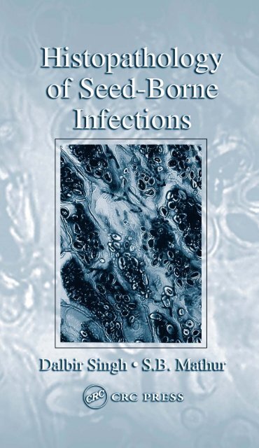
Cover Photograph: Section <strong>of</strong> chickpea (Cicer arietinum) cotyledon showing interandintracellular mycelium <strong>of</strong> Ascochyta rabiei, cause <strong>of</strong> blight in chickpea. (FromMaden, S. et al. 1975. <strong>Seed</strong> Sci. Technol. 3: 667–681. With permission.)Library <strong>of</strong> Congress Cataloging-in-Publication DataSingh, Dalbir, 1932-<strong>Histopathology</strong> <strong>of</strong> seed-borne infections / Dalbir Singh, S.B. Mathur.p. cm.Includes bibliographical references and index.ISBN 0-8493-2823-3 (alk. paper)1. <strong>Seed</strong>-borne phytopathogens. 2. <strong>Seed</strong>-borne plant diseases. 3. Histology, Pathological.I. Mathur, S. B. II. Title.SB732.8.S56 2004632.3—dc222004041407This book contains information obtained from authentic and highly regarded sources. Reprinted materialis quoted with permission, and sources are indicated. A wide variety <strong>of</strong> references are listed. Reasonableefforts have been made to publish reliable data and information, but the author and the publisher cannotassume responsibility for the validity <strong>of</strong> all materials or for the consequences <strong>of</strong> their use.Neither this book nor any part may be reproduced or transmitted in any form or by any means, electronicor mechanical, including photocopying, micr<strong>of</strong>ilming, and recording, or by any information storage orretrieval system, without prior permission in writing from the publisher.The consent <strong>of</strong> CRC Press LLC does not extend to copying for general distribution, for promotion, forcreating new works, or for resale. Specific permission must be obtained in writing from CRC Press LLCfor such copying.Direct all inquiries to CRC Press LLC, 2000 N.W. Corporate Blvd., Boca Raton, Florida 33431.Trademark Notice: Product or corporate names may be trademarks or registered trademarks, and areused only for identification and explanation, without intent to infringe.Visit the CRC Press Web site at www.crcpress.com© 2004 by CRC Press LLCNo claim to original U.S. Government worksInternational Standard Book Number 0-8493-2823-3Library <strong>of</strong> Congress Card Number 2004041407Printed in the United States <strong>of</strong> America 1 2 3 4 5 6 7 8 9 0Printed on acid-free paper
PrefaceThe book deals with only one aspect <strong>of</strong> seed-borne infection — the histopathology.Since the publication <strong>of</strong> the late Dr. Paul Neergaard’s book, <strong>Seed</strong> Pathology, whichstill remains an invaluable guide, phenomenal progress has taken place in the subject.Recent information on histopathology <strong>of</strong> seeds infected by different groups <strong>of</strong>microorganisms is scattered in numerous research periodicals. An attempt has thereforebeen made to consolidate this scattered information and present a coordinatedand coherent account. Information on flower and development <strong>of</strong> anther and ovuleleading to the formation <strong>of</strong> seed, and variability in seed structure <strong>of</strong> crop plants,relevant to studies in seed pathology has also been provided. Much <strong>of</strong> the informationis based on the material used by the authors for their teaching and incorporatesimportant developments in histopathology. A large number <strong>of</strong> the illustrations usedare from the studies and publications <strong>of</strong> the authors and their collaborators.Up-to-date scientific names are used for pathogens based on the followingpublications:Farr, D.P., Ellis. G.F., Chamunis, G.P., and Rossman, A.Y. 1989. Fungi on Plants and PlantProducts in United States. APS Press, St. Paul, MN.Fauquet, C.M. and Martelli, G.P. 1995. Updated ICTV list <strong>of</strong> names and abbreviations <strong>of</strong>viruses, viroides and satellites infecting plants. Arch. Virol. 140: 393–413.Fauquet, C.M. and Mayo, M.A. 1999. Abbreviations <strong>of</strong> plant virus names — 1999. Arch.Virol. 144: 1249–1273.Young, J.M., Saddler, G.S., Takikawa, Y., De Boer, S.H., Vauterin, L., Gardan, L., Gvozdyak,R.I., and Stead, D.E. 1996. Names <strong>of</strong> plant pathogenic bacteria, 1864–1995. Rev.Plant Pathol. 75: 721–763.This book will be useful to students, teachers, and researchers in seed pathologyand seed technology. Personnel working in seed health testing laboratories, plantquarantine, and agro-industries will find this book helpful in formulating strategiesfor testing, interception, and control <strong>of</strong> pathogens occurring as internal infections.Dalbir SinghS.B. Mathur
AcknowledgmentsThe authors are grateful to Dr. Carmen Nieves Mortensen, Associate Pr<strong>of</strong>essor, andMr. S.E. Albrechtsen, former Associate Pr<strong>of</strong>essor at the Danish Government Institute<strong>of</strong> <strong>Seed</strong> Pathology for Developing Countries, Copenhagen, for perusing Chapters 6(<strong>Seed</strong> Infection by Bacteria) and 7 (<strong>Seed</strong> Infection by Viruses), respectively, andmaking critical comments and suggestions. We thank Pr<strong>of</strong>. Thomas W. Carroll,Department <strong>of</strong> Plant Pathology, Montana State University, Bozeman, U.S.A., andDr. Andy J. Maule, Department <strong>of</strong> Virology, John Innes Centre, Norwich, U.K., forproviding literature on viruses.We wish to thank the publishers and executives <strong>of</strong> journals and books, andindividuals for granting permission to reproduce figures from their publications. Dueacknowledgment has been made for such figures. Special thanks are due to thefollowing individuals for providing photographs from their files: Pr<strong>of</strong>. Rolland R.Dute, Auburn University, Auburn, Alabama; Pr<strong>of</strong>. S.V. Thomson, Utah State University,Logan; Pr<strong>of</strong>. A.M. Alvarez, University <strong>of</strong> Hawaii, Manoa; Dr. A. Halfon-Meiri, The Volcani Centre, Bet Dagan, Israel; Dr. M.J. Christey, Christchurch, NewZealand; and Dr. Eigil de Neergaard, Royal Veterinary and Agricultural University,Copenhagen, Denmark.We thank Ms. Anette Højbjerg Hansen for her patience during the computertyping <strong>of</strong> the manuscript and Mr. Magdi El-din Ragab for his skillful cooperationin arranging the figures. We thank Ms. Henriette Westh for processing the finalmanuscript for submission.Dalbir Singh is grateful to the Danish Ministry <strong>of</strong> Foreign Affairs (Danida) forsupporting his visits to the Institute <strong>of</strong> <strong>Seed</strong> Pathology in Denmark for planning andwriting the book. He is grateful to his colleagues at the Department <strong>of</strong> Botany,University <strong>of</strong> Rajasthan, Jaipur, for their interest and cooperation and to all hisresearch collaborators for their cooperation and for allowing him free use <strong>of</strong> theircontributions. He is especially thankful to Pr<strong>of</strong>. Tribhuwan Singh, University <strong>of</strong>Rajasthan, Jaipur, and Dr. Kailash Agarwal, Agarwal College, Jaipur, for usefuldiscussions and for improving some <strong>of</strong> the figures used in the book. The gracioushelp <strong>of</strong> Dr. Dileep Kumar during the entire period <strong>of</strong> manuscript preparation isgratefully acknowledged. Thanks are also due to Shri Rajesh Benara for typing themanuscript, and to Shri Mehar Chand and Shri Ankur at Jaipur for help withillustrations.Dalbir Singh expresses his deep gratitude to his wife, Prem Singh, and hischildren — Nidhi, Smita and Mayank — for their patience and cooperation whilehe was engaged in writing the book.
The AuthorsDalbir Singh, Ph.D., former Pr<strong>of</strong>essor <strong>of</strong> Botany, University<strong>of</strong> Rajasthan, Jaipur, India, received his Master’sdegree in Botany in 1952 and his Ph.D. in ReproductiveBiology and Developmental Morphology in 1959 fromAgra University. For 40 years (1952 to 1992), he taughtgenerations <strong>of</strong> graduate and postgraduate students, andconducted courses in reproductive biology, embryology,anatomy, seed pathology, and seed technology. Dr.Singh initiated the teaching <strong>of</strong> seed pathology and seedtechnology to postgraduate students in the Department<strong>of</strong> Botany at Jaipur in 1975.For the past 50 years, Dr. Singh has been involved inresearch concerning the development and structure <strong>of</strong> seed in economically importantfamilies <strong>of</strong> angiosperms and the histopathology <strong>of</strong> seeds infected with fungal pathogens.Since 1973 he has been associated with the Danish Government Institute <strong>of</strong><strong>Seed</strong> Pathology for Developing Countries (DGISP). He and his collaborators havemade significant contributions to the histopathology <strong>of</strong> a large number <strong>of</strong> fungalpathogens in the seeds <strong>of</strong> cereals, oilseeds, legumes, and spices. His research alsoconcerns histology <strong>of</strong> physiogenic disorders in pea and chickpea, nematode galldevelopment and structure in wheat, and more recently (after 1985), the histopathology<strong>of</strong> seeds infected with bacteria. He has guided 40 successful Ph.D. candidatesand has published 300 research papers. Dr. Singh was awarded the Birbal SahniMedal <strong>of</strong> the Indian Botanical Society in 1992 for his outstanding research contributions.Dr. Singh is an elected Fellow <strong>of</strong> the National Academy <strong>of</strong> Sciences. He visitedthe former U.S.S.R. in 1977 as a member <strong>of</strong> an Indian delegation <strong>of</strong> botanists undera bilateral exchange program. From 1986 to 1987, he was a Visiting Pr<strong>of</strong>essor atASEAN PLANT I, a Regional Plant Quarantine and Training Institute in KualaLumpur. Dr. Singh has been associated with several national and internationalbotanical societies. He served as the Secretary <strong>of</strong> the Indian Botanical Society from1986 to 1992 and as its President in 1995 and 1996. From 1993 to 1994, he wasthe President, Section <strong>of</strong> Botany, Indian Science Congress Association. He is currentlythe Additional Secretary <strong>of</strong> the International Society <strong>of</strong> Plant Morphologists.
S.B. Mathur, Ph.D., is the Director <strong>of</strong> the Danish GovernmentInstitute <strong>of</strong> <strong>Seed</strong> Pathology for DevelopingCountries (DGISP) in Copenhagen, Denmark, where hehas spent most <strong>of</strong> his pr<strong>of</strong>essional life as a pioneer inthe field <strong>of</strong> seed pathology. For more than 35 yearsDr. Mathur has been instrumental in realizing seedhealth as an important step toward fighting hunger inthe third world. His primary objective has been to fightseed-borne diseases, not only to find cures, but moreimportantly to investigate ways to detect seed-borneinfections in the laboratory and prevent outbreaks <strong>of</strong>diseases at an early stage, in both formal and informalseed sectors. Creating awareness <strong>of</strong> the importance <strong>of</strong> seed health, the relationshipbetween seed health and food production and food security, and the impact <strong>of</strong> goodquality seed on increase in yield has been a milestone in Dr. Mathur’s life. Hiscontributions to international agriculture have been recognized by the internationalcommunity. In 1992 he was awarded the prestigious FIS World <strong>Seed</strong> Prize, presentedby the International <strong>Seed</strong> Federation, Switzerland, and in 2002 he was awarded thePr<strong>of</strong>. K.M. Safeeulla Gold Medal by the University <strong>of</strong> Mysore, India.The Institute <strong>of</strong> <strong>Seed</strong> Pathology in Denmark, the brain-child <strong>of</strong> Dr. Mathur, isfinanced and supported by Danida (Ministry <strong>of</strong> Foreign Affairs <strong>of</strong> Denmark). Morethan 550 scientists and technologists from 72 developing countries have been educatedthere and have conducted basic and applied research related to solving seedpathological problems. Dr. Mathur has trained more than 400 agricultural scientistsfrom more than 70 countries in short courses, conducted in various countries <strong>of</strong> thedeveloping world. He has been responsible for the introduction and monitoring <strong>of</strong>seed health at the Consultative Group on International Agricultural <strong>Research</strong>(CGIAR) <strong>Center</strong>s and routine checking <strong>of</strong> germplasm for seed health at nationalplant quarantine inspection laboratories.Dr. Mathur is presently leading a group <strong>of</strong> renowned experts engaged in establishingtwo educational <strong>Seed</strong> Pathology <strong>Center</strong>s, one in India for Asia and the otherin Tanzania for Africa. The major goal <strong>of</strong> these centers is to develop trained personnelwho will be responsible for handling seed health issues and increase and improvefood and seed production, especially for resource-poor farmers.
ContentsChapter 1 Introduction ..........................................................................................11.1 The <strong>Seed</strong>...........................................................................................................11.2 Microorganisms in <strong>Seed</strong> ..................................................................................21.3 <strong>Histopathology</strong> .................................................................................................3References..................................................................................................................3Chapter 2 Reproductive Structures and <strong>Seed</strong> Formation .....................................72.1 Flower...............................................................................................................72.1.1 Vascularization <strong>of</strong> Flower ....................................................................92.2 Sterile Appendages.........................................................................................102.3 Fertile Appendages.........................................................................................112.3.1 Stamen................................................................................................112.3.1.1 Development <strong>of</strong> Microsporangium andMicrosporogenesis ..............................................................112.3.1.2 Development and Structure <strong>of</strong> Male Gametophyte ...........132.3.2 Carpel .................................................................................................132.3.2.1 Ovary...................................................................................142.3.2.2 Style ....................................................................................152.3.2.3 Stigma .................................................................................162.4 Nectaries.........................................................................................................172.5 Ovule ..............................................................................................................172.5.1 Structure and Types <strong>of</strong> Ovules ..........................................................172.5.2 Vascular Supply <strong>of</strong> Ovule..................................................................202.5.3 Cuticles in Ovule ...............................................................................202.5.4 Special Structures in Ovules..............................................................212.6 Development and Structure <strong>of</strong> Female Gametophyte ...................................212.7 Fertilization ....................................................................................................232.8 <strong>Seed</strong> Development .........................................................................................232.8.1 Endosperm..........................................................................................252.8.2 Embryo ...............................................................................................272.8.3 Changes in Nucellus ..........................................................................282.8.4 Changes in Chalaza............................................................................312.8.5 Changes in Integuments.....................................................................312.9 <strong>Seed</strong> Coat Development in Selected Genera.................................................322.9.1 Brassica..............................................................................................322.9.2 Crotalaria...........................................................................................322.9.3 Hibiscus and Gossypium....................................................................342.9.4 Cucurbita and Sechium......................................................................34
2.9.5 Lycopersicon.......................................................................................372.9.6 Lactuca ...............................................................................................372.9.7 Triticum ..............................................................................................392.10 Concluding Remarks......................................................................................40References................................................................................................................41Chapter 3 Structure <strong>of</strong> <strong>Seed</strong>s ..............................................................................473.1 Constitution <strong>of</strong> <strong>Seed</strong>s.....................................................................................473.2 Exomorphic Features .....................................................................................483.2.1 Color...................................................................................................483.2.2 Shape ..................................................................................................483.2.3 Size .....................................................................................................483.2.4 Surface................................................................................................493.2.5 Micropyle ...........................................................................................503.2.6 Hilum..................................................................................................513.2.7 Raphe..................................................................................................513.2.8 <strong>Seed</strong> Appendages ...............................................................................513.3 Internal Morphology ......................................................................................533.3.1 Gross Internal Morphology................................................................533.3.2 <strong>Seed</strong> Coat and Pericarp......................................................................553.4 <strong>Seed</strong> Structure in Selected Families ..............................................................553.4.1 Brassicaceae (Cruciferae) .................................................................553.4.2 Malvaceae...........................................................................................573.4.3 Linaceae .............................................................................................573.4.4 Fabaceae (Leguminosae) Subfamily Faboideae(Papilionatae)......................................................................................603.4.5 Cucurbitaceae .....................................................................................613.4.6 Apiaceae (Umbelliferae) ...................................................................633.4.7 Pedaliaceae ........................................................................................653.4.8 Solanaceae .........................................................................................663.4.9 Asteraceae (Compositae) ..................................................................683.4.10 Amaranthaceae ..................................................................................683.4.11 Chenopodiaceae ................................................................................703.4.12 Poaceae (Graminae) ..........................................................................703.5 Concluding Remarks......................................................................................74References................................................................................................................74Chapter 4 Penetration and Establishment <strong>of</strong> Fungi in <strong>Seed</strong> ..............................814.1 Environment <strong>of</strong> Ovule and <strong>Seed</strong>....................................................................814.2 Nature <strong>of</strong> the Pathogen ..................................................................................824.3 Infection in Developing <strong>Seed</strong>s.......................................................................824.3.1 Routes for Internal Ovary Infection ..................................................834.3.1.1 Direct Infection from Mother Plant ...................................834.3.1.2 Indirect Infection from Outside..........................................85
4.3.2 Routes for Infection from Ovary to Ovule and <strong>Seed</strong>........................914.4 Avenues <strong>of</strong> Infection in Threshed <strong>Seed</strong>s.......................................................924.5 Mechanism <strong>of</strong> Penetration <strong>of</strong> Ovary, Fruit, and <strong>Seed</strong> Surfaces....................934.6 Concluding Remarks......................................................................................96References................................................................................................................96Chapter 5 Location <strong>of</strong> Fungal Hyphae in <strong>Seed</strong>s ..............................................1015.1 Severity <strong>of</strong> Infection and Location..............................................................1015.2 Primary Sites <strong>of</strong> Colonization .....................................................................1045.3 Host–Pathogen Interactions .........................................................................1045.4 Mixed <strong>Infections</strong> ..........................................................................................1055.5 Colonization <strong>of</strong> <strong>Seed</strong> Tissues.......................................................................1075.5.1 Oomycetes........................................................................................1075.5.1.1 Phytophthora.....................................................................1075.5.1.2 Peronospora ......................................................................1105.5.1.3 Plasmopara .......................................................................1105.5.1.4 Sclerospora, Peronosclerospora, and Sclerophthora .......1105.5.1.5 Albugo ...............................................................................1115.5.2 Ascomycetes.....................................................................................1135.5.2.1 Protomyces........................................................................1135.5.2.2 Claviceps...........................................................................1155.5.2.3 Sclerotinia .........................................................................1195.5.2.4 Didymella..........................................................................1195.5.3 Basidiomycetes.................................................................................1195.5.3.1 Ustilaginales (Smuts and Bunts) ......................................1195.5.3.2 Uredinales (Rusts) ............................................................1245.5.4 Deuteromycetes................................................................................1255.5.4.1 Hyphomycetes...................................................................1255.5.4.2 Coelomycetes....................................................................1415.6 Endophytes ...................................................................................................1495.6.1 Stromatic Infection...........................................................................1505.6.2 Nonstromatic <strong>Infections</strong>...................................................................1515.6.3 Viability <strong>of</strong> Mycelium in <strong>Seed</strong> ........................................................1515.7 Implication <strong>of</strong> Internal Infection..................................................................1545.8 Concluding Remarks....................................................................................155References..............................................................................................................155Chapter 6 <strong>Seed</strong> Infection by Bacteria...............................................................1696.1 Penetration....................................................................................................1696.1.1 Invasion <strong>of</strong> Plant Parts .....................................................................1706.1.2 Spread in Plant and Course <strong>of</strong> Entry into Ovaryand Fruit and Ovule and <strong>Seed</strong>.........................................................1746.1.3 Invasion <strong>of</strong> Threshed and Disseminated <strong>Seed</strong>s ...............................1776.2 <strong>Histopathology</strong> <strong>of</strong> Infected <strong>Seed</strong>s................................................................178
6.2.1 Xanthomonas....................................................................................1786.2.2 Pseudomonas, Acidovorax, and Burkholderia.................................1856.2.3 Rathayibacter and Clavibacter ........................................................1876.2.4 Curtobacterium ................................................................................1886.2.5 Pantoea.............................................................................................1906.3 Survival in <strong>Seed</strong> ...........................................................................................1906.4 Concluding Remarks....................................................................................191References..............................................................................................................192Chapter 7 <strong>Seed</strong> Infection by Viruses ................................................................1997.1 Infection and Multiplication ........................................................................1997.2 Cellular Contacts, Isolation, and Transport Systems in Ovule and <strong>Seed</strong>...2057.3 Virus Movement...........................................................................................2067.3.1 Infected Plant ...................................................................................2067.3.2 Ovule and <strong>Seed</strong> ................................................................................2077.4 Localization in Reproductive Shoot, Ovule, and <strong>Seed</strong> ...............................2097.4.1 Barley Stripe Mosaic Virus (BSMV) and Similar Viruses .............2097.4.2 Bean Common Mosaic Virus (BCMV) ...........................................2177.4.3 Lettuce Mosaic Virus (LMV) ..........................................................2177.4.4 Pea <strong>Seed</strong>-<strong>Borne</strong> Mosaic Virus (PSbMV) ........................................2177.5 Cytopathological Effects..............................................................................2197.6 Inactivation or Longevity <strong>of</strong> Viruses in <strong>Seed</strong> during Maturationand Storage...................................................................................................2217.7 Concluding Remarks....................................................................................222References..............................................................................................................223Chapter 8 <strong>Seed</strong> Infection by Nematodes..........................................................2298.1 Penetration by Nematodes ...........................................................................2298.2 <strong>Histopathology</strong> .............................................................................................2318.2.1 Anguina tritici (Steinbuck) Chitwood (Ear Cockle Disease)..........2318.2.1.1 Disease Cycle....................................................................2318.2.1.2 Origin <strong>of</strong> Galls..................................................................2328.2.1.3 Histology <strong>of</strong> Developing and Mature Galls.....................2338.2.2 Anguina agrostis (Steinbuch) Filipjev(Bent Grass Gall Nematode)............................................................2358.2.2.1 Histology and Development <strong>of</strong> Galls...............................2358.2.3 Anguina agropyronifloris Norton(Western Wheatgrass Nematode).....................................................2388.2.3.1 Origin and Histology <strong>of</strong> Galls..........................................2388.2.4 Ditylenchus destructor Thorne (Potato Nematode in Groundnut)...2388.2.5 Aphelenchoides besseyi Christie (White Tip Nematode <strong>of</strong> Rice) ....2398.2.6 Aphelenchoides arachidis Bos (Testa Nematode <strong>of</strong> Groundnut)....2398.2.7 Pratylenchus brachyurus (Godfrey) Filipjev...................................2418.3 Association <strong>of</strong> Nematode and Bacteria .......................................................241
8.4 Survival in <strong>Seed</strong> ...........................................................................................2428.5 Concluding Remarks....................................................................................244References..............................................................................................................244Chapter 9 Physiogenic or Nonpathogenic <strong>Seed</strong> Disorders ..............................2479.1 Mineral Nutrient Deficiency ........................................................................2499.1.1 Marsh Spot in Peas ..........................................................................2499.1.1.1 Histology...........................................................................2499.2 Humidity Effects ..........................................................................................2519.2.1 Low Humidity Effects......................................................................2519.2.1.1 Hollow Heart.....................................................................2519.2.1.2 Necrosis in Lettuce Cotyledons .......................................2559.2.1.3 Cracking <strong>of</strong> Cotyledons....................................................2559.2.2 High Humidity Effects.....................................................................2579.3 Concluding Remarks....................................................................................257References..............................................................................................................258Chapter 10 Microtechniques in <strong>Seed</strong> <strong>Histopathology</strong>......................................26110.1 Choice <strong>of</strong> Material .......................................................................................26110.2 Determination <strong>of</strong> the Identity <strong>of</strong> Internal Mycelium ..................................26210.2.1 Procedure for Component Plating ...................................................26210.3 <strong>Seed</strong> S<strong>of</strong>tening .............................................................................................26210.4 Histological Methods ...................................................................................26310.4.1 Whole-Mount Method......................................................................26310.4.2 Freehand Sections ............................................................................26410.4.3 Microtomy........................................................................................26410.4.3.1 Fixing and Storage............................................................26410.4.3.2 Dehydration.......................................................................26410.4.3.3 Infiltration .........................................................................26510.4.3.4 Embedding ........................................................................26510.4.3.5 S<strong>of</strong>tening <strong>of</strong> Embedded Material .....................................26510.4.3.6 Sectioning and Mounting <strong>of</strong> Ribbons ..............................26510.4.3.7 Staining and Mounting .....................................................26610.5 Procedures for Preparing Some Reagents and Stains .................................26610.5.1 Fixative.............................................................................................26610.5.2 Adhesives .........................................................................................26610.5.3 Mounting Media...............................................................................26710.5.3.1 Aqueous Mounting Media................................................26710.5.3.2 Nonaqueous Mounting Media ..........................................26810.5.4 Stains ................................................................................................268References..............................................................................................................269Index......................................................................................................................271
1 IntroductionThe reports on the number <strong>of</strong> microorganisms associated with seeds have increasedgradually during the latter half <strong>of</strong> the 20th century. This is obvious from the firstand the most recent editions <strong>of</strong> An Annotated List <strong>of</strong> <strong>Seed</strong>-<strong>Borne</strong> Diseases by Noble,De Tempe, and Neergaard (1958), and Richardson (1990), respectively. The organismsoccur with seed either as contaminants adhering to the seed surface, looselymixed with seed, or as an infection present inside the seed tissues. This bookdescribes penetration and location <strong>of</strong> microorganisms in seed tissues. Informationon location <strong>of</strong> infection in seeds using histological techniques alone is considered.The presence <strong>of</strong> internal infection in seed detected in transmission studies is beyondthe scope <strong>of</strong> this book.In order to appreciate penetration, course <strong>of</strong> infection, location, and the effect<strong>of</strong> infection <strong>of</strong> microorganisms in floral and seed tissues, it is necessary to have agood understanding <strong>of</strong> the structure <strong>of</strong> flowers and changes in fertile appendages —stamen and carpel — leading to the formation <strong>of</strong> seed. Neergaard (1979) has includedinformation on morphology and anatomy <strong>of</strong> seed in relation to transmission <strong>of</strong>pathogens. This account is fragmentary and does not include information on manycritical steps in the formation <strong>of</strong> seed. Considerable new knowledge, includingultrastructure <strong>of</strong> reproductive components — embryo sac, endosperm, embryo, andtheir surrounding tissues — has been provided (Johri, 1984; Johri, Ambegaokar, andSrivastava, 1992). These data elucidate the contacts and barriers among the tissues<strong>of</strong> developing seed. The structure <strong>of</strong> mature seed, including one-seeded dry indehiscentfruits, is also highly variable in angiosperms (Netolitzky, 1926; Corner,1976). The variations in size <strong>of</strong> the hilum, micropyle opening, nature and thickness<strong>of</strong> the cutile, and thickness <strong>of</strong> seed coat and pericarp have shown direct correlationwith the penetration and location <strong>of</strong> fungal pathogens in certain host–parasite interfaces.Chapter 2 provides a concise up-to-date account <strong>of</strong> the structure and development<strong>of</strong> floral parts and the formation <strong>of</strong> seed. Chapter 3 deals with the structure<strong>of</strong> mature seed in selected families <strong>of</strong> angiosperms. Chapters 5 through 8 concernthe histopathology <strong>of</strong> seed infections by fungi, bacteria, viruses, and nematodes.Chapter 9 describes physiogenic seed disorders. Chapter 10 includes a brief account<strong>of</strong> histopathological techniques and tips. Only a basic account is given in Chapter10; several detailed books on plant microtechnique and transmission and scanningelectron microscopy are available.1.1 THE SEEDBiologically, seed is the ripened ovule. In angiosperms, to which a majority <strong>of</strong> thecrop plants belong, the ovules are borne in the ovary, the basal part <strong>of</strong> the gynoecium1
2 <strong>Histopathology</strong> <strong>of</strong> <strong>Seed</strong>-<strong>Borne</strong> <strong>Infections</strong>(pistil). The seed formation takes place in situ through a series <strong>of</strong> integrated sequentialsteps in the life cycle <strong>of</strong> the flowering plant (Maheshwari, 1950, 1963; Johri,1984). After pollination and fertilization, changes in different parts <strong>of</strong> the ovule, i.e.,the embryo sac (zygote and primary endosperm nucleus), nucellus, chalaza, andintegument, lead to the formation <strong>of</strong> seed. The term seed, when used sensu lato,includes one-seeded dry indehiscent fruits that are the dispersal propagative unitsin plants <strong>of</strong> several families such as Poaceae, Asteraceae, Apiaceae, and Chenopodiaceae.In the present treatment, the term seed is used in a loose sense.<strong>Seed</strong> is an autonomous living unit and links successive generations. Structurally,it consists <strong>of</strong> an embryo (new plantlet), a protective covering — seed coat, pericarp,or both — and reserve food material, which may be present in the endosperm,perisperm, or embryo. It has the capacity to withstand desiccation and retain viabilityunder unfavorable environments or until it germinates. These properties <strong>of</strong> seed makeit an important commodity for storage as well as transport to new areas or countries,for planting or for edible purposes. The structure <strong>of</strong> seed is fairly constant in aspecies, but varies in different plant taxa (Netolitzky, 1926; Corner, 1976).<strong>Seed</strong>s, if infected by microorganisms, will act as carriers. If the organisms remainviable they will result in development <strong>of</strong> disease in the new crop. Infected seeds are<strong>of</strong>ten responsible for the spread <strong>of</strong> diseases to new areas. For this reason seed hasbecome an object <strong>of</strong> plant quarantine internationally. Countries also use domesticseed certification, including seed health testing, as a method <strong>of</strong> quality control <strong>of</strong>seed.1.2 MICROORGANISMS IN SEEDFungi, bacteria, viruses, and nematodes are known to be seed-borne (Neergaard,1979; Maude, 1996; Agarwal and Sinclair, 1997). Fungi form a major group <strong>of</strong>pathogens that are seed-borne as well as seed-transmitted. In addition to saprophytesand parasites, fungi are known to form an inherent association with seeds <strong>of</strong> somemembers <strong>of</strong> Cistaceae, Ericaceae, and Orchidaceae. In the Orchidaceae, the seedswill not usually germinate without the presence <strong>of</strong> a mycorhizal fungus (Rayner,1915). The seed coat in seeds <strong>of</strong> Helianthemum chamaecistus (Cistaceae) harbors afungus that seems essential for normal germination <strong>of</strong> seed. In the absence <strong>of</strong> thefungus, the plumule fails to emerge, and roots also are not formed at the time <strong>of</strong>germination (Boursnell, 1950).The list <strong>of</strong> saprophytic and parasitic fungi associated with seeds <strong>of</strong> differentplants is very large (Richardson, 1990), and they belong to all fungal classes. Thefungi that are discussed in this book and for which histopathological information isavailable belong to the division Eumycota, subdivision Mastigomycotina, classOomycetes, subdivisions Ascomycotina, Basidiomycotina, and Deuteromycotina.The members <strong>of</strong> Deuteromycotina dominate and these fungi belong to the classesHyphomycetes and Coelomycetes. The endophytic fungi are discussed separately.A large number <strong>of</strong> viruses, including cryptoviruses and viroids, are known tobe seed-borne, but the information on seeds infected by viruses is limited and mostlyinconclusive. The better-studied viruses are barley stripe mosaic virus (BSMV) andpea seed-borne mosaic virus (PSbMV) due to the studies <strong>of</strong> Carroll and co-workers
Introduction 3(Carroll, 1969, 1974; Carroll and Mayhew, 1976a,b; Mayhew and Carroll, 1974)and Wang and Maule (1992, 1994), respectively. Similarly, the histopathology <strong>of</strong>seeds affected by bacteria is poorly studied, and the investigations are usuallyconfined to seeds infected by Acidovorax, Burkholderia, Clavibacter, Curtobacterium,Pantoea, Pseudomonas, Rathayibacter, and Xanthomonas.<strong>Seed</strong>-borne nematodes also occur as seed infestation or seed infection. The lattercauses either seed gall formation (Anguina spp.) or symptomatic or symptomlessinfections.1.3 HISTOPATHOLOGYEver since Cobb (1892; see Royle, 1976) proposed the mechanical theory <strong>of</strong> rustresistance indicating that morphological features, such as thick cuticle, waxy covering,small stomata, abundant leaf hairs, and upright leaves, might be responsiblefor the resistance <strong>of</strong> wheat varieties to Puccinia graminis, numerous studies onhistology <strong>of</strong> infected plant parts, particularly leaves and stems, have been carriedout. This information has been summarized in excellent reviews on (1) histology <strong>of</strong>defense (Akai, 1959; Royle, 1976; Schonbeck and Schlouster, 1976); (2) abilities<strong>of</strong> pathogens to breach host barriers (Dickinson, 1960; Emmett and Parberry, 1975;Dodman, 1979); and (3) physiology and biochemistry <strong>of</strong> penetration and infection(Flentje, 1959; Alberschein, Jones, and English, 1969; Mount, 1978; Durbin, 1979;Kollattukudy, 1985).Although the first observation on internal presence <strong>of</strong> the mycelium <strong>of</strong> Colletotrichumlindemuthianum in cotyledons <strong>of</strong> seeds <strong>of</strong> Phaseolus vulgaris was madeas early as 1883, further progress until 1950 was rather slow. It is during the latterhalf <strong>of</strong> the 20th century and more particularly after 1970 that several comprehensivereports have appeared on the penetration and location <strong>of</strong> microorganisms in seeds.Early information has been summarized by Baker (1972), Neergaard (1979), andAgarwal and Sinclair (1997).Various histological techniques, e.g., the embryo extraction method initially usedby Skvortzov (1937), whole-mount preparations <strong>of</strong> seed components (Maden et al.,1975; Singh, Mathur, and Neergaard, 1977), free hand sections, and microtomesections have been used. Microtome sections <strong>of</strong> weakly, moderately, and heavilyinfected seeds alone provide information on exact expanse <strong>of</strong> mycelium in seed andalso the effects <strong>of</strong> host–parasite interactions (Singh, 1983). Although used primarilyin the study <strong>of</strong> virus infections and in a limited way in the study <strong>of</strong> fungal infections,transmission electron microscopy (TEM) and scanning electron microscopy (SEM)have yielded valuable information, which certainly surpasses the results <strong>of</strong> lightmicroscopy.REFERENCESAgarwal, V.K. and Sinclair, J.B. 1997. Principles <strong>of</strong> <strong>Seed</strong> Pathology, 2nd ed. CRC Press,Boca Raton, FL.
4 <strong>Histopathology</strong> <strong>of</strong> <strong>Seed</strong>-<strong>Borne</strong> <strong>Infections</strong>Akai, S. 1959. Histology <strong>of</strong> defence in plants. In Plant Pathology: An Advanced Treatise.Horsfall, J.G. and Dimond, J.G., Eds. Academic Press, London. Vol. 1, 391–434.Albershein, P., Jones, T.M., and English, P.D. 1969. Biochemistry <strong>of</strong> the cell wall in relationto infective processes. Ann. Rev. Phytopathol. 7: 171–194.Baker, K.F. 1972. <strong>Seed</strong> Pathology. In <strong>Seed</strong> Biology. Kozlowski, T.T., Ed. Academic Press,New York. Vol. 2, 317–416.Boursnell, J.G. 1950. The symbiotic seed-borne fungus in the Cistaceae. I. Distribution andfunction <strong>of</strong> the fungus in the seedling and in the tissues <strong>of</strong> the mature plant. Ann.Bot. (Lond.) N.S. 14: 217–243.Carroll, T.W. 1969. Electron microscopic evidence for the presence <strong>of</strong> Barley stripe mosaicvirus in cells <strong>of</strong> barley embryos. Virology 37: 649–657.Carroll, T.W. 1974. Barley stripe mosaic virus in sperm and vegetative cells <strong>of</strong> barley pollen.Virology 60: 21–28.Carroll, T.W. and Mayhew, D.E. 1976a. Anther and pollen infection in relation to the pollenand seed transmissibility <strong>of</strong> two strains <strong>of</strong> Barley stripe mosaic virus in barley. Can.J. Bot. 54: 1604–1621.Carroll, T.W. and Mayhew, D.E. 1976b. Occurrence <strong>of</strong> virus in developing ovules and embryosacs <strong>of</strong> barley in relation to the seed transmissibility <strong>of</strong> Barley stripe mosaic virus.Can. J. Bot. 54: 2497–2512.Corner, E.J.H. 1976. <strong>Seed</strong>s <strong>of</strong> Dicotyledons. Vols. 1 and 2. Cambridge University Press,Cambridge, U.K.Dickinson, S. 1960. The mechanical ability to breach the host-barriers. In Plant Pathology:An Advanced Treatise. Horsfall, J.G. and Dimond, J.G, Eds. Academic Press, London.Vol. 1, 203–232.Dodman, R.L. 1979. How the defenses are breached. In Plant Disease: An Advanced Treatise.Horsfall, J.G. and Cowling, E.B., Eds. Academic Press, New York. Vol. 4, 135–151.Durbin, R.D. 1979. How the breachhead is widened. In Plant Disease: An Advanced Treatise.Horsfall, J.G. and Cowling, E.B., Eds. Academic Press, New York. Vol. 4, 155–162.Emmett, R.W. and Parberry, D.G. 1975. Appressoria. Ann. Rev. Phytopathol. 13: 147–167.Flentje, N.T. 1959. The physiology <strong>of</strong> penetration and infection. In Plant Pathology, Problemsand Progress, 1908–1958. University <strong>of</strong> Wisconsin, Madison, 76–87.Johri, B.M., Ed. 1984. Embryology <strong>of</strong> Angiosperms. Springer-Verlag, Berlin.Johri, B.M., Ambegaokar, K.B., and Srivastava, P.S. 1992. Comparative Embryology <strong>of</strong>Angiosperms. Vols. 1 and 2. Springer-Verlag, Berlin.Kolattukudy, P.E. 1985. Enzymatic penetration <strong>of</strong> the plant cuticle by fungal pathogens. Ann.Rev. Phytopathol. 23: 223–250.Maden, S., Singh, D., Mathur, S.B., and Neergaard, P. 1975. Detection and location <strong>of</strong> seedborneinoculum <strong>of</strong> Ascochyta rabiei and its transmission in chickpea (Cicer arietinum).<strong>Seed</strong> Sci. Technol. 3: 667–681.Maheshwari, P. 1950. An Introduction to the Embryology <strong>of</strong> Angiosperms. McGraw-Hill, NewYork.Maheshwari, P., Ed. 1963. Recent Advances in the Embryology <strong>of</strong> Angiosperms. InternationalSociety <strong>of</strong> Plant Morphologists, University <strong>of</strong> Delhi, India.Mayhew, D.E. and Carroll, T.W. 1974. Barley stripe mosaic virus in the egg cell and egg sac<strong>of</strong> infected barley. Virology 58: 561–567.Maude, R.B. 1996. <strong>Seed</strong>-<strong>Borne</strong> Diseases and Their Control. CAB International, Wallingford,U.K.Mount, M.S. 1978. Tissue is disintegrated. In Plant Disease: An Advanced Treatise. Horsfall,J.G. and Cowling, E.B., Eds. Academic Press, New York. Vol. 3, 279–293.Neergaard, P. 1979. <strong>Seed</strong> Pathology. Vols. 1 and 2. Macmillan Press, London.
Introduction 5Netolitzky, F. 1926. Antomie der Angiospermen-Samen. In Handbuch der Pflanzenanatomie.Linsbauer, K., Ed. Abt. 2, Teil 2, Bd. 10. G. Borntraeger, Berlin.Noble, M., de Tempe, J., and Neergaard, P. 1958. An Annotated List <strong>of</strong> <strong>Seed</strong>-<strong>Borne</strong> Diseases.Commonwealth Mycological Institute, Kew, Richmond, Surrey, U.K.Rayner, M.C. 1915. Obligate symbiosis in Calluna vulgaris. Ann. Bot. (Lond.) N.S. 29:96–131.Richardson, M.J. 1990. An Annotated List <strong>of</strong> <strong>Seed</strong>-<strong>Borne</strong> Diseases, 4th ed. Proceedings <strong>of</strong>the International <strong>Seed</strong> Testing Association, Wageningen, the Netherlands.Royle, D.J. 1976. Structural features to plant diseases. In Biochemical Aspects <strong>of</strong> PlantParasite Relationships. Frank, J. and Thresefall, D.R., Eds. Academic Press, NewYork, pp. 162–193.Schonbeck, F. and Schlouster, E. 1976. Preformed substances as potential protectants. InPhysiological Plant Pathology. Heitefuss, R. and Williams, P.H., Eds. Springer-Verlag, Berlin, pp. 653–678.Singh, D. 1983. <strong>Histopathology</strong> <strong>of</strong> some seed-borne infections: a review <strong>of</strong> recent investigations.<strong>Seed</strong> Sci. Technol. 11: 651–663.Singh, D., Mathur, S.B., and Neergaard, P. 1977. <strong>Histopathology</strong> <strong>of</strong> sunflower seeds infectedby Alternaria tenuis. <strong>Seed</strong> Sci. Technol. 5: 579–586.Skvortzov, S.S. 1937. A simple method for detecting hyphae <strong>of</strong> loose smut on wheat grains.Plant Prot. Leningrad 15: 90–91.Wang, D. and Maule, A.J. 1992. Early embryo invasion as a determinant in pea <strong>of</strong> the seedtransmission <strong>of</strong> Pea seed-borne mosaic virus. J. Gen. Virol. 73: 1615–1620.Wang, D. and Maule, A.J. 1994. A model for seed transmission <strong>of</strong> a plant virus: genetic andstructural analyses <strong>of</strong> pea embryo invasion by Pea seed-borne mosaic virus. PlantCell 6: 777–787.
2Reproductive Structuresand <strong>Seed</strong> Formation<strong>Seed</strong> is the end product <strong>of</strong> sexual reproduction that takes place through sequentialchanges in the reproductive shoot, the flower in angiosperms. The ovules are bornein a closed structure, the ovary. The ovules and seeds may get infected at any stageduring their development either directly through the mother plant or through theinfected floral parts, including bracts and nectaries. The contacts between the plantand flower and in turn with the ovule and seed are important in understanding thecourse <strong>of</strong> such infections. The interface <strong>of</strong> pathogen and host may cause morphologicaland anatomical changes in tissues <strong>of</strong> the flower and may also affect thereproductive cycle, including seed formation. Green ear disease <strong>of</strong> pearl millet,caused by Sclerospora graminicola, results in the transformation <strong>of</strong> florets into leafystructures. Inflorescence or flower infection by Protomyces macrosporus in corianderand Albugo candida in Brassicaceae cause hypertrophy in the latter, and fruit gallformation in the former. Claviceps purpurea and other species, when infecting cerealflorets, cause the ovary to form fungal sclerotia instead <strong>of</strong> producing a kernel. Thecontents in the ovary are replaced by smut chlamydospores due to the infection <strong>of</strong>Sphacelotheca, Tilletia, Tolyposporium, and Ustilago in various cereal crops.The structure <strong>of</strong> flowers, the reproductive cycle, and seed formation are describedin this chapter. The aspects that may be relevant to pathogenesis are highlighted. Anoutline <strong>of</strong> the reproductive cycle is given in Figure 2.1.2.1 FLOWERThe flowers may occur singly (cotton, kenaf, cucumber, pumpkin, and melon) oraggregated in inflorescence — small (tomato, potato, and brinjal) to massive (rice,wheat, pearl millet, sunflower, and coriander). Stalked or sessile flowers are borneloosely (coriander, carrot, and tomato) or compactly (wheat, pearl millet, maize, andsunflower), have an orderly arrangement, and depending on the order <strong>of</strong> development<strong>of</strong> flowers, the inflorescence is classified into indeterminate (racemose) or determinate(cymose) type. In the former, the order <strong>of</strong> development <strong>of</strong> flowers is acropetal,with the youngest bud and flower near the growing tip (Brassica, Raphanus, andLinum), whereas in the latter, it is basipetal, with the growing point ending in aflower and subsequent ones produced in the axil(s) <strong>of</strong> bracts below the terminalflower (Lycopersicon, Sesamum, and Solanum). The presence <strong>of</strong> accessory structures,e.g., spathe in spadix (maize); involucre <strong>of</strong> bracts in capitulum (sunflower) andumbel (coriander and carrot); and glumes, palea, and lemma with florets in simple7
8 <strong>Histopathology</strong> <strong>of</strong> <strong>Seed</strong>-<strong>Borne</strong> <strong>Infections</strong>FLOWER (Reproductive shoot <strong>of</strong> angiosperms)Fertile AppendagesStamenantheranther sac(microsporangium)microspore mother cellsCallose depositionpachytene I onward,isolationTetrads <strong>of</strong> microspores(Tetrahedral andisobilateral mostly)MEIOSISCarpelovulenucellus(megasporangium)megasporemother cellTetrads <strong>of</strong> megaspore(Usually linear)Callose degradationUninucleate pollen grainsTwo mitolicdivisionsMale gametophyte(Pollen grains)3-celled with exine,and intine, autonomousphasePollination(Pollen on stigma, germination, pollen tube penetration)Pollen tubeStyleOvaryFunctional megasporeThree mitolicdivisionsFemale gametophytye(Embryo sac)(egg, 2 synergids,central cell, 3 antipodalcells)Through one <strong>of</strong> the synergidsOvuleEmbryo sacDOUBLE FERTILIZATIONsperm 1sperm 2eggcentral cellSyngamyTriple fusionChanges in ovule parts otherthan in embryo sac—Nucellus—Chalaza—Integument(s)<strong>Seed</strong> coatZygoteProembryoEmbryoPrimaryendosperm cellEndosperm(Absorbed orpersistent)True seeds(Ovary and/or other accessory structures<strong>of</strong> flower forming protective covering)1-seeded indehiscent fruits(caryopsis, cypsil, achene, utricle, etc.)FIGURE 2.1 Schematic representation <strong>of</strong> reproductive cycle and seed formation, includingone-seeded indehiscent fruits in angiosperms.
Reproductive Structures and <strong>Seed</strong> Formation 9or compound spikes (wheat, barley, pearl millet, and rice), provide additional protectionto flowers, but cause increased humidity around them. They may also providea conducive environment for the development <strong>of</strong> pathogens that succeed in invadingthem.The flower is a shoot <strong>of</strong> determinate growth. The distal end <strong>of</strong> the axis is swollenor cup-shaped, forming a thalamus or receptacle, which bears laterally floral organs.A typical flower <strong>of</strong> dicotyledons has four types <strong>of</strong> organs comprising two whorls <strong>of</strong>sterile appendages, the sepals and petals collectively called the calyx and corolla,respectively, and two whorls <strong>of</strong> fertile appendages, the stamens and carpels, collectivelytermed the androecium and gynoecium. The appendages are arranged on thereceptacle in succession spirally or in whorls. Many variations occur in size, shape,color, and number <strong>of</strong> members in each whorl and in their organization. Usually inmonocotyledons, there is only one whorl <strong>of</strong> sterile appendages, termed the perianth(onion and garlic). Flowers may be bisexual or unisexual. In the unisexual conditionstaminate and pistillate flowers are monoecious, or borne on one plant, as in maize,cucumber, pumpkin, and squash. If the staminate and pistillate flowers occur ondifferent plants, as in papaya, they are dioecious.In a flower, if the floral appendages are inserted successively one above the otherbelow the gynoecium (carpel), the flower is hypogynous and the ovary is superior(Figure 2.2A). When the receptacle becomes concave and surrounds the ovary or isfused with the ovary wall so that the sepals, petals, and stamens arise from the top<strong>of</strong> the ovary (Figure 2.2B), the flower is epigynous and the ovary is inferior as incoriander, cumin, carrot, cucumber, pumpkin, squash, sunflower, lettuce, apple, andpear. In an epigynous flower, the ovary is exposed to the environment from initiationuntil maturity, i.e., the formation <strong>of</strong> fruit.The flower in the grass family, Poaceae, is subsessile and bracteate; the drychaffy lemma, which is usually awned, is the bract. The dry membranous structureon the posterior side is known as the palea, which represents two fused posterolaterallysituated bracteoles. The palea encloses the flower and itself is enclosed by thelemma. The flower consists <strong>of</strong> lodicules (tepals), androecium, and gynoecium. Usuallythere are two fleshy, <strong>of</strong>ten hairy, lodicules situated in the anterolateral position.In Bambusa, there are six lodicules. Above the lodicules, there are usually threestamens and rarely more, six or less in Oryza and Bambusa, etc., two in Anthraxon,and one in Uniola. The gynoecium is tricarpellary and syncarpous and the ovary isunilocular with a single large ovule. The vascular anatomy has shown that theplacentation is parietal and that two <strong>of</strong> the three placentae are sterile. The ovarybears two laterally situated feathery styles and stigma.2.1.1 VASCULARIZATION OF FLOWERThe anatomical structures <strong>of</strong> the peduncle (axis <strong>of</strong> the inflorescence) and the pedicelare similar to that <strong>of</strong> the stem. The vascular cylinder may be continuous or split.The vascular bundles <strong>of</strong> the pedicel at a higher level break up and give out tracesto the floral appendages in succession in a hypogynous flower (Figure 2.2A). Thevascular traces meant for the floral appendages enter each unit either unbranched orafter undergoing further branching. Each sepal generally receives three traces, a
10 <strong>Histopathology</strong> <strong>of</strong> <strong>Seed</strong>-<strong>Borne</strong> <strong>Infections</strong>stistyoovdctvsvctspespeseoseABFIGURE 2.2 Structure and vascularization <strong>of</strong> angiospermous flower. A, Longitudinal section(Ls) showing vascular supply in hypogynous flower, traces <strong>of</strong> each whorl separate in thethalamus and diverge at corresponding places. B, Ls epigynous flower, traces for carpels arethe first to diverge while those <strong>of</strong> calyx, corolla, and androecium diverge at levels above theovary. (Abbreviations: dct, dorsal carpellary trace; o, ovary; ov, ovule; pe, petal; s, stamen;se, sepal; sti, stigma; sty, style; vct, ventral carpellary trace; vs, vascular supply.)petal with a single median trace or a median trace with two or more laterals, andthe stamen a single trace. The carpel is vascularized with a median (dorsal) and twolateral (ventral or marginal) traces. The ventral traces unite partially or completelyinto a single ventral trace. The ventral trace(s) are generally limited to the ovulebearingpart <strong>of</strong> the carpel. The vasculature in an epigynous flower differs only inthe manner <strong>of</strong> resolution or departure <strong>of</strong> vascular traces from the main cylinder. Thevascular traces <strong>of</strong> the whorls <strong>of</strong> appendages depart at levels higher than that <strong>of</strong> thegynoecium (Figure 2.2B). Thus, in a flower there is continuity in the vascular supply<strong>of</strong> different whorls with that <strong>of</strong> the mother plant through the peduncle and pedicel.2.2 STERILE APPENDAGESSepals are more or less leaflike or bractlike in form, structure, and vasculature. Theyare usually green and rarely petaloid. Anatomically, the epidermis shows deposition<strong>of</strong> cutin and development <strong>of</strong> stomata and trichomes similar to those on foliage leaves.The mesophyll is undifferentiated or rarely differentiated into palisade and spongytissues.Petals vary in shape, size, and color. In most flowers, they form the mostconspicuous whorl and help in insect pollination. The epidermis bears stomata,functional or undifferentiated and nonfunctioning, and may develop intercellular
Reproductive Structures and <strong>Seed</strong> Formation 11spaces over-arched by the cuticle. The mesophyll is only a few cells thick, exceptin flowers with fleshy petals. The cuticle is commonly striated (Martens, 1934). Thefragrance <strong>of</strong> flowers is produced by volatile substances, mostly essential oils, occurringin the epidermal cells. In some plants, such as Lupinus and Narcissus, thefragrance originates in special glands called osmophors.2.3 FERTILE APPENDAGESThe stamens and carpels, male and female appendages, respectively, are highlyspecialized and form the seat <strong>of</strong> the development <strong>of</strong> male and female gametophytes,pollen and embryo sac, in angiosperms. The development and structure <strong>of</strong> male andfemale gametophytes have been studied in numerous plants <strong>of</strong> dicotyledons andmonocotyledons. Detailed information on this topic can be found in the publications<strong>of</strong> Schnarf (1929, 1931), Maheshwari (1950, 1963), Davis (1966), Johri (1984), andJohri, Ambegaokar, and Shrivastava (1992).2.3.1 STAMENA typical stamen consists <strong>of</strong> the filament bearing a two-lobed and four-loculed anther(Figure 2.3A, B). The two lobes <strong>of</strong> the anther are separated by a sterile tissue, calledthe connective. The single vascular supply <strong>of</strong> stamen traverses the filament and mayend at the base <strong>of</strong> the anther or may extend into the tissue <strong>of</strong> the connective. Thevascular bundle is not connected by any vascular element with the sporogenoustissue. The ground tissue <strong>of</strong> the connective and filament consists <strong>of</strong> parenchymatouscells. The epidermis is cutinized and may have stomata.The fertile region <strong>of</strong> the anther comprises four microsporangia, locules or pollensacs, two per anther lobe. During development and initial organization, the pollensacs <strong>of</strong> an anther lobe are distinct (Figure 2.3B), but at maturity due to confluence,these become one.2.3.1.1 Development <strong>of</strong> Microsporangium and MicrosporogenesisThe development <strong>of</strong> microsporangium is fairly uniform in angiosperms. The malearchesporium differentiates as a row <strong>of</strong> cells in the hypodermal region in four corners.The archesporial cells divide periclinally. The outer derivatives divide anticlinallyand periclinally forming anther wall layers other than the epidermis. Depending onthe pattern <strong>of</strong> divisions in the primary parietal layer, four types <strong>of</strong> anther walls arerecognized (Davis, 1966). The anther wall usually consists <strong>of</strong> an epidermis, anendothecium, one or two middle layers, and a tapetum (Figure 2.3C, D). Tapetum,the innermost wall layer that forms a jacket around the sporogenous tissue, is <strong>of</strong>dual origin, partly formed by the innermost derivatives <strong>of</strong> parietal layer and partlyby the parenchymatous cells, adjacent to the sporogenous tissue <strong>of</strong> the connective(Periasamy and Swamy, 1966). Its cells become rich in cell contents and functionto nourish the developing sporogenous cells. Depending on its mode <strong>of</strong> function,the tapetum is secretory or amoeboid in nature. In the latter, the cells lose cell walls,and contents migrate in between the sporogenous cells (Figure 2.3G, H).
Reproductive Structures and <strong>Seed</strong> Formation 13homotypic division. The simultaneous type <strong>of</strong> wall formation is common in dicotyledonsand the successive type is common in monocotyledons.Recent studies using TEM and fluorescence microscopy have shown that duringearly stages <strong>of</strong> prophase <strong>of</strong> heterotypic division <strong>of</strong> meiosis, plasmodesmatal connectionsoccur between the tapetum and sporogenous cells as well as among thesporogenous cells, forming a syncytium. Subsequently, from pachytene I to telophaseI, the sporogenous cells gradually develop refractive walls <strong>of</strong> callose. The synthesis<strong>of</strong> callose is retarded during homotypic division. The callose isolates the microsporocytesand microspore tetrads from the tapetum and also from one another (Westerkeyn,1962). The autoradiographic study <strong>of</strong> Heslop-Harrison and Mackenzie (1967)has shown that the labeled thymidine derivatives do not penetrate microspore mothercells or microspores when they are surrounded by callose. Any irregularity in calloseformation results in male sterility (Frankel, Izhar, and Nitsen, 1969).2.3.1.2 Development and Structure <strong>of</strong> Male GametophyteWith the formation <strong>of</strong> microspore tetrads, the callose starts to dissolve due to theaction <strong>of</strong> b-1,3-glucanase (Stiegelitz and Stern, 1973). The microspores <strong>of</strong> a tetradusually separate, and the uninucleate microspores become more or less spherical(Figure 2.3G, H). Further development <strong>of</strong> uninucleate microspores follows a uniformpattern in angiosperms (Figure 2.4A to F). The microspore nucleus divides mitoticallyforming a generative cell close to the wall and the vegetative cell. Electronmicroscope studies have revealed that the formation <strong>of</strong> a wall around the generativecell corresponds to that in other cells (Karas and Cass, 1976). The generative cellacquires a spindle shape in the pollen grain. The generative cell divides in situ(Figure 2.4G, H) or after pollination in the pollen tube to form two sperms(Figure 2.4I, J).Mature pollen grains have a double wall, the exine and the intine (Figure 2.3I,J). The intine is thin and made up <strong>of</strong> pectocellulose while the principal component<strong>of</strong> the exine is sporopollenin, derived from tapetum. The exine is thick andremarkably durable. The intine and the exine also contain enzymes (proteins) thatare released at the time <strong>of</strong> fertilization and degrade the cuticle <strong>of</strong> stigma papillae(Tsinger and Petrovskaya-Baranova, 1961). The intine proteins are the secretoryproducts <strong>of</strong> pollen protoplast whereas the proteins <strong>of</strong> the exine originate from thetapetum. The exine proteins are involved in the penetration <strong>of</strong> the pollen tube intothe stigma and also in pollen–stigma interaction that determines the incompatibilityrelationship (Heslop-Harrison, 1975).2.3.2 CARPELOne <strong>of</strong> the distinctive features <strong>of</strong> the angiosperms is the carpel, which bears theovules. A typical carpel consists <strong>of</strong> three parts, the basal fertile region — the ovary— and two sterile parts, the style and the stigma. The gynoecium is termedapocarpous when carpels in a flower are free, or syncarpous when carpels becomefused. In some angiosperms, the carpels are not completely closed, e.g., Degenaria,Drimys, and Reseda. Butomaceae, Hydrocharitaceae, and intraovarian pollen grainshave been reported in Butomopsis and Reseda.
14 <strong>Histopathology</strong> <strong>of</strong> <strong>Seed</strong>-<strong>Borne</strong> <strong>Infections</strong>vegetativecellABCDgenerativecellEFGHmale gametespollen tubeImale gametesJFIGURE 2.4 Development <strong>of</strong> male gametophyte. A, Newly formed microspore. B,Microspore showing vacuolation and wallward position <strong>of</strong> nucleus. C, Microspore nucleusdividing. D, Two-celled microspore. E, Generative cell losing contact with wall. F, Generativecell lying free in the cytoplasm <strong>of</strong> vegetative cell. G, H, Division <strong>of</strong> generative cell in pollengrain to form male gametes. I, J, Division <strong>of</strong> generative cell in pollen tube to form malegametes. (From Maheshwari, P. 1950. An Introduction to the Embryology <strong>of</strong> Angiosperms.McGraw-Hill, New York.)2.3.2.1 OvaryThe ovary bears ovules internally at distinctive sites called placentae. The ovary insyncarpous gynoecia is multilocular with axile placentation or unilocular havingparietal, superficial, and free central placentae. In the unilocular ovary, with one orseveral ovules, the placentation may be basal or pendulous. In the monocarpellaryovary, the placentation is marginal.As mentioned previously, the carpel is vascularized with a median (dorsal) andtwo lateral (ventral or marginal) traces. The ventral traces unite partially or completelyand give out the ovular supply. Before fertilization, the ovary wall comprisesparenchyma and vascular bundles. It may have secretory canals as in Apiaceae(coriander and cumin) and calcium oxalate crystals as in Asteraceae. The epidermisis cuticularized, and the stomata and hairs may occur (Figure 2.5A, B, D, E).
Reproductive Structures and <strong>Seed</strong> Formation 15AngBCpgDEFIGURE 2.5 SEM photographs <strong>of</strong> ovary surface <strong>of</strong> Cassia occidentalis. A, Ovary surfaceshowing trichomes. B, Same showing stomata and trichomes. C, Cup-shaped glands on ovarysurface having thick cuticle around globular head. D, Stomata magnified, guard cells witharching cuticular rim. E, Trichomes with pollen grains having reticulate exine. (Abbreviations:ng, nectary gland; pg, pollen grain; st, stomata.) (From Sharangpani, P.R. and Shirke, D.R.1996. Phytomorphology 46: 277–281.)2.3.2.2 StyleThe sterile part <strong>of</strong> the carpel between the stigma and ovary constitutes the style.Single and free carpels usually have one style, while in syncarpous gynoecia, thestyle may be united or partly or completely free. The style forms the pathway forpollen tubes to reach placentae and ovules. It has a specialized tissue that provides
16 <strong>Histopathology</strong> <strong>of</strong> <strong>Seed</strong>-<strong>Borne</strong> <strong>Infections</strong>nutrients to the pollen tube and permits it to reach the destination. Arber (1937)called this tissue the transmitting tissue. Based on its distribution, the styles areclassified as hollow and solid. The hollow style, which is common in monocotyledons,is characterized by the presence <strong>of</strong> a stylar canal, lined entirely or in longitudinalstrips by glandular transmitting tissue. In Lilium longiflorum, the cells <strong>of</strong> thetransmitting tissue are rich in organelles and contain abundant multivesicular bodies(Rosen and Thomas, 1970; Dashek, Thomas, and Rosen, 1971).The solid styles lack stylar canal and show one or more strands <strong>of</strong> transmittingtissue, leading to the placentae <strong>of</strong> ovary. The cells <strong>of</strong> the transmitting tissue areelongated, rich in cytoplasm, and possess intercellular spaces. The pollen tubes passthrough the intercellular spaces, which, as reported in Lycopersicon, possess aviscous fluid (Cresti et al., 1976). A solid style is common in dicotyledons.A third type <strong>of</strong> style, half-closed, has been reported in some members <strong>of</strong> Cactaceae(Hanf, 1935) and Artabotrys (Rao and Gupta, 1951). In these plants the styleis hollow, and the transmitting tissue develops only on one side <strong>of</strong> the stylar canal.In addition to the transmitting tissue, the style consists <strong>of</strong> parenchyma withvascular supply. The epidermis may have stomata and is covered by the cuticle.2.3.2.3 StigmaThe distal part <strong>of</strong> the carpel, having special features to facilitate pollen reception,germination, and penetration <strong>of</strong> the pollen tube into the closed carpel, is called thestigma. The stigmatic surface is generally papillate or hairy and rarely smooth. InPoaceae and other wind-pollinated plants, the stigmatic surface develops into longbranched hairs. According to Konar and Linskens (1966a), the stigma in Petuniacomprises two zones — an upper secretory zone <strong>of</strong> epidermis and the lower storagezone. The epidermal cells are covered with the cuticle. Mattson et al. (1974) observedthe presence <strong>of</strong> an extracuticular proteinaceous layer (pellicle) on the stigma papillae<strong>of</strong> many angiosperms. The pellicle remains intact in fresh stigma, but forms cracksand fissures on older stigma through which the cuticle can be seen after stainingwith benzpyrene and observing with a fluorescent microscope.The stigma secretes the stigmatic fluid, which in Petunia consists <strong>of</strong> oil, sugars,and amino acids (Konar and Linskens, 1966b). The protein contents <strong>of</strong> the stigmaticexudate are specific recognition factors that interact with proteins released from thepollen. In case <strong>of</strong> acceptance or compatible reaction, the pollen germinates, forminga tube, which penetrates the cuticle <strong>of</strong> the stigma, whereas rejection is indicated bythe failure <strong>of</strong> pollen to germinate. Heslop-Harrison et al. (1975) using fluorochromaticand immun<strong>of</strong>luorescence methods have concluded that the stigma pellicle isthe site <strong>of</strong> recognition responses. In compatible reactions, the ejected pollen proteinsbind very quickly to the pellicle, and it is in the binding zone that erosion <strong>of</strong> thecuticle takes place to facilitate pollen tube entry. In Raphanus (Dickinson and Lewis,1973) and Iberis (Heslop-Harrison, Knox, and Heslop-Harrison, 1974), the rejectionreaction is signified by the deposition <strong>of</strong> callose in both the pollen tube and thestigma papillae within 4 to 6 hours <strong>of</strong> pollination. Dickinson and Lewis (1975)observed that in Raphanus the incompatible pollen tubes may penetrate the papillarwall through enzymatic action, but soon after penetration a lenticular callose reaction
Reproductive Structures and <strong>Seed</strong> Formation 17body is formed in the region <strong>of</strong> entrance in response to penetration by the incompatibletube. Pollen tube growth ceases after formation <strong>of</strong> the reaction body.Recently, Atkinson et al. (1993) and Anderson et al. (1996) detected proteinaseinhibitors in Nicotiana alata stigmas, and they believe that these may be involvedin protecting the sexual tissues against potential predators and pathogens. Andersonet al. (1996) have found that the most abundant defense-related molecules in thestigma <strong>of</strong> N. alata are a series <strong>of</strong> serine proteinase inhibitors.2.4 NECTARIESThe nectaries occur on flowers (floral nectaries) and on vegetative parts (extrafloralnectaries). The floral nectaries are found in most insect- and bird-pollinated flowers.Fahn (1953) has classified nectaries on the basis <strong>of</strong> their distribution on floral partsinto perigonial (perianth), toral (torus or receptacle), staminal, ovarial (Figure 2.5C), and stylar nectaries. A nectary is usually composed <strong>of</strong> small cells with thin walls,relatively large nuclei, dense cytoplasm, and small vacuoles. The tissue containsbranches <strong>of</strong> vascular bundles with a high proportion <strong>of</strong> phloem elements (Frei, 1955;Frey-Wyssling, 1955). According to Agthe (1951), the type <strong>of</strong> vascular tissue presentin the nectariferous tissue is correlated with the sugar concentration <strong>of</strong> the nectar.If the sugar concentration in the nectar is high, the ends <strong>of</strong> vascular branches consistonly <strong>of</strong> phloem. When nectaries produce nectar with low sugar concentration, thebranch endings comprise equal amounts <strong>of</strong> xylem and phloem.The exudation <strong>of</strong> nectar from the nectary depends on the structure <strong>of</strong> the tissuesecreting nectar. If the nectar is secreted from the epidermal cells, which lack anobservable cuticle, nectar diffuses through the wall. In case the secretory epidermalcells are covered by a cuticle, exudation takes place through the pores in the cuticle,by its rupture or by the cuticle being permeable (Frey-Wyssling, 1933). When thesecretion comes from parenchymatous cells, the nectar is collected in the intercellularspaces from where it is exuded through stomata, which remain open as the guardcells are not able to close the opening.2.5 OVULEThe ovule developing from the placenta in the ovary is the site for the formation <strong>of</strong>an embryo sac (female gametophyte) and is the forerunner <strong>of</strong> seed (Figure 2.6A).The primordia for ovules are two- or three-zonate. The three-zonate primordiumgenerally gives rise to a large-size ovule as found in Brassicaceae, Cucurbitaceae,Malvaceae, Euphorbiaceae, Apiaceae, Solanaceae, Poaceae, etc., and the two-zonateprimordium forms primarily small-size ovules (Kordyum, 1968; Bouman, 1984).2.5.1 STRUCTURE AND TYPES OF OVULESA normal ovule has the funiculus, chalaza, nucellus, and integument (Figure 2.7A).The size <strong>of</strong> the funicle is variable and when absent, the ovule is sessile. The part <strong>of</strong>the funicle that gets adnate to the body <strong>of</strong> the ovule is called the raphe. The openingin the integument forming a passage above the nucellus is called the micropyle. The
18 <strong>Histopathology</strong> <strong>of</strong> <strong>Seed</strong>-<strong>Borne</strong> <strong>Infections</strong>dctovoiiivsnuesvsowvctAFoiiinuBCDEFIGURE 2.6 Vascular supply <strong>of</strong> ovule. A, Ls <strong>of</strong> portion <strong>of</strong> carpel <strong>of</strong> pea showing vascularsupply <strong>of</strong> developing ovules from the ventral carpellary trace. B, C, Ls and Ts ovule showingraphae vascular bundle terminating at chalaza. D, E, Ls and Ts ovule to show postchalazaldevelopment <strong>of</strong> vascular supply. F, G, Ls and Ts ovule <strong>of</strong> Ricinus showing pachychalaza andpachychalazal bundle. (Abbreviations: dct, dorsal carpellary trace; es, embryo sac; ii, innerintegument; nu, nucellus; oi, outer integument; ov, ovule; ow, ovary wall; vct, ventral carpellarytrace; vs, vascular supply.) (A, Adapted and redrawn from Hayward, H.E. 1938. TheStructure <strong>of</strong> Economic Plants. Macmillan, New York; F and G, From Singh, R.P. 1954.Phytomorphology 4: 118–121.)scar left on the seed after it has separated from the funicle is the hilum. The funicle,raphe (when present), chalaza, and nucellus form a continuous tissue — the centralaxis <strong>of</strong> the ovule — and sharp demarcation lines cannot be drawn between them(Figure 2.7A). Generally five types <strong>of</strong> ovules based on the turning <strong>of</strong> ovule on stalkand funicle and curvature in its body are recognized. These are atropous(orthotropous), hemitropous (hemianatropous), anatropous, campylotropous, andamphitropous. The anatropous ovule is common in the angiosperms. Bocquet (1959)regards orthotropous and anatropous ovules as basic types, each forming a series <strong>of</strong>derived campylotropous and amphitropous conditions. The ontogeny and the form<strong>of</strong> vascular supply in fully developed ovule, straight or curved, permit the identification<strong>of</strong> ovules in the two series, i.e., ortho-campylotropous and ortho-amphitropous,and ana-campylotropous and ana-amphitropous types. If the body <strong>of</strong> the ovule takesa complete turn on its stalk and funicle so that the micropyle faces upward and theG
Reproductive Structures and <strong>Seed</strong> Formation 19iifumoinuesintarvsAchBCmgmDEFsynecpnusnuantGHIJFIGURE 2.7 Structure <strong>of</strong> ovule and development <strong>of</strong> female gametophyte. A, Ls bitegmic andcrassinucellar ovule. B to J, Sesamum indicum. B, Ls unitegmic and tenuinucellar ovule. C, Lsovular primordium with female archesporium. D to F, Ls parts <strong>of</strong> ovules with megaspore mothercell, dyad with dividing nuclei and triad with nucleus <strong>of</strong> the micropylar dyad cell in dividingstage. G to J, Two-, four-, and eight-nucleate, and organized embryo sacs. (Abbreviations: ant,antipodal cells; ar, archesporium; ch, chalaza; ec, egg cell; es, embryo sac; fu, funiculus; ii,inner integument; int, integument; m, micropyle; mgm, megaspore mother cell; nu, nucellus;oi, outer integument; pnu, polar nuclei; snu, secondary nucleus; syn, synergid; vs, vascularsupply.) (B to J, From Singh, S.P. 1960. Phytomorphology 10: 65–82.)
20 <strong>Histopathology</strong> <strong>of</strong> <strong>Seed</strong>-<strong>Borne</strong> <strong>Infections</strong>long funicle almost completely surrounds it, the ovule is called the circinotropoustype.The ovules are also classified as crassinucellar and tenuinucellar. In the former,parietal layers exist between the nucellar epidermis and the female gametophyte(Figure 2.7A), whereas in the latter the nucellar epidermis alone covers the femalegametophyte (Figure 2.7B, E). The tenuinucellar ovules are characteristic <strong>of</strong> sympetalae(Lycopersicon, Solanum, Sesamum, Helianthus, and Lactuca) while otherdicotyledons (Gossypium, Hibiscus, Glycine, Vigna, Phaseolus, Pisum, Cucurbita,and Cucumis) and monocotyledons (Triticum, Hordeum, Zea, Oryza, and Allium)have crassinucellar ovules.The ovules have one (Figure 2.7B) or two integuments (Figure 2.7A). In bitegmicovules, the inner integument is largely <strong>of</strong> dermal origin except in Euphorbiaceae(Bor and Bouman, 1974) and Malvaceae (Joshi, Wadhwani, and Johri, 1967; Kumarand Singh, 1990), but the outer integument or the single integument is <strong>of</strong> subdermalor dermal origin. The integument arises as a rimlike outgrowth and grows to enclosethe nucellus to various extents, having an opening at the apex, the micropyle. Themicropyle may be formed by both the integuments or by the outer or inner integumentalone. Electron microscopy has shown that the micropyle in Beta vulgaris containsa fibrillar periodic acid-Schiff positive substance and is <strong>of</strong>ten covered by a thin sheetor hymen (Olesen and Bruun, 1990).2.5.2 VASCULAR SUPPLY OF OVULEThe ovule in angiosperms commonly receives a single vascular bundle from theventral carpellary vein (Figure 2.6A), rarely two or more in Sechium and Sicyos(Puri, 1954; Singh, 1965). The vascular supply ends as such or after fanning out inthe chalaza (Figure 2.6B, C). The ovular supply may extend in the outer (Cucurbitaceaeand Fabaceae) (Figure 2.6D, E) or the single integument (Asteraceae andConvolvulaceae) on antiraphe side and rarely in the inner integument as in Euphorbiaceae(Figure 2.6F, G). Integumentary vascularization is rare in monocotyledons.Large ovules usually have a developed vascular supply, whereas small ones showreductions. Very small ovules as in Orchidaceae have no trace <strong>of</strong> vascular supply.The integumentary vascular supply may be unbranched, or branched in some cases,forming a network <strong>of</strong> bundles (Kuhn, 1928). Vascular elements, xylem and phloem,may become differentiated in later developmental stages, or it may not take place,and the vascular supply consists <strong>of</strong> procambial strands only.Nucellar tracheids are known in the Asclepiadaceae, Capparidaceae, Casuarinaceae,Amantiferae, Liliaceae, and Olacaceae. The tracheids are annular or spiral,very small, slender, and usually isolated or in small clusters.2.5.3 CUTICLES IN OVULECuticles are reported to be present in ovules from early stages <strong>of</strong> development. Theovule primordium bears a cuticle. After the development <strong>of</strong> the integuments in abitegmic and crassinucellar ovule, as many as five cuticular layers may be identified:(1) on the outside <strong>of</strong> the outer integument and the funiculus; (2) on the inside <strong>of</strong> the
Reproductive Structures and <strong>Seed</strong> Formation 21outer integument; (3) on the outer side <strong>of</strong> the inner integument; (4) on the inside <strong>of</strong>the inner integument; and (5) on the nucellus. In unitegmic crassinucellar ovules,the cuticles for the single integument and the nucellus are present. In unitegmic andtenuinucellar ovules, however, since cells <strong>of</strong> the nucellus are disorganized early, thecuticle may also be lost.2.5.4 SPECIAL STRUCTURES IN OVULESThe ovule may have the development <strong>of</strong> some structures that continue differentiationin the developing seed. Epistase is organized at the micropylar end <strong>of</strong> the nucellus.It is a caplike structure formed from the nucellar epidermis or its derivatives. Itscells become cutinized.The nucellar cells on the chalazal side form a hypostase (van Tiegham, 1901).In literature the term hypostase has been used in a rather loose sense. Its featuresare diverse in different taxa. The cells are rich in cytoplasm, or accumulate tanninlikesubstances, or become cutinized, callosic, lignified, or suberised. More than onetype <strong>of</strong> cell may differentiate to form a hypostase as in Carica (Dathan and Singh,1970) and Passiflora (Dathan and Singh, 1973). Multiple functions have been attributedto the hypostase. It acts as connecting tissue between the vascular supply andthe embryo sac, facilitating the transport <strong>of</strong> nutrition (Johansen, 1928; Venkata Rao,1953; Tilton, 1980). When the cells are thick-walled, the hypostase forms a barrieror protective tissue outside the embryo sac.Another important feature reported in ovules <strong>of</strong> many families <strong>of</strong> dicotyledonsand monocotyledons is the differentiation <strong>of</strong> an integumentary tapetum or endothelium.In tenuinucellar ovules, the nucellus disorganizes early and the megasporemother cell or the female gametophyte is surrounded by the inner epidermis <strong>of</strong> theintegument. Its cells enlarge radially and become densely cytoplasmic (Figure 2.7G,J). The endothelium is common in unitegmic ovules and rarely formed in bitegmicovules (Linaceae). In the latter, the cells <strong>of</strong> the inner epidermis <strong>of</strong> the innerintegument form the endothelium. The endothelial cells are separated from theembryo sac by a cuticle whose thickness varies at different sites and alters duringseed development (Erdelska, 1975). It is suggested that the endothelium translocatesnutrients, derived from the integumentary tissue to the embryo sac (Esser, 1963;Masand and Kapil, 1966).2.6 DEVELOPMENT AND STRUCTURE OF FEMALEGAMETOPHYTEUsually one, rarely more, archesporial cells differentiate in the hypodermis <strong>of</strong> thenucellus due to their large size, prominent nucleus, and dense cytoplasm (Figure2.7C). With or without cutting <strong>of</strong> a parietal cell, the female archesporium functionsas a megaspore mother cell (Figure 2.7D). The megaspore mother cell undergoesmeiosis, forming four megaspores (Figure 2.7E, F). The three micropylar megasporesdegenerate and the chalazal megaspore functions (Figure 2.7G). The nucleus undergoesthree mitotic divisions to form eight nuclei (Figure 2.7G to I). Three <strong>of</strong> thenuclei at the micropylar end form the egg apparatus, an egg and two synergids, while
22 <strong>Histopathology</strong> <strong>of</strong> <strong>Seed</strong>-<strong>Borne</strong> <strong>Infections</strong>three nuclei at the chalazal end form the antipodal cells. One nucleus from each endmoves to the center to form the polar nuclei. Thus an eight-nucleate, seven-celledfemale gametophyte or embryo sac is formed (Figure 2.7I, J). This type <strong>of</strong> embryosac development in which only one megaspore forms the embryo sac is known asthe monosporic type. When two or all four megaspore nuclei form the embryo sacs,they are known as bisporic and tetrasporic types, respectively. Each <strong>of</strong> these categorieshas a number <strong>of</strong> subtypes (Maheshwari, 1950, 1963).The female archesporium and megaspore mother cell have plasmodesmatalconnections with the nucellar cells. Callose is deposited during megasporogenesis.It develops in species with monosporic and bisporic embryo sacs, but is absent intetrasporic forms (Rodkiewicz, 1970). In the monosporic polygonum type <strong>of</strong> embryosac, the callose disappears completely from the chalazal functional megaspore.Plasmodesmata are usually absent during callose development between megasporesand the nucellus. Cytoplasmic connections are also absent in the transverse walls<strong>of</strong> cells in dyads and tetrads. The functional megaspore and two- and four-nucleateembryo sacs have plasmodesmatal connections to the nucellus (Sehulz and Jensen,1986; Folsom and Cass, 1988, 1989).The ultrastructural studies <strong>of</strong> the embryo sac have shown that the egg andsynergids are surrounded by a wall at the micropylar end. The lower one third part<strong>of</strong> these cells is surrounded by the plasma membrane only (Jensen, 1965a, b; Folsomand Peterson, 1984; Maeda and Maeda, 1990; Willemse and van Went, 1984). Yan,Yang, and Jensen (1991), who have carried out detailed investigations on the developingembryo sac <strong>of</strong> Helianthus annuus, have observed that in the young embryosac, two days before anthesis, egg, synergids and central cell were completelysurrounded by walls. The chalazal portion <strong>of</strong> the walls <strong>of</strong> the egg, synergids, andthe micropylar part <strong>of</strong> the central cell disappeared one day before anthesis. Thechalazal and lateral wall <strong>of</strong> the central cell remained intact and became thick.The synergids are characterized by the presence <strong>of</strong> a filiform apparatus at themicropylar end. It consists <strong>of</strong> a mass <strong>of</strong> wall projections extending deep into thecytoplasm. The presence <strong>of</strong> plasmodesmata between the synergid and the centralcell are reported in a few species (Morgensen and Suthar, 1979; Wilms, 1981;Vijayraghavan and Bhat, 1983; Willemse and van Went, 1984; Folsom and Peterson,1984). In addition to plasmodesmata, Wang and Wang (1991) have observed somevesicles between the synergids and central cell and believe that these may be involvedin the transport <strong>of</strong> metabolites between synergids and the central cell.The plasma membrane and the cell wall <strong>of</strong> the egg are usually intact. Plasmodesmataare rarely observed between egg and the central cell cytoplasm in the micropylarregion in maize and rice (van Lammereen, 1986; Maeda and Maeda, 1990). Maedaand Maeda (1990) consider that the egg cell has transmembranal and symplasticconnections with the central cell, but apoplastic connections with nucellar cells.The antipodal cells are usually small and ephemeral but in Poaceae they multiply,become many-celled and persist during the early stages <strong>of</strong> grain formation. Thesecells develop wall invaginations on the side adjacent to nucellar cells (Newcomb,1973; Maze and Lin, 1975; Wilms, 1981; Engell, 1994). The presence <strong>of</strong> plasmodesmatalconnections between antipodal cells and the surrounding nucellar cells has
Reproductive Structures and <strong>Seed</strong> Formation 23been observed in Capsella (Schulz and Jensen, 1972) and Helianthus (Newcomb,1973).The embryo sac wall lacks plasmodesmatal connections with the surroundingtissue (Orel and Shmaraev, 1987; Folsom and Cass, 1988; Johansson and Walles,1993). Its wall shows the formation <strong>of</strong> protuberances on the internal surface andmicroinvagination <strong>of</strong> plasmalemma (Figure 2.12A; see p. 30). These formations aresimilar to those <strong>of</strong> the transfer cells involved in short-distance transport <strong>of</strong> metabolites.2.7 FERTILIZATIONDouble fertilization is characteristic <strong>of</strong> angiosperms. The pollen tube is porogamous— entering the ovule through the micropyle (Figure 2.8A); chalazogamous —entering through the chalaza (Figure 2.8B); and, very rarely, mesogamous —penetrating laterally through integuments between the micropyle and chalaza. Inalmost all the economically important plants, the pollen tube enters the ovule throughthe micropyle and is porogamous. Under all the above conditions, the entry <strong>of</strong> thepollen tube into the embryo sac is at the micropylar end. The pollen tube entersthrough one <strong>of</strong> the synergids (Figure 2.8C), and the sperms are released in thecytoplasm <strong>of</strong> the synergid through a subterminal or terminal pore (Cass and Jensen,1970; Jensen, 1973). One sperm comes in contact with the egg plasma membraneand the other sperm come in contact with the plasma membrane <strong>of</strong> the central cell(Figure 2.8D, E). An opening in the fused plasma membranes <strong>of</strong> egg and spermallows the entry <strong>of</strong> the sperm nucleus into the egg. Similarly, the other sperm nucleusenters the central cell. There is no evidence <strong>of</strong> the entry <strong>of</strong> the pollen tube or spermcytoplasm in the egg or in the central cell.The sperm nucleus fuses with the egg nucleus. Three types <strong>of</strong> karyogamy —premitotic, postmitotic, and intermediate — are involved in their fusion (Gerassimova-Navashina,1960). The phenomenon is known as syngamy. The other malenucleus, which comes to lie close to the polar nuclei, or their fusion product, thesecondary nucleus, fuses with them to achieve triple fusion, forming the triploidprimary endosperm nucleus.2.8 SEED DEVELOPMENTPollination and fertilization provide the stimulus for the development <strong>of</strong> fruit andseed. The fertilized ovule undergoes changes in all parts during seed formation. Theovule enlarges, the zygote forms the embryo, and the primary endosperm cell withthe triploid nucleus forms the endosperm, which is required to provide nutrition tothe developing embryo. The nucellus, chalaza, and the integuments undergo varieddegrees <strong>of</strong> structural change. The development <strong>of</strong> seed is relatively constant in agroup. A general account <strong>of</strong> the development <strong>of</strong> the endosperm, embryo, and otherparts <strong>of</strong> the ovule is given here.
24 <strong>Histopathology</strong> <strong>of</strong> <strong>Seed</strong>-<strong>Borne</strong> <strong>Infections</strong>ptoimiinuesptptABpnuecpmsyncwfasnusnssnptdpCptDEFIGURE 2.8 Course <strong>of</strong> pollen tube in ovule and embryo sac and the transfer <strong>of</strong> sperms. A,Ls ovule showing pollen tube entry through micropyle (porogamy). B, Ls ovule showingpollen tube traversing along the raphe bundle and entering nucellus through the chalazal endand the embryo sac at the micropylar end (chalazogamy). C to E, Entry <strong>of</strong> pollen tube intoone <strong>of</strong> the synergids (C), release <strong>of</strong> its contents into the synergid (D), and subsequentmovement <strong>of</strong> the two male gametes toward the egg and central cell. (Abbreviations: cw, cellwall; ec, egg cell; es, embryo sac; fa, filiform apparatus; ii, inner integument; m, micropyle;nu, nucellus; oi, outer integument; p, pore; pm, plasma membrane; pnu, polar nuclei; pt,pollen tube; ptd, pollen tube discharge; s, sperm; sn, sperm nuclei; snu, secondary nucleus;syn, synergid.) (C to E, From Jensen, W.A. 1973. Bioscience 23: 21–27. With permission.)
Reproductive Structures and <strong>Seed</strong> Formation 252.8.1 ENDOSPERMThe primary endosperm nucleus divides mitotically, and depending on the manner<strong>of</strong> wall formation during its development, three main types <strong>of</strong> endosperms arerecognized. (1) If the first few divisions are not followed by wall formation andnuclei lie free in the cytoplasm, the endosperm is <strong>of</strong> the nuclear type (Figure 2.9A,B). (2) If the first and subsequent nuclear divisions are followed by cell wallformation, the endosperm is <strong>of</strong> the cellular type (Figure 2.9C, D). (3) It is in theintermediate type, in which the first nuclear division is followed by cell wallformation, that two unequal chambers are usually formed. The micropylar chamberis larger, and its nucleus divides repeatedly without wall formation. In the smallchalazal chamber, the nucleus may undergo a few free nuclear divisions or it mayremain undivided. This endosperm is <strong>of</strong> the helobial type. During further development,cell wall formation takes place in the micropylar chamber. Nuclearendosperms also ultimately become cellular. The endosperm development is <strong>of</strong> thenuclear type in most <strong>of</strong> the food plants (Brassicaceae, Malvaceae, Tiliaceae,Fabaceae, Cucurbitaceae, Apiaceae, Solanaceae, Asteraceae, and Poaceae) and cellularin Sesamum (Pedaliaceae).The female gametophyte remains sac-like, and food material is translocated intoit from the surrounding tissue. In several families, portions <strong>of</strong> endosperm in thechalazal and/or micropylar regions undergo tubular elongation to form endospermhaustoria, which function to draw nutrition. Endosperm haustoria have been reportedin the nuclear as well as the cellular types <strong>of</strong> endosperms. Among the families <strong>of</strong>food plants, some genera <strong>of</strong> Fabaceae, Cucurbitaceae, and Pedaliaceae (sesame)develop haustoria. In Cucurbitaceae and Fabaceae, a chalazal, tubular, and coenocyticor cellular haustorium with dense cytoplasm, has been reported. The coenocytichaustoria in Cucurbitaceae (Chopra, 1955; Singh, 1957) and Fabaceae (Rau, 1953),when studied in living materials, show protoplasmic streaming. Dute and Peterson(1992), who studied the endosperm development in soybean, have reported thedevelopment <strong>of</strong> wall ingrowths in the chalazal endosperm haustorium, providingevidence that the haustorium functions to absorb nutrients from the surroundingnucellar tissue that shows lysis. Johansson and Walles (1993) report wall ingrowthsalong the whole embryo sac boundary in faba bean (Figure 2.12A).The endosperm tissue is unique to angiosperms. It is rich in food materials, andin many plants almost the entire endosperm is absorbed by the developing embryo(Fabaceae and Cucurbitaceae), while in others it is persistent (Poaceae, Solanaceae,Euphorbiaceae, Sesamum, Linum, Malvaceae). However, little is known regardingthe time when the embryo begins to utilize the endosperm for its nutrition. Recentstudies have shown that the endosperm, during the early stages <strong>of</strong> development,needs adequate nutrients for its growth, and only in late embryogeny does it havea pool <strong>of</strong> reserve materials, which are utilized for the growth <strong>of</strong> embryo (Newcomb,1973; Yeung and Clutter, 1978).
26 <strong>Histopathology</strong> <strong>of</strong> <strong>Seed</strong>-<strong>Borne</strong> <strong>Infections</strong>ptzABcaqcbmqEIciii′mnn′FJmciii′mnn′Gii′mciHcotsiCDKLMFIGURE 2.9 Development <strong>of</strong> endosperm and embryo. A, B, Arachis hypogaea. Embryo sacswith two and several endosperm nuclei (nuclear type). C, D, Sesamum indicum. Ls embryosac to show initial stages in cellular type <strong>of</strong> endosperm development. E to K, Development<strong>of</strong> embryo in Viola tricolor. Note the bilateral symmetry in cordate embryo (dicotyledon type).L, M, Late stages from the development <strong>of</strong> embryo in Najas lacerata (monocot) showingdifferentiation <strong>of</strong> shoot initials on one side and single cotyledon in terminal position. (Abbreviations:cot, cotyledon; pt, pollen tube; si, shoot initials; z, zygote; the rest are the usualabbreviations used for terms that trace embryo development.) (A, B, From Prakash, S. 1960.Phytomorphology 10: 60–64; C, D, From Singh, S.P. 1960. Phytomorphology 10: 65–82; Eto K, Singh, D. 1963. J. Indian Bot. Soc. 42: 448–462; L, M, Redrawn from Swamy, B.G.L.and Krishnanurthy, K.V. 1980. From Flower to Fruit — Embryology <strong>of</strong> Flowering Plants.Tata McGraw-Hill, New Delhi.)
Reproductive Structures and <strong>Seed</strong> Formation 272.8.2 EMBRYOThe zygote cytoplasm becomes homogeneous, the vacuole disappears, and the cellorganelles show distinct polarization. It develops the cell wall all around. It usuallydivides after the primary endosperm nucleus has undergone several divisions. Thedivisions in the zygote follow a schematic order, and the pattern is characteristic <strong>of</strong>a species. During the development <strong>of</strong> the embryo, two main stages are identified:the formation <strong>of</strong> the proembryo, and the formation <strong>of</strong> the embryo proper. The mode<strong>of</strong> development in dicotyledons and monocotyledons has been shown in Viola tricolorand Najas lacerata, respectively (Figure 2.9E to M). The development <strong>of</strong> theproembryo is similar, maintaining bilateral symmetry in two groups. Main embryotypes are recognized depending on the plane <strong>of</strong> divisions in cells <strong>of</strong> the two-celledproembryo and the contribution made by cells <strong>of</strong> the four-celled proembryo in theformation <strong>of</strong> parts <strong>of</strong> the embryo proper (Souéges, 1932; Johansen, 1950; Maheshwari,1950; Crété, 1963). The proembryo may have a conspicuous or an inconspicuoussuspensor. For detailed information on embryo development in angiosperms,the reader should refer to Plant Embryology by Johansen (1950).During the development <strong>of</strong> the embryo proper and the differentiation <strong>of</strong> organs,bilateral symmetry is maintained throughout in dicotyledons (Figure 2.9E to K),while in monocotyledons, the globular proembryo acquires unilaterality. The differentiation<strong>of</strong> structures takes place along one face <strong>of</strong> the developing embryo; theother remains smooth and barren (Figure 2.9L, M). The mature embryo comprisesan embryonal axis made up <strong>of</strong> the plumule (epicotyl, shoot apex), the hypocotyl —radicle axis — and one or two cotyledons. The shape and size <strong>of</strong> the embryo andthe cotyledons show considerable variation in dicots and monocots. The cotyledonsare thick or thin, straight or curved, with margins smooth or curled. The shoot apexlies between the two cotyledons in dicots (Figure 2.10A) and by the side <strong>of</strong> thesingle cotyledon in monocots (Figure 2.10B).Generally, the embryo in seed is fully differentiated into the radicle, the plumule(epicotyl), and the cotyledons, but in some plants, it is reduced and lacks differentiation.The coiled embryo <strong>of</strong> Cuscuta is devoid <strong>of</strong> cotyledons and radicle. Theembryo in Orobanchaceae (Tiagi, 1951, 1963), Orchidaceae (Poddubnaja-Arnoldi,1967), and several other families, mostly <strong>of</strong> parasitic and saprophytic floweringplants, is without the differentiation <strong>of</strong> organs or even tissue.The structure <strong>of</strong> the embryo is unique in Poaceae. The single cotyledon is shieldshapedand called the scutellum. The embryo has a few additional organs, viz., thecoleoptile, the coleorhiza, and the epiblast (Figure 2.10C). The epiblast is absent inZea and Sorghum embryos.The basal part <strong>of</strong> the embryo that does not participate in the formation <strong>of</strong>embryo proper is known as the suspensor. It is inconspicuous in most angiosperms,but in Fabaceae (Maheshwari, 1950; Johri, 1984), Brassicaceae (Maheshwari,1950), Orchidaceae (Poddubnaja-Arnoldi, 1967), Crassulaceae, and Rubiaceae(Bhatnagar and Johri, 1972), the suspensors are well developed and probablyhaustorial. The ultrastructure <strong>of</strong> the developing embryo <strong>of</strong> soybean shows wallinvaginations (Figure 2.11A to C) in cells <strong>of</strong> the suspensor (Dute et al., 1989). Wallingrowths in suspensor cells have been observed in several species (Schulz and
28 <strong>Histopathology</strong> <strong>of</strong> <strong>Seed</strong>-<strong>Borne</strong> <strong>Infections</strong>sctcolpcotfflsaepbsamesrarcrarccolrABCFIGURE 2.10 Diagrams <strong>of</strong> dicot A, monocot B, and grass C, embryos to show the relativeposition <strong>of</strong> cotyledons and plumule, and additional structures, coleoptile, coleorhiza, andepiblast in grass embryo. (Abbreviations: colp, coleoptile; colr, coleorhiza; cot, cotyledon;epb, epiblast; ffl, first foliage leaf; mes, mesocotyl; ra, radicle; rc, root cap; sa, shoot apex;sct, scutellum.)Jensen, 1969; Newcomb and Fowke, 1974; Yeung and Clutter, 1979). The suspensorin Phaseolus coccineus comprises nearly 200 cells. Experimental studies inP. coccineus after administration <strong>of</strong> 14 C-sucrose to excised pods and seeds haveprovided direct evidence that the suspensor is the preferred major site <strong>of</strong> uptake <strong>of</strong>metabolites for the embryo, particularly at the globular-heart shaped stages (Yeung,1980).Plasmodesmata have been reported in the cell walls between the suspensor andendosperm (Figure 2.12C). They are also abundant between individual suspensorcells (Figure 2.12B) and between suspensor cells and the embryo proper in fababean (Johansson and Walles, 1993). The distribution <strong>of</strong> wall ingrowths and plasmodesmatashows the occurrence <strong>of</strong> apoplastic as well as symplastic pathways fortransport <strong>of</strong> nutrients from the endosperm to the embryo through the suspensor.2.8.3 CHANGES IN NUCELLUSThe nucellus in tenuinucellar ovules is absorbed during the prefertilization stages,and the developing female gametophyte comes in direct contact with the innerepidermis <strong>of</strong> the inner integument or that <strong>of</strong> the single integument. In crassinucellarovules, the nucellar cells are polygonal and rich in cytoplasmic contents in unfertilizedovules. After fertilization, as the endosperm and the embryo grow, and the
Reproductive Structures and <strong>Seed</strong> Formation 29XepXXependendsuAnuCBFIGURE 2.11 Young embryo <strong>of</strong> Glycine max. A, Light photomicrograph <strong>of</strong> embryo differentiatedinto embryo proper and suspensor with wall ingrowths in cells <strong>of</strong> suspensor (arrows).B, Transmission electron photomicrograph at the junction between the embryo proper, suspensor(arrows), and the endosperm cells. Note that wall ingrowths are associated with thesuspensor and not with the embryo proper. Also the concentration <strong>of</strong> ingrowths increasestoward the tip <strong>of</strong> the suspensor. C, Wall ingrowths in tip cell where suspensor abuts the crushednucellar cells. (Abbreviations: end, endosperm; ep, embryo proper; nu, remnants <strong>of</strong> nucellus;su, suspensor.) (From Dute, R.R., Peterson, C.M., and Rushing, A.E. 1989. Ann. Bot. 64:123–135. With permission.)nucellar cells enlarge and are digested from inside to outside. In most species thenucellus is represented in seed by a few greatly compressed peripheral layers. Thepersistent nucellar remains are covered by a cuticle.The nucellus in Amaranthaceae, Chenopodiaceae, Polygonaceae, Piperaceae,and Scitamineae increases in volume, stores reserve food materials, and persists. Itis called the perisperm and serves as an accessory nutritive tissue, supplementingthe endosperm.
30 <strong>Histopathology</strong> <strong>of</strong> <strong>Seed</strong>-<strong>Borne</strong> <strong>Infections</strong>embendoiAsuendBCFIGURE 2.12 Transmission electron microscope photomicrographs showing contactsbetween embryo sac and integument, and embryo in Vicia faba. A, Part <strong>of</strong> the embryo sacadjacent to the outer integument with wall ingrowths in embryo sac boundary. B, Suspensorcells with plastids (arrowheads) and plasmodesmata (arrows) in the wall between the individualcell. C, Plasmodesmata (arrows) in the wall between the endosperm and a suspensorcell. (Abbreviations: emb, embryo; end, endosperm; oi, outer integument, su, suspensor.) (Ato C, From Johansson, M. and Walles, B. 1993. Int. J. Plant Sci. 154: 535–549. Withpermission.)
Reproductive Structures and <strong>Seed</strong> Formation 312.8.4 CHANGES IN CHALAZAThe chalaza may or may not undergo significant changes in seed. In the formercondition it is punctiform or unspecialized. However, in Euphorbiaceae, Asteraceae,Meliaceae, Annonaceae, and Myristicaceae, the chalaza undergoes general amplificationcontributing a significant portion <strong>of</strong> the mature seed. Such seeds are calledpachychalazal (Periasamy, 1962). If the growth in chalaza is localized, forming aband or hoop around the nucellus, the chalaza is called the perichalaza (Corner,1976).The chalazal tissue may undergo differentiation similar to that taking place inthe integuments (outer or single one), or it may have independent changes. In mostseeds the epidermis in this region becomes thick walled and lignified.2.8.5 CHANGES IN INTEGUMENTSThe ovules are bi- or unitegmic. In a bitegmic ovule, both integuments form theseed coat in Brassicaceae (Rathore and Singh, 1968; Prasad, 1974; Harris, 1991),Malvaceae (Joshi, Wadhwani, and Johri, 1967; Kumar and Singh, 1990), Euphorbiaceae(Singh, 1954), Amaranthaceae, Chenopdiaceae (Taneja, 1981), Linaceae(Boesewinkel, 1980), and Liliaceae (Sulbha, 1954), whereas in many families theinner integument degenerates and the outer integument alone forms the seed coat,e.g., Fabaceae (Corner, 1976; Pandey and Jha, 1988) and Cucurbitaceae (Singh,1953; Singh and Dathan, 1972, 1990). In Poaceae, it is the inner integument thatforms the seed coat, the outer one degenerates (Bradbury, Cull, and MacMarters,1956; Bradbury, MacMasters, and Cull, 1956; Naryanaswami, 1953; Chandra, 1963,1976). <strong>Seed</strong>s in some <strong>of</strong> the Poaceae are without a seed coat.The cells <strong>of</strong> the integuments divide anticlinally to keep pace with the growingseed. This is also attained through the enlargement <strong>of</strong> cells. The integuments mayor may not undergo periclinal divisions, and they are called multiplicative andnonmultiplicative, respectively. Both outer and inner integuments are nonmultiplicativein Amaranthaceae, Chenopodiaceae, and Liliaceae; the outer integument ismultiplicative in Fabaceae and Cucurbitaceae; and the inner integument is multiplicativein Malvaceae, Euphorbiaceae, and Linaceae. Multiplication in cell layers maybe diffuse, spread all over, or be localized, confined to certain zones. The moststriking example <strong>of</strong> localized multiplication is Cucurbitaceae (Singh, 1953; Singhand Dathan, 1972, 1990) in which the outer epidermis <strong>of</strong> the outer integument andits derivatives undergo periclinal divisions (Figure 2.15B, G).During development the integument undergoes differentiation, absorption <strong>of</strong> celllayers, and thickening <strong>of</strong> cell walls. In some cases, the deposition <strong>of</strong> pigmentedmaterial, mucilage, tannin, and crystals takes place. One or more layers <strong>of</strong> seed coatdevelop characteristic thickenings, forming the main mechanical tissue. The place<strong>of</strong> origin in the integument and the structure <strong>of</strong> cells <strong>of</strong> the main mechanical layerare characteristic in the seeds <strong>of</strong> a family. Depending on whether the main mechanicallayer differentiates in the outer or inner integument, Corner (1976) has classifieddicotyledonous seeds formed from bitegmic ovules into testal or tegmic types. Each<strong>of</strong> these types is further subdivided into exotestal, mesotestal, and endotestal, and
32 <strong>Histopathology</strong> <strong>of</strong> <strong>Seed</strong>-<strong>Borne</strong> <strong>Infections</strong>exotegmic, mesotegmic, and endotegmic on the basis <strong>of</strong> the position <strong>of</strong> the mainmechanical layer in the outer epidermis, middle layers, or inner epidermis <strong>of</strong> theouter or the inner integument, respectively.2.9 SEED COAT DEVELOPMENT IN SELECTED GENERAThe seed coat development for some economically important plants, e.g., for trueseeds <strong>of</strong> Brassica (Brassicaceae), Hibiscus, Gossypium (Malvaceae), Crotalaria(Fabaceae), Lycopersicon (Solanaceae), Cucurbita, Sechium (Cucurbitaceae), andone-seeded indehiscent fruits <strong>of</strong> Lactuca (Asteraceae) and Triticum (Poaceae), isdescribed.2.9.1 BRASSICAThe ovule is bitegmic; initially each integument is two or three-layered. Both theinteguments show slight multiplication. The outer integument becomes four-layered(Figure 2.13A). The cells in the inner epidermis <strong>of</strong> the outer integument elongateradially, and those <strong>of</strong> the other layers stretch tangentially and lose contents. Thecells <strong>of</strong> the inner epidermis acquire U-shaped thickenings, forming the main mechanicallayer in the mature seed coat (Figure 2.13C, D).The inner integument becomes many-layered (Figure 2.13B), and its cellsenlarge and undergo absorption during seed development (Figure 2.13C). The innerepidermis persists and forms the endothelium. Its cells develop pigmented materialand some proteinaceous bodies (Vaughan, 1956, 1959; Rathore and Singh, 1968;Prasad, 1974). The mature seed coat consists <strong>of</strong> layers <strong>of</strong> the outer integument andthe endothelium (Figure 2.13D).2.9.2 CROTALARIAThe ovules are bitegmic (Figure 2.13E, F) but the inner integument disappears duringdevelopment (Figure 2.13G, H) and the outer integument alone forms the seed coat.The outer integument is multiplicative. The cells <strong>of</strong> the outer epidermis elongateradially and form the palisade layer <strong>of</strong> sclereids or macrosclereids (Figure 2.13G toI). This is the main mechanical layer in fabaceous seed.The cells <strong>of</strong> the subepidermal layer become columnar, develop unequal thickenings,and form the hourglass cells (Figure 2.13H). The remaining layers remainthin-walled and stretch tangentially, and are partly digested during development(Corner, 1976; Pandey and Jha, 1988).The differentiation in the hilar region in developing fabaceous seed and itsstructure at maturity are quite characteristic (Baker and Mebrahtu, 1990), The regionconsists <strong>of</strong> rim-aril (when present), counter palisade (differentiated in the funiculus),hilar fissure, palisade layer (macrosclereids), tracheid bar, stellate parenchyma, andaerenchyma (Figure 2.13I). The development <strong>of</strong> hilar fissure, counter palisade,tracheid bar, and stellate parenchyma begins quite early and is nearly completed bythe time 35% maximum seed size is attained in soybean (Baker and Mebrahtu, 1990).
Reproductive Structures and <strong>Seed</strong> Formation 33oiAiioiBiioiCiioiDiioi iinuEoi ii nuFcp ratbcotscendoiGHpalhgc pc endIFIGURE 2.13 Development and structure <strong>of</strong> seed coat in Brassicaceae and Fabaceae. A toD, Brassica campestris var. yellow sarson. A to C, Ls portions <strong>of</strong> seed coat at various stages<strong>of</strong> ovule and seed development showing disintegration <strong>of</strong> layers <strong>of</strong> the inner integument andthe development <strong>of</strong> characteristic thickenings in inner epidermis <strong>of</strong> outer integument in C.D, Ls part <strong>of</strong> mature seed coat. E to I, Crotalaria varrucosa. E, Ls micropylar part <strong>of</strong> ovuleat organized embryo sac stage. F, Ls part <strong>of</strong> developing seed coat. G, H, Ts developing andmature seed coat showing palisade cells and hourglass cells. I, Ts mature seed with rim aril,counter palisade, and tracheid bar in the hilar region. (Abbreviations: cot, cotyledon; cp,counter palisade layer; end, endosperm; es, embryo sac; hgc, hourglass cells; ii, inner integument;oi, outer integument; pal, palisade layer; pc parenchyma cells; ra, rim aril; sc, seedcoat; tb, tracheid bar.) (A to D, From Rathore, R.K.S. and Singh, R.P. 1968. J. Indian Bot.Soc. 47: 341–349; E to I, From Pandey, A.K. and Jha, S.S. 1988. Flora 181: 417–426.)
34 <strong>Histopathology</strong> <strong>of</strong> <strong>Seed</strong>-<strong>Borne</strong> <strong>Infections</strong>2.9.3 HIBISCUS AND GOSSYPIUMThe outer integument is three-layered in H. ficulneus and four- to six-layered inGossypium (Figure 2.14A, B, H). It is nonmultiplicative. The inner integument isinitially three-layered and multiplicative, becoming seven- to nine-layered in Hibiscus(Figure 2.14C, D) and eight- to fifteen-layered in Gossypium (Figure 2.14H). Boththe integuments form the seed coat. The cells <strong>of</strong> the outer epidermis <strong>of</strong> the outerintegument elongate tangentially and lose their contents, but hair initials remainsquarish and finally undergo radial enlargement into hairs (Figure 2.14H). Stomata(Figure 2.14F, G) are found in the epidermis <strong>of</strong> the entire organism, but they areaggregated more in the chalaza. The middle layers are almost squeezed in Hibiscus,but they are differentiated into an outer zone <strong>of</strong> pigmented cells and an inner zone <strong>of</strong>colorless cells in Gossypium (Figure 2.14I). The cells in the inner epidermis elongateradially, lose their contents, and develop thickenings on the inner tangential and radialwalls in H. ficuleuns (Figure 2.14E), but they remain thin-walled in Gossypium.The inner integument differentiates into four zones. The cells <strong>of</strong> the outerepidermis enlarge radially and become thick-walled, forming the main mechanicallayer (Figure 2.14E, I). The cells <strong>of</strong> four or five subdermal layers enlarge, becomethick-walled, and accumulate tanniferous contents. The remaining middle layersremain thin-walled, lose their contents, and are partially digested, forming thecolorless zone (Figure 2.14C, D, and I). The cells <strong>of</strong> the inner epidermis stretchtangentially and accumulate tanniferous contents in Hibiscus (Kumar and Singh,1990), but the inner epidermis develops characteristic thickenings in Gossypium,called the fringe layer (Joshi, Wadhwani, and Johri, 1967).The development <strong>of</strong> the seed coat in Gossypium is similar to Hibiscus, but thelint and fuzz hairs are distributed all over the surface <strong>of</strong> the seed.2.9.4 CUCURBITA AND SECHIUMThe ovules are bitegmic, and the two- to four-layered inner integument (Figure2.15A, F) is absorbed during ontogeny. The seed coat develops from the outerintegument, which shows localized multiplication. By two successive periclinaldivisions the outer epidermis forms three layers, which are designated e, e≤, and e¢,from outside to inside (Figure 2.15B, G). The second division does not take placethroughout the surface in Sechium. The subepidermal layer e is, therefore, discontinuousin Sechium (Figure 2.15H). The cells in e divide only anticlinally to keeppace with the growing seed and later on enlarge tangentially and become thickwalled(sclerotic), forming the main mechanical layer in Cucurbita. The cells <strong>of</strong> eand e≤ divide periclinally and the outermost derivatives enlarge radially, formingseed epidermis. The remaining layers become thick-walled and lignified, formingthe hypodermis (Figure 2.15C to E).The cells <strong>of</strong> the ovular hypodermis divide once or twice periclinally, enlarge,become stellate, and develop prominent air spaces to form aerenchyma. They becomethick-walled and lignified in Cucurbita. The cells <strong>of</strong> other layers enlarge and those onthe outer side develop air spaces while those on the inner side remain compact, forminga chlorenchymatous or parenchymatous zone. This tissue at maturity is separated fromthe outer hard coat and forms the inner coat (Singh and Dathan, 1972).
Reproductive Structures and <strong>Seed</strong> Formation 35nuoiiioiiiiiAhoioiBpaltzCpc floipaltzDpcflEhFGoiHiinuoipaltzpcfl nuIFIGURE 2.14 Development <strong>of</strong> seed coat in Hibiscus and Gossypium. A to F, H. ficulineus.A, Ls part <strong>of</strong> ovule showing outer and inner integuments. B to D, Ls integuments at differentstages <strong>of</strong> development showing increase in number <strong>of</strong> layers in inner integument and radialelongation <strong>of</strong> outer epidermis. E, Ls part <strong>of</strong> mature seed coat showing epidermal hairs. F, G,Stomata in epidermis in section and surface view. H, I, G. arboreum var. Bhoj Ls part <strong>of</strong>developing seed showing development <strong>of</strong> hairs from epidermal cells <strong>of</strong> the outer integumentand differentiation in the inner integument. (Abbreviations: fl, fringe layer; h, hair; ii, innerintegument; oi, outer integument; nu, nucellus; pal, palisade cells; pc, parenchyma cells; tz,tannin zone.) (A to G, From Kumar, P. and Singh, D. 1990. Phytomorphology 40: 179–188;H, I, From Joshi, P.C., Wadhwani, A.M., and Johri, B.M. 1967. Proc. Natl. Inst. Sci. India33B: 37–93.)
36 <strong>Histopathology</strong> <strong>of</strong> <strong>Seed</strong>-<strong>Borne</strong> <strong>Infections</strong>e e′′e′epsoiAiiBhse′ izCeps sg hs sclaereps sgDhs scl aer chle e′′ e′EGoi ii nuFe′sgHFIGURE 2.15 Development and structure <strong>of</strong> seed coat in Cucurbita and Sechium. A to C,C. pepo; D, E, C. moschata. A, Ls part <strong>of</strong> integuments. B, Outer region <strong>of</strong> outer integumentshowing periclinal divisions in the epidermis to form e, e≤, and e¢ layers. C, D, Ls part <strong>of</strong>developing seed coat showing radial elongation <strong>of</strong> epidermal cells and tangential elongationand initiation <strong>of</strong> thickenings in e, sclerenchymatous layer. E, Ts part <strong>of</strong> mature seed coat. Fto H, S. edule. F, Ls part <strong>of</strong> integuments. G, Outer region <strong>of</strong> outer integument showingpericlinal divisions in epidermis. H, Ts part <strong>of</strong> seed coat; cells possess starch grains but lacklignification. (Abbreviations: aer, aerenchyma; chl, chlorenchyma; e, e¢, and e≤, derivatives<strong>of</strong> ovular epidermis from outside to inside; eps, seed epidermis; hs, seed hypodermis; ii, innerintegument; iz, inner zone <strong>of</strong> parenchyma cells; nu, nucellus; oi, outer integument; scl,sclerenchymatous layer; sg, starch grains.) (A to E, From Singh, D. and Dathan, A.S.R. 1972.Phytomorphology 46: 277–281; F to H, From Singh, D. 1965. Curr. Sci. 34: 696–697.)
Reproductive Structures and <strong>Seed</strong> Formation 37In Sechium edule, the cells <strong>of</strong> e, e≤ (wherever present), and e¢ get packed withstarch grains, but remain unlignified (Figure 2.15H). The remaining layers becomeaerenchymatous and acquire starch grains (Singh, 1965). The fruit in Sechium isone-seeded, and the seed shows many uncommon features, i.e., leathery testa, vivipary,and hypogeal germination.2.9.5 LYCOPERSICONThe single integument is multiplicative and differentiates into three zones — theouter epidermis, the mesophyll, and the inner epidermis, which forms the endothelium(Figure 2.16B to D). During the advanced stages <strong>of</strong> seed development, thecells <strong>of</strong> the outer epidermis undergo enormous radial elongation. Their inner tangentialand radial walls become thick, thickening and tapering from inside to outsideon radial walls. The outer wall remains thin and readily separates at maturity (Figure2.16E). The middle layers develop lysigenous cavities and are gradually absorbed.Only a few hypodermal layers persist. The cells <strong>of</strong> endothelium (Figure 2.16D, E)are flattened and accumulate pigmented contents (Saxena, 1970).A succulent envelope (arillode) surrounds the mature seed. The primordium forthe envelope originates in the placenta around the funiculus within 72 to 96 hours<strong>of</strong> pollination (Figure 2.16A). It grows rapidly and gradually surrounds the seed.Finally, the seed is completely surrounded (Figure 2.16F, H). The cells <strong>of</strong> the arillodeare initially polygonal with prominent nuclei, but subsequently enlarge and acquirechloroplasts and starch grains (Figure 2.16 G). At maturity they lose their contentsand their cell walls gelatinize.2.9.6 LACTUCAThe cypsil, dry, one-seeded, indehiscent fruit, forms seed. The pericarp constitutesthe protective covering, and the seed coat is thin when present, or it may be completelyobliterated. Thus the integument and ovary wall both participate in formingthe seed cover.In Lactuca, the integument is six- or seven-layered, multiplicative, and differentiatesinto three and finally four zones (Figure 2.17A to C). The inner epidermisforms the endothelium, and three or four layers adjacent to it constitute the periendothelium(Figure 2.17C). All the layers except the epidermis and the hypodermisare absorbed. The cells <strong>of</strong> the periendothelium undergo gelatinization before absorption.The epidermal cells develop cellulose thickenings on the radial walls (Figure2.17E, F).The ovary wall comprises eight or nine layers, and its cells stretch tangentiallyduring development. The epidermal cells show pronounced gliding growth, formingspinescent structures. At maturity the pericarp is sinuate and in carinal (ridge)position bears a fibrovascular bundle (Figure 2.17D, E). Other components <strong>of</strong> thepericarp are the epidermis and one or two layers <strong>of</strong> narrow tangentially elongatedsclereids (Figure 2.17F, H). The size <strong>of</strong> the epidermal cells, the amount <strong>of</strong> tannin,and the ratio <strong>of</strong> their body and spinescent region (Figure 2.17G) vary in differentLactuca species (Kaul and Singh, 1982).
38 <strong>Histopathology</strong> <strong>of</strong> <strong>Seed</strong>-<strong>Borne</strong> <strong>Infections</strong>arlpovAmlepoentBepoomlomlCimlimlententDespomlEseedarlarlFGHFIGURE 2.16 Development and structure <strong>of</strong> seed <strong>of</strong> Lycopersicon esculentum. A, Ls ovuleat four-celled endosperm stage with arillode primordium (arlp). B to D, Ls part <strong>of</strong> integumentand developing seed coat showing absorption <strong>of</strong> cells in outer middle layers. E, Ls part <strong>of</strong>immature seed coat comprising epidermal cells with inner tangential and radial walls lignified;reduced number <strong>of</strong> middle layers and the persistent endothelium. F, Ls developing seed withthe arillode. G, Arillode cells from whole-mount preparation at green fruit stage. H, Diagram<strong>of</strong> fresh seed on graph to show the relative proportion <strong>of</strong> seed and arillode. (Abbreviations:arl, arillode; arlp, arillode primordium; ent, endothelium; epo, outer epidermis; eps, seedepidermis; iml, inner zone <strong>of</strong> middle layers; ml, middle layers; oml, outer zone <strong>of</strong> middlelayers; ov, ovule.) (From Saxena, T. 1970. Ph.D. thesis, University <strong>of</strong> Rajasthan, Jaipur, India.)
Reproductive Structures and <strong>Seed</strong> Formation 39per epo mlentperAintepp hs izscBscepo oml pent entkepphsizkCperfvbscEendFvbwGDHFIGURE 2.17 Development <strong>of</strong> seed coat and pericarp in Lactuca scariola. A, B, Ls part <strong>of</strong>ovary wall and integument at megaspore mother cell and four-nucleate embryo sac stages,respectively. C, Ls part <strong>of</strong> integument at organized embryo sac stage showing periendothelium.D, Ts mature cypsil having fibrovascular bundles in ridges. E, F, Ts and Ls portions <strong>of</strong> pericarpand seed coat, respectively. G, H, Epidermal cells and sclereid fiber from maceration. Notethe proportion <strong>of</strong> the body and spine portions in epidermal cells. (Abbreviations: cot, cotyledon;end, endosperm; ent, endothelium; epo, outer epidermis <strong>of</strong> integument; epp, epidermis<strong>of</strong> pericarp; fvb, fibrovascular bundle; hs, hypodermis; int, integument; iz, inner zone <strong>of</strong> thinwalledcells; k, crystals; ml, middle layers; oml, outer middle layers; pent, periendothelium;per, pericarp; sc, seed coat; vb, vascular bundle; w, wing.) (From Kaul, V. and Singh, D. 1982.Acta Biologica Cracoviensia 24: 19–30.)2.9.7 TRITICUMThe caryopsis development in Poaceae has been studied for most <strong>of</strong> the importantgenera. The ovule is bitegmic, the outer multilayered integument disintegrates, andthe inner integument, which is usually two-layered, is either completely lost or the
Reproductive Structures and <strong>Seed</strong> Formation 41through the synergid, and lack <strong>of</strong> evidence that the sperm cytoplasm enters the eggor the central cell, are recorded during pollination and fertilization.The development <strong>of</strong> the endosperm may be nuclear, cellular, or helobial.Endosperm haustoria are found in several taxa <strong>of</strong> angiosperms. Available informationshows absence <strong>of</strong> plasmodesmatal connections in the embryo sac wall and thesurrounding tissue. Development <strong>of</strong> protuberances on the internal surface and themicroinvagination <strong>of</strong> plasmalemma, similar to those <strong>of</strong> transfer cells, are observed.This indicates that the transfer <strong>of</strong> nutrients from the maternal tissues to the newsporophyte including the endosperm takes place apoplastically.The development <strong>of</strong> the proembryo is similar in dicotyledons and monocotyledons.Differences develop during the organization <strong>of</strong> various parts <strong>of</strong> the embryo.The proembryo gets its nutrition through its basal suspensor cells, which developwall ingrowths. During the later stages nutrition is obtained from the endosperm.Changes take place in the nucellus, chalaza, and integument during seed development.Integuments undergo differentiation, absorption <strong>of</strong> cell layers, thickening<strong>of</strong> cell walls, and deposition <strong>of</strong> pigmented material and crystals etc. One or morelayers <strong>of</strong> the seed coat develop characteristic thickenings, forming the main mechanicallayer. The seed coat is testal if the main mechanical layer is formed in the outerintegument and tegmic if it differentiates in the inner integument <strong>of</strong> a bitegmic ovule.Each type has several subtypes. The development and structure <strong>of</strong> the seed coat isfairly constant in a family.REFERENCESAgthe, C. 1951. Über die physiologische Herkunft des Pflanzen-nektars. Ber. Schweiz. Bot.Ges. 61: 240–277.Anderson, M.A., Lee, M., Heath, R.L., Nielson, K.J., Craik, D.J., Guest, D.J., and Clarke,A.E. 1996. Defence-related molecules in the pistil <strong>of</strong> Nicotiana alata. Plant Reproduction.14th International Congress <strong>of</strong> Sexual Plant Reproduction, Lorne (nearMelbourne, Australia), Abstract, p. 1.Arber, A. 1937. The interpretation <strong>of</strong> the flower: a study <strong>of</strong> some aspects <strong>of</strong> morphologicalthought. Biol. Rev. 12: 157–184.Atkinson, A.H., Heath, R.L., Simpson, R.J., Clarke, A.E., and Anderson, M.A. 1993. Proteinaseinhibitors in Nicotiana alata stigmas are derived from a precursor protein whichis processed into five homologous inhibitors. Plant Cell 5: 203–213.Baker, D.M. and Mebrahtu, T. 1990. Scanning electron microscopy examination <strong>of</strong> soybeanhilum development. Am. J. Bot. 68: 544–550.Bhatnagar, S.P. and Johri, B.M. 1972. Development <strong>of</strong> angiosperm seed. In <strong>Seed</strong> Biology.Kozlowski, T.T., Ed. Academic Press, New York, Vol. 1, pp. 77–149.Bocquet, G. 1959. The campylotropous ovule. Phytomorphology 9: 222–227.Boesewinkel, F.D. 1980. Development <strong>of</strong> ovule and testa <strong>of</strong> Linum usitatissimum. L. ActaBot. Neerl. 29: 17–32.Bor, J. and Bouman, F. 1974. Development <strong>of</strong> ovule and integuments in Euphorbia milli andCodiaeum variegatum. Phytomorphology 24: 280–296.Bouman, F. 1984. The Ovule. In Embryology <strong>of</strong> Angiosperms. Johri, B.M., Ed. Springer-Verlag, Berlin, pp. 123–157.
42 <strong>Histopathology</strong> <strong>of</strong> <strong>Seed</strong>-<strong>Borne</strong> <strong>Infections</strong>Bradbury, D., Cull, I.M., and MacMasters, M.M. 1956. Structure <strong>of</strong> the mature wheat kernel.I. Gross anatomy and relationships <strong>of</strong> parts. Cereal Chem. 33: 329–342.Bradbury, D., MacMasters, M.M., and Cull, J.M. 1956. Structure <strong>of</strong> mature wheat kernel. II.Microscopic structure <strong>of</strong> pericarp, seed coat and other coverings <strong>of</strong> the endospermand germ <strong>of</strong> hard red winter wheat. Cereal Chem. 33: 342–360.Bruun, L. 1987. The mature embryo sac <strong>of</strong> the sugar beet, Beta vulgaris: a structural investigation.Nord. J. Bot. 7: 543–551.Cass, D.D. and Jensen, W.A. 1970. Fertilization in barley. Am. J. Bot. 57: 62–70.Chandra, N. 1963. Morphological studies in the Graminae. IV. Embryology <strong>of</strong> Eleusine indicaGaertn. and Dactyloctenium aegyptium (Desf.) Beauv. Proc. Indian Acad. Sci. 58:117–127.Chandra, N. 1976. Embryology <strong>of</strong> some species <strong>of</strong> Eragrostis. Acta Bot. Indica 4: 36–43.Chopra, R.N. 1955. Some observations on endosperm development in the Cucurbitaceae.Phytomorphology 5: 219–230.Corner, E.J.H. 1976. The <strong>Seed</strong>s <strong>of</strong> Dicotyledons. Vols. 1 and 2. Cambridge University Press,Cambridge, U.K.Cresti, M., Went, J.L., van Pacini, E., and Willemse, M.T.M. 1976. Ultrastructure <strong>of</strong> transmittingtissue <strong>of</strong> Lycopersicon peruvianum style. Development and histochemistry.Planta 132: 305–312.Crété, P. 1963. Embryo. In Recent Advances in the Embryology <strong>of</strong> Angiosperms. Maheshwari,P., Ed. International Society <strong>of</strong> Plant Morphologists, University <strong>of</strong> Delhi, 171–220.Dashek, W.V., Thomas, H.R., and Rosen, W.G. 1971. Secretory cells <strong>of</strong> lily pistils. II. Electronmicroscope cytochemistry <strong>of</strong> canal cells. Am. J. Bot. 38: 909–920.Dathan, A.S.R. and Singh, D. 1970. Female gametophyte and seed <strong>of</strong> Carica canda-marensis.Hook. F. Plant Sci. 2: 52–60.Dathan, A.S.R. and Singh, D. 1973. Development and structure <strong>of</strong> seed in Tacsonia Juss andPassiflora L. Proc. Indian Acad. Sci. 74B: 5–18.Davis, G.L. 1966. Systematic Embryology <strong>of</strong> the Angiosperms. John Wiley & Sons, New York.Dickinson, H.G. and Lewis, D. 1973. Cytochemical and ultrastructural differences betweenintraspecific compatible and incompatible pollination in Raphanus. Proc. R. Soc.London B 188: 327–344.Dickinson, H.G. and Lewis, D. 1975. Interaction between pollen grain coating and thestigmatic surface during compatible and incompatible intraspecific pollination inRaphanus. In The Biology <strong>of</strong> the Male Gamete. Duckett, J.G. and Racey, P.A., Eds.Academic Press, London, pp. 165–175.Dute, R.R. and Peterson, C.M. 1992. Early endosperm development in ovules <strong>of</strong> soybean,Glycine max (L.) Merr. (Fabaceae). Ann. Bot. 69: 263–271.Dute, R.R., Peterson, C.M., and Rushing, A.E. 1989. Ultrastructural changes <strong>of</strong> the eggapparatus associated with fertilization and proembryo development <strong>of</strong> soybean,Glycine max (Fabaceae). Ann. Bot. 64: 123–135.Engell, K. 1994. Embryology <strong>of</strong> barley. IV. Ultrastructure <strong>of</strong> the antipodal cells <strong>of</strong> Hordeumvulgare. L. cv. Boni before and after fertilization <strong>of</strong> the egg cell. Sex Plant Reprod.7: 333–346.Erdelska, O. 1975. Pre-fertilization development <strong>of</strong> ovule <strong>of</strong> Jasione montana L. Phytomorphology25: 76–81.Esser, K. 1963. Bildung und Abbau von Callose in den Samenanlagen der Petunia hybrida.Z. Bot. 51: 32–51.Fahn, A. 1953. The topography <strong>of</strong> the nectary in the flower and its phylogenetical trend.Phytomorphology 3: 424–426.
Reproductive Structures and <strong>Seed</strong> Formation 43Folsom, M.W. and Cass, D.D. 1988. The characteristics and fate <strong>of</strong> the soybean inner nucellus.Acta Bot. Neerl. 37: 387–394.Folsom, M.W. and Cass, D.D. 1989. Embryo sac development in soybean: ultrastructure <strong>of</strong>megasporogenesis and early megagametogenesis. Can. J. Bot. 67: 2841–2849.Folsom, M.W. and Peterson, C.M. 1984. Ultrastructural aspects <strong>of</strong> the mature embryo sac <strong>of</strong>soybean, Glycine max (L.) Merr. Bot. Gaz. 145:1–10.Frankel, R., Izhar, S., and Nitsen, J. 1969. The timing <strong>of</strong> callose activity and cytoplasmicmale sterility in Petunia. Biochem. Genet. 3: 451–455.Frei, E. 1955. Die Innervierung der floralen Nektarien dikotyler Pflanzenfamilien. Ber. Schweiz.Bot. Ges. 65: 60–114.Frey-Wyssling, A. 1933. Über die physiologische Bedeutung der extrafloralen Nektarien vonHevea brasiliensis Muell. Ber. Schweiz. Bot. Ges. 42: 109–122.Frey-Wyssling, A. 1955. The phloem supply to the nectaries. Acta Bot. Neerl. 4: 358–369.Gerassimova-Navashina, H. 1960. A contribution to the cytology <strong>of</strong> fertilization in floweringplants. Nucleus (Calcutta) 3: 111–120.Hanf, M. 1935. Vergleichende und entwicklungsgeschichtliche Untersuchungen über Morphologieund Anatomie der Griffel und Griffelaste. Beih. Bot. Ztbl. 54A: 99–141.Harris, W.H. 1991. <strong>Seed</strong> coat development in radish (Raphanus sativus L.). Phytomorphology41: 341–349.Hayward, H.E. 1938. The Structure <strong>of</strong> Economic Plants. Macmillan, New York.Heslop-Harrison, J. 1975. Incompatibility and the pollen-stigma interaction. Annu. Rev. PlantPhysiol. 26: 403–425.Heslop-Harrison, J., Knox, R.B., Heslop-Harrison, Y., and Mattison, O. 1975. Pollen wallproteins: erosion and role in incompatibility responses. In The Biology <strong>of</strong> the MaleGamete. Duckett, J.C. and Racey, P.A., Eds. Academic Press, London, pp. 187–204.Heslop-Harrison, J., Knox, R.B., and Heslop-Harrison, Y. 1974. Pollen wall proteins: exineheld fractions associated with the incompatibility response in Cruciferae. Theor. Appl.Genet. 44: 133–137.Heslop-Harrison, J. and Mackenzie, A. 1967. Autoradiography <strong>of</strong> (2- 14 C) – thymidine derivativeduring meiosis and microsponogenesis in Lilium anthers. J. Cell Sci. 2: 387–400.Jensen, W.A. 1965a. The ultrastructure and histochemistry <strong>of</strong> the synergids <strong>of</strong> cotton. Am. J.Bot. 52: 238–256.Jensen, W.A. 1965b. The ultrastructure and composition <strong>of</strong> the egg and central cell <strong>of</strong> cotton.Am. J. Bot. 52: 781–797Jensen, W.A. 1973. Fertilization in flowering plants. Bioscience 23:21–27.Johansen, D.A. 1928. The hypostase: its presence and function in the ovule <strong>of</strong> Onagraceae.Proc. Natl. Acad. Sci. (Wash.). 14: 710–713.Johansen, D.A. 1950. Plant Embryology. Chronica Botanica, Waltham, MA.Johansson, M. and Walles, B. 1993. Functional anatomy <strong>of</strong> the ovule in broad bean, Viciafaba L. II. Ultrastructural development up to early embryogenesis. Int. J. Plant Sci.154: 535–549.Johri, B.M., Ed. 1984. Embryology <strong>of</strong> Angiosperms. Springer-Verlag, Berlin.Johri, B.M., Ambegaokar, K.B., and Srivastava, P.S. 1992. Comparative Embryology <strong>of</strong>Angiosperms. Vols.1 and 2. Springer-Verlag, Berlin.Joshi, P.C., Wadhwani, A.M. (nee Ramchandani, S.), and Johri, B.M. 1967. Morphologicaland embryological studies <strong>of</strong> Gossypium. Proc. Natl. Inst. Sci. India 33B: 37–93.Karas, J. and Cass, D.D. 1976. Ultrastructural aspects <strong>of</strong> sperm cell formation in rye. Evidencefor cell plate involvement in generative cell division. Phytomorphology 26: 36–45.Kaul, V. and Singh, D. 1982. Embryology and development <strong>of</strong> fruit in Cichonieae. LactucaLinn. Acta Biologica Cracoviensia 24:19–30.
44 <strong>Histopathology</strong> <strong>of</strong> <strong>Seed</strong>-<strong>Borne</strong> <strong>Infections</strong>Konar, R.N. and Linskens, H.F. 1966a. The morphology and anatomy <strong>of</strong> the stigma <strong>of</strong> Petuniahybrida. Planta 71: 356–371.Konar, R.N. and Linskens, H.F. 1966b. Physiology and biochemistry <strong>of</strong> the stigma fluid <strong>of</strong>Petunia hybrida. Planta 71: 372–387.Kordyum, E.L. 1968. Peculiarities <strong>of</strong> early ontogeny in ovules and different types <strong>of</strong> archesporiumin some representatives in angiosperms. [In Russian.] Tsitol. Genet. 2: 415–424.Kuhn, G. 1928. Beiträge zur Kenntnis der intraseminalen Leitbundel bei den Angiospermen.Bot. Jahrb. Syst. 61: 325–385.Kumar, P. and Singh, D. 1990. Development and structure <strong>of</strong> seed coat in Hibiscus. Phytomorphology40: 179–188.Maeda, E. and Maeda, K. 1990. Ultrastructure <strong>of</strong> egg apparatus <strong>of</strong> rice (Oryza sativa) afteranthesis. Japan J. Crop Sci. 59: 179–197.Maheshwari, P. 1950. An Introduction to the Embryology <strong>of</strong> Angiosperms. McGraw-Hill,New York.Maheshwari, P., Ed. 1963. Recent Advances in the Embryology <strong>of</strong> Angiosperms. InternationalSociety <strong>of</strong> Plant Morphologists, University <strong>of</strong> Delhi.Martens, P. 1934. Recherches sur la cuticule: IV. Le relief cuticulaire et la differentiationepidermique des organes floraux. Cellule 43: 289–320.Masand, P. and Kapil, R.N. 1966. Nutrition <strong>of</strong> the embryo sac and embryo — A morphologicalapproach. Phytomorphology 16: 158–175.Mattson, O., Knox, R.B., Heslop-Harrison, J., and Heslop-Harrison, Y. 1974. Protein pellicle<strong>of</strong> stigmatic papillae as a probable recognition site in incompatibility reactions. Nature(London) 247: 298–300.Maze, J. and Lin, S.Ch. 1975. A study <strong>of</strong> mature gametophyte <strong>of</strong> Stipa elmeri. Can. J. Bot.53: 2958–2977.Morgensen, H.L. and Suthar, H.K. 1979. Ultrastructure <strong>of</strong> the egg apparatus <strong>of</strong> Nicotianatabacum (Solanaceae) before and after fertilization. Bot. Gaz. 140: 168–179.Naryanaswami, S. 1953. The structure and development <strong>of</strong> the caryopsis in some Indianmillets — I. Pennisetum typhoideum Rich. Phytomorphology 3: 98–112.Newcomb, W. 1973. The development <strong>of</strong> the embryo sac <strong>of</strong> sunflower Helianthus annuusbefore fertilization. Can. J. Bot. 51: 863–878.Newcomb, W. and Fowke, L.C. 1974. Stellaria media embryogenesis. The development andstructure <strong>of</strong> the suspensor. Can. J. Bot. 52: 607–614.Olesen, P. and Bruun, L. 1990. A structural investigation <strong>of</strong> the ovule in sugar beet. Betavulgaris: integuments and micropyle. Nord. J. Bot. 9: 499–506.Orel, L.L. and Shmaraev, I.G. 1987. Ultrastructure <strong>of</strong> the Triticum aestivum embryo sac wallafter fertilization. [In Russian.] Botanicheskii Zhurmal 72: 753–757.Pandey, A.K. and Jha, S.S. 1988. Development and structure <strong>of</strong> seeds in some Genisteae(Papilionoideae-Leguminosae). Flora 181: 417–426.Periasamy, K. 1962. The ruminate endosperm: development and types <strong>of</strong> rumination. In PlantEmbryology: A Symposium. CSIR, New Delhi, pp. 62–74.Periasamy, K. and Swamy, B.G.L. 1966. Morphology <strong>of</strong> anther tapetum in angiosperms. Curr.Sci. 35: 427–430.Poddubnaya-Arnoldi, V.A. 1967. Comparative embryology <strong>of</strong> the Orchidaceae. Phytomorphology17: 312–320.Prasad, K. 1974. Studies in the Cruciferae gametophytes, structure and development <strong>of</strong> seedin Eruca sativa Mill. J. Indian Bot. Soc. 53: 24–33.Prakash, S. 1960. The endosperm in Arachis hypogaea Linn. Phytomorphology 10: 60–64.Puri, V. 1954. Studies in floral anatomy. VII. On placentation in the Cucurbitaceae. Phytomorphology4: 278–299.
Reproductive Structures and <strong>Seed</strong> Formation 45Ramchandani, S., Joshi, P.C., and Pundir, N.S. 1966. <strong>Seed</strong> development in Gossypium Linn.Indian Cotton J. 20: 97–106.Rao, V.S. and Gupta, K. 1951. A few observations on the carpels <strong>of</strong> Artohotrys. J. Univ.Bombay 20: 62–65.Rathore, R.K.S. and Singh, R.P. 1968. Embryological studies in Brassica campestris. L. var.yellow sarson Prain. J. Indian Bot. Soc. 47: 341–349.Rau, M.A. 1953. Some observations on the endosperm in Papilionaceae. Phytomorphology3: 209–222.Rodkiewicz, B. 1970. Callose in cell wall during megasporogenesis in angiosperms. Planta93: 39–47.Rosen, W.G. and Thomas, H.R. 1970. Secretory cells <strong>of</strong> lily pistils. I. Fine structure andfunction. Am. J. Bot. 57: 1108–1114.Saxena, T. 1970. Studies on the Development and Structure <strong>of</strong> <strong>Seed</strong> in Solanaceae. Ph.D.thesis, University <strong>of</strong> Rajasthan, Jaipur, India.Schnarf, K. 1929. Embryologie der Angiospermen. G. Borntraeger, Berlin.Schnarf, K. 1931. Vergleichende Embryologie der Angiospermen. G. Borntraeger, Berlin.Schulz, P. and Jensen, W.A. 1969. Capsella embryogenesis: the suspensor and the basal cell.Protoplasma 67: 139–163.Schulz, P. and Jensen, W.A. 1972. Capsella embryogenesis: the chalazal proliferating tissue.J. Cell Sci. 8: 201–207.Schulz, P. and Jensen, W.A. 1986. Prefertilization ovule development in Capsella, the dyad,tetrad, developing megaspore and two-nucleate embryo sac. Can. J. Bot. 64: 875–884.Sharangpani, P.R. and Shirke, D.R. 1996. Scanning electron microscopic studies on ovariannectaries <strong>of</strong> Cassia occidentalis L. Phytomorphology 46: 277–281.Singh, B. 1953. Studies on the structure and development <strong>of</strong> seeds <strong>of</strong> Cucurbitaceae. Phytomorphology3: 224–239.Singh, D. 1957. Endosperm and its chalazal haustorium in Cucurbitaceae. Agra Univ. J. Resi(Sci.) 6: 75–89.Singh, D. 1963. Structure and development <strong>of</strong> ovule and seed <strong>of</strong> Viola tricolor and Ionidiumsuffruticosum. J. Indian Bot. Soc. 42: 448–462.Singh, D. 1965. Ovule and seed development <strong>of</strong> Sechium edule Sw. A reinvestigation. Curr.Sci. 34: 696–697.Singh, D. and Dathan, A.S.R. 1972. Structure and development <strong>of</strong> seed coat in Cucurbitaceae.VI. <strong>Seed</strong>s <strong>of</strong> Cucurbita L. Phytomorphology 22: 29–45.Singh, D. and Dathan, A.S.R. 1990. <strong>Seed</strong> coat anatomy <strong>of</strong> the Cucurbitaceae. In Biology andUtilization <strong>of</strong> the Cucurbitaceae. Bates, D.M., Robinson, R.W., and Jeffrey, C., Eds.Comstock Publishing Associates, Cornell University Press, Ithaca, NY, pp. 225–238.Singh, R.P. 1954. Structure and development <strong>of</strong> seeds in Euphorbiaceae Ricinus communisL. Phytomorphology 4: 118–121.Singh, S.P. 1960. Morphological studies in some members <strong>of</strong> the family Pedaliaceae – I.Sesamum indicum D.C. Phytomorphology 10: 65–82.Souéges, R. 1932. Les methodes de travail en embryologie vegetale. Bull. Soc. Fr. Micr. 1:88–104.Stiegelitz, H. and Stern, H. 1973. Regulation <strong>of</strong> b-1,3 glucanase activity in developing anthers<strong>of</strong> Lilium microsporocytes. Dev. Biol. 34: 169–173.Sulbha, K. 1954. The embryology <strong>of</strong> Iphigenia indica. Phytomorphology 4: 180–191.Swamy, B.G.L. and Krishnamurthy, K.V. 1980. From Flower to Fruit — Embryology <strong>of</strong>Flowering Plants. Tata McGraw-Hill, New Delhi.Taneja, C.P. 1981. Structure and Development <strong>of</strong> <strong>Seed</strong> Coat in Some Centrospermae. Ph.D.thesis, University <strong>of</strong> Rajasthan, Jaipur, India.
46 <strong>Histopathology</strong> <strong>of</strong> <strong>Seed</strong>-<strong>Borne</strong> <strong>Infections</strong>Tiagi, B. 1951. Studies in the family Orobanchaceae — III. A contribution to the embryology<strong>of</strong> Orobanche cernua Loeff. and O. aegyptiaca Pers. Phytomorphology 1: 158–169.Tiagi, B. 1963. Studies in the family Orobanchaceae – IV. Embryology <strong>of</strong> Boschniackiahimlaica Hook. and B. tuberosa (Hook.) Jepson with remarks on the evolution <strong>of</strong> thefamily. Bot. Notiser 116: 81–93.Tilton, V.R. 1980. Hypostase development in Ornithogalum caudatum (Liliaceae) and noteson other types <strong>of</strong> modifications in the chalaza <strong>of</strong> angiospermous ovules. Can. J. Bot.58: 2059–2066.Tsinger, V.N. and Petrovskaya-Baranova, T.P. 1961. The pollen grain-wall a living physiologicallyactive structure. Dokl. Acad. Nauk. SSSR 138: 466–469.Van Lammereen, A.A.M. 1986. A comparative ultrastructural study <strong>of</strong> the megagametophytesin two strains <strong>of</strong> Zea mays L. before and after fertilization. Agric. Univ. WageningenPapers 86(1): 1–37.Van Tieghem, P. 1901. L’hypotase, sa structure et son role constantes, sa position et sa formevariables. Bulb. Mus. Hist. Nat. 7: 412–418.Vaughan, J.G. 1956. The seed coat structure <strong>of</strong> Brassica integrifolia (West) O.E. Schulz var.Carinata (A. Br.). Phytomorphology 6: 363–367.Vaughan, J.G. 1959. The testa <strong>of</strong> some Brassica seeds <strong>of</strong> oriental origin. Phytomorphology9: 107–110.Venkata Rao, C. 1953. Contributions to the embryology <strong>of</strong> Sterculiaceae. V. J. Indian Bot.Soc. 32: 208–238.Vijayaraghavan, M.R. and Bhat, U. 1983. Synergids before and after fertilization. Phytomorphology33: 74–84.Wang, V. and Wang, X. 1991. A new connection between the synergids and the central cellin Vicia faba L. Phytomorphology 41: 351–354.Westerkeyn, L. 1962. Les parois microcytaires de nature callosique chez Helleboris et Tradescantia.Cellule 62: 225–255.Willemse, M.T.M. and van Went, J.E.L. 1984. Female gametophyte. In Embryology <strong>of</strong>Angiosperms. Johri, B.M., Ed. Springer-Verlag, Berlin, pp. 159–196.Wilms, H.J. 1981. Ultrastructure <strong>of</strong> the developing embryo sac <strong>of</strong> spinach. Acta Bot. Neerl.30: 75–99.Yan, H., Yang, H., and Jensen, W.A. 1991. Ultrastructure <strong>of</strong> the developing embryo sac <strong>of</strong>sunflower (Helianthus annuus) before and after fertilization. Can. J. Bot. 69: 191–202.Yeung, E.C. 1980. Embryogeny <strong>of</strong> Phaseolus: the role <strong>of</strong> the suspensor. Z. Pflanzenphysiol.96: 17–28.Yeung, E.C. and Clutter, M.E. 1978. Embryogeny <strong>of</strong> Phaseolus coccineus: growth andmicroanatomy. Protoplasma 94: 19–40.Yeung, E.C. and Clutter, M.E. 1979. Embryogeny <strong>of</strong> Phaseolus coccineus: the ultrastructureand development <strong>of</strong> the suspensor. Can. J. Bot. 57: 120–136.
3Structure <strong>of</strong> <strong>Seed</strong>sThe seed habit is a significant advancement in the evolutionary history in the plantkingdom and has bestowed several advantages to seed-bearing plants. Biologically,the seed is the ripened ovule, as discussed in Chapter 2. However, the use <strong>of</strong> theterm seed is not always restricted to this morphologically accurate definition. <strong>Seed</strong>is usually applied in the functional sense, i.e., as a unit <strong>of</strong> dissemination, a disseminule.In agriculture, in addition to true seeds, many one-seeded dry indehiscentfruits, namely caryopsis (Poaceae), cypsil (Asteraceae), cremocarp and mericarp(Apiaceae), and achene and utricle (Amaranthaceae and Chenopodiaceae), aretermed seeds. The term seed is used sensu lato in this volume.<strong>Seed</strong> structure is intimately concerned with the infection caused by plant microorganisms.<strong>Seed</strong> morphology and anatomy reveal the probable pathways <strong>of</strong> infectionas well as the barriers to infection; therefore, we include a detailed general account<strong>of</strong> the structure <strong>of</strong> seed in angiosperms as well as brief specific accounts <strong>of</strong> seeds<strong>of</strong> 12 plant families that contribute to the majority <strong>of</strong> crop plants.3.1 CONSTITUTION OF SEEDSA viable seed consists <strong>of</strong> an embryo, a protective covering, and the reserve foodmaterial. The embryo comprises one or two (and rarely more) cotyledons, a plumulebud (epicotyl), a hypocotyl, and a radicle. The embryo varies in size and shape. Theembryo is undifferentiated in many plants, particularly parasitic, saprophytic, andinsectivorous plants. The cotyledons are absent in such cases. The embryo is narrow,elongated, and spirally coiled with plumular scales in Cuscuta reflexa (Johri andTiagi, 1952).The protective covering usually comprises a seed coat in true seeds, and apericarp and a seed coat or a pericarp in one-seeded fruits (Apiaceae, Asteraceae,and Poaceae), bracts in Oryza and Hordeum, a calyx in Brunnonia, and a cuticle <strong>of</strong>endosperm in Crinum, Santalaceae, Loranthaceae, and Olacaceae. There is considerablevariability in the anatomy <strong>of</strong> the seed coat and pericarp.The reserve food material is stored in the endosperm (seeds called endospermic),the perisperm (persistent nucellar tissue), or the embryo (cotyledons), or in both theembryo and the endosperm. The major reserve foods are carbohydrates, fats andlipids, and proteins. <strong>Seed</strong>s also contain other minor reserves, some <strong>of</strong> which arenutritionally undesirable or even toxic, e.g., alkaloids, inhibitors, lectins, and phytins.The cuticula <strong>of</strong> different components <strong>of</strong> seed are distinct. The surface <strong>of</strong> eachcomponent is covered by a thick or thin cuticle.47
48 <strong>Histopathology</strong> <strong>of</strong> <strong>Seed</strong>-<strong>Borne</strong> <strong>Infections</strong>3.2 EXOMORPHIC FEATURESVariations in exomorphic seed characters are considerable and many <strong>of</strong> them areimportant with respect to the functional attributes <strong>of</strong> seeds. With the development<strong>of</strong> seed technology during and after the 1940s, several publications with characteristicsand drawings <strong>of</strong> seeds have appeared (Korsmo, 1935; Murley, 1944, 1946,1951; Isley, 1947; Martin, 1954; Brouwer and Stahlin, 1955; Heinisch, 1955;McClure, 1957; Dobrokhotov, 1961; Musil, 1963; Breggren, 1969). The U.S. Department<strong>of</strong> Agriculture (USDA) and some state agriculture departments have publishedseed handbooks (USDA, 1948, 1952, 1961; Bellue, 1952; Britton and Fuller, 1957).In the <strong>Seed</strong> Identification Manual, Martin and Barkley (1961) included 824 photographs<strong>of</strong> seeds <strong>of</strong> more than 600 plant species occurring in the United States. Gunn(1970a,b, 1971) provided seed characters and drawings <strong>of</strong> seeds <strong>of</strong> native andnaturalized Vicia spp.The common external features <strong>of</strong> seeds concern seed color, shape, size (length,width, and thickness), surface features, and the size, shape, and position <strong>of</strong> hilum,micropyle, and raphe. Other recognizable features, when present, are the appendages,e.g., wings, pappus, aril, caruncle, elaisome, spines, tubercles, and hairs. The mainfeatures <strong>of</strong> seed are fairly constant for a species; however, variations occur in seeds<strong>of</strong> different cultivars or clones, but such variations are not marked within the cultivar.3.2.1 COLOR<strong>Seed</strong>s may be monochrome or marked by points, mottles, and streaks. White, brownand brown derivatives, and black are by far the most common seed colors. Othercolors, such as red, green, yellow, and double colors (e.g., the red and black <strong>of</strong> Abrusprecatarious), are infrequent.<strong>Seed</strong> pigment is reported to impart slight resistance to fungal pathogens. Singhand Singh (1979) have reported that the white seeds <strong>of</strong> sesame are most susceptibleto Macrophomina phaseolina, the brown seeds showed weak incidence, and none<strong>of</strong> the black seed samples carried the infection. Glueck and Rooney (1978) foundthat kernel pigmentation in sorghum provides resistance to head mold caused byCurvularia lunata and Phoma sorghina. Stasz, Harmon, and Marx (1980) also foundthat fewer Pythium ultimum hyphae developed on the surface <strong>of</strong> colored seeds <strong>of</strong> pea.3.2.2 SHAPECommon seed shapes are spherical, subspherical, oblong, oval, ellipsoid, sublenticular,subpyramidal, cuboid, subcuboid, and reniform. <strong>Seed</strong>s may be turgid or compressed.3.2.3 SIZE<strong>Seed</strong>s vary immensely in size from dustlike particles to large coconuts and doublecoconuts. The feature is not very reliable, but seed length, width, thickness, and theratio between length and width may be used in seed identification at the species andsubspecies levels. It is difficult to determine the size <strong>of</strong> small seeds; Gunn’s methods
Structure <strong>of</strong> <strong>Seed</strong>s 49(1971) <strong>of</strong> using an occular micrometer in a stereoscopic microscope or a Lufkinpocket slide caliper appear useful. Apparently, large seeds provide more exposedspace for microorganisms as compared to small seeds, but there are no data to supportsuch a conjecture.3.2.4 SURFACEThe surface <strong>of</strong> the seed coat may be smooth or sculptured. Various types <strong>of</strong> ornamentationsuch as wrinkles, ridges and furrows, striations, reticulate, punctate,tuberculate, hairy, or spinescent, have been recorded. The surface <strong>of</strong> the seed coatis covered by the cuticle, a waxy, fatty hemicellulose or pectinaceous substance. Itis thin or thick. The waxy coating may appear as irregular particles, crystalloidrodlets, filaments, flakes, or plates (Barthlott and Wollenweber, 1981). The gloss <strong>of</strong>the seed surface is due to the waxy coating, and the thin, uniform wax coveringmakes the seed surface shiny in fabaceous seeds.The micromorphology <strong>of</strong> the seed coat surface described using SEM providesgreater detail <strong>of</strong> sculpturing pattern, including the presence <strong>of</strong> minute hairs,micropores, cracks, and deposits (Figures 3.1 A through E). It has been studiedextensively for soybean seeds (Wolf, Baker, and Bernard, 1981; Yaklich, Vigil, andWergin, 1984). The surfaces <strong>of</strong> seed coats <strong>of</strong> different cultivars <strong>of</strong> soybean havebeen classified into three types: smooth, with pores (Figure 3.1A), and with a distincttype <strong>of</strong> waxy deposit called blooms. The pores <strong>of</strong> seed coat surfaces in the cultivarsWilliams, Guelph, Hoosier, and Jogan vary in number, size, and shape among thefour cultivars. The seed coat surfaces in the cultivars Old Dominion, Laredo, Barchet,and Sooty are without pores (Figure 3.1B) (Kulik and Yaklich, 1991). The surfacesin Barchet and Sooty haves wax deposits called bloom (Figure 3.1C, D) believed tobe derived from the endocarp layer <strong>of</strong> the pod (Newell and Hymovitz, 1978; Wolf,Baker, and Bernard, 1981).Stomata on the seed surface have been reported in seeds <strong>of</strong> 19 families, mostwith endotestal or exotegmic seed coats. The presence <strong>of</strong> stomata is not known tobe a constant feature <strong>of</strong> any family. Stomata are absent when the exotesta is madeup <strong>of</strong> a compact palisade layer as in Fabaceae. Among the families with crop plants,stomata are found on the seed surface <strong>of</strong> Malvacae, Euphorbiaceae, and Papaveraceae.In cotton seed there is greater aggregation <strong>of</strong> stomata in the chalazal region.The pericarp is photosynthetic and commonly bears stomata. Cochrane and Duffus(1979) observed stomata in the pericarp epidermis on the ventral side at the apicalend in some cultivars <strong>of</strong> wheat and barley. The lemma and palaea in Oryza (Azegami,Tabei, and Fukuda, 1988), wheat, and barley (Fukuda, Azegami, and Tabei, 1990),bear stomata in outer as well as inner epidermis.Some <strong>of</strong> the above features <strong>of</strong> seed surface such as pores, stomata, and fissuresare directly correlated with the functional attributes <strong>of</strong> seeds. <strong>Seed</strong> coats with poresare generally permeable (Calero, West, and Hinson, 1981; Wolf and Baker, 1980;Wolf, Baker, and Bernard, 1981). Kulik and Yaklich (1991) report that seeds <strong>of</strong>soybean cultivars that lack pores on the seed coat have a low rate <strong>of</strong> infection <strong>of</strong>Phomopsis phaseoli as compared to those with multiple pores.
50 <strong>Histopathology</strong> <strong>of</strong> <strong>Seed</strong>-<strong>Borne</strong> <strong>Infections</strong>BPBACEDFGFIGURE 3.1 SEM photomicrographs <strong>of</strong> surface and micropyle <strong>of</strong> seeds <strong>of</strong> soybean cultivars.A, cultivar Williams seed coat surface with micropores. B to D, cultivar Sooty. B, <strong>Seed</strong> coatsurface without pores. C, D, <strong>Seed</strong> coat surface showing areas without and with blooms,respectively. E, Cultivar Williams seed coat surface showing areas with and without deposits.F, G, Micropyle, open-type cultivar Williams and closed-type cultivar Laredo, respectively.(Abbreviation: p, pore.) (A, B, F, G, From Kulik, M.M. and Yaklich, R.W. 1991. Crop Sci.31: 108–113; C to E, From Yaklich, R.W., Vigil, E.L., and Wergin, W.P. 1986. Crop Sci. 26:616–624. With permission.)3.2.5 MICROPYLEThe size and position <strong>of</strong> the micropyle are usually given in seed descriptions and,usually, in dry seeds the micropyle is described as occluded. SEM micrographs haveindicated significant differences in measurements <strong>of</strong> the micropyle in seeds <strong>of</strong>different cultivars <strong>of</strong> soybean (Kulik and Yaklich, 1991). Kuklik and Yaklich dividethe micropyle into two broad types: open (Figure 3.1F) and closed (Figure 3.1G).The average opening <strong>of</strong> the open micropyle is 0.44 mm 2 and the average opening<strong>of</strong> the closed micropyle is 0.18 mm 2 . In soybean cultivars, the open type <strong>of</strong> micropyle
Structure <strong>of</strong> <strong>Seed</strong>s 51is found in association with seed coats with pores, whereas the closed type is foundwith those without pores. Kulik and Yaklich (1991) found that hyphae <strong>of</strong> Phomopsisphaseoli were present on the seed coat and hilum, and their penetration via the opentype <strong>of</strong> micropyle was far more prevalent than in seeds <strong>of</strong> cultivars with closedmicropyles.3.2.6 HILUMThe size, shape, and position <strong>of</strong> the hilum with respect to the micropyle and chalazaconstitute important features. The hilum is generally considered to be a scar leftafter separation <strong>of</strong> the seed from the funiculus. Recent studies have shown that itevolves during seed development. Partial seed abscission is observed during hilumdevelopment (Pamplin, 1963; Baker and Mebrahtu, 1990). The scar is not the result<strong>of</strong> mechanical separation, but rather caused by the organization <strong>of</strong> an abscissionlayer. Hilum size varies from insignificant to quite prominent. In the latter, the cellorganization in the hilar region may differ from that <strong>of</strong> the seed coat. The cells maybe homogeneous or show tissue differentiation, as in fabaceous seed (Baker andMebrahtu, 1990). The broad surface <strong>of</strong> hilum lacks the usual cuticle and may havefissures. Hilar fissure is characteristic in the seeds <strong>of</strong> Fabaceae.3.2.7 RAPHEIn several types <strong>of</strong> ovules and seeds, the funiculus is adnate to the ovule surface.The abscission <strong>of</strong> seed occurs in the free part <strong>of</strong> the funiculus. The adnate partremains as a longitudinal ridge in seed and is called the raphe. Structurally, it usuallyresembles the seed coat, but sometimes it is distinctive.3.2.8 SEED APPENDAGES<strong>Seed</strong>s have various types <strong>of</strong> appendages, such as aril (Figure 3.2E, F), caruncle(Figure 3.2C, D), strophiole, wings (Figure 3.2A, B), and hairs. The aril is a s<strong>of</strong>t,succulent local outgrowth <strong>of</strong> seeds <strong>of</strong> varied origin. The aril primordium may developfrom any part <strong>of</strong> the ovule (e.g., funicle, raphe, and chalaza), the placenta, or thecarpel. It may enclose the seed more or less completely as in Passiflora (Figure3.2F) or it may form a localized outgrowth in Turnera ulmifolia (Figure 3.2E) andothers. For detailed information on seed appendages in general and aril in particular,the reader should refer to Kapil, Bor, and Bouman (1980).Caruncle (Figure 3.2C, D) is a small, disclike appendage, the attachment andgrowth <strong>of</strong> which are limited to the exostome rim. The micropyle may be seen in thecenter (Ricinus and Euphorbia). Strophioles are glandular or spongy, with proliferationlimited only to the raphal region (Chelidonium majus).Wings (Figure 3.2A, B) are flattened extensions <strong>of</strong> seed having optimal strengthand a minimal biomass. <strong>Seed</strong> wings are <strong>of</strong>ten correlated with seed dispersal. Corner(1976) believes that the seed wings are local outgrowths <strong>of</strong> the testa. They may beperipheral or restricted to the raphe, chalaza, antiraphe, hilum, or funicle. <strong>Seed</strong> wingsare rarely provided with vascular bundles, whereas fruit wings have a well-developedvascular supply.
52 <strong>Histopathology</strong> <strong>of</strong> <strong>Seed</strong>-<strong>Borne</strong> <strong>Infections</strong>ABCDEFFIGURE 3.2 SEM photomicrographs <strong>of</strong> seed appendages. A, B, Winged seed <strong>of</strong> Nemesiafloribunda, circular wing having reticulate patterns <strong>of</strong> elongated cells. C, D, Lobed caruncle<strong>of</strong> Euphorbia lathyris. E, Unilateral raphal aril <strong>of</strong> Turnera ulmifolia. F, Funicular aril coveringthe seed on all sides <strong>of</strong> Passiflora suberosa. (From Kapil, R.N., Bor, J., and Bouman, F. 1980.Bot. Jahrb. Syst. 101: 555–573. With permission.)<strong>Seed</strong> hairs are common in many families, particularly Malvaceae, Cochlospermaceae,Asclepiadaceae, Convolvulaceae, Acanthaceae, and Polygalaceae. Usually theseed hairs are simple and unicellular with a thin or thick cuticle; however, multicellularhairs are known in some Rutaceae. The hairy structures on the seed surface in
Structure <strong>of</strong> <strong>Seed</strong>s 53dry seed <strong>of</strong> Lycopersicon and some Solanum species are not true hairs. Saxena (1970)has shown that these are the radial walls <strong>of</strong> seed epidermal cells with thickeningdecreasing from inside to outside. These have been termed spurious hairs by Corner(1976) and pseudohairs by Rick (1978).<strong>Seed</strong> appendages do not occur universally and they are rare in crop plants. Theycause an increase in seed surface and this may promote association <strong>of</strong> microorganisms;however, no data exist to support this fact.3.3 INTERNAL MORPHOLOGY3.3.1 GROSS INTERNAL MORPHOLOGYThe gross internal morphological features <strong>of</strong> the main components <strong>of</strong> seed, i.e., theseed coat, endosperm and perisperm, and embryo, give an idea <strong>of</strong> their spatial andtopographic adjustments. The usefulness <strong>of</strong> these features as taxonomic pointers iswidely recognized (Bouman, 1974). These features are also significant in the functionalperformance <strong>of</strong> seed.Martin (1946) described the comparative morphology <strong>of</strong> seeds <strong>of</strong> 1287 genera<strong>of</strong> angiosperms and proposed a classification <strong>of</strong> seed types on the basis <strong>of</strong> the size<strong>of</strong> the embryo in relation to the endosperm, and on the differences in size, shape,and position <strong>of</strong> the embryo (Figure 3.3). He used embryo measurements in quarterunits <strong>of</strong> a circle and designated five types: (1) small, with the embryo smaller thana quarter <strong>of</strong> internal space; (2) quarter, with a quarter or more but less than twoquarters internal space; (3) half, with two quarters or more but less than three quartersinternal space; (4) dominant, with more than three quarters internal space; and (5)total, with the embryo occupying the entire inner seed space.On the basis <strong>of</strong> embryo position, Martin (1946) proposed three main categories<strong>of</strong> seed: basal, peripheral, and axile. Because <strong>of</strong> their overlapping features the basaland peripheral categories are merged into a single category called the peripheral.These categories are further subdivided on the basis <strong>of</strong> size and shape <strong>of</strong> the embryo.The peripheral type with the embryo at the micropylar end or oriented peripherallyand with copious endosperm or perisperm has five subcategories: (1) rudimentary,where the embryo is small, globular to ovate oblong, and relatively undifferentiated(Magnolia and Piper); (2) broad, where the embryo is as wide as or wider thanlong, globular, or lenticular (Nymphaea and Juncus); (3) capitate, where theembryo is distally expanded (Dioscorea, Tradescantia, Scirpus, and Carex); and(4) lateral, where the embryo is lateral, inclined to expand along the periphery(Poaceae); and (5) peripheral, where the embryo is elongated, large, <strong>of</strong>ten curved,extending along the periphery, with cotyledons narrow or expanded (Amaranthaceae,Chenopodiaceae).The axile type has small to large embryo, straight, variously curved or coiled,central, seeds small to large, and endospermic or nonendospermic. It has sevensubtypes: (1) linear, where the embryo is much longer than broad, with the cotyledonsstraight, curved, or coiled (Lilium and Allium); (2 and 3) dwarf and micro,where the seeds are small, with the seed interior 0.2 to 0.3 mm long in (2) and lessthan 0.2 mm long in (3), the embryo is small or large (Orchidaceae, Burmanniaceae,
54 <strong>Histopathology</strong> <strong>of</strong> <strong>Seed</strong>-<strong>Borne</strong> <strong>Infections</strong>Size designations for embryosmallquarterhalfdominanttotal<strong>Seed</strong> types based on position, size and shape <strong>of</strong> embryoPeripheralrudimentarybroadcapitatelateralperipheralAxilelineardwarfmicrospatulatebentfoldedinvestingFIGURE 3.3 Types <strong>of</strong> seed according to Martin (1946). The size designations refer to theembryo–endosperm size ratio, represented volumetrically in quarter units <strong>of</strong> a circle. The twomain groups, peripheral and axile seeds, are characterized based on the size, shape, andposition <strong>of</strong> embryo in seed. (Adapted and redrawn from Martin, A.C. 1946. Am. Midl. Nat.36: 513–660.)and Orobanchaceae); (4) spatulate, where the embryo is erect, the hypocotyledonaryaxis is not enclosed by cotyledons or only slightly enclosed (Corchorus, Sesamum,and Linum); (5) bent, where the embryo is bent like a sac knife, and the cotyledonsare expanded (Moraceae); (6) folded, the embryo is curved with folded cotyledons(Gossypium, Hibiscus, Brassica, Eruca, and Ipomoea); and (7) investing, where theembryo is erect and the cotyledons are large and enclose the hypocotyl axis (Cucurbitaceaeand Mimosoideae).Martin and Barkley (1961) used the above features in their <strong>Seed</strong> IdentificationManual.
Structure <strong>of</strong> <strong>Seed</strong>s 553.3.2 SEED COAT AND PERICARPThe protective covering <strong>of</strong> seed is the seed coat in true seeds, pericarp, or bracts inone-seeded fruits. The seed has a main mechanical layer, which, as mentioned inChapter 2, differentiates in the outer or the inner integument. Corner (1976) classifiesdicotyledonous seeds into testal or tegmic, depending on whether the main mechanicallayer has differentiated into the outer or the inner integument. Each category issubdivided on the basis <strong>of</strong> the place <strong>of</strong> differentiation <strong>of</strong> the main mechanical layerinto exotestal, mesotestal, and endotestal, and exotegmic, mesotegmic, and endotegmic,respectively. Each subcategory is further divided into two or more types onthe basis <strong>of</strong> the nature (shape and size) <strong>of</strong> cells in the main mechanical layer.Corner (1976) did not consider unitegmic seeds (seeds formed from unitegmicovules) in his classification, but he remarked that these can be called exotestalbecause the seed coat develops in an exostestal manner in these ovules. The seedcoat in monocotyledons also has a main mechanical layer, and Corner’s concept canbe readily extended to the structure <strong>of</strong> the seed coat in this group as well (MaheshwariDevi et al., 1994).The pericarp in caryopsis, cypsils, cremocarp, achene, and utricle is also characterizedby the presence <strong>of</strong> mechanical (sclerenchymatous) layers and the presence<strong>of</strong> cuticle on the surface (Lavialle, 1912; Borthwick and Robbins, 1928; Bradburyet al., 1956a,b; Gupta, 1964; Bechtel and Pomeranz, 1978; Zeleznak and Verriano-Marston, 1982; Kaul and Singh, 1982).For detailed information on seed coat structure the reader should refer toNetolitzky (1926), Singh, (1964), Vaughan (1970), and Corner (1976). The Structureand Composition <strong>of</strong> Foods, Vols. 1 to 4 (Winton and Winton, 1932–1939), givedetailed information on the microscopic structure <strong>of</strong> various parts, namely the seedcoat and pericarp, embryo, and endosperm and perisperm <strong>of</strong> grains and seeds usedas food.3.4 SEED STRUCTURE IN SELECTED FAMILIES<strong>Seed</strong> characteristics <strong>of</strong> 12 families <strong>of</strong> angiosperms with common crop plants aregiven. The description includes external and internal characteristics. Special featuresand variations, when present, are indicated.3.4.1 BRASSICACEAE (CRUCIFERAE) (FIGURE 3.4A TO E)(Thompson, 1933; Sulbha, 1957; Rathore and Singh, 1968; Vaughan, 1970; Vaughanand Whitehouse, 1971; Prasad, 1974)External: <strong>Seed</strong>s are small, globose, compressed or slightly flattened laterally.In the last type surface, contours usually have distinct radicle ridges and bentembryo, notched or cleft with a groove or line between the cotyledons and radicle;brown to black, yellow or white; surface is reticulate or pitted, with hilum andmicropyle inconspicuous.Internal: <strong>Seed</strong> coats two, endotestal, seed epidermis <strong>of</strong> cuboid or flattened cellswith low (Brassica nigra, B. juncea, B. rapa, and B. campestris) or high content <strong>of</strong>
56 <strong>Histopathology</strong> <strong>of</strong> <strong>Seed</strong>-<strong>Borne</strong> <strong>Infections</strong>scendcotepsmucembmlepiiiendABCepsmlepiDiiEFIGURE 3.4 Structure <strong>of</strong> seed <strong>of</strong> Brassicaceae. A to C, Eruca sativa. A, B, Ls and Ts seed.C, Ts part <strong>of</strong> seed coat, cells <strong>of</strong> epidermis are full <strong>of</strong> mucilage and cells <strong>of</strong> inner epidermis(epi) <strong>of</strong> outer integument thick-walled. D, Ls part <strong>of</strong> seed coat <strong>of</strong> Brassica juncea. E, Ls part<strong>of</strong> seed coat <strong>of</strong> B. campestris var. yellow sarson. (Abbreviations: cot, cotyledon; emb, embryo;end, endosperm; epi, inner epidermis <strong>of</strong> outer integument consisting <strong>of</strong> characteristic thickwalledcells; eps, seed epidermis; ii, inner integument; ml, middle layers; muc, mucilage; sc,seed coat.) (A to C, From Prasad, K. 1974. J. Indian Bot. Soc. 53: 24–33; D, Sulbha, K. 1957.J. Indian Bot. Soc. 36: 292–301; E, Rathore, R.K.S. and Singh, R.P. 1968. J. Indian Bot. Soc.47: 341–349.)internal mucilage (Eruca sativa); mesophyll <strong>of</strong> outer seed coat thin-walled, the nextlayer formed by the inner epidermis <strong>of</strong> the outer integument is <strong>of</strong> cuboid or shortradially elongated cells, thick-walled — inner and radial walls lignified forming themain mechanical layer (endotestal); the inner seed coat <strong>of</strong> thin-walled, compressedcells with protein bodies and pigmented material. A three-tiered basal body occursin hilar region, subhilar tissue constitutes the solid core <strong>of</strong> the basal body and innerand outer integument after invagination forms the other parts. The basal body istwo-tiered due to the obliteration <strong>of</strong> the lower (middle) tier in Eruca sativa.Mucilage may diffuse or break through the outer wall and cuticle <strong>of</strong> the seedcoat during seed maturity. When seeds are soaked in water, mucilage collects on theseed surface (Eruca).Embryo: Bent, radicle dorsal or lateral, extend along the margin <strong>of</strong> seed; radiclelying along the edges <strong>of</strong> cotyledons or along the back <strong>of</strong> one cotyledon, cotyledonsconduplicated or folded; reserve food material predominantly oil.
Structure <strong>of</strong> <strong>Seed</strong>s 57Endosperm: One- or two-layered, cells <strong>of</strong> outer layer contain aleurone grains.3.4.2 MALVACEAE (FIGURE 3.5A TO H)(Ramchandani, Joshi, and Pundir, 1966; Joshi, Wadhwani, and Johri, 1967; Kumarand Singh, 1990, 1991)External: <strong>Seed</strong>s usually compressed, reniform with central notch or subglobose(Abelmoschus esculentus) or pyriform (Gossypium); usually brown to black; smooth,rough, rugose, or hairy (Gossypium and some Hibiscus spp.); hilum flush ordepressed within the notch; micropyle inconspicuous; obliterated; stomata on seedsurface, particularly at the chalazal end.Internal: <strong>Seed</strong> coats two, exotegmic, differentiated into five or more zones: (1)seed epidermis, composed <strong>of</strong> thin-walled horizontal cells covered with thin cuticle,in Gossypium cells form lint as well as fuzz, sparse hairs in others; hairs are simple,one-celled or septate, thin-walled or lignified, hair bases are thick-walled; (2) outermesophyll, one or more layers <strong>of</strong> outer integument, thin-walled, in Gossypiumdifferentiated into an outer zone <strong>of</strong> pigmented cells and an inner <strong>of</strong> colorless cells;cells <strong>of</strong> inner epidermis inconspicuous, rarely with calcium oxalate crystals (Hibiscuscalycina); (3) palisade or macroscleroid layer, thick-walled, lignified, light line areain the outer half <strong>of</strong> cells, the layer formed by outer epidermis <strong>of</strong> inner integumentand is the main mechanical layer —seed coat exotegmic; (4) inner mesophyll usuallymulti-layered and differentiated into two zones: a pigmented zone <strong>of</strong> outer layers<strong>of</strong> small, thin-walled and tannin-pigmented cells and a colorless zone <strong>of</strong> thin-walledcells; (5) fringe layer, inner epidermis <strong>of</strong> inner integument, cells longitudinallyelongated, thick-walled with pits on radial walls and pigmented contents. In Gossypiumcells <strong>of</strong> inner mesophyll possess compound starch grains.The vascular supply is prominent and terminates at the chalaza below a tanniferouspad.Embryo: Peripheral, large, <strong>of</strong>ten curved, cotyledons two, foliaceous, folded orconvoluted.Endosperm: Scanty or one- or two-layered around the embryo, five- or sixlayeredat the micropylar and chalazal ends in Gossypium, also present between thefolds <strong>of</strong> cotyledons. The cells <strong>of</strong> endosperm and embryo are rich in oil and proteins.Cotton seed contains the alkaloid gossypol.3.4.3 LINACEAE (FIGURE 3.6A, B)(Boesewinkel, 1980, 1984)External: <strong>Seed</strong> small, flat or flattish, elliptic or elliptic-ovate, pointed at oneend; smooth, lustrous; yellow to brown; hilum inconspicuous, micropyle obliterated.Internal: <strong>Seed</strong> coats two, exotegmic; outer coat two-layered, epidermis <strong>of</strong> short,radially elongated cells with stratified mucilage deposits, cuticle thin, plicate, innerlayer parenchymatous, inner coat, epidermis formed <strong>of</strong> tangentially elongatedfibrous, thick-walled, lignified cells forming the main mechanical layer (exotegmic);mesophyll <strong>of</strong> crushed cells, inner epidermis — cells rectangular, thick-walled, pitson radial walls, tanniferous. Vascular supply terminates at the chalaza.
58 <strong>Histopathology</strong> <strong>of</strong> <strong>Seed</strong>-<strong>Borne</strong> <strong>Infections</strong>epsotzpcpal itzpcflembeps hs pal lltzpc flendhyDAendembtzCeps otz pc palpalpmepsFGEHsg itz pc flBFIGURE 3.5 Structure <strong>of</strong> seed in Malvaceae. A, B, Gossypium herbaceum. A, Ls seed. B,Ls part <strong>of</strong> seed coat. C to G, Abelmoschus esculentus. C, Ts mature seed. D, Ts part <strong>of</strong> seedcoat. E, F, Lateral view and view from top <strong>of</strong> palisade cells from maceration. G, Surface view<strong>of</strong> fringe layer (inner epidermis <strong>of</strong> inner integument). H, Ts part <strong>of</strong> mature seed coat <strong>of</strong>Hibiscus cannabinus. (Abbreviations: emb, embryo; end, endosperm; eps, seed epidermis; fl,fringe layer; hs, seed hypodermis; hy, hypostase; itz, inner tanniferous zone; ll, light line; otz,outer tanniferous zone; pal, palisade layer; pm, perisperm; sg, starch grains; tz, tanniferouszone.) (A, B, From Joshi, P.C., Wadhwani, A.M. [nee Ramchandani, S.], and Johri, B.M.1967. Proc. Natl. Inst. Sci. India, 33B: 37–93; C to G, From Kumar, P. and Singh, D. 1991.Acta Bot. India 19: 62–67; H, From Kumar, P. and Singh, D. 1990. Phytomorphology 40:179–188.)Addition <strong>of</strong> water to seeds causes swelling <strong>of</strong> epidermal cells because <strong>of</strong> theswelling <strong>of</strong> mucilage. This causes ruptures in the cuticle, which remains attachedwith the peripheral wall at places; the free ends curl up.
Structure <strong>of</strong> <strong>Seed</strong>s 59scendembepsmuAcscoiendiiendBcotcotCFIGURE 3.6 Structure <strong>of</strong> seed <strong>of</strong> Linum usitatissimum (A, B) and Sesamum indicum (C). A,Median Ls <strong>of</strong> seed. B, Ts <strong>of</strong> mature seed coat consisting <strong>of</strong> thick cuticle, radially elongatedepidermal cells with mucilage, and thick-walled outer epidermis <strong>of</strong> inner integument. C, Tspart <strong>of</strong> seed, epidermal cells radially elongated with calcium oxalate crystals. (Abbreviations:c, calcium oxalate crystals; cot, cotyledon; emb, embryo; end, endosperm; eps, seed epidermis;ii, inner integument; mu, mucilage; oi, outer integument; sc, seed coat.) (A, B, Adapted fromBoesewinkel, F.D. 1984. Ber. Dtsch. Bot. Ges. Bd. 97: 443–450; C, Adapted from Vaughan,J.A. 1970. The Structure and Utilization <strong>of</strong> Oil <strong>Seed</strong>s. Chapman & Hall, London.)Embryo: Erect, axile, spatulate, cotyledons two, flat, cells with oil and proteinas reserve food.Endosperm: Three to six cells thick; cells are thin-walled and reserve food asoil and protein.
60 <strong>Histopathology</strong> <strong>of</strong> <strong>Seed</strong>-<strong>Borne</strong> <strong>Infections</strong>3.4.4 FABACEAE (LEGUMINOSAE) SUBFAMILY FABOIDEAE(PAPILIONATAE) (FIGURE 3.7A TO G)(Vaughan, 1970; Lersten, 1981; Wolf, Baker, and Bernard, 1981; Yaklich, Vigil, andWergin, 1984, 1986; Jha and Pandey, 1989; Baker and Mebrahtu, 1990; Kulik andYaklich, 1991)External: <strong>Seed</strong>s turgid, usually bean shaped (reniform, ovoid [Cicer] or nearlyspherical [Pisum]), oblong, (rhomboid [Trigonella] or straight cylindrical [Arachis]),smooth or wrinkled; glossy or dull; monochrome (brown, black, green, red, cream,white, or their shades) or dichrome due to mottling or areas <strong>of</strong> two distinct colors;hilum conspicuous, nearly central or terminal, white or dull cream colored, withoutor with a colored margin, oval or elongate with a longitudinal split; micropyleinconspicuous to conspicuous, closed or open; arillate (rim-aril a collar-like outgrowtharound the hilum, well to poorly developed) or nonarillate; lens (a raisedarea between the hilum and chalaza, area where water penetrates in the otherwiseimpermeable testa) distinct or inconspicuous, color as <strong>of</strong> testa or variable; raphedistinct or indistinct, discolored or with a different color (Lablab).Spermoderm patterns in SEM, smooth, reticulate, striate, tuberculate, rugose,and faveolate; seed coat with or without pores; covered with bloom or other depositsderived from endocarp <strong>of</strong> pod (Newell and Hymowitz, 1978; Wolf, Baker, andBernard, 1981; Yaklich, Vigil, and Wergin, 1986).Internal: <strong>Seed</strong> coat exotestal, seed epidermis — thick-walled palisade — likeprismatic cells (Malpighian cells) with linea lucida, in transections outer and innerfacets hexagonal, lumen linear and <strong>of</strong>ten substellate; weakly lignified or unlignified;hypodermis one-layered; rarely two-layered in hilar region (Cajanus), thick-walled,unlignified; hourglass cells with prominent air-spaces; remaining mesophyll includinginner epidermis unspecialized, cells thin-walled, greatly compressed, a few outerlayers distinct. In Cicer, the palisade layer is without thickening in kabuli seeds, butit is thick-walled and possesses pigmented contents in seeds <strong>of</strong> desi cultivars (Singhet al., 1984).The chalaza is simple. The hilum is well differentiated, differentiation takingplace in the young developing seed (Baker and Mebrahtu, 1990). The hilar layer isin continuation with the macrosclereid <strong>of</strong> the seed coat, radially elongated forminghilar palisade layer, funicle cells, opposite the hilar macrosclereids develop into anadditional macrosclereid layer referred to as counter palisade layer (confined to hilarregion only), palisade and counter palisade layers interrupted along the mid lineforming suture or groove leading to the tracheid bar (hilar median groove), tracheidbar below hilar groove, cells elongate or isodiametric, lignified, thickenings reticulate;funicular remnants cover hilum-epihilum; rim aril present or absent; subhilartissue <strong>of</strong> stellate parenchyma.<strong>Seed</strong> vascular supply variable, usually extends into the antiraphe, postchalazalsupply unbranched or branched; two recurrent bundles provided to the hilar region.Embryo: Cotyledons two, thick, radicle exposed and embryonic axis inflexed;epicotyl with one or more buds, one or more seminal leaves, reserve food proteinand starch in most pulses; protein and oil in oil seeds (Arachis, Glycine).
Structure <strong>of</strong> <strong>Seed</strong>s 61Endosperm: Scanty, copious in Trigonella and Cyamopsis; cells in Trigonellaand Cyamposis thin-walled, mucilagenous.<strong>Seed</strong> coat in Arachis hypogaea is atypical for Faboideae, characteristic malpighiancells and hourglass cells are lacking and so are the special features <strong>of</strong> hilum.<strong>Seed</strong> coat <strong>of</strong> unlignified cells with cellulose thickenings, epidermis <strong>of</strong> short palisadeor squarish cells with thickenings on radial walls. Mesophyll and inner epidermisthin-walled enclosing vascular supply.<strong>Seed</strong> <strong>of</strong> bambarra groundnut (Vigna subterranean; syn. Vandezeia subterranea)has features similar to those <strong>of</strong> other Faboideae members.3.4.5 CUCURBITACEAE (FIGURE 3.8A TO F)(Singh, 1953; Singh, 1965, 1968; Singh and Dathan, 1972, 1973, 1974, 1990)External: <strong>Seed</strong>s medium to large, oval to ovate, ellipsoid or globose (Trichosanthesdioica); compressed or flattened, tumid or turgid, pointed or beaked at hilarend; white, pale or cream, brown to black, rarely red; smooth or sculptured with orwithout a distinct margin, winged in Luffa cylindrica; hilum inconspicuous; micropyleobliterated.Fresh seeds in Momordica and Trichosanthes enclosed in an envelope <strong>of</strong> placentalorigin (placental aril), usually red.Internal: <strong>Seed</strong> coat derived from outer integument only, generally consists <strong>of</strong>five identifiable zones: (1) seed epidermis, homocellular or heterocellular, cells large(epl) and small (eps), radially or horizontally enlarged; (2) hypodermis is a few tomany layered, thin- or thick-walled; when multilayered, it may be distinguished intotwo zones — outer zone <strong>of</strong> thin-walled cells and inner zone <strong>of</strong> thick-walled cells;in Luffa, cells <strong>of</strong> the innermost hypodermal layer are radially elongated, thick-walled,lignified and pitted; (3) main sclerenchymatous layer (e¢, the innermost derivative<strong>of</strong> ovular epidermis), cells thick-walled lignified-macrosclereids (Luffa), oesteosclereids(Sicyos and Marah), or astrosclereids (Benincasa, Cucurbita, Cucumis,Citrullus, Trichosanthes, Lagenaria, and Momordica), (4) aerenchyma one or manylayered, cells stellate, thin-walled or weakly thick-walled, and (5) parenchyma orchlorenchyma, thin-walled cells with poor contents. The inner epidermis, which hassometimes been recognized as a distinct sixth zone, is indistinguishable from theadjacent cell layers.In mature seed, the outer three zones usually remain together while the innertwo zones — aerenchyma and parenchyma or chlorenchyma — detach from themain mechanical layer, forming two seed coats.Solitary seed in Sechium edule (chou-chou) is large, viviparous, seed coat leathery— cells <strong>of</strong> epidermis, hypodermis and the so-called main mechanical layerremain thin-walled and acquire abundant starch grains.<strong>Seed</strong> vascular supply traverses the inner layers in raphe and antiraphe, usuallyunbranched, branched and anastomising in Momordica, Trichosanthes, and Cyclanthera,three or more vascular bundles <strong>of</strong> ovary form ovular and seed supply inSechium and Sicyos.
62 <strong>Histopathology</strong> <strong>of</strong> <strong>Seed</strong>-<strong>Borne</strong> <strong>Infections</strong>palhgc pc endcotfutbrvbBchvbarprpraAcptbcotscendDpalhgcpcCepsperhsscF cot fib vbEGFIGURE 3.7 <strong>Seed</strong> structure in Fabaceae (Faboideae). A, Ls seed showing vascular supplyin Convalia. B, Ts part <strong>of</strong> seed coat in Glycine max. Note the palisade (macrosclereid) layer,hourglass cells (osteosclereids), and parenchyma zone in seed coat. C, D, Ts seed and seedcoat in Melilotus alba. Note the hilar organization consisting <strong>of</strong> palisade layer, counter palisadelayer, funicular remnants on outer side, and tracheid bar and spongy parenchyma on innerside. E to G, Arachis hypogaea. E, Diagram <strong>of</strong> fruit. F, Ts part <strong>of</strong> fruit. G, Ts seed coat. Notesimple epidermal cells and lack <strong>of</strong> hourglass cell layer. (Abbreviations: arp, anti-raphe; ch,chalaza; cot, cotyledon; cp, counter palisade; end, endosperm; eps, seed epidermis; fib, fiberzone; fu, funiculus; hgc, hourglass cells; hs, seed hypodermis; pal, palisade layer; pc, parenchymacells; per, pericarp; ra, rim aril; rp, raphe; rvb, recurrent vascular bundle; sc, seed coat;tb, tracheid bar; vb, vascular bundle.) (B, E to G, Redrawn from Vaughan, J.A. 1970. TheStructure and Utilization <strong>of</strong> Oil <strong>Seed</strong>s. Chapman & Hall, London; C, D, From Jha, S.S. andPandey, A.K. 1989. Phytomorphology 39: 273–285.)
Structure <strong>of</strong> <strong>Seed</strong>s 63Chalaza simple; only outer one to four layers persist in nucellus, cells in outermostlayer regular and covered by a thick cuticle.Embryo: Cotyledons two, large, thick, fleshy, partially or completely investingthe radicle, cotyledons multi-layered, hypodermis on adaxial surface palisade-like;fats and lipids and protein major reserve food material, procambial strands distinctin cotyledons.Endosperm: One or two layers <strong>of</strong> greatly compressed cells.3.4.6 APIACEAE (UMBELLIFERAE) (FIGURE 3.9 A TO H)(Gupta, 1964; Sehgal, 1965; Arora, 1976)Fruit dry, schizocarpic, splitting longitudinally into two parts, called mericarps(seed for agriculture). Most mericarps are flattened or concave on one side wherethey remain attached to the carpophore. In cross section, mericarps are asymmetricalwith ridges and furrows. Pericarp forms the protective covering.External: <strong>Seed</strong> (mericarp) elongate, narrowly oblong, planoconvex, flattened orplano or convexo-concave; usually five or more distinct or indistinct ridges, inDaucus carota and Cuminum cyminum, the primary ridges are intercalated by secondaryridges, which are in the form <strong>of</strong> multicellular emergences (vallecular ridges<strong>of</strong> Heywood and Dakshini, 1971); dark lines between ridges mark the position <strong>of</strong>oil ducts (vittae); surface with hairs, prickles, and warts; mericarp scar basal inconspicuous;stylopodium (lower swollen part <strong>of</strong> style) and calyx (sepals) usuallypersistent at the tip <strong>of</strong> mericarp.Internal: Mericarp coat (pericarp) thick, differentiated into exocarp (outer epidermisor one or two subepidermal layers), mesocarp (middle layers) and endocarp(inner epidermis); epidermis covered with thick cuticle, stomata present, cells mostlyrectangular, outer wall thick; mesocarp <strong>of</strong> two or three zones, outer zone mostlychlorenchymatous (Daucus, Cumin, and Foeniculum), sometimes parenchymatous;inner pericarp always parenchymatous; in some cases, e.g., Coriandrum, the cells<strong>of</strong> middle layers in mesocarp are tangentially elongated, thin-walled, lignified forminga fibrous zone; endocarp indistinct or distinct <strong>of</strong> tangentially or radially elongatedthin or thick-walled cells.Mericarps in mesocarp have five vascular bundles in ridges (one dorsal, twolateral, and two commisural bundles); alternate to vascular bundles, vittae occur infurrows. The vittae lie above the vascular bundles in Coriandrum. In Daucus vittaeoccur in both alternating vascular bundles and above the bundles.The carpophore, an area common to the mericarps, is supplied by the ventralvascular bundle. It is flanked by two vittae. At maturity its cells become thick-walledand lignified. Finally it detaches from the tissue <strong>of</strong> the septum and forms the axison which the two mericarps hang.<strong>Seed</strong> Coat: Membranous, <strong>of</strong> more or less crushed cells or with persistent thinwalledouter epidermis.Embryo: Small, basal, rudimentary or axile with two cotyledons.Endosperm: Abundant, cell walls thick, reserve food as oil.
64 <strong>Histopathology</strong> <strong>of</strong> <strong>Seed</strong>-<strong>Borne</strong> <strong>Infections</strong>epuhssclizembepu hs scl aer izBACepuhs scl aer izepuhs scl aer izEepuhs scl izeplhssclaerembepsDFFIGURE 3.8 Structure <strong>of</strong> seed in Cucurbitaceae. A, B, Benincasa hispida. A, Ls seed. B, Tspart <strong>of</strong> seed coat. C, Ts part <strong>of</strong> seed coat <strong>of</strong> Cucumis melo. D, E, Ts seed and part <strong>of</strong> seedcoat <strong>of</strong> Cucumis sativus. F, Ts part <strong>of</strong> seed coat <strong>of</strong> Luffa acutangula. (Abbreviations: aer,aerenchyma; emb, embryo; epl, large cells <strong>of</strong> seed epidermis; eps, small cells <strong>of</strong> seed epidermis;epu, uniform size <strong>of</strong> cells <strong>of</strong> seed epidermis; hs, seed hypodermis; iz, inner zone <strong>of</strong>parenchymatous cells; scl, sclerenchymatous cells.) (A, B, From Singh, D. and Dathan, A.S.R.1978. In Physiology <strong>of</strong> Sexual Reproduction in Flowering Plants. Malik, C.P., Ed. KalyaniPublishers, New Delhi, India, pp. 292–299; C to E, From Singh, D. and Dathan, A.S.R. 1974.New Bot. 1: 8–22; F, From Singh, D. 1971. J. Indian Bot. Soc. 50A: 208–215.)
Structure <strong>of</strong> <strong>Seed</strong>s 65stystypmerA BcarpCper scl epi scendembperendFbsvbviDEperendvbvihvivbGFHFIGURE 3.9 Structure <strong>of</strong> cremocarp in Apiaceae. A to D, Coriandrum sativum. A, B, Entireand splitted cremocarp showing parts. C, D, Ts and Ls cremocarp respectively. E, F, Foeniculumvulgare. E, Ls part <strong>of</strong> immature mericarp wall with sclerotic pitted cells in pericarp.F, Ts cremocarp showing vascular bundles in ridges and vittae in alternate positions. Thecarpophore has two vascular bundles and four vittae, two on each side <strong>of</strong> vascular bundles.G, H, Daucus carota, outline diagram <strong>of</strong> cremocarp and Ts <strong>of</strong> mericarp respectively. Notethat the vittae occur above each vascular bundle and also in alternate positions. (Abbreviations:carp, carpophore; emb, embryo; end, endosperm; epi, inner epidermis <strong>of</strong> pericarp; fbs, fiberstrands; h, hair; mer, mericarp; per, pericarp; sc, seed coat; scl, sclerenchymatous pitted cells;sty, style; styp, stylopodium; vb, vascular bundle; vi, vittae.) (E, From Gupta, S.C. 1964.Phytomorphology 14: 530–547. With permission.)3.4.7 PEDALIACEAE (FIGURE 3.6C)(Singh, 1960; Vaughan, 1970)External: <strong>Seed</strong>s small-medium, flattish ovate, beaked; black, brown or white;surface smooth or weakly reticulate with faint marginal line, hilum and micropyleinconspicuous.
66 <strong>Histopathology</strong> <strong>of</strong> <strong>Seed</strong>-<strong>Borne</strong> <strong>Infections</strong>Internal: <strong>Seed</strong>s thinly albuminous, seed coat exotestal; epidermal cells radiallyelongated and those at the edges larger, forming ridges, crystalliferous (calciumoxalate crystals), crystals in outer part <strong>of</strong> cells in Sesamum indicum and in innerregion in S. radiatum; mesophyll — four to six layers, thin-walled, compressed;inner epidermis — endothelium in developing seed, cells unspecialized and boundon inner side by cuticle.Embryo: Erect, axile, broadly spatulate, cotyledons two, multi-layered, hypodermison adaxial side <strong>of</strong> palisade cells, remaining layers <strong>of</strong> isodiametric cells;reserve food material, oil and aleurone grains.Endosperm: Two to five layers, epidermis covered by cuticle, cells thin-walled,oil and aleurone grains as reserve food material.3.4.8 SOLANACEAE (FIGURE 3.10A TO K)(Saxena and Singh, 1969; Saxena, 1970; Vaughan, 1970; Sharma, 1976)External: <strong>Seed</strong>s small to medium, flattened and subcircular or discoid (Lycopersicon,Solanum and Capsicum), minute and globose, subglobose or cubical (Nicotiana);white to yellowish, brown-black; smooth, reticulate or hairy; hilum indiscoid seeds marginal in notch, but in globose and cubical seeds subterminal andflush; micropyle inconspicuous.Internal: <strong>Seed</strong> coat one, exotestal, seed epidermis main mechanical layer, cellsradially elongated with inner tangential and radial walls thickened, thickening heavyat base and tapering outwards, lignified, outer tangential wall remains thin (Lycopersiconand some Solanum spp.); cells flattened with rodlike thickenings on radialwalls emerging from inner tangential wall (Capsicum, some Solanum spp.), epidermalcells with radial and tangential walls thickened, lumen full <strong>of</strong> pigmented contents(Nicotiana); mesophyll present or absent, when present cells thin-walled, compressed,if multi-layered those in outer zone distinct (Capsicum); inner epidermis(endothelium) — cells small, narrow, thin-walled with striate or reticulate thickenings,full <strong>of</strong> pigmented material.Note: Freshly harvested seeds in Lycopersicon and Solanum are enclosed in asucculent sac (arillode <strong>of</strong> placental origin). It loses water on exposure to the atmosphere,reduced to a thin membrane. The dried arillode and the outer tangential wall<strong>of</strong> seed epidermis get detached, and the thick radial walls assume the appearance<strong>of</strong> hairs, silky outgrowths or rodlike fibrous thickenings. Corner (1976) called themspurious hairs and Rick (1978), pseudohairs. No true hairs occur on seeds <strong>of</strong>Solanaceae.Embryo: Axile straight, bent or curved (Nicotiana), coiled, annular (Capsicum,some Solanum spp.), spirally coiled with tips incurved (Lycopersicon, some Solanumspp.), coiled with tips recurved (Datura spp.); cotyledons two, flat or folded, hypocotyledonaryroot axis well developed, cells with oil and aleurone grains.Endosperm: Five- or six-layered; in seeds with coiled embryo with commahead and comma stem; cells thin or thick-walled, parenchymatous, reserve foodmaterial as oil and aleurone grains.
Structure <strong>of</strong> <strong>Seed</strong>s 67epsent endCADBEscendcschembcotepsGmlendscFJeps ent endscendembHIKFIGURE 3.10 Structure <strong>of</strong> seed in Solanaceae. A to E, Lycopersicon esculentum. A, B, Lsand Ts <strong>of</strong> seed. C, Ls seed coat with endosperm. D, Single epidermal cell from maceration;note elongated thick radial walls with pointed tips. E, Endothelial cells in surface view showingpigmented contents and thick cell walls. F to H, Capsicum annuum. F, G, Ls and Ts seed.H, Ls part <strong>of</strong> seed coat with strong rodlike thickenings on radial walls and persistent thinwalledmiddle layers. I to K, Nicotiana tabacum. I, J, Ls and Ts seed. K, Ls part <strong>of</strong> seedcoat, epidermal cells tangentially elongated and full <strong>of</strong> pigmented contents. (Abbreviations:ch, comma head and cs, comma stem <strong>of</strong> endosperm; cot, cotyledon; emb, embryo; end,endosperm; ent, endothelium; eps, seed epidermis; ml, middle layers; sc, seed coat.) (A toH, From Saxena, T. 1970. Ph.D. thesis, University <strong>of</strong> Rajasthan, Jaipur, India; I to K, FromSharma, R.C. 1976. Ph.D. thesis, University <strong>of</strong> Rajasthan, Jaipur, India.)
68 <strong>Histopathology</strong> <strong>of</strong> <strong>Seed</strong>-<strong>Borne</strong> <strong>Infections</strong>3.4.9 ASTERACEAE (COMPOSITAE) (FIGURE 3.11A TO C)(Borthwick and Robbins, 1928; Vaughan, 1970; Kaul, 1972; Chopra and Singh,1976); Kaul and Singh, 1982)The disseminule or propagule is one-seeded indehiscent fruit known as cypsela.The description <strong>of</strong> fruit is given with reference to important crop plants.External: Cypsils are straight, slightly or strongly curved (Calendula); pointedat both ends (Lactuca and Guizotia), obconical pointed at basal end and rounded attop (Carthamus and Cichorium), elongate-oblong, compressed roughly four-sidedin cross-section (Helianthus); terete, angled, longitudinally ribbed; white, cream,light to dark brown, black; glabrous, hairy, bristled or barbed; dull or lustrous; fruitscar basal, depressed, straight or oblique, at the top annular ring <strong>of</strong>ten present; pappuspresent or absent, when present numerous fine bristles (Lactuca), in place <strong>of</strong> pappusstiff barbs, papery scales or stubby scales present (Carthamus).Internal: Pericarp <strong>of</strong> epicarp, mesocarp and endocarp, epicarp one-layered, cellsrectangular or elongated along the long axis <strong>of</strong> the fruit; rarely, cell ends formingspiny excrescences (Lactuca and Cichorium), with or without pigmented contents;mesocarp — a few or multi-layered — with or without split, schizogenous spacefilled with phytomelanin (Helianthus, Carthamus, and Guizotia), cells thick-walledor thin-walled; thick-walled sclerosed cells uniformly distributed (Helianthus andCarthannus) or occur in fibrovascular strands (Lactuca and Guizotia), hypodermal,three to four layers with calcium oxalate crystals (Cichorium). Endocarp one-layered,thin-walled, inconspicuous.Presence <strong>of</strong> phytomelanin is <strong>of</strong> particular interest as it is shown to deter insectpredation in the cultivated sunflower (Carlson and Witt, 1974; Rogers and Kreitner,1983). It is hard and highly resistant to alkali and acids. Little is known about itschemical nature; Vries (1948) and Hegnauer (1977) consider it to be <strong>of</strong> highlyunsaturated acetylenes in Helianthus and Tagetes. Hegnauer (1977) has suggestedthat such compounds are effective against nematodes and bacteria.<strong>Seed</strong> Coat: Exotestal, epidermis conspicuous, cells rectangular, tangentiallystretched, radially or obliquely elongated, thin- or thick-walled, thickening, uniformwithout or with pits (Helianthus) or confined to radial walls; striate or wedge-shaped(Cichorium and Lactuca), one or more hypodermal layers also present, stretched,compressed, usually thin-walled, rarely subdermal layer thick-walled (Carthamus).Embryo: Axile, straight, spatulate, cotyledons two, appressed, palisade layerdemarcated on adaxial side; procambial strands differentiated, oil and aleurone grainsas reserve food material.Endosperm: One- or two-layered, cells rectangular covered by thick cuticle, oildrops and aleurone grains present.3.4.10 AMARANTHACEAE (FIGURE 3.12H TO J)(Woodcock, 1931; Kowal, 1954; Taneja, 1981)External: <strong>Seed</strong>s small to medium, rounded (circular), lenticular, reniform oroblong, cylindrical, usually with a distinct marginal rim, notched, brown to black,
Structure <strong>of</strong> <strong>Seed</strong>s 69perhscendembperBscend cotperscend cotAPhFIGURE 3.11 Cypsil structure in Asteraceae. A, Ls cypsil <strong>of</strong> Lactuca longifolia. B, Ts part<strong>of</strong> cypsil <strong>of</strong> Helianthus annuus showing hair, pericarp with phytomelanin layer, seed coat,and cotyledon. C, Ts part <strong>of</strong> cypsil <strong>of</strong> Guizotia abyssinica. (Abbreviations: cot, cotyledon;emb, embryo; end, endosperm; per, pericarp; ph, phytomelanin layer; sc, seed coat.) (A,From Kaul, V. and Singh, D. 1982. Acta Biologica Cracoviensia 24: 19–30; B, C, Vaughan,J.A. 1970. The Structure and Utilization <strong>of</strong> Oil <strong>Seed</strong>s. Chapman & Hall, London.)smooth, shining; hilum inconspicuous at or near marginal notch, micropyle inconspicuous.Internal: <strong>Seed</strong> coats two, exotestal, testa (outer coat) crustaceous, epidermis <strong>of</strong>cuboid or radially elongated cells, thick-walled, thickening as stalactites (long pillarlikeor wedge-shaped projections with pointed ends from the outer tangential wall),stalactites rarely absent (Digeria, Achyranthes, and Alternanthera); mesophyll <strong>of</strong>one or two layers <strong>of</strong> thin-walled cells. Inner coat membranous consisting <strong>of</strong> innerepidermis <strong>of</strong> inner integument, cells narrow, elongated, full <strong>of</strong> pigmented contentsand with bands <strong>of</strong> thickenings on cell walls.Embryo: Peripheral, large, curved-lenticular to annular, rarely tips <strong>of</strong> cotyledonsincurved (Achyranthes), cotyledons two, equal or larger than the hypocotyledonaryaxis.C
70 <strong>Histopathology</strong> <strong>of</strong> <strong>Seed</strong>-<strong>Borne</strong> <strong>Infections</strong>Endosperm: Scanty, remnants around embryo and at the micropylar and chalazalends, cell layers distinct only at ends.Perisperm: Massive, main tissue having reserve food material, enclosed in theconcavity <strong>of</strong> embryo, cells thin-walled, rich in starch grains.3.4.11 CHENOPODIACEAE (FIGURE 3.12A TO G)(Artschwager, 1927; Bhargava, 1936; Taneja, 1981)External: <strong>Seed</strong> small to medium, circular-lenticular to reniform, notched, rarelyovoid and beaked (Saeuda) or flat and narrowly obovate (Kochia), brown to black,shining. Di- and polymorphism <strong>of</strong> seeds known in Atriplex spp. Achenes withattached perianth (seed ball) form planting unit in Beta and Spinacia.Internal: <strong>Seed</strong> coats two, exotestal, seed epidermis radially or horizontallyelongated, usually thick-walled with stalactites, cells with uniformly thickened outertangential and radial walls (Spinacia) or full <strong>of</strong> pigmented material (Beta). Stalactitesabsent in both <strong>of</strong> them. Mesophyll one or two layers, thin-walled, compressed;innercoat membranous formed by inner epidermis, cells narrow tangentiallystretched, full <strong>of</strong> pigmented contents, cell walls with bands <strong>of</strong> thickenings. In Kochiaand Salsola the seed coat is membranous and uniseriate.Embryo: Peripheral, curved-horse-shoe shaped, annular or coiled, large, cotyledonstwo, hypocotyledonary root axis usually longer than the cotyledons.Endosperm: Scanty, remnants <strong>of</strong> one or two layers at micropylar and chalazalends.Perisperm: Massive, enclosed in the concavity <strong>of</strong> embryo; cells thin-walled,full <strong>of</strong> starch grains, main storage tissue. Perisperm absent in seeds <strong>of</strong> Saeuda andSalsola.3.4.12 POACEAE (GRAMINAE) (FIGURE 3.13A TO I)(Kiesselbach and Walker, 1952; Narayanswami, 1953, 1955a,b; Sanders, 1955; Bradburyet al. 1956a,b; Chandra, 1963, 1976)One-seeded indehiscent fruit, caryopsis usually with pericarp and testa fused,form the commercial grain or seed for planting. There is considerable variability inexomorphic and internal features <strong>of</strong> the caryopsis. This account mainly concernscultivated cereals and millets.External: Caryopsis naked (Triticum, Zea, Sorghum, and Pennisetum) or coveredwith lemma and palea, which are chaffy (Hordeum and Avena) or hard horny (Oryzaand Paspalum); pericarp papery, hyaline, white, easily separating and seed coat welldeveloped, tough (Eleusine); elongated-ovate, elliptic, fusiform, rounded or squarish;smooth, faintly or prominently longitudinally striate, rough, pubescent or spinescents,rarely median groove on ventral surface (Triticum); pale white, yellow, variousshades <strong>of</strong> red, brown, purplish, grey or black; basal lateral embryo area distinctiveand easily identified in naked types (Zea, Triticum, Sorghum, and Pennisetum);caryopsis scar basal, opposite embryo on ventral side, inconspicuous or conspicuous,rarely floret stalk (rachilla) attached at the base (Hordeum, Avena, and Oryza); innaked grains at the apical end a blunt pointed outgrowth marks the remains <strong>of</strong> style
Structure <strong>of</strong> <strong>Seed</strong>s 71eposgepi′epoepi′embscpmendACepo epiDscpmBcotstlcEFepoepi epi′Iepoepi epi′pmstlstlembHscendJepoGepi epi′FIGURE 3.12 Structure <strong>of</strong> seed in Chenopodiaceae and Amaranthaceae. A to D, Beta vulgaris.A, B, Ls and Ts seed. C, Ls part <strong>of</strong> seed coat at early globular stage <strong>of</strong> embryo. Notedeposition <strong>of</strong> pigmented material in outer epidermis <strong>of</strong> outer integument and inner epidermis<strong>of</strong> inner integument. D, Ls part <strong>of</strong> mature seed coat. E, F, Spinacea oleracea Ts seed and Lspart <strong>of</strong> seed coat, respectively. G, Ls part <strong>of</strong> seed coat <strong>of</strong> Chenopodium album showingstalactitis in epidermis. H to J, Amaranthus viridis. H, Ls seed. I, J, Ls part <strong>of</strong> immature andmature seed coat respectively. (Abbreviations: c, crystals, cot, cotyledon; end, endosperm;emb, embryo; epi, inner epidermis <strong>of</strong> outer integument; epi¢, inner epidermis <strong>of</strong> inner integument;epo, outer epidermis <strong>of</strong> outer integument; pm, perisperm; sc, seed coat; sg, starchgrains; stl, stalactitis.) (From Taneja, C.P. 1981. Ph.D. thesis, University <strong>of</strong> Rajasthan, Jaipur,India.)
72 <strong>Histopathology</strong> <strong>of</strong> <strong>Seed</strong>-<strong>Borne</strong> <strong>Infections</strong>grendperpercucrtchendfendperalsctcolpsamesralcolrembsctABCtcaltccrhpalemDglEscFaltcperscGHIFIGURE 3.13 Structure <strong>of</strong> caryopsis in Poaceae. A, B, Ls <strong>of</strong> caryopsis and part <strong>of</strong> pericarprespectively in Triticum. Note differentiation <strong>of</strong> pericarp and structure <strong>of</strong> cross cells and tubecells. C, Ls caryopsis <strong>of</strong> Zea mays. D to G, Oryza sativa. D, Caryopsis covered with glumes(lemma with awn and palea). E, Macerated part <strong>of</strong> husk showing longitudinal rows <strong>of</strong>epidermal cells, mesophyll cells, and hair. F, Surface view <strong>of</strong> part <strong>of</strong> pericarp showing tubecells and cross cells. G, Tube cells and cells <strong>of</strong> pigmented testa. H, I, Eleusine indica. H, Lspart <strong>of</strong> pericarp. Note thinwalled cells, which form thin, loose, papery membrane around theseed. I, Ls part <strong>of</strong> seed coat, aleurone layer, and endosperm. <strong>Seed</strong> coat formed by both thelayers <strong>of</strong> inner integument, outer thick-walled, and inner with pigmented contents. (Abbreviations:al, aleurone layer; colp, coleoptile; colr, coleorhiza; cr, cross cells; cu, cuticle; emb,embryo; end, endosperm; fend, floury endosperm; gl, glume; gr, groove; h, hair; hend, hornyendosperm; lem, lemma; mes, mesocotyl; pa, palea; per, pericarp; r, radicle; sa, shoot apex;sc, seed coat; sct, scutellum; tc, tubular cells.) (A, B, From Esau, K. 1974. Anatomy <strong>of</strong> <strong>Seed</strong>Plants. Wiley Eastern, New Delhi. With permission; C to G, Redrawn from Vaughan, J.A.1970. The Structure and Utilization <strong>of</strong> Oil <strong>Seed</strong>s. Chapman & Hall, London; H, I, FromChandra, N. 1963. Proc. Indian Acad. Sci. 58: 117–127.)
Structure <strong>of</strong> <strong>Seed</strong>s 73(Zea and Sorghum), a tuft <strong>of</strong> hairs in Triticum. Caryopsis covered with lemma andpalea have base <strong>of</strong> awn on the terminal end and in Oryza lips <strong>of</strong> lemma and paleathey become hard and pointed and are called apiculus.Internal: Caryopsis coat thin to coriaceous or crustaceous, loose and membranous(Eleusine and Eragrostis); embryo lateral and endosperm abundant.Pericarp protective and usually consists <strong>of</strong> (1) epicarp, (2) mesocarp, (3) crosscells, and (4) tube cells. Epicarp, single layer, cells elongated along long axis, wallsthick and pitted, covered by a thick cuticle; mesophyll, number <strong>of</strong> layers vary, cells<strong>of</strong> outer layers usually thick-walled, thickening gradually, decreasing from outsideto inside; cross cells, inner hypodermis, cells elongated at right angles to those <strong>of</strong>epicarp or transversely, walls thick and pitted; tube cells, inner epidermis <strong>of</strong> pericarp,cells elongated along the long axis <strong>of</strong> the grain or at right angles to cross cells,intercellular spaces large, walls relatively thin and pitted. Pericarp zones one to fourclearly recognized in Triticum, Zea, Sorghum, Pennisetum, and Secale; zones recognizedin Oryza but mesocarp greatly reduced; undifferentiated in Hordeum coveredby multi-layered persistent adherent lemma and palea; and membranous, thin-walledin Eleusine and Eragrostis. In Oryza, hull or husk, formed by lemma and palea,epidermis <strong>of</strong> almost squarish cells, deeply sinuate walls, pointed hairs and stomatapresent; hypodernis <strong>of</strong> two or three layers <strong>of</strong> thick-walled elongated fibers; mesophyllspongy parenchyma; inner epidermis cells thin-walled, isodiametric, stomatapresent; vascular bundles present in inner zone <strong>of</strong> husk.<strong>Seed</strong> Coat: Usually adherent to pericarp, formed by inner epidermis <strong>of</strong> innerintegument, cells without or with tanniferous contents (Sorghum, Pennisetum, Oryza,and Echinochloa), crushed and only cuticle distinct (Zea, Triticum, Euchlaena, andSecale); seed coat well developed, formed by two layers <strong>of</strong> inner integument, outerthick-walled and horny, inner thin-walled with pigmented contents (Eleusine andEragrostis); rarely pigmented contents present in outer layer also.Placento-Chalazal Region: The placental tissue <strong>of</strong> the carpel and the adjoiningtissue <strong>of</strong> the campylotropous ovule merge without the intervention <strong>of</strong> the funiculus,the region is called placento-chalazal region. It is multi-layered differentiated intothree or more zones, cells below embryo-endosperm thin-walled, compressed; followedby a zone <strong>of</strong> compressed, nearly structureless cells with brown contents,closing zone; cells <strong>of</strong> the remaining layers thin or thick-walled, peripheral layerswith suberised walls.Nucellus: Usually as a thin cuticular membrane; when present, one- or twolayered,cells thin-walled.Embryo: Peripheral, basal, lateral, monocotyledonous — scutellate with typicalfeatures <strong>of</strong> grass embryo — coleorhiza, coleoptile, and epiblast present or absent;radicle with primordia <strong>of</strong> lateral roots, epicotyl with primordia <strong>of</strong> foliage leaves incultivated species.Endosperm: Abundant, starchy, floury or floury and horny; cells <strong>of</strong> epidermisor outer two to three layers with abundant aleurone grains and poor or with no starchgrains form the aleurone layer(s).
74 <strong>Histopathology</strong> <strong>of</strong> <strong>Seed</strong>-<strong>Borne</strong> <strong>Infections</strong>3.5 CONCLUDING REMARKS<strong>Seed</strong> (sensu lato) includes true seeds as well as one-seeded dry indehiscent fruits.A viable seed has a protective covering, embryo, or germ and reserve food material,which may be stored separately or in the cotyledons <strong>of</strong> the embryo. The color, shape,size, and surface features <strong>of</strong> seeds vary considerably. The micropyle may be closedor open; the hilum varies in size, shape, and position. The organization <strong>of</strong> the hilumprecedes seed maturity and partial abscission observed during seed development.The size, shape, and position <strong>of</strong> embryo in seed are variable. The axile embryo maybe surrounded by endosperm or lie below the seed coat, while the peripheral embryo,despite copious endosperm, has close contacts with the protective covering. Theseed coat in true seeds and pericarp in one-seeded fruits have thick-walled mechanicaltissue and <strong>of</strong>ten in colored seeds pigmented phenolic compounds. The seedstructure is variable in different taxa, but broadly follows a common pattern in aparticular family.The features <strong>of</strong> seed, particularly the nature <strong>of</strong> cuticles, position and nature<strong>of</strong> mechanical tissue, position <strong>of</strong> embryo, nature <strong>of</strong> micropyle (open or closed),and features <strong>of</strong> the seed surface (micropores, stomata, hairs, cracks, and waxydeposits) play an important role in imposing functional attributes <strong>of</strong> seeds. Theyact as preformed passages or barriers to water absorption and infection by microorganisms.The seeds may show di- or polymorphism with respect to the above micr<strong>of</strong>eatures.Bhattacharya and Saha (1992) have identified two categories <strong>of</strong> seeds inCassia tora: seeds with (1) smooth surface and closed micropyle and (2) roughsurface with pores and micropyle open. <strong>Seed</strong>s <strong>of</strong> the first category are dormant,while those <strong>of</strong> the second category are nondormant. Similarly, Russi et al. (1992)observed differences in cuticle thickness (thin and thick), which accounts for differencesin seed dormancy. Thickness <strong>of</strong> the cuticle, light line, palisade cells, andpresence or absence <strong>of</strong> pores and closed or open micropyle are features that havebeen found to affect water permeability and as infection by microorganisms infabaceous seeds (Marouani, 1990; Kulik and Yaklich, 1991).The presence <strong>of</strong> pigmented contents, phenols, or phenol-like substances in seedcoat and pericarp may impart resistance to infection by providing biochemicalbarriers (Glueck and Rooney, 1978; Singh and Singh, 1979). In Helianthus annuus,phytomelanin in the pericarp reduces insect predation (Carlson and Witt, 1974;Rogers and Kreitner, 1983).REFERENCESArora, K. 1976. Morphological and Embryological Studies in Umbelliferae. Ph.D. thesis,University <strong>of</strong> Rajastha, Jaipur, India.Artschwager, E. 1927. Development <strong>of</strong> flower and seed in the sugar beet. J. Agric. Res. 34:1–25.Azegami, K., Tabei, H., and Fukuda, T. 1988. Entrance into rice grains <strong>of</strong> Pseudomonasplantarii, the causal agent <strong>of</strong> seedling blight <strong>of</strong> rice. Ann. Phytopathol. Soc. Japan54: 633–636.
Structure <strong>of</strong> <strong>Seed</strong>s 75Baker, D.M. and Mebrahtu, T. 1990. Scanning electron microscopy examination <strong>of</strong> soybeanhilum development. Am. J. Bot. 68: 544–550.Barthlott, W. and Wollenweber, E. 1981. Zur Feinstruktur, Chemie und taxonomischen Signifikanzepicuticulurer Wachse und ähnlicher Sekrete. Trop. Subtrop. Pflanzenwelt32: 1–67.Bechtel, D.B. and Pomeranz, Y. 1978. Ultrastructure <strong>of</strong> the mature ungerminated rice (Oryzasativa) caryopsis and the germ. Am. J. Bot. 65: 75–85.Bellue, M.K. 1952. Weed <strong>Seed</strong> Handbook. U.S. Department <strong>of</strong> Agriculture Bulletin, Sacramento,CA.Bhargava, H.R. 1936. The life-history <strong>of</strong> Chenopodium album Linn. Proc. Indian Acad. Sci.4: 179–200.Bhattacharya, A. and Saha, P.K. 1992. SEM studies on morphological diversities in the seeds<strong>of</strong> Cassia tora L. <strong>Seed</strong> Sci. Technol. 20: 85–91.Boesewinkel, F.D. 1980. Development <strong>of</strong> ovule and testa <strong>of</strong> Linum usitatissimum L. Acta Bot.Neerl. 29: 17–32.Boesewinkel, F.D. 1984. A comparative SEM study <strong>of</strong> the seed coats <strong>of</strong> recent and 900–1100years old, subfossil linseed. Ber. Dtsch. Bot. Ges. Bd. 97: 443–450.Borthwick, H.A. and Robbins, W.W. 1928. Lettuce seed and its germination. Hilgardia 3:275–305.Bouman, F. 1974. Developmental Studies <strong>of</strong> the Ovule, Integument and <strong>Seed</strong> in SomeAngiosperms. Ph.D. thesis, University <strong>of</strong> Amsterdam, Amsterdam, the Netherlands.Bouwer, W. and Stahlin, A. 1955. Handbuch der Samenkunde fur Landwirtschaft. DLG Verlag,Frankurt.Bradbury, D., Cull, J.M., and MacMasters, M.M. 1956a. Structure <strong>of</strong> the mature wheat kernel.I. Gross anatomy and relationships <strong>of</strong> parts. Cereal Chem. 33: 329–342.Bradbury, D., MacMasters, M.M., and Cull, J.M. 1956b. Structure <strong>of</strong> mature wheat kernel.II. Microscopic structure <strong>of</strong> pericarp, seed coat, and other coverings <strong>of</strong> the endospermand germ <strong>of</strong> hard red winter wheat. Cereal Chem. 33: 342–360.Breggren, G. 1969. Atlas <strong>of</strong> <strong>Seed</strong>s. Part 2: Cyperaceae. Swedish National Science <strong>Research</strong>Council, Stockholm.Britton, F.A. and Fuller, T.C. 1957. Weed <strong>Seed</strong> Handbook. U.S. Department <strong>of</strong> AgricultureBulletin, Sacramento, CA.Calero, E., West, S.H., and Hinson, K. 1981. Water absorption <strong>of</strong> soybean seeds and associatedcausal factors. Crop Sci. 21: 926–933.Carlson, E.C. and Witt, R. 1974. Moth resistance in armored layer sunflower seeds. Calif.Agric. 28: 12–14.Chandra, N. 1963. Morphological studies in the Gramineae. IV Embryology <strong>of</strong> Eleusineindica Gaertn. and Dactyloctenium aegyptium (Desf.) Beauv. Proc. Indian Acad. Sci.58: 117–127.Chandra, N. 1976. Embryology <strong>of</strong> some species <strong>of</strong> Eragrostis. Acta. Bot. Indica 4: 36–43.Chopra, S. and Singh, R.P. 1976. Effect <strong>of</strong> gamma rays and 2,4-D on ovule, female gametophyte,seed and fruit development in Guizotia abyssinica. Phytomorphology 26:240–249.Cochrane, M.P. and Duffus, C.M. 1979. Morphology and ultrastructure <strong>of</strong> immature cerealgrains in relation to transport. Ann. Bot. 44: 67–72.Corner, E.J.H. 1976. The <strong>Seed</strong>s <strong>of</strong> Dicotyledons. Vols. 1 and 2. Cambridge University Press,Cambridge, U.K.Dobrokhotov, V.N. 1961. <strong>Seed</strong>s <strong>of</strong> Weed Plants. Agricultural Literature, Moscow.Esau, K. 1974. Anatomy <strong>of</strong> <strong>Seed</strong> Plants. Wiley Eastern, New Delhi.
76 <strong>Histopathology</strong> <strong>of</strong> <strong>Seed</strong>-<strong>Borne</strong> <strong>Infections</strong>Fukuda, T., Azegami, K., and Tabei, H. 1990. Histological studies on bacterial black node <strong>of</strong>barley and wheat caused by Pseudomonas syringae pv. japonica. Ann. Phytopathol.Soc. Japan 56: 252–256.Glueck, J.A. and Rooney, L.W. 1978. Chemistry and structure <strong>of</strong> grain in relation to moldresistance. Proceedings <strong>of</strong> the International Workshop on Sorghum Diseases. Hyderabad,India.Gunn, C.B. 1970a. A key and diagrams for the seeds <strong>of</strong> one hundred species <strong>of</strong> Vicia(Leguminosae). Proc. Inter. Off. <strong>Seed</strong> Analysts 35: 773–790.Gunn, C.B. 1970b. <strong>Seed</strong>s <strong>of</strong> the tribe Vicieae (Leguminosae) in North American Agriculture.Proc. Assoc. Off. <strong>Seed</strong> Analysts 60: 48–70.Gunn, C.B. 1971. <strong>Seed</strong>s <strong>of</strong> Native and Naturalised Vetches <strong>of</strong> North America. U.S. Department<strong>of</strong> Agriculture Handbook, p. 392.Gupta, S.C. 1964. The embryology <strong>of</strong> Coriandrum sativum L. and Foeniculum vulgare Mill.Phytomorphology 14: 530–547.Hegnauer, R. 1977. The chemistry <strong>of</strong> the Compositae. In The Biology and Chemistry <strong>of</strong> theCompositae. Heywood. V.H., Harborne, J.B., and Turner, B.L., Eds. Academic Press,London, pp. 283–335.Heinisch, O. 1955. Samenatlas. Deutsche Akademische Landwirtschaft, Berlin.Heywood, V.H. and Dakshini, K.H.M. 1971. Fruit structure in the Umbelliferae. Caucalideae.In Biology and Chemistry <strong>of</strong> the Umbelliferae. Heywood, V.H., Ed. Linnean Society<strong>of</strong> London, Academic Press, London, pp. 215–232.Isley, D. 1947. Investigation in seed classification by family characteristics. Iowa Agr. Exp.Sta. Res. Bull. 351: 315–380.Jha, S.S. and Pandey, A.K. 1989. <strong>Seed</strong> coat structure in Melilotus (Fabaceae). Phytomorphology39: 273–285.Johri, B.M. and Tiagi, B. 1952. Floral morphology and seed formation in Cuscuta reflexaRoxb. Phytomorphology 2: 162–180.Joshi, P.C., Wadhwani, A.M. [nee Ramchandani, S.], and Johri, B.M. 1967. Morphologicaland embryological studies <strong>of</strong> Gossypium L. Proc. Natl. Inst. Sci. India, 33B: 37–93.Kapil, R.N., Bor, J., and Bouman, F. 1980. <strong>Seed</strong> appendages in angiosperms. Bot. Jahrb. Syst.101: 555–573.Kaul, V. 1972. Cytology and Embryology <strong>of</strong> Indian Cichorieae (Compositae). Ph.D. thesis,University <strong>of</strong> Rajasthan, Jaipur, India.Kaul, V. and Singh, D. 1982. Embryology and development <strong>of</strong> fruit in Cichorieae. LactucaLinn. Acta Biologica Cracoviensia 24: 19–30.Kiesselbach, T.A. and Walker, E.R. 1952. Structure <strong>of</strong> certain tissues in the kernel <strong>of</strong> corn.Am. J. Bot. 39: 561–569.Korsmo, E. 1935. Unkrautsamen [Weed <strong>Seed</strong>s]. Glyndendal Norsk Forlag, Oslo.Kowal, T. 1954. Morphological and anatomical features <strong>of</strong> the seeds <strong>of</strong> the genus Amaranthusand keys for their identification. Monogr. Bot. 21: 162–193.Kulik, M.M. and Yaklich, R.W. 1991. Soybean seed coat structures: relationship to weatheringresistance and infection by the fungus Phomopsis phaseoli. Crop Sci. 31: 108–113.Kumar, P. and Singh, D. 1990. Development and structure <strong>of</strong> seed coat in Hibiscus L.Phytomorphology 40: 179–188.Kumar, P. and Singh, D. 1991. Development and structure <strong>of</strong> seed coat in Malvaceae. V.Althea, Pavonia, Abelmoschus. Acta Bot. India 19: 62–67.Lavialle, P. 1912. <strong>Research</strong>es sur le development de levaine en fruit chez les composees. Ann.Soc. Nat. Ser. 915: 39–152.Lersten, N.R. 1981. Testa topography in Leguminosae subfamily Papilionoideae. Proc. IowaAcad. Sci. 88: 180–191.
Structure <strong>of</strong> <strong>Seed</strong>s 77Maheshwari Devi, H., Johri, B.M., Rau, M.A., Singh, D., Dathan, A.S.R., and Bhanwra, R.K.1995. Embryology <strong>of</strong> angiosperms. In Botany in India: History and Progress. Johri,B.M., Ed. Oxford and IBH Publishing Co., New Delhi, Vol. 2: 59–146.Marouani, A. 1990. Studies in seed coat anatomy and composition, seed germination andrhizobium seed inoculation <strong>of</strong> annual Medicago sativa. Dissertation Abs. Int. B. Sci.Eng. 51: 484B.Martin, A.C. 1946. The comparative internal morphology <strong>of</strong> seeds. Am. Midl. Nat. 36:513–660.Martin, A.C. 1954. Identifying Polygonum seeds. J. Wildl. Mgmt. 18: 514–520.Martin, A.C. and Barkley, W.D. 1961. <strong>Seed</strong> Identification Manual. University <strong>of</strong> CaliforniaPress, Berkeley, CA.McClure, D.S. 1957. <strong>Seed</strong> characters <strong>of</strong> selected plant families. Iowa State Coll. J. Sci. 31:649–682.Murley, M.R. 1944. A seed key to fourteen species <strong>of</strong> Geraniaceae. Proc. Iowa Acad. Sci.51: 241–246.Murley, M.R. 1946. Umbelliferae in Iowa, with seed keys. Iowa State Coll. J. Sci. 20: 349–364.Murley, M.R. 1951. <strong>Seed</strong>s <strong>of</strong> Cruciferae <strong>of</strong> Northeastern North America. Am. Midl. Nat. 46:1–81.Musil, A.F. 1963. Identification <strong>of</strong> Crop and Weed <strong>Seed</strong>s. U.S. Department <strong>of</strong> AgricultureHandbook, p. 219.Narayanswami, S. 1953. The structure and development <strong>of</strong> the caryopsis in some Indianmillets. I. Pennisetum typhoideum Rich. Phytomorphology 3: 98–112.Narayanswami, S. 1955a. The structure and development <strong>of</strong> the caryopsis in some Indianmillets. III. Panicum miliare Lamk. and P. miliaceum Linn. Lloydia 18: 61–73.Narayanswami, S. 1955b. The structure and development <strong>of</strong> the caryopsis in some Indianmillets. IV. Echinocholoa frumentacea. Phytomorphology 5: 161–171.Netolitzky, F. 1926. Anatomie der Angiospermen – Samen. G. Borntraeger, Berlin.Newell, C.A. and Hymovitz, T. 1978. <strong>Seed</strong> coat variation in Glycine Willd. Subgenus Glycine(Leguminosae) by scanning electron microscope (SEM). Brittonia 30: 76–88.Pamplin, R.A. 1963. The anatomical development <strong>of</strong> the ovule and seed in the soybean. Ph.D.dissertation, University <strong>of</strong> Illinois, Urbana. Diss. Abstr. 63: 5128.Prasad, K. 1974. Studies in the Cruciferae gametophytes, structure and development <strong>of</strong> seedin Eruca sativa Mill. J. Indian Bot. Soc. 53: 24–33.Rathore, R.K.S. and Singh, R.P. 1968. Embryological studies in Brassica campestris. L. var.Yellow Sarson Prain. J. Indian Bot. Soc. 47: 341–349.Rick, C.M. 1978. The tomato. Sci. Am. 239: 67–76.Ramchandani, S., Joshi, P.C., and Pundir, S. 1966. <strong>Seed</strong> development in Gossypium. IndianCotton J. Vol. 20: 97–106.Rogers, C.E. and Kreitner, G.L. 1983. Phytomelanin <strong>of</strong> sunflower achenes: a mechanism forpericarp resistance to abrasion by larvae <strong>of</strong> the sunflower moth (Lepidoptera: Pyralidae).Environ. Entomol. 12: 277–285.Russi, L., Cocks, P.S., and Roberts, E.H. 1992. Coat thickness and hard-seededness in someMedicago and Trifolium species. <strong>Seed</strong> Sci. Res. 2: 243–249.Sanders, E.H. 1955. Development and morphology <strong>of</strong> the kernel in grain sorghum. CerealChem. 32: 12–25.Saxena, T. 1970. Studies on the Development and Structure <strong>of</strong> <strong>Seed</strong> in Solanaceae. Ph.D.thesis, University <strong>of</strong> Rajasthan, Jaipur, India.Saxena, T. and Singh, D. 1969. Embryology and seed development in tetraploid form <strong>of</strong>Solanum nigrum. J. Indian Bot. Soc. 48: 148–157.
78 <strong>Histopathology</strong> <strong>of</strong> <strong>Seed</strong>-<strong>Borne</strong> <strong>Infections</strong>Sehgal, C.B. 1965. The embryology <strong>of</strong> Cuminum cyminum L. and Trachyspermum ammi (L.)Sprague (= Carum copticum Clarke). Proc. Natl. Inst. Sci. India B 35: 175–201.Sharma, R.C. 1976. Studies on the Structure and Development <strong>of</strong> <strong>Seed</strong> in Solanaceae withSpecial Reference to Medicinal Plants. Ph.D. thesis, University <strong>of</strong> Rajasthan, Jaipur,India.Singh, B. 1953. Studies on the structure and development <strong>of</strong> seeds <strong>of</strong> Cucurbitaceae. Phytomorphology3: 224–239.Singh, B. 1964. Development and structure <strong>of</strong> angiosperm seed. I. Bull. Natl. Bot. Gard.Lucknow 89: 1–115.Singh, D. 1965. Ovule and seed <strong>of</strong> Sechium edule S.W. A reinvestigation. Curr. Sci. 34:696–697.Singh, D. 1968. Structure and development <strong>of</strong> seed coat in Cucurbitaceae. III. <strong>Seed</strong>s <strong>of</strong>Acanthosicyos Hook f. and Citrullus Schrad. Proc. Indian Sci. Congr. Pt. 3: 347.Singh, D. 1971. Structure and development <strong>of</strong> seed coat in Cucurbitaceae. II. <strong>Seed</strong>s <strong>of</strong> LuffaMill. J. Indian Bot. Soc. 50A: 208–215.Singh, D. and Dathan, A.S.R. 1972. Structure and development <strong>of</strong> seed coat in Cucurbitaceae.VI. <strong>Seed</strong>s <strong>of</strong> Cucurbita L. Phytomorphology 22: 29–45.Singh, D. and Dathan, A.S.R. 1973. Structure and development <strong>of</strong> seed coat in Cucurbitaceae.IX. <strong>Seed</strong>s <strong>of</strong> Zanonioideae. Phytomorphology 23: 138–148.Singh, D. and Dathan, A.S.R. 1974. Structure and development <strong>of</strong> seed coat. Cucumis L. NewBot. 1: 8–22.Singh, D. and Dathan, A.S.R. 1978. Structure and development <strong>of</strong> seed coat in CucurbitaceaeXII. <strong>Seed</strong> <strong>of</strong> subtribes Benincasineae and Trochomerineae. In Physiology <strong>of</strong> SexualReproduction in Flowering Plants. Malik, C.P., Ed. Kalyani Publishers, New Delhi,pp. 292–299.Singh, D. and Dathan, A.S.R. 1990. <strong>Seed</strong> coat anatomy <strong>of</strong> the Cucurbitaceae. In Biology andUtilization <strong>of</strong> the Cucurbitaceae. Bates, D.M., Robinson, R.W., and Jeffrey, C., Eds.Comstock Publishing Associates, Cornell University Press, Ithaca, NY, pp. 225–238.Singh, S.P. 1960. Morphological studies in some members <strong>of</strong> the family Pedaliaceae. I.Sesamum indicum DC. Phytomorphology 10: 65–81.Singh, T. and Singh, D. 1979. Anatomy <strong>of</strong> penetration <strong>of</strong> Macrophomina phaseoli in seeds<strong>of</strong> sesame. In Recent <strong>Research</strong> in Plant Sciences. Bir, S.S., Ed. Kalyani Publishers,New Delhi, pp. 603–606.Singh, U., Manohar, S., and Singh, A.K. 1984. The anatomical structure <strong>of</strong> desi and kabulichickpea seed coats. Int. Chickpea Newslett. 10: 26–27.Stasz, T.E., Harman, G.E., and Marx, G.A. 1980. Time and site <strong>of</strong> infection <strong>of</strong> resistant andsusceptible germinating pea seeds by Pythium ultimum. Phytopathology 70: 730–733.Sulbha, K. 1957. Embryology <strong>of</strong> Brassica juncea Czern and Coss. J. Indian Bot. Soc. 36:292–301.Taneja, C.P. 1981. Structure and Development <strong>of</strong> <strong>Seed</strong> Coat in some Centrospermae. Ph.D.thesis, University <strong>of</strong> Rajasthan, Jaipur, India.Thompson, R.C. 1933. A morphological study <strong>of</strong> flower and seed development in cabbage.J. Agr. Res. 27: 215–237.U.S. Department <strong>of</strong> Agriculture (USDA). 1948. Woody Plant <strong>Seed</strong> Manual. Publication 654.U.S. Department <strong>of</strong> Agriculture (USDA). 1952. Manual for Testing Agricultural and Vegetable<strong>Seed</strong>s. Handbook 30.U.S. Department <strong>of</strong> Agriculture (USDA). 1961. <strong>Seed</strong>s. The Yearbook <strong>of</strong> Agriculture.Vaughan, J.A. 1970. The Structure and Utilization <strong>of</strong> Oil <strong>Seed</strong>s. Chapman & Hall, London.Vaughan, J.G. and Whitehouse, J.M. 1971. <strong>Seed</strong> structure and taxonomy <strong>of</strong> the Cruciferae.Bot. J. Linn. Soc. 64: 383–409.
Structure <strong>of</strong> <strong>Seed</strong>s 79Vries, M.A. de 1948. Over de vorming von phytomelaan by Tagetes patula L. an enige andereComposieten. H. Burman, Leiden.Winton, A.L. and Winton, K.B. 1932–1939. The Structure and Composition <strong>of</strong> Foods. Vols.1–4. Wiley, New York.Woodcock, E.F. 1931. <strong>Seed</strong> development in Amaranthus caudatum. Papers Mich. Acad. Sci.Arts Lett. 15: 173–178.Wolf, W.J. and Baker, F.L. 1980. Scanning electron microscopy <strong>of</strong> soybeans and soybeanprotein products. Scanning Electron Microsc. 3: 621–634.Wolf, W.J., Baker, F.L. and Bernard, R.L. 1981. Soybean seed-coat structural features: pits,deposits and cracks. Scanning Electron Microsc. 3: 531–544.Yaklich, R.W., Vigil, E.L., and Wergin, W.P. 1984. Scanning electron microscopy <strong>of</strong> soybeanseed coat. Scanning Electron Microsc. 2: 991–1000.Yaklich, R.W., Vigil, E.L., and Wergin, W.P. 1986. Pore development and seed coat permeabilityin soybean. Crop Sci. 26: 616–624.Zeleznak, K. and Varriano-Marston, E. 1982. Pearl millet (Pennisetum americanum (L.)Leeke) and grain sorghum (Sorghum bicolor (L.) Moench.) ultrastructure. Am. J. Bot.69: 1306–1313.
4Penetration andEstablishment <strong>of</strong> Fungiin <strong>Seed</strong>Fungal infection <strong>of</strong> seed-borne pathogens may reach the ovule and seed at any stagefrom the initiation <strong>of</strong> ovule to mature seed. The previous chapters provided anaccount <strong>of</strong> the stages in ovule development, ovule contacts with the mother plant,probable passages and barriers to infection, and the structure <strong>of</strong> the mature seed.The developing ovule and seed are enclosed in the ovary and in contact with themother plant while the disseminated or threshed seed is an independent unit. Differentfactors determine the entry <strong>of</strong> the pathogen under these two conditions.The major pathogen groups, namely fungi, bacteria, and viruses, differ in theirmodes <strong>of</strong> multiplication and attack on the host. Fungal propagules germinate, andthe hyphae grow. Germination <strong>of</strong> propagule and initiation <strong>of</strong> hyphal growth areimportant factors that determine the entry <strong>of</strong> fungal pathogens in any tissue, includingthe fruit and seed. Phytopathogenic bacteria multiply while the viruses replicateintracellularly, and both lack the phenomenon <strong>of</strong> growth. This chapter discussespenetration and establishment <strong>of</strong> fungal pathogens in the ovule and seed alone.4.1 ENVIRONMENT OF OVULE AND SEEDThe ovule and seed develop in the pistil, which is enclosed by other floral appendagesin the hypogynous flower, but the pistil is exposed to the environment in the epigynousflowers. Bracts, bracteoles, and other accessory structures may also limit directexposure <strong>of</strong> pistil in the hypogynous as well as epigynous flowers. These constitutethe immediate environment for the ovule and seed. The physical environment, whichis also important, is greatly affected by the immediately surrounding protectivetissues. The developing ovules and seeds are actively growing structures, and waterand humidity inside the ovary and developing fruit cannot be the limiting factors.The microclimate in enclosures formed by the fruit wall and enveloping accessorystructures, if present, has been little studied. Apparently, these may provide favorableconditions for infection, establishment, and spread <strong>of</strong> fungi. However, physiologicaland biochemical factors inside the fruits in general and the fleshy fruit in particularmay further control the establishment <strong>of</strong> infection. This is borne out by the fact thatseeds <strong>of</strong> fleshy fruits such as cucumber, squash, melon, and tomato are usuallyremarkably free from fungi.81
82 <strong>Histopathology</strong> <strong>of</strong> <strong>Seed</strong>-<strong>Borne</strong> <strong>Infections</strong><strong>Seed</strong>s after harvest and threshing or after natural dissemination are devoid <strong>of</strong>the above surroundings and are directly exposed to the environment that they cometo occupy such as storage areas and soil. <strong>Seed</strong>s have a well-formed cuticule, surfacewax deposits (when present), seed coat, or seed coat and pericarp (one-seeded dryindehiscent fruits) with protective layers, hilum, pits or micropores, and cracks. Themicropyle is narrow or open to various extents. Exposed hilum, micropyle, and raphe(when present) are features <strong>of</strong> true seeds. The latter two do not occur on the surface<strong>of</strong> one-seeded fruits, and the scar left upon separation <strong>of</strong> the fruit is at best analogousto the hilum. Since seeds are stored under dry conditions, no free moisture isavailable. The chief determinants for the development <strong>of</strong> fungi in and on seeds duringstorage are temperature and available moisture (water) <strong>of</strong> the grain. Storage fungican grow with restricted water availability (Christensen and Kaufman, 1969; Jainet al., 1994). The soil environments <strong>of</strong> seeds are highly variable, ranging from dryto water-logged soils.4.2 NATURE OF THE PATHOGENThe seed-borne fungi may be parasitic or saprophytic and, according to Dickinsonand Lucas (1977), may be biotrophs or necrotrophs. Biotrophs cause minimal damageto the host, including seed tissues, and are in fair harmony with the host.Biotrophs have a narrow host range and are usually obligate parasites. Necrotrophscause apparent damage to the host cells and have a wide host range. They secreteenzymes and bring about the disintegration <strong>of</strong> cell components, resulting in celldeath. The released cell contents are used by such pathogens for their growth.Basically, the mode <strong>of</strong> nutrition is like that <strong>of</strong> saprophytes. The necrotrophic fungi,depending upon time <strong>of</strong> infection and humidity, cause superficial or deep infection,whereas the biotrophs generally establish in deeper tissues including the embryo.The majority <strong>of</strong> seed-borne fungi are known to be necrotrophs. The obligate parasitesthat belong to Peronosporaceae, Albuginaceae, Erysiphales, Ustilaginales, and Uredinalesare biotrophs. Many intermediate conditions occur between the true necrotrophsand biotrophs. Maude (1996) believes that necrotrophs, which degrade tissuesas they spread, are rarely transmitted to the embryo through the mother plant.Another quality <strong>of</strong> the pathogen that may determine its passage during infection<strong>of</strong> the ovule and seed in the field is the nature <strong>of</strong> disease in the plant and themechanism <strong>of</strong> transmission for becoming seed-borne. Neergaard (1979) has listedeight disease cycles for seed-borne pathogens taking into consideration the location<strong>of</strong> primary inoculum in seed, course <strong>of</strong> disease development, and reinfection <strong>of</strong> ovuleand seed. The infection may be systemic, local, or organospecific. The systemicinfection may follow a vascular or a nonvascular course predominantly.4.3 INFECTION IN DEVELOPING SEEDSSince the ovules and developing seeds are present inside the ovary, the passages fortheir invasion need to be recognized at two levels: (1) routes leading to internalovary infection and (2) ovary to ovule and seed infection. Infection passages <strong>of</strong> a
Penetration and Establishment <strong>of</strong> Fungi in <strong>Seed</strong> 83pathogen to reach inside the ovary may be exclusive or follow a particular coursepredominantly together with other alternate courses. The former condition seems tooccur only rarely.4.3.1 ROUTES FOR INTERNAL OVARY INFECTIONThe infection may either reach the pistil directly from the mother plant through thevascular supply or the parenchyma, primarily through the intercellular spaces <strong>of</strong> thepedicel, or take place indirectly from outside using stigma-style, ovary or fruit wall,and other floral parts, including nectaries, as sites for the receipt <strong>of</strong> inoculum.Investigations carried out using artificial inoculation and histological techniques,including SEM, have improved our understanding <strong>of</strong> the course <strong>of</strong> hyphae duringpenetration and growth in the tissues <strong>of</strong> the pistil (Marsh and Payne, 1984; Chikuoand Sugimoto, 1989; Neergaard, 1989; Kobayashi et al., 1990).4.3.1.1 Direct Infection from Mother PlantSystemic plant infection <strong>of</strong> most vascular and nonvascular pathogens enters theflower and fruit through the pedicel (Figure 4.1A, B). Local infection below theflower, if it becomes systemic, can also cause infection via the pedicel (Lawrence,Nelson, and Ayers, 1981).4.3.1.1.1 Entry through Vascular SupplyVascular infection <strong>of</strong> wilt pathogens, Fusarium and Verticillium species, reaches thepistil via the vascular supply. Rudolph and Harrison (1945) isolated F. moniliforme,F. oxysporum, and F. scirpi from vascular bundles from all parts <strong>of</strong> the cotton plant,including boll and seed. Snyder and Wilhelm (1962) found that V. albo-atrum movedthrough the vascular elements <strong>of</strong> the mother plant into the flower and fruit stalk insugar beet and spinach. Verticillium dahliae also follow a similar mode <strong>of</strong> flowerand fruit infection in these crops (Van der Spek, 1972). Parnis and Sackston (1979)found mycelium <strong>of</strong> V. albo-atrum in vessels <strong>of</strong> the testa <strong>of</strong> Lupinus luteum seedsand believed that it spread through the funicular tissue. Infection via the vasculartissue (xylem) <strong>of</strong> the mother plant is the usual route for infection <strong>of</strong> garden stock(Mathiola incana) seed by F. oxysporum f. sp. mathiolae (Baker, 1948).Kingsland and Wernham ((1962) report that F. moniliforme invades the rudimentaryears in corn through vascular tissues <strong>of</strong> the stalk. But Lawrence, Nelson,and Ayers (1981) have found that sweet corn plants showing systemic or localinfection <strong>of</strong> F. moniliforme and F. oxysporum carry hyphae <strong>of</strong> the two fungi inintercellular spaces. The xylem vessels were found occluded in stem and leaves, butno hyphae were seen. The fungus moved through the parenchyma tissue <strong>of</strong> the stalkinto the cob and subsequently the pedicels <strong>of</strong> florets. Klisiewicz (1963) has observedmycelial tufts <strong>of</strong> F. oxysporum f. sp. carthami in infected receptacles <strong>of</strong> safflowerheads. Hyphae traversed through the abscission zone <strong>of</strong> the cypsil and were associatedwith them, but not limited to the xylem. Halfon-Meiri, Kunwar, and Sinclair(1987) have found colonization <strong>of</strong> achenes <strong>of</strong> Ranunculus asiaticus by Alternariafrom the mother plant through the vascular system.
84 <strong>Histopathology</strong> <strong>of</strong> <strong>Seed</strong>-<strong>Borne</strong> <strong>Infections</strong>stistypcchoovmyvemymfuvsBpdAFIGURE 4.1 (Color figure follows p. 146.) Direct infection <strong>of</strong> ovary and ovule from motherplant. A, Diagram showing direct movement <strong>of</strong> fungal infection (arrows) via vascular elements(xylem) and nonvascular tissues (parenchyma and intercellular spaces) in pedicel. Infectionmay pass through the placenta and funiculus into the ovule or after escaping from the placenta,it may pass through the funiculus and ovary wall into the locule (ovary cavity) and enter theovule via the micropyle and/or the surface <strong>of</strong> the ovule. B, Diagram <strong>of</strong> part <strong>of</strong> the pedicelregion to show the spread <strong>of</strong> infection via vascular and nonvascular tissues. (Abbreviations:ch, chalaza; fu, funiculus; m, micropyle; my, mycelium; o, ovary; ov, ovule; pc, parenchymacells; pd, pedicel; sti, stigma; sty, style; ve, vascular elements; vs, vascular supply.)4.3.1.1.2 Entry through Nonvascular TissuesIn plants systemically infected by smuts, downy mildew, and endophytes in cerealsand grasses, the mycelium moves through the intercellular spaces and enters the earfrom the mother plant. In smuts and downy mildews, this infection may lead tomalformation and disruption <strong>of</strong> reproductive structures (Mathre, 1978; Safeeulla,1976). In systemically infected plants <strong>of</strong> pearl millet by Sclerospora graminicola,finger millet by S. macrospora, and sorghum by Peronosclerospora sorghi, the
Penetration and Establishment <strong>of</strong> Fungi in <strong>Seed</strong> 85inflorescence axis is directly infected from the mother plant. The mycelium subsequentlyinvades floral primordia (Figure 4.2A) and enters anthers and ovaries. However,secondary infection by conidia may take place through the stigma, style, orovary (Prabhu, Safeeulla, and Shetty, 1983). Muralidhara Rao, Prakash, and Shetty(1987) also found the fungal mycelium <strong>of</strong> P. sorghi in systemically infected maizeplants at all growth stages, including seeds. Fungal infection through silk and itsestablishment in seeds also occurred.Plasmopara halstedii infection in systemically infected plants <strong>of</strong> sunflowerproceeds from the receptacle to the ovary through its base. The hyphae reach thefuniculus and cause seed infection (Cohen and Sackston, 1974; Doken, 1989). Doken(1989) has shown that P. halstedii hyphae move from the receptacle to the ovarythrough intercellular spaces in the pedicel at an early ovule development stage.The mycelium <strong>of</strong> endophytic fungi also spreads intercellularly (Sampson, 1933;Philipson and Christey, 1986; Siegel, Leach, and Johnson, 1987). Using light andelectron microscopy, Philipson and Christey (1986) have provided an excellentaccount <strong>of</strong> endophyte infection in Lolium perenne. The endophyte progresses intercellularlyinto inflorescence primordium and shows acropetal growth <strong>of</strong> its hyphaeinto successively formed primordia <strong>of</strong> spikelets, florets, ovary, placenta, and ovule.In the ovary wall, hyphae rarely invade the vascular system.In barley seedlings and plants systemically infected by Drechslera graminea,the fungal hyphae spread through intercellular spaces, finally causing ear, floret, andpistil infection (Figure 4.2B to F) while the ear is still in the boot leaf stage (Thakkar,1988). The infection reaches the base <strong>of</strong> the ovary and ovule prior to the differentiation<strong>of</strong> vascular elements in these structures (Figure 4.2E, F).4.3.1.2 Indirect Infection from OutsideWhen transferred from other infected plants or from a local infection on the sameplant to the ovary or fruit, fungal spores and conidia result in indirect infection. Theinoculum may be transferred through various dispersal agencies such as wind, water(rain, irrigation), and insects. The routes for internal infection vary, depending onthe site <strong>of</strong> receipt <strong>of</strong> inoculum.4.3.1.2.1 Stigma and StyleDuring the first quarter <strong>of</strong> the 20th century, several authors reported fungal infectionthrough the stigma (Figure 4.3A) and its establishment in the seed, viz., loose smuts<strong>of</strong> barley and wheat (Lang, 1910, 1917) and anther mold <strong>of</strong> red clover, caused byBotrytis anthophila (Silow, 1933). This has been denied by later workers (Batts,1955; Jung, 1956; Campbell, 1956; Malik and Batts, 1960; Bennum, 1972). Jung(1956) inoculated stigmas <strong>of</strong> 61 plant species with numerous fungi but never foundhyphal entry into the ovary. Bennum (1972) reinvestigated B. anthophila infectionin Trifolium and found that hyphae from the stem enter the ovary and reach thefuniculus. She also observed hyphae growing acropetally from the receptacle intothe style but never saw this in the stigma.Ergot disease <strong>of</strong> grasses caused by Claviceps spp. is a classic example <strong>of</strong> blossominfection. Claviceps purpurea infects floral tissues prior to fertilization or within the
86 <strong>Histopathology</strong> <strong>of</strong> <strong>Seed</strong>-<strong>Borne</strong> <strong>Infections</strong>anpsfstimyowovrnyArinmyCBmyD E FFIGURE 4.2 Direct infection from mother plant. A, Ls <strong>of</strong> part <strong>of</strong> systemically infected spikeshowing spread <strong>of</strong> Sclerospora graminicola mycelium (arrows) in floral buds <strong>of</strong> pearl millet.B to F, Pathway <strong>of</strong> infection in systemically infected barley plants by Drechslera graminea.B, Diagrammatic Ls <strong>of</strong> part <strong>of</strong> developing spikelets from 80-day-old plant with mycelium innode, internode, and different parts <strong>of</strong> the floret, including base <strong>of</strong> the ovary. C, D, Parts <strong>of</strong>node and internode, respectively, showing intercellular mycelium (arrows). E, F, Parts <strong>of</strong> thebase <strong>of</strong> the ovary and the ovule showing intercellular mycelium (arrows). (Abbreviations: an,anther; fu, funiculus; my, mycelium; o, ovary; ov, ovule; ps, pollen sac; rin, rachis internode;rn, rachis node; sti, stigma.) (A, From Safeeulla, K.M. 1976. Biology and Control <strong>of</strong> theDowney Mildews <strong>of</strong> Pearl Millet. Downy Mildew <strong>Research</strong> Laboratory, Manasagangotri,Mysore University, India; B to F, Thakkar, R. 1988. Ph.D. thesis, University <strong>of</strong> Rajasthan,Jaipur, India.)
Penetration and Establishment <strong>of</strong> Fungi in <strong>Seed</strong> 87fsstistyfsoovhstmngfshychaApdBFIGURE 4.3 (Color figure follows p. 146.) Indirect infection via stigma, style, and ovarywall. A, Diagram to show infection movement from stigma, style, and ovary wall (arrows)to the ovule. Hyphae may enter ovule via funiculus, micropyle, or ovule surface. B, Diagram<strong>of</strong> part <strong>of</strong> ovary wall showing various avenues (cuticle, epidermal cells, stomata, cracks orwounds, nectary, and hairs) for hyphae to penetrate the ovary wall. (Abbreviations: c, crackor wound; fu, funiculus; fs, fungal spore; h, hair; ha, haustorium; hy, hypha; m, micropyle;ng, nectary gland; o, ovary; ov, ovule; pd, pedicel; st, stomata; sti, stigma; sty, style; vs,vascular supply.)first few days after fertilization. On reaching the stigma, ascospores germinate, andthe germ tubes penetrate the stigma-style and grow down to the ovary (Agrios, 1988;Shaw and Mantle, 1980). Campbell (1958) and Webster (1986) have doubted thisinfection route. However, Luttrell (1977) has clearly shown that the primary infectionby ascospores and secondary infection by conidia <strong>of</strong> Claviceps paspali on Paspalumdilatatum occur through the stigma and the style (Figure 4.4A to C). The hyphaegrow downward inside the style to the ovary and permeate in its inner layers. Asimilar path <strong>of</strong> ovary infection is observed in sorghum for C. sorghi (Bandyopadhyayet al., 1990) and for pearl millet by C. fusiformis (Thakur, Rao, and Williams, 1984;Willingale and Mantle, 1987). The infection in pearl millet by C. fusiformis occurson fresh receptive stigmas, and hyphae follow the path normally taken by pollentubes. Colonization <strong>of</strong> the ovary by the fungus predominantly takes place along theabaxial wall toward the vascular trace supplying the ovary (Willingale and Mantle,1987). Prakash, Shetty, and Safeeulla (1980), using artificially inoculated pistils <strong>of</strong>
88 <strong>Histopathology</strong> <strong>of</strong> <strong>Seed</strong>-<strong>Borne</strong> <strong>Infections</strong>fsgtABCFIGURE 4.4 Artificially inoculated stigma and style <strong>of</strong> Paspalum dilatatum by ascosporesand conidia <strong>of</strong> Claviceps paspali. A, Germ tubes from conidia penetrating between cells <strong>of</strong>stigma. B, Conidia germinating on surface <strong>of</strong> style and germ tubes penetrating cells andgrowing downward toward ovary. C, Ts <strong>of</strong> infected ovary 2 days after inoculation, intercellularmycelium in nucellus (lower) and ovary wall (upper) with hyphal tips emerging between cells<strong>of</strong> ovary epidermis. (Abbreviations: fs, fungal spore; gt, germ tube.) (From Luttrell, E.S. 1977.Phytopathology 67: 1461–1468. With permission.)pearl millet by conidial suspension <strong>of</strong> C. fusiformis, noted that the germ tubes enterthrough the stigma, style, and ovary wall.Neergaard (1989) traced the infection path <strong>of</strong> Didymella bryoniae, a cause <strong>of</strong>internal fruit rot in cucumber. The spores adhere to stigmatic papillae and germinate,and the germ tubes penetrate the stigma and invade the ovary through the style(Figure 4.5A, B). The preferred route taken by the hyphae is the transmitting tissue.Initially the hyphae grow intercellularly, but subsequently they also become intracellular.Marsh and Payne (1984) artificially inoculated silk <strong>of</strong> corn at different stages(green, yellow-brown, and brown). Conidia on yellow-brown silks germinated in 4to 8 hours, and hyphae entered directly or indirectly through cracks and intercellulargaps. The hyphae reached inside the parenchyma cells and grew parallel to the silkaxis.Halfon-Meiri and Rylski (1983) reported fruit infection in pepper by Alternariaalternata, following stigmatic infection. In a comparative study to determine featuresassociated with the degree <strong>of</strong> resistance to tomato fruit rot by F. oxysporum f. sp.lycopersici, Kabayashi et al. (1990) observed germination <strong>of</strong> fungal conidia on thestigma <strong>of</strong> both susceptible and resistant cultivars, and the hyphae grew in their stylesfor the first four days. The growth was retarded in the styles <strong>of</strong> the resistant cultivar,but continued in those <strong>of</strong> the susceptible cultivar.
Penetration and Establishment <strong>of</strong> Fungi in <strong>Seed</strong> 89ABCDFIGURE 4.5 Infection <strong>of</strong> Cucumis sativus stigma and ovule by Didymella bryoniae afterartificial inoculation. A, SEM photomicrograph <strong>of</strong> stigma showing papillae and germinatingconidia, 4 hours after inoculation. B, Ls <strong>of</strong> stigma showing papillae and hyphae (arrows) after48 hours <strong>of</strong> inoculation. C, Part <strong>of</strong> pericarp and chalazal region <strong>of</strong> ovule showing hyphaetransversing the gap between the pericarp and ovule, and pentrating the epidermis <strong>of</strong> the outerintegument. D, Ls <strong>of</strong> part <strong>of</strong> ovule with hyphae (arrow) in nucellus. (From Neergaard, E.1989. Can. J. Plant. Pathol. 11: 28–38. With permission.)4.3.1.2.2 Ovary and Fruit WallFungal inoculum may reach the ovary and developing fruit wall from outside afterthe flower has bloomed in hypogynous flowers. It may reach them easily when thenonessential whorls (sepals and petals) have dropped. The inferior ovary, which
90 <strong>Histopathology</strong> <strong>of</strong> <strong>Seed</strong>-<strong>Borne</strong> <strong>Infections</strong>remains exposed from initiation, can get infected at any stage during the development.The infection may be localized, forming lesions or progressive discoloration,or it may remain symptomless. The pathogens may penetrate the ovary wall throughthe cuticle, epidermal cells, stomata, cracks, hairs, and nectaries, if any (Figure 4.3A,B).The course <strong>of</strong> infection <strong>of</strong> loose smut <strong>of</strong> wheat and barley seems to exemplifythis type <strong>of</strong> infection, although there is no general agreement (Brefeld, 1903;McAlpin, 1910; Lang, 1910, 1917; Ruttle, 1934; Simmonds, 1946; Batts, 1955;Pederson, 1956; Shinohara, 1972). Using artificially inoculated and naturallyinfected ears <strong>of</strong> wheat by Ustilago tritici, Batts (1955) concluded that despiteplentiful spores on the stigma, the spores caught on the ovary germinated, and hyphaepenetrated the ovary wall directly. Stomata occur in the ovary wall, but entry <strong>of</strong> thehyphae through them was not seen. Malik and Batts (1960), who studied infection<strong>of</strong> barley ears by U. nuda, also found numerous spores on the stigma and style, butobserved that spores caught on the ovary surface alone caused infection.Localized infection <strong>of</strong> the fruit wall causing internal infection <strong>of</strong> the locule iscommon (Neergaard, 1979; Agarwal and Sinclair, 1997). This has been reported forsiliqua and seed infection <strong>of</strong> cabbage by Phoma lingam (Bonman and Gahrielson,1981), pod infection <strong>of</strong> soybean by Phomopsis longicola (Roy and Abney, 1985),and infection <strong>of</strong> sugar beet flower and seed bolls by Colletotrichum dematium f.spinaciae (Chikuo and Sugimoto, 1989). In artificially inoculated cotton bolls withColletotrichum capsici, Roberts and Snow (1984) observed that conidia germinatedon the fruit wall and penetrated directly or through the stomata and hairs (Figure4.6A to D). Hyphae invaded the pericarp parenchyma, the vascular system, endocarp,and lint fibers. Conidia <strong>of</strong> Colletotrichum gloeosporioides, after artificial inoculation<strong>of</strong> Carica papaya green fruits, germinated and penetrated the cuticle and epidermalcells (Chau and Alvarez, 1983).4.3.1.2.3 Other Floral Parts Including NectaryAlthough the necrotrophs <strong>of</strong>ten attack senescing nonessential floral parts (Jarvis,1977), the exact role <strong>of</strong> this type <strong>of</strong> infection, including that <strong>of</strong> floral nectaries incausing fruit and seed infection, is little understood. Colletotrichum lini and Aureobasidiumlini infect senescing petals in linseed flowers and developing fruits(Lafferty, 1921; Johansen, 1943). Davis (1952) found that Acronidiella eschscholtziaeon Eschscholtzia californica penetrates the capsule from infected petals throughthe fruit stalk. Infection <strong>of</strong> barley caryopsis by Rhynchosporium secalis occurscommonly on the inside <strong>of</strong> the lemma just below the base <strong>of</strong> the awn and rarely onthe inside <strong>of</strong> the palea. This infection subsequently spreads to the portion <strong>of</strong> thepericarp in contact with the infected regions <strong>of</strong> the lemma and palaea (Skoropad,1959). Neergaard (1989) has observed the invasion <strong>of</strong> cucumber ovaries through thenatural openings in the nectary by D. bryoniae and presumed that this infection mayspread to the transmitting tissue inside the ovary.
Penetration and Establishment <strong>of</strong> Fungi in <strong>Seed</strong> 91gtABCDFIGURE 4.6 SEM photomicrographs <strong>of</strong> cotton boll surface showing germination and penetration<strong>of</strong> Colletotrichum capsici conidia. A, Three-septate conidia and germinating conidia— one <strong>of</strong> the germ tubes appears to penetrate the cuticle without producing an appressorium.B, Appressoria (arrows) produced on guard cells. C, Appressoria (arrows) on a multicellulartrichome. D, Direct penetration by germ tube and formation <strong>of</strong> appressorium in the cavity <strong>of</strong>the stomata. (Abbreviation: gt, germ tube.) (From Roberts, R.G. and Snow, J.P. 1984. Phytopathology74: 390–397. With permission.)4.3.2 ROUTES FOR INFECTION FROM OVARY TO OVULE AND SEEDVarious options open to the hyphae to invade the ovule and seed after reaching insidethe ovary are (1) funicular vascular supply for systemic or localized systemic,vascular pathogens; (2) funicular parenchyma for systemic nonvascular pathogensand also for pathogens entering through stigma-style and reaching the placentae inthe ovary; (3) ovule and seed surface consisting <strong>of</strong> integuments and seed coat,chalaza, and raphe (when present) for pathogens causing localized ovary and fruitwall infection that permeates on to ovules and seeds beneath infection courts, andalso for fungal hyphae that grow out <strong>of</strong> the placentae, funiculus, and ovary wall inthe locule and reach the ovule and seed surface; and (4) the micropyle for hyphae<strong>of</strong> fungal pathogens following strictly the path <strong>of</strong> the pollen tube and also for freegrowingmycelium in the locule (Figures 4.1A and 4.3A). The ovule and seed surfacemay be invaded directly through the cuticle and epidermal cells, or entry may take
92 <strong>Histopathology</strong> <strong>of</strong> <strong>Seed</strong>-<strong>Borne</strong> <strong>Infections</strong>place through the stomata, micropores, and pits and natural cracks in the seed surface.Micropores and cracks develop during ripening and desiccation <strong>of</strong> the seed (Yaklich,Vigil, and Wergin, 1986).Rudolph and Harrison (1945) isolated F. moniliforme, F. oxysporum and F. scirpifrom xylem isolated from seeds and concluded that the infection reached throughthe xylem. The hyphae <strong>of</strong> Alternaria sp. in achenes <strong>of</strong> Ranunculus asiaticus invadethe ovule and seed through the ovular vascular supply via the funiculus, raphe, andchalaza, but the hyphae that escape from the pericarp and/or funiculus in the locularcavity penetrate the seed coat directly (Halfon-Meiri, Kunwar, and Sinclair, 1987).Vaughan et al. (1988) determined the routes <strong>of</strong> entry <strong>of</strong> A. alternata into ovulesand seeds in artificially inoculated pods <strong>of</strong> soybean cultivars Union, PI 181550 andWilliams. The fungal hyphae cover the seed surface and enter the micropyle. Entrydirectly through the cuticle and seed epidermis and in the case <strong>of</strong> cultivar Williams,through micropores on the seed coat, also occur. Variation occurs in seeds <strong>of</strong> soybeancultivars for the presence and nature <strong>of</strong> pitting, and also the features <strong>of</strong> the micropyle,closed or open type (Yaklich, Vigil, and Wergin, 1986; Wolf, Baker, and Bernard,1981). Kulik and Yaklich (1991) have found that hyphal infection <strong>of</strong> Phomopsisphaseoli via the micropyle is far more prevalent in seeds <strong>of</strong> cultivars with highincidence <strong>of</strong> infection. <strong>Seed</strong>s <strong>of</strong> cultivars with high infection have open micropyleand seed coats with multiple pits, whereas those with low infection have closedmicropyle, and the seed coats lack pits. The hyphae <strong>of</strong> P. longicolla from localizedpod infection in soybean enter the seed through the funiculus or directly throughthe seed coat (Roy and Abney, 1988).D. bryoniae infection in the pistil <strong>of</strong> Cucumis sativus follows the path <strong>of</strong> thepollen tube and reaches the ovary mainly in the transmitting tissue. Invasion <strong>of</strong> theovules takes place through the funiculus, and occasionally the hyphae transversedirectly from the inner ovary wall to the ovule and seed surface in the chalazal regionand enter through the epidermis (Figure 4.5C). Thereafter, the mycelium spreadsthroughout the ovule (Figure 4.5D).The mycelium in infected ovaries <strong>of</strong> wheat and barley by U. tritici and U. nuda,respectively, grows centripetally to enter the testa and other seed tissues (Batts, 1955;Malik and Batts, 1960). The young ovule in barley receives infection <strong>of</strong> Drechsleragraminea through the funiculus, hyphae moving in intercellular spaces. However,during grain development, the hyphae may also trespass from the inside <strong>of</strong> the ovaryon to the seed surface, which they penetrate directly (Thakkar, 1988).Styer and Cantliffe (1984) observed penetration <strong>of</strong> the pericarp through smallrandom cracks in the surface <strong>of</strong> cultivar Sh 2 <strong>of</strong> sweet corn by F. moniliforme, andSmart, Wicklow, and Caldwell (1990) observed the same in Dekalb hybrid ¥ L12<strong>of</strong> corn by A. flavus.4.4 AVENUES OF INFECTION IN THRESHED SEEDSAfter harvest, seeds are generally stored under dry conditions. Fungal inoculum <strong>of</strong>pathogens (field fungi) infesting the seed surface rarely develop further and remainquiescent during storage. However, storage fungi, particularly species <strong>of</strong> Aspergillusand Penicillium, can tolerate low available moisture and are more thermotolerant.
Penetration and Establishment <strong>of</strong> Fungi in <strong>Seed</strong> 93In a favorable environment, such fungi are able to grow and cause internal infection.In true seeds, avenues for infection include (1) seed surface through the cuticle,natural openings, cracks, or injuries caused during threshing; (2) hilum that iscovered by the cuticle but with fissures; (3) micropyle, particularly the open type;and (4) accessory structures, e.g., hairs, wings, aril, and caruncle. Little informationis available on the role <strong>of</strong> accessory structures in seed infection.Seenappa, Stobbs, and Kempton (1980) found infection <strong>of</strong> Aspergillus halophilicusin dried red peppers, stored at 70% relative humidity. The fungal conidiagerminated, germ tubes entered the stalks through the stomata and pericarp throughcrevices caused by mechanical injury. It colonized the inner fruit wall, producedconidiophores and conidia, which reached the seed surface. These conidia germinatedforming hyphae with appressoria and infection pegs entering the seed coat.The germination <strong>of</strong> spores <strong>of</strong> Alternaria brassicicola on the seed coat, hilum, andmicropyle took place on artificially inoculated cabbage seeds (Knox-Davies, 1979).Extensive mycelial growth occurred on a seed with damaged testa.One-seeded dry fruits such as caryopsis, cypsils, achenes, and cremocarp mayalso get infected during the postharvest period. In these seeds (sensu lato), thepericarp, adhering bracts, separation scar (analogous to hilum), remnants or scars<strong>of</strong> the style and stigma, and other persistent structures such as the seed cap in sugarbeet and spinach provide points <strong>of</strong> entry. Such seeds lack the micropyle, hilum,raphe and chalaza as exposed areas. Mycock, Lloyd, and Berjak (1988) noted thatA. flavus var. columnaris penetrates maize caryopsis through lesions in the pericarpand peduncle (caryopsis base) under suitable storage conditions.4.5 MECHANISM OF PENETRATION OF OVARY, FRUIT,AND SEED SURFACESThe penetration <strong>of</strong> fungal hyphae into the ovary and fruit and the ovule and seedsurfaces is similar to the processes observed during their entry into vegetative parts(see Dodman, Barker, and Walker, 1968; Heath, 1980; Kulik, 1987). It may bemechanical or enzymatic or both. The germ tube may enter directly without anyspecial manifestation or penetrate after forming appressoria, cushions, and pegs. Thelimited information based on artificial inoculations or natural infection <strong>of</strong> the fruitwall reveals that the germ tubes from fungal propagules may invade the surfaceusing more than one mode <strong>of</strong> entry. Similarly, there is evidence that there may bedifferent modes <strong>of</strong> entry on surfaces <strong>of</strong> different cultivars (Bassi, Moore, and Batson,1979).The formation <strong>of</strong> appressorium at the site <strong>of</strong> infection on the fruit surface seemsto be quite common. Batts (1955) and Malik and Batts (1960) report that as thewheat and barley ovaries were inoculated with spores <strong>of</strong> U. tritici and U. nuda,respectively, the tips <strong>of</strong> the promycelia, upon coming into contact with the epidermalcells <strong>of</strong> the ovary wall, developed appressorium-like swellings. Beneath the appressoriuma bulbous swelling developed, through which the penetrating hypha passedand entered the cell immediately below it. Binyamini and Schiffmann-Nadel (1972)found that in avocado fruits infected by C. gloeosporioides, the spores germinateand germ tubes enter the thick wax layers above the cuticle and form dark appressoria
94 <strong>Histopathology</strong> <strong>of</strong> <strong>Seed</strong>-<strong>Borne</strong> <strong>Infections</strong>(Figure 4.7A, B), which persist in fruits until the fruits harden. During s<strong>of</strong>tening <strong>of</strong>the fruits, thin hyphae develop from the appressoria and penetrate the cuticle andcell wall. Appressoria formation and subsequent infection by narrow germ tubeshave been reported in ethylene-degreened Robinson tangerines by Colletotrichumgloeosporioides (Brown, 1977) and sugar beet flowers and seed bolls by C. dematiumf. spinaciae (Chikuo and Sugimoto, 1989).Penetration <strong>of</strong> the fruit surface <strong>of</strong> Carica papaya by Colletotrichum gloeosporioidestakes place after appressoria formation (Figure 4.7C, D) or directly by thegerm tube. The penetration by appressoria and infection peg formation is the mostcommon mode <strong>of</strong> entry (Chau and Alvarez, 1983). Similar mode <strong>of</strong> entry <strong>of</strong> hyphae<strong>of</strong> Capsicum capsici occurs on cotton bolls in which appressoria are formed on thecuticle <strong>of</strong> the epidermal cells, multicellular hairs and stomata (Figure 4.6 A to D)(Roberts and Snow, 1984). The hyphae <strong>of</strong> Fusarium moniliforme enter directlythrough small cracks and by appressoria formation on intact areas in sweet corn(Styer and Cantliffe, 1984).The stigmatic surface is usually penetrated directly by fungal hyphae (Luttrell,1977; Bandyopadhyay et al. 1980; Prakash, Shetty, and Safeeulla, 1980; Neergaard,1989). Snow and Sachdev (1977) have observed direct penetration <strong>of</strong> wax, epidermalcells, and epidermal hairs on cotton bolls by Diplodia gossypina.Bassi, Moore, and Batson (1979) observed differential behavior <strong>of</strong> Rhizoctoniasolani on tomato fruits <strong>of</strong> a susceptible cultivar, C-28, and those <strong>of</strong> a resistant cultivar,PI 193407. The mycelial growth was extensive with cushion formation on C-28fruits (Figure 4.7E), but it was sparse on those <strong>of</strong> PI 193407. The penetration wasby multiple infection pegs under infection cushions in the former (Figure 4.7F) andrare with individual hyphae in the latter.Appressoria formation takes place on seeds <strong>of</strong> Capsicum before the seed coatis penetrated by Aspergillus halophilicus (Seenappa, Stobbs, and Kempton, 1980).The direct or indirect (through the development <strong>of</strong> appressoria or cushions)penetration <strong>of</strong> the fruit or seed surface by fungal pathogens may be mechanical orenzymatic. Histological studies suggest that if the cuticle and/or cell wall at the point<strong>of</strong> penetration is depressed inwards, the penetration is accomplished by physicalforce. Lack <strong>of</strong> such depressions and the symptoms <strong>of</strong> digestion <strong>of</strong> cuticle are takenas evidence <strong>of</strong> an enzymatic penetration (Kolattukudy, 1985). Enzymatic hydrolysis<strong>of</strong> the cuticle and cell wall will certainly weaken these barriers and permit the entry<strong>of</strong> infection hyphae. Secretion <strong>of</strong> cutinase by fungi during surface penetration <strong>of</strong>plant parts has been demonstrated (Shaykh. Soliday, and Kolattukudy, 1977; Dickman,Patil, and Kolattukudy, 1982). Fusarium solani f. sp. pisi, C. capsici and C.gloeosporioides, which are common pathogens infecting fruits and seeds, secretecutinase. Dickman, Patil, and Kolattukudy (1982) isolated cutinase from papaya fruitsinfected by C. gloeosporioides and further demonstrated that exogenous application<strong>of</strong> cutinase helped fungal pathogens, which are incapable <strong>of</strong> infection because <strong>of</strong>their inability to penetrate the cuticle, to cause infection. Mycosphaerella sp., whichinfects papaya fruits only when the cuticular barrier is mechanically breached,infected fruits with an intact cuticle when the surface was pretreated with cutinasefrom C. gloeosporioides (Dickman, Patil, and Kolattukudy, 1982). Similar informationon the penetration <strong>of</strong> the ovule and seed surface is lacking.
Penetration and Establishment <strong>of</strong> Fungi in <strong>Seed</strong> 95fsapgtcuapwawacucwABicapCEcwipcwhycuDFFIGURE 4.7 Modes <strong>of</strong> fungal penetration <strong>of</strong> ovary surface. A, B, Transverse section <strong>of</strong>avocado fruit from periphery showing Colletotrichum gloeosporioides spore germination andformation <strong>of</strong> appressorium. Many appressoria are seen in the wax layer in B. C, D, Section<strong>of</strong> papaya fruit wall showing infection <strong>of</strong> C. gloeosporioides forming appressorium andinfection peg. E, F, Infection <strong>of</strong> tomato fruit wall <strong>of</strong> susceptible cultivar C-28 by Rhizoctoniasolani showing infection cushion in E and numerous infection pegs in F. (Abbreviations: ap,appressorium; cu, cuticle; cw, cell wall; fs, fungal spore; gt, germ tube; hy, hyphae; ic, infectioncushion; ip, infection peg; wa, wax.) (A, B, From Binyamini, N. and Schiffman-Nadel, M.1972. Phytopathology 62: 569–594; C, D, From Chau, K.F. and Alvarez, A.M. 1983. Phytopathology73: 1113–1116; E, F, From Bassi, A., More, E.L., and Batson, W.E. 1979. Phytopathology69: 556–559. With permission.)
96 <strong>Histopathology</strong> <strong>of</strong> <strong>Seed</strong>-<strong>Borne</strong> <strong>Infections</strong>4.6 CONCLUDING REMARKSThe infection course pathogens take and the passages they use to reach the ovuleand seed differ in developing seeds and seeds in storage. They also vary with thenature <strong>of</strong> pathogens, necrotroph or biotroph, and the course <strong>of</strong> disease developmentand reinfection <strong>of</strong> the plant. In flower and fruit, the pathogen may follow predominantlyone path <strong>of</strong> infection or it may also use alternate routes. After reaching theinterior <strong>of</strong> the ovary, the nonvascular pathogens may become more opportunisticand enter the ovule and seed through more than one site.The avenues <strong>of</strong> infection in threshed seeds including one-seeded fruits differfrom those <strong>of</strong> developing seeds. Field fungi usually remain quiescent. Storage fungi,which may show activity under low available moisture, may cause internal infectionif they are thermotolerant.The penetration <strong>of</strong> the ovary and fruit and the ovule and seed surfaces may bemechanical or enzymatic, or both. The invasion may be direct or may take placeafter the formation <strong>of</strong> appressoria, cushions, and pegs. A fungus may invade thesurface using more than one mode <strong>of</strong> entry. Entry may also differ on surfaces <strong>of</strong>different cultivars. If the cuticle or cell wall at the point <strong>of</strong> entry is depressed inward,the penetration is mechanical and evidence <strong>of</strong> weakening or digestion <strong>of</strong> cuticle isconsidered to support an enzymatic penetration. The resistant reactions betweenpathogen and host tissues are seldom described, but the information on specific andquick resistant reactions in tissues forming passages for entry and spread in theovary and fruit and the ovule and seed could be <strong>of</strong> practical importance in breedingcultivars with inhibitory reactions to the invasion <strong>of</strong> fungi from the outside.REFERENCESAgarwal, V.K. and Sinclair, J.B. 1997. Principles <strong>of</strong> <strong>Seed</strong> Pathology, 2nd ed. CRC Press, BocaRaton, FL.Agrios, G.N. 1988. Plant Pathology, 3rd ed. Academic Press, San Diego.Baker, K.F. 1948. Fusarium wilt <strong>of</strong> garden stock (Mathiola incana). Phytopathology 38: 399.Bandyopadhyay, R., Mughogho, L.K., Manohar, S.K., and Satyanarayana, M.V. 1990. Stromadevelopment, honeydew formation and conidial production in Claviceps sorghi.Phytopathology 80: 812–818.Bassi, A., More, E.L., and Batson, W.E. 1979. <strong>Histopathology</strong> <strong>of</strong> resistant and susceptibletomato fruit infected with Rhizoctonia solani. Phytopathology 69: 556–559.Batts, C.C.V. 1955. Observation on infection <strong>of</strong> wheat by loose smut (Ustilago tritici [Per.])Rosta). Trans. Br. Mycol. Soc. 38: 465–475.Bennum, A. 1972. Botrytis anthophila Bondarzew, med sacrligt henblik på patologisk anatomi.Ph.D. Thesis, Royal Veterinary and Agricultural University, Copenhagen, Denmark.Binyamini, N. and Schiffman-Nadel, M. 1972. Latent infection in avocado fruit due toColletotrichum gloeosporioides. Phytopathology 62: 592–921.Bonman, J.M. and Gabrielson, R.L. 1981. Localized infections <strong>of</strong> siliques and seed <strong>of</strong> cabbageby Phoma lingam. Plant Dis. 65: 868–869.Brefeld, O. 1903. Neue Untersuchungen und Ergebnisse über naturliche Infektion und Verbreitungder Brandkrankheiten des Getreides. Klub Landw. Berlin. Nachr. 466:4224–4234.
Penetration and Establishment <strong>of</strong> Fungi in <strong>Seed</strong> 97Brown, G.E. 1977. Ultrastructure <strong>of</strong> penetration <strong>of</strong> ethylene-degreened Robinson tangerinesby Colletotrichum gloeosporioides. Phytopathology 67: 315–320.Campbell, W.P. 1958. Infection <strong>of</strong> barley by Claviceps purpurea. Can. J. Bot. 36: 615–619.Chau, K.F. and Alvarez, A.M. 1983. A histological study <strong>of</strong> anthracnose on Carica papaya.Phytopathology 73: 1113–1116.Chikuo, Y. and Sugimoto, T. 1989. <strong>Histopathology</strong> <strong>of</strong> sugar beet flowers and seed bolls infectedwith Colletotrichum dematium f. spinaciae. Ann. Phytopathol. Soc. Japan 55: 404–409.Christensen, C.M. and Kaufmann, H.H. 1969. Grain Storage. The Role <strong>of</strong> Fungi in QualityLoss. University <strong>of</strong> Minnesota Press, Minneapolis, MN.Cohen, Y. and Sackston, W.E. 1974. <strong>Seed</strong> infection and latent infection <strong>of</strong> sunflowers byPlasmopara halstedii. Can. J. Bot. 22: 231–238.Davis, L.H. 1952. The Heterosporium disease <strong>of</strong> California poppy. Mycologia 44: 366–376.Dickinson, C.H. and Lucas, J.A. 1977. Basic microbiology. In Plant Pathology and PlantPathogens. Wilkinson, J.E., Ed. Vol. 6, Blackwell Scientific, London.Dickman, M.B., Patil, S.S., and Kolattukudy, P.E. 1982. Purification and characterization <strong>of</strong>an extracellular cutinolytic enzyme from Colletotrichum gloeosporioides on Caricapapaya. Physiol. Plant Pathol. 20: 333–347.Dodman, R.L., Barker, K.R., and Walker, J.C. 1968. A detailed study <strong>of</strong> the different modes<strong>of</strong> penetration by Rhizoctonia solani. Phytopathology 58: 1271–1276.Doken, M.T. 1989. Plasmopara halstedii (Farl.) Berl et de Toni in sunflower seeds and therole <strong>of</strong> infected seeds in producing plants with systemic symptoms. J. Phytopathol.124: 23–26.Halfon-Meiri, A., Kunwar, I.K., and Sinclair, J.B. 1987. <strong>Histopathology</strong> <strong>of</strong> achenes and seeds<strong>of</strong> Ranunculus asiaticus infected with an Alternaria sp. <strong>Seed</strong> Sci. Technol.15:197–204.Halfon-Meiri, A. and Rylski, I. 1983. Internal mold caused in sweet pepper by Alternariaalternata. Phytopathology 73: 67–70.Heath, M.C. 1980. Fundamental questions related to plant fungal interactions: Can recombinantDNA technology provide the answers? In Biology and Molecular Biology <strong>of</strong>Plant Pathogen Interactions. Bailey, J., Ed. NATO NSI Series. Springer-Verlag,Berlin. Vol. 3, pp. 15–27.Jain, P.C., Shukla, A.K., Agarwal, S.C., and Lacey, J. 1994. Spoilage <strong>of</strong> cereal grains bythermophilous fungi. In Vistas in <strong>Seed</strong> Biology. Vols. 1 and 2. Singh, T. and Trivedi,P.C., Eds. Printwell, Jaipur. Vol. 1, pp. 353–365.Jarvis, W.R. 1977. Botryotinia and Botrytis species: Taxonomy, Physiology and Pathogenicity.Monograph No.15, Information Division, Canada Department <strong>of</strong> Agriculture, Ottawa.Johansen, G. 1943. Horsygdomme. Tidsskr. Plant Avl. 48: 187–298.Jung, J. 1956. Sind Narbe und Griffel Eintrittspforten für Pilzinfektionen? Phytopathol. Z.27: 405–426.Kingsland, G.C. and Wernham, C.C. 1962. Etiology <strong>of</strong> stalk rot <strong>of</strong> corn in Pennsylvania.Phytopathology 52: 519–523.Klisiewicz, J.M. 1963. Wilt-incitant Fusarium oxysporum f. carthami present in seed frominfected safflower. Phytopathology 53: 1046–1049.Knox-Davies, P.S. 1979. Relationships between Atlernaria brassicicola and Brassica seeds.Trans. Br. Mycol. Soc. 73: 235–248.Kobayashi, I., Sakurai, M., Tomikawa, A., Yamamoto, T., Yamaoka, N., and Kunoh, H. 1990.Cytological studies <strong>of</strong> tomato fruit rot caused by Fusarium oxysporum f. sp. lycopersicirace 13(II) ultrastructural differences in infected styles <strong>of</strong> susceptible and resistantcultivars. Ann. Phytopathol. Soc. Japan 56: 235–242.
98 <strong>Histopathology</strong> <strong>of</strong> <strong>Seed</strong>-<strong>Borne</strong> <strong>Infections</strong>Kolattukudy, P.E. 1985. Enzymatic penetration <strong>of</strong> the plant cuticle by fungal pathogens. Ann.Rev. Phytopathol. 23: 225–250.Kulik, M.M. 1987. Observations by scanning electron and bright-field microscopy on themode <strong>of</strong> penetration <strong>of</strong> soybean seedlings by Phomopsis phaseoli. Plant Dis. 72:115–118.Kulik, M.M. and Yaklich, R.W. 1991. Soybean seed coat structures: Relationship to weatheringresistance and infection by the fungus Phomopsis phaseoli. Crop Sci. 31: 108–113.Lafferty, H.A. 1921. The “browning” and “stem-break” disease <strong>of</strong> cultivated flax (Linumusitatissimum) caused by Polyspora lini n. gen. et sp. Sci. Proc. R. Dubl. Soc. 16(N.S.): 248–278.Lang, W. 1910. Die Bluteninfektion von Weizenflugbranol. Zentralbl. Bakteriol. Parasitenkd.Abt. 2, 25: 86–101.Lang, W. 1917. Zur Ansteckung der Gerste durch Ustilago nuda. Ber. Dtsch. Bot. Ges. 35:4–20.Lawrence, E.B., Nelson, P.E., and Ayers, J.E. 1981. <strong>Histopathology</strong> <strong>of</strong> sweet corn seed andplants infected with Fusarium moniliforme and F. oxysporum. Phytopathology 67:1461–1468.Luttrell, E.S. 1977. The disease cycle and fungus — host relationships in dalligrass ergot.Phytopathology 67: 1461–1468.Malik, M.M.S. and Batts, C.C.V. 1960. The infection <strong>of</strong> barley by loose smut (Ustilago nuda(Jens.) Rostr.). Trans. Br. Mycol. Soc. 43: 117–125.Marsh, S.F. and Payne, G.A. 1984. Scanning EM studies on the colonization <strong>of</strong> dent-corn byAspergillus flavus. Phytopathology 74: 557–561.Maude, R.B. 1996. <strong>Seed</strong>-<strong>Borne</strong> Diseases and Their Control. CAB International, Wallingford,U.K.McAlpin, D. 1910. The Rusts <strong>of</strong> Australia, Their Structure, Life History, Treatment andClassification. Melbourne.Mathre, D.E. 1978. Disrupted reproduction. In Plant Disease: An Advanced Treatise. Horsfall,J.G. and Cowling, E.B. Eds. Academic Press, New York. Vol. 3, pp. 257–275.Muralidhara Rao, B., Prakash, H.S., and Shetty, H.S. 1987. Colonization <strong>of</strong> maize seed byPeronosclerospora sorghi and its significance. Geobios 14: 237–240.Mycock, D.J., Lloyd, B.L., and Berjak, P. 1988. Micropylar infection <strong>of</strong> post-harvest caryopses<strong>of</strong> Zea mays by Aspergillus flavus var. columnaris var. nov. <strong>Seed</strong> Sci. Technol. 16:647–653.Neergaard, E. 1989. Histological investigation <strong>of</strong> flower parts <strong>of</strong> cucumber infected byDidymella bryoniae. Can. J. Plant Pathol. 11: 28–38.Neergaard, P. 1979. <strong>Seed</strong> Pathology. Vols. 1 and 2. Macmillan Press, London.Parnis, E.M. and Sackston, W.E. 1979. Invasion <strong>of</strong> lupin seed by Verticillium albo-atrum.Can. J. Bot. 57: 597–601.Pedersen, P.N. 1956. Infection <strong>of</strong> barley by loose smut, Ustilago nuda (Jens.) Rostr. Friesia5: 341–348.Philipson, M.N. and Christey, M.C. 1986. The relationships <strong>of</strong> host and endophyte duringflowering, seed formation and germination <strong>of</strong> Lolium perenne, N.Z. J. Bot. 24:125–134.Prabhu, S.C.M., Safeeulla, K.M., and Shetty, H.S. 1983. Penetration and establishment <strong>of</strong>downy mildew mycelium in sorghum seeds and its transmission. Proc. Indian Natl.Sci. Acad. 49B: 459–465.Prakash, H.S., Shetty, H.S., and Safeeulla, K.M. 1980. Histology <strong>of</strong> carpel infection <strong>of</strong>Claviceps fusiformis in pearl millet. Proc. Indian Natl. Sci. Acad. 46B: 708–712.
Penetration and Establishment <strong>of</strong> Fungi in <strong>Seed</strong> 99Roberts, R.G. and Snow, J.P. 1984. <strong>Histopathology</strong> <strong>of</strong> cotton boll rot caused by Colletotrichumcapsici. Phytopathology 74: 390–397.Roy, K.W. and Abney, T.S. 1988. Colonization <strong>of</strong> pods and infection <strong>of</strong> seeds by Phomopsislongicolla in susceptible and resistant soybean lines inoculated in the greenhouse.Can. J. Plant Pathol. 10: 317–320.Rudolph, B.A. and Harrison, G.J. 1945. The invasion <strong>of</strong> the internal structure <strong>of</strong> cotton seedby certain Fusaria. Phytopathology 35: 542–546.Ruttle, M.L. 1934. Studies on barley smuts and on loose smut <strong>of</strong> wheat. N.Y. Agr. Exp. Sta.Tech. Bull. 221: 1–31.Safeeulla, K.M. 1976. Biology and Control <strong>of</strong> the Downey Mildews <strong>of</strong> Pearl Millet. DownyMildew <strong>Research</strong> Laboratory Manasagangotri, Mysore University, Mysore, India.Sampson, K. 1933. The systemic infection <strong>of</strong> grasses by Epichloe typhina (Pers.) Tul. Trans.Br. Mycol. Soc. 18: 30–47.Seenappa, M., Stobbs, L.W., and Kempton, A.G. 1980. Aspergillus colonization <strong>of</strong> Indian redpepper during storage. Phytopathology 70: 318–322.Shaw, B.I. and Mantle, P.G. 1980. Host infection by Claviceps purpurea. Trans. Br. Mycol.Soc. 75: 77–90.Shaykh, M., Soliday, C.L., and Kolattukudy, P.E. 1977. Pro<strong>of</strong> for the production <strong>of</strong> cutinaseby Fusarium solani f. pisi during penetration into its host, Pisum sativum. PlantPhysiol. 60: 170–172.Shinohara, M. 1972. Anatomical studies on barley loose smut (Ustilago nuda [Jens] Rostrup)I. Path <strong>of</strong> embryo-infection in the developing caryopsis. II. Host-parasite morphologicalfeatures at the point <strong>of</strong> entry and in the tissue <strong>of</strong> the developing caropsis. Bull.Coll. Agric. Vet. Med. Nihan Univ. 6: 437–440.Siegel, M.R., Latch, G.C.M., and Johnson, M.C. 1987. Fungal endophytes <strong>of</strong> grasses. Ann.Rev. Phytopathol. 25: 298–315.Silow, R.A. 1933. A systemic disease <strong>of</strong> red clover caused by Botrytis anthophila Bond.Trans. Br. Mycol. Soc. 18: 239–248.Simmonds, P.M. 1940. Detection <strong>of</strong> the loose smut fungi in embryos <strong>of</strong> barley and wheat.Sci. Agric. 26: 51–56.Skoropad, W.P. 1959. <strong>Seed</strong> and seedling infection <strong>of</strong> barley by Rhynchosporium secalis.Phytopathology 49: 623–626.Smart, M.C., Wieklow, D.T., and Caldwell, R.W. 1990. Pathogenesis in Aspergillus ear rot<strong>of</strong> maize: light microscopy <strong>of</strong> fungal spread from wounds. Phytopathology 80:1287–1294.Snow, J.P. and Sachdev, M.G. 1977. Scanning electron microscopy <strong>of</strong> cotton ball invasion byDiplodia gossipina. Phytopathology 67: 589–591.Snyder, W.C. and Wilhelm, S. 1962. <strong>Seed</strong> transmission <strong>of</strong> Verticillium wilt <strong>of</strong> spinach.Phytopathology 52: 365.Styer, R.C. and Cantliffe, D.J. 1984. Infection <strong>of</strong> two endosperm mutants <strong>of</strong> sweet corn byFusarium moniliforme and its effect on seedling vigour. Phytopathology 74: 189–194.Thakkar, R. 1989. Studies on Some <strong>Seed</strong>-<strong>Borne</strong> Infection <strong>of</strong> Barley (Hordeum vulgare L.)Grown in Rajasthan. Ph.D. thesis, University <strong>of</strong> Rajasthan, Jaipur, India.Thakur, R.P., Rao, V.P., and Williams, R.J. 1984. The morphology and disease cycle <strong>of</strong> ergotcaused by Claviceps fusiformis in pearl millet. Phytopathology 74: 201–205.Van der Spek, J. 1972. Internal carriage <strong>of</strong> Verticillium dahliae by seeds and its consequences.Meded. Fac. LandWet. Gent 37: 567–573.Vaughan, D.A., Kunwar, I.K., Sinclair, J.B., and Bernard, R.L. 1988. Routes <strong>of</strong> entry <strong>of</strong>Alternaria sp. into soybean seed coats. <strong>Seed</strong> Sci. Technol. 16: 725–731.Webster, J. 1986. Introduction to Fungi, 2nd ed. Cambridge University Press, Cambridge, U.K.
100 <strong>Histopathology</strong> <strong>of</strong> <strong>Seed</strong>-<strong>Borne</strong> <strong>Infections</strong>Willingale, J. and Mantle, P.G. 1987. Stigmatic constriction in pearl millet followng infectionby Claviceps fusiformis. Physiolog. Mol. Plant Pathol. 30: 247–257.Wolf, W.J., Baker, F.L., and Bernard, R.L. 1981. Soybean seed-coat structural features: pits,deposits and cracks. Scanning Electron Microsc. 3: 621–624.Yaklich, R.W., Vigil, E.L., and Wergin, W.P. 1986. Pore development and seed coat permeabilityin soybean. Crop Sci. 26: 616–626.
5Location <strong>of</strong> Fungal Hyphaein <strong>Seed</strong>sContamination <strong>of</strong> seeds by smut propagules (Remmant, 1637; Tull, 1733), andaccompanying infection <strong>of</strong> the ergot pathogen, Claviceps purpurea (Hellwig, 1699)were reported long before internal seed infection was observed. Frank (1883) wasprobably the first to report that the mycelium <strong>of</strong> Colletotrichum lindemuthianum,the cause <strong>of</strong> anthracnose in beans, <strong>of</strong>ten penetrated the bean (Phaseolus vulgaris)seed cotyledons. The embryo infection <strong>of</strong> wheat seed by Ustilago tritici, loose smutfungus, was recorded by Maddox in 1896 and confirmed by Brefeld (1903). Heald,Wilcox, and Pool (1909) reported the internal seed-borne mycelium <strong>of</strong> Diplodiazeae in endosperm and embryo <strong>of</strong> maize (Zea mays). Neergaard (1979) providedsuch information under seed and fruit component subheadings, namely (1) embryoinfection; (2) endosperm infection; (3) seed coat infection; (4) pericarp infection;(5) bract infection; and (6) contamination <strong>of</strong> seed coat and pericarp. He pointed outthat the location <strong>of</strong> pathogen within the embryo is dependent on the species, perhapseven the race or strain <strong>of</strong> the pathogen and the variety or cultivar <strong>of</strong> the host species.Agarwal and Sinclair (1997) followed Neergaard’s categories in their book. Maude(1996), while describing the internal inoculum <strong>of</strong> seed, stressed the nutritional status<strong>of</strong> the fungus, necrotroph or biotroph. Accordingly, the necrotrophic fungi are generallylocated in the seed and fruit coat, and deeper penetration, i.e., endosperm andembryo infection, is infrequent. Biotrophs, on the other hand, are located in theembryo.5.1 SEVERITY OF INFECTION AND LOCATION<strong>Histopathology</strong> <strong>of</strong> infected categorized seeds <strong>of</strong> different crops by pathogens rangingfrom well-known necrotrophs to biotrophs has shown that the extent and amount <strong>of</strong>fungal inoculum in seed tissues are directly correlated with the severity <strong>of</strong> seedinfection. In symptomatic infected seeds (Figure 5.1), severity can be determinedby the coverage <strong>of</strong> seed surface by fungal inoculum, such as number and distribution<strong>of</strong> microsclerotia, pycnidia, aceruli, dormant mycelium (with or without propagules),and degree <strong>of</strong> discoloration (Figure 5.1) and shriveling. Asymptomatic infectioncannot be evaluated by visual observation. In symptomatic infected seeds, weakinfections are usually confined to seed coat and pericarp, whereas heavy infectionmay invade all parts including the embryo.The severity <strong>of</strong> infection <strong>of</strong> any pathogen in an affected cultivar may depend onthe stage at which the ovule and seed become infected and also on its environmentafter the infection has taken place. Late infections, when the seed is reaching101
102 <strong>Histopathology</strong> <strong>of</strong> <strong>Seed</strong>-<strong>Borne</strong> <strong>Infections</strong>FIGURE 5.1 (Color figure follows p. 146.) <strong>Seed</strong>s <strong>of</strong> soybean (Glycine max) showing slightto very severe symptoms, purple seed stain, caused by Cercospora kikuchii.maturity, may remain superficial unless the environment (humidity and temperature)is favorable to pathogens for a long period. Early infections <strong>of</strong> necrotrophs andnonsystemic pathogens may cause failure <strong>of</strong> ovule and seed development or deepinfections. Pirson (1960) found a relationship between the time <strong>of</strong> inoculation <strong>of</strong>Stagnospora nodorum in winter wheat and the amount <strong>of</strong> infection, expressed bythe number <strong>of</strong> conidia produced in the spikes and the reduction <strong>of</strong> grain weight.Early inoculation, June 10, led to a reduction in grain weight <strong>of</strong> about 40% whilelate inoculation, July 10, had little or almost no effect. Djerbi (1971), using infection<strong>of</strong> Fusarium culmorum to the caryopsis <strong>of</strong> wheat, also observed that the extent <strong>of</strong>invasion reflects the actual time <strong>of</strong> inoculation or infection. When infection takesplace at an earlier stage, colonization may be deep, reaching the integument,endosperm, and the surroundings <strong>of</strong> the embryo, but if infection takes place nearmaturity <strong>of</strong> the caryopsis, only the outer layers <strong>of</strong> the pericarp will be invaded.Ponchet (1966) reported the occurrence <strong>of</strong> the mycelium <strong>of</strong> S. nodorum beneaththe testa <strong>of</strong> diseased wheat kernels. However, Agarwal et al. (1985), while examiningthe seeds <strong>of</strong> cultivars Svenno and Starke-II infected by S. nodorum that were categorizedas bold, loose and cracked pericarp, shriveled, and discolored, found myceliumrestricted to the outer layers <strong>of</strong> the pericarp in bold infected seeds, but in seeds withloose and cracked pericarp, shriveled and discolored types, pr<strong>of</strong>use mycelium occurredin pericarp, and it extended to other parts in order <strong>of</strong> severity (Figure 5.2A). Thediscolored seeds carried the mycelium in all parts, including the embryo.A similar trend in spread <strong>of</strong> infection has been observed in wheat kernels infectedby Bipolaris sorokiniana (Figure 5.2B, C) and Drechslera tetramera (Figure 5.2D,E) (Yadav, 1984), maize by Botryodiplodia theohromae (Singh et al., 1986b), chiliby Colletotrichum dematium (Chitkara, Singh, and Singh, 1990), barley by Drechsleragraminea (Thakkar et al., 1991), sorghum by Curvularia lunata and Phomasorghina (Rastogi, Singh, and Singh, 1990, 1991), sunflower by Rhizoctonia bataticola(Godika, Agarwal, and Singh, 1999), and mustard by Albugo candida (Sharma,Agarwal, and Singh, 1997).
Location <strong>of</strong> Fungal Hyphae in <strong>Seed</strong>s 103pycperalmypycmyperalendendboldprrcvbsctcolpelsaloose/crackedpericarpshriveledAvbsaepiprrcmydiscoloredmysctmycolpmymymyBscvbCDmymyEFIGURE 5.2 Diagrammatic Ls <strong>of</strong> categorized infected wheat seeds. A, Ls bold, loose crackedpericarp, shriveled and discolored types <strong>of</strong> Stagnospora nodorum-infected wheat seeds showingthe expanse <strong>of</strong> mycelium. B, C, Ls <strong>of</strong> moderate and heavily infected wheat seeds byBipolaris sorokiniana. D, E, Ls wheat seeds moderate and heavily infected by Drechsleratetramera. The embryo damage is more by D. tetramera than B. sorokiniana or S. nodorum(Abbreviations: al, aleurone layer; colp, coleoptile; el, embryonic leaf; end, endosperm; epi,epiblast; my, mycelium; per, pericarp; pr, primary root; pyc, pycnidium; rc, root cap; sa, shootapex; sc, seed coat; sct, scutellum; vb, vascular bundle.) (A, From Agarwal, K. et al. 1985.Phytomorphology 35: 87–94; B to E, From Yadav, V. 1984. Ph.D. thesis, University <strong>of</strong>Rajasthan, Jaipur, India.)
104 <strong>Histopathology</strong> <strong>of</strong> <strong>Seed</strong>-<strong>Borne</strong> <strong>Infections</strong>5.2 PRIMARY SITES OF COLONIZATIONDevelopmental histopathological studies suggest that there is a primary site <strong>of</strong>colonization by the fungal mycelium in seed, depending on the course <strong>of</strong> penetrationand the availability <strong>of</strong> space or s<strong>of</strong>t tissue. The infection through the micropylespreads readily in spaces between the components <strong>of</strong> the ovule and developing seed(Singh and Singh, 1979; Singh, Mathur, and Neergaard, 1980). This inoculumsubsequently infects cells in different components. The infection coming throughthe funiculus spreads first in the integument or developing seed coat or raphe, ifpresent. The hyphae further spreads from outside to the inside in different components<strong>of</strong> seeds, including the spaces between the components. The fungal myceliumentering directly through seed epidermis via natural openings, such as stomata andmicropores, or through cracks and wounds during late stages <strong>of</strong> seed development,predominantly occupies the cells with prominent air spaces and parenchymatouscells. In leguminous seeds, this infection is readily observed in hourglass cells withprominent air spaces (Ilyas et al., 1975; Singh and Sinclair, 1985, 1986; Mathur,1992; Sharma, 1992). If the fungus penetrates the ovule and seed through thefuniculus or micropyle, the infected seeds, particularly weakly infected ones, <strong>of</strong>tenlack hyphae or propagules <strong>of</strong> the fungus on the seed surface, whereas when infectiontakes place through the seed surface, the aggregation <strong>of</strong> mycelium on the seed surfaceis common. The fungal hyphae may follow more than one course <strong>of</strong> entry, as seenin soybean seeds infected with Alternaria sp. (Vaughan et al., 1988) and Cercosporasojina (Singh and Sinclair, 1985), Phomopsis longicolla (Roy and Abney, 1988),and Ranunculus asiaticus by Alternaria sp. (Halfon-Meiri, Kunwar, and Sinclair,1987).5.3 HOST–PATHOGEN INTERACTIONSThe reaction <strong>of</strong> seed tissues to different pathogens is variable. Usually heavy infection<strong>of</strong> fungi, particularly necrotrophs, causes distortion and weakening <strong>of</strong> tissues,including thickenings <strong>of</strong> cuticles and cell wall (Singh, Mathur, and Neergaard, 1977,1980; Singh, 1983) and poor storage <strong>of</strong> reserve food material in the endospermand/or embryo, resulting in deformed and shriveled seeds. Several fungi, Alternariabrassicicola, A. tenuis, Trichothecium roseum in rape and mustard (Sharma, 1989),T. roseum in maize (Singh, Singh, and Singh, 1985), D. tetramera in wheat (Yadav,1984), C. lunata in sorghum (Rastogi, Singh, and Singh, 1990), R. bataticola inmothbean (Vigna aconitifolia) (Varma, Singh, and Singh, 1992b), and Fusariumoxysporum in mothbean and cowpea (Vigna unguiculata) (Varma, 1990), cause lysis<strong>of</strong> the cells <strong>of</strong> the embryo, forming cavities.Macrophomina phaseolina (R. bataticola) infection occurring in spaces betweenseed components in sesame seeds induces cell division in the endosperm and embryo(Singh and Singh, 1979). The divisions are conspicuous on the adaxial side <strong>of</strong>cotyledons (Figure 5.3A, B). Cell divisions are also incited by C. dematium in theembryo <strong>of</strong> chili (Chitkara, Singh, and Singh, 1990), the epithelium <strong>of</strong> scutellum inwheat by B. sorokiniana (Yadav, 1984), and in the seed coat <strong>of</strong> rape and mustardby A. brassicicola (Sharma, 1989). Premature xylogenesis in the mesocotyl <strong>of</strong>
Location <strong>of</strong> Fungal Hyphae in <strong>Seed</strong>s 105sorghum embryo (Figure 5.3C, D) is incited by C. lunata (Rastogi, Singh, and Singh,1990) and in that <strong>of</strong> wheat by B. sorokiniana (Yadav, 1984). In rubber (Heveabrasiliansis) seeds infected with B. theobromae, some cells in mesophyll <strong>of</strong> cotyledonsbecame hypertrophied and thick-walled (Varma, Singh, and Singh, 1990). Theembryo in heavily infected seeds shows conspicuous axial elongation, comprisingnarrow elongated cells in sorghum and wheat infected by C. lunata (Rastogi, Singh,and Singh, 1990) and D. tetramera (Yadav, 1984), respectively.Common histological manifestations at the subcellular level in heavily affectedcells <strong>of</strong> cotyledons in soybean by F. oxysporum and R. bataticola (Mathur, 1992;Sharma, 1992) are the loosening <strong>of</strong> cell wall fibrils, the broadening <strong>of</strong> plasmodesmata,the depletion <strong>of</strong> plasma membrane, the degradation <strong>of</strong> cytoplasm and cellorganelles, and the deformation <strong>of</strong> protein bodies, lipid bodies, and nuclear membrane.The soybean cotyledons infected with R. bataticola also revealed the appearance<strong>of</strong> striated excretory bodies not found in uninfected cotyledons (Figure 5.3E,F). Thus, the changes at subcellular level show symptoms <strong>of</strong> disturbed metabolicactivity, including triggering <strong>of</strong> some new pathways.5.4. MIXED INFECTIONS<strong>Seed</strong>s usually do not have unifungal infection, but information on histopathology<strong>of</strong> seeds infected with more than one fungus is limited (Wilson, Noble, and Gray,1945; Kunwar, Singh, and Sinclair, 1985; Sharma, 1992). Wilson, Noble, and Gray(1945) distinguished the hyphae <strong>of</strong> Gloetinia granigena (blind seed fungus) fromthat <strong>of</strong> the endophyte in rye grains on the basis <strong>of</strong> hyphal morphology. Gloetiniagranigena occurred in different parts <strong>of</strong> the seed and its expanse depended on thestage at which infection took place. But the mycelium <strong>of</strong> the endophyte was usuallyconfined to a layer immediately outside the aleurone layer (probably the seed coat).In germinating caryopsis, the endophyte spread to the plumule, while G. granigenawas usually restricted to the endosperm and scutellum.Kunwar, Singh, and Sinclair (1985) found Colletotrichum truncatum and Phomopsissp. or Cercospora sojina in soybean seeds. Based on hyphal color (unstained),width, presence or absence <strong>of</strong> oil globules, and reaction to stains (safranin-lightgreen and trypan blue), the mycelium <strong>of</strong> these fungi could be distinguished fromone another (Table 5.1). These pathogens compete with one another for colonization<strong>of</strong> seed tissues. The hyphae <strong>of</strong> Phomopsis sp., an aggressive pathogen, are restrictedto seed coat layers when C. truncatum is present. Similarly, C. sojina hyphae aremore numerous in the seed coat than C. truncatum hyphae in seeds having mixedinfection <strong>of</strong> these pathogens. An additive effect is seen on deterioration <strong>of</strong> tissues<strong>of</strong> seed coat when infected with C. truncatum and Phomopsis sp.<strong>Seed</strong>s <strong>of</strong> soybean can be infected with F. oxysporum and Papulaspora coprophila,a saprophyte. Hyphae <strong>of</strong> F. oxysporum are hyline, branched, septate, 2 to3.5 mm broad and stained green or light red, whereas those <strong>of</strong> P. coprophila arebrown to dark brown, much branched, shortly septate, 4 to 10.5 mm broad and darkred brown in safranin-fast green stained preparations. With trypan blue the hyphae<strong>of</strong> F. oxysporum are stained blue, but those <strong>of</strong> P. coprophila retained the naturalbrown color. Both the fungi invade all the components, but F. oxysporum is more
106 <strong>Histopathology</strong> <strong>of</strong> <strong>Seed</strong>-<strong>Borne</strong> <strong>Infections</strong>ABCDpbebcwlbEFFIGURE 5.3 Photomicrographs <strong>of</strong> normal and infected parts <strong>of</strong> embryo showing reaction <strong>of</strong>host tissue to infection. A, B, Ts part <strong>of</strong> cotyledons from adaxial surface <strong>of</strong> healthy andinfected sesame seeds by Macrophomina phaseolina. Note mycelium and microclerotiabetween cotyledons in B and pronounced cell divisions in cells. C, D, Ls embryo from normaland infected sorghum seeds by Curvularia lunata showing induced premature xylogenesis inthe mesocotyl region in D. E, F, TEM <strong>of</strong> cells from normal (E) and infected cotyledons <strong>of</strong>soybean infected by Rhizoctonia bataticola, the latter showing weakening <strong>of</strong> cell wall, dilatedprotein bodies, and ergastic bodies. The ergastic bodies are absent in cells <strong>of</strong> normal cotyledons.(Abbreviations: cw, cell wall; eb, ergastic bodies; lb, lipid bodies; pb, protein bodies.)(A, B, Singh, T. and Singh, D. 1979. In Recent <strong>Research</strong> in Plant Science. Bir, S.S., Ed.Kalyani Publishers, New Delhi; C, D, Rastogi, 1984; E, F, Mathur, R. 1992. Ph.D. thesis,University <strong>of</strong> Rajasthan, Jaipur, India.)
Location <strong>of</strong> Fungal Hyphae in <strong>Seed</strong>s 107TABLE 5.1Characteristics <strong>of</strong> Hyphae <strong>of</strong> Cercospora sojina, Colletotrichum truncatum,and Phomopsis sp. in <strong>Seed</strong> Tissues Carrying Mixed InfectionHyphal MorphologyStain ReactionSafranin–Light Green Trypan BluePathogenColorWidth (mm)OilGlobuleCercospora sojina Dark brown 0.8–1.6 + Light green BlueColletotrichum Brown 3.0–11.0 + Green BluetruncatumPhomopsis sp. Hyaline 3.8–8.7 – Red or green BlueNote: + = presence <strong>of</strong> oil globules; – = absence <strong>of</strong> oil globules.Based on Kunwar, I.K., Singh, T., and Sinclair, J.B. 1985. Phytopathology 75: 489–592.extensively ramified in deeper tissues. Hyphae <strong>of</strong> both the fungi occurred on thesurface <strong>of</strong> the plumule and radicle, but only F. oxysporum was seen in inner layers(Sharma, 1992).5.5 COLONIZATION OF SEED TISSUESThe histopathology <strong>of</strong> seeds infected by fungi belonging to Oomycetes, Ascomycetes,Basidiomycetes, and Deuteromycetes is described for each genus separately.<strong>Seed</strong> infection <strong>of</strong> endophytes is given separately.5.5.1 OOMYCETESTerrestrial Oomycetes, mostly members <strong>of</strong> Peronosporales, are either facultative orobligate plant parasites. The hyphae <strong>of</strong> facultative parasites grow indiscriminatelyinto and through the affected cells <strong>of</strong> the host, whereas those <strong>of</strong> obligate parasitesare usually intercellular forming haustoria into the host cells. Species <strong>of</strong> Phytophthora,Plasmopara, Peronospora, Peronosclerospora, Sclerospora, Sclerophthora,and Albugo are known to be seed-borne (Richardson, 1990). The information onlocation <strong>of</strong> Oomycetes in seeds is presented in Table 5.2.5.5.1.1 PhytophthoraAseptate and branched mycelium without haustoria <strong>of</strong> Phytophthora parasitica var.sesami, causing sesamum blight, occur in the seed coat, endosperm, and embryo <strong>of</strong>infected sesame seeds (Sehgal and Prasad, 1966). Dubey and Singh (1999) reportedP. parasitica var. sesami mycelium, chlamydospores, and oospores in seed components.The distribution <strong>of</strong> mycelium in seed varies and depends on the severity <strong>of</strong>infection. It is superficial, confined to the seed coat in seeds with weak infection,but seeds with deep infection carry mycelium in the seed coat, endosperm, andembryo (Figure 5.4 A, B). Severely infected seeds fail to germinate, but weakinfection is transmitted to seedling and plant.
108 <strong>Histopathology</strong> <strong>of</strong> <strong>Seed</strong>-<strong>Borne</strong> <strong>Infections</strong>TABLE 5.2Location <strong>of</strong> Oomycetes in <strong>Seed</strong>s <strong>of</strong> Crop PlantsPathogenHostPart(s) <strong>of</strong> <strong>Seed</strong>and FruitImportantReferencesAlbugo bliti Amaranthus sp. <strong>Seed</strong> coat Melhus, 1931A. candida Brassica juncea <strong>Seed</strong> coat, endosperm,embryoSharma, 1989;Sharma et al., 1997Phytophthora parasiticavar. sesamiSesamum indicum <strong>Seed</strong> coat, endosperm,embryoSehgal and Prasad, 1966;Dubey and Singh, 1999Phytophthora sp. Theobroma cacao <strong>Seed</strong> coat, endosperm, Kumi et al., 1996embryoP. cinnamomi Persea americana Embryo Neergaard, 1979Peronospora ducomati Fagopyrum esculentum Persistent calyx, Zimmer et al., 1992pericarp, seed coatP. farinosa (P. schachtii) Beta vulgaris <strong>Seed</strong> coat Leach, 1931P. manshurica Glycine max <strong>Seed</strong> coat Roongruangsree et al., 1988P. viciae Pisum sativum <strong>Seed</strong> coat Melhus, 1931Plasmopara halstedii(P. helianthii)Helianthus annuus Pericarp, seed coat,endosperm, embryoNovotelnova, 1963; Cohenand Sackston, 1974;Doken, 1989Peronosclerospora Zea maysAleurone layer, under Rathore et al., 1987heteropogoniscutellumP. maydis(Sclerospora maydis)Zea mays Embryo Purakusumah, 1965;Semangoen, 1970P. phillippinensis(Sclerosporaphillippinensis)Zea mays Endosperm, embryo Weston, 1920; Miller, 1952P. sacchari(= Sclerosporasacchari)P. sorghi(Sclerospora sorghi)SclerosporagraminicolaSclerophthoramacrospora(Sclerosporamacrospora)Sclerophthora rayssaiaevar. zeae (Sclerosporarayssiae var. zeae)Zea mays Embryo Singh et al., 1968;Semangoen, 1970Sorghum vulgare Pericarp, endosperm Jones et al., 1972;Kaveriappa and Safeeulla,1975, 1978; Safeeulla,1976; Prabhu et al., 1984Zea maysPericarp, endosperm,embryoMurlidhara Rao et al., 1984,1985Pennisetum typhoides Pericarp, endosperm, Safeeulla, 1976; Shettyembryo-scutellum et al., 1978, 1980Zea mays Embryo Ullstrup, 1952Eleusine coracanaPericarp, endosperm,embryoSafeeulla, 1976;Raghvendra and Safeeulla,1979Zea mays Embryo Singh et al., 1968
Location <strong>of</strong> Fungal Hyphae in <strong>Seed</strong>s 109scendembABCFIGURE 5.4 A, B, Phytophthora parasitica f. sp. sesami in sesame seed. A, Transversesection <strong>of</strong> seed having oospores and hyphae in seed coat, and hyphae in endosperm andembryo. B, Part from A magnified to show intercellular mycelium in endosperm. C, Ts part<strong>of</strong> soybean seed coat with hyphae <strong>of</strong> Peronospora manshurica in various layers (arrows) andoospores and mycelium on seed surface. (Abbreviations: emb, embryo; end, endosperm;sc, seed coat.) (A, B, From Dubey, A.K. 2000. Ph.D. thesis, University <strong>of</strong> Rajastan, Jaipur,India; C, From Roongruangsree, U-Tai et al. 1988. J. Phytopathol. 123, 1326–1328. Withpermission).
110 <strong>Histopathology</strong> <strong>of</strong> <strong>Seed</strong>-<strong>Borne</strong> <strong>Infections</strong>Neergaard (1979) reported the occurrence <strong>of</strong> Phytophthora cinnamomi myceliumin the embryo <strong>of</strong> avocado pear seed. Kumi et al. (1996) also observed thatcacao seeds from symptomatic (black) and asymptomatic pods from infected trees<strong>of</strong> Theobroma cacao yielded Phytophthora from more than 90% <strong>of</strong> embryos, butthe recovery was only 4% from seed coat. Such seeds either failed to germinate orproduced seedlings that usually died soon after germination or, if they survived,were stunted and developed leaf spots.5.5.1.2 PeronosporaLeach (1931) found mycelium and oospores <strong>of</strong> Peronospora farinosa (Peronosporaschachtii) in the seed coat <strong>of</strong> sugar beet and Melhus (1931) found mycelium andoospores <strong>of</strong> P. viciae in the seed coat <strong>of</strong> pea. The presence <strong>of</strong> oospore crust onsoybean seeds, first described by Johnson and Lefebvre (1942), is well known, Suchseeds are produced in infected pods (Hildebrand and Koch, 1951; Mckenzie andWyllie, 1971) and cause systemic infection in seedlings (Lehman, 1953; Zad, 1989).Roongruangsree, Olson, and Lange (1988) found that seeds encrusted by P. manshurica,contain, in addition to the oospores, thick- and thin-walled mycelium onthe seed surface. Only thin-walled mycelium occurred in the seed coat betweenpalisade cells, hourglass cells, and thin-walled parenchyma layers (Figure 5.4 C).The oospores, thick-walled mycelium on seed surface and mycelium in seed coat,retain viability. Hildebrand and Koch (1951) also observed fragments <strong>of</strong> mycelium<strong>of</strong> P. manshurica within the palisade and hourglass cells, but considered them to besenescent.Peronospora ducomati, the causal agent <strong>of</strong> downy mildew <strong>of</strong> buckwheat(Fagopyrum esculentum), is also internally seed-borne (Zimmer, McKeen, andCampbell, 1992). The commercial seed is in fact an indehiscent, one-seeded fruit,the achene. According to Zimmer, McKeen, and Campbell (1992), oospores <strong>of</strong>P. ducomati occur in the remnant <strong>of</strong> persistent calyx on the inside <strong>of</strong> the seed coat(probably the pericarp) and in the spermoderm layer (the seed coat). The seed-borneoospores constitute the primary inoculum that causes systemic invasion <strong>of</strong> the seedlingsat the time <strong>of</strong> germination.5.5.1.3 PlasmoparaPlasmopara halstedii (P. helianthii) is an obligate parasite that causes downy mildew<strong>of</strong> sunflower. <strong>Seed</strong>s (cypsils) with severe infection contain mycelium in all partsincluding the embryo (Novotelnova, 1963; Cohen and Sackston, 1974). Doken(1989) observed hyphae <strong>of</strong> P. halstedii in the pericarp and testa (seed coat) <strong>of</strong> seedsfrom systemically infected sunflower plants. No infection was seen in the embryoin these seeds. The infection entered the ovary from the receptacle, and the fungalcolonization was especially dense in the hilum region (the point <strong>of</strong> fruit separation).5.5.1.4 Sclerospora, Peronosclerospora, and SclerophthoraIn 1920, Weston was the first to show that the mycelium <strong>of</strong> Sclerospora is internallyseed-borne, followed by Arya and Sharma in 1962. Weston (1920) reported the
Location <strong>of</strong> Fungal Hyphae in <strong>Seed</strong>s 111mycelium <strong>of</strong> Sclerospora phillippinensis in the pericarp and endosperm <strong>of</strong> maizekernels. Arya and Sharma (1962) found dormant mycelium <strong>of</strong> S. graminicola, thecausal organism <strong>of</strong> the green ear disease <strong>of</strong> pearl millet, in seeds collected from theinfected portion <strong>of</strong> the ears. However, Suryanarayana (1962), who also observedmycelium in seed, did not attribute it to S. graminicola. It was not until recently,during the last two decades, that convincing evidence <strong>of</strong> the occurrence <strong>of</strong> hyphae<strong>of</strong> S. graminicola in pearl millet seed and its role in disease transmission has beenrevealed. The dormant mycelium <strong>of</strong> S. graminicola is reported in the pericarp,endosperm, and embryo (Figure 5.5A to C) <strong>of</strong> infected grains (Safeeulla, 1976;Shetty et al., 1978; Shetty, Mathur, and Neergaard, 1980; and Subramanya, Safeeulla,and Shetty, 1981). The mycelium is coenocytic, thick, and netlike with constrictionsand forked haustoria (Figure 5.5D). In embryos, scutellum alone is infected, and theradicle and plumule are not invaded. This embryal mycelium alone causes systemicinfection <strong>of</strong> seedlings and plants (Shetty, Mathur, and Neergaard, 1980).Several Sclerospora species have been placed under Peronosclerospora andSclerophthora in recent years. The mycelium and oospores <strong>of</strong> P. sorghi occur in theglumes and pericarp <strong>of</strong> seeds from systemically infected sorghum plants (Kaveriappaand Safeeulla, 1975; Safeeulla, 1976). The fungal mycelium occasionally invadesthe endosperm, but neither mycelium nor oospores are found in embryos. Kernelscollected from maize plants systemically infected by Peronosclerospora sorghioospores and mycelium occurred in all parts <strong>of</strong> infected kernels, i.e., the pericarp,endosperm, and embryo (Muralidhara Rao et al., 1984, 1985). The infected seedsproduced diseased plants in large numbers.In seeds from partially malformed finger millet (Eleusine coracana) earheads,P. sorghi mycelium is reported in the pericarp, endosperm, and embryo. Similarly,Rathore, Siridhana, and Mathur (1987) found mycelium <strong>of</strong> Peronosclerospora heteropogoniin most parts including the embryo in kernels <strong>of</strong> Zea mays.Sclerophthora macrospora (Sclerospora macrospora), the cause <strong>of</strong> crazy top inmaize, is internally seed-borne. Ullstrup (1952) reported coenocytic mycelium incob tissues including seeds. In a few kernels from severely infected plants, themycelium was found in the scutellum and coleorhiza. Singh, Joshi, and Chaube(1968) found the presence <strong>of</strong> Sclerophthora rayssiae var. zeae mycelium in theplumule and the adjoining part <strong>of</strong> coleoptile in corn kernels from infected plants.The aseptate mycelium in the embryo was swollen and irregular with prominentvacuoles and granular protoplasm. Thin hyphae extended from the broad swollenmycelium to the adjoining tissues. Ullstrup (1952) reported the transmission <strong>of</strong>Sclerophthora mycelium to the seedlings and plants, including aerial vegetative andfloral primordia.5.5.1.4 AlbugoAlbugo species cause white blisters (white rust) and hypertrophy <strong>of</strong> vegetative andfloral parts in crucifers. Alcock (1931) reported minute white spots <strong>of</strong> A. candida(Cystopus candidus) on turnip seeds. Oospore contamination is common in Brassicaseeds (Petrie, 1975; Verma and Bhowmik, 1988). According to Sharma, Agarwal,and Singh (1997), the white rust-affected seeds may be symptomless or symptomatic
112 <strong>Histopathology</strong> <strong>of</strong> <strong>Seed</strong>-<strong>Borne</strong> <strong>Infections</strong>ACBDFIGURE 5.5 Mycelium <strong>of</strong> Sclerospora graminicola in pearl millet seed. A to C, Clearedwhole-mount prepartions <strong>of</strong> pericarp, endosperm and scutellum, respectively, showing themycelium. D, Mycelium from seed tissues showing constrictions and forked haustoria (arrow).(From Shetty, H.S. et al. 1978. <strong>Seed</strong> Sci. Technol. 6: 935–941. With permission.)(Figure 5.6A). The latter can be subclassified into (1) seeds partly covered withwhite crust, (2) bold-discolored with white mycelium and oospores, and (3) shriveleddiscolored seeds. The symptomatic seeds are usually smaller and lighter than thesymptomless seeds. Coenocytic, branched, and intercellular mycelium <strong>of</strong> A. candidaoccurs in seed epidermis, hypodermis, and rarely, superficial layers <strong>of</strong> cotyledonsin symptomatic seeds (Figures 5.6B to D). Fungal mycelium was observed in about4% <strong>of</strong> symptomless seeds from seed lots <strong>of</strong> infected crop plants. Symptomatic seedscarried the mycelium in seed coat together with sex organs (oogonia and antheridia)and immature to mature oospores (Figure 5.6E to G). Occasionally the myceliumalso invaded the endosperm and embryo. Embryonal infection took place only rarelyin heavily infected seeds belonging to the last two categories (Sharma, Agarwal, and
Location <strong>of</strong> Fungal Hyphae in <strong>Seed</strong>s 113ABscendCembDE F GFIGURE 5.6 <strong>Histopathology</strong> <strong>of</strong> Albugo candida-infected Brassica juncea seeds. A, Infectedsymptomatic seeds with white crust on seed surface. B, C, Cleared whole-mount preparations<strong>of</strong> epidermis and cotyledon, respectively, showing mycelium and oospores. D, E, Ls part <strong>of</strong>bold-discolored seed with mycelium and oospore in seed coat with slight increase in number<strong>of</strong> its layers. F, A part <strong>of</strong> mature seed coat magnified to show oogonium and antheridium.G, Ls part <strong>of</strong> shriveled discolored seed showing abundant mycelium and oospores in seedcoat. (Abbreviations: emb, embryo; end, endosperm; sc, seed coat.) (Sharma, J., Agarwal, K.,and Singh, D. 1997. J. Phytol. Res. 10: 25–29.)Singh, 1997). The seed-borne mycelium and oospores remain viable and can causeseedling infection (Sharma, Agarwal, and Singh, 1994).5.5.2 ASCOMYCETESTable 5.3 gives an account <strong>of</strong> Ascomycetes in seeds <strong>of</strong> crop plants.5.5.2.1 ProtomycesProtomyces macrosporus is the most widespread species causing discoloration andgall formation on members <strong>of</strong> the Apiaceae (Umbelliferae). Coriandrum sativum is
114 <strong>Histopathology</strong> <strong>of</strong> <strong>Seed</strong>-<strong>Borne</strong> <strong>Infections</strong>TABLE 5.3Infection <strong>of</strong> Ascomycetes and Basidiomycetes in <strong>Seed</strong>s <strong>of</strong> Crop PlantsPathogenHostPart(s) <strong>of</strong> <strong>Seed</strong>and FruitImportantReferencesProtomycesmacrosporusAscomycetesCoriandrum sativum Persistent calyx,stylopodium, pericarp,carpophoreGupta, 1962; Rao, 1972;Pavgi andMukhopadhyay, 1972;Singh et al., 2001Claviceps fusiformis Pennisetum typhoides <strong>Seed</strong> sclerotia Prakash et al., 1980;Roy, 1984C. paspali Paspalum dilatatum <strong>Seed</strong> sclerotia Luttrell, 1977C. purpurea Secale secale, Triticum <strong>Seed</strong> sclerotia Campbell, 1958aestivum, HordeumvulgareDidymella bryoniae Cucurbita pepo <strong>Seed</strong> coat, embryo Lee, Mathur, andNeergaard, 1984D. lycopersici Lycopersicum <strong>Seed</strong> coat Fischer, 1954esculentumHelianthus annuus Pericarp, testa Tollenaar and Bleiholder,1971SclerotiniasclerotiorumEruca sativa <strong>Seed</strong> coat Sharma, 1992BasidiomycetesMelanopsichiumeleusinisEleusine coracana <strong>Seed</strong> cavity Thirumalachar andMundkur, 1947TolyposporiumpanicillariaePennisetum typhoides Affects all parts exceptpericarpBhat, 1946; Mitter andSiddiqui, 1995Ustilago tritici Triticum aestivum Pericarp, endosperm,embryoBatts, 1955; Popp, 1959;Ohms and Bever, 1956Ustilago nuda Hordeum vulgare Pericarp, endosperm, Malik and Batts, 1960aembryoTilletia controversa Triticum aestivum Colonizes developing Mathur and Cunfer, 1993ovary, affects all partswithin pericarpT. indica(Neovossia indica)Triticum aestivum Partial or completeinfection, all partsGoates, 1988; Cashion andLuttrell, 1988except aleurone layerand pericarpT. laevis (T. foetida) Triticum aestivum Colonizes ovary, affects Singh, 1991all parts except pericarpT. tritici (T. caries) Triticum aestivum Colonizes ovary, affectsall parts except pericarpMathur and Cunfer, 1993Puccinia calcitrapaevar. centaureae(P. carthami)Carthamus tinctoriusUredospore andteleutospore infestation<strong>of</strong> seed surfaceUromyces betae Beta vulgaris Uredospores in seedballsHalfon-Meiri, 1983Alcock, 1931
Location <strong>of</strong> Fungal Hyphae in <strong>Seed</strong>s 115the common host, and the disease appears in the form <strong>of</strong> tumor-like swellings onaerial parts including flowers. The infection is partial and scattered on different parts.The infected fruits are hypertrophied, and they are called galls. The fungal myceliumand chlamyspores are found only in infected tissues (Gupta, 1962; Pavgi andMukhopadhyay, 1972; Rao, 1972; Singh, 1991).The healthy cremocarp is symmetrical and comprises two mericarps. The hypertrophiedand galled fruits are symmetrical or asymmetrical depending on whetherthe infection is complete or partial. The ridges on the surface <strong>of</strong> these fruits areinconspicuous, and the affected pericarp lacks differentiation. Its cells are uniformlyenlarged with prominent air spaces and abundant chlamydospores. The vittae arenot recognized and the vascular bundles are dispersed with poor thickening <strong>of</strong> xylemelements. The cells in the inner epidermis are tangentially stretched and thin-walled.The cells in carpophore are also hypertrophied, and its vittae and vascularbundles are inconspicuous. The pedicel and stylopodium, which remain with fruit,show hypertrophy <strong>of</strong> cells, prominent air spaces, distinct but reduced amount <strong>of</strong>vascular zone, indistinct vittae, and abundant mycelium and chlamydosphores. Inpartially infected fruits, the uninfected part shows normal anatomical features(Figure 5.7).The size <strong>of</strong> locules and the development <strong>of</strong> ovules are directly correlated withthe intensity <strong>of</strong> infection. In severely infected and fully hypertrophied fruits, theovules abort. In moderately hypertrophied fruits, the ovules develop to varyingextents and have endosperm and embryo, but the latter is weakly developed. Fruitswith chlamydospores confined only to the pericarp usually have well-formed seedswith a developed embryo. Such seeds germinate normally (Gupta, 1962).5.5.2.2 ClavicepsSpecies <strong>of</strong> Claviceps cause ergot disease in diverse graminaceous crops and grasses.The disease cycle (Figure 5.8) follows an almost uniform pattern. The path <strong>of</strong>infection and development <strong>of</strong> stroma and sclerotia under natural and experimentalconditions are known for Claviceps purpurea in rye (Tulsane, 1853; Campbell,1958), C. paspali in Paspalum dilatatum (Brown, 1916; Luttrell, 1977), C. fusiformisin pearl millet (Thakur, Rao, and Williams, 1984; Roy, 1984), and C. sorghi insorghum (Bandyopadhyay et al., 1990). The development <strong>of</strong> sclerotia follows twodifferent courses.Initial infection by ascospores (primary) and conidia (secondary) takes placethrough the stigma. The invading hyphae grow down the style (Figure 5.9A) andcolonize the ovary. In ovaries <strong>of</strong> rye infected by C. purpurea and pearl millet byC. fusiformis, the development begins at the base <strong>of</strong> ovary (Figure 5.9B, C) with theappearance <strong>of</strong> the sphacelial stage (Weihing, 1956; Roy, 1984). The affected tissues<strong>of</strong> the ovary produce sweet viscid fluid in which conidia remain embedded. It isexuded as honeydew. The apical part <strong>of</strong> the ovary resists disintegration, and it iscarried at the top <strong>of</strong> the sphacelial stroma. The downward colonization <strong>of</strong> the hostdoes not proceed beyond the plate <strong>of</strong> cells at the upper limit <strong>of</strong> rachilla (Figure 5.9C),where a tissue (plate) <strong>of</strong> four to six layers separates the infected ovary from the
116 <strong>Histopathology</strong> <strong>of</strong> <strong>Seed</strong>-<strong>Borne</strong> <strong>Infections</strong>stystypseovowpdFIGURE 5.7 Reconstructed Ls <strong>of</strong> partially hypertrophied fruit <strong>of</strong> coriander showing infection<strong>of</strong> Protomyces macrosporus in different parts. (Abbreviations: ov, ovule; ow, ovary wall; pd,pedicel; se, sepal; sty, style; styp, stylopodium.) (From Singh, B.K. 1991. Ph.D. thesis,University <strong>of</strong> Rajasthan, Jaipur, India.)
Location <strong>of</strong> Fungal Hyphae in <strong>Seed</strong>s 117colonization <strong>of</strong>stigmatic tissue bythe pathogeninfected panicle showinghoney dew stageinflorescence atprotogyny stagemicro- andmacroconidiapseudomorph(sclerotial formation)ascosporesconidiacollateralhostssclerotia in places<strong>of</strong> seedasci containingascosporessclerotiainfestedseedperithecium showingasci and paraphysesL.s.stromagerminatingsclerotiaFIGURE 5.8 Disease cycle <strong>of</strong> ergot <strong>of</strong> pearl millet caused by Claviceps fusiformis. (Reproducedwith permission from Chahal, S.S., Thakur, R.P. and Mathur, S.B. 1994. <strong>Seed</strong>-<strong>Borne</strong>Diseases and <strong>Seed</strong> Health Testing <strong>of</strong> Pearl Millet. Danish Government Institute <strong>of</strong> <strong>Seed</strong>Pathology for Developing Countries, Copenhagen, Denmark.)uninfected part <strong>of</strong> the floret. The development <strong>of</strong> this cell plate is completed within8 days <strong>of</strong> infection in pearl millet (Roy, 1984).The ovary wall is also infected, and intercellular hyphae emerge in between theepidermal cells to form the extramatrical sphacelial stage (Campbell, 1958; Roy,1984). The development <strong>of</strong> ergot sclerotium begins with the development <strong>of</strong> pseudoparenchymatousor plectenchymatous sclerotic tissue in the lower half <strong>of</strong> the ovary(Roy, 1984; Bandyopadhyay et al., 1990). This tissue gradually increases, carryingthe conidial pouches at the top. Finally the sclerotium formation is completed andthe entire ovary is filled with pseudoparenchymatous hyphal mass.After invading stigma and style, C. paspali permeates the ovary wall inP. dilatatum and spreads outward, emerging between the cells <strong>of</strong> the outer epidermisand forming a plectenchymatous extrametrical stroma around the ovary(Figure 5.9D). The fungus grows to a lesser extent toward the inside. The hyphaeon the surface transform into conidiophores forming conidia (Figure 5.9D) and the
118 <strong>Histopathology</strong> <strong>of</strong> <strong>Seed</strong>-<strong>Borne</strong> <strong>Infections</strong>stymydohowovfprADhdchcalalccppstpsmfphcrcplrvbBCFIGURE 5.9 Developing gramineaceous florets showing formation <strong>of</strong> sclerotia <strong>of</strong> Clavicepsspecies. A to C, Claviceps fusiformis in Pennisetum typhoides. A, Ls ovary after 5 days <strong>of</strong>inoculation with mycelium in stylar region. B, C, Ls infected ovaries showing development<strong>of</strong> sclerotium, conidial pouches, and position <strong>of</strong> cell plate separating infected ovary fromrachilla. D, Cross section <strong>of</strong> infected ovary <strong>of</strong> Paspalum dilatatum by Claviceps paspaliproducing conidia from extramatrical stroma on surface. (Abbreviations: al, remnants <strong>of</strong> antherlobes; c, conidia; cp, conidial pouch, cpl, cell plate separating ovary from rachilla; doh, areasshowing dissolution <strong>of</strong> host cells; fp, floral parts; hc, host cells; hd, honey dew; my, mycelium;ov, ovule; ow, ovary wall; psm, pseudoparenchymatous mass; pst, presclerotic tissue; r, rachilla;sty, style; vb, vascular bundle.) (A to C, Roy, S. 1984. Ph.D. thesis, University <strong>of</strong> Rajasthan,Jaipur, India; D, Luttrell, E.S., 1977, Phytopathology, 67: 1461–1468. With permission.)
Location <strong>of</strong> Fungal Hyphae in <strong>Seed</strong>s 119affected tissues secrete honeydew (Luttrell, 1977). The extramaterical stromaexpands, fills the space between the lemma and palea, and protrudes above theirtips. In the base <strong>of</strong> the sclerotium in P. dilatatum, the outline <strong>of</strong> the ovary is distinct.A loose plectenchyma fills the space occupied by the ovary.The mature sclerotia in all cases are dark colored, and the remnants <strong>of</strong> the apicalpart <strong>of</strong> the ovary, particularly the fragments <strong>of</strong> stigmatic cells together with conidialpouches, persist (Luttrell, 1977; Roy, 1984). Anatomically, the mature sclerotium ismade up <strong>of</strong> dark colored (brown) rind or cortex <strong>of</strong> relatively thick-walled irregularplectenchyma on the surface and uniform compact plectenchyma inside in the case<strong>of</strong> C. paspali. The sclerotia are also composed <strong>of</strong> dark cortex in C. purpurea, butinternally it is differentiated into an outer region <strong>of</strong> fine hyphal cells and an innerregion <strong>of</strong> isodiametric cells (Campbell, 1958).5.5.2.3 SclerotiniaSclerotinia spp., particularly Sclerotinia sclerotiorum, cause destructive diseases <strong>of</strong>vegetable and flower crops. The early symptoms on infected plants are a white fluffymycelial growth in which sclerotia, white at first but ultimately black and hard, areproduced. Sclerotinia sclerotiorum sclerotia may be attached to the seeds or mayoccur mixed with them (Neergaard, 1979). Tollenaar and Blieholder (1971) observedmycelial strands <strong>of</strong> S. sclerotiorum in the thick-walled and thin-walled layers <strong>of</strong> thepericarp and in the seed coat <strong>of</strong> sunflower kernels.Sharma (1992) detected S. sclerotiorum in seeds <strong>of</strong> Eruca sativa from Rajasthan,India. Infected seeds are symptomatic with black mycelium and sclerotia on theseed surface. The infection is confined to the seed coat, usually the seed epidermis,and only rarely on the subepidermal cells. The infection occurs as thick myceliumand microsclerotia, which are either submerged in the epidermis or emergent.5.5.2.4 DidymellaDidymella bryoniae, the cause <strong>of</strong> internal fruit rot in Cucumis and Cucurbita, invadesthe pistil through the stigma and style in Cucumis sativus. The fungus infects theovules and developing seeds superficially or internally (Neergaard, 1989). In pumpkinseeds, the fungus is located in the seed coat and rarely in cotyledons. It occursin all the layers <strong>of</strong> the seed coat and is usually prominent in the chlorenchyma andthe inner epidermis (Lee, Mathur, and Neergaard, 1984).5.5.3 BASIDIOMYCETES5.5.3.1 Ustilaginales (Smuts and Bunts)The seed-borne smuts and bunts attack the ovary and sometimes other parts <strong>of</strong> floretsor the inflorescence (Table 5.3). Three main disease cycles are found in these fungi.The primary infection is intraembryal and causes systemic, latent infection in theplant, expressing itself in the ovary or developing grains in which abundantteliospores (chlamydospores) are formed. The exposed teliospores are disseminatedby the wind and germinate on the stigma and ovary wall in florets on healthy plants
120 <strong>Histopathology</strong> <strong>of</strong> <strong>Seed</strong>-<strong>Borne</strong> <strong>Infections</strong>swflflfflssAscembBCFIGURE 5.10 Schematic diagrams to show different pathways <strong>of</strong> development <strong>of</strong> seed-borneinfections <strong>of</strong> smuts and bunts in cereals. A, Ustilago tritici (loose smut) <strong>of</strong> wheat — intraembryalseed inoculum followed by systemic infection. B, Ustilago hordei (covered smut)<strong>of</strong> barley — externally seed-borne inoculum causing systemic infection. C, Tilletia indica(karnal bunt) — externally seed as well as soil-borne inoculum, spores cause blossom infection<strong>of</strong> individual florets. (Abbreviations: mb, embryo; ffl, first foliar leaf; fl, foliar leaves; s, seed;sc, seed coat; ss, soil surface; wfl, weakened foliar leaves.) (Adapted and redrawn fromNeergaard, P. 1979. <strong>Seed</strong> Pathology. Vols. 1 and 2. Macmillan, London.)to form promycelium. They cause secondary infection to developing caryopsis, resultingin embryal infection, which remains dormant in seeds, e.g., Ustilago tritici andUstilago nuda (loose smuts <strong>of</strong> wheat and barley), until germination (Figure 5.10A).The primary inoculum is externally seed-borne or, as in some Tilletia spp., externallyseed- as well as soil-borne, and causes seedling (coleoptile) infection at the time <strong>of</strong>seed germination. Infection is systemic and latent in the vegetative phase andexpresses in ears. All ears <strong>of</strong> tillers <strong>of</strong> a plant and every seed in the infected eardevelop teliospores held by the persistent membrane and pericarp (Figure 5.10B),e.g., Ustilago hordei (covered smut <strong>of</strong> barley). Sometimes, as in the common bunt,individual seeds or ears may remain healthy (Tilletia tritici and Tilletia laevis). Theprimary inoculum is externally seed-borne and soil-borne. Sporidia formed in soilbecomeairborne, cause infection <strong>of</strong> individual florets,and invade ovary and ovuleforming teliospores. Grain infection is either total or partial, covered by pericarp(Figure 5.10C), e.g., Tolyposporium penicillariae (smut <strong>of</strong> pearl millet), Tilletiaindica (Neovossia indica, Karnal bunt).5.5.3.1.1 Type 1 Disease CycleThe embryal infection in wheat grains by Ustilago tritici, causal organism <strong>of</strong> loosesmut <strong>of</strong> wheat, was first reported by Maddox (1896) in Australia and later confirmedby Brefeld (1903) in Germany. Subsequently, many studies have described the course
Location <strong>of</strong> Fungal Hyphae in <strong>Seed</strong>s 121stymyperscalendlbmydesctcolptimysaprcolrAFIGURE 5.11 Location <strong>of</strong> loose smut mycelium in seed and young seedling <strong>of</strong> barley.A, Diagrammatic representation (Ls) <strong>of</strong> the distribution <strong>of</strong> mycelium <strong>of</strong> Ustilago nuda in aninfected barley grain. B, Diagrammatic representation <strong>of</strong> U. nuda mycelium in crown node<strong>of</strong> 5-week-old seedling. (Abbreviations: al, aleurone layer; colp, coleoptile; colr, coleorhiza;de, developing ear; end, endosperm; lb, leaf bases; my, mycelium; per, pericarp; pr, primaryroot; sa, shoot apex; sc, seed coat; sct, scutellum; sty, stylar tissue; ti, tillers.) (Redrawn fromMalik, M.M.S. and Batts, C.C.V. 1960a. Trans. Br. Mycol. Soc. 43: 112–125 and Malik,M.M.S. and Batts, C.C.V. 1960b. Trans. Br. Mycol. Soc. 43: 126–131.)<strong>of</strong> infection and location <strong>of</strong> mycelium <strong>of</strong> U. tritici and U. nuda in wheat and barleygrains (Ruttle, 1934; Vanderwalle, 1942; Simmonds, 1946; Batts, 1955; Pedersen,1956; Malik and Batts, 1960a; Loiselle and Shands, 1960; Kozera, 1968; Shinohara,1972). Batts (1955), working with artificially as well as naturally infected wheatgrains, and Malik and Batts (1960a), working with barley grains, have shown thatentry and location <strong>of</strong> U. tritici and U. nuda mycelium in wheat and barley grainsare similar. In grains obtained from plants inoculated at the time <strong>of</strong> anthesis, themycelium occurs in the pericarp, testa, aleurone layer, other layers <strong>of</strong> endosperm,and embryo (Figure 5.11A). It is mainly intracellular in the pericarp and testa, andusually intercellular in the aleurone layer, the other layers <strong>of</strong> endosperm, and theembryo. The mycelium has not been found in the radicle. In cases where the infectionB
122 <strong>Histopathology</strong> <strong>of</strong> <strong>Seed</strong>-<strong>Borne</strong> <strong>Infections</strong>occurs just before the ripening <strong>of</strong> the grains, hyphae are confined to the endosperm,especially at the base (Pedersen, 1956). Ohms and Bever (1956) also observed thehighest percentage <strong>of</strong> infected embryos with a large amount <strong>of</strong> hyphae when inoculationswere made at anthesis in winter wheat. Inoculation made after anthesisshowed a decreased percentage <strong>of</strong> infected embryos with a small amount <strong>of</strong> hyphae.The embryo count method is commonly used in routine seed health testing forthe detection <strong>of</strong> loose smut mycelium in wheat seed lots. In extracted embryos, themycelium takes trypan blue stain, but the older mycelium is brown and does notstain. The smut mycelium is broad and branched (Khanzada et al., 1980; Mathurand Cunfer, 1993).Loiselle and Shands (1960) compared the location <strong>of</strong> U. nuda in grains <strong>of</strong>resistant and susceptible barley cultivars. They found that in grains <strong>of</strong> resistant plantsmycelium was confined to the chalaza and the parenchyma associated with thevascular bundle while in grains <strong>of</strong> susceptible cultivars, it occurred in the integument(seed coat and pericarp), aleurone, endosperm, and embryo. Batts and Jeater (1958)and Popp (1959) found three different reactions when resistant and susceptiblecultivars were artificially inoculated by different races <strong>of</strong> U. tritici. Mycelium spreadin all tissues <strong>of</strong> the embryo <strong>of</strong> susceptible cultivars. The scutellum and plumular budwere regularly infected, but the coleoptile, epiblast, radicle, coloerhiza, and suspensorwere less frequently infected. In resistant varieties, only the scutellum wasconsistently infected and the plumule bud had no infection. Other structures wereless frequently infected. All parts <strong>of</strong> the embryo were free <strong>of</strong> infection in totallyresistant or immune varieties. Popp (1959) further demonstrated that only infection<strong>of</strong> plumule bud is directly correlated with smut infection in plants grown frominfected seed (Table 5.4). Based on the above criteria, a reasonably accurate prediction<strong>of</strong> percentage <strong>of</strong> smut infection in field plants can be made. Based on recordings<strong>of</strong> scutellum infection, however, Khanzada et al. (1980) found that the laboratorycounts <strong>of</strong> the embryo infection in wheat seed closely correlated to the number <strong>of</strong>smutted plants in the field.Batts and Jeater (1958) and Malik and Batts (1960b) determined the development<strong>of</strong> the fungus in infected germinating seed, seedling, and plant in susceptible cultivars<strong>of</strong> wheat and barley, respectively. The mycelium becomes active at the time <strong>of</strong> seedgermination and is carried upward by the elongation <strong>of</strong> the plumule (epicotyl). Themycelium spreads in the crown node in which the development <strong>of</strong> all parts <strong>of</strong> theadult plant (nodes, internodes, and ears <strong>of</strong> each tiller with its leaf sheath) takes place.The smut mycelium permeates the whole structure including the ears (Figure 5.11B).During further development, stems elongate and the mycelium already present inthe ears is carried up. By the time the ears emerge out <strong>of</strong> the leaf sheath, all <strong>of</strong> itsparts except the rachis are completely replaced by spores.Wallen (1964) has reported that the cultivar Keystone barley in Canada isextremely embryo susceptible but mature plant resistant. The embryal infection isnever transferred to the plant.5.5.3.1.2 Type 2 Disease CycleCommon bunt or stinking smut is caused by two closely related pathogens, T. tritici(T. caries) and T. laevis (T. foetida). Bunt balls are seed- as well as soil-borne. The
Location <strong>of</strong> Fungal Hyphae in <strong>Seed</strong>s 123TABLE 5.4Estimates <strong>of</strong> Loose Smut Infection in Wheat Varieties <strong>of</strong> Different Adult PlantReaction as Determined by Scutellum, Plumular Bud, and Adult Plant TestsPercentage InfectionEmbryosPlumular BudVarietal ReactionScutellumAdult PlantsArtificially inoculatedHighly susceptible 85 85 86Moderately susceptible 68 48 49Moderately resistant 83 8 7Highly resistant 68 0 0Immune 0 0 0Naturally infectedHighly susceptible 8 5 5Highly resistant 5 5 5From Popp, W. 1959. Phytopathology 49: 75–77. With permission.teliospores germinate at the time <strong>of</strong> seed germination and directly cause seedlingcoleoptile infection. The plant infection is systemic and latent in the vegetative phase.Microscopic examination prior to ear emergence from the boot leaf shows that thepistil in affected ears is larger and deep green and the ovary is double the length <strong>of</strong>the normal ovary. The stamens are reduced in length and breadth. The anthers arepale yellow instead <strong>of</strong> green, and they possess defective pollen grains. The buntedgrains are full <strong>of</strong> black spore mass (teliospores) covered with pericarp. The number<strong>of</strong> teliospores in affected grains varies even in a susceptible cultivar (Griffith,Zscheile, and Oswald, 1955; Zscheile and Anken, 1956). Infected grains with alimited number <strong>of</strong> teliospores contain normal-appearing tissue surrounding the sorus.The spore-bearing region decreases toward the stigmatic side and increases towardthe base. The spores mature first at the center <strong>of</strong> the sorus and proceed centrifugallyto the periphery. The development <strong>of</strong> teliospores at more than one place may takeplace in a kernel, but these sori usually fuse in the mature kernel.Several other smuts, namely covered smut <strong>of</strong> barley caused by Ustilago hordei(externally seed-borne), loose smut <strong>of</strong> oats caused by U. avenae (grain infectionconfined to the pericarp and external seed contamination), covered smut <strong>of</strong> oatscaused by U. kolleri (externally seed-borne), loose smut <strong>of</strong> sorghum caused bySphacelotheca cruenta (externally seed-borne), and grain smut <strong>of</strong> sorghum causedby S. sorghi (externally seed-borne), have disease cycles broadly similar to those <strong>of</strong>T. tritici (T. caries) and T. laevis (T. foetida). The teliospores fill the kernels and areenclosed in the pericarp membrane. No anatomical details are available.5.5.3.1.3 Type 3 Disease CycleT. penicillariae is the causal organism <strong>of</strong> smut <strong>of</strong> pearl millet and the smutted grainsare scattered in earheads. The primary inoculum consists <strong>of</strong> spore balls that adhere
124 <strong>Histopathology</strong> <strong>of</strong> <strong>Seed</strong>-<strong>Borne</strong> <strong>Infections</strong>to the seed surface during harvesting and threshing or are soil-borne. In nature, smutinfection occurs through air-borne sporidia produced from germinating teliosporesin soil at the time <strong>of</strong> flowering <strong>of</strong> the crop. Smut infection occurs through the stigmasbefore anthesis and is confined to individual spikelets (Bhat, 1946; Mitter andSiddiqui, 1995). The mycelium, after reaching the ovary, branches pr<strong>of</strong>usely betweenthe pericarp and aleurone layer. The hyphae are inter- and intracellular. A cavity,full <strong>of</strong> fungal mass, is formed inside the ovary. Teliospore formation follows, andfinally all the structures within the ovary except the ovary wall are replaced byteliospores (Mitter and Siddiqui, 1995). The smutted grains are oval or pear-shaped,bigger than healthy grains, and project beyond the glumes. The top is bluntly roundedto conical. The enclosing membrane is tough and consists <strong>of</strong> host tissue.Smut <strong>of</strong> finger millet (Eleusine coracana) due to Melanopsichium eleusinis iscaused by air-borne sporidia. Soon after infection the cells in the ovary wall divide,making it multilayered. This is followed by the disintegration <strong>of</strong> cells at differentsites, resulting in the formation <strong>of</strong> small lysigenous cavities. The cavities enlarge,become ovate or spherical, and a thick felt <strong>of</strong> mycelium borders each cavity, filledwith a mucilaginous fluid. Teliospores are formed from the hyphae around the cavityin a centripetal manner. The sorus is usually multilocular due to the development<strong>of</strong> two, three, or even four cavities full <strong>of</strong> spores. Each cavity or locule remainsdistinct, separated from the others by the host tissue, even at maturity (Thirumalacharand Mundkur, 1947).A similar disease cycle occurs in rice bunt, caused by Tilletia horrida(Chowdhury, 1946), and karnal wheat bunt, caused by Tilletia indica (Neovossiaindica). In T. indica the primary infection, which takes place in individual florets,under favorable conditions, causes secondary and tertiary spread <strong>of</strong> the pathogenwithin and between spikelets through mycelium or secondary sporidia produced oninitially infected florets (Dhaliwal et al., 1983; Bedi and Dhiman, 1984). In the case<strong>of</strong> T. indica entry <strong>of</strong> hyphae is reported through the ovary wall.5.5.3.2 Uredinales (Rusts)Some rust fungi that cause serious diseases in crop plants are known to be seedborne(Alcock, 1931; Savile, 1973; Neergaard, 1979; Halfon-Meiri, 1983). Alcock(1931) reported Uromyces betae on seed balls <strong>of</strong> sugar beet as yellow spots, soricontaining uredospores (Table 5.3). Hungerford (1920) found abundant sori <strong>of</strong> uredosporesand teleutospores <strong>of</strong> Puccinia graminis var. tritici on wheat seeds.Puccinia calcitrapae var. centaureae (=Puccinia carthami), a macrocyclic autoeciousrust, has been convincingly shown to be transmitted directly from seeds(Figure 5.12). Uredo- and teleutospores <strong>of</strong> P. calcitrapae var. centaureae occur insidethe crevices, scarred ends, and surface <strong>of</strong> safflower cypsils (Figure 5.13A to D).Roughness <strong>of</strong> spore surface also contributes to their adherence to cypsil surface. Theteleutospores, larger in size and two-celled, are easily distinguished from uredospores(Figure 5.13B). The teleutospores germinate, forming sporidia, whichcause seedling infection (Halfon-Meiri, 1983). Schuster and Christiansen (1952),Schuster (1956), and Zimmer (1963) have also reported that seed or soil infestedby teleutospores supply the initial inoculum for seedling infection.
Location <strong>of</strong> Fungal Hyphae in <strong>Seed</strong>s 125infestion seedsduring threshingfoliarinfection stagesecondary infectionwitting plantinfestionseedsplantinginfested seedsseedling infectionstageabovegroundinfectionsoilinfestationundergroundinfectionFIGURE 5.12 Disease cycle <strong>of</strong> seed-borne safflower rust, Puccinia calcitrapae var. centaureae(From Halfon-Meiri, A. 1983. <strong>Seed</strong> Sci. Technol. 11, 835–851. With permission.)5.5.4 DeuteromycetesDeuteromycetes or fungi imperfecti are very economically important because many<strong>of</strong> them are the causes <strong>of</strong> serious plant diseases. It is generally assumed that thesefungi reproduce exclusively by asexual means. However, in recent years, sexual orperfect stages <strong>of</strong> several taxa have been discovered. It has been revealed that specieshaving similar conidial form may have a sexual form belonging to different genera.Species <strong>of</strong> Rhizoctonia are placed in the basidiomycetous genera Thanetophorusand Cerotobasidium, while those <strong>of</strong> Phoma are placed in the ascomycetous generaDidymella (Phoma lycopersici), Leptosphaeria (Phoma lingam), and Pleospora(Phoma betae). Furthermore, sexual or perfect stages <strong>of</strong> many species belonging tosuch form genera are still unknown. The literature also reveals that the imperfect orasexual form causes disease and is associated with seed. The Deuteromycetes aresubclassified into Hyphomycetes and Coelomycetes, and they are dealt with separately.Their location in seed is presented in Table 5.5.5.5.4.1 Hyphomycetesplanting rust-free seeds5.5.4.1.1 AlternariaPlant pathogenic species <strong>of</strong> Alternaria usually cause leaf spots and blights in variouscrop plants throughout the world. They are weak to serious pathogens, and a largenumber <strong>of</strong> them are seed-borne and seed-transmitted (Richardson, 1990). Many <strong>of</strong>the seed-borne Alternaria species cause deep infection in seed (Table 5.5). Themycelium is dark colored in Alternaria and moderate to heavy seed infections resultin symptomatic seeds.
126 <strong>Histopathology</strong> <strong>of</strong> <strong>Seed</strong>-<strong>Borne</strong> <strong>Infections</strong>A B CFIGURE 5.13 Puccinia calcitrapae var. centaureae on safflower seed surface. A, Infestedseed surface. B, Teleutospores (two-celled) and uredospores (one-celled). C, D, SEM photomicrographs<strong>of</strong> seed surface showing uredo- and teleutospores, respectively. (From Halfon-Meiri, A. 1983. <strong>Seed</strong> Sci. Technol. 11: 835–851. With permission.)D
Location <strong>of</strong> Fungal Hyphae in <strong>Seed</strong>s 127TABLE 5.5Location <strong>of</strong> Fungi Belonging to Deuteromycetes in <strong>Seed</strong>s <strong>of</strong> Crop PlantsFungusHost<strong>Seed</strong> and FruitPart(s)Important ReferencesHyphomycetesAlternaria alternata Helianthus annuus Pericarp, seed coat, Singh et al, 1977endosperm, embryoCapsicum annuum <strong>Seed</strong> coat, endosperm Chitkara et al., 1986aGlycine max <strong>Seed</strong> coat, endosperm Kunwar et al., 1986aTriticum aestivum Asymptomatic seedpericarpAgarwal et al., 1987only;symptomatic all partsHordeum vulgare Husk, lodicules, Thakkar et al., 1988pericarp, endospermBrassica sp.<strong>Seed</strong> coat, endosperm, Sharma, 1989embryoCoriandrum sativum Pericarp, seed coat, Singh, 1991endosperm, embryoLinum usitatissimum <strong>Seed</strong> coat, endosperm, Sharma, 1992embryoEruca sativa<strong>Seed</strong> coat, endosperm, Sharma et al., 1993embryoCyamopsis<strong>Seed</strong> coat, endosperm, Bhatia, 1995tetragonoloba embryoCuminum cyminum Pericarp, seed coat, Rastogi et al., 1998acarpophore, rarelyendospermTrigonella foenumgraecum<strong>Seed</strong> coat, endosperm, Rastogi et al., 1998bembryoA. brassicae Brassica rapa <strong>Seed</strong> coat Chupp, 1935A. brassicicola Brassica sp. <strong>Seed</strong> coat Boek, 1952; Domsch,1957; Knox-Davies, 1979Brassica sp.<strong>Seed</strong> coat, endospermembryoSharma, 1989; Maude andHumpherson-Jones, 1980Eruca sativa<strong>Seed</strong> coat, endosperm, Sharma, 1992embryoA. brunsii Cuminum cyminum Pericarp, carpophore, Rastogi, 1995endospermA. dauci Daucus carota Pericarp Soteros, 1979A. longipes Nicotiana tabaccum <strong>Seed</strong> coat Heursel, 1961A. padwickii(Trichoconispadwickii)Oryza sativaGlume, pericarp,endosperm, embryoCheeran and Raj, 1972A. radicina Daucus carota Pericarp Netzer and Kenneth, 1969;Strandberg, 1983A. raphani Raphanus sativus <strong>Seed</strong> coat, embryo Atkinson, 1950(continued)
128 <strong>Histopathology</strong> <strong>of</strong> <strong>Seed</strong>-<strong>Borne</strong> <strong>Infections</strong>TABLE 5.5 (CONTINUED)Location <strong>of</strong> Fungi Belonging to Deuteromycetes in <strong>Seed</strong>s <strong>of</strong> Crop PlantsFungusHost<strong>Seed</strong> and FruitPart(s)Important ReferencesHyphomycetes (continued)A. sesamicola Sesamum indicum <strong>Seed</strong> coat, endosperm,embryoA. zinniae Zinnia elegans Usually pericarp, seedcoat, endosperm(rarely)Acroconidiellatropaeoli(Heterosporiumtropaeoli)Tropaeolum majusPericarp, seed coat,embryo-cotyledons(rarely)Singh et al., 1980Tarp, 1979; Parmar, 1981Baker and Davis, 1950Bipolaris maydis Zea mays Pericarp, seed coat, Singh et al, 1986aendosperm, embryoB. nodulosa Eleusine coracana Pericarp, endosperm Ranganathaiah andMathur, 1978B. oryzae Oryza sativa Pericarp, endosperm, Paul, 1987embryoB. setariae Pennisetum typhoides Pericarp, endosperm, Shetty et al., 1982embryoSetaria italica Pericarp, endosperm Keshavamurthy, 1990;Ranganathaiah, 1994B. sorokiniana Triticum aestivum Pericarp, endosperm, Yadav, 1984embryoBotrytis allii Allium cepa <strong>Seed</strong> coat Maude and Presly, 1977B. anthophila Trifolium pratense <strong>Seed</strong> coat Silow, 1933; Bennum,1972B. cinerea Linum usitatissimum <strong>Seed</strong> coat Van der Spek, 1965B. fabae Vicia faba <strong>Seed</strong> coat, embryo Harrison, 1978Cercospora kikuchii Glycine max <strong>Seed</strong> coat, hilarregion, embryo rareEllis et al., 1975; Singh andSinclair, 1986C. sojina Glycine max <strong>Seed</strong> coat, hilar Singh and Sinclair, 1985region, embryo rareC. traversiana Trigonella foemumgraecum<strong>Seed</strong> coat, endosperm, Rastogi et al., 1998cembryoCurvularia lunata Sorghum vulgare Pericarp, seed coat, Rastogi et al., 1990endosperm, embryoCylindrocladium Arachis hypogaea <strong>Seed</strong> coat, embryocotyledonsPorter et al., 1991crotalariaeDrechslera graminea Hordeum vulgare Husk, pericarp, Thakkar et al., 1991endosperm, embryoD. tetramera Triticum aestivum Pericarp, endosperm,embryoYadav, 1984(continued)
Location <strong>of</strong> Fungal Hyphae in <strong>Seed</strong>s 129TABLE 5.5 (CONTINUED)Location <strong>of</strong> Fungi Belonging to Deuteromycetes in <strong>Seed</strong>s <strong>of</strong> Crop PlantsFungusHost<strong>Seed</strong> and FruitPart(s)Important ReferencesHyphomycetes (continued)Fusarium culmorum Triticum aestivum Pericarp, seed coat, Djerbi, 1971endosperm, embryoSecale cereale Pericarp, seed coat, Djerbi, 1971endosperm, embryoF. moniliforme Zea mays Pericarp, endosperm,embryo (rarely)Singh and Singh, 1977;Singh et al., 1985F. oxysporum Vigna spp. <strong>Seed</strong> coat, hilarregion, embryoVarma, 1990; Sharma,1999Glycine max<strong>Seed</strong> coat, hilar tissue, Sharma, 1992embryoCajanus cajan <strong>Seed</strong> coat, hilar tissue, Sharma, 1996embryoCyamopsis<strong>Seed</strong> coat, hilar tissue, Bhatia et al., 1996atetragonoloba endosperm, embryoF. oxysporum var. Carthamus tinctorius Pericarp, seed coat Klisiewicz, 1963carthamiF. oxysporum var. Lagenaria siceraria <strong>Seed</strong> coat Kuniyasu, 1980laginariaeF. oxysporum f. sp. Mathiola incana <strong>Seed</strong> coat Baker, 1952mathiolaeHelminthosporiumpapaverisPapaver somniferum <strong>Seed</strong> coat, endosperm Meffert, 1950;Schmiedeknecht, 1958Itersonilia pastinacae Pastinaca sativa Pericarp (?) Channon, 1969Nigrospora oryzae Zea mays Pericarp, endosperm, Singh and Singh, 1989embryoPyricularia oryzae Oryza sativa Husk, pedicel, Chung and Lee, 1983endosperm, embryoP. grisea Eleusine coracana Pericarp, endosperm Ranganathaiah andMathur, 1978Ramularia foeniculi Foeniculum vulgare Pericarp Singh, 1991RhynchosporiumsecalisHordeum vulgare Husk, pericarp Mathre, 1982; Skoropad,1959Trichothecium roseum Zea mays Pericarp, endosperm, Singh et al., 1985embryoBrassica sp.<strong>Seed</strong> coat, endosperm, Sharma, 1989embryoEruca sativa<strong>Seed</strong> coat, endosperm, Sharma, 1992embryoVigna sp. <strong>Seed</strong> coat, embryo Varma and Singh, 1991(continued)
130 <strong>Histopathology</strong> <strong>of</strong> <strong>Seed</strong>-<strong>Borne</strong> <strong>Infections</strong>TABLE 5.5 (CONTINUED)Location <strong>of</strong> Fungi Belonging to Deuteromycetes in <strong>Seed</strong>s <strong>of</strong> Crop PlantsFungusHost<strong>Seed</strong> and FruitPart(s)Important ReferencesHyphomycetes (continued)VerticilliumalboatrumHelianthus annuus Pericarp, seed coat Sackston and Martens,1959Carthamus tinctorius Pericarp, seed coat Klisiewicz, 1975Lupinus lutens <strong>Seed</strong> coat only or seed Parnis and Sackston, 1979coat, tracheid bar,funicular trace,cotyledonsMedicago sativa <strong>Seed</strong> coat Christen, 1982;Huang et al., 1985V. dahliae Carthamus tinctorius Pericarp, seed coat Klisiewicz, 1975ColletotrichumdematiumCoelomycetesGlycine max<strong>Seed</strong> coat, hourglass Schneider et al., 1974cellsCapsicum annuum <strong>Seed</strong> coat, endosperm, Chitkara et al., 1990embryoVigna sp. <strong>Seed</strong> coat, embryo Varma et al., 1992cCyamopsis<strong>Seed</strong> coat, endosperm, Bhatia et al., 1996btetragonoloba embryoBeta vulgaris Pericarp, seed coat Chikuo and Sugimoto,1989Basu Chaudhary andMathur, 1979;Prasad et al., 1985C. dematium f. sp.spinaciaeC. graminicola Sorghum vulgare Pericarp, endosperm,embryoC. lini Linum usitatissimum <strong>Seed</strong> coat Lafferty, 1921C. lindemuthianum Phaseolus vulgare <strong>Seed</strong> coat, embryo Frank, 1883; Grummer andMach, 1955; Zaumeyerand Thomas, 1957C. truncatum Glycine max <strong>Seed</strong> coat, embryo Kunwar et al., 1985Ascochyta fabae f. sp. Lens esculentum <strong>Seed</strong> coat, embryo Singh et al., 1993lentisA. pinodes Pisum sativum <strong>Seed</strong> coat, embryo Maude, 1996A. pisi Pisum sativum <strong>Seed</strong> coat, embryo Dekker, 1957A. rabiei Cicer arietinum <strong>Seed</strong> coat, embryo Maden et al., 1975BotryodiplodiatheobromaeZea maysHevea brasiliensisPericarp, closingtissue, endosperm,embryo<strong>Seed</strong> coat (tegmen),perisperm,endosperm, embryoSingh et al., 1986b;Kumar and Shetty, 1983Varma et al., 1990(continued)
Location <strong>of</strong> Fungal Hyphae in <strong>Seed</strong>s 131TABLE 5.5 (CONTINUED)Location <strong>of</strong> Fungi Belonging to Deuteromycetes in <strong>Seed</strong>s <strong>of</strong> Crop PlantsFungusHost<strong>Seed</strong> and FruitPart(s)Important ReferencesCoelomycetes (continued)Diplodia zeae Zea mays Pericarp, endosperm,embryoHeald et al., 1909;Miller, 1952Phoma lingam Brassica oleracea <strong>Seed</strong> coat Jacobsen and Williams,1971P. sorghina Sorghum vulgare Pericarp, endosperm, Rastogi et al., 1991embryoPhomopsis heveae Hevea brasiliensis <strong>Seed</strong> coat (tegmen) Varma et al., 1991perisperm,endosperm, embryoPhomopsis sp. Glycine max <strong>Seed</strong> coat, hilar tissue,embryoIlyas et al., 1975; Singhand Sinclair, 1986Septoria glycines Glycine max <strong>Seed</strong> coat Sinclair and Backman,1989S. linicola Linum usitatissimum <strong>Seed</strong> coat Lafferty, 1921Stagnospora nodorum Triticum aestivum Pericarp, seed coat, Agarwal et al., 1985endosperm, embryoStagnospora sp. Hevea brasiliensis <strong>Seed</strong> coat (tegmen);perisperm,endosperm, embryoVarma et al., 1991RhizoctoniabataticolaVigna spp. <strong>Seed</strong> coat, embryo Sinha and Khare, 1977;Varma et al., 1992a,bPhaseolus vulgaris <strong>Seed</strong> coat, embryo Agarwal and Jain, 1978Sesamum indicum <strong>Seed</strong> coat, endosperm, Singh and Singh, 1979embryoCrotolaria juncea <strong>Seed</strong> coat, embryo Basu Choudhary and Pal,1982Helianthus annuusGlycine maxPericarp, endosperm,embryo-cotyledons(rare)<strong>Seed</strong> coat, endosperm,embryoRaut, 1983; Godika, 1996;Godika et al., 1999Kunwar et al., 1986b;Mathur, 1992Cajanus cajan <strong>Seed</strong> coat, embryo Sharma, 1996Cyamopsis<strong>Seed</strong> coat, endosperm, Bhatia et al., 1998tetragonoloba embryoAbelmoschus<strong>Seed</strong> coat, embryo Agarwal and Singh, 2000esculentusR. solani Capsicum annuum <strong>Seed</strong> coat, endosperm Baker, 1947;Chitkara et al., 1986b
132 <strong>Histopathology</strong> <strong>of</strong> <strong>Seed</strong>-<strong>Borne</strong> <strong>Infections</strong>Alternaria infection, established as dormant mycelium, is reported in seed coatfor Alternaria brassicae in Brassica (Chupp, 1935), Alternaria brassicicola inBrassica (Boek, 1952; Domsch, 1957), and Alternaria longipes in tobacco (Heursel,1961), and in the pericarp <strong>of</strong> carrot for Alternaria dauci and Alternaria radicina(Soteros, 1979; Netzer and Kenneth, 1969). Atkinson (1950) alone recorded Alternariaraphani in the seed coat and embryo <strong>of</strong> radish.Dormant mycelium <strong>of</strong> Alternaria alternata (Alternaria tenuis) occurs in the seedcoat and pericarp in asymptomatic and weakly symptomatic seeds. Moderately toheavily infected seeds carry fungal mycelium in all parts — the seed coat or pericarpand seed coat (one-seeded dry fruits), endosperm, and embryo in the case <strong>of</strong> Capsicum,Glycine, Brassica, Eruca, Helianthus, and Cumin (Table 5.5). In weaklyinfected seeds <strong>of</strong> soybean inter- and intracellular hyphae occur in all the layers <strong>of</strong>the seed coat. Rarely do hyphae traverse the cells <strong>of</strong> endosperm and peripheral layers<strong>of</strong> the cotyledon. Moderately and heavily infected seeds carry a thick mat <strong>of</strong> hyphaein the region <strong>of</strong> seed coat parenchyma and endosperm; the two zones are indistinct.Hyphae also occur in all the parts <strong>of</strong> the embryo. In the hilar region, hyphae areseen outside as well as inside the hilium. When the hyphae are internal, they occurin the parenchyma and the tracheid bar (Kunwar, Manandhar, and Sinclair, 1986a).In Hordeum vulgare the husk, persistent lodicules, pericarp, and endosperm areinfected (Thakkar, 1988). The mycelium usually occurs in layers <strong>of</strong> pericarp andtesta outside the thick cuticle <strong>of</strong> the endosperm in sunflower seeds. Rarely in heavilyinfected seeds, the mycelium penetrates the cuticle and invades the endosperm andembryo (Singh, Mathur, and Neergaard, 1977; Godika, Agarwal, and Singh, 1999).In Alternaria sesamicola, the cause <strong>of</strong> sesame blight, the mycelium readilyspreads in s<strong>of</strong>t parenchymatous layers <strong>of</strong> the seed coat. The epidermal cells, whichhave calcium oxalate crystals, remain free <strong>of</strong> infection in weak and moderatelyinfected seeds (Figure 5.14A, B). In severely infected seeds, abundant myceliumoccurs in all parts <strong>of</strong> the seed showing host cell disintegration (Figure 5.14C, D).Conidia formation occurs in spaces in the seed and also on the seed surface. Suchseeds were probably infected early during development or had prolonged humidenvironments after being infected.In A. brassicicola, the cause <strong>of</strong> leaf spot and blight <strong>of</strong> crucifers, infected seedsare asymptomatic and symptomatic in Brassica juncea, B. campestris, and Erucasativa. In asymptomatic infected and weakly infected seeds, the hyphae are confinedto the seed coat. However, in moderate and heavily infected seeds (bold discoloredand shriveled discolored), the hyphae occur in the seed coat, endosperm, and embryo.The infection is maximum (100%) in the seed coat and gradually declines in theendosperm and embryo (Sharma, 1989; Sharma, 1992). Among the Brassica spp.,the severity <strong>of</strong> seed infection was greater in mustard (B. juncea) than in rapeseed(B. campestris). Knox-Davies (1979) has found that in cabbage seeds naturallyinfected by A. brassicicola, the testa is usually colonized in the hilum area.5.5.4.1.2 CurvulariaCurvularia lunata is a common seed-borne fungus, recorded in seeds <strong>of</strong> field cropsall over the world (Richardson, 1990). It is one <strong>of</strong> the fungi that caused grain molddisease in sorghum (Rastogi, Singh, and Singh, 1990). The affected seeds are
Location <strong>of</strong> Fungal Hyphae in <strong>Seed</strong>s 133myepsscicuendembecuAendBendembscCDFIGURE 5.14 Location <strong>of</strong> Alternaria sesamicola hyphae in sesame seeds. A, Ts seed througha weakly infected seed showing dark-colored mycelium in subepidermal cells <strong>of</strong> seed coat.B, A portion from A magnified. C, Ls micropylar part <strong>of</strong> severely infected seed showingfungal conidia in the space. D, Ls part <strong>of</strong> severely infected seed. The wall layers in the seedcoat are poorly differentiated, endosperm and embryo with mycelium all over. (Abbreviations:ecu, cuticle <strong>of</strong> endosperm; emb, embryo; end, endosperm; eps, seed epidermis; icu, innerenticle <strong>of</strong> seed coat; my, mycelium; sc, seed coat.) (From Singh, D., Mathur, S.B., andNeergaard, P. 1980. <strong>Seed</strong> Sci. Technol. 8: 85–93. With permission.)discolored, becoming black. Thick, dark brown, septate, knotty mycelium occur exoandendophytically in glumes <strong>of</strong> infected seeds. Curvularia lunata primarily colonizesthe pericarp and rarely invades the aleurone layer in moderately infected seeds.
134 <strong>Histopathology</strong> <strong>of</strong> <strong>Seed</strong>-<strong>Borne</strong> <strong>Infections</strong>It is common in the persistent stylar tissue. Hyphae are distributed in all parts, i.e.,the pericarp, aleurone layer, and other layers <strong>of</strong> the endosperm and embryo in heavilyinfected seeds. Embryal infection occurs in the scutellum, coleoptile, mesocotyl,primary root, and coleorhiza. Cells in infected embryos are narrow and elongatedwith poor contents, and those <strong>of</strong> mesocotyl show premature xylogenesis. Rarely, theembryo remains undifferentiated, made up <strong>of</strong> small parenchyma cells with clumps<strong>of</strong> mycelium. The heavily infected seeds fail to germinate.Symptomatic seeds <strong>of</strong> onion (Allium cepa) infected with C. lunata are dull black.Asymptomatic seeds rarely (about 20%) carry mycelium on the seed surface. Thick,brown, septate, and branched mycelium occurs in the seed coat and rarely in theendosperm <strong>of</strong> symptomatic seeds. Hyphal aggregation is more toward the micropylarend (Dwivedi, 1994).5.5.4.1.3 Bipolaris, Drechslera, and HelminthosporiumMost plant pathogenic species, initially described under Helminthosporium, wereplaced at one time under Drechslera (Subramanian and Jain, 1966; Ellis, 1971;Subramanian, 1971). Subsequently, these have been reclassified under Bipolaris,Drechslera, and Helminthosporium. These fungi cause leaf spots, blight, and crownor root rot in plants <strong>of</strong> Poaceae (Graminae). They are weak to potent pathogens andmany <strong>of</strong> them are seed-borne and seed-transmitted. Infection <strong>of</strong> such species wasprimarily recorded in the pericarp and seed coat in early studies, e.g., Bipolarissorokiniana in wheat (Weniger, 1925) and barley (Mead, 1942), Bipolaris oryzae inrice (Nisikado and Nakayama, 1943; Fazli and Schroeder, 1966), and Drechsleragraminiea in barley (Vogt, 1923; Genau, 1928; Platenkamp, 1975). Meffert (1950)reported Helminthosporium papaveris in the seed coat and endosperm <strong>of</strong> poppyseeds. Fazli and Schroeder (1966) also found B. oryzae hyphae in the endosperm.Recent investigations on seeds infected by Bipolaris and Drechslera species <strong>of</strong>several crop plants have clearly shown that the expanse <strong>of</strong> mycelium in seed isdirectly correlated with the severity <strong>of</strong> infection (Yadav, 1984; Singh, Singh, andSingh, 1986a; Thakkar et al., 1991).Yadav (1984) divided the wheat seeds affected with B. sorokiniana into fourcategories: bold seeds, seeds with loose pericarp, shriveled seeds, and discoloredseeds. The mycelium is usually confined to outer layers <strong>of</strong> pericarp in bold seedsand rarely penetrates cross and tube cells (Figure 5.15H). Kernels with loose pericarpand shriveled type carry abundant mycelium in all the layers <strong>of</strong> the pericarp. Infectionoccurs in all components, i.e., pericarp, aleurone layer, endosperm (Figure 5.15I),and embryo in discolored seeds. The embryonal infection varies from weak to heavy.Weak infection is confined to the scutellum, but moderate to heavy infection spreadsto all parts <strong>of</strong> the embryo. Rarely, the cells <strong>of</strong> scutellar epithelium undergo two tothree transverse divisions, and premature xylogenesis is induced in the mesocotyledonarynode. Heavy infection <strong>of</strong> B. sorokiniana caused 62% pre- and postemergencelosses, and 28% <strong>of</strong> the 38% <strong>of</strong> the surviving seedlings developed symptoms (Yadav,1984).Wheat seeds from earheads artificially inoculated by D. tetramera carry myceliumin the pericarp, aleurone layer, endosperm, and rarely the embryo (Yadav, 1984).
Location <strong>of</strong> Fungal Hyphae in <strong>Seed</strong>s 135A B CDEFG H IFIGURE 5.15 Photomicrographs <strong>of</strong> parts <strong>of</strong> seeds infected by Drechslera and Bipolarisspecies. A to G, D. graminea in barley kernels. A, B, Cleared whole-mounts <strong>of</strong> persistentlodicules showing hyphae. C to F, Whole-mounts <strong>of</strong> husk, aleurone layer, endosperm, andscutellum, respectively, having abundant hyphae. G, Ls part <strong>of</strong> embryo showing base <strong>of</strong> thecoleoptile having hyphal bits (arrow). H, I, Ls part <strong>of</strong> pericarp and endosperm in wheat kernelsshowing inter- and intracellular hyphae (arrows) <strong>of</strong> B. sorokiniana. (A to G, From Thakkar,R. 1988. Ph.D. thesis, University <strong>of</strong> Rajasthan, Jaipur, India; H, I, From Yadav, V. 1984. Ph.D.thesis, University <strong>of</strong> Rajasthan, Jaipur, India.)
136 <strong>Histopathology</strong> <strong>of</strong> <strong>Seed</strong>-<strong>Borne</strong> <strong>Infections</strong>Infected embryos remain thin, and those with heavy infection possess mycelium inall parts. Occasionally, embryos develop induced premature xylogenesis in themesocotyl and lysogenous cavities (Yadav, 1984). More deleterious effects on wheatembryo are caused by Drechslera tetramera than by B. sorokiniana.The inter- and intracellular mycelium <strong>of</strong> Bipolaris maydis occurs in the basalcap, closing tissue, and pericarp in weakly infected maize kernels, but it is abundantin the pericarp, basal cap, closing tissue, aleurone layer, endosperm, and embryo inheavily infected seeds (Singh, Singh, and Singh, 1986a).Drechslera graminea, which causes leaf stripe disease in barley, is seed-borne.According to Vogt (1923), Genau (1928), and Platenkamp (1975), the hyphae areconfined to the pericarp. Thakkar et al. (1991) found that moderate to heavily infectedseeds are symptomatic, and the expanse <strong>of</strong> mycelium and its effect on embryo arevariable. In moderately infected seeds, the hyphae may be confined to the pericarp(husk) and vascular strand <strong>of</strong> seed, or they may invade persistent lodicules (Figure5.15A, B), all layers up to the aleurone layer (Figure 5.15C, D), or penetrate theendosperm and rarely the embryo (Figure 5.15E to G). The hyphae spread interandintracellularly in all parts <strong>of</strong> heavily infected kernels. In very heavily infectedseeds, hyphae freely traverse from one part to another, binding the tissues and makingit difficult to separate husk, pericarp, aleurone layer, and even endosperm. Heavilyinfected seeds may have a small well-formed embryo, a shriveled poorly differentiatedembryo, or undifferentiated embryonal mass. The mycelium invades all partsexcept seminal leaves and the plumule bud in small embryos, all parts <strong>of</strong> shriveledembryos, and formless embryos. Barley kernels with deep infection <strong>of</strong> D. gramineafail to germinate (Thakkar, 1988).Bipolaris oryzae, a serious pathogen that causes brown leaf spot in rice, occursin the pericarp, seed coat, and endosperm (Fazli and Schroeder, 1966). Paul (1987)noted B. oryzae in the husk (palea and lemma), pericarp on the embryal side, andhilar region in weakly infected kernels. It spread all over the pericarp and testa, butnot in the endosperm and embryo in moderately infected seeds. In shriveled heavilyinfected seeds, B. oryzae occurred in all parts. The fungus formed pseudosclerotiabetween pericarp and testa and between testa and aleurone layer. Hyphae occurredin the peripheral layers <strong>of</strong> the endosperm and in the scutellum and other tissues <strong>of</strong>the embryo.5.5.4.1.4 CylindrocladiumCylindrocladium black rot caused by Cylindrocladium crotalariae is widespread inpeanut-growing areas in the United States. Porter et al. (1991) found a high degree<strong>of</strong> correlation between the incidence <strong>of</strong> disease in the field and seed infection.Hyphae <strong>of</strong> C. crotalariae ramified both inter- and intracellularly in cells <strong>of</strong> the seedcoat in discolored seeds. <strong>Seed</strong>s with dark brown seed coat carried hyphae in cotyledonsalso. In these seeds, abundant hyphae occurred in the seed coat, between theseed coat and the cotyledons, and in cells <strong>of</strong> cotyledons (Porter et al., 1991). Porteret al. (1991) have proposed cleaning seed lots by removing shriveled and discoloredseeds to check the disease transmission.
Location <strong>of</strong> Fungal Hyphae in <strong>Seed</strong>s 1375.5.4.1.5 CercosporaCercospora species cause serious diseases <strong>of</strong> Arachis hypogaea, Glycine max, andBeta vulgaris. Cercospora kekuchii and C. sojina cause purple seed stain and seeddiscoloration <strong>of</strong> soybean, respectively. Ilyas et al. (1975) found hyphae <strong>of</strong> C. kekuchiiconfined to the seed coat, but Singh and Sinclair (1985, 1986) observed theiroccurrence in the seed coat, endosperm, and embryo. Singh and Sinclair (1985),using symptomatic seeds with gray to brown discoloration <strong>of</strong> the seed coat as wellas cotyledons (collected from uninoculated and artificially inoculated plants byC. sojina), found that the pathogen colonized the seed coat tissues (Figure 5.16A),the space between the seed coat and embryo, and, rarely, the hypocotyl-radicle region<strong>of</strong> the embryo. Abundant hyphae occurred in the hilar region including tracheid bar(Figure 5.16B).Rastogi, Singh, and Singh (1998c) found hyphae <strong>of</strong> C. traversiana in the seedcoat and tissues <strong>of</strong> the hilar region in moderately infected seeds <strong>of</strong> Trigonella foenumgraecum.Abundant hyphae occurred in the seed coat, endosperm, and embryo inseverely infected seeds. Singh (1991) has reported the presence <strong>of</strong> conidia <strong>of</strong>C. personata in the shells <strong>of</strong> groundnut, but there is no histopathological evidencefor this.5.5.4.1.6 BotrytisBotrytis anthophila, which causes anther mold <strong>of</strong> Trifolium pratense (red clover),reduces pollen fertility and also seed formation. Bondarzew (1914) and Silow (1933)reported systemic infection <strong>of</strong> plants. The infection originated from intraseminalmycelium, particularly present in the hourglass cells <strong>of</strong> the seed coat. Using seedsincubated on agar for 3 to 5 days, Bennum (1972) observed predominantly intercellularand rarely intracellular mycelium in the seed coat and hilar region — remnants<strong>of</strong> funiculus, tracheid bar, and aerenchyma (stellate parenchyma) — but not in theembryo.Botrytis fabae occurs in the seed coat, cotyledons, and embryonal axis in infectedseeds <strong>of</strong> Vicia faba. Hyphae are present in the seed coat <strong>of</strong> all the infected seeds,but these occur in 40% <strong>of</strong> cotyledons and 20% <strong>of</strong> embryonic axes (Harrison, 1978).Van der Spek (1965) has reported Botrytis cinerea in the cells <strong>of</strong> seed coat in flax.5.5.4.1.7 PyriculariaPyricularia oryzae causes the most destructive and widely distributed blast diseasein rice. Chung and Lee (1983) have reported infection <strong>of</strong> P. oryzae within the tissues<strong>of</strong> the glumes, rachilla, pedicel, palea, lemma, and pericarp. In heavily infectedseeds, the mycelium penetrates the aleurone layer and peripheral layers <strong>of</strong> theendosperm. The affected layers <strong>of</strong> endosperm turn dark brown. The embryos arefree <strong>of</strong> infection. Earlier, Suzuki (1930, 1934) had reported that seeds from earsartificially inoculated before, during, and after the flowering period, carried thefungal hyphae in the endosperm and embryo.The seeds <strong>of</strong> Eleusine coracona infected by Pyricularia grisea are asymptomaticand symptomatic (discolored). The discolored seeds exhibited two to three timeshigher infection than the asymptomatic seeds. Pericarp <strong>of</strong> all the infected seeds had
138 <strong>Histopathology</strong> <strong>of</strong> <strong>Seed</strong>-<strong>Borne</strong> <strong>Infections</strong>palhgctbpcABscendembCDendembFEFIGURE 5.16 Distribution <strong>of</strong> fungal mycelium <strong>of</strong> Cercospora sojina and Macrophominaphaseolina in seeds <strong>of</strong> Glycine max (A, B) and Sesamum indicum (C to F), respectively. A, Tsseed showing hyphae and hyphal aggregations (arrows) in palisade layer, hourglass cells, andin parenchyma. B, Enlarged region <strong>of</strong> hilar tracheids showing hyphae. C, Ts seed showingmicrosclerotia in seed coat and endosperm. D, E, Whole-mount preparations <strong>of</strong> seed coat andendosperm respectively, having thick, knotty, and branched mycelium and microsclerotia.F, Ts part <strong>of</strong> seed with mycelium in endosperm and its spread to the embryo. (Abbreviations:emb, embryo; end, endosperm; hgc, hourglass cells; pal, palisade cells; pc, parenchymatouscells; sc, seed coat; tb, tracheid bar.) (A, B, From Singh, T. and Sinclair, J.B. 1985. Phytopathology,75, 185–189. With permission; C to F, Singh, T. and Singh, D. 1979. In Recent<strong>Research</strong> in Plant Science. Bir, S.S., Ed. Kalyani Publishers, New Delhi.)
Location <strong>of</strong> Fungal Hyphae in <strong>Seed</strong>s 139fungal mycelium, but it occurred only in 56.8% <strong>of</strong> the endosperms. The embryoswere free <strong>of</strong> infection (Rangnathaiah and Mathur, 1978).5.5.4.1.8 FusariumFusarium spp. are a common associate <strong>of</strong> seeds <strong>of</strong> a large number <strong>of</strong> crop plants(Richardson, 1990). They cause vascular wilts, primarily <strong>of</strong> annual vegetable andflowering plants. Djerbi (1971) has given an excellent account <strong>of</strong> Fusariumculmorum infection in wheat kernels. Infection may occur only in the outer layers<strong>of</strong> the pericarp, in the layers <strong>of</strong> pericarp and seed coat, and in all parts <strong>of</strong> the caryopsisincluding the thick cuticle <strong>of</strong> the endosperm and the aleurone layer, where themycelial cushions may be formed. The degree <strong>of</strong> invasion <strong>of</strong> kernel tissues reflectsthe time <strong>of</strong> infection. If infection takes place during the early stages <strong>of</strong> kerneldevelopment, colonization may reach deeper tissues. If infection takes place nearmaturity, only the pericarp is invaded (Djerbi, 1971).Fusarium moniliforme was initially reported to be confined to the pericarpbetween the brown cap and the pedicel in maize kernels (Sumner, 1966). Singh andSingh (1977) observed abundant mycelium <strong>of</strong> F. moniliforme in the pericarp(Figure 5.17A, B) and parenchyma layers outside the brown sclerotic cap in maizeseeds (Figure 5.17A, D). Only rarely does the mycelium penetrate the endospermon the sides (Figure 5.17C) as well as around the brown cap. Singh, Singh, andSingh (1985) in a study <strong>of</strong> maize kernels from tribal areas in Rajasthan, India, foundhyphae <strong>of</strong> F. moniliforme in the pericarp, butt, endosperm, and peripheral layers <strong>of</strong>scutellum. The hyphae also penetrated the placentochalazal region. Mathur, Mathur,and Neergaard (1975) have also reported the presence <strong>of</strong> F. moniliforme in thepericarp, endosperm, and embryo in sorghum.Fusarium oxysporum is common in seeds <strong>of</strong> Fabaceae (Table 5.5). Velicheti andSinclair (1991) have reported the hyphae <strong>of</strong> F. oxysporum over the seed surface, inthe hilar region, and seed coat <strong>of</strong> soybean. Sharma (1992) has reported that thesoybean seeds infected by F. oxysporum are asymptomatic or symptomatic, dependingon the degree <strong>of</strong> infection. The symptomatic seeds are reddish brown. In asymptomaticseeds, the hyphae are confined to the seed coat and the hilar stellate parenchyma.In symptomatic seeds, however, the infection occurs in all the layers <strong>of</strong> theseed coat, stellate parenchyma, and rarely, in the tracheid bar, aleurone layer, andperipheral two to three layers <strong>of</strong> cotyledons on the abaxial surface in weakly infectedseeds. In moderately infected seeds, inter- and intracellular mycelium occurs in allcomponents. Heavy colonization is found in the seed coat with mycelial mat in theparenchymatous region. In cotyledons the infection is more on the abaxial than theadaxial surface. Infection in the embryonal axis is rare. In heavily infected seeds,aggregation <strong>of</strong> mycelium occurs in different components including the embryonalaxis. Chlamydospores, microsclerotia, and microconidia may be present on the seedsurface, in spaces between the components, and in the lysigenous cavities in cotyledonsand the hypocotyl root-shoot axis. Mycelium is inter- and intracellular andalso seen in the vascular elements <strong>of</strong> the seed coat and cotyledons (Sharma, 1992).A similar trend in the spread <strong>of</strong> mycelium <strong>of</strong> F. oxysporum has been observedin the seeds <strong>of</strong> Cyamopsis, Cajanus, and Vigna (Varma, 1990; Bhatia, Singh, andSingh, 1996a; Sharma, 1996).
140 <strong>Histopathology</strong> <strong>of</strong> <strong>Seed</strong>-<strong>Borne</strong> <strong>Infections</strong>mypersclembcu epi olp ilp myalBendalcuAepimyCDFIGURE 5.17 Location <strong>of</strong> Fusarium moniliforme in maize kernels. A, Ls maize kernelshowing mycelium in pericarp and pedicel. B, C, Ls part <strong>of</strong> pericarp showing mycelium inthe inner layers and penetration <strong>of</strong> hyphae into the aleurone layer in C. D, Ls part from pedicelregion showing inter- and intracellular hyphae. (Abbreviations: al, aleurone layer; cu, cuticle;emb, embryo; end, endosperm; epi, epidermis; ilp, inner layers <strong>of</strong> pericarp; olp, outer layers<strong>of</strong> pericarp; my, mycelium; per, pericarp; scl, sclerotic layer.) (From Singh, D. and Singh, T.1977. J. Mycol. Plant Pathol. 7: 32–38.)5.5.4.1.9 VerticilliumVerticillum causes vascular wilt in plants and is found almost worldwide, but issignificant in temperate zones. Verticillium albo-atrum and V. dahliae attack hundreds<strong>of</strong> hosts and are reported to be seed-borne (Richardson, 1990). However, evenin cases where Verticillium is positively reported to be seed-borne, the infection is<strong>of</strong>ten found to be inconsistent (Rudolph, 1944; Sackston and Martens, 1959; Parnisand Sackston, 1979; Huang, Hanna, and Kokke, 1985). According to Van der Spek(1972), V. dahliae invades through the vascular system <strong>of</strong> the mother plant and
Location <strong>of</strong> Fungal Hyphae in <strong>Seed</strong>s 141establishes in seeds <strong>of</strong> beet and spinach. Huang, Hanna, and Kokke (1985) observedV. albo-atrum throughout the stems <strong>of</strong> alfalfa, but it occurred only sporadically inthe peduncle, pedicels, pods, and seeds. Artificial inoculation <strong>of</strong> stigmas <strong>of</strong> healthyplants <strong>of</strong> cultivar Vernal <strong>of</strong> Medicago sativa (alfalfa) caused infection <strong>of</strong> the stigmaand upper part <strong>of</strong> style. The infection remained confined to these parts throughoutthe fruit and seed development. Under humid conditions, the fungus in the remnants<strong>of</strong> style attached to the mature pod, rarely invaded the pod and seed coat (Huang,Hanna, and Kokke, 1985). Christen (1982) found V. albo-atrum hyphae in alfalfaseeds, particularly small seeds obtained from artificially inoculated greenhouseplants <strong>of</strong> susceptible cultivars. SEM photomicrographs showed the mycelium withinand between hourglass cells <strong>of</strong> the seed coat.Artificial inoculation by injecting spore-mycelium suspension <strong>of</strong> V. albo-atrumin stems <strong>of</strong> Lupinus albus and L. luteus showed variability in the colonization <strong>of</strong>fruit and seed. The pathogen was discontinuous in tissues <strong>of</strong> fruit, including funiculusin L. albus. It was confined to the seed coat, but not seen in vascular elements. InL. luteus, mycelium occurred in vascular bundles <strong>of</strong> the seed coat, within the tracheidbar and the funicular trace in infected seeds. Pr<strong>of</strong>use mycelium occurred in the spacebetween the seed coat and embryo and from there it spread to the cotyledons (Parnisand Sackston, 1979).5.5.4.1.10 TrichotheciumTrichothecium roseum, a well-known saprophyte, is common in seeds <strong>of</strong> crop plants.It causes seed rot and seedling blight in incubation tests in maize kernels (Singh,Singh, and Singh, 1985), seeds <strong>of</strong> cowpea (Varma and Singh, 1991), and rape andmustard (Sharma, 1989). The seeds are asymptomatic or symptomatic. In cowpea,rape, and mustard, symptomatic seeds show discoloration and shiny pinkish myceliumand/or conidia on the seed surface. In maize kernels, thin, shining hyalinehyphae colonized the basal cap, pericarp, endosperm, and embryo. The infectedembryo was well formed, but thin with lysigenous cavities containing abundantmycelium and bicelled spores <strong>of</strong> T. roseum.In symptomatic seeds <strong>of</strong> cowpea, T. roseum occurred in the seed coat, endosperm,and embryo. The mycelium was inter- and intracellular in hourglass cells and formeda thick mat in parenchyma cells and spongy parenchyma in the hilar region. Theembryonal infection occurred in the peripheral layers <strong>of</strong> cotyledons, radicle, and allover in the plumule and seminal leaves (Varma and Singh, 1991). In rape and mustardthe mycelium <strong>of</strong> T. roseum was confined to the epidermis and subepidermis <strong>of</strong> theseed coat in symptomless and bold weakly discolored seeds. In heavily discoloredseeds, however, it occurred in the seed coat, endosperm, and embryo. The myceliumwas inter- as well as intracellular and caused depletion <strong>of</strong> cell contents (Sharma,1989). The symptomatic seeds in cowpea and heavily discolored and shriveleddiscolored seeds <strong>of</strong> rape and mustard failed to germinate.5.5.4.2 Coelomycetes5.5.4.2.1 ColletotrichumColletotrichum species, which cause anthracnose and die-back diseases, have mostlyGlomerella and occasionally Physalospora or some other genus as the sexual stage.
142 <strong>Histopathology</strong> <strong>of</strong> <strong>Seed</strong>-<strong>Borne</strong> <strong>Infections</strong>Colletotrichum lindemuthianum was the first seed-borne fungus whose myceliumwas found to penetrate deep into the cotyledons <strong>of</strong> bean (Phaseolus vulgaris) byFrank (1883). Colletotrichum spp. are commonly encountered in seeds <strong>of</strong> legumes,solanads, and cereals (Richardson, 1990).Chili seeds infected with Colletotrichum dematium have a few to many brownto black spots (acervuli) on the seed surface (Chitkara, Singh, and Singh, 1990). Inmoderately infected seeds, the mycelium is usually confined to the seed coat, spacebetween seed coat and endosperm, and superficial layers <strong>of</strong> endosperm. Hyphaerarely invaded the embryo. Inter- and intracellular hyphae and acervuli occurred inthe seed coat, endosperm, and embryo in heavily infected seeds (Table 5.5). Thickseptate mycelium covered the seed surface and spread among spaces between differentcomponents <strong>of</strong> seed. A thick mat <strong>of</strong> mycelium was found in parenchymatouslayers <strong>of</strong> the seed coat, the comma stem region <strong>of</strong> endosperm, and the radicle endand tips <strong>of</strong> cotyledons, which lie close to the comma stem region (Chitkara, Singh,and Singh, 1990). Symptomatic seed showed only 53% germination and 32% seedlingsurvival.Chikuo and Sugimoto (1989) observed variations in the establishment <strong>of</strong>C. dematium f. sp. spinaceae mycelium in seeds obtained from plants <strong>of</strong> Beta vulgarisartificially inoculated at early and late flowering stages. In the former condition,infection spread in the fruit cavity, and hyphae invaded developing seed, causing itscollapse. But in the latter situation, the mycelium was usually confined to the surface<strong>of</strong> the seed ball, and hyphae were rarely seen in the apical pore and under the seedcoat.Tiffani (1951) artificially inoculated seeds <strong>of</strong> soybean with C. dematium andobserved pre-emergence killing, seedling blight, and plants with latent infection. Infruits <strong>of</strong> plants with latent infection, fungal hyphae were traced in the carpel wall,ovary cavity, and in the cotyledons <strong>of</strong> developing seeds. Initially these pods weresymptomless, but at maturity, some <strong>of</strong> them were covered with acervuli. Schneideret al. (1974) found that C. dematium mycelium in symptomatic soybean seeds fromIndia is confined to the hourglass layer <strong>of</strong> the seed coat and to naturally occurringwounds.Varma, Singh, and Singh (1992c) and Bhatia et al. (1996b) found that heavyinfection <strong>of</strong> Colletotrichum dematium occurred in all components <strong>of</strong> categorizednaturally infected seeds <strong>of</strong> Vigna aconitifolia and Cyamopsis tetragonoloba, respectively(Table 5.5). Varma, Singh, and Singh (1992c) found hyphae occurring on orin the palisade cells, hourglass cells, and, rarely, in the peripheral layers <strong>of</strong> the hilarregion in moderately infected seeds. The hyphae in the heavily infected seeds weredistributed all over in the seed, i.e., in the seed coat-palisade cells, the hourglasslayer, and the parenchyma layers, the embryo-cotyledons and hypocotyledonaryroot-shoot axis, and in the hilar region in all tissues, including the tracheid bar andspongy parenchyma. Heavily infected seeds failed to germinate.Basu Chaudhary and Mathur (1979) recorded C. graminicola in the pericarp (97and 83%), horny (24.6 and 39.2%) and floury endosperms (8.6 and 14.2%), andembryos (1.4 and 1.3%) <strong>of</strong> two seed samples <strong>of</strong> sorghum. About 55% <strong>of</strong> seeds didnot germinate, showing severe seed rot. Of the emerging seedlings, the majority
Location <strong>of</strong> Fungal Hyphae in <strong>Seed</strong>s 143showed drying <strong>of</strong> leaves from tip to base and died in 2 to 3 weeks. Only 2 to 3%<strong>of</strong> the plants survived.5.5.4.2.2 AscochytaAscochyta rabiei and A. fabae var. lentis cause chickpea and lentil blight, respectively.Infected seeds show discolored areas. Lesions vary from small to large withmycelium and pycnidia <strong>of</strong>ten seen on the seed surface <strong>of</strong> chickpea (Maden et al.,1975) and occasionally in lentil (Singh, Khare, and Mathur, 1993). In chickpea thelesions are localized, and abundant mycelium occurs in the region <strong>of</strong> the lesions,but the adjoining clean areas are free <strong>of</strong> infection. <strong>Seed</strong>s with small superficial lesionscarry mycelium in the seed coat and on the surface <strong>of</strong> cotyledons, whereas thosewith deep lesions contain pr<strong>of</strong>use mycelium in the seed coat (Figures 5.18A, B) andcotyledons (Figure 5.18C). Pycnidia are common in the seed coat (Figure 5.18B)and hyphae <strong>of</strong>ten become intracellular in vascular elements (Figure 5.18D). Bothsuperficial and deep infections have equal potential to cause seedling infection(Maden et al., 1975; Vishnuawat, Agarwal, and Singh, 1985).The hyphae <strong>of</strong> A. fabae f. sp. lentis travel vertically as well as horizontally inthe palisade cells, hourglass cells, and parenchymatous cells <strong>of</strong> the seed coat inmoderately infected seeds. Hyphae are inter- and intracellular in parenchyma. Inheavily infected seeds, the fungal mycelium occurs in the seed coat, the spacebetween the seed coat and the cotyledons, and, rarely, the space between the cotyledonsand the embryonal axis. Heavy accumulation <strong>of</strong> mycelium also takes placein the hilar tissues (Singh, Khare, and Mathur, 1993).Ascochyta pisi, which causes blight, leaf, and pod spot in pea, is carried asmycelium in the seed coat in infected seeds; 75% <strong>of</strong> cotyledons and 40% <strong>of</strong> plumuleare also infected in such seeds. Infection may be caused by mycelium spreading inthe pod cavity or directly by lesions on the pod surface (Dekker, 1957).5.5.4.2.3 BotryodiplodiaBotryodiplodia theobromae is seed-borne in a large number <strong>of</strong> crops (Kumar andShetty, 1983; Richardson, 1990). It causes stem rot and seed rot in maize. Singh,Singh, and Singh (1986b) found that the seed surface in weak to moderately infectedmaize seeds develops black streaks or pinhead-like microsclerotia, but heavilyinfected seeds are almost black. B. theobromae hyphae are confined to the pericarpand basal cap in weakly infected seeds, but occur in the pericarp, basal cap, closingtissue, aleurone layer, and, rarely, in other layers <strong>of</strong> the endosperm and placentochalazalregion in moderately infected seeds. It occurs in all tissues, including theembryo in heavily infected kernels (Kumar and Shetty, 1983; Singh, Singh, andSingh, 1986b). Heavy infection causes disintegration <strong>of</strong> tissues and depletion <strong>of</strong>reserve food material.The decoated (woody testa removed) rubber seeds infected with B. theobromaehave brown to black discoloration progressing from micropylar to chalazal end(Varma, Singh, and Singh, 1990). The expanse <strong>of</strong> mycelium differed quantitativelyin different components in seeds showing different degrees <strong>of</strong> discoloration. Thefungus colonized the tegmen, perisperm, endosperm, and embryo in all the symptomaticseeds, but it was recorded only rarely in asymptomatic seeds. In moderately
144 <strong>Histopathology</strong> <strong>of</strong> <strong>Seed</strong>-<strong>Borne</strong> <strong>Infections</strong>epspyccotilsABCDFIGURE 5.18 Ascochyta rabiei in seeds <strong>of</strong> Cicer arietinum. A, Cleared whole-mount <strong>of</strong>inner layers <strong>of</strong> seed coat showing pr<strong>of</strong>use branched and septate mycelium. B, Ls part <strong>of</strong> seedwith mycelium aggregated in the layers <strong>of</strong> seed coat and subepidermal pycnidium. C, Ls part<strong>of</strong> cotyledon showing inter- and intracellular mycelium (arrow). D, Ts through vascular region<strong>of</strong> seed in the position <strong>of</strong> lesion showing intracellular hyphae (arrows). (Abbreviations: cot,cotyledon; eps, seed epidermis; ils, inner layers <strong>of</strong> seed coat; pyc, pycnidium.) (FromMaden, S. et al. 1975. <strong>Seed</strong> Sci. Technol. 3: 667–681. With permission.)
Location <strong>of</strong> Fungal Hyphae in <strong>Seed</strong>s 145and heavily infected seeds, abundant mycelium occurred in various tissues, includingvascular elements in the tegmen. It formed a thick mycelial mat in the spaces aroundthe endosperm and embryo. In heavily infected seeds, B. theobromae caused disintegration<strong>of</strong> cells. Some <strong>of</strong> the mesophyll cells in infected cotyledons becamehypertrophied and thick-walled (Varma, Singh, and Singh, 1990).5.5.4.2.4 DiplodiaHeald, Wilcox, and Pool (1909) reported that Diplodia zeae is internally seed-bornein maize kernels. The dormant mycelium occurs in the endosperm and embryo.Miller’s (1952) report that the fungal mycelium first invades the embryo and subsequentlyspreads to the endosperm and pericarp needs confirmation.Diplodia gossipina, which causes black boll rot in cotton, infects seed and lint,making them smutty (Crowford, 1923; Crosier, 1944; Roncadori, McCarter, andCrawford, 1971). Snow and Sachdev (1977) observed that D. gossipina enters cottonbolls through the epidermis, multicellular hairs, and stomata to reach the ovary andfruit cavity.5.5.4.2.5 PhomaPhoma lingam, which causes blackleg in oilseed and vegetable crucifers, expressesas crown canker, stem canker, and leaf lesions. The disease is seed-borne and seedtransmitted (Petrie and Vanterpool, 1974; Gabrielson, 1983). In cabbage seeds,P. lingam is common in the outer epidermis and subepidermal parenchyma andoccasionally in other layers <strong>of</strong> the seed coat. It is frequently found in the peripherallayers <strong>of</strong> cotyledons and only rarely infects the radicle (Jacobsen and Williams,1971). Phoma lingam infection survives for more than 10 years in vegetable cruciferseed (Gabrielson, 1983).Hyphae and pycnidia <strong>of</strong> Phoma lycopersici, which causes stem rot in tomato,are located in spaces left by the absorption <strong>of</strong> the parenchymatous cells <strong>of</strong> the middlezone <strong>of</strong> the seed coat (Fischer, 1954).Rastogi, Singh, and Singh (1991) reported that seeds <strong>of</strong> different cultivars <strong>of</strong>sorghum infected by Phoma sorghina bear black, pinhead-like pycnidia on thesurface. Their number correlates directly with the severity <strong>of</strong> infection. Sorghumseeds are with or without testa. Bold asymptomatic seeds with or without testa werefree <strong>of</strong> infection. <strong>Seed</strong>s without testa had weaker infection than those with testa.Symptomatic seeds with testa carried thick, dark brown, and septate mycelium inthe epicarp and mesocarp, and only occasionally invaded the endocarp in weaklyinfected seeds. In moderately infected seeds, inter- and intracellular myceliumoccurred in all layers <strong>of</strong> the pericarp and rarely spread in the hilar region, aleuronelayer, transfer cells (modified endosperm cells), and peripheral layers <strong>of</strong> the scutellum.Pycnidia were common on the seed surface and in the pericarp. Abundanthyphae and pycnidia occurred in the pericarp, testa, aleurone layer, and endospermin heavily infected seeds. The infection in the endosperm proceeded centripetally,and the mycelium formed a network around the cells in floury endosperm. Embryonalinfection was moderate to heavy, and abundant mycelium and pycnidia occurred allover (Rastogi, Singh, and Singh, 1991).
146 <strong>Histopathology</strong> <strong>of</strong> <strong>Seed</strong>-<strong>Borne</strong> <strong>Infections</strong>5.5.4.2.6 PhomopsisPhomopsis spp. cause blight, stem canker, leaf spot, and fruit rot in many vegetable,ornamentals, fruit, and other economic crops. In soybean Phomopsis spp. complexcomprising Diaporthe phaseolorum var. sojae, D. phaseolorum var. caulivora, theirPhomopsis anamorphs and an unidentified species cause seed decay. Ilyas et al.(1975) reported hyphae <strong>of</strong> Phomopsis sp. in the seed coat and cotyledons <strong>of</strong> soybean.Subsequently, Singh and Sinclair (1986) observedi the mycelium <strong>of</strong> Phomopsis sp.on the surface and in various layers <strong>of</strong> the seed coat in naturally infected seeds, i.e.,palisade cells, hourglass cells, and parenchyma cells. It also occurred in the hilarregion, including the stellate parenchyma and tracheid bar, aleurone layer, otherlayers <strong>of</strong> endosperm, and the embryo (cotyledons). Hyphae formed a thick mat inthe seed coat parenchyma and endosperm and also colonized the space around theembryo and between the two cotyledons.Phomopsis heveae, which causes die-back <strong>of</strong> stem and mature seed pods, is acommon seed-borne fungus <strong>of</strong> Hevea brasiliansis (Richardson, 1990; Singh andSingh, 1990). Decoated infected seeds may be symptomless or discolored, light todark brown. The symptomless infected seeds carry thin and hyaline mycelium inthe tegmen, endosperm, and space between the endosperm and embryo. The hyphaeare relatively thick, inter- and intracellular in the above tissues in light brown seeds.The dark brown discolored seeds (heavy infection) contain hyphae in all seedcomponents. In the tegmen, hyphae also occur around vascular bundles and invascular elements, particularly the vessels. Abundant inter- and intracellular myceliumis seen in the perisperm, endosperm, and embryo. The embryo is surroundedby a mycelial mat, and cotyledonary infection is more on the abaxial side than theadaxial side (Varma, Singh, and Singh, 1991).5.5.4.2.7 Septoria and StagnosporaDiseases caused by these fungi are worldwide and affect many crops. The mostcommon and most serious disease is leaf and glume blotch in wheat, caused byStagnospora nodorum. It is seed-borne and seed transmitted. Important seed-borneSeptoria spp. are S. glycines in Glycine max, S. linicola in Linum, S. apiicola incelery and parsley, and S. lactucae in Lactuca. The pycnidia are present on seeds<strong>of</strong> celery and parsley (Neergaard, 1979). Dormant mycelium <strong>of</strong> S. glycines andS. linicola occurs in the seed coat <strong>of</strong> soybean and flax seeds, respectively (Lafferty,1921; Sinclair and Backman, 1989).Wheat seeds infected by S. nodorum form an important source <strong>of</strong> primaryinoculum for leaf and glume blotch disease. The seed-borne infection may survivefor 10 or more years, depending upon the storage conditions (Hewett, 1987; Siddiquiand Mathur, 1989; Cunfer, 1991). Ponchet (1966) reported the occurrence <strong>of</strong>S. nodorum mycelium beneath the testa (probably the pericarp) <strong>of</strong> diseased wheatkernels, but Kietreiber (1961) found that all seed tissues, including the embryo, maybe colonized in severely damaged seeds. Agarwal et al. (1985) found that wheatseeds infected by S. nodorum may be symptomless (bold) or symptomatic, havinga loose cracked pericarp, shriveling, and discoloration. The hyphae <strong>of</strong> S. nodorumare confined mostly to the pericarp in bold and cracked seeds, to the pericarp andaleurone layer in shriveled seeds, and are found in all the parts, including the embryo,
Location <strong>of</strong> Fungal Hyphae in <strong>Seed</strong>s 147in discolored, heavily infected seeds (Figure 5.2A). The internal infection may varyin seeds <strong>of</strong> the same category in different cultivars. In symptomatic seeds, thepericarp is always infected, but the infection gradually decreases from outside toinside in seeds <strong>of</strong> each category, even in the same cultivar (Table 5.6). Usually inbold symptomless seeds having a loose pericarp (weakly infected), hyphae areconfined to the pericarp, but in shriveled and discolored seeds, hyphae are frequentlypresent in all tissues, e.g., cultivar Svenno, but in cultivar Starke II, no infection wasseen in the testa, aleurone layer, endosperm, or embryo in shriveled seeds, and thesecomponents were only rarely invaded in discolored seeds (Table 5.6).Varma, Singh,and Singh (1991) determined the distribution <strong>of</strong> hyphae <strong>of</strong> an unidentified Stagnosporasp. in decoated rubber seeds. The pathogen colonized the tegmen, perisperm,endosperm, and embryo. The fungus caused lysigenous cavities in the endospermand embryo, containing abundant mycelium.5.5.4.2.8 RhizoctoniaRhizoctonia species are soil inhabitants. They are both soil-borne and seed-borne.Rhizoctonia species, particularly Rhizoctonia bataticola and Rhizoctonia solani,have a very wide host range and are internally seed-borne in a variety <strong>of</strong> hosts (Table5.5). The seeds infected with R. bataticola (Sclerotium bataticola) are mostly symptomaticand occasionally symptomless. The symptomatic seeds have black pinheadlikemicrosclerotia on the seed surface. Their number is directly correlated with theseverity <strong>of</strong> infection and also with the internal infection and damage caused to seedtissues (Singh and Singh, 1979; Kunwar et al., 1986b; Mathur, 1992; Varma, Singh,and Singh, 1992a,b; Bhatia, Singh, and Singh, 1998).In seeds <strong>of</strong> different leguminous crops, the course and extent <strong>of</strong> infection <strong>of</strong> R.bataticola is basically similar (Table 5.5). In soybean seeds, the fungus is readilyobserved on the outer and inner surfaces <strong>of</strong> the seed coat in SEM photomicrophs.Hyphae are seen penetrating through cracks, micropores, and the funiculus (Mathur,1992). In asymptomatic seeds, fungal hyphae are confined to the seed coat and hilartracheids. Thick, branched, septate, inter- and intracellular mycelium, and microsclerotiaoccur in all the layers <strong>of</strong> the seed coat in weakly, moderately, and heavilyinfected symptomatic seeds. In heavily infected seeds hyphae traverse horizontallyand vertically in cells <strong>of</strong> seed components and also in spaces between components.Cotyledons are invaded from adaxial as well as abaxial surfaces. In the hilar regionabundant mycelium occurs on either side <strong>of</strong> the counter palisade layer, in the tracheidbar, and stellate parenchyma. The fungus causes lysis <strong>of</strong> cells, cell vacuolation andnecrosis, and formation <strong>of</strong> sclerotia in the cotyledons, radicle, and plumule inseverely infected seeds. TEM micrographs show necrosis <strong>of</strong> the cell wall, enlargement<strong>of</strong> protein bodies with pronounced changes in optically dense and stainedbodies, which become unrecognizable, digestion <strong>of</strong> lipid bodies, loss <strong>of</strong> discretenessin cell organelles, and appearance <strong>of</strong> ergastic bodies with surface striations, usuallyseen along the cell wall and plasma membrane (Mathur, 1992).Basu Chaudhary and Pal (1982) found infection <strong>of</strong> M. phaseolina in all parts <strong>of</strong>sunn hemp seeds. In seeds <strong>of</strong> Cyamopsis tetragonoloba, having well-developedendosperm, R. bataticola infection occurs in all components (Bhatia, Singh, and
TABLE 5.6Location <strong>of</strong> Mycelium <strong>of</strong> Stagnospora nodorum in Different Categories <strong>of</strong> <strong>Seed</strong>s <strong>of</strong> Two Wheat Samples<strong>Seed</strong> CategoriesEpicarpSample No. 1 (Cv. Svenno)PericarpHypodermCross Cell LayerTestaAleurone LayerEndospermEmbryoPericarpSample No. 2 (Cv. Starke II)Bold seeds 4 4 1 0 0 0 0 3 2 0 0 0 0 0<strong>Seed</strong>s with loose and 5 4 3 0 0 0 0 5 3 1 0 0 0 0cracked pericarpShriveled seeds 5 5 3 2 2 2 2 5 4 2 0 0 0 0Discolored seeds 5 5 5 3 3 3 3 5 5 2 1 1 1 1Note: Evaluated using microtome sections, five seeds per category.From Agarwal, K., Singh, T., Singh, D., and Mathur, S.B. 1985. Phytomorphology 35: 87–94.EpicarpHypodermCross Cell LayerTestaAleurone LayerEndospermEmbryo148 <strong>Histopathology</strong> <strong>of</strong> <strong>Seed</strong>-<strong>Borne</strong> <strong>Infections</strong>
Location <strong>of</strong> Fungal Hyphae in <strong>Seed</strong>s 149Singh, 1998). In sunflower, infection is common in the pericarp and endosperm, butrare in the embryo (Raut, 1983; Godika, Agarwal, and Singh, 1999).Singh and Singh (1979) observed that M. phaseolina causes usually symptomaticand rarely symptomless infection in sesame seeds. In symptomatic seeds, pr<strong>of</strong>usethick, knotty, dark brown to black, and septate mycelium and also microsclerotiawere seen in all components and also between the spaces among the components(Figure 5.16C to F). Weak infection induced cell divisions in the endosperm andembryo, quite characteristic in the palisade cell region <strong>of</strong> cotyledons (Figure 5.3A,B). Heavy infection resulted in abundant aggregation <strong>of</strong> mycelium and microsclerotiain seed components and depletion <strong>of</strong> food contents (Figure 5.16F).Hedgecock (1904) first reported Rhizoctonia solani to be internally seed-bornein beans. Mycelium and sclerotia were present in the seed coat <strong>of</strong> seeds obtainedfrom pods in contact with soil. Baker (1947), who examined seeds <strong>of</strong> Capsicumfrutescens from rotted fruits that were in contact with soil, found R. solani asmycelium and sclerotia on the surface <strong>of</strong> the seed coat and as mycelium in theremnants <strong>of</strong> the funiculus, seed coat, endosperm, and embryo, particularly at the tip<strong>of</strong> the radicle. Chitkara, Singh, and Singh (1986b) observed R. solani mycelium andmicrosclerotia in the seed coat and endosperm in seeds <strong>of</strong> C. annuum obtained fromfruits borne on plants above the soil surface.5.6 ENDOPHYTESEndophytic fungi constitute an interesting group present in conifers to grasses(Alexopoulos, Mims, and Blackwell, 1996). Taxonomically they belong to Hypocreales(Clavicipitaceae), Loculoascomycetes, and Inoperculate Discomycetes(Rhytismatales). The term endophyte has been defined as an organism contained orgrowing entirely within a plant, parasitically or symbiotically (Snell and Dick, 1971;Siegel, Latch, and Johnson, 1987). The endophytic fungi, which are seed-borne andbelong to the tribe Balansiae <strong>of</strong> the Clavicipitaceae (Diehl, 1950; Siegel, Latch, andJohnson, 1987), are considered here. The infection is systemic, and nonhaustorialand intercellular mycelium occur in all parts <strong>of</strong> the affected plant except roots.The presence <strong>of</strong> endophytic mycelium in seed <strong>of</strong> a grass (Lolium temulentum)was first reported by Vogl (1898). Sampson (1933, 1935, 1937) showed the maternaltransmission <strong>of</strong> the endophyte. This has been confirmed on E – ¥ E + pair crosses byNeill (1940, 1941), Siegel et al. (1984a, 1985), and Manuel and Do Valle (1993).The effects <strong>of</strong> the fungal endophytes on the reproductive cycle <strong>of</strong> their hosts vary,ranging from complete sterility to complete fertility. There is an inverse correlation<strong>of</strong> host and fungus sexuality. In parasitic associations, the fungus is sexual (producesboth ascospores and conidia) and the host is asexual or with serious derangementsin the reproductive cycle. In symbiotic or mutualistic associations, the host is sexual(produces viable seeds) and the fungus is asexual (Clay, 1986). The behavior <strong>of</strong> thesame endophyte may also vary in different hosts (Sampson, 1935).Table 5.7 gives information on infection <strong>of</strong> endophytes in flowers and seeds <strong>of</strong>grasses. The taxonomic identity <strong>of</strong> the endophyte in many <strong>of</strong> these cases has notbeen determined. Latch, Christensen, and Samuels (1984) isolated five endophytes<strong>of</strong> Lolium and Festuca in New Zealand and discussed their taxonomy and taxonomic
150 <strong>Histopathology</strong> <strong>of</strong> <strong>Seed</strong>-<strong>Borne</strong> <strong>Infections</strong>TABLE 5.7Fungal Endophytes in Reproductive Structures and <strong>Seed</strong>sPathogenHostPart(s) <strong>of</strong> <strong>Seed</strong>and FruitImportant ReferencesEpichloe typhina Festuca rubra Developing ovaries,seed-pericarp,endosperm, embryoE. typhina Dactylis glomerata Stromatic sheath aroundinflorescence, totallyor partially suppressedpanicles: seeds in thelatter abortive orcontained mycelium inall partsE. typhina(Acremoniumtyphinum)UnidentifiedendophyteUnidentifiedendophyteSampson, 1935Western and Cavett, 1959Agrostis hiemalis <strong>Seed</strong> including embryo White and Chambless,1991Lolium perenne Nucellus, periphery <strong>of</strong>aleurone layerLolium perenne InflorescenceFestuca arundinacea primordium, floralapices, ovary, ovule,embryo sac, seedpericarp,aleuronelayer, endosperm,embryo includingplumule apexPseudocercosporella Trichachne insularis Scutellum, coleoptiletrichachinicolaand embryonic leavesAcremonium Festuca arundinacea Between seed coat andcoenophialumaleurone layerA. lolii Lolium perenne Between seed coat andaleurone layerLloyd, 1959Philipson and Christey,1986White and Morrow, 1990Manuel et al., 1994Manuel et al., 1994relationships with previously reported unidentified fungal endophytes. The endophytesidentified are Acremonium lolii (A. loliae) and a Gliocladium-like speciesfound in Lolium perenne, A. coenophialum, and Phialophora-like species in Festucaarundinacea, and Epichloe typhina in Festuca rubra.5.6.1 STROMATIC INFECTIONMany grasses and sedges are infected by endophytes that produce a stromatic sheatharound the panicle. Epichloe typhina produces a stromatic sheath that almost completelycovers the panicle in Dactylis glomerata (Cocksfoot grass). Usually thepanicle is completely suppressed, but it is rarely partially emerged. The partiallyemergent panicles have ovules and developing seeds that are invaded by fungal
Location <strong>of</strong> Fungal Hyphae in <strong>Seed</strong>s 151mycelium. At maturity such panicles bear seeds <strong>of</strong> all kinds from completely abortedto apparently sound seeds that germinate normally (Western and Cavett, 1959).Cyperus virens plants infected by the endophyte, Balansia cyperi, produceaborted inflorescence covered with fungal stroma. The stromatic tissue bears abundantconidial fructifications and, more rarely, ascostromata. Viviparous plantlets areoccasionally produced on the aborted panicles <strong>of</strong> infected plants. The plantlets arealso infected by B. cyperi (Clay, 1986). Sampson and Western (1954) have alsoreported that when Poa bulbosa, a viviparous grass, is infected by E. typhina, thebulbils are also infected. Clay (1986) considers the induced vivipary in C. virens torepresent a mechanism <strong>of</strong> vegetative reproduction wherein host and fungus aredispersed simultaneously by the same propagule.5.6.2 NONSTROMATIC INFECTIONSSymbiotic or mutualistic associations between the endophyte and the host usuallydo not affect the sexual reproduction, and viable seeds are produced. Although fungalsexual stages occur in many <strong>of</strong> these endophytes, it is their anamorph stage that isusually encountered in host tissues. Sampson (1935) traced Epichloe typhina in allparts <strong>of</strong> flowers — palea, lemma, lodicules, stamens, and young ovaries — <strong>of</strong> Festucarubra. The fungus invades the ovule and embryo sac (Figure 5.19A), and in seed itoccurs in the pericarp, endosperm including aleurone layer, and, rarely, in embryo(Figure 5.19 B to F). Thus in F. rubra (red fescue) viable, but <strong>of</strong>ten infected, seedsare produced. On germination these seeds produce seedlings with mycelium in theplumule (Figure 5.19G, H). The occurrence <strong>of</strong> endophyte mycelium in seeds <strong>of</strong>several other grasses has been demonstrated (Table 5.7).Philipson and Christey (1986) have provided an elegant account <strong>of</strong> the relationship<strong>of</strong> host and endophyte mycelium during flowering, fruiting, and seed germinationin Lolium perenne and Festuca arundinacea. The endophyte progressesintercellularly from the vegetative apex into the inflorescence primordium, floralapices, ovary, and ovule. The mycelium aggregates outside the embryo sac wall(Figure 5.20A) and subsequently penetrates it. It has been observed in antipodalcells. During early embryogenesis, mycelium occurs on the surface <strong>of</strong> the embryoand penetrates it at the notched stage. At seed maturity, hyphae are widespread withinthe embryo, including the plumule apex (Figure 5.20C, D) and in the pericarp,aleurone layer, and the space between them (Figure 5.20B).5.6.3 VIABILITY OF MYCELIUM IN SEEDEndophyte viability in infected seed declines as the seed ages (Latch and Christensen,1982; Siegel et al., 1984b). Siegel, Latch, and Johnson (1985) believe that mostendophyte-infected seeds, which have been stored in warehouses for 2 years, containlittle or no viable endophyte. Endophyte viability in infected seeds <strong>of</strong> tall fescue islost after 7 to 11 months <strong>of</strong> storage at 21°C (Siegel et al., 1984b), but at lowtemperatures and low humidity it is retarded. The perennial rye grass seed infectedby the endophyte and stored at 0 to 5°C and near-zero humidity contained livingendophyte mycelium for 15 years.
152 <strong>Histopathology</strong> <strong>of</strong> <strong>Seed</strong>-<strong>Borne</strong> <strong>Infections</strong>myesmycolpsctmyBendCAmyperalFmysctEmyendsctmycolpmymyGHDFIGURE 5.19 Epichloe typhina (endophyte) in ovule and seed <strong>of</strong> Festuca rubra. A, Ls ovaryat the time <strong>of</strong> pollination showing mycelium on the edge <strong>of</strong> the embryo sac. B, Part <strong>of</strong> maturecaryopsis in transverse section having mycelium between coleoptile and scutellum. C, Partfrom B magnified to show mycelium. D, Ls part <strong>of</strong> caryopsis showing mycelium in thepericarp, endosperm, and embryo. E, F, Mycelium in between the cells <strong>of</strong> endosperm andembryo, respectively. G, Ls germinating 3-day-old seedling showing mycelium in plumule.H, Part magnified from G to show mycelium. (Abbreviations: al, aleurone layer; colp, coleoptile;end, endosperm; es, embryo sac; my, mycelium; per, pericarp; sct, scutellum.) (FromSampson, K. 1935. Trans. Br. Mycol. Soc. 19: 337–343.)
Location <strong>of</strong> Fungal Hyphae in <strong>Seed</strong>s 153pernuesantmyEpnunuFHAalBlbsamyCDFIGURE 5.20 Endophyte in developing caryopsis and plumule apex in Lolium perenne. A, Lsovule showing part <strong>of</strong> embryo sac with antipodal cells and endophyte hyphae (arrowheads).B, Ls part <strong>of</strong> caryopsis having abundant mycelium between pericarp and aleurone layer. C, Lswater-imbibed seed showing endophyte in shoot apex, developing leaf, and mesocotyl (arrowheads).D, TEM photomicrograph <strong>of</strong> cells <strong>of</strong> shoot apex with hyphae in between host cells.(Abbreviations: al, aleurone layer; ant, antipodal cells; es, embryo sac; lb, leaf base; my,mycelium; nu, nucellus; per, pericarp; pnu, polar nuclei; sa, shoot apex.) (From Philipson,M.N. and Christey, M.C. 1986. N.Z. J. Bot. 24: 125–135. With permission.)
154 <strong>Histopathology</strong> <strong>of</strong> <strong>Seed</strong>-<strong>Borne</strong> <strong>Infections</strong>5.7 IMPLICATION OF INTERNAL INFECTIONThe importance <strong>of</strong> seed-borne inoculum is in its ability to survive and affect seedand seedlings at the time <strong>of</strong> germination. Agarwal and Sinclair (1997) have tabulatedthe information on the longevity <strong>of</strong> fungi in seed. It varies for different fungi andhost contaminations from 1 year to more than 13 years. However, there is noinformation on the longevity <strong>of</strong> a fungus in different hosts or different fungi in seeds<strong>of</strong> a host. Information on the nature <strong>of</strong> infection, superficial or deep, and its survivalperiod in seed will be useful for deciding on control strategies.Naturally infected seeds cause mostly deleterious effects, viz., seed rot, earlydamping <strong>of</strong>f, seedling blight, spots, and necrosis on cotyledons, leaf spots, spots andstreaks on hypocotyl, and primary root rot (Maden et al., 1975; Singh and Singh,1982; Agarwal et al., 1986). <strong>Seed</strong>s with heavy internal infection <strong>of</strong> fungi mostly failto germinate with the exception <strong>of</strong> a few specialized biotrophs, e.g., loose smutsand endophytes. Ascochyta rabiei, with localized deep infection on the seed surface,affects seedling emergence only slightly. The seedling symptoms develop as spotsand rotting on the lower part <strong>of</strong> the stem and wilting and dryness <strong>of</strong> the leaflets(Maden et al., 1975). Heavy infection <strong>of</strong> Macrophomina phaseolina causes failure<strong>of</strong> seed germination and browning and rotting <strong>of</strong> seedlings in sesame, whereasmoderate and weak infections produce diseased seedlings (Singh and Singh, 1982).Albugo candida survives on plant debris in soil (Butler, 1918; Walker, 1969).The pathogen is shown to be seed-borne in Canada (Petrie, 1975; Verma and Petrie,1975; Verma et al. 1975) and also in India (Verma and Bhowmik, 1988; Sharma,Agarwal, and Singh, 1997). The seed-borne inoculum remains viable and causesseedling infection (Sharma, 1989). Verma and Bhowmik (1988) and Sharma,Agarwal, and Singh (1994) failed to recover oospores from plant debris buried inthe field after 6 months and have argued that the seeds with contaminated andinfected oospores provide an important source <strong>of</strong> perennation and primary infection<strong>of</strong> white rust disease in India.The location <strong>of</strong> pathogens in seed affects seed viability as well as control <strong>of</strong>seed-borne infection. Fungal pathogens survive for a longer period in seed evenunder normal storage conditions, and under optimal conditions this is considerablyprolonged. The dormant mycelium and fruiting bodies (sclerotia and reproductivepropagules) occurring in seed tissues are difficult to inactivate or control by fungicidaltreatments. The deeper the location <strong>of</strong> the inoculum, the more difficult it is tocontrol. Routine treatments and dosages prove ineffective. Treatment, if given,should be tested using growing-on tests. However, an adequate knowledge <strong>of</strong> thebehavior <strong>of</strong> internal inoculum during storage seems essential before steps to controlit are taken. It is also known that the inoculum in seed may lose viability muchbefore the seed viability is seriously affected.Grain quality is also adversely affected in moderately and heavily infected seeds,particularly due to the depletion <strong>of</strong> food materials. Infection by mycotoxin-producingfungi may cause health hazards.
Location <strong>of</strong> Fungal Hyphae in <strong>Seed</strong>s 1555.8 CONCLUDING REMARKS<strong>Histopathology</strong> provides information on the exact expanse <strong>of</strong> mycelium in seed, andthe use <strong>of</strong> categorized infected seeds has revealed a direct correlation between theseverity <strong>of</strong> infection and the expanse <strong>of</strong> mycelium in seed tissues. Most seed infections<strong>of</strong> field fungi occur in the developing ovule and seed in the field. The extent<strong>of</strong> invasion <strong>of</strong> seed tissues varies with the stage <strong>of</strong> development <strong>of</strong> the host, its nature(susceptibility and degree <strong>of</strong> resistance), and the environment, particularly humidityand temperature, at the time <strong>of</strong> infection. Early infections in congenial environmentsusually result in deep internal infections, whereas infections during advanced stages<strong>of</strong> seed development generally cause superficial invasion. The nutritional status <strong>of</strong>the fungus, necrotroph or biotroph, seems to have little influence on the invasion <strong>of</strong>tissues. The effects <strong>of</strong> invasion on the development <strong>of</strong> seed and also the degradation<strong>of</strong> its tissues depend on the harmonic status that exists between the host and thefungus. Necrotrophs cause more damage than biotrophs, and specialized biotrophs,such as loose smuts and endophytes, cause apparently little damage to seed tissues.In mixed fungal infections <strong>of</strong> seeds, hyphae <strong>of</strong> different fungi compete forcolonization, showing antagonistic or synergistic behavior.Dermal coatings, cuticula <strong>of</strong> different components, lignified or suberized cells,and polyphenols or other similar compounds in seed components appear to act asbarriers to fungal invasion; however, the evidence is not wholly conclusive.Deep internal infection remains viable for various durations. This may play animportant role in disease spread over long distances and also in the recurrence <strong>of</strong>disease from one crop season to another.REFERENCESAgarwal, K., Singh, T., Singh, D., and Mathur, S.B. 1985. Studies on glume blotch disease<strong>of</strong> wheat. I. Location <strong>of</strong> Septoria nodorum in seed. Phytomorphology 35: 87–94.Agarwal, K., Singh, T., Singh, D., and Mathur, S.B. 1986. Studies on glume blotch disease<strong>of</strong> wheat. II. Transference <strong>of</strong> seed-borne inoculum <strong>of</strong> Septoria nodorum from seedto seedling. Phytomorphology 36: 291–297.Agarwal, K., Sharma, J., Singh, T., and Singh, D. 1987. <strong>Histopathology</strong> <strong>of</strong> Alternaria tenuisinfected black pointed kernels <strong>of</strong> wheat. Bot. Bull. Academia Sinica 28: 123–130.Agarwal, N.K. and Jain, B.L. 1978. Histopathological studies <strong>of</strong> ‘Rajma’ infected by Macrophominaphaseolina (Abstr.). J. Mycol. Plant Pathol. 8: 59.Agarwal, S. and Singh, T. 2000. Effect <strong>of</strong> extra- and intraembryonal infection <strong>of</strong> Macrophominaphaseolina on disease transmission in okra seeds. J. Mycol. Plant. Pathol. 6:135–139.Agarwal, V.K. and Sinclair, J.B. 1997. Principles <strong>of</strong> <strong>Seed</strong> Pathology, 2nd ed. CRC Press, BocaRaton, FL.Alcock, N.L. 1931. Notes on common diseases sometimes seed-borne. Trans. Bot. Soc.Edinburgh 30: 332–337.Alexopoulos, C.J., Mims, C.W., and Blackwell, M. 1996. Introductory Mycology. John Wiley& Sons, New York.
156 <strong>Histopathology</strong> <strong>of</strong> <strong>Seed</strong>-<strong>Borne</strong> <strong>Infections</strong>Arya, H.C. and Sharma, R. 1962. On the perpetuation and recurrence <strong>of</strong> the green ear disease<strong>of</strong> bajra (Pennisetum typhoides) caused by Sclerospora graminicola (Sacc.) Schroet.Indian Phytopathol. 15: 166–172.Atkinson, R.G. 1950. Studies on the parasitism and variation <strong>of</strong> Alternaria raphani. Can. J.Res. Sect. C 28: 288–317.Baker, K.F. 1947. <strong>Seed</strong> transmission <strong>of</strong> Rhizoctonia solani in relation to control <strong>of</strong> seedlingdamping <strong>of</strong>f. Phytopathology 37: 912–924.Baker, K.F. 1952. A problem <strong>of</strong> seedsmen and flower growers — seed-borne-parasites. <strong>Seed</strong>World 70: 38, 40, 44, 46–47.Baker, K.F. and Davis, L.H. 1950. Heterosporium disease <strong>of</strong> Nasturtium and its control.Phytopathology 40: 553–566.Bandyopadhyay, R., Mughogho, L.K., Manohar, S.K., and Satyanarayana, M.V. 1990. Stromadevelopment, honey-dew formation and conidial production in Claviceps sorghi.Phytopathology 80: 812–818.Basu Chaudhary, K.C. and Mathur, S.B. 1979. Infection <strong>of</strong> sorghum seeds by Colletotrichumgraminicola. I. Survey, location in seed and transmission <strong>of</strong> the pathogen. <strong>Seed</strong> Sci.Technol. 7: 87–92.Basu Chaudhary, K.C. and Pal, A.K. 1982. Infection <strong>of</strong> sunn hemp (Crotalaria juncea) seedsby Macrophomina phaseolina. <strong>Seed</strong> Sci. Technol. 10: 151–154.Batts, C.C.V. 1955. Observations on the infection <strong>of</strong> wheat by loose smut (Ustilago tritici[Pers.] Rostr.). Trans. Br. Mycol. Soc. 38: 465–475.Batts, C.C.V. and Jeater, A. 1958. The development <strong>of</strong> loose smut (Ustilago tritici) in susceptiblevarieties <strong>of</strong> wheat, and some observations on field inspection. Trans. Br.Mycol. Soc. 41: 115–125.Bedi, P.S. and Dhiman, J.S. 1984. Spread <strong>of</strong> Neovossia indica in a wheat ear. Indian Phytopathol.37: 335–337.Bennum, A. 1972. Botrytis anthophila Bondarzew, med særligt henblik på pathologiskanatomi. Ph.D. thesis, Royal Veterinary and Agricultural University, Copenhagen,Denmark.Bhat, R.S. 1946. Studies in the Ustilaginales. I. The mode <strong>of</strong> infection <strong>of</strong> the bajra plant(Pennisetum typhoides) by the smut, Tolyposporium penicillariae. J. Indian Bot. Soc.25: 163–186.Bhatia, Anshu 1995. Studies on Important Field and Storage <strong>Seed</strong>-borne Fungi <strong>of</strong> Guar(Cyamopsis tetragonoloba [L.] Taub.) Ph.D. thesis, University <strong>of</strong> Rajasthan, Jaipur,India.Bhatia, A., Singh, T., and Singh, D. 1996a. Infection <strong>of</strong> Fusarium oxysporum in guar (Cyamopsistetragonoloba). Acta Bot. Indica 23: 257–259.Bhatia, A., Singh, T., and Singh, D. 1996b. <strong>Histopathology</strong> <strong>of</strong> Colletotrichum dematium (Fr.)groves in cluster bean seeds. Proc. Natl. Acad. Sci. India 66(B): 289–291.Bhatia, A., Singh, T., and Singh, D. 1998. <strong>Seed</strong>-borne infection <strong>of</strong> Rhizoctonia bataticola inguar and its role in disease development. J. Mycol. Plant Pathol. 28: 231–235.Boek, K. 1952. Einige Untersuchungen an samenübertragbaren Krankheitserregern derKruziferen. Diplomarbeit, University <strong>of</strong> Hamburg, Hamburg, Germany.Bondarzew, A.S. 1914. Eine neue Krankheit der Bluten des Rotlees im Zusammenhange mitseiner Fruktifikation. [In Russian.] Bolezni Rastenij, St. Petersburg 8: 1–25.Brefeld, O. 1903. Neue Untersuchugen und Ergebnisse über die natürliche Infektion undVerbreitung der Brandkrankheiten des Getreides. Klub Landw. Berlin. Nachr. 466:4224–4234.Brown, H.B. 1916. Life history and poisonous properties <strong>of</strong> Claviceps purpurea. J. Agric.Res. 7: 401–406.
Location <strong>of</strong> Fungal Hyphae in <strong>Seed</strong>s 157Butler, E.J. 1918. Fungi and Diseases in Plants. Thacker, Spinck and Co., Calcutta, India.Campbell, W.P. 1958. Infection <strong>of</strong> barley by Claviceps purpurea. Can. J. Bot. 36: 615–619.Cashion, N.L. and Luttrell, E.S. 1988. Host-parasitic relationships in karnal bunt <strong>of</strong> wheat.Phytopathology 78: 75–84.Chahal, S.S., Thakur, R.P., and Mathur, S.B. 1994. <strong>Seed</strong>-<strong>Borne</strong> Diseases and <strong>Seed</strong> HealthTesting <strong>of</strong> Pearl Millet. Danish Government Institute <strong>of</strong> <strong>Seed</strong> Pathology for DevelopingCountries, Copenhagen, Denmark.Channon, A.G. 1969. Infection <strong>of</strong> flowers and seed <strong>of</strong> parsnip by Itersonilia pastinacae. Ann.Appl. Biol. 64: 281–288.Cheeran, A. and Raj, J.S.. 1972. A technique for the separation <strong>of</strong> the whole embryo <strong>of</strong> thegrains for the detection <strong>of</strong> fungal mycelium. Agric. Res. J. (Kerala) India 10: 125–127.Chikuo, Y. and Sugimoto, T. 1989. <strong>Histopathology</strong> <strong>of</strong> sugar beet flowers and seed balls infectedwith Colletotrichum dematium f. spinaciae. Ann. Phytopathol. Soc. Japan 55:404–409.Chitkara, S., Singh, T., and Singh, D. 1986a. Alternaria tenuis in chili seeds <strong>of</strong> Rajasthan.Biol. Bull. India 8: 18–23.Chitkara, S., Singh, T., and Singh, D. 1986b. Rhizoctonia solani in chili seeds <strong>of</strong> Rajasthan.Indian Phytopathol. 39: 565–567.Chitkara, S., Singh, T., and Singh, D. 1990. <strong>Histopathology</strong> <strong>of</strong> Colletotrichum dematiuminfected chili seeds. Acta Bot. Indica 18: 226–230.Chowdhury, S. 1946. Mode <strong>of</strong> transmission <strong>of</strong> the bunt <strong>of</strong> rice. Curr. Sci. 15: 111.Christen, A.A. 1982. Demonstration <strong>of</strong> Verticillium albo-atrum within alfalfa seed. Phytopathology72: 412–414.Chung, H.S. and Lee, C.U. 1983. Detection and transmission <strong>of</strong> Pyricularia oryzae in germinatingrice seed. <strong>Seed</strong> Sci. Technol. 11: 625–637.Chupp, C. 1935. Macrosporium and Colletotrichum rots <strong>of</strong> turnip roots. Phytopathology 25:269–274.Clay, K. 1986. Induced vivipary in the sedge Cyperus virens and the transmission <strong>of</strong> thefungus Balansia cyperi (Clavicipitaceae). Can. J. Bot. 64: 2984–2988.Cohen, Y. and Sackston, W.E. 1974. <strong>Seed</strong> infection and latent infection <strong>of</strong> sunflowers byPlasmopara halstedii. Can. J. Bot. 52: 231–238.Crosier, W.F. 1944. Diplodia gossypina and other fungi in cotton seed. Assoc. Off. <strong>Seed</strong> Anal.Newsl. 18: 13–15.Crowford, R.F. 1923. Fungi isolated from the interior <strong>of</strong> cotton seed. Phytopathology 13:501–503.Cunfer, B.M. 1991. Long term viability <strong>of</strong> Septoria nodonum in stored wheat seed. CerealRes. Commun. 19: 347–349.Dekker, J. 1957. Inwendige ontsmetting van door Ascochyta pisi aangetaste erwtezaden metde antibiotica rimocidine en pimaricine, benevens enkele aspecten van het parasitismevan deze schimmel. Tijdschr. Plant Ziekt. 63: 65–144.Dhaliwal, H.S., Randhawa, A.S., Chand, K., and Singh, Dalip. 1983. Primary infection andfurther development <strong>of</strong> karnal bunt <strong>of</strong> wheat. Indian J. Agric. Sci. 53: 239–244.Diehl, W.W. 1950. Balansia and the Balansiae in America. Agricultural Monograph 4. U.S.Department <strong>of</strong> Agriculture, Washington, D.C.Djerbi, M. 1971. Etude ecologique et histologique des actions parasitaires des espèces fusariennesà l’égard du blé. Thesis, Paris.Doken, M.T. 1989. Plasmopara halstedii (Farl.) Berl. et de Toni in sunflower seeds and therole <strong>of</strong> infected seeds in producing plants with systemic symptoms. J. Phytopathol.124: 23–26.Domsch, K.H. 1957. Die Raps- und Kohlschwarze. Z. Pflanzenkr. Pflanzenschutz 64: 65–79.
158 <strong>Histopathology</strong> <strong>of</strong> <strong>Seed</strong>-<strong>Borne</strong> <strong>Infections</strong>Dubey, A.K. 2000. Studies on <strong>Seed</strong>-<strong>Borne</strong> Microorganisms <strong>of</strong> Sesame (Sesamum indicumL.). Ph.D. thesis, University <strong>of</strong> Rajasthan, Jaipur, India.Dubey, A.K. and Singh, T. 1999. Oospore contamination and infection <strong>of</strong> Phytophthora insesame seeds. J. Indian Bot. Soc. 78: 103–106.Dwivedi, A.K. 1994. Field and Storage Fungal Diseases <strong>of</strong> Garlic and Onion. Ph.D. thesis,University <strong>of</strong> Rajasthan, Jaipur, India.Ellis, M.B. 1971. Dematiaceous Hyphomycetes. Commonwealth Mycological Institute, Kew,Richmond, Surrey, U.K.Ellis, M.A., Ilyas, M.B., and Sinclair, J.B. 1975. Effect <strong>of</strong> three fungicides on internally seedbornefungi and germination <strong>of</strong> soybean seeds. Phytopathology 65: 553–556.Fazli, I.S.F. and Schroeder, H.W. 1966. Effect <strong>of</strong> kernel infection <strong>of</strong> rice by Helminthosporiumoryzae on yield and quality. Phytopathology 56: 1003–1005.Fischer, D.E. 1954. Stem and fruit rot <strong>of</strong> outdoor tomatoes. Rep. Natl. Veg. Res. Sta. Warwick1953: 15–16.Frank, B. 1883. Ueber einige neue und weniger bekannte Pflanzenkrankheiten. Ber. Deutsch.Bot. Gesell. 1: 29–34.Gabrielson, R.L. 1983. Blackleg disease <strong>of</strong> crucifers caused by Leptosphaeria maculans(Phoma lingam) and its control. <strong>Seed</strong> Sci. Technol. 11: 749–780.Genau, A. 1928. Methoden der künstlichen Infektion der Gerste mit Helminthosporiumgramineum und Studien über die Anfälligkeit verschiedener Sommergersten diesenPilz gegenüber. Kuhn-Arch. 19: 303–351.Goates, B.J. 1988. Histology <strong>of</strong> infection <strong>of</strong> wheat by Tilletia indica, the Karnal bunt pathogen.Phytopathology 78: 1434–1441.Godika, S. 1996. Myc<strong>of</strong>lora Associated with <strong>Seed</strong>s <strong>of</strong> Sunflower and Its PhytopathologicalEffects. Ph.D. thesis, University <strong>of</strong> Rajasthan, Jaipur, India.Godika, S., Agarwal, K., and Singh, T. 1999. Incidence <strong>of</strong> Rhizoctonia bataticola in sunflowerseeds grown in Rajasthan. J. Mycol. Plant Pathol. 29: 255–256.Griffith, R.B., Zscheile, F.P., and Oswald, J.W. 1955. The influence <strong>of</strong> certain environmentalfeatures on expression <strong>of</strong> resistance to bunt in wheat. Phytopathology 45: 428–434.Grummer, G. and Mach, F. 1955. Die Behandlung brennfleckenkranker Bohnen mit Patulinund Streptomycin. Zentralbl. Bakteriol. Parasitenkd. Abt. 2, 108: 449–464.Gupta, J.S. 1962. Pathological anatomy <strong>of</strong> the floral parts and fruits <strong>of</strong> coriander affectedwith Protomyces macrosporus Ung. Agra Univ. J. Res. (Sci.) 11: 307–320.Halfon-Meiri, A. 1983. <strong>Seed</strong> transmission <strong>of</strong> safflower rust (Puccinia carthami) in Israel.<strong>Seed</strong> Sci. Technol. 11: 835–851.Halfon-Meiri, A., Kunwar, I.K., and Sinclair, J.B. 1987. <strong>Histopathology</strong> <strong>of</strong> achenes and seeds<strong>of</strong> Ranunculus asiaticus infected with an Alternaria sp. <strong>Seed</strong> Sci. Technol. 15:197–205.Harrison, J.G. 1978. Role <strong>of</strong> seed-borne infection in epidemiology <strong>of</strong> Botrytis fabae on fieldbeans. Trans. Br. Mycol. Soc. 70: 35–40.Heald, P.D., Wilcox, E.M., and Pool, Y.W. 1909. The life history and parasitism <strong>of</strong> Diplodiazeae (Schw.) Lev. Nebr. Agric. Exp. Sta. 22nd Ann. Rep. 1–7.Hedgecock, G.G. 1904. A note on Rhizoctonia. Science 19: 268.Hellwig, L.C. 1699. Kurzes Send-Schreiben wegen des so genanndten Honig Tanes, welchersich am heurigen Korn sehen lassen, und die grossen Schwarzen Korner (ins gemeinMutter-Korn genanndt) hervor bracht, was davon au halten, wovon es entstanden,und ob es nützlich, oder schadlich sey. J.C. Bachmann, Langen Saltza.Heursel, J. 1961. Gozondheidstoestand van Tabakzaad (Health conditions <strong>of</strong> tobacco seed).Verh. Rijkssta. Pfziekt. Gent 12 (Abstr. in Rev. Appl. Mycol. 42: 171).
Location <strong>of</strong> Fungal Hyphae in <strong>Seed</strong>s 159Hewett, P.D. 1987. Pathogen viability on seed in deep freeze storage. <strong>Seed</strong> Sci. Technol. 15:73–77.Hildebrand, A.A. and Koch, L.W. 1951. A study <strong>of</strong> the systemic infection <strong>of</strong> downy mildew<strong>of</strong> soybean with special reference to symptomatology, economic significance andcontrol. Sci. Agric. 31: 505–518.Huang, H.C., Hanna, M.R., and Kokke, E.G., 1985. Mechanism <strong>of</strong> seed contamination byVerticillium albo-atrum in alfalfa. Phytopathology 75: 482–488.Hungerford, C.W. 1920. Rust in seed wheat and its relation to seedling infection. J. Agric.Res. 19: 257–277.Ilyas, M.B., Dhingra, O.D., Ellis, M.A., and Sinclair, J.B. 1975. Location <strong>of</strong> mycelium <strong>of</strong>Diaporthe phaseolorum var sojae and Cercospora kikuchii in infected soybean seeds.Plant Dis. Rep. 59: 17–19.Jacobsen, B.J. and Williams, P.H. 1971. Histology and control <strong>of</strong> Brassica oleracea seedinfection by Phoma lingam. Plant Dis. Rep. 55: 934–938.Johnson, H.W. and Lefebvre, C.L. 1942. Downy mildew <strong>of</strong> soybean seeds. Plant Dis. Rep.26: 49–50.Jones, B.L., Leeper, J.C., and Frederiksen, R.A. 1972. Sclerospora sorghi in corn: its locationin carpellate flowers and mature seeds. Phytopathology 62: 817–819.Kaveriappa, K.M. and Safeeulla, K.M. 1975. Investigation on the sorghum downy mildew.J. Mysore Univ. Sect. B 26: 201–214.Kaveriappa, K.M. and Safeeulla, K.M. 1978. <strong>Seed</strong>-borne nature <strong>of</strong> Sclerospora sorghi insorghum. Proc. Indian Acad. Sci. (Plant Science) 87: 303–308.Keshavamurthy, G.T. 1990. Studies on seed-borne nature and cultural aspects <strong>of</strong> Drechslerasetariae incitant <strong>of</strong> leaf spot or leaf blotch <strong>of</strong> navne. Ph.D. thesis, University <strong>of</strong>Agricultural Sciences, Bangalore, India.Khanzada, A.K., Rennie, W.J., Mathus, S.B., and Neergaard, P. 1980. Evaluation <strong>of</strong> tworoutine embryo test procedures for assessing the incidence <strong>of</strong> loose smut infectionin seed samples <strong>of</strong> wheat (Triticum aestivum). <strong>Seed</strong> Sci. Technol. 8: 363–370.Kietreiber, M. 1961. Die Erkennung des Septoria-Befalles von Weizenkörnern bei der Saatgutprüfung.Pflanzenschutzberichte 26: 129–157.Klisiewicz, J.M. 1963. Wilt-incitant Fusarium oxysporum f. carthami present in seed infectedsafflower. Phytopathology 53: 1046–1049.Klisiewicz, J.M. 1975. Survival and dissemination <strong>of</strong> Verticillium in infected safflower seed.Phytopathology 65: 696–698.Knox-Davies, P.S. 1979. Relationships between Alternaria brassicicola and Brassica seeds.Trans. Br. Mycol. Soc. 73: 235–248.Kozera, W. 1968. Przebieg pierwszych etapow zakazenia Jeczmienia przez glownie pykowa,Ustilago nuda (Jens.) Rostr. Hodowla Rosl. Aklim. Narienn. 12: 269–281.Kumar, V. and Shetty, H.S. 1983. <strong>Seed</strong>-borne nature and transmission <strong>of</strong> Botryodiplodiatheobromae in maize (Zea mays). <strong>Seed</strong> Sci. Technol. 11: 781–790.Kumi, M.A., Wolffhechel, H., Hansen, H.J., and Mathur, S.B. 1996. <strong>Seed</strong> transmission <strong>of</strong>Phytophthora in cacao. <strong>Seed</strong> Sci. Technol. 24: 593–595.Kunwar, I.K., Singh, T., and Sinclair, J.B. 1985. <strong>Histopathology</strong> <strong>of</strong> mixed infections byColletotrichum truncatum and Phomopsis spp. or Cercospora sojina in soybean seeds.Phytopathology 75: 489–592.Kunwar, I.K., Manandhar, J.B., and Sinclair, J.B. 1986a. <strong>Histopathology</strong> <strong>of</strong> soybean seedsinfected with Alternaria alternata. Phytopathology 76: 543–546.Kunwar, I.K., Singh, T., Machado, C.C., and Sinclair, J.B. 1986b. <strong>Histopathology</strong> <strong>of</strong> soybeanseed and seedling infection by Macrophomina phaseolina. Phytopathology 76:532–535.
160 <strong>Histopathology</strong> <strong>of</strong> <strong>Seed</strong>-<strong>Borne</strong> <strong>Infections</strong>Kuniyasu, K. 1980. <strong>Seed</strong> transmission <strong>of</strong> Fusarium wilt <strong>of</strong> bottle gourd, Lagenaria siceraria,used as root stock <strong>of</strong> watermelon. Jap. Agric. Res. 14: 157–162.Lafferty, H.A. 1921. The ‘browning’ and ‘stem-break’ disease <strong>of</strong> cultivated flax (Linumusitatissimum), caused by Polyspora lini n. gen. et sp. Sci. Proc. R. Dubl. Soc. 16(N.S.): 248–274.Latch, G.C.M. and Christensen, M.J. 1982. Ryegrass endophyte, incidence and control. N.Z..J. Agric Res. 25: 443–448.Latch, G.C.M., Christensen, M.J., and Samuel, G.J. 1984. Five endophytes <strong>of</strong> Lolium andFestuca in New Zealand. Mycotaxon 15: 311–318.Leach, L.D. 1931. Downy mildew <strong>of</strong> beet, caused by Peronospora schachtii Fuckel. Hilgardia6: 203–251.Lee, D.H., Mathur, S.B., and Neergaard, P. 1984. Detection and location <strong>of</strong> seed-borneinoculum <strong>of</strong> Didymella bryoniae and its transmission in seedling <strong>of</strong> cucumber andpumpkin. Phytopathol. Z. 108: 301–308.Lehman, S.G. 1953. Systemic infection <strong>of</strong> soybean by Peronospora manshurica as affectedby temperature. Elisha Mitchell Scient. Soc. J. 69: 83.Lloyd, A.B. 1959. The endophytic fungus <strong>of</strong> perennial ryegrass. N.Z. J. Agric. Res. 2:1187–1194.Loiselle, R. and Shands, R.G. 1960. Influence <strong>of</strong> maternal tissue on loose smut infection <strong>of</strong>hybrid barley kernels. Can. J. Bot. 38: 741–746.Luttrell, E.S. 1977. The disease cycle and fungus-host relationships in dalligrass ergot.Phytopathology 67: 1461–1468.Maddox, F. 1896. Eastfield experiments, smut and bunt. Agr. Gaz. J. Counc. Agr. Tasmania4: 92–95.Maden, S., Singh, D., Mathur, S.B., and Neergaard, P. 1975. Detection and location <strong>of</strong> seedborneinoculum <strong>of</strong> Ascochyta rabiei and its transmission in chickpea (Cicer arietinum).<strong>Seed</strong> Sci. Technol. 3: 667–681.Malik, M.M.S. and Batts, C.C.V. 1960a. The infection <strong>of</strong> barley by loose smut (Ustilagonuda) (Jens.) Rostr. Trans. Br. Mycol. Soc. 43: 112–125.Malik, M.M.S. and Batts, C.C.V. 1960b. The development <strong>of</strong> loose smut (Ustilago nuda) inthe barley plant, with observations on spore formation in nature and in culture. Trans.Br. Mycol. Soc. 43: 126–131.Manuel, A.M. and Do Valle, R. 1993. Transmission and survival <strong>of</strong> Acremonium and theimplications for grass breeding. Agric. Ecosystems Environ. 44: 193–213.Manuel, A.M., Do Valle, R., Gwinn, K.D., Siegel, M.R., and Bush, L.P. 1994. Transmission<strong>of</strong> Acremonium spp. infection. In Proceedings <strong>of</strong> the International Conference onHarmful and Beneficial Microorganisms in Grassland, Pastures and Turf, Paderborn,Germany, IOBC/WPRS Bulletin 17: 111–121.Mathre, D.E. 1982. Compendium <strong>of</strong> Barley Diseases. American Phytopathological Society,St. Paul, MN.Mathur, R. 1992. Pathological and Physiological Studies on Some <strong>Seed</strong> Disorders in Soybean.Ph.D. thesis, University <strong>of</strong> Rajasthan, Jaipur, India.Mathur, S.B. and Cunfer, B.M., Eds. 1993. <strong>Seed</strong>-<strong>Borne</strong> Diseases and <strong>Seed</strong> Health Testing <strong>of</strong>Wheat. Danish Government Institute <strong>of</strong> <strong>Seed</strong> Pathology for Developing Countries,Jordbrugsforlager, Frederiksberg, Denmark.Mathur, S.K., Mathur, S.B., and Neergaard, P. 1975. Detection <strong>of</strong> seed-borne fungi in sorghumand location <strong>of</strong> Fusarium moniliforme in the seed. <strong>Seed</strong> Sci. Technol. 3: 683–690.Maude, R.B. 1996. <strong>Seed</strong>-<strong>Borne</strong> Diseases and Their Control. CAB International, Wallingford,U.K.
Location <strong>of</strong> Fungal Hyphae in <strong>Seed</strong>s 161Maude, R.B. and Presly, A.H. 1977. Neck rot (Botrytis allii) <strong>of</strong> bulb onions. I. <strong>Seed</strong>-borneinfection and its relationship to the disease in the onion crop. Ann. Appl. Biol. 86:163–180.Maude, R.B. and Humpherson-Jones, F.M. 1980. Studies on the seed-borne phases <strong>of</strong> darkleaf spot (Alternaria brassicicola) and grey leaf spot (Alternaria brassicae) <strong>of</strong> brassicas.Ann. Appl. Biol. 95: 311–319.McKenzie, T.R. and Wyllie, T.D. 1971. The effect <strong>of</strong> temperature and lesions size on thesporulation <strong>of</strong> Peronospora manshurica. Phytopathol. Z. 71: 321–326.Mead, H.W. 1942. Host–parasite relationships in a seed-borne disease <strong>of</strong> barley caused byHelminthosporium sativum Pammel, King and Bakke. Can. J. Res. Sec. C 20:501–523.Meffert, M.-E. 1950. Ein Beitrag zur Biologie und Morphologie der Erreger der ParasitarenBlattdürre des Mohns. Z. Parasitenkd. 14: 442–498.Melhus, I.E., 1931. The presence <strong>of</strong> mycelium and oospores <strong>of</strong> certain downy mildews in theseeds <strong>of</strong> their hosts. Iowa St. Coll. J. Sci. 5: 185–188.Miller, J.H. 1952. The presence <strong>of</strong> internal mycelium in corn grains in relation to externalsymptoms <strong>of</strong> corn ear rot. Phytopathology 42: 286.Mitter, V. and Siddiqui, M.R. 1995. Histopathological evidence for infection process in smut<strong>of</strong> pearl millet. J. Mycol. Plant Pathol. 25: 43 (Abstr.).Muralidhara Rao, B., Shetty, H.S., and Safeeulla, K.M. 1984. Production <strong>of</strong> Peronosclerosporasorghi oospores in maize seeds and further studies on the seed-borne nature <strong>of</strong> thefungus. Indian Phytopathol. 37: 278–283.Muralidhara Rao, B., Prakash, H.S., Shetty, H.S., and Safeeulla, K.M. 1985. Downy mildewinoculum in maize seeds: Techniques to detect seed-borne inoculum <strong>of</strong> Peronosclerosporasorghi in maize. <strong>Seed</strong> Sci. Technol. 13: 593–600.Neergaard, E. 1989. Histological investigation <strong>of</strong> flower parts <strong>of</strong> cucumber infected byDidymella bryoniae. Can. J. Plant Pathol. 11: 28–38.Neergaard, P. 1979. <strong>Seed</strong> Pathology. Vols. 1 and 2. Macmillan Press, London.Neill, J.C. 1940. The endophyte <strong>of</strong> ryegrass (Lolium perenne). N.Z. J. Sci. Technol. 21A:280–291.Neill, J.C. 1941. The endophytes <strong>of</strong> Lolium and Festuca. N.Z.. J. Sci. Technol. 23A: 185–193.Netzer, D. and Kenneth, R.G. 1969. Resistance and transmission <strong>of</strong> Alternaria dauci (Kuhn)Groves and Skolko in the semi-arid conditions <strong>of</strong> Israel. Ann. Appl. Biol. 63: 289–291.Nisikado, Y. and Nakayama, T. 1943. Notes on the pathological anatomy <strong>of</strong> rice grains affectedby Helminthosporium oryzae. Ber. Ohara Inst. Landw. Forsch. 9: 208–213.Novotelnova, N.S. 1963. Nature <strong>of</strong> parasitism <strong>of</strong> the pathogen <strong>of</strong> downy mildew <strong>of</strong> sunflowerat seed infection. [In Russian.] Bot. J. Acad. Sci. USSR 48: 845–860.Ohms, R.E. and Bever, W.M. 1956. Effect <strong>of</strong> time <strong>of</strong> inoculation <strong>of</strong> winter wheat with Ustilagotritici on the percentage <strong>of</strong> embryos infected and on the abundance <strong>of</strong> hyphae.Phytopathology 46: 157–158.Parmar, S.M.S. 1981. Studies on Some Alternaria Species Infecting Plants <strong>of</strong> the FamilyCompositae. Ph.D. thesis, University <strong>of</strong> Rajasthan, Jaipur, India.Parnis, E.M. and Sackston, W.E. 1979. Invasion <strong>of</strong> lupin seed by Verticillium alboatrum. Can.J. Bot. 57: 597–601.Paul, M. 1987. Studies on the Pathological Disorders in <strong>Seed</strong>s <strong>of</strong> Paddy from Tribal Areas<strong>of</strong> Rajasthan. Ph.D. thesis, University <strong>of</strong> Rajasthan, Jaipur, India.Pavgi, M.S. and Mukhopadhyay, A.N. 1972. Development <strong>of</strong> coriander fruit infected byProtomyces macrosporus Unger. Cytologia 37: 619–627.Pedersen, P.N. 1956. Infection <strong>of</strong> barley by loose smut, Ustilago nuda (Jens.) Rostr. Friesia5: 341–348.
162 <strong>Histopathology</strong> <strong>of</strong> <strong>Seed</strong>-<strong>Borne</strong> <strong>Infections</strong>Petrie, G.A. 1975. Prevalence <strong>of</strong> oospores <strong>of</strong> Albugo cruciferarum in Brissica seed samplesfrom western Canada, 1967–1973. Can. Plant Dis. Surv. 55: 19–24.Petrie, G.A. and Vanterpool, T.C. 1974. Infestation <strong>of</strong> crucifer seed in western Canada by theblackleg fungus Leptosphaeria maculans. Can. Plant Dis. Surv. 54: 119–123.Pirson, H. 1960. Prüfung verschiedener Winterweizensorten auf Anfälligkeit gegen Septorianodorum Berk mit Hilfe von kunstlichen Infektionen. Phytopathol. Z. 37: 330–342.Philipson, M.N. and Christey, M.C. 1986. The relationship <strong>of</strong> host and endophyte duringflowering, seed formation and germination <strong>of</strong> Lolium perenne. N.Z. J. Bot. 24:125–135.Platenkamp, R. 1975. Investigation on the infection pathway <strong>of</strong> Drechslera graminea ingerminating barley. Royal Veterinary and Agricultural University, Copenhagen, Denmark.Yearbook 1976: 49–64.Ponchet, J. 1966. Etude des Communaute’s mycopericarpiques de caryopsis de ble. InstitutNational de la Recherche Agronomique, Paris.Popp, W. 1959. A new approach to the embryo test for predicting loose smut <strong>of</strong> wheat inadult plant. Phytopathology 49: 75–77.Porter, D.M., Wright, F.S., Taber, R.A., and Smith, D.H. 1991. Colonization <strong>of</strong> peanut seedby Cylindrocladium crotalariae. Phytopathology 81: 896–900.Prabhu, M.S.C., Safeeulla, K.M., Venkatasubbaiah, P., and Shetty, H.S. 1984. Detection <strong>of</strong>seed-borne inoculum <strong>of</strong> Peronosclerospora sorghi in sorghum. Phytopathol. Z. 111:174–178.Prakash, H.S., Shetty, H.S. and Safeeulla, K.M. 1980. Histology <strong>of</strong> carpel infection <strong>of</strong>Claviceps fusiformis in pearl millet. Proc. Indian Natl. Sci. Acad. 49B: 459–465.Prasad, K.V.V., Khare, M.N. and Jain, A.C. 1985. Site <strong>of</strong> infection and further development<strong>of</strong> Colletotrichum graminicola (Ces.) Wilson in naturally infected sorghum grains.<strong>Seed</strong> Sci. Technol. 13: 37–40.Purakusumah, H. 1965. <strong>Seed</strong>-borne primary infection in downy mildew. Sclerospora maydis(Ricib.) Butler. Nature 207: 1312–1313.Raghuvendra, S. and Safeeulla, K.M. 1979. Histopathological studies on ragi (Eleusinecoracana L. Gaertnr.) infected by Sclerophthora macrospora (Sacc.). Proc. IndianAcad. Sci. (Plant Science) 88: 19–24.Ranganathaiah, K.G. 1994. <strong>Seed</strong>-borne diseases <strong>of</strong> minor millets. In Vistas in <strong>Seed</strong> Biology,Singh, T. and Trivedi, P.C., Eds. Printwell, Jaipur, India. Vol. 1, 251–261.Rangnathaiah, K.G. and Mathur, S.B. 1978. <strong>Seed</strong> health testing <strong>of</strong> Eleusine coracona withspecial reference to Drechslera nodulosa and Pyricularia grisea. <strong>Seed</strong> Sci. Technol.6: 943–951.Rao, C.G.P. 1972. Anatomical studies on abnormal growth caused by Protomyces macrosporuson Coriandrum sativum. Indian Phytopathol. 25: 483–496.Rastogi, A. 1995. Studies on <strong>Seed</strong>-borne Fungi and Diseases in Cumin and Fenugreek Grownin Rajasthan. Ph.D. thesis, University <strong>of</strong> Rajasthan, Jaipur, India.Rastogi, A., Singh, T., and Singh, D. 1998a. Contamination, infection and transmission <strong>of</strong>Alternaria alternata in cumin. J. Indian Bot. Soc. 77: 151–154.Rastogi, A., Singh, T., and Singh, D. 1998b. Colonization and phytopathological effects <strong>of</strong>Alternaria alternata in seeds <strong>of</strong> fenugreek (Trigonella foenum-graecum L.). J. Phytol.Res. 11: 137–139.Rastogi, A., Singh, T., and Singh, D. 1998c. Incidence, location and transmission <strong>of</strong> naturalseed-borne inoculum <strong>of</strong> Cercospora traversiana in fenugreek. J. Mycol. Plant Pathol.28: 218–222.
Location <strong>of</strong> Fungal Hyphae in <strong>Seed</strong>s 163Rastogi, R. 1984. Studies on <strong>Seed</strong>-<strong>Borne</strong> Myc<strong>of</strong>lora and Some Important Diseases <strong>of</strong> Sorghum(Sorghum vulgare Pres.) with Special Reference to Rajasthan. Ph.D. thesis, University<strong>of</strong> Rajasthan, Jaipur, India.Rastogi, R., Singh, T., and Singh, D. 1990. Infection <strong>of</strong> Curvularia lunata in sorghum seeds.J. Indian Bot. Soc. 69: 71–73.Rastogi, R., Singh, T., and Singh, D. 1991. <strong>Histopathology</strong> <strong>of</strong> Phoma sorghina infectedsorghum seeds. J. Indian Bot. Soc. 70/71: 329–333.Rathore, R.S., Siradhana, B.S., and Mathur, K. 1987. Transmission and location <strong>of</strong> Peronosclerosporaheteropogoni in maize seeds. <strong>Seed</strong> Sci. Technol. 15: 101–107.Raut, J.G. 1983. Transmission <strong>of</strong> seed-borne Macrophomina phaeolina in sunflower. <strong>Seed</strong>Sci. Technol. 11: 807–814.Remmant, R. 1637. A Discourse or Historie <strong>of</strong> Bees. Whereunto is added the causes, andcure <strong>of</strong> blasted wheat. And some remedies for blasted Hops and Rie, and Fruit.Together with the causes <strong>of</strong> smutty wheat: all <strong>of</strong> which are very useful for this laterage. Thomas Slater, London.Richardson, M.J. 1990. An Annotated List <strong>of</strong> <strong>Seed</strong>-<strong>Borne</strong> Diseases. 4th ed. International <strong>Seed</strong>Testing Association, Zurich, Switzerland.Roncadori, R.W., McCarter, S.M., and Crawford, J.L. 1971. Influence <strong>of</strong> fungi on cotton seeddeterioration prior to harvest. Phytopathology 61: 1326–1328.Roongruangsree, U-Tai, Olson, L.W., and Lange, L. 1988. The seed-borne inoculum <strong>of</strong>Peronospora manshurica, causal agent <strong>of</strong> soybean downy mildew. J. Phytopathol.123: 233–243.Roy, K.W. and Abney, T.S. 1988. Colonization <strong>of</strong> pods and infection <strong>of</strong> seeds by Phomopsislongicolla in susceptible and resistant soybean lines inoculated in the greenhouse.Can. J. Plant Pathol. 10: 317–320.Roy, S. 1984. Studies on Ergot Disease <strong>of</strong> Bajra (Pennisetum typhoides (Burm.) Stapf andHubb.) in Rajasthan. Ph.D. thesis, University <strong>of</strong> Rajasthan, Jaipur, India.Rudolph, B.A. 1944. The unimportance <strong>of</strong> tomato seed in the dissemination <strong>of</strong> Verticilliumwilt in California. Phytopathology 34: 622–630.Ruttle, M.L. 1934. Studies on barley smuts and on loose smut <strong>of</strong> wheat. N.Y. Agr. Exp. Sta.Tech. Bull. 221: 1–39.Sackston, W.E. and Martens, J.W. 1959. Dissemination <strong>of</strong> Verticillium albo-atrum on seed <strong>of</strong>sunflower (Helianthus annuus). Can. J. Bot. 37: 759–768.Safeeulla, K.M. 1976. Biology and Control <strong>of</strong> the Downy Mildew <strong>of</strong> Pearl Millet, Sorghumand Finger Millet. Wesley Press, Mysore, India.Sampson, K. 1933. The systemic infection <strong>of</strong> grasses by Epichloe typhina (Pers.) Tul. Trans.Br. Mycol. Soc. 18: 30–47.Sampson, K. 1935. The presence and absence <strong>of</strong> an endophytic fungus in Lolium temulentumand Lolium perenne. Trans. Br. Mycol. Soc. 19: 337–343.Sampson, K. 1937. Further observations on the systemic infection <strong>of</strong> Lolium. Trans. Br. Mycol.Soc. 21: 84–97.Sampson, K. and Western, J.H. 1954. Diseases <strong>of</strong> British Grasses and Herbage Legumes, 2nded. Cambridge University Press, Cambridge, U.K.Savile, D.B.O. 1973. Rusts that pass import inspection. Can. Plant Dis. Surv. 53: 105–106.Schmiedeknecht, M. 1958. Morphologische Untersuchungen zur Frage der Rassenbildungbei Helminthosporium papaveris Saw. Arch. Mikrobiol. 28: 404–416.Schneider, R.W., Dhingra, O.D., Nicholson, J.F., and Sinclair, J.B. 1974. Colletotrichumtruncatum borne within the seedcoat <strong>of</strong> soybean. Phytopathology 64: 154–155.Schuster, M.L. 1956. Investigations on the foot and root phase <strong>of</strong> safflower rust. Phytopathology46: 591–595.
164 <strong>Histopathology</strong> <strong>of</strong> <strong>Seed</strong>-<strong>Borne</strong> <strong>Infections</strong>Schuster, M.L. and Christiansen, D.W. 1952. A foot and root disease <strong>of</strong> safflower caused byPuccinia carthami Cda. Phytopathology 42: 211–212.Sehgal, S.P. and Prasad, N. 1966. Studies on the perennation and survival <strong>of</strong> SesamumPhytophthora. Indian Phytopathol. 19: 173–177.Semangoen, H. 1970. Studies on downy mildew <strong>of</strong> maize in Indonesia, with special referencein the perennation <strong>of</strong> the fungus. Indian Phytopathol. 23: 307–320.Sharma, J. 1989. Studies on <strong>Seed</strong>-<strong>Borne</strong> Myc<strong>of</strong>lora and Some Important <strong>Seed</strong>-<strong>Borne</strong> Diseases<strong>of</strong> Rape and Mustard Grown in Rajasthan. Ph.D. thesis, University <strong>of</strong> Rajasthan,Jaipur, India.Sharma, J. 1992. Myc<strong>of</strong>lora <strong>of</strong> Soybean <strong>Seed</strong>s and Their Pathological Effects. Ph.D. thesis,University <strong>of</strong> Rajasthan, Jaipur, India.Sharma, J. Agarwal, K., and Singh, D. 1994. <strong>Seed</strong>-borne infection <strong>of</strong> Albugo candida inmustard and its role in disease development. <strong>Seed</strong> Pathol. News, Copenhagen 23: 3.Sharma, J., Agarwal, K., and Singh, D. 1997. Infection <strong>of</strong> Albugo candida (Pers. ex Lev.)Kunze in mustard seeds. J. Phytol. Res. 10: 25–29.Sharma, K. 1999. Studies on <strong>Seed</strong> and <strong>Seed</strong>ling Diseases <strong>of</strong> Green Gram (Mung Bean) Grownin Rajasthan. Ph.D. thesis, University <strong>of</strong> Rajasthan, Jaipur, India.Sharma, M. 1996. Studies on <strong>Seed</strong>-borne Myc<strong>of</strong>lora <strong>of</strong> Pigeonpea Grown in Rajasthan. Ph.D.thesis, University <strong>of</strong> Rajasthan, Jaipur, India.Sharma, Neelam, 1992. <strong>Seed</strong>-borne disease <strong>of</strong> Eruca sativa Mill and Linum usitatissimum L.Grown in Rajasthan. Ph.D. thesis, University <strong>of</strong> Rajasthan, Jaipur, India.Sharma, N., Singh, T., and Singh, D. 1993. Incidence and infection <strong>of</strong> Alternaria tenuis Neesin Eruca seeds <strong>of</strong> Rajasthan. J. Indian Bot. Soc. 72: 159–161.Shetty, H.S., Khanzada, A.K., Mathur, S.B., and Neergaard, P. 1978. Procedures for detectingseed-borne inoculum <strong>of</strong> Sclerospora graminicola in pearl millet (Pennisetumtyphoides). <strong>Seed</strong> Sci. Technol. 6: 935–941.Shetty, H.S., Mathur, S.B., and Neergaard, P. 1980. Sclerospora graminicola in pearl milletseeds and its transmission. Trans. Br. Mycol. Soc. 74: 127–134.Shetty, H.S., Mathur, S.B., and Neergaard, P. 1982. Drechslera setariae in Indian pearl milletseeds, its seed-borne nature, transmission and significance. Trans. Br. Mycol. Soc.78: 170–173.Shinohara, M. 1972. Anatomical studies on barley loose smut (Ustilago nuda [Jens.] Rostrup).I. Path <strong>of</strong> embryo infection in the developing caryopsis. II. Host-parasite morphologicalfeatures at the point <strong>of</strong> the entry and in the tissue <strong>of</strong> the developing caryopsis.Bull. Coll. Agric. Vet. Med. Nihon Univ. 20: 84–113.Siddiqui, M.R. and Mathur, S.B. 1989. Survival <strong>of</strong> Septoria nodorum Berk. in wheat seedstored at 5°C. FAO/IBPGR Plant Genetic Resources Newsletter 75/76: 7–8 (CN III).Siegel, M.R., Latch, G.C.M. and Johnson, M.C. 1985. Acremonium fungal endophytes <strong>of</strong> tallfescue and perennial ryegrass: significance and control. Plant Dis. 69: 179–185.Siegel, M.R., Latch, G.C.M., and Johnson, M.C. 1987. Fungal endophytes <strong>of</strong> grasses. Ann.Rev. Phytopathol. 25: 293–315.Siegel, M.R., Johnson, M.C., Varney, D.R., Nesmith, W.C., Buchner, R.C., Bush, L.P., Burrus,P.B., Jones, T.A., and Boling, J.A. 1984a. A fungal endophyte in tall fescue: incidenceand dissemination. Phytopathology 74: 932–937.Siegel, M.R., Varney, D.R., Johnson, M.C., Nesmith, W.C., Buchner, R.C., Bush, L.P., Burrus,P.B., and Hardison, J.R. 1984b. A fungal endophyte <strong>of</strong> tall fescue I: evaluation <strong>of</strong>control methods. Phytopathology 74: 937–941.Silow, R.A. 1933. A systemic disease <strong>of</strong> red clover caused by Botrytis anthophila Bond.Trans. Br. Mycol. Soc. 18: 239–248.
Location <strong>of</strong> Fungal Hyphae in <strong>Seed</strong>s 165Simmonds, P.M. 1946. Detection <strong>of</strong> the loose smut fungi in embryos <strong>of</strong> barley and wheat.Sci. Agric. 26: 51–58.Sinclair, J.B. and Backman, P.A. Eds. 1989. Compendium <strong>of</strong> Soybean Diseases, 3rd ed.American Phytopathological Society, St. Paul, MN.Singh, B.K. 1991. Studies on <strong>Seed</strong>-Bborne Myc<strong>of</strong>lora <strong>of</strong> Some Umbelliferous Spices withSpecial Reference to Rajasthan. Ph.D. thesis, University <strong>of</strong> Rajasthan, Jaipur, India.Singh, B.K., Singh, T., and Singh, D. 2001. Symptoms and histopathology <strong>of</strong> flowers andfruits <strong>of</strong> coriander induced by Protomyces macrosporous Unger. In Some Aspects <strong>of</strong><strong>Research</strong> in <strong>Applied</strong> Botany, Chauhan, S.V.S. and Singh, K.P., Eds. Printwell, Jaipur,India, pp. 227–286.Singh, D. 1983. <strong>Histopathology</strong> <strong>of</strong> some seed-borne infections: a review <strong>of</strong> recent investigations.<strong>Seed</strong> Sci. Technol. 11: 651–663.Singh, D. and Singh, T. 1977. Location <strong>of</strong> Fusarium monaliforme in kernels <strong>of</strong> maize anddisease transmission. J. Mycol. Plant Pathol. 7: 32–38.Singh, D., Mathur, S.B., and Neergaard, P. 1977. <strong>Histopathology</strong> <strong>of</strong> sunflower seeds byAlternaria tenuis. <strong>Seed</strong> Sci. Technol. 5: 579–586.Singh, D., Mathur, S.B., and Neergaard, P. 1980. Histopathological studies <strong>of</strong> Alternariasesamicola penetration in sesame seed. <strong>Seed</strong> Sci. Technol. 8: 85–93.Singh, D. and Singh, K.G. 1990. Occurrence <strong>of</strong> fungi in rubber seeds <strong>of</strong> Malaysia. Indian J.Natl. Rubber Res. 3: 64–65.Singh, K. and Singh, D. 1989. Nigrospora in corn kernels from tribals in Rajasthan. Biol.Bull. India 11: 68–73.Singh, K., Singh, T., and Singh, D. 1985. Colonization <strong>of</strong> corn seeds by Fusarium monaliformeand Trichothecium roseum. Ann. Biol. 1: 232–234.Singh, K., Singh, T., and Singh, D. 1986a. <strong>Histopathology</strong> <strong>of</strong> Drechslera maydis infectedmaize kernels from tribal areas <strong>of</strong> Rajasthan. Indian Phytopathol. 39: 432–434.Singh, K., Singh, T., and Singh, D. 1986b. Botryodiplodia theobromae Pat. in maize kernelsfrom tribal areas <strong>of</strong> Rajasthan. Ann. Biol. 2: 25–31.Singh, K., Khare, M.N., and Mathur, S.B. 1993. Ascochyta fabae f. sp. lentis in seeds <strong>of</strong>lentil, its location and detection. Acta Phytopathol. Entomol. Hung. 28: 2–4.Singh, R.S. 1991. Plant Disease, 6th ed. Oxford and IBH Publishing Co., New Delhi, India.Singh, R.S., Joshi, M.M., and Chaube, H.S. 1968. Further evidence <strong>of</strong> the seed-borne nature<strong>of</strong> corn downy mildews and their possible control with chemicals. Plant Dis. Rep.52: 446–449.Singh, T. and Singh, D. 1979. Anatomy <strong>of</strong> penetration <strong>of</strong> Macrophomina phaseolina in seeds<strong>of</strong> sesame. In Recent <strong>Research</strong> in Plant Science, Bir, S.S., Ed. Kalyani Publishers,New Delhi, India.Singh, T. and Singh, D. 1982. Transmission <strong>of</strong> seed-borne inoculum <strong>of</strong> Macrophoma phaseolinafrom seed to plant. Proc. Indian Acad. Sci. (Plant Science) 91: 357–370.Singh, T. and Sinclair, J.B. 1985. <strong>Histopathology</strong> <strong>of</strong> Cercospora sojina in soybean seeds.Phytopathology 75: 185–189.Singh, T. and Sinclair, J.B. 1986. Further studies on the colonization <strong>of</strong> soybean seeds byCercospora kikuchii and Phomopsis spp. <strong>Seed</strong> Sci. Technol. 14: 71–77.Sinha, O.K. and Khare, M.N. 1977. Site <strong>of</strong> infection and further development <strong>of</strong> Macrophominaphaseolina and Fusarium equiseti in naturally infected cowpea seeds. <strong>Seed</strong> Sci.Technol. 5: 721–725.Skoropad, W.P. 1959. <strong>Seed</strong> and seedling infection <strong>of</strong> barley by Rhynchosporium secalis.Phytopathology 49: 623–626.Snell, W.H. and Dick, E.A. 1971. A Dictionary <strong>of</strong> Mycology. Harvard University Press,Cambridge, MA.
166 <strong>Histopathology</strong> <strong>of</strong> <strong>Seed</strong>-<strong>Borne</strong> <strong>Infections</strong>Snow, J.P. and Sachdev, M.G. 1977. Scanning electron microscopy <strong>of</strong> cotton ball invasion byDiplodia gossipina. Phytopathology 67: 589–591.Soteros, J.J. 1979. Detection <strong>of</strong> Alternaria radicina and A. dauci from imported carrot seedin New Zealand. N.Z. J. Agric. Res. 22: 185–190.Strandberg, J.O. 1983. Infection and colonization <strong>of</strong> inflorescences and mericarps <strong>of</strong> carrotby Alternaria dauci. Plant Dis. 67: 1351–1353.Subramanian, C.V. 1971. Hyphomycetes. An Account <strong>of</strong> Indian Species, except Cercosporae.Indian Council <strong>of</strong> Agricultural <strong>Research</strong>, New Delhi, India.Subramanian, C.V. and Jain, B.L. 1966. A revision <strong>of</strong> some graminicolous Helminthosporia.Curr. Sci. 35: 352–353.Subramanya, S., Safeeulla, K.M., and Shetty, H.S. 1981. Carpel infection and establishment<strong>of</strong> downy mildew mycelium in pearl millet seed, Proc. Indian Acad. Sci. (PlantScience) 90: 99–106.Sumner, D.R. 1966. Histology <strong>of</strong> corn kernels and seedlings infected with Fusarium moniliformeand Cephalosporium sp. Phytopathology 56: 903 (Abstr.)Suryanarayana, D. 1962. Infectivity <strong>of</strong> oospore material <strong>of</strong> Sclerospora graminicola (Sacc.)Schroet, the bajra green ear pathogen. Indian Phytopathol. 15: 247–249.Suzuki, H. 1930. Experimental studies on the possibility <strong>of</strong> primary infection <strong>of</strong> Piriculariaoryzae and Ophiobolus miyabeanus internal <strong>of</strong> rice seed. Ann. Phytopathol. Soc.Japan 2: 245–275. [In Japanese with English summary.]Suzuki, H. 1934. Studies on an infection type <strong>of</strong> rice disease analogous to the flower infection.Ann. Phytopathol. Soc. Japan 3: 1–14.Tarp, G. 1978. Alternaria zinniae on Zinnia elegans seed pathological studies. Kgl. Vet.Landbohøjsk. Årsskr. (Royal Veterinary and Agricultural University Yearbook, Copenhagen,Denmark): 13–20.Thakkar, R. 1988. Studies on Some <strong>Seed</strong>-<strong>Borne</strong> <strong>Infections</strong> <strong>of</strong> Barley (Hordeum vulgare)Grown in Rajasthan. Ph.D. thesis, University <strong>of</strong> Rajasthan, Jaipur, India.Thakkar, R., Aggarwal, A., Singh, T., and Singh, D. 1991. <strong>Histopathology</strong> <strong>of</strong> Drechsleragraminea infected barley seeds. Acta Bot. Indica 19: 150–156.Thakur, R.P., Rao, V.P., and Williams, R.J. 1984. The morphology and disease cycle <strong>of</strong> ergotcaused by Claviceps fusiformis. Phytopathology 74: 201–205.Thirumalachar, M.J. and Mundkur, B.B. 1947. Morphology and the mode <strong>of</strong> transmission <strong>of</strong>the ragi smut. Phytopathology 37: 481–486.Tiffani, L.H. 1951. Delayed sporulation <strong>of</strong> Colletotrichum on soybean. Phytopathology 41:975–985.Tollenaar, H. and Bleiholder, H. 1971. Distribucion del micelio de Sclerotinia sclerotiorumen semilla de Girasol (Helianthus annuus). Agricultura tecnica, Santiago de Chile31: 44–47.Tull, J. 1733. Horse-hoeing husbandry: or an essay on the principles <strong>of</strong> tillage and vegetation.London, pp. 65–67.Tulsane, L.R. 1853. Memoirs sur l’ergot des glumaces. Ann. Sci. Nat. 20: 5–56.Ullstrup, A.J. 1952. Observations on crazy top <strong>of</strong> corn. Phytopathology 42: 675–680.Van der Spek, J. 1965. Botrytis cinerea als parasiet van vlas. Laboratorium v. Fytopathologie,Meded. 213, Wageningen, the Netherlands.Van der Spek, J. 1972. Internal carriage <strong>of</strong> Verticillium dahliae by seeds and its consequences.Meded. Fak. Landb. Wet. Gent 37: 567–573.Vanderwalle, R. 1942. Note sur la Biologie d’Ustilago nuda tritici Schaf. Bull. Inst. Agric.Sta. Recherches de Gembious 11: 103–113.
Location <strong>of</strong> Fungal Hyphae in <strong>Seed</strong>s 167Varma, R. 1990. <strong>Seed</strong>-borne Myc<strong>of</strong>lora and Diseases <strong>of</strong> Moth bean (Vigna aconitifolia) andCowpea (Vigna unguiculata) Grown in Rajasthan. Ph.D. thesis, University <strong>of</strong> Rajasthan,Jaipur, India.Varma, R. and Singh, T. 1991. Trichothecium roseum (Pers.) Link Ex Fr. in cowpea seeds.Geobios New Reports 10: 156–157.Varma, R., Singh, T., and Singh, D. 1990. Colonization <strong>of</strong> Botryodiplodia theobromae Pat.in rubber seeds. Indian J. Nat. Rubber Res. 3: 66–68.Varma, R., Singh, T., and Singh, D. 1991. <strong>Histopathology</strong> <strong>of</strong> rubber seeds infected withPhomopsis heveae Boedign. and Stagnospora. J. Indian Bot. Soc. 71: 303–306.Varma, R., Singh, T., and Singh, D. 1992a. Incidence and colonization <strong>of</strong> Rhizoctonia bataticolain cowpea seeds. Acta Bot. Indica 20: 104–107.Varma, R., Singh, T., and Singh, D. 1992b. Rhizoctonia bataticola in mothbean seeds <strong>of</strong>Rajasthan. J. Indian Bot. Soc. 71: 1–2.Varma, R., Singh, T., and Singh, D. 1992c. <strong>Seed</strong>-borne infection <strong>of</strong> Colletotrichum dematiumin Vigna aconitifolia (Jaco.) Marechal. Proc. Natl. Acad. Sci. India 62B: 63–65.Vaughan, D.A., Kunwar, I.K., Sinclair, J.B., and Bernard, R.L. 1988. Routes <strong>of</strong> entry <strong>of</strong>Alternaria sp. into soybean seed coats. <strong>Seed</strong> Sci. Technol. 16: 725–731.Velicheti, R.K. and Sinclair, J.B. 1991. <strong>Histopathology</strong> <strong>of</strong> soybean seeds colonized by Fusariumoxysporum. <strong>Seed</strong> Sci. Technol. 19: 445–450.Verma, P.R. and Petrie, G.A. 1975. Germination <strong>of</strong> oospores <strong>of</strong> Albugo candida. Can. J. Bot.53: 836–842.Verma, P.R., Harding, H., Petrie, G.A., and Williams, P.H. 1975. Infection and temporaldevelopment <strong>of</strong> mycelium <strong>of</strong> Albugo candida in cotyledons <strong>of</strong> four Brassica species.Can. J. Bot. 53: 1016–1020.Verma, U. and Bhowmik, T.P. 1988. Occurrence <strong>of</strong> Albugo candida (Purse ex Lev.) Kunze— its germination and role as the primary source <strong>of</strong> inoculum for the white rustdisease <strong>of</strong> rapeseed and mustard. Int. J. Trop. Plant Dis. 6: 365–369.Vishnuawat, K., Agarwal, V.K., and Singh, R.S. 1985. Relationship <strong>of</strong> discoloration <strong>of</strong> mustard(Brassica campestris var. sarson) seed infection by Alternaria brassicae and itslongevity in seed. <strong>Seed</strong> Res. 13: 53–56.Vogl, A.E. 1898. Mehl und die anderen Mehlprodukte der Cerealien und Leguminosen.Nahrungsm. Unters. Hyg. Warenk. 12: 25–29.Vogt, E. 1923. Ein Beitrag zur Kenntnis von Helminthosporium gramineum Rbh. Arb. biol.Reichsanst. Ld-u. Forstw. 11: 387–397.Wallen, V.R. 1964. Host-parasite relations and environmental influences in seed-borne diseases.Symposia <strong>of</strong> the Society for General Microbiology 14: 187–212.Walker, J.C. 1969. Plant Pathology, 3rd ed. McGraw-Hill, New York.Weihing, J.L. 1956. False smut <strong>of</strong> ballograss. Nebr. Agric. Exp. Stn. Res. Bull. 180: 1–26.Weniger, W. 1923. Pathological morphology <strong>of</strong> durum wheat grains affected with black point.Phytopathology 13: 48–49.Weniger, W. 1925. Black point disease caused by Helminthosporium sativum Pam. King andBak. Plant Dis. Rep. Suppl. 40: 136.Weston, W.H. 1920. Philippine downy mildew <strong>of</strong> maize. J. Agric. Res. 19: 97–122.Western, J.H. and Cavett, J.J. 1959. The choke disease <strong>of</strong> cocksfoot (Dactylis glomerata)caused by Epichloe typhina (Fr.) Tul. Trans. Br. Mycol. Soc. 42: 298–307.White, J.F. and Chambless, D.A. 1991. Endophyte–host associations in forage grasses. XV.Clustering <strong>of</strong> stromata-bearing individuals <strong>of</strong> Agrostis hiemalis infected by Epichloetyphina. Am. J. Bot. 78: 527–533.
168 <strong>Histopathology</strong> <strong>of</strong> <strong>Seed</strong>-<strong>Borne</strong> <strong>Infections</strong>White, J.F. and Morrow, A.C. 1990. Endophyte-host associqations in forage grasses. XII. Afungal endophyte <strong>of</strong> Trichachne insularis belonging to Pseudocercosporella. Mycologia82: 218–226.Wilson, M., Noble, M. and Gray, E.G., 1945. The blind seed disease <strong>of</strong> rye-grass and itscausal fungus. Trans. Roy. Soc. Edin. 61: 327–340.Yadav, V. 1984. Studies on <strong>Seed</strong>-<strong>Borne</strong> Inoculum <strong>of</strong> Drechslera spp. in Rajasthan GrownWheat. Ph.D. thesis, University <strong>of</strong> Rajasthan, Jaipur, India.Zad, S.J. 1989. Transmission <strong>of</strong> soybean downy mildew by seed. Meded. Fac. Landbouwwet.Rijksuniv (Gent) 54: 561–566.Zaumeyer, W.J. and Thomas, H.R. 1957. A monographic study <strong>of</strong> bean diseases and methodsfor their control. U.S. Dep. Agric. Tech. Bull. 868 (rev. ed.), pp. 255.Zimmer, D.E. 1963. Spore stages and life cycle <strong>of</strong> Puccinia carthamii. Phytopathology 53:316–319.Zimmer, R.C., McKeen, W.E., and Campbell, C.G. 1992. Location <strong>of</strong> oospores in buckwheatseed and probable roles <strong>of</strong> oospores and conidia <strong>of</strong> Peronospora ducometi in thedisease cycle on buckwheat. J. Phytopathol. 135: 217–223.Zscheile, F.P. and Anken, M. 1956. Limited development <strong>of</strong> Tilletia chlamydospores withinwheat kernels. Phytopathology 46: 183–186.
6<strong>Seed</strong> Infectionby BacteriaBacteria are prokaryotes that have a rigid cell wall, cell membrane, and <strong>of</strong>ten oneor more flagella. A large number <strong>of</strong> bacteria are saprophytes, and plant pathogenicbacteria are basically facultative saprophytes. The nonfilamentous phytopathogenicbacteria generally belong to Acidovorax, Agrobacterium, Burkholderia, Erwinia,Pantoea, Pseudomonas, Ralstonia, Xanthomonas, and coryneform plant pathogens.These genera are usually seed-borne. The coryneform phytopathogenic species thatwere placed earlier in Corynebacterium have been transferred to Arthrobacter, Clavibacter,Curtobacterium, Rathayibacter, and Rhodococcus. Some <strong>of</strong> the Pseudomonasspecies are now placed in Acidovorax and Burkholderia, and Erwinia has been placedin Pantoea (Agrios, 1988; Young et al., 1996).The bacteria may cause seed infestation, i.e., carried on the surface <strong>of</strong> the seedor seed infection, that occurs in the seed coat and other parts <strong>of</strong> the seed. Both typesare known to be seed-transmitted, causing failure in seed germination and/or diseasesymptoms in seedlings and plants. The effects <strong>of</strong> the seed infecting bacteria on hosttissues can be determined in histological preparations. Some examples <strong>of</strong> theseinfections are included here.6.1 PENETRATIONUnlike fungi, bacteria lack mechanisms for forcing their way physically throughprotective barriers, such as cuticle, epidermis, and bark. Before invasion can takeplace, the bacteria must establish themselves on the plant surface, i.e., they shouldbe able to find a proper niche. There are numerous examples <strong>of</strong> growth <strong>of</strong> bacteriaon plant foliage. Bacteria multiply with rapidity, and their significance as pathogensmay primarily depend on the fact that they can produce large numbers <strong>of</strong> cells in ashort time. The penetration and spread <strong>of</strong> bacteria in plant tissues have been studiedin natural infections and in infections produced after artificial inoculations (Zaumeyer,1930, 1932; Thiers and Blank, 1951; Wiles and Walker, 1951; Pine, Grogan,and Hewitt,, 1955; Layne, 1967; Getz, Fulbright, and Stephens, 1983; Kritzman andZutra, 1983; Rudolph, 1993).The developing ovules and seeds occupy a specific position in angiosperms, asdiscussed in detail in Chapter 2. In order to reach the ovule or seed on a plant theinfection must find passages for invasion <strong>of</strong> the ovary or developing fruit. Thissituation was discussed in Chapter 4 with respect to fungi but the details are alsoapplicable to bacteria. <strong>Seed</strong>s after harvest and threshing are directly exposed to theenvironments in storage and in soil.169
170 <strong>Histopathology</strong> <strong>of</strong> <strong>Seed</strong>-<strong>Borne</strong> <strong>Infections</strong>6.1.1 INVASION OF PLANT PARTSBacterial diseases <strong>of</strong> seedling and plant may be established from seed-borne infection(Neergaard, 1979) or may take place through soil, air, water, insects, and nematodes.The entry <strong>of</strong> phytopathogenic bacteria may be passive and occur through naturalopenings (stomata, lenticels, hydathodes, or nectariferous surfaces [nectarthodes]),through wounds including scars left by dropping <strong>of</strong> hairs, cracks, or by means <strong>of</strong>enzymes. Entry through noncutinized surface <strong>of</strong> hairs, stigma, and anthers has alsobeen suggested. The enzymatic penetration <strong>of</strong> the surface depends on maceratingand digesting enzymes that degrade the barrier presented by the complex plant cellwall. Enzymatic degradation <strong>of</strong> plant tissue is important in pathogenesis, andenzymes involved in s<strong>of</strong>t rotting by bacteria are polygalacturonase, pectate lyases,cellulase, proteases, and nucleases (Klement, Rudolph, and Sands, 1990). Only afew phytopathogenic bacteria can degrade cellulose at a rate comparable to that <strong>of</strong>many cellulolytic fungi (Kelman, 1979). Although bacterial degradation <strong>of</strong> cell wallcomponents, including lignin, has been reported in wood following extensive exposureto high moisture, conclusive evidence for lignolytic capabilities <strong>of</strong> the specificbacteria involved has not been demonstrated (Liese, 1970). Furthermore, no bacterialplant pathogens that degrade lignin are known (Kirk and Connors, 1977). This isimportant since the seed coat in the majority <strong>of</strong> true seeds and the pericarp in oneseededindehiscent fruits possess lignified cells.The stomata on the leaf and stem are the common passagex for entry <strong>of</strong> bacteria.Zaumeyer (1930) found that Xanthomonas axonopodis pv. phaseoli (Bacteriumphaseoli) enters the leaves, stems, and pods <strong>of</strong> the bean plant through the stomata(Figure 6.1A to E). The entry <strong>of</strong> bacteria through the stomata seems to be commonand has been reported for X. axonopodis pv. malvacearum in cotton (Thiers andBlank, 1951); Pseudomonas syringae pv. pisi in pea (Skoric, 1927); P. s. pv. lachrymansin cucumber (Wiles and Walker, 1951); Burkholderia plantari, B. glumae,and Pantoea agglomerans (Erwinia herbicola) in the lemma and palea <strong>of</strong> rice grains(Azegami, Tabei, and Fukuda, 1988; Tabei et al., 1988, 1989); Clavibacter michiganensissubsp. michiganensis in tomato (Layne, 1967); and Curtobacterium flaccumfacienspv. flaccumfaciens in beans (Schuster and Sayre, 1967). Fukuda,Azegami, and Tabei (1990) found that P. s. pv. syringae (P. s. pv. japonica) invadedthe leaf blade, leaf sheath, lemma, and palea through the stomata in barley andwheat. Azegami, Tabei, and Fukuda (1988) also observed that B. plantari andB. glumae entered young rice seedlings through the stomata present in the surfaces<strong>of</strong> coleoptiles and leaf sheaths. Tabei (1967) found that nonpathogenic bacteria andXanthomonas oryzae pv. oryzae (X. c. pv. oryzae) entered through the stomata inrice, and pointed out that this seems to occur easily since some stomata in coleoptileand leaf sheaths are always open. Ramos and Valin (1987), while examining therole <strong>of</strong> stomatal opening and frequency on infection <strong>of</strong> Lycopersicon species byXanthomonas vesicatoria, noted positive correlation between stomatal frequency onadaxial and abaxial leaf surfaces and the number <strong>of</strong> spots and lesions produced onartificial inoculation. Bacterial spots also were considerably reduced when the stomatalclosure was physiologically induced or chemically suppressed by abscissicacid or phenylmercuric acetate before inoculation with X. vesicatoria.
<strong>Seed</strong> Infection by Bacteria 171ACBFDEGFIGURE 6.1 Stomatal penetration and vascular invasion by Xanthomonas axonopodis pv.phaseoli in bean. A to E, Stomatal penetration showing bacteria in the substomatal cavity andspreading from there into intercellular spaces. A, B, Leaf. C, D, Stem. E, Pod. F, Cs <strong>of</strong> midribregion <strong>of</strong> leaf showing bacteria in the xylem vessel. G, Cs stem showing invasion <strong>of</strong> metaxylemvessels by bacteria. (From Zaumeyer, W.J. 1930. U.S. Dept. Agric. Bull. 180: 1–36.)In addition to the stomata, C. m. subsp. michiganensis enters tomato leavesthrough trichomes (Layne, 1967). Getz, Fulbright, and Stephens (1983) alsoobserved that P. syringae pv. tomato entered tomato ovaries during the anthesisthrough eglandular and glandular hairs (Figure 6.2A to C). After the separation <strong>of</strong>trichomes, the open trichome bases also served as sites for infection (Figure 6.2D).Meier (1934) studied early infection <strong>of</strong> Xanthomonas c. pv. campestris in cabbageand cauliflower and showed that the bacterium enters through the hydathodes. Thefire blight bacterium, Erwinia amylovora, invaded flowers <strong>of</strong> pear and apple througha noncutinized surface <strong>of</strong> stigma and anthers, hydathodes on sepals, stomata on thestyle, and sepals and nectarthodes on hypanthium (Hildebrand and MacDaniels,1935). After artificial inoculation <strong>of</strong> pear and apple flowers by E. amylovora, Pierstorff(1931) and Rosen (1935) found that the most common site <strong>of</strong> infection wasthrough the nectariferous surface <strong>of</strong> hypanthium. Thomson (1986) observed thatE. amylovora occurs predominantly on the stigmas <strong>of</strong> flowers in Pyrus, Malus,
172 <strong>Histopathology</strong> <strong>of</strong> <strong>Seed</strong>-<strong>Borne</strong> <strong>Infections</strong>ADsBrEbCFFIGURE 6.2 SEM photomicrographs <strong>of</strong> tomato ovaries inoculated with Pseudomonas syringaepv. tomato showing infection sites. A, Tomato ovary at anthesis covered with nonglandularand glandular hairs (arrow). B, C, Capitate glandular hair (arrow) with bacteria on the head.Bacteria seen in C. D, Natural opening caused by detachment <strong>of</strong> the trichome on ovary surface.E, Ovary surface 11 days after inoculation showing swollen (arrow) and raised ruptured(arrow) areas with bacteria. F, Ruptured bacterial lesion magnified showing bacteria (arrow)extruded from the crack. (Abbreviations: b, bacteria; r, raised area on ovary surface; s, swollenarea.) (From Getz, S., Fulbright, D.W., and Stephens, C.T. 1983. Phytopathology 73: 39–43.With permission.)Pyracantha, Crataegus, and Cotoneaster (Figure 6.3A to C). The bacterium survivedbetter on the stigma than on the hypanthium, but the bacterial populations movedto the hypanthium to cause infection.Smith (1922) and Ark (1944) have shown that the pollen <strong>of</strong> Juglans regia maybe contaminated by Xanthomonas juglandis, causing walnut blight, and Ark found
<strong>Seed</strong> Infection by Bacteria 173bBbACFIGURE 6.3 SEM photomicrographs <strong>of</strong> stigma <strong>of</strong> flowers colonized by Erwinia amylovora.A, Stigma <strong>of</strong> pear flower pistil with bacterial cells present on the stigmatic surface. B,Enlargement <strong>of</strong> bacterial cells on stigmatic surface <strong>of</strong> pear flower pistil. C, Mass <strong>of</strong> bacterialcells on stigmatic surface <strong>of</strong> apple flower. (Abbreviations: b, bacteria; sti, stigma.) (FromThompson, S.V. 1986. Phytopathology 76: 476–482. With permission.)that most <strong>of</strong> the flowers that were experimentally pollinated by contaminated pollendeveloped infected nuts. Ivan<strong>of</strong>f (1933) also reported that maize pollen may beinfected or contaminated by Pantoea stewartii subsp. stewartii (Erwinia stewartii),and this may cause infection <strong>of</strong> maize florets through the stigma; however, nohistopathological studies <strong>of</strong> these two systems have been made.Wounds or openings caused by any injury, whether mechanical or by insects ornematodes, form easy passages for entry <strong>of</strong> bacteria (Zaumeyer, 1932; Skoric, 1927;Thiers and Blank, 1951; Nelson and Dickey, 1970). Artificial inoculations (Figure6.2E, F) pricking the plant surface also demonstrate this (Kelman, 1953; Offutt andBaldridge, 1956; Dickey and Nelson, 1970; Getz, Fulbright, and Stephens, 1983).Ralstonia solanacearum enters through insect wounds and lenticels in peanut plants(Miller and Harvey, 1932). Azegami, Tabei, and Fukuda (1988) observed that duringseedling growth, ruptures occur in the surfaces <strong>of</strong> coleoptile and roots at the points<strong>of</strong> emergence <strong>of</strong> leaves or secondary roots. B. plantari enters through such woundsas well as stomata. Vakili (1967) has shown the importance <strong>of</strong> wounds in bacterialspot disease <strong>of</strong> tomato caused by X. vesicatoria.Available information on the role <strong>of</strong> plant–parasitic nematodes in the initiation<strong>of</strong> bacterial diseases was summarized by Pitcher in 1963. According to Pitcher, allnematodes puncture plant cells and can act as inoculants, but the micropuncturemade by a nematode stylet is not the type <strong>of</strong> wound most likely to favor the entry<strong>of</strong> bacteria. The endoparasitic nematodes, which enter the host bodily and, perhaps,cause local necrosis, seem better suited for entry and establishment <strong>of</strong> bacteria.Anguina tritici for Rathayibacter tritici in wheat and Aphelenchoides ritzema-bosi
174 <strong>Histopathology</strong> <strong>of</strong> <strong>Seed</strong>-<strong>Borne</strong> <strong>Infections</strong>for Rhodococcus facians (Corynebacterium fascians) in strawberry and cauliflowerare reported to be essential as vectors <strong>of</strong> these pathogens (Hunger, 1901; Carne,1926; Vasudeva and Hingorani, 1952; Crosse and Pitcher, 1952; Pitcher and Crosse,1958).6.1.2 SPREAD IN PLANT AND COURSE OF ENTRY INTO OVARYAND FRUIT AND OVULE AND SEEDAfter entering the plant tissue, the bacteria multiply in a substomatal cavity(Figure 6.1A, C, E) or space beneath the site <strong>of</strong> entry. Within the host, the bacteriafollow two courses for further spread: they invade xylem elements and move verticallyafter coming in contact with vascular elements and they multiply and spreadin intercellular spaces (Figure 6.1B, D). The latter course usually results in arestricted spread. Vascular bacteria can move over long distances, and they can alsospread laterally after dissolving the walls <strong>of</strong> the vessels (Smith, 1911; Yu, 1933;Pine, Grogan, and Hewitt, 1955) or after escaping through openings (pits) in thewalls <strong>of</strong> vessel elements (Ivan<strong>of</strong>f, 1933; Wolf and Nelson, 1969). Nelson and Dickey(1966) have also suggested lateral movement <strong>of</strong> bacteria through plasmodesmatalconnections in the pit membranes <strong>of</strong> vessels. Association <strong>of</strong> bacteria with phloemis observed, but it probably plays no significant role in their spread (Pine, Grogan,and Hewitt, 1955).Several seed infecting bacteria are systemic vascular pathogens (Zaumeyer,1930; Pine, Grogan, and Hewitt, 1955; Cormack and M<strong>of</strong>fat, 1956; Mukerjee andSingh, 1983; Du Plessis, 1990). Pine, Grogan, and Hewitt (1955) have given adetailed account <strong>of</strong> the spread <strong>of</strong> C. m. subsp. michiganensis, a bacterial cankerpathogen, in young tomato plants and provided evidence <strong>of</strong> longitudinal and lateralmovement <strong>of</strong> the organism in xylem elements (Figure 6.4A to D). Bacterial pocketswere formed in phloem and pith, but they gave no indication <strong>of</strong> vertical movement.During earlier studies, C. m. subsp. michiganensis was described primarily as aphloem parasite (Smith, 1914, 1920). However, Zaumeyer (1930) has also observedinvasion <strong>of</strong> xylem elements in leaf (Figure 6.1F) and stem (Figure 6.1G) by X. a.pv. phaseoli, and its subsequent spread throughout the organism. X. oryzae pv. oryzaehas a similar course in rice (Mukerjee and Singh, 1983).The systemic vascular infection <strong>of</strong>ten enters the reproductive shoot and causesovary and fruit infection (Zaumeyer, 1930; Cormack and M<strong>of</strong>fatt, 1956; Mukerjeeand Singh, 1983; Du Plessis, 1990). Zaumeyer (1930) traced the passage <strong>of</strong> X. a.pv. phaseoli from plant to pods in beans and noted that the bacteria in the xylemvessels <strong>of</strong> the stem travels up and through the vascular elements <strong>of</strong> the pedicel andpasses into the vascular supply <strong>of</strong> the pod. Mukerjee and Singh (1983) observedthat X. o. pv. oryzae reaches the rice husk through vessels <strong>of</strong> the infected rachilla.Skoric (1927) reported that Pseudomonas syringae pv. pisi is largely a parenchymainvader and only occasionally may enter the vessels under field conditions.The bacterium penetrates pods through wounds and spreads in intercellular spaces,forming abundant slime on the inner side <strong>of</strong> the pod. After artificial inoculation byP. s. pv. lachrymans <strong>of</strong> the stem 1.5 m from the fruit, Kritzman and Zutra (1983)observed systemic invasion <strong>of</strong> the cucumber fruits. No histopathological studies were
<strong>Seed</strong> Infection by Bacteria 175vbbBDbxypcvbvbbCbExyfubAFGFIGURE 6.4 Path <strong>of</strong> movement <strong>of</strong> bacteria in plant after artificial inoculation. A to D, Clavibactermichiganensis ssp. michiganensis in young tomato plant. A, Diagram from plant collectedafter 5 days <strong>of</strong> inoculation. Arrow indicates the point <strong>of</strong> inoculation and solid black areas, theprimary vascular bundles that carry bacteria. B, Cross section from the main axis <strong>of</strong> theinoculated plant. Solid black areas represent bacteria in and around xylem. C, Bacteria in vessel.D, Bacterial pocket formed by escape <strong>of</strong> bacterial cells from a vessel into the surroundingintercellular spaces and xylem parenchyma. E to G, Pseudomonas syringae pv. lachrymans innaturally infected cucumber fruit tissues. E, F, Bacteria in and between parenchyma cells andwithin xylem in the mesocarp, respectively. G, Bacteria invading the parenchyma cells <strong>of</strong> thefuniculus. (Abbreviations: b, bacteria; fu, funiculus; pc, parenchyma cells; vb, vascular bundle;vbb, vascular bundle with bacteria; xy, xylem elements.) (A to D, from Pine, T.S., Grogan, R.G.,and Hewitt, Wm. B. 1955. Phytopathology 45: 267–271; E to G, from Wiles, A.B. and Walker,J.C. 1951. Phytopathology 41: 1059–1064. With permission.)
176 <strong>Histopathology</strong> <strong>of</strong> <strong>Seed</strong>-<strong>Borne</strong> <strong>Infections</strong>made by them. However, earlier observations by Wiles and Walker (1951) <strong>of</strong> thesame pathogen showed that the bacterium advanced intercellularly as well as withinxylem elements <strong>of</strong> the fruit mesocarp (Figure 6.4E, F) after invasion through thestomata. Zaumeyer (1932) has also noted the movement <strong>of</strong> P. s. pv. phaseolicolathrough the xylem and parenchyma tissues <strong>of</strong> bean plants.The above information reveals that the ovary and fruit infection may take placefrom the mother plant via vascular elements or through intercellular spaces in theparenchyma. Local infection <strong>of</strong> any part <strong>of</strong> the fruit including the pedicel may alsobecome vascular if it gains entry into xylem elements, or it may spread in intercellularspaces, or both. Whether vascular, or from intercellular spaces, the infection canreach the ovule either through the funiculus, which connects ovule and seed to thefruit at the placenta, or it may enter the ovary and fruit cavity after breaking thewall. In the latter condition, bacterium may spread on the ovule surface in part orcompletely, and it may enter the ovule through the micropyle and ovule surfacedirectly or through the stomata, if present, as in Gossypium and Hibiscus.X. axonopodis pv. phaseoli in beans enters the ovule and seed through thevascular elements and also the parenchyma <strong>of</strong> the funiculus or from the bacterialmass occurring in pod cavity. In cases <strong>of</strong> severe funicular infection, its tissuesdegrade and the ovule and seed failto develop. If weak to moderate infection takesplace at a late stage <strong>of</strong> seed development, the bacterium spreads in the seed coatand around the cotyledons. Bacterial mass in the pod cavity surrounding or occurringin the vicinity <strong>of</strong> the ovule and seed may easily invade through the micropyle(Zaumeyer, 1930). In 1932 Zaumeyer found that C. f. pv. flaccumfaciens also entersthe ovule through the vascular elements in the funiculus or through the micropyle.After the bacterium has escaped from the vessels in the funiculus and the dorsalsuture and the parenchyma <strong>of</strong> the pod wall, it may gain entry into the micropyle.Skoric (1927) found invasion <strong>of</strong> pea seeds by P. s. pv. pisi through the parenchymatouscells <strong>of</strong> the funiculus and also through the micropyle (Figure 6.8A, B;see p. 186). In the pea pod, abundant bacterial mass also occurs on the inner side<strong>of</strong> infected pods. Although Skoric (1927) reports that the micropylar infection occursfrom the funiculus, its invasion from the bacterial mass present in the pod cavitycannot be ruled out. <strong>Seed</strong> infection in cucumber by P. s. pv. lachrymans occursthrough bacteria growing intra- and intercellularly (Figure 6.4G) in the funiculus(Wiles and Walker, 1951). In the mesocarp <strong>of</strong> the fruit it is detected in xylem elementsand also in parenchyma (Figure 6.4E, F). However, Wiles and Walker (1951) couldnot detect the bacterium in seed. But Nauman (1963) observed that P. s. pv. lachrymanspenetrates through the funiculus and micropyle and spreads in the seed coat,endosperm, and embryo. X. c. pv. campestris also reaches cabbage seed through thefuniculus as shown by Cook, Larson, and Walker (1952) in artificially inoculatedplants. The invasion is systemic and spreads through the vascular system in pedicelsand siliculas. The bacterial mass was observed throughout the funiculus up to itsjunction with the seed and it disrupted the xylem in severely affected areas.In artificially inoculated tomato plants with Clavibacter michiganensis subsp.sepedonicus, Larson (1944) found that the pathogen entered the fruit through thevessels and/or tracheids in the fleshy placenta to the funiculus <strong>of</strong> the seed. Fukuda,
<strong>Seed</strong> Infection by Bacteria 177scbembbAbembFIGURE 6.5 Location <strong>of</strong> Xanthomonas campestris pv. campestris in naturally infected seeds<strong>of</strong> Brassica campestris. A, Ls part <strong>of</strong> seed showing bacterial masses in seed coat and in thespaces between endosperm and embryo, and in between the cotyledons. B, A magnified view<strong>of</strong> part <strong>of</strong> seed showing aggregation <strong>of</strong> bacteria in seed coat, endosperm, and the space outsidethe embryo. (Abbreviations: b, bacteria; emb, embryo; sc, seed coat.) (From Sharma, J.,Agarwal, K., and Singh, D. 1992. <strong>Seed</strong> Res. 20: 128–133.)Azegami, and Tabei (1990) reported that P. s. pv. syringae invaded the kernel in thewheat and barley caryopsis through the funiculus.B6.1.3 INVASION OF THRESHED AND DISSEMINATED SEEDSAfter harvest and threshing or dissemination seeds find an entirely different environmentfrom the environment during their development inside the fruit. Such seedsare directly exposed to the surroundings and have a well-formed cuticule; wax
178 <strong>Histopathology</strong> <strong>of</strong> <strong>Seed</strong>-<strong>Borne</strong> <strong>Infections</strong>deposits, when present; a seed coat with protective thick-walled and lignified layers;a hilum; micropyles, closed or open to various extents; raphe, when present,; andpits or micropores and cracks on the surface. Since seeds are stored under dryconditions, no free moisture is available. The soil environments <strong>of</strong> seeds are highlyvariable and range from dry to water-logged conditions.Field bacteria in general are not expected to cause internal infection in dry seedsduring storage; however, seed contamination is possible during threshing. Underfavorable conditions <strong>of</strong> humidity and temperature, in true seeds, the avenues forinfection are (1) the seed surface through the cuticle, natural openings such as thestomata as in cotton, micropores, cracks, or through injuries caused during threshing;(2) the hilum, which is covered by cuticle, but usually has fissures; (3) micropyles,particularly the open type; and (4) accessory structures, e.g., hairs, wings, aril, andcaruncle.One-seeded fruits such as caryopsis, cypsils, achenes, cremocarp, and mericarpmay also become infected or contaminated during the postharvest period. Contaminationis common during threshing as well as during storage and in soil, but infectionwill depend on favorable environmental conditions. Avenues for infection in suchseeds could be the pericarp and adhering bract surface, separation scar (analogousto hilum), remnants or scars caused by the style and stigma, and persistent accessorystructures. Information on penetration and establishment <strong>of</strong> bacterial infection <strong>of</strong>seeds (true seeds as well as one-seeded fruits) during the postharvest period eitherin storage or in soil is meager. Grogan and Kimble (1967) have reported that inhalo-blight <strong>of</strong> beans caused by P. s. pv. phaseolicola the hilum and cracks in theseed coat during threshing or from wetting provide sites at which the bacteriumgains entry.6.2 HISTOPATHOLOGY OF INFECTED SEEDSThe important bacterial species that cause seed infection and have been investigatedhistologically are listed in Table 6.1. These belong to the genera Xanthomonas,Pseudomonas, Acidovorax, Burkholderia, Rathayibacter, Clavibacter, Curtobacterium,and Pantoea. The nomenclature followed is as given in Young et al.’s (1996)article “Names <strong>of</strong> Plant Pathogenic Bacteria, 1854–1995.”6.2.1 XANTHOMONASXanthomonas species cause some <strong>of</strong> the serious bacterial diseases in crucifers, beans,cotton, and rice (Table 6.1). Xanthomonas c. pv. campestris is a serious pathogenthat causes black rot in crucifers. Cook, Larson, and Walker (1952) reported systemicinvasion by this bacterium through the vascular system <strong>of</strong> pedicels and pods (silicula).Further invasion through the xylem <strong>of</strong> the funiculus was recorded and infection<strong>of</strong> the seed coat was suspected. Walker (1950) observed that X. c. pv. campestrisreaches the seed coat through the vascular system in the funiculus. Sharma, Agarwal,and Singh (1992) have described the location <strong>of</strong> X. c. pv. campestris in rape andmustard seeds. The infected seeds were asymptomatic and symptomatic and werecategorized into bold-symptomless, bold-discolored, and ) shriveled-discolored. The
<strong>Seed</strong> Infection by Bacteria 179TABLE 6.1<strong>Seed</strong> Infecting Bacterial PathogensBacteria Host (Disease) <strong>Seed</strong> PartImportantReferencesXanthomonas axonopodispv. phaseoli (Smith) Dye(X. phaseoli (Smith)Dowson; Bacteriumphaseoli (Smith) Smith)X. a. pv. phaseoli var.fuscans Vauterin et al.X. a. pv. malvacearum(Smith) Dye(X. malvacearum(Smith) Dowson)X. a. pv. cajani (Kulkarniet al.) Vauterin et al.(=X. cajani Kulkarniet al.)X. campestris pv.campestris (Pammel)Dowson (X. campestris)(Pammel) Dowson)X. oryzae pv. oryzae(Ishiyama) Swings et al.(X. oryzae (Uyeda andIshiyama) Dowson; X. c.pv. oryzae (Ishiyama)Dye)X. oryzae pv. oryzicola(Fang et al.) Swingset al. (X. c. pv. oryzicola(Fang et al.) Dye)Phaseolus, Lablab(bacterial blight)Phaseolus (fuscusblight)Gossypium (bacterialblight, angular leafspot)Cajanus (leaf spot,canker)Brassica (black rot)Oryza (bacterial leafblight)Oryza (bacterial leafstreak)Xanthomonas<strong>Seed</strong> coat, embryo Burkholder, 1921;Zaumeyer, 1930;Weller and Saettlar,1980<strong>Seed</strong> coat, hilum Weller and Saettler,1980<strong>Seed</strong> coat, embryo Tennyson, 1936;Brinkerh<strong>of</strong>f andHunter, 1963<strong>Seed</strong> coat, embryo<strong>Seed</strong> coat, endosperm,embryoSharma, 1996, Sharmaet al., 2001Cook et al.,1952;Sharma et al., 1992Glumes, endosperm Fang et al., 1956;Srivastava and Rao,1964; Mukerjee andSingh, 1983Glumes, endospermsurfaceShekhawat et al., 1969Pseudomonas savastanoipv. phaseolicola(Burkholder) Gardenet al.P. s. pv. glycinea(Coerper) Gardan et al.(P. glycinea Coerper)Phaseolus (bacterialhalo blight)Glycine (bacterialblight)Pseudomonas<strong>Seed</strong> coat, surface <strong>of</strong>cotyledons<strong>Seed</strong> coat, embryoTaylor et al., 1979Parashar and Leben,1972(continued)
180 <strong>Histopathology</strong> <strong>of</strong> <strong>Seed</strong>-<strong>Borne</strong> <strong>Infections</strong>TABLE 6.1 (CONTINUED)<strong>Seed</strong> Infecting Bacterial PathogensBacteria Host (Disease) <strong>Seed</strong> PartImportantReferencesP. syringae pv.lachrymans (Smith andBryan) Young et al.(P. lachrymans (Smithand Bryan) Carsner)P. s. pv. pisi (Sackett)Young, Dye and Wilkie(P. pisi Sackett)P. s. pv. helianthi(Kawamura) Young et al.Pseudomonas (continued)Cucumis (angular leaf <strong>Seed</strong> coat, embryospot)Pisum (bacterialblight)HelianthusP. s. pv. syringae van Hall Hordeum, Triticum(bacterial blacknode)Wiles and Walker,1951; Nauman, 1963<strong>Seed</strong> coat Skoric, 1927Pericarp, seed coat, Godika, 1995endosperm, embryoPalea, lemma Fukuda et al., 1990Acidovorax avenae subsp.citrulli Willems et al.(P. pseudoalcaligenesssp. citrulli Schaadet al.)AcidovoraxCitrullus (bacterial <strong>Seed</strong> coat, embryo Rane and Latin, 1992fruit blotch)Burkholderia glumae(Kurita and Tabei)Urakami et al.(P. glumae Kurita andTabei)Oryza (bacterialseedling rot)BurkholderiaLemma, palea, surface<strong>of</strong> endosperm,embryoTabei et al., 1989Rathayibacter tritici (exHutchinson) Zgurskayaet al. (Corynebacteriumtritici (Hutchinson)Burkholder)R. iranicus (ex Scharif)Zgurskaya et al.(Corynebacteriumiranicum Scharif)Triticum (yellow shinedisease, yellow earrot, tundu disease)Triticum (yellow earrot)RathayibacterSpikes completely orpartially affectedCheo, 1946; Sabet1954a,b; Swarup andSingh, 1962Spikes affected Scharif, 1961(continued)
<strong>Seed</strong> Infection by Bacteria 181TABLE 6.1 (CONTINUED)<strong>Seed</strong> Infecting Bacterial PathogensBacteria Host (Disease) <strong>Seed</strong> PartImportantReferencesR. rathayi (Smith)Zgurskaya et al.(Corynebacteriumrathayi (Smith)Dowson)Rathayibacter (continued)Dactylis, Festuca Spikes affected Neergaard, 1979(yellow slimedisease, Rathay’sdisease)Lolium (seed galls) <strong>Seed</strong>s affected Stynes et al., 1979;Bird et al., 1980Clavibactermichiganensis subsp.insidiosus (McCulloch)Davis et al.(Corynebacteriuminsidiosum (McCulloch)Jensen)C. m. subsp.michiganensis (Smith)Davis et al.(Corynebacteriummichiganense (Smith)Jensen)C. m. subsp. nebraskensis(Vidaver and Mandel)Davis et al.(Corynebacteriumnebraskense) Vidaverand Mandel)C. m. subsp. tessellarius(Carlson and Vidaver)Davis et al.(Corynebacteriummichiganense ssp.tessellarius Carlson andVidaver)Medicago (bacterialwilt)Lycopersicon,Capsicum (bacterialcanker)Zea (Goss’s bacterialwilt and leaf blight)Triticum (bacterialmosaic)Clavibacter<strong>Seed</strong> coat, aleuronelayer<strong>Seed</strong> coatChalaza, vicinity <strong>of</strong>embryo<strong>Seed</strong> coat, endosperm,near embryoCormack and M<strong>of</strong>fatt,1956Bryan, 1930; Groganand Kendrick, 1953;Patino-Mendez,1964Schuster, 1972; Biddleet al., 1990McBeath andAdelman, 1986(continued)
182 <strong>Histopathology</strong> <strong>of</strong> <strong>Seed</strong>-<strong>Borne</strong> <strong>Infections</strong>TABLE 6.1 (CONTINUED)<strong>Seed</strong> Infecting Bacterial PathogensBacteria Host (Disease) <strong>Seed</strong> PartImportantReferencesC. flaccumfaciens pv.flaccumfaciens (Hedges)Collins and Jones(Corynebacteriumflaccumfaciens pv.flaccumfaciens (Hedges)Dowson)Pantoea stewartii subsp.stewartii (Smith)Mergaert et al. (Erwiniastewartii (Smith) Dye)Pantoea agglomerans(Beijerinck) Gavini et al.(E. herbicola (Lohnis)Dye)Phaseolus, Glycine(bacterial wilt)Zea (bacterial wilt,Stewart’s disease)Oryza (bacterial paleabrowning)CurtobacteriumPantoeaRaphe, seed coat Burkholder, 1930;Zaumeyer, 1932;Schuster and Sayre,1967; Schuster andSmith, 1983Chalaza, endosperm Rand and Cash, 1921;Ivan<strong>of</strong>f, 1933Lemma, palea Tabei et al., 1988discoloration varied from small water-soaked to slimy brown spots or general browning<strong>of</strong> seeds. Occasionally brownish sticky ooze occurred on the seed surface <strong>of</strong>symptomatic seeds. <strong>Histopathology</strong> <strong>of</strong> infected seeds revealed direct correlationbetween the severity <strong>of</strong> infection and internal invasion <strong>of</strong> tissues. In infected boldseeds, the bacterium usually occurred in the seed coat and rarely in the endosperm.But in bold-discolored and shriveled-discolored seeds, the bacterium was found inthe seed coat, endosperm, and embryo (Figure 6.5A, B). The distortion <strong>of</strong> seedtissues was greater in shriveled seeds than in bold-discolored seeds. The embryowas relatively small and thin in the former, and the infection was extra- as well asintraembryal. Symptomatic infected seeds on germination produced seedlings withbrown-black necrotic spots on the cotyledons.Mihail, Taylor, and Versluses (1993) detected X. campestris pv. campestris insiliqua and the surface <strong>of</strong> seeds <strong>of</strong> Crambe abyssinica. Zaumeyer (1930) foundinvasion <strong>of</strong> X. a. pv. phaseoli in bean seed through the xylem in the funiculus and/ormicropyle. Infection occurred in intercellular spaces in the seed coat and also in thespace around the embryo. No infection was seen in the cells <strong>of</strong> the palisade andhourglass layers <strong>of</strong> seed coat. It is only at the time <strong>of</strong> seed germination that thebacteria enter into the cotyledons.Sharma et al. (2001) have described the location <strong>of</strong> Xanthamonas a. pv. cajaniin seeds <strong>of</strong> pigeon pea. <strong>Seed</strong> infection varied from weak to severe and severelyinfected seeds were small and shriveled (Figure 6.6). In asymptomatic infected seeds,bacteria occurred in the palisade, hourglass cells, and parenchyma <strong>of</strong> the seed coat.
<strong>Seed</strong> Infection by Bacteria 183FIGURE 6.6 (Color figure follows p. 146.) Pigeon pea (Cajanus cajan) seeds healthy (upperrow) and infected with Xanthomonas axonopodis pv. cajani. <strong>Seed</strong>s moderately shriveleddiscolored(middle row) and heavily shriveled-discolored (lower row) with water-soakedtranslucent areas. (From Sharma, M. et al. 2001. J. Mycol. Plant Pathol. 31: 216–219.)The embryo remained free <strong>of</strong> infection. Moderately shriveled and discolored seedscarried infection in all the layers <strong>of</strong> seed coat and superficial layers <strong>of</strong> the cotyledons(Figure 6.7A, D). Bacteria occurred inter- and intracellularly and were present inthe stellate parenchyma and persistent part <strong>of</strong> funiculus (Figure 6.7C). Affectedcotyledonary cells also showed necrosis. Heavily infected seeds had infection in allparts including parts <strong>of</strong> the embryo (Figure 6.7B). Bacterial masses occurred in theseed coat (Figure 6.7E), stellate parenchyma, and remnants <strong>of</strong> the funiculus. Necrosisand formation <strong>of</strong> the lytic cavities, occupied by bacterial cells, were seen. Spacesin seed, between the seed coat and embryo, between the cotyledons, and around theshoot-root axis also contained bacteria. Bacteria also occurred intracellularly incotyledonary cells (Figure 6.7F) and caused depletion <strong>of</strong> cell contents includingstarch grains, which were small and only <strong>of</strong> the simple type (simple and compoundstarch grains in unaffected cotyledons).X. axonopodis pv. malvacearum, a causal organism <strong>of</strong> bacterial blight, angularleaf spot, or black arm <strong>of</strong> cotton, occurs externally and internally in seed. Internalinfection was detected in the micropylar as well as chalazal halves <strong>of</strong> the seed coats,and rarely in the embryo (Tennyson, 1936; Brinkerh<strong>of</strong>f and Hunter 1963). Brinkerh<strong>of</strong>fand Hunter (1963) tried to determine the course <strong>of</strong> entry <strong>of</strong> the bacterium intothe seed. They soaked ginned fuzzy seeds <strong>of</strong> several varieties <strong>of</strong> cotton in aqueousdye (light green) and aqueous suspension <strong>of</strong> the bacterium. The dye readily enteredthrough the blunt chalazal end, and only after the embryo swelled, entered through
184 <strong>Histopathology</strong> <strong>of</strong> <strong>Seed</strong>-<strong>Borne</strong> <strong>Infections</strong>racpsccotpalChgcABDbhgcsgEFFIGURE 6.7 Histology <strong>of</strong> normal and Xanthomonas axonopodis pv. cajani-infected pigeonpea seeds. A, Ts seed through hilar region. B, Ts shriveled and discolored seed showingpresence <strong>of</strong> bacterial masses in different parts <strong>of</strong> seed. C, Section <strong>of</strong> part <strong>of</strong> funiculus havingbacterial masses in cells. D, Ts part <strong>of</strong> seed coat with bacterial cells in hourglass cells andinner parenchymatous cells. E, Ts part <strong>of</strong> seed coat from heavily infected seed. Hourglasscells are distorted and possess bacterial masses. F, Inter- and intracellular colonies <strong>of</strong> bacteriain cells <strong>of</strong> cotyledon. Starch grains are surrounded by bacteria and show corrosion. (Abbreviations:b, bacteria; cot, cotyledon; cp, counter palisade; hgc, hourglass cells; pal, palisadecells; ra, rim aril; sc, seed coat; sg, starch grains.) (A, B, E, F, From Sharma, M. et al. 2001.J. Mycol. Plant Pathol. 31: 216–219; C, D, From Sharma, M. 1996. Ph.D. thesis, University<strong>of</strong> Rajasthan, Jaipur, India.)the micropylar end. Similarly, the causal bacterium entered the fuzzy seed throughthe chalazal end. It may be mentioned that the stomata are aggregated in the surface<strong>of</strong> the chalaza.Crossan and Morehart (1964) isolated X. vesicatoria from vascular and corticaltissues <strong>of</strong> pedicel, ovary and seed <strong>of</strong> Capsicum annuum. SEM studies <strong>of</strong> artificiallyinoculated stalks and mature fruits <strong>of</strong> Golden King plum with X. arboricola pv.pruni (X. c. pv. pruni) have shown that the vascular elements <strong>of</strong> the stalk, mesocarp,stony endocarp, and seed are filled with masses <strong>of</strong> the bacterium (Du Plessis, 1990).
<strong>Seed</strong> Infection by Bacteria 185X. oryzae pv. oryzae and X. o. pv. oryzicola (X. campestris pv. oryzicola) occurbeneath glumes and rarely in the endosperm <strong>of</strong> rice kernels (Fang, Liu, and Chu,1956; Srivastava and Rao, 1964; Shekhawat and Srivastava, 1972a,b; Mukerjee andSingh, 1983). While making detailed histological studies, Mukerjee and Singh (1983)found that the bacterial colonization was common in the innermost parenchymalayers and rarely in the sclerenchyma and xylem parenchyma <strong>of</strong> husk (lemma andpalea). In the upper part <strong>of</strong> the peduncle, bacteria occurred in the cortical tissue,intercellular spaces, xylem vessels, and xylem parenchyma. Abundant bacteria wereseen in the endosperm adjacent to the scutellum, but none was seen in the aleuronelayer and testa. No bacterial cells occurred in the embryo, but after 48 hours <strong>of</strong>soaking, bacterial masses were seen in the space between the coleoptile and the firstleaf.6.2.2 PSEUDOMONAS, ACIDOVORAX, AND BURKHOLDERIAThe more important Pseudomonas species, for which evidence <strong>of</strong> natural seedinfection is available, are listed in Table 6.1. The classicl example is bacterial haloblight <strong>of</strong> beans caused by Pseudomonas savastanoi pv. phaseolicola. The halo blightorganism, after entering through raphe or the micropyle, is mainly located in theparenchyma layers <strong>of</strong> the seed coat. It may be present on the surface <strong>of</strong> the cotyledonsand in severely infected seeds in the cells <strong>of</strong> the embryo (Zaumeyer, 1932; Taylor,Dudley, and Presley, 1979). Using hand-harvested pods with lesions, Grogan andKimble (1967) found that 81% <strong>of</strong> pods had positive transmission from one or moreseeds in seed transmission studies under controlled conditions. A total <strong>of</strong> 51% <strong>of</strong>the seeds from beneath or adjacent to a lesion transmitted the disease, whereas only18% <strong>of</strong> the seeds situated at least one seed away from a lesion did so. Such seedswere examined under a dissecting microscope, and the authors concluded that thecontaminating inoculum could be internal as well as external.Skoric (1927) observed that P. s. pv. pisi enters pea seeds through the funiculusand micropyle (Figure 6.8A, B) and is usually distributed in the seed coat. Thebacteria are inter- and intracellular in the parenchymatous cells and cause their lysis(Figure 6.8C). The bacterial cavities are small or large and full <strong>of</strong> slime and bacterialcells.Wiles and Walker (1951) demonstrated histologically the presence <strong>of</strong> P. s. pv.lachrymans in the placental tissue and the funiculus in cucumber. <strong>Seed</strong>s fromdiseased fruits on germination produced seedlings with bacterial lesions on thesurface <strong>of</strong> cotyledons. Nauman (1963) found P. s. pv. lachrymans in the embryo,confined to the outermost layers <strong>of</strong> the radicle. Kritzman and Zutra (1983) isolatedP. s. pv. lachrymans from 60% <strong>of</strong> seeds <strong>of</strong> the infected cucumber fruits and concludedthat 16% <strong>of</strong> seeds carried the pathogen internally. Fukuda, Azegami, and Tabei (1990)have reported that P. s. pv. syringae, which causes bacterial black node <strong>of</strong> barleyand wheat, after invading the grains through the stomata in the lemma and paleathen infects the caryopsis. The pathogen multiplies in the intercellular spaces <strong>of</strong> theparenchyma <strong>of</strong> the caryopsis and funiculus.The fruit blotch pathogen <strong>of</strong> watermelon, Acidovorax avenae subsp. citrulli(Pseudomonas pseudoalcaligenes subsp. citrulli), was isolated from seed coats and
186 <strong>Histopathology</strong> <strong>of</strong> <strong>Seed</strong>-<strong>Borne</strong> <strong>Infections</strong>cpmbmAfuBpal b pc sc cotFIGURE 6.8 Pseudomonas syringae pv. pisi in pea seed. A, Ls lower part <strong>of</strong> seed togetherwith the part <strong>of</strong> funiculus. Note bacterial mass in the funiculus and its entry through themicropyle. B, A part from A magnified showing bacteria in micropyle and the cavity in seed.C, Cross section <strong>of</strong> seed coat with bacteria in the parenchyma cells. (Abbreviations: b, bacteria;cot, cotyledon; cp, counter palisade; fu, funiculus; m, micropyle; pal, palisade layer; pc,parenchyma cells; sc, seed coat.) (From Skoric, V. 1927. Phytopathology 17: 611–627. Withpermission.)C
<strong>Seed</strong> Infection by Bacteria 187embryos <strong>of</strong> naturally infected as well as artificially inoculated symptomatic fruits(Wall and Santos, 1988; Wall, 1989; Rane and Latin, 1992).Burkholderia glumae, the causal bacterium <strong>of</strong> rice grain rot, enters lemma andpalea through stomata, multiplies in intercellular spaces in the parenchyma, andreaches the surface <strong>of</strong> the endosperm and embryo. The bacterium never invaded theendosperm and embryo (Tabei et al., 1989). The sites <strong>of</strong> occurrence <strong>of</strong> B. glumaein naturally infected and artificially inoculated grains were the same.6.2.3 RATHAYIBACTER AND CLAVIBACTERSeveral phytopathogenic bacteria previously placed in the genus Corynebacteriumhave been transferred to the genera Rathayibacter and Clavibacter (Table 6.1).Rathayibacter tritici, which causes yellow slime disease or yellow ear rot in wheat,has drawn considerable attention (Cheo, 1946; Vasudeva and Hingorani, 1952; Sabet,1954a,b; Gupta and Swarup, 1968). Neergaard (1979) mentioned that Rathayibactertritici (Conynebacterium tritici) and R. rathayi (Conynebacterium rathayi) are probablyvascular pathogens, but none <strong>of</strong> the histopathological investigations conformto this possibility. The bacterium is transmitted by Anguina tritici. Sabet (1954a)determined that infection takes place in the soil, and aerial infection does not occurunder field conditions even in plants infested with the nematode. Using sterilizedand unsterilized soil, sterilized and unsterilized nematode larvae, and nematode galls,Swarup and Singh (1962) observed that the bacterial disease symptoms appearedonly when the nematode galls as such were used for inoculation. They concludedthat possibly the bacterium was carried on the gall surface.Sabet (1954a) found that R. tritici spreads on the surface <strong>of</strong> the affected plantsand only in leaf sheath and terminal internode (ear-stalk); it may invade internally,occupying intercellular spaces. Early spike infection is from the inner surface <strong>of</strong> thesheath. Bacteria spread and fill in all gaps between the organs <strong>of</strong> spikelet, causingdisintegration <strong>of</strong> tissues and results in yellow slime. If the bacterium is sparselydistributed on the leaf sheath, rachis, and parts <strong>of</strong> florets, slight or no disintegration<strong>of</strong> the internal tissues occurs. In such spikes either no grains or distorted ungerminableor germinable grains are formed. The germinable grains may give rise toeither healthy or affected seedlings (Sabet, 1954a).In plants raised from artificially inoculated seeds, Sabet (1954b) observed thatthe bacterium spread on the surface <strong>of</strong> affected plants and caused complete or partialinflorescence infection. In partially affected inflorescence, the stamens were affected,one or more <strong>of</strong> the anther’s four pollen chambers compressed and tissues disintegrated,and slimy mass was seen on the surface. The bacterium either coveredvirtually the whole surface <strong>of</strong> the styles and ovary (carpel) or was restricted to certainareas on the ovary. The infection might reach the ovary cavity, and depending onthe amount <strong>of</strong> slime, the developing ovule was affected. Severe infection preventedpollination and further development <strong>of</strong> grains. However, in less affected florets,grains may develop. The affected grains had mostly bacterial crust in and aroundthe groove. Bacterial cells also occurred between the pericarp and seed coat,endosperm below the groove, and rarely in between the pericarp and embryo.
188 <strong>Histopathology</strong> <strong>of</strong> <strong>Seed</strong>-<strong>Borne</strong> <strong>Infections</strong>Rathayibacter iranicus produces pr<strong>of</strong>use honey-yellow ooze in wheat spikeletsin Iran (Scharif, 1961). The ooze develops from ovaries resulting in abortion <strong>of</strong>grains. No histopathological studies have been made on this pathogen, but its generalbehavior is comparable to that <strong>of</strong> R. tritici.Rathayibacter rathayi is also transferred by a nematode, Anguina agrostis, andcauses yellow slime disease or Rathay’s disease in Dactylis glomerata and Festucarubra var. fallax (Neergaard, 1979). In these hosts, the bacterium produces pr<strong>of</strong>useyellow slime, which may cover the uppermost leaves, stem, and parts <strong>of</strong> inflorescence.Contaminated seeds have a varnish-like crust <strong>of</strong> bacteria.Rathayibacter rathayi and A. agrostis complex cause gall formation in the place<strong>of</strong> seeds in rye grass (Lolium rigidum). The presence <strong>of</strong> nematodes is consideredessential for the gall initiation, but mature galls are colonized predominantly byeither nematodes or bacteria. The two types <strong>of</strong> galls are almost <strong>of</strong> the same shapeand size, but differ in color and contents (Stynes et al., 1979; Bird, Stynes, andThompson, 1980). The bacterial galls are bright yellow in comparison to dark brownfor galls containing nematodes. The wall <strong>of</strong> bacterial galls is thin, and the cavity ispacked with bacterial cells (Figure 6.9 A to C).C. michiganensis subsp. insidiosum, which causes bacterial wilt <strong>of</strong> alfalfa, occursin the vascular system <strong>of</strong> the flowering rachis (peduncle) and pedicel, and in vascularand parenchymatous tissues in seed pods. Masses <strong>of</strong> bacterium were present in thevicinity <strong>of</strong> aborted seeds. The bacterial cells were seen in the intercellular spacesbelow the malpighian cells in immature seeds, but these were rare in mature seeds(Cormack and M<strong>of</strong>fatt, 1961).C. michiganensis subsp. michiganensis, causing bacterial canker <strong>of</strong> tomato, is awell-known vascular invader (Pine, Grogan, and Hewitt, 1955). Detailed histopathology<strong>of</strong> infected seed has not been worked out. However, the available informationreveals that C. m. subsp. michiganensis sometimes forms large bacterial pockets inthe chalazal region and can be seen in the inner cells <strong>of</strong> the seed coat. It does notinvade the endosperm and embryo (Patino-Mendez, 1964). Similarly C. m. subsp.nebraskensis, causing Goss’s bacterial wilt and leaf blight <strong>of</strong> corn, could be seen inthe chalazal region, the area between the scutellum and endosperm, and in thevicinity <strong>of</strong> the embryo <strong>of</strong> the heavily infected seeds (Schuster, 1972). Biddle, McGee,and Braun (1990), after leaf inoculation <strong>of</strong> a susceptible corn inbred, detected thebacterium in seeds, ear shanks, and stalks. In seeds the pathogen occurred internallyand externally.McBeath and Adelman (1986) detected C. m. subsp. tessellarius in wheat usinga scanning electron microscope. The clusters <strong>of</strong> bacterial cells were seen in the seedcoat (pericarp), endosperm, and interface near the embryo in infected grains.6.2.4 CURTOBACTERIUMCurtobacterium flaccumfaciens pv. flaccumfaciens, causing bacterial wilt <strong>of</strong> beans,can be carried in bean seeds either externally or internally (Zaumeyer, 1932). Accordingto Zaumeyer, C. f. pv. flaccumfaciens is primarily a vascular pathogen and tendsto become systemic. The bacterium is located principally in the seed coat (Zaumeyer,
<strong>Seed</strong> Infection by Bacteria 189gagwbgbAcwBbFIGURE 6.9 <strong>Seed</strong> gall <strong>of</strong> Lolium rigidum caused by Rathayibacter rathayi. A, Gall colonizedby bacteria. B, Interference contrast photograph <strong>of</strong> a section through bacterial gall showingthe wall and bacteria filling the cavity. C, A part from B magnified showing the closely packedbacteria. (Abbreviations: b, bacteria; cw, cell wall; ga, gall apex; gb, gall base, gw, gall wall.)(From Bird, A.F., Stynes, B.A., and Thomson, W.W. 1980. Phytopathology 70: 1104–1109.With permission.)C
190 <strong>Histopathology</strong> <strong>of</strong> <strong>Seed</strong>-<strong>Borne</strong> <strong>Infections</strong>1932). Embryos in infected seeds are occasionally surrounded by bacterial masses(Schuster and Smith, 1983).6.2.5 PANTOEAThe genus, Erwinia, has two major groups <strong>of</strong> plant pathogenic species, viz., thepectolytic bacteria that cause s<strong>of</strong>t rot (<strong>of</strong>ten placed in Pectobacterium) and bacteriathat cause necrosis and wilt have been reorganized into Enterobacter, Erwinia, andPantoea Gavini. The two species Enterobacter stewartii and E. herbicola, known tocause seed infection in corn and rice, have been transferred to Pantoea.Ivan<strong>of</strong>f (1933) has reported the location <strong>of</strong> Pantoea stewartii subsp. stewartii inthe tissues <strong>of</strong> corn kernel. The bacterium occurs in the chalazal tissues, between thechalazal tissue and endosperm and in endosperm. In the chalazal region, it occursin spiral vessels, sometimes completely plugging them, and after breaking throughthe vessel walls, forms cavities in the surrounding parenchyma. After disrupting theinner epidermis, the bacterium invades the space between the chalazal tissue andaleurone layer and the inner layers <strong>of</strong> the endosperm. Earlier Rand and Cash (1921)isolated P. stewartii subsp. stewartii from the endosperm <strong>of</strong> infected corn seeds.Pepper (1967) also concluded that P. stewartii subsp. stewartii is internally locatedin seed.Pantoea agglomerans, a causal agent <strong>of</strong> bacterial palea browning <strong>of</strong> rice, infectsthe lemna and palea through the stomata (Tabei, Azegami, and Fukuda, 1988). Itmultiplies in intercellular spaces, and browning occurred only <strong>of</strong> the parenchymatoward the inner surface in the palea. The browning <strong>of</strong> palea is suspected to be adefense reaction.6.3 SURVIVAL IN SEEDThe seed-borne bacteria do not form spores. Some bacterial pathogens die beforethe seed loses its viability, while many others survive even beyond the time <strong>of</strong> seedgerminability. Skoric (1927) found that P. s. pv. pisi survives in pea seed for at least10 months. X. o. pv. oryzae in rice seeds is also short-lived, only 2 to 6 months(Kauffman and Reddy, 1975; Trimurthy and Devadath, 1984). X. o. pv. oryzicolaand X. a. pv. phaseoli survive in rice and bean seeds, respectively, from one seasonto another (Zaumeyer, 1930; Shekhawat and Srivastava, 1972a,b). Zaumeyer andThomas (1957) reported that C. f. pv. flaccumfaciens was viable after 5 to 24 yearsin bean seeds. Schuster and Sayre (1967) isolated virulent C. f. pv. flaccumfaciensvar. aurantiacum, X. a. pv. phaseoli and its pigmented var. fuscans from 15-yearoldbean seeds. Basu and Wallen (1966) found X. a. pv. phaseoli viable in beanseeds after 3 years at 20 to 35°C and nonviable in another sample <strong>of</strong> 2-year-oldseeds, revealing the variability in its viability. Acidovorax avenae subsp. avenae wasfound to survive in rice seed lots stored for 8 years at 5°C (Shakya, Vinther, andMathur, 1985).Pantoea stewartii subsp. stewartii survived and was pathogenic when isolatedfrom 1-year-old maize kernels. This bacterium has another very interesting mechanism<strong>of</strong> survival and transmission <strong>of</strong> the disease. P. stewartii subsp. stewartii survives
<strong>Seed</strong> Infection by Bacteria 191in the intestinal tract <strong>of</strong> corn flea beetle (Chaetocnema pullicaria). Robert (1955)found that 10 to 70% <strong>of</strong> the beetles emerging from hibernation carried the bacterialwilt organism, and up to 75% <strong>of</strong> the beetles feeding on corn in midsummer may actas carriers.Vegetative cells <strong>of</strong> bacteria are not subject to dormancy; still, many are tolerantto desiccation and survive relatively long periods under dry conditions as mentionedin the preceding paragraphs. Do these bacteria have any means <strong>of</strong> protection in theseed? They occur in varied conditions in seeds as contaminants <strong>of</strong> the seed surfacein the form <strong>of</strong> loose cells or embedded in the bacterial slime or ooze, in seed tissuesin intercellular spaces, including the vascular elements (xylem). They may occur inseed galls along with the nematode, e.g., R. tritici or exclusively, R. rathayi. R.rathayi cells are closely packed in a regular fashion along the wall <strong>of</strong> the gall andalso within the gall (Bird, Stynes, and Thompson, 1980). Bird, Stynes, and Thompson(1980) regard this as an anhydrobiotic state, although they have not carried outstudies on the survival <strong>of</strong> the bacterium. Chand (1967) observed that R. triticisurvived in soil debris under laboratory conditions for about 7 months only, whereasMathur and Ahmad (1964) had reported that the bacterium remained viable for atleast 5 years in the gall <strong>of</strong> A. tritici.Bacterial ooze or slime has been considered protective to bacterial cells (Rosen,1929, 1938; Hildebrand, 1939). Erwinia amylovora is believed to be very susceptibleto desiccation, but survives a long time in dry exudate (Rosen, 1929, 1938). Hildebrand(1939) recovered virulent bacterial cells <strong>of</strong> E. amylovora from dry exudateafter 15 and 25 months, but the organism survived only 13 days in moist exudate.Leach et al. (1957) found an appreciable number <strong>of</strong> viable X. a. pv. phaseoli cellsin exudate for as long as 1325 days under different conditions.Bacteria present in seed tissues may have better chances <strong>of</strong> survival than thosepresent on the seed surface. However, environmental conditions, inherent seed factors(structural features), and the inherent characteristics <strong>of</strong> the pathogen affectsurvival as well as transmission. Schuster and Coyne (1974) have reviewed theliterature on this aspect. It may, however, be indicated that complete information onthe above aspects is not available even for one disease.6.4 CONCLUDING REMARKSGood information is available on initial penetration, multiplication, and spread <strong>of</strong>bacteria in plant tissues and penetration <strong>of</strong> ovule and seed (Zaumeyer, 1930, 1932;Skoric, 1927; Cook, Larson, and Walker, 1952; Wiles and Walker, 1951; Pine,Grogan, and Hewitt, 1955; Getz, Fulbright, and Stephens, 1983; Mukerjee and Singh,1983; Tabei, 1967; Tabei et al., 1988, 1989; Azegami, Tabei, and Fukuda, 1988;Fukuda, Azegami, and Tabei, 1990). There is, however, very little experimentalevidence on penetration and association or establishment <strong>of</strong> bacterial infection inseeds during the postharvest period. The former may become systemic with longdistance movement through tracheids and vessels or it may be localized, multiplyingand spreading in the intercellular spaces. The course <strong>of</strong> short distance systemicinfection as shown by Kritzman and Zutra (1983) needs support from histologicalstudies.
192 <strong>Histopathology</strong> <strong>of</strong> <strong>Seed</strong>-<strong>Borne</strong> <strong>Infections</strong>Presence <strong>of</strong> C. m. subsp. tessellarius in sieve element <strong>of</strong> wheat needs confirmationbecause earlier observations by Smith (1914, 1920) on C. m. subsp. michiganensisthat described the bacterium as primarily a phloem parasite have provederroneous (Pine, Grogan, and Hewitt, 1955). In their excellent review on histopathology<strong>of</strong> plants infected with vascular bacterial pathogens, Nelson and Dickey (1970)have clearly shown that primary infection <strong>of</strong> tissues by such pathogens is in thexylem elements, and subsequently the infection may spread to surrounding tissues,mostly in the parenchyma cells.Studies on the location <strong>of</strong> bacteria are meager and mostly inconclusive. Recentobservations by Sharma, Agarwal, and Singh (1992) on seed infection <strong>of</strong> rapeseedand mustard and X. c. pv. campestris and by Sharma et al. (2001) on X. a. pv. cajani(X. c. pv. cajani) in pigeon pea seeds have clearly demonstrated that affected seedsvary in the degree <strong>of</strong> the severity <strong>of</strong> infection, and the location and effects on seedtissues are directly correlated with it. The spread <strong>of</strong> infection varies in asymptomaticand symptomatic seeds. Severely infected seeds carry infection in all seed tissues.Similar detailed accounts are needed for full insight into the location <strong>of</strong> bacteria inseeds in other cases.REFERENCESAgrios, G.N. 1988. Plant Pathology, 3rd ed. Academic Press, San Diego.Ark, P.A. 1944. Pollen as a source <strong>of</strong> walnut bacterial blight infection. Phytopathology 34:330–334.Azegami, K., Tabei, H., and Fukuda, T. 1988. Entrance into rice grains <strong>of</strong> Pseudomonasplantarii, the causal agent <strong>of</strong> seedling blight <strong>of</strong> rice. Ann. Phytopathol. Soc. Japan54: 633–636.Basu, P.K. and Wallen, U.R. 1966. Influence <strong>of</strong> temperature on the viability, virulence, andphysiologic characteristics <strong>of</strong> Xanthomonas phaseoli. Can. J. Bot. 44: 1239–1245.Biddle, J.A., McGee, D.C., and Braun, E.J. 1990. <strong>Seed</strong> transmission <strong>of</strong> Clavibacternebraskense in corn. Plant Dis. 74: 908–911.Bird, A.F., Stynes, B.A., and Thomson, W.W. 1980. A comparison <strong>of</strong> nematode and bacteriacolonizedgalls induced by Anguina agrostis in Lolium rigidum. Phytopathology 70:1104–1109.Brinkerh<strong>of</strong>f, L.A. and Hunter, R.E. 1963. Internally infected seed as a source <strong>of</strong> inoculumfor the primary cycle <strong>of</strong> bacterial blight <strong>of</strong> cotton. Phytopathology 53: 1397–1401.Bryan, M.K. 1930. Studies on bacterial canker <strong>of</strong> tomato. J. Agric. Res. 41: 825–851.Burkholder, W.H. 1921. The bacterial blight <strong>of</strong> the bean: a systemic disease. Phytopathology11: 61–69.Burkholder, W.H. 1930. The bacterial disease <strong>of</strong> the bean. A comparative study. New York(Cornell) Agric. Exp. Sta. Mem. 127, 93 pp.Carne, W.M. 1926. Earcockle (Tylenchus tritici) and a bacterial disease (Pseudomonas tritici)<strong>of</strong> wheat. J. Dept. Agric. Western Australia 3 (Ser. 2): 508–512.Chand, J.N. 1967. Longevity <strong>of</strong> Corynebacterium tritici (Hutchinson) Burk. causing ‘tundu’disease <strong>of</strong> wheat in Haryana. Sci. and Cult. 33: 539.Cheo, C.C. 1946. A note on the relation <strong>of</strong> nematodes (Tylenchus tritici) to the development<strong>of</strong> the bacterial disease <strong>of</strong> wheat caused by Bacterium tritici. Ann. Appl. Biol. 33:446–449.
<strong>Seed</strong> Infection by Bacteria 193Cook, A.A., Larson, R.H., and Walker, J.C. 1952. Relation <strong>of</strong> the black rot pathogen tocabbage seed. Phytopathology 42: 316–320.Cormack, M.W. and M<strong>of</strong>fatt, J.E. 1956. Occurrence <strong>of</strong> the bacterial wilt organism in alfalfaseed. Phytopathology 46: 407–409.Crossan, D.F. and Morehart, A.L. 1964. Isolation <strong>of</strong> Xanthomonas vasicatoria from tissues<strong>of</strong> Capsicum annuum. Phytopathology 54: 356–359.Crosse, J.E. and Pitcher, R.S. 1952. Studies in the relationship <strong>of</strong> eelworms and bacteria incertain plant diseases. II. The etiology <strong>of</strong> strawberry cauliflower disease. Ann. Appl.Biol. 39: 475–484.Dicky, R.S. and Nelson, P.E. 1970. Pseudomonas caryophylli in carnation. IV. Unidentifiedbacteria isolated from carnation. Phytopathology 60: 647–653.Du Plessis, H.J. 1990. Systemic invasion <strong>of</strong> plum seed and fruit by Xanthomonas campestrispv. pruni through stalks. J. Phytopathol. 13: 37–45.Fang, C.T., Liu, C.F., and Chu, C.L. 1956. A preliminary study on the disease cycle <strong>of</strong> thebacterial leaf blight <strong>of</strong> rice. Acta Phytopathol. Sinica 2: 179–185.Fukuda, T., Azegami, K., and Tabei, H. 1990. Histological studies on bacterial black node <strong>of</strong>barley and wheat caused by Pseudomonas syringae pv. japonica. Ann. Phytopathol.Soc. Japan. 56: 252–256.Getz, S., Fulbright, D.W., and Stephens, C.T. 1983. Scanning electron microscopy <strong>of</strong> infectionsites and lesion development on tomato fruit infected with Pseudomonas syringaepv. tomato. Phytopathology 73: 39–43.Godika, S. 1995. Microorganisms <strong>of</strong> Sunflower <strong>Seed</strong>s and Their Pathological Effects. Ph.D.thesis, University <strong>of</strong> Rajasthan, Jaipur, India.Grogan, R.G. and Kendrick, J.B. 1953. <strong>Seed</strong> transmission, mode <strong>of</strong> overwintering and spread<strong>of</strong> bacterial canker <strong>of</strong> tomato caused by Corynebacterium michiganense. Phytopathology43: 473.Grogan, R.G. and Kimble, K.A. 1967. The role <strong>of</strong> seed contamination in the transmission <strong>of</strong>Pseudomonas phaseolicola in Phaseolus vulgaris. Phytopathology 57: 36–42.Gupta, P. and Swarup, G. 1968. On the ear-cockle and yellow ear rot diseases <strong>of</strong> wheat. I.Symptoms and histopathology. Indian Phytopathol. 21: 318–323.Hildebrand, E.M. 1939. Studies on fire-blight ooze. Phytopathology 29: 142–155.Hildebrand, E.M. and MacDaniels, L.H. 1935. Modes <strong>of</strong> entry <strong>of</strong> Erwinia amylovora intothe flowers <strong>of</strong> principal pome fruits (Abst.). Phytopathology 25: 20.Hunger, F.W.T. 1901. Een bacterie-ziekte den tomaat. S. Lands Plantentuin Meded. 48: 4–57.Ivan<strong>of</strong>f, S.S. 1933. Stewart’s wilt disease <strong>of</strong> corn, with emphasis on the life history <strong>of</strong>Phytomonas stewarti in relation to pathogenesis. J. Agric. Res. 47: 749–770.Kauffman, H.E. and Reddy, A.P.K. 1975. <strong>Seed</strong> transmission studies <strong>of</strong> Xanthomonas oryzaein rice. Phytopathology 65: 663–666.Kelman, A. 1953. The bacterial wilt caused by Pseudomonas solanacearum. North CarolinaAgric. Exp. Sta. Tech. Bull. 99: 1–194.Kelman, A. 1979. How bacteria induce disease. In Plant Disease: An Advanced Treatise.Horsfall, J.G. and Cowling, E.B., Eds. Academic Press, New York. Vol. 4, 181–202.Kirk, T.K. and Connors, W.J. 1977. Advances in understanding the microbiological degradation<strong>of</strong> lignin. In Recent Advances in Phytochemistry. The Structure, Biosynthesis andDegradation <strong>of</strong> Wood. Loewius, F.A. and Runeckles, V.C., Eds. Plenum, New York.Vol. 2, 369–394.Klement, Z., Rudolph, K., and Sands, D.C. 1990. Methods in Phytobacteriology. AkadémiaiKiadó, Budapest.
194 <strong>Histopathology</strong> <strong>of</strong> <strong>Seed</strong>-<strong>Borne</strong> <strong>Infections</strong>Kritzman, G. and Zutra, D. 1983. Systemic movement <strong>of</strong> Pseudomonas syringae pv. lachrymansin the stem, leaves, fruits and seeds in cucumber. Can. J. Plant Pathol. 5:273–278.Larson, R.H. 1944. The ring rot bacterium in relation to tomato and eggplant. J. Agric. Res.69: 309–325.Layne, R.E.C. 1967. Foliar trichomes and their importance as infection sites for Corynebacteriummichiganense on tomato. Phytopathology 57: 981–985.Leach, J.G., Lily, V.G., Wilson, H.A., and Purvis, M.R., Jr. 1957. Bacterial polysaccharides:the nature and function <strong>of</strong> the exudate produced by Xanthomonas phaseoli. Phytopathology47: 113–120.Liese, W. 1970. Ultrastructural aspects <strong>of</strong> woody tissue disintegration. Ann. Rev. Phytopathol.8: 231–258.Mathur, R.S. and Ahmad, Z.U. 1964. Longevity <strong>of</strong> Corynebacterium tritici causing tundudisease <strong>of</strong> wheat. Proc. Natl. Acad. Sci. India Sect. B 34: 335–336.McBeath, J.H. and Adelman, M. 1986. Detection <strong>of</strong> Corynebacterium michiganense ssp.tessellarius in seeds <strong>of</strong> wheat plants (Abstr.). Phytopathology 76: 1099.Meier, D. 1934. A cytological study <strong>of</strong> the early infection stages <strong>of</strong> black rot <strong>of</strong> cabbage.Bull. Torrey Bot. Club 61: 173–190.Mihail, J.D., Taylor, S.J., and Versluses, P.E. 1993. Bacterial blight <strong>of</strong> Cambe akyssimica inMissouri caused by Xanthomonas campestris. Plant Dis. 77: 569–574.Miller, J.H. and Harvey, H.W. 1932. Peanut wilt in Georgia. Phytopathology 22: 371–383.Mukerjee, P. and Singh, R.A. 1983. Histopathological studies on infected rice seed withXanthomonas campestris pv. oryzae and mode <strong>of</strong> its passage from seed to seedling.<strong>Seed</strong> Res. 11: 32–41.Naumann, K. 1963. Über das Anftreten von Bacterien in Gurkensamen aus Fruchten, diedurch Pseudomonas lachrymans infiziert waren. Phytopathol. Z. 48: 258–271.Neergaard, P. 1979. <strong>Seed</strong> Pathology. Vols. 1 and 2. Macmillan Press, London.Nelson, P.E. and Dickey, R.S. 1966. Pseudomonas caryophilli in carnation. II. Histopathologicalstudies <strong>of</strong> infected plants. Phytopathology 56: 154–163.Nelson, P.E. and Dickey, R.S. 1970. <strong>Histopathology</strong> <strong>of</strong> plants infected with vascular bacterialpathogens. Ann. Rev. Phytopathol. 8: 259–280.Offutt, M.S. and Baldridge, J.D. 1956. Inoculation studies related to breeding for resistanceto bacterial wilt in lespedeza. Mo. Agr. Exp. Sta. Res. Bull. 600: 1–47.Parasher, R.D. and Leben, C. 1972. Detection <strong>of</strong> Pseudomonas glycinea in soybean seed lots.Phytopathology 62: 1075–1077.Patino-Mendez, G. 1964. Studies on the pathogenicity <strong>of</strong> Corynebacterium michiganense(E.F. Sm.) Jensen and its transmission in tomato seed. Ph.D. thesis, University <strong>of</strong>California, Davis.Pepper, E.H. 1967. Stewart’s bacterial wilt <strong>of</strong> corn. Am. Phytopathol. Soc. Monogr. 4: 1–56.Pierstorff, A.L. 1931. Studies on the fire blight organism, Bacillus amylivora. N.Y. (Cornell)Agric. Exp. Stn. Mem. 136: 1–53.Pine, T.S., Grogan, R.G., and Hewitt, Wm. B. 1955. Pathological anatomy <strong>of</strong> bacterial canker<strong>of</strong> young tomato plants. Phytopathology 45: 267–271.Pitcher, R.S. 1963. Role <strong>of</strong> plant-parasitic nematodes in bacterial diseases. Phytopathology53: 33–39.Pitcher, R.S. and Crosse, J.E. 1958. Studies in the relationship <strong>of</strong> eelworms and bacteria incertain plant diseases. III. Further analysis <strong>of</strong> the strawberry cauliflower diseasecomplex. Nematologica 3: 244–256.
<strong>Seed</strong> Infection by Bacteria 195Ramos, L.J. and Valin, R.B. 1987. Role <strong>of</strong> stomatal opening and frequency <strong>of</strong> infection <strong>of</strong>Lycopersicon spp. by Xanthomonas campestris pv. vesicatoria. Phytopathology 77:1311–1317.Rand, F.V. and Cash, L.C. 1921. Stewart’s disease <strong>of</strong> corn. J. Agric. Res. 21: 263–264.Rane, K.K. and Latin, X.L. 1992. Bacterial fruit blotch <strong>of</strong> watermelon: association <strong>of</strong> thepathogen with seed. Plant Dis. 76: 509–512.Robert, A.L. 1955. Bacterial wilt and Stewart’s leaf blight <strong>of</strong> corn. U.S. Dept. Agric. FarmersBull. 2092:1–13.Rosen, H.R. 1929. The life history <strong>of</strong> the fire blight pathogen, Bacillus amylovorus, as relatedto the means <strong>of</strong> over-wintering and dissemination. Ark. Agric. Exp. Sta. Bull. 244:1–96.Rosen, H.R. 1935. The mode <strong>of</strong> penetration <strong>of</strong> pear and apple by the fire blight pathogen.Science 81: 26.Rosen, H.R. 1938. Life span and morphology <strong>of</strong> the fire blight bacteria as influenced byrelative humidity, temperature and nutrition. J. Agric. Res. 56: 239–258.Rudolph, K. 1993. Infection <strong>of</strong> the plant by Xanthomonas. In Xanthomonas. Swings, J.G.and Civerolo, E.L., Eds. Chapman & Hall, London, 193–264.Sabet, K. 1954a. On the sources and mode <strong>of</strong> infection with the yellow slime disease <strong>of</strong>wheat. Bull. Fac. Agric. Cairo Univ. 42: 15.Sabet, K. 1954b. Pathological relationships between host and parasite in the yellow slimedisease <strong>of</strong> wheat. Bull. Fac. Agric. Cairo Univ. 43: 10.Scharif, G. 1961. Corynebacterium iranicum sp. nov. on wheat (Triticum vulgare L.) in Iran,and a comparative study <strong>of</strong> it with C. tritici and C. rathayi. Ent. Phytopathol. Appl.Tehran 19: 1–24.Schuster, M.L. 1972. Leaf freckles and wilt, a new corn disease. In Proc. Acad. Corn SorghumRes. Conf. 27. American <strong>Seed</strong> Trade Association, Washington, D.C., pp. 176–191.Schuster, M.L. and Coyne, D.P. 1974. Survival mechanisms <strong>of</strong> phytopathogenic bacteria. Ann.Rev. Phytopathol. 12: 199–221.Schuster, M.L. and Sayre, R.M. 1967. A coryneforn bacterium induces purple-colored seedand leaf hypertrophy <strong>of</strong> Phaseolus vulgaris and other leguminosae. Phytopathology57: 1064–1066.Schuster, M.L. and Smith, C.C. 1983. <strong>Seed</strong> transmission and pathology <strong>of</strong> Corynebacteriumflaccumfaciens in beans (Phaseolus vulgaris). <strong>Seed</strong> Sci. Technol. 11: 867–875.Shakya, D.D., Vinther, P., and Mathur, S.B. 1985. World wide distribution <strong>of</strong> a bacterial stripepathogen <strong>of</strong> rice identified as Pseudomonas avenae. Phytopathol. Z. 114: 256–259.Sharma, J., Agarwal, K., and Singh, D. 1992. Detection <strong>of</strong> Xanthomonas compestris pv.campestris (Pammel) Dowson infection in rape and mustard seeds. <strong>Seed</strong> Res. 20:128–133.Sharma, M. 1996. Studies on <strong>Seed</strong>-<strong>Borne</strong> Myc<strong>of</strong>lora <strong>of</strong> Pigeonpea Grown in Rajasthan. Ph.D.thesis, University <strong>of</strong> Rajasthan, Jaipur, India.Sharma, M., Kumar, D., Agarwal, K., Singh, T., and Singh, D. 2001. Colonization <strong>of</strong> pigeonpeaseed by Xanthomonas campestris pv. cajani. J. Mycol. Plant Pathol. 31: 216–219.Shekhawat, G.S. and Srivastava, D.N. 1972a. Mode <strong>of</strong> seed infection and transmission <strong>of</strong>bacterial leaf <strong>of</strong> rice. Ann. Phytopathol. Soc. Japan 38: 4–6.Shekhawat, G.S. and Srivastava, D.N. 1972b. Epidemiology <strong>of</strong> bacterial leaf streak <strong>of</strong> rice.Ann. Phytopathol. Soc. Japan 38: 7–14.Shekhawat, G.S., Srivastava, D.N., and Rao, Y.P. 1969. <strong>Seed</strong> infections and transmission <strong>of</strong>bacterial leaf streak <strong>of</strong> rice. Plant Dis. Rep. 53: 115–116.Skoric, V. 1927. Bacterial blight <strong>of</strong> pea: overwintering, dissemination and pathological histology.Phytopathology 17: 611–627.
196 <strong>Histopathology</strong> <strong>of</strong> <strong>Seed</strong>-<strong>Borne</strong> <strong>Infections</strong>Smith, E.F. 1911. Wilt <strong>of</strong> cucurbits. In Bacteria in Relation to Plant Diseases. CarnegieInstitution, Washington, D.C. Vol. 2, pp. 209–299.Smith, E.F. 1914. The Grand Rapids tomato disease. In Bacteria in Relation to Plant Diseases.Carnegie Institution, Washington, D.C. Vol. 3, pp. 161–165.Smith, E.F. 1920. Bacterial canker <strong>of</strong> tomato. In Introduction to Bacterial Diseases <strong>of</strong> Plants.Saunders, Philadelphia, pp. 202–222.Smith, C.O. 1922. Some studies relating to infection and resistance to walnut blight,Pseudomonas juglandis. Phytopathology 12: 106.Srivastava, D.N. and Rao, Y.P. 1964. <strong>Seed</strong> transmission and epidemiology <strong>of</strong> the bacterialblight disease <strong>of</strong> rice in Northern India. Indian Phytopathol. 17: 77–78.Stynes, B.A., Petterson, D.S., Lloyd, J., Payne, A.L., and Laningan, G.W. 1979. The production<strong>of</strong> a toxin in annual rye grass, Lolium rigidum, infected with a nematode, Anguinasp. and Corynebacterium rathayi. Aust. J. Agric. Res. 30: 201–209.Swarup, G. and Singh, N.J. 1962. A note on the nematode — bacterium complex in tundudisease <strong>of</strong> wheat. Indian Phytopathol. 15: 294–295.Tabei, H. 1967. Anatomical studies <strong>of</strong> rice plants affected with bacterial leaf blight withspecial reference to stomatal infection at the coleoptile and the foliage leaf sheath <strong>of</strong>rice seedling. Ann. Phytopathol. Soc. Japan 33: 12–16.Tabei, H., Azegami, K., and Fukuda, T. 1988. Infection site <strong>of</strong> rice grain with Erwiniaherbicola, the causal agent <strong>of</strong> bacterial palea browning <strong>of</strong> rice. Ann. Phytopathol.Soc. Japan 54: 637–639.Tabei, H., Azegami, K., Fukuda, T., and Goto, T. 1989. Stomatal infection <strong>of</strong> rice grains withPseudomonas glumae, the causal agent the bacterial grain rot <strong>of</strong> rice. Ann. Phytopathol.Soc. Japan 55: 224–228.Taylor, J.D., Dudley, C.L., and Presley, L. 1979. Studies <strong>of</strong> halo-blight seed infection anddisease transmission. Ann. Appl. Biol. 93: 267–277.Tennyson, G. 1936. Invasion <strong>of</strong> cotton seed by Bacterium malvacearum. Phytopathology 26:1083–1085.Thiers, H.D. and Blank, L.M. 1951. A histological study <strong>of</strong> bacterial blight <strong>of</strong> cotton. Phytopathology41: 499–510.Thomson, S.V. 1986. The role <strong>of</strong> stigma in fire blight infections. Phytopathology 76: 476–482.Trimurthy, V.S. and Devadath, S. 1984. Role <strong>of</strong> seed in survival and transmission <strong>of</strong> Xanthomonascampestris pv. oryzae. causing bacterial leaf blight <strong>of</strong> rice. Phytopathol. Z.110: 15–19.Vakili, N.G. 1967. Importance <strong>of</strong> wound in bacterial spot (Xanthomonas vesicatoria) <strong>of</strong>tomatoes in the field. Phytopathology 57: 1099–1103.Vasudeva, R.S. and Hingorani, M.K. 1952. Bacterial disease <strong>of</strong> wheat caused by Corynebacteriumtritici (Hutchinson) Bergey et al. Phytopathology 42: 291–293.Wall, G.C. 1989. Control <strong>of</strong> watermelon fruit blotch by seed heat treatment. (Abstr.) Phytopathology769: 1191.Wall, G.C. and Santos, V.M. 1988. A new bacterial disease on watermelon in the MarianaIslands (Abstr.) Phytopathology 78: 1605.Walker, J.C. 1950. The mode <strong>of</strong> seed infection by the cabbage black-rot organism. Phytopathology40: 30.Weller, D.M. and Saettler, A.W. 1980. Evaluation <strong>of</strong> seedborne Xanthomonas phaseoli andX. phaseoli var. fuscans as primary inocula bean blights. Phytopathology 70: 148–152.Wiles, A.B. and Walker, J.C. 1951. The relation <strong>of</strong> Pseudomonas lachymans to cucumberfruits and seeds. Phytopathology 41: 1059–1064.Wolf, E.T. and Nelson, P.E. 1969. An anatomical study <strong>of</strong> carnation stems infected with thecarnation strain <strong>of</strong> Erwinia chrysanthemi. Phytopathology 59: 1802–1808.
<strong>Seed</strong> Infection by Bacteria 197Young, J.M., Saddler, G.S., Takikawa, Y., De Boer, S.H., Vauterin, L., Garden, L., Gvozdyak,R.I., and Stead, D.E. 1996. Names <strong>of</strong> plant pathogenic bacteria 1864–1995. Rev. PlantPathol. 75: 721–763.Yu, T.F. 1933. Pathological and physiological effects <strong>of</strong> Bacillus tracheiphilus E.F. Smith onspecies <strong>of</strong> Cucurbitaceae. Nanking Univ. Coll. Agric. Forest Bull. 5: 1–72.Zaumeyer, W.J. 1930. The bacterial blight <strong>of</strong> beans caused by Bacterium phaseoli. U.S. Dept.Agric. Bull. 180: 1–36.Zaumeyer, W.J. 1932. Comparative pathological histology <strong>of</strong> three bacterial diseases <strong>of</strong> bean.J. Agric. Res. 44: 605–632.Zaumeyer, W.J. and Thomas, H.R. 1957. A monographic study <strong>of</strong> bean diseases and methodsfor their control. U.S. Dept. Agric. Tech. Bull. 868 (rev. ed.): 1–255.
7<strong>Seed</strong> Infection by VirusesViruses are submicroscopic entities composed <strong>of</strong> a nucleic acid, ribonucleic acid(RNA), or deoxyribonucleic acid (DNA), surrounded by a protective protein orlipoprotein coat. They are acellular, but each virus has a characteristic shape, whichmay be a rigid rod, flexuous thread, spherical (polyhedral), or bacilliform (bulletshaped).One fourth <strong>of</strong> all known viruses cause diseases in plants. Plant virusesnumber about 2000, and new viruses are described regularly (Agrios, 1988). Approximately18 to 20% <strong>of</strong> the described plant viruses are seed-transmitted (Mathews,1991; Johansen, Edwards, and Hampton, 1994). Stace-Smith and Hamilton (1988)believe that one third <strong>of</strong> plant viruses will eventually prove to be seed-borne.Mathews (1991), Mink (1993), and Agarwal and Sinclair (1997) consider thatmost seed-transmitted viruses are carried within the embryo. The seed-borne andseed-transmitted viruses are listed in several publications (Bennett, 1969; Phatak,1974; Bos, 1977, Mink, 1993; Shukla, Ward, and Bunk, 1994; Agarwal and Sinclair,1997). Table 7.1 provides a list <strong>of</strong> seed-borne and seed-transmitted viruses, excludingcryptic viruses, using the nomemclature <strong>of</strong> the updated International Committee onTaxonomy <strong>of</strong> Viruses (ICTV) lists <strong>of</strong> 1995 and 1999. The cryptic viruses are listedseparately in Table 7.2. The occurrence <strong>of</strong> viruses in seed is shown in seed transmission,infectivity tests, and serological tests using whole seed or seed components.Histopathological investigations using TEM are few, but in recent years enzymelinkedimmunosorbent assay (ELISA), immunosorbent electron microscopy (ISEM),and molecular techniques have been used more frequently. Viroids, composed <strong>of</strong>naked single-stranded, low-molecular-weight, and circular RNA, were initiallytreated under viruses. They are also seed-borne and seed-transmitted. Table 7.3 liststhe most common seed-borne viroids. Histopathological information on seed infectionsby viroids is lacking at present.7.1 INFECTION AND MULTIPLICATIONViruses are unique as they multiply intracellularly. They enter cells through woundsmade mechanically, by vectors (insects, aphids, thrips, whiteflies, leafhoppers, beetles,mites, nematodes, and fungi), systemically through seeds, and by infected pollengrains. Ectodesmata have also been implicated as possible routes <strong>of</strong> infection forplant viruses (Brants, 1964; Thomas and Fulton, 1968). Outside the cell, viruses donot divide and are not known to produce any specialized reproductive structures.The viral parasitism is at the genetic level, using the internal cellular machineryduring its replication. They multiply by inducing the host cell to form more virusparticles, and in doing so, they disturb the normal cellular processes, upset the cellmetabolism, and prove injurious to functions and life <strong>of</strong> the host cell. Since viruses199
200 <strong>Histopathology</strong> <strong>of</strong> <strong>Seed</strong>-<strong>Borne</strong> <strong>Infections</strong>TABLE 7.1<strong>Seed</strong>-<strong>Borne</strong> Viruses Excluding Crypto-Viruses (Nomenclature as in the Reports<strong>of</strong> the International Committee on Taxonomy <strong>of</strong> Viruses)Virus Name Acronym Genus Important Host (Genus)Alfalfa mosaic virus AMV Alfamovirus Medicago, Melilotus, Glycine,Nicandra, Solanum, CapsicumApple stem grooving virus ASGV Capillovirus MalusArabis mosaic virus ArMV Nepovirus Chenopodium, Capsella, Beta,GlycineArracacha virus B AVB Nepovirus SolanumArtichoke yellow ringspot AYRSV Nepovirus CyanaravirusAsparagus virus 2 AV-2 Ilarvirus AsparagusBarley stripe mosaic virus BSMV Hordeivirus Hordeum, Triticum, Avenaseveral other grainsBean common mosaic necrosis BCMNV Potyvirus Phaseolusvirus (serotype A <strong>of</strong> BCMV)Bean common mosaic virus BCMV Potyvirus Phaseolus, VignaBean pod mottle virus BPMV Comovirus GlycineBean yellow mosaic virus BYMV Potyvirus Lupinus, Vicia, Pisum, MelilotusBlackgram mottle virus BMoV Carmovirus VignaBlueberry leaf mottle virus BLMV Nepovirus Chenopodium, VitisBroad bean mottle virus BBMV Bromovirus Vicia, Cicer, Vigna, Phaseolus,PisumBroad bean stain virus BBSV Comovirus Vicia, Vigna, Pisum, LensBroad bean true mosaic virus BBTMV Comovirus ViciaBroad bean wilt virus 1 BBWV-1 Fabavirus ViciaBroad bean wilt virus 2 BBWV-2 Fabavirus ViciaBrome mosaic virus BMV Bromovirus TriticumCassia yellow spot virus CasYSV Potyvirus CassiaCherry leaf roll virus CLRV Nepovirus Prunus, Rhus, Sambucus,Juglans, Glycine, PhaseolusCherry rasp leaf virus CRLV Nepovirus Prunus, Chenopodium,TaraxacumChicory yellow mottle virus ChYMV Nepovirus CichoriumClover yellow mosaic virus ClYMV Potexvirus TrifoliumCocoa mosaic virus CoMV Nepovirus Glycine, PhaseolusCowpea aphid-borne mosaic CABMV Potyvirus Phaseolus, Vignavirus (South AfricanPassiflora virus)Cowpea green vein banding CGVBV Potyvirus VignavirusCowpea mild mottle virus CPMMV Carlavirus Vigna, Glycine, PhaseolusCowpea mosaic virus CPMV Comovirus VignaCowpea mottle virus CPMoV Carmovirus Vigna, Phaseolus(continued)
<strong>Seed</strong> Infection by Viruses 201TABLE 7.1 (CONTINUED)<strong>Seed</strong>-<strong>Borne</strong> Viruses Excluding Crypto-Viruses (Nomenclature as in the Reports<strong>of</strong> the International Committee on Taxonomy <strong>of</strong> Viruses)Virus Name Acronym Genus Important Host (Genus)Cowpea severe mosaic virus CPSMV Comovirus VignaCrimson clover latent virus CCLV Nepovirus TrifoliumCucumber green mottle mosaic CGMMV Tobamovirus Cucumis, Citrullus, LagenarisvirusCucumber mosaic virus CMV Cucumovirus Cucumis, Cucurbita, Luffa,Echinocystis, Arachis, Glycine,Lupinus, Phaseolus,Lycopersicon, SolanumCycas necrotic stunt virus CNSV Nepovirus CycasDesmodium mosaic virus DesMV Potyvirus DesmodiumDulcamara mottle virus DuMV SolanumEggplant mosaic virus (Andean EMV Tymovirus Solanum, Nicotiana, Petuniapotato latent virus)Elm mottle virus EMoV Ilarvirus UlmusGrapevine Bulgarian latent GBLV Nepovirus Vitis, ChenopodiumvirusGrapevine fanleaf virus GFLV Nepovirus Vitis, ChenopodiumGuar symptomless virus GSLV Potyvirus CyamopsisHippeastrum mosaic virus HiMV Potyvirus HippeastrumHop mosaic virus HpMV Carlavirus HumulusHumulus japonicus latent virus HJLV Ilarvirus HumulusHydrangea mosaic virus HdMV Ilarvirus ChenopodiumIndian peanut clump virus IPCV Pecluvirus ArachisLettuce mosaic virus LMV Potyvirus Lactuca, SenecioLucerne Australian latent virus LALV Nepovirus Medicago, ChenopodiumLychnis ringspot virus LRSV Hordeivirus Lychnis, Beta, Capsella, Silene,StellariaMaize dwarf mosaic virus MDMV Potyvirus ZeaMelon necrotic spot virus MNSV Carmovirus CucumisMulberry ringspot virus MRSV Nepovirus GlycineMungbean mosaic virus MbMV Potyvirus VignaPea early-browning virus PEBV Tobravirus Pisum, ViciaPea enation mosaic virus PEMV Enamovirus Pisum, LathyrusPea mild mosaic virus PMiMV Comovirus PisumPea seed-borne mosaic virus PSbMV Potyvirus Pisum, Lathyrus, Lens, Vicia,VignaPeach rosette mosaic virus PRMV Nepovirus Chenopodium, Taraxacum, VitisPeanut clump virus PCV Pecluvirus Arachis, SetariaPeanut mottle virus PeMoV Potyvirus Arachis, Glycine, Vigna,VoandzeiaPeanut stunt virus PSV Cucumovirus Arachis, GlycinePepper mild mottle virus PMMoV Tobamovirus Capsicum(continued)
202 <strong>Histopathology</strong> <strong>of</strong> <strong>Seed</strong>-<strong>Borne</strong> <strong>Infections</strong>TABLE 7.1 (CONTINUED)<strong>Seed</strong>-<strong>Borne</strong> Viruses Excluding Crypto-Viruses (Nomenclature as in the Reports<strong>of</strong> the International Committee on Taxonomy <strong>of</strong> Viruses)Virus Name Acronym Genus Important Host (Genus)Plum pox virus PPV Potyvirus PrunusPotato virus T PVT Trichovirus Solanum, Datura, NicandraPotato virus U PVU Nepovirus Nicotiana, ChenopodiumPrune dwarf virus PDV Ilarvirus PrunusPrunus necrotic ringspot virus PNRSV Ilarvirus Prunus, CucurbitaRaspberry bushy dwarf virus RBDV Idaeovirus Fragaria, Malus, Chenopodium,RubusRaspberry ringspot virus RpRSV Nepovirus Fragaria, Rubus, Beta, Glycine,Petunia, StellariaRed clover mottle virus RCMV Comovirus TrifoliumRed clover vein mosaic virus RCVMV Carlavirus Trifolium, ViciaRubus Chinese seed-borne RCSV Nepovirus RubusvirusSatsuma dwarf virus SDV Nepovirus PhaseolusSouthern bean mosaic virus SBMV Sobemovirus PhaseolusSouthern cowpea mosaic virus SCPMV Sobemovirus VignaSowbane mosaic virus SoMV Sobemovirus Atriplex, ChenopodiumSoybean mosaic virus SMV Potyvirus Glycine, Lupinus, PhaseolusSpinach latent virus SpLV Ilarvirus Spinacia, Chenopodium,Celosia, NicotianaSquash mosaic virus SqMV Comovirus Citrullus, Cucumis, CucurbitaStrawberry latent ringspot virus SLRSV Nepovirus Rubus, Amaranthus, Apium,Pastinacia, Petroselinum,Lamium, Mentha, Solanum,Stellaria, SenecioSubterranean clover mottle SCMoV Sobemovirus TrifoliumvirusSugarcane mosaic virus SCMV Potyvirus ZeaSunflower mosaic virus SuMV Potyvirus HelianthusSunn-hemp mosaic virus SHMV Tobamovirus CrotalariaTelfairia mosaic virus TeMV Potyvirus TelfairiaTobacco mosaic virus TMV Tobamovirus Lycopersicon, Capsicum, Malus,PrunusTobacco rattle virus TRV Tobravirus Capsella, Lamium, Myosutus,Papaver, Solanum, Nicandra,PetuniaTobacco ringspot virus TRSV Nepovirus Nicotiana, Cicer, Glycine,Vigna, LactucaTobacco streak virus TSV Ilarvirus Nicotiana, Nicandra,Lycopersicon, Datura, Glycine,Cicer, Vigna, Phaseolus,Raphanus, Chenopodium,Asparagus(continued)
<strong>Seed</strong> Infection by Viruses 203TABLE 7.1 (CONTINUED)<strong>Seed</strong>-<strong>Borne</strong> Viruses Excluding Crypto-Viruses (Nomenclature as in the Reports<strong>of</strong> the International Committee on Taxonomy <strong>of</strong> Viruses)Virus Name Acronym Genus Important Host (Genus)Tomato aspermy virus TAV Cucumovirus StellariaTomato black ring virus TBRV Nepovirus Lycopersicon, Datura,Nicotiana, Rubus, Cajanus,Glycine, Phaseolus, Vigna,CapsellaTomato bushy stunt virus TBSV Tombusvirus MalusTomato mosaic virus ToMV Tobamovirus Lycopersicon, PhysalisTomato ringspot virus ToRSV Nepovirus Lycopersicon, Nicotiana, Rubus,Fragaria, GlycineTurnip yellow mosaic virus TYMV Tymovirus Brassica, Camelina, AlliariaUrdbean leaf crinkle virus UBLCV Vigna, ViciaWheat streak mosaic virus WSMV Tritimovirus TriticumWhite clover mosaic virus WClMV Potexvirus TrifoliumZucchini yellow mosaic virus ZYMV Potyvirus Cucurbita, Ranunculusdo not show the phenomenon <strong>of</strong> growth and are also not motile, they depend on thetransport systems available in plants for their movement from the site <strong>of</strong> entry toother tissues. But if this is to accompany the continued multiplication, it must takeplace mainly through cell-to-cell movement and vascular elements, particularlyphloem sieve tubes. Interconnected air spaces and xylem vessels or tracheids maynot support multiplication <strong>of</strong> viruses.Experimental evidence that viruses can move over long distances in the phloemhave been provided by (1) killing or ringing a section <strong>of</strong> the stem, preventing orsubstantially delaying the movement (Helms and Wardlaw, 1976); (2) presence <strong>of</strong>virus particles in sieve elements (Esau, 1967; Esau, Cronshaw, and Hoefert, 1967;Halk and McGuire, 1973); and (3) inoculation <strong>of</strong> viruses into young leaves, revealingthat the first mesophyll cells to show signs <strong>of</strong> infection are always next to the phloemelements. Once the virus enters the phloem, movement is very rapid (Bennett, 1940;Helms and Wardlaw, 1976). The rapid rates in phloem cells may be due to the verydirectional nature <strong>of</strong> protoplasmic streaming in these cells. Phloem-limited virusesthat are transmitted by insect vectors through injection directly into sieve elements(SE) must get out <strong>of</strong> SE into companion cells and/or phloem parenchyma, withwhich sieve elements have plasmodesmatal connections, for replication (Lucas andGilbertson, 1994).The virus particles have also been observed in young xylem cells in leaf veinsand they probably move in xylem (Schneider and Worley, 1959). Carroll and Mayhew(1976a) observed barley stripe mosaic virus (BSMV) particles in phloem as well asxylem in the vascular supply <strong>of</strong> the anther in barley.
204 <strong>Histopathology</strong> <strong>of</strong> <strong>Seed</strong>-<strong>Borne</strong> <strong>Infections</strong>TABLE 7.2<strong>Seed</strong>-<strong>Borne</strong> Cryptic Viruses (Nomenclature as in the Reports <strong>of</strong> theInternational Committee on Taxonomy <strong>of</strong> Viruses)Virus Name Acronym Genus Host GenusAlfalfa cryptic virus 1 ACV-1 Alphacryptovirus MedicagoAlfalfa cryptic virus 2 ACV-2 Betacryptovirus MedicagoBeet cryptic virus 1 BCV-1 Alphacryptovirus BetaBeet cryptic virus 2 BCV-2 Alphacryptovirus BetaBeet cryptic virus 3 BCV-3 Alphacryptovirus BetaCarnation cryptic virus 1 CCV-1 Alphacryptovirus DianthusCarnation cryptic virus 2 CCV-2 Alphacryptovirus DianthusCucumber cryptic virus CuCV Alphacryptovirus CucumisHop trefoil cryptic virus 1 HTCV-1 Alphacryptovirus HumulusHop trefoil cryptic virus 2 HTCV-2 Betacryptovirus HumulusHop trefoil cryptic virus 3 HTCV-3 Alphacryptovirus HumulusPoinsettia cryptic virus PnCV Alphacryptovirus PoinsettiaRed clover cryptic virus 2 RCCV-2 Betacryptovirus TrifoliumRed pepper cryptic virus 1 RPCV-1 Alphacryptovirus TrifoliumRed pepper cryptic virus 2 RPCV-2 Alphacryptovirus TrifoliumRyegrass cryptic virus RGCV Alphacryptovirus LoliumVicia cryptic virus VCV Alphacryptovirus ViciaWhite clover crytpic virus 1 WCCV-1 Alphacryptovirus TrifoliumWhite clover cryptic virus 2 WCCV-2 Betacryptovirus TrifoliumWhite clover cryptic virus 3 WCCV-3 Betacryptovirus TrifoliumTABLE 7.3<strong>Seed</strong>-<strong>Borne</strong> Viroids (Nomenclature as in Updated ICTV Reports)Viroid Acronym Genus HostApple scar skin ASS Vd Apseaviroid MalusAvocado sunblotch ASBVd Avsunviroid PerseaChrysanthemum stunt CSVd Pospiviroid Chrysanthemum, LycopersiconCitrus exocortis CEVd Pospiviroid Citrus, LycopersiconCoconut cadang-cadang CCC Vd Cocadviroid CocosColeus blumei CbVd Coleviroid ColeusHop stunt HSVd Hostuviroid LycopersiconPotato spindle tuber PST Vd Pospiviroid Lycopersicon, Scopolia, SolanumThe tracheary elements have half-bordered or simple pit pairs with the contiguousparenchyma cells (Esau, 1953; Fahn, 1974). However, Maule (1991) is <strong>of</strong> theopinion that virus movement within the vascular tissues may be a passive processrequiring neither virus replication nor gene expression. At present there is no adequateevidence that xylem plays any important role in systemic movement <strong>of</strong> viruses.
<strong>Seed</strong> Infection by Viruses 205Unlike conventional viruses, not all cryptoviruses are transmitted through vectors,grafting, or mechanical processes. They seem unable to move from cell to cell.Propagation and distribution <strong>of</strong> cryptic viruses occur only through cell multiplication(Boccardo et al., 1987; Mink, 1993). Pollination experiments among carrier andnoncarrier parents have shown that cryptic viruses are transmitted through pollen orovule or both. When both parents are carriers, all progeny carry the virus. Whenboth parents are free <strong>of</strong> the virus, the progeny remains free.7.2 CELLULAR CONTACTS, ISOLATION, ANDTRANSPORT SYSTEMS IN OVULE AND SEEDThe recent information, based on ultrastructure, has yielded valuable details aboutcell contacts and barriers in the ovule and developing seed. A brief summary is givenbecause this situation has been largely misunderstood, resulting in several erroneousconclusions. Neergaard (1979) has written that “The virus may not invade the ovuleor embryo. The ovule is isolated by lack <strong>of</strong> plasmodesmatal connections with thesurrounding tissues….” Mathews (1991) considers that a few viruses that are confinedto vascular elements may be unable to enter the ovule, which has no vascularconnection with the parent.The ovule, progenitor <strong>of</strong> seed, is connected through the funiculus to the ovarywall at the placenta. Each ovule receives a vascular supply originating from theventral carpellary trace which, in turn, is in continuation <strong>of</strong> the pedicel and stemvascular supply. Initially, the ovular supply is <strong>of</strong> procambial cells, but the xylemand phloem elements differentiate during later stages. The cells <strong>of</strong> the integument,nucellus, and chalaza in the ovule have plasmodesmatal connections. However, thecuticles <strong>of</strong> integument and that <strong>of</strong> the nucellus act as barriers between them. Thenucellus is the site <strong>of</strong> differentiation, and development <strong>of</strong> the female gametophyteand cell contacts and separation in these tissues are as follows:1. Megasporocyte, functional megaspore, two- and four-nucleate embryosacs have plasmodesmatal connections with the surrounding nucellar cells.2. By the time the mature embryo sac is formed, the plasmodesmatal connectionswith the surrounding cells are severed.3. In mature female gametophyte, the synergids and egg have no cell wallin the chalazal one third part, which is delimited by plasmalemma. Thecentral cell is also without the cell wall adjacent to the egg apparatus.4. After fertilization, the zygote develops the cell wall all around.5. The proembryo has plasmodesmatal connections among its cells, but it isisolated (no plasmodesmata) from the surrounding endosperm.6. Wall ingrowths occur in the basal cells <strong>of</strong> the proembryo, particularlythose <strong>of</strong> suspensor, and are prominent at the globular or precordate stage<strong>of</strong> the embryo.7. The embryo proper in early stages is covered by the cuticle, which isabsent over the suspensor cells.
206 <strong>Histopathology</strong> <strong>of</strong> <strong>Seed</strong>-<strong>Borne</strong> <strong>Infections</strong>8. At the cordate stage <strong>of</strong> embryo, the suspensor usually shows weakeningand obliteration, and simultaneously the cuticle around the embryo properdisappears.9. The developing endosperm also develops wall ingrowths along the embryosac boundary as seen in endosperm <strong>of</strong> Vicia faba (Johansson and Walles,1993).10. In ovule and developing seed, the sieve elements <strong>of</strong> ovular supply havenumerous plasmodesmatal connections with companion cells, and alsosome with the parenchyma cells. The companion cells in turn are connectedthrough plasmodesmata to the parenchyma cells (Thorne, 1980,1981; Offler and Patrick, 1984; Offler, Nerlich, and Patrich, 1989; Singh,1998).The above information shows that there are two kinds <strong>of</strong> transport systemsoperating in ovule and seed: (1) cells with plasmodesmatal connections have symplastictransport, and (2) those with wall ingrowths (transfer cell structure) carry onapoplastic transport. Johansson and Walles (1993) have concluded that cells withwall ingrowths are common at sites at the junction <strong>of</strong> different generations (oldmaternal and the new sporophyte) in ovules.7.3 VIRUS MOVEMENT7.3.1 INFECTED PLANTThe information on the movement <strong>of</strong> viruses in vegetative parts, particularly leafand stem, <strong>of</strong> the infected plant has been summarized in several excellent reviewarticles (Broadbent, 1976; Maule, 1991; Mink, 1993; Lucas and Gilbertson, 1994;Johansen, Edwards, and Hampton, 1994). A brief summary <strong>of</strong> various aspects relatedto virus movement in these parts is given with the view that similar systems probablyoperate in the ovule and seed.1. The conventional viruses that are seed-borne and seed-transmitted, afterentering the leaf epidermis or mesophyll cells through wounds or byvectors, multiply and spread to neighboring cells through cell-to-cellmovement. When this infection comes in contact with the vascular tissues,it enters the phloem sieve elements. In the sieve tubes, the virus movesover long distances. Long-distance spread through the phloem is alsoimportant for phloem-limited viruses.2. Virus particles have been seen in plasmodesmatal channels and sieve pores(Esau, Cronshaw, and Hoefert, 1967; Kitajima and Lauritus, 1969). Plasmodesmatavary considerably in diameter, 20 to 200 mm, in youngtobacco leaves. The properties <strong>of</strong> plasmodesmata may be altered as aresult <strong>of</strong> virus infection. The altered plasmodesmata have consistentlylarger openings <strong>of</strong> uniform diameter throughout their length (Kitajimaand Lauritus, 1969).
<strong>Seed</strong> Infection by Viruses 2073. The movement <strong>of</strong> virus through plasmodesmata may take place on account<strong>of</strong> the capacity <strong>of</strong> the virus to alter the structure <strong>of</strong> plasmodesmata or maybe facilitated by the spread and movement proteins coded by the virus(Meshi et al., 1987; Wolf et al., 1989).4. Long-distance movement <strong>of</strong> virus occurs through conducting tissues, particularlythe phloem. The existence <strong>of</strong> functional plasmodesmata betweenmesophyll cells and phloem has been observed, providing a symplasticpathway for the movement <strong>of</strong> viruses. The open ends <strong>of</strong> the vascularelements in the leaf mesophyll may also provide sites for the phloemloading with virus. The virus moves rapidly over long distances as evidentfrom the classic experiments <strong>of</strong> Samuel (1934) on tobacco mosaic virus(TMV) in tomato. Samuel inoculated one terminal leaflet and then followedthe spread <strong>of</strong> the virus in the infected plant with the help <strong>of</strong>infectivity tests and found a systematic spread <strong>of</strong> the pathogen downwardas well as upward. Schippers (1963) examined the spread <strong>of</strong> BCMV inbean plants. He inoculated the middle leaflet <strong>of</strong> the first compound leafon the main stem and thereafter determined its presence in flower budsand ovaries at different nodes (Figure 7.1). Five days after inoculation,the virus was detected for the first time in buds and ovaries from nodesfour and five and from axillary shoots at nodes one and three. After 6days <strong>of</strong> inoculation, the virus also could be detected in floral buds andovaries at nodes zero and two (Figure 7.1). This showed upward as wellas downward transport <strong>of</strong> virus in the plant.5. An important question is the form in which the virus moves from cell tocell systematically in the plant. Three options are possible: (1) as virusparticle; (2) as virus nucleic acid; and (3) as virus-specific nucleoprotein.It is also possible that the virus may move in more than one form. TheTMV infection spreads from cell to cell in the absence <strong>of</strong> protein coat intomato (Siegel, Zaitlin, and Sehgal, 1962; Dawson, Bubrick, andGranthan, 1988) but for long-distance transport, virus particles areprobably essential (Saito, Yamanaka, and Okada, 1990). BSMV causesinfection in the absence <strong>of</strong> the protein coat and may become systemic(Maule, 1991).7.3.2 OVULE AND SEEDNeergaard (1979) and Mathews (1991) believe that the viruses may not enter ovuleson account <strong>of</strong> the lack <strong>of</strong> plasmodesmatal connections with the surrounding tissues.Neergaard (1979) further concluded that (1) the virus may not be capable <strong>of</strong> establishinga compatible relationship with the gametes or the embryo sac <strong>of</strong> the host;(2) the virus is lethal to the gametes, thus causing sterility and preventing production<strong>of</strong> infected seed;and (3) the virus is not capable <strong>of</strong> infecting male and female gametesor the young embryo, either due to lack <strong>of</strong> virulence to these stages <strong>of</strong> the host ordue to their resistance. Such conclusions seem to have been made mainly on thebasis <strong>of</strong> detrimental effects <strong>of</strong> virus infection on the floral, fruit, and seed parts.Caldwell (1962) has observed that tomato aspermy virus is not seed-transmitted in
208 <strong>Histopathology</strong> <strong>of</strong> <strong>Seed</strong>-<strong>Borne</strong> <strong>Infections</strong>node54321005days after inoculation6inoculated leafletbud in which virus could be demonstratedFIGURE 7.1 Bean common mosaic virus (BCMV) transmission in Phaseolus vulgaris.Transport <strong>of</strong> virus to flower buds and their ovaries situated at the basal nodes <strong>of</strong> axillaryshoots and at nodes four and five <strong>of</strong> the main stem, after 5 and 6 days <strong>of</strong> inoculation <strong>of</strong> themiddle leaflet <strong>of</strong> the second fully unfolded compound leaf. (From Schippers, B. 1963. ActaBot. Neerl. 12: 433–497.)tomato plants because it causes sterility <strong>of</strong> pollen and ovule. Virus infection affectsproduction and quality <strong>of</strong> pollen in several other cases (Yang and Hamilton, 1974;Mink, 1993). However, the recent histopathological studies using electron microscopyhave shown that the invasion, as well as the effects <strong>of</strong> virus infection, dependon the virus or its strain, host cultivar, and the environment.Three alternatives for virus invasion <strong>of</strong> the ovule and seed are recognized. Wherevirus invades the shoot and floral meristem or causes early invasion <strong>of</strong> fertile floralappendages, the virus may occur in anthers and pollen grains and in ovule and femalegametophyte at all stages <strong>of</strong> development. Bennett (1969) calls it ovule infectionfrom the mother plant. Mathews (1991) believes that virus transmission through thefemale gametophyte is probably more efficient for most seed-borne viruses thantransmission through pollen. The following have noted the presence <strong>of</strong> virus particlesin anthers and ovules and male and female gametophytes during their development:Yang and Hamilton (1974) for tobacco ring spot virus (TRSV) in soybean;
<strong>Seed</strong> Infection by Viruses 209Wilcoxson, Johnson, and Frosheiser (1975) for alfalfa mosaic virus (AMV) in alfalfa;Carroll and Mayhew (1976a,b) and Mayhew and Carroll (1974) using BSMV inbarley; and Hunter and Bowyer (1993) for LMV in lettuce.A second course <strong>of</strong> invasion <strong>of</strong> the ovule may take place through infected pollengrains (Mandahar, 1981; Mandahar and Gill, 1984; Mink, 1993). Mandahar (1981)listed 37 viruses as pollen-transmitted, but according to Mink (1993) the current listincludes only 9 viruses. Pollen grains may be contaminated by virus, or the virusmay occur inside in the cytoplasm and in sperm. BSMV particles in barley pollengrains occur in the cytoplasm as well as the nucleus <strong>of</strong> sperm cells (Carroll, 1974;Carroll and Mayhew, 1976a). Brlansky, Carroll, and Zaske (1986) have shown thatBSMV-infected barley pollen grains are viable, germinate, and on germination thevirus particles pass into the pollen tube and reach the embryo sac via the stigmaand style. The cytoplasmic contents <strong>of</strong> the pollen tube together with the sperms aredischarged in the degenerating synergid in the embryo sac.A third course for viruses to reach the ovule and embryo is through the funiculus,its vascular supply, or parenchyma cells. Once the infection has reached the ovule,it may spread through symplastic pathways in cells <strong>of</strong> the integument and thenucellus. Using pea seed-borne mosaic virus (PSbMV) in pea, Wang and Maule(1992, 1993, 1994) traced this course <strong>of</strong> virus entry. They found abundant virusparticles in and around carpellary vascular bundles <strong>of</strong> unfertilized carpels (Figure7.2A, B), but none in the ovule, including the egg cell, synergids, and antipodalcells (i.e., the embryo sac). After fertilization, virus was detected in and around thevascular supply <strong>of</strong> the ovule (Figure 7.2C to E). Subsequently, it spread in the cells<strong>of</strong> integument.7.4 LOCALIZATION IN REPRODUCTIVE SHOOT,OVULE, AND SEEDTable 7.4 lists all those cases that convincingly show the presence <strong>of</strong> virus in seedcomponents. Histopathological determinations using ultra-thin sections and electronmicroscopy are few. The detection <strong>of</strong> seed-borne viruses in mature dry seeds isdifficult because <strong>of</strong> the very laborious procedures for preparing material for electronmicroscopy transmission and also because <strong>of</strong> the inactivation <strong>of</strong> viruses in matureseeds in many cases. The most comprehensive investigations are on BSMV infectionin barley by Carroll and co-workers and on PSbMV infection in pea (Wang andMaule, 1992, 1994; Maule and Wang, 1996).There have been no studies using histopathological techniques <strong>of</strong> viruses <strong>of</strong> thecryptovirus group, which are transmitted with high efficiency through pollen andseed (Boccardo et al., 1987; Mathews, 1991).7.4.1 BARLEY STRIPE MOSAIC VIRUS (BSMV) AND SIMILARVIRUSESBSMV is seed-borne and seed-transmitted. It has seed-transmitted as well as nonseed-passage(NSP) strains, and its response in different cultivars also varies.Figure 7.3 is a schematic representation <strong>of</strong> the infection cycle <strong>of</strong> a seed-transmitted
210 <strong>Histopathology</strong> <strong>of</strong> <strong>Seed</strong>-<strong>Borne</strong> <strong>Infections</strong>ovcwcwovABCDfummEFFIGURE 7.2 Distribution <strong>of</strong> pea seed-borne mosaic virus (PSbMV) RNA in the ovary tissuebefore and after fertilization (dark areas represent virus RNA concentration also marked byarrow). A, B, Ls through unfertilized ovaries <strong>of</strong> cultivars Progreta and Vedette, respectively,showing viral RNA restricted mostly to the carpel tissue. C, D, Ls fertilized ovaries showingthe ingress <strong>of</strong> the virus into the ovule along the vascular tissue (arrow). E, F, magnified views<strong>of</strong> ovules from C, D to show the ingress <strong>of</strong> the virus. (Abbreviations: cw, carpel wall; fu,funiculus; m, micropyle; ov, ovule.) (From Wang, D. and Maule, A.J. 1994. Plant Cell 6:777–787. With permission.)strain (MI-1) in a susceptible cultivar Atlas. The infection reaches the primordia forfertile appendages — stamen and pistil — quite early. In the anther, cells <strong>of</strong> walllayers, connective, anther sac-male archesporium, and its derivatives (i.e., microsporemother cells, dyads, and microspores in tetrads), pollen grains contain virus particlesin the cytoplasm individually, in clusters, and attached to the microtubules. The virusoccurs in vegetative cells and in sperm cells (Figure 7.4A to C), and in the latter inthe cytoplasm as well as the nucleus (Carroll, 1974; Carroll and Mayhew, 1976a).The pollen grains are viable and after pollination, they germinate, and pollen tubesenter the stigma and style, ovule, and embryo sac, and discharge their contents inthe cytoplasm <strong>of</strong> the degenerating synergid (Figure 7.5A to D). BSMV particlesoccur in pollen tubes at all stages and also in the contents discharged in the embryosac (Brlansky, Carroll, and Zaske, 1986).In the ovule, the BSMV particles have been observed in the integument, nucellus,female archesporium (Figure 7.6A, B), megaspore mother cell, megaspores, two-,four-, and eight-nucleate embryo sacs, and in cells (synergids, egg, and antipodalcells) <strong>of</strong> the organized embryo sac (Figure 7.6C to E) (Carroll and Mayhew, 1976b;Mayhew and Carroll, 1974). After fertilization, clusters <strong>of</strong> BSMV particles are seen
<strong>Seed</strong> Infection by Viruses 211TABLE 7.4Virus Distribution in Floral Parts and Ovule and <strong>Seed</strong> <strong>of</strong> Crop PlantsVirus Host PartsImportantReferencesAlfalfa mosaic(AMV)Bean common mosaic(BCMV)Barley stripe mosaic(BSMV)Bean pod mottle(BPMV)Cowpea mosaic(CPMV)Lettuce mosaic(LMV)Maize dwarf mosaic(MDMV)Pea seed-bornemosaic (PSbMV)Medicago sativaPhaseolus vulgarisAnther, pollen, ovary wall,ovule integument; seedcoat, embryoBud, ovary, ovule (not inembryo sac)<strong>Seed</strong>, embryoWilcoxson et al.,1975; Pesie andHiruki, 1986; Pesicet al., 1988; Bailissand Offei, 1990Crowley, 1957;Schippers, 1963Ekpo and Saettler,1974Vigna mungo Embryo Agarwal et al., 1979V. unguiculata <strong>Seed</strong> coat, embryo Patil and Gupta, 1992;Gillaspie et al., 1993Arachis hypogaea <strong>Seed</strong> coat, embryo(cotyledon and plumule)Demski and Lovell,1985; Xu et al., 1991Hordeum vulgareAnther cells, pollen grainsincluding sperms; ovuleintegument,nucellus,female archesporium andgametophytes includingegg, synergids, antipodals,zygote, endosperm,embryo-plumule, scutellumCarroll, 1969, 1974,1981; Carroll andMayhew, 1976a,b;Mayhew and Carroll,1974; Brlansky et al.,1986Phaseolus vulgaris <strong>Seed</strong> coat, embryo Zaumeyer andThomas, 1948Vigna unguiculata <strong>Seed</strong> coat, embryoPatil and Gupta, 1992(cotyledons, embryonicaxis)Lactuca sativa Ovary wall, integument, Hunter and Bowyer,endosperm, embryo, 1991, 1993, 1994pericarpZea maysPisum sativumAnther (not in pollen grains),silks, unfertilized ovaries,immature kernels (pericarp,endosperm, embryo;mature kernel) rarely inendosperm, pericarp, not inembryoFlower parts, anther, ovary,immature seed-embryo,endosperm, seed coat;mature embryo, seed coatMikel et al., 1982Nemchnov et al.,1990Wang and Maule,1992, 1994Plum pox (PPV) Prunus sp. <strong>Seed</strong> coat, rarely embryo Eynard et al., 1991(continued)
212 <strong>Histopathology</strong> <strong>of</strong> <strong>Seed</strong>-<strong>Borne</strong> <strong>Infections</strong>TABLE 7.4 (CONTINUED)Virus Distribution in Floral Parts and Ovule and <strong>Seed</strong> <strong>of</strong> Crop PlantsVirus Host PartsImportantReferencesPrune dwarf (PDV) Prunus avium Pollen grains, seed — seedcoat, endosperm, embryo(cotyledons, hypocotyl)radicle axisSoybean mosaic(SMV)Southern bean mosaic(SBMV)Squash mosaic(SqMV)Tobacco mosaic(TMV)Tobacco ringspot(TRSV)Glycine maxPhaseolus vulgarisFlowers, green pods,immature seeds — seedcoat, embryo; matureembryoFlower, fruit all parts,immature seed — seedcoat, embryo; mature seed— seed coat<strong>Seed</strong> coat, endosperm,embryoKelley and Cameron,1986Galver, 1963; Bowersand Goodman, 1979Cheo, 1955; Crowley,1957; McDonald andHamilton, 1972Cucumis meloAlvarez andCampbell, 1978Lycopersicon <strong>Seed</strong> coat, endosperm Taylor et al., 1961;esculentumBroadbent, 1965Capsicum annuum <strong>Seed</strong> coat, rarely endosperm, Demski, 1981embryoMalus sylvestris <strong>Seed</strong> coat, embryo Gilmer and Wilks,1967Glycine max Pollen grain-intine,Athow and Bancr<strong>of</strong>t,generative cell, ovuleintegument,1959; Yang andnucellus, wall Hamilton, 1974and cells <strong>of</strong> embryo sac,embryoTobacco rattle (TRV) Lycopersicon Microspore mother cells, Gasper et al., 1984esculentum pollen grainsTobacco streak (TSV) Glycine max <strong>Seed</strong> coat, embryo Fagbenle and Ford,1970; Ghanekar andSchwenk, 1974in cytoplasm <strong>of</strong> the zygote and in synergids. Virus particles have been recorded inthe endosperm, embryo, and pericarp (Carroll, 1969, 1972). Although the chancesfor infection <strong>of</strong> the egg and zygote from pollen tube discharge is not completelyruled out, the presence <strong>of</strong> virus in the egg secures its transmission from the femalegametophyte.Contrary to the seed-transmitting strain, the plants mechanically infected with theNSP strain <strong>of</strong> BSMV usually did not reveal the presence <strong>of</strong> virus particles in youngand mature anthers, ovules, embryo sacs, and embryos (Carroll and Mayhew, 1976a,b).<strong>Seed</strong>-transmitted strains <strong>of</strong> AMV seem to resemble BSMV (MI-1) in theirbehavior. Wilcoxson, Johnson, and Frosheiser (1975) detected AMV particles in theepidermis, parenchyma, and vascular parenchyma <strong>of</strong> the ovary wall, anthers, pollengrains, and embryo (cotyledons). Using ELISA and ISEM, the occurrence <strong>of</strong> AMV
<strong>Seed</strong> Infection by Viruses 213floretsspikeinfected plant(+)rachis (+)lemma (+)palea (+)fertileappendagesantherprimordia (+)pistilprimordia (+)antherwallepidermis (+)middle layers (+)tapetum (+)malearchesporium (+)microsporemother cells (+)seedling(+)seedendosperm (+)pericarpseed coat (+)endosperm (+)embryo (+)ovule—primary endopermcell (+)integument (+)nucellus (+)femalearchesporium(+)megasporemother cell (+)dyad, tetrad &functionalmegaspore (+)2-, 4-, 8-,nucleate (+)embryo sacsorganisedembryo sacdyad (+)tetrad (+)microspores (+)vegetative cell(+)cytoplasm(+)antipodalcells (+)central cell(+)synergids (+)egg (+)pollen grainssperms(+)nucleus(+)embryo (+)—zygote (+)fertilized embryo sacpollination andfertilizationFIGURE 7.3 Schematic representation <strong>of</strong> infection cycle <strong>of</strong> BSMV seed-transmitted strainin barley. (+) = presence <strong>of</strong> virus. (-) = absence <strong>of</strong> virus. (Based on the publications <strong>of</strong> Carrolland co-workers.)antigen has been observed in the ovule, microspores, pollen grains, and anthertapetum, but not in the embryo sac (Pesic, Hiruki, and Chen, 1988) and seed coat(Pesic and Hiruki, 1986). Baillis and Offei (1990) have concluded that AMV invadesthe female reproductive organs <strong>of</strong> most flowers systemically.Similarly, southern bean mosaic virus (SBMV) and soybean mosaic virus (SMV)occur in all flower parts, green pods, immature seeds, and seed coat and embryo <strong>of</strong>mature seeds (Cheo, 1955; Bowers and Goodman, 1979). According to Yang andHamilton (1974) tobacco ringspot virus (TRSV) particles are contained in the pollen,integuments, nucellus, embryo sac wall, and cells in soybean. Earlier Athow andBancr<strong>of</strong>t (1959) found TRSV in embryo, but not in the seed coat <strong>of</strong> soybean seed.
214 <strong>Histopathology</strong> <strong>of</strong> <strong>Seed</strong>-<strong>Borne</strong> <strong>Infections</strong>ABnunecbvcCFIGURE 7.4 TEM micrographs <strong>of</strong> barley stripe mosaic virus (BSMV)-infected pollen grains<strong>of</strong> barley. A, Part <strong>of</strong> mature pollen grain containing two sperm cells having BSMV particles.B, Part from A magnified showing slightly enlarged view <strong>of</strong> the sperm cell. C, Portion outlinedin B magnified to show BSMV infection in sperm cell. Note virions (arrows) in the nucleusand cytoplasm <strong>of</strong> the sperm cell. (Abbreviations: cb, cell boundary; nu, nucleus; ne, nuclearenvelope, all <strong>of</strong> the sperm cell; vc, vegetative cell.) (From Carroll, T.W. 1974. Virology 60:21–28. With permission.)
<strong>Seed</strong> Infection by Viruses 215ptowvvlABVdsptdpsbCDFIGURE 7.5 TEM <strong>of</strong> pollinated ovaries before fertilization showing BSMV particles inpollen tube and its discharge in embryo sac. A, Large aggregate <strong>of</strong> virus particles in a segment<strong>of</strong> pollen tube penetrating the ovary. B, Magnified view <strong>of</strong> large aggregate <strong>of</strong> virus particlesfrom A. Particles are surrounded by dense cytoplasmic material. C, Pollen tube dischargewithin the degenerating synergid. D, Portion from C enlarged to show aggregate <strong>of</strong> virusparticles in the discharge. (Abbreviations: ds, degenerating synergid; i, integument; ow, ovarywall; psb, polysaccharide body; pt, pollen tube; ptd, pollen tube discharge; v, virus particles.)(From Brlansky, R.H., Carroll, T.W., and Zaske, S.K. 1986. Can. J. Bot. 64: 853–858. Withpermission.)
216 <strong>Histopathology</strong> <strong>of</strong> <strong>Seed</strong>-<strong>Borne</strong> <strong>Infections</strong>arnarpdABnono7 cessynsynccnuCvecnoecDEFIGURE 7.6 TEM <strong>of</strong> female archesporium and cells <strong>of</strong> female gametophyte in ovules <strong>of</strong>barley infected with BSMV, MI-1 strain. A, Female archesporial cell beneath the protodermin ovule primordium. B, Portion outlined in A magnified showing virus particles in thecytoplasm (circles). C, Micropylar end <strong>of</strong> a seven-celled embryo sac with the surroundingnucellar cells. D, Portion <strong>of</strong> the embryo sac showing parts <strong>of</strong> egg, synergid, and central cell.E, Part <strong>of</strong> the egg cell showing encapsulated virus particles within the cytoplasm. (Abbreviations:ar, archesporium; cc, central cell; 7-ces, part <strong>of</strong> seven-celled embryo sac; ec, egg cell;n, nucleus; no, nucleolus; nu, nucellar cells; pd, protoderm layer <strong>of</strong> ovule primordiumepidermis;syn, synergid; v, virus.) (From Carroll, T.W. and Mayhew, D.E. 1976b. Can. J.Bot. 54: 2497–2512. With permission.)
<strong>Seed</strong> Infection by Viruses 2177.4.2 BEAN COMMON MOSAIC VIRUS (BCMV)Early infection <strong>of</strong> BCMV on bean plants causes necrosis and dropping <strong>of</strong> buds. Thepistils at the ovule primordial stage lack virus particles, but these could be detected4 or 5 days before flowering when the ovules contain the eight-nucleate embryo sac.Infectious virus reaches the ovules 2 or 3 days before flowering. Cross-pollinationexperiments using pollen and pistils <strong>of</strong> infected plants have shown that the embryomight get infection from infected eggs or pollen grains (Schippers, 1963). BCMVhas been reported in immature and mature seeds from infected plants. It occurs inthe cotyledons and plumule (Ekpo and Saettler, 1974).BCMV has also been reported in embryos <strong>of</strong> urdbean (Vigna mungo) and cowpea(Vigna unquiculata) by Agarwal, Nene, and Beniwal (1979) and Patil and Gupta(1992), respectively.7.4.3 LETTUCE MOSAIC VIRUS (LMV)LMV particles are seen throughout the ovary and ovular tissues, except the embryosac, in infected plants <strong>of</strong> lettuce cultivar Salinas. High levels <strong>of</strong> LMV infection occurin the integumentary tapetum (Hunter and Bowyer, 1994). In mature seeds (cypsil),LMV particles occur in the endosperm (Figure 7.7D), cotyledons (Figure 7.7E),hypocotyl-radicle axis, and pericarp (Figure 7.7A to C). The virus particles are eitherscattered in the cytoplasm (Figure 7.7C) or seen in association with protein bodies.Cylindrical and pinwheel-shaped cytoplasmic inclusions have been observed in thecells <strong>of</strong> the integument, ovary wall, endosperm (Figure 7.7D), and embryo(Figure 7.7E, F), but not in those <strong>of</strong> the pericarp (Hunter and Bowyer, 1993, 1994).7.4.4 PEA SEED-BORNE MOSAIC VIRUS (PSBMV)Figure 7.8 gives a schematic representation <strong>of</strong> the infection cycle <strong>of</strong> PSbMV in ahigh seed-transmitting cultivar <strong>of</strong> pea. The virus infection in unfertilized flowersoccurs in sepals, petals, anther-epidermis, and carpel, but none in the pollen grainsand ovule except the funiculus (Wang and Maule, 1992, 1994). In fertilized ovules,the virus antigen was initially detected in cells close to the vascular supply <strong>of</strong> theovule, from where it spread throughout the integument. Subsequently, it reached theendospermic fluid and suspensor <strong>of</strong> the embryo. In the embryo proper, in spite <strong>of</strong>abundant virus in the embryo sac fluid, the infection was seen initially in cells thatwere in contact with the suspensor, indicating that it had traversed through thesuspensor (Wang and Maule, 1994). The spread <strong>of</strong> infection <strong>of</strong> PSbMV from carpelto embryo clearly shows that the virus enters the ovule through the funiculus,probably via its vascular supply. From the ovular supply it spreads in the integumentin permissive cultivar–virus interaction systems as in cultivar Vedette. Finally, thevirus enters the embryo with the suspensor as its main route (Figure 7.9A). In acultivar with a nonpermissive interaction, e.g., cultivar Progreta, virus enters theovule through the funicular vascular supply, but is unable to invade the cells <strong>of</strong> theintegument in the nonvascular region. Thus, the infection fails to reach the micropyleregion crucial for the transmission <strong>of</strong> virus to the embryo via suspensor (Figure 7.9B).Although it is shown that the transmission <strong>of</strong> PSbMV occurs exclusively through
218 <strong>Histopathology</strong> <strong>of</strong> <strong>Seed</strong>-<strong>Borne</strong> <strong>Infections</strong>BAlbCDlbEFFIGURE 7.7 TEM <strong>of</strong> lettuce seed infected with lettuce mosaic virus (LMV). A, Pericarpfrom rib area showing thick-walled sclerenchyma cells. B, C, Immunogold labeling <strong>of</strong> LMVparticles (arrows) in cells <strong>of</strong> outer pericarp. D, Infected endosperm cells showing pinwheelinclusion (arrowheads) with associated LMV particles. E, F, Cells <strong>of</strong> cotyledons <strong>of</strong> ungerminatedLMV-infected seed. Virus particles (arrow) interspersed with ribosomes and tightlypacked particles (arrowheads) in E. Immunogold labeling <strong>of</strong> aggregated (arrowhead) andsingle (arrows) virus particles. (Abbreviations: lb, lipid body.) (From Hunter, D.G. andBowyer, J.W. 1993. J. Phytopathol. 137: 61–72. With permission.)
<strong>Seed</strong> Infection by Viruses 219seed(seed coat +embryo +)embryo (+)early cotyledonarystageradicle end (+)suspensor (+)embryo properremainder (−)globularcordatestageseedling(+)suspensor (+)embryo proper (−)infected plant(+)sepals (+)petals (+)epidermis(+)antherpollen grains (−)flowerovary(vascular zone +)other parts(−)ovule(before fertilization)funiculus(+)body(−)embryo sac cells(−)pollination and fertilizationyoung proembryoendosperm(+)seed coatvascular bundlein raphe &integument (+)fertilized ovuleprimaryendosperm cell(−)zygote(−)embryo sacFIGURE 7.8 Schematic representation <strong>of</strong> infection cycle <strong>of</strong> PSbMV in a high seed transmittingcultivar in pea. (+) = presence <strong>of</strong> virus. (–) = absence <strong>of</strong> virus. (Based on the publications<strong>of</strong> Wang and Maule.)direct embryo invasion in pea, it varies with virus–host cultivar combination. Theembryonic suspensor is the main conduit for virus invasion <strong>of</strong> the embryo proper.The development <strong>of</strong> wall ingrowths in cells <strong>of</strong> the suspensor adjacent to the embryosac boundary and the occurrence <strong>of</strong> plasmodesmata between the cells <strong>of</strong> the suspensorand those <strong>of</strong> the suspensor and endosperm provide support to this pathway(Johansson and Walles, 1993).There are many other viruses reported in the embryo (Table 7.4). It may bepointed out that most viruses found in the embryo are also located in the endospermand seed coat and pericarp as seen for BSMV in barley, LMV in lettuce, and PSbMVin pea.7.5 CYTOPATHOLOGICAL EFFECTSViruses are known to induce histological as well as cytological changes includingformation <strong>of</strong> in vivo cytoplasmic and nuclear inclusions in cells <strong>of</strong> infected plants.
220 <strong>Histopathology</strong> <strong>of</strong> <strong>Seed</strong>-<strong>Borne</strong> <strong>Infections</strong>mmABFIGURE 7.9 (Color figure follows p. 146.) Diagrammatic analysis <strong>of</strong> the distribution <strong>of</strong> peaseed-borne mosaic virus (PSbMV) in longitudinal section through immature pea seeds byimmunocytochemistry using a monoclonal antibody to PSbMV coat protein in cultivar Vedettepermissive for seed transmission and cultivar Progreta with nonpermissive interaction. A,Diagrammatic Ls <strong>of</strong> immature seed <strong>of</strong> cultivar Vedette showing widespread accumulation <strong>of</strong>the virus in testa tissues reaching near the micropyle providing point <strong>of</strong> contact between thetesta tissues and the embryonic suspensor through which the virus enters embryo. B, Samefor cultivar Progreta showing entry <strong>of</strong> virus through the vascular supply <strong>of</strong> the seed but thevirus is unable to invade the adjacent testa tissues — a nonpermissive interaction. (Abbreviation:m, micropyle.) (From Maule, A.J. and Wang, D. 1996. Trends Microbiol. 4: 153–158.With permission.)These have been widely observed in cells <strong>of</strong> vegetative parts by light microscopyand in recent years by electron microscopy, and the data are summarized by Mathews(1991) and Shukla, Ward, and Brunt (1994). The effects <strong>of</strong> virus infection occur onthe nuclei, mitochondria, chloroplasts, and cell wall and are also observed as formations<strong>of</strong> cytoplasmic inclusions (cylindrical, crystalline, and pinwheel-shaped),and formations <strong>of</strong> nuclear inclusions (crystalline, amorphous, and tubular). Theinformation on virus-induced inclusions in seed tissues is limited, and the effectson the nuclei and cell organelles have not been reported. LMV-induced cylindricalinclusions have been found throughout the ovular tissues, and are particularly abundantin the integumentary tapetum, residual integumentary cells, ovary wall (Hunterand Bowyer, 1994), in the embryo, endosperm, and pericarp (Hunter and Bowyer,1993). Cytoplasmic pinwheel inclusions with the most characteristic feature <strong>of</strong> acentral tubule, from which curved arms radiate (Figure 7.7D), are observed in theintegumentary tapetum and embryonic tissues, but not in the cells <strong>of</strong> the pericarp.Lamellar structures with LMV particles in association are seen in integumentarytapetal cells, but Hunter and Bowyer (1994) consider them <strong>of</strong> unknown origin.
<strong>Seed</strong> Infection by Viruses 221AMV forms rafts <strong>of</strong> short virus particles in the anthers, pollen, and cotyledons,and large crystalline bodies <strong>of</strong> long particles in the ovary wall, bud receptacle, andcotyledons. The crystalline bodies are crescent-shaped in cells <strong>of</strong> cotyledons, whilein pollen grains they form star-like aggregations (Wilcoxson, Johnson, andFrosheiser, 1975). Pesic, Hiruki, and Chen (1988) observed large crystalline bodiesin the cytoplasm <strong>of</strong> tapetal cells and pollen grains in anthers infected with AMV.Hoch and Provvidenti (1978) have reported areas <strong>of</strong> granular or fibrillar appearanceassociated with virus arrays <strong>of</strong> BCMV particles, virus-associated paracrystallinebodies, and arrays or undulating filaments in cells <strong>of</strong> infected seeds.7.6 INACTIVATION OR LONGEVITY OF VIRUSES INSEED DURING MATURATION AND STORAGEStability <strong>of</strong> viruses during seed maturation and dehydration has been shown to varywith the virus, host, and its cultivar. Inactivation <strong>of</strong> viruses in the seed coat duringmaturation has been observed in numerous cases, including AMV (Bailiss and Offei,1990), SMV (Bowers and Goodman, 1979), SBMV (Cheo, 1955), BSMV (Goldet al., 1954; Carroll, 1972), BCMV (Ekpo and Saettler, 1974), and PSbMV (Stevensonand Hagedorn, 1973).AMV is seed-transmitted in lucerne, and infectivity and ELISA tests have shownthat infective virus incidence decreases rapidly with seed maturation. But the antigenincidence (ELISA test) declined more slowly than the infective virus (Baillis andOffei, 1990). The incidence <strong>of</strong> infective virus and antigen was higher in seeds <strong>of</strong>cultivar Maris Kabul than cultivar Europe. Incidence <strong>of</strong> seeds with infective AMVdecreased during maturation from 59 to 6.9% in cultivar Maris Kabul and 75 to1.7% in cultivar Europe.SMV is seed-transmitted at frequencies from 0 to 75%, depending on soybeancultivar and the virus strain (Tu, 1989). Bowers and Goodman (1979) found infectionin 94% and 58% <strong>of</strong> seeds <strong>of</strong> Midwest (highly seed transmitting cultivar) and Merit(poorly seed transmitting) at physiological seed maturity and 66% and 0.8% atharvest maturity, respectively. SMV was detected in the seed coat and embryos fromimmature seeds <strong>of</strong> both the cultivars, but in mature seeds it was detected in embryos<strong>of</strong> Midwest seeds, but not in those <strong>of</strong> Merit. SMV transmission showed only a slightdecline after storage for 6 months at 14°C.Cheo (1955) observed that during later stages <strong>of</strong> seed development, SBMVdecreased in the pods and seed coat, but increased in the embryo in Phaseolusvulgaris. During dehydration, the virus in the embryo was inactivated very rapidly,but not in the seed coat. In contrast, BCMV was inactivated in the pod wall andseed coat during maturation and drying in bean. It remained unaffected in the embryoduring maturation, drying, and also storage (Schippers, 1963). BCMV probablysurvives in seeds <strong>of</strong> navy bean as long as the seeds remain viable.The maize dwarf mosaic virus (MDMV) occurs in large proportions in immaturekernels (70%) from infected plants. MDMV was not detected in mature kernels andthe frequency <strong>of</strong> detection in the pericarp fell to 2% (Mikel et al., 1982).
222 <strong>Histopathology</strong> <strong>of</strong> <strong>Seed</strong>-<strong>Borne</strong> <strong>Infections</strong>The longevity <strong>of</strong> viruses in seed during storage is also variable. Bennett (1969)has reported that the longevity <strong>of</strong> different viruses varies from 2 months to 6.5 years.TMV infection in tomato seeds declined rapidly in the first year, but was still presentafter 3 years <strong>of</strong> storage (Alexander, 1960). Frosheiser (1974) found little loss <strong>of</strong>AMV in infected alfalfa seed after 5 years at room temperature. Scott (1961) found64% BSMV-infected seeds in seeds <strong>of</strong> barley stored for 6.5 years. An extremeexample is the detection <strong>of</strong> BCMV in a bean seedling grown from seeds after 30years <strong>of</strong> storage (Pierce and Hungerford, 1929).7.7 CONCLUDING REMARKSThe number <strong>of</strong> seed-borne and seed-transmitted viruses and viroids is quite large,and the present list (Tables 7.1 to 7.3) includes more than 120 <strong>of</strong> them. The histopathologicalinvestigations <strong>of</strong> virus-infected seeds using ultra-thin sections and electronmicroscopy are not only meager, confined to perhaps a dozen virus–seed interfaces,but except for BSMV in barley, the information is insufficient in most casesand <strong>of</strong>ten not supported by adequate photomicrographs or diagrams. Although cryptovirusesare only seed- and pollen-transmitted, and seed transmission <strong>of</strong> viroids islinked with their affinity to meristematic cells, there is virtually no information onhow these pathogens are introduced into the seed. Entry into seed is importantbecause the propagation and distribution <strong>of</strong> cryptic viruses occur only through cellmultiplication. Likewise, plant infection <strong>of</strong> more than a dozen viruses results in theproduction <strong>of</strong> symptomatic seeds (Phatak, 1974; Bos, 1977), but their histopathologyhas remained uninvestigated.The ovules and seeds, borne on the placenta, have contacts with tissues includingthe vascular supply <strong>of</strong> the pistil. The ovule has both symplastic and apoplasticsystems <strong>of</strong> transport <strong>of</strong> nutrients. Adequate histopathological data <strong>of</strong> infected reproductiveshoot (flower) and fertile floral appendages throughout their developmentallow proper understanding <strong>of</strong> the infection cycle <strong>of</strong> seed-transmitting virusesthrough the host (Figures 7.3 and 7.8). The infection <strong>of</strong> seed-transmitting strains <strong>of</strong>BSMV spreads systemically in floral parts and exists in the egg, which developsinto an embryo (Carroll and Mayhew, 1976b; Carroll, 1981). The delayed invasion<strong>of</strong> ovule and seed by PSbMV is also direct from the mother plant. PSbMV appearsto be slow growing, does not reach the primordia <strong>of</strong> fertile appendages, and reachesthe ovule primarily through vascular tissue. The embryonic invasion takes placethrough the suspensor (Wang and Maule, 1994; Maule and Wang, 1996). How thevirus antigen traverses the embryo sac wall or the suspensor cell walls from theintegument remains unexplained; Wang and Maule (1994) think that it is unlikelyto occur apoplastically. The lack <strong>of</strong> histopathological investigation in such cases issorely felt because it could certainly yield corroborative evidence. The histopathologicalobservations on the infection cycle <strong>of</strong> seed-borne viruses in host plants,particularly the events leading to the infection <strong>of</strong> sporogenous cells, their survival,the infection <strong>of</strong> male and female gametophytic phases, and the passage <strong>of</strong> virus intothe seed and embryo are important, not only to gain insight into host–parasiteinteractions, but also to determine strategies to check the spread <strong>of</strong> viruses.
<strong>Seed</strong> Infection by Viruses 223REFERENCESAgarwal, V.K., Nene, Y.L., and Beniwal, S.P.S. 1979. Location <strong>of</strong> bean common mosaic virusin urdbean seed. <strong>Seed</strong> Sci. Technol. 7: 455–458.Agarwal, V.K. and Sinclair, J.B. 1997. Principles <strong>of</strong> <strong>Seed</strong> Pathology, 2nd ed. CRC Press, BocaRaton, FL.Agrios, G.N. 1988. Plant Pathology, 3rd ed. Academic Press, San Diego.Alexander, L.J. 1960. Inactivation <strong>of</strong> tobacco mosasic virus from tomato seed. Phytopathology50: 627.Alvarez, M. and Campbell, R.N. 1978. Transmission and distribution <strong>of</strong> squash mosaic virusin seeds <strong>of</strong> cantaloupe. Phytopathology 68: 257–263.Athow, K.L. and Bancr<strong>of</strong>t, J.B. 1959. Development and transmission <strong>of</strong> tobacco ringspotvirus in soybean. Phytopathology 49: 697–701.Baillis, K.W. and Offei, S.K. 1990. Alfalfa mosaic virus in lucerne seed during seed maturationand storage, and in seedlings. Plant Pathol. 39: 539–547.Bennett, C.W. 1940. The relation <strong>of</strong> viruses to plant tissues. Bot. Rev. 6: 427–473.Bennett, C.W. 1969. <strong>Seed</strong> transmission <strong>of</strong> plant viruses. Adv. Virus Res. 14: 221–259.Boccardo, G., Lisa, V., Luisoni, L., and Milne, R.G. 1987. Cryptic plant viruses. Adv. VirusRes. 32: 171–174.Bos, L. 1977. <strong>Seed</strong>-borne viruses. In Plant Health and Quarantine in International Transfer<strong>of</strong> Genetic Resources. Hewitt, W.B. and Chiarappa, L., Eds. CRC Press, Boca Raton,FL, pp. 39–65.Bowers, G.R. and Goodman, R.M. 1979. Soybean mosaic virus: infection <strong>of</strong> soybean seedparts and seed transmission. Phytopathology 69: 569–572.Brants, D.H. 1964. The susceptibility <strong>of</strong> tobacco and bean leaves to tobacco mosaic virusinfection in relation to the condition <strong>of</strong> ectodesmata. Virology 23: 588–594.Broadbent, L. 1965. The epidemiology <strong>of</strong> tomato mosaic. XI. <strong>Seed</strong>-transmission <strong>of</strong> TMV.Ann. Appl. Biol. 56: 177–205.Broadbent, L. 1976. Epidemiology and control <strong>of</strong> tomato mosaic virus. Ann. Rev. Phytopathol.14: 75–95.Brlansky, R.H., Carroll, T.W., and Zaske, S.K. 1986. Some ultrastructural aspects <strong>of</strong> the pollentransmission <strong>of</strong> barley stripe mosaic virus in barley. Can. J. Bot. 64: 853–858.Caldwell, J. 1962. <strong>Seed</strong>-transmission <strong>of</strong> viruses. Nature 193: 457–459.Carroll, T.W. 1969. Electron microscopic evidence for the presence <strong>of</strong> barley stripe mosaicvirus <strong>of</strong> barley embryos. Virology 37: 649–657.Carroll, T.W. 1972. <strong>Seed</strong> transmissibility <strong>of</strong> two strains <strong>of</strong> barley stripe mosaic virus. Virology48: 323–336.Carroll, T.W. 1974. Barley stripe mosaic virus in sperm and vegetative cells <strong>of</strong> barley pollen.Virology 60: 21–28.Carroll, T.W. 1981. <strong>Seed</strong>borne viruses: virus-host interactions. In Plant Diseases and Vectors:Ecology and Epidemiology. Maramorosch, K. and Harris, K.J., Eds. Academic Press,London, pp. 293–317.Carroll, T.W. and Mayhew, D.E. 1976a. Anther and pollen infection in relation to the pollenand seed transmissibility <strong>of</strong> two strains <strong>of</strong> barley stripe mosaic virus. Can. J. Bot.54: 1604–1621.Carroll, T.W. and Mayhew, D.E. 1976b. Occurrence <strong>of</strong> virions in developing ovules andembryo sacs <strong>of</strong> barley in relation to the seed transmissibility <strong>of</strong> barley stripe mosaicvirus. Can. J. Bot. 54: 2497–2512.Cheo, P.C. 1955. Effect <strong>of</strong> seed maturation on inhibition <strong>of</strong> southern bean mosaic virus inbean. Phytopathology 45: 17–21.
224 <strong>Histopathology</strong> <strong>of</strong> <strong>Seed</strong>-<strong>Borne</strong> <strong>Infections</strong>Crowley, N.C. 1957. Studies on the seed transmission <strong>of</strong> plant virus diseases. Austr. J. Biol.Sci. 10: 449–464.Dawson, W.O., Bubrick, P., and Granthan, G.L. 1988. Modification <strong>of</strong> tobacco mosaic viruscoat protein gene affecting replication, movement, and symptomatology. Phytopathology78: 783–789.Demski, J.W. 1981. Tobacco mosaic virus is seedborne in pimiento peppers. Plant Dis. 65:723–724.Demski, J.W. and Lovell, G.R. 1985. Peanut stripe virus and its distribution in peanut seed.Plant Dis. 69: 734.Ekpo, E.J.A. and Saettler, A.W. 1974. Distribution <strong>of</strong> bean common mosaic virus in developingbean seed. Phytopathology 64: 269–270.Esau, K. 1953. Plant Anatomy. John Wiley & Sons, New York.Esau, K. 1967. Anatomy <strong>of</strong> plant virus infections. Ann. Rev. Phytopathol. 5: 45–86.Esau, K., Cronshaw, J., and Hoefert, L.L. 1967. Relation <strong>of</strong> beet yellows virus to the phloemand to movement in the sieve tube. J. Cell Biol. 32: 71–87.Eynard, A., Roggero, P., Lenzi, R., Conti, M., and Milne, R.G. 1991. Test for pollen and seedtransmission <strong>of</strong> plum pox virus (sharka) in two apricot cultivars. Adv. Hort. Sci. 5:104–106.Fagbenle, H.H. and Ford, R.E. 1970. Tobacco streak virus isolated from soybean, Glycinemax. Phytopathology 60: 814–820.Fahn, A. 1974. Plant Anatomy. Pergamon Press, Oxford, U.K.Fauquet, C.M. and Martelli, G.P. 1995. Updated ICTV list <strong>of</strong> names and abbreviations <strong>of</strong>viruses, viroids and satellites infecting plants. Arch. Virol. 140: 393–413.Fauquet, C.M. and Mayo, M.A. 1999. Abbreviations for plant virus names — 1999. Arch.Virol. 144: 1249–1273.Frosheiser, F.I. 1974. Alfalfa mosaic virus transmission to seed through alfalfa gametes andlongevity in alfalfa seed. Phytopathology 64: 102–105.Galver, G.E. 1963. Host-range, purification, and electron microscopy <strong>of</strong> soybean mosaic virus.Phytopathology 53: 388–393.Gaspar, J.O., Vega, J., Camargo, I.J.B., and Costa, A.S. 1984. An ultrastructural study <strong>of</strong>particle distribution during microsporogenesis in tomato plants infected with theBrazilian tobacco rattle virus. Can. J. Bot. 62: 372–378.Ghanekar, A.M. and Schwenk, F.W. 1974. <strong>Seed</strong> transmission and distribution <strong>of</strong> tobaccostreak virus in six cultivars <strong>of</strong> soybeans. Phytopathology 64: 112–114.Gillaspie, A.G., Jr., Hopkins, M.S., and Pinrow, D.L. 1993. Relationship <strong>of</strong> cowpea seedpartinfection and seed transmission <strong>of</strong> blackeye cowpea mosaic potyvirus in cowpea.Plant Dis. 77: 875–877.Gilmer, R.M. and Wilks, J.M. 1967. <strong>Seed</strong> transmission <strong>of</strong> tobacco mosaic virus in apple andpear. Phytopathology 57: 214–217.Gold, A.H., Suneson, C.A., Houston, B.R., and Oswald, J.W. 1954. Electron microscopy andseed and pollen transmission <strong>of</strong> rod-shaped particles associated with the false stripevirus disease <strong>of</strong> barley. Phytopathology 44: 115–117.Halk, E.L. and McGuire, J.M. 1973. Translocation <strong>of</strong> tobacco ringspot virus in soybean.Phytopathology 63: 1291–1300.Helms, K. and Wardlaw, L.F. 1976. Movement <strong>of</strong> viruses in plants: long distance movement<strong>of</strong> tobacco mosaic virus in Nicotiana glutinosa. In Transport and Transfer Processesin Plants. Wardlaw, L.F. and Passioura, J.B., Eds. Academic Press, New York.Hoch, H.C. and Provvidenti, R. 1978. Ultrastructural localization <strong>of</strong> bean common mosaicvirus in dormant and germinating seeds <strong>of</strong> Phaseolus vulgaris. Phytopathology 68:327–330.
<strong>Seed</strong> Infection by Viruses 225Hunter, D.G. and Bowyer, J.W. 1991. Location <strong>of</strong> lettuce mosaic virus in mature lettuce seedtissues by immunogold cytochemistry. Aust. Plant Path. 20: 3–5.Hunter, D.G. and Bowyer, J.W. 1993. Cytopathology <strong>of</strong> lettuce mosaic virus–infected lettuceseeds and seedlings. J. Phytopathol. 137: 61–72.Hunter, D.G. and Bowyer, J.W. 1994. Cytopathology <strong>of</strong> mature ovaries from lettuce plantsinfected by lettuce mosaic potyvirus. J. Phytopathol. 140: 11–18.Johansen, E., Edwards, M.C., and Hampton, R.O. 1994. <strong>Seed</strong> transmission <strong>of</strong> viruses: Currentperspectives. Ann. Rev. Phytopathol. 32: 363–386.Johansson, M. and Walles, B. 1993. Functional anatomy <strong>of</strong> the ovule in broad bean (Viciafaba L.). I. Histogenesis prior to and after pollination. Int. J. Plant Sci. 154: 80–89.Kelley, R.D. and Cameron, H.R. 1986. Location <strong>of</strong> prune dwarf and prunus necrotic ring spotviruses associated with sweet cherry pollen and seed. Virology 76: 317.Kitajima, E.W. and Lauritus, J.A. 1969. Plant virions in plasmodesmata. Virology 37: 681–684.Lucas, W.J. and Gilbertson, R.L. 1994. Plasmodesmata in relation to viral movement withinleaf tissues. Ann. Rev. Phytopathol. 32: 387–411.Mandahar, C.L. 1981. Virus transmission through seed and pollen. In Plant Diseases andVectors. Ecology and Epidemiology. Maramorosch, K. and Harris, K.F., Eds. AcademicPress, New York, pp. 241–292.Mandahar, C.L. and Gill, P.S. 1984. The epidemiological role <strong>of</strong> pollen transmission <strong>of</strong> viruses.J. Plant Dis. Prot. 91: 246–249.Mathews, R.E.F. 1991. Plant Virology, 3rd ed. Academic Press, New York.Maule, A.J. 1991. Virus movement in infected plants. Crit. Rev. Plant Sci. 9: 457–473.Maule, A.J. and Wang, D. 1996. <strong>Seed</strong> transmission <strong>of</strong> plant viruses: a lesson in biologicalcomplexity. Trends Microbiol. 4: 153–158.Mayhew, D.E. and Carroll, T.W. 1974. Barley stripe mosaic virus in the egg cell and egg sac<strong>of</strong> infected barley. Virology 58: 561–567.McDonald, J.G. and Hamilton, R.L. 1972. Distribution <strong>of</strong> southern bean mosaic virus in seed<strong>of</strong> Phaseolus vulgaris. Phytopathology 62: 387–389.Meshi, T., Watanabe, Y., Sato, T., Sugimoto, A., Maeda, T., and Okada, Y. 1987. Function <strong>of</strong>the 30 Kd protein <strong>of</strong> tobacco mosaic virus involvement in cell to cell movement anddispensability for replication. EMBO J. 6: 2557–2563.Mikel, M.A., D’Arcy, C.J., Rhodes, A.M., and Ford, R.E. 1982. <strong>Seed</strong> transmission <strong>of</strong> MDMVin sweet corn. Phytopathology 72: 1138 (Abstr.).Mink, G.I. 1993. Pollen and seed-transmitted viruses and viroids. Ann. Rev. Phytopathol. 31:375–402.Neergaard, P. 1979. <strong>Seed</strong> Pathology. Vols. 1 and 2. Macmillan Press, London.Nemchnov, L.G., Tertyak, D.D., and Satya Prasad, M. 1990. Localization <strong>of</strong> dwarf mosaicvirus in reproductive organs <strong>of</strong> infected maize plants during ontogenesis. Set’skokhozyastvennayaBiologiya 3: 192–195.Offler, C.E. and Patrick, J.W. 1984. Cellular structures, plasma membrane surface areas andplasmodesmatal frequencies <strong>of</strong> seed coats <strong>of</strong> Phaseolus vulgaris L. in relation tophotosynthate transfer. Aust. J. Bot. 11: 79–99.Offler, C.E., Nerlich, S.M., and Patrick, J.W. 1989. Pathway <strong>of</strong> photosynthate transfer in thedeveloping seed <strong>of</strong> Vicia faba L. transfer in relation to seed anatomy. J. Exper. Bot.40: 769–780.Patil, M.D. and Gupta, B.M. 1992. The location <strong>of</strong> two mosaic viruses in cowpea seeds.J. Turk. Phytopathol. 21: 21–23.Pesic, Z. and Hiruki, C. 1986. Differences in the incidence <strong>of</strong> alfalfa mosaic virus in seedcoat and embryo <strong>of</strong> alfalfa seed. Can. J. Plant Pathol. 8: 39–42.
226 <strong>Histopathology</strong> <strong>of</strong> <strong>Seed</strong>-<strong>Borne</strong> <strong>Infections</strong>Pesic, Z., Hiruki, C., and Chen, M.H. 1988. Detection <strong>of</strong> viral antigen by immunogoldcytochemistry in ovules, pollen and anthers <strong>of</strong> alfalfa infected with alfalfa mosaicvirus. Phytopathology 78: 1027–1032.Pierce, W.H. and Hungerford, C.W. 1929. Symptomatology, transmission, infection and control<strong>of</strong> bean mosaic in Idaho. Idaho Exp. Sta. Res. Bull. 7: 1–37.Phatak, H.C. 1974. <strong>Seed</strong>-borne plant viruses identification and diagnosis in seed health testing.<strong>Seed</strong> Sci. Technol. 2: 3–155.Saito, T., Yamanaka, K., and Okada, Y. 1990. Long-distance movement and viral assembly<strong>of</strong> tobacco mosaic virus mutants. Virology 176: 329.Samuel, G. 1934. Movement <strong>of</strong> tobacco mosaic virus within the plant. Ann. Appl. Biol. 21:90–111.Schippers, B. 1963. Transmission <strong>of</strong> Bean common mosaic virus by seeds <strong>of</strong> Phaseolusvulgaris L. cultivar Beka. Acta Bot. Neerl. 12: 433–497.Schneider, I.B. and Worley, J.F. 1959. Rapid entry <strong>of</strong> infectious particles <strong>of</strong> southern beanmosaic virus into living cells following transport <strong>of</strong> the particles in the water stream.Virology 8: 243.Scott, H.A. 1961. Serological detection <strong>of</strong> barley stripe mosaic virus in single seeds anddehydrated leaf tissues. Phytopathology 51: 200–201.Shukla, D.D., Ward, C.W., and Brunt, A.A. 1994. The Potyviridae. CAB International, Wallingford,U.K.Siegel, A., Zaitlin, M., and Sehgal, O.M. 1962. The isolation <strong>of</strong> defective tobacco mosaicvirus strains. Proc. Natl. Acad. Sci. U.S.A. 48: 1845.Singh, D. 1998. <strong>Seed</strong> development and nutrition. In Plant Reproduction, Genetics and Biology.Gohil, R.N., Ed. Scientific Publishers (India), Jodhpur, pp. 137–154.Stace-Smith, R. and Hamilton, R.L. 1988. Inoculum thresholds <strong>of</strong> seedborne pathogens,Viruses. Phytopathology 78: 875–880.Stevenson, W.R. and Hagedorn, D.J. 1973. Further studies on seed transmission <strong>of</strong> peaseedborne mosaic virus in Pisum sativum. Plant Dis. Rep. 57: 248–252.Taylor, R.H., Grogan, R.G., and Kimble, K.A. 1961. Transmission <strong>of</strong> tobacco mosaic virusin tomato seed. Phytopathology 51: 837–842.Thomas, P.E. and Fulton, R.W. 1968. Correlation <strong>of</strong> ectodesmata number with non-specificresistance to initial virus infection. Virology 34: 459.Thorne, J.H. 1980. Kinetics <strong>of</strong> 14 C photosynthate uptake by developing soybean fruit. PlantPhysiol. 45: 975–979.Thorne, J.H. 1981. Morphology and ultranstruture <strong>of</strong> maternal seed tissues <strong>of</strong> soybean inrelation to import <strong>of</strong> photosynthate. Plant Physiol. 47: 1016–1025.Tu, J.C. 1989. Effect <strong>of</strong> different strains <strong>of</strong> soybean mosaic virus on growth, maturity, yield,seed mottling and seed transmission in several soybean cultivars. J. Phytopathol. 126:231–236.Wang, D. and Maule, A.J. 1992. Early embryo invasion as a determinant in pea <strong>of</strong> the seedtransmission <strong>of</strong> pea seed-borne mosaic virus. J. Gen. Virol. 73: 1615–1620.Wang, D. and Maule, A.J. 1993. <strong>Seed</strong> transmission <strong>of</strong> pea seed-borne mosaic virus in pea —a process full <strong>of</strong> surprises. 9th International Congress <strong>of</strong> Virology, Glasgow, Scotland,Abstr. 64–68.Wang, D. and Maule, A.J. 1994. A model for seed transmission <strong>of</strong> a plant virus: genetic andstructural analyses <strong>of</strong> pea embryo invasion by pea seed-borne mosaic virus. PlantCell 6: 777–787.Wilcoxson, R.D., Johnson, L.E.B., and Frosheiser, F.L. 1975. Variation in the aggregationforms <strong>of</strong> alfalfa mosaic virus strains in different alfalfa organs. Phytopathology 65:1249–1254.
<strong>Seed</strong> Infection by Viruses 227Wolf, S., Deom, C.M., Beachy, R.M., and Lucas, W.J. 1989. Movement protein <strong>of</strong> tobaccomosaic virus modifies plasmodesmatal size exclusion limit. Science 246: 377–379.Xu, Z., Chen, K., Zhang, Z., and Chen, J. 1991. <strong>Seed</strong> transmission <strong>of</strong> peanut stripe virus inpeanut. Plant Dis. 75: 723–726.Yang, A.F. and Hamilton, R.I. 1974. The mechanism <strong>of</strong> seed transmission <strong>of</strong> tobacco ringspotvirus in soybean. Virology 62: 26–37.Zaumeyer, W.J. and Thomas, H.R. 1948. Pod mottle, a virus disease <strong>of</strong> beans. J. Agric. Res.77: 80–96.
8<strong>Seed</strong> Infectionby NematodesNematodes, also called eel-worms, are wormlike in appearance, but quite distincttaxonomically from true worms. They live freely in fresh or salt water or in soil.They are saprozoic and also parasitize animals (including man) and plants. Manyspecies <strong>of</strong> nematodes parasitize higher plants and cause disease symptoms. Plantparasitic nematodes are small (invisible to the naked eye), long, tubular, round inthe cross section, unsegmented, and smooth invertebrates. They inhabit differentparts <strong>of</strong> plants, namely, roots, stems, flower buds, and seeds. Depending on thehost–parasite relationship, plant parasitic nematodes may be sedentary or migratory,and ectoparasitic and/or endoparasitic in their feeding habits.<strong>Seed</strong>-borne nematodes may be internal or may occur as seed infestation. Severalsaprozoic soil nematodes occur in beds <strong>of</strong> soil mixed with seed. <strong>Seed</strong> infestation bynematodes does not produce any specific symptoms on seed, but in those that occuras endoparasites, the floral structure may be modified into a seed gall (Anguina spp.),may produce symptoms such as discoloration <strong>of</strong> testa in groundnut (Ditylenchusdestructor), or may be symptomless as in paddy kernels infected by Aphelenchoidesbesseyi. Neergaard (1979) listed five genera — Anguina, Aphelenchoides, Ditylenchus,Heterodora, and Rhadinaphelenchus — that are seed-transmitted. Heterodoraand Rhadinaphelenchus occur as endoparasites in roots, stems, and leaves. The latteralso affects the growing apex, inflorescence, and husks <strong>of</strong> dropped nuts (Fenuwick,1957). Table 8.1 provides a list <strong>of</strong> nematodes recorded as seed-borne in crop plants.Detailed histopathological information is available on seed infection caused bydifferent species <strong>of</strong> Anguina. Information on the infection <strong>of</strong> other nematodes isscanty. The available histopathological observations on seeds infected by endoparasiticnematodes are given under separate heads. A brief account at the end <strong>of</strong> thischapter discusses the interaction <strong>of</strong> seed nematodes and bacteria for which histopathologicalobservations are available.8.1 PENETRATION BY NEMATODESNematodes have a well-developed sensory and behavioral system that enables themto locate and attack specific parts <strong>of</strong> the plants. All plant parasitic nematodes possessa hollow buccal stylet that extrudes from the buccal cavity to puncture the cell wall.The nematodes vigorously attack the cell wall or the plant surface by repeated thrusts<strong>of</strong> the stylet. They puncture the cell wall, inject saliva into the cell, suck a part <strong>of</strong>229
230 <strong>Histopathology</strong> <strong>of</strong> <strong>Seed</strong>-<strong>Borne</strong> <strong>Infections</strong>TABLE 8.1<strong>Seed</strong> Infection <strong>of</strong> NematodesNematodesHostPart(s) <strong>of</strong> <strong>Seed</strong>InfectedImportantReferencesAnguina triticiTriticum aestivum, Avenasativa, Secale cereale,and other grassesA. agrostis Agrostis spp. and othergrasses<strong>Seed</strong> galls<strong>Seed</strong> gallsByars, 1920; Guptaand Swarup, 1968;Southey, 1972;Agarwal, 1984;Midha and Swarup,1974Courtney and Howell,1952; Stynes andBird, 1982;Southey, 1973A. agropyronifloris Agropyron smithii <strong>Seed</strong> galls Norton and Sass,1966Ditylenchus destructor Arachis hypogaea Hull, seed coat,and surface<strong>of</strong> cotyledonsDe Waele et al.,1989;Jones and De Waele,1990; Hooper, 1973D. africanus Arachis hypogaea Hull, seed coat, Venter et al., 1995and embryoD. dipsaci Vicia faba <strong>Seed</strong> coat Neergaard, 1979Allium sativumHilum, below seedcoatGoodey, 1945;Southey, 1965Aphelenchoides besseyi Oryza sativa Beneath hull Thorne, 1961; Huangand Huang, 1972;Franklin andSiddiqui, 1972A. arachidis Arachis hypogaea <strong>Seed</strong> coat Bos, 1977; McDonaldet al., 1979A. ritzembosi Callistephus sp. Below seed coat Siddiqui, 1974;Caubel, 1983A. blastophothorus Callistephus sp. Below seed coat Caubel, 1983Pratylenchus brachyurus Arachis hypogaea Hull Good et al., 1958;Corbett, 1976the cell contents, and move on within a few seconds. Nematodes may also entertissues through natural openings, such as stomata, lenticels, and cracks in the surface.Dropkin (1969, 1977) has given an account <strong>of</strong> the process <strong>of</strong> infection by plantparasitic nematodes and the various cellular responses. Species <strong>of</strong> Anguina stimulategall formation in flower parts <strong>of</strong> grasses. During gall formation hypertrophy andhyperplasia <strong>of</strong> the parenchyma cells <strong>of</strong> the pericarp take place, and the central cavityharbors the nematodes. No syncytia (giant cells) develop in tissues attacked bynematodes.
<strong>Seed</strong> Infection by Nematodes 2318.2 HISTOPATHOLOGYOf the genera that cause internal infection <strong>of</strong> seeds, Ditylenchus, Aphelenchoides,and Pratylenchus are soil- as well as seed-borne. They are facultative endoparasitesand produce most stages in soil. Usually the preadult stage (fourth stage) is infective.Other stages, except egg and first-stage larvae, are also known to cause infection.Anguina shows more marked adaptation to parasitism. They are obligate parasitesand normally complete their development only after invading the inflorescence, but,rarely, galls containing adults may be formed on leaves. Second-stage larvae thatremain in seed galls are released in soil. These become associated with the hostseedlings on which they feed ectoparasitically until they invade the floral parts.8.2.1 ANGUINA TRITICI (STEINBUCK) CHITWOOD(EAR COCKLE DISEASE)The ear cockle disease <strong>of</strong> wheat caused by Anguina tritici was the first plantnematode disease that was discovered. It was observed by Needham, a Dutch clergyman,in 1743. The nematode is known to infect bread wheat in most wheatgrowingcountries <strong>of</strong> the world (Southey, 1972). It also infects emmer wheat (Triticumdicoccum), spelt wheat (Triticum spelta), rye (Secale cereale), oat (Avenasativa), and certain grasses. The nematode produces seed galls, which are the majorsource <strong>of</strong> inoculum.8.2.1.1 Disease Cycle (Figure 8.1)The galls contain second-stage infective quiscent larvae. On sowing, the galls, alongwith healthy seed, reach the soil. The galls absorb moisture, and the larvae becomeactive and are released. They move in a thin film <strong>of</strong> water and reach different parts<strong>of</strong> the seedling, including roots and parts <strong>of</strong> seed, aerial parts (shoot), and spacesbetween coleoptile and leaf and between those <strong>of</strong> the leaf bases. The nematodes thatbecome lodged in seed tissues are <strong>of</strong> little consequence in disease development, butthose that reach aerial parts are then carried up with the growing seedling. The larvaefeed ectoparasitically, affect growth, and cause various symptoms in plant parts.After the plant enters the reproductive phase, the larvae in large numbers lie aroundthe differentiating florets in the spike enclosed by the boot leaf. Subsequently, theyenter the primordia <strong>of</strong> floral structures at the early stages <strong>of</strong> development. Theinfected structures (stamen and/or carpel) develop into galls, which are green initiallyand turn brown-black at maturity.After becoming endoparasitic, the second-stage larvae molt and differentiateinto adult males and females. The nematode is amphimictic. The females lay eggsinside the galls. The eggs undergo embryogenesis and produce second-stage larvaein the green galls. These larvae remain in quiescent form in dried galls. The totallife period is between 106 to 113 days (Swarup, Dasgupta, and Koshy, 1989). Onlyone generation is completed in a crop season.
232 <strong>Histopathology</strong> <strong>of</strong> <strong>Seed</strong>-<strong>Borne</strong> <strong>Infections</strong>infectedear headnormalear headcarpellategallstaminate multiplegall gallcompoundgallwheat seedcontaminatedamphimicticreproductionegg (2nd generation)1stgeneration1st generation2nd stagejuvenile (2nd generation)entrythroughbrushendgermination<strong>of</strong> seedunsuccessfulestablishment<strong>of</strong> infection rupturedcoleorhizaradicleemergence<strong>of</strong> juvenilesbrushendembryoendentry throughpoint <strong>of</strong>radicle towardembryo throughrupturedcoleorhizajuvenilesbasalswellingcrinklingtwisting<strong>of</strong> leaversprostratetilleringjuveniles reachplumule growingpointboot stagesymptomFIGURE 8.1 Life cycle <strong>of</strong> Anguina tritici in wheat. (Adapted and redrawn after Swarup, G.,Sethi, C.L., and Gokte, N. 1993. In <strong>Seed</strong>-<strong>Borne</strong> Diseases and <strong>Seed</strong> Health Testing <strong>of</strong> Wheat.Mathur, S.B. and Cunfer, B.M., Eds. Danish Government Institute <strong>of</strong> <strong>Seed</strong> Pathology forDeveloping Countries, Copenhagen. Denmark.)8.2.1.2 Origin <strong>of</strong> GallsAter macroscopic studies, Marcinowski (1910) concluded that galls could arise fromundifferentiated as well as differentiated tissues <strong>of</strong> spikelets. Byars (1920) agreed
<strong>Seed</strong> Infection by Nematodes 233largely with Marcinowski that occasionally one or two stamens <strong>of</strong> a flower may benormal while others are transformed into galls. Midha and Swarup (1974) observedthat it is the staminate tissue that is primarily involved in gall formation. Carpellategalls are also formed. On the basis <strong>of</strong> macroscopic and microscopic observations,Agarwal (1984) traced the origin <strong>of</strong> seed galls to the anther, ovary, or anther–ovarytogether (Figure 8.2A, B to F). In all, seven types <strong>of</strong> galls are identified, dividedinto three broad categories.Category 1: Staminate galls(i) single anther gall (Figure 8.2B)(ii) double anther gall(iii) triple anther gallGalls, (i) to (iii) are sessile, but since sporogenous tissue is seen in suchdeveloping galls, they are called anther galls rather than staminate galls.The basal part <strong>of</strong> primordium forming filament fails to develop after theinfection has taken place. However, when the infection is delayed or gallformation is slow, partial galls with distinct filament develop.Category 2: Carpellate galls(iv) ovary gall (Figure 8.2C)Usually these galls do not show any evidence <strong>of</strong> style–stigma differentiation.However, the normal half <strong>of</strong> the carpel <strong>of</strong> the partial ovary gallsshow well-developed feathery stigma.Category 3: Staminate–carpellate galls(v) single anther–ovary gall (Figure 8.2D)(vi) double anther–ovary gall.(vii) triple anther–ovary gall (Figure 8.2F)The development <strong>of</strong> various types <strong>of</strong> galls may take place in the same spikelet.Once a particular structure becomes infected in a spikelet, other floral parts alsocontinue to develop and can be observed as whitish, pale, and sterile structures(Figure 8.2B to F).Agarwal (1984) made an extensive study on mature infected field crop andexperimental potted plants in order to collect evidence on the types <strong>of</strong> galls producedin wheat. Out <strong>of</strong> the 1257 green galls studied, triple anther–ovary galls were predominant(74.62%), and the single anther galls were the second dominating type(11.06%).Besides the anther and ovary galls, galls are rarely formed on leaves, glumes,palea, lemma, and its awn and lodicules. In exceptional cases, as many as four orfive galls are seen in a spikelet.8.2.1.3 Histology <strong>of</strong> Developing and Mature GallsThe histology <strong>of</strong> developing galls permits identification <strong>of</strong> galls developing fromanther or ovary; therefore, a separate account is given for them.
234 <strong>Histopathology</strong> <strong>of</strong> <strong>Seed</strong>-<strong>Borne</strong> <strong>Infections</strong>stiongoananloABCDEFFIGURE 8.2 (Color figure follows p. 146.) Normal floret and florets with developing galls.Glumes, lemma, and palaea removed in all cases. A, Normal floret showing three stamens,ovary with feathery stigma, and lodicules. B, Floret showing developing single anther gallwith two stamens and the ovary. C, Floret showing ovary gall and three stamens. D, Compoundgreen gall formed by the ovary and one stamen. E, Two green galls in a floret, a double anthergall (left) and ovary gall (right). F, A compound triple anther and ovary gall. (Abbreviations:an, anther; lo, lodicules; ng, nematode gall; o, ovary; sti, stigma.) (From Agarwal, K. 1984.Ph.D. thesis, University <strong>of</strong> Rajasthan, Jaipur, India.)
<strong>Seed</strong> Infection by Nematodes 235Anther Gall: The young anther galls are oval to round with a prominent cavityoccupied by a large number <strong>of</strong> second stage larvae and the degenerating sporogenouscells (Figure 8.3A to C). The wall comprises four to seven layers, differentiated intotwo zones — an outer zone <strong>of</strong> two to three layers and an inner zone <strong>of</strong> two to fourlayers <strong>of</strong> richly protoplasmic cells. The sporogenous tissue is completely absorbedby the time the final molting takes place and males and females are formed(Figure 8.3D). Subsequently, the females lay eggs, which undergo embryogenesisand molt to form second-stage larvae. Simultaneously, the wall cells undergo hyperplasiaand become 12- to 14-layered. Inner layers remain parenchymatous, and theybecome absorbed during maturity. The outer layers become thick-walled and lignified.The mature galls are circular or slightly angular in transection, and the cavityhas numerous second-stage larvae (Figure 8.3E).Ovary Gall: The young ovary galls are more or less circular in outline intransections with a small cavity occupied by a few nematodes. The cavity enlargesand the larvae begin to molt, forming adult male and female nematodes. The wallcells undergo hyperplasia. They are thin-walled and parenchymatous. The femaleslay eggs (Figure 8.3F, G), which develop and molt to form second-stage larvae. Theinner layers remain thin-walled and are gradually absorbed, while the outer onesbecome thick-walled and lignified (Figure 8.3H).At maturity the ovary galls closely resemble the anther galls and are brown-black.The mature seed galls do show some differences in their morphology and color. Indry seed inspection, three categories <strong>of</strong> galls — bold, shriveled, and compound — arerecognized. Longitudinal sections <strong>of</strong> compound galls <strong>of</strong>ten show a multicavity condition.No distinction is observed in the portion <strong>of</strong> the wall contributed by differentorgans.8.2.2 ANGUINA AGROSTIS (STEINBUCH) FILIPJEV (BENT GRASSGALL NEMATODE)This nematode was recorded for the first time in 1797 from galls in spikes <strong>of</strong> colonialbent grass (Agrostis tenuis) and has been subsequently recorded in many grasses(Thorne, 1961; Southey, 1973). It is a serious pest <strong>of</strong> Lolium rigidum (Stynes andBird, 1982) and Leymus chinensis (Ma and Gu, 1987). The mode <strong>of</strong> infection anddevelopment <strong>of</strong> galls in the inflorescence <strong>of</strong> grasses by A. agrostis is similar to that<strong>of</strong> A. tritici (Courtney and Howell, 1952; Stynes and Bird, 1982). Spindle-shapedpurple galls are produced in the inflorescence <strong>of</strong> the infected plants in A. tenuis andother grasses.8.2.2.1 Histology and Development <strong>of</strong> GallsThe infective second-stage larvae <strong>of</strong> A. agrostis colonizes the plant in the vegetativephase, migrates toward the main and lateral shoot apices, and remains in their vicinity.A large number <strong>of</strong> nematodes collect around the floret primordia (Figure 8.4A).Stynes and Bird (1982) believed that the nematodes feeding on the primordium <strong>of</strong>the gynoecium induce rapid cell division and enlargement. This growth and simultaneouslocal destruction <strong>of</strong> cells produce a depression, and then finally a gall with
236 <strong>Histopathology</strong> <strong>of</strong> <strong>Seed</strong>-<strong>Borne</strong> <strong>Infections</strong>anwmmcnABgwnC D EnngwF G HFIGURE 8.3 Histology <strong>of</strong> developing and mature galls caused by Anguina tritici in wheat.A to E, Anther gall. A, Ts part <strong>of</strong> young green gall showing nematode larvae and degeneratingsporogenous tissue. B, C, Parts from A magnified to show degenerating sporogenous cellsand second stage larvae. D, Ts green gall showing adult nematodes and eggs. E, Ts maturegall, cavity full <strong>of</strong> second-stage larvae and the wall <strong>of</strong> four to six lignified layers. F to H,Ovary gall. F, Part <strong>of</strong> gall showing eggs and adults. G, Ts gall at egg-laying stage. H, Ts part<strong>of</strong> mature gall showing second-tage larvae and thick and lignified cells <strong>of</strong> wall. (Abbreviations:anw, anther wall; gw, gall wall; mmc, microspore mother cells; n, nematode.) (Agarwal, K.1984. Ph.D. thesis, University <strong>of</strong> Rajasthan, Jaipur, India.)
<strong>Seed</strong> Infection by Nematodes 237ennnflpeAgwBnDgwCEFIGURE 8.4 Sections <strong>of</strong> spikelet and galls <strong>of</strong> Lolium rigidum caused by Anguina agrostis.A, Ls part <strong>of</strong> spikelet showing large number <strong>of</strong> nematodes (second-stage larvae) around afloret primordium. B, Ls <strong>of</strong> gall showing adult nematodes and large number <strong>of</strong> eggs. C, A partfrom B magnified. D, Ls almost mature gall showing gall cavity full <strong>of</strong> larvae and thin wall.E, Part from D magnified showing nematodes (second-stage larvae) and thick-walled cells <strong>of</strong>gall wall. (Abbreviations: e, eggs; flp, floret primordium; gw, gall wall; n, nematode.) (A toE, From Stynes, B.A. and Bird, A.F. 1982. Phytopathology 72: 336–346. With permission.)a cavity and a distinct opening. Such galls that develop in place <strong>of</strong> ovules and lesscommonly in place <strong>of</strong> stamens contain usually three or four, rarely more, nematodes.Both Stynes and Bird (1982) and Courtney and Howell (1952) concluded that thegalls develop in the place <strong>of</strong> ovules and called them ovular galls. The authors wouldlike to point out here that the developing primordium <strong>of</strong> the gynoecium in itsontogeny is open at the apex, and the cavity is enclosed by the ovary wall. The gallsformed are the ovary galls since their wall is made up by the pericarp (ovary wall)
238 <strong>Histopathology</strong> <strong>of</strong> <strong>Seed</strong>-<strong>Borne</strong> <strong>Infections</strong>rather than any part <strong>of</strong> the ovule. The nematodes may enter through the opening atthe top or they may feed and enter after puncturing cells using their buccal stylet.After the gall initiation has taken place, the nematode larvae molt, forming malesand females (Figure 8.4B). Similar to A. tritici, this nematode is also amphimictic.The females lay eggs (Figure 8.4B, C) and finally second-stage larvae are hatched.Simultaneously, the galls enlarge, become 3 to 5 mm long and purplish due to theformation <strong>of</strong> anthocyanin pigment. Adults die in more advanced galls. The larvaeare closely pressed against each other in mature dried galls. The cells <strong>of</strong> the gallwall lose cytoplasmic contents, collapse, and develop weak thickenings. The cellslining the cavity have callose deposition (Figure 8.4D, E).In some florets, two galls occasionally develop. The additional gall may arisefrom stamen, glumes, or rachis. The structure <strong>of</strong> mature galls incited by A. agrostison other hosts is also similar to that described here (Stynes and Bird, 1982).8.2.3 ANGUINA AGROPYRONIFLORIS NORTON (WESTERNWHEATGRASS NEMATODE)Anguina agropyronifloris produces galls in the inflorescence <strong>of</strong> western wheat grass,Agropyron smithii (Lakon 1953; Norton and Sass, 1966) and in golden oatgrass,Trisetum flavescens (Lakon, 1953). The galls are elongated with a brittle wall.8.2.3.1 Origin and Histology <strong>of</strong> GallsNorton and Sass (1966) studied plant infection and gall formation in artificiallyinoculated seeds by placing 300 to 1000 juveniles <strong>of</strong> Anguina agropyronifloris perseed in the greenhouse. The nematodes invade the roots and scutellar node as bothecto- and endoparasites. They also reach the axils <strong>of</strong> leaf sheaths. The endoparasiticinfection <strong>of</strong> root and scutellar node does not cause floret infection. Ectoparasiticnematodes in leaf sheaths migrate to the apex and finally the inflorescence. Theypenetrate the undeveloped ovary as the earhead emerges from the boot leaf. Onlyfertile florets are invaded. Once the ovary is infected, other parts cease to develop.The larvae molt to produce adult males and females. The females lay eggs, whichmolt and form the second-stage larvae. The cells <strong>of</strong> the ovary wall elongate alongthe long axis. The wall is five- to six-layered, greatly compressed, weakly thickwalled,and brittle at maturity. The cavity is occupied by second-stage juvenile larvae(Norton and Sass, 1966).8.2.4 DITYLENCHUS DESTRUCTOR THORNE (POTATO NEMATODEIN GROUNDNUT)Ditylenchus destructor is an important pest <strong>of</strong> groundnut (Arachis hypogaea) in theRepublic <strong>of</strong> South Africa (De Waele et al., 1989; Jones and De Waele, 1988). Thegroundnut hulls have brown necrotic tissue at the point <strong>of</strong> connection with peg andblack discoloration along the longitudinal veins. The infected seeds are shrunken,and the testa and embryo show yellow to brown or black discoloration.Jones and De Waele (1990) determined the time and mode <strong>of</strong> entry, and thespread <strong>of</strong> D. destructor in pod and seed <strong>of</strong> groundnut under field and greenhouse
<strong>Seed</strong> Infection by Nematodes 239conditions. The developing pods are invaded after the fruiting pegs have penetratedthe soil. The nematodes enter the pod near the point <strong>of</strong> connection with the peg(Figure 8.5D). They invade the peg and feed on the parenchyma cells, which collapselater. The nematodes enter the exocarp, feed on the parenchyma cells, and migrateto the base <strong>of</strong> the mesocarp (Figure 8.5A, E). They remain confined to the sclerenchymatousmesocarp for quite some time, and its cells (in contact with nematodes)become discolored. As the mesocarp breaks down, the nematodes reach the endocarp.They enter the seed through the micropyle, invade the seed coat (Figure 8.5F) andembryo (Figure 8.5C), and lodge on the surface <strong>of</strong> the cotyledons. At maturity thetesta carry eggs and nematodes in the testa (Figure 8.5B, H). In the testa, nematodesmay also occur in vascular bundles (Figure 8.5G).Venter, McDonald, and van der Merwe (1995) have reported that Ditylenchusafricanus Wendt et al. (peanut pod nematode) is both seed- and soil-borne. Thenematodes survive in the testa and embryo <strong>of</strong> the seed and in the hull. Ditylenchusangustus has been found in dried seeds <strong>of</strong> rice, located mainly in the germ portion(Prasad and Varaprasad, 2002).8.2.5 APHELENCHOIDES BESSEYI CHRISTIE (WHITE TIP NEMATODEOF RICE)The disease was first described in the United States and Japan, but it is now knownin most <strong>of</strong> the rice-growing countries <strong>of</strong> the world (Franklin and Siddiqui, 1972;Franklin, 1982). The nematode is an ectoparasite that is carried beneath the hull inthe rice kernel. In India the disease has also been reported on Setaria italica, thefox tail millet (Lal and Mathur, 1988) and Panicum melacium (Gokte et al., 1990).Its other important host is strawberry in the United States and Australia and severalother flowering plants in Hawaii, the Philippines, and Japan (Franklin, 1982).Detailed histological observations are lacking. The infested seeds, upon reachingsoil, absorb moisture and the preadult nematodes emerge and move in the thin film<strong>of</strong> water to reach different plant parts. They feed ectoparasitically on vegetativetissues, migrate to the growing panicle, puncture the inflorescence, reach the florets,and develop beneath the glumes. The nematode is amphimictic, and its densityincreases before anthesis. As the grain dries, second-stage larvae undergo anhydrobiosisand persist in the paddy kernel (Huang and Huang, 1972; Nandkumar et al.,1975). Nandkumar et al. (1975) have observed that in dry seed the nematodes remaincoiled up inside the palea and on the surface <strong>of</strong> the lodicules, which become paperyas the kernel matures.Aphelenchoides besseyi was detected beneath the glumes in anhydrobiotic statein P. miliaceum. The maximum number <strong>of</strong> nematodes obtained from a seed was 16,and the average number was 1.8 (Gokte et al., 1990).8.2.6 APHELENCHOIDES ARACHIDIS BOS (TESTA NEMATODE OFGROUNDNUT)The testa nematode <strong>of</strong> groundnut (A. arachidis) is a facultative endoparasite <strong>of</strong> roots,hypocotyl, pods, and testa (Bos, 1977). The nematode penetrates young roots and
240 <strong>Histopathology</strong> <strong>of</strong> <strong>Seed</strong>-<strong>Borne</strong> <strong>Infections</strong>mesnABCmesDEsccotscvbFGHFIGURE 8.5 <strong>Histopathology</strong> <strong>of</strong> peanut pod infected with D. destructor. A to C, Naturallyinfected. A, Ts through the mesocarp at the base <strong>of</strong> the young pod showing an opening createdby nematodes. B, Eggs and nematodes in testa <strong>of</strong> mature seed. C, Ls part <strong>of</strong> cotyledon showingnematodes and eggs. D to H, Artificially infected. D, Ts peg showing nematodes in parenchymatissue. E, Ts mesocarp at the base <strong>of</strong> a young pod showing discolored sclerenchymatissue and openings that developed following the presence <strong>of</strong> nematodes. F, G, Ts immaturepeanut seed showing nematodes in parenchyma and vascular bundle in seed coat. H, Ts matureseed showing nematodes in the parenchyma tissue <strong>of</strong> seed coat. (Abbreviations: cot, cotyledon;mes, mesocarp; n, nematode; sc, seed coat; vb, vascular bundle.) (From Jones, B.L. andDe Waele, D. 1990. J. Nematol. 22: 268–272. With permission.)
<strong>Seed</strong> Infection by Nematodes 241hypocotyls, and it reproduces itself in very young plants. Affected seeds havetranslucent testa with dark brown vascular strands, as seen in fresh seeds. In dryseeds the testa is dark brown, wrinkled, and thicker than the normal testa. Theepidermal layer <strong>of</strong> the testa is reduced, and the internal tissue is disorganized(McDonald, Bos, and Gumel, 1979). More than 25,000 nematodes are <strong>of</strong>ten foundper testa. Bridge et al. (1977) reported presence <strong>of</strong> nematodes in the spaces betweenthe shell, testa, and cotyledons, but not in the embryo (Bos, 1977; Bridge et al., 1977).8.2.7 PRATYLENCHUS BRACHYURUS (GODFREY) FILIPJEVPratylenchus brachyurus also occurs in the pegs and hulls <strong>of</strong> groundnut (Good etal., 1958). Only macroscopic observations have been made. In pod shells nematodesare located in the tissue between the vascular network <strong>of</strong> the pericarp. The nematodehas never been observed to enter the seed.8.3 ASSOCIATION OF NEMATODE AND BACTERIANematodes as vectors <strong>of</strong> fungi, bacteria, and viruses are reported in many publications.Several reviews have also appeared on the interaction <strong>of</strong> nematodes and otherpathogens (see Southey, 1982). Anguina spp. are known to associate with bacteriabelonging to the genus Rathayibacter (Corynebacterium). In wheat, Anguina triticiacts as a vector carrying the bacterium on the external body surface (Gupta andSwarup, 1972; Suryanarayana and Mukhopadhaya, 1971), but in the case <strong>of</strong> A.agrostis, it is still not clear how R. rathayi appears in the galls. Under favorableconditions in wheat, R. tritici (C. tritici) multiplies very quickly in the young gall,forming a thick viscous fluid in which the nematode larvae are unable to survive.The emerging ears are sterile and covered with the yellow shiny mass <strong>of</strong> the bacterium.The disease is called yellow slime rot or tundu disease. Galls are not formedin affected ears under such conditions. However, under less favorable conditions forthe bacterium, partial ear cockle and yellow ear rot symptoms are produced.Anguina agrostis and its associate, R. rathayi (C. rathayi), form distinct galls inLolium rigidum. Two types <strong>of</strong> galls produced are readily recognized. A detailedhistopathological study on these galls was carried out by Bird, Stynes, and Thomson(1980).Anguina agrostis and R. rathayi (C. rathayi) incite nematode and bacterial galls,respectively, in the inflorescence <strong>of</strong> L. rigidum. Two types <strong>of</strong> seed galls are formed.They are almost <strong>of</strong> the same size (Figure 8.6A, B), but those containing nematodesare dark brown, and others colonized by bacteria are bright yellow. The walls <strong>of</strong>galls containing nematodes are about twice as thick (Figure 8.6C) as the walls <strong>of</strong>the bacterial galls (Figure 8.6E). The gall cavity in nematode galls is occupied bynumerous coiled, closely packed infective second-stage larvae (Figure 8.6D). Theinterior <strong>of</strong> the bacterial gall is full <strong>of</strong> R. rathayi, arranged in a regular array, bothalong the wall and within the cavity <strong>of</strong> the gall (Figure 8.6E). In addition to bacteria,freeze-fracture preparations viewed under TEM show numerous particles 25 to 30nm in diameter. These particles occur in association with the bacteria and the cell
242 <strong>Histopathology</strong> <strong>of</strong> <strong>Seed</strong>-<strong>Borne</strong> <strong>Infections</strong>walls <strong>of</strong> galls (Figures 8.6F, G). Their presence was also recorded in freeze-etchpreparations <strong>of</strong> cultured R. rathayi. Bird, Stynes, and Thomson (1980) suspect themto be corynephages. It is well known that the galls induced by A. agrostis inL. rigidum become toxic to animals when further colonized by R. rathayi (Styneset al., 1979).8.4 SURVIVAL IN SEEDIn general, nematodes occur as endophytes in seed galls, and they are quiescent,having a very low metabolic activity. These features enable them to overcomeunfavorable environmental conditions and to extend their survival during storage.However, information on their survival and the mode <strong>of</strong> anhydrobiosis in seed isnot available in all cases.The second-stage juveniles <strong>of</strong> A. tritici in seed galls <strong>of</strong> wheat are able to remaindormant for many years. Byars (1920) reported successful reactivation <strong>of</strong> larvae in1920 from galls imported from Turkistan in 1910. He cited a case <strong>of</strong> resumed vitalactivity after a dormancy <strong>of</strong> 27 years. Fielding (1951) found 100% revival <strong>of</strong> larvaefrom galls stored for 28 years under dry conditions while Reeder (1954) observedthat galls can survive up to 8 or 9 or even 14 years under moist conditions. Thedormant larvae in galls are also resistant to temperature changes, but they are unableto withstand very high temperatures. In galls kept at 60°C for 10 min, all the larvaewere killed (Heald, 1933). Mukhopadhyaya, Chand, and Suryanarayana (1970)found that only 30% <strong>of</strong> larvae survived in the galls placed at 42°C for 7 days. Theyalso studied the survival <strong>of</strong> nematodes inside the galls at various soil depths andconditions in India. Many larvae survived up to 3 months at 20 cm depth and fewsurvived at any depth after 4 months.The dry nematode galls in L. rigidum contain coiled and closely apposed infectiveanhydrobiotic larvae <strong>of</strong> A. agrostis. Bird, Stynes, and Thomson (1980) foundthat these larvae in dry galls can withstand the dryness and heat <strong>of</strong> the Australiansummer.The white tip nematode <strong>of</strong> rice (Aphelenchoides besseyi) could survive in dehydratedrice grains from 23 months to 8 years (Todd and Atkins, 1959). Yoshu andYamoto (1950) found 75% survival <strong>of</strong> A. besseyi in grains kept for 3 years.Basson, De Waele, and Meyer (1993) studied the survival <strong>of</strong> Ditylenchus destructorin soil, hull, and seeds <strong>of</strong> groundnut cultivar Sellie. Ditylenchus destructor cansurvive in the field in the absence <strong>of</strong> host plants and in hulls left in the field afterharvest for at least 7 to 8 months, but very few nematodes survived in whole seedsstored in paper bags at 10°C. The surviving nematodes, however, were able to buildup large populations. A gradual decrease in the survival <strong>of</strong> nematodes with increasingtime, especially during the first 3 months, was recorded in fragmented hulls andseeds stored at 22°C.Ditylenchus dipsaci survives adverse conditions by forming nematode wool. Thenematode loses up to 99% <strong>of</strong> its body water and undergoes anhydrobiosis. Thenematode larvae have been found to survive in dried teasel for 23 years (Fielding,1951).
<strong>Seed</strong> Infection by Nematodes 243nnngbgABCgwngwbDcwEcwcapbbparparFGFIGURE 8.6 Comparison <strong>of</strong> galls containing nematode (A. agrostis) and bacterium R. rathayi(C. rathayi) in L. rigidum. A, <strong>Seed</strong> gall with wall cut longitudinally to show nematodes.B, Gall colonized by bacteria. C, D, Scanning electron micrographs showing parts <strong>of</strong> nematodegall, and enlarged part showing the regularly packed infective larvae, respectively. E, Nomarskidifferential interference contrast photograph <strong>of</strong> a section <strong>of</strong> a gall colonized by bacteria.F, TEM <strong>of</strong> a freeze-etched hydrated gall colonized by bacteria. G, A magnified bacteriumshowing bacterial capsule and adherent particles. (Abbreviations: b, bacteria; bg, gall containingbacteria; capb, capsule <strong>of</strong> bacterium; cw, cell wall; gw, gall wall; n, nematodes; ng,nematode gall; par, particle.) (From Bird, A.F., Stynes, B.A., and Thomason, W.W. 1980.Phytopathology 70: 1104–1109. With permission.)
244 <strong>Histopathology</strong> <strong>of</strong> <strong>Seed</strong>-<strong>Borne</strong> <strong>Infections</strong>8.5 CONCLUDING REMARKSCritical morphological and histological studies <strong>of</strong> nematode infections <strong>of</strong> seeds,wherever completed, have conclusively shown the path <strong>of</strong> penetration, the parts <strong>of</strong>the floret forming galls, location in the seed, and the conditions under which nematodesoccur in seed. There are several examples in which close association <strong>of</strong>nematodes and seed tissues are known, e.g., D. dipsaci in lucerne, clover, onion,faba bean, and broad bean; D. africanus and P. brachyurus in groundnut; A. besseyiin rice; A. africanus in groundnut; A. ritzembosi and A. blastophorus in Callistephus;Heterodora in bean, pea, and sugarbeet; and Rhadinaphaenchus in coconut. Detailedinformation on the nematode association in the majority <strong>of</strong> these cases is lacking.Histopathological observations have revealed changes that nematodes undergoduring anhydrobiosis or quiescence, which enables them to survive unfavorableconditions. Information on location and period <strong>of</strong> survival <strong>of</strong> nematodes in seed isuseful for seed certification and quarantine, and for developing control strategies.REFERENCESAgarwal, K. 1984. Studies on <strong>Seed</strong>-<strong>Borne</strong> Myc<strong>of</strong>lora and Some Important <strong>Seed</strong>-<strong>Borne</strong> Diseasesin Wheat Cultivars Grown in Rajasthan. Ph.D. thesis, University <strong>of</strong> Rajasthan,Jaipur, India.Basson, S., De Waele, D., and Meyer, A.J. 1993. Survival <strong>of</strong> Ditylenchus destructor in soil,hulls and seeds <strong>of</strong> groundnut. Fundam. Appl. Nematol. 16: 79–85.Bird, A.F., Stynes, B.A., and Thomson, W.W. 1980. A comparison <strong>of</strong> nematode and bacteriacolonized galls induced by Anguina agrostis in Lolium rigidum. Phytopathology 70:1104–1109.Bos, W.S. 1977. Aphelenchoides arachidis n.sp. (Nematoda: Aphelenchoidea), an endoparasite<strong>of</strong> the testa <strong>of</strong> groundnuts in Nigeria. Z. Pflanzenkr. Pflanzenschutz 84: 95–99.Bridge, J., Bos, W.S., Page, L.J., and McDonald, D. 1977. The biology and possible importance<strong>of</strong> Aphalenchoides arachidis: a seed-borne endoparasitic nematode <strong>of</strong> groundnutsfrom northern Nigeria. Nematologica 23: 255–259.Byars, L.P. 1920. The nematode disease <strong>of</strong> wheat caused by Tylenchus tritici. Bull. U.S. Dept.Agric. 1042: 40.Caubel, G. 1983. Epidemiology and control <strong>of</strong> seed-borne nematodes. <strong>Seed</strong> Sci. Technol. 11:989–996.Corbett, D.C.M. 1976. Pratylenchus brachyurus. C.I.H. descriptions <strong>of</strong> plant-parasitic nematodes.Set 6, No. 89. CAB International, Wallingford, U.K.Courtney, W.D. and Howell, H.B. 1952. Investigation <strong>of</strong> the bent grass nematode, Anguinaagrostis (Steinbuch, 1799) Filipjev, 1936. Plant Dis. Rep. 36: 75–83.De Waele, D., Jones, B.L., Bolton, C., and Van den Berg, E. 1989. Ditylenchus destructor inhulls and seeds <strong>of</strong> peanut. J. Nematol. 21: 10–15.Dropkin, V.H. 1969. Cellular reponses <strong>of</strong> plants to nematode infections. Ann. Rev. Phytopathol.7: 101–122.Dropkin, V.H. 1977. Nematode parasites <strong>of</strong> plants, their ecology and the process <strong>of</strong> infection.In Encyclopaedia <strong>of</strong> Plant Physiology (New Series). Heitefuss, R. and Williams, P.H.,Eds. Springer-Verlag, Berlin. Vol. 4, pp. 222–242.Fenuwick, D.W. 1957. Red ring disease <strong>of</strong> coconuts in Trinidad and Tobago. Colonial OfficeReport, London, p. 55.
<strong>Seed</strong> Infection by Nematodes 245Fielding, M.J. 1951. Observations on the length <strong>of</strong> dormancy in certain plant infectingnematodes. Proc. Helminth. Soc. Wash. 18: 110–112.Franklin, M.T. and Siddiqui, M.R. 1972. Aphelenchoides besseyi. C.I.H. descriptions <strong>of</strong> plant— parasitic nematodes. Set 1, No. 4. CAB International, Wallingford, U.K.Franklin, M.T. 1982. Aphelenchoides and Related Genera. In Plant Nematology, Southey, J.F.,Ed. Her Majestry’s Stationery Office, London, pp. 172–187.Gokte, N., Mathur, V.K., Rajan, and Lal, A. 1990. Panicum miliaceum — A new host recordfor Aphelenchoides besseyi. Indian J. Nematol. 20: 111–112.Good, J.M., Boyle, I.W., and Hammons, R.O. 1958. Studies <strong>of</strong> Pratylenchus brachyurus onpeanuts. Phytopathology 48: 530–535.Goodey, T. 1945. Anguillulina dipsaci on onion seed and its control by fumigation with methylbromide. J. Helminth. 21: 45–59.Gupta, P. and Swarup, G. 1968. On the earcockle and yellow ear rot diseases <strong>of</strong> wheat. I.Symptoms and histopathology. Indian Phytopathol. 21: 318–323.Gupta, P. and Swarup, G. 1972. Earcockle and yellow ear rot disease <strong>of</strong> wheat. II. Nematodebacterial association. Nematologica 18: 320–324.Heald, F.D. 1933. Manual <strong>of</strong> Plant Diseases. McGraw-Hill, New York.Hooper, D.J. 1973. Ditylenchus destructor. C.I.H. descriptions <strong>of</strong> plant-parasitic nematodes.Set 2, No. 21. CAB International, Wallingford, U.K.Huang, C.S. and Huang, S.P. 1972. White tip nematode in florets and developing grains <strong>of</strong>rice. Bot. Bull. Acad. Sinica (Taiwan) 193: 1–10.Jones, B.L. and De Waele, D. 1988. First report <strong>of</strong> Ditylenchus destructor in pods and seeds<strong>of</strong> peanut. Plant Dis. 72: 453.Jones, B.L. and De Waele, D. 1990. <strong>Histopathology</strong> <strong>of</strong> Ditylenchus destructor on peanut.J. Nematol. 22: 268–272.Lakon, G. 1953. Die Alchen-Fruchtgallen der Gramineen. Saatgutwirtschaft 5: 257–258.Lal, V. and Mathur, V.K. 1988. Record <strong>of</strong> Aphelenchoides besseyi on Setaria italica. IndianJ. Nematol. 18: 131.Ma, H.L. and Gu, X.Z. 1987. Discovery and preliminary study in Anguina in Leymus chinense.Grassl. China 4: 29–33.Marcinowski, K. 1910. Parasitisch und semiparasitisch an Pflanzen lebende Nematoden. Arb.Kais. Biol. Anst. Land. Forstwirtsch. 7: 1–192.McDonald, D., Bos, W.S., and Gumel, M.H. 1979. Effects <strong>of</strong> infestations <strong>of</strong> peanut (groundnut)seed by the testa nematode, Aphelenchoides arachidis on seed infection by fungiand on seedling emergence. Plant Dis. Rep. 63: 464–467.Midha, S.K. and Swarup, G. 1974. Studies on the wheat gall nematode caused by Anguinatritici. Indian J. Nematol. 4: 53–63.Mukhopadhyaya, M.C., Chand, J.N., and Suryanarayana, D. 1970. Studies on the longevity<strong>of</strong> earcockles. Punjab Agric. Univ. J. Res. 7: 625–627.Nandkumar, C., Prasad, J.S., Rao, Y.S., and Rao, J. 1975. Investigations on the white-tipnematode Aphelenchoides besseyi Christie, 1942 <strong>of</strong> rice (Oryza sativa L.). Indian J.Nematol. 5: 62–69.Needham, T. 1743. A letter concerning certain chalky tubulous concretions called malm, withsome microscopical observations on the farina <strong>of</strong> red lily and <strong>of</strong> worms discoveredin smutty corn. Philos. Trans. Roy. Soc. London 2: 173, 174, 634–641.Neergaard, P. 1979. <strong>Seed</strong> Pathology. Vols. 1 and 2. Macmillan Press, London.Norton, D.C. and Sass, J.E. 1966. Pathological changes in Agropyron smithii induced byAnguina agropyronifloris. Phytopathology 56: 769–771.Prasad, J.S. and Varaprasad, K.S. 2002, Ufra nematode, Ditylenchus angustus is seed-borne.Crop Prot. 21: 75–76.
246 <strong>Histopathology</strong> <strong>of</strong> <strong>Seed</strong>-<strong>Borne</strong> <strong>Infections</strong>Reeder, J.N. 1954. A note on the longevity <strong>of</strong> wheat nematode, Anguina tritici Steinbuch.Plant Dis. Rep. 38: 268–269.Siddiqui, M.R. 1974. Aphelenchoides ritzemabosi C.I.H. descriptions <strong>of</strong> plant-parasitic nematodes.Set 3, No. 32. CAB International, Wallingford, U.K.Southey, J.F. 1965. The incidence and location <strong>of</strong> stem eelworms on onion seed. Plant Pathol.14: 55–59.Southey, J.F. 1972. Anguina tritici. C.I.H. descriptions <strong>of</strong> plant-parasitic nematodes. Set 1,No. 13. CAB International, Wallingford, U.K.Southey, J.F. 1973. Anguina agrostis. C.I.H. descriptions <strong>of</strong> plant-parasitic nematodes. Set 2,No. 20. CAB International, Wallingford, U.K.Southey, J.F., Ed. 1982. Plant Nematology. Her Majesty’s Stationery Office, London, U.K.Stynes, B.A. and Bird, A.F. 1982. Development <strong>of</strong> galls induced in Lolium rigidum by Anguinaagrostis. Phytopathology 72: 336–346.Stynes, B.A., Petterson, D.S., Lloyd, J., Payne, A.L., and Lanigan, W. 1979. The production<strong>of</strong> toxin in annual ryegrass, Lolium rigidum, infected with a nematode, Anguina sp.and Corynebacterium rathayi. Aust. J. Agric. Res. 30: 201–209.Suryanarayana, D. and Mukhopadhyaya, M.C. 1971. Earcockle and tundu diseases <strong>of</strong> wheat.Indian J. Agric. Sci. 41: 407–413.Swarup, G., Dasgupta, D.R., and Koshy, P.K. 1989. Plant Diseases. Anmol Publications, NewDelhi, India.Swarup, G., Sethi, C.L., and Gokte, N. 1993. Ear cockle (nematode gall) and yellow slime.In <strong>Seed</strong>-<strong>Borne</strong> Diseases and <strong>Seed</strong> Health Testing <strong>of</strong> Wheat. Mathur, S.B. and Cunfer,B.M., Eds. Danish Government Institute <strong>of</strong> <strong>Seed</strong> Pathology for Developing Countries,Copenhagen, Denmark.Thorne, G. 1961. Principles <strong>of</strong> Nematology. McGraw-Hill, New York.Todd, E.H. and Atkins, J.G. 1959. White tip disease <strong>of</strong> rice. II. <strong>Seed</strong> treatment studies.Phytopathology 49: 184–188.Venter, C., McDonald, A.H., and van der Merwe, P.J.A. 1995. Integrated control <strong>of</strong> the peanutpod nematode on groundnuts. 12th Symposium Nematological Society <strong>of</strong> SouthernAfrica, March 1995. (Text <strong>of</strong> poster presented, personal communication.)Yoshu, H. and Yamamota, S. 1950. A rice nematode disease “Senchu shingarebyo.” I. Symptomsand pathogenic nematode. J. Fac. Agric. Kyushu Univ. 9: 210–222.
9Physiogenic orNonpathogenic <strong>Seed</strong>Disorders<strong>Seed</strong> disorders in which no recognizable pathogenic incitant is associated are knownas physiogenic or nonpathogenic diseases. Fungi, bacteria, viruses, mycoplasma,nematodes, and spermatophytic parasites are common pathogenic causes <strong>of</strong> plantdiseases. Nonpathogenic abnormalities are caused by disorders in the physiology <strong>of</strong>plants due to unfavorable environments including soil conditions.Physiogenic diseases may be induced by (1) unfavorable soil conditions thatmay be due to the deficiency <strong>of</strong> essential elements, macroelements (nitrogen, phosphorus,calcium, magnesium, potassium, and sulfur), or microelements (iron, manganese,boron, zinc, copper, and molybdenum), adverse water relations (draught,water logging, and impeded aeration), and adverse physiochemical conditions (alkalinity,acidity, and salinity); (2) climatic stresses, including temperature, light, andhumidity; and (3) atmospheric environmental pollutants (gases such as ethylene,ammonia, sulfur dioxide, nitrogen dioxide, hydrogen fluorides, and particulates).Wallace (1951) has given a good account <strong>of</strong> diseases caused by mineral deficienciesin plants. Scaife and Turner (1984) and Bennett (1993) have discussednutrition deficiencies and toxicity in crop plants. Levitt (1972) has provided informationon the responses <strong>of</strong> plants to environmental stresses. In recent years, considerableinformation has been generated on air pollution and injuries to plants.Some <strong>of</strong> the important publications are those <strong>of</strong> McMurtney (1953), Darley andMiddleton (1966), Jacobson and Hill (1970), Lacasse and Treshow (1976), Pell(1979), Lawrence and Weinstein (1981), and Evans (1984). Agrios (1988) hasincluded a chapter on environmental factors that cause plant diseases in his book,while Neergaard (1979) has given a good account <strong>of</strong> nonpathogenic/physiogenicseed disorders.Aerial parts <strong>of</strong> plants, particularly growth and features <strong>of</strong> stem and leaves, arecommonly affected by nonpathogenic factors. Some <strong>of</strong> them also affect the reproductivephase, causing failure <strong>of</strong> seed formation. Only a few cause notable disordersin seeds (Table 9.1). <strong>Seed</strong> disorders for which adequate histological information isavailable are described under separate headings.247
248 <strong>Histopathology</strong> <strong>of</strong> <strong>Seed</strong>-<strong>Borne</strong> <strong>Infections</strong>TABLE 9.1Major <strong>Seed</strong> Disorders <strong>of</strong> Nonpathogenic OriginCause Plant species Symptoms Important ReferencesNutrient DeficiencyMacronutrientsNitrogen Triticum aestivum Localized or entire grainwith light yellowishspots — yellow berry <strong>of</strong>wheatPotassiumMicronutrientsManganesePisum sativum,Cucumis sativusPisum sativum, beans— broad bean,harricot bean, runnerbeanFailure <strong>of</strong> seed to mature,small seeds; abnormallytapering seeds incucumberMarsh spot or cotyledonswith brown necrosis onadaxial facesBoron Arachis hypogaea Hollow heart or cavitation<strong>of</strong> cotyledons on adaxialsurface, region turnsbrown when seedsroastedZinc Vicia faba <strong>Seed</strong>s small withdepression on eithersides <strong>of</strong> hilum, variationin height and shape <strong>of</strong>papillae and deposition<strong>of</strong> wax on seed surfaceNeergaard, 1979Eckstein et al., 1937; Stapeland Bovien, 1943;H<strong>of</strong>fman, 1933Mansholt, 1894; Perry andHowell, 1965; Singh,1974Harris and Gilman, 1957;Bennett, 1993Gupta et al., 1994Low humidity orphysiologicaldraughtHigh humidityPisum sativum, CicerarietinumPisum sativum, Cicerarietinum, Viciafaba, Phaseolusvulgaris, TropaeolummajusLactuca sativaSinapsis alba,Raphanus sativusHumidity EffectsCotyledons withtransverse cracksHollow heart or cavitationon adaxial face <strong>of</strong>cotyledonsPhysiological necrosis orspotted cotyledonsGray discoloration <strong>of</strong>seedsNeergaard, 1979; Jain,1984Myers, 1947, 1948; Perryand Howell, 1965; Allen,1961; Perry and Harrison,1973; Mattusch, 1973;Singh, 1974; Jain andSingh, 1985Dempsey and Harrington,1951; Finley, 1959; Bass,1970; Smith, 1989Neergaard, 1945;Jørgensen, 1967
Physiogenic or Nonpathogenic <strong>Seed</strong> Disorders 2499.1 MINERAL NUTRIENT DEFICIENCYThe deficiency in soil <strong>of</strong> a number <strong>of</strong> major or micronutrients leads to the development<strong>of</strong> visible symptoms in seeds (Table 9.1). The symptoms may be exomorphic,caused externally, e.g., yellow berry <strong>of</strong> wheat, tapering seeds in cucumber, anddistorted seeds <strong>of</strong> Vicia faba due to the deficiency <strong>of</strong> nitrogen, potassium, and zinc,respectively, or endomorphic (internal), mostly seen on the adaxial surface <strong>of</strong> cotyledons,namely marsh spot <strong>of</strong> peas and beans, and hollow heart in groundnut causedby the deficiency <strong>of</strong> manganese and boron, respectively. The use <strong>of</strong> the term hollowheart for the disorder in groundnut is a misnomer since this term is widely used foran anomaly caused in peas due to low atmospheric humidity at the time <strong>of</strong> thematurity <strong>of</strong> the crop. Detailed histopathological information is available only on peaseeds showing marsh spot.9.1.1 MARSH SPOT IN PEASMarsh spot is characterized by discolored brown areas in the center <strong>of</strong> the adaxialface <strong>of</strong> the cotyledons (Figure 9.1A, affected cotyledons [right] and normal [left]).The disorder was first described by Mansholt in 1894 and later observed in theNetherlands (De Bruijn, 1933), Britain (Lacey, 1934), and Finland (Jamalainen,1936). It is common in peas grown in Romney Marsh, England, and in newlyreclaimed sea silt soils <strong>of</strong> the Netherlands (Noble, 1960). Singh (1974) detectedmarsh spot in seeds <strong>of</strong> smooth seeded cultivars, namely Diktron, Marrow (theNetherlands) and Feltham First (Scotland). Manganese deficiency in soil is shownto be the cause <strong>of</strong> this disorder (De Bruijn, 1933; Löhnis, 1936; Pethybridge, 1936;Heintze, 1938, 1956; Cuddy, 1959), and manganese sulphate as a measure <strong>of</strong> controlwas established by these authors.Wallace (1951) has figured brown lesion on adaxial surface <strong>of</strong> cotyledons, similarto marsh spot, in broad bean, harricot bean and runner bean. He also attributes thisdisorder to manganese deficiency <strong>of</strong> soil.9.1.1.1 HistologyAnatomically the asymptomatic cotyledons consist <strong>of</strong> parenchyma cells, which, apartfrom the epidermis, are all polygonal (Figure 9.1B). None <strong>of</strong> the mesophyll layersare <strong>of</strong> the palisade type. Air spaces occur throughout the mesophyll (Figure 9.1B,C). The size and shape <strong>of</strong> the nuclei show great variations depending upon the size<strong>of</strong> the cell and the amount <strong>of</strong> the starch grains. The nuclei are hypertrophied, havingdiffused and granular intranuclear contents. An organized nucleus is rarely seen. Thecells contain abundant simple starch grains (Figure 9.1B, C).The affected cotyledons on the adaxial side reveal that the subhypodermal layers,three to four cells deep, show more serious symptoms <strong>of</strong> derangement. The cellsand intercellular space are enlarged and the cell contents stained deeply with safranin.The cell walls become broad with granular material beneath. The cell contents,including starch grains, show corrosion and secretion <strong>of</strong> glistening dark brown oilydroplets, which accumulate in intercellular air space (Figure 9.1D, E).
250 <strong>Histopathology</strong> <strong>of</strong> <strong>Seed</strong>-<strong>Borne</strong> <strong>Infections</strong>ABCDEFIGURE 9.1 Photographs <strong>of</strong> normal and marsh spot-affected cotyledons <strong>of</strong> pea. A, Cotyledonsshowing adaxial surface from water-soaked split seeds <strong>of</strong> cultivar Koroza (Holland) —normal (left) and marsh spot affected (right). B to E, Histology <strong>of</strong> normal and marsh spotaffectedcotyledons <strong>of</strong> cultivar Feltham First (Scotland). B, Ts part <strong>of</strong> normal cotyledon fromadaxial side, showing clear air spaces and cells packed with reserve food contents. C, Aportion from B magnified. D, E, Ts parts <strong>of</strong> marsh spot-affected cotyledon showing accumulation<strong>of</strong> pigmented material in air spaces (arrows) and depletion <strong>of</strong> cell contents. (FromSingh, D. 1974. <strong>Seed</strong> Sci. Technol. 2: 443–456. With permission.)
Physiogenic or Nonpathogenic <strong>Seed</strong> Disorders 251A comparative account <strong>of</strong> ultrastructure <strong>of</strong> asymptomatic and symptomatic cotyledonsis given by Singh and Mathur (1992). The cell walls are uniformly thickand more or less smooth in asymptomatic cotyledons. The fibrillar material is clear,and fine plasmodesmatal connections occur across the cell wall. The plasma membranelies close to the primary wall, and the cytoplasmic net is well organized,showing cell organelles such as endoplasmic reticulum, mitochondria, vacuoles,ribosomes, protein bodies, and starch grains. The aggregation <strong>of</strong> endoplasmic reticulum,mostly rough endoplasmic reticulum, and mitochondria is much more in theperipheral cytoplasm. The protein bodies vary in size and are deeply stained(Figure 9.2A).In symptomatic cotyledons the cell walls become broad, showing loosening<strong>of</strong> fibrillar material, and they are prominently sinuate with well-developed plasmodesmatalchannels connecting adjoining cells and the intercellular spaces(Figure 9.2B). The plasma membrane is deeply invaginated. The ribosomes andmitochondria are disfigured. The protein bodies are membrane bound, but the deposition<strong>of</strong> reserve material is poor and vacuolation in these bodies produces bizarreshapes (Figure 9.2B). Initially, the cell contents do not reveal the occurrence <strong>of</strong> oildroplets, but their appearance in the intercellular spaces is indicative <strong>of</strong> secretion.The starch grains are greatly corroded.Increased derangement leads to severe subcellular effects. The cell walls showfurther loosening <strong>of</strong> fibrils, broad plasmodesmatal channels, and accumulation <strong>of</strong> alarge amount <strong>of</strong> granular material beneath the cell wall (Figure 9.2C). Oily materialoccurs interspersed among these granules. The plasma membrane is dissociated fromthe cell wall and shows prominent breaks. The cytoplasmic net shows disintegration,small fragments <strong>of</strong> endoplasmic reticulum randomly dispersed, weakening or breakdown<strong>of</strong> the unit membrane around the cell organelles, and protein bodies. Theremains <strong>of</strong> protein bodies show great variation in shape (Figure 9.2B). Air spacesare greatly enlarged and show accumulation <strong>of</strong> a large amount <strong>of</strong> pigmented oilysecretion (Figure 9.2D).9.2 HUMIDITY EFFECTSClimatic stresses, particularly humidity, low or high, cause visible symptoms inseeds <strong>of</strong> crop plants (Table 9.1). The symptoms may be exomorphic, e.g., gray seeds<strong>of</strong> radish and white mustard, or endomorphic, discoloration (lettuce), transversecracks (pea and chickpea) and cavitation on adaxial face (pea and chickpea) <strong>of</strong>cotyledons. Histology <strong>of</strong> hollow heart in pea and gray discolored seeds in whitemustard is known (Perry and Howell, 1965; Singh, 1974; Jørgensen, 1967).9.2.1 LOW HUMIDITY EFFECTS9.2.1.1 Hollow HeartHollow heart or cavitation (Figure 9.3A) on the adaxial face <strong>of</strong> cotyledons <strong>of</strong> pea(Pisum sativum) was first observed by Myers (1947, 1948) in seeds from NewZealand, Australia, and the United States. Perry and Howell (1965) examined more
252 <strong>Histopathology</strong> <strong>of</strong> <strong>Seed</strong>-<strong>Borne</strong> <strong>Infections</strong>tsascwerpmipbBsgACastscwpDFIGURE 9.2 Ultrastructure <strong>of</strong> normal and marsh spot-affected cotyledons <strong>of</strong> pea. A, TEMphotomicrograph showing part <strong>of</strong> the normal cotyledon cell. Note the uniformly thick cellwall, plasma membrane, and the cytoplasmic net with cell organelles. B, TEM <strong>of</strong> marsh spotaffectedcotyledon cell showing changes in the cell wall, broadened plasmodemata, sinuateplasma membrane, vacuolated protein bodies, and pigmented contents in air spaces. C, D,TEM <strong>of</strong> severely affected cotyledons. C, Portion <strong>of</strong> cell beneath the cell wall showingaggregation <strong>of</strong> granular material. D, A portion showing broad cell wall with well-developedplasmodematal channels and air space with pigmented tanniferous material. (Abbreviations:as, air space; cw, cell wall; er, endoplasmic reticulum; mi, mitochondria; p, plasmodemata;pb, protein bodies; sg, starch grain; ts, tanniferous secretion.) (From Singh, D. and Mathur,R. 1992. Phytomorphology 42: 145–150.)
Physiogenic or Nonpathogenic <strong>Seed</strong> Disorders 253than 150 samples <strong>of</strong> pea seed from England, Hungary, the Netherlands, New Zealand,and the United States. and reported that hollow heart is a common disorder <strong>of</strong>wrinkle-seeded peas. Singh (1974), who tested seeds from Argentina, Czechoslovakia,Denmark, India, Lesotho, the Netherlands, New Zealand, Romania, Scotland,Turkey, and the United States, also concluded that the wrinkle-seeded cultivars havea moderate to heavy incidence <strong>of</strong> hollow heart whereas the smooth-seeded cultivarsshow either no or a very low incidence. Perry and Howell (1965) also observed thatcultivar Alaska, a round-seeded pea, consistently had the least hollow heart.Jain and Singh (1985) examined 304 chickpea seed samples belonging to 297cultivars (254 cultivars desigram and 43 cultivars kabuligram) from India for hollowheart. A total <strong>of</strong> 156 cultivars (61%) <strong>of</strong> desigram and 37 cultivars (86%) <strong>of</strong> kabuligramshowed hollow heart incidence <strong>of</strong> 0.5 to 20% and 0.5 to 62%, respectively.The cavities on the adaxial surface <strong>of</strong> cotyledons were usually identical and rarelydissimilar or on one cotyledon only.Hollow heart in pea may be due to too quick drying <strong>of</strong> immature seed. Perryand Harrison (1973) experimentally showed that predisposition to hollow heart maybe caused by high ambient temperature during maturation <strong>of</strong> seeds on plants, andby drying them when immature. Hollow heart increased with increasing temperaturefrom 35 to 45°C and decreased as seed maturity advanced.Hollow heart has also been reported in Vicia faba and Phaseolus vulgaris (Perryand Howell, 1965) and Tropaeolum majus (Heit and Crosier, 1961).9.2.1.1.1 Hollow Heart and Shape and Size <strong>of</strong> <strong>Seed</strong>sGane and Biddle (1973) observed the relationship between shape <strong>of</strong> seed and incidence<strong>of</strong> hollow heart in seed samples <strong>of</strong> cultivars Lincoln and Kelvedon Wonder.In the former, triangular seeds showed a higher incidence <strong>of</strong> hollow heart, whereasthis occurred in only two <strong>of</strong> the five samples <strong>of</strong> Kelvedon Wonder. Singh (1974)examined samples <strong>of</strong> four cultivars having seeds with three distinct shapes, namely,(1) squarish seeds with depression on two faces; (2) conical seeds with basal depression;and (3) irregular seeds. Squarish seeds formed the bulk in the samples. Nopositive correlation in the incidence <strong>of</strong> hollow heart and seed shape squarish, conicaland irregular was found.<strong>Seed</strong> samples <strong>of</strong> cultivar 4.12.01 7d Turkey had 44% large and heavy and 56%small and light seeds. The hollow heart incidence in small seeds was slightly higher(52%) than in the large seeds (42%) (Singh, 1974).9.2.1.1.2 HistologyThe histology <strong>of</strong> affected cotyledons has been examined in pea (Perry and Howell,1965; Singh, 1974) and chickpea (Jain, 1984). Anatomically, asymptomatic cotyledonsin wrinkle-seeded pea varieties consist <strong>of</strong> parenchyma cells as described undermarsh spot (Figure 9.3B). But the starch grains are compound (Figure 9.3B, C) andthe protoplasmic contents are evenly distributed in the cell.In symptomatic cotyledons, the anatomical symptoms are prominent in the cells<strong>of</strong> mesophyll on the adaxial face, i.e., in the region <strong>of</strong> cavitation. The cells are loosewith large air spaces. The protoplasmic contents are poor and aggregated in thecenter as if plasmolyzed (Figure 9.3D, E). However, the cells on the abaxial face
254 <strong>Histopathology</strong> <strong>of</strong> <strong>Seed</strong>-<strong>Borne</strong> <strong>Infections</strong>ABCDEFIGURE 9.3 Morphology and histology <strong>of</strong> normal and hollow-hearted cotyledons <strong>of</strong> peacultivar Dark Skin Perfection (Scotland). A, Cotyledon showing adaxial surface from watersoakedsplit seeds <strong>of</strong> hollow heart (right) and normal (left). B, C, Ts part <strong>of</strong> normal cotyledonfrom adaxial side. D, E, Ts from concavity in hollow-hearted cotyledons showing poor cellcontents. (From Singh, D. 1974. <strong>Seed</strong> Sci. Technol. 2: 443–456. With permission.)
Physiogenic or Nonpathogenic <strong>Seed</strong> Disorders 255and sides in such cotyledons have moderate and evenly distributed protoplasmiccontents. The epidermal cells lining the concavity contain homogeneous cytoplasmand normal interphase nuclei (Figure 9.3D, E).In chickpea (Cicer arietinum), the symptomatic cotyledons have epidermis andone or two subepidermal layers similar to those <strong>of</strong> the normal cotyledons. However,the mesophyll cells <strong>of</strong> deeper layers contain comparatively thin and weak cell wallsand prominently vacuolated more or less homogeneous cytoplasm. Comparativehistochemical studies revealed the occurrence <strong>of</strong> poor protein contents in the cells<strong>of</strong> the symptomatic zone, but the starch content was comparable to that <strong>of</strong> the normalcotyledons.9.2.1.2 Necrosis in Lettuce CotyledonsPhysiological necrosis, rot, or spotted cotyledons (Figure 9.4A, B), in seedlings <strong>of</strong>lettuce (Lactuca sativa) is a recognized abnormality (Dempsey and Harrington, 1951;Rogers, 1953; Cuddy and Lyall, 1959; Bass, 1970). Cotyledonary necrosis is rarelyevident in freshly harvested seeds. It is not evident immediately in imbibed, excisedembryos, and it takes several days to manifest. It is influenced by storage conditions,and the process <strong>of</strong> deterioration is gradual. Initial deterioration causes only delay inmidrib greening, but severe effects result in weak growth <strong>of</strong> the embryonic axis andthe cotyledons, which fail to emerge from the seed coat (Dempsey and Harrington,1951; Bass, 1970). Finley (1959) has experimentally shown that it is caused byphysiological drought due to moisture stress at temperature <strong>of</strong> 32°C or above overan extended period.Smith (1989) has given a comparative account <strong>of</strong> ultrastructure <strong>of</strong> normal andsymptomatic cotyledons with particular reference to the mobilization <strong>of</strong> reservesduring germination. The ultrastructure <strong>of</strong> cotyledonary cells reveals the presence <strong>of</strong>electron-dense protein bodies with phytin-like deposits, many lipid bodies, plastids,elongated mitochondria, microbodies, and a prominent nucleus with a compactnucleolus (Figure 9.4C). The anatomy <strong>of</strong> symptomatic cotyledons shows that thenecrotic patch, according to size and severity, represents a region <strong>of</strong> localized celldeath. This necrosis is predominantly localized in the midrib region and in thetransverse section is seen to be symmetrical (Figure 9.4D). The epidermal cells andthe provascular cells are apparently more resistant to deterioration than the palisadeor mesophyll cells. The affected mesophyll cells fail to mobilize food reserves andshow distortion <strong>of</strong> cytoplasmic contents. The cells develop irregularity in the plasmalemma,fretted protein bodies with numerous inclusions and confluence <strong>of</strong> lipidbodies. While manifestations <strong>of</strong> membrane damage, such as discontinuity, myelinlikefigures adjacent to the plasmalemma, and/or withdrawal <strong>of</strong> the plasmalemmafrom the cell wall, are seen (Figure 9.4E), it is speculated that the delayed mobilization<strong>of</strong> reserves may be the result <strong>of</strong> membrane lesions (Smith, 1989).9.2.1.3 Cracking <strong>of</strong> CotyledonsOnly macro-morphological observations are known. McCollum (1953), Pirson(1966), Pollock, Roos, and Manalo (1969), and Kietreiber (1969) observed seedlings
256 <strong>Histopathology</strong> <strong>of</strong> <strong>Seed</strong>-<strong>Borne</strong> <strong>Infections</strong>4d 6d w milbpbLAnoplplpbBCvtDlbcwEpbFIGURE 9.4 Morphology and histology <strong>of</strong> lettuce cotyledons showing physiological necrosiscultivar Great Lakes. A, Cleared unaged (control) cotyledons 4 and 6 days after imbibition<strong>of</strong> water showing normal vascular supply, development, and growth <strong>of</strong> margins. B, Clearedcotyledon <strong>of</strong> a low vigor embryo showing necrosis and marginal development <strong>of</strong> vascularsupply 7 days after inhibition. The necrotic zone is demarcated by dots. C, TEM photograph<strong>of</strong> a palisade cell from the cotyledon (high vigor embryo) after 2 days imbibition showing acompact nucleus and cell organelles. D, Ts necrotic cotyledon (low vigor) showing normalvascular bundle and surrounding parenchyma cells with incomplete vacuolation. Note symmetricallylocated mesophyll cells that have failed to mobilize reserves (curve arrows).E, TEM photograph <strong>of</strong> parts <strong>of</strong> two adjacent mesophyll cells <strong>of</strong> a necrotic (low vigor)cotyledon after 5 days <strong>of</strong> imbibition. Note somewhat irregular plasmalemma along the cellwall, fretted nature <strong>of</strong> protein bodies, numerous membraneous inclusions (arrow) in otherprotein bodies, and lipid bodies. (Abbreviations: cw, cell wall; d, days; lb, lipid bodies; mielongated mitochondria; no, nucleolus; pl, plastid; pb, protein bodies; vt, vascular tissue.)(From Smith, M.T. 1989. <strong>Seed</strong> Sci. Technol. 17: 453–462. With permission.)
Physiogenic or Nonpathogenic <strong>Seed</strong> Disorders 257<strong>of</strong> bean and pea showing abnormalities due to drought. The cotyledons showedtransverse cracks resembling mechanical damage (Neergaard, 1979). Affected seedsshowed poor emergence, and the seedlings were very weak. Dickson (1973)attributes this disorder to a combination <strong>of</strong> factors, e.g., the genotype, the rate <strong>of</strong>seed drying at maturation, and imbibition rate at germination. According to Dickson,transverse cracking in cotyledons is common in most Phaseolus bean cultivars andcan occur as much as 100% in some cultivars.Jain (1984) observed cracking <strong>of</strong> cotyledons in chickpea. The disorder wasobserved both in desi and kabuli types. Cultivars <strong>of</strong> desi chickpea had higherincidence <strong>of</strong> seeds with cracked cotyledons than those <strong>of</strong> kabuli. Cracks are transversebreaks extending partially or completely across the cotyledons and usuallyoccur in both the cotyledons and rarely in one <strong>of</strong> them only.9.2.2 HIGH HUMIDITY EFFECTSThe high air humidity in the field at the time when the crop is close to maturity orhigh humidity during storage adversely affect the viability <strong>of</strong> the seed and also causeseedling abnormalities (Neergaard, 1979). In Denmark, such environmental conditionscause gray discoloration in seeds <strong>of</strong> radish (Neergaard, 1945) and white mustard(Jørgensen, 1967). From a comparative anatomical study <strong>of</strong> normal and gray discoloredwhite mustard seeds, Jørgenson (1967) demonstrated that the direct cause<strong>of</strong> the discoloration is swelling <strong>of</strong> the subepidermal parenchyma <strong>of</strong> the seed coat.This swelling <strong>of</strong> subepidermal layers results in the distortion and breaks in theepidermis.9.3 CONCLUDING REMARKSThe deleterious effects <strong>of</strong> disorders, such as marsh spot, hollow heart, cracking <strong>of</strong>cotyledons, and physiological necrosis in cotyledons, are well known. They resultin delayed germination, poor root system, reduced size <strong>of</strong> seedlings, and poorseedling growth (Löhnis, 1936; Pethybridge, 1936; Perry and Howell, 1965; Perry,1967; Harrison and Perry, 1973; Gane and Biddle, 1973; Jain, 1984; Dempsey andHarrington, 1951; Bass, 1970; Smith, 1989). Effects <strong>of</strong> marsh spot causing deadplumule tips are more severe than those <strong>of</strong> hollow heart (Perry and Howell, 1965).The histology <strong>of</strong> marsh spot-affected cotyledons shows distinctive features <strong>of</strong> disturbedbiochemical or metabolic functioning. Loosening <strong>of</strong> the cell wall, dissociationand discontinuity <strong>of</strong> plasma membrane, degradation <strong>of</strong> cytoplasm, fretted proteinbodies, corroded starch grains, and secretion <strong>of</strong> oily pigmented material, whichaccumulate in enlarged intercellular air spaces, reveal pr<strong>of</strong>ound differences in structuresin normal cotyledons (Singh and Mathur, 1992). The degradation <strong>of</strong> the membranesystem and macromolecules might account for the poor performance <strong>of</strong>affected seed. What triggers the new biochemical activity in peas and how is itrelated to manganese deficiency needs to be understood.The histology <strong>of</strong> hollow-hearted cotyledons shows poor cell contents, particularlyreserve materials. These cells seem to lose more water during drying and onimbibition and take more time to recover, causing concavity. Don et al. (1984)
258 <strong>Histopathology</strong> <strong>of</strong> <strong>Seed</strong>-<strong>Borne</strong> <strong>Infections</strong>concluded that irrespective <strong>of</strong> germination conditions, hollow heart is related to thequantity <strong>of</strong> deteriorated tissues near the center <strong>of</strong> the adaxial surface <strong>of</strong> cotyledonsin ungerminated seeds. The question whether these cells are dead as suggested byMoore (1964) and Perry and Howell (1965) has not been confirmed by Singh (1974).The seed development and maturation, including the development <strong>of</strong> the embryo,comprise an early phase <strong>of</strong> cell division, expansion, and differentiation, and a period<strong>of</strong> physiological maturation in which the deposition <strong>of</strong> reserve materials takes place.During the last few days or hours, this is a simple drying process, not accompaniedby accumulation <strong>of</strong> nutrient reserves. The deposition <strong>of</strong> reserve materials, whichtakes place in the endosperm or embryo, or both, is usually progressive fromperiphery to center. In the case <strong>of</strong> pea seed, where the endosperm is negligible, itoccurs in the embryo. Premature drying <strong>of</strong> seeds will imply a cut in the storagephase, which is bound to affect cells <strong>of</strong> the adaxial central region <strong>of</strong> the cotyledonsmore than those <strong>of</strong> other parts.The anatomical study <strong>of</strong> necrotic lettuce cotyledons (Smith, 1989) has alsobrought out a visible manifestation <strong>of</strong> membrane damage, such as breaks, myelinlikefigures adjacent to the plasmalemma, withdrawal <strong>of</strong> the plasmalemma from thecell wall, irregular nuclei with dilated membrane <strong>of</strong> the envelope, fretted nature <strong>of</strong>protein bodies, and fusion <strong>of</strong> lipid droplets. Smith (1989) remarked that the subcellularmechanism <strong>of</strong> deterioration in cotyledons appears no different from thatreported in root tips <strong>of</strong> aging seeds (Villiers, 1973; Smith, 1978). However, accordingto Smith (1989), it needs to be established if lipid peroxidation is involved in thisprocess.The histological studies <strong>of</strong> symptomatic parts <strong>of</strong> structures showing physiogenicdisorders, particularly ultrastructural observations, provide clear evidence <strong>of</strong> structuralchanges and disturbed biochemical functioning. Histochemical and experimentalstudies may provide further evidence on the mechanism <strong>of</strong> these deteriorations.REFERENCESAgrios, G.N. 1988. Plant Pathology, 3rd ed. Academic Press, San Diego.Allen, J.D. 1961. Hollow heart <strong>of</strong> pea seed. N.Z. J. Res. 4: 286–288.Bass, L.N. 1970. Prevention <strong>of</strong> physiological necrosis (red cotyledon) in lettuce seed (Lactucasativa L.). J. Am. Soc. Hort. Sci. 95: 550–553.Bennett, W.F. 1993. Nutrient Deficiencies and Toxicities in Crop Plants. The AmericanPhytopathological Society, St. Paul, MN.Cuddy, T.F. 1959. Marsh spot in peas. Proc. Assoc. Off. <strong>Seed</strong> Analysts N. Am. 49: 156–158.Cuddy, T.F. and Lyall, L.H. 1959. Spotted cotyledons <strong>of</strong> lettuce. Proc. Assoc. Off. <strong>Seed</strong>Analysts N. Am. 49: 103–106.Darley, E.F. and Middleton, J.T. 1966. Problems <strong>of</strong> air pollution in plant pathology. Ann. Rev.Phytopathol. 4: 103–118.De Bruijn, H.L.G. 1933. Kwade harten van der erwten. Tijdschr. Plant Ziekt. 39: 281–318.Dempsey, W.H. and Harrington, J.F. 1951. Red cotyledons <strong>of</strong> lettuce. Calif. Agric. 5: 4.Dickson, M.H. 1973. Selection <strong>of</strong> transverse cracking resistance in beans. Ann. Rep. BeanImprovement Coop. 16: 21–22.
Physiogenic or Nonpathogenic <strong>Seed</strong> Disorders 259Don, R., Bustamente, L., Rennie, W.J., and Seddon, M.G. 1984. Hollow heart <strong>of</strong> pea (Pisumsativum). <strong>Seed</strong> Sci. Technol. 12: 707–721.Eickstein, O., Bruno, A., and Turrentine, J.W. 1937. Kennzeichen des Kalimangels. ZweiteAuflage. Verlag Gesellschaft für Ackerbau, Berlin.Evans, L.S. 1984. Acidic precipitation effects on terrestrial vegetation. Ann. Rev. Phytopathol.22: 397–420.Finley, A.M. 1959. Drought spot <strong>of</strong> lettuce cotyledons. Plant Dis. Rep. 43: 629–632.Gane, A.J. and Biddle, A.J. 1973. Hollow heart <strong>of</strong> pea (Pisum sativum). Ann. Appl. Biol. 74:239–247.Gupta, M., Pandey, N., and Sharma, C.P. 1994. Zinc deficiency on seed coat topography <strong>of</strong>Vicia faba Linn. Phytomorphology 44: 135–138.Harris, H.C. and Gilman, R.L. 1957. Effect <strong>of</strong> boron on peanuts. Soil Sci. 84: 233–242.Heintze, S.G. 1938. Readily soluble manganese <strong>of</strong> soils and marsh spot <strong>of</strong> peas. J. Agric. Sci.Camb., 28: 175–186.Heintze, S.G. 1956. The effects <strong>of</strong> various soil treatments on the occurrence <strong>of</strong> marsh spotin pea and on manganese uptake and yield <strong>of</strong> oats and timothy. Plant Soil 7: 218–252.Heit, C.E. and Crosier, W.F. 1961. Nasturtium seed germination as affected by abnormal seeddevelopment. Proc. Assoc. Off. <strong>Seed</strong> Analysts N. Am. 51: 78–81.Harrison, J.G. and Perry, D.A. 1973. Effect <strong>of</strong> hollow heart on growth <strong>of</strong> peas. Ann. Appl.Biol. 73: 103–109.H<strong>of</strong>fman, I.C. 1933. Potash starvation in the greenhouse. Better Crops with Plant Food 18: 10.Jacobson, J.S., and Hill, A.C. Eds. 1970. Recognition <strong>of</strong> Air Pollution Injury to Vegetation:A Pictorial Atlas. Air Pollution Control Association, Pittsburgh, PA.Jain, S.K. 1984. Physical, Physiological and Pathological Disorders <strong>of</strong> Chickpea (Cicerarietinum L.). Ph.D. thesis, University <strong>of</strong> Rajasthan, Jaipur, India.Jain, S.K. and Singh, D. 1985. Hollow heart in chickpea seed. ICN 12: 30–31.Jamalainen, E.A. 1936. Herneen Siementen sisainen urmeltuminen. [Internal necrosis <strong>of</strong> peaseeds.] Valtion Maatalousk Julk. 79: 1–8.Jørgensen, J. 1967 . Nogle undersøgelser over årsagerne til grafarvningen <strong>of</strong> frø af gul sennep(Sinapsis alba). In Statsfrokontrollen Kobenhaven. Beretning for det 96. arbejdsarfra 1. juli 1966 til 30 juni. 1967: 78–97.Kietreiber, M. 1969. Abnormale Saprossentwicklung bei Bohnenkeimlingen. Jahrbuch 1968der Bundesanstalt für Planzenbau und Samenprufüng in Wien: 38–45.Lacasse, N.L. and Treshow, M., Eds. 1976. Diagnosing Vegetation Injury Caused by AirPollution. <strong>Applied</strong> Science Associates, Inc., Washington, D.C.Lacey, M.S. 1934. Studies in bacteriosis. XXI. An investigation <strong>of</strong> marsh spot <strong>of</strong> peas. Ann.Appl. Bbiol. 21: 621–640.Lawrence, J.A. and Weinstein, L.H. 1981. Effects <strong>of</strong> air pollutants on plant productivity. Ann.Rev. Phytopathol. 19: 257–271.Levitt, J. 1972. Responses <strong>of</strong> Plants to Environmental Stresses. Academic Press, New York.Lohnis, M.P. 1936. Wat veroorgaakt kwade harten in erwten. Tijdschr. Plziekt. 42: 159–167.Mansholt, J.H. 1894. Antwoord op vraag in no. 4 van dit blad. [About a disorder in pea.]Nederlandsch Landbouw, Weekblad 7: 2–3.Mattusch, von P. 1973. Die Hohlberzigkeit, eine physiologische Störung von Samen derGemuseerbse (Pisum sativum L.) Nachrichtenbl. Dtsch. Pflanzenschutzdienst, Braunschweig35: 179–182.McCollum, J.P. 1953. Factors affecting cotyledonary cracking during the germination <strong>of</strong> beans(Phaseolus vulgaris). Plant Physiol. 28: 267–274.McMurtrey, J.E, Jr. 1953. Environmental nonparasitic injuries. Yearbook Agriculture, U.S.Department <strong>of</strong> Agriculture, Washington, D.C., pp. 94–100.
260 <strong>Histopathology</strong> <strong>of</strong> <strong>Seed</strong>-<strong>Borne</strong> <strong>Infections</strong>Moore, R.P. 1964. Garden pea cotyledon cavities. Newsl. Assoc. Off. <strong>Seed</strong> Analysts N. Am.38: 12–13.Myers, A. 1947. “Hollow heart”: an abnormal condition <strong>of</strong> the cotyledons <strong>of</strong> Pisum sativumL. J. Aust. Inst. Agric. Sci. 13: 76.Myers, A. 1948. “Hollow heart”: an abnormal condition <strong>of</strong> the cotyledons <strong>of</strong> Pisum sativumL. Proc. Int. <strong>Seed</strong> Test Assoc. 14: 35–37.Neergaard, P. 1945. Danish Species <strong>of</strong> Alternaria and Stemphylium. Einar Munksgaard,Copenhagen.Neergaard, P. 1979. <strong>Seed</strong> Pathology. Vols. 1 and 2. Macmillan Press, London.Noble, M. 1960. Marsh spot and hollow heart in peas. Proc. Int. <strong>Seed</strong> Test Assoc. 25: 536–538.Pell, E.J. 1979. How air pollutants induce disease. In Plant Disease: An Advanced Treatise.Horsfall, J.G. and Cowling, E.B., Eds. Academic Press, New York. Vol. 4,pp. 273–292.Perry, D.A. 1967. <strong>Seed</strong> vigour and field establishments <strong>of</strong> peas. Proc. Int. <strong>Seed</strong>. Test. Assoc.32: 3–12.Perry, D.A. and Harrison, J.G. 1973. Causes and development <strong>of</strong> hollow heart in pea seed.Ann. Appl. Biol. 73: 95–101.Perry, D.A. and Howell, P.J. 1965. Symptoms and nature <strong>of</strong> hollow heart in pea seed. PlantPathol. 14: 111–116.Pethybridge, G.H. 1936. Marsh spot in pea seed: is it a deficiency disease? J. Min. Agric.Fish. 43: 55–58.Pirson, H. 1966. Über Trockenschäden an Bohnen und Erbsen. Saatgutwirtschaft 18: 240:242–243.Pollock, B.M., Roos, E.E., and Manalo, J.R. 1969. Vigor <strong>of</strong> garden bean seeds and seedlingsinfluenced by initial seed moisture, substrate oxygen and imbibition temperature.J. Am. Soc. Hort. Sci. 94: 577–584.Rogers, C.B.W. 1953. Report <strong>of</strong> the subcommittee on the evaluation <strong>of</strong> lettuce seedlings.Proc. Assoc. Off. <strong>Seed</strong> Analysts N. Am. 43: 35.Scaife, A. and Turner, M. 1984. Diagnosis <strong>of</strong> Mineral Disorders in Plants. Vols. 1 and 2.Chemical Publishing, New York.Singh, D. 1974. Occurrence and histology <strong>of</strong> hollow heart and marsh spot in peas. <strong>Seed</strong> Sci.Technol. 2: 443–456.Singh, D. and Mathur, R. 1992. Comparative anatomy and ultrastructure <strong>of</strong> normal and marshspot affected cotyledons <strong>of</strong> pea. Phytomorphology 42: 145–150.Smith, M.T. 1978. Cytological changes in artificially aged seeds during imbibition. Proc.Electron Microsc. Soc. Southern Africa 8: 105–106.Smith, M.T. 1989. The ultrastructure <strong>of</strong> physiological necrosis in cotyledons <strong>of</strong> lettuce seeds(Lactuca sativa L.). <strong>Seed</strong> Sci. Technol. 17: 453–462.Stapel, C. and Bovein, P. 1943. Mark frøafrødernes Sygdomme og Skadedyr. Det Kgl. DanskeLandhusholdnings Selskab, Copenhagen.Villiers, T.A. 1973. Ageing and longevity <strong>of</strong> seed in field conditions. In <strong>Seed</strong> Ecology.Heydecker, W. Ed. Butterworths, London, pp. 265–288.Wallace, T. 1951. The Diagnosis <strong>of</strong> Mineral Deficiencies in Plants by Visual Symptoms. HisMajesty’s Stationery Office, London.
10Microtechniques in <strong>Seed</strong><strong>Histopathology</strong><strong>Seed</strong>s, after dispersal, are autonomous and exposed to the hazards <strong>of</strong> the environments.They have a strong protective covering with cuticle, waxy coatings, and seedcoat and pericarp with thick-walled lignified or suberised cells. The usual histologicaltechniques cannot be applied to cut seeds in a dry state. <strong>Seed</strong>s need to be s<strong>of</strong>tenedand cut into small pieces. Immature developing seeds and internal components <strong>of</strong>seeds, the endosperm and embryo or the embryo after removing the seed coat andpericarp, can be processed like any other s<strong>of</strong>t material.There are several books on plant microtechnique (Johansen, 1940; Baker, 1958;O’Brien and McCully, 1981; Gerlach, 1984; Neergaard, 1997) and electron microscopy(Glauert, 1974; Aldrich and Todd, 1986; Robards and Wilson, 1993). In thischapter, only some tips and methods, which are useful in the study <strong>of</strong> seed histopathology,are provided. For detailed information and for SEM and TEM techniquesrefer to the books cited above.10.1 CHOICE OF MATERIAL• Artificially or naturally infected seeds, undamaged or degraded ormechanically injured, have been used (Singh, 1983). Naturally fieldinfectedundamaged seeds should have priority over artificially inoculatedseeds or seeds from field-inoculated plants.• Selection should be made following laboratory screening <strong>of</strong> seed samplesfor seed-borne fungi.• Select samples with single pathogen infection or predominant infection<strong>of</strong> the pathogen under study. In the latter condition, samples with goodinfection percentage <strong>of</strong> the target pathogen and low infection <strong>of</strong> otherfungi, which are readily eliminated after chlorine pretreatment, may bepreferred.• Asymptomatic as well as symptomatic seeds, the latter categorized intoweakly, moderately, and heavily infected, should be examined.• Size <strong>of</strong> the seed sample examined should be large enough to give correctassessment.The above procedure is also useful for selecting seed samples infected bybacteria, but for viruses, half-seeds corresponding to those halves that test positivefor the virus in serological assay and/or other tests are recommended (Carroll, 1969;Alvarez and Campbell, 1978; Hunter and Bowyer, 1993).261
262 <strong>Histopathology</strong> <strong>of</strong> <strong>Seed</strong>-<strong>Borne</strong> <strong>Infections</strong>10.2 DETERMINATION OF THE IDENTITYOF INTERNAL MYCELIUMThe problem <strong>of</strong> determining the identity <strong>of</strong> internally seed-borne mycelium <strong>of</strong> fungiposes some difficulty. D. Singh, while working at the Danish Government Institute<strong>of</strong> <strong>Seed</strong> Pathology (DGISP), Copenhagen (1973 to 1974), developed the techniquecalled component plating. It has been widely used to detect the internal infection <strong>of</strong>fungi, which readily sporulate or produce structures, permitting their identification(Maden et al., 1975; Singh, Mathur, and Neergaard, 1977, 1980; Singh, 1983).10.2.1 PROCEDURE FOR COMPONENT PLATING• Soak seeds in water at room temperature, just long enough to permitseparation <strong>of</strong> components, namely, seed coat and pericarp, endosperm andembryo.• Dissect seed aseptically to separate components using sterilized scalpel,needles, and forceps.• Sterilize each component by washing in 1% chlorine solution.• Plate on wet blotters (as in the standard blotter test) or potato dextroseagar medium (PDA).• Incubate for 7 days under near ultra-violet (NUV) or daylight fluorescencetubes.• Examine the different components under stereobinocular microscope onday 8.Note: Period for soaking <strong>of</strong> seeds in water and for incubation <strong>of</strong> plates may bedetermined in preliminary tests.Separated components <strong>of</strong> seeds can also be used to determine bacteria andviruses using infectivity, serological, and other tests (Schaad, 1988; Lange, Wu, andvan Vuurde, 1992).10.3 SEED SOFTENINGDry seeds need to be s<strong>of</strong>tened for any histological study. <strong>Seed</strong>s are usually soakedin water, but prolonged soaking <strong>of</strong> infected seeds at room temperature or temperaturecongenial for revival and growth <strong>of</strong> the pathogen may enable the pathogen to spreadto new areas. The following treatments are usually followed.• Hydr<strong>of</strong>luoric acid treatment: Soak seeds in 5 to 20% hydr<strong>of</strong>luoric aciduntil rendered s<strong>of</strong>t, then wash in running water for 24 or more hours untiltraces <strong>of</strong> the acid are removed. Store in 70% ethyl alcohol.• Picric acid treatment: Water-boil seeds for 1 to 2 hours. Cool and transferto aqueous saturated picric acid solution at room temperature or at 40°C(keep in oven) for 2 to 4 weeks, depending on the hardness <strong>of</strong> seed. Washin running water until traces <strong>of</strong> the acid are removed. Store in 70% ethylalcohol.
Microtechniques in <strong>Seed</strong> <strong>Histopathology</strong> 263• Water boiling: Water-boil seeds for 2 to 48 hours on a water bath untilseed coat becomes s<strong>of</strong>t. Treating seeds in an autoclave has also beensuggested but the present authors prefer water boiling on a water bath.Cool and store in 70% ethyl alcohol.• <strong>Seed</strong>s with mucilage in seed coat (Eruca, Linum) or endosperm (Cyamopsis,Trigonella) should be boiled only for a short period (5 to 10 minutes)or kept overnight at 60°C in an oven to s<strong>of</strong>ten them.<strong>Seed</strong>s boiled in water give satisfactory results because this treatment does notcreate problems with stains and staining procedures. Sections from acid-treated seedsstain well, but on storage the stains <strong>of</strong>ten fade.Large seeds with a thick seed coat (Cucurbita, Citrullus, and Hevea) and pericarp(Helianthus) should be boiled and, after cooling, the seed coat and pericarp shouldbe separated from the rest <strong>of</strong> the seed components. Divide the former into smallpieces and the latter into two longitudinal halves before processing further.10.4 HISTOLOGICAL METHODS10.4.1 WHOLE-MOUNT METHODA general account <strong>of</strong> preparing whole-mounts <strong>of</strong> plant parts is given by Johansen(1940), Gardner (1975), and Neergaard (1997). These methods cannot be appliedas such for preparing whole-mounts <strong>of</strong> seed that consist <strong>of</strong> a number <strong>of</strong> componentsand <strong>of</strong> both hard and s<strong>of</strong>t tissues. Whole-mount preparations are, <strong>of</strong> course, veryuseful for determining the location <strong>of</strong> the pathogen in seed components because themethod is quick and gives a total picture <strong>of</strong> the characteristics <strong>of</strong> the mycelium andits spread.The procedure for preparing whole-mounts <strong>of</strong> seed components in some studies(Maden et al., 1975; Singh, Mathur, and Neergaard, 1977, 1980; Agarwal et al.,1985) is as follows:• Water-boil seeds for 30 to 45 minutes.• Cool and separate seed components.• Boil components <strong>of</strong> one seed in a test tube in 5 or 10% aqueous KOH orNaOH solution or in 5% HCl for 10 or 20 minutes.• Wash thoroughly with tap water to remove traces <strong>of</strong> alkali or acid.• Stain with cotton blue and mount in lactophenol.In the case <strong>of</strong> bulky components or those that do not become transparent in theabove treatment, it may be cleared in lactophenol by gently heating the slide on aflame or if prolonged treatment is to be given, in the oven at 80°C (Agarwal et al.,1987).Acid treatment is found superior for seeds with crystals in the seed coat as inthe case <strong>of</strong> sesame. The calcium oxalate crystals, present in the epidermis, areremoved by acid treatment (Singh, Mathur, and Neergaard, 1977).
264 <strong>Histopathology</strong> <strong>of</strong> <strong>Seed</strong>-<strong>Borne</strong> <strong>Infections</strong>Many <strong>of</strong> the chemicals used for clearing tissues, e.g., sodium hydroxide (5%)and lactic acid (9%), are corrosive. Safety precautions must be taken while usingthem. A lab coat, protective gloves, and protective glasses should be used (Mathurand Kongsdal, 2003).10.4.2 FREEHAND SECTIONSSections, cut by razor or by means <strong>of</strong> a sliding microtome and handled loosely, notattached to the slide by means <strong>of</strong> an adhesive, are in this category. The material maybe fresh or fixed, but it should be fairly rigid. Hard or woody components <strong>of</strong> seedare usually cut freehand. Dry seeds are cut after s<strong>of</strong>tening.The difficulty lies in holding the seed tightly to avoid bending when the razorstrikes the seed. This can be achieved by holding seed in the pith, any s<strong>of</strong>t but rigidplant material, or by embedding directly (without dehydration and infiltration) inparaffin wax or soap.Sections may be stained with cotton blue and mounted in lactophenol or stainedwith safranin and fast green and mounted in glycerin jelly or polyvinyl alcohol(PVA).10.4.3 MICROTOMYUsually paraffin-embedded seeds and seed parts are used for histopathological investigations<strong>of</strong> seeds infected by microorganisms other than viruses. Resin-embeddedpieces <strong>of</strong> components <strong>of</strong> seeds and ultratrome cut sections are required for TEM forthe study <strong>of</strong> viruses. A brief account <strong>of</strong> the paraffin method is given below. Forultramicrotomy and TEM and SEM techniques, the readers are advised to consultGlauret (1974), Aldrich and Todd (1986), Robards and Wilson (1993), and Neergaard(1997).10.4.3.1 Fixing and StorageBuds, flowers, developing seeds as such, and mature dry seeds after water boilingmay be fixed in FAA (formalin-acetic acid-alcohol) for 24 to 48 hours. For betterfixation, dehydration, and infiltration, seeds may be cut longitudinally on one side,exposing the internal s<strong>of</strong>t components. Wash and store in 70% ethyl alcohol.10.4.3.2 DehydrationThe tertiary butyl alcohol (TBA) series is most satisfactory, although other dehydratingseries, e.g., alcohol-xylol and chlor<strong>of</strong>orm, have also been used. TBA doesnot cause excessive hardening <strong>of</strong> the material. The composition <strong>of</strong> TBA proposedby Johansen (1940) is widely used (Table 10.1).Keep s<strong>of</strong>t materials for 2 to 4 hours in each solution. <strong>Seed</strong>s may be kept for 12hours in each grade.After a 100% alcohol solution, give three changes with pure TBA. Keep thematerial for 6 hours in the first two changes and overnight in the last.
Microtechniques in <strong>Seed</strong> <strong>Histopathology</strong> 265TABLE 10.1Composition <strong>of</strong> Tertiary Butyl Alcohol SeriesReagentsAlcohol Percentage50 70 85 95 100Distilled water 50 30 15 — —95% ethyl alcohol 40 50 50 45 —Tertiary butyl alcohol 10 20 35 55 75100% ethyl alcohol — — — — 2510.4.3.3 InfiltrationInfiltration is the process <strong>of</strong> transfer from TBA to the paraffin wax in tissue. It is animportant but slow process. The authors prefer to carry out the process initiallyunder a bulb (60 W). Add flakes <strong>of</strong> paraffin wax to a vial. Gradually increase theamount by adding more flakes every 2 hours. Continue until the amount <strong>of</strong> mixturehas doubled, leave overnight, drain half <strong>of</strong> the fluid, add melted paraffin wax, andkeep for 6 or more hours. Repeat the process twice and transfer the vials into theoven at 60°C. Drain the fluid and change with melted wax. Make changes every 6or more hours until traces <strong>of</strong> TBA are removed.10.4.3.4 EmbeddingEmbed the infiltrated material in melted wax. Arrange the material in proper order(for details, see Johansen, 1940).10.4.3.5 S<strong>of</strong>tening <strong>of</strong> Embedded MaterialThis step is very important for microtomy <strong>of</strong> seeds. The following protocol isrecommended:• Cut block into small individual blocks with one seed per block.• Trim individual blocks, particularly on the cut side <strong>of</strong> the seed leavingabout 1 mm <strong>of</strong> the paraffin wax covering it.• Immerse blocks in 1% aqueous solution <strong>of</strong> sodium lauryl sulphate for24 hours.• Wash with water.• Transfer to a mixture <strong>of</strong> glycerin and acetic acid (1:1) for 1 to 4 weeksdepending on the size and hardness <strong>of</strong> seed.• Wash thoroughly with water and store.10.4.3.6 Sectioning and Mounting <strong>of</strong> RibbonsMicrotome sections can be cut using any rotary microtome. For more details consultJohansen (1940).
266 <strong>Histopathology</strong> <strong>of</strong> <strong>Seed</strong>-<strong>Borne</strong> <strong>Infections</strong>The paraffin ribbons containing sections are cut in pieces <strong>of</strong> suitable size andfixed on slides using Haupt’s or Meyer’s adhesive (Johansen, 1940). For sections <strong>of</strong>seeds, the double adhesive process has given better results. Smear the slides withHaupt’s or Meyer’s adhesive, cover the smeared surface with gloy solution, mountthe ribbons, spread, drain the extra fluid, and dry.10.4.3.7 Staining and Mounting1. Temporary preparations: Deparaffinize sections with xylol, bring to water,stain with cotton blue, and mount in lactophenol. Hyphae are stained blue.2. Permanent preparations: Several standard staining procedures aredescribed by Johansen (1940), Baker (1958), and Gerlach (1984). Safranin–lightgreen and safranin–fast green are found most suitable for histopathology.Only the main steps are given below. For details consultJohansen (1940).Deparaffinize slides in xylol, bring down slides to 70% alcohol, stain in safraninsolution, wash excess stain with water, destain in picric acid–alcohol solution or70% ethyl alcohol until the dye is removed from the thin-walled cells. Dehydrateslides through alcohol grades (30, 50, 70 and 90%). Counter stain with light greenor fast green, differentiate in clove oil, and wash in a mixture <strong>of</strong> xylol and 100%alcohol (1:1) to remove traces <strong>of</strong> clove oil. Transfer to pure xylol and mount inD.P.X., Canada balsam, or Caedax solution.Slides must be dried before storing in slide boxes.10.5 PROCEDURES FOR PREPARING SOME REAGENTSAND STAINS10.5.1 FIXATIVEFormalin-Acetic Acid-Alcohol (FAA)70% ethyl alcohol 90 mlGlacial acetic acid 5 mlFormalin 5 mlFor delicate materials, 50% ethyl alcohol may be used in place <strong>of</strong> 70%.10.5.2 ADHESIVESHaupt’s AdhesivePlain, finely divided pure gelatin 1 gDistilled water 100 mlPhenol crystals 2 gGlycerin 15 ml
Microtechniques in <strong>Seed</strong> <strong>Histopathology</strong> 267Dissolve gelatin in water at 30°C. When completely dissolved, add phenolcrystals and glycerin.Meyer’s AdhesiveWhite <strong>of</strong> fresh egg 50 mlGlycerin 50 mlSodium salicylate 1 gShake the mixture and filter through sterile cotton or cheese cloth.Gloy SolutionDistilled water 10 mlGloy (liquid) 2 dropsPotassium dichromate small crystal or a pinchShake the mixture vigorously. Do not store for long.10.5.3 MOUNTING MEDIA10.5.3.1 Aqueous Mounting MediaLactophenolLactic acidGlycerinPhenol crystals or distilled waterMix the above reagents in a ratio <strong>of</strong> 1:1:1:1 or 2:1:1:1.The use <strong>of</strong> phenol has been found to be hazardous in laboratories in Swedenand Denmark, and use should be avoided. The mixture <strong>of</strong> lactic acid, glycerol, andwater (1:2:1) has been found suitable (Mathur and Kongsdal, 2003). A safe and goodmedium for temporary mounts is 2% aqueous glycerin.Lactophenol Cotton Blue0.1 g aniline blue (cotton blue) in 100 ml lactophenolStaining, clearing, and mounting are achieved in one step.Polyvinyl Alcohol (PVA)Polyvinyl alcohol 1.66 gDistilled water 10 mlLactic acid 10 mlGlycerol 1 mlAdd polyvinyl alcohol slowly to the water. Stir on magnetic stirrer. Dissolve for1 to 2 hours. Add lactic acid, stirring vigorously, followed by glycerol.
268 <strong>Histopathology</strong> <strong>of</strong> <strong>Seed</strong>-<strong>Borne</strong> <strong>Infections</strong>Glycerin JellyGelatin (pure and high quality) 1 part by weightDistilled water 6 parts by weightGlycerin 7 parts by weightPhenol crystals 1 g per 100 g mixtureDissolve gelatin in distilled water for 2 hours or longer. Add glycerin and phenolcrystals. Warm for 15 minutes, stirring continuously. Filter through cheese cloth ina wide-mouth bottle. The mixture solidifies on cooling. Take a small piece <strong>of</strong> jellyonto a slide, gently warm, mount the material, and place the coverglass.Note: Slides mounted in aqueous mounting media or other temporary liquidmedia may be sealed with nail polish.10.5.3.2 Nonaqueous Mounting MediaCanada balsamCaedaxDPXThe authors prefer to use dilute caedax solution: 2 parts <strong>of</strong> caedax and 1 part<strong>of</strong> xylene.10.5.4 STAINSSafranin OAlcoholic solution95% ethyl alcohol 100 mlSafranin 1 gDilute with equal amount <strong>of</strong> distilled water when the solution needs to be used.Methyl cellosolve solutionSafranin 2 gMethyl cellosolve 100 mlEthyl alcohol 95% 50 mlDistilled water 50 mlSodium acetate 2 gFormalin 4 mlDissolve safranin in methyl cellosolve. Add alcohol, distilled water, and otherchemicals. Stir and store.This solution produces a sharp contrast in stained tissues.
Microtechniques in <strong>Seed</strong> <strong>Histopathology</strong> 269Fast Green and Light GreenAlcohol solutionEthyl alcohol 95% 100 mlStain 0.2 or 0.5 g (0.2 g preferred)Clove oil solutionStain 0.2 or 0.5 gEthyl alcohol 95% 50 mlClove oil 50 mlFast green and light green staining is rapid, and thus the process should bequickly completed.Mixed Solution <strong>of</strong> Safranin–Light Green and Safranin–Fast GreenConc. HCl 1.25 mlDistilled water 75 mlEthyl alcohol (95%) 120 mlSafranin 0 1.5 gLight green or fast green 0.5 gPlasma-filled hyphae are stained red while host cells are green (Pedersen, 1956).The authors prefer to use safranin and fast green and light green as separatestains.REFERENCESAgarwal, K., Sharma, J., Singh, T., and Singh, D. 1987. <strong>Histopathology</strong> <strong>of</strong> Alternaria tenuisinfected black-pointed kernels <strong>of</strong> wheat. Bot. Bull. Academia Sinica 28: 123–130.Agarwal, K., Singh, T., Singh, D., and Mathur, S.B. 1985. Studies on glume blotch disease<strong>of</strong> wheat I. Location <strong>of</strong> Septoria nodorum in seed. Phytomorphology 35: 87–91.Aldrich, H.C. and Todd, W.J. 1986. Ultrastructure Techniques for Microorganisms. PlenumPress, New York.Alvarez, M. and Campbell, R.N. 1978. Transmission and distribution <strong>of</strong> squash mosaic virusin seeds <strong>of</strong> cantaloupe. Phytopathology 68: 257–263.Baker, J.R. 1958. Principles and Biological Microtechnique. Methuen, London.Carroll, T.W. 1969. Electron microscopic evidence for the presence <strong>of</strong> barley stripe mosaicvirus in cells <strong>of</strong> barley embryos. Virology 37: 649–657.Gardner, R.O. 1975. An overview <strong>of</strong> botanical clearing technique. Stain Technol. 50: 99–105.Gerlach, D. 1984. Botanische Mikrotechnik, 3rd ed. Thieme, Stuttgart.Glauert, A.M. Ed. 1974. Practical Methods in Electron Microscopy. Vol. 3, Part 1. NorthHolland Publishing Company, Amsterdam.Hunter, D.G. and Bowyer, J.W. 1993. Cytopathology <strong>of</strong> lettuce mosaic virus — infectedlettuce seeds and seedlings. J. Phytopathol. 137: 61–72.
270 <strong>Histopathology</strong> <strong>of</strong> <strong>Seed</strong>-<strong>Borne</strong> <strong>Infections</strong>Johansen, D.A. 1940. Plant Microtechnique. McGraw-Hill, New York.Lange, L., Wu, W.-S., and van Vuurde, J.W.L. 1992. <strong>Seed</strong> Transmitted Virus Diseases: Biology,Detection and Control. Yi Hsien Publishing Company Ltd., Taipei, Taiwan.Maden, S., Singh, D., Mathur, S.B., and Neergaard, P. 1975. Detection and location <strong>of</strong> seedborneinoculum <strong>of</strong> Ascochyta rabiei and its transmission in chickpea (Cicer arietinum).<strong>Seed</strong> Sci. Technol. 3: 667–681.Mathur, S.B. and Kongsdal, O. 2003. Common Laboratory <strong>Seed</strong> Health Testing Methods forDetecting Fungi. International <strong>Seed</strong> Testing Association, Zurich, Switzerland.Neergaard, E. 1997. Methods in Botanical <strong>Histopathology</strong>. Danish Government Institute <strong>of</strong><strong>Seed</strong> Pathology for Developing Countries, Kandrups Botrykkeri, Copenhagen, Denmark.O’Brien, T.P. and McCully, M.E. 1981. The Study <strong>of</strong> Plant Structure, Principles and SelectedMethods. Termarcarphi, Melbourne.Pedersen, P. N. 1956. Infection <strong>of</strong> barley by loose smut, Ustilago nuda (Hens.) Rostr. Friesia5: 341–348.Robards, A.W. and Wilson, A.J., Eds. 1993. Procedures in Electron Microscopy. John Wiley& Sons, Chichester, U.K.Schaad, N.W. Ed. 1988. Laboratory Guide for Identification <strong>of</strong> Plant Pathogenic Bacteria,2nd ed. American Phytopathological Society, St. Paul, MN.Singh, D. 1983. <strong>Histopathology</strong> <strong>of</strong> some seed-borne infections: a review <strong>of</strong> recent investigations.<strong>Seed</strong> Sci. Technol. 11: 651–663.Singh, D., Mathur, S.B., and Neergaard, P. 1977. <strong>Histopathology</strong> <strong>of</strong> sunflower seeds infectedby Alternaria tenuis. <strong>Seed</strong> Sci. Technol. 5: 579–586.Singh, D., Mathur, S.B., and Neergaard, P. 1980. Histopathological studies <strong>of</strong> Alternariasesamicola penetration in sesame seed. <strong>Seed</strong> Sci. Technol. 8: 85–93.
IndexAAbelmoschus esculentus, 57Abrus precatarious, 48Acanthaceae, 52Achyranthes, 69Acidovorax spp., 3, 169, 185, 186, 187, 190Acremonium spp., 150Acronidiella eschscholtziae, 90Adhesives, 266–267Agrobacterium, 169Albuginaceae, 82Albugo spp.Brassicaceae, 7colonization, 107, 111, 112infection, 102, 154Alfalfa, 188Alfalfa mosaic virus (AMV), 209, 212–213, 221Allium, 20, 53, see also OnionAlternanthera, 69Alternaria spp.colonization, 125, 132, 133host–pathogen interactions, 104ovule and seed infection, 92stigma and style infection, 88threshed seeds infection, 93vascular supply infection, 83Amantiferae, 20Amaranthaceaeembryo, 53integuments, 31nucellus, 29seeds, 47seed structure, 68–70, 71Amphitropous ovules, 18, see also OvulesAMV, see Alfalfa mosaic virus (AMV)Anatropous ovules, 18, see also OvulesAnguina spp.bacterial infection, 173, 188basics, 235–238gall formation, 3nematodes, 229–235, 241–242Annonaceae, 31Anther gall, 235Anthers, 12, 170, see also StamenAnthraxon, 9Aphelenchoides spp.bacterial infection, 173nematodes, 229, 231, 239–241Apiaceaeendosperm, 25ovules, 17seeds, 2, 47seed structure, 63, 65Apiculus, 73Apple, 9, 171Aqueous mounting media, 267–268Arachis spp., 60–61Aril, 51, 93, 178Arillode, 37Artabotrys, 16Arthrobacter, 169Asclepiadaceae, 20, 52Ascochyta spp., 143, 144, 154Ascomycetes, 113–119, 114, 116–118Ascomycotina, 2Aspergillus spp., 92–94Asteraceaechalaza, 31endosperm, 25seeds, 2, 47seed structure, 68, 69Atlas, 210Atriplex, 70Atropous ovules, 18, see also OvulesAureobasidium lini, 90Avena spp., 70, see also OatsAvocado, 93Avocado pear, 110Axile seeds, 53BBacterial infectionbasics, 3, 169, 191–192Clavibacter, 187–188, 189Curtobacterium, 188, 190disseminated seeds, 177–178histopathology, 178–190nematodes, 241–242, 243Pantoea, 190penetration, 169–178271
272 <strong>Histopathology</strong> <strong>of</strong> <strong>Seed</strong>-<strong>Borne</strong> <strong>Infections</strong>plant part invasion, 170–174Pseudomonas, 185, 186, 187Rathayibacter, 187–188, 189spreading, 174, 175, 176survival in seed, 190–191threshed seeds, 177–178Xanthomonas, 177, 178, 182–185, 183–184Bacterium spp., 170Balansia cyperi, 151Bambusa, 9Barchet, 49Barleybacterial infection, 177colonization, 120–122, 134, 136flowers, 9infection penetration, 93nonvascular infection, 85ovary and fruit wall infection wall, 90ovule and seed infection, 92seed coat surface, 49severity <strong>of</strong> infection, 102viral infections, 209Barley stripe mosaic virus (BSMV), 2, 203,209–210, 212–213, 221Basal seeds, 53Basdiomycetes, 119–124, 125–126Basidiomycotina, 2BCMV, see Bean common mosaic virus (BCMV)Bean common mosaic virus (BCMV), 217, 221Beansbacterial infection, 170, 176, 188colonization, 142, 149infection, 101marsh spot, 249nematodes, 244Beets, 141, see also Sugar beetsBenincasa, 61Bent grass gall nematodes, 235–238Beta spp., 20, 70Biotrophs, 82, 155Bipolaris spp.colonization, 134, 135, 136host–pathogen interactions, 104–105severity <strong>of</strong> infection, 102Bisexuality, 9Botryodiplodia spp.colonization, 143, 145host–pathogen interactions, 105severity <strong>of</strong> infection, 102Botrytis spp., 85, 137BrassicaceaeAlbugo candida, 7embryo, 27endosperm, 25integuments, 31ovules, 17seed structure, 55–57Brassica spp.embryo, 54flowers, 7seed coat development, 32, 33seed structure, 55Brinjal, 7Broad beans, 244, 249Brunnonia, 47BSMV, see Barley stripe mosaic virus (BSMV)Buckwheat, 110Burkholderia spp., 3, 169–170, 173, 185–187Burmanniaceae, 53Butomaceae, 13Butomopsis, 13CCabbagebacterial infection, 171, 176colonization, 132, 145ovary and fruit wall infection wall, 90Cacao seeds, 110Cactaceae, 16Cajanus, 60Calendula, 68Campylotropous ovules, 18, see also OvulesCapparidaceae, 20Capsicum spp., 66, 94, 184Carex, 53Carica spp., 21, 90, 94Carpelbasics, 13flowers, 9ovary, 14, 15stigma, 16–17style, 15–16Carrot, 7, 9, 132Carthamus, 68Caruncle, 51, 93, 178Cassia tora, 74Casuarinaceae, 20Cauliflower, 171, 174Celery, 146Cell plate, 117Cellular contacts, 205–206Cellular endosperm, 25, see also EndospermCell wall, female gametophyte, 22Cercospora spp.colonization, 137, 138colonization site, 104mixed infections, 105Cereal crops, 7
Index 273Chaetocnema pullicaria, 190Chalazabacterial infection, 184seed development, 23, 31threshed seeds, 93Chelidonium majus, 51Chenopodiaceaeembryo, 53integuments, 31nucellus, 29seeds, 2, 47seed structure, 70, 71Chickpeacolonization, 143cotyledons, 257hollow heart, 253, 255humidity effects, 251Chili pepper and seeds, 102, 142Cicer, 60Cichorium, 68Cistaceae, 2Citrullus, 61Clavibacter spp., 3, 169–171, 174, 176, 187–189,192Claviceps spp.colonization, 115, 117, 117–118, 119infection, 101seed formation, 7stigma and style infection, 85, 87–88Clover, 244Cochlospermaceae, 52Cocksfoot grass, 150Coconut, 244Coelomycetes spp., 2, 141–149Colletotrichum spp.colonization, 141–143floral and nectaries infection, 90histopathology, 3host–pathogen interactions, 104infection, 101infection penetration, 93–94mixed infections, 105ovary and fruit wall infection wall, 90severity <strong>of</strong> infection, 102Color, 48Component plating, 262Convolvulaceae, 52Corchorus, 54Coriander, 7, 9Coriandrum, 63Cornbacterial infection, 188, 190infection penetration, 94stigma and style infection, 88vascular supply infection, 83Corn flea beetle, 190Corynebacterium spp., 169, 174, 187Cotoneaster, 172Cottonbacterial infection, 170, 183colonization, 145flowers, 7ovary and fruit wall infection wall, 90seed coat surface, 49seed structure, 57Cotyledons, 255, 256, 257Cowpea, 104, 141, 217Crambe abyssinica, 182Crassinucellar ovules, 20, see also OvulesCrassulaceae, 27Crataegus, 172Crinum, 47Cross cells, 40Crotalaria, 32, 33Crucifers, 145Cryptoviruses, 2, 205, 209, see also ViralinfectionsCucumberbacterial infection, 170, 174, 176flowers, 7, 9mineral deficiency, 249stigma and style infection, 88Cucumis spp., 20, 61, 92Cucurbitaovules, 20seed coat development, 34, 36, 37seed structure, 61Cucurbitaceaeembryo, 54endosperm, 25integuments, 31ovules, 17seed structure, 61, 63, 64Cumin, 9Cumin, 63Cuminum cyminum, 63Curtobacterium spp., 3, 169–170, 176, 188, 190Curvularia spp.colonization, 132–134color, 48host–pathogen interactions, 104–105severity <strong>of</strong> infection, 102Cuscuta spp., 27, 47Cuticles, 11, 20–21, 93Cyamposis, 61Cyclanthera, 61Cylindrocladium spp., 136Cyperus virens, 151Cytopathological effects, 219–221
274 <strong>Histopathology</strong> <strong>of</strong> <strong>Seed</strong>-<strong>Borne</strong> <strong>Infections</strong>DDactylis glomerata, 150, 188Daucus spp., 63Degenaria, 13Dehydration, 264, 265Deuteromycotina, 2Didymella spp.colonization, 119floral and nectaries infection, 90ovule and seed infection, 92stigma and style infection, 88Digeria, 69Dioscorea, 53Diplodia spp., 145Disseminated seeds, 177–178Ditylenchus spp., 229, 231, 238–239, 240, 242Drechslera spp.colonization, 134, 135, 136host–pathogen interactions, 105nonvascular infection, 85ovule and seed infection, 92severity <strong>of</strong> infection, 102Drimys, 13EEar cockle disease, 231–235Electron microscopy, 220Eleusine, 40, 70, 73Embedding, 265EmbryoAmaranthaceae, 69Apiaceae, 63Asteraceae, 68Brassicaceae, 56Chenopodiaceae, 70Cucurbitaceae, 63Fabaceae, 60Linaceae, 59Malvaceae, 57Pedaliaceae, 66Poaceae, 73position, 53seed development, 27–28, 28–30Solanaceae, 66Embryo sac, 22–23, see also FemalegametophytesEmmer wheat, 231, see also WheatEndophytes, 149–153EndospermAmaranthaceae, 70Apiaceae, 63Asteraceae, 68Chenopodiaceae, 70Cucurbitaceae, 63Fabaceae, 61food material storage, 47Linaceae, 59Malvaceae, 57Pedaliaceae, 66Poaceae, 73seed development, 25, 26Solanaceae, 66Endothelium, 21, 37Enterobacter spp., 190Epichloe typhina, 150–151Epidermis, 10Eragrostis, 73Ericaceae, 2Eruca spp., 54, 56Erwina spp., 169–171, 190–191Erysiphales, 82Eschscholtziae californica, 90Establishment, see PenetrationEuchlaena, 73Eumycota, 2Euphorbia, 51Euphorbiaceaechalaza, 31endosperm, 25integuments, 31ovules, 17seed coat surface, 49Exine, 13Exomorphic features, 48–53Exotestal seeds, 55FFaba beans, 25, 244Fabaceaeembryo, 27endosperm, 25hilum, 51integuments, 31seed coat surface, 49seed structure, 60–61, 62Female gametophytesbasics, 19, 21–23endosperm, 25viral infections, 208Fertile appendages, 11–17, 12, 14–15Fertilization, 23, 24Festuca spp., 149–151, 188Finger millet, 84, 111, 124Fixatives, 264, 266Flowers, 7, 9–10, 10
Index 275Fluorescence microscopy, 13Foeniculum, 63Fragrance, 11Freehand sections, 264Fruit, 95Fruit gall formation, 7Fungal hyphae locationsascomycetes, 113–119, 114basdiomycetes, 119–125basics, 101, 155colonization, 107–149colonization sites, 104deuteromycetes, 125–149endophytes, 149–153host–pathogen interactions, 104–105, 106implication <strong>of</strong> infection, 154mixed infections, 105, 107, 107mycelium viability, 151nonstromatic infection, 151, 152–153oomycetes, 107–113, 108severity, 101–102, 102–103stromatic infection, 150–151Fungi infectionbasics, 81, 96developing seed infection, 82–92fruit, 93–94, 95ovary infection, 83–91, 93–94, 95ovules, 81–82, 91–92pathogen nature, 82penetration mechanism, 93–94, 95seeds, 81–82, 91–92seed surfaces, 93–94, 95threshed seeds, 92–93Funiculus, 209Fusarium spp.colonization, 139, 140host–pathogen interactions, 104–105infection penetration, 94mixed infections, 105ovule and seed infection, 92severity <strong>of</strong> infection, 102stigma and style infection, 88vascular supply infection, 83GGallsbasics, 3bent grass gall nematodes, 235–238, 237ear cockle disease, 232–235, 234, 236western wheatgrass nematodes, 238Gametophytes, see Female gametophytes; MalegametophytesGarlic, 9Gloetina granigena, 105Glycine, 20, 60Goss’s bacterial wilt, 188Gossypiumbacterial infection, 176embryo, 54ovules, 20seed coat development, 34, 35seed structure, 57Gossypol, 57Green ear disease, 7Gross internal morphology, 53–54, 54Groundnuts, 238–242, 240, 249Guizotia, 68HHairy structuresbacterial infection, 170, 178basics, 51–53threshed seeds, 93Harricot beans, 249Helianthemum chamaecistus, 2Helianthus spp., 20, 22, 68Helminthosporium spp., 134, 135, 136Helobial endosperm, 25, see also EndospermHemitropous ovules, 18, see also OvulesHemp seeds, 147Heterodora, 229Hevea brasiliansis, 105Hibiscus spp.bacterial infection, 176embryo, 54ovules, 20seed coat development, 34, 35seed structure, 57Hilum, 51, 93, 178Hollow heart, 249, 251–253, 254, 255Honeydew, 115, 117Hordeum, 20, 47, 70, 73Host–pathogen interactions, 104–105, 106Humidity effectscotyledons, 255, 256, 257high humidity, 257hollow heart, 251–253, 254, 255Hydrocharitaceae, 13Hypertrophy, 7Hyphomycetes spp., 2, 125–141Hypostase, 21
276 <strong>Histopathology</strong> <strong>of</strong> <strong>Seed</strong>-<strong>Borne</strong> <strong>Infections</strong>IIberis, 16Inactivation, viral infection, 221–222Infiltration, 265Injuries, bacterial infection, 173Integument, 23, 31, 36Integumentary tapetum, 21Intermediate karyogamy, 23Internal mycelium identification, 262Intine, 13Ipomoea, 54Isolation, viral infection, 205–206JJuglans regia, 172Juncus, 53KKaryogamy, 23Kenaf, 7Keystone barley, 122Kochia, 70LLablab, 60Lactuca, 20, 37, 39, 68Lagenaria, 61Laredo, 49Lentil, 143Lettuce, 9, 251Lettuce mosaic virus (LMV), 217, 218, 220Light microscopy, 3, 220Lignin, 170Liliaceae, 20, 31Lilium spp., 16, 53Linaceae, 21, 31, 57–59, 59Linum, 7, 25, 54LMV, see Lettuce mosaic virus (LMV)Localization, viral infection, 209–219, 213–216Lolium spp.bacterial infection, 188endophytes, 149–150nematodes, 241–242nonstromatic infection, 151nonvascular infection, 85Longevity, 221–222Loranthaceae, 47Lucerne, 244Luffa spp., 61Lupinus spp., 11, 83Lycopersiconbacterial infection, 170flowers, 7hairy structures, 53ovules, 20seed coat development, 37, 38seed structure, 66style, 16MMacrophomina phaseolina, 48, 104, 154Magnolia, 53Maizebacterial infection, 173, 190colonization, 111, 139, 141, 143, 145flowers, 7host–pathogen interactions, 104infection, 101severity <strong>of</strong> infection, 102Maize dwarf mosaic virus (MDMV), 221Male gametophytes, 13, 14Malpighian cells, 60Malus, 171Malvaceaeendosperm, 25hairy structures, 52integuments, 31ovules, 17seed coat surface, 49seed structure, 57, 58Marah, 61Marsh spot, 249–251, 250, 252Mastigomycotina, 2Material choice, 261Mathiola incana, 83MDMV, see Maize dwarf mosaic virus (MDMV)Megaspore mother cells, 22Meliaceae, 31Melon, 7Mesophyll, 10–11Microgenesis and microsporangium, 11–13Micropylebacterial infection, 176, 178seeds, 50–51threshed seeds, 93Microrganisms, 2–3Microscopyelectron microscopy, 220fluorescence microscopy, 13light microscopy, 3, 220scanning electron microscopy, 3, 49–50, 83,184
Index 277transmission electron microscopy, 3, 13, 241Microsporangium and microgenesis, 11–13Microtechniquesadhesives, 266–267aqueous mounting media, 267–268basics, 261component plating, 262dehydration, 264, 265embedding, 265fixatives, 264, 266freehand sections, 264infiltration, 265internal mycelium identification, 262material choice, 261microtomy, 264–266mounting, 265–268nonaqueous mounting media, 268reagent preparation, 266–269sectioning, 265–266seed s<strong>of</strong>tening, 262–263s<strong>of</strong>tening, 265staining, 266, 268–269storage, 264whole-mount method, 263–264Microtomy, 264–266Millet, 7Mimosoideae, 54Mineral deficiency, 249–251Mixed infections, 105, 107, 107Momordica, 61Mothbean, 104Mounting, 265–268Movement, viral infection, 206–209Mustardbacterial infection, 192colonization, 132, 141host–pathogen interactions, 104humidity effects, 251severity <strong>of</strong> infection, 102Mycelium viability, 151Mycospaerella, 94Myristicaceae, 31NNajas lacerata, 27Narcissus, 11Navy beans, 221Necrosis, 255, 256Necrotrophs, 82, 155Nectaries, 17, 90Nematode infectionAnguina agropyronifloris, 238Anguina agrostis, 235–238Anguina tritici, 231–235Aphelenchoides arachidis, 239–241Aphelenchoides besseyi, 239bacteria association, 241–242, 243bacterial infection, 19, 173, 188basics, 3, 229, 244bent grass gall nematode, 235–238Ditylenchus destructor, 238–239, 240ear cockle disease, 231–235histopathology, 231–241penetration, 229–230potato nematodes, 238–239, 240Pratylenchus brachyurus, 241survival in seed, 242testa nematode, 239–241western wheatgrass nematode, 238white tip nematode, 239Nicotiana spp., 17, 66Nonaqueous mounting media, 268Nonpathogenic infections, see Physiogenic andnonpathogenic disordersNonstromatic infection, 151, 152–153Nucellar tracheids, 20Nucellus, 23, 28–29, 73Nuclear endosperm, 25, see also EndospermNymphaea, 53OOats, 123, 231, see also Avena spp.Oilseed, 145Olacaceae, 20, 47Old Dominion, 49Onion, 9, 134, 244, see also AlliumOomycetes, 2, 107–112Orchidaceae, 2, 27, 53Orchidaceae, 20Orobanchaceae, 27, 54Oryza spp.flowers, 9ovules, 20seed coat, 40, 49seeds, 47seed structure, 70, 73Osmophors, 11Ovariesbacterial infection, 174, 175, 176basics, 14, 15content replacement, 7fungi, 83–91, 93–94, 95Ovary gall, 235, 237Ovulesbacterial infection, 169basics, 17, 18
278 <strong>Histopathology</strong> <strong>of</strong> <strong>Seed</strong>-<strong>Borne</strong> <strong>Infections</strong>Pcuticles, 20–21fungi infection, 81–82, 91–92integuments, 31special structures, 21structure and types, 17–20, 19vascular supply, 18, 20viral infection, 205–209Pachychalazal seeds, 31Pantoea spp., 3, 169–170, 173, 190Papaveraceae, 49Papaya, 9, 94Papulaspora coprophila, 105Parasitic fungi, 2Parenchyma invader, 174Parsley, 146Paspalum spp., 70, 87Passiflora, 21, 51Pathogen nature, 82Peabacterial infection, 176basics, 249–251, 250, 252colonization, 110color, 48hollow heart, 253Peanut plants, 173Pear, 110, 171Pearl milletcolonization, 115, 117, 120, 123flowers, 7, 9nonvascular infection, 84stigma and style infection, 87Peas, 244, 251Pea seed-borne mosaic virus (PSbMV), 2, 209,217, 219, 219–220, 221Pedaliaceae, 25, 59, 65–66Penetrationbacterial infection, 169–178fungi, 93–94, 95nematode infection, 229–230ovary infection, 83–91ovules, 81–82pathogen nature, 82threshed seeds, 92–93Penicillium, 92Pennisetum, 70, 73Pepper, 88Pericarp, see <strong>Seed</strong> coatsPerichalazal seeds, 31Peripheral seeds, 53Perisperm, 70Peronosclerospora spp., 84–85, 107, 110–111,112Peronosporaceae, 82Peronospora spp., 107, 110Petal, 10–11Petunia, 16Phaseolus spp.cotyledons, 257embryo, 28histopathology, 3hollow heart, 253ovules, 20viral infection, 221Phoma spp.colonization, 145color, 48ovary and fruit wall infection wall, 90severity <strong>of</strong> infection, 102Phomopsis spp.colonization, 146colonization site, 104micropyle, 51mixed infections, 105ovary and fruit wall infection wall, 90ovule and seed infection, 92seed coat surface, 49Physiogenic and nonpathogenic disordersbasics, 247, 248, 257–258cotyledons, 255, 256, 257hollow heart, 251–253, 254, 255humidity effects, 251–257marsh spot, 249–251, 250, 252mineral deficiency, 249–251necrosis, 255, 256peas, 249–251, 250, 252Phytophthora spp., 107, 109, 110Pigeon pea, 182, 192Piper, 53Piperaceae, 29Pisum, 20, 60Placento-chalazal region, 73Plant part invasion, 170–174Plants, 206–207Plasma membrane, 22Plasmodesmata, 22, 28Plasmopara spp., 85, 107, 110Poa bulbosa, 151Poaceaeembryo, 27, 53endosperm, 25female gametophyte, 22integuments, 31ovules, 17seeds, 2, 47
Index 279seed structure, 70, 72, 73stigma, 16Pollenbacterial infection, 172–173carpel, 13male gametophyte, 13viral infection, 208–209Polygonaceae, 29, 52Poppy seeds, 134Postmitotic karyogamy, 23Potato, 7Potato nematodes, 238–239, 240Pratylenchus spp., 231, 241Premitotic karyogamy, 23Protomyces spp., 7, 113–116PSbMV, see Pea <strong>Seed</strong>-<strong>Borne</strong> Mosaic Virus(PSbMV)Pseudohairs, 53, 66Pseudomonas spp., 3, 169–171, 174, 185–187,190Puccinia graminis, 3Pumpkin, 7, 9Pyracantha, 172Pyricularia spp., 137, 139Pyrus, 171Pythium ultimum, 48RRadish, 132, 251Ralstonia, 169Ranunculus asiaticus, 83, 92, 104Rapeseed, 104, 132, 141, 192Raphanus, 7, 16Raphe, 51Rathayibacter spp.bacterial infection, 173, 191basics, 3, 169, 187–188, 189nematodes, 241Reagent preparation, 266–269Red clover, 137Red fescue, 151Red pepper, 93Reproductive structures and seed formationbasics, 7, 8, 40–41female gametophytes, 19, 21–23fertile appendages, 11–17fertilization, 23, 24flowers, 7, 9–10gametophytes, 13, 14, 21–23male gametophytes, 13, 14nectaries, 17ovules, 17–21seed coat development, 32–40seed development, 23–31sterile appendages, 10–11Reseda, 13Rhadinaphelenchus, 229Rhizoctonia spp.colonization, 147, 149host–pathogen interactions, 104–105infection penetration, 94severity <strong>of</strong> infection, 102Rhodococcus spp., 169, 174Rhynchosporium secalis, 90Ricebacterial infection, 170, 174, 185, 187, 190colonization, 124, 134, 136–137flowers, 7, 9nematodes, 239, 242seed coat, 40Ricinus, 51Rubber and rubber seeds, 105, 147Rubiaceae, 27Runner beans, 249Rutaceae, 52Rye, 105, 115, 151SSaeuda, 70Salsola, 70Santalaceae, 47Saprophytic fungi, 2SBMV, see Southern bean mosaic virus (SBMV)Scanning electron microscopy (SEM)bacterial infection, 184basics, 3micropyle, 50ovary infection, 83seed coat surface, 49Scirpus, 53Scitamineae, 29Sclerophthora spp., 107, 110–111, 112Sclerospora spp., 7, 84, 107, 110–111, 112Sclerotinia spp., 119Sechium spp.seed coat development, 34, 36, 37seed structure, 61, 73Sectioning, 265–266<strong>Seed</strong> coatsApiaceae, 63Asteraceae, 68basics, 47, 55Brassica, 32, 33Crotalaria, 32, 33Cucurbita, 34, 36, 37Gossypium, 34, 35
280 <strong>Histopathology</strong> <strong>of</strong> <strong>Seed</strong>-<strong>Borne</strong> <strong>Infections</strong>Hibiscus ficulneus, 34, 35Lactuca, 37, 39Lycopersicon, 37, 38Poaceae, 73pores, 49, 51Sechium, 34, 36, 37threshed seeds, 93Triticum poaceae, 39–40<strong>Seed</strong> developmentbasics, 23chalaza changes, 31embryo, 27–28, 28–30endosperm, 25, 26integument changes, 31, 36nuclellus changes, 28–29<strong>Seed</strong> formation, see Reproductive structures andseed formation<strong>Seed</strong>s, see also <strong>Seed</strong> coats; specific familyappendages, 51–53, 52bacterial infection, 169bacterial survival, 190–191basics, 1–2, 47, 74categories, 53color, 48constitution, 47embryo position, 53exomorphic features, 48–53fungi infection, 81–82, 91–92gross internal morphology, 53–54, 54hilum, 51histopathology, 3micropyle, 50–51microrganisms, 2–3nematode infection, 242raphe, 51shape, 48size, 48–49s<strong>of</strong>tening, microtechniques, 262–263surface, 49, 50viral infection, 205–209<strong>Seed</strong> surfacesbacterial infection, 178penetration, 93–94, 95structure, 49, 50SEM, see Scanning electron microscopy (SEM)Sepals, 10Septoria spp., 146–147, 148Sesame seeds, 149Sesamum spp.embryo, 54endosperm, 25flowers, 7ovules, 20seed structure, 66Severity, fungal infections, 101–102, 102–103Shape <strong>of</strong> seeds, 48Sicyos, 61Size <strong>of</strong> seeds, 48–49SMV, see Soybean mosaic virus (SMV)S<strong>of</strong>tening <strong>of</strong> seeds, 265Solanaceae, 17, 25, 66, 67Solanumflowers, 7hairy structures, 53ovules, 20seed structure, 66Sooty, 49Sorghumcolonization, 111, 115, 123, 132, 139, 142,145host–pathogen interactions, 104–105nonvascular infection, 84severity <strong>of</strong> infection, 102Sorghum, 27, 70, 73Southern bean mosaic virus (SBMV), 213, 221Soybean mosaic virus (SMV), 213, 221Soybeanscolonization, 137, 146–147colonization site, 104Crotalaria, 32endosperm, 25host–pathogen interactions, 105micropyle, 50mixed infections, 105ovule and seed infection, 92seed coat surface, 49viral infections, 208, 213Spelt wheat, 231, see also Triticum spp.Sphacelotheca, 7Spinach, 141Spinacia, 70Spreading, bacterial infection, 174, 175, 176Spurious hairs, 53, 66Squash, 9Stagnospora spp., 102, 146–147, 148Staining, 266, 268–269Stalacites, 69–70Stamen, 11–13, 12, 14Sterile appendages, 10–11Stigma, 16–17, 170–171Stomata, 49, 170Storagebacterial infection, 169, 178microtechniques, 264viral infection, 221–222Strawberry, 174Stromatic infection, 150–151Strophiole, 51Style, 15–16
Index 281Sugar beets, see also Beetscolonization, 110, 124nematodes, 244ovary and fruit wall infection wall, 90Sunflowercolonization, 110, 119, 149flowers, 7, 9severity <strong>of</strong> infection, 102Sunn hemp seeds, 147Survival, bacterial infections, 190–191Suspensor, 27–28Syngamy, 23TTagetes, 68Tegmic type seeds, 31, 55TEM, see Transmission electron microscopy(TEM)Tenuinucellar ovules, 20, see also OvulesTestal type seeds, 31, 55Testa nematodes, 239–241Threshed seeds, 92–93, 177–178Tiliaceae, 25Tilletia, 7TMV, see Tobacco mosaic virus (TMV)Tobacco, 132Tobacco mosaic virus (TMV), 222Tobacco ring spot virus (TRSV), 208, 213Tolyposporium, 7Tomatobacterial infection, 170–171, 173–174, 176,188colonization, 145flowers, 7infection penetration, 94stigma and style infection, 88viral infection, 207–208Tradescantia, 53Transmission electron microscopy (TEM), 3, 13,241Transport systems, viral infection, 205–206Trichothecium spp., 61, 104, 141Trifolium, 85Trigonella, 60–61Triticum spp., 20, 39–40, 70, 73, see also WheatTropaelum majus, 253TRSV, see Tobacco ring spot virus (TRSV)Tube cells, 40Turnera ulmifolia, 51Turnip seeds, 111UUniola, 9Unisexuality, 9Unitegmic seeds, 55Urdbean, 217Uredinales, 82Ustilaginales, 82Ustilago spp.infection, 101ovary and fruit wall infection wall, 90ovule and seed infection, 92penetration, 93seed formation, 7VVandezeia subterranea, 61Vascularization, 9–10, 10Vascular supply, 18, 174Verticillum spp., 83, 140–141Vicia faba, 253Vigna spp.host–pathogen interactions, 104ovules, 20seed structure, 61viral infections, 217Viola tricolor, 27Viral infectionsbarley stripe mosaic virus, 209–210, 212–213basics, 2, 199, 199–203, 222bean common mosaic virus, 217cellular contacts, 205–206cryptoviruses, 2, 205, 209cytopathological effects, 219–221inactivation and longevity, 221–222infected plants, 206–207infection and multiplication, 199, 203–205isolation, 205–206lettuce mosaic virus, 217, 218localization, 209–219, 213–216movement, 206–209ovule and seed, 207–209pea seed-borne mosaic virus, 217, 219,219–220storage, 221–222transport systems, 205–206Viroids, 2WWalnut blight, 172Western wheatgrass nematodes, 238
282 <strong>Histopathology</strong> <strong>of</strong> <strong>Seed</strong>-<strong>Borne</strong> <strong>Infections</strong>Wheat, see also Triticum spp.bacterial infection, 170, 177, 188, 192colonization, 120–122, 124, 134, 146ear cockle disease, 231–235flowers, 7, 9host–pathogen interactions, 104–105infection penetration, 93mineral deficiency, 249nematodes, 231, 241ovary and fruit wall infection wall, 90ovule and seed infection, 92Puccinia graminis, 3seed coat surface, 49severity <strong>of</strong> infection, 102White tip nematodes, 239, 242Whole-mount method, 263–264Wings, 51, 178Wounds, bacterial infection, 173XXanthomonas spp., 3, 169–178, 182–185,190–191ZZea spp.embryo, 27infection, 101ovules, 20seed structure, 70, 73Zygotes, 27