Diagnostic Protocol for Guignardia bidwellii (Black rot on
Diagnostic Protocol for Guignardia bidwellii (Black rot on
Diagnostic Protocol for Guignardia bidwellii (Black rot on

Create successful ePaper yourself
Turn your PDF publications into a flip-book with our unique Google optimized e-Paper software.
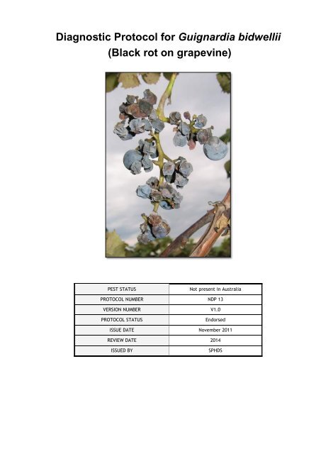
<str<strong>on</strong>g>Diagnostic</str<strong>on</strong>g> <str<strong>on</strong>g>P<str<strong>on</strong>g>rot</str<strong>on</strong>g>ocol</str<strong>on</strong>g> <str<strong>on</strong>g>for</str<strong>on</strong>g> <str<strong>on</strong>g>Guignardia</str<strong>on</strong>g> <str<strong>on</strong>g>bidwellii</str<strong>on</strong>g><br />
(<str<strong>on</strong>g>Black</str<strong>on</strong>g> <str<strong>on</strong>g>rot</str<strong>on</strong>g> <strong>on</strong> grapevine)<br />
PEST STATUS Not present in Australia<br />
PROTOCOL NUMBER NDP 13<br />
VERSION NUMBER V1.0<br />
PROTOCOL STATUS Endorsed<br />
ISSUE DATE November 2011<br />
REVIEW DATE 2014<br />
ISSUED BY SPHDS
Prepared <str<strong>on</strong>g>for</str<strong>on</strong>g> the Subcommittee <strong>on</strong> Plant Health<br />
<str<strong>on</strong>g>Diagnostic</str<strong>on</strong>g> Standards (SPHDS)<br />
This versi<strong>on</strong> of the Nati<strong>on</strong>al <str<strong>on</strong>g>Diagnostic</str<strong>on</strong>g> <str<strong>on</strong>g>P<str<strong>on</strong>g>rot</str<strong>on</strong>g>ocol</str<strong>on</strong>g> (NDP) <str<strong>on</strong>g>for</str<strong>on</strong>g> <str<strong>on</strong>g>Guignardia</str<strong>on</strong>g> <str<strong>on</strong>g>bidwellii</str<strong>on</strong>g> (<str<strong>on</strong>g>Black</str<strong>on</strong>g> <str<strong>on</strong>g>rot</str<strong>on</strong>g> <strong>on</strong><br />
grapevine) is current as at the date c<strong>on</strong>tained in the versi<strong>on</strong> c<strong>on</strong>trol box <strong>on</strong> the fr<strong>on</strong>t of this<br />
document.<br />
NDPs are updated every 3 years or be<str<strong>on</strong>g>for</str<strong>on</strong>g>e this time if required (i.e. when new techniques<br />
become available).<br />
The most current versi<strong>on</strong> of this document is available from the SPHDS website:<br />
http://www.padil.gov.au/Sphds
Table of C<strong>on</strong>tents<br />
1 Introducti<strong>on</strong> ................................................................................................................... 1<br />
1.1 Host range ............................................................................................................... 1<br />
1.1.1 Primary hosts .................................................................................................... 1<br />
1.1.2 Alternative hosts ............................................................................................... 1<br />
2 Tax<strong>on</strong>omic in<str<strong>on</strong>g>for</str<strong>on</strong>g>mati<strong>on</strong> ................................................................................................. 1<br />
2.1 Classificati<strong>on</strong> ........................................................................................................... 1<br />
2.2 Name and Syn<strong>on</strong>yms .............................................................................................. 1<br />
2.2.1 Syn<strong>on</strong>yms ......................................................................................................... 1<br />
3 Detecti<strong>on</strong> ........................................................................................................................ 2<br />
3.1 Leaf, stem and fruit symptoms ................................................................................. 2<br />
3.2 C<strong>on</strong>fusi<strong>on</strong> with other diseases ................................................................................. 5<br />
3.3 Sampling and detecti<strong>on</strong> methods ............................................................................. 6<br />
3.3.1 Incubati<strong>on</strong> of symptomatic plant material .......................................................... 6<br />
3.3.2 Culturing <strong>on</strong> agar media ................................................................................... 7<br />
4 Identificati<strong>on</strong> .................................................................................................................. 9<br />
4.1 Culture growth ......................................................................................................... 9<br />
4.2 Identificati<strong>on</strong> of c<strong>on</strong>idia .......................................................................................... 11<br />
4.2.1 Microscopic descripti<strong>on</strong> .................................................................................. 11<br />
5 C<strong>on</strong>tact points <str<strong>on</strong>g>for</str<strong>on</strong>g> further in<str<strong>on</strong>g>for</str<strong>on</strong>g>mati<strong>on</strong> ....................................................................... 13<br />
6 Acknowledgements ..................................................................................................... 13<br />
7 References ................................................................................................................... 14<br />
7.1 Other useful references ......................................................................................... 14<br />
8 Appendix ...................................................................................................................... 16<br />
Disease cycle ................................................................................................................... 16
1 Introducti<strong>on</strong><br />
<str<strong>on</strong>g>Black</str<strong>on</strong>g> <str<strong>on</strong>g>rot</str<strong>on</strong>g> of grape is caused by the ascomycete fungus <str<strong>on</strong>g>Guignardia</str<strong>on</strong>g> <str<strong>on</strong>g>bidwellii</str<strong>on</strong>g> and can lead to<br />
substantial yield losses in humid regi<strong>on</strong>s when it is not effectively managed. It has been<br />
described as <strong>on</strong>e of the most ec<strong>on</strong>omically important and destructive diseases of grapes.<br />
Most cultivars of Vitis vinifera as well as French/American hybrids and American bunch<br />
grapes are susceptible, while varieties of muscadine range in disease susceptibility from<br />
resistant to very susceptible.<br />
The fungus may be spread <strong>on</strong> grapevine cuttings and fruit.<br />
1.1 Host range<br />
1.1.1 Primary hosts<br />
Vitis vinifera Domestic grape<br />
Vitis ariz<strong>on</strong>ica Cany<strong>on</strong> grape<br />
Vitis labrusca American grape<br />
Vitis <str<strong>on</strong>g>rot</str<strong>on</strong>g>undifolia Muscadine grape<br />
1.1.2 Alternative hosts<br />
Vitis amurensis Amur grape<br />
Ampelopsis Wild grape<br />
Cissus Ornamental vine<br />
Parthenocissus quinquefolia Virginia creeper<br />
Parthenocissus tricuspidata Bost<strong>on</strong> ivy<br />
Asplenium nidus Birds nest fern<br />
2 Tax<strong>on</strong>omic in<str<strong>on</strong>g>for</str<strong>on</strong>g>mati<strong>on</strong><br />
2.1 Classificati<strong>on</strong><br />
Class: Dothideomycetes<br />
Order Incertae sedis<br />
Family: Botryosphaeriaceae<br />
Genus: <str<strong>on</strong>g>Guignardia</str<strong>on</strong>g><br />
Species: <str<strong>on</strong>g>bidwellii</str<strong>on</strong>g><br />
2.2 Name and Syn<strong>on</strong>yms<br />
<str<strong>on</strong>g>Guignardia</str<strong>on</strong>g> <str<strong>on</strong>g>bidwellii</str<strong>on</strong>g> (Ellis) Viala & Ravaz, Bulletin de la Société Mycologique de France 8:<br />
63, 1892<br />
2.2.1 Syn<strong>on</strong>yms<br />
Sphaeria bidwelli Ellis 1880<br />
Physalospora bidwelli (Ellis) Sacc., 1882<br />
Laestadia bidwelli (Ellis) Viala & Ravaz, 1888<br />
Sphaeralla <str<strong>on</strong>g>bidwellii</str<strong>on</strong>g> (Ellis) Ellis 1890<br />
Carlia <str<strong>on</strong>g>bidwellii</str<strong>on</strong>g> (Ellis) Magnus 1892<br />
Phyllachorella <str<strong>on</strong>g>bidwellii</str<strong>on</strong>g> (Ellis) Theiss. 1919<br />
Botryosphaeria <str<strong>on</strong>g>bidwellii</str<strong>on</strong>g> (Ellis) Petr. 1958<br />
Carlia <str<strong>on</strong>g>bidwellii</str<strong>on</strong>g> (Ellis) Prunet, 1989<br />
NDP Guignadia black <str<strong>on</strong>g>rot</str<strong>on</strong>g><br />
1
3 Detecti<strong>on</strong><br />
<str<strong>on</strong>g>Guignardia</str<strong>on</strong>g> <str<strong>on</strong>g>bidwellii</str<strong>on</strong>g> can be readily identified by macroscopic symptoms <strong>on</strong> most parts of the<br />
plant. The disease cycle of black <str<strong>on</strong>g>rot</str<strong>on</strong>g> is described in Appendix 1.<br />
3.1 Leaf, stem and fruit symptoms<br />
G. <str<strong>on</strong>g>bidwellii</str<strong>on</strong>g> is most likely to harbour <strong>on</strong> leaves, stems and fruit of grapevines and minor<br />
hosts. Sources could include imported fruit or cuttings.<br />
In the vineyard, symptoms are visually most evident <strong>on</strong> leaves in the spring; leaves, stems<br />
and fruit in the summer and <strong>on</strong> stems and fruit in autumn and winter.<br />
Infected leaves can be easily detected by circular lesi<strong>on</strong>s which vary in size but are typically<br />
small under field c<strong>on</strong>diti<strong>on</strong>s (Figure 1a,d) and appear brown or tan with reddish margins and<br />
they characteristically c<strong>on</strong>tain small black pycnidia (Figure 1a-c). When inspecting vines, leaf<br />
lesi<strong>on</strong>s may be observed associated with mummified berries from the previous or current<br />
seas<strong>on</strong>s (Figure 1d).<br />
a b<br />
c d<br />
Figure 1. Symptoms of G. <str<strong>on</strong>g>bidwellii</str<strong>on</strong>g> <strong>on</strong> leaves of Vitis vinifera (cv. Riesling); (a) <strong>on</strong> the vine, (b) <strong>on</strong> the<br />
laboratory bench, (c) under the dissecting microscope with pycnidia evident and (d) leaf lesi<strong>on</strong>s<br />
caused by c<strong>on</strong>idia ejected from infected berries above. (Photos by M. Sosnowski, SARDI)<br />
NDP Guignadia black <str<strong>on</strong>g>rot</str<strong>on</strong>g><br />
2
On shoots, petioles and tendrils the lesi<strong>on</strong>s are initially tan, brown turning purple to black,<br />
sunken, elliptical to el<strong>on</strong>gated and c<strong>on</strong>tain pycnidia or pseudothecia observed as small black<br />
dots (Figure 2). The bark may split and stem infecti<strong>on</strong>s remain localised and do not usually<br />
extend more than several centimetres.<br />
a b<br />
d<br />
Figure 2. Symptoms of G. <str<strong>on</strong>g>bidwellii</str<strong>on</strong>g> <strong>on</strong> stems and petioles of Vitis vinifera (cv. Riesling); (a) <strong>on</strong> young<br />
green shoots <strong>on</strong> the vine, (b) <strong>on</strong> the laboratory bench, (c) under the dissecting microscope and (d) <strong>on</strong><br />
older lignified shoots of a Vitis interspecific hybrid (cv. Vignoles) <strong>on</strong> the vine. (Photos a-c by M.<br />
Sosnowski, SARDI and photo d by W. Wilcox, Cornell University)<br />
NDP Guignadia black <str<strong>on</strong>g>rot</str<strong>on</strong>g><br />
3<br />
c
On the developing fruit, small, pale dots (approx. 1mm diam.) appear, rapidly becoming<br />
surrounded by a widening brown ring. If lesi<strong>on</strong> expansi<strong>on</strong> is halted due to an applicati<strong>on</strong> of<br />
some fungicides or the development of age-related resistance in half grown berries, palebrown<br />
coloured spots with a dark ring (bird's eye effect) and a sunken centre, about 6mm<br />
diam, may result (Figure 3a). More typically, however, a chocolate-brown lesi<strong>on</strong> expands<br />
through the berry (Figure 3b,c) until it becomes completely <str<strong>on</strong>g>rot</str<strong>on</strong>g>ted. Berries then shrivel and<br />
turn dark brown with numerous black pseudothecia or pycnidia developing over the surface<br />
(Figure 3d). Eventually, the fruit becomes dry and shrivelled, turning into hard, blue-black<br />
mummies that often remain firmly attached to the pedicel, although some may be shed from<br />
the vine (Figure 3e).<br />
a b<br />
d<br />
Figure 3. Symptoms of G. <str<strong>on</strong>g>bidwellii</str<strong>on</strong>g> <strong>on</strong> grape berries of Vitis labrusca (cv. C<strong>on</strong>cord); (a-c) progressi<strong>on</strong><br />
from small lesi<strong>on</strong>s to mummified berries, (d) pseudothecia <strong>on</strong> surface of mummified berry under<br />
dissecting microscope and (e) mummified berries. (Photos a, b, d and e by M. Sosnowski, SARDI and<br />
c by W. Wilcox, Cornell University)<br />
NDP Guignadia black <str<strong>on</strong>g>rot</str<strong>on</strong>g><br />
4<br />
e<br />
c
3.2 C<strong>on</strong>fusi<strong>on</strong> with other diseases<br />
Symptoms can be c<strong>on</strong>fused with those of black spot (anthracnose) disease caused by the<br />
fungus Elsinoe ampelina which is endemic in Australia. Leaf lesi<strong>on</strong>s are similar when viewed<br />
from a distance, but do not c<strong>on</strong>tain the small black pycnidia typically found in black <str<strong>on</strong>g>rot</str<strong>on</strong>g><br />
lesi<strong>on</strong>s. Stems and petioles develop raised cankers with sunken centres, whereas black <str<strong>on</strong>g>rot</str<strong>on</strong>g><br />
lesi<strong>on</strong>s are less “three-dimensi<strong>on</strong>al”. Infected fruit develop similar lesi<strong>on</strong>s initially, but those<br />
of black spot do not usually expand to include the entire berry nor evolve into mummified fruit<br />
(Figure 4).<br />
a b c<br />
Figure 4. Symptoms of black spot (anthracnose) disease caused by the fungus Elsinoe ampelina <strong>on</strong><br />
Vitis vinifera (table grape cv. Red globe); (a) leaf lesi<strong>on</strong>s, (b) stem cankers and (c) berry lesi<strong>on</strong>s.<br />
(Photos by M. Sosnowski, SARDI)<br />
Symptoms of Phomopsis cane and leaf spot, which is caused by the fungus Phomopsis<br />
viticola, may also be c<strong>on</strong>fused with those of black <str<strong>on</strong>g>rot</str<strong>on</strong>g> (Figure 5). Grapevines affected by<br />
Phomopsis develop leaf lesi<strong>on</strong>s that are smaller than those due to black <str<strong>on</strong>g>rot</str<strong>on</strong>g>; they are often<br />
surrounded with a translucent halo, and do not c<strong>on</strong>tain pycnidia. Stem lesi<strong>on</strong>s are sometimes<br />
smaller in size, but when larger, the two diseases are very difficult to distinguish <strong>on</strong> the basis<br />
of lesi<strong>on</strong>s <strong>on</strong> the green shoots. Lesi<strong>on</strong>s <strong>on</strong> lignified canes often have a bleached appearance<br />
and develop into larger basal cankers. Fruit symptoms can be similar to black <str<strong>on</strong>g>rot</str<strong>on</strong>g>, with<br />
shrivelled mummified berries; however, this phase of the disease does not usually occur with<br />
Phomopsis until shortly be<str<strong>on</strong>g>for</str<strong>on</strong>g>e harvest, whereas it typically occurs by verais<strong>on</strong> with black <str<strong>on</strong>g>rot</str<strong>on</strong>g>.<br />
There is also a complex of pathogens including Botrytis cinerea, Aspergillus niger, Alternaria<br />
spp., Cladosporium spp., Rhizopus arrhizus, Penicillium spp. which may cause fruit <str<strong>on</strong>g>rot</str<strong>on</strong>g> of<br />
grapevines so should also be c<strong>on</strong>sidered when diagnosing black <str<strong>on</strong>g>rot</str<strong>on</strong>g>.<br />
NDP Guignadia black <str<strong>on</strong>g>rot</str<strong>on</strong>g><br />
5
a b<br />
c d e<br />
Figure 5. Symptoms of Phomopsis cane and leaf spot disease caused by the fungus Phomopsis<br />
viticola <strong>on</strong> Vitis vinifera (cv. unknown); (a) small leaf lesi<strong>on</strong>s with halos, (b) green stem lesi<strong>on</strong>s, (c)<br />
bleached cane, (d) basal stem canker and (e) bunch symptoms. (Photos a-d by B. Rawnsley, SARDI<br />
and e by R. Emmett, DPI Victoria)<br />
3.3 Sampling and detecti<strong>on</strong> methods<br />
As visual symptoms can be found <strong>on</strong> leaves, stems and fruit of grapevines, they should all be<br />
targeted during any inspecti<strong>on</strong>s of vineyards, post-entry quarantine or examinati<strong>on</strong> of<br />
grapevine material at the border. In the vineyard, symptoms are likely to be most obvious <strong>on</strong><br />
leaves first, but mummified berries could also be present from infecti<strong>on</strong> in the previous<br />
seas<strong>on</strong>. Symptoms to look <str<strong>on</strong>g>for</str<strong>on</strong>g> are brown circular lesi<strong>on</strong>s (often c<strong>on</strong>taining small black<br />
pycnidia) with reddish margins <strong>on</strong> leaves; brown/purple elliptical or el<strong>on</strong>gated lesi<strong>on</strong>s <strong>on</strong><br />
stems; and brown circular lesi<strong>on</strong>s <strong>on</strong> fruit or shrivelled berries (details in secti<strong>on</strong> 3.1). In<br />
vineyards, symptoms can be present both during the growing seas<strong>on</strong> and dormancy, as<br />
infected, mummified berries often remain hanging <strong>on</strong> the vine since they are not detached<br />
from the rachis as easily as healthy berries that merely shrivel <strong>on</strong> the vine through normal<br />
dehydrati<strong>on</strong> if not harvested at maturity.<br />
Symptomatic leaf, stem and fruit samples should be sealed in a polyethylene bag and<br />
transported to a diagnostic facility, keeping samples cool during the entire process. For<br />
nati<strong>on</strong>al guidelines <strong>on</strong> resp<strong>on</strong>se to an emergency plant pest refer to PLANTPLAN (PHA<br />
2009).<br />
3.3.1 Incubati<strong>on</strong> of symptomatic plant material<br />
Equipment:<br />
Dissecting microscope<br />
Scalpel<br />
Trays, polyethylene bags and paper towel<br />
Slides and coverslips<br />
Sterile distilled water (SDW)<br />
NDP Guignadia black <str<strong>on</strong>g>rot</str<strong>on</strong>g><br />
6
Method:<br />
Symptomatic leaf, stem and fruit material should be placed in a tray lined with wet paper<br />
towel and sealed in a plastic bag <str<strong>on</strong>g>for</str<strong>on</strong>g> incubati<strong>on</strong> under high humidity. The samples can be left<br />
<strong>on</strong> the bench in normal laboratory c<strong>on</strong>diti<strong>on</strong>s. Within 1-3 days, pycnidia <strong>on</strong> lesi<strong>on</strong>s may<br />
exude cirrhi or tendrils c<strong>on</strong>taining c<strong>on</strong>idia which can be viewed under the dissecting<br />
microscope (Figure 6).<br />
a b<br />
Figure 6. Pycnidia of <str<strong>on</strong>g>Guignardia</str<strong>on</strong>g> <str<strong>on</strong>g>bidwellii</str<strong>on</strong>g> exuding cirrhi <strong>on</strong> the leaf surface of grapevine cv. Riesling<br />
following incubati<strong>on</strong> at high humidity overnight. (Photos by M. Sosnowski, SARDI)<br />
Using a scalpel, scrape cirrhi from leaf surface and mount <strong>on</strong> slide in a drop of SDW. If no<br />
cirrhi appear then remove several pycnidia from the lesi<strong>on</strong> and mount in a drop of SDW <strong>on</strong> a<br />
slide and crush the fruiting bodies with the side of the scalpel blade. C<strong>on</strong>idia can be viewed<br />
under a compound microscope and identificati<strong>on</strong> made as described in secti<strong>on</strong> 4.2.<br />
3.3.2 Culturing <strong>on</strong> agar media<br />
Equipment:<br />
Autoclave<br />
Sterile plastic petri dishes<br />
Laminar flow cabinet<br />
Incubator (25ºC, white fluorescent lights)<br />
Scalpel and <str<strong>on</strong>g>for</str<strong>on</strong>g>ceps<br />
Bunsen burner and alcohol<br />
Sodium hypochlorite soluti<strong>on</strong> (0.5%)<br />
Sterile distilled water (SDW)<br />
Sterile filter paper<br />
Parafilm<br />
Media preparati<strong>on</strong>:<br />
Potato Dextrose Agar – half-strength (½ PDA) Hoffman et al. (2002)<br />
Potato Dextrose Agar (PDA) 9.8 g<br />
Bacto agar (granulated agarose) 3.5 g<br />
Distilled water 500 ml<br />
Mix ingredients and autoclave at 121ºC <str<strong>on</strong>g>for</str<strong>on</strong>g> 15 minutes, mixing well be<str<strong>on</strong>g>for</str<strong>on</strong>g>e pouring into sterile<br />
plastic petri dishes.<br />
NDP Guignadia black <str<strong>on</strong>g>rot</str<strong>on</strong>g><br />
7
Malt Extract Agar (MEA) Jailloux (1992)<br />
Malt Extract Agar (MEA) 16.8 g<br />
Distilled water 500 ml<br />
Mix ingredients and autoclave at 121ºC <str<strong>on</strong>g>for</str<strong>on</strong>g> 15 minutes, mixing well be<str<strong>on</strong>g>for</str<strong>on</strong>g>e pouring into sterile<br />
plastic petri dishes.<br />
Oatmeal Agar (OA) Jailloux (1992)<br />
Oat flakes 20 g<br />
Bacto agar (granulated agarose) 7.5 g<br />
Distilled water 500 ml<br />
Grind oat flakes to a powder in blender. Mix ingredients and autoclave at 121ºC <str<strong>on</strong>g>for</str<strong>on</strong>g> 15<br />
minutes, mixing well be<str<strong>on</strong>g>for</str<strong>on</strong>g>e pouring into sterile plastic petri dishes.<br />
Alternatively, use commercial <str<strong>on</strong>g>for</str<strong>on</strong>g>mulati<strong>on</strong> of oatmeal agar.<br />
Method:<br />
In the laminar flow cabinet, remove secti<strong>on</strong>s of leaf, stem and fruit samples with lesi<strong>on</strong>s using<br />
a scalpel. Surface sterilise secti<strong>on</strong>s in 0.5% sodium hypochlorite <str<strong>on</strong>g>for</str<strong>on</strong>g> 60 s, rinse in sterile<br />
distilled water (SDW) and place <strong>on</strong> sterile filter paper to dry. Excise small pieces (approx. 3<br />
mm x 3 mm) from lesi<strong>on</strong>s and place <strong>on</strong>to surface of agar media.<br />
Alternatively, cirrhi exuding from pycnidia <strong>on</strong> lesi<strong>on</strong>s incubated as described in secti<strong>on</strong> 3.3.1<br />
can be carefully removed with a flame-sterilised scalpel and placed directly <strong>on</strong>to agar<br />
surface.<br />
Seal plates with parafilm strips and place into incubator at 25ºC under c<strong>on</strong>tinuous fluorescent<br />
light. Check plates <str<strong>on</strong>g>for</str<strong>on</strong>g> c<strong>on</strong>taminati<strong>on</strong> at regular intervals and subculture putative col<strong>on</strong>ies of<br />
G. <str<strong>on</strong>g>bidwellii</str<strong>on</strong>g> if necessary. Pure cultures should be incubated <str<strong>on</strong>g>for</str<strong>on</strong>g> 15-20 days in order to induce<br />
sporulati<strong>on</strong>.<br />
NDP Guignadia black <str<strong>on</strong>g>rot</str<strong>on</strong>g><br />
8
4 Identificati<strong>on</strong><br />
<str<strong>on</strong>g>Guignardia</str<strong>on</strong>g> <str<strong>on</strong>g>bidwellii</str<strong>on</strong>g> can be readily identified by macroscopic symptoms (Secti<strong>on</strong> 3.1) and<br />
then c<strong>on</strong>firmed by morphological characteristics of cultures and c<strong>on</strong>idia. No molecular<br />
methods have been reported <str<strong>on</strong>g>for</str<strong>on</strong>g> the identificati<strong>on</strong> of G. <str<strong>on</strong>g>bidwellii</str<strong>on</strong>g>.<br />
4.1 Culture growth<br />
Half-strength PDA is the optimal medium <str<strong>on</strong>g>for</str<strong>on</strong>g> identificati<strong>on</strong> and c<strong>on</strong>idia producti<strong>on</strong> (Figure 7).<br />
Mycelium growth rate ranges from 1-2 mm/day and appears speckled and irregular. Pycnidia<br />
<str<strong>on</strong>g>for</str<strong>on</strong>g>mati<strong>on</strong> can occur within 7 days <str<strong>on</strong>g>for</str<strong>on</strong>g> some isolates and after 15 days all isolates produce<br />
fruiting bodies, from which c<strong>on</strong>idia can be harvested.<br />
a b<br />
a<br />
c d<br />
e f<br />
Figure 7. <str<strong>on</strong>g>Guignardia</str<strong>on</strong>g> <str<strong>on</strong>g>bidwellii</str<strong>on</strong>g> cultures <strong>on</strong> ½ Potato Dextrose Agar; (a) 15 days and (b-f) 20 days of<br />
incubati<strong>on</strong> at 25ºC under c<strong>on</strong>tinuous fluorescent light. (e & f) Under a dissecting microscope, c<strong>on</strong>idia<br />
are observed oozing from pycnidia. (Photos by M. Sosnowski, SARDI)<br />
NDP Guignadia black <str<strong>on</strong>g>rot</str<strong>on</strong>g><br />
9
Cultures grow at approx 0.5 mm/day <strong>on</strong> Malt Extract Agar (Figure 8) in a circular pattern;<br />
pycnidia develop within 7-15 days and are densely distributed, although they yield fewer<br />
c<strong>on</strong>idia than <strong>on</strong> ½ PDA. Cultures grow at 1-2 mm/day <strong>on</strong> Oatmeal Agar (Figure 9) and are<br />
circular with a distinctive dark green, creamy appearance; few, if any, pycnidia are produced.<br />
a b<br />
Figure 8. <str<strong>on</strong>g>Guignardia</str<strong>on</strong>g> <str<strong>on</strong>g>bidwellii</str<strong>on</strong>g> cultures <strong>on</strong> Malt Extract Agar; (a) 15 days and (b) 20 days of incubati<strong>on</strong><br />
at 25ºC under c<strong>on</strong>tinuous fluorescent light. (Photos by M. Sosnowski, SARDI)<br />
a b<br />
Figure 9. <str<strong>on</strong>g>Guignardia</str<strong>on</strong>g> <str<strong>on</strong>g>bidwellii</str<strong>on</strong>g> cultures <strong>on</strong> Oatmeal Agar; (a) 15 days and (b) 20 days of incubati<strong>on</strong> at<br />
25ºC under c<strong>on</strong>tinuous fluorescent light. (Photos by M. Sosnowski, SARDI)<br />
NDP Guignadia black <str<strong>on</strong>g>rot</str<strong>on</strong>g><br />
10
4.2 Identificati<strong>on</strong> of c<strong>on</strong>idia<br />
C<strong>on</strong>idial ooze can be scraped from pycnidia <strong>on</strong> agar cultures or leaf lesi<strong>on</strong>s using a scalpel,<br />
and mounted <strong>on</strong> a glass slide in a drop of SDW with a cover slip placed <strong>on</strong> top.<br />
Viewed under a compound light microscope, c<strong>on</strong>idia appear as <strong>on</strong>e celled, hyaline, broadly<br />
ovoid, ellipsoidal or almost globose, somewhat clavate when young and slightly indented;<br />
spores are 5-12 × 4-7 µm in size (Figure 10).<br />
a b<br />
Figure 10. Pycnidia (a) and c<strong>on</strong>idia (a&b) of <str<strong>on</strong>g>Guignardia</str<strong>on</strong>g> <str<strong>on</strong>g>bidwellii</str<strong>on</strong>g> from cultures <strong>on</strong> ½ PDA incubated<br />
<str<strong>on</strong>g>for</str<strong>on</strong>g> 15 days at 25ºC under c<strong>on</strong>tinuous fluorescent light. (Photos by M. Sosnowski, SARDI)<br />
4.2.1 Microscopic descripti<strong>on</strong><br />
The following descripti<strong>on</strong> is taken from Sivanesan and Holliday (1981).<br />
Pseudothecia <str<strong>on</strong>g>for</str<strong>on</strong>g>med as locules in a stroma, depressed globose, immersed subepidermal,<br />
70-180µm broad with a flat or papillate ostiolar apex (Figure 11A). The pseudothecial wall is<br />
made up of pseudoparenchymatic cells. Asci arise from a cushi<strong>on</strong> shaped hyaline tissue at<br />
the base, cylindrical to clavate, 45-65 × 9-14 µm (Figure 11B). Ascospores hyaline, <strong>on</strong>e<br />
celled, ovoid to ellipsoid, 12-17 × 6-7.5 µm, often with hyaline, mucilaginous, apical caps.<br />
Pycnidia mostly epiphyllous, solitary, unilocular, globose or depressed globose, with a flat or<br />
inc<strong>on</strong>spicuous papillate ostiolar apex, 120-230 µm broad (Figure 11C). Stroma poorly<br />
developed <strong>on</strong> leaves but well developed <strong>on</strong> fruits. C<strong>on</strong>idiogenous cells c<strong>on</strong>ical to cylindrical<br />
(Figure 11D). C<strong>on</strong>idia <strong>on</strong>e celled, hyaline, broadly ovoid, ellipsoidal or almost globose,<br />
somewhat clavate when young and slightly indented, 5-12 × 4-7 µm, surrounded by a<br />
mucilaginous sheath and with an apical hyaline appendage as l<strong>on</strong>g as the c<strong>on</strong>idium.<br />
Spermatia hyaline, unicellular, rod shaped 4-7 × 0.5-2 µm.<br />
NDP Guignadia black <str<strong>on</strong>g>rot</str<strong>on</strong>g><br />
11
Figure 11. Microscopic descripti<strong>on</strong> of <str<strong>on</strong>g>Guignardia</str<strong>on</strong>g> <str<strong>on</strong>g>bidwellii</str<strong>on</strong>g>; A. pseudothecium, B. ascus and<br />
ascospores, C. pycnidium and D. c<strong>on</strong>idiogenous cells and c<strong>on</strong>idia (Sivanesan and Holliday 1981).<br />
NDP Guignadia black <str<strong>on</strong>g>rot</str<strong>on</strong>g><br />
12
5 C<strong>on</strong>tact points <str<strong>on</strong>g>for</str<strong>on</strong>g> further in<str<strong>on</strong>g>for</str<strong>on</strong>g>mati<strong>on</strong><br />
Dr Mark Sosnowski<br />
South Australian Research and Development Institute<br />
GPO Box 397 South Australia 5001<br />
Ph: 08 8303 9489<br />
Fax: 08 8303 9393<br />
Email: mark.sosnowski@sa.gov.au<br />
Dr Gary K<strong>on</strong>g<br />
Plant Science, Primary Industries and Fisheries<br />
Department of Employment, Ec<strong>on</strong>omic Development and Innovati<strong>on</strong><br />
PO Box 102 Toowoomba Qld 4350<br />
Ph: 07 4688 1319<br />
Mob: 0428103521<br />
Email: Gary.K<strong>on</strong>g@deedi.qld.gov.au<br />
Prof Wayne Wilcox<br />
Cornell University<br />
New York State Agricultural Experiment Stati<strong>on</strong><br />
PO Box 462 Geneva, NY 14456 USA<br />
Ph: +1 315 787 2335<br />
Email: wfw1@cornell.edu<br />
6 Acknowledgements<br />
This document is based <strong>on</strong> In<str<strong>on</strong>g>for</str<strong>on</strong>g>mati<strong>on</strong> taken from PaDIL - Plant Biosecurity Toolbox which<br />
was originally authored by Gary K<strong>on</strong>g. <str<strong>on</strong>g>Diagnostic</str<strong>on</strong>g> methods <str<strong>on</strong>g>for</str<strong>on</strong>g> <str<strong>on</strong>g>Black</str<strong>on</strong>g> Rot of Grapes -<br />
<str<strong>on</strong>g>Guignardia</str<strong>on</strong>g> <str<strong>on</strong>g>bidwellii</str<strong>on</strong>g> [http://www.padil.gov.au/pbt] accessed 5 June 2009.<br />
The in<str<strong>on</strong>g>for</str<strong>on</strong>g>mati<strong>on</strong> was reviewed and written into the current p<str<strong>on</strong>g>rot</str<strong>on</strong>g>ocol by Dr Mark Sosnowski <str<strong>on</strong>g>for</str<strong>on</strong>g><br />
with funding from the Department of Agriculture, Fisheries and Forestry (DAFF) through the<br />
<str<strong>on</strong>g>Diagnostic</str<strong>on</strong>g> Training Scholarship programme.<br />
Wayne Wilcox and Judy Burr (Cornell University, NY, USA) <str<strong>on</strong>g>for</str<strong>on</strong>g> assistance with training and<br />
providing details <str<strong>on</strong>g>for</str<strong>on</strong>g> the diagnostic p<str<strong>on</strong>g>rot</str<strong>on</strong>g>ocol.<br />
NDP Guignadia black <str<strong>on</strong>g>rot</str<strong>on</strong>g><br />
13
7 References<br />
Ferrin DM and Ramsdell DC (1977) Ascospore dispersal and infecti<strong>on</strong> of grapes by<br />
<str<strong>on</strong>g>Guignardia</str<strong>on</strong>g> <str<strong>on</strong>g>bidwellii</str<strong>on</strong>g>, causal agent of grape black <str<strong>on</strong>g>rot</str<strong>on</strong>g> disease. Phytopathology, 67:<br />
1501-1505.<br />
Ferrin DM and Ramsdell DC (1978) Influence of c<strong>on</strong>idia dispersal and envir<strong>on</strong>ment <strong>on</strong><br />
infecti<strong>on</strong> of grape by <str<strong>on</strong>g>Guignardia</str<strong>on</strong>g> <str<strong>on</strong>g>bidwellii</str<strong>on</strong>g>. Phytopathology, 68: 892-895.<br />
Hoffman LE and Wilcox WF (2002) Utilizing epidemiological investigati<strong>on</strong>s to optimize<br />
management of grape black <str<strong>on</strong>g>rot</str<strong>on</strong>g>. Phytopathology, 92: 676-680.<br />
Hoffman LE, Wilcox WF, Gadoury DA and Seem RC (2002) Influence of grape berry age <strong>on</strong><br />
susceptibility to <str<strong>on</strong>g>Guignardia</str<strong>on</strong>g> <str<strong>on</strong>g>bidwellii</str<strong>on</strong>g> and its incubati<strong>on</strong> period length. Phytopathology,<br />
92: 1068-1076.<br />
Jailloux F (1992) In vitro producti<strong>on</strong> of the teleomorph of <str<strong>on</strong>g>Guignardia</str<strong>on</strong>g> <str<strong>on</strong>g>bidwellii</str<strong>on</strong>g>, causal agent of<br />
black <str<strong>on</strong>g>rot</str<strong>on</strong>g> of grapevine. Canadian Journal of Botany, 70: 254-257.<br />
Pezet R and Jermini M (1989) <str<strong>on</strong>g>Black</str<strong>on</strong>g> <str<strong>on</strong>g>rot</str<strong>on</strong>g> of grapevine: symptoms, epidemiology and c<strong>on</strong>trol.<br />
Revue Suisse de Viticulture, d'Arboriculture et d'Horticulture, 21: 27-34.<br />
PHA (2009) PLANTPLAN: Australian Emergency Plant Pest Resp<strong>on</strong>se Plan. Versi<strong>on</strong> 1 May<br />
2009, Plant Health Australia, Canberra ACT [http://www.planthealthaustralia.com.au/]<br />
Ramsdell DC and Milholland RD, (1988) <str<strong>on</strong>g>Black</str<strong>on</strong>g> <str<strong>on</strong>g>rot</str<strong>on</strong>g>. In: APS, ed. Compendium of Grape<br />
Diseases. St Paul, USA: APS Press, 15-17.<br />
Sivanesan A and Holliday P (1981) CMI Descripti<strong>on</strong>s of Pathogenic Fungi and Bacteria, No.<br />
710. Walling<str<strong>on</strong>g>for</str<strong>on</strong>g>d, UK: CAB Internati<strong>on</strong>al.<br />
Spotts RA 1977. Effect of leaf wetness durati<strong>on</strong> and temperature <strong>on</strong> the infectivity of<br />
<str<strong>on</strong>g>Guignardia</str<strong>on</strong>g> <str<strong>on</strong>g>bidwellii</str<strong>on</strong>g> <strong>on</strong> grape leaves. Phytopathology, 67: 1378-1381.<br />
Wilcox W (2003) Grapes: <str<strong>on</strong>g>Black</str<strong>on</strong>g> <str<strong>on</strong>g>rot</str<strong>on</strong>g> (<str<strong>on</strong>g>Guignardia</str<strong>on</strong>g> <str<strong>on</strong>g>bidwellii</str<strong>on</strong>g> (Ellis) Viala and Ravaz.) Cornell<br />
Cooperative Extensi<strong>on</strong> Disease Identificati<strong>on</strong> Sheet No. 102GFSG-D4, Cornell<br />
University.<br />
7.1 Other useful references<br />
Alves A, Phillips AJL, Henriques I and Correia A (2007) Rapid differentiati<strong>on</strong> of species of<br />
botryosphaeriaceae by PCR fingerprinting. Research in Microbiology, 158: 112-121.<br />
CAB Internati<strong>on</strong>al Distributi<strong>on</strong> Maps of Plant Diseases, 1991, April (Editi<strong>on</strong> 4), Map 81.<br />
Hoffman LE and Wilcox WF (2003) Factors influencing the efficacy of myclobutanil and<br />
azoxystrobin <str<strong>on</strong>g>for</str<strong>on</strong>g> c<strong>on</strong>trol of grape black <str<strong>on</strong>g>rot</str<strong>on</strong>g>. Plant Disease, 87: 273-281.<br />
Hoffman LE, Wilcox WF, Gadoury DM, Seem RC and Riegel DG (2004) Integrated c<strong>on</strong>trol of<br />
grape black <str<strong>on</strong>g>rot</str<strong>on</strong>g>: Influence of host phenology, inoculum availability, sanitati<strong>on</strong>, and spray<br />
timing. Phytopathology, 94: 641-650.<br />
Janex-Favre MC, Pargueyleduc A and Jailloux F (1993) The <strong>on</strong>togeny of pycnidia of<br />
<str<strong>on</strong>g>Guignardia</str<strong>on</strong>g> <str<strong>on</strong>g>bidwellii</str<strong>on</strong>g> in culture. Mycological Research, 97: 1333-1339.<br />
Janex-Favre MC, Parguey-Leduc A and Jailloux F (1996) The <strong>on</strong>togeny of perithecia in<br />
<str<strong>on</strong>g>Guignardia</str<strong>on</strong>g> <str<strong>on</strong>g>bidwellii</str<strong>on</strong>g>. Mycological Research 100: 875-880.<br />
Jermini M and Gessler C (1996) Epidemiology and c<strong>on</strong>trol of grape black <str<strong>on</strong>g>rot</str<strong>on</strong>g> in southern<br />
Switzerland. Plant Disease, 80: 322-325.<br />
Kummuang N, Diehl SV, Smith BJ and Graves CH (1996) Muscadine grape berry <str<strong>on</strong>g>rot</str<strong>on</strong>g><br />
diseases in Mississippi: Disease epidemiology and crop reducti<strong>on</strong>. Plant Disease, 80:<br />
244-247.<br />
Kummuang N, Smith BJ, Diehl SV and Graves CH (1996) Muscadine grape berry <str<strong>on</strong>g>rot</str<strong>on</strong>g><br />
diseases in Mississippi: Disease identificati<strong>on</strong> and incidence. Plant Disease, 80: 238-<br />
243.<br />
NDP Guignadia black <str<strong>on</strong>g>rot</str<strong>on</strong>g><br />
14
Rao R and Kale SB (1965) A new species of <str<strong>on</strong>g>Guignardia</str<strong>on</strong>g> from India. Mycopathologia, 27: 13-<br />
14.<br />
Spotts RA (1980) Infecti<strong>on</strong> of grape by <str<strong>on</strong>g>Guignardia</str<strong>on</strong>g> <str<strong>on</strong>g>bidwellii</str<strong>on</strong>g> - factors affecting lesi<strong>on</strong><br />
development, c<strong>on</strong>idial dispersal, and c<strong>on</strong>idial populati<strong>on</strong>s <strong>on</strong> leaves. Phytopathology,<br />
70: 252-255.<br />
Wilcox WF, English-Loeb G, Dunst RM, Landers A (2006) New York and Pennsylvania Pest<br />
Management Guidelines <str<strong>on</strong>g>for</str<strong>on</strong>g> Grapes. Eds. T.H. Weigle and A.J. Muza. Cornell and<br />
Penn State University Cooperative Extensi<strong>on</strong> P 17.<br />
NDP Guignadia black <str<strong>on</strong>g>rot</str<strong>on</strong>g><br />
15
8 Appendix<br />
Disease cycle<br />
The black <str<strong>on</strong>g>rot</str<strong>on</strong>g> disease cycle is illustrated in Figure 12. The black <str<strong>on</strong>g>rot</str<strong>on</strong>g> fungus overwinters<br />
primarily in mummified fruit within the vine and <strong>on</strong> the ground, although it also can overwinter<br />
<str<strong>on</strong>g>for</str<strong>on</strong>g> at least 2 years within lesi<strong>on</strong>s of infected shoots that are retained as canes or spurs.<br />
Spring rains trigger release of ascospores (airborne sexual spores) that <str<strong>on</strong>g>for</str<strong>on</strong>g>m within<br />
mummies <strong>on</strong> the ground and in the trellis, and these can be blown <str<strong>on</strong>g>for</str<strong>on</strong>g> moderate distances by<br />
wind. C<strong>on</strong>idia (asexual spores) can also <str<strong>on</strong>g>for</str<strong>on</strong>g>m, both within cane lesi<strong>on</strong>s or <strong>on</strong> mummies that<br />
have remained within the trellis, and these are dispersed short distances within the vine or to<br />
neighbouring vines by splashing rain drops. This process can start within 1 hour of the <strong>on</strong>set<br />
of rain (at least 0.3 mm) and may c<strong>on</strong>tinue <str<strong>on</strong>g>for</str<strong>on</strong>g> up to 8 hours after rainfall ceases (Ferrin and<br />
Ramsdell 1977). Infecti<strong>on</strong> occurs when either spore type lands <strong>on</strong> susceptible green tissue<br />
such as leaves, blossoms and young fruit and it remains wet <str<strong>on</strong>g>for</str<strong>on</strong>g> 6-24 h depending <strong>on</strong><br />
temperature (Spotts 1977).<br />
Ascospores slowly germinate, often taking 36 to 48 hours, but eventually penetrate the<br />
young leaves and fruit stems (pedicels). The period of time required <str<strong>on</strong>g>for</str<strong>on</strong>g> symptoms to appear<br />
after the occurrence of infecti<strong>on</strong> depends <strong>on</strong> both the temperature and the age of the tissue<br />
at the time of infecti<strong>on</strong>, usually taking 8 to 25 days. The period during which these<br />
overwintering spores are available to cause infecti<strong>on</strong>s depends <strong>on</strong> their source. From<br />
mummies <strong>on</strong> the ground, significant discharge of ascospores begins about 2 to 3 weeks after<br />
bud break and is virtually complete within 1 to 2 weeks after the start of flowering. In c<strong>on</strong>trast,<br />
mummies within the trellis can c<strong>on</strong>tinue to release both c<strong>on</strong>idia and ascospores from the<br />
early pre-flowering period through verais<strong>on</strong>. From overwintering cane lesi<strong>on</strong>s, c<strong>on</strong>idia can be<br />
dispersed from bud break through mid-summer.<br />
Pycnidia, produced within the lesi<strong>on</strong>s, c<strong>on</strong>tinue to release c<strong>on</strong>idia during wet weather<br />
throughout the seas<strong>on</strong>. The c<strong>on</strong>idia will germinate and infect leaves, blossoms and young<br />
fruit and can cause substantial spread of the disease under warm and rainy c<strong>on</strong>diti<strong>on</strong>s,<br />
particularly if berries are still susceptible to infecti<strong>on</strong> after c<strong>on</strong>idia develop. Most berries that<br />
become infected near the end of their period of susceptibility do not show symptoms until at<br />
least 3 weeks later, and the majority do not begin to <str<strong>on</strong>g>rot</str<strong>on</strong>g> until 4 to 5 weeks after the infecti<strong>on</strong><br />
event. Fruit are most susceptible to infecti<strong>on</strong> by the fungus from mid-flowering to about 6<br />
weeks after flowering, and become resistant to infecti<strong>on</strong> at maturity. Some older literature<br />
has reported that berries become resistant when they reach 5% to 8% sugar, while research<br />
in New York indicates that berries become resistant much earlier, 3 to 4 weeks after<br />
flowering.<br />
In summary, spore producti<strong>on</strong>, dispersal, infecti<strong>on</strong> and c<strong>on</strong>tinued disease are favoured by<br />
warm, humid c<strong>on</strong>diti<strong>on</strong>s, summer rainfall and persistent dew. .<br />
NDP Guignadia black <str<strong>on</strong>g>rot</str<strong>on</strong>g><br />
16
Figure 12. The disease cycle of black <str<strong>on</strong>g>rot</str<strong>on</strong>g>, caused by <str<strong>on</strong>g>Guignardia</str<strong>on</strong>g> <str<strong>on</strong>g>bidwellii</str<strong>on</strong>g>. (Wilcox 2003)<br />
NDP Guignadia black <str<strong>on</strong>g>rot</str<strong>on</strong>g><br />
17