Chytridiomycota

Phylum Chytridiomycota

(Kingdom Fungi)

chyzoo.jpg (4078 bytes) Chytridiomycota - Blastocladiales - zoospore of Allomyces (phase contrast illumination)
  
X 2000
eucarpic.jpg (9135 bytes) Chytridiomycota - Chytridiales - eucarpic thallus of Rhizophydium in culture
   X 500
pollen1.jpg (2511 bytes) Chytridiomycota - Chytridiales - Chytridium lagenaria: sporangium developing on a pollen grain of Pinus.  Time lapse sequence
   X 500
pollen2.jpg (3116 bytes) Chytridiales - Chytridium releasing zoospores.
   X 500
pollen3.jpg (2730 bytes) Chytridiales - Chytridium releasing uniflagellate zoospores
   X 500
pollen4.jpg (2643 bytes) Chytridiales - Chytridium - empty sporangium on pine pollen grain.
   X 500
wart.jpg (7156 bytes) Chytridiomycota - Chytridiales - wart of potato caused by Synchytrium endobioticum
  
X 1/3
spirogyr.jpg (6733 bytes) Chytridiomycota - Chytridiales - sporangia of a eucarpic chytrid on the green alga, Spirogyra
  
X 100
dinoflag.jpg (4677 bytes) Chytridiomycota - Chytridiales - sporangia of a chytrid on Ceratium, a planktonic dinoflagellate
   X 200

Phylum Oomycota (Kingdom Chromista)

flagella1.jpg (11895 bytes) Oomycota - the two flagella of a zoospore of Phytophthora palmivora (EM)
achlya.jpg (12789 bytes) Oomycota - Saprolegniales - broad hyphae of Achlya growing from a hemp seed and producing, (1) long, white asexual zoosporangia and (2) round white sexual oogonia
   X 8
saprolegnialean zoosporangia1.jpg (7873 bytes) Oomycota - Saprolegniales - zoosporangium
   X 400
saprolegnialean zoosporangia2.jpg (10721 bytes) Oomycota -Saprolegniales - zoosporangia.
   X 400

This and the slide above were submitted by Correspondence students in course Biology 422 as part of an assignment.
oogonium.jpg (5735 bytes) Oomycota - Saprolegniales - an oogonium containing several eggs.  Note the basal septum, and the thin-walled zones that facilitate penetration by antheridia. (Nomarski interference-contrast illumination).
   X 400
2 Albugo lo res.jpg (12890 bytes) Oomycota - Peronosporales - white rust of crucifers caused by Albugo candida
  
X 1
albugo2.jpg (10286 bytes) Oomycota - Peronosporales - vertical section through a sorus of Albugo producing chains of unicellular asexual sporangia from a basal layer of sporangiophores.
   X 200
perono1.jpg (5449 bytes) Oomycota - Peronosporales - sporangiophore of Peronospora with attached sporangia
   X 200
dowmilgr.jpg (6302 bytes) Oomycota - Peronosporales - downy mildew of grape caused by Plasmopara viticola
  
X 1/3
plasmop1.jpg (4308 bytes) Oomycota - Peronosporales - branched aerial sporangiophore of Plasmopara viticola
  
X 200
bordmix1.jpg (10189 bytes) Oomycota - Peronosporales - vines near Beaune, France,  sprayed with a derivative of Bordeaux Mixture to prevent downy mildew
bordmix2.jpg (8612 bytes) a billboard in Beaune for the fungicide
blight4.jpg (16612 bytes) Oomycota - Peronosporales - late blight of potato caused by Phytophthora infestans
blight1.gif (10148 bytes) Oomycota - Peronosporales - the aerial sporangiophores of Phytophthora emerging from the potato leaves - a drawing made in 1847
blight2.gif (7199 bytes) Oomycota - Peronosporales - Phytophthora infestans - destitute Irish girls gleaning for food at the height of the potato famine - 1847
blight3.jpg (14181 bytes) Oomycota - Peronosporales - Phytophthora infestans - people preparing to leave Ireland forever dueing the potato famine.
blight5.jpg (5925 bytes) blight6.jpg (5137 bytes) Oomycota - Peronosporales - Phytophthora infestans.   Monument to 187 Irish would-be immigrants to Canada, fleeing from the potato famine, who drowned just off the Gaspe Peninsula in 1847.
bluemol1.jpg (5754 bytes) Oomycota - Peronosporales - blue mould of tobacco caused by Peronospora tabacina
  
X 1/4
bluemol2.jpg (3891 bytes) Oomycota - Peronosporales - a branched aerial sporangiophore of Peronospora tabacina.
  
X 200

چند بیماری

Alternaria

The dark brown spores are borne in simple or branched chains from the tips of simple dark conidiophores and are divided into several cells by transverse and vertical walls. New spores are produced by the extrusion of wall material through a pore at the tip of the previous spore. Commonly isolated from decaying plant materials; also causing plant diseases. Spores of Alternaria species are dispersed by air currents and are usually a major component of outdoor air. Holomorphs: Clathrospora, Leptosphaeria, Pleospora, Pyrenophora. Refs: Ellis 1971, 1976; Joly 1964.

Aspergillus

The U.S. Government's Occupational Safety and Health Administration [OSHA] lists the following Aspergillus species as all being allergens and irritants and a cause of Hypersensitivity pneumonitis and Dermatitis: Aspergillus flavipes, Aspergillus flavus, Aspergillus fumigatus, Aspergillus glaucus, Aspergillus nidulans, Aspergillus niger, Aspergillus ochraceus, and Aspergillus versicolor.

Microscopic Photograph of Aspergillus species 2

Aureobasidium

Aureobasidium is a known Type I and Type III allergen that can sometimes cause infections in the human skin, nails, and eyes. The U.S. Government's Occupational Safety and Health Administration [OSHA] lists both Aureobasidium and Aureobasidium pullulans as an allergen and irritant, and as a cause of Hypersensitivity pneumonitis and Dermatitis.

Chaetomium

Chaetomium is a dematiaceous filamentous fungus found in soil, air, and plant debris. As well as being a contaminant, Chaetomium spp. are also encountered as causative agents of infections in humans. Some species are thermophilic and neurotropic in nature.

Chaetomium spp. are among the fungi causing infections wholly referred to as phaeohyphomycosis. Fatal deep mycoses due to Chaetomium atrobrunneum have been reported in an immunocompromised host. Brain abscess, peritonitis, cutaneous lesions, and onychomycosis may also develop due to Chaetomium spp.
[from
http://www.doctorfungus.org ]

Cladosporium

The U.S. Government's Occupational Safety and Health Administration [OSHA] lists the following as the health effects of Cladospotium mold: Allergen, Irritant, Hypersensitivity pneumonitis, Dermatitis.

Cladosporium is a dematiaceous (pigmented) mold widely distributed in air and rotten organic material and frequently isolated as a contaminant on foods. Some species are predominant in tropical and subtropical regions. Also, some Cladosporium spp. were isolated from fish and were associated with findings of infection.

Exserohilum

Exserohilum species are common environmental molds found in soil and on plants, especially grasses. Several species have been reported as agents of phaeohyphomycosis, notably E. rostratum (= E. halodes), E. meginnisii and E. longirostratum. Clinical manifestations include mycotic keratitis, subcutaneous phaeohyphomycosis, endocarditis, osteomyelitis and sinusitis in both normal and immunosuppressed patients.


The U.S. Government's Occupational Safety and Health Administration [OSHA] lists the following as the health effects of Exserohilum mold: Allergen, Irritant, Hypersensitivity pneumonitis, Dermatitis.


Fusarium

Fusarium is a filamentous fungus widely distributed on plants and in the soil. It is found in normal mycoflora of commodities, such as rice, bean, soybean, and other crops. While most species are more common at tropical and subtropical areas, some inhabit in soil in cold climates. Some Fusarium species have a teleomorphic state.

As well as being a common contaminant and a well-known plant pathogen, Fusarium species  may cause various infections in humans. Fusarium is one of the emerging causes of opportunistic mycoses.

Microsporum

Microsporum species particularly infect the hair and skin, except for Microsporum persicolor which does not infect the hair.  The pathogenesis of the infection depends on the natural reservoir of the species in such a way that the geophilic species are acquired through contact with soil, zoophilic species are transmitted from the infected animal, and direct or indirect human – to – human transmission is of concern for anthropophilic species. Infections involving the nails are rare.  Immunocompromised patients are infected as well as the otherwise healthy hosts.

Microsporum mold picture

Mucor

The U.S Government’s Occupational Safety and Health Administration [OSHA] lists the following as the health effects of Mucor: Allergen, Irritant, Hypersensitivity pneumonitis, Dermatitis.

Mucor is a dangerous mold that can adversely affect one's respiratory system. It is a possible cause of the dangerous mold disease zygomycosis.  For those of you who watched the very disturbing feature on the Ripley's Believe It or Not cable TV show about a man's face [eyes, nose, cheeks, and everything else between his mouth and his forehead] having been eaten away by a mold that began to grow in the man's sinus cavities, the flesh-eating mold that ate his face was actually the very unhealthy mold Mucor!

Penicillium

Penicillium spp. are occasional causes of infection in humans and the resulting disease is known generically as penicilliosis. Penicilliosis is an infection caused by Penicillium marneffei, a dimorphic fungus endemic to Southeast Asia and the southern part of China. Persons affected by penicilliosis usually have AIDS with low CD4+ cell count of typically <100 cells/cu mm. The average CD4 count at presentation is 63.5 cells/cu mm. Penicillium marneffei infections have also been reported in non-AIDS patients with hematological malignancies and those receiving immunosuppressive therapy.

Penicillium mold species microscopic morphology

Rhinocladiella

Rhinocladiella is a cosmopolitan fungus which can be found in soil, herbaceous substrates, and decaying wood.  To date, there are only three cases of subcutaneous infection that have been reported as caused by Rhinocladiella aquaspersa.

Rhinocladiella species

Rhizopus

Rhizopus is a cosmopolitan filamentous fungus frequently isolated from soil, decaying fruit and vegetables, animal feces, and old bread. Aside from being known as common contaminants, Rhizopus species are also occasional causes of serious, and often fatal, infections in humans.  Certain species are plant pathogens as well.

Rhizopus species are among the fungi causing the group of infections referred to as zygomycosis Zygomycosis is now the preferred term over mucormycosis for this angio – invasive disease.  Rhizopus arrhizus is the most common cause of zygomycosis and is followed by Rhizopus microsporus var. rhizopodiformis.

Zygomycosis infection includes mucocutaneous, rhinocerebral, genitourinary, gastrointestinal, pulmonary, and disseminated infections.  The most frequent predisposing factors for zygomycosis include diabetic ketoacidosis and immunosuppression due to various reasons, such as organ transplantation and other factors such as desferoxamine treatment, renal failure, extensive burns, trauma, and intravenous drug use which may also predispose to development of zygomycosis.  Heatstroke has been described as a risk factor for disseminated zygomycosis as well.  Contaminated adhesive tapes and wooden tongue depressors have been reported to lead to nosocomial outbreaks of zygomycosis.  Vascular invasion that causes necrosis of the infected tissue, and perineural invasion are the most frustrating features of these infections. Zygomycosis is frequently considered as fatal infection.


(Image Courtesy of www.doctorfungus.org @ 2005)

Rhinocerebral zygomycosis caused by Rhizopus oryzae extensive involvement of the orbit and associated MRI image.

Trichoderma


Trichoderma species are usually considered as non – pathogenic, on the other hand, Trichoderma viride has been reported as a causative agent of pulmonary infection, peritonitis in a dialysis patient, and perihepatic infection in a liver transplant patient.  Trichoderma infections are opportunistic in nature and develop in immunocompromised patients, such as neutropenic cases and transplant patients, as well as those with chronic renal failure, chronic lung disease, or amyloidosis.  Disseminated infections due to Trichoderma have also been reported. 

Taxonomy

  • Domain: Eukaryota Whittaker & Margulis,1978 - eukaryotes
    • Kingdom: Fungi T.L. Jahn & F.F. Jahn, 1949 ex R.T. Moore, 1980 - Fungi

Similar Species

Members of the genus Urocystis:

There are approximately 227 species and subspecies in this genus. Here are just 100 of them: U. achnatheri · U. agropiyri · U. agropyri · U. agropyri-campestris · U. agropyri-juncei · U. agrostidis · U. alaskana · U. allii · U. alopecuri · U. alstroemeriae · U. americana · U. andina · U. anemones · U. anemones f. aconiti · U. anemones f. anemones · U. anemones f. cassubici · U. anemones f. repentis · U. anemones var. adonis · U. anemones var. andina · U. anemones var. anemones · U. anemones var. japonica · U. anemones var. kerguelensis · U. anemones var. pulsatillae · U. antarctica · U. antipolitana · U. aquilegiae · U. aristidicola · U. arrhenatheri · U. asphodeli · U. atragenes · U. atropidis · U. aurea · U. avenae-elatioris · U. avenastri · U. beckmanniae · U. behboudii · U. beijingensis · U. bolivari · U. bolivarii · U. bomareae · U. bornmuelleri · U. brassicae · U. bromi · U. bulbigera · U. bulbocodii · U. calamagrostidis · U. callianthemi · U. camassiae · U. carcinodes · U. castellana · U. cepulae · U. ceratocephali · U. cholerae · U. cholerae-asiaticae · U. chorizandrae · U. circaeasteri · U. clintoniae · U. colchici · U. colchici f. colchici · U. colchici f. narcissi · U. colchici var. colchici · U. colchici-lutei · U. coralloides · U. corsica · U. cortusae · U. corydalis · U. dactylidina · U. delphinii · U. destruens · U. dioscoreae · U. elymi · U. eranthidis · U. eriospermi · U. erythronii · U. ferrarisiana · U. festucae · U. ficariae · U. filipendulae · U. fischeri · U. fischeri var. fischeri · U. fischeri var. littoralis · U. floccosa · U. flowersii · U. frasarii · U. fraseri · U. fraserii · U. gageae · U. galanthi · U. gei · U. giliae · U. gladiolicola · U. granulosa · U. hederae · U. helanensis · U. helvetica · U. herteriana · U. heucherae · U. hierochloae · U. hieronymi · U. hispanica

Members of the genus Uredo:

There are approximately 1,700 species and subspecies in this genus. Here are just 100 of them: U. segetum hordei · U. segetum panici-miliacea · U. segetum segetum · U. abdita · U. aberrans · U. abietina · U. abietis-pectinatae · U. abri · U. abscondita · U. aburiensis · U. acaciae · U. acaciae-bursariae · U. acaciae-concinnae · U. acaenae · U. acalyphae · U. acalyphae-fruticosae · U. acherois · U. achyranthicola · U. achyranthis · U. achyroclines · U. acori · U. acriuli · U. acuta · U. adapertilis · U. adenocalymmatis · U. adenocauli · U. adenocaulonis · U. adoxae · U. aeluropodina · U. aeschynomenes · U. affinis · U. aframomi · U. africana · U. agerati · U. aggregata · U. agnostoica · U. agrimoniae-eupatoriae · U. agropyrina · U. agrostidis · U. agrostidis-myrianthae · U. agrostidis-rupestris · U. agrostis-palustris · U. akaisiensis · U. alabamensis · U. alafiae · U. alaskana · U. albertensis · U. albiziae · U. alchorneae · U. alemquerensis · U. alibertiae · U. alismacearum · U. alismatis · U. allii-fragilis · U. allmaniae · U. allophili · U. aloës · U. alocasiae · U. alpestris · U. alstroemeriae · U. alternantherae · U. alysicarpi · U. amagensis · U. amami-oshimaensis · U. amaniensis · U. amapaensis · U. amazonensis · U. amazonica · U. americana · U. amicosa · U. amitostigmatis · U. ammopiptanthi · U. amomi · U. amphiosporae · U. amsoniae · U. amygdalinae · U. anacardii · U. ancylanthi · U. andicola · U. andina · U. andromedae · U. andropogonicola · U. andropogonis-gabonensis · U. andropogonis-gayani · U. andropogonis-lepidi · U. andropogonis-schoenanthi · U. andropogonis-zeylanici · U. androsaemi · U. andryalae · U. aneimiae · U. angeae · U. angiosperma · U. anguillariae · U. angusii · U. anilis · U. anodae · U. antarctica · U. antarctina · U. anthephorae · U. antherarum a silenes-nutantis


Fusarium conidia

a conidium of Fusarium


Fusarium conidia


Diversity in micromycètes

Diversity in micromycètes

Colors and shapes

Rectangular arthrospores
Geotrichum candidum

Unicellular conidia
Aspergillus niger

Bicellular conidia
Trichothecium roseum

Smooth-walled macroconidia
Trichophyton ajelloi

Macroconidia
Fusarium sp.

Helicospores
Helicosporium sp.

Staurospores
Tripospermum sp.

Spores
Beauveria sp.

Phialides at the top
of a thin and septate conidiophore
Penicillium sp.

Cluster of spores at the top
of rectilinear phialides
Acremonium sp.

Gymnothecia of Ctenomyces serratus
Peridial hyphae

Seen from the sky

Graphium state of Scedosporium apiospermum

Pycnidia, Phoma sp.

Fusarium oxysporum


Image:K7725-1-sm.jpg


Oidium passiflorae J. Politis Ascomycetes, Erysiphales

Fungi
Passionfruit powdery mildew
Oidium passiflorae J. Politis Ascomycetes, Erysiphales
Status
Exotic (but present in Australia)


fructifications-LM

Caption: Oidium passiflorae on Passiflora foetida (VPRI 20581). (a-c) Lobed or hooked mycelial appressorium (arrow). (d-f) Conidiophores and lobed mycelial appressorium (arrow). (g-k) conidiophores. (l) conidium. (m-s) Germinated conidium. (Bar = 10 µm).
Source: Liberato (2006) DPI&F

The fungus: On living leaves. Mycelium epiphyllous. Hyphae branched, septate, sub-straight to flexuous, hyaline, cells 30―68 × 4―6 μm, mycelial appressoria multilobed, individually or in pairs. Conidiophores produced from the external mycelium, hyaline, smooth, septate, foot-cells usually straight, 16―60 × 5―8 μm, followed by (0―) 1―2 cells. The second cell of conidiophore is 10―40 × 6―10 μm and they are usually wider than the foot-cell. Conidia produced singly at the apex of the conidiophores, cylindrical or doliiform, 24―44 × 10―17 μm, l/w ratio 1.5―3.5, hyaline, smooth. Germ tube, one at shoulder of the conidium, rarely two, small (up to 1 × the length of the conidium), ending in a lobed or apically swollen appressorium. Teleomorph: unknown (Liberato 2006).

Note: Braun (1998) reported hyphal cells 2.5―5 μm wide, foot-cells 15-50 x 4-10 μm and conidia 25-55 x 10-18 μm.


Bremia Plasmopara

MILDÚS.

1.- Describa las características de signos y síntomas en las muestras proporcionadas.

  • Cucumis sativus (pepino).Síntomas: manchas circulares rodeadas por un halo clorótico. Signos: en el envés de la hoja aspecto velloso.

'Mildius'

  • Lactuca sativa (lechuga).Síntomas: manchas cloróticas. Signos: aspecto vellosos por el envés de la hoja.

'Mildius'

  • Espinaca. Síntomas: manchas cloroticas circulares. Signos: aspecto vellosos por el lado del envés.

'Mildius'

  • Maleza. Síntomas: manchas circulares rodeadas por un halo clorotico. Signos: en el envés de la hoja aspecto velloso.

2.- Describa las características microscópicas de los esporangios observados.

    • Bremia presenta haustorios globosos y ramificación en una sola dirección, los esporangios germinan de forma directa o indirecta.

    • Perosnospora las terminaciones de las ramas de los esporangios son agudos, no presenta papila apical, presenta haustorios claviformes.

    • Plasmopara presenta haustorios piriformes, las ramificaciones de los esporangios son en ángulo recto, presenta una papila apical.

    • Pseudoperonospora la ramificación de los esporangios es dicotomica, presenta papila, las puntas son romas, presenta haustorios claviformes.

3. - ¿Cuál es la forma y color de los esporangios de los hongos observados?

Son hilanos.

Forma: Bremia Plasmopara

'Mildius'
'Mildius'

Peronospora Pseudoperonospora

'Mildius'
'Mildius'

4.- ¿Por qué no se emplea un medio de cultivo para el aislamiento de los hongos que producen Mildiús?

Por que son parásitos estrictos.

5. - ¿Qué estructura fungosa nos permiten establecer la identificación a nivel de género de los Mildiús?

Los haustorios.

6.- ¿Qué estructuras son las encargadas de adsorber los nutrientes de las células vegetales?

Los haustorios y el micelio intracelular.

7.- ¿Por qué no es conveniente usar lactófenol con azul de algodón para observar las estructuras fúngicas?

Por que el azul de algodón va a teñir las estructuras del hongo y no se va a poder apreciar si el hongo presenta color por si mismo, además que el color de las estructuras es un criterio para la identificación del hongo, sobretodo si las oosporas o conidios presentan color.

8.- Comentarios y discusión.

Los mildiús son parásitos estrictos por lo cual no se van a aislar en medios de cultivo por lo que va a ser necesario que se realicen cortes de los tejidos infectados para la observación de esporangioforos y esporangios para su identificación.

Para una buena identificación se hace uso del material vegetal ya que se tiene establecido que patógenos son los más frecuentemente encontrados en el material vegetal que nos interesa o que estamos trabajando.

El diagnostico de mildiús es rápida ya que como se menciono anteriormente son parásitos estrictos y con una observación microscópica en fresco se realiza la identificación.

Para la diferenciación de los mildiús se hace uso de sus estructuras tales como: esporangios, el tipo de ramificaciones y la presencia de papila apical en los esporangios.

En cuanto el tipo de lesiones producidas por los diferentes hongos son similares, por lo que no se puede hacer un diagnostico diferencial como lo es con las bacterias que algunas de ellas presentan lesiones características en el hospedante.

De los aspectos más importantes para la identificación del parásito en el tejido vegetal es saber diferenciar las células vegetales de las estructuras fungosas y para que se realice una buena observación se necesita de un buen corte vegetal que sea delgado y representativo.

ائوميست

PPT]

ائوميست1

ساختار فايل: Microsoft Powerpoint - نسخه HTML
به صورت توليد زئوسپور در زئوسپورانژيوم ( Zoosporangium) ميباشد که بر روي ... بيماري بادزدگي يا سفيدک دروغي سيب زميني وگوجه فرنگي در اثرPhytophtora infestans ...
www.cua.ac.ir/agric/Moosawi-Jorf/Moosawi-Jorf_files/PLPLectures_files/PLP4.ppt - صفحات همسان


PPT]

ائوميست2

ساختار فايل: Microsoft Powerpoint - نسخه HTML
قارچهاي اين خانواده , عامل بيماري سفيدک کرکي يا دروغي يا داخلي (Downy mildew) در تعداد زيادي .... در اين جنس معمولا اسپورانژيومها مانند كنيديوم عمل ميكنند. ...
www.cua.ac.ir/agric/Moosawi-Jorf/Moosawi-Jorf_files/PLPLectures_files/PLP5.ppt - صفحات همسان


Erysiphe syringae)

First Report of Powdery Mildew of Ligustrum japonicum (Japanese Privet) Caused by Microsphaera syringae (Erysiphe syringae) in North America


Jennifer S. Falacy, Irrigated Agriculture Research and Extension Center, Washington State University, Prosser 99350-8694; and Dean A. Glawe, Puyallup Research and Extension Center, Washington State University, 7612 Pioneer Way East, Puyallup 98371-4998


Corresponding author: Dean A. Glawe. glawe@wsu.edu


Falacy, J. S., and Glawe, D. A. 2003. First report of powdery mildew of Ligustrum japonicum (Japanese privet) caused by Microsphaera syringae (Erysiphe syringae) in North America. Online. Plant Health Progress doi:10.1094/PHP-2003-1210-01-HN.


The woody shrub Ligustrum japonicum Thunb. (Japanese privet) is used widely as a landscape plant. The only published report (4) of powdery mildew on this host in North America was based on an anamorphic fungus in Louisiana regarded as a possible species of Microsphaera. The Plant Clinic at Oregon State University houses an unpublished record, dated August 30, 1961, of a powdery mildew on this host in Portland, Multnomah County, Oregon (Jay Pscheidt, personal communication); that fungus was recorded as Microsphaera alni but no voucher specimen or morphological description exists. In August 2002, powdery mildew was collected by Master Gardener Marilyn Tilbury from L. japonicum in Seattle, King County, Washington. The authors determined the causal agent to be Microsphaera syringae (Schw.) Magn. The present report documents for the first time the occurrence of M. syringae on L. japonicum in North America, and presents information on the taxonomy and identification of this fungus.

Signs of the fungus included white mycelium consisting of hyphae, conidiophores bearing single conidia, and brown to black ascocarps. Distinguishing characteristics included lobed appressoria (Fig. 1); ascocarps (85-) 90-113 (-116) µm with dichotomously branched appendages (Fig. 2), short-stipitate asci (Fig. 3) 50-60 (-64 ) × 27-42 (-45) µm each with 4 to 8 oval to phaseoliform ascospores 18-22 (-23) × 10-11  (-12) µm; conidiophores with short cylindrical foot cells and cylindrical conidia (Fig. 4) (26-) 27-35 (-41) × (11-) 12-14 (-15) µm. A voucher specimen (WSP 70743) was deposited with the Mycological Herbarium, Department of Plant Pathology, Washington State University.


Fig. 1. Appressorium of Microsphaera syringae formed on Ligustrum japonicum.


Fig. 2. Ascocarp appendages of Microsphaera syringae formed on Ligustrum japonicum.


Fig. 3. Asci with ascospores formed by Microsphaera syringae on Ligustrum japonicum.


Fig. 4. Conidium of Microsphaera syringae formed on Ligustrum japonicum.


A Survey of Ascomycetous Holomorphs

A Survey of Ascomycetous Holomorphs
 

New Phylogenetic System: * = covered in this treatment

Phylum 6  Ascomycota  --
with three Subphyla
   
Subphylum Taphrinomycotina
          Class Taphrinomycetes
               Order Taphrinales*
         
Class Schizosaccharomycetes*
         
Class Pneumocystidiomycetes
         
Class Neolectomycetes
    
Subphylum Saccharomycotina
            Class Saccharomycetes
              
Order Saccharomycetales* - see Chapter 6
    
Subphylum Pezizomycotina
          Class Pezizomycetes (operculate discomycetes)
               
Order Pezizales*
         
Class Dothideomycetes 
               
Order Dothideales*
               
Order Capnodiales*
               
Order Myriangiales
               
Order Pleosporales*
               
Order Botryosphaeriales
               
Order Hysteriales
               
Order Patellariales
          
Class Eurotiomycetes 
               
Order Eurotiales*
               
Order Onygenales*
               
Order Coryneliales
               
Order Mycocaliciales
               
Order Chaetothyriales
               
Order Pyrenulales
               
Order Verrucariales
          
Class Laboulbeniomycetes
               
Order Laboulbeniales*
               
Order Pyxidiophorales
          
Class Lecanoromycetes
               
many orders of Lichens - see Chapter 7
           Class Arthoniomycetes
               
Order Arthoniales
           Class Leotiomycetes
               
Order Helotiales*
               
Order Erysiphales*
               
Order Rhytismatales*
               
Order Cyttariales*
               
Order Thelebolales
          
Class Sordariomycetes
               
Order Sordariales*
               
Order Ophiostomatales*
               
Order Diaporthales*
               
Order Coniochaetales
               
Order Chaetosphaeriales
               
Order Hypocreales* (includes Clavicipitales*)
               
Order Microascales
               
Order Melanosporales
               
Order Coronophorales
               
Order Xylariales*
               
Order Trichosphaeriales
               
Order Phyllachorales
               
Order Meliolales*
               
Order Lulworthiales
               
Order Calosphaeriales
 

click on this line to see a new page explaining the classification


Now to put anamorph and teleomorph together, and talk about the whole fungus (the holomorph). If you have any queries about anamorph-teleomorph connections in ascomycetes, there is now a web site at which you can look them up.  Here is the URL - http://www.cbs.knaw.nl/databases/anateleo.html
Enter Peziza and see how many different connections you retrieve.  Try Acremonium (a hyphomycete with single, generalized phialides) and see how many holomorphs come upWhy do you think there are so many?

I will briefly survey the more important orders of ascomycetes, linking the different life-forms together in as many cases as possible. Although 50 orders of ascomycetes (quite a few of them almost entirely lichenized) were recognized in one recent classification, you may be relieved to discover that I will show examples of only 19, and provide a key to only 17 (mainly non-lichenized orders - see Chapter 7 for some of the others)

I have also added a page (click here) which is essentially copied from the Myconet Web page established and maintained by Dr. Ove Eriksson.  This gives the most recent classification of Ascomycetes, which is (of course) much more complex than the one I use in this chapter.  Students should at least be aware of the full complexity of the situation, even if they - or their professors - choose not to expose them(selves) to its full rigour.


(1)
Order Taphrinales   Subphylum Taphrinomycotina: 9 genera, 120 species. This is an outlying group which causes serious diseases of some plants in the Rosaceae (e.g., Taphrina deformans causing peach leaf curl) and the Amentiferae (e.g., Taphrina populina on poplar).

Here is Taphrina deformans attacking peach leaves in New Zealand.   But the same picture could also have been taken in North America or Europe.  The leaves become thickened, distorted and often yellow or reddish in colour.
This fungus has four unique or unusual features. 

(A) The assimilative mycelium is dikaryotic -- this would immediately distinguish it from most other ascomycetes (and indeed raises questions about the taxonomic position of this order). 

(B) It produces an exposed layer of asci on the surface of the host leaf (right). Since there is no surrounding or supporting fungal tissue, there is nothing we could call an ascoma. 
 (C) The ascospores often bud in a rather yeast-like manner, even while still inside the ascus (right and bottom right).  

(D) When the asci open to release their spores, they tend to split across the tip, rather than around it (bottom, left), so they are not like the rest of the operculate group -- compare them with the asci of the Pezizales, the next order.  As you may have guessed by now, this group sits uneasily among the other ascomycetes, and one eminent authority grouped the Taphrinales with the smut fungi (see order Ustilaginales in Chapter 5c); both are yeast-like when grown in axenic culture.
The anamorph of Taphrina, the phase in which it grows in culture, is a single- celled budding yeast named Lalaria

Compare its features for yourself with some of the orders that follow.
 
WEB IMAGES
Taphrina populina (left) attacks leaves of poplar.

Taphrina amentorum
(below) attacks the green fruits of Alnus (alder), turning them bright red.  This illustration is from a wonderful book, Fungi of Switzerland - Volume 1 - Ascomycetes by Breitenbach and Kraenzlin.   I recommend this book to all mycologists.  The series is undoubtedly the best of its kind, with excellent illustrations of macroscopic and microscopic features, as you see here, plus detailed descriptions on the facing pages.   Check it out...


Pneumocystis carinii, Class Pneumocystidiomycetes, the causal agent of a lung disease that affects many AIDS sufferers, fits into the Subphylum Taphrinomycotina.


Series Unitunicatae-Operculatae

(2) Order Pezizales   Class Pezizomycetes  Subphylum Pezizomycotina: 150 genera, 900 species. The 'operculate discomycetes' -- we'll look at 7 of the 15 families currently recognized.


(a) Family Pezizaceae. Classic 'cup-fungi' producing apothecial ascomata that are usually shaped more like saucers or goblets, usually without stalks, and found growing on wood, dung or soil. They vary so much in colour, texture and ornamentation that most discomycete specialists split the Pezizaceae into several tribes or even families. Their asci have a diagnostic pop-open lid or operculum, and the tips of the asci are amyloid (sometimes expressed as I+ -- this means giving a blue, starch-like reaction in an iodine solution known as Melzer's reagent).

A small species of Peziza (right, as seen through the dissecting microscope) often crops up on soil in greenhouses, frequently preceded by its blastic-synchronous Chromelosporium anamorph (the whitish fuzz at lower left).


Microscopic detail of both reproductive phases is given in the diagrams (right).


4 pezchrom.gif (21246 bytes)
The Chromelosporium anamorph of Peziza is shown here under phase-contrast illumination.


Larger species of Peziza, producing thin, rather brittle apothecial ascomata several centimetres across, with light brown or orange hymenia, can be found on the ground in Spring and Fall (right).


(b) Family Sarcosomataceae.
 This family contains wood-inhabiting fungi with apothecia that are often stalked (stipitate), relatively tough, and brightly coloured. The asci are sub-operculate, and non-amyloid. The scarlet cups of Sarcoscypha coccinea (left), growing from buried hemlock branches, brighten up the early Spring in Canadian woodlands.
 

Urnula (below) is another striking example of the Sarcosomataceae.

(c) Family Pyronemataceae. Aleuria aurantia, the orange peel fungus (below),
 is common along the edge of logging roads on Vancouver Island.

The picture above is from Fungi of Switzerland Volume 1 by Breitenbach and Kraenzlin. Note how the field picture is flanked by details of the diagnostic microscopic features.  This book is the best of its kind, and belongs on every mycologist's shelf.  

Another example of the Pyronemataceae from this magnificent book is Humaria hemisphaerica (above), 
Scutellinia scutellata (right), [Pyronemataceae] its orange apothecia rimmed with dark hairs (which give it the common name 'eye-lash fungus', and with non-amyloid asci, is one of the commonest cup-fungi, growing on rotten wood.  Anamorphs don't seem to be produced by many members of  this family.
Caloscypha fulgens is a common spring discomycete in western North America.  It is easily recognized by its brilliant orange apothecia which soon become externally tinged blue or greenish-blue.  These specimens grew in May along a trail in John Dean Park near my home.


The phase contrast photomicrograph below shows asci and ascospores of
Caloscypha fulgens. 

  There are 8 globose, uniseriate ascospores.
Some genera, such as Genea (right), produce closed but hollow ascomata. The asci  are cylindrical or clavate, and are arranged in an extensive flat hymenium lining the ascoma, but they do not shoot their spores.  These apparently contradictory features show that members of this family are becoming sequestrate (their fruit bodies do not liberate spores at maturity) and hypogeous (they produce their ascomata underground).
Geopora cooperi, here seen bisected,  is also sequestrate and hypogeous, but now the air space inside the fruit body is much less than in Genea: another step on the way to becoming a truffle (see below, and Family Tuberaceae).

This evolutionary process is diagrammatically illustrated here. The fourth and final step sees the elimination of air spaces altogether, and is a solid truffle of the genus Tuber, Family Tuberaceae -- see family (g) below. 


Evolution toward the sequestrate and hypogeous condition is not restricted to the Pyronemataceae, but can also be seen operating in several other families of the operculate discomycetes. 

4 sequestrate evolution.gif (27453 bytes)

(d) Family Ascobolaceae

Students who have followed the succession of fungal fructifications appearing on horse dung will be familiar with the two most important genera of this largely coprophilous (dung-inhabiting) family -- Ascobolus and Saccobolus. Both produce minute, translucent apothecia (seen under the low power of the dissecting microscope, top right; higher power, lower right). 

The dark dots are mature asci, which are broad, and project from the hymenium when mature, so that their tips may become oriented toward the light.
ascob3.jpg (17557 bytes) The 8 ascospores have a purple or brown outer wall layer. Ascobolus (left), like most other ascomycetes, shoots ascospores individually. Saccobolus (right) atypically sticks all eight together in a bundle, and they are expelled as a single projectile, which gives them extra range. I haven't seen any anamorphs in this family, though a few are known. saccob.jpg (14636 bytes)

(e) Family Helvellaceae
These mostly spring-fruiting fungi have large and unusually configured apothecial ascomata. All are stalked, with beige to brown, hymenium- covered caps. Helvella species (H. elastica, below, left; H. crispa, below, right) have a drooping flap on either side, and are called saddle fungi.  Abbott and Currah (1997) gave a good revision of this family.

This is Helvella lacunosa...


...and this is a cross section of its stipe - surely one of the most interesting of any fungus.  Its structure gives it a lot of the stiffness of a girder, for a minimal investment in materials.
gyromit1.jpg (21170 bytes) gyromit2.gif (5555 bytes) The ascomata of Gyromitra species (left), are among the largest ascomycete fructifications, and some species contain the toxin gyromitrin, a precursor of the deadly monomethylhydrazine. 
By causing some fatal poisonings, the Spring-fruiting Gyromitra esculenta (far left) has earned its place in Chapter 22 on poisonous mushrooms. 
gyromit3.jpg (18695 bytes) gyromit4.jpg (24164 bytes) It is vital for morel-hunters to be able to distinguish the convoluted head of Gyromitra, the false morel,  from the ridged and pitted head of the delicious true morel (see below)
...and here's a March 2000 collection we made of Gyromitra infula on a rotten log (note that superficially, it looks more like a Helvella, but is distinguished by its spores, which have an apiculus at each end.
Occasionally you may find a specimen that looks as if a mould is growing on it.  Sometimes there really is a mould attacking it, but it's also likely that what you're seeing is a deposit of the fungus's own ascospores, as is the case with the Gyromitra esculenta below, which was sitting in very still air (the beautifully ellipsoidal spores, each containing polar oil droplets, are shown in the second photo).


(f) Family Morchellaceae
While Gyromitra is one of the few lethally toxic fungi, its cousin, Morchella, the true morel (below, left) is one of the finest of all edible fungi.  The left-hand picture below is of my first morel of the year 2000 (Morchella angusticeps, found near Lake Wenatchee, Washington State). Species of Morchella have a broad, hollow stalk, and a pitted and ridged, sponge-like, more or less conical or ellipsoidal head. Since the hymenium doesn't cover the ridges (as you can see in the transverse section, below centre), it seems likely that a morel is a compound ascoma, each pit representing an individual apothecium.  The anamorph of the morel is a blastic-sympodial hyphomycete, Costantinella, which I have often found in the Fall growing on soil beside trails in Algonquin Park, Ontario (below, right).
morel2.jpg (15731 bytes)
Morels have a broad geographic range, but are common in relatively few areas, of which Michigan is perhaps the best-known. People throng to the woods in May to hunt this elusive delicacy, and Boyne City holds an annual morel-hunting championship. When Dutch elm disease was killing millions of elm trees, morels sometimes fruited profusely around recently dead trees.  In recent years they have also been collected in large numbers on burned over areas of western forests.    Morels are discussed as a a delicacy in Chapter 18.
Just to confuse the issue, a second genus of Morchellaceae, Verpa, also fruits in May.  Species of Verpa aren't toxic, but neither  are they good to eat.   Verpa bohemica (left), found at Lake Wenatchee, like the morel above, is called the wrinkled thimble-cap.
Verpa bohemica (above, and far left), looks like a morel, but it is easy to tell the difference by bisecting the fruit bodies vertically. While the cap and stalk of the true morels (the two right-hand specimens in each picture) are firmly united, the cap of Morchella semilibera (first from left, next to the Verpa) is, as its name implies, half-free, and that of Verpa is attached only at the apex, as you can see in the sectioned fruit bodies in the lower photograph. In addition, the stipe of Verpa  (far left) is 'stuffed' with cottony mycelium, while those of the Morchella species are completely hollow.

(g) Family Tuberaceae   Order Pezizales  Class Pezizomycetes...  

...the truffles.  Here, the evolutionary process still active in the Geneaceae, Otideaceae, etc. has run its course. The ascomata are sequestrate, hypogeous and solid (no air spaces any more -- as you can see in this bisected specimen of Tuber aestivum, which a truffle dog brought to me at Scheggino in Italy)  


The asci, produced in a highly convoluted hymenium, are rounded and thin-walled (those of Tuber albidum are shown at left) with no trace of an operculum or other shooting mechanism, and usually contain only 1-3 spores.
tuber4.jpg (18965 bytes) The ascospores of truffles have complex, highly ornamented walls.  They come in two basic patterns - spiny (left) and lacunose (right). These SEM pictures show single ascospores of (left) the black truffle, Tuber melanosporum (the French favourite) and (right) the white truffle, Tuber magnatum (which the Italians prefer). You might be interested in my own ratings, given in Chapter 18). tuber3.jpg (10447 bytes)

Only by examining a series of microscopic characters, and considering some intermediate forms that trace the probable course of evolution in the group (a set of diagrams given earlier) can we tell that these fungi are related to the 'operculate discomycetes.' 

Although it doesn't make taxonomy any easier, we must now logically place these hypogeous (underground) families with their epigeous (above-ground) forebears in the order Pezizales. The hypogeous habit has necessitated the evolution of new methods for passive spore dispersal, in which some agency other than the fungus supplies the energy for dispersal. Members of the Tuberaceae, especially species of the genus Tuber (the true truffles), have achieved this by developing what can only be called fascinating smells. These odours are released when the ascospores are mature, and lead many mammals unerringly to the ascomata, which they unearth and consume, subsequently depositing the still-viable spores elsewhere. Tuber is dependent, not only on mammalian vectors, but on the roots of oak and hazelnut trees, with which it establishes a symbiotic ectomycorrhizal relationship (see Chapter 17). Tuber melanosporum and Tuber magnatum are, respectively, the black and white truffles of French and Italian haute cuisine, perhaps the most highly esteemed (and certainly the most expensive) of all edible fungi, and so are discussed in detail in Chapter 18.
Molecular studies (Urban et al., 2004) have shown that certain species of Tuber have previously unknown hyphomycetous anamorphs resembling Geniculodendron, with branched conidiophores and blastic-sympodial conidiogenesis.
(3) Order Elaphomycetales

1 family, 2 genera, 21 species.    At first sight the hypogeous ascomata of Elaphomyces (left) look just like truffles; and they're even called 'deer truffles'...


...but they have no hymenium - the basically spherical, non-shooting asci (two stained asci are shown here) are produced randomly throughout the interior of the ascoma. Elaphomyces no longer offers much in the way of visual clues about its possible epigeous ancestors, so only molecular techniques can help us decide its relationships.  These techniques are what placed a strange new fungus from the forests of Guyana right next to Elaphomyces... 

The two pictures on the left [from Miller et al. (2001) Mycol. Res. 105: 1268-1272] show a fungus that clearly has a volva and a stipe (stalk).  It was discovered only in 1998 and described in 2001.  It doesn't look like Elaphomyces (see picture above), and was almost described as a basidiomycete, a member of the Tulostomataceae (the stalked puffballs).  But repeated molecular results from different collections showed that it is in fact an ascomycete very closely related to Elaphomyces, and has apparently evolved major new characters as a way of dealing with a very wet environment -- it needed to get its spores not only out of the soil, but above it.  We welcome Pseudotulostoma to the known fungi.   

Series Unitunicatae-Inoperculatae

Although none have lids (opercula), the asci of this group are not as uniform in appearance or structure as we might like (below). Most have thicker walls at their tips, pierced by a fine pore.  Inside the apices, many have diagnostic sphincter-like rings, which control the expulsion of the spores. Some of those rings are amyloid (they stain blue in iodine), others don't react with iodine, and are called chitinoid. Some asci don't have rings at all, and in the lichenized Lecanorales (G) (now placed in the Class Lecanoromycetes), the ascal apex is extremely thick and pierced by a narrow canal. The true relationships among these orders have yet to be fully worked out.

4 inoperc asci.gif (21448 bytes)


(4) Order Sphaeriales   Class Sordariomycetes: 225 genera, 1300 species.  Many members of this group produce dark, brittle, globose to pear-shaped individual perithecial ascomata with prominent ostioles. Others have many perithecial cavities immersed in a single stroma to form a compound fructification, as in Xylaria below  The asci often have an apical ring or sphincter, which is usually, though not always, amyloid (stains blue in iodine). Thread-like, sterile elements called paraphyses are present between the asci in the hymenium of some members, absent from others.  Ascospores can be light or dark, simple or septate, with or without germ pore or slit, sometimes with gelatinous sheaths or appendages.

The compound fructification of Xylaria, a common wood-inhabiting genus, has hundreds of perithecial ascomata just below the surface, as a you can see in the transversely cut specimen (below, left).  Each perithecium contains many asci, as you can see in the section (below, centre). 

   xylaria1.jpg (11976 bytes)

xylaria2.jpg (22972 bytes)

inoperc2.jpg (2203 bytes)

The asci are inoperculate, with an amyloid apical ring (stained blue - above, right) and contain 8 darkly pigmented, asymmetrical spores, which will eventually be shot out through the ostiole.

4b Xylaria hypoxylon.jpg (34076 bytes)

Xylaria hypoxylon is another common and easily recognized species.  The upper part of the compound ascoma is covered with the whitish conidia of the blastic-sympodial anamorph (which, strange to say, has not been named). This species is often seen on fallen, rotting branches.
This order also includes such pathogens as Entoleuca mammata (formerly Hypoxylon pruinatum) (below), which causes poplar canker, a disease that kills millions of trees every year. The extensive, more or less elliptical cankers (below, left) develop groups of perithecial ascomata (light circular patches, below, right) after the tree cambium has been killed.
For more information about the Xylariaceae, please consult the web site built by Jack Rogers, an authority on this group, at  http://mycology.sinica.edu.tw/Xylariaceae/
This deals with all the main genera, and has many useful illustrations and keys.

The genus Annulohypoxylon has recently (2005) been erected for species of Hypoxylon distinguished by molecular characteristics and by the presence of: (1) a discrete carbonaceous layer enclosing each perithecium, and (2) ostiolar discs. 

(5)
Order Sordariales  Class Sordariomycetes: 5 families, 75 genera, 600 species. This is a generally saprobic group producing solitary perithecial ascomata, and found on dung or decaying plant remains. Their asci sometimes have non-amyloid apical sphincters, and sometimes lack any apical apparatus. Several members of this order are important tools in fungal genetics and biochemistry.  First and foremost is Neurospora, which has justifiably been called the 'Drosophila of the fungus world'. It was on Neurospora crassa that the science of haploid genetics was founded. The uses of Neurospora and Sordaria mutants are explored in Chapter 10.
Neurospora has Chrysonilia anamorphs (left) that closely resemble the Monilia anamorphs of Monilinia (Sclerotiniaceae, Leotiales).

When I was in China some years back, I noticed the vivid yellow-orange fruiting of  Chrysonilia on the husks of corn cobs thrown away near the great wall at Badaleng.

This slide prep. shows the branched blastic-acropetal chains of conidia formed by this fungus (stained blue).
sord1.jpg (22165 bytes) Many species of Sordaria and Podospora fruit on herbivore dung, and shoot their ascospores from perithecial ascomata whose necks, like that of the Sordaria on the left, are phototropic (point toward the light).  Look for the apical ring mechanism in the Sordaria asci (right), seen under phase contrast illumination. sord2.jpg (19402 bytes)
podos1.jpg (18524 bytes) Different species of Podospora have 4, 8 (as on the left), 16, 32, 64, 128, 256, 512, 1024 or 2048 ascospores per ascus. 

How many do you think there are in each of the two asci of Podospora tarvisina on the right?
podos4.jpg (19670 bytes)
podos2.jpg (10273 bytes) The various combinations of tubular and gelatinous ascospore appendages in Podospora not only help in species identification, but also stick the spores to grass after they have been shot away from the dung on which the ascomata develop. Some species of Podospora have Phialophora anamorphs. podos3.jpg (14105 bytes)
Podospora has been analyzed from the molecular point of view and found to be a polyphyletic genus. Species bearing plates of agglutinated hyphae (see middle left-hand photo above) were found to be monophyletic, and have been moved to Schizothecium.
 
chaetom.jpg (22606 bytes) Chaetomium (left) is an important cellulolytic genus that damages fabrics and paper, especially in the tropics. It differs from most other Sordariales in that its asci, though cylindrical, deliquesce or autolyse at maturity.  Since they don't shoot their spores, they have no apical ring mechanism, and the mucilaginous, lemon-shaped ascospores ooze out of the ascoma into a characteristic mass of coiled (left) or dichotomously branched hairs that develop on the top of the ascoma. Dispersal must be by rain or arthropods. Chaetomium has Botryotrichum anamorphs
(6) Order Diatrypales  Class Sordariomycetes: 20 genera, 125 species. The bark on dead branches of trees often develops eruptions that mark the extensive immersed stromata (compound ascomata) and the grouped ostioles of such common genera as Diatrype (in surface view, right, and cut away, below, left, to show the perithecial cavities) and Quaternaria.   Diatrypalean asci (seen in a squash, below, right) have a tiny amyloid apical ring, and the ascospores, also very small, are characteristically sausage-shaped (allantoid). diatrype.jpg (31125 bytes)

Another informative pair of illustrations, of Diatrype disciformis (above), from the highly recommended Fungi of Switzerland.  The reference is at the end of the chapter. 

(7) Order Hypocreales  Class Sordariomycetes: 80 genera, 550 species.
This order is recognized by its brightly coloured, simple or compound, perithecial ascomata -- usually yellow, orange or red -- which are fleshy or waxy in texture, and usually borne on supporting layers of mycelium (subicula) or in stromata. Four genera are especially well-known.

1) Nectria (27 species) has bright red, superficial perithecia (right) containing 2-celled (didymosporous) ascospores. Some species cause cankers and die-backs of trees.

Nectria sensu lato has a variety of conidial anamorphs, all of them phialidic. The erumpent sporodochia of one commonly encountered phialidic anamorph, Tubercularia, cause a condition known as coral spot (below, left).

nectria2.jpg (14042 bytes)
The picture on the right shows the yellow-orange Tubercularia anamorph growing beside the dark red Nectria perithecial ascomata.  It is interesting and a little unusual to see both phenotypic expressions of the genome being produced simultaneously.

However, the most economically important of the nectriaceous anamorphs are certain Fusarium species (below, right), many of which cause destructive wilt diseases of higher plants, or produce mycotoxins.

(2)
Gibberella also has Fusarium anamorphs, which are producing the reddish pigment seen in the picture (near right)  

Fusarium (far right) produces curved phragmoconidia, frequently with an angled 'foot cell,' from clusters of phialides.


The picture on the right shows Gibberella (the dark bodies are its perithecia) and its Fusarium anamorph (reddish-orange) growing together on a corn cob. 


One species of Gibberella causes a disease of rice called 'foolish seedling' in which seedlings grow too rapidly and consequently fall over. The active principle, a plant growth hormone called gibberellic acid, has been extracted and is now widely used to stimulate plant growth.
(3) Hypomyces.  In this series of five pictures, we zoom in on Hypomyces lactifluorum, an orange fungus which, like other species of the genus, parasitizes basidiiomycetes, in this case the agaric genera Lactarius and Russula...
...producing a layer of tissue that completely covers the gills and suppresses their development...
...then developing thousands of bright orange-red perithecial ascomata all over the surface of the subiculum. 

The Hypomyces completely envelops the aborted mushroom and its colour gives the host-parasite combination  the name 'lobster fungus'.  Strangely enough, this monstrosity is edible, though I regret to have to tell you that it does not taste like the divine crustacean.

4 Hypomyces ostiolar region.jpg (68069 bytes) Here is the ostiolar region of one Hypomyces perithecial ascoma. 

Note the pseudoparenchymatous wall of the perithecium, and some of the narrow asci which have been squeezed out during the preparation of the slide...

4 Hypomyces ascospores.jpg (4683 bytes) ...and here are two of the extremely characteristic spindle-shaped, 2-celled, colourless ascospores.  If you look carefully you'll see the septum in the lower spore, and that the spores are rather rough-walled or verrucose.

Twenty-one species of Hypomyces that grow on fruit bodies of various basidiomycetes produce anamorphs belonging to the hyphomycete genus, Cladobotryum, which has an unusual blastic-retrogressive method of forming conidia 
(see Chapter 4a).    

If you need to identify a species of Hypomyces, there is now a fine new fully illustrated resource on the web, at: http://nt.ars-grin.gov/taxadescriptions/hypomyces/


(4)  Hypocrea forms fleshy stromata on wood (right). The dark spots are the ostioles of the embedded perithecial cavities.  


The asci are typically 16-spored, the ascospores uniseriate, as you can see in the second picture

The teleomorph of Hypocrea is recorded far less often than its green-spored, phialidic anamorph, Trichoderma (lower right) which, because some species are broad-spectrum mycoparasites, and others produce cellulases and antibiotics, is one of the most important genera of moulds in forest soils. It is now being exploited in biological control of pathogenic fungi (see Chapter 14), and in the production of enzymes which can convert cellulose to glucose (Chapter 24)

Chaverri and Samuels (2003)give a detailed treatment of Hypocrea species with Trichoderma anamorphs. 

4 Trichoderma.jpg (12460 bytes)

The diagrams below show the wide range of anamorphs found in the Nectria-like members of the Hypocreales.  They all have phialides as their conidiogenous cells, though the different ways in which these are arranged, and the varying shape and septation of the conidia, place them in many different anamorph-genera (named in the drawing).

(8) Order Diaporthales, Class Sordariomycetes: 90 genera, 500 species. Here several beaked, perithecioid ascomata are usually immersed in a single stroma (as in Diaporthe impulsa, right). 
Paraphyses are often absent; and the asci become free inside the ascoma, and then autolyse. This rather paradoxical situation suggests that evolution is in active progress here. Two important genera stand out. Cryphonectria (Endothia) parasitica causes chestnut blight, which has almost extinguished an important species of North American tree in about 50 years: you can read the full story in Chapter 12. Because of this near-extinction, you will probably not be able to find specimens of Cryphonectria, but another member of this order, Gaeumannomyces graminis, which causes 'whiteheads' or,  take-all' of wheat, is common. It rots the roots of afflicted plants, and causes premature drying out of the plant, sometimes reducing yields to zero.  Anamorphs are coelomycetous.
diaporth.jpg (9093 bytes)

Leucostoma niveum (above), as illustrated in Fungi of Switzerland - Volume 1 - Ascomycetes,  is another member of the Diaporthales.

(9) Order Leotiales   Class Leotiomycetes: 13 families, 400 genera, 2000 species. (Note that the Geoglossaceae have now been removed from this Class).  A large assemblage termed the 'inoperculate discomycetes.' The apothecial ascomata are superficially similar to those of the Pezizales, but the asci are inoperculate, and usually have amyloid apical rings. This suggests to me that the two major kinds of apothecial ascomata are examples of parallel or convergent evolution. Several of the families in this order are common and well-known, so four of them are dealt with below.

(a) Family Sclerotiniaceae
Class Leotiomycetes
As the name implies, these fungi often form sclerotia, which may be solid masses of fungal tissue, or may be of mixed origin -- fungal hyphae riddling a mummified host such as a peach, plum, cherry or blueberry, or a catkin (right and below, in Ciboria amentacea).  Having overwintered in this guise, they germinate in spring and use the stored energy to produce stalked apothecial ascomata (right).  Ascospores (primary inoculum) are shot when the host is in flower, and gain entrance through the stigma.
The illustration above is from Fungi of Switzerland, which I recommend to you all.

4 Monilia.jpg (33954 bytes)

The anamorphs are generally responsible for secondary dispersal, and some cause serious plant diseases.  For example, the soft brown rot of peaches (above, left) is produced by a Monilia anamorph of Monilinia.  The greyish powder on the surface of the peach (above, left) and the cherry (below) is made up of masses of branched blastic-acropetal chains of conidia (above, right).

The longer I leave the ripe cherries on my cherry tree, the more of them will succumb to the Monilinia soft brown rot, as the conidia being produced on one cherry (far right) infect others 
(see Chapter 12).

Monilinia vaccinii-corymbosi causes mummyberry, a serious disease of cultivated blueberry.  Shoots infected by this fungus become ultraviolet-reflective, and release a fragrance and sugars - three features that attract pollinators. 

These insects then transfer the Monilia conidia to the flowers, initiating new infections.

 
[Oregon State University has a good web page about this problem: check it out at:
http://plant-disease.ippc.orst.edu/disease.cfm?RecordID=182]
The mummified berries that result from these infections are pseudosclerotia (a mixture of plant and fungal tissue).


The mummified berries overwinter, then germinate to produce Monilinia apothecia that release the primary inoculum (ascospores) to start the cycle again.

Another Monilinia produces spur blight of wild cherry (left), killing back young shoots and forming  new conidia on the leaves. 

Grey mould of strawberry (left) is caused by Botrytis cinerea, the anamorph of  Botryotinia fuckeliana.
 
Botrytis cinerea (holomorph = Botryotinia) is a very common mould that can also be seen fruiting on dead flower heads and overripe blackberries (below) during damp weather in late summer and fall. 

And a bright red flower head of geranium (Pelargonium) (below, left) can turn into a rather sorry looking mess (below, right) in about a week.  Botrytis cinerea at work.

Here is a young, branched conidiophore of Botrytis, highly magnified, showing the development of blastic-synchronous conidia on small terminal vesicles.

Many members of the Sclerotiniaceae have distinctive anamorphs (see above and
Chapter 4a), while the teleomorphs are relatively uniform. So some of the holomorph genera erected for the teleomorphs have atypically been distinguished by characters of their anamorphs -- and even named after them. So we have Sclerotinia with Sclerotium (sclerotial) anamorphs, Monilinia with Monilia anamorphs (blastic-acropetal), Botryotinia with Botrytis anamorphs (blastic-synchronous), and Streptotinia with Streptobotrys anamorphs (blastic-sympodial).

Sclerotium, Monilia and Botrytis cause several serious plant diseases (see above and Chapter 12), but when Botrytis grows on overripe grapes in certain areas of France, Germany, Hungary, and South Africa it is called the 'noble rot' in several languages ('pourriture noble', 'edelfaule') because the small quantities of sweet dessert wine that can be made from such shriveled grapes have intense and exquisite flavour, and can be sold for very high prices. Find out what a bottle of Chateau d'Yquem sauternes from France (or a 'Trockenbeerenauslese' from Germany, or a good Tokay from Hungary) costs at your local wine store: be prepared for a shock. The full story and some pictures can be found in Chapter 19.


(b) Family Phacidiaceae Class LeotiomycetesSome Phacidium spp. cause snow blight diseases of conifers, as these two photos of extensive damage to Abies foliage demonstrate.

If we look more closely, we will see that this family is not typical of discomycetous fungi in general, since the ascomata develop inside host tissue, and are at first covered by a thick roof of dark fungal tissue, as in the diagram (left). 


But at maturity the roof splits open and exposes the hymenium. The apical ring in the asci is amyloid (I+).   Compare this family with the order Rhytismatales, a little lower on the page...How do these orders differ?

phacid4.jpg (7426 bytes)


Phacidium
has coelomycetous anamorphs: those of pathogenic species such as P. coniferarum belong to Apostrasseria (upper left)...


...while those of saprobic species like P. betulinum belong to Ceuthospora (lower left).

The two sets of photomicrographs show vertical sections through the pycnidial conidiomata, and details of the conidiogenous cells and conidia under phase contrast illumination. 

Can you tell from these pictures how the two anamorph genera, Apostrasseria (above) and Ceuthospora (below), can be differentiated?

phacid5.jpg (8108 bytes)
(c) Family Geoglossaceae  Now (surprise) placed in a separate Class Lichinomycetes.
The family name means
'earth-tongues' - they produce unusual stalked, somewhat flattened and tongue-like, or sometimes pileate, ascomata which emerge from the ground.  The hymenium doesn't line a cup or saucer, but covers the convex upper surface of the ascoma, which is fleshy and yellow in Spathularia (right), tough and black in Microglossum, Trichoglossum and Geoglossum (below, left).    
If you squash a tiny piece of the hymenium of a mature Geoglossum ascoma, you will see the asci, each of which contains a bundle of 8 long, parallel, phragmoseptate brown ascospores (below, centre).  A single 7-septate (phragmosporous) ascospore is shown in the right-hand picture (The number of septa in the ascospore is diagnostic).

Cudonia
(left and below) is a pileate (bearing a well-differentiated beret-like cap) genus of the Geoglossaceae.  C. grisea (left) is common in spring on decaying wood on the Pacific Northwest.

The illustration of Cudonia circinans (below) is from Fungi of Switzerland  Volume 1, a work all those who are interested in the ascomycetes should consult regularly. Note its admirable inclusion of both macroscopic and microscopic characters.

Mitrula produces what looks like a small version of Spathularia.  One of the common species, Mitrula paludosa (left, approx 3x), grows on dead leaves in bogs or other wet places.  This one was in a shallow stream beside the trail to Botanical Beach, Vancouver Island, B.C.
(d) Family Leotiaceae  Class Leotiomycetes contains some more normal-looking 'discomycetes' such as Bisporella, which produces those small yellow discoid apothecia so common on fallen, decorticated tree-trunks...

...while Chlorociboria, also fairly common, stains wood a deep green and forms small, vivid green apothecia on rotten logs.

 
4 Bulgaria inquinans.jpg (77146 bytes) Another rather spectacular member of this order is Bulgaria inquinans, found on wood of deciduous trees.  The apothecial ascomata have a rubbery texture, and the hymenium is jet black.  

(This picture is from "The Wild Mushroom" by George McCarthy, which I recommend to those with an eye for fine photographs of macrofungi).


Less typical are the spectacular ascomata of Leotia: these are much larger, stalked, jelly-like, and have beret-like convex fertile heads (Leotia lubrica below,  left and the beautiful velvety green and translucent yellow Leotia viscosa below, right).
leotia1.jpg (17557 bytes) leotia2.jpg (16117 bytes)
(e) Family Dermateaceae includes Diplocarpon rosae (which, with its Marssonina anamorph, causes black spot of roses - see illustration in Chapter 12), and a common but interesting fungus, Trochila ilicicola, that fruits on dead leaves of holly (Ilex) in our garden.  Here are several illustrations that will put you in the picture.   Below, left, a scan of a dead holly leaf showing numerous fruit bodies all over the upper surface.  Below, right, the closed fruit bodies under the dissecting microscope.
4 holly asco.jpg (46077 bytes) 4 Trochila unopened.jpg (54207 bytes)
These ascomata have a hinged lid, which opens when the leaf is kept in a damp chamber (below, left).  The exposed hymenium contains thousands of asci like that shown below, right.
4 Trochila open.jpg (48424 bytes) 4 Trochila ascus.jpg (65462 bytes)

(f) Family Vibrisseaceae is a small family with filiform (thread-like), multiseptate, fragmenting ascospores. It has interesting stream-dwelling anamorphs (Anavirga).  
This illustration of Vibrissea truncorum (below) is from the highly recommended        Fungi of Switzerland.

(10) Order Cyttariales   Class Leotiomycetes 1 genus, 10 species. 

The globose compound ascomata appear on the branches of the Southern Beech (Nothofagus) in Chile, Argentina and New Zealand. They seem to be made up of many apothecial ascomata packed together, as the photographs (left and below) show.  In South America they are eaten and used to make an alcoholic beverage 
(see Chapter 18)

(photos courtesy M. Wingfield, 
D. Minter)

(11) Order Rhytismatales   Class Leotiomycetes: 70 genera, 400 species.
The ascomata develop immersed in host tissue or a fungal stroma, which ultimately ruptures to expose the hymenium. The asci often have apical rings, but these are small and chitinoid (do not stain blue in iodine). The ascospores are usually long and thin


The genus Lophodermum (right) is sometimes endophytic and asymptomatic in pine needles for much of its life, but eventually fruits after the needles die (see Chapter 11).   


The lower picture (right) is of a transverse section of a pine needle that has been colonized by Lophodermium -- the section passes through two ascomata.   Note the built-in thin-walled area in the roof of each ascoma, at which it will split open in order to shoot its ascospores.

phacid6.jpg (2899 bytes)

Rhytisma acerinum (below) causes 'tar spot' of red maple leaves in Eastern N. America...
...while Rhytisma punctatum (below) produces a similar syndrome on big-leaf maple in Western North America, but the small, individual stromata do not fuse.  In this photo, the fungus appears to be prolonging the life of the leaf tissue surrounding its colony. 


(12) Order Clavicipitales: 27 genera, 270 species. Now placed in Order Hypocreales, Class Sordariomycetes

This order comprises a group of highly evolved and sophisticated, obligately parasitic fungi with: (a) frequently stalked, all-fungal stromata (below, A,B,D,E), (b) long asci without apical rings, but with thickened tips (below, right, F), and (c) long, thread-like ascospores that in some taxa fragment at or following release (below, right, F).  They have some interesting anamorphs, including Tolypocladium, Polycephalomyces, and Neotyphodium (which used to be called Acremonium, until it was realized that the holomorphs were in different Orders). 

Three bizarre and spectacular genera, Claviceps, Cordyceps and Epichloë, will give us a snapshot of this fascinating order.

4 Clavicipitales.gif (80730 bytes)

(1) Claviceps purpurea (A,B,C above) discharges its ascospores when its main host, rye, is in flower, and infection takes place through the stigma. As the infection progresses, the fungus takes over the food being channeled into seed-production by the host. The ovarian tissues are replaced by a mycelial mat that produces masses of conidia of the Sphacelia anamorph in a sweet-smelling nectar. Insects are attracted to the nectar, and spread the conidia to other host plants. The mycelial mat hardens and becomes a purplish sclerotium -- the ergot -- which replaces the grain (drawings A and B above, and scan below).
4 ergots.jpg (72121 bytes)
I found the ergots shown above at Whiffin Spit, Sooke, Vancouver Island, on Elymus mollis, a large grass that grows along the shore in the Pacific NorthWest.  The largest ergot in the scan above is 4 cm long and almost 5mm wide.  These sclerotia fall to the ground in Autumn, overwinter, and germinate the following Spring, each producing several stalked stromata (drawings B and C above, and photos below).  Each stroma has a spherical head within which many perithecia develop around the periphery just below the surface.

clavic3.jpg (20918 bytes)
Because this fungus has a small target, the stigma of the grass flower, which is available only during a narrow time-window, and because spores reach it only by chance, the fungus must disperse a large number of ascospores in a short time. A rough calculation suggests that a single ergot can give rise to 5 stromata, and each of those may contain 100 perithecial cavities, each cavity with 50 asci, and each ascus producing eight ascospores: a total of 5 X 100 X 50 X 8 = 200,000 propagules per ergot.

If the sclerotia are accidentally consumed by cattle, or if rye bread made from ergoty rye is eaten by humans, a large number of alkaloids found in the ergot cause a form of poisoning known as ergotism, or, more picturesquely, St. Anthony's Fire. Human victims frequently hallucinate and feel that they are burning (see chapter 21 for a fuller account of this mycotoxicosis). The alkaloids ergotamine and ergotaline cause contractions of the smooth muscles, and the ensuing restriction of the peripheral blood supply can lead to gangrene and even death. St. Anthony's Fire was fairly common in the Middle Ages, and sporadic outbreaks occurred until recently. Ergot, the only fungal structure in the British Pharmacopoeia Codex, has been used in obstetrics both to induce childbirth and to control post-partural bleeding. Another species of Claviceps brought the genus renewed fame, or perhaps I should say notoriety, as the prime source of LSD (lysergic acid diethylamide), one of the most powerful psychedelic drugs (it is a hundred times more potent than psilocybin, the active ingredient of `magic' mushrooms).

(2) Cordyceps species (drawings D and E, above, and several illustrations below) are bizarre: they generally parasitize insects, spiders and mites, or hypogeous fungi, and their large stromata spring up directly from their victims. These perithecial stromata, arising from an insect larva or pupa (below), are known as vegetable caterpillars, in recognition of the fact that they always incorporate elements from more than one kingdom.

clavic6.jpg (18195 bytes) clavic8.jpg (15641 bytes) clavic4.jpg (34405 bytes)
"These strange 'two Kingdom' structures are used in traditional Chinese medicine (right), as a treatment for "general debility after illness, weakness, spitting of blood caused by TB ...chronic coughing and asthma ...night sweating ...anaemia ... malignant tumour."

clavic7.jpg (15434 bytes)

Cordyceps sinensis (above, right) traditionally bundled with red thread, can be purchased in many Chinese pharmacies, but most people do not know where it comes from.  You may be surprised to learn that most of the supply is collected at high altitudes in Tibet. 

Yartsa Gunbu, as Tibetans call it, parasitizes the larvae of small white butterflies of the genus Thitarodes (formerly Hepialus). It occurs in alpine pastures at altitudes of 3000-5000 m, but most commonly from 3800 m to 4500 m.  In Litang County, collectors are allowed to gather these fungi only in their legal grazing areas. Outsiders have to pay a fee to the local government for the right to collect. Not surprisingly, there are reports of conflicts between locals and unlicensed intruders.

The harvest of Cordyceps sinensis, which is collected in early spring in all grasslands across the Tibetan Plateau, is substantial. Estimates for the present annual harvest in Litang range up to 5,000 kg, representing 5 to 10 million specimens. For comparison, old statistics for Xikang Province report a Cordyceps harvest of 15,000 kg in 1939. Between 1949 and the mid-1980s the annual Cordyceps harvest ranged between 5,000 and 20,000 kg in Ganzi Prefecture. Cordyceps sinensis makes up about 95% of the fungal market in Tibet. Considering that it is worth $30,000/kg retail, this is not surprising. This one fungus contributes about 40% of the rural income in Tibet.

I am grateful to Daniel Winkler  (link to his webpages) , who has spent much time working in Tibet, for this first-hand information, and for the picture (left) of a "bu" (Tibetan, short for yartsa gunbu, meaning worm) hunters' camp in Tibet.    
During my recent visit to Japan, this  specimen of Cordyceps neovolkiana arising from a beetle larva was spotted in a rotten log at Kikuchi Glen near Kumamoto by my guide, Dr. Hitoshi Neda.  

For me, this find maintained Japan's reputation as the world centre for Cordyceps.  I took two photos through the dissecting microscope with my digital camera and stitched them together after I got home to produce the result seen here. 

Before I left Japan I obtained a copy of the classic Japanese book on Cordyceps and related genera, by Shimizu and Kobayasi (ISBN 4-259-53866-7) Its English title is "Illustrated Vegetable Wasps and Plant Worms in Colour" and it contains literally hundreds of superb colour paintings of these fungi.  It is a mycologist's and a bibliophile's delight.  Some of the paintings could clearly inspire the makers of science fiction movies.  Seek it out!

To see some pictures from it, click here

clavic9.jpg (15699 bytes) A few species of Cordyceps don't pick on arthropods, but cannibalistically attack another fungus - actually, it's even another ascomycete - the deer truffle (Elaphomyces).   (Left) the large, stalked stroma of Cordyceps capitatus can be seen emerging from the host truffle.  (Right) a close-up of the head shows the ostioles of hundreds of perithecial cavities, and a slice of the head (below) reveals their orientation. clavic10.jpg (23076 bytes)

clavic11.jpg (8821 bytes)

clavic21.jpg (21857 bytes) Here is another species, Cordyceps ophioglossoides, that also attacks Elaphomyces.  Every September for many years, during our mycology field course, we found this species parasitizing Elaphomyces along one of the hiking trails in Algonquin Park, Ontario.  Once one of the students had spotted the club-shaped stroma of the parasite, excitement ensued as we dug down, following the yellow rhizomorphs of the fungus, until we finally excavated the host.  This find was often dubbed - and with good reason - "fungus of the day," though perhaps that title should have been pluralized.
I am a member of a small group of Canadian west-coast mycologists, supported by a grant from the Mellon Foundation, who have been compiling an inventory of the macrofungi of Clayoquot Sound.  On our November 2001 collecting trip, we made an exciting find -- a species of Cordyceps previously unreported from the west coast, arising from coleopteran pupae buried in the sand dunes on a rarely visited beach at the northern end of Pacific Rim National Park. The two photographs below document that find.  In the first, you can see the head of a compound ascoma emerging from the sand between the two digging hands.

The second picture shows several excavated stromata and the sand-covered hosts.

I took the next picture in a cloud forest in Ecuador during April 2002.  We were excited to find a spectacular Cordyceps that had killed a huge bird-eating spider (Mygale).   

The next picture gives you some idea of the size of the spider...

Cordyceps species, which must infect target organisms that are clearly far scarcer than rye flowers, go a big step further than Claviceps in the multiplication of propagules.  In some species, each of the 8 long ascospores breaks up into 128 part-spores, often while still in the ascus (below). I estimate that the usually single large stroma produced by some species bears more than 3000 perithecial ascomata, each containing at least 200 asci (seen in squash, below, left), each ascus containing 8 spores, and each of them fragmenting into 128 part-spores (look at the beaded appearance of the asci, below, centre, and at the single ascospore, below, right), for a total of 3000 X 200 X 8 X 128 = 614,400,000 propagules -- all from a single stroma.

A recent paper (Hywel-Jones (2002) Mycological Research 106: 2-3) points out that the number of part-spores in Cordyceps varies.  Since there are about 300 species described in Cordyceps, there is a need for some subdivision. Neocordyceps is restricted to attacking Hymenoptera (wasps, ants, bees).  The ascospores of Neocordyceps always break up into 64 part-spores.  In Eucordyceps, some species also produce 64 part-spores, but others, like C. militaris and other species that are parasites of Lepidoptera (butterflies and moths) and in some cases Coleoptera (beetles), always produce 128 part-spores.  Ascospores of species attacking cicadas (Homoptera) commonly break up into 32 part-spores, some species attacking spiders (with Akanthomyces anamorphs) produce only 16 part-spores.  Only one known species produces fewer than 16 part-spores -- a recently described species from Coleoptera that has spores which divide into 4.  At the other extreme, no species has been observed to produce 256 part spores.  Perhaps at that point they would be getting too small to carry the necessary nucleus and food reserves. 

Hywel-Jones ends his article with the statement: 'Molecular phylogenetics, classical morphology and field observation must be used together to provide a holomycological approach to fungal classification. Without this approach, confusion can...ensue, especially in...megagenera such as Cordyceps.'   Recent molecular studies (2005) show that the large genus Cordyceps is not monophyletic, and that species occur throughout the Clavicipitaceae, which consists of three major clades. The morphological characters most consistent with phylogeny are: (1) colour and texure of stromata, (2) presentation of perithecia, and (3) anamorphs.

Here is a series of pictures that zoom in, starting with a tangle of long asci from a squashed perithecial cavity (below, left) and ending with a single spore (below, right).

clavic12.jpg (27956 bytes) clavic13.jpg (26918 bytes) clavic14.jpg (13761 bytes) clavic15.jpg (12483 bytes)
Many of the anamorphs of the Clavicipitales are in the Acremonium-like genus Neotyphodium, with simple, tapered phialides, but in 1996 one species of  Cordyceps,  C. subsessilis, was discovered to be the holomorph of Tolypocladium inflatum.    So what, you might say, until you realized that Tolypocladium inflatum is the fungus that produces the medically important, selective immunosuppressant, Cyclosporine, which has made the organ transplant revolution possible.  For the story of that amazing pharmaceutical, and some pictures, go to Chapter 24.
(3) Epichloë causes 'choke' disease of grasses. On the left, a grass called Glyceria growing normally - with open, nodding inflorescences.  On the right, the energy for the inflorescences has been stolen by the fungus, and used to produce a perithecial stroma that surrounds the stalk of the grass (the  vertical whitish lines).
On closer inspection, these lines are seen to be creamy yellow fungal stromata (left), each incorporating many perithecia. A transverse section (below, left) reveals the peripheral perithecial ascomata, and the sheathing grass blades entirely encased in fungal tissue.  Individual long, narrow asci can be seen in one of the perithecial ascomata (below, right).

clavic19.jpg (29185 bytes)

clavic20.jpg (34744 bytes)

In a recently discovered twist to this story, this apparently damaging parasitic fungus has been found to have a mutualistic symbiosis with the grass. The simple, phialidic anamorph of Epichloë (which used to be called Acremonium, but has recently been segregated into a new anamorph genus, Neotyphodium), grows systemically throughout the grass plant without producing any disease symptoms, and actually protects the grass from herbivores by producing a virulent neurotoxin. A more detailed discussion of this relationship is given in Chapter 21.

To enjoy several wonderful paintings of members this bizarre group, taken from the superb Japanese book mentioned earlier, please click here.   Don't miss them! 
A wide-ranging book about the group, entitled 'Clavicipitalean Fungi', edited by White et al., has been published (September 2003). The full reference is given at the end of the chapter.

(13) Order Erysiphales  Class Leotiomycetes: 28 genera, 100 species.
All members of this order are obligate parasites on leaves and fruits of higher plants, causing diseases called powdery mildews. These fungi have superficial mycelium which extracts nourishment from the host plant through specialized hyphae that penetrate the epidermal cells of the host and develop special absorbing organs called haustoria. You should have no difficulty spotting a few powdery mildews in summer; their whitish colonies growing on living leaves are unlike anything else (on squash, below, left). In dry summers, they are particularly common on grass in shady parts of lawns, on squash plants (below, left), on perennial Phlox, on Alnus rugosa, and many other angiosperms (over 1,000 species).   

erysi1.jpg (26084 bytes) erysi2.jpg (21691 bytes) Basauxic chains of conidia of the Oidium anamorphs (SEM picture, left, and light micrograph below), whose powdery, whitish appearance gives these diseases their name (far left), arise from the mycelium in early summer.
(Oidium on grass: picture courtesy of Jose Rodriguez)
Airborne conidia spread the disease from plant to plant, and are later succeeded by dark-coloured ascomata (right) - of Uncinula in this case -which mature slowly in fall, and release ascospores the following spring. The order Erysiphales parasitizes well over 1,000 higher plant species, and the powdery mildews of grapes, hops, gooseberries and cereals are economically important diseases.
The generic concepts in this order are unusually straightforward and easy to apply; they depend on two major features of the ascoma -- the number of asci within it, and the kind of appendage growing out from it (see table below). In one way, the Erysiphales are the antithesis of the Sclerotiniaceae. There, the anamorphs were far more distinctive and diverse than the teleomorphs; here, the reverse is true. Most anamorphs of the Erysiphales belong to the hyphomycete genus Oidium, though a few other anamorph genera have been recognized - see below (from Boesewinkel 1980)

Although the order Erysiphales is very easy to characterize and recognize, its systematic position is controversial. Some mycologists insist that its asci are bitunicate, which would place it alongside the Dothideales (see below), but many mycologists do not accept this, and place the order among the unitunicate ascomycetes. The asci are sometimes rather thick-walled, but one of the world experts on the group, Dr. Zheng Ru-yong, of Beijing, tells me that she has seen distinctive inner and outer wall layers only in an undescribed taxon from Tibet, and has never seen the "Jack-in-a-box" mechanism so typical of the bitunicatae. The asci seem to have neither an operculum, nor an apical ring apparatus. This information, plus their strange arrangements for dispersal and dehiscence (see chapter 8), their unique anamorphs, and their obligately parasitic yet strangely superficial lifestyle, make them a rather peripheral (though important and interesting) group.

 
  Key to Some Common Genera of Erysiphales

Appendage type        

One ascus per ascoma  

More than one ascus per ascoma   

Appendages like assimilative hyphae        Sphaerotheca                    Erysiphe
Appendages dichotomously branched at end        Podosphaera                Microsphaera
Appendages curled at end                   Uncinula
Appendages needle-shaped, with bulbous base                        Phyllactinia

Use the key given above to determine which genus is represented by the three photomicrographs below.

erysi4.jpg (22411 bytes)

erysi5.jpg (24486 bytes)

(large photomicrograph above, courtesy of Jose Rodriguez)
erysi7.jpg (17122 bytes)

erysi8.jpg (12129 bytes)

Use the key again to identify the genus shown in the two photos above.  Can you be sure of your identification?     If not, why not?

It's easy to identify the genus illustrated below, since there's only one in the key with needle-like appendages with a bulbous base.  Phyllactinia has some interesting adventures, which I will try to explain with the aid of several pictures.

Here is an alder leaf with several extensive colonies of Phyllactinia.  Below, under the dissecting microscope you can see some developing ascomata with unique appendages.  The bulbs, which have differentially thickened walls, develop first, then the needle-like extensions grow out.  At maturity, as they dry out and collapse, the bulbs distort, causing the appendages to bend downward and lever the ascoma off the leaf surface, breaking its connections with the mycelium (below). 
erysi11.jpg (37740 bytes) erysi12.jpg (8936 bytes)
erysi13.jpg (15738 bytes) erysi14.jpg (13053 bytes)
It is then free to fall away, the appendages acting as stabilizing vanes so that the ascoma will fall straight downward, and become attached to a new substrate by the large  blob of mucilage on its top. This has been secreted by specialized hyphal cells with fine apical branches. These cells and the mucilage can be seen in the drawings below.

This new position leaves the asci, which are designed to shoot their spores, facing downward (the third drawing above).   The final chapter is written when the ascoma splits around the equator at a built-in line of weakness, and hinges open so that the spores can be shot away.

Here is a wonderful photomicrograph of Cystotheca wrightii (below).  Cystotheca is a genus not included in the key given above.  Note that there is only one ascus per ascoma, with 8 ascospores.  How would you change or add to the key to incorporate this genus?
This picture was very kindly provided by Jose Rodriguez.


If you would like to see more of the genera that are placed in the Erysiphales (and some of them are pretty weird-looking), I have inserted a separate page of illustrations. 
Click here to see it.

Series Prototunicatae

In the following four orders, the walls of the asci break down when the ascospores mature, and therefore the spores cannot be forcibly ejected.  This has led to the evolution of new ways of dispersing the spores.

(14) Order Onygenales   Class Eurotiomycetes: 40 genera, 120 species.
Here belong some unusual fungi which cause skin diseases in people (below, left), and can digest hair, horn and feather (below right, a picture of Onygena fruiting on what was once a robin - photo courtesy of Paul Kroeger) -- all because they have the unusual ability to metabolize that rather resistant protein, keratin.

onygen8.jpg (12911 bytes) onygen2.jpg (23926 bytes)

Onygena equina (above - from Fungi of Switzerland - Volume 1 - Ascomycetes ) producing its stalked mazaedia on shed horns and hooves.

This Order has traditionally been divided into 4 families: Arthrodermataceae, Gymnoascaceae, Myxotrichaceae and Onygenaceae.  Many members are keratinophilic, producing simple thallic-arthric conidia, and some genera in each family have ascomata with reticuloperidia - loosely-woven, mesh-like 'walls', though which ascospores can easily escape at maturity - look at the pictures below. 

Auxarthron, with 12 species, is a member of the Onygenaceae.  The scanning electronmicrograph below (Skinner et al. 2006) shows very beautifully the groups of ornamented ascospores and some of the hyphae of the reticuloperidium,
The members of one family, the Myxotrichaceae (genera Myxotrichum, Pseudogymnoascus and Gymnostellatospora) are atypical, partly because they are cellulolytic, and partly because they have more complex thallic-arthric anamorphs classified in genera like Oidiodendron. Developmental and  molecular information now suggests that this family is more closely related to the inoperculate discomycetes (Tsuneda and Currah 2004). The two pictures below are of Myxotrichum ascomata. 
Note that in addition to having a reticuloperidium, they develop long, barbed appendages adapted to dispersal by hitching rides on passing arthropods.
onygen4.jpg (15437 bytes)

Family Arthrodermataceae
contains the genera Arthroderma (anamorphs in Trichophyton) and Nannizzia (anamorphs in Microsporum), the infamous dermatophytes which cause superficial mycoses ranging from the inappropriately named ringworm of the scalp to another misnomer, athlete's foot (you certainly don't need to be athletic to catch it) - see Chapter 23.

Other members of the Onygenales can degrade cellulose, and yet others are coprophilous (dung-inhabiting). They all produce ascomata, but although these are theoretically cleistothecial, their walls, as mentioned above, may be very loosely woven, and in some the ascospores can simply fall out through the gaps. The asci are always more or less spherical, never shoot their spores, and tend to break down at maturity. Because of my earlier conclusion that asci evolved as spore-shooting devices, I assume that the ascoma and asci in the Onygenales are 'reduced' forms, simplified during evolution from an earlier spore-shooting design. The ascomata often bear highly characteristic coiled or branched appendages that can make identification easy -- if the teleomorph is present.  The ascoma illustrated below is that of Ctenomyces, which has comb-like appendages that are adapted for dispersal by attachment to small animals or to the hairs of larger ones.
(picture courtesy of Dave Spero).

   
onygen9.gif (92687 bytes) If you isolate dermatophytes in pure culture, they may or may not produce teleomorphs. But they will develop characteristic thallic conidial anamorphs (left). Sometimes these produce small, thallic-arthric conidia (Chrysosporium or Malbranchea), sometimes large, spindle-shaped, transversely septate, solitary thallic conidia (Trichophyton or Microsporum), and sometimes the same culture will produce both kinds of conidia. When a fungus has two or more different anamorphs, these are called synanamorphs. The three most important anamorph genera of dermatophytes are Epidermophyton, Microsporum and Trichophyton. Of these, Epidermophyton has no known teleomorph, 9 species of Microsporum have teleomorphs in Nannizzia, and 7 species of Trichophyton have teleomorphs in Arthroderma.
Can you identify the anamorph shown below by comparing the photomicrographs with the named illustrations above?  The left-hand photo shows the fungus growing on a horse hair.  Note that the photo on the right shows several stages in the development of the conidia.  If necessary, check back to the developmental section in Chapter 4
onygen7.jpg (25039 bytes) onygen6.jpg (19131 bytes)

(15) Order Eurotiales   Class Eurotiomycetes: 50 genera, 140 species.
This largely cleistothecial order contains the teleomorphs of some of the most successful of all conidial fungi -- the common green and blue moulds of the hyphomycete genera Penicillium and Aspergillus (colonies of both are illustrated in the left-hand picture below: the granular colony is of Aspergillus flavus.   Aspergillus conidiophores with their apical vesicles are shown under high power in the centre picture, the brush-like conidiophores of Penicillium on the right).
asppen.jpg (31275 bytes) asperg2.jpg (33592 bytes) penic2.jpg (27106 bytes)
These ubiquitous and almost omnivorous anamorphs are blastic-phialidic, and produce masses of dry, wind-dispersed conidia. These moulds aren't just extremely successful, they are of considerable importance to us because they produce antibiotics and mycotoxins, and cause a lot of food spoilage. Species of Aspergillus have teleomorphs in Eurotium (a section through a cleistothecial ascoma is shown below, left, and stained ascospores below, centre) or Emericella, while many penicillia have teleomorphs in the rather similar Talaromyces or Eupenicillium. The cleistothecial ascomata of the teleomorphs have impermeable walls one or more cells thick. The asci are scattered throughout the cavity of the ascoma (i.e., never in a hymenium, as the left-hand picture below shows); they are spherical, thin-walled, and break down when the spores mature. The ascospores often have a pulley-wheel shape (as shown in the centre and right-hand pictures below). Again, it is thought that these fungi are 'reduced' derivatives of spore-shooting ancestors.

eurot3.jpg (17554 bytes)


(16) Order Ophiostomatales  Class Sordariomycetes: 15 genera, 130 species. The ascomata of this order have long, tubular necks, with the ostiole at the tip (below, left). The asci are not arranged in a hymenium, and autolyse early. The spores ooze out of the ostiole and form a slimy droplet that is supported by a ring of specialized, hair-like ostiolar hyphae at the top of the neck (below, centre). These fungi often fruit in bark beetle tunnels, and the elevated spore drop has evolved to ensure that the beetle carries spores with it when it flies off in search of another tree.
The most important genera in this order are Ophiostoma and Ceratocystis. Ophiostoma novo-ulmi causes Dutch elm disease (below), which was introduced into the U.S. in 1930, to Canada in 1944, has since spread right across the continent, and has much more than decimated the American elm.

This beetle-transmitted fungus has a Pesotum anamorph (below) that produces many tall, synnematal conidiomata (below, left) each bearing a slimy droplet of conidia at its tip (below, right). In producing this stalked spore drop, the anamorph is completely analogous to the teleomorph; both are trying to ensure that beetles don't leave home without them.
ophio2.jpg (25648 bytes) ophio3.jpg (16247 bytes)
As of October 2003, there is a huge mountain pine beetle infestation in the interior of British Columbia - it covers an area of more than 700 x 400 km, and affects 72 million cubic metres of wood.  One probably unsuccessful way of controlling it is to cut newly infested trees, since the beetles are killed by the sawmill processes.  However, the salvaged wood is stained blue by a fungus that is introduced to the tree by the beetle, and such wood, even though sound, will not be accepted by the Japanese, so loses value.  The fungus is often a species of Ophiostoma, and this is what blue stain looks like.

This is clearly a major challenge to the British Columbia lumber industry, but Lynn Pont, a female entrepreneur, hopes she has the answer: instead of calling the wood 'bug-killed', call it 'Denim pine' and make a variety of trendy products under this label.  She flew off to China in September 2003 with a suitcase full of samples, in a game attempt to open the markets there.  Since the truth is that the blue-stained wood has lost little or none of its strength, the problem being largely cosmetic, we wish her luck. Watch out for 'Denim wood' in your neck of the woods... 

ophio6.jpg (14198 bytes) Ceratocystis fagacearum and its Chalara quercina phialidic anamorph (left) are the cause of another widespread and serious tree disease, oak wilt. The teleomorphs of Ophiostoma and Ceratocystis are very similar, but the genera are easily distinguished by their anamorphs: the Chalara anamorph of Ceratocystis has solitary phialides with long, tubular collarettes (left), and forms long, cylindrical conidia (one is emerging from the left-hand phialide); Ophiostoma has several different anamorphs, none of them anything like Chalara.
(17) Order Meliolales  
Class Sordariomycetes
: 24 genera, 1600 species.  This obligately biotrophic, largely tropical order has black, superficial hyphae with lateral appressoria, and black, superficial, cleistothecial ascomata containing evanescent, 2-spored asci.  Ascospores usually 4-septate.  I found this colony growing on a leaf of salal (Gaultheria shallon) on Vancouver Island.

(18) Order Laboulbeniales   Class Laboulbeniomycetes: 75 genera, 1700 species. This group has recently been intensively studied in New Zealand, increasing its representation from 26 taxa to 190 taxa, including about 100 undescribed taxa, which goes to show how much potential there is for increasing our knowledge of many fungal groups. This group is so distinct from the other ascomycetes that some people put it in a separate Class, Laboulbeniomycetes. While that might be justifiable, it would also complicate our classification and make life a little more difficult for you. So, having noted the possibility of such elevated status, I will press on. All 1700 species are invariably found attached to the exoskeleton of insects, or occasionally, millipedes and mites. The left-hand picture below shows a gyrinid beetle with small spiny outgrowth protruding from its upper left quarter.  These are shown more highly magnified in the centre, and revealed as ascomata of the Laboulbeniales.  The development of  Stigmatomyces baerii, which is found on houseflies, is followed in the diagrams below, right.  An ascospore (A) becomes attached to the animal, germinates, and sends a foot into the exoskeleton to absorb nutrients. Although haustoria may penetrate as far as the epidermal cells, there is never any real invasion of host tissues. The ascospore develops a median septum, and the upper cell becomes differentiated into a male organ, with several phialide-like cells (B) that produce spermatia. The lower cell then develops an ascogonium with a trichogyne, which is fertilized by the spermatia (C). Several asci then develop from the ascogonium, and eventually deliquesce. The mature ascoma is spine-like, projecting from the exterior of the host, and can be seen with a hand-lens (D). Other genera exhibit the same basic features (Hesperomyces - E and Corethromyces - F).  
laboul3.jpg (11377 bytes) laboul2.jpg (9167 bytes) laboul1.gif (11569 bytes)

Newly described Laboulbenialean taxa in Diphymyces (A,D,E,F), Cucujomyces (B) and  Rhacomyces (C), found on beetles in subantarctic islands of New Zealand
The Laboulbeniales apparently don't produce anamorphs, so are presumably spread from animal to animal by adhesive ascospores during mating of the hosts, or when insects form dense swarms. This goes some way toward explaining the almost incredible site-specificity of many Laboulbeniales. Various species are restricted to one part of the insect, for example, one side of a particular left limb; or even to one sex of their host, though most species are not quite so limited.
                                 Series Bitunicatae
These all produce bitunicate asci. 
(Again, if you don't remember what these are, look back at the beginning of Chapter 4a)

(19) Order Dothideales  Class Dothideomycetes: 50 families, 650 genera, 6300 species. This is an extremely large and diverse order, which will obviously need to be subdivided when its taxonomy is better understood: I will mention only a few common examples, from 7 families.

(a) Family Venturiaceae. Venturia inaequalis causes apple scab, an economically important disease. You'll find the Spilocaea pomi anamorph causing large brownish spots on the leaves (below, right), and disfiguring blackish scabs on the fruit (below, left). 

It produces its blastic-annellidic conidiogenous cells (below, left -- you can see the rings clearly) and obclavate conidia on those spots and scabs.  But you won't find the teleomorph during the growing season. Its pseudothecial ascomata (seen in section, below, right) develop slowly in the dead apple leaves over the winter, and the ascospores are shot in spring when the susceptible young leaves appear.


Apiosporina morbosa
(below) causes the extremely common and disfiguring black knot of some rosaceous trees, especially wild cherry and damson plum, its pseudothecial ascomata (below, left) developing on conspicuous black fungal stromata (below, right).

dothid9.jpg (37740 bytes)


(b) Family Leptosphaeriaceae   The illustration of Leptosphaeria acuta (below) is from the superb Fungi of Switzerland - Volume 1.  This fungus is common on dead stems of nettle (Urtica dioica) in Spring.  Note that the ascospores are phragmosporous.  The anamorph is a Phoma (Coelomycetes), which looks like the teleomorph, but produces unicellular conidia.


(c) Family Pleosporaceae  Now placed in
Order Pleosporales,
Class Dothideomycetes
.
Lewia
has anamorphs in Alternaria (whose beaked, catenate dictyoconidia are shown at right).

Alternaria conidia are among the most common spores in the atmosphere (see Chapter 8)


Species of Pleospora (above) are common on dead herbaceous stems, and have anamorphs in the dictyosporous  hyphomycete genus Stemphylium (left), and the pycnidial coelomycete genus Phoma (right), among others.   pycnid3.jpg (6184 bytes)
Ascospores and conidia in Pleosporaceae are, as you can see above, commonly both dictyoseptate. Phragmoseptate or dictyoseptate ascospores are common throughout  the Dothideales; in fact, if a fungus has ascospores of this kind, the odds are about 9 to1 that it is a member of the Dothideales

(d) Family Botryosphaeriaceae.
Now placed in
Order Botryosphaeriales,
Class Dothideomycetes


Guignardia aesculi
and its Phyllosticta coelomycetous anamorph cause a leaf scorch of Aesculus (horse chestnut, buckeye) ubiquitous in eastern North America that defoliates many ornamental chestnut trees a month or so before they would normally lose their leaves. It doesn't kill the trees, but it is unsightly, and significantly shortens the trees' growing season.  This disease seems to
be absent from the west coast, since the Aesculus in my neighbourhood is not afflicted.

(e) Family Capnodiaceae Now placed in Order Capnodiales,
Class
Dothideomycetes

Commonly known as 'sooty moulds,' these fungi grow on the sugary excreta of various insects such as mealy bugs and scale insects.  I have found their thick black mycelia producing spectacular black coatings on the trunks and leaves of southern beech (Nothofagus) in the forests of South Island, New Zealand. It's hard to believe that the tree trunks in the left-hand picture would normally appear whitish. The black, feathery branches of some of the numerous and diverse anamorphs can be seen in the picture on the right.   Molecular evidence suggests that the Capnodiaceae do not belong in the Dothideales


(f) Family Dothioraceae   Dothiora pyrenophora (below), which fruits on dead branches of Sorbus (Mountain ash), is shown here in an illustration from the highly recommended Fungi of Switzerland - Volume 1.  The ascospores are dictyosporous, and anamorphs of this family, where known, are coelomycetous.


(g) Family Hysteriaceae  Now placed in Order Hysteriales,
Class Dothideomycetes.

Ascomata opening by a longitudinal split, and sometimes called hysterothecia.  The illustration below, of Hysterium pulicare, is again drawn from the renowned and unique Fungi of Switzerland, by Breitenbach and Kraenzlin, in which macroscopic and microscopic features are always elegantly illustrated side by side. 

Mytilidion mytilinellum (below) with superficial ascomata, also opening by a longitudinal slit, is also from the superb Fungi of Switzerland.


Now I will provide a dichotomous key to 17 of the traditional orders just discussed. But you will see at the very beginning of the key that this chapter has by no means dealt with all fungi that produce asci. Many thousands of fungi are always found in intimate relationships with algae, and are called lichens (Chapter 7). Many more never produce ascomata, often have unicellular thalli, and are chemically rather different from other ascus-producing fungi. These are known as yeasts (see Chapter 6). I have treated yeasts and lichens separately because each group is phylogenetically diverse, and includes non-ascomycetous fungi (notably basidiomycetes).

plant pathogens


The Microbial World:
Biotrophic plant pathogens

Produced by Jim Deacon
Institute of Cell and Molecular Biology, The University of Edinburgh

Biotrophic plant pathogens

Quite a lot of plant-pathogenic fungi establish a long-term feeding relationship with the living cells of their hosts, rather than killing the host cells as part of the infection process. These pathogens are termed biotrophic [from the Greek: bios = life, trophy = feeding].

Typically, these fungi grow between the host cells and invade only a few of the cells to produce nutrient-absorbing structures termed haustoria. By their feeding acitivities, they create a nutrient sink to the infection site, so that the host is disadvantaged but is not killed. This type of parasitism can result in serious economic losses of crop plants, and in natural environments it can reduce the competitive abilities of the host; indeed, a few biotrophic pathogens have been used successfully as biological control agents of agricultural weeds.

In many ways, this type of parasitism is very sophisticated - keeping the host alive as a long-term source of food. This has led some people to suggest that biotrophic parasitism is evolutionarily advanced. But this is clearly not the case in general, because an almost identical type of parasitism is found in the arbuscular mycorrhizal fungi (see Mycorrhizas) which are thought to have developed on the earliest land plants.

Here we consider the two most important groups of biotrophic plant pathogens:

  • the rust fungi (Basidiomycota)
  • the powdery mildew fungi (Ascomycota).

A parallel can be made between the behaviour of these fungi and the biotrophic mycoparasites (see Verticillium biguttatum)


1. Powdery mildew fungi

Powdery mildew of roses, caused by the fungus Sphaerotheca pannosa.

This is a very common disease, familiar to most gardeners, and it is typical of many powdery mildews, where the fungus forms a powdery coating of white spores on the leaf surface. Other common examples in Britain are powdery mildew of hawthorn (Podosphaera oxyacanthae), gooseberry (Sphaerotheca mors-uvae), and cereals and grasses (Erysiphe graminis).

All these fungi grow superficially on the host, only penetrating the leaf epidermis. But they extract considerable amounts of plant nutrients through their haustoria, and these nutrients are used for sporulation, leading to rapid epidemic spread of these diseases.

The powdery mildew pathogens are in the fungal group Ascomycota (ascus-forming fungi). They produce chains of asexual spores (conidia) for aerial dispersal, and at the end of the growing season they can produce small fruiting bodies (ascocarps) containing the sexual spores (ascospores) that serve for dormant survival.

Figure A (above) shows many separate, localised lesions of Erysiphe graminis on wheat leaves. The conidia from these lesions (C, D, stained with trypan blue) are produced continuously in chains, maturing at the tip of the chain and being wind-dispersed. They are large enough (about 30 micrometres) to impact onto cereal leaves at normal wind speeds (typically 1-2 metres per second) in field conditions (see Airborne Microbes). Figure B shows similar lesions near the end of the growing season. The small black flecks are the ascocarps. The ascocarps of a different fungus (Podosphaera, the powdery mildew fungus of hawthorn) are seen at higher magnification in Figure E. This type of ascocarp is termed a cleistothecium - a closed body containing one or more asci (each with 8 ascospores inside it). The ascospores are released when the cleistothecium wall is ruptured. (For another example, see Thermoascus)

The haustorium of Erysiphe graminis is highly distinctive (Figure F), consisting of a rounded body with finger-like projections in a wheat epidermal cell. The fungus in this Figure was stained with trypan blue, which also shows the host cell nucleus (n). The haustoria of E. graminis, like those of all biotrophic fungi, are not in direct contact with the host cell contents, because they are surrounded by a membrane - the extrahaustorial membrane - which represents a modified form of the host cell membrane (Fig. G).


From Deacon (1997) Modern Mycology

By digesting the host cell walls with enzymes, it has been possible to isolate "haustorial complexes" consisting of the haustorium and its encasing membrane. Experimental studies on these haustorial complexes of pea powdery mildew have shown that the extrahaustorial membrane lacks ATPase activity (see Figure G) and thus lacks the ability to control the movement of nutrients across this membrane. In contrast, both the haustorial membrane (of the fungus) and the plant cell membrane have normal ATPase activity for driving nutrient uptake.

The consequence is that the fungus can take up nutrients from the host cell, with little or no resistance, while the infected host cell can take up nutrients from its neighbours. So there is a one-way flow of nutrients into the haustorium, and from there to the fungal hyphae on the plant surface, where the fungus uses the nutrients for spore production.


2. Rust fungi

The infection behaviour of rust fungi is broadly similar to that of the powdery mildews, involving nutrient absorption by haustoria to support abundant sporulation for epidemic spread. These fungi also get their name from the characteristic sporing stage - in this case the (usually) rust-coloured uredospores which develop in pustules where the fungus erupts through the plant surface.

Figure H. Wheat leaf infected by the rust fungus, Puccinia graminis var tritici, showing individual lesions (light coloured haloes on the leaf) with pustules of uredospores in their centres. [Image taken by placing an infected leaf on a flat-bed scanner]


The life cycle of rust fungi (basidiomycota, related to the toadstool-producing fungi) is often more complex than that of powdery mildews, because some rust fungi need two different types of host to complete their cycle. These hosts are termed the main host and the alternate host.

For example, Puccinia graminis var. tritici has wheat as its main host and barberry plants (Berberis species) as its alternate host. There is a correspondingly large number of sporing stages - up to 5 in some cases, as shown below.

Figure I. Life cycle of Puccinia graminis var tritici.


On wheat:
  • P. graminis produces uredospores from a bed of tissue that erupts through the leaf or stem surface (Figures J, K). These uredospores can reinfect another wheat plant (see Fungal tip growth), leading to multiple cycles of infection during the cropping season. They are binucleate spores, containing nuclei of different mating types, and they germinate to produce hyphae that have 2 nuclei in each hyphal cell. In this condition, the fungus is termed a dikaryon (i.e. with two nuclear types).
  • Near the end of the growing season, the same pustules produce a different type of spore - the teliospore, which consists of two cells with heavily thickened and darkly pigmented walls (Figure L). The teliospores also are dikaryons, with two nuclei in each cell.
  • The teliospores overwinter, and in spring the nuclear pairs fuse to form diploid nuclei. This is followed immediately by meiosis, then the spore germinates from each cell to form a short hypha that produces 4 uninucleate, haploid basidiospores (see Figure I).

Figures J-L. Puccinia graminis on the cereal host. (J) Pustules of uredospores on a cereal stem. (K) Section of a leaf showing eruption of uredospores through the leaf epidermis (stained with safranin). (L) Section of a leaf later in the season, showing teliospores in place of the uredospores that were produced earlier.


On barberry:
  • The basidiospores can only infect a barberry plant. They give rise to haploid hyphae of different mating types, which grow through the barberry leaf. These hyphae produce flask-shaped sexual structures termed spermogonia on the upper surface of the barberry leaf (Figures M and N). Small "male" sexual spores (spermatia) are formed within the spermogonia, and "female" flexuous hyphae project from the neck of the spermogonium, among the stiffer hairs (arrowhead in Figure N).
  • Fertilisation of flexuous hyphae by spermatia of a different mating type is brought about by insects. Then the nuclei pair in the hyphae, forming a dikaryon which gives rise to sporing pustules on the lower surface of the barberry leaf (Figures O and P).
  • The spores from these pustules are termed aeciospores. They can only infect a cereal host, thereby completing the life cycle.

Figures M-P. Puccinia graminis on the alternate host, barberry. (M) Small lesions on the upper surface of a barberry leaf, with spermogonia in their centres. (N) Section of a spermogonium, showing the minute spermatia (male sexual cells) and the position (arrowhead) where flexuous (female) hyphae arise. (O) Close-up of lower surface of the leaf, showing cup-shaped pustules of aeciospores. (P) Cross section of a leaf showing the aeciospores developing in tightly packed chains from a pad of fungal tissue.


Some common rust fungi

Rust fungi are remarkably common on both crop plants and wild, native plants. On crops they cause serious economic damage, necessitating the use of fungicides. Although Puccinia graminis (black stem rust of cereals) is most important in the USA, Puccinia striiformis (yellow rust) and P. recondita (brown rust) are more important on cereals in Britain.

Several other rusts are common in Britain.

  • Phragmidium violaceum produces pustules of violet teliospores on the leaves of blackberry bushes (Rubus fruticosus) (Figures Q, R). The stalked teliospores of this fungus are highly distinctive (R). There is no alternate host in this case, only the main host.
  • Puccinia punctiformis (thistle rust) is also commonly seen (Figure S). It grows systemically in the thistle Cirsium arvense, overwintering as mycelium in the rootstock, and producing chocolate-brown aeciospores. This fungus also has no alternate hosts.
  • Another common species is birch rust, Melampsoridium betulinum, which forms abundant uredospores (Figure T) and aeciospores on birch leaves. Larch trees are the alternate host of this fungus.
  • Further common species include mint rust, groundsel rust (Coleosporium tussilaginis; Figures U, V), dandelion rust, hollyhock (mallow) rust and snapdragon (Antirrhinum) rust.


Figure Q-R. Blackberry rust, showing pustules of aeciospores on the leaf surface (Q) and the stalked, multicellular aeciospores under a microscope (R). Figure S. Thistle rust. Figure T. A mass of uredospores of birch rust, each about 30 micrometers long and easily impacted onto leaf surfaces during wind-dispersal.


Figures U,V. Groundsel (Senecio vulgaris), a common weed of open ground. U, whole plant (about 15 cm tall) with rust infection at the base; V, close-up of base, showing uredospore pustules of Coleosporium tussilaginis on the stem and leaves.

آزمون ارشد- بيماري ها 1383

[DOC]

آزمون ارشد- بيماري ها 1383

ساختار فايل: Microsoft Word - نسخه HTML
64-Nuclear Cap در زئوسپور كدام راسته از قارچها وجود دارد؟ ..... 1)پژمردگي ورتيسليومي پنبه 2)بادزدگي سيب زميني 3)زنگ قهوه اي گندم 4)سفيدك كركي مو ...

Oidium

Tomato plants showing typical signs of powdery mildew were received in April 2006 from a hydroponic greenhouse situated in western Turkey. Dense, irregular white patches observed on the upper surfaces of the leaves and on the stems of the plants (Fig. 1) could be easily differentiated from the symptoms caused by Leveillula taurica.  L. taurica is known as the unique agent of tomato powdery mildew in Turkey up to now and causes white powdery masses appearing just under the chlorotic spots that are produced on the adaxial surface of the leaves.

Figure 1: Symptoms of powdery mildew on stalks and upper leaf surfaces of tomato Figure 2: Conidiophore with false chain of conidia (A), conidium developing singly on conidiophore (B), and germinating conidia (C) of Oidium neolycopersici (bar represents 30 µm)

To determine the morphological characteristics of the pathogen, surface mycelium was removed with small strips of clear adhesive tape and examined using light microscopy. Microscopic observations revealed ellipsoid-ovoid or doliform conidia that measured 31-57 x 15-27 µm (mean: 33.7 x 17.4 µm; n=50) germinating with one short germ tube terminating in simple apices. Conidiophores were straight, with cylindrical foot-cells measuring 47-78 µm (mean: 53.8 µm), followed by two or three short cells. The total length of the conidiophores measured 73-104 µm (mean: 87.4 µm). Conidia were solitary or sometimes in short pseudo-chains of 2-4 conidia (Fig. 2). Based on these characteristics the fungus was identified as Oidium neolycopersici (Kiss et al., 2001).

To confirm the pathogenicity of the fungus, disease free tomatoes plants (20 plants of each cultivars SC 2121 and H 2274) were inoculated at the five to six true-leaf stage with conidia falling from diseased tomato leaves hanging above them. Plants were kept in a polyethylene chamber placed in a greenhouse cabinet at 21 ± 10C and a 14-h photoperiod for five days. The polyethylene chamber was then removed and the plants were grown in the greenhouse. The first white fungal colonies appeared on the leaves of the inoculated plants 7 days after inoculation and after 14 days, a powdery mildew, exhibiting the same morphological features, was observed on all of the tomatoes plants.

This is believed to be the first report of powdery mildew caused by Oidium neolycopersici on tomatoes in Turkey. This disease has the potential to cause economic losses (Jones et al., 2001) and may become a problem in greenhouse-grown tomatoes in the near future.


Figure 2: Conidium, mycelium and conidiophore of Oidium piperis. Top left: close-up of conidium; Top right: close-up of mycelium showing the indistinct to slightly nipple-shaped apressoria; Bottom: close-up of conidiophore. Note that oil droplets attached to the surface of the fungal structures originated from the host-plant tissue.

Based on these characteristics the fungus was identified as Oidium piperis. A specimen was deposited in the herbarium of the Universidade Federal de Viçosa (VIC 27825). The description of O. piperis provided in Braun (1987) states that conidia of this fungus are 20–74 x 6–23 µm (mostly 34–47 x 13–20 µm). Although our specimen had smaller conidia it was otherwise identical to Braun’s description. Previously reported only in India on Piper betle (Uppal et al., 1946) and in Denmark on Peperomia verticillata (Anon., 1981), there is no previous record of this fungus occurring in Brazil. This new finding suggests a much wider distribution and perhaps a more common occurrence than previously thought. Only minor was observed damage on isolated plants and the threat to medicinal use of P. aduncum appears small.


Oidiopsis haplophylli

Fungi
Powdery Mildew
Oidiopsis haplophylli (Magnus) Rulamort (teleomorph: Leveillula taurica (Lév.) G. Arnaud (Ascomycetes, Erysiphales)
Status
Exotic (but present in Australia)
Conidiophore

Caption: Oidiopsis haplophylli on Euphorbia cyathophora. Conidiophores arising through the stomata (st) with primary (pc) and secondary conidium (sc) (BRIP 16890).
Source: Dr Jose Liberato DPI&F

Anamorph

Mycelium hemiendophytic (partly external and partly internal), amphigenous, evanescent to persistent. Superficial hyphae entering the leaves through stomata, branched, septate, hyaline, smooth. Conidiophores produced from the internal mycelium, arising through the stomata, rarely from the external mycelium, isolate or in groups, long and fairly slender, mostly about 100-350 mm, simple or occasionally branched, cylindrical, septate, hyaline; foot-cells not twisted or sinuous. Conidia formed singly at the apex of conidiophores, hyaline, morphologically distinguished: primary conidia roughly lanceolate, usually apically pointed, 30-80 x 9-28 mm and secondary conidia roughly ellipsoid to cylindrical, sometimes somewhat irregular, rounded or truncate at the apex, not pointed, approximately the same size like the primary conidia, (Braun 1987).


Teleomorph:

Cleistothecia gregarious to subscattered, often immersed in the dense mycelial felt, 120-280 x mm in diameter, cells obscure, irregularly shaped, 8-20 mm in diameter, appendages usually well developed, numerous, seldom few, poorly developed, arising from the lower half of the ascocarp, mycelioid, simple or irregularly branched, septate, thin-walled, hyaline to light brown, smooth to rough, mostly shorter than the cleistothecial diameter, often very short, occasionally longer, up to 1.5 times as long as the cleistothecial diameter, 4-11 mm wide, mostly interlaced with each other and with the mycelium, asci very numerous, on an average more than 20 per ascocarp, clavate-ovoid to nearly cylindric, stalked, 60-120 x 20-50 mm, 1-4 spored, spores ellipsoid-ovoid, 20-45 x 12-23 mm (Braun 1987).


Notes:

1. L. taurica is a complex of more or less genetically divergent isolates, comprising several biological species for which a clear morphological separation of species is impracticable.

2. Records describing the actual observation of the teleomorph are scanty.

3. The emergence of conidiophores through the stomata, typical of Oidiopsis, is the main difference between Oidiopsis and Streptopodium and has already been the cause of misidentifications. This feature is well observed under scanning electron microscopy. Moreover, leaf clearing and staining techniques (Liberato et al. 2005) enable for observation of this feature with a light microscope.

4. Additional synonyms are given by Braun (1987). Many authors have used the name Oidiopsis taurica for the anamorph of L. taurica ( Erysiphe taurica), which was introduced as a new combination (comb. nov.): Oidiopsis taurica (Lév.) E.S. Salmon. It is considered a formal error by the rules of the International Code of Botanical Nomenclature (Art. 59.6) and O. taurica must be attributed to Salmon alone as new name for the anamorphic state, i.e., O. taurica E.S. Salmon, introduced in 1906. Oidium haplophylli Magnus (1900) is the oldest name for the anamorph, with Oidiopsis haplophylli (Magnus) Rulamort as correct combination in Oidiopsis. The latter name should be used for the anamorph of L. taurica.


PHYLUM ASCOMYCOTA

INTRODUCTION TO THE ASCOMYCOTA

The Ascomycota (as-ko-mi-KO-ta) is derived from two Greek roots that mean wineskin or bladder (aski -ασκί); and fungus (mykes -μύκης).  The reference is to the structure (ascus) within which the sexual meiospores are formed.

The ascus-bearing fungi include a very diverse and economically-important collection of organisms.  Asci (Figure A) and ascocarps (Figure B), the structures that bear the asci, are among the important structural themes in this phylum.  Asci contain the sexual meiospores, which may be agents of dispersal, but most taxa disperse themselves asexually by means of conidiospores contained on conidia (Figure C).  The phylum itself is extraordinarily diverse formed of free-living, parasitic, and symbiotic taxa (Figures D-T).  Many are parasites of agricultural plants and cause diseases like: apple scab, apple bitter rot, brown stone rot, strawberry stem rot, etc.  Some, like Endothia parasitica, have by their introduction altered the Eastern Deciduous Forest in North America by the effective elimination of one of its dominant plants, the American Chestnut (Castanea dentata).  Similarly, American Elms (Ulmus americana) have disappeared due to the introduction of another ascomycete that causes Dutch Elm Disease.  

Ascomycete-caused diseases are not restricted to plants.  For example, skin ailments (e.g. ringworm, athlete's foot), and histoplasmosis, a pneumonia-like disease, are caused by ascomytogenous fungi.  Household molds (toxic molds, black molds, and green molds) tend to be from this phylum, though many have lost the ability to produce sexual spores.  Ergot, a disease brought on by ingesting rye infected with Claviceps purpurea, causes hallucinations and uncontrolled contractions of certain muscles, especially the uterus.  The active agent in ergotized grain seems to be a compound similar to LSD.

All ascomycetes are not dangerous or detrimental.  Truffles and morels produce much-prized edible ascocarpsYeast (Saccharomyces and related taxa) is perhaps the most economically-important fungus of all and is responsible for the alcoholic fermentation of beer, wine, etc. as well as the fermentation necessary for the production of leavened bread.

Some species of the Orbiliomycetes are associated with dry wood and are the causative agents of dry rot. These thrive in the xeric environments of dry dead wood on a tree (where they can be exposed to drying winds and sun) or the semi-arid soil associated with plants like Yucca. However, when the hyphae of their sparse mycelia come into contact with nematodes, they begin to elaborate hyphal loops, which function as nematode traps.  When a nematode sticks its head into a snare, the hydrostatic pressure of the hyphal loop increases suddenly, and the worm is caught.  The fungus then elaborates a feeding haustorium into the nematode and quickly digests the animal. The fungus, with the added nutrition from the nematode, elaborates conidia for dispersal.  Not only do they lead a double life as wood eaters and nematode trappers, but some have lost the ability to make asci.  The most well-known nematode-eating fungus, Arthrobotrys, is the anamorph (asexual form) of some taxa within the sexual genus Orbilia.  Thus, these same fungi can consume the trim wood on my garden shed door, recycle wood and its elements in the brush pile at the bottom of my yard, and consume soil nematodes in the garden bed where I grow tomatoes.  Clearly, the benefits to me far outweigh the costs.

Many species of the ascomycetes perform ecological functions that are quite valuable in the long run.  Indeed, the environmental role of most ascomycetes cannot be overstated.  Apart from their roles as "decomposers", many of them enter into symbiotic relationships with plants to form a fungus-plant mycorrhizal associations.  Similar fungus chimeras include the lichens, most of which have an ascomycete as the mycobiont.

The typical ascomycete life cycle involves the association of haploid, monokaryotic branched filaments.  In the case of morel (Morchella), hyphae of two compatible mating types associate and begin to weave the ascocarp.  Then, in the hymenial layer, each filament has cells that enlarge.  The functional female grows a long structure called a trichogyne that fuses with an enlarged cell in the compatible filament.  The result is the emergence of a filament that remains haploid with two distinct nuclei (dikaryotic).  As it divides, the terminal end makes a crook (called a crozier) that sequesters one of the nuclei to insure that each daughter cell has the full complement of haploid nuclei.  This dikaryon is short-lived and after a few cell divisions leads to the development of the ascus, within which the haploid nuclei fuse and then undergo meiosis to form the ascospores on the surface of the ascocarp.  

One of the oddest members of this phylum is Laboulbenia, an obligate parasite of insects, especially beetles, with a distinctive non-mycelial and determinate growth pattern. The fungus body, the receptacle, attaches to the host by a basal cellular holdfast and a single, simple haustorium penetrates the insect.  Lateral filamentous appendages and one or more sessile or stalked perithecia arise on the receptacle after feeding on the insect.  The ascus wall deliquesces (begins to gelatinize) prior to spore discharge. 

asci-humboldt.gif (11070 bytes)

A. Asci of Peziza with ascospores (opaque linear structures)

ascocarps-bold.jpg (106409 bytes)

B. The types of ascocarps found in the Ascomycota.

conidium-vbi.jpg (91523 bytes)

C. An SEM micrograph illustrating the structure of an ascomycete conidium.

neolecta-uoguelph.jpg (45775 bytes)

D. The ascocarps of Neolecta, a symbiont (parasite?) of spruce.

Pneumocystis_BAL_cysts-cdc.jpg (37595 bytes)

E. Spores of Pneumocystis from a lung tissue of a person who was immune compromised by HIV. 

schizosaccharomyces-umassmed.jpg (12213 bytes)

F. Schizosaccharomyces, a non-budding yeast.

Taphrina_deformans_asci_tjv-wisc.jpg (31281 bytes)

G. Asci of Taphrina are scattered over the host tissue rather than being united into an ascocarp.

saccharomyces-bath.gif (107983 bytes)

H. An SEM image of Saccharomyces showing a developing bud and bud scars.

arthonia-pnl.jpg (28134 bytes)

I. Arthonia, an ascomycete fungus in lichenized form.

fruit_sootymold-chaetothyrium-dost.jpg (7888 bytes)

J. Sooty mold caused by Chaetotherium.

capnodium-perithecium-ncsu.jpg (11447 bytes)

K. Capnodium forming a perithecium on Pinus.  This is the causative agent of sooty mold on pine.

venturia-ASCOcarp-uottawa.gif (94495 bytes)

L.  Perithecia of Venturia in the leaf tissue of apple (causing apple scab).

eurotium-herbariorum-schimmel.gif (790065 bytes)

M. An SEM micrograph of a Eurotium cleistothecium.  This is the perfect stage of the mold that produces aflatoxins in peanuts and grain.

Cladonia-macilenta-uvic.jpg (178650 bytes)

N. Cladonia, a common lichenized fungus known as British Soldier.

O. The cleistothecium of Microsphaera, the agent responsible for powdery mildew on lilac leaves.

Peziza-micropus-uni-griefswald.jpg (42438 bytes)

P. Apothecia of Peziza.

morchella-umn.jpg (49689 bytes)

Q. Ascocarps of Morchella, a prized delicacy for mushroom pickers.

claviceps-unsa.jpg (48189 bytes)

R. Perithecia of Claviceps growing in rye.  This is the agent responsible for ergot.

neurospora-perithecium-berkeley.gif (86961 bytes)

S. An SEM micrograph of the perithecium of Neurospora.

laboulbenia-bsu.jpg (29459 bytes)

T. The receptacle of Laboulbenia attached to the body of an ant.

Images taken from:
A: http://www.humboldt.edu/~dll2/bot105/fungi/ascos.htm
B: Bold et al. (1987)
C: http://staff.vbi.vt.edu/pathport/pathinfo_images/Aspergillus_flavus/23293C.jpg
D: http://www.uoguelph.ca/~gbarron/SAC%20FUNGI/ascokey.htm
E: http://www.dpd.cdc.gov/dpdx/HTML/Pneumocystis.asp?body=Frames/M-R/Pneumocystis/
F: http://www.umassmed.edu/bmp/graphics/rhindfig1.jpg
G: http://botit.botany.wisc.edu/images/332/Ascomycota/Hemiascomycetes/
H: http://www.bath.ac.uk/bio-sci/wheals2.gif
I: http://www.pnl.gov/ecology/Gallery/lichens/arun.html
J: http://www.min.pcarrd.dost.gov.ph/pest/fruit/fruit_sootymold.html
K: http://www.cals.ncsu.edu/course/pp318/profiles/fdc/fdc.htm
L: http://biodidac.bio.uottawa.ca/thumbnails/filedet.htm
M: http://schimmel-schimmelpilze.de/download-1/eurotium-herbariorum.gif
N: http://web.uvic.ca/ail/bibliography.html
O: http://sorrel.humboldt.edu/~dll2/bot359/htmfiles/erysipha/microsp1.gif
P: http://www.uni-greifswald.de/~mycology/gallery/Seiten/Peziza%20micropus.htm
Q: http://fungi.umn.edu/gallery/725058a.jpg
R: http://www.pmf.unsa.ba/biologija/talofiti/asco6b.jpg
S: http://pmb.berkeley.edu/~glass/Glasslab_site/
T: http://www.bsu.edu/classes/ruch/msa/blackwell/9-4.jpg

SYNOPTIC DESCRIPTION OF THE ASCOMYCOTA

The following description comes from Alexopoulos and Mims (1979), Alexopoulos et al (1996), Bold et al. (1987), and Scagel et al., (1984).

I. SYNONYMS: ascomycetes, sac fungi.

II. NUMBER: >15,000 species.

III. PHYLUM CHARACTERISTICS:

A. ASEXUAL REPRODUCTION:  Conidia.

B. SEXUAL REPRODUCTION:  Ascospores produced within an ascus and often enclosed within an ascocarp. Nuclear fusion followed by meiosis (and usually a mitosis) to produce 8 ascospores in an ascus.  Distinctive gametangia and stages of ascospore formation.

C. VEGETATIVE HYPHAE:  Haplophase dominant; dikaryophase produces ascogenous hyphae and ascus mother cells.

D. CELL WALLS: Chitin and glucan.

E. ECOLOGY: These are fungi that are free-living saprobes or parasites.  Some of them make chimeroid entities like mycorrhizal associations and lichens

SYSTEMATICS OF THE ASCOMYCOTA

The taxonomy of the Ascomycota has been in flux for some time ( e.g. Hudson 1984, Alexopoulos and Mims 1979, Bold et al. 1987, and Scagel et al. 1984).  First, the practice of separating the lichens and imperfect fungi (those that do not exhibit sexual reproduction) was abandoned and more natural taxonomic systems began to appear.  This trend can be seen in the systems of Margulis and Schwartz (1982, 1988, and 1998).  Then, Nishida and Sugiyama (1994) discovered a distinct group that they called the Archiascomycetes according to their SSU rRNA analysis of fungi. Thus, they and others including Liu et al. (1999), defined the Ascomycota as having 3 classes: Archiascomycetes, Saccharomycetes, and the Euascomycetes.  Both the Saccharomycetes and the Euascomycetes groups seemed to be well defined and monophyletic.  The "Archiascomycetes" seemed to be paraphyletic and comprised the broad grouping from which the other two groups sprang.  I feel that the diversity of the Ascomycota is too great to be reflected in a system of 3 classes.  Thus, I have adopted the system of Eriksson et al (2001) which has 3 subphyla and 14 classes.  The analysis of Lutzoni et al. (2004) confirms the monophyly of the Ascomycota but calls into question the monophyly of some of the Taphrinomycotina.  Adl et al. (2005) seem to separate the ascomycotes into four taxa at the level of Taphrinomycotina (which I interpret as 4 subphyla).  Thus, the system of Eriksson et al. (2001) likely will be modified.

HIERARCHICAL CLASSIFICATION OF THE ASCOMYCOTA

This system is a modification of Eriksson et al (2001) which has 3 subphyla and 14 classes.

SUBPHYLUM TAPHRINOMYCOTINA = CLASS ARCHIASCOMYCETES 

This is the group that Nishida and Sugiyama (1994) called the class Archaeascomycetes.  I have raised it to subphylum level according to the system of Ericksson (2000) who claims that they are the sisters to all of the other Ascomycota, and they appear to be the groups from which the other Ascomycotes arose.  However, the following classes differ from each other structurally and, according to Ericksson (2000), on the basis of their SSU rRNA sequences.    Thus, the diversity of the group of four classes really indicates that they are defined by exclusion from the well-defined and natural groupings: Saccharomycotina and Pezizomycotina.  Also, I am troubled by their apparent primitiveness.  All of these taxa (except the fission yeasts) are parasites and, therefore, may only appear to be primitive through reduction.  Clearly, the book is not closed on the taxonomy of the Ascomycota.

CLASS NEOLECTOMYCETES

These are fungi that produce large fruiting bodies (ascocarps up to 9 cm tall).  The ascogenous hyphae do not have crosiers.  The asci open by a slit.  The ascospores germinate to form yeast-like conidia.  The mycelia and fruiting bodies are associated with spruce roots and may be parasitic.  

ORDER NEOLECTIALES

Neolecta 

CLASS PNEUMOCYSTIDOMYCETES

These live as parasites in the alveoli of certain vertebrates and, therefore, are of significant medical importance.  They grow as yeasts that divide by fission (not budding).  They fuse to form asci of 8 banana-shaped ascospores.  

ORDER PNEUMOCYSTIALES

Pneumocystis

CLASS SCHIZOSACCHAROMYCETES  = OCTOSPOROMYCES

These are the fission yeasts.  They fuse to form asci of four or eight ascospores.  They live as saprobes in fruit juice.  

ORDER SCHIZOSACCHARIALES

Schizosaccharomyces

CLASS TAPHRINOMYCETES

Biotrophic parasites of seed plants and ferns causing galls, leaf curls, deformed fruits and witches brooms. Intercellular or subcuticular dikaryophase mycelium in parasitic phase with terminal chlamydospores or ascogenous cells, each forming a single ascus in a hymenium-like layer; after nuclear fusion and mitosis, ascogenous cells often divide to give a basal stalk cell with ascus at the apex; ascospores may bud within ascus so that it appears multispored; saprotrophic phase of budding monokaryotic cells; can be cultured in the yeast state.  

ORDER TAPHRINALES)

Taphrina, Protomycetes.

SUBPHYLUM SACCHAROMYCOTINA = HEMIASCOMYCETES

These are the budding yeasts.  Vegetative phase unicellular yeast-like or filamentous; asci one-walled, naked, not borne on ascogenous hyphae, produced singly, following karyogamy; no ascocarps.  

CLASS SACCHAROMYCETES

ORDER SACCHAROMYCETALES = ENDOMYCETALES

Saccharomyces, Dipoascus.

SUBPHYLUM PEZIZOMYCOTINA

These comprise most of the ascomycota.  The organisms form mycelia that make ascocarps (ascus-bearing structures also called ascomata) with hymenia.  Some of the taxa are lichenized (enter into a symbiotic relationship with algae to form lichens).  Some of the taxa have lost the ability to undergo meiosis and, although they might fuse, they can not produce ascospores or asci.  Such taxa were once called the Fungi Imperfecti or Deuteromycota.  Such a distinction is decidedly artificial.  On the other hand, symbiotic entities like lichens do not easily fit into a natural system unless the fungal symbiont (mycobiont) is given complete preference.  To be consistent with current fungal taxonomic systems, I will include the lichenized fungi in this system and describe the lichens in a separate page.  This subphylum follows that of Ericksson (2000), but I have added a 9th class, Laboulbeiomycetes, a group of uncertain status in Ericksson's system.

CLASS ARTHRONIOMYCETES 

This class appears to be monophyletic. Most of them are lichenized and produce asci with double walls and slits.  The asci elongate to a rostrum when discharging spores.   The ascocarps are apothecia with a naked hymenium.  

ORDER ARTHRONIALES

Arthonia, Chrysothrix, Melaspilea, Roccella, Arthrophacopsis.

CLASS CHAETOTHYRIOMYCETES

This class appears to be monophyletic. Most species are saprobes on vascular plants, dead wood, lichens, etc.  Some are human pathogens.  The ascocarps are small perithecia that contains paraphyces.  

ORDER CHAETOTHYRIALES

Chaetothyrium, Capronia, Adelococcus, Verrucaria.

CLASS DOTHIDIOMYCETES 

Typically, these form bitunicate asci within perithecia (members of the order Patellariales produce apothecia).  The asci usually are associated with paraphyses-like structures (pseudothecia).  The spores are septate.  This class and the Chaetothyriomycetes corresponds to Loculoascomycetes in some earlier systems and is often referred to as "bitunicate ascomycetes".  Five orders are recognized in this system (after Ericksson, 2000). Some families will probably have to be transferred to the Chaetothyriomycetes when more molecular data are available.

ORDER CAPNODIALES

Capnodaria, Capnodium, Achaetobotrys, Antennulariella, Coccodinium, Metacapnodium.

ORDER DOTHIDIALES

Asci ovoid, club-shaped or cylindrical, grouped in small locules without pseudoparaphyses in pseudothecia; pseudothecia separate or grouped on or in stroma; ascospores usually uniseptate.

Dothidea, [Mycosphaerella, Guignardia both incertae sedis].

ORDER HYSTERIALES

On dead woody branches and bare wood; with distinct boat-shaped, carbonaceous pseudothecia opening by longitudinal slit and appearing apothecium-like when moist.

Hysterium.

ORDER MYRIANGIALES

Mostly tropical or subtropical, epiphytes, parasites or hyperparasites on fungi or scale insects on living leaves and stems; asci globose, scattered individually throughout ascocarp.

Myrangium, Elsinoe.

ORDER PATELLARIALES

Asci in apothecia.

Patellaria

ORDER PLEOSPORALES

Asci long, cylindrical, separated by pseudoparaphyses in relatively large, uniloculate, usually solitary pseudothecia; ascospores commonly phragmosporous or dictyosporous, pigmented.

Venturia, Delitschia, Leptosphaeria, Lophiostoma, Melanomma, Montagnula, Phaeosphaeria, Phaeotrichum, Pleospora, Sporormia, Teichospora.

CLASS EUROTIOMYCETES

This class appears to be monophyletic. Two orders are accepted. The Elaphomycetaceae form a monophyletic group with Eurotiales and are not treated as a separate order, but should perhaps be placed in a separate suborder in Eurotiales.  Asci are small, evanescent and are produced at different levels within the ascocarp which may vary from a loose weft of hyphae bearing asci to a well organized structure with a definite wall; the ascocarp is often enclosed by a cleistothecium (but osteolate in some); conidia common; widespread and often associated with seeds, soils, and as animal parasites. "The blue and green molds".  Some like Aspergillus and Penicillium are form-taxa.  That is, sexual structures are not known.  The class has 2 orders.

ORDER EUROTIALES

Eurotium, Eupenicillium.

ORDER ONYGENIALES

Gymnoascus, Eremascus, Onygena, Ascosphaera, Arthroderma

CLASS LECANOROMYCETES

This class contains most of the lichenized fungi.  Most produce asci in apothecia with a naked hymenium.  Asci usually thin-walled with a thicker wall at the distal end.  Dehiscence is rostrate.  This class  is used for most of the discolichens, but it is not strongly supported in phylogenetic analyses.  This is a large and diverse class of 5 orders and 500 genera.

ORDER AGYRIALES

Agyrium, Lithographa, Anamylopsora, Elixia.

ORDER GYALECTALES

Coenogonium, Gyalecta.

ORDER LECANORALES

Acarospora, Hymenelia, Anzia, Arctomia, Anthroraphis, Biatorella, Calicium, Calycidium, Catillaria, Cetradonai, Cladonia, Coccocarpia, Collema, Crocynia, Dactylospora, Gypsoplaca, Haematomma, Arctopeltis, Lecanora, Lecidea, Loxospora, Megalaria, Macarea, Miltidea, Micoblastus, Ophioparma, Pachyascus, Pannaria, Parmelia, Physcia, Porpidia, Psora, Ramalinia, Rhizocarpon, Sphaerophorus, Stereocaulon, Lobaria, Nephroma, Peltigera, Placynthium, Fuscidea, Letrouitia, Teloschistes.

ORDER LICHINALES

Gloeoheppia, Heppia, Lichina, Peltula.

ORDER PERTUSARIALES

Megaspora, Pertusaria.

CLASS LEOTIOMYCETES

They have thin-walled asci that are inoperculate.  The mildews are part of this class (as indicated by molecular studies).  Most produce apothecia (the mildews produce reduced cleistothecia).

ORDER CYTTARIALES

Cyttaria.

ORDER ERISIPHALES

Biotrophic parasites; ascocarps with 1 to several oval-shaped to club-shaped explosive asci; ascospores unicellular, colorless; chains of conidia arising in basipetal succession from mother cell on superficial colorless mycelium; penetration of host by haustoria confined to epidermal cells. "Powdery mildews."

Erysiphe, Microsphaera, Uncinula.

ORDER HELIOTIALES (HELOTIALES)

Asci inoperculate in distinct hymenium in apothecia of varying form; mostly saprotrophic but with a few plant pathogens.

Monilinia, Bulgaria, Dermea, Geoglossum, Hemiphacidium, Hyaloscypha, Leotia, Loramyces, Phacidium, Rustroemia, Sclerotinia, Vibrissea, Ascocorticium.

ORDER RHYTISMATALES

Ascodichaena, Cryptomyces, Cudonia, Rhytisma.

ORDER THELEBOLALES

Thelebolus.

CLASS ORBILIOMYCETES

This class contains dry rot fungi, as well as taxa that feed on other plants.  When in the presence of nematodes, some will elaborate capture mechanisms with which they can significantly reduce the populations of soil nematodes.  Arthrobotrys is the anamorph (asexual form) of small cup fungi, in the genus Orbilia.

ORDER ORBILIALES

Orbilia, Hyalorbilia

CLASS PEZIZOMYCETES

Thin-walled asci operculate in distinct hymenium.  Most produce apothecia of varying shapes, large to minute; saprophytic on soils, dung, wood and plant debris.  Others (truffels) produce subterranean (hypogeal)  ascocarps that are modified apothecia in which the acsi have become inoperculate.  This large class has a single order (PEZIZALES)

ORDER PEZIZALES

Anthracobia, Ascolobus, Ascodesmis, Caloscypha, Carbomyces, Gyromitra, Glaziella, Helvella, Karstenella, Acervus, Pyronema, Sphaerosoma, Peziza, Morchella, Rhizinia, Sarcoscypha, Sarcosoma, Tuber.

CLASS SORDARIOMYCETES

These have unitunicate asci in perithecia.  The asci open by a pore  This assemblage is supported by SSU rRNA as a natural group.  The 8 orders are distributed among 3 subclasses: Hypocreomycetidae, Xylariomycetidae, and Sordariomycetidae.  This is a large and diverse class of nearly 800 genera.

ORDER HALOSPHAERIALES

Halosphaeria.

ORDER HYPOCREALES

Perithecial fungi with unitunicate asci; perithecia usually in a well-developed stroma which is usually light-colored; asci are long and cylindrical, with a thickened apex; 8 ascospores which are filiform, hyaline, septate and break apart easily; mainly parasitic on grasses, insects, spiders and other fungi.

Bionectria, Melanospora, Claviceps, Hypocrea,Nectria, Niesslia.

ORDER MICROASCALES

Chadefaudiella, Microascus.

ORDER BOLINIALES

Endoxyla, Catabotrys.

ORDER DIAPORTHALES

Melanoconis, Valsa.

ORDER OPHIOSTOMATALES

Kathistes, Ophiostoma.

ORDER SORDARIALES

Perithecia are ostiolate with persistent, unitunicate asci; asci usually embedded in a stroma which may be composed of both host and fungus tissue or fungus tissue only: ascocarps are usually dark and carbonaceous.

Annulatascus, Batistia, Cephalotheca, Chaetomium, Chaetosphaeria, Coniochaeta, Helminthosphaeria, Lasiosphaeria, Nitschkia, Neurospora, Sordaria. 

ORDER XYLARIALES

Amphisphaerella, Clypeosphaerella, Diatrype, Graphostroma, Hyponectria, Xylaria.

CLASS LABOULBENIOMYCETES

Mainly obligate parasites of insects, especially beetles, with distinctive non-mycelial and determinate growth pattern. With main body of the fungus, the receptacle, attached to host by basal cellular holdfast; single, simple haustorium penetrating host; receptacle varies in size and complexity, in some row of 3 cells, in others large number of cells superimposed in tiers. Lateral filamentous appendages and 1 or more sessile or stalked perithecia arise on receptacle; asci usually 4-spored; ascospores usually colorless, elongated and more or less spindle-shaped, 2-celled with large basal cell, each surrounded by a colorless envelope thickened at the lower end; ascus wall deliquesces prior to spore discharge. 

ORDER LABOULBENIALES

Laboulbenia.


Downy Mildew in Greenhouse Cucumber

Downy Mildew in Greenhouse Cucumber
Author: Gillian Ferguson - Greenhouse Vegetable IPM Specialist/OMAFRA; Ray Cerkauskas - Plant Pathologist/Agriculture and Agri-Food Canada; Michael Celetti - Plant Pathologist/OMAFRA.
Creation Date: 03 April 2007
Last Reviewed: 03 April 2007

Table of Contents

  1. Introduction
  2. Symptoms
  3. Disease Cycle
  4. Management Strategies
  5. References

Introduction

Downy mildew is a disease caused by the fungus-like water mold, Pseudoperonospora cubensis, which attacks only cucumbers and related crop species (gourds, pumpkin, squash, melons) . This disease primarily affects the foliage and can cause severe yield losses in a short period of time.

Symptoms

Symptoms are usually seen first on the lower, older leaves. Initial symptoms of downy mildew typically consist of angular, yellow spots on the upper leaf surfaces (Figure 1). On the undersides of such spots, a purplish grey fungal (Figure 2) growth may be visible when there is high relative humidity or moist conditions. As the disease progresses, the yellow spots enlarge, become necrotic or brownish in the centre, with the browning spreading to the margins of the spots (Figure 3). Such spots may merge to form large brown areas on the leaves (Figure 4). This kills leaves if the disease is allowed to develop unchecked. Lack of photosynthetic tissue results in stunting of plants, reduced fruit size, and poor fruit set.

Yellow angular spots defined on upper surface of cucumber leaf

Figure 1: Yellow angular spots defined on upper surface of cucumber leaf

ungal growth of Pseudoperonospora cubenis on lower surface of cucumber leaf

Figure 2: Fungal growth of Pseudoperonospora cubenis on lower surface of cucumber leaf

Typical papery brown spots of downy mildew

Figure 3: Typical papery brown spots of downy mildew

Coalescing of many infected spots in downy mildew infection

Figure 4: Coalescing of many infected spots in downy mildew infection

Disease Cycle

Pseudoperonospora cubensis is an obligate parasite requiring living host tissue to survive and does not live in debris in the soil. The pathogen does not survive over winter in Canada. However, occasionally under optimum environmental conditions, the pathogen may develop thick-walled spores (called oospores) that are resistant to low temperatures and dry conditions, but this is rare and not considered an important source of inoculum. Infections in greenhouses likely originate from another type of spore (called sporangia), that enter the facilities from the outside. Local field infections are usually established by spores that are carried by moist air currents blowing northwards from southern regions during the summer.

Moisture on the leaf surfaces is necessary for infections to occur. When spores land on a wet leaf surface, they can either germinate and infect through the breathing pores (stomates) on leaves, or release many smaller spores (called zoospores) that swim in the film of water on leaves during humid or wet conditions, and infect leaves through stomates. Optimum temperatures for infection range between 16° to 22°C. Infection occurs more rapidly at the warmer temperatures. The period of wetness needed for infection on cucumber leaves are about 12 hours at 10 to 15°C, 6 hours at 15 to 19°C, and 2 hours at 20°C. About 4 to 5 days after infection, new spores are produced and released into the air primarily during the morning period. Spores can quickly spread within the greenhouse via moist air currents, contaminated tools, equipment, fingers, and clothing. Fortunately, the spores become less infective under conditions of high temperatures and low humidity in the greenhouse.

| Top of Page |

Management Strategies

Cultural Practices
  1. Management of the greenhouse environment - Avoid dew formation by providing adequate heating and ventilation. This is critical to reducing incidence of downy mildew. Special attention must be paid to purging moist air out of the greenhouses during the evenings, and keeping the plant foliage dry, particularly during the night. At normal greenhouse temperatures, relative humidity should generally not be allowed to exceed 70-75%.
  2. Sanitation - All sources of infection should be removed and discarded away from the greenhouse so that spores are not blown back into the greenhouse. This could include burial of plant debris. Infected leaves or plants should be carefully removed and placed in a garbage bag before disposal to ensure that spores do not escape and infect nearby commercial cucurbit fields (pumpkin, zucchini, cucumber, squash). All cucurbit plants should be removed from the immediate surroundings of the greenhouse because they may serve as reservoirs for the downy mildew fungus. Surfaces in infested greenhouses should be thoroughly disinfected.
  3. Adequate canopy aeration - Ensure that plants are sufficiently spaced and that the canopy is well pruned and thinned to provide for adequate air-circulation.
  4. Avoid over watering - Over-watering not only leads to overly soft, more vulnerable plants, but also to guttation or production of droplets of moisture at the margins of leaves early in the morning. This moisture at the leaf margins provides perfect infection sites for the downy mildew pathogen.
  5. Avoid overhead watering or humidification - Any cultural practice (e.g. misting) that increases leaf moisture will increase disease development when spores are present in the air.
Chemical Controls

Apply chemical controls in a timely manner - If only a few spots are evident on a few leaves of one plant, then the disease is in the early stages of development. Appropriate chemical control procedures should be implemented immediately because the spores are readily dispersed by air currents. Further disease development may be very rapid under favourable environmental conditions as previously described.

Rotate products - Where possible, it is best to rotate fungicides to reduce chances of development of resistance in the fungus to the material applied. Generally, systemic fungicides should be used in combination with protectant fungicides to reduce the chances of development of resistance in the fungus. As for all crop protection chemicals, growers must always read and follow label recommendations.

Anthracnose of Cucumber


Anthracnose of Cucumber

Karen Rane, Plant Disease Diagnostician

These leaf spots on cucumber are the symptoms of anthracnose, caused by the plant pathogenic fungus Colletotrichum orbiculare. The round, tan lesions are 2-10 mm in diameter, and often develop holes or cracks in the center. Lesions can form on leaf blades, petioles, stems and fruit. The spore-bearing structures (acervuli) of the causal fungus are found in the necrotic lesions. They are tan or light orange in color and contain numerous spores in a slimy matrix. A diagnostic feature of this disease is the presence of dark brown hairs, or setae, in the acervuli. The setae are visible with a hand lens or dissecting microscope.

The fungus can infect muskmelon and watermelon in addition to cucumber. The pathogen survives the winter in infected plant residues. The fungus can also be associated with seed. As with most fungal diseases, long periods of leaf wetness favor disease development. Spores are splashed from leaf to leaf, and plant to plant, during irrigation or rain events. Several disease cycles can occur in a single growing season, resulting in defoliation of severely infected plants.

Management of anthracnose in cucurbits involves both cultural and chemical measures. There are several cucumber cultivars that are resistant to anthracnose –this information is usually listed in the descriptions found in seed catalogues. Remove infected plant debris from the vegetable garden to reduce the amount of spores available for causing infection the following season. Purchase new seed each year, since the fungus can overwinter on seed from infected fruit. Avoid overhead irrigation, which promotes long leaf wetness periods and aids in the spread of the fungus from plant to plant. If anthracnose has been a problem in the past, consider using a protectant fungicide to protect uninfected leaves. Remember, fungicides will not cure leaves with lesions. Always follow label directions when using any pesticide.

Click on the small image to view a larger image.

Anthracnose of Cucumber Anthracnose of Cucumber

Anthracnose, caused by Colletotrichum orbiculare, on cucumber leaves
(Photos by Peggy Sellers)

Close-up of anthracnose leaf lesions
(Photos by Peggy Sellers)
Cucumber Anthracnose Setae
Setae (dark hair-like structures) of Colletotrichum orbiculare in an anthracnose leaf lesion (magified 100X)
(Photos by Peggy Sellers)

Pseudomonas syringae

Pathovars

Following ribotypical analysis several pathovars of Pseudomonas syringae were incorporated into other species[12] (see P. amygdali, 'P. tomato', P. coronafaciens, P. avellanae, 'P. helianthi', P. tremae, P. cannabina, and P. viridiflava). The remaining pathovars are as follows:

Pseudomonas syringae pv. aceris attacks maple Acer species.

Pseudomonas syringae pv. aptata attacks beets Beta vulgaris.

Pseudomonas syringae pv. atrofaciens attacks wheat Triticum aestivum.

Pseudomonas syringae pv. dysoxylis attacks the kohekohe tree Dysoxylum spectabile.

Pseudomonas syringae pv. japonica attacks barley Hordeum vulgare.

Pseudomonas syringae pv. lapsa attacks wheat Triticum aestivum.

Pseudomonas syringae pv. panici attacks Panicum grass species.

Pseudomonas syringae pv. papulans attacks crabapple Malus sylvestris species.

Pseudomonas syringae pv. pisi attacks peas Pisum sativum.

Pseudomonas syringae pv. syringae attacks Syringa and Phaseolus species.

Note that Pseudomonas savastanoi was once considered a pathovar or sub-species of P. syringae, and in many places continues to be referred to as Pseudomonas syringae pv. savastanoi, although as a result of DNA-relatedness studies it has been instated as a new species[13]. It itself has three host-specific pathovars, fraxini which causes ash canker, nerii which attacks oleander and oleae which causes olive knot.

شبدر

Diseases of Alsike clover

Mosaic
Causal organism: Bean yellow mosaic virus (BYMV), Cucumber mosaic virus (CMV)
Most individuals are infected at the second or third year from seeding in the warm regions. The symptoms begin to appear in spring and are various such as yellow mosaic, geen mosaic, chlorotic spots, rugose, etc. according to the varieties of plants and environmental conditions. The virus is transmitted by sucking of the various aphids.

Witches' broom
Cause of a disease: Phytoplasma (PLO)
Mycoplasm-like organism disease causing shrinkage of the plant. A lot of small leaves at first appear from the center part of the plant. These leaves turn to reddish brown or fade. The plant gradually begins to rosette and wither. The causal organism is transmitted by some kinds of leafhoppers.

Pepper spot
Causal organism: Leptosphaerulina trifolii (Rostrup) Petrak, Ascomycotina
Spot-causing fungal disease occurring in the cool regions. The blackish brown, small lesions of about 1 mm in diameter are produced a lot and the whole leaf looks like sprinkled with black pepper. The surroundings of the lesion turn to yellow gradually and the leaf withers finally. It occurs severely in the cool condition with frequent rain. Black small grains (perithecia) are formed on the old lesions. The species of the causal organism is different from pepper spot fungus of alfalfa.

    Rust
    Causal organism: Uromyces hybridi Davis, Basidiomycotina
    Rust disease occurring in Hokkaido, Tohoku Dist. and etc., the northern part of Japan. The causal organism can infect subterranean clover, but infect neither red clover nor white clover.

      Sclerotinia crown rot and root rot
      Causal organism: Sclerotinia trifoliorum Eriksson, Ascomycotina
      Important fungal disease which causes plant death occurring in cool and wet regions. The small spots appears at first and then the leaf and stalk turn to yellow and wither. The disease progresses gradually under the snowfall. The stem, leaf and root rot to ash white according to increase of the temperature after snow-melting in the spring of next year. White and fluffy hyphae are produced on the surface of the withering plant and large black sclerotia of irregular types and about 8-10mm in size are produced before long. They germinate in autumn and produce light brown stroma of 3-8 mm in diameter. The ascospores spread from the stroma and the infection happens again. The host range of the causal organism is wide including alfalfa and vetches.

        Sooty blotch
        Causal organism: Cymadothea trifolii (Persoon) Wolf, Ascomycotina
        Leaf blight causing fungal disease occurring in the cool region. Black sooty molds are produced in the back side of the leaf. These are like black, irregular shaped, small lesions bristly in the leaf. The infected leaf keeps green at first but gradually turns to yellow. It occurs in other clovers.

          Summer black stem and leaf spot
          Causal organism: Cercospora zebrina Passerini, Imperfect fungi
          Spot-causing fungal disease mainly occurring in the leaf and stalk. The lesions are grayish brown devided by leaf veins, fuse mutually and cause leaf blight. They become purple brown stripes when occurring in the stalk and the damage enlarges. The causal organism can infect other kinds of clovers, but the pathogenicity is considered to be differentiated.


          Cucumber, Squash, Melon & Other Cucurbit Diseases

          Cucumber, Squash, Melon & Other Cucurbit Diseases

          HGIC 2206

          Printer Friendly Version

          Many diseases of cucurbits can be prevented or minimized in the home vegetable garden by using the following simple cultural controls:

          • Plant certified disease-free seeds.
          • Select varieties recommended for South Carolina, especially those with some degree of disease resistance (Table 1).
          • Keep the garden and surrounding area free of weeds that harbor insects, which can spread viruses and bacterial wilt.
          • Remove plant debris from the garden after harvest, since many diseases survive on plant debris from year to year.

          More information about growing cucurbit plants is available in the fact sheets: HGIC 1304, Cantaloupe and Honeydew Melon; HGIC 1309, Cucumber; HGIC 1321, Summer Squash; and HGIC 1325, Watermelon. See also Fact Sheet CE-6 Cucurbit Diseases, an Aid to identification.

          Bacterial Wilt

          The main symptom of this disease is severe wilting of the vines, followed by rapid death of the plant. The disease is caused by the bacterium Erwinia tracheiphila, and at first may only affect a few vines on a plant. However, as the disease progresses, more leaves wilt, and eventually the entire vine is affected. Bacterial wilt is most severe on cucumber and cantaloupe and less severe on squash, pumpkin and watermelon.

          Prevention and Treatment: There is no chemical control for bacterial wilt once plants become infected. The bacteria are carried from plant to plant by striped or spotted cucumber beetles. The beetles spread the wilt bacterium by feeding on infected vines and then feeding on healthy plants.

          Bacterial wilt can be reduced in your garden if the beetles are kept under control at the first sign of activity. Insecticides that control striped and spotted cucumber beetles in the home vegetable garden include carbaryl and esfenvalerate (see HGIC 2207, Cucumber, Squash, Melon and Other Cucurbit Insects). Bees pollinate many of these vegetables, so spray all insecticides in the late afternoon. Apply all chemicals according to directions on the label.
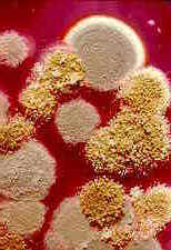
          Powdery Mildew

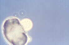
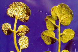
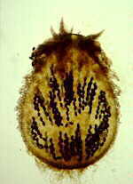
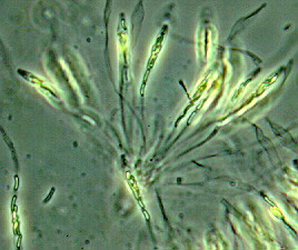
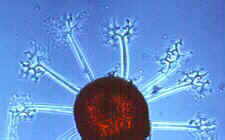
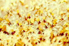
          Powdery mildew causes a white powdery growth on the upper surfaces of leaves and on the stems of infected plants. Infected areas are often stunted and distorted and may drop prematurely from the plant. Fruits are usually not directly affected, but their size and growth may be stunted. Powdery mildew is caused by the fungi Erysiphe cichoracearum and Sphaerotheca fuliginea. Infection can occur when temperatures are between 50 and 90 °F, during dry weather with high relative humidity. The disease can be a particular problem on late-planted squash.

          Image depicts powdery mildew on watermelon leaves.

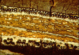
          Powdery mildew on watermelon leaves.
          David B. Langston, University of Georgia, www.insectimages.org
          Larger Image (280Kb)

          Prevention and Treatment: Powdery mildew-resistant varieties (Table 1) are available for most cucurbits, thus with proper planning, chemical control should not be necessary. Preventative fungicide treatments are available (Table 2) if disease becomes severe enough to warrant chemical control.
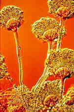
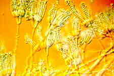
          Downy Mildew

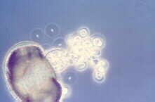
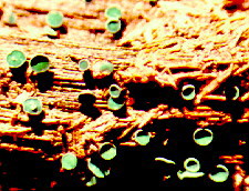
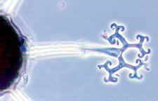
          Downy mildew is one of the most important leaf diseases of cucurbits. Typically, symptoms begin as small yellow areas on the upper leaf surface. As lesions expand, they may become brown with irregular margins. Affected areas may grow together, and the entire leaf may wither and die. Infected plants also develop a gray mold on the lower leaf surface. The fruit is not affected, but in the case of cantaloupes, it will be less sweet. This disease is caused by the fungus Pseudoperonospora cubensis and is favored by moist conditions.

          Image depicts downy mildew on lower surface of cantaloupe leaf.

          Larger Image (509 Kb)

          Image depicts downy mildew on upper surface of cantaloupe leaf.

          Larger Image (990 Kb)

          Downy Mildew on top (left) and bottom (right) of cantaloupe leaf.
          Clemson University - USDA Cooperative Extension Slide Series, Bugwood.org

          Symptoms on watermelon are different than symptoms on other curcurbits. Leaf spots on watermelon are dark brown and irregular in shape, ranging from oval to angular to retangular. Slight yellowing may be seen around the edges of the spots or in small patches in other parts of the leaf. Leaves infected with downy mildew curl inward as the leaf dies. As on other crops, spores usually are found on the bottom of the leaf, although spores may be formed on top of the leaf in severe infections or foggy weather.

          Prevention and Treatment: Use varieties that are resistant to this disease (Table 1). Fungicides are available for the home vegetable garden if disease becomes severe enough to warrant chemical control (Table 2).
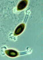
          Gummy Stem Blight

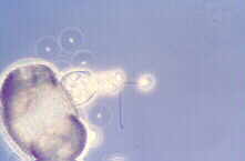
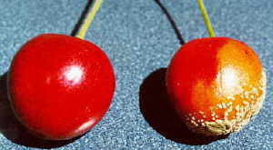
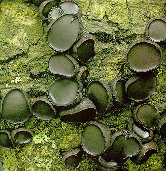
          Gummy stem blight is a stem and leaf disease of cucumber, cantaloupe, pumpkin and watermelon caused by the fungus Didymella bryoniae. This fungus also causes a fruit rot called black rot.

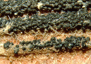
          Symptoms include leaves with brown or tan spots of various sizes that may eventually cover the entire leaf. The stems may split to form open wounds called cankers. A brown, gummy substance may be evident on the surface of these open wounds. Infected vines usually wilt after the middle of the season. Infected stems die one after another, and seedlings and entire individual vines may be killed. Affected fruit have irregular circular spots, and a wet rot occurs where the fungus penetrates the rind.

          Image depicts spots caused by gummy stem blight on watermelon leaf.

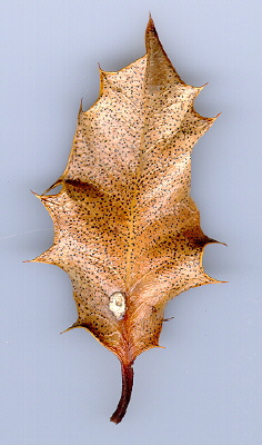
          Leaf spots caused by gummy stem blight on watermelon leaf.
          Clemson University - USDA Cooperative Extension Slide Series, Bugwood.org
          Larger Image (259 Kb)

          To distinguish gummy stem blight on watermelon from downy mildew, look at the size, shape, and position of leaf spots. Leaf spots of gummy stem blight are larger than individual spots of downy mildew. Some leaf spots of gummy stem blight have a ringed or target look. Gummy stem blight also can be found on the petioles (leaf stems) and the mid vein of leaves as a water-soaked or reddish-brown wet spot.

          Prevention and Treatment: There are no varieties that are resistant to this disease. This disease may be seed-borne, so purchase seed from a reputable source. Remove and destroy all plant debris in the garden, since the disease can survive on plant debris from year to year. Rotate crops with nonhost plants, such as corn, for two or more years as an effective way of reducing the incidence of this disease. Avoid wetting the leaves when watering. If disease is severe enough to warrant chemical control, preventative fungicides are available (Table 2).

          Image depicts anthracnose spots on watermelon leaf.

          Symptoms of anthracnose on watermelon.
          Clemson University - USDA Cooperative Extension Slide Series, Bugwood.org
          Larger Image (343 Kb)
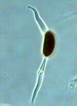
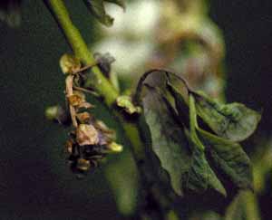
          Anthracnose

          Anthracnose is caused by the fungus Colletotrichum obiculare, and requires rainy, cool weather for several days for the disease to develop. The first symptoms of anthracnose are spots on the leaves that begin as yellowish or water-soaked areas. Spots enlarge and turn brown to black. The diseased tissue dries and the center of the spots fall out giving a “shot-hole” appearance. Infected fruits have black, circular, sunken cankers of different sizes.

          Prevention and Treatment: Remove and destroy old cucurbit vines and residues, since this is where the fungus survives the winter. Rotation of crops in the garden is also important to minimize disease. Purchase seeds from a reputable source, since the disease can be seed-borne. If the disease is severe enough to warrant the use of fungicides, several are available for home garden use (Table 2).

          Alternaria Leaf Spot

          This disease is caused by the fungus Alternaria cucumerina and causes small, circular, tan spots to appear on the leaves, which later enlarge to 1½ inches or more in diameter. Definite concentric rings and margins appear that give the area a “bull’s eye” appearance. Leaf drop can be severe. Bright sunshine, frequent dews or rain, and temperatures between 60 and 90 °F favor disease development.

          Image depicts alternaria leaf spot on cantaloupe leaf.

          Alternaria leaf spot on cantaloupe.
          Clemson University - USDA Cooperative Extension Slide Series, Bugwood.org
          Larger Image (379 Kb)

          Prevention and Treatment: Remove and destroy all infected plant residues at the end of the gardening season, since the fungus survives thewinter on plant residue. The disease is easily spread by tools, wind, splashing water or insects. Rotation of crops and seed treatment will also help. When this disease occurs consistently in the garden, a preventative fungicide program can be followed (Table 2).
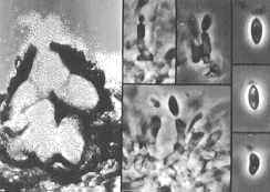
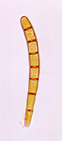
          Fusarium Wilt

          This disease is caused by the fungus, Fusarium oxysporum forma specialis melonis. It attacks the roots of the plant and moves into the stems. Older, established plants that are infected become stunted, wilt and eventually die. Wilt symptoms develop in one or more laterals, usually starting at the vine tips. A white mold may develop on dead vines. Affected seedlings will damp-off (rot at the soil line), wilt and die. On runners near the crown of the plant, brown streaks may be evident. Roots will have a honey brown discoloration inside.

          Prevention and Treatment: This fungus can survive in the soil for many years. Planting resistant varieties (Table 1) is critical in preventing this disease. Careful water management is also important in minimizing root stress. There are no chemical treatments available for control.
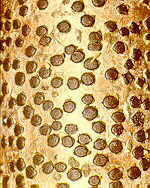
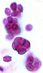
          Viruses

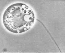
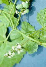
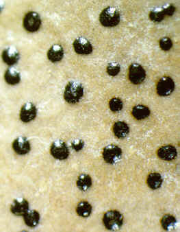
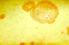
          There are several common viruses that can affect cucurbits, including cucumber mosaic virus (CMV) and watermelon mosaic virus (WMV). Infected plants may be stunted or have leaves that are mottled, crinkled, or a light green color. Fruits may be irregular in shape, mottled or warty. Various insects transmit these viruses.

          Cucumber mosaic virus (CMV) on squash.
          Division of Plant Industry Archive,
          Florida Department of Agriculture and Consumer Services, www.insectimages.org
          Larger Image (111 Kb)

          Prevention and Treatment: There are no chemicals available to kill viruses. Chemical control of the insects that spread the viruses may minimize the disease. This control method is difficult, because infection occurs immediately after an insect feeds, and insects migrate freely between plants. A good control strategy is to maintain healthy and vigorous plants, plant recommended varieties and monitor your garden for any unusual symptoms as they occur. Keep the area clear of weeds that can harbor insects. Choosing separate areas for early and late plantings may help to reduce virus severity in the late plantings.

          Blossom-End Rot

          Blossom-end rot appears as a dark-colored dry rot on the end of the fruit where the flower was. The problem is caused by a lack of calcium in the developing fruit. It is an indication that calcium is lacking in the soil or that the plant does not have the ability to take up enough calcium. When growth is rapid, not enough calcium may be delivered to the blossom end of the developing fruit.

          Image depicts blossom end rot on watermelon.

          Blossom end rot on watermelon.
          Clemson University - USDA Cooperative Extension Slide Series, Bugwood.org
          Larger Image (361 Kb)

          Prevention and Treatment: Help prevent blossom-end rot by having your soil tested before planting through your local county Extension office, and lime according to recommendations. Always maintain an adequate supply of moisture, especially during fruit growth. Mulch plants to prevent rapid drying of the soil and water plants during extended dry periods. Use gypsum (1-2 pounds per 100 square feet) as a supplement to liming on calcium deficient soil. Lime and/or gypsum should be applied before planting. Do not overfertilize plants with excessive nitrogen or potassium. Excess amounts of these nutrients reduce the uptake of calcium in the plant. When plants are dark green, extra fertilizer should not be applied.

          Treatment of affected plants includes applying a calcium solution to prevent additional fruits from being damaged. Spray or drench the foliage with a calcium nitrate or calcium chloride (three tablespoons per gallon of water) solution. Premixed solutions are also available. Follow the instructions on the label. Removing fruit with symptoms is recommended.

          RIPing in an asexual fungus

          RIPing in an asexual fungus

          Posted on March 23rd, 2008 by Jason Stajich · 2 Comments

          ResearchBlogging.orgA.niger conidiophoreA paper in Current Genetics describes the discovery of Repeat Induced Polymorphism (RIP) in two Euriotiales fungi.  RIP has been extensively studied in Neurospora crassa and has been identified in other Sordariomycete fungi Magnaporthe, Fusiarium. This is not the first Aspergillus species to have RIP described as it was demonstrated in the biotech workhorse Aspergillus oryzae.  However, I think this study is the first to describe RIP in a putatively asexual fungus.  The evidence for RIP is only found in transposon sequences in the Aspergillus and Penicillium.  A really interesting aspect of this discovery is RIP is thought to only occur during sexual stage, but a sexual state has never been observed for these fungi.  

          The authors discuss this evidence as to whether or not there is a cryptic sexual state that hasn't been observed or sex has only been lost for a short time in these fungi (the RIP evidence suggests the transposons were RIPed relatively recently).  The mating genes are still present in the genome of A. niger but whether its actively able to complete a sexual cycle (or enough of it to allow for RIPing) still needs to be investigated.

          Powdery Mildew on Vegetables

          Powdery Mildew on Vegetables

          Published 11/01

          In this Guideline:


          Powdery mildew is a common disease on many types of plants. There are many different species of powdery mildew fungi (e.g., Erysiphe spp., Sphaerotheca spp.) and each species only attacks specific plants. A wide variety of vegetable crops are affected by powdery mildews, including artichoke, beans, beets, carrot, cucumber, eggplant, lettuce, melons, parsnips, peas, peppers, pumpkins, radicchio, radishes, squash, tomatillo, tomatoes, and turnips (Table 1). Powdery mildews generally do not require moist conditions to establish and grow, and normally do well under warm conditions; thus they are more prevalent than many other leaf-infecting diseases under California's dry summer conditions.

          Table 1. Host Plants and Control Measures for Powdery Mildew Species.
          Hosts Fungus species Controls

          cucumbers, endive, lettuce, melons, potato, pumpkin, squash

          Erysiphe cichoracearum

          resistant varieties of lettuce, cucumber; water sprays; fungicides if necessary on squash and pumpkin

          broccoli, Brussels sprouts, cauliflower, and other cole crops; radicchio, radishes, turnips

          Erysiphe cruciferarum

          not usually required

          tomatoes

          Erysiphe lycopersici

          fungicides if necessary

          peas

          Erysiphe pisi

          resistant varieties; sprinkler irrigation

          carrots, parsley, parsnips

          Erysiphe heraclei

          tolerant varieties

          beets

          Erysiphe polygoni

          tolerant varieties

          artichoke, eggplant, peppers, tomatillo, tomatoes

          Leveillula taurica

          rarely required; fungicides if necessary

          beans, black-eyed peas, cucurbits, okra

          Sphaerotheca fuliginea

          resistant varieties for some; fungicides if necessary

          IDENTIFICATION AND DAMAGE
          Powdery mildew first appears as white, powdery spots that may form on both surfaces of leaves, on shoots, and sometimes on flowers and fruit. These spots gradually spread over a large area of the leaves and stems. An exception is one of the powdery mildews that affects artichokes, onions, peppers, and tomatoes: it produces yellow patches on leaves but little powdery growth.

          Leaves infected with powdery mildew may gradually turn completely yellow, die, and fall off, which may expose fruit to sunburn. On some plants, powdery mildew may cause the leaves to twist, buckle, or otherwise distort. Powdery mildew fungal growth does not usually grow on vegetable fruits, although pea pods may get brownish spots. Severely infected plants may have reduced yields, shortened production times, and fruit that has little flavor.

          LIFE CYCLE

          All powdery mildew fungi require living plant tissue to grow. Year-round availability of crop or weed hosts is important for the survival of some powdery mildew fungi. Special resting spores are produced, allowing overwinter survival of the species that causes the disease in cucurbits, lettuce, peas, and certain other crops.

          Most powdery mildew fungi grow as thin layers of mycelium (fungal tissue) on the surface of the affected plant part. Spores, which are the primary means of dispersal, make up the bulk of the white, powdery growth visible on the plant's surface and are produced in chains that can be seen with a hand lens; in contrast, spores of downy mildew grow on branched stalks that look like tiny trees.

          Powdery mildew spores are carried by wind to new hosts. Although humidity requirements for germination vary, all powdery mildew species can germinate and infect in the absence of free water. In fact, spores of some powdery mildew fungi are killed and germination is inhibited by water on plant surfaces for extended periods. Moderate temperatures (60° to 80°F) and shady conditions generally are the most favorable for powdery mildew development. Spores and fungal growth are sensitive to extreme heat (above 90°F) and direct sunlight.

          MANAGEMENT

          The best method of control is prevention. Planting resistant vegetable varieties when available, or avoiding the most susceptible varieties, planting in the full sun, and following good cultural practices will adequately control powdery mildew in many cases (Table 1). However, very susceptible vegetables such as cucurbits (cucumber, melons, squash, and pumpkins) may require fungicide treatment. Several least-toxic fungicides are available but must be applied no later than the first sign of disease.

          Resistant Varieties

          In some cases, varieties resistant to powdery mildew may be available. If available, plant resistant varieties of cantaloupe, cole crops, cucumber, melons, peas, pumpkins, and squash. If you plant more susceptible varieties, you may need to take control measures.

          Cultural Practices

          Plant in sunny areas as much as possible, provide good air circulation, and avoid applying excess fertilizer. A good alternative is to use a slow-release fertilizer. Overhead sprinkling may help reduce powdery mildew because spores are washed off the plant. However, overhead sprinklers are not usually recommended as a control method in vegetables because their use may contribute to other pest problems.

          Fungicide Application

          In some situations, especially in the production of susceptible cucurbits, fungicides may be needed. Fungicides function as protectants, eradicants, or both. A protectant fungicide prevents new infections from occurring whereas an eradicant can kill an existing infection. Apply protectant fungicides to highly susceptible plants before the disease appears. Use eradicants at the earliest signs of the disease. Once mildew growth is extensive, control with any fungicide becomes more difficult.

          Fungicides. Several least-toxic fungicides are available, including horticultural oils, neem oil, jojoba oil, sulfur, potassium bicarbonate, bicarbonate of soda (baking soda), and the biological fungicides AQ10 and Serenade. With the exception of the oils, these materials are primarily preventive, although potassium bicarbonate has some eradicant activity. Oils work best as eradicants but also have some protectant activity.

          Oils. To eradicate mild to moderate powdery mildew infections, use a horticultural oil such as JMS Stylet Oil, Saf-T-Side Spray Oil, Sunspray Ultra-Fine Spray Oil or one of the plant-based oils such as neem oil (e.g., Powdery Mildew Killer) or jojoba oil (e.g., E-rase). Be careful, however, to never apply an oil spray within 2 weeks of a sulfur spray or plants may be injured. Also, oils should never be applied when temperatures are above 90°F or to drought-stressed plants. Some plants may be more sensitive than others, however, and the interval required between sulfur and oil sprays may be even longer; always consult the fungicide label for any special precautions. Of the horticultural oils, JMS Stylet Oil is the most highly refined and therefore the least likely to damage plants, but it may be more difficult to obtain than the others.

          Sulfur. Sulfur products have been used to manage powdery mildew for centuries but are only effective when applied before disease symptoms appear. The best sulfur products to use for powdery mildew control in gardens are wettable sulfurs that are specially formulated with surfactants similar to those in dishwashing detergent (e.g., Safer Garden Fungicide). However, sulfur can be damaging to some squash and melon varieties. To avoid injuring any plant, do not apply sulfur when air temperature is near or over 90°F and do not apply it within 2 weeks of an oil spray. Other sulfur products, such as liquid lime sulfur or sulfur dust, are much more difficult to use, irritating to skin and eyes, and limited in terms of the plants they can safely be used on. Copper is also available to control powdery mildew but is not very effective.

          Bicarbonates. Also available is a fungicide product containing potassium bicarbonate (e.g., Kaligreen) and a fungicide that can be made at home by combining 2-1/2 tablespoons of horticultural oil (Sunspray Ultra-Fine, Saf-T-Side, etc.) in a gallon of water and adding 4 teaspoons baking soda. This solution is sprayed on plants to prevent powdery mildew infections. Sprays of both potassium bicarbonate and baking soda can injure the plant, so use these materials with caution. Also, baking soda sprays can have deleterious effects on soil structure and should be used sparingly.

          Biological Fungicides. Biological fungicides (AQ10 and Serenade) are commercially available beneficial microorganisms formulated into a product that, when sprayed on the plant, destroys fungal pathogens. AQ10 is a parasitic fungus, Ampelomyces quisqualis, that actively attacks and destroys the powdery mildew fungus. The active ingredient in Serenade is a bacterium, Bacillus subtilis, that helps prevent the powdery mildew from infecting the plant. While both products function to kill the powdery mildew organism and are nontoxic to people, pets, and beneficial insects, they have not proven to be as effective as the oils or sulfur in controlling this disease.

          How to Use. Apply protectant fungicides to susceptible plants before or in the earliest stages of disease development. The protectant fungicides are only effective on contact, so applications must provide thorough coverage of all susceptible plant parts. As plants grow and produce new tissue, additional applications may be necessary at 7- to 10-day intervals as long as conditions are conducive to disease growth.

          If mild to moderate powdery mildew symptoms are present, the horticultural oils and plant-based oils such as neem oil and jojoba oil can be used.

          WARNING ON THE USE OF CHEMICALS

          برخی از بیماریها

          WILTING OR YELLOWING OF PLANTS

          Fusarium Wilt
          Causal Agent (fungus - Fusarium oxysporum f.sp. niveum)


          Initially, leaves of infected plants wilt during hotter times of the day, but eventually, the wilt becomes permanent [Picture 1]. Initially, one or a few vines of a plant are affected, but eventually the other vines wilt and the plant dies. Other agents can cause plants to wilt. For example, anasa wilt is caused by the feeding of the squash bug.

          The fungus causes browning of the xylem (water-conducting tissue) of the crown and runners [Picture 2]. Races 1 and 2 of this fungus are present in several watermelon-growing areas of Texas. Most commercial cultivars have resistance to to race 1, but not race 2. The fungus persists many years in soil and so, watermelons should not be replanted into infested soils for at least five years.


          Yellow Vine
          Causal Agent: (unknown, suspected to be a bacterium)

          This disease is found mainly in the Cross Timbers vegetation zone in central Texas and Oklahoma, but it has been found outside of this area, specifically, in Anderson county in east Texas. The symptoms appear two weeks before harvest. There is a general yellowing of leaves and terminal leaves of the vine are turned up [Picture 3]. Not all plants within a field initially show symptoms [Picture 4]. Plants eventually die. These symptoms are not diagnostic for this disease. The distinctive symptom of yellow vine is a honey-brown discoloration of the phloem in lower crown and primary root of plants [Picture 5]. The cause has not been conclusively determined, but a phloem-limited bacterium has been consistently associated with diseased plants and a deltacephaline leafhopper is thought to transmit this bacterium. Other susceptible cucurbits include squash, pumpkin, and cantaloupe. There are no control measures available for this disease, but resistance in triploid seedless watermelon (e.g. 'Tri-X 313') has been identified.


          Southern Blight
          Causal Agent: (fungus - Sclerotium rolfsii)

          This soilborne fungus infects the crown [Picture 6], resulting in wilting of foliage. The fungus also causes a fruit rot. Infection is favored in warm, moist soils. There are no control recommendations.


          Charcoal Rot
          Causal Agent: (fungus - Macrophomina phaseolina)

          A water-soaked lesion develops around the crown of the plant, followed by the appearance of an amber-colored ooze at the surface. This area becomes dark brown, dried and cracked. Small black fungal structures (microsclerotia) are then formed in this area. A dark ring is visible beneath the outer layer when a cross section is cut through the canker. Infected plants have yellow crown leaves and die soon after symptoms are observed.

          The damage is most severe when melons approach maturity. There are no effective controls for the fungus. Altering the irrigation program to prevent water stress may alleviat the problem.


          Monosporascus Root Rot/Vine Decline
          ausal Agent: (fungus - Monosporascus cannonballus)

          Infected plants usually do not develop symptoms until near harvest time. Infected plants wilt and eventually die. Sometimes, there is vascular browning [Picture 7]. Brown lesions may be found on the roots [Picture 8], even on plants that are not wilting. Root lesions may be caused by other soilborne fungi, such as Pythium sp. and Rhizoctonia solani [Picture 9]. This disease is primarily a problem on silt or clay soils of the Lower Rio Grande Valley. The fungus is indigenous to the soil and populations build up with continued cucurbit monoculture, particularly cantaloupe. Soil fumigation is an effective control, but is not usually economical. Soils with consistent problems should not be planted to cucurbits.


          Damping-Off
          Causal Agents: (fungi - Rhizoctonia solani, Pythium spp.)

          Seedlings wilt and die. The lower stems have a watersoaked appearance. Damping-off is usually a problem of direct-seeded watermelons germinating in cool, moist soil [Picture 10], but Pythium aphanidermatum can rapidly kill new transplants in warm soils [Picture 11]. A soil application of mefenoxam (Ridomil Gold) at the time of planting will prevent damping-off caused by Pythium species.


          Nematodes

          Nematodes can substantially reduce yield of watermelon. The affected plants are stunted and chlorotic. Root-knot nematodes can cause galling on roots [Picture 12], but there are no visible root symptoms with the reniform nematode. The root-knot nematode occurs primarily in sandy soils. Although nematicides can provide an economical control, a crop rotation with corn or sorghum and a weed-free fallow can reduce numbers of nematodes below damaging thresholds.


          Non-Pathogenic Causes

          Agents that can cause seedling death that resemble damping-off include the seed corn maggot [Picture 13] and lightning damage [Picture 14]. Wind movement of young plants on plastic mulch can cause damage to the stems, leading to wilt [Picture 15]. This problem disappears once vines are able to anchor to something.

          Anasa wilt [Picture 16], caused by the squash bug [Picture 17], can be mistaken for Fusarium wilt. Watermelon growing next to brush are more prone to this problem.

          Herbicide injury can cause symptoms that resemble those caused by pathogenic fungi. Glyphosate can cause yellowing [Picture 18]. Trifluralin can cause scattered wilting in a field [Picture 19] and affected plants will also exhibit discoloration or swelling of the crown area [Picture 20].


          [Back to Top]


          LEAVES DEFORMED, OR WITH SPOTS OR MOLD

          Bacterial Fruit Blotch
          Causal Agent: (bacterium - Acidovorax avenae subsp. citrulli)

          Infected seedlings initially develop water-soaking on the lower surface of cotyledons and leaves [Picture 21]. These areas then become necrotic [Picture 22]. The pathogen can also cause non-distinctive, necrotic leafspots on transplants [Picture 23]. On leaves of more mature plants, the pathogen can cause large, circular brown lesions, usually near the edge of the leaf, but this is not reliable for diagnosis. The unique symptoms are: irregular, rust-colored lesions that run along leaf ribs [Picture 24], while on the underside of the leaf, the leaf rib is water-soaked [Picture 25]. Leaf lesions can serve as a source of bacteria that can infect fruit. In the field, leaf lesions are often difficult to find and may be confused with lesions caused by pathogenic fungi.

          Where this disease has occurred, watermelons and other cucurbit crops should not be replanted in the field for at least one season, crop debris should be plowed under, and volunteer plants destroyed. Cucurbit weeds such as citron melon should also be controlled. Copper-based fungicides should be applied on a regular schedule at the time of flowering or earlier, if transplants are used.

          For more information, see the following publication: L-5222 Bacterial Fruit Blotch of Watermelon
          (In order to view this file you must have Adobe Acrobat Reader installed on your computer.)


          Anthracnose
          Causal Agent: (fungus - Colletotrichum orbiculare)

          Symptoms appear first on crown leaves as small, brown-black spots usually after vines begin to "run" [Picture 26]. These lesions are also visible on the underside of leaves [Picture 27]. During damp weather, orange-pink masses of spores develop in centers of larger leaf spots. During dry weather the spore masses turn gray. All but the youngest leaves are infected. Lesions can coalesce, causing the leaves to die [Picture 28]. The disease can be seed borne and symptoms can sometimes be seen on young transplants [Picture 29].

          A combination of seed treatment, crop rotation and fungicide applications are necessary for controlling this disease on susceptible varieties. Protective spray applications should be made when vines start to run and should be continued at 7 to 10 day intervals during periods of humid or rainy weather. Schedules may be lengthened to 14 days during dry weather.


          Gummy Stem Blight
          Causal Agent: (fungus - Didymella bryoniae)

          Early symptoms are round black, wrinkled spots on young leaves and dark sunken areas on stems. Early infection usually develops from contaminated seed and can be visible on transplants [Picture 30]. Often, infected transplants will have lesions on the the stem [Picture 31]. On older leaves in the field, irregular brown to black spots develop between leaf veins [Picture 32]. Leaf lesions will expand [Picture 33], eventually resulting in the death of leaves [Picture 34]. As the disease progresses, older stems near the crown, most commonly near a leaf petiole or tendril, split and a light brown gum oozes from the watersoaked areas [Picture 35]. The crown may also be infected and produce a brown ooze [Picture 36]. Infected runners die, leaving fruit exposed [Picture 37]. Fruits are not usually affected. Protective fungicide applications are needed for control. Resistance to benomyl and thiophanate-methyl fungicides have occurred in some growing areas, so these fungicides are not recommended.


          Downy Mildew
          Causal Agent: (fungus - Pseudoperonospora cubensis)

          The disease is most severe during periods of cool temperatures and rain. Irregular yellowish to brown spots, often vague in outline, appear on upper leaf surface near the crown [Picture 38]. Brown spots later become more distinct on both sides of the leaves [Picture 39]. The underside of leaves exhibit a brown to gray fungal growth on wet mornings [Picture 40], which disappears during dry weather. This growth is best seen early in the morning, or, alternatively, suspect leaves can be removed and kept several hours in a plastic bag with a moist paper towel to provide high humidity that encourages sporulation. Spots enlarge rapidly and turn black and the entire leaf withers and dies [Picture 41]. Leaf petioles of diseased leaves stand upright although the leaf is almost dead [Picture 42]. There can be a rapid, extensive loss of foliage with this disease [Picture 43].

          The disease is favored by cool (60 F night and up to 77 F day) temperatures, along with rain or continuous high humidity. This disease can occur in a field miles away from other cucurbits, indicating that the spores can travel great distances. The fungus develops and spreads rapidly during cool, moist weather conditions.

          Excellent control of this disease can be achieved with mefanoxam (Ridomil Gold), which must always be used simultaneously with a protectant fungicide such as mancozeb or chlorothalonil, to prevent the emergence of resistant strains. It is important that these sprays are applied on a preventative basis, before the disease becomes established in a field. Some growers report that they only use chlorothalonil or mancozeb to effectively manage downy mildew, but, the key to their effectiveness is regular, preventative applications.


          Alternaria Leaf Spot
          Causal Agent: (fungus - Alternaria cucumerina)

          Lesions are round to irregular target spots on older leaves [Picture 44]. The symptoms are first observed at the crown of the plant. As the spots enlarge, concentric rings are formed in the lesion. The disease is favored by continuous wet conditions. The fungus is controlled with a 2 year cucurbit-free rotation, destruction of the previous crop residue and application of fungicides as needed. At the end of the growing season, plow under crop residue.


          Cercospora Leaf Spot
          Causal Agent: (fungus - Cercospora citrullina)

          Leaf spots have a dark brown center and a yellow halo [Picture 45]. Infected leaves are first observed at the crown of the plant. When the disease is severe, foliage loss will restrict fruit development and result in sunburn of fruit. The fungus is controlled with fungicides. Applications should begin soon after runner development.


          Myrothecium Leaf Spot
          Causal Agent: (fungus - Myrothecium roridum)

          This fungus causes small, dark brown circular lesions on leaves [Picture 46]. These lesions can coalesce [Picture 47]. The disease is favored by wet weather conditions that favor the development of other foliar pathogens and can be controlled by the protectant fungicides used to control these pathogens. The disease occurs infrequently and has been observed mostly in the Lower Rio Grande Valley.


          Powdery Mildew
          Causal Agent(s): (fungi - Sphaerotheca fuliginea, Erysiphe cichoracearum)

          The first symptom is a white to gray dusty material on the upper leaf surface [Picture 48]. The fungus quickly spreads to completely cover the leaf surface. Once the leaf is covered, it dies and has a crisp texture. This disease is favored by high humidity, but not free water on leaves. The fungus is best controlled with fungicide applications when symptoms are first observed. It tends to occur on older leaves and later in the growing season, so control is not usually warranted. Two systemic fungicides, Benlate (benomyl) and Bayleton (triadimefon), are no longer effective because of pathogen resistance.


          Leaf Mosaic
          Causal Agents: (viruses -Watermelon Mosaic Virus, Papaya Ringspot Virus, Zucchini Yellow Mosaic Virus, Cucumber Mosaic Virus)

          Leaves of infected plants have a mosaic pattern [Picture 49], i.e., areas of light and dark coloration. This mosaic may be in association with raised areas on leaves [Picture 50]. Leaves may also be distorted in shape [Picture 51]. Diagnosis of the mosaic viruses can not be made with certainty from symptoms alone. Sometimes plants may be infected by more than one type of virus. Losses from virus infection are more significant if young plants are infected. Most of the young fruit is aborted and those that are not aborted are malformed. The viruses listed above are aphid-transmitted and can overwinter in weeds. Virus control by insect control is not usually feasible. Weed control in and around the field may help to reduce losses, except for virus introduction via winged aphids.


          Tobacco Ring Spot Virus

          Tiny brown spots surrounded by yellow halo may appear on young leaves as a "stippling" effect [Picture 52]. Symptoms produced on fruits are more important than leaf symptoms, though. The virus can overwinter in wild hosts and in infected seed. It is spread from infected plants by the dagger nematode (Xiphinema spp.), grasshoppers, tobacco thrips, tobacco flea beetles and several other insects.


          Squash Leaf Curl Virus

          This virus is transmitted by whiteflies and is a problem only in south Texas[Picture 53]. Symptoms of the disease are crumpled leaves with yellowed, mottled areas [Picture 54]. Leaves have shortened petioles that cluster around the vines [Picture 55]. Squash is also susceptible to this virus. Severe yield losses are associated with infection of young seedlings, usually when whitefly populations are high. Infections of older plants do not affect yield.

          Another whitefly-transmitted virus recently identified in south Texas is the cucurbit yellow stunt disorder virus, which causes a yellowing of leaves [Picture 56].

          The squash leaf curl virus is economically important only in fall-grown watermelons. To alleviate the impact on yield, transplants should be used, rather than direct-seeded plants. Fields should be plowed under after harvest to minimize carryover of the virus to subsequent crops. The virus can persist in whiteflies for 3 weeks after feeding on infected plants, so there should be a 4 week break between melon crops.


          Herbicide Injury

          Growth deformation on foliage can be caused by 2,4-D or dicamba [Picture 57]. This deformation can resemble symptoms caused by some mosaic viruses.

          [Back to Top]


          FRUIT AFFECTED

          Bacterial Fruit Blotch
          Causal Agent: (bacterium - Acidovorax avenae subsp. citrulli)

          Fruit symptoms start as water-soaked spots that rapidly enlarge. Symptoms are generally seen in the light portions of striped fruit [Picture 58]. The lesions later turn brown and crack [Picture 59]. The pathogen is introduced to a field by planting contaminated seed or transplants. Within the field, the bacteria spread to healthy plants via rain or overhead irrigation, especially under conditions of high humidity and temperature. The fruit are most susceptible to infection around the time of pollination. As the fruit matures, it becomes resistant to infection. Once introduced into a field, the bacteria can persist on volunteer watermelon or citron [Picture 60] plants, or in their seed. Cantaloupe and honeydew melons are also susceptible to this pathogen.

          Where this disease has occurred, watermelons and other cucurbit crops should not be replanted in the field for at least one season, crop debris should be plowed under, and volunteer plants destroyed. Cucurbit weeds such as citron melon should also be controlled. Copper-based fungicides should be applied on a regular schedule at the time of flowering or earlier, if transplants are used.

          For more information, see the following publication: L-5222 Bacterial Fruit Blotch of Watermelon
          (In order to view this file you must have Adobe Acrobat Reader installed on your computer.)


          Anthracnose
          Causal Agent: (fungus - Colletotrichum orbiculare)

          The fungus produces sunken spots on the rind of fruit, that are often black because of the production of spores [Picture 61]. Symptoms may not be visible at harvest, or they might be slight [Picture 62], but decay may develop further in transit or at the market. Small anthracnose spots can be confused with pimples disease.


          Bacterial Rind Necrosis
          Causal Agent: (bacterium - Erwinia spp.)

          The first symptoms are numerous brown, dry, hard, discolored areas in the rind. These areas enlarge and may merge to form extensive necrotic areas [Picture 63]. The disease rarely extends into the flesh of infected melons in the field. The disease occurs sporadically and is thought to be caused by bacteria that are naturally present in fruit, but the reasons for symptom development are not understood. Since usually there are no external symptoms, a small percentage of diseased melons could make the crop unmarketable, as diseased melons cannot be culled. There are no control measures.


          Belly Rot
          Causal Agent(s): (fungi - several species)

          The fruit decays at the area in contact with soil. The rot begins as a small water soaked area that quickly spreads. In many cases, the area will be surrounded by a profuse growth of white mycelium. One of the fungal species associated with belly rot is Pythium aphanidermatum [Picture 64], which rapidly colonizes the fruit [Picture 65]. Another species involved is the southern blight fungus, Sclerotium rolfsii, which produces seed-like resting structures [Picture 66]. The disease is most severe during periods of rainfall and high temperature, when the soil remains wet for long periods of time. To control, use well-drained soils, or plant on plastic mulch.


          Lasiodiplodia Fruit Rot / Diplodia Stem-End Rot
          Causal Agent: (fungus - Lasiodiplodia theobromae)

          Watersoaking is seen at the stem end and the tissue is soft [Picture 67]. When harvesting, a large portion of the peduncle should remain attached to the fruit. The disease is of minor importance.


          Pimples
          Causal Agent: (suspected virus - Tobacco Ring Spot Virus)

          Small bumps or pimples develop on fruit [Picture 68], which are most noticeable on dark rind varieties. Other fruit symptoms are ringspots [Picture 69] and necrotic, concentric rings [Picture 70].


          Blossom End Rot
          Causal Agent: (physiological - water stress)

          Browning and shriveling occur at the blossom end of melon [Picture 71], followed by a secondary decay caused by microorganisms that progresses inward. It is caused by a calcium deficiency in the fruit and occurs in plants subjected to drought stress. Cultivars can differ in their susceptibility to the problem. Watering operations that avoid drought stress can reduce or eliminate the problem. Excess nitrogen fertilizer may also contribute to blossom-end rot.


          Bottle Neck
          Causal Agents: (physiological - several)

          Bottle neck [Picture 72] or otherwise misshapen fruit can be caused by pollination problems or fluctuations in watering. Cultivars can differ in susceptibility. Increasing the bee population by bringing in bee hives and placing them near the melon field may alleviate the problem.


          Sunburn

          The lack of foliage to protect the fruit can result in sunburn [Picture 73]. This often occurs as a consequence of loss of foliage caused by fungal diseases such as downy mildew.


          Fruit crack/scarring

          Cracks, scarring [Picture 74], and pitting can be caused by mechanical damage when fruits are young, Insects can also cause such injury. Animals, such as wild hogs and racoons, can cause more substantial damage [Picture 75].


          Splitting

          Splitting is caused by an erratic watering pattern, particularly the excessive accumulation of water. The inside of the fruit is under great pressure and the fruit splits open when lightly tapped. This condition is weather related and there probably is a cultivar interaction as well.


          Cross-stitch

          There are parallel cuts extending into the rind, which are perpendicular to vascular bundles [Picture 76]. The cause is unknown, but it is not suspected to be infectious.

          [Back to Top]

          Rhizoctonia solani

          Rhizoctonia solani

          Pathogen profile created by Paulo Ceresini
          as one of the requirements of the course PP-728 Soilborne Plant Pathogens, offered on Spring 1999


          Introduction
          Rhizoctonia solani, the most widely recognized species of Rhizoctonia was originally described by Julius Kühn on potato in 1858. Rhizoctonia solani is a basidiomycete fungus that does not produce any asexual spores (called conidia) and only occasionally will the fungus produce sexual spores (basidiospores). In nature, R. solani reproduces asexually and exists primarily as vegetative mycelium and/or sclerotia. Unlike many basidiomycete fungi, the basidiospores are not enclosed in a fleshy, fruiting body or mushroom. The sexual fruiting structures and basidiospores (i.e. teleomorph) were first observed and described in detail by Prillieux and Delacroiz in 1891. The sexual stage of R. solani has undergone several name changes since 1891, but is now known as Thanatephorus cucumeris.


          Host range and distribution

          R. solani  is a very common soilborne pathogen with a great diversity of host plants. The Table 1 illustrates the relationship of particular anastomosis groups of R. solani  and the hosts they infect.


          Isolation

          Qualitative determinations of R. solani in infected plants are made by isolations from infected host plant tissues. Infected plant tissues are cut in pieces of 5 cm, washed in running tap water to eliminate any attached organic debris, and blotted to dry. Small samples of plant tissue (0.5 cm of length) are then cut from the lesions and transferred to an isolation medium, which can be either general (e.g. alkaline water agar) or selective (e. g. modified Ko & Hora medium). The alkaline water agar medium provides a faster way of isolating the fungus than other general media since successful isolation of R. solani can be obtained after 24 h of transfer (Guttierrez et al., 1997).

          Quantitative determination of R. solani from soils to estimate the inoculum density are based on the saprophitic and/or pathogenic competitive abilities of the fungus. Methods developed from this principle included the burial and subsequent recovery of various substrates as baits for Rhizoctonia. The baits include suscetible host plants, autoclaved seeds, stem segments such as flax, buckwheat, bean, cotton and cereal straw, and even agar baits. Other methods include different soil sieving procedures combined with selective media for the isolation of R. solani from soil. A subsequent method using a multiple-pellet soil-sampler was developed for quantitative estimation of propagule density of R. solani based on placement of weighed amounts of soil, or soil pellets on water agar supplemented with chloramphenicol, or on selective media (Hennis et al. 1978, Ko & Hora 1971, Castro et al. 1988).


          Identification

          The vegetative mycelium of R. solani and other Rhizoctonia fungi are colorless when young but become brown colored as they grow and mature. The mycelium consists of hyphae partitioned into individual cells by a septum containing a dough-nut shaped pore. This septal pore allows for the movement of cytoplasm, mitochondria, and nuclei from cell to cell. The hyphae often branch at a 90o angles and usually possess more than three nuclei per hyphal cell. The anatomy of the septal pore and the cellular nuclear number (CNN) have been used extensively by researchers to differentiate R. solani from other Rhizoctonia fungi. R. solani [renamed Moniliopsis solani = Moniliopsis anderholdii (Moore, 1987)] is characterized by: CNN close to the tips in young hyphae greater than two, main runner hyphae usually wider than 7mm, mycelium buff-colored to dark brown, sclerotia (if present) irregular shape, light to dark brown, not differentiated into rind and medula and having Thanatephorus cucumeris its as teleomorph.

          Because R. solani and other Rhizoctonia fungi do not produce conidia and only rarely produce basidiospores, the classification of these fungi often has been difficult. Prior to the 1960’s, researchers relied mostly on differences in morphology observed by culturing the fungus on a nutrient medium in the laboratory and/or pathogenicity on various plant species to classify Rhizoctonia. In 1969, J. R. Parmeter and his colleagues at the University of California in Berkeley, reintroduced the concept of "hyphal anastomosis" to characterize and identify Rhizoctonia. The concept implies that isolates of Rhizoctonia that have the ability to recognize and fuse (i.e. "anastomose") with each other are genetically related, whereas isolates of Rhizoctonia that do not have this ability are genetically unrelated.

          Anastomosis groups of binucleate and multinucleate Rhizoctonia spp.

          Hyphal anastomosis criteria have been used extensively to place isolates of Rhizoctonia into taxonomically distinct groups called anastomosis groups. In practice, hyphal anastomosis is determined in several ways. The most commonly employed practice involves pairing two isolates of Rhizoctonia on a glass slide and allowing them to grow together. The area of merged hyphae is stained and examined microscopically for the resulting hyphal interaction(s).

          Pairing of isolates belonging to the same AG-results in hyphal fusion (anastomosis), leading to either acceptance (self-pairings) or rejection (somatic incompatibility). Pairings between AGs do not result in hyphal fusion, suggesting greater genetic differences between isolates (i.e., different species, etc.) Interpretation of anastomosis reaction is not always straightforward because the four hyphal interaction phenotypes (C0 to C3) represent a continuum.  Within an AG, two types of hyphal interactions (C2 and C3) are most relevant for the study of population biology. The C2 reaction (also referred as killing reaction), represents a somatic incompatibility response between genetically distinct individuals. The C3 reaction (perfect fusion) between two isolates is indicative of genetic identity or near identity.

          Very little is known about the genetic mechanisms controlling this recognition process in Rhizoctonia. In other filamentous fungi, somatic incompatibility is controlled by several genes with multiple alleles. For two fungal isolates to be compatible, all somatic compatibility loci must be the same.

          Isolates of R. solani have been assigned to 12 AGs. Recent protein and DNA-based studies support the separation of R. solani into genetically distinct groupings, but has also revealed considerable genetic diversity within an anastomosis group. Hyphal anastomosis and molecular methods are currently being used to further examine the taxonomy, ecology and pathology of R. solani.


          Symptoms

          R. solani primarily attacks below ground plant parts such as the seeds, hypocotyls, and roots, but is also capable of infecting above ground plant parts (e.g. pods, fruits, leaves and stems). The most common symptom of Rhizoctonia disease is referred to as "damping-off" characterized by non germination of severely infected seed whereas infected seedlings can be killed either before or after they emerge from the soil. Infected seedlings not killed by the fungus often have cankers, which are reddish-brown lesions on stems and roots. In addition to attacking below ground plant parts, the fungus will occasionally infect fruit and leaf tissue located near or on the soil surface. This type of disease often occurs because the mycelium and/or sclerotia of the fungus are close to or splashed on the plant tissue.

          Although most Rhizoctonia diseases are initiated by mycelium and/or sclerotia, several important disease of beans, sugar beet, and tobacco result from basidiospore infection.These basidiospores also serve as a source for rapid and long distance dispersal of the fungus. The basidiospores germinate to produce hyphae that infect leaves during periods of high relative humidity and periods of extended wet weather. Under these conditions, basidiospores can often be observed on the base of stems near the soil surface or on the underside of leaves in the plant canopy.


          Ecology and life cycle

          R. solani can survive for many years by producing small (1 to 3-mm diameter), irregular-shaped, brown to black structures (called sclerotia) in soil and on plant tissue. Certain rice pathogens of R. solani, have evolved the ability to produce sclerotia with a thick outer layer that allows them to float and survive in water. R. solani also survives as mycelium by colonizing soil organic matter as a saprophyte, particularly as a result of plant pathogenic activity. Sclerotia and/or mycelium present in soil and/or on plant tissue germinate to produce vegetative threads (hyphae) of the fungus that can attack a wide range of food and fiber crops.

          The fungus is attracted to the plant by chemical stimulants released by actively growing plant cells and/or decomposing plant residues. As the attraction process proceeds, the fungal hypha will come in contact with the plant and become attached to its external surface. After attachment, the fungus continues to grow on the external surface of the plant and will causes disease by producing a specialized infection structure (either an appresorium or infection cushion) that penetrates the plant cell and releases nutrients for continued fungal growth and development. The infection process is promoted by the production of many different extracellular enzymes that degrade various components of plant cell walls (e.g. cellulose, cutin and pectin). As the fungus kills the plant cells, the hyphae continues to grow and colonize dead tissue, often forming sclerotia. New inoculum is produced on or in host tissue, and a new cycle is repeated when new substrates become available.


          Links to other sites

          Rhizoctonia research
          Rhizoctonia Diseases on Potato
          Rhizoctonia Sheath Disease Complex in Rice
          Rhizoctonia Root Rot on Wheat
          Rhizoctonia Diseases on Lettuce
          Dry bean diseases
          Availability of germplasm for resistance against Rhizoctonia spp.  (USDA)
          Rhizoctonia on corn

          Alternaria


          Conidia of Alternaria euphorbiicola


          Alternaria blight causes a variety of symptoms,


          Wet conditions favor the development of fungal blights of tomato. Two common foliage diseases of tomato are early blight, caused by Alternaria solani, and Septoria blight, caused by Septoria lycopersici.

          Both blights attack foliage, particularly older (lower) leaves. Early blight causes brown spots of varying size (up to more than 1/2" in diameter) that typically contain concentric rings of darker brown. Septoria blight causes numerous small ( about 1/8" to 1/4" in diameter) brown spots that develop light tan to white center as they age. The overall effect of both blights is similar. Leaves turn yellow, brown, then wither and die.

          Septoria blight and early blight both overwinter on infected debris from previous years. If there are only a few plants in a garden, the progress of the blights may be slowed somewhat by removing infected leaves as they appear. Nevertheless, f ungicide sprays are likely to be needed if these diseases are present. Many products are labeled, including Bravo and Dyrene, and should be applied every week to 10 days through harvest.

          Cultural techniques can help to reduce the risk of foliar blight outbreaks, but it takes some advanced planning. At the end of the season, remove as much tomato plant debris as possible from the planting. Till thoroughly in the fall in order t o break up remaining infected debris. Rotation away from tomatoes and potatoes for 3 to 4 years also helps to break the debris link in the disease cycle. If a long rotation is not feasible, a 2-4" layer of organic mulch (leaves, grass clippings, straw, etc.) placed over the soil surface after transplanting acts as a barrier against release of fungal spores


          Heading for disaster: Fusarium graminearum on cereal crops
          Rubella S Goswami, H Corby Kistler

          Turnip yellow mosaic virus: transfer RNA mimicry, chloroplasts and a C-rich genome
          Theo W Dreher
           
          Claviceps purpurea: molecular aspects of a unique pathogenic lifestyle
          Paul Tudzynski, Jan Scheffer

          Gaeumannomyces graminis, the take-all fungus and its relatives
          Jacqueline Freeman and Elaine Ward

          Sugarbeet leaf spot disease (Cercospora beticola Sacc.)
          John Weiland and Georg Koch

          Black currant reversion virus, a mite-transmitted nepovirus
          Petri Susi

          Ustilago maydis, model system for analysis of the molecular basis of fungal pathogenicity
          Christoph W Basse and Gero Steinberg

          Xanthomonas axonopodis pv. citri: factors affecting successful eradication of citrus canker
          James H. Graham, Tim R. Gottwald, Jaime Cubero and Diann S. Achor

          Cotton leaf curl disease, a multicomponent begomovirus complex
          Rob W Briddon

          Molecular, ecological and evolutionary approaches to understanding Alternaria diseases of citrus
          Kazuya Akimitsu, Tobin L Peever and LW Timmer

          Root-knot nematode parasitism and host response: molecular basis of a sophisticated interaction
          Pierre Abad, Bruno Favery, Marie-Noëlle Rosso and Philippe Castagnone-Sereno

          Alternaria spp.: from general saprophyte to specific parasite
          Bart PHJ Thomma

          Turnip vein-clearing virus, from pathogen to host expression profile
          Ulrich Melcher

          Xanthomonas citri: breaking the surface
          Asha M. Brunings and Dean W. Gabriel

          Downy mildew of Arabidopsis thaliana caused by Hyaloperonospora parasitica (formerly Peronospora parasitica)
          Alan J Slusarenko and Nikolaus L Schlaich

          Potato leafroll virus: a classic pathogen shows some new tricks
          Michael Taliansky, Mike A. Mayo and Hugh Barker

          Beet yellows virus: the importance of being different
          Valerian V Dolja

          Alfalfa mosaic virus: coat protein-dependent initiation of infection
          John F Bol

          Tomato yellow leaf curl virus, the intracellular dynamics of a plant DNA virus
          Yedidya Gafni

          Soft rot erwiniae: from genes to genomes
          Ian K Toth, Kenneth S Bell, Maria C Holeva, Paul RJ Birch


          Links

          blackwell plantsci

          Plant Pathology Internet Guidebook

          Play the aMaizing Plant Disease Game online!
          Grow a virtual crop and combat disease on the BSPP website

          Plant Pathology
          A bimonthly journal published by Blackwell Publishing on behalf of the British Society for Plant Pathology


          Alternaria spp.: from general saprophyte to specific parasite

          Bart P. H. J. Thomma

          Centre of Microbial and Plant Genetics (CMPG), Katholieke Universiteit Leuven, Kasteelpark Arenberg 20, B-3001 Heverlee-Leuven, Belgium


          Summary: Alternaria species are mainly saprophytic fungi. However, some species have acquired pathogenic capacities collectively causing disease over a broad host range. This review summarizes the knowledge on pathogenic strategies employed by the fungus to plunder the host. Furthermore, strategies employed by potential host plants in order to ward off an attack are discussed.
          Taxonomy: Alternaria spp. kingdom Fungi, subkingdom Eumycotera, phylum Fungi Imperfecti (a non-phylogenetic or artificial phylum of fungi without known sexual stages whose members may or may not be related; taxonomy does not reflect relationships), form class Hypomycetes, Form order Moniliales, form family Dematiaceae, genus Alternaria. Some species of Alternaria are the asexual anamorph of the ascomycete Pleospora while others are speculated to be anamorphs of Leptosphaeria.
          Host Range: Most Alternaria species are common saprophytes that derive energy as a result of cellulytic activity and are found in a variety of habitats as ubiquitous agents of decay. Some species are plant pathogens that cause a range of economically important diseases like stem cancer, leaf blight or leaf spot on a large variety of crops. Latent infections can occur and result in post-harvest diseases or damping-off in case of infected seed.
          Useful Website:  http://ag.arizona.edu/PLP/alternaria/online.htm

          Symptoms caused by, and appearance of Alternaria spp. (A) Stand of A. alternata conidiophores with chains of conidia (picture kindly provided by G. Barron). (B) Germinating conidia of A. alternata f.sp. citri, the causal agent of brown spot, infecting a citrus leaf (SEM picture kindly provided by A. Bhatia and P. Timmer). (C) Black spot on potato caused by A. solani (picture kindly provided by Carlos A. Lopes). (D) Typical ‘target spot’ symptom of Alternaria: a series of concentric rings at the site of attack. (E) A. brassicicola on a susceptible Arabidopsis leaf. (F) developing chains of A. brassicicola conidia on the surface of an inoculated Arabidopsis leaf. (G) A. brassicicola conidia with longitudinal as well as transverse septa (phaeodictyospores).

          چند بیماری

          بيماري لكه برگي سركوسپورايي گياهان زينتی


          علائم بيماري:


          لكه­هاي ايجاد شده بر روي گونه­هاي رافيس در دو سطح برگ آن به صورت نقطه­هاي قهوه­اي خرمايي رنگ به قطر بيش از دو ميلي‌متر ديده مي­شود. اين لكه­ها به تدريج تيره‌تر و گرد و نامسطح با هاله زرد رنگ ديده مي­شوند. گاهي لكه‌ها بهم پيوسته و عامل پژمردن گياه مي‌شوند.

          لكه برگي سركوسپورا روي بافت لاستيك هنديافيكوس پلاست به صورت تغيير رنگ سطح برگ ظاهر مي­شود و نواحي گرد كمي برجسته، ريز به رنگ سبز تيره و روشن مشاهده مي­شود. استروماي سياه ريزي از عامل بيماري روي زخم­ها تشكيل مي­گردد و برگ­هايي كه آلودگي شديد دارند جدا مي­شوند. اما همين لكه برگي سركوسپورا روي گياهان زينتي مخصوصاً لاستيكي بچه يا (Baby rubber) به صورت لكه­هاي برجسته خرمايي در سطح برگ ظاهر مي‌شوند و به تدريج اين لكه­ها قهوه­اي تيره شده و به ندرت به قطر يك ميلي‌متري بالغ مي‌شوند. البته روي فلادميرون يا برگ قلبي لكه­هاي زرد كوچك تا قطر 27 ميلي‌متر بالغ مي‌شوند. مركز لكه­ها فرورفته، سوخته و حاشيه‌ي آن‌ها به رنگ زرد ديده مي­شود.

          علائم متفاوت حاصل از يك پاتوژن روي گياهان مختلف با توجه به اينكه اندام‌هاي بافتي و سختي فيزيولوژي گياه متفاوت است و با توجه به عكس‌العمل‌هايي كه خود گياه در مقابل پاتوژن نشان مي­دهد، لكه نيز متفاوت ديده مي شوند.

          عامل بيماري:

          قارچCercospora rhapisicola عامل بيماري لكه­برگي سركوسپوراي است. استروماي قارچ به اندازه 38 15×5030 ميكرون است. كنيدي­برها قهوه­اي بدون حجره تا دو حجره­اي و ندرتاً منشعب مي‌شوند، معمولاً داراي يك ديواره عرضي هستند. كنيدي­هاي دو حجره­اي شفاف و نوك تيز تا كمي خميده هستند و قاعده آن‌ها بريده و نوك آن‌ها كمي تيره بوده كه از 3 تا 9 ديواره از منابع مختلف در گياهان گزارش شده است.

          حرارت مناسب براي رشد قارچ 25 درجه گزارش شده است و در فاصله زماني چهار هفته علائم بيماري را نشان مي‌دهند. علائم لكه برگي سركوسپورا روي درخت لاستيك هندي پس از 60 روز از تلقيح ايجاد مي­شود و حساسيت برگ‌هاي سنين مختلف معمولا ‌ً متفاوت است و در طبيعت برگ‌هاي مسن تر علائم را بهتر نشان مي دهند.

          مبارزه با بيماري:

          كاربرد هفتگي زاينب مفيد و موثر گزارش شده است


          پژمردگی فوزاریومی در گوجه فرنگی:

          منبع:

           قادر به بقاء در طی یک دوره طولانی در خاک و در روی بقایای گیاهی هستند که این امر به دلیل وجود اسپورهای مقاوم ( کلامیدوسپورها ) می باشد. اجتماع قارچها در زیر عمق 80 سانتی متری از خاک قرار دارد.

          انتشار:

           می تواند از طریق کپوست ( خاک برگ ) – ذره های خاک ( بعضی اوقا ت فواصل خیلی طولانی بخصوص در مورد خاکهای فقیر از نظر زراعی انتشار می یابد ) – آب – ابزارهای کشاورزی و حشراتی مانند هزارپاهای علف خوار انتشار پیدا کنند

          .
          توسعه و موقعیت انتشار : این قارچها قسمتهای گرم سال را بصورت کلونی برای فعالیت خود ترجیح می دهند . دمای بهینه برای گسترش آنها 28 درجه سانتی گراد می باشد . این قارچها خاکهای شنی و اسیدی را ترجیح می دهند. گیاهان اختصاصی و حساس به این بیماری وقتی در معرض کمبود نیتروژن – فسفرو کلسیم قرار می گیرند و همچنین زمانی که در روزهای کوتاه و در معرض نور کم قرار می گیرند آسیب می بینند.


          گونه های مقاوم به پژمردگی فوزاریومی گاهی نشانه های خفیفی از این بیماری را نشان می دهند. این امر زمانی رخ می دهد که اختصاصا" زخمهایی تو سط نماتدها ایجاد شده و یا خود گیاه در بخش ریشه در اثر خفگی آسیب دیده باشد.


          روشهای کنترل:

          شخم زدن :

           این روش در مراقبت بیماری تاثیر گذار نمی باشد . بنومیل یا تیوفانات متیل بعضی اوقات برای این گیاه توصیه می شود . ارزش مراقبت در مورد این بیماری به دلیل بدست آوردن محصول سالم بسیار حائز اهمیت است. بهمین دلیل در هنگام شخم زدن و یا بعد از آن انتقال گیاهان آلوده و ته مانده های محصولات می تواند یکی از راههای کنترلی موثربرای این بیماری محسوب گردد.
          یکی از راههای تکثیر محصول استفاده از قلمه می باشد به همین دلیل برای جلو گیری از آلوده شدن گیاهان جدید بایستی از قلمه سالم استفاده شود.


          روشهای ژنتیکی یکی از راههای قابل قبول برای کنترل این بیماری می باشد. خیلی از واریته های مقاوم به این بیماری در اثر افزایش مقاومت در آنها آسیب دیده اند.پیوند زدن پایه هایی که آنها را آلوده کرده است می تواند در مقابل قارچ فوزاریومPyrenochaeta lycopersici ازخود مقاومت نشان دهند .
          وقتی که گونهء حساسی از این گیاه ترجیح داده می شود و یا هیچ گونهء مقاومی برای پرورش دهنده گوجه فرنگی ارائه نمی شود می توان با ضدعفونی خاک و یا کندن پیوندها بیماری را کنترل نمود. ضدعفونی همیشه بطور کامل موفقیت آمیز نمی باشد بلکه وابسته به سموم تدخینی است که برای ضدعفونی بکاربرده می شود و بایستی تمام پیش گیریها قبل از ضدعفونی صورت گیرد چرا که از آلودگی مجدد به قارچ ( پاتوژن ) جلو گیری شود. اگر تمام مراقبتها به خوبی انجام گرفت باشد انجام ضدعفونی در آخر یک فصل صورت می گیرد.

          از بهترین سموم تد خینی که برای ضدعفونی خاک استفاده می شود می توان به کلروپیرین و متیل بروماید و مخلوطی از هر دو سم اشاره نمود. برای حفاظت از محصولات زراعی برخی از محققان ( نویسندگان ) توصیه می کنند که روی سطح خاک بعد از ضدعفونی کاملا" پوشیده شود تا از آلودگی مجدد به قارچ جلوگیری گردد. ( این موضوع در مورد خاکهای فقیر اندازه گیری شده است ) علاوه بر خاک - تجهیزات و وسایل کشاورزی و تانکهای حاوی مواد غذایی محلول و مواد غذایی محلول برای تغذیه گیاه که بطور دوره ای انجام می شود بایستی ضدعفونی گردد.


          برای انجام این کار بایستی از فرمالدهید محلول 3 درصد و یا از بلیج برای ضدعفونی استفاده کنیم همچنین از این سموم برای ضدعفونی لایه های زیرین خاکهای فقیر نیز استفاده می شود.

          علاوه بر آن توصیه می شود که از اضافه کردن بیش از حد فسفر و منیزیوم جددا" خودداری شود و مقدار نیتروژن بیشتری نسبت به آمونیاک به خاک افزوده شود که این روشها می توانند باعث محدود کردن فعالیت و تاثیر گذاری پژمردگی فوزاریومی گردد.


          آفلاتوكسين ها :

          در سال 1959 يك واقعه منحصر به فرد به وقوع پيوست كه موجب جلب توجه محققين به بررسي مسئله مايكوتوكسين ها گرديد . اين مسئله موجب مرگ هزاران بوقلمون و ساير طيور در مزرعه اي در آنگلياي شرقي بود ، به دليل درگيري صنعت بوقلمون و صنعت توليد مكمل هاي غذايي طيور با اين مسئله تلاش هاي زيادي جهت شناسايي منشاء بروز اين شيوع گسترده بيماري (كه در ابتدا به عنوان بيماري نا مشخص بوقلمون ناميده شد) صورت گرفت . هر چند اين نام دلالت بر بيماريهاي نظير عفونت هاي ويروسي دارد ، اما نشان داده شد كه اين پرندگان توسط نوعي سم موجود در بادام زميني آسياب شده مورد استفاده در توليد مكمل پروتئيني خوراك طيور مسموم گرديده اند . اين ماده سمي كه بنام آفلاتوكسين  (Aflatoxin)ناميده شد زير نور ماوراء بنفش داراي تلالو فلورسنس مي باشد و نشان داده شده كه به وسيله رشد كپك آسپرژيلوس فلاوس (Aspergillus flavus)بر روي بادام زميني توليد مي گردد . آفلاتوكسين نه تنها داراي سميت حادي مي باشد بلكه جزو سرطان زاترين تركيبات شناخته شده براي موش هاي صحرايي است . اثبات پتانسيل سرطانزايي آفلاتوكسين اين امكان را فراهم نمود كه منشاء بروز بيماريهاي نظير سرطان كبد در ماهي قزل آلاي رنگين كماني و هپاتيت در سگ ها كه تقريبا يك قرن پيش توصيف گرديده بود اما به عنوان يك مسئله ناشناخته باقيمانده بودند ، مشخص گردد . تكامل روش هاي آناليز بسيار دقيق جهت شناسايي آفلاتوكسين ها منجر به اثبات اين مسئله گرديد كه حضور اين تركيبات در برخي محصولات كشاورزي به ويژه بادام زميني و ذرت كه اغلب جهت مصرف انساني مورد استفاده قرار مي گيرند ، شايع مي باشد .

          حضور آفلاتوكسين ها هنوز هم در انواع مختلف مواد غذايي و خوراك حيوانات گزارش مي گردد  ، و هر چند غلظتهايي از آن كه موجب ايجاد علائم مسموميت حاد مي شود بر حسب ميلي گرم در كيلوگرم (mgkg-1) اندازه گيري مي گردد ، با اين حال امروزه روش هاي آناليز دقيق امكان اندازه گيري غلظت هاي آن در حد ميكروگرم در كيلوگرم (µgkg-1) را فراهم نموده اند .

          اكنون مشخص شده است كه آفلاتوكسين ها به وسيله دو گونه كپكي يعني آسپرژيلوس فلاوس و آسپرژيلوس پارازيتيكوس (A.parasiticus) توليد مي گردند كه هر دو آنها به خصوص در مناطق گرمسيري و نيمه گرمسيري پراكنده هستند . اخيرا گونه سومي به نام آسپرژيلوس نوميوس (A.nomius)به عنوان مولد آفلاتوكسين شناسايي شده است اما گزارش هاي متعدد در منابع قديمي در رابطه با توليد آفلاتوكسين به وسيله ساير گونه ها ، حتي گونه هايي كه به جنس هاي مختلف ديگر تعلق دارند ، معمولا در نتيجه اشتباه يا ساخته و پرداخته ذهن بشر مي باشد .

          در ابتدا ، اين مسئله در نظر گرفته شد كه آلودگي به آفلاتوكسين اساسا ناشي از نگهداري نا مناسب محصولات بعد از برداشت است كه به كپك هاي انباري نظير آسپرژيلوسها و پني سيليوم ها اجازه مي دهد بر روي محصولات رشد نموده و مايكوتوكسين ها را توليد نمايند . علاوه بر اين رطوبت بالا و هواي گرم موجب توليد بالاترين ميزان آفلاتوكسين در مواد غذايي مي گردند به طوري كه در اين شرايط ميزان توليد آن بيش از حداكثر 30 ميكروگرم در كيلو ماده خوراكي تعيين شده به وسيله سازمان غذا و كشاورزي (FAO) و سازمان بهداشت جهاني (WHO) در مواد غذايي مورد مصرف انسان مي باشد . لازم به توضيح است كه اين سازمانها در هنگام تعيين اين حدود بر سر دوراهي قرار گرفتند كه به اين صورت بيان مي شود : به طور مشخص اين گروه ها ميزان پايين تري را ترجيح دادند اما احساس مي شد كه خطر سوء تغذيه بيش از خطر ايجاد سرطان كبد به وسيله آفلاتوكسين ها در انسان است . در ضمن ، برخي كشورهاي توسعه يافته قوانين سخت تر يا مقادير پايين تري را جهت آفلاتوكسين ها تعيين نموده اند .

          Aspergillus flavus growth on groundnuts 

          اكنون مشخص شده است كه توليد آفلاتوكسين ها تنها به طور ساده يك مسئله ناشي از نگهداري نا مناسب نيست و اين تركيبات در مرحله قبل از برداشت و در محصولات در حال رشد در مزرعه نيز توليد مي گردند . گونه هاي آفلاتوكسين زايي آسپرژيلوس مي توانند با گياهان سالم يك رابطه پارازيتي بر قرار نمايند و زماني كه گياه تحت استرس قرار مي گيرد مثل شرايط خشكسالي ، مقادير كم اما قابل توجه از آفلاتوكسين را توليد مي نمايند .همانند تعدادي از متابوليت هاي ثانويه ميكروبي ، آفلاتوكسين ها گروهي از تركيبات شبيه به هم هستند كه سمي ترين آنها به عنوان آفلاتوكسين B1 شناخته مي شود . طبيعت دقيق واكنش به آفلاتوكسين در حيوانات به گونه ، جنس و سن بستگي دارد و عموما نرها حساسيت بيشتري نسبت به ماده ها دارند . برخي حيوانات نظير جوجه اردك يكروزه و سگ بالغ به ميزان قابل توجهي به مسموميت حاد با آفلاتوكسين B1 حساس هستند به طوري كه مقادير LD50 آنها به ترتيب 35/0 و 5/0 ميلي گرم به ازاي هر كيلو گرم وزن بدن است ، در حالي كه ساير حيوانات نظير موش صحرايي و موش خانگي بالغ مقاومتر مي باشند و LD50 آنها حدودا 9mg/kgمي باشد . تمام حيوانات به فعاليت سرطانزايي آفلاتوكسين پاسخ نمي دهند اما در مورد موش صحرايي و ماهي قزل آلاي رنگين كماني ، آفلاتوكسين B1 يكي از سرطانزاترين تركيبات شناخته شده است .


          وضعيت سميت آفلاتوكسين براي انسان چگونه است ؟آيا حساسيت انسان به اندازه سگ است يا مانند موش صحرايي به سميت حاد اين سم مقاومت دارد ؟ و آيا آفلاتوكسين موجب ايجاد سرطان كبد در انسان مي گردد ؟يكي از موارد تاسف انگيز در رابطه با سميت حاد آفلاتوكسين در انسان در سال 1974 از هندوستان گزارش گرديد . در اين سال يك مورد شيوع گسترده مسموميت به وقوع پيوست كه تقريبا هزار نفر درگير آن بوده و حدود 100 نفر از آنها فوت نمودند . با اندازه گيري ميزان آفلاتوكسين موجود در ذرت كپك زده عامل مسموميت ، امكان تخمين ميزان LD50 آفلاتوكسين B1 در انسان فراهم شد و مشخص گرديد كه اين ميزان بين مقادير مربوط به سگ و موش صحرايي قرار مي گيرد .

          هر چند آفلاتوكسين به عنوان يكي از سرطانزاترين تركيبات طبيعي براي برخي از حيوانات محسوب مي شود ، اما هنوز مشخص نشده كه آيا اين تركيب براي انسان سرطان زا هست يا خير . وضعيت بروز سرطان كبد در برخي از تقاط جهان نظير قاره آفريقا پيچيده است و فرض اوليه ارتباط بين قرار گرفتن در معرض آفلاتوكسين رژيم غذايي و وقوع سرطان كبد بايستي با احتياط مورد بررسي قرار گيرد . مشخص شده است كه ارتباط شديدي بين حضور ويروس هپاتيت B و سرطان كبد در انسان وجود دارد و در رابطه با تاثير سينرژيستي اين دو عامل بين محققين اتفاق نظر وجود دارد .

          با وجود اينكه ممكن است ايجاد سرطان كبد در برخي نقاط آفريقا به دليل استفاده از مصرف غذاهاي آفلاتوكسين زا قابل توجه باشد ، اما اين سئوال مطرح است كه چرا در هندوستان كه در آنجا نيز چنين عادات غذايي متداولي است سرطان كبد رايج نيست . در هندوستان سيروز كبدي متداولتر مي باشد و با توجه به اين مسئله مشخص مي شود كه هنوز بايد در رابطه با نقش آفلاتوكسين در ايجاد سرطان كبد و صدمات كبدي در نقاط مختلف دنيا بررسي هاي بيشتري صورت گيرد .

          در بدن حيوانات واكنش هاي مختلفي به اثرات سمي يك تركيب بوقوع مي پيوندد زيرا اين تركيب در بدن آنها متابوليزه شده و سميت نهايي به وسيله اين فعاليت متابوليكي تحت تاثير قرار مي گيرد . اين وضعيت قطعا در رابطه با آفلاتوكسين B1 صادق است و در مورد اين تركيب در كبد گونه هاي حيواني مختلف متابوليت هاي متفاوتي تشكيل مي گردد. از اين رو بدن گاو قادر به هيدروكسيله نمودن اين مولكول است و آفلاتوكسين حاصل را كه به نام آفلاتوكسين M1 شناخته مي شود ، وارد شير مي نمايد . اين مسئله موجب مي شود كه بدون ايجاد كپك زدگي در شير ، شرايطي جهت آلودگي شير و محصولات شيري مورد استفاده به عنوان غذاي انسان فراهم گردد .

          ثابت شده است كه تشكيل يك نوع اپوكسيد عامل ايجاد سميت حاد و مزمن مي باشد ، و حيواناتي كه قادر به توليد آن نيستند در مقابل بروز هر دو نوع سميت نسبتا مقاوم مي باشند . حيواناتي كه اين اپوكسيد را توليد مي نمايند اما نمي توانند آن را به طور مؤثري متابوليزه نمايند ، در معرض بالاترين خطر فعاليت سرطانزايي آفلاتوكسين B1 قرار دارند زيرا گزارش مي شود كه اپوكسيد با DNA واكنش انجام مي دهد . حيواناتي كه اين اپوكسيد را توليد مي نمايند اما آن را به كمك آنزيم هيدروكسيلاز تجزيه مي نمايند نتيجتا ايجاد يك هيدروكسي استال بسيار فعال مي نمايند . بدين ترتيب بيشترين حساسيت را به سميت حاد اين تركيب دارند . مشخص شده كه هيدروكسي استال با دنباله هاي ليزين در مولكول هاي پروتئين واكنش انجام مي دهد .

          اكنون مشخص شده است كه اپوكسيد آفلاتوكسين B1 به صورت اختصاصي با دنباله هاي گوانين مولكول هاي DNA در تعدادي از نقاط فعال واكنش انجام مي دهد كه يكي از اين نقاط كدون 249 در ژن P53 مي باشد . محصول حاصل از اين ژن در فرآيندي كه به طور طبيعي موجب محافظت در برابر سرطان مي شود ، شركت مي نمايد همچنين مشخص شده كه ويروس هپاتيت B به محصول ژن P53 متصل مي شود . بدين ترتيب آفلاتوكسين B1 و ويروس هپاتيت B به روش هاي مختلف بر روي ژن P53 تاثير مي گذارند و به سادگي مي توان ديد كه اين دو عامل مي توانند با يكديگر بصورت سينرژيستي عمل نمايند .

          از اينرو مولكول اوليه (والدين) به صورت يك سيستم انتقال كار آمد عمل مي نمايد كه داراي خصوصيات مناسب جذب از روده و انتقال به كبد و ساير اندامهاي بدن است . به هر حال روش متابوليزه شدن نهايي مولكول اوليه در بدن موجودات زنده تعيين كننده طبيعت اصلي عكس العمل حيوان است . اطلاعات موجود در رابطه با فعاليت متابوليكي در كبد انسان نشان مي دهد كه انسان از نظر حساسيت به سميت حاد آفلاتوكسين B1 در حد متوسطي قرار دارد و ممكن است به سميت مزمن اين سم از جمله خاصيت سرطانزايي آن تا حدي حساسيت نشان دهد .

          مطالعات متعدد ثابت نموده كه اطفال حتي قبل از اينكه از شير گرفته شوند ممكن است در معرض آفلاتوكسين ها قرار گيرند زيرا مادراني كه توسط غذاي خود آفلاتوكسين دريافت مي نمايند ممكن است آفلاتوكسين M1 را در شير خود ترشح نمايند . هيچ شكي در رابطه با پتانسيل ايجاد خطر توسط آفلاتوكسين موجود در رژيم غذايي وجود ندارد و بايد تلاشهاي وسيعي جهت كاهش يا در صورت امكان حذف آلودگي آن از مواد غذايي صورت گيرد .


          بیماری آتشک سيب و گلابي ( Fire Blight)


          آتشک از قديمی ترين بيماری های باکتريائی است که عامل آن باکتری                   Erwinia Amylovora می باشد و ميتواند بيش از 75 نوع درخت و بوته از خانواده Rosacea را مورد حمله قرار دهد .


          بيماری در درختان سيب و گلابی بيشتر مخرب است .
          باکتری عامل بيماری در شانکر های نسبتاً فرو رفته زمستان گذرانی کرده و در بهار، زمانی که دمای محيط مساعد باشد و باران های مکرری روی دهد باکتری شروع به فعاليت کرده و به سرعت زياد شده و با کمک حشرات ، باران و باد پراکنده می شود .


          اين بيماری بيش از 200 سال است که در آمريکای شمالی شناخته شده ولی کنترل آن بعلت ناشناخته بودن عامل بيماری مشکل بوده است . متاسفانه در حال حاضر هم که عامل بيماری مشخص است به دلايل مشروحه زير کنترل بيماری مشکل تر شده است.


          1- سابقاً در هر هکتار 250 تا 500 درخت کاشته میشد در حاليکه امروزه جهت افزايش محصول 1250 تا 2500 درخت در هکتار کاشته میشود و برای انجام اين امر لزوماً درختانی از واريته هائی که قد و قواره مناسب دارند انتخاب می شود که اکثراً در مقابل بيماری حساس هستند .


          2- تقاضای بازار خريد و داشتن محصولاتی بظاهر مطلوب باغداران را تشويق ميکند که به کاشت واريته های جديدی اقدام کنند که متاسفانه اکثراً در مقابل بيماری آتشک حساس هستند .

           
          3- داشتن درختان زياد و محصول بيشتر در واحد سطح احتمالاً موجب نقصان مکانيزم های  فيزيولوژيکی  طبيعی در دفاع   از    بيماري   ها می گردد .


                 شرايط شيوع بيماری:


          نظر به اينکه بيماری آتشک در درختانی که رشد زياد دارند شديدتر است لذا بايد در کود دهی (مخصوصاً کودهای ازوته ) توجه بيشتری مبذول گردد . کوددهی نبايد تابع برنامه ساليانه باشد بلکه بايد ديد درخت کی و چه مقدار کود احتياج دارد . درختان سيب نبايد بيش از 25 تا 30 سانتيمتر و درختان گلابی بيش از 15 تا 20سانتيمتر رشد سر شاخه داشته باشند .

          تجربه نشان داده است که در خاک های سنگين و کمتر آبکش ، درختان حساسيت بيشتری برای بيماری دارند و دليل اين امر ميزان زياد ازت و همچنين آب ذخيره شده در خاک می باشد که موجب رشد زياد درخت ميگردد . ميزان ازت در برگ های درختان سيب و گلابی حداکثر 2 تا 2.4 درصد توصيه شده است .


          حساسيت به بيماری آتشک در خاک های اسيدی که قاعدتاً کلسيم و منيزيوم کمتری دارند بيشتر است . نگهداری سطح زيرين خاک با پ هاش حدود 6 و سطح فوقانی خاک با پ هاش 6.5 تا 7 توصيه شده است . نقصان پتاسيم خاک هم موجب حساسيت است و ميزان آن 1.35 تا 1.80 درصد توصيه می شود .


          شرايط ديگر شيوع بيماری داشتن رطوبت نسبتاً زياد و باران های مکرر ، دمای 21 تا 27 درجه سانتيگراد ، طولانی بودن دوره گل در شرايط سرد و رطوبی فصل بهار ونيز حضور باکتری در شانکر ها می باشد .


              علائم بيماری:


          در اوايل بهار ، حدود دو هفته قبل از باز شدن گل ها ، گلبرگ ها آب سوخته ، قهوه ای رنگ و سپس سياه می شوند و روی درخت باقی می مانند . شاخه های کوچک پژمرده و سياه رنگ شده و بعضاً با 180 درجه خميدگی عصائی شکل می شوند . روی بعضی از شاخه های قديمی که از طريق گل ها و شاخه های کوچک آلوده می شوند شانکر های چروکيده و بعضاً فرو رفته تشکيل می شود که ممکن است شکاف برداشته و چوب زيرين نمايان گردد . مايع کرم رنگی که حامل ميليون ها باکتری می باشد از شانکرها خارج می شود و در شرايط رطوبی بطرف پائين روی تنه و شاخه ها سرازير می شود . حشرات با اين ترشحات آلوده شده و هر کدام با بيش از يکصد هزار باکتری موجب آلودگی گل ها می شود .


          علائم بيماری روی ميوه بستگی به زمان آلودگی دارد. اگر آلودگی زودتر اتفاق بيافتد ، ميوه کوچک مانده و تغيير رنگ ميدهد و به حالت چروکيده روی درخت باقی می ماند .

           در صورتيکه ديرتر آلوده شود به اندازه ميوه نارس چروکيده نشده و تغيير رنگ نمی دهد .ميوه های آلوده که با تگرگ يا حشرات آسيب ديده اند به رنگ های قرمز ، قهوه ای يا سياه در می آيند . از ميوه های آلوده ممکن است قطرات مايع زرد رنگ باکتری خارج شود .


               چرخه بيماری:


          زمانی که شرايط محيط مساعد شد باکتری در شانکر که از سال گذشته در آن زمستان گذرانی کرده است بسرعت با تقسيم سلولی تکثير يافته و مايع کرم رنگ، شيرين و چسبنده ای به نام Bacterial Ooze
          توليد می کند . حشرات باکتری را به گل ها ، برگ ها وشاخه های تازه منتقل می کنند . در بهار و در دمای مساعد بين 18 تا 30 درجه سانتيگراد مدت زمان بين آلودگی و ظهور بيماری حدود 5 روز است . در شاخه های جديد آلودگی بسرعت در حدود روزانه 15 سانتيمتر پيشرفت می کند . باکتری از طريق شاخه های جديد بشاخه های قطورتر و تنه اصلی ميرسد و در آنجا با تشکيل شانکر ها زمستان گذرانی می کند تا سال بعد چرخه بيماری را آغاز کند .


          حشرات از قبيل زنبور ، مورچه ، حشرات پردار ، شته و سوسک به اين ترشحات جلب شده و باکتری را به گل های باز شده منتقل می کند . اين عمل با کمک باد و باران تشديد می شود . باکتری در گل هاتکثير يافته و بسرعت بطرف ساقه حرکت می کند و در زمان کوتاهی تمامی گل ها ، برگ ها و ميوه ها در محل آلودگی ميميرند . شاخه های جوان نيز از طريق منافذ برگ ها و زخم ها ، آلوده و سياه رنگ شده و می ميرند . قابل توجه است که تنها يک شانکر فعال ميتواند ميليون ها باکتری توليد کرده و تمامی باغ را آلوده نمايند .


          كنترل بيماري :


          1 ــ هرس


          نظر به اينکه باکتری در شانکر ها زمستان گذرانی می کند لذا حذف آنها از شدت بيماری در سال بعد جلوگيری می کند . بهتر است هرس در زمستان انجام شود چون زمانيکه برگ ها روی درخت باشند بعضی از شانکر ها قابل رويت نخواهند بود . در بهار يا اوايل تابستان هرس نبايد انجام شود زيرا ممکن است موجب سرايت بيماری به قسمت های سالم درخت گردد . شاخه های آلوده 15 سانتيمتر پائين تر از ناحيه سياه شده حذف شود . هرس شاخه های کوچک ممکن است اواخر تابستان انجام شود ولی حذف شاخه های قطور بايد در اواخر زمستان انجام شود زيرا حذف آنها در آن زمان ممکن است موجب رويش جديد باشد .


          هرس بايد در هوای خشک انجام شود و هرس در فصل رويش حداقل 30 سانتيمتر پائين تر از قسمت آلوده که تغيير رنگ يافته است بعمل آيد و در صورتيکه فقط چند درخت آلوده وجود داشته باشدممکن است باحذف قسمت های آلوده بطور دقيق ، از اشاعه بيماری به ساير درختان جلوگيری شود . برای جلوگيری از بيماری و شيوع آن لازم است درختان همه روزه مورد بازرسی قرار گيرد و قسمت های آلوده حذف شود.


          وسايل هرس در فاصله هر برش بايد با محلول 10% مايع سفيد کننده ضد عفونی شود . در صورتيکه در تنه درختان آلوده ، هرس امکان نداشته باشد ، بايد شانکر از 2.5 سانتيمتر ازطرفين زخم و 7 سانتيمتر از بالا و پائين آن با چاقوی تيز تا رسيدن به نسج سالم برداشته شود و با رنگ بوردو پر شود .


           2ــ مبارزه شيميائی


          1ــ سمپاشی با ترکيب بوردو ( 100ــ 0.75 ــ 0.25 ) شامل 1% روغن قبل از باز شدن شکوفه ها توصيه می شود .

           
          Professor of Plant Pathology , The Pensylvania State University ,U.S.A. , 12 May 1999

           
          2ــ سمپاشی با ترکيب بوردو باضافه 1% روغن قبل از باز شدن شکوفه ها توصيه می شود .

           
          Fire Blight Management , U.S.A. internet 2002 )


          3ــ سمپاشی با ترکيب بوردو ( 100ــ 0.75 ــ 0.75 ) قبل از باز شدن شکوفه ها توصيه می شود .

           
          University of Illinois , U.S.A                      . September 19


          4ــ سمپاشی با ترکيب بوردو در اواخر دوره خواب درختان بشرطی که تمامی درخت را بپوشاند توصيه می شود . بهتر است ترکيب بوردو با روغن مخلوط گردد . اين سمپاشی بيماری اسکاب سيب را نيز کنترل می کند .

           
          Presented by Professor Paul W. Steiner , at the State Horticultural
          Association of Pennsylvania
          Annual Meeting . U.S.A. January 2000


          5ــ برای کنترل آتشک سمپاشی با ترکيب بوردو 1% قبل از شروع زمان رويش و استربتومايسين 100 پی پی ام سه نوبت در زمان گل توصيه می شود .

           
          W. Hal Shaffer ,Department of Plant Pathology , University of Missouri , Columbia , U.S.A. 1999

          6ــ برای کنترل آتشک سمپاشی با ترکيب بوردو 1% باضافه روغن در اواخر دوره خواب درختان توصيه می شود .


          Utah Plant Diseases Control , U.S.A. February 1993

          7ــ برای کنترل بيماری آتشک سمپاشی با ترکيب بوردو در دوره خواب درختان توصيه می شود .


          8ــ برای کنترل بيماری آتشک در اواخر زمستان شاخه های آلوده 15 سانتيمتر پائين تر از محل آلودگی هرس شود و با ترکيب بوردو سمپاشی شود .

           
          University of Minnesota ,Yard and Garden Clinic , Chad J. Behrendt, Ph.D and Crystal M. Floyd , U.S.A. 1999..


          9ــ برای کنترل بيماری آتشک سمپاشی با ترکيب بوردو (100 ــ 0.75 ــ 0.25 ) باضافه 1% روغن در اواخر زمستان توصيه می شود .

           
          The University of Tennessee , Steve Bost , Professor and Alan Windham , U.S.A.

          10ــ برای کنترل بيماری آتشک سمپاشی با ترکيب بوردو (100 ــ 0.75 ــ 0.25) در دوره گل يک يا دو دفعه بفواصل 4 روز توصيه می شود .

           
          The Morton Arboretum , Lisle , Illinis , U.S.A. 2000


          پیچیدگی برگ هلو Peach Leaf Curl


          بيماری پيچيدگی برگ هلو که به فارسی بيماری لب شتری و يا بيماری باد سرخ هلو ناميده می شود ، اولين بار در اوايل قرن نوزدهم در اروپا شناخته شده است و در آمريکا نيز بيماری مزبور زودتر از ساير بيماری های هلو مشخص گرديده است و در سال 1845 از ايالات شرقی آمريکا و در سال 1855 از ايالت کاليفرنيا گزارش شده است . در ايران نيز احتمالاً بيماری از قديم الايام وجود داشته است ولی اولين گزارش کتبی مربوط به سال 1325 از اسفندياری می باشد . بيماری پيچيدگی برگ هلو با آنکه در بعضی از مناطق جهان به علت نا مساعد بودن شرايط محيطی وجود ندارد ولی خسارت آن در اروپا و قسمت هايی از چين ، افريقا ، استراليا ، آمريکای جنوبی، آمريکای شمالی و زلاند نو شديد می باشد . در ايران بيماری از نواحی آذربايجان ، سواحل دريای خزر ، استان های مرکزی ، اصفهان ، خراسان و زنجان گزارش شده و احتمال می رود که در بعضی مناطق هلو کاری ديگر نيز وجود داشته باشد .
          علائم بيماری
          خسارت بيماری شامل پيچيدگی ، تورم و تغيير رنگ و ريزش برگ ها و آلودگی جوانه ها و سر شاخه ها و سرانجام ضعف کلی درخت می باشد . درختان جوان بيش از درختان پير در برابر اين بيماری حساس هستند و به طور کلی بيماری در مناطق گرم وخشک اهميت اقتصادی ندارد . مشخص ترين علائم بيماری روی برگ ها ظاهر می شود ، به اين که در بهار برگ ها پيچيده و پهنک در بعضی از قسمت ها متورم ، کلفت و به رنگ سفيد ، زرد يا قرمز در می آيد . به همين جهت در اصطلاح زبان فارسی بيماری لب شتری ناميده می شود . ميوه ها نيز ممکن است آلوده شود و قبل از موعد مقرر بريزند . برگ های آلوده به بيماری زود ريخته و برگ هائی که بعداً تشکيل می گردند سالم باقی می مانند . نشانه های بيماری ممکن است فقط روی تعدادی از برگ های هر درخت ظاهر شود ولی عملاًتمام برگ ها را مبتلا می گرداند . موقعی که برگ های آلوده به رنگ قرمز يا ارغوانی در آمدند تشکيل اسپورهای قارچ عامل بيماری در سطح فوقانی شروع می شود . بارقارچ به صورت گرد خاکستری رنگی سطح برگ را می پوشاند و در همين موقع برگ ها به رنگ قهوه ای روشن درآمده و خزان می کند. اين ريزش برگ بستگی به شرايط آب وهوائی دارد . به طوری که در هوای گرم وخشک برگ ها زودتر و معمولاً در اوايل تير ماه يا در مرداد ماه می ريزد و اگر مقدار زيادی از برگ ها خزان کند برگ های جديدی از جوانه های خواب تشکيل و ظاهر می شود . شاخه های جوان مبتلا نيز کمی متورم شده و رشدشان متوقف می گردد و يا اينکه به رشد خود ادامه داده و لکه ها به صورت شانکر باقی می مانند .
          شکوفه ها و ميوه های جوان آلوده نيز اغلب قبل از موعد می ريزد . روی پوست ميوه های بزرگتر ، قسمت های تغيير رنگ يافته و چروکيده ای ظاهر می شود .


          Peach Leaf Curl


          عامل بيماری
          قارچ عامل پيچيدگی برگ هلو اولين بار در سال 1875 توسط برکلی به نام
          Ascomyces deformans مشخص گرديده اما اين نام با آنکه بعداً در سال 1869 توسط فوکل به Exoascus deformans تغيير يافت ، تولازنه درسال 1866نام اين قارچ را
          Taphrina deformansکه در واقع همنام دو گونه فوق الذکراست می داند ميسيليوم و هيف های رويشی اين قارچ با قارچ های ديگر متفاوت و مشخص می باشد . هيف های نسبتاً کوتاه ، خميده ، پيچيده و طويل آنها بسيار متغير و بندهای هيف نيز نامنظم می باشد و اين نوع هيف ها را معمولاً در بافت پارانشيم برگ می توان يافت . هيف های زايشی يا آسک زای قارچ روی هيف های رويشی تکامل يافته تشکيل می گردد . هيف های رويشی زير اپيدرم سطح فوقانی و ندرتاً سطح تحتانی برگ می رويند . اين هيف ها در بين سلول های اپيدرمی نفوذ کرده و آزادانه بين اپيدرم و کوتيکول در جهات مختلف رشد و نمو می نمايند . سپس بزودی لايه هيمنيوم بين بافت اپيدرم و کوتيکول تشکيل می گردد . آنگاه هيمنيوم در بافت آلوده شکل گرفته و سرانجامتشکيل آسکوسپورهای قارچ را می دهد . آسک ها معمولاً اندازه شان متغير و به طول 17-36 ميکرون و عرض 7-15 ميکرون می باشند . در داخل هر آسک معمولاً هشت آسکوسپورکروی که قطر آن بين 3-7 ميکرون است تشکيل می شود . آسکوسپورها عموماً به طريق جوانه زدن تندش می يابند و توليد کنيدی می کنند .
          چرخه بيماری
          گلميک می نويسد که قارچ عامل بيماری پيچيدگی برگ هلو زمستان را به صورت ميسليوم روی شاخه ها و بدون اينکه داخل بافت شوند به سر می برد . در بهار همزمان با بارندگی های فصلی کنيدی هائی توسط ميسليوم روی برگ های جوان تشکيل می شود . در بهار های پر باران آلودگی وشيوع بيماری شدت دارد . موقعی که قارچ مدتی در برگ گسترش يافت در بين بافت اپيدرم و کوکتيکول لايه قارچی متراکم و سفيدی به نام هيمنيوم تشکيل می دهد که در آن آسک و در داخل آسک آسکوسپور ايجادمی گردد. آسکوسپورها تقريباً اوائل خردادماه از داخل آسک خارج شده و توسط باد روی شاخه و يا درختان مجاور انتقال می يابند و در آنجا جوانه زده و توليد ميسليوم هائی را که زمستان گذرانی قارچ را به عهده دارند می کند . بعضی ها گفته اند که زمستان گذرانی اين قارچ به وسيله اسپورهائی که در اواخرتابستان و قبل از خزان برگ ها تشکيل می گردد انجام می شود . اين اسپورها روی فلس های جوانه ها يا در سطح شاخه های جوان بسر برده و در بهار با بارش باران شسته شده و روی جوانه های برگ قرار می گيرند و با فراهم شدن شرايط محيطی مساعد جوانه زده و آلودگی شروع می شود . اندرسون می نويسد به طور کلی در مناطقی که فصل بهار در آنجا شبنم و بارندگی کم وهوا نسبتاً گرم باشد عموماً شرايط برای شيوع بيماری پيچيدگی برگ هلو نامساعد است .
          مبارزه
          1 ــ برای کنترل بيماری پيچيدگی برگ هلو دو نوبت سمپاشی با ترکيب بوردو 1% ،
          اولی در پائيز پس از ريزش 50% برگ ها و دومی در اواخر زمستان قبل از تورم جوانه ها توصيه می شود .
          Plant Disease Control , Oregan State University , U.S.A
          2 ــ سمپاشی با ترکيب بوردو يک نوبت دراواخر پائيز پس از ريزش برگ ها و يا در اواخر زمستان قبل از تورم جوانه ها توصيه می شود . بايد توجه شود که پس از باز شدن جوانه سمپاشی اثر ندارد .
          Paul C. Pecknold , Extension Plant Pathologist , U.S.A.
          3 ــ پس از آلودگی برگ ها ديگر مبارزه ممکن نخواهد بود چون آلودگی زمان تورم جوانه ها روی می دهد . سمپاشی با ترکيب بوردو پس از ريزش برگ ها و يا در اواخر زمستان قبل از تورم جوانه ها توصيه می شود .
          Paula Flynn , Department of Plant Pathology , Iowa State University , Ames
          ,
          Iowa, U.S.A
          4 ــ دو نوبت سمپاشی با ترکيب بوردو توصيه می شود . سمپاشی اولی در دوره خواب درخت و سمپاشی دومی قبل از تورم جوانه ها و زمانی که هنوز رنگ جوانه ها عوض نشده می باشد .
          University of California , Agriculture and Natural Resources , U.S.A.
          5 ــ با يک نوبت سمپاشی با ترکيب بوردو در پائيز قبل از برگ ريزان و يا در بهار قبل از متورم شدن جوانه ها بيماری کنترل می شود . اگر خطر بيماری شديد باشد سمپاشی در هر دو زمان توصيه می شود .
          Bruce Watt , Plant Pathologist , Pest Management Office , Orano , U.S.A.
          6 ــ سمپاشی با ترکيب بوردو در دوره خواب توصيه می شود . سمپاشی ممکن است در پائيز پس از ريزش برگ ها يا در اوايل بهار 3 تا4 هفته قبل از متورم شدن جوانه ها انجام شود . سمپاشی پس از باز شدن جوانه ها بی اثر است .
          The Plant Disease Diagnostic Clinic , at Cornell University , NY , U.S.A.
          7 ــ سمپاشی با ترکيب بوردو توصيه می شود .
          University of Connecticut , Integrated Pest Management , U.S.A.
          8 ــ سمپاشی با ترکيب بوردو در پائيز و يا در بهار قبل از متورم شدن جوانه ها توصيه می شود . اگر در بهار قارچ بيماری وارد برگ يا ميوه بشود کنترل بيماری ديگر امکان ندارد .
          Illinois Fruit and Vegetable News , U.S.A.

          9 ــ برای مبارزه با بيماری پيچيدگی برگ هلو سمپاشی با ترکيب بوردو توصيه می شود .
          Integrated Pest
          Management (IPM) Sulutions for The Landscaping Professional , U.S.A.


          سفيدك سطحي)پودري= واقعي( سيب و گلابي

                                                     Pear&Apple Powdery Mildew

          هر جا كه سيب كشت مي شود اين بيماري نيز وجود دارد بويژه در خزانه هاي سيب در سراسر جهان بيماري مهمي محسوب مي گردد در ايالات متحده اين بيماري با ايجاد شبكة زنگاري (زبر و چوب پنبه ايRusseting) روي پوست ميوه موجب كاهش ارزش ميوه تا 50% مي شود.

          علائم:

          سفيدك سطحي سيب به قسمتهاي مختلف درخت مانند: سرشاخه ها ، برگها ، شكوفه ها و ميوه ها حمله مي كند.

          علائم روي برگها بصورت لكه هاي خاكستري تا سفيد كثيف ظاهر مي شوند و اگر برگهاي جوان آلوده شوند دچار پيچ خوردگي و بد شكلي شده و نهايتاً خشك مي شوند. شكوفه هاي درخت بر اثر زمستانگذاراني قارچ در جوانه هاي زايشي زودتر از بقيه قسمتها آلوده مي شوند. در اثر آلودگي شكوفه ها ،‌گلبرگها چروكيده و كاسبرگها گوشتي و كلفت مي شوند و توانايي تشكيل ميوه را از دست مي دهند. كاسبرگها خيلي حساس هستند.ميسليومها كنيديوفورها و كنيديهاي قارچ روي سرشاخه ها منظرة سفيد رنگي را ايجاد مي كنند رشد اين شاخه ها متوقف شده و فواصل ميانگره ها كوتاه مي ماند و نهايتاً سرشاخه ها خشك مي شوند معمولاً روي سرشاخه ها فرم جنسي قارچ (كليستوتسيوم) تشكيل مي شود . ميوه ها نيز آلوده شده و حالت شبكة چوب پنبه اي در سطح ميوه ها (زنگار ميوه) بوجود مي آيد. در مورد گلابي هم پوشش سفيد ميوه تا اواسط تابستان روي ميوه باقي مي ماند و بعد اين پوشش محو مي شود و اثر آن بصورت لكه زنگاري(Russet) باقي مي ماند در خزانه ها قارچ به همة برگهاي در حال رشد حمله كرده و موجب كندي رشد قسمت هاي انتهايي شاخه ها مي شود .

          عامل بيماري :

           روي سيب و گلابي قارچ زير است:

          Te) Podosphaera leucotricha(Erysiphaceae-Erysiphales-Hymenoascomycetidae-Ascomycetes)

          An) Oidium farinosum (Moniliaceae-Moniliales-Hyphomycetidae-Deutromycetes)

          كليستوتس P.leucotricha داراي زوائد دو شاخه اي شكل اند هر چند در اين گونه حالت دو شاخه اي به سختي ديده مي شود داخل كليسترتسيوم فقط يك آسك وجود دارد كه حاوي 8 آسكوسپور بيضوي است . اين قارچ هتروتاليك است.

          كنيديهاي O.farinosum شفاف، تك سلولي و دو سر تخت و استوانه اي شكل اند و بصورت زنجيري روي كنيديوفور تشكيل مي شوند.


          چرخة بيماري :

          ريسه درون جوانه ها مهمترين بخش زمستانگذران قارچ مي باشد اين قارچ انگل اجباري است . اگر آسكوكارپ تشكيل گردد مي تواند موجب بقاء قارچ گردد ولي بنظر نمي رسد كليستوتسيوم در چرخة زندگي قارچ نقش داشته باشد و آسكوسپورهاي P.leucotricha موجب آلودگي نمي شوند در بهار به مجرد مناسب شدن شرايط محيطي ريسه ها، داخل جوانه ها فعال شده و توليد كنيديوم مي كند كنيدي ها با باد پخش شده و موجب آلودگي اوليه مي شوند سپس از زخمها و لكه هاي جديد نيز كنيديوفورها توليد كنيديهاي جديد را كرده و موجب انتشار بيماري و آلودگي هاي ثانويه مي گردند. فعاليت قارچ در تابستان شديداً كاهش مي يابد ولي در اواخر تابستان و پائيز مجدداً فعال شده و با  انتشاركنيديها موجب آلودگي هاي جديد مي گردد، نهالهاي موجود در خزانه بعلت اينكه تا اواخر پائيز رشد مي كنند بسيار مستعد آلودگي هستند. البته برگهاي جوان درخت سيب فقط تا چند روز بعد از باز شدن به آلودگي حساس هستند و پس از آن آلودگي فقط در برگهايي ديده مي شود كه بطور مكانيكي صدمه ديده اند. كنيديها در سطح برگ وقتي رطوبت نسبي حدود 70% باشد به خوبي تندش مي كنند. اوج توليد و آزاد سازي اسپورها در ظهر انجام مي شود. قارچ در گسترة دمايي0C 32-4 فعاليت مي كند ولي بهينة دماي آن 0C20-16 است. اين بيماري در مناطق نيمه خشك به فراواني ديده مي شود ولي به ندرت در مناطق خشك ظاهر مي شود. از عوامل مهم و موثر در جوانه زدن كنيديها دما و رطوبت است هر چند نقش دما خيلي بيش تر از رطوبت است زيرا ميزان آب در داخل كنيديها بالا بوده و رطوبت تأثير كمتري در جوانه زني اسپور قارچ دارد. بعد از عفونت ميسليوم قارچ در جوانه هاي آلوده تا بهار سال بعد به حالت ركود باقي مي مانند و جوانه هاي انتهايي مهمترين مكان براي زمستانگذراني قارچ عامل بيماري هستند.

          اگر آلودگي 3 هفته قبل تا 3 هفته بعد از مرحلة گلدهي صورت گيرد حالت زنگار روي ميوه هاي سيب و گلابي بوجود مي آيد.


          مبارزه :

          1- هرس شاخه ها و سرشاخه هاي آلوده: مي تواند موجب كاهش مايه تلقيح اوليه گردد. كاهش مايه تلقيح اوليه و حفاظت برگ، ميوه و جوانه از عفونت ثانويه در مبارزه با اين بيماري مهم است.

          2-ارقام مقاوم: ارقام سيب با حساسيت كمتر عبارتند از : سيب زرد لبناني ، دليشز،گلدن دليشز، واين ساپ ، يورك ايمپريال ، نيتاني، لورد لمبورن.

          ارقام گلابي مقاوم به سفيدكهاي سطحي عبارتند از :دوآنژو،لوئيزبون Louise Bonne

          3-شيميايي: قارچكشهاي عليه سفيدكهاي حقيقي عبارتند از : گوگرد ، بنزيمدازول ها، ضد بيوسنتز ارگوسترول

           (Ergosterol Biosynthesis Inhibitores :EBI)، بوپريمات ، پيرازوفوس،تري آديمفون، دينوكاپ (كاراتان).

          قبل از پيدايش گلها ، مصرف سفيدك كشهاي بسيار مؤثر مثل تري آديمفون كه به شدت از توليد كنيدي جلوگيري مي كنند ضرورت ندارد. در اروپاي غربي به علت وجود هواي ملايم در زمستان و شرايط مناسب بيماري در بهار ،  درختان باغ را 15 بار با قارچكش ها سمپاشي مي كنند.

          زمان سمپاشي:

          الف)نوبت اول هنگام تورم جوانه ها كه ترجيحاً از سموم سيستميك استفاده مي شود . چون خنكي هوا مانع تسعيد گوگرد است و از طرفي ديگر سموم سيستميك روي آلودگي درون جوانه ها مؤثرند.

          ب)نوبت دوم بعد از ريزش گلبرگها كه مي توان از گوگرد و تابل استفاده نمود همچنين كاراتان و ساير سموم سيستميك نيز قابل استفاده اند(بعضي ارقام سيب و گلابي به گوگرد حساسند و موجب گياهسوزي آنها مي شود كه بايستي دقت نمود).

          ج) نوبت سوم : بستگي به شرايط جوي و ميزان پيشرفت بيماري دارد بيش از 3 نوبت هم ممكنست لازم به سمپاشي باشد. قارچ كشهاي باز دارندة بيوسنتز ارگوسترول زمانيكه بيماري هنوز شدت پيدا نكرده است بسيار موثرند.


          تصویرشکستگی رنگ گل لاله ( streaked tulips or African violets)

          تصویر نقش حلقوی ویروسی روی شمعدانی

          تصویر علائم موزائیک روی برگ زغال اخته(Blueberry mosaic virus)

          تصویر  Blueberry mosaic virus with multicolored patchy mottling on leaves.


          + نوشته شده توسط ولی الله مهدی زاده در پنجشنبه 25 اسفند1384 و ساعت 11:56 |
          + نوشته شده در  دوشنبه یازدهم اردیبهشت 1385ساعت 19:23  توسط م-ش  |  آرشیو نظرات

          براي كنترل بيماري پوسيدگي ساقه ذرت، از مصرف زياد كود ازته خودداري كنيد

          براي كنترل بيماري پوسيدگي ساقه ذرت كه در اكثر مناطق كشت ذرت گزارش شده است بايد از مصرف زياد كود ازته خودداري شود.
          به گزارش خبرگزاري كشاورزي ايران (ايانا)- براساس بررسي‌هاي انجام شده توسط پژوهشگران بيماري پوسيدگي ساقه ذرت با نام علمي Gibberalla zeae علاوه بر ذرت به گندم، جو و ساير غلات نيز آسيب مي‌رساند.
          اين بيماري توسط نوعي قارچ ايجاد مي‌شود كه عامل بيماري در روي ساقه ذرت در هواي مرطوب و گرم رشد كرده و فعاليت مي‌كند.
          همچنين اين بيماري توسط بذرهاي آلوده منتقل مي‌شود.
          تغيير ناگهاني برگ‌هاي بوته‌هاي آلوده به رنگ خاكستري متمايل به سبز از نشانه‌هاي بارز اين بيماري است.
          همچنين در روي ساقه نيز نقاط كوچك گردي نمايان مي‌شود و لكه‌ها نيز به صورت دواير متحد‌المركزي ممكن است ظاهر شود، ريشه‌ها و قسمت‌هاي پايين ساقه و گره‌هاي پاييني نيز پوسيده مي‌شوند. در زماني كه رشد گياه كامل مي‌شود پوسيدگي شدت پيدا مي‌كند.
          كاشت ارقام مقاوم، اجتناب از مصرف زياد كود ازته و مصرف مناسب و متعادل كود، و تراكم كم در واحد سطح از روش‌هاي كنترل اين بيماري است.
          همچنين از بين بردن بقاياي آلوده‌گياهي و رعايت تناوب زراعي به عنوان ساير روش‌هاي كنترل اين بيماري ذكر شده است


          تازه‌هاي كشاورزي شرايط مساعد آب‌و هوايي باعث گسترش بيماري سفيدك سطحي چغندر قند در ايران مي‌شود

          وجود دماي 20 تا 25 درجه سانتيگراد، رطوبت نسبي بالا در اكثر مناطق كشور و وجود ميزبان‌هاي متعدد باعث شده است كه شرايط مساعد براي توسعه بيماري سفيدك سطحي چغندرقند در ايران فراهم باشد.
          به گزارش خبرگزاري كشاورزي ايران (ايانا) مهندس اميرمسعود طايفه سلطانخاني در مقاله‌اي به معرفي بيماري سفيدك سطحي چغندر قند پرداخته است.
          در مقدمه اين مقاله آمده است: بيماري سفيدك سطحي يا حقيقي چغندرقند با نام علمي «Erysiphe betae» در تمام نواحي چغندركاري كشور وجود دارد و از بيماري‌هاي شايع اين محصول است.
          اين بيماري كه توسط نوعي قارچ بوجود مي‌آيد ميزان وزن ريشه و قند محصول چغندرقند را كاهش مي‌دهد و ميزان خسارت آن بستگي به تاريخ ظهور بيماري و زمان آلودگي بوته‌ها دارد.
          اين مقاله مي‌افزايد: شرايط آب‌و‌هوايي مساعد از جمله دما و رطوبت بالا از جمله عواملي است كه شيوع اين بيماري را در اكثر مناطق كشور گسترش داده است.
          همچنين نشانه‌هاي بيماري كه شامل ظهور گرد سفيد‌رنگي است در سطح زيرين برگ‌ها و سپس در سطح فوقاني آن بوجود مي‌آيد.
          در اثر بروز اين بيماري، برگ‌هاي گياه تقريباً شادابي خود را از دست داده و برگ‌هاي جوان كمي چروك‌دار شده و برگ‌هاي مسن‌تر نيز به طرف پايين بوته ها كمي خميدگي پيدا مي‌كنند.
          اين مقاله در خصوص زمان پيشروي اين بيماري مي‌افزايد: در فصل مرداد به علت بالا رفتن دما، از شدت پيشروي بيماري كاسته مي‌شود و از اوايل شهريورماه نيز به علت بالا رفتن رطوبت نسبي و مساعد شدن دما بيماري شدت پيدا مي‌كند.
          در پايان اين مقاله استفاده از گل‌گوگرد به صورت گردپاشي به ميزان 20 تا 25 كيلوگرم در هكتار و يا محلولپاشي با گوگرد قابل تعليق در آب مانند الوزال با غلظت 4 تا 6 در هزار توصيه شده است.

          |

          / نوشته شده توسط zeinab در پنجشنبه ششم مرداد 1384 و ساعت 3:17
          + نوشته شده در  پنجشنبه هفتم اردیبهشت 1385ساعت 18:36  توسط م-ش  |  آرشیو نظرات

          كود پتاسيم شدت بيماري آكاگاره در برنج را كاهش مي‌دهد

          با مصرف كود پتاسيم در شاليزارها مي‌توان از شيوع بيماري آكاگاره جلوگيري كرد و شدت آن را كاهش داد. توسط پژوهشگران بيماري‌هاي فيزيولوژيكي زيادي در برنج مورد بررسي و شناسايي قرار گرفته است كه يكي از مهترين آنها در ايران كه شناخت آن براي كشاورزان شاليكار ضروري است، بيماري آكاگاره « Akagare» است. فرق اساسي اين بيماري با ديگر بيماري‌هاي برنج مثل بلاست و يا بيماري شيت بلايت در اين است كه عامل اين بيماري يك عامل خارجي مثل قارچ نيست، بلكه علت آن نارسايي در جذب مواد غذايي توسط گياه در برخي از خاك‌هاست، به همين دليل به آن بيماري فيزيولوژيكي مي‌گويند.
          تحقيقات در خصوص علائم بيماري نشان داد: آكاگاره معمولاً در زمان 2 تا 3 هفته پس از نشاكاري در برنج ظاهر مي‌شود و معمولاً به صورت لكه‌هاي قهوه‌اي مايل به قرمز در روي برگ‌هاي مسن ظاهر مي‌شود. بيماري آكا‌گاره بر حسب نوع علائمي كه در گياه ايجاد مي‌كند به سه نوع تقسيم مي‌شود. بطور كلي علت اين بيماري نارسايي در جذب مواد غذايي توسط گياه برنج در بعضي از خاك‌هاست و كمبود طبيعي پتاسيم در خاك، توليد هيدروژن سولفوره H2S زياد در خاك، كمبود فسفر در خاك از جمله عوامل پيدايش اين بيماري است.
          همچنين براي جلوگيري و كاهش شدت بيماري آكاگاره بايستي اقدام به مصرف كود پتاسيم در شاليزارها كرد. براي اين منظور لازم است مقدار 100 كيلوگرم در هكتار سولفات پتاسيم در زمان قبل از نشاكاري به خاك اضافه و با آن مخلوط شود و سپس حدود يكماه و نيم پس از نشاكاري مجدداً به مقدار 100 كيلوگرم در هكتار سولفات پتاسيم مصرف كرد.
          تنك كردن فاصله نشاكاري در مزرعه (تراكم كمتر با تعداد نشاء بيشتر در هر بوته) و استفاده از كود فسفات آمونيم از راه‌هاي ديگر جلوگيري يا كاهش شدت خسارت بيماري آكاگاره است.


          بيماري پوسيدگي طوقه (گموز)و ريشه پسته :

          بيماري پوسيدگي طوقه (گموز)و ريشه پسته :

          Crown and root rot diseases in pistachio trees

             د رحال حاضر مهمترين بيماري درختان پسته در ايران مي‍باشد، هر ساله تعدادي از درختان پسته بارور و غير بارور  بر اثر ابتلا به اين بيماري از بين مي روند .

          شكل 163- بيماري گموز روي طوقه

          شكل 164- خشك شدن درخت بر اثر بيماري گموز


          علائم بيماري :

             علائم اين بيماري با توجه به سن درخت فرق مي‍كند. درختان جوان كه آلودگي شديد دارند، سريعاً خشك مي ‍شوند. در حالي كه در درختان مسن كاهش برگ، خشكيدگي سرشاخه ها و نهايتاً  خشك شدن كامل درخت صورت مي‍ گيرد. بيشترين ميزان مرگ و مير در اواخر بهار و تابستان كه هوا به شدت گرم باشد اتفاق و درختان در يك رديف يكي پس از ديگري مبتلا و سبز خشك مي شوند. در درختان آلوده صمغ به صورت قطرات ريز و درشت در سطح يا شكاف‍هاي پوست درخت در محل طوقه يا حدود 20 يا 30 سانتي‍متر بالاي سطح خاك ديده مي ‍شود. چنانچه پوست قسمت آلوده برداشته شود شيره سفيد رنگي به بيرون تراوش مي شود كه در مجاورت هوا سريعاً به رنگ قهوه‍اي تا سياه در مي آيد.


          روش مبارزه :

             قارچ عامل اين بيماري مي‍تواند درختان پسته را در سنين مختلف مبتلا نمايد. انتقال اين قارچ خاك زاد با جابجايي نهال، از طريق خاك و آب آلوده صورت مي‍گيرد. اين قارچ بسيار آب دوست است و با فراهم بودن آب آزاد در خاك‍هاي سنگين و با زه‍كش نامناسب به سرعت تكثير و انتشار مي ‍يابد و بنابراين بهترين روش مبارزه  با اين بيماري رعايت موارد پيشگيري به شرح زير مي‍باشد :

             كنار زدن خاك اطراف طوقه درخت تا ناحيه انشعاب ريشه هاي اصلي و دور نگه داشتن آب از طوقه درختان به گونه اي كه طوقه و تنه در معرض هوا قرار گيرند و همواره خشك باشند.

              حذف و ريشه‍كني درختان خشك شده، آبياري جداگانه قسمت‍هاي آلوده و استفاده از كانال‍هاي غير‍خاكي و انتقال آب سالم به درختان.

             استفاده نكردن از زه آب قسمت‍هاي آلوده جهت آبياري كردن قسمت‍هاي ديگر باغ.

             استفاده از نهال‍هاي سالم و عاري از بيماري.

              برداشتن بافت‍هاي آلوده تا مشاهده بافت‍هاي سالم و سپس ضدعفوني با قارچ‍كش‍هاي موثر باعث طولاني شدن عمر درختان و كاهش امكان انتقال بيماري مي ‍شود.


          بيماري گموز


          ائوميست1

          ساختار فايل: Microsoft Powerpoint - نسخه HTML
          بيماري گموز(Gommosis) يا پوسيدگي طوقهي مرکبات در اثرPhytophtora citrophthora و Phytophtora parasitica.  بيماري گموز(Gommosis) يا پوسيدگي طوقهي پسته در ...
          www.cua.ac.ir/agric/Moosawi-Jorf/Moosawi-Jorf_files/PLPLectures_files/PLP4.ppt -

          لیست سایتهای مرتبط با گیاه پزشکی

        • راسته های مهم قارچ های بیماریزا
        • کنترل بیولوژیک آفات
        • شته و طرز مبارزه با آن
        • زنبور تریکوگراما
        • بیماریهای مهم آفتابگردان
        • بیماریهای مهم گلرنگ
        • بیماریهای مهم یونجه
        • بیماریهای مهم کنجد
        • بیماریهای مهم خربزه هندوانه
        • بیماریهای مهم گوجه فرنگی و بادمجان و  خیار
        • بیماریهای مهم غلات
        • بیماریهای مهم مرکبات
        • بیماریهای مهم پیاز
        • بیماریهای مهم سیب و گلابی
        • بیماریهای مهم سویا
        • بیماریهای مهم درختان میوه
        • جایگاه حشرات و کنه‌ها در شاخه بندپایان-2
        • جایگاه حشرات و کنه‌ها در شاخه بندپایان-1
        • بیماریهای مهم درختان زیتون
        • بیماریهای مهم حبوبات
        • بیماریهای مهم توتون و تنباکو
        • بیماریهای مهم پنبه
        • بیماریهای مهم برنج
        • بیماریهای مهم چغندر
        • آفات مهم مرکبات
        • آفات مهم غلات
        • آفات مهم درگلخانه
        • آفتکشهای کاربردی شیمیایی
        • آفات مهم سیب و گلابی
        • آفات مهم سیب زمینی
        • آفات درختان میوه
        • آفات درختان غیر مثمر
        • آفات مهم  درختان خرما
        • آفات مهم پنبه
        • آفات مهم درختان پسته
        • آفات مهم برنج
        • آفات مهم انگور
        • آفات مهم درختان انجیر
        • آموزش پرورش میگو
        • افات مهم یونجه
        • آفات مهم کشتهای جالیزی
        • پوسیدگی فایتوفتورایی ریشه و طوقه درختان میوه

          پوسیدگی فایتوفتورایی ریشه و طوقه درختان میوه

          Phytophthora Root & Crown Rot                                                            

          گونه های جنس Phytophthora در گیاهان مختلف تولید بیماری می کنند یکی از خطرناکترین بیماریها، پوسیدگی ریشه و طوقة درختان میوه است تا کنون بیش از 60 گونه فایتوفتورا شناسایی شده که بیش از 15 گونه از ایران گزارش شده است همة گونه های فاتیوفتورا انگل اختیاری هستند و روی گیاهان مختلف ایجاد بیماری می کنند گسترة میزبانی اغلب گونه ها هم زیاد است. پوسیدگیهای طوقه ، یقه و ریشة درختان سیب عملاً در تمام کشورهای سیب خیز جهان شیوع دارد ولی در گلابی فقط پوسیدگی طوقه گزارش شده است.

          علائم بیماری:علائم پوسیدگی ریشه و طوقه در گیاهان مختلف کم و بیش شبیه به هم است بطوریکه غالباً قارچ موجب پوسیدگی ریشه های فرعی و اصلی شده و موجب ایجاد لکه های آبسوخته روی طوقه می شود که این لکه ها بتدریج قهوه ای تا سیاه شده و ممکن است دور تا دور طوقه را فرا بگیرد و گیاه را از پای اندازد هر چند قارچ در ناحیه ریشه و طوقه تولید بیماری می کند اما در شرایط مناسب اسپورهای قارچ بطرف بالا پرتاب شده (باد یا شتک باران ) و باعث آلودگی شاخه ، برگ و میوه می شوند . ترشح صمغ از نواحی طوقه تا ارتفاع نیم متری تنه از علائم بارز بیماری است صمغ ابتدا آبکی و شفاف است و سپس خشک و در نواحی مرطوب و پر باران ، صمغ در اثر بارندگی حل و شسته شده ، سپس پوسیدگی قهوه ای در طوقه و ریشه بوجود می آید.


          بیولوژی و رفتار اکولوژیک گونه های عامل پوسیدگی ریشه و طوقه درختان میوه کم و بیش شبیه همدیگر است و به همین دلیل روشهای مبارزه نیز در آنها از اصول مشابهی پیروی می کند در ایران مهمترین درختان میوة میزبان این قارچ عبارتند از مرکبات ، پسته ، سیب ، گردو و بادام . علاوه بر این بیماری از روی درختان انار ، انجیر، زرد آلو، گلابی و آلو نیز گزارش شده است.


          نشانه های بیماری اغلب نا مشهود است مگر اینکه لایه های خارجی پوست (پریدرم) را تا نزدیکیهای لایة زاینده بردارند و آوندهای آبکشی نمایان شود بافتهای آوند آبکشی در درختهای بیمار به جای آنکه سفید رنگ باشند نکروزه و به رنگ قهوه ای مایل به قرمز هستند که سرانجام با پوسیدن آنها برنگ قهوه ای تیره در می آیند شانکرهای فعال پوسیدگی طوقه در قسمت داخلی تر آوندهای آبکشی حالت نواری مرمری پیدا می کنند و حاشیة مشخص نواحی سالم و آلودة نکروزی را از یکدیگر جدا می کند. در پوسیدگی طوقه ممکن است بافتهای تنه درخت تا ارتفاع یک متری از سطح زمین نکروزه شود. زخمهایی که در اثر این بیماری در تنه ایجاد می شود به سمت پائین (طوقه) و حتی ریشه توسعه می یابند. پوست قسمتهای آلوده بسیار سخت شده و با دست به سختی شکسته می شود پوست تنه در ناحیه طوقه بصورت ورقه های خشک و بطور عمودی از درخت جدا می گردد. با فساد پوست و آوندهای آبکشی در جریان انتقال شیرة پرورده از قسمت هوایی به ریشه اختلالاتی بروز کرده و ممکن است بطور کلی قطع شود و درخت از پا درآید. علائم بیماری گموز ممکن است روی برگ ، جوانه و شاخه نیز ظاهر شود ، برگها از ناحیة دمبرگ حالت رنگ پریدگی به خود گرفته و زرد می شوند این زردی در رگبرگهای میانی به خوبی قابل تشخیص است برگها پس از مدتی ریزش می کنند و درخت ضعیف می گردد. ضعف درخت از نوک درخت شروع شده و به تدریج به قسمتهای دیگر تنه سرایت می کند (Die Back) در مورد مرکبات : در اثر ترشح قطرات باران (Splashing) اسپورهای قارچ از خاک به روی میوه ها منتقل شده و موجب پوسیدگی قهوه ای میوه (Brown Rot) می گردد که در شرایط انباری غالباً پیشرفت می کند و کل میوه قهوه ای می گردد.


          عوامل بیماری :             Phytophthora spp.(Pythiaceae-Peronosporales-Oomycetes)


          1)   پوسیدگی طوقه و ریشة سیب ، گلابی ، انار ،گردو، زردآلو     Phytophthora cactorum 


          2)   پوسیدگی طوقه و ریشة گلابی   Ph.syringae, Ph.cactorum                                           


          3)   پوسیدگی طوقه و ریشة انار   Ph. cactorum, Ph. citrophthora                                    

          4)   پوسیدگی طوقه و ریشة انجیر                                                    Ph.cryptogea     


          5)   پوسیدگی طوقه و ریشة آلو سیاه                                                       Ph.iranica   

          6)   پوسیدگی طوقة پسته =گموز پسته =شیره سیاه پسته                           Ph. megasperma,

            Ph.cryptogea,  Ph.dreshcleri , Ph. nicotiana var. parasitica,  Ph.citrophthora


          ـ پوسیدگی طوقه انار                       Pomegranate Crown Rot      پوسیدگی طوقه انار در بیشتر باغهای انار استان یزد و شیراز و سایر مناطق انار خیز کشور دیده می شود. پوست درخت در محل طوقه ترک می خورد و بافتهای پوست و قسمتی از چوب دچار پوسیدگی خشک می گردد. در بعضی موارد رنگ چوب در محل طوقه تیره می شود شاخه های سمتی که پوست طوقه آنها دچار زوال شده است از رشد باز ایستاده و برگهای آنها زرد می شود. گاهی پوسیدگی در محل طوقه ها متوقف شده و پوست جدید بوجود می آید که معمولاً بر آمده تر از پوست قدیمی بوده و در حاشیه متورم است. پوسیدگیهای پوست طوقه های مبتلا به بیماری بعد از مدتی خشک شده و می ریزند گاهی پوسیدگی به ریشه ها نیز سرایت می کند و در این صورت درخت به سرعت خشک می شود . در اطراف این درختهای خشک شده و یا مبتلا به بیماری گاهی پاجوش می روید، عامل بیماری پوسیدگی طوقه درختان انار تاکنون به طور قطع مشخص نشده است به ندرت از بعضی درختهای مبتلا به بیماری Phytophthora cactorum جدا شده است ولی نقش آن در پوسیدگی طوقه کاملاً به اثبات نرسیده است. مطالعات انجام شده در باغهای انار شیراز و یزد نشان می دهد که سرمای زمستانه و بهاره می تواند باعث این بیماری باشد. عواملی از قبیل آبیاری زیاد و مصرف کودهای ازته مخصوصا اوره باعث می شود که درخت انار به موقع در پاییز به خواب نرود و مدتی بیشتر سبز باقی بماند. در نتیجه این قبیل درختان انار نمی توانند سرمای 10 تا 14 درجه زیر صفر را تحمل کنند و طوقه آنها دچار ترکیدگی و پوسیدگی می شود. شاخه های صدمه دیده از سرما در بهار دیرتر از خواب بیدار شده و کم برگ می باشند. مطالعات انجام شده با هورمون ژیبرلین که باعث ایجاد تأخیر در خزان درخت می شود نشان می دهد که هر قدر خزان درخت انار در پائیز به تعویق بیافتد درصد سرماخوردگی و در نتیجه پوسیدگی طوقه انار افزایش پیدا می کند.

          مبارزه:

          برای جلوگیری از بروز پوسیدگی طوقه انار اقدامات زیر مؤثر می باشد:


          1ـ آبیاری درخت های انار باید به اندازه نیاز و به موقع انجام شود. به طوری که باعث تغییر در فیزیولوژی طبیعی درخت نشده و منجر به طولانی شدن زمان رشد گیاه نگردد همچنین از مصرف زیاد کودهای ازته باید خودداری گردد.


          2ـ دور طوقه و تنه درختها تا ارتفاع 50 سانتی متر در اواخر پائیز  تا اوایل بهار با گونی یا پلاستیک پوشانده شود.


          3ـ از کشت دوم مانند سبزی ، صیفی و جالیز در باغهای انار حتی الامکان باید خودداری شود چرا که معمولاً  آبیاری در این باغها تابع زراعت دوم شده و باعث تغییرات فیزیولوژیکی در درخت می گردد و یا پوسیدگی طوقه در اثر Phytophothra را تشدید می کند.


          ـ  ریشه گرهی انارPomegranate Root Knot                                 


          این بیماری در مناطق انارخیز کشور مخصوصا در اصفهان و یزد شیوع دارد. علائم بیماری روی اندامهای هوایی درخت های انار به صورت توقف رشد ، ضعف عمومی ، زردی برگها ، ریزش برگهای انتهایی شاخه ها و خشکیدگی تدریجی سرشاخه تظاهر می کند. روی ریشه های نازک و موی ریشه ها غده های کوچک به اندازه ته سنجاق و گاهی بزرگتر ایجاد می شود که علامت اختصاصی بیماری بوده ودر مواردی که شدت داشته باشد ممکن است موجب زوال تدریجی درخت شود. مطالعات انجام شده نشان می دهد که در باغهای انار استان یزد (شهرستان یزد ، اردکان ، میبد ، مهریز) دو گونه نماتد از جنس Meloidogyne به نامهای M. javanica و M. incognita و در استان اصفهان M. incognita  و M. hapla و روی ریشه درخت های انار در استان گیلان M. arenaria وجود دارند و جزء عوامل مولد بیماری ریشه گوهی انار محسوب می شوند


           ( Meloidogyninae-Heteroderidae-Tylenchoidae-Tylenchina-Tylenchida-Secernenta-Nemata)        

          مبارزه :

          اقدامات زیر در جلوگیری از پیدایش بیماری و کاهش شدت آن موثر است:


          1ـ احداث باغ انار در زمینهایی که آلودگی به نماتدهای مولد بیماری نداشته باشد. قبل از احداث باغ جدید انار لازم است خاک زمینهای مورد نظر مورد آزمایش قرار گیرد  در صورت آلودگی زمین به نماتد و اجبار در احداث باغ در آن قطعه باید خاک را با فومیگانت هایی مانند متیل بروماید ضدعفونی کرد.


          2ـ برای احداث باغ انار (وسایر باغها)توصیه می شود از نهالهای ریشه دار سالم و عاری از نماتود استفاده گردد.


          3ـ مبارزه با علفهای هرز: بیشتر علفهای هرز می تواند میزبان نماتود های مولد غده ریشه باشند و از این رو لازم است با آنها در مرحله استقرار مبارزه شود.


          4ـ از کاشت درختان انار در خاکهای خیلی سبک و ماسه ای که مستعد آلودگی شدید به نماتد های مولد غده می باشد حتی الامکان باید اجتناب گردد.

          ـ  پوسیدگی میوه انار


          میوه های انار در اواخر فصل روی درخت و در اکثر مواقع پس از برداشت و هنگام نگهداری دچار پوسیدگی می شود. پوست میوه های پوسیده نرم و رنگ آنها شفاف می شود. گوشت دانه های میوه انار پوسیده نیز نرم شده و بسته به نوع قارچ لهیده شده و به رنگ سیاه یاآبی و رنگهای دیگر در می آید. گاهی تمام حفره داخلی میوه انار پر از توده سیاه قارچ می شود. بررسیهای به عمل آمده در ایران نشان می دهد گونه های مختلفی از قارچهای جنس Aspergillus ، Penicillium ، Botrytis ، Rhizopus ، Alternaria و Nematospora می توانند باعث پوسیدگی میوه انار شوند ولی اغلب پوسیدگیهای میوه از نوع پوسیدگی آبی و سیاه در اثر قارچهای به ترتیب پنیسلیوم و آسپرژیلوس می باشد.

          عوامل پیدایش پوسیدگی و گندیدگی میوه های انار را می توان به شرح زیر خلاصه کرد:


          الف: عوامل محیطی و غیرزنده مانند ترکیدگی میوه ، سوراخ شدن میوه بر اثر خار و شاخه های درخت هنگام وزش باد


          ب: حشراتی که لارو یا بالغ آنها وارد میوه می شوند مانند کرم گلوگاه انار (Spectrobates ceratoniae و سوسکهای Carpophylus .


          ج: حشرات مکنده که وارد میوه نمی شوند ولی نیش یا خرطوم آلوده به قارچ یا باکتری خودشان را به داخل میوه فرو می برند مانند انواع سنهای درختی.


          د: صدمات وارده به میوه انار در اثر کنه ها ، آفتاب و مکانیکی در موقع برداشت.


          مبارزه: همانطور که گفته شد پوسیدگی های میوه انار به طور عمده در اثر ترک خوردگی ، سوراخ شدن ، صدمات مکانیکی و یا تغذیه کنه ها و آفتاب زدگی پوست بوجود می آید. بنابراین برای جلوگیری از پیدایش پوسیدگی توصیه می شود به شرح زیر عمل کنند.


          1ـ میوه انار به موقع و زود برداشت شود تا مواجه با سرمای زودرس نشده و ترک خوردگی در آنها پیدا نشود.


          2ـ انارهای سالم و بدون ترک خوردگی و آفت زدگی را از انارهای آلوده و مصدوم جدا کنند.


          3ـ هنگام برداشت محصول دقت شود به میوه ها ضربه وارد نشده و پوست آنها زخمی نگرددو جابجایی آنها با دقت به عمل آید.


          4ـ انارهایی که به منظور نگهداری در انبار یا سردخانه انتخاب می شود را قبل از حمل به انبار یا سردخانه با محلول 3 در هزار زینب ضدعفونی

          بيماري خشکيدگي شاخه پسته


          بررسي جنبه‌هاي مختلف بيماري خشکيدگي شاخه پسته

          h t t p : / / d a t a b a s e . i r a n d o c . a c . i r

          در بررسي امکان ايجاد عارضه خشکيدگي شاخه‌هاي پسته، مايه‌زني قارچهاي Cytospora, Paecilomyces Coniothyrium, Cephalosporium, Ulocladium در سه تيمار زخم، سوختگي و سطحي روي شاخه‌هاي درختان سالم و بدون عارضه در باغ انجام شد، تا مدت 5 ماه بعد از مايه‌زني مشاهده گرديد که در هيچکدام از تيمارها علائم تيپيک عارضه ايجاد نگرديد فقط در يک تکرار از تيمار زخم که با قارچ Cytospora مايه‌زني شده بود تغيير رنگي به طول 3-4 سانتيمتر از محل مايه‌زني مشاهده شد. همچنين علائم عارضه بر روي هيچکدام از سرشخه‌هاي پسته که در لوله‌هاي محتوي توکسين (تيمار 1)، اسپر قارچ + آب (تيمار 2) و آب مقطر استريل (تيمار 3) نگهداري مي‌شوند مشاهده نگرديد. ميزان پيشروي علائم ظاهري عارضه خشکيدگي روي 50 شاخه پسته مبتلا در باغ که بطور طبيعي داراي اين عارضه بودند در شاخه‌هاي مختلف ، متفاوت بود، بعد از 7 ماه از چند ميليمتر تا بيش از 20 سانتيمتر نوسان داشت که در پايان آزمايش (در تابستان سال آينده) شاخه‌هاي مورد آزمايش از درخت جدا و به آزمايشگاه منتقل مي‌شوند. از قسمت‌هاي آلوده کشت داده خواهد شد و وجود قارچ و اختلاف در ميزان پيشروي بررسي خواهد گرديد.

          وزارت کشاورزي، سازمان تحقيقات ، آموزش و ترويج کشاورزي
          وزارت کشاورزي [دولتي]
          شاخه / درخت پسته / خشکيدگي
          Branch / Pistachio tree / Drain

          Serial no: 00097004 == Call No. : 35260


          Common Scab of Potato

          Common Scab of Potato
          Phillip Wharton1, Jarred Driscoll2, Dave Douches2, and William Kirk1
          1Department of Plant Pathology, 2Department of Crop & Soil Sciences, Michigan State University

          To download the .pdf version of this bulletin click here.

          Introduction
          Of the more than 400 identified species in the genus Streptomyces only a fraction are considered plant pathogenic. Common scab may be caused by several soil dwelling plant pathogenic bacterial species in this genus. These include S. scabies and S. turgidiscabies. In particular, S. scabies has been well documented as causing scab lesions. Streptomyces scabies infects a number of root-grown crops including radish (Raphanus sativus), parsnip (Pastinaca sativa), beet (Beta vulgaris), carrot (Daucus carota), as well as potato (Solanum tuberosum). The disease occurs worldwide wherever potatoes are grown. Although scab does not usually affect total yields, since the marketplace for potatoes is quality driven, the presence of scab lesions, especially those which are pitted, significantly lessens the marketability for both tablestock and processing varieties.

          Symptoms
          The symptoms of common potato scab are quite variable and are manifested on the surface of the potato tuber. The disease forms several types of cork-like lesions including surface (Fig. 1), raised (Fig. 2), and pitted lesions (fig 3). Sometimes surface lesions are also referred to as russeting, particularly on round whites (Fig. 1), as the general appearance resembles the skin of a russet-type tuber. Pitted lesions vary in their range of depth, although on average they extend 3 to 4 mm deep. The type of lesion formed on a tuber is thought to be determined by a combination of host resistance, aggressiveness of the pathogen strain, time of infection, and environmental conditions.

          Figure 1. A common scab infected tuber. Cork-like lesions form on the surface of the tuber. Figure 2. A common scab infected tuber. The disease may form raised cork-like lesions on the surface of the tuber (arrows).

          Scab symptoms are usually first noticed late in the growing season or at harvest. However, tubers are susceptible to infection as soon as they are formed. Lesions start out as small, brownish spots, which enlarge into water-soaked circular lesions within a few weeks of infection. These circular lesions may coalesce forming large scabby areas (Fig. 3). Scab is most severe when tubers develop under warm, dry soil conditions with a soil pH above 5.2 (Fig. 4).

          Figure 3. As well as surface and rasied cork-like lesions, common scab may form pitted lesions which may be 3-4 mm deep. Figure 4. In severe cases of common scab, lesions may cover the entire surface of the tuber.

          Common scab is greatly suppressed in soils with a pH of 5.2 or lower. However, tubers grown in acidic soil may develop scab-like lesions. This may be due to acid scab, a similar disease to common scab caused by the related pathogen S. acidiscabies. The acid scab pathogen can grow in soils with a pH as low as 4.0. Acid scab and common scab are hard to differentiate as lesions caused by S. acidiscabies, are similar, if not identical, to those caused by S. scabies.

          Disease cycle
          Common potato scab is an efficient saprophyte that can over-winter either in soil or on the surface of tubers and crop residues. The pathogen is spread from one location to another by splashing water (irrigation or rain), wind, and on seed tubers and farm equipment with leftover soil residue. Most if not all the soils where potatoes are grown in Michigan have a resident population of S. scabies. The population may be reduced by rotation with grain crops or other non-hosts but cannot be eliminated entirely since it reproduces to some extent on organic matter in the soil.

          Figure 5. The disease cycle of the common scab pathogen Streptomyces scabies.

          Streptomyces scabies has branched mycelium. Its sporogenous hyphae develop into corkscrew-like spiral chains with cross walls that eventually constrict and break off into individual spores. As spores mature they develop a gray or melanized pigmentation. When a spore comes into contact with a suitable host it will germinate and the infection process begins.

          The optimum temperature for infection of potato tubers by S. scabies is 68 to 72°F, but the pathogen can attack tubers in soil within a wide range of temperatures from 50 to 88°F. Infection usually begins with the onset of tuberization. The pathogen primarily invades lenticels, but will take advantage of any open wound on the surface of the potato tuber. After penetration the pathogen grows through up to three peridermal cell layers causing the cells to die. The bacterium then feeds on them saprophytically. The pathogen also secretes a compound that promotes rapid cell division in the living cells surrounding the lesion. This causes them to produce several layers of cork (suberized) cells that isolate the bacterium and surrounding tuber cells. As the tuber cells above this suberized layer die, the pathogen feeds on them. As the suberized layers are pushed out and sloughed off the pathogen grows and multiplies in the additional dead cells which results in the development of the scab lesion. This cycle may occur several times throughout the growing season, and therefore will produce a larger lesion. Lesion size will also vary depending on when infection occurs. Generally, the earlier a tuber becomes infected, the larger the lesion.

          Monitoring and control
          No single measure provides effective control of scab. However, the disease can be managed using an integrated approach that combines the use of host resistance and cultural control methods. Chemical control methods have met with limited success.

          Cultural control
          There are several factors which are known to influence scab disease severity. These are soil moisture, soil acidity, soil type and amendments and crop rotation. Soil moisture during tuberization has a dramatic effect on common scab infections. Irrigation has been used as a way to manage this disease since the early 1920’s. Maintaining soil moisture levels near field capacity during the 2 to 6 weeks following tuberization will inhibit infection. However, irrigation may not always be the most practical, especially for soils with low water holding capacity. Furthermore, other disease problems such as Pythium Leak and Pink rot, may be aggravated by excessive irrigation.

          Acidic soils, with a pH level below 5.2 can also significantly reduce the severity of common scab and potatoes are commonly grown in soils with a pH of 5.0 to 5.2 for control of common scab.

          Soil amendments including manure, lime, and cover crops have produced inconsistent results for the control of scab. Streptomyces are generally involved in the decomposition of organic matter and therefore thought to be stimulated by its presence. Thus, if possible it is best to avoid light textured soils and those with high levels of organic matter.

          Crop rotation is important in the control of common scab as it reduces the levels of inoculum in potato fields. However, S. scabies can survive for many years in the absence of potatoes due to its ability to live saprophytically and infect other plants. It has been reported on many fleshy root vegetables such as beets, carrot, radish, and turnip. Rotation with small grains, or alfalfa appears to reduce disease in subsequent potato crops.

          Host resistance
          Although the mechanism of resistance to scab is not well understood, varieties with different levels of resistance to common scab have been identified through field screening programs. Planting resistant cultivars is probably the best and easiest way to combat common scab. However, resistant varieties are not immune and will become infected if soil inoculum densities are high and conditions are favorable.

          Chemical control
          Chemical and antimicrobial compounds have been used to control common scab with varying degrees of success. Chemical treatments such as 3,5-dichlorophenoxyacetic acid (3,5-D), benzoic, and picolinic acids tend to cause plant injury. A few antimicrobial compounds have proved effective but none are registered for use in the U.S. The chemical pentachloronitrobenzene (PCNB), also known as Blocker® (Amvac) has been tested. While this has demonstrated some degree of success, studies have indicated that if it is used at higher concentrations (20lbs/A) a decrease in tuber size or yield can result. Pic-plus (chloropicrin) has shown some efficacy in trials in Michigan and Ontario but more so in Florida. The activity of chloropicrin and application requirements such as minimum soil temperatures >45°F and 30-day interval post-application planting restrictions would require fall application in Michigan in most seasons.

          bottom border

          Scab of Potato Tubers

          Scab of Potato Tubers

          HYG-3105-95

          Randall C. Rowe
          Sally A. Miller
          Richard M. Riedel


          Scab is a disease of potato tubers that results in lowered tuber quality due to scab-like surface lesions. There are no above-ground symptoms. Two forms of scab occur. Common scab occurs in all production areas and is most severe in soils with a pH above 5.5. Another less common form, called acid scab, is important in acidic soils (below pH 5.5).

          Symptoms

          Raised, tan to brown, corky lesions of potato scab.

          Scab symptoms are quite variable. Usually, roughly circular, raised, tan to brown, corky lesions of varying size develop randomly across tuber surfaces. Sometimes scab develops as a rather superficial layer of corky tissues covering large areas of the tuber surface. This is called russet scab. Pitted scab can also occur where lesions develop up to 1/2 inch deep. These deep lesions are dark brown to black, and the tissues underneath are often straw-colored and somewhat translucent. More than one of these lesion types may be present on a single tuber. Although scab symptoms are usually noticed late in the growing season or at harvest, tubers are susceptible to infection as soon as they are formed. Small brown, water-soaked, circular lesions are visible on tubers within a few weeks after infection. Mature tubers with a well-developed skin are no longer susceptible, but existing lesions will continue to expand as tubers enlarge. Thus disease severity increases throughout the growing season. Scab is most severe when tubers develop under warm, dry soil conditions. Coarse-textured soils that dry out quickly are therefore more conducive to scab than are fine-textured soils.

          A few other conditions can be confused with scab. White, enlarged lenticles, which frequently occur on potato tubers harvested from wet soil, can be mistaken for scab. Usually this condition will disappear when tubers are dried. Patchy russeting, checking, or cracking of tuber surfaces caused by the fungus Rhizoctonia also may be confused with russet scab. A totally different but uncommon disease called powdery scab, caused by the fungus Spongospora subterranea, causes very similar scab-like symptoms. Laboratory examination may be necessary to identify these diseases.

          Causal Organisms

          Scab is caused by a group of filamentous bacteria called actinomycetes that occur commonly in soil. In soils with a pH above 5.5, Streptomyces scabies is usually responsible for common scab, and is capable of causing all the types of scab lesions described above. It is commonly introduced into fields on seed potatoes, and will survive indefinitely on decaying plant debris once the soil is contaminated. Because the organism can survive passage through the digestive tract of animals and be distributed.

          Click here for a PDF version of this Fact Sheet.

          Potato Scab

          Potato Scab


          Potato scab is a common tuber disease that occurs throughout the potato growing regions of the world. Although scab does not usually affect total yields, significant economic losses result from reduced marketability of the tubers. Economic losses are greatest when tubers intended for table stock are infected, since appearance is important for this market. While superficial scab lesions do not greatly affect the marketability of processing potatoes, deep-pitted lesions, however, do increase peeling losses and detract from the appearance of the processed product. The occurrence of scab and its severity varies by season and from field to field. Cropping history, soil moisture, and soil texture are largely responsible for this variability. Potato scab lesions can be confused with powdery scab, a disease caused by an entirely different pathogen, the fungus Spongospora subterranea (see Cornell Cooperative Extension Information Bulletin 205: Detection of Potato Tuber Diseases and Defects).

          Symptoms and Signs

          Potato scab lesions are quite variable and distinctions have been made between russet (superficial corky tissue), erumpent (a raised corky area), and pitted (a shallow-to-deep hole) scab as depicted in figures 1, 2, and 3. All of these can be caused by the same pathogen, Streptomyces scabies; however, the type of lesion probably is determined by host resistance, aggressiveness of the pathogen strain, time of infection, and environmental conditions.

          Individual scab lesions are circular but may coalesce into large scabby areas. Insects may be involved in creating deep pitted lesions. The term "common scab" generally refers to the response of the disease to soil pH. Common scab is controlled or greatly suppressed at soil pH levels of 5.2 or lower. Common scab is widespread and is caused by S. scabies. "Acid scab" seems to have a more limited distribution, but has been found in several states in the Northeast. This disease occurs in soils below pH 5.2, as well as at higher levels. The causal agent, S. acidiscabies, is closely related to the common scab pathogen and can grow in soils as low as pH 4.0. Acid scab is controlled by crop rotation, but can be a problem when seed is produced in contaminated soils. Acid scab lesions are similar, if not identical, to those caused by S. scabies.

          Click on Photo for Magnification (This feature is not currently available)

          Potato Scab photo collage

          Disease Cycle

          Most if not all potato soils have a resident population of S. scabies which will increase with successive potato or other host crops. Scab-susceptible potato varieties appear to increase soil populations faster than scab-resistant varieties. Rotation with grains or other nonhosts eventually reduces but does not eliminate the S. scabies population. This pathogen is a good saprophyte and probably reproduces to some extent on organic material in the soil. Given the right environmental conditions and a scab-susceptible potato variety, scab can occur in afield that has been out of potatoes for several years.

          S. scabies infects young developing tubers through the lenticels and occasionally through wounds. Initial infections result in superficial reddish-brown spots on the surface of tubers. As the tubers grow, lesions expand, becoming corky and necrotic. The pathogen sporulates in the lesion, and some of these spores are shed into the soil or reinfest soil when cull potatoes are left in the field. The pathogen survives in lesions on tubers in storage, but the disease does not spread or increase in severity. Inoculum from infected seed tubers can produce disease on progeny tubers the next season.

          The disease cycle of S. acidiscabies is similar to that of S. scabies, but the acid scab pathogen does not survive in soil as well as common scab. Inoculum on seed tubers, even those without visible lesions, seems to be important in disease outbreaks in New York.

          Factors Influencing Disease Severity

          Varietal resistance. Though the mechanism of resistance to scab is not well understood, varieties with different levels of resistance have been identified through field screening programs. Using resistant varieties is an effective tool for management of scab. Resistant varieties are not immune, however, and will become infected given high inoculum densities and favorable environmental conditions. The limited information available indicates that there is a good correlation between resistance to common scab and to acid scab among potato varieties. Consult potato disease-control recommendations for current information on potato varieties with resistance to scab.

          Soil acidity. Severity of common scab is significantly reduced in soils with pH levels of 5.2 and below, but losses can rapidly increase with small increases in pH above 5.2. Potatoes are commonly grown in soils with a pH of 5.0 to 5.2 for control of common scab. As mentioned, S. acidiscabies ("acid scab") causes scab in low-pH soils. This species does not compete well with other soilborne microbes, however, and can usually be controlled with seed treatments and crop rotation.

          While low-pH soils provide good control of common scab, there are disadvantages associated with this management strategy. Plant nutrients are most available at soil pH levels near 6.5. Since acid soils are unfavorable for most vegetable and field crops, the number of them that can be grown in rotation with potatoes is limited. Maintaining soils near pH 5.0 reduces both fertilizer efficiency and minor element availability, and may result in phytotoxic levels of some minor elements. Potatoes grown in soils near pH 6.5 produce higher yields with less fertilizer. Lack of crop rotation aggravates many pest problems, especially the Colorado potato beetle.

          Soil moisture. Soil moisture during tuberization has a dramatic effect on common scab infection. Maintaining soil at moisture levels above -0.4 bars (near field capacity) during the 2 to 6 weeks following tuber initiation will inhibit infection by S. scabies. Bacteria that flourish at high soil moisture appear to outcompete S. scabies on the tuber surface. However, maintaining high soil moisture may be difficult in some soils, and it is possible that other disease problems may be aggravated by excessive irrigation.

          Soil type and soil amendments. Light-textured soils and those with high levels of organic matter are favorable to scab infection.Streptomyces are generally involved in the decomposition of soil organic matter, and therefore thought to be stimulated by its presence. Applying manure to potato fields can cause an increase in scab infection. Coarse-textured soils are conducive to scab, probably because of their moisture-holding capacity; thus, gravelly or eroded areas of fields that tend to dry out rapidly are often sites of heavy scab infection.

          Crop rotation. Crop rotation reduces the inoculum levels in potato fields, but S. scabies can survive for many years in the absence of potato. This may be due to saprophytic activity or an ability of S. scabies to infect other plants. Infection of seedlings of many vegetables and fleshy roots of beet, cabbage, carrot, radish, spinach, turnips and other plants has been reported. Rotation with small grains, corn, or alfalfa appears to reduce disease in subsequent potato crops. Red clover, however, stimulates problems with common scab and should not be used in fields where scab has been a problem. S. acidiscabies appears to have a host range similar to that of S. scabies but does not survive well in the presence of nonhost crops.

          Recommended Disease-control Strategies

          1. Use resistant varieties in fields where scab is a problem

          2. Use scab-free seed and seed treatments to prevent introduction of the pathogen into fields. Seed treatments do not eliminate the pathogen but will provide some suppression of disease. Consult current potato disease-control recommendations for appropriate seed treatments.

          3. Rotate heavily infested fields away from potatoes and alternate hosts such as radish, beets, and carrots. Use small grains, corn, or alfalfa in rotations; avoid red clover.

          4. Maintain soil pH levels between 5.0 and 5.2 by using acid-producing fertilizers such as ammonium sulphate. Avoid or limit the use of such alkaline-producing amendments as lime and manure.

          5. Avoid moisture stress during the 2 to 6 weeks following tuberization.

          Listing of Factsheets and Information Bulletins regarding Potatoes


          Common Scab

          Potato

          Common Scab

          Pathogen: Streptomyces spp.

          (Reviewed 8/07, updated 8/07)

          In this Guideline:


          SYMPTOMS

          Tuber symptoms of common scab vary in extent and appearance. Common scab lesions are usually circular and 0.25 to 0.33 inch (6 to 8 mm) in diameter, but they can be smaller in early stages of development and larger if they coalesce. Lesions typically possess a raised margin and slightly depressed center. Some characteristic symptoms have descriptive names: russet scab appears on tubers as superficial tan to brown corky lesions; pitted scab is characterized by lesions with depressions beneath the tuber surface; and raised scab appears as cushionlike, warty lesions. Common scab lesions can be confused with tuber lesions of powdery scab caused by Spongospora subterranea and patchy russetting caused by Rhizoctonia solani. In addition to tuber symptoms, Streptomyces spp. can cause brown stem and stolon lesions.

          COMMENTS ON THE DISEASE

          Streptomyces spp. are widely distributed and persist in soil on decaying organic matter. Inoculum is also carried on infected seed tubers. Disease severity is usually increased by continuous cropping to potatoes. Tubers become susceptible to infection when they start forming. Scab lesions expand as the infected tubers grow. Mature tubers with well-developed skins are not susceptible. The fungus can also persist in noncomposted manure from animals that have fed on infested tubers. When done properly, however, composting can apparently destroy infective inoculum of Streptomyces. Infection is favored by warm dry soils. A relatively low soil pH (5.5) inhibits scab, but S. acidiscabes can cause scab in soils at less than pH 5.5.

          MANAGEMENT

          Maintaining high soil moisture (80–90% of available water storage) during tuber initiation and the 6 to 8 weeks that follow reduces the severity of scab and usually controls the disease adequately. Other practices that help reduce the incidence of scab include crop rotation with green manure crops such as rye, millet, and oats; whereas, rotations with carrots, beets, spinach, turnip, and radish are not advisable. Avoid soil application of animal wastes, which favors scab development. Use certified seed tubers free from common scab. Contact your local farm advisor about relative resistance of varieties that are adapted to your area.

          When applied before planting, some soil amendments such as sulfur and triple superphosphate help to increase soil pH, which makes the soil less favorable to disease development. Your local farm advisor can provide information on amounts that are appropriate for your soil conditions.

          Potato (Solanum tuberosum) -- Black Dot

           
          Potato (Solanum tuberosum) -- Black Dot
           
          Cause: Colletotrichum coccodes, a fungus with a wide host range in many potato-growing regions.
          Symptoms: The fungus forms many minute, black sclerotia on senescent potato tissue (including roots, stolons, tubers, and stems) especially toward the end of the growing season, causing root and stolon necrosis. Affected plants' leaves may progressively yellow and wilt. The stem yellows, browns, and dries; then sclerotia appear at the base up to several inches above soil level. Other symptoms, such as dark, necrotic, sunken lesions on stems, petioles, and veins, may occur also. Lesions may advance, causing the affected stem to die back or completely wilt.

          Foliar symptoms of black dot are similar to Verticillium or Rhizoctonia diseases.

          Notice here the black dot stem infections.

          Notice the characteristic tiny, black sclerotia within the infected stems.

          A tuber infected with black dot.

          Close-up of an infected tuber with black dot.

           
          Cultural control:

          1. Do not plant seed potatoes from fields with a history of high incidence of black dot.
          2. Crop rotation (2 to 3 years) may help reduce the inoculum level in the soil.
          3. Carefully manage water and fertilization to avoid crop stress.
          Chemical control:
          1. Blocker 4 F at 3 to 7.5 pints/A by ground or through irrigation at the first sign of disease or no later than just before row closure. Do not apply by air. Can reapply at a 7- to 10-day interval. Do not apply within 45 days of harvest, shorter preharvest interval with reduced rates. SLN OR-030015, SLN WA-030014, and SLN ID-030008. 12-hr reentry.
          2. Bravo Ultrex at 0.7 to 1.36 lb/A or Bravo WeatherStik at 0.75 to 1.5 pints/A at 7- to 10-day intervals. Preharvest interval is 7 days. 12-hr reentry.
          3. Quadris at 6 to 15.5 fl oz/A prior to disease development on a 7- to 14-day interval or Quadris at 0.4 to 0.8 fl oz/1000 row feet applied in a 7” or less band either in furrow at planting or as a directed spray at cultivation. Do not apply more than one (1) foliar application of Quadris or other Group 11 fungicide before alternating to a labeled fungicide with a different mode of action. Do not apply more than 2 lb a.i./A per season of azoxystrobin-containing products. Preharvest interval is 14 days. 4-hr reentry.
          4. Quadris Opti at 1.6 pints/A on a 7- to 14-day interval. Use the shorter interval if disease epidemics are severe. Do not make more than one (1) foliar application of Quadris Opti or other Group 11 fungicide before alternating to a labeled fungicide with a different mode of action. Do not make more than six (6) foliar applications of Quadris Opti or other Group 11 fungicide per acre per year. Do not apply more than 1.5 lb a.i. of azoxystrobin per acre per year. Do not apply more than 11.25 lb a.i. of chlorothalonil or chlorothalonil-containing products per acre per year. Preharvest interval is 14 days. 12-hr reentry.
          5. Quadris Ridomil Gold at 0.82 fl oz/1000 row feet applied in furrow using 3 to 15 gal water/A.
          Content edited by: Cynthia M. Ocamb on January 1, 2007
           

          Potato

          Potato Pink Rot Control in Field & Storage
            Pink Rot of Potatoes: Screening Fungicides using Pot Trials
            Studies on Pink Rot

          Pink Rot

          Pink Rot
          Phillip Wharton and William Kirk
          Department of Plant Pathology, Michigan State University

          To download the .pdf version of this bulletin click here.

          Introduction
          Pink rot of potato, caused by Phytophthora erythroseptica, is an important storage disease of potatoes worldwide. The disease is usually associated with high soil moisture when tubers are approaching maturity and is a serious problem in poorly drained soils. Although the disease is predominantly found in wet fields it can also develop in sandy soils without excessive moisture. Pink rot was first reported in the United States in Maine in 1938 and has become widely distributed in North America. Potato growers refer to the disease as "water rot" without distinguishing the pathogens involved. The name pink rot describes the pink color that develops in infected tuber tissue when tubers are cut and exposed to air for 15 to 30 minutes (Fig. 1). Pink rot is a serious problem in storage and high humidity along with poor ventilation can cause heavy losses of stored potatoes. Pink rot infection is usually associated with secondary infection by anaerobic soft rot bacteria (Fig. 2).

          Figure 1. Pink rot infected tubers turn pink after exposure to air for 15 to 30 minutes. Arrows indicated dseased tissue. Figure 2. Pink rot infection is usually followed by  secondary infection with anaerobic soft rot bacteria. These turn the tuber tissue soft, creamy and mushy.

          Symptoms
          Diseased plants are first observed in poorly drained parts of the field. Disease symptoms, mostly characterized by stunting and wilting, appear on the potato vines late in the growing season. Wilting starts from the base of the stem and progresses upwards causing leaf yellowing, drying and defoliation. Vascular discoloration and blackening of the underground stems may also be observed. Similarly roots may turn brown to black in color and occasionally aerial tubers may develop.

          Figure 3. In the early stages of pink rot infection the rotted portion of the tuber is delimited by a dark line. Figure 4. Phytophthora erythroseptica can infect plants by means of oospores (a) or sporangium (S) and zoospores (Z).

          Symptoms on tubers are more obvious and characteristic of the disease. Tuber symptoms are characterized by a tuber decay that begins at or near the stem or stolon end of the tuber. Infected tissue becomes rubbery but not discolored in the early stages of infection and when infected tubers are cut open the rotted portion is delimited by a dark line, visible through the tuber skin (Fig. 3). The tuber skin (periderm) over the rotted portion is light brown in color in white skinned cultivars. Pink rot is not a slimy soft rot and rotten tissues remain intact but spongy. When rotten tubers are cut open, the internal tissues are cream in color and usually odorless. The tough, leathery, rubber-like texture of infected tubers distinguishes pink rot from bacterial rot diseases in which the diseased tissue becomes soft, pulpy and contains numerous cavities. However, infected tissues are easily invaded by secondary pathogens, such as soft rot bacteria (Pectobacterium spp.) which produce the slimy symptoms often found in potatoes with pink rot (Fig. 2). As tubers are exposed to the air, the color of the infected tissue progressively changes color from cream to salmon pink within 15 to 30 minutes (Fig. 1). After about one hour the tissue gradually turns brown and then black. If the cut tuber is squeezed, a clear liquid may ooze out of the cut surface.

          Disease cycle
          Phytophthora erythroseptica, the causal agent of pink rot is a soil born pathogen that produces thick walled sexual spores called oospores that can survive and persist in the soil for up to seven years (Fig. 4). Soil borne oospores are the primary source of inoculum for pink rot in potato. Transmission by infected seed tubers has always been considered of minor importance since rapid rotting and decay of tuber tissue during storage will exclude these tubers from being used as planting material. However, the surface of healthy tubers may be contaminated with oospores that can be transferred to new locations. Oospores formed in infected tubers missed during harvest that remain unfrozen over the winter (volunteer potatoes) or in cull piles will end up in the soil after deterioration of the tubers (Fig. 5). The pathogen can also be transferred to new fields in infested soils carried on farm machinery and bins. Within an infested field oospores are likely to spread when soil is moved around during cultivation. Irrigation water is also an important source of movement of the oospores from one location to another within a field and among nearby fields. Pink rot may also spread from tuber to tuber in storage.

          Phytophthora erythroseptica, develops rapidly at soil temperatures from 50 to 86°F. However, the optimal temperature for infection is 77°F. The pathogen usually infect tubers through stems below soil level and stolons. In warm, wet conditions oospores of P. erythroseptica will germinate and produce the asexual stage of the pathogen which consists of mycelia and sporangia. Infection occurs when germinating oospores, sporangia or zoospores penetrate the epidermis of underground portions of the plant. Sporangia are the secondary source of inoculum for dispersal of the pathogen in the field or storage. Sporangia germinate producing either a germ tube that can penetrate and infect new tubers or produce zoospores. Zoospores swim freely in water films, attach to the host surface (encyst), and infect the plant. Roots, stem bases, stolons and tubers may become infected. In water logged soils infection can also occur via tuber eyes and lenticels. Disease incidence is usually higher in water-logged areas of a field.

          Figure 7. The disease cycle of the pink rot pathogen Phytophthora erythroseptica.

          Although initial infection occurs during the early stages of tuber development the disease becomes most apparent during harvest. Tubers infected with pink rot in the field will rot during harvest and handling allowing the pathogen to come into contact with healthy tubers. Wounds and bruises on tubers caused by harvesting and handling provide entry points for the pathogen into healthy tubers and thus postharvest rotting is usually more severe than field infection. In moist storage conditions with poor ventilation the disease spreads rapidly.

          Monitoring and control
          No single control measure will provide effective control of pink rot. However the disease can be managed using an integrated approach that combines the use of host resistance, cultural and chemical control methods.

          Cultural control
          One of the most successful and important approaches to pink rot control is to reduce the amount of inoculum in soil by removing crop debris, volunteer and cull potatoes from the field. Crop rotation with non-host plants can also reduce the amount of inoculum in the soil. A three to four year rotation including legumes is useful to reduce the build up of P. erythroseptica inoculum in the soil. Soil moisture management is also a significant approach to the management of pink rot. Improving the drainage of potato fields and adjusting irrigation systems so water does not accumulate in parts of the field will certainly reduce the incidence of pink rot. The following cultural practices are also suggested to prevent and reduce the incidence of pink rot:

          • Avoid excessive soil moisture especially late in the growing season.
          • Let tubers establish good skin set before harvesting.
          • Avoid wounding and bruising of tubers during harvest.
          • Avoid harvesting tubers from poorly drained parts of the field.
          • Avoid harvesting tubers when the pulp temperature is above 65°F.
          • Remove infected tubers at harvest prior to piling.
          • Cure tubers at 45 to 50°F at > 90% relative humidity to promote wound healing.
          • Reduce tuber pulp temperature in storage to 50°F or lower as quickly as possible.
          • In storage, use high airflows and prevent water condensation in the pile.

          Host Resistance
          Most commercially grown potato cultivars in the United States are considered to be susceptible to pink rot. Limited breeding has been done to developed potato cultivars that are resistant to pink rot. This is partially due to the endemic nature of the disease and the relative success of chemical control with compounds such as metalaxyl. However, surveys conducted recently in the US have shown that metalaxyl-resistant isolates of the pathogen are now wide-spread. Therefore integrated disease management is becoming increasingly important due to failure of chemical control.

          Recent studies conducted at North Dakota State University, to evaluate tuber susceptibility of common US potato cultivars to infection by pink rot suggested that there were some differences in susceptibility among cultivars with some being less susceptible than others. Among the white-skinned cultivars, Atlantic, LaChipper, Pike and FL 1833 were the least susceptible. Among the red-skinned cultivars Red Norland and Nordonna were the least susceptible and among the russet-skinned cultivars Ranger Russet and Russet Burbank were the least susceptible.

          Chemical control
          Chemical control options for pink rot are limited to a few compounds. Fungicides containing metalaxyl and mefenoxam were used effectively to control pink rot in the 1990's. Metalaxyl is the active ingredient in Ridomil and mefenoxam in Ridomil Gold (Syngenta Corp.), Ultra Flourish and Flouronil (Nufarm Co.). However as mentioned above, metalaxyl-resistant isolates of P. erythroseptica are now widespread and this may lead to failure of these chemicals to control pink rot. Studies at Michigan State University have been largely inconclusive with respect to the efficacy of metalaxyl/mefenoxam-based products and thus the use of these products is no longer recommended for the control of pink rot.

          The rapid development of metalaxyl and mefenoxam resistance among populations of Phytophthora spp. as well as in P. erythroseptica populations in some locations suggest that continuous use of mefenoxam compounds will eventually lead to failure of chemical control of pink rot and necessitate finding alternatives to these compounds.

          An alternative to mefenoxam is a product named Phostrol (Nufarm Americans Inc.). Phostrol shows some promise in recent studies to control pink rot. The compound is composed of mono and dibasic sodium, potassium and ammonium salts of phosphorous acid. Phosphorous acid has some systemic activity and inhibits the growth of Phytophthora spp. The exact mode of action of this compound is not fully understood. Phostrol may be applied as a foliar or a post-harvest treatment to tubers entering storage and also has activity against potato late blight.

          Since few fungicides are available currently to control pink rot, it is necessary to keep these fungicides effective for as long as possible. It is important to avoid the build up of fungicide resistant populations. This can be achieved by integrating all methods available for management of this disease especially the use of cultural methods and by following the guidelines laid out by the Fungicide Resistance Action Committee

          Fungal diseases of potatoes


          f07491as.gif (12393 bytes)

          Figure 1. Early blight: leaf lesions.

          f07491ds.gif (9409 bytes)

          Figure 2. Early blight: tuber damage.

          f07491cs.gif (9813 bytes)

          Figure 3. Rhizoctonia canker severing stems.

          f07491bs.gif (9221 bytes)

          Figure 4. Rhizoctonia sclerotes on mature tuber.

          f07491es.gif (9198 bytes)

          Figure 5. Powdery scab on susceptible tuber (winter crop).

          f07491fs.gif (11801 bytes)

          Figure 6. Common scab: large, deep pits on surface of tuber.

          f07491gs.gif (12300 bytes)

          Figure 7. Verticillium wilt: senescence and leaf scorch affected plants; lack of competition from potato plants results in considerable weed growth.

          f07491hs.gif (12661 bytes)

          Figure 8. Fusarium dry rot of stored tuber.

          f07491is.gif (7339 bytes)

          Figure 9. Pink rot: cut surface, showing pink discolouration after exposure to air.

          Fungal diseases of potatoes

          By Robert Floyd, Plant Pathologist, Plant Pathology Branch, South Perth Western Australia

          Early blight, Alternaria solani

          Early blight is the most common fungal disease of potatoes, causing severe defoliation and yield loss to susceptible crops. The most susceptible varieties include Delaware and Russet Burbank, the most important varieties grown for fresh market and processing respectively.

          Leaf infection is first seen as circular brown spots, which rapidly enlarge, becoming zonate or target-like (Figure 1). Multiple infections are common and rapidly cover the whole leaf.

          Affected leaves usually hang on the plant, where they serve as an efficient source of wind blown spores. Tuber infections begin on digging damage, producing depressed leathery, dark grey lesions (Figure 2) and developing as a dry rot in storage.

          Leaves are infected in cool moist weather, below 20oC and when there is free water on the leaves. Autumn infections are most damaging, which is when processing potatoes are bulking. These crops need particular attention.

          The disease spreads from neighbouring infected crops, paddock residues and dumps of infected tubers. Tuber dumps and paddock residues should be buried to reduce new infection.

          Tomatoes are also a host. They may be infected from the potato crop and also may infect new potato plantings.

          Rhizoctonia scurf, Rhizoctonia solani

          Rhizoctonia affects shoots, stolons and tubers. The disease develops as a brown canker that can prevent emergence or weaken young shoots (Figure 3). Stolons are also damaged, leading to loss of tubers.

          The fungus is also seen as a dark scurf on the mature tubers (Figure 4). This is particularly bad in ground-stored crops, where much of the tuber may be covered. Affected tubers are downgraded.

          Yields are rarely reduced by the disease but appearance is affected. Stem and stolon cankers often result in crops being very irregular in tuber size, with large and small, rather than medium-sized tubers being dominant.

          The disease is introduced on seed tubers, but once established in a field, infection may be mainly from the soil.

          Prompt digging after tuber maturity is the best method of reducing the buildup of scurf. Do not feed stock with affected tubers on paddocks that are to be planted to potatoes, since the disease can spread in dung.

          Powdery scab, Spongospora subterranea

          Powdery scab only affects roots and tubers; it has no top symptoms. It is common during the winter, being favoured by wet soil.

          During warmer weather, infection is less common than in the winter, but infection may still occur with excessive irrigation and in wet swamps.

          The infection usually begins on the roots where galls are formed. These are only of importance as a source of further disease on the tubers. Tuber infection begins very early, when tubers are only pea-sized, and continues while conditions are suitable. Infected tubers first develop swellings, which later burst, exposing a mass of powdery spores. Affected tubers are unsightly and are downgraded (Figure 5).

          The fungus is spread on infected seed. Once introduced, it remains in the soil for up to 20 years. Control is difficult.

          Rotation is ineffective.

          Use resistant varieties to control this disease. Katahdin, Exton, Russet Burbank and Nooksack have useful resistance and should be grown where infection is known to occur.

          Common scab, Actinomyces spp.

          Common scab is seen in irregularly irrigated crops, either as prominent galls or as corky depressions (Figure 6). Unlike powdery scab, this disease is favoured by dry conditions and is most common on spring and summer crops. It is less common on acid soils.

          No chemical control is available, but effective control is possible with accurate timing of irrigation to prevent the soil drying out during tuber growth. Varieties resistant to powdery scab are not necessarily resistant to common scab.

          Verticillium wilt, Verticillium dahliae

          The common symptom of verticillium wilt is known as ,early dying'. This is a top wilt with yellowing and early senescence (Figure 7). The lower stems and tubers of infected plants have a light brown internal ring.

          The disease is widespread in all potato growing areas and is expected to become a problem when susceptible varieties such as Kennebec and Russet Burbank are grown.

          Tomatoes and nightshade are also susceptible to verticillium wilt. Do not grow tomatoes in rotation with potatoes and ensure that nightshade is controlled between crops.

          The fungus can persist for some years in infected paddocks on these alternative hosts without showing symptoms. It is difficult to eradicate without fumigation.

          The disease is most damaging where meadow nematodes (Pratylenchus spp.)

          Fusarium dry rot, Fusarium spp.

          Seed breakdown and rot of harvested tubers (Figure 8) may be caused by Fusarium species. Rapid and careful curing of cut or damaged tubers is the best control.

          Hold tubers after any damage (harvesting or cutting) in a cool (15'C), moist and well aerated store to promote healing.

          White mould, Scierotinia sclerotiorum

          White mould is most likely to be a problem in a rotation where vegetables such as tomato or beans have been infected. Damage is most often seen in over-vigorous crops with heavy tops and on continuously moist soil.

          Stems rot at or near soil level, and pockets of infected plants may lodge in irregular patches through the crop.

          Skin blemishes, Helminthosporium solani and Colletotrichum spp.

          Common defects downgrading fresh market tubers are skin blemishes usually referred to as 'silver scurf'. Two fungi, Helminthosporium and Colletotrichum, cause similar skin damage, seen as a greyish cast over the surface. When wet, an air layer under the skin causes a silvery sheen, giving the common name of silver scurf. Infection continues to develop on tubers in storage, but is worst in ground storage where conditions are excessively moist.

          Neither fungus causes severe yield losses, although Colletotrichum has been found on roots of plants affected by drought and dying prematurely.

          Helminthosporium is controlled by seed treatment

          Late blight, Phytophthora infestans

          Worldwide, late blight is the most damaging potato disease.

          The first symptoms are irregular green spots on leaves, becoming purplish-black with a yellowish halo. Tubers are also affected, initially with brownish dry rot, which frequently softens through secondary fungi and bacteria, leading to a complete collapse.

          Pink rot, Phytophthora erythroseptica

          Pink rot is a tuber disease that is restricted to the southern swamps, although most of the heavily infected, poorly drained areas have been withdrawn from cultivation. Affected plants wilt late in the season and all parts may be infected.

          Affected tubers have black surface areas. A firm rot develops, from which water may be expressed. The cut surface of infected tubers produces a salmon-pink colour in 20 to 30 minutes and becomes black in about an hour (Figure 9).

          There is no effective control for this disease, but in swamps, improved drainage removes the predisposing conditions. Control of irrigation and drainage normally prevents infection in other areas.

          Leak, Pythium spp.

          Like pink rot, leak is only seen in conditions of poor drainage. Heavy rain near harvest may cause temporary waterlogging; which allows the fungus to invade tubers. The condition may develop after harvest where tubers are held in poorly ventilated boxes in hot sheds. Cut seed may be damaged similarly if it is poorly stored.

          In extreme cases, the whole of the tuber degenerates to a water-filled bag that ruptures under pressure. In earlier stages, the tissues become spongy and wet. A cut surface will blacken, but does not get the pink typical of pink rot.

          Grey mould, Botrytis cinerea

          Although it is a relatively minor disease on potatoes, grey mould is common in vigorous crops. Damage is usually confined to lower leaves within the canopy. Late in the season, grey mould often occurs with, and is confused with, early blight.

          Gangrene, Phoma exigua

          This disease produces small dark depressions of the tuber skin, which enlarge to form 'thumb-mark' lesions during storage.


          Black Dot Disease of Potatoes


          Black Dot Disease of Potatoes


          Black dot disease of potato, caused by the fungus Colletotrichum coccodes, is generally considered to be a weak root pathogen of potato. Recent studies in New York and elsewhere have revealed, however, that this disease must be considered as part of the total disease complex affecting potato.

          Although not as serious a tuber- or soilborne pathogen as black scurf (Rhizoctonia solani), silver scurf (Helminthosporium solani), or common scab (Streptomyces scabies), Colletotrichum can cause severe rotting of below-ground plant parts and early plant decline leading to discolored tubers and reduced yields. The same black dot organism causes anthracnose or ripe-fruit disease of tomato, and can occur on other solanaceous crops and weed species.

          Symptoms and signs

          The name "black dot" accurately describes the numerous dot-like, black sclerotia that can appear on tubers, stolons, roots, and stems both above and below ground level. A significant portion of the stem may be covered with sclerotia (fig. 1) which are easily seen after vine kill. Total root growth is reduced and appears brown to black in color (fig.2). Sclerotial bodies can be found on both roots and stolons. Some of the roots and stolons appear white in this photograph because the cortical tissue has been sloughed off due to severe infection. Apparently, enzymes secreted by the pathogen are responsible for the foot and root rot stages. Small, brownish lesions caused by Colletotrichum, bearing a resemblance to Rhizoctonia-induced lesions, may appear on recently infected stolons.

          Tuber infection appears as brownish to gray discoloration over a large portion of the tuber (fig. 3), or as roundish spots often larger than 1/4-inch in diameter. Silver scurf can also appear as patchy discoloration on the tuber surface, but takes on a silvery sheen when the affected area is moistened. Both pathogens frequently occur on the same tuber (as seen in fig. 3, where a silver scurf-infected area appears just below the largest sprout).

          Closer examination of the same tuber reveals the presence of dot-like sclerotia of Colletotrichum within the discolored area (fig. 4). The sclerotia can easily be seen with a hand lens. Sclerotia develop into clumps (acervuli) on tubers and affected parts following storage or after overwintering in the field. Acervuli produce characteristic dark, needle-shaped spines, or setae (fig. 5), and give rise to spores (conidia) (fig. 6) which are responsible for new infections. On infected tubers, the organism does not penetrate intact tuber skin, but can grow and sporulate on damaged tissue. This limited growth, however, does not appear to provide entry sites for secondary fungal or bacterial invaders in stored tubers.

          In a three-month greenhouse test to study the effects of black dot on tuber infection and yield, disease-free plantlets of three varieties (Chippewa, Hampton, and Green Mountain) were inoculated with Colletotrichum spores at the time of transplanting to sterile potting soil. The varieties chosen represent early to late maturities. The relative productivity of the late variety Green Mountain was most greatly affected. Similar results following inoculations with Colletotrichum have been reported from Idaho. In the same greenhouse experiment, bacterial soft rot was unexpectedly found on tubers inoculated with.black dot. The highest incidence occurred in the variety Hampton, followed by Chippewa; soft rot was negligible for Green Mountain. The same trends in the number of tubers with soft rot occurred when pots were inoculated with Rhizoctonia, but the incidence was at much lower levels. These results suggest that decay of roots and stolons of greenhouse- and field-grown plants may create a favorable environment for soft rotting bacteria, and may increase the chance for infection of especially susceptible varieties.

          Although foliar symptoms of black dot have not been reported in eastern United States, they have been observed in Idaho and bear a resemblance to early blight. The yellowing and wilting of foliage can also be confused with wilts caused by Verticillium and Fusarium spp.

          Epidemiology

          The disease cycle for black dot is straightforward. The fungus overwinters as sclerotia either on tuber surfaces or on plant debris in the field (potato, tomato, and other hosts). Although not an active soil inhabitant, the fungus appears to survive there for long periods. The importance of soilborne versus tuberborne inoculum is not known.

          In the spring, sclerotia on plant debris or on tubers develop into acervuli and then give rise to spores. Infection of below-ground plant parts probably continues throughout the season, especially when plants are under stress. Poor soil drainage and low plant fertility increase disease incidence. Discoloration can occur on all tuber sizes, including tubers as small as 1/2-inch in diameter. Foliar symptoms, although not common, have been reported in western states, and may contribute to premature death of potatoes and increased colonization of daughter tubers.

          Control

          • Because black dot is both soil- and tuberborne, it is important to use long rotations (3-4 years) and clean tubers for planting. Deep plowing will bury infected debris and encourage decomposition. Do not include solanaceous crops in the rotation scheme; be sure that solanaceous weeds are controlled.

          • No varietal resistance to black dot is known, but late-maturing varieties are more vulnerable to some yield reduction. Avoid plant stress and maintain adequate fertility. Select well-drained land if possible.

          • Although currently registered seed-piece fungicides are not effective for black dot control, their use is encouraged to combat other seed- and soilborne pathogens.

          Click on Each Photo for Magnification and Description

          Black Dot Photo Collage #1

          Click on Each Photo for Magnification and Description

          Black Dot Photo Collage #2

          Listing of Factsheets and Information Bulletins regarding Potatoes


          Pink rot of potato

          Important New York Vegetable

           Diseases


          Pink rot of potato caused by Phytophthora erythroseptica showing both external and internal symptoms on the variety Reba. Pinkish internal color appeared approx. 15 min. after cutting. Photos courtesy of T.A. Zitter, Cornell University, Ithaca, NY

          << Previous Photo | Next Photo>>

          Link to the Important New York Vegetable Diseases Factsheet

          Link to Photo Gallery of Important New York Potato Diseases


          (Pink Rot and Leak (Water Rots)

          (Pink Rot and Leak (Water Rots)


          Neil C. Gudmestad, Gary A. Secor and Bacilio Salas

          Department of Plant Pathology

          North Dakota State University


          Water rots of potato are a problem that many growers deal with to some degree every year. Tubers that have water rot appear discolored and may have a waterlogged appearance that will often be dotted with white tufts of fungal growth. There are actually two different types of water rot, pink rot and Pythium leak, each caused by a different fungus.

          PINK ROT

          Probably the most prevalent of the two water rots is pink rot caused by the soil-borne fungus Phytophthora erythroseptica. This disease can be found in the field before harvest and is characterized by rotted tuber tissues that turn pink after exposure to air for 20 to 30 minutes. Another important diagnostic trait for pink rot is that the rot will usually appear to start from the stem end of the tuber and will then progress through the tuber in a very uniform manner, often with a nearly straight line between the healthy and the diseased portions of the tuber. Pure pink rot is not a slimy soft rot, but infected tissues are easily, and often invaded by soft rot bacteria which will cause this symptom. In a tuber that is infected with the pink rot fungus alone, the rotted tissues will retain some structure and firmness but not nearly as much as the healthy portions of the tuber. The texture of the infected tuber tissue is much like that of a boiled potato. We refer to this as a "cooked potato" texture. Another feature of pink rot is the characteristic smell of ammonia that is given off by pink rot infected tubers. This odor can frequently be defected in potato storages prior to the development of visual symptoms.

          PYTHIUM LEAK

          The other water rot is Pythium leak, often referred to as simply "leak". This disease is caused by fungi of the genius Pythium and may be either P. debaryanum or P. ultimum. These fungi are also soil-borne. The Pythium and Phytophthora genera are closely related and belong to a class of fungi known as the "water molds." This group also includes the late blight fungus, Phutophthora infestans, although the disease cycle for late blight is much different that either pink rot or leak. Leak is characterized by a rot that starts from an infection site on the surface of the tuber and generally rots out the entire central portion of the tuber while leaving the portion of the tuber from the vascular ring out to the skin of the tuber intact. This results in a condition that is often described as "shell rot." The rotted tissues are brown to black in color and may have cavities within them. The texture of the rotted tissues is not a slimy soft rot but is much more watery with a slightly granular appearance. When a diseased tuber is squeezed a clear fluid can be expressed from the tuber. This is the origination of the name "watery wound rot," another name for leak. Like pink rot, the diseased tubers can be easily invaded by soft rot bacteria. The net result with both diseases are wet spots in storage that frequently 'sink' as a result of rotting tubers being crushed from the weight of tubers above them

          DISEASE CYCLES

          As previously stated, both of these fungi are soil-borne and can survive for long periods of time in the soil. The disease cycle for the two diseases is somewhat different, however. Pink rot infections are usually associated with wet conditions, low spots in the field, near wheel tracks or simply in over-watered areas or fields. Infections usually occur in the soil prior to harvest through the lenticels, eyes, or through the stolons of the tubers. This disease can rot a tuber quite rapidly, usually within just a couple of weeks. Some recent evidence obtained at North Dakota State University indicates that infection of wounds made during harvest and handling may also occur. Usually pink rot involves only the tuber but symptoms of the above-ground portions of the plant sometimes occur. Infected plants may wilt, with the leaves becoming chlorotic and eventually drying up and falling off. Occasional, aerial tubers will form as well.

          In contrast, Pythium invades the tuber wounds that occur during harvest, especially when tuber pulp temperatures are high (>68o F). For this reason, leak is usually more prevalent in years characterised by unseasonably warm temperatures during harvest. Wounds are necessary for these fungi to infect. Because of this requirement, Pythium will not be found in the field before harvest like pink rot. Pythium can be responsible for seed-piece decay in cut seed if conditions are warm and wet immediately after planting. There are no vine symptoms associated with Pythium leak.

          Management recommendations:

          Pink rot and leak can be managed efficiently using applications of Ridomil® during the growing season. One well timed application, when the largest tubers are the size of a nickel to a quarter, is generally sufficient. However, water rot susceptible varities such as Russet Norkotah, Red Lasoda and FL1533 may require a second application 14 days after the first.

          Avoid excess watering late in the growing season particularly if temperatures stay above 75 oF. Look for this disease in low-lying, water stagnated areas of the field, especially around the pivot shaft. If detected, handle these potatoes separately, after the rest of the field is harvested. Delay harvest of these potatoes to confirm the presence of pink rot. If confirmed, you may wish to simply avoid harvesting problem areas. If problem areas are harvested, sort and discard infected tubers since these tubers can act as sources of inoculum causing infection of tuber wounds during harvest. Potatoes suspected of having a Pink rot problem should be placed in storage last and closer to the access doors so that they can be removed first, or removed if they begin to deteriorate. Alternatively, fields with a history of pink rot should be marketed from the field

          If the disease is detected after the potatoes are in storage, provide adequate airflow through the pile. The early curing condition should be as cool as possible for the length of time it takes to dry the potatoes. If only a small portion of the pile is affected, healthy potatoes should be cured at 50 oF. Subsequently, a rapid cooling to the holding conditions is advantageous. Continuous airflow is a must during this period, to minimize secondary bacterial soft rots from developing.

          A primary management strategy for leak is to generally avoid harvesting potatoes under extremely warm conditions ( >68 o F ). At these temparatures, leak development is very rapid. Since leak fungi require wounds for infections, growers should avoid mechanical injury to potato tubers during harvest.


          Potato Pink Rot, Pythium Leak and Seed-Piece Decay

          Potato Pink Rot, Pythium Leak and Seed-Piece Decay

          HYG-3104-95

          Randall C. Rowe
          Sally A. Miller
          Richard M. Riedel

          Pink rot and Pythium leak, sometimes collectively called water rot, occur sporadically wherever potatoes are grown. These diseases are a problem of mature tubers at harvest and in storage. They are most serious when warm, wet soil conditions persist during tuber formation and at harvest. When newly-planted seed pieces are exposed to these conditions, Pythium seed-piece decay also can be severe. Major problems with these diseases are usually associated with excessive rainfall or irrigation either early or late in the season, especially on poorly-drained soils.

          Potato tubers with pink rot

          Symptoms

          Pythium seed-piece decay often results in delayed emergence and poor stands. Infected seed pieces become a soft, watery mass in the soil. Symptoms of pink rot in mature plants include brown or blackened roots or stolons, and in severe cases, leaf chlorosis, stunting, wilting, and even plant death. Tubers develop pink rot mostly through diseased stolons, but occasionally infections occur at buds or lenticels. Decay spreads through infected tubers with the advancing margin of the rot usually sharply defined by a dark line, which may be visible through the skin. Eyes of infected tubers are often dark brown. Decaying tubers remain intact, but are spongy and odorless. If squeezed, a clear liquid will exude. When infected tubers are cut open, the internal tissues turn salmon pink after a 15-20 minute exposure to air, then later become brownish-black. Pythium leak usually develops through harvest wounds in tuber surfaces and begins as a discolored, watersoaked area. As with pink rot, the advancing margin of infection is usually bounded by a dark line. Infected tissues are extremely watery, and appear brown or gray. Severely rotted tubers are of a uniform texture resembling a soft, watery paste.

          Causal Organisms

          Pink rot is caused by several species of the soilborne fungus Phytophthora (NOT the species that causes late blight) while Pythium leak and seed-piece decay are caused by several species of the closely related soilborne fungus Pythium. These fungi are widely distributed in both water and soil, and their behavior is similar. They survive in soil within decaying plant material, or as resistant spores free in the soil. In warm, moist soil, these fungi produce swimming spores that move in water films. Roots can be infected by Phytophthora at almost any stage of plant growth, but symptoms are more severe on younger roots. Both groups of fungi infect tubers through wounds, but Phytophthora generally infects tubers before harvest, often through stolons. Infection by Pythium usually occurs through harvest wounds, especially at temperatures above 70 F. Seed pieces can be infected by Pythium as soon as they are planted.

          Management

          1. Select areas with well-drained soils for planting potatoes.

          2. Use a crop rotation away from potatoes for at least 4 years if pink rot or leak have been severe. This may reduce the amount of fungus surviving in the soil.

          3. Delay planting for at least 2 weeks after plowing down green vegetation as this may temporarily stimulate populations of Pythium fungi.

          4. Avoid planting in soils colder than 45 F or warmer than 70 F.

          5. Avoid harvesting infested fields when soils are especially wet or soil temperatures are below 50 F or above 65 F. Stop irrigation well in advance of harvest.

          6. Avoid bruising tubers during harvest by adjusting equipment properly, keeping digger chains fully loaded and minimizing drops to 6 inches or less. Do not leave harvested tubers lying on warm, moist soils for any length of time as infection with Pythium may occur quickly.

          7. Leave low spots in fields unharvested if they have been waterlogged and much rot is present.

          8. Keep tubers cool and as dry as possible during harvest, loading, transit and storage.

          9. Grade out infected tubers as much as possible before placing harvested tubers in storage.

          10. Store lots of harvested tubers containing many infected tubers separately from healthy lots. Good airflow through the pile should be provided to dry out leaky tubers. Lots with significant amounts of disease should be marketed as soon as possible as they will not store well.

          Click here for a PDF version of this Fact Sheet.

          Rhizoctonia Stem and Stolon

          Rhizoctonia Stem and Stolon

          Canker of Potato

          HYG-3108-95

          Randall C. Rowe
          Sally A. Miller
          Richard M. Riede
          l

          Rhizoctonia stem and stolon canker, also called black scurf, is an extremely common problem on potatoes that can result in delayed emergence, reduced stands and poor tuber quality.

          Symptoms

          Rhizoctonia canker often goes unnoticed until harvest when tubers are found to be covered with small brownish-black fungal bodies (sclerotia) that look somewhat like bits of black soil that will not wash off. These may vary in size from a pinhead to as large as a pea. When introduced into soil, these structures can survive and germinate the following spring to attack young shoots, roots, stolons, and tubers of the new crop. In many cases, weak, spindly-looking or late-emerging plants may be the result of an attack by this fungus. The first sprouts are often killed before they reach the surface, resulting in the emergence of a weaker secondary sprout. Dry, sunken, brownish lesions developing on the base of the stem below the soil line are evidence of the stem canker phase of this disease. The lesions may girdle the stem or large cankers may interfere with movement of nutrients from the leaves to the tubers. In such cases, vines become yellow to reddish purple; the leaves begin to curl upward; the stalks swell, particularly at the nodes; and often small purplish tubers form where leaves branch from stems. During midseason under a dense canopy of foliage, the fungus may develop a white, powdery mold growth on stems, extending just above the soil line. Cankers that form on stolons may prune off young developing tubers. This disease may lead to russeting or surface cracking of mature tubers and sometimes shallow, brown lesions will form around lenticles. Low soil temperatures in the fall favor formation of sclerotia on tubers.

          Potato tuber covered with fungal bodies Brown, sunken Rhizoctonia stem cankers Rhizoctonia cankers on stolons

          Causal Organism

          This disease is caused by the fungus Rhizoctonia solani which can survive in the soil for many years, even under relatively dry conditions. Rhizoctonia causes disease in a wide variety of crops, but the strains found in association with potato generally do not attack and reproduce on other plant species. The fungus survives in soil associated with decomposing plant residues. In addition, the sclerotia can survive on infected tubers and can persist free in soil for extended periods. Emerging sprouts are usually attacked by fungus present on the seed tubers. Once green leaves develop on sprouts, stem tissues are much less susceptible to infection. Stem cankers, stolon infections, and sclerotia on tubers usually develop when these tissues grow in proximity to sources of the Rhizoctonia fungus in soil. Cool (55-60 F), moist soils are optimal for infection. Sclerotia form on the surfaces of mature tubers under cool, moist conditions, generally after the vines have begun to die.

          Management

          1. Use a crop rotation with corn, grasses, and cereal grains. If this disease has been severe, 3-5 years should elapse between potato crops.

          2. Plant certified seed tubers that are free of Rhizoctonia on the skin.

          3. Use planting practices that promote rapid emergence: Avoid planting in heavy, poorly-drained soils. Plant seed potatoes when soil is warm (above 60 F). Cover seed tubers with no more than 2 inches of soil.

          4. Harvest tubers promptly after vines are dead to avoid the development of sclerotia on the surfaces of tubers while still in the soil.

          Click here for a PDF version of this Fact Sheet.

          Rhizoctonia stem canker and black scurf of potato

          Rhizoctonia stem canker and black scurf of potato
          Phillip Wharton1, William Kirk1, Devan Berry1 and Sieglinde Snapp2
          1Department of Plant Pathology, 2Department of Crop & Soil Sciences, Michigan State University

          To download the .pdf version of this bulletin click here.

          Introduction
          Rhizoctonia diseases of potato are caused by the fungus Rhizoctonia solani Kühn (teleomorph Thanatephorus cucumeris (A. B. Frank) Donk) and can be found on all underground parts of the plant at different times during the growing season. Rhizoctonia solani has many synonyms and is divided into subgroups called anastomosis groups (AG's), in which isolates are categorized according to the ability of their hyphae to anastomose (fuse) with one another. Three AG's of R. solani are prevalent in Michigan, AG2-2, AG-3, and AG-4. AG-3 is the causal agent of disease in potato while AG2-2 and AG-4 cause sugar beet crown and root rot and seedling damping off, respectively. Rhizoctonia solani AG-3 is relatively specific to potato and sclerotia on tubers belong almost exclusively to AG-3. Other AG's of R. solani like AG2-2 and AG-4, may be pathogenic to potato at some temperatures, but they generally cause little damage.

          Figure 1. Rhizoctonia solani sclerotia on the surface of tubers

          In Michigan, R. solani causes black scurf on tubers (Fig. 1), and stem and stolon canker on underground stems and stolons (Fig. 2), and occurs wherever potatoes are grown. However, R. solani causes economically significant damage only in cool wet soils. In the more southern temperate areas of Michigan, losses from Rhizoctonia are sporadic and only occur when the weather is cold and wet in the weeks following planting. In northern areas, where growers often must plant in cold soils, Rhizoctonia is a more consistent problem. Poor stands, stunted plants, reduced tuber number and size, and misshapen tubers are characteristic of diseases caused by R. solani.

          Figure 2. Brown sunken lesions on underground stems and stolons are caused by R. solani.

          Symptoms
          The symptoms of the disease are found on both above and below ground portions of the plant. Black scurf (Fig. 1), is the most conspicuous sign of Rhizoctonia disease. In this phase of the disease the fungus forms dark brown to black hard masses on the surface of the tuber. These are called sclerotia and are resting bodies of the fungus (Fig. 1). Sclerotia are superficial and irregularly shaped, ranging from small, flat, barely visible blotches to large, raised lumps. Although these structures adhere tightly to the tuber skin, they do not penetrate or damage the tuber, even in storage. However, they will perpetuate the disease and inhibit the establishment of potato plants if infected tubers are used as seed.

          Although black scurf is the most noticeable sign of Rhizoctonia, stem canker (Fig. 2), is the most damaging of the disease as it occurs underground and often goes unnoticed. Early in the season, the fungus attacks germinating sprouts underground before they emerge from the soil (Fig. 3). The sprout may be killed outright if lesions form near the growing tip (Fig 3). Damage at this stage results in delayed emergence and is expressed as poor and uneven stands with weakened plants. Reduction in crop vigor results from expenditure of seed energy used to produce secondary or tertiary sprouts to compensate for damage to primary sprouts. Occasionally, heavily infested potato seed tubers are unable to produce stems. Instead, the tubers will produce stolons with several small tubers. This symptom is referred to as "no top" and can be confused with the same symptom caused by physiologically old seed that has been de-sprouted.

          Figure 3. Germinating sprouts may be killed by R. solani before they emerge from the soil.

          Poor stands may also be mistaken for seed tuber decay, caused by Fusarium or soft rot bacteria, unless plants are dug up and examined. Rhizoctonia does not cause seed decay, damaging only sprouts and stolons. Poor stands and stunted plants can also be caused by blackleg, a bacterial disease that originates from seed tubers and progresses up stems, causing a wet, sometimes slimy rot. In contrast, Rhizoctonia lesions are always dry and usually sunken.

          Stolons and roots can also be infected by the pathogen. Early in disease development, stolons, roots and stems have reddish-brown to brown lesions. As lesions mature, they become cankers that are rough, brown and can have craters, cracks or both. Damage varies and can be limited to a superficial brown area that has no discernible effect on plant growth to severe lesions that are large and sunken, as well as necrotic. If cankers are severe they may girdle the stem, interfering with the normal movement of water and carbohydrates throughout the plant.

          Figure 4. Small aerial tubers may form above ground if stolons and underground stems are severely infected.

          Late season damage to plants is a direct result of cankers on stolons and stems causing problems with starch translocation. Stolon cankers also affect the shape, size and numbers of tubers produced. If stolons and underground stems are severely infected, the flow of starch from the leaves to the developing tubers is interrupted. This results in small, green tubers, called aerial tubers forming on the stem above the soil (Fig. 4). Formation of aerial tubers may indicate that the plant has no tubers of marketable quality below ground. Interruptions in carbohydrate flow may also result in a stunting or rosetting of the plant. A leaf curl, which can be confused with symptoms of the Potato Leaf Roll Virus, has also been reported in severely infected plants.

          Disease cycle
          Rhizoctonia diseases are initiated by seedborne or soilborne inoculum. The pathogen overwinters as sclerotia and mycelium on infected tubers, in plant residue, or in infested soils (Fig. 5). When infected seed tubers are planted in the spring, the fungus grows from the seed surface to the developing sprout and infection of root primordia, stolon primordia and leaf primordia can occur. Seed inoculum is particularly effective in causing disease because of its close proximity to developing sprouts and stolons.

          Mycelia and sclerotia of R. solani are endemic to Michigan soils, living on organic debris, and can cause disease independently of or in conjunction with seedborne inoculum. Soilborne inoculum is potentially as damaging as seedborne inoculum, but it can cause infection only when the plant organs develop in the proximity of the inoculum. Roots and stolons may be attacked at any time during the growing season, although most infections probably occur in the early part of the plant growth cycle. The plant's resistance to stolon infection increases after emergence, eventually limiting expansion of lesions.

          Figure 5. The disease cycle of the stem canker and black scurf pathogen Rhizoctonia solani.

          Previous research has shown that soil temperature is a critical factor in the initiation of Rhizoctonia disease in potato, with disease severity being positively correlated with the temperature that is most favorable for pathogen growth. The optimal temperature range for the growth of R. solani AG-3 is 41 to 77°F. Thus, plants will be most susceptible to infection when the soil temperatures are within this critical range. Cool temperatures, high soil moisture, fertility and a neutral to acid soil (pH 7 or less) are thought to favor development of Rhizoctonia disease. Damage is most severe at cool temperatures because of reduced rates of emergence and growth of stems and stolons are slow relative to the growth of the fungus. Wet soils warm up more slowly than dry soils which exasperates damage because excessive soil moisture slows plant development and favors fungal growth. However, it has been shown that high soil temperatures, especially during the early stages of plant development tend to minimize the impacts of R. solani, even when inoculum is abundant.

          Sclerotia begin to form late in the season, principally after vine death. The mechanisms involved in sclerotial development on daughter tubers are different from those acting in the infection of the mother plant. The mechanisms which trigger sclerotial formation are not well understood, but they may involve products related to plant senescence. However, daughter tubers produced from infected mother plants do not always become infested with sclerotia.

          Monitoring and control
          Currently it is not possible to completely control Rhizoctonia diseases, but severity may be limited by following a combination of cultural and crop protection strategies. Effective management of this disease requires implementation of an integrated disease management approach and knowledge of each stage of the disease. Although the most important measures are cultural, chemical controls should also be utilized. To date, there have been no comparisons of the relative susceptibility of potato varieties currently grown in Michigan.

          Cultural control
          One of the keys to minimizing disease is to plant certified seed free of sclerotia. If more than 20 sclerotia are visible on one side of washed tubers, consider using a different seed source. Tuber inoculum is more important than the soil inoculum as the primary cause of disease. Seed growers should plant only sclerotia-free seed.

          Following practices that do not delay emergence in the spring minimizes damage caused to shoots and stolons and lessens the chance for infection. Planting seed tubers in warm soil (above 46°F) and covering them with as little soil as possible speeds spout and stem development and emergence reduces the risk of stem canker. Plant fields with coarse-textured soils first because they are less likely to become waterlogged and will warm up faster.

          Rhizoctonia does not compete exceptionally well with other microbes in the soil. Increasing the rate of crop residue decomposition decreases the growth rate of Rhizoctonia. Residue decomposition also releases carbon dioxide, which reduces the competitive ability of the pathogen. Since the fungus is not an efficient cellulose decomposer, soil populations are greatly reduced by competing microflora and less disease is observed.

          Potatoes should be harvested as soon as skin is set so minimal bruising will occur. The percent of tubers covered with sclerotia increases as the interval between vine kill and harvest is lengthened. Vine removal or burning also reduces the amount of fungus overwintering and thus the amount of inoculum available to infect future potato crops. Do not dump infested tubers on future potato fields as they can become sources of inoculum.

          Biological control
          There is growing evidence that a 'bio-fumigation' treatment based on incorporating a mustard cover crop is one way to reduce Rhizoctonia incidence. Mustard residues when incorporated into the soil release cyanide-containing compounds that fumigate the soil, but at the same time they also release carbon and nutrients that are the feedstock for soil organisms. Incorporating green cover crop tissues provides energy that supports the complex web of soil organisms that compete with parasite and disease organisms. Thus mustards, and related 'brassica' plant species such as oil-seed radish, do not leave a soil void of organisms. Instead, these cover crops tend to tip the balance in the favor of beneficial organisms and against parasites and pests.

          Our preliminary research indicates that it is important to maximize growth of the cover crop using a high seed rate (15 lb. acre or more) and irrigation to improve establishment if rainfall is insufficient. A tiny seed such as mustard cannot be drilled too deep. It appears to establish well if broadcast and harrowed or irrigated into sandy soil. The bio-fumigation benefits of mustard residues are maximized if they are incorporated at or just before flowering. We suggest that residues be mowed and incorporated while still green. Mustards are rapid growing species and can become a weed in a subsequent crop, so it is important not to let this cover crop produce seed (see Cover crop choices for Michigan vegetables. MSU Extension Bulletin E2896).

          We are just beginning to understand the exact mechanisms involved in bio-fumigation using mustard cover crops. Initial results from Michigan research indicate that oriental mustard can be used as a cover crop to improve potato root and tuber health. The growth of Rhizoctonia was slowed by 90% in soil amended with oriental mustard cover crop tissue compared to bare soil. A field experiment indicated that tubers of the tablestock variety Onaway had no observable signs of Rhizoctonia when grown after a spring cover crop of oriental mustard. Further research is required to learn more about management practices that optimize the bio-fumigation action of mustard cover crops, but initial results are promising and farmers are encouraged to experiment with brassica cover crops such as oriental and white mustard or oilseed radish to improve soil health.

          Table 1. Product name, active ingredient and FRAC resistance management grouping, type and rate of application and activity of products currently registered for control of Rhizoctonia diseases of potato.

          Chemical control
          Seed Treatment
          Several products have been specifically developed for control of seed-borne potato diseases (Table 1) and offer broad-spectrum control for Rhizoctonia, Silver Scurf, Fusarium Dry Rot and to some extent Black Dot (Colletotrichum coccodes). These include Tops MZ, Maxim MZ (and other Maxim formulations + Mancozeb) and Moncoat MZ. The general impact of these seed treatments is noted in improved plant stand and crop vigor but occasionally, application of seed treatments in combination with cold and wet soils can result in delayed emergence. The delay is generally transient and the crop normally compensates. The additional benefit of the inclusion of Mancozeb is for prevention of seed-borne late blight.

          In-furrow applied fungicides
          Application of fungicide in-furrow at planting has resulted in significant improvement in control of Rhizoctonia disease of potatoes. Products such as Moncut and Amistar applied in-furrow at planting have given consistent and excellent control of Rhizoctonia diseases of potatoes in trials at MSU. However, both seed treatments and in-furrow applications on some occasions have resulted in poor control of Rhizoctonia. This sporadic failure may be due to extensive periods of wet and cold soil shortly after planting or planting in fields with plentiful inoculum. Amistar applied in-furrow has been reported to reduce the symptoms of Black Dot on lower stems and tubers.

          Stem and Stolon Canker

          Reddish brown lesions of Rhizoctonia stem and stolon canker.

          Potato

          Stem and Stolon Canker

          Pathogen: Rhizoctonia solani

          (Reviewed 8/07, updated 8/07)

          In this Guideline:


          SYMPTOMS

          Aboveground symptoms of stem canker include uneven stands, weak shoot growth, and aerial tubers. Foliage may develop yellowing, purpling, and upward curling of leaves. Aboveground symptoms alone are not diagnostic, however, because other diseases can cause similar symptoms. On belowground stems and stolons, Rhizoctonia solani typically causes reddish-brown lesions that often develop into sunken cankers. Stolons can be girdled and killed, resulting in a pruning effect and malformation and abortion of tubers. If tubers in affected fields are left in the ground after vine death, they often develop black scurf, an accumulation of irregular black sclerotia of R. solani on the tuber surface.

          COMMENTS ON THE DISEASE

          The strain of R. solani that typically affects potatoes, called AG-3, is specific for potatoes and generally does not cause damage in or reproduce on other species. Close relatives of potato, such as black nightshade and tomato, may be exceptions. Rhizoctonia solani is a soilborne fungus, but inoculum of the fungus on seed tubers (visible as dark, irregularly shaped sclerotia) is sometimes more important for disease development than inoculum in soil. In Kern County, AG-3 types of R. solani apparently do not survive in soil between crops of potatoes; whereas in Tulelake districts, R. solani AG-3 may overwinter in soil. The fungus only infects juvenile tissue. Disease development is favored by relatively wet, cool (55° to 60°F) soils.

          MANAGEMENT

          Reduce initial inoculum by using certified seed tubers that are free from sclerotia of the fungus. Where R. solani AG-3 survives in soil between potato crops, rotate out of potatoes for 2 to 3 years to reduce soilborne inoculum. Sugarbeet, however, has been associated with increased severity of stem canker in subsequent potato crops. Rhizoctonia stem canker can be reduced by practices that favor rapid emergence, such as warming seed tubers before planting, planting tubers at a relatively shallow depth, and avoiding early planting dates when soil temperatures are cool. Black scurf development on daughter tubers is minimized by harvesting quickly after vine desiccation rather than holding tubers in soil for extended periods.

          Fungicide treatment of seed tubers can reduce infection by R. solani from inoculum borne on the seed pieces. The significance of this benefit may be small in fields where heavy soil infestations of R. solani AG-3 persist between potato crops.

          POWDERY MILDEW


          POWDERY MILDEW


          Introduction
          There are numerous fungi that fall under the general description of a powdery mildew. They will be treated together here since most of these have similar habits and similar management practices. Houseplants that are commonly infected by powdery mildew include African violet and begonia. Outdoors there are numerous plants that may be infected in any year, but lilac, phlox, some rose varieties and fruit trees seem to be infected most frequently. The actual injury to the plant varies greatly with the species and even the variety attacked. For instance, lilacs are typically infected late in the growing season and this does not usually cause serious injury to the plants. Lilacs are able to survive year after year in spite of the disease. On the other hand, begonia may be seriously injured by even a mild infection. The tissue under the fungal growth dies soon after infection resulting in leaf drop and poor plant growth. Thorough management practices must be implemented in this case. Among roses there are varieties that are more seriously affected than others. If powdery mildew has been a problem in the past, choose a variety that has some resistance to the disease.

          Symptoms

          Powdery mildew appears as a dusty white to gray coating over leaf surfaces or other plant parts (Fig. 1). In most cases this fungal growth can be partially removed by rubbing the leaves. It might be identified incorrectly as dust that has accumulated on the leaves. Powdery mildew, however, will begin as discrete, usually circular, powdery white spots. As these spots expand they will coalesce, producing a continuous matt of mildew (similar to dirt or dust). A plant pathologist using a microscope can determine whether a fungus is present anytime the whitish patches are present.

          Click to view the larger image
          Figure 1: Powdery Mildew on Rose. Note distortion of some leaflets.
          Click to view the larger image
          Figure 2: Cleistothecia of the fungus on the underside of an infected leaf.

          Symptoms usually appear late in the growing season on outdoor crops. The fungus is favored by periods of high relative humidity or site conditions that promote a more humid environment, such as close spacing of plants, densely growing plants, or shade. Indoors, symptoms may occur at any time of year, but the rate of spread and development will be affected by the relative humidity and temperature.

          Injury due to powdery mildews includes stunting and distortion of leaves, buds, growing tips, and fruit. The fungus may cause death of invaded tissue (begonia, for example). Yellowing of leaves and death of tissue may result in premature leaf drop. Nutrients are removed from the plant by the fungus during infection and may result in a general decline in the growth and vigor of the plant. The seriousness of the disease will depend on the extent of the various types of injury.

          Disease cycle

          The fungi which cause powdery mildew are spread by spores produced in the white patches. These spores are blown in the wind to other parts of the plant or to other plants during the growing season. Generally each species of fungus will be limited in the number of plant species that can be attacked. For example the species of fungus infecting lilacs will not cause powdery mildew on apples.

          During the winter the fungus survives on infected plant parts and in debris such as fallen leaves. It may produce resting structures known as cleistothecia, which resist harsh winter conditions. These will appear as small black dots within the white powdery patches (Fig. 2). The next spring, sexual spores (ascospores) are released from the cleistothecia, shot up into the air, and carried by air currents to leaves of plants where new infections will begin. During the growing season, the fungus produces asexual spores (conidia) that help the fungus to spread and infection to build. This is the general cycle for most powdery mildews of outdoor plants. With houseplants the overwintering stage is of little significance. Depending on the environmental conditions indoors, the fungus could continue to grow and spread during the entire year.

          Management Strategies

          There are several effective fungicides available for different sites and plants, but use on plants varies with each product, and not all fungicides registered for use to treat powdery mildew may be used on all plants. Be certain the product you purchase is labeled for the intended use(s), and follow directions on that label.

          For indoor plants, disease management includes gathering up and destroying fallen leaves, reducing relative humidity around plants, and spraying with a fungicide. In New York State, some products that may be used to treat powdery mildew on indoor plants include: sulfur (Lilly Miller RTU-MPF*), lime sulfur (Lilly Miller Polysulfide), potassium bicarbonate (Milstop or Remedy), jojoba oil, (E-Rase), Bacillus subtilis(Serenade Garden RTU*) and some products containing the active ingredients: neem oil or thiophanate-methyl.

          For outdoor ornamental plants, gather up fallen leaves in autumn and destroy them. Where powdery mildew is a problem, resistant varieties (if available) should be grown. If needed during the growing season, begin fungicide applications when the first white patches are noticed. Repeat as indicated on the product label during cool humid weather. Some products with a broad range of applications for outdoor ornamentals include products containing: Bacillus subtilis, jojoba or neem oil, potassium bicarbonate, sulfur or lime sulfur. Some of these products may also be used to treat powdery mildew infections in the vegetable garden. Other products may also be available, so refer to the appropriate pest management guidelines or contact your local Cooperative Extension Office for more information.

          Management of powdery mildew on fruit trees may be combined with the normal spray schedule and should begin at the green tip bud stage for apples. In New York State, some products that may be available for use include: Bonide Copper Spray/Dust, Elexa-4 (chitosan), multi-purpose sprays, and some products containing the active ingredients: Bacillus subtilis, neem oil, thiophanate-methyl, potassium bicarbonate, or myclobutanil.

          Wettable sulfur is known to cause injury to some plants. Check labels for cautions about sensitive plants. If thiophanate-methyl does not seem to be controlling the disease, do not continue to use it. In some cases the fungus is able to develop a resistance to this chemical.

          Phomopsis Fruit Rot (Phomopsis Blight)

          Eggplant, Pepper, and Tomato


          Phomopsis Fruit Rot (Phomopsis Blight)


          Howard F. Schwartz and David H. Gent


          Identification and Life Cycle

          Phomopsis blight is caused by the fungus Phomopsis vexans, and is a major disease of eggplant.  Tomato and pepper are not affected by Phomopsis blight.  The disease occurs when spores are released from a fungal fruiting body (pycnidia) and dispersed by splashing rain, insects, and contaminated equipment.  Spores germinate rapidly when free moisture is present on leaves, stems, or leaves.  The fungus survives between eggplant crops on and in crop debris, seeds, and soil. 


          Plant Response and Damage

          Phomopsis blight can cause disease on eggplant leaves, stems, and fruit.  P. vexans also can cause a seedling damping-off.  Leaf spots first appear as small (less than 0.4 inches) gray to brown lesions with light centers.  Lesions often become numerous and cover large areas of leaves.  Severely infected leaves become torn, yellow and wither.  Small black dots, the fungal fruiting bodies called pycnidia, are often apparent on older leaf, stem, and fruit lesions.  Stems and branches may develop dry, brown, cracked and sunken cankers.  If a canker develops at the base of a stem, it can girdle and kill the stem.  Fruit lesions are sunken, discolored, and soft with a surrounding margin of black fruit bodies.  If conditions become dry, infected fruit become shriveled, dry, and form black mummies. 


          Management Approaches


          Biological Control

          No biological control strategies have been developed for Phomopsis blight. 


          Cultural Control

          Plant only high quality seed and transplants free from the Phomopsis blight fungus.  Plant resistant varieties if available and suitable for your marketing needs.  Practice a three-year or longer crop rotation between eggplant crops.  Destroy crop residues after harvest by deep plowing to reduce over wintering of the pathogen.  Promote rapid leaf drying by timing irrigations to end before dusk, spacing plants farther apart in and between rows, and planting parallel to the prevailing wind direction.


          Chemical Control

          If disease is identified in the field treatment with a recommend fungicide is advisable.  Newer fungicides such as Cabrio, Quadris, and Endura are registered for use on eggplant, but their efficacy against Phomopsis fruit rot is unknown.  Fungicides are most effective when combined with cultural control strategies.


          Product List for Phomopsis Fruit Rot (Phomopsis Blight):


          Pesticide

          Rate/Acre

          Application Frequency
          (days)

          Remarks


          Captan


          Captan 50

          2 lb

          3-7 days

          4 day REI


          Copper Fungicides


          Champ Dry Prill

          2 lb

          7-10 days


          Champ Formula 2

          2 pt

          7-10 days


          Copper-Count-N

          4 pt

          7-10 days


          Cuprofix Disperss

          2.5-6 lb

          7-10 days


          Kocide 101

          1.5-3 lb

          5-7 days


          Kocide DF

          1.5-3 lb

          5-7 days


          Kocide 4.5LF

          1-2 pt

          5-7  days


          Kocide 3000

          0.75 lb

          7-10 days


          Nordox

          2-4 lb

          7-10 days


          Top Cop + S

          4 pt

          7-10 days

          1 day PHI


          Tri-Basic Copper

          3 lb

          7-10 days

          1 day PHI


          EBDC


          Maneb 75DF

          1.5-2 lb

          7-10 days

          Maximum of 14.9 lbs; 5 day PHI


          Maneb 80W

          1.5-2 lb

          7-10 days

          Maximum of 12.8 lbs; 5 day PHI


          The information herein is supplied with the understanding that no discrimination is intended and that listing of commercial products, necessary to this guide, implies no endorsement by the authors or the Extension Services of Nebraska, Colorado, Wyoming or Montana. Criticism of products or equipment not listed is neither implied nor intended. Due to constantly changing labels, laws and regulations, the Extension Services can assume no liability for the suggested use of chemicals contained herein. Pesticides must be applied legally complying with all label directions and precautions on the pesticide container and any supplemental labeling and rules of state and federal pesticide regulatory agencies. State rules and regulations and special pesticide use allowances may vary from state to state: contact your State Department of Agriculture for the rules, regulations and allowances applicable in your state and locality.


          Categories: Eggplant, Pepper, Tomato, Disease, Phomopsis Fruit Rot, Phomopsis Blight

          Date: 04/01/2007

          Tomato Problems.

          Tomato Problems.
          • Tomatoes are an easy and popular vegetable to grow.
          • Tomato problems may be caused by nutrient deficiencies, diseases, fungi or Insects.
          • Assess the symptoms, then make the appropriate treatment.
          • Good cultural practices can reduce or eliminate many problems.

          Despite the short growing season in many areas of Colorado, tomatoes are an easy and popular vegetable to grow. For healthy tomatoes:

          • Improve Garden soil by adding organic material such as compost.
          • Use disease-resistant varieties (e.g., VFN).
          • Eliminate competition from weeds.
          • Keep the plant growing vigorously with proper water and nutrients.
          • Keep the Garden clean of plant debris.
          • Rotate Crops.
          • Space plants for maximum air circulation.
          • Monitor for pests.

          Failure to follow one or more of these steps can lead to pest problems.

          To manage pests, identify the source of the problem by assessing the symptoms. The key in Table 1 will help you narrow down the cause. It is organized by affected plant parts: leaves, stems, roots and fruit.

          Phosphorus deficiency
          Figure 1: Phosphorous deficiency.
          Psyllid damage
          Figure 2: Psyllid damage.
          Evidence Flea btl
          Figure 3: Evidence of flea beetles.
          Tomato hornworm
          Figure 4: Tomato hornworm.
          Early blight symp.
          Figure 5: Symptoms of early blight.
          Aphids
          Figure 6: Aphids.

          Common Tomato Problems

          Phosphorus deficiencies (Figure 1) occur early in the growing season when soil is still cool. Phosphorus is abundant in our soils but may be unavailable to the plant when the soil is too cold. Don't plant tomatoes too early in the season, or use plastic mulch to warm the soil. Once soil temperatures rise, the problem usually corrects itself.

          Curly top virus is transmitted by the beet leafhopper. This problem is common in western Colorado but seldom found in eastern Colorado. Infected plants turn yellow and stop growing. Upper leaflets roll and develop a purplish color, especially along the veins. Leaves and stems become stiff; fruit ripens prematurely. It is difficult to control because leafhoppers migrate from southern areas. Hot, dry springs with predominantly southwest winds usually indicate increased problems with this disease. No chemical controls are effective. Use row covers to protect tomato plants from the leafhopper.

          Psyllids (Figure 2) are more commonly found in eastern Colorado and are seldom a problem in western Colorado. They feed on tomato or potato plant sap and inject a toxic saliva that causes the characteristic "psyllid yellows." Leaves turn yellow; veins often turn purple. Stems may become distorted, giving the bush a zig-zag appearance. To confirm psyllids, check the undersides of leaves for nymphs. Nymphs are about the size of an aphid. At first, they are yellow, then they turn green. They are sedentary while feeding and secrete small, white granules that resemble sugar. For best control, dust the foliage, especially the undersides, with sulfur. See fact sheet 5.540, Potato and Tomato Psyllids.

          Flea beetles (Figure 3) are small, black or brown beetles that jump when disturbed. The adults chew small holes or pits in leaves. Wounded tissue may be more susceptible to diseases such as early blight. Most plants outgrow flea beetle damage. insecticides such as cabaryl or permethrin are effective controls, if needed. See 5.592, Flea Beetles.

          Tomato or tobacco hornworms (Figure 4) are large, green or gray-green caterpillars with white to tan v-shaped or dashed markings on their sides. A green to reddish horn protrudes from the hind end. They are voracious feeders, stripping leaves from stems and even eating unripe fruit. Pick them off by hand. The caterpillars are susceptible to Bacillus thuringiensis (Bt), sold as Dipel or Thuricide), as well as to many common vegetable insecticides.

          Early blight (Alternaria leaf spot) (Figure 5) is caused by the fungus Alternaria solani. Symptoms become prevalent during the hotter months. This disease produces brown to black, target-like spots on older leaves. If severe, the fungus also attacks stems and fruit. Affected leaves may turn yellow, then drop, leaving the fruit exposed to sunburn. Sanitation is the best control. Remove all diseased plant tissue on the ground, as the fungus overwinters on leaf debris. Do not plant tomatoes in the same place next year. Space plants farther apart to improve air circulation. Avoid overhead irrigation. If the infestation is heavy, sulfur dust may help protect new leaves from infection.

          Septoria leaf spot is less common in Colorado than early blight. It, too, is a fungal disease. Characteristic symptoms are white or gray spots on leaves, surrounded by a black or brown margin. Control is similar to early blight.

          Whiteflies and aphids (Figure 6) both cause leaf yellowing and leave a characteristic sticky excrement called honeydew. Leaves appear shiny and are somewhat sticky when honeydew is present. Damage usually is minimal on tomatoes and often can be ignored. If aphids become a problem, some applications of insecticidal soap are quite effective.

          Cucumber mosaic virus and herbicide injury from 2,4-D are almost impossible to tell apart without previous knowledge of chemicals applied or laboratory confirmation. Cucumber mosaic virus causes tomato plants to yellow and become bushy and stunted. Leaves may be mottled. The virus most often is carried in tomato seeds. Mechanical transmission by workers touching plants and movement by aphid carriers can occur, but this is much less common in tomatoes than in cucurbits. Remove and destroy plants. There are no chemical controls.

          <em>Fusarium</em> wilt
          Figure 7: Fusarium wilt.

          Fusarium wilt and Fusarium crown rot symptoms (Figure 7) begin as yellowing of older leaves. With Fusarium crown rot, the leaves often turn brown or black and eventually wilt. With Fusarium wilt, the yellow leaves turn downward and droop. Fusarium oxysporum, the cause of both diseases, is a common tomato fungus that lives in the plant's vascular system, which carries water from the roots to the leaves. To see if either of these diseases is present:

          • Check watering practices. Both over- and underwatering can mimic disease symptoms.
          • Check the roots. Discolored roots indicate root rot.
          • Cut the lower or main stem and look inside at the vascular tissue. Fusarium wilt causes a dark brown discoloration within the vascular tissue. Fusarium crown rot causes a rot or canker at the base of the stem and possibly a root rot.

          Most tomato seeds or transplants are labeled VFN, meaning that they have been bred to be resistant to Fusarium wilt (F), so Fusarium wilt is unlikely. However, if vascular discoloration is noted during the diagnostic process, pull and destroy affected plants. Do not plant tomatoes, potatoes or eggplant in the affected area for two or three years. No fungicides are labeled for control.

          Bacterial canker
          Figure 8: Bacterial canker.
          Tomato spotted wilt/impatiens necrotic spot
          Figure 9: Tomato spotted wilt/impatiens necrotic spot.

          Bacterial canker (Figure 8) occurs sporadically in Colorado. Symptoms begin with lower leaves turning downward. Dark to light brown streaks may develop on the leaf midribs and eventually extend down the petiole to form a canker on the stem. There may be vascular discoloration. Symptoms on fruit are small, white, scabby, raised lesions, often described as "bird's eye." This disease is difficult to distinguish from other tomato diseases and may need laboratory confirmation. If identified, destroy the plants. Do not compost plant material. Do not plant tomatoes, potatoes or eggplant in the same soil for two to three years.

          Tomato spotted wilt/impatiens necrotic spot tospoviruses (TSWV or INSV) (Figure 9) has traditionally been a problem in commercial tomato production. Recently, however, the disease has increasingly been found in home Gardens. Symptoms begin as dark brown to purple spots on leaves. The dark areas spread to stems, forming cankers. Stem streaking also may be noticed. Wilting symptoms gradually develop as the disease spreads.

          However, the leaf tissue is stiff, not limp. The most noticeable symptoms are yellow rings or spots on fruit. Fruit may be distorted. TSWV and INSV are moved from plant to plant by western flower thrips. The only way to manage the disease is to remove and destroy affected plants. Thrips are extremely difficult to control. Trials with soaps, oils and sulfur dust have not yielded good results. For more information, see 2.947, Greenhouse Plant Viruses (TSWV/INSV) .

          Root rot can be caused by several fungi. However, poor soil drainage and overwatering are always involved. For home Gardeners, control of a potential root rotting fungus is almost impossible. Therefore, management of root rot requires soil improvement, proper watering, and rotation of tomatoes. Plant tomatoes and related species in the same Garden spot only once every three years.

          Catfacing
          Figure 10: Catfacing.
          Blossom end rot
          Figure 11: Blossom end rot.
          Sunscald
          Figure 12: Sunscald on tomato fruit.

          Catfacing (Figure 10) is a term that describes tomato fruit that is misshapen, with scars and holes in the blossom end. The cause is thought to be cold weather during blossoming and perhaps high levels of nitrogen. To manage it, avoid setting out transplants too early in the season.

          Blossom end rot (Figure 11) begins as light tan, water-soaked lesions on the blossom end of the fruit. These lesions enlarge and turn black and leathery. The cause is thought to be a combination of cold temperatures or excessive heat during blossom set, and fluctuations in water supply. When these two factors occur simultaneously, calcium often becomes unavailable to the plant. Hence, a calcium deficiency occurs during fruit formation. To manage the problem, fertilize and water properly. Avoid setting out transplants too early in the season. Use mulch to help reduce fluctuations in moisture levels.

          Stink bugs cause spots and internal damage to tomato fruit by their feeding. Adults are shield-shaped and brown or green, with red, pink or yellow markings. They release an offensive odor when handled. Control weeds around the tomato patch, as these Insects overwinter in such areas. Stink bug damage on green fruit appears as dark pinpricks surrounded by a light, discolored area. As fruit ripens, these spots may remain green or turn yellow. The tissue underneath is white and spongy. Cavities may form under the damaged skin.

          Sunscald (Figure 12) is common on fruit exposed to too much sun. This problem often occurs when infestations of hornworms are high and defoliation or pruning of leaves is heavy. To help control sunscald, shade the plant and reduce the fruit's exposure to the sun.

          Green/yellow shoulders develop on ripening fruit, possibly due to high temperatures. Chlorophyll in this area is slow to break down and results in a patch that remains green or turns yellow but not red. This problem may affect the entire shoulder or only a small, irregular patch. Shade the plant and take other precautions to reduce the fruit's exposure to the sun.


          Table 1: Key to the identification of tomato problems.
          LEAVES
          Purple leaves:
          Leaves with purple veins and overall purplish tint. Early season, cool temperatures. No Insects present Phosphorus deficiency
          Leaflets roll upward and develop a purplish color, especially along the veins Curly top virus
          Yellowed leaves:
          Slight distortion, purpling of veins. Zigzag stem and branching pattern. Check underside of leaves for Insects Psyllids
          Infected plants turn yellow and soon stop growing Curly top virus
          Small holes, approximately 0.5 mm Flea beetle
          Chewing injury on leaves. Tomato/tobacco hornworm
          Black or dark brown spots. Lesions have margins or concentric rings. Lower leaves commonly affected Early blight (Alternaria)
          Spots on leaves with white or gray centers surrounded by dark black or brown margins Septoria leaf spot
          Honeydew produced and
              white Insects fly away
              black, rosy or green Insects present

          Whiteflies
          Aphids
          Leaf scorching. Check watering, ambient temperature, salts, etc.
          No signs of insect or disease present. Check stems, roots and vascular system.
          Vascular system discolored. See wilting section.
          Distorted leaves:
          Curling, shoestringing, cupping and
               herbicides applied recently
               plants stunted, no herbicides applied

          Herbicide injury
          Cucumber mosaic virus
          Wilted leaves: (Check roots for rot and cut the stem to look inside the stem tissue.)
          Vascular streaking evident along entire length of stem Fusarium wilt
          Vascular streaking evident only in the crown (first 12 inches of stem) Fusarium crown rot
          Lower leaves turn down, leaf edges brown Bacterial canker
          Leaf spotting, concentric rings and margins may or may not be present, leaf tissue stiff, not flaccid Tomato spotted wilt virus/impatiens necrotic spot virus
          No vascular streaking. Check roots and soil moisture.
          Roots discolored, mushy. See root section.
          STEMS
          Black or dark cankers on stem. (Submit samples to a diagnostic clinic for confirmation) Early blight (Alternaria), tomato spotted wilt virus/impatiens necrotic spot virus, bacterial canker
          ROOTS
          Roots discolored, mushy. Check soil moisture and watering Root rot
          FRUIT
          Spots, circles or blotches:
          Ring spots on fruit Tomato spotted wilt virus/impatiens necrotic spot virus
          Dark pinpricks surrounded by a light, discolored area on green fruit. These areas turn yellow or remain green on ripe fruit. The tissue under the spots is white and spongy and remains firm as the fruit ripens Stink bugs
          White, leathery areas Sunscald
          Blotches on the shoulder of the fruit Green/yellow shoulders
          Small, white to yellow, raised blotches on fruit, often called bird's eye Bacterial canker
          Distorted:
          Yellowing or not, with necrotic areas or not Catfacing
          Blossom end flattened and black Blossom end rot
          Yellowing Psyllids
          Ring spots on fruit Tomato spotted wilt virus/impatiens necrotic spot virus
          Contact your Colorado State University Extension county office for more information or for help identifying problems.

          1 Colorado State University Extension plant pathologist, Integrated Pest Management Program, Jefferson County. 3/06.

          Colorado State University, U.S. Department of Agriculture and Colorado counties cooperating. Extension programs are available to all without discrimination. No endorsement of products mentioned is intended nor is criticism implied of products not mentioned.