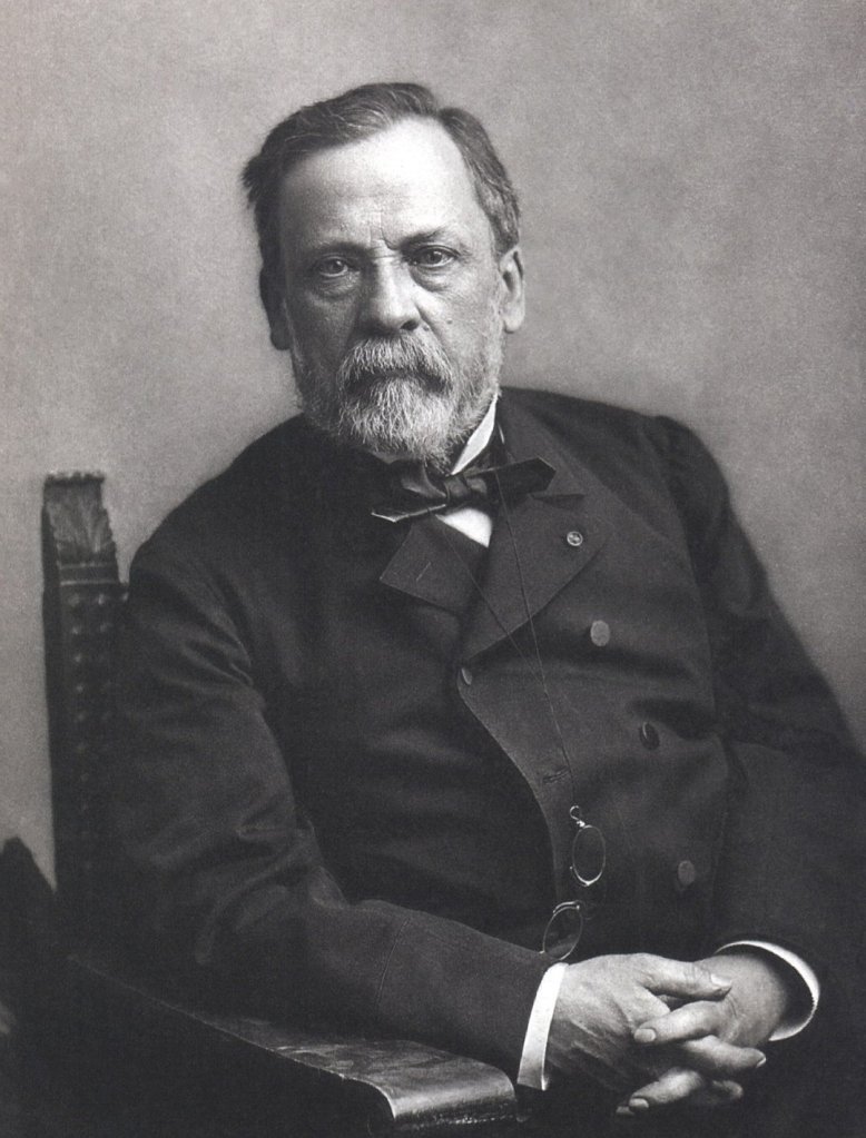
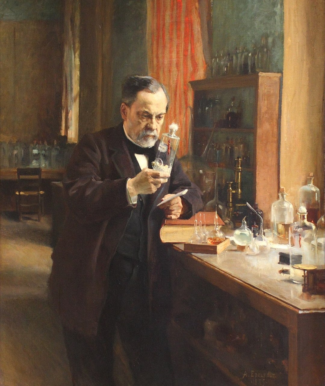
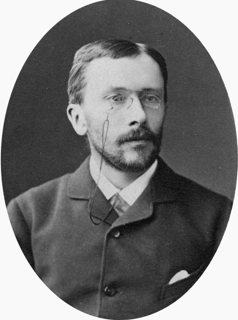
Louis Pasteur

Photograph by Nadar

Louis Pasteur (/ˈluːi pæˈstɜːr/French: [lwi pastœʁ]; December 27, 1822 – September 28, 1895) was a French biologistmicrobiologist, and chemistrenowned for his discoveries of the principles of vaccinationmicrobial fermentation and pasteurization. He is remembered for his remarkable breakthroughs in the causes and prevention of diseases, and his discoveries have saved many lives ever since. He reduced mortality from puerperal feverand created the first vaccines for rabies and anthrax

BornDecember 27, 1822
DoleJura, FranceDiedSeptember 28, 1895(aged 72)
Marnes-la-Coquette, FranceNationalityFrenchAlma mater

Awards

Scientific careerFields

Institutions

Notable studentsCharles Friedel[3]Signature

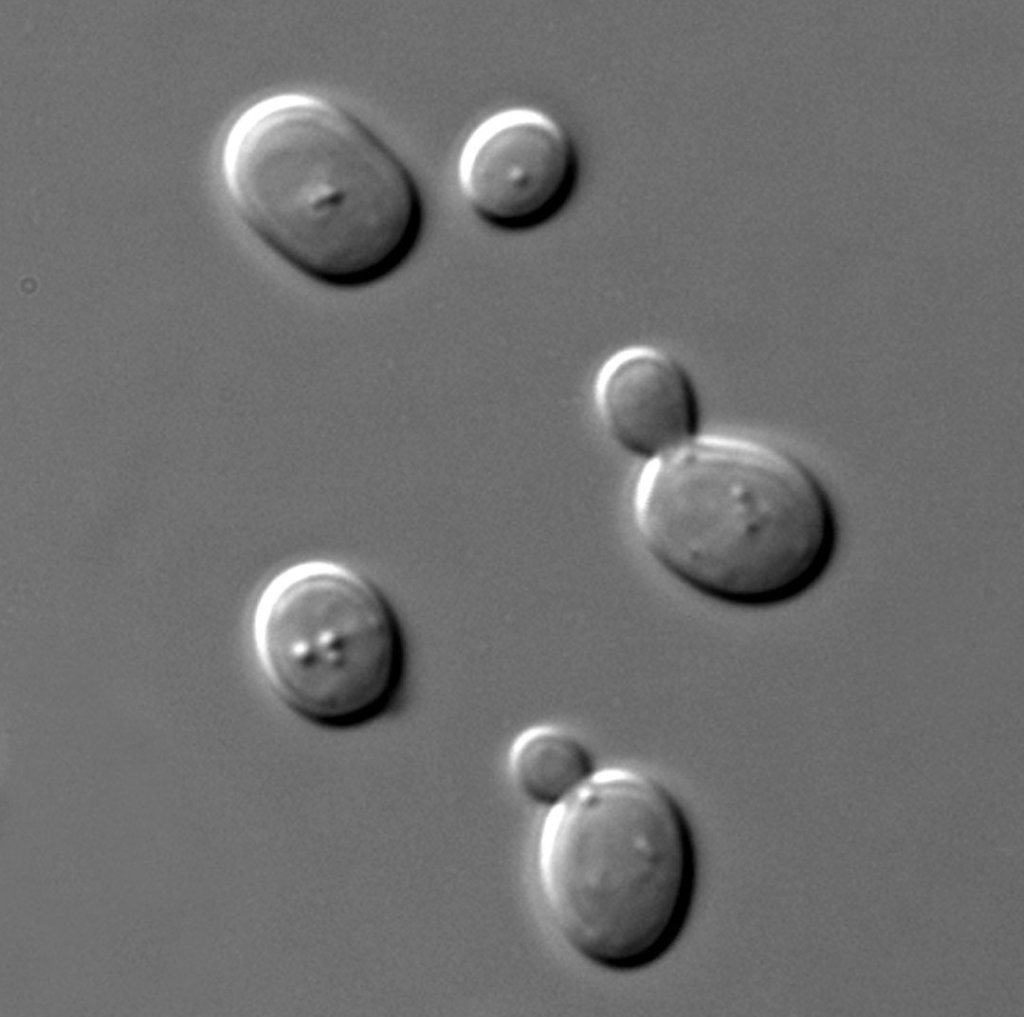
His medical discoveries provided direct support for the germ theory of disease and its application in clinical medicine. He is best known to the general public for his invention of the technique of treating milk and wine to stop bacterial contamination, a process now called pasteurization. He is regarded as one of the three main founders of bacteriology, together with Ferdinand Cohn and Robert Koch, and is popularly known as the “father of microbiology”.[4][5][6]

Pasteur was responsible for disproving the doctrine of spontaneous generation. He performed experiments that showed that, without contamination, microorganisms could not develop. Under the auspices of the French Academy of Sciences, he demonstrated that in sterilized and sealed flasks, nothing ever developed; and, conversely, in sterilized but open flasks, microorganisms could grow.[7] Although Pasteur was not the first to propose the germ theory, his experiments indicated its correctness and convinced most of Europe that it was true.

Today, he is often regarded as one of the fathers of germ theory.[8] Pasteur made significant discoveriesin chemistry, most notably on the molecular basis for the asymmetry of certain crystals and racemization. Early in his career, his investigation of tartaric acidresulted in the first resolution of what is now called optical isomers. His work led the way to the current understanding of a fundamental principle in the structure of organic compounds.

He was the director of the Pasteur Institute, established in 1887, until his death, and his body was interred in a vault beneath the institute. Although Pasteur made groundbreaking experiments, his reputation became associated with various controversies. Historical reassessment of his notebook revealed that he practiced deception to overcome his rivals.[9][10]

Education and early life

The house in which Pasteur was born, Dole

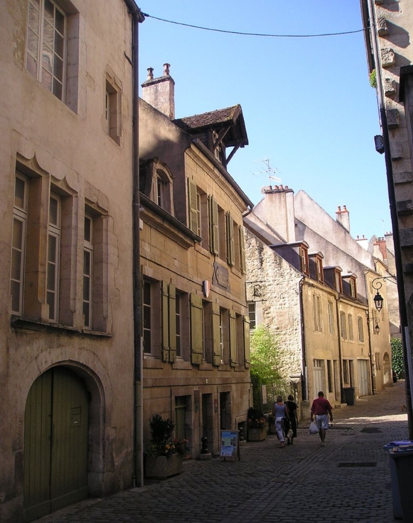
Louis Pasteur was born on December 27, 1822, in Dole, JuraFrance, to a Catholic family of a poor tanner.[4] He was the third child of Jean-Joseph Pasteur and Jeanne-Etiennette Roqui. The family moved to Marnoz in 1826 and then to Arbois in 1827.[11][12] Pasteur entered primary school in 1831.[13]

He was an average student in his early years, and not particularly academic, as his interests were fishingand sketching.[4] He drew many pastels and portraits of his parents, friends and neighbors.[14] Pasteur attended secondary school at the Collège d’Arbois.[15]In October 1838, he left for Paris to join the Pension Barbet, but became homesick and returned in November.[16]

In 1839, he entered the Collège Royal at Besançon to study philosophy and earned his Bachelor of Letters degree in 1840.[17] He was appointed a tutor at the Besançon college while continuing a degree science course with special mathematics.[18] He failed his first examination in 1841. He managed to pass the baccalauréat scientifique (general science) degree in 1842 from Dijon but with a mediocre grade in chemistry.[19]

Later in 1842, Pasteur took the entrance test for the École Normale Supérieure.[20] He passed the first set of tests, but because his ranking was low, Pasteur decided not to continue and try again next year.[21] He went back to the Pension Barbet to prepare for the test. He also attended classes at the Lycée Saint-Louis and lectures of Jean-Baptiste Dumas at the Sorbonne.[22] In 1843, he passed the test with a high ranking and entered the École Normale Supérieure.[23]In 1845 he received the licencié ès sciences (Master of Science) degree.[24] In 1846, he was appointed professor of physics at the Collège de Tournon (now called Lycée Gabriel-Faure [fr]) in Ardèche, but the chemist Antoine Jérôme Balard wanted him back at the École Normale Supérieure as a graduate laboratory assistant (agrégé préparateur).[25] He joined Balard and simultaneously started his research in crystallography and in 1847, he submitted his two theses, one in chemistry and the other in physics.[24][26]

After serving briefly as professor of physics at the Dijon Lycée in 1848, he became professor of chemistry at the University of Strasbourg,[27] where he met and courted Marie Laurent, daughter of the university’s rector in 1849. They were married on May 29, 1849,[28] and together had five children, only two of whom survived to adulthood;[29] the other three died of typhoid.

Career

Pasteur in 1857

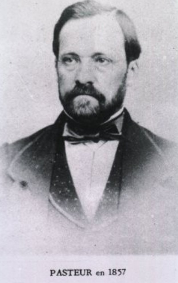
Pasteur was appointed professor of chemistry at the University of Strasbourg in 1848, and became the chair of chemistry in 1852.[30] In 1854, he was named dean of the new faculty of sciences at University of Lille, where he began his studies on fermentation.[31]It was on this occasion that Pasteur uttered his oft-quoted remark: “dans les champs de l’observation, le hasard ne favorise que les esprits préparés” (“In the field of observation, chance favors only the prepared mind”).[32]

In 1857, he moved to Paris as the director of scientific studies at the École Normale Supérieurewhere he took control from 1858 to 1867 and introduced a series of reforms to improve the standard of scientific work. The examinations became more rigid, which led to better results, greater competition, and increased prestige. Many of his decrees, however, were rigid and authoritarian, leading to two serious student revolts. During “the bean revolt” he decreed that a mutton stew, which students had refused to eat, would be served and eaten every Monday. On another occasion he threatened to expel any student caught smoking, and 73 of the 80 students in the school resigned.[33]

In 1863, he was appointed professor of geology, physics, and chemistry at the École nationale supérieure des Beaux-Arts, a position he held until his resignation in 1867. In 1867, he became the chair of organic chemistry at the Sorbonne,[34] but he later gave up the position because of poor health.[35] In 1867, the École Normale’s laboratory of physiological chemistry was created at Pasteur’s request,[34] and he was the laboratory’s director from 1867 to 1888.[36] In Paris, he established the Pasteur Institute in 1887, in which he was its director for the rest of his life.[5][6]

Research

Molecular asymmetry

In Pasteur’s early work as a chemist, beginning at the École Normale Supérieure, and continuing at Strasbourg and Lille, he examined the chemical, optical and crystallographic properties of a group of compounds known as tartrates.[37]

He resolved a problem concerning the nature of tartaric acid in 1848.[38][39][40][41] A solution of this compound derived from living things rotated the plane of polarization of light passing through it.[37]The problem was that tartaric acid derived by chemical synthesis had no such effect, even though its chemical reactions were identical and its elemental composition was the same.[42]

Pasteur noticed that crystals of tartrates had small faces. Then he observed that, in racemic mixtures of tartrates, half of the crystals were right-handed and half were left-handed. In solution, the right-handed compound was dextrorotatory, and the left-handed one was levorotatory.[37] Pasteur determined that optical activity related to the shape of the crystals, and that an asymmetric internal arrangement of the molecules of the compound was responsible for twisting the light.[31] The (2R,3R)- and (2S,3S)- tartrates were isometric, non-superposable mirror images of each other. This was the first time anyone had demonstrated molecular chirality, and also the first explanation of isomerism.[37]

Some historians consider Pasteur’s work in this area to be his “most profound and most original contributions to science”, and his “greatest scientific discovery.”[37]

Fermentation and germ theory of diseases

Pasteur was motivated to investigate fermentation while working at Lille. In 1856 a local wine manufacturer, M. Bigot, whose son was one of Pasteur’s students, sought for his advice on the problems of making beetroot alcohol and souring.[43][44]

According to his son-in-law, René Vallery-Radot, in August 1857 Pasteur sent a paper about lactic acid fermentation to the Société des Sciences de Lille, but the paper was read three months later.[45] A memoire was subsequently published on November 30, 1857.[46] In the memoir, he developed his ideas stating that: “I intend to establish that, just as there is an alcoholic ferment, the yeast of beer, which is found everywhere that sugar is decomposed into alcohol and carbonic acid, so also there is a particular ferment, a lactic yeast, always present when sugar becomes lactic acid.”[47]

Pasteur also wrote about alcoholic fermentation.[48] It was published in full form in 1858.[49][50] Jöns Jacob Berzelius and Justus von Liebig had proposed the theory that fermentation was caused by decomposition. Pasteur demonstrated that this theory was incorrect, and that yeast was responsible for fermentation to produce alcohol from sugar.[51]He also demonstrated that, when a different microorganism contaminated the wine, lactic acid was produced, making the wine sour.[44] In 1861, Pasteur observed that less sugar fermented per part of yeast when the yeast was exposed to air.[51] The lower rate of fermentation aerobically became known as the Pasteur effect.[52]

Pasteur experimenting in his laboratory.
Institut Pasteur de Lille

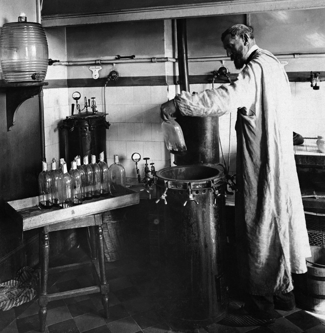
Pasteur’s research also showed that the growth of micro-organisms was responsible for spoiling beverages, such as beer, wine and milk. With this established, he invented a process in which liquids such as milk were heated to a temperature between 60 and 100 °C.[53] This killed most bacteria and moulds already present within them. Pasteur and Claude Bernard completed tests on blood and urine on April 20, 1862.[54] Pasteur patented the process, to fight the “diseases” of wine, in 1865.[53] The method became known as pasteurization, and was soon applied to beer and milk.[55]

Beverage contamination led Pasteur to the idea that micro-organisms infecting animals and humans cause disease. He proposed preventing the entry of micro-organisms into the human body, leading Joseph Lister to develop antiseptic methods in surgery.[56]

In 1866, Pasteur published Etudes sur le Vin, about the diseases of wine, and he published Etudes sur la Bière in 1876, concerning the diseases of beer.[51]

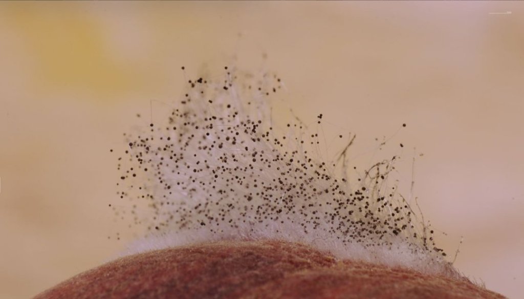
In the early 19th century, Agostino Bassi had shown that muscardine was caused by a fungus that infected silkworms.[57] Since 1853, two diseases called pébrine and flacherie had been infecting great numbers of silkworms in southern France, and by 1865 they were causing huge losses to farmers. In 1865, Pasteur went to Alès and worked for five years until 1870.[58][59]

Silkworms with pébrine were covered in corpuscles. In the first three years, Pasteur thought that the corpuscles were a symptom of the disease. In 1870, he concluded that the corpuscles were the cause of pébrine (it is now known that the cause is a microsporidian).[57] Pasteur also showed that the disease was hereditary.[60] Pasteur developed a system to prevent pébrine: after the female moths laid their eggs, the moths were turned into a pulp. The pulp was examined with a microscope, and if corpuscles were observed, the eggs were destroyed.[61][60] Pasteur concluded that bacteria caused flacherie. The primary cause is currently thought to be viruses.[57] The spread of flacherie could be accidental or hereditary. Hygiene could be used to prevent accidental flacherie. Moths whose digestive cavities did not contain the microorganisms causing flacherie were used to lay eggs, preventing hereditary flacherie.[62]

Spontaneous generation

Louis Pasteur’s pasteurization experiment illustrates the fact that the spoilage of liquid was caused by particles in the air rather than the air itself. These experiments were important pieces of evidence supporting the germ theory of disease.

Following his fermentation experiments, Pasteur demonstrated that the skin of grapes was the natural source of yeasts, and that sterilized grapes and grape juice never fermented. He drew grape juice from under the skin with sterilized needles, and also covered grapes with sterilized cloth. Both experiments could not produce wine in sterilized containers.[44]

His findings and ideas were against the prevailing notion of spontaneous generation. He received a particularly stern criticism from Félix Archimède Pouchet, who was director of the Rouen Museum of Natural History. To settle the debate between the eminent scientists, the French Academy of Sciences offered the Alhumbert Prize carrying 2,500 francs to whoever could experimentally demonstrate for or against the doctrine.[63][64][65]

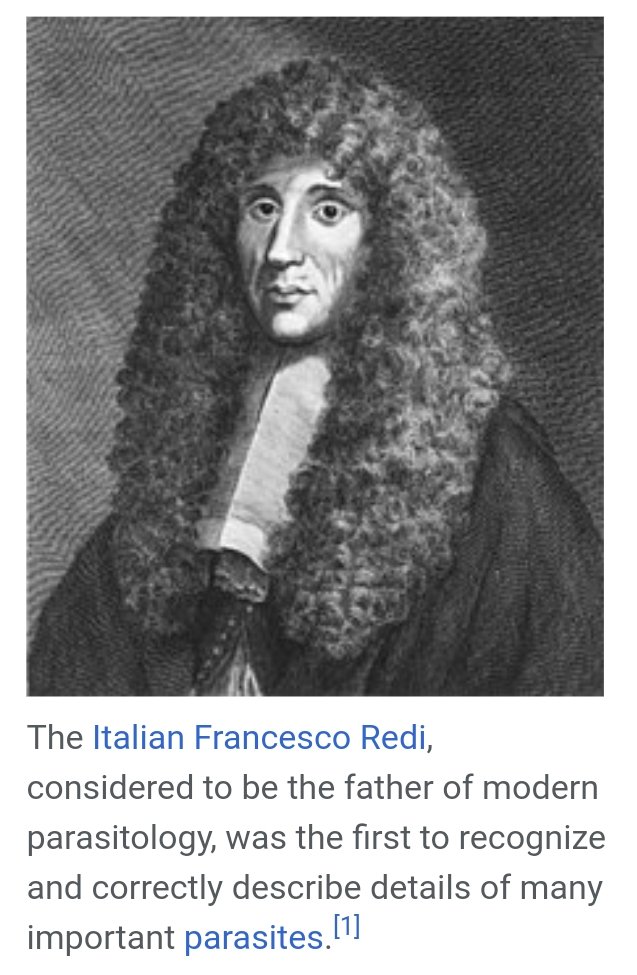
Pouchet stated that air everywhere could cause spontaneous generation of living organisms in liquids.[66] In the late 1850s, he performed experiments and claimed that they were evidence of spontaneous generation.[67][63] Francesco Redi and Lazzaro Spallanzani had provided some evidence against spontaneous generation in the 17th and 18th centuries, respectively. Spallanzani’s experiments in 1765 suggested that air contaminated broths with bacteria. In the 1860s, Pasteur repeated Spallanzani’s experiments, but Pouchet reported a different result using a different broth.[58]

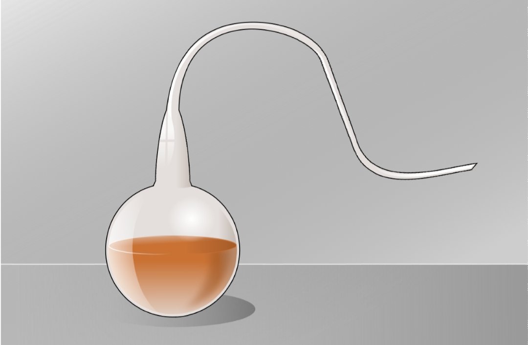
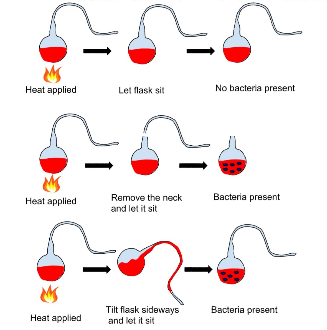
Pasteur performed several experiments to disprove spontaneous generation. He placed boiled liquid in a flask and let hot air enter the flask. Then he closed the flask, and no organisms grew in it.[67] In another experiment, when he opened flasks containing boiled liquid, dust entered the flasks, causing organisms to grow in some of them. The number of flasks in which organisms grew was lower at higher altitudes, showing that air at high altitudes contained less dust and fewer organisms.[44][68] Pasteur also used swan neck flasks containing a fermentable liquid. Air was allowed to enter the flask via a long curving tube that made dust particles stick to it. Nothing grew in the broths unless the flasks were tilted, making the liquid touch the contaminated walls of the neck. This showed that the living organisms that grew in such broths came from outside, on dust, rather than spontaneously generating within the liquid or from the action of pure air.[44][69]

These were some of the most important experiments disproving the theory of spontaneous generation, for which Pasteur won the Alhumbert Prize in 1862.[67]He concluded that:[44][59]

Never will the doctrine of spontaneous generation recover from the mortal blow of this simple experiment. There is no known circumstance in which it can be confirmed that microscopic beings came into the world without germs, without parents similar to themselves.

Immunology and vaccination


Chicken cholera

Pasteur’s later work on diseases included work on chicken cholera. He received cultures from Jean Joseph Henri Toussaint, and cultivated them in chicken broth.[70] During this work, a culture of the responsible bacteria had spoiled and failed to induce the disease in some chickens he was infecting with the disease. Upon reusing these healthy chickens, Pasteur discovered he could not infect them, even with fresh bacteria; the weakened bacteria had caused the chickens to become immune to the disease, though they had caused only mild symptoms.[4][8]

In 1879, his assistant, Charles Chamberland (of French origin), had been instructed to inoculate the chickens after Pasteur went on holiday. Chamberland failed to do this and went on holiday himself. On his return, the month-old cultures made the chickens unwell, but instead of the infections being fatal, as they usually were, the chickens recovered completely. Chamberland assumed an error had been made, and wanted to discard the apparently faulty culture, but Pasteur stopped him.[71][72] He inoculated the chickens with virulent bacteria that killed other chickens, and they survived. Pasteur concluded that the animals were now immune to the disease.[73]

In December 1879, Pasteur used a weakened culture of the bacteria to inoculate chickens. The chickens survived, and when he inoculated them with a virulent strain, they were immune to it. In 1880, Pasteur presented his results to the French Academy of Sciences, saying that the bacteria were weakened by contact with oxygen.[70]

Anthrax

In the 1870s, he applied this immunization method to anthrax, which affected cattle, and aroused interest in combating other diseases. Pasteur cultivated bacteria from the blood of animals infected with anthrax. When he inoculated animals with the bacteria, anthrax occurred, proving that the bacteria was the cause of the disease.[74] Many cattle were dying of anthrax in “cursed fields”.[59] Pasteur was told that sheep that died from anthrax were buried in the field. Pasteur thought that earthworms might have brought the bacteria to the surface. He found anthrax bacteria in earthworms’ excrement, showing that he was correct.[59] He told the farmers not to bury dead animals in the fields.[75]

Louis Pasteur in his laboratory, painting by A. Edelfeldt in 1885

In 1880, Pasteur’s rival Jean-Joseph-Henri Toussaint, a veterinary surgeon, used carbolic acid to kill anthrax bacilli and tested the vaccine on sheep. Pasteur thought that this type of killed vaccine should not work because he believed that attenuated bacteria used up nutrients that the bacteria needed to grow. He thought oxidizing bacteria made them less virulent.[76] In early 1881, Pasteur discovered that growing anthrax bacilli at about 42 °C made them unable to produce spores,[77] and he described this method in a speech to the French Academy of Sciences on February 28.[78] Later in 1881, veterinarian Hippolyte Rossignol proposed that the Société d’agriculture de Melun organize an experiment to test Pasteur’s vaccine. Pasteur agreed, and the experiment, conducted at Pouilly-le-Fort on sheep, goats and cows, was successful. Pasteur did not directly disclose how he prepared the vaccines used at Pouilly-le-Fort.[79][77] His laboratory notebooks, now in the Bibliothèque Nationale in Paris, show that he actually used heat and potassium dichromate, similar to Toussaint’s method.[80][42][81]

The notion of a weak form of a disease causing immunity to the virulent version was not new; this had been known for a long time for smallpox. Inoculation with smallpox (variolation) was known to result in a much less severe disease, and greatly reduced mortality, in comparison with the naturally acquired disease.[82] Edward Jenner had also studied vaccination using cowpox (vaccinia) to give cross-immunity to smallpox in the late 1790s, and by the early 1800s vaccination had spread to most of Europe.[83]

The difference between smallpox vaccination and anthrax or chicken cholera vaccination was that the latter two disease organisms had been artificially weakened, so a naturally weak form of the disease organism did not need to be found.[80] This discovery revolutionized work in infectious diseases, and Pasteur gave these artificially weakened diseases the generic name of “vaccines“, in honour of Jenner’s discovery.[84]Main article: Koch–Pasteur rivalry

In 1876, Robert Koch had shown that Bacillus anthracis caused anthrax.[85] In his papers published between 1878 and 1880, Pasteur only mentioned Koch’s work in a footnote. Koch met Pasteur at the Seventh International Medical Congress in 1881. A few months later, Koch wrote that Pasteur had used impure cultures and made errors. In 1882, Pasteur replied to Koch in a speech, to which Koch responded aggressively.[8] Koch stated that Pasteur tested his vaccine on unsuitable animals and that Pasteur’s research was not properly scientific.[44] In 1882, Koch wrote “On the Anthrax Inoculation”, in which he refuted several of Pasteur’s conclusions about anthrax and criticized Pasteur for keeping his methods secret, jumping to conclusions, and being imprecise. In 1883, Pasteur wrote that he used cultures prepared in a similar way to his successful fermentation experiments and that Koch misinterpreted statistics and ignored Pasteur’s work on silkworms.[85]

Swine erysipelas

In 1882, Pasteur sent his assistant Louis Thuillier to southern France because of an epizootic of swine erysipelas.[86] Thuillier identified the bacillus that caused the disease in March 1883.[58] Pasteur and Thuillier increased the bacillus’s virulence after passing it through pigeons. Then they passed the bacillus through rabbits, weakening it and obtaining a vaccine. Pasteur and Thuillier incorrectly described the bacterium as a figure-eight shape. Roux described the bacterium as stick-shaped in 1884.[87]

Rabies

Pasteur produced the first vaccine for rabies by growing the virus in rabbits, and then weakening it by drying the affected nerve tissue.[59][88] The rabies vaccine was initially created by Emile Roux, a French doctor and a colleague of Pasteur, who had produced a killed vaccine using this method.[44] The vaccine had been tested in 50 dogs before its first human trial.[89][90] This vaccine was used on 9-year-old Joseph Meister, on July 6, 1885, after the boy was badly mauled by a rabid dog.[42][88] This was done at some personal risk for Pasteur, since he was not a licensed physician and could have faced prosecution for treating the boy.[6] After consulting with physicians, he decided to go ahead with the treatment.[91] Over 11 days, Meister received 13 inoculations, each inoculation using viruses that had been weakened for a shorter period of time.[92] Three months later he examined Meister and found that he was in good health.[91][93] Pasteur was hailed as a hero and the legal matter was not pursued.[6]Analysis of his laboratory notebooks shows that Pasteur had treated two people before his vaccination of Meister. One survived but may not actually have had rabies, and the other died of rabies.[92][94] Pasteur began treatment of Jean-Baptiste Jupille on October 20, 1885, and the treatment was successful.[92] Later in 1885, people, including four children from the United States, went to Pasteur’s laboratory to be inoculated.[91] In 1886, he treated 350 people, of which only one developed rabies.[92] The treatment’s success laid the foundations for the manufacture of many other vaccines. The first of the Pasteur Institutes was also built on the basis of this achievement.[42]

In The Story of San MicheleAxel Munthe writes of some risks Pasteur undertook in the rabies vaccine research:[95]

Pasteur himself was absolutely fearless. Anxious to secure a sample of saliva straight from the jaws of a rabid dog, I once saw him with the glass tube held between his lips draw a few drops of the deadly saliva from the mouth of a rabid bull-dog, held on the table by two assistants, their hands protected by leather gloves.

Because of his study in germs, Pasteur encouraged doctors to sanitize their hands and equipment before surgery. Prior to this, few doctors or their assistants practiced these procedures.

Controversies

A French national hero at age 55, in 1878 Pasteur discreetly told his family never to reveal his laboratory notebooks to anyone. His family obeyed, and all his documents were held and inherited in secrecy. Finally, in 1964 Pasteur’s grandson and last surviving male descendant, Pasteur Vallery-Radot, donated the papers to the French national library (Bibliothèque nationale de France). Yet the papers were restricted for historical studies until the death of Vallery-Radot in 1971. The documents were given a catalogue number only in 1985.[96]

In 1995, the centennial of the death of Louis Pasteur, a historian of science Gerald L. Geison published an analysis of Pasteur’s private notebooks in his The Private Science of Louis Pasteur, and declared that Pasteur had given several misleading accounts and played deceptions in his most important discoveries.[9][97] Max Perutz published a defense of Pasteur in The New York Review of Books.[98] Based on further examinations of Pasteur’s documents, French immunologist Patrice Debré concluded in his book Louis Pasteur (1998) that, in spite of his genius, Pasteur had some faults. A book review states that Debré “sometimes finds him unfair, combative, arrogant, unattractive in attitude, inflexible and even dogmatic”.[99][100]

Fermentation

Scientists before Pasteur had studied fermentation. In the 1830s, Charles Cagniard-LatourFriedrich Traugott Kützing and Theodor Schwann used microscopes to study yeasts and concluded that yeasts were living organisms. In 1839, Justus von LiebigFriedrich Wöhler and Jöns Jacob Berzeliusstated that yeast was not an organism and was produced when air acted on plant juice.[51]

In 1855, Antoine Béchamp, Professor of Chemistry at the University of Montpellier, conducted experiments with sucrose solutions and concluded that water was the factor for fermentation.[101] He changed his conclusion in 1858, stating that fermentation was directly related to the growth of moulds, which required air for growth. He regarded himself as the first to show the role of microorganisms in fermentation.[102][47]

Pasteur started his experiments in 1857 and published his findings in 1858 (April issue of Comptes Rendus Chimie, Béchamp’s paper appeared in January issue). Béchamp noted that Pasteur did not bring any novel idea or experiments. On the other hand, Béchamp was probably aware of Pasteur’s 1857 preliminary works. With both scientists claiming priority on the discovery, a dispute, extending to several areas, lasted throughout their lives.[103][104]

However, Béchamp was on the losing side, as the BMJ obituary remarked: His name was “associated with bygone controversies as to priority which it would be unprofitable to recall”.[105] Béchamp proposed the incorrect theory of microzymes. According to K. L. Manchester, anti-vivisectionistsand proponents of alternative medicine promoted Béchamp and microzymes, unjustifiably claiming that Pasteur plagiarized Béchamp.[47]

Pasteur thought that succinic acid inverted sucrose. In 1860, Marcellin Berthelot isolated invertase and showed that succinic acid did not invert sucrose.[51]Pasteur believed that fermentation was only due to living cells. Hans Buchner discovered that zymasecatalyzed fermentation, showing that fermentation was catalyzed by enzymes within cells.[106] Eduard Buchner also discovered that fermentation could take place outside living cells.[107]

Anthrax vaccine

Pasteur publicly claimed his success in developing the anthrax vaccine in 1881.[93] However, his admirer-turned-rival Toussaint was the one who developed the first vaccine. Toussaint isolated the bacteria that caused chicken cholera (later named Pasteurella in honour of Pasteur) in 1879 and gave samples to Pasteur who used them for his own works.[108] On July 12, 1880, Toussaint presented his successful result to the French Academy of Sciences, using an attenuated vaccine against anthrax in dogs and sheep.[109] Pasteur on grounds of jealousy contested the discovery by publicly displaying his vaccination method at Pouilly-le-Fort on May 5, 1881.[110] Pasteur gave a misleading account of the preparation of the anthrax vaccine used in the experiment at Pouilly-le-Fort. He used potassium dichromate to prepare the vaccine.[9] The promotional experiment was a success and helped Pasteur sell his products, getting the benefits and glory.[110][111][112]

Experimental ethics

Pasteur experiments are often cited as against medical ethics, especially on his vaccination of Meister. He did not have any experience in medical practice, and more importantly, lacked a medical license. This is often cited as a serious threat to his professional and personal reputation.[113][114] His closest partner Émile Roux, who had medical qualifications, refused to participate in the clinical trial, likely because he considered it unjust.[92]However, Pasteur executed vaccination of the boy under the close watch of practising physicians Jacques-Joseph Grancher, head of the Paris Children’s Hospital’s paediatric clinic, and Alfred Vulpian, a member of the Commission on Rabies. He was not allowed to hold the syringe, although the inoculations were entirely under his supervision.[91] It was Grancher who was responsible for the injections, and he defended Pasteur before the French National Academy of Medicine in the issue.[115]

Pasteur has also been criticized for keeping secrecy of his procedure and not giving proper pre-clinical trials on animals.[44] Pasteur stated that he kept his procedure secret in order to control its quality. He later disclosed his procedures to a small group of scientists. Pasteur wrote that he had successfully vaccinated 50 rabid dogs before using it on Meister.[116][117][118] According to Geison, Pasteur’s laboratory notebooks show that he had vaccinated only 11 dogs.[44]

Meister never showed any symptoms of rabies,[92]but the vaccination has not been proved to be the reason. One source estimates the probability of Meister contracting rabies at 10%.[80]

Awards and honours

Pasteur was awarded 1,500 francs in 1853 by the Pharmaceutical Society for the synthesis of racemic acid.[119] In 1856 the Royal Society of London presented him the Rumford Medal for his discovery of the nature of racemic acid and its relations to polarized light,[120] and the Copley Medal in 1874 for his work on fermentation.[121] He was elected a Foreign Member of the Royal Society (ForMemRS) in 1869.[1]

The French Academy of Sciences awarded Pasteur the 1859 Montyon Prize for experimental physiology in 1860,[34] and the Jecker Prize in 1861 and the Alhumbert Prize in 1862 for his experimental refutation of spontaneous generation.[67][122] Though he lost elections in 1857 and 1861 for membership to the French Academy of Sciences, he won the 1862 election for membership to the mineralogy section.[123] He was elected to permanent secretary of the physical science section of the academy in 1887 and held the position until 1889.[124]

In 1873 Pasteur was elected to the Académie Nationale de Médecine[125] and was made the commander in the Brazilian Order of the Rose.[126] In 1881 he was elected to a seat at the Académie française left vacant by Émile Littré.[127] Pasteur received the Albert Medal from the Royal Society of Arts in 1882.[128] In 1883 he became foreign member of the Royal Netherlands Academy of Arts and Sciences.[129] On June 8, 1886, the Ottoman Sultan Abdul Hamid II awarded Pasteur with the Order of the Medjidie (I Class) and 10000 Ottoman liras.[130] He was awarded the Cameron Prize for Therapeutics of the University of Edinburgh in 1889.[131] Pasteur won the Leeuwenhoek Medal from the Royal Netherlands Academy of Arts and Sciences for his contributions to microbiology in 1895.[132]

Pasteur was made a Chevalier of the Legion of Honour in 1853, promoted to Officer in 1863, to Commander in 1868, to Grand Officer in 1878 and made a Grand Cross of the Legion of Honor in 1881.[133][128]

Legacy

Vulitsya Pastera or Pasteur Street in OdessaUkraine

Main article: List of things named after Louis Pasteur

In many localities worldwide, streets are named in his honor. For example, in the US: Palo Alto and Irvine, California, Boston and Polk, Florida, adjacent to the University of Texas Health Science Center at San Antonio; Jonquière, Québec; San Salvador de Jujuy and Buenos Aires (Argentina), Great Yarmouth in Norfolk, in the United Kingdom, Jericho and Wulguru in Queensland, (Australia); Phnom Penh in CambodiaHo Chi Minh City; Batna in AlgeriaBandung in IndonesiaTehran in Iran, near the central campus of the Warsaw University in WarsawPoland; adjacent to the Odessa State Medical University in OdessaUkraineMilan in Italy and BucharestCluj-Napocaand Timișoara in Romania. The Avenue Pasteur in Saigon, Vietnam, is one of the few streets in that city to retain its French name. Avenue Louis Pasteur in the Longwood Medical and Academic Area in BostonMassachusetts was named in his honor in the French manner with “Avenue” preceding the name of the dedicatee.[134]

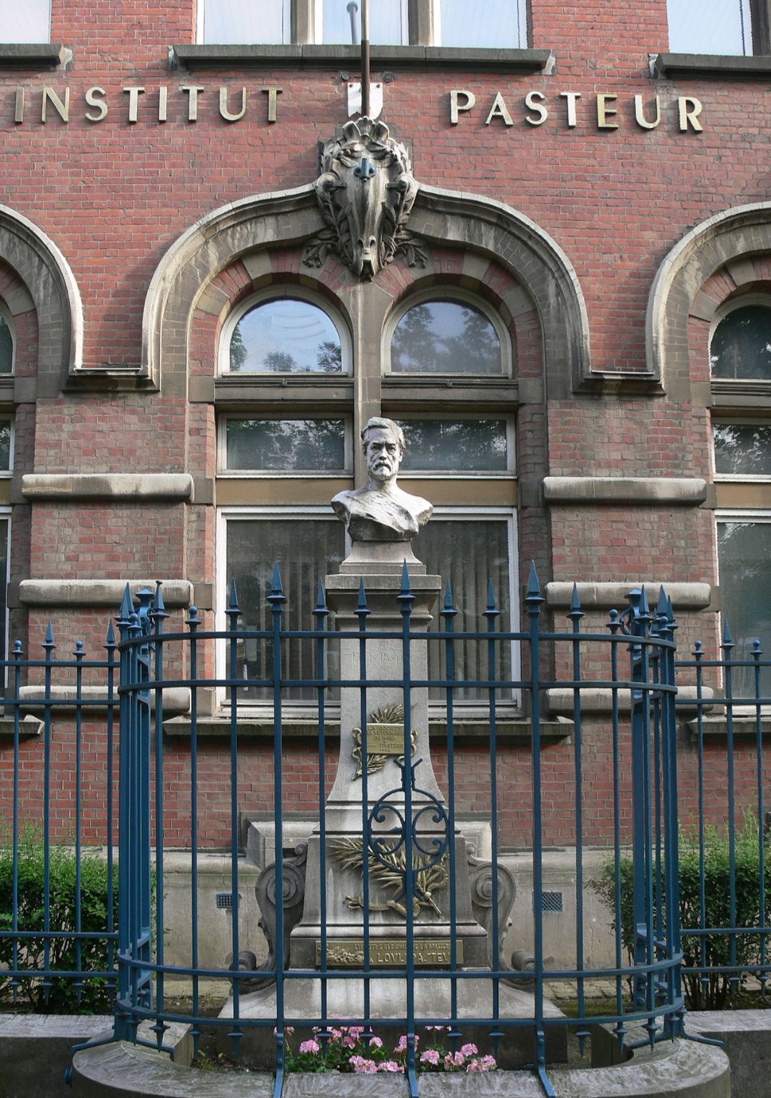
Both the Institut Pasteur and Université Louis Pasteurwere named after Pasteur. The schools Lycée Pasteur in Neuilly-sur-Seine, France, and Lycée Louis Pasteur in Calgary, Alberta, Canada, are named after him. In South Africa, the Louis Pasteur Private Hospital in Pretoria, and Life Louis Pasteur Private Hospital, Bloemfontein, are named after him. Louis Pasteur University Hospital in KošiceSlovakia is also named after Pasteur.

Louis Pasteur University Hospital, KošiceSlovakia

A statue of Pasteur is erected at San Rafael High School in San Rafael, California. A bronze bust of him resides on the French Campus of Kaiser Permanente‘s San Francisco Medical Center in San Francisco. The sculpture was designed by Harriet G. Moore and cast in 1984 by Artworks Foundry.[135]

The UNESCO/Institut Pasteur Medal was created on the centenary of Pasteur’s death, and is given every two years in his name, “in recognition of outstanding research contributing to a beneficial impact on human health”.[136]

Pasteur Institute

Main article: Pasteur Institute

After developing the rabies vaccine, Pasteur proposed an institute for the vaccine.[137] In 1887, fundraising for the Pasteur Institute began, with donations from many countries. The official statute was registered in 1887, stating that the institute’s purposes were “the treatment of rabies according to the method developed by M. Pasteur” and “the study of virulent and contagious diseases”.[91] The institute was inaugurated on November 14, 1888.[91] He brought together scientists with various specialties. The first five departments were directed by two graduates of the École Normale SupérieureÉmile Duclaux (general microbiology research) and Charles Chamberland (microbe research applied to hygiene), as well as a biologist, Élie Metchnikoff(morphological microbe research) and two physiciansJacques-Joseph Grancher (rabies) and Émile Roux (technical microbe research). One year after the inauguration of the institute, Roux set up the first course of microbiology ever taught in the world, then entitled Cours de Microbie Technique (Course of microbe research techniques). Since 1891 the Pasteur Institute had been extended to different countries, and currently there are 32 institutes in 29 countries in various parts of the world.[138]

Personal life

Faith and spirituality

His grandson, Louis Pasteur Vallery-Radot, wrote that Pasteur had kept from his Catholic background only a spiritualism without religious practice.[139] However, Catholic observers often said that Pasteur remained an ardent Christian throughout his whole life, and his son-in-law wrote, in a biography of him:

Absolute faith in God and in Eternity, and a conviction that the power for good given to us in this world will be continued beyond it, were feelings which pervaded his whole life; the virtues of the gospel had ever been present to him. Full of respect for the form of religion which had been that of his forefathers, he came simply to it and naturally for spiritual help in these last weeks of his life.[140]

The Literary Digest of 18 October 1902 gives this statement from Pasteur that he prayed while he worked:

Posterity will one day laugh at the foolishness of modern materialistic philosophers. The more I study nature, the more I stand amazed at the work of the Creator. I pray while I am engaged at my work in the laboratory.

Maurice Vallery-Radot, grandson of the brother of the son-in-law of Pasteur and outspoken Catholic, also holds that Pasteur fundamentally remained Catholic.[141] According to both Pasteur Vallery-Radot and Maurice Vallery-Radot, the following well-known quotation attributed to Pasteur is apocryphal:[142]“The more I know, the more nearly is my faith that of the Breton peasant. Could I but know all I would have the faith of a Breton peasant’s wife”.[4] According to Maurice Vallery-Radot,[143] the false quotation appeared for the first time shortly after the death of Pasteur.[144] However, despite his belief in God, it has been said that his views were that of a freethinker rather than a Catholic, a spiritual more than a religious man.[145][146] He was also against mixing science with religion.[147][148]

Death

In 1868, Pasteur suffered a severe brain stroke that paralysed the left side of his body, but he recovered.[149] A stroke or uremia in 1894 severely impaired his health.[150][151][152] Failing to fully recover, he died on September 28, 1895, near Paris.[42] He was given a state funeral and was buried in the Cathedral of Notre Dame, but his remains were reinterred in the Pasteur Institute in Paris,[153] in a vault covered in depictions of his accomplishments in Byzantine mosaics.[154]

Publications

Pasteur’s principal published works are:[4]French TitleYearEnglish TitleEtudes sur le Vin1866Studies on WineEtudes sur le Vinaigre1868Studies on VinegarEtudes sur la Maladie des Vers à Soie (2 volumes)1870Studies on Silk Worm DiseaseQuelques Réflexions sur la Science en France1871Some Reflections on Science in FranceEtudes sur la Bière1876Studies on BeerLes Microbes organisés, leur rôle dans la Fermentation, la Putréfaction et la Contagion1878Microbes organized, their role in fermentation, putrefaction and the ContagionDiscours de Réception de M.L. Pasteur à l’Académie française1882Speech by Mr L. Pasteur on reception to the Académie françaiseTraitement de la Rage1886Treatment of RabiesThe standard author abbreviationPasteur is used to indicate this person as the author when citing a botanical name.[155]

See also

  1.  “Fellows of the Royal Society”. London: Royal Society. Archived from the original on March 16, 2015.
  2. ^ “II. Abdülhamid’in Fransız kimyagere yaptığı yardım ortaya çıktı”. CNN Türk. Retrieved December 29, 2016.
  3. ^ Asimov, Asimov’s Biographical Encyclopedia of Science and Technology 2nd Revised edition
  4. a b c d e f James J. Walsh (1913). “Louis Pasteur” . In Herbermann, Charles (ed.). Catholic Encyclopedia. New York: Robert Appleton Company.
  5. a b Feinstein, S (2008). Louis Pasteur: The Father of Microbiology. Enslow Publishers, Inc. pp. 1–128. ISBN 978-1-59845-078-1.
  6. a b c d Hook, Sue Vander (2011). Louis Pasteur: Groundbreaking Chemist & Biologist. Minnesota: ABDO Publishing Company. pp. 8–112. ISBN 978-1-61758-941-6.
  7. ^ Seckbach, Joseph (editor) (2004). Origins: Genesis, Evolution and Diversity of Life. Dordrecht, The Netherlands: Kluwer Academic Publishers. p. 20. ISBN 978-1-4020-1813-8.
  8. a b c Ullmann, Agnes (August 2007). “Pasteur-Koch: Distinctive Ways of Thinking about Infectious Diseases”Microbe2 (8): 383–387. Archived from the original on May 10, 2016. Retrieved December 12, 2007.
  9. a b c Geison, Gerald L (1995). The Private Science of Louis Pasteur. Princeton, NJ: Princeton university press. ISBN 978-0-691-01552-1.
  10. ^ Anderson, C. (1993). “Pasteur Notebooks Reveal Deception”. Science259 (5098): 1117. Bibcode:1993Sci…259.1117Adoi:10.1126/science.259.5098.1117-aPMID 8438162.
  11. ^ Debré, Patrice (2000). Louis Pasteur. Translated by Forster, Elborg. Baltimore: JHU Press. pp. 6–7. ISBN 978-0-8018-6529-9.
  12. ^ Robbins, Louise (2001). Louis Pasteur and the Hidden World of Microbes. New York: Oxford University Press. p. 14. ISBN 978-0-19-512227-5.
  13. ^ Debré, Patrice (2000). Louis Pasteur. Translated by Forster, Elborg. Baltimore: JHU Press. p. 8. ISBN 978-0-8018-6529-9.
  14. ^ Debré, Patrice (2000). Louis Pasteur. Translated by Forster, Elborg. Baltimore: JHU Press. pp. 12–13. ISBN 978-0-8018-6529-9.
  15. ^ Robbins, Louise (2001). Louis Pasteur and the Hidden World of Microbes. New York: Oxford University Press. p. 15. ISBN 978-0-19-512227-5.
  16. ^ Debré, Patrice (2000). Louis Pasteur. Translated by Forster, Elborg. Baltimore: JHU Press. pp. 11–12. ISBN 978-0-8018-6529-9.
  17. ^ Keim, Albert; Lumet, Louis (1914). Louis Pasteur. Frederick A. Stokes Company. pp. 10, 12.
  18. ^ Debré, Patrice (2000). Louis Pasteur. Translated by Forster, Elborg. Baltimore: JHU Press. pp. 14, 17. ISBN 978-0-8018-6529-9.
  19. ^ Debré, Patrice (2000). Louis Pasteur. Translated by Forster, Elborg. Baltimore: JHU Press. pp. 19–20. ISBN 978-0-8018-6529-9.
  20. ^ Robbins, Louise (2001). Louis Pasteur and the Hidden World of Microbes. New York: Oxford University Press. p. 18. ISBN 978-0-19-512227-5
  1.  Debré, Patrice (2000). Louis Pasteur. Translated by Forster, Elborg. Baltimore: JHU Press. pp. 20–21. ISBN 978-0-8018-6529-9.
  2. ^ Keim, Albert; Lumet, Louis (1914). Louis Pasteur. Frederick A. Stokes Company. pp. 15–17.
  3. ^ Debré, Patrice (2000). Louis Pasteur. Translated by Forster, Elborg. Baltimore: JHU Press. pp. 23–24. ISBN 978-0-8018-6529-9.
  4. a b Debré, Patrice (2000). Louis Pasteur. Translated by Forster, Elborg. Baltimore: JHU Press. p. 502. ISBN 978-0-8018-6529-9.
  5. ^ Debré, Patrice (2000). Louis Pasteur. Translated by Forster, Elborg. Baltimore: JHU Press. pp. 29–30. ISBN 978-0-8018-6529-9.
  6. ^ Keim, Albert; Lumet, Louis (1914). Louis Pasteur. Frederick A. Stokes Company. pp. 28–29.
  7. ^ Keim, Albert; Lumet, Louis (1914). Louis Pasteur. Frederick A. Stokes Company. pp. 37–38.
  8. ^ Holmes, Samuel J. (1924). Louis Pasteur. Harcourt, Brace and company. pp. 34–36.
  9. ^ Robbins, Louise E. (2001). Louis Pasteur and the Hidden World of Microbes. Oxford University Press. p. 56. ISBN 978-0-19-028404-6.
  10. ^ Debré, Patrice (2000). Louis Pasteur. Translated by Forster, Elborg. Baltimore: JHU Press. pp. 502–503. ISBN 978-0-8018-6529-9.
  11. a b “Louis Pasteur”Science History Institute. June 2016. Retrieved March 20, 2018.
  12. ^ L. Pasteur, “Discours prononcé à Douai, le 7 décembre 1854, à l’occasion de l’installation solennelle de la Faculté des lettres de Douai et de la Faculté des sciences de Lille” (Speech delivered at Douai on December 7, 1854 on the occasion of his formal inauguration to the Faculty of Letters of Douai and the Faculty of Sciences of Lille), reprinted in: Pasteur Vallery-Radot, ed., Oeuvres de Pasteur (Paris, France: Masson and Co., 1939), vol. 7, p. 131.
  13. ^ Debré, Patrice (2000). Louis Pasteur. Translated by Elborg Forster. Baltimore: Johns Hopkins University Press. pp. 119–120. ISBN 978-0-8018-6529-9. Retrieved January 27,2015.
  14. a b c Debré, Patrice (2000). Louis Pasteur. Translated by Forster, Elborg. Baltimore: JHU Press. pp. 505–507. ISBN 978-0-8018-6529-9.
  15. ^ Vallery-Radot, René (1919). The Life of Pasteur. Translated by Devonshire, R. L. London: Constable & Company. p. 246.
  16. ^ Heilbron, J. L., ed. (2003). “Pasteur, Louis”The Oxford Companion to the History of Modern Science. Oxford University Press. p. 617. ISBN 978-0-19-974376-6.
  17. a b c d e H.D. Flack (2009) “Louis Pasteur’s discovery of molecular chirality and spontaneous resolution in 1848, together with a complete review of his crystallographic and chemical work,” Acta Crystallographica, Section A, vol. 65, pp. 371–389.
  18. ^ L. Pasteur (1848) “Mémoire sur la relation qui peut exister entre la forme cristalline et la composition chimique, et sur la cause de la polarisation rotatoire” (Memoir on the relationship that can exist between crystalline form and chemical composition, and on the cause of rotary polarization),” Comptes rendus de l’Académie des sciences (Paris), 26 : 535–538.
  19. ^ L. Pasteur (1848) “Sur les relations qui peuvent exister entre la forme cristalline, la composition chimique et le sens de la polarisation rotatoire” (On the relations that can exist between crystalline form, and chemical composition, and the sense of rotary polarization), Annales de Chimie et de Physique, 3rd series, vol. 24, no. 6, pp. 442–459.
  20. ^ George B. Kauffman and Robin D. Myers (1998)“Pasteur’s resolution of racemic acid: A sesquicentennial retrospect and a new translation,” The Chemical Educator, vol. 3, no. 6,[page needed].
  1.  Joseph Gal: Louis Pasteur, Language, and Molecular Chirality. I. Background and Dissymmetry, Chirality 23 (2011) 1−16.
  2. a b c d e Cohn, David V (December 18, 2006). “Pasteur”. University of Louisville. Retrieved December 2, 2007. Fortunately, Pasteur’s colleagues Chamberlain [sic] and Roux followed up the results of a research physician Jean-Joseph-Henri Toussaint, who had reported a year earlier that carbolic-acid/heated anthrax serum would immunize against anthrax. These results were difficult to reproduce and discarded although, as it turned out, Toussaint had been on the right track. This led Pasteur and his assistants to substitute an anthrax vaccine prepared by a method similar to that of Toussaint and different from what Pasteur had announced.
  3. ^ Vallery-Radot, René (1919). The Life of Pasteur. Translated by Devonshire, R. L. London: Constable & Company. p. 79.
  4. a b c d e f g h i j Ligon, B. Lee (2002). “Biography: Louis Pasteur: A controversial figure in a debate on scientific ethics”. Seminars in Pediatric Infectious Diseases13 (2): 134–141. doi:10.1053/spid.2002.125138PMID 12122952.
  5. ^ Vallery-Radot, René (1907). La vie de Pasteur(in French). Paris: Librairie Hachette. p. 98.
  6. ^ Pasteur, Louis (1857). “Mémoire sur la fermentation appelée lactique”Comptes Rendus Chimie (in French). 45: 913–916.
  7. a b c Manchester, K.L. (2007). “Louis Pasteur, fermentation, and a rival”South African Journal of Science103 (9–10): 377–380.
  8. ^ Pasteur, Louis (1857). “Mémoire sur la fermentation alcoolique”Comptes Rendus Chimie (in French). 45 (6): 1032–1036. PMC 2229983.
  9. ^ Pasteur, Louis (1858). “Nouveaux faits concernant l’histoire de la fermentation alcoolique”. Comptes Rendus Chimie (in French). 47: 1011–1013.
  10. ^ Pasteur, Louis (1858). “Nouveaux faits concernant l’histoire de la fermentation alcoolique”. Annales de Chimie et de Physique. 3rd Series (in French). 52: 404–418.
  11. a b c d e Barnett, James A.; Barnett, Linda (2011). Yeast Research : A Historical Overview. Washington, DC: ASM Press. ISBN 978-1-55581-516-5.
  12. ^ Zimmermann, F.K.; Entian, K.-D., eds. (1997). Yeast Sugar Metabolism. CRC Press. pp. 20–21. ISBN 978-1-56676-466-7.
  13. a b Bowden, Mary Ellen; Crow, Amy Beth; Sullivan, Tracy (2003). Pharmaceutical achievers : the human face of pharmaceutical research. Philadelphia: Chemical Heritage Press. ISBN 978-0-941901-30-7.
  14. ^ Vallery-Radot, René (1919). The Life of Pasteur. Translated by Devonshire, R.L. London: Constable & Company. p. 104.
  15. ^ Nelson, Bryn (2009). “The Lingering Heat over Pasteurized Milk”Chemical Heritage Magazine27 (1). Retrieved March 20, 2018.
  16. ^ Hicks, Jesse. “A Fresh Breath”Chemical Heritage Magazine. Archived from the originalon June 11, 2016. Retrieved January 27, 2015.
  17. a b c Hatcher, Paul; Battey, Nick (2011). Biological Diversity: Exploiters and Exploited. John Wiley & Sons. pp. 88–89, 91. ISBN 978-0-470-97986-0.
  18. a b c Berche, P. (2012). “Louis Pasteur, from crystals of life to vaccination”Clinical Microbiology and Infection18 (s5): 1–6. doi:10.1111/j.1469-0691.2012.03945.xPMID 22882766.
  1. , Schwartz, M. (2001). “The life and works of Louis Pasteur”. Journal of Applied Microbiology91 (4): 597–601. doi:10.1046/j.1365-2672.2001.01495.xPMID 11576293.
  2. a b Keim, Albert; Lumet, Louis (1914). Louis Pasteur. Frederick A. Stokes Company. pp. 87–88.
  3. ^ Vallery-Radot, René (1919). The Life of Pasteur. Translated by Devonshire, R. L. London: Constable & Company. p. 141.
  4. ^ Vallery-Radot, René (1919). The Life of Pasteur. Translated by Devonshire, R. L. London: Constable & Company. p. 156.
  5. a b Magner, Lois N. (2002). History of the Life Sciences (3 ed.). New York: Marcel Dekker. pp. 251–252. ISBN 978-0-203-91100-6.
  6. ^ Roll-Hansen, Nils (1979). “Experimental Method and Spontaneous Generation: The Controversy between Pasteur and Pouchet, 1859–64” (PDF). Journal of the History of Medicine and Allied SciencesXXXIV (3): 273–292. doi:10.1093/jhmas/XXXIV.3.273PMID 383780.
  7. ^ Farley, J; Geison, GL (1974). “Science, politics and spontaneous generation in nineteenth-century France: the Pasteur-Pouchet debate”. Bulletin of the History of Medicine48 (2): 161–198. PMID 4617616.
  8. ^ Keim, Albert; Lumet, Louis (1914). Louis Pasteur. Frederick A. Stokes Company. p. 64.
  9. a b c d Porter, JR (1961). “Louis Pasteur: achievements and disappointments, 1861”Bacteriological Reviews25 (4): 389–403. doi:10.1128/MMBR.25.4.389-403.1961PMC 441122PMID 14037390.
  10. ^ Vallery-Radot, René (1919). The Life of Pasteur. Translated by Devonshire, R. L. London: Constable & Company. pp. 96–98.
  11. ^ Keim, Albert; Lumet, Louis (1914). Louis Pasteur. Frederick A. Stokes Company. pp. 63–67.
  12. a b Plotkin, Stanley A., ed. (2011). History of Vaccine Development. Springer. pp. 35–36. ISBN 978-1-4419-1339-5.
  13. ^ Dixon, Bernard (1980). “The hundred years of Louis Pasteur”New Scientist. No. 1221. Reed Business Information. pp. 30–32.
  14. ^ Sternberg, George M. (1901). A Textbook of Bacteriology. New York: William Wood and Company. pp. 278–279. pasteur loir anthrax.
  15. ^ Artenstein, Andrew W., ed. (2009). Vaccines: A Biography. Springer. p. 75. ISBN 978-1-4419-1108-7.
  16. ^ Keim, Albert; Lumet, Louis (1914). Louis Pasteur. Frederick A. Stokes Company. pp. 123–125.
  17. ^ Vallery-Radot, René (1919). The Life of Pasteur. Translated by Devonshire, R. L. London: Constable & Company. pp. 303–305.
  18. ^ Tizard, Ian (1998). “Grease, Anthraxgate, and Kennel Cough: A Revisionist History of Early Veterinary Vaccines”. In Schultz, Ronald D. (ed.). Veterinary Vaccines and Diagnostics. Academic Press. pp. 12–14. ISBN 978-0-08-052683-6.
  19. a b Bazin, Hervé (2011). Vaccinations: a History: From Lady Montagu to Jenner and genetic engineering. John Libbey Eurotext. pp. 196–197. ISBN 978-2-7420-1344-9.
  20. ^ Pasteur, L.; Chamberland, C.; Roux, E. (1881). “Le vaccin de charbon”Comptes Rendus Hebdomadaires des Séances de l’Académie des Sciences (in French). 92: 666–668.
  21. ^ Plotkin, Stanley A., ed. (2011). History of Vaccine Development. Springer. pp. 37–38. ISBN 978-1-4419-1339-5.
  22. a b c Giese, Matthias, ed. (2013). Molecular Vaccines: From Prophylaxis to Therapy1. Springer. p. 4. ISBN 978-3-7091-1419-3.
  23. ^ Loir, A (1938). “A l’ombre de Pasteur”Le mouvement sanitaire. pp. 18, 160.
  24. ^ Artenstein, Andrew W., ed. (2009). Vaccines: A Biography. Springer. p. 10. ISBN 978-1-4419-1108-7.
  25. ^ Bazin, Hervé (2011). Vaccinations: a History: From Lady Montagu to Jenner and genetic engineering. John Libbey Eurotext. pp. 66–67, 82. ISBN 978-2-7420-1344-9.
  26. ^ Vallery-Radot, René (1919). The Life of Pasteur. Translated by Devonshire, R. L. London: Constable & Company. p. 332.
  27. a b De Paolo, Charles (2006). Epidemic Disease and Human Understanding: A Historical Analysis of Scientific and Other Writings. McFarland. pp. 103, 111–114. ISBN 978-0-7864-2506-8.
  28. ^ Plotkin, Stanley A., ed. (2011). History of Vaccine Development. Springer. p. 39. ISBN 978-1-4419-1339-5.
  29. ^ Bazin, Hervé (2011). Vaccination: A History. John Libbey Eurotext. p. 211. ISBN 978-2-7420-0775-2.
  30. a b Wood, Margaret E. (June 3, 2016). “Biting Back”Chemical Heritage Magazine28 (2): 7. Retrieved March 20, 2018.
  31. ^ Hook, Sue Vander (2011). Louis Pasteur: Groundbreaking Chemist & Biologist. ABDO. p. 8ISBN 978-1-61714-783-8.
  1.  Corole D, Bos (2014). “Louis Pasteur and the Rabies Virus – Louis Pasteur Meets Joseph Meister”. Awesome Stories. Retrieved November 22, 2014.
  2. a b c d e f Wasik, Bill; Murphy, Monica (2013). Rabid: A Cultural History of the World’s Most Diabolical Virus. New York: Penguin Books. ISBN 978-1-101-58374-6.
  3. a b c d e f Jackson, Alan C., ed. (2013). Rabies: Scientific Basis of the Disease and Its Management (3rd ed.). Amsterdam: Academic Press. pp. 3–6. ISBN 978-0-12-397230-9.
  4. a b Trueman C. “Louis Pasteur”HistoryLearningSite.co.uk. Retrieved July 3,2013.
  5. ^ Artenstein, Andrew W., ed. (2009). Vaccines: A Biography. Springer. p. 79. ISBN 978-1-4419-1108-7.
  6. ^ Munthe, Axel (2010) [First published 1929]. “V: Patients”The Story of San Michele. Hachette UK. ISBN 978-1-84854-526-7.
  7. ^ Geison, Gerald L. (2014). The Private Science of Louis Pasteur. Princeton University Press. p. 3. ISBN 978-1-4008-6408-9.
  8. ^ Altman, Lawrence K (1995). “Revisionist history sees Pasteur as liar who stole rival’s ideas”The New York Times on the Web16: C1, C3. PMID 11647062.
  9. ^ December 21, 1995 NY Review of Books [1], letters [2] [3]
  10. ^ Debré, Patrice (2000). Louis Pasteur. Baltimore: Johns Hopkins University Press. ISBN 978-0-8018-6529-9.
  11. ^ Kauffman, George B (1999). “Book Review: Louis, Louis, Louis”American Scientist. Retrieved October 27, 2014.
  12. ^ Béchamp, A (1855). “Note sur l’influence que l’eau pure et certaines dissolutions salines exercent sur le sucre de canne”. Comptes Rendus Chimie40: 436–438.
  13. ^ Béchamp, A (1858). “De l’influence que l’eau pur ou chargée de diverse sels exerce à froid sur the sucre de canne”. Comptes Rendus Chimie46: 4–47.
  14. ^ Cadeddu, A (2000). “The heuristic function of ‘error’ in the scientific methodology of Louis Pasteur: the case of the silkworm diseases”. History and Philosophy of the Life Sciences22(1): 3–28. PMID 11258099.
  15. ^ Manchester KL (2001). “Antoine Béchamp: pere de la biologie. Oui ou non?”. Endeavour25(2): 68–73. doi:10.1016/S0160-9327(00)01361-2PMID 11484677.
  16. ^ Anonymous (1908). “Obituary: Professor Bechamp”The British Medical Journal1(2471): 1150. doi:10.1136/bmj.1.2471.1150-bPMC 2436492.
  17. ^ Windelspecht, Michael (2003). Groundbreaking Scientific Experiments, Inventions, and Discoveries of the 19th Century. Westport: Greenwood Publishing Group. p. 100. ISBN 978-0-313-31969-3.
  18. ^ Dworkin, Martin; Falkow, Stanley; Rosenberg, Eugene; Schleifer, Karl-Heinz; Stackebrandt, Erko, eds. (2006). The Prokaryotes: Vol. 1: Symbiotic Associations, Biotechnology, Applied Microbiology. Springer. pp. 285–286. ISBN 978-0-387-25476-0.
  19. ^ Swabe, Joanna (2002). Animals, Disease and Human Society: Human-animal Relations and the Rise of Veterinary Medicine. Routledge. p. 83. ISBN 978-1-134-67540-1.
  20. ^ Jones, Susan D. (2010). Death in a Small Package: A Short History of Anthrax. JHU Press. p. 69. ISBN 978-1-4214-0252-9.
  21. a b Chevallier-Jussiau, N (2010). “[Henry Toussaint and Louis Pasteur. Rivalry over a vaccine]” (PDF). Histoire des Sciences Médicales44 (1): 55–64. PMID 20527335.
  22. ^ Williams, E (2010). “The forgotten giants behind Louis Pasteur: contributions by the veterinarians Toussaint and Galtier”. Veterinary Heritage : Bulletin of the American Veterinary History Society33 (2): 33–39. PMID 21466009.
  23. ^ Flower, Darren R. (2008). Bioinformatics for Vaccinology. Chichester: John Wiley & Sons. p. 31. ISBN 978-0-470-69982-9.
  24. ^ Geison, Gerald L. (1990). “Pasteur, Roux, and Rabies: Scientific Clinical Mentalities”. Journal of the History of Medicine and Allied Sciences45(3): 341–365. doi:10.1093/jhmas/45.3.341PMID 2212608.
  25. ^ Forster, Patrice Debré ; translated by Elborg (2000). Louis Pasteur (Johns Hopkins pbk. ed.). Baltimore: Johns Hopkins University Press. pp. 455–456. ISBN 978-0-8018-6529-9.
  26. ^ Gelfand, T (2002). “11 January 1887, the day medicine changed: Joseph Grancher’s defense of Pasteur’s treatment for rabies”. Bulletin of the History of Medicine76 (4): 698–718. doi:10.1353/bhm.2002.0176PMID 12446976.
  27. ^ Murphy, Timothy F. (2004). Case Studies in Biomedical Research Ethics. Cambridge: MIT Press. p. 83ISBN 978-0-262-63286-7.
  28. ^ Geison, GL (1978). “Pasteur’s work on rabies: reexamining the ethical issues”. The Hastings Center Report8 (2): 26–33. doi:10.2307/3560403JSTOR 3560403PMID 348641.
  29. ^ Hoenig, Leonard J. (1986). “Triumph and controversy. Pasteur’s preventive treatment of rabies as reported in JAMA”. Archives of Neurology43 (4): 397–399. doi:10.1001/archneur.1986.00520040075024PMID 3513741.
  30. ^ Debré, Patrice (2000). Louis Pasteur. Translated by Forster, Elborg. Baltimore: JHU Press. p. 503. ISBN 978-0-8018-6529-9.
  31. ^ Lord Wrottesley (1856). “[Address Delivered before the Royal Society]”Proceedings of the Royal Society of London8: 254–257. doi:10.1098/rspl.1856.0067.
  32. ^ “Anniversary Meeting”. Proceedings of the Royal Society of London23 (156–163): 68–70. 1874. doi:10.1098/rspl.1874.0007.
  33. ^ Manchester, Keith (2001). “Exploding the Pasteurian legend”. Endeavour25 (4): 148–152. doi:10.1016/S0160-9327(00)01389-2. Also Manchester K (2001). “Exploding the Pasteurian legend”. Trends Biochem. Sci26 (10): 632–636. doi:10.1016/s0968-0004(01)01909-0PMID 11590017.
  34. ^ Keim, Albert; Lumet, Louis (1914). Louis Pasteur. Frederick A. Stokes Company. pp. 50–51, 69.
  35. ^ “Biographie”Maison de Louis Pasteur (in French). Retrieved February 13, 2017.
  36. ^ Vallery-Radot, René (1919). The Life of Pasteur. Translated by Devonshire, R. L. London: Constable & Company. p. 225.
  37. ^ Debré, Patrice (2000). Louis Pasteur. Translated by Forster, Elborg. Baltimore: JHU Press. p. 508. ISBN 978-0-8018-6529-9.
  38. ^ Debré, Patrice (2000). Louis Pasteur. Translated by Forster, Elborg. Baltimore: JHU Press. p. 509. ISBN 978-0-8018-6529-9.
  39. a b Frankland, Percy; Frankland, Percy (1901). Pasteur. Cassell and Company. p. 211.
  40. ^ “Louis Pasteur (1822 – 1895)”. Royal Netherlands Academy of Arts and Sciences. Retrieved July 19, 2015.
  41. ^ Sevan Nişanyan: Yanlış Cumhuriyet İstanbul: Kırmızı Yayınları 2009, S. 263.
  42. ^ Lutzker, Edythe (January 1, 1978). “Cameron Prizewinner: Waldemar M. Haffkine, C. I. E.”Clio Medica : Acta Academiae Internationalis Historiae Medicinae, Vol. 13: 269–276. doi:10.1163/9789004418257_030.
  43. ^ “Leeuwenhoek Medal”. Royal Netherlands Academy of Arts and Sciences. Retrieved February 7, 2017.
  44. ^ “Louis Pasteur”Grande chancellerie de la Légion d’honneur (in French). Retrieved February 7, 2017.
  1.  Remembrance of Things Pasteur ArchivedOctober 14, 2010, at the Wayback Machine
  2. ^ “Louis Pasteur, (sculpture)”Save Outdoor Sculpture!Smithsonian American Art Museum. Retrieved May 12, 2012.
  3. ^ “Louis Pasteur (1822-1895)”. UNESCO. Retrieved January 21, 2018.
  4. ^ Vallery-Radot, René (1919). The Life of Pasteur. Translated by Devonshire, R. L. London: Constable & Company. p. 428.
  5. ^ “Institut Pasteur International Network”pasteur-international.org. Retrieved July 3, 2013.
  6. ^ Pasteur Vallery-Radot, Letter to Paul Dupuy, 1939, quoted by Hilaire Cuny, Pasteur et le mystère de la vie, Paris, Seghers, 1963, pp. 53–54. Patrice Pinet, Pasteur et la philosophie, Paris, 2005, pp. 134–135, quotes analogous assertions of Pasteur Vallery-Radot, with references to Pasteur Vallery-Radot, Pasteur inconnu, p. 232, and André George, Pasteur, Paris, 1958, p. 187. According to Maurice Vallery-Radot (Pasteur, 1994, p. 378), the false quotation appeared for the first time in the Semaine religieuse … du diocèse de Versailles, October 6, 1895, p. 153, shortly after the death of Pasteur.
  7. ^ (Vallery-Radot 1911, vol. 2, p. 240)
  8. ^ Vallery-Radot, Maurice (1994). Pasteur. Paris: Perrin. pp. 377–407.
  9. ^ Pasteur Vallery-Radot, Letter to Paul Dupuy, 1939, quoted by Hilaire Cuny, Pasteur et le mystère de la vie, Paris, Seghers, 1963, pp. 53–54.
  10. ^ Pasteur, 1994, p. 378.
  11. ^ In Pasteur’s Semaine religieuse … du diocèse de Versailles, October 6, 1895, p. 153.
  12. ^ Joseph McCabe (1945). A Biographical Dictionary of Ancient, Medieval, and Modern Freethinkers. Haldeman-Julius Publications. Retrieved August 11, 2012. The anonymous Catholic author quotes as his authority the standard biography by Vallery-Radot, yet this describes Pasteur as a freethinker; and this is confirmed in the preface to the English translation by Sir W. Osler, who knew Pasteur personally. Vallery-Radot was himself a Catholic yet admits that Pasteur believed only in “an Infinite” and “hoped” for a future life. Pasteur publicly stated this himself in his Academy speech in 1822 (in V.R.). He said: “The idea of God is a form of the idea of the Infinite whether it is called Brahma, Allah, Jehova, or Jesus.” The biographer says that in his last days he turned to the Church but the only “evidence” he gives is that he liked to read the life of St. Vincent de Paul, and he admits that he did not receive the sacraments at death. Relatives put rosary beads in his hands, and the Catholic Encyclopedia claims him as a Catholic in virtue of the fact and of an anonymous and inconclusive statement about him. Wheeler says in his Dictionary of Freethinkers that in his prime Pasteur was Vice-President of the British Secular (Atheist) Union; and Wheeler was the chief Secularist writer of the time. The evidence is overwhelming. Yet the Catholic scientist Sir Bertram Windle assures his readers that “no person who knows anything about him can doubt the sincerity of his attachment to the Catholic Church,” and all Catholic writers use much the same scandalous language.
  13. ^ Patrice Debré (2000). Louis Pasteur. JHU Press. p. 176. ISBN 978-0-8018-6529-9Does this mean that Pasteur was bound to a religious ideal? His attitude was that of a believer, not of a sectarian. One of his most brilliant disciples, Elie Metchnikoff, was to attest that he spoke of religion only in general terms. In fact, Pasteur evaded the question by claiming quite simply that religion has no more place in science than science has in religion. … A biologist more than a chemist, a spiritual more than a religious man, Pasteur was held back only by the lack of more powerful technical means and therefore had to limit himself to identifying germs and explaining their generation.
  14. ^ Debré, Patrice (2000). Louis Pasteur. Translated by Forster, Elborg. JHU Press. p. 368. ISBN 978-0-8018-6529-9Pasteur advocated separation of science and religion: “In each one of us there are two men, the scientist and the man of faith or of doubt. These two spheres are separate, and woe to those who want to make them encroach upon one another in the present state of our knowledge!”
  15. ^ Patrice Debré (2000). Louis Pasteur. JHU Press. p. 176. ISBN 978-0-8018-6529-9.
  16. ^ Vallery-Radot, René (1919). The Life of Pasteur. Translated by Devonshire, R. L. London: Constable & Company. pp. 159–168.
  17. ^ Debré, Patrice (2000). Louis Pasteur. Translated by Forster, Elborg. Baltimore: JHU Press. p. 512. ISBN 978-0-8018-6529-9.
  18. ^ Keim, Albert; Lumet, Louis (1914). Louis Pasteur. Frederick A. Stokes Company. p. 206.
  19. ^ Vallery-Radot, René (1919). The Life of Pasteur. Translated by Devonshire, R. L. London: Constable & Company. p. 458.
  20. ^ Frankland, Percy; Frankland, Percy (1901). Pasteur. Cassell and Company. pp. 217–219.
  21. ^ Campbell, D M (January 1915). “The Pasteur Institute of Paris”American Journal of Veterinary Medicine10 (1): 29–31. Retrieved February 8, 2010.

Fungus

For the US radio station, see Fungus (XM).”Fungi” redirects here. For other uses, see Fungi (disambiguation).

fungus (pluralfungi[2] or funguses[3]) is any member of the group of eukaryotic organisms that includes microorganisms such as yeasts and molds, as well as the more familiar mushrooms. These organisms are classified as a kingdom, which is separate from the other eukaryotic life kingdoms of plants and animals.

Clockwise from top left:Amanita muscaria, a basidiomycete;
Sarcoscypha coccinea, an ascomycete;
bread covered in mold;
chytrid;
an Aspergillus conidiophore.


Fungi
Temporal range: Early DevonianPresent (but see text) 410–0 Ma PreꞒ

O
S
D
C
P
T
J
K
Pg
N

Scientific classification(unranked):Opisthokonta(unranked):Holomycota(unranked):ZoosporiaKingdom:Fungi
(L.R.T.Moore[1]

Subkingdoms/Phyla

A characteristic that places fungi in a different kingdom from plants, bacteria, and some protists is chitin in their cell walls. Similar to animals, fungi are heterotrophs; they acquire their food by absorbing dissolved molecules, typically by secreting digestive enzymes into their environment. Fungi do not photosynthesize. Growth is their means of mobility, except for spores (a few of which are flagellated), which may travel through the air or water. Fungi are the principal decomposers in ecological systems. These and other differences place fungi in a single group of related organisms, named the Eumycota(true fungi or Eumycetes), which share a common ancestor (from a monophyletic group), an interpretation that is also strongly supported by molecular phylogenetics. This fungal group is distinct from the structurally similar myxomycetes (slime molds) and oomycetes (water molds). The discipline of biology devoted to the study of fungi is known as mycology (from the Greek μύκης mykes, mushroom). In the past, mycology was regarded as a branch of botany, although it is now known fungi are genetically more closely related to animals than to plants.

Abundant worldwide, most fungi are inconspicuous because of the small size of their structures, and their cryptic lifestyles in soil or on dead matter. Fungi include symbionts of plants, animals, or other fungi and also parasites. They may become noticeable when fruiting, either as mushrooms or as molds. Fungi perform an essential role in the decomposition of organic matter and have fundamental roles in nutrient cycling and exchange in the environment. They have long been used as a direct source of human food, in the form of mushrooms and truffles; as a leavening agent for bread; and in the fermentation of various food products, such as winebeer, and soy sauce. Since the 1940s, fungi have been used for the production of antibiotics, and, more recently, various enzymes produced by fungi are used industrially and in detergents. Fungi are also used as biological pesticides to control weeds, plant diseases and insect pests. Many species produce bioactivecompounds called mycotoxins, such as alkaloids and polyketides, that are toxic to animals including humans. The fruiting structures of a few species contain psychotropic compounds and are consumed recreationally or in traditional spiritual ceremonies. Fungi can break down manufactured materials and buildings, and become significant pathogens of humans and other animals. Losses of crops due to fungal diseases (e.g., rice blast disease) or food spoilage can have a large impact on human food supplies and local economies.

The fungus kingdom encompasses an enormous diversity of taxa with varied ecologies, life cyclestrategies, and morphologies ranging from unicellular aquatic chytrids to large mushrooms. However, little is known of the true biodiversity of Kingdom Fungi, which has been estimated at 2.2 million to 3.8 million species.[4] Of these, only about 120,000 have been described, with over 8,000 species known to be detrimental to plants and at least 300 that can be pathogenic to humans.[5] Ever since the pioneering 18th and 19th century taxonomical works of Carl LinnaeusChristian Hendrik Persoon, and Elias Magnus Fries, fungi have been classified according to their morphology (e.g., characteristics such as spore color or microscopic features) or physiology. Advances in molecular genetics have opened the way for DNA analysis to be incorporated into taxonomy, which has sometimes challenged the historical groupings based on morphology and other traits. Phylogenetic studies published in the last decade have helped reshape the classification within Kingdom Fungi, which is divided into one subkingdom, seven phyla, and ten subphyla.

Etymology

The English word fungus is directly adopted from the Latin fungus (mushroom), used in the writings of Horace and Pliny.[6] This in turn is derived from the Greek word sphongos (σφόγγος “sponge”), which refers to the macroscopic structures and morphology of mushrooms and molds;[7] the root is also used in other languages, such as the German Schwamm(“sponge”) and Schimmel (“mold”).[8]

The word mycology is derived from the Greek mykes(μύκης “mushroom”) and logos (λόγος “discourse”).[9]It denotes the scientific study of fungi. The Latin adjectival form of “mycology” (mycologicæ) appeared as early as 1796 in a book on the subject by Christiaan Hendrik Persoon.[10] The word appeared in English as early as 1824 in a book by Robert Kaye Greville.[11] In 1836 the English naturalist Miles Joseph Berkeley‘s publication The English Flora of Sir James Edward Smith, Vol. 5. also refers to mycology as the study of fungi.[7][12]

A group of all the fungi present in a particular area or geographic region is known as mycobiota (plural noun, no singular), e.g., “the mycobiota of Ireland”.[13

Characteristics

Fungal hyphae cells

Fungal hyphae cells

  1. Hyphal wall
  2. Septum
  3. Mitochondrion
  4. Vacuole
  5. Ergosterol crystal
  6. Ribosome
  7. Nucleus
  8. Endoplasmic reticulum
  9. Lipid body
  10. Plasma membrane
  11. Spitzenkörper
  12. Golgi apparatus

Before the introduction of molecular methods for phylogenetic analysis, taxonomists considered fungi to be members of the plant kingdom because of similarities in lifestyle: both fungi and plants are mainly immobile, and have similarities in general morphology and growth habitat. Like plants, fungi often grow in soil and, in the case of mushrooms, form conspicuous fruit bodies, which sometimes resemble plants such as mosses. The fungi are now considered a separate kingdom, distinct from both plants and animals, from which they appear to have diverged around one billion years ago (around the start of the Neoproterozoic Era).[14][15] Some morphological, biochemical, and genetic features are shared with other organisms, while others are unique to the fungi, clearly separating them from the other kingdoms:

Shared features:

Unique features:

  • Some species grow as unicellular yeasts that reproduce by budding or fissionDimorphic fungican switch between a yeast phase and a hyphal phase in response to environmental conditions.[26]
  • The fungal cell wall is composed of glucans and chitin; while glucans are also found in plants and chitin in the exoskeleton of arthropods,[28][29] fungi are the only organisms that combine these two structural molecules in their cell wall. Unlike those of plants and oomycetes, fungal cell walls do not contain cellulose.[30]

Diversity

Bracket fungi on a tree stump

Fungi have a worldwide distribution, and grow in a wide range of habitats, including extreme environments such as deserts or areas with high salt concentrations[35] or ionizing radiation,[36] as well as in deep sea sediments.[37] Some can survive the intense UV and cosmic radiation encountered during space travel.[38] Most grow in terrestrial environments, though several species live partly or solely in aquatic habitats, such as the chytrid fungus Batrachochytrium dendrobatidis, a parasite that has been responsible for a worldwide decline in amphibian populations. This organism spends part of its life cycle as a motile zoospore, enabling it to propel itself through water and enter its amphibian host.[39] Other examples of aquatic fungi include those living in hydrothermal areas of the ocean.[40]

Around 120,000 species of fungi have been described by taxonomists,[41] but the global biodiversity of the fungus kingdom is not fully understood.[41] A 2017 estimate suggests there may be between 2.2 and 3.8 million species.[4] In mycology, species have historically been distinguished by a variety of methods and concepts. Classification based on morphologicalcharacteristics, such as the size and shape of spores or fruiting structures, has traditionally dominated fungal taxonomy.[42] Species may also be distinguished by their biochemical and physiologicalcharacteristics, such as their ability to metabolize certain biochemicals, or their reaction to chemical tests. The biological species concept discriminates species based on their ability to mate. The application of molecular tools, such as DNA sequencing and phylogenetic analysis, to study diversity has greatly enhanced the resolution and added robustness to estimates of genetic diversitywithin various taxonomic groups.[43]

Mycology

Mycology is the branch of biology concerned with the systematic study of fungi, including their genetic and biochemical properties, their taxonomy, and their use to humans as a source of medicine, food, and psychotropic substances consumed for religious purposes, as well as their dangers, such as poisoning or infection. The field of phytopathology, the study of plant diseases, is closely related because many plant pathogens are fungi.[44]

In 1729, Pier Antonio Michelifirst published descriptions of fungi.

The use of fungi by humans dates back to prehistory; Ötzi the Iceman, a well-preserved mummy of a 5,300-year-old Neolithic man found frozen in the Austrian Alps, carried two species of polypore mushrooms that may have been used as tinder (Fomes fomentarius), or for medicinal purposes (Piptoporus betulinus).[45] Ancient peoples have used fungi as food sources–often unknowingly–for millennia, in the preparation of leavened bread and fermented juices. Some of the oldest written records contain references to the destruction of crops that were probably caused by pathogenic fungi.[46]

History

Mycology is a relatively new science that became systematic after the development of the microscopein the 17th century. Although fungal spores were first observed by Giambattista della Porta in 1588, the seminal work in the development of mycology is considered to be the publication of Pier Antonio Micheli‘s 1729 work Nova plantarum genera.[47]Micheli not only observed spores but also showed that, under the proper conditions, they could be induced into growing into the same species of fungi from which they originated.[48] Extending the use of the binomial system of nomenclature introduced by Carl Linnaeus in his Species plantarum (1753), the Dutch Christian Hendrik Persoon (1761–1836) established the first classification of mushrooms with such skill as to be considered a founder of modern mycology. Later, Elias Magnus Fries (1794–1878) further elaborated the classification of fungi, using spore color and microscopic characteristics, methods still used by taxonomists today. Other notable early contributors to mycology in the 17th–19th and early 20th centuries include Miles Joseph BerkeleyAugust Carl Joseph CordaAnton de Bary, the brothers Louis René and Charles TulasneArthur H. R. BullerCurtis G. Lloyd, and Pier Andrea Saccardo. The 20th century has seen a modernization of mycology that has come from advances in biochemistrygeneticsmolecular biology, and biotechnology. The use of DNA sequencing technologies and phylogenetic analysis has provided new insights into fungal relationships and biodiversity, and has challenged traditional morphology-based groupings in fungal taxonomy.[49

Morphology

Microscopic structures

An environmental isolate of Penicilliumhypha
conidiophore
phialide
conidia
septa

Most fungi grow as hyphae, which are cylindrical, thread-like structures 2–10 µm in diameter and up to several centimeters in length. Hyphae grow at their tips (apices); new hyphae are typically formed by emergence of new tips along existing hyphae by a process called branching, or occasionally growing hyphal tips fork, giving rise to two parallel-growing hyphae.[50] Hyphae also sometimes fuse when they come into contact, a process called hyphal fusion (or anastomosis). These growth processes lead to the development of a mycelium, an interconnected network of hyphae.[26] Hyphae can be either septateor coenocytic. Septate hyphae are divided into compartments separated by cross walls (internal cell walls, called septa, that are formed at right angles to the cell wall giving the hypha its shape), with each compartment containing one or more nuclei; coenocytic hyphae are not compartmentalized.[51]Septa have pores that allow cytoplasmorganelles, and sometimes nuclei to pass through; an example is the dolipore septum in fungi of the phylum Basidiomycota.[52] Coenocytic hyphae are in essence multinucleate supercells.[53]

Many species have developed specialized hyphal structures for nutrient uptake from living hosts; examples include haustoria in plant-parasitic species of most fungal phyla, and arbuscules of several mycorrhizal fungi, which penetrate into the host cells to consume nutrients.[54]

Although fungi are opisthokonts—a grouping of evolutionarily related organisms broadly characterized by a single posterior flagellum—all phyla except for the chytrids have lost their posterior flagella.[55] Fungi are unusual among the eukaryotes in having a cell wall that, in addition to glucans (e.g., β-1,3-glucan) and other typical components, also contains the biopolymer chitin.[56]

Macroscopic structures

Fungal mycelia can become visible to the naked eye, for example, on various surfaces and substrates, such as damp walls and spoiled food, where they are commonly called molds. Mycelia grown on solid agarmedia in laboratory petri dishes are usually referred to as colonies. These colonies can exhibit growth shapes and colors (due to spores or pigmentation) that can be used as diagnostic features in the identification of species or groups.[57] Some individual fungal colonies can reach extraordinary dimensions and ages as in the case of a clonalcolony of Armillaria solidipes, which extends over an area of more than 900 ha (3.5 square miles), with an estimated age of nearly 9,000 years.[58]

The apothecium—a specialized structure important in sexual reproduction in the ascomycetes—is a cup-shaped fruit body that is often macroscopic and holds the hymenium, a layer of tissue containing the spore-bearing cells.[59] The fruit bodies of the basidiomycetes (basidiocarps) and some ascomycetes can sometimes grow very large, and many are well known as mushrooms.

Growth and physiology

The growth of fungi as hyphae on or in solid substrates or as single cells in aquatic environments is adapted for the efficient extraction of nutrients, because these growth forms have high surface area to volume ratios.[60] Hyphae are specifically adapted for growth on solid surfaces, and to invade substrates and tissues.[61] They can exert large penetrative mechanical forces; for example, many plant pathogens, including Magnaporthe grisea, form a structure called an appressorium that evolved to puncture plant tissues.[62] The pressure generated by the appressorium, directed against the plant epidermis, can exceed 8 megapascals (1,200 psi).[62]The filamentous fungus Paecilomyces lilacinus uses a similar structure to penetrate the eggs of nematodes.[63]

The mechanical pressure exerted by the appressorium is generated from physiological processes that increase intracellular turgor by producing osmolytes such as glycerol.[64]Adaptations such as these are complemented by hydrolytic enzymes secreted into the environment to digest large organic molecules—such as polysaccharidesproteins, and lipids—into smaller molecules that may then be absorbed as nutrients.[65][66][67] The vast majority of filamentous fungi grow in a polar fashion (extending in one direction) by elongation at the tip (apex) of the hypha.[68] Other forms of fungal growth include intercalary extension (longitudinal expansion of hyphal compartments that are below the apex) as in the case of some endophytic fungi,[69] or growth by volume expansion during the development of mushroom stipes and other large organs.[70] Growth of fungi as multicellular structures consisting of somatic and reproductive cells—a feature independently evolved in animals and plants[71]—has several functions, including the development of fruit bodies for dissemination of sexual spores (see above) and biofilms for substrate colonization and intercellular communication.[72]

The fungi are traditionally considered heterotrophs, organisms that rely solely on carbon fixed by other organisms for metabolism. Fungi have evolved a high degree of metabolic versatility that allows them to use a diverse range of organic substrates for growth, including simple compounds such as nitrateammoniaacetate, or ethanol.[73][74] In some species the pigment melanin may play a role in extracting energy from ionizing radiation, such as gamma radiation. This form of “radiotrophic” growth has been described for only a few species, the effects on growth rates are small, and the underlying biophysical and biochemical processes are not well known.[36] This process might bear similarity to CO2fixation via visible light, but instead uses ionizing radiation as a source of energy.[75]

Reproduction

Polyporus squamosus

Fungal reproduction is complex, reflecting the differences in lifestyles and genetic makeup within this diverse kingdom of organisms.[76] It is estimated that a third of all fungi reproduce using more than one method of propagation; for example, reproduction may occur in two well-differentiated stages within the life cycle of a species, the teleomorph and the anamorph.[77] Environmental conditions trigger genetically determined developmental states that lead to the creation of specialized structures for sexual or asexual reproduction. These structures aid reproduction by efficiently dispersing spores or spore-containing propagules.

Asexual reproduction

Asexual reproduction occurs via vegetative spores (conidia) or through mycelial fragmentation. Mycelial fragmentation occurs when a fungal mycelium separates into pieces, and each component grows into a separate mycelium. Mycelial fragmentation and vegetative spores maintain clonal populations adapted to a specific niche, and allow more rapid dispersal than sexual reproduction.[78] The “Fungi imperfecti” (fungi lacking the perfect or sexual stage) or Deuteromycota comprise all the species that lack an observable sexual cycle.[79] Deuteromycota is not an accepted taxonomic clade, and is now taken to mean simply fungi that lack a known sexual stage.

Sexual reproduction

See also: Mating in fungi and Sexual selection in fungi

Sexual reproduction with meiosis has been directly observed in all fungal phyla except Glomeromycota[80] (genetic analysis suggests meiosis in Glomeromycota as well). It differs in many aspects from sexual reproduction in animals or plants. Differences also exist between fungal groups and can be used to discriminate species by morphological differences in sexual structures and reproductive strategies.[81][82] Mating experiments between fungal isolates may identify species on the basis of biological species concepts.[82] The major fungal groupings have initially been delineated based on the morphology of their sexual structures and spores; for example, the spore-containing structures, asci and basidia, can be used in the identification of ascomycetes and basidiomycetes, respectively. Fungi employ two mating systemsheterothallic species allow mating only between individuals of opposite mating type, whereas homothallic species can mate, and sexually reproduce, with any other individual or itself.[83]

Most fungi have both a haploid and a diploid stage in their life cycles. In sexually reproducing fungi, compatible individuals may combine by fusing their hyphae together into an interconnected network; this process, anastomosis, is required for the initiation of the sexual cycle. Many ascomycetes and basidiomycetes go through a dikaryotic stage, in which the nuclei inherited from the two parents do not combine immediately after cell fusion, but remain separate in the hyphal cells (see heterokaryosis).[84]

The 8-spore asci of Morchella elata, viewed with phase contrast microscopy

In ascomycetes, dikaryotic hyphae of the hymenium(the spore-bearing tissue layer) form a characteristic hook at the hyphal septum. During cell division, formation of the hook ensures proper distribution of the newly divided nuclei into the apical and basal hyphal compartments. An ascus (plural asci) is then formed, in which karyogamy (nuclear fusion) occurs. Asci are embedded in an ascocarp, or fruiting body. Karyogamy in the asci is followed immediately by meiosis and the production of ascospores. After dispersal, the ascospores may germinate and form a new haploid mycelium.[85]

Sexual reproduction in basidiomycetes is similar to that of the ascomycetes. Compatible haploid hyphae fuse to produce a dikaryotic mycelium. However, the dikaryotic phase is more extensive in the basidiomycetes, often also present in the vegetatively growing mycelium. A specialized anatomical structure, called a clamp connection, is formed at each hyphal septum. As with the structurally similar hook in the ascomycetes, the clamp connection in the basidiomycetes is required for controlled transfer of nuclei during cell division, to maintain the dikaryotic stage with two genetically different nuclei in each hyphal compartment.[86] A basidiocarp is formed in which club-like structures known as basidia generate haploid basidiospores after karyogamy and meiosis.[87] The most commonly known basidiocarps are mushrooms, but they may also take other forms (see Morphology section).

In fungi formerly classified as Zygomycota, haploid hyphae of two individuals fuse, forming a gametangium, a specialized cell structure that becomes a fertile gamete-producing cell. The gametangium develops into a zygospore, a thick-walled spore formed by the union of gametes. When the zygospore germinates, it undergoes meiosis, generating new haploid hyphae, which may then form asexual sporangiospores. These sporangiospores allow the fungus to rapidly disperse and germinate into new genetically identical haploid fungal mycelia.[88]

Spore dispersal

Both asexual and sexual spores or sporangiospores are often actively dispersed by forcible ejection from their reproductive structures. This ejection ensures exit of the spores from the reproductive structures as well as traveling through the air over long distances.

The bird’s nest fungus Cyathus stercoreus

Specialized mechanical and physiological mechanisms, as well as spore surface structures (such as hydrophobins), enable efficient spore ejection.[89] For example, the structure of the spore-bearing cells in some ascomycete species is such that the buildup of substances affecting cell volume and fluid balance enables the explosive discharge of spores into the air.[90] The forcible discharge of single spores termed ballistospores involves formation of a small drop of water (Buller’s drop), which upon contact with the spore leads to its projectile release with an initial acceleration of more than 10,000 g;[91]the net result is that the spore is ejected 0.01–0.02 cm, sufficient distance for it to fall through the gills or pores into the air below.[92] Other fungi, like the puffballs, rely on alternative mechanisms for spore release, such as external mechanical forces. The hydnoid fungi (tooth fungi) produce spores on pendant, tooth-like or spine-like projections.[93] The bird’s nest fungi use the force of falling water drops to liberate the spores from cup-shaped fruiting bodies.[94] Another strategy is seen in the stinkhorns, a group of fungi with lively colors and putrid odor that attract insects to disperse their spores.[95]

The most common means of spore dispersal is by wind – species using this form of dispersal often produce dry or hydrophobic spores which do not absorb water and are readily scattered by raindrops, for example. Most of the researched species of fungus are transported by wind.[96][97]

Homothallism

In homothallic sexual reproduction, two haploidnuclei derived from the same individual fuse to form a zygote that can then undergo meiosis. Homothallic fungi include species with an aspergillus-like asexual stage (anamorphs) occurring in numerous different genera,[98] several species of the ascomycete genus Cochliobolus,[99] and the ascomycete Pneumocystis jiroveccii.[100] Heitman[101] reviewed evidence bearing on the evolution of sexual reproduction in the fungi and concluded that the earliest mode of sexual reproduction among eukaryotes was likely homothallism, that is, self-fertile unisexual reproduction.

Other sexual processes

Besides regular sexual reproduction with meiosis, certain fungi, such as those in the genera Penicilliumand Aspergillus, may exchange genetic material via parasexual processes, initiated by anastomosis between hyphae and plasmogamy of fungal cells.[102]The frequency and relative importance of parasexual events is unclear and may be lower than other sexual processes. It is known to play a role in intraspecific hybridization[103] and is likely required for hybridization between species, which has been associated with major events in fungal evolution.[104

Evolution

Main article: Evolution of fungi

Prototaxites milwaukeensis(Penhallow, 1908)—a Middle Devonianfungus from Wisconsin.

In contrast to plants and animals, the early fossil record of the fungi is meager. Factors that likely contribute to the under-representation of fungal species among fossils include the nature of fungal fruiting bodies, which are soft, fleshy, and easily degradable tissues and the microscopic dimensions of most fungal structures, which therefore are not readily evident. Fungal fossils are difficult to distinguish from those of other microbes, and are most easily identified when they resemble extantfungi.[105] Often recovered from a permineralizedplant or animal host, these samples are typically studied by making thin-section preparations that can be examined with light microscopy or transmission electron microscopy.[106] Researchers study compression fossils by dissolving the surrounding matrix with acid and then using light or scanning electron microscopy to examine surface details.[107]

The earliest fossils possessing features typical of fungi date to the Paleoproterozoic era, some 2,400 million years ago (Ma); these multicellular benthic organisms had filamentous structures capable of anastomosis.[108] Other studies (2009) estimate the arrival of fungal organisms at about 760–1060 Ma on the basis of comparisons of the rate of evolution in closely related groups.[109] For much of the Paleozoic Era (542–251 Ma), the fungi appear to have been aquatic and consisted of organisms similar to the extant chytrids in having flagellum-bearing spores.[110] The evolutionary adaptation from an aquatic to a terrestrial lifestyle necessitated a diversification of ecological strategies for obtaining nutrients, including parasitismsaprobism, and the development of mutualisticrelationships such as mycorrhiza and lichenization.[111] Recent (2009) studies suggest that the ancestral ecological state of the Ascomycotawas saprobism, and that independent lichenizationevents have occurred multiple times.[112]

In May 2019, scientists reported the discovery of a fossilized fungus, named Ourasphaira giraldae, in the Canadian Arctic, that may have grown on land a billion years ago, well before plants were living on land.[113][114][115] Earlier, it had been presumed that the fungi colonized the land during the Cambrian(542–488.3 Ma), also long before land plants.[116]Fossilized hyphae and spores recovered from the Ordovician of Wisconsin (460 Ma) resemble modern-day Glomerales, and existed at a time when the land flora likely consisted of only non-vascular bryophyte-like plants.[117] Prototaxites, which was probably a fungus or lichen, would have been the tallest organism of the late Silurian and early Devonian. Fungal fossils do not become common and uncontroversial until the early Devonian (416–359.2 Ma), when they occur abundantly in the Rhynie chert, mostly as Zygomycota and Chytridiomycota.[116][118][119] At about this same time, approximately 400 Ma, the Ascomycota and Basidiomycota diverged,[120] and all modern classesof fungi were present by the Late Carboniferous(Pennsylvanian, 318.1–299 Ma).[121]

Lichen-like fossils have been found in the Doushantuo Formation in southern China dating back to 635–551 Ma.[122] Lichens formed a component of the early terrestrial ecosystems, and the estimated age of the oldest terrestrial lichen fossil is 400 Ma;[123] this date corresponds to the age of the oldest known sporocarp fossil, a Paleopyrenomycitesspecies found in the Rhynie Chert.[124] The oldest fossil with microscopic features resembling modern-day basidiomycetes is Palaeoancistrus, found permineralized with a fern from the Pennsylvanian.[125] Rare in the fossil record are the Homobasidiomycetes (a taxon roughly equivalent to the mushroom-producing species of the Agaricomycetes). Two amber-preserved specimens provide evidence that the earliest known mushroom-forming fungi (the extinct species Archaeomarasmius leggetti) appeared during the late Cretaceous, 90 Ma.[126][127]

Some time after the Permian–Triassic extinction event (251.4 Ma), a fungal spike (originally thought to be an extraordinary abundance of fungal spores in sediments) formed, suggesting that fungi were the dominant life form at this time, representing nearly 100% of the available fossil record for this period.[128]However, the relative proportion of fungal spores relative to spores formed by algal species is difficult to assess,[129] the spike did not appear worldwide,[130][131] and in many places it did not fall on the Permian–Triassic boundary.[132]

65 million years ago, immediately after the Cretaceous–Paleogene extinction event that famously killed off most dinosaurs, there is a dramatic increase in evidence of fungi, apparently the death of most plant and animal species leading to a huge fungal bloom like “a massive compost heap”.[133]

Taxonomy

Although commonly included in botany curricula and textbooks, fungi are more closely related to animalsthan to plants and are placed with the animals in the monophyletic group of opisthokonts.[134] Analyses using molecular phylogenetics support a monophyletic origin of fungi.[43] The taxonomy of fungi is in a state of constant flux, especially due to recent research based on DNA comparisons. These current phylogenetic analyses often overturn classifications based on older and sometimes less discriminative methods based on morphological features and biological species concepts obtained from experimental matings.[135]

There is no unique generally accepted system at the higher taxonomic levels and there are frequent name changes at every level, from species upwards. Efforts among researchers are now underway to establish and encourage usage of a unified and more consistent nomenclature.[43][136] Fungal species can also have multiple scientific names depending on their life cycle and mode (sexual or asexual) of reproduction. Web sites such as Index Fungorum and ITIS list current names of fungal species (with cross-references to older synonyms).

The 2007 classification of Kingdom Fungi is the result of a large-scale collaborative research effort involving dozens of mycologists and other scientists working on fungal taxonomy.[43] It recognizes seven phyla, two of which—the Ascomycota and the Basidiomycota—are contained within a branch representing subkingdom Dikarya, the most species rich and familiar group, including all the mushrooms, most food-spoilage molds, most plant pathogenic fungi, and the beer, wine, and bread yeasts. The accompanying cladogram depicts the major fungal taxa and their relationship to opisthokont and unikont organisms, based on the work of Philippe Silar,[137]“The Mycota: A Comprehensive Treatise on Fungi as Experimental Systems for Basic and Applied Research”[138] and Tedersoo et al. 2018.[139] The lengths of the branches are not proportional to evolutionary distances.

Taxonomic groups

See also: List of fungal orders

The major phyla (sometimes called divisions) of fungi have been classified mainly on the basis of characteristics of their sexual reproductivestructures. Currently, seven phyla are proposed: Microsporidia, Chytridiomycota, Blastocladiomycota, Neocallimastigomycota, Glomeromycota, Ascomycota, and Basidiomycota.[43]

Phylogenetic analysis has demonstrated that the Microsporidia, unicellular parasites of animals and protists, are fairly recent and highly derived endobiotic fungi (living within the tissue of another species).[110][140] One 2006 study concludes that the Microsporidia are a sister group to the true fungi; that is, they are each other’s closest evolutionary relative.[141] Hibbett and colleagues suggest that this analysis does not clash with their classification of the Fungi, and although the Microsporidia are elevated to phylum status, it is acknowledged that further analysis is required to clarify evolutionary relationships within this group.[43]

The Chytridiomycota are commonly known as chytrids. These fungi are distributed worldwide. Chytrids and their close relatives Neocallimastigomycota and Blastocladiomycota(below) are the only fungi with active motility, producing zoospores that are capable of active movement through aqueous phases with a single flagellum, leading early taxonomists to classify them as protistsMolecular phylogenies, inferred from rRNA sequences in ribosomes, suggest that the Chytrids are a basal group divergent from the other fungal phyla, consisting of four major clades with suggestive evidence for paraphyly or possibly polyphyly.[142]

The Blastocladiomycota were previously considered a taxonomic clade within the Chytridiomycota. Recent molecular data and ultrastructuralcharacteristics, however, place the Blastocladiomycota as a sister clade to the Zygomycota, Glomeromycota, and Dikarya (Ascomycota and Basidiomycota). The blastocladiomycetes are saprotrophs, feeding on decomposing organic matter, and they are parasites of all eukaryotic groups. Unlike their close relatives, the chytrids, most of which exhibit zygotic meiosis, the blastocladiomycetes undergo sporic meiosis.[110]

The Neocallimastigomycota were earlier placed in the phylum Chytridomycota. Members of this small phylum are anaerobic organisms, living in the digestive system of larger herbivorous mammals and in other terrestrial and aquatic environments enriched in cellulose (e.g., domestic waste landfill sites).[143]They lack mitochondria but contain hydrogenosomesof mitochondrial origin. As in the related chrytrids, neocallimastigomycetes form zoospores that are posteriorly uniflagellate or polyflagellate.[43]

Arbuscular mycorrhiza seen under microscope. Flax root cortical cells containing paired arbuscules.
Diagram of an apothecium (the typical cup-like reproductive structure of Ascomycetes) showing sterile tissues as well as developing and mature asci.

Members of the Glomeromycota form arbuscular mycorrhizae, a form of mutualist symbiosis wherein fungal hyphae invade plant root cells and both species benefit from the resulting increased supply of nutrients. All known Glomeromycota species reproduce asexually.[80] The symbiotic association between the Glomeromycota and plants is ancient, with evidence dating to 400 million years ago.[144]Formerly part of the Zygomycota (commonly known as ‘sugar’ and ‘pin’ molds), the Glomeromycota were elevated to phylum status in 2001 and now replace the older phylum Zygomycota.[145] Fungi that were placed in the Zygomycota are now being reassigned to the Glomeromycota, or the subphyla incertae sedisMucoromycotinaKickxellomycotina, the Zoopagomycotina and the Entomophthoromycotina.[43] Some well-known examples of fungi formerly in the Zygomycota include black bread mold (Rhizopus stolonifer), and Pilobolus species, capable of ejecting spores several meters through the air.[146] Medically relevant genera include MucorRhizomucor, and Rhizopus.

The Ascomycota, commonly known as sac fungi or ascomycetes, constitute the largest taxonomic group within the Eumycota.[42] These fungi form meiotic spores called ascospores, which are enclosed in a special sac-like structure called an ascus. This phylum includes morels, a few mushrooms and truffles, unicellular yeasts (e.g., of the genera SaccharomycesKluyveromycesPichia, and Candida), and many filamentous fungi living as saprotrophs, parasites, and mutualistic symbionts (e.g. lichens). Prominent and important genera of filamentous ascomycetes include AspergillusPenicilliumFusarium, and Claviceps. Many ascomycete species have only been observed undergoing asexual reproduction (called anamorphic species), but analysis of molecular data has often been able to identify their closest teleomorphs in the Ascomycota.[147] Because the products of meiosis are retained within the sac-like ascus, ascomycetes have been used for elucidating principles of genetics and heredity (e.g., Neurospora crassa).[148]

Members of the Basidiomycota, commonly known as the club fungi or basidiomycetes, produce meiospores called basidiospores on club-like stalks called basidia. Most common mushrooms belong to this group, as well as rust and smut fungi, which are major pathogens of grains. Other important basidiomycetes include the maize pathogen Ustilago maydis,[149] human commensal species of the genus Malassezia,[150] and the opportunistic human pathogen, Cryptococcus neoformans.[151]

Fungus-like organisms

Because of similarities in morphology and lifestyle, the slime molds (mycetozoansplasmodiophoridsacrasidsFonticula and labyrinthulids, now in AmoebozoaRhizariaExcavataOpisthokonta and Stramenopiles, respectively), water molds (oomycetes) and hyphochytrids (both Stramenopiles) were formerly classified in the kingdom Fungi, in groups like MastigomycotinaGymnomycota and Phycomycetes. The slime molds were studied also as protozoans, leading to an ambiregnal, duplicated taxonomy.

Unlike true fungi, the cell walls of oomycetes contain cellulose and lack chitin. Hyphochytrids have both chitin and cellulose. Slime molds lack a cell wall during the assimilative phase (except labyrinthulids, which have a wall of scales), and ingest nutrients by ingestion (phagocytosis, except labyrinthulids) rather than absorption (osmotrophy, as fungi, labyrinthulids, oomycetes and hyphochytrids). Neither water molds nor slime molds are closely related to the true fungi, and, therefore, taxonomists no longer group them in the kingdom Fungi. Nonetheless, studies of the oomycetes and myxomycetes are still often included in mycology textbooks and primary research literature.[152]

The Eccrinales and Amoebidiales are opisthokontprotists, previously thought to be zygomycete fungi. Other groups now in Opisthokonta (e.g., CorallochytriumIchthyosporea) were also at given time classified as fungi. The genus Blastocystis, now in Stramenopiles, was originally classified as a yeast. Ellobiopsis, now in Alveolata, was considered a chytrid. The bacteria were also included in fungi in some classifications, as the group Schizomycetes.

The Rozellida clade, including the “ex-chytrid” Rozella, is a genetically disparate group known mostly from environmental DNA sequences that is a sister group to fungi. Members of the group that have been isolated lack the chitinous cell wall that is characteristic of fungi.

The nucleariids may be the next sister group to the eumycete clade, and as such could be included in an expanded fungal kingdom.[134] Many Actinomycetales (Actinobacteria), a group with many filamentous bacteria, were also long believed to be fungi.[153][154]

Ecology

A pin mold decomposing a peach

Although often inconspicuous, fungi occur in every environment on Earth and play very important roles in most ecosystems. Along with bacteria, fungi are the major decomposers in most terrestrial (and some aquatic) ecosystems, and therefore play a critical role in biogeochemical cycles[155] and in many food webs. As decomposers, they play an essential role in nutrient cycling, especially as saprotrophs and symbionts, degrading organic matter to inorganic molecules, which can then re-enter anabolic metabolic pathways in plants or other organisms.[156][157]

Symbiosis

Many fungi have important symbiotic relationships with organisms from most if not all kingdoms.[158][159][160] These interactions can be mutualistic or antagonistic in nature, or in the case of commensal fungi are of no apparent benefit or detriment to the host.[161][162][163]

With plants

Mycorrhizal symbiosis between plants and fungi is one of the most well-known plant–fungus associations and is of significant importance for plant growth and persistence in many ecosystems; over 90% of all plant species engage in mycorrhizal relationships with fungi and are dependent upon this relationship for survival.[164]

The dark filaments are hyphae of the endophytic fungus Neotyphodium coenophialum in the intercellular spaces of tall fescue leaf sheath tissue

The mycorrhizal symbiosis is ancient, dating to at least 400 million years ago.[144] It often increases the plant’s uptake of inorganic compounds, such as nitrate and phosphate from soils having low concentrations of these key plant nutrients.[156][165]The fungal partners may also mediate plant-to-plant transfer of carbohydrates and other nutrients.[166]Such mycorrhizal communities are called “common mycorrhizal networks“.[167][168] A special case of mycorrhiza is myco-heterotrophy, whereby the plant parasitizes the fungus, obtaining all of its nutrients from its fungal symbiont.[169] Some fungal species inhabit the tissues inside roots, stems, and leaves, in which case they are called endophytes.[170] Similar to mycorrhiza, endophytic colonization by fungi may benefit both symbionts; for example, endophytes of grasses impart to their host increased resistance to herbivores and other environmental stresses and receive food and shelter from the plant in return.[171

With algae and cyanobacteria

The lichen Lobaria pulmonaria, a symbiosis of fungal, algal, and cyanobacterial species

Lichens are a symbiotic relationship between fungi and photosynthetic algae or cyanobacteria. The photosynthetic partner in the relationship is referred to in lichen terminology as a “photobiont”. The fungal part of the relationship is composed mostly of various species of ascomycetes and a few basidiomycetes.[172] Lichens occur in every ecosystem on all continents, play a key role in soil formation and the initiation of biological succession,[173] and are prominent in some extreme environments, including polaralpine, and semiariddesert regions.[174] They are able to grow on inhospitable surfaces, including bare soil, rocks, tree bark, wood, shells, barnacles and leaves.[175] As in mycorrhizas, the photobiont provides sugars and other carbohydrates via photosynthesis to the fungus, while the fungus provides minerals and water to the photobiont. The functions of both symbiotic organisms are so closely intertwined that they function almost as a single organism; in most cases the resulting organism differs greatly from the individual components. Lichenization is a common mode of nutrition for fungi; around 20% of fungi—between 17,500 and 20,000 described species—are lichenized.[176] Characteristics common to most lichens include obtaining organic carbon by photosynthesis, slow growth, small size, long life, long-lasting (seasonal) vegetative reproductivestructures, mineral nutrition obtained largely from airborne sources, and greater tolerance of desiccation than most other photosynthetic organisms in the same habitat.[177]

With insects

Many insects also engage in mutualistic relationships with fungi. Several groups of ants cultivate fungi in the order Agaricales as their primary food source, while ambrosia beetles cultivate various species of fungi in the bark of trees that they infest.[178] Likewise, females of several wood waspspecies (genus Sirex) inject their eggs together with spores of the wood-rotting fungus Amylostereum areolatum into the sapwood of pine trees; the growth of the fungus provides ideal nutritional conditions for the development of the wasp larvae.[179] At least one species of stingless bee has a relationship with a fungus in the genus Monascus, where the larvae consume and depend on fungus transferred from old to new nests.[180] Termites on the African savannahare also known to cultivate fungi,[158] and yeasts of the genera Candida and Lachancea inhabit the gut of a wide range of insects, including neuropteransbeetles, and cockroaches; it is not known whether these fungi benefit their hosts.[181] Fungi ingrowing dead wood are essential for xylophagous insects (e.g. woodboring beetles).[182][183][184] They deliver nutrients needed by xylophages to nutritionally scarce dead wood.[185][183][184] Thanks to this nutritional enrichment the larvae of woodboring insect is able to grow and develop to adulthood.[182]The larvae of many families of fungicolous flies, particularly those within the superfamily Sciaroideasuch as the Mycetophilidae and some Keroplatidaefeed on fungal fruiting bodies and sterile mycorrhizae.[186]

Mycotoxins

Ergotamine, a major mycotoxin produced by Claviceps species, which if ingested can cause gangrene, convulsions, and hallucinations

Many fungi produce biologically active compounds, several of which are toxic to animals or plants and are therefore called mycotoxins. Of particular relevance to humans are mycotoxins produced by molds causing food spoilage, and poisonous mushrooms (see above). Particularly infamous are the lethal amatoxins in some Amanita mushrooms, and ergot alkaloids, which have a long history of causing serious epidemics of ergotism (St Anthony’s Fire) in people consuming rye or related cerealscontaminated with sclerotia of the ergot fungus, Claviceps purpurea.[202] Other notable mycotoxins include the aflatoxins, which are insidious liver toxinsand highly carcinogenic metabolites produced by certain Aspergillus species often growing in or on grains and nuts consumed by humans, ochratoxinspatulin, and trichothecenes (e.g., T-2 mycotoxin) and fumonisins, which have significant impact on human food supplies or animal livestock.[203]

Mycotoxins are secondary metabolites (or natural products), and research has established the existence of biochemical pathways solely for the purpose of producing mycotoxins and other natural products in fungi.[32] Mycotoxins may provide fitnessbenefits in terms of physiological adaptation, competition with other microbes and fungi, and protection from consumption (fungivory).[204][205]Many fungal secondary metabolites (or derivatives) are used medically, as described under Human Use below.

Pathogenic mechanisms

Ustilago maydis is a pathogenic plant fungus that causes smut disease in maize and teosinte. Plants have evolved efficient defense systems against pathogenic microbes such as U. maydis. A rapid defense reaction after pathogen attack is the oxidative burst where the plant produces reactive oxygen species at the site of the attempted invasion. U. maydis can respond to the oxidative burst with an oxidative stress response, regulated by the gene YAP1. The response protects U. maydis from the host defense, and is necessary for the pathogen’s virulence.[206] Furthermore, U. maydis has a well-established recombinational DNA repair system which acts during mitosis and meiosis.[207] The system may assist the pathogen in surviving DNA damage arising from the host plant’s oxidative defensive response to infection.[208]

Cryptococcus neoformans is an encapsulated yeast that can live in both plants and animals. C. neoformans usually infects the lungs, where it is phagocytosed by alveolar macrophages.[209] Some C. neoformans can survive inside macrophages, which appears to be the basis for latency, disseminated disease, and resistance to antifungal agents. One mechanism by which C. neoformanssurvives the hostile macrophage environment is by up-regulating the expression of genes involved in the oxidative stress response.[209] Another mechanism involves meiosis. The majority of C. neoformans are mating “type a”. Filaments of mating “type a” ordinarily have haploid nuclei, but they can become diploid (perhaps by endoduplication or by stimulated nuclear fusion) to form blastospores. The diploid nuclei of blastospores can undergo meiosis, including recombination, to form haploid basidiospores that can be dispersed.[210] This process is referred to as monokaryotic fruiting. This process requires a gene called DMC1, which is a conserved homologue of genes recA in bacteria and RAD51 in eukaryotes, that mediates homologous chromosome pairing during meiosis and repair of DNA double-strand breaks. Thus, C. neoformans can undergo a meiosis, monokaryotic fruiting, that promotes recombinational repair in the oxidative, DNA damaging environment of the host macrophage, and the repair capability may contribute to its virulence.[208][210]

Human use

Saccharomyces cerevisiaecells shown with DIC microscopy

The human use of fungi for food preparation or preservation and other purposes is extensive and has a long history. Mushroom farming and mushroom gathering are large industries in many countries. The study of the historical uses and sociological impact of fungi is known as ethnomycology. Because of the capacity of this group to produce an enormous range of natural products with antimicrobial or other biological activities, many species have long been used or are being developed for industrial production of antibiotics, vitamins, and anti-cancer and cholesterol-lowering drugs. More recently, methods have been developed for genetic engineering of fungi,[211] enabling metabolic engineering of fungal species. For example, genetic modification of yeast species[212]—which are easy to grow at fast rates in large fermentation vessels—has opened up ways of pharmaceutical production that are potentially more efficient than production by the original source organisms.[213]

Therapeutic uses

Modern chemotherapeutics

See also: Medicinal fungi

The mould Penicillium chrysogenumwas the source of penicillin G.

Many species produce metabolites that are major sources of pharmacologically active drugs. Particularly important are the antibiotics, including the penicillins, a structurally related group of β-lactam antibiotics that are synthesized from small peptides. Although naturally occurring penicillins such as penicillin G (produced by Penicillium chrysogenum) have a relatively narrow spectrum of biological activity, a wide range of other penicillins can be produced by chemical modification of the natural penicillins. Modern penicillins are semisynthetic compounds, obtained initially from fermentation cultures, but then structurally altered for specific desirable properties.[214] Other antibiotics produced by fungi include: ciclosporin, commonly used as an immunosuppressant during transplant surgery; and fusidic acid, used to help control infection from methicillin-resistant Staphylococcus aureus bacteria.[215] Widespread use of antibiotics for the treatment of bacterial diseases, such as tuberculosissyphilisleprosy, and others began in the early 20th century and continues to date. In nature, antibiotics of fungal or bacterial origin appear to play a dual role: at high concentrations they act as chemical defense against competition with other microorganisms in species-rich environments, such as the rhizosphere, and at low concentrations as quorum-sensing molecules for intra- or interspecies signaling.[216] Other drugs produced by fungi include griseofulvin isolated from Penicillium griseofulvum, used to treat fungal infections,[217] and statins (HMG-CoA reductase inhibitors), used to inhibit cholesterol synthesis. Examples of statins found in fungi include mevastatin from Penicillium citrinum and lovastatinfrom Aspergillus terreus and the oyster mushroom.[218] Fungi produce compounds that inhibit viruses[219][220] and cancer cells.[221][222]Specific metabolites, such as polysaccharide-Kergotamine, and β-lactam antibiotics, are routinely used in clinical medicine. The shiitake mushroom is a source of lentinan, a clinical drug approved for use in cancer treatments in several countries, including Japan.[223][224] In Europe and Japanpolysaccharide-K(brand name Krestin), a chemical derived from Trametes versicolor, is an approved adjuvant for cancer therapy.[225]

Traditional and folk medicine

The medicinal fungi Ganoderma lucidum (left) and Ophiocordyceps sinensis (right)

Certain mushrooms enjoy usage as therapeutics in folk medicines, such as Traditional Chinese medicine. Notable medicinal mushrooms with a well-documented history of use include Agaricus subrufescens,[221][226] Ganoderma lucidum,[227]Psilocybe and Ophiocordyceps sinensis.[228]

Cultured foods

Baker’s yeast or Saccharomyces cerevisiae, a unicellular fungus, is used to make bread and other wheat-based products, such as pizza dough and dumplings.[229] Yeast species of the genus Saccharomyces are also used to produce alcoholic beverages through fermentation.[230] Shoyu koji mold (Aspergillus oryzae) is an essential ingredient in brewing Shoyu (soy sauce) and sake, and the preparation of miso,[231] while Rhizopus species are used for making tempeh.[232] Several of these fungi are domesticated species that were bred or selected according to their capacity to ferment food without producing harmful mycotoxins (see below), which are produced by very closely related Aspergilli.[233] Quorn, a meat substitute, is made from Fusarium venenatum.[234]

In food

A selection of edible mushrooms eaten in Asia

Edible mushrooms include commercially raised and wild-harvested fungi. Agaricus bisporus, sold as button mushrooms when small or Portobello mushrooms when larger, is the most widely cultivated species in the West, used in salads, soups, and many other dishes. Many Asian fungi are commercially grown and have increased in popularity in the West. They are often available fresh in grocery stores and markets, including straw mushrooms (Volvariella volvacea), oyster mushrooms (Pleurotus ostreatus), shiitakes (Lentinula edodes), and enokitake(Flammulina spp.).[235]

Stilton cheese veined with Penicillium roqueforti

Many other mushroom species are harvested from the wild for personal consumption or commercial sale. Milk mushroomsmorelschanterellestrufflesblack trumpets, and porcini mushrooms (Boletus edulis) (also known as king boletes) demand a high price on the market. They are often used in gourmet dishes.[236]

Certain types of cheeses require inoculation of milk curds with fungal species that impart a unique flavor and texture to the cheese. Examples include the bluecolor in cheeses such as Stilton or Roquefort, which are made by inoculation with Penicillium roqueforti.[237] Molds used in cheese production are non-toxic and are thus safe for human consumption; however, mycotoxins (e.g., aflatoxins, roquefortine C, patulin, or others) may accumulate because of growth of other fungi during cheese ripening or storage.[238]

Amanita phalloides accounts for the majority of fatal mushroom poisoningsworldwide. It sometimes lacks the greenish color seen here.

Many mushroom species are poisonous to humans and cause a range of reactions including slight digestive problems, allergic reactions, hallucinations, severe organ failure, and death. Genera with mushrooms containing deadly toxins include ConocybeGalerinaLepiota, and, the most infamous, Amanita.[239] The latter genus includes the destroying angel (A. virosa) and the death cap (A. phalloides), the most common cause of deadly mushroom poisoning.[240] The false morel (Gyromitra esculenta) is occasionally considered a delicacy when cooked, yet can be highly toxic when eaten raw.[241]Tricholoma equestre was considered edible until it was implicated in serious poisonings causing rhabdomyolysis.[242] Fly agaric mushrooms (Amanita muscaria) also cause occasional non-fatal poisonings, mostly as a result of ingestion for its hallucinogenic properties. Historically, fly agaric was used by different peoples in Europe and Asia and its present usage for religious or shamanic purposes is reported from some ethnic groups such as the Koryak people of northeastern Siberia.[243]

As it is difficult to accurately identify a safe mushroom without proper training and knowledge, it is often advised to assume that a wild mushroom is poisonous and not to consume it.[244][245]

Pest control

Grasshoppers killed by Beauveria bassiana

In agriculture, fungi may be useful if they actively compete for nutrients and space with pathogenicmicroorganisms such as bacteria or other fungi via the competitive exclusion principle,[246] or if they are parasites of these pathogens. For example, certain species may be used to eliminate or suppress the growth of harmful plant pathogens, such as insects, mitesweedsnematodes, and other fungi that cause diseases of important crop plants.[247] This has generated strong interest in practical applications that use these fungi in the biological control of these agricultural pests. Entomopathogenic fungi can be used as biopesticides, as they actively kill insects.[248] Examples that have been used as biological insecticides are Beauveria bassianaMetarhizium spp, Hirsutella spp, Paecilomyces (Isaria) spp, and Lecanicillium lecanii.[249][250] Endophyticfungi of grasses of the genus Neotyphodium, such as N. coenophialum, produce alkaloids that are toxic to a range of invertebrate and vertebrate herbivores. These alkaloids protect grass plants from herbivory, but several endophyte alkaloids can poison grazing animals, such as cattle and sheep.[251] Infecting cultivars of pasture or forage grasses with Neotyphodium endophytes is one approach being used in grass breeding programs; the fungal strains are selected for producing only alkaloids that increase resistance to herbivores such as insects, while being non-toxic to livestock.[252][253]

Bioremediation

See also: Mycoremediation

Certain fungi, in particular white-rot fungi, can degrade insecticidesherbicidespentachlorophenolcreosotecoal tars, and heavy fuels and turn them into carbon dioxide, water, and basic elements.[254]Fungi have been shown to biomineralize uraniumoxides, suggesting they may have application in the bioremediation of radioactively polluted sites.[255][256][257]

Model organisms

Several pivotal discoveries in biology were made by researchers using fungi as model organisms, that is, fungi that grow and sexually reproduce rapidly in the laboratory. For example, the one gene-one enzyme hypothesis was formulated by scientists using the bread mold Neurospora crassa to test their biochemical theories.[258] Other important model fungi are Aspergillus nidulans and the yeasts Saccharomyces cerevisiae and Schizosaccharomyces pombe, each of which with a long history of use to investigate issues in eukaryotic cell biology and genetics, such as cell cycle regulation, chromatinstructure, and gene regulation. Other fungal models have more recently emerged that address specific biological questions relevant to medicineplant pathology, and industrial uses; examples include Candida albicans, a dimorphic, opportunistic human pathogen,[259] Magnaporthe grisea, a plant pathogen,[260] and Pichia pastoris, a yeast widely used for eukaryotic protein production.[261]

Others

Fungi are used extensively to produce industrial chemicals like citricgluconiclactic, and malicacids,[262] and industrial enzymes, such as lipasesused in biological detergents,[263] cellulases used in making cellulosic ethanol[264] and stonewashed jeans,[265] and amylases,[266] invertasesproteasesand xylanases.[267]

See also

Organelle

Organelle

In cell biology, an organelle is a specialized subunit, usually within a cell, that has a specific function. Organelles are either separately enclosed within their own lipid bilayers (also called membrane-bound organelles) or are spatially distinct functional units without a surrounding lipid bilayer (non-membrane bound organelles). Although most organelles are functional units within cells, some functional units that extend outside of cells are often termed organelles, such as cilia, the flagellum and archaellum, and the trichocyst.

OrganelleDetailsPronunciation/ɔːrɡəˈnɛl/Part ofCellIdentifiersLatinorganellaMeSHD015388THH1.00.01.0.00009FMA63832Anatomical terms of microanatomy
[edit on Wikida

The name organelle comes from the idea that these structures are parts of cells, as organs are to the body, hence organelle, the suffix -elle being a diminutive. Organelles are identified by microscopy, and can also be purified by cell fractionation. There are many types of organelles, particularly in eukaryotic cells. While prokaryotes do not possess intracellular organelles per se, some do contain protein-based bacterial microcompartments, which are thought to act as primitive prokaryotic organelles.[1] Also, the prokaryotic flagellum which protrudes outside the cell, and its motor, as well as the largely extracellular pilus, are often spoken of as organelles.

Components of typical animal cell:

Components of a typical animal cell:

  1. Nucleolus
  2. Nucleus
  3. Ribosome (little dots)
  4. Vesicle
  5. Rough endoplasmic reticulum
  6. Golgi apparatus (or, Golgi body)
  7. Cytoskeleton
  8. Smooth endoplasmic reticulum
  9. Mitochondrion
  10. Vacuole
  11. Cytosol (fluid that contains organelles, comprising the cytoplasm)
  12. Lysosome
  13. Centrosome
  14. Cell membrane

In biology organs are defined as confined functional units within an organism.[2] The analogy of bodily organs to microscopic cellular substructures is obvious, as from even early works, authors of respective textbooks rarely elaborate on the distinction between the two.

In the 1830s, Félix Dujardin refuted Ehrenberg theory which said that microorganisms have the same organs of multicellular animals, only minor.[3]

Credited as the first[4][5][6] to use a diminutive of organ(i.e., little organ) for cellular structures was German zoologist Karl August Möbius (1884), who used the term organula (plural of organulum, the diminutive of Latin organum).[7] In a footnote, which was published as a correction in the next issue of the journal, he justified his suggestion to call organs of unicellular organisms “organella” since they are only differently formed parts of one cell, in contrast to multicellular organs of multicellular organisms.[7][8]

TypesEdit

While most cell biologists consider the term organelleto be synonymous with cell compartment, a space often bound by one or two lipid bilayers, some cell biologists choose to limit the term to include only those cell compartments that contain deoxyribonucleic acid (DNA), having originated from formerly autonomous microscopic organisms acquired via endosymbiosis.[9][10][11]

Under this definition, there would only be two broad classes of organelles (i.e. those that contain their own DNA, and have originated from endosymbiotic bacteria):

Other organelles are also suggested to have endosymbiotic origins, but do not contain their own DNA (notably the flagellum – see evolution of flagella).

A second, less restrictive definition of organelles is that they are membrane-bound structures. However, even by using this definition, some parts of the cell that have been shown to be distinct functional units do not qualify as organelles. Therefore, the use of organelle to also refer to non-membrane bound structures such as ribosomes is common and accepted.[13][14][15] This has led many texts to delineate between membrane-bound and non-membrane bound organelles.[16] The non-membrane bound organelles, also called large biomolecular complexes, are large assemblies of macromoleculesthat carry out particular and specialized functions, but they lack membrane boundaries. Many of these are referred to as “proteinaceous organelles” as there many structure is made of proteins. Such cell structures include:

The mechanisms by which such non-membrane bound organelles form and retain their spatial integrity have been likened to liquid-liquid phase separation.[17]

Eukaryotic organellesEdit

Eukaryotic cells are structurally complex, and by definition are organized, in part, by interior compartments that are themselves enclosed by lipid membranes that resemble the outermost cell membrane. The larger organelles, such as the nucleus and vacuoles, are easily visible with the light microscope. They were among the first biological discoveries made after the invention of the microscope.

Not all eukaryotic cells have each of the organelles listed below. Exceptional organisms have cells that do not include some organelles that might otherwise be considered universal to eukaryotes (such as mitochondria).[18] There are also occasional exceptions to the number of membranes surrounding organelles, listed in the tables below (e.g., some that are listed as double-membrane are sometimes found with single or triple membranes). In addition, the number of individual organelles of each type found in a given cell varies depending upon the function of that cell.

Major eukaryotic organellesOrganelleMain functionStructureOrganismsNotescell membraneseparates the interior of all cells from the outside environment (the extracellular space) which protects the cell from its environment.two-dimensional liquidall eukaryotescell wallThe cell wall is composed of peptidoglycan and is rigid, provides shape to the cell, helps to keeps the organelles inside the cell, and does not let the cell burst due to changes in osmotic pressure.celluloseplants, protists, rare kleptoplastic organismschloroplast(plastid)photosynthesis, traps energy from sunlightdouble-membrane compartmentplants, protists, rare kleptoplastic organismshas own DNA; theorized to be engulfed by the ancestral eukaryotic cell (endosymbiosis)endoplasmic reticulumtranslation and folding of new proteins (rough endoplasmic reticulum), expression of lipids (smooth endoplasmic reticulum)single-membrane compartmentall eukaryotesrough endoplasmic reticulum is covered with ribosomes, has folds that are flat sacs; smooth endoplasmic reticulum has folds that are tubularflagellumlocomotion, sensoryproteinsome eukaryotesGolgi apparatussorting, packaging, processing and modification of proteinssingle-membrane compartmentall eukaryotescis-face (convex) nearest to rough endoplasmic reticulum; trans-face (concave) farthest from rough endoplasmic reticulummitochondrionenergy production from the oxidation of glucose substances and the release of adenosine triphosphatedouble-membrane compartmentmost eukaryotesconstituting element of the chondriome; has own DNA; theorized to have been engulfed by an ancestral eukaryotic cell (endosymbiosis)[19]nucleusDNA maintenance, controls all activities of the cell, RNA transcriptiondouble-membrane compartmentall eukaryotescontains bulk of genomevacuolestorage, transportation, helps maintain homeostasissingle-membrane compartmenteukaryotes

Mitochondria and plastids, including chloroplasts, have double membranes and their own DNA. According to the endosymbiotic theory, they are believed to have originated from incompletely consumed or invading prokaryotic organisms.

See alsoEdit

ReferencesEdit

  1. a b Kerfeld CA, Sawaya MR, Tanaka S, Nguyen CV, Phillips M, Beeby M, Yeates TO (August 2005). “Protein structures forming the shell of primitive organelles”. Science309 (5736): 936–8. Bibcode:2005Sci…309..936KCiteSeerX 10.1.1.1026.896doi:10.1126/science.1113397PMID 16081736.
  2. ^ Peterson L (April 17, 2010). “Mastering the Parts of a Cell”. Lesson Planet. Retrieved 2010-04-19.
  3. ^ Di Gregorio MA (2005). From Here to Eternity: Ernst Haeckel and Scientific Faith. Gottingen: Vandenhoeck & Ruprecht. p. 218.
  4. ^ Bütschli O (1888). Dr. H. G. Bronn’s Klassen u. Ordnungen des Thier-Reichs wissenschaftlich dargestellt in Wort und Bild. Erster Band. Protozoa. Dritte Abtheilung: Infusoria und System der Radiolaria. p. 1412. Die Vacuolen sind demnach in strengem Sinne keine beständigen Organe oder O r g a n u l a (wie Möbius die Organe der Einzelligen im Gegensatz zu denen der Vielzelligen zu nennen vorschlug).
  5. ^ Ryder JA, ed. (February 1889). “Embryology: The Structure of the Human Spermatozoon”American Naturalist23: 184. It may possibly be of advantage to use the word organula here instead of organ, following a suggestion by Möbius. Functionally differentiated multicellular aggregates in multicellular forms or metazoa are in this sense organs, while, for functionally differentiated portions of unicellular organismsor for such differentiated portions of the unicellular germ-elements of metazoa, the diminutive organula is appropriate.
  6. ^ Robin C, Pouchet G, Duval MM, Retterrer E, Tourneux F (1891). Journal de l’anatomie et de la physiologie normales et pathologiques de l’homme et des animaux. F. Alcan.
  7. a b Möbius K (September 1884). “Das Sterben der einzelligen und der vielzelligen Tiere. Vergleichend betrachtet”Biologisches Centralblatt4 (13, 14): 389–392, 448. Während die Fortpflanzungszellen der vielzelligen Tiere unthätig fortleben bis sie sich loslösen, wandern und entwickeln, treten die einzelligen Tiere auch durch die an der Fortpflanzung beteiligten Leibesmasse in Verkehr mit der Außenwelt und viele bilden sich dafür auch besondere Organula”. Footnote on p. 448: “Die Organe der Heteroplastiden bestehen aus vereinigten Zellen. Da die Organe der Monoplastiden nur verschieden ausgebildete Teile e i n e r Zelle sind schlage ich vor, sie „Organula“ zu nennen
  8. ^ Walker, Patrick (2009). Nuclear import of histone fold motif containing heterodimers by importin 13. Niedersächsische Staats-und Universitätsbibliothek Göttingen.
  9. ^ Keeling PJ, Archibald JM (April 2008). “Organelle evolution: what’s in a name?”. Current Biology18 (8): R345-7. doi:10.1016/j.cub.2008.02.065PMID 18430636.
  10. ^ Imanian B, Carpenter KJ, Keeling PJ (March–April 2007). “Mitochondrial genome of a tertiary endosymbiont retains genes for electron transport proteins”. The Journal of Eukaryotic Microbiology54 (2): 146–53. doi:10.1111/j.1550-7408.2007.00245.xPMID 17403155.
  11. ^ Mullins C (2004). “Theory of Organelle Biogenesis: A Historical Perspective”. The Biogenesis of Cellular OrganellesSpringer Science+Business MediaNational Institutes of HealthISBN 978-0-306-47990-8.
  12. ^ Alberts B, Johnson A, Lewis J, Raff M, Roberts K, Walter P. “The Genetic Systems of Mitochondria and Plastids”Molecular Biology of the Cell (4th ed.). ISBN 978-0-8153-3218-3.
  13. ^ Campbell NA, Reece JB, Mitchell LG (2002). Biology (6th ed.). Benjamin Cummings. ISBN 978-0-8053-6624-2.
  14. ^ Nott TJ, Petsalaki E, Farber P, Jervis D, Fussner E, Plochowietz A, Craggs TD, Bazett-Jones DP, Pawson T, Forman-Kay JD, Baldwin AJ (March 2015). “Phase transition of a disordered nuage protein generates environmentally responsive membraneless organelles”Molecular Cell57(5): 936–947. doi:10.1016/j.molcel.2015.01.013PMC 4352761PMID 25747659.
  15. ^ Banani SF, Lee HO, Hyman AA, Rosen MK (May 2017). “Biomolecular condensates: organizers of cellular biochemistry”. Nature Reviews Molecular Cell Biology18 (5): 285–298. doi:10.1038/nrm.2017.7PMID 28225081.
  16. ^ Cormack DH (1984). Introduction to Histology. Lippincott. ISBN 978-0-397-52114-2.
  17. ^ Brangwynne CP, Eckmann CR, Courson DS, Rybarska A, Hoege C, Gharakhani J, Jülicher F, Hyman AA (June 2009). “Germline P granules are liquid droplets that localize by controlled dissolution/condensation”Science324(5935): 1729–32. Bibcode:2009Sci…324.1729Bdoi:10.1126/science.1172046PMID 19460965.
  18. ^ Fahey RC, Newton GL, Arrick B, Overdank-Bogart T, Aley SB (April 1984). “Entamoeba histolytica: a eukaryote without glutathione metabolism”. Science224 (4644): 70–2. Bibcode:1984Sci…224…70Fdoi:10.1126/science.6322306PMID 6322306.
  19. ^ Alberts B, Johnson A, Lewis J, Morgan D, Raff MC, Roberts K, Walter P, Wilson JH, Hunt T (2014-11-18). Molecular biology of the cell (Sixth ed.). Garland Science. p. 679. ISBN 978-0815345244.
  20. ^ Badano JL, Mitsuma N, Beales PL, Katsanis N (September 2006). “The ciliopathies: an emerging class of human genetic disorders”. Annual Review of Genomics and Human Genetics7: 125–48. doi:10.1146/annurev.genom.7.080505.115610PMID 16722803.
  21. ^ Anderson P, Kedersha N (March 2008). “Stress granules: the Tao of RNA triage”. Trends in Biochemical Sciences33 (3): 141–50. doi:10.1016/j.tibs.2007.12.003PMID 18291657.
  22. ^ Tsai Y, Sawaya MR, Cannon GC, Cai F, Williams EB, Heinhorst S, Kerfeld CA, Yeates TO (June 2007). “Structural analysis of CsoS1A and the protein shell of the Halothiobacillus neapolitanus carboxysome”PLoS Biology5(6): e144. doi:10.1371/journal.pbio.0050144PMC 1872035PMID 17518518.
  23. ^ Ryter A (January–February 1988). “Contribution of new cryomethods to a better knowledge of bacterial anatomy”. Annales de l’Institut Pasteur. Microbiology139 (1): 33–44. doi:10.1016/0769-2609(88)90095-6PMID 3289587.
  24. ^ Komeili A, Li Z, Newman DK, Jensen GJ (January 2006). “Magnetosomes are cell membrane invaginations organized by the actin-like protein MamK” (PDF). Science311 (5758): 242–5. Bibcode:2006Sci…311..242Kdoi:10.1126/science.1123231PMID 16373532.
  25. ^ Scheffel A, Gruska M, Faivre D, Linaroudis A, Plitzko JM, Schüler D (March 2006). “An acidic protein aligns magnetosomes along a filamentous structure in magnetotactic bacteria”. Nature440 (7080): 110–4. Bibcode:2006Natur.440..110Sdoi:10.1038/nature04382PMID 16299495.
  26. ^ Fuerst JA (October 13, 2005). “Intracellular compartmentation in planctomycetes”. Annual Review of Microbiology59: 299–328. doi:10.1146/annurev.micro.59.030804.121258PMID 15910279.

External linksEdit

Eukaryotes

Eukaryotic cell” redirects here. For the journal, see Eukaryotic Cell (journal).

Eukaryotes (/juːˈkærioʊts, -əts/) are organismswhose cells have a nucleus enclosed within a nuclear envelope.[3][4][5] Eukaryotes belong to the domainEukaryota or Eukarya; their name comes from the Greek εὖ (eu, “well” or “good”) and κάρυον (karyon, “nut” or “kernel”).[6] The domain Eukaryota makes up one of the domains of life in the three-domain system; the two other domains are Bacteria and Archaea,[7] which are prokaryotes (cellular organisms without a nucleus).[8] Eukaryotes represent a tiny minority of all living things;[9] however, due to their generally much larger size, their collective worldwide biomass is estimated to be about equal to that of prokaryotes.[9] Eukaryotes evolved approximately 1.6–2.1 billion years ago, during the Proterozoic eon.

Eukaryote
Temporal range: Orosirian – Present 1850–0 Ma Pha.ProterozoicArcheanHad’nEukaryotes and some examples of their diversity – clockwise from top left: Red mason beeBoletus edulischimpanzeeIsotricha intestinalisRanunculus asiaticus, and Volvox carteri

Scientific classificationDomain:Eukaryota
(Chatton, 1925) Whittaker & Margulis, 1978Supergroups[2] and kingdoms

Kingdom Plantae – Plants

  1. Hacrobia[1]
  2. SAR (Stramenopiles + Alveolata + Rhizaria)
  3. Discoba
  4. Loukozoa
  5. Amoebozoa
  6. Opisthokonta

Kingdom Animalia – AnimalsKingdom Fungi


Eukaryotic organisms that cannot be classified under the kingdoms Plantae, Animalia or Fungi are sometimes grouped in the kingdom Protista.

Eukaryotic cells typically contain membrane-bound organelles such as mitochondria and Golgi apparatus, and chloroplasts can be found in plantsand algae; these organelles are unique to eukaryotes, although primitive organelles can be found in prokaryotes.[10] As well as being unicellular, eukaryotes may also be multicellular and include many cell types forming different kinds of tissue; in comparison, prokaryotes are typically unicellular. Animalsplants, and fungi are the most familiar eukaryotes; other eukaryotes are sometimes called protists.[11]

Eukaryotes can reproduce both asexually through mitosis and sexually through meiosis and gametefusion. In mitosis, one cell divides to produce two genetically identical cells. In meiosis, DNA replicationis followed by two rounds of cell division to produce four haploid daughter cells. These act as sex cells (gametes). Each gamete has just one set of chromosomes, each a unique mix of the corresponding pair of parental chromosomesresulting from genetic recombination during meiosis.[12]

Konstantin Mereschkowskiproposed a symbiotic originfor cells with nuclei

The concept of the eukaryote has been attributed to the French biologist Edouard Chatton (1883–1947). The terms prokaryote and eukaryote were more definitively reintroduced by the Canadian microbiologist Roger Stanier and the Dutch-American microbiologist C. B. van Niel in 1962. In his 1937 work Titres et Travaux Scientifiques,[13] Chatton had proposed the two terms, calling the bacteria prokaryotes and organisms with nuclei in their cells eukaryotes. However he mentioned this in only one paragraph, and the idea was effectively ignored until Chatton’s statement was rediscovered by Stanier and van Niel.[14]

In 1905 and 1910, the Russian biologist Konstantin Mereschkowski (1855–1921) argued that plastidswere reduced cyanobacteria in a symbiosis with a non-photosynthetic (heterotrophic) host that was itself formed by symbiosis between an amoeba-like host and a bacterium-like cell that formed the nucleus. Plants had thus inherited photosynthesis from cyanobacteria.[15]

In 1967, Lynn Margulis provided microbiological evidence for endosymbiosis as the origin of chloroplasts and mitochondria in eukaryotic cells in her paper, On the origin of mitosing cells.[16] In the 1970s, Carl Woese explored microbial phylogenetics, studying variations in 16S ribosomal RNA. This helped to uncover the origin of the eukaryotes and the symbiogenesis of two important eukaryote organellesmitochondria and chloroplasts. In 1977, Woese and George Fox introduced a “third form of life”, which they called the Archaebacteria; in 1990, Woese, Otto Kandler and Mark L. Wheelis renamed this the Archaea.[17][14]

In 1979, G. W. Gould and G. J. Dring suggested that the eukaryotic cell’s nucleus came from the ability of Gram-positive bacteria to form endospores. In 1987 and later papers, Thomas Cavalier-Smith proposed instead that the membranes of the nucleus and endoplasmic reticulum first formed by infolding a prokaryote’s plasma membrane. In the 1990s, several other biologists proposed endosymbiotic origins for the nucleus, effectively reviving Mereschkowski’s theory.[15]

Cell featuresEdit

Eukaryotic cells are typically much larger than those of prokaryotes, having a volume of around 10,000 times greater than the prokaryotic cell.[18] They have a variety of internal membrane-bound structures, called organelles, and a cytoskeleton composed of microtubulesmicrofilaments, and intermediate filaments, which play an important role in defining the cell’s organization and shape. Eukaryotic DNA is divided into several linear bundles called chromosomes, which are separated by a microtubular spindle during nuclear division.

Internal membrane

The endomembrane system and its components

Eukaryote cells include a variety of membrane-bound structures, collectively referred to as the endomembrane system.[19] Simple compartments, called vesicles and vacuoles, can form by budding off other membranes. Many cells ingest food and other materials through a process of endocytosis, where the outer membrane invaginates and then pinches off to form a vesicle.[20] It is probable[citation needed] that most other membrane-bound organelles are ultimately derived from such vesicles. Alternatively some products produced by the cell can leave in a vesicle through exocytosis.

The nucleus is surrounded by a double membrane (commonly referred to as a nuclear membrane or nuclear envelope), with pores that allow material to move in and out.[21] Various tube- and sheet-like extensions of the nuclear membrane form the endoplasmic reticulum, which is involved in protein transport and maturation. It includes the rough endoplasmic reticulum where ribosomes are attached to synthesize proteins, which enter the interior space or lumen. Subsequently, they generally enter vesicles, which bud off from the smooth endoplasmic reticulum.[22] In most eukaryotes, these protein-carrying vesicles are released and further modified in stacks of flattened vesicles (cisternae), the Golgi apparatus.[23]

Vesicles may be specialized for various purposes. For instance, lysosomes contain digestive enzymesthat break down most biomolecules in the cytoplasm.[24] Peroxisomes are used to break down peroxide, which is otherwise toxic. Many protozoanshave contractile vacuoles, which collect and expel excess water, and extrusomes, which expel material used to deflect predators or capture prey. In higher plants, most of a cell’s volume is taken up by a central vacuole, which mostly contains water and primarily maintains its osmotic pressure.

Mitochondria and plastids

Simplified structure of a mitochondrion

Mitochondria are organelles found in all but one[note 1]eukaryote. Mitochondria provide energy to the eukaryote cell by converting sugars into ATP.[26] They have two surrounding membranes, each a phospholipid bi-layer; the inner of which is folded into invaginations called cristae where aerobic respirationtakes place.

The outer mitochondrial membrane is freely permeable and allows almost anything to enter into the intermembrane space while the inner mitochondrial membrane is semi permeable so allows only some required things into the mitochondrial matrix.

Mitochondria contain their own DNA, which has close structural similarities to bacterial DNA, and which encodes rRNA and tRNA genes that produce RNA which is closer in structure to bacterial RNA than to eukaryote RNA.[27] They are now generally held to have developed from endosymbiotic prokaryotes, probably proteobacteria.

Some eukaryotes, such as the metamonads such as Giardia and Trichomonas, and the amoebozoan Pelomyxa, appear to lack mitochondria, but all have been found to contain mitochondrion-derived organelles, such as hydrogenosomes and mitosomes, and thus have lost their mitochondria secondarily.[25] They obtain energy by enzymatic action on nutrients absorbed from the environment. The metamonad Monocercomonoides has also acquired, by lateral gene transfer, a cytosolic sulfurmobilisation system which provides the clusters of iron and sulfur required for protein synthesis. The normal mitochondrial iron-sulfur cluster pathway has been lost secondarily.[25][28]

Plants and various groups of algae also have plastids. Plastids also have their own DNA and are developed from endosymbionts, in this case cyanobacteria. They usually take the form of chloroplasts which, like cyanobacteria, contain chlorophyll and produce organic compounds (such as glucose) through photosynthesis. Others are involved in storing food. Although plastids probably had a single origin, not all plastid-containing groups are closely related. Instead, some eukaryotes have obtained them from others through secondary endosymbiosis or ingestion.[29] The capture and sequestering of photosynthetic cells and chloroplasts occurs in many types of modern eukaryotic organisms and is known as kleptoplasty.

Endosymbiotic origins have also been proposed for the nucleus, and for eukaryotic flagella.[30]

Cell wallEdit

Main article: Cell wall

The cells of plants and algae, fungi and most chromalveolates have a cell wall, a layer outside the cell membrane, providing the cell with structural support, protection, and a filtering mechanism. The cell wall also prevents over-expansion when water enters the cell.[33]

The major polysaccharides making up the primary cell wall of land plants are cellulosehemicellulose, and pectin. The cellulose microfibrils are linked via hemicellulosic tethers to form the cellulose-hemicellulose network, which is embedded in the pectin matrix. The most common hemicellulose in the primary cell wall is xyloglucan.[34]

Differences among eukaryotic cellsEdit

There are many different types of eukaryotic cells, though animals and plants are the most familiar eukaryotes, and thus provide an excellent starting point for understanding eukaryotic structure. Fungi and many protists have some substantial differences, however.

Differences among eukaryotic cells

Structure of a typical animal cell
Structure of a typical plant cell

All animals are eukaryotic. Animal cells are distinct from those of other eukaryotes, most notably plants, as they lack cell walls and chloroplasts and have smaller vacuoles. Due to the lack of a cell wall, animal cells can transform into a variety of shapes. A phagocytic cell can even engulf other structures.

Plant cellEdit

Main article: Plant cell

Plant cells are quite different from the cells of the other eukaryotic organisms. Their distinctive features are:

Fungal cell

Fungal Hyphae cells: 1 – hyphal wall, 2 – septum, 3 – mitochondrion, 4 – vacuole, 5 – ergosterol crystal, 6 – ribosome, 7 – nucleus, 8 – endoplasmic reticulum, 9 – lipid body, 10 – plasma membrane, 11 – spitzenkörper, 12 – Golgi apparatus

The cells of fungi are most similar to animal cells, with the following exceptions:[39]

  • A cell wall that contains chitin
  • Less compartmentation between cells; the hyphaeof higher fungi have porous partitions called septa, which allow the passage of cytoplasm, organelles, and, sometimes, nuclei; so each organism is essentially a giant multinucleate supercell — these fungi are described as coenocytic. Primitive fungi have few or no septa.
  • Only the most primitive fungi, chytrids, have flagella.

Other eukaryotic cellsEdit

Some groups of eukaryotes have unique organelles, such as the cyanelles (unusual chloroplasts) of the glaucophytes,[40] the haptonema of the haptophytes, or the ejectosomes of the cryptomonads. Other structures, such as pseudopodia, are found in various eukaryote groups in different forms, such as the lobose amoebozoans or the reticulose foraminiferans.[41]

Reproduction

This diagram illustrates the twofold cost of sex. If each individual were to contribute the same number of offspring (two), (a) the sexual population remains the same size each generation, where the (b) asexual population doubles in size each generation.

Cell division generally takes place asexually by mitosis, a process that allows each daughter nucleus to receive one copy of each chromosome. Most eukaryotes also have a life cycle that involves sexual reproductionalternating between a haploid phase, where only one copy of each chromosome is present in each cell and a diploid phase, wherein two copies of each chromosome are present in each cell. The diploid phase is formed by fusion of two haploid gametes to form a zygote, which may divide by mitosis or undergo chromosome reduction by meiosis. There is considerable variation in this pattern. Animals have no multicellular haploid phase, but each plant generation can consist of haploid and diploid multicellular phases.

Eukaryotes have a smaller surface area to volume ratio than prokaryotes, and thus have lower metabolic rates and longer generation times.[42]

The evolution of sexual reproduction may be a primordial and fundamental characteristic of eukaryotes. Based on a phylogenetic analysis, Dacks and Roger proposed that facultative sex was present in the common ancestor of all eukaryotes.[43] A core set of genes that function in meiosis is present in both Trichomonas vaginalis and Giardia intestinalis, two organisms previously thought to be asexual.[44][45] Since these two species are descendants of lineages that diverged early from the eukaryotic evolutionary tree, it was inferred that core meiotic genes, and hence sex, were likely present in a common ancestor of all eukaryotes.[44][45] Eukaryotic species once thought to be asexual, such as parasitic protozoa of the genus Leishmania, have been shown to have a sexual cycle.[46] Also, evidence now indicates that amoebae, previously regarded as asexual, are anciently sexual and that the majority of present-day asexual groups likely arose recently and independently.[47]

ClassificationEdit

Further information: wikispecies:Eukaryota

Phylogenetic and symbiogenetic tree of living organisms, showing a view of the origins of eukaryotes & prokaryotes
One hypothesis of eukaryotic relationships – the Opisthokonta group includes both animals (Metazoa) and fungi, plants (Plantae) are placed in Archaeplastida
A pie chart of described eukaryote species (except for Excavata), together with a tree showing possible relationships between the groups

In antiquity, the two lineages of animals and plantswere recognized. They were given the taxonomic rankof Kingdom by Linnaeus. Though he included the fungi with plants with some reservations, it was later realized that they are quite distinct and warrant a separate kingdom, the composition of which was not entirely clear until the 1980s.[48] The various single-cell eukaryotes were originally placed with plants or animals when they became known. In 1818, the German biologist Georg A. Goldfuss coined the word protozoa to refer to organisms such as ciliates,[49]and this group was expanded until it encompassed all single-celled eukaryotes, and given their own kingdom, the Protista, by Ernst Haeckel in 1866.[50][51]The eukaryotes thus came to be composed of four kingdoms:

The protists were understood to be “primitive forms”, and thus an evolutionary grade, united by their primitive unicellular nature.[51] The disentanglement of the deep splits in the tree of life only really started with DNA sequencing, leading to a system of domains rather than kingdoms as top level rank being put forward by Carl Woese, uniting all the eukaryote kingdoms under the eukaryote domain.[17] At the same time, work on the protist tree intensified, and is still actively going on today. Several alternative classifications have been forwarded, though there is no consensus in the field.

Eukaryotes are a clade usually assessed to be sister to Heimdallarchaeota in the Asgard grouping in the Archaea.[52][53][54] The basal groupings are the OpimodaDiphoda, the Discoba, and the Loukozoa. The Eukaryote root is usually assessed to be near or even in Discoba.

classification produced in 2005 for the International Society of Protistologists,[55] which reflected the consensus of the time, divided the eukaryotes into six supposedly monophyletic ‘supergroups’. However, in the same year (2005), doubts were expressed as to whether some of these supergroups were monophyletic, particularly the Chromalveolata,[56] and a review in 2006 noted the lack of evidence for several of the supposed six supergroups.[57] A revised classification in 2012[2]recognizes five supergroups.Archaeplastida(or Primoplantae)Land plantsgreen algaered algae, and glaucophytesSAR supergroupStramenopiles (brown algaediatoms, etc.), Alveolata, and Rhizaria (ForaminiferaRadiolaria, and various other amoeboidprotozoa)ExcavataVarious flagellate protozoaAmoebozoaMost lobose amoeboids and slime moldsOpisthokontaAnimalsfungichoanoflagellates, etc.

There are also smaller groups of eukaryotes whose position is uncertain or seems to fall outside the major groups[58] – in particular, HaptophytaCryptophytaCentrohelidaTelonemiaPicozoa,[59]ApusomonadidaAncyromonadidaBreviatea, and the genus Collodictyon.[60] Overall, it seems that, although progress has been made, there are still very significant uncertainties in the evolutionary history and classification of eukaryotes. As Roger & Simpson said in 2009 “with the current pace of change in our understanding of the eukaryote tree of life, we should proceed with caution.”[61]

In an article published in Nature Microbiology in April 2016 the authors, “reinforced once again that the life we see around us – plants, animals, humans and other so-called eukaryotes – represent a tiny percentage of the world’s biodiversity.”[62] They classified eukaryote “based on the inheritance of their information systems as opposed to lipid or other cellular structures.” Jillian F. Banfield of the University of California, Berkeley and fellow scientists used a super computer to generate a diagram of a new tree of life based on DNA from 3000 species including 2,072 known species and 1,011 newly reported microbial organisms, whose DNA they had gathered from diverse environments.[8][63] As the capacity to sequence DNA became easier, Banfield and team were able to do metagenomic sequencing – “sequencing whole communities of organisms at once and picking out the individual groups based on their genes alone.”[62]

PhylogenyEdit

The rRNA trees constructed during the 1980s and 1990s left most eukaryotes in an unresolved “crown” group (not technically a true crown), which was usually divided by the form of the mitochondrial cristae; see crown eukaryotes. The few groups that lack mitochondria branched separately, and so the absence was believed to be primitive; but this is now considered an artifact of long-branch attraction, and they are known to have lost them secondarily.[64][65]

As of 2011, there is widespread agreement that the Rhizaria belong with the Stramenopiles and the Alveolata, in a clade dubbed the SAR supergroup, so that Rhizaria is not one of the main eukaryote groups; also that the Amoebozoa and Opisthokonta are each monophyletic and form a clade, often called the unikonts.[66][67][68][69][70] Beyond this, there does not appear to be a consensus.

It has been estimated that there may be 75 distinct lineages of eukaryotes.[71] Most of these lineages are protists.

The known eukaryote genome sizes vary from 8.2 megabases (Mb) in Babesia bovis to 112,000–220,050 Mb in the dinoflagellate Prorocentrum micans, showing that the genome of the ancestral eukaryote has undergone considerable variation during its evolution.[71] The last common ancestor of all eukaryotes is believed to have been a phagotrophic protist with a nucleus, at least one centriole and cilium, facultatively aerobic mitochondria, sex (meiosis and syngamy), a dormant cyst with a cell wall of chitin and/or cellulose and peroxisomes.[71] Later endosymbiosis led to the spread of plastids in some lineages.

Five supergroupsEdit

A global tree of eukaryotes from a consensus of phylogenetic evidence (in particular, phylogenomics), rare genomic signatures, and morphological characteristics is presented in Adl et al. 2012[2] and Burki 2014/2016 with the Cryptophyta and picozoa having emerged within the Archaeplastida.[58][72][73][74][75][76] A similar inclusion of Glaucophyta, Cryptista (and also, unusually, Haptista) has also been made.[25]

In some analyses, the Hacrobia group (Haptophyta + Cryptophyta) is placed next to Archaeplastida,[66] but in other ones it is nested inside the Archaeplastida.[77] However, several recent studies have concluded that Haptophyta and Cryptophyta do not form a monophyletic group.[78] The former could be a sister group to the SAR group, the latter cluster with the Archaeplastida (plants in the broad sense).[79]

The division of the eukaryotes into two primary clades, bikonts (Archaeplastida + SAR + Excavata) and unikonts (Amoebozoa + Opisthokonta), derived from an ancestral biflagellar organism and an ancestral uniflagellar organism, respectively, had been suggested earlier.[77][80][81] A 2012 study produced a somewhat similar division, although noting that the terms “unikonts” and “bikonts” were not used in the original sense.[59]

A highly converged and congruent set of trees appears in Derelle et al. (2015), Ren et al. (2016), Yang et al. (2017) and Cavalier-Smith (2015) including the supplementary information, resulting in a more conservative and consolidated tree. It is combined with some results from Cavalier-Smith for the basal Opimoda.[82][83][84][85][86][75][87] The main remaining controversies are the root, and the exact positioning of the Rhodophyta and the bikontsRhizaria, Haptista, Cryptista, Picozoa and Telonemia, many of which may be endosymbiotic eukaryote-eukaryote hybrids.[88] Archaeplastida acquired chloroplasts probably by endosymbiosis of a prokaryotic ancestor related to a currently extant cyanobacteriumGloeomargarita lithophora.[89][90][88]

Cavalier-Smith’s treeEdit

Thomas Cavalier-Smith 2010,[91] 2013,[92] 2014,[93]2017[83] and 2018[94] places the eukaryotic tree’s root between Excavata (with ventral feeding groove supported by a microtubular root) and the grooveless Euglenozoa, and monophyletic Chromista, correlated to a single endosymbiotic event of capturing a red-algae. He et al.[95] specifically supports rooting the eukaryotic tree between a monophyletic Discoba(Discicristata + Jakobida) and an AmorpheaDiaphoretickes clade.

Origin of eukaryotes

The three-domains tree and the Eocyte hypothesis[96]
Phylogenetic tree showing a possible relationship between the eukaryotes and other forms of life;[97]eukaryotes are colored red, archaea green and bacteria blue
Eocyte tree.[63]

The origin of the eukaryotic cell is a milestone in the evolution of life, since eukaryotes include all complex cells and almost all multicellular organisms. A number of approaches have been used to find the first eukaryote and their closest relatives. The last eukaryotic common ancestor (LECA) is the hypothetical last common ancestor of all eukaryotes that have ever lived, and was most likely a biological population.[98]

Eukaryotes have a set of signature features that differentiate them from other domains of life, including an endomembrane system and unique biochemical pathways such as sterane synthesis.[99]A set of proteins called eukaryotic signature proteins (ESPs) was proposed to identify eukaryotic relatives in 2002: they have no homology to proteins known in other domains of life by then, but they appear to be universal among eukaryotes. They include proteins that make up the cytoskeleton, the complex transcription machinery, membrane-sorting systems, the nuclear pore, as well as some enzymes in the biochemical pathways.[100]

FossilsEdit

The timing of this series of events is hard to determine; Knoll (2006) suggests they developed approximately 1.6–2.1 billion years ago. Some acritarchs are known from at least 1.65 billion years ago, and the possible alga Grypania has been found as far back as 2.1 billion years ago.[101] The Geosiphon-like fossil fungus Diskagma has been found in paleosols 2.2 billion years old.[102]

Organized living structures have been found in the black shales of the Palaeoproterozoic Francevillian B Formation in Gabon, dated at 2.1 billion years old. Eukaryotic life could have evolved at that time.[103]Fossils that are clearly related to modern groups start appearing an estimated 1.2 billion years ago, in the form of a red algae, though recent work suggests the existence of fossilized filamentous algae in the Vindhya basin dating back perhaps to 1.6 to 1.7 billion years ago.[104]

Biomarkers suggest that at least stem eukaryotes arose even earlier. The presence of steranes in Australian shales indicates that eukaryotes were present in these rocks dated at 2.7 billion years old,[99][105] although it was suggested they could originate from samples contamination.[106]

Whenever their origins, eukaryotes may not have become ecologically dominant until much later; a massive uptick in the zinc composition of marine sediments 800 million years ago has been attributed to the rise of substantial populations of eukaryotes, which preferentially consume and incorporate zinc relative to prokaryotes.[107]

In April 2019, biologists reported that the very large medusavirus, or a relative, may have been responsible, at least in part, for the evolutionary emergence of complex eukaryotic cells from simpler prokaryotic cells.[108]

Relationship to ArchaeaEdit

The nuclear DNA and genetic machinery of eukaryotes is more similar to Archaea than Bacteria, leading to a controversial suggestion that eukaryotes should be grouped with Archaea in the clade Neomura. In other respects, such as membrane composition, eukaryotes are similar to Bacteria. Three main explanations for this have been proposed:

  • Eukaryotes resulted from the complete fusion of two or more cells, wherein the cytoplasm formed from a eubacterium, and the nucleus from an archaeon,[109] from a virus,[110][111] or from a pre-cell.[112][113]
  • Eukaryotes developed from Archaea, and acquired their eubacterial characteristics through the endosymbiosis of a proto-mitochondrion of eubacterial origin.[114]
  • Eukaryotes and Archaea developed separately from a modified eubacterium.
Diagram of the origin of life with the Eukaryotes appearing early, not derived from Prokaryotes, as proposed by Richard Egel in 2012. This view implies that the UCA was relatively large and complex.[115]

Alternative proposals include:
  • The chronocyte hypothesis postulates that a primitive eukaryotic cell was formed by the endosymbiosis of both archaea and bacteria by a third type of cell, termed a chronocyte. This is mainly to account for the fact that eukaryotic signature proteins were not found anywhere else by 2002.[100]
  • The universal common ancestor (UCA) of the current tree of life was a complex organism that survived a mass extinction event rather than an early stage in the evolution of life. Eukaryotes and in particular akaryotes (Bacteria and Archaea) evolved through reductive loss, so that similarities result from differential retention of original features.[116]

Assuming no other group is involved, there are three possible phylogenies for the Bacteria, Archaea and Eukaryota in which each is monophyletic. These are labelled 1 to 3 in the table below. The eocyte hypothesis is a modification of hypothesis 2 in which the Archaea are paraphyletic. (The table and the names for the hypotheses are based on Harish and Kurland, 2017.[117])

In recent years, most researchers have favoured either the three domains (3D) or the eocyte hypothesis. An rRNA analyses supports the eocyte scenario, apparently with the Eukaryote root in Excavata.[63][91][92][93][83] A cladogram supporting the eocyte hypothesis, positioning eukaryotes within Archaea, based on phylogenomic analyses of the Asgard archaea, is:[52][53][54]

ProteoarchaeotaTACK

Korarchaeota

Crenarchaeota

Aigarchaeota

Geoarchaeota

Thaumarchaeota

BathyarchaeotaAsgard

Lokiarchaeota

Odinarchaeota

Thorarchaeota

Heimdallarchaeota (+α─Proteobacteria)

Eukaryota

In this scenario, the Asgard group is seen as a sister taxon of the TACK group, which comprises Crenarchaeota (formerly named eocytes), Thaumarchaeota, and others. This group is reported contain many of the eukaryotic signature proteins and produce vesicles.[118]

In 2017, there has been significant pushback against this scenario, arguing that the eukaryotes did not emerge within the Archaea. Cunha et al. produced analyses supporting the three domains (3D) or Woese hypothesis (2 in the table above) and rejecting the eocyte hypothesis (4 above).[119] Harish and Kurland found strong support for the earlier two empires (2D) or Mayr hypothesis (1 in the table above), based on analyses of the coding sequences of protein domains. They rejected the eocyte hypothesis as the least likely.[120][117] A possible interpretation of their analysis is that the universal common ancestor (UCA) of the current tree of life was a complex organism that survived an evolutionary bottleneck, rather than a simpler organism arising early in the history of life.[116] On the other hand, the researchers who came up with Asgard re-affirmed their hypothesis with additional Asgard samples.[121]

Details of the relation of Asgard archaea members and eukaryotes are still under consideration,[122]although, in January 2020, scientists reported that Candidatus Prometheoarchaeum syntrophicum, a type of cultured Asgard archaea, may be a possible link between simple prokaryotic and complex eukaryoticmicroorganisms about two billion years ago.[123][118]

Endomembrane system and mitochondriaEdit

The origins of the endomembrane system and mitochondria are also unclear.[124] The phagotrophic hypothesis proposes that eukaryotic-type membranes lacking a cell wall originated first, with the development of endocytosis, whereas mitochondria were acquired by ingestion as endosymbionts.[125] The syntrophic hypothesisproposes that the proto-eukaryote relied on the proto-mitochondrion for food, and so ultimately grew to surround it. Here the membranes originated after the engulfment of the mitochondrion, in part thanks to mitochondrial genes (the hydrogen hypothesis is one particular version).[126]

In a study using genomes to construct supertrees, Pisani et al. (2007) suggest that, along with evidence that there was never a mitochondrion-less eukaryote, eukaryotes evolved from a syntrophy between an archaea closely related to Thermoplasmatales and an α-proteobacterium, likely a symbiosis driven by sulfur or hydrogen. The mitochondrion and its genome is a remnant of the α-proteobacterial endosymbiont.[127] The majority of the genes from the symbiont have been transferred to the nucleus. They make up most of the metabolic and energy-related pathways of the eukaryotic cell, while the information system is retained from archaea.[128]

HypothesesEdit

Different hypotheses have been proposed as to how eukaryotic cells came into existence. These hypotheses can be classified into two distinct classes – autogenous models and chimeric models.

An autogenous model for the origin of eukaryotes.

Autogenous models propose that a proto-eukaryotic cell containing a nucleus existed first, and later acquired mitochondria.[129] According to this model, a large prokaryote developed invaginations in its plasma membrane in order to obtain enough surface area to service its cytoplasmic volume. As the invaginations differentiated in function, some became separate compartments – giving rise to the endomembrane system, including the endoplasmic reticulumgolgi apparatusnuclear membrane, and single membrane structures such as lysosomes.[130]

Mitochondria are proposed to come from the endosymbiosis of an aerobic proteobacterium, and it is assumed that all the eukaryotic lineages that did not acquire mitochondria became extinct,[131] a statement criticized for its lack of falsifiability. Chloroplasts came about from another endosymbiotic event involving cyanobacteria. Since all known eukaryotes have mitochondria, but not all have chloroplasts, the serial endosymbiosis theory proposes that mitochondria came first.

Chimeric modelsEdit

Chimeric models claim that two prokaryotic cells existed initially – an archaeon and a bacterium. The closest living relatives of these appears to be Asgardarchaeota and (distantly related) the alphaproteobacteria.[132][133] These cells underwent a merging process, either by a physical fusion or by endosymbiosis, thereby leading to the formation of a eukaryotic cell. Within these chimeric models, some studies further claim that mitochondria originated from a bacterial ancestor while others emphasize the role of endosymbiotic processes behind the origin of mitochondria.

The inside-out hypothesisEdit

The inside-out hypothesis, developed by cousins David and Buzz Baum, suggest the fusion between free-living mitochondria-like bacteria and an archaeon into a eukaryotic cell happened gradually over a long period of time, instead of phagocytosis in a single gulp. In this scenario, an archaeon would trap aerobic bacteria with cell protrusions, and then keep them alive to draw energy from them instead of digesting them. During the early stages the bacteria would still be partly in direct contact with the environment, and the archaeon would not have to provide them with all the required nutrients. But eventually the archaeon would engulf the bacteria completely, creating the internal membrane structures and nucleus membrane in the process.[134]

It is assumed the archaean group called halophileswent through a similar procedure, where they acquired as much as a thousand genes from a bacterium, way more than through the conventional horizontal gene transfer that often occurs in the microbial world, but that the two microbes separated again before they had fused into a single eukaryote-like cell.[135]

Based on the process of mutualistic symbiosis, the hypotheses can be categorized as – the serial endosymbiotic hypothesis or theory (SET),[136][137][138] the hydrogen hypothesis (mostly a process of symbiosis where hydrogen transfer takes place among different species),[126] and the syntrophy hypothesis.[139][140] These hypotheses are discussed separately in the following sections.

An expanded version of the inside-out hypothesis proposes that the eukaryotic cell was created by physical interactions between two prokaryotic organisms and that the last common ancestor of eukaryotes got its genome from a whole population or community of microbes participating in cooperative relationships to thrive and survive in their environment. The genome from the various types of microbes would complement each other, and occasional horizontal gene transfer between them would be largely to their own benefit. This accumulation of beneficial genes gave rise to the genome of the eukaryotic cell, which contained all the genes required for independence.[141]

The serial endosymbiotic hypothesisEdit

According to serial endosymbiotic theory (championed by Lynn Margulis), a union between a motile anaerobic bacterium (like Spirochaeta) and a thermoacidophilic crenarchaeon (like Thermoplasmawhich is sulfidogenic in nature) gave rise to the present day eukaryotes. This union established a motile organism capable of living in the already existing acidic and sulfurous waters. Oxygen is known to cause toxicity to organisms that lack the required metabolic machinery. Thus, the archaeon provided the bacterium with a highly beneficial reduced environment (sulfur and sulfate were reduced to sulfide). In microaerophilic conditions, oxygen was reduced to water thereby creating a mutual benefit platform. The bacterium on the other hand, contributed the necessary fermentationproducts and electron acceptors along with its motility feature to the archaeon thereby gaining a swimming motility for the organism.

From a consortium of bacterial and archaeal DNA originated the nuclear genome of eukaryotic cells. Spirochetes gave rise to the motile features of eukaryotic cells. Endosymbiotic unifications of the ancestors of alpha-proteobacteria and cyanobacteria, led to the origin of mitochondria and plastidsrespectively. For example, Thiodendron has been known to have originated via an ectosymbioticprocess based on a similar syntrophy of sulfur existing between the two types of bacteria – Desulphobacter and Spirochaeta.

However, such an association based on motile symbiosis has never been observed practically. Also there is no evidence of archaeans and spirochetes adapting to intense acid-based environments.[129]

The hydrogen hypothesisEdit

In the hydrogen hypothesis, the symbiotic linkage of an anaerobic and autotrophic methanogenic archaeon (host) with an alpha-proteobacterium (the symbiont) gave rise to the eukaryotes. The host utilized hydrogen (H2) and carbon dioxide (CO
2) to produce methane while the symbiont, capable of aerobic respiration, expelled H2 and CO
2 as byproducts of anaerobic fermentation process. The host’s methanogenic environment worked as a sink for H2, which resulted in heightened bacterial fermentation.

Endosymbiotic gene transfer (EGT) acted as a catalyst for the host to acquire the symbionts’ carbohydrate metabolism and turn heterotrophic in nature. Subsequently, the host’s methane forming capability was lost. Thus, the origins of the heterotrophic organelle (symbiont) are identical to the origins of the eukaryotic lineage. In this hypothesis, the presence of H2 represents the selective force that forged eukaryotes out of prokaryotes.[citation needed]

The syntrophy hypothesisEdit

The syntrophy hypothesis was developed in contrast to the hydrogen hypothesis and proposes the existence of two symbiotic events. According to this theory, the origin of eukaryotic cells was based on metabolic symbiosis (syntrophy) between a methanogenic archaeon and a delta-proteobacterium. This syntrophic symbiosis was initially facilitated by H2 transfer between different species under anaerobic environments. In earlier stages, an alpha-proteobacterium became a member of this integration, and later developed into the mitochondrion. Gene transfer from a delta-proteobacterium to an archaeon led to the methanogenic archaeon developing into a nucleus. The archaeon constituted the genetic apparatus, while the delta-proteobacterium contributed towards the cytoplasmic features.

This theory incorporates two selective forces at the time of nucleus evolution

6+ serial endosymbiosis scenarioEdit

Pitts and Galbanón propose a complex scenario of 6+ serial endosymbiotic events of Archaea and bacteria in which mitochondria and an asgard related archaeota were acquired at a late stage of eukaryogenesis, possibly in combination, as a secondary endosymbiont.[142][143] The findings have been rebuked as an artefact.[144]

See alsoEdit

NotesEdit

To date, only one eukaryote, Monocercomonoides, is known to have completely lost its mitochondria

ReferencesEdit

  1. ^ Sakaguchi M, Takishita K, Matsumoto T, Hashimoto T, Inagaki Y (July 2009). “Tracing back EFL gene evolution in the cryptomonads-haptophytes assemblage: separate origins of EFL genes in haptophytes, photosynthetic cryptomonads, and goniomonads”. Gene441(1–2): 126–31. doi:10.1016/j.gene.2008.05.010PMID 18585873.
  2. a b c Adl SM, Simpson AG, Lane CE, Lukeš J, Bass D, Bowser SS, et al. (September 2012). “The revised classification of eukaryotes”(PDF). The Journal of Eukaryotic Microbiology59(5): 429–93. doi:10.1111/j.1550-7408.2012.00644.xPMC 3483872PMID 23020233. Archived from the original(PDF) on 16 June 2016.
  3. ^ Youngson RM (2006). Collins Dictionary of Human Biology. Glasgow: HarperCollins. ISBN 978-0-00-722134-9.
  4. ^ Nelson DL, Cox MM (2005). Lehninger Principles of Biochemistry (4th ed.). New York: W.H. Freeman. ISBN 978-0-7167-4339-2.
  5. ^ Martin EA, ed. (1983). Macmillan Dictionary of Life Sciences (2nd ed.). London: Macmillan Press. ISBN 978-0-333-34867-3.
  6. ^ Harper, Douglas. “eukaryotic”Online Etymology Dictionary.
  7. ^ Woese CR, Kandler O, Wheelis ML (June 1990). “Towards a natural system of organisms: proposal for the domains Archaea, Bacteria, and Eucarya”Proceedings of the National Academy of Sciences of the United States of America87(12): 4576–9. Bibcode:1990PNAS…87.4576Wdoi:10.1073/pnas.87.12.4576PMC 54159PMID 2112744.
  8. a b Zimmer C (11 April 2016). “Scientists Unveil New ‘Tree of Life'”The New York Times. Retrieved 11 April 2016.
  9. a b Whitman WB, Coleman DC, Wiebe WJ (June 1998). “Prokaryotes: the unseen majority”(PDF). Proceedings of the National Academy of Sciences of the United States of America95(12): 6578–6583. Bibcode:1998PNAS…95.6578Wdoi:10.1073/pnas.95.12.6578PMC 33863PMID 9618454.
  10. ^ Murat, Dorothee; Byrne, Meghan; Komeili, Arash (1 October 2010). “Cell Biology of Prokaryotic Organelles”Cold Spring Harbor Perspectives in Biologydoi:10.1101/cshperspect.a000422PMC 2944366PMID 20739411. Retrieved 11 July 2020.
  1. Whittaker, R.H. (January 1969). “New concepts of kingdoms or organisms. Evolutionary relations are better represented by new classifications than by the traditional two kingdoms”. Science163 (3863): 150–60. Bibcode:1969Sci…163..150WCiteSeerX 10.1.1.403.5430doi:10.1126/science.163.3863.150PMID 5762760.
  2. ^ Campbell NA, Cain ML, Minorsky PV, Reece JB, Urry LA (2018). “Chapter 13: Sexual Life Cycles and Meiosis”. Biology: A Global Approach (11th ed.). New York: Pearson Education. ISBN 978-1-292-17043-5.
  3. ^ Chatton, Édouard (1937). Titres Et Travaux Scientifiques (1906-1937) De Edouard Chatton. Sète: Impr. E. Sottano.
  4. a b Sapp J (June 2005). “The prokaryote-eukaryote dichotomy: meanings and mythology”Microbiology and Molecular Biology Reviews69 (2): 292–305. doi:10.1128/MMBR.69.2.292-305.2005PMC 1197417PMID 15944457.
  5. a b Martin WF, Garg S, Zimorski V (September 2015). “Endosymbiotic theories for eukaryote origin”Philosophical Transactions of the Royal Society of London. Series B, Biological Sciences370 (1678): 20140330. doi:10.1098/rstb.2014.0330PMC 4571569PMID 26323761.
  6. ^ Sagan L (March 1967). “On the origin of mitosing cells”. Journal of Theoretical Biology14 (3): 255–274. doi:10.1016/0022-5193(67)90079-3PMID 11541392.
  7. a b Woese CR, Kandler O, Wheelis ML (June 1990). “Towards a natural system of organisms: proposal for the domains Archaea, Bacteria, and Eucarya”Proceedings of the National Academy of Sciences of the United States of America87(12): 4576–4579. Bibcode:1990PNAS…87.4576Wdoi:10.1073/pnas.87.12.4576PMC 54159PMID 2112744.
  8. ^ Yamaguchi M, Worman CO (2014). “Deep-sea microorganisms and the origin of the eukaryotic cell” (PDF). Jpn. J. Protozool47 (1, 2): 29–48.
  9. ^ Linka M, Weber AP (2011). “Evolutionary Integration of Chloroplast Metabolism with the Metabolic Networks of the Cells”. In Burnap RL, Vermaas WF (eds.). Functional Genomics and Evolution of Photosynthetic Systems. Springer. p. 215. ISBN 978-9400715332.
  10. ^ Marsh M (2001). Endocytosis. Oxford University Press. p. vii. ISBN 978-0-19-963851-2.
  11. ^ Hetzer MW (March 2010). “The nuclear envelope”Cold Spring Harbor Perspectives in Biology2 (3): a000539. doi:10.1101/cshperspect.a000539PMC 2829960PMID 20300205.
  1.  “Endoplasmic Reticulum (Rough and Smooth)”. British Society for Cell Biology. Retrieved 12 November 2017.
  2. ^ “Golgi Apparatus”. British Society for Cell Biology. Retrieved 12 November 2017.
  3. ^ “Lysosome”. British Society for Cell Biology. Retrieved 12 November 2017.
  4. a b c d Karnkowska A, Vacek V, Zubáčová Z, Treitli SC, Petrželková R, Eme L, Novák L, Žárský V, Barlow LD, Herman EK, Soukal P, Hroudová M, Doležal P, Stairs CW, Roger AJ, Eliáš M, Dacks JB, Vlček Č, Hampl V (May 2016). “A Eukaryote without a Mitochondrial Organelle”Current Biology26 (10): 1274–1284. doi:10.1016/j.cub.2016.03.053PMID 27185558.
  5. ^ Mack, Steve (1 May 2006). “Re: Are there eukaryotic cells without mitochondria?”madsci.org.
  6. ^ Watson J, Hopkins N, Roberts J, Steitz JA, Weiner A (1988). “28: The Origins of Life”Molecular Biology of the Gene (Fourth ed.). Menlo Park, CA: The Benjamin/Cummings Publishing Company, Inc. p. 1154ISBN 978-0-8053-9614-0.
  7. ^ Davis JL (13 May 2016). “Scientists Shocked To Discover Eukaryote With NO Mitochondria”IFL Science. Retrieved 13 May 2016.
  8. ^ Sato N (2006). “Origin and Evolution of Plastids: Genomic View on the Unification and Diversity of Plastids”. In Wise RR, Hoober JK (eds.). The Structure and Function of Plastids. Advances in Photosynthesis and Respiration. 23. Springer Netherlands. pp. 75–102. doi:10.1007/978-1-4020-4061-0_4ISBN 978-1-4020-4060-3.
  9. ^ Margulis L (1998). Symbiotic planet: a new look at evolution. New York: Basic Books. ISBN 978-0-465-07271-2. OCLC 39700477.[page needed]
  10. ^ Lynn Margulis, Heather I. McKhann & Lorraine Olendzenski (ed.), Illustrated Glossary of Protoctista, Jones and Bartlett Publishers, Boston, 1993, p. xviii. ISBN 0-86720-081-2
  11. ^ Vorobjev IA, Nadezhdina ES (1987). The centrosome and its role in the organization of microtubules. International Review of Cytology. 106. pp. 227–293. doi:10.1016/S0074-7696(08)61714-3ISBN 978-0-12-364506-7. PMID 3294718.
  12. ^ Howland JL (2000). The Surprising Archaea: Discovering Another Domain of Life. Oxford: Oxford University Press. pp. 69–71. ISBN 978-0-19-511183-5.
  13. ^ Fry, Stephen C. (1989). “The Structure and Functions of Xyloglucan”. Journal of Experimental Botany40 (1): 1–11. doi:10.1093/jxb/40.1.1.
  14. ^ Raven J (July 1987). “The role of vacuoles”. New Phytologist106 (3): 357–422. doi:10.1111/j.1469-8137.1987.tb00149.x.
  15. ^ Oparka K (2005). Plasmodesmata. Oxford, UK: Blackwell Publishing.
  16. ^ Raven PH, Evert RF, Eichorm SE (1999). Biology of Plants. New York: W.H. Freeman.
  17. ^ Silflow CD, Lefebvre PA (December 2001). “Assembly and motility of eukaryotic cilia and flagella. Lessons from Chlamydomonas reinhardtii”Plant Physiology127 (4): 1500–1507. doi:10.1104/pp.010807PMC 1540183PMID 11743094.
  18. ^ Deacon J (2005). Fungal Biology. Cambridge, Massachusetts: Blackwell Publishers. pp. 4 and passim. ISBN 978-1-4051-3066-0.
  19. ^ Keeling PJ (October 2004). “Diversity and evolutionary history of plastids and their hosts”American Journal of Botany91 (10): 1481–1493. doi:10.3732/ajb.91.10.1481PMID 21652304.
  20. ^ Patterson DJ. “Amoebae: Protists Which Move and Feed Using Pseudopodia”. Tree of Life Web Project. Retrieved 12 November 2017.
  21. ^ Lane N (June 2011). “Energetics and genetics across the prokaryote-eukaryote divide”Biology Direct6 (1): 35. doi:10.1186/1745-6150-6-35PMC 3152533PMID 21714941.
  22. ^ Dacks J, Roger AJ (June 1999). “The first sexual lineage and the relevance of facultative sex”. Journal of Molecular Evolution48 (6): 779–783. Bibcode:1999JMolE..48..779Ddoi:10.1007/PL00013156PMID 10229582S2CID 9441768.
  23. a b Ramesh MA, Malik SB, Logsdon JM (January 2005). “A phylogenomic inventory of meiotic genes; evidence for sex in Giardia and an early eukaryotic origin of meiosis”. Current Biology15 (2): 185–191. doi:10.1016/j.cub.2005.01.003PMID 15668177.
  24. a b Malik SB, Pightling AW, Stefaniak LM, Schurko AM, Logsdon JM (August 2007). Hahn MW (ed.). “An expanded inventory of conserved meiotic genes provides evidence for sex in Trichomonas vaginalis”PLOS ONE3 (8): e2879. Bibcode:2008PLoSO…3.2879Mdoi:10.1371/journal.pone.0002879PMC 2488364PMID 18663385.
  25. ^ Akopyants NS, Kimblin N, Secundino N, Patrick R, Peters N, Lawyer P, Dobson DE, Beverley SM, Sacks DL (April 2009). “Demonstration of genetic exchange during cyclical development of Leishmania in the sand fly vector”Science324 (5924): 265–268. Bibcode:2009Sci…324..265Adoi:10.1126/science.1169464PMC 2729066PMID 19359589.
  26. ^ Lahr DJ, Parfrey LW, Mitchell EA, Katz LA, Lara E (July 2011). “The chastity of amoebae: re-evaluating evidence for sex in amoeboid organisms”Proceedings: Biological Sciences278 (1715): 2081–2090. doi:10.1098/rspb.2011.0289PMC 3107637PMID 21429931.
  27. ^ Moore RT (1980). “Taxonomic proposals for the classification of marine yeasts and other yeast-like fungi including the smuts”. Botanica Marina23: 361–373.
  28. ^ Goldfuß (1818). “Ueber die Classification der Zoophyten” [On the classification of zoophytes]. Isis, Oder, Encyclopädische Zeitung von Oken (in German). 2 (6): 1008–1019. From p. 1008: “Erste Klasse. Urthiere. Protozoa.” (First class. Primordial animals. Protozoa.) [Note: each column of each page of this journal is numbered; there are two columns per page.]
  29. ^ Scamardella JM (1999). “Not plants or animals: a brief history of the origin of Kingdoms Protozoa, Protista and Protoctista”(PDF). International Microbiology2: 207–221. PMID 10943416. Archived from the original(PDF) on 14 June 2011.
  1.  Rothschild LJ (1989). “Protozoa, Protista, Protoctista: what’s in a name?”Journal of the History of Biology22 (2): 277–305. doi:10.1007/BF00139515PMID 11542176.
  2. a b Spang A, Saw JH, Jørgensen SL, Zaremba-Niedzwiedzka K, Martijn J, Lind AE, van Eijk R, Schleper C, Guy L, Ettema TJ (May 2015). “Complex archaea that bridge the gap between prokaryotes and eukaryotes”Nature521(7551): 173–179. Bibcode:2015Natur.521..173Sdoi:10.1038/nature14447PMC 4444528PMID 25945739.
  3. a b Zaremba-Niedzwiedzka K, Caceres EF, Saw JH, Bäckström D, Juzokaite L, Vancaester E, Seitz KW, Anantharaman K, Starnawski P, Kjeldsen KU, Stott MB, Nunoura T, Banfield JF, Schramm A, Baker BJ, Spang A, Ettema TJ (January 2017). “Asgard archaea illuminate the origin of eukaryotic cellular complexity”Nature541 (7637): 353–358. Bibcode:2017Natur.541..353Zdoi:10.1038/nature21031PMID 28077874.
  4. a b Liu Y, Zhou Z, Pan J, Baker BJ, Gu JD, Li M (April 2018). “Comparative genomic inference suggests mixotrophic lifestyle for Thorarchaeota”The ISME Journal12 (4): 1021–1031. doi:10.1038/s41396-018-0060-xPMC 5864231PMID 29445130.
  5. ^ Adl SM, Simpson AG, Farmer MA, Andersen RA, Anderson OR, Barta JR, et al. (2005). “The new higher level classification of eukaryotes with emphasis on the taxonomy of protists”. The Journal of Eukaryotic Microbiology52 (5): 399–451. doi:10.1111/j.1550-7408.2005.00053.xPMID 16248873.
  6. ^ Harper JT, Waanders E, Keeling PJ (January 2005). “On the monophyly of chromalveolates using a six-protein phylogeny of eukaryotes”(PDF). International Journal of Systematic and Evolutionary Microbiology55 (Pt 1): 487–496. doi:10.1099/ijs.0.63216-0PMID 15653923. Archived from the original (PDF) on 17 December 2008.
  7. ^ Parfrey LW, Barbero E, Lasser E, Dunthorn M, Bhattacharya D, Patterson DJ, Katz LA (December 2006). “Evaluating support for the current classification of eukaryotic diversity”PLOS Genetics2 (12): e220. doi:10.1371/journal.pgen.0020220PMC 1713255PMID 17194223.
  8. a b Burki F (May 2014). “The eukaryotic tree of life from a global phylogenomic perspective”Cold Spring Harbor Perspectives in Biology6 (5): a016147. doi:10.1101/cshperspect.a016147PMC 3996474PMID 24789819.
  9. a b Zhao S, Burki F, Bråte J, Keeling PJ, Klaveness D, Shalchian-Tabrizi K (June 2012). “Collodictyon – an ancient lineage in the tree of eukaryotes”Molecular Biology and Evolution29 (6): 1557–1568. doi:10.1093/molbev/mss001PMC 3351787PMID 22319147.
  10. ^ Romari K, Vaulot D (2004). “Composition and temporal variability of picoeukaryote communities at a coastal site of the English Channel from 18S rDNA sequences”. Limnol Oceanogr49 (3): 784–798. Bibcode:2004LimOc..49..784Rdoi:10.4319/lo.2004.49.3.0784S2CID 86718111.
  11. ^ Roger AJ, Simpson AG (February 2009). “Evolution: revisiting the root of the eukaryote tree”. Current Biology19 (4): R165–67. doi:10.1016/j.cub.2008.12.032PMID 19243692.
  12. a b Sanders R (11 April 2016). “Wealth of unsuspected new microbes expands tree of life”. Berkeley News. Retrieved 11 April 2016.
  13. a b c Hug LA, Baker BJ, Anantharaman K, Brown CT, Probst AJ, Castelle CJ, Butterfield CN, Hernsdorf AW, Amano Y, Ise K, Suzuki Y, Dudek N, Relman DA, Finstad KM, Amundson R, Thomas BC, Banfield JF (April 2016). “A new view of the tree of life”Nature Microbiology1(5): 16048. doi:10.1038/nmicrobiol.2016.48PMID 27572647.
  14. ^ Tovar J, Fischer A, Clark CG (June 1999). “The mitosome, a novel organelle related to mitochondria in the amitochondrial parasite Entamoeba histolytica”. Molecular Microbiology32 (5): 1013–1021. doi:10.1046/j.1365-2958.1999.01414.xPMID 10361303.
  15. ^ Boxma B, de Graaf RM, van der Staay GW, van Alen TA, Ricard G, Gabaldón T, van Hoek AH, Moon-van der Staay SY, Koopman WJ, van Hellemond JJ, Tielens AG, Friedrich T, Veenhuis M, Huynen MA, Hackstein JH (March 2005). “An anaerobic mitochondrion that produces hydrogen” (PDF). Nature434 (7029): 74–79. Bibcode:2005Natur.434…74Bdoi:10.1038/nature03343PMID 15744302.
  16. a b Burki F, Shalchian-Tabrizi K, Minge M, Skjaeveland A, Nikolaev SI, Jakobsen KS, Pawlowski J (August 2007). Butler G (ed.). “Phylogenomics reshuffles the eukaryotic supergroups”PLOS ONE2 (8): e790. Bibcode:2007PLoSO…2..790Bdoi:10.1371/journal.pone.0000790PMC 1949142PMID 17726520.
  17. ^ Burki F, Shalchian-Tabrizi K, Pawlowski J (August 2008). “Phylogenomics reveals a new ‘megagroup’ including most photosynthetic eukaryotes”Biology Letters4 (4): 366–369. doi:10.1098/rsbl.2008.0224PMC 2610160PMID 18522922.
  18. ^ Burki F, Inagaki Y, Bråte J, Archibald JM, Keeling PJ, Cavalier-Smith T, Sakaguchi M, Hashimoto T, Horak A, Kumar S, Klaveness D, Jakobsen KS, Pawlowski J, Shalchian-Tabrizi K (July 2009). “Large-scale phylogenomic analyses reveal that two enigmatic protist lineages, telonemia and centroheliozoa, are related to photosynthetic chromalveolates”Genome Biology and Evolution1: 231–238. doi:10.1093/gbe/evp022PMC 2817417PMID 20333193.
  19. ^ Hackett JD, Yoon HS, Li S, Reyes-Prieto A, Rümmele SE, Bhattacharya D (August 2007). “Phylogenomic analysis supports the monophyly of cryptophytes and haptophytes and the association of rhizaria with chromalveolates”Molecular Biology and Evolution24 (8): 1702–1713. doi:10.1093/molbev/msm089PMID 17488740.
  20. ^ Cavalier-Smith T (June 2010). “Kingdoms Protozoa and Chromista and the eozoan root of the eukaryotic tree”Biology Letters6 (3): 342–345. doi:10.1098/rsbl.2009.0948PMC 2880060PMID 20031978.
  1.  Jagus R, Bachvaroff TR, Joshi B, Place AR (2012). “Diversity of Eukaryotic Translational Initiation Factor eIF4E in Protists”Comparative and Functional Genomics2012: 1–21. doi:10.1155/2012/134839PMC 3388326PMID 22778692.
  2. ^ Burki F, Kaplan M, Tikhonenkov DV, Zlatogursky V, Minh BQ, Radaykina LV, Smirnov A, Mylnikov AP, Keeling PJ (January 2016). “Untangling the early diversification of eukaryotes: a phylogenomic study of the evolutionary origins of Centrohelida, Haptophyta and Cryptista”Proceedings: Biological Sciences283 (1823): 20152802. doi:10.1098/rspb.2015.2802PMC 4795036PMID 26817772.
  3. ^ Janouškovec J, Tikhonenkov DV, Burki F, Howe AT, Rohwer FL, Mylnikov AP, Keeling PJ (December 2017). “A New Lineage of Eukaryotes Illuminates Early Mitochondrial Genome Reduction” (PDF). Current Biology27 (23): 3717–24.e5. doi:10.1016/j.cub.2017.10.051PMID 29174886.
  4. ^ Bodył A (February 2018). “Did some red alga-derived plastids evolve via kleptoplastidy? A hypothesis”. Biological Reviews of the Cambridge Philosophical Society93 (1): 201–222. doi:10.1111/brv.12340PMID 28544184S2CID 24613863.
  5. a b Brown MW, Heiss AA, Kamikawa R, Inagaki Y, Yabuki A, Tice AK, Shiratori T, Ishida KI, Hashimoto T, Simpson AG, Roger AJ (February 2018). “Phylogenomics Places Orphan Protistan Lineages in a Novel Eukaryotic Super-Group”Genome Biology and Evolution10 (2): 427–433. doi:10.1093/gbe/evy014PMC 5793813PMID 29360967.
  6. ^ Lax G, Eglit Y, Eme L, Bertrand EM, Roger AJ, Simpson AG (November 2018). “Hemimastigophora is a novel supra-kingdom-level lineage of eukaryotes”. Nature564 (7736): 410–414. Bibcode:2018Natur.564..410Ldoi:10.1038/s41586-018-0708-8PMID 30429611S2CID 205570993.
  7. a b Kim E, Graham LE (July 2008). Redfield RJ (ed.). “EEF2 analysis challenges the monophyly of Archaeplastida and Chromalveolata”PLOS ONE3 (7): e2621. Bibcode:2008PLoSO…3.2621Kdoi:10.1371/journal.pone.0002621PMC 2440802PMID 18612431.
  8. ^ Baurain D, Brinkmann H, Petersen J, Rodríguez-Ezpeleta N, Stechmann A, Demoulin V, Roger AJ, Burger G, Lang BF, Philippe H (July 2010). “Phylogenomic evidence for separate acquisition of plastids in cryptophytes, haptophytes, and stramenopiles”Molecular Biology and Evolution27 (7): 1698–1709. doi:10.1093/molbev/msq059PMID 20194427.
  9. ^ Burki F, Okamoto N, Pombert JF, Keeling PJ (June 2012). “The evolutionary history of haptophytes and cryptophytes: phylogenomic evidence for separate origins”Proceedings: Biological Sciences279 (1736): 2246–2254. doi:10.1098/rspb.2011.2301PMC 3321700PMID 22298847.
  10. ^ Cavalier-Smith T (2006). “Protist phylogeny and the high-level classification of Protozoa”. European Journal of Protistology39 (4): 338–348. doi:10.1078/0932-4739-00002S2CID 84403388.
  1.  Burki F, Pawlowski J (October 2006). “Monophyly of Rhizaria and multigene phylogeny of unicellular bikonts”Molecular Biology and Evolution23 (10): 1922–1930. doi:10.1093/molbev/msl055PMID 16829542.
  2. ^ Ren R, Sun Y, Zhao Y, Geiser D, Ma H, Zhou X (September 2016). “Phylogenetic Resolution of Deep Eukaryotic and Fungal Relationships Using Highly Conserved Low-Copy Nuclear Genes”Genome Biology and Evolution8 (9): 2683–2701. doi:10.1093/gbe/evw196PMC 5631032PMID 27604879.
  3. a b c Cavalier-Smith T (January 2018). “Kingdom Chromista and its eight phyla: a new synthesis emphasising periplastid protein targeting, cytoskeletal and periplastid evolution, and ancient divergences”Protoplasma255(1): 297–357. doi:10.1007/s00709-017-1147-3PMC 5756292PMID 28875267.
  4. ^ Derelle R, Torruella G, Klimeš V, Brinkmann H, Kim E, Vlček Č, Lang BF, Eliáš M (February 2015). “Bacterial proteins pinpoint a single eukaryotic root”Proceedings of the National Academy of Sciences of the United States of America112 (7): E693–699. Bibcode:2015PNAS..112E.693Ddoi:10.1073/pnas.1420657112PMC 4343179PMID 25646484.
  5. ^ Yang J, Harding T, Kamikawa R, Simpson AG, Roger AJ (May 2017). “Mitochondrial Genome Evolution and a Novel RNA Editing System in Deep-Branching Heteroloboseids”Genome Biology and Evolution9 (5): 1161–1174. doi:10.1093/gbe/evx086PMC 5421314PMID 28453770.
  6. ^ Cavalier-Smith T, Fiore-Donno AM, Chao E, Kudryavtsev A, Berney C, Snell EA, Lewis R (February 2015). “Multigene phylogeny resolves deep branching of Amoebozoa”Molecular Phylogenetics and Evolution83: 293–304. doi:10.1016/j.ympev.2014.08.011PMID 25150787.
  7. ^ Torruella G, de Mendoza A, Grau-Bové X, Antó M, Chaplin MA, del Campo J, Eme L, Pérez-Cordón G, Whipps CM, Nichols KM, Paley R, Roger AJ, Sitjà-Bobadilla A, Donachie S, Ruiz-Trillo I (September 2015). “Phylogenomics Reveals Convergent Evolution of Lifestyles in Close Relatives of Animals and Fungi”Current Biology25 (18): 2404–2410. doi:10.1016/j.cub.2015.07.053PMID 26365255.
  8. a b López-García P, Eme L, Moreira D (December 2017). “Symbiosis in eukaryotic evolution”Journal of Theoretical Biology434: 20–33. doi:10.1016/j.jtbi.2017.02.031PMC 5638015PMID 28254477.
  9. ^ Ponce-Toledo RI, Deschamps P, López-García P, Zivanovic Y, Benzerara K, Moreira D (February 2017). “An Early-Branching Freshwater Cyanobacterium at the Origin of Plastids”Current Biology27 (3): 386–391. doi:10.1016/j.cub.2016.11.056PMC 5650054PMID 28132810.
  10. ^ de Vries J, Archibald JM (February 2017). “Endosymbiosis: Did Plastids Evolve from a Freshwater Cyanobacterium?”Current Biology27 (3): R103–105. doi:10.1016/j.cub.2016.12.006PMID 28171752.
  11. f Cavalier-Smith T (June 2010). “Kingdoms Protozoa and Chromista and the eozoan root of the eukaryotic tree”Biology Letters6 (3): 342–345. doi:10.1098/rsbl.2009.0948PMC 2880060PMID 20031978.
  12. a b Cavalier-Smith T (May 2013). “Early evolution of eukaryote feeding modes, cell structural diversity, and classification of the protozoan phyla Loukozoa, Sulcozoa, and Choanozoa”. European Journal of Protistology49 (2): 115–178. doi:10.1016/j.ejop.2012.06.001PMID 23085100.
  13. a b Cavalier-Smith T, Chao EE, Snell EA, Berney C, Fiore-Donno AM, Lewis R (December 2014). “Multigene eukaryote phylogeny reveals the likely protozoan ancestors of opisthokonts (animals, fungi, choanozoans) and Amoebozoa”Molecular Phylogenetics and Evolution81: 71–85. doi:10.1016/j.ympev.2014.08.012PMID 25152275.
  14. ^ Cavalier-Smith T, Chao EE, Lewis R (April 2018). “Multigene phylogeny and cell evolution of chromist infrakingdom Rhizaria: contrasting cell organisation of sister phyla Cercozoa and Retaria”Protoplasma255 (5): 1517–1574. doi:10.1007/s00709-018-1241-1PMC 6133090PMID 29666938.
  15. ^ He D, Fiz-Palacios O, Fu CJ, Fehling J, Tsai CC, Baldauf SL (February 2014). “An alternative root for the eukaryote tree of life”Current Biology24 (4): 465–470. doi:10.1016/j.cub.2014.01.036PMID 24508168.
  16. ^ Cox CJ, Foster PG, Hirt RP, Harris SR, Embley TM (December 2008). “The archaebacterial origin of eukaryotes”Proceedings of the National Academy of Sciences of the United States of America105 (51): 20356–20361. Bibcode:2008PNAS..10520356Cdoi:10.1073/pnas.0810647105PMC 2629343PMID 19073919.
  17. ^ Ciccarelli FD, Doerks T, von Mering C, Creevey CJ, Snel B, Bork P (March 2006). “Toward automatic reconstruction of a highly resolved tree of life”. Science311 (5765): 1283–1287. Bibcode:2006Sci…311.1283CCiteSeerX 10.1.1.381.9514doi:10.1126/science.1123061PMID 16513982.
  18. ^ O’Malley, Maureen A.; Leger, Michelle M.; Wideman, Jeremy G.; Ruiz-Trillo, Iñaki (18 February 2019). “Concepts of the last eukaryotic common ancestor”. Nature Ecology & Evolution. Springer Science and Business Media LLC. 3 (3): 338–344. doi:10.1038/s41559-019-0796-3ISSN 2397-334X.
  19. a b Brocks JJ, Logan GA, Buick R, Summons RE (August 1999). “Archean molecular fossils and the early rise of eukaryotes”. Science285(5430): 1033–1036. CiteSeerX 10.1.1.516.9123doi:10.1126/science.285.5430.1033PMID 10446042.
  20. a b Hartman H, Fedorov A (February 2002). “The origin of the eukaryotic cell: a genomic investigation”Proceedings of the National Academy of Sciences of the United States of America99 (3): 1420–5. Bibcode:2002PNAS…99.1420Hdoi:10.1073/pnas.032658599PMC 122206PMID 11805300.
  21. ^ Knoll AH, Javaux EJ, Hewitt D, Cohen P (June 2006). “Eukaryotic organisms in Proterozoic oceans”Philosophical Transactions of the Royal Society of London. Series B, Biological Sciences361 (1470): 1023–1038. doi:10.1098/rstb.2006.1843PMC 1578724PMID 16754612.
  22. ^ Retallack GJ, Krull ES, Thackray GD, Parkinson DH (2013). “Problematic urn-shaped fossils from a Paleoproterozoic (2.2 Ga) paleosol in South Africa”. Precambrian Research235: 71–87. Bibcode:2013PreR..235…71Rdoi:10.1016/j.precamres.2013.05.015.
  23. ^ El Albani A, Bengtson S, Canfield DE, Bekker A, Macchiarelli R, Mazurier A, Hammarlund EU, Boulvais P, Dupuy JJ, Fontaine C, Fürsich FT, Gauthier-Lafaye F, Janvier P, Javaux E, Ossa FO, Pierson-Wickmann AC, Riboulleau A, Sardini P, Vachard D, Whitehouse M, Meunier A (July 2010). “Large colonial organisms with coordinated growth in oxygenated environments 2.1 Gyr ago”. Nature466 (7302): 100–104. Bibcode:2010Natur.466..100Adoi:10.1038/nature09166PMID 20596019S2CID 4331375.
  24. ^ Bengtson S, Belivanova V, Rasmussen B, Whitehouse M (May 2009). “The controversial “Cambrian” fossils of the Vindhyan are real but more than a billion years older”Proceedings of the National Academy of Sciences of the United States of America106 (19): 7729–7734. Bibcode:2009PNAS..106.7729Bdoi:10.1073/pnas.0812460106PMC 2683128PMID 19416859.
  25. ^ Ward P (9 February 2008). “Mass extinctions: the microbes strike back”New Scientist: 40–43.
  26. ^ French KL, Hallmann C, Hope JM, Schoon PL, Zumberge JA, Hoshino Y, Peters CA, George SC, Love GD, Brocks JJ, Buick R, Summons RE (May 2015). “Reappraisal of hydrocarbon biomarkers in Archean rocks”Proceedings of the National Academy of Sciences of the United States of America112 (19): 5915–5920. Bibcode:2015PNAS..112.5915Fdoi:10.1073/pnas.1419563112PMC 4434754PMID 25918387.
  27. ^ Isson TT, Love GD, Dupont CL, Reinhard CT, Zumberge AJ, Asael D, et al. (June 2018). “Tracking the rise of eukaryotes to ecological dominance with zinc isotopes”Geobiology16(4): 341–352. doi:10.1111/gbi.12289PMID 29869832.
  28. ^ Yoshikawa G, Blanc-Mathieu R, Song C, Kayama Y, Mochizuki T, Murata K, Ogata H, Takemura M (April 2019). “Medusavirus, a Novel Large DNA Virus Discovered from Hot Spring Water”Journal of Virology93 (8). doi:10.1128/JVI.02130-18PMC 6450098PMID 30728258Lay summary – EurekAlert!(30 April 2019).
  29. ^ Martin W (December 2005). “Archaebacteria (Archaea) and the origin of the eukaryotic nucleus”. Current Opinion in Microbiology8 (6): 630–637. doi:10.1016/j.mib.2005.10.004PMID 16242992.
  30. ^ Takemura M (May 2001). “Poxviruses and the origin of the eukaryotic nucleus”. Journal of Molecular Evolution52 (5): 419–425. Bibcode:2001JMolE..52..419Tdoi:10.1007/s002390010171PMID 11443345S2CID 21200827.
  1.  Bell PJ (September 2001). “Viral eukaryogenesis: was the ancestor of the nucleus a complex DNA virus?”. Journal of Molecular Evolution53 (3): 251–256. Bibcode:2001JMolE..53..251Ldoi:10.1007/s002390010215PMID 11523012S2CID 20542871.
  2. ^ Wächtershäuser G (January 2003). “From pre-cells to Eukarya – a tale of two lipids”. Molecular Microbiology47 (1): 13–22. doi:10.1046/j.1365-2958.2003.03267.xPMID 12492850S2CID 37944519.
  3. ^ Wächtershäuser G (October 2006). “From volcanic origins of chemoautotrophic life to Bacteria, Archaea and Eukarya”Philosophical Transactions of the Royal Society of London. Series B, Biological Sciences361 (1474): 1787–1806, discussion 1806–1808. doi:10.1098/rstb.2006.1904PMC 1664677PMID 17008219.
  4. ^ Lane, Nick (2016). The Vital Question: Why is Life the Way it is? (paperback ed.). Profile Books. pp. 157–91. ISBN 978-1-781-25037-2.
  5. ^ Egel R (January 2012). “Primal eukaryogenesis: on the communal nature of precellular States, ancestral to modern life”Life2 (1): 170–212. doi:10.3390/life2010170PMC 4187143PMID 25382122.
  6. a b Harish A, Tunlid A, Kurland CG (August 2013). “Rooted phylogeny of the three superkingdoms”. Biochimie95 (8): 1593–1604. doi:10.1016/j.biochi.2013.04.016PMID 23669449.
  7. a b Harish A, Kurland CG (July 2017). “Akaryotes and Eukaryotes are independent descendants of a universal common ancestor”. Biochimie138: 168–183. doi:10.1016/j.biochi.2017.04.013PMID 28461155.
  8. a b Imachi H, Nobu MK, Nakahara N, Morono Y, Ogawara M, Takaki Y, et al. (January 2020). “Isolation of an archaeon at the prokaryote-eukaryote interface”Nature577 (7791): 519–525. doi:10.1038/s41586-019-1916-6PMC 7015854PMID 31942073.
  9. ^ Da Cunha V, Gaia M, Gadelle D, Nasir A, Forterre P (June 2017). “Lokiarchaea are close relatives of Euryarchaeota, not bridging the gap between prokaryotes and eukaryotes”PLOS Genetics13 (6): e1006810. doi:10.1371/journal.pgen.1006810PMC 5484517PMID 28604769.
  10. ^ Harish A, Kurland CG (July 2017). “Empirical genome evolution models root the tree of life”. Biochimie138: 137–155. doi:10.1016/j.biochi.2017.04.014PMID 28478110.
  1.  Spang A, Eme L, Saw JH, Caceres EF, Zaremba-Niedzwiedzka K, Lombard J, et al. (March 2018). “Asgard archaea are the closest prokaryotic relatives of eukaryotes”PLOS Genetics14 (3): e1007080. doi:10.1371/journal.pgen.1007080PMC 5875740PMID 29596421.
  2. ^ MacLeod F, Kindler GS, Wong HL, Chen R, Burns BP (2019). “Asgard archaea: Diversity, function, and evolutionary implications in a range of microbiomes”AIMS Microbiology5(1): 48–61. doi:10.3934/microbiol.2019.1.48PMC 6646929PMID 31384702.
  3. ^ Zimmer, Carl (15 January 2020). “This Strange Microbe May Mark One of Life’s Great Leaps – A organism living in ocean muck offers clues to the origins of the complex cells of all animals and plants”The New York Times. Retrieved 18 January 2020.
  4. ^ Jékely G (2007). “Origin of Eukaryotic Endomembranes: A Critical Evaluation of Different Model Scenarios”Eukaryotic Membranes and Cytoskeleton. Advances in Experimental Medicine and Biology. 607. New York, N.Y. : Springer Science+Business Media; Austin, Tex. : Landes Bioscience. pp. 38–51doi:10.1007/978-0-387-74021-8_3ISBN 978-0-387-74020-1PMID 17977457.
  5. ^ Cavalier-Smith T (March 2002). “The phagotrophic origin of eukaryotes and phylogenetic classification of Protozoa”International Journal of Systematic and Evolutionary Microbiology52 (Pt 2): 297–354. doi:10.1099/00207713-52-2-297PMID 11931142.
  6. a b Martin W, Müller M (March 1998). “The hydrogen hypothesis for the first eukaryote”. Nature392 (6671): 37–41. Bibcode:1998Natur.392…37Mdoi:10.1038/32096PMID 9510246S2CID 338885.
  7. ^ Pisani D, Cotton JA, McInerney JO (August 2007). “Supertrees disentangle the chimerical origin of eukaryotic genomes”Molecular Biology and Evolution24 (8): 1752–1760. doi:10.1093/molbev/msm095PMID 17504772.
  8. ^ Brueckner J, Martin WF (April 2020). “Bacterial Genes Outnumber Archaeal Genes in Eukaryotic Genomes”Genome Biology and Evolution12(4): 282–292. doi:10.1093/gbe/evaa047PMC 7151554PMID 32142116.
  9. a b Latorre A, Durban A, Moya A, Pereto J (2011). “The role of symbiosis in eukaryotic evolution”. In Gargaud M, López-Garcìa P, Martin H (eds.). Origins and Evolution of Life: An astrobiological perspective. Cambridge: Cambridge University Press. pp. 326–339. ISBN 978-0-521-76131-4.
  10. ^ Ayala J (April 1994). “Transport and internal organization of membranes: vesicles, membrane networks and GTP-binding proteins”Journal of Cell Science. 107 ( Pt 4) (107): 753–763. PMID 8056835.
  11. ^ Martin WF. “The Origin of Mitochondria”Scitable. Nature education. Retrieved 27 March2013.
  12. ^ Dacks JB, Field MC (August 2018). “Evolutionary origins and specialisation of membrane transport”Current Opinion in Cell Biology53: 70–76. doi:10.1016/j.ceb.2018.06.001PMC 6141808PMID 29929066.
  13. ^ Martijn J, Vosseberg J, Guy L, Offre P, Ettema TJ (May 2018). “Deep mitochondrial origin outside the sampled alphaproteobacteria”. Nature557 (7703): 101–105. Bibcode:2018Natur.557..101Mdoi:10.1038/s41586-018-0059-5PMID 29695865S2CID 13740626Lay summary – The Scientist.
  14. ^ Baum DA, Baum B (October 2014). “An inside-out origin for the eukaryotic cell”BMC Biology12: 76. doi:10.1186/s12915-014-0076-2PMC 4210606PMID 25350791Lay summary – University of Wisconsin-Madison.
  15. ^ Brouwers L (12 April 2013). “How genetic plunder transformed a microbe into a pink, salt-loving scavenger”Scientific American109(50): 20537–20542.
  16. ^ Margulis L (1970). Origin of Eukaryotic Cells. New Haven, London: Yale University Press.
  17. ^ Margulis L (1993). Symbiosis in Cell Evolution. New York: W.H. Freeman.
  18. ^ Margulis L, Dolan MF, Guerrero R (June 2000). “The chimeric eukaryote: Origin of the nucleus from the karyomastigont in amitochondriate protists”Proceedings of the National Academy of Sciences of the United States of America97(13): 6954–6959. Bibcode:2000PNAS…97.6954Mdoi:10.1073/pnas.97.13.6954PMC 34369PMID 10860956.
  19. ^ Moreira D, Lopez-Garcia P (November 1998). “Symbiosis between methanogenic archaea and delta-proteobacteria as the origin of eukaryotes: The syntrophic hypothesis”. Journal of Molecular Evolution47 (5): 517–530. Bibcode:1998JMolE..47..517Mdoi:10.1007/PL00006408PMID 9797402S2CID 3911443.
  20. ^ López-García P, Moreira D (May 2006). “Selective forces for the origin of the eukaryotic nucleus”. BioEssays28 (5): 525–533. doi:10.1002/bies.20413PMID 16615090.

Parasitology

Adult black fly (Simulium yahense) with Onchocerca volvulus emerging from the insect’s antenna. The parasite is responsible for the disease known as river blindness in Africa. Sample was chemically fixed and critical point dried, then observed using conventional scanning electron microscopy. Magnified 100×.

Parasitology is the study of parasites, their hosts, and the relationship between them. As a biological discipline, the scope of parasitology is not determined by the organism or environment in question but by their way of life. This means it forms a synthesis of other disciplines, and draws on techniques from fields such as cell biologybioinformaticsbiochemistrymolecular biologyimmunologygeneticsevolution and ecology.

Fields

The study of these diverse organisms means that the subject is often broken up into simpler, more focused units, which use common techniques, even if they are not studying the same organisms or diseases. Much research in parasitology falls somewhere between two or more of these definitions. In general, the study of prokaryotes falls under the field of bacteriologyrather than parasitology.

Medical

See also: Human parasites
The Italian Francesco Redi, considered to be the father of modern parasitology, was the first to recognize and correctly describe details of many important parasites.[1]

The parasitologist F.E.G. Cox noted that “Humans are hosts to nearly 300 species of parasitic worms and over 70 species of protozoa, some derived from our primate ancestors and some acquired from the animals we have domesticated or come in contact with during our relatively short history on Earth”.[2]

One of the largest fields in parasitology, medical parasitology is the subject that deals with the parasites that infect humans, the diseases caused by them, clinical picture and the response generated by humans against them. It is also concerned with the various methods of their diagnosis, treatment and finally their prevention & control. A parasite is an organism that live on or within another organism called the host. These include organisms such as:

Medical parasitology can involve drug development, epidemiological studies and study of zoonoses.

VeterinaryEdit

Main article: Veterinary parasitology

The study of parasites that cause economic losses in agriculture or aquaculture operations, or which infect companion animals. Examples of species studied are:

  • Lucilia sericata, a blowfly, which lays eggs on the skins of farm animals. The maggots hatch and burrow into the flesh, distressing the animal and causing economic loss to the farmer
  • Otodectes cynotis, the cat ear mite, responsible for Canker.
  • Gyrodactylus salaris, a monogenean parasite of salmon, which can wipe out populations which are not resistant.

StructuralEdit

Main article: Structural parasitology

This is the study of structures of proteins from parasites. Determination of parasitic protein structures may help to better understand how these proteins function differently from homologous proteins in humans. In addition, protein structures may inform the process of drug discovery

QuantitativeEdit

Parasites exhibit an aggregated distribution among host individuals, thus the majority of parasites live in the minority of hosts. This feature forces parasitologists to use advanced biostatistical methodologies.[3]

Parasite ecologyEdit

Parasites can provide information about host population ecology. In fisheries biology, for example, parasite communities can be used to distinguish distinct populations of the same fish species co-inhabiting a region. Additionally, parasites possess a variety of specialized traits and life-history strategies that enable them to colonize hosts. Understanding these aspects of parasite ecology, of interest in their own right, can illuminate parasite-avoidance strategies employed by hosts.

Conservation biology of parasitesEdit

Main article: Conservation biology of parasites

Conservation biology is concerned with the protection and preservation of vulnerable species, including parasites. A large proportion of parasite species are threatened by extinction, partly due to efforts to eradicate parasites which infect humans or domestic animals, or damage human economy, but also caused by the decline or fragmentation of host populations and the extinction of host species.

Taxonomy and phylogeneticsEdit

The huge diversity between parasitic organisms creates a challenge for biologists who wish to describe and catalogue them. Recent developments in using DNA to identify separate species and to investigate the relationship between groups at various taxonomic scales has been enormously useful to parasitologists, as many parasites are highly degenerate, disguising relationships between species.

History

Cyst and imago of Giardia lamblia, the protozoan parasite that causes giardiasis. The species was first observed by Antonie van Leeuwenhoek in 1681.
Further information: Parasitism § History

Antonie van Leeuwenhoek observed and illustrated Giardia lamblia in 1681, and linked it to “his own loose stools”. This was the first protozoan parasite of humans that he recorded, and the first to be seen under a microscope.[4]

A few years later, in 1687, the Italian biologists Giovanni Cosimo Bonomo and Diacinto Cestonipublished that scabies is caused by the parasitic mite Sarcoptes scabiei, marking scabies as the first disease of humans with a known microscopic causative agent.[5] In the same publication, Esperienze Intorno alla Generazione degl’Insetti(Experiences of the Generation of Insects), Francesco Redi also described ecto- and endoparasites, illustrating ticks, the larvae of nasal flies of deer, and sheep liver fluke. His earlier (1684) book Osservazioni intorno agli animali viventi che si trovano negli animali viventi (Observations on Living Animals found in Living Animals) described and illustrated over 100 parasites including the human roundworm.[6] He noted that parasites develop from eggs, contradicting the theory of spontaneous generation.[7]

Modern parasitology developed in the 19th century with accurate observations by several researchers and clinicians. In 1828, James Annersley described amoebiasis, protozoal infections of the intestines and the liver, though the pathogen, Entamoeba histolytica, was not discovered until 1873 by Friedrich Lösch. James Paget discovered the intestinal nematode Trichinella spiralis in humans in 1835. James McConnell described the human liver fluke in 1875. A physician at the French naval hospital at Toulon, Louis Alexis Normand, in 1876 researching the ailments of French soldiers returning from what is now Vietnam, discovered the only known helminth that, without treatment, is capable of indefinitely reproducing within a host and causes the disease strongyloidiasis.[8] Patrick Manson discovered the life cycle of elephantiasis, caused by nematode worms transmitted by mosquitoes, in 1877. Manson further predicted that the malaria parasite, Plasmodium, had a mosquito vector, and persuaded Ronald Ross to investigate. Ross confirmed that the prediction was correct in 1897–1898. At the same time, Giovanni Battista Grassi and others described the malaria parasite’s life cycle stages in Anopheles mosquitoes. Ross was controversially awarded the 1902 Nobel prize for his work, while Grassi was not.[4]

See alsoEdit

Wikimedia Commons has media related to Parasitology.

ReferencesEdit

  1. ^ Roncalli Amici R (2001). “The history of Italian parasitology” (PDF). Veterinary Parasitology98(1–3): 3–10. doi:10.1016/S0304-4017(01)00420-4PMID 11516576. Archived from the original (PDF) on 2013-10-23.
  2. ^ Cox F.E.G. 2002. “History of human parasitology”
  3. ^ Rózsa, L.; Reiczigel, J.; Majoros, G. (2000). “Quantifying parasites in samples of hosts”. J. Parasitol86 (2): 228–32. doi:10.1645/0022-3395(2000)086[0228:QPISOH]2.0.CO;2PMID 10780537.
  4. a b Cox, Francis E. G. (June 2004). “History of human parasitic diseases”Infectious Disease Clinics of North America18 (2): 173–174. doi:10.1016/j.idc.2004.01.001PMID 15145374.
  5. ^ “The cause of scabies”
  6. ^ Ioli, A; Petithory, J.C.; Theodorides, J. (1997). “Francesco Redi and the birth of experimental parasitology”. Hist Sci Med31 (1): 61–66. PMID 11625103.
  7. ^ Bush, A. O.; Fernández, J. C.; Esch, G. W.; Seed, J. R. (2001). Parasitism: The Diversity and Ecology of Animal Parasites. Cambridge University Press. p. 4ISBN 978-0521664479.
  8. ^ Cox, F. E (2002). “History of Human Parasitology”Clinical Microbiology Reviews15 (4): 595–612. doi:10.1128/CMR.15.4.595-612.2002PMC 126866PMID 12364371

BibliographyEdit

  • Loker, E., & Hofkin, B. (2015). Parasitology: a conceptual approach. Garland Science.

Mycology

Mushrooms are considered a kind of fungal reproductive organ

Mycology is the branch of biology concerned with the study of fungi, including their genetic and biochemical properties, their taxonomy and their use to humans as a source for tindertraditional medicinefood, and entheogens, as well as their dangers, such as toxicity or infection.

A biologist specializing in mycology is called a mycologist.

Mycology branches into the field of phytopathology, the study of plant diseases, and the two disciplines remain closely related because the vast majority of plant pathogens are fungi.

OverviewEdit

Historically, mycology was a branch of botanybecause, although fungi are evolutionarily more closely related to animals than to plants,[1] this was not recognized until a few decades ago.[citation needed]Pioneer mycologists included Elias Magnus FriesChristian Hendrik PersoonAnton de Bary, and Lewis David von Schweinitz.

Many fungi produce toxinsantibiotics, and other secondary metabolites. For example, the cosmopolitan (worldwide) genus Fusarium and their toxins associated with fatal outbreaks of alimentary toxic aleukia in humans were extensively studied by Abraham Joffe.

Fungi are fundamental for life on earth in their roles as symbionts, e.g. in the form of mycorrhizaeinsectsymbionts, and lichens. Many fungi are able to break down complex organic biomolecules such as lignin, the more durable component of wood, and pollutants such as xenobioticspetroleum, and polycyclic aromatic hydrocarbons. By decomposing these molecules, fungi play a critical role in the global carbon cycle.

Fungi and other organisms traditionally recognized as fungi, such as oomycetes and myxomycetes (slime molds), often are economically and socially important, as some cause diseases of animals (such as histoplasmosis) as well as plants (such as Dutch elm disease and Rice blast).

Apart from pathogenic fungi, many fungal species are very important in controlling the plant diseases caused by different pathogens. For example, species of the filamentous fungal genus Trichodermaconsidered as one of the most important biological control agents as an alternative to chemical based products for effective crop diseases management.[2]

Field meetings to find interesting species of fungi are known as ‘forays’, after the first such meeting organized by the Woolhope Naturalists’ Field Club in 1868 and entitled “A foray among the funguses”[sic].[3]

Some fungi can cause disease in humans and other animals – The study of pathogenic fungi that infect animals is referred to as medical mycology.[4]

HistoryEdit

It is believed that humans started collecting mushrooms as food in prehistoric times. Mushrooms were first written about in the works of Euripides(480-406 B.C.). The Greek philosopher Theophrastosof Eresos (371-288 B.C.) was perhaps the first to try to systematically classify plants; mushrooms were considered to be plants missing certain organs. It was later Pliny the Elder (23–79 A.D.), who wrote about truffles in his encyclopedia Naturalis historia. The word mycology comes from the Greek: μύκης (mukēs), meaning “fungus” and the suffix -λογία (-logia), meaning “study”.

Fungi and truffles are neither herbs, nor roots, nor flowers, nor seeds, but merely the superfluous moisture or earth, of trees, or rotten wood, and of other rotting things. This is plain from the fact that all fungi and truffles, especially those that are used for eating, grow most commonly in thundery and wet weather.— Jerome Bock (Hieronymus Tragus), 1552[5]

The Middle Ages saw little advancement in the body of knowledge about fungi. However, the invention of the printing press allowed authors to dispel superstitions and misconceptions about the fungi that had been perpetuated by the classical authors.[6]

The start of the modern age of mycology begins with Pier Antonio Micheli‘s 1737 publication of Nova plantarum genera.[7]Published in Florence, this seminal work laid the foundations for the systematic classification of grasses, mosses and fungi. He originated the still current genus names Polyporus P. Micheli[8] and Tuber P. Micheli,[9] both dated 1729 (though the descriptions were later amended as invalid by modern rules). Note that when referring to the scientific name of a genus, the author abbreviation can optionally be added afterwards.

The founding nomenclaturist Linnaeus included fungi in his “binomial” naming system of 1753, where each type of organism has a two-word name consisting of the “genus” and the “species” (whereas up to then organisms were often designated with Latin phrases containing many words).[10] He originated the scientific names, still used today, of numerous well-known mushroom taxa, such as Boletus L.[11] and Agaricus L..[12] At that period fungi were considered to belong to the plant kingdom, and so they find their place in his magnum opus “Species plantarum”, but he was much more interested in higher plants and for instance he grouped together as genus Agaricus all gilled mushrooms which have a stem. [13][14] There are many thousands of such gilled species, which later were divided into dozens of diverse genera and in its modern usage the genus Agaricus only refers to mushrooms closely related to the common shop mushroom, Agaricus bisporus (J.E. Lange) Imbach.[15]As an example, Linnaeus gave the name Agaricus deliciosus to the saffron milk-cap, but its current name is Lactarius deliciosus (L.) Gray.[16] On the other hand the field mushroom Agaricus campestris L. has kept the same name ever since Linnaeus’s publication.[17] The English word “agaric” is still used for any gilled mushroom, which corresponds to Linnaeus’s sense of the word.[15]

The term mycology and the complementary term mycologist were first used in 1836 by M.J. Berkeley.[18]

Mycology and drug discoveryEdit

Main article: Medicinal fungi

For centuries, certain mushrooms have been documented as a folk medicine in ChinaJapan, and Russia.[19] Although the use of mushrooms in folk medicine is centered largely on the Asian continent, people in other parts of the world like the Middle EastPoland, and Belarus have been documented using mushrooms for medicinal purposes.[20]

Mushrooms produce large amounts of vitamin Dwhen exposed to ultraviolet (UV) light.[21] Penicillinciclosporingriseofulvincephalosporin and psilocybin are examples of drugs that have been isolated from molds or other fungi.[citation needed]

See alsoEdit

ReferencesEdit

  1. ^ Hecht, Jeff. “Science: Animals and fungi closer than anyone expected”New Scientist. Retrieved 2020-06-18.
  2. ^ Ruano-Rosa, David; Prieto, Pilar; Rincón, Ana María; Gómez-Rodríguez, María Victoria; Valderrama, Raquel; Barroso, Juan Bautista; Mercado-Blanco, Jesús (2015-11-07). “Fate ofTrichoderma harzianum in the olive rhizosphere: time course of the root colonization process and interaction with the fungal pathogen Verticillium dahliae” (PDF). BioControl61 (3): 269–282. doi:10.1007/s10526-015-9706-zhdl:10261/157852ISSN 1386-6141.
  3. ^ Anon (1868). “A foray among the funguses”Transactions of the Woolhope Naturalists’ Field Club. Woolhope Naturalists’ Field Club. 1868: 184–192.
  4. ^ San-Blas G; Calderone RA (editors). (2008). Pathogenic Fungi. Caister Academic Press. ISBN 978-1-904455-32-5.
  5. ^ De stirpium maxime earum quae in Germania nostra nascuntur, usitatis nomenclaturis. Strasbourg. In Ainsworth, p. 13, quoting Buller, AHR. (1915). Micheli and the discovery of reproduction in fungi. Transactions of the royal Society of Canada, series 3 9: 1–25.
  6. ^ Ainsworth, p. 13.
  7. ^ Ainsworth, p. 4.
  8. ^ “the Polyporus P. Micheli page”Index Fungorum. Royal Botanic Gardens Kew. Retrieved 2020-06-20.
  9. ^ “the Tuber P. Micheli page”Index Fungorum. Royal Botanic Gardens Kew. Retrieved 2020-06-20.
  10. ^ Kibby, Geoffrey (2017). Mushrooms and Toadstools of Britain & Europe. Great Britain: Geoffrey Kibby. pp. xiv–xv. ISBN 9780957209428.
  11. ^ “the Boletus L. page”Index Fungorum. Royal Botanic Gardens Kew. Retrieved 2020-06-20.
  12. ^ “the Agaricus L. page”Index Fungorum. Royal Botanic Gardens Kew. Retrieved 2020-06-20.
  13. ^ Robert W. Kiger. “Index to Binomials Cited in the First Edition of Linnaeus’ Species PlantarumHunt Institute for Botanical DocumentationArchived from the original on 2018-07-12. Retrieved 2018-07-12. Searching on the names Agaricus or Boletus, for instance, finds many mushroom species described by Linnaeus under those genera.
  14. ^ Linnaeus, Carl (1753). Species Plantarum: exhibentes plantas rite cognitas, ad genera relatas, cum differentiis specificis, nominibus trivialibus, synonymis selectis, locis natalibus, secundum systema sexuale digestas (in Latin) (1st. ed.). Stockholm: Impensis Laurentii Salvii.The entries for fungi start with Agaricus on page 1171 of volume 2.
  15. a b Læssøe, H.; Petersen, Jens (2019). Fungi of Temperate Europe. Princeton University Press. p. 500. ISBN 9780691180373. Page 8 defines the word “agaric” and page 500 gives the modern definition of Agaricus.
  16. ^ “the Agaricus deliciosus L. page”Species Fungorum. Royal Botanic Gardens Kew. Retrieved 2020-06-22.
  17. ^ “the Agaricus campestris L. page”Species Fungorum. Royal Botanic Gardens Kew. Retrieved 2020-06-22.
  18. ^ Ainsworth, p. 2.
  19. ^ Smith JE, Rowan NJ, Sullivan R (May 2002). “Medicinal Mushrooms: Their therapeutic properties and current medical usage with special emphasis on cancer treatments”. Cancer Research UK. p. 5. Archived from the original on 2009-08-31.
  20. ^ Shashkina MIa; Shashkin PN; Sergeev AV (October 2006). “[Chemical and medicobiological properties of Chaga (review)]”Farmatsevtychnyĭ Zhurnal40 (10): 560–568. doi:10.1007/s11094-006-0194-4.
  21. ^ Bowerman, Susan (March 31, 2008), “If mushrooms see the light”The Los Angeles Times

Cited literatureEdit

External linksEdit

Protistology

Protistology is a scientific discipline devoted to the study of protists, a highly diverse group of eukaryoticorganisms. Its field of study overlaps with more traditional disciplines of phycologymycology, and protozoology, just as protists, which, being a paraphyletic group embrace algae, some organisms regarded previously as primitive fungi, and protozoa(“animal” motile protists lacking chloroplasts


HistoryEdit

As the term “protozoology” has become dated as our understanding of the evolutionary relationships of the eukaryotes has improved, the term “protistology” become more common. For example, the Society of Protozoologists, founded in 1947, was renamed International Society of Protistologists in 2005. However, the older term persists in some cases (e.g., the Polish journal Acta Protozoologica).

Journals and societiesEdit

Dedicated academic journals include:[1]

Other less specialized journals, important to protistology before the appearance of the more specialized:

Some societies:

Notable protistologists (sorted by alphabetical order of surnames)Edit

The field of protistology was idealized by Haeckel, but its widespread recognition is more recent. In fact, many of the researchers cited below considered themselves as protozoologists, phycologists, mycologists, microbiologists, microscopists, parasitologists, limnologists, biologists, naturalists, zoologists, botanists, etc., but made significant contributions to the field.

ReferencesEdit

  1. ^ Wolf M., Hausmann K. (2001). “Protozoology from the perspective of science theory: history and concept of a biological discipline” (PDF). Linzer Biol. Beitr33: 461–488.
  2. ^ Protist. Elsevier. Retrieved 12 January2013.
  3. ^ Journal of Eukaryotic MicrobiologyJournal of Eukaryotic Microbiologydoi:10.1111/(ISSN)1550-7408. Retrieved 18 June 2013.
  4. ^ Acta Protozoologica (International Journal of Protozoology)”Jagiellonian University Press. Archived from the original on 4 May 2016. Retrieved 12 January 2013.
  5. ^ European Journal of ProtistologyElsevier. Retrieved 12 January 2013.
  6. ^ Journal of ProtistologyJ-STAGE. Retrieved 21 February 2017.
  7. ^ Protistology, an international journal”. Retrieved 12 January 2013.
  8. ^ “New President’s Address”protozoa.uga.edu. Retrieved 2015-05-01.
  9. ^ Taylor, F. J. R. ‘M. (2003). “The collapse of the two-kingdom system, the rise of protistology and the founding of the International Society for Evolutionary Protistology (ISEP)”. International Journal of Systematic and Evolutionary Microbiology53 (6): 1707–1714. doi:10.1099/ijs.0.02587-0PMID 14657097

External linksEdit

Bacteriology

An agar plate streaked with microorganisms

Bacteriology is the branch and specialty of biologythat studies the morphologyecologygenetics and biochemistry of bacteria as well as many other aspects related to them. This subdivision of microbiology involves the identification, classification, and characterization of bacterial species.[1] Because of the similarity of thinking and working with microorganisms other than bacteria, such as protozoafungi, and viruses, there has been a tendency for the field of bacteriology to extend as microbiology.[2] The terms were formerly often used interchangeably.[3] However, bacteriology can be classified as a distinct science.

IntroductionEdit

Bacteriology is the study of bacteria and their relation to medicine. Bacteriology evolved from physicians needing to apply the germ theory to test the concerns relating to the spoilage of foods and wines in the 19th century. Identification and characterizing of bacteria being associated to diseases led to advances in pathogenic bacteriologyKoch’s postulates played a role into identifying the relationships between bacteria and specific diseases. Since then, bacteriology has had many successful advances like effective vaccines, for example, diphtheria toxoid and tetanus toxoid. There have also been some vaccines that were not as effective and have side effects for example, typhoid vaccine. Bacteriology has also provided discovery of antibiotics.

History

The discovery of the connection of microorganisms to disease can be dated back to the nineteenth century, when German physician Robert Kochintroduced the science of microorganisms to the medical field. He identified bacteria as the cause of infectious diseases and process of fermentation in diseases. French scientist Louis Pasteur developed techniques to produce vaccines. Both Koch and Pasteur played a role in improving antisepsis in medical treatment. This had an enormous positive effect on public health and gave a better understanding of the body and diseases. In 1870-1885 the modern methods of bacteriology technique were introduced by the use of stains and by the method of separating mixtures of organisms on plates of nutrient media. Between 1880 and 1881 Pasteur produced two successful vaccinations for animals against diseases caused by bacteria and it was successful. The importance of bacteria was recognized as it led to a study of disease prevention and treatment of diseases by vaccines. Bacteriology has developed and can be studied in agriculturemarine biologywater pollution, bacterial genetics and biotechnology.[4][4][5][6]

See alsoEdit

ReferencesEdit

  1. ^ Wassenaar, T. M. “Bacteriology: the study of bacteria”. http://www.mmgc.eu. Archived from the original on 24 July 2011. Retrieved 18 June2011.
  2. ^ Ward J. MacNeal; Herbert Upham Williams (1914). Pathogenic micro-organisms; a text-book of microbiology for physicians and students of medicine. P. Blakiston’s sons & co. pp. 1–. Retrieved 18 June 2011.
  3. ^ Jeanne Stove Poindexter (30 November 1986). Methods and special applications in bacterial ecology. Springer. p. 87. ISBN 978-0-306-42346-8. Retrieved 18 June 2011.
  4. a b Kreuder‐Sonnen, Katharina(Aug 2016) History of Bacteriology. In: eLS. John Wiley & Sons Ltd, Chichester. http://www.els.net [doi: 10.1002/9780470015902.a0003073.pub2]
  5. ^ Baron, Samuel. “Introduction to Bacteriology.” Medical Microbiology. 4th Edition., U.S. National Library of Medicine, 1 Jan. 1996, http://www.ncbi.nlm.nih.gov/books/NBK8120/. Retrieved 22 November 2017
  6. ^ The Editors of Encyclopædia Britannica. “Bacteriology.” Encyclopædia Britannica, Encyclopædia Britannica, Inc., 7 Sept. 2010, http://www.britannica.com/science/bacteriology. Retrieved 22 November 2017

Further readingEdit

  • McGrew, Roderick. Encyclopedia of Medical History(1985), brief history pp 25–30

Virology

Gamma phage, an example of virus

Virology is the study of viruses – submicroscopic, parasitic particles of genetic material contained in a protein coat[1][2] – and virus-like agents. It focuses on the following aspects of viruses: their structure, classification and evolution, their ways to infect and exploit host cells for reproduction, their interaction with host organism physiology and immunity, the diseases they cause, the techniques to isolate and culture them, and their use in research and therapy. Virology is a subfield of microbiology.

Virus structure and classificationEdit

A major branch of virology is virus classification. Viruses can be classified according to the host cell they infect: animal virusesplant virusesfungalviruses, and bacteriophages (viruses infecting bacterium, which include the most complex viruses). Another classification uses the geometrical shape of their capsid (often a helix or an icosahedron) or the virus’s structure (e.g. presence or absence of a lipidenvelope).[3] Viruses range in size from about 30 nmto about 450 nm, which means that most of them cannot be seen with light microscopes. The shape and structure of viruses has been studied by electron microscopyNMR spectroscopy, and X-ray crystallography.

The evolution of viruses, which often occurs in concert with the evolution of their hosts, is studied in the field of viral evolution.

The most useful and most widely used classification system distinguishes viruses according to the type of nucleic acid they use as genetic material and the viral replication method they employ to coax host cells into producing more viruses:

The latest report by the International Committee on Taxonomy of Viruses (2005) lists 5,450 viruses, organized in over 2,000 species, 287 genera, 73 families and 3 orders.

Virologists also study subviral particles, infectious entities notably smaller and simpler than viruses:

  • viroids (naked circular RNA molecules infecting plants),
  • satellites (nucleic acid molecules with or without a capsid that require a helper virus for infection and reproduction), and
  • prions (proteins that can exist in a pathological conformation that induces other prion molecules to assume that same conformation).[4]

Taxa in virology are not necessarily monophyletic, as the evolutionary relationships of the various virus groups remain unclear. Three hypotheses regarding their origin exist:

  1. Viruses arose from non-living matter, separately from yet in parallel to cells, perhaps in the form of self-replicating RNA ribozymes similar to viroids.
  2. Viruses arose by genome reduction from earlier, more competent cellular life forms that became parasites to host cells and subsequently lost most of their functionality; examples of such tiny parasitic prokaryotes are Mycoplasma and Nanoarchaea.
  3. Viruses arose from mobile genetic elements of cells (such as transposonsretrotransposons or plasmids) that became encapsulated in protein capsids, acquired the ability to “break free” from the host cell and infect other cells.

Of particular interest here is mimivirus, a giant virusthat infects amoebae and encodes much of the molecular machinery traditionally associated with bacteria. Two possibilities are that it is a simplified version of a parasitic prokaryote or it originated as a simpler virus that acquired genes from its host.

The evolution of viruses, which often occurs in concert with the evolution of their hosts, is studied in the field of viral evolution.

While viruses reproduce and evolve, they do not engage in metabolism, do not move, and depend on a host cell for reproduction. The often-debated question of whether they are alive or not is a matter of definition that does not affect the biological reality of viruses.

Continue reading “Virology”
Discover WordPress

A daily selection of the best content published on WordPress, collected for you by humans who love to read.

The Atavist Magazine

microbiologist India its globle carrier.

Longreads

Longreads : The best longform stories on the web

WordPress.com News

The latest news on WordPress.com and the WordPress community.

Design a site like this with WordPress.com
Get started