
Pseudoesotropia
Home / Pediatric Ophthalmology and Strabismus / Horizontal Strabismus
Title: Pseudoesotropia
Author (s): Celestine Gregerson, MSIV University of Utah
Date:06/26/19
Keywords/Main Subjects: pseudoesotropia, pseudostrabismus, esotropia

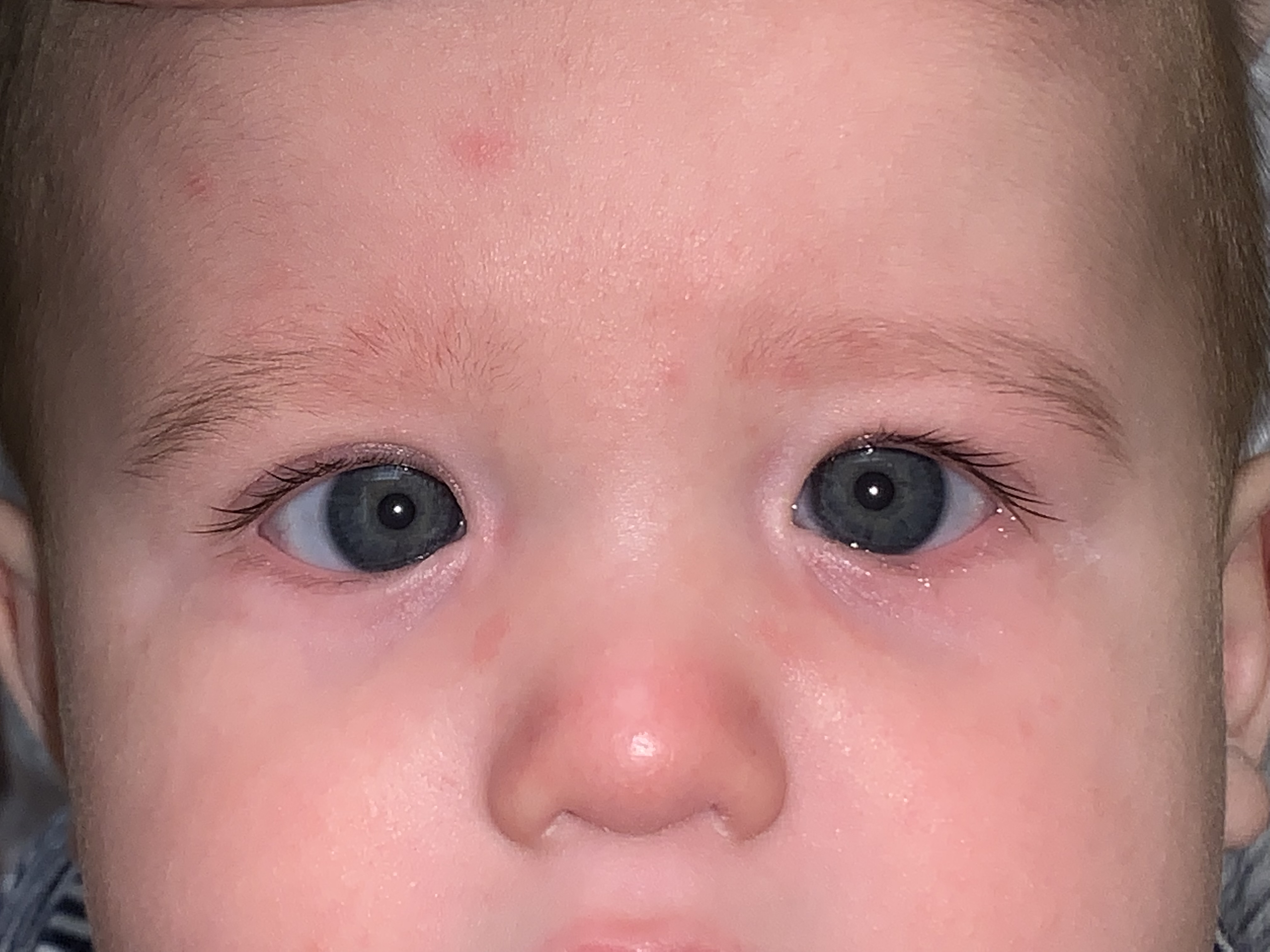
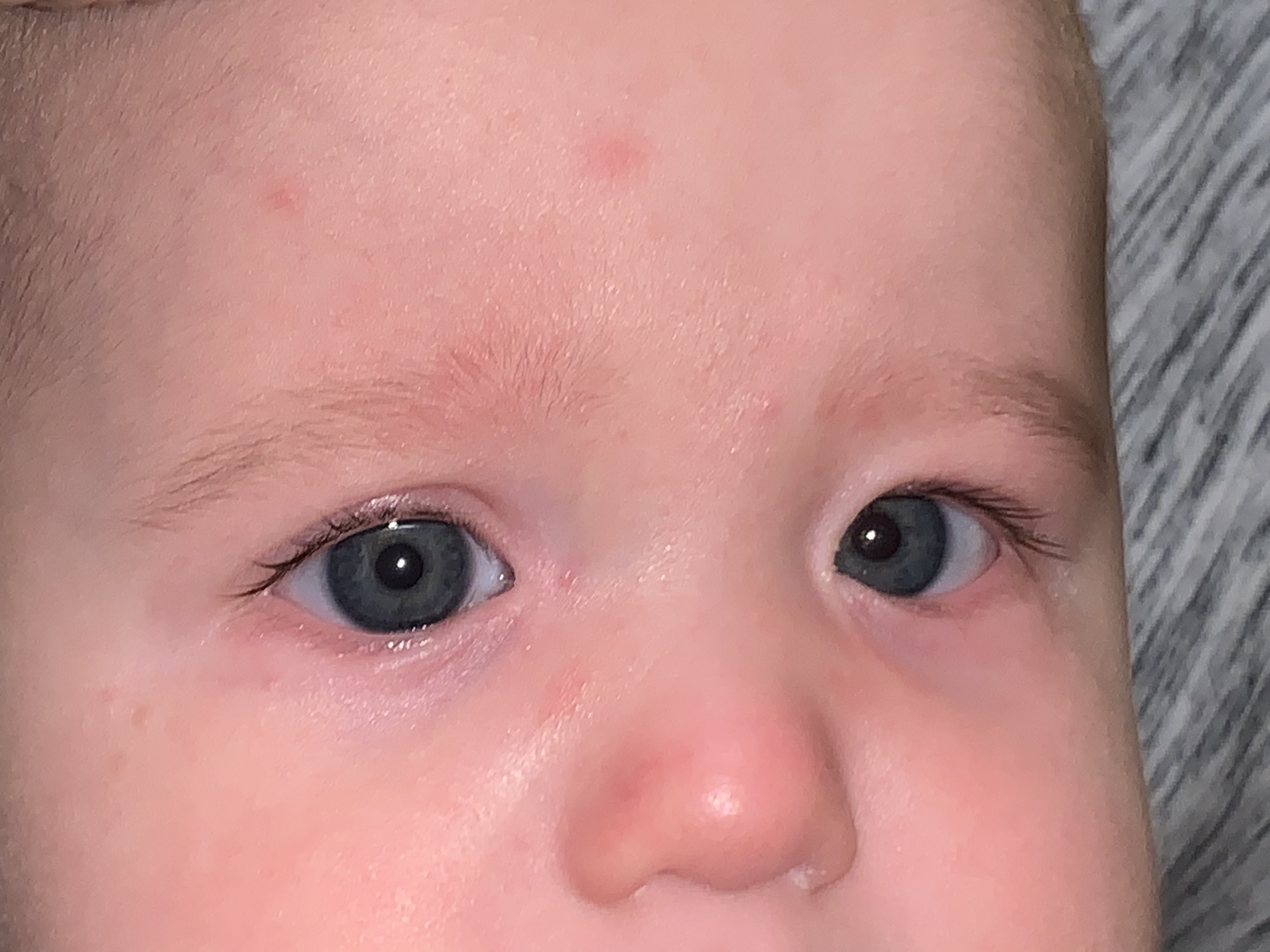
Images 1, 2, & 3 from Right to Left: The pseudoesotropia almost always appears worse when the child is in side gaze (images 1 and 3). Image 2 demonstrates, when looking straight ahead, most of the medial conjunctiva or white portion of the eye, is covered up by the epicanthal folds. The corneal light reflex, in all three photos, is symmetric and centered within the pupil.
Overview: Pseudoesotropia describes the condition in which a patient has the illusion of esotropic strabismus (inwardly deviated eye misalignment) despite accurately aligned visual axes. It is the most common type of pseudostrabismus, which also includes the conditions of pseduoexotropia (illusion of lateral deviation of one or both eyes) and pseudohypertropia (illusion of vertically misaligned eyes).1 Pseudoesotropia is one of the most common reasons why a pediatric ophthalmologist is asked to evaluate an infant.2
Etiology: Specific facial morphological features, including the orbit size or shape, globe size or shape, characteristics of the retrobulbar tissue, and eyelid margin anatomy are associated with this condition.3,4 Pseudoesotropia is most commonly seen in patients with prominent epicanthal folds, such as infants (i.e. prior to development of the nasal bridge), patients of Asian race, and patients with Down syndrome.4 A narrow interpupillary distance, facial asymmetry, or negative angle kappa (the angle between the visual axis and the optical axis) can also result in pseudoesotropia.4 Rarely, pseudoesotropia can occur as a result of nasal dislocation of the macula.5
Evaluation: Prior to the exam, a thorough history of the onset and duration of eye deviation should be obtained. Parents or pediatricians may have noticed less white sclera nasally than expected in certain facial orientations (see images 1 and 3). Pseudoesotropia can be exaggerated when the child looks in side gazes due to the nasal sclera being hidden behind the epicanthal fold (See images 1 and 3).
The diagnosis of pseudoesotropia is predominantly made by ophthalmic exam. Accurate diagnosis of pseudostrabismus is critical because true strabismus can lead to permanent vision loss if left untreated.
The ophthalmic examination of a patient with suspected esotropia or suspected pseudoesotropia should include:
- Evaluation of corneal light reflex (Hirschberg test)
- Cover-uncover test
- Cycloplegic refraction
- Examination of external eye anatomy
A corneal light reflex that is centered in both eyes (see image 2) and a lack of refixation movement with the cover-uncover test differentiates a diagnosis of pseudoesotropia from true esotropia2. Measuring the refractive error of the patient is important to rule out other potential causes of true strabismus, including amblyopia or high refractive error. However, the presence of high refractive error does not necessarily indicate true strabismus; pseudoesotropia can be found in patients with high infantile myopia or high hyperopia.5,6 It is also important to keep in mind that intermittent esotropia can be present in patients with prominent epicanthal folds.7
Management: If a diagnosis of pseudoesotropia is made, the initial step of management is parental reassurance and education. It is helpful to clarify with parents that the patient will outgrow this appearance or illusion of esotropia, as some parents will erroneously believe that their child has an actual esotropia that will resolve on its own. In addition, parents should be encouraged to seek re-evaluation if eye deviation does not improve or if suspected again in the future.2 Repeat ophthalmic exam is especially critical in children with a history of pseudostrabismus because they are at increased risk of developing true strabismus in the future.6,8,9
References:
- Kinori M, Robbins SL. Esotropia. Ophthalmology, Fifth Edition. 11.6, 1210-1216.e1
- Olitsky SE, Marsh JD. Disorders of Eye Movement and Alignment. Nelson Textbook of Pediatrics, Edition 21. 2019. Ch 642, 3350-3360.
- Urist MJ. Pseudostrabismus caused by abnormal configuration of the upper eyelid margins. Am J Ophthalmol 1973;75(3):455-7.
- Arnoldi K. Case corner: Pseudostrabismus: when are you sure? Am Orthopt J 2005;55:162-5.
- Damms T, Damms C, Schulz E, Haase W. Pseudo-esotropia caused by nasal dislocation of the macula in patients with high infantile myopia. Ophthalmologe 1994;91(1):77-80.
- Silbert AL, Matta NS, Silbert DI. Incidence of strabismus and amblyopia in preverbal children previously diagnosed with pseudoesotropia. Am Orthopt J 2013;63:103-6.
- Martin RJ, Fanaroff AA, Walsh MC. Examination and Common Problems in the Neonatal Eye: Neuromuscular Abnormalities: Strabismus. Fanaroff and Martin’s Neonatal-Perinatal Medicine, Eleventh Edition. 2019. Vol 2, Ch 95.
- Sefi-Yurdakul N, Tugcu B. Development of Strabismus in Children Initially Diagnosed with Pseudostrabismus. Strabismus 2016;24(2):70-3.
- Mohan K, Sharma A. Development of refractive accommodative esotropia in children initially diagnosed with pseudoesotropia. J Am Assoc Ped Ophthalmol Strab 2012;16(3):266-8.
Faculty Approval by: Griffin Jardine, MD
Identifier: Moran_CORE_26958
Copyright statement: Copyright ©2019. For further information regarding the rights to this collection, please visit: http://morancore.utah.edu/terms-of-use/