
Zopfiellasins A–D, Two Pairs of Epimeric Cytochalasins from Kiwi-Associated Fungus Zopfiella sp. and Their Antibacterial Assessment


Abstract
:
1. Introduction
2. Results and Discussion
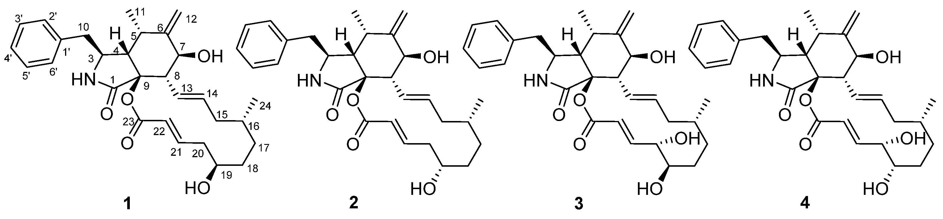
2.1. Structural Identification of Compounds 1–4
2.2. Antibacterial Activity against Psa
3. Materials and Methods
3.1. Materials and Instruments
3.2. Fungal Material and Cultivation Conditions
3.3. Extraction and Isolation
- Zopfiellasin A (1): Colorless crystals, mp: 220–222 °C; [α]24D + 114.5 (c 0.16, MeOH); 1H-NMR (600 MHz, methanol-d4) and 13C-NMR (150 MHz, methanol-d4) data, see Table 1; HRESIMS m/z 480.27461 [M + H]+ (calcd for C29H38NO5+: 480.27445).
- Zopfiellasin B (2): White powder; [α]24D + 130.1 (c 0.35, MeOH); UV (MeOH) λmax (log ε) 215 (3.82) nm; 1H-NMR (600 MHz, methanol-d4) and 13C-NMR (150 MHz, methanol-d4) data, see Table 1; HRESIMS m/z 480.27446 [M + H]+ (calcd for C29H38NO5+: 480.27445).
- Zopfiellasin C (3): Colorless crystals, mp: 219–220 °C; [α]24D + 63.5 (c 0.80, MeOH); UV (MeOH) λmax (log ε) 210 (3.76) nm; IR (KBr) νmax 3423, 1651, 1277, 1102, 1016, 974, 703 cm−1; 1H-NMR (600 MHz, methanol-d4) and 13C-NMR (150 MHz, methanol-d4) data, see Table 1; HRESIMS m/z 496.26932 [M + H]+ (calcd for C29H38NO6+: 496.26936).
- Zopfiellasin D (4): White solid; [α]24D + 87.6 (c 0.21, MeOH); UV (MeOH) λmax (log ε) 215 (3.82) nm; 1H-NMR (600 MHz, methanol-d4) and 13C-NMR (150 MHz, methanol-d4) data, see Table 1; HR-ESI-MS m/z 496.26950 [M + H]+ (calcd for C29H38NO6+: 496.26936).
- Crystal Data for Zopfiellasin A (1). C29H37NO5, M = 479.59 a = 10.1460 (4) Å, b = 11.9673 (5) Å, c = 21.3369 (8) Å, α = 90.00°, β = 90.00°, γ = 90.00°, V = 2590.73 (18) Å3, T = 151 (2) K. a = 9.2386 (2) Å, b = 11.0791 (2) Å, c = 26.7495 (6) Å, α = 90°, β = 90°, γ = 90°, V = 2737.96 (10) Å3, T = 298 (2) K, space group P21 21 21, Z = 4, μ(Cu Kα) = 1.54178 mm−1. A total of 33,559 reflections were measured, of which 5559 were independent (Rint = 4.22%). The final anisotropic full-matrix least-squares refinement on F2 with 326 variables converged at R1 = 3.02%, for the observed data and wR2 = 7.62% for all data. The goodness of fit was 1.032. The absolute configuration was determined by the Flack parameter = 0.02(4) CCDC: 2,104,459 (https://www.ccdc.cam.ac.uk).
- Crystal data for Zopfiellasin C (3). C29H37NO6·CH3OH, M = 527.64, a = 10.4883(6) Å, b = 23.6229(13) Å, c = 11.8002(6) Å, α = 90.00°, β = 90.452(2)°, γ = 90.00°, V = 2923.6(3) Å3, T = 150(2) K, space group P1 21 1, Z = 4, μ(Cu Kα) = 0.687 mm−1. A total of 69,193 reflections were measured, of which 12,495 were independent (Rint = 6.72%). The final anisotropic full-matrix least-squares refinement on F2 with 717 variables converged at R1 = 4.06%, for the observed data and wR2 = 10.97% for all data. The goodness of fit was 1.016. The absolute configuration was determined by the Flack parameter = 0.06(5). CCDC: 2,104,583 (https://www.ccdc.cam.ac.uk).

| No. | 1 | 2 | 3 | 4 | ||||
|---|---|---|---|---|---|---|---|---|
| δC | δH (J in Hz) | δC | δH (J in Hz) | δC | δH (J in Hz) | δC | δH (J in Hz) | |
| 1 | 174.0, C | 173.8, C | 174.0, C | 173.9, C | ||||
| 3 | 54.9, CH | 3.37, td (6.1, 2.7) | 55.2, CH | 3.34, m | 54.7, CH | 3.40, m | 54.9, CH | 3.38, m |
| 4 | 48.7, CH | 2.82, m | 49.3, CH | 2.76, dd (4.7, 3.2) | 48.4, CH | 2.86, d (2.8) | 48.7, CH | 2.82, d (5.9) |
| 5 | 32.8, CH | 3.17, m | 33.0, CH | 3.11, m | 32.8, CH | 3.22, m | 32.9, CH | 3.19, m |
| 6 | 151.4, C | 151.6, C | 151.4, C | 151.5, C | ||||
| 7 | 71.2, CH | 3.78, dd (10.9, 0.8) | 70.6, CH | 3.81, dd (11.3, 1.1) | 71.6, CH | 3.78, d (10.7) | 71.2, CH | 3.80, d (11.0) |
| 8 | 49.6, CH | 3.34, m | 50.6, CH | 3.18, dd (11.1, 9.9) | 49.0, CH | 3.38, m | 49.4, CH | 3.31, m |
| 9 | 85.2, C | 84.7, C | 85.5, C | 85.4, C | ||||
| 10 | 43.9, CH2 | 2.82, m; 2.82, m | 43.9, CH2 | 2.89, m; 2.89, m | 43.9, CH2 | 2.78, d (5.7) | 43.9, CH2 | 2.82, d (5.9) |
| 11 | 14.2, CH3 | 0.85, d (6.7) | 14.5, CH3 | 0.83, d (6.7) | 14.1, CH3 | 0.87, d (6.7) | 14.3, CH3 | 0.86, d (6.7) |
| 12 | 114.3, CH2 | 5.29, s; 5.08, s | 114.2, CH2 | 5.33, s; 5.09, s | 114.4, CH2 | 5.27, s; 5.09, s | 114.3, CH2 | 5.30, s; 5.09, s |
| 13 | 128.7, CH | 5.84, m | 128.7, CH | 5.76, m | 128.8, CH | 5.84, dd (15.1, 9.8) | 128.9, CH | 5.77, dd (15.1, 9.8) |
| 14 | 136.8, CH | 5.23, m | 136.6, CH | 5.35, m | 136.6, CH | 5.18, m | 136.4, CH | 5.24, m |
| 15 | 42.9, CH2 | 2.11, m; 1.68, m | 42.6, CH2 | 2.17, m; 1.75, m | 43.3, CH2 | 2.09, m; 1.63, m | 43.2, CH2 | 2.13, m; 1.68, m |
| 16 | 35.2, CH | 1.23, m | 32.8, CH | 1.52, m | 35.1, CH | 1.16, d (6.4) | 33.5, CH | 1.34, m |
| 17 | 32.4, CH2 | 1.63, m; 0.65, m | 30.7, CH2 | 1.68, m; 0.89, m | 33.8, CH2 | 1.54, m; 0.62, m | 30.6, CH2 | 1.62, m; 0.79, m |
| 18 | 38.7, CH2 | 1.84, m; 1.19, m | 34.6, CH2 | 1.69, m; 1.57, m | 31.0, CH2 | 1.54, m; 1.44, m | 29.7, CH2 | 1.70, m; 1.50, m |
| 19 | 72.2, CH | 3.59, td (9.7, 4.4) | 69.7, CH | 3.92, m | 75.2, CH | 3.59, d (9.8) | 74.3, CH | 3.71, dd (7.0, 6.5) |
| 20 | 43.8, CH2 | 2.71, m; 2.17, m | 42.7, CH2 | 2.58, m; 2.35, m | 74.5, CH | 4.48, s | 76.0, CH | 4.13, dd (6.5, 5.2) |
| 21 | 148.9, CH | 6.88, m | 149.0, CH | 7.10, m | 151.4, CH | 6.87, dd (15.6, 3.6) | 151.0, CH | 7.03, dd (15.7, 5.1) |
| 22 | 123.1, CH | 5.64, dd (15.6, 0.8) | 123.5, CH | 5.70, d (15.7) | 119.6, CH | 5.81, d (15.6) | 121.1, CH | 5.83, dd (15.7, 1.5) |
| 23 | 166.0, C | 166.1, C | 166.2, C | 166.2, C | ||||
| 24 | 20.8, CH3 | 0.90, d (6.6) | 20.5, CH3 | 0.92, d (6.6) | 20.7, CH3 | 0.89, d (6.6) | 20.5, CH3 | 0.89, d (6.6) |
| 1′ | 138.4, C | 138.8, C | 138.1, C | 138.3, C | ||||
| 2′,6′ | 131.0, CH | 7.13, d (7.4) | 130.9, CH | 7.16, d (7.4) | 131.1, CH | 7.13, d (7.4) | 131.0, CH | 7.14, d (7.4) |
| 3′,5′ | 129.6, CH | 7.26, dd (7.4, 7.4) | 129.6, CH | 7.28, dd (7.4, 7.4) | 129.6, CH | 7.26, dd (7.4, 7.4) | 129.6, CH | 7.26, dd (7.4, 7.4) |
| 4′ | 127.8, CH | 7.18, t (7.4) | 127.8, CH | 7.20, t (7.4) | 127.8, CH | 7.18, t (7.4) | 127.9, CH | 7.18, t (7.4) |
3.4. Antibacterial Assay
4. Conclusions
Supplementary Materials
Author Contributions
Funding
Institutional Review Board Statement
Informed Consent Statement
Data Availability Statement
Acknowledgments
Conflicts of Interest
Sample Availability
References
- Vanneste, J.L.; Cornish, D.A.; Yu, J.; Stokes, C.A. First Report of Pseudomonas syringae pv. actinidiae the Causal Agent of Bacterial Canker of Kiwifruit on Actinidia arguta Vines in New Zealand. Plant Dis. 2014, 98, 418. [Google Scholar] [CrossRef] [PubMed]
- Testolin, R.; Ferguson, A.R. Kiwifruit (Actinidia spp.) production and marketing in Italy. N. Zeal. J. Crop Hortic. Sci. 2009, 37, 1–32. [Google Scholar] [CrossRef]
- Ferrante, P.; Scortichini, M. Redefining the global populations of Pseudomonas syringae pv. actinidiae based on pathogenic, molecular and phenotypic characteristics. Plant Pathol. 2015, 64, 51–62. [Google Scholar] [CrossRef]
- Opgenorth, D.C.; Lai, M.; Sorrell, M.; White, J.B. Pseudomonas canker of kiwifruit. Plant Dis. 1983, 67, 1283–1284. [Google Scholar] [CrossRef] [Green Version]
- Serizawa, S.; Ichikawa, T.; Takikawa, Y.; Tsuyumu, S.; Goto, M. Occurence of bacterial canker of kiwifruit in Japan: Description of symptoms, isolation of the pathogen and screening of bactericides. Ann. Phytopathol. Soc. Jpn. 1989, 55, 427–436. [Google Scholar] [CrossRef] [Green Version]
- Wang, R.L.; Li, Q.; He, S.S.; Liu, Y.; Wang, M.T.; Jiang, G. Modeling and mapping the current and future distribution of Pseudomonas syringae pv. actinidiae under climate change in China. PLoS ONE 2018, 13, e0192153. [Google Scholar] [CrossRef] [PubMed] [Green Version]
- Cameron, A.; Sarojini, V. Pseudomonas syringae pv. actinidiae: Chemical control, resistance mechanisms and possible alternatives. Plant Pathol. 2014, 63, 1–11. [Google Scholar] [CrossRef]
- Tyson, J.; Curtis, C.; Manning, M.; Rees-George, J.; Snelgar, W.; Blattmann, P. Systemic movement of Pseudomonas syringae pv. actinidiae in kiwifruit vines in New Zealand. N. Zeal. Plant Prot. 2014, 67, 41–47. [Google Scholar] [CrossRef]
- Stefani, E.; Giovanardi, D. Dissemination of Pseudomonas syringae pv. actinidiae through pollen and its epiphytic life on leaves and fruits. Phytopathol. Mediterr. 2012, 50, 489–496. [Google Scholar]
- Mariz-Ponte, N.; Regalado, L.; Gimranov, E.; Tassi, N.; Moura, L.; Gomes, P.; Tavares, F.; Santos, C.; Teixeira, C. A synergic potential of antimicrobial peptides against Pseudomonas syringae pv. actinidiae. Molecules 2021, 26, 1461. [Google Scholar] [CrossRef] [PubMed]
- Hao, N.; Han, L.R.; Li, Y.T.; Li, J.; Tian, X.L.; Kong, D.; Tian, X.R. New 8-O-4′ neolignans and their antibacterial activity from the whole plants of Clematis lasiandra. ACS Omega 2020, 5, 19661–19666. [Google Scholar] [CrossRef] [PubMed]
- Rao, J.R.; Liu, L.W.; Zeng, D.; Wang, M.W.; Xiang, M.; Yang, S. Antibiotic activities of propanolamine containing 1,4-benzoxazin-3-ones against phytopathogenic bacteria. RSC Adv. 2020, 10, 682–688. [Google Scholar] [CrossRef] [Green Version]
- Huang, X.; Liu, H.W.; Long, Z.Q.; Li, Z.X.; Zhu, J.J.; Wang, P.Y.; Qi, P.Y.; Liu, L.W.; Yang, S. Rational optimization of 1,2,3-triazole-tailored carbazoles as prospective antibacterial alternatives with significant in vivo control efficiency and unique mode of action. J. Agric. Food Chem. 2021, 69, 4615–4627. [Google Scholar] [CrossRef] [PubMed]
- Sun, L.T.; Chen, Y.; Yang, H.X.; Li, Z.H.; Liu, J.K.; Wang, G.K.; Feng, T. Bisabolane sesquiterpenes and α-pyrone derivative from endophytic fungus Zopfiella sp. Phytochem. Lett. 2020, 37, 29–32. [Google Scholar] [CrossRef]
- Yi, X.W.; He, J.; Sun, L.T.; Liu, J.K.; Wang, G.K.; Feng, T. 3-Decalinoyltetramic acids from kiwi-associated fungus Zopfiella sp. and their antibacterial activity against Pseudomonas syringae. RSC Adv. 2021, 11, 18827–18831. [Google Scholar] [CrossRef]
- Evidente, A.; Andolfi, A.; Vurro, M.; Zonno, M.C.; Motta, A. Cytochalasins Z1, Z2 and Z3, three 24-oxa [14]cytochalasans produced by Pyrenophora semeniperda. Phytochemistry 2002, 60, 45–53. [Google Scholar] [CrossRef]
- Evidente, A.; Andolfi, A.; Vurro, M.; Zonno, M.C.; Motta, A. Cytochalasins Z4, Z5, and Z6, three new 24-oxa[14]cytochalasans produced by Phoma exigua var. heteromorpha. J. Nat. Prod. 2003, 66, 1540–1544. [Google Scholar] [CrossRef] [PubMed]
- Kim, E.L.; Wang, H.; Park, J.H.; Hong, J.; Choi, J.S.; Im, D.S.; Chung, H.Y.; Jung, J.H. Cytochalasin derivatives from a jellyfish-derived fungus Phoma sp. Bioorg. Med. Chem. Lett. 2015, 25, 2096–2099. [Google Scholar] [CrossRef] [PubMed]
- Wang, W.X.; Li, Z.H.; He, J.; Feng, T.; Li, J.; Liu, J.K. Cytotoxic cytochalasans from fungus Xylaria longipes. Fitoterapia 2019, 137, 104278. [Google Scholar] [CrossRef] [PubMed]
- Steyn, P.S.; Breytenbach, J.C.; Botha, J.H.; Fernandes, M.A.; Wessels, P.L. Synthesis, complete 1H and 13C-NMR assignment and crystal structure of novel epoxide derivatives of cytochalasin B. Magn. Reson. Chem. 2008, 46, 650–659. [Google Scholar] [CrossRef] [PubMed]


Publisher’s Note: MDPI stays neutral with regard to jurisdictional claims in published maps and institutional affiliations. |
© 2021 by the authors. Licensee MDPI, Basel, Switzerland. This article is an open access article distributed under the terms and conditions of the Creative Commons Attribution (CC BY) license (https://creativecommons.org/licenses/by/4.0/).
Share and Cite
Zhang, J.-Y.; He, J.; Li, Z.-H.; Feng, T.; Liu, J.-K. Zopfiellasins A–D, Two Pairs of Epimeric Cytochalasins from Kiwi-Associated Fungus Zopfiella sp. and Their Antibacterial Assessment. Molecules 2021, 26, 5611. https://doi.org/10.3390/molecules26185611
Zhang J-Y, He J, Li Z-H, Feng T, Liu J-K. Zopfiellasins A–D, Two Pairs of Epimeric Cytochalasins from Kiwi-Associated Fungus Zopfiella sp. and Their Antibacterial Assessment. Molecules. 2021; 26(18):5611. https://doi.org/10.3390/molecules26185611
Chicago/Turabian StyleZhang, Jie-Yu, Juan He, Zheng-Hui Li, Tao Feng, and Ji-Kai Liu. 2021. "Zopfiellasins A–D, Two Pairs of Epimeric Cytochalasins from Kiwi-Associated Fungus Zopfiella sp. and Their Antibacterial Assessment" Molecules 26, no. 18: 5611. https://doi.org/10.3390/molecules26185611