Communication

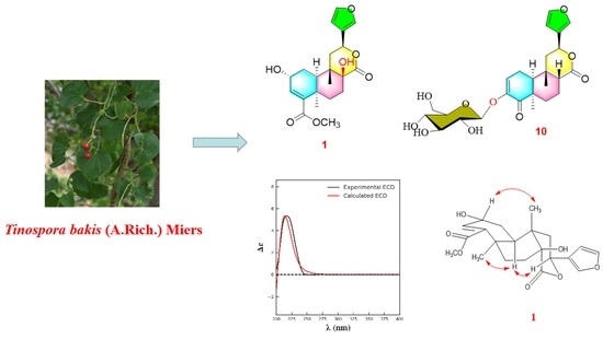
Clerodane Furanoditerpenoids from Tinospora bakis (A.Rich.) Miers (Menispermaceae)

1
H. E. J. Research Institute of Chemistry, International Center for Chemical and Biological Sciences, University of Karachi, Karachi 75270, Pakistan
2
Department of Microbiology, Faculty of Pure and Applied Sciences, International University of Africa, Khartoum 12223, Sudan
3
Dr. Panjwani Center for Molecular Medicine and Drug Research, International Center for Chemical and Biological Sciences, University of Karachi, Karachi 75270, Pakistan
4
Third World Center (TWC) for Chemical Sciences, International Center for Chemical & Biological Sciences, University of Karachi, Karachi 75270, Pakistan
*
Authors to whom correspondence should be addressed.
These authors contributed equally to this work.
Molecules 2024, 29(1), 154; https://doi.org/10.3390/molecules29010154
Submission received: 27 November 2023 / Revised: 13 December 2023 / Accepted: 22 December 2023 / Published: 26 December 2023

Abstract

:
Tinospora bakis (A.Rich.) Miers (Menispermaceae) has traditionally been used to alleviate headaches, rheumatism, mycetoma, and diabetes, among others. Despite its extensive use, the active components of the plant have never been investigated. In this work, a series of furanoditerpenoids (118) and five compounds from other classes (1923) were isolated from T. bakis. Notably, two new compounds were discovered and named: tinobakisin (1) and tinobakiside (10). Their molecular structures were elucidated with NMR, MS, UV, IR, and ECD spectra. Additionally, known compounds (29 and 1123) were corroboratively identified through spectral comparisons with previously reported data, while highlighting and addressing some inaccuracies in the prior literature. Remarkably, compounds 6, 7, 13, and 17 exhibited a superior anti-glycation effect, outperforming established agents like rutin and quercetin in a lab model of protein glycation with glucose. The overall findings suggest that furanoditerpenoids play a crucial role in the antidiabetic properties of T. bakis. This research marks the first comprehensive phytochemical investigation of T. bakis, opening the door for further investigation into furanoditerpenoids and their biological mechanisms.

Graphical Abstract

1. Introduction

Diabetes has been a severe health and economic burden globally, with increasing prevalence year by year. It is worth mentioning that around 422 million adults were living with diabetes in 2014 globally, in comparison to 108 million in 1980, and the prevalence is expected to approach 629 million worldwide in 2045 [1,2]. Therefore, treating diabetes and its associated comorbidities has been a hot field, attracting the attention and effort of scientists [3]. Natural products have been indispensable sources for new drug discovery. Among the approved drugs from 1981 to 2014, natural products or their derivative accounted for 26% [4]. Folk herbal medicines have been used worldwide for controlling blood glucose levels in patients with diabetes since ancient times [5]. Many investigations have been conducted to explore the active components for advanced antidiabetic drug discovery [6].
The Tinospora genus, belonging to the family Menispermaceae, has 16 accepted species (World Flora Online). The species are widely distributed throughout the tropical and subtropical parts of Asia, Africa, and Australia. Several species, such as T. capillipes, T. cordifolia, T. sagittata, T. sinensis, etc., have been well-studied due to their medicinal importance. Until now, more than 200 secondary metabolites have been isolated from these species, including diterpenoids, triterpenoids, sesquiterpenoids, alkaloids, steroids, flavonoids, lignans, etc. [7,8,9], among which more than 100 compounds belong to clerodane furanoditerpenoids [10,11,12,13,14,15,16].
Tinospora bakis (A.Rich.) Miers is a deciduous climber, wildly growing in Africa. Its whole plant, root, or leaves have been used to treat headaches, rheumatism, mycetoma, diabetes, etc., in Africa [8,17,18]. Despite its therapeutic importance, the phytochemicals of T. bakis have never been investigated, except for the main component, columbin [17]. Although the extracts from the roots or whole plant of T. bakis have been studied through in vivo pharmacological approaches, demonstrating antipyretic, antidiabetic, and immunomodulatory effects, most of the active components in T. bakis remain unknown [18,19,20].
In this work, a systematic phytochemical investigation was conducted on T. bakis for the first time. A total of 23 compounds were isolated from the EtOAc fraction of T. bakis. The structures of all compounds were elucidated based on multiple pieces of spectroscopic evidence, such as NMR, MS, UV, IR, and ECD. Among them, 18 compounds (118) were furanoditerpenoids, as shown in Figure 1. Moreover, an in vitro anti-glycation essay on the obtained components was performed, resulting in several potential antidiabetic leads, which shed light on the molecular basis of the antidiabetic property of T. bakis. Further investigation of the in vivo evidence of the active components and related mechanisms might be required [21,22,23].

2. Results and Discussion

Compound 1 was obtained as white needle-like crystals. The molecular formula was deduced as C21H26O7 according to the molecular ion peak [M]+ at m/z 390.1687 (calcd. for 390.1679) in HR-EI-MS, with nine unsaturation degrees. The 1H-NMR data (Table 1) displayed the characteristic signals for a furan ring at δH 7.54 (br s), 7.39 (t, J = 1.2 Hz), and 6.73 (d, J = 1.2 Hz). An olefinic proton at δH 6.57, two oxymethine signals at δH 5.15 (dd, J = 11.2, 4.8 Hz) and 4.46 (m), two methyls at δH 1.44 (s) and 1.05 (m), and a methoxy at δH 3.73 were also observed. The 13C-NMR (Table 1) and DEPT spectra revealed the existence of 21 carbons, including two carbonyl signals at δC 178.3 and 169.5; 4 carbons for the furan group at δC 144.1, 142.0, 127.6, and 110.9; 2 olefinic carbons at δC 141.1 and 140.4; 3 oxygen-substituted alkyl carbons at δC 90.4, 74.3, and 65.1; two methyls at δC 30.6 and 22.2; and a methoxy at δC 52.1, along with another 2 quaternary carbons, one methine, and four methylenes in the upfield. All the data agreed with the aglycon of tinospinosides B (2) [11]. The structure was further confirmed by correlations in the 1H–1H COSY, HSQC, and HMBC spectra, which are assigned in Figure 2. Correlations of H-2 (δH 4.46)/CH3-20 (δH 1.05), H-10 (δH 1.92)/CH3-19 (δH 1.44), and H-10 (δH 1.92)/H-12 (δH 5.15) in the NOESY spectrum indicated the configurations of chiral carbons. Since enantiomers of clerodane diterpenoids commonly exist naturally [24], the relative configuration of 1 was also further proved by curve fitting the experimental and calculated ECD. As shown in Figure 3, the calculated ECD of the proposed structure showed an excellent agreement with the experimental ECD of 1. Therefore, the structure of compound 1 was confirmed and named tinobakisin.
Compounds 2 and 3 were identified as tinospinoside B and tinospinoside C, respectively [11]. Compound 4 was found identical to the aglycon of tinospinoside A (5), obtained after hydrolysis. Therefore, it is isolated as a natural compound for the first time herein [11]. Compound 6 was determined as the 8-epimer of tinospinosides A, tinophylloloside [25]. Compounds 79 were identified as tinocallone A [26], fibaruretin H [27], and sagitone [14], respectively.
Compound 10 was isolated as an amorphous, colorless semisolid. The molecular formula was deduced as C25H32O6, through the molecular ion peak [M + H]+ at m/z 493.2061 (calcd. for 493.2074, C25H33O6) in positive HR-FAB-MS, suggesting 10 degrees of unsaturation. The 1H-NMR data (Table 1) displayed the characteristic signals for a furan moiety at δH 7.59 (br s), 7.49 (t, J = 2.0 Hz), and 6.54 (d, J = 2.0 Hz); an olefinic proton at δH 6.30 (dd, J = 6.5, 2.5 Hz); an oxymethine signal at δH 5.57 (dd, J = 12.5, 4.0 Hz); and two methyls at δH 1.28 (s) and 0.97 (s). 13C-NMR (Table 1) revealed the presence of two carbonyl carbons at δC 200.5 and 174.8; aromatic carbons at δC 149.2, 145.0, 141.4, 126.8, 124.3, and 109.6; and two angular methyls at δC 29.0 and 26.6. All these data of 1 were similar to those of sagitone, a novel 18-norclerodane furanoditerpenoid isolated from the roots of Tinospora sagittata var. yunnanensis [14]. However, additional signals belonging to a glucopyranoside at δC 102.3, 74.6, 78.3, 71.3, 77.6, and 62.5, corresponding to δH 4.62, 3.84, 3.65, 3.39, 3.38, 3.34, and 3.32 in the HSQC spectrum, were also observed [28]. After acid hydrolysis, the sugar moiety was determined as D-glucose, based on the optical rotation detected by an optical detector through HPLC. Also, the large coupling constant exhibited by the anomeric proton at δH 4.62 (1H, d, J = 7.0 Hz) suggested the relative configuration as β-oriented (Figure 4). In addition, the experimental ECD of 10 was close to 1 (Figure 5), indicating a similar relative configuration. Thus, the structure of 10 was established and named tinobakiside.
Compound 11 was elucidated as palmatoside G [29]. Compounds 12 and 13 were determined to be jateorin and isojateorin, respectively, based on the NMR data [30,31]. The X-ray structure of jateorin was reported in 1986 as a major component of Tinospora cordifolia [32]. Tinosporide was previously wrongly identified from Tinospora cordifolia and then later confirmed by X-ray to be identical to jateorin [33]. Chasmanthin and palmarin from Jateorrhiza palmate, Jateorhiza palmate, and Fibraurea chloroleuca were reported as the 12-epimer of jateorin and isojateorin, respectively [30,31,34], and also found in Tinospora cordifolia [35], which proved the common existence of these compounds in the Menispermaceae family. The structure of 14 was initially deduced, as reported by Hanuman et al. [36], because the NMR values were identical. However, after careful elucidation by 2D NMR, it was determined as the columbin, which was also isolated from several other species of the Tinospora genus [14,16,37,38] and found to be the major component of T. bakis [17]. This literature might wrongly determine the structure [36] in which 2D NMR was not performed. In addition, this is the only report of this structure. Compound 14 had β-H at C-8. Compound 15 had α-H (δ 2.98) at C-8 as its isomer [30]. Compounds 16 and 17, palmatoside C and D, the glucosides of 14 and 15, were isolated together from Jateorhiza palmata Miers in 1987 [29]. Later, palmatoside C was also found in the Tinospora genus [14,37]. Its configuration can be confirmed by the correlations with CH3-20 (δ 1.26) and H-10 (δ 1.84) in the NOESY spectrum. Compound 18 was elucidated as 8-hydroxycolumbin [39].
Five compounds from other classes were elucidated, based on their NMR and MS data, as 4-[formyl-5-(hydroxymethyl)-1H-pyrrol-1-yl] butanoic acid (19) [40], quercetin (20) [41], β-sitosterol (21) [42], β-sitosterol β-D-glucoside (22) [43], and oleic acid (23) [44].
The anti-glycation activities of all clerodane furanoditerpenoids 118 were evaluated by the in vitro BSA (bovine serum albumin)–glucose glycation model. Compared to the positive reference compounds rutin and quercetin, having an IC50 of 69 ± 0.12 and 104 ± 1.75 μM, respectively, compounds 6, 7, 13, and 17 displayed potent inhibitory activities with an IC50 of 37 ± 0.48, 78 ± 3.05, 66 ± 1.89, and 25 ± 0.25 μM, respectively. Compounds 12, 16, and 18 showed moderate activity, having an IC50 of 260 ± 2.50, 909 ± 5.86, and 265 ± 3.88 μM, respectively. This is the first report to find hypoglycemic compounds from T. bakis, unveiling the components responsible for the traditional use of this plant for treating diabetes.

3. Materials and Methods

3.1. General Experiment Procedures

The Bruker AMS-400 and AMX-500 (Bruker, Billerica, MA, USA) were used to record NMR spectra. For LR-FAB-MS and LR-EI-MS, JEOL MS Route JMS 600H (JEOL Ltd., Akishima, Japan) mass spectrometer was used, and, for HR-FAB-MS, JEOL JMS-HX110 mass spectrometer was used. A UV/visible spectrophotometer was used to accomplish the UV/visible method. The optical rotations were completed on the p-2000 Polarimeter, and the infrared (IR) spectra were recorded on the Attenuated Total Reflectance Infrared Spectrophotometer (ATRIR) FTIR iS50 (Fourier-Transform Infrared Spectrophotometer, Thermo Fisher Scientific, Waltham, MA, USA) in the KBr disc. Electronic Circular Dichorism (ECD) measurements were performed on Jaso-J-8-10 Circular Dichorism Spectro-Polarimeter. Normal silica gel (E. Merck, 70–230 Mesh) was utilized for fractionation by column chromatography. C18 (Wakogel, 38–63 Mesh), Sephadex LH-20 (GE healthcare, Chicago, IL, USA), and normal and reverse-phase HPLC were employed for purification. The purity of the sample was confirmed using normal and reverse-phase precoated TLC. UV light at 254 nm was used to evaluate TLC plates. Dragendroff, ceric sulfate, and vanillin were employed to visualize the spots on TLC plates.

3.2. Plant Material

The whole plant of T. bakis was collected from the Nuba Mountains in western Sudan from December 2018 to January 2019 and taxonomically identified by Yahya Sulieman Mohamed at Herbarium of Medicinal and Aromatic Plants and Traditional Medicine Research Institute, National Center for Research, Khartoum, Sudan. A voucher specimen (M-95-17-MAPTRI-H) was preserved in the Herbarium of Medicinal and Aromatic Plants and Traditional Medicine Research Institute, National Center for Research, Khartoum, Sudan, for further reference.

3.3. Extraction and Isolation

The powdered plant material (5 kg) of T. bakis was extracted with 80% EtOH to obtain crude extract (560 g). The extract was suspended in distilled water and partitioned through n-hexane (31 g), ethyl acetate (160 g), and n-butanol (119 g) in succession, resulting in four fractions. The ethyl acetate (160 g) was subjected to silica gel column chromatography (CC) by using DCM:MeOH (100:0–0:100) as eluent, from which 21 fractions (F1–F21) were collected. Fr.4 was separated by silica gel CC eluted with hexane/acetone to afford 17 sub-fractions (F4-1–F4-17), F4-5 were further purified by silica gel CC, then compound 21 (7.8 mg) was obtained. Compound 7 (12.3 mg) was purified from F4-8 by the same method. F5 was subjected to silica gel CC eluted with hexane/acetone solvent system and gave F5-1–F5-14. Compounds 8 (13.5 mg) and 9 (9.5 mg) were gained from F5-11 and F5-12, respectively, by repeated silica gel CC. F6–F8 was found to have a major component, 14 (1.5 g). F9 was fractionated by silica gel CC eluted through gradient hexane/acetone to obtain 14 sub-fractions (F9-1–F9-14). Compounds 12 (8.2 mg), 13 (4.9 mg), and 15 (11.4 mg) were purified from F9-4, F9-3, and F9-2, respectively, by subjecting the semi-pure fraction on silica gel CC again. F10, F13, F15, F16, F17, and F18 were loaded on C18 silica gel CC with gradient elution of 20–100% MeOH for further fractionation. F10-4 was subjected to silica gel CC eluted with hexane/acetone solvent system, and then compounds 4 (8.1 mg), 18 (12.1 mg), and 23 (9.0 mg) were purified. F13-2 was loaded on silica gel CC by DCM/MeOH elution, resulting in compound 1 (5.2 mg). F15-3 was subjected to silica gel CC by gradient DCM/MeOH elution to afford 12 sub-fractions (F15-3-1–F15-3-12). F15-3-8 was further fractionated over silica gel CC with gradient EtOAc/EtOH elution and purified by preparative RP-HPLC, resulting in compounds 5 (19.8 mg) and 6 (15.2 mg), while compound 22 (8.2 mg) was obtained from F15-9 by purification on silica gel. Compounds 3 (7.8 mg), 10 (4.8 mg), 16 (14.6 mg), and 17 (12.4 mg) were obtained from F16-2 by a similar protocol. Compound 2 (25.8 mg) was isolated from F17-3 by subjecting the semi-pure fraction to Sephadex LH-20 with MeOH/H2O, followed by purification over silica gel CC using gradient DCM/MeOH/H2O elution. Similarly, F18-5 was fractionated over silica gel CC eluted with gradient DCM/MeOH/H2O to afford 13 sub-fractions. Then, F18-5-6, F18-5-7, and F18-5-8 were separated on Sephadex LH-20 column with MeOH/H2O and purified by successive preparative RP-HPLC to yield compounds 11 (15.9 mg), 19 (4.9 mg), and 20 (6.3 mg), respectively. The flow rate during the preparative RP-HPLC was 4 mL/min.
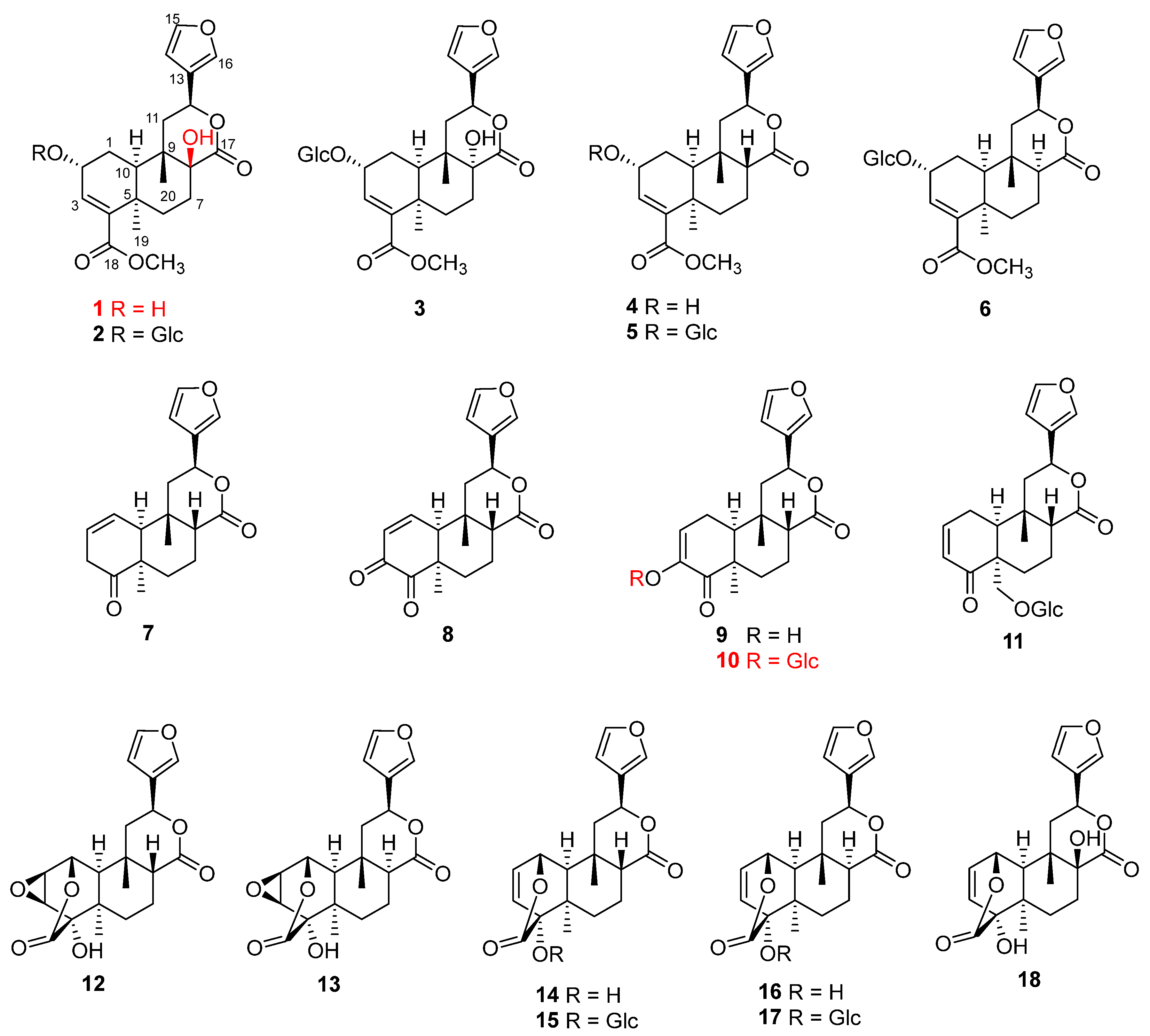
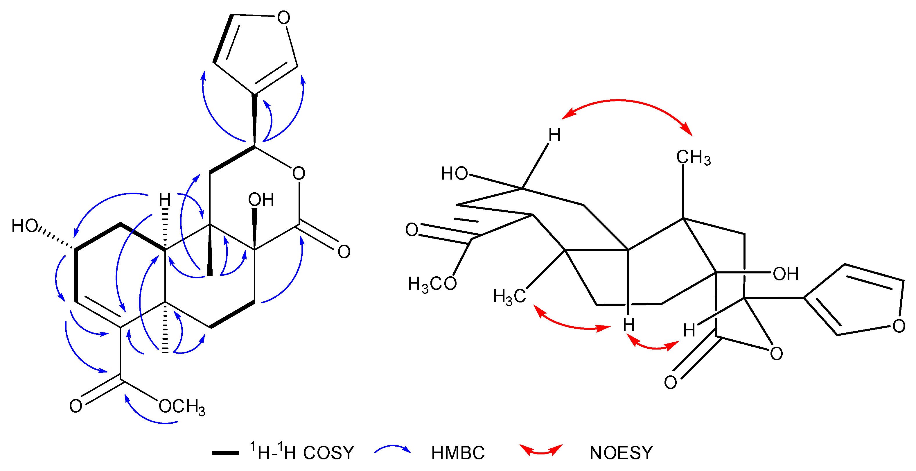
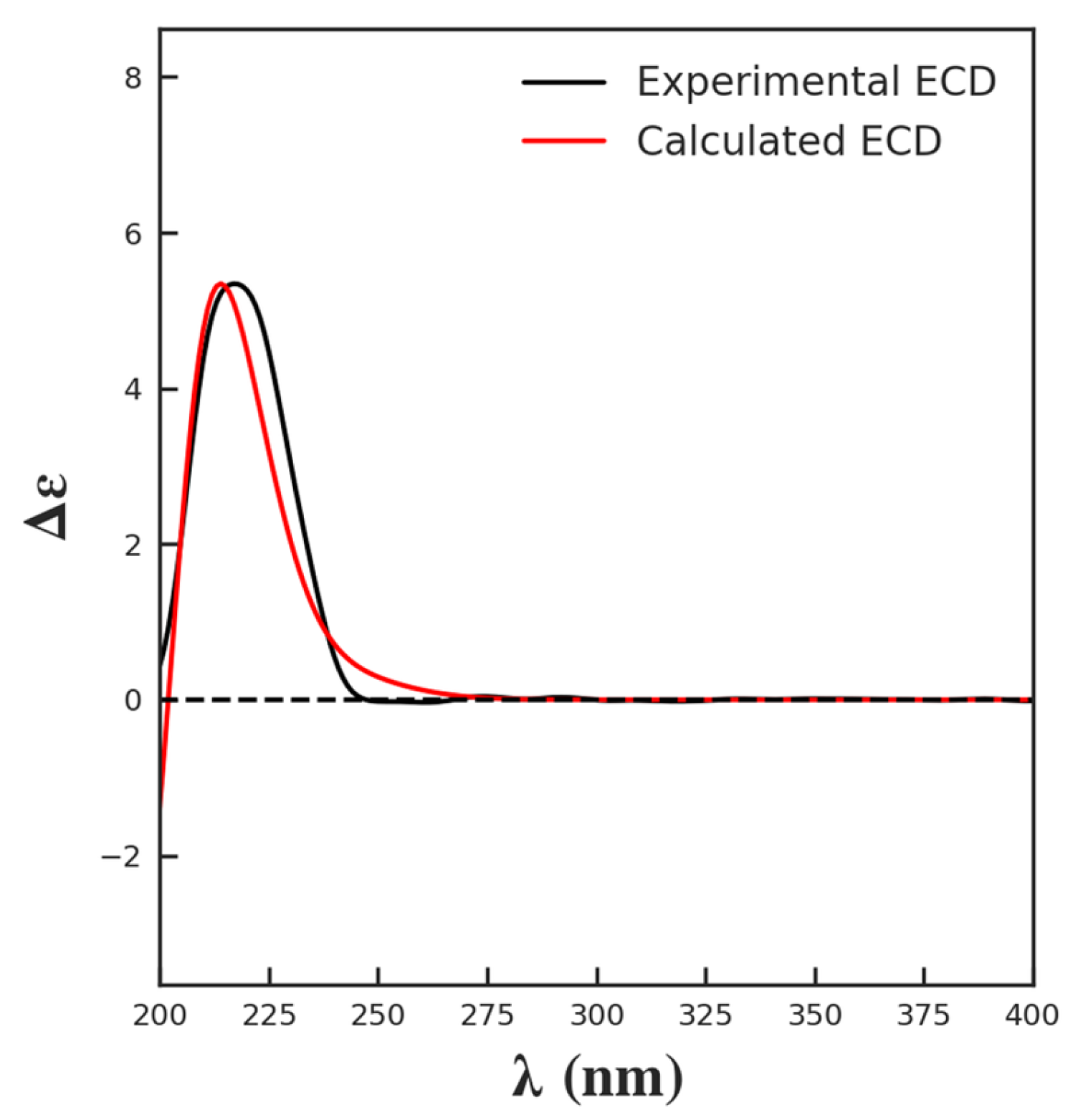
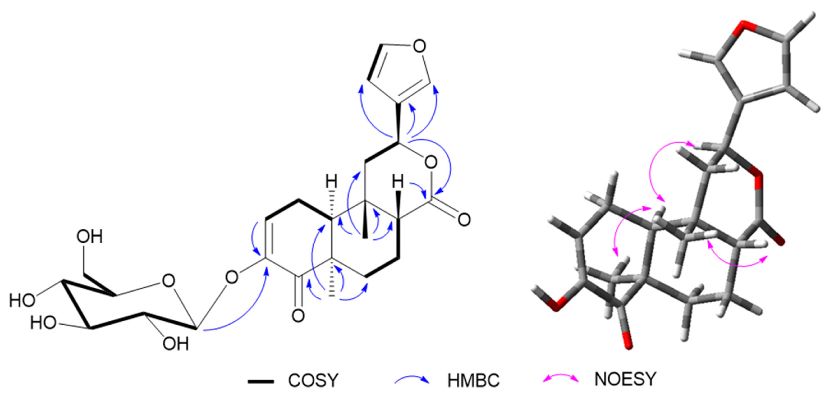
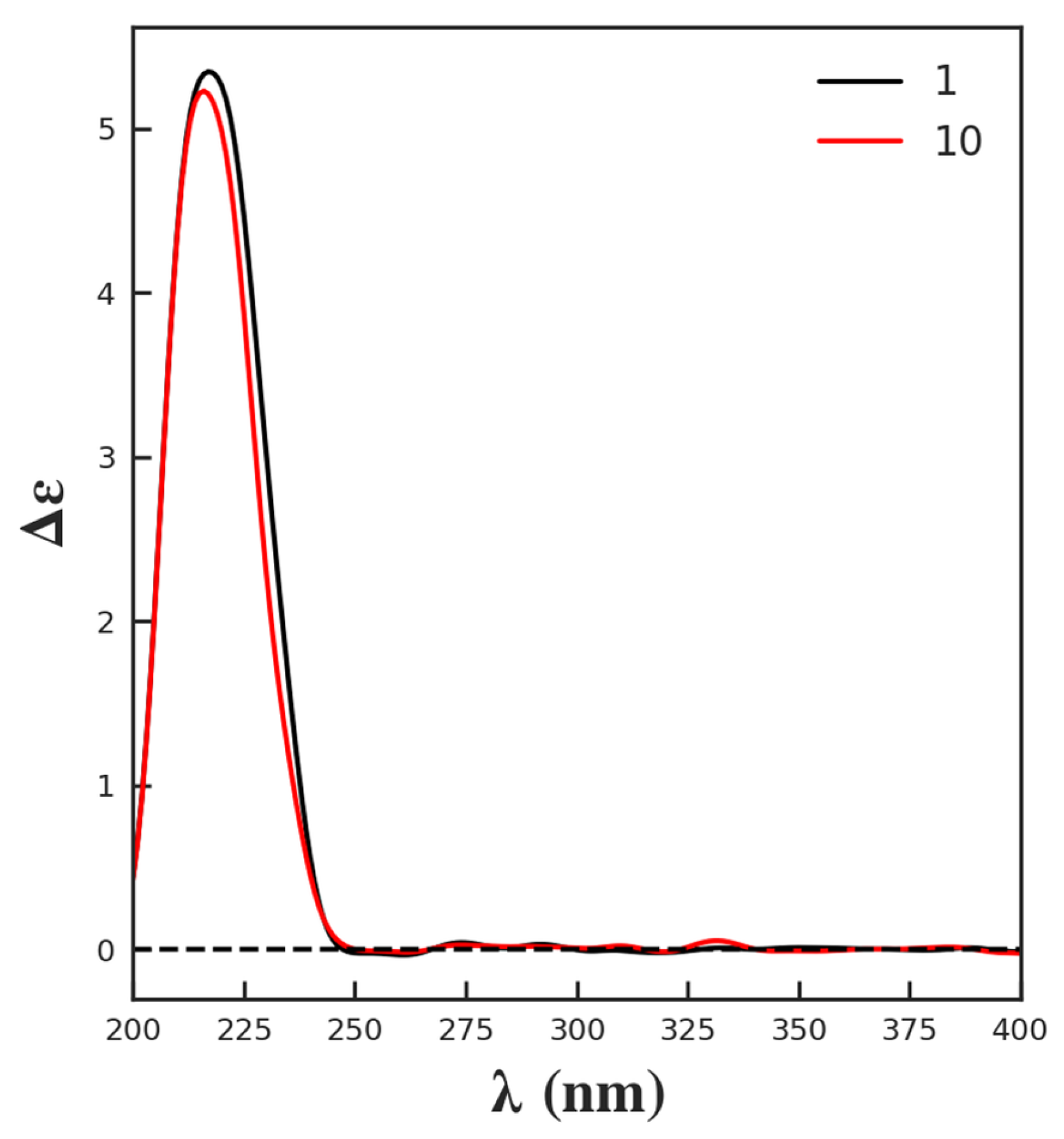
  • Tinobakisin (1): white needle-like crystals; [ α ] D 25 −18.18 (c 1.10 × 10 3 , MeOH); UV λmax 244 nm; IR (KBr) νmax 3310, 2943, 2832, 1738, 1589, 1427, 1280, 1073, 1020, 697, 668 cm−1; CD [nm (mdeg)]: 217 (5.34); 1H-NMR and 13C-NMR data, see Table 1; EI-MS [M]+, m/z 390.3; HR-EI-MS [M]+ m/z 390.1687 (calculated for 390.1679, C21H26O7).
  • Tinobakiside (10): amorphous, colorless semisolid; [ α ] D 25 −110.16 (c 2.46 × 10 3 , MeOH); UV λmax 248 nm; IR (KBr) νmax 3405, 2918, 1724, 1676, 1506, 1463, 1441, 1389, 1252, 1075, 1021, 602 cm−1; CD [nm (mdeg)]: 216 (5.23); 1H-NMR and 13C-NMR data, see Table 1; FAB-MS [M + H]+ m/z 493.1; HR-FAB-MS [M + H]+ m/z 493.2061 (calculated for 493.2074, C25H33O6).

3.4. Acid Hydrolysis

First, 5 mL 1 M HCl was used to dissolve compound 10 (2 mg), and then the mixture was stirred at 80 °C. After 8 h, the reaction mixture was extracted by CH2Cl2. Then, the obtained aqueous layer was evaporated under a vacuum and diluted multiple times to provide a neutral residue. The residue was subjected to analytical HPLC (Jasco LC-4000, Tokyo, Japan) with a Jasco OR-4090 optical rotation detector. The hydrolyzed sugar moiety indicated a positive rotation [45,46].

3.5. ECD Calculation

The ECD calculation of 1 was performed as reported, and the details are provided in Supplementary Data [10,47]. Conformational search with systematic algorithm was performed in Yinfo Cloud Platform (https://cloud.yinfotek.com/, accessed on 26 September 2023) using Confab at MMFF94 force field. Conformers were filtered by an RMSD threshold of 0.5 Å and an energy window of 7 kcal/mol. The energies and populations of dominative conformers are provided in Table S1. Structures for ECD calculation are shown in Table S2. All structures were confirmed by vibration frequency analysis, and no imaginary frequencies were found. Table S3 indicates the standard orientations of all configurations for ECD calculation at B3LYP/6-311G(d,p) level in methanol.

3.6. Anti-Glycation Activity

The anti-glycation effect was performed by the BSA (bovine serum albumin)–glucose glycation model in vitro, as previously described [48]. Glucose (0.5 M), BSA (10 mg/mL), and sodium azide (NaN3) (0.1 mM) were dissolved in sodium phosphate buffer (pH 7.4) as the reaction reagent to perform with or without the tested sample. The test compounds and standard references (rutin and quercetin) were dissolved in 10% DMSO. BSA without a glycating agent was used as a negative control. All samples were initially screened at 1 mM concentration. The assay was performed in triplicates, and a final reaction volume of 200 μL was maintained in each well of a 96-well plate. The reaction mixture was incubated at 37 °C for 7 days in a dark, sterile condition. The fluorescence (excitation 340 nm and emission 440 nm) was measured using a Varioskan Lux microtitre plate reader (Thermo Fisher Scientific, Waltham, MA, USA).
The percentage (%) of inhibition was calculated by using the following formula:
Inhibition (%) of fluorescence = (1 − Fluorescence of test compound/glycated BSA) × 100.
The compounds that exhibited more than 50% inhibition were processed for IC50. The two-fold dilution was made, and multiple concentrations (1, 0.5, 0.25, 0.125, 0.06, and 0.03 mM) of active compounds and standards (rutin and quercetin) were incubated with BSA-glucose at 37 °C for 7 days. The fluorescence was measured, and then the IC50 (μM) was calculated using the EZ-FIT Enzyme Kinetics Program (Perrella Scientific Inc., Amherst, MA, USA).

4. Conclusions

In summary, our phytochemical investigation of Tinospora bakis uncovered a plethora of clerodane furanoditerpenoids, including the discovery of two new compounds, tinobakisin (1) and tinobakiside (10), and provided clarifications on previously reported compounds. The observed superior anti-glycation effects of compounds 6, 7, 13, and 17 underscore the potential therapeutic relevance of T. bakis in managing diabetic complications. This research offers the first in-depth chemical profile of T. bakis. It lays the foundation for future studies to understand the mechanistic details of these compounds’ biological activities and their potential therapeutic applications, thus paving the way for new avenues in treating chronic diseases. More in vivo pharmacological and mechanism investigation might be required to further understand the active components’ antidiabetic properties, leading to the discovery of potential lead compounds for drug discovery.

Supplementary Materials

The following supporting information can be downloaded at https://www.mdpi.com/article/10.3390/molecules29010154/s1. Figure S1. UV spectrum of compound 1. Figure S2. IR spectrum of compound 1. Figure S3. EI-MS spectrum of compound 1. Figure S4. HR-EI-MS spectrum of compound 1. Figure S5. CD spectrum of compound 1. Figure S6. 1H-NMR spectrum of compound 1 (CD3OD, 400 MHz). Figure S7. 13C-NMR spectrum of compound 1 (CD3OD, 125 MHz). Figure S8. DEPT spectrum of compound 1. Figure S9. COSY spectrum of compound 1. Figure S10. HSQC spectrum of compound 1. Figure S11. HMBC spectrum of compound 1. Figure S12. NOESY spectrum of compound 1. Figure S13. IR spectrum of compound 10. Figure S14. IR spectrum of compound 10. Figure S15. CD spectrum of compound 10. Figure S16. FAB-MS spectrum of compound 10. Figure S17. HR-FAB-MS spectrum of compound 10. Figure S18. 1H-NMR spectrum of compound 10 (CD3OD, 500 MHz). Figure S19. 13C-NMR spectrum of compound 10 (CD3OD, 125 MHz). Figure S20. DEPT spectrum of compound 10. Figure S21. COSY spectrum of compound 10. Figure S22. HSQC spectrum of compound 10. Figure S23. HMBC spectrum of compound 10. Figure S24. NOESY spectrum of compound 10. S1. Detailed methodology of ECD calculation of compound 1. Table S1. Energies of all Conformers of compound 1 at MMFF94 force field. Table S2. Energies of all configurations of compound 1 at B3LYP/6-311G(d,p) in methanol. Table S3. Standard orientations of all configurations of compound 1 at B3LYP/6-311G(d,p) level in Methanol.

Author Contributions

Conceptualization, A.S.K. and Y.W.; methodology, A.S.K., M.A.S., N.N.S. and M.A.; formal analysis, Y.W., M.A.S. and M.A.; investigation, A.S.K., M.A.S., M.A., M.K., A.F. and N.N.S.; resources, A.S.K.; writing—original draft preparation, M.A.S. and Y.W.; writing—review and editing, Y.W. and F.-A.K.; visualization, Y.W.; supervision, Y.W., F.-A.K. and H.J.; project administration, Y.W. and F.-A.K.; funding acquisition, A.S.K., F.-A.K. and Y.W. All authors have read and agreed to the published version of the manuscript.

Funding

This work was supported by the National Administration of Traditional Chinese Medicine (no. GZYYGJ2020023) and the Higher Education Commission of Pakistan (NRPU 2021 #17603), Sindh Higher Education Commission (SHEC/SRSP/MS-1/7/2020-21). We acknowledge The World Academy of Science (TWAS) for the 2019 ICCBS-TWAS Sandwich Postgraduate Fellowship awarded to Ahmed Saeed Kabbashi with FR number 3240305623. The authors appreciate the support from the Medicinal and Aromatic Plants and Traditional Medicine Research Institute (MAPTMRI) and the Faculty of Pure and Applied Sciences, International University of Africa, Khartoum, Sudan.

Institutional Review Board Statement

Not applicable.

Informed Consent Statement

Not applicable.

Data Availability Statement

The data presented in this study are available in article and Supplementary Materials.

Conflicts of Interest

The authors declare no conflicts of interest.

References

  1. Forouhi, N.G.; Wareham, N.J. Epidemiology of Diabetes. Medicine 2019, 47, 22–27. [Google Scholar] [CrossRef]
  2. Yeung, A.W.K.; Tzvetkov, N.T.; Durazzo, A.; Lucarini, M.; Souto, E.B.; Santini, A.; Gan, R.Y.; Jozwik, A.; Grzybek, W.; Horbańczuk, J.O.; et al. Natural Products in Diabetes Research: Quantitative Literature Analysis. Nat. Prod. Res. 2021, 35, 5813–5827. [Google Scholar] [CrossRef] [PubMed]
  3. Elsayed, N.A.; Aleppo, G.; Aroda, V.R.; Bannuru, R.R.; Brown, F.M.; Bruemmer, D.; Collins, B.S.; Cusi, K.; Das, S.R.; Gibbons, C.H.; et al. Summary of Revisions: Standards of Care in Diabetes—2023. Diabetes Care 2023, 46, S5–S9. [Google Scholar] [CrossRef] [PubMed]
  4. Newman, D.J.; Cragg, G.M. Natural Products as Sources of New Drugs from 1981 to 2014. J. Nat. Prod. 2016, 79, 629–661. [Google Scholar] [CrossRef] [PubMed]
  5. Shapiro, K.; Gong, W.C. Natural Products Used for Diabetes. J. Am. Pharm. Assoc. 2002, 42, 217–226. [Google Scholar] [CrossRef] [PubMed]
  6. Ríos1, J.L.; Francini, F.; Schinella, G.R. Natural Products for the Treatment of Type 2 Diabetes Mellitus. Pharmacol. Res. 2018, 130, 451–465. [Google Scholar] [CrossRef]
  7. Chi, S.; She, G.; Han, D.; Wang, W.; Liu, Z.; Liu, B. Genus Tinospora: Ethnopharmacology, Phytochemistry, and Pharmacology. Evid.-Based Complement. Altern. Med. 2016, 2016, 9232593. [Google Scholar] [CrossRef]
  8. Haque, M.A.; Jantan, I.; Abbas Bukhari, S.N. Tinospora Species: An Overview of Their Modulating Effects on the Immune System. J. Ethnopharmacol. 2017, 207, 67–85. [Google Scholar] [CrossRef]
  9. Singh, D.; Chaudhuri, P.K. Chemistry and Pharmacology of Tinospora cordifolia. Nat. Prod. Commun. 2017, 12, 299–308. [Google Scholar] [CrossRef]
  10. Jiang, H.; Zhang, G.J.; Liu, Y.F.; Wang, H.S.; Liang, D. Clerodane Diterpenoid Glucosides from the Stems of Tinospora sinensis. J. Nat. Prod. 2017, 80, 975–982. [Google Scholar] [CrossRef]
  11. Li, W.; Huang, C.; Li, S.; Ma, F.; Li, Q.; Asada, Y.; Koike, K. Clerodane Diterpenoids from Tinospora sagittata (Oliv) Gagnep. Planta Med. 2012, 78, 82–85. [Google Scholar] [CrossRef] [PubMed]
  12. Jiang, Z.Y.; Li, W.J.; Jiao, L.X.; Guo, J.M.; Tian, K.; Yang, C.T.; Huang, X.Z. New Clerodane Diterpenes from Tinospora sagittata var. yunnanensis. Planta Med. 2014, 80, 419–425. [Google Scholar] [CrossRef] [PubMed]
  13. Choudhary, M.I.; Ismail, M.; Shaari, K.; Abbaskhan, A.; Sattar, S.A.; Lajis, N.H.; Atta-Ur-Rahman. Cis-Clerodane-Type Furanoditerpenoids from Tinospora crispa. J. Nat. Prod. 2010, 73, 541–547. [Google Scholar] [CrossRef] [PubMed]
  14. Huang, X.Z.; Cheng, C.M.; Dai, Y.; Fu, G.M.; Guo, J.M.; Yin, Y.; Liang, H. A Novel 18-Norclerodane Diterpenoid from the Roots of Tinospora sagittata var. yunnanensis. Molecules 2010, 15, 8360–8365. [Google Scholar] [CrossRef] [PubMed]
  15. Maurya, R.; Wazir, V.; Tyagi, A.; Kapil, R.S. Clerodane Diterpenoids from Tinospora cordifolia. Phytochemistry 1995, 38, 659–661. [Google Scholar] [CrossRef]
  16. Khan, M.A.; Gray, A.I.; Waterman, P.G. Tinosporaside, an 18-Norclerodane Glucoside from Tinospora cordifolia. Phytochemistry 1989, 28, 273–275. [Google Scholar] [CrossRef]
  17. Ibrahim Abdelwahab, S.; Syaed Koko, W.; Mohamed Elhassan Taha, M.; Mohan, S.; Achoui, M.; Ameen Abdulla, M.; Rais Mustafa, M.; Ahmad, S.; Ibrahim Noordin, M.; Lip Yong, C.; et al. In Vitro and in Vivo Anti-Inflammatory Activities of Columbin through the Inhibition of Cycloxygenase-2 and Nitric Oxide but Not the Suppression of NF-ΚB Translocation. Eur. J. Pharmacol. 2012, 678, 61–70. [Google Scholar] [CrossRef] [PubMed]
  18. Alamin, M.A.; Yagi, A.I.; Yagi, S.M. Evaluation of Antidiabetic Activity of Plants Used in Western Sudan. Asian Pac. J. Trop. Biomed. 2015, 5, 395–402. [Google Scholar] [CrossRef]
  19. Zafinindra, L.R.; Diatta, W.; Dieye, A.M.; Nongonierma, R.; Faye, B.; Bassene, E. Antipyretic Effect of Aqueous Extract and Alcaloid of Tinospora bakis (Miers) in Rabbits. Dakar Med. 2003, 48, 29–33. [Google Scholar]
  20. Koko, W.S.; Mesaik, M.A.; Yousaf, S.; Galal, M.; Choudhary, M.I. In Vitro Immunomodulating Properties of Selected Sudanese Medicinal Plants. J. Ethnopharmacol. 2008, 118, 26–34. [Google Scholar] [CrossRef]
  21. El-Nashar, H.A.S.; Abbas, H.; Zewail, M.; Noureldin, M.H.; Ali, M.M.; Shamaa, M.M.; Khattab, M.A.; Ibrahim, N. Neuroprotective Effect of Artichoke-Based Nanoformulation in Sporadic Alzheimer’s Disease Mouse Model: Focus on Antioxidant, Anti-Inflammatory, and Amyloidogenic Pathways. Pharmaceuticals 2022, 15, 1202. [Google Scholar] [CrossRef] [PubMed]
  22. Abdelghffar, E.A.R.; El-Nashar, H.A.S.; Fayez, S.; Obaid, W.A.; Eldahshan, O.A. Ameliorative Effect of Oregano (Origanum vulgare) versus Silymarin in Experimentally Induced Hepatic Encephalopathy. Sci. Rep. 2022, 12, 17854. [Google Scholar] [CrossRef] [PubMed]
  23. Ge, N.; Yan, G.; Sun, H.; Yang, L.; Kong, L.; Sun, Y.; Han, Y.; Zhao, Q.; Kang, S.; Wang, X. Version Updates of Strategies for Drug Discovery Based on Effective Constituents of Traditional Chinese Medicine. Acupunct. Herb. Med. 2023, 3, 158–179. [Google Scholar] [CrossRef]
  24. Li, R.; Morris-Natschke, S.L.; Lee, K.H. Clerodane Diterpenes: Sources, Structures, and Biological Activities. Nat. Prod. Rep. 2016, 33, 1166–1226. [Google Scholar] [CrossRef] [PubMed]
  25. Itokawa, H.; Mizuno, K.; Tajima, R.; Takeya, K. Furanoditerpene Glucosides from Fibraurea tinctoria. Phytochemistry 1986, 25, 905–908. [Google Scholar] [CrossRef]
  26. Wang, B.; Zhang, P.L.; Zhou, M.X.; Shen, T.; Zou, Y.X.; Lou, H.X.; Wang, X.N. New Nor-Clerodane-Type Furanoditerpenoids from the Rhizomes of Tinospora capillipes. Phytochem. Lett. 2016, 15, 225–229. [Google Scholar] [CrossRef]
  27. Zhang, G.; Ma, H.; Hu, S.; Xu, H.; Yang, B.; Yang, Q.; Xue, Y.; Cheng, L.; Jiang, J.; Zhang, J.; et al. Clerodane-Type Diterpenoids from Tuberous Roots of Tinospora sagittata (Oliv.) Gagnep. Fitoterapia 2016, 110, 59–65. [Google Scholar] [CrossRef]
  28. Roslund, M.U.; Tähtinen, P.; Niemitz, M.; Sjöholm, R. Complete Assignments of the 1H and 13C Chemical Shifts and JH,H Coupling Constants in NMR Spectra of d-Glucopyranose and All d-Glucopyranosyl-d-Glucopyranosides. Carbohydr. Res. 2008, 343, 101–112. [Google Scholar] [CrossRef]
  29. Yonemitsu, M.; Fukuda, N.; Kimura, T.; Komori, T. Studies on the Constituents of Jateorhiza palmata Miers (Colombo Root), II. Separation and Structure of Six New Furanoid Diterpene Glucosides: Palmatoside B, C, D, E, F, and G. Liebigs Ann. Chem. 1987, 1987, 193–197. [Google Scholar] [CrossRef]
  30. Yonemitsu, M.; Fukuda, N.; Kimura, T.; Komori, T. Studies on the Constituents of Jateorhiza palmata Miers (Colombo Root), I Separation and Structure of a New Furanoid Diterpene Glucoside (Palmatoside A). Liebigs Ann. Chem. 1986, 1986, 1327–1333. [Google Scholar] [CrossRef]
  31. Itokawa, H.; Mizuno, K.; Ichihara, Y.; Takeya, K. Isolation and 13C-NMR Studies on Three New Furanoditerpenyl Glucosides from Jateorrhiza columba. Planta Med. 1987, 53, 271–273. [Google Scholar] [CrossRef] [PubMed]
  32. Goddard, R.; Akhtar, F. The Structure of Jateorin. Acta Crystallogr. Sect. C Cryst. Struct. Commun. 1986, 42, 1217–1220. [Google Scholar] [CrossRef]
  33. Swaminathan, K.; Sinha, U.C.; Bhatt, R.K.; Sabata, B.K.; Tavale, S.S. Structure of Tinosporide, a Diterpenoid Furanolactone from Tinospora cordifolia Miers. Acta Crystallogr. C 1989, 45 Pt 1, 134–136. [Google Scholar] [CrossRef]
  34. Zakaria, M.; Saito, I.; Yao, X.-K.; Wang, R.-J.; Matsuura, T. Furanoditerpenes of Fibraurea chloroleuca. Planta Med. 1989, 55, 477–478. [Google Scholar] [CrossRef] [PubMed]
  35. Hanuman, J.B.; Bhatt, R.K.; Sabata, B. A Clerodane Furano-Diterpene from Tinospora cordifolia. J. Nat. Prod. 1988, 51, 197–201. [Google Scholar] [CrossRef]
  36. Hanuman, J.B.; Bhatt, R.K.; Sabata, B.K. A Diterpenoid Furanolactone from Tinospora cordifolia. Phytochemistry 1986, 25, 1677–1680. [Google Scholar] [CrossRef]
  37. Shi, L.M.; Li, R.Q.; Liu, W.H. Two New Furanoid Diterpenoids from Tinospora cagittata. Helv. Chim. Acta 2008, 91, 978–982. [Google Scholar] [CrossRef]
  38. Rishikesan, R.; Raju, R.; Manikandaselvi, S.; Thinagarbabu, R.; Sivasubramanian, A. Isolation and Characterisation of Clerodane Diterpenoids from the Traditional Medicinal Plant-Tinospora glabra (Burm. f.) Merrill. Pharm. Lett. 2016, 8, 280–287. [Google Scholar]
  39. Oguakwa, J.U.; Galeffi, C.; Nicoletti, M. Research on African Medicinal Plants. XI. 8-Hydroxycolumbin, a New Furanoid Diterpene from Chasmanthera Dependens. Planta Med. 1986, 52, 198–199. [Google Scholar] [CrossRef]
  40. Chin, Y.W.; Lim, S.W.; Kim, S.H.; Shin, D.Y.; Suh, Y.G.; Kim, Y.B.; Kim, Y.C.; Kim, J. Hepatoprotective Pyrrole Derivatives of Lycium chinense Fruits. Bioorg. Med. Chem. Lett. 2003, 13, 79–81. [Google Scholar] [CrossRef]
  41. Leena, P.N.; Aleykutty, N.A. Isolation and Spectral Identification of Quercetin Fron the Alcoholic Root Extract of Clerodendrum paniculatum. Int. J. Pharma Sci. Res. 2016, 7, 47–50. [Google Scholar]
  42. Chaturvedula, V.S.P.; Prakash, I. Isolation of Stigmasterol and β-Sitosterol from the Dichloromethane Extract of Rubus suavissimus. Int. Curr. Pharm. J. 2012, 1, 239–242. [Google Scholar] [CrossRef]
  43. Faizi, S.; Ali, M.; Saleem, R.; Irfanullah; Bibi, S. Spectral Assignments and Reference Data: Complete 1H and 13C NMR Assignments of Stigma-5-En-3-O-β-Glucoside and Its Acetyl Derivative. Magn. Reson. Chem. 2001, 39, 399–405. [Google Scholar] [CrossRef]
  44. Otun, K.O.; Olatunji, G.A.; Ajiboye, A.T.A.; Badeggi, U.M. Isolation and Characterization of the Chemical Constituents of the Stem Bark of Parinari polyandra Benth. Int. Res. J. Pure Appl. Chem. 2014, 4, 710–717. [Google Scholar] [CrossRef]
  45. Zhao, H.Y.; Wang, Y.Q.; Li, Y.C.; Lan, Q.; Liao, H.B.; Wang, H.S.; Liang, D. Flavonol Glycosides and Phenylpropanoid Glycosides with Inhibitory Effects on Microglial Nitric Oxide Production from Neoshirakia japonica. Fitoterapia 2021, 151, 104877. [Google Scholar] [CrossRef]
  46. Dai, L.; He, S.; Zhang, B.; Wang, H.; Wang, Y.; Liang, D. Foegraecumoside O and P, a Pair of Triterpenoid saponins with a 4/5/6 Fused Tricyclic Oleanane Carbon Skeleton from Lysimachia foenum-graecum Hance. Molecules 2023, 28, 5061. [Google Scholar] [CrossRef]
  47. Hu, Y.-J.; Lan, Q.; Su, B.-J.; Wang, Y.; Liang, D. Three New Phenolic Glycosides and a New Lignan Glycoside from Gaultheria leucocarpa var. yunnanensis. Fitoterapia 2024, 172, 105740. [Google Scholar] [CrossRef]
  48. Ali, R.; Salawu, K.M.; Aamer, M.; Jahan, H.; Tufail, P.; Irshad, R.; Khan, F.A.; Sener, B.; Choudhary, M.I.; Wang, Y. A New Sesquiterpene, Prosoterpene, from Prosopis africana (Guill. & Perr.) Taub. Nat. Prod. Res. 2023, 37, 3220–3227. [Google Scholar] [CrossRef]
Figure 1. Structures of compounds 118.
Molecules 29 00154 g001
Figure 2. Key 1H–1H COSY, HMBC, and NOESY correlations of compound 1.
Molecules 29 00154 g002
Figure 3. Experimental and calculated ECD of compound 1.
Molecules 29 00154 g003
Figure 4. Key 1H–1H COSY, HMBC, and NOESY correlations of compound 10.
Molecules 29 00154 g004
Figure 5. Experimental ECD of compounds 1 and 10.
Molecules 29 00154 g005
Table 1. NMR data of compounds 1 and 10.
No.1 a10 b
δHδC, MultipilicityδHδC, Multipilicity
12.27 overlapped
2.04 overlapped
30.1, CH22.90 ddd (20.0, 6.5, 2.5)
2.54 dd (20.0, 6.5)
22.5, CH2
24.46 m65.1, CH6.30 dd (6.5, 2.5)124.3, CH
36.57 d (3.2)140.4, CH-149.2, C
4-141.1, C-200.5, C
5-36.0, C-46.8, C
62.03 overlapped
1.75 m
30.6, CH22.28 overlapped
1.06 td (14.0, 4.0)
30.9, CH2
72.10 overlapped
1.64 ddd (14.0, 10.0, 2.8)
39.6, CH22.16 dq (14.0, 4.0)
1.72 tt (14.0, 4.0)
20.3, CH2
8-90.4, C2.47 br t (4.0)50.1, CH
9-50.0, C-37.6, C
101.92 d (5.6)47.0, CH2.29 overlapped45.3, CH
112.27 overlapped
2.05 overlapped
47.0, CH22.37 dd (15.0, 4.0)
1.80 dd (15.0, 12.5)
41.3, CH2
125.15 dd (11.2, 4.8)74.3, CH5.57 dd (12.5, 4.0)72.3, CH
13-127.6, C-126.8, C
146.73 d (1.2)110.9, CH6.54 d (2.0)109.6, CH
157.39 t (1.2)144.1, CH7.49 t (2.0)145.0, CH
167.54 brs142.0, CH7.59 br s141.4, CH
17-178.3, C-174.8, C
18-169.5
191.44 s30.6, CH31.28 s29.0, CH3
201.05 s22.2, CH30.97 s26.6, CH3
1′- 4.62 d (7.0)102.3, CH
2′- 3.38 overlapped74.6, CH
3′- 3.32 overlapped78.3, CH
4′- 3.34 overlapped71.3, CH
5′- 3.39 overlapped77.6, CH
6′- 3.84 dd (12.0, 1.8)
3.65 dd (12.0, 5.2)
62.5, CH2
OCH33.73 s52.1, CH3
a 1H-NMR data (δ) were measured in CD3OD at 400 MHz. 13C-NMR data (δ) were measured in CD3OD at 125 MHz. b 1H-NMR data (δ) were measured in CD3OD at 500 MHz. 13C-NMR data (δ) were measured in CD3OD at 125 MHz.
Disclaimer/Publisher’s Note: The statements, opinions and data contained in all publications are solely those of the individual author(s) and contributor(s) and not of MDPI and/or the editor(s). MDPI and/or the editor(s) disclaim responsibility for any injury to people or property resulting from any ideas, methods, instructions or products referred to in the content.

