Review

Defense Mechanisms of Cotton Fusarium and Verticillium Wilt and Comparison of Pathogenic Response in Cotton and Humans

1
Zhengzhou Research Base, State Key Laboratory of Cotton Biology, School of Agricultural Sciences, Zhengzhou University, Zhengzhou 450001, China
2
State Key Laboratory of Cotton Biology, Institute of Cotton Research, Chinese Academy of Agricultural Sciences, Anyang 455000, China
3
School of Pharmaceutical Sciences (Shenzhen), Shenzhen Campus of Sun Yat-Sen University, Shenzhen 518107, China
4
Hainan Yazhou Bay Seed Laboratory, Sanya 572000, China
5
Institute of Urban Agriculture, Chinese Academy of Agricultural Sciences, Chengdu 610000, China
*
Authors to whom correspondence should be addressed.
These authors contributed equally to this work.
Int. J. Mol. Sci. 2022, 23(20), 12217; https://doi.org/10.3390/ijms232012217
Submission received: 24 August 2022 / Revised: 29 September 2022 / Accepted: 11 October 2022 / Published: 13 October 2022
(This article belongs to the Special Issue Crop Biotic and Abiotic Stress Tolerance 2.0)

Abstract

:
Cotton is an important economic crop. Fusarium and Verticillium are the primary pathogenic fungi that threaten both the quality and sustainable production of cotton. As an opportunistic pathogen, Fusarium causes various human diseases, including fungal keratitis, which is the most common. Therefore, there is an urgent need to study and clarify the resistance mechanisms of cotton and humans toward Fusarium in order to mitigate, or eliminate, its harm. Herein, we first discuss the resistance and susceptibility mechanisms of cotton to Fusarium and Verticillium wilt and classify associated genes based on their functions. We then outline the characteristics and pathogenicity of Fusarium and describe the multiple roles of human neutrophils in limiting hyphal growth. Finally, we comprehensively compare the similarities and differences between animal and plant resistance to Fusarium and put forward new insights into novel strategies for cotton disease resistance breeding and treatment of Fusarium infection in humans.

1. Introduction

Cotton (Gossypium spp.) is an important cash crop as it yields grain, fiber, and oil [1]. Cotton is the world’s most important source of natural fiber, providing approximately 35% of the world’s total fiber [2]. Compared with synthetic fiber, cotton is a renewable resource and cotton plantations generate a higher number of jobs that involve planting, processing, and textile manufacturing. Cotton has important environmental and social benefits, and cottonseed can be used as animal feed after oil extraction [3]. Cotton is grown in more than 80 countries, of which approximately 30 regard cotton as a major crop [4]. China is the world’s largest producer of cotton fiber [5]. However, cotton has been affected by various pests and pathogens, among which Fusarium and Verticillium wilt cause serious global cotton economic losses [6,7], thus leading to them being considered as the main obstacle to sustainable high-quality cotton production in China [8]. Therefore, this review describes the relationship between cotton and pathogenic fungi, using Fusarium and Verticillium as examples.
Fusarium oxysporum (FOV) is the cause of Fusarium cotton wilt and is classified into eight physiological types, of which three exist in China [9]. Based on their symptoms, there are two species of Verticillium dahliae that infect cotton, namely defoliating and non-defoliating strains [10]. These pathogens can exist in soil and plant debris in the form of mycelium, chlamydia spores, and microsclerotia and survive in soil for a long time (until the beginning of a new infection cycle) [9]. In addition, they can survive saprophytically on other crops and weeds [8]. Diseases caused by Fusarium typically manifest following the seedling stage and are most severe during the budding stage. The onset of Fusarium wilt is characterized by plaques between the main veins, while the rest of the leaves remain green; chlorotic plaques gradually cause the leaves to become necrotic and fall off the stem [9]. Verticillium wilt occurs before the budding stage, manifesting as yellow mottled or fallen leaves during the boll setting stage, when the disease is also the most serious [10]. However, the determinants that lead to the development of these diseases remain unclear [8].
Humans are warm-blooded, their innate and adaptive immune system is complex, and they are naturally resistant to most invasive fungal infections. For fungi to successfully infect humans they must: (1) grow at or above human body temperature (37 °C), (2) bypass or penetrate host surface barriers to reach internal tissues, (3) digest and absorb human tissues, and (4) resist against the human immune system. Thus, the number of fungi capable of infecting a healthy human host is very limited [11,12]. However, the pathogenic fungi Fusarium and Aspergillus have these abilities and can therefore infect humans. Although there are limited data on the role of host defenses against these fungi, fusariosis, aspergillosis, and other fungal infections share many features [13]. Hence, this review discusses Fusarium to illustrate the relationship between pathogenic fungi and humans. Fusarium can produce mycotoxins that pose a significant health risk to humans and animals. There has been a gradual increase in infections caused by Fusarium in immunosuppressed patients that have undergone organ transplantation and chemotherapy in recent years [14]. Moreover, Fusarium is second only to Candida albicans and Aspergillus in terms of morbidity and mortality in immunodeficient individuals and is internationally classified as an important infectious disease [15].
To eliminate, or significantly mitigate, the harm of Fusarium and Verticillium wilt on cotton so as to improve yield, and simultaneously seek a method to treat patients suffering from Fusarium spp. infectious diseases, we review the studies on cotton genes related to resistance and susceptibility to Fusarium and Verticillium wilt in recent years, summarize the molecular mechanisms by which humans resist Fusarium disease, compare resistance strategies in humans and plants, and provide novel insights into the improvements in the resistance of plants and humans against Fusarium.

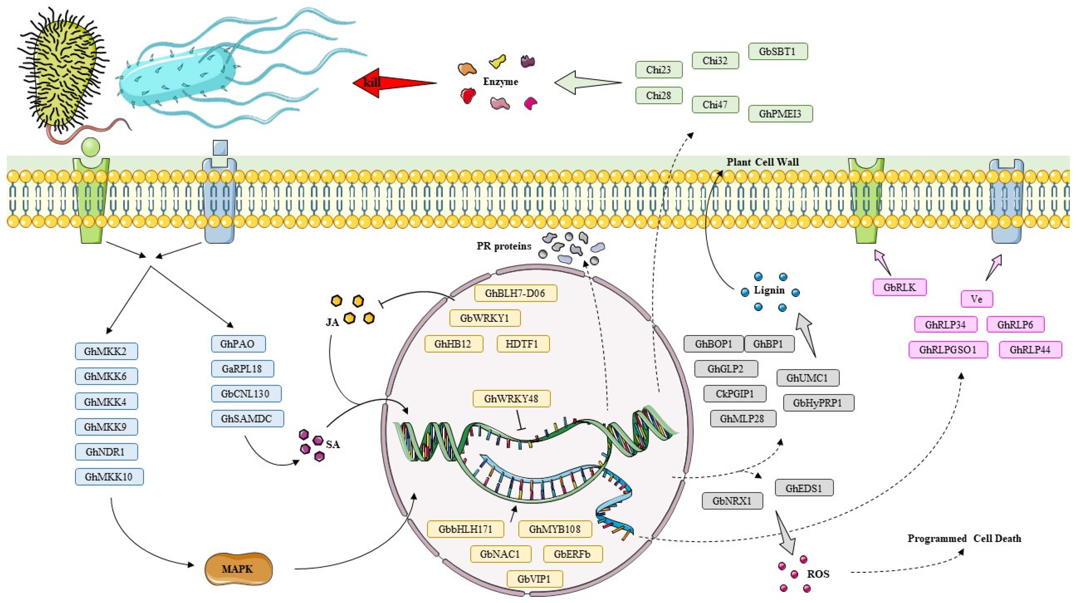
2. Molecular Mechanisms of Plant Resistance to Fusarium and Verticillium Wilt

Plants can use various strategies to counter invasion by Fusarium and Verticillium by activating complex cellular and molecular regulatory networks in vivo. This includes using pattern recognition receptors (PRRs) to induce immune responses, modifying cell walls to block pathogens, producing extracellular enzymes to directly degrade pathogens, or activating the expression of specific genes through various signaling pathways and transcription factors [16].
In recent years, considerable research has been carried out on the physiological and molecular mechanisms of cotton Fusarium wilt caused by FOV and Verticillium wilt caused by V. dahliae [2]. This review highlights the relatively recent discovery of genes involved in defenses against Fusarium and Verticillium and classifies them according to whether they act in signaling cascades and transcriptional regulation or are directly involved in cell wall and protein defense (Figure 1). This provides a theoretical basis to better understand the molecular genetic mechanisms of plant resistance to Fusarium and Verticillium wilt and provides a reference for future studies on genetic resistance mechanisms to fungal wilt.

2.1. Genes Involved in Signaling Cascades and Transcriptional Regulation

During the long-term evolution of plants, an immune system directly controlled by a series of cross-linked signal transduction pathways and various plant hormones has been developed to rapidly regulate gene expression, thereby adapting, resisting, and tolerating various biotic stresses. The reaction substrates of these intricate signaling networks are usually transcription factors (TFs). TFs can be regulated via multiple signals and can also regulate the expression of multiple genes related to disease resistance. They also play an important regulatory role in the process of plant stress signal transmission.
Within this intricate signaling network, the mitogen-activated protein kinase (MAPK) cascade response is the primary pathway for sorting and amplifying external signals into intracellular signals, including three kinases—MAPK kinase kinases (MAPKKKs), MAPK kinases (MKKs), and MAPKs—which are important in response to biotic and abiotic stresses [17,18]. Infection of cotton by FOV significantly induced GhMPK20 expression, whereas silencing GhMPK20 enhanced resistance to Fusarium cotton wilt. The WRKY family of TFs regulate various biotic stress responses and physiological processes. GhWRKY40 is a substrate of GhMPK20; FOV induces GhWRKY40 expression through the GhMKK4-GhMPK20 cascade reaction, whereas silencing of GhMKK4 and GhWRKY40 enhances resistance to Fusarium cotton wilt [19]. However, GhWRKY53 silencing activates the jasmonic acid (JA) signaling pathway and inhibits the salicylic acid (SA) signaling pathway, which reduces resistance to Verticillium cotton wilt [20]. Other members of the WRKY family are regulated by other hormones. GbWRKY1 is a negative regulator of the JA-mediated defense pathway involved in plant resistance to V. dahliae and mediates the transition from defense to development by activating JAZ1 expression during infection by V. dahliae [21]. GhWRKY48 expression in roots, stems, and leaves is induced by V. dahliae infestation, JA, and SA; whereas its silencing enhances resistance to Verticillium cotton wilt and may act as a negative regulatory transcription factor to repress the expression of downstream disease resistance genes, thereby affecting resistance to Verticillium cotton wilt [22]. MKK family members play a dual role in regulating resistance to fungal pathogens in cotton; GhMKK4, GhMKK6, and GhMKK9 positively regulate resistance to Verticillium wilt, whereas GhMKK10 negatively regulates resistance to Verticillium wilt [23]. MAPK scaffold protein (GhMORG1) interacts with GhMKK6 and GhMPK4, and its overexpression significantly enhances the activity of the GhMKK6-GhMPK4 cascade response and positively regulates resistance to Fusarium cotton wilt [24]. GhAP2C1 is a negative regulator of the MAPK cascade that interacts with GhMPK4 to negatively regulate the immune response to Fusarium cotton wilt [25]. Gene silencing experiments demonstrated that GhNDR1 and GhMKK2 are essential for cotton resistance to Verticillium wilt [26].
Glutathione transferases (GSTs) catalyze their binding to glutathione for various physiological functions, including disarming endogenous and exogenous toxins, participating in the translocation of flavonoids, and affecting disease resistance in plants [27]. GaGSTF9 overexpression positively regulates resistance to Verticillium wilt in Arabidopsis [28]. Similarly, GbGSTU7 positively regulates resistance to Fusarium wilt in Gossypium barbadense, whereas its silencing significantly reduces glutathione peroxidase activity in vivo and increases the incidence of Fusarium cotton wilt [29].
Expression of ribosomal protein GaRPL18 is induced by SA treatment, suggesting that GaRPL18 is associated with SA-mediated signaling pathways. Silencing GaRPL18 significantly reduces the number of immune molecules induced by the SA signaling pathway and reduces Verticillium wilt resistance in cotton, whereas GaRPL18 overexpression increases plant resistance to Verticillium wilt [30]. Expression of cotton cyclin-dependent kinase E (GhCDKE) is induced by Verticillium wilt infestation and methyl jasmonate (MeJA); GhCDKE silencing in cotton increases vulnerability to Verticillium wilt, whereas its overexpression in Arabidopsis enhances resistance [31]. The JA-induced calcium-dependent protein kinase GhCPK33 negatively regulates resistance to Verticillium cotton wilt, and knockdown of GhCPK33 enhances resistance [32]. The expression of GhIQM1 Ca2+-independent calmodulin-binding protein is induced by V. dahliae and SA, which may negatively regulate resistance to Verticillium cotton wilt by inhibiting the SA pathway [33]. GhBIN2 interacts with JAZ (jasmonate-ZIM domain) family proteins and phosphorylates JAZ proteins in response to infestation by V. dahliae. This may negatively regulate resistance to Verticillium cotton wilt by regulating the JA signaling pathway, since GhBIN2 overexpression in cotton increases sensitivity to Verticillium wilt [34].
Several stress factors induce the production of large amounts of reactive oxygen species (ROS) in plants, which act as direct toxic agents against pathogens, and may act as signaling molecules for defense; ROS levels are precisely regulated by the plant [35,36]. Ascorbic acid (AsA) is an important cellular antioxidant that oxidizes ROS and affects signaling pathways involving ROS, while oxidized inositol can be involved in AsA synthesis [37]. Inhibition of myo-inositol oxygenase GbMIOX5 causes H2O2 accumulation, and a reduction in AsA content in cotton cells significantly reduces resistance to Verticillium cotton wilt [38].
Lipids are essential plant cell components and can act as signaling molecules to regulate plant pathogen resistance [39]. Silencing of stearoyl-ACP desaturase GhSSI2 enhances resistance to Fusarium and Verticillium cotton wilt. Inhibition of GhSSI2 expression reduces oleic acid content, which adversely affects cell membrane function and activates the immune response, leading to increased SA content. SA triggers a hypersensitive response (HR) in plants, with rapid accumulation of ROS and induction of high levels of PR (pathogenesis-related) gene expression, which seals the pathogen in the tomb of dead cells [40]. Oleic acid is also a defense signal which, at low levels, can directly upregulate the expression of different disease resistance genes in a pathway independent of SA and JA, whereas activating the second messenger, NO (nitric oxide), upregulates a wide range of intranuclear disease resistance genes [41].
Knockout of the SA-related spermine (Spm) protein, Spm synthase (GhSPMS), and S-adenosylmethionine decarboxylase (GhSAMDC) genes increases the susceptibility to Verticillium cotton wilt. Conversely, transfection of Arabidopsis with these genes enhances its resistance to Verticillium wilt, indicating that the SA signaling pathway and Spm biosynthesis are associated with plant resistance to Verticillium wilt [42]. Expression of cotton polyamine oxidase (GhPAO) in Arabidopsis increases its resistance to Verticillium wilt and causes significant accumulation of H2O2, SA, and camalexin (a plant antitoxin), implying that GhPAO promotes plant resistance to Verticillium wilt by activating signaling pathways associated with Spm and camalexin [43].
The MYB (v-myb avian myeloblastosis viral oncogene homolog) transcription factor family is involved in resistance to pathogen infection, and knockout of GhMYB108 makes cotton more susceptible to Verticillium wilt, whereas GhMYB108 overexpression improves resistance to Verticillium wilt in Arabidopsis [44]. Silencing the homeodomain transcription factor (HDTF1) activates the JA signaling pathway and further improves Verticillium cotton wilt resistance [45]. Both JA and V. dahliae can induce the expression of HD-ZIP transcription factor GhHB12, with its overexpression reducing the resistance of cotton to V. dahliae [46]. GbbHLH171 interacts with, and is phosphorylated by, a defense-associated receptor-like kinase (GbSOBIR1) and positively regulates resistance to Verticillium cotton wilt [47].
Ethylene (ET) response factors (ERFs) are required for pathogen defense responses, and genes from different hormone signaling pathways all play important roles in response to infection by fungal pathogens. GbABR1 of the apetala 2 (AP2) family positively regulates plant resistance to Verticillium wilt, whereas its silencing increases disease rates in cotton [48]. GbERFb is an AP2/ERF type TF which increases disease resistance in cotton [49]. GbERF1-like ERFs enhance plant resistance to Verticillium wilt by positively regulating lignin synthesis [50]. V. dahliae and phytohormones (SA, JA, and ET) induce the expression of the nucleotide binding site–leucine-rich repeat (NBS-LRR) gene GbaNA1 in cotton, which is involved in its response to Verticillium wilt [51]; GbaNA1 overexpression increases ROS levels in Arabidopsis and promotes the expression of genes related to the ET signaling pathway [52]. The CC-NBS-LRR gene GbCNL130 activates the SA-mediated signaling pathway, increases ROS accumulation, and promotes the expression of downstream PR genes, thereby enhancing resistance to Verticillium cotton wilt [53]. In addition, knockdown of positively regulated genes GhJAZ10, GhbHLH18 (JA), GhPUB17 (SA), and GhEBF1 (ET) significantly increases susceptibility to Verticillium wilt in resistant varieties, whereas silencing of negatively regulated genes GhTGA7 (SA) and GhBZR1 (Brassinolide, BR) significantly increases resistance to Verticillium wilt in susceptible varieties [54].
The bZIP transcription factor GbVIP1 (VirE2 interacting protein 1) is induced by V. dahliae and ET, and its silencing reduces Verticillium cotton wilt resistance, whereas its ectopic expression in tobacco enhances resistance to Verticillium wilt in tobacco by positively regulating the expression of PR1, PR1-like, and HSP70 genes [55]. JA induces the expression of BEL1-like transcription factor GhBLH7-D06 in vascular tissues, acting on secondary cell formation and negatively regulating resistance to Verticillium cotton wilt [56]. In addition, knockdown of transcription factor GbNAC1 reduces resistance to Verticillium cotton wilt, whereas its overexpression increases Verticillium wilt resistance in Arabidopsis [57].

2.2. Genes Directly Involved in Cell Wall and Protein Defense

Plant resistance to fungal pathogens is largely influenced by defense-related proteins. Plants have evolved sophisticated sensory mechanisms to detect microbial invasion and overcome the negative effects caused by microbes in terms of growth, yield, and survival [58].
V. dahliae invades host plants mainly through plant cell wall degradation; therefore, the cell wall is the first barrier for plants that helps prevent pathogen entry, often effectively stopping pathogen invasion [59,60]. Pectin is one of the main components of the plant cell wall, and its structure and function are closely related to the degree of esterification. Pectin methylesterases (PMEs) protect plant cell walls by catalyzing the dimethyl esterification of pectin polygalacturonate. GhPMEI3 regulates resistance to V. dahliae in cotton since its silencing increases susceptibility to V. dahliae infestation, while ectopic expression of GhPMEI3 increases pectin methyl esterification and limits fungal infection by regulating root growth [61]. Knockout of pectin lyase genes VdPL3.1 and VdPL3.3 reduces pathogen virulence to cotton [60]. Cell wall polysaccharides are mainly modified by acetylation; the level and location of this modification directly affects the structure, physicochemical properties, and ability of the cell wall to resist pathogens [62]. Acetyltransferase GhTBL34 (trichome birefringence-like) expression is induced by V. dahliae, ET, SA, and JA and mediates cell wall polysaccharide acetylation to block pathogen invasion and regulate Verticillium wilt resistance [63,64]. Resistance of cotton to wild-type fungal diseases is largely dependent on lignin content, and its levels can be used as an indicator of resistance against Fusarium wilt [65]. Transcriptome analysis suggests that caffeic acid 3-O-methyltransferase (COMT) and peroxidase 2 (POD2) may be involved in the synthesis and accumulation of lignin in response to Fusarium cotton wilt [66]. Patatin-like proteins (PLPs) are defense proteins with non-specific lipid acyl hydrolytic activity that hydrolyze cell membranes into fatty acids and lysophospholipids. GhPLP2, which is induced by FOV, V. dahliae, ET, and JA, plays an important role in reducing fungal pathogenicity and regulating plant resistance to Verticillium wilt [67]. Overexpression of polygalacturonase inhibitor proteins CkPGIP1 and GhPGIP1 from Cynanchum komarovii and G. hirsutum enhances cotton resistance to Verticillium wilt by promoting xylem lignification in cotton [68]. The apoplastic thioredoxin protein GbNRX1 enhances the immune response of cotton to V. dahliae; GbNRX1 silencing causes a large accumulation of ROS and increases susceptibility to V. dahliae infection [69].
BLADE-ON-PETIOLE (BOP) 1 and BOP2 are two BTB-ankyrin proteins specifically expressed at lateral-organ boundaries (LOBs). GhBOP1 acts synergistically with GhBP1 to co-regulate lignin biosynthesis and enhance resistance to Verticillium cotton wilt [70]. GbHyPRP1 encodes a proline-rich protein with a POLE1 structural domain that negatively regulates resistance to Verticillium cotton wilt. Expression of GbHyPRP1 is inhibited by the SA signaling pathway when cotton is infected with V. dahliae, and GbHyPRP1 silencing enhances resistance to V. dahliae by thickening the cell wall and accumulating ROS; in contrast, HyPRP1 overexpression reduces resistance to Verticillium wilt in Arabidopsis [71].
Latex protein GhMLP28 is induced by V. dahliae, JA, SA, or ET to produce and regulate plant disease resistance [2]. GhDIR1 overexpression increases cotton lignin content and enhances resistance to Verticillium cotton wilt [72]. Defense regulator EDS1 encodes a lipase-like protein that is induced by SA and regulates plant resistance to Verticillium wilt. GbEDS1 overexpression promotes the production of SA and H2O2 and enhances Arabidopsis resistance to Verticillium wilt, whereas GbEDS1 silencing reduces cotton resistance to Verticillium wilt [73].
Papain-like cysteine proteases (PLCPs) play an important role in plant defense against pathogen invasion. GhRD21-7 substantially promotes the resistance to Verticillium cotton wilt and its overexpression further enhances this resistance [74]. Germin-like proteins (GLPs) are a family of glycoproteins involved in plant resistance to various stresses. GhGLP2 silencing increases susceptibility to FOV and V. dahliae infection in cotton, whereas GhGLP2 overexpression attenuates mycelial growth of these fungi, increases callus formation at leaf infection sites, enhances cell wall lignification, and improves resistance to Fusarium and Verticillium wilt in Arabidopsis [75]. Thaumatin-like proteins (TLPs) are a family of multi-member defense-related proteins; GhTLP19 silencing increases malondialdehyde levels and decreases catalase levels in cotton, which increases its susceptibility to Verticillium wilt [76].
Extracellular enzymes help plants to resist fungal pathogen infection. For example, chitinase (Chi) hydrolyzes chitin (a main component of the fungal cell wall) into N-ethoxylated oligosaccharides and glucose to inhibit spore germination and mycelial growth [77]. There are low levels of Chi (with low activity) in all higher plant organs; however, its levels rapidly increase following pathogen infestation [78]. Chi23, Chi32, or Chi47 positively regulate Verticillium wilt resistance; their knockout reduces resistance to Verticillium cotton wilt [77]. Cotton infestation by V. dahliae triggers the secretion of Chi28 and cysteine protein CRR1 in order to degrade the fungal cell wall and protect Chi28 from degradation by serine protease secreted by V. dahlia, respectively. Thus, silencing either Chi28 or CRR1 reduces resistance to Verticillium cotton wilt, whereas CRR1 overexpression enhances its resistance [79]. Plant lysine motif-containing (LysM) proteins recognize chitin in the cell wall of fungal pathogens and initiate the corresponding defense response. Silencing Lyp1, Lyk7, and LysMe3 significantly reduces the production of SA, JA, and ROS, decreases the activity of defense-related genes, and ultimately reduces resistance to Verticillium cotton wilt [80].
Verticillium highly induces laccase GhLAC15 expression, and the cell walls of overexpressing plants enhance resistance to Verticillium wilt through a defense-mediated response that enhances lignification and increases arabinose and xylose accumulation [81]. GhUMC1, a divalent copper-binding protein, is an umecyanin-like gene involved in resistance to Verticillium cotton wilt through regulation of the JA signaling pathway and lignin metabolism [82]. Walls are thin (WAT) genes regulate SA metabolism and signaling by affecting polar transport of growth hormones to further enhance plant resistance to a variety of pathogens [83]. Knockout of GhWATs increases SA accumulation, activates the expression of SA pathway-related genes, and increases lignin accumulation in the xylem, each of which promotes plant resistance to Verticillium wilt [84]. Simultaneous silencing of GhWAT1, GhWAT2, and GhWAT3 increases lignin accumulation in the xylem and enhances plant resistance to Verticillium wilt but inhibits plant growth [84]. Knockdown of the Gh4CL30 lignin synthesis gene in cotton decreases flavonoid, lignin, and butyryl content but increases guaiacyl, caffeic acid, and ferulic acid content, which provides a new idea for resistance to Verticillium cotton wilt [85].
Enoyl-CoA reductase (GhECR) is involved in the formation of long-chain fatty acids, and its silencing increases the susceptibility of cotton to FOV and V. dahliae infection [86]. The subtilase-like protein GbSBT1 is highly induced following infection with V. dahliae, or by JA and ET treatment. The protein acts at the cell membrane and regulates plant resistance to Fusarium and Verticillium wilt [87].
Plants infested with pathogens over a long period of time have evolved various innate immune mechanisms [88]. In general, plants employ two sets of defense strategies: microbial (or pathogen)-associated molecular pattern (MAMP/PAMP)-triggered immunity (MTI/PTI) and effector-triggered immunity (ETI) [89]. Some pathogenic bacteria disrupt plant tissue structure and produce damage-associated molecular patterns (DAMPs) such as cell wall fragments, which induce innate immunity similar to that of MAMPs [90].
Activation of plant innate immunity requires receptor recognition of pathogens. MTI/PTI is triggered by PRRs located across the cell surface, whereas ETI is caused by NBS-LRR receptor proteins located intracellularly [91]. PRRs include receptor-like proteins (RLPs) and receptor-like kinases (RLKs) that are located on plant cell membranes and which are responsible for detecting molecular markers of invading pathogens or damaged plant tissues and activating defense responses, thereby preventing infection before pathogen invasion of the plant cell [92,93]. The GhRLPGSO1-like gene, GhRLP44, GhRLP6, and GhRLP34 may play a role in resistance to Fusarium cotton wilt [94]. Verticillium wilt infestation of disease-resistant G. barbadense cotton induces GbRLK; transfection of cotton and Arabidopsis with this gene increases resistance to Verticillium wilt [95]. The TIR-NBS-LRR protein encoded by CG02 is likely to be a key gene for resistance to Verticillium cotton wilt since it specifically promotes the expression of disease resistance genes, while silencing this gene increases its susceptibility to Verticillium wilt [96].
Invasion of cotton cells by FOV causes the release of large amounts of intracellular Ca2+, which acts as a second messenger to initiate the expression of various downstream disease resistance genes [97]. GhGLR4.8 encoding glutamate-like receptor (GLR) protein plays an active role in resistance to Fusarium cotton wilt and it is hypothesized that it may be the ion channel required for Ca2+ to flood into the cell and enhance the disease resistance response [98].
In addition, the Ve R-gene enhances plant resistance to Verticillium wilt by encoding RLPs with LRR structural domains [99]. The transcription products of GhlncNAT-ANX2 and GhlncNAT-RLP7 are long non-coding RNAs whose silencing enhances resistance to Verticillium cotton wilt, possibly by promoting lipoxygenase 1 and lipoxygenase 2 expression [100]. GbAt11 (AXMN toxin-induced protein-11) is highly resistant to Verticillium wilt, and its overexpression stimulates the expression of disease resistance genes, including FLS2 and BAK1, to increase resistance [101]. GhPUB17 is a U-box ubiquitin ligase E3 that can be inhibited by antifungal protein GhCyP3 to negatively regulate Verticillium cotton wilt resistance [102]. GbANS is involved in the biosynthesis of anthocyanins in cotton and its silencing significantly reduces anthocyanin production and resistance to Verticillium cotton wilt [103]. Similarly, GbCAD1 is involved in cotton phenol synthesis, and its silencing also reduces resistance to Verticillium cotton wilt [104].
Taken together, these findings suggest that defense genes induced during pathogen attack provide potent protection to the plant by their respective defense mechanisms (Figure 2). However, more studies are required to understand the mechanisms of action of these genes.

3. Mechanism of Human Resistance to Fusarium and Its Pathogenic Features

Fusarium is a naturally widespread, highly adaptable saprophytic fungus that can survive in a wide range of extreme environments [14]. Some Fusarium species are non-pathogenic, while others are extremely harmful. Common pathogenic Fusarium species include F. moniliforme, F. solani, and FOV [105]. They infect a wide range of crops (cotton, bananas, aubergines, etc.), cause the worldwide spread of many plant diseases, and infect animals such as fish and shrimps [106]. They may even cause skin, corneal, nail, and systemic damage in humans [107].

3.1. Fungal Toxins

Fungi are often found in various types of grain in fields and the grain stored in warehouses. This is accompanied by the production of mycotoxins which affect plant regeneration, cause programmed cell death, and lead to disease in humans and animals following accidental consumption of grain contaminated with mycotoxins or through fungi-contaminated grain coming into contact with wounds or eyes [108].
The gastrointestinal tract is a barrier to harmful substances such as pathogens, toxins, and exogenous antigens. It is a habitat for intestinal flora that synergistically protects the host from fungi and plays a key role in regulating the body’s immune system [109,110]. Mycotoxins that enter the body through food initially interact with the gastrointestinal tract, break through its defenses, disrupt its homeostasis, and alter its histomorphology, nutrient absorption, and barrier function, which results in intestinal dysfunction and local immune compromise and may eventually lead to serious illnesses such as systemic chronic mycotoxicosis [110,111]. Mycotoxins can also affect the susceptibility of animals to infectious diseases, such as bacteria, viruses, and parasites, by affecting the innate and acquired immune systems of healthy animals. Thus, exposure of animals to mycotoxins increases their susceptibility to these infectious diseases [112].
Trichothecenes are mainly produced by F. graminearum and include two major toxins: T-2 toxin and deoxynivalenol (DON). Their oral ingestion causes toxicity in animals and humans, results in a variety of symptoms (such as vomiting and diarrhea), and may poison white blood cells and affect the host’s immune system [113,114,115]. The toxicity of T-2 and DON is generally mediated by oxidative stress-mediated DNA damage and apoptosis, although they can also bind to the large subunit of the ribosome and interfere with its peptidyl transferase activity to inhibit protein synthesis [116,117].
Fumonisins are mainly produced by F. verticillioides; fumonisin B1 (FB1) is the most abundant type found in nature [118]. Epidemiological studies have shown that esophageal cancer in humans and liver cancer in animals are associated with the ingestion of foods containing fumonisins [119,120]. FB1 has a similar structure to that of sphingolipids and can affect sphingolipid metabolism by interfering with ceramide synthase, leading to increased intracellular sphingomyelin levels that ultimately lead to oxidative stress and apoptosis [121,122]. In addition, FB1 may weaken the intestinal barrier by affecting the synthesis of related proteins and lead to immune dysfunction [123,124].
Zearalenone (ZEA) is a fungal toxin mainly produced in grain by F. graminearum and F. culmorum which may act synergistically with FBs and DON. It is structurally similar to estrogen; therefore, ZEA competes with natural estrogens for estrogen receptor binding and has a major impact on the reproductive system [125]. In addition, ZEA can downregulate tumor suppressor genes in intestinal cells, which can cause cancer [126].

3.2. Fusariosis—Fungal Keratitis

Fusariosis is an infectious disease of the skin, eyes, and internal organs mainly caused by Fusarium infections [15]. Fusarium is an opportunistic pathogen that does not normally cause disease; however, at times it causes local infections in immunocompetent individuals and invasive infections when the body’s immunity is compromised or when there is trauma. Therefore, neutrophils and macrophages play an important role in the immune defense against Fusarium, which infects immunocompromised patients in two main ways: (1) through trauma or foreign bodies that cause infection of the patient’s soft tissues or mucous membranes, resulting in keratitis and nail infection, among others [127]; and (2) through rhinitis and pneumonia caused by inhalation or ingestion of Fusarium and its toxins [105].
Fungal keratitis is the most common type of fusariosis in normal individuals and it has a significant medical and socioeconomic impact [128,129]. Most patients have an infected corneal stroma due to trauma related to agricultural work, particularly in hot and humid areas that lead to an increase in the growth rate of Fusarium and mycotoxin levels [130,131]. The next section presents the pathogenic mechanisms of Fusarium and the host defense response using the example of fungal keratitis to help advance the prevention and control of fungal keratitis.
The severity of a fungal infection is determined by the strength of the pathogen and the immunity of the host [129]. The innate immune response against fungal keratitis involves detection of the fungus by macrophages residing in the cornea followed by chemokine secretion by the macrophages, which recruits neutrophils to the infected corneal stroma [132]. The neutrophils act as killer cells and remove the invading fungus. However, insufficient levels of chemokines restrict the number of neutrophils infiltrating the corneal stroma, resulting in a failure to clear the fungus in time for the early stages of infection.
Recognition of fungal invasion by immune cells through the C-type lectin receptor (CLR) and toll-like receptor (TLR) is the first step in the defense response [133]. The CLR and TLR of macrophages recognize intrinsic components of the fungal cell wall, while neutrophils use different oxidative and non-oxidative mechanisms to inhibit mycelial growth and regulate the host immune response to the fungus [134,135]. However, Fusarium also expresses a variety of proteins and small molecule virulence factors that inhibit the host’s defense response. For example, conidia secrete the hydrophobic protein RodA, which provides a protective layer for Fusarium and prevents recognition by the receptor, thus delaying initiation of the immune response and neutrophil recruitment and allowing the fungus to survive and cause disease [136].
Neutrophils can destroy fungi by producing ROS such as O2−, H2O2, and HClO through the NADPH oxidase (NOX) system [137]. Neutrophils in mice with mutations in the NOX subunit have impaired ability to produce ROS, leading to a marked increase in mycelial growth in the cornea and more severe disease progression [138]. Fungi produce antioxidant factors including thioredoxin and superoxide dismutase (regulated by the Yap1 transcription factor) to counteract neutrophil ROS production. Although most fungal secondary metabolites and catalases have antioxidant activity, they do not enhance fungal resistance to ROS [138]. Protein phosphatase Z (Ppz) also plays an important role in oxidative stress tolerance in filamentous fungi [139].
Neutrophils can use various non-oxidative mechanisms to limit microbial growth, such as the production of proteins that compete with microbes for metal ions necessary for growth (a process also known as nutritional immunity) [140]. Iron is essential for thioredoxin production by fungi; neutrophils secrete iron-binding proteins, such as transferrin and lactoferrin, to compete with fungi for iron ions. Zinc is also essential for mycelial growth since it is an important component of many enzymes and TFs, and neutrophils secrete calprotectin, which binds zinc and manganese to compete with fungi [141]. Neutrophils more efficiently kill Fusarium species with mutated ion transport carriers, and the growth of these mutant fungi on the cornea itself is inhibited [142]. New mobile, germline-specific chromosomes were identified in FOV that can transmit pathogenicity by transfer in plants. These germline-specific chromosomes have numerous genes that encode metal ion transport proteins that may play an important role in the evasion of nutritional immunity by Fusarium; these genes are also present in cancer patients with systemic Fusarium infections and in those with fungal keratitis [143,144].
Macrophages and neutrophils can secrete acidic mammalian chitinase (AMCase) to inhibit fungal growth [145,146]. In vitro experiments showed that neutrophils lacking AMCase are less potent against fungi, while fungi survival is higher in mice lacking AMCase [147]. Neutrophils are an important source of cytokines during infection, producing large amounts of pro-inflammatory and chemotactic cytokines. The expression of IL-1α and IL-1β cytokines is significantly higher in diseased corneas compared to healthy corneas [129,148,149].
Neutrophils are bactericidal in two main ways: direct phagocytosis of pathogens or by the release of a specific structure, namely neutrophil extracellular traps (NETs) consisting of depolymerized chromatin and intracellular granule proteins that cause death to the outside of the cell. This process involves the generation of ROS and the depolymerization of chromatin by neutrophil elastase (NE) to trap and kill pathogenic bacteria [150]. In addition, peptidyl arginine deiminase 4 (PAD4) guanylates histones, which changes the chromatin charge and promotes chromatin depolymerization and nucleus expansion [151]. NETosis is induced by fungi such as Fusarium, leading to the release of histones, calprotectin, and other particles. Therefore, NETs kill pathogenic fungi and can also cause damage to their own tissues [152].

4. Comparison of Pathogenic Mechanisms against Fusarium in Cotton and Humans

Plants and humans possess conserved innate immune systems to respond to microbial invasion (Table 1). They use specialized receptors to sense PAMPs and endogenous DAMPs, which then activates the appropriate defense response. The immune response includes transcriptional reprogramming, production of antimicrobial substances, programmed cell death of infected cells, and the release of soluble factors (including cytokines and phytohormones) that produce a signal from the site of infection to alert the host of the arrival of danger [153].
In vertebrates, membrane-bound immune receptors belonging to the TLR family are located on the cell surface or in endosomal compartments, while the CLR is located on the cell surface. Plant RLKs and RLPs are mainly located on the plasma membrane, with RLPs being structurally similar to TLRs in animal cells [154]. Both plants and vertebrates use membrane PRRs to recognize PAMPs/DAMPs to then trigger activation of transcriptional programs that ultimately produce antimicrobial molecules and enable host adaptive responses to resist pathogen attack.
Activating programmed cell death at the site of pathogen infection is a strategy shared by plants and humans that protects the organism from further invasion by pathogens through directly disrupting the environment in which they live. Cell death also releases intracellular components that can activate or enhance antimicrobial responses and signal alarms to neighboring cells. Apoptosis, pyroptosis, and necroptosis limit pathogen development in humans [155]. Recognition of invading microorganisms by plants also triggers an HR which prevents their spread in healthy tissues by limiting the uptake of plant metabolites by living trophic pathogens [156]. The characteristics of HR-associated cell death are remarkably similar to programmed cell death in humans, and leakage of cell contents can constitute an alarm signal to neighboring cells and prepare them to respond to infection, ensuring a comprehensive and effective defense response [157,158]. In addition, humans and plants secrete chitinase and ROS to inhibit fungal growth [35,77,129,137].
Plants and humans have innate immune memory and immune memory, which enables rapid mobilization of large amounts of resources for the synthesis of disease-resistant proteins that protect them from injury when reinfested by pathogenic bacteria. However, this depends on the plant life cycle, with long-lived perennials exhibiting better immune memory than annuals [159,160].
The biggest difference between animals and plants in the defense against Fusarium infestation is that animals have an innate immune system, which is triggered by allergenic proteins such as Fusarium’s beta-1,3 glucan and enzymes released during budding. Furthermore, allergen proteins are recognized by phagocytes, neutrophils, and dendritic cells of the host through receptors such as CLR, thereby initiating the allergic process [161,162]. In addition, these receptors activate intracellular signaling pathways that lead to inflammatory responses [163]. Macrophages and neutrophils also inhibit Fusarium infection through the presence of interferon-gamma, chemokines, tumor necrosis factor-alpha (TNF-α), granulocyte colony-stimulating factor (G-CSF), granulocyte-macrophage colony-stimulating factor (GM-CSF), and various interleukins (IL-1β, IL-6, IL-15, IL-23) [13,164]. After the innate immune cells are activated, antigen-presenting cells such as macrophages can engulf pathogens and produce antigens, which are then presented to two types of T cells. Helper T cells mainly help B cells to generate antibody-labeled antigens, whereas cytotoxic T cells directly kill pathogen-infected somatic cells [165]. This is centered on neutrophils and is key to an animal’s defense against Fusarium. The prognosis for invasive fusariosis in patients with neutropenia or organ transplantation is poor and up to 100% lethal in the absence of recovery of neutrophil function [166]. At the onset of inflammation, killer cells such as neutrophils are attracted by chemotactic substances and rapidly accumulate through blood vessels to the site of disease to surround and destroy the invading Fusarium in a single locality and prevent its spread in the body in immunocompetent individuals.
Unlike animals, plants have not evolved motile immune cells or an adaptive immune system [167]; they also lack a well-developed cellular circulation system similar to that of blood vessels and lymphatic vessels that can produce a direct immune response in the bloodstream. However, every cell in a plant has the ability to sense and resist pathogens [89]. After sensing pathogen signals, plants activate or inhibit TFs through hormone signals such as SA, JA, and ET, thereby affecting the expression of downstream PR genes, the thickening of cell walls, and the production of phytoalexins [168]. Initial animal defenses against pathogens mainly involve the mucous membranes and cuticles. Plant structures also provide physical defense strategies against pathogenic bacteria, such as epicuticular waxes (EWs) and the closure of stomata in many land plants [169,170].
In animals, immune cells such as effector T cells are directed to move flexibly and specifically throughout the body through endogenous cellular factors, such as chemokines or exogenous cellular signals generated by the inflammatory microenvironment, so that resistance is acquired throughout the body [171]. In contrast, plants send a systemic signal at the site of pathogen infestation, such as methyl salicylate (MeSA) which transmits the pathogen signal from the infected site to healthy tissues distal to the plant via intercellular filaments or phloem [172]. This allows the whole plant to acquire resistance to the disease; this process is also known as systemic acquired resistance (SAR) [173]. Systemic resistance in plants is temporarily built up to keep out soon-to-be invading microbes [174]. Conversely, vertebrates have evolved an adaptive immune system with specialized immune cells dedicated to producing a stronger and more specific immune response while ensuring long-term protection of the organism (Figure 3).
Furthermore, in addition to their highly complex adaptive immune system, humans are warm-blooded. Higher body temperature makes the body resistant to most invasive fungal infections, and humans can further raise their body temperature during immune defense; thus, only a few fungi that can grow at or above 37°C can infect humans [175].

5. Conclusions and Outlook

There is no cure for Fusarium and Verticillium cotton wilt caused by FOV and V. dahliae. Currently, chemical control remains the most efficient way to combat crop diseases, and the application of emerging disease resistance breeding is the most environmentally safe control method. This review summarized the disease resistance and susceptibility genes in various types of cotton and suggested that it is possible to selectively combine or knockdown important disease-related genes to increase disease resistance in cotton [176]. Moreover, these disease-resistant genes can be used in patients undergoing reconstitution of immune function. So far, the treatment of human fusariosis mainly consists of surgical excision of the infected site, systemic antifungal drugs, and reconstitution of immune function in immunocompromised hosts [106,166,177]; if we can find a way to use anti-disease proteins from cotton (such as extracellular enzymes) to assist in the treatment of patients, while avoiding side effects, it may improve the survival rates in critically ill patients and provide a new strategy for the prevention and control of fungal diseases in humans. Knowledge of Fusarium infection in humans can also be applied to cotton. Even among agricultural workers, Fusarium infection mostly occurs in the cornea, as the rest of the body is protected by clothing. A major reason for plant susceptibility to Fusarium wilt is that FOV inhabits the soil, which allows it to invade the plant through its root system. Therefore, the application of techniques such as soilless cultivation will isolate plants from the natural environment of FOV, thereby protecting the plant’s vulnerable roots and greatly reducing the chances of FOV infestation.
However, the extremely complex pathogenesis of Fusarium and Verticillium cotton wilt, as well as the large breeding effort, long lead time, development of fungal resistance, and increasing national attention to pesticide residues and food safety issues, has slowed the development of disease resistance breeding and chemical control. Therefore, new ideas are required to prevent fungal diseases. Similar to the way humans and animals are vaccinated against disease, plants can also use non-pathogenic Fusarium to effectively control Fusarium disease. Some non-pathogenic Fusarium directly inhibit the growth, invasion, and colonization of pathogens by competing for nutrients in the soil, infestation sites on the surface of plant roots, and colonization sites within plant tissues [178,179,180]. Other non-pathogenic Fusarium can activate the plant’s defense response by colonizing the plant and allowing it to acquire induced systemic resistance, thus indirectly antagonizing the pathogenic Fusarium and reducing the damage caused by it [181]. Furthermore, there are viruses among plant pathogenic fungi that can cause fungal virulence decline. These fungal viruses can regulate the expression of the host enzymes and TFs, inhibit the synthesis of the host cytoplasmic membrane, and impair the host’s transport system [182,183,184]; this affects the host’s physiological state and potentially prevents and controls plant fungal diseases. Many fungal viruses that can inhibit Fusarium growth and mycotoxin production have been found [185,186,187], and they are expected to be used in the prevention and control of Fusarium in the future.
Although Fusarium is a vicious pathogen, it can produce many beneficial products. L-tryptophan can induce marine Fusarium sp. L1 to produce indole alkaloids with anti-Zika virus activity [188]. The fermentation of Fusarium sp. DCJ-A produces cyclic hexapeptide compounds that exhibit cytotoxic activity against five human tumor cells [189]. Xylanase produced by F. graminearum FGSG_03624 activates the immune response of plants and enhances their resistance to bacterial and fungal pathogens [190]. In addition, F. graminearum Ec220 produces a xylanase with low cellulase content when grown on wheat bran [191], which has good commercial prospects. FOV PR-33 produces fusarinolic acid and its isomers, dehydrofusaric acid and fusaric acid, which are resistant to pathogenic bacteria and yeasts; these metabolites are potential antibacterial drugs [192]. Bananas are highly susceptible to Fusarium infection [193]; therefore, they could be considered for Fusarium cultivation for the production of compounds with industrial applications. This would provide theoretical and technological support in the fields of plant disease resistance and human medicine.

Author Contributions

Conceptualization, M.R. and Y.X.; writing—original draft preparation, M.M. and Y.Z. writing—review and editing, L.L. (Lulu Liu), L.L. (Lei Luo), X.H., L.Q. and F.L.; funding acquisition, F.L., M.R. and Y.X. All authors have read and agreed to the published version of the manuscript.

Funding

This work was supported by grants from the National Natural Science Foundation of China (No. U1804231 to F.L., 31972469 to M.R.) and the China Postdoctoral Science Foundation (no. 2020M672286 to Y.X.).

Institutional Review Board Statement

Not applicable.

Informed Consent Statement

Not applicable.

Acknowledgments

The authors thank the teachers and students in our research team for their help and support.

Conflicts of Interest

The authors declare no conflict of interest.

References

  1. Sunilkumar, G.; Campbell, L.M.; Puckhaber, L.; Stipanovic, R.D.; Rathore, K.S. Engineering cottonseed for use in human nutrition by tissue-specific reduction of toxic gossypol. Proc. Natl. Acad. Sci. USA 2006, 103, 18054–18059. [Google Scholar] [CrossRef] [PubMed] [Green Version]
  2. Billah, M.; Li, F.; Yang, Z. Regulatory Network of Cotton Genes in Response to Salt, Drought and Wilt Diseases (Verticillium and Fusarium): Progress and Perspective. Front. Plant Sci. 2021, 12, 759245. [Google Scholar] [CrossRef] [PubMed]
  3. Rathore, K.S.; Campbell, L.M.; Sherwood, S.; Nunes, E. Cotton (Gossypium hirsutum L.). Methods Mol. Biol. 2015, 1224, 11–23. [Google Scholar] [CrossRef] [PubMed]
  4. Abdelraheem, A.; Esmaeili, N.; O’Connell, M.; Zhang, J. Progress and perspective on drought and salt stress tolerance in cotton. Ind. Crops Prod. 2019, 130, 118–129. [Google Scholar] [CrossRef]
  5. Khanale, V.; Bhattacharya, A.; Satpute, R.; Char, B. Version 4.3-12/08/20 Cotton bZIP Transcription Factors: Characterization of the bZIP Family from Gossypium hirsutum, Gossypium arboreum and Gossypium raimondii. 2020; under review. 1–21. [Google Scholar]
  6. Davis, R.M.; Colyer, P.D.; Rothrock, C.S.; Kochman, J.K. Fusarium Wilt of Cotton: Population Diversity and Implications for Management. Plant Dis. 2006, 90, 692–703. [Google Scholar] [CrossRef] [PubMed] [Green Version]
  7. Cun, M.; Guiliang, J.; Chuanlin, Z. The advances in cotton breeding resistance to Fusarium and Verticillium wilts in China during past fifty years. Zhongguo Nongye Kexue 2002, 35, 508–513. [Google Scholar]
  8. Li, X.; Zhang, Y.; Ding, C.; Xu, W.; Wang, X. Temporal patterns of cotton Fusarium and Verticillium wilt in Jiangsu coastal areas of China. Sci. Rep. 2017, 7, 12581. [Google Scholar] [CrossRef] [PubMed] [Green Version]
  9. Qiying, C.; Xiuqin, J.; Wenji, S. Identification of races of cotton Fusarium wilt in China. Sci. Agric. Sin. 1985, 18, 1–6. [Google Scholar]
  10. Hu, X.-P.; Gurung, S.; Short, D.P.; Sandoya, G.V.; Shang, W.-J.; Hayes, R.J.; Davis, R.M.; Subbarao, K.V. Nondefoliating and defoliating strains from cotton correlate with races 1 and 2 of Verticillium dahliae. Plant Dis. 2015, 99, 1713–1720. [Google Scholar] [CrossRef] [PubMed] [Green Version]
  11. Dickman, M.B.; Figueiredo, P. Comparative pathobiology of fungal pathogens of plants and animals. PLoS Pathog. 2011, 7, e1002324. [Google Scholar] [CrossRef]
  12. Sharon, A.; Shlezinger, N. Fungi infecting plants and animals: Killers, non-killers, and cell death. PLoS Pathog. 2013, 9, e1003517. [Google Scholar] [CrossRef] [PubMed] [Green Version]
  13. Nucci, M.; Anaissie, E. Fusarium infections in immunocompromised patients. Clin. Microbiol. Rev. 2007, 20, 695–704. [Google Scholar] [CrossRef] [PubMed]
  14. Nucci, M.; Marr, K.A.; Queiroz-Telles, F.; Martins, C.A.; Trabasso, P.; Costa, S.; Voltarelli, J.C.; Colombo, A.L.; Imhof, A.; Pasquini, R.; et al. Fusarium infection in hematopoietic stem cell transplant recipients. Clin. Infect. Dis. 2004, 38, 1237–1242. [Google Scholar] [CrossRef] [Green Version]
  15. Ran, Y.P.; Wang, X.X.; Dai, Y.L.; Peng, X.M.; Zhang, J.; Zhou, G.P. Chronic leg ulcer caused by Fusarium sonali: A case report and super-microstructure investigation. Chin. J. Mycol. 2006, 1, 197–199+210. [Google Scholar]
  16. Song, R.; Li, J.; Xie, C.; Jian, W.; Yang, X. An Overview of the Molecular Genetics of Plant Resistance to the Verticillium Wilt Pathogen Verticillium dahliae. Int. J. Mol. Sci. 2020, 21, 1120. [Google Scholar] [CrossRef] [PubMed] [Green Version]
  17. Meng, X.; Zhang, S. MAPK cascades in plant disease resistance signaling. Annu. Rev. Phytopathol. 2013, 51, 245–266. [Google Scholar] [CrossRef]
  18. Shi, J.; Zhang, L.; An, H.; Wu, C.; Guo, X. GhMPK16, a novel stress-responsive group D MAPK gene from cotton, is involved in disease resistance and drought sensitivity. BMC Mol. Biol. 2011, 12, 22. [Google Scholar] [CrossRef] [Green Version]
  19. Wang, C.; He, X.; Li, Y.; Wang, L.; Guo, X.; Guo, X. The cotton MAPK kinase GhMPK20 negatively regulates resistance to Fusarium oxysporum by mediating the MKK4-MPK20-WRKY40 cascade. Mol. Plant Pathol. 2018, 19, 1624–1638. [Google Scholar] [CrossRef] [Green Version]
  20. Zhang, X.L. Function Analysis of GhWRKY53 in Gossypium hirsutum Resistance to Verticillium Wilt. Master’s Thesis, Shihezi University, Shihezi, China, 2021. [Google Scholar]
  21. Li, C.; He, X.; Luo, X.; Xu, L.; Liu, L.; Min, L.; Jin, L.; Zhu, L.; Zhang, X. Cotton WRKY1 mediates the plant defense-to-development transition during infection of cotton by Verticillium dahliae by activating JASMONATE ZIM-DOMAIN1 expression. Plant Physiol. 2014, 166, 2179–2194. [Google Scholar] [CrossRef] [Green Version]
  22. Liu, J.F.; Lei, Y.; Zhang, Z.N.; Hu, G.; Tang, Y.; Zhang, N.; Si, H.J.; Wu, J.H. Resistance of GhWRKY48 Negatively Regulated Cotton Against Verticillium dahlia. Acta Agric. Boreali-Sin. 2019, 34, 99–105. [Google Scholar]
  23. Meng, J.; Gao, H.; Zhai, W.; Shi, J.; Zhang, M.; Zhang, W.; Jian, G.; Zhang, M.; Qi, F. Subtle regulation of cotton resistance to Verticillium wilt mediated by MAPKK family members. Plant Sci. 2018, 272, 235–242. [Google Scholar] [CrossRef] [PubMed]
  24. Wang, C.; Guo, H.; He, X.; Zhang, S.; Wang, J.; Wang, L.; Guo, D.; Guo, X. Scaffold protein GhMORG1 enhances the resistance of cotton to Fusarium oxysporum by facilitating the MKK6-MPK4 cascade. Plant Biotechnol. J. 2020, 18, 1421–1433. [Google Scholar] [CrossRef] [PubMed] [Green Version]
  25. Guo, D.; Hao, C.; Hou, J.; Zhao, G.; Shan, W.; Guo, H.; Wang, C.; Guo, X. The Protein Phosphatase GhAP2C1 Interacts Together with GhMPK4 to Synergistically Regulate the Immune Response to Fusarium oxysporum in Cotton. Int. J. Mol. Sci. 2022, 23, 2014. [Google Scholar] [CrossRef]
  26. Gao, X.; Wheeler, T.; Li, Z.; Kenerley, C.M.; He, P.; Shan, L. Silencing GhNDR1 and GhMKK2 compromises cotton resistance to Verticillium wilt. Plant J. 2011, 66, 293–305. [Google Scholar] [CrossRef]
  27. Gullner, G.; Komives, T.; Király, L.; Schröder, P. Glutathione S-Transferase Enzymes in Plant-Pathogen Interactions. Front. Plant Sci. 2018, 9, 1836. [Google Scholar] [CrossRef] [Green Version]
  28. Gong, Q.; Yang, Z.; Chen, E.; Sun, G.; He, S.; Butt, H.I.; Zhang, C.; Zhang, X.; Yang, Z.; Du, X.; et al. A Phi-Class Glutathione S-Transferase Gene for Verticillium Wilt Resistance in Gossypium arboreum Identified in a Genome-Wide Association Study. Plant Cell Physiol. 2018, 59, 275–289. [Google Scholar] [CrossRef] [PubMed]
  29. Deng, X.J.; Lu, X.S.; Chen, Q.; Zhang, M.J.; Qu, Y.Y. Effect of Silencing GbGSTU7 Gene on the Resistance to Fusarium Wilt of Gossypium barbadense. Chin. J. Agric. Biotechnol. 2022, 30, 434–441. [Google Scholar]
  30. Gong, Q.; Yang, Z.; Wang, X.; Butt, H.I.; Chen, E.; He, S.; Zhang, C.; Zhang, X.; Li, F. Salicylic acid-related cotton (Gossypium arboreum) ribosomal protein GaRPL18 contributes to resistance to Verticillium dahliae. BMC Plant Biol. 2017, 17, 59. [Google Scholar] [CrossRef] [Green Version]
  31. Li, X.; Pei, Y.; Sun, Y.; Liu, N.; Wang, P.; Liu, D.; Ge, X.; Li, F.; Hou, Y. A Cotton Cyclin-Dependent Kinase E Confers Resistance to Verticillium dahliae Mediated by Jasmonate-Responsive Pathway. Front. Plant Sci. 2018, 9, 642. [Google Scholar] [CrossRef] [Green Version]
  32. Miller-Kleinhenz, J.; Guo, X.X.; Qian, W.P.; Zhou, H.Y.; Bozeman, E.N.; Zhu, L.; Ji, X.; Wang, Y.A.; Styblo, T.; O’Regan, R.; et al. Dual-targeting Wnt and uPA receptors using peptide conjugated ultra-small nanoparticle drug carriers inhibited cancer stem-cell phenotype in chemo-resistant breast cancer. Biomaterials 2018, 152, 47–62. [Google Scholar] [CrossRef]
  33. Li, M.J.; Lei, J.F.; Dai, P.H.; Liu, C.; Liu, X.D. Cloning and functional verification of GhIQM1 gene of cotton in response to Verticillium wilt. Acta Agron. Sin. 2022, 48, 1–11. [Google Scholar]
  34. Song, Y.; Zhai, Y.; Li, L.; Yang, Z.; Ge, X.; Yang, Z.; Zhang, C.; Li, F.; Ren, M. BIN2 negatively regulates plant defence against Verticillium dahliae in Arabidopsis and cotton. Plant Biotechnol. J. 2021, 19, 2097–2112. [Google Scholar] [CrossRef] [PubMed]
  35. Ciarmiello, L.F.; Woodrow, P.; Fuggi, A.; Pontecorvo, G.; Carillo, P. Plant genes for abiotic stress. In Abiotic Stress in Plants–Mechanisms and Adaptations; InTechOpen: London, UK, 2011; pp. 283–308. [Google Scholar]
  36. Mou, Z.; Fan, W.; Dong, X. Inducers of plant systemic acquired resistance regulate NPR1 function through redox changes. Cell 2003, 113, 935–944. [Google Scholar] [CrossRef] [Green Version]
  37. Lorence, A.; Chevone, B.I.; Mendes, P.; Nessler, C.L. myo-inositol oxygenase offers a possible entry point into plant ascorbate biosynthesis. Plant Physiol. 2004, 134, 1200–1205. [Google Scholar] [CrossRef] [PubMed] [Green Version]
  38. Tu, Z.X.; Cao, A.P.; Wang, F.; Li, H.B. Suppressed expression of GbMIOX5 gene reduced the Verticillium dahlia resistance in Gossypium barbadense. Plant Physiol. J. 2022, 58, 524–532. [Google Scholar] [CrossRef]
  39. Lim, G.H.; Singhal, R.; Kachroo, A.; Kachroo, P. Fatty Acid- and Lipid-Mediated Signaling in Plant Defense. Annu. Rev. Phytopathol. 2017, 55, 505–536. [Google Scholar] [CrossRef]
  40. Coll, N.S.; Epple, P.; Dangl, J.L. Programmed cell death in the plant immune system. Cell Death Differ. 2011, 18, 1247–1256. [Google Scholar] [CrossRef] [Green Version]
  41. Mo, S.; Zhang, Y.; Wang, X.; Yang, J.; Sun, Z.; Zhang, D.; Chen, B.; Wang, G.; Ke, H.; Liu, Z.; et al. Cotton GhSSI2 isoforms from the stearoyl acyl carrier protein fatty acid desaturase family regulate Verticillium wilt resistance. Mol. Plant Pathol. 2021, 22, 1041–1056. [Google Scholar] [CrossRef]
  42. Mo, H.J.; Sun, Y.X.; Zhu, X.L.; Wang, X.F.; Zhang, Y.; Yang, J.; Yan, G.J.; Ma, Z.Y. Cotton S-adenosylmethionine decarboxylase-mediated spermine biosynthesis is required for salicylic acid- and leucine-correlated signaling in the defense response to Verticillium dahliae. Planta 2016, 243, 1023–1039. [Google Scholar] [CrossRef]
  43. Mo, H.; Wang, X.; Zhang, Y.; Zhang, G.; Zhang, J.; Ma, Z. Cotton polyamine oxidase is required for spermine and camalexin signalling in the defence response to Verticillium dahliae. Plant J. 2015, 83, 962–975. [Google Scholar] [CrossRef] [Green Version]
  44. Cheng, H.Q.; Han, L.B.; Yang, C.L.; Wu, X.M.; Zhong, N.Q.; Wu, J.H.; Wang, F.X.; Wang, H.Y.; Xia, G.X. The cotton MYB108 forms a positive feedback regulation loop with CML11 and participates in the defense response against Verticillium dahliae infection. J. Exp. Bot. 2016, 67, 1935–1950. [Google Scholar] [CrossRef] [PubMed] [Green Version]
  45. Gao, W.; Long, L.; Xu, L.; Lindsey, K.; Zhang, X.; Zhu, L. Suppression of the homeobox gene HDTF1 enhances resistance to Verticillium dahliae and Botrytis cinerea in cotton. J. Integr. Plant Biol. 2016, 58, 503–513. [Google Scholar] [CrossRef] [PubMed] [Green Version]
  46. He, X.; Wang, T.; Zhu, W.; Wang, Y.; Zhu, L. GhHB12, a HD-ZIP I Transcription Factor, Negatively Regulates the Cotton Resistance to Verticillium dahliae. Int. J. Mol. Sci. 2018, 19, 3997. [Google Scholar] [CrossRef] [Green Version]
  47. Zhou, Y.; Sun, L.; Wassan, G.M.; He, X.; Shaban, M.; Zhang, L.; Zhu, L.; Zhang, X. GbSOBIR1 confers Verticillium wilt resistance by phosphorylating the transcriptional factor GbbHLH171 in Gossypium barbadense. Plant Biotechnol. J. 2019, 17, 152–163. [Google Scholar] [CrossRef] [PubMed] [Green Version]
  48. Liu, Y.; Liu, X.; Long, L.; Wang, W.; Sun, Q.; Li, B.; Wang, C.; Cheng, J.; Zhang, Y.; Xie, Y. GbABR1 is associated with Verticillium wilt resistance in cotton. Biologia 2018, 73, 449–457. [Google Scholar] [CrossRef]
  49. Liu, J.; Wang, Y.; Zhao, G.; Zhao, J.; Du, H.; He, X.; Zhang, H. A novel Gossypium barbadense ERF transcription factor, GbERFb, regulation host response and resistance to Verticillium dahliae in tobacco. Physiol. Mol. Biol. Plants 2017, 23, 125–134. [Google Scholar] [CrossRef]
  50. Guo, W.; Jin, L.; Miao, Y.; He, X.; Hu, Q.; Guo, K.; Zhu, L.; Zhang, X. An ethylene response-related factor, GbERF1-like, from Gossypium barbadense improves resistance to Verticillium dahliae via activating lignin synthesis. Plant Mol. Biol. 2016, 91, 305–318. [Google Scholar] [CrossRef]
  51. Li, N.Y.; Ma, X.F.; Short, D.P.G.; Li, T.G.; Zhou, L.; Gui, Y.J.; Kong, Z.Q.; Zhang, D.D.; Zhang, W.Q.; Li, J.J.; et al. The island cotton NBS-LRR gene GbaNA1 confers resistance to the non-race 1 Verticillium dahliae isolate Vd991. Mol. Plant Pathol. 2018, 19, 1466–1479. [Google Scholar] [CrossRef] [Green Version]
  52. Li, N.Y.; Zhou, L.; Zhang, D.D.; Klosterman, S.J.; Li, T.G.; Gui, Y.J.; Kong, Z.Q.; Ma, X.F.; Short, D.P.G.; Zhang, W.Q.; et al. Heterologous Expression of the Cotton NBS-LRR Gene GbaNA1 Enhances Verticillium Wilt Resistance in Arabidopsis. Front. Plant Sci. 2018, 9, 119. [Google Scholar] [CrossRef] [Green Version]
  53. Li, T.; Zhang, Q.; Jiang, X.; Li, R.; Dhar, N. Cotton CC-NBS-LRR Gene GbCNL130 Confers Resistance to Verticillium Wilt Across Different Species. Front. Plant Sci. 2021, 12, 695691. [Google Scholar] [CrossRef]
  54. Zhang, H.; Zhang, W.; Jian, G.; Qi, F.; Si, N. The genes involved in the protective effects of phytohormones in response to Verticillium dahliae infection in Gossypium hirsutum. J. Plant Biol. 2016, 59, 194–202. [Google Scholar] [CrossRef]
  55. Zhang, K.; Zhao, P.; Wang, H.; Zhao, Y.; Chen, W.; Gong, H.; Sang, X.; Cui, Y. Isolation and characterization of the GbVIP1 gene and response to Verticillium wilt in cotton and tobacco. J. Cotton Res. 2019, 2, 1–11. [Google Scholar] [CrossRef] [Green Version]
  56. Ma, Q.; Wang, N.; Ma, L.; Lu, J.; Wang, H.; Wang, C.; Yu, S.; Wei, H. The Cotton BEL1-Like Transcription Factor GhBLH7-D06 Negatively Regulates the Defense Response against Verticillium dahliae. Int. J. Mol. Sci. 2020, 21, 7126. [Google Scholar] [CrossRef]
  57. Wang, W.; Yuan, Y.; Yang, C.; Geng, S.; Sun, Q.; Long, L.; Cai, C.; Chu, Z.; Liu, X.; Wang, G.; et al. Characterization, Expression, and Functional Analysis of a Novel NAC Gene Associated with Resistance to Verticillium Wilt and Abiotic Stress in Cotton. G3 Genes Genomes Genet. 2016, 6, 3951–3961. [Google Scholar] [CrossRef]
  58. Iqbal, Z.; Iqbal, M.S.; Hashem, A.; Abd Allah, E.F.; Ansari, M.I. Plant Defense Responses to Biotic Stress and Its Interplay with Fluctuating Dark/Light Conditions. Front. Plant Sci. 2021, 12, 631810. [Google Scholar] [CrossRef]
  59. Cantu, D.; Vicente, A.R.; Greve, L.C.; Dewey, F.M.; Bennett, A.B.; Labavitch, J.M.; Powell, A.L. The intersection between cell wall disassembly, ripening, and fruit susceptibility to Botrytis cinerea. Proc. Natl. Acad. Sci. USA 2008, 105, 859–864. [Google Scholar] [CrossRef] [PubMed]
  60. Chen, J.Y.; Xiao, H.L.; Gui, Y.J.; Zhang, D.D.; Li, L.; Bao, Y.M.; Dai, X.F. Characterization of the Verticillium dahliae Exoproteome Involves in Pathogenicity from Cotton-Containing Medium. Front. Microbiol. 2016, 7, 1709. [Google Scholar] [CrossRef] [PubMed] [Green Version]
  61. Liu, N.; Sun, Y.; Pei, Y.; Zhang, X.; Wang, P.; Li, X.; Li, F.; Hou, Y. A Pectin Methylesterase Inhibitor Enhances Resistance to Verticillium Wilt. Plant Physiol. 2018, 176, 2202–2220. [Google Scholar] [CrossRef] [PubMed] [Green Version]
  62. Gille, S.; Pauly, M. O-acetylation of plant cell wall polysaccharides. Front. Plant Sci. 2012, 3, 12. [Google Scholar] [CrossRef] [PubMed] [Green Version]
  63. Schultink, A.; Naylor, D.; Dama, M.; Pauly, M. The role of the plant-specific ALTERED XYLOGLUCAN9 protein in Arabidopsis cell wall polysaccharide O-acetylation. Plant Physiol. 2015, 167, 1271–1283. [Google Scholar] [CrossRef] [Green Version]
  64. Zhao, Y.; Jing, H.; Zhao, P.; Chen, W.; Li, X.; Sang, X.; Lu, J.; Wang, H. GhTBL34 Is Associated with Verticillium Wilt Resistance in Cotton. Int. J. Mol. Sci. 2021, 22, 9115. [Google Scholar] [CrossRef]
  65. Xu, L.; Zhu, L.; Tu, L.; Liu, L.; Yuan, D.; Jin, L.; Long, L.; Zhang, X. Lignin metabolism has a central role in the resistance of cotton to the wilt fungus Verticillium dahliae as revealed by RNA-Seq-dependent transcriptional analysis and histochemistry. J. Exp. Bot. 2011, 62, 5607–5621. [Google Scholar] [CrossRef] [Green Version]
  66. Hou, J.; Zhao, F.A.; Yang, X.; Li, W.; Xie, D.; Tang, Z.; Lv, S.; Nie, L.; Sun, Y.; Wang, M. Lignin synthesis related genes with potential significance in the response of upland cotton to Fusarium wilt identified by transcriptome profiling. Trop. Plant Biol. 2021, 14, 106–119. [Google Scholar] [CrossRef]
  67. Zhu, Y.; Hu, X.; Wang, P.; Gao, L.; Pei, Y.; Ge, Z.; Ge, X.; Li, F.; Hou, Y. GhPLP2 Positively Regulates Cotton Resistance to Verticillium Wilt by Modulating Fatty Acid Accumulation and Jasmonic Acid Signaling Pathway. Front. Plant Sci. 2021, 12, 749630. [Google Scholar] [CrossRef]
  68. Liu, N.; Zhang, X.; Sun, Y.; Wang, P.; Li, X.; Pei, Y.; Li, F.; Hou, Y. Molecular evidence for the involvement of a polygalacturonase-inhibiting protein, GhPGIP1, in enhanced resistance to Verticillium and Fusarium wilts in cotton. Sci. Rep. 2017, 7, 39840. [Google Scholar] [CrossRef] [PubMed] [Green Version]
  69. Li, Y.B.; Han, L.B.; Wang, H.Y.; Zhang, J.; Sun, S.T.; Feng, D.Q.; Yang, C.L.; Sun, Y.D.; Zhong, N.Q.; Xia, G.X. The Thioredoxin GbNRX1 Plays a Crucial Role in Homeostasis of Apoplastic Reactive Oxygen Species in Response to Verticillium dahliae Infection in Cotton. Plant Physiol. 2016, 170, 2392–2406. [Google Scholar] [CrossRef] [PubMed]
  70. Zhang, Z.; Wang, P.; Luo, X.; Yang, C.; Tang, Y.; Wang, Z.; Hu, G.; Ge, X.; Xia, G.; Wu, J. Cotton plant defence against a fungal pathogen is enhanced by expanding BLADE-ON-PETIOLE1 expression beyond lateral-organ boundaries. Commun. Biol. 2019, 2, 238. [Google Scholar] [CrossRef] [PubMed] [Green Version]
  71. Yang, J.; Zhang, Y.; Wang, X.; Wang, W.; Li, Z.; Wu, J.; Wang, G.; Wu, L.; Zhang, G.; Ma, Z. HyPRP1 performs a role in negatively regulating cotton resistance to V. dahliae via the thickening of cell walls and ROS accumulation. BMC Plant Biol. 2018, 18, 339. [Google Scholar] [CrossRef] [PubMed]
  72. Shi, H.; Liu, Z.; Zhu, L.; Zhang, C.; Chen, Y.; Zhou, Y.; Li, F.; Li, X. Overexpression of cotton (Gossypium hirsutum) dirigent1 gene enhances lignification that blocks the spread of Verticillium dahliae. Acta Biochim. Biophys. Sin. 2012, 44, 555–564. [Google Scholar] [CrossRef] [PubMed] [Green Version]
  73. Yan, Z.; Xingfen, W.; Wei, R.; Jun, Y.; Zhiying, M. Island Cotton Enhanced Disease Susceptibility 1 Gene Encoding a Lipase-Like Protein Plays a Crucial Role in Response to Verticillium dahliae by Regulating the SA Level and H2O2 Accumulation. Front. Plant Sci. 2016, 7, 1830. [Google Scholar] [CrossRef] [Green Version]
  74. Li, R.; Liu, C.; Zhao, R.; Wang, L.; Chen, L.; Yu, W.; Zhang, S.; Sheng, J.; Shen, L. CRISPR/Cas9-Mediated SlNPR1 mutagenesis reduces tomato plant drought tolerance. BMC Plant Biol. 2019, 19, 38. [Google Scholar] [CrossRef] [Green Version]
  75. Pei, Y.; Zhu, Y.; Jia, Y.; Ge, X.; Li, X.; Li, F.; Hou, Y. Molecular evidence for the involvement of cotton GhGLP2, in enhanced resistance to Verticillium and Fusarium Wilts and oxidative stress. Sci. Rep. 2020, 10, 12510. [Google Scholar] [CrossRef]
  76. Li, Z.; Wang, X.; Cui, Y.; Qiao, K.; Zhu, L.; Fan, S.; Ma, Q. Comprehensive Genome-Wide Analysis of Thaumatin-Like Gene Family in Four Cotton Species and Functional Identification of GhTLP19 Involved in Regulating Tolerance to Verticillium dahlia and Drought. Front. Plant Sci. 2020, 11, 575015. [Google Scholar] [CrossRef] [PubMed]
  77. Xu, J.; Xu, X.; Tian, L.; Wang, G.; Zhang, X.; Wang, X.; Guo, W. Discovery and identification of candidate genes from the chitinase gene family for Verticillium dahliae resistance in cotton. Sci. Rep. 2016, 6, 29022. [Google Scholar] [CrossRef]
  78. Cheng, X.X.; Zhao, L.H.; Klosterman, S.J.; Feng, H.J.; Feng, Z.L.; Wei, F.; Shi, Y.Q.; Li, Z.F.; Zhu, H.Q. The endochitinase VDECH from Verticillium dahliae inhibits spore germination and activates plant defense responses. Plant Sci. 2017, 259, 12–23. [Google Scholar] [CrossRef]
  79. Han, L.B.; Li, Y.B.; Wang, F.X.; Wang, W.Y.; Liu, J.; Wu, J.H.; Zhong, N.Q.; Wu, S.J.; Jiao, G.L.; Wang, H.Y.; et al. The Cotton Apoplastic Protein CRR1 Stabilizes Chitinase 28 to Facilitate Defense against the Fungal Pathogen Verticillium dahliae. Plant Cell 2019, 31, 520–536. [Google Scholar] [CrossRef] [PubMed]
  80. Xu, J.; Wang, G.; Wang, J.; Li, Y.; Tian, L.; Wang, X.; Guo, W. The lysin motif-containing proteins, Lyp1, Lyk7 and LysMe3, play important roles in chitin perception and defense against Verticillium dahliae in cotton. BMC Plant Biol. 2017, 17, 148. [Google Scholar] [CrossRef]
  81. Zhang, Y.; Wu, L.; Wang, X.; Chen, B.; Zhao, J.; Cui, J.; Li, Z.; Yang, J.; Wu, L.; Wu, J.; et al. The cotton laccase gene GhLAC15 enhances Verticillium wilt resistance via an increase in defence-induced lignification and lignin components in the cell walls of plants. Mol. Plant Pathol. 2019, 20, 309–322. [Google Scholar] [CrossRef] [Green Version]
  82. Zhu, W.; Gao, E.; Shaban, M.; Wang, Y.; Wang, H.; Nie, X.; Zhu, L. GhUMC1, a blue copper-binding protein, regulates lignin synthesis and cotton immune response. Biochem. Biophys. Res. Commun. 2018, 504, 75–81. [Google Scholar] [CrossRef]
  83. Denancé, N.; Ranocha, P.; Oria, N.; Barlet, X.; Rivière, M.P.; Yadeta, K.A.; Hoffmann, L.; Perreau, F.; Clément, G.; Maia-Grondard, A.; et al. Arabidopsis wat1 (walls are thin1)-mediated resistance to the bacterial vascular pathogen, Ralstonia solanacearum, is accompanied by cross-regulation of salicylic acid and tryptophan metabolism. Plant J. 2013, 73, 225–239. [Google Scholar] [CrossRef] [PubMed]
  84. Tang, Y.; Zhang, Z.; Lei, Y.; Hu, G.; Liu, J.; Hao, M.; Chen, A.; Peng, Q.; Wu, J. Cotton WATs Modulate SA Biosynthesis and Local Lignin Deposition Participating in Plant Resistance Against Verticillium dahliae. Front. Plant Sci. 2019, 10, 526. [Google Scholar] [CrossRef] [Green Version]
  85. Xiong, X.P.; Sun, S.C.; Zhu, Q.H.; Zhang, X.Y.; Li, Y.J.; Liu, F.; Xue, F.; Sun, J. The Cotton Lignin Biosynthetic Gene Gh4CL30 Regulates Lignification and Phenolic Content and Contributes to Verticillium Wilt Resistance. Mol. Plant Microbe Interact. 2021, 34, 240–254. [Google Scholar] [CrossRef]
  86. Mustafa, R.; Hamza, M.; Kamal, H.; Mansoor, S.; Scheffler, J.; Amin, I. Tobacco Rattle Virus-Based Silencing of Enoyl-CoA Reductase Gene and Its Role in Resistance Against Cotton Wilt Disease. Mol. Biotechnol. 2017, 59, 241–250. [Google Scholar] [CrossRef]
  87. Duan, X.; Zhang, Z.; Wang, J.; Zuo, K. Characterization of a Novel Cotton Subtilase Gene GbSBT1 in Response to Extracellular Stimulations and Its Role in Verticillium Resistance. PLoS ONE 2016, 11, e0153988. [Google Scholar] [CrossRef] [Green Version]
  88. Jones, D.A.; Takemoto, D. Plant innate immunity—direct and indirect recognition of general and specific pathogen-associated molecules. Curr. Opin. Immunol. 2004, 16, 48–62. [Google Scholar] [CrossRef]
  89. Jones, J.D.; Dangl, J.L. The plant immune system. Nature 2006, 444, 323–329. [Google Scholar] [CrossRef]
  90. Hetmann, A.; Kowalczyk, S. Membrane receptors recognizing MAMP/PAMP and DAMP molecules that activate first line of defence in plant immune system. Postepy Biochem. 2018, 64, 29–45. [Google Scholar] [CrossRef]
  91. Zhang, J.; Coaker, G.; Zhou, J.-M.; Dong, X. Plant Immune Mechanisms: From Reductionistic to Holistic Points of View. Mol. Plant 2020, 13, 1358–1378. [Google Scholar] [CrossRef]
  92. Hammond-Kosack, K. Responses to Plant Pathogens. In Biochemistry and Molecular Biology of Plants; Buchanan, B.B., Gruissem, W., Jones, R.L., Eds.; American Society of Plant Physiology: Rockville, MD, USA, 2000; pp. 1102–1156. [Google Scholar]
  93. Yang, X.; Deng, F.; Ramonell, K.M. Receptor-like kinases and receptor-like proteins: Keys to pathogen recognition and defense signaling in plant innate immunity. Front. Biol. 2012, 7, 155–166. [Google Scholar] [CrossRef]
  94. Cilkiz, M. Genome-Wide Bioinformatic and Functional Analysis of Receptor-Like Proteins in Cotton. Master’s Thesis, Texas A & M University, College Station, TX, USA, 2017. [Google Scholar]
  95. Jun, Z.; Zhang, Z.; Gao, Y.; Zhou, L.; Fang, L.; Chen, X.; Ning, Z.; Chen, T.; Guo, W.; Zhang, T. Overexpression of GbRLK, a putative receptor-like kinase gene, improved cotton tolerance to Verticillium wilt. Sci. Rep. 2015, 5, 15048. [Google Scholar] [CrossRef] [Green Version]
  96. Li, T.; Ma, X.; Li, N.; Zhou, L.; Liu, Z.; Han, H.; Gui, Y.; Bao, Y.; Chen, J.; Dai, X. Genome-wide association study discovered candidate genes of Verticillium wilt resistance in upland cotton (Gossypium hirsutum L.). Plant Biotechnol. J. 2017, 15, 1520–1532. [Google Scholar] [CrossRef] [PubMed] [Green Version]
  97. Grenzi, M.; Bonza, M.C.; Alfieri, A.; Costa, A. Structural insights into long-distance signal transduction pathways mediated by plant glutamate receptor-like channels. New Phytol. 2021, 229, 1261–1267. [Google Scholar] [CrossRef] [PubMed]
  98. Wang, P.; Mormile, B.; He, P. A ‘GLoRy’ Battle for Cotton against Fusarium. Trends Plant Sci. 2021, 26, 671–673. [Google Scholar] [CrossRef] [PubMed]
  99. Nazar, R.N.; Xu, X.; Kurosky, A.; Robb, J. Antagonistic function of the Ve R-genes in tomato. Plant Mol. Biol. 2018, 98, 67–79. [Google Scholar] [CrossRef]
  100. Zhang, L.; Wang, M.; Li, N.; Wang, H.; Qiu, P.; Pei, L.; Xu, Z.; Wang, T.; Gao, E.; Liu, J.; et al. Long noncoding RNAs involve in resistance to Verticillium dahliae, a fungal disease in cotton. Plant Biotechnol. J. 2018, 16, 1172–1185. [Google Scholar] [CrossRef] [Green Version]
  101. Qiu, T.; Wang, Y.; Jiang, J.; Zhao, J.; Wang, Y.; Qi, J. GbAt11 gene cloned from Gossypium barbadense mediates resistance to Verticillium wilt in Gossypium hirsutum. J. Cotton Res. 2020, 3, 1–10. [Google Scholar] [CrossRef]
  102. Qin, T.; Liu, S.; Zhang, Z.; Sun, L.; He, X.; Lindsey, K.; Zhu, L.; Zhang, X. GhCyP3 improves the resistance of cotton to Verticillium dahliae by inhibiting the E3 ubiquitin ligase activity of GhPUB17. Plant Mol. Biol. 2019, 99, 379–393. [Google Scholar] [CrossRef]
  103. Long, L.; Zhao, J.-R.; Xu, F.-C.; Yang, W.-W.; Liao, P.; Gao, Y.; Gao, W.; Song, C.-P. Silencing of GbANS reduces cotton resistance to Verticillium dahliae through decreased ROS scavenging during the pathogen invasion process. Plant Cell Tissue Organ Cult. (PCTOC) 2018, 135, 213–221. [Google Scholar] [CrossRef]
  104. Gao, W.; Long, L.; Zhu, L.F.; Xu, L.; Gao, W.H.; Sun, L.Q.; Liu, L.L.; Zhang, X.L. Proteomic and virus-induced gene silencing (VIGS) Analyses reveal that gossypol, brassinosteroids, and jasmonic acid contribute to the resistance of cotton to Verticillium dahliae. Mol. Cell. Proteom. 2013, 12, 3690–3703. [Google Scholar] [CrossRef] [Green Version]
  105. Shi, J.Y.; Xu, Y.C. The epidemiology of Fusarium infection and advances in its diagnosis and treatment. Chin. J. Mycol. 2009, 4, 124–128. [Google Scholar]
  106. Liu, Z.G.; Geng, C.F.; Li, Z.H.; Luo, L.H. A Case Report of Fusariomycosis. Int. J. Biomed. Eng. 2012, 26, 730–731. [Google Scholar]
  107. Yang, M.; Xu, L.; Zhang, X.D. Three cases of corneal, perioral and genital infections with Fusarium solani. China J. Lepr. Skin Dis. 2005, 21, 72–73. [Google Scholar]
  108. Afsah-Hejri, L.; Jinap, S.; Hajeb, P.; Radu, S.; Shakibazadeh, S. A Review on Mycotoxins in Food and Feed: Malaysia Case Study. Compr. Rev. Food Sci. Food Saf. 2013, 12, 629–651. [Google Scholar] [CrossRef] [PubMed]
  109. Staples, E.; Monaghan, T.M.; Mahida, Y. The Immunology of the Gastrointestinal System. In Neuro-Immuno-Gastroenterology; Constantinescu, C., Arsenescu, R., Arsenescu, V., Eds.; Springer International Publishing: Cham, Switzerland, 2016; pp. 1–22. [Google Scholar]
  110. Liew, W.P.; Mohd-Redzwan, S. Mycotoxin: Its Impact on Gut Health and Microbiota. Front. Cell Infect. Microbiol. 2018, 8, 60. [Google Scholar] [CrossRef] [PubMed] [Green Version]
  111. Assunção, R.; Martins, C.; Dupont, D.; Alvito, P. Patulin and ochratoxin A co-occurrence and their bioaccessibility in processed cereal-based foods: A contribution for Portuguese children risk assessment. Food Chem. Toxicol. 2016, 96, 205–214. [Google Scholar] [CrossRef]
  112. Liang, T.T.; Wang, X.Y.; Mu, G.D.; Feng, W.J.; Guo, J.Y. The impart of Fusarium mycotoxins on animal susceptibility to infectious diseases of bacterium, virus and parasite and its mechanism. China Feed 2015, 3, 35–37. [Google Scholar] [CrossRef]
  113. Pestka, J.J.; Smolinski, A.T. Deoxynivalenol: Toxicology and potential effects on humans. J. Toxicol. Environ. Health B Crit. Rev. 2005, 8, 39–69. [Google Scholar] [CrossRef]
  114. Nathanail, A.V.; Varga, E.; Meng-Reiterer, J.; Bueschl, C.; Michlmayr, H.; Malachova, A.; Fruhmann, P.; Jestoi, M.; Peltonen, K.; Adam, G.; et al. Metabolism of the Fusarium Mycotoxins T-2 Toxin and HT-2 Toxin in Wheat. J. Agric. Food Chem. 2015, 63, 7862–7872. [Google Scholar] [CrossRef]
  115. Etzel, R.A. Reducing malnutrition: Time to consider potential links between stunting and mycotoxin exposure? Pediatrics 2014, 134, 4–6. [Google Scholar] [CrossRef]
  116. Wu, Q.H.; Wang, X.; Yang, W.; Nüssler, A.K.; Xiong, L.Y.; Kuča, K.; Dohnal, V.; Zhang, X.J.; Yuan, Z.H. Oxidative stress-mediated cytotoxicity and metabolism of T-2 toxin and deoxynivalenol in animals and humans: An update. Arch. Toxicol. 2014, 88, 1309–1326. [Google Scholar] [CrossRef]
  117. Yang, L.; Tu, D.; Zhao, Z.; Cui, J. Cytotoxicity and apoptosis induced by mixed mycotoxins (T-2 and HT-2 toxin) on primary hepatocytes of broilers in vitro. Toxicon 2017, 129, 1–10. [Google Scholar] [CrossRef] [PubMed]
  118. Lerda, D. Fumonisins in foods from Cordoba (Argentina), presence: Mini review. Toxicol. Open Access J. 2017, 3, 1–4. [Google Scholar] [CrossRef]
  119. Chilaka, C.A.; De Boevre, M.; Atanda, O.O.; De Saeger, S. The Status of Fusarium Mycotoxins in Sub-Saharan Africa: A Review of Emerging Trends and Post-Harvest Mitigation Strategies towards Food Control. Toxins 2017, 9, 19. [Google Scholar] [CrossRef] [PubMed] [Green Version]
  120. Edite Bezerra da Rocha, M.; Freire, F.d.C.O.; Erlan Feitosa Maia, F.; Izabel Florindo Guedes, M.; Rondina, D. Mycotoxins and their effects on human and animal health. Food Control 2014, 36, 159–165. [Google Scholar] [CrossRef]
  121. Masching, S.; Naehrer, K.; Schwartz-Zimmermann, H.E.; Sărăndan, M.; Schaumberger, S.; Dohnal, I.; Nagl, V.; Schatzmayr, D. Gastrointestinal Degradation of Fumonisin B₁ by Carboxylesterase FumD Prevents Fumonisin Induced Alteration of Sphingolipid Metabolism in Turkey and Swine. Toxins 2016, 8, 84. [Google Scholar] [CrossRef] [Green Version]
  122. Hahn, I.; Nagl, V.; Schwartz-Zimmermann, H.E.; Varga, E.; Schwarz, C.; Slavik, V.; Reisinger, N.; Malachová, A.; Cirlini, M.; Generotti, S.; et al. Effects of orally administered fumonisin B₁ (FB₁), partially hydrolysed FB₁, hydrolysed FB₁ and N-(1-deoxy-D-fructos-1-yl) FB₁ on the sphingolipid metabolism in rats. Food Chem. Toxicol. 2015, 76, 11–18. [Google Scholar] [CrossRef]
  123. Minervini, F.; Garbetta, A.; D’Antuono, I.; Cardinali, A.; Martino, N.A.; Debellis, L.; Visconti, A. Toxic mechanisms induced by fumonisin b1 mycotoxin on human intestinal cell line. Arch. Environ. Contam. Toxicol. 2014, 67, 115–123. [Google Scholar] [CrossRef]
  124. Romero, A.; Ares, I.; Ramos, E.; Castellano, V.; Martínez, M.; Martínez-Larrañaga, M.R.; Anadón, A.; Martínez, M.A. Mycotoxins modify the barrier function of Caco-2 cells through differential gene expression of specific claudin isoforms: Protective effect of illite mineral clay. Toxicology 2016, 353–354, 21–33. [Google Scholar] [CrossRef] [PubMed]
  125. Hueza, I.M.; Raspantini, P.C.; Raspantini, L.E.; Latorre, A.O.; Górniak, S.L. Zearalenone, an estrogenic mycotoxin, is an immunotoxic compound. Toxins 2014, 6, 1080–1095. [Google Scholar] [CrossRef]
  126. Taranu, I.; Braicu, C.; Marin, D.E.; Pistol, G.C.; Motiu, M.; Balacescu, L.; Beridan Neagoe, I.; Burlacu, R. Exposure to zearalenone mycotoxin alters in vitro porcine intestinal epithelial cells by differential gene expression. Toxicol. Lett. 2015, 232, 310–325. [Google Scholar] [CrossRef]
  127. Dignani, M.C.; Anaissie, E. Human fusariosis. Clin. Microbiol. Infect. 2004, 10 (Suppl. S1), 67–75. [Google Scholar] [CrossRef] [Green Version]
  128. Thomas, P.A. Fungal infections of the cornea. Eye 2003, 17, 852–862. [Google Scholar] [CrossRef] [PubMed]
  129. Ratitong, B.; Pearlman, E. Pathogenic Aspergillus and Fusarium as important causes of blinding corneal infections—the role of neutrophils in fungal killing, tissue damage and cytokine production. Curr. Opin. Microbiol. 2021, 63, 195–203. [Google Scholar] [CrossRef] [PubMed]
  130. Srinivasan, M. Fungal keratitis. Curr. Opin. Ophthalmol. 2004, 15, 321–327. [Google Scholar] [CrossRef] [PubMed]
  131. Mohd-Redzwan, S.; Jamaluddin, R.; Abd-Mutalib, M.S.; Ahmad, Z. A mini review on aflatoxin exposure in Malaysia: Past, present and future. Front. Microbiol. 2013, 4, 334. [Google Scholar] [CrossRef] [Green Version]
  132. Leal, S.M., Jr.; Cowden, S.; Hsia, Y.C.; Ghannoum, M.A.; Momany, M.; Pearlman, E. Distinct roles for Dectin-1 and TLR4 in the pathogenesis of Aspergillus fumigatus keratitis. PLoS Pathog. 2010, 6, e1000976. [Google Scholar] [CrossRef] [PubMed]
  133. Ward, R.A.; Vyas, J.M. The first line of defense: Effector pathways of anti-fungal innate immunity. Curr. Opin. Microbiol. 2020, 58, 160–165. [Google Scholar] [CrossRef] [PubMed]
  134. Karthikeyan, R.S.; Leal, S.M., Jr.; Prajna, N.V.; Dharmalingam, K.; Geiser, D.M.; Pearlman, E.; Lalitha, P. Expression of innate and adaptive immune mediators in human corneal tissue infected with Aspergillus or Fusarium. J. Infect. Dis. 2011, 204, 942–950. [Google Scholar] [CrossRef] [PubMed] [Green Version]
  135. Hardison, S.E.; Brown, G.D. C-type lectin receptors orchestrate antifungal immunity. Nat. Immunol. 2012, 13, 817–822. [Google Scholar] [CrossRef] [PubMed] [Green Version]
  136. Carrion Sde, J.; Leal, S.M., Jr.; Ghannoum, M.A.; Aimanianda, V.; Latgé, J.P.; Pearlman, E. The RodA hydrophobin on Aspergillus fumigatus spores masks dectin-1- and dectin-2-dependent responses and enhances fungal survival in vivo. J. Immunol. 2013, 191, 2581–2588. [Google Scholar] [CrossRef] [PubMed] [Green Version]
  137. Urban, C.F.; Backman, E. Eradicating, retaining, balancing, swarming, shuttling and dumping: A myriad of tasks for neutrophils during fungal infection. Curr. Opin. Microbiol. 2020, 58, 106–115. [Google Scholar] [CrossRef]
  138. Leal, S.M., Jr.; Vareechon, C.; Cowden, S.; Cobb, B.A.; Latgé, J.P.; Momany, M.; Pearlman, E. Fungal antioxidant pathways promote survival against neutrophils during infection. J. Clin. Investig. 2012, 122, 2482–2498. [Google Scholar] [CrossRef] [PubMed] [Green Version]
  139. Leiter, É.; González, A.; Erdei, É.; Casado, C.; Kovács, L.; Ádám, C.; Oláh, J.; Miskei, M.; Molnar, M.; Farkas, I. Protein phosphatase Z modulates oxidative stress response in fungi. Fungal Genet. Biol. 2012, 49, 708–716. [Google Scholar] [CrossRef]
  140. Hood, M.I.; Skaar, E.P. Nutritional immunity: Transition metals at the pathogen-host interface. Nat. Rev. Microbiol. 2012, 10, 525–537. [Google Scholar] [CrossRef] [PubMed] [Green Version]
  141. Zackular, J.P.; Chazin, W.J.; Skaar, E.P. Nutritional Immunity: S100 Proteins at the Host-Pathogen Interface. J. Biol. Chem. 2015, 290, 18991–18998. [Google Scholar] [CrossRef] [PubMed] [Green Version]
  142. Leal, S.M., Jr.; Roy, S.; Vareechon, C.; Carrion, S.; Clark, H.; Lopez-Berges, M.S.; Di Pietro, A.; Schrettl, M.; Beckmann, N.; Redl, B.; et al. Targeting iron acquisition blocks infection with the fungal pathogens Aspergillus fumigatus and Fusarium oxysporum. PLoS Pathog. 2013, 9, e1003436. [Google Scholar] [CrossRef]
  143. Zhang, Y.; Yang, H.; Turra, D.; Zhou, S.; Ayhan, D.H.; DeIulio, G.A.; Guo, L.; Broz, K.; Wiederhold, N.; Coleman, J.J.; et al. The genome of opportunistic fungal pathogen Fusarium oxysporum carries a unique set of lineage-specific chromosomes. Commun. Biol. 2020, 3, 50. [Google Scholar] [CrossRef] [PubMed] [Green Version]
  144. Ma, L.J.; van der Does, H.C.; Borkovich, K.A.; Coleman, J.J.; Daboussi, M.J.; Di Pietro, A.; Dufresne, M.; Freitag, M.; Grabherr, M.; Henrissat, B.; et al. Comparative genomics reveals mobile pathogenicity chromosomes in Fusarium. Nature 2010, 464, 367–373. [Google Scholar] [CrossRef] [Green Version]
  145. Żurawska-Płaksej, E.; Ługowska, A.; Hetmańczyk, K.; Knapik-Kordecka, M.; Piwowar, A. Neutrophils as a Source of Chitinases and Chitinase-Like Proteins in Type 2 Diabetes. PLoS ONE 2015, 10, e0141730. [Google Scholar] [CrossRef]
  146. Zhu, Z.; Zheng, T.; Homer, R.J.; Kim, Y.K.; Chen, N.Y.; Cohn, L.; Hamid, Q.; Elias, J.A. Acidic mammalian chitinase in asthmatic Th2 inflammation and IL-13 pathway activation. Science 2004, 304, 1678–1682. [Google Scholar] [CrossRef] [PubMed]
  147. de Jesus Carrion, S.; Abbondante, S.; Clark, H.L.; Marshall, M.E.; Mouyna, I.; Beauvais, A.; Sun, Y.; Taylor, P.R.; Leal, S.M., Jr.; Armstrong, B.; et al. Aspergillus fumigatus corneal infection is regulated by chitin synthases and by neutrophil-derived acidic mammalian chitinase. Eur. J. Immunol. 2019, 49, 918–927. [Google Scholar] [CrossRef] [PubMed]
  148. Zhang, Q.; Zhang, J.; Gong, M.; Pan, R.; Liu, Y.; Tao, L.; He, K. Transcriptome Analysis of the Gene Expression Profiles Associated with Fungal Keratitis in Mice Based on RNA-Seq. Investig. Ophthalmol. Vis. Sci. 2020, 61, 32. [Google Scholar] [CrossRef] [PubMed]
  149. Tecchio, C.; Micheletti, A.; Cassatella, M.A. Neutrophil-derived cytokines: Facts beyond expression. Front. Immunol. 2014, 5, 508. [Google Scholar] [CrossRef] [PubMed] [Green Version]
  150. Brinkmann, V.; Reichard, U.; Goosmann, C.; Fauler, B.; Uhlemann, Y.; Weiss, D.S.; Weinrauch, Y.; Zychlinsky, A. Neutrophil extracellular traps kill bacteria. Science 2004, 303, 1532–1535. [Google Scholar] [CrossRef]
  151. Fuchs, T.A.; Abed, U.; Goosmann, C.; Hurwitz, R.; Schulze, I.; Wahn, V.; Weinrauch, Y.; Brinkmann, V.; Zychlinsky, A. Novel cell death program leads to neutrophil extracellular traps. J. Cell Biol. 2007, 176, 231–241. [Google Scholar] [CrossRef] [PubMed]
  152. Urban, C.F.; Nett, J.E. Neutrophil extracellular traps in fungal infection. Semin. Cell Dev. Biol. 2019, 89, 47–57. [Google Scholar] [CrossRef] [PubMed]
  153. Roudaire, T.; Héloir, M.C.; Wendehenne, D.; Zadoroznyj, A.; Dubrez, L.; Poinssot, B. Cross Kingdom Immunity: The Role of Immune Receptors and Downstream Signaling in Animal and Plant Cell Death. Front. Immunol. 2020, 11, 612452. [Google Scholar] [CrossRef] [PubMed]
  154. Kopp, E.; Medzhitov, R. Recognition of microbial infection by Toll-like receptors. Curr. Opin. Immunol. 2003, 15, 396–401. [Google Scholar] [CrossRef]
  155. Amarante-Mendes, G.P.; Adjemian, S.; Branco, L.M.; Zanetti, L.C.; Weinlich, R.; Bortoluci, K.R. Pattern Recognition Receptors and the Host Cell Death Molecular Machinery. Front. Immunol. 2018, 9, 2379. [Google Scholar] [CrossRef]
  156. Locato, V.; De Gara, L. Programmed Cell Death in Plants: An Overview. Methods Mol. Biol. 2018, 1743, 1–8. [Google Scholar] [CrossRef] [PubMed]
  157. Lord, C.E.; Gunawardena, A.H. Programmed cell death in C. elegans, mammals and plants. Eur. J. Cell Biol. 2012, 91, 603–613. [Google Scholar] [CrossRef]
  158. Pitsili, E.; Phukan, U.J.; Coll, N.S. Cell Death in Plant Immunity. Cold Spring Harb. Perspect. Biol. 2020, 12, a036483. [Google Scholar] [CrossRef] [PubMed]
  159. Pradeu, T.; Du Pasquier, L. Immunological memory: What’s in a name? Immunol. Rev. 2018, 283, 7–20. [Google Scholar] [CrossRef]
  160. Heil, M. Ecological costs of induced resistance. Curr. Opin. Plant Biol. 2002, 5, 345–350. [Google Scholar] [CrossRef]
  161. Kraft, S.; Buchenauer, L.; Polte, T. Mold, Mycotoxins and a Dysregulated Immune System: A Combination of Concern? Int. J. Mol. Sci. 2021, 22, 12269. [Google Scholar] [CrossRef]
  162. Mintz-Cole, R.A.; Brandt, E.B.; Bass, S.A.; Gibson, A.M.; Reponen, T.; Khurana Hershey, G.K. Surface availability of beta-glucans is critical determinant of host immune response to Cladosporium cladosporioides. J. Allergy Clin. Immunol. 2013, 132, 159–169. [Google Scholar] [CrossRef]
  163. Tang, J.; Lin, G.; Langdon, W.Y.; Tao, L.; Zhang, J. Regulation of C-Type Lectin Receptor-Mediated Antifungal Immunity. Front. Immunol. 2018, 9, 123. [Google Scholar] [CrossRef] [PubMed] [Green Version]
  164. Drummond, R.A.; Saijo, S.; Iwakura, Y.; Brown, G.D. The role of Syk/CARD9 coupled C-type lectins in antifungal immunity. Eur. J. Immunol. 2011, 41, 276–281. [Google Scholar] [CrossRef] [PubMed]
  165. Nicholson, L.B. The immune system. Essays Biochem. 2016, 60, 275–301. [Google Scholar] [CrossRef]
  166. Song, H.J. A Case of Leg Ulcer Caused by Fusarium moniliforme Abstract. Master’s Thesis, Shanxi Medical University, Jinzhong, China, 2011. [Google Scholar]
  167. Sharrock, J.; Sun, J.C. Innate immunological memory: From plants to animals. Curr. Opin. Immunol. 2020, 62, 69–78. [Google Scholar] [CrossRef]
  168. Shaban, M.; Miao, Y.; Ullah, A.; Khan, A.Q.; Menghwar, H.; Khan, A.H.; Ahmed, M.M.; Tabassum, M.A.; Zhu, L. Physiological and molecular mechanism of defense in cotton against Verticillium dahliae. Plant Physiol. Biochem. 2018, 125, 193–204. [Google Scholar] [CrossRef] [PubMed]
  169. Bernaola, L.; Butterfield, T.S.; Tai, T.H.; Stout, M.J. Epicuticular Wax Rice Mutants Show Reduced Resistance to Rice Water Weevil (Coleoptera: Curculionidae) and Fall Armyworm (Lepidoptera: Noctuidae). Environ. Entomol. 2021, 50, 948–957. [Google Scholar] [CrossRef] [PubMed]
  170. Lim, C.W.; Baek, W.; Jung, J.; Kim, J.H.; Lee, S.C. Function of ABA in Stomatal Defense against Biotic and Drought Stresses. Int. J. Mol. Sci. 2015, 16, 15251–15270. [Google Scholar] [CrossRef] [Green Version]
  171. Fowell, D.J.; Kim, M. The spatio-temporal control of effector T cell migration. Nat. Rev. Immunol. 2021, 21, 582–596. [Google Scholar] [CrossRef]
  172. Park, S.W.; Kaimoyo, E.; Kumar, D.; Mosher, S.; Klessig, D.F. Methyl salicylate is a critical mobile signal for plant systemic acquired resistance. Science 2007, 318, 113–116. [Google Scholar] [CrossRef]
  173. Durrant, W.E.; Dong, X. Systemic acquired resistance. Annu. Rev. Phytopathol. 2004, 42, 185–209. [Google Scholar] [CrossRef] [PubMed]
  174. Klessig, D.F.; Choi, H.W.; Dempsey, D.A. Systemic Acquired Resistance and Salicylic Acid: Past, Present, and Future. Mol. Plant Microbe Interact. 2018, 31, 871–888. [Google Scholar] [CrossRef] [PubMed] [Green Version]
  175. Köhler, J.R.; Hube, B.; Puccia, R.; Casadevall, A.; Perfect, J.R. Fungi that Infect Humans. Microbiol. Spectr. 2017, 5, 3–5. [Google Scholar] [CrossRef] [PubMed]
  176. Luo, M.; Xie, L.; Chakraborty, S.; Wang, A.; Matny, O.; Jugovich, M.; Kolmer, J.A.; Richardson, T.; Bhatt, D.; Hoque, M.; et al. A five-transgene cassette confers broad-spectrum resistance to a fungal rust pathogen in wheat. Nat. Biotechnol. 2021, 39, 561–566. [Google Scholar] [CrossRef]
  177. Yang, Y.M.; Li, D.M. Skin fusariosis: A literature review. Mycosystema 2020, 39, 1451–1475. [Google Scholar] [CrossRef]
  178. Larkin, R.P.; Fravel, D.R. Mechanisms of Action and Dose-Response Relationships Governing Biological Control of Fusarium Wilt of Tomato by Nonpathogenic Fusarium spp. Phytopathology 1999, 89, 1152–1161. [Google Scholar] [CrossRef] [PubMed] [Green Version]
  179. Mandeel, Q.; Baker, R. Mechanisms involved in biological control of Fusarium wilt of cucumber with strains of nonpathogenic Fusarium oxysporum. Phytopathology 1991, 81, 462–469. [Google Scholar] [CrossRef]
  180. Olivain, C.; Alabouvette, C. Process of tomato root colonization by a pathogenic strain of Fusarium oxysporum f. sp. lycopersici in comparison with a non-pathogenic strain. New Phytol. 1999, 141, 497–510. [Google Scholar] [CrossRef]
  181. Biles, C.; Martyn, R. Local and systemic resistance induced in watermelons by formae speciales of Fusarium oxysporum. Phytopathology 1989, 79, 856–860. [Google Scholar] [CrossRef]
  182. Hua, X.M. The Diversity Analysis of Mycoviruses in Fusarium asiaticum and Molecular Characteristics of Bunyavirus FaBV1. Master’s Thesis, Huazhong Agricultural University, Wuhan, China, 2018. [Google Scholar]
  183. Kwon, S.J.; Cho, S.Y.; Lee, K.M.; Yu, J.; Son, M.; Kim, K.H. Proteomic analysis of fungal host factors differentially expressed by Fusarium graminearum infected with Fusarium graminearum virus-DK21. Virus Res. 2009, 144, 96–106. [Google Scholar] [CrossRef]
  184. Gill, S.S.; Tuteja, N. Polyamines and abiotic stress tolerance in plants. Plant Signal. Behav. 2010, 5, 26–33. [Google Scholar] [CrossRef] [Green Version]
  185. Yu, X.; Li, B.; Fu, Y.; Jiang, D.; Ghabrial, S.A.; Li, G.; Peng, Y.; Xie, J.; Cheng, J.; Huang, J.; et al. A geminivirus-related DNA mycovirus that confers hypovirulence to a plant pathogenic fungus. Proc. Natl. Acad. Sci. USA 2010, 107, 8387–8392. [Google Scholar] [CrossRef] [Green Version]
  186. Kwon, S.J.; Lim, W.S.; Park, S.H.; Park, M.R.; Kim, K.H. Molecular characterization of a dsRNA mycovirus, Fusarium graminearum virus-DK21, which is phylogenetically related to hypoviruses but has a genome organization and gene expression strategy resembling those of plant potex-like viruses. Mol. Cells 2007, 23, 304–315. [Google Scholar] [CrossRef]
  187. Yu, J.; Lee, K.M.; Son, M.; Kim, K.H. Effects of the deletion and over-expression of Fusarium graminearum gene FgHal2 on host response to mycovirus Fusarium graminearum virus 1. Mol. Plant Pathol. 2015, 16, 641–652. [Google Scholar] [CrossRef]
  188. Guo, Y.W.; Liu, X.J.; Yuan, J.; Li, H.J.; Mahmud, T.; Hong, M.J.; Yu, J.C.; Lan, W.J. l-Tryptophan Induces a Marine-Derived Fusarium sp. to Produce Indole Alkaloids with Activity against the Zika Virus. J. Nat. Prod. 2020, 83, 3372–3380. [Google Scholar] [CrossRef]
  189. Yang, J.M.; Liao, Y.J.; Chen, N.; Huang, L.; Zhang, L.Z.; Du, G.; Zhou, M. Cyclic hexadepsipeptides from the fermentation of Fusarium sp. DCJ-A and their cytotoxic activities. J. Asian Nat. Prod. Res. 2022, 24, 1–7. [Google Scholar] [CrossRef] [PubMed]
  190. Tundo, S.; Paccanaro, M.C.; Bigini, V.; Savatin, D.V.; Faoro, F.; Favaron, F.; Sella, L. The Fusarium graminearum FGSG_03624 Xylanase Enhances Plant Immunity and Increases Resistance against Bacterial and Fungal Pathogens. Int. J. Mol. Sci. 2021, 22, 10811. [Google Scholar] [CrossRef] [PubMed]
  191. Cruz-Davila, J.; Perez, J.V.; Castillo, D.S.D.; Diez, N. Fusarium graminearum as a producer of xylanases with low cellulases when grown on wheat bran. Biotechnol. Rep. 2022, 35, e00738. [Google Scholar] [CrossRef] [PubMed]
  192. Poleto, L.; da Rosa, L.O.; Fontana, R.C.; Rodrigues, E.; Poletto, É.; Baldo, G.; Paesi, S.; Sales-Campos, C.; Camassola, M. Production of antimicrobial metabolites against pathogenic bacteria and yeasts by Fusarium oxysporum in submerged culture processes. Bioprocess Biosyst. Eng. 2021, 44, 1321–1332. [Google Scholar] [CrossRef]
  193. Pegg, K.G.; Coates, L.M.; O’Neill, W.T.; Turner, D.W. The Epidemiology of Fusarium Wilt of Banana. Front. Plant Sci. 2019, 10, 1395. [Google Scholar] [CrossRef]
Figure 1. Functional schematic diagram of resistance genes in cotton. Pink boxes represent receptor-like proteins that recognize and bind to extracellular pathogens or damaged plant tissue before then transmitting the signal downstream. Blue boxes represent genes involved in signal transduction, which are important transmitters of signals from the cell surface to the nucleus and ultimately to downstream reaction substrates. Yellow boxes represent transcription factors that interact with RNA polymerase to influence the initiation of the transcription of genes related to disease resistance. Grey boxes are defense-related proteins that both strengthen the defenses of the cell wall, keeping pathogenic bacteria out of the cell, and accumulate ROS levels, thereby activating the cell’s hypersensitivity response, etc. Parts of the figure were drawn using pictures from Servier Medical Art (https://creativecommons.org/licenses/by/3.0/ accessed on 15 July 2022).
Ijms 23 12217 g001
Figure 2. Regulatory network of disease resistance genes in cotton. Solid lines indicate the mechanism by which genes further affect plant disease resistance by acting on JA, SA, ET, lignin, ROS, and MAPK. Dashed lines indicate the mechanisms by which genes are regulated by JA, SA, ET, lignin, ROS, and MAPK to affect plant disease resistance. Blunt ends indicate inhibition. Arrows indicate promotion. Brown, yellow, purple, green, blue, and pink indicate MAPK, JA, SA, ET, lignin, and ROS interactions with these genes, respectively. Red indicates unclear mechanisms.
Ijms 23 12217 g002
Figure 3. Similarities and differences in the resistance of plants (cotton) and humans to Fusarium. Blue represents the similarities between the defense mechanisms of plants and humans; green and yellow represent the respective characteristics of plants and humans. This cover has been designed using images from Freepik.com.
Ijms 23 12217 g003
Table 1. Comparison between the pathogenic response in humans and cotton.
SimilaritiesPlant/HumansReceptorsImmune ResponseNatural Strategies
Can sense PAMP and DAMPTranscriptional reprogramming
Production of antimicrobial substances
Programmed cell death of infected cells
Release of soluble factors
Secrete chitinase and ROS
Innate immune memory and immune memory
Differences Host
Barrier
Whole Body ImmunizationImmune CellsSignaling
Pathways
Special Antibacterial SubstancesOther Characteristics
PlantEWs and closure of stomataSARAll cells can produce immune functionsPhytohormonesSecondary metabolitesPR proteins
Cell wall
HumansCuticles and mucous membranesMovement of immune cellsSpecialized immune cellsInterleukin, TNFInterferonInnate immune system cells;
inflammation; acquired immune system; blood vessels and lymphatic vessels; body temperature
Publisher’s Note: MDPI stays neutral with regard to jurisdictional claims in published maps and institutional affiliations.

