Article

Trichoderma viride Colonizes the Roots of Brassica napus L., Alters the Expression of Stress-Responsive Genes, and Increases the Yield of Canola under Field Conditions during Drought

by
Zuzanna Garstecka
1,
Marcel Antoszewski
1,
Agnieszka Mierek-Adamska
1,
Daniel Krauklis
2,
Katarzyna Niedojadło
3,
Beata Kaliska
2,
Katarzyna Hrynkiewicz
4 and
Grażyna B. Dąbrowska
1,*
1
Department of Genetics, Faculty of Biological and Veterinary Sciences, Nicolaus Copernicus University in Toruń, Lwowska 1, 87-100 Toruń, Poland
2
Research Centre for Cultivar Testing in Słupia Wielka, Chrząstowo 8, 89-100 Nakło nad Notecią, Poland
3
Department of Cellular and Molecular Biology, Faculty of Biological and Veterinary Sciences, Nicolaus Copernicus University in Toruń, Lwowska 1, 87-100 Toruń, Poland
4
Department of Microbiology, Faculty of Biological and Veterinary Sciences, Nicolaus Copernicus University in Toruń, Lwowska 1, 87-100 Toruń, Poland
*
Author to whom correspondence should be addressed.
Int. J. Mol. Sci. 2023, 24(20), 15349; https://doi.org/10.3390/ijms242015349
Submission received: 2 August 2023 / Revised: 11 October 2023 / Accepted: 17 October 2023 / Published: 19 October 2023
(This article belongs to the Special Issue Drought Stress Tolerance in Plants in 2022)

Abstract

:
In this work, we present the results of the inoculation of canola seeds (Brassica napus L.) with Trichoderma viride strains that promote the growth of plants. Seven morphologically different strains of T. viride (TvI-VII) were shown to be capable of synthesizing auxins and exhibited cellulolytic and pectinolytic activities. To gain a deeper insight into the molecular mechanisms underlying canola–T. viride interactions, we analyzed the canola stress genes metallothioneins (BnMT1-3) and stringent response genes (BnRSH1-3 and BnCRSH). We demonstrated the presence of cis-regulatory elements responsive to fungal elicitors in the promoter regions of B. napus MT and RSH genes and observed changes in the levels of the transcripts of the above-mentioned genes in response to root colonization by the tested fungal strains. Of the seven tested strains, under laboratory conditions, T. viride VII stimulated the formation of roots and the growth of canola seedlings to the greatest extent. An experiment conducted under field conditions during drought showed that the inoculation of canola seeds with a suspension of T. viride VII spores increased yield by 16.7%. There was also a positive effect of the fungus on the height and branching of the plants, the number of siliques, and the mass of a thousand seeds. We suggest that the T. viride strain TvVII can be used in modern sustainable agriculture as a bioinoculant and seed coating to protect B. napus from drought.

1. Introduction

Brassica napus L. (rapeseed, canola, or oilseed rape) is one of the most important crops, being the second most widely grown oilseed crop in the world. Rapeseed oil has a well-balanced amino acid profile and desirable features of technological and functional utility for industry [1]. Rapeseed oil production is continuously growing due to its wide range of applications; i.e., it is used for human and animal nutrition and in the manufacture of biofuels, paints, polyurethane, and other products [2,3]. In Europe, over 24 million tons of rapeseed is produced annually, accounting for 32.9% of global rapeseed production. Germany, France, and Poland are the three biggest European producers of rapeseed [4].
Progressing climate change leading to drought events that are longer and more frequent is negatively affecting the growth and yield of crops [5,6]. Drought inhibits metabolic processes and thereby reduces biomass accumulation and disrupts seed formation [7]. It is now widely known that the plant microbiome is crucial for plant stress resistance [8]. Among the great variety of microorganisms that interact with plants, there are mycorrhizal fungi, which are able to colonize the roots of around 85% of vascular plants [9]. Mycorrhizal fungi can increase plant stress resilience [10,11] under various adverse conditions, including drought [12,13]. The lack of interaction of some plant species with mycorrhizal fungi is probably caused by the absence of anatomical adaptations in plant roots or secreting antifungal compounds in root exudates (e.g., isothiocyanates, defensins, and glucosinolates), which can inhibit the development of fungal spores and mycelium [14]. Among the plants that do not interact with mycorrhizal fungi are species of the families Chenopodiaceae, Amaranthaceae, and Brassicaceae, including canola. Filamentous plant-growth-promoting fungi (PGPF) can be used to support the cultivation of non-mycorrhizal plants. Fungal inoculates are becoming more and more popular because they are an environmentally friendly alternative to agrochemicals. Agrochemicals negatively affect the natural environment and human health [15,16,17,18,19,20,21] and raise the costs of plant production in comparison to biological agents [22].
Fungi belonging to the genus Trichoderma promote the growth and development of monocotyledonous and dicotyledonous plants, e.g., increasing yields [23,24], increasing micro-/macro-element content [25], supporting seed germination and vigor [26,27], and improving the host plant’s functioning under both abiotic and biotic stresses [28,29,30,31]. Trichoderma can support plant growth via the production of phytohormones [32] and siderophores [28], the solubilization and mineralization of micro- and macro-elements [33], and the degradation of the precursor of ethylene—ACC (1-aminocyclopropane-1-carboxylate) [34]. Trichoderma effectively limits the development of plant pathogens by competing within the niche occupied by phytopathogens and by secreting peptaibols, antibiotics, and other metabolites, including volatile organic compounds (VOCs) [23,35]. Trichoderma secretes elicitors and changes phytohormone levels in the host plant, which trigger induced systemic resistance (ISR) and/or systemic acquired resistance (SAR) [36,37] and can affect the plant’s proteome [38] and secretome [39].
Various mechanisms are activated to enable a plant to adapt to stress conditions. Metallothioneins (MTs), low-molecular-weight proteins rich in cysteine residues (Cys), are examples of proteins related to different types of environmental stress. Promoters of MT genes contain several stress-response motifs, including elements responsive to microbial elicitors and drought, for example, in rice (Oryza sativa L.), Arabidopsis thaliana (L.) Heynh. [40], maize (Zea mays L.) [41], tobacco (Nicotiana tabacum L.), and oat (Avena sativa L.) [42,43]. Plant MTs (pMTs) are divided into four types, depending on the number and arrangement of cysteine residues: pMT1 (12 Cys), pMT2 (14 Cys), pMT3 (10 Cys), and pMT4 (17 Cys) [44]. MTs bind heavy metal ions through the -SH groups of cysteines and are involved in maintaining micronutrient homeostasis (e.g., zinc and copper) and the detoxification of toxic metals (e.g., cadmium and lead). In addition, thiol groups can be oxidized, and MTs are thus involved in the scavenging of reactive oxygen species (ROS). The expression of pMTs is dependent on the phase of plant development, is tissue-specific, and varies depending on the type of pMT. Plant MTs are hypothesized to be involved in processes such as the regulation of gene expression, metalloenzyme activity, apoptosis, DNA repair, cell growth and division, root development, fruit maturation, and cell wall suberization [45,46,47]. There are some clear indicators that pMTs are related to plant tolerance under water-limited conditions. For example, the expressions of pMTs were up-regulated by drought in rice [48], cotton (Gossypium hirsutum L.) [49], A. thaliana [50] oat [43], and guar (Cyamopsis tetragonoloba (L.) Taub.) [51]. Moreover, it was shown that the MT gene is up-regulated during dehydration and down-regulated during rehydration in the resurrection plant Xerophyta humilis (Baker) T. Durand & Schinz [52]. In the B. napus genome, 16 MT genes were identified, i.e., 1 pMT1 gene, 10 pMT2 genes, 3 pMT3 genes, and 2 pMT4 genes [44]. The other mechanism responsible for the regulation of plant development and the adaptability of plants is the stringent response. RSH proteins (RelA/SpoT homolog) synthesize and/or hydrolyze atypical nucleotides—alarmones (guanosine penta-tetra- and triphosphates; (pp)pGpp). The plant stringent response can be triggered by wounding, infection, salinity, treatment with abscisic acid (ABA), changes in light conditions, drought stress, and oxidative stress [53,54]. Plant RSH proteins are divided into three subtypes: RSH1, RSH2/RSH3, and Ca2+-activated CRSH. Plant RSHs are bifunctional proteins containing both alarmone synthetase (SYNTH) and hydrolase (HD) domains. The SYNTH domain mediates the addition of ATP-derived pyrophosphate to the 3′ end of GTP/GDP. Through Mn2+-dependent hydrolysis, which is mediated by the HD domain, (p)pGpp is converted to GTP/GDP and PPi [55]. In B. napus, similarly to Arabidopsis, RSH1 acts only as an alarmone hydrolase because of the lack of crucial Gly for the SYNTH domain, whereas CRSH acts only as a synthase due to the lack of His and Asp for the HD domain. RSH2 and RSH3 in B. napus possess functional SYNTH and HD domains [56]. Other conserved domains present in plant RSHs are the EF-hand motif in CRSH that is responsible for binding Ca2+ ions [57] and the ACT and TGS domains allowing for the recognition and binding of ligands [58]. At the N-terminus of plant RSH proteins, there is also a chloroplast-targeting sequence [55]. The latest reports suggest that the stringent response is activated in chloroplasts and changes the expression profile of plastidial and nuclear genes to fine-tune plant metabolism under adverse environmental conditions [59,60]. Under physiological conditions, plant RSHs regulate processes such as translation and photosynthesis [61], as well as affecting the levels of metabolites [62], lipids [63], and phytohormones [64]. RSHs play key roles in plant development and growth, especially during seed production [65] and plant aging [61]. The rapeseed genome contains 12 RSH genes: 6 RSH1 genes, 2 RSH2 genes, 3 RSH3 genes, and 1 CRSH gene. Moreover, an in silico analysis of the promoter regions of rapeseed RSH genes showed the presence of cis-regulatory elements involved in the response to biotic factors [56].
Preliminary studies confirmed that T. viride promotes the growth of B. napus seedlings under laboratory conditions [26]. This study aimed to investigate the potential role of MTs and RSHs in plant–microbe interactions via an analysis of the expressions of B. napus metallothionein (BnMT13) and stringent response (BnRSH13 and CRSH) genes. In silico analyses of the promoter regions of BnMTs and BnRSHs revealed the presence of cis-regulatory elements related to the plant response to fungal elicitors and phytohormones involved in a plant’s immune response, which indicated the potential role of BnMTs and BnRSHs in plant–fungi interactions. Further, to go beyond laboratory conditions for analyzing plant–microbe interactions, we tested whether the presence of T. viride affects the growth and yield of rapeseed under field conditions. To select a fungal strain for testing under field conditions, we determined the ability to produce auxin and hydrolytic enzymes, as well as the ability to colonize canola roots. Based on the obtained data presented herein, strain TvVII was selected, and the positive effect of this strain on the growth and yield of B. napus under adverse environmental conditions was proved.

2. Results

2.1. Characterization of T. viride Strains
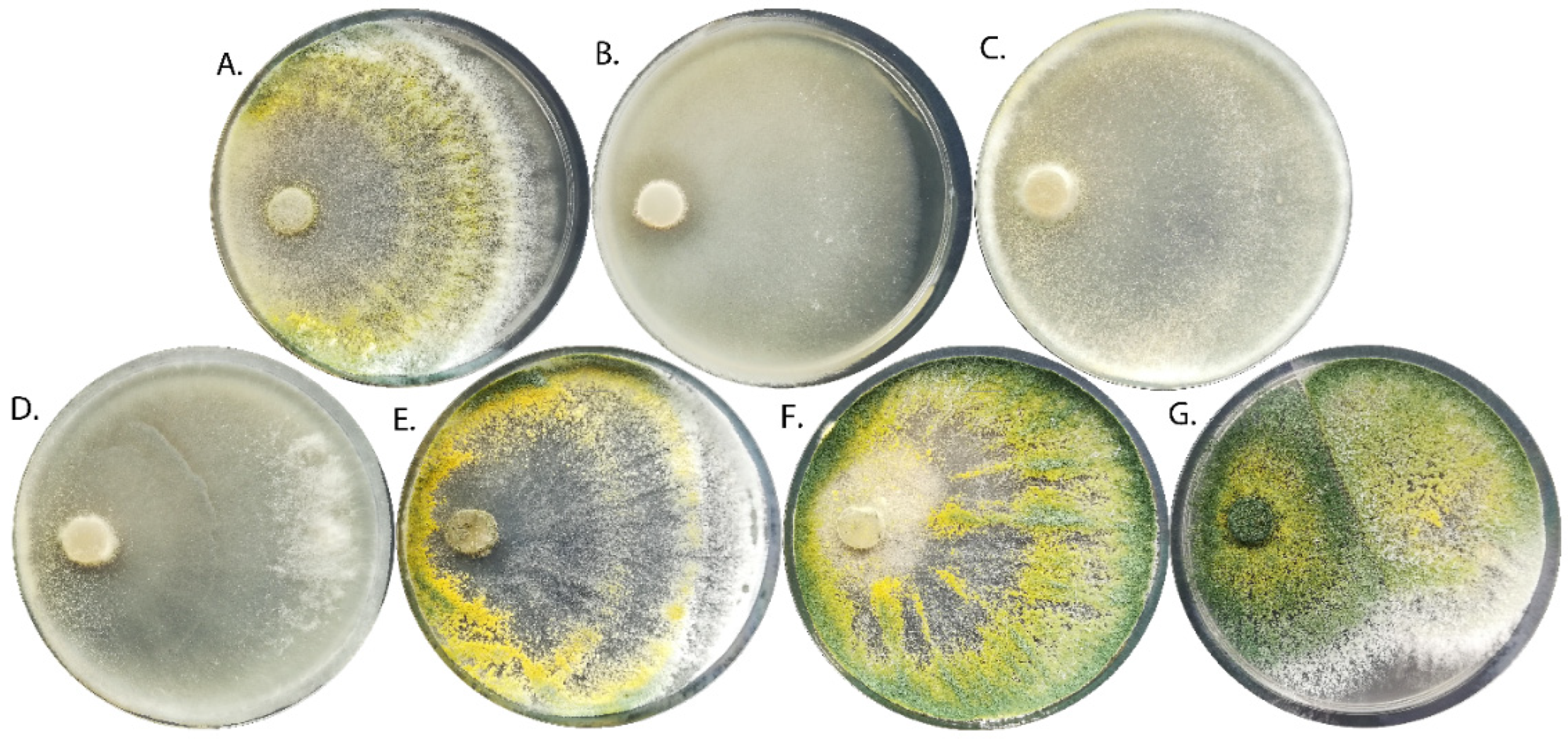
2.1.1. Morphology

The T. viride strains showed significant differences in mycelium morphology. The pigmentation of the mycelium ranged from white, through yellow-green, to dark green (Figure 1). Moreover, the concentric rings were visible for some strains (TvI, TvV, and TvVI), whereas for other strains, nodules on the surface of the hyphae-specific conidiophore agglomerations in the form of lumps (TvI and TvV–TvVII) were present (Table 1). Some strains were characterized by a specific coconut smell (TvII, TvIII, and TvV), probably due to the production of the volatile compound 6-n-pentyl-2H-pyran-2-one. Spherical conidia were observed in six strains, while elliptical conidia were present in strain TvII (Table 1).

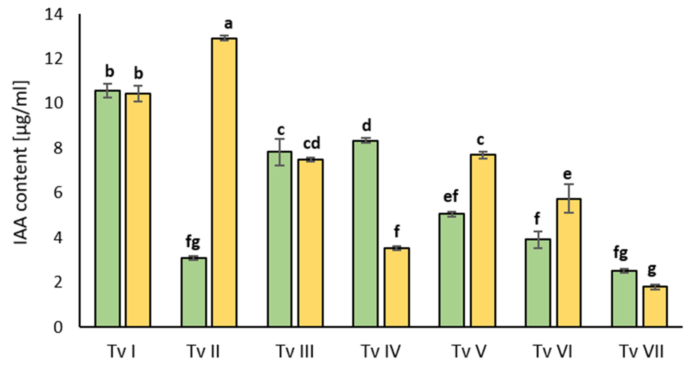
2.1.2. Capacity to Synthesize Indole-3-Acetic Acid

All of the analyzed T. viride strains showed the ability to synthesize indole-3-acetic acid (IAA) in media not supplemented and supplemented with L-tryptophan (Figure 2). The level of IAA was the highest for strain TvII in the media supplemented with L-tryptophan, whereas in the media without L-tryptophan, this strain produced 4 times less IAA. Strains TvI, TvIII, and TvVII produced the same amount of IAA in both media. Interestingly, TvIV synthesized more IAA in the media without the addition of L-tryptophan than in the media containing this amino acid, i.e., around 3 times more. TvVII produced the least amount of IAA in comparison with the other strains, almost 5 times less than TvI and TvII. Only three strains, i.e., TvII, TvV, and TvVI, synthesized more IAA in the media containing L-tryptophan.

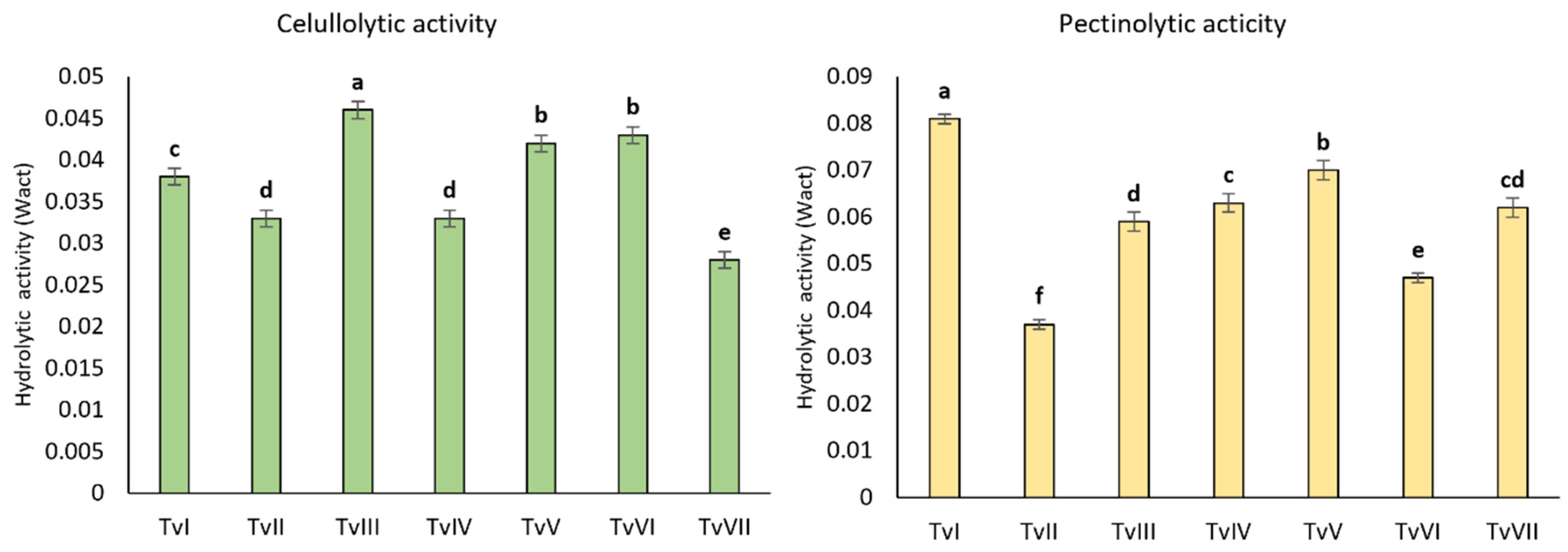
2.1.3. Hydrolytic Activity

All analyzed strains produced hydrolytic enzymes hydrolyzing carboxymethylcellulose and pectin (Figure 3). The cellulolytic activity was the highest for the TvIII, TvV, and TvVI strains, whereas strains TvI, TvIV, TvV, and TvVII showed the highest pectinolytic activity. TvVII showed the lowest cellulolytic activity among the tested strains, whereas the pectinolytic activity of this strain was one of the highest. Similar results were observed for the other strains; i.e., strains showing high cellulolytic activity had low pectinolytic activity, and vice versa. Only the TvII strain showed relatively low cellulolytic and pectinolytic activities (Figure 3).

2.2. The Growth of B. napus Seedlings Inoculated with T. viride

In the next experiment, it was examined how the inoculation of B. napus seeds with T. viride spores of seven strains affected the growth of seedlings (Table 2). The roots of six-day-old seedlings that sprouted from seeds inoculated with fungal spores were longer in all variants, with the differences being statistically significant for the T. viride isolates TvI, TvII, TvVI, and TvVII. The longest roots were noted in the seedlings inoculated with TvVII, i.e., almost 1.5-fold longer than in the control, followed by TvI and TvII. The hypocotyls of the seedlings that grew from the seeds inoculated with the spores of TvI, TvII, and TvV–VII were longer than in the control. The hypocotyls of the seedlings inoculated with TvII were the longest, i.e., 1.5-fold longer than in the control, followed by TvVII and TvV. Interestingly, the hypocotyls of the seedlings that grew from the seeds inoculated with TvIII and TvIV were significantly shorter than the hypocotyls of the non-inoculated seedlings; however, other parameters were the same as those in the control seedlings. Both the fresh biomass and dry biomass of the seedlings that grew from the seeds inoculated with TvVI and TvVII were higher than those of the control seedlings; i.e., the fresh biomass was 1.3- and 1.1-fold higher, respectively, and the dry biomass was around 1.2-fold higher (Table 2).
For further field experiments, out of seven T. viride strains analyzed under laboratory conditions, TvVII was selected because it increased the fresh biomass and dry biomass the most and induced the strongest stimulation of the shoot and root growth of the B. napus seedlings.

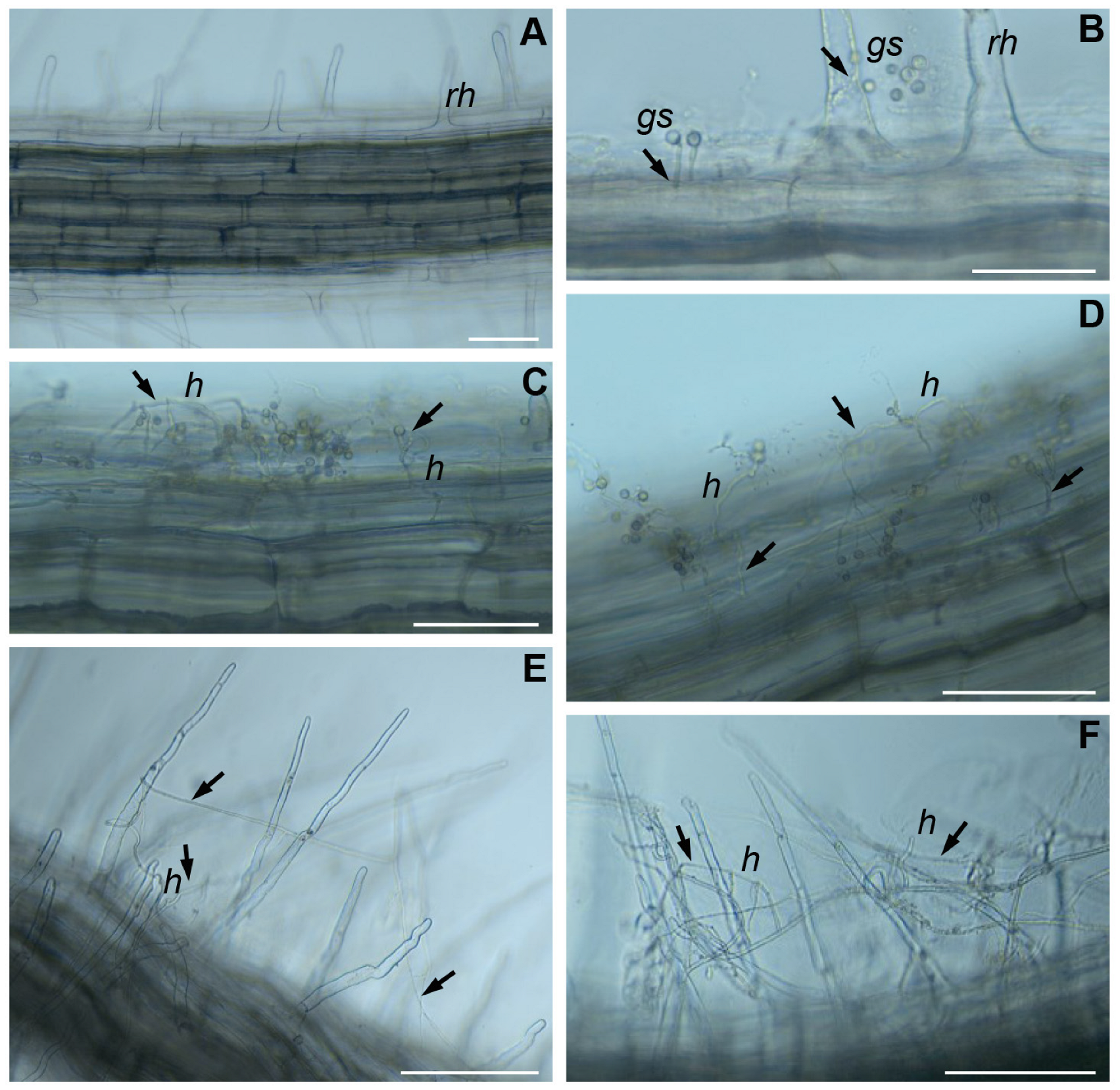
2.3. The Colonization of B. napus Roots by T. viride

To verify the ability of T. viride to colonize the roots of B. napus seedlings, microscopic observations of the growth of fungal spores on the roots were performed (Figure 4). For the experiment, T. viride strain VII was selected because of the ability of this strain to promote the growth of rapeseed, as shown previously [26] and confirmed in this study. The microscopic observations revealed that fungal spores germinated toward the root after 24 h (Figure 4B). Within 48 h after inoculation (Figure 4C,D), hyphae started forming a net around the main root, indicating that, in some places, the hyphae penetrated the apoplast of the endoderm. After 72 h, the hyphae through entwinement formed a net around the root hairs (Figure 4E,F).

2.4. Analysis of BnMT and BnRSH Genes

2.4.1. In Silico Analysis of Gene Promoters

An in silico analysis of the promoter regions of BnMTs and BnRSHs using PlantCARE software (https://bioinformatics.psb.ugent.be/webtools/plantcare/html/, accessed on 16 October 2023) [66] showed the presence of cis-regulatory elements involved in the response to elicitors and metabolites of fungal origin (Table 3). Those regulatory elements were the most abundant in the promoters of BnMT2 and BnCRSH. The presence of elements for the response to phytohormones, including jasmonic acid (JA) and salicylic acid (SA), was also confirmed. In BnMT1-3, JA- and SA-responsive elements were present, whereas, in BnRSH2, BnRSH3, and CRSH, elements for the response to JA were noted.

2.4.2. B. napus Metallothionein Gene Expression

The expression of BnMT1-3 genes was evaluated in 6-day-old seedlings that grew from seeds inoculated with the spores of T. viride strain TvI-TvVII and compared with the expression of BnMT1-3 genes in seedlings that grew from non-inoculated seeds (Figure 5). The relative level of BnMT1 mRNA was not significantly affected by T. viride inoculation in cotyledons, whereas, in roots, the level of BnMT1 transcripts was significantly increased by all tested T. viride strains. The highest expression of BnMT1 was observed in the roots of the seedlings that grew from the seeds inoculated with TvV, i.e., 2.25-fold higher than in the control seedlings (Figure 5A). The mRNA level of BnMT2 was significantly higher in the cotyledons of the seedlings that grew from the seeds inoculated with TvIII–VI spores, i.e., approximately 1.6-fold higher than in the control seedlings. The BnMT2 mRNA level was significantly higher in the roots of the seedlings that grew from the seeds inoculated with TvV–VII spores, i.e., approximately 1.4-fold higher than in the roots of the control seedlings (Figure 5B). The expression of BnMT3 in cotyledons increased in the seedlings that grew from the seeds inoculated with all tested fungal isolates, except for TvVII. The increase was the greatest for the TvIV and TvV strains, i.e., a 2.2-fold higher expression than in the control seedlings. In the roots, the expression of BnMT3 was increased only in the seedlings that grew from the seeds inoculated with TvV; i.e., the expression was almost 2 times higher than in the control seedlings (Figure 5C).

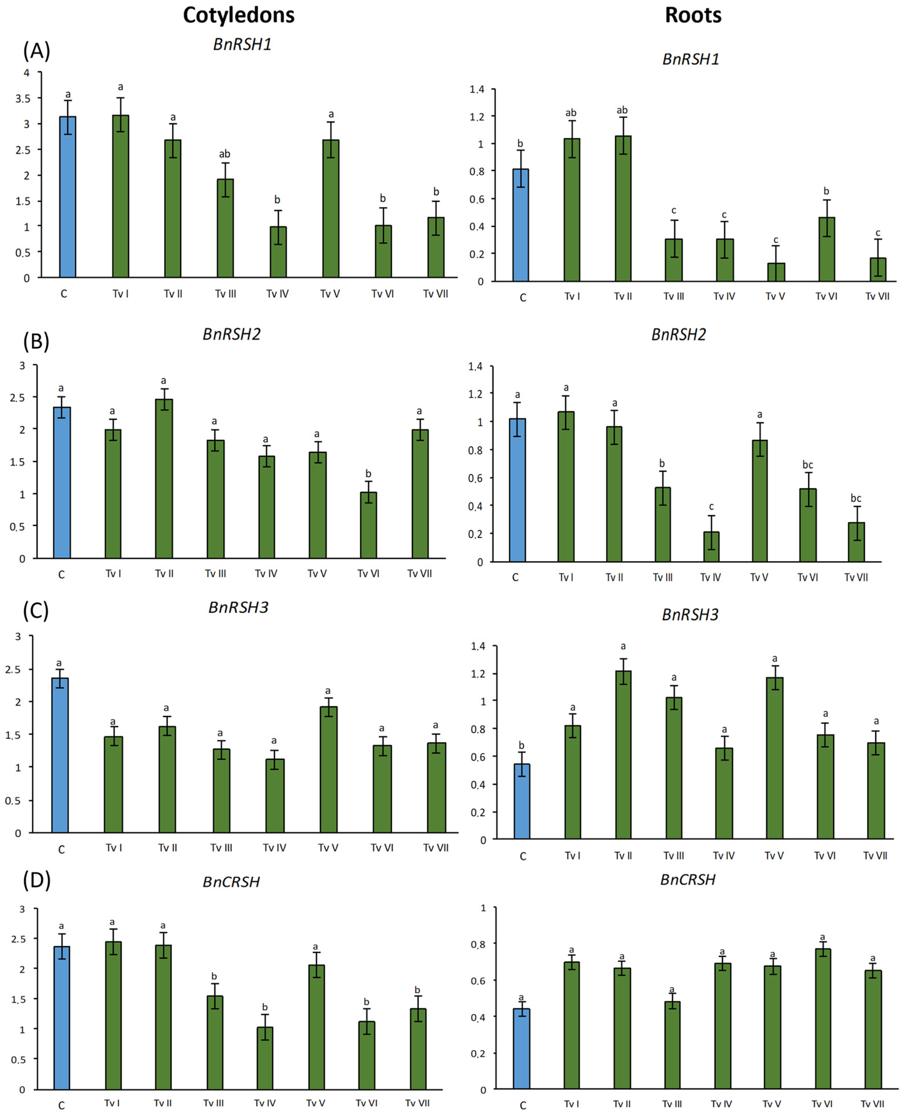
2.4.3. B. napus RSH Genes Expression

The inoculation of seeds with the spores of T. viride strains I–VII did not significantly increase the relative mRNA level of the stringent response genes in the cotyledons and roots of the 6-day-old rape seedlings in comparison to the control seedlings (Figure 6). In fact, only the expression of BnRSH3 increased approximately 1.7-fold in the roots of the seedlings that grew from the inoculated seeds in comparison to the control (Figure 6C). Moreover, an increase in the mRNA level of BnCRSH was observed in roots; however, the differences were not significant (Figure 6D). For other variants, mostly a decrease in the transcription level of BnRSHs was observed. In the cotyledons of the seedlings that grew from the seeds inoculated with TvIV, TvVI, and TvVII, 3 times lower BnRSH1 mRNA levels in comparison to the control were observed. In roots, the expression of BnRSH1 was significantly down-regulated by inoculation with TvIII, TvIV, TvV, and TvVII, mostly by TvV, which reduced the expression 8 times in comparison to control (Figure 6A). The relative level of BnRSH2 mRNA was significantly lower only in the cotyledons of the plants that grew from the seeds inoculated with TvVI, i.e., 2.3-fold lower than in the control. Significantly lower levels of BnRSH2 were observed in the roots of the plants that grew from the seeds inoculated with TvIII, TvIV, TvVI, and TvVII, being approximately 3 times lower than in the control (Figure 6B). The level of BnRSH3 in cotyledons was down-regulated in the seedlings that grew from the inoculated seeds; however, the differences were not significant (Figure 6C). In cotyledons, the expression of BnCRSH was reduced by TvIII, TvIV, TvVI, and TvVII; i.e., the expression was approximately 2 times lower than in the control seedlings (Figure 6D).

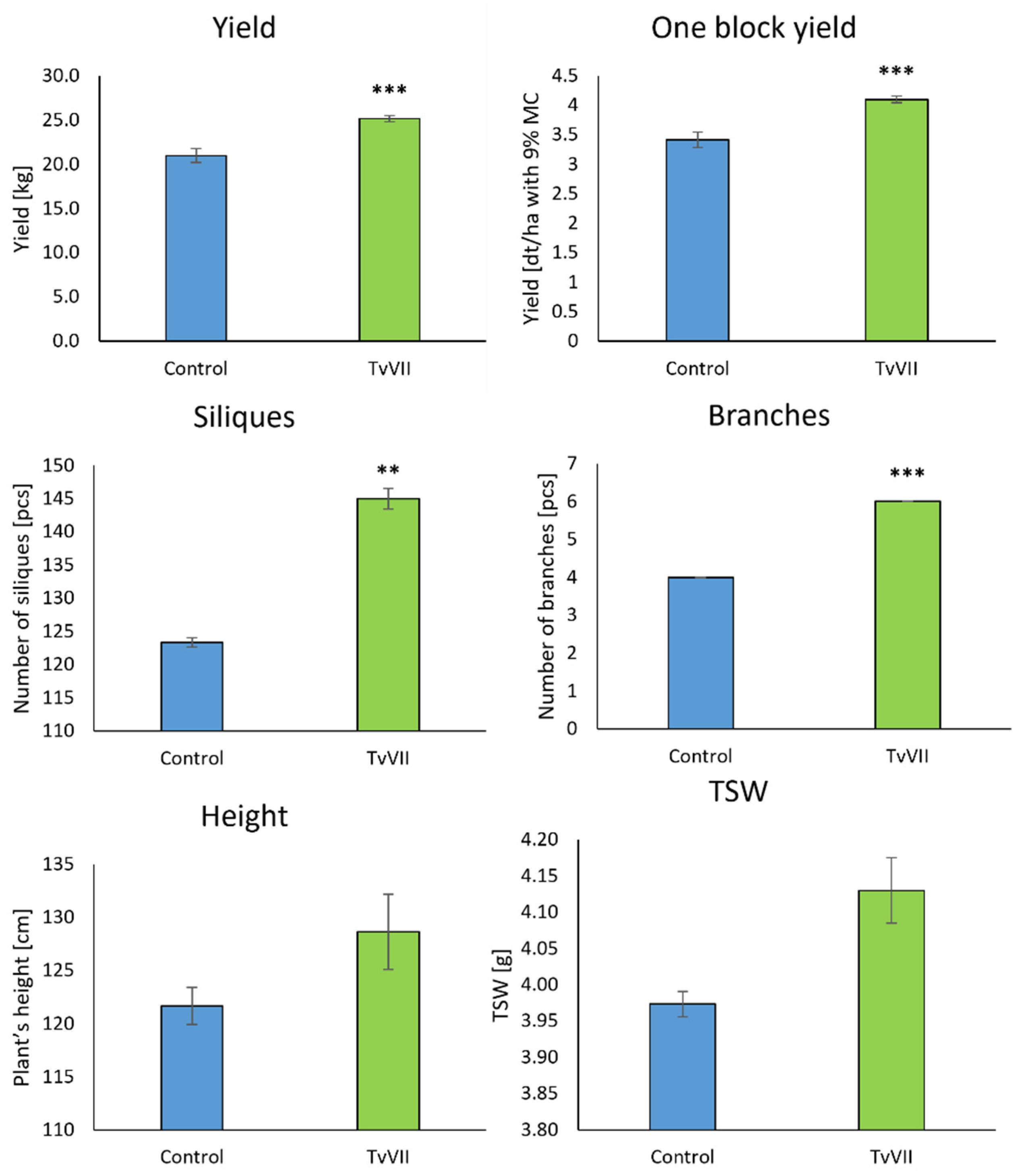
2.5. Field Experiment

The results of the field experiment showed that the inoculation of B. napus seeds with the spores of T. viride strain TvVII significantly affected the obtained yield, increasing it by 16.7% (Figure 7). T. viride strain TvVII also significantly increased the number of branches and siliques. There were on average two more branches per inoculated plant compared to the control, and the number of siliques was increased by 17.56% (Figure 7). There was an increase in the weight of thousand seeds (TSW) produced by the plants that grew from the inoculated seeds. Moreover, the inoculated plants were higher (5.75% higher) than the control plants; however, these differences were not statistically significant.

3. Discussion

Progressive environmental pollution has a significant negative impact on living organisms, including the growth and development of crops. There is an urgent need to reduce the amounts of agrochemicals, and, thus, the development of effective biological solutions for agriculture is of crucial importance. The use of microbes in agriculture is an alternative approach to replace plant protection chemicals and fertilizers. Despite all belonging to the same species, the T. viride strains used in this study showed significant differences in mycelial morphology and the abilities to synthesize IAA and produce hydrolytic enzymes. The differences suggest that these isolates might also differ in the level of synthesis of other metabolites, such as elicitors, i.e., molecules that trigger plant immune responses [67]. The observed differences among the T. viride isolates probably explain the differences in the ability of the tested isolates to promote the growth of B. napus and the impact on BnMT and BnRSH gene expressions. It is widely observed that the positive impact of a single strain of microorganisms on plant growth and development depends on plant species or even variety. Moreover, several different strains of the same microorganism might affect the growth and development of the same plant differently, as observed in this study (reviewed in [68]). Interesting results were obtained by Zhang et al. [69]; i.e., three strains of the fungus Mortierella elongata varied in their effects on the height, dry biomass, and leaf area of watermelon (Citrullus lanatus Thunb. Matsum & Nakai), maize, tomato (Solanum lycopersicum L.), and pumpkin (Cucurbita sp. L.). In the present study, the inoculation of B. napus seeds with T. viride TvI–TvII and TvV–VII strains under laboratory conditions resulted in a significant increase in the length of the hypocotyls and roots of the seedlings. This effect might be due to the ability of those isolates to synthesize IAA; however, it needs to be noted that strain TvVII produced the lowest amount of IAA among the tested strains. As demonstrated by Contreras-Cornejo et al. [32], the mutation of A. thaliana AUX1, BIG, EIR1, and AXR1 genes, which are involved in the transport and signaling of auxins, attenuated the growth-promoting effect of Trichoderma virens observed in wild-type plants.
During plant–microorganism interactions, transcriptome changes occur both in the plant and in the microbe. We hypothesized that MTs and RSHs, which are widely known for their roles in the response to diversified environmental factors, are also important during plant interactions with microorganisms. The presence of many cis-regulatory elements regulating the response to abiotic and biotic stresses within the promoters of MT and RSH genes indicates that they participate in plant responses not only to environmental physio-chemical conditions but also to the presence of bacteria and fungi [40,42,44,56]. For example, the MT genes of rice and A. thaliana have numerous regulatory motifs of response to metals, low/high light, low temperature, and drought, as well as motifs associated with stress-related signaling, including the response to ROS and ABA [40]. In the case of BnRSH, the largest number of motifs was found to be associated with the response to abiotic stress and with signaling via phytohormones, including ethylene and ABA [56]. The analysis of BnRSH and BnMT promoter regions showed the presence of cis-regulatory elements responsive to microbial elicitors and other compounds of microbial origin. Thus, we assumed that the expressions of these genes might be altered in the presence of potential microbial symbionts, as well as in the presence of pathogens. Plant cells are equipped with pattern-recognizing receptors (PRRs), i.e., transmembrane proteins with ectodomains binding ligands of microbial origin—pathogen-/microbe-associated molecular patterns (P/MAMPs) [70]. The recognition of a ligand leads to the activation of plant pattern-triggered immunity (PTI), which limits the development of microorganisms in plant tissues, e.g., through an increased synthesis of antimicrobial compounds [71]. To date, a number of MAMP molecules produced by the fungi of the Trichoderma genus have been identified [72,73], including hydrolytic enzymes that allow for the penetration of the outermost layers of root tissues, such as cellulases [74], xylanases [75], and polygalacturonases [76]. A microscopic analysis showed progressive root colonization by germinating T. viride spores, including spore germination toward the root, the formation of a net-like structure, and the entwinement of root hairs by the growing hyphae. This may be due to the recognition of signaling molecules and/or nutrients (e.g., sugars) exuded by the roots and perceived by the fungus. The observed adhesion of the mycelium to the root is probably mediated by small proteins—hydrophobins [77]. It was shown that the presence of hydrophobins facilitates the colonization of plant tissue by fungi and probably protects the sensitive hyphal tips against the activity of antifungal compounds [78]. Moreover, within BnMT and BnRSH promoters, there were also elements of response to JA and SA—phytohormones responsible for triggering plant immune system responses. These mechanisms might be activated by the presence of Trichoderma fungi [36,37,79]. The observed changes in the expressions of BnMT and BnRSH genes are potentially the result of the changes triggered by the presence of the fungus. The presence of fungus is detected by plants because fungus secretes elicitors and hydrolytic enzymes that violate the integrity of the root tissue. Furthermore, changes in the balance of rapeseed phytohormones might further affect the expression of the analyzed genes. These conclusions are supported by the fact that, through the production of elicitors, fungi affect the level of accumulation of plant intermediate metabolites and by the increased signaling of SA, JA, ABA, or nitric oxide (NO) [80]. Within the promoters of the BnMT1–BnMT3, BnRSH1, and BnRSH3 genes, the presence of motifs binding WRKY transcription factors was observed. An increase in the expression of genes encoding WRKY was demonstrated in response to the colonization of maize roots by Trichoderma atroviride [81]. Moreover, changes in the expression profiles of genes involved in JA and SA signaling in inoculated plants compared to the control were shown.
Genes encoding MT1–3 in plants are expressed in various tissues and organs during ontogenesis. In most analyzed up-to-date species, MT4 is expressed only in developing and mature seeds [82,83,84,85], and, thus, BnMT4 was not analyzed in this study. It has been shown that MT expression changes in response to a number of endogenous and exogenous factors, i.e., the presence of heavy metals [86], ROS [87], polyamines [88], phytohormones [89], the presence of microorganisms [14,90,91], salinity [92], drought stress [43,48], osmotic stress [93], light conditions [94], and wounds and pathogen infections [95,96]. The presence of T. viride increased the expression of BnMT13 in the cotyledons and roots of 6-day-old seedlings, but the effect was dependent on the type of MT and fungal isolate. Mirzahossini et al. [91] showed that the inoculation of Festuca arundinacea Schreb. with the fungus Epichloe coenophiala significantly increased plant tolerance to nickel, increased dry biomass, decreased H2O2 production, and lowered Ni accumulation in plant tissues. Moreover, E. coenophiala down-regulated the expressions of plant MT and ABC (ATP-binding cassette) metal transporter genes. The inoculation of rapeseed with arbuscular mycorrhizal fungi (AMF) (Acaulospora longula, Glomus geosporum, G. mosseae, and Scutellospora calospora) in sterile soil resulted in an increased concentration of N, P, K, and S in shoots but, at the same time, negatively affected plant growth compared to non-inoculated plants. Interestingly, in an experiment conducted in unsterile soil, the concentrations of analyzed elements were lower in the inoculated plants than in the control. The presence and absence of native microorganisms in the soil differentiated the effect on the expression of BnMT2 inoculated with AMF spores; i.e., the expression was increased in the variant with sterile soil and decreased in the variant with unsterile soil containing native microorganisms [14]. In a study on the phytoextraction of heavy metals by willow (Salix viminalis L.) inoculated with the ectomycorrhizal fungus Hebeloma mesophaeum and bacteria Bacillus cereus, the content of Cd and Zn in plant tissues increased. Moreover, changes in SvMT1 expression were observed. B. cereus significantly increased the level of SvMT1 transcripts, whereas H. mesophaeum slightly decreased the expression of this MT [90]. The inoculation of rapeseed with the IAA-producing ectomycorrhizal fungus Laccaria laccata increased the number of leaves and the biomass of inoculated plants. In addition, L. laccata almost doubled BnMT1 mRNA levels and decreased BnMT3 transcript levels. Similar results have been observed in plants inoculated with Collybia tuberosa and Cyathus olla which significantly increased the level of BnMT2 expression [97].
The stringent response was first observed in bacteria in response to amino acid deficiency [98], and RSH genes have been identified in bacteria [99,100,101], animals [102], and plants [103,104,105,106]. The stringent response genes are closely associated with plant adaptation to stress factors, including oxidative stress [107], nitrogen deficiency [62,106,107], salinity [108], drought, wounds, the presence of heavy metals, and UV radiation [53]. This is the first report showing the expression profiles of RSH genes in a plant in response to colonization by filamentous fungi. The tested fungal isolates caused an increase or a decrease in the expression of BnRSH genes depending on the type of RSH gene and fungal isolate. Interesting results were obtained for the BnCRSH gene. Although the BnCRSH promoter contains the highest number of regulatory elements responsive to biotic factors (3), and JA and SA signaling (8) among the tested BnRSHs, the expression of this gene decreased in response to colonization by fungi in cotyledons. To date, it has been shown that plant RSH genes are altered in the presence of rhizosphere bacteria. Dąbrowska et al. [56] showed that the inoculation of rapeseed with Serratia plymuthica, Serratia liquefaciens, and Massila timonae under salt stress conditions variably affects BnRSH expression. S. plymuthica significantly increased the expression of all analyzed genes, i.e., BnRSH13 and BnCRSH, in roots and cotyledons. S. liquefaciens significantly increased BnRSH1 and BnCRSH mRNA levels in canola cotyledons and BnRSH13 expression in its roots, whereas M. timonae increased only the amount of BnRSH1 transcripts in the root. The inoculation of canola with P. stutzeri significantly increased the expressions of BnRSH1 and BnRSH3 in roots. Moreover, an increased tolerance of inoculated canola to salt stress compared to non-inoculated plants was observed [108].
A major limitation of using bioinoculants is the low positive effect on plant growth and development in field experiments, despite their positive effects under laboratory conditions. This is due to the low level of survival of the microorganisms because of the competition for niches with native microorganisms. Moreover, the growth of microorganisms and/or their ability to promote plant growth highly depends on environmental factors, such as temperature, rainfall, and soil type [8]. For these reasons, the effectiveness of laboratory-developed bioinoculants must be further evaluated under field conditions. The inoculation of canola seeds with T. viride isolate TvVII spores positively affected B. napus growth and yield when grown in the field. Seed inoculation with T. viride TvVII spores increased plant tolerance to unfavorable weather. The inoculation of tomato with Trichoderma spp. spores increased plant height and biomass, as well as Ca, Mg, P, and K content, in shoots and roots under laboratory conditions. A larger area and number of leaves and a higher chlorophyll content were also noted compared with control plants [109]. In melon (Cucumis melo L.) inoculated with Trichoderma phayaoense, accelerated plant development, increased biomass, and increased yield were observed in laboratory studies. Moreover, T. phayaoense exhibited the ability to biocontrol the phytopathogens Stagonosporopsis cucurbitacearum and Fusarium equiseti [24]. The inoculation of turmeric (Curcuma longa L.) with T. harzianum resulted in increased plant height and yield under laboratory conditions. An analysis of T. harzianum revealed the ability of the fungus to solubilize P and to produce IAA, HCN, and cellulases [23]. Trichoderma fungi can also accelerate flowering, as shown in a greenhouse experiment on freesia (Freesia refracta Jacq. Klatt) inoculated with T. viride, T. harzianum, and Trichoderma hamatum. An accelerated development of side shoots and an increased content of K, Fe, Mn, and Zn were also observed [25]. It should be noted that, during the field experiment described in this publication, unfavorable environmental conditions prevailed throughout the canola growing season, i.e., ground frost, a low level of precipitation, and high air temperatures, which increased the effects of drought. Therefore, the fungal strain tested in this study effectively promoted plant growth during drought, which is now one of the most common and severe adverse environmental conditions. Under greenhouse conditions, it was shown that coating seeds with T. harzianum spores increased drought stress tolerance in wheat (Triticum aestivum L.). Plants colonized by the fungus showed lower levels of proline, malondialdehyde (MDA), and H2O2, as well as increased levels of phenolic compounds [110]. Similar results were obtained under laboratory conditions for maize inoculated with T. atroviride ID20G grown under drought-stress conditions. Increased biomass, chlorophyll, and carotenoid content, as well as limited lipid peroxidation associated with drought stress, were observed. An increased activity of antioxidant enzymes and decreased H2O2 content were also noted in inoculated plants [111]. A greenhouse study showed the increased activity of antioxidant enzymes in sugar cane (Saccharum sp. L.) inoculated with Trichoderma asperellum under drought conditions. Moreover, T. asperellum also increased photosynthesis efficiency and tissue water content [112]. There is still a knowledge gap in this research area, mainly regarding the effect of Trichoderma inoculation in field studies. Environmental conditions have a multifaceted effect on the microbial inoculation of plants, and the involvement of stress factors can alter the effectiveness of inoculation [8], which is why there is a need for the field testing of PGPM.

4. Materials and Methods

4.1. Microbial and Plant Materials

Seven morphologically different Trichoderma viride strains were used in this study (numbers in GenBank NCBI: OL221590.1, OL221591.1, OL221592.1, OL221593.1, OL221594.1, OL221595. 1, OL221596.1, respectively, hereinafter referred to as TvI–TvVII). The fungi used for the research were cultured on the microbial media PDA for a morphological analysis (potato dextrose agar, LABM) and TSB (trypticase soy broth, Sigma Aldrich; Poznań, Poland) for the assessment of the ability to synthesize IAA at room temperature. Seven-day-old cultures of the fungi growing on the PDA medium were used to prepare spore suspensions for the inoculation of B. napus seeds. Spores were collected by adding 5 mL of sterile dH2O to Petri dishes containing 7-day-old fungal cultures. With the use of sterile cell spreaders, the spores were suspended and then filtered via Miracloth (Merck; Warsaw, Poland) in order to remove hyphae. The concentration of the spores was then analyzed with the use of a Thoma cell counting chamber.
Seeds of spring rape (Brassica napus L.), variety Lumen, were purchased from Rapool (Wągrowiec, Poland). Before use, the seeds were surface-sterilized, according to the method presented by Mierek-Adamska et al. [113], in a mixture of 96% ethanol and 30% H2O2 (1:1, v:v) for 5 min and then rinsed at least 10 times with sterile water. The sterile seeds were incubated for 10 min in T. viride TvI–TvVII spore suspensions (106 spores/mL for an in vitro study and 109 spores/mL for a field experiment) with shaking (150 rpm) at room temperature. The control seeds were shaken in sterile water. Twenty seeds were placed in Petri dishes (90 mm diameter) on sterile filter paper moistened with 4 mL of sterile water. The seeds were incubated for six days in the dark at 25 °C. The length of the roots and hypocotyls and the fresh and dry biomass were analyzed after six days of incubation. For a gene expression analysis, the roots and cotyledons were frozen in liquid nitrogen and stored at −80 °C.

4.2. Characterization of T. viride Isolates

4.2.1. Morphological Analysis

Mycelium discs, obtained from the 7-day-old T. viride culture grown on solid PDA, were placed 1 cm from the edge of a Petri dish (90 mm diameter) on a solid PDA medium and cultured for 7 days at 25 °C. The following features of the mycelia were analyzed: fungal pigmentation, the presence of rings, the shape of conidia, the agglomeration of conidiophores in the form of lumps, and the ability to produce volatile compounds (a coconut smell).

4.2.2. Evaluation of Capacity to Synthesize Indole-3-Acetic Acid

To verify the ability to synthesize indole-3-acetic acid (IAA), the spectrophotometric method based on Gravel et al. [114] was used. Three mycelium discs (for each strain) obtained from the 7-day-old T. viride cultures were placed in a liquid TSB medium supplemented with L-tryptophan at a final concentration of 200 µg/mL or without L- tryptophan. The cultures were then shaken for 7 days (150 rpm) at 24 °C. The cultures were then filtered through a sterile gauze to remove mycelia. Next, 4 mL of Salkowski’s reagent (0.5 M FeCl3, distilled H2O, and 70% HClO4 at a ratio of 2:49:49) was added to 2 mL of the filtrate and incubated for 20 min at room temperature. Absorbance was measured at a wavelength of 535 nm (NanoDrop, Thermo Fisher Scientific, Waltham, MA, USA). IAA reacts with ferric chloride and perchloric acid to give a colored product; i.e., the amount of colored compound was directly proportional to the concentration of IAA in the tested sample.

4.2.3. Assessment of Hydrolytic Activity

The hydrolytic activity of the T. viride strains was analyzed based on a measurement of the hydrolysis zone around the colonies and the colony diameter of the examined fungi on the 7th day of cultivation at 24 °C. Enzymatic activity (Wact) was calculated according to Hrynkiewicz et al. [115]:
Wact = Sh2/(Sc·t)
where Sh—hydrolysis zone diameter; Sc—colony diameter; and t—incubation time (96 h and 168 h).
The ability to synthesize cellulase was assessed using a medium described by Wood [116] enriched with CMC (carboxymethylcellulose). Pectin hydrolysis was carried out on a medium described by Strzelczyk and Szpotański [117]. The level of pectin hydrolysis was verified by adding to the culture plates a solution of 1% cetyl-trimethylammonium bromide (BDH), which precipitates undigested pectin.

4.3. Microscopic Analysis

Three-day-old B. napus seedlings grown on a water agar medium (Phytagel, Sigma-Aldrich) were inoculated with the T. viride strain TvVII spore suspension (106 spores/mL), and, after 24 h, 48 h, and 72 h of cultivation, roots were collected. Next, the samples were placed on glass slides and covered with distilled water for observation using an Olympus BX50 fluorescence microscope. The results were recorded with an Olympus XC50 digital color camera and CellB software, version 3.4 (Olympus Soft Imaging Solutions GmbH; Münster, Germany). For the control, the inoculation of plants with T. viride spores was omitted.

4.4. Analysis of Gene Expression

Total RNA was isolated from 100 mg of the roots and cotyledons of 6-day-old canola seedlings according to Chomczyński and Sacchi [118]. The obtained RNA was analyzed via spectrophotometric measurements (NanoDrop Lite, Thermo Fisher Scientific; Waltham, MA, USA) and electrophoresis in agarose gel stained with ethidium bromide. High-quality RNA samples with an A260/A280 ratio of approximately 2.0 were used for further analysis. For the reverse transcription reaction, 1.5 μg of total RNA was used, and the reaction was performed according to Dąbrowska et al. [97].
The relative levels of BnRSH and BnMT gene transcripts were checked using the semiquantitative (sq) RT-PCR method [119]. The relative level of the transcripts of the tested genes is expressed as the ratio of the amount of the RT-PCR product for the analyzed gene to the amount of the RT-PCR product for the reference gene encoding actin (BnAct). The reaction mixture contained 1 μL cDNA, 2 μL OptiTaq polymerase buffer, 0.2 mM dNTP, 0.15 μM forward and reverse primers, and 1 U OptiTaq DNA polymerase (EURx, Gdańsk, Poland) in a final volume of 20 μL. The reaction conditions were as follows: initial denaturation at 95 °C for 2 min, denaturation at 95 °C for 30 s, primer annealing for 45 s (temperature optimized for each primer pair given in Table 4), elongation at 72 °C for 35 s, and final elongation at 72 °C for 5 min. The number of cycles was optimized for each pair of primers to complete the reaction in a logarithmic growth phase (Table 4). The sequences of the oligonucleotides used are listed in Table 1. ImageGuage 3.46 software (FujiFilm; Tokyo, Japan) was used for densitometric measurements of the amount of obtained RT-PCR products.

4.5. Field Experiment

The field experiment was conducted in Chrząstowo (53°09′52″ N 17°35′02″ E), on a site with typical brown soil, medium clay, and soil pH 6.4. The content of bioavailable minerals was as follows: Mg—47 mg/kg−1, P—95 mg/kg−1, and K—183 mg/kg−1 (Chemical-Agricultural District Station in Bydgoszcz, Poland). Canola seeds inoculated with T. viride strain TvVII and non-inoculated B. napus seeds were used for the field experiment. The fore crop was spring barley. The size of the experimental field was 16.5 m2, and the seeds were sown at a density of 100 plants per m2. The experiment was carried out in three replicates on different plots simultaneously. Fertilization was carried out at half-dose before sowing, and the other half was used afterward as a top dressing, with total doses of the pure ingredients of 30 NH4NO3 kg/ha; 48 P2O5 kg/ha; 34 (NH4)2SO4 kg/ha; and 80 K2O kg/ha. The insecticides Decis Mega 50EW (Bayer; Leverkusen, Germany) and Boravi 50 WG (Gowan; Yuma, AZ, USA) were used to protect the plants against silique pests, including Meligethes aeneus.
The experiment was established on 7 April 2022, and the crop was harvested on 12 August 2022. During the growing season, there were unfavorable environmental conditions, i.e., ground frost in April (down to −4.5 °C) and May (down to −6.2 °C), and high air temperatures (up to 34.6 °C in June and up to 35.7 °C in July). The precipitation during the growing season is given in Table 5. The following plant parameters were evaluated: the number of branches and siliques, the height of the plants (10 randomly selected plants in three replicates), the seed yield determined at 9% humidity of seeds, and thousand seed weight (TSW).

4.6. Statistical Analysis

Statistical analyses (one-way ANOVA with Tukey’s post hoc test) were performed in Past 4.12b [120]. For most analyses, the threshold for significance was p < 0.05. For the results of the field experiment, the thresholds for significance were p < 0.05 (*), p < 0.01 (**), and p < 0.001 (***).

5. Conclusions

The development of novel bioinoculants for agriculture is crucial to reduce the usage of chemical plant protection products and mineral fertilizers while maintaining high yields and quality. However, advancing global warming and the more frequent occurrence of drought periods require intensified efforts for the betterment of crop systems and novel solutions in agriculture. These are the most important reasons for further and deeper research on interactions between plants and microorganisms, especially in economically important crops. In order to develop bioinoculants more effectively, the molecular basis, i.e., the proteins and pathways underlying plant–microbe interactions, needs to be thoroughly understood. The conducted research shows that the saprophytic fungus T. viride colonizes the roots of the non-mycorrhizal plant B. napus. Furthermore, it accelerates germination and growth, as well as increases the yield. In addition, as the field study showed, the presence of T. viride protects rapeseed plants against water deficits. Molecular studies showed changes in the expressions of B. napus MT and RSH genes in response to the presence of T. viride; however, the changes depended on the tested fungal strain. This indicates the involvement of metallothioneins and the stringent response proteins in plant–fungus interactions. The results obtained in this study will contribute to the development of novel bioinoculants (T. viride VII) for the direct or indirect regulation of plant metabolism, crucial for food security. The conducted research shows that each fungal strain has a different potential to interact with the selected plant. The information obtained in this study is particularly important for the producers of biopreparations, as it raises the awareness that the microorganisms that are biocomponents of effective biopreparations should be thoroughly tested.

Author Contributions

Conceptualization, G.B.D.; methodology, G.B.D., Z.G., B.K. and D.K.; software, Z.G. and M.A.; validation, Z.G. and A.M.-A.; formal analysis, Z.G., M.A. and A.M.-A.; investigation, Z.G., M.A., D.K., K.N. and K.H.; data curation, Z.G., M.A. and D.K.; writing—original draft preparation Z.G. and M.A.; writing—review and editing M.A., A.M.-A. and G.B.D.; visualization, Z.G., M.A., K.N. and K.H.; supervision, A.M.-A., B.K. and G.B.D.; project administration, G.B.D.; funding acquisition, G.B.D. All authors have read and agreed to the published version of the manuscript.

Funding

This research was funded by The Marshal’s Office of the Kuyavian-Pomeranian Voivodeship, grant number 690 40001.0000011, awarded to G.B.D.

Institutional Review Board Statement

Not applicable.

Informed Consent Statement

Not applicable.

Data Availability Statement

Relevant data applicable to this research are within the paper and are available on request from the corresponding author.

Conflicts of Interest

The authors declare no conflict of interest.

References

  1. Chmielewska, A.; Kozłowska, M.; Rachwał, D.; Wnukowski, P.; Amarowicz, R.; Nebesny, E.; Rosicka-Kaczmarek, J. Canola/rapeseed protein—Nutritional value, functionality and food application: A review. Crit. Rev. Food Sci. Nutr. 2021, 61, 3836–3856. [Google Scholar] [CrossRef] [PubMed]
  2. Zdziennicka, A.; Szymczyk, K.; Jańczuk, B.; Longwic, R.; Sander, P. Surface, volumetric, and wetting properties of oleic, linoleic, and linolenic acids with regards to application of canola oil in diesel engines. Appl. Sci. 2019, 9, 3445. [Google Scholar] [CrossRef]
  3. Lin, L.; Allemekinders, H.; Dansby, A.; Campbell, L.; Durance-Tod, S.; Berger, A.; Jones, P.J. Evidence of health benefits of canola oil. Nutr. Rev. 2013, 71, 370–385. [Google Scholar] [CrossRef]
  4. Fridrihsone, A.; Romagnoli, F.; Cabulis, U. Environmental life cycle assessment of rapeseed and rapeseed oil produced in Northern Europe: A Latvian case study. Sustainability 2020, 12, 5699. [Google Scholar] [CrossRef]
  5. Haile, G.G.; Tang, Q.; Li, W.; Liu, X.; Zhang, X. Drought: Progress in broadening its understanding. Wiley Interdiscip. Rev. Water 2020, 7, e1407. [Google Scholar] [CrossRef]
  6. Hasan, H.H.; Razali, S.F.M.; Muhammad, N.S.; Ahmad, A. Research trends of hydrological drought: A systematic review. Water 2019, 11, 2252. [Google Scholar] [CrossRef]
  7. Wu, W.; Ma, B.L. Assessment of canola crop lodging under elevated temperatures for adaptation to climate change. Agric. For. Meteorol. 2018, 248, 329–338. [Google Scholar] [CrossRef]
  8. Antoszewski, M.; Mierek-Adamska, A.; Dąbrowska, G.B. The importance of microorganisms for sustainable agriculture—A review. Metabolites 2022, 12, 1100. [Google Scholar] [CrossRef]
  9. Soudzilovskaia, N.A.; van Bodegom, P.M.; Terrer, C.; van’t Zelfde, M.; McCallum, I.; Luke McCormack, M.; Fisher, J.B.; Brundrett, M.C.; de Sá, N.C.; Tedersoo, L. Global mycorrhizal plant distribution linked to terrestrial carbon stocks. Nat. Commun. 2019, 10, 5077. [Google Scholar] [CrossRef]
  10. Tabrizi, L.; Mohammadi, S.; Delshad, M.; Moteshare Zadeh, B. Effect of arbuscular mycorrhizal fungi on yield and phytoremediation performance of pot marigold (Calendula officinalis L.) under heavy metals stress. Int. J. Phytoremediat. 2015, 17, 1244–1252. [Google Scholar] [CrossRef]
  11. Daei, G.; Ardekani, M.R.; Rejali, F.; Teimuri, S.; Miransari, M. Alleviation of salinity stress on wheat yield, yield components, and nutrient uptake using arbuscular mycorrhizal fungi under field conditions. J. Plant Physiol. 2009, 166, 617–625. [Google Scholar] [CrossRef] [PubMed]
  12. Mathur, S.; Tomar, R.S.; Jajoo, A. Arbuscular mycorrhizal fungi (AMF) protects photosynthetic apparatus of wheat under drought stress. Photosynth. Res. 2019, 139, 227–238. [Google Scholar] [CrossRef]
  13. Marulanda, A.; Porcel, R.; Barea, J.M.; Azcón, R. Drought tolerance and antioxidant activities in lavender plants colonized by native drought-tolerant or drought-sensitive Glomus species. Microb. Ecol. 2007, 54, 543–552. [Google Scholar] [CrossRef]
  14. Dąbrowska, G.B.; Baum, C.; Trejgell, A.; Hrynkiewicz, K. Impact of arbuscular mycorrhizal fungi on the growth and expression of gene encoding stress protein—Metallothionein BnMT2 in the non-host crop Brassica napus L. J. Plant Nutr. Soil Sci. 2014, 177, 459–467. [Google Scholar] [CrossRef]
  15. Santoyo, G.; Guzmán-Guzmán, P.; Isela Parra-Cota, F.; de los Santos-Villalobos, S.; del Carmen Orozco-Mosqueda, M.; Glick, B.R.; Flores-Félix, D.; Velazquez, E.; González-Andrés, F. Plant growth stimulation by microbial consortia. Agronomy 2021, 11, 219. [Google Scholar] [CrossRef]
  16. Mukherjee, R.K.; Kumar, V.; Roy, K. Chemometric modeling of plant protection products (PPPs) for the prediction of acute contact toxicity against honey bees (A. mellifera): A 2D-QSAR approach. J. Hazard. Mater. 2022, 423, 127230. [Google Scholar] [CrossRef]
  17. Rani, L.; Thapa, K.; Kanojia, N.; Sharma, N.; Singh, S.; Grewal, A.S.; Srivastav, A.L.; Kaushal, J. An extensive review on the consequences of chemical pesticides on human health and environment. J. Clean. Prod. 2021, 283, 124657. [Google Scholar] [CrossRef]
  18. De Souza, R.M.; Seibert, D.; Quesada, H.B.; de Jesus Bassetti, F.; Fagundes-Klen, M.R.; Bergamasco, R. Occurrence, impacts and general aspects of pesticides in surface water: A review. Process Saf. Environ. Prot. 2020, 135, 22–37. [Google Scholar] [CrossRef]
  19. Matarczyk, J.A.; Willis, A.J.; Vranjic, J.A.; Ash, J.E. Herbicides, weeds and endangered species: Management of bitou bush (Chrysanthemoides monilifera ssp. rotundata) with glyphosate and impacts on the endangered shrub, Pimelea spicata. Biol. Conserv. 2002, 108, 133–141. [Google Scholar]
  20. Weltje, L.; Simpson, P.; Gross, M.; Crane, M.; Wheeler, J.R. Comparative acute and chronic sensitivity of fish and amphibians: A critical review of data. Environ. Toxicol. Chem. 2013, 32, 984–994. [Google Scholar] [CrossRef] [PubMed]
  21. Tudi, M.; Ruan, H.D.; Wang, L.; Lyu, J.; Sadler, R.; Connell, D.; Chu, C.; Phung, D.T. Agriculture development, pesticide application and its impact on the environment. Int. J. Environ. Res. Public Health 2021, 18, 1112. [Google Scholar] [CrossRef] [PubMed]
  22. Schnitkey, G.; Paulson, N.; Zulauf, C.; Swanson, K.; Baltz, J. Fertilizer Prices, Rates, and Costs for 2023; University of Illinois Urbana: Champaign, IL, USA, 2022; Volume 12. [Google Scholar]
  23. Vinayarani, G.; Prakash, H.S. Fungal endophytes of turmeric (Curcuma longa L.) and their biocontrol potential against pathogens Pythium aphanidermatum and Rhizoctonia solani. World J. Microbiol. Biotechnol. 2018, 34, 49. [Google Scholar] [CrossRef] [PubMed]
  24. Nuangmek, W.; Aiduang, W.; Kumla, J.; Lumyong, S.; Suwannarach, N. Evaluation of a newly identified endophytic fungus, Trichoderma phayaoense for plant growth promotion and biological control of gummy stem blight and wilt of muskmelon. Front. Microbiol. 2021, 12, 410. [Google Scholar] [CrossRef]
  25. Janowska, B.; Andrzejak, R.; Kosiada, T. The influence of fungi of the Trichoderma genus on the flowering of Freesia refracta Klatt “Argentea” in winter. Hortic. Sci. 2020, 47, 203–210. [Google Scholar] [CrossRef]
  26. Znajewska, Z.; Dąbrowska, G.B.; Narbutt, O. Trichoderma viride strains stimulating the growth and development of winter rapeseed (Brassica napus L.). Prog. Plant Prot. 2018, 58, 264–269. [Google Scholar]
  27. Turkan, S.; Mierek-Adamska, A.; Kulasek, M.; Konieczna, W.B.; Dąbrowska, G.B. New seed coating containing Trichoderma viride with anti-pathogenic properties. PeerJ 2023, 11, e15392. [Google Scholar] [CrossRef] [PubMed]
  28. Qi, W.; Zhao, L. Study of the siderophore-producing Trichoderma asperellum Q1 on cucumber growth promotion under salt stress. J. Basic Microbiol. 2013, 53, 355–364. [Google Scholar] [CrossRef] [PubMed]
  29. Shukla, N.; Awasthi, R.P.; Rawat, L.; Kumar, J. Biochemical and physiological responses of rice (Oryza sativa L.) as influenced by Trichoderma harzianum under drought stress. Plant Physiol. Biochem. 2012, 54, 78–88. [Google Scholar] [CrossRef]
  30. Illescas, M.; Pedrero-Méndez, A.; Pitorini-Bovolini, M.; Hermosa, R.; Monte, E. Phytohormone production profiles in Trichoderma species and their relationship to wheat plant responses to water stress. Pathogens 2021, 10, 991. [Google Scholar] [CrossRef]
  31. Mastouri, F.; Björkman, T.; Harman, G.E. Seed treatment with Trichoderma harzianum alleviates biotic, abiotic, and physiological stresses in germinating seeds and seedlings. Phytopathology 2010, 100, 1213–1221. [Google Scholar] [CrossRef]
  32. Contreras-Cornejo, H.A.; Macías-Rodríguez, L.; Cortés-Penagos, C.; López-Bucio, J. Trichoderma virens, a plant beneficial fungus, enhances biomass production and promotes lateral root growth through an auxin-dependent mechanism in Arabidopsis. Plant Physiol. 2009, 149, 1579–1592. [Google Scholar] [CrossRef]
  33. Li, R.X.; Cai, F.; Pang, G.; Shen, Q.R.; Li, R.; Chen, W. Solubilisation of phosphate and micronutrients by Trichoderma harzianum and its relationship with the promotion of tomato plant growth. PLoS ONE 2015, 10, e0130081. [Google Scholar] [CrossRef]
  34. Viterbo, A.; Landau, U.; Kim, S.; Chernin, L.; Chet, I. Characterization of ACC deaminase from the biocontrol and plant growth-promoting agent Trichoderma asperellum T203. FEMS Microbiol. Lett. 2010, 305, 42–48. [Google Scholar] [CrossRef]
  35. Shi, M.; Chen, L.; Wang, X.W.; Zhang, T.; Zhao, P.B.; Song, X.Y.; Sun, C.Y.; Chen, X.L.; Zhou, B.C.; Zhang, Y.Z. Antimicrobial peptaibols from Trichoderma pseudokoningii induce programmed cell death in plant fungal pathogens. Microbiology 2012, 158, 166–175. [Google Scholar] [CrossRef] [PubMed]
  36. Leonetti, P.; Zonno, M.C.; Molinari, S.; Altomare, C. Induction of SA-signaling pathway and ethylene biosynthesis in Trichoderma harzianum-treated tomato plants after infection of the root-knot nematode Meloidogyne incognita. Plant Cell Rep. 2017, 36, 621–631. [Google Scholar] [CrossRef]
  37. Shoresh, M.; Yedidia, I.; Chet, I. Involvement of jasmonic acid/ethylene signaling pathway in the systemic resistance induced in cucumber by Trichoderma asperellum T203. Phytopathology 2007, 95, 76–84. [Google Scholar] [CrossRef] [PubMed]
  38. Shoresh, M.; Harman, G.E. The molecular basis of shoot responses of maize seedlings to Trichoderma harzianum T22 inoculation of the root: A proteomic approach. Plant Physiol. 2008, 147, 2147–2163. [Google Scholar] [CrossRef]
  39. Nogueira-Lopez, G.; Greenwood, D.R.; Middleditch, M.; Winefield, C.; Eaton, C.; Steyaert, J.M.; Mendoza-Mendoza, A. The apoplastic secretome of Trichoderma virens during interaction with maize roots shows an inhibition of plant defence and scavenging oxidative stress secreted proteins. Front. Plant Sci. 2018, 9, 409. [Google Scholar] [CrossRef]
  40. Dąbrowska, G.B.; Mierek-Adamska, A.; Goc, A. Plant metallothioneins: Putative functions identified by promoter analysis in silico. Acta Biol. Cracoviensia 2012, 54, 109–120. [Google Scholar] [CrossRef]
  41. Gao, C.; Gao, K.; Yang, H.; Ju, T.; Zhu, J.; Tang, Z.; Zhao, L.; Chen, Q. Genome-wide analysis of metallothionein gene family in maize to reveal its role in development and stress resistance to heavy metal. Biol. Res. 2022, 55, 1. [Google Scholar] [CrossRef]
  42. Yu, Q.; He, L.; Huo, C.; Jiang, X.; Chen, H.; Wang, R.; Tang, M.; Dong, L.; Chen, J.; Li, Y.; et al. Genome-wide identification and expression analysis of heavy metal stress–responsive metallothionein family genes in Nicotiana tabacum. Plant Mol. Biol. Rep. 2021, 39, 443–454. [Google Scholar] [CrossRef]
  43. Konieczna, W.; Warchoł, M.; Mierek-Adamska, A.; Skrzypek, E.; Waligórski, P.; Piernik, A.; Dąbrowska, G.B. Changes in physio-biochemical parameters and expression of metallothioneins in Avena sativa L. in response to drought. Sci. Rep. 2023, 13, 2486. [Google Scholar] [CrossRef] [PubMed]
  44. Pan, Y.; Zhu, M.; Wang, S.; Ma, G.; Huang, X.; Qiao, C.; Wang, R.; Xu, X.; Liang, Y.; Lu, K.; et al. Genome-wide characterization and analysis of metallothionein family genes that function in metal stress tolerance in Brassica napus L. Int. J. Mol. Sci. 2018, 19, 2181. [Google Scholar] [CrossRef] [PubMed]
  45. Ziller, A.; Fraissinet-Tachet, L. Metallothionein diversity and distribution in the tree of life: A multifunctional protein. Metallomics 2018, 10, 1549–1559. [Google Scholar] [CrossRef]
  46. Liu, S.X.; Fabisiak, J.P.; Tyurin, V.A.; Borisenko, G.G.; Pitt, B.R.; Lazo, J.S.; Kagan, V.E. Reconstitution of apo-superoxide dismutase by nitric oxide-induced copper transfer from metallothioneins. Chem. Res. Toxicol. 2000, 13, 922–931. [Google Scholar] [CrossRef]
  47. Saeed-Ur, R.; Khalid, M.; Hui, N.; Kayani, S.I.; Tang, K. Diversity and versatile functions of metallothioneins produced by plants: A review. Pedosphere 2020, 30, 577–588. [Google Scholar] [CrossRef]
  48. Yang, Z.; Wu, Y.; Li, Y.; Ling, H.Q.; Chu, C. OsMT1a, a type 1 metallothionein, plays the pivotal role in zinc homeostasis and drought tolerance in rice. Plant Mol. Biol. 2009, 70, 219–229. [Google Scholar] [CrossRef]
  49. Xue, T.; Li, X.; Zhu, W.; Wu, C.; Yang, G.; Zheng, C. Cotton metallothionein GhMT3a, a reactive oxygen species scavenger, increased tolerance against abiotic stress in transgenic tobacco and yeast. J. Exp. Bot. 2009, 60, 339–349. [Google Scholar] [CrossRef]
  50. Seki, M.; Narusaka, M.; Ishida, J.; Nanjo, T.; Fujita, M.; Oono, Y.; Kamiya, A.; Nakajima, M.; Enju, A.; Sakurai, T.; et al. Monitoring the expression profiles of 7000 Arabidopsis genes under drought, cold and high-salinity stresses using a full-length cDNA microarray. Plant J. 2002, 31, 279–292. [Google Scholar] [CrossRef]
  51. Jaiswal, P.S.; Mittal, N.; Randhawa, G.S. Cyamopsis tetragonoloba type 1 metallothionein (CtMT1) gene is upregulated under drought stress and its protein product has an additional C-X-C motif and unique metal binding pattern. Int. J. Biol. Macromol. 2018, 119, 1324–1334. [Google Scholar] [CrossRef]
  52. Collett, H.; Shen, A.; Gardner, M.; Farrant, J.M.; Denby, K.J.; Illing, N. Towards transcript profiling of desiccation tolerance in Xerophyta humilis: Construction of a normalized 11 k X. humilis cDNA set and microarray expression analysis of 424 cDNAs in response to dehydration. Physiol. Plant 2004, 122, 39–53. [Google Scholar] [CrossRef]
  53. Takahashi, K.; Kasai, K.; Ochi, K. Identification of the bacterial alarmone guanosine 5′-diphosphate 3′-diphosphate (ppGpp) in plants. Proc. Natl. Acad. Sci. USA 2004, 101, 4320–4324. [Google Scholar] [CrossRef] [PubMed]
  54. Prusińska, J.M.; Boniecka, J.; Dąbrowska, G.B.; Goc, A. Identification and characterization of the Ipomoea nil RelA/SpoT Homologs (InRSHs) and potential directions of their transcriptional regulation. Plant Sci. 2019, 284, 161–176. [Google Scholar] [CrossRef]
  55. Boniecka, J.; Prusińska, J.; Dąbrowska, G.B.; Goc, A. Within and beyond the stringent response-RSH and (p)ppGpp in plants. Planta 2017, 246, 817–842. [Google Scholar] [CrossRef]
  56. Dąbrowska, G.B.; Turkan, S.; Tylman-Mojżeszek, W.; Mierek-Adamska, A. In silico study of the rsh (Rela/spot homologs) gene family and expression analysis in response to pgpr bacteria and salinity in Brassica napus. Int. J. Mol. Sci. 2021, 22, 10666. [Google Scholar] [CrossRef] [PubMed]
  57. Tozawa, Y.; Nozawa, A.; Kanno, T.; Narisawa, T.; Masuda, S.; Kasai, K.; Nanamiya, H. Calcium-activated (p)ppGpp synthetase in chloroplasts of land plants. J. Biol. Chem. 2007, 282, 35536–35545. [Google Scholar] [CrossRef] [PubMed]
  58. Shin, J.; Singal, B.; Grüber, A.; Wong, D.M.K.; Ragunathan, P.; Grüber, G. Atomic structure of the regulatory TGS domain of Rel protein from Mycobacterium tuberculosis and its interaction with deacylated tRNA. FEBS Lett. 2021, 595, 3006–3018. [Google Scholar] [CrossRef]
  59. Ito, K.; Ito, D.; Goto, M.; Suzuki, S.; Masuda, S.; Iba, K.; Kusumi, K. Regulation of ppGpp synthesis and its impact on chloroplast biogenesis during early leaf development in rice. Plant Cell Physiol. 2022, 63, 919–931. [Google Scholar] [CrossRef]
  60. Field, B. Green magic: Regulation of the chloroplast stress response by (p)ppGpp in plants and algae. J. Exp. Bot. 2018, 69, 2797–2807. [Google Scholar] [CrossRef]
  61. Sugliani, M.; Abdelkefi, H.; Ke, H.; Bouveret, E.; Robaglia, C.; Caffarri, S.; Field, B. An ancient bacterial signaling pathway regulates chloroplast function to influence growth and development in Arabidopsis. Plant Cell 2016, 28, 661–679. [Google Scholar] [CrossRef]
  62. Honoki, R.; Ono, S.; Oikawa, A.; Saito, K.; Masuda, S. Significance of accumulation of the alarmone (p)ppGpp in chloroplasts for controlling photosynthesis and metabolite balance during nitrogen starvation in Arabidopsis. Photosynth. Res. 2018, 135, 299–308. [Google Scholar] [CrossRef] [PubMed]
  63. Maekawa, M.; Honoki, R.; Ihara, Y.; Sato, R.; Oikawa, A.; Kanno, Y.; Ohta, H.; Seo, M.; Saito, K.; Masuda, S. Impact of the plastidial stringent response in plant growth and stress responses. Nat. Plant. 2015, 1, 15167. [Google Scholar] [CrossRef] [PubMed]
  64. Abdelkefi, H.; Sugliani, M.; Ke, H.; Harchouni, S.; Soubigou-Taconnat, L.; Citerne, S.; Mouille, G.; Fakhfakh, H.; Robaglia, C.; Field, B. Guanosine tetraphosphate modulates salicylic acid signalling and the resistance of Arabidopsis thaliana to Turnip mosaic virus. Mol. Plant Pathol. 2018, 19, 634–646. [Google Scholar] [CrossRef]
  65. Masuda, S.; Mizusawa, K.; Narisawa, T.; Tozawa, Y.; Ohta, H.; Takamiya, K.I. The bacterial stringent response, conserved in chloroplasts, controls plant fertilization. Plant Cell Physiol. 2008, 49, 135–141. [Google Scholar] [CrossRef]
  66. Lescot, M.; Déhais, P.; Thijs, G.; Marchal, K.; Moreau, Y.; Van De Peer, Y.; Rouzé, P.; Rombauts, S. PlantCARE, a database of plant cis-acting regulatory elements and a portal to tools for in silico analysis of promoter sequences. Nucleic Acids Res. 2002, 30, 325–327. [Google Scholar] [CrossRef]
  67. Malik, N.A.A.; Kumar, I.S.; Nadarajah, K. Elicitor and receptor molecules: Orchestrators of plant defense and immunity. Int. J. Mol. Sci. 2020, 21, 963. [Google Scholar]
  68. Mohanram, S.; Kumar, P. Rhizosphere microbiome: Revisiting the synergy of plant-microbe interactions. Ann. Microbiol. 2019, 69, 307–320. [Google Scholar] [CrossRef]
  69. Zhang, K.; Bonito, G.; Hsu, C.M.; Hameed, K.; Vilgalys, R.; Liao, H.L. Mortierella elongata increases plant biomass among non-leguminous crop species. Agronomy 2020, 10, 754. [Google Scholar] [CrossRef]
  70. Dangl, J.L.; Horvath, D.M.; Staskawicz, B.J. Pivoting the plant immune system from dissection to deployment. Science 2013, 341, 746–751. [Google Scholar] [CrossRef]
  71. Romero, F.M.; Marina, M.; Pieckenstain, F.L.; Rossi, F.R.; Gonzalez, M.E.; Vignatti, P.; Gárriz, A. Gaining insight into plant responses to beneficial and pathogenic microorganisms using metabolomic and transcriptomic approaches. In Metabolic Engineering for Bioactive Compounds: Strategies and Processes; Springer: Berlin/Heidelberg, Germany, 2017; pp. 113–140. [Google Scholar]
  72. Brotman, Y.; Briff, E.; Viterbo, A.; Chet, I. Role of swollenin, an expansin-like protein from Trichoderma, in plant root colonization. Plant Physiol. 2008, 147, 779–789. [Google Scholar] [CrossRef]
  73. Engelberth, J.; Koch, T.; Schüler, G.; Bachmann, N.; Rechtenbach, J.; Boland, W. Ion channel-forming alamethicin is a potent elicitor of volatile biosynthesis and tendril coiling. Cross talk between Jasmonate and salicylate signaling in lima bean. Plant Physiol. 2001, 125, 369–377. [Google Scholar] [CrossRef]
  74. Martinez, C.; Blanc, F.; le Claire, E.; Besnard, O.; Nicole, M.; Baccou, J.C. Salicylic acid and ethylene pathways are differentially activated in melon cotyledons by active or heat-denatured cellulase from Trichoderma longibrachiatum. Plant Physiol. 2001, 127, 334–344. [Google Scholar] [CrossRef] [PubMed]
  75. Rotblat, B.; Enshell-Seijffers, D.; Gershoni, J.M.; Schuster, S.; Avni, A. Identification of an essential component of the elicitation active site of the EIX protein elicitor. Plant J. 2002, 32, 1049–1055. [Google Scholar] [CrossRef] [PubMed]
  76. Eugenia, M.D.; Hermosa, R.; Ambrosino, P.; Cardoza, R.E.; Gutiérrez, S.; Lorito, M.; Monte, E. The ThPG1 endopolygalacturonase Is required for the Trichoderma harzianum-plant beneficial interaction. Mol. Plant-Microbe Interact. 2009, 22, 1021–1031. [Google Scholar]
  77. Tymiński, Ł.P.; Znajewska, Z.; Dąbrowska, G.B. Characteristics and functions of hydrophobins and their use in manifold industries. Adv. Microbiol. 2018, 57, 374–384. [Google Scholar] [CrossRef]
  78. Viterbo, A.; Chet, I. TasHyd1, a new hydrophobin gene from the biocontrol agent Trichoderma asperellum, is involved in plant root colonization. Mol. Plant Pathol. 2006, 7, 249–258. [Google Scholar] [CrossRef] [PubMed]
  79. Yedidia, I.; Benhamou, N.; Chet, I. Induction of defense responses in cucumber plants (Cucumis sativus L.) by the biocontrol agent Trichoderma harzianum. Appl. Environ. Microbiol. 1999, 65, 1061–1070. [Google Scholar] [CrossRef]
  80. Zhai, X.; Jia, M.; Chen, L.; Zheng, C.J.; Rahman, K.; Han, T.; Qin, L.P. The regulatory mechanism of fungal elicitor-induced secondary metabolite biosynthesis in medical plants. Crit. Rev. Microbiol. 2017, 43, 238–261. [Google Scholar] [CrossRef] [PubMed]
  81. Agostini, R.B.; Ariel, F.; Rius, S.P.; Vargas, W.A.; Campos-Bermudez, V.A. Trichoderma root colonization triggers epigenetic changes in jasmonic and salicylic acid pathway-related genes. J. Exp. Bot. 2022, 74, 2016–2028. [Google Scholar] [CrossRef]
  82. Ren, Y.; Liu, Y.; Chen, H.; Li, G.; Zhang, X.; Zhao, J. Type 4 metallothionein genes are involved in regulating Zn ion accumulation in late embryo and in controlling early seedling growth in Arabidopsis. Plant Cell Environ. 2012, 35, 770–789. [Google Scholar] [CrossRef]
  83. Dąbrowska, G.; Mierek-Adamska, A.; Goc, A. Characterisation of Brassica napus L. metallothionein genes (BnMTs) expression in organs and during seed germination. Aust. J. Crop Sci. 2013, 7, 1324–1332. [Google Scholar]
  84. Schiller, M.; Hegelund, J.N.; Pedas, P.; Kichey, T.; Laursen, K.H.; Husted, S.; Schjoerring, J.K. Barley metallothioneins differ in ontogenetic pattern and response to metals. Plant Cell Environ. 2014, 37, 353–367. [Google Scholar] [CrossRef] [PubMed]
  85. Guo, W.J.; Bundithya, W.; Goldsbrough, P.B. Characterization of the Arabidopsis metallothionein gene family: Tissue-specific expression and induction during senescence and in response to copper. New Phytol. 2003, 159, 369–381. [Google Scholar] [CrossRef] [PubMed]
  86. Gautam, N.; Verma, P.K.; Verma, S.; Tripathi, R.D.; Trivedi, P.K.; Adhikari, B.; Chakrabarty, D. Genome-wide identification of rice class i metallothionein gene: Tissue expression patterns and induction in response to heavy metal stress. Funct. Integr. Genom. 2012, 12, 635–647. [Google Scholar] [CrossRef] [PubMed]
  87. Liu, J.; Shi, X.; Qian, M.; Zheng, L.; Lian, C.; Xia, Y.; Shen, Z. Copper-induced hydrogen peroxide upregulation of a metallothionein gene, OsMT2c, from Oryza sativa L. confers copper tolerance in Arabidopsis thaliana. J. Hazard. Mater. 2015, 294, 99–108. [Google Scholar] [CrossRef]
  88. Li, Z.; Zhang, Y.; Zhang, X.; Peng, Y.; Merewitz, E.; Ma, X.; Huang, L.; Yan, Y. The alterations of endogenous polyamines and phytohormones induced by exogenous application of spermidine regulate antioxidant metabolism, metallothionein and relevant genes conferring drought tolerance in white clover. Environ. Exp. Bot. 2016, 124, 22–38. [Google Scholar] [CrossRef]
  89. Ahn, Y.O.; Kim, S.H.; Lee, J.; Ran Kim, H.; Lee, H.S.; Kwak, S.S. Three Brassica rapa metallothionein genes are differentially regulated under various stress conditions. Mol. Biol. Rep. 2012, 39, 2059–2067. [Google Scholar] [CrossRef]
  90. Hrynkiewicz, K.; Dabrowska, G.; Baum, C.; Niedojadlo, K.; Leinweber, P. Interactive and single effects of ectomycorrhiza formation and Bacillus cereus on metallothionein MT1 expression and phytoextraction of Cd and Zn by willows. Water Air Soil. Pollut. 2012, 223, 957–968. [Google Scholar] [CrossRef]
  91. Mirzahossini, Z.; Shabani, L.; Sabzalian, M.R.; Sharifi-Tehrani, M. ABC transporter and metallothionein expression affected by Ni and Epichloe endophyte infection in tall fescue. Ecotoxicol. Environ. Saf. 2015, 120, 13–19. [Google Scholar] [CrossRef]
  92. Zhou, Y.; Liu, J.; Liu, S.; Jiang, L.; Hu, L. Identification of the metallothionein gene family from cucumber and functional characterization of CsMT4 in Escherichia coli under salinity and osmotic stress. 3 Biotech 2019, 9, 394. [Google Scholar] [CrossRef]
  93. Konieczna, W.; Mierek-Adamska, A.; Warchoł, M.; Skrzypek, E.; Dąbrowska, G.B. The involvement of metallothioneins and stress markers in response to osmotic stress in Avena sativa L. J. Agron. Crop Sci. 2023, 209, 371–389. [Google Scholar] [CrossRef]
  94. Dunaeva, M.; Adamska, I. Identification of genes expressed in response to light stress in leaves of Arabidopsis thaliana using RNA differential display. Eur. J. Biochem. 2001, 268, 5521–5529. [Google Scholar] [CrossRef] [PubMed]
  95. Choi, D.; Kim, H.M.; Yun, H.K.; Park, J.A.; Kim, W.T.; Bok, S.H. Molecular cloning of a metallothionein-like gene from Nicotiana glutinosa L. and its induction by wounding and tobacco mosaic virus infection. Plant Physiol. 1996, 112, 353–359. [Google Scholar] [CrossRef]
  96. Butt, A.; Mousley, C.; Morris, K.; Beynon, J.; Can, C.; Holub, E.; Greenberg, J.T.; Buchanan-Wollaston, V. Differential expression of a senescence-enhanced metallothionein gene in Arabidopsis in response to isolates of Peronospora parasitica and Pseudomonas syringae. Plant J. 1998, 16, 209–221. [Google Scholar] [CrossRef]
  97. Dąbrowska, G.B.; Garstecka, Z.; Trejgell, A.; Dąbrowski, H.P.; Konieczna, W.; Szyp-Borowska, I. The impact of forest fungi on promoting growth and development of Brassica napus L. Agronomy 2021, 11, 2475. [Google Scholar] [CrossRef]
  98. Cashel, M.; Gallant, J. Two compounds implicated in the function of the RC gene of Escherichia coli. Nature 1969, 221, 838–841. [Google Scholar] [CrossRef]
  99. Wang, J.; Tian, Y.; Zhou, Z.; Zhang, L.; Zhang, W.; Lin, M.; Chen, M. Identification and functional analysis of RelA/SpoT Homolog (RSH) genes in Deinococcus radiodurans. J. Microbiol. Biotechnol. 2016, 26, 2106–2115. [Google Scholar] [CrossRef] [PubMed]
  100. Gan, H.M.; Buckley, L.; Szegedi, E.; Hudson, A.O.; Savka, M.A. Identification of an rsh gene from a Novosphingobium sp. necessary for quorum-sensing signal accumulation. J. Bacteriol. 2009, 191, 2551–2560. [Google Scholar] [CrossRef]
  101. Geiger, T.; Goerke, C.; Fritz, M.; Schäfer, T.; Ohlsen, K.; Liebeke, M.; Lalk, M.; Wolz, C. Role of the (p)ppGpp synthase RSH, a RelA/SpoT homolog, in stringent response and virulence of Staphylococcus aureus. Infect. Immun. 2010, 78, 1873–1883. [Google Scholar] [CrossRef] [PubMed]
  102. Sun, D.; Lee, G.; Lee, J.H.; Kim, H.Y.; Rhee, H.W.; Park, S.Y.; Kim, K.J.; Kim, Y.; Kim, B.Y.; Hong, J.I.; et al. A metazoan ortholog of SpoT hydrolyzes ppGpp and functions in starvation responses. Nat. Struct. Mol. Biol. 2010, 17, 1188–1194. [Google Scholar] [CrossRef] [PubMed]
  103. Van der Biezen, E.A.; Sun, J.; Coleman, M.J.; Bibb, M.J.; Jones, J.D.G. Arabidopsis RelA/SpoT homologs implicate (p)ppGpp in plant signaling. Proc. Natl. Acad. Sci. USA 2000, 97, 3747–3752. [Google Scholar] [CrossRef] [PubMed]
  104. Givens, R.M.; Lin, M.H.; Taylor, D.J.; Mechold, U.; Berry, J.O.; Hernandez, V.J. Inducible expression, enzymatic activity, and origin of higher plant homologues of bacrterial RelA/SpoT stress proteins in Nicotiana tabacum. J. Biol. Chem. 2004, 279, 7495–7504. [Google Scholar] [CrossRef]
  105. Kim, T.H.; Ok, S.H.; Kim, D.; Suh, S.C.; Byun, M.O.; Shin, J.S. Molecular characterization of a biotic and abiotic stress resistance-related gene RelA/SpoT homologue (PepRSH) from pepper. Plant Sci. 2009, 176, 635–642. [Google Scholar] [CrossRef]
  106. Romand, S.; Abdelkefi, H.; Lecampion, C.; Belaroussi, M.; Dussenne, M.; Ksas, B.; Citerne, S.; Caius, J.; D’Alessandro, S.; Fakhfakh, H.; et al. A guanosine tetraphosphate (ppGpp) mediated brake on photosynthesis is required for acclimation to nitrogen limitation in Arabidopsis. Elife 2022, 11, 75041. [Google Scholar] [CrossRef]
  107. Li, H.; Nian, J.; Fang, S.; Guo, M.; Huang, X.; Zhang, F.; Wang, Q.; Zhang, J.; Bai, J.; Dong, G.; et al. Regulation of nitrogen starvation responses by the alarmone (p)ppGpp in rice. J. Genet. Genom. 2022, 49, 469–480. [Google Scholar] [CrossRef]
  108. Szymańska, S.; Dąbrowska, G.B.; Tyburski, J.; Niedojadło, K.; Piernik, A.; Hrynkiewicz, K. Boosting the Brassica napus L. tolerance to salinity by the halotolerant strain Pseudomonas stutzeri ISE12. Environ. Exp. Bot. 2019, 163, 55–68. [Google Scholar] [CrossRef]
  109. Azarmi, R.; Hajieghrari, B.; Giglou, A. Effect of Trichoderma isolates on tomato seedling growth response and nutrient uptake. Afr. J. Biotechnol. 2013, 10, 5850–5855. [Google Scholar]
  110. Shukla, N.; Awasthi, R.P.; Rawat, L.; Kumar, J. Seed biopriming with drought tolerant isolates of Trichoderma harzianum promote growth and drought tolerance in Triticum aestivum. Ann. Appl. Biol. 2015, 166, 171–182. [Google Scholar] [CrossRef]
  111. Guler, N.S.; Pehlivan, N.; Karaoglu, S.A.; Guzel, S.; Bozdeveci, A. Trichoderma atroviride ID20G inoculation ameliorates drought stress-induced damages by improving antioxidant defence in maize seedlings. Acta Physiol. Plant 2016, 38, 132. [Google Scholar] [CrossRef]
  112. Scudeletti, D.; Crusciol, C.A.C.; Bossolani, J.W.; Moretti, L.G.; Momesso, L.; Servaz Tubaña, B.; de Castro, S.G.Q.; de Oliveira, E.F.; Hungria, M. Trichoderma asperellum inoculation as a tool for attenuating drought stress in sugarcane. Front. Plant Sci. 2021, 12, 570. [Google Scholar] [CrossRef]
  113. Mierek-Adamska, A.; Kotowicz, K.; Goc, A.; Boniecka, J.; Berdychowska, J.; Dąbrowska, G.B. Potential involvement of rapeseed (Brassica napus L.) metallothioneins in the hydrogen peroxide-induced regulation of seed vigour. J. Agron. Crop Sci. 2019, 205, 598–607. [Google Scholar] [CrossRef]
  114. Gravel, V.; Antoun, H.; Tweddell, R.J. Growth stimulation and fruit yield improvement of greenhouse tomato plants by inoculation with Pseudomonas putida or Trichoderma atroviride: Possible role of indole acetic acid (IAA). Soil. Biol. Biochem. 2007, 39, 1968–1977. [Google Scholar] [CrossRef]
  115. Hrynkiewicz, K.; Ciesielska, A.; Haug, I.; Baum, C. Ectomycorrhiza formation and willow growth promotion as affected by associated bacteria: Role of microbial metabolites and use of C sources. Biol. Fertil. Soils 2010, 46, 139–150. [Google Scholar] [CrossRef]
  116. Wood, P.J. Specificity in the interaction of direct dyes with polysaccharides. Carbohydr. Res. 1980, 85, 271–287. [Google Scholar] [CrossRef]
  117. Strzelczyk, E.; Szpotański, T. Cellulolytic and pectolytic activity of streptomycetes isolated from root-free soil, rhizosphere and mycorrhizosphere of pine (Pinus sylvestris L.). Biol. Fertil. Soils 1989, 7, 365–369. [Google Scholar] [CrossRef]
  118. Chomczynski, P.; Sacchi, N. Single-step method of RNA isolation by acid guanidinium thiocyanate-phenol-chloroform extraction. Anal. Biochem. 1987, 162, 156–159. [Google Scholar] [CrossRef] [PubMed]
  119. Marone, M.; Mozzetti, S.; De Ritis, D.; Pierelli, L.; Scambia, G. Semiquantitative RT-PCR analysis to assess the expression levels of multiple transcripts from the same sample. Biol. Proced. Online 2001, 3, 19–25. [Google Scholar] [CrossRef]
  120. Hammer, Ø.; Harper, D.A.T.; Ryan, P.D. PAST: Paleontological statistics software package for education and data analysis. Palaeontol. Electron. 2001, 4, 9. [Google Scholar]
Figure 1. Morphology of 7-day-old mycelium of different strains of T. viride ((A)—TvI, (B)—TvII, (C)—TvIII, (D)—TvIV, (E)—TvV, (F)—TvVI, (G)—TvVII) on PDA medium.
Ijms 24 15349 g001
Figure 2. Levels of IAA produced in liquid cultures of different strains of T. viride (TvI-TvVII) not supplemented with L-tryptophan (green bars) and supplemented with L-tryptophan (yellow bars). Bars represent the mean of three replicates ± SD. Different letters indicate statistically significant differences between the tested variants at p < 0.05 (one-way ANOVA with post hoc Tukey’s test).
Ijms 24 15349 g002
Figure 3. The hydrolytic activity of different strains (TvI-TvVII) of T. viride. Bars represent the mean of three replicates ± SD. Different letters indicate statistically significant differences at p < 0.05 (one-way ANOVA with post hoc Tukey’s test).
Ijms 24 15349 g003
Figure 4. Progressive colonization of B. napus seedling root by T. viride strain TvVII. (A) Root of control non-inoculated seedling; (B) 24 h after inoculation, germinating spores (arrows) of T. viride on root and root hairs surface were observed; (C,D) intensive T. viride mycelial growth and root penetration (arrows) 48 h after inoculation; (E,F) 72 h after inoculation, coiling of T. viride hyphae (arrows) around root hairs was visible. rh—root hair, gs—germinating spores, h—hyphae; scale bars represent 100 µm.
Ijms 24 15349 g004
Figure 5. Relative levels of BnMT1 (A), BnMT2 (B), and BnMT3 (C) transcripts in cotyledons and roots of 6-day-old B. napus seedlings that grew from seeds inoculated with T. viride spores of 7 strains (TvI–TvVII) and control (C). Bars represent mean values ± SD. The y-axis represents the relative transcript level expressed as the ratio of the amount of RT-PCR product of the analyzed gene to the amount of RT-PCR product of the reference gene. Different letters indicate statistically significant differences at p < 0.05 (one-way ANOVA with post hoc Tukey’s test).
Ijms 24 15349 g005
Figure 6. Relative mRNA levels of BnRSH1 (A), BnRSH2 (B), BnRSH3 (C), and BnCRSH (D) genes in cotyledons and roots of 6-day-old B. napus seedlings that grew from seeds inoculated with T. viride of seven strains (TvI–TvVII) and control (C). Bars represent mean values ± SD. The y-axis represents the relative transcript level expressed as the ratio of the amount of RT-PCR product of the analyzed gene to the amount of RT-PCR product of the reference gene. Different letters indicate statistically significant differences at p < 0.05 (one-way ANOVA with post hoc Tukey’s test).
Ijms 24 15349 g006
Figure 7. Evaluation of biometric parameters of B. napus plants that grew from seeds inoculated with T. viride VII (green) and control plants (blue) grown under field conditions. Asterisks indicate significant differences at p < 0.01 (**) and p < 0.001 (***) (one-way ANOVA with post hoc Tukey’s test).
Ijms 24 15349 g007
Table 1. Morphological characteristics of the mycelia of different strains (TvI-TvVII) T. viride grown on PDA medium.
StrainPigmentation of MyceliumConcentric RingsCoconut SmellConidiophore AgglomerationShape of Conidia
TvIYellow-green-white+-+Spherical
TvIIWhite-+-Elliptical
TvIIIWhite-+-Spherical
TvIVWhite---Spherical
TvVYellow-green-white+++Spherical
TvVIYellow-green-white+-+Spherical
TvVIIDark green--+Spherical
Table 2. Effect of inoculation of B. napus seeds with T. viride spores of different strains (TvI–TvVII) on the growth and development of 6-day-old rapeseed seedlings. Non-inoculated 6-day-old seedlings served as a control (C). Numbers represent mean values (n = 90) ± SD. Different letters indicate statistically significant differences at p < 0.05 (one-way ANOVA with post hoc Tukey’s test).
StrainRoot [cm]Hypocotyl [cm]Fresh Biomass [mg]Dry Biomass [mg]
C1.8 ± 0.2 b2.6 ± 0.2 a39.22 ± 1.48 c3.84 ± 0.63 b
TvI2.5 ± 0.2 a3.2 ± 0.2 a36.78 ± 1.25 c3.55 ± 0.09 b
TvII2.4 ± 0.2 a3.9 ± 0.2 a41.52 ± 1.34 c3.70 ± 0.13 b
TvIII1.9 ± 0.1 b1.7 ± 0.1 b34.06 ± 0.81 c4.03 ± 0.09 b
TvIV2.2 ± 0.1 b1.6 ± 0.1 b32.53 ± 1.12 c3.63 ± 0.12 b
TvV2.2 ± 0.2 b3.4 ± 0.2 a43.06 ± 1.47 c3.80 ± 0.17 b
TvVI2.3 ± 0.2 a3.1 ± 0.2 a50.72 ± 1.86 a4.53 ± 0.86 a
TvVII2.6 ± 0.2 a3.5 ± 0.3 a43.58 ± 1.09 b4.63 ± 0.10 a
Table 3. Cis-regulatory elements involved in the response to biotic factors and phytohormones (JA and SA) signaling within the promoters of the analyzed BnMT and BnRSH genes.
MotifGenes and the Number of ElementsFunction
BnMTTC-rich repeatsBnMT1 (1), BnMT3 (1)Defense and stress responsiveness
W box (TTGACC)BnMT1 (1), BnMT2 (1), BnMT3 (2)WRKY binding site involved in wounding and pathogen response
CGTCA-motifBnMT2 (1), BnMT3 (4)MeJA responsiveness
TGACG-motifBnMT2 (1), BnMT3 (4)
TCA-element (CCATCTTTTT)BnMT1 (1), BnMT2 (1)SA responsiveness
TCA (TCATCTTCAT)BnMT3 (3)
as-1 (TGACG)BnMT2 (1), BnMT3 (4)
BnRSHAT-rich sequenceBnCRSH (1)Fungal elicitor-mediated activation
TC-rich repeatsBnCRSH (2)Defense and stress responsiveness
W box (TTGACC)BnRSH1 (1), BnRSH3 (1)WRKY binding site involved in wounding and pathogens response
CGTCA-motifBnRSH2 (2), BnRSH3 (3), BnCRSH (4)MeJA responsiveness
TGACG-motifBnRSH2 (2), BnRSH3 (3), BnCRSH (4)
Table 4. Oligonucleotides used in sqRT-PCR and sqRT-PCR reaction conditions.
Gene/Gene IDPrimer Sequences 5′-3′Size of PCR Product (bp)Temperature of AnnealingNumber of PCR Cycles
BnMT1
JX035784.1
for TGGCAGGTTCTAACTGTGGA
rev CAAATGAAAACATTATACACCACACA
30952 °C30
BnMT2
JX103200.1
for TCAATTTGATTAAATTCTCTGCT
rev AAGCCTGCAGCCATTATTACA
40152 °C30
BnMT3
JX103201.1
for GCAAAACAACAAAACACACACA
rev CTCACGCTATCCTCCGTCTC
41856 °C26
BnRSH1
XM_013821537.3
for GGAGGTTCAGATCAGAACCG
rev CCATTCACCTTCGCTGCTAC
39658 °C32
BnRSH2
XM_022703426.2
for GCAAGATGTTGAAGAAGAATCTAACG
rev GCACAGACATCTTGTCATTTTCG
53454 °C34
BnRSH3
XM_048781742.1
for CCGAAACTTTCCGATTTCAA
rev TCGTAGTCAACGCACGAGTC
52454 °C34
BnCRSH
XM_048753141.1
for AAGTGATGGAGGAGCTTGGA
rev CCATTTACTGGAACGCAACA
26354 °C38
BnAct
NM_001316010.1
for CTCACGCTATCCTCCGTCTC
rev TTGATCTTCATGCTGCTTGG
46952 °C30
Table 5. Precipitation during rapeseed growing season.
MonthI Decade [mm]II Decade [mm]III Decade [mm]Sum [mm]
April10.613.20.724.5
May2.26.716.525.4
June8.623.57.439.5
July27.69.26.142.9
Disclaimer/Publisher’s Note: The statements, opinions and data contained in all publications are solely those of the individual author(s) and contributor(s) and not of MDPI and/or the editor(s). MDPI and/or the editor(s) disclaim responsibility for any injury to people or property resulting from any ideas, methods, instructions or products referred to in the content.

