
Morphology, Phylogeny, and Pathogenicity of Colletotrichum Species Causing Anthracnose in Camellia japonica in China
1
Key Laboratory of National Forestry and Grassland Administration for Control of Diseases and Pests of South Plantation, Central South University of Forestry and Technology, Changsha 410004, China
2
Hunan Provincial Key Laboratory for Control of Forest Diseases and Pests, Central South University of Forestry and Technology, Changsha 410004, China
*
Author to whom correspondence should be addressed.
Diversity 2023, 15(4), 516; https://doi.org/10.3390/d15040516
Submission received: 19 January 2023
/
Revised: 23 March 2023
/
Accepted: 29 March 2023
/
Published: 3 April 2023
(This article belongs to the Special Issue Recent Advances in Plant-Pathogen Interactions)
Abstract
:Camellia japonica is a renowned flower and an influential plant in Chinese urban landscaping. However, Colletotrichum, one of the world’s most commercially important phytopathogenic genera that causes anthracnose on a wide range of plant species, have annually caused significant economic losses to Ca. japonica. In this study, 115 strains were isolated from Ca. japonica leaves with typical symptoms from the provinces of Hunan, Jiangxi, Hainan, Guangxi, Hubei, Chongqing, Guizhou, and Shanxi. They were then subjected to pathogen identification and using method of morphology combined with ApMat gene sequence analysis, along with the pathogenicity tests based on Koch’s postulates. The 115 strains were identified as C. gloeosporioides, C. fructicola, C. siamense, C. camelliae or C. aeschynomenes. Pathogenicity tests revealed that all species produced brown lesions on healthy Ca. japonica leaves, indicating significant virulence. Furthermore, C. fructicola had the broadest distribution and the highest isolation rate., Most importantly, this is the first report in China of C. aeschynomenes causing the anthracnose disease in Ca. japonica.
1. Introduction
Camellia japonica is one of the ten traditional famous flowers and an important plant in urban landscaping in China. Its cultivation has a long history and it has high ornamental and economic value [1]. Ca. japonica contains high concentrations of saponins, tannins, flavonoids, and other active compounds that have health-promoting and disease-prevention properties. [2]. Ca. japonica plants have been highlighted as exhibiting antimicrobial (antibacterial, antifungal, antiviral) and antitumoral activity, as well as antioxidant properties and biological activity [3]. Moreover, these plants’ flowers are tonic, astringent, hemostatic, antihemorrhagic, and the leaves have a high concentration of anti-inflammatory compounds [2,3]. Thus, the development of the Ca. japonica industry is of great significance to the national economy of China.
More than 20 Ca. japonica diseases have been recorded, including leaf spot disease, putrefaction disease, and sooty blotch, among others [4]. Anthracnose, caused by Colletotrichum spp., is one of the most serious diseases [5]. Leaves infected by Colletotrichum generally cause water-soaked lesions in the early stages of the disease. As the disease progresses, the lesions get larger and necrotic, resulting in significant yield losses [6]. However, relatively little is known about the taxonomy, genetic diversity, and pathogenicity of Ca. japonica anthracnose.
Colletotrichum can inhabit plants as a pathogen, endophyte, epiphyte, or saprobe [7,8,9,10,11]. However, until recently, Colletotrichum species identification was limited to inconsistent morphological characteristics and host relations. To date, the combination of morphology and molecular systematics has shown to be an effective identification strategy, and knowledge on taxonomy of Colletotrichum has improved [8,9,10]. Nearly all acknowledged species studied were grouped into 16 Colletotrichum species complexes [11,12]. Colletotrichum species, considered to be the main causative agents of anthracnose on Ca. Sinensis, include six known species (C. camelliae, C. cliviae, C. fioriniae, C. fructicola, C. karstii, C. siamense), three new record species (C. aenigma, C. endophytica, C. truncatum), one novel species (C. wuxiense) [9]. In a previous study, seven Colletotrichum species caused Ca. oleifera anthracnose in China [13,14]. Moreover, Colletotrichum species can be identified accurately and rapidly using the ApMat gene sequence analysis [15,16,17]. This study aimed to identify the anthracnose pathogen associated with Ca. japonica in China based on both morphological characteristics and molecular phylogeny.
2. Materials and Methods
2.1. Sample Collection and Isolation
In this study, the samples were collected from Ca. japonica with irregular brownish-grey lesions on leaves. Samples were collected from the Ca. japonica production fields in the Hunan, Jiangxi, Hainan, Guangxi, Hubei, Chongqing, Guizhou and Shanxi provinces in 2021. Colletotrichum species were isolated using the protocol described in [17].
2.2. Morphological Characterization
Inoculations of isolates were conducted on potato dextrose agar (PDA) plates at 28 °C for five days. The colony diameter was measured, and the growth rate of mycelium was calculated. As morphological characteristics, we measured the size, growth rate, and color of conidia and appressoria on each isolate. A 10 µL volume of spore suspension (1 × 10⁵ spores/mL) was placed in the center of hydrophobic slides and cultured at 28 °C for 12 h to observe the formation of appressoria [17]. Three replicates were prepared per sample.
2.3. DNA Extraction, PCR Amplification, and Sequencing
To further confirm the identification, genomic DNA was extracted using the CTAB [cetyltrimethylammonium bromide] method [18]. The Apn2-Mat1-2 intergenic spacer and partial mating type (Mat1-2) (ApMat) gene region were amplified using the primer pair, Am-F2 (5′-TCATTCTACGTATGCCCG-3′) and Am-R2 (5′-CCAGAAATACACCGAACTTGC-3′) [17], under the conditions described in [18]. PCR products were sequenced by Tsingke Biotechnology Co., Ltd. (Beijing, China).
2.4. Phylogenetic Analyses
All Colletotrichum isolates were selected for ApMat sequencing and analysis [19]. The GenBank accession numbers for ApMat gene sequences of the examined Colletotrichum isolates are shown in Table 1. Nineteen reference strains were downloaded with the ApMat sequences from GenBank database (Table 1), according to recent publications of the genus. A phylogenetic tree was generated via the Neighbor-Joining method with 1000 bootstrap replications. Evolutionary analyses were conducted in MEGA7 [20].
2.5. Pathogenicity Testing
Young and healthy leaves of Ca. japonica were collected from trees growing in the greenhouse. Three isolates of each species were used for a pathogenicity test, which was performed as previously described [8,17]. The pathogen was re-isolated from the symptomatic tissue using the method described in [23].
3. Results
3.1. Phylogenetic Analyses
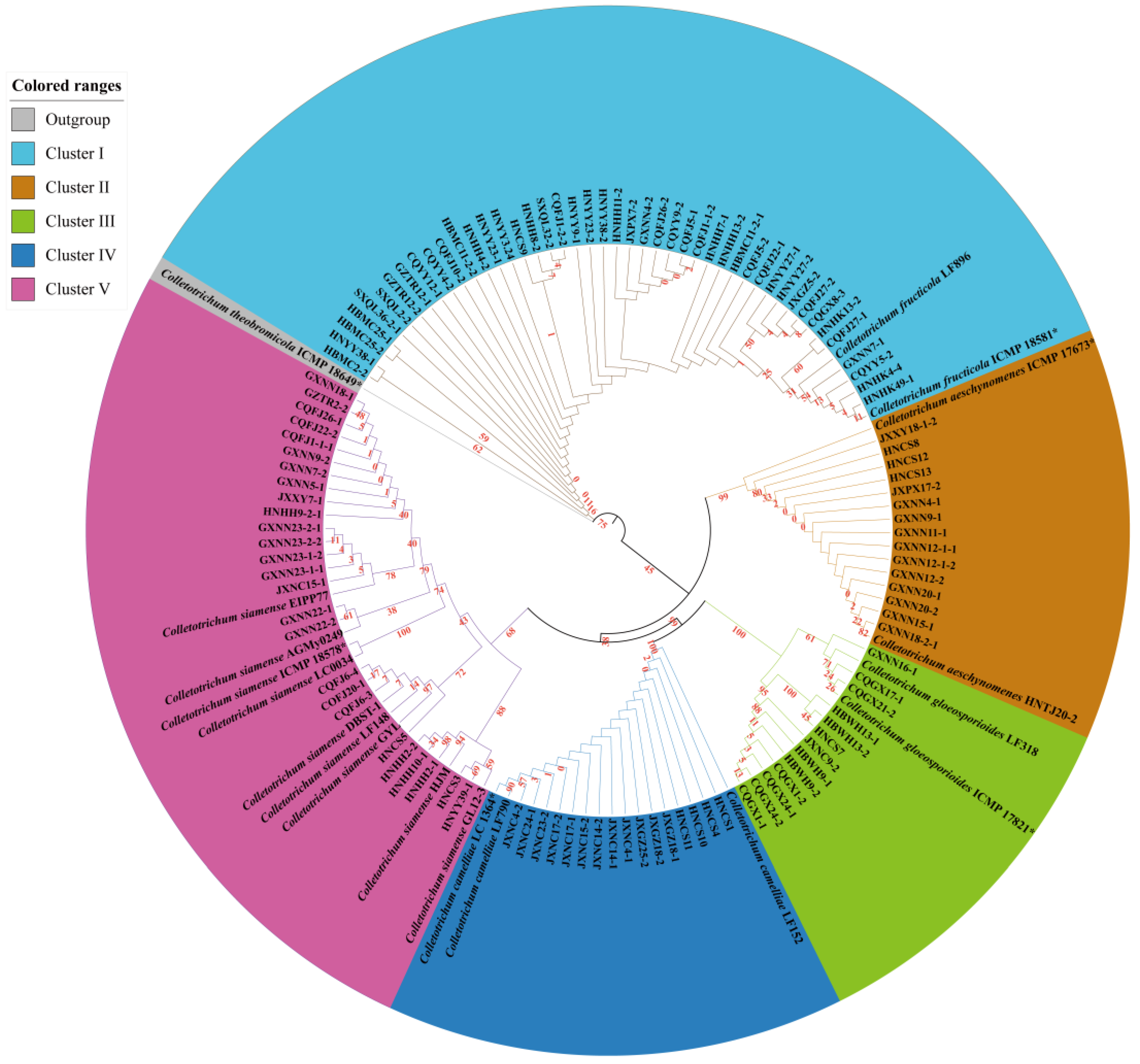
The sequences of 115 tested ApMat genes and other known species of Colletotrichum in GenBnak were analyzed, and the Neighbor-Joining tree was constructed. The sequence datasets for the ApMat were analyzed in combination to establish interspecific relationships within Colletotrichum. The Colletotrichum isolates’ combined species phylogeny comprising 134 sequences, including the outgroup C. theobromicola ICMP18649* (Figure 1).
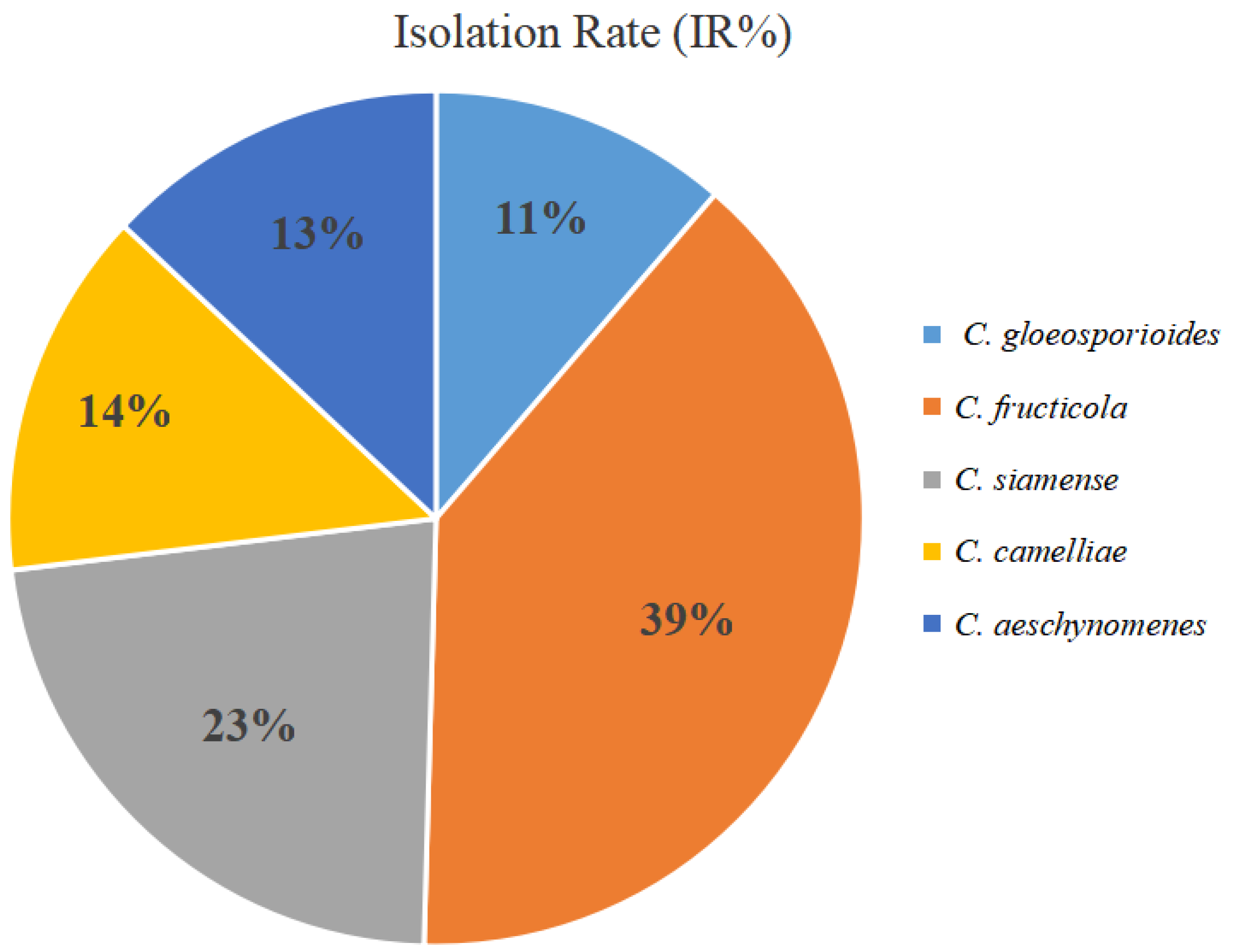
Phylogenetic analyses using the Neighbor-Joining algorithm showed the 115 isolated strains were clustered into five obvious evolutionary branches with a very high support rate, i.e., C. gloeosporioides, C. fructicola, C. siamense, C. camelliae and C. aeschynomenes, including forty-five isolates of C. fructicola, fifteen isolates of C. aeschynomenes, twenty-six isolates of C. siamense, and sixteen isolates of C. camelliae. C. fructicola is the predominant species among Colletotrichum of Ca. japonica in China, accounting for 39% (n = 115) of the isolates tested. (Figure 2).
3.2. Taxonomy
- Colletotrichum gloeosporioides (Penz.) Penz. and Sacc. 1884
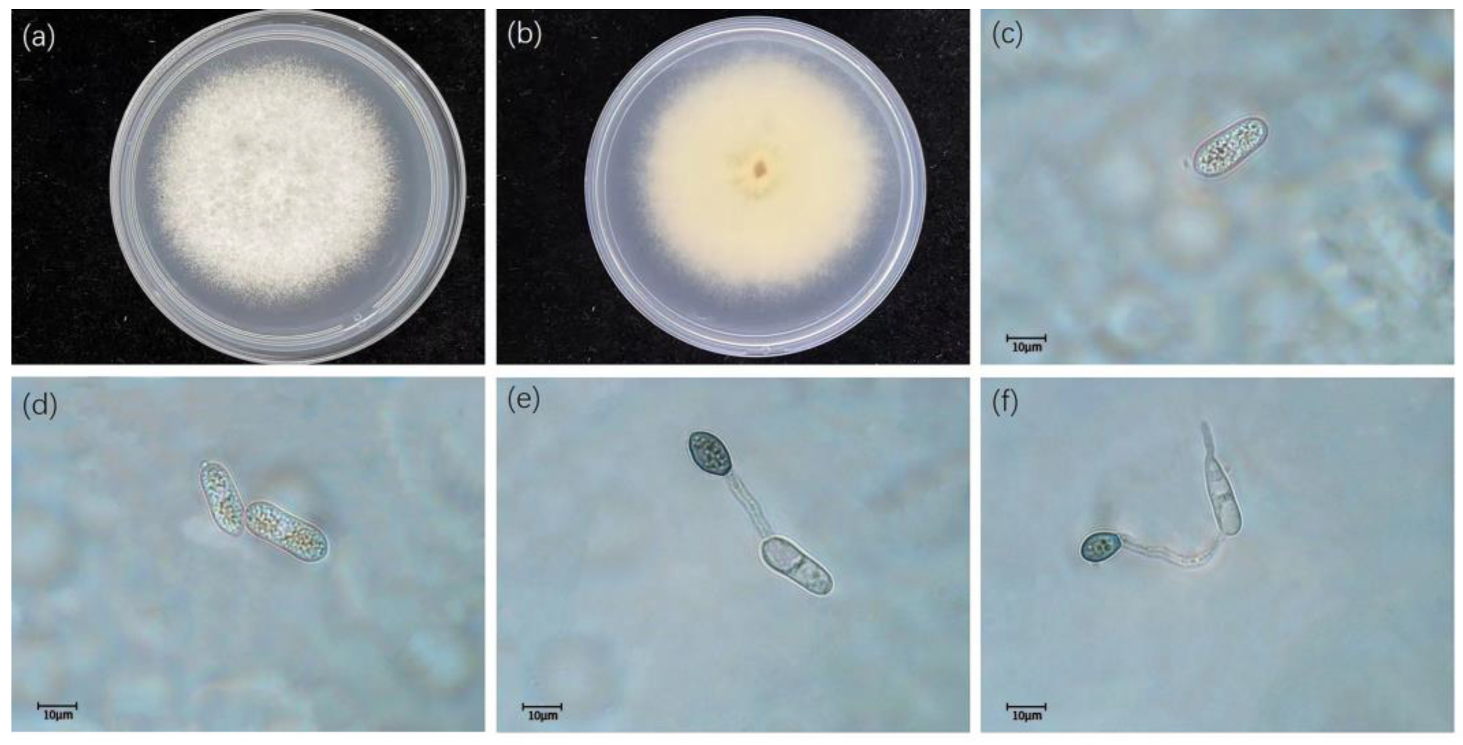
Description: Colonies on PDA reaching 67–69 mm diam after five days. Colonies flat with rose edge, scattered acervuli with orange conidial ooze near center, fuscous black pigment near the edge; reverse honey with fuscous black near the edge. Chlamydospores not observed. Conidia hyaline, aseptate, smooth-walled, cylindrical, both ends bluntly rounded, 15–16.5 × 4.5–5.5 μm. Appressoria medium to dark brown, aseptate, solitary or in groups. Variable in shape, circular, clavate, ellipsoidal or irregular in outline, crenate or slightly lobed at edge, 7.5–9.5 × 5.5–6.5 μm (Figure 3).
- Colletotrichum fructicola Priastuti, L. Cai. and K.D. Hyde. 2009.
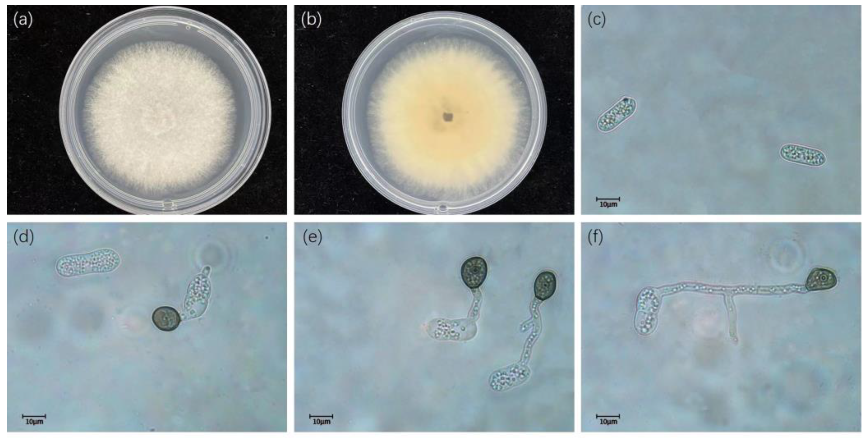
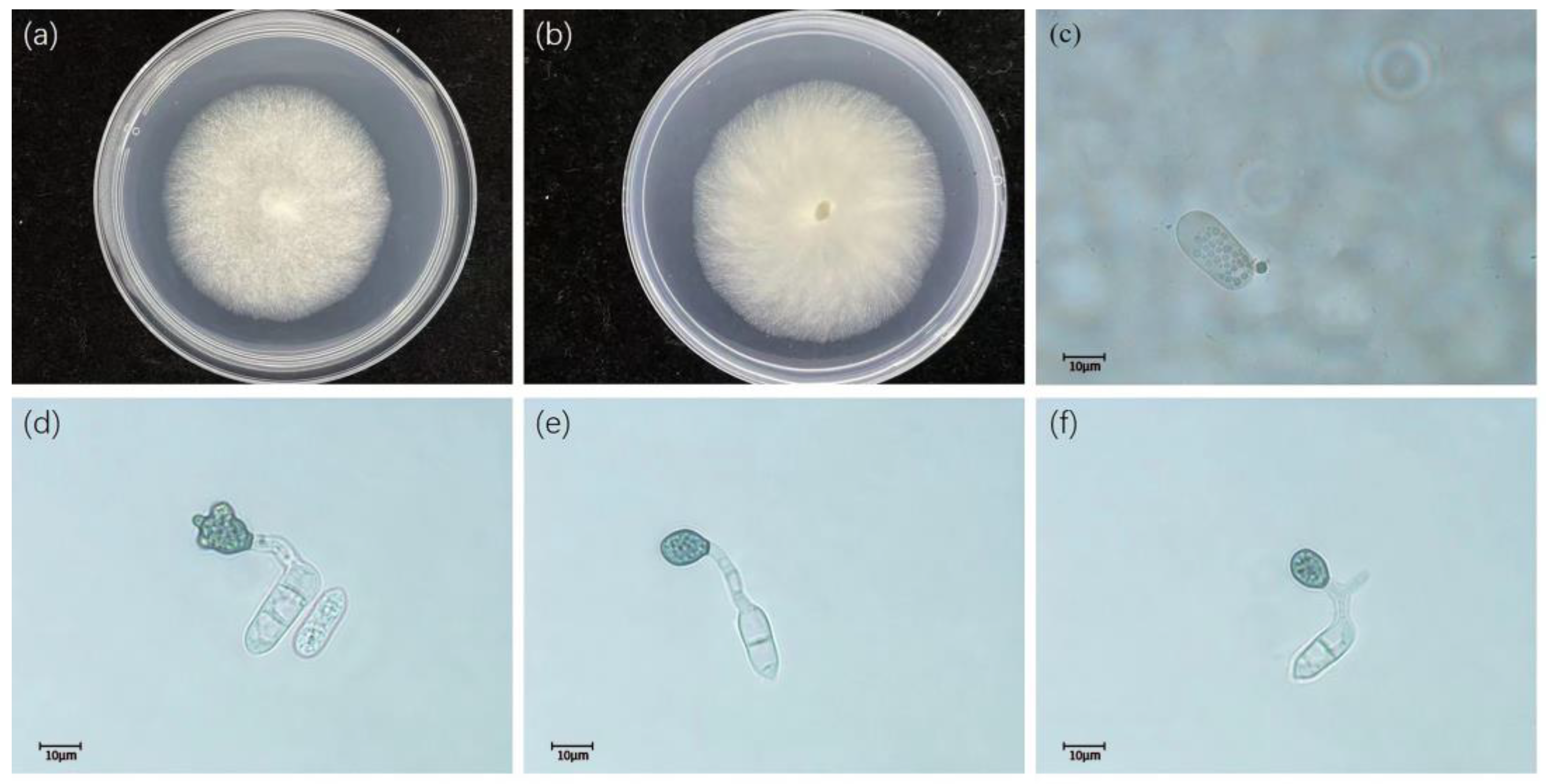
Description: Colonies on PDA reaching 64–66 mm diam after five days. Colonies flat with entire edge, aerial mycelium dense, cottony, pale prey to white aerial mycelium and numerous black stroma scattered over the surface, grey in the center, white at the margin; reverse greyish green. Conidia hyaline, aseptate, smooth-walled, cylindrical, both ends obtuse, 14.5–16.5 × 4.9–5.5 μm (Figure 4).
- Colletotrichum siamense Priastuti, L. Cai. and K.D. Hyde. 2009.
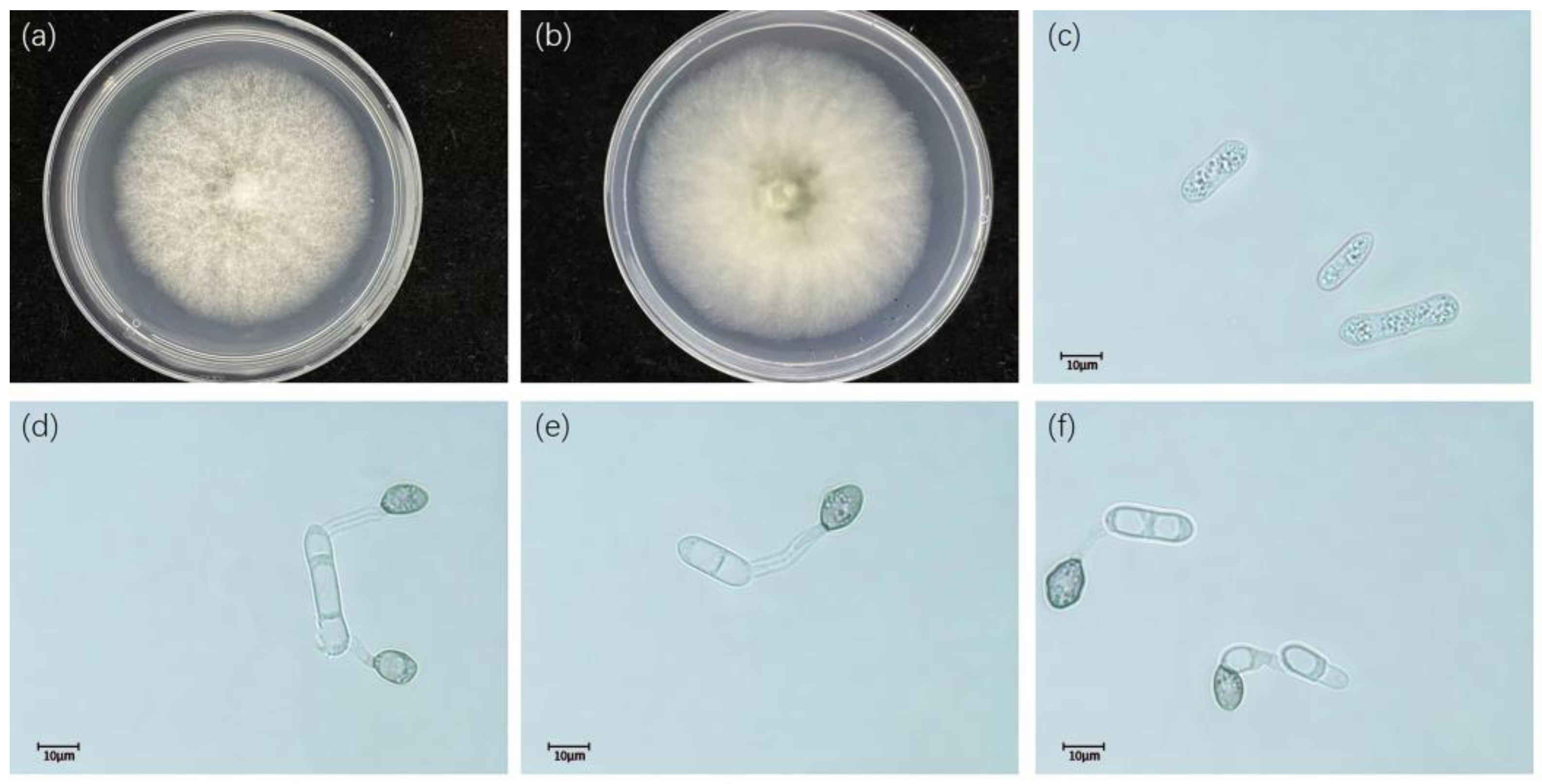
Description: Colonies on PDA reaching 58–61 mm diam after five days. Colonies pale yellow-white, grey, dense cottony aerial mycelium with orange acervular conidiomata at the center; reverse pale yellowish. Conidia hyaline, aseptate, smooth-walled, fusiform to cylindrical, both ends bluntly rounded, 14–15.5 × 4.9–5.2 μm. Appressoria dark brown, solitary, circular, entire to crenate margin, 7.5–10 × 5–6.5 μm (Figure 5).
- Colletotrichum camelliae Massee. 2012
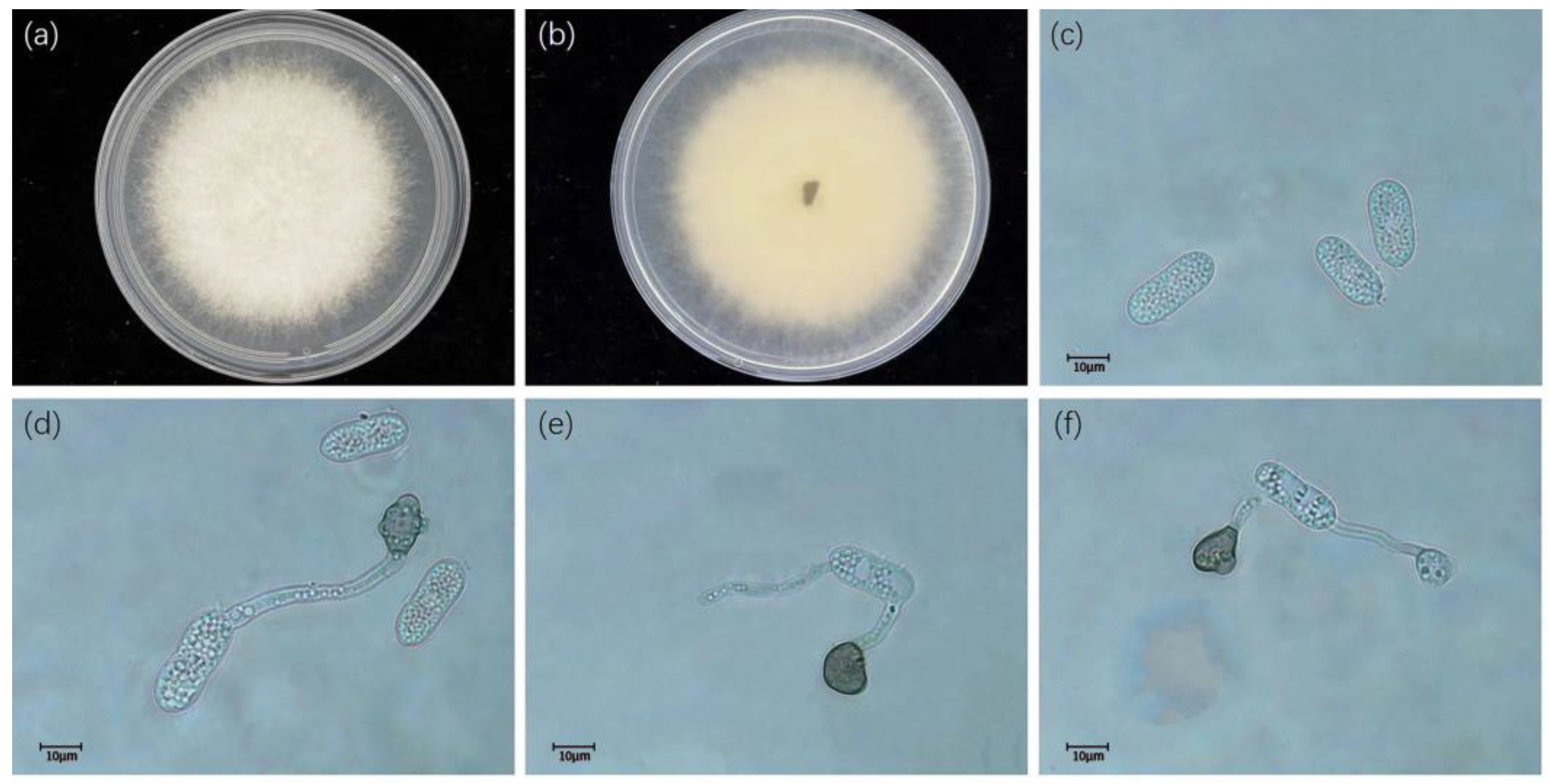
Description: Colonies on PDA reaching 53–56 mm diam after five days. Colonies flat with entire edge, aerial mycelium white, cottony, sparse; reverse white at first, then grey to black at the center. Conidia hyaline, smooth-walled, guttulate, cylindrical with obtuse ends, sometimes narrowed at the center or towards the base, 14–17 × 4.5–5.5 μm. Appressoria irregularly shaped, clavate, crenate, lobed, brown to dark brown, solitary, branched, catenate, with age sometimes complex chlamydospore-like structures develop, 8–11.5 × 5–8.5 μm (Figure 6).
- Colletotrichum aeschynomenes B.S. Weir, P.R. Johnston, and U. Damm. 2012
Description: Colonies on PDA reaching 63–67 mm diam after five days. Colonies pale light grey color with dense cottony aerial mycelium and white to orange conidial masses at the center. Conidia hyaline, aseptate, fusiform to cylindrical, both ends bluntly rounded, 7–9 × 17.5–24 μm. Appressoria were brown, mostly elliptic to cuboid, deeply lobed, 9–11.5 × 9.5–13 μm (Figure 7).
3.3. Pathogenicity Assay
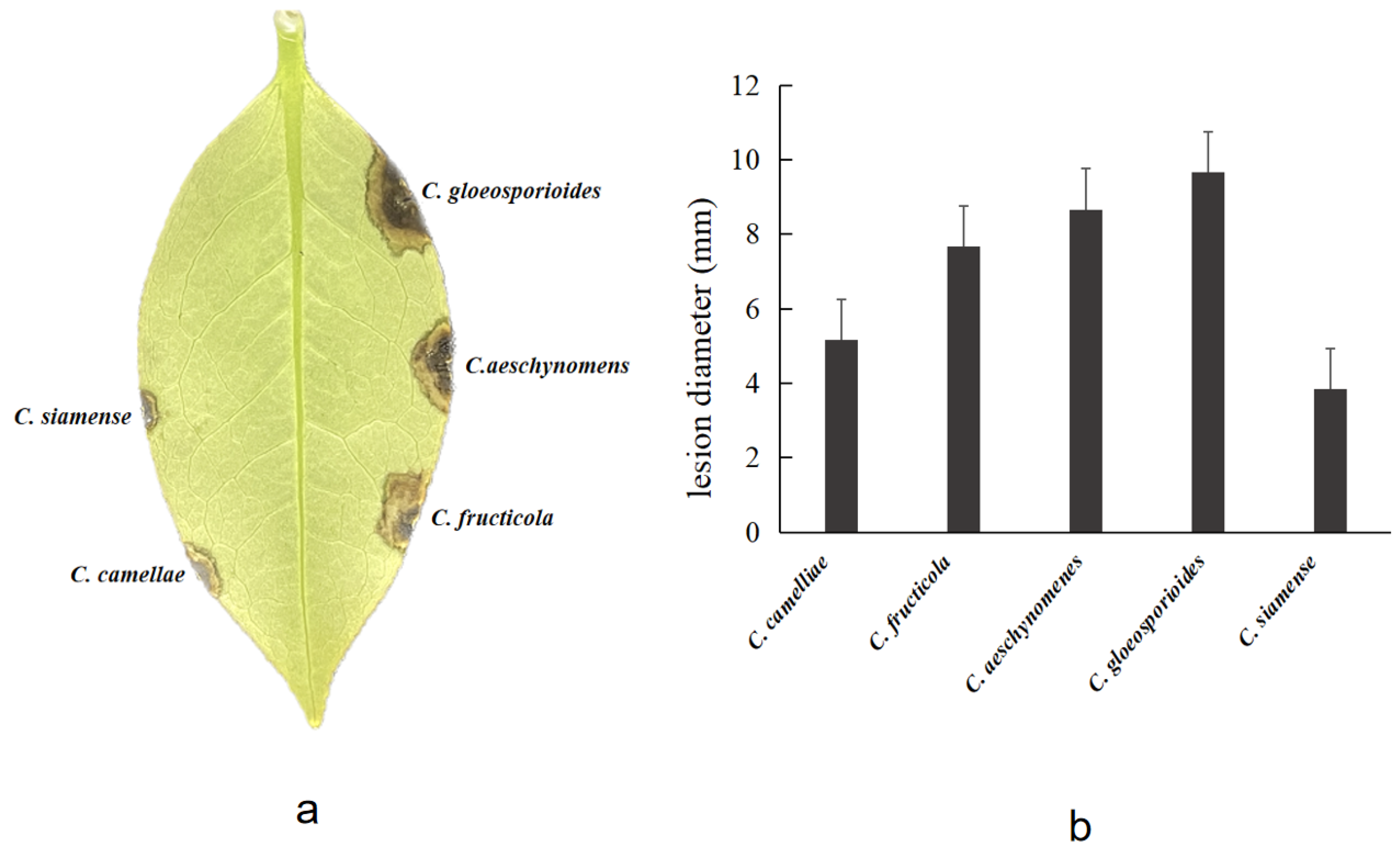
In the pathogenicity tests, C. gloeosporioides, C. fructicola, C. siamense, C. camelliae, and C. aeschynomenes developed brown lesions on wounded leaves after three days, whereas the controls exhibited no symptoms (Figure 8). Koch’s postulates were confirmed by reisolating the same fungi and verifying its colony and morphological characteristics.
4. Discussion
Ca. japonica anthracnose caused by Colletotrichum fungi in China was observed as a common disease. Taking this into account, an investigation of Ca. japonica diseases in China was carried out. A total of 115 isolates were obtained from eight Ca. japonica tree plantations in eight provinces, representing the broad geographical distribution of camellia tree plantations in China. Identification of our collections was processed based on isolates from symptomatic leaves of Ca. japonica using ApMat gene, as well as morphological characteristics. The results showed the isolates identified as C. gloeosporioides, C. fructicola, C. siamense, C. camelliae and C. aeschynomenes, as previously described [24,25,26,27]. However, some species of Gloeosporium theae-sinensis, C. aenigma, C. aracearum and C. camelliae-japoncae were not isolated in this study [28,29,30,31]. Further research is needed to find more species of pathogens.
Colletotrichum is one of the most significant plant within the pathogenic fungi genera, with 200 or more species known to cause diseases in plants and crops across the world [7]. Weir et al. [32] characterized the taxonomy of C. gloeosporioides species complex based on multi-gene and morphological features, which was a significant taxonomic breakthrough. They incorporated multiple gene regions in their phylogenetic analyses, but heavily relied on ACT, CAL, CHS1, GAPDH and ITS gene-regions to redefine species boundaries within this species complex [5,6,9,10,22]. The ITS sequences of Colletotrichum species complex have been considered to be an insufficient variable to reliably distinguish between the different members [33]. In addition, the ApMat marker gene has been used to resolve and improve the systematic classification of Colletotrichum species complexes [15,16,17,19,24]. In this study, phylogenetic analysis was performed on ApMat gene sequences and the 115 isolates were clustered into five obvious evolutionary branches with a very high support rate. Among them, C. fructicola exhibited the most widespread distribution and the highest isolated rate. This is probably because C. fructicola has stronger adaptability to heterogenous habitat.
Pathogenicity experiments of five Colletorchum species from Ca. japonica revealed that all species were capable of infecting wounded leaves. C. gloeosporioides, C. fructicola and C. aeschynomenes were the most virulent. Interestingly, the major pathogens of Ca. japonica anthracnose were found to be distinct in different provinces. This is the first report of C. aeschynomenes causing anthracnose disease in Ca. japonica in China. Further studies will be needed to understand how the disease can be controlled.
5. Conclusions
Five Colletotrichum species on Ca. japonica were described and illustrated. This is the first report of C. aeschynomenes causing anthracnose in Ca. japonica in China. Pathogenicity tests indicated that there were significant differences in virulence among the five Colletotrichum species when inoculated on the leaves of Ca. japonica. This study provides valuable information for the identification and control of this plant disease.
Author Contributions
Experiments, L.W.; Writing—original draft preparation, L.W.; Writing—review and editing, L.W. and H.L. All authors have read and agreed to the published version of the manuscript.
Funding
This research was funded by the National Natural Science Foundation of China (32071765).
Institutional Review Board Statement
Not applicable.
Data Availability Statement
All sequence data are available in NCBI GenBank following the accession numbers in the manuscript.
Acknowledgments
We are grateful for the assistance of Qin Yang and Song Sheng.
Conflicts of Interest
The authors declare no conflict of interest.
References
- Wei, Y.H. Research progress on phenotypic genetic diversity of Camellia germplasm resources in China. South China Agric. 2020, 14, 66–68. [Google Scholar]
- Li, X.L. Analysis of nutritional components and poisonous elements in flowers of four Camellia species. For. Res. 2010, 23, 298–301. [Google Scholar]
- Ana, M.T.; Clara, S. A review on the biological activity of Camellia species. Molecules 2021, 26, 2178. [Google Scholar] [CrossRef]
- Wang, J.W.; Qian, X.M.; Zhang, B.X. Camellia diseases in Zhejiang Province. Acta Phytopathol. Sin. 1990, 20, 2. [Google Scholar]
- Luo, H.S. The Control of Camellia japonica diseases and pests. China Flowers Hortic. 2016, 4, 32–34. [Google Scholar]
- Lv, Y.F. Diseases and Insect Pests of Camellia japonica L. and Their Control Techniques. J. Agric. Catastropholgy 2021, 11, 18–19. [Google Scholar]
- Liu, L.P.; Gao, J.; Li, Y. Advances in knowledge of the fungi referred to the genus Colletotrichum. J. Fungal Res. 2020, 18, 266–281. [Google Scholar]
- Li, L.L.; Yang, Q.; Li, H. Morphology, phylogeny, and pathogenicity of pestalotioid species on Camellia oleifera in China. J. Fungi 2021, 7, 1080. [Google Scholar] [CrossRef]
- Wang, Y.C.; Hao, X.Y.; Wang, L.; Xiao, B.; Wang, X.C.; Yang, Y.J. Diverse Colletotrichum species cause anthracnose of tea plants (Camellia sinensis (L.) O. Kuntze) in China. Sci. Rep. 2016, 6, 35387. [Google Scholar] [CrossRef] [PubMed] [Green Version]
- Jayawardena, R.S.; Hyde, K.D.; Damm, U.; Cai, L.; Liu, M.; Li, X.H.; Zhang, W.; Zhao, W.S.; Yan, J.Y. Notes on currently accepted spcies of Colletotrichum. Mycosphere 2016, 7, 1192–1260. [Google Scholar] [CrossRef]
- Liu, F.; Ma, Z.Y.; Hou, L.W.; Diao, Y.Z.; Wu, W.P.; Damm, U.; Song, S.; Cai, L. Updating species diversity of Colletotrichum, with a phylogenomic overview. Stud. Mycol. 2022, 101, 1–56. [Google Scholar] [CrossRef]
- Hou, S.F.; Hua, Z.Y.; Liu, J.J.; Dong, Z.F.; Feng, Z.K.; Yan, J.Q.; Wang, H.Q. Multi-gene joint identification and pathogenicity analysis of Colletotrichun gloeosporioides complex of strawberry in China. J. China Agric. Univ. 2022, 27, 82–94. [Google Scholar]
- Sun, W.; Lei, T.Y.; Yuan, H.Z.; Chen, S.N. First Report of Anthracnose Caused by Colletotrichum kahawae and Colletotrichum horri on Tea-oil Tree in China. Plant Dis. 2022. [Google Scholar] [CrossRef]
- Wang, Y.X.; Chen, J.Y.; Xu, X.W.; Cheng, J.Y.; Zheng, L.; Huang, J.B.; Li, D.W. Identification and characterization of Colletotrichum species associated with anthracnose disease of Camellia oleifera in China. Plant Dis. 2020, 104, 474–482. [Google Scholar] [CrossRef]
- Lu, Y.; He, C.P.; Wu, W.H.; Zheng, J.L.; Huang, X.; Xi, J.G.; Tan, S.B.; Xian, Y.K. Pathogenicity differentiation of Coffee anthracnose in China. Southwest China J. Agric. Sci. 2021, 34, 1008–1014. [Google Scholar]
- Silva, D.N.; Talhinhas, P.; Várzea, V.; Cai, L.; Paulo, S.O.; Batista, D. Application of the Apn2/MAT locus to improve the systematics of the Colletotrichum gloeosporioides complex: An example from coffee (Coffea spp.) hosts. Mycologia 2012, 104, 396–409. [Google Scholar] [CrossRef] [Green Version]
- Li, L.L.; Li, H. First report of Colletotrichum aeschynomenes causing anthracnose on Camellia oleifera in China. For. Pathol. 2022, 52, e12770. [Google Scholar] [CrossRef]
- Doyle, J.J.; Doyle, J.L. Isolation of plant DNA from fresh tissue. Focus 1990, 12, 13–15. [Google Scholar]
- Kumar, G.S.N.; Weir, B.S.; Hyde, K.D.; Shenoy, B.D. The ApMat market can resolve Colletotrichum species: A case study with Mangifera indica. Fungal Divers. 2013, 61, 117–138. [Google Scholar] [CrossRef]
- Kumar, S.; Stecher, G.; Tamura, K. MEGA7: Molecular evolutionary genetics analysis version 7.0 for bigger datasets. Mol. Biol. Evol. 2016, 33, 1870–1874. [Google Scholar] [CrossRef] [Green Version]
- Liu, F.; Weir, B.S.; Damm, U.; Crous, P.W.; Wang, Y.; Liu, B.; Wang, M.; Zhangm, M.; Cai, L. Unravelling Colletotrichum species associated with Camellia: Employing ApMat and GS loci to resolve species in the C. gloeosporioides complex. Persoonia 2015, 35, 63–86. [Google Scholar] [CrossRef] [Green Version]
- Douanla-Meli, C.; Unger, J.G. Phylogenetic study of the Colletotrichum species on imported citrus fruits uncovers a low diversity and a new species in the Colletotrichum gigasporum complex. Fungal Biol. 2017, 121, 858–868. [Google Scholar] [CrossRef]
- Gong, C.Y.; Liu, J.J.; Deng, Q.; Zhang, L.X. Identification and Pathogenicity of Colletotrichum species causing anthracnose on Camellia sinensis. Acta Hortic. Sin. 2022, 49, 1092–1101. [Google Scholar] [CrossRef]
- Li, H.; Li, S.Z.; Wang, Y.C.; Liu, J.A.; Xu, J.P.; Zhou, G.Y. Identification of the pathogens causing anthracnose of Camellia oleifera in nursery and their resistence to fungcides. Sci. Silvae Sin. 2019, 55, 85–94. [Google Scholar]
- Sharma, G.; Maymon, M.; Freeman, S. Epidemiology, pathology and identification of Colletotrichum including a novel species associated with avocado (Persea americana) anthracnose in Israel. Sci. Rep. 2017, 7, 15839. [Google Scholar] [CrossRef] [Green Version]
- Huang, Y.N.; Zhu, T.H. A Preliminary Study of anthracnose on the leaf of Camellia. J. Sichuan For. Sci. Technol. 2005, 26, 10–13. [Google Scholar]
- Peng, X.J.; Yuan, Y.X.; Zhang, S.K.; Zhou, X.Z. First Report of Anthracnose on Camellia japonica Caused by Colletotrichum siamense in Zhejiang Province, China. Am. Phytopathol. Soc. 2022, 106, 768. [Google Scholar] [CrossRef]
- Han, C.Z.; Zhao, H.Y. Pathogen identification of Camellia anthracnose disease. For. Pest Dis. 2016, 35, 5–6. [Google Scholar]
- Chen, X.; Wang, T.; Guo, H.; Zhu, P.K. First Report of Anthracnose of Camellia sasanqua Caused by Colletotrichum aenigma in China. Plant Dis. 2019, 103, 1423. [Google Scholar] [CrossRef]
- Yang, S.; Wang, H.X.; Yi, Y.J.; Tan, L.L. First Report that Colletotrichum aenigma Causes Leaf Spots on Camellia japonica in China. Plant Dis. 2019, 103, 2127. [Google Scholar] [CrossRef]
- Hou, L.W.; Liu, F.; Duan, W.J.; Cai, L. Colletotrichum aracearum and C. camelliae-japoncae, two holomorphic new species from China and Japan. Mycosphere 2016, 7, 1111–1123. [Google Scholar] [CrossRef]
- Weir, B.S.; Johnston, P.R.; Damm, U. The Colletotrichum gloeosporioides species comple. Stud. Mycol. 2012, 73, 115–180. [Google Scholar] [CrossRef] [Green Version]
- Wang, W.; de Silva, D.D.; Moslemi, A.; Edwards, J.; Ades, P.K.; Crous, P.W.; Taylor, P.W.J. Colletotrichum Species Causing Anthracnose of Citrus in Australia. J. Fungi. 2021, 7, 47. [Google Scholar] [CrossRef]
Figure 1.
Phylogenetic tree based on ApMat gene sequences using the Neighbor-Joining method in MEGA7 with 1000 bootstrap replications. The isolates obtained in this study are marked in bold. The corresponding sequences for the reference species were retrieved from NCBI GenBank. The numbers at nodes represent their bootstrap support. *: type strain.

Figure 2.
Isolation Rate (IR%) of Colletotrichum species isolated from Ca. japonica leaves.

Figure 3.
C. gloeosporioides (CQGX 24-1). (a) Colony on PDA. (b) Reverse side of the colony on PDA. (c) Conidia. (d–f) Appressoria. Scale bars = 10 μm.
Figure 3.
C. gloeosporioides (CQGX 24-1). (a) Colony on PDA. (b) Reverse side of the colony on PDA. (c) Conidia. (d–f) Appressoria. Scale bars = 10 μm.

Figure 4.
Colletotrichum fructicola (HNHH7-1). (a) Colony on PDA. (b) Reverse side of the colony on PDA. (c) Conidia. (d–f) Appressoria. Scale bars = 10 μm.
Figure 4.
Colletotrichum fructicola (HNHH7-1). (a) Colony on PDA. (b) Reverse side of the colony on PDA. (c) Conidia. (d–f) Appressoria. Scale bars = 10 μm.

Figure 5.
Colletotrichum siamense (CQFJ6-3). (a) Colony on PDA. (b) Reverse side of the colony on PDA. (c,d) Conidia. (e,f) Appressoria. Scale bars = 10 μm.
Figure 5.
Colletotrichum siamense (CQFJ6-3). (a) Colony on PDA. (b) Reverse side of the colony on PDA. (c,d) Conidia. (e,f) Appressoria. Scale bars = 10 μm.

Figure 6.
Colletotrichum camelliae (JXGZ18-1). (a) Colony on PDA. (b) Reverse side of the colony on PDA. (c) Conidia. (d–f) Appressoria. Scale bars = 10 μm.
Figure 6.
Colletotrichum camelliae (JXGZ18-1). (a) Colony on PDA. (b) Reverse side of the colony on PDA. (c) Conidia. (d–f) Appressoria. Scale bars = 10 μm.

Figure 7.
Colletotrichum aeschynomenes (GXNN4-1). (a) Colony on PDA. (b) Reverse side of the colony on PDA. (c) Conidia. (d–f) Appressoria. Scale bars = 10 μm.
Figure 7.
Colletotrichum aeschynomenes (GXNN4-1). (a) Colony on PDA. (b) Reverse side of the colony on PDA. (c) Conidia. (d–f) Appressoria. Scale bars = 10 μm.

Figure 8.
Pathogenicity of five Colletotrichum species from Ca. japonica leaves. (a) Induced symptoms on non-wounded Ca. japonica leaves after 3 days. (b). The virulence of the isolates was evaluated by measuring the diameters of the necrotic lesions.
Figure 8.
Pathogenicity of five Colletotrichum species from Ca. japonica leaves. (a) Induced symptoms on non-wounded Ca. japonica leaves after 3 days. (b). The virulence of the isolates was evaluated by measuring the diameters of the necrotic lesions.


Table 1.
GenBank accession numbers of nucleotide sequences used in this study.
| Taxon | Isolate Designation | Host | Geographic Location | ApMat GenBank | References |
|---|---|---|---|---|---|
| C. camelliae | LF152 | Camellia sp. | China | KJ954506.1 | [21] |
| LF790 | Cinamomum zeylanicum | India | KU239747.1 | Direct Submission | |
| LC1364 * | Camellia sinensis | China | KJ954497.1 | [21] | |
| HNCS1 | Camellia japonica | China | OQ198468 | In this study | |
| HNCS4 | Camellia japonica | China | OQ198469 | In this study | |
| HNCS10 | Camellia japonica | China | OQ198470 | In this study | |
| HNCS11 | Camellia japonica | China | OQ198471 | In this study | |
| JXGZ18-1 | Camellia japonica | China | OQ198472 | In this study | |
| JXGZ18-2 | Camellia japonica | China | OQ198473 | In this study | |
| JXGZ25-2 | Camellia japonica | China | OQ198474 | In this study | |
| JXNC4-1 | Camellia japonica | China | OQ198475 | In this study | |
| JXNC4-2 | Camellia japonica | China | OQ198476 | In this study | |
| JXNC14-1 | Camellia japonica | China | OQ198477 | In this study | |
| JXNC14-2 | Camellia japonica | China | OQ198478 | In this study | |
| JXNC15-2 | Camellia japonica | China | OQ198479 | In this study | |
| JXNC17-1 | Camellia japonica | China | OQ198480 | In this study | |
| JXNC17-2 | Camellia japonica | China | OQ198481 | In this study | |
| JXNC23-2 | Camellia japonica | China | OQ198482 | In this study | |
| JXNC24-1 | Camellia japonica | China | OQ198483 | In this study | |
| C. siamense | AGMy0249 | Citrus pennivesiculata | Bangladesh | KX578769.1 | [22] |
| LC0034 | Coffee berry | Thailand | JQ899288.1 | Direct Submission | |
| HJM | Loropetalum chinense | China | MG717312.1 | Direct Submission | |
| GL12-3 | Plum | China | OM816816.1 | Direct Submission | |
| GYL | Magnolia grandiflora | China | MG717298.1 | Direct Submission | |
| LF148 | Camellia sp. | China | KJ954504.1 | [21] | |
| DBST-1 | Cycas debaoensis | China | MT786728.1 | Direct Submission | |
| EIPP77 | Coffee | China | MK344209.1 | Direct Submission | |
| ICMP18649 * | Coffea arabica | China | JQ899289 | [21] | |
| HNCS3 | Camellia japonica | China | OQ198484 | In this study | |
| HNCS5 | Camellia japonica | China | OQ198485 | In this study | |
| HNYY39-1 | Camellia japonica | China | OQ198486 | In this study | |
| HNHH2-1 | Camellia japonica | China | OQ198487 | In this study | |
| HNHH2-2 | Camellia japonica | China | OQ198488 | In this study | |
| HNHH9-2-1 | Camellia japonica | China | OQ198489 | In this study | |
| HNHH10-1 | Camellia japonica | China | OQ198490 | In this study | |
| JXXY7-1 | Camellia japonica | China | OQ198491 | In this study | |
| JXNC15-1 | Camellia japonica | China | OQ198492 | In this study | |
| GXNN5-1 | Camellia japonica | China | OQ198493 | In this study | |
| GXNN7-2 | Camellia japonica | China | OQ198494 | In this study | |
| GXNN9-2 | Camellia japonica | China | OQ198495 | In this study | |
| GXNN18-1 | Camellia japonica | China | OQ198496 | In this study | |
| GXNN22-1 | Camellia japonica | China | OQ198497 | In this study | |
| GXNN22-2 | Camellia japonica | China | OQ198498 | In this study | |
| GXNN23-1-1 | Camellia japonica | China | OQ198499 | In this study | |
| GXNN23-1-2 | Camellia japonica | China | OQ198500 | In this study | |
| GXNN23-2-1 | Camellia japonica | China | OQ198501 | In this study | |
| GXNN23-2-2 | Camellia japonica | China | OQ198502 | In this study | |
| CQFJ1-1-1 | Camellia japonica | China | OQ198503 | In this study | |
| CQFJ6-3 | Camellia japonica | China | OQ198504 | In this study | |
| CQFJ6-4 | Camellia japonica | China | OQ198505 | In this study | |
| CQFJ20-1 | Camellia japonica | China | OQ198506 | In this study | |
| CQFJ22-2 | Camellia japonica | China | OQ198507 | In this study | |
| CQFJ26-1 | Camellia japonica | China | OQ198508 | In this study | |
| GZTR2-2 | Camellia japonica | China | OQ198509 | In this study | |
| C. gloeosporioides | LF318 | Ca. sinensis | China | KJ954541.1 | [21] |
| ICMP1782 * | Citrus sinensis | Italy | JQ807843 | [21] | |
| HNCS7 | Camellia japonica | China | OQ198510 | In this study | |
| JXNC9-2 | Camellia japonica | China | OQ198511 | In this study | |
| HBWH9-1 | Camellia japonica | China | OQ198512 | In this study | |
| HBWH9-2 | Camellia japonica | China | OQ198513 | In this study | |
| HBWH13-1 | Camellia japonica | China | OQ198514 | In this study | |
| HBWH13-2 | Camellia japonica | China | OQ198515 | In this study | |
| GXNN16-1 | Camellia japonica | China | OQ198516 | In this study | |
| CQGX1-1 | Camellia japonica | China | OQ198517 | In this study | |
| CQGX1-2 | Camellia japonica | China | OQ198518 | In this study | |
| CQGX17-1 | Camellia japonica | China | OQ198519 | In this study | |
| CQGX21-2 | Camellia japonica | China | OQ198520 | In this study | |
| CQGX24-1 | Camellia japonica | China | OQ198521 | In this study | |
| CQGX24-2 | Camellia japonica | China | OQ198522 | In this study | |
| C. aeschynomenes | ICMP1767 * | Aeschynomene viginica | China | KM360145.1 | [21] |
| HNTJ20-1 | Camellia oleifera | China | MZ8321172.1 | [17] | |
| HNCS8 | Camellia japonica | China | OQ198523 | In this study | |
| HNCS12 | Camellia japonica | China | OQ198524 | In this study | |
| HNCS13 | Camellia japonica | China | OQ198525 | In this study | |
| JXPX17-2 | Camellia japonica | China | OQ198526 | In this study | |
| JXXY18-1-2 | Camellia japonica | China | OQ198527 | In this study | |
| GXNN4-1 | Camellia japonica | China | OQ198528 | In this study | |
| GXNN9-1 | Camellia japonica | China | OQ198529 | In this study | |
| GXNN11-1 | Camellia japonica | China | OQ198530 | In this study | |
| GXNN12-1-1 | Camellia japonica | China | OQ198531 | In this study | |
| GXNN12-1-2 | Camellia japonica | China | OQ198532 | In this study | |
| GXNN12-2 | Camellia japonica | China | OQ198533 | In this study | |
| GXNN15-1 | Camellia japonica | China | OQ198534 | In this study | |
| GXNN18-2-1 | Camellia japonica | China | OQ198535 | In this study | |
| GXNN20-1 | Camellia japonica | China | OQ198536 | In this study | |
| GXNN20-2 | Camellia japonica | China | OQ198537 | In this study | |
| C. theobromicola | ICMP18649 * | Theobroma cacao | Panama | KC790726 | [21] |
| C. fructicola | LF896 | Ca. sinensis | China | KJ954624.1 | [21] |
| LC0033 | Coffea arabica | India | JQ807838.1 | Direct Submission | |
| ICMP18581 * | Coffea arabica | Thailand | JQ07838 | [21] | |
| HNCS9 | Camellia japonica | China | OQ198538 | In this study | |
| HNYY3.24 | Camellia japonica | China | OQ198539 | In this study | |
| HNYY9-1 | Camellia japonica | China | OQ198540 | In this study | |
| HNYY23-1 | Camellia japonica | China | OQ198541 | In this study | |
| HNYY23-2 | Camellia japonica | China | OQ198542 | In this study | |
| HNYY27-1 | Camellia japonica | China | OQ198543 | In this study | |
| HNYY27-2 | Camellia japonica | China | OQ198544 | In this study | |
| HNYY38-1 | Camellia japonica | China | OQ198545 | In this study | |
| HNYY38-2 | Camellia japonica | China | OQ198546 | In this study | |
| HNHH4-2 | Camellia japonica | China | OQ198547 | In this study | |
| HNHH7-1 | Camellia japonica | China | OQ198548 | In this study | |
| HNHH8-2 | Camellia japonica | China | OQ198549 | In this study | |
| HNHH11-2 | Camellia japonica | China | OQ198550 | In this study | |
| HNHH13-2 | Camellia japonica | China | OQ198551 | In this study | |
| JXPX7-2 | Camellia japonica | China | OQ198552 | In this study | |
| JXGZ5-2 | Camellia japonica | China | OQ198553 | In this study | |
| HBMC2-2 | Camellia japonica | China | OQ198554 | In this study | |
| HBMC11-2-1 | Camellia japonica | China | OQ198555 | In this study | |
| HBMC11-2-2 | Camellia japonica | China | OQ198556 | In this study | |
| HBMC25-1 | Camellia japonica | China | OQ198557 | In this study | |
| HBMC25-2 | Camellia japonica | China | OQ198558 | In this study | |
| GXNN4-2 | Camellia japonica | China | OQ198559 | In this study | |
| GXNN7-1 | Camellia japonica | China | OQ198560 | In this study | |
| CQGX8-3 | Camellia japonica | China | OQ198561 | In this study | |
| CQFJ1-1-2 | Camellia japonica | China | OQ198562 | In this study | |
| CQFJ1-2-2 | Camellia japonica | China | OQ198563 | In this study | |
| CQFJ5-1 | Camellia japonica | China | OQ198564 | In this study | |
| CQFJ5-2 | Camellia japonica | China | OQ198565 | In this study | |
| CQFJ10-2 | Camellia japonica | China | OQ198566 | In this study | |
| CQFJ22-1 | Camellia japonica | China | OQ198567 | In this study | |
| CQFJ26-2 | Camellia japonica | China | OQ198568 | In this study | |
| CQFJ27-1 | Camellia japonica | China | OQ198569 | In this study | |
| CQFJ27-2 | Camellia japonica | China | OQ198570 | In this study | |
| CQYY4-2 | Camellia japonica | China | OQ198571 | In this study | |
| CQYY5-2 | Camellia japonica | China | OQ198572 | In this study | |
| CQYY9-2 | Camellia japonica | China | OQ198573 | In this study | |
| CQYY12-1 | Camellia japonica | China | OQ198574 | In this study | |
| HNHK4-4 | Camellia japonica | China | OQ198575 | In this study | |
| HNHK13-2 | Camellia japonica | China | OQ198576 | In this study | |
| HNHK49-1 | Camellia japonica | China | OQ198577 | In this study | |
| GZTR12-1 | Camellia japonica | China | OQ198578 | In this study | |
| GZTR12-2 | Camellia japonica | China | OQ198579 | In this study | |
| SXQL2-2 | Camellia japonica | China | OQ198580 | In this study | |
| SXQL32-2 | Camellia japonica | China | OQ198581 | In this study | |
| SXQL36-2-1 | Camellia japonica | China | OQ198582 | In this study |
Note: the asterisk indicates the ex-type strain.
Disclaimer/Publisher’s Note: The statements, opinions and data contained in all publications are solely those of the individual author(s) and contributor(s) and not of MDPI and/or the editor(s). MDPI and/or the editor(s) disclaim responsibility for any injury to people or property resulting from any ideas, methods, instructions or products referred to in the content. |
© 2023 by the authors. Licensee MDPI, Basel, Switzerland. This article is an open access article distributed under the terms and conditions of the Creative Commons Attribution (CC BY) license (https://creativecommons.org/licenses/by/4.0/).
Share and Cite
MDPI and ACS Style
Wen, L.; Li, H. Morphology, Phylogeny, and Pathogenicity of Colletotrichum Species Causing Anthracnose in Camellia japonica in China. Diversity 2023, 15, 516. https://doi.org/10.3390/d15040516
AMA Style
Wen L, Li H. Morphology, Phylogeny, and Pathogenicity of Colletotrichum Species Causing Anthracnose in Camellia japonica in China. Diversity. 2023; 15(4):516. https://doi.org/10.3390/d15040516
Chicago/Turabian StyleWen, Lixia, and He Li. 2023. "Morphology, Phylogeny, and Pathogenicity of Colletotrichum Species Causing Anthracnose in Camellia japonica in China" Diversity 15, no. 4: 516. https://doi.org/10.3390/d15040516
Note that from the first issue of 2016, this journal uses article numbers instead of page numbers. See further details here.