
Identification of Cytospora Species Isolated from Branch Canker Diseases of Woody Plants in Tibet, China

1
Key Laboratory of Forest Ecology in Tibet Plateau, Ministry of Education, Institute of Tibet Plateau Ecology, Tibet Agricultual & Animal Husbandry University, Nyingchi 860000, China
2
National Key Station of Field Scientific Observation & Experiment, Nyingchi 860000, China
3
Key Laboratory of Biodiversity Conservation of National Forestry and Grassland Administration, Ecology and Nature Conservation Institute, Chinese Academy of Forestry, Beijing 100091, China
*
Authors to whom correspondence should be addressed.
Forests 2024, 15(1), 121; https://doi.org/10.3390/f15010121
Submission received: 24 November 2023
/
Revised: 3 January 2024
/
Accepted: 5 January 2024
/
Published: 8 January 2024
(This article belongs to the Special Issue Diversity, Taxonomy and Functions of Forest Microorganisms)
Abstract
:Branch canker diseases are important in forest ecosystems, causing economic and ecological losses. Members of Cytospora are common on cankered branches and associated with the diseases. Investigations on Cytospora cankers were conducted in Tibet, China, during 2022 and 2023. Samples were collected from Euonymus japonicus, Larix gmelinii, Malus pumila, M. spectabilis and Rosa omeiensis f. pteracantha, and cultures were obtained and identified by morphological features and molecular phylogeny of a combination of internal transcribed spacer region rDNA (ITS), the partial actin (act) region, RNA polymerase II second largest subunit (rpb2) gene, the translation elongation factor 1-alpha (tef1) gene and the partial beta-tubulin (tub2) gene. As a result, a new species is proposed herein named Cytospora lhasaensis and four known species are described for the first time from Tibet, viz. C. euonymina, C. gigaspora, C. mali and C. schulzeri. The current research enhances our understanding of the Cytospora species associated with woody host diseases in Tibet, China.
1. Introduction
Branch canker disease is a serious problem in forest plants, causing plant barks to become cracked, discolored or even sunken. Species of several fungal genera are responsible for this disease, such as Aplosporella Speg., Botryosphaeria Ces. & De Not., Coryneum Nees, Cytospora Ehrenb., Dendrostoma X.L. Fan & C.M. Tian, Diaporthe Fuckel, Melanconis Tul. & C. Tul. and Endothia Fr. [1,2,3,4,5,6,7,8]. Of these, Cytospora is common on the cankered branches of woody plants, with C. chrysosperma (Pers.) Fr. widely causing polar and willow diseases in China [9].
Cytospora was initially established based on C. betulina Ehrenb., C. epimyces Ehrenb., C. resinae Ehrenb. and C. ribis [10]. Cytospora chrysosperma was subsequently selected as the type species [11]. Species in this genus are morphologically similar in having allantoid hyaline conidia and ascospores [12,13,14]. During species identification, host association and spore morphology were important than molecular phylogeny [15,16,17]. However, recently, many cryptic species were revealed from the same host with similar spore morphologies by polyphasic methods using a combination of morphology and molecular phylogeny [18,19,20,21,22]. For example, Cytospora vinacea D.P. Lawr., Travadon & Pouzoulet and C. viticola D.P. Lawr., and Travadon & Pouzoulet cause Vitis vinifera cankers in the USA [23]; Cytospora ailanthicola X.L. Fan & C.M. Tian and five other Cytospora species are responsible for Chinese polar canker diseases [24]; eight species of Cytospora are associated with willow cankers in China [25]; and C. kuanchengensis C.M. Tian & N. Jiang and five other Cytospora species cause Castanea mollissima Blume branch canker in China [26].
Tibet is located in the Qinghai–Tibet Plateau, which is known as the third pole of the earth. Euonymus japonicus Thunb. (Celastraceae R. Br.), Malus spectabilis Borkh. (Rosaceae Juss.) and Larix gmelinii Kuzeneva (Pinaceae Spreng. ex F. Rudolphi) are common landscaping plant species used in Tibet. Malus pumila Mill. (Rosaceae Juss.) is a commercial tree species cultivated to produce fruit apples in Tibet. Rosa omeiensis f. pteracantha Rehd. et Wils (Rosaceae Juss.) is a common wild shrub in the highland ecosystems. During our disease investigations on these plants in Tibet, branch and twig canker symptoms were observed and sampled. The aim of the present study was to identify the pathogenic fungal species using morphological features and molecular phylogeny.
2. Materials and Methods
2.1. Sample Collection, Morphology and Isolation
Investigations of canker diseases on woody plants were conducted in Lhasa and Shigatse in Tibet, China, during 2022 and 2023. Cankered branches and twigs from Euonymus japonicus, Larix gmelinii, Malus pumila, M. spectabilis and Rosa omeiensis f. pteracantha were sampled and packed in paper envelopes for morphological study and fungal isolation.
Species observations of Cytospora were based on the conidiomata that naturally formed on the host barks. Conidiomata were sectioned by hand using sterile blades and the conidiomatal structures were observed using a dissecting microscope (M205 C, Leica, Wetzlar, Germany). Microscopy photographs of conidiogenous cells and conidia were obtained using a Nikon compound microscope (Eclipse 80i, Nikon, Tokyo, Japan).
Cultures of Cytospora were isolated using two methods. For samples with conidiomata on the cankered barks, the conidial masses were taken from the conidiomata and spread onto potato dextrose agar (PDA, 200 g potatoes, 20 g dextrose, 20 g agar per L) plates and incubated at 25 °C for two days. Then, colonies from germinated conidia were transferred to new PDA plates and incubated at 25 °C in the dark. For samples without conidiomata, the diseased barks were first sterilized for 3 min in 75% ethanol, and then rinsed for 2 min in distilled water and dried using sterile filter paper. Then, sample barks were cut into pieces (0.5 cm2) and transferred to PDA plates and incubated at 25 °C for two days. Cultures were obtained by removing hyphal tips to new plates using sterile needles under a dissecting stereomicroscope. Fungal cultures were kept in the China Forestry Culture Collection Center, and the holotype is kept in the herbarium of the Chinese Academy of Forestry.
2.2. Sequence Data
The DNA of the fungal species were obtained using 7-day-old colonies growing on PDA plates using the CTAB method [27]. Polymerase chain reactions were conducted to amplify the internal transcribed spacer region rDNA (ITS), the partial actin (act) region, the gene for the second largest subunit of RNA polymerase II (rpb2), the translation elongation factor 1-alpha (tef1) gene and the partial beta-tubulin (tub2) gene using primer pairs ITS5/ITS4, ACT512F/ACT783R, fRPB2-5f/fRPB2-7cR, 983F/2218R and Bt2a/Bt2b, respectively [28,29,30,31,32]. DNA sequencing was conducted by Shanghai Invitrogen Biological Technology Company Limited (Beijing, China).
2.3. Phylogenetic Analyses
The obtained sequences for ITS, act, rpb2, tef1 and tub2 from the present study and related sequence data from recent publications (Supplementary file) [9,24,25] were aligned using the MAFFT v.7 online web server (http://mafft.cbrc.jp/alignment/server/index.html, accessed on 1 November 2023) under the default settings. The maximum likelihood (ML) phylogenic analysis was conducted using RAxMLHPC2 on the XSEDE in the CIPRES Science Gateway platform [33]. The GTR substitution model were employed, and 1000 non-parametric bootstrap replicates were set for the ML phylogenic analysis. The Bayesian analysis was performed using MrBayes v. 3.2.6 on XSEDE at the CIPRES with four simultaneous Markov Chain runs for 1,000,000 generations. The phylograms were visualized in FigTree v. 1.4.0 and edited using Adobe Illustrator 2020.
3. Results
3.1. Phylogenetic Analysis
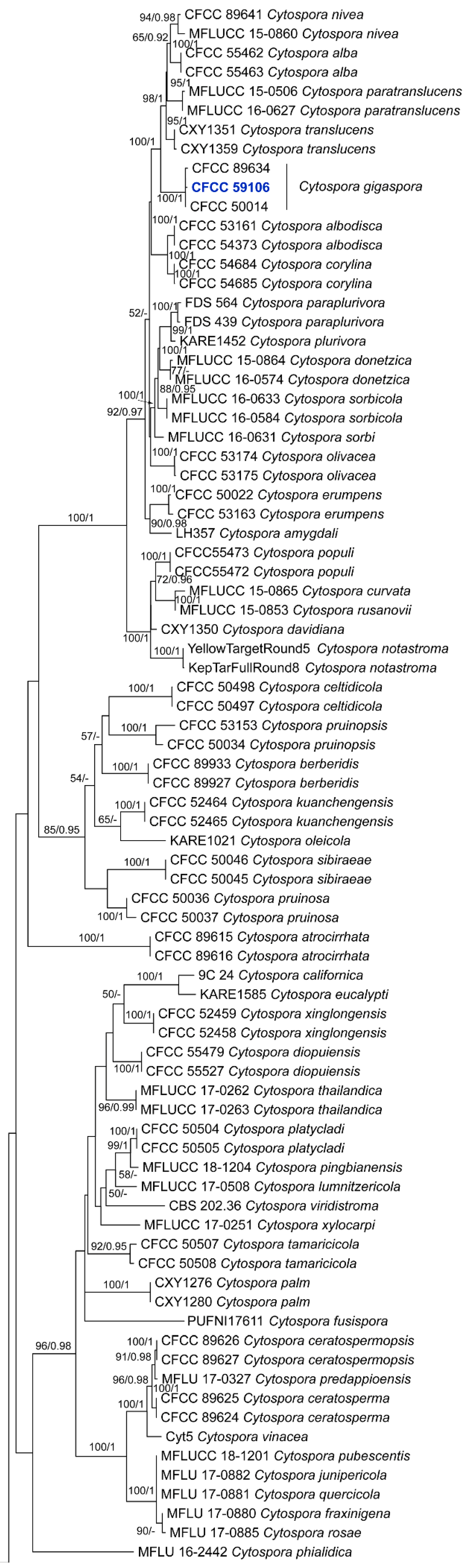
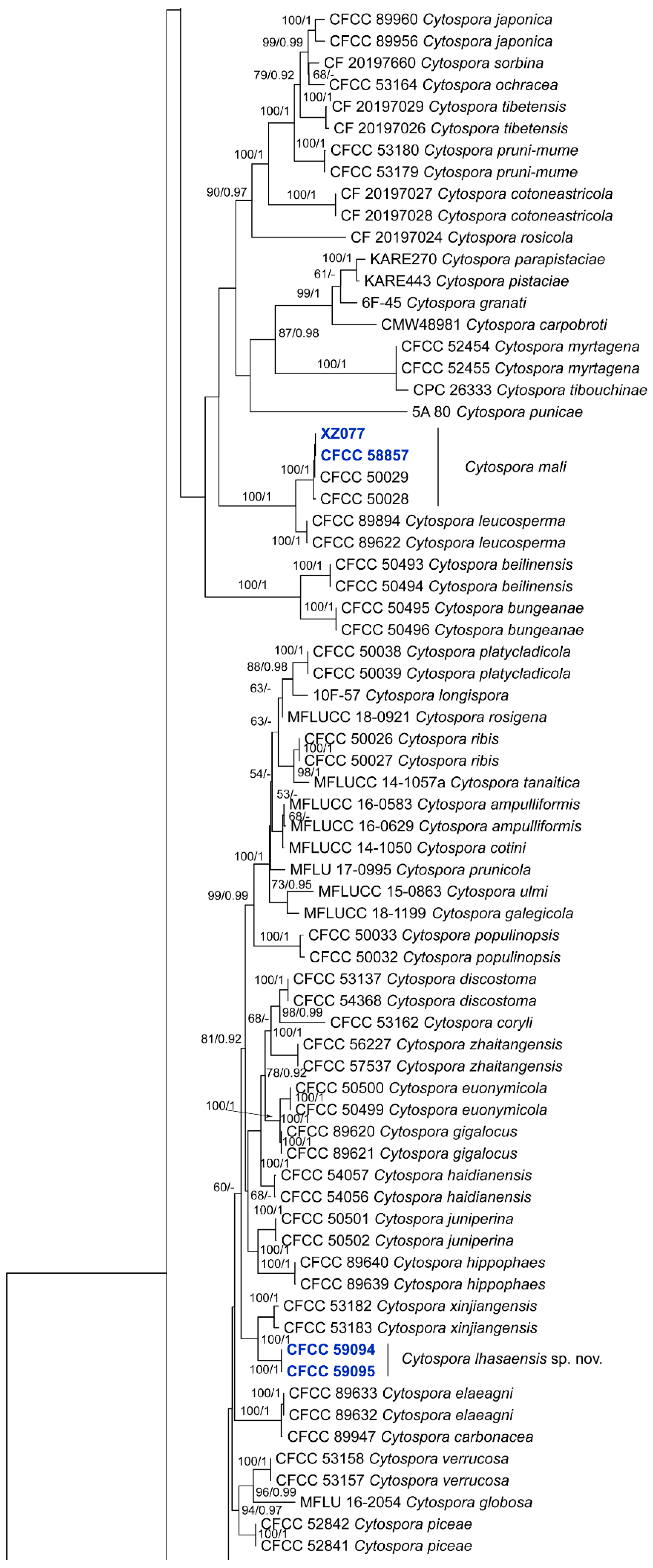
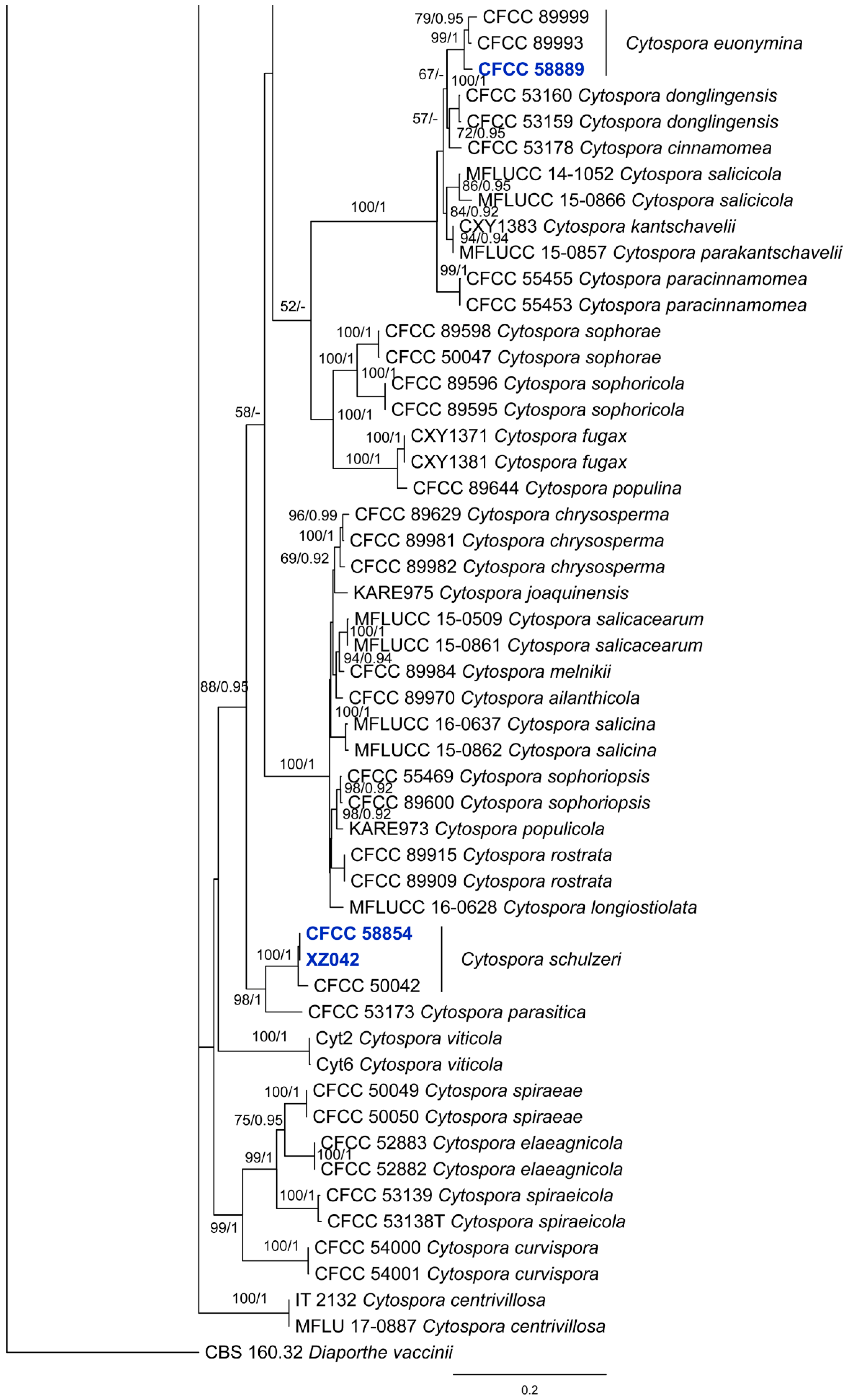
The combined dataset of the ITS, act, rpb2, tef1 and tub2 sequences consisted of 204 strains, with Diaporthe vaccinii (CBS 160.32) as the outgroup taxon. The final alignment comprised 3152 characters including 566 characters for ITS, 330 characters for act, 741 characters for rpb2, 709 characters for tef1 and 806 characters for tub2. The final ML optimization likelihood value of the best RAxML tree was −58,893.68, and the matrix had 2068 distinct alignment patterns, with 40.58% undetermined characters or gaps. The estimated base frequencies were as follows: A = 0.244479, C = 0.288112, G = 0.237008 and T = 0.230400; substitution rates AC = 1.356952, AG = 3.037317, AT = 1.343486, CG = 0.970365, CT = 5.077078 and GT = 1.0; and gamma distribution shape parameter α = 0.379557. The RAxML and Bayesian analyses yielded a similar tree topology. The topology of our phylogenetic tree is nearly identical to those from previous publications. Eight isolates from the present study formed a new clade that is distinct from previously known species, which was named Cytospora lhasaensis sp. nov. There were also four known clades named C. euonymina, C. gigaspora, C. mali and C. schulzeri (Figure 1).
3.2. Taxonomy
3.2.1. Description of Cytospora euonymina from Euonymus japonicus
Cytospora euonymina X.L. Fan & C.M. Tian, Persoonia 45: 21 (2019).
See Figure 2.
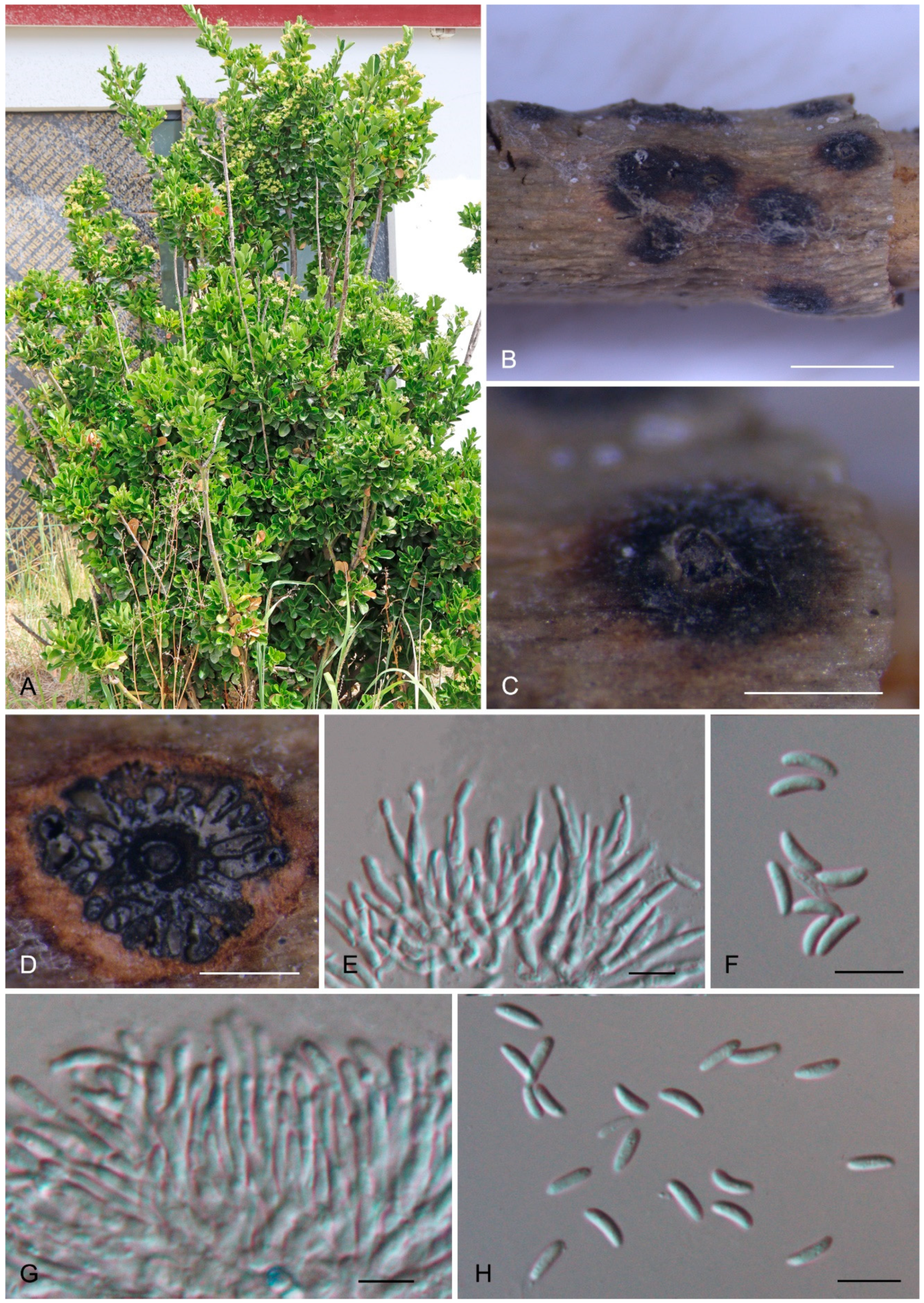
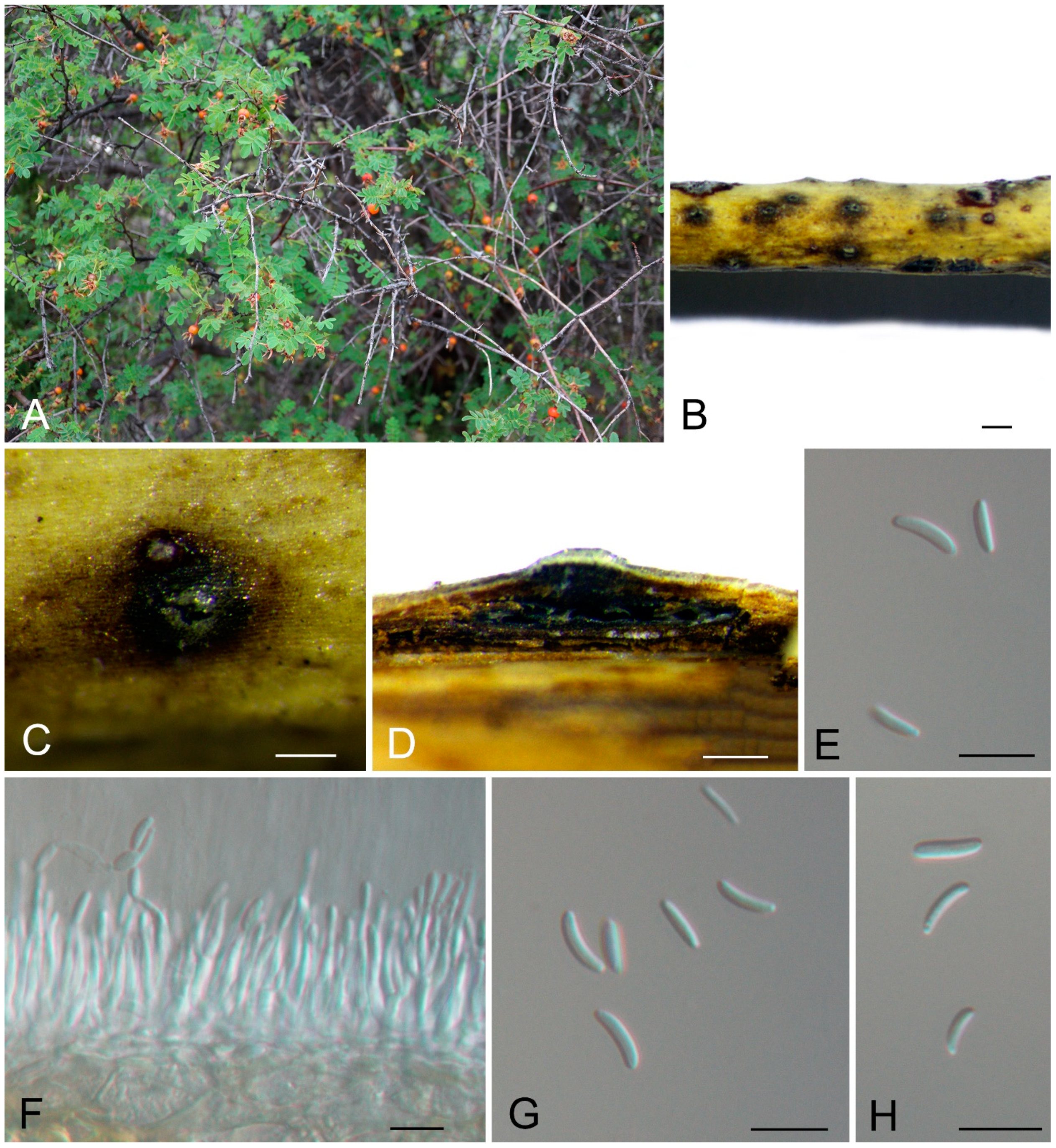
Description: Causes branch and twig canker disease of Euonymus japonicus with cracked barks on the branches and twigs. Bark was easily removed from the cankered tissues and fruiting bodies formed on the damaged area. Sexual morph: Undetermined. Asexual morph: Conidiomata pycnidial, ostiolate, semi-immersed in the host bark, scattered, discoid, with multiple locules. Conceptacle black, circular surrounded stromata. Ectostromatic disc grey, circular to ovoid, (100–)150–250(–300) μm diam., with one ostiole per disc. Ostioles dark, at the same level as the disc, (50–)60–85(–120) μm diam. Locule numerous, arranged circularly or elliptically with independent walls, (250–)350–550(–750) μm diam. Peridium comprising few layers of cells of textura angularis, with innermost layer brown, outer layer brown to dark brown. Conidiophores hyaline, branched, thin walled, filamentous. Conidiogenous cells enteroblastic polyphialidic, 9–45.5 × 1.5–2.5 μm. Conidia hyaline, allantoid, smooth, aseptate, thin-walled, (7–)7.3–8.7(–9.2) × 2–3 μm ( = 8 × 2.5 μm).
Culture characteristics: Colonies on PDA are flat, spreading, with sparse to moderate aerial mycelium, initially white, becoming umber after 15 days, reaching a 90 mm diameter after 15 days and forming abundant conidiomata after 30 days at 25 °C in the dark.
Materials examined: CHINA, Tibet, Lhasa City, Chengguan District, Nongke Road, 29°38′43.06″ N, 91°53′93″ E, alt. 3615 m, on cankered branches and twigs of Euonymus japonicus, 29 July 2022, Jin Peng, Liu Yuanyuan, Jiang Ning and Liu Min (XZ053); culture CFCC 58889 (ITS: OR769862, act: OR767318, rpb2: OR767332, tef1: OR767358, tub2: OR767346).
Notes: Cytospora euonymina was initially described from diseased branches and twigs of Euonymus kiautschovicus collected from Shanxi Province [9], and was subsequently discovered on leaf spots of E. japonicus in Beijing City [25]. This fungal species has only been reported from the host genus Euonymus and in China. The present study revealed a new collection of C. euonymina from E. japonicus in Tibet, which is associated with branch and twig canker disease.
3.2.2. Description of Cytospora gigaspora from Larix gmelinii
Cytospora gigaspora C.M. Tian, X.L. Fan & K.D. Hyde, Phytotaxa 197(4): 232 (2015).
Description: Causes branch canker disease of Larix gmelinii. The host branches were dead, and part of the xylem became brown; however, no fruiting bodies were discovered on the cankered barks. Sexual morph: Undetermined. Asexual morph: Undetermined.
Culture characteristics: Colonies on PDA are flat, spreading, white, reaching a 90 mm diameter after 10 days, sterile.
Materials examined: CHINA, Tibet, Lhasa City, Chengguan District, Lalu Wetland National Nature Reserve, 29°40′57.86″ N, 91°6′11.13″ E, alt. 3617 m, on cankered branches of Larix gmelinii, 27 July 2022, Jin Peng, Liu Yuanyuan, Jiang Ning and Liu Min (XZ034); culture CFCC 59106 (ITS: OR769867, act: OR767323, rpb2: OR767337, tef1: OR767363).
3.2.3. Description of the new species Cytospora lhasaensis from Rosa omeiensis f. pteracantha
Cytospora lhasaensis Ning Jiang, sp. nov.
See Figure 3.
MycoBank: MB851204.
Etymology: Named after the collection site of the holotype, Lhasa City.
Description: Causes branch canker disease of Rosa omeiensis f. pteracantha. Barks of the cankered branches and twigs were discolored, with black rounded fruiting bodies abundantly formed on the diseased tissues. Sexual morph: Undetermined. Asexual morph: Conidiomata pycnidial, ostiolate, semi-immersed in the host bark, scattered, pulvinate, with multiple locules. Conceptacle dark brown to black, circular surrounded stromata. Ectostromatic black, circular to ovoid, (150–)200–300(–550) μm diam., with one ostiole per disc. Ostioles dark, at the same level as the disc, (35–)55–85(–100) μm diam. Locule numerous, arranged circularly or elliptically with independent walls, (245–)300–450(–550) μm diam. Peridium comprising few layers of cells of textura angularis, with innermost layer brown, outer layer brown to dark brown. Conidiophores hyaline, branched, thin walled, filamentous. Conidiogenous cells enteroblastic polyphialidic, 8–28.5 × 1.5–2.5 μm. Conidia hyaline, allantoid, smooth, aseptate, thin-walled, (5.7–)6.6–8.6(–9.5) × 1.7–2.4 μm ( = 7.6 × 2 μm).
Culture characteristics: Colonies on PDA are flat, spreading, with flocculent mycelium, white, reaching a 90 mm diameter after 15 days and forming abundant black conidiomata after 25 days at 25 °C in the dark.
Materials examined: CHINA, Tibet, Lhasa City, Mozhugongka County, Riduo Town, Zen Village, 29°42′24″ N, 92°6′28″ E, alt. 3893 m, on cankered branches of Rosa omeiensis f. pteracantha, 28 July 2022, Jin Peng, Liu Yuanyuan, Jiang Ning and Liu Min (CAF800086, holotype); ex-type cultures CFCC 59094 (ITS: OR769863, act: OR767319, rpb2: OR767333, tef1: OR767359, tub2: OR767347) and CFCC 59095 (ITS: OR769864, act: OR767320, rpb2: OR767334, tef1: OR767360, tub2: OR767348).
Notes: Cytospora lhasaensis from Rosa omeiensis f. pteracantha is phylogenetically close to C. xinjiangensis from Rosa sp. (Figure 1). However, Cytospora lhasaensis differs from C. xinjiangensis in having larger conidia (6.6–8.6 × 1.7–2.4 μm in C. lhasaensis vs. 4–4.5 × 1–1.5 μm in C. xinjiangensis) [34]. Additionally, C. lhasaensis can be distinguished from C. xinjiangensis by sequence data (nucleotide differences in ITS: 13/508; act: 14/248; rpb2: 25/727; tef1: 21/553; tub2: 24/430).
3.2.4. Description of Cytospora mali from Malus pumila
Cytospora mali Grove, British Stem- and Leaf-Fungi (Coelomycetes) (Cambridge) 1: 279 (1935).
See Figure 4.
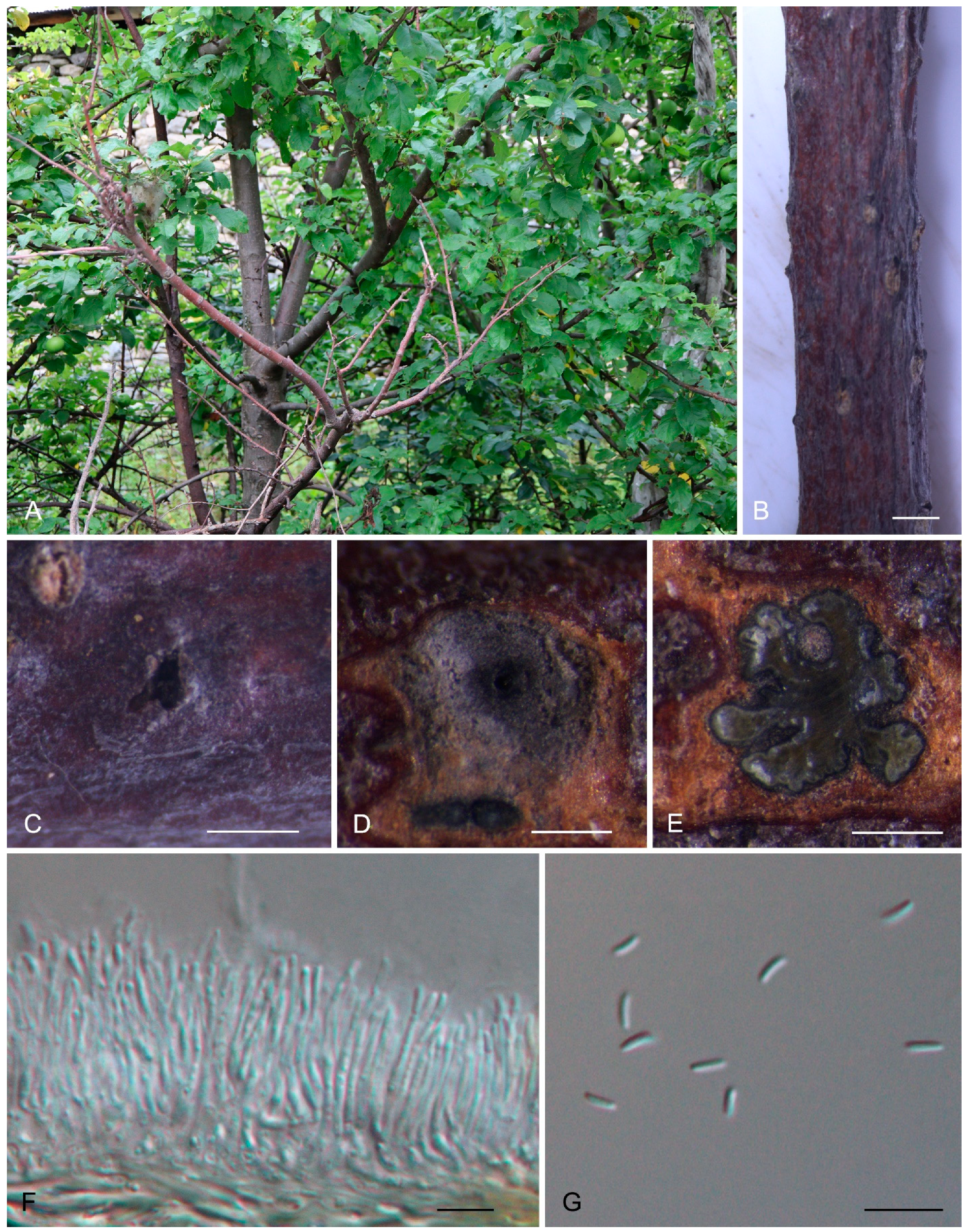
Description: Causes branch and twig canker disease of Malus pumila. Diseased branches and twigs differed from healthy ones in having small brown rounded fruiting bodies under the bark. With older cankers, the host bark was easily removed. Sexual morph: Undetermined. Asexual morph: Conidiomata Pycnidia, ostiolate, semi-immersed in the host bark, scattered, discoid, with multiple locules. Conceptacle absent. Ectostromatic disc black, circular to ovoid, (200–)300–450(–500) μm diam., with one ostiole per disc. Ostioles dark, at the same level as the disc, (35–)50–100(–150) μm diam. Locule numerous, arranged irregularly with common walls, (350–)500–900(–1100) μm diam. Peridium comprising few layers of cells of textura angularis, with innermost layer brown, outer layer brown to dark brown. Conidiophores hyaline, branched, thin walled, filamentous. Conidiogenous cells enteroblastic polyphialidic, 7–28.5 × 1.5–2.5 μm. Conidia hyaline, allantoid, smooth, aseptate, thin-walled, (4–)4.2–5(–5.8) × 1–1.5 μm ( = 4.6 × 1.3 μm).
Culture characteristics: Colonies on PDA are flat, spreading, with aerial mycelium, grey to greenish, reaching a 90 mm diameter after 10 days, sterile.
Materials examined: CHINA, Tibet, Shigatse City, Gyirong County, Gyirong Town, 28°28′1.87″ N, 85°13′40.83″ E, alt. 3081 m, from cankered branches and twigs of Malus pumila, 10 August 2022, Jin Peng, Jiang Ning and Liu Min (XZ077; ITS: OR769866, act: OR767322, rpb2: OR767336, tef1: OR767362, tub2: OR767350); culture CFCC 58857 (ITS: OR769865, act: OR767321, rpb2: OR767335, tef1: OR767361, tub2: OR767349).
Notes: Cytospora mali is a common canker pathogen infecting Maloideae hosts, especially Malus pumila [9]. Although this fungus has been widely collected in many provinces in China, Tibetan samples have not been studied using molecular data. The present study confirmed the pathogen species on the apple trees in Tibet morphologically and phylogenetically.
3.2.5. Description of Cytospora schulzeri from Malus spectabilis
Cytospora schulzeri Sacc. & P. Syd., Syll. fung. (Abellini) 14(2): 918 (1899).
See Figure 5.
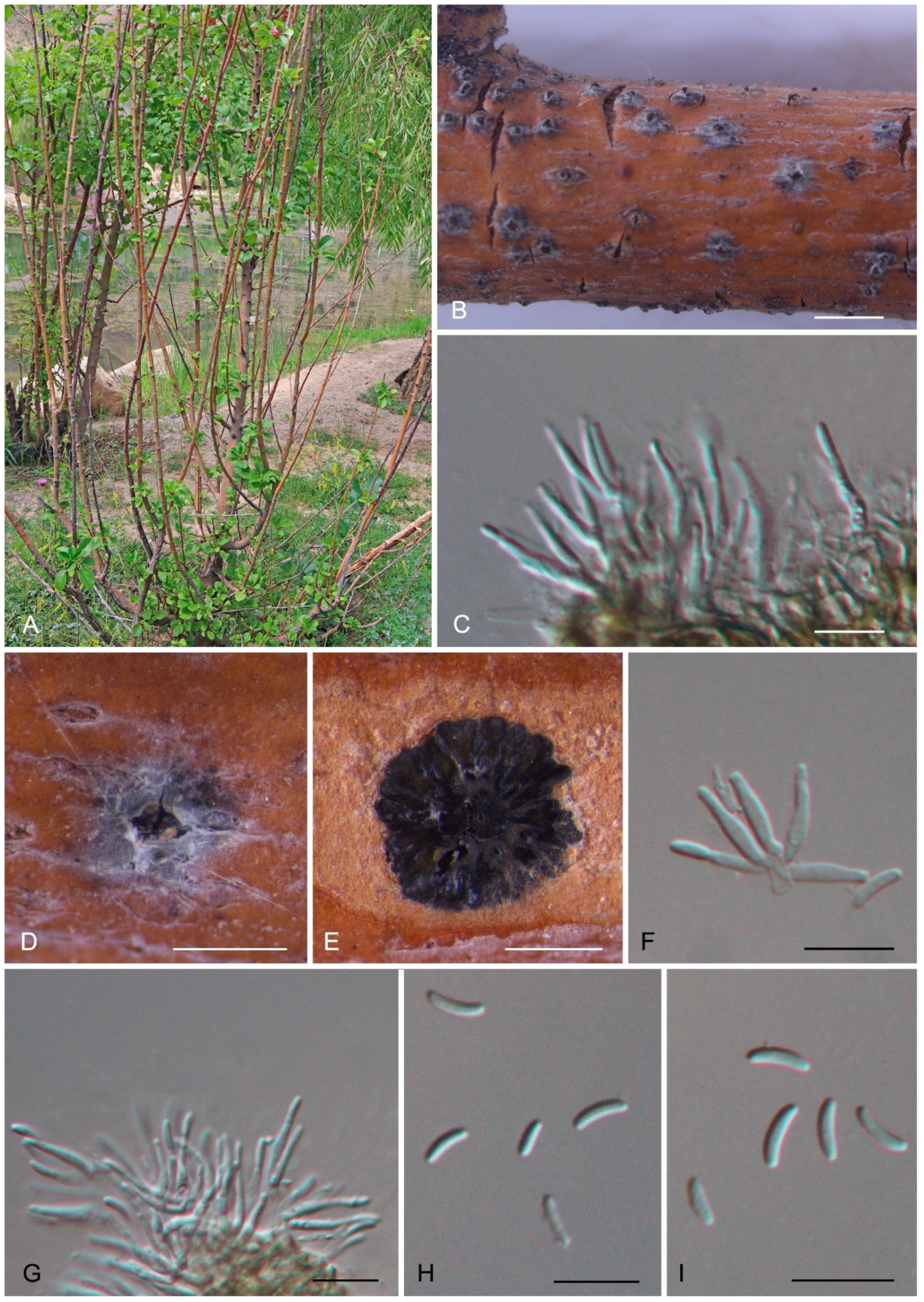
Description: Causes branch canker disease of Malus spectabilis. Cankered branches were discolored, with brown to dark, rounded fruiting bodies formed on the dead tissues. Sexual morph: Undetermined. Asexual morph: Conidiomata pycnidial, ostiolate, semi-immersed in the host bark, scattered, discoid, with multiple locules. Conceptacle absent. Ectostromatic disc brown to black, circular to ovoid, (200–)350–450(–550) μm diam., with 1–7 ostioles per disc. Ostioles dark, at the same level as the disc, (40–)60–85(–110) μm diam. Locule numerous, arranged circularly with common walls, (550–)700–1250(–1550) μm diam. Peridium comprising few layers of cells of textura angularis, with innermost layer brown, outer layer brown to dark brown. Conidiophores hyaline, branched, thin walled, filamentous. Conidiogenous cells enteroblastic polyphialidic, 7–18 × 1.5–2.5 μm. Conidia hyaline, allantoid, smooth, aseptate, thin-walled, (4.1–)4.5–5.9(–6.5) × 1–1.5 μm ( = 5.2 × 1.4 μm).
Culture characteristics: Colonies on PDA are flat, spreading, centrally white and olivaceous grey at the margin, reaching a 90 mm diameter after 15 days, sterile.
Materials examined: CHINA, Tibet, Lhasa City, Chengguan District, Nanshan Park, 29°38′20.81″ N, 91°6′55.92″ E, alt. 3631 m, on cankered branches of Malus spectabilis, 27 July 2022, Jin Peng, Liu Yuanyuan, Jiang Ning and Liu Min (XZ042; ITS: OR769861, act: OR767317, rpb2: OR767331, tef1: OR767357, tub2: OR767345); culture CFCC 58854 (ITS: OR769860, act: OR767316, rpb2: OR767330, tef1: OR767356, tub2: OR767344).
Notes: Cytospora schulzeri is common on Malus hosts, causing canker diseases [9], and sometimes infects other hosts, such as Castanea mollissima [26]. However, Tibetan Cytospora samples on Malus spectabilis have not been identified based on combined approaches of morphology and phylogeny. In the present study, we confirmed that C. schulzeri infects M. spectabilis using morphological and molecular data.
4. Discussion
In this study, we investigated the species diversity of Cytospora from five common woody plants in Tibet, which is in the third pole of the earth. Euonymus japonicus, Larix gmelinii, Malus pumila, M. spectabilis and Rosa omeiensis f. pteracantha appeared to have typical canker symptoms during our investigations, and five Cytospora species were identified from them based on the morphological and molecular data. Of these, a new species named Cytospora lhasaensis and four known species (C. euonymina, C. gigaspora, C. mali and C. schulzeri) were identified. Before the present study, only four species of Cytospora were recorded in Tibet, viz. C. chrysosperma from Ulmus pumila, C. cotoneastricola and C. tibetensis from Cotoneaster sp., and Cytospora rosicola from Rosa sp. [9,34]. Hence, the current study improves our understanding of the distribution and host association of the genus Cytospora in Tibet.
Cytospora lhasaensis and C. rosicola are associated with Rosa hosts in Tibet [34]. Both species are described as an asexual morph on naturally formed barks, with similar conidial shapes. However, Cytospora lhasaensis (6.6–8.6 × 1.7–2.4 μm) has obviously longer conidia than C. rosicola (4.5–5 × 1–2 μm) [34]. In addition, they are phylogenetically distinct (Figure 1).
Cytospora is a species-rich genus of Diaporthales, which is often associated with plant diseases [35,36,37]. In recent publications, many cryptic new species of this genus were proposed based on evidence from combined morphology and phylogeny analyses [9,21,24,25,38]. Some of them have been proven to be the pathogens causing important diseases [21,24,25]. However, the others, including the new species in the current study, need further investigated through pathogenicity studies in the future. By using the molecular method for species identification, more cryptic species will be revealed from the third pole of the earth in the future.
5. Conclusions
In the present study, we identified five Cytospora species associated with canker diseases of five woody hosts in Tibet, China, using morphological and phylogenetic approaches. Of these, Cytospora lhasaensis from Rosa omeiensis f. pteracantha is new to science. Four fungal species named C. euonymina, C. gigaspora, C. mali and C. schulzeri were recorded for the first time in Tibet.
Supplementary Materials
The following supporting information can be downloaded at: https://www.mdpi.com/article/10.3390/f15010121/s1, Table S1: GenBank accession numbers used in the phylogenetic analyses.
Author Contributions
Conceptualization, J.-R.L.; methodology, N.J.; software, N.J.; validation, J.-T.L.; formal analysis, J.-T.L.; investigation, N.J.; resources, N.J.; data curation, N.J.; writing—original draft preparation, N.J.; writing—review and editing, J.-R.L.; visualization, J.-R.L.; supervision, J.-T.L.; project administration, J.-R.L.; funding acquisition, J.-R.L. and N.J. All authors have read and agreed to the published version of the manuscript.
Funding
This research was funded by the Long-Term Ecological Observation Study of Alpine Pine in Southeast Tibet (Science and Technology Innovation Base, XZ202301JD0001G); the flexible talent introduction projects of the Key Laboratory of Forest Ecology in the Tibet Plateau, Ministry of Education, Tibet Agricultural and Animal Husbandry University (2022–2023); and the National Microbial Resource Center of the Ministry of Science and Technology of the People’s Republic of China (NMRC-2023-7).
Data Availability Statement
All sequence data are available in NCBI GenBank: CFCC 58889 (ITS: OR769862, act: OR767318, rpb2: OR767332, tef1: OR767358, tub2: OR767346); CFCC 59106 (ITS: OR769867, act: OR767323, rpb2: OR767337, tef1: OR767363); CFCC 59094 (ITS: OR769863, act: OR767319, rpb2: OR767333, tef1: OR767359, tub2: OR767347); CFCC 59095 (ITS: OR769864, act: OR767320, rpb2: OR767334, tef1: OR767360, tub2: OR767348); XZ077 (ITS: OR769866, act: OR767322, rpb2: OR767336, tef1: OR767362, tub2: OR767350); CFCC 58857 (ITS: OR769865, act: OR767321, rpb2: OR767335, tef1: OR767361, tub2: OR767349); XZ042 (ITS: OR769861, act: OR767317, rpb2: OR767331, tef1: OR767357, tub2: OR767345); and CFCC 58854 (ITS: OR769860, act: OR767316, rpb2: OR767330, tef1: OR767356, tub2: OR767344).
Conflicts of Interest
The authors declare no conflicts of interest.
References
- Jia, H.; Liu, Z.; Sungbom, O.; Yao, C.; Chen, J.; Dong, A.; Liu, X. First report of Aplosporella javeedii causing branch blight disease of Mulberry (Morus alba) in China. J. Plant Dis. Prot. 2019, 126, 475–477. [Google Scholar] [CrossRef]
- Moral, J.; Morgan, D.; Michailides, T.J. Management of Botryosphaeria canker and blight diseases of temperate zone nut crops. Crop Prot. 2019, 126, 104927. [Google Scholar] [CrossRef]
- Wagener, W.W. Coryneum canker of cypress. Science 1928, 67, 584. [Google Scholar] [CrossRef] [PubMed]
- Bloomberg, W.J. Cytospora canker of poplars: Factors influencing the development of the disease. Can. J. Bot. 1962, 40, 1271–1280. [Google Scholar] [CrossRef]
- Jaklitsch, W.M.; Voglmayr, H. European species of Dendrostoma (Diaporthales). MycoKeys 2019, 59, 1. [Google Scholar] [CrossRef]
- Baumgartner, K.; Fujiyoshi, P.T.; Travadon, R.; Castlebury, L.A.; Wilcox, W.F.; Rolshausen, P.E. Characterization of species of Diaporthe from wood cankers of grape in eastern North American vineyards. Plant Dis. 2013, 97, 912–920. [Google Scholar] [CrossRef] [PubMed]
- Fan, X.; Du, Z.; Bezerra, J.D.; Tian, C. Taxonomic circumscription of melanconis-like fungi causing canker disease in China. MycoKeys 2018, 42, 89–124. [Google Scholar] [CrossRef]
- Rigling, D.; Prospero, S. Cryphonectria parasitica, the causal agent of chestnut blight: Invasion history, population biology and disease control. Mol. Plant Pathol. 2018, 19, 7–20. [Google Scholar] [CrossRef]
- Fan, X.L.; Bezerra, J.D.P.; Tian, C.M.; Crous, P.W. Cytospora (Diaporthales) in China. Persoonia 2020, 45, 1–45. [Google Scholar] [CrossRef]
- Ehrenberg, C.G. Sylvae Mycologicae Berolinenses; Formis Theophili Bruschcke: Berlin, Germany, 1818. [Google Scholar]
- Donk, M.A. Nomina conservanda proposita 1. Proposals in fungi. Deuteromycetes. Regnum Veg. 1964, 34, 7–15. [Google Scholar]
- Wang, Y.L.; Lu, Q.; Decock, C.; Li, Y.X.; Zhang, X.Y. Cytospora species from Populus and Salix in China with C. davidiana sp. nov. Fungal Biol. 2015, 119, 420–432. [Google Scholar] [CrossRef] [PubMed]
- Adams, G.C.; Wingfield, M.J.; Common, R.; Roux, J. Phylogenetic relationships and morphology of Cytospora species and related teleomorphs (Ascomycota, Diaporthales, Valsaceae) from Eucalyptus. Stud. Mycol. 2005, 52, 1–144. [Google Scholar]
- Fan, X.L.; Hyde, K.D.; Yang, Q.; Liang, Y.M.; Ma, R.; Tian, C.M. Cytospora species associated with canker disease of three anti-desertification plants in northwestern China. Phytotaxa 2015, 1974, 227–244. [Google Scholar] [CrossRef]
- Yang, Q.; Fan, X.L.; Crous, P.W.; Liang, Y.M.; Tian, C.M. Cytospora from Ulmus pumila in Northern China. Mycol. Prog. 2015, 14, 74. [Google Scholar] [CrossRef]
- Norphanphoun, C.; Doilom, M.; Daranagama, D.A.; Phookamsak, R.; Wen, T.C.; Bulgakov, T.S.; Hyde, K.D. Revisiting the genus Cytospora and allied species. Mycosphere 2017, 8, 51–97. [Google Scholar] [CrossRef]
- Norphanphoun, C.; Raspé, O.; Jeewon, R.; Wen, T.C.; Hyde, K.D. Morphological and phylogenetic characterisation of novel Cytospora species associated with mangroves. MycoKeys 2018, 38, 93–120. [Google Scholar] [CrossRef] [PubMed]
- Lawrence, D.P.; Holland, L.A.; Nouri, M.T.; Travadon, R.; Abramians, A.; Michailides, T.J.; Trouillas, F.P. Molecular phylogeny of Cytospora species associated with canker diseases of fruit and nut crops in California, with the descriptions of ten new species and one new combination. IMA Fungus 2018, 92, 333–369. [Google Scholar] [CrossRef]
- Zhu, H.Y.; Pan, M.; Bonthond, G.; Tian, C.M.; Fan, X.L. Diaporthalean fungi associated with canker and dieback of trees from Mount Dongling in Beijing, China. MycoKeys 2019, 59, 67–94. [Google Scholar] [CrossRef]
- Shang, Q.J.; Hyde, K.D.; Camporesi, E.; Maharachchikumbura, S.S.N.; Norphanphoun, C.; Brooks, S.; Liu, J.K. Additions to the genus Cytospora with sexual morph in Cytosporaceae. Mycosphere 2020, 11, 189–224. [Google Scholar] [CrossRef]
- Zhou, X.; Pan, M.; Li, H.Y.; Tian, C.M.; Fan, X.L. Dieback of Euonymus alatus (Celastraceae) caused by Cytospora haidianensis sp. nov. in China. Forests 2020, 115, 524. [Google Scholar] [CrossRef]
- Gao, H.; Pan, M.; Tian, C.M.; Fan, X.L. Cytospora and Diaporthe species associated with hazelnut canker and dieback in Beijing, China. Front. Cell. Infect. Microbiol. 2021, 11, 664366. [Google Scholar] [CrossRef] [PubMed]
- Lawrence, D.P.; Travadon, R.; Pouzoulet, J.; Rolshausen, P.E.; Wilcox, W.F.; Baumgartner, K. Characterization of Cytospora isolates from wood cankers of declining grapevine in North America, with the descriptions of two new Cytospora species. Plant Pathol. 2017, 66, 713–725. [Google Scholar] [CrossRef]
- Lin, L.; Pan, M.; Bezerra, J.D.; Tian, C.; Fan, X. Re-evaluation of the fungal diversity and pathogenicity of Cytospora species from Populus in China. Plant Dis. 2023, 107, 83–96. [Google Scholar] [CrossRef] [PubMed]
- Lin, L.; Pan, M.; Gao, H.; Tian, C.; Fan, X. The potential fungal pathogens of Euonymus japonicus in Beijing, China. J. Fungi 2023, 9, 271. [Google Scholar] [CrossRef] [PubMed]
- Jiang, N.; Yang, Q.; Fan, X.L.; Tian, C.M. Identification of six Cytospora species on Chinese chestnut in China. MycoKeys 2020, 62, 1–25. [Google Scholar] [CrossRef] [PubMed]
- Doyle, J.J. Isolation of plant DNA from fresh tissue. Focus 1990, 12, 13–15. [Google Scholar]
- White, T.J.; Bruns, T.; Lee, S.; Taylor, J. Amplification and direct sequencing of fungal ribosomal RNA genes for phylogenetics. In PCR Protocols: A Guide to Methods and Applications; Innis, M.A., Gelfand, D.H., Sninsky, J.J., White, T.J., Eds.; Academic Press: San Diego, CA, USA, 1990; pp. 315–322. [Google Scholar]
- Carbone, I.; Kohn, L.M. A method for designing primer sets for speciation studies in filamentous ascomycetes. Mycologia 1999, 3, 553–556. [Google Scholar] [CrossRef]
- Liu, Y.J.; Whelen, S.; Hall, B.D. Phylogenetic relationships among Ascomycetes: Evidence from an RNA polymerse II subunit. Mol. Biol. Evol. 1999, 16, 1799–1808. [Google Scholar] [CrossRef]
- Rehner, S.A. Primers for Elongation Factor 1-alpha (EF1-alpha). 2001. Available online: http://ocid.nacse.org/research/deephyphae/EF1primer.pdf (accessed on 15 November 2022).
- Glass, N.L.; Donaldson, G.C. Development of primer sets designed for use with the PCR to amplify conserved genes from filamentous ascomycetes. Appl. Environ. Microb. 1995, 61, 1323–1330. [Google Scholar] [CrossRef]
- Miller, M.A.; Pfeiffer, W.; Schwartz, T. Creating the CIPRES Science Gateway for inference of large phylogenetic trees. In Proceedings of the Gateway Computing Environments Workshop, GCE, New Orleans, LA, USA, 14 November 2010; pp. 1–8. [Google Scholar]
- Pan, M.; Zhu, H.; Bonthond, G.; Tian, C.M.; Fan, X.L. High diversity of Cytospora associated with canker and dieback of Rosaceae in China, with 10 new species described. Front. Plant Sci. 2020, 11, 690. [Google Scholar] [CrossRef]
- Jiang, N.; Fan, X.L.; Tian, C.M. Identification and pathogenicity of Cryphonectriaceae species associated with chestnut canker in China. Plant Pathol. 2019, 68, 1132–1145. [Google Scholar] [CrossRef]
- Senanayake, I.C.; Crous, P.W.; Groenewald, J.Z.; Maharachchikumbura, S.S.; Jeewon, R.; Phillips, A.J.; Bhat, J.D.; Perera, R.H.; Li, Q.R.; Li, W.J.; et al. Families of Diaporthales based on morphological and phylogenetic evidence. Stud. Mycol. 2017, 86, 217–296. [Google Scholar] [CrossRef] [PubMed]
- Jiang, N.; Tian, C.M. An emerging pathogen from rotted chestnut in China: Gnomoniopsis daii sp. nov. Forests 2019, 10, 1016. [Google Scholar] [CrossRef]
- Pan, M.; Zhu, H.; Tian, C.M.; Huang, M.; Fan, X.L. Assessment of Cytospora isolates from conifer cankers in China, with the descriptions of four new Cytospora species. Front. Plant Sci. 2021, 12, 636460. [Google Scholar] [CrossRef]
Figure 1.
Phylogram of maximum likelihood analysis based on the combined dataset of ITS, act, rpb2, tef1 and tub2 sequences. Numbers above the branches indicate ML bootstraps (left, ML BS ≥ 50%) and Bayesian Posterior Probabilities (right, BPP ≥ 0.90). The tree is rooted with Diaporthe vaccinii (CBS 160.32). Isolates obtained from the present study are marked in bold and blue.


Figure 2.
Morphology of Cytospora euonymina from Euonymus japonicus. (A) Symptoms of canker disease on the host. (B,C) Conidiomata formed on branches. (D) Transverse section through the conidioma. (E,G) Conidiophores and conidiogenous cells. (F,H) Conidia. Scale bars: (B) = 2 mm; (C,D) = 500 μm; (E–H) = 10 μm.

Figure 3.
Morphology of Cytospora lhasaensis from Rosa omeiensis f. pteracantha. (A) Symptoms of canker disease on the host. (B,C) Conidiomata formed on branches. (D) Longitudinal section through the conidioma. (E,G,H) Conidiophores and conidiogenous cells. (F) Conidia. Scale bars: (B) = 300 μm; (C,D) = 100 μm; (E–H) = 10 μm.

Figure 4.
Morphology of Cytospora mali from Malus pumila. (A) Symptoms of canker disease on the host. (B,C) Conidiomata formed on branches. (D,E) Transverse section through the conidiomata. (F) Conidiophores and conidiogenous cells. (G) Conidia. Scale bars: (B) = 1 mm; (C) = 500 μm; (D,E) = 200 μm; (F,G) = 10 μm.

Figure 5.
Morphology of Cytospora schulzeri from Malus spectabilis. (A) Symptoms of canker disease on the host. (B,D) Conidiomata formed on branches. (C,F,G) Conidiophores and conidiogenous cells. (E) Transverse section through the conidioma. (H,I) Conidia. Scale bars: (B) = 1 mm; (C,F–I) = 10 μm; (D,E) = 500 μm.

Disclaimer/Publisher’s Note: The statements, opinions and data contained in all publications are solely those of the individual author(s) and contributor(s) and not of MDPI and/or the editor(s). MDPI and/or the editor(s) disclaim responsibility for any injury to people or property resulting from any ideas, methods, instructions or products referred to in the content. |
© 2024 by the authors. Licensee MDPI, Basel, Switzerland. This article is an open access article distributed under the terms and conditions of the Creative Commons Attribution (CC BY) license (https://creativecommons.org/licenses/by/4.0/).
Share and Cite
MDPI and ACS Style
Li, J.-T.; Li, J.-R.; Jiang, N. Identification of Cytospora Species Isolated from Branch Canker Diseases of Woody Plants in Tibet, China. Forests 2024, 15, 121. https://doi.org/10.3390/f15010121
AMA Style
Li J-T, Li J-R, Jiang N. Identification of Cytospora Species Isolated from Branch Canker Diseases of Woody Plants in Tibet, China. Forests. 2024; 15(1):121. https://doi.org/10.3390/f15010121
Chicago/Turabian StyleLi, Jie-Ting, Jiang-Rong Li, and Ning Jiang. 2024. "Identification of Cytospora Species Isolated from Branch Canker Diseases of Woody Plants in Tibet, China" Forests 15, no. 1: 121. https://doi.org/10.3390/f15010121
Note that from the first issue of 2016, this journal uses article numbers instead of page numbers. See further details here.