Article

Fungal Presence and Changes of Wood Structure in Bark Stripping Wounds Made by Red Deer (Cervus elaphus L.) on Stems of Fraxinus angustifolia (Vahl)

1
Faculty of Forestry and Wood Technology, Institute of Forest Protection and Wildlife Management, University of Zagreb, Svetošimunska cesta 23, 10000 Zagreb, Croatia
2
Faculty of Forestry and Wood Technology, Institute of Wood Science, University of Zagreb, Svetošimunska cesta 23, 10000 Zagreb, Croatia
*
Author to whom correspondence should be addressed.
Forests 2024, 15(2), 314; https://doi.org/10.3390/f15020314
Submission received: 24 November 2023 / Revised: 15 January 2024 / Accepted: 3 February 2024 / Published: 7 February 2024
(This article belongs to the Special Issue Interactions between Ungulates and Forest Ecosystems)

Abstract

:
Narrow-leaved ash (Fraxinus angustifolia Vahl), a highly valued European forest tree species, has been severely affected by a large-scale decline, which is most probably driven by a complex of multiple interacting factors including fungi, which contribute to and accelerate this process. Red deer (Cervus elaphus L.) can be considered as one of the contributing factors, as they inflict damage on the stems of young trees by stripping the bark. These wounds not only represent suitable entry points for fungi which can cause tissue necroses and decomposition, they can lead to changes in the wood structure as well. The aims of this research were to analyze chosen parameters of bark stripping wounds in narrow-leaved ash stands, identify fungi present in the tissue exposed by wounding, and inspect the effect of wounding on the wood structure. Bark stripping was observed on ash trees from 2 cm up to 18 cm of DBH and between 0.1 m and 1.9 m of stem height. The most susceptible trees were those with an average DBH of 5 ± 2.5 cm. On most of the ash trees (51%), one-third to two-thirds of the circumference was damaged. In wounded tissue, 174 fungal isolates were found, most of which belonged to known endophytic fungi from the genera Trichoderma, Fusarium, and Clonostachys. It was observed that earlywood cells in the wounding zone had narrower lumens compared to ones in adjacent healthy zone with regard to different trees and stem heights.

1. Introduction

Narrow-leaved ash (Fraxinus angustifolia Vahl), a widespread and important European forest trees species, has been seriously affected by a large-scale decline in recent decades. This is especially prominent in south-eastern and some parts of central Europe, where narrow-leaved ash occupies large floodplain forest areas and acts as a pioneer tree species [1,2]. An invasive pathogenic fungus, Hymenoscyphus fraxineus, has been identified as the main causative agent of common (F. excelsior L.) and narrow-leaved ash dieback in Europe; it infects ash leaves, shoots, stems, and roots, leading to wilting of leaves, loss of foliage, and formation of necrosis and cankers on all wooden plant parts [3,4]. However, more recent studies have revealed that ash decline is more probably driven by a complex of multiple interacting factors, including other fungal species, which contribute to and accelerate this process [5,6]. Ash trees are asymptomatically colonized by a variety of endophytic fungi, which can become opportunistic pathogens and spread in weakened tissue, especially when the host undergoes vitality loss caused by biotic or abiotic stresses [7,8,9,10,11]. Rot fungi (Armillaria spp., Ganoderma spp., Lentinus tigrinus, etc.) are often isolated from declining ash trees, and have mostly been reported as secondary invaders which rapidly lead to root and stem deterioration and ultimately tree death and fall [9,12,13,14].
Although large animals are an inseparable part of forest ecosystems and capable of inflicting damage on young plants, their possible role in the decline of ash stands has not been studied very much. Research conducted in Hungary revealed no clear signs that herbivorous ungulates play a major role in the spread of H. fraxineus on common ash [15]. However, large ungulates such as red deer (Cervus elaphus L.), sika deer (Cervus nippon Temminck), fallow deer (Dama dama L.), and moose (Alces alces L.) are widely distributed across European forests, where they regularly cause damage to trees and stands simply by satisfying their daily and seasonal physiological needs [16]. They commonly bite green plant parts such as leaves, buds, and bark, and directly affect the structure and composition of forest vegetation [17]. Bark stripping and vegetation damage can cause economic losses [18] and enable the infection of wounded trees by various pathogenic organisms, mostly fungi [19]. Among the European large herbivores which affect forest stands, the red deer is by far most widespread [16]. There are numerous studies dealing with the analysis and different aspects of damage caused by red deer to forest stands [16,20,21,22,23,24,25,26,27]. Although deer cause bark stripping wounds on various tree species in different forest stands, in most of the studies the authors agree that debarking occurs because of a lack of fibrous food, essential nutrients, or minerals during the winter months. Several studies have focused on forest protection measures, such as providing supplemental nutrition to red deer in order to influence their feeding habits during the winter months. For example, a study conducted in Austria showed that targeted supplementary feeding in the winter months can influence the behavior of red deer and keep them in a desired area, deterring them from sensitive and endangered areas where there is a risk of stripping the bark of young trees [28]. However, the results showed effects only within a radius of 1.3 to 1.5 km of the feeding location. In contrast, Jerina et al. [29] did not establish a connection between winter feeding sites and damage to forest stands. In their research, the authors determined that microclimatic conditions are more important for the incidence of damage than the layout and average distance from the supplementary feeding place.
Nevertheless, inflicted bark stripping wounds represent suitable entry points for fungal spores and facilitate the colonization of exposed wood by various obligate or opportunistic plant pathogens and wood decay fungi, including those which are already known to contribute to ash dieback [30,31,32]. Additionally, endophytic fungi which were latently present in wood prior to wounding can shift to a parasitic or saprotrophic lifestyle due to changes in the substrate which favor their growth [33,34,35]. A study on pedunculate oak (Quercus robur L.) in Lithuania revealed that fungal composition in bark stripping wounds changes over time, although certain fungi can continue to grow and strive in wounded tissue even after wound closure. The same study confirmed that younger wounds are colonized by widely distributed fungal generalists, mainly saprotrophs, while opportunistic endophytes thrived in somewhat older wounds [32]. However, this can significantly vary depending on the wounded tree species, its capability with regard to wound closure, and its overall resistance to fungal infections [36].
Additionally, despite effective sealing of wounds which forest trees usually employ, the wood structure undergoes certain changes and the timber quality deteriorates because of the discoloration and rot caused by fungi [37]. Although many external factors can affect wood structure, bark stripping is the most common one. When only phloem and cambial tissue are removed, there is a vastly different impact on tree physiology than when the xylem tissue, especially the vascular tissue, is affected [38]. A study conducted in Germany on European beech (Fagus sylvatica L.) showed that the area with internal wood damage was smaller than the external bark stripping area, although length of the damaged stem section was always greater inside than outside [39]. Bark stripping, which mostly occurs in young forest stands [40], affects wood quality from both the economic point of view and that of final wood production [41]. The internal damage which manifests itself in the wooden body is of decisive importance for forestry as far as silvicultural decisions and economic profits are concerned [39]. In relation to this, additional data for quantifying the damage caused by debarking are essential. Currently, research evaluating the impacts of bark stripping is mostly directed to determining the length of time over which bark stripping occurs, the size of the damaged trees or trunks, the frequency of repeated damage, and the recovery of the resulting wounds [42].
Although the variability of wood within and among trees is perhaps the most widely studied subject in forestry [43], there is no specific model or norm in wood variation; each species can develop its own pattern in each environment and with different external effects. Cell characteristics, including cell and lumen width, can differ between the upper and lower juvenile wood of hardwoods [44]. The variability of juvenile wood properties with tree height is evident in hardwoods. Cell width plays a major role in the wood quality of hardwoods because of the vessel elements [43], with earlywood vessels having primarily a vascular role. Typically, narrow-leaved ash wood is ring-porous, with the xylem earlywood vessels being conspicuously wider in diameter in comparison to the latewood vessels. They are arranged in a ring and distributed individually or in pairs of one to four in a radial row [45]. Thus, healthy earlywood is easily distinguished from latewood in terms of vessel size [46]. In this study, damage caused by bark stripping was evaluated by quantifying one of the most important wood tissues, namely, the vascular tissue.
In light of the above, the aims of this research were to: (1) measure the intensity of stem wounding inflicted by red deer on F. angustifolia stems; (2) isolate and identify fungi present in the wounded tissue; and (3) compare the wood structures of wounded and adjacent healthy tissue in order to gain a first insight into the contribution of red deer damage to the complex process of ash decline.

2. Materials and Methods

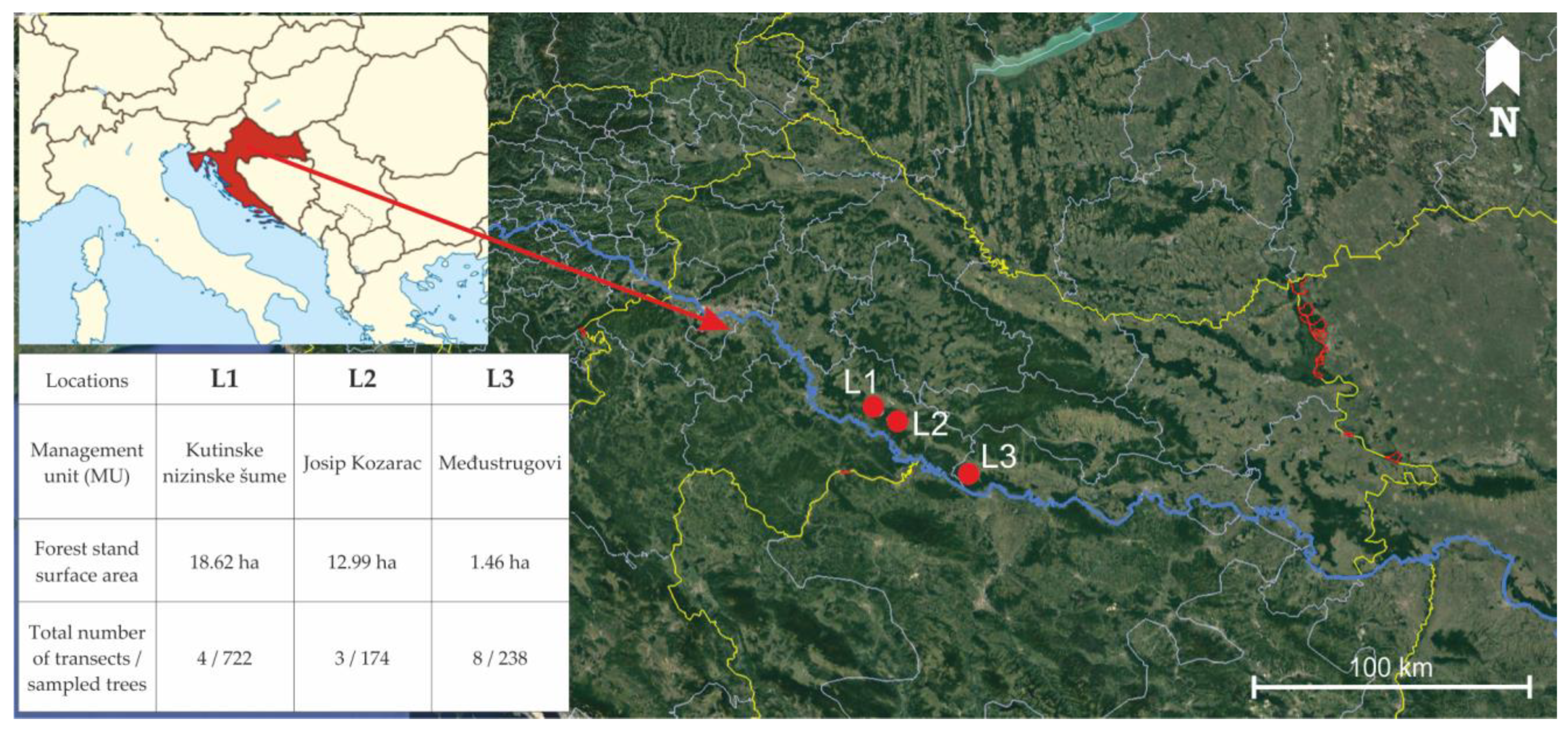
2.1. Field Measurement and Sampling

Research was conducted in lowland forests at the Sava river basin, where narrow-leaved ash occurs naturally and occupies the largest continuous forest area (28,000 ha) in Croatia [47]. Measurements of bark stripping wounds on stems were carried out in 2021 and 2022 in three relatively distant F. angustifolia forest stands less than 15 years old that were known to be in the area inhabited by red deer (Figure 1, Table 1). For this purpose, 20 m wide transects were laid out in each forest stand at least 20 m away from the forest edge. Transects were evenly distributed across the stand surface area in such a way that their total area represented 10% of the total forest stand surface area; the length and the number of transects in each forest stand was varied accordingly. All present trees were recorded on each transect, and the following features were noted or measured for each:
  • tree species;
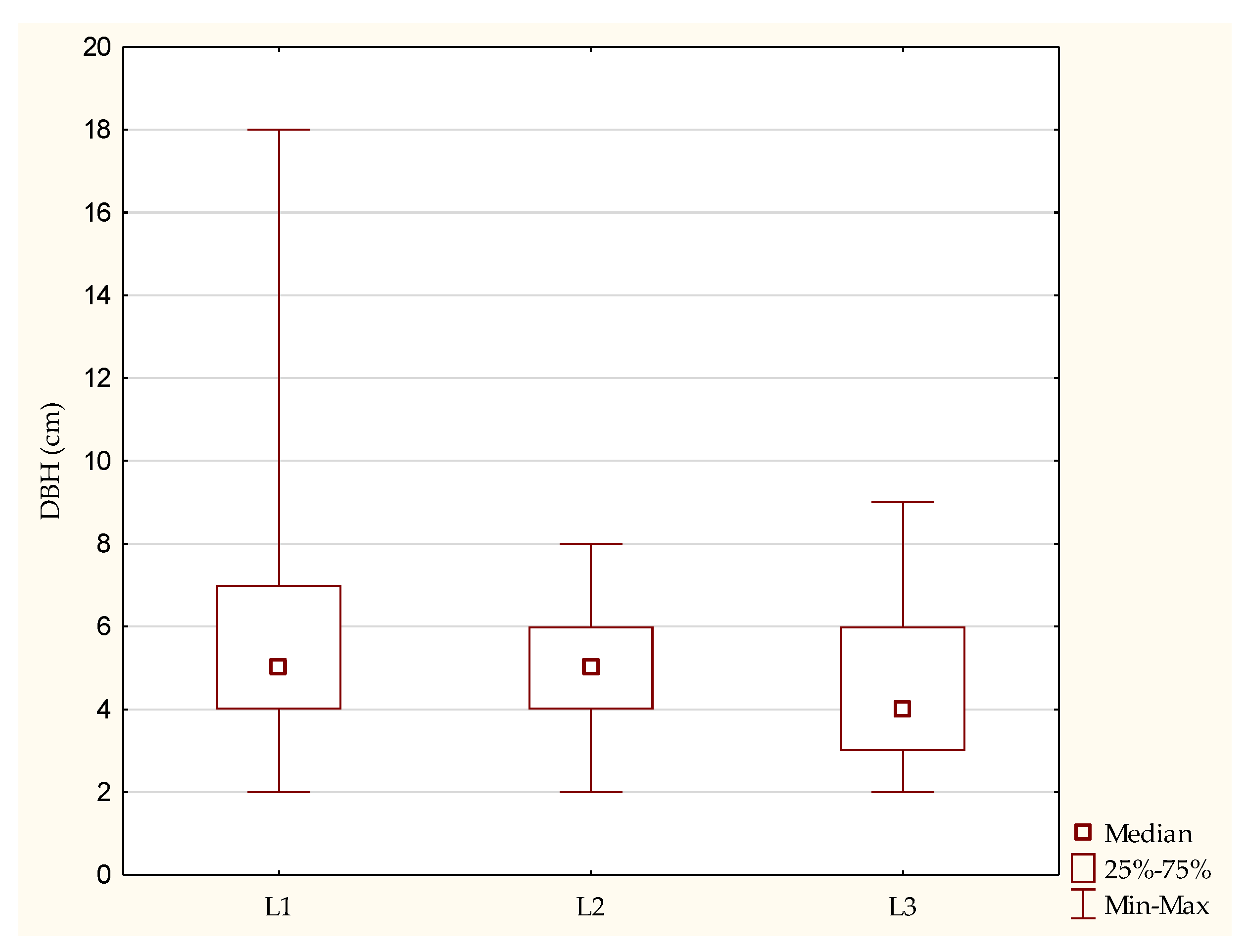
  • diameter at breast height (DBH, cm), measured at the 1.30 m mark of stem height;
  • presence of red deer damage on stem (visible bark stripping, yes/no) (Figure 2);
  • bark stripping intensity (1—less than 1/3 of circumference damaged, 2—1/3 to 2/3 of circumference damaged, 3—more than 2/3 of circumference damaged);
  • height at which bark stripping occurred (the lowest and the highest point on each stem, m);
  • total length of visible bark stripping on a stem (m).
In location L1, ten samples of damaged narrow-leaved ash stems approximately 25 cm in length were randomly collected for fungal analysis and stored at 4 °C for not more than 24 h before further processing. The sampled stems reflected different wounding stages, mostly closed wounds with formed callus. For the analysis of changes in wood structure, 5 cm thick cross-sections of narrow-leaved ash stem were taken from older trees (approximately 20 years old) growing in the vicinity of location L1. Five trees were sampled from that location, and cross-section disks were taken from four different stem heights on each tree (0.25, 0.75, 1.25 and 1.75 m).

2.2. Isolation and Identification of Fungi

From each stem sample, 0.5 cm thick cross-sections with visible wood or bark discoloration or necrosis were taken and surface-sterilized by immersion in 70% ethanol for one min followed by rinsing in sterile distilled water and drying on sterile filter paper under a laminar flow hood (Iskra Pio, Šentjernej, Slovenia). Xylem subsamples (wood chips, 5 × 5 mm) were then aseptically taken at the edge of discoloration or necrosis and plated on malt extract agar (MEA, Oxoid, Basingstoke, UK) supplemented with streptomycin sulphate (200 mg/L, Sigma-Aldrich, St. Louis, MI, USA). Petri dishes were incubated in the dark at 19–20 °C for four weeks and checked daily for fungal growth. Emerging mycelia were transferred onto fresh MEA to obtain pure fungal cultures. Based on their morphological characteristics, the obtained isolates were grouped into putative species, that is, morphotypes, and least one representative isolate from each morphotype was chosen for identification using molecular methods.
DNA was extracted from mycelia cultured in malt extract broth (MEB, Liofilchem, Roseto degli Abruzzi, Italy) in 2 mL microtubes for 7 days by the salting-out method according to Cenis [48]. Minor modifications were made to the composition of the extraction buffer (20 mM Tris HCl, 200 mM NaCl, 2 mM EDTA, 10% SDS, pH 8.0) and to the homogenization procedure, where the mycelium pellet was subjected to grinding in a Tissue Lyser II (Qiagen, Hilden, Germany) for 3 min at 30 Hz with addition of a single 5 mm stainless steel bead per microtube, rather than crushing it with a conical grinder. In Polymerase Chain Reaction (PCR), the internal transcribed spacer (ITS) region of the ribosomal DNA (rDNA) was amplified using primers ITS1-F [49] and ITS 4 [50]. PCR was carried out in 65 µL reactions containing 200 µM deoxyribonucleoside triphosphates, 0.2 µM of each primer, 1.5 U of Taq DNA polymerase with 1× reaction buffer (Sigma-Aldrich, St. Louis, MO, USA), 2.75 mM MgCl2, and 1 µL of 100-fold diluted DNA template. The cycling conditions were as follows: an initial denaturation at 95 °C for 5 min, 35 cycles of denaturation at 95 °C for 30 s, annealing at 55 °C for 30 s, extension at 72 °C for 30 s, and a final extension step at 72 °C for 7 min.
The resulting PCR products were sequenced using the same primers as in the PCR at the DNA sequencing facility of Macrogen Europe (Amsterdam, The Netherlands). After processing raw data using BioEdit Sequence Alignment Editor v.7.2.5 software [51], sequences were identified by comparison with reference sequences in National Center for Biotechnology Information (NCBI) GenBank using the Basic Local Alignment Search Tool (BLAST + 2.15.0) [52]. Sequences with 99.0%–100% similarity were identified to the species level and with 95.0%–98.99% similarity to the genus level.

2.3. Sample Preparation and Wood Structure Analysis

Cross-section disks taken from each of the four stem heights were naturally dried and finely sanded (granulation 320) in order to make visible the wounds within the annual growth rings. Disks were first visually observed, and the diameter was measured. According to wound presence, the same single annual growth ring from the pith was selected and marked along one radius of each disk: two zones by type, including one with the wound present and a control zone (a healthy intact part of the annual growth ring).
Each selected annual growth ring was cut in the form of two sample blocks sized approximately 10 (tangential) × 10 (radial) × 20 (longitudinal) mm. Sample preparation included softening using boiled distilled water for 120 min and cutting into transverse sections (30 µm thick) using a sliding microtome (A.C. Reichert, Vienna, Austria). The sections were stained with a mixture of Safranin (ThermoFisher (Kandel) GmbH, Kandel, Germany) and Astra blue (Carl Roth GmbH&Co KG, Karlshruhe, Germany) for 15 min, washed multiple times in a 70% and 96% ethanol solution until the stain was completely rinsed, and mounted on microscope slides using Euparal (Carl Roth GmbH&Co KG, Karlshruhe, Germany). Four images were taken as consecutive replicates from the earlywood region in the transverse section using a light microscope (Carl Zeiss Jena, Germany) and DinoCapture 2.0 software (Dino-Lite Europe, The Netherlands) connected to a 5 MP digital camera (Dino-Lite Europe, The Netherlands). The samples were photographed at ×60 magnification. Earlywood vessel lumen diameter (EVLD) (µm) measurements were performed using ImageJ software v. 1.8.0 [53].

2.4. Statistical Analysis

Differences in the DBH of the analyzed trees among different locations and categories of bark stripping intensity were tested using analysis of variance (One-way ANOVA). Chi-square tests of independence were used to test the significance of the association between the presence of red deer wounding on narrow-leaved ash stems and the researched location. Pearson correlation values were calculated between the DBH of wounded narrow-leaved ash trees and the measured parameters of inflicted bark stripping wounds (height at which bark stripping occurs, total length of visible bark stripping on a stem).
Regarding the wood structure analysis, the descriptive statistics (mean and standard deviation) were calculated for the EVLD of two zones at different stem heights on all five trees. Repeated measures analysis of variance (ANOVA) was used to test the effect of the stem height, tree, wood zone, and their interactions. This analysis was used to determine the significance of differences in EVLD in several instances: in trees between two zones and four stem heights, between zones of four stem heights, and in the case of all three parameters together. Further analysis was based on finding differences in the EVLD of two zones by observing four different stem heights separately.
In all performed tests, a p-value less than 0.05 was considered statistically significant. Statistical analyses were performed in the TIBCO Statistica 13.5.0 software package (TIBCO Software Inc., Palo Alto, CA, USA).

3. Results

3.1. Inventory of Bark Stripping in the Analyzed Forest Stands

In total, 1134 trees were observed and measured in three locations. Most of the trees belonged to narrow-leaved ash (78.3%). Other observed species were pedunculate oak (14.5%), elm (Ulmus sp., 5.9%), black alder (Alnus glutinosa, 0.9%), and wild pear (Pyrus pyraster, 0.4%). More than half of ash trees (56%) displayed bark stripping wounds, whereas this was visible on only 2% of oak and 9% of elm trees, and was not noticed on black alder or wild pear (Figure 3).
The average DBH of narrow-leaved ash trees (Table 1) differed significantly among the three locations (F (2, 885) = 62.81, p < 0.001), as well as in the presence of bark stripping wounds on stems (X2 (2) = 75.64, p < 0.001).
The average DBH of the wounded narrow-leaved ash trees in all three locations was 5 cm (SD = 2.5 cm). Bark stripping was observed on ash trees from 2 cm up to 18 cm of DBH, between 0.1 m and 1.9 m of stem height (average bark stripping height = 0.6 ± 0.4). The average length of bark stripping wounds was 0.6 m (SD = 0.3 m). This varied somewhat among the researched locations (Figure 4, Table 2).
On most of the ash trees (51%), one-third to two-thirds of the circumference was damaged (Figure 5).
According to ANOVA, the DBH of wounded ash trees did not significantly vary among different categories of bark stripping intensity (F (2, 496) = 0.01, p = 0.98), indicating that these parameters are not significantly associated. Weak positive correlations were found between the height at which bark stripping occurred (the lowest and the highest point on each stem) and the DBH of wounded trees (Table 3). The height of wounds on the stem increased with the increase in DBH as well.

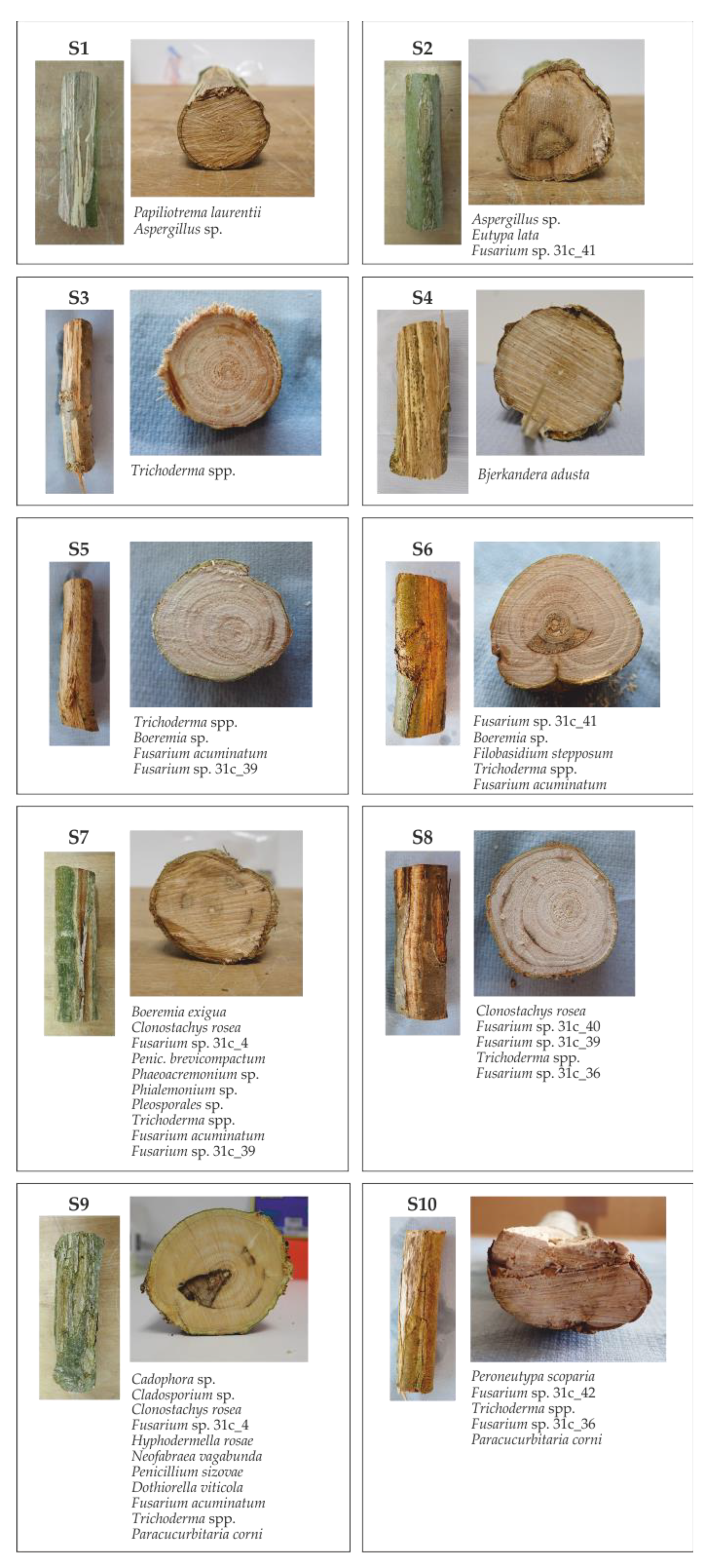
3.2. Fungi Present in Bark Stripping Wounds

The sampled wounded narrow-leaved ash stems reflected various stages of symptom development following bark stripping, from freshly exposed wood with only phloem discoloration visible on the cross-section to partly closed (partially formed callus) or fully closed (completely formed callus) cankers with specific limited discolorations or rot visible on cross-sections, which mostly followed the line of individual tree rings or were placed in the central part of the stem (often of a triangular shape) (Figure 6).
From of ten wounded stem samples, 236 xylem subsamples (wooden chips) were plated on agar media, resulting in growth of 174 isolates that represented 28 fungal taxa (Table 4), of which 24 were members of the phylum Ascomycota. Of the 28 taxa, 14 were identified to species level, 13 taxa to genus level, and one taxon to order level. At least one fungal taxon was isolated from each stem, with an average number of five different taxa per stem. The maximum number of taxa found on one stem was 11. The most frequently isolated were various species of genus Trichoderma, which were combined together and referred to as Trichoderma spp., due to difficult delimitation and identification of species in this genus and limited lab resources. Other most common species found in bark stripping wounds were Clonostachys rosea and Fusarium acuminatum, as well as several other Fusarium species which remained identified only to a genus level. Eutypa lata was highly abundant regarding the number of obtained isolates (29), but was found in only one stem.

3.3. Differences in Wood Structure in Relation to Zones, Trees, and Stem Height

The stem diameter of all five trees was measured at 0.25, 0.75, 1.25, and 1.75 m of the tree height (Table 5).
In total, 20 disks were sampled at four different stem heights, and 40 histological preparations with cross-sections (20 of control (healthy) zone and 20 of wounded zone) were used in measurements. The mean values and standard deviations for the EVLD of the control (healthy) and wounded zones from all five trees are provided in Table 5. The trends in the EVLD of the control (healthy) and wounded zones with height are shown in Figure 7.
The lowest mean EVLD was measured in the control (healthy) zone at a stem height of 0.25 m in trees 1 and 4 and at stem height 0.75 m in trees 2, 3, and 5. The mean values were almost identical in trees 1 and 4, but were somewhat variable between trees 2, 3, and 5. The highest mean EVLD was measured in the control (healthy) zone at stem height 0.25 m in tree 3. Stem height had a similar effect on EVLD in trees 2 and 5, as well as in trees 1 and 4, resulting in the highest mean values at height 1.25 in the first two and 1.75 in the second two, respectively.
In addition, the EVLD in the wounded zone varied with height between trees. In majority of trees, the lowest mean EVLD was measured at a stem height of 0.75 m (trees 1, 2, 4, and 5), though at only stem height 1.75 in tree 3. The mean EVLD showed the highest values in the wounded zones of trees 1, 3, and 4 at stem height 0.25 m (trees 1, 3, and 4) and stem height 1.75 m (trees 2 and 5). Stem height 1.25 m was the least pronounced variable in the peak EVLD values of all stem heights in all trees.
Similar trends in EVLD with height were observed in both the control (healthy) and wounded zones, with a decrease at stem height 0.75 m (Figure 7). In general, the control (healthy) zone showed significantly higher values of EVLD in comparison to the wounded zone (Table 6). This can be observed in Figure 8b,c, where control (healthy) earlywood vessels are significantly wider in diameter and of more round/oval shape in comparison to vessels in wounded earlywood, especially those close to the wound.
The ANOVA results showed that tree-by-zone interaction was significant, indicating differences in the EVLD of the two zones in the whole tree. Highly significant interaction was determined in other three cases as well: for the stem height by tree, stem height by zone, and stem height by both tree and zone. However, the objective was to identify whether and to what extent the control (healthy) and wounded zone EVLDs differed with different stem heights. Thus, the following discussion focuses on the zone main effect and the stem height by zone interaction terms.
The effect of wood zone was significant for all stem heights except for 0.25 m (Table 7). The results indicate the expected significant differences in EVLD between the control (healthy) and wounded zones at greater stem heights.

4. Discussion

4.1. Bark Stripping Inventory

In our study, narrow-leaved ash was confirmed to be a highly food attractive species for red deer, as previously reported [54], with more than half (56%) of observed trees displaying bark stripping wounds. This is in accordance with similar studies conducted on common ash [18,40,55]. In comparison, the share of wounded trees for other observed species (elm, oak, alder, and pear) did not exceed 10%. This can be partially explained by the fact that this study was conducted in single-species forest stands where narrow-leaved ash was by far the most abundant and most available tree species, making it more liable to bark stripping. Moreover, it is possible that the thickness or composition of ash bark is more appealing to red deer in comparison to the bark of other trees species, which can be more robust or contain more tannins, although this needs to be further investigated.
The ratio of wounded and unwounded ash trees differed significantly among the three studied locations, as did their average DBH, indicating that these parameters are associated. The incidence of wounded trees increased with the decrease in average DBH. In location L1, where the average DBH was 10 cm, 46% of ash trees were wounded; in location L2, the average DBH was 7.5 cm, and the share of wounded trees was 54%; and in location L3, where the average DBH was 5 cm, the number of wounded trees (81%) was four times the number of unwounded trees. It can be concluded that younger ash trees, especially those with a DBH around 5 cm, were the most appealing to red deer in the researched forest stands. This was additionally corroborated by the fact that the average DBH of all wounded ash trees from all three locations was 5 ± 2.5 cm. However, these variations in the number of wounded ash trees among locations were most probably affected by other factors as well. Several authors have pointed out that the frequency and intensity of bark stripping are impacted forest stand properties such as density, age, and horizontal and vertical composition [29,56], and are additionally influenced by the density of the red deer population [16]. Red deer tend to reside in younger forest stands with higher tree density and a thinner snow layer on the ground, as these conditions provide them with shelter and more food sources [29].
In this research it was determined that bark stripping stops when ash trees reach a certain age, specifically, a DBH of 18 cm. The reason for this is primarily because the bark of young trees is more digestible [25,57], as well as the fact that at a DBH greater than 18 cm the ash bark becomes too rough and too thick for red deer to strip off with their teeth.
Most of the bark stripping wounds occurred in the browsing zone at stem heights between 0.1 m and 1.9 m (average bark stripping height = 0.6 ± 0.4 m). These results are partially in concordance with results from a study on common ash, where 80% of all bark stripping damage occurred between 1.0 m and 1.9 m of height [18], in which the bark stripping was inflicted by both red deer and moose. Because moose are significantly larger and taller than red deer, they consequently inflict the most damage on higher parts of stem, which is why their results showed somewhat higher values compared to the results of our study.
Usually, older trees with greater DBH display more and larger bark stripping wounds because of the greater surface available for debarking and repeated wounding of the same trees over several years [54]. Although the bark stripping intensity (the share of damaged circumference) did increase slightly with the increase of DBH in this research, no significant association between these parameters was found, most probably because the circumference of all observed trees was relatively small and had a very narrow range. A positive association was found between the DBH of wounded trees and the height of bark stripping on stems (the lowest and highest points), most probably because the height of the trees increased with the DBH and because of the thinner bark on higher stem parts.

4.2. Fungal Presence in Bark Stripping Wounds

Fortunately, narrow-leaved ash seems to heal inflicted wounds relatively successfully and quickly, which has been reported for common ash as well [18]. Out of ten randomly selected wounded stems used for fungal isolation in this study, six displayed fully closed wounds where discoloration and decay were restricted to the annual rings that grew before the stripping, as described for European beech by other authors [58]. Three samples displayed freshly inflicted wounds, and only one represented a partially closed wound. This is probably related to tree vigor and associated intensified cambium growth, which are at their peak in younger trees, enabling faster wound healing [18,19]. Nevertheless, the further spread of discoloration and decay are possible with tree aging and loss of vitality, as certain fungal species continue to dwell in wooden tissue and advance even after wound closure [32]. Moreover, because bark-stripping causes physiological stress [18] and activates the allocation of resources to healing [15], tree growth is reduced and it is possible that less carbon is invested into the root system [59], making trees more prone to the effects of other abiotic and biotic stresses. A relationship between tree mortality caused by pathogenic fungi and previous damage to the trunk caused by deer has been observed in aspen (Populus tremuloides Michx.) [60].
Most of the fungi isolated from the wounded wooden tissue in this research were ubiquitous generalists and known saprotrophs or endophytes, similar to in the young bark stripping wounds on Quercus robur in Lithuania studied in [32]. Several of these species and genera (Clonostachys rosea, Fusarium spp., Trichoderma spp.) were reported to be endophytic in narrow-leaved ash in a previous study [9], indicating that they might have been present in the ash wood prior to bark stripping. However, further research is required to determine the exact colonization process and patterns of the fungi found in this study. Relatively young trees and wound ages might be responsible for the low incidence of several potentially pathogenic and decay fungi found in this research, as reported by Burneviča et al. [61] and Marčiulynas et al. [32]. Only two white-rot fungi were isolated, Bjerkandera adusta and Hyphodermella rosae, with rather low incidence; both have already been reported on common ash [12,62]. Restricted spread of decay following bark stripping has been reported for common ash as well [18]. This indirectly corroborates the finding that summer bark stripping leads to a greater risk of decay development in comparison to winter wounding [37]. Similarly, only two designated woody plant pathogens were isolated: Peroneutypa scoparia (syn. Eutypella scoparia) and Eutypa lata. The first is associated with canker, stem blight, and dieback symptoms on blueberry, grapevine, and fig [63], while the latter is considered to be a pathogen with a wide host range, and is particularly fatal for grapevine and apricot [64]. Fungi of the genus Eutypa are quite abundant in silver fir trees (Abies alba Mill.) damaged by bark stripping, although mostly in older wounds [65]. While these pathogens were relatively abundant regarding the number of isolates obtained in this study, each occurred in only one sampled stem. The most frequently isolated taxa which colonized more than two stems were Boeremia spp., Clonostachys rosea, Fusarium spp., and Trichoderma spp., all of which are reported to be endophytes and potential opportunistic pathogens of ash. Boeremia isolates were found to be the most abundant fungi in necrotic common ash lenticels in [66], with the species B. lilacis and B. exigua reported to be endophytes with an ability to become pathogenic and cause minor damage to the host [8,66,67]. Clonostachys rosea has been isolated from both healthy and necrotic wooden tissues of common and narrow-leaved ash in previous studies [9,68], confirming its ability to spread in symptomatic tissue. Members of the genus Fusarium are widespread and colonize a great number of plant hosts, often without causing severe disease; these include F. acuminatum [69], which was the only Fusarium species identified to the species level in this study. This species has been found in old bark stripping wounds on silver fir as well [65]. Fusarium species have a tendency to become pathogenic, especially in stressed hosts, which has been confirmed in inoculation experiments on common ash, with three species able to cause necrotic lesions on the stems of some seedlings [8]. Fusarium species have been isolated from healthy and necrotic tissues of narrow-leaved ash as well [9]. The most frequently isolated fungi in this study were members of genus Trichoderma, which are known to be ubiquitous saprotrophs and endophytes in plants [70] and in certain cases antagonists of fungal pathogens [71]. Their abundance in narrow-leaved ash stems of different health status has already been reported [9], as has their presence in freshly inflicted and healed bark stripping wounds on silver fir [65]. Without further identification to a species level, it is hard to conclude whether they act antagonistically to other fungi and prevent the further spread of discoloration and decay or opportunistically contribute to the advantage of the mentioned symptoms.

4.3. Wood Structure Analysis

Spread of discoloration, decay, and associated microorganisms can be mitigated or completely stopped by tree defense mechanisms, which often lead to changes in the wood structure. No recent studies on bark stripping have dealt with its effect on vessel dimensions, which represent the main element of the xylem vascular tissue. Earlier studies [72,73] quantified changes in cell dimensions due to different tissue composition after wounding, and a somewhat similar topic was elaborated in [74] by artificially wounding hardwood trees at different stem heights.
Macroscopic observation of disks taken at four different stem heights allows for the determination of visible anomalies in the wood tissue based on the presence of certain wood defects. The inflicted damage can vary from minor scarring to significant deformations of the trunk [27]. It can be concluded that trees respond to wounds anatomically in order to isolate the wounded area [75,76]. In this study, discoloration, decay, partial detachment, and deformation of annual growth rings were observed on cross-sections at stem heights 0.25–1.25 m; at stem height 1.75 m, mostly bright covered knots were seen, as well as some defects similar to those seen at lower stem heights. There were zones that were impossible to sample due to the difficulty of cutting thin cross-sections for anatomical analysis, e.g., large zones containing cracks and decay due to excessively damaged xylem wood tissue. For this reason, the most optimal or most pronounced wounds may not have been selected for analysis.
The literature reports that the timing of stripping is mostly in the late winter and early spring, when the sap flow of trees is increasing [27]. It was expected that selected wounded earlywood zones from each of the selected stem heights would have vessels with different dimensions. The main preliminary results of this study indicate significant differences in earlywood vessel lumen diameter between the control (healthy) and wounded zones in all sampled trees within the locations. Significant tree-by-zone and stem height-by-zone interactions were determined. Similar results for the first interaction have been confirmed by Lowerts et al. [74]. However, the same authors identified non-significant stem height-by-zone interaction, which is not in accordance with the results of this study. Welch and Scott [77] reported on small wounds that healed well without any significant damage present. Despite the determined differences in EVLD between the two zones, the size of the wounds selected in this study can be considered as the key factor, especially at stem height 0.25 m. This is due to insignificant differences between the two zones at the lowest height. If the stem height effect was individually investigated, no differences between the control (healthy) and wounded zone would be taken into consideration, whereas the zone effect was significant when analyzed separately and after including other interacting factors, i.e., tree and stem height. The high statistical significance of all individual factors contributed to the significance of their interactions.
Vessel lumen diameter is considered a key parameter of vessel dimension [78]. Vessel dimensions generally varies with stem height. However, the vertical variations of this wood anatomical characteristic have not been extensively investigated. One study on several tree species, including narrow-leaved ash, reported that vessel lumen diameter decreased with axial height, as the conduits gradually widen when moving downwards, which is a necessary anatomical feature for stabilizing hydrodynamic resistance with tree height [79,80]. The results of this study, which are suitable for comparison concerning the control (healthy) zone, do not follow this previously explained vertical trend. The EVLD decreased between stem heights of 0.25 m and 0.75 m in both the control (healthy) and wounded zones, after which it increased until the highest stem height. In addition, higher EVLD values in the control (healthy) zone at all stem heights indicate the presence of damage in wood in the range of the smallest and largest measured stem heights. These values are closer to the values reported by Wagenführ [45]. It is well known that the anatomical structure of wood is genetically determined [46]. The absence of a difference in EVLD distribution with the height of both the zones, as seen in Figure 7, could be associated with the small distance between the analyzed zones within the disks or with some other environmental effect on trees.
The characteristics of the wounded wood were similar to those of other hardwoods described in the literature. As described by Tippet and Shigo [81] and Luley [82], xylem cells produced by the vascular cambium form a specific zone following wounding, known as the “barrier zone”. The cells in this distinct zone differ, mainly visually, from the cells in healthy zones. The “barrier zone” is observed as a band of parenchyma, as described by Lowerts et al. [74], with the earlywood vessels in the proximity being significantly smaller. Microscopic images of wounded zone cross-sections (Figure 8c) show that the wounded zone has been effectively healed by phenolic substances in the medullary rays (seen as vertical lines). The separation of the wood as a fracture, as seen in Figure 8c, occurs between of the growth layers, commonly together with discoloration. A similar visual anomaly in the wood tissue called “ring shake”, in which internal fractures depend on the balance between stresses and strength, was described by Fonti and Macchioni [83]. The earlywood zone is generally an area of structural weakness, and is especially vulnerable to stresses which can cause separation [83]. Nevertheless, it is not easy to make definite conclusions about the causes of such fractures, and ring shakes are not the subject of this study. Mechanical damage to the tree is more likely to be the explanation for the poor bonding quality and deterioration of the observed wounded zones.
It is common for tyloses to be present in the earlywood vessels of narrow-leaved ash wood [45]. However, the analyzed healthy wood tissues did not contain tyloses of any kind, nor were the vessels observed in the wounded zone broken, and no vessel occlusions with tyloses were present. The only substantial difference was that they were narrower in terms of lumen diameter. At the time of the damage, water conductivity was likely affected by the development of smaller vessels. In accordance with this, the decrease in longitudinal and lateral conductivity would serve to inhibit the spread of microorganisms [74].
Bark stripping was observed on ash trees from all three locations from 2 cm up to 18 cm of DBH and between 0.1 m and 1.9 m stem height. However, no significant differences were found between control (healthy) and wounded zones at a stem height of 0.25 m, only at the other three stem heights. These results point to the possibility that the size of the wounds was not optimal for interpretation. Considering that only a small proportion of damage to the wood structure was analyzed, these results cannot be generalized as much as if all possible effects were analyzed.

5. Conclusions

Narrow-leaved ash can be confirmed as a highly attractive food source for red deer during winter months, as more than half of observed trees were wounded. Younger trees with smaller DBH and thinner bark were more susceptible to wounding, particularly those with average DBH 5 ± 2.5 cm. The share of wounded trees increased with decreasing average DBH in all three studied locations. When the trees reached a certain age, i.e., a DBH of 18 cm, the bark stripping stopped. The height of bark stripping wounds on a stem increased with increasing DBH as well, most likely due to the consequential increase in overall height of older ash trees and the fact that higher stem parts carry thinner bark.
Young narrow-leaved ash trees have been shown to be fast healers of wounds inflicted by bark stripping. On most of the samples the wounds were completely closed and discoloration and decay were limited to the central part of stem, i.e., the wooden tissue present before the injury has happened. The wounds were mostly inhabited by widespread generalist fungi, mostly Trichoderma spp., Fusarium spp., and Clonostachys rosea, which are reported to be endophytes, saprotrophs, or potential opportunistic pathogens of plants. Decay and pathogenic fungi were present but rare. Although most of these fungi are likely to have been present in the narrow-leaved ash wood prior to wounding, as they are reported to be endophytic in this species, further research is required to study their colonization patterns in bark stripping wounds in narrow-leaved ash.
Our preliminary results confirm the effect of bark stripping on the wood structure of narrow-leaved ash, indicating significantly narrower vessel lumens within the earlywood wounded zone. The abnormal wood tissue in the wounded zone most likely has a mechanical origin. The results of our wood anatomical analysis were limited to smaller damage due to methodology constraints, specifically the need to avoid complete separation and damage to the wood tissue while cutting the samples. Further analysis should include wood mechanical properties in order to better evaluate wood quality after bark stripping damage by measuring and analyzing larger wound areas.
Despite being a fast healer of wounds at an early age, narrow-leaved ash has proven to be very susceptible to bark-stripping by red deer at same stage when it is more susceptible to H. fraxineus infections. Although this pathogen was not found in the current study, its presence in wounded trees cannot be excluded and might be confirmed by increased sampling efforts. Wounds were found to be colonized by other pathogenic and decay fungi as well as by endophytes, which could potentially contribute to the process of ash dieback.

Author Contributions

Conceptualization, J.K.O. and K.T.; methodology, J.K.O. and I.I.; software, J.K.O. and I.I.; validation, D.D.; formal analysis, I.V. and I.I.; investigation, K.T. and L.H.; resources, D.D.; data curation, M.G.; writing—original draft preparation, J.K.O. and K.T.; writing—review and editing, J.K.O., K.T. and I.I.; visualization, I.V.; supervision, K.T.; project administration, D.D.; funding acquisition, D.D. All authors have read and agreed to the published version of the manuscript.

Funding

The research was carried out as a part of the project “Conservation of narrow-leaved ash stands (Fraxinus angustifolia Vahl) in the Republic of Croatia with an emphasis on harmful biotic factors” funded by the Ministry of Agriculture of the Republic of Croatia from the Funds for the Multifunctional Role of Forests (MRF)—financing scientific research in the field of forestry. This publication was also supported by the Fund for Scientific Research and Professional Work and Publishing of the University of Zagreb Faculty of Forestry and Wood technology.

Data Availability Statement

Data are contained within the article.

Conflicts of Interest

The authors declare no conflicts of interest.

References

  1. Temunović, M.; Franjić, J.; Satovic, Z.; Grgurev, M.; Frascaria-Lacoste, N.; Fernández-Manjarrés, J.F. Environmental Heterogeneity Explains the Genetic Structure of Continental and Mediterranean Populations of Fraxinus angustifolia Vahl. PLoS ONE 2012, 7, e42764. [Google Scholar] [CrossRef]
  2. Gašparović, M.; Pilaš, I.; Klobučar, D.; Gašparović, I. Monitoring Ash Dieback in Europe—An Unrevealed Perspective for Remote Sensing? Remote Sens. 2023, 15, 1178. [Google Scholar] [CrossRef]
  3. Kowalski, T. Chalara fraxinea sp. Nov. Associated with Dieback of Ash (Fraxinus excelsior) in Poland. For. Pathol. 2006, 36, 264–270. [Google Scholar] [CrossRef]
  4. Gross, A.; Holdenrieder, O.; Pautasso, M.; Queloz, V.; Sieber, T.N. Hymenoscyphus pseudoalbidus, the Causal Agent of E Uropean Ash Dieback. Mol. Plant Pathol. 2014, 15, 5–21. [Google Scholar] [CrossRef]
  5. Linaldeddu, B.T.; Bottecchia, F.; Bregant, C.; Maddau, L.; Montecchio, L. Diplodia fraxini and Diplodia subglobosa: The Main Species Associated with Cankers and Dieback of Fraxinus excelsior in North-Eastern Italy. Forests 2020, 11, 883. [Google Scholar] [CrossRef]
  6. Linaldeddu, B.T.; Bregant, C.; Montecchio, L.; Brglez, A.; Piškur, B.; Ogris, N. First Report of Diplodia fraxini and Diplodia subglobosa Causing Canker and Dieback of Fraxinus excelsior in Slovenia. Plant Dis. 2022, 106, 26–29. [Google Scholar] [CrossRef]
  7. Kowalski, T.; Kraj, W.; Bednarz, B. Fungi on Stems and Twigs in Initial and Advanced Stages of Dieback of European Ash (Fraxinus excelsior) in Poland. Eur. J. For. Res. 2016, 135, 565–579. [Google Scholar] [CrossRef]
  8. Kowalski, T.; Bilański, P.; Kraj, W. Pathogenicity of Fungi Associated with Ash Dieback towards Fraxinus excelsior. Plant Pathol. 2017, 66, 1228–1238. [Google Scholar] [CrossRef]
  9. Kranjec Orlović, J.; Moro, M.; Diminić, D. Role of Root and Stem Base Fungi in Fraxinus angustifolia (Vahl) Dieback in Croatian Floodplain Forests. Forests 2020, 11, 607. [Google Scholar] [CrossRef]
  10. Meyn, R.; Langer, G.J.; Gross, A.; Langer, E.J. Fungal Colonization Patterns in Necrotic Rootstocks and Stem Bases of Dieback-Affected Fraxinus excelsior L. For. Pathol. 2019, 49, e12520. [Google Scholar] [CrossRef]
  11. Vemić, A. The Ecological and Histological Investigation of Species Complex Diaporthe Eres on Fraxinus excelsior Seed from Montenegro. Reforesta 2021, 11, 19–26. [Google Scholar] [CrossRef]
  12. Lygis, V.; Vasiliauskas, R.; Larsson, K.-H.; Stenlid, J. Wood-Inhabiting Fungi in Stems of Fraxinus excelsior in Declining Ash Stands of Northern Lithuania, with Particular Reference to Armillaria cepistipes. Scand. J. For. Res. 2005, 20, 337–346. [Google Scholar] [CrossRef]
  13. Bakys, R.; Vasiliauskas, A.; Ihrmark, K.; Stenlid, J.; Menkis, A.; Vasaitis, R. Root Rot, Associated Fungi and Their Impact on Health Condition of Declining Fraxinus excelsior Stands in Lithuania. Scand. J. For. Res. 2011, 26, 128–135. [Google Scholar] [CrossRef]
  14. Lenz, H.D.; Bartha, B.; Straßer, L.; Lemme, H. Development of Ash Dieback in South-Eastern Germany and the Increasing Occurrence of Secondary Pathogens. Forests 2016, 7, 41. [Google Scholar] [CrossRef]
  15. Bence, M.; Katona, K. Vadrágás Szerepe a Magas Kőris Hajtás- És Vesszőpusztulásában (Hymenoscyphus fraxineus). Vadbiológia 2020, 20, 31. [Google Scholar]
  16. Verheyden, H.; Ballon, P.; Bernard, V.; SAINT-ANDRIEUX, C. Variations in Bark-stripping by Red Deer Cervus elaphus across Europe. Mammal Rev. 2006, 36, 217–234. [Google Scholar] [CrossRef]
  17. Gill, R.; Beardall, V. The Impact of Deer on Woodlands: The Effects of Browsing and Seed Dispersal on Vegetation Structure and Composition. Forestry 2001, 74, 209–218. [Google Scholar] [CrossRef]
  18. Vasiliauskas, R.; Stenlid, J. Discoloration Following Bark Stripping Wounds on Fraxinus excelsior. Eur. J. For. Pathol. 1998, 28, 383–390. [Google Scholar] [CrossRef]
  19. Vasiliauskas, R. Patterns of Wounding and Decay in Stems of Quercus Robur Due to Bark Peeling. Scand. J. For. Res. 1998, 13, 437–441. [Google Scholar] [CrossRef]
  20. Putman, R.; Staines, B.W. Supplementary Winter Feeding of Wild Red Deer Cervus elaphus in Europe and North America: Justifications, Feeding Practice and Effectiveness. Mammal Rev. 2004, 34, 285–306. [Google Scholar] [CrossRef]
  21. Candaele, R.; Lejeune, P.; Licoppe, A.; Malengreaux, C.; Brostaux, Y.; Morelle, K.; Latte, N. Mitigation of Bark Stripping on Spruce: The Need for Red Deer Population Control. Eur. J. For. Res. 2021, 140, 227–240. [Google Scholar] [CrossRef]
  22. Vospernik, S. Probability of Bark Stripping Damage by Red Deer (Cervus elaphus) in Austria. Silva Fenn. 2006, 40, 589. [Google Scholar] [CrossRef]
  23. Månsson, J.; Jarnemo, A. Bark-Stripping on Norway Spruce by Red Deer in Sweden: Level of Damage and Relation to Tree Characteristics. Scand. J. For. Res. 2013, 28, 117–125. [Google Scholar] [CrossRef]
  24. Renaud, P.-C.; Verheyden-Tixier, H.; Dumont, B. Damage to Saplings by Red Deer (Cervus elaphus): Effect of Foliage Height and Structure. For. Ecol. Manag. 2003, 181, 31–37. [Google Scholar] [CrossRef]
  25. Kiffner, C.; Rossiger, E.; Trisl, O.; Schulz, R.; Rühe, F. Probability of Recent Bark Stripping Damage by Red Deer (Cervus elaphus) on Norway Spruce (Picea abies) in a Low Mountain Range in Germany—A Preliminary Analysis. Silva Fenn. 2008, 42, 125. [Google Scholar] [CrossRef]
  26. Hahn, C.; Vospernik, S. Position, Size, and Spatial Patterns of Bark Stripping Wounds Inflicted by Red Deer (Cervus elavus L.) on Norway Spruce Using Generalized Additive Models in Austria. Ann. For. Sci. 2022, 79, 13. [Google Scholar] [CrossRef]
  27. White, T. The Cause of Bark Stripping of Young Plantation Trees. Ann. For. Sci. 2019, 76, 105. [Google Scholar] [CrossRef]
  28. Maria Arnold, J.; Gerhardt, P.; Steyaert, S.M.; Hochbichler, E. Diversionary Feeding Can Reduce Red Deer Habitat Selection Pressure on Vulnerable Forest Stands, but Is Not a Panacea for Red Deer Damage. For. Ecol. Manag. 2017, 407, 166–173. [Google Scholar] [CrossRef]
  29. Jerina, K.; Dajčman, M.; Adamič, M. Red Deer (Cervus elaphus) Bark Stripping on Spruce with Regard to Spatial Distribution of Supplemental Feeding Places. Zb. Gozdarstva Lesar. 2008, 86, 33–43. [Google Scholar]
  30. Boddy, L. Fungal Community Ecology and Wood Decomposition Processes in Angiosperms: From Standing Tree to Complete Decay of Coarse Woody Debris. Ecol. Bull. 2001, 49, 43–56. [Google Scholar]
  31. Bässler, C.; Müller, J.; Dziock, F.; Brandl, R. Effects of Resource Availability and Climate on the Diversity of Wood-decaying Fungi. J. Ecol. 2010, 98, 822–832. [Google Scholar] [CrossRef]
  32. Marčiulynas, A.; Sirgedaitė-Šėžienė, V.; Menkis, A. Fungi Inhabiting Stem Wounds of Quercus Robur Following Bark Stripping by Deer Animals. Forests 2023, 14, 2077. [Google Scholar] [CrossRef]
  33. Menkis, A.; Redr, D.; Bengtsson, V.; Hedin, J.; Niklasson, M.; Nordén, B.; Dahlberg, A. Endophytes Dominate Fungal Communities in Six-Year-Old Veteranisation Wounds in Living Oak Trunks. Fungal Ecol. 2022, 59, 101020. [Google Scholar] [CrossRef]
  34. Parfitt, D.; Hunt, J.; Dockrell, D.; Rogers, H.J.; Boddy, L. Do All Trees Carry the Seeds of Their Own Destruction? PCR Reveals Numerous Wood Decay Fungi Latently Present in Sapwood of a Wide Range of Angiosperm Trees. Fungal Ecol. 2010, 3, 338–346. [Google Scholar] [CrossRef]
  35. Cline, L.C.; Schilling, J.S.; Menke, J.; Groenhof, E.; Kennedy, P.G. Ecological and Functional Effects of Fungal Endophytes on Wood Decomposition. Funct. Ecol. 2018, 32, 181–191. [Google Scholar] [CrossRef]
  36. Arhipova, N.; Jansons, A.; Zaluma, A.; Gaitnieks, T.; Vasaitis, R. Bark Stripping of Pinus Contorta Caused by Moose and Deer: Wounding Patterns, Discoloration of Wood, and Associated Fungi. Can. J. For. Res. 2015, 45, 1434–1438. [Google Scholar] [CrossRef]
  37. Hecht, U.; Kohnle, U.; Nill, M.; Grüner, J.; Metzler, B. Bark Wounds Caused by Felling Are More Susceptible to Discoloration and Decay than Wounds Caused by Extraction in European Beech. Ann. For. Sci. 2015, 72, 731–740. [Google Scholar] [CrossRef]
  38. Moore, G. Ring-Barking and Girdling: How Much Vascular Connection Do You Need between Roots and Crown; University of Melbourne Adelaide: Docklands, Australia, 2013; pp. 87–96. [Google Scholar]
  39. Rößner, M.; Vor, T.; Seidel, D. Quantifying Bark-Stripping Damages to Address the Relationship between External and Internal Wood Defects. Trees For. People 2023, 13, 100415. [Google Scholar] [CrossRef]
  40. Čermák, P.; Beranová, P.; Orálková, J.; Horsák, P.; Plšek, J. Relationships between Browsing Damage and the Species Dominance by the Highly Food-Attractive and Less Food-Attractive Trees. Acta Univ. Agric. Silvic. Mendel. Brun. 2014, 59, 4. [Google Scholar] [CrossRef]
  41. Schwartz, M.K.; Luikart, G.; Waples, R.S. Genetic Monitoring as a Promising Tool for Conservation and Management. Trends Ecol. Evol. 2007, 22, 25–33. [Google Scholar] [CrossRef]
  42. Welch, D.; Scott, D. Observations on Bark-Stripping by Red Deer in a Picea Sitchensis Forest in Western Scotland over a 35-Year Period. Scand. J. For. Res. 2017, 32, 473–480. [Google Scholar] [CrossRef]
  43. Zobel, B.J.; Van Buijtenen, J.P. Wood Variation: Its Causes and Control; Springer Science & Business Media: Berlin/Heidelberg, Germany, 2012; ISBN 3-642-74069-3. [Google Scholar]
  44. Panshin, A.J.; de Zeeuw, C. Textbook of Wood Technology. Volume I. Structure, Identification, Uses, and Properties of the Commercial Woods of the United States and Canada; CABI: Wallingford, UK, 1970. [Google Scholar]
  45. Wagenführ, R.; Scheiber, C. Holzatlas; Fachbuchverlag Leipzig: Munchen, Germany, 1974. [Google Scholar]
  46. Tsoumis, G. Science and Technology of Wood: Structure, Properties, Utilization; Van Nostrand Reinhold: New York, NY, USA, 1991; Volume 115. [Google Scholar]
  47. Anić, I. Uspijevanje i Pomlađivanje Sastojina Poljskog Jasena (Fraxinus angustifolia Vahl) u Posavini. Ph.D. Thesis, University of Zagreb Faculty of Forestry, Zagreb, Croatia, 2001. [Google Scholar]
  48. Cenis, J.L. Rapid Extraction of Fungal DNA for PCR Amplification. Nucleic Acids Res. 1992, 20, 2380. [Google Scholar] [CrossRef]
  49. Gardes, M.; Bruns, T.D. ITS Primers with Enhanced Specificity for Basidiomycetes-application to the Identification of Mycorrhizae and Rusts. Mol. Ecol. 1993, 2, 113–118. [Google Scholar] [CrossRef]
  50. White, T.J.; Bruns, T.; Lee, S.; Taylor, J. Amplification and Direct Sequencing of Fungal Ribosomal RNA Genes for Phylogenetics. In PCR Protocols: A Guide to Methods and Applications; Elsevier BV: Amsterdam, The Netherlands, 1990; Volume 18, pp. 315–322. [Google Scholar]
  51. Hall, T.A. BioEdit: A User-Friendly Biological Sequence Alignment Editor and Analysis Program for Windows 95/98/NT. Nucl Acids Symp. Ser. 1999, 41, 95–98. [Google Scholar]
  52. Altschul, S.F.; Gish, W.; Miller, W.; Myers, E.W.; Lipman, D.J. Basic Local Alignment Search Tool. J. Mol. Biol. 1990, 215, 403–410. [Google Scholar] [CrossRef]
  53. Schneider, C.A.; Rasband, W.S.; Eliceiri, K.W. NIH Image to ImageJ: 25 Years of Image Analysis. Nat. Methods 2012, 9, 671–675. [Google Scholar] [CrossRef]
  54. Tomljanović, K.; Grubešić, M.; Diminić, D.; Poljak, M.; Kranjec Orlović, J. Red Deer (Cervus elaphus L.) Damage on Stands of Narrow-Leaved Ash (Fraxinus angustifolia Vahl) of Middle Posavina. Šumar. List 2022, 146, 117–124. [Google Scholar] [CrossRef]
  55. Čermák, P.; Horsák, P.; Špiřík, M.; Mrkva, R. Relationships between Browsing Damage and Woody Species Dominance. J. For. Sci. 2009, 55, 23–31. [Google Scholar] [CrossRef]
  56. Volk, F. Bark Peeling Frequency in the Alpine Provinces of Austria: The Importance of Forest Structure and Red Deer Management. Z. Fûr Jagdwiss. 1999, 45, 1–16. [Google Scholar]
  57. Gill, R. A Review of Damage by Mammals in North Temperate Forests: 1. Deer. For. Int. J. For. Res. 1992, 65, 145–169. [Google Scholar] [CrossRef]
  58. Jelonek, T.; Tomczak, K.; Naskrent, B.; Klimek, K.; Tomczak, A.; Lewandowski, K. The Effect of Beech (Fagus sylvatica L.) Bark Stripping by Deer on Depreciation of Wood. Forests 2022, 13, 1531. [Google Scholar] [CrossRef]
  59. Krisans, O.; Saleniece, R.; Rust, S.; Elferts, D.; Kapostins, R.; Jansons, A.; Matisons, R. Effect of Bark-Stripping on Mechanical Stability of Norway Spruce. Forests 2020, 11, 357. [Google Scholar] [CrossRef]
  60. Hart, J.H.; Hart, D. Interaction among Cervids, Fungi, and Aspen in Northwest Wyoming; Shepperd, W.D., Binkley, D., Bartos, D.L., Stohlgren, T.J., Eskew, L.G., Eds.; U.S. Department of Agriculture Forest Service: Washington, DC, USA, 2001; pp. 197–205. [Google Scholar]
  61. Burneviča, N.; Jansons, Ā.; Zaļuma, A.; Kļaviņa, D.; Jansons, J.; Gaitnieks, T. Fungi Inhabiting Bark Stripping Wounds Made by Large Game on Stems of Picea abies (L.) Karst. in Latvia. Balt. For. 2016, 22, 2–7. [Google Scholar]
  62. Peters, S.; Fuchs, S.; Bien, S.; Bußkamp, J.; Langer, G.J.; Langer, E.J. Fungi Associated with Stem Collar Necroses of Fraxinus excelsior Affected by Ash Dieback. Mycol. Prog. 2023, 22, 52. [Google Scholar] [CrossRef]
  63. Ghaedi, M.; Bolboli, Z.; Mostowfizadeh-Ghalamfarsa, R. First Report of Peroneutypa Scoparia Associated with Canker Disease on Ficus Carica in Northern Iran. New Dis. Rep. 2023, 48, e12201. [Google Scholar] [CrossRef]
  64. Travadon, R.; Baumgartner, K. Molecular Polymorphism and Phenotypic Diversity in the Eutypa Dieback Pathogen Eutypa Lata. Phytopathology 2015, 105, 255–264. [Google Scholar] [CrossRef]
  65. Pusz, W.; Baturo-Cieśniewska, A.; Kaczmarek-Pieńczewska, A.; Patejuk, K.; Czarnota, P. Is the Survivability of Silver Fir under Condition of Strong Ungulate Pressure Related to Mycobiota of Bark-Stripping Wounds? Forests 2021, 12, 976. [Google Scholar] [CrossRef]
  66. Nemesio-Gorriz, M.; McGuinness, B.; Grant, J.; Dowd, L.; Douglas, G.C. Lenticel Infection in Fraxinus excelsior Shoots in the Context of Ash Dieback. IForest-Biogeosciences For. 2019, 12, 160. [Google Scholar] [CrossRef]
  67. Langer, G.J.; Peters, S.; Bußkamp, J.; Bien, S. Cryptostroma Corticale and Fungal Endophytes Associated with Fraxinus excelsior Affected by Ash Dieback. J. Plant Dis. Prot. 2023, 1–11. [Google Scholar] [CrossRef]
  68. Kowalski, T.; Bilański, P. Fungi Detected in the Previous Year’s Leaf Petioles of Fraxinus excelsior and Their Antagonistic Potential against Hymenoscyphus fraxineus. Forests 2021, 12, 1412. [Google Scholar] [CrossRef]
  69. Munkvold, G.P.; Proctor, R.H.; Moretti, A. Mycotoxin Production in Fusarium According to Contemporary Species Concepts. Annu. Rev. Phytopathol. 2021, 59, 373–402. [Google Scholar] [CrossRef]
  70. Harman, G.E.; Howell, C.R.; Viterbo, A.; Chet, I.; Lorito, M. Trichoderma Species—Opportunistic, Avirulent Plant Symbionts. Nat. Rev. Microbiol. 2004, 2, 43–56. [Google Scholar] [CrossRef]
  71. Elad, Y.; Chet, I.; Henis, Y. Degradation of Plant Pathogenic Fungi by Trichoderma Harzianum. Can. J. Microbiol. 1982, 28, 719–725. [Google Scholar] [CrossRef]
  72. Bauch, J.; Shigo, A.; Starck, M. Wound Effects in the Xylem of Acer and Betula Species. Holzforschung 1980, 34, 153–160. [Google Scholar] [CrossRef]
  73. Rademacher, P.; Bauch, J.; Shigo, A. Characteristics of Xylem Formed after Wounding in Acerbetula, Andfagus. LAWA Bull. 1984, 5, 141–150. [Google Scholar]
  74. Lowerts, G.; Wheeler, E.; Kellison, R.C. Characteristics of Wound-Associated Wood of Yellow-Poplar (Liriodendron tulipifera L.). Wood Fiber Sci. 1986, 18, 537–552. [Google Scholar]
  75. Morris, H.; Brodersen, C.; Schwarze, F.W.; Jansen, S. The Parenchyma of Secondary Xylem and Its Critical Role in Tree Defense against Fungal Decay in Relation to the CODIT Model. Front. Plant Sci. 2016, 7, 1665. [Google Scholar] [CrossRef]
  76. Shigo, A.L.; Marx, H.G. Compartmentalization of Decay in Trees; U.S. Department of Agriculture Forest Service: Washington, DC, USA, 1977. [Google Scholar]
  77. Welch, D.; Scott, D. An Estimate of Timber Degrade in Sitka Spruce Due to Bark Stripping by Deer in a Scottish Plantation. Forestry 2008, 81, 489–497. [Google Scholar] [CrossRef]
  78. Li, S.; Li, X.; Link, R.; Li, R.; Deng, L.; Schuldt, B.; Jiang, X.; Zhao, R.; Zheng, J.; Li, S. Influence of Cambial Age and Axial Height on the Spatial Patterns of Xylem Traits in Catalpa Bungei, a Ring-Porous Tree Species Native to China. Forests 2019, 10, 662. [Google Scholar] [CrossRef]
  79. Anfodillo, T.; Petit, G.; Crivellaro, A. Axial Conduit Widening in Woody Species: A Still Neglected Anatomical Pattern. In Wood Structure in Plant Biology and Ecology; Brill: Leiden, The Netherlands, 2013; pp. 24–36. [Google Scholar]
  80. Anfodillo, T.; Carraro, V.; Carrer, M.; Fior, C.; Rossi, S. Convergent Tapering of Xylem Conduits in Different Woody Species. New Phytol. 2006, 169, 279–290. [Google Scholar] [CrossRef]
  81. Tippett, J.T.; Shigo, A.L. Barrier Zone Formation: A Mechanism of Tree Defense against Vascular Pathogens. IAWA J. 1981, 2, 163–168. [Google Scholar] [CrossRef]
  82. Luley, C.J. Biology and Assessment of Callus and Woundwood. Arborist News 2015, 24, 12–21. [Google Scholar]
  83. Fonti, P.; Macchioni, N. Ring Shake in Chestnut: Anatomical Description, Extent and Frequency of Failures. Ann. For. Sci. 2003, 60, 403–408. [Google Scholar] [CrossRef]
Figure 1. Locations of three narrow-leaved ash forest stands in the Sava river basin where bark stripping wounds made by red deer were studied.
Forests 15 00314 g001
Figure 2. Examples of bark stripping wounds inflicted by red deer on narrow-leaved ash: (a) open wounds without visible formation of callus and (b) partially or fully closed wounds with visible callus formation.
Forests 15 00314 g002
Figure 3. Number of wounded trees (in dark brown) and unwounded trees (in green) distributed by species and location.
Forests 15 00314 g003
Figure 4. DBH of the wounded narrow-leaved ash trees in the three researched locations.
Forests 15 00314 g004
Figure 5. Share of wounded narrow-leaved ash trees with different bark stripping intensity (1—less than 1/3 of circumference damaged, 2—1/3 to 2/3 of circumference damaged, 3—more than 2/3 of circumference damaged) in the three researched locations and in total.
Forests 15 00314 g005
Figure 6. Sampled wounded narrow-leaved ash stems reflecting various stages of symptom development following bark stripping, with lists of associated fungi isolated in this study (Stem samples: S1, S3, and S4—freshly exposed wood with only phloem discoloration; S2, S6, S7, and S9—closed wound with triangular shape discoloration and/or rot on a cross-section; S5 and S8—closed wound with a discoloration limited to the line of an individual tree ring on a cross-section; S10—partially closed wound with irregularly shaped discoloration and rot on a cross-section).
Forests 15 00314 g006
Figure 7. Distribution of earlywood vessel lumen diameter (EVLD) in the control (healthy) and wounded zones by stem height (vertical bars denote 95% confidence intervals).
Forests 15 00314 g007
Figure 8. Cross sections: (a) macroscopic image of the disk with visible wounds; (b) microscopic image of control (healthy) and (c) wounded narrow-leaved ash earlywood (sampled stem height 0.75 m given as an example), scale 200 µm.
Forests 15 00314 g008
Table 1. Habitat, climate, and stand characteristics of three narrow-leaved ash forest stands in the Sava river basin where bark stripping wounds made by red deer were studied.
L1L2L3
Coordinates45.431646 N, 16.735151 E45.403425 N,
16.787225 E
45.203113 N,
17.152394 E
Elevation (above sea level, m)94–9594–9591–92
Slope (°)000
Phytocenosisforest of narrow-leaved ash with autumn snowflake (As. Leucojo-Fraxinetum angustifoliae Glavač 1959)forest of pedunculate oak with Genista elata (As. Genisto elatae-Quercetum roboris Ht. 1938)forest of narrow-leaved ash with autumn snowflake (As. Leucojo-Fraxinetum angustifoliae Glavač 1959)
Forest typeregular, even aged forest of narrow-leaved ashregular, even aged forest of pedunculate oak with narrow-leaved ashregular, even aged forest of narrow-leaved ash
Average tree age<15 years<15 years<15 years
Density (No. of trees/ha)3881341698
The average DBH of narrow-leaved ash trees (cm)1075
Soil typeamphigleyamphigleyamphigley
Mean annual temperature (°C)11.0111.0111.1
Mean temperature in the vegetation period (°C)17.617.617.7
Mean annual precipitation (mm)861.3861.3770.3
Mean precipitation in the vegetation period (mm)506.7506.7441.0
Understory vegetationshrubs rarely present, ground vegetation mostly presented by Leucojum aestivumrich shrub layer mostly presented by Genista elatashrubs rarely present, ground vegetation mostly presented by Leucojum aestivum
Table 2. The average height at which bark stripping occurred and average length of visible bark stripping on stems of wounded narrow-leaved ash trees in the three researched locations.
L1L2L3
The average lowest point of bark stripping (±SD) (m)0.4 (±0.2)0.5 (±0.3)0.3 (±0.2)
The average highest point of bark stripping (±SD) (m)1.0 (±0.3)1.0 (±0.3)0.9 (±0.2)
The average length of visible bark stripping (±SD) (m)0.6 (±0.3)0.5 (±0.2)0.6 (±0.2)
Table 3. Pearson correlation values between DBH of wounded narrow-leaved ash trees and measured parameters of inflicted bark stripping wounds (red-marked correlations are significant at p < 0.05).
VariableDBHLowest Point of Bark Stripping on a StemHighest Point of Bark Stripping on a StemLength of Bark Stripping on a Stem
DBH1.0000.1810.1530.004
Lowest point of bark stripping on a stem0.1811.0000.441−0.394
Highest point of bark stripping on a stem0.1530.4411.0000.651
Length of bark stripping on a stem0.004−0.3940.6511.000
Table 4. Number of obtained isolates and colonized stems by fungal taxa found in bark stripping wounds inflicted by red deer on narrow-leaved ash.
PhylumFungal TaxonGenBank Accession No.No. of Obtained IsolatesNo. of Colonized Stems
AAspergillus sp.OR80805532
ABoeremia exiguaOR80805611
ABoeremia sp.OR80805752
ACadophora sp.OR80805811
ACladosporium sp.OR80805911
AClonostachys roseaOR808060173
ADothiorella viticolaOR80806111
AEutypa lataOR808062291
AFusarium acuminatumOR808063134
AFusarium sp. 31c_36OR80806432
AFusarium sp. 31c_42OR80806522
AFusarium sp. 31c_4OR808066122
AFusarium sp. 31c_41OR80806722
AFusarium sp. 31c_40OR80806811
AFusarium sp. 31c_39OR808069123
ANeofabraea vagabundaOR80807021
AParacucurbitaria corniOR80807122
APenicillium brevicompactumOR80807221
APenicillium sizovaeOR80807321
APeroneutypa scopariaOR80807461
APhaeoacremonium sp.OR80807531
APhialemonium sp.OR808076121
APleosporales sp.OR80807721
ATrichoderma spp.325
BBjerkandera adustaOR80807811
BFilobasidium stepposumOR80807921
BHyphodermella rosaeOR80808041
BPapiliotrema laurentiiOR80808111
Table 5. The mean and standard deviation of narrow-leaved ash earlywood vessel lumen diameter (EVLD) of the control (healthy) and wounded zones and the stem diameter (D) from four different stem heights.
TreePropertyDataTypeStem Height (m)
0.250.751.251.75Total (All Heights)
1EVLD (µm)MEAN ± SDC151.24 ± 36.53183.09 ± 33.02171.20 ± 35.50238.93 ± 41.98186.12 ± 36.76
D148.38 ± 27.9280.63 ± 25.9297.25 ± 23.88119.71 ± 64.41111.49 ± 35.53
D (cm)MEANS35.0031.0029.0022.0029.25
2EVLD (µm)MEAN ± SDC168.43 ± 43.83118.14 ± 34.67174.50 ± 35.21141.31 ± 29.15150.60 ± 35.72
D91.90 ± 20.8672.54 ± 24.7588.83 ± 30.54120.49 ± 32.4193.44 ± 27.14
D (cm)MEANS24.0021.0019.5016.5020.25
3EVLD (µm)MEAN ± SDC177.60 ± 28.67140.91 ± 33.95159.54 ± 29.50176.62 ± 24.96163.67 ± 29.27
D162.91 ± 35.36101.29 ± 32.18126.86 ± 28.6088.94 ± 23.12120.00 ± 29.82
D (cm)MEANS27.0022.0020.5017.5021.75
4EVLD (µm)MEAN ± SDC151.96 ± 35.98178.13 ± 27.38168.26 ± 36.34235.87 ± 39.29183.56 ± 34.75
D147.81 ± 29.6581.46 ± 23.0499.41 ± 19.22118.39 ± 54.14111.77 ± 31.51
D (cm)MEANS33.5030.5029.0024.0029.25
5EVLD (µm)MEAN ± SDC167.34 ± 37.15120.81 ± 29.97172.54 ± 33.83141.81 ± 28.15150.63 ± 32.28
D87.78 ± 18.9171.42 ± 20.1584.04 ± 29.21119.25 ± 29.9290.62 ± 24.55
D (cm)MEANS25.5022.5019.0016.5020.88
Notes: EVLD—earlywood vessel lumen diameter; D—stem diameter; C—control (healthy) zone; S—stem; SD—standard deviation.
Table 6. Results of repeated measures analysis of variance (ANOVA) for the narrow-leaved ash earlywood vessel lumen diameter (EVLD) in the control (healthy) and wounded zones from four different stem heights.
PropertySourceSSd.f.MSFp
EVLD (µm)Tree × Zone4281410702.940.0365
Stem height × Tree215141217939.10.0000
Stem height × Zone104293347617.60.0000
Stem height × Tree × Zone3552912296115.00.0000
Notes: EVLD—earlywood vessel lumen diameter; SS—sum of squares; d.f.—degrees of freedom; MS—mean sum of squares.
Table 7. Results of repeated measures analysis of variance (ANOVA) for the narrow-leaved ash earlywood vessel lumen diameter (EVLD) in the control (healthy) and wounded zones by separately observing four different stem heights.
PropertyStem
Height
(m)
SourceSSd.f.MSFp
EVLD (µm)0.25Zone12,883.8112,883.84.80.0594
0.75Zone42,432.8142,432.821.20.0017
1.25Zone49,862.0149,862.071.80.0001
1.75Zone61,930.6161,930.012.00.0085
Notes: EVLD—earlywood vessel lumen diameter; SS—sum of squares; d.f.—degrees of freedom; MS—mean sum of squares.
Disclaimer/Publisher’s Note: The statements, opinions and data contained in all publications are solely those of the individual author(s) and contributor(s) and not of MDPI and/or the editor(s). MDPI and/or the editor(s) disclaim responsibility for any injury to people or property resulting from any ideas, methods, instructions or products referred to in the content.

