Article

Isolation and Functional Characterization of Culture-Dependent Endophytes Associated with Vicia villosa Roth

1
Department of Biological Production Science, United Graduate School of Agriculture, Tokyo University of Agriculture and Technology, Fuchu, Tokyo 183-8509, Japan
2
Department of International Environmental and Agricultural Science, Graduate School of Agriculture, Tokyo University of Agriculture and Technology, Fuchu, Tokyo 183-8509, Japan
3
Department of Plant Pathology, Bangladesh Agricultural University, Mymensingh 2202, Bangladesh
4
Department of Crop Science, University of Ghana, Legon, Accra P.O. Box LG 44, Ghana
5
Genome Research Center for Bioresources, Tokyo University of Agriculture, Tokyo 156-0054, Japan
6
Department of Horticultural Science, Faculty of Agriculture, Ferdowsi University of Mashhad, Mashhad 9177948974, Iran
*
Author to whom correspondence should be addressed.
Agronomy 2022, 12(10), 2417; https://doi.org/10.3390/agronomy12102417
Submission received: 26 August 2022 / Revised: 26 September 2022 / Accepted: 30 September 2022 / Published: 5 October 2022
(This article belongs to the Special Issue Allelopathic Interactions between Crop Plants)

Abstract

:
In a natural ecosystem, endophytic fungi in the internal tissues of plants help to improve the growth of the host plants and to decrease the negative effects of biotic and abiotic stresses without having adverse effects. In Japan, Vicia villosa (hairy vetch), a legume plant with a high capacity to fix nitrogen, is usually used as a cover crop before soybeans to enhance the fertility and structure of the soil. This study aimed to isolate endophytic fungi associated with different tissues of hairy vetch and to evaluate their potential for growth-promoting and biocontrol effects in plants. Thirty-three fungal endophytes belonging to Ascomycota and Basidiomycota were isolated from the leaf, stem, and root tissues of hairy vetch grown under both greenhouse and field conditions. The highest colonization frequency in both the greenhouse and field-grown hairy vetch plants was obtained from the root tissues. All isolates were checked for indole-3-acetic acid (IAA) and siderophore production. The maximum IAA content in the culture filtrate (4.21 μg mL−1) was produced by the isolate hvef7 (Cladosporium cladosporioides), followed by hvef18 (Penicillium simplicissimum) (3.02 μg mL−1) and hvef1 (Cladosporium pseudocladosporioides) (2.32 μg mL−1). Nineteen isolates among a total of thirty-three isolates produced siderophores. Moreover, some of the isolated strains could solubilize phosphate and potassium. Most of the isolates showed antagonistic potential against Calonectria ilicicola. The results of this study show that endophytic fungi isolated from hairy vetch have the potential for application as plant growth promotion fungi (PGPF) to promote plant growth and control disease in sustainable agriculture.

1. Introduction

The demand to improve and increase food resources through agrochemicals such as nitrogen fertilizers and other crop protection chemicals has risen in recent decades due to the rapid growth of the global population [1]. However, the excessive and often indiscriminate use of agrochemicals has resulted in several negative impacts on our environment, such as soil and water pollution and the destruction of the soil microbiome, threatening our food security [2,3]. Therefore, it is essential to innovate alternatives to minimize the over-reliance on agrochemicals, which may have negative impacts on the ecosystem.
Vicia villosa Roth subsp. villosa, commonly known as hairy vetch, is a winter annual legume cultivated for high-quality animal fodder [4,5]. Hairy vetch does not show any domestication characteristics and is widely used as green manure and cover crop in fruit, vegetable, and rice farms in Japan [6]. Due to its remarkable potential to withstand biotic and abiotic stresses, hairy vetch can be used in a variety of climatic conditions [7]. Hairy vetch is ranked as the second most utilized vetch in agricultural systems in Japan. This unique cover crop is generally utilized in the rotation system as a pre-crop not only for soybeans, but also for other crops in Japan to control weeds and increase the soil organic chemicals for the succeeding crops [4,5,6]. As a legume plant, it can fix nitrogen from the air, and this nitrogen can be utilized by the plant itself [8,9,10]. It can also promote the growth of other species in the system after the decomposition of the plant [11,12,13,14,15]. In particular, hairy vetch has a significant effect on the management of plant diseases such as controlling Fusarium oxysporum (the main reason for Fusarium wilt of watermelon) [16] and Thielaviopsis basicola (important disease of cotton) [17]. In addition, hairy vetch has been reported to be a good candidate for weed suppression due to the production and release of unique allelochemicals such as cyanamide [18,19,20] and L-canavanine [21]. However, these two chemicals are degraded in a few days or weeks [21]. Other factors responsible for the long-lasting effects of hairy vetch as a weed and disease suppressant species have recently been identified. A new chemical, okaramine, was identified in the rhizosphere soil of hairy vetch grown in the field of Tokyo University of Agriculture and Technology (TUAT) in Japan, as well as in the rhizosphere soil of soybean plants that succeeded hairy vetch in the field of Ibaraki University (Japan), using untargeted and targeted metabolome analyses [22]. The metabolite, okaramine, has been known to be biosynthesized in the soybean pulp (okara) medium using Penicillium simplicissimum AK-40 and has a potent insecticidal activity [23].
Plants and endophytic fungi have interacted for more than a million years [24], and evidence from more than 100 years of research indicates that plants in our natural environment have a symbiotic relationship with these microorganisms [25,26,27,28]. Studies on endophytic fungi have increased since the 1990s when de Barry, a German scientist, described them as internal organisms of plant tissues. This lifestyle has been established based on the impacts between macroscopic hosts and microscopic symbionts [29]. Generally, an endophyte is defined as any microorganism found inside plant tissues without showing any obvious sign of disease in their host plants [30,31]. In this symbiotic relationship, plants provide diverse accommodations for endophytes [32], and in return endophytic fungi protect their host plant against biotic and abiotic stresses through the regulation of various defense hormones and also by releasing anti-microbial substances. Moreover, many of these fungi have different mechanisms for increasing plant growth. Plant-growth-promoting fungi (PGPFs) directly promote growth by producing different types of plant growth, promoting hormones such as indole-3-acetic acid (IAA) and gibberellic acid [33]. Additionally, some of these fungi promote the growth of host plants through the solubilization of phosphate and potassium, which are important for plant growth [34] to increase the yield of host plants [35,36,37]. Moreover, Sakurai et al. [22] suggested the indirect plant defense system through the plant-associated fungus. However, the knowledge related to the beneficial fungus associated with Vicia villosa is still very limited. To realize the utilization of these symbiotic relationships for crop improvement, it is necessary to increase our knowledge.
Consequently, this study focused on the isolation and identification of culture-dependent endophytic fungal communities associated with different tissues of Vicia villosa (hairy vetch). Plant growth properties, as well as inorganic phosphate and potassium solubilization, siderophore production, and qualitative and quantitative indole-3-acetic acid (IAA) production levels, were evaluated. Afterwards, the antimicrobial potential of all isolated fungi against Calonectria ilicicola known as red crown rot was tested.

2. Materials and Methods

2.1. Plant Sample Collection

Healthy, fresh, mature samples of Vicia villosa (n = 3) were collected separately from the greenhouse and experimental field of the Tokyo University of Agriculture and Technology. The representative parts (leaf, stem, and root) of the collected plant samples were initially selected with the criteria of being healthy or diseased plants before collection. To avoid desiccation, plant samples were wrapped in moist paper towels and sealed in Ziplock bags. No flowers were observed at the time of sample collection. All of the collected plant materials were processed within 72 h of collection. The soil samples were sent to Vegetech Physical and Chemical Center, Japan, for the analysis of the physicochemical properties of the soil.

2.2. Isolation of Endophytic Fungi

The surface sterilization method proposed by Muller et al. [38] was adopted to study the plant tissues. Plant samples (leaf, stem, and root) were thoroughly cleansed under running tap water to remove the surface adherents followed by being consecutively washed (three times) with sterilized distilled water, then dipped in 70% ethanol (20 s) followed by 1% sodium hypochlorite (20 s), and finally rinsed five times with sterile deionized water to remove the surface sterilant. After air-drying the extra water under the laminar flow, the samples were cut into small pieces (5 mm × 5 mm) under strict aseptic conditions. About 6–8 segments were placed on each Petri dish containing potato dextrose agar medium (PDA) supplemented with 50 μg mL−1 of chloramphenicol. The selection of PDA, which supported the maximum endophyte recovery in terms of both the number and types, was made through a literature review. A total of 60 segments of each tissue type from each plant were screened for the presence of endophytes. The Petri dishes were then incubated at 28 ± 2 °C in an incubator (Sanyo MIR-253 Refrigerated Incubator) under dark conditions and monitored every day for fungal emergence for up to one month. Different endophytes emerging from the explants were transferred and maintained on fresh PDA plates as axenic cultures.

2.3. Macroscopic and Microscopic Identification of Recovered Endophytic Fungi

Both morphological and microbial methods were used for the identification. Based on the culture characteristics, such as shape, size, color, texture, growth pattern, and backside color of the colony details, all of the fungal isolates were identified and grouped into different morphotypes or operational taxonomic units (OTUs), and one of them was used as a representative strain for molecular identification. The total genomic DNA of all representative morphotypes was extracted following the DNA plant mini kit (QIAGEN—Hilden, Germany) protocol for high-throughput fungal DNA extraction. Here, 100 mg pure mycelium was pulverized in a plastic microtube for DNA extraction. The concentration and purity of the extracted DNA were measured using NanoDrop instruments (Thermo Fisher Scientific—Waltham, MA, USA). The universal primer pair comprising ITS1 (5′ TCCGTAGGTGAACCTGCGG 3′) and ITS4 (5′ TCCTCCGCTTATTGATATGC 3′) was used to amplify the fungal 18S rRNA regions of a representative isolate of each morphotype. The PCR amplification (modified) was performed on a PCR 320/350 programmable thermal cycler (ASTEC, Tokyo, Japan) in 25 μL reaction volumes, each containing 12.5 μL of Go Taq DNA polymerase (Promega, Madison, WI, USA), 2 μL of each primer, and 2 μL of extracted DNA template. The rest of the volume was adjusted with 6.5 μL autoclaved deionized distilled water. The optimized conditions used for amplification were the initial denaturation at 95 °C for 5 min followed by 35 cycles of each denaturation at 95 °C for 1 min, primer annealing at 54 °C for 1 min, extension at 72 °C for 1 min, and 1 last cycle for a final extension at 72 °C for 5 min. A non-template negative control was also run each time. The integrity and quality of the resulting PCR products were examined on 1.5% (w/v) agarose gel impregnated with ethidium bromide (0.5 μg mL−1) and visualized under a UV transilluminator. The amplified ITS fragments were cleaned using Nucleo-Spin gel and a PCR clean-up kit (Macherey-Nagel, Düren, Germany) by strictly following the manufacturer’s manual, and were then sequenced by Eurofins Genomics, Japan, using ITS4 (reverse) primer. The quality of the fungal ITS sequences was checked using Finch TV 1.5 (Geospiza Inc., Seattle, WA, USA; Mac OS X), and any cryptic bases (N) were manually corrected by matching the chromatogram. For the data analysis, the blast search program (blast.ncbi.nlm.nih.gov, accessed on 11 September 2018) was used to search for nucleotide sequence homology for the 18S region for the fungi. The obtained ITS sequences were submitted to GenBank for accession numbers (www.ncbi.nlm.nih.gov/genbank accessed on 11 September 2018) and then were aligned with the program MEGA6 (Molecular Evolutionary Genetics Analysis, version 6) to derive the evolutionary history by using the maximum likelihood method.

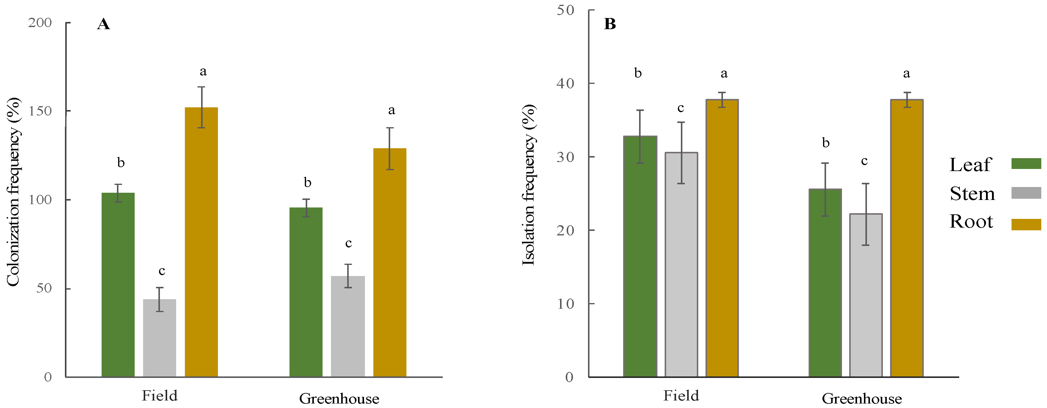
2.4. Colonization and Isolation Frequency

An analysis of fungal occurrence was estimated by calculating the colonization frequency and isolation rate. The colonization frequency (CF) and isolation frequency (IF) were calculated according to the formula below and two-way ANOVA tests were carried out to compare the CF and IF values between the tissue types and growing conditions:
CF (%) = (Number of segments showing endophyte growth/number of segments plated) × 100
IF (IF%) = (Number of unique taxa isolated/number of segments plated) × 100

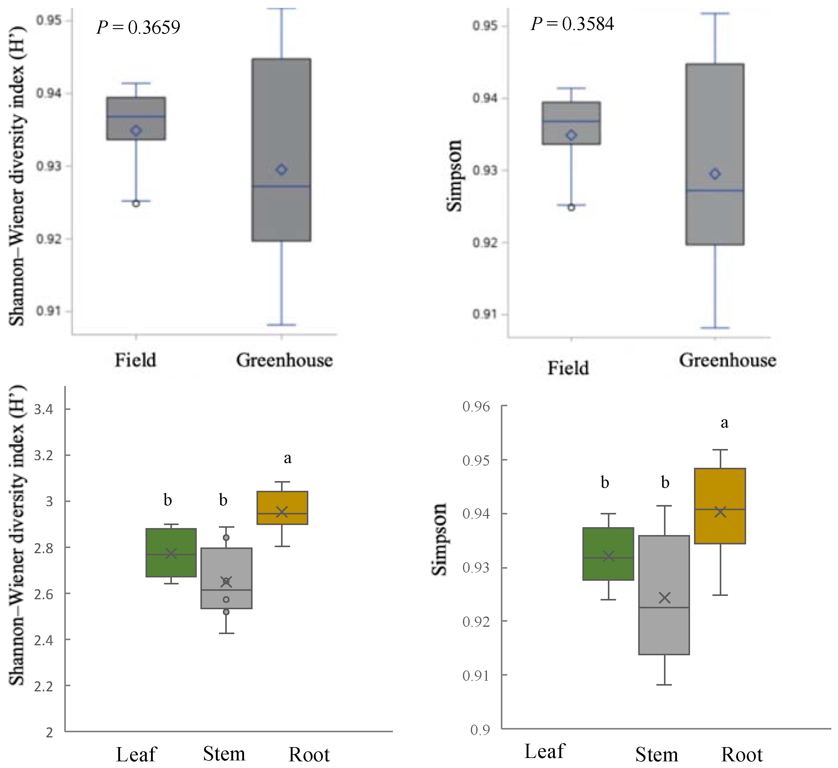
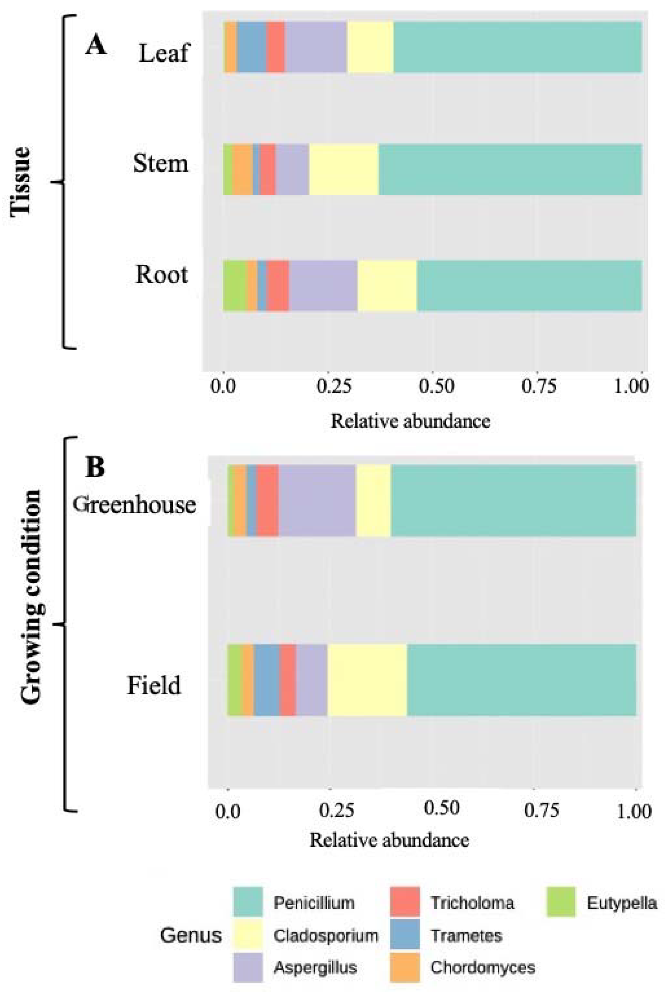
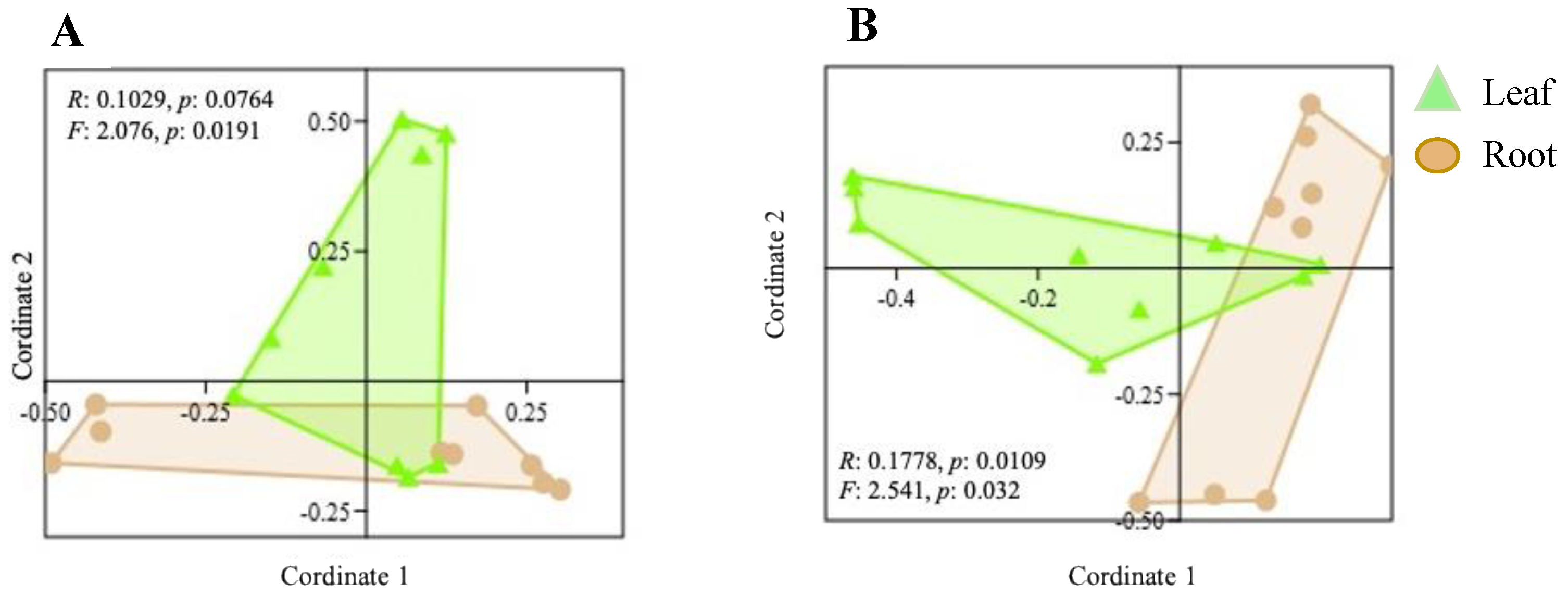
2.5. Community Structure of Culturable Endophytic Fungi

The α diversity of the endophyte species within the samples was measured by using the Shannon–Wiener diversity index (H’) and Simpson dominance. An ANOVA test was performed to compare the diversity index values among the host tissue types and growth conditions. To compare the differences in community composition and structure among the tissue types and growing conditions (β-diversity), an analysis of similarity (ANOSIM) and PERMANOVA were used. The use of two different pairwise similarity measures was attempted because they consider different types of information that can provide different insights for comparing communities: (i) Jaccard’s index, which only considers the presence or absence of fungal taxa among samples; (ii) the Bray–Curtis coefficient, which considers the abundance of taxa along with the presence or absence of particular fungal taxa. To analyze the diversity measures (α-diversity and β-diversity), PAST ver. 3.26 were used.

2.6. Plant Growth Promotion Potential of Endophytic Fungi

2.6.1. Phosphate and Potassium Solubilization Activity

A qualitative estimation of the phosphate solubilization activity was performed according to Paul and Rao [39] on a solid medium using PDA plugs from 14-day-old fungal cultures on Pikovskaya medium supplemented with bromophenol blue (0.5 mL/100 mL). The potassium solubilization activity was also tested by using the Alexandrov media [40]. The clear halo around the colony was considered a positive result of the phosphate and potassium solubilization activity. The phosphate and potassium solubilization index values were determined using the following formula:
Solubilization Index (SI) = ((Colony diameter + Halo zone diameter)/Colony diameter)

2.6.2. Estimation of Siderophore Production

The detection of the siderophore production ability of the fungal isolates was tested based on the CAS half-plate agar plate assay [41]. Each half of the plate was filled with PDA medium and the other half contained a solid CAS autoclaved medium that was prepared according to Schwyn and Neiland [42]. The cube of the fungal isolate inoculum was placed in the PDA half and the plate was sealed with parafilm. All of the plates were incubated at 30 °C under dark conditions for 5–7 days and monitored for a change in the blue color into purplish-red or orange. The CAS liquid assay was used for the quantitative assessment of the siderophore production [43]. The fungal isolates were grown on a liquid PDB medium for 14 days and the fungal culture was centrifuged at 6.00 rpm for 5 min. An equal volume of CAS reagent was added to the culture supernatant and the O.D. was observed at 630 nm. The following calculation was used to express siderophore units [44]:
% Siderophores unit = [(Ar − As)/Ar] × 100
Where Ar is the reference absorbance at 630 nm (CAS reagent only) and as is the sample absorbance at 630 nm.

2.6.3. Qualitative and Quantitative Evaluation of Indole-3-Acetic Acid (IAA) Production

The production of indole acetic acid (IAA) was tested colorimetrically according to Gordon and Weber using the Salkowski reagent [45]. Three plugs of 14-day-old cultures were inoculated on YPD broth (1% yeast extract, 2% peptone, 2% glucose, final pH 6.0) and shaken at 120 rpm for 5 days at 28 °C. The cultures were centrifuged at 10,000 g for 1 min and filtered through a 0.2 μm filter (Millipore, Burlington, MA, USA). Finally, 1 mL of the filtered culture was mixed with 1 mL of Salkowski reagent (2% of 0.5 M FeCl3 in 35% HClO4) and incubated at room temperature for 30 min. The development of the pink color was considered a positive result. The quantitative IAA concentration of the samples was measured by checking the absorbance at 530 nm with a microplate reader and then was measured based on a standard curve of known values. The fungal isolate that showed the highest concentration of indole-3-acetic acid (IAA) was further evaluated based on the growth of the soybean (Glycine max, Enri Cultivar), which is one of the most important soybean cultivars used in Japan. Surface-disinfected and germinated soybean seeds were transferred into a plant box (CUL-JAR300; Iwaki, Japan) containing sterilized vermiculate and were inoculated with 2 mL of fungal spore suspension (107), then the sterilized water was used as a mock treatment. The plants were transferred to a plant growth chamber (25 °C and 70% humidity under a 16/8 h day/night regimen). Sterilized Broughton and Dilworth nitrogen-free solutions (B&D) were used for watering. Disease symptoms, plant height, and plant fresh and dry weights were measured after 25 days in the plant growth chamber.

2.7. In Vitro Antagonistic Potential against Phytopathogenic Fungi

The antagonistic potential of fungal isolates was screened using in vitro dual culture tests for their ability to control the mycelial growth of the fungal soybean pathogen. The identified pathogenic fungus (Calonectria ilicicola), known as red crown rot, was used for this experiment. All endophytic fungal strains were tested against the pathogenic fungus. A plug of the pathogen (5 mm) was used and placed on one side of a 9 cm petri dish, which contained Potato dextrose agar. One disc of endophytic fungus (5 mm) was also placed on the opposite side of the petri dish. Both discs were 4 cm apart from each other. In the control plate, only a disc of PDA medium (5 mm) was placed on the opposite side of the pathogen. Three replicates were used for each combination. All the plates were incubated at room temperature and observed every day [46]. When the growth of the pathogenic fungus stopped in the dual culture or the colony filled the whole plate in the control, the antagonistic type, width of the inhibition band, and radius of the pathogen were measured. The antagonism type between the endophytic fungi and pathogen in vitro was estimated in each combination. The endophytic fungal strain was identified to have antagonistic properties if it suppressed the growth of the pathogen, grew faster than the pathogen, or parasitized the pathogen [47]. The growth inhibition rates (CGIR) of the pathogenic fungi were also calculated using the following formula:
CGIR = ((RCK (radius of control colony) − R test (radius of test colony))/RCK) × 100

2.8. Statistical Analysis

Each result for the test plants was expressed as the mean percentage. Statistical differences in parameters between control and sample groups were evaluated using a one-factor analysis of variance (ANOVA) followed by Tukey’s HSD (honestly significant difference) test. Here, p < 0.05 was considered statistically significant.

3. Results and Discussion

3.1. Isolation and Identification of Endophytic Fungi

Various studies have shown that plants and beneficial endophytes have a symbiotic relationship that stimulates plant health and growth. In this study, we isolated and identified culturable endophytic fungi from different tissues of V. villosa for the first time. To verify the efficacy of the surface disinfection method after surface sterilization of the samples, the sterile water used to rinse the samples was taken for a control experiment to confirm the surface disinfection. The absence of growth of microorganisms on the plate was checked, and this indicated the effectiveness of the protocol to eliminate the epiphytic microorganisms. A total of 1043 isolates (here isolates are defined as completely visible fungal growths from each (5 mm × 5 mm) tissue segment placed on the PDA plates) were recovered from 1080 tissue segments (leaf, stem, and root) of hairy vetch plants collected from the two different growing conditions (experimental field and greenhouse). Based on their morphological characteristics (color and shape), the isolates were grouped into 64 representative fungal isolates. The sixty-four representative fungal isolates were subjected to molecular identification based on ITS rDNA sequences. The BLAST search resulted in a total of 33 different endophytic fungal taxa that belonged to two main phyla, 4 classes, 6 orders, and 7 genera (Table S1, Figure S1, and Table 1).
The soil type in all experimental sites is volcanic ash soil, with pH and electric conductivity (EC) values of 6.5 and 0.07, respectively. The ANOVA test on colonization frequency (CF) indicated no significant differences between the growing conditions. However, a significant difference was observed among the tissues that were examined in this study (overall colonization frequency = 96.9%). The results showed that the highest colonization frequency was observed in root samples from the field condition, while the stem segments yielded the lowest number of isolates in both growth conditions (Figure 1A).
A similar trend was observed concerning the isolation rate, which expresses the fungal richness of the samples and was calculated by the number of isolates obtained from tissue segments divided by the total number of segments in that sample (Figure 1B). Other studies showed that the colonization rate and species richness of endophytic fungi among root, stem, and leaf samples were the highest in the roots [48], followed by the stems [49,50]. Generally, the isolation frequency in all the tissues of the hairy vetch was high (20–25%). These results suggest that endophytic fungi had extensively colonized many parts of hairy vetch plants, especially the roots. The differences in the assembly of the endophytic fungi in different tissues might be related to the capacity of the root tissue to uptake and utilize nutrients, which provide good conditions for fungal growth. Moreover, other factors such as the structure and chemistry of the tissues might also play a role in this variation. On the contrary, Shreelalitha [51] found a low colonization frequency in the roots compared to other segments in wild legumes.
However, we were not able to identify a few taxa at the genus level (Figure S1). In total, the most dominant phylum in both growth conditions was Ascomycota, with rates of 89% and 92% for field and greenhouse samples, respectively (a total of 91.0%). However, a few samples in both growing conditions belonged to Basidiomycota (field; 10% and greenhouse; 7%). Those endophytic fungi classified in the Ascomycota phylum were distributed into three classes. The largest part represented Eurotiomycetes (73.5%), followed by Dothideomycetes (15.5%) and Dothideomycetes (6.4%). All Basidiomycota phyla were distributed only in the Agaricomycetes class (Figure S2). The same results were also found in other isolation experiments [49,50,52,53,54].
The diversity and species richness of the endophytic fungi (α-diversity) concerning the tissue types and growing conditions were measured using the Shannon–Wiener diversity index (H’) and Simpson dominance index. All indices were calculated with PAST (Version 4) [55]. A higher α-diversity index is defined as higher species diversity and a greater distribution of endophytic fungi. According to the Shannon and Simpson dominance index values for the growing conditions, we did not detect any differences in the α-diversity values of endophytic fungi between the two conditions, but there was significant variation among the tissues based on the results. In total, the root samples showed the highest α-diversity, whereas no significant difference was observed between the leaves and stems in both growing conditions (Figure 2).

3.2. Variation in Endophyte Community Composition and Assembly

According to the ITS sequence and NCBI database results, the total sequences were characterized into two main phyla, 4 classes, 6 orders, 7 genera, and 33 species. Ascomycota was the most dominant in both growth conditions, with rates of 89% and 92% for field and greenhouse samples, respectively. However, only a few of the samples belonged to Basidiomycota (field = 10%; greenhouse = 7%, respectively). Among the Ascomycota, Eurotiomycetes (78%) were the richest class, whereas only one class was identified in the Basidiomycota phylum.
Overall, the most dominant fungal genera were Penicillium, Cladosporium, and Aspergillus. A previous study showed that several endophytic fungi belonging to the Penicillium genus have been reported for their ability to synthesize plant-growth-promoting hormones and antagonize pathogens [56]. Among the tissues of hairy vetch plants evaluated here, the relative abundance of some genera was completely noticeable. The highest abundance of the genus Eutypella was recovered from root samples, followed by stem and leaf samples, respectively, as shown in Figure 3. Meanwhile, the relative abundance levels of the genus Eutypella were significantly different between the two growing conditions and were mostly recovered from field samples (Figure 3). To evaluate the differences in the community composition of endophytic fungi, a beta diversity analysis of the data was performed. Cluster analyses (two- dimensional NMDS), as well as ANOSIM and PERMANOVA, were performed to understand the effects of the growth conditions and tissue type on the endophytic fungal community patterns, based on Bray–Curtis and Jaccard’s similarity tests. The clustering of fungal communities displayed a good representation of the hairy vetch fungal endophytes’ community structure.
The outcomes of the two-dimensional non-metric multidimensional scale (NMDS) according to the ANOSIM and PERMANOVA tests showed that the growth conditions of the samples did not affect the assembly of the endophyte communities (R = 0.1029, p = 0.0764; Figure 4). However, the ANOSIM and PERMANOVA tests suggested that the tissue types significantly influenced the community structure of the fungal endophytes under both growth conditions (Figure S3).

3.3. Plant-Growth-Promoting Traits of Isolated Endophytes

In this study, the plant-growth-promoting potential of all isolates was tested by checking the plant hormone levels (IAA production), which directly stimulates the growth, as well as other factors such as phosphate and potassium solubilization activity and siderophore production levels, which also promote plant growth indirectly.

3.3.1. Phosphate and Potassium Solubilization Activity

Fungal endophytes have various levels of potential to promote the growth of their host plants by harnessing direct and indirect processes. Through an indirect process, these microorganisms can solubilize phosphate and potassium, which are essential for plant growth. [57]. In this study, all the fungal isolates (33 strains) were tested qualitatively on Pikoviskaya media to assess the phosphate solubilization activity (Table 2). Out of all the endophytic fungal samples evaluated, only 5 isolates (hvef7, Cladosporium cladosporioides; hvef18, Penicillium simplicissimum; hvef29, Aspergillus flavus; hvef45, Penicillium italicum; hvef60, Penicillium oxalicum) showed the potential to solubilize phosphate and produce a clear zone in the Pikoviskaya media supplemented with a pH indicator dye, bromothymol blue. The isolate hvef7 (Cladosporium cladosporioides) presented the maximum solubilization index value of 1.36 ± 0.01 (Table 2).
Moreover, all fungal endophytes were screened for their ability to solubilize potassium by exhibiting a clear zone in the Aleksandrov medium (Figure S4). The potassium solubilization index values ranged from 3 to 15, and the highest solubilization index value was recorded for hvef45 (Penicillium italicum) (Table 2). Phosphate and potassium exist in the soil in an insoluble form, but a different type of endophytic fungus has the potential to change them to available forms. In this study, the phosphate and potassium solubilizing activity levels of all isolates were qualitatively checked, as shown in Table 2. These results are also inconsistent with other reports [58,59], which showed that most Penicillium and Aspergillus species can convert unavailable phosphate and potassium to available forms for uptake by plants. Phosphate is one of the most vital macronutrients necessary for plant growth and development. However, phosphate in most agricultural soils is in immobile forms, making the isolation and identification of phosphate-solubilizing microbes very essential to modern agriculture.

3.3.2. Siderophore Production Potential of Isolated Fungi

Plants usually suffer from iron deficiencies in the soil, and it is pivotal to solve this problem to increase crop productivity. Some plants have developed certain techniques to overcome the deleterious effects of iron deficiency; for example, monocotyledons produce phytosiderophores. These siderophores are iron carriers that bind with the available ferric iron in the soil with a high affinity [60,61]. Siderophores produced by microorganisms have received more interest because of their potential application in agriculture systems. These small compounds can be synthesized by many fungal species in the rhizosphere soil [62,63]. In this study, nineteen isolates changed the color of the CAS agar plate into a dark purplish-red, as a result of their siderophore production and binding of iron from the medium (Figure S5). The assessment of the siderophore production rates of these nineteen isolates was performed quantitively. Generally, the average siderophore unit production rate of these isolates was 61%. These microbial siderophores play an important role in the transfer of iron to the plants to increase the growth of plants [64]. Moreover, it was reported that siderophores might play a significant role as biocontrol agents [65] because apart from causing the symbiotic relationship between plants and fungi, they also showed hostility against some fungal pathogens [66]. Fungi are generally known for the production of different types of siderophores. Most of them can produce hydroxymate siderophores [67,68,69]. Other studies have corroborated the results of this study by showing that Aspergillus species have a high ability to produce siderophores [40,59,60]. Additionally, the results of this study showed that hvef29 (Aspergillus flavus) had the highest rate of siderophore production at 74.7 ± 0.16%.

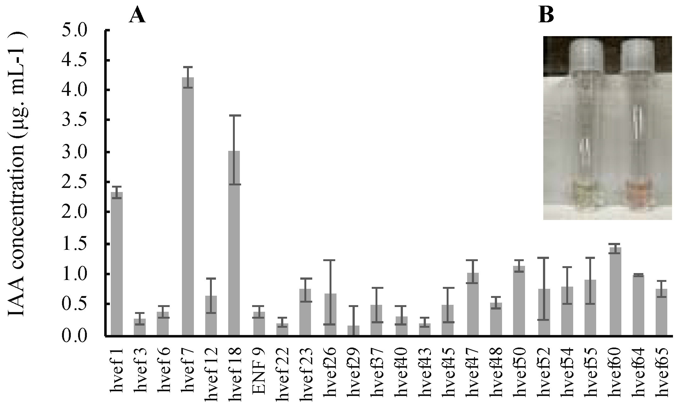
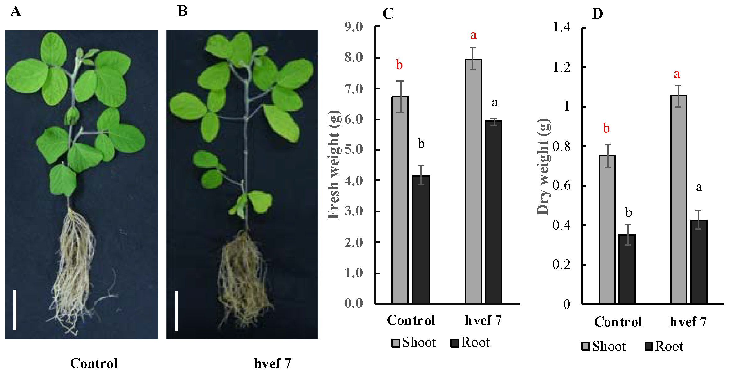
3.3.3. Qualitative and Quantitative Evaluation of Indole-3-Acetic Acid (IAA) Production

Phytohormones are essential for the growth and changes in the morphology and structure of plants. The production of phytohormones such as indole acetic acid (IAA), a significant phytohormone, is crucial a technique used by endophytic fungi to stimulate the growth of their host plants. Based on the calorimetric test, 28 isolates were able to produce IAA hormones and showed a pink color when adding the Salkowski reagent. The quantitative amount of IAA concentration of samples was measured by comparing the absorbance of standard pure IAA with culture filtrates of each isolate at 530 nm. The results indicated that the highest IAA content in the culture filtrate (4.21 ± 0.16 μg mL−1) was produced by isolate hvef7 (Cladosporium cladosporioides), followed by hvef18 (Penicillium simplicissimum) (3.02 μg mL−1) and hvef1 (Cladosporium pseudocladosporioides) (2.32 μg mL−1) (Figure 5).
Indole-3-acetic acid (IAA) is responsible for cell division, differentiation, growth rate control, and root formation [70]. Therefore, the isolation and identification of endophytic fungi capable of producing IAA can significantly promote plant growth by increasing the root length and area, increasing the plant’s capacity to uptake nutrients, and helping to regulate the entire metabolism necessary for its life cycle. Cladosporium in this study showed the highest amount of IAA production, and these data are similar to the results of previous a study [71].
The strain that produced the highest concentration of indole-3-acetic acid (IAA) was evaluated for its potential as a bio-inoculant for soybean. The results showed that the inoculation of soybean plants with hvef7 (Cladosporium cladosporioides) increased the fresh weight of the soybean shoots and roots compared to the non-inoculated plants (control). Similar results were also observed for the dry weights of the shoots and roots of soybean plants as compared to non-inoculated plants (Figure 6). Moreover, the fungal inoculation did not show any significant differences related to plant height compared to the control plants (data not shown). Phytohormones are essential for plant growth, as well as for changing the morphology and structure of plants. As the result of the current experiment, hvef7 (Cladosporium cladosporioides) showed the highest rate of IAA production among all tested isolates. This result is similar to a previous report [72].

3.4. Antagonistic Potential of Fungal Isolates against Red Crown Rot (Calonectria ilicicola)

In this study, the antagonistic potential of the isolated fungi against Calonectria ilicicola was evaluated and classified into three different types. The isolates that showed similar growth with the pathogen were classified as competition for the substance and surfaces. The growth of both fungi (pathogens and endophytes) will continue until they reach and contact each other. The second group was identified as an antibiotic type, meaning the fungi with antimicrobial potential will inhibit the growth of the pathogen without contact by releasing some antimicrobial metabolites. The last group was classified as mycoparasites. In this group, the rapid growth of endophytic fungi resulted in the coverage of the colony of pathogens, as shown in Figure S6. Most of the isolates showed different abilities to control the growth of Calonectria ilicicola, the main cause of red crown rot disease in soybean plants. This result presents the possibility of finding novel metabolites that can be utilized as promising antimicrobial agents. The highest antimicrobial potential was recorded for hvef3 (Penicillium sp.) with 95.27 ± 0.06 (Table 3). The antimicrobial potential of endophytic fungi has recently received high attention. Many reports have mentioned the production of some secondary metabolites by endophytic fungi with high potential for antibacterial and antifungal activity, which control the growth of pathogenic microorganisms [65]. The antagonistic potential of the Penicillium genus (dominant species in this study) has been reported in other studies [73]. P. simplicissimum and P. simplicissimum ENF22 isolated from rice have exhibited antagonistic activity against these pathogens, such as Pyricularia oryzae, Rhizoctonia solani, Sclerotium oryzae, and Pythium ultimum [74].

4. Conclusions

Plant diseases and climate change cause major economic losses in crop production and threaten food security. The application of PGPF has been identified as a promising technique to help mitigate crop losses due to diseases and other environmental stresses. The data obtained from this study indicate that besides the well-established weed suppression effect of Vicia villosa (hairy vetch), the plant also harbors various kinds of endophytic fungi. Thirty-three strains of fungal endophytes were isolated from different tissues (leaf, stem, and root) of V. villosa from different growth conditions based on morphological and molecular analyses. The colonization frequency of fungal endophytes was significantly higher in the root segments than in other tissues. The Penicillium genus was found to be the most dominant genus among all isolated fungi. The isolated endophytes showed PGPF potential by producing phytohormones (IAA). Some of these isolates could solubilize phosphate and potassium, which are pivotal for plant growth, and the isolates convert them to the available form for the plants. Moreover, most of the isolated fungi exhibited an antagonistic effect against red crown rot, with the potential to protect plants against the disease. Although these fungal isolates showed important functional characteristics, further studies are necessary to check their ability to make symbiosis relationships with other target host plants and to evaluate their potential as bioinoculants.

Supplementary Materials

The following supporting information can be downloaded at: https://www.mdpi.com/article/10.3390/agronomy12102417/s1. Table S1. Mycelium color of the identified fungi (morphological identification. Figure S1. Taxonomic classification of fungal endophytes’ operational taxonomic units (OTUs) from ITS sequences of endophytic fungi recovered from three representative tissues from two different growing conditions. Figure S2. Taxonomic classification of fungal endophytes’ operational taxonomic units (OTUs) from ITS sequences of endophytic fungi recovered from three representative tissues from two different growing conditions. Figure S3. Fungal endophyte communities as influenced by tissue types from two environmental growing conditions. Non-metric multidimensional scaling (NMDS) plots for cluster analyses of the fungal endophyte community were calculated: (A) Bray–Curtis coefficient, which compares the presence or absence of fungal taxa along with the abundance among groups; (B) Jaccard’s index, which is used only to compute presence and absence data to compare fungal community similarity among groups. The ANOSIM statistic R and the PERMANOVA statistic F values and the corresponding p-values indicating the significance of dissimilarity were obtained via the permutation of group membership with 9999 replicates. Figure S4. Potassium solubilization activity of fungal isolates. Figure S5. Identification of the siderophore-producing isolate: (A) control; (B) not able to produce siderophores; (C) siderophore production activity of fungal isolate assessed by changing the blue color of CAS agar into purplish red. Figure S6. (A) The number of antagonistic combinations of endophytic fungi against pathogenic fungi. (B) Antagonistic types of endophytic fungi towards pathogenic fungi.

Author Contributions

Conceptualization, P.T. and Y.F.; methodology, P.T., R.K., K.M.G.D., H.M.K., and S.O.; software, P.T. and K.M.G.D.; validation, P.T., K.M.G.D., and K.T.; formal analysis, P.T., K.M.G.D., and K.T.; investigation, P.T. and Y.F.; data curation, P.T., K.M.G.D., and K.T.; writing-original draft preparation, P.T. and K.S.A.; writing—review and editing, R.K., K.M.G.D., K.S.A., M.Y., K.T., H.M.K., M.A., S.O., and Y.F.; visualization, K.M.G.D., K.S.A., and M.A.; supervision, Y.F.; funding acquisition, Y.F. All authors have read and agreed to the published version of the manuscript.

Funding

This study was supported by a grant-in-aid for Research on Agriculture and Food Science (25029AB) from the Ministry of Agriculture, Forestry, and Fisheries of Japan, and JST CREST Grant Number JPMJCR17O2, Japan.

Acknowledgments

The authors thank the Japanese Ministry of Education, Culture, Sports, Science, and Technology (MEXT; 173338) for providing scholarships and funds to carry out this research at the Tokyo University of Agriculture and Technology.

Conflicts of Interest

The authors declare no conflict of interest.

References

  1. Tudi, M.; Daniel Ruan, H.; Wang, L.; Lyu, J.; Sadler, R.; Connell, D.; Chu, C.; Phung, D.T. Agriculture development, pesticide application and its impact on the environment. Int. J. Environ. Res. Public Health 2021, 18, 1112. [Google Scholar] [CrossRef] [PubMed]
  2. Baweja, P.; Kumar, S.; Kumar, G. Fertilizers and pesticides: Their impact on soil health and environment. In Soil Health; Giri, B., Varma, A., Eds.; Soil Biology; Springer International Publishing: Cham, Switzerland, 2020; pp. 265–285. ISBN 978-3-030-44364-1. [Google Scholar]
  3. Raza, H.; Idress, M.; Yar, G.; Farah, N.; Asim, M.; Naveed, M.; Yasin, M.; Younus, M. Residual impact of pesticides on environment and health of sugarcane farmers in punjab with special reference to integrated pest management. J. Glob. Innov. Agric. Soc. Sci. 2019, 7, 79–84. [Google Scholar] [CrossRef]
  4. Javanmard, A.; Amani Machiani, M.; Lithourgidis, A.; Morshedloo, M.R.; Ostadi, A. Intercropping of maize with legumes: A cleaner strategy for improving the quantity and quality of forage. Clean. Eng. Technol. 2020, 1, 100003. [Google Scholar] [CrossRef]
  5. Ren, J.; Zhang, L.; Duan, Y.; Zhang, J.; Evers, J.B.; Zhang, Y.; Su, Z.; van der Werf, W. Intercropping Potato (Solanum tuberosum L.) with Hairy Vetch (Vicia villosa) increases water use efficiency in dry conditions. Field Crops Res. 2019, 240, 168–176. [Google Scholar] [CrossRef]
  6. Kim, N.; Zabaloy, M.C.; Guan, K.; Villamil, M.B. Do cover crops benefit soil microbiome? a meta-analysis of current research. Soil Biol. Biochem. 2020, 142, 107701. [Google Scholar] [CrossRef]
  7. Habibi, D.; Kashani, A.; Paknejad, F.; Jafary, H.; Al-Ahmadi, M.J.; Tookalloo, M.R.; Lamei, J. Evaluation of hairy vetch (Vicia villosa Roth) in pure and mixed cropping with barley (Hordeum vulgare l.) to determine the best combination of legume and cereal for forage production. Am. J. Agric. Biol. Sci. 2010, 5, 169–176. [Google Scholar]
  8. Sainju, U.M.; Singh, H.P.; Singh, B.P. Cover crop effects on soil carbon and nitrogen under bioenergy sorghum crops. J. Soil Water Conserv. 2015, 70, 410–417. [Google Scholar] [CrossRef]
  9. Omae, H.; Nagumo, F. Effects of Oat (Avena sativa) and Hairy Vetch (Vicia villosa) cover crops on nitrate leaching, soil water, and maize yield in subtropical islands in Japan. J. Agric. Sci. (Tor.) 2016, 8, 44–54. [Google Scholar] [CrossRef]
  10. Sato, T.; Sato, E.; Takakai, F.; Yokoyama, T.; Kaneta, Y. Effects of hairy vetch foliage application on nodulation and nitrogen fixation in soybean cultivated in three soil types. Soil Sci. Plant Nutr. 2011, 57, 313–319. [Google Scholar] [CrossRef]
  11. Bansal, S.; Yin, X.; Schneider, L.; Sykes, V.; Jagadamma, S.; Lee, J. Carbon footprint and net carbon gain of major long-term cropping systems under no-tillage. J. Environ. Manag. 2022, 307, 114505. [Google Scholar] [CrossRef] [PubMed]
  12. Roig, C.J. Performance of Upland Cotton Under a Hairy Vetch Regiment from a Crop Insurance Perspective. Ph.D. Thesis, Louisiana State University and Agricultural & Mechanical College, Baton Rouge, LA, USA, 2021. [Google Scholar]
  13. Hartwig, N.L.; Ammon, H.U. Cover crops and living mulches. Weed Sci. 2002, 50, 688–699. [Google Scholar] [CrossRef]
  14. Anugroho, F.; Kitou, M.; Nagumo, F.; Kinjo, K.; Tokashiki, Y. Growth, nitrogen fixation, and nutrient uptake of hairy vetch as a cover crop in a subtropical region. Weed Biol. Manag. 2009, 9, 63–71. [Google Scholar] [CrossRef]
  15. Seo, J.H.; Lee, H.J. Mineral nitrogen effects of hairy vetch (Vicia villosa Roth) on maize (Zea mays l.) by green manure amounts. J. Agron. 2008, 7, 272–276. [Google Scholar] [CrossRef] [Green Version]
  16. Zhou, X.G.; Everts, K.L. Suppression of Fusarium wilt of watermelon by soil amendment with hairy vetch. Plant Dis. 2004, 88, 1357–1365. [Google Scholar] [CrossRef] [PubMed] [Green Version]
  17. Candole, B.L.; Rothrock, C.S. Characterization of the suppressiveness of hairy vetch-amended soils to Thielaviopsis basicola. Phytopathology 1997, 87, 197–202. [Google Scholar] [CrossRef] [PubMed] [Green Version]
  18. Kamo, T.; Sakurai, S.; Yamanashi, T.; Todoroki, Y. Cyanamide is biosynthesized from l-canavanine in plants. Sci. Rep. 2015, 5, 10527. [Google Scholar] [CrossRef] [Green Version]
  19. Mohammadi, G.R. Weed control in irrigated corn by hairy vetch interseeded at different rates and times. Weed Biol. Manag. 2010, 10, 25–32. [Google Scholar] [CrossRef]
  20. Abdin, O.A.; Zhou, X.M.; Cloutier, D.; Coulman, D.C.; Faris, M.A.; Smith, D.L. Cover crops and interrow tillage for weed control in short season maize (Zea Mays). Eur. J. Agron. 2000, 12, 93–102. [Google Scholar] [CrossRef]
  21. Mardani-Korrani, H.; Nakayasu, M.; Yamazaki, S.; Aoki, Y.; Kaida, R.; Motobayashi, T.; Kobayashi, M.; Ohkama-Ohtsu, N.; Oikawa, Y.; Sugiyama, A.; et al. L-canavanine, a root exudate from hairy vetch (Vicia villosa) drastically affecting the soil microbial community and metabolite pathways. Front. Microbiol. 2021, 12, 2754. [Google Scholar] [CrossRef]
  22. Sakurai, N.; Mardani-Korrani, H.; Nakayasu, M.; Matsuda, K.; Ochiai, K.; Kobayashi, M.; Tahara, Y.; Onodera, T.; Aoki, Y.; Motobayashi, T.; et al. Metabolome analysis identified okaramines in the soybean rhizosphere as a legacy of hairy vetch. Front. Genet. 2020, 11, 114. [Google Scholar] [CrossRef] [PubMed] [Green Version]
  23. Hayashi, H.; Takiuchi, K.; Murao, S.; Arai, M. Structure and insecticidal activity of new indole alkaloids, okaramines a and b, from Penicillium simplicissimum ak-40. Agric. Biol. Chem. 1989, 53, 461–469. [Google Scholar] [CrossRef]
  24. Jia, M.; Chen, L.; Xin, H.-L.; Zheng, C.-J.; Rahman, K.; Han, T.; Qin, L.-P. A friendly relationship between endophytic fungi and medicinal plants: A systematic review. Front. Microbiol. 2016, 7, 906. [Google Scholar] [CrossRef] [Green Version]
  25. López, S.; Pastorino, G.; Franco, M.; Medina, R.; Lucentini, C.; Saparrat, M.; Balatti, P. Microbial endophytes that live within the seeds of two tomato hybrids cultivated in Argentina. Agronomy 2018, 8, 136. [Google Scholar] [CrossRef] [Green Version]
  26. Oh, S.-Y.; Yang, J.H.; Woo, J.-J.; Oh, S.-O.; Hur, J.-S. Diversity and distribution patterns of endolichenic fungi in jeju island, South Korea. Sustainability 2020, 12, 3769. [Google Scholar] [CrossRef]
  27. Mishra, S.; Bhattacharjee, A.; Sharma, S. An Ecological insight into the multifaceted world of plant-endophyte association. Crit. Rev. Plant Sci. 2021, 40, 127–146. [Google Scholar] [CrossRef]
  28. Nelson, J.; Shaw, A.J. Exploring the natural microbiome of the model liverwort: Fungal endophyte diversity in Marchantia polymorpha L. Symbiosis 2019, 78, 45–59. [Google Scholar] [CrossRef]
  29. Gibert, A.; Tozer, W.; Westoby, M. Plant performance response to eight different types of symbiosis. New Phytol. 2019, 222, 526–542. [Google Scholar] [CrossRef]
  30. Fesel, P.H.; Zuccaro, A. Dissecting endophytic lifestyle along the parasitism/mutualism continuum in Arabidopsis. Curr. Opin. Microbiol. 2016, 32, 103–112. [Google Scholar] [CrossRef] [Green Version]
  31. Hodkinson, T.R.; Doohan, F.M.; Saunders, M.J.; Murphy, B.R. Endophytes for a Growing World; Cambridge University Press: Cambridge, UK, 2019; ISBN 978-1-108-47176-3. [Google Scholar]
  32. Wolfe, E.R.; Ballhorn, D.J. Do foliar endophytes matter in litter decomposition. Microorganisms 2020, 8, 446. [Google Scholar] [CrossRef] [PubMed] [Green Version]
  33. Khalil, A.M.A.; Hassan, S.E.-D.; Alsharif, S.M.; Eid, A.M.; Ewais, E.E.-D.; Azab, E.; Gobouri, A.A.; Elkelish, A.; Fouda, A. Isolation and characterization of fungal endophytes isolated from medicinal plant Ephedra pachyclada as plant growth-promoting. Biomolecules 2021, 11, 140. [Google Scholar] [CrossRef]
  34. Varga, T.; Hixson, K.K.; Ahkami, A.H.; Sher, A.W.; Barnes, M.E.; Chu, R.K.; Battu, A.K.; Nicora, C.D.; Winkler, T.E.; Reno, L.R.; et al. Endophyte-promoted phosphorus solubilization in populus. Front. Plant Sci. 2020, 11, 567918. [Google Scholar] [CrossRef] [PubMed]
  35. Contreras-Cornejo, H.A.; del-Val, E.; Macías-Rodríguez, L.; Alarcón, A.; González-Esquivel, C.E.; Larsen, J. Trichoderma atroviride, a maize root associated fungus, increases the parasitism rate of the fall armyworm Spodoptera frugiperda by its natural enemy Campoletis Son. Soil Biol. Biochem. 2018, 122, 196–202. [Google Scholar] [CrossRef]
  36. Timmusk, S.; Behers, L.; Muthoni, J.; Muraya, A.; Aronsson, A.-C. Perspectives and challenges of microbial application for crop improvement. Front. Plant Sci. 2017, 8, 49. [Google Scholar] [CrossRef] [PubMed] [Green Version]
  37. Getachew, G.; Rewald, B.; Godbold, D.L.; Sandén, H. Endophytic fungal root colonization of Eragrostis tef in eroded croplands of the ethiopian highlands is limited by low spore density and fertilisation. Agronomy 2019, 9, 73. [Google Scholar] [CrossRef] [Green Version]
  38. Müller, M.M.; Hallaksela, A.-M. Diversity of norway spruce needle endophytes in various mixed and pure norway spruce stands. Mycol. Res. 1998, 102, 1183–1189. [Google Scholar] [CrossRef]
  39. Paul, N.B.; Sundara Rao, W.V.B. Phosphate-dissolving bacteria in the rhizosphere of some cultivated legumes. Plant Soil 1971, 35, 127–132. [Google Scholar] [CrossRef]
  40. Aleksandrov, V.G.; Blagodyr’, R.N.; Il’ev, I.P. Phosphorus acid isolation from apatite produced by silicate bacteria. Mikrobiolohichnyi Zhurnal 1967, 29, 111–114. [Google Scholar] [PubMed]
  41. Milagres, A.M.F.; Machuca, A.; Napoleão, D. Detection of siderophore production from several fungi and bacteria by a modification of chrome azurol s (CAS) agar plate Assay. J. Microbiol. Methods 1999, 37, 1–6. [Google Scholar] [CrossRef]
  42. Schwyn, B.; Neilands, J.B. Universal chemical assay for the detection and determination of Siderophores. Anal. Biochem. 1987, 160, 47–56. [Google Scholar] [CrossRef]
  43. Osman, Y.; Gebreil, A.; Mowafy, A.M.; Anan, T.I.; Hamed, S.M. Characterization of Aspergillus niger siderophore that mediates bioleaching of rare earth elements from phosphorites. World J. Microbiol. Biotechnol. 2019, 35, 93. [Google Scholar] [CrossRef]
  44. Kejela, T.; Thakkar, V.R.; Patel, R.R. Novel strain of Pseudomonas inhibits Colletotrichum gloeosporioides and Fusarium oxysporum infections and promotes germination of coffee. Rhizosphere 2017, 4, 9–15. [Google Scholar] [CrossRef]
  45. Gordon, S.A.; Weber, R.P. Colorimetric estimation of indoleacetic acid. Plant Physiol. 1951, 26, 192–195. [Google Scholar] [CrossRef] [PubMed] [Green Version]
  46. Campanile, G.; Ruscelli, A.; Luisi, N. Antagonistic activity of endophytic fungi towards Diplodia corticola assessed by in vitro and in planta Tests. Eur. J. Plant Pathol. 2007, 117, 237–246. [Google Scholar] [CrossRef]
  47. Landum, M.; Felix, M.; Alho, J.; Garcia, R.; Cabrita, M.J.; Rei, F.; Varanda, C.M. Antagonistic activity of fungi of Olea europaea L. against Colletotrichum acutatum. Microbiol. Res. 2016, 183, 100–108. [Google Scholar] [CrossRef] [PubMed] [Green Version]
  48. Naik, B.S.; Shashikala, J.; Krishnamurthy, Y.L. Study on the diversity of endophytic communities from rice (Oryza sativa L.) and their antagonistic activities in vitro. Microbiol. Res. 2009, 164, 290–296. [Google Scholar] [CrossRef]
  49. Li, J.-L.; Sun, X.; Zheng, Y.; Lü, P.-P.; Wang, Y.-L.; Guo, L.-D. Diversity and community of culturable endophytic fungi from stems and roots of desert halophytes in Northwest China. MycoKeys 2020, 62, 75–95. [Google Scholar] [CrossRef]
  50. Dastogeer, K.M.G.; Li, H.; Sivasithamparam, K.; Jones, M.G.K.; Wylie, S.J. Host specificity of endophytic mycobiota of wild Nicotiana plants from arid regions of northern Australia. Microb. Ecol. 2018, 75, 74–87. [Google Scholar] [CrossRef]
  51. Shreelalitha, S.J.; Sridhar, K.R. Endophytic fungi of wild legume Sesbania bispinosa in coastal sand dunes and mangroves of the southwest coast of India. J. For. Res. 2015, 26, 1003–1011. [Google Scholar] [CrossRef]
  52. Zhang, T.; Zhang, Y.-Q.; Liu, H.-Y.; Wei, Y.-Z.; Li, H.-L.; Su, J.; Zhao, L.-X.; Yu, L.-Y. Diversity and cold adaptation of culturable endophytic fungi from Bryophytes in the fildes region, King george island, maritime antarctica. FEMS Microbiol. Lett. 2013, 341, 52–61. [Google Scholar] [CrossRef] [Green Version]
  53. Zheng, H.; Qiao, M.; Xu, J.; Yu, Z. Culture-based and culture-independent assessments of endophytic fungal diversity in aquatic plants in southwest China. Front. Fungal. Biol. 2013, 341, 52–61. [Google Scholar] [CrossRef]
  54. Tibpromma, S.; Karunarathna, S.; Bhat, D.J.; Suwannarach, N.; Stephenson, S.; Elgorban, A.; Al-Rejaie, S.; Xu, J.; Mortimer, P. Using culture-dependent and molecular techniques to identify endophytic fungi associated with tea leaves (Camellia spp.) in yunnan province, China. Diversity 2022, 14, 287. [Google Scholar] [CrossRef]
  55. Hammer, O.; Harper, D.A.T.; Ryan, P.D. PAST: Paleontological Statistics Software Package for Education and Data Analysis. Palaeontol. Electron. 2001, 4, 9. [Google Scholar]
  56. Toghueo, R.M.K.; Boyom, F.F. Endophytic Penicillium species and their agricultural, biotechnological, and pharmaceutical applications. 3 Biotech 2020, 10, 107. [Google Scholar] [CrossRef] [PubMed]
  57. Patle, P.; Navnage, N.; Ramteke, P. Endophytes in plant system: Roles in growth promotion, mechanism, and their potentiality in achieving agriculture sustainability. Int. J. Chem. Stud. 2018, 6, 270–274. [Google Scholar]
  58. Muthuraja, R.; Muthukumar, T. Isolation and characterization of potassium solubilizing Aspergillus species isolated from saxum habitats and their effect on maize growth in different soil types. Geomicrobiol. J. 2021, 38, 672–685. [Google Scholar] [CrossRef]
  59. Qiao, H.; Sun, X.-R.; Wu, X.-Q.; Li, G.-E.; Wang, Z.; Li, D.-W. The phosphate-solubilizing ability of Penicillium guanacastense and its effects on the growth of Pinus massoniana in phosphate-limiting conditions. Biol. Open 2019, 8, bio046797. [Google Scholar] [CrossRef] [Green Version]
  60. Roskova, Z.; Skarohlid, R.; McGachy, L. Siderophores: An alternative bioremediation strategy? Sci. Total Environ. 2022, 819, 153144. [Google Scholar] [CrossRef]
  61. Ghosh, S.K.; Bera, T.; Chakrabarty, A.M. Microbial siderophore – a boon to agricultural sciences. Biol. Control. 2020, 144, 104214. [Google Scholar] [CrossRef]
  62. Joo, K.A.H. and J.H. Zinc ions affect siderophore production by fungi isolated from the Panax ginseng rhizosphere. J. Microbiol. Biotechnol. 2019, 29, 105–113. [Google Scholar] [CrossRef]
  63. Tamariz-Angeles, C.; Huamán, G.D.; Palacios-Robles, E.; Olivera-Gonzales, P.; Castañeda-Barreto, A. Characterization of Siderophore-producing microorganisms associated to plants from high-andean heavy metal polluted soil from Callejón de Huaylas (Ancash, Perú). Microbiol. Res. 2021, 250, 126811. [Google Scholar] [CrossRef]
  64. Chowdappa, S.; Jagannath, S.; Konappa, N.; Udayashankar, A.C.; Jogaiah, S. Detection and characterization of antibacterial siderophores secreted by endophytic fungi from Cymbidium aloifolium. Biomolecules 2020, 10, 1412. [Google Scholar] [CrossRef] [PubMed]
  65. Fadiji, A.E.; Babalola, O.O. Elucidating mechanisms of endophytes used in plant protection and other bioactivities with multifunctional prospects. Front. Bioeng. Biotechnol. 2020, 8, 467. [Google Scholar] [CrossRef] [PubMed]
  66. Oide, S.; Moeder, W.; Krasnoff, S.; Gibson, D.; Haas, H.; Yoshioka, K.; Turgeon, B.G. NPS6, Encoding a nonribosomal peptide synthetase involved in siderophore-mediated iron metabolism, is a conserved virulence determinant of plant pathogenic Ascomycetes. Plant Cell 2006, 18, 2836–2853. [Google Scholar] [CrossRef] [PubMed]
  67. Franken, A.C.W.; Lechner, B.E.; Werner, E.R.; Haas, H.; Lokman, B.C.; Ram, A.F.J.; van den Hondel, C.a.M.J.J.; de Weert, S.; Punt, P.J. Genome mining and functional genomics for siderophore production in Aspergillus niger. Brief. Funct. Genom. 2014, 13, 482. [Google Scholar] [CrossRef] [Green Version]
  68. Baakza, A.; Vala, A.K.; Dave, B.; Dube, H. A comparative study of siderophore production by fungi from marine and terrestrial habitats. J. Exp. Mar. Biol. Ecol. 2004, 311, 1–9. [Google Scholar] [CrossRef]
  69. Wright, W.; Little, J.; Liu, F.; Chakraborty, R. Isolation, and structural identification of the trihydroxamate siderophore vicibactin and its degradative products from rhizobium leguminosarum ATCC 14479 Bv. Trifolii. Biometals 2013, 26, 271–283. [Google Scholar] [CrossRef]
  70. Woodward, A.W. Auxin: Regulation, action, and interaction. Ann. Bot. 2005, 95, 707–735. [Google Scholar] [CrossRef] [Green Version]
  71. Răut, I.; Călin, M.; Capră, L.; Gurban, A.-M.; Doni, M.; Radu, N.; Jecu, L. Cladosporium sp. isolate as fungal plant growth promoting agent. Agronomy 2021, 11, 392. [Google Scholar] [CrossRef]
  72. Hirota, A.; Sakai, H.; Isogai, A. New Plant Growth Regulators, Cladospolide A and B, Macrolides Produced by Cladosporium cladosporioides. Agric Biol Chem. 1985, 49, 731–735. [Google Scholar] [CrossRef]
  73. Liang, L.-J.; Jeewon, R.; Dhandevi, P.; Durairajan, S.S.K.; Li, H.; Lin, F.-C.; Wang, H.-K. A novel species of Penicillium with inhibitory effects against Pyricularia oryzae and fungal pathogens inducing citrus diseases. Front. Cell Infect. Microbiol. 2021, 10, 604504. [Google Scholar] [CrossRef]
  74. Potshangbam, M.; Devi, S.I.; Sahoo, D.; Strobel, G.A. Functional characterization of endophytic fungal community associated with Oryza sativa L. and Zea mays L. Front. Microbiol. 2017, 8, 325. [Google Scholar] [CrossRef] [PubMed]
Figure 1. Colonization frequency (A) and isolation frequency (B) of endophytic fungi common to leaf, stem, and root tissues of hairy vetch (Vicia villosa) from two different growing conditions (field and greenhouse). The error bar indicates the standard error (SE) of the mean. Bars with different letters (a, b, c) indicate that the mean values are significantly different at a significance level of p < 0.05.
Agronomy 12 02417 g001
Figure 2. Fungal endophyte biodiversity analysis. The effect of the sampling growing conditions (top) and tissue types (bottom) on fungal endophyte biodiversity was measured using the Shannon–Wiener diversity (H’) and Simpson index. The endophytes were isolated from the leaf, stem, and root tissues of hairy vetch plants from two different growing conditions (field and greenhouse). Bars with different letters (a, b) indicate that the mean values are significantly different at a significance level of p < 0.05.
Agronomy 12 02417 g002
Figure 3. The endophytic community structure of fungal genera in different hairy vetch tissues (A) and growth conditions (B), showing their relative abundance. The result for each tissue is presented as the sum of that representative tissue, while for the growing condition it is the sum of all tissues.
Agronomy 12 02417 g003
Figure 4. Fungal endophyte communities as influenced by growing conditions. Non-metric multidimensional scaling (NMDS) plots for cluster analyses of the fungal endophyte community groups were calculated: (A) Bray–Curtis coefficient, which compares the presence or absence of fungal taxa along with the abundance among groups; (B) Jaccard’s index, which is used to show the presence and absence of data to compare the similarity among groups. The ANOSIM statistic R and PERMANOVA statistic F values and the corresponding p-values indicate the significance of dissimilarities obtained via the permutation of group membership with 9999 replicates.
Agronomy 12 02417 g004
Figure 5. Quantitative (A) and qualitative indole-3-acetic acid (IAA) production rates (B) caused by 24 isolated fungi after 14 days of incubation. Values are the means of three replicates ± standard deviation.
Agronomy 12 02417 g005
Figure 6. Soybean plant without inoculation (A) and with inoculation of hvef7 (B), soybean shoot and root fresh weights (C), shoot and root dry weights (D) after 25 days inoculation with hvef7 (Cladosporium cladosporioides) compared with control plants. Data were statistically different at p < 0.05 as assessed by one-way ANOVA with Tukey’s test (n= 5); means ± SD. For each inoculation treatment, bars with different letters (a, b) indicate that the mean values are significantly different at a significance level of p < 0.05.
Agronomy 12 02417 g006
Table 1. Taxonomic classification of fungal endophyte OTUs from ITS sequences of endophytic fungi recovered from three representative tissues of hairy vetch with NCBI accession numbers.
Isolate No.Isolates Acc. No.ClassClosest Match (Genbank)Query Coverage (%)Tissue
hvef1MK036245DothideomycetesCladosporium pseudocladosporioides (MK111597.1)99.8R, S
hvef3MK036247EurotiomycetesPenicillium sp. (LC133788.1) 100L, S, R
hvef6MK036250EurotiomycetesPenicillium brefeldianum (MH858155.1) 100L, S, R
hvef7MK036251DothideomycetesCladosporium cladosporioides (MG669180.1)96.0R
hvef9 MK036253 EurotiomycetesPenicillium ochrochloron (MH137639.1) 99.0L, S, R
hvef10 MK036254 EurotiomycetesPenicillium glaucoroseum (MH865551.1)100L, S, R
hvef12MK036256AgaricomycetesTrametes versicolor (MK322281.1)100L, S
hvef18MK036262EurotiomycetesPenicillium simplicissimum (MH856014.1) 100L, S, R
hvef22MK036266EurotiomycetesPenicillium cremeogriseum (MH374608.1)100L, S, R
hvef23 MK036267EurotiomycetesPenicillium sp. (MK625191.1)100L, S, R
hvef26 MK036270DothideomycetesCladosporium halotolerans (MK265717.1)100L, S, R
hvef29 MK036273EurotiomycetesAspergillus flavus (MK791661.1) 100R, L
hvef30 MK036274EurotiomycetesAspergillus sp. (MK817589.1) 100L, S, R
hvef31 MK036275DothideomycetesCladosporium sphaerospermum (MH482916.1)100R, S
hvef32 MK036276SordariomycetesEutypella sp. (KX828160.1)99.0L, S, R
hvef37 MK036281SordariomycetesChordomyces antarcticum (KX385856.1)100L, S, R
hvef40 MK036284EurotiomycetesPenicillium steckii (MH484016.1)99.0S, R
hvef41 MK036285EurotiomycetesPenicillium expansum (KC456325.1)100L, S, R
hvef43 MK036287EurotiomycetesAspergillus westerdijkiae (MG733714.1)100L, S, R
hvef44 MK036288EurotiomycetesAspergillus sydowii (KX898426.1)99.0L, S, R
hvef45 MK036289EurotiomycetesPenicillium italicum (DQ991463.1)100L, S, R
hvef46 MK036290EurotiomycetesPenicillium svalbardense (KC346348.1) 93.0L, S, R
hvef47 MK036291EurotiomycetesPenicillium sp. (KY350140.1)100L, S, R
hvef48 MK036292AgaricomycetesTricholoma matsutake (MF037418.1)100R
hvef50 MK036294EurotiomycetesPenicillium sp. (MG551581.1)96.0L, S, R
hvef52 MK036296EurotiomycetesPenicillium sp. (MH325925.1)98.0L, S, R
hvef54 MK036298EurotiomycetesPenicillium sp. (MH550491.1)94.0L, S, R
hvef55 MK036299EurotiomycetesPenicillium griseopurpureum (KY678777.1)100L, S, R
hvef58 MK036302EurotiomycetesPenicillium sp. (KJ921867.1)99.0L, R
hvef60 MK036304EurotiomycetesPenicillium oxalicum (KF152942.1)94.0L, S, R
hvef63 MK036307EurotiomycetesPenicillium sp. (KY425713.1)97.0L, S, R
hvef64 MK036308EurotiomycetesPenicillium crustosum (KT735107.1)96.0R
hvef65 MK036309EurotiomycetesPenicillium sp. (KT369826.1)98.0L, S, R
L: leaf; S: stem; R: root.
Table 2. Phosphate and potassium solubilizing activities and siderophore production of endophytic fungal strains isolated from V. villosa.
NoFugal StrainPhosphate Solubilization IndexPotassium Solubilization Index% Siderophore Unit
1hvef1000
2hvef30065.4 ± 0.13
3hvef6000
4hvef71.36 ± 0.0100
5hvef9000
6hvef10000
7hvef120062.0 ± 0.03
8hvef181.13 ± 0.06055.3 ± 0.46
9hvef22000
10hvef230060.7 ± 0.07
11hvef260060.1 ± 0.02
12hvef291.13 ± 0.03074.7 ± 0.16
13hvef300063.0 ± 0.25
14hvef31000
15hvef320064.7 ± 0.23
16hvef3703.53 ± 0.1169.4 ± 0.24
17hvef400061.1 ± 0.04
18hvef41015.9 ± 0.1161.0 ± 0.05
19hvef43012.6 ± 0.2063.1 ± 0.25
20hvef440057.5 ± 0.03
21hvef451.14 ± 0.0415.1 ± 0.230
22hvef46011.1 ± 0.110
23hvef4704.73 ± 0.250
24hvef480056.7 ± 0.05
25hvef5004.10 ± 0.1054.7 ± 0.05
26hvef52000
27hvef540054.3 ± 0.08
28hvef550056.3 ± 0.01
29hvef5803.33 ± 0.3058.9 ± 0.17
30hvef601.30 ± 0.0210.8 ± 0.2061.7 ± 0.24
31hvef63014.6 ± 0.200
32hvef64000
33hvef65000
Values are means ± SD of three replicates.
Table 3. Inhibitory potential of isolated fungal endophytes against Calonectria ilicicola.
NoFungal Isolates CodeGrowth Inhibition Rates (%)NoFungal Isolates CODEGrowth Inhibition Rates (%)
1hvef116.0 ± 0.2518hvef4149.7 ± 0.29
2hvef395.2 ± 0.0619hvef4393.5 ± 0.13
3hvef689.3 ± 0.1720hvef4456.2 ± 0.15
4hvef762.7 ± 0.2621hvef451.14 ± 0.04
5hvef992.9 ± 0.0122hvef4643.2 ± 0.20
6hvef1067.5 ± 0.2923hvef4792.9 ± 0.10
7hvef1259.2± 0.2024hvef4883.4 ± 0.12
8hvef1863.3 ± 0.2125hvef5081.1 ± 0.10
9hvef2280.5 ± 0.1726hvef5267.5 ± 0.22
10hvef2360.4 ± 0.2527hvef5448.5 ± 0.17
11hvef2660.3 ± 0.1228hvef5569.8 ± 0.21
12hvef2995.9 ± 0.0629hvef5873.4 ± 0.06
13hvef3087.0 ± 0.2530hvef6055.6 ± 0.50
14hvef3176.3 ± 0.1531hvef6388.7 ± 0.23
15hvef3254.4 ± 0.2132hvef6467.5 ± 0.29
16hvef3767.5 ± 0.7633hvef6582.2 ± 0.20
17hvef4075.7 ± 0.15
Values are means ± SD of three replicates.
Publisher’s Note: MDPI stays neutral with regard to jurisdictional claims in published maps and institutional affiliations.

