Article

Identification and Characterization of Fungal Pathogens Causing Trunk and Branch Cankers of Almond Trees in Morocco

1
Phytopathology Unit, Department of Plant Protection, Ecole Nationale d’Agriculture de Meknès, Km10, Rte Haj Kaddour, BP S/40, Meknes 50001, Morocco
2
Laboratory of Biotechnology and Valorization of Phyto-Resources, Faculty of Sciences, Moulay Ismail University of Meknes, Avenue Zitoun, Meknes 50001, Morocco
3
Laboratory of Functional Ecology and Environmental Engineering, Department of Biology, FST-Fez, Sidi Mohamed Ben Abdellah University, BP 2202, Fez 30050, Morocco
4
Unité de Recherche Résistance Induite et Bio-Protection des Plantes-EA 4707, Université de Reims Champagne-Ardenne, 51100 Reims, France
*
Authors to whom correspondence should be addressed.
Agronomy 2023, 13(1), 130; https://doi.org/10.3390/agronomy13010130
Submission received: 8 December 2022 / Revised: 26 December 2022 / Accepted: 27 December 2022 / Published: 30 December 2022

Abstract

:
Canker symptoms were observed on almond trees in the Fez-Meknes region, Morocco. Isolations were conducted from the infected branch and trunk showing internal and external brown lesions. Four representative fungal isolates were screened, and their identities were confirmed by cultural traits and sequence analysis of DNA using two genes, the ITS region (internal transcribed spacer) and calmodulin (cmdA). The identified fungi were Curvularia hawaiiensis, Fusarium ambrosium, Lasiodiplodia theobromae, and Chondrestereum purpureum. The pathogenicity test on almond twigs revealed that these species were pathogenic to their host with different degrees of virulence, with Lasiodiplodia theobromae as the most virulent causing the longest necrotic lesion (285.17 mm) and the death of twigs. Physiological traits analysis of the above-mentioned fungi showed that the optimum mycelium growth response at different temperatures varies from 10 to 35 °C, while the pH ranges between 3.0 and 8.0. This study confirms the presence of canker pathogens on almond trees, which will contribute valuable information to improve the understanding of the contemporary status of almond trees, thus helping the improvement of the management of almond orchards. To our knowledge, all these fungi represent new records in Morocco and some of them are confirmed on the almond trees for the first time in the world.

1. Introduction

The almond tree (Prunus dulcis) is considered the most important and oldest nut crop produced and consumed all over the world [1]. The almond tree originated in central Asia, precisely in the mountainous regions of Iran, Afghanistan, and the former Soviet Union [2], then transported to the Mediterranean basin [3] where its plantation was possible thanks to its self-incompatibility and its open pollination [4]. Almond trees have adapted to severe climatic conditions, such as delayed spring frost and drought. However, the tree has become more sensitive to cold in the last three decades [5]. World almond production is led by the USA, Spain, Iran, Australia, and Morocco, which constitute the main almond-producing countries [6]. According to FAOSTAT, the world yield of almonds in 2019 was estimated at almost 3.49 million metric tons; the USA ranking first with 77% of almonds’ global production, followed by Australia and Spain with 8% and 6% respectively [7].
Since the last century, Morocco is the fourth-largest producer of almonds, with over 117,270 tons per year [8]. In terms of covered area, almonds rank second after olives production in Morocco. The special climate characteristics and environmental conditions in north and south Morocco ensure the huge genetic variability of almond cultivars explaining the vigorous production and the high quality of Moroccan almonds [9].
The almond crop was always vulnerable to various biotic (such as bacteria, fungi, and phytoplasma) as well as abiotic stresses (namely drought and freeze damage). Almond trees are commonly attacked by several destructive diseases, which damage both the foliar part and the fruit, such as Tranzschelia discolor f. sp. Dulcis, the causal agent of almond rust. In addition, some cultivars are susceptible to other diseases such as a shot hole and almond scab, caused by Wilsonomyces carpophilus and Cladosporium carpophilum respectively [10]. Furthermore, the trunk and roots are also susceptible to various fungal attacks including Chondrostereum purpureum, which causes the silver leaf disease.
Several authors have reported the almond trees’ sensitivity to many fungal pathogens attacking their woody part. For instance, canker disease has become one of the most serious problems in almond production since it can damage young trees [11]. Generally, canker diseases occur as wood degradation, showing symptoms such as wood necrosis and gummosis, internal symptoms such as vascular discoloration, cankers on the trunk or branches, and even tree death. In addition, dieback of scaffold branches and shoots through pruning wounds and damaged tissues were reported [12]. Cross section of infected tissues showed circular, irregular, or wedge-shaped necrosis, leading to the destruction of phloem and xylem tissues, and therefore, in the end, canker blocks the circulation of water and nutrients resulting in the death of bark and cambium [13]. In the recent past, numerous studies have been conducted on fungal trunk pathogens of fruit trees, such as stone and pome fruit trees [14,15,16], grapevine [17,18] pistachio [19,20], Ficus trees [21], blueberry [22] and olive [23].
A wide range of Botryosphaeriaceae species, such as Botryosphaeria, Diplodia, Dothiorella, Lasiodiplodia, and Neofusicoccum, among others, are recognized as common fungal pathogens of woody trees, showing a large distribution on various fruit tree and nut crops as a saprobe, parasite, and endophyte [24]. Several previous reports reveal the implication of species from this complex fungal group in almond crop attacks causing band cankers such as the Dothiorella canker, which affect the trunk of three- to six-year-old almond trees [25]. Other studies conducted on almond trees confirm that Lasiodiplodia theobromae is an important pathogen associated with branch cankers as well [19]. Moreover, in other studies, different species of Diaporthaceae (Phomopsis) were reported on Prunus trees, especially on almonds, e.g., D. eres in Portugal [26], D. foeniculina in Italy [27], and D. phaseolorum in Spain [28]. D. amygdali has been documented as the main causal agent of woody canker diseases and showed symptoms of shoot blight and twig canker associated with Punus trees [26]. Furthermore, many species of Fusarium genus were reported to cause canker diseases on the almond tree; especially, in young plantations, which are more susceptible to Fusarium canker [29].
In the past few years, trunk diseases have been frequently observed in many Moroccan almond orchards. However, the implications of many of the above-mentioned fungal species in canker disease progression, are still unclear; and no comprehensive attempt has been announced to identify pathogens responsible for almond trunk diseases in this area. The overall aim of our work was to identify and characterize pathogenic agents causing canker and wood necrosis in almond orchards of the fez region, Morocco, through monitoring morphological traits, physiological characteristics, molecular data, and pathogenicity analysis.

2. Materials and Methods

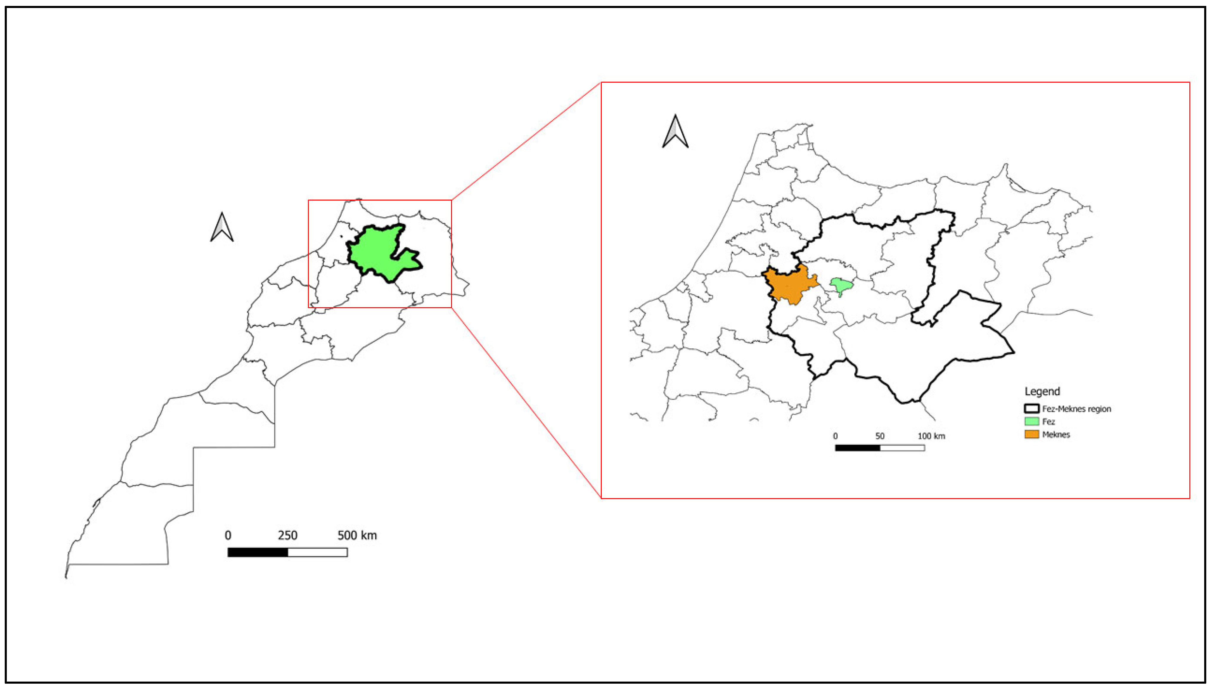
2.1. Sampling and Isolation

During the growing season of 2020, canker diseases were observed in almond orchards in the fez Meknes region, Morocco (Figure 1). The collected samples showed symptoms of canker diseases including internal woody necrosis, cankers, dieback of branch and trunk as well as shoot blight (Figure 2).
The cut wood segments (3 × 3 mm) of infected tissue taken from the lesions’ margin and unaffected wood, were rinsed using tap water, sterilized for 1 min in a solution of sodium hypochlorite (1.5%) then, washed with sterile distilled water (SDW) two times. Wood fragments were deposited on PDA plates (PDA; Biokar-Diagnostics, Zac de Ther, France) supplemented with streptomycin sulfate (0.5 g/L) (Sigma-Aldrich, St. Louis, MO, USA). Petri plates were incubated in darkness at 25 °C, after one-week four types of fungal colonies were transferred to Petri plates of PDA. Successive subcultures were made until obtaining pure cultures.

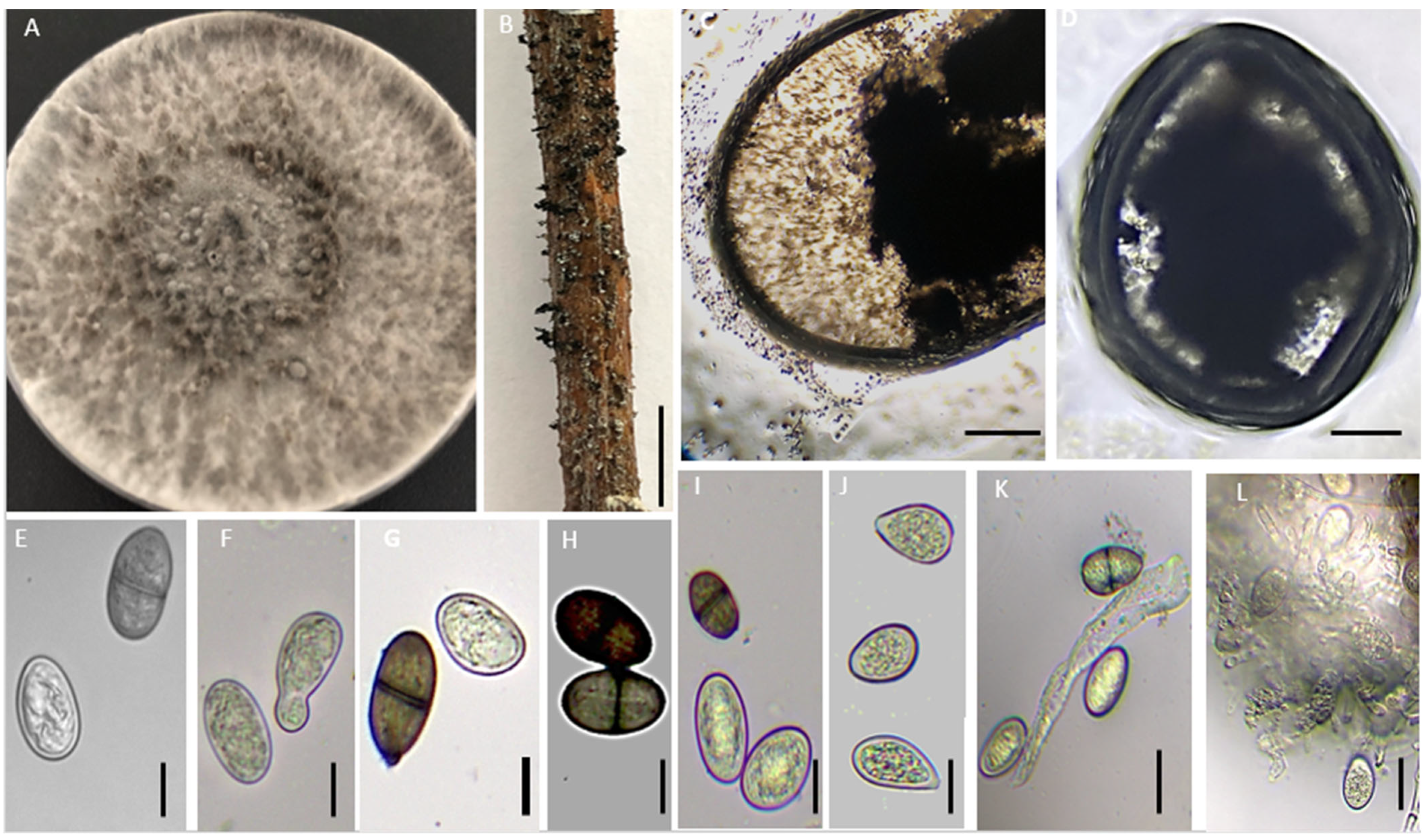
2.2. Morphological Identification

Isolated fungi were cultured, purified on PDA plates, and incubated at 25 °C, then criteria, such as color and appearance of colonies, were recorded after 10 days [30]. To force the isolates’ sporulation, colonized plug agar of each one was cultured first on malt extract agar medium (MEA; Sigma-Aldrich, St. Louis, MO, USA), then the isolates that did not sporulate directly on MA were transferred into PDA medium supplemented with three to four pine needles already double-autoclaved. Plates were then incubated at 25 °C exposed to 12 h/12 h of light/darkness cycle. The diameters of conidia for each isolate (at least 100 conidia) were noted using the microscope (Olympus EX41) linked with the camera and ImageFocus Plus V2 Software.

2.3. Molecular Identification

To confirm the identity of isolated pathogens, Genomic DNA was extracted from actively growing mycelia on a PDA medium for seven to 14 days following the Doyle and Doyle [31] protocol. DNA aliquots were stored at 20 °C. The internal transcribed spacer ITS-5.8-ITS of the 5.8 ribosomal and calmodulin (cmdA) genes, were amplified and sequenced using respectively ITS1/ITS4 [32] and CAL-228F/CAL-737R [33] primers in both directions. Reaction mixtures contained 2.5 µL of 10× PCR reaction buffer, 0.25 µL (10 mM) of dNTP, 1 µL of each primer, 2.25 µL (50 mM) MgCl2, 2.5 µL DNA, 0.2 µL Taq DNA Polymerase (New England Biolabs, Beverly, MA, USA), and 16.3 µL sterile distilled waters; the total volume was 25 µL. The PCR process was performed in a T100 thermal cycler (Bio-Rad, Hercules, CA, USA). PCR program consisted of an initial denaturation step for 3 min at 94 °C, then 35 cycles of 94 °C for 1 min, 55 °C for 1 min, 72 °C for 1 min, and the final extension step took about 10 min at 72 °C. PCR products were visualized by 1.5% agarose gel electrophoresis, then purified and sequenced by STAB Vida, Lda. (Portugal). Both sequences, forward and reverse, were assembled and aligned using Molecular Evolutionary Genetics Analysis (MEGA) and the Basic Local Alignment Search Tool (BLAST) was used to compare the assembled ITS and cmdA sequences with all the sequenced isolates already deposed in the GeneBank. The phylogenetic tree was created using the maximum likelihood method with 1000 bootstrap values Reference isolates used are reported in Table 1.

2.4. Pathogenicity Test

To examine the pathogenicity of the four selected isolates in laboratory conditions, the experiment was conducted as described by [26]. Thirty cm length of healthy and unaffected almond twigs (cv. Ferreduel) one–two-year-old were submerged twice in ethanol 70% for 30 s interrupted by immersion in sodium hypochlorite solution (1.5%) for 1 min. After each step, twigs were rinsed using SDW and then air-dried inside a laminar flow cabinet. To wound the twigs, a disinfected metal scalpel was utilized to detach the superficial tissue from the center of the twigs to create artificial wounds. PDA plugs colonized by mycelia of fresh fungal culture (seven days old) were placed on the artificial cuts and sealed with parafilm. Twigs inoculated with sterile PDA discs were used as control. Detached twigs were put in a 0.5 L container, filled with 200 mL of SDW, and placed in a humid chamber at 24 °C with 12 h/12 h of dark/light and a water change every three days. During the first four days, the containers were enveloped with a polyethylene bag to maintain a humid environment inside. Six branches per fungal species and six controls were used. The pots were randomly distributed.
After 15 days of inoculation, lesion lengths above and below the site of inoculation were measured. To satisfy the Koch postulates, re-isolation from the margin of the cankered and healthy tissue was conducted. Cut fragments were disinfected and placed onto PDA as described above. Petri plates were incubated for one week at 25 °C in darkness and recovered isolates were identified. This experiment was repeated twice.

2.5. Physiological Traits

2.5.1. Effect of Temperature

To assess the impact of different temperatures on the mycelial growth of isolated fungi per day, all fungal species used in this work were subcultured in PDA plates and incubated for seven days in conditions of darkness and temperature at 25 °C. Mycelial discs (5 mm) cut from the active growth zone, were deposited in the center of PDA plates and then incubated at different temperatures (5, 10, 25, 30, 35, and 40 °C), with three replicates of each isolate at each temperature. After 24 h of incubation, daily radial growth rates were recorded for 18 days using a digital caliper.

2.5.2. Effect of pH

To evaluate the pH effect on the mycelial growth per day, the PDA was calibrated with a buffer of citric acid containing (0.1 M sodium citrate and 0.1 M HCl), to reach the required pH between 3 and 4. The phosphate buffer was used to attain the following values of pH 5.0, 6.0, and 7.0, while for pH from 8.0 to 12.0 the medium was modified by NaOH (1N) or HCL (1N). Colonized discs (5 mm) were placed at the midpoint of Petri plates of potato dextrose agar with freshly modified pH. The pH was measured using an electrical pH meter before autoclaving at 121 °C. Finally, plates were incubated at 25 °C and colony parameters were recorded every day (n = 10). Three biological replicates for each pH were made, and the experiment was repeated two times.

2.6. Statistical Analysis

All treatments were in a completely randomized design. Analysis of variance was performed using SPSS statistical software (version 20, IBM SPSS Statistics 20, New York, NY, USA) and when the result was shown to be significant, the Tukey test was used for means separation at a significance level of p < 0.05.

3. Results

3.1. Morphological Characterization of Isolates

According to morphological features obtained in our research, namely, shape, colony color, and microscopic criteria, all isolated fungi have different morphological appearances, belong to different groups, and were identified from infected almond samples in the field located in the region of Fez Meknes in Morocco. Further, they showed symptoms in the trunk, branches, and shoots including, wedge-shaped, black or dark brown regular and irregular discolorations and lesions.
The first fungal isolate formed on PDA medium, hairy, fluffy mycelia, light brown at the edge and dark brown in the middle of the colony, then change to black, with concentric zonation after seven days of incubation (Figure 3). Conidiophores were unbranched, straight with curved apexes. Conidia were frequently brown, cylindrical to spindle-shaped with rounded ends, multiseptate, four usually, and rarely ovoid, oval, and measured 5.44–23.87 × 3.65–9.76 µm. These characteristics correspond to the description of the genus Curvularia, especially the species Curvularia hawaiiensis [34,35]. The colonies of the second isolate have aerial mycelium, white grey at the surface, olivaceous grey at the reverse plate, and became dark grey after two weeks of incubation. Pycnidia formed on pine needles liberate ovoid, hyaline, aseptate conidia with apex rounded and sometimes, pigmented and one septate conidium, measuring 17.84–22.71 × 9.98–12.66 µm (Figure 4). Based on the taxonomy of Botryosphaeriaceae species [36], these macroscopic and microscopic criteria resembled those of Lasiodiplodia theobromae. The third pathogen cultured on PDA was distinguished by white cottony mycelia, convex in the center. Conidia were oblong, hyaline, and smooth and measured 17.84–22.71 × 2.75–12.66 µm and cystidia were also observed (Figure 5). All morphological features were identical to those described for C. purpureum [37]. Colonies of the last isolate showed radial mycelial growth that is yellowish-white in color and, with age, turned light violet. Formed a red pigment that surrounded the medium. Abundant sporulation on PDA under light, typical conidia that were remarkably variable in shape and size, Macroconidiawere curved cylindrical, mostly sickle-shaped to long-clavate, usually multiseptated (3–4), measuring 24.43–27.27 × 2.05–6.79 µm in size. Microconidia were ellipsoidal to fusiform with a rounded apex and produced on elongated conidiophores. They had 0–1 septate and measured 10.34–15.76 × 2.05–3.48 μm (Figure 6). These morphological traits are identical to the species belonging to Fusarium ambrosium (Neocosmospora ambrosium) within the Fusarium solani complex [38,39]

3.2. Phylogenetic Analysis

The molecular analysis of the ITS region (internal transcribed spacer) and calmodulin (cmdA) gene confirmed that Chondrostereum purpureum, Curvularia hawaiiensis, Lasiodiplodia theobromae, and Fusarium ambrosium (Neocosmospora ambrosia) are linked to the symptoms described in the field and are responsible for the trunk and branch canker of almond trees. The tree based on concatenated sequences of two genes, the internal transcribed spacer and calmodulin highly support the close relationship between our isolates and their references (Figure 7). All isolates were clustered with their references (Figure 7, Table 1), which further confirmed the identity (98–100%) of different species isolated in this paper.

3.3. Pathogenicity Test

The pathogenicity test carried out through inoculation of one-year-old detached almond twigs by fungal isolates, indicated that all tested species were pathogenic to almond twigs, and caused brown discoloration above and below the wound site (Figure 8). The average length of lesions in almonds with different isolates varied from 30 to 288 mm and the control twigs treated with PDA plugs free of mycelial fungi (controls) did not induce any lesions (Figure 8). Statistical analysis revealed a significant difference in vascular discoloration length between twigs inoculated with all isolates and control (p < 0.05). The most aggressive fungus was Lasiodiplodia theobromae, which produced the longer lesions 285.17 mm in diameter, followed by Fusarium ambrosium and Chondrostereum purpureum, which developed moderate lesions of 94.52 mm and 80.29 mm, respectively (Figure 9). Curvularia hawaiiensis was the least virulent with lesions averaging 32.17 mm in length. The four isolates used in the pathogenicity test were re-isolated from the symptomatic twigs and their identities were confirmed, fulfilling Koch’s postulates.

3.4. Physiological Traits

The results of variance analysis demonstrated a significant effect of temperature (p < 0.05) on the radial growth of colonies on PDA of all isolated pathogens used in the current study. Data revealed that all isolates, C. purpureum, C. hawaiiensis, F. ambrosium, and L. theobromae grew ideally between 10 and 35 °C, while C. hawaiiensis could grow slowly in a relatively narrow temperature of 5 °C (Table 2). Additionally, C. purpureum has the ability to develop hypha at 40 °C. For the optimum growth temperatures, C. hawaiiensis and C. purpureum recorded the highest optimum growth at 30 °C with a hyphal growth rate of 14.67 ± 0.28 and 9.08 ± 0.44 mm day−1, respectively. However, L. theobromae showed an optimum growth rate at 20 °C with 27.04 ± 0.05 mm day−1. For F. ambrosium, the highest growth temperature was 30 °C with a mycelial extension of 9.12 ± 0.1. Overall, this experiment showed that the appropriate temperature in which all tested isolates develop in a perfect way, varied between 20 °C and 30 °C.
Selected isolates used in this experiment reacted differently within various pH values tested. For all evaluated pH, the highest mycelial growth of C. hawaiiensis was recorded at pH 8.0 with an average extension of 10.60 ± 0.1 mm per day, while F. ambrosium and C. purpureum showed maximum growth at pH 3.0 with 15.99 ± 0.12 mm day−1 and 9.42 ± 0.12 mm day−1, respectively. However, L. theobromae showed the highest mycelial growth rate 40.00 ± 0.00 at pH 4.0 and pH 6.0 (Table 3).

4. Discussion

The present work is the first attempt to identify various pathogens, causing symptoms of dieback and wood canker on the almond tree in the Fez region, Morocco. These included C. hawaiiensis, F. ambrosium, L. theobromae, and C. purpureum. The identification of isolated pathogens was first confirmed by means of cultural and conidial morphology, then DNA sequence data and pathogenicity tests. Molecular detection was used to validate the identity of isolated fungi using the internal transcribed region (ITS) and calmodulin (cmdA).
Based on previous reports, the L. theobromae species of the Botryosphaeriaceae is well known as a serious parasite of wood worldwide, infecting an extensive variety of woody trees, and causing diverse disease symptoms, namely dieback, gummosis, and canker [40]. This pathogen has already been documented, to cause grapevine cankers in different countries, such as Mexico [41] and Peru [42]. In another study in Uruguay, L. theobromae was isolated from diseased wood of apple trees and it showed pathogenicity toward pear and peach trees as well [15]. It was also detected to cause dieback on mangos, in Peru, turkey, and Korea [42,43]. This pathogen was also found, in strawberries [44] and nut crops [19]. Almond is susceptible to Lasiodiplodia theobromae, where the pathogen is associated with almond trunks canker, mostly associated with band canker [25]. The fungus shows a large geographic distribution worldwide. For instance, it has been isolated from almond trees showing symptoms of band canker in California [13,25] and symptoms of dieback in turkey [45]. Further, the pathogenicity experience in this work revealed that the L. theobromae showed the highest virulence on almonds causing a longer lesion of an average of 280 mm; these results were close to previous studies. In another study conducted on four different cultivars of almonds, lesions produced by L. theobromae ranged from 103 to 206 mm in length [46]. Based on statistical data of environmental traits evaluated in this work, L. Theobroma developed at a temperature extending from 10 to 35 °C, with the highest growth at 20 °C, while no mycelial development was recorded at 5 or 40 °C. Regarding the pH effect on mycelium growth, L. theobromae grew at pH 3.0–8.0, with optimum growth noted at pH 6.0 and 4.0. In parallel [47], L. theobromae grew at temperatures ranging from 4 °C to 36 °C and pH 3.0–8.0 with optimum growth at 28 °C and pH 6.0. However, there are no previous reviews on Lasiodiplodia species associated with trunk and branch canker of almonds in Morocco. This is the first finding of L. theobromae as a responsible pathogen of cankered wood of almond crops in Morocco.
The genus Curvularia, which belongs to the Pleosporaceae family, is characterized by widespread phytopathogenic species, additionally, opportunistic pathogens on animals and humans [48]. Obviously, Curvularia species are important phytopathogens reported worldwide as endophytes or pathogens [49,50,51]. Species of the Curvularia genus consist of major destructive plant pathogens isolated from soil and living or dead parts of plants [34,38,39,40]. A study by Manamgoda et al. [48] reassessed the phylogenetic relationships of species of the genus Curvularia using Multilocus DNA sequence analysis with three loci, ITS, GPDH (glyceraldehyde-3-phosphate dehydrogenase), and TEF (Translation Elongation Factor). This study provides descriptions to distinguish between several species of the genus Curvularia showing a high similarity. Indeed, this genus lacked a description with details of the host and species distribution of C. hawaiiensis [34,52]. This fungus was isolated from various hosts namely herbaceous and woody plants, it was reported on neem trees in Iran, causing symptoms of dieback and canker [41,42], and also on Eucalyptus which is a common host of Curvularia species including C. hawaiiensis [48]. In our study, the results of inoculated almond twigs revealed that C. hawaiiensis was weakly pathogenic to this host (33 mm). Pathogenicity results of this species are similar to those obtained by Ghasemi-Sardareh et al. [53] on detached shoots of the neem tree, in which C. hawaiiensis produce lesions averaging (30 mm). In Morocco, Curvularia tuberculata was isolated and identified for the first time during the analysis of seeds microflora of rice [54]. Our study is the first pathogenicity of C. hawaiiensis on almond trees.
Chondrostereum purpureum is recognized historically, as the responsible pathogen of Silver Leaf disease [55,56]. It could be found as a saprophyte on recently dead or living trees causing mortality, by invading the cambium via wounded area [45,46]. Whereas this fungus has been used in numerous studies as a natural agent to control coniferous plantations subjected to woody weeds [57,58,59], it can cause damage to woody plants as well, namely fruit, ornamentals, and forest trees [60]. Almond crop is subject to C. purpureum attack causing silver leaf disease [10], similar to the present research, the pathogen was recovered from almond branches showing symptoms of xylem necrosis. These symptoms were identical to those described from fruit tree crops, namely apple [61], blueberry [62], and plum [37]. Additionally, a pathogenicity study revealed that C. purpureum showed moderate virulence against almond twigs and colonized the xylem of inoculated twigs. In an experiment conducted by Spiers et al. [51], C. purpureum was pathogenic to three genera (Salix, Populus, Malus), but was not able to colonize the cambium of apple cultivars in the same study. This outcome is not consistent with the results reported in another study which demonstrates that C. purpureum was isolated sporadically from infected almond trees in California [13]. Different reports on the pathogenicity of C. purpureum may be explained by the degree in virulence of the different strains or host vulnerability to this fungus. Moreover, C. purpureum showed maximum mycelium growth at 20 °C and 25 °C [63], conversely to our isolate, where it grew rapidly at 30 °C.
Fusarium ambrosium (as N. ambrosium) was occasionally isolated from an infected almond tree in this work. The newly suggested name for this species is Neocosmospora ambrosia [39]. Normally, Fusarium ambrosium is a symbiont of beetle species, such as Euwallacea ambrosia and Euwallacea fornicatus, which are common pathogens for a diverse range of hosts worldwide. These pathogens cultivate multiple species of Ambrosia Fusarium Clade (AFC), including F. ambrosium in their galleries functioning as a source of nutriment, frequently on dead woody hosts and sometimes on healthy hosts [64,65]. While F. ambrosium had never been described as a plant pathogen, Eskalen et al. [66] suggest the relationship between the fungal symbiont of Fusarium Clade (AFC) species and the infection of woody plants, revealing that, after the attack of the insect, the fungus extends from the galleries, penetrates and circulates through the vascular tissue of trees. Thus, the pathogen blocks the circulation of water and nutrient causing xylem necrosis, and therefore, dieback and tree death is called Fusarium Dieback [66]. Furthermore, in previous studies, Ambrosia beetles have been reported from apple trees for causing sudden apple decline syndrome [67,68]. When an ambrosia beetle is attached to a potential woody plant, colonizes the cambium part, forming small holes in the bark, and infuses these openings with the symbiotic fungi associated with this beetle [69]. This fungus then develops inside the hole and extracts nutrients from the wood and xylem. All the above mentioned explain the mechanism of F. ambrosium as a symbiont of ambrosia beetle to cause canker on branches and shoots of woody trees, including the almond tree. The cankered zones developed and expand all over the branch, then eventually led to dieback and tree death, especially on the stressed and weakened plants. Actually, some species of insects including the ambrosia beetle can also be a source of stress on young trees which become susceptible to threats by one or many pathogens or sometimes can play a role in pathogen spread and penetration [68]. This finding is the first report of F. ambrosium (N. ambrosia) causing necrotic diseases on almond trees.
During this study, we identified four various fungal species from different orchards as causing the almond decline. Based on previous research, the development of canker diseases is linked to environmental conditions, thus, these factors may affect both host and latent pathogen severity. In this sense, further research is necessary to complete our knowledge about the geographical distribution and the pathogenicity of various fungal trunk pathogens associated with almond crops in Morocco. Additionally, the cultivar of almond ‘cv. Ferraduel’ used in the pathogenicity essay was found to be susceptible to all tested isolates, especially the fungus L. theobromae which was the most virulent. Given the economic importance and genetic variability of Moroccan almond, more investigations by exploitation of different Moroccan cultivars to evaluate their susceptibility, to dieback and trunk disease are needed, in order to select more resistant lineages and to improve our knowledge concerning these complex diseases and their interaction with environmental conditions.

5. Conclusions

The fungi reported in this study represent the first detailed finding of C. hawaiiensis, F. ambrosium, L. theobromae, and C. purpureum isolated from diseased wood in Moroccan almond orchards. All of these isolates are not host-specific and have been detected on other crops such as fruit, nut, and forest trees. Furthermore, the association of two of these fungal isolates (C. hawaiiensis and F. ambrosium) in symptomatic almond trees was revealed for the first time in the world. Our results confirm the pathogenicity of all isolated fungi to almond twigs and physiological traits demonstrate the appropriate conditions for fungal infection; this will contribute to the knowledge of trunk and branch pathogens. Therefore, more value must be given to woody fungal pathogens in Morocco, and more research is required on these fungal groups in order to highlight their potential impact on the almond canker and dieback diseases. Additionally, specific management control strategies are necessary within the almond industry, to attenuate the impact of canker disease on this crop and provide us with sustainability.

Author Contributions

Conceptualization, K.G., A.T., M.B. and R.L.; methodology, K.G., R.L., M.B., N.R. and O.B.; validation, A.T. and R.L.; writing—original draft preparation, K.G. and O.B.; writing—review and editing, J.K., N.R., S.E., S.A., H.E.H., N.E.A., E.A.B. and R.L.; Software, K.G., H.E.H., N.R. and S.E.; supervision, A.T., E.A.B., N.E.A. and R.L.; funding acquisition, R.L., E.A.B. and A.T. All authors have read and agreed to the published version of the manuscript.

Funding

This study was supported by the Phytopathology Unit of the Department of Plant Protection-Ecole Nationale d’Agriculture de Meknès.

Institutional Review Board Statement

Not applicable.

Informed Consent Statement

Not applicable.

Data Availability Statement

The data used for the analyses in this study are available within the article.

Conflicts of Interest

The authors declare no conflict of interest.

References

  1. Pérez-Sánchez, R.; Morales-Corts, M.R. Agromorphological Characterization and Nutritional Value of Traditional Almond Cultivars Grown in the Central-Western Iberian Peninsula. Agronomy 2021, 11, 1238. [Google Scholar] [CrossRef]
  2. Mahhou, A.; Dennis, F.G. The Almond in Morocco. Horttechnology 2018, 2, 488–492. [Google Scholar] [CrossRef]
  3. Chalak, L. Almond: Multiple Uses of a Mediterranean Heritage. In Proceedings of the I International Symposium on Fruit Culture and Its Traditional Knowledge along Silk Road Countries, Tbilisi, Georgia; Yerevan, Armenia, 4–8 November 2013; Volume 1032, pp. 29–36. [Google Scholar] [CrossRef]
  4. Kodad, S.; Melhaoui, R.; Hano, C.; Addi, M.; Sahib, N.; Elamrani, A.; Abid, M.; Mihamou, A. Effect of Culture Media and Plant Growth Regulators on Shoot Proliferation and Rooting of Internode Explants from Moroccan Native Almond (Prunus Dulcis Mill.) Genotypes. Int. J. Agron. 2021, 2021, 9931574. [Google Scholar] [CrossRef]
  5. Polat, A.A.; Durgaç, C.; Kamiloglu, Ö. Determination of Pomological Characteristics of Some Local and Foreign Almond Cultivars in Yayladag y Ecological Conditions. Cah. Options Mediterr. 1993, 384, 381–384. [Google Scholar]
  6. Gradziel, T.M.; Socias i Company, R. (Eds.) Almonds: Botany, Production and Uses; CABI: Wallingford, UK, 2017; pp. 70–86. [Google Scholar]
  7. Alasalvar, C.; Salas-Salvadó, J.; Ros, E.; Sabaté, J. Health Benefits of Nuts and Dried Fruits: An Overview. In Health Benefits of Nuts and Dried Fruits; CRC Press: Boca Raton, FL, USA, 2020; pp. 1–10. [Google Scholar]
  8. FAOSTAT. Citation Database Results. Food and Agricultural Organization. USA. 2020. Available online: http//faostat.fao.org (accessed on 18 March 2020).
  9. Kodad, S.; Melhaoui, R.; Houmy, N.; Addi, M.; Serghini-Caid, H.; Elamrani, A.; Abid, M.; Mihamou, A. Evaluation of Pomological and Biochemicalquality of Moroccan Almond Native Genetic Resources for Conservation of Biodiversity. E3S Web Conf. 2020, 183, 04005. [Google Scholar] [CrossRef]
  10. Gouk, C. Almond Diseases and Disorders. Acta Hortic. 2016, 1109, 249–254. [Google Scholar] [CrossRef]
  11. Nouri, M.T. Neoscytalidium Dimidiatum Causing Canker, Shoot Blight and Fruit Rot of Almond in California. Plant Dis. 2018, 102, 1638–1647. [Google Scholar] [CrossRef] [Green Version]
  12. Gramaje, D.; Agustí-Brisach, C.; Pérez-Sierra, A.; Moralejo, E.; Olmo, D.; Mostert, L.; Damm, U.; Armengol, J. Fungal Trunk Pathogens Associated with Wood Decay of Almond Trees on Mallorca (Spain). Pers. Mol. Phylogeny Evol. Fungi 2012, 28, 1. [Google Scholar] [CrossRef]
  13. Holland, L.A.; Trouillas, F.P.; Nouri, M.T.; Lawrence, D.P.; Crespo, M.; Doll, D.A.; Duncan, R.A.; Holtz, B.A.; Culumber, C.M.; Yaghmour, M.A.; et al. Fungal Pathogens Associated with Canker Diseases of Almond in California. Plant Dis. 2021, 105, 346–360. [Google Scholar] [CrossRef]
  14. Arzanlou, M.; Narmani, A.; Khodaei, S.; Moshari, S. Pome and Stone Fruit Trees as Possible Reservoir Hosts for Phaeoacremonium Spp., the Causal Agents of Grapevine Esca Disease, in Iran. Arch. Phytopathol. Plant Prot. 2014, 47, 717–727. [Google Scholar] [CrossRef]
  15. Sessa, L.; Abreo, E.; Bettucci, L.; Lupo, S. Botryosphaeriaceae Species Associated with Wood Diseases of Stone and Pome Fruits Trees: Symptoms and Virulence across Different Hosts in Uruguay. Eur. J. Plant Pathol. 2016, 146, 519–530. [Google Scholar] [CrossRef]
  16. Lawrence, D.P.; Holland, L.A.; Nouri, M.T.; Travadon, R.; Abramians, A.; Michailides, T.J.; Trouillas, F.P. Molecular Phylogeny of Cytospora Species Associated with Canker Diseases of Fruit and Nut Crops in California, with the Descriptions of Ten New Species and One New Combination. IMA Fungus 2018, 9, 333–370. [Google Scholar] [CrossRef]
  17. Mondello, V.; Songy, A.; Battiston, E.; Pinto, C.; Coppin, C.; Trotel-Aziz, P.; Clément, C.; Mugnai, L.; Fontaine, F. Grapevine Trunk Diseases: A Review of Fifteen Years of Trials for Their Control with Chemicals and Biocontrol Agents. Plant Dis. 2018, 102, 1189–1217. [Google Scholar] [CrossRef] [Green Version]
  18. Kenfaoui, J.; Lahlali, R.; Mennani, M.; Radouane, N.; Goura, K.; El Hamss, H.; El Ghadraoui, L.; Fontaine, F.; Tahiri, A.; Barka, E.A. Emerging Threat to the Moroccan Vineyards. Plants 2022, 11, 2167. [Google Scholar] [CrossRef]
  19. Sohrabi, M.; Mohammadi, H.; León, M.; Armengol, J.; Banihashemi, Z. Fungal Pathogens Associated with Branch and Trunk Cankers of Nut Crops in Iran. Eur. J. Plant Pathol. 2020, 157, 327–351. [Google Scholar] [CrossRef]
  20. López-Moral, A.; del Carmen Raya, M.; Ruiz-Blancas, C.; Medialdea, I.; Lovera, M.; Arquero, O.; Trapero, A.; Agustí-Brisach, C. Aetiology of Branch Dieback, Panicle and Shoot Blight of Pistachio Associated with Fungal Trunk Pathogens in Southern Spain. Plant Pathol. 2020, 69, 1237–1269. [Google Scholar] [CrossRef]
  21. Bolboli, Z.; Tavakolian, B.; Mostowfizadeh-Ghalamfarsa, R.; Jafari, M.; Cacciola, S.O. Stilbocrea Banihashemiana Sp. Nov. a New Fungal Pathogen Causing Stem Cankers and Twig Dieback of Fruit Trees. J. Fungi 2022, 8, 694. [Google Scholar] [CrossRef]
  22. Tennakoon, K.M.S.; Ridgway, H.J.; Jaspers, M.V.; Jones, E.E. Factors Affecting Neofuscicoccum Ribis Infection and Disease Progression in Blueberry. Eur. J. Plant Pathol. 2018, 151, 87–99. [Google Scholar] [CrossRef]
  23. van Dyk, M.; Spies, C.F.J.; Mostert, L.; Halleen, F. Survey of Trunk Pathogens in South African Olive Nurseries. Plant Dis. 2021, 105, 1630–1639. [Google Scholar] [CrossRef]
  24. Slippers, B.; Wingfield, M.J. Botryosphaeriaceae as Endophytes and Latent Pathogens of Woody Plants: Diversity, Ecology and Impact. Fungal Biol. Rev. 2007, 21, 90–106. [Google Scholar] [CrossRef]
  25. Inderbitzin, P.; Bostock, R.M.; Trouillas, F.P.; Michailides, T.J. A Six Locus Phylogeny Reveals High Species Diversity in Botryosphaeriaceae from California Almond. Mycologia 2010, 102, 1350–1368. [Google Scholar] [CrossRef] [PubMed]
  26. Diogo, E.L.F.; Santos, J.M.; Phillips, A.J.L. Phylogeny, Morphology and Pathogenicity of Diaporthe and Phomopsis Species on Almond in Portugal. Fungal Divers. 2010, 44, 107–115. [Google Scholar] [CrossRef]
  27. Santos, L.; Phillips, A.J.L.; Crous, P.W.; Alves, A. Diaporthe Species on Rosaceae with Descriptions of D. Pyracanthae Sp. Nov. and D. Malorum Sp. Nov. Mycosphere 2017, 8, 485–512. [Google Scholar] [CrossRef]
  28. León, M.; Berbegal, M.; Rodríguez-Reina, J.M.; Elena, G.; Abad-Campos, P.; Ramón-Albalat, A.; Olmo, D.; Vicent, A.; Luque, J.; Miarnau, X.; et al. Identification and Characterization of Diaporthe Spp. Associated with Twig Cankers and Shoot Blight of Almonds in Spain. Agronomy 2020, 10, 1062. [Google Scholar] [CrossRef]
  29. Stack, A.J.; Madra, M.; Gordon, T.R.; Bostock, R.M. Seasonal Variation in Host Susceptibility to Fusarium Canker in Young Almond Trees. Plant Dis. 2020, 104, 772–779. [Google Scholar] [CrossRef]
  30. Rayner, R.W. A Mycological Colour Chart; Commonwealth Mycological Institute: London, UK, 1970. [Google Scholar]
  31. Doyle, J. Isolation of Plant DNA from Fresh Tissue. Focus 1990, 12, 13–15. [Google Scholar]
  32. White, T.J.; Bruns, T.; Lee, S.; Taylor, J. Amplification and Direct Sequencing of Fungal Ribosomal RNA Genes for Phylogenetics. In PCR Protocols: A Guide to Methods and Applications; Academic Press: San Diego, CA, USA, 1990; Volume 18, pp. 315–322. [Google Scholar]
  33. Carbone, I.; Kohn, L.M. A Method for Designing Primer Sets for Speciation Studies in Filamentous Ascomycetes. Mycologia 1999, 91, 553–556. [Google Scholar] [CrossRef]
  34. Manamgoda, D.S.; Cai, L.; Hyde, K.D. A Phylogenetic and Taxonomic Re-Evaluation of the Bipolaris - Cochliobolus - Curvularia Complex. Fungal Divers. 2012, 56, 131–144. [Google Scholar] [CrossRef]
  35. Kusai, N.A.; Mior Zakuan Azmi, M.; Zulkifly, S.; Yusof, M.T.; Mohd Zainudin, N.A.I. Morphological and Molecular Characterization of Curvularia and Related Species Associated with Leaf Spot Disease of Rice in Peninsular Malaysia. Rend. Lincei 2016, 27, 205–214. [Google Scholar] [CrossRef]
  36. Phillips, A.J.L.; Alves, A.; Abdollahzadeh, J.; Slippers, B.; Wingfield, M.J.; Groenewald, J.Z.; Crous, P.W. The Botryosphaeriaceae: Genera and Species Known from Culture. Stud. Mycol. 2013, 76, 51–167. [Google Scholar] [CrossRef] [Green Version]
  37. Grinbergs, D.; Chilian, J.; Hahn, C.; Reyes, M.; Isla, M.; France, A.; Børve, J. Silverleaf (Chondrostereum Purpureum) Effects on Japanese Plum (Prunus Salicina). Plants 2021, 10, 2777. [Google Scholar] [CrossRef]
  38. Chehri, K.; Salleh, B.; Zakaria, L. Morphological and Phylogenetic Analysis of Fusarium Solani Species Complex in Malaysia. Microb. Ecol. 2015, 69, 457–471. [Google Scholar] [CrossRef] [Green Version]
  39. Lombard, L.; van der Merwe, N.A.; Groenewald, J.Z.; Crous, P.W. Generic Concepts in Nectriaceae. Stud. Mycol. 2015, 80, 189–245. [Google Scholar] [CrossRef] [Green Version]
  40. Burgess, T.I.; Barber, P.A.; Mohali, S.; Pegg, G.; De Beer, W.; Wingfield, M.J. Three New Lasiodiplodia Spp. from the Tropics, Recognized Based on DNA Sequence Comparisons and Morphology. Mycologia 2006, 98, 423–435. [Google Scholar] [CrossRef]
  41. Úrbez-Torres, J.R.; Leavitt, G.M.; Guerrero, J.C.; Guevara, J.; Gubler, W.D. Identification and Pathogenicity of Lasiodiplodia Theobromae and Diplodia Seriata, the Causal Agents of Bot Canker Disease of Grapevines in Mexico. Plant Dis. 2008, 92, 519–529. [Google Scholar] [CrossRef] [Green Version]
  42. Rodríguez-Gálvez, E.; Maldonado, E.; Alves, A. Identification and Pathogenicity of Lasiodiplodia Theobromae Causing Dieback of Table Grapes in Peru. Eur. J. Plant Pathol. 2015, 141, 477–489. [Google Scholar] [CrossRef]
  43. Kwon, J.H.; Choi, O.; Kang, B.; Lee, Y.; Park, J.; Kang, D.W.; Han, I.; Kim, J. Identification of Lasiodiplodia Pseudotheobromae Causing Mango Dieback in Korea. Can. J. Plant Pathol. 2017, 39, 241–245. [Google Scholar] [CrossRef]
  44. Nam, M.H.; Park, M.S.; Kim, H.S.; Kim, T.I.; Lee, E.M.; Park, J.D.; Kim, H.G. First Report of Dieback Caused by Lasiodiplodia Theobromae in Strawberry Plants in Korea. Mycobiology 2016, 44, 319–324. [Google Scholar] [CrossRef] [Green Version]
  45. Özer, G.; Türkölmez, Ş.; Derviş, S. First Report of Lasiodiplodia Theobromae Causing Dieback on Almond (Prunus Dulcis) in Turkey. J. Plant Pathol. 2022, 104, 445–446. [Google Scholar] [CrossRef]
  46. Chen, S.F.; Morgan, D.; Beede, R.H.; Michailides, T.J. First Report of Lasiodiplodia theobromae Associated with Stem Canker of Almond in California. Plant Dis. 2013, 97, 994. [Google Scholar] [CrossRef]
  47. Saha, A.; Mandal, P.; Dasgupta, S.; Saha, D. Influence of culture media and environmental factors on mycelial growth and sporulation of Lasiodiplodia theobromae (Pat.) Griffon and Maubl. J. Environ. Biol. 2008, 29, 407. [Google Scholar] [PubMed]
  48. Manamgoda, D.S.; Rossman, A.Y.; Castlebury, L.A.; Chukeatirote, E.; Hyde, K.D. A Taxonomic and Phylogenetic Re-Appraisal of the Genus Curvularia (Pleosporaceae): Human and Plant Pathogens. Phytotaxa 2015, 212, 175–198. [Google Scholar] [CrossRef] [Green Version]
  49. Scott, E.M.; Carter, R.T. Canine Keratomycosis in 11 Dogs: A Case Series (2000–2011). J. Am. Anim. Hosp. Assoc. 2014, 50, 112–118. [Google Scholar] [CrossRef] [PubMed]
  50. Manamgoda, D.S.; Cai, L.; McKenzie, E.H.C.; Chukeatirote, E.; Hyde, K.D. Two New Curvularia Species from Northern Thailand. Sydowia 2012, 64, 255–266. [Google Scholar]
  51. Ariyawansa, H.A.; Thambugala, K.M.; Manamgoda, D.S.; Jayawardena, R.; Camporesi, E.; Boonmee, S.; Wanasinghe, D.N.; Phookamsak, R.; Hongsanan, S.; Singtripop, C. Towards a Natural Classification and Backbone Tree for Pleosporaceae. Fungal Divers. 2015, 71, 85–139. [Google Scholar] [CrossRef]
  52. Mehrabi-Koushki, M.; Pooladi, P.; Eisvand, P.; Babaahmadi, G. Curvularia Ahvazensis and C. Rouhanii Spp. Nov. from Iran. Mycosphere 2018, 9, 1173–1186. [Google Scholar] [CrossRef]
  53. Ghasemi-Sardareh, R.; Mohammadi, H. Characterization and Pathogenicity of Fungal Trunk Pathogens Associated with Declining of Neem (Azadirachta Indica A. Juss) Trees in Iran. J. Plant Pathol. 2020, 102, 1159–1171. [Google Scholar] [CrossRef]
  54. El Abdellaoui, F.; Ouazzani Touhami, A.; Badoc, A.; Douira, A. Culture in Vitro de Deux Isolats de Curvularia Tuberculata et Pouvoir Pathogène Sur Six Cultivars de Riz. Bull. Soc. Pharmacol. Bordx. 2005, 144, 7–26. [Google Scholar]
  55. Teviotdale, B.L.; Adaskaveg, J.E.; Brown, G.T.; Duncun, R.A.; Uyemoto, J.K. Integrated Pest Almond Management of Almond. Diseases 2002, 108–150. [Google Scholar]
  56. De Jong, M.D. The BioChon story: Deployment of Chondrostereum purpureum to suppress stump sprouting in hardwoods. Mycologist 2000, 14, 58–62. [Google Scholar] [CrossRef]
  57. Prasad, R. Influence of Several Pesticides and Adjuvants on Chondrostereum Purpureum - A Bioherbicide Agent for Control of Forest Weeds. Weed Technol. 1994, 8, 445–449. [Google Scholar] [CrossRef]
  58. Hamberg, L.; Saksa, T.; Hantula, J. Role and Function of Chondrostereum Purpureum in Biocontrol of Trees. Appl. Microbiol. Biotechnol. 2021, 105, 431–440. [Google Scholar] [CrossRef]
  59. Becker, E.; Shamoun, S.F.; Hintz, W.E. Efficacy and Environmental Fate of Chondrostereum Purpureum Used as a Biological Control for Red Alder (Alnus Rubra). Biol. Control 2005, 33, 269–277. [Google Scholar] [CrossRef]
  60. France, A.; Santelices, C.; Buddie, A.; Kirk, P. Silver Leaf: First Worldwide Report of a New and Harmful Disease on Blueberry. Acta Hortic. 2009, 810, 341–344. [Google Scholar] [CrossRef]
  61. Grinbergs, D.; Chilian, J.; Carrasco-Fernández, J.; France, A.; Moya-Elizondo, E.; Gerding, M. A PCR-Based Method for the Rapid Detection of Chondrostereum Purpureum in Apple. Plant Dis. 2020, 104, 702–707. [Google Scholar] [CrossRef]
  62. Francea, A.; Grinbergs, D.; Carrasco, J. First Detection of Silverleaf (Chondrostereum Purpureum) on Rabbiteye Blueberry (Vaccinium Virgatum) and Disease Damages. Acta Hortic. 2017, 1180, 277–282. [Google Scholar] [CrossRef]
  63. Spiers, A.G.; Brewster, D.T.; Slade, A.; Gardiner, S.E. Characterization of New Zealand Isolates of Chondrostereum Purpureum with Regard to Morphology, Growth, Pathogenicity and RAPD Banding Patterns. Mycol. Res. 2000, 104, 395–402. [Google Scholar] [CrossRef]
  64. O’Donnell, K.; Libeskind-Hadas, R.; Hulcr, J.; Bateman, C.; Kasson, M.T.; Ploetz, R.C.; Konkol, J.L.; Ploetz, J.N.; Carrillo, D.; Campbell, A.; et al. Invasive Asian Fusarium—Euwallacea Ambrosia Beetle Mutualists Pose a Serious Threat to Forests, Urban Landscapes and the Avocado Industry. Phytoparasitica 2016, 44, 435–442. [Google Scholar] [CrossRef]
  65. Hulcr, J.; Stelinski, L.L. The Ambrosia Symbiosis: From Evolutionary Ecology to Practical Management. Annu. Rev. Entomol. 2017, 62, 285–303. [Google Scholar] [CrossRef] [Green Version]
  66. Eskalen, A.; Stouthamer, R.; Lynch, S.C.; Rugman-Jones, P.F.; Twizeyimana, M.; Gonzalez, A.; Thibault, T. Host Range of Fusarium Dieback and Its Ambrosia Beetle (Coleoptera: Scolytinae) Vector in Southern California. Plant Dis. 2013, 97, 938–951. [Google Scholar] [CrossRef] [Green Version]
  67. Villani, S.M.; Calvin, J.; Kreis, R.; Schoof, S.; Walgenbach, J.F. Defining Factors Associated with Rapid Apple Decline in the Southeastern United States. In Proceedings of the International Congress of Plant Pathology 2018: Health in a Global Economy, Boston, MA, USA, 29 July–3 August 2018. [Google Scholar]
  68. Ali, S.; Renderos, W.; Bevis, E.; Hebb, J.; Abbasi, P.A. Diaporthe Eres Causes Stem Cankers and Death of Young Apple Rootstocks in Canada. Can. J. Plant Pathol. 2020, 42, 218–227. [Google Scholar] [CrossRef]
  69. Berger, M.C. Interactions between Euwallacea Ambrosia Beetles, Their Fungal Symbionts and the Native Trees They Attack in the Eastern United States; West Virginia University: Morgantown, WV, USA, 2017; pp. 1–116. [Google Scholar]
Figure 1. Map showing almond fields examined for wood disease symptoms in Fes Meknes region, Morocco. Prepared using ArcGIS software 10.3.1.
Agronomy 13 00130 g001
Figure 2. Symptoms of trunk diseases on almonds observed in the field. (A) Internal necrosis on the trunk caused by Lasiodiplodia theobromae. (B) Gummosis on the trunk caused by Fusarium ambrosium (as Neocosmospora ambrosia), (C,D) Black spots and brown to black lesions of the xylem tissue, and (EG) Transverse cuts showed sectorial necrosis and wood discoloration caused by (E) Curvularia hawaiiensis; (F) Lasiodiplodia theobromae; (G) Fusarium ambrosium. (H) Circular discoloration caused by Chondrostereum purpureum. Scale bars: (AC) = 10 cm; (EH) = 3 cm.
Agronomy 13 00130 g002
Figure 3. Morphological features of Curvularia hawaiiensis. (A). Colony on PDA; (B). Mycelium; (C,D). Conidiophore and conidia; (EM). Conidia. Scale bar: (BD) = 30 µm; (E,F) = 15 µm; (GM) = 20 µm.
Agronomy 13 00130 g003
Figure 4. Morphological features of Lasiodiplodia theobromae. (A). Colony appearance and pycnidia formed at the surface; (B). Pycnidia on almond twig; (C,D). Section of pycnidium; (EJ). Conidia; (K). Conidiophore and conidia; (L). Conidia and conidiophores. Scale bar. (B) = 1 cm; (C,D) = 100 µm; (EH) = 10 µm; (IL) = 10 µm.
Agronomy 13 00130 g004
Figure 5. Morphological features of Chondrostereum purpureum. (A). Colony appearance on PDA; (B). Subhymenial hyphal ends; (C). Mycelium; (D). Conidia; (E,F). cystidia. Scale bar: (C) = 100 µm; (E) = 10 µm; (E,F) = 20 µm.
Agronomy 13 00130 g005
Figure 6. Morphological features of Fusarium ambrosium. (A). Colony on PDA; (B). Microconidia (CF). Macroconidia; (G). Conidia and conidiophore. Scale bar: (B) = 30 µm; (C) = 15 µm; (D) = 20 µm; (E,F) = 20 µm; (G) = 20 µm.
Agronomy 13 00130 g006
Figure 7. Phylogenetic tree of C. hawaiiensis, F. ambrosium L. theobromae, and C. purpureum using the maximum likelihood method with 1000 bootstrap values, based on their internal transcribed (ITS) and calmodulin (cmdA), showing the position of our fungi that were clustered with their references.
Agronomy 13 00130 g007
Figure 8. Results of pathogenicity test after two weeks. (A) Control. (B) Curvularia hawaiiensis. (C) Fusarium ambrosium. (D) Chondrostereum purpureum and (E) Lasiodiplodia theobromae.
Agronomy 13 00130 g008
Figure 9. Mean lesion length caused by four fungal species in almonds two weeks after inoculation. Mean lesion length is based on six replicates. Means followed by the same letter are not significantly different (p < 0.05). Bars represent the standard error of the mean.
Agronomy 13 00130 g009
Table 1. Fungal isolates reported in this work and references used in the phylogenetic analysis.
SpeciesIsolate StrainHostCountryITScdmA
Chondrostereum purpureumGQD-7-1Scaevola taccadaChinaMN626467-
Chondrostereum purpureumRGM_2160Apple treeChileMK788299-
Chondrostereum purpureumAM5DCAlmondMoroccoOK571345OP784425
Curvularia hawaiiensisBb80eLGossypium hirsutumIndiaMN170717-
Curvularia hawaiiensisSZMC13061-HungaryKC999907-
Curvularia hawaiiensisDCK1Almond treeMoroccoMW959366OP832201
Curvularia hawaiiensisBLSOryza sativaPakistan-MK457736
Diplodia intermediaICMP 9810Malus domesticaNew ZealandOL871454-
Diplodia mutilaCBS 112533Vitis viniferaPortugalNR 144906-
Diplodia seriataPo8AppleHungaryMN706189-
Diplodia corticolaBKCO1_2500027Quercus suber L.Portugal-XM_020273274
Diplodia corticolaBKCO1_2000057Quercus suber L.Portugal-XM_020272238
Diplodia corticolaDC-2.5Quercus albaTennesseeOM716006-
Diplodia corticolaT4-4DcQuercus albaTennesseeOM716954-
Fusarium ambrosiumAM1DCAlmond treeMoroccoMZ868496OP765586
Fusarium ambrosiumTSV1Tea stemIndiaKY090779-
Fusarium ambrosiumNRRL 20438Oak treeSpainAF178397-
Fusarium ambrosiumCBS 57194Camellia sinensisIndiaKM231801KM231373
Lasiodiplodia pseudotheobromaeAi252Azadirachta indicaKenyaFJ904832-
Lasiodiplodia pseudotheobromaeGR102Grevillea robustaKenyaFJ904837-
Lasiodiplodia pseudotheobromaeGrS2Grevillea robustaKenyaFJ904913-
Lasiodiplodia pseudotheobromaeCBS 116459BaobabSouth Africa-KU886784
Lasiodiplodia pseudotheobromaeCBS 116459BaobabSouth Africa-KU886785
Lasiodiplodia theobromaeAM2DCAlmond treeMoroccoMZ868498OP832202
Lasiodiplodia theobromaeLY93Grevillea robustaKenyaFJ904845-
Lasiodiplodia theobromaeMaz111Melia azedarachKenyaFJ904842-
Lasiodiplodia theobromaeCBS 164.96BaobabSouth Africa-KU886789
Lasiodiplodia theobromaeCBS 111530BaobabSouth Africa-KU886790
Table 2. Mycelial growth rate (mm/day) versus temperature (°C) for C. hawaiiensis, F. ambrosium, L. theobromae, and C. purpureum.
T °C510152025303540
Species
C. hawaiiensis2.13 ± 0.3 c2.94 ± 0.2 d5.43 ± 0.1 g13.53 ± 0.5 l13.83 ± 0.2 l,m14.67 ± 0.3 m,n3.38 ± 0.1 d,e0.0 ± 0.0 a
F. ambrosium0.0 ± 0.0 a0.56 ± 0.0 b3.87 ± 0.1 e6.50 ± 0.1 h9.40 ± 0.3 k9.12 ± 0.1 j,k1.88 ± 0.1 c0.0 ± 0.0 a
L. theobromae0.0 ± 0.0 a2.06 ± 0.1 c15.01 ± 0.1 n27.04 ± 0.0 q26.90 ± 0.2 q25.14 ± 1.1 p19.39 ± 0.3 o0.0 ± 0.0 a
C. purpureum0.0 ± 0.0 a1.87 ± 0.1 c5.03 ± 0.1 f,g7.23 ± 0.3 h8.40 ± 0.3 i,j9.08 ± 0.4 j,k8.01 ± 0.2i4.69 ± 0.1 f
The data represent mean ± standard deviation (SD). Values having the same letter (a–q) are not significantly different according to Tukey’s test (p = 0.05).
Table 3. Mycelial growth rate (mm/day) versus pH for C. hawaiiensis, F. ambrosium, L. theobromae, and C. purpureum.
pH3456789101112
Species
C. hawaiiensis4.99 ± 0.3 d4.08 ± 0.1 c6.83 ± 0.1 e–g7.05 ± 0.1 g–i8.47 ± 0.1 m–r10.60 ± 0.1 v8.60 ± 0.2 o–s7.44 ± 0.1 h–j9.12 ± 0.2 s–u6.35 ± 0.0 e
F. ambrosium15.99 ± 0.1 w7.49 ± 0.2 i–k8.90 ± 0.1 q–u9.09 ± 0.1 s–u9.28 ± 0.0 t,u6.49 ± 0.1 e,f8.69 ± 0.0 p-t9.13 ± 0.1 s–u9.05 ± 0.0 r–u6.92 ± 0.2 f–h
L. theobromae20.34 ± 0.3 x,y40.00 ± 0.0 &8.04 ± 0.4 k–o40.00 ± 0.0 &26.85 ± 0.2 z11.11 ± 0.1 v9.23 ± 0.1 tu20.42 ± 0.1 y19.51 ± 0.4 x2.96 ± 0.2 b
C. purpureum9.42 ± 0.1 u8.48 ± 0.2 n–r7.62 ± 0.0 j–l7.90 ± 0.3 j–m8.72 ± 0.4 q–t7.93 ± 0.2 j–m9.13 ± 0.1 s–u8.11 ± 0.1 l–p8.43 ± 0.0 m–q0.00 ± 0.0 a
The data represent mean ± standard deviation (SD). Values having the same letter (a–z) are not significantly different according to Tukey’s test (p = 0.05).
Disclaimer/Publisher’s Note: The statements, opinions and data contained in all publications are solely those of the individual author(s) and contributor(s) and not of MDPI and/or the editor(s). MDPI and/or the editor(s) disclaim responsibility for any injury to people or property resulting from any ideas, methods, instructions or products referred to in the content.

