Article

Phylogenetic Revision and Patterns of Host Specificity in the Fungal Subphylum Entomophthoromycotina

1
Jena Microbial Resource Collection, Leibniz Institute for Natural Product Research and Infection Biology, 07745 Jena, Germany
2
Institute of Microbiology, Faculty of Biological Sciences, Friedrich Schiller University Jena, 07743 Jena, Germany
3
Institute of Zoology and Evolutionary Research, Friedrich Schiller University Jena, 07743 Jena, Germany
4
Zoological Research Museum Alexander Koenig, Leibniz Institute for the Analysis of Biodiversity Change, 53113 Bonn, Germany
5
Department of Plant and Environmental Science, University of Copenhagen, 1871 Frederiksberg, Denmark
6
Center of Expertise in Mycology, Radboud University Medical Center, 6525 GA Nijmegen, The Netherlands
7
Westerdijk Fungal Biodiversity Institute—KNAW, 3584 CT Utrecht, The Netherlands
8
Institute for Evolution & Biodiversity, University of Münster, 48149 Münster, Germany
*
Author to whom correspondence should be addressed.
Microorganisms 2022, 10(2), 256; https://doi.org/10.3390/microorganisms10020256
Submission received: 19 December 2021 / Revised: 12 January 2022 / Accepted: 14 January 2022 / Published: 24 January 2022

Abstract

:
The Entomophthoromycotina, a subphylum close to the root of terrestrial fungi with a bias toward insects as their primary hosts, has been notoriously difficult to categorize taxonomically for decades. Here, we reassess the phylogeny of this group based on conserved genes encoding ribosomal RNA and RNA polymerase II subunits, confirming their general monophyly, but challenging previously assumed taxonomic relationships within and between particular clades. Furthermore, for the prominent, partially human-pathogenic taxon Conidiobolus, a new type species C. coronatus is proposed in order to compensate for the unclear, presumably lost previous type species C. utriculosus Brefeld 1884. We also performed an exhaustive survey of the broad host spectrum of the Entomophthoromycotina, which is not restricted to insects alone, and investigated potential patterns of co-evolution across their megadiverse host range. Our results suggest multiple independent origins of parasitism within this subphylum and no apparent co-evolutionary events with any particular host lineage. However, Pterygota (i.e., winged insects) clearly constitute the most dominantly parasitized superordinate host group. This appears to be in accordance with an increased dispersal capacity mediated by the radiation of the Pterygota during insect evolution, which has likely greatly facilitated the spread, infection opportunities, and evolutionary divergence of the Entomophthoromycotina as well.

1. Introduction

The subphylum Entomophthoromycotina originated from one of the largest and oldest known radiation events of terrestrial fungi [1]. A Devonian origin has been suggested for this taxon around 405 ± 90 mya [2], which implies that they existed in terrestrial environments together with early winged insects [3]. The Greek name is a composite of entomon (insect), phthor (destroyer), and mycota (fungi). It refers to the best-known and most diverse host taxon of these organisms, insects, and to the widely described entomopathogenic potential in this fungal lineage (Figure 1).
Interestingly, although most of the research focused on Entomophthoromycotina is dedicated to obligate or facultative parasites of insects [4], the majority of extant species in this subphylum are soil living and saprotrophic [5,6]. It is currently unclear how the obligate and often host-specific entomopathogenic lifestyle of some of the entomophthoromycotinan taxa evolved [7]. Some of the molecular mechanisms associated with adaptations for an entomopathogenic lifestyle have recently been unraveled [8,9,10], and molecular phylogenetic analyses seem to corroborate multiple independent origins of entomopathogenicity within this subphylum [11]. In contrast to obligate insect-pathogenic species, some primarily saprotrophic taxa apparently possess the capacity to switch to parasitism, also causing infections in humans [12,13]. For instance, species of the genera Basidiobolus and Conidiobolus are well-known human parasites [14]. However, it is unclear how frequently these switches from a saprotrophic to a parasitic lifestyle occurred.
Presently, about 70 of the 321 known Entomophthoromycotina species have been documented to be capable of infecting a broad range of host taxa, from arthropods to humans [11,14]. The host range varies distinctly between generalist genera such as Basidiobolus and Conidiobolus on the one hand and obligatory entomopathogenic specialists on the other. Some species are capable of infecting insect hosts across several orders, whereas others naturally only infect individuals of a specific family, genus, or even species. Apparently, host-specific pathogenic fungi do not switch hosts at all and also lack the potential to cause opportunistic infections [14,15,16]. This renders host-specific entomopathogenic members of this subphylum particularly suited as biological control agents in integrated pest management programs. In contrast to chemical control (i.e., pesticides), biological pest control does not only have a higher potential to keep pest species from ever reaching an economically damaging level but also bears considerable advantages for human health, agricultural quality, and environmental sustainability [17,18,19]. However, to properly assess taxa within the Entomophthoromycotina with targeted host specificity for particular pest species, a clearer phylogenetic understanding of this clade will be indispensable.
The difficulties in separating genera within the Entomophthoromycotina are mirrored by the shifting position of this taxon within the Zygomycota, a group notoriously difficult to assess taxonomically and phylogenetically, which had previously been classified as a superordinated phylum [20,21]. However, recent molecular and morphological studies clearly rejected the previously assumed monophyly of the Zygomycota [1,22].
Similarly, Entomophthoromycotina is a taxon with an uncertain phylogenetic basis [23], previously assumed to be represented by at least five unrelated clades [24]. At the genus level, the aforementioned partially human-pathogenic taxon Conidiobolus constitutes a prime example of the difficulty in taxonomically characterizing entomophthoromycotinan lineages. First, being grouped into four clades that appear to be paraphyletic to each other [25], it has consequently been suggested to reject Conidiobolus and its three subgenera, Conidiobolus, Capillidium, and Delacroixia [26]. The latter two subgenera were then elevated to genus level with the additional description of two new genera, Microconidiobolus and Neoconidiobolus [25]. Clearly, the taxonomic assessment of this prominent taxon which has gained notoriety for its capability to switch from a primarily saprobic to a parasitic lifestyle requires a thorough revision.
We also have very limited general knowledge of the evolutionary history of the Entomophthoromycotina, their diversity, as well as how and when switches of lifestyles occurred. The number of extant insect-pathogenic as well as saprobic lineages differs widely between the established clades within this subphylum [27]. To gain a more holistic view on the evolutionary history, diversity as well as host range and specificity of the Entomophthoromycotina, we conducted a large-scale phylogenetic analysis of 159 entomophthoromycotinan strains based on sequence data of three genes [28], two that encode large and small subunits of the ribosome (LSU 28S and SSU 18S, respectively), and one that encodes an RNA polymerase II subunit (RPB2). We combine our phylogenetic analysis with available information on the host range of the investigated entomophthoromycotinan taxa. Specifically, we ask (1) whether the postulated monophyly of different Entomophthoromycotina subclades can be confirmed and (2) whether patterns of diversity and co-evolution between the pathogenic Entomophthoromycotina taxa and their hosts, particularly insects, are detectable. Therefore, we mainly focus on the degree of radiation in insect-pathogenic lineages related to the extent of host specialization.

2. Materials and Methods

2.1. Data Acquisition of Fungal Strains

For phylogenetic analysis, we used sequence data retrieved from the National Center for Biotechnology Information (NCBI) Bethesda (MD, USA) and from the Westerdijk Fungal Biodiversity Institute (formerly: Centraalbureau voor Schimmelcultures (CBS)—KNAW Fungal Biodiversity Center) Utrecht (NL). Data acquired from the Westerdijk Fungal Biodiversity Institute was obtained during a long-term sequencing project targeting the entire fungal collection. The corresponding molecular methods are detailed in [29,30].
We collected nucleic acid sequences encoding the ribosomal RNA (LSU: 69 species in 147 strains; SSU: 54 species in 91 strains) and the DNA-directed RNA polymerase II subunit (RPB2) from a total of 71 strains from 36 species of the Entomophthoromycotina. Sequences from the following sources were included: AFTOL (Assembling The Fungal Tree of Life, NSF, USA), ARSEF (Agricultural Research Service, Collection of Entomopathogenic Fungal Cultures, Ithaca, NY, USA), ATCC (American Type Culture Collection, Manassas, VA, USA), CBS (Centraalbureau voor Schimmelcultures, Utrecht, NL, now: Westerdijk Fungal Biodiversity Institute Utrecht, NL), FSU (Friedrich-Schiller-Universität Jena, Jena Microbial Resource Collection, GER), NRRL (Northern Regional Research Laboratories, now: National Center for Agricultural Utilization Research, Agricultural Research Service, Culture Collection, Ithaca, NY, USA) and RCEF (Research Center for Agricultural Fungi, Hefei, China). The following representatives of the Mucorales, a sister group within the zygosporic fungi, were used as outgroup taxa: Rhizopus oryzae, Rhizopus azygosporus, Rhizomucor pusillus, Rhizomucor miehei, Rhizomucor variabilis, and Parasitella parasitica https://www.ncbi.nlm.nih.gov/nuccore/ (accessed on 15 April 2013). Additional information on all strains used in the present study are indicated in Table S1.
The following genera were not included in the analysis due to incomplete or missing sequence data: Meristacrum (Meristacraceae), Tabanomyces (Meristacraceae), Apterivorax (Neozygitaceae), Thaxterosporium (Neozygitaceae), Ancylistes (Ancylistaceae), Completoria (Completoriaceae), and Orthomyces (Entomophthoraceae) (Figure 2).
The quality check of the assembled data was carried out with MegaBLAST [32,33,34] by searching gene sequences against the non-redundant (nr) Genbank at NCBI (Bethesda MD:1 National Library of Medicine (U.S.); available from: https://blast.ncbi.nlm.nih.gov/Blast.cgi (accessed on 15 May 2013). Filtering and masking criteria were “Low complexity regions” and “Mask for lookup table only”. We only kept one strain per search incidence with hits generating an e-value of 0.0 and matches with 100% query coverage. Fungal sequences that did not fulfill these criteria were excluded from further analysis. For the analysis of LSU sequences, we used the D1/D2 domain of the 28S rDNA (which is 554 bp in length), a D-loop fragment of the 18S rDNA (894 bp), and an RPB2 fragment of about 777 bp in length.

2.2. Phylogenetic Analysis

We treated each of the ribosomal DNA sequence alignments (LSU and SSU) and the protein-coding RPB2 sequence alignment as separate data sets, which comprised 147, 91, and 71 sequences, respectively. The sequences were individually aligned for each locus using MAFFT version 7.123 [35], with the adequate algorithm being automatically chosen (FFT-NS-1, FFT-NS-2, FFT-NS-I, or L-INS-i; dependent on the data size). We selected BLOSUM 62 as the scoring matrix for amino acids. The parameters for the scoring matrix were 1 PAM/k = 2, as these parameters have been shown to be best suited for a data set focusing on closely related species [36]. We then visually inspected the multiple sequence alignments (MSAs) with BioEdit (Version 7.2.0, [37]), trimmed the beginning and end of each MSA, and manually excluded ambiguously aligned sections based on our visual inspection from further analyses based on 69 LSU, 54 SSU, and 36 rbp2 strains. Sequences of the genus Neozygites had to be excluded from the analyses as alignments with other entomophthoromycotinan sequences were not feasible due to uncharacteristically large sequence divergences. The resulting alignments are provided in Table S2
The Bayesian inference and maximum likelihood approaches for phylogram reconstruction were calculated with the NSF (National Science Foundation, Arlington, VA, USA) supercomputer XSEDE [38] at CIPRES Science Gateway V. 3.1 (Cyberinfrastructure for Phylogenetic Research, www.phylo.org (accessed on 14 June 2013), [39]).
Statistical support was computed from 1,000 bootstrap replicates by using RAxML 7.0.4. [40,41] with the default models using GTRCAT for the bootstrapping phase and GTRGAMMA for the final tree inference. Bootstrap support was mapped automatically onto the best tree (majority rule extended) while we considered only support >90% as reliable.
Bayesian interference (BI) trees were estimated with MrBayes 3.1.2 [42] with default settings and the following changes: We ran five million generations for each analysis with a sample frequency of 0.25 and a sampling every 2000 trees. Posterior probability was collected with consensus majority rule and a “sumt burnin value” of 25.
The output files “infile.nex” (MrBayes) and “RAxML_bipartions” (RAxML) were visualized and re-rooted with Figtree version 1.4 (http://tree.bio.ed.ac.uk/software/figtree/) (accessed on 8 July 2013). Trees were edited and adjusted with Inkscape software (Version 0.91, Free Software Foundation, Inc. Boston, MA, USA).

2.3. Parasitic Fungi and Their Insect Hosts

In total, we investigated 429 strains encompassing 84 described entomophthoromycotinan species (Table S3). Information on all known hosts of the Entomophthoromycota was obtained from the literature and the ARSEF database (10.15482/USDA.ADC/1326695). Insect host species records were summarized on an ordinal level. We only included data where the respective sampling site of a pathogenic strain could unambiguously be identified to a particular host. This allowed us to not only uniquely link affected host taxa with specific lineages of the Entomophthoromycota but also to estimate the number of parasitized species within these host taxa. Fungal species only collected in soil samples were not included here. A summarizing overview of all fungal-host associations is shown in Table S3 with information on the hosts taken from the ARSEF collection catalog (Agricultural Research Service, Collection of Entomopathogenic Fungal Cultures, Ithaca, NY, USA; Richard A. Humber) and from other data collections (NCBI; JMRC, FSU Jena, see above).

3. Results

3.1. Revised Phylogeny of the Entomophthoromycotina

Of the three analyzed gene sequences, we based the phylogeny of the Entomophthoromycotina on the LSU sequences, as they yielded the most comprehensive and robust phylogenetic backbone (compare Figure 3 with Figures S1–S5 for LSU, SSU, and RPB2, respectively).
The class Basidiobolomycetes is placed as a monophyletic sister group to the class Entomophthoromycetes at the basis of the Entomophthoromycotina (Figure 3). Schizangiella and Basidiobolus, the only known type genera in this class that were grouped in the family Basidiobolaceae, were also retrieved as monophyletic clades, respectively.
The second retrieved class is the Entomophthoromycetes, with the family Ancylistaceae clearly displayed as paraphyletic, containing five monophyletic branches, separated from the other two retrieved subfamilies in the Entomophthoromycetes, Entomophthoroidea, and Erynoidea. The first monophyletic ancylistacean branch almost exclusively contains species of the genus Conidiobolus, namely C. coronatus, C. incongruus, C. firmipilleus, C. lamprauges, C. gonimodes, C. polytocus, C. brefeldianus, C. lichenicolus, and Microconidiobolus nodosus (bootstrap support: 67%, Bayesian posterior probability: 1). We will refer to this lineage as Ancylistaceae sensu stricto in the following, which is in accordance with Gryganskyi et al. [1].
The second branch mainly contains species of Batkoa [1,2]. The obtained pattern implies the polyphyly of both Batkoa and Conidiobolus as traditionally defined (Figure 3) and strongly suggests that Batkoa is most closely related to C. obscurus.
Thus, merging clade D (Batkoa s.l. + Ancylistaceae II + Entomophthoraceae, Figure 3) into Batkoa appears to be justified. The third ancylistacean branch contains 30 species of Neoconidiobolus, Capillidium, and Conidiobolus but also Entomophaga destruens. This suggests that Entomophaga is not justified as a separate genus from a phylogenetic perspective. The monophyletic subfamily Entomophthoroidea is placed as a sister group to the clade Erynoidea. The former contains species of the genera Entomophaga, Entomophthora, Massospora, and Eryniopsis, whereas the latter contains the genera Furia, Pandora, Erynia, and Zoophaga.
The results do not confirm the monophyly of any of these genera; thus, their respective initial taxonomic classifications require further and more thorough revision. Besides the basal Basidiobolomycetes and the highly diversified Entomophthoromycetes, the postulated third class within Entomophthoromycotina, Neozygitomycetes, could not be phylogenetically assessed due to insufficient and inconclusive sequence data. It remains to be seen whether this taxon retains its status as a monophyletic class in future comparative studies. Also, two families within the class Entomophthoromycetes, Completoriaceae and Meristacraceae, could not be reliably included in the revised phylogeny because few type specimens and sequence data were available.

3.2. Patterns of Host Specificity of the Entomophthoromycotina

Of the two retrieved, monophyletic Entomophthoromycotina classes, Basidiobolomycetes are primarily soil dwellers and often observed at dung of amphibians, reptiles, and rodents, whereas Entomophthoromycetes reflect the highest species diversity with the broadest host spectrum for the parasitic taxa. This can partially be ascribed to the species assigned to the genus Conidiobolus, of which representatives are distributed among all retrieved Ancylistaceae clades. Particularly in insects, Conidiobolus host species range from various ametabolous and hemimetabolous orders (Collembola, Thysanoptera, Hemiptera, Isoptera) to different groups of the megadiverse Holometabola. Representatives of the exclusively entomopathogenic subfamily Entomophthoroidea, which contains the type species Entomophthora muscae, can be found as parasites in four distinctive and distantly related insect taxa, the hemimetabolous orders Orthoptera (only reported from the family Acridoidea so far) and Hemiptera (Cicadomorpha, Fulgoromorpha, Heteroptera, Sternorrhyncha), as well as in the two holometabolous orders Lepidoptera and Diptera (Figure 4). Species of the other retrieved entomopathogenic subfamily, Erynioideae, show a particularly strong tendency to infect species of the order Hemiptera but have also been reported to infect species from the orders Lepidoptera and Diptera (Figure 4, Table S3). The class Neozygitomycetes is mono-ordinal (order Neozygitales), mono-familiar (family Neozygitaceae), and trigeneric, comprising the genera Apterivorax, Neozygites (Figure 4, Table S3), and Thaxterosporium, and appears to be specialized on mites and plant lice [1].
It was not possible to include the other previously characterized Entomophthoromycotina lineages (Figure 2) in both our phylogenetic revision and our host specificity assessment, either due to a paucity in exemplary specimens or insufficient sequence data. Thus, the phylogenetic relationships of Completoriaceae and Meristacraceae cannot be further resolved here. Concerning their respective host specificity, the only described species of Completoriaceae, Completoria complens, has been isolated as an intracellular parasite of fern gametophytes, whereas the two genera within the Meristacraceae, Meristacrum, and Tabanomyces, parasitize nematode worms and tabanid flies (Tabanidae, Diptera).

3.3. New Type Species for Conidiobolus: C. coronatus

For the genus Conidiobolus, the lectotype C. utriculosus Brefeld 1884 [45] was indicated as a type for the genus by Clements and Shear in 1931 [46]. This species is lost and has been lectotypified by Nie et al. in 2020 [25] with some of Brefeld’s pictures, which were assigned to the species C. minor, which was also not cultivated. However, for the establishment of an unambiguous taxonomy, it is mandatory to access extant strains of the corresponding type species. Due to the prominence of C. coronatus as a human pathogen causing entomophthoromycosis [13], its global distribution and use as a model organism for fungal infections (as reviewed by Mendoza et al. [47]), we suggest maintaining the genus name Conidiobolus (which was recognized as Conidiobolus s.s. by Nie et al. [25]) and we propose C. coronatus as a new type for the genus Conidiobolus s.s.
Type: Conidiobolus Bref. 1884.
Synonymy:Boudierella Costantin, Bull. Soc. mycol. Fr. 13: 40 (1897) [48].
Delacroixia Sacc. and P. Syd., Syll. fung. (Abellini) 14(1): 457 (1899) [49].
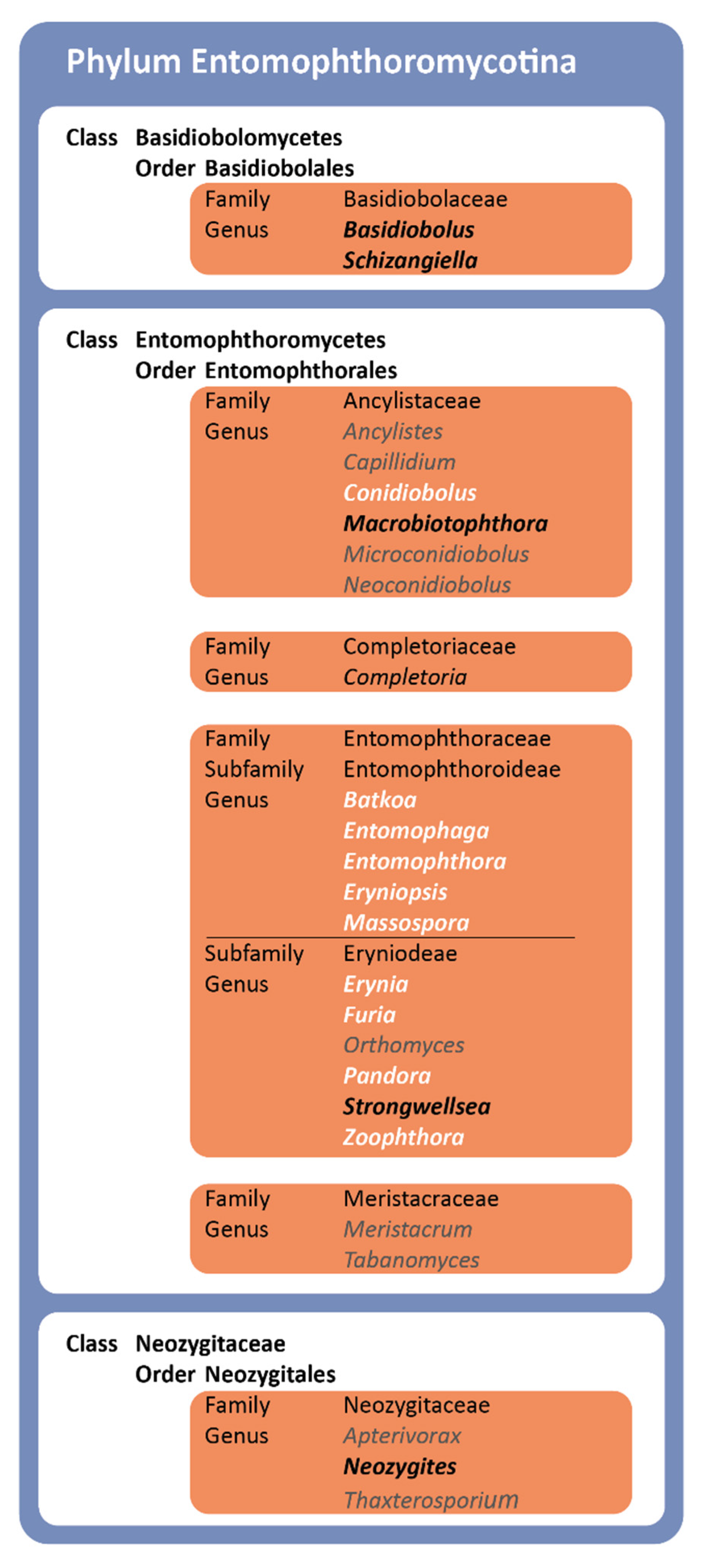
Classification: Entomophthoromycotina, Entomophthoromycetes,
Entomophthorales, Ancylistaceae.
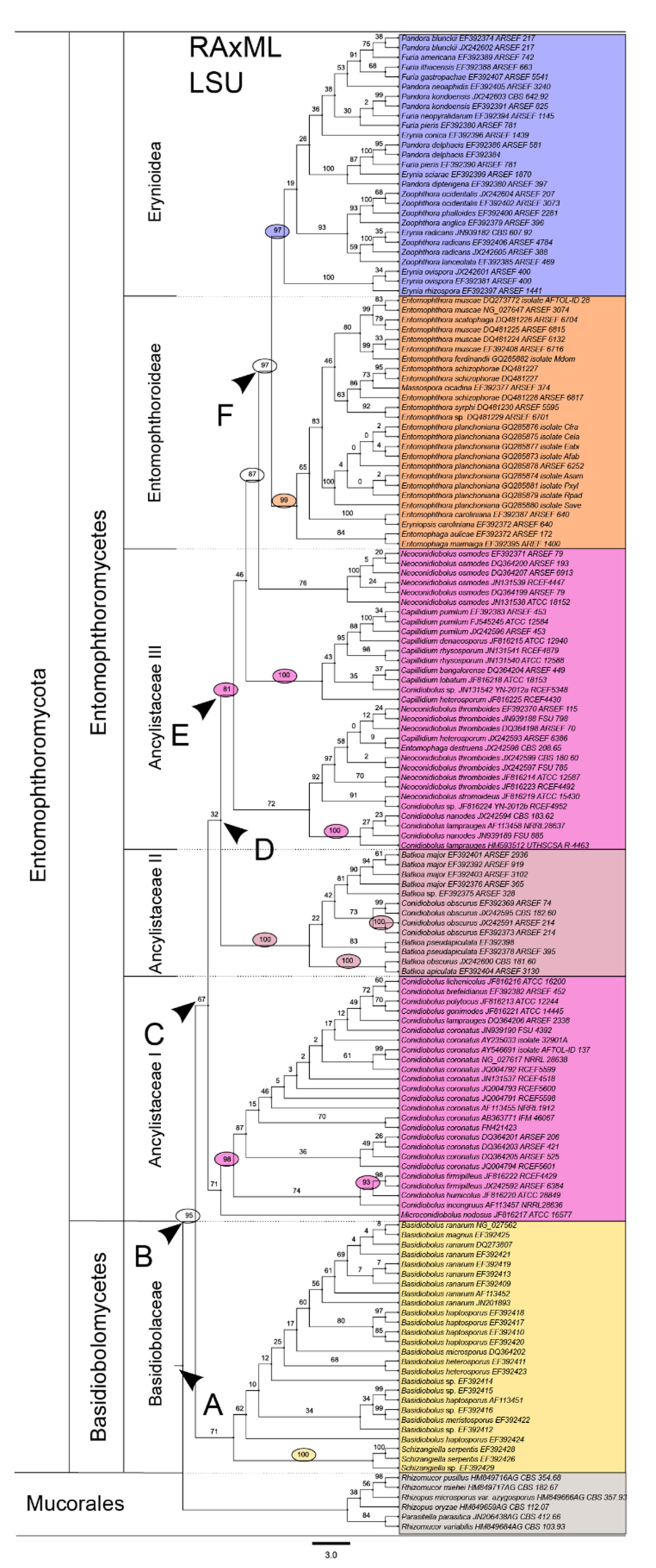
  • Comprises Conidiobolus clade C: Ancylistaceae I (see Figure 3).
  • Note: Comparing Conidiobolus coronatus AF113418 with Macrobiotophthora vermicola AF052400 at the level of small subunit (18S, SSU) rDNA nucleotide sequences, the deviation was found to be high: identity = 824/967 (85%) and gaps = 34/967 (3%), which was lower than 90% SSU sequence similarity as proposed by [50] justifying classification in a new family.
  • Lectotype: Conidiobolus utriculosus Bref. 1884 (Clements and Shear, Gen. Fungi: 239. 1931) [46].
  • Proposed epitype: Conidiobolus coronatus (Costantin) A. Batko 1964 (typ. cons. prop.) [51] ≡ Boudierella coronata Costantin 1897 [48].
  • Lectotype: Costantin, Bull. Soc. mycol. Fr. 13: 40 (1897): see Pl. 4, Figures 1–10, ; Pl. 5, Figures 11–17 in [48].
  • Type: JMRC:SF:11506 (metabolically inactive culture), Sweden, isol. M. Gustafsson (B42536, here designated).
  • Isotypes: NRRL 28638; CBS 209.66; ATCC 28846.
  • Whole genome sequence PRJNA67455 available from: http://www.ncbi.nlm.nih.gov/bioproject/PRJNA67455 (accessed on 23 February 2016), see also [52].
  • Reference sequences: AF113418 (SSU), AF113456, AY546691 and NG_027617 (LSU), AY997041 (ITS1-5.8S-ITS2), DQ275337 (elongation factor 1-alpha), DQ294591 (RNA polymerase II subunit RPB1), DQ302779 (RNA polymerase II subunit RPB2).

4. Discussion

The analyses of our sequence data confirm the monophyly of the subphylum Entomophthoromycotina with a broad taxon sampling, in agreement with Gryganskyi et al. [1] and Spatafora et al. [22]. Within the retrieved subphylum, however, discrepancies between the previously suggested phylogenetic relationships and our topology become apparent. For instance, the Ancylistaceae are retrieved as a tripartite paraphyletic clade (“Ancylistaceae I, II & III”) containing three monophyletic branches, mainly but not only composed of taxa currently assigned to Conidiobolus. Traditionally, this assemblage has been taxonomically unified based on several shared morphological features, such as a coenocytic mycelium, very small nuclei (2.5–4 μm), and a prominent central nucleolus. However, their monophyletic status has been questioned for decades, as well as their whole status as a taxonomic unit [1,53]. Our results now indicate that this group does indeed not constitute a monophyletic group. However, we strongly suggest that the well-known genus Conidiobolus, which also contains human-pathogenic strains, should be conserved. Based on our phylogenetic assessments and previous data, we propose Conidiobolus coronatus as the new type for the genus Conidiobolus sensu stricto.
Concerning host specificity, we thoroughly investigated all relevant species and strains kept in fungal collections and performed an exhaustive literature survey. The host spectrum of all investigated taxa varies tremendously within the Entomophthoromycota, reflecting a plethora of different lifestyles unified in this clade (Figure 4). Far from being exclusively restricted to insects, hosts of the Entomophthoromycota range from algae (Ancylistes) over nematodes (Macrobiotophthora) and mites (Neozygites), through different types of vertebrates up to mammals (Basidiobolus and Conidiobolus) (Figure 4). Clearly, the complete host spectrum of this subphylum is far from being exhaustively resolved, as the infection capacity and host specificity of many taxa remain unknown. This is underlined by our survey on all known host species from the thus far described taxa within the Entomophthoromycota. For over 50% of the investigated fungal species, it is not known whether they have a parasitic life, and if so, which specific hosts they can infect (Figure 4).
Focusing solely on the eponymous entomopathogenic species, it becomes apparent that host specificity and host spectra are very unevenly distributed among the investigated taxa, potentially also hinting at sampling biases. In addition, there is no recognizable congruence in the phylogeny of insects and the two primarily entomopathogenic subfamilies of the Entomophthoromycotina, i.e., Entomophthoroidea and Erynoidea, with parasitism apparently having evolved many times independently within them. Despite the limitations of our knowledge of the real extent of their respective host spectra, the asymmetries of the thus far described host specializations are striking. For instance, the highly diverse insect order Hemiptera (including plant lice, cicada, true bugs, and moss bugs) contains by far the most host species for all investigated classes of the Entomophthoromycotina except the primarily saprobic Basidiobolomycetes. Diptera, Lepidoptera, and Orthoptera are also among the most frequently parasitized insect orders, with the latter exclusively serving as hosts for species of the subfamily Entomophthoroideae according to the current state of knowledge.
Entomophthoromycotina belongs to the oldest known terrestrial lineages of fungi, with an estimated appearance in the Silurian more than 400 mya [1,20,21]. The earliest Pterogyta (i.e., winged insects) fossils (Palaeodictyoptera) are known from the early Carboniferous, 358 mya [54]. As the Pterogyta constitute the most dominantly parasitized insect host group of the Entomophthoromycotina (Figure 4), this hints at a potential correlation of the diversifications in both groups. However, other factors potentially accounting for asymmetric host preference patterns have to be taken into consideration as well, such as optimized dispersal and different defense mechanisms of host species. In any case, the dramatic rise of pterygote insects has very likely increased the dispersal capacities for associated entomopathogenic fungi, far beyond the hypothesized ancestral way of dispersal via apterygote insects primarily living in soil and leaf litter. Generalized saprotrophic soil dwellers are still often linked to hosts such as Collembola (springtails) [55], or other arthropods living in the ground, e.g., mites (Acari). Even though it becomes apparent from our study that there is no strict link between insect lifestyles and specific fungal families, we hypothesize that the appearance and eventual dominance of winged insects largely broadened the spectrum of available hosts for the entomopathogenic lineages of the Entomophthoromycotina. Furthermore, it has been suggested that the ability to digest chitin, which constitutes an efficient infection barrier, had most likely already evolved prior to the radiation of winged insects in this very old lineage of terrestrial fungi [10,21,53]. Concordantly, similar infection patterns within Entomophthoromycotina hint at their ancestral potential to infect insect hosts, and this repeatedly occurred and in similar ways during the evolution of different lineages within this subphylum [56,57]. That no recognizable congruence between the phylogenic patterns in insects and Entomophthoromycotina could be discerned strongly suggests that parasitism evolved many times independently within this clade from saprobic ancestors (Figure 4).
In conclusion, we confirm the overall monophyly of the phylogenetically controversial fungal clade Entomophthoromycotina. Whereas certain subgroups were supported as monophyletic in our analyses, others, especially in the Ancylistaceae lineages, have to be critically re-evaluated. The previously established phylogenetic concept of the prominent taxon Conidiobolus cannot be upheld, and we propose C. coronatus as a new type for this genus. Our extensive survey of host specificity within parasitic lineages of the Entomophthoromycotina does not show clear patterns of co-evolution with their primary targets, insects, but suggests that the radiation of winged insects specifically favored their dispersal. This opens up the avenue for future studies expanding the investigations on the complete host spectrum of this subphylum, potentially also contributing to their successful application as biological pest control agents.

Supplementary Materials

The following supporting information can be downloaded at: https://www.mdpi.com/article/10.3390/microorganisms10020256/s1, Figure S1: Phylogeny of SSU-rDNA sequence with RAxML; Figure S2: Phylogeny of RPB2-rDNA sequence with RAxML; Figure S3: Phylogeny of LSU-rDNA sequence with MrBayes; Figure S4: Phylogeny of SSU-rDNA sequence with MrBayes; Figure S5: Phylogeny of RPB2-rDNA sequence with MrBayes; Table S1: Detailed overview of all LSU, SSU and RPB2 sequences for the strains used; Table S2: Sequence data after alignment with MAFFT; Table S3: Matrix of documented host-fungal interactions.

Author Contributions

Conceptualization, K.V., R.G.B. and J.B.; methodology, K.M. and B.M.; software, K.M. and B.S.; validation, V.U.S., B.S., H.H.D.F.L. and S.d.H.; formal analysis, L.M., V.U.S. and K.M.; investigation, L.M., K.M., K.V. and R.G.B.; resources, K.M., B.M. and S.d.H.; data curation, K.M., B.M., B.S., S.d.H. and R.G.B.; writing—original draft preparation, L.M., K.V., and J.B.; writing—review and editing, L.M., H.H.D.F.L., S.d.H. and J.B.; visualization, L.M. and K.M.; supervision, K.V., R.G.B. and J.B.; project administration, J.B.; funding acquisition, K.V. and J.B. All authors have read and agreed to the published version of the manuscript.

Funding

This work was funded by the German Research Foundation (Deutsche Forschungsgemeinschaft, DFG), project number 427879779 to J.B and project number 210879364 (A6) to K.V. through the TRR 124 FungiNet, “Pathogenic fungi and their human host: Networks of Interaction”.

Data Availability Statement

All data underlying the presented study will be made available at an online data repository.

Acknowledgments

R.G.B. and L.M. wish to thank Hans Pohl (Friedrich-Schiller-University Jena, Germany) for technical advice and assistance with scanning electron and light microscopy.

Conflicts of Interest

The authors declare no conflict of interest.

References

  1. Gryganskyi, A.P.; Humber, R.A.; Smith, M.E.; Hodge, K.; Huang, B. Phylogenetic lineages in Entomophthoromycota. Mol. Phylogen. Evol. 2013, 65, 682–694. [Google Scholar] [CrossRef] [PubMed]
  2. Gryganskyi, A.P.; Humber, R.A.; Smith, M.E.; Miadlikowska, J.; Wu, S.; Voigt, K.; Walther, G.; Anishchenko, I.M.; Vilgalys, R. Molecular phylogeny of the Entomophthoromycota. Mol. Phylogen. Evol. 2012, 65, 682–694. [Google Scholar] [CrossRef] [PubMed]
  3. Misof, B.; Liu, S.; Meusemann, K.; Peters, R.S.; Donath, A.; Mayer, C.; Frandsen, P.B.; Ware, J.; Flouri, T.; Beutel, R.G.; et al. Phylogenomics resolves the timing and pattern of insect evolution. Science 2014, 346, 763–767. [Google Scholar] [CrossRef] [PubMed]
  4. Elya, C.; Lok, T.C.; Spencer, Q.E.; McCausland, H.; Martinez, C.C.; Eisen, M. Robust manipulation of the behavior of Drosophila melanogaster by a fungal pathogen in the laboratory. eLife 2018, 7, e34414. [Google Scholar] [CrossRef]
  5. White, M.M.; James, T.; O'Donnell, K.; Cafaro, M.; Tanabe, Y.; Sugiyama, J. Phylogeny of Zygomycota based on nuclear RNA. Mycologia 2006, 98, 872–884. [Google Scholar] [CrossRef]
  6. Carruthers, R.I.; Hural, K. Fungi as naturally occuring entomopathogens. UCLA Symp. Mol. Cell Biol. 1990, 112, 115–138. [Google Scholar]
  7. De Fine Licht, H.H.; Jensen, A.B.; Eilenberg, J. Comparative transcriptomics reveal host-specific nucleotide variation in entomophthoralean fungi. Mol. Ecol. 2017, 26, 2092–2110. [Google Scholar] [CrossRef]
  8. De Ruiter, J.; Arnbjerg-Nielsen, S.F.; Herren, P.; Høier, F.; De Fine Licht, H.H.; Jensen, K.H. Fungal artillery of zombie flies: Infectious spore dispersal using a soft water cannon. J. R. Soc. Interface 2019, 16, 20190448. [Google Scholar] [CrossRef]
  9. Boyce, G.R.; Gluck-Thaler, E.; Slot, J.C.; Stajich, J.E.; Davis, W.J.; James, T.Y.; Cooley, J.R.; Panaccione, D.G.; Eilenberg, J.; De Fine Licht, H.H.; et al. Psychoactive plant- and mushroom-associated alkaloids from two behavior modifying cicada pathogens. Fungal Ecol. 2019, 41, 147–164. [Google Scholar] [CrossRef]
  10. Arnesen, J.A.; Małagocka, J.; Gryganskyi, A.; Grigoriev, I.V.; Voigt, K.; Stajich, J.E.; De Fine Licht, H.H. Early diverging insect-pathogenic fungi of the order Entomophthorales possess diverse and unique subtilisin-like serine proteases. G3 Genes Genomes Genet. 2018, 8, 3311–3319. [Google Scholar] [CrossRef] [Green Version]
  11. Humber, R.A. Evolution of entomopathogenicity in fungi. J. Invertebr. Pathol. 2008, 98, 262–266. [Google Scholar] [CrossRef] [PubMed]
  12. Rothhardt, J.E.; Schwartze, V.U.; Voigt, K. Entomophthorales. In Molecular Detection of Human Fungal Pathogens; Liu, D., Ed.; Taylor & Francis CRC Press: Boca Raton, FL, USA, 2011; pp. 723–734. [Google Scholar]
  13. El-Shabrawi, M.H.; Arnaout, H.; Madkour, L.; Kamal, N.M. Entomophthoromycosis: A challenging emerging disease. Mycoses 2014, 57, 132–137. [Google Scholar] [CrossRef] [PubMed]
  14. De Hoog, S.; Guarro, J.; Gené, J.; Ahmed, S.; Al-Hatmi, A.; Figueras, M.; Vitale, R. Atlas of Clinical Fungi: The Ultimate Benchtool for Diagnostics. Filamentous Ascomycetes C-Z, Part 2, 4th ed.; Foundation Atlas of Clinical Fungi: Hilversum, The Netherlands, 2020. [Google Scholar]
  15. Radhakrishnan, N.; Sachdeva, A.; Oberoi, J.; Yadav, S.P. Conidiobolomycosis in relapsed acute lymphoblastic leukemia. Pediatr. Blood Cancer 2009, 53, 1321–1323. [Google Scholar] [CrossRef] [PubMed]
  16. Kwon-Chung, K.J. Taxonomy of fungi causing mucormycosis and entomophthoramycosis (zygomycosis) and nomenclature of the disease: Molecular mycologic perspectives. Clin. Infect. Dis. 2012, 54, S8–S15. [Google Scholar] [CrossRef] [PubMed] [Green Version]
  17. Durel, L.; Estrada-Peña, A.; Franc, M.; Mehlhorn, H.; Bouyer, J. Integrated fly management in European ruminant operations from the perspective of directive 2009/128/EC on sustainable use of pesticides. Parasitol. Res. 2015, 114, 379–389. [Google Scholar] [CrossRef]
  18. Stern, V.M.; Smith, R.F.; van den Bosch, R.; Hagen, K.S. The integrated control concept. Hilgardia 1959, 29, 81–101. [Google Scholar] [CrossRef] [Green Version]
  19. Naranjo, S.E.; Ellsworth, P.C.; Frisvold, G.B. Economic value of biological control in Integrated Pest Management of managed plant systems. Annu. Rev. Entomol. 2015, 60, 621–645. [Google Scholar] [CrossRef] [Green Version]
  20. Li, Y.; Steenwyk, J.L.; Chang, Y.; Wang, Y.; James, T.Y.; Stajich, J.E.; Spatafora, J.W.; Groenewald, M.; Dunn, C.W.; Hittinger, C.T.; et al. A genome-scale phylogeny of the kingdom fungi. Curr. Biol. 2021, 31, 1653–1665. [Google Scholar] [CrossRef]
  21. James, T.Y.; Stajich, J.E.; Hittinger, C.T.; Rokas, A. Toward a fully resolved fungal tree of life. Annu. Rev. Microbiol. 2020, 74, 291–313. [Google Scholar] [CrossRef]
  22. Spatafora, J.W.; Chang, Y.; Benny, G.L.; Lazarus, K.; Smith, M.E.; Berbee, M.L.; Bonito, G.; Corradi, N.; Grigoriev, I.; Gryganskyi, A.; et al. A phylum-level phylogenetic classification of zygomycete fungi based on genome-scale data. Mycologia 2016, 108, 1028–1046. [Google Scholar] [CrossRef] [Green Version]
  23. Liu, X.-y.; Voigt, K. Molecular characters of zygomycetous fungi. In Molecular Identification of Fungi; Gherbawy, Y., Voigt, K., Eds.; Springer: Berlin/Heidelberg, Germany, 2010; pp. 461–488. [Google Scholar]
  24. Hibbett, D.S.; Binder, M.; Bischoff, J.F.; Blackwell, M.; Cannon, P.F.; Eriksson, O.E.; Huhndorf, S.; James, T.; Kirk, P.M.; Lucking, R.; et al. A higher-level phylogenetic classification of the Fungi. Mycol. Res. 2007, 111, 509–547. [Google Scholar] [CrossRef]
  25. Nie, Y.; Yu, D.-S.; Wang, C.-F.; Liu, X.-Y.; Huang, B. A taxonomic revision of the genus Conidiobolus (Ancylistaceae, Entomophthorales): Four clades including three new genera. MycoKeys 2020, 66. [Google Scholar] [CrossRef] [PubMed] [Green Version]
  26. Ben-Ze'ev, I.; Kenneth, R.G. Features-criteria of taxonomic value in the Entomophthorales: 1. A revision of the Batkoan classification. Mycotaxon 1982, 14, 393–455. [Google Scholar]
  27. Boomsma, J.J.; Jensen, A.B.; Meyling, N.V.; Eilenberg, J. Evolutionary interaction networks of insect pathogenic fungi. Annu. Rev. Entomol. 2014, 59, 467–485. [Google Scholar] [CrossRef] [PubMed]
  28. De Fine Licht, H.H.; Hajek, A.E.; Eilenberg, J.; Jensen, A.B. Utilizing genomics to study entomopathogenicity in the fungal phylum entomophthoromycota: A review of current genetic resources. In Advances in Genetics; Lovett, B., St. Leger, R.J., Eds.; Academic Press: Cambridge, MA, USA, 2016; Volume 94, pp. 41–65. [Google Scholar]
  29. Stielow, J.B.; Lévesque, C.A.; Seifert, K.A.; Meyer, W.; Iriny, L.; Smits, D.; Renfurm, R.; Verkley, G.J.M.; Groenewald, M.; Chaduli, D.; et al. One fungus, which genes? Development and assessment of universal primers for potential secondary fungal DNA barcodes. Persoonia 2015, 35, 242–263. [Google Scholar] [CrossRef] [PubMed] [Green Version]
  30. Vu, D.; Groenewald, M.; de Vries, M.; Gehrmann, T.; Stielow, B.; Eberhardt, U.; Al-Hatmi, A.; Groenewald, J.Z.; Cardinali, G.; Houbraken, J.; et al. Large-scale generation and analysis of filamentous fungal DNA barcodes boosts coverage for kingdom fungi and reveals thresholds for fungal species and higher taxon delimitation. Stud. Mycol. 2019, 92, 135–154. [Google Scholar] [CrossRef]
  31. Humber, R.A. Identification of entomopathogenic fungi. In Manual of Techniques in Invertebrate Pathology, 2nd ed.; Lacey, L.A., Ed.; Academic Press: San Diego, CA, USA, 2012; pp. 151–187. [Google Scholar]
  32. Altschul, S.F.; Gish, W.; Miller, W.; Myers, E.W.; Lipman, D.J. Basic local alignment search tool. J. Mol. Biol. 1990, 215, 403–410. [Google Scholar] [CrossRef]
  33. Altschul, S.F.; Boguski, M.S.; Gish, W.; Wootton, J.C. Issues in searching molecular sequence databases. Nat. Genet. 1994, 6, 119–129. [Google Scholar] [CrossRef]
  34. McGinnis, S.; Madden, T.L. BLAST: At the core of a powerful and diverse set of sequence analysis tools. Nucleic Acids Res. 2004, 32, 20–25. [Google Scholar] [CrossRef]
  35. Katoh, K.; Standley, D.M. MAFFT Multiple sequence alignment software version 7: Improvements in performance and usability. Mol. Biol. Evol. 2013, 30, 772–780. [Google Scholar] [CrossRef] [Green Version]
  36. Henikoff, S.; Henikoff, J.G. Amino acid substitution matrices from protein blocks. Proc. Natl. Acad. Sci. USA 1992, 89, 10915–10919. [Google Scholar] [CrossRef] [PubMed] [Green Version]
  37. Hall, T. BioEdit: A user-friendly biological sequence alignment editor and analysis program for Windows 95/98/NT. Nucleic Acids Symp. Ser. 1999, 41, 95–98. [Google Scholar]
  38. Towns, J.; Cockerill, T.; Dahan, M.; Foster, I.; Gaither, K.; Grimshaw, A.; Hazlewood, V.; Lathrop, S.; Lifka, D.; Peterson, G.D.; et al. XSEDE: Accelerating scientific discovery. Comput. Sci. Eng. 2014, 16, 62–74. [Google Scholar] [CrossRef]
  39. Miller, M.A.; Pfeiffer, W.; Schwartz, T. Creating the CIPRES Science Gateway for inference of large phylogenetic trees. In Proceedings of the 2010 Gateway Computing Environments Workshop (GCE), New Orleans, LA, USA, 14 November 2010; pp. 1–8. [Google Scholar]
  40. Stamatakis, A.; Hoover, P.; Rougemont, J. A Rapid Bootstrap Algorithm for the RAxML Web Servers. Syst. Biol. 2008, 57, 758–771. [Google Scholar] [CrossRef] [PubMed]
  41. Stamatakis, A. RAxML-VI-HPC: Maximum likelihood-based phylogenetic analyses with thousands of taxa and mixed models. Bioinformatics 2006, 22, 2688–2690. [Google Scholar] [CrossRef] [PubMed]
  42. Ronquist, F.; Huelsenbeck, J.P. MrBayes 3: Bayesian phylogenetic inference under mixed models. Bioinformatics 2003, 19, 1572–1574. [Google Scholar] [CrossRef] [Green Version]
  43. Voigt, K.; A, M.; Wostemeyer, J. Phylogeny of zygomycetes: A molecular approach towards systematics of Mucorales. Cour. Forsch. Inst. Senckenberg 1999, 215, 207–213. [Google Scholar]
  44. Beutel, R.; Yavorskaya, M.; Mashimo, Y.; Fukui, M.; Meusemann, K. The phylogeny of Hexapoda (Arthropoda) and the evolution of megadiversity. Proc. Arthropod. Embryol. Soc. Jpn. 2017, 51, 1–15. [Google Scholar]
  45. Brefeld, O. Conidiobolus Utriculosus und Minor; Untersuchungen aus der Gesammtgebiete der Mykologie Arthur Felix: Leipzig, Germany, 1884; Volume 6, pp. 35–78. [Google Scholar]
  46. Clements, F.; Shear, C.L. The Genera of Fungi; H.W. Wilson Co.: New York, NY, USA, 1931. [Google Scholar]
  47. Mendoza, L.; Vilela, R.; Voelz, K.; Ibrahim, A.S.; Voigt, K.; Lee, S.C. Human fungal pathogens of Mucorales and Entomophthorales. Cold Spring Harb. Perspect. Med. 2015, 5. [Google Scholar] [CrossRef] [Green Version]
  48. Costantin, S. Sur une Entomophthoree nouvelle. Bull. Soc. Mycol. Fr. 1897, 13, 38–43. [Google Scholar]
  49. Saccardo, P.; Sydow, P. Supplementum Universale, Pars IV. Syll. Fung. 1899, 14, 1–1316. [Google Scholar]
  50. Hoffmann, K.; Telle, S.; Walther, G.; Eckhart, M.; Kirchmair, M.; Prillinger, H.; Prazenica, A.; Newcombe, G.; Dölz, F.; Tamás, P.; et al. Diversity, genotypic identification, ultrastructural and phylogenetic characterization of zygomycetes from different ecological habitats and climatic regions: Limitations and utility of nuclear ribosomal DNA barcode markers. In Current Advances in Molecular Mycology; Gherbawy, Y.M., Mach, R.L., Rai, M., Eds.; Nova Science Pub: New York, NY, USA, 2008; pp. 263–312. [Google Scholar]
  51. Batko, A. Notes on entomophthoraceous fungi in Poland. Entomophaga 1964, 2, 129–131. [Google Scholar]
  52. Chang, Y.; Wang, S.; Sekimoto, S.; Aerts, A.L.; Choi, C.; Clum, A.; LaButti, K.M.; Lindquist, E.A.; Yee Ngan, C.; Ohm, R.A.; et al. Phylogenomic analyses indicate that early fungi evolved digesting cell walls of algal ancestors of land plants. Genome Biol. Evol. 2015, 7, 1590–1601. [Google Scholar] [CrossRef] [PubMed]
  53. Wheeler, Q.D.; Blackwell, M. Fungus-Insect Relationships; Columbia University Press: New York, NY, USA, 1984. [Google Scholar]
  54. Grimaldi, D.A.; Engel, M.S. Evolution of Insects; Cambridge University Press: Cambridge, MA, USA, 2005. [Google Scholar]
  55. Hopkin, S.P. Biology of the Springtails (Insecta: Collembola); Oxford University Press: Oxford, UK, 1997. [Google Scholar]
  56. Berbee, M.L.; Taylor, J.W. Dating the molecular clock in fungi–how close are we? Fungal Biol. Rev. 2010, 24, 1–16. [Google Scholar] [CrossRef]
  57. Elya, C.; De Fine Licht, H.H. The genus Entomophthora: Bringing the insect destroyers into the twenty-first century. IMA Fungus 2021, 12, 34. [Google Scholar] [CrossRef]
Figure 1. Examples of host infections by the Entomophthoromycotina. (A) Scanning electron microscopic image of Meroplius fasciculatus (Sepsidae, Diptera) with a substantial fungal infection (Entomophthora sp.). Fungal outgrowth points are indicated by arrows. (B) Separated prothoracic leg of Meroplius fasciculatus (Sepsidae, Diptera) shows the extent of fungal growth within the host tissue (arrows). (C) Light microscopic image of the abdomen of Meroplius sp. (Sepsidae, Diptera) with an infection of Entomophthora muscae (Entomophthoraceae) along the pleurae, less sclerotized structures that potentially facilitate outward growth of the fungus (arrow). (D) Isolated hyphae of the fungus in (C) strongly hint at an intimate somatic contact to the pleura of the host insect. (E) Macro camera image of an infected Delia sp. (Anthomyiidae, Diptera) with an entomophthoralean infection, arrows indicate fungal hyphae and conidia protruding from the abdomen of the host insect. (F) Macro camera image of an Ecuadorian leafhopper from the family Cicadellidae (Cicadomorpha, Auchenorrhyncha) with entomophthoralean hyphae growing from multiple segments of its infected body. Images (AD,F): by Lars Möckel, Image (E): by Hans Pohl (Friedrich Schiller University Jena, Germany).
Microorganisms 10 00256 g001
Figure 2. Entomophthoromycotina taxonomy based on the system of Humber [31]. Names in black indicate taxa represented by strains in the present study with available rRNA sequences from the International Nucleotide Sequence Database Collaboration (INSDC). For taxa in gray, sequences were not available. Concerning genera, names in bold white indicate the availability of both LSU and SSU sequences for their representative strains, whereas names in bold black indicate genera for which only SSU sequences were available.
Microorganisms 10 00256 g002
Figure 3. Entomophthoromycotina phylogeny based on a maximum likelihood phylogram reconstruction inferred from large subunit (28S, LSU) ribosomal RNA nucleotide sequences of 147 taxa. Statistical bootstrap support (BS, inferred with RAxML) is indicated above branches. BS above 90%, above 80% and 70% were considered as high, well, and moderate support in accordance to earlier published rRNA phylogenies of entomophthoralean and mucoralean species, respectively [43]. Well-supported branches are marked with ellipses in the color code of the corresponding clade. Six species of Mucorales were collectively used as an outgroup. (A) Split of Mucorales (outgroup) + Entomophthoromycotina, (B) Split of Basidiobolomycetes + Entomophthoromycetes, (C) Split of Ancylistaceae I + remaining clades of Entomophthoromycetes, (D) Split of Batkoa s.l. + Ancylistaceae II + Entomophthoraceae, (E) Split of Ancylistaceae II + Entomophthoraceae, (F) Split of Entomophthoroideae + Erynoideae. Scale bar indicates substitutions per site.
Microorganisms 10 00256 g003
Figure 4. Representation of the host spectrum of the Entomophthoromycotina with particular emphasis across the phylogeny of the Insecta. The occurrence of entomophthoromycotan (sub)families is mapped next to their respective host taxa. Different colors represent the different (sub)families of the Entomophthoromycotina with documented host records. Numbers in brackets indicate the documented insect species per clade, whereas, on the right-hand side, the numbers of host species known to be infected by the entomophthoromycotan (sub)families per clade are indicated. The primarily infected Pterogyta are shaded in light grey. The arthropod phylogeny has been adapted from Misof et al. [3] and Beutel et al. [44] and was further modified by collapsing the nodes to the ordinal levels. Representative pictograms indicate characteristic insect host taxa; insect orders without records of host species are in gray capitals. Classification of fungal families after Humber [31]. The entomophthoromycotan taxa are indicated on the bottom with their respective total numbers of described species (right-hand side, 274 to date) vs. the number of species with a documented host record left-hand side, 84 to date). Detailed information on all fungal-host interactions are given in Table S3.
Microorganisms 10 00256 g004
Publisher’s Note: MDPI stays neutral with regard to jurisdictional claims in published maps and institutional affiliations.

