

Potential Biocontrol Agents of Corn Tar Spot Disease Isolated from Overwintered Phyllachora maydis Stromata

Crop Bioprotection Research Unit, National Center for Agricultural Utilization Research, Agricultural Research Service, United States Department of Agriculture, 1815 N University Street, Peoria, IL 61604, USA
*
Author to whom correspondence should be addressed.
Microorganisms 2023, 11(6), 1550; https://doi.org/10.3390/microorganisms11061550
Submission received: 20 April 2023
/
Revised: 6 May 2023
/
Accepted: 6 June 2023
/
Published: 10 June 2023
(This article belongs to the Special Issue Biological Control of the Plant Pathogens)
Abstract
:Tar spot disease in corn, caused by Phyllachora maydis, can reduce grain yield by limiting the total photosynthetic area in leaves. Stromata of P. maydis are long-term survival structures that can germinate and release spores in a gelatinous matrix in the spring, which are thought to serve as inoculum in newly planted fields. In this study, overwintered stromata in corn leaves were collected in Central Illinois, surface sterilized, and caged on water agar medium. Fungi and bacteria were collected from the surface of stromata that did not germinate and showed microbial growth. Twenty-two Alternaria isolates and three Cladosporium isolates were collected. Eighteen bacteria, most frequently Pseudomonas and Pantoea species, were also isolated. Spores of Alternaria, Cladosporium, and Gliocladium catenulatum (formulated as a commercial biofungicide) reduced the number of stromata that germinated compared to control untreated stromata. These data suggest that fungi collected from overwintered tar spot stromata can serve as biological control organisms against tar spot disease.
1. Introduction
Corn is one of the most important grain crops in the world, but production is limited by a variety of pests and diseases [1]. One devastating disease is caused by Phyllachora maydis and is commonly referred to as tar spot because of its characteristic black, shiny, raised leaf spots which generally range from 2–4 mm in diameter. Infection by this pathogenic fungus can result in significant yield losses, and even death of plants if infection occurs early in a susceptible variety [2]. Tar spot disease has been endemic in much of Central and South America for several decades [2]. It was first reported in two U.S. “corn belt” states, Illinois and Indiana, in 2015 [3], and since then there have been major outbreaks in several regions in both 2018 and 2021 [4].
Management strategies for tar spot disease include use of resistant varieties and application of fungicides [2]. However, under high inoculum pressure, significant yield losses can still occur, even with the use of resistant varieties [5]. Fungicide application has not been very effective because once symptoms are noticed, further infection becomes difficult to control [6].
The black structures characteristic of tar spot disease are long term survival structures called stromata, which are similar to the sclerotia produced by the vegetable pathogen Sclerotinia sclerotiorum and other fungi [7]. These stromata are the overwintering structures for tar spot, and the source of initial inoculum. However, reports of spore production and percent germination by overwintered stromata vary considerably [8], suggesting the presence of natural biocontrol organisms. Mycoparasites may contribute to reduced survival of stromata, as reported for other stromata or sclerotia-like structures in several fungal species [7,9] For example, several species of biocontrol organisms have been isolated from the sclerotia of S. sclerotiorum, some of which have been commercialized as biological control products [10,11]. Mycoparasites of stromata of Coccodiella miconiae, a relative of P. maydis, included fungi in the genera Cladosporium, Cornespora. and Sagenomella [12]. To investigate the potential for biocontrol of tar spot, stromata from overwintered corn leaves collected in Illinois were examined for the presence of mycoparasites or other biocontrol agents. This study identifies several bacterial and fungal species infecting tar spot stromata and describes bacterial and fungal species with potential to serve as biocontrol agents against tar spot disease of corn.
2. Materials and Methods
2.1. Isolation of Organisms
Several overwintered corn leaves of inbred GE440 (planted from seed increased from plants originally obtained from the USDA-ARS Plant Introduction Center) were collected in late April 2022 from a 2021-planted research plot. This site in Peoria, IL, has had continuous corn production for several years and is thus more ecologically stable and likely to yield biocontrol agents for tar spot than commercial fields where crop rotation typically occurs. Stromata were cut from leaves along with a small leaf piece “handle”. Approximately 50 stromata were surface sterilized with 70% ethanol and then blotted dry as described previously [8]. Stromata were placed in Petri dishes with tight fitting lids (FalconR 351006, Corning Inc., Corning, NY, USA) containing 5 mL of 3% water agar to induce rehydration and stimulate growth of mycoparasites; in some cases, a few µL of sterile water was added to help with rehydration. Plates were held at 25–27 °C. Stromata were examined daily for outgrowth of organisms that were not visually the same as any seen from the attached leaf material, and organisms noted were photographed with cameras equipped with macro lenses. Microorganisms were isolated using two different methods. In the first method, fungal mycelium growing out of a single stroma was transferred to a potato dextrose agar (PDA, Difco potato dextrose broth, Becton Dickinson Company, Sparks, MD, USA, with bacto agar at 20 g/L) plate using a sterile metal probe. A few days later, bacteria were observed growing together with the fungus. The bacteria were transferred to a new PDA plate, whereas the bacterium-contaminated fungus was transferred to a PDA plus 0.01% chloramphenicol plate. In the second method, petroleum jelly was used to stick leaves with stromata to the inside of the top lid over a 3% water agar plate. After 2–3 days, microorganisms growing on the surface of the agar were transferred to nutrient media plates (Luria broth (LB) or tryptone glucose yeast extract (TGY) for bacteria or PDA for fungi). LB agar plates consisted of tryptone (10 g/L), sodium chloride (10 g/L), yeast extract (5 g/L), and bacto agar at 15 g/L. TGY agar plates consisted of tryptone (5 g/L), yeast extract (5 g/L), K2HPO4 (1 g/L), and glucose (1 g/L); the pH of the final mixture was adjusted to 7.0 and bacto agar was added at 15 g/L before autoclaving.
2.2. Identification of Organisms
Genomic DNA was isolated from fungi as previously described [13] with a modification: a small fragment of fungal mycelia from the culture plate was pulverized with a small amount of 800 µm silica beads using a Minibead Beater (Biospec Products, Bartlesville, OK, USA). Genomic DNA was isolated from bacteria as described in [14] with several modifications. A 1 mL aliquot of bacterial overnight culture was centrifuged at 16,000× g; the bacterial pellet was first suspended in 480 µL of 50 mM EDTA and 120 µL of 5 mg/mL lysozyme was then added. The suspended pellet was incubated at 37 °C for 30–60 min and the suspension was centrifuged at 16,000× g for 2 min. The supernatant was moved to a new tube and 600 µL of nuclei lysis solution was added. The lysate was incubated at 80 °C for 5 min. After cooling to room temperature, 12 µg of ribonuclease A was added to the lysate and the mixture was incubated at 37 °C for 15–60 min. Then, 250 µL of 5 M NaCl was added to the lysate and the mixture was vortexed for 20 s. The mixture was kept on ice for 5 min and then centrifuged at 16,000× g for 3 min. The method was continued as previously described [14]. Genomic DNA was stored at −20 °C until processing. PCR amplification of various gene products from genomic DNA was performed using Platinum™ SuperFi™ II DNA polymerase (Thermo Fisher Scientific, Waltham, MA, USA) according to the manufacturer’s instructions; primers used for PCR product amplification are listed in Supplementary File S1. PCR products were sequenced using the BigDye Terminator Cycle Sequencing Kit (Version 3.1, Applied Biosystems, Foster City, CA, USA) and BLAST analysis (National Center for Biotechnology Information (NCBI) was used to determine potential identities [15]. For bacteria, conserved gene sequences targeting the 16S rDNA gene were amplified by PCR, sequenced, and utilized for identification of each organism. In some cases of bacterial identification, a portion of the gyrB gene was also amplified from genomic DNA because the 16S rDNA gene sequence was not suitable to identify the bacterium to the species level. The threshold for assigning a bacterial sequence to the species level was 98% or greater similarity of the PCR product sequence with a GenBank accession from the nucleotide collection or the whole genome shotgun contig database. For fungi, conserved gene sequences targeting the rDNA gene were amplified from genomic DNA using PCR and sequenced; these PCR products could not identify the fungi to the species level. Therefore, a portion of the actin gene was amplified by PCR in the Cladosporium fungi [16,17], whereas portions of three genes (RNA polymerase second largest subunit, rpb2; Alternaria major allergen, Alt-a1; glyceraldehyde 3-phosphate dehydrogenase, gapdh) were amplified via PCR in the Alternaria fungi. These Alternaria genes were sufficient to distinguish new species in a recent paper [18]. Phylogenetic analysis (see below) was used to identify Cladosporium and Alternaria fungi to the species level. All the sequencing results were supplied in the Supplementary File S2.
2.3. Repression of Stromata Germination by Representative Fungal Potential Biocontrol Agents
Corn leaves with tar spot stromata were collected from a field in Peoria County, IL, in late July 2022. Leaves were rubbed gently while held under flowing deionized water to dislodge debris and blotted dry with WipeallR L40 wipes (Kimberly ClarkRoswell, GA, USA). Leaves containing approximately 50 to 100 P. maydis stromata in a 100 cm2 square were trimmed with the midvein central so they would fit snugly and flat in 100 × 15 mm Petri dishes containing 25 mL of 3% water agar. Plates were sealed with 3 M micropore™ surgical tape (3 M Company, ST. Paul, MN, USA) and allowed to incubate at room temperature overnight. The experiment was set up with three treatments, including two representatives of the most common genera of potential biocontrol fungi isolated from overwintered stromata (Alternaria alternata/arborescens and Cladosporium rectoides), and the commercial biofungicide LALstop G46wg, which contains Gliocladium catenulatum strain J1446, obtained from Lallemand Specialties (Milwaukee, WI, USA). Spores were obtained from fresh cultures of potential biocontrol fungi that were grown on S-medium [19]. Solutions of spores from the candidate biocontrol fungi were diluted to 10 colony forming units per µL in 0.01% Triton X 100 (Sigma Chemical, St. Louis, MO, USA). The commercial biofungicide LALstop was also diluted to 10 CFU per µL to match that for the candidate biocontrol fungi from the stromata, which was comparable to the recommended application rate. The next day, stromata that had not germinated were treated with 1 µL per mm diameter of control or spore solution, with 10 stromata treated on each side of the leaf midrib with either control (Triton X 100 solution alone) or spore solution. Plates were allowed to incubate at room temperature for 4 days, and then the number of treated germinated stromata was determined. Each treatment was replicated 3 times.
2.4. Phylogenetic Analysis
Partial gene sequences from Alternaria (Alt-a1, gapdh, and rpb2 genes) and Cladosporium (actin gene) ex-type species were retrieved from NCBI. Sequences of Cladosporium ex-type strains, ex-epitype strains, and the strains from this study were aligned (using the Muscle alignment tool) and phylogenetic tree constructed using MEGA 11 [20]. Sequences of Alternaria ex-type strains and the strains from this study were aligned (using the “very accurate” algorithm in the classical sequence analysis menu) in CLC Genomics Workbench Version 22.0.2 (Qiagen, Valencia, CA, USA). Each Alternaria gene was aligned individually and trimmed at the edges; nucleotides within the gene that had low consensus among all the sequences were removed from the alignment. A multi-locus alignment of the three genes was made using the “join alignments” function. The alignment was imported into MEGA 11 and the phylogenetic tree constructed.
2.5. Statistical Analysis
Significant differences in frequency of tar spot stromata germination between control treated stromata and biological control organism treated stromata were determined using weighted Chi Square analysis with the SAS Version 7.1 Proc Freq (SAS Institute, Cary, NC, USA).
3. Results
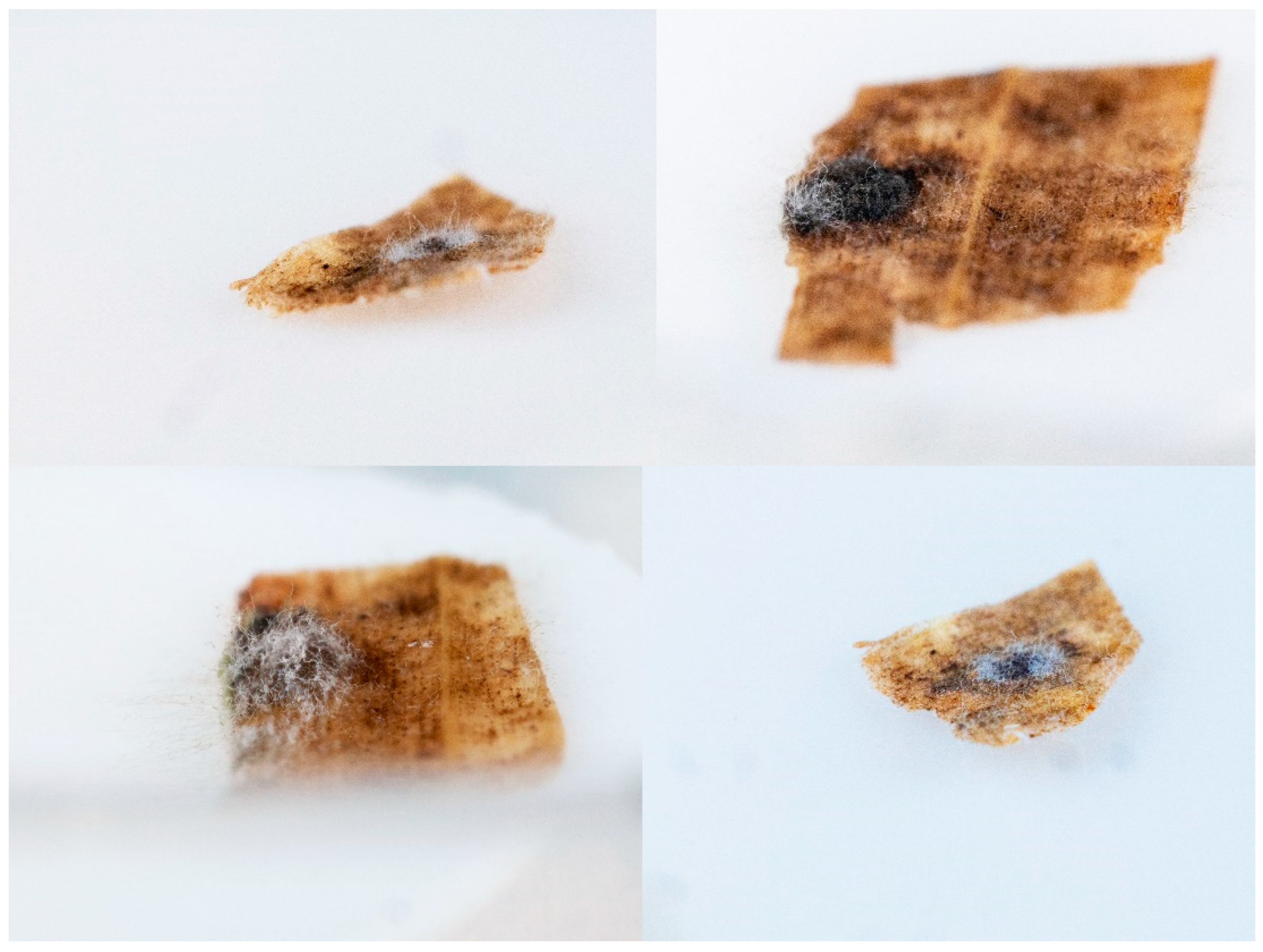
No germinating stromata were observed in any collected overwintered tar spot infected material. However, several different types of organisms were observed growing out from stromata and not the leaf “handle” (Figure 1). We recovered 43 isolates of bacteria and fungi from approximately 50 of the P. maydis stromata (Table 1). Bacteria isolates included four species of Pantoea, including P. agglomerans (3 isolates), as well as two isolates of Priestia (formerly known as Bacillus) megaterium. Other bacteria isolated included Curtobacterium faccumfaciens, and multiple species of Pseudomonas, including P. graminis, P. prosekii, and P. quercus. One Pseudomonas isolate is either P. fluorescens or P. shahriarae.
Alternaria was the most common fungus isolated from the stromata (Table 1). The Alternaria fungi could not be identified to the species level using the amplified region of the rDNA gene. PCR amplification of three gene fragments and subsequent phylogenetic analysis of the multi-locus alignment indicated that 6 isolates were A. ovoidea and 15 were possibly A. alternata or A. arborescens (Supplementary File S3). Three putative Cladosporium strains were isolated. The phylogenetic analysis (Supplementary File S4) indicated the three Cladosporium isolates were C. crousii, C. rectoides, and C. subuliforme.
The number of germinating stromata as indicated by exudate presence was significantly reduced by spore suspensions of two representative fungal species isolated from tar spot stromata compared to control applications (Table 2). However, none of these fungi were as effective as the commercially available G. catenulatum.
4. Discussion
4.1. Biocontrol Potential of Bacteria Isolated from Tar Spot Stromata
Of the 18 bacteria isolated from tar spot stromata, only P. megaterium has been reported from the corn microbiome [21]. Some bacterial species that we isolated from tar spot stromata have previously been reported as biocontrol agents or have properties of biocontrol agents. Due to limited available leaf material with the needed density of stromata, bioassay evaluation of representative bacteria was not performed. Although the Curtobacterium species we isolated from tar spot stromata has not previously been reported as a biocontrol agent, Curtobacterium spp. can be present in plants as pathogens or endophytes. An undescribed species of Curtobacterium was chitinolytic and had potential as a biocontrol agent [22]. Pantoea (formerly Enterobacter) agglomerans is reported as a biocontrol for many fungal pathogens such as Botrytis cinera, Pythium sp., and Sclerotinia sclerotiorum in several fruit, vegetable, and row crops [23]. Although many strains of Pantoea ananatis are plant pathogens, one has been reported as mycoparasite of wheat leaf rust, Puccinia graminis [24]. A strain of P. anthophola antagonized the growth of Ralstonia solanacearum [25]. A non-nitrogen-fixing strain of P. eucalypti promotes the growth of the pine Pinus massoniana [26], which could be due to inhibition of pathogens. Other species of Pantoea have also been reported as biocontrol agents [27].
Priestia megaterium has been reported to control septoria tritici blotch [28] of wheat caused by ascomycete fungus, Mycophaerella graminicola, multiple species of mycotoxigenic fungi [29], and other fungal pathogens in diverse crops [23]. Other Priestia species have also been reported as biocontrol agents [30]. Pseudomonas graminis has been reported as an antagonist of the fire blight causal organism Erwinia amylovora [31]. Pseudomonas prosekii inhibits plant pathogenic strains of P. fluorescens and P. viridiflava [32]. Pseudomonas fluorescens has been widely reported as a biological control agent effective against both bacterial and fungal plant pathogens, and different strains have been commercialized in several instances [33]. For example, pathogen inhibition by P. fluorescens has been reported for Venturia inaequalis in apple, Macrophomina phaseolina in mung bean, Fusarium moniliforme in cauliflower, Xanthomonas campestris pv. malvacearum in cotton, Puccinia arachidis in peanut, Magnaporthe grisea in rice, Rhizoctonia solani in tomato, and Helminthosporium sativum in wheat [33]. No biological control properties have been described for Pseudomonas shahriarae, which was isolated from the rhizosphere of wheat growing in Iran [34].
4.2. Biocontrol Potential of Fungi Isolated from Tar Spot Stromata
The commercial fungus formulation of G. catenulatum was very effective at inhibiting germination of P maydis stromata in corn leaves. Alternaria alternata/arborescens strain 11C + F, which was isolated from an overwintered P. maydis stroma, reduced germination of stromata in growing season leaves at statistically significant levels compared to the control leaves but was not as effective at reducing germination rates as G. catenulatum. Cladosporium rectoides strain 10RDF reduced germination rates of stromata in growing season leaves but the germination rate was not lower than the germination rate in the control leaves at a statistically significant level (p = 0.0653). Of the fungi isolated from tar spot stromata, only A. alternata has been reported from the corn microbiome [21]. Because different taxonomic approaches can identify the same fungus as two different species [35], it is challenging to compare past reports of fungal biocontrol agents that were predominately based on morphological identifications with isolated species identified using molecular methods in the present study. However, several species in the two genera of fungi we isolated from tar spot stromata have been reported as biocontrol agents. Eight species of Alternaria are reported as biocontrol agents [36], one of which, A. alternata, we identified in the present study. A. alternata is reported as a biocontrol agent for tan spot of wheat caused by Pyrenophora tritici-repentis [37], verticillium wilt of cotton caused by Verticillium dahlia [38], and white mold of bean caused by Sclerotinia sclerotiorum [39]. Alternaria ovoidea, which we isolated in the present study, has been reported as a saprophyte of the grass Dactylis glomerata [18]. Based on the saprophytic capabilities of some biocontrol agents of sclerotia described previously, it would not be surprising that A. ovoidea could also function as a mycoparasite. The other species of Alternaria reported previously as biocontrol agents have been used on banana, cyclamen, cotton, grape onion, roses, and strawberry, primarily against Botrytis and Verticillium spp. pathogens [36].
One species of Cladosporium, C. subuliforme, which we isolated from tar spot stromata in the present study, has previously been reported as a biocontrol agent. C. subuliforme from the rice phylloplane inhibited the growth of four rice fungal pathogens in dual culture plate assays [40]. Five other species of Cladosporium have been reported as biocontrol agents [36], although none of the species were the same as those identified in the present study. These additional species of Cladosporium have been used as biocontrol agents on chrysanthemum, cotton, guava, and grape for control of Colletotrichum, Eutypa, Puccinia, and Verticillium spp. pathogens [36].
4.3. Potential Role of Biocontrol in Tar Spot Management
The disease cycle of the tar spot pathogen is not well understood, although it is thought that the overwintered stromata are the source of initial infective material each growing season, and that the disease can spread through a corn field when spores are released by germinating stromata [2]. Plant resistance has been suggested and utilized to help reduce tar spot disease [2]. However, under certain conditions, even resistant varieties can be severely infected by the tar spot pathogen [5]. Although some fungicides are labeled for tar spot disease control (e.g., Veltyma), only spores and mycelia are listed as targets, not stromata. Stromata from fields treated with fungicide labeled for tar spot will still sporulate a few weeks after treatment (authors, personal observation). However, there are commercial products labeled to control species of sclerotia forming fungi, including Coniothyrium minitans (Contans WG), G. castenulatum (LALstopG46), and Trichoderma harzianum (Trianum-G); it should be noted that sclerotia are analogous to P. maydis stromata. Previous reports indicated these species can control several species of sclerotia-forming fungi, although the tar spot organism is not mentioned as one of them; G. castenulatum and T. harzianum are described as “ecologically facultative” [7] and thus can also survive as saprophytes and potentially persist to exert long-term control. The commercial strain of G. castanulatum, which is labeled for use in many crops, including corn, has saprophytic capabilities [41], and this strain was able to prevent germination of tar spot stromata as indicated in the present study. Trichoderma harzianum has been used to control foliar disease in corn [42].
The present study indicates that overwintered tar spot stromata can also be a source of useful biological control organisms, as two representative isolates significantly reduced the percentage of stromata germination compared to the control treatment. Stromatal isolates may have advantages over existing commercial materials such as the ability to grow and infect tar spot stromata under cooler conditions, making them more suitable for applications in the fall season to reduce the viability of overwintering stromata. However, the present study also suggests that overwintering fungi can be applied during the growing season and provide some control of stromata to augment fungicides that target spores and mycelia of the tar spot pathogen. Infected nongerminating stromata have also been collected during the growing season at different locations in 2022 and were common in late season stromata collected from the site investigated in the present study indicating growing season applications could also be used to reduce overwintering survival of tar spot stromata. However, further study is needed to identify the most effective organisms/strains under proposed field application conditions and determine whether undesirable traits, such as plant pathogenicity or other genes that produce harmful proteins, are present.
5. Conclusions
The present study suggests that survival of overwintering tar spot stromata can be greatly reduced by both bacterial and fungal mycoparasites. Some of the species of organisms that we recovered from stromata have previously been reported to be biocontrol agents. Three example microorganisms that we tested significantly reduced the percent sporulation of growing season stromata, suggesting that mycoparasites can be used as part of an integrated management plan for both early season preventative and growing season control measures for tar spot disease in corn.
Supplementary Materials
The following supporting information can be downloaded at: https://www.mdpi.com/article/10.3390/microorganisms11061550/s1. File S1: A list of the PCR primers used in this study. File S2: Sequences of all of the PCR products used in this study for identification of bacteria and fungi. File S3: Phylogenetic analysis of the Alternaria fungi. File S4: Phylogenetic analysis of the Cladosporium fungi. Refs. [43,44,45,46,47,48,49,50] are cited in the Supplementary Materials.
Author Contributions
Conceptualization, P.F.D. and E.T.J.; methodology, all authors; data analysis, P.F.D. and E.T.J.; writing—original draft preparation, P.F.D. and E.T.J.; writing—review and editing, all authors; project administration, J.L.R. All authors have read and agreed to the published version of the manuscript.
Funding
Funding for this work was provided to E.T.J. using USDA ARS in-house project 5010-22410-024-00-D, and to P.D., R.W.B. and J.L.R. using USDA ARS in-house project 5010-22410-023-00D.
Data Availability Statement
All data generated in this study are available in the Tables and Supplementary Materials.
Acknowledgments
We thank Mark Doehring and David Lee for excellent technical assistance. We appreciate the comments from Kirk Broders and Ephantus Muturi on a draft of this manuscript. The mention of firm names or trade products does not imply that they are endorsed or recommended by the USDA over other firms or similar products not mentioned. USDA is an equal opportunity provider and employer.
Conflicts of Interest
The authors declare no conflict of interest.
References
- Savary, S.; Willocquet, L.; Pethybridge, S.J.; Esker, P.; McRoberts, N.; Nelson, A. The global burden of pathogens and pests on major food crops. Nat. Ecol. Evol. 2009, 3, 430–439. [Google Scholar] [CrossRef]
- Valle-Torrres, J.; Ross, T.J.; Plewa, D.; Avellaneda, M.C.; Check, J.; Chilvers, M.I.; Cruz, A.P.; Dalla Lana, F.; Groves, C.; Gongora-Canul, C.; et al. Tar spot: An understudied disease threatening corn production in the Americas. Plant Dis. 2020, 104, 2541–2550. [Google Scholar] [CrossRef]
- Rocco da Silva, C.; Check, J.; MacCready, J.S.; Alakonya, A.E.; Beiriger, R.; Bissonnette, K.M.; Collins, A.; Cruz, C.D.; Esker, P.D.; Goodwin, S.B.; et al. Recovery plan for tar spot of corn, caused by Phyllachora maydis. Plant Health Prog. 2021, 22, 596–616. [Google Scholar] [CrossRef]
- AgWeb. Tar Spot Found in New States, Severe Infestation Slashes Yield. 2021. Available online: https://www.agweb.com/news/crops/corn/tar-spot-found-new-states-severe-infestation-slashes-yield (accessed on 28 February 2023).
- Chilvers, M.I.; Byrne, P.M.; Widdicombe, W.D.; Williams, L.; Singh, M.P. Corn tar spot: Hybrid resistance and susceptibility. 2018 Michigan Corn Hybrids Compared MSU Extension Bulletin E-Coutinho T, Ventr SN (2009) Pantoea ananatis: An unconventional plant pathogen. Mol. Plant Pathol. 2018, 10, 325–335. [Google Scholar]
- Brandt Company. Tar Spot. 2022, p. 16. Available online: https://brandt.co/tar-spot (accessed on 28 February 2023).
- Whipps, J.M. Effects of mycoparasites on sclerotia-forming fungi. Dev. Agric. Manag. For. Ecol. 1991, 23, 129–140. [Google Scholar]
- Groves, C.L.; Kleczewski, N.M.; Telenko, D.E.P.; Chilvers, M.L.; Smith, D.L. Phyllachora maydis ascospore release and germination from overwintered corn residue. Plant Health Prog. 2020, 21, 26–30. [Google Scholar] [CrossRef]
- Karlsson, M.; Atanasova, L.; Jensen, D.F.; Seilinger, S. Necrotrophic mycoparasites and their genomes. Microbiol. Spectr. 2017, 5. [Google Scholar] [CrossRef]
- Jones, E.E.; Steward, A. Selection of mycoparasites of sclerotia of Sclerotinia sclerotiorum isolated from New Zealand soils. N. Z. J. Crop Hortic. Sci. 2000, 28, 105–114. [Google Scholar] [CrossRef]
- de Vrije, T.; Antoine, N.; Buitelaar, R.M.; Bruckner, S.; Dissevelt, M.; Durand, A.; Gerlagh, M.; Jones, E.E.; Lüth, P.; Oostra, J.; et al. The fungal biocontrol agent Coniothyrium minitans: Production by solid-state fermentation, application and marketing. Appl. Microbiol. Biotechnol. 2001, 56, 58–68. [Google Scholar] [CrossRef]
- Seixas, C.D.S.; Barreto, R.W.; Beserra, J.L.; David, J. Mycoparasites of Coccodiella miconiae (Ascomycota: Phyllachoraceae) a potential biocontrol agent for Miconia calvescens (Melastomataceae). In Pacific Cooperative Studies Unit University of Hawaii at Manoa Technical Report 133; Pacific Cooperative Studies Unit, University of Hawaii at Manoa, Department of Botany: Honolulu, HI, USA, 2005. [Google Scholar]
- Johnson, E.T.; Dowd, P.F. A quantitative method for determining relative colonization rates of maize callus by Fusarium graminearum for resistance gene evaluations. J. Microbiol. Methods 2016, 130, 73–75. [Google Scholar] [CrossRef]
- Johnson, E.T.; Dowd, P.F.; Hughes, S.R. Expression of a wolf spider toxin in tobacco inhibits the growth of microbes and insects. Biotechnol. Lett. 2014, 36, 1735–1742. [Google Scholar] [CrossRef] [PubMed]
- Johnson, M.; Zaretskaya, I.; Raytselis, Y.; Merezhuk, Y.; McGinnis, S.; Madden, T.L. NCBI BLAST: A better web interface. Nucleic Acids Res. 2008, 36, W5–W9. [Google Scholar] [CrossRef] [PubMed]
- El-Dawy, E.G.A.E.M.; Gherbawy, Y.A.; Hussein, M.A. Morphological, molecular characterization, plant pathogenicity and biocontrol of Cladosporium complex groups associated with faba beans. Sci. Rep. 2021, 11, 14183. [Google Scholar] [CrossRef] [PubMed]
- Iturrieta-González, I.; García, D.; Gené, J. Novel species of Cladosporium from environmental sources in Spain. MycoKeys 2021, 77, 1–25. [Google Scholar] [CrossRef]
- Li, J.; Phookamsak, R.; Jiang, H.; Bhat, D.J.; Camporesi, E.; Lumyong, S.; Kumla, J.; Hongsanan, S.; Mortimer, P.E.; Xu, J.; et al. Additions to the inventory of the genus Alternaria section Alternaria (Pleosporaceae, Pleosporales) in Italy. J. Fungi 2022, 24, 898. [Google Scholar] [CrossRef]
- Shahin, E.A.; Shepard, J.F. An efficient technique for inducing profuse sporulation of Alternaria species. Phytopathology 1979, 69, 618–620. [Google Scholar] [CrossRef]
- Tamura, K.; Stecher, G.; Kumar, S. MEGA11, molecular evolutionary genetics analysis version 11. Mol. Biol. Evol. 2021, 38, 3022–3027. [Google Scholar] [CrossRef]
- Singh, R.; Goodwin, S.B. Exploring the corn microbiome: A detailed review on current knowledge, techniques, and future directions. PhytoFrontiers 2022, 2, 158–175. [Google Scholar] [CrossRef]
- Dimkic’, I.; Bhardwaj, V.; Carpentieri, P.; Polo, V.; Kuzmanovic′, N.; Degrassi, G. The chitinolytic activity of the Curtobacterium S1 isolated from field-grown soybean and analysis of its genomic sequence. PLoS ONE 2021, 16, e0259465. [Google Scholar]
- Walterson, A.M.; Stravrinides, J. Pantoea: Insights into a highly versatile and diverse genus within the Enterobacteriaceae FEMS Microbiol. Rev. 2015, 39, 968–974. [Google Scholar]
- Coutinho, T.; Venter, S.N. Pantoea ananatis: An unconventional plant pathogen. Mol. Plant Pathol. 2009, 10, 325–335. [Google Scholar] [CrossRef] [PubMed]
- He, Q.; Li, J.; Liu, Q.; Li, Y.; Liao, X.; Shou, J.; Zhou, Z. Complete genome sequence resource of Pantoea anthophila CL1 causing soft rot disease in Claosena lansium (Wampee) in China. Plant Dis. 2023, 107, 1613–1616. [Google Scholar] [CrossRef] [PubMed]
- Song, Z.; Lu, Y.; Liu, X.; Wei, C.; Oladipo, A.; Fan, B. Evaluation of Pantoea eucalypti FBS135 for pine (Pinus massoniana) growth promotion and its genome analysis. J. Appl. Microbiol. 2020, 129, 958–970. [Google Scholar] [CrossRef] [PubMed]
- Al-Nadabi, H.H.; Al-Boraiki, N.S.; Al-Nabhani, A.A.; Maharachchikumbura, S.N.; Velazhaben, R.; Al-Sadi, A.M. In vitro antifungal activity of endophytic bacteria isolated from date palm (Phoenix dactylifera L) against fungal pathogens causing leaf spot of date. Egypt. J. Biol. Pest Control 2021, 31, 65. [Google Scholar] [CrossRef]
- Kildea, S.; Ransbotyn, V.; Hkan, M.R.; Fagan, B.; Leonard, G.; Mullins, E.; Doohan, F.M. Bacillus megaterium shows potential for the biocontrol of Septoria tritici blotch of wheat. Biol. Control 2008, 47, 37–45. [Google Scholar] [CrossRef]
- Saleh, A.E.; Ul-Hassan, Z.; Seidan, R.; Al-Shamary, N.; Al-Yafei, T.; Alnaimi, H.; Higazy, N.S.; Migheli, Q.; Jaoua, S. Biocontrol activity of Bacillus megaterium BM344-1 against toxigenic fungi. ACS Omega 2021, 6, 10984–10990. [Google Scholar] [CrossRef]
- Bashir, S.; Iqbal, A.; Hasnain, S.; White, J.F. Screening of sunflower associated bacteria as biocontrol agents for plant growth promotion. Arch. Microbiol. 2021, 203, 4901–4912. [Google Scholar] [CrossRef]
- Mikiciński, A.; Sobiczewski, P.; Pulawska, J.; Malusa, E. Antagonistic potential of Pseudomonas graminis 49M against Erwinia amylovora, the causal agent of fire blight. Arch. Microbiol. 2016, 198, 531–539. [Google Scholar] [CrossRef] [Green Version]
- Snopková, K.; Dufková, K.; Šmajs, D. Pseudomonas prosekii isolated in Antarctica inhibits plant-pathogenic strains of Pseudomonas fluorescens and Pseudomonas viridiflava. Czech Polar. Rep. 2021, 11, 270–278. [Google Scholar] [CrossRef]
- David, B.V.; Chandrasehar, G.; Selvam, P.N. Pseudomonas fluorescens: A plant-growth-promoting Rhizobacterium (PGPR) with potential role in biocontrol of pests of crops. In Crop Improvement through Microbial Biotechnology; Prasad, R., Gill, S.S., Tuteja, N., Eds.; Elsevier: New York, NY, USA, 2018; pp. 221–243. [Google Scholar]
- Girard, L.; Lood, C.; Höfte, M.; Vandamme, P.; Rokni-Zadeh, H.; van Noort, V.; Lavigne, R.; De Mot, R. The ever-expanding Pseudomonas genus: Description of 43 new species and partition of the Pseudomonas putida group. Microorganisms 2021, 9, 1766. [Google Scholar] [CrossRef]
- Lücking, R.; Aime, M.C.; Robbertse, B.; Miller, A.N.; Aoki, T.; Ariyawansa, H.A.; Cardinali, G.; Crous, P.W.; Druzhinina, I.S.; Geiser, D.M.; et al. Fungal taxonomy and sequence-based nomenclature. Nat. Microbiol. 2021, 6, 540–548. [Google Scholar] [CrossRef]
- Thambugala, K.M.; Daranagama, D.A.; Phillips, A.J.L.; Kannangara, S.D.; Promputtha, I. Fungi vs. fungi in biocontrol: An overview of fungal antagonists applied against fungal plant pathogens. Front. Cell Infect. Microbiol. 2020, 10, 604923. [Google Scholar] [CrossRef] [PubMed]
- Li, B.C.; Sutton, J.C. Evaluation of leaf-associated microorganisms for biocontrol of tar spot in wheat foliage. Fitopatol. Bras. 1995, 20, 545–552. [Google Scholar]
- Li, Z.F.; Wang, L.F.; Feng, Z.L.; Zhao, L.H.; Shi, Y.Q.; Shu, H.Q. Diversity of endophytic fungi from different Verticillium-wilt-resistant Gossypium hirsutum and evaluation of antifungal activity against Verticillium dahlia in vitro. J. Microbiol. Biotechnol. 2014, 24, 1149–1161. [Google Scholar] [CrossRef] [PubMed] [Green Version]
- Boland, G.J.; Inglis, G.D. Antagonism of white mold (Sclerotinia sclerotiorum) of bean by fungi from bean and rapeseed flowers. Can. J. Bot. 1989, 67, 1775–1781. [Google Scholar] [CrossRef]
- Chaibub, A.A.; de Sousa, T.P.; de Araújo, L.G.; de Filippi, M.C. Molecular and morphological characterization of rice phylloplane fungi and determination of the antagonistic activity against rice pathogens. Microbiol. Res. 2020, 231, 126353. [Google Scholar] [CrossRef]
- United States Environmental Protection Agency. Gliocladium catenulatum strain J. In Biopesticides Registration Action Document; United States Environmental Protection Agency: Washington, DC, USA, 2002; p. 1446. [Google Scholar]
- Savaravanakumar, K.; Fan, L.; Fu, K.Y.C.; Wang, M.; Xia, H.; Sun, J.; Li, Y.; Chen, J. Cellulase from Trichoderma harzianum interacts with roots and triggers induced systemic resistance to foliar disease in maize. Sci. Rep. 2016, 6, 35543. [Google Scholar] [CrossRef] [PubMed]
- Gerrits van den Ende, A.H.G.; de Hoog, G.S. Variability and molecular diagnostics of the neurotropic species Cladophialophora bantiana. Stud. Mycol. 1999, 43, 151–162. [Google Scholar]
- Liu, Y.J.; Whelen, S.; Hall, B.D. Phylogenetic relationships among ascomycetes: Evidence from an RNA polymerse II subunit. Mol. Biol. Evol. 1999, 16, 1799–1808. [Google Scholar] [CrossRef] [Green Version]
- Hong, S.G.; Cramer, R.A.; Lawrence, C.B.; Pryor, B.M. Alt a 1 allergen homologs from Alternaria and related taxa: Analysis of phylogenetic content and secondary structure. Fungal Genet. Biol. 2005, 42, 119–129. [Google Scholar] [CrossRef]
- Berbee, M.L.; Pirseyedi, M.; Hubbard, S. Cochliobolus phylogenetics and the origin of known, highly virulent pathogens, inferred from ITS and glyceraldehyde-3-phosphate dehydrogenase gene sequences. Mycologia 1999, 91, 964–977. [Google Scholar] [CrossRef]
- Carbone, I.; Kohn, L.M. A method for designing primer sets for speciation studies in filamentous ascomycetes. Mycologia 1999, 91, 553–556. [Google Scholar] [CrossRef]
- Lane, D.J. 16S/23S rRNA sequencing, p. 115-147. In Nucleic Acid Techniques in Bacterial Systematics; Stackebrandt, E., Goodfellow, M., Eds.; John Wiley & Sons: New York, NY, USA, 1991. [Google Scholar]
- Stackebrandt, E.; Liesack, W. Nucleic acids and classification. In Handbook of New Bacterial Systematics; Goodfellow, M., O’Donnell, A.G., Eds.; Academic Press: London, UK, 1993; pp. 152–189. [Google Scholar]
- Yamamoto, S.; Harayama, S. PCR amplification and direct sequencing of gyrB genes with universal primers and their application to the detection and taxonomic analysis of Pseudomonas putida strains. Appl. Environ. Microbiol. 1995, 61, 1104–1109. [Google Scholar] [CrossRef] [PubMed] [Green Version]
Figure 1.
Several examples of a possible biological control organism emerging from P. maydis overwintered stromata.
Figure 1.
Several examples of a possible biological control organism emerging from P. maydis overwintered stromata.


Table 1.
Identities of organisms recovered from overwintered tar spot stromata.
| Organisms Directly Removed from Stromata | Number of Individual Organisms | Biological Control Properties Reported |
|---|---|---|
| Fungi | ||
| Alternaria | 1 | |
| A. alternata/A. arborescens | 11 | Yes * |
| Alternaria ovoidea | 6 | Yes |
| Cladosporium rectoides | 1 | Yes * |
| Cladosporium subuliforme | 1 | Yes |
| Bacteria | ||
| Curtobacterium flaccumfaciens | 2 | |
| Pantoea agglomerans | 3 | Yes |
| Pantoea ananatis | 1 | Yes |
| Pantoea anthophila | 1 | Yes |
| Pantoea eucalypti | 2 | Yes |
| Priestia megaterium | 2 | Yes |
| Pseudomonas graminis | 3 | Yes |
| Pseudomonas prosekii | 1 | Yes |
| Pseudomonas quercus | 1 | |
| Organisms that fell onto water agar plates from stromata | ||
| Fungi | ||
| A. alternata/A. arborescens | 4 | Yes |
| Cladosporium crousii | 1 | |
| Bacteria | ||
| Priestia flexa | 1 | |
| Pseudomonas fluorescens or shahriarae | 1 |
* Reported in this study for the first time; isolates A. alternata/A. arborescens 11C + F and Cladosporium rectoides 10RDF exhibited biological control properties.
Table 2.
Inhibition of tar spot stromata germination by putative biocontrol fungi.
| Organism | Source | % Germination Control | % Germination with Organism | Χ2 Value | p Value |
|---|---|---|---|---|---|
| Gliocladium catenulatum | Commercial | 83.3 | 10.0 | 32.4107 | <0.0001 |
| Alternaria alternata/ arborescens 11C + F | Present study | 66.7 | 36.7 | 5.4060 | 0.0201 |
| Cladosporium rectoides 10RDF | Present study | 50.0 | 26.7 | 3.4548 | 0.0653 |
% germination values are based on three replicates of 10 stromata for each treatment per replicate, with each replicate having the control and fungal treatments on opposites sides of the same leaf.
Disclaimer/Publisher’s Note: The statements, opinions and data contained in all publications are solely those of the individual author(s) and contributor(s) and not of MDPI and/or the editor(s). MDPI and/or the editor(s) disclaim responsibility for any injury to people or property resulting from any ideas, methods, instructions or products referred to in the content. |
© 2023 by the authors. Licensee MDPI, Basel, Switzerland. This article is an open access article distributed under the terms and conditions of the Creative Commons Attribution (CC BY) license (https://creativecommons.org/licenses/by/4.0/).
Share and Cite
MDPI and ACS Style
Johnson, E.T.; Dowd, P.F.; Ramirez, J.L.; Behle, R.W. Potential Biocontrol Agents of Corn Tar Spot Disease Isolated from Overwintered Phyllachora maydis Stromata. Microorganisms 2023, 11, 1550. https://doi.org/10.3390/microorganisms11061550
AMA Style
Johnson ET, Dowd PF, Ramirez JL, Behle RW. Potential Biocontrol Agents of Corn Tar Spot Disease Isolated from Overwintered Phyllachora maydis Stromata. Microorganisms. 2023; 11(6):1550. https://doi.org/10.3390/microorganisms11061550
Chicago/Turabian StyleJohnson, Eric T., Patrick F. Dowd, José Luis Ramirez, and Robert W. Behle. 2023. "Potential Biocontrol Agents of Corn Tar Spot Disease Isolated from Overwintered Phyllachora maydis Stromata" Microorganisms 11, no. 6: 1550. https://doi.org/10.3390/microorganisms11061550
Note that from the first issue of 2016, this journal uses article numbers instead of page numbers. See further details here.