
Three New Species of Aceria (Acari: Trombidiformes: Eriophyoidea) from China †

1
Key Laboratory of Beibu Gulf Environment Change and Resources Utilization of Ministry of Education, Nanning Normal University, Nanning 530001, China
2
Guangxi Key Laboratory of Agric-Environment and Agric-Products Safety, National Demonstration Center for Experimental Plant Science Education, College of Agriculture, Guangxi University, Nanning 530004, China
*
Author to whom correspondence should be addressed.
†
urn:lsid:zoobank.org:pub:C9ADE3B4-407F-4473-ACB0-5C2CB370D90C;
urn:lsid:zoobank.org:act:8BDC930C-D093-49F2-841D-0E9C91A9849E;
urn:lsid:zoobank.org:act:EABCFE27-5953-45D6-8EC0-F2877C15FDF5;
urn:lsid:zoobank.org:act:ED164732-7CC0-4FCF-A3F0-9347608FFA19.
urn:lsid:zoobank.org:act:8BDC930C-D093-49F2-841D-0E9C91A9849E;
urn:lsid:zoobank.org:act:EABCFE27-5953-45D6-8EC0-F2877C15FDF5;
urn:lsid:zoobank.org:act:ED164732-7CC0-4FCF-A3F0-9347608FFA19.
Animals 2024, 14(5), 720; https://doi.org/10.3390/ani14050720
Submission received: 16 January 2024
/
Revised: 20 February 2024
/
Accepted: 22 February 2024
/
Published: 25 February 2024
(This article belongs to the Special Issue The Ecology, Evolution, Systematics and Behaviour of Mites)
Abstract
:Simple Summary
The superfamily Eriophyoidea includes more than 5000 species worldwide and is a group of phytophagous mites that has an important influence on the agricultural economy. Aceria is a rich genus of more than 1000 species that belongs to the family Eriophyidae, which is distributed throughout the whole world. Here, three new species, Aceria bischofiae sp. nov., Aceria cryptocaryae sp. nov., and Aceria buddlejae sp. nov., from Guangxi and Chongqing Province, China (the Oriental realm), are described and illustrated.
Abstract
Three new Aceria species from South China are described and illustrated. Aceria bischofiae sp. nov. was collected on Bischofia javanica Blume (Phyllanthaceae), inducing galls on surfaces of the leaves; Aceria cryptocaryae sp. nov. was collected on Cryptocarya metcalfiana Allen (Lauraceae), causing the formation of erinea on the undersurface of the leaves; and Aceria buddlejae sp. nov. was collected as a vagrant on Buddleja lindleyana Fort. (Scrophulariaceae) leaves, and no symptoms were observed on the host plant.
1. Introduction
Eriophyoidea (Acari: Prostigmata) is a large mite superfamily and among the smallest arthropods known. Until now, more than 5000 named species have been recognized, some of which are significant pests of agronomic plants [1,2], and over 80% of eriophyoid mite species are monophagous, registered on only one host plant [3,4]. Host plants supposedly played key roles in their diversification [5].
Aceria Keifer is the genus of the family Eriophyidae Nalepa with the highest number of known species. Until now, more than 1000 species names of Aceria have been reported around the world, of which about 81 species have been found in China [6,7,8,9,10]. However, some species within Aceria are described too simply, and their taxonomic status needs to be further clarified through more detailed morphological descriptions and comprehensive taxonomic methods.
Bischofia javanica Blume is an evergreen tree belonging to the family Phyllanthaceae, broadly distributed in China, India, Bangladesh, and Southeast Asia. The nutritional value of this plant is very high, and the leaves are widely used in the preparation of salads and condiments [11]. Until now, three eriophyoid mites have been described from the plants of the genus Bischofia: Phyllocoptruta maerimae Boczek and Chandrapatya, 2000; Bischofius kanchanaburi Boczek and Chandrapatya, 2000; and Diptilomiopus bischofiae Li, Wei, and Wang, 2009 [12,13]. Buddleja lindleyana Fort. is a garden ornamental plant and also a commonly used medicinal plant that belongs to the family Scrophulariaceae, which is native to China and mainly distributed in most parts of southern China. It is also distributed in America, Malaysia, Africa, and so on [14]. To date, one eriophyoid mite has been described from the plants of the genus Buddleja: Aculops salviifoliae Meyer and Ueckermann, 1990 [15]. Cryptocarya metcalfiana Allen belongs to the family Lauraceae, which is distributed in South China. Only one eriophyoid mite has been described from the plants of the genus Cryptocarya: Aceria aphanothrix (Nalepa, 1923) [16].
This paper presents descriptions of three new Aceria species: Aceria bischofiae sp. nov., Aceria cryptocaryae sp. nov., and Aceria buddlejae sp. nov. from the subtropical zone of China (the Oriental Region).
2. Materials and Methods
Mite specimens were collected from different host plants in Guangxi and Chongqing provinces by the aid of a hand lens (80×) (brand: Binyun; model: BY2600; manufacturer: Xinxiang Optics, Hangzhou, China) in China. The mites were collected from leaf samples and stored in a 70% ethanol solution using a brush. Samples were slide-mounted in modified Berlese medium [17] without adding additional fibers [18]. All specimens were examined with an Olympus CX41 (Philippines) microscope under phase contrast (oil immersion: 100×/1.25; widefield eyepiece: 10×). Micrographs were obtained from a Nikon DS-Ri2 microscope. The morphological terminology used in the morphological description of the mites follows Lindquist [1] and Amrine et al. [19], and internal female genitalia nomenclature follows Chetverikov [20]. The generic classification follows Amrine et al. [19] in combination with descriptions of all the published genera after 2003. All morphological measurements were according to Amrine and Manson [17], as modified by de Lillo et al. [18]. Measurements refer to the length of the morphological trait unless otherwise specified and are given in micrometers (μm). The holotype female measurement precedes the corresponding range for paratypes (given in parentheses). For males, only the ranges are given. Moreover, “*” in the descriptions means there is no variation in measurements. The number of measured specimens (n) is given within parentheses in the description of each stage. Line drawings were prepared according to de Lillo et al. [18], and abbreviations used in figures follow Amrine et al. [19]. Host plant names and their synonymies are in accordance with The World Flora Online “http://www.worldfloraonline.org/” (30 May 2023).
Type materials are deposited at the Key Laboratory of Beibu Gulf Environment Change and Resources Utilization of the Ministry of Education, Nanning Normal University, Guangxi, China.
3. Results
Systematics
Family: Eriophyidae Nalepa, 1898.
Subfamily: Eriophyinae Nalepa, 1898.
Tribe: Aceriini Amrine and Stasny, 1994.
Genus: Aceria Keifer, 1944.
Description: Female (n = 15). Body vermiform, 191 (185–202, including gnathosoma), 48 (43–48) wide, 44 (42–46) thick. Gnathosoma 19 (18–21), projecting obliquely downwards, pedipalp coxal setae ep 2 (2–3), dorsal pedipalp genual setae d 3 (2–4), unbranched, palp tarsus setae v 1 (1–2), cheliceral stylets 18 (18–20). Prodorsal shield 29 (27–30), including frontal lobe, 38 (33–38) wide; with a short flexible distally rounded frontal lobe, 3 (3–5), over gnathosomal base. Median lines and admedian lines are present on the posterior half of the shield; submedian lines do not reach the rear shield margin; a few short dashes medially; and some short and long dashes on the lateral margin of the shield. Tubercles of scapular setae sc on rear shield margin, 14 (12–15) apart, scapular setae sc 20 (18–21), divergently backward. Coxae smooth; prosternal apodeme 5 (5–6); setae 1b 7 (7–8), tubercles 1b 7 (6–7) apart; setae 1a 16 (15–20), tubercles 1a 8 (8–9) apart; setae 2a 29 (27–33), tubercles 2a 17 (16–18) apart. Leg I 24 (20–25), femur 8 (7–8), femoral setae bv 7 (5–7), genu 3 (3–4), genual setae l″ 18 (16–18), tibia 4 (4–5), tibial setae l′ 2 (2–3), tarsus 5 (5–6), tarsal setae ft′ 6 (5–8), setae ft″ 15 (11–16), setae u′ 3 (2–3), solenidion ω 5 (5–6), curved down, distally simple, empodium simple, 4 (4–5), 5-rayed. Leg II 22 (20–23), femur 7 (7–8), femoral setae bv 6 (6–8), genu 3 (3–4), genual setae l″ 8 (5–8), tibia 3 (3–4), tarsus 4 (4–5), tarsal setae ft′ 5 (4–5), setae ft″ 12 (10–12), setae u′ 3 (3–4), solenidion ω 6 (5–6), curved down, distally simple, empodium simple, 4 (4–5), 5-rayed. Opisthosoma with 68 (67–70) dorsal semiannuli, with elongate microtubercles, and 62 (61–64) ventral semiannuli, with small elongate microtubercles on rear annulus margin; coxigenital region with 4 (3–4) semiannuli between coxae and genitalia, with fine microtubercles; last 8 (8–9) dorsal semiannuli with fine and elongated microtubercles. Setae c2 20 (19–21), on ventral semiannulus 11 (10–11), 40 (38–44) apart; setae d 31 (28–33), on ventral semiannulus 23 (22–23), 31 (30–33) apart; setae e 38 (36–39), on ventral semiannulus 38 (38–39), 18 (15–19) apart; setae f 13 (11–14), on 6th ventral semiannulus from rear, 12 (11–13) apart. Setae h2 38 (33–40), setae h1 absent. Genital coverflap 11 (10–12), 18 (17–18) wide, coverflap with 15 (14–16) longitudinal ridges, setae 3a 7 (5–7), 13 (12–13) apart. Internal female genitalia, spermathecae ovoid, oriented posterolateral; spermathecal tubes relatively short; short spermathecal tubes, directed laterad; transverse genital apodeme trapezoidal, distally folded.
Male (n = 3). Similar in shape and prodorsal shield arrangement to female. Body 175–182, 40–41 wide. Gnathosoma 18–19, projecting obliquely downwards, setae ep 2*, setae d 2–3, unbranched, setae v 1*, cheliceral stylets 18*. Prodorsal shield 25–26, 30* wide. Tubercles of scapular setae sc on rear shield margin, 13* apart, scapular setae sc 18–19, divergently backward. Coxae smooth; setae 1b 7–8, tubercles 1b 6* apart; setae 1a 16–18, tubercles 1a 8* apart; setae 2a 30–32, tubercles 2a 17* apart. Leg I 22–24, femur 7–8, femoral setae bv 6–7, genu 3*, genual setae l″ 16–18, tibia 4*, tibial setae l′ 2*, tarsus 4–5, tarsal setae ft′ 5–6, setae ft″ 14–16, setae u′ 3*, solenidion ω 5*, curved down, distally simple, empodium simple, 5*, 4-rayed. Leg II 20–22, femur 7–8, femoral setae bv 6–7, genu 3*, genual setae l″ 7–8, tibia 3*, tarsus 4–5, tarsal setae ft′ 4*, setae ft″ 12 10–11, setae u′ 2*, solenidion ω 6*, curved down, distally simple, empodium simple, 5*, 4-rayed. Opisthosoma dorsally arched with 67–68 dorsal semiannuli, with elongate microtubercles, and 63–65 ventral semiannuli, with elongate microtubercles on the rear annulus margin; coxigenital region with 3* semiannuli between coxae and genitalia, with fine microtubercles. Setae c2 17–19, on ventral semiannulus 10*, 40–42 apart; setae d 30–34, on ventral semiannulus 22*, 30–31 apart; setae e 32–37, on ventral semiannulus 37*, 17* apart; setae f 12*, on 6th ventral semiannulus from rear, 11* apart. Setae h2 35–38, setae h1 absent. Genitalia 9–11, 13–15 wide, setae 3a 6–8, 11–12 apart.
Type material: Holotype, female (slide number EAA2-3.1; marked Holotype), found on Bischofia javanica Blume (Fam. Phyllanthaceae), Nanning Normal University, Nanning City, Guangxi, China, 23°10′55″ N, 108°17′12″ E, elevation 109 m, 23 May 2023, coll. Meng-Chao Tan. Paratypes, 14 females on 14 slides and three males on three slides (slide number EAA2-3.2~3.18; marked Paratypes), from B. javanica, with the same details as holotype.
Type of host plant: Bischofia javanica Blume (Fam. Phyllanthaceae).
Relation to the host plant: mites induce small round galls on the surfaces of the leaves (Figure 3A,B).
Etymology: the species is named after the generic name of the type of host plant, i.e., Bischofia, in the genitive case.
Differential diagnosis: Aceria bischofiae sp. nov. appears to be close to Aceria varia (Nalepa, 1892), which was originally found on Populus tremula L. (Salicaceae) in France and Iran [21,22]. Aceria bischofiae sp. nov. and A. varia have similar short median line at the basal third of shield, numerous short lines on the outer side of the shield, empodium 5-rayed, genital coverflap with 14–16 longitudinal ridges, but they differ by the number of rings of the opisthosoma (67–70 dorsal semiannuli and 61–64 ventral semiannuli in A. bischofiae sp. nov. versus 73–86 dorsal semiannuli and 64–80 ventral semiannuli in A. varia), setae h1 (absent in A. bischofiae sp. nov. versus 9–10 in A. varia), the coxal ornamentation (smooth in A. bischofiae sp. nov. versus with distinct granules in A. varia), the length of the scapular setae sc (18–21 μm in A. bischofiae sp. nov. versus 31–35 μm in A. varia), the length of the scapular setae d and setae e (setae d 28–33 μm, setae e 36–39 μm in A. bischofiae sp. nov. versus with setae d 46–60 μm, setae e 15–17 μm in A. varia).
This new species also has few morphological similarities to Aceria lagerstroemiae Kuang and Yang, 1994, collected on Lagerstroemia indica L. in China [23], including coxal ornamentation (with 15–18 longitudinal ridges), coxae smooth, setae h1 absent, number of dorsal semiannuli (65–70), scapular setae sc length, as well as the length of ventral setae d, e, and f. The new species can be differentiated for prodorsal shield ornamentation (median line and admedian lines present on about posterior half of the shield, a few short dashes medially and some short and long dashes on the lateral margin of the shield in A. bischofiae sp. nov. versus shield ornamented several lines in A. lagerstroemiae), the number of empodium rays (5-rayed in A. bischofiae sp. nov. versus 6-rayed in A. lagerstroemiae), and the shape of microtubercles on dorsal semiannuli (with elongate microtubercles in A. bischofiae sp. nov. versus with semi-oval microtubercles in A. lagerstroemiae).
Description: Female (n = 15). Body vermiform, 199 (193–231, including gnathosoma), 53 (48–53) wide, 48 (45–48) thick. Gnathosoma 18 (18–20), projecting obliquely downwards, setae ep 2 (2–3), setae d 3 (3–4), unbranched, setae v 1 (1–2), cheliceral stylets 16 (15–16). Prodorsal shield 26 (25–26), including frontal lobe, 31 (29–33) wide. The shield pattern is distinct and composed of granules aligned and connected by lines as follows: an incomplete median line broken; two complete, sinuous subparallel admedian lines; diverging posteriorly; submedian lines incomplete, extending from the anterior margin and ending ahead of the prodorsal shield tubercle; a lateral line; and granules on each side. Tubercles of scapular setae sc on rear shield margin, 21 (19–21) apart, scapular setae sc 16 (15–16), divergently backward. Coxae with coarse distinct granules; prosternal apodeme 6 (5–6); setae 1b 5 (5–6), tubercles 1b 9 (7–9) apart; setae 1a 17 (15–17), tubercles 1a 8 (8–10) apart; setae 2a 33 (30–35), tubercles 2a 21 (20–22) apart. Leg I 26 (23–26), femur 8 (7–8), with fine granules, femoral setae bv 6 (5–6), genu 3 (3–4), genual setae l″ 15 (13–15), tibia 5 (4–5), tibial setae l′ 2*, tarsus 6 (5–6), tarsal setae ft′ 14 (12–15), setae ft″ 18 (15–18), setae u′ 3 (2–3), solenidion ω 7 (6–7) distally slightly knobbed, empodium simple, 6 (5–6), 4-rayed. Leg II 24 (23–25), femur 8 (7–8), with fine granules, femoral setae bv 5 (5–7), genu 3 (3), genual setae l″ 5 (5–6), tibia 4 (3–4), tarsus 5 (5–6), tarsal setae ft′ 4 (4–6), setae ft″ 15 (13–15), setae u′ 3 (2–3), solenidion ω 8 (7–8) distally slightly knobbed, empodium simple, 5 (5–6), 4-rayed. Opisthosoma with 68 (67–69) dorsal semiannuli, with elliptical microtubercles, and 65 (65–67) ventral semiannuli, with circular microtubercles on the rear annulus margin; coxigenital region with 4* semiannuli between coxae and genitalia, with circular microtubercles; spiny microtubercles on the rear margin of the last 10 (10–11) dorsal semiannuli; elongated and linear microtubercles on the last 6 ventral semiannuli. Setae c2 15 (15–16), on ventral semiannulus 11 (10–11), 39 (36–42) apart; setae d 35 (31–35), on ventral semiannulus 22 (22–23), 31 (27–33) apart; setae e 41 (36–45), on ventral semiannulus 38 (38–39), 17 (17–19) apart; setae f 12 (11–13), on 5th ventral semiannulus from rear, 14 (13–14) apart. Setae h2 48 (45–52), setae h1 4 (3–4). Genital coverflap 13 (12–14), 19 (18–21) wide, with some strong granulated lines at the genital coverflap base and 8–9 longitudinal ridges distally; setae 3a 6 (5–6), 13 (11–13) apart. Internal female genitalia, transverse genital apodeme trapezoidal, with thickened anterior margin; longitudinal bridge relatively long; spermathecae bulbous; both spermathecae are equal in size; spermathecal tubes short, slightly swollen, directed posterolaterad.
Male (n = 3). Similar in shape and prodorsal shield arrangement to female. Body 175–188, 44–45 wide. Gnathosoma 15–16, projecting obliquely downwards, chelicerae 15*, setae ep 2*, setae d 2–3, unbranched, setae v 1–2. Prodorsal shield 26–28, 32–34 wide. Tubercles of the scapular setae sc ahead of rear shield margin 15–16 apart, setae sc 14–15. Coxae similar to that of the female; setae 1b 5–6, tubercles 1b 7–8 apart; setae 1a 14–16, tubercles 1a 9–10 apart; setae 2a 26–28, tubercles 2a 17–18 apart. Leg I 21–24, femur 7–8, femoral setae bv 5–6, genu 4*, genual setae l″ 14–16, tibia 3*, tibial setae l′ 2–3, tarsus 5*, tarsal setae ft′ 8–9, setae ft″ 15–18, setae u′ 3*, solenidion ω 6–7 slightly knobbed, empodium simple, 5–6, 4-rayed. Leg II 21–24, femur 7–8, femoral setae bv 4–5, genu 3–4, genual setae l″ 5*, tibia 4*, tarsus 5*, tarsal setae ft′ 4–6, setae ft″ 14–16, setae u′ 2*, solenidion ω 7* slightly knobbed, empodium simple, 5*, 4-rayed. Opisthosoma dorsally arched with 67–68 semiannuli, with elongate microtubercles on rear annular margins; 66–67 ventral semiannuli, with small circular microtubercles on rear annulus margin; 4* semiannuli between coxae and genital region. Setae c2 14–16 on ventral semiannulus 10*, 37–39 apart; setae d 30–32 on ventral semiannulus 20–21, 26–27 apart; setae e 33–35 on ventral semiannulus 36–38, 15–16 apart; setae f 10–11 on 6th ventral semiannulus from rear, 11–12 apart. Setae h2 40–46; setae h1 2*. Genitalia 12–13, 18–20 wide, setae 3a 4*, 13–14 apart.
Type material: Holotype, female (slide number EAA2-5.1; marked Holotype), found on Cryptocarya metcalfiana Allen (Fam. Lauraceae), Mulun National Nature Reserve, Hechi City, Guangxi, China, 25°9′31″ N, 108°3′30″ E, elevation 306.7 m, 28 July 2021, coll. Meng-Chao Tan, An-Kang Lv. Paratypes, 14 females on 14 slides and three males on three slides (slide number EAA2-5.2~5.18; marked Paratypes), from C. metcalfiana, with the same details as holotype.
Type of host plant: Cryptocarya metcalfiana Allen (Fam. Lauraceae).
Relation to the host plant: causing the formation of erinea on the undersurface of the leaves, with slight bulges on the opposite side of the lamina. (Figure 3C,D)
Etymology: the species is named after the generic name of the type of host plant, i.e., Cryptocarya in the genitive case.
Differential diagnosis: Aceria cryptocaryae sp. nov. is most similar to Aceria tribuli (Keifer, 1974) collected from Tribulus terrestris L. (Zygophyllaceae) in Sudan and Egypt [24,25], in the prodorsal shield ornamentation pattern, sculpture of coxae, and coverflap. The new species is distinguishable from A. tribuli for the femur of legs (with fine granules in A. cryptocaryae sp. nov. versus smooth in A. tribuli), empodium (4-rayed in A. cryptocaryae sp. nov. versus 6-rayed in A. tribuli), the number of rings of the opisthosoma (67–69 dorsal semiannuli and 65–67 ventral semiannuli in A. cryptocaryae sp. nov. versus 70–80 dorsal semiannuli and 70–75 ventral semiannuli in A. tribuli), the length of scapular setae sc (15–16 μm in A. cryptocaryae sp. nov. versus 50–55 μm in A. tribuli), the opisthosomal setae c2 (15–16 μm in A. cryptocaryae sp. nov. versus 32–45 μm in A. tribuli), setae d (31–35 μm in A. cryptocaryae sp. nov. versus 66–75 μm in A. tribuli).
Description: Female (n = 14). Body vermiform, 181 (170–195, including gnathosoma), 49 (45–49) wide, 48 (48–49) thick. Gnathosoma 19 (17–20), projecting obliquely downwards, setae ep 2 (2–3), setae d 5 (4–5), unbranched, setae v 1 (1–2), cheliceral stylets 20 (18–21). Prodorsal shield 29 (28–30), including frontal lobe, 39 (37–41) wide; short and rounded frontal lobe 4 (4–5) over gnathosomal base. Median lines are present on the posterior half of the shield; admedian lines are complete and sinuate; and submedian lines are present on the anterior half of the shield. Some short dashes and microtubercles are on the lateral sides of the shield. Tubercles of scapular setae sc on the rear shield margin, 17 (15–17) apart, scapular setae sc 15 (15–16), divergently backward. Coxae ornamented with some granules; prosternal apodeme 7 (6–7); setae 1b 5 (5–6), tubercles 1b 8 (7–9) apart; setae 1a 13 (11–14), tubercles 1a 9 (9–11) apart; setae 2a 24 (23–26), tubercles 2a 20 (19–22) apart. Leg I 25 (24–27), femur 7 (6–8), femoral setae bv 7 (5–8), genu 4*, genual setae l″ 18 (17–19), tibia 5 (4–5), tibial setae l′ 3 (3–4), tarsus 6 (6–7), tarsal setae ft′ 8 (6–9), setae ft″ 20 (17–20), setae u′ 3 (2–3), solenidion ω 7 (6–7) distally slightly knobbed, empodium simple, 5 (4–5), 3-rayed. Leg II 23 (23–26), femur 7 (7–8), femoral setae bv 8 (7–8), genu 3 (3–4), genual setae l″ 6 (5–7), tibia 4 (4–5), tarsus 6 (6–7), tarsal setae ft′ 4 (4–5), setae ft″ 19 (17–20), setae u′ 3 (2–3), solenidion ω 6 (6–7) distally slightly knobbed, empodium simple, 5 (4–5), 3-rayed. Opisthosoma with 65 (63–67) dorsal semiannuli, with elongate microtubercles on rear annular margins, and 67 (65–69) ventral semiannuli, with spiny microtubercles on rear annulus margin; coxigenital region with 4 (3–4) semiannuli between coxae and genitalia, with fine microtubercles; spiny microtubercles on rear margin of last 7 (7–8) dorsal semiannuli; elongated and linear microtubercles on last 8 ventral semiannuli. Setae c2 23 (21–23), on ventral semiannulus 12 (11–12), 41 (40–43) apart; setae d 45 (42–45), on ventral semiannulus 23 (22–23), 33 (31–34) apart; setae e 13 (11–13), on ventral semiannulus 40 (40–42), 19 (18–20) apart; setae f 18 (16–18), on 6th–7th ventral semiannulus from rear, 17 (17–18) apart. Setae h2 72 (60–77), setae h1 6 (5–6). Genital coverflap 12 (11–12), 18 (15–18) wide, with some strong granulated lines at the genital coverflap base; setae 3a 13 (10–14), 14 (11–14) apart. Internal female genitalia, transverse genital apodeme trapezoidal, longitudinal bridge relatively long; oblique apodeme present; short spermathecal tubes, directed laterad; spermathecae oval-shaped, relatively small.
Male (n = 1). Similar in shape and prodorsal shield arrangement to female. Body 151*, 44* wide. Gnathosoma 19*, projecting obliquely downwards, chelicerae 21*, setae ep 2*, setae d 3*, unbranched, setae v 1*. Prodorsal shield 21*, 32* wide. Tubercles of the scapular setae sc ahead of the rear shield margin are 15* apart, setae sc 17*. Coxae are similar to those of the female; setae 1b 6*, tubercles 1b 8* apart; setae 1a 13*, tubercles 1a 7* apart; setae 2a 27*, tubercles 2a 18* apart. Leg I 26*, femur 7*, femoral setae bv 5*, genu 4*, genual setae l″ 17*, tibia 4*, tibial setae l′ 4*, tarsus 7*, tarsal setae ft′ 6*, setae ft″ 16*, setae u′ 3*, solenidion ω 6* slightly knobbed, empodium simple, 4*, 3-rayed. Leg II 25*, femur 7*, femoral setae bv 5*, genu 3*, genual setae l″ 4*, tibia 4*, tarsus 6*, tarsal setae ft′ 4*, setae ft″ 16*, setae u′ 3*, solenidion ω 7* slightly knobbed, empodium simple, 4*, 3-rayed. Opisthosoma dorsally arches with 61* semiannuli; 62* ventral semiannuli; 4* semiannuli between the coxae and genital region. Setae c2 19* on ventral semiannulus 10*, 35* apart; setae d 33* on ventral semiannulus 21*, 27* apart; setae e 13* on ventral semiannulus 35*, 15* apart; setae f 16* on ventral semiannulus 6th ventral semiannulus from rear, 16* apart. Setae h2 55*, setae h1 6*. Genitalia 11*, 14* wide, setae 3a 8*, 11* apart.
Type material: Holotype, female (slide number EAA2-6.1; marked Holotype), found on Buddleja lindleyana Fort. (Fam. Scrophulariaceae), Chengkou County, Chongqing City, China, 32°16′29″ N, 108°46′75″ E, elevation 956.6 m, 27 August 2022, coll. Li-Mei Ren, An-Kang Lv. Paratypes, 12 females on 13 slides and one male on three slides (slide number EAA2-6.2~6.15; marked Paratypes), from B. lindleyana, with the same details as holotype.
Type of host plant: Buddleja lindleyana Fort. (Fam. Scrophulariaceae).
Relation to the host plant: vagrant on the leaves; no apparent damage was observed.
Etymology: the specific designation buddlejae is from the generic name of the host, Buddleja.
Differential diagnosis: Aceria buddlejae sp. nov. appears to be close to Aceria noxia Flechtmann and Tassi, 2020, that was found on Amaranthus viridis L. (Amaranthaceae), in the prodorsal shield ornamentation pattern, sculpture of coxae, seta h1 present [26]. Aceria buddlejae sp. nov. can be differentiated from the above-mentioned species by the genitalia coverflap (with some strong granulated lines at the genital coverflap base in A. buddlejae sp. nov. versus coverflap basally with two transverse bands of coarse granules and distally with 14–16 longitudinal ridges in A. noxia), the frontal lobe (present in A. buddlejae sp. nov. versus absent in A. noxia), the number of empodium rays (3-rayed in A. buddlejae sp. nov. versus 5-rayed in A. noxia), the number of rings of the dorsal semiannuli (63–67 dorsal semiannuli in A. buddlejae sp. nov. versus 76–93 dorsal semiannuli in A. noxia), the length of the coxal seta III 2a (23–26 μm in A. buddlejae sp. nov. versus 40–45 μm in A. noxia), the length of setae c2 (21–23 μm in A. buddlejae sp. nov. versus 28–40 μm in A. noxia).
The new species is also similar to Aceria hupehensis Kuang and Hong, 1995, collected from Castanea mollissima Blume (Fagaceae) in China [27]. It shares the same prodorsal shield pattern, sculpture of coverflap, and number of empodium rays as A. genistae. However, the two species differ in: the frontal lobe (present in A. tinctoriae sp. nov. versus absent in A. cumaniamajoris), the number of rings of the opisthosoma (63–67 dorsal semiannuli and 65–69 ventral semiannuli in A. buddlejae sp. nov. versus 52–56 dorsal and ventral semiannuli in A. hupehensis), the length of setae c2 (21–23 μm in A. buddlejae sp. nov. versus 6 μm in A. hupehensis), the length of setae 3a (10–14 μm in A. buddlejae sp. nov. versus 5 μm in A. hupehensis).
4. Discussion
According to the data from the published references, there are 84 species (including 3 new species in this paper) of the genus Aceria that have been found in China, parasitizing 34 families and 77 species of plants [9]. Among them, 7 Aceria species of host plants belong to the family Salicaceae, and 5 Aceria species of host plants, respectively, belong to the families Poaceae and Solanaceae. In relation to the host plants of Aceria, 41 species cause galls on leaves, 8 species cause the formation of erinea on the undersurface of the leaves, and 29 species causes vagrants on the leaves. In terms of the geographical distribution and floral distribution of Aceria, there are 66 species in the Oriental realm and 24 species in the Palearctic realm. Aceria kuko (Kishida, 1927) and Aceria lycopersici (Massee, 1939) are distributed in most areas of China; Aceria dispar (Nalepa, 1891) and Aceria tosichella Keifer, 1969, are widely distributed in northern China; Aceria litchii (Keifer, 1943) and Aceria hupehensis Kuang and Hong, 1995, are widely distributed in southern China; others are only distributed in local areas. The discovery of three new Aceria species in China indicates that the species richness of the genus is still underestimated. Undoubtedly, it is necessary to further collect and investigate the taxa of Aceria in the future to understand their real diversity.
However, there are still some problems in the classification of the genus Aceria. Due to the limitations of early microscopic techniques and the low standards for the description of new species of Aceria, many Aceria species were not described in detail when they were published. The quality of illustrations was poor or no illustrations, the host plants were not identified, the damage description of Aceria to the host plants was simple, and the naming was irregular, which led to the emergence of a large number of homonyms in Aceria in recent years. For example, the Aceria species studied by Nalepa were stored in the Vienna Museum of Natural History in the United States. Due to long-term poor management, the alcohol-soaked specimens had dried up, the slide specimens could not be observed, the original manuscripts were lost, and many type specimens could not be verified [18,28]. Therefore, there are still some known Aceria species that need to be collected again for supplementary description and revision. In addition, most Aceria species are mainly distinguished by similar characteristics such as dorsal shield decoration, the number of dorsal and ventral rings, the genital coverflap decoration, the number of empodium, and so on. The distinguishing features of some closely related species are very subtle, and there may be certain differences on the same host in different regions [29,30,31]. Therefore, a combination of molecular and morphological methods is needed to determine the taxonomic status of species.
5. Conclusions
In this research, we described three new Aceria species: Aceria bischofiae sp. nov., Aceria cryptocaryae sp. nov., and Aceria buddlejae sp. nov. We also summarized the number of species, host plants, and geographical distribution of the genus Aceria in China. However, because Aceria species in northern China have not been systematically investigated yet, it is safe to assume that many other Aceria species may exist and will eventually be discovered.
Author Contributions
Conceptualization, M.T.; methodology, M.T.; formal analysis, M.T., R.L., H.R. and X.L.; data curation, M.T. and R.L.; writing—original draft preparation, M.T., R.L., H.R. and X.L.; supervision, M.T.; funding acquisition, M.T. All authors have read and agreed to the published version of the manuscript.
Funding
This research was funded by the National Natural Science Foundation of China (Grant No. 32000324).
Institutional Review Board Statement
Not applicable.
Informed Consent Statement
Not applicable.
Data Availability Statement
All data are available in this paper.
Acknowledgments
We would like to thank Li-Mei Ren and An-Kang Lv (Guangxi University, China) for their help with collecting mite specimens. We also thank the reviewers for their critical review of the manuscript.
Conflicts of Interest
The authors declare no conflicts of interest.
References
- Lindquist, E.E. External anatomy and notation of structures. In Eriophyoid Mites—Their Biology, Natural Enemies and Control; World Crop Pests, 6; Lindquist, E.E., Sabelis, M.W., Bruin, J., Eds.; Elsevier Science Publishers: Amsterdam, The Netherlands, 1996; pp. 3–31. [Google Scholar]
- Zhang, Z.Q. Eriophyoidea and allies: Where do they belong? Syst. Appl. Acarol. 2017, 22, 1091–1095. [Google Scholar] [CrossRef]
- Skoracka, A.; Smith, L.; Oldfield, G.; Cristofaro, M.; Amrine, J.W., Jr. Host-plant specificity and specialization in eriophyoid mites and their importance for the use of eriophyoid mites as biocontrol agents of weeds. Exp. Appl. Acarol. 2010, 51, 93–113. [Google Scholar] [CrossRef]
- Yin, Y.; Yao, L.F.; Hu, Y.; Shao, Z.K.; Hong, X.Y.; Hebert, P.D.N.; Xue, X.F. DNA barcoding uncovers cryptic diversity in minute herbivorous mites (Acari, Eriophyoidea). Mol. Ecol. Resour. 2022, 22, 1986–1998. [Google Scholar] [CrossRef]
- Xue, X.F.; Yao, L.F.; Yin, Y.; Liu, Q.; Li, N.; Hoffmann, A.A.; Sun, J.T.; Yin, Y.; Hong, X.Y. Macroevolutionary analyses point to a key role of hosts in diversification of the highly speciose eriophyoid mite superfamily. Mol. Phylogenet. Evol. 2023, 179, 107676. [Google Scholar] [CrossRef]
- Hong, X.Y.; Xue, X.F.; Song, Z.W. Eriophyoidea of China: A review of progress, with a checklist. Zoosymposia 2010, 4, 57–93. [Google Scholar] [CrossRef]
- Liu, L.X.; Yao, G.; Yang, J.; Wang, G.Q. One new genus, ten new species, and four new records of eriophyoid mites (Acari: Eriophyoidea) in Tibet, China. Syst. Appl. Acarol. 2022, 27, 1295–1336. [Google Scholar] [CrossRef]
- Ruan, H.Y.; Hu, L.; Cui, X.Y.; Tan, M.C. Three new species of eriophyoid mites (Acari: Eriophyoidea) associated with Melicope pteleifolia from Guangxi Zhuang Autonomous Region, China. Syst. Appl. Acarol. 2021, 26, 353–366. [Google Scholar] [CrossRef]
- Xue, X.F.; Hong, X.Y. A checklist of eriophyoid mites of China (Acariformes: Eriophyoidea). Syst. Appl. Acarol. 2023, 28, 1958–2148. [Google Scholar] [CrossRef]
- Keifer, H.H. Eriophyid studies XIV. Bull. Calif. Dep. Agric. 1944, 33, 18–38. [Google Scholar]
- Indra, R.; Bachheti, R.; Archana, J. Chemical composition, mineral and nutritional value of wild Bischofia javanica seed. Int. Food Res. J. 2013, 20, 1747–1751. [Google Scholar]
- Boczek, J.; Chandrapatya, A. Studies on eriophyoid mites (Acari: Eriophyoidea). XLI. Bull. Pol. Acad. Sci.-Tech. 2000, 48, 348–351. [Google Scholar]
- Wei, S.G.; Wang, G.Q.; Li, D.W.; Ou, S.S. Eriophyoid Mites of Guangxi, China (Acari: Eriophyoidea); Guangxi Science and Technology Press: Guangxi, China, 2009; 329p. [Google Scholar]
- Liu, S.S.; Feng, S.Y.; Huang, Y.Y.; An, W.L.; Yang, Z.R.; Xie, C.Z.; Zheng, X.S. Characterization of the Complete Chloroplast Genome of Buddleja lindleyana. J. AOAC Inter. 2021, 105, 202–210. [Google Scholar] [CrossRef]
- Meyer, M.K.P.S.; Ueckermann, E.A. African Eriophyidae: Genus Aculops Keifer 1966 (Acari: Eriophyidae). Phytophylactica 1990, 22, 159–175. [Google Scholar]
- Nalepa, A. Eriophyiden aus Java (4. Beitrag). Treubia 1923, 3, 423–432. [Google Scholar]
- Amrine, J.W., Jr.; Manson, D.C.M. Preparation, mounting and descriptive study of Eriophyoid mites. In Eriophyoid Mites. Their Biology, Natural Enemies and Control; World Crop Pests, 6; Lindquist, E.E., Sabelis, M.W., Bruin, J., Eds.; Elsevier Science Publishers: Amsterdam, The Netherlands, 1996; pp. 383–396. [Google Scholar]
- de Lillo, E.; Craemer, C.; Amrine, J.W., Jr.; Nuzzaci, G. Recommended procedures and techniques for morphological studies of Eriophyoidea (Acari: Prostigmata). Exp. Appl. Acarol. 2010, 51, 283–307. [Google Scholar] [CrossRef]
- Amrine, J.W., Jr.; Stasny, T.A.H.; Flechtmann, C.H.W. Revised Key to the World Genera of Eriophyoidea (Acari: Prostigmata); Indira Publishing House: West Bloomfield, MI, USA, 2003; pp. 1–244. [Google Scholar]
- Chetverikov, P.E. Comparative confocal microscopy of internal genitalia of phytoptine mites (Eriophyoidea, Phytoptidae): New generic diagnoses reflecting host-plant associations. Exp. Appl. Acarol. 2014, 62, 129–160. [Google Scholar] [CrossRef]
- Nalepa, A. Neue Arten der Gattung Phytoptus Duj. und Cecidophyes Nal. In Denkschriften kaiser Akademie der Wissenschaften: Mathematisch-Naturwissenschaftliche Classe; Aus der Kaiserlich-Königlichen Hof-und Staatsdruckerei: Wien, Auatria, 1892; Volume 59, pp. 525–540+4 pls. [Google Scholar]
- Mehri-Heyran, H.; Lotfollahi, P.; de Lillo, E.; Azimi, S. Redescription of Aceria varia and Tegoprionus dentatus (Trombidiformes: Eriophyoidea: Eriophyidae) from Iran. Pers. J. Acarol. 2020, 9, 129–139. [Google Scholar]
- Kuang, H.Y.; Yang, S.P.; Lu, Q.B. Four new species of the genus Aceria from Guangxi, China (Acari: Eriophyoidae). Acta Zootaxonomica Sin. 1994, 19, 296–301. [Google Scholar]
- Keifer, H.H. Eriophyid Studies C-9; Agricultural Research Service, United State Department of Agriculture: Beltsville, MD, USA, 1974; pp. 1–24.
- Elhalawany, A.S.; Mohamed, A.A.; Ueckermann, E.A. Two new species and complementary descriptions of four new records of family Eriophyidae (Acari: Trombidiformes) in Egypt. Syst. Appl. Acarol. 2022, 27, 670–696. [Google Scholar] [CrossRef]
- Flechtmann, C.H.W.; Tassi, A.D. An Aceria species (Prostigmata, Eriophyidae) from Amaranthus in Brazil. Pers. J. Acarol. 2020, 9, 311–319. [Google Scholar]
- Kuang, H.Y. Economic Insect Fauna of China. Fasc. 44. (Acari: Eriophyoidea) (I); Science Press: Beijing, China, 1995; pp. 54–55. [Google Scholar]
- Chetverikov, P.E.; Hörweg, C.; Kozlov, M.I.; Amrine, J.W., Jr. Reconditioning of the Nalepa collection of eriophyoid mites (Acariformes, Eriophyoidea). Syst. Appl. Acarol. 2016, 21, 583–595. [Google Scholar]
- Lewandowski, M.; Skoracka, A.; Szydło, W.; Kozak, M.; Druciarek, T.; Griffiths, D.A. Genetic and morphological diversity of Trisetacus species (Eriophyoidea: Phytoptidae) associated with coniferous trees in Poland: Phylogeny, barcoding, host and habitat specialization. Exp. Appl. Acarol. 2014, 63, 497–520. [Google Scholar] [CrossRef]
- Laska, A.; Majer, A.; Szydło, W.; Karpicka-Ignatowska, K.; Hornyák, M.; Labrzycka, A.; Skoracka, A. Cryptic diversity within grass-associated Abacarus species complex (Acariformes: Eriophyidae), with the description of a new species, Abacarus plumiger n. sp. Exp. Appl. Acarol. 2018, 76, 1–28. [Google Scholar] [CrossRef] [PubMed]
- Li, H.S.; Xue, X.F.; Hong, X.Y. Cryptic diversity in host-associated populations of Tetra pinnatifidae (Acari: Eriophyoidea): What do morphometric, mitochondrial and nuclear data reveal and conceal? Bull. Entomol. Res. 2014, 104, 221–232. [Google Scholar] [CrossRef] [PubMed]
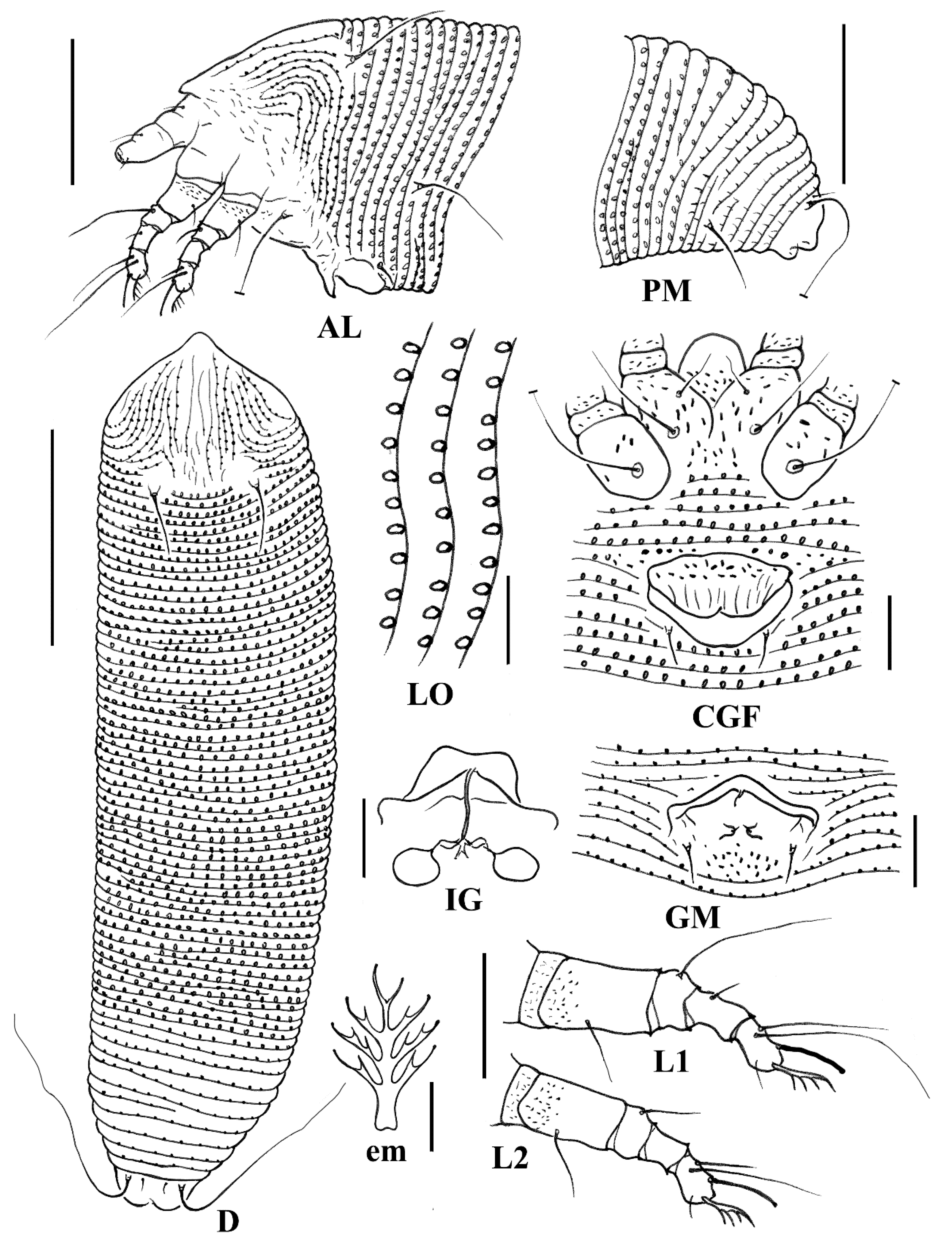
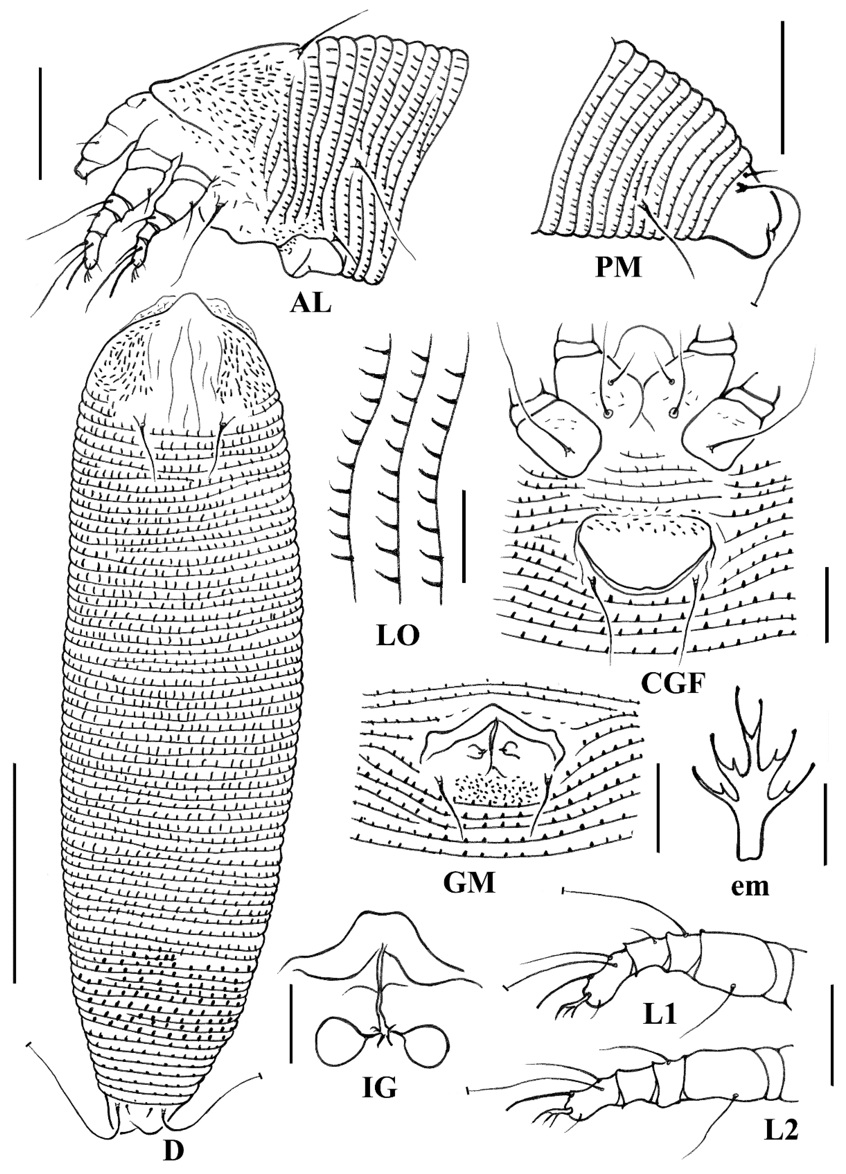
Figure 1.
Line drawings of Aceria bischofiae sp. nov.: AL. Lateral view of anterior opisthosoma; CGF. Coxigenital region of female; D. Dorsal view; em. Empodium; GM. Male genitalia; IG. Internal female genitalia; LO. Lateral view of annuli; L1. Leg I; L2. Leg II; PM. Lateral view of the posterior opisthosoma. Scale bar: 50 μm for D; 25 μm for AL and PM; 10 μm for CGF, GM, LO, L1, L2, and IG; 2.5 μm for em.

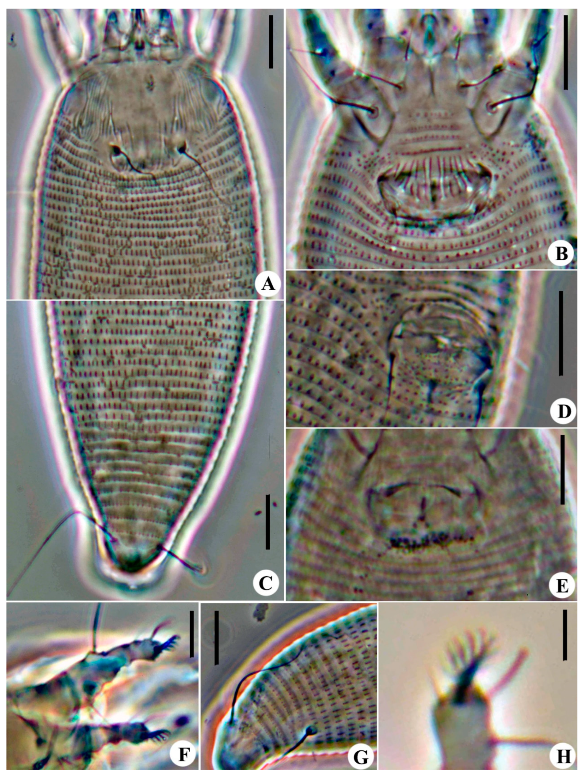
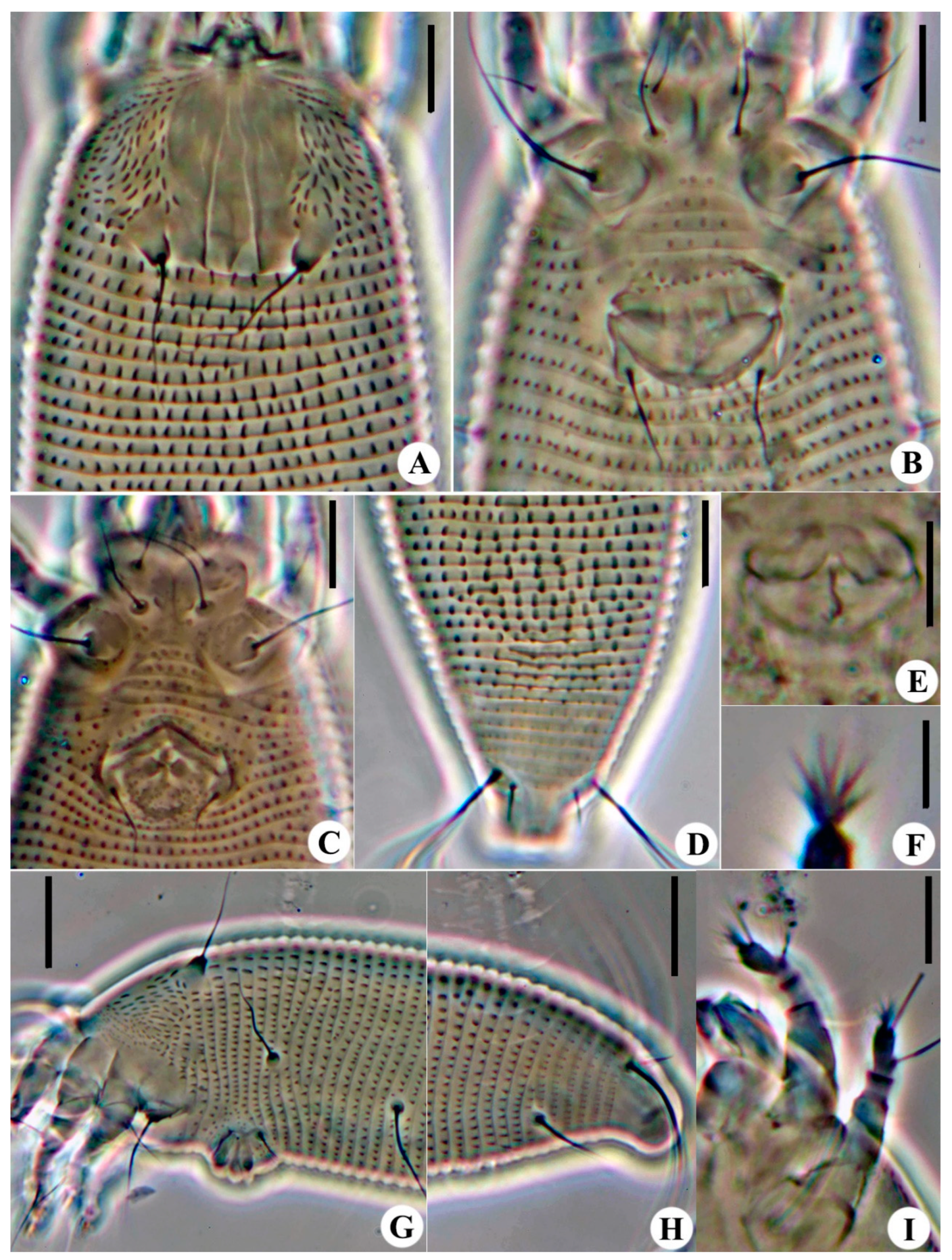
Figure 2.
Images of Aceria bischofiae sp. nov.: (A) Prodorsal shield of female; (B) Female coxigenital area; (C) Postero-dorsal view of mite; (D) Male coxigenital area; (E) Internal genitalia; (F) Legs; (G) Postero-lateral view of mite; (H) Empodium. Scale bar: 10 μm for (A–G); 2.5 μm for (H).
Figure 2.
Images of Aceria bischofiae sp. nov.: (A) Prodorsal shield of female; (B) Female coxigenital area; (C) Postero-dorsal view of mite; (D) Male coxigenital area; (E) Internal genitalia; (F) Legs; (G) Postero-lateral view of mite; (H) Empodium. Scale bar: 10 μm for (A–G); 2.5 μm for (H).

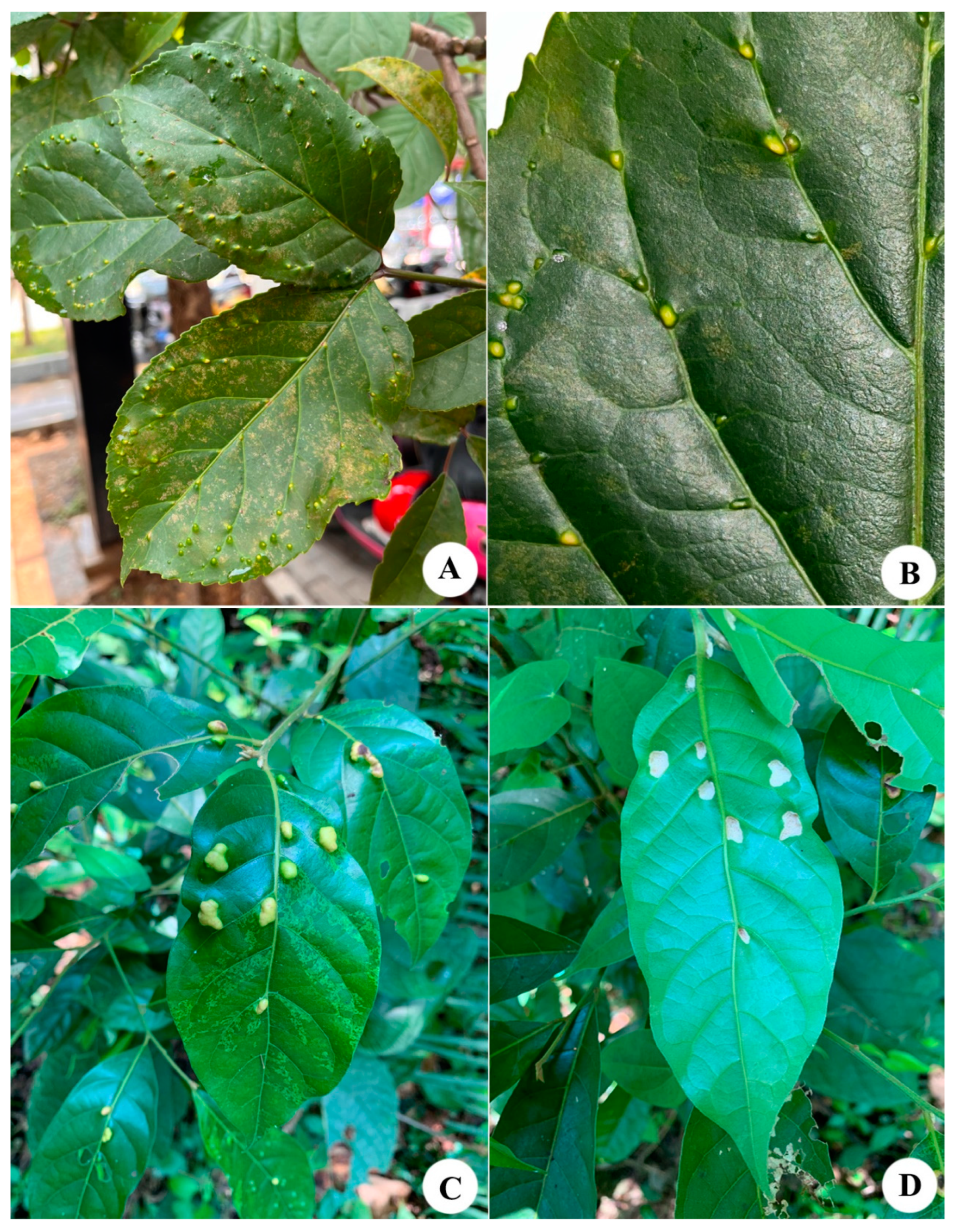
Figure 3.
(A,B) Damage symptoms associated with Aceria bischofiae sp. nov. on Bischofia javanica Blume; (C,D) Erineum caused by Aceria cryptocaryae sp. nov. on Cryptocarya metcalfiana Allen.
Figure 3.
(A,B) Damage symptoms associated with Aceria bischofiae sp. nov. on Bischofia javanica Blume; (C,D) Erineum caused by Aceria cryptocaryae sp. nov. on Cryptocarya metcalfiana Allen.

Figure 4.
Line drawings of Aceria cryptocaryae sp. nov.: AL. Lateral view of anterior opisthosoma; CGF. Coxigenital region of female; D. Dorsal view; em. Empodium; GM. Male genitalia; IG. Internal female genitalia; LO. Lateral view of annuli; L1. Leg I; L2. Leg II; PM. Lateral view of the posterior opisthosoma. Scale bar: 50 μm for D; 25 μm for AL and PM; 10 μm for CGF, GM, LO, L1, L2, and IG; 2.5 μm for em.

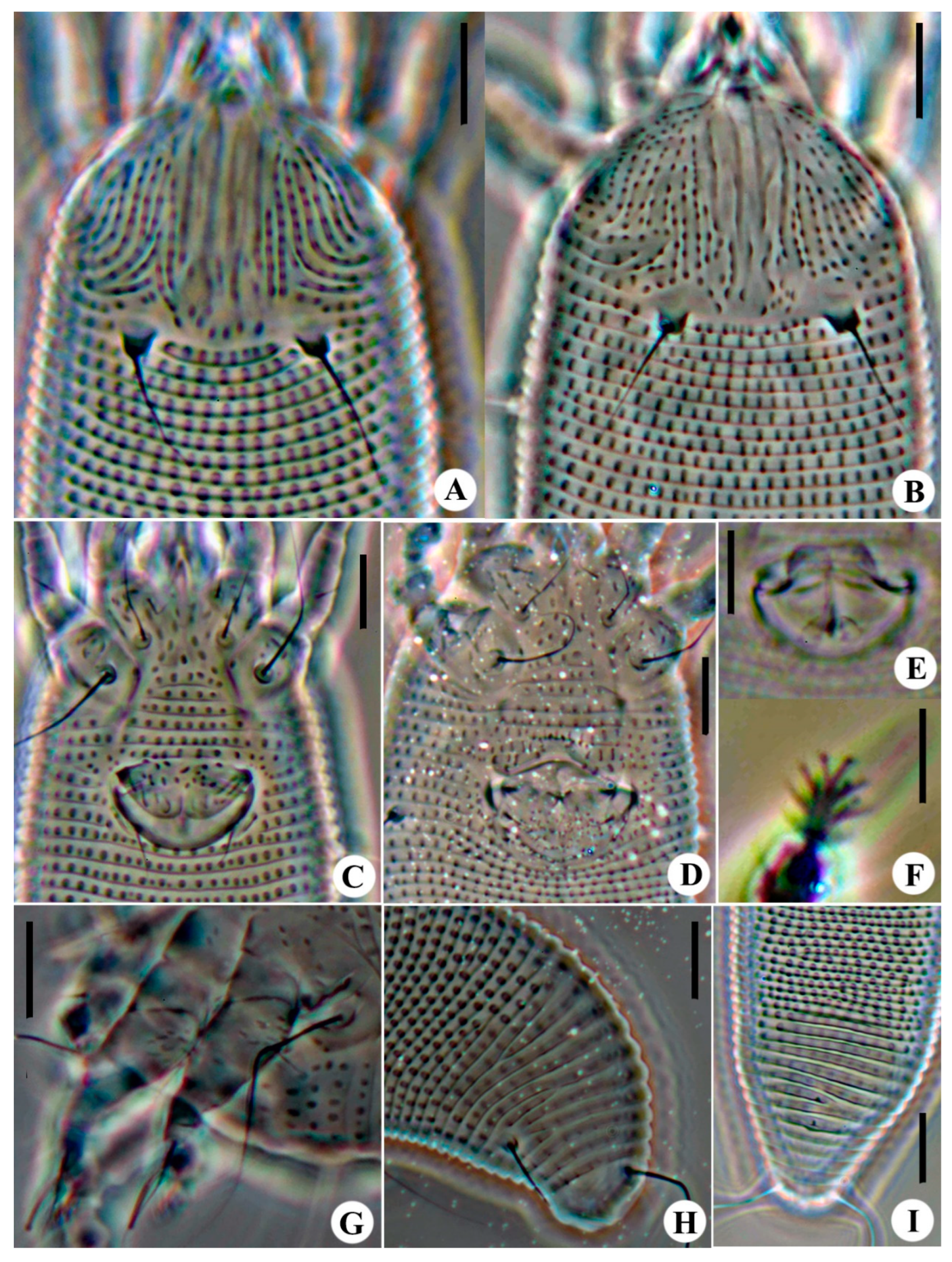
Figure 5.
Images of Aceria cryptocaryae sp. nov.: (A) Prodorsal shield of female; (B) Prodorsal shield of male; (C) Female coxigenital area; (D) Male coxigenital area; (E) Internal genitalia; (F) Empodium; (G) Legs; (H) Postero-lateral view of mite; (I) Postero-dorsal view of mite. Scale bar: 10 μm for (A–E,G–I); 5 μm for (F).

Figure 6.
Line drawings of Aceria buddlejae sp. nov.: AL. Lateral view of anterior opisthosoma; CGF. Coxigenital region of female; D. Dorsal view; em. Empodium; GM. Male genitalia; IG. Internal female genitalia; LO. Lateral view of annuli; L1. Leg I; L2. Leg II; PM. Lateral view of the posterior opisthosoma. Scale bar: 50 μm for D; 25 μm for AL and PM; 10 μm for CGF, GM, LO, L1, L2, and IG; 2.5 μm for em.

Figure 7.
Images of Aceria buddlejae sp. nov.: (A) Prodorsal shield; (B) Female coxigenital area; (C) Male coxigenital area; (D) Postero-dorsal view of mite; (E) Internal genitalia; (F) Empodium; (G) Lateral view of anterior opisthosoma; (H) Postero-lateral view of mite; (I) Legs. Scale bar: 20 μm for G, H; 10 μm for (A–E), I; 5 μm for (F).

Disclaimer/Publisher’s Note: The statements, opinions and data contained in all publications are solely those of the individual author(s) and contributor(s) and not of MDPI and/or the editor(s). MDPI and/or the editor(s) disclaim responsibility for any injury to people or property resulting from any ideas, methods, instructions or products referred to in the content. |
© 2024 by the authors. Licensee MDPI, Basel, Switzerland. This article is an open access article distributed under the terms and conditions of the Creative Commons Attribution (CC BY) license (https://creativecommons.org/licenses/by/4.0/).
Share and Cite
MDPI and ACS Style
Tan, M.; Lian, R.; Ruan, H.; Liang, X. Three New Species of Aceria (Acari: Trombidiformes: Eriophyoidea) from China. Animals 2024, 14, 720. https://doi.org/10.3390/ani14050720
AMA Style
Tan M, Lian R, Ruan H, Liang X. Three New Species of Aceria (Acari: Trombidiformes: Eriophyoidea) from China. Animals. 2024; 14(5):720. https://doi.org/10.3390/ani14050720
Chicago/Turabian StyleTan, Mengchao, Ranran Lian, Hongyan Ruan, and Xuhui Liang. 2024. "Three New Species of Aceria (Acari: Trombidiformes: Eriophyoidea) from China" Animals 14, no. 5: 720. https://doi.org/10.3390/ani14050720
Note that from the first issue of 2016, this journal uses article numbers instead of page numbers. See further details here.