
Current Knowledge on the Fungal Degradation Abilities Profiled through Biodeteriorative Plate Essays


1
Centre for Functional Ecology, Department of Life Sciences, University of Coimbra, 3004-531 Coimbra, Portugal
2
Fitolab-Laboratory for Phytopathology, Instituto Pedro Nunes, 3030-199 Coimbra, Portugal
*
Author to whom correspondence should be addressed.
Appl. Sci. 2021, 11(9), 4196; https://doi.org/10.3390/app11094196
Submission received: 26 March 2021
/
Revised: 27 April 2021
/
Accepted: 27 April 2021
/
Published: 5 May 2021
(This article belongs to the Special Issue Application of Biology to Cultural Heritage)
Abstract
:Fungi are known to contribute to the development of drastic biodeterioration of historical and valuable cultural heritage materials. Understandably, studies in this area are increasingly reliant on modern molecular biology techniques due to the enormous benefits they offer. However, classical culture dependent methodologies still offer the advantage of allowing fungal species biodeteriorative profiles to be studied in great detail. Both the essays available and the results concerning distinct fungal species biodeteriorative profiles obtained by amended plate essays, remain scattered and in need of a deep summarization. As such, the present work attempts to provide an overview of available options for this profiling, while also providing a summary of currently known fungal species putative biodeteriorative abilities solely obtained by the application of these methodologies. Consequently, this work also provides a series of checklists that can be helpful to microbiologists, restorers and conservation workers when attempting to safeguard cultural heritage materials worldwide from biodeterioration.
1. Introduction
The Fungal Kingdom comprises a highly diverse eukaryotic group able to inhabit every ecological niche available on the Planet [1]. The growth and biological activity of fungal species in cultural heritage materials is known to develop serious damages by means of biodeterioration (the undesirable modifications of a valuable material occurring by the action of living organisms) [2,3]. Fungi are highly versatile, ubiquitous, chemoheterotrophic microorganisms, being able to grow in a vast number of materials and contributing to the development of various biodeterioration phenomena [2,3]. Such modifications are a result from fungal species settling, development and exploitation of various organic and inorganic compounds present in historic art-pieces and monuments [2,3,4,5,6,7,8,9,10,11,12,13,14,15]. The fungal biodeterioration of books, paper, parchment, textiles, photographs, paintings, sculptures and wooden materials occurs due to the aesthetic modifications, mechanical pressure and exoenzymatic action [2]. Various components of these materials such as cellulose, collagen, linen, glues, inks, waxes and organic binders can be oxidized, hydrolyzed, dissolved, stained or structurally modified as a result of the action of fungal enzymes, pigments and organic acids [2,3,7,8,9,10,11]. A typical and widely known example of these phenomena is known as “foxing”, the development of red-brownish localized spots, hypothesized to be a result from fungal proliferation and metabolization of organic acids, oligosaccharides and proteic compounds that can stain and modify the constituent materials of many paper-based and photographic supports [3,8,13]. Another example of microorganism’s attack of organic materials is related to the biodeterioration of human remains, mummies and funerary materials, where opportunistic, saprotrophic and highly cellulolytic and proteolytic taxa are able to thrive and trough their actions severely alter them [2,14,15]. Complementarily, historic relics mainly composed of inorganic components such as stone, frescoes, glass and ceramics can also suffer deep aesthetical, physical and chemical modifications resulting from fungal grow and action [2,3,4,5,6,12,16]. In these supports, deterioration is caused by hyphae penetration into the substrate, the production and release of extracellular destructive organic acids, enzymes and metabolites and by the the formation of distinct colored outlines as a result of fungi high pigment contents, contribution to biofilm development and chemical reactions with inorganic compounds [2,3,4,5,6].
Due to the known biodeterioration problems arising from their proliferation, the accurate species identification and a consequent deteriorative profiling of isolates are crucial steps towards the development and the establishment of proper protective measures for the diverse cultural heritage treasures around the world. With the recent development of innovative culture independent methodologies such as -omics technologies, molecular data is becoming increasingly more valuable for the identification of the microbes, the characterization of their metabolic functions and their deteriorative byproducts [17]. Methodologies such as metagenomics, transcriptomics, metabolomics and proteomics revolutionized the field and are increasingly allowing understanding of microbial diversity, but also species specific and holistic contributions to various materials biodeterioration phenomena [17]. These methods are particularly relevant considering that traditional cultivation dependent methodologies hold the disadvantage of being unable to correctly infer microorganism’s abundance and only allow the study of active forms, failing to provide information regarding viable non-culturable and non-viable forms [17,18,19,20,21,22,23,24,25,26,27]. Nonetheless, classical culture dependent methodologies still offer an important advantage when compared with modern methodologies, especially when considering that the isolation of microbes allows their natural biodeteriorative profiles to be studied in great detail. Culture media plates modified to specify a positive biodeteriorative ability upon the microorganism development and deteriorative action (see Figure 1 for examples) can provide valuable data that allow the evaluation of the microorganism’s putative risks to cultural heritage materials. Moreover, they also offer a highly informative, rapid and low-cost platform [28] that can help in a quick and focused decision-making process aiming to protect valuable artifacts. Currently, plate assays aiming to identify fungal deteriorative characteristics, such as calcium carbonate solubilization, mineralization and various enzymatic activities, have been proposed and somewhat widely used.
Although differences among distinct isolates, assays and incubation conditions are known and expected, the available literature concerning distinctive fungal species deteriorative profiles obtained using such methodologies remains pending a deep summarization. With this in mind, this work aims to provide an overview of available plate assays, as well as the fungal putative biodeteriorative profiles obtained solely through such tests so far. In addition, we also aimed at providing a series of quick and straight forward checklists that can be consulted by microbiologists, restorers and conservation staff, when working to safeguard important cultural heritage materials worldwide. These checklists were also annotated to contain currently accepted fungal names according to Mycobank (www.mycobank.org, last accessed on 26 April 2021) and Index Fungorum (www.indexfungorum.org, last accessed on 26 April 2021) in order to ensure an updated identification for fungi displaying such profiles, and to facilitate information sharing in the future.
2. Calcium Carbonate Solubilization or Dissolution
One of the greatest fungal effects on stone monuments is credited to their secretion of inorganic and organic acids that can alter the material properties [2,3,4,29,30,31,32]. In fact, carbonate weathering has been consistently linked to the excretion and action of these metabolites [33,34,35]. Evaluation of fungal calcium carbonate solubilization abilities in cultural heritage scenarios has been helpful to study the biodeteriorative contribution of isolates retrieved from air, mural paintings, wooden art objects, frescoes, catacombs, bricks, concrete, buildings and various limestone and plaster monuments and museums [28,33,35,36,37,38,39,40,41,42,43,44]. Fungal calcium carbonate solubilization ability screening is usually conducted with CaCO3 glucose agar and adapted formulations [33]. Nonetheless, the application of Malt extract agar and Reasoner’s 2A agar amended with CaCO3 (CMEA and CR2A) has also been successfully achieved [35]. Moreover, Kiyuna and colleagues [37] also highlighted the utility of Glucose Yeast extract calcium carbonate agar (GYC) [45] for such evaluation. Positive CaCO3 dissolution is usually evaluated by the visualization of a halo around the growing colony after a period of incubation. In addition, calcium carbonate solubilization screening can also be conducted coupled with the evaluation of the media pH modifications. For this purpose, Creatine Sucrose agar (CREA) [46] followed by the analysis of medium color changes around growing colonies, or liquid media according to the formulations provided by Borrego and colleagues [47] followed by pH analysis, can also be applied. A quick overview of the known fungal species able of CaCO3 dissolution points that isolates from more than fifty species have been found to display this biodeteriorative profile, with the great majority of them being Aspergillus and Penicillium species (Table 1). Both genera are known important biodeteriogens, producing various acidic molecules and contributing to the deterioration of materials [28,48]. The detection of species from these genera (as well as others for example, from Pestalotiopsis and Talaromyces among others) might indicate a putative threat to acid susceptible resources, such is the case of stone structures, mural paintings and frescoes.
3. Mineralization or Crystallization Development
Calcium carbonate solubilization by the action of fungal acids can often occur coupled with the recrystallization of minerals in the substrate [2,3,5,6,49,50,51]. Such mineralization singularities can lead to the development of various biodeterioration phenomena [52,53]. They occur from the reactions of secreted acids (especially oxalic acid) with stone cations [32] and often result in the formation of carbonates and/or calcium magnesium oxalates [5,6]. Characterization of fungal crystallization abilities in cultural heritage scenarios has been helpful to study the biodeteriorative contribution of isolates retrieved from air, limestone monuments, stone stela, wall and mural paintings [35,41,42,43,53,54,55,56]. Fungal mineralization ability screening is usually conducted using B4 (with calcium acetate) or modified B4 (with calcium carbonate) media and adapted formulations [57]. Moreover, CaCO3 modified Malt agar, Nutrient agar (NA) with CaCl2 and the above mentioned CMEA and CR2A media have also been found useful for such purposes [35,42,43,58,59]. Positive mineralization development is usually evaluated by the microscopical visualization of neo-formed minerals around or in fungal hyphae after a period of incubation. Moreover, further characterization of these crystals can also be achieved by applying analytical methodologies such as X-ray powder diffraction (XRD) and/or energy dispersive X-ray spectroscopy (EDS) in conjunction with scanning electron microscopy (SEM) methodologies. So far, circa sixty species have been found to display mineralization abilities in plate essays and, as similarly found for fungal calcium carbonate dissolution, multiple Aspergillus and Penicillium species have also denoted this biodeteriorative profile (Table 2). Such findings can be correlated with their long-known abilities to secrete oxalic acid, among various other acids [41]. Nonetheless, a relevant number of species from genera Alternaria, Cladosporium, Colletotrichum, Pestalotiopsis and Trichoderma putatively displaying these biodeteriorative abilities can also be verified. Typical minerals detected include calcium carbonate in the form of calcite and vaterite-calcite, weddellite, whewellite, hydroxyapatite, hydrocerussite, pyromorphite, phosphate and other still unidentified calcium oxalates and minerals. The detection of species from these genera might indicate a putative threat to materials highly susceptible to fungal acidolysis and biomineralization, such is the case of limestone monuments and murals [5,6,60].
4. Enzymatic Action
Fungal ligninolytic action is often considered a threat to wooden structures [61,62,63,64,65,66]. Cultural heritage materials constructed with these materials can be affected by fungal hyphae penetration but also by the action of various exoenzymes [67]. Moreover, brown and white rot fungi are known to contribute to these substrates’ deterioration and degradation in various contexts [68]. So far, fungal ligninolytic ability characterization in cultural heritage scenarios has been helpful to study the biodeteriorative contribution of isolates retrieved from air, wooden materials and art objects [61,69,70]. Ligninolytic ability screening can be conducted using media with Azure B (lignin peroxidase), Phenol Red (Mn peroxidase), Remazol Brilliant Blue R (laccase) [71,72,73,74] or, alternatively, by applying Potato Dextrose agar supplemented with guaiacol (PDA-guaiacol) [61,70]. Positive ligninolytic ability is usually evaluated by the clearance of the media specific color (Azure B, Phenol Red and Remazol Brilliant Blue R) or by the development of reddish-brown zones (PDA-guaiacol) after a period of incubation. As pointed by Pangallo and colleagues [36,69], data regarding ligninolytic abilities of filamentous fungi in biodeterioration contexts is still somewhat scarce. Nonetheless, as evidenced by Table 3, almost thirty species have been found to display these biodeteriorative abilities. Moreover, mainly species of genera Aspergillus, Chaetomium, Cladosporium and Penicillium represent the bulk of the currently studied lignin deteriorating fungi. As such, the detection of species from these genera might indicate a putative threat to lignin materials, such as the case of some types of paper and wood art pieces and objects.
Fungi can also have an important role in the attack of animal-based objects, adhesives and additives. Textile materials such as silk and wool can suffer microbial mediated biodeterioration processes by the action of deteriorating enzymes [75,76]. In particular, the silks fibroin and sericin can both be the target of microbial attack [77]. Moreover, wool keratins can also be the target of attack by microbes [77]. Evaluation of fibroinolytic and keratinolytic action in cultural heritage scenarios has been helpful to study mummies, funeral clothes and accessories biodeterioration [78,79,80]. Moreover, fungal chitinolytic and pectinolytic action has also been pinpointed as a threat to Ancient Yemeni mummies preserved with diverse organic compounds [81]. Additionally, esterease action profiling has also been helpful to study isolates retrieved from wax seals, air, textiles and human remains [82,83,84]. Fibroinolytic screening can be conducted using fibroin agar, with the fibroinolytic action being evaluated by the isolates ability to grow in the culture-amended plates [85]. Moreover, keratinolytic action can be evaluated using feather broth and keratin medium and positive ability can be verified by media turbidity changes [79,85]. On the other hand, chitinolytic activity can be evaluated using powdered chitin agar [86] and pectinolytic activities can be evaluated with media containing pectin [87]. Both these deteriorative activities can be estimated and quantified [81]. Additionally, esterease action can be studied using Tributyrin agar and Tween 80 agar [83,84,88]. Their action can be detected by the development of clear zones (Tributyrin agar) or by the precipitation of insoluble salts and compounds (Tween 80) around colonies. As occurring with ligninolytic action, data regarding filamentous fungi fibroinase and keratinolytic action is still infrequent. Twenty-three species were found to be able of fibroinolytic activity, while more than twenty-five were found to have keratinolytic action. Again, Aspergillus and Penicillium species also dominate these biodeteriorative profiles (Table 4 and Table 5). Moreover, various Alternaria species also displayed putative keratinolytic abilities. On the other hand, chitinolytic abilities have been identified for Aspergillus niger and Penicillium sp., while pectinolytic action has been identified in a slightly more diversified range of fungal genera and species (Aspergillus candidus, Mucor circinelloides, Penicillium echinulatum, Scopulariopsis koningii, Stachybotrys chartarum and Trichoderma hamatum) [81]. In parallel, fifty species have been identified as displaying estereolytic action, with a great dominance of Aspergillus and Penicillium species (Table 6). Understandably, the detection of these fungal species on crypt environments, human remains, buried materials, mummies, wax seals, textiles and clothes denotes a putative threat to these materials [3].
Fungal lipolytic action can have an important impact on the biodeterioration of parchment and leather related materials [91]. Fungi can attack lipids and take advantage of fatty materials as a mean to obtain carbon (while also contributing to the material deterioration) [92]. Fungal lipolytic ability characterization in cultural heritage scenarios has been helpful to study the biodeteriorative contribution of isolates retrieved from air, textiles, human remains, wax seals, albumen photographical materials, statues, wooden organs and pipes [83,84,89,93,94]. Lipolytic ability screening can be mainly conducted using Spirit Blue agar and Nile blue, and the positive action can be identified by the development of a halo around the colonies, after a period of incubation [89]. Circa sixty species were found to be able of lipolytic action (Table 7). As similarly verified in other deteriorative analyses, Aspergillus and Penicillium species are still predominant in these profiles. The detection of these fungal species on materials rich in fatty compounds, such as wax seals and photographic materials should be considered putatively hazardous.
Fungal proteolytic action can contribute to the biodeterioration of proteinaceous materials, such is the case of artistic natural binders. In addition, some conservation approaches also employ similar materials that can be targeted by microbial biodeterioration [38]. Fungal proteolytic ability characterization in cultural heritage scenarios has been helpful to study the biodeteriorative contribution of isolates retrieved from air, funeral clothes and accessories, graphic documents, materials present in libraries and museums, frescoes, textiles, human remains, mummies, mural paintings, cinematographic films, wax seals, paper, parchment, wooden organs and pipes [28,38,41,78,79,80,83,84,85,93,95,96,97,98,99,100]. Proteolytic ability screening can be mainly conducted using Gelatin agar (R2A-Gel), Casein agar (CN), Milk Nutrient agar (MilkNA) and media containing rabbit glue [78,94,96]. After a period of incubation, positive proteolytic ability can be detected by flooding of agar plates with 10% tannin solution and the visualization of the formed hydrolysis zones [101]. Over one hundred and thirty species have been found to be able of promoting protein attack (Table 8). As similarly verified in other enzymatic activities, Aspergillus and Penicillium species also dominate this biodeteriorative profile. Nonetheless, a significant number of species from genera Alternaria, Cladosporium and Talaromyces displaying these characteristics can also be confirmed. Detection of these fungal species on proteinaceous materials will putatively result in their accentuated biodeterioration.
Fungal cellulolytic action is known to contribute to the biodeterioration of paper, canvas oil paintings, binders and photographic materials [3]. Moreover, cellulolytic action abilities characterization in cultural heritage scenarios has been helpful to study the biodeteriorative contribution of isolates retrieved from air, albumen photographic materials, mummies, funeral accessories, wooden art objects, organs and pipes, wax seals, graphic documents, stone, drawings, lithographs, paintings, textiles, human remains, maps, photographs, paper and other materials present in libraries and museums [28,36,69,79,81,82,83,96,97,98,99,100,102,103,104,105]. Cellulolytic ability screening can be conducted using Czapek-Dox agar supplemented with hydroxyethyl cellulose [69], Congo Red agar [79], Mandels and Reese medium with carboxymethyl cellulose (CMC) [106] or media containing sterilized filter paper [47]. Positive evaluation of cellulolytic ability can be assessed by the visualization of hydrolyzed areas or after congo red application and treatment. Over one hundred and fifty fungal species have been found to have cellulolytic abilities (Table 9). The great majority of species belonged to genera Alternaria, Aspergillus, Chaetomium, Cladosporium, Penicillium and Talaromyces. As such, detection of these fungal species on cellulolytic materials including paper, paintings and photographic materials, should be perceived as putatively threatening from a biodeterioration standpoint.
5. Conclusions
As pointed and reviewed by Pyzik and colleagues [107] the application of high-throughput Next-Generation sequencing technologies has highlighted that cultural heritage materials are inhabited by various unknown microorganisms still pending taxonomic description and their biodeteriorative profiling. The material biodeterioration is known to sometimes be caused by a predominant or specific microbial group, while more often the complex biodeterioration processes are a result of the synergistic action of a group of organisms resulting from various colonization events influenced by the impacts of multiple external factors throughout a time frame [77]. Cultivation methodologies often face limitations in what regards the ability for distinct organisms to be effectively cultivated and their original biodeteriorative characteristics replicated under laboratory conditions [108]. Understandably the application of more modern molecular techniques in cultural heritage biodeterioration studies has been increasingly being used and updated for the last two decades [27,108]. Although with their own set of limitations, culture-dependent methodologies still offer three main advantages: (1) The isolation of microbes for further differential analysis; (2) the possibility to isolate, characterize and describe previously unknown taxa; and (3) the development and improvement of biological and genetic databases. These aspects are especially important when considering that even the biodeteriorative role (but also their taxonomic classification) of long known species might also need to be constantly revised, updated and reevaluated [107,109]. For instance, the inclusion of the Fusarium solani Species Complex in the genus Neocosmospora was recently reevaluated and continues to be the focus of additional studies [110].
Fungi are constantly regarded as one if not the most important microorganism groups causing cultural heritage materials biodeterioration [2,3,30]. This review highlighted that, so far, isolates from more than two-hundred fungal species have been showed to exhibit biodeteriorative abilities when studied by specific plate essays. Based on the available studies performed so far, it is possible to verify that Aspergillus and Penicillium species dominate the biodeteriorative abilities usually screened in biodeterioration contexts. With this in mind, it should be reinforced that the detection of these species in various cultural heritage materials can, under specific conditions, result in severe biodeterioration of the substrate. Nonetheless, a careful analysis of these checklists, as well as, the biodeteriorative screening of obtained isolates, wherever possible, is strongly advised. Not all isolates might display deteriorative action or display similar degradative rates and thus a proper and specific analysis in each case and/or the implementation of additional tests (e.g., molecular identification of genes involved in biodeterioration (see for example [111])) is also recommended. In conjunction with molecular approaches not relying in cultivation, they can provide a holistic evaluation of a specific biodeterioration phenomena. As pointed by Sterflinger and colleagues [112], understanding deterioration mechanisms and the main microbial perpetrators is still one of the major challenges in historic and cultural materials biodeterioration research. As such, the information summarized in this work provides a contribution that can help microbiologists, restorers, conservators and curators in their attempt to preserve cultural heritage materials for future generations.
Author Contributions
Conceptualization, J.T. writing—original draft preparation, J.T.; writing—review and editing, J.T. and A.P.; supervision, A.P.; funding acquisition, A.P. All authors have read and agreed to the published version of the manuscript.
Funding
This work was financed by IPN—Financiamento Base FITEC approved under the National Call with reference no. 01/FITEC/2018 to obtain multi-year base financing under the INTERFACE Program and by FEDER- Fundo Europeu de Desenvolvimento Regional funds through the COMPETE 2020-Operational Programme for Competitiveness and Internationalisation (POCI), and by Portuguese funds through FCT- Fundação para a Ciência e a Tecnologia in the framework of the project POCI-01-0145-FEDER-PTDC/EPH-PAT/3345/2014. This work was carried out at the R&D Unit Centre for Functional Ecology—Science for People and the Planet (CFE), with reference UIDB/04004/2020, financed by FCT/MCTES through national funds (PIDDAC). João Trovão was supported by POCH—Programa Operacional Capital Humano (co-funding by the European Social Fund and national funding by MCTES), through a “FCT- Fundação para a Ciência e Tecnologia” PhD research grant (SFRH/BD/132523/2017).
Institutional Review Board Statement
Not applicable.
Informed Consent Statement
Not applicable.
Data Availability Statement
Not applicable.
Conflicts of Interest
The authors declare no conflict of interest.
References
- Deacon, J.W. Fungal Biology, 4th ed.; Blackwell Pub.: Malden, MA, USA, 2006. [Google Scholar]
- Sterflinger, K. Fungi: Their Role in Deterioration of Cultural Heritage. Fungal Biol. Rev. 2010, 24, 47–55. [Google Scholar] [CrossRef]
- Sterflinger, K.; Piñar, G. Microbial Deterioration of Cultural Heritage and Works of Art—Tilting at Windmills? Appl. Microbiol. Biotechnol. 2013, 97, 9637–9646. [Google Scholar] [CrossRef] [Green Version]
- Dakal, T.C.; Cameotra, S.S. Microbially Induced Deterioration of Architectural Heritages: Routes and Mechanisms Involved. Environ. Sci. Eur. 2012, 24, 36. [Google Scholar] [CrossRef] [Green Version]
- Gadd, G.M. Geomicrobiology of the Built Environment. Nat. Microbiol. 2017, 2, 1–9. [Google Scholar] [CrossRef] [Green Version]
- Gadd, G.M. Fungi, Rocks, and Minerals. Elements 2017, 13, 171–176. [Google Scholar] [CrossRef]
- Cappitelli, F.; Sorlini, C. From Papyrus to Compact Disc: The Microbial Deterioration of Documentary Heritage. Crit. Rev. Microbiol. 2005, 31, 1–10. [Google Scholar] [CrossRef]
- Sterflinger, K.; Pinzari, F. The Revenge of Time: Fungal Deterioration of Cultural Heritage with Particular Reference to Books, Paper and Parchment. Environ. Microbiol. 2012, 14, 559–566. [Google Scholar] [CrossRef]
- Paiva de Carvalho, H.; Mesquita, N.; Trovão, J.; Fernández Rodríguez, S.; Pinheiro, A.C.; Gomes, V.; Alcoforado, A.; Gil, F.; Portugal, A. Fungal Contamination of Paintings and Wooden Sculptures inside the Storage Room of a Museum: Are Current Norms and Reference Values Adequate? J. Cult. Herit. 2018, 34, 268–276. [Google Scholar] [CrossRef]
- Poyatos, F.; Morales, F.; Nicholson, A.W.; Giordano, A. Physiology of Biodeterioration on Canvas Paintings. J. Cell Physiol. 2018, 233, 2741–2751. [Google Scholar] [CrossRef] [PubMed]
- Kosel, J.; Ropret, P. Overview of Fungal Isolates on Heritage Collections of Photographic Materials and Their Biological Potency. J. Cult. Herit. 2021, 48, 277–291. [Google Scholar] [CrossRef]
- Coutinho, M.L.; Miller, A.Z.; Macedo, M.F. Biological Colonization and Biodeterioration of Architectural Ceramic Materials: An Overview. J. Cult. Herit. 2015, 16, 759–777. [Google Scholar] [CrossRef] [Green Version]
- Sclocchi, M.C.; Kraková, L.; Pinzari, F.; Colaizzi, P.; Bicchieri, M.; Šaková, N.; Pangallo, D. Microbial Life and Death in a Foxing Stain: A Suggested Mechanism of Photographic Prints Defacement. Microb. Ecol. 2017, 73, 815–826. [Google Scholar] [CrossRef]
- Piñar, G.; Piombino-Mascali, D.; Maixner, F.; Zink, A.; Sterflinger, K. Microbial Survey of the Mummies from the Capuchin Catacombs of Palermo, Italy: Biodeterioration Risk and Contamination of the Indoor Air. FEMS Microbiol. Ecol. 2013, 86, 341–356. [Google Scholar] [CrossRef] [PubMed] [Green Version]
- Ruga, L.; Orlandi, F.; Romano, B.; Fornaciari, M. The Assessment of Fungal Bioaerosols in the Crypt of St. Peter in Perugia (Italy). Int. Biodeterior. Biodegrad. 2015, 98, 121–130. [Google Scholar] [CrossRef]
- Rodrigues, A.; Gutierrez-Patricio, S.; Miller, A.Z.; Saiz-Jimenez, C.; Wiley, R.; Nunes, D.; Vilarigues, M.; Macedo, M.F. Fungal Biodeterioration of Stained-Glass Windows. Int. Biodeterior. Biodegrad. 2014, 90, 152–160. [Google Scholar] [CrossRef]
- Beata, G. The Use of -Omics Tools for Assessing Biodeterioration of Cultural Heritage: A Review. J. Cult. Herit. 2020, 45, 351–361. [Google Scholar] [CrossRef]
- Amann, R.I.; Ludwig, W.; Schleifer, K.H. Phylogenetic Identification and in Situ Detection of Individual Microbial Cells without Cultivation. Microbiol. Rev. 1995, 59, 143–169. [Google Scholar] [CrossRef]
- Dakal, T.C.; Arora, P.K. Evaluation of Potential of Molecular and Physical Techniques in Studying Biodeterioration. Rev. Environ. Sci. Biotechnol. 2012, 11, 71–104. [Google Scholar] [CrossRef]
- González Grau, J.M.; Sáiz-Jiménez, C. Unknown Microbial Communities on Rock Art Paintings. Consequences for Conservation and Future Perspectives. Coalition 2005, 10, 4–7. [Google Scholar]
- Gonzalez, J.M.; Saiz-Jimenez, C. Microbial Diversity in Biodeteriorated Monuments as Studied by Denaturing Gradient Gel Electrophoresis. J. Sep. Sci. 2004, 27, 174–180. [Google Scholar] [CrossRef]
- González, J.M.; Sáiz-Jiménez, C. Application of Molecular Nucleic Acid-Based Techniques for the Study of Microbial Communities in Monuments and Artworks. Int. Microbiol. 2005, 8, 189–194. [Google Scholar] [PubMed]
- Laiz, L.; Piñar, G.; Lubitz, W.; Sáiz-Jiménez, C. The Colonisation of Building Materials by Microorganisms as Revealed by Culturing and Molecular Methods. Mol. Biol. Cult. Herit. 2003. [Google Scholar] [CrossRef]
- Mihajlovski, A.; Seyer, D.; Benamara, H.; Bousta, F.; Di Martino, P. An Overview of Techniques for the Characterization and Quantification of Microbial Colonization on Stone Monuments. Ann. Microbiol. 2015, 65, 1243–1255. [Google Scholar] [CrossRef] [Green Version]
- Otlewska, A.; Adamiak, J.; Gutarowska, B. Application of Molecular Techniques for the Assessment of Microorganism Diversity on Cultural Heritage Objects. Acta Biochim. Pol. 2014, 61. [Google Scholar] [CrossRef] [Green Version]
- Sanmartín, P.; DeAraujo, A.; Vasanthakumar, A. Melding the Old with the New: Trends in Methods Used to Identify, Monitor, and Control Microorganisms on Cultural Heritage Materials. Microb. Ecol. 2018, 76, 64–80. [Google Scholar] [CrossRef] [PubMed]
- Ward, D.M.; Weller, R.; Bateson, M.M. 16S RRNA Sequences Reveal Numerous Uncultured Microorganisms in a Natural Community. Nature 1990, 345, 63–65. [Google Scholar] [CrossRef]
- Savković, Ž.; Stupar, M.; Unković, N.; Ivanović, Ž.; Blagojević, J.; Vukojević, J.; Ljaljević Grbić, M. In Vitro Biodegradation Potential of Airborne Aspergilli and Penicillia. Sci. Nat. 2019, 106, 8. [Google Scholar] [CrossRef] [PubMed]
- Warscheid, T.; Braams, J. Biodeterioration of Stone: A Review. Int. Biodeterior. Biodegrad. 2000, 46, 343–368. [Google Scholar] [CrossRef]
- Sterflinger, K. Fungi as Geologic Agents. Geomicrobiol. J. 2000, 17, 97–124. [Google Scholar] [CrossRef]
- Scheerer, S.; Ortega-Morales, O.; Gaylarde, C. Chapter 5 Microbial Deterioration of Stone Monuments—An Updated Overview. Adv. Appl. Microbiol. 2009, 66, 97–139. [Google Scholar] [CrossRef]
- Liu, X.; Koestler, R.J.; Warscheid, T.; Katayama, Y.; Gu, J.-D. Microbial Deterioration and Sustainable Conservation of Stone Monuments and Buildings. Nat. Sustain. 2020, 3, 991–1004. [Google Scholar] [CrossRef]
- Albertano, P.; Urzì, C. Structural Interactions among Epilithic Cyanobacteria and Heterotrophic Microorganisms in Roman Hypogea. Microb. Ecol. 1999, 38, 244–252. [Google Scholar] [CrossRef] [PubMed]
- Burford, E.P.; Kierans, M.; Gadd, G.M. Geomycology: Fungi in Mineral Substrata. Mycologist 2003, 17, 98–107. [Google Scholar] [CrossRef]
- Ortega-Morales, B.O.; Narváez-Zapata, J.; Reyes-Estebanez, M.; Quintana, P.; la Rosa-García, D.; Bullen, H.; Gómez-Cornelio, S.; Chan-Bacab, M.J. Bioweathering Potential of Cultivable Fungi Associated with Semi-Arid Surface Microhabitats of Mayan Buildings. Front. Microbiol. 2016, 7, 201. [Google Scholar] [CrossRef] [PubMed]
- Pangallo, D.P.; Chovanová, K.C.; Šimonovičová, A.Š.; Ferianc, P.F. Investigation of Microbial Community Isolated from Indoor Artworks and Air Environment: Identification, Biodegradative Abilities, and DNA Typing. Can. J. Microbiol. 2009, 55, 277–287. [Google Scholar] [CrossRef] [PubMed]
- Kiyuna, T.; An, K.-D.; Kigawa, R.; Sano, C.; Miura, S.; Sugiyama, J. Bristle-like Fungal Colonizers on the Stone Walls of the Kitora and Takamatsuzuka Tumuli Are Identified as Kendrickiella Phycomyces. Mycoscience 2012, 53, 446–459. [Google Scholar] [CrossRef]
- Unković, N.; Dimkić, I.; Stupar, M.; Stanković, S.; Vukojević, J.; Grbić, M.L. Biodegradative Potential of Fungal Isolates from Sacral Ambient: In Vitro Study as Risk Assessment Implication for the Conservation of Wall Paintings. PLoS ONE 2018, 13, e0190922. [Google Scholar] [CrossRef]
- Ponizovskaya, V.B.; Rebrikova, N.L.; Kachalkin, A.V.; Antropova, A.B.; Bilanenko, E.N.; Mokeeva, V.L. Micromycetes as Colonizers of Mineral Building Materials in Historic Monuments and Museums. Fungal Biol. 2019, 123, 290–306. [Google Scholar] [CrossRef]
- Gámez-Espinosa, E.; Bellotti, N.; Deyá, C.; Cabello, M. Mycological Studies as a Tool to Improve the Control of Building Materials Biodeterioration. J. Build. Eng. 2020, 32, 101738. [Google Scholar] [CrossRef]
- Ma, W.; Wu, F.; Tian, T.; He, D.; Zhang, Q.; Gu, J.-D.; Duan, Y.; Ma, D.; Wang, W.; Feng, H. Fungal Diversity and Its Contribution to the Biodeterioration of Mural Paintings in Two 1700-Year-Old Tombs of China. Int. Biodeterior. Biodegrad. 2020, 152, 104972. [Google Scholar] [CrossRef]
- Trovão, J.; Tiago, I.; Catarino, L.; Gil, F.; Portugal, A. In Vitro Analyses of Fungi and Dolomitic Limestone Interactions: Bioreceptivity and Biodeterioration Assessment. Int. Biodeterior. Biodegrad. 2020, 155, 105107. [Google Scholar] [CrossRef]
- Trovão, J.; Gil, F.; Catarino, L.; Soares, F.; Tiago, I.; Portugal, A. Analysis of Fungal Deterioration Phenomena in the First Portuguese King Tomb Using a Multi-Analytical Approach. Int. Biodeterior. Biodegrad. 2020, 149, 104933. [Google Scholar] [CrossRef]
- Isola, D.; Zucconi, L.; Cecchini, A.; Caneva, G. Dark-Pigmented Biodeteriogenic Fungi in Etruscan Hypogeal Tombs: New Data on Their Culture-Dependent Diversity, Favouring Conditions, and Resistance to Biocidal Treatments. Fungal Biol. 2021. [Google Scholar] [CrossRef]
- De Ley, J.; Swings, J.; Gosselé, F. Genus I. Acetobacter Beijerinck 1898, 215AL. In Bergey’s Manual of Systematic Bacteriology; Williams and Wilkins: Baltimore, MD, USA, 1984; Volume 1, pp. 268–274. [Google Scholar]
- Samson, R.A.; Houbraken, J.; Thrane, U.; Frisvad, J.C.; Andersen, B. Food and Indoor Fungi, 2nd ed.; CBS-KNAW Fungal Biodiversity Centre: Utrecht, The Netherlands, 2010. [Google Scholar]
- Borrego, S.; Guiamet, P.; Gómez de Saravia, S.; Batistini, P.; Garcia, M.; Lavin, P.; Perdomo, I. The Quality of Air at Archives and the Biodeterioration of Photographs. Int. Biodeterior. Biodegrad. 2010, 64, 139–145. [Google Scholar] [CrossRef]
- Gutarowska, B. Metabolic Activity of Moulds as a Factor of Building Materials Biodegradation. Pol. J. Microbiol. 2010, 59, 119–124. [Google Scholar] [CrossRef]
- Ahmad, A.; Rautaray, D.; Sastry, M. Biogenic Calcium Carbonate: Calcite Crystals of Variable Morphology by the Reaction of Aqueous Ca2+ Ions with Fungi. Adv. Funct. Mater. 2004, 14, 1075–1080. [Google Scholar] [CrossRef]
- Burford, E.P.; Hillier, S.; Gadd, G.M. Biomineralization of Fungal Hyphae with Calcite (CaCO3) and Calcium Oxalate Mono- and Dihydrate in Carboniferous Limestone Microcosms. Geomicrobiol. J. 2006, 23, 599–611. [Google Scholar] [CrossRef]
- Gadd, G.M.; Bahri-Esfahani, J.; Li, Q.; Rhee, Y.J.; Wei, Z.; Fomina, M.; Liang, X. Oxalate Production by Fungi: Significance in Geomycology, Biodeterioration and Bioremediation. Fungal Biol. Rev. 2014, 28, 36–55. [Google Scholar] [CrossRef]
- Del Monte, M.; Sabbioni, C.; Zappia, G. The Origin of Calcium Oxalates on Historical Buildings, Monuments and Natural Outcrops. Sci. Total Environ. 1987, 67, 17–39. [Google Scholar] [CrossRef]
- Li, T.; Hu, Y.; Zhang, B.; Yang, X. Role of Fungi in the Formation of Patinas on Feilaifeng Limestone, China. Microb. Ecol. 2018, 76, 352–361. [Google Scholar] [CrossRef]
- Savković, Ž.; Unković, N.; Stupar, M.; Franković, M.; Jovanović, M.; Erić, S.; Šarić, K.; Stanković, S.; Dimkić, I.; Vukojević, J.; et al. Diversity and Biodeteriorative Potential of Fungal Dwellers on Ancient Stone Stela. Int. Biodeterior. Biodegrad. 2016, 115, 212–223. [Google Scholar] [CrossRef]
- Li, T.; Hu, Y.; Zhang, B. Biomineralization Induced by Colletotrichum acutatum: A Potential Strategy for Cultural Relic Bioprotection. Front. Microbiol. 2018, 9, 1884. [Google Scholar] [CrossRef] [Green Version]
- Unković, N.; Erić, S.; Šarić, K.; Stupar, M.; Savković, Ž.; Stanković, S.; Stanojević, O.; Dimkić, I.; Vukojević, J.; Ljaljević Grbić, M. Biogenesis of Secondary Mycogenic Minerals Related to Wall Paintings Deterioration Process. Micron 2017, 100, 1–9. [Google Scholar] [CrossRef]
- Boquet, E.; Boronat, A.; Ramos-Cormenzana, A. Production of Calcite (Calcium Carbonate) Crystals by Soil Bacteria Is a General Phenomenon. Nature 1973, 246, 527–529. [Google Scholar] [CrossRef]
- Guggiari, M.; Bloque, R.; Aragno, M.; Verrecchia, E.; Job, D.; Junier, P. Experimental Calcium-Oxalate Crystal Production and Dissolution by Selected Wood-Rot Fungi. Int. Biodeterior. Biodegrad. 2011, 65, 803–809. [Google Scholar] [CrossRef] [Green Version]
- Pasquale, V.; Fiore, S.; Hlayem, D.; Lettino, A.; Huertas, F.J.; Chianese, E.; Dumontet, S. Biomineralization of Carbonates Induced by the Fungi Paecilomyces inflatus and Plectosphaerella cucumerina. Int. Biodeterior. Biodegrad. 2019, 140, 57–66. [Google Scholar] [CrossRef]
- Gadd, G.M. Geomycology: Biogeochemical Transformations of Rocks, Minerals, Metals and Radionuclides by Fungi, Bioweathering and Bioremediation. Mycol. Res. 2007, 111, 3–49. [Google Scholar] [CrossRef] [PubMed]
- Liu, Z.; Wang, Y.; Pan, X.; Ge, Q.; Ma, Q.; Li, Q.; Fu, T.; Hu, C.; Zhu, X.; Pan, J. Identification of Fungal Communities Associated with the Biodeterioration of Waterlogged Archeological Wood in a Han Dynasty Tomb in China. Front. Microbiol. 2017, 8. [Google Scholar] [CrossRef]
- Gutarowska, B.; Celikkol-Aydin, S.; Bonifay, V.; Otlewska, A.; Aydin, E.; Oldham, A.L.; Brauer, J.I.; Duncan, K.E.; Adamiak, J.; Sunner, J.A.; et al. Metabolomic and High-Throughput Sequencing Analysis—Modern Approach for the Assessment of Biodeterioration of Materials from Historic Buildings. Front. Microbiol. 2015, 6, 979. [Google Scholar] [CrossRef] [PubMed] [Green Version]
- Ortiz, R.; Navarrete, H.; Navarrete, J.; Párraga, M.; Carrasco, I.; de la Vega, E.; Ortiz, M.; Herrera, P.; Blanchette, R.A. Deterioration, Decay and Identification of Fungi Isolated from Wooden Structures at the Humberstone and Santa Laura Saltpeter Works: A World Heritage Site in Chile. Int. Biodeterior. Biodegrad. 2014, 86, 309–316. [Google Scholar] [CrossRef]
- Piñar, G.; Dalnodar, D.; Voitl, C.; Reschreiter, H.; Sterflinger, K. Biodeterioration Risk Threatens the 3100 Year Old Staircase of Hallstatt (Austria): Possible Involvement of Halophilic Microorganisms. PLoS ONE 2016, 11, e0148279. [Google Scholar] [CrossRef] [PubMed]
- Pournou, A. Biodeterioration of Wooden Cultural Heritage: Organisms and Decay Mechanisms in Aquatic and Terrestrial Ecosystems; Springer International Publishing: Cham, Switzerland, 2020; ISBN 978-3-030-46503-2. [Google Scholar]
- Alfieri, P.V.; García, R.A.; Rosato, V.G.; Correa, M.V. Biodeterioration and Biodegradation of Wooden Heritage: Role of Fungal Succession. Int. J. Conserv. Sci. 2016, 7, 607–614. [Google Scholar]
- Blanchette, R.A. A Review of Microbial Deterioration Found in Archaeological Wood from Different Environments. Int. Biodeterior. Biodegrad. 2000, 46, 189–204. [Google Scholar] [CrossRef]
- Goodell, B.; Winandy, J.E.; Morrell, J.J. Fungal Degradation of Wood: Emerging Data, New Insights and Changing Perceptions. Coatings 2020, 10, 1210. [Google Scholar] [CrossRef]
- Pangallo, D.; Šimonovičová, A.; Chovanová, K.; Ferianc, P. Wooden Art Objects and the Museum Environment: Identification and Biodegradative Characteristics of Isolated Microflora. Lett. Appl. Microbiol. 2007, 45, 87–94. [Google Scholar] [CrossRef]
- Liu, Z.; Fu, T.; Hu, C.; Shen, D.; Macchioni, N.; Sozzi, L.; Chen, Y.; Liu, J.; Tian, X.; Ge, Q.; et al. Microbial Community Analysis and Biodeterioration of Waterlogged Archaeological Wood from the Nanhai No. 1 Shipwreck during Storage. Sci. Rep. 2018, 8, 7170. [Google Scholar] [CrossRef] [PubMed] [Green Version]
- Archibald, F.S. A New Assay for Lignin-Type Peroxidases Employing the Dye Azure B. Appl. Environ. Microbiol. 1992, 58, 3110–3116. [Google Scholar] [CrossRef] [Green Version]
- Manji, S.; Ishihara, A. Screening of Tetrachlorodibenzo-p-Dioxin-Degrading Fungi Capable of Producing Extracellular Peroxidases under Various Conditions. Appl. Microbiol. Biotechnol. 2004, 63, 438–444. [Google Scholar] [CrossRef]
- Falade, A.O.; Eyisi, O.A.L.; Mabinya, L.V.; Nwodo, U.U.; Okoh, A.I. Peroxidase Production and Ligninolytic Potentials of Fresh Water Bacteria Raoultella Ornithinolytica and Ensifer Adhaerens. Biotechnol. Rep. 2017, 16, 12–17. [Google Scholar] [CrossRef]
- Kiiskinen, L.-L.; Rättö, M.; Kruus, K. Screening for Novel Laccase-Producing Microbes. J. Appl. Microbiol. 2004, 97, 640–646. [Google Scholar] [CrossRef]
- Szostak-Kotowa, J. Biodeterioration of Textiles. Int. Biodeterior. Biodegrad. 2004, 53, 165–170. [Google Scholar] [CrossRef]
- Gutarowska, B.; Pietrzak, K.; Machnowski, W.; Milczarek, J.M. Historical Textiles—A Review of Microbial Deterioration Analysis and Disinfection Methods. Text. Res. J. 2017, 87, 2388–2406. [Google Scholar] [CrossRef]
- Mazzoli, R.; Giuffrida, M.G.; Pessione, E. Back to the Past: “Find the Guilty Bug—Microorganisms Involved in the Biodeterioration of Archeological and Historical Artifacts”. Appl. Microbiol. Biotechnol. 2018, 102, 6393–6407. [Google Scholar] [CrossRef]
- Pangallo, D.; Kraková, L.; Chovanová, K.; Šimonovičová, A.; De Leo, F.; Urzì, C. Analysis and Comparison of the Microflora Isolated from Fresco Surface and from Surrounding Air Environment through Molecular and Biodegradative Assays. World J. Microbiol. Biotechnol. 2012, 28, 2015–2027. [Google Scholar] [CrossRef]
- Kraková, L.; Šoltys, K.; Puškárová, A.; Bučková, M.; Jeszeová, L.; Kucharík, M.; Budiš, J.; Orovčík, L.; Szemes, T.; Pangallo, D. The Microbiomes of a XVIII Century Mummy from the Castle of Krásna Hôrka (Slovakia) and Its Surrounding Environment. Environ. Microbiol. 2018, 20, 3294–3308. [Google Scholar] [CrossRef] [PubMed]
- Kisová, Z.; Planý, M.; Pavlović, J.; Bučková, M.; Puškárová, A.; Kraková, L.; Kapustová, M.; Pangallo, D.; Šoltys, K. Biodeteriogens Characterization and Molecular Analyses of Diverse Funeral Accessories from XVII Century. Appl. Sci. 2020, 10, 5451. [Google Scholar] [CrossRef]
- Naji, K.M.; Abdullah, Q.Y.M.; AL-Zaqri, A.Q.M.; Alghalibi, S.M. Evaluating the Biodeterioration Enzymatic Activities of Fungal Contamination Isolated from Some Ancient Yemeni Mummies Preserved in the National Museum. Biochem. Res. Int. 2014, 2014, e481508. [Google Scholar] [CrossRef] [Green Version]
- Kavkler, K.; Gunde-Cimerman, N.; Zalar, P.; Demšar, A. Fungal Contamination of Textile Objects Preserved in Slovene Museums and Religious Institutions. Int. Biodeterior. Biodegrad. 2015, 97, 51–59. [Google Scholar] [CrossRef]
- Šimonovičová, A.; Kraková, L.; Pangallo, D.; Majorošová, M.; Piecková, E.; Bodoriková, S.; Dörnhoferová, M. Fungi on Mummified Human Remains and in the Indoor Air in the Kuffner Family Crypt in Sládkovičovo (Slovakia). Int. Biodeterior. Biodegrad. 2015, 99, 157–164. [Google Scholar] [CrossRef]
- Šoltys, K.; Planý, M.; Biocca, P.; Vianello, V.; Bučková, M.; Puškárová, A.; Sclocchi, M.C.; Colaizzi, P.; Bicchieri, M.; Pangallo, D.; et al. Lead Soaps Formation and Biodiversity in a XVIII Century Wax Seal Coloured with Minium. Environ. Microbiol. 2020, 22, 1517–1534. [Google Scholar] [CrossRef] [PubMed]
- Pangallo, D.; Kraková, L.; Chovanová, K.; Bučková, M.; Puškarová, A.; Šimonovičová, A. Disclosing a Crypt: Microbial Diversity and Degradation Activity of the Microflora Isolated from Funeral Clothes of Cardinal Peter Pázmány. Microbiol. Res. 2013, 168, 289–299. [Google Scholar] [CrossRef]
- Hsu, S.C.; Lockwood, J.L. Powdered Chitin Agar as a Selective Medium for Enumeration of Actinomycetes in Water and Soil. Appl. Microbiol. 1975, 29, 422–426. [Google Scholar] [CrossRef]
- Eggins, H.O.W.; Pugh, G.J.F. Isolation of Cellulose-Decomposing Fungi from the Soil. Nature 1962, 193, 94–95. [Google Scholar] [CrossRef]
- Paterson, R.R.M.; Bridge, P.D.; International Mycological Institute. Biochemical Techniques for Filamentous Fungi; CAB International: Wallingford, UK, 1994. [Google Scholar]
- Pangallo, D.; Bučková, M.; Kraková, L.; Puškárová, A.; Šaková, N.; Grivalský, T.; Chovanová, K.; Zemánková, M. Biodeterioration of Epoxy Resin: A Microbial Survey through Culture-Independent and Culture-Dependent Approaches. Environ. Microbiol. 2015, 17, 462–479. [Google Scholar] [CrossRef]
- Shivani, D.; Kumar, J.S. Extracellular Enzymatic Profile of Fungal Deteriogens of Historical Palace of Ujjain. Int. J. Curr. Microbiol. Appl. Sci. 2015, 4, 122–132. [Google Scholar]
- Strzelczyk, A.B. Observations on Aesthetic and Structural Changes Induced in Polish Historic Objects by Microorganisms. Int. Biodeterior. Biodegrad. 2004, 53, 151–156. [Google Scholar] [CrossRef]
- Caneva, G.; Nugari, M.P.; Salvadori, O.; International Centre for the Study of the Preservation and the Restoration of Cultural Property. Biology in the Conservation of Works of Art; ICCROM—International Centre for the Study of the Preservation and Restoration of Cultural Property: Rome, Italy, 1991. [Google Scholar]
- Puškárová, A.; Bučková, M.; Habalová, B.; Kraková, L.; Maková, A.; Pangallo, D. Microbial Communities Affecting Albumen Photography Heritage: A Methodological Survey. Sci. Rep. 2016, 6, 20810. [Google Scholar] [CrossRef] [Green Version]
- Štafura, A.; Nagy, Š.; Bučková, M.; Puškárová, A.; Kraková, L.; Čulík, M.; Beronská, N.; Nagy, Š.; Pangallo, D. The Influence of Microfilamentous Fungi on Wooden Organ Pipes: One Year Investigation. Int. Biodeterior. Biodegrad. 2017, 121, 139–147. [Google Scholar] [CrossRef]
- Abrusci, C.; Martín-González, A.; Del Amo, A.; Catalina, F.; Collado, J.; Platas, G. Isolation and Identification of Bacteria and Fungi from Cinematographic Films. Int. Biodeterior. Biodegrad. 2005, 56, 58–68. [Google Scholar] [CrossRef]
- Rojas, T.I.; Aira, M.J.; Batista, A.; Cruz, I.L.; González, S. Fungal Biodeterioration in Historic Buildings of Havana (Cuba). Grana 2012, 51, 44–51. [Google Scholar] [CrossRef]
- Kraková, L.; Chovanová, K.; Selim, S.A.; Šimonovičová, A.; Puškarová, A.; Maková, A.; Pangallo, D. A Multiphasic Approach for Investigation of the Microbial Diversity and Its Biodegradative Abilities in Historical Paper and Parchment Documents. Int. Biodeterior. Biodegrad. 2012, 70, 117–125. [Google Scholar] [CrossRef]
- Anaya, M.; Borrego, S.F.; Gámez, E.; Castro, M.; Molina, A.; Valdés, O. Viable Fungi in the Air of Indoor Environments of the National Archive of the Republic of Cuba. Aerobiologia 2016, 32, 513–527. [Google Scholar] [CrossRef]
- Borrego, S.; Molina, A.; Santana, A. Mold on Stored Photographs and Maps: A Case Study. Top. Photogr. Preserv. 2015, 16, 109–120. [Google Scholar]
- Borrego, S.; Molina, A.; Santana, A. Fungi in Archive Repositories Environments and the Deterioration of the Graphics Documents. EC Microbiol. 2017, 11, 205–226. [Google Scholar]
- Saran, S.; Isar, J.; Saxena, R.K. A Modified Method for the Detection of Microbial Proteases on Agar Plates Using Tannic Acid. J. Biochem. Biophys. Methods 2007, 70, 697–699. [Google Scholar] [CrossRef] [PubMed]
- Borrego, S.; Perdomo, I. Aerobiological Investigations inside Repositories of the National Archive of the Republic of Cuba. Aerobiologia 2012, 28, 303–316. [Google Scholar] [CrossRef]
- Okpalanozie, O.E.; Adebusoye, S.A.; Troiano, F.; Polo, A.; Cappitelli, F.; Ilori, M.O. Evaluating the Microbiological Risk to a Contemporary Nigerian Painting: Molecular and Biodegradative Studies. Int. Biodeterior. Biodegrad. 2016, 114, 184–192. [Google Scholar] [CrossRef]
- Boniek, D.; Bonadio, L.; Santos de Abreu, C.; Dos Santos, A.F.; de Resende Stoianoff, M.A. Fungal Bioprospecting and Antifungal Treatment on a Deteriorated Brazilian Contemporary Painting. Lett. Appl. Microbiol. 2018, 67, 337–342. [Google Scholar] [CrossRef]
- Coronado-Ruiz, C.; Avendaño, R.; Escudero-Leyva, E.; Conejo-Barboza, G.; Chaverri, P.; Chavarría, M. Two New Cellulolytic Fungal Species Isolated from a 19 Th -Century Art Collection. Sci. Rep. 2018, 8, 7492. [Google Scholar] [CrossRef] [PubMed] [Green Version]
- Teather, R.M.; Wood, P.J. Use of Congo Red-Polysaccharide Interactions in Enumeration and Characterization of Cellulolytic Bacteria from the Bovine Rumen. Appl. Environ. Microbiol. 1982, 43, 777–780. [Google Scholar] [CrossRef] [PubMed] [Green Version]
- Pyzik, A.; Ciuchcinski, K.; Dziurzynski, M.; Dziewit, L. The Bad and the Good—Microorganisms in Cultural Heritage Environments—An Update on Biodeterioration and Biotreatment Approaches. Materials 2021, 14, 177. [Google Scholar] [CrossRef]
- Piñar, G.; Sterflinger, K. Natural Sciences at the Service of Art and Cultural Heritage: An Interdisciplinary Area in Development and Important Challenges. Microb. Biotechnol. 2021. [Google Scholar] [CrossRef] [PubMed]
- Leplat, J.; François, A.; Bousta, F. Parengyodontium album, a Frequently Reported Fungal Species in the Cultural Heritage Environment. Fungal Biol. Rev. 2020, 34, 126–135. [Google Scholar] [CrossRef]
- O’Donnell, K.; Al-Hatmi, A.M.S.; Aoki, T.; Brankovics, B.; Cano-Lira, J.F.; Coleman, J.J.; de Hoog, G.S.; Pietro, A.D.; Frandsen, R.J.N.; Geiser, D.M.; et al. No to Neocosmospora: Phylogenomic and Practical Reasons for Continued Inclusion of the Fusarium Solani Species Complex in the Genus Fusarium. mSphere 2020, 5. [Google Scholar] [CrossRef] [PubMed]
- Kraková, L.; Chovanová, K.; Puškarová, A.; Bučková, M.; Pangallo, D. A Novel PCR-Based Approach for the Detection and Classification of Potential Cellulolytic Fungal Strains Isolated from Museum Items and Surrounding Indoor Environment. Lett. Appl. Microbiol. 2012, 54, 433–440. [Google Scholar] [CrossRef] [PubMed]
- Sterflinger, K.; Little, B.; Pinar, G.; Pinzari, F.; de los Rios, A.; Gu, J.-D. Future Directions and Challenges in Biodeterioration Research on Historic Materials and Cultural Properties. Int. Biodeterior. Biodegrad. 2018, 129, 10–12. [Google Scholar] [CrossRef]
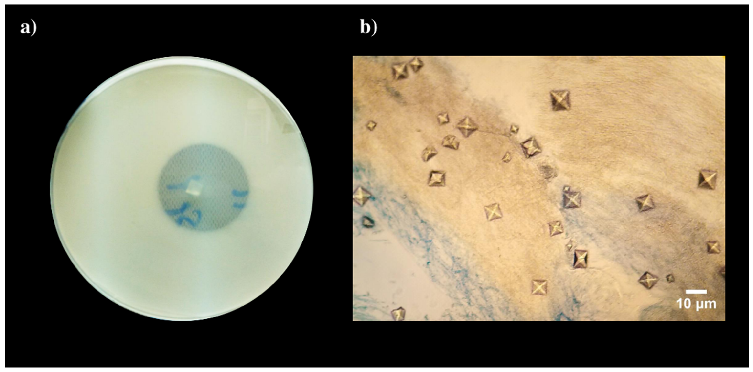
Figure 1.
Examples of fungal species biodeteriogenic abilities detected through plate assays: (a) Calcium carbonate dissolution visualized by the development of a halo around colonies in CaCO3 glucose agar; and (b) calcium oxalates crystals developing around fungal mycelium growing in Malt extract agar containing CaCO3.


Table 1.
Overview of fungal species for which isolates have been identified as having CaCO3 dissolution abilities in biodeteriorative plate essays.
Table 1.
Overview of fungal species for which isolates have been identified as having CaCO3 dissolution abilities in biodeteriorative plate essays.
| Current Species Name | Original Study Focus | References |
|---|---|---|
| Acremonium charticola (Lindau) W. Gams | Limestone and plaster monuments and museums | [39] |
| Actinomucor elegans (Eidam) C.R. Benjamin and Hesseltine | Mural paintings | [41] |
| Alternaria alternata (Fr.) Keissl. | Mural paintings | [41] |
| Annulohypoxylon stygium (Lév.) Y.M. Ju, J.D. Rogers and H.M. Hsie | Mayan buildings | [35] |
| Aspergillus amstelodami (L. Mangin) Thom and Church | Wooden art objects | [36] |
| Aspergillus awamori Nakaz. | Wooden art objects | [36] |
| Aspergillus europaeus Hubka, A. Nováková, Samson, Houbraken, Frisvad and M. Kolařík | Frescoes and air | [38] |
| Aspergillus glaucus (L.) Link | Limestone tomb | [43] |
| Aspergillus nidulans (Eidam) G. Winter | Mural paintings | [41] |
| Aspergillus niger Tiegh | Wooden art objects, frescoes, air, brick and concrete | [28,36,38,40] |
| Aspergillus versicolor (Vuill.) Tirab | Wooden art objects, mural paintings, limestone, plaster monuments and museums | [36,39,41] |
| Aspergillus westerdijkiae Frisvad and Samson | Limestone tomb | [43] |
| Botrytis cinerea Pers. | Limestone tomb | [43] |
| Cephalotrichum Link 1 | Catacombs | [33] |
| Cladosporium Link | Etruscan hypogeal tombs | [44] |
| Cladosporium sphaerospermum Penz. | Mural paintings | [41] |
| Cyphellophora G.A. de Vries | Etruscan hypogeal tombs | [44] |
| Cyphellophora olivacea (W. Gams) Réblová & Unter. | Etruscan hypogeal tombs | [44] |
| Exophiala J.W. Carmich. | Etruscan hypogeal tombs | [44] |
| Kendrickiella phycomyces (Auersw.) K. Jacobs and M.J. Wingf. | Mural paintings in Tumuli | [37] |
| Lasiodiplodia theobromae (Pat.) Griffon and Maubl. | Mayan buildings | [35] |
| Lecanicillium W. Gams and Zare | Limestone and plaster monuments and museums | [39] |
| Paecilomyces Bainier | Mayan buildings and catacombs | [33,35] |
| Parengyodontium album (Limber) C.C. Tsang, J.F.W. Chan, W.M. Pong, J.H.K. Chen, A.H.Y. Ngan, M. Cheung, C.K.C. Lai, D.N.C. Tsang, S.K.P. Lau and P.C.Y. Woo | Dolomitic limestone wall | [42] |
| Penicillium angulare S.W. Peterson, E.M. Bayer and Wicklow | Dolomitic limestone wall | [42] |
| Penicillium aurantiogriseum Dierckx | Mural paintings | [41] |
| Penicillium bilaiae Chalab. | Frescoes and air | [38] |
| Penicillium brevicompactum Dierckx | Air, limestone tomb and dolomitic limestone wall | [28,42,43] |
| Penicillium chrysogenum Thom | Wooden art objects, air, mural paintings, limestone tomb, dolomitic limestone wall, limestone and plaster monuments and museums | [28,36,39,41,42,43] |
| Penicillium commune Thom | Frescoes, air and mural paintings | [38,40,41] |
| Penicillium crustosum Thom | Dolomitic limestone wall | [42] |
| Penicillium glabrum (Wehmer) Westling | Wooden art objects, air, dolomitic limestone wall and limestone tomb | [28,36,42] |
| Penicillium griseofulvum Dierckx | Frescoes and air | [38] |
| Penicillium lanosum Westling | Frescoes and air | [38] |
| Penicillium Link | Wooden art objects, air, frescoes, concrete and bricks | [36,40] |
| Penicillium oxalicum Currie and Thom | Mayan buildings | [35] |
| Penicillium polonicum K.W. Zaleski | Mural paintings | [41] |
| Penicillium rubens Biourge | Frescoes and air | [38] |
| Penicillium scabrosum Frisvad, Samson and Stolk | Dolomitic limestone wall | [42] |
| Penicillium solitum Westling | Air | [28] |
| Penicillium viridicatum Westling | Air | [28] |
| Periconia byssoides Pers. | Dolomitic limestone wall | [42] |
| Pestalotiopsis maculans (Corda) Nag Raj | Mayan buildings | [35] |
| Pestalotiopsis microspora (Speg.) G.C. Zhao and Nan Li | Mayan buildings | [35] |
| Pseudogymnoascus pannorum (Link) Minnis and D.L. Lindner | Limestone and plaster monuments and museums | [39] |
| Rosellinia De Not. | Mayan buildings | [35] |
| Sclerotinia Fuckel | Air and frescoes | [36] |
| Sclerotinia sclerotiorum (Lib.) de Bary | Air and frescoes | [36] |
| Talaromyces amestolkiae N. Yilmaz, Houbraken, Frisvad and Samson | Dolomitic limestone wall | [42] |
| Talaromyces sayulitensis Visagie, N. Yilmaz, Seifert and Samson | Air | [28] |
| Trichocladium canadense S. Hughe | Mayan buildings | [35] |
| Trichoderma Pers. | Mayan buildings | [35] |
| Valsaria spartii Maubl. | Dolomitic limestone wall | [42] |
| Xylaria Hill ex Schrank 2 | Mayan buildings | [35] |
Previously identified as: 1 Doratomyces sp.; 2 Hypoxylon sp.
Table 2.
Overview of fungal species for which isolates have been identified as displaying mineralization abilities in biodeteriorative plate essays.
Table 2.
Overview of fungal species for which isolates have been identified as displaying mineralization abilities in biodeteriorative plate essays.
| Current Species Name | Mineral Details | Original Study Focus | References |
|---|---|---|---|
| Actinomucor elegans (Eidam) C.R. Benjamin and Hesseltine | Cc, Wd | Mural paintings | [41] |
| Aeminium ludgeri J. Trovão, I. Tiago and A. Portugal | Cc | Dolomitic limestone wall | [42] |
| Alternaria alternata (Fr.) Keissl. | Unk | Air and wall paintings | [56] |
| Alternaria infectoria E.G. Simmons | Unk | Air and wall paintings | [56] |
| Annulohypoxylon stygium (Lév.) Y.M. Ju, J.D. Rogers and H.M. Hsie | Cc, Wd | Mayan buildings | [35] |
| Ascochyta medicaginicola Qian Chen and L. Cai 1 | Unk | Air and wall paintings | [56] |
| Aspergillus aureolatus Munt.—Cvetk. and Bata | Cc | Air and wall paintings | [56] |
| Aspergillus europaeus Hubka, A. Nováková, Samson, Houbraken, Frisvad and M. Kolařík | Unk | Air and wall paintings | [56] |
| Aspergillus flavipes (Bainier and R. Sartory) Thom and Church | Unk | Air and wall paintings | [56] |
| Aspergillus flavus Link | Cc, Wd | Air and wall paintings | [56] |
| Aspergillus glaucus (L.) Link | Unk CO | Limestone tomb | [54] |
| Aspergillus niger Tiegh | Unk, Wh | Limestone monument, air and wall paintings | [53,56] |
| Aspergillus ostianus Wehmer | Cc, Wd | Air and wall paintings | [56] |
| Aspergillus pallidofulvus Visagie, Varga, Frisvad and Samson | Cc | Air and wall paintings | [56] |
| Aspergillus parasiticus Speare | Cc, Wd | Air and wall paintings | [56] |
| Aspergillus westerdijkiae Frisvad and Samson | Unk CO | Limestone tomb | [54] |
| Bionectria ochroleuca (Schwein.) Schroers and Samuels | Cc, Wh | Stone stela | [54] |
| Botryotrichum murorum (Corda) X. Wei Wang and Samson 2 | Cc, Unk | Air, wall paintings and stone stela | [54,56] |
| Botrytis cinerea Pers. | Unk CO | Limestone tomb | [54] |
| Chaetomium ancistrocladum Udagawa and Cain | Unk | Air and wall paintings | [56] |
| Cladosporium cladosporioides (Fresen.) G.A. de Vries | Unk | Air and wall paintings | [56] |
| Cladosporium oxysporum Berk. and M.A. Curtis | Cc, Wd | Air and wall paintings | [56] |
| Cladosporium sphaerospermum Penz. | Cc, Wd | Mural paintings | [41] |
| Cladosporium uredinicola Speg. | Cc | Air and wall paintings | [56] |
| Colletotrichum acutatum J.H. Simmonds | Cc (Vaterite), Cc, Hap, Phosp | Limestone monument | [53,55] |
| Colletotrichum gloeosporioides (Penz.) Penz. and Sacc. | Cc (Vaterite) | Limestone monument | [53] |
| Epicoccum nigrum Link | Cc, Wd | Air and wall paintings | [56] |
| Fusarium fujikuroi Nirenberg 3 | Cc | Air and wall paintings | [56] |
| Fusarium proliferatum (Matsush.) Nirenberg ex Gerlach and Nirenberg | Cc | Stone stela | [54] |
| Lasiodiplodia theobromae (Pat.) Griffon and Maubl. | Unk | Mayan buildings | [35] |
| Leptosphaeria avenaria G.F. Weber 4 | Cc | Air and wall paintings | [56] |
| Mucor fragilis Bainier | Cc, Wd | Dolomitic limestone wall | [42] |
| Paecilomyces Bainier | Unk | Mayan buildings | [35] |
| Penicillium angulare S.W. Peterson, E.M. Bayer and Wicklow | Cc, Wd, Wh | Dolomitic limestone wall | [42] |
| Penicillium atrosanguineum B.X. Dong 5 | Cc, Wd | Air and wall paintings | [56] |
| Penicillium bilaiae Chalab. | Cc, Wd | Air and wall paintings | [56] |
| Penicillium brevicompactum Dierckx | Cc, Wd | Dolomitic limestone wall | [42] |
| Penicillium chrysogenum Thom | Cc, Wd, Unk CO | Limestone tomb and mural paintings | [41,43] |
| Penicillium commune Thom | Cc, Wd | Air, wall and mural paintings | [41,56] |
| Penicillium crustosum Thom | Cc, Wd | Stone stela | [54] |
| Penicillium glabrum (Wehmer) Westling | Cc, Wd, Wh | Dolomitic limestone wall | [42] |
| Penicillium griseofulvum Dierckx | Unk | Air and wall paintings | [56] |
| Penicillium lanosum Westling | Cc, Wd, Unk | Air and wall paintings | [56] |
| Penicillium oxalicum Currie and Thom | Cc, Wd, Wh | Mayan buildings and limestone monument | [35,53] |
| Penicillium polonicum K.W. Zaleski | Cc, Wd | Mural paintings | [41] |
| Penicillium rubens Biourge | Unk | Air and wall paintings | [56] |
| Periconia byssoides Pers. | Cc | Dolomitic limestone wall | [42] |
| Pestalotiopsis maculans (Corda) Nag Raj | Unk | Mayan buildings | [35] |
| Pestalotiopsis microspora (Speg.) G.C. Zhao and Nan Li | Cc, Wd, Wh | Mayan buildings | [35] |
| Phialemonium inflatum (Burnside) Dania García, Perdomo, Gené, Cano and Guarro 6 | Cc, Cc (Vaterite), Hyd, Pyr | Stalactite in building | [59] |
| Plectosphaerella cucumerina (Lindf.) W. Gams | Cc, Hyd, Pyr | Stalactite in building | [59] |
| Rhizoctonia solani J.G. Kühn 7 | Unk | Air and wall paintings | [56] |
| Rosellinia De Not. | Cc, Wd | Mayan buildings | [35] |
| Sclerotinia sclerotiorum (Lib.) de Bary | Cc, Wd | Air and wall paintings | [56] |
| Stereum hirsutum (Willd.) Pers. | Cc, Wd | Dolomitic limestone wall | [42] |
| Trichocladium canadense S. Hughes | Unk | Mayan buildings | [35] |
| Trichoderma atroviride P. Karst. | Cc, Wd | Dolomitic limestone wall | [42] |
| Trichoderma harzianum Rifai | Cc | Stone stela | [54] |
| Trichoderma Pers. | Unk | Mayan buildings | [35] |
| Xylaria Hill ex Schrank 8 | Unk | Mayan buildings | [35] |
Previously identified as: 1 Phoma medicaginis; 2 Chaetomium murorum; 3 Gibberella moniliformis; 4 Phaeosphaeria avenaria; 5 Penicillium manginii; 6 Paecilomyces inflatus; 7 Thanatephorus cucumeris; 8 Hypoxylon sp.; Cc—calcium carbonate; Wd—weddellite; Wh—whewellite; Hap—hydroxyapatite; Hyd—hydrocerussite; Phosp—phosphate; Pyr—pyromorphite; Unk CO—Unidentified calcium oxalates; Unk—Unidentified mineralization’s.
Table 3.
Overview of fungal species for which isolates have been identified as displaying ligninolytic abilities in biodeteriorative plate essays.
Table 3.
Overview of fungal species for which isolates have been identified as displaying ligninolytic abilities in biodeteriorative plate essays.
| Current Species Name | Original Study Focus | References |
|---|---|---|
| Alternaria consortialis (Thüm.) J.W. Groves and S. Hughes 1 | Wooden art objects | [36] |
| Arthrinium phaeospermum (Corda) M.B. Ellis | Wooden art objects | [36] |
| Aspergillus amstelodami (L. Mangin) Thom and Church 2 | Wooden art objects | [36] |
| Aspergillus awamori Nakaz. | Wooden art objects | [36] |
| Aspergillus fischeri Wehmer 3 | Wooden art objects | [36] |
| Aspergillus flavus Link | Wooden art objects | [36] |
| Aspergillus fumigatus Fresen. | Wooden art objects | [36] |
| Aspergillus niger Tiegh | Wooden art objects | [36] |
| Aspergillus terreus Thom | Wooden art objects and air | [36,69] |
| Aspergillus ustus (Bainier) Thom and Church | Wooden art objects | [36] |
| Aspergillus versicolor (Vuill.) Tirab. | Wooden art objects | [36] |
| Beauveria bassiana (Bals.—Criv.) Vuill. | Wooden art objects | [36] |
| Chaetomium elatum Kunze | Wooden art objects | [36] |
| Chaetomium globosum Kunze | Wooden art objects and air | [36,69] |
| Cladosporium cladosporioides (Fresen.) G.A. de Vries | Wooden objects and air | [69] |
| Cladosporium herbarum (Pers.) Link | Wooden art objects | [36] |
| Cladosporium Link | Wooden art objects | [36] |
| Hypochnicium J. Erikss. | Wooden materials | [61] |
| “Neocosmospora” solani (Mart.) L. Lombard and Crous 4 | Wooden materials | [70] |
| Penicillium chrysogenum Thom | Wooden art objects | [36] |
| Penicillium expansum Link | Wooden art objects | [36] |
| Penicillium glabrum (Wehmer) Westling | Wooden art objects | [36] |
| Penicillium herquei Bainier and Sartory | Wooden art objects | [36] |
| Penicillium Link | Wooden art objects | [36] |
| Penicillium sacculum E. Dale | Wooden art objects | [36] |
| Pseudogymnoascus pannorum (Link) Minnis and D.L. Lindner 5 | Wooden art objects | [36] |
| Trichoderma viride Pers. | Wooden art objects | [36] |
Previously identified as: 1 Alternaria consortiale; 2 Eurotium amstelodami; 3 Neosartorya fischeri; 4 Fusarium solani; 5 Chrysosporium pannorum.
Table 4.
Overview of fungal species for which isolates have been identified as displaying fibrinolytic abilities in biodeteriorative plate essays.
Table 4.
Overview of fungal species for which isolates have been identified as displaying fibrinolytic abilities in biodeteriorative plate essays.
| Current Species Name | Original Study Focus | References |
|---|---|---|
| Alternaria alternata (Fr.) Keissl. | Funeral accessories | [70] |
| Ascomycota Caval. -Sm. | Funeral clothes | [85] |
| Aspergillus caninus (Sigler, Deanna A. Sutton, Gibas, Summerb. and Iwen) Houbraken, Tanney, Visagie and Samson 1 | Funeral clothes | [85] |
| Aspergillus cristatus Raper and Fennell 2 | Funeral clothes | [85] |
| Aspergillus fumigatus Fresen. | Funeral clothes | [85] |
| Aspergillus P. Micheli ex Haller | Funeral accessories | [80] |
| Aspergillus puniceus Kwon-Chung and Fennell | Funeral clothes | [85] |
| Aspergillus sydowii (Bainier and Sartory) Thom and Church | Funeral clothes | [85] |
| Aspergillus tubingensis Mosseray | Funeral clothes | [85] |
| Aspergillus versicolor (Vuill.) Tirab. | Funeral accessories | [80] |
| Beauveria bassiana (Bals.—Criv.) Vuill. | Funeral clothes | [85] |
| Myriodontium keratinophilum Samson and Polon. | Funeral clothes | [85] |
| Penicillium brevicompactum Dierckx | Funeral clothes | [85] |
| Penicillium commune Thom | Funeral accessories | [80] |
| Penicillium crocicola T. Yamam. | Funeral clothes | [85] |
| Penicillium crustosum Thom | Funeral clothes | [85] |
| Penicillium expansum Link | Funeral clothes | [85] |
| Penicillium granulatum Bainier | Funeral accessories | [80] |
| Penicillium Link | Funeral accessories | [80] |
| Penicillium roseopurpureum Dierckx | Funeral clothes | [85] |
| Penicillium spinulosum Thom | Funeral clothes | [85] |
| Sporobolomyces roseus Kluyver and C.B. Niel 3 | Funeral accessories | [80] |
| Xenochalara juniperi M.J. Wingf. and Crous | Funeral clothes | [85] |
Previously identified as: 1 Phialosimplex caninus; 2 Eurotium cristatum; 3 Sporidiobolus metaroseus.
Table 5.
Overview of fungal species for which isolates have been identified as displaying keratinolytic abilities in biodeteriorative plate essays.
Table 5.
Overview of fungal species for which isolates have been identified as displaying keratinolytic abilities in biodeteriorative plate essays.
| Current Species Name | Original Study Focus | References |
|---|---|---|
| Alternaria consortialis (Thüm.) J.W. Groves and S. Hughes 1 | Wooden art objects | [36] |
| Arthrinium phaeospermum (Corda) M.B. Ellis | Wooden art objects | [36] |
| Aspergillus amstelodami (L. Mangin) Thom and Church 2 | Wooden art objects | [36] |
| Aspergillus awamori Nakaz. | Wooden art objects | [36] |
| Aspergillus fischeri Wehmer 3 | Wooden art objects | [36] |
| Aspergillus flavus Link | Wooden art objects | [36] |
| Aspergillus fumigatus Fresen. | Wooden art objects | [36] |
| Aspergillus niger Tiegh | Wooden art objects | [36] |
| Aspergillus terreus Thom | Wooden art objects and air | [36,69] |
| Aspergillus ustus (Bainier) Thom and Church | Wooden art objects | [36] |
| Aspergillus versicolor (Vuill.) Tirab. | Wooden art objects | [36] |
| Beauveria bassiana (Bals.—Criv.) Vuill. | Wooden art objects | [36] |
| Chaetomium elatum Kunze | Wooden art objects | [36] |
| Chaetomium globosum Kunze | Wooden art objects and air | [36,69] |
| Cladosporium cladosporioides (Fresen.) G.A. de Vries | Wooden objects and air | [69] |
| Cladosporium herbarum (Pers.) Link | Wooden art objects | [36] |
| Cladosporium Link | Wooden art objects | [36] |
| Hypochnicium J. Erikss. | Wooden materials | [61] |
| “Neocosmospora” solani (Mart.) L. Lombard and Crous 4 | Wooden materials | [70] |
| Penicillium chrysogenum Thom | Wooden art objects | [36] |
| Penicillium expansum Link | Wooden art objects | [36] |
| Penicillium glabrum (Wehmer) Westling | Wooden art objects | [36] |
| Penicillium herquei Bainier and Sartory | Wooden art objects | [36] |
| Penicillium Link | Wooden art objects | [36] |
| Penicillium sacculum E. Dale | Wooden art objects | [36] |
| Pseudogymnoascus pannorum (Link) Minnis and D.L. Lindner 5 | Wooden art objects | [36] |
| Trichoderma viride Pers. | Wooden art objects | [36] |
Previously identified as: 1 Alternaria consortiale; 2 Eurotium amstelodami; 3 Neosartorya fischeri; 4 Fusarium solani; 5 Chrysosporium pannorum.
Table 6.
Overview of fungal species for which isolates have been identified as displaying estereolytic abilities in biodeteriorative plate essays.
Table 6.
Overview of fungal species for which isolates have been identified as displaying estereolytic abilities in biodeteriorative plate essays.
| Current Species Name | Original Study Focus | References |
|---|---|---|
| Acrodontium salmoneum de Hoog | Wax seal | [84] |
| Agaricaceae Chevall. | Statue and air | [89] |
| Alternaria Nees | Statue, stone and air | [89,90] |
| Alternaria tenuissima (Kunze) Wiltshire | Statue and air | [89] |
| Aspergillus amstelodami (L. Mangin) Thom and Church 1 | Stone | [90] |
| Aspergillus caespitosus Raper and Thom | Air, textiles and human remains | [83] |
| Aspergillus calidoustus Varga, Houbraken and Samson | Air, textiles and human remains | [83] |
| Aspergillus candidus Link | Air, textiles and human remains | [83] |
| Aspergillus clavatus Desm. | Textiles | [82] |
| Aspergillus fischeri Wehmer 2 | Air, textiles and human remains | [83] |
| Aspergillus flavus Link | Stone | [90] |
| Aspergillus fumigatus Fresen. | Air, statues, textiles and human remains | [83,89] |
| Aspergillus niger Tiegh | Stone | [90] |
| Aspergillus repens (Corda) Sacc. 3 | Air, textiles and human remains | [83] |
| Aspergillus sydowii (Bainier and Sartory) Thom and Church | Air, textiles and human remains | [83] |
| Aspergillus terreus Thom | Air, textiles and human remains | [83] |
| Aspergillus ustus (Bainier) Thom and Church | Air, textiles and human remains | [83] |
| Aspergillus venenatus Jurjević, S.W. Peterson and B.W. Horn | Air, textiles and human remains | [83] |
| Aspergillus versicolor (Vuill.) Tirab. | Air, statues, textiles and human remains | [83,89] |
| Aspergillus westerdijkiae Frisvad and Samson | Air, textiles and human remains | [83] |
| Aureobasidium pullulans (de Bary and Löwenthal) G. Arnaud | Wax seal | [84] |
| Bulleromyces albus Boekhout and Á. Fonseca 4 | Wax seal | [84] |
| Cladosporium aggregatocicatricatum Bensch, Crous and U. Braun | Wax seal | [84] |
| Cladosporium Link | Air, textiles and human remains | [83] |
| Cladosporium macrocarpum Preuss | Statue and air | [89] |
| Cladosporium tenuissimum Cooke | Textiles | [82] |
| Coprinellus xanthothrix (Romagn.) Vilgalys, Hopple and Jacq. Johnson | Air, textiles and human remains | [83] |
| Coprinopsis cinerea (Schaeff.) Redhead, Vilgalys and Moncalvo | Statue and air | [89] |
| Curvularia Boedijn | Stone | [90] |
| Fusarium Link | Stone | [90] |
| Fusarium sporotrichioides Sherb. | Statue and air | [89] |
| Hypoxylon fragiforme (Pers.) J. Kickx f. | Textiles | [82] |
| Microascus brevicaulis S.P. Abbott 5 | Air, textiles and human remains | [83] |
| Nigrospora oryzae (Berk. and Broome) Petch | Air, textiles and human remains | [83] |
| Penicillium chrysogenum Thom | Air, textiles and human remains | [82,83] |
| Penicillium commune Thom | Air, textiles and human remains | [83] |
| Penicillium corylophilum Dierckx | Textiles, statue and air | [82,89] |
| Penicillium crustosum Thom | Air, statues, textiles and human remains | [83,89] |
| Penicillium griseofulvum Dierckx | Air, textiles and human remains | [83] |
| Penicillium hordei Stolk | Air, textiles and human remains | [83] |
| Penicillium Link | Statue and air | [89] |
| Penicillium polonicum K.W. Zaleski | Air, textiles and human remains | [83] |
| Pseudoscopulariopsis hibernica (A. Mangan) Sand. -Den., Gené and Cano 6 | Air, textiles and human remains | [83] |
| Rhizopus arrhizus A. Fisch. 7 | Air, textiles and human remains | [83] |
| Rhizopus microsporus Tiegh. | Air, textils, human remains, statue and air | [83,89] |
| Rhizopus stolonifer (Ehrenb.) Vuill. | Air, textiles and human remains | [83] |
| Sordaria fimicola (Roberge ex Desm.) Ces. and De Not. | Statue and air | [89] |
| Talaromyces purpureogenus Samson, N. Yilmaz, Houbraken, Spierenb., Seifert, Peterson, Varga and Frisvad | Stone | [90] |
| Thyronectria austroamericana (Speg.) Seeler 8 | Statue and air | [89] |
| Trichoderma paraviridescens Jaklitsch, Samuels and Voglmayr | Statue and air | [89] |
Previously identified as: 1 Eurotium amsteldomi; 2 Neosartorya fischeri; 3 Eurotium repens; 4 Bullera alba; 5 Scopulariopsis brevicaulis; 6 Scopulariopsis hibernica; 7 Rhizopus oryzae; 8 Pleonectria austroamericana.
Table 7.
Overview of fungal species for which isolates have been identified as displaying lipolytic abilities in biodeteriorative plate essays.
Table 7.
Overview of fungal species for which isolates have been identified as displaying lipolytic abilities in biodeteriorative plate essays.
| Current Species Name | Original Study Focus | References |
|---|---|---|
| Acrodontium salmoneum de Hoog | Wax seal | [84] |
| Acrostalagmus luteoalbus (Link) Zare, W. Gams and Schroers | Albumen photographic materials | [93] |
| Agaricaceae Chevall. | Statue and air | [89] |
| Alternaria mali Roberts | Wooden organ and pipes | [94] |
| Alternaria Nees | Statue and air | [89] |
| Alternaria tenuissima (Kunze) Wiltshire | Statue and air | [89] |
| Aspergillus amstelodami (L. Mangin) Thom and Church 1 | Air, textiles, human remains, wooden organ and pipes | [83,94] |
| Aspergillus caespitosus Raper and Thom | Air, textiles and human remains | [83] |
| Aspergillus calidoustus Varga, Houbraken and Samson | Air, textiles and human remains | [83] |
| Aspergillus candidus Link | Air, textiles and human remains | [83] |
| Aspergillus cristatus Raper and Fennell 2 | Wooden organ and pipes | [94] |
| Aspergillus fischeri Wehmer 3 | Air, textiles, statues and human remains | [89,93] |
| Aspergillus fumigatus Fresen. | Air, textiles, statues and human remains | [83,89] |
| Aspergillus repens (Corda) Sacc. 4 | Air, textiles and human remains | [83] |
| Aspergillus sydowii (Bainier and Sartory) Thom and Church | Air, textiles, human remains, wooden organ and pipes | [83,94] |
| Aspergillus terreus Thom | Air, textiles and human remains | [83] |
| Aspergillus ustus (Bainier) Thom and Church | Air, textiles and human remains | [83] |
| Aspergillus venenatus Jurjević, S.W. Peterson and B.W. Horn | Air, textiles and human remains | [83] |
| Aspergillus versicolor (Vuill.) Tirab | Air, textiles, human remains, albumen photographic materials, wooden organ and pipes | [83,89,93,94] |
| Aspergillus westerdijkiae Frisvad and Samson | Air, textiles and human remains | [83] |
| Aureobasidium pullulans (de Bary and Löwenthal) G. Arnaud | Wax seal | [84] |
| Bjerkandera adusta (Willd.) P. Karst. | Albumen photographic materials | [83] |
| Bulleromyces albus Boekhout and Á. Fonseca 5 | Wax seal | [84] |
| Chaetomium elatum Kunze | Albumen photographic materials | [83] |
| Cladosporium aggregatocicatricatum Bensch, Crous and U. Braun | Wax seal | [84] |
| Cladosporium cladosporioides (Fresen.) G.A. de Vries | Wooden organ and pipes | [94] |
| Cladosporium Link | Air, textiles, human remains and Etruscan hypogeal tombs | [44,83] |
| Cladosporium macrocarpum Preuss | Statue and air | [89] |
| Coprinellus xanthothrix (Romagn.) Vilgalys, Hopple and Jacq. Johnson | Air, textiles and human remains | [83] |
| Coprinopsis cinerea (Schaeff.) Redhead, Vilgalys and Moncalvo | Statue and air | [89] |
| Cyphellophora G.A. de Vries | Etruscan hypogeal tombs | [44] |
| Cyphellophora olivacea (W. Gams) Réblová & Unter. | Etruscan hypogeal tombs | [44] |
| Epicoccum nigrum Link | Wooden organ and pipes | [94] |
| Exophiala angulospora Iwatsu, Udagawa & T. Takase | Etruscan hypogeal tombs | [44] |
| Exophiala J.W. Carmich. | Etruscan hypogeal tombs | [44] |
| Fusarium sporotrichioides Sherb. | Statue and air | [89] |
| Microascus brevicaulis S.P. Abbott 6 | Air, textiles and human remains | [83] |
| Mucor plumbeus Bonord. | Albumen photographic materials | [93] |
| Nigrospora oryzae (Berk. and Broome) Petch | Air, textiles and human remains | [83] |
| Paecilomyces maximus C. Ram | Wooden organ and pipes | [94] |
| Penicillium chrysogenum Thom | Air, textiles and human remains | [83] |
| Penicillium commune Thom | Air, textiles and human remains | [83] |
| Penicillium corylophilum Dierckx | Statue and air | [89] |
| Penicillium crustosum Thom | Air, textiles, statues and human remains | [83,89] |
| Penicillium digitatum (Pers.) Sacc. | Air, textiles and human remains | [83] |
| Penicillium griseofulvum Dierckx | Air, textiles and human remains | [83] |
| Penicillium hordei Stolk | Air, textiles and human remains | [83] |
| Penicillium Link | Albumen photographic materials, statues and air | [89,93] |
| Penicillium polonicum K.W. Zaleski | Air, textiles and human remains | [83] |
| Penicillium thomii Maire | Albumen photographic materials | [93] |
| Phlebia Fr. | Albumen photographic materials | [93] |
| Pleosporales Luttr. ex M.E. Barr | Albumen photographic materials | [93] |
| Pseudoscopulariopsis hibernica (A. Mangan) Sand. -Den., Gené and Cano 7 | Air, textiles and human remains | [83] |
| Rhizopus arrhizus A. Fisch. 8 | Air, textiles and human remains | [83] |
| Rhizopus microsporus Tiegh. | Air, textiles, human remains, statues and air | [83,89] |
| Rhizopus stolonifer (Ehrenb.) Vuill. | Air, textiles and human remains | [83] |
| Sordaria fimicola (Roberge ex Desm.) Ces. and De Not. | Statue and air | [89] |
| Talaromyces flavus (Klöcker) Stolk and Samson | Air, textiles and human remains | [83] |
| Thyronectria austroamericana (Speg.) Seeler 9 | Statue and air | [89] |
| Trichoderma harzianum Rifai | Air, textiles and human remains | [83] |
| Trichoderma paraviridescens Jaklitsch, Samuels and Voglmayr | Statue and air | [89] |
| Trichothecium roseum (Pers.) Link | Albumen photographic materials | [93] |
Previously identified as: 1 Eurotium amstelodami; 2 Eurotium cristatum; 3 Neosartorya fischeri; 4 Eurotium repens; 5 Bullera alba; 6 Scopulariopsis brevicaulis; 7 Scopulariopsis hibernica; 8 Rhizopus oryzae; 9 Pleonectria austroamericana.
Table 8.
Overview of fungal species for which isolates have been identified as displaying proteolytic abilities in biodeteriorative plate essays.
Table 8.
Overview of fungal species for which isolates have been identified as displaying proteolytic abilities in biodeteriorative plate essays.
| Current Species Name | Original Study Focus | References |
|---|---|---|
| Acrodontium salmoneum de Hoog | Wax seal | [84] |
| Acrostalagmus luteoalbus (Link) Zare, W. Gams and Schroers | Albumen photographic materials | [93] |
| Actinomucor elegans (Eidam) C.R. Benjamin and Hesseltine | Mural paintings | [41] |
| Alternaria alternata (Fr.) Keissl. | Air, mural paintings, mummies, funeral accessories, cinematographic films | [41,79,80,95,98] |
| Alternaria mali Roberts | Mummies, wooden organ and pipes | [79,94] |
| Alternaria Nees | Mummies and funeral clothes | [79,85] |
| Alternaria solani Sorauer | Mummies | [79] |
| Alternaria tenuissima (Kunze) Wiltshire | Mummies | [79] |
| Ascomycota Caval. -Sm. | Funeral clothes | [85] |
| Aspergillus alliaceus Thom and Church | Air and graphic documents | [98,100] |
| Aspergillus amstelodami (L. Mangin) Thom and Church | Air, wooden organ and pipes | [28,94] |
| Aspergillus aureolatus Munt.—Cvetk. and Bata | Frescoes and air | [38] |
| Aspergillus auricomus (Guég.) Saito | Air and graphic documents | [100] |
| Aspergillus caespitosus Raper and Thom | Air, textiles and human remains | [83] |
| Aspergillus candidus Link | Air, graphic documents, photographs, maps, textiles and human remains | [83,84,98,99,100] |
| Aspergillus caninus (Sigler, Deanna A. Sutton, Gibas, Summerb. and Iwen) Houbraken, Tanney, Visagie and Samson 1 | Funeral clothes | [85] |
| Aspergillus chevalieri (L. Mangin) Thom and Church 2 | Air, graphic documents, photographs, maps and materials present in libraries and museums | [96,99,100] |
| Aspergillus creber Jurjević, S.W. Peterson and B.W. Horn | Air | [28] |
| Aspergillus cristatus Raper and Fennell 3 | Funeral clothes, wooden organ and pipes | [85,94] |
| Aspergillus europaeus Hubka, A. Nováková, Samson, Houbraken, Frisvad and M. Kolařík | Frescoes and air | [28,38] |
| Aspergillus flavipes (Bainier and R. Sartory) Thom and Church | Air | [98] |
| Aspergillus flavus Link 4 | Air, frescoes, mummies, photographs, maps and materials present in libraries and museums | [38,47,78,79,96,99,100] |
| Aspergillus fumigatus Fresen. | Air, materials present in libraries and museums, paper and parchment and funeral clothes | [85,96,97] |
| Aspergillus japonicus Saito | Air and graphic documents | [100] |
| Aspergillus jensenii Jurjević, S.W. Peterson and B.W. Horn | Air | [28] |
| Aspergillus nidulans (Eidam) G. Winter | Air, materials present in libraries and museum, mural paintings and cinematographic films | [41,95,96] |
| Aspergillus niger Tiegh | Frescoes, air, textiles, human remains, photographs, maps, paper and parchment, graphic documents, materials present in libraries and museums | [38,73,96,97,98,99,100] |
| Aspergillus ostianus Wehmer | Frescoes, air and graphic documents | [38,100] |
| Aspergillus P. Micheli ex Haller 5 | Air and mural paintings | [41,47] |
| Aspergillus penicillioides Speg. | Air and graphic documents | [100] |
| Aspergillus proliferans G. Sm. | Air | [28] |
| Aspergillus protuberus Munt. -Cvetk. | Air | [28] |
| Aspergillus pseudodeflectus Samson and Mouch. | Funeral clothes | [85] |
| Aspergillus puniceus Kwon-Chung and Fennell | Funeral clothes | [85] |
| Aspergillus sydowii (Bainier and Sartory) Thom and Church | Air, mural paintings, funeral clothes, wooden organ and pipes and materials present in libraries and museums | [28,41,85,94,96] |
| Aspergillus tabacinus Nakaz., Y. Takeda, Simo and A. Watan. | Air and materials present in libraries and museums | [28] |
| Aspergillus tamarii Kita | Air and materials present in libraries and museums | [96] |
| Aspergillus terreus Thom | Air and materials present in libraries and museums | [96] |
| Aspergillus tubingensis Mosseray | Funeral clothes | [85] |
| Aspergillus unguis (Émile-Weill and L. Gaudin) Thom and Raper | Air and graphic documents | [100] |
| Aspergillus unilateralis Thrower | Air and graphic documents | [100] |
| Aspergillus ustus (Bainier) Thom and Church | Cinematographic films | [95] |
| Aspergillus versicolor (Vuill.) Tirab | Air, mural paintings, frescoes, textiles, human remains, funeral accessories, cinematographic materials, wooden organ and pipes and materials present in libraries and museums | [28,38,41,78,80,83,94,95,96] |
| Aspergillus westerdijkiae Frisvad and Samson | Air, textiles and human remains | [83] |
| Aureobasidium pullulans (de Bary and Löwenthal) G. Arnaud | Wax seal and mummies | [79,84] |
| Beauveria bassiana (Bals.—Criv.) Vuill. | Funeral clothes | [85] |
| Bjerkandera adusta (Willd.) P. Karst. | Frescoes, air albumen photographic materials | [38,93] |
| Botryotrichum murorum (Corda) X. Wei Wang and Samson 6 | Frescoes and air | [38] |
| Bulleromyces albus Boekhout and Á. Fonseca 7 | Wax seal | [84] |
| Chaetomium ancistrocladum Udagawa and Cain | Frescoes and air | [38] |
| Chaetomium elatum Kunze | Albumen photographic materials | [93] |
| Chaetomium globosum Kunze | Mural paintings | [41] |
| Cladosporium aggregatocicatricatum Bensch, Crous and U. Braun | Wax seal | [84] |
| Cladosporium cladosporioides (Fresen.) | Mural paintings, frescoes, air, mummies, graphic documents, maps, photographs, cinematographic films, wooden organ and pipes | [38,41,79,94,95,99,100] |
| Cladosporium cucumerinum Ellis and Arthur | Mural paintings | [41] |
| Cladosporium herbarum (Pers.) Link 8 | Mummies and mural painitngs | [41,81] |
| Cladosporium Link | Etruscan hypogeal tombs, air and mummies | [44,47,79] |
| Cladosporium macrocarpum Preuss | Air and frescoes | [78] |
| Cladosporium oxysporum Berk. and M.A. Curtis | Frescoes and air | [38] |
| Cladosporium pseudocladosporioides Bensch, Crous and U. Braun | Mummies | [79] |
| Cladosporium sphaerospermum Penz. | Mummies and mural paintings | [41,79] |
| Cladosporium tenuissimum Cooke | Mural paintings | [41] |
| Cladosporium uredinicola Speg. | Mummies, frescoes and air | [38,79] |
| Coprinellus xanthothrix (Romagn.) Vilgalys, Hopple and Jacq. Johnson | Air, textiles and human remains | [83] |
| Curvularia australiensis (Bugnic. ex M.B. Ellis) Manamgoda, L. Cai and K.D. Hyde 9 | Air and graphic documents | [100] |
| Curvularia Boedijn | Air | [47] |
| Curvularia pallescens Boedijn | Air and graphic documents | [98,100] |
| Cyphellophora G.A. de Vries | Etruscan hypogeal tombs | [44] |
| Didymella glomerata (Corda) Qian Chen and L. Cai 10 | Cinematographic films | [95] |
| Dipodascus Lagerh. 11 | Air | [47] |
| Epicoccum Link | Air and graphic documents | [100] |
| Epicoccum nigrum Link | Mummies, wooden organ and pipes | [79,94] |
| Fusarium chlamydosporum Wollenw. and Reinking | Mural paintings | [41] |
| Fusarium Link | Air and graphic documents | [57,100] |
| Fusarium oxysporum Schltdl. | Air | [98] |
| Hormodendrum pyri W.H. English | Wax seal | [84] |
| Leptosphaeria avenaria G.F. Weber 12 | frescoes and air | [38] |
| Meyerozyma guilliermondii (Wick.) Kurtzman and M. Suzuki 13 | Air | [98] |
| Microascus brevicaulis S.P. Abbott 14 | Air, textiles and human remains | [83] |
| Mucor racemosus Bull. | Cinematographic films, paper and parchment | [95,97] |
| Mucor spinosus Schrank | Paper and parchment | [97] |
| Myriodontium keratinophilum Samson and Polon. | Funeral clothes | [85] |
| Nigrospora oryzae (Berk. and Broome) Petch 15 | Air, textiles and human remains | [83,98] |
| Paecilomyces maximus C. Ram 16 | Wooden organ and pipes | [94] |
| Paecilomyces variotii Bainier | Air | [98] |
| Penicillium aurantiogriseum Dierckx | Mural paintings | [41] |
| Penicillium bilaiae Chalab. | Frescoes and air | [38] |
| Penicillium brevicompactum Dierckx | Air and funeral clothes | [28,85] |
| Penicillium camemberti Thom | Paper and parchment | [97] |
| Penicillium canescens Sopp | Air and graphic documents | [100] |
| Penicillium chrysogenum Thom | Air, mural paintings, textiles, human remains, mummies, paper, photographs, maps, parchment, graphic documents and cinematographic films | [28,41,79,83,95,97,99,100] |
| Penicillium citreonigrum Dierckx | Air | [28] |
| Penicillium citrinum Thom | Air, photographs, maps and graphic documents | [28,99,100] |
| Penicillium commune Thom | Air, textiles, human remains, mural paintings, funeral accessories and clothes, paper and parchment | [41,80,83,85,87] |
| Penicillium crocicola T. Yamam. | Funeral clothes | [85] |
| Penicillium crustosum Thom | Funeral clothes, wooden organ and pipes | [85,94] |
| Penicillium decumbens Thom | Air, paper and parchment | [28,97] |
| Penicillium digitatum (Pers.) Sacc. | Air, textiles and human remains | [28,83] |
| Penicillium expansum Link | Air, mummies and funeral clothes | [28,79,85] |
| Penicillium glabrum (Wehmer) Westling | Air | [28] |
| Penicillium granulatum Bainier | Funeral accessories | [80] |
| Penicillium griseofulvum Dierckx | Air, frescoes, graphic documents, textiles and human remains | [38,83,100] |
| Penicillium hordei Stolk | Air, textiles and human remains | [83] |
| Penicillium janczewskii K.W. Zaleski | Air, maps, photographs and graphic documents | [99,100] |
| Penicillium Link | Air, frescoes, albumen photographic materials and funeral accessories | [57,78,80,93] |
| Penicillium olsonii Bainier and Sartory | Mural paintings | [41] |
| Penicillium polonicum K.W. Zaleski | Air, textiles, mural paintings and human remains | [41,83] |
| Penicillium raistrickii G. Sm. | Mummies | [79] |
| Penicillium roseopurpureum Dierckx | Mummies and funeral clothes | [79,85] |
| Penicillium simplicissimum (Oudem.) Thom 17 | Air, maps, photographs and graphic documents | [99,100] |
| Penicillium solitum Westling | Air | [28] |
| Penicillium spinulosum Thom | Funeral clothes | [85] |
| Penicillium viridicatum Westling | Air, paper and parchment | [28,97] |
| Penicillium waksmanii K.W. Zaleski | Air and graphic documents | [100] |
| Pestalotia De Not. | Air and graphic documents | [100] |
| Phoma herbarum Westend. | Paper and parchment | [97] |
| Pleurotus ostreatus (Jacq.) P. Kumm. 18 | Albumen photographic materials | [93] |
| Pseudozyma prolifica Bandoni | Mural paintings | [31] |
| Rhizopus microsporus Tiegh. 19 | Air, textiles and human remains | [83] |
| Sarocladium W. Gams and D. Hawksw. 20 | Air, graphic documents and albumen photographic materials | [93,98,100] |
| Sporobolomyces roseus Kluyver and C.B. Niel 21 | Funeral Accessories | [80] |
| Stemphylium vesicarium (Wallr.) E.G. Simmons | Mummies | [79] |
| Talaromyces aculeatus (Raper and Fennell) Samson, N. Yilmaz, Frisvad and Seifert | Mural paintings | [41] |
| Talaromyces rugulosus (Thom) Samson, N. Yilmaz, Frisvad and Seifert | Wooden organ and pipes | [94] |
| Talaromyces sayulitensis Visagie, N. Yilmaz, Seifert and Samson | Air | [28] |
| Trichoderma harzianum Rifai | Air, textiles and human remains | [83] |
| Trichoderma longibrachiatum Rifai | Cinematographic films | [95] |
| Trichoderma viride Pers. | Air | [98] |
| Trichothecium roseum (Pers.) Link | Albumen photographic materials | [93] |
| Xenochalara juniperi M.J. Wingf. and Crous | Funeral clothes | [85] |
Previously identified as: 1 Phialosimplex caninus; 2 Eurotium chevalieri; 3 Eurotium cristatum; 4 Aspergillus oryzae; 5 Emericella sp.; 6 Chaetomium murorum; 7 Bullera alba; 8 Cladosporium herbarium; 9 Bipolaris australiensis; 10 Phoma glomerata; 11 Geotrichum sp.; 12 Phaeosphaeria avenaria; 13 Candida guilliermondii; 14 Scopulariopsis brevicaulis; 15 Nigrospora sphaerica; 16 Paecilomyces formosus; 17 Penicillium janthinellum; 18 Pleurotus pulmonarius; 19 Rhizopus microsporus var. oligosporus; 20 Cephalosporium sp.; 21 Sporidiobolus metaroseus.
Table 9.
Overview of fungal species for which isolates have been identified as displaying cellulolytic abilities in biodeteriorative plate essays.
Table 9.
Overview of fungal species for which isolates have been identified as displaying cellulolytic abilities in biodeteriorative plate essays.
| Current Species Name | Original Study Focus | References |
|---|---|---|
| Acremonium alabamense Morgan-Jones | Air | [98] |
| Acrostalagmus luteoalbus (Link) Zare, W. Gams and Schroers | Albumen photographic materials | [93] |
| Alternaria alternata (Fr.) Keissl. | Air, mummies and funeral accessories | [79,80,98] |
| Alternaria chartarum Preuss 1 | Mummies | [81] |
| Alternaria consortialis (Thüm.) J.W. Groves and S. Hughes 2 | Wooden art objects | [36] |
| Alternaria mali Roberts | Mummies, wooden organ and pipes | [79,94] |
| Alternaria Nees | Air and graphic documents, stone and mummies | [47,79,90,91] |
| Alternaria solani Sorauer | Mummies | [79] |
| Alternaria tenuissima (Kunze) Wiltshire | Air and mummies | [79,102] |
| Arthrinium arundinis (Corda) Dyko and B. Sutton | Drawings and lithographs | [94] |
| Arthrinium phaeospermum (Corda) M.B. Ellis | Wooden art objects | [36] |
| Ascomycota Caval. -Sm. | Funeral clothes and oainting | [85,103] |
| Ascotricha Berk. | Painting | [103] |
| Ascotricha chartarum Berk. | Painting | [103] |
| Aspergillus alliaceus Thom and Church | Air and graphic documents | [98,100] |
| Aspergillus amstelodami (L. Mangin) Thom and Church 3 | Air, stone, wooden organ, pipes and objects | [28,36,69,90,94] |
| Aspergillus auricomus (Guég.) Saito | Air and graphic documents | [100] |
| Aspergillus caespitosus Raper and Thom | Air, textiles and human remains | [83] |
| Aspergillus calidoustus Varga, Houbraken and Samson | Air, textiles and human remains | [28,83] |
| Aspergillus candidus Link | Air, textiles, mummies, graphic documents, maps, photographs and human remains | [81,83,98,99,100] |
| Aspergillus chevalieri (L. Mangin) Thom and Church 4 | Air, photographs, maps and graphic documents | [99,100] |
| Aspergillus clavatus Desm. | Air and textiles | [82,102] |
| Aspergillus creber Jurjević, S.W. Peterson and B.W. Horn | Air | [28] |
| Aspergillus cristatus Raper and Fennell 5 | Funeral clothes, wooden organ and pipes | [85,94] |
| Aspergillus fischeri Wehmer 6 | Air, textiles and human remains | [83] |
| Aspergillus flavipes (Bainier and R. Sartory) Thom and Church | Air | [98,102] |
| Aspergillus flavus Link 7 | Air, stone, paintings, graphic documents, photographs, maps, paintings, library and museums | [28,47,90,96,98,99,100,102,104] |
| Aspergillus fumigatus Fresen. | Air, textiles, paper, parchment, funeral clothes, libraries, museums and human remains | [83,85,96,97] |
| Aspergillus japonicus Saito | Air and graphic documents | [100] |
| Aspergillus jensenii Jurjević, S.W. Peterson and B.W. Horn | Air | [28] |
| Aspergillus melleus Yukawa | Air | [28] |
| Aspergillus niger Tiegh | Air, stone, paintings, libraries, museums, graphic documents, photographs, maps, drawings and lithographs | [28,90,96,98,99,100,102,104,105] |
| Aspergillus ostianus Wehmer | Air and graphic documents | [100] |
| Aspergillus P. Micheli ex Haller | Air | [47] |
| Aspergillus penicillioides Speg. | Air and graphic documents | [100] |
| Aspergillus protuberus Munt. -Cvetk. | Air | [28] |
| Aspergillus pseudodeflectus Samson and Mouch. | Funeral clothes | [85] |
| Aspergillus sydowii (Bainier and Sartory) Thom and Church | Air, textiles, human remains, funeral clothes, libraries, museums, wooden organ and pipes | [28,83,85,94,96] |
| Aspergillus tabacinus Nakaz., Y. Takeda, Simo and A. Watan. | Air | [28] |
| Aspergillus tamarii Kita | Air and materials present in libraries and museums | [96] |
| Aspergillus terreus Thom | Air, textiles, wooden art objects, libraries, museums and human remains | [36,69,83,96,102] |
| Aspergillus unguis (Émile-Weill and L. Gaudin) Thom and Raper | Air and graphic documents | [100] |
| Aspergillus unilateralis Thrower | Air and graphic documents | [100] |
| Aspergillus ustus (Bainier) Thom and Church | Mummies and wooden art objects | [36,81] |
| Aspergillus venenatus Jurjević, S.W. Peterson and B.W. Horn | Air, textiles and human remains | [83] |
| Aspergillus versicolor (Vuill.) Tirab | Air, wooden art objects, textiles, human remains, albumen photographic materials, drawings, lithographs, wooden organ and pipes | [28,36,83,93,94,102,105] |
| Aspergillus westerdijkiae Frisvad and Samson | Air, textiles and human remains | [83] |
| Aureobasidium pullulans (de Bary and Löwenthal) G. Arnaud | Mummies | [81] |
| Bjerkandera adusta (Willd.) P. Karst. | Paper, parchment and albumen photographic materials | [93,97] |
| Chaetomium elatum Kunze | Air, wooden art objects and albumen photographic materials | [36,93,102] |
| Chaetomium globosum Kunze | Wooden art, air, paintings, libraries, museums, drawings and lithographs | [36,69,96,103,105] |
| Chaetomium thermophilum La Touche | Mummies | [81] |
| Cladosporium aggregatocicatricatum Bensch, Crous and U. Braun | Wax seal | [84] |
| Cladosporium angustisporum Bensch, Summerell, Crous and U. Braun | Drawings and lithographs | [105] |
| Cladosporium cladosporioides (Fresen.) | Air, wooden art objects, mummies, libraries, museums, graphic documents, photographs, maps, drawings. lithographs, wooden organ and pipes | [36,69,79,94,96,98,99,100,102,105] |
| Cladosporium herbarum (Pers.) Link 8 | Air and mummies | [81,102] |
| Cladosporium Link | Etruscan hypogeal tombs, air, textiles, human remains, wooden art objects, mummies, drawings and lithographs | [36,44,57,79,83,105] |
| Cladosporium oxysporum Berk. and M.A. Curtis | Air and materials present in libraries and museums | [96,102] |
| Cladosporium pseudocladosporioides Bensch, Crous and U. Braun | Mummies | [79] |
| Cladosporium sphaerospermum Penz. | Mummies, air and materials present in libraries and museums | [79,82,92,96,105] |
| Cladosporium tenuissimum Cooke | Textiles, mummies, drawings and lithographs | [79,82,105] |
| Cladosporium uredinicola Speg. | Mummies | [79] |
| Collariella bostrychodes (Zopf) X. Wei Wang and Samson 9 | Air and materials present in libraries and museums | [96] |
| Colletotrichum kahawae J.M. Waller and Bridge | Drawings and lithographs | [105] |
| Coniochaeta cipronana Coronado-Ruiz, Avendaño, Escudero-Leyva, Conejo-Barboza, P. Chaverri and Chavarría | Drawings and lithographs | [105] |
| Coprinellus xanthothrix (Romagn.) Vilgalys, Hopple and Jacq. Johnson | Air, textiles and human remains | [83] |
| Curvularia australiensis (Bugnic. ex M.B. Ellis) Manamgoda, L. Cai and K.D. Hyde 10 | Air and graphic documents | [100] |
| Curvularia Boedijn | Air and stone | [47,90] |
| Curvularia clavata B.L. Jain | Air | [102] |
| Curvularia eragrostidis (Henn.) J.A. Mey. | Air and graphic documents | [98,100] |
| Curvularia lunata (Wakker) Boedijn | Air and materials present in libraries and museums | [86,102] |
| Curvularia pallescens Boedijn | Air and graphic documents | [98,100,102] |
| Cyphellophora G.A. de Vries | Etruscan hypogeal tombs | [44] |
| Cyphellophora olivacea (W. Gams) Réblová & Unter. | Etruscan hypogeal tombs | [44] |
| Dichotomopilus indicus (Corda) X. Wei Wang and Samson 11 | Air and materials present in libraries and museums | [96] |
| Dipodascus Lagerh. 12 | Air | [47] |
| Epicoccum Link | Air and graphic documents | [100] |
| Epicoccum nigrum Link | Mummies, wooden organ and pipes | [79,94] |
| Fomes fomentarius (L.) Fr. | Textiles | [82] |
| Fulvia fulva (Cooke) Cif. 13 | Air | [102] |
| Fusarium Link | Air, stone and graphic documents | [47,90,100,102] |
| Fusarium oxysporum Schltdl. | Air and wooden materials | [70,98] |
| Fusarium proliferatum (Matsush.) Nirenberg ex Gerlach and Nirenberg | Air | [98] |
| Hormodendrum pyri W.H. English | Wax seal | [84] |
| Hypochnicium J. Erikss. | Wooden materials | [61] |
| Meyerozyma guilliermondii (Wick.) Kurtzman and M. Suzuki 14 | Air | [98] |
| Microascus brevicaulis S.P. Abbott 15 | Air, textiles and human remains | [83] |
| Mortierella Coem. | Wooden materials | [61] |
| Mucor circinelloides Tiegh. | Mummies | [81] |
| Mucor P. Micheli ex L. | Air | [102] |
| Mucor plumbeus Bonord. | Albumen photographic materials | [93] |
| Mucor racemosus Bull. | Air, paper and parchment | [97,98] |
| Mucor spinosus Schrank | Paper and parchment | [97] |
| “Neocosmospora” solani (Mart.) L. Lombard and Crous 16 | Wooden materials | [70] |
| Neurospora crassa Shear and B.O. Dodge | Air | [98,102] |
| Neurospora sitophila Shear and B.O. Dodge 17 | Air and materials present in libraries and museums | [96,98] |
| Nigrospora oryzae (Berk. and Broome) Petch | Air, textiles and human remains | [83] |
| Nigrospora oryzae (Berk. and Broome) Petch | Air and materials present in libraries and museums | [96,98] |
| Paecilomyces maximus C. Ram 18 | Wooden organ and pipes | [94] |
| Paecilomyces variotii Bainier | Air | [98] |
| Penicillium aurantiogriseum Dierckx | Air | [102] |
| Penicillium brevicompactum Dierckx | Air | [28] |
| Penicillium camemberti Thom | Paper and parchment | [97] |
| Penicillium canescens Sopp | Air and graphic documents | [28,100] |
| Penicillium carneum (Frisvad) Frisvad | Air | [28] |
| Penicillium chrysogenum Thom | Wooden objects, air, textiles, mummies, human remains, drawings, maps, photographs, lithographs, graphic documents and materials present in libraries and museums | [28,36,69,79,82,83,96,97,98,99,100,102,105] |
| Penicillium citreonigrum Dierckx | Air | [28] |
| Penicillium citrinum Thom | Air, paintings, graphic documents, photographs, maps and materials present in libraries and museums | [28,96,99,100,102,104] |
| Penicillium commune Thom | Air, textiles, paper, parchment, funeral clothes and human remains | [83,85,97,102] |
| Penicillium corylophilum Dierckx | Textiles, air and materials present in libraries and museums | [82,96] |
| Penicillium crocicola T. Yamam. | Funeral clothes | [85] |
| Penicillium crustosum Thom | Air, textiles, funeral clothes, human remains and wooden organ and pipes | [83,85,94] |
| Penicillium decumbens Thom 19 | Air, paper and parchment | [28,97,102] |
| Penicillium digitatum (Pers.) Sacc. | Air, textiles and human remains | [28,83,102] |
| Penicillium expansum Link | Air, wooden art objects and funeral clothes | [28,36,69,85] |
| Penicillium glabrum (Wehmer) Westling | Wooden objects, air and materials present in libraries and museums | [28,69,96] |
| Penicillium griseofulvum Dierckx | Air, textiles, graphic documents and human remains | [83,100,102] |
| Penicillium herquei Bainier and Sartory | Wooden objects and air | [36,69] |
| Penicillium janczewskii K.W. Zaleski | Air, photographs, maps and graphic documents | [99,100] |
| Penicillium Link | Air, mummies, wooden art objects, albumen photographic materials, funeral accessories and paintings | [36,47,80,81,93,98,99,100,103] |
| Penicillium raistrickii G. Sm. | Mummies | [79] |
| Penicillium roseopurpureum Dierckx | Mummies and funeral clothes | [79,85] |
| Penicillium rubens Biourge | Painting | [103] |
| Penicillium sacculum E. Dale | Wooden objects and air | [36,69] |
| Penicillium sanguifluum (Sopp) Biourge | Air | [28] |
| Penicillium simplicissimum (Oudem.) Thom | Air, photographs, maps, graphic materials and materials present in libraries and museums | [96,99,100] |
| Penicillium solitum Westling | Air | [28] |
| Penicillium thomii Maire | Albumen photographic materials | [93] |
| Penicillium ulaiense H.M. Hsieh, H.J. Su and Tzean | Air | [28] |
| Penicillium viridicatum Westling | Paper and parchment | [97] |
| Penicillium waksmanii K.W. Zaleski | Air and graphic documents | [100] |
| Penicillium westlingii K.W. Zaleski | Drawings and lithographs | [105] |
| Periconia epigraphicola Coronado-Ruiz, R. E. Escudero-Leyva, G. Conejo-Barboza, P. Chaverri and M. Chavarría | Drawings and lithographs | [105] |
| Pestalotia De Not. | Air and graphic documents | [100] |
| Phlebia Fr. | Albumen photographic materials | [93] |
| Phoma herbarum Westend. | Paper and parchment | [97] |
| Pleosporales Luttr. ex M.E. Barr | Albumen photographic materials | [93] |
| Pleurotus ostreatus (Jacq.) P. Kumm. 20 | Albumen photographic materials | [93] |
| Pseudallescheria fimeti (Arx, Mukerji and N. Singh) McGinnis, A.A. Padhye and Ajello | Painting | [103] |
| Pseudallescheria Negr. and I. Fisch. | Painting | [103] |
| Pseudogymnoascus pannorum (Link) Minnis and D.L. Lindner 21 | Wooden objects and air | [36,69] |
| Pseudoscopulariopsis hibernica (A. Mangan) Sand. -Den., Gené and Cano 22 | Air, textiles and human remains | [83] |
| Rhizopus arrhizus A. Fisch. 23 | Air, textiles and human remains | [83] |
| Rhizopus microsporus Tiegh. | Air, textiles and human remains | [83] |
| Sarocladium W. Gams and D. Hawksw. 24 | Air and graphic documents | [98,100] |
| Sporobolomyces roseus Kluyver and C.B. Niel 25 | Funeral accessories | [80] |
| Stachybotrys chartarum (Ehrenb.) S. Hughes | Mummies | [81] |
| Stemphylium vesicarium (Wallr.) E.G. Simmons | Mummies | [79] |
| Syncephalastrum racemosum Cohn ex J. Schröt. | Air and materials present in libraries and museums | [96] |
| Talaromyces amestolkiae N. Yilmaz, Houbraken, Frisvad and Samson | Air | [28] |
| Talaromyces purpureogenus Samson, N. Yilmaz, Houbraken, Spierenb., Seifert, Peterson, Varga and Frisvad | Stone | [90] |
| Talaromyces rugulosus (Thom) Samson, N. Yilmaz, Frisvad and Seifert | Wooden organ and pipes | [94] |
| Talaromyces sayulitensis Visagie, N. Yilmaz, Seifert and Samson | Air | [28] |
| Talaromyces verruculosus (Peyronel) Samson, N. Yilmaz, Frisvad and Seifert | Air | [28] |
| Trichoderma harzianum Rifai | Air, textiles and human remains | [83] |
| Trichoderma longibrachiatum Rifai | Drawings and lithographs | [105] |
| Trichoderma viride Pers. | Wooden art objects and air | [36,69,98] |
| Trichothecium roseum (Pers.) Link | Albumen photographic materials | [93] |
Previously identified as: 1 Ulocladium chartarum; 2 Alternaria consortiale; 3 Eurotium amsteldomi; 4 Eurotium chevalieri; 5 Eurotium cristatum; 6 Neosartorya fischeri; 7 Aspergillus oryzae; 8 Cladosporium herbarium; 9 Chaetomium bostrychodes; 10 Bipolaris australiensis; 11 Chaetomium indicum; 12 Geotrichum sp.; 13 Cladosporium fulvum; 14 Candida guilliermondii; 15 Scopulariopsis brevicaulis; 16 Fusarium solani; 17 Chrysonilia sitophila; 18 Paecilomyces formosus; 19 Penicillium funiculosum; 20 Pleurotus pulmonarius; 21 Chrysosporium pannorum; 22 Scopulariopsis hibernica; 23 Rhizopus oryzae; 24 Cephalosporium sp.; 25 Sporidiobolus metaroseus.
Publisher’s Note: MDPI stays neutral with regard to jurisdictional claims in published maps and institutional affiliations. |
© 2021 by the authors. Licensee MDPI, Basel, Switzerland. This article is an open access article distributed under the terms and conditions of the Creative Commons Attribution (CC BY) license (https://creativecommons.org/licenses/by/4.0/).
Share and Cite
MDPI and ACS Style
Trovão, J.; Portugal, A. Current Knowledge on the Fungal Degradation Abilities Profiled through Biodeteriorative Plate Essays. Appl. Sci. 2021, 11, 4196. https://doi.org/10.3390/app11094196
AMA Style
Trovão J, Portugal A. Current Knowledge on the Fungal Degradation Abilities Profiled through Biodeteriorative Plate Essays. Applied Sciences. 2021; 11(9):4196. https://doi.org/10.3390/app11094196
Chicago/Turabian StyleTrovão, João, and António Portugal. 2021. "Current Knowledge on the Fungal Degradation Abilities Profiled through Biodeteriorative Plate Essays" Applied Sciences 11, no. 9: 4196. https://doi.org/10.3390/app11094196
Note that from the first issue of 2016, this journal uses article numbers instead of page numbers. See further details here.