Review

Bioactive Compounds of Verbascum sinuatum L.: Health Benefits and Potential as New Ingredients for Industrial Applications

1
Faculty of Science, Nutrition and Bromatology Group, Department of Analytical Chemistry and Food Science, Universidade de Vigo, E32004 Ourense, Spain
2
Centro de Investigação de Montanha (CIMO), Instituto Politécnico de Bragança, Campus de Santa Apolonia, 5300-253 Bragança, Portugal
*
Author to whom correspondence should be addressed.
Biomolecules 2023, 13(3), 427; https://doi.org/10.3390/biom13030427
Submission received: 31 December 2022 / Revised: 7 February 2023 / Accepted: 14 February 2023 / Published: 24 February 2023

Abstract

:
Verbascum sinuatum (V. sinuatum) is a plant belonging to the Scrophulariaceae family that has been used as an ingredient in traditional medicine infusions for the treatment of many diseases. The aerial part of this plant is a source of bioactive compounds, especially polyphenols and iridoids. Moreover, antioxidant activity studies have shown that V. sinuatum phenolic and flavonoid composition is higher than those in other plants of the same genus. V. sinuatum bioactive compound composition could vary according to the harvesting location, growing conditions of the plants, sample preparation methods, type and concentration of the extraction solvent, and the extraction methods. The obtention of these compounds can be achieved by different extraction techniques, most commonly, maceration, heat assisted extraction, and infusion. Nevertheless, since conventional extraction techniques have several drawbacks such as long times of extraction or use of large amounts of solvents, the use of green extraction techniques is suggested, without affecting the efficiency of the extraction. Moreover, V. sinuatum bioactive compounds have several biological activities, such as antioxidant, anticancer, cardiovascular, antimicrobial, antidiabetic, and neuroprotective activities, that may be increased by encapsulation. Since the bioactive compounds extracted from V. sinuatum present good potential as functional food ingredients and in the development of drugs or cosmetics, this review gives an approach of the possible incorporation of these compounds in the food and pharmacological industries.

1. Introduction

The interest in natural ingredients extracted from traditional plants being applied in the production of therapeutics, functional foods, and cosmetics has drastically increased in recent years. Following the growing consumer demand, several industries are now looking toward developing new products based on natural ingredients to produce safe products with associated beneficial health effects. In the big scope of medicinal plants, the genus Verbascum (family Scrophulariaceae) consists of a large group of plants with many applications in traditional medicine. The aerial parts of these plant species are used in the treatment of respiratory problems, such as cough and bronchial congestion, and urinary tract infections. Many biological properties have been attributed to these plants, including antioxidant, anticancer, antihistaminic, analgesic, antispasmodic, antiseptic, and sedative properties, which justify their use in traditional medicine [1]. Among this genus, Verbascum sinuatum L. (Phylum Tracheophyta, Class Magnoliopsida, Order Lamiales, Family Scropulaceaea, V. sinuatum), also known as mullein, is of huge interest, due to its variety of bioactive properties. V. sinuatum is an herbaceous plant of 50 to 200 cm in height, with an erect inflorescence stem, covered with trichomes. Leaves are alternate, entire, and serrated. In winter, a rosette of oblong shortly petiolate leaves grow at the plant base, reaching a length around 30–50 cm. In spring, a leafless stem grows from the base, splitting into other stems (50–100 cm), where the lamp-shaped flowers develop. It presents bracts of 3–8 mm, cordate-deltate, shortly cuspidate. The calix is around 2–4 mm, with lanceolate lobes. Flowers have 4–5 yellow petals with a corolla of 15–30 mm diameter and five stamens [2,3]. This species is a biennial plant, with flowering season during early spring to summer, generally in late May to August [4]. This species is predominantly found in the Mediterranean area, especially in the Turkey–Iraq–Iran region, typically found in waste habitats [5].
Flowers and leaves of V. sinuatum have been used for diverse purposes, for example, as an ichotoxic (poison) plant to harvest fish in rivers by the ancient Mediterranean or as an insecticidal agent to kill pests in food products, but primarily, in traditional medicine [6]. The leaves, flowers, and roots of V. sinuatum are used in traditional medicine in many countries because of their therapeutic properties in relation to their bioactive compounds content. The flavonoid, iridoid, and phenylethanoid glycoside and phenolic acid content of V. sinuatum are potentially responsible of its medicinal properties and valuable health benefits (Figure 1). Many studies on this plant have revealed its antioxidant [7], antimicrobial [8], and antimitotic properties [2]. Thus, this plant presents beneficial properties that would be relevant to explore to evaluate its potential for industrial applications.
For the extraction of the bioactive compounds present in V. sinuatum, the most used technique was maceration (a conventional method). This technique has some drawbacks, such as the use of high amounts of solvents, in some cases, not eco-friendly ones, and long extraction times. In recent years, to overcome these drawbacks, there has been a migration toward green extraction techniques. These techniques are considered more eco-friendly, since lower amounts of solvents are required, the extraction time is significantly reduced, and depending on the selected method and conditions, thermosensitive compounds are preserved. However, the extraction conditions must be carefully evaluated, to obtain the best recovery yields. Prior to the selection of an emerging extraction method, it is important to compare their benefits and disadvantages. Therefore, Table 1 presents the advantages and drawbacks of the different green extraction methods that can be applied in the extraction of bioactive compounds present in V. sinuatum. The extracted compounds can have different applications with respect to their biological properties. In food industries, they could be employed as preservatives due to their antioxidant properties or as ingredients for new product development, such as functional foods. They can also be applied as natural ingredients in the pharmaceutical industry for the development of drugs with lesser adverse effects and to overcome the increasing problem of resistance of some synthetic antibiotics. In cosmetic industries, the extracted bioactive compounds of V. sinuatum could be valuable natural ingredients for the formulation of products. To be sure that the extracted bioactive compounds will finally reach their target after consumption or use without considerable deterioration, they can be preserved by encapsulation. In this sense, two major methods could be employed: microencapsulation and nanoencapsulation techniques [9]. The aim of this study was to evaluate the current findings regarding the bioactive compounds of V. sinuatum, the extraction methods that can be used to extract these compounds, and the biological properties of the plant extracts, in order to discuss their potential applications as ingredients in the food, pharmaceutical, and cosmetic industries. In addition, this review may permit identification of current knowledge gaps and stimulate the research in an underexploited species.

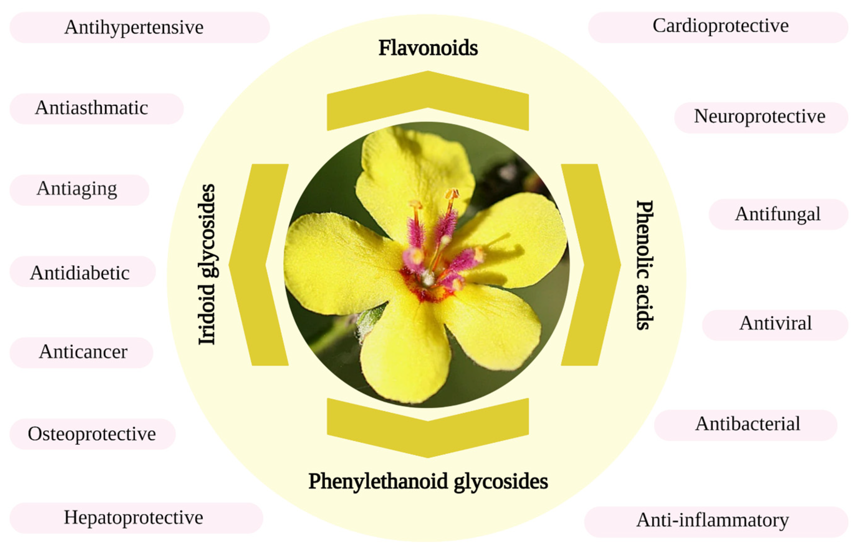
2. Bioactive Compounds

Bioactive compounds are minor chemical compounds present in plants or animals with beneficial health properties with many potential industrial applications [20]. The aerial part of V. sinuatum used in traditional medicine have been analyzed by some authors, who have reported that the main bioactive compounds are polyphenols and iridoids (Table 2).

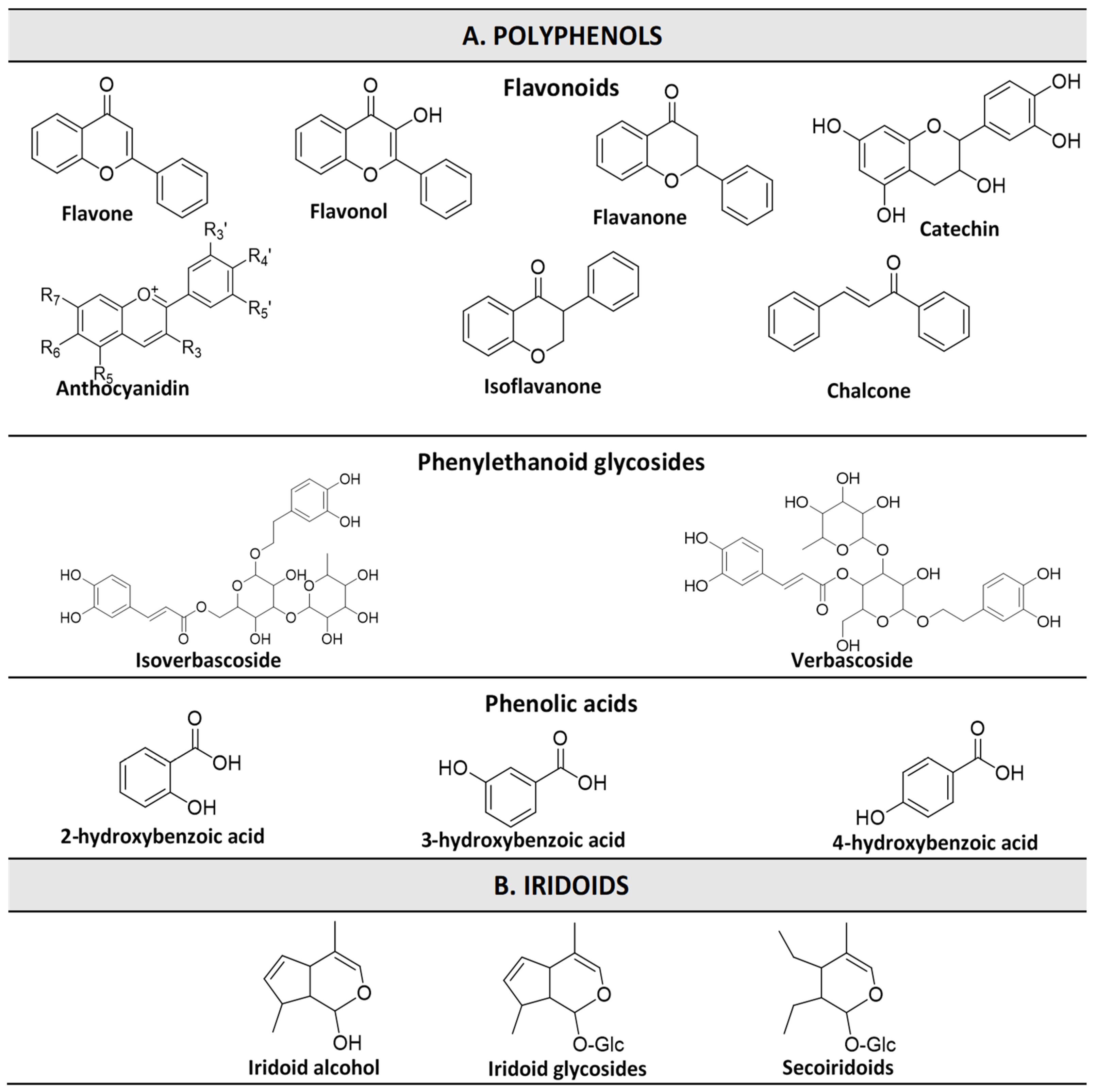
2.1. Phenolic Compounds

Polyphenols are ubiquitous secondary metabolites involved in the protection of plants against different stresses, such as adverse climatic conditions or microbial invasions, in addition to other functions in plant physiology [24]. Polyphenols are highly diversified class of compounds, whose chemical structures are shown in Figure 2. These molecules are divided into four major groups: (1) flavonoids (flavones, flavonols, flavanones, isoflavones, anthocyanidins, chalcones, and catechins); (2) phenolic acids (hydroxybenzoic acids and hydroxycinnamic acids); (3) stilbenes (e.g., resveratrol); and (4) lignans (e.g., secoisolariciresinol). Polyphenols are produced in plants through two main pathways: the acetate and shikimic pathways [24]. Many researchers have assigned many biological properties to polyphenols such as antioxidant, anti-microbial, anti-inflammatory, anti-obesity, anti-diabetic, anti-aging, neuroprotective, and anti-carcinogenic properties [25,26]. For this reason, the extraction and characterization of the different classes of the phenolic compounds from natural plants may lead to the development of new ingredients for industrial applications. Concerning these compounds, three different classes have been identified in V. sinuatum [6,20,21,22,23]: (1) flavonoids, such as apigenin, luteolin, naringin, rutin, naringenin, plantagonine, rhamnetin, myricetin, hesperetin, cynaroside, apigetrin, hyperoside, chrysin, and quercetin; (2) phenolic acids, such as caffeic acid, caffeic acid hexoxide, ρ-Coumaric acid, quinic acid, ursolic acid, chlorogenic acid, cinnamic acid, and gallic acid; and (3) phenylethanoid glycosides, such as p-coumaroyl-6-O-rhamnosyl aucubin isomer I and p-coumaroyl-6-O-rhamnosyl aucubin isomer II.
Many studies have evaluated the polyphenolic profile of the extracts of the aerial part of V. sinuatum. For instance, a study evaluated the phenolic profile of the ethanolic extracts (maceration at room temperature for 72 h with ethanol at 70%) of V. sinuatum flowers from three different origins in Iran [21]. Spectrophotometric results showed that the total phenolic content of the three samples ranged from 29.03 mg gallic acid equivalents (GAE)/g dry weight (DW) to 54.77 mg GAE/g DW, while the total flavonoid content varied between 13.72 mg rutin equivalents (RE)/g DW and 19.91 mg RE /g DW. Further quantitative analysis, carried out with high-performance liquid chromatography with photodiode-array detection (HPLC–PDA), identified apigenin (5.85–13.82 µg/mg DW), luteolin (3.18–16.98 µg/mg DW), naringin (7.10–12.32 µg/mg DW), rutin (4.90–5.55 µg/mg DW), and caffeic acid (4.98–7.78 µg/mg DW). Other authors have evaluated the total phenolic content (TPC), total flavonoid content (TFC), and the phytochemical profile of the compounds present in the methanolic extracts (maceration at room temperature, 24 h) of flowers of different species of the genus Verbascum, including V. sinuatum [7]. For TPC and TFC, spectrophotometric methods were employed, and the results showed values of 51.94 mg GAE/g DW and 22.57 mg quercetin equivalents (QE)/g DW, respectively, which were the greatest among the studied species. Main phenolic compounds were identified and quantified by HPLC–PDA coupled with mass spectrophotometry (HPLC–PDA–MS). Thirteen phenolic compounds were identified, the largest being the flavonoid rutin (21.00 mg/g DW) and the phenolic acid ρ-Coumaric acid (10.22 mg/g DW). Further phytochemical analysis qualitatively identified other 20 compounds in the methanolic extracts, among them flavonoids (naringenin, plantagonine, luteolin, rhamnetin, myricetin, hesperetin, cynaroside, apigenin, apigetrin, hyperoside, and chrysin) and phenolic acids (quinic acid and ursolic acid) [7]. Other authors have performed chromatographic analysis of the phenolic compounds of V. sinuatum leaves, extracted by two different methods: heat assisted extraction with ethanol 80% v/v, at 40 °C, under stirring at 100 rpm for 1 h (ethanolic extract) and infusion into boiled water for 5 minutes (infusion extract) [23]. The two extracts were lyophilized and redissolved into a solution of 20% ethanol. The chromatographic analysis, achieved through high-performance liquid chromatography coupled with a diode-array detection (HPLC–DAD) system, revealed that the ethanolic extract (EE) presents higher phenolic content than the infusion extract (IE). They identified two phenolic acids: quinic acid at a concentration of 1.25 µg/mL for EE and 1.02 µg/mL for IE and caffeic acid hexoxide at 0.281 µg/mL and 0.112 µg/mL concentration for EE and IE, respectively. They also identified two phenylethanoid glycosides: p-coumaroyl-6-O-rhamnosyl aucubin isomer I at the concentration of 6.4 µg/mL and 2.7 µg/mL for EE and IE, respectively, and p-coumaroyl-6-O-rhamnosyl aucubin isomer II at a concentration of 2.3 µg/mL and 1.29 µg/mL for EE and IE, respectively. Their results also revealed that the extraction yield and TPC of the EE estimated at 34.34% and 36 µg/mL, respectively, were higher than those of the IE estimated at 20.29% and 25.2 µg/mL, respectively. Recently, other authors achieved the quantitative analysis of the phenolic compounds present in a methanolic extract (maceration with 80% methanol at room temperature for 24 h) of V. sinuatum flowers by HPLC with diode-array detection (HPLC–DAD) [22]. In their findings, the authors detected and quantified six phenolic compounds: quercetin (0.393 g/kg DW), chlorogenic acid (0.154 g/kg DW), cinnamic acid (0.037 g/kg DW), ρ-coumaric acid (0.031 g/kg DW), gallic acid (0.015 g/kg DW), and caffeic acid (0.006 g/kg DW). Authors noticed the absence of rutin and apigenin in the analyzed V. sinuatum methanolic extract, unlike the previous study [22]. It could be deduced that the polyphenol content of V. sinuatum depends on the geographical location and growing conditions as well as methods and parameters applied for their extraction.

2.2. Iridoids

Iridoids are cyclopentano[c]pyran monoterpenoids compounds, with a bicyclic H-5/H-9 β, β-cis-fused cyclopentanopyran ring systema double bond between C-3 and C-4, including a non-substitution at C-4. They are a particularly important class of compounds for the Verbascum species. The iridoid glycoside content of natural plants such as V. sinuatum has been efficient for the treatment of many diseases, such as Alzheimer’s disease, ischemic brain injury, cerebral ischemia, colon cancer, liver injury, diarrhea, food retention, maldigestion, kidney damage, gout, pains, and arrhythmia [27]. Regarding iridoids, the following compounds have been reported by different authors: aucubin, sinuatol, sinuatoside, aucuboside, catalpol, harpagoside, harpagide, pulverulentoside I, catalposide, genipin, catalopl, verbascoside and isoverbascoside, 6-O-β-D-glucopyranosyl aucubin, 6-O-β-D-xylopyranosyl aucubin, 6-O-α-L-sinuatosyl aucubin, and 6-O-(3”-O-p-coumaroyl)-α-L rhamnopyranosyl catalpol.
Few studies have evaluated these compounds in V. sinuatum (Table 2). A study reported twelve iridoid glycosides compounds: aucubin, 6-O-β-D-glucopyranosyl aucubin, sinuatol, 6-O-β-D-xylopyranosyl aucubin, 6-O-α-L-sinuatosyl aucubin, sinuatoside, aucuboside, catalpol, 6-O-(3”-O-p-coumaroyl)-α-L rhamnopyranosyl catalpol, pulverulentoside I, harpagide, and harpagoside [28]. In a previous study of Selseleh et al., authors quantified an iridoid compound in methanolic extracts of V. sinuatum, harpagoside (0.01 mg/g DW). In addition, five other iridoids (catalposide, verbascoside, genipin, catalopl, and aucubin) were qualitatively identified [7]. Further, in the study conducted by Garcia-Oliveira et al., two iridoid glycosides were identified and quantified: verbascoside and isoverbascoside [23]. These authors revealed that the infusion contained a larger amount of verbascoside (13.1 µg/mL) than the ethanolic extract (12.4 µg/mL), while the isoverbascoside content was found to be larger in the extract (2.5 µg/mL) compared to the infusion (2.41 µg/mL). Overall, these findings show that the chemical structures of compounds could affect their affinity with solvents and consequently the extraction yield.

3. Extraction Techniques

The screening of the bioactive compounds in the plant extract of V. sinuatum has revealed that they are mainly phenolic compounds and iridoids. As previously mentioned, these compounds are widely reported for their beneficial properties. Therefore, obtaining extracts rich in these compounds may be of interest for potential industrial applications. Nevertheless, for an efficient recovery of these compounds (in terms of target compounds, quantity, quality, biological properties, and environmental safety), the methods, type of solvent, and extraction conditions should be carefully considered and optimized [13]. In the following sections, the extraction methods applied to extract V. sinuatum found in the literature have been discussed.

3.1. Conventional Extraction Techniques

The extraction of bioactive compounds can be done through conventional extraction techniques such as Soxhlet, maceration, and hydro-distillation [12]. These methods use selective solvents that affect the extraction yield of the targeted compounds based on their affinity. The commonly used solvents for these techniques are water, ethanol, methanol, chloroform, dichloromethanol, ether, and acetone [10]. In these methods, for the solvent selection, both polarity and affinity of compounds, environmental issues, toxicity, and cost must be considered. Some authors have explored conventional extractions techniques such as infusion with boiled water, heat assisted extraction (HEA), and maceration into ethanol or methanol to recover bioactive compounds from V. sinuatum [7,22]. From a study conducted by previous authors, the extraction yield was higher for HAE using 80% ethanol for 1 h at 40 °C under 100 rpm stirring compared to extraction by infusion into boiled water for 24 h [23]. Thus, the extraction yield of bioactive compounds is affected by the extraction conditions such as solvent (type and concentration), temperature, time, and agitation. Even though there are not many reported data comparing the extraction efficiencies of conventional techniques for V. sinuatum, some authors have investigated other Verbascum species. For example, a study carried out the extraction of bioactive compounds from Verbascum bombyciferum by two methods: homogenizer assisted extraction and maceration with two solvents (water and methanol). For homogenizer assisted extraction, authors obtained extraction yields of 99.750 mg/kg using methanol and 31.018 mg/kg using water. On the other hand, for maceration, the extraction yields were 272.862 mg/kg with methanol and 195.503 mg/kg with water. In the two methods, the extraction yields were higher with methanol as solvent compared to those with water, while the maceration method presented a higher recovery. Nevertheless, they found that the highest TPC and TFC was observed for the homogenizer assisted extraction/methanol (41.80 mg GAE/g and 39.44 mg RE/g), while total bioactive compound extraction was higher when methanol maceration was applied, obtaining 272.862 mg/kg. These authors also showed that, in most assays, the extraction method using methanol as solvent presents the highest antioxidant activity. The authors concluded that methanol was the best extraction solvent and maceration the best conventional extraction method [29]. Another study evaluating the chemical constituents of Verbascum euphaticum and Verbascum oocarpum applying maceration at room temperature for 24 h using hexane, ethyl acetate, methanol, and methanol: water 80% was also carried out, achieving similar results. For both Vebascum species, methanol extracted the highest yields of TPC and TFC. Thus, 50.20 mg GAE/g and 28.47 mg RE/g of TPC and TFC, respectively, were extracted from Verbascum euphaticum. The same trial was applied in Verbascum oocarpum, obtaining 61.23 mg GAE/g and 49.97 RE/g of TPC and TFC, respectively [30].

3.2. Green Extraction Techniques

The emerging green extraction techniques are increasingly preferred for extraction of bioactive compounds. They have the advantage of preserving the environment, speeding up the extraction (lesser time), and enhancing the extraction yields [19]. The major green extraction techniques used in the extraction of bioactive compounds are supercritical fluid extraction (SFE), pulsed electric field (PEF), microwave assisted extraction (MAE), and ultrasound assisted extraction (UAE).

3.2.1. Supercritical Fluid Extraction

Supercritical fluid extraction (SFE) is a novel technology used in the extraction of many natural chemical compounds such as flavonoids, carotenoids, fatty acids, or essential oils [19]. Supercritical state is induced by the temperature and pressure of fluids up to the value of their critical points. In that state, the fluid has both gas-like and liquid-like properties that leads to the reduction of extraction time and an increase in the extraction yields. There are several chemical solvents considered as supercritical fluids, but the most employed is CO2 because it possesses low values of critical temperature and pressure and is non-toxic [31,32]. This technique has a reduced processing time, solvent amount, and energy consumption and so is considered eco-friendly. However, it possesses some inconveniences, such as excessive cost of equipment and the fact that the use of CO2 as extraction fluid only allows the extraction of non-polar compounds [14].
The use of SFE in the recovery of bioactive compounds from flowers has recently increased according to the reported studies [33]. The SFE of bioactive compounds from flowers such as from V. sinuatum could present the same advantages as in reported studies on flowers, according to their reported phenolic compounds [7]. Thus, many authors have performed the SFE of phytochemicals in flower samples. In the study conducted by Pimentel-Moral et al., SFE has been used to optimize the extraction of the total phenolic and organic acids from Hibiscus sabdariffa calyces [34]. They obtained the highest extraction yield (113 mg/g) under the following processing conditions: a pressure of 25 Mpa, a temperature of 50 °C, and 16.7% ethanol. Another study showed that there is an increase in the efficiency with supercritical fluid extraction of bioactive compounds from sunflower (Helianthus annuus L.) while adding 5% of methanol, dimethyl sulphide (DMSO), or water as a modifier [35]. They also found that the best extraction yield process conditions are 50 Mpa of pressure, a temperature of 50 °C, and 5% of water as a modifier. Although supercritical fluid extraction has not had a reported application in the extraction of bioactive ingredients from V. sinuatum, it is obvious to notice that it is an open door that needs to be explored.

3.2.2. Pulse-Electric Field

Pulsed electric field (PEF) is a non-thermal method used in the extraction of bioactive compounds. This technology consists of the application of an electric potential to break the cell membranes and release the cellular content, increasing mass transfer and reducing the time of extraction. In this process, the plant material is placed between two electrodes and is subjected to an energy input of 1 to 20 KJ/kg in an electric field of 0.5–10 kV/cm for a short pulse duration (1 µs−1 ms). It is considered a pre-treatment method used to improve the extraction of bioactive compounds.
Many authors have presented the benefits of using PEF before processing the extraction of compounds. The study by Corrales et al. has shown that anthocyanin mono-glucosides are highly extracted with the application of PEF [36]. The same result has been observed by another study regarding the extraction of anthocyanin and polyphenols [37]. Further, other authors have observed an increase in the extraction yields of phytosterol and isoflavonoids by 32.4% and 20–21%, respectively [38]. The drawback of this technology is the damage produced in the cell membrane when the following conditions are applied—between 500 and 1000 V/cm, for 10-4-10-2 s—due to a minor expansion in the temperature. However, PEF is a highly selective method with low energy cost, low time of extraction, and low quantity of solvent requirement [15]. Moreover, a study has shown that, in this technology, the plant cells are destroyed, allowing an increase in the extraction yield of bioactive compounds from plant tissues. PEF is a non-thermal strengthening method, which can therefore improve the extraction rate of bioactive compounds in flowers [33,39]. Although PEF has fewer applications in the extraction of bioactive ingredients from edible flowers, including V. sinuatum, this technology is still very promising.

3.2.3. Microwave-Assisted Extraction

Microwave assisted extraction (MAE) is a green extraction technology in which an electromagnetic field, ranging from 300 MHz to 300 GHz, is applied. In MAE, the electromagnetic energy is converted into heat, which helps to break down the cellular components and improve the transfer of compounds into the solvent [19,40]. The application of the MAE technique has the advantages of using equipment of small size, allowing a rapid heat transfer, and increasing the yield. This method is faster than conventional extraction techniques, and it is considered green due to the use of a reduced amount of solvent [16]. Nevertheless, this extraction method is not suitable for heat sensitive compounds [17].
Several studies have been carried out on the MAE of bioactive compounds from plant extracts, presenting its efficiency and rapidity compared to conventional methods or other green extraction techniques. For example, a study found that by employing MAE, the recovery of flavolignin and silybinin from Silybum marianum was higher than conventional techniques [41]. Another study proved that the extraction yields of E- and Z-guggolster-one, cinnamaldehyde, and tannin from many plants were higher in MAE than conventional extraction methods and concluded that MAE is faster and more accessible [42]. Further, other authors have reported that microwave-assisted extraction showed better extraction efficiency than ultrasound assisted extraction in the recovery of bioactive ingredients such as proanthocyanidins from the residue of Kushui rose [33]. The exploration of MAE in the recovery of bioactive compounds from V. sinuatum could be a particularly good technique to be applied in non-thermosensitive compounds.

3.2.4. Ultrasound-Assisted Extraction

During ultrasound-assisted extraction (UAE), energy is generated by ultrasound waves between 20 kHz to 100 MHz, causing a cavitation phenomenon through a solvent. This phenomenon creates little bubbles that grow and collapse, inducing energy that finally breaks the components of the cells and improves mass transfer to the solvent [43]. Several factors affect the UAE such as particle size, moisture content, sonification power, frequency, time, pressure, temperature, type of solvent, and distribution of ultrasonic waves [44]. Similar to other green techniques, UAE allows the reduction in the extraction time and the volume of solvent. In addition, it is suitable for the extraction of thermolabile compounds.
UAE of bioactive compounds from plant extracts have been subject to many comparative studies against conventional methods. For example, Zu and colleagues showed that UAE of rosmarinic acid and phenol carboxylic acid is more rapid and efficient compared to those in conventional methods [45]. Further, other research proved that the extraction of quercetin and rutin from Euonymus alatus are optimal with UAE [46]. Similarly, another study also found that UAE with ionic solvent from Catharanthus spp. of three indole alkaloids—catharanthine, vinblastine, and vindoline—was the most efficient method (extraction time from 0.5 h to 4 h and higher yields) compared to conventional techniques [47]. The study achieved by Herrera and Luque de Castro presented best results in the extraction of some phytochemicals (kaempferol, ellagic acid, naringenin, quercetin, naringin, and rutin) from strawberry with the UAE method [48]. V. sinuatum, with the reported identified phytochemicals [7], contains compounds such as naringenin as well that can be better extracted by UAE, which makes this technique a very promising one. Nevertheless, further trials considering all the bioactive compounds present in this plant are needed. Concerning other Verbascum species, the application of UAE has been reported in a study related to the UAE of functional polysaccharides from Verbascum thapsus [49]. Authors found that in comparison to conventional extraction methods (hot water extraction), UAE under the optimal conditions (60 min, temperature 67.5 °C, ultrasonic power of 371.03 W, and solvent/plant material ratio of 40 v/w) has a higher extraction yield (5.75%) and a higher antioxidant activity (67.66% of DPPH radical scavenging activity) [49].

4. Biological Activities of V. sinuatum

Motivated by records of the use of this plant in traditional medicine to alleviate various disorders, some studies have evaluated the biological properties of this plant (Table 3). Most are in vitro analyses, so it is necessary to carry out more specific analysis and in vivo studies to gain an in-depth understanding of the mechanisms of action.

4.1. Antioxidant Activity

The metabolic processes that are taking place in the human body involve oxidative reactions responsible for the generation of free radicals. In healthy conditions, endogenous antioxidants are scavenging the oxidation of excessively produced free radicals under the regulation of enzymes to maintain and guarantee equilibrium between pro- and antioxidants for the good functioning of the organism. However, when a disruption in this balance appears with an excess of prooxidant production, it leads to a disturbance in the redox signaling, an incapacity of control, and molecular damage. This phenomenon is known as oxidative stress. During this process, many functional and physiological disorders appear, thereby inducing or favoring the development of many non-communicative diseases such as cancer, obesity, diabetes, and cardiovascular and neurological diseases [52,53,54]. Thus, in that case, the organism needs a sufficient intake of compounds with high antioxidant activity. The antioxidant activity of an extract or compound is the capacity to scavenge or delay the oxidation of a free radical, thus avoiding the formation of harmful compounds that can favor or induce the development of many human disorders. The antioxidant activity of the extracts of V. sinuatum have been studied by many authors, as presented in Table 3. For example, Moein and colleagues studied the in vitro antioxidant activity of ethanolic extracts of the aerial part of ten medicinal plants, including V. sinuatum. From their results, this plant possessed the highest antioxidant activity in terms of 2,2-diphenyl-1-picrylhydrazyl (DPPH) radical scavenging with a half inhibitory concentration (IC50) of 263.52 µg/mL and also in terms of ferric reducing capacity, with a value of 85.08 μg/mL. These results were attributed to the TPC, which was estimated at 8.53 mg/g, the highest among the analyzed plants, corroborating the role of these compounds in the biological properties of the plant [51]. Moreover, the study of Karamian and Ghasemlou (2013) focused on the analysis of antioxidant activity of the methanolic extract of V. sinuatum. The evaluation was performed using DPPH, metal chelating, and inhibition of linoleic acid oxidation assays. According to the results, the extracts showed great DPPH scavenging activity, with an IC50 of 0.11 mg/mL of extract, which was lower than the control ascorbic acid. Regarding metal chelating activity, the extract obtained a value of 10.88 mg/mL. In the oxidation assay, the extract effectively reduced the oxidation of linoleic acid, with a 51.41% inhibition. However, in these two assays, the results were not comparable with those of the control. As in the previous study, the antioxidant activity was correlated with the high content of phenolic compounds (TPC = 118.2 mg GAE/g DW, TFC = 4.87 QE/g DW) [50]. More recently, a study evaluated the antioxidant activity of three fractions (n-Hexane, ethyl acetate, and water fractions) of the methanolic extract of V. sinuatum, among other plants of Verbascum genus [7]. For DPPH activity, fractions displayed IC50 values of 38.15, 23.56 and 27.37 µg/mL, respectively. For the ferric reducing antioxidant power (FRAP) assay, the results were 1553.29, 2650.92, and 1749.03 equivalents Fe2+ µM, respectively. As could be observed, V. sinuatum fractions displayed better results in the DPPH activity than in the FRAP assay, which also varied depending on the fraction, suggesting that different compounds are involved in the antioxidant properties. In the study conducted by Garcia-Oliveira et al., the antioxidant activity of an infusion extract (IE) and an ethanolic extract (EE) of V. sinuatum were evaluated by the thiobarbituric acid reactive substances (TBARS) [23]. As per their findings, the EE presented a higher antioxidant activity than IE with IC50, respectively, evaluated at 4.2 and 17.4 µg/mL. According to Amini et al., amongst the methanolic extracts (maceration with methanol 80% at room temperature for 24 h) of nine species of Verbascum, V. sinuatum unfortunately presented the lowest values of TFC (2.12 g QE/kg DW), total carotenoid content (1.56 g/kg DW), and β-carotene content (1.1 mg/kg DW) [22]. Further, the antioxidant capacity measured by the FRAP assay was also the lowest for V. sinuatum (1.12 μmol Fe (II)/g DW.

4.2. Antimicrobial Activity

The study of the antimicrobial activity of V. sinuatum has been performed by different authors. Thus, the results obtained in different studies are compiled in Table 3. In the literature, one of the first studies to evaluate this property analyzed the effects of methanolic extract of V. sinuatum inflorescences against four Gram + bacteria—Staphylococcus epidermidis, S. aureus, Enterococcus faecalis, and Bacillus subtilis—and eight Gram — bacteria—Proteus vulgaris, Enterobacter aerogenes, Enterobacter cloacae, Klebsiella pneumoniae, Proteus mirabilis, Pseudomonas aeruginosa, Salmonella typhi, and Citrobacter diversus. The results of this study showed that the methanolic extract is more effective against Gram + bacteria with minimum inhibitory concentrations (MICs) ranging from 16 µg/mL for S. epidermidis to 128 µg/mL for Bacillus subtilis. On the other hand, MIC values for Gram — bacteria varied between 64 to 256 µg/mL [1]. A further study evaluated the ethanolic extracts of V. sinuatum leaves [8]. Authors focused their research on seven pathogens related to urinary tract infections: E. faecalis, Escherichia coli, K. pnemoniae, P. aeruginosa, P. mirabilis, and Candida albicans. They also compared the antimicrobial activity of the plant extract with six commonly used antibiotics (penicillin, tobramycin, ampicillin, nystatin, ketoconazole, and clotrimazole). The studied extract presented a higher antimicrobial activity against E. faecalis, P. mirabilis, and C. albicans compared to those of the six antibiotics, with inhibition zones of 20.0, 18.0 and 20.0 mm and MICs and minimum bactericidal concentrations (MBCs) of 4.0 (8.0), 8.0 (16.0), and 8.0 (16.0) µg/mL, respectively [8]. Another author evaluated the MIC and MBC of three fractions (n-Hexane: NHF, ethyl acetate: EAF and water fractions: WAF) of the methanolic extract of V. sinuatum and nine other Verbascum species against Escherichia coli (Gram +) and Staphylococcus aureus (Gram −) among other Verbascum genus [7]. Their analysis revealed that, for the Gram (+) bacteria, MICs of the three fractions of V. sinuatum were estimated at 32 mg/mL each, while the MBCs were, respectively, 32, 32, and 32 mg/mL for NHF, EAF, and WAF. Besides that, concerning the Gram (−) bacteria, the three fractions have, respectively, presented the MICs of 8, 2, and 16 mg/mL and MBCs of 16, 8, and 16 mg/mL, showing that the ethyl acetate fraction is more efficient against Staphylococcus aureus [7]. Recently, the antimicrobial activity of ethanolic and infusion extracts of V. sinuatum leaves against six bacteria (Staphylococcus aureus, Bacillus cereus, Micrococcus flavus, Listeria monocytogenes, Enterobacter cloacae, and Salmonella typhimurium) and five fungi (Aspergillus fumigatus, Aspergillus versicolor, Aspergillus niger, Penicillium funiculosum, and Penicillium aurantiogriseum) were explored by the previous authors [23]. In this study, the MIC of the methanolic extract ranged from 0.12 to 1 mg/mL and from 0.25 to 1 mg/mL for the infusion extract. Regarding the MBC of the methanolic extract, the results were estimated to be between 0.25 and 2 mg/mL while it was between 0.5 and 2 mg/mL for the infusion extract. In addition, concerning the fungi, the minimum fungicidal concentration (MFC) of the ethanolic extract ranged from 0.5 to 1 mg/mL, whereas for the infusion extract, it presented the same MFC of 1 mg/mL for all the five studied fungi. Thereby, these results revealed that the ethanolic extract of V. sinuatum presented better antibacterial properties against the studied microorganisms in comparison to the infusion extract.
Among the bioactive compounds present in V. sinuatum, acteoside, a phenylpropanoid glycoside, also known as verbascoside, has been reported to possess antiviral properties. Although no studies have been conducted specifically with extracts or isolated acteoside from V. sinuatum, other in vitro and in silico studies have confirmed these properties against different viruses [55,56,57]. Therefore, V. sinuatum may also possess anti-viral properties.

4.3. Anticancer Activity

The research regarding the use of compounds extracted from natural plants has been increasing in recent decades, and many natural compounds have proven their anticancer effects at different levels. Concerning the extract of V. sinuatum, few studies have reported their antitumoral activity. Recently, a study carried out the evaluation of the antimitotic activity of the alkaloid fraction of V. sinuatum leaf extract on root cell division of Allium cepa L. Through a comparative study between the alkaloid fraction and colchicine (an alkaloid positive control), authors evaluated the mitotic, prophase, and aberration indices. Their results showed that the mitotic index and prophase index of the V. sinuatum alkaloid fraction (33.00 and 32.16, respectively, shown in Table 3) were close to the values of the positive alkaloid control (35.00 and 32.28, respectively). Further, V. sinuatum alkaloid fraction caused a higher number of chromosomal abnormalities, with an aberration index of 21.49, compared to 5.60 for the positive control [2]. Thus, this research established that the alkaloid fraction of the V. sinuatum leaves possessed a mitodepressive effect and could be used in studies based on tumor cells to further evaluate its potential anti-cancer properties. Another author studied the cytotoxic activity of ethanolic and infusion extracts of V. sinuatum amongst five traditional medicinal plants on four cancer cell lines: MCF-7 (adenocarcinoma of the breast), NCI-H640 (non-small cell of lung cancer), HeLa (carcinoma of the cervix), and HepG2 (hepatocellular carcinoma) [23]. Between the studied medicinal plants, V. sinuatum presented the highest effect against all the studied cancer cell lines with growth inhibitory concentrations (GI50) ranging from 101.1 to 172.2 µg/mL in the ethanolic extract and from 59.1 to 92.1 µg/mL in the infusion extract. Although few studies have evaluated this property, acteoside has been reported to possess anticancer activities [58]. According to several studies, this compound exerted pro-oxidative, apoptotic, anti-proliferative, anti-angiogenetic, anti-metastatic, and antitumoral effects against various cancers, which have been attributed to the mediation in different signaling pathways [59,60]. To cite some examples, acteoside have been shown to inhibit the proliferation of breast cancer cells with an IC50 of 117 μM [61]. Further, this compound was able to inhibit the progression of glioblastoma cells via a blockade of Wnt/beta-catenin signaling, which led to a reduction in cell viability and tumor growth [62]. In the treatment of prostate cancer, acteoside significantly inhibited cell proliferation, migration, and invasion abilities through downregulation of the TGF-β pathway [63].

4.4. Neuroprotective Activity

Amongst the range of neurodegenerative diseases, Alzheimer’s disease (AD) is one of the most reported in clinical trials. It is a degenerative disease mostly occurring at an older age, characterized by memory loss and cognitive impairments [64]. Many authors have studied the neuroprotective effects of verbascoside, a biological compound first isolated in V. sinuatum. This compound can inhibit the aggregation of amyloid β (Aβ) [65], reduce the expression of caspase 3 [66] or repair the memory impairment in ICR mice [67], and contribute to the increase of the memory abilities of mice with D-galactose and AlCl3-induced Alzheimer’s disease [68]. The endoplasmic reticulum stress (ER-Stress) is correlated to the progression of Alzheimer’s disease [69]. Some authors have studied the effect of verbascoside on APPswe/PSEN1dE9 transgenic (APP/PS1) mice and amyloid β (Aβ)1–42-damaged human glioma (U251) cells [68]. Their findings reveal that, in the brains of APP/PS1 mice, verbascoside significantly inhibits apoptosis, improves the viability of cells, and reduces the accumulation of Amyloid β and the formation of neurofibrillary tangles due to hyperphosphorylated tau protein. Further, for Aβ1-42-damaged U251 cells, they also found that verbascoside significantly reduces calcium accumulation, inhibits apoptosis, improves cell viability, reduces the intracellular concentrations of ROS, and improves the morphology of mitochondria and ER. They concluded that due to the inhibitory effect of verbascoside on the branches of the unfolded protein response, by attenuating ER stress and preventing apoptosis, they have significant neuroprotective properties and could be potentially used in the treatment of AD. Additionally, through their correlation with the modulation of neuroinflammation via the NF-κB-p65 pathway, some authors have presented verbascoside as a potential ingredient in the formulation of drugs for the treatment and prevention of AD [70]. In the case of Alzheimer’s disease, previous authors have reported that verbascoside extracted from V. sinuatum presented beneficial effects on APPswe/PSEN1dE9 transgenic (APP/PS1) mice and amyloid β (Aβ)1–42 exposed glioma cells [68]. They found that verbascoside increases the resistance to endoplasmic reticulum stress, which is involved in the progression of Alzheimer’s disease. Through their findings, verbascoside has been presented as a good ingredient in the development of drugs for the treatment of this neurodegenerative disease.

4.5. Cardiovascular Protection

Plants have been used for several years in traditional medicine for the treatment of cardiovascular diseases. For example, in a study conducted by some authors, the potential of Plantago ovata husks (source of acteoside compounds) in the prevention of hypertension were presented [71]. In the human cardiovascular system, the renin–angiotensin–aldosterone system (RAAS) has a key role in the regulation of electrolyte balance, blood pressure, and cardiovascular development [72]. The abnormal activation of RAAS automatically leads to hypertension. Indeed, in the kidney, the proteinase known as Renin releases and induces the transformation of Angiotensinogen into inactive Angiotensin I, which will later be converted by the action of angiotensin-converting enzyme (ACE) into Angiotensin II (Ang II), a vasoconstrictive compound. By binding to their receptors of type I, they will cause narrowing of capillaries, regulation of aldosterone, and remodeling of vessels and capillaries, leading to cardiac dysfunction due to the consequent high blood pressure [72]. In the research conducted by Tong and co-workers [73], the in vitro inhibitory activity of acteoside on ACE were experimentally confirmed. They conclude on the antihypertensive effects of an acteoside rich plant, through their impact in the reduction of blood pressure and protection of aorta, heart, and kidney. In addition, many authors have also studied the ACE inhibitory activity (ACEI) of twenty-one phenylethanoid glycosides, including acteoside [74]. The IC50 values of the 21 phenylethanoid glycosides on ACEI ranged from 0.53 to 15.035 mM with the IC50 of acteoside estimated at 2.22 mM. The presence of hydroxyl groups in phenylethanoids was suggested to have allowed the chelation by Zn2+ of the ACE site, concluding that acteoside and the other studied compounds are reliable potential sources of natural anti-hypertensive products. V. sinuatum is a source of acteoside and may also show potential in the treatment of cardiovascular diseases.

4.6. Antidiabetic Properties

An effective strategy in the management of diabetes is the control of glucose concentration in the blood that can be done by reducing the glucose absorption rate. In the human digestive system, the regulation of the digestion of carbohydrate and glucose absorption is controlled by α-Amylase. In a study conducted with acteoside, authors found that it can induce the inhibition of the activity of α-Amylase with an IC50 value of 125.21 mg/mL [75]. Other authors reported that acteoside was effective in the management of diabetic neuropathy, chronic hyperalgesia, and allodynia, which are the most predominant complications in diabetes mellitus [76]. Sodium-dependent glucose cotransporter 1 (SGLT1) plays a significant role in the transport of galactose and glucose into the enterocyte in the small intestine. Monitoring the coupling mechanism of SGLT1/GLUT2 is another means of controlling the concentration of glucose in the blood. A study showed that acteoside can inhibit the sodium dependent glucose cotransporter 1-mediated glucose uptake in intestinal epithelial cells [77]. The release of glucose in the blood is controlled by the pancreatic β-cells through the secretion of insulin. The endoplasmic reticulum stress (ER-Stress) leads to a decrease of the biosynthesis and secretion of insulin by the pancreatic β-cells. This stress could be controlled by bioactive compounds extracted from plants through their antioxidant activities. In this sense, one study demonstrated the protective effects of acteoside against oxidative stress and ER-Stress in the pancreatic β cells by reducing the expression of PERK/eIF2α signal [78]. Therefore, considering that V. sinuatum contains a significant amount of acteoside, this plant may be a valuable matrix for further anti-diabetic research.

4.7. Toxicity and Herb–Drug Interaction of V. sinuatum

In the literature, few studies have reported the toxicity (in-vitro or in-vivo) of V. sinuatum preparations and their potential antagonist interaction effect toward many nuclear receptors. In a previous study [23], the in-vitro cytotoxicity of infusion and ethanolic extracts of V. sinuatum against the non-cancer cell line PLP2 was evaluated. According to the results, only the infusion exerted toxic activities against normal cells, with a growth inhibitory concentration 50 (GI50) of 223.1 µg/mL (Table 3). Even though we have not found any reported studies regarding the antagonist drug interaction effect of the bioactive compounds of V. sinuatum, a previous research [79] reported the possibility of natural ingredients extracted from plants, for example, the case of some flavonoids (like apigenin and luteolin that can also be extracted from V. sinuatum), in presenting antagonist effects under specific conditions (high dose) with the aryl hydrocarbon receptor (AhR). They reported that this interaction affected the transcription of the genes responsible for the transport of endogenous and xenobiotic elements involved in diseases management. Further, despite the previously cited beneficial biological benefits of these bioactive ingredients or compounds, some authors have revealed that they can also have health side effects and cause drug interactions due to their capacity to downregulate the transcriptional activities of Cytochrome P450 enzymes and P-glycoprotein through their antagonist effects on various others receptors such as Pregnane Xenobiotic Receptor (PXR) and Constitutive androstane receptor (CAR) [80,81].

5. Encapsulation of Bioactive Compounds

Bioactive compounds can possess biological properties, but if after consumption they are not bioavailable, the desired or expected effect will not be achieved. Further, bioactive compounds are generally sensitive to light, oxygen, and temperature. Thus, to preserve the chemical structure and bioactivity of compounds after extraction, different encapsulation techniques can be applied depending on particle size and final use [82]. Therefore, encapsulation is a preservation method for bioactive compounds, that allows to control their stability, water solubility and bioavailability as well as the release rate, with the final aim of reaching a targeted site of action during human consumption or utilization [83]. Two main groups of techniques are used in food and pharmaceutical industries for the encapsulation of bioactive compounds: nanoencapsulation (capsule particle size from 10 to 1000 nm) and microencapsulation (capsule particle size from 3 to 800 μm) [9,83,84].
Although the encapsulation of V. sinuatum extracts has not been explored yet, some studies have been performed for other Verbascum species and the bioactive compounds identified in V. sinuatum, which could suggest that the results would be satisfactory. For example, the quercetin incorporation into a nanoemulsion under optimal conditions have been presented by some authors, as a solution for improving the bioavailability after oral administration [85]. Similarly, the encapsulation of p-coumaric acid extracted from Phyllostachys makinoi with alginate with chitosan as coated materials permitted to avoid low solubility and poor bioavailability [86]. Further, in the study conducted by others authors [87], the efficiency of nanoencapsulation of the extract of Verbascum flavidum have been explored. This research showed that, the nano-formulation of Apo 4 Extract-Poly Lactide-co-Glycolide of Verbascum flavidum water extract have the potential to inhibit the aggregation of Aβ in relation to their high metal chelator activity (Fe+2 chelating activity: 0.413 mg/mL) and encapsulation efficiency (EE: 15%). Their stability was optimal when preserved at −20 °C. Thus, regarding the composition and biological activities of the extracts of V. sinuatum, they could be a potential matrix for encapsulation essays to preserve their stability and control their release and bioavailability.

6. Potential Applications of V. sinuatum

6.1. Pharmaceutical Industries

As mentioned before, V. sinuatum has been used in different traditional formulations for the treatment of many disorders affecting the human system. Some authors have reported their uses in the formulation of treatments against many diseases, including hemorrhoids, diarrhea, gallstone, liver inflammation, ulcer inflammation, teeth pain, gumboil, hoarse, tonsillitis, cold, cough, asthma, bronchitis, hemoptysis, contusion, broken bones, arthrosis, rheumatism, eczema, exanthema, cysts, zits, wounds, ulcers, burns, chilblain, conjunctivitis, otitis, and helminthiasis [88]. Thus, they have positive effects in the circulatory, digestive, and respiratory system and on the skeleton, skin, and in parasitic infections. Most of these pathologies involve anti-inflammatory processes, which can be justified from a scientific point of view considering the chemical composition of V. sinuatum [88]. According to the different reported bioactive compounds and biological properties, this plant could be a promising source of bioactive ingredients for pharmaceutical industries. For example, considering the reported antimicrobial activity of V. sinuatum, its extract and bioactive compounds could be a matrix for the development of new antibiotics of natural source. Nowadays, much research has been done on different traditional medicinal plants to determine their real potential in the treatment of diseases caused by bacteria, viruses, and fungi, and to seek alternatives to synthetic antimicrobials and deal with the emergence of antimicrobial resistance. Further, the antioxidant assays conducted with different extracts have shown satisfactory results [7,22,23,50,51]. As it has been widely described, oxidative stress is involved in the development of cardiovascular diseases, cancer, obesity, diabetes, inflammatory diseases, aging, and degenerative diseases [89,90]. Therefore, V. sinuatum could be a promising matrix for the development of therapeutics to manage the balance of oxidative stress and help in the management of related diseases. However, more in vivo research models and in clinical trials should be performed, to evaluate the full potential and possible negative effects of this plant. Amongst the reported compounds present in V. sinuatum, verbascoside, also known as acteoside, has demonstrated potential to act as a natural alternative in the formulation of treatment of cancers [58], Alzheimer’s disease [68], hypertension [73], and diabetes [75,76,77]. Thus, they are examples of compounds of interest extractable from V. sinuatum among many others with a possible application as bioactive ingredients in pharmaceutical industries.

6.2. Food Industries

Regarding the phytochemical content and biological properties of V. sinuatum, this plant may also have considerable potential in food applications. For example, the bioactive ingredients of this plant can be used as natural food preservatives to prolong the shelf life of food products through their antioxidant properties or used as functional ingredients or nutraceuticals, intended to produce functional foods. Many studies have reported the antioxidant properties of extracts of V. sinuatum [6,21,22,49,49,50], which could be considered for the development of preservative agents. This is in line with market trends, which seek a lower use of synthetic antioxidants such as butylated hydroxyl anisole (BHA), butylated hydroxytoluene (BHT), and tert-butylhydroquinone (TBHQ) as they have been related to adverse effects on the health of consumers. Some studies have corroborated the efficacy of the use of natural extracts as preservative agents. For example, Efenberger-Szmechtyk and colleagues showed the potential of using plant extracts rich in polyphenols as natural preservatives and antimicrobial agents in meat industries [91]. Considering the phenolic content of V. sinuatum, it could be a very promising application area. On the other hand, V. sinuatum could be explored as functional ingredients in the production of functional foods. Several authors have successfully employed natural extracts to enhance the composition and biological properties of food. For example, a study reported that during the processing of kefir, yogurt, and cheese, various bioactive ingredients such as phenolic compounds, can be incorporated in milk to produce functional fermented milk products [92]. Some authors have presented an application of phenolic compounds extracted from olive leaf extracts in biscuits preparations. In their processing, they found that samples formulated with the extract exerted antiglycative capacity against pentosidine and Ne-carboxyethyl-lysine [93]. Thus, the phenolic compounds of V. sinuatum may have a similar potential.
In view of the various bioactive compounds extracted from V. sinuatum, the potential and the possibility of producing nutraceuticals intended to be used as food preservatives or as functional ingredients in the processing of functional foods is noticeable. However, the use of these bioactive compounds should take into consideration in the delivery method and the different processing steps that they may undergo. Therefore, more research is still necessary before the development of food industrial applications.

7. Conclusions

Medicinal plants have been used for years, generation after generation, for the treatment of many diseases but some species have not been fully studied in terms of chemical composition and effective biological activity. Thus, some plants are still underestimated in their overall potential, thereby not sufficiently valued, because of the lack of scientific information. The present review has presented the different bioactive compounds that have been identified in V. sinuatum, with an emphasis on the phenolic and iridoids reported compounds. It also presents different methods of extraction, from conventional to green techniques of the previously identified compounds, as a directive for the selection of an eco-friendly method to obtain compounds of high purity and yields. The biological properties of different extracts of this plant and compounds have been presented to reveal their potential beneficial health effects. The current information about compounds and biological properties may suggest that this plant has a considerable potential for applications in food, nutraceutical, and pharmaceutical industries. However, in-vitro, in-vivo, and clinical trial studies for the screening of negative interactions and toxicity of the bioactive compounds intended to serve as ingredients in the pharmaceutical or food industries should be a pre-requisite for avoiding possible antagonistic adverse risks and to guarantee consumer safety.

Author Contributions

Conceptualization, M.A.P. and P.D.; methodology, P.D., P.B. and A.P-V.; software, A.P-V. and P.B.; validation, L.C., J.S.-G. and M.A.P.; formal analysis, P.D., P.B. and A.P.-V.; investigation, P.D., P.B. and A.P-V.; data curation, P.D., P.B. and A.P.-V.; writing—original draft preparation, P.D., P.B. and A.P.-V.; writing—review and editing, P.D. and L.C.; supervision, L.C., J.S-G. and M.A.P.; project administration, J.S.-G. and M.A.P.; funding acquisition, J.S.-G. and M.A.P. All authors have read and agreed to the published version of the manuscript.

Funding

The research leading to these results was supported by MICINN by supporting the Ramón y Cajal grant for M.A. Prieto (RYC-2017-22891) and by Xunta de Galicia by supporting the program EXCELENCIA-ED431F 2020/12 and the post-doctoral grant L. Cassani (ED481B-2021/152). This work has also received funding from the Argentinean Agency for the Scientific and Technological Promotion (ANPCyT, Argentina) under the project PICT (2020)/1602.

Institutional Review Board Statement

Not applicable.

Informed Consent Statement

Not applicable.

Data Availability Statement

Not applicable.

Conflicts of Interest

The authors declare no conflict of interest.

References

  1. Senatore, F.; Rigano, D.; Formisano, C.; Grassia, A.; Basile, A.; Sorbo, S. Phytogrowth-Inhibitory and Antibacterial Activity of Verbascum Sinuatum. Fitoterapia 2007, 78, 244–247. [Google Scholar] [CrossRef] [PubMed]
  2. Aiche-Iratni, G.; Ouzid, Y.; Amari, T.; Ouarab, K.; Dahmani, M.; Bouaziz-Menouar, T.; Houali, K. Antimitotic Effect of Verbascum Sinuatum L. Extracts on Meristematic Cells. Int. J. Minor Fruits Med. Aromat. Plants 2022, 8, 7. [Google Scholar] [CrossRef]
  3. Duman, H.; Uzunhisarcikli, M.E.; Ozbek, F. A New Natural Hybrid of Verbascum L. (Scrophulariacae) from Turkey. Gazi Univ. J. Sci. 2021, 34, 937–946. [Google Scholar] [CrossRef]
  4. Bretzel, F.; Pezzarossa, B.; Malorgio, F. Study of Herbaceous Annual and Perennial Species Native to Mediterranean Area for Landscape Purposes. Acta Hortic. 2009, 813, 321–328. [Google Scholar] [CrossRef]
  5. Occurrence Search. Available online: https://www.gbif.org/occurrence/map?taxon_key=3171946&occurrence_status=present (accessed on 23 January 2023).
  6. Demnati, F.; Allache, F. Effect of Verbascum Sinuatum (Scrophulariaceae) on Oviposition of Callosobruchus Maculatus (Bruchidae). J. Crop Prot. 2014, 3, 327–334. [Google Scholar]
  7. Selseleh, M.; Nejad Ebrahimi, S.; Aliahmadi, A.; Sonboli, A.; Mirjalili, M.H. Metabolic Profiling, Antioxidant, and Antibacterial Activity of Some Iranian Verbascum L. Species. Ind. Crops Prod. 2020, 153, 112609. [Google Scholar] [CrossRef]
  8. Sener, A.; Dulger, B. Antimicrobial Activity of the Leaves of Verbascum Sinuatum L. on Microorganisms Isolated from Urinary Tract Infection. Afr. J. Microbiol. Res. 2009, 3, 778–781. [Google Scholar]
  9. Sobel, R.; Versic, R.; Gaonkar, A.G. Introduction to Microencapsulation and Controlled Delivery in Foods; Elsevier Inc.: Amsterdam, The Netherlands, 2014; ISBN 9780124045682. [Google Scholar]
  10. Rasul, M.G. Mohammed Golam Rasul Conventional Extraction Methods Use in Medicinal Plants, Their Advantages and Disadvantages. Int. J. Basic Sci. Appl. Comput. 2018, 2, 10–14. [Google Scholar]
  11. Luque de Castro, M.D.; Priego-Capote, F. Soxhlet Extraction: Past and Present Panacea. J. Chromatogr. A 2010, 1217, 2383–2389. [Google Scholar] [CrossRef]
  12. Azmir, J.; Zaidul, I.S.M.; Rahman, M.M.; Sharif, K.M.; Mohamed, A.; Sahena, F.; Jahurul, M.H.A.; Ghafoor, K.; Norulaini, N.A.N.; Omar, A.K.M. Techniques for Extraction of Bioactive Compounds from Plant Materials: A Review. J. Food Eng. 2013, 117, 426–436. [Google Scholar] [CrossRef]
  13. Sagar, N.A.; Pareek, S.; Sharma, S.; Yahia, E.M.; Lobo, M.G. Fruit and Vegetable Waste: Bioactive Compounds, Their Extraction, and Possible Utilization. Compr. Rev. Food Sci. Food Saf. 2018, 17, 512–531. [Google Scholar] [CrossRef] [PubMed] [Green Version]
  14. Qamar, S.; Torres, Y.J.M.; Parekh, H.S.; Robert Falconer, J. Extraction of Medicinal Cannabinoids through Supercritical Carbon Dioxide Technologies: A Review. J. Chromatogr. B Anal. Technol. Biomed. Life Sci. 2021, 1167, 122581. [Google Scholar] [CrossRef] [PubMed]
  15. Carpentieri, S.; Režek Jambrak, A.; Ferrari, G.; Pataro, G. Pulsed Electric Field-Assisted Extraction of Aroma and Bioactive Compounds From Aromatic Plants and Food By-Products. Front. Nutr. 2022, 8, 792203. [Google Scholar] [CrossRef] [PubMed]
  16. Pan, X.; Niu, G.; Liu, H. Microwave-Assisted Extraction of Tea Polyphenols and Tea Caffeine from Green Tea Leaves. Chem. Eng. Process. 2003, 42, 129–133. [Google Scholar] [CrossRef]
  17. Abbas, K.A.; Mohamed, A.; Abdulamir, A.S.; Abas, H.A. A Review on Supercritical Fluid Extraction as New Analytical Method. Am. J. Biochem. Biotechnol. 2008, 4, 345–353. [Google Scholar]
  18. Luque-García, J.L.; Luque De Castro, M.D. Ultrasound: A Powerful Tool for Leaching. TrAC-Trends Anal. Chem. 2003, 22, 41–47. [Google Scholar] [CrossRef]
  19. Rodríguez García, S.L.; Raghavan, V. Green Extraction Techniques from Fruit and Vegetable Waste to Obtain Bioactive Compounds—A Review. Crit. Rev. Food Sci. Nutr. 2022, 62, 6446–6466. [Google Scholar] [CrossRef]
  20. Park, J.; Beck, B.R.; Kim, H.H.; Lee, S.; Kang, K. A Brief Review of Machine Learning-Based Bioactive Compound Research. Appl. Sci. 2022, 12, 2906. [Google Scholar] [CrossRef]
  21. Ia, F.A.J.A.; Aeidi, K.A.S.; Origooini, Z.A.L.; Aggi, F.I.M. P Henolic P Rofile, C Hemical R Elationship and a Ntioxidant a Ctivity of I Ranian V Erbascum S Pecies. Pan 2020, 62, 31–42. [Google Scholar]
  22. Amini, S.; Hassani, A.; Alirezalu, A.; Maleki, R. Phenolic and Flavonoid Compounds and Antioxidant Activity in Flowers of Nine Endemic Verbascum Species from Iran. J. Sci. Food Agric. 2022, 102, 3250–3258. [Google Scholar] [CrossRef]
  23. Garcia-Oliveira, P.; Carreira-Casais, A.; Pereira, E.; Dias, M.I.; Pereira, C.; Calhelha, R.C.; Stojković, D.; Sokovic, M.; Simal-Gandara, J.; Prieto, M.A.; et al. From Tradition to Health: Chemical and Bioactive Characterization of Five Traditional Plants. Molecules 2022, 27, 6495. [Google Scholar] [CrossRef]
  24. Cutrim, C.S.; Cortez, M.A.S. A Review on Polyphenols: Classification, Beneficial Effects and Their Application in Dairy Products. Int. J. Dairy Technol. 2018, 71, 564–578. [Google Scholar] [CrossRef]
  25. Rajha, H.N.; Paule, A.; Aragonès, G.; Barbosa, M.; Caddeo, C.; Debs, E.; Dinkova, R.; Eckert, G.P.; Fontana, A.; Gebrayel, P.; et al. Recent Advances in Research on Polyphenols: Effects on Microbiota, Metabolism, and Health. Mol. Nutr. Food Res. 2022, 66, 2100670. [Google Scholar] [CrossRef] [PubMed]
  26. Yoshioka, Y.; Ohishi, T.; Nakamura, Y.; Fukutomi, R.; Miyoshi, N. Anti-Cancer Effects of Dietary Polyphenols via ROS-Mediated Pathway with Their Modulation of MicroRNAs. Molecules 2022, 27, 3816. [Google Scholar] [CrossRef] [PubMed]
  27. Hussain, H.; Nazir, M.; Green, I.R.; Saleem, M.; Raza, M.L. Therapeutic Potential of Iridoid Derivatives: Patent Review. Inventions 2019, 4, 29. [Google Scholar] [CrossRef] [Green Version]
  28. Tatli, I.I.; Zeliha, A. Chemical Cons. of Verbascum L. Species. J. Pharm. Sci. 2004, 29, 93–107. [Google Scholar]
  29. Angeloni, S.; Zengin, G.; Sinan, K.I.; Ak, G.; Maggi, F.; Caprioli, G.; Kaplan, A.; Bahşi, M.; Çakılcıoğlu, U.; Bouyahya, A.; et al. An Insight into Verbascum Bombyciferum Extracts: Different Extraction Methodologies, Biological Abilities and Chemical Profiles. Ind. Crops Prod. 2021, 161, 113201. [Google Scholar] [CrossRef]
  30. Zengin, G.; Mahomoodally, M.F.; Sinan, K.I.; Sadeer, N.; Maggi, F.; Caprioli, G.; Angeloni, S.; Mollica, A.; Stefanucci, A.; Ak, G.; et al. Evaluation of Chemical Constituents and Biological Properties of Two Endemic Verbascum Species. Process Biochem. 2021, 108, 110–120. [Google Scholar] [CrossRef]
  31. de Hoyos-Martínez, P.L.; Merle, J.; Labidi, J.; Charrier–El Bouhtoury, F. Tannins Extraction: A Key Point for Their Valorization and Cleaner Production. J. Clean. Prod. 2019, 206, 1138–1155. [Google Scholar] [CrossRef] [Green Version]
  32. Uwineza, P.A.; Waśkiewicz, A. Recent Advances in Supercritical Fluid Extraction of Natural Bioactive Compounds from Natural Plant Materials. Molecules 2020, 25, 3847. [Google Scholar] [CrossRef]
  33. Zhao, L.; Fan, H.; Zhang, M.; Chitrakar, B.; Bhandari, B.; Wang, B. Edible Flowers: Review of Flower Processing and Extraction of Bioactive Compounds by Novel Technologies. Food Res. Int. 2019, 126, 108660. [Google Scholar] [CrossRef] [PubMed]
  34. Pimentel-Moral, S.; Borrás-Linares, I.; Lozano-Sánchez, J.; Arráez-Román, D.; Martínez-Férez, A.; Segura-Carretero, A. Supercritical CO2 Extraction of Bioactive Compounds from Hibiscus Sabdariffa. J. Supercrit. Fluids 2019, 147, 213–221. [Google Scholar] [CrossRef]
  35. Casas, L.; Mantell, C.; Rodríguez, M.; Torres, A.; Macías, F.A.; Martínez de la Ossa, E. Effect of the Addition of Cosolvent on the Supercritical Fluid Extraction of Bioactive Compounds from Helianthus Annuus L. J. Supercrit. Fluids 2007, 41, 43–49. [Google Scholar] [CrossRef]
  36. Corrales, M.; Toepfl, S.; Butz, P.; Knorr, D.; Tauscher, B. Extraction of Anthocyanins from Grape By-Products Assisted by Ultrasonics, High Hydrostatic Pressure or Pulsed Electric Fields: A Comparison. Innov. Food Sci. Emerg. Technol. 2008, 9, 85–91. [Google Scholar] [CrossRef]
  37. Delsart, C.; Ghidossi, R.; Poupot, C.; Cholet, C.; Grimi, N.; Vorobiev, E.; Milisic, V.; Peuchot, M.M. Enhanced Extraction of Phenolic Compounds from Merlot Grapes by Pulsed Electric Field Treatment. Am. J. Enol. Vitic. 2012, 63, 205–211. [Google Scholar] [CrossRef]
  38. Guderjan, M.; Töpfl, S.; Angersbach, A.; Knorr, D. Impact of Pulsed Electric Field Treatment on the Recovery and Quality of Plant Oils. J. Food Eng. 2005, 67, 281–287. [Google Scholar] [CrossRef]
  39. Moradi, N.; Rahimi, M. Effect of Ultrasound- and Pulsed Electric Field-Assisted Enzymatic Treatment on the Recovery and Quality of Sunflower Oil. Sep. Sci. Technol. 2019, 54, 1043–1054. [Google Scholar] [CrossRef]
  40. Chan, C.H.; Yusoff, R.; Ngoh, G.C.; Kung, F.W.L. Microwave-Assisted Extractions of Active Ingredients from Plants. J. Chromatogr. A 2011, 1218, 6213–6225. [Google Scholar] [CrossRef] [PubMed]
  41. Asghari, J.; Ondruschka, B.; Mazaheritehrani, M. Extraction of Bioactive Chemical Compounds from the Medicinal Asian Plants by Microwave Irradiation. J. Med. Plants Res. 2011, 5, 495–506. [Google Scholar]
  42. Chiremba, C.; Rooney, L.W.; Beta, T. Microwave-Assisted Extraction of Bound Phenolic Acids in Bran and Flour Fractions from Sorghum and Maize Cultivars Varying in Hardness. J. Agric. Food Chem. 2012, 60, 4735–4742. [Google Scholar] [CrossRef] [Green Version]
  43. Carreira-Casais, A.; Otero, P.; Garcia-Perez, P.; Garcia-Oliveira, P.; Pereira, A.G.; Carpena, M.; Soria-Lopez, A.; Simal-Gandara, J.; Prieto, M.A. Benefits and Drawbacks of Ultrasound-Assisted Extraction for the Recovery of Bioactive Compounds from Marine Algae. Int. J. Environ. Res. Public Health 2021, 18, 9153. [Google Scholar] [CrossRef] [PubMed]
  44. Chemat, F.; Rombaut, N.; Sicaire, A.G.; Meullemiestre, A.; Fabiano-Tixier, A.S.; Abert-Vian, M. Ultrasound Assisted Extraction of Food and Natural Products. Mechanisms, Techniques, Combinations, Protocols and Applications. A Review. Ultrason. Sonochem. 2017, 34, 540–560. [Google Scholar] [CrossRef] [PubMed]
  45. Zu, G.; Zhang, R.; Yang, L.; Ma, C.; Zu, Y.; Wang, W.; Zhao, C. Ultrasound-Assisted Extraction of Carnosic Acid and Rosmarinic Acid Using Ionic Liquid Solution from Rosmarinus Officinalis. Int. J. Mol. Sci. 2012, 13, 11027–11043. [Google Scholar] [CrossRef] [PubMed] [Green Version]
  46. Yang, Y.; Zhang, F. Ultrasound-Assisted Extraction of Rutin and Quercetin from Euonymus Alatus (Thunb.) Sieb. Ultrason. Sonochem. 2008, 15, 308–313. [Google Scholar] [CrossRef]
  47. Yang, L.; Wang, H.; Zu, Y.G.; Zhao, C.; Zhang, L.; Chen, X.; Zhang, Z. Ultrasound-Assisted Extraction of the Three Terpenoid Indole Alkaloids Vindoline, Catharanthine and Vinblastine from Catharanthus Roseus Using Ionic Liquid Aqueous Solutions. Chem. Eng. J. 2011, 172, 705–712. [Google Scholar] [CrossRef]
  48. Herrera, M.C.; Luque De Castro, M.D. Ultrasound-Assisted Extraction for the Analysis of Phenolic Compounds in Strawberries. Anal. Bioanal. Chem. 2004, 379, 1106–1112. [Google Scholar] [CrossRef]
  49. Babamoradi, N.; Yousefi, S.; Ziarati, P. Optimization of Ultrasound-Assisted Extraction of Functional Polysaccharides from Common Mullein (Verbascum Thapsus L.) Flowers. J. Food Process Eng. 2018, 41, e12851. [Google Scholar] [CrossRef]
  50. Karamian, R.; Ghasemlou, F. Total Phenolic Content, Antioxidant and Antibacterial Activities of Three Verbascum Species from Iran. Med. Plants By-Prod. 2013, 1, 43–51. [Google Scholar]
  51. Moein, S.; Moein, M.; Khoshnoud, M.J.; Kalanteri, T. In Vitro Antioxidant Properties Evaluation of 10 Iranian Medicinal Plants by Different Methods. Iran. Red Crescent Med. J. 2012, 14, 771–775. [Google Scholar] [CrossRef] [Green Version]
  52. Reuter, S.; Gupta, S.C.; Chaturvedi, M.M.; Aggarwal, B.B. Oxidative Stress, Inflammation, and Cancer: How Are They Linked? Free Radic. Biol. Med. 2010, 49, 1603–1616. [Google Scholar] [CrossRef] [Green Version]
  53. Di Meo, S.; Reed, T.T.; Venditti, P.; Victor, V.M. Role of ROS and RNS Sources in Physiological and Pathological Conditions. Oxid. Med. Cell. Longev. 2016, 2016, 1245049. [Google Scholar] [CrossRef] [PubMed]
  54. Gelpi, J.R.; Boveris, A.; Poderoso, J.J. Biochemistry of Oxidative Stress Physiopathology and Clinical Aspects. In Advances in Biochemistry in Health and Disease; Springer: Cham, Switzerland, 2016; Volume 25, pp. 1058–1071. ISBN 978-3-319-45864-9. [Google Scholar] [CrossRef]
  55. Song, X.; He, J.; Xu, H.; Hu, X.P.; Wu, X.L.; Wu, H.Q.; Liu, L.Z.; Liao, C.H.; Zeng, Y.; Li, Y.; et al. The Antiviral Effects of Acteoside and the Underlying IFN-γ-Inducing Action. Food Funct. 2016, 7, 3017–3030. [Google Scholar] [CrossRef] [PubMed] [Green Version]
  56. Khattab, A.R.; Teleb, M.; Kamel, M.S. In Silico Study of Potential Anti-SARS Cell Entry Phytoligands from Phlomis Aurea: A Promising Avenue for Prophylaxis. Future Virol. 2021, 16, 761–775. [Google Scholar] [CrossRef] [PubMed]
  57. Abdallah, H.M.; El-Halawany, A.M.; Sirwi, A.; El-Araby, A.M.; Mohamed, G.A.; Ibrahim, S.R.M.; Koshak, A.E.; Asfour, H.Z.; Awan, Z.A.; Elfaky, M.A. Repurposing of Some Natural Product Isolates as SARS-CoV-2 Main Protease Inhibitors via in Vitro Cell Free and Cell-Based Antiviral Assessments and Molecular Modeling Approaches. Pharmaceuticals 2021, 14, 213. [Google Scholar] [CrossRef] [PubMed]
  58. Zhao, C.; Lin, G.; Wu, D.; Liu, D.; You, L.; Högger, P.; Simal-Gandara, J.; Wang, M.; da Costa, J.G.M.; Marunaka, Y.; et al. The Algal Polysaccharide Ulvan Suppresses Growth of Hepatoma Cells. Food Front. 2020, 1, 83–101. [Google Scholar] [CrossRef]
  59. Khan, R.A.; Hossain, R.; Roy, P.; Jain, D.; Mohammad Saikat, A.S.; Roy Shuvo, A.P.; Akram, M.; Elbossaty, W.F.; Khan, I.N.; Painuli, S.; et al. Anticancer Effects of Acteoside: Mechanistic Insights and Therapeutic Status. Eur. J. Pharmacol. 2022, 916, 174699. [Google Scholar] [CrossRef]
  60. Xiao, Y.; Ren, Q.; Wu, L. The Pharmacokinetic Property and Pharmacological Activity of Acteoside: A Review. Biomed. Pharmacother. 2022, 153, 113296. [Google Scholar] [CrossRef]
  61. Daneshforouz, A.; Nazemi, S.; Gholami, O.; Kafami, M.; Amin, B. The Cytotoxicity and Apoptotic Effects of Verbascoside on Breast Cancer 4T1 Cell Line. BMC Pharmacol. Toxicol. 2021, 22, 72. [Google Scholar] [CrossRef]
  62. Jia, W.Q.; Zhu, J.W.; Yang, C.Y.; Ma, J.; Pu, T.Y.; Han, G.Q.; Zou, M.M.; Xu, R.X. Verbascoside Inhibits Progression of Glioblastoma Cells by Promoting Let-7g-5p and down-Regulating HMGA2 via Wnt/Beta-Catenin Signalling Blockade. J. Cell. Mol. Med. 2020, 24, 2901–2916. [Google Scholar] [CrossRef] [Green Version]
  63. Wu, C.H.; Chen, C.H.; Hsieh, P.F.; Lee, Y.H.; Kuo, W.W.T.; Wu, R.C.Y.; Hung, C.H.; Yang, Y.L.; Lin, V.C. Verbascoside Inhibits the Epithelial-Mesenchymal Transition of Prostate Cancer Cells through High-Mobility Group Box 1/Receptor for Advanced Glycation End-Products/TGF-β Pathway. Environ. Toxicol. 2021, 36, 1080–1089. [Google Scholar] [CrossRef]
  64. Darby, D.G.; Pietrzak, R.H.; Fredrickson, J.; Woodward, M.; Moore, L.; Fredrickson, A.; Sach, J.; Maruff, P. Intraindividual Cognitive Decline Using a Brief Computerized Cognitive Screening Test. Alzheimer’s Dement. 2012, 8, 95–104. [Google Scholar] [CrossRef] [PubMed]
  65. Kurisu, M.; Miyamae, Y.; Murakami, K.; Han, J.; Isoda, H.; Irie, K.; Shigemori, H. Inhibition of Amyloid β Aggregation by Acteoside, a Phenylethanoid Glycoside. Biosci. Biotechnol. Biochem. 2013, 77, 1329–1332. [Google Scholar] [CrossRef] [PubMed]
  66. Peng, X.M.; Gao, L.; Huo, S.X.; Liu, X.M.; Yan, M. The Mechanism of Memory Enhancement of Acteoside (Verbascoside) in the Senescent Mouse Model Induced by a Combination of d -Gal and AlCl3. Phyther. Res. 2015, 29, 1137–1144. [Google Scholar] [CrossRef] [PubMed]
  67. Lee, K.Y.; Jeong, E.J.; Lee, H.S.; Kim, Y.C. Acteoside of Callicarpa Dichotoma Attenuates Scopolamine-Induced Memory Impairments. Biol. Pharm. Bull. 2006, 29, 71–74. [Google Scholar] [CrossRef] [PubMed] [Green Version]
  68. Wang, C.; Cai, X.; Wang, R.; Zhai, S.; Zhang, Y.; Hu, W.; Zhang, Y.; Wang, D. Neuroprotective Effects of Verbascoside against Alzheimer’s Disease via the Relief of Endoplasmic Reticulum Stress in Aβ-Exposed U251 Cells and APP/PS1 Mice. J. Neuroinflamm. 2020, 17, 309. [Google Scholar] [CrossRef] [PubMed]
  69. Hosoi, T.; Nomura, J.; Ozawa, K.; Nishi, A.; Nomura, Y. Possible Involvement of Endoplasmic Reticulum Stress in the Pathogenesis of Alzheimer’s Disease. Endoplasmic Reticulum Stress Dis. 2016, 2, 107–118. [Google Scholar] [CrossRef]
  70. Chen, S.; Liu, H.; Wang, S.; Jiang, H.; Gao, L.; Wang, L.; Teng, L.; Wang, C.; Wang, D. The Neuroprotection of Verbascoside in Alzheimer’s Disease Mediated through Mitigation of Neuroinflammation via Blocking NF-ΚB-P65 Signaling. Nutrients 2022, 14, 1417. [Google Scholar] [CrossRef]
  71. Galisteo, M.; Sánchez, M.; Vera, R.; González, M.; Anguera, A.; Duarte, J.; Zarzuelo, A. A Diet Supplemented with Husks of Plantago Ovata Reduces the Development of Endothelial Dysfunction, Hypertension, and Obesity by Affecting Adiponectin and TNF-α in Zucker Rats. J. Nutr. 2005, 135, 2399–2404. [Google Scholar] [CrossRef] [Green Version]
  72. Te Riet, L.; Van Esch, J.H.M.; Roks, A.J.M.; Van Den Meiracker, A.H.; Danser, A.H.J. Hypertension: Renin-Angiotensin-Aldosterone System Alterations. Circ. Res. 2015, 116, 960–975. [Google Scholar] [CrossRef] [Green Version]
  73. Tong, R.C.; Qi, M.; Yang, Q.M.; Li, P.F.; Wang, D.D.; Lan, J.P.; Wang, Z.T.; Yang, L. Extract of Plantago Asiatica L. Seeds Ameliorates Hypertension in Spontaneously Hypertensive Rats by Inhibition of Angiotensin Converting Enzyme. Front. Pharmacol. 2019, 10, 403. [Google Scholar] [CrossRef] [Green Version]
  74. Li, P.; Qi, M.; Hu, H.; Liu, Q.; Yang, Q.; Wang, D.; Guo, F.; Bligh, S.W.A.; Wang, Z.; Yang, L. Structure-Inhibition Relationship of Phenylethanoid Glycosides on Angiotensin-Converting Enzyme Using Ultra-Performance Liquid Chromatography-Tandem Quadrupole Mass Spectrometry. RSC Adv. 2015, 5, 51701–51707. [Google Scholar] [CrossRef]
  75. Lu, Y.; Zhou, W.; Feng, Y.; Li, Y.; Liu, K.; Liu, L.; Lin, D.; He, Z.; Wu, X. Acteoside and Acyl-Migrated Acteoside, Compounds in Chinese Kudingcha Tea, Inhibit α-Amylase in Vitro. J. Med. Food 2017, 20, 577–585. [Google Scholar] [CrossRef]
  76. Czerwińska, M.E.; Gąsińska, E.; Leśniak, A.; Krawczyk, P.; Kiss, A.K.; Naruszewicz, M.; Bujalska-Zadrożny, M. Inhibitory Effect of Ligustrum Vulgare Leaf Extract on the Development of Neuropathic Pain in a Streptozotocin-Induced Rat Model of Diabetes. Phytomedicine 2018, 49, 75–82. [Google Scholar] [CrossRef] [PubMed]
  77. Shimada, H.; Urabe, Y.; Okamoto, Y.; Li, Z.; Kawase, A.; Morikawa, T.; Tu, P.; Muraoka, O.; Iwaki, M. Major Constituents of Cistanche Tubulosa, Echinacoside and Acteoside, Inhibit Sodium-Dependent Glucose Cotransporter 1-Mediated Glucose Uptake by Intestinal Epithelial Cells. J. Funct. Foods 2017, 39, 91–95. [Google Scholar] [CrossRef]
  78. Galli, A.; Marciani, P.; Marku, A.; Ghislanzoni, S.; Bertuzzi, F.; Rossi, R.; Di Giancamillo, A.; Castagna, M.; Perego, C. Verbascoside Protects Pancreatic β-Cells against Er-Stress. Biomedicines 2020, 8, 582. [Google Scholar] [CrossRef] [PubMed]
  79. Goya-Jorge, E.; Rodríguez, M.E.J.; Veitía, M.S.I.; Giner, R.M. Plant Occurring Flavonoids as Modulators of the Aryl Hydrocarbon Receptor. Molecules 2021, 26, 2315. [Google Scholar] [CrossRef] [PubMed]
  80. Husain, I.; Dale, O.R.; Martin, K.; Gurley, B.J.; Adams, S.J.; Avula, B.; Chittiboyina, A.G.; Khan, I.A.; Khan, S.I. Screening of Medicinal Plants for Possible Herb-Drug Interactions through Modulating Nuclear Receptors, Drug-Metabolizing Enzymes and Transporters. J. Ethnopharmacol. 2023, 301, 115822. [Google Scholar] [CrossRef] [PubMed]
  81. Ortiz-Flores, M.; Portilla-Martínez, A.; Cabrera-Pérez, F.; Nájera, N.; Meaney, E.; Villarreal, F.; Pérez-Durán, J.; Ceballos, G. PXR Is a Target of (-)-Epicatechin in Skeletal Muscle. Heliyon 2020, 6, e05357. [Google Scholar] [CrossRef] [PubMed]
  82. Milinčić, D.D.; Popović, D.A.; Lević, S.M.; Kostić, A.; Tešić, Ž.L.; Nedović, V.A.; Pešić, M.B. Application of Polyphenol-Loaded Nanoparticles in Food Industry. Nanomaterials 2019, 9, 1629. [Google Scholar] [CrossRef] [PubMed] [Green Version]
  83. Noore, S.; Rastogi, N.K.; O’Donnell, C.; Tiwari, B. Novel Bioactive Extraction and Nano-Encapsulation. Encyclopedia 2021, 1, 632–664. [Google Scholar] [CrossRef]
  84. Marcillo-Parra, V.; Tupuna-Yerovi, D.S.; González, Z.; Ruales, J. Encapsulation of Bioactive Compounds from Fruit and Vegetable By-Products for Food Application—A Review. Trends Food Sci. Technol. 2021, 116, 11–23. [Google Scholar] [CrossRef]
  85. Karadag, A.; Yang, X.; Ozcelik, B.; Huang, Q. Optimization of Preparation Conditions for Quercetin Nanoemulsions Using Response Surface Methodology. J. Agric. Food Chem. 2013, 61, 2130–2139. [Google Scholar] [CrossRef] [PubMed]
  86. Yeh, Y.M.; Tsai, T.Y.; Yang, C.Y. Encapsulation and Release Kinetics of Polyphenols and P-Coumaric Acid Extracted from Phyllostachys Makinoi by Ultrasonic-Pretreatment Autoclaving. LWT 2022, 167, 113838. [Google Scholar] [CrossRef]
  87. Kazdal, F.; Bahadori, F.; Celik, B.; Ertas, A.; Topcu, G. Inhibition of Amyloid β Aggregation Using Optimized Nano-Encapsulated Formulations of Plant Extracts with High Metal Chelator Activities. Curr. Pharm. Biotechnol. 2019, 21, 681–701. [Google Scholar] [CrossRef] [PubMed]
  88. Blanco-Salas, J.; Hortigón-Vinagre, M.P.; Morales-Jadán, D.; Ruiz-Téllez, T. Searching for Scientific Explanations for the Uses of Spanish Folk Medicine: A Review on the Case of Mullein (Verbascum, Scrophulariaceae). Biology 2021, 10, 618. [Google Scholar] [CrossRef]
  89. Atamer, A.; Bilici, A.; Yenice, N.; Selek, S.; Ilhan, N.; Atamer, Y. The Importance of Paraoxonase 1 Activity, Nitric Oxide and Lipid Peroxidation in Hepatosteatosis. J. Int. Med. Res. 2008, 36, 771–776. [Google Scholar] [CrossRef]
  90. D’Arcy, M.S. A Review of Biologically Active Flavonoids as Inducers of Autophagy and Apoptosis in Neoplastic Cells and as Cytoprotective Agents in Non-Neoplastic Cells. Cell Biol. Int. 2022, 46, 1179–1195. [Google Scholar] [CrossRef] [PubMed]
  91. Efenberger-Szmechtyk, M.; Nowak, A.; Czyzowska, A. Plant Extracts Rich in Polyphenols: Antibacterial Agents and Natural Preservatives for Meat and Meat Products. Crit. Rev. Food Sci. Nutr. 2021, 61, 149–178. [Google Scholar] [CrossRef] [PubMed]
  92. Gruskiene, R.; Bockuviene, A.; Sereikaite, J. Microencapsulation of Bioactive Ingredients for Their Delivery into Fermented Milk Products: A Review. Molecules 2021, 26, 4601. [Google Scholar] [CrossRef] [PubMed]
  93. Navarro, M.; Morales, F.J. Effect of Hydroxytyrosol and Olive Leaf Extract on 1,2-Dicarbonyl Compounds, Hydroxymethylfurfural and Advanced Glycation Endproducts in a Biscuit Model. Food Chem. 2017, 217, 602–609. [Google Scholar] [CrossRef] [PubMed] [Green Version]
Figure 1. Health benefits of bioactive compounds identified in Verbascum sinuatum.
Biomolecules 13 00427 g001
Figure 2. Chemical structures of polyphenols and iridoids present in V. sinuatum.
Biomolecules 13 00427 g002
Table 1. Advantages and drawbacks of conventional and green extraction techniques.
ETAdvantagesDrawbacksRef
M 1Requires simple equipment
It is an energy saving process
Ideal for less soluble compounds needing high prolonged contact
Suitable for less potent and cheap drugs
Long ET time up to weeks
Low ET yield
High quantity of solvent
[10]
SX 1High mass of plant matrix could be used
Filtration is not required after ET
Simple and repeatable
Repeated contact of fresh solvent with the solid matrix
Higher risk of thermal destruction for thermolabile compounds
Time consuming and intensive
The manipulations of variables are limited
Not eco-friendly and requires high quantities of solvents
[11,12]
HD 1Simple and old method
Suitable for small-scale industries
Provides different options: hydro-distillation, steam and water distillation, direct steam distillation, and hydro-diffusion.
The ETs by steam and water distillation are more reproducible
Higher risk of thermal destruction for thermolabile compounds
The ET is extremely low and therefore time consuming
[13].
SFE 2Reduced ET time
Higher ET yield
Less energy consumption
Mild temperature and pressure
Small volume of solvent
Suitable for ET of volatile compounds
Excessive cost of equipment
Use of CO2 as ET fluid only allows the ET of non-polar compounds
[14]
PEF 2Highly selective
Low quantity of solvent and energy cost
Rapid ET process
Damage of cell membrane[15].
MAE 2Small size equipment
Allows a rapid heat transfer
Increase in the ET yield
Reduced amount of solvent
Induces damage of heat sensitive compounds
Low drying rate
[16,17]
UAE 2High ET yields
Thermolabile compounds are safe
ET time is reduced
Has simple and small equipment
Is more selective and diverse in ET
Solvent is not renewed
The additional filtration step can lead to loss or contamination
[18,19]
Abbreviations: 1 refers to conventional extraction techniques; 2 refers to green extraction techniques; ET: extraction technique; M: maceration; HD: hydro-distillation; SFE: supercritical fluid extraction2; PEF: pulse electric field; MAE: microwave assisted extraction; UAE: ultrasound assisted extraction; SX: soxhlet.
Table 2. Studies on the bioactive composition of V. sinuatum extracts.
EXPartCondResultsRef.
Maceration (ethanol)Flower70%, RT, 72 hSP (TPC; mg GAE/g DW; TFC: mg RE/g DW)HPLC–PDA (µg/mg DW)[21]
TPC (29.03–54.77),
TFC (13.72–19.91)
Flavonoids: Apigenin (5.85–13.82); Luteolin (3.18–16.98); Naringin (7.10–12.3); Rutin (4.90–5.55)
PAs: CAF (4.98–7.78)
Methanolic (extract)FlowerRT, 24 hSP (TPC; mg GAE/g DW; TFC: mg RE/g DW)HPLC–PDA–MS (mg/g DW)[7]
TPC (51.94),
TFC (22.57)
Flavonoids: Rutin (21); Quercetin (1.51)
PAs: PCA (0.12); GA (0.09); SAc (2.45); PMCA (10.22); FA (0.45); RA (3.63)
Iridoids: Harpagoside (0.01)
MacerationFlower80%, RT, 24 hHPLC–DAD (g/kg DW)[22]
Flavonoids: Quercetin (0.393)
PAs: CGA (0.154); CAN (0.037); PMCA (0.031); GAA (0.015); CAF (0.006)
HAE (ethanol)Leaf80%, 40 °C, 100 rmp/1hSP/ GRV (TPC: µg/mL)HPLC–DAD (µg/mL)[23]
TPC (36),
EY (34.34%)
PAs: QA (1.25); CAFh (0.281)
PGs: PMCI (6.4); PMCII (2.3)
IGs: VB (12.4); IVB (2.5)
InfusionLeafWater, 5 minSP/ GRV (TPC: µg/mL)HPLC–DAD (µg/mL)
TPC (25.2),
EY (20.29%)
PAs: QA (1.02); CAFh (0.112)
PGs: PCMI (2.7); PCMII (1.29)
IGs: VB (13.1); IVB (2.41)
Abbreviations: EX: extraction; COND: condition; IGs: iridoid glycosides; PGs: phenylethanoid glycosides; PAs: phenolic acids; CAF: caffeic acid; CAFh: caffeic acid hexoxide; PCA: protocatechuic acid; GA: gentisic acid; Sac: salicylic acid; PMCA: p-Coumaric acid; FA: ferulic acid; RA: rosmarinic acid; GAA: gallic acid; CGA: chlorogenic acid; CAN: cinnamic acid; QA: quinic acid; VB: verbascoside; IBV: isoverbascoside; PMCI: p-Coumaroyl-6-O-rhamnosyl aucubin isomer I; PCMII: p-Coumaroyl-6-O-rhamnosyl aucubin isomer II; EY: extraction yield; SP: spectrophotometry; GRV: gravimetry.
Table 3. Biological activities of V. sinuatum extracts.
ExtractsEffectsRef.
Antioxidant Activity
MethanolicTPC (118.2 GAE/g DW); TF (4.87 mg QE/g DW); DPPH (IC50 0.11 mg/mL); Fe-Chelate concentration (10.88 mg/mL)[50]
MethanolicTPC (51.94 mg GAE/g DW); TF (22.57 mg QE/g DW); DPPH (NHF 38.15, EAF 23.56, WAF 27.37) IC50; µg/mL; FRAP (NHF 1553.29, EAF 2650.92, WAF 1749.03) equivalents Fe2+ µM[7]
MethanolicTF (2.12 g QE/kg DW); TCC (1.56 g/kg DW); β-carotene content (1.1 mg/kg DW); FRAP (1.12 μmol Fe (II)/g DW)[22]
Ethanolic TPC (8.53 mg/g); DPPH (IC50 = 263.52 µg/mL); FRAP (85.08 μg/mL)[51]
Ethanolic TBARS (IC50: 4.2 µg/mL)[23]
Infusion TBARS (IC50: 17.4 µg/mL)
Antimicrobial Activity
MethanolicMIC µg/mL: S. epidermidis (16); S. aureus (32), E. faecalis (32); P. vulgaris (64); P. mirabilis (64); C. diversus (64); B. subtilis (128); E. cloacae (128); P. aeruginosa (128); S. typhi (128); E. aerogenes (256); K. pneumoniae (256)[1]
MethanolicMIC mg/mL: E. coli (NHF 32; EAF 32; WAF: 32); S. aureus (NHF 8, EAF 2; WAF 16)[7]
MBC mg/mL: E. coli (NHF 32; EAF 32; WAF: 32); S. aureus (NHF 16, EAF 8, WAF 16)
EthanolicIZ mm: E. faecalis (22); E. coli (11); K. pneumoniae (12); P. aeruginosa (12); P. mirabili (18); C. albicans (20)[8]
MIC µg/mL: E. faecalis (4); E. coli (500); K. pneumoniae (250); P. aeruginosa (250); P. mirabili (8); C. albicans (8)
MBC µg/mL: E. faecalis (8); E. coli (1000); K. pneumoniae (500); P. aeruginosa (250); P. mirabili (16); C. albicans (16)
EthanolicMIC mg/mL: S. aureus (0.25); B. cereus (0.5); M. flavus (1); L. monocytogenes (1); E. cloacae (1); S typhi (1); A. fumigatus (0.25); A. versicolor (0.25); A. niger (0.5); P. funiculosum (0.12); P. aurantiogriseum (0.5)[23]
MBC mg/mL: S. aureus (0.25); B. cereus (0.5); M. flavus (2); L. monocytogenes (2); E. cloacae (2); S typhi (2)
MFC mg/mL: A. fumigatus (0.5); A. versicolor (0.5); A. niger (1); P. funiculosum (0.5); P. aurantiogriseum (1)
Infusion MIC mg/mL: S. aureus (0.25); B. cereus (1); M. flavus (1); L. monocytogenes (1); E. cloacae (0.5); S. typhi (1); A. fumigatus (0.5); A. versicolor (0.5); A. niger (0.5); P. funiculosum (0.5); P. aurantiogriseum (0.5)[23]
MBC mg/mL: S. aureus (0.5); B. cereus (1); M. flavus (2); L. monocytogenes (1); E. cloacae (1); S. typhi (2)
MFC mg/mL: A. fumigatus (1); A. versicolor (1); A. niger (1); P. funiculosum (1); P. aurantiogriseum (1)
Anticancer Activity
Alkaloid fraction Mitotic index (33.00); prophase index (32.16); aberration index (21.49)[2]
Ethanolic GI50 µg/mL: MCF-7 (125.9); NCI-H640 (140); HeLa (101.1); HepG2 (172.2); PLP2 (>400)[23]
Infusion GI50 µg/mL: MCF-7 (74.8); NCI-H640 (92.1); HeLa (59.1); HepG2 (65.4); PLP2 (223.1)[23]
Abbreviations: DPPH: 2,2-Diphenyl-1-picrylhydrazyl radical scavenging; FRAP: ferric reducing antioxidant power; TBARS: thiobarbituric acid reactive substances; IC50: half inhibitory concentration; NHF: n-Hexane fraction; EAF: ethyl acetate fraction; WAF: water fraction; MIC: minimum inhibitory concentration; MBC: minimum bactericidal concentration; MFC: minimum fungicidal concentration; GI50: growth inhibitory (50%) concentration; MCF-7: adenocarcinoma of the breast; NCI-H640: non-small cell of lung cancer; HeLa: carcinoma of the cervix; HepG2: hepatocellular carcinoma; PLP2: non-tumor cell line obtained from pig liver; TPC: total phenolic content; FRAP: ferric reducing capacity; TF: total flavonoid; TCC: total carotenoid content; S. epidermidis: Staphylococcus epidermidis; S aureus: Staphylococcus aureus; E. faecalis: Enterococcus faecalis; P. vulgaris: Proteus vulgaris; P. mirabilis: Proteus mirabilis; C. diversus: Citrobacter diversus; B. subtilis: Bacillus subtilis; E. cloacae: Enterobacter cloacae: P. aeruginosa: Pseudomonas aeruginosa; S. typhi: Salmonella typhi; E. aerogenes: Enterobacter aerogenes; K. pneumoniae: Klebsiella pneumoniae; IZ: inhibition zones; E. coli: Escherichia coli; C. albicans: Candida albicans; M. flavus: Micrococcus flavus; L. monocytogenes; Listeria monocytogenes; A. fumigatus: Aspergillus fumigatus; A. versicolor: Aspergillus versicolor; A. niger: Aspergillus niger; P. funiculosum: Penicillium funiculosum; P. aurantiogriseum: Penicillium aurantiogriseum.
Disclaimer/Publisher’s Note: The statements, opinions and data contained in all publications are solely those of the individual author(s) and contributor(s) and not of MDPI and/or the editor(s). MDPI and/or the editor(s) disclaim responsibility for any injury to people or property resulting from any ideas, methods, instructions or products referred to in the content.

