Article

Taxonomic Revisiting and Phylogenetic Placement of Two Endangered Plant Species: Silene leucophylla Boiss. and Silene schimperiana Boiss. (Caryophyllaceae)

1
Botany and Microbiology Department, Faculty of Science, Suez Canal University, 41522 Ismailia, Egypt
2
Botany and Microbiology Department, Faculty of Science, Alexandria University, 21511 Alexandria, Egypt
3
Biodiversity and Environment Management Department, Faculty of Biological and Environmental Sciences, University of León, 24071 León, Spain
4
Botany and Microbiology Department, Faculty of Science, Sohag University, 82524 Sohag, Egypt
5
Botany and Microbiology Department, Faculty of Science, Assiut University, 71515 Assiut, Egypt
6
Biology Department, College of Science and Arts, Sajir, Shaqra University, 11961 Shaqra, Saudi Arabia
7
Biology Department, Faculty of Applied Science, Umm Al-Qura University, 24381 Makkah, Saudi Arabia
8
Research Laboratories Centre, Faculty of Applied Science, Umm Al-Qura University, 24381 Makkah, Saudi Arabia
9
Botany Department, Faculty of Science, University of Benghazi, 1308 Benghazi, Libya
10
Botany Department, Faculty of Science, Fayoum University, 63514 Fayoum, Egypt
*
Authors to whom correspondence should be addressed.
Plants 2021, 10(4), 740; https://doi.org/10.3390/plants10040740
Submission received: 13 March 2021 / Revised: 6 April 2021 / Accepted: 6 April 2021 / Published: 9 April 2021

Abstract

:
The genus Silene L. is one of the largest genera in Caryophyllaceae, and is distributed in the Northern Hemisphere and South America. The endemic species Silene leucophylla and the near-endemic S. schimperiana are native to the Sinai Peninsula, Egypt. They have reduced population size and are endangered on national and international scales. These two species have typically been disregarded in most studies of the genus Silene. This research integrates the Scanning Electron Microscope (SEM), species micromorphology, and the phylogenetic analysis of four DNA markers: ITS, matK, rbcL and psb-A/trn-H. Trichomes were observed on the stem of Silene leucophylla, while the S. schimperiana has a glabrous stem. Irregular epicuticle platelets with sinuate margin were found in S. schimperiana. Oblong, bone-shaped, and irregularly arranged epidermal cells were present on the leaf of S. leucophylla, while Silene schimperiana leaf has “tetra-, penta-, hexa-, and polygonal” epidermal cells. Silene leucophylla and S. schimperiana have amphistomatic stomata. The Bayesian phylogenetic analysis of each marker individually or in combination represented the first phylogenetic study to reveal the generic and sectional classification of S. leucophylla and S. schimperiana. Two Silene complexes are proposed based on morphological and phylogenetic data. The Leucophylla complex was allied to section Siphonomorpha and the Schimperiana complex was related to section Sclerocalycinae. However, these two complexes need further investigation and more exhaustive sampling to infer their complex phylogenetic relationships.

1. Introduction

Caryophyllaceae contain 70–86 genera and 2200 species, which are distributed all over the world [1]. The family is divided into four subfamilies: Alsinoideae, Caryophylloideae, Paronychioideae, and Polycarpoideae “Polycarpaoideae” [2]. Within the Caryophylloideae, the tribe Sileneae DC. is regarded as the largest tribe in the family [3].
Silene L. is one of the largest genera in Caryophyllaceae, with about 850 species, distributed throughout Eurasia, from temperate regions of the Mediterranean basin to central and western Asia [4,5]. The genus Silene L. is divided into three subgenera, Lychnis (L.), Behenantha (Otth) Torr. & A. Gray, and Silene (Rohrbach) [6], as well as 34 sections, based on morphological and phylogenetic analyses [4].
Egypt’s Sinai Peninsula is a central area between Africa and Asia with a unique ecosystem [7,8]. South Sinai is home to 14 endemic and threatened plant species [5]. Twenty-nine Silene taxa are native and well recognized in Egypt; two are endemic, Silene leucophylla Boiss. and S. oreosinaica Chowdhuri, while Silene schimperiana Boiss. is near-endemic [9,10,11].
Silene leucophylla and S. schimperiana are two perennial hemicryptophytes. They are isolated at high elevations (1775–2099 m) and grow in the Saint Katherine Protectorate Mountains’ rocky habitats in Southern Sinai.
Silene leucophylla Boiss. and S. schimperiana are accepted names. These names were derived from World Checklist of Selected Plant Families (WCSP). No synonyms are recorded for either taxa [12]. Silene leucophylla is a critically endangered species with a reduced population size, i.e., 109 individuals [13], whereas, S. schimperiana has been evaluated as endangered at a national scale [14]. The relatively small populations are susceptible to threats such as overgrazing and environmental changes [15]. The chromosome count and karyotype study was performed for S. schimperiana, it was (2n = 2x = 24) [16]. Therefore, the wild population of Silene species could be under severe ecological pressures that would lead to their extinction [14].
Rohrbach [6] classified S. leucophylla and S. schimperiana in the same section, “Botryosilene”. While he considered S. leucophylla in the Nutantes series, S. schimperiana was classified by him into series Sclerocalycinae. Classification of the genus Silene by [10,11] considered S. leucophylla a member of section Siphonomorpha, while S. schimperiana was allied to section Sclerocalycinae (Subsection Chlorifoliae).
The infraspecific classification of the genus Silene based on morphological description showed that Silene leucophylla and S. schimperiana allied to subgenus Silene sections Siphonomorpha Otth and section Sclerocalycinae Boiss., respectively [9,10,11]. A table of morphological differences between Silene leucophylla and Silene schimperiana and the nearest elements of the genus Silene is represented in the Supplementary (Table S1).
Scanning Electron Microscopy (SEM) has a vital role in the discrimination between taxa within the genus [17,18]. Moreover, the stomatal distribution pattern is highly variable among Silene species and represents another powerful tool for species discrimination [19,20,21,22,23,24,25].
Phylogenetic analysis is essential for explaining structural, ecological, taxonomical, and functional biodiversity characteristics in an evolutionary background [26,27]. The critical species Silene leucophylla and S. schimperiana have been disregarded in recent phylogenetic and taxonomic studies of the genus Silene [4,28].
The present study explores the micromorphological characterization and molecular phylogeny of S. leucophylla and S. schimperiana, revealing the phylogenetic placement of these species and helping to resolve sectional classification within the whole genus.

2. Results

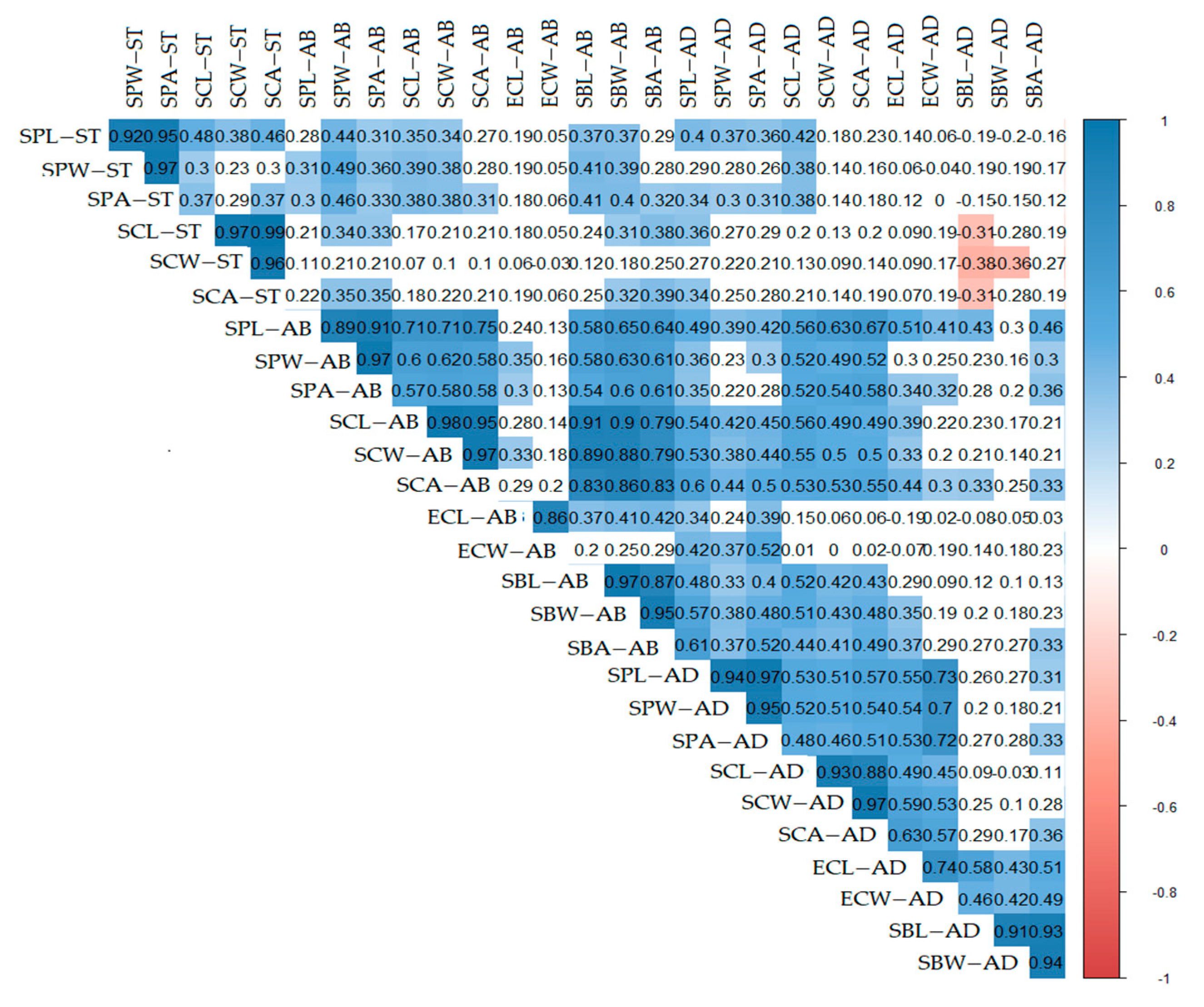
2.1. Scanning Electron Microscope (SEM)

2.1.1. Stem Micromorphology

Pustulate unicellular non-glandular trichomes of (45–98 × 13–23 µm) were observed on the stem of Silene leucophylla. In comparison, S. schimperiana had a glabrous stem surface. A thin layer of epicuticular wax was found in S. leucophylla. Simultaneously, S. schimperiana was covered with irregular crustose platelets of <1 µm height, with sinuate margin.
The type of stomatal complex was anomocytic in both species (Figure 1). Stem qualitative and quantitative traits are summarized in Table 1 and Supplementary (Table S2).
Geom-boxplot and ANOVA indicate a significant variation of stomatal traits among the two species with p-value (p = 0.0054, R-squared = 0.9487). Higher median stem stomata of S. leucophylla than S. schimperiana was also recorded in (Figure 2).

2.1.2. Leaf Epidermal Cells

The leaf epidermal cells of the adaxial (AD) and abaxial (AB) surface of S. leucophylla were oblong, bone-shaped, and irregularly arranged. On the other hand, in S. schimperiana, the AD and AB surfaces’ epidermal cells were “tetra-, penta-, hexa-, and polygonal” parallelly arranged (Figure 3).
The anticlinal walls (AW) were sunken, irregularly channeled and curved in S. leucophylla. At the same time, it was sunken but straight in S. schimperiana. The fine relief of the epidermal cell wall was highly ribbed on the A.B. surface and moderately ribbed on the AD surface in S. leucophylla. In contrast, it was covered by irregular epicuticular crustose platelets in S. schimperiana (Figure 3). Significant variations in the size of leaf epidermal cells were recorded by ANOVA analysis for the two species (p < 2.2 × 10⁻¹⁶, R-squared = 0.9708). The smallest stomatal area was recorded on the AB surface of S. leucophylla (46.44–74.64 = 61.42 ± 8.56 µm2), whereas the largest area was recorded on the AD surface of S. schimperiana (102.20–253.50 = 152.30 ± 37.92 µm2). Those measurements were confirmed by grouped boxplot for abaxial and adaxial leaf (Figure 4).

2.1.3. Stomatal Complex
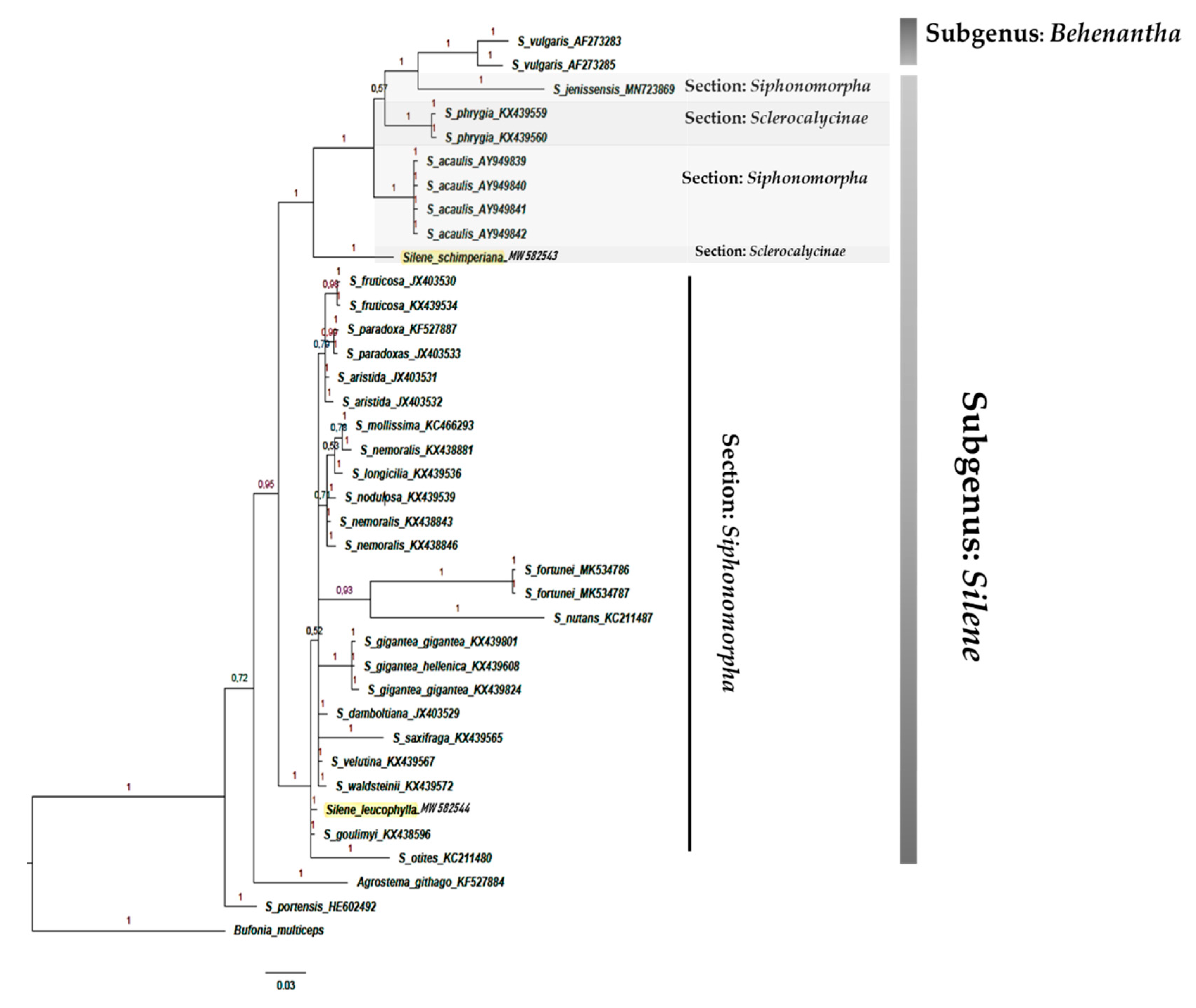
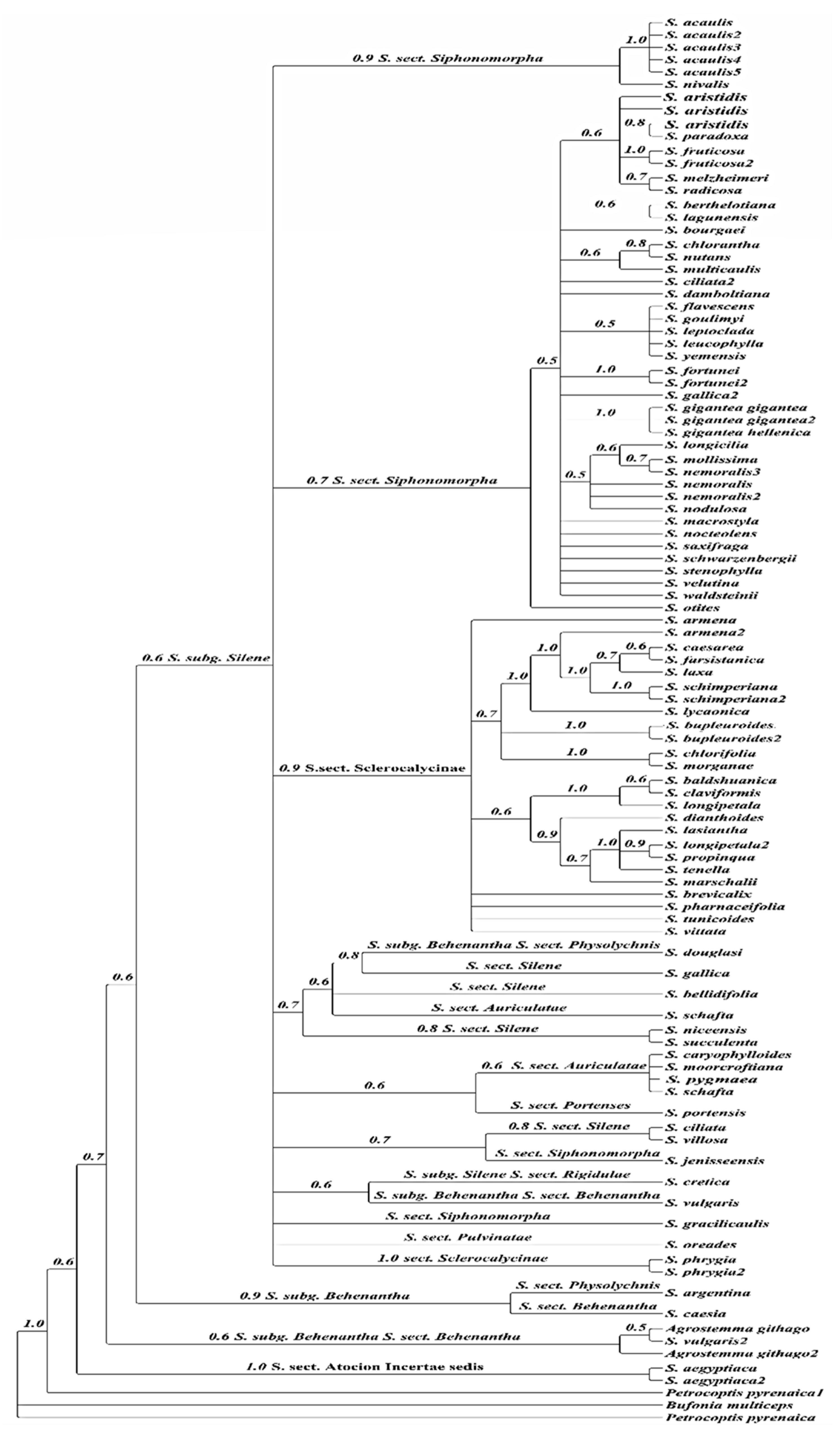
Leaves of the studied Silene taxa were amphistomatic. They have raised diacytic stomata with smooth guard cells observed in S. leucophylla, while S. schimperiana attained sunken diacytic stomata with irregular epicuticular crustose platelets on guard cells (Figure 3).
The smallest stomatal area was recorded on the AB surface of S. leucophylla (46.44–74.64 = 61.42 ± 8.56 µm2), On the other hand, the largest stomatal area was measured on the AD surface of S. schimperiana (102.20–253.50 = 152.30 ± 37.92 µm2) (Figure 5).
ANOVA of the length, width and area for the stomatal pore, stomatal complex and subsidiary cells of both AB and AD were p = 5.638 × 10⁻¹¹, R-squared = 0.9304. Grouped boxplot for those measurements were represented in (Figure 5).
The lowest stomatal index (SI%) was recorded in S. leucophylla (11.76–12.12 = 11.94 ± 0.25), whereas the highest SI (12.90–20.69 = 15.10 ± 3.76) was noticed for the AD surface in S. schimperiana. The pheatmap in (Figure 6) represents the overall variations between the investigated taxa. S. leucophylla representatives grouped in a separate cluster diverting from representatives of S. schimperiana. The correlogram correlation analysis showed a significant relationship among numerous traits; (Figure 7).

2.2. Phylogeny

The phylogenetic placement of the Egyptian species S. leucophylla and S. schimperiana within the whole genus was conducted using novel DNA sequences. The used markers were: a nuclear marker “nrDNA” Internal Transcribed Spacer (ITS), and three plastid markers “cpDNA”: matK; psbA-trnH, and rbcL.
The matrices of the DNA sequences of ITS, matK, psb-A/trn-H, and rbcL consisted of 48, 34, 36, and 23 Silene taxa, respectively. The outgroups were Petrocoptis glaucifolia, P. viscosa and P. pyrenaica in the ITS tree. Agrostemma githago and Petrocoptis pyrenaica were used as outgroups in matK. Bufonia multiceps was used in both psb-A/trn-H and and rbcL.
The results showed that section Sclerocalycinae represented a well-supported monophyletic clade (PP = 1) composed of 22 Silene taxa in the ITS analysis (Figure 8) and four taxa in the matK (Figure 9). The Egyptian species S. schimperiana was related to this section.
Section Siphonomorpha was also retrieved successfully. It consisted of 20 Silene including the Egyptian endemic species S. leucophylla. This section was moderately supported in both phylogenies PP = 0.72 in the ITS (Figure 8), and in the matk PP = 0.8 (Figure 9).
The phylogenetic tree based on the DNA sequences of the psb-A/trn-H showed that section Sclerocalycinae is not monophyletic. Silene schimperiana represented a sister clade to other Silene taxa belonging to section Siphonomorpha Otth: S. acaulis and S. jenissensis, and S. vulgaris related to the subgenus Behenanthae. Similarly, section Siphonomorpha was polyphyletic composed of 30 taxa, including the Egyptian species S. leucophylla (Figure 10).
The phylogenetic tree of the rbcL section Sclerocalycinae including S. schimperiana, represented a robust but unresolved clade (PP = 0.99). Simultaneously, Section Siphonomorpha including S. leucophylla was retrieved with a moderately supporting value PP = 0.88 (Figure 11).
In the four markers’ combined phylogenetic tree, the ingroup consisted of 102 taxa with 97 Silene taxa and five taxa from other genera (Figure 12). While Bufonia multiceps and Petrocoptis pyrenaica constituted the outgroup clades, Agrostemma githago was imbedded in the ingroup taxa.
The 97 ingroups of Silene taxa were divided into two subgenera: Silene subg. Silene Rohrbach, and Silene subg. Behenantha (Otth) Torr. & A. Gray, and neither of these two subgenera was monophyletic in our reconstruction. The subgenus Silene was composed of six sections: Sclerocalycinae, Siphonomorpha, Pulvinatae, Portenses, Auriculatae, and Atocion, all of which were not monophyletic. Silene schimperiana and S. leucophylla clustered into their related sections as proposed by single markers analyses, Sect. Sclerocalycinae and Sect. Siphonomorpha, respectively.

3. Discussion

The current research represents the first detailed study of the micromorphological and molecular phylogenetic placement of the endemic S. leucophylla and the near-endemic S. schimperiana native to the Sinai Peninsula, Egypt.
Leaf tomentum and the stem wax secretion of Silene species have significant ecological and systematic importance concerning the interaction between plants and their environment [29]. The current study confirms the hairy leaf texture of S. leucophylla, whereas S. schimperiana was glabrous. In contrast, irregular epicuticular crustose wax platelets were observed on the stem and leaf of S. schimperiana only. The obtained results were consistent with [23], who indicated that where species grow in drier habitats, they can attain straight to curved anticlinal walls. Silene leucophylla and S. schimperiana have amphistomatic leaves. This is also a characteristic feature of species occupying xerophytic habitats [30].
Guard cells integrate multiple environmental signals and control the aperture width to ensure appropriate stomatal function for plant survival [31]. Therefore, guard cells have been studied extensively as a model system for scrutinizing environment sensing dynamics and mechanisms [25]. In S. leucophylla, the stem stomatal complex area measured 1.2 times larger than S. schimperiana. Quantitative characteristics of the leaf stomatal complex and the epidermal cells on the leaf’s AD were more extensive than those on the A.B. in both species. These indicate great integrity between the cell geometry and size of the epidermal cells and genetic constraints, and environmental factors [32,33].
The cell dimensions of the stomatal complex of S. schimperiana were 2.6–2.9 times larger than those of S. leucophylla. The stomatal pore and its area were 24.4–33.5 times larger S. schimperiana than in S. leucophylla. Additionally, the subsidiary cell area was 2.69–3.13 times larger in S. schimperiana than S. leucophylla.
As suggested by [34,35,36], plant species located in high-elevation and shady areas similar to the growing habitats of both S. leucophylla and S. schimperiana in the mountains of the Sinai Peninsula are usually characterized by a low average Stomatal Index (SI%). This critical trait (SI%) was 12.42% in S. leucophylla and 14.44% in S. schimperiana. The SI% for both species were considerably low compared to the high SI% (≥95.58%) reported in plant species growing in low-elevation and sunny habitats [22].
Rohrbach [6] classified S. leucophylla and S. schimperiana in the same section, “Botryosilene”. While he considered S. leucophylla in the Nutantes series, S. schimperiana was classified by him into the series Sclerocalycinae. Classification of the genus Silene by [10,11] considered S. leucophylla a member of section Siphonomorpha, while S. schimperiana was allied to section Sclerocalycinae (Subsection Chlorifoliae).
The Sileneae taxonomy and systematics were recently studied by [4,9,37], who integrated morphological and molecular phylogeny to reveal the phylogenetic relationships within the tribe Sileneae. According to [4,9], S. leucophylla belongs to the subsection Brachypodae (Boiss.) Gürke allied to section Siphonomorpha, while S. schimperiana belongs to subsection Sclerocalycinae.
In the current study, the phylogenetic analysis of each single and the four combined markers confirmed the generic, sectional and phylogenetic placement of the Egyptian species under investigation—with both S. leucophylla and S. schimperiana related to the subgenus Silene. At the same time, the sectional placement shows that S. leucophylla can be placed into the S. sect. Sclerocalycinae and S. leucophylla into S. sect. Siphonomorpha.
The phylogenetic trees’ topologies were generally consistent with the generic and sections of the Silene classification [4]. Most Silene sections were not monophyletic and the support values for clades were either weak or moderate. The clade support shows low support of section Siphonomorpha in the ITS analysis PP = 0.72 (Figure 8). In contrast, it was moderately supported in the rbcL analysis PP = 0.88 (Figure 1), and strongly supported (PP = 1) in both matK (Figure 9) and psb-A/trn-H (Figure 10).
The results of Bayesian analysis of the ITS, matK, and rbcL data sets supported the monophyly of the sections Siphonomorpha and Sclerocalycinae. However, psb-A/trn-H and the combined analysis failed to retrieve the monophyly of those sections.
According to [37], S. sect. Siphonomorpha s.str. was monophyletic and should be considered a separate section. The current study evaluated that this section should incorporates two complexes: the Leucophylla complex and the Schimperiana complex (Figure 8 and Figure 9).
The Leucophylla complex (Figure 8) was considered to be allied to the widely distributed Nutans group [38]. However, the current study reveals that the Leucophylla complex is confined to narrowly distributed taxa native to the South Mediterranean region, S. flavescens: Bulgaria, Greece, Hungary, Romania, and Yugoslavia; S. leptoclada: East Aegean Island, Turkey, and North Africa; S. leucophylla: endemic to Egypt; S. yemensis: Eritrea, Ethiopia, Saudi Arabia, and Yemen. Moreover, S. saxifraga, a native and widely distributed species in Europe, constitutes a sister group of the Leucophylla complex. In addition to the geographical distribution pattern, the current study confirms that members of the Leucophylla complex shared several morphological traits. Those traits were perennial life form, plant texture, stem woody base, leaf type, flowers number and inflorescence type, calyx shape, and capsule features [39,40,41,42].
Similarly, section Sclerocalycinae s.l. (Figure 8, Figure 9 and Figure 12) is composed of nine geographically restricted species of Silene: S. armena, S. caesarea, S. farsistanica, S. laxa, S. schimperiana, S. lycaonica, S. bupleuroides, S. chlorofolia and S. morganae. Meanwhile, the matK shows that this section is composed of four species: S. vittata, S. armena, S. schimperiana and S. tunicoides. These constitute a subset of Silene taxa native to the west Irano-Turinain floristic region, including Turkey, Armenia, Iran, and Iraq. These species establish the strongly supported Schimperiana complex (PP = 0.99, ITS; PP = 1, matK; and PP = 1 in the combined analysis). Members of the Schimperiana complex shared observable morphological traits: perennial life form, glabrous stem, leaf type “lanceolate to linear”, opposite cauline leaf arrangement, paniculate inflorescence, glabrous calyx, whitish-yellow and bifid petals, anthophore length from 1 mm to 15 mm, and capsule length from 3 mm to 15 mm [40,41,42,43].

4. Materials and Methods

4.1. Plant Materials

Two herbarium specimens of Silene leucophylla and three of Silene schimperiana were obtained from Assuit University Herbarium (ASTU). Three stem and leaf replicates of each specimen were used in SEM. For molecular analysis, fresh leaf materials were collected from two different localities at Saint Katherine, South Sinai, Egypt. Vouchers of the collected specimens are deposited at the herbarium of Suez Canal University, Ismailia, Egypt (SCUI) under the collection number (SCUI).

4.2. Specimens Examined

The following specimens were examined: Silene leucophylla, Egypt, Southern Sinai, Wadi Gebal, 28.3219 N, 33.5513 E, Alt. 1895–1991 m, 13 May 2004, Fayed, I. El-Garf, Abdel-Khalik and A. Osman; (ASTU#1); Egypt, Southern Sinai, Wadi Gebal, 28.3219 N, 33.5513 E, Alt. 1895–1991 m, 13 May 2004, Fayed, I. El-Garf, Abdel-Khalik and A. Osman; (ASTU#2); Egypt, Southern Sinai, Wadi Gebal, Reheibet Nada, 28.528761 N, 33.91720 E, Alt. 2099 m, 4 May 2016, Ahmed El-Banhawy, Ahmed Elkordy (SCUI-AEB#302); Silene schimperiana, Egypt, Southern Sinai Al-Meserdy, 28.3242 N, 33.5623 E, Alt. 1775–1940 m, 13 May 2004, A. Fayed, I. El-Garf, Abdel-Khalik and A. Osman, (ASTU#3); Egypt, Southern Sinai Al-Meserdy, 28.3242 N, 33.5623 E, Alt. 1775–1940 m, 13 May 2004, A. Fayed, I. El-Garf, Abdel-Khalik and A. Osman, (ASTU#4) Wadi Gebal, Farsh EL Rommanah, 28.536667 N, 33.901111 E, Alt. 2004 m, 4 May 2016, Ahmed El-Banhawy, Ahmed Elkordy, (SCUI-AEB#303); Southern Sinai Wadi Gebal, 28.3219 N, 33.5513 E, Alt. 1895–1919 m, 13 May 2016, A. Fayed, I. El-Garf, Abdel-Khalik and A. Osman, (ASTU#5).

4.3. Scanning Electron Microscopy (SEM)

The stem, as well as the leaf abaxial (AB) and adaxial (AD) surfaces, were mounted onto stubs with double-sided adhesive tape, coated for 5 min with gold in a polaron JFC-1100E coating unit, and then were examined and photographed with JEOL JSM-IT200 scanning electron microscope unit at Faculty of Science, Alexandria University, Alexandria, Egypt. The epidermal cell characteristics separately described for abaxial (AB) and adaxial (AD) leaf surfaces. The quantitative data of stomatal measurements were recorded in both closed and opened stomata, i.e., length, width, and area. The quantitative characteristics measured by image analysis software [44] followed the terminology in [29].

4.4. Statistical Analysis

Sixty-eight traits of the stem and leaf of the examined taxa were analyzed by using the R- software (Vienna, Austria), with the required packages installed [45]. Initially, boxplots were generated using the “ggplot2” library [46], to address the variations in the measured traits of the stem, and the AB and AD of the leaf. Analysis of variance (ANOVA) was performed using the (aov) function. Subsequently, post hoc Tukey Honestly Significant Difference (HSD) was used to figure out which group(s) of the sample differed [47]. The “pheatmap” and “ggplot2” packages [46,48] were used to visualize the similarity and dissimilarity within and among species. The scale of color is relative to the value of the divergence between investigated readings. The red color indicates the high similarity between accessions while the blue color assuming the low similarity [49]. The relationships among the quantitative traits were assessed through correlation analysis which visualized by correlogram using “corrplot2”. In the correlogram, the intensity of the colors is an indication for positive correlation, whereas the white box point indicates the insignificant correlation between variables [50].

4.5. DNA Extraction, PCR Amplification, Sequencing, and Phylogenetic Analysis

Two fresh leaf materials preserved in silica gel of each species were used for molecular analysis. Total DNA was extracted from silica-gel dried leaves using a silica column method similar to commercially available extraction kits [51]. DNA was extracted using the Cetyltrimethylammonium bromide (CTAB) protocol with some modifications [52]. The PCR amplification were performed in 15 µL volume for ITS, matK, rbcL and psb-A/trn-H containing 5U/µL Taq DNA polymerase with 25 µM MgCl2, 10 µM of dNTPs, 10 µM of each primer. Amplifications were conducted using an Applied Biosystems®-VeritiTM 96- well thermal cycler (Thermo Fisher Scientific-Fisher Scientific AS-Postboks 114, Smestad-0309 Oslo, Norway). PCR products were purified with ExoSAP-IT (USB Corporation, Cleveland, OH, USA). PCR products were sent to Macrogen Spain for direct sequencing in both directions with an ABI 3730XL Genetic Analyzer (Life Technologies Corporation 5791 Van Allen Way Carlsbad, CA 92008). For the newly generated sequences, forward and reverse reads were assembled, and the contigs were edited into in GENEIOUS® v.R9 (Biomatters Ltd., Berkeley, CA 94709-1405 USA https://www.geneious.com accessed 15 January 2021) using a personal license (C.A.). Four data matrices were constructed: ITS, matK, rbcL, and psb-A/trn-H. The ingroup were selected to cover most of the major sections in the genus Silene. Each marker’s datasets were initially aligned using MAFFT algorithms, implemented in Geneious, using default alignment parameters, and visually revised to manually correct errors in alignment.
Eight novel DNA sequences of both species under investigation were deposited in the GenBank along with the following accessions: S. leucophylla (SCUI-AEB #302), ITS (MW 559753), matK (MW 582539), psb-A/trn-H (MW 582543), and rbcL (MW 582541); and S. schimperiana (SCUI-AEB #303) ITS (MW 559754), matK (MW 582540), psb-A/trn-H (MW 582544), and rbcL (MW 582542). Previously, published DNA sequences for ITS, matK, psbA-trnH, and rbcL were downloaded from GenBank to construct balanced datasets, and NCBI codes are reported in the Supplementary (Table S3).
The optimal nucleotide substitution model was estimated using MrModeltest [53] and executed in MrBayes blocks. A 50% majority role consensus tree was constructed to get the posterior probabilities (PP). Posteriori probabilities, values >0.5 at a given branch were considered to support the existence of that branch [54]. All phylogenetic analyses were run on the CIPRES portal [55].

5. Conclusions

The combined investigations of the morphological data of leaf, stem and the molecular phylogenetic analysis of four molecular markers represented a comprehensive characterization of the endemic S. leucophylla and the near-endemic S. schimperiana native to Egypt, Yemen, and Saudi Arabia. The qualitative and quantitative morphological characters include presence/absence of trichomes, epicuticular wax type, leaf epidermal cells, and complex stomatal traits attributed entirely to well-characterizing endemic endangered species supported the previous taxonomic revisions. The Bayesian phylogenetic inference of S. leucophylla and S. schimperiana using four nuclear markers portrayed the target taxa’s phylogenetic position within the whole genus. Both species related to the subgenus Silene and their corresponding sections, where S. leucophylla allied to section Siphonomorpha and S. schimperiana allied to section Sclerocalycinae.
The sectional classification of the Silene species native to the Sinai Peninsula, Egypt has so far always been investigated on the basis of morphological descriptions only. The current research represents the first phylogenetically based study to reveal the sectional classification of S. leucophylla and S. schimperiana. Moreover, two morphologically and phylogenetically supported Silene complexes are proposed by the current research. The Leucophylla complex allied to section Siphonomorpha and the Schimperiana complex related to section Sclerocalycinae. However, these two complexes require further investigation and more exhaustive sampling to infer their complex phylogenetic relationships.

Supplementary Materials

The following are available online at https://www.mdpi.com/article/10.3390/plants10040740/s1. Table S1. Morphological differences between Silene leucophylla and Silene schimperiana and the nearest elements of the genus Silene. Table S2. Quantitative characteristics of stem and leaf Abaxial leaf (AB) and Adaxial leaf (AD) characters of Silene leucophylla and Silene schimperiana. Table S3. SEQs Accession numbers of ITS, matK, psbA-trnH, and rbcL downloaded from GenBank.

Author Contributions

Conceptualization, A.E.-B. and A.E.; methodology, A.E.-B., C.A., I.H.N., F.Y.E., A.F., A.O.O. and A.E.; software, A.E.-B., F.Y.E., W.A.-J., A.M.H.G. and C.A.; data curation, A.E.-B., I.H.N., F.Y.E., W.A.-J., A.M.H.G. and C.A.; investigation, A.E.-B., C.A., I.H.N., F.Y.E., A.F. and A.O.O.; resources, A.E.-B., I.H.N., F.Y.E., A.F., A.O.O., W.A.-J., A.M.H.G. and A.E.; Formal analysis, A.E.-B., I.H.N., F.Y.E. and C.A.; writing—original draft preparation, A.E.-B., I.H.N., F.Y.E. and A.O.O.; writing—review and editing, A.E.-B., I.H.N., F.Y.E., W.A.-J., A.M.H.G. and A.F.; funding acquisition, A.E.-B., C.A. and A.E.; All authors have read and agreed to the published version of the manuscript.

Funding

This research received partial funding from the Spanish Ministerio de Educación under the program “Campus de Excelencia Internacional CEI Triangular E3 (Grants 2017)”, which provided financial support for the postdoctoral stay of Ahmed Elkordy at the University of León (Spain). The Research Team of the University of León 395 TaCoBi (Taxonomy and Biodiversity Conservation) partially supported the experimental study.

Data Availability Statement

The data presented in this study are available in this article and Supplementary Material.

Conflicts of Interest

The authors declare no conflict of interest.

References

  1. Greenberg, A.K.; Donoghue, M.J. Molecular systematics and character evolution in Caryophyllaceae. Taxon 2011, 60, 1637–1652. [Google Scholar] [CrossRef]
  2. Rabeler, R.K.; Hartman, R.L. Caryophyllaceae in North America: Change over the last 110 years. J. Bot. Res. Inst. Tex. 2008, 2, 1285–1289. [Google Scholar]
  3. Madhani, H.; Rabeler, R.; Pirani, A.; Oxelman, B.; Heubl, G.; Zarre, S. Untangling phylogenetic patterns and taxonomic confusion in tribe Caryophylleae (Caryophyllaceae) with special focus on generic boundaries. Taxon 2018, 67, 83–112. [Google Scholar] [CrossRef] [Green Version]
  4. Jafari, F.; Zarre, S.; Gholipour, A.; Eggens, F.; Rabeler, R.; Oxelman, B. A new taxonomic backbone for the infrageneric classification of the species-rich genus Silene (Caryophyllaceae). Taxon 2020, 69, 337–368. [Google Scholar] [CrossRef]
  5. Hernández-Ledesma, P.; Berendsohn, W.G.; Borsch, T.; Von Mering, S.; Akhani, H.; Arias, S.; Castañeda-Noa, I.; Eggli, U.; Eriksson, R.; Flores-Olvera, H. A taxonomic backbone for the global synthesis of species diversity in the angiosperm order Caryophyllales. Willdenowia 2015, 45, 281–383. [Google Scholar] [CrossRef] [Green Version]
  6. Rohrbach, P. Conspectus systematicus specierum generis Silene Ann. Sci. Nat. Bot. 1868, 5, 369–382. [Google Scholar]
  7. Faried, A.; El-Banhawy, A.; Elqahtani, M. Taxonomic, DNA barcoding and phylogenetic reassessment of the Egyptian Ephedra L. (Ephedraceae). Catrina Int. J. Environ. Sci. 2018, 17, 1–13. [Google Scholar]
  8. El-Banhawy, A.; Al-Juhani, W. DNA barcoding and phylogeny of Phlomis aurea (Lamiaceae) endemic to Sinai Peninsula, Egypt. Pak. J. Bot. 2019, 51, 1263–1271. [Google Scholar] [CrossRef]
  9. Oxelman, B.; Rautenberg, A.; Thollesson, M.; Larsson, A.; Frajman, B.; Eggens, F.; Petri, A.; Aydin, Z.; Töpel, M.; Brandtberg-Falkman, A. Sileneae Taxonomy and Systematics. Available online: http://www.sileneae.info (accessed on 25 February 2021).
  10. Hosny, A.; El Hadidi, M.; Shamso, E. Taxonomic studies of Silenoideae (Cartophyllaceae) in Egypt. 1. Systematic revision of the genus Silene L. Taeckholmia 1992, 14, 1–36. [Google Scholar]
  11. Chowdhuri, P. Studies in the genus Silene. Notes R. Bot. Gard. Edinb. 1957, 22, 221–278. [Google Scholar]
  12. Royal Botanic Gardens Kew. World Checklist of Selected Plant Families (WCSP); Royal Botanic Gardens Kew: London, UK, 2018. [Google Scholar]
  13. Fakhry, A.M.; El-Keblawy, A.; Shabana, H.A.; Gamal, I.E.; Shalouf, A. Microhabitats Affect Population Size and Plant Vigor of Three Critically Endangered Endemic Plants in Southern Sinai Mountains, Egypt. Land 2019, 8, 86. [Google Scholar] [CrossRef] [Green Version]
  14. Omar, K. Community–Based Conservation of Threatened Plants Silene schimperiana and Polygala sinaica in South Sinai, Egypt. Available online: www.rufford.org (accessed on 15 February 2021).
  15. Abdelaal, M.; Fois, M.; Fenu, G.; Bacchetta, G. Using MaxEnt modeling to predict the potential distribution of the endemic plant Rosa arabica Crép. in Egypt. J. Ecol. Inform. 2019, 50, 68–75. [Google Scholar] [CrossRef]
  16. El-Ghamery, A.; Morsy, M.; Abu-Shahba, M.J.T. Chromosome count and karyotype study on endemic species Silene schimperiana Boiss.(Caryophyllaceae), Sinai, Egypt. Taeckholmia 2016, 36, 102–114. [Google Scholar] [CrossRef]
  17. Zarrinkamar, F. Foliar anatomy of the Caryophyllaceae family in Arasbaran, NW Iran. Iran. J. Bot. 2001, 9, 93–102. [Google Scholar]
  18. Ahmad, K.; Khan, M.A.; Ahmad, M.; Zafar, M.; Arshad, M.; Ahmad, F. Taxonomic diversity of stomata in dicot flora of a district tank (N.W.F.P.) in Pakistan. Afr. J. Biotechnol. 2009, 8, 1052–1055. [Google Scholar]
  19. Hosseini, E.; Ghahremaninejad, F.; Assadi, M. The indumentum of vegetative and reproductive parts of annual species of Silene (Caryophyllaceae) in Iran. Prog. Biol. Sci. 2016, 6, 107–116. [Google Scholar]
  20. Sheibani, M.; Pirani, A. Trichome micromorphology of Silene (Caryophyllaceae) species in Iran. Iran. J. Bot. 2018, 24, 28–45. [Google Scholar]
  21. Chandra, S.; Rawat, D.; Verma, S.R.; Uniyal, P. Trichome diversity of the family Caryophyllaceae from Western Himalaya and their taxonomic implication. Trop. Plant Res. 2019, 6, 397–407. [Google Scholar] [CrossRef]
  22. Ullah, F.; Zafar, M.; Ahmad, M.; Shah, S.N.; Razzaq, A.; Sohail, A.; Zaman, W.; Çelik, A.; Ayaz, A.; Sultana, S. A systematic approach to the investigation of foliar epidermal anatomy of subfamily Caryophylloideae (Caryophyllaceae). Flora 2018, 246, 61–70. [Google Scholar] [CrossRef]
  23. Stace, C.A. Cuticular Studies as an Aid to Plant Taxonomy; British Museum (Natural History): London, UK, 1965. [Google Scholar]
  24. Casson, S.; Gray, J.E. Influence of environmental factors on stomatal development. New Phytol. 2008, 178, 9–23. [Google Scholar] [CrossRef]
  25. Dina, A.F.; Ibrahim, M.F.; Farag, R.; El-Gawad, H.G.A.; El-Banhawy, A.; Alaraidh, I.A.; Rashad, Y.M.; Lashin, I.; El-Yazied, A.A.; Elkelish, A.J.P. Influence of Polyethylene Glycol on Leaf Anatomy, Stomatal Behavior, Water Loss, and Some Physiological Traits of Date Palm Plantlets Grown In Vitro and Ex Vitro. Plants 2020, 9, 1440. [Google Scholar] [CrossRef] [PubMed]
  26. Hoveka, L.N.; van der Bank, M.; Bezeng, B.S.; Davies, T.J. Identifying biodiversity knowledge gaps for conserving South Africa’s endemic flora. Biodivers. Conserv. 2020, 29, 2803–2819. [Google Scholar] [CrossRef]
  27. Kougioumoutzis, K.; Kokkoris, I.P.; Panitsa, M.; Kallimanis, A.; Strid, A.; Dimopoulos, P. Plant Endemism Centres and Biodiversity Hotspots in Greece. Biology 2021, 10, 72. [Google Scholar] [CrossRef] [PubMed]
  28. Martín-Gómez, J.J.; Rewicz, A.; Rodríguez-Lorenzo, J.L.; Janoušek, B.; Cervantes, E. Seed Morphology in Silene Based on Geometric Models. Plants 2020, 9, 1787. [Google Scholar] [CrossRef]
  29. Barthlott, W.; Neinhuis, C.; Cutler, D.; Ditsch, F.; Meusel, I.; Theisen, I.; Wilhelmi, H. Classification and terminology of plant epicuticular waxes. Bot. J. Linn. Soc. 1998, 126, 237–260. [Google Scholar] [CrossRef]
  30. Parkhurst, D.F. The adaptive significance of stomatal occurrence on one or both surfaces of leaves. J. Ecol. 1978, 66, 367–383. [Google Scholar] [CrossRef]
  31. Takahashi, S.; Monda, K.; Negi, J.; Konishi, F.; Ishikawa, S.; Hashimoto-Sugimoto, M.; Goto, N.; Iba, K. Natural variation in stomatal responses to environmental changes among Arabidopsis thaliana ecotypes. PLoS ONE 2015, 10, e0117449. [Google Scholar] [CrossRef] [Green Version]
  32. Horiguchi, G.; Tsukaya, H. Organ size regulation in plants: Insights from compensation. Front. Plant Sci. 2011, 2, 24. [Google Scholar] [CrossRef] [Green Version]
  33. Gázquez, A.; Beemster, G.T. What determines organ size differences between species? A meta-analysis of the cellular basis. New Phytol. 2017, 215, 299–308. [Google Scholar] [CrossRef] [Green Version]
  34. El-Banhawy, A.; Acedo, C.; Qari, S.; Elkordy, A. Molecular Identification and Phylogenetic Placement of Rosa arabica Crép.(Rosaceae), a Critically Endangered Plant Species. Life 2020, 10, 335. [Google Scholar] [CrossRef]
  35. Rossatto, D.R.; Kolb, R.M. Gochnatia polymorpha (Less.) Cabrera (Asteraceae) changes in leaf structure due to differences in light and edaphic conditions. Acta Bot. Bras. 2010, 24, 605–612. [Google Scholar] [CrossRef]
  36. Schoch, P.-G.; Zinsou, C.; Sibi, M.J. Dependence of the stomatal index on environmental factors during stomatal differentiation in leaves of Vigna sinensis L. 1. Effect of light intensity. J. Exp. Bot. 1980, 31, 1211–1216. [Google Scholar] [CrossRef]
  37. Naciri, Y.; Du Pasquier, P.-E.; Lundberg, M.; Jeanmonod, D.; Oxelman, B. A phylogenetic circumscription of Silene sect. Siphonomorpha (Caryophyllaceae) in the Mediterranean Basin. Taxon 2017, 66, 91–108. [Google Scholar]
  38. Jeanmonod, D. Révision de la section Siphonomorpha Otth du genre Silene L. (Caryophyllaceae) en Méditerranée occidentale. IV: Species caeterae. Candollea 1985, 40, 5–34. [Google Scholar]
  39. Boissier, P.E. Flora Orientalis, Sive, Enumeratio Plantarum in Oriente a Graecia et Aegypto ad Indiae Fines Hucusque Observatarum; Georg, H., Ed.; Basileae: Genevae, Switzerland, 1875; Volume 1. [Google Scholar]
  40. Boulos, L. Flora of Egypt; Al Hadara Publishing: Cairo, Egypt, 1999. [Google Scholar]
  41. Coode, M.J.E.; Cullen, J. Silene L. Flora of Turkey and the East Aegean Islands; Davis, P.H., Ed.; Edinburgh University Press: Edinburgh, Scotland, 1967; Volume 2, pp. 179–242. [Google Scholar]
  42. Deflers, A. Voyage au Yémen, Journal D’une Excursion Botanique Faite en 1887 Dans les Montagnes de l’Arabie Heureuse; Kincksieck, P., Ed.; Wentworth Press: Paris, France, 1889; p. 306. [Google Scholar]
  43. Greuter, W. Studies in Greek Caryophylloideae: Agrostemma, Silene, and Vaccaria; Willdenowia: Berlin, Germany, 1995; pp. 105–142. [Google Scholar]
  44. Schneider, C.A.; Rasband, W.S.; Eliceiri, K.W. NIH Image to ImageJ: 25 years of image analysis. Nat. Methods 2012, 9, 671–675. [Google Scholar] [CrossRef]
  45. R Development Core Team. R: A Language and Environment for Statistical Computing; R Foundation for Statistical Computing: Vienna, Austria, 2011. [Google Scholar]
  46. Wickham, H. ggplot2: Elegant Graphics for Data Analysis; Springer: New York, NY, USA, 2016. [Google Scholar]
  47. Ferreira, E.B.; Cavalcanti, P.P.; Nogueira, D.A. ExpDes: An R package for ANOVA and experimental designs. J. Appl. Math. 2014, 5, 2952. [Google Scholar] [CrossRef] [Green Version]
  48. Kolde, R. Pheatmap: Pretty Heatmaps; Version 1.0.8; Vienna, Austria, 2015. [Google Scholar]
  49. Viscosi, V.; Cardini, A. Leaf morphology, taxonomy and geometric morphometrics: A simplified protocol for beginners. PLoS ONE 2011, 6, e25630. [Google Scholar] [CrossRef] [Green Version]
  50. Soetewey, A. Correlation Coefficient and Correlation Test in R. 2020. Available online: https://www.statsandr.com/blog/correlation-coefficient-and-correlation-test-in-r/ (accessed on 20 February 2021).
  51. Alexander, P.J.; Rajanikanth, G.; Bacon, C.D.; Bailey, C.D. Recovery of plant DNA using a reciprocating saw and silica-based columns. Mol. Ecol. Notes 2007, 7, 5–9. [Google Scholar] [CrossRef]
  52. Doyle, J. DNA protocols for plants. In Molecular Techniques in Taxonomy; Springer: Berlin/Heidelberg, Germany, 1991; pp. 283–293. [Google Scholar]
  53. Posada, D.; Crandall, K.A. Modeltest: Testing the model of DNA substitution. Bioinformatics 1998, 14, 817–818. [Google Scholar] [CrossRef] [PubMed] [Green Version]
  54. Mao, Y.R.; Zhang, Y.H.; Nakamura, K.; Guan, B.C.; Qiu, Y.X. Developing DNA barcodes for species identification in Podophylloideae (Berberidaceae). J. Syst. Evol. 2014, 52, 487–499. [Google Scholar] [CrossRef]
  55. Miller, M.A.; Pfeiffer, W.; Schwartz, T. The CIPRES science gateway: A community resource for phylogenetic analyses. In Proceedings of the 2011 TeraGrid Conference: Extreme Digital Discovery, Salt Lake City, UT, USA, 18–21 July 2011; pp. 1–8. [Google Scholar]
Figure 1. Scanning Electron Microscope (SEM) photomicrographs of Silene stem. Stem texture: (a) Non-glandular pustulate trichome of Silene leucophylla; (b) glabrous stem surface of Silene schimperiana. Stem epicuticular wax: (c) thin layer with anomocytic stomata of Silene leucophylla; (d) irregular epicuticular crustose platelets with anomocytic stomata in Silene schimperiana.
Plants 10 00740 g001
Figure 2. Boxplots of micromorphological stem stomatal traits of Silene leucophylla and Silene schimperiana.
Plants 10 00740 g002
Figure 3. Scanning Electron Microscope (SEM) photomicrographs of Silene leaf surfaces. Epidermal cell shapes in (a) Silene leucophylla and (b) Silene schimperiana. Ribbing pattern of fine relief in Silene leucophylla: (c) densely ribbed and raised stomata, (d) moderately ribbed. Epicuticular platelets in Silene schimperiana: (e) with sunken stomata, (f) irregular platelets. (Abaxial leaf surface (b,c,e); Adaxial leaf surface (a,d,f).
Plants 10 00740 g003
Figure 4. Boxplots of micromorphological quantitative measurements of epidermal cells of Silene leucophylla and Silene schimperiana leaves.
Plants 10 00740 g004
Figure 5. Boxplots of the quantitative data of complex stomatal characteristics, subsidiary cells and stomatal pore measurements of Silene leucophylla and Silene schimperiana.
Plants 10 00740 g005
Figure 6. Pheatmap based on the quantitative data of stem and leaf micromorphological traits, showing the similarity and dissimilarity within and among Silene leucophylla and Silene schimperiana.
Plants 10 00740 g006
Figure 7. Correlogram between quantitative traits of Silene leucophylla and Silene schimperiana. Positive and negative correlations are displayed in blue and red color, respectively. Correlation coefficients are proportional to color intensity.
Plants 10 00740 g007
Figure 8. Bayesian phylogenetic tree of Silene species based on nrDNA sequences of the ITS marker. Numbers above branches represented (PP).
Plants 10 00740 g008
Figure 9. Bayesian phylogenetic tree of Silene species based on cpDNA sequences of the matK marker. Numbers above branches represented (PP).
Plants 10 00740 g009
Figure 10. Bayesian phylogenetic tree of Silene species based on cpDNA sequences of the psb-A/trn-H marker. Numbers above branches represented (PP).
Plants 10 00740 g010
Figure 11. Bayesian phylogenetic tree of Silene species based on cpDNA sequences of the rbcL marker. Numbers above branches represented (PP).
Plants 10 00740 g011
Figure 12. Bayesian cladogram of Silene taxa based on combined DNA sequences of ITS, matK, psb-A/trn-H, and rbcL markers. Numbers above branches represented (PP).
Plants 10 00740 g012
Table 1. Qualitative characteristics of stem and leaf Abaxial leaf (AB) and Adaxial leaf (AD) characters of Silene leucophylla and Silene schimperiana.
CharactersSilene leucophylla Boiss.Silene schimperiana Boiss.
Stem characters
SurfacePubescentGlabrous
Epicuticular WaxThin layerIrregular crustose platelets
TrichomesPresentAbsent
Trichomes TypeUnicellular, non-glandularAbsent
Trichome SurfaceDensely pustulateAbsent
Trichomes Length 45–98 µmAbsent
Trichomes Width 13–23 µmAbsent
Stomata TypeAnomocyticAnomocytic
Abaxial leaf (AB) characters
Epidermal Cell ShapeIrregular oblong to bone-shapeParallel tetra- to polygonal
Anticlinal WallsSunken channeled irregularly curvedSunken straight
The curvature of Outer Periclinal Walls ConvexSlightly Convex
Fine Relief of the Cell WallHighly ribbedIrregular epicuticular crustose platelets
Trichomes TypeUnicellular, non-glandularAbsent
Trichome SurfaceDensely irregular pustulateAbsent
Trichomes Length78–130 µmAbsent
Trichomes Width18–20 µmAbsent
Stomata TypeDiacyticDiacytic
Stomata LevelRaisedSunken
Guard Cell SurfaceSmoothIrregular epicuticular crustose platelets
Pore ShapeElliptic slitLinear slit
Adaxial leaf (AD) characters
Epidermal Cell ShapeIrregular oblong to bone-shapeParallel penta- to polygonal
Anticlinal WallsSunken channeled irregularly curvedSunken straight
The curvature of Outer Periclinal Walls ConvexSlightly Convex
Fine Relief of the Cell WallModerately ribbedIrregular epicuticular crustose platelets
Trichomes TypeUnicellular, non-glandularAbsent
Trichome SurfaceDensely irregular pustulateAbsent
Trichomes Length75–125 µmAbsent
Trichomes Width16–25 µmAbsent
Stomata TypeDiacyticDiacytic
Stomata LevelRaisedSunken
Guard Cell SurfaceSmoothIrregular epicuticular crustose platelets
Pore ShapeElliptic slitElliptic + Linear slits
Publisher’s Note: MDPI stays neutral with regard to jurisdictional claims in published maps and institutional affiliations.

