Article

Higher Virulence of Diplodia seriata Isolates on Vines of cv. Cabernet Sauvignon Associated with 10-Year-Old Wood Compared to Young Tissue

by
Alejandra Larach
1,2,
Paulina Vega-Celedón
1,2,
Eduardo Salgado
1,
Aldo Salinas
1,
Natalia Riquelme
1,
Diyanira Castillo-Novales
1,2,
Paulina Sanhueza
1,
Michael Seeger
2 and
Ximena Besoain
1,*
1
Escuela de Agronomía, Facultad de Ciencias Agronómicas y de los Alimentos, Pontificia Universidad Católica de Valparaíso, Casilla 4-D, Quillota 2260000, Chile
2
Laboratorio de Microbiología Molecular y Biotecnología Ambiental, Departamento de Química & Centro de Biotecnología Dr. Daniel Alkalay Lowitt, Universidad Técnica Federico Santa María, Avenida España 1680, Valparaíso 2390123, Chile
*
Author to whom correspondence should be addressed.
Plants 2023, 12(16), 2984; https://doi.org/10.3390/plants12162984
Submission received: 1 July 2023 / Revised: 8 August 2023 / Accepted: 14 August 2023 / Published: 18 August 2023
(This article belongs to the Special Issue Pathogenesis and Disease Control in Crops)

Abstract

:
Botryosphaeria dieback (BD) occurs in young and old plants. In the field, the prevalence and severity of the disease increase proportionally with the age of vineyards. Among the pathogens that cause BD, Diplodia seriata is the most prevalent species in Chile and other countries with a Mediterranean climate. To date, no information is available on the susceptibility of adult wood to infection by this pathogen since most of the pathogenicity tests have been carried out on 1- or 2-year-old shoots or detached canes. Therefore, a pathogenicity test was carried out on plants under field conditions, with inoculations in 1-year-old shoots and 2- and 10-year-old wood in grapevine cv. Cabernet Sauvignon. A pathogenicity test was carried out with two isolates of D. seriata. The results for the plants show that D. seriata was significantly more aggressive on the 10-year-old than on the one- or two-year-old tissue, where the lesions were 4.3 and 2.3 cm on average, respectively. These results were compared with the lesions obtained from two-year-old canes after the isolates were activated in grape berries. Also, the Chilean isolates of D. seriata were compared phylogenetically with those from other countries, and no major differences were found between them. Our results are consistent with the damage observed in the field, contributing to the knowledge of the epidemiology of this disease in Mediterranean climates. In the future, the effect observed in cv. Cabernet Sauvignon with D. seriata on virulence at different tissue ages should be tested for other BD-causing agents and wine varieties.

1. Introduction

Grapevine trunk diseases (GTDs) are the leading cause of vineyard deterioration, causing a decrease in the viability of vineyards, an increase in production costs, and major economic losses in the wine industry [1,2,3,4,5,6]. The three main GTDs are esca disease, Eutypa dieback, and Botryosphaeria dieback (BD), which generally attack the structural parts of plants [2,6,7,8,9,10]. BD is present on all continents where wine or table grapes are grown [2,5,7,11,12,13,14,15,16]. Regarding the pathogens associated with BD, 26 species have been identified [10], mainly within the genera Botryosphaeria, Diplodia, Lasiodiplodia, and Neofusicoccum [7,10,11,17]. Some genera are more common in different climates, for example, Diplodia in temperate regions or those with cold winters and Lasiodiplodia in tropical and subtropical areas [18,19,20,21,22,23,24]. Diplodia seriata is the most prevalent species in vineyards in Central Chile [5,15,22] as well as in other parts of the world, such as New South Wales [25,26,27], Western Australia [28], California [29], and South Australia [26].
BD causes essential losses in the production of grapes for wine, with a decrease from 30 to more than 50% of production depending on the severity of the disease [5]. The loss of production is associated with the damage it causes to the vines due to the death of the wood where fruit develops, such as cankers in the wood, dead spurs, dead arms, and brown discolorations in the vascular area [2,5,7,15,16,30,31,32,33,34]. In the field, the prevalence and severity of (BD) increase proportionally with the age of the vineyards [5,6,7,10,15,33].
Considering physiological and molecular studies carried out with Botryosphaeriaceae, Bernard-Gellon et al.’s [35] work with the callous tissue of V. vinifera cv. Chardonnay found that N. parvum produced more extracellular proteins in higher concentrations than D. seriata, and equivalent concentrations of proteins secreted by N. parvum were more aggressive than those of D. seriata in producing necrosis and induced more grapevine defense genes. Morales-Cruz et al. [36] found that D. seriata has fewer genes associated with CAZymes enzymes, enzymes associated with secondary metabolism, active enzymes, and cytochrome P450 than N. parvum.
However, many available pathogenicity studies have been conducted on callus tissue on one-year-old shoots and two-year-old canes. Another critical issue to consider is the susceptibility and importance of some varieties for the wine industry. In this case, Cabernet Sauvignon is the most planted and susceptible variety in Chile, and it is appreciated for good quality wines [37].
For this reason, in this study, a pathogenicity test was carried out with five Chilean isolates of D. seriata in 2-year-old detached canes of Vitis vinifera cv. Cabernet Sauvignon, and with the two most representative isolates, a pathogenicity test was carried out on vine plants under field conditions, with inoculations in 1-year-old shoots, 2-year-old wood, and 10-year-old wood of grapevine.

2. Results

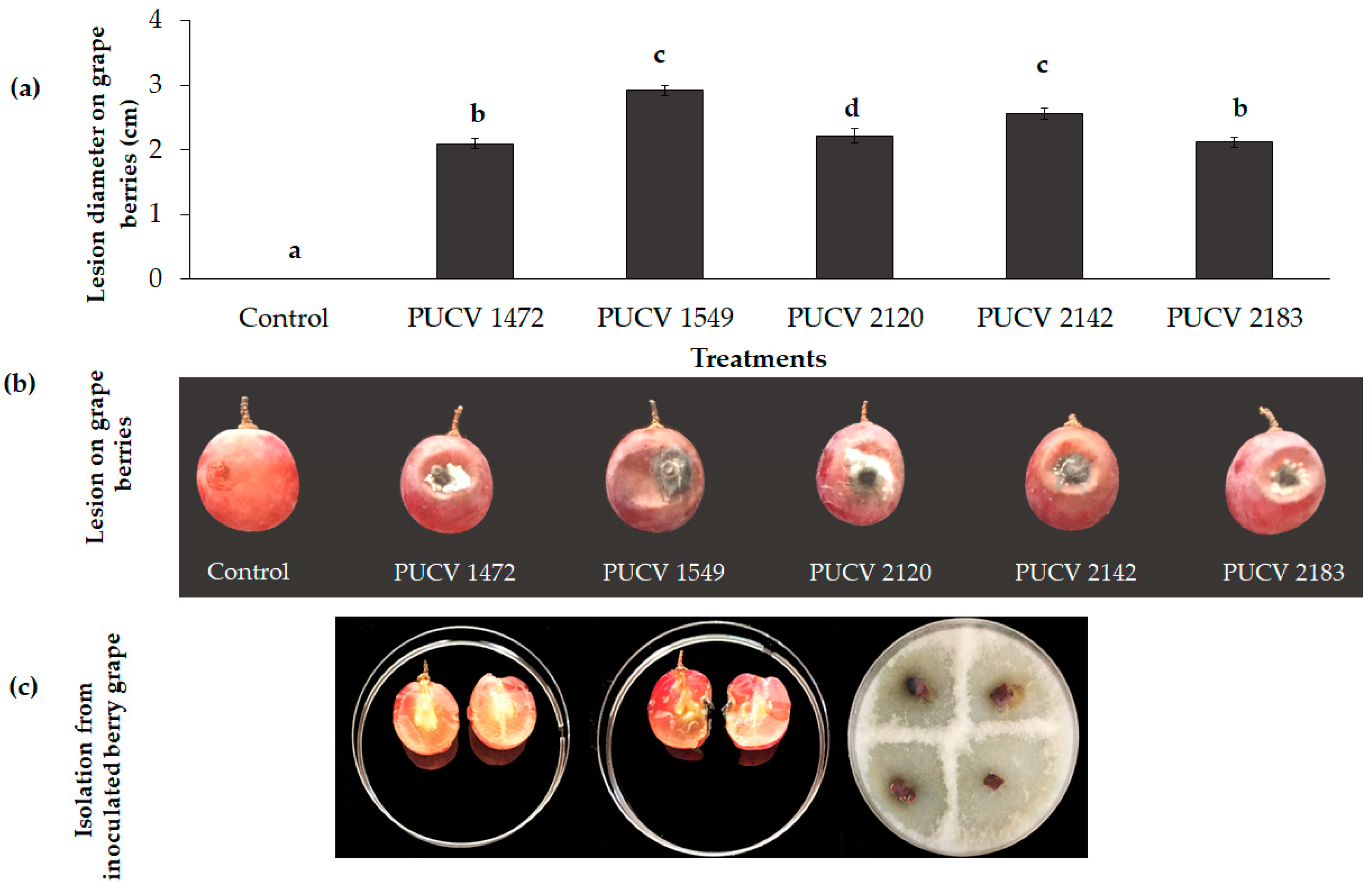
2.1. Pathogenicity Tests on Berry Grapes

All isolates caused circular soft rot around the point of inoculation in grape berries, with lesion diameters between 2.1 and 2.9 cm and the presence of grayish-white mycelium around the point of inoculation (Figure 1a,b). The largest lesions were caused by isolate 1549 (2.9 cm), followed by isolate 2142 (2.6 cm), and then by isolates 1472 (2.1), 2120 (2.2 cm), and 2183 (2.1 cm). The pathogen was recovered from the area of advancement of the lesions, and D. seriata was morphologically identified. The controls (uninoculated berry grape) did not show damage (Figure 1c), thus corroborating that all D. seriata isolates were pathogenic in berry cv. Red Globe.

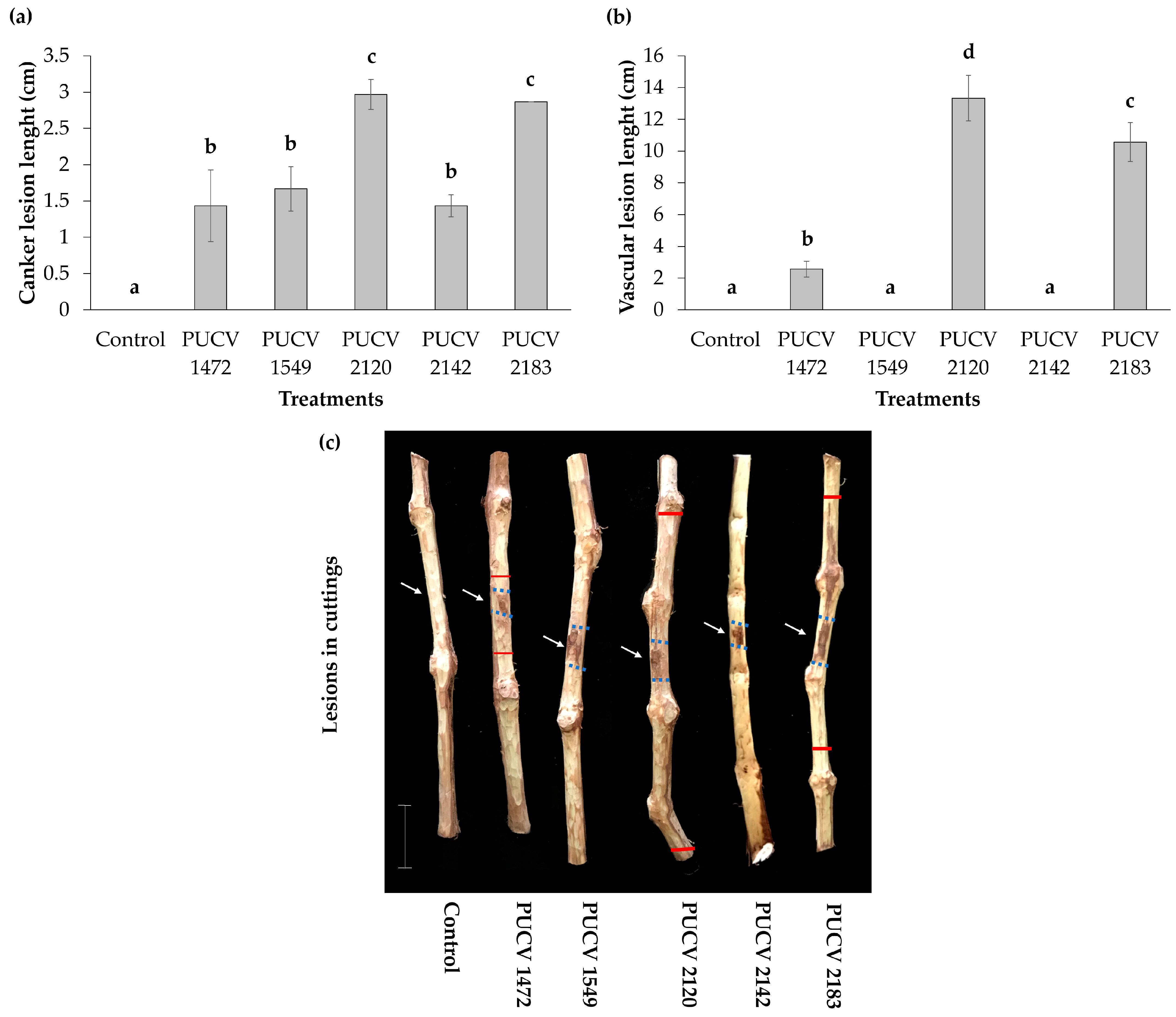
2.2. Pathogenicity Tests in Detached Canes (Activated Isolates)

All isolates produced canker lesions on inoculated canes, with lengths between 1.4 and 3.0 cm (Figure 2a), and isolates PUCV 2120 and PUCV 2183 causing the longest cankers. Isolates PUCV 1472, PUCV 2120, and PUCV 2183 also produced vascular discoloration, which was expressed up and down from the point of inoculation. The vascular lesions presented variable lengths between 2.6 and 13.3 cm (Figure 2b), and as with the cankers, isolates PUCV 2120 and PUCV 2183 were the ones that caused the most significant lesions. It is also important to note that the average size of the vascular lesions was 4.1 times longer than that of the canker lesions. The vascular lesions and cankers were brown and dark brown, respectively; the negative controls (noninoculated canes) did not present damage (no canker and no vascular discoloration) (Figure 2c). From the lesions produced by the isolates, the pathogen was recovered, and D. seriata was morphologically identified. With the results obtained in detached cane assays, isolates PUCV 2120 and PUCV 2183 were selected for the field trial.

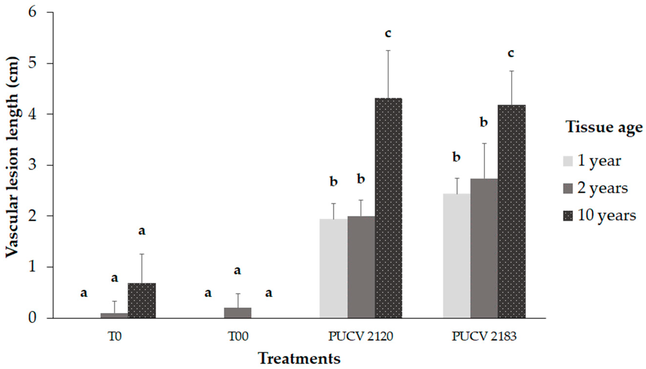
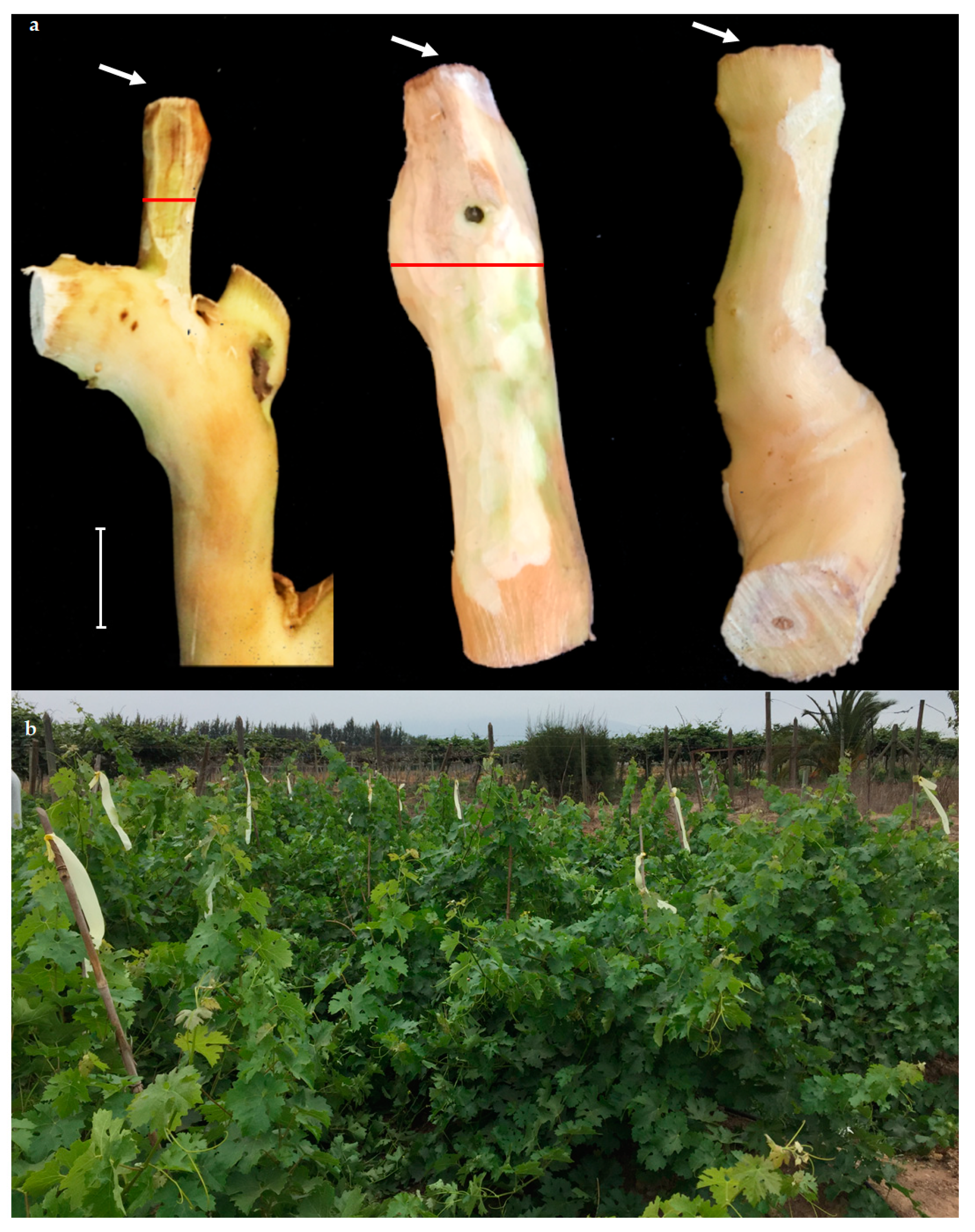
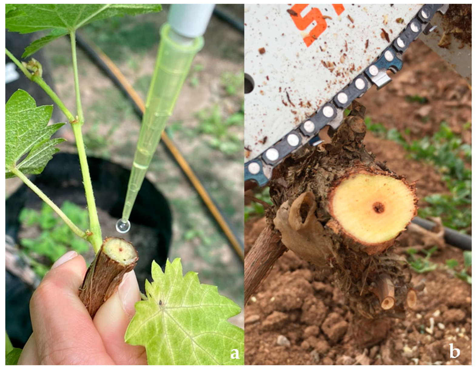
2.3. Pathogenicity Trial in the Field

Both isolates produced significant vascular lesions compared to the controls (T0 and T00), as shown in Figure 3. In young tissue (one-year-old shoots and two-year-old wood), the vascular lesions were significantly smaller than those in older tissue (10-year-old wood) (Figure 3 and Figure 4a). The vascular lesions developed in the 10-year-old wood were 4.2 and 4.3 cm for the PUCV 2183 and PUCV 2120 isolates, respectively, with no significant differences in the aggressiveness among the D. seriata isolates evaluated. In young tissue, the vascular lesions caused by D. seriata isolates ranged from 1.9 to 2.7 cm, with no differences between the tissue ages (one and two years old) or between the isolates (PUCV 2120 and PUCV 2183). Only from inoculated plants was the pathogen recovered from young and old tissue. This result shows that the vines used in this trial were healthy and that the lesions presented are attributable to the inoculations with D. seriata isolates. No plants showed foliar symptoms (either inoculated or noninoculated) (Figure 4b).

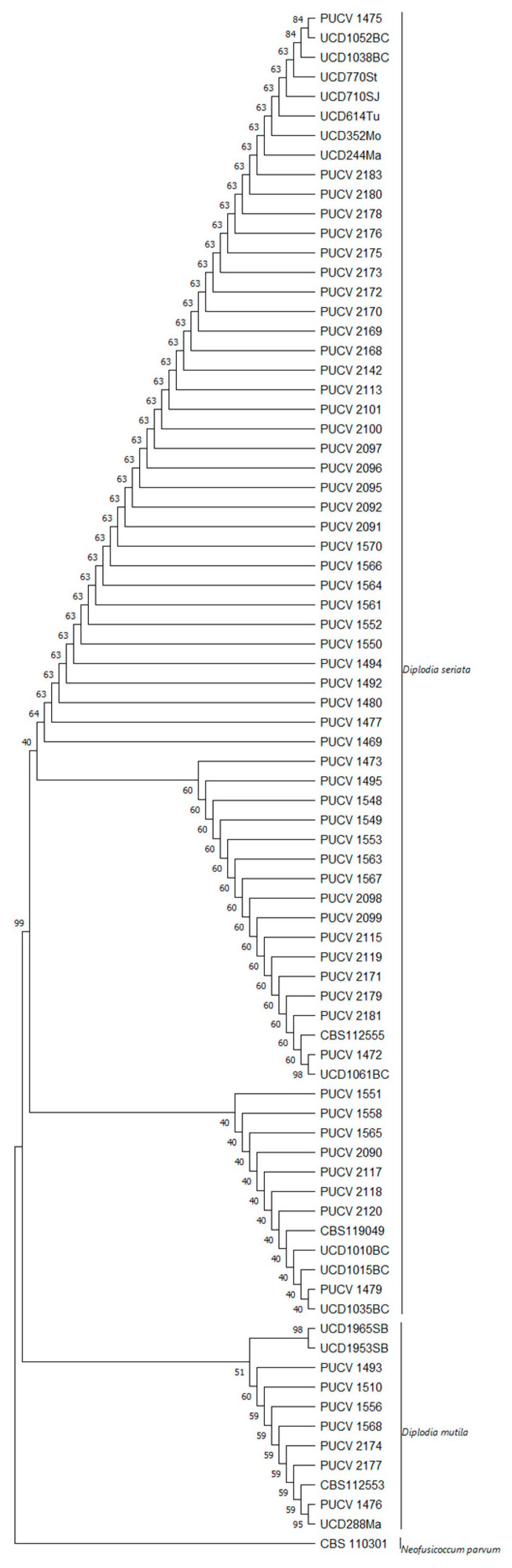
2.4. Maximum Parsimony Analysis

The concatenated ITS and BT phylogenetic analysis included 66 D. seriata isolates, all obtained from V. vinifera (Figure 5). The replicate trees (in percentages) of the associated taxa clustered in the bootstrap test (1000 replicates) can be seen next to the branches. The tree was obtained using the tree–bisection–regrafting (TBR) algorithm. The analyses contained 802 nucleotides and involved 79 nucleotide sequences.
According to the results of the phylogenetic analysis, there were no major differences in the D. seriata isolates between the ITS and BT segments (Tables S1 and S2). However, there were differences in the virulence among the isolates, even after being activated in fruit, which is not attributable to the place of origin or the time of collection (autumn or spring) but is associated with the year of collection.

3. Discussion

This study reports, for the first time, a comparison of the pathogenicity of the fungus D. seriata on the tissues of different ages in cv. Cabernet Sauvignon and show greater aggressiveness in the old tissue under field conditions. Of the isolates obtained from diseased tissue, D. seriata is the most widely distributed in New South Wales, Australia, California (USA), Catalonia (Spain), and Central Chile [5,6,15,17,23,25,26,28,38,39]. Based on the works mentioned above, most isolates of D. seriata were obtained from symptomatic old wood tissue. However, pathogenicity tests for D. seriata are mainly carried out on young tissue (shoots, one- or two-year-old canes, or young tissue on old plants). In these cases, D. seriata virulence was weak or less significant than that of other Botryosphaeriaceae species, such as Lasiodiplodia theobromae, L. viticola, Neofusicoccum parvum, or Diplodia mutila [8,33,40,41], or when compared to other causal agents of GTDs, such as Eutypa lata or N. parvum [42,43]. The difference in virulence could be due to several factors, including climatic conditions, varietal susceptibility of the vine, age of the host plant tissue, type of inoculation performed, geographic origin of the isolates, and period of experiment development, among others [7,31,32,43,44]. Also, it is important to study the microbiome associated with plants and its influence on disease development [45].
In our work, the pathogenicity of D. seriata isolates was verified in plants, with inoculations in young tissue (one- or two-year-old tissue age) and in old tissue (10-year-old wood) in vine plants. The vascular lesions developed by D. seriata on old tissue (10-year-old wood) were more than twice as long as those in the young tissue. Our results coincide with those described by Morales et al. [15] for old table grape plants in a commercial vineyard. When inoculating shoots < one year, canes > five years old, and mature arms in 25-year-old vines in commercial vineyards, D. seriata increased the damage by more than 40% in the mature arms with respect to the other two ages evaluated, contrary to D. mutila. Additionally, our results are consistent with those reported in naturally affected vineyards. In the field, symptoms of BD increase with the age of the vineyard, both in incidence and severity [5,12,15,30]. The extension of the advancement of this pathogen in the wood can be explained by the microenvironmental theory, where pathogen reaction zones can alone explain the progression of fungi within tree trunks, as fungi follow dehydrated, oxygen-rich zones that occur along the reaction zones near the wounds [46]. The high moisture content and the associated aeration restriction limit the activity of mycelial fungi in felled wood. Additionally, the most significant damage in old tissue can be explained concerning the plant microbiome, which evolves over time and with plant tissues. Microbiome diversity is higher in young vines [1,47,48,49] and in visually healthy tissues than in diseased tissues [50].
In our study, when comparing the results of the pathogenicity tests on two-year-old tissue, detached cuttings, or plant canes (attached), a difference in the severity of the damage of D. seriata was obtained, where it was more aggressive in detached canes (inoculated with mycelium plug) than in adhered canes (inoculated with conidia). Vascular lesions were up to 12 cm in detached canes, while in attached canes, they were up to 5 cm. This may be due to the type of inoculum (mycelium or conidia) and/or the state of the tissue (detached or attached). Regarding the type of inoculum, our results are contrary to those reported by Moral et al. [51], where inoculation with conidia of Botryosphaeria dothidea resulted in a more severe disease than inoculation with a mycelium plug in olive fruits. Concerning the state of the tissue, the literature describes that during grapevine infection, a series of molecules are activated that protect against the pathogen’s spread [50]. The first active defense is the formation of tylosis within the vessels. Responses to GTD fungi are thought to occur due to PAMP-activated immunity. Phytoalexins, compounds of the phenylpropanoid pathway, such as resveratrol or viniferin, are found in higher amounts in tissues after GTD fungal infection [52,53,54,55,56]. Phytoalexins can inhibit fungal growth and colonization, block some metabolites produced by fungi during infection, or interfere with oxidation–reduction reactions [57]. Due to the loss of xylem function and decreased hydraulic conductivity, which induce fungal colonization, active plant defense associated with parenchyma rays can prevent this from occurring [58]. In this work, it would have been important to measure the hydraulic conductivity of these tissues and the presence of phenols or phytoalexins specific to the vine, such as those mentioned above.
Reveglia et al. [59] demonstrated that the Botryosphaeriaceae family associated with BD in grapevine leaves generates secondary metabolites. The study by Martos et al. [60] pointed out that D. seriata can produce secondary metabolites with phytotoxic properties, dependent on acidic pH substrates, among other possible mechanisms related to its virulence. This could explain the lower virulence variability of D. seriata isolates when inoculated on fruits rather than on canes, although this requires further study. It is important to note that storage in culture can cause the loss of virulence or pathogenicity due to nutrient restriction for a prolonged period [61,62,63]. In addition to the active defense implemented by the vines, the anatomy of the wood could also be essential. The density of parenchymal rays and their arrangement in space could enhance active responses to infection [64] and the diameter of the vessels could also be a factor [58,65,66]. In contrast, Amponsah et al. [67] showed a faster rate of germination of N. luteum conidia on attached and wounded shoots and leaves than on detached and unwounded leaf surfaces. This may be due to the ability of Neofusicoccum species to attack in spring and summer time, which is associated with damage to the shoots and leaves, unlike D. seriata, which mainly affects woody tissue.
Furthermore, in other Botryosphaeriaceae species, B. dothidea in this case, the physiological characterization of the isolates that cause Dalmatian disease in olive showed that the optimal temperature was 26 °C for mycelium growth and 30 °C for the germination of conidia [51], which is also the optimal temperature for the conidia of D. seriata isolated from V. vinifera [68]. Both factors, the type of inoculum and the temperature, can influence the aggressiveness of the symptoms, which should be studied for D. seriata. The difference in virulence observed in our work was also reported by Elena et al. [44], who inoculated with mycelium different isolates of D. seriata in detached canes and two-year-old potted plants cv. Tempranillo. In this case, the differences were attributed to the type of host from which each isolate was obtained. The ITS and BT sites analyzed in this work showed no significant nucleotide variations between the Chilean isolates and the reference ones (Table S3), which came from California (USA) and Baja California (Mexico). This coincides with the results found by Urbéz-Torres et al. [29,41], where low variation was detected in the ITS and BT sequences of D. seriata from different geographical origins concerning other pathogens causing BD, such as L. theobromae. Thus, D. seriata is considered a more cosmopolitan pathogen.

4. Materials and Methods

4.1. Chemicals, Reagents, and Culture Media

The reagents and culture media used were sodium hypochlorite (SMF Ltda., Santiago, Chile), agar papa dextrose acidulated (APDA): 20 g L−1 granulated agar (Algas Marinas S.A., Pontevedra, Spain), 20 g L−1 mashed potatoes (Nestlé S.A., Vevey, Switzerland), 22 g L−1 glucose (Vimaroni S.A., Valparaíso, Chile), 7 drops of citric acid (Merck S.A., Darmstadt, Germany), and 1 L of sterile distilled water (SDW). The sterile distilled water was obtained by using a distiller (Pobel, Madrid, Spain) and autoclave (Zonkia, Hefei, China). The parafilm used was obtained from Bemis, Sheboygan Falls, WI, USA.

4.2. Fungal Isolates

Five isolates of D. seriata were used (Table 1). The isolates belong to the fungal collection of the Phytopathology Laboratory, School of Agronomy, PUCV. The PUCV 1472, PUCV 1549, PUCV 2120, PUCV 2142, and PUCV 2183 isolates were previously obtained from plants with cankers and vascular lesions from commercial vineyards in the wine-growing zone of central Chile, all with a history of BD that was previously molecularly identified [5]. The isolates were classified based on a phylogenetic analysis of the concatenated ITS-BT. Multilocus phylogenetic analysis was performed using maximum parsimony (MP) in MEGA X [69]. Bootstrap values were calculated using 1000 replicates, yielding the MP tree using the tree bisection and reconnection algorithms. The tree was rooted with Neofusicoccum parvum strain CBS 110302, and other reference isolates of different isolates of D. seriata and D. mutila were used (Table S3). The retention index, rescaled consistency index, and homoplasy index were calculated using MEGA X.

4.3. Pathogenicity Tests on Berry Grapes

The inoculations were performed on fresh berry grape cv. Red Globe of Vitis vinifera. The berry grapes were obtained in the summer of 2021 from the experimental vineyards of Estación La Palma of Universidad Católica de Valparaíso (PUCV) and immediately used for the test.

4.3.1. Fungal Isolates and Inoculation

For the inoculation, the berry grapes were disinfected with 1% sodium hypochlorite for 30 seg and then triple-washed in sterile distilled water. A 5-mm-diameter inoculum of a 5-mm-diameter mycelial plug from a 6-day-old growth in APDA culture was inoculated in the middle of the berry through a previously made wound with a sterile needle. The inoculated area was covered with parafilm. The pathogenicity assay was performed with five isolates (Table 1). The control consisted of noninoculated berry grapes, with only an APDA plug. The berry grapes were incubated for 15 days in an individual humid chamber at 23 °C.

4.3.2. Damage Assessment and Pathogen Recovery

After the incubation of the berry grapes, fruit rot was measured around the point of inoculation. The diameter of the lesions was considered. Tissue samples were taken from the zone of advancement of the lesions, disinfected with sodium hypochlorite for 5 seg, triple-washed in ADE, and cultivated in APDA. The plates were incubated for 7 d at 24 °C.

4.4. Pathogenicity Tests on Detached Canes

The inoculations were performed on healthy detached 2-year-old canes (15 cm in length) of Vitis vinifera cv. Cabernet Sauvignon. The canes were obtained in the winter of 2020 from an experimental vineyard of the Experimental Station La Palma of Pontifical Catholic University of Valparaíso (PUCV) and maintained at 5 °C for three months before use [65].
The pathogenicity assay was performed with five isolates (Table 1), previously activated in grape berry cv. Red Globe plants. Detached canes were disinfected (1% sodium hypochlorite for 5 min and 95% ethanol for 30 s) and then triple-washed in SDW, according to Morales et al. [15], and an inoculum of 5-mm-diameter mycelial plugs from a 6-day-old APDA culture was inoculated using an oblique cut made in the bark with a sterile scalpel in the middle of the canes. The inoculated area was covered with parafilm [15,47]. Three detached 2-year-old canes were used for each isolate, and three were used for the control (noninoculated canes, only APDA plug). The assay was performed in duplicate. The canes were incubated for 90 days in an individual humid chamber at 23 °C.

Damage Assessment and Pathogen Recovery

After three months of incubation, the vascular lesions and canker lengths of the canes were measured up and down from the point of inoculation. For the data analysis, the total length of the lesions was considered. Tissue samples were taken from the zone of advancement of the lesions, disinfected with 1% sodium hypochlorite for 5 s, triple-washed in SWD, and cultivated in APDA.

4.5. Pathogenicity Trial in the Field

The experiment was carried out on 10-year-old ungrafted cv. Cabernet Sauvignon plants in an experimental vineyard at the Experimental Station La Palma of the Pontifical Catholic University of Valparaíso (PUCV). The selected vineyard did not present previous GTD symptoms, and it was trained to the bilateral cordon system, with the spur pruned to 4 buds. The inoculations were performed on tissues of three different ages: 1-year-old shoots, 2-year-old wood, and 10-year-old wood, with each inoculation/age on a different plant.

4.5.1. Fungal Isolates and Inoculation

Two isolates of D. seriata, PUCV 2120 and PUCV 2183 (Table 1), previously selected in pathogenicity tests on two-year-old detached canes, were used for the field experiment. The inoculation was carried out in July 2021, with 50 µL of inoculum at 1 × 10⁴ spores × mL−1 on fresh cuts on 1-year-old shoots, 2-year-old wood, and 10-year-old wood (arm) (Figure 6). The cuts in the 1-year-old shoots and 2-year-old wood were made with pruning shears, and in 10-year-old wood with a chainsaw (Stihl, Waiblingen, Germany). Spore suspensions were prepared according to Úrbez-Torres et al. [38] and Larach et al. [5]: 5-day-old mycelial plugs of each isolate were placed in Petri dishes containing 2% agar water and needle-autoclaved pine. The plates were incubated in a chamber at room temperature (19–21 °C) under near-ultraviolet light (λ = 320 nm) until pycnidium production and conidium development. Mature pycnidia were crushed in sterile distilled water, and the solution obtained was filtered through a sterilized cheesecloth. Five plants were used for each isolate/tissue age, with one inoculation per plant. Five absolute control plants (without cutting or inoculation, T00) and five control plants (cut plus sterile distilled water, T0) were used.

4.5.2. Damage Assessment and Pathogen Recovery

Five months after the inoculation of the tissue, the vascular lesions and canker lengths were recorded. For this, the damage produced in each tissue/age was evaluated in the field, and the plant material was cut to measure the lesions. The assay was performed in duplicate. To recover the fungus, tissue samples were taken from the zone of advancement of the lesions, disinfected, and cultivated in APDA.

4.6. Statistical Analysis

The pathogenicity test on the berry grapes was distributed according to a completely randomized design. One berry grape was the experimental unit, and five replicates were used for each treatment. The results were analyzed for variance, and the means were compared according to Tukey’s test (p ≤ 0.05).
The pathogenicity test on the detached canes was distributed according to a completely randomized design. One cane was the experimental unit, and three replicates were used for each treatment. The results were analyzed for variance, and the means were compared according to Tukey’s test (p ≤ 0.05).
In the field, the plants of the pathogenicity trial were distributed according to a completely randomized design. One plant was the experimental unit, and five repetitions were used. An analysis of variance of two factors (tissue age and isolates) was performed, and no interaction of the factors was obtained, so a factor analysis was performed for the significant factor (tissue age) using Tukey’s test (p ≤ 0.05).

5. Conclusions

In this study, we observed that D. seriata is more aggressive in old tissue of grapevine plants cv. Cabernet Sauvignon than in young tissue, verifying the pathogenicity of D. seriata and its role as a causal agent of the disease (BD). These results contribute to the knowledge of the epidemiology of this disease in Mediterranean climates. Additionally, the pathogenicity tests carried out to select the isolates for pathogenicity testing in the field showed different results for vegetative tissue and berry grapes, highlighting the importance of the type of tissue in the pathogenicity and the virulence of the D. seriata pathogen. This work should be continued by employing other BD pathogens and different wine varieties.

Supplementary Materials

The supporting information can be downloaded at: https://www.mdpi.com/article/10.3390/plants12162984/s1. Table S1: Comparison of the conserved sites of the ITS sequences of the Chilean isolates of D. seriata (code PUCV) analyzed for the phylogenetic tree. Table S2: Comparison of the conserved sites of the BT sequences of the Chilean isolates of D. seriata (code PUCV) analyzed for the phylogenetic tree. Table S3: Isolates of D. seriata and D. mutila, obtained from GenBank Database and included in this study for phylogenetic analysis.

Author Contributions

Investigation and writing, A.L.; original draft preparation, A.L. and D.C.-N.; formal analysis, X.B., M.S., P.V.-C. and E.S.; investigation, P.S., A.S., N.R. and A.L.; statistical analysis: A.L., E.S. and P.V.-C.; writing—review and editing, P.V.-C. and M.S.; supervision and writing—review and editing, X.B. All authors have read and agreed to the published version of the manuscript.

Funding

This research was funded by FONDECYT REGULAR 1211094 (ANID, Chile) and the operating expenses of Alejandra Larach’s doctoral scholarship, grant number 21180920.

Data Availability Statement

Not applicable.

Acknowledgments

The authors thank Lorena Tapia for her support in the laboratory activities and Iván Cortés for his collaboration in the field maintenance trial.

Conflicts of Interest

The authors declare no conflict of interest.

References

  1. Bruez, E.; Lecomte, P.; Grosman, J.; Doublet, B.; Bertsch, C.; Fontaine, F.; Ugaglia, A.; Teissedre, P.; Da Costa, J.; Guerin-Dubrana, L.; et al. Overview of grapevine trunk diseases in France in the 2000s. Phytopathol. Mediterr. 2013, 52, 262–275. [Google Scholar]
  2. Fontaine, F.; Pinto, C.; Vallet, J.; Clément, C.; Catarina, A.; Spagnolo, A. The effects of grapevine trunk diseases (GTDs) on vine physiology. Eur. J. Plant Pathol. 2016, 114, 707–721. [Google Scholar] [CrossRef]
  3. Hillis, V.; Lubell, M.; Kaplan, J.; Doll, D.; Baumgartner, K. The role of pest control advisers in preventative management of grapevine trunk diseases. Phytopathology 2016, 106, 339–347. [Google Scholar] [CrossRef] [PubMed]
  4. Kaplan, J.; Travadon, R.; Cooper, M.; Hillis, V.; Lubell, M.; Baumgartner, K. Identifying Economic Hurdles to Early Adoption of Preventative Practices: The Case of Trunk Diseases in California Winegrape Vineyards. Wine Econ. Policy 2016, 5, 127–141. [Google Scholar] [CrossRef]
  5. Larach, A.; Torres, C.; Riquelme, N.; Valenzuela, M.; Salgado, E.; Seeger, M.; Besoain, X. Yield loss estimation and pathogen identification from (BD) in vineyards of Central Chile over two growing seasons. Phytopathol. Mediterr. 2020, 59, 537–548. [Google Scholar]
  6. Kenfaoui, J.; Radouane, N.; Mennani, M.; Tahiri, A.; El Ghadraoui, L.; Belabess, Z.; Fontaine, F.; El Hamss, H.; Amiri, S.; Lahlli, R.; et al. A Panoramic View on Grapevine Trunk Diseases Threats: Case of Eutypa Dieback, (BD), and Esca Disease. J. Fungi. 2022, 8, 595. [Google Scholar] [CrossRef]
  7. Úrbez-Torres, J.R.; Gubler, W.D. Susceptibility of grapevine pruning wounds to infection by Lasiodiplodia theobromae and Neofusicoccum parvum. Plant Pathol. 2011, 60, 261–270. [Google Scholar] [CrossRef]
  8. Úrbez-Torres, J.R.; Battany, M.; Bettiga, L.J.; Gispert, C.; McGourty, G.; Roncoroni, J.; Smith, R.; Verdegaal, P.; Gubler, W. Botryosphaeriaceae species spore-trapping studies in California vineyards. Plant Dis. 2010, 94, 717–724. [Google Scholar] [CrossRef] [PubMed]
  9. Besoain, X. Grapevine Trunk Diseases (GTDs): Impact on Table Grapes and Wine Vineyards in Chile. In: Grapes and Wines-Advances in Production, Processing, Analysis and Valorization. InTechOpen 2018, 3, 43–58. [Google Scholar]
  10. Gramaje, D.; Úrbez-Torres, J.R.; Sosnowski, M.R. Managing grapevine trunk diseases with respect to etiology and epidemiology: Current strategies and Future prospects. Plant Dis. 2018, 102, 12–39. [Google Scholar] [CrossRef]
  11. Billones-Baaijens, R.; Savocchia, S. A review of Botryosphaeriaceae species associated with grapevine trunk diseases in Australia and New Zealand. Aust. Plant Pathol. 2019, 48, 3–18. [Google Scholar] [CrossRef]
  12. Rolshausen, P.; Baumgartner, K.; Travador, R.; Funjiyoshi, P.; Pouzoulet, J.; Wilcox, W. Identification of Eutypa spp. Causing Eutypa Dieback o Grapevine in Eastern North America. Plant Dis. 2014, 98, 483–491. [Google Scholar] [CrossRef] [PubMed]
  13. Burruano, S.; Mondello, V.; Conigliario, G.; Alfonzo, A.; Spagnolo, A.; Mugnai, L. Grapevine decline in Italy caused by Lasiodiplodia theobromae. Phytopathol. Mediterr. 2008, 47, 132–136. [Google Scholar]
  14. Reis, P.; Magnin-Robert, M.; Nascimento, T.; Spagnolo, A.; Abou-Mansour, E.; Fioretti, C.; Clément, C.; Rego, C.; Fontaine, F. Reproducing (BD) foliar symptoms in a simple model system. Plant Dis. 2016, 100, 1071–1079. [Google Scholar] [CrossRef] [PubMed]
  15. Morales, A.; Latore, B.; Piontelli, E.; Besoain, X. Botryosphaeriaceae Species affecting table grape vineyards in Chile and cultivar susceptibility. Cien Investig. Agrar. 2012, 39, 445. [Google Scholar] [CrossRef]
  16. Díaz, G.A.; Auger, J.; Besoain, X.; Bordeu, E.; Latorre, B.A. Prevalence and pathogenicity of fungi associated with grapevine trunk diseases in Chilean vineyards. Cienc. Investig. Agrar. 2013, 40, 327–339. [Google Scholar] [CrossRef]
  17. Savocchia, S.; Steel, C.C.; Stodart, B.J.; Somers, A. Pathogenicity of Botryosphaeria species isolated from declining grapevines in sub-tropical regions of Eastern Australia. VITIS-J. Grapevine Res. 2007, 46, 27–32. [Google Scholar]
  18. Burgess, T.; Wingfield, M.J. Quarantine is important in restricting the spread of exotic seed-borne tree pathogens in the southern hemisphere. Int. For. Rev. 2002, 4, 56. [Google Scholar]
  19. Mohali, S.; Burgess, T.I.; Wingfield, M.J. Diversity and host association of the tropical tree endophyte Lasiodiplodia theobromae revealed using simple sequence repeat markers. For. Pathol. 2005, 35, 385–396. [Google Scholar] [CrossRef]
  20. Burgess, T.I.; Barber, P.A.; Mohali, S.; Pegg, G.; De Beer, W.; Wingfield, M.J. Three new Lasiodiplodia spp. from the tropics, recognized based on DNA sequence comparisons and morphology. Mycologia 2006, 98, 423–435. [Google Scholar] [CrossRef]
  21. Kuntzmann, P.; Villaume, S.; Larignon, P.; Bertsch, C. Esca, BDA and Eutypiosis: Foliar symptoms, trunk lesions and fungi observed in diseased vinestocks in two vineyards in Alsace. Vitis 2010, 49, 71–76. [Google Scholar]
  22. Valencia, D.; Torres, C.; Camps, R.; López, E.; Celis-Diez, J.L.; Besoain, X. Dissemination of Botryosphaeriaceae conidia in vineyards in the semiarid Mediterranean climate of the Valparaíso Region of Chile. Phytopathol. Mediterr. 2015, 54, 394–402. [Google Scholar]
  23. Burgess, T.; Tan, Y.P.; Garnas, J.; Edwards, J.; Scarlett, K.; Shuttleworth, L.; Daniel, R.; Dann, E.; Parkinson, L.; Dihn, Q.; et al. Current status of the Botryosphaeriaceae in Australia. Aust. Plant Pathol. 2018, 48, 35–44. [Google Scholar] [CrossRef]
  24. Reis, P.; Gaspar, A.; Alves, A.; Fontaine, F.; Lourenco, I.; Saramago, J.; Mota, M.; Rego, C. Early season symptoms on stem, inflorescences and flowers of grapevine associated with Botryosphaeriaceae species. Plants 2020, 11, 1427. [Google Scholar] [CrossRef]
  25. Castillo-Pando, M.; Somers, A.; Green, C.D.; Priest, M.; Sriskanthades, M. Fungi associated with dieback of Semillon grapevines in the Hunter Valley of New South Wales. Aust. Plant Pathol. 2001, 30, 59–63. [Google Scholar] [CrossRef]
  26. Pitt, W.M.; Huang, R.; Steel, C.C.; Savocchia, S. Identification, distribution and current taxonomy of Botryosphaeriaceae species associated with grapevine decline in New South Wales and South Australia. Aust. J. Grape Wine Res. 2010, 16, 258–271. [Google Scholar] [CrossRef]
  27. Qiu, Y.; Steel, C.C.; Ash, G.J.; Savocchia, S. Survey of Botryosphaeriaceae associated with grapevine decline in the Hunter Valley and Mudgee grape growing regions of New South Wales. Aust. Plant Pathol. 2011, 40, 1–11. [Google Scholar] [CrossRef]
  28. Taylor, A.; Hardy, G.E.S.J.; Wood, P.; Burgess, T. Identification and pathogenicity of Botryosphaeria species associated with grapevine decline in Western Australia. Aust. Plant Pathol. 2005, 34, 187–195. [Google Scholar] [CrossRef]
  29. Úrbez-Torres, J.R.; Leavitt, G.M.; Voegel, T.M.; Gubler, W.D. Identification and distribution of Botryosphaeria spp. associated with grapevine cankers in California. Plant Dis. 2006, 90, 1490–1503. [Google Scholar] [CrossRef]
  30. Larignon, P.; Fulchic, R.; Cere, L.; Dubos, B. Observation on black arm in French vineyards. Phytopathol. Mediterr. 2001, 40, 336–342. [Google Scholar]
  31. Van Niekerk, J.M.; Crous, P.W.; Groenewald, J.Z.; Fourie, P.H.; Halleen, F. DNA Phylogeny, Morphology and Pathogenicity of Botryosphaeria species on Grapevines. Mycologia 2004, 96, 781–798. [Google Scholar] [CrossRef] [PubMed]
  32. Baskarathevan, J.; Jaspers, M.; Jones, E.E.; Ridgway, H.J. Incidence and distribution of botryosphaeriaceous species in New Zealand vineyards. Eur. J. Plant Pathol. 2012, 132, 549–560. [Google Scholar] [CrossRef]
  33. Bertsch, C.; Ramírez-Suero, M.; Magnin-Robert, M.; Larignon, P.; Chong, J.; Abou-Mansour, E.; Spagnolo, A.; Clément, A.; Fontaine, F. Grapevine trunk diseases: Complex and still poorly understood. Plant Pathol. 2013, 62, 243–265. [Google Scholar] [CrossRef]
  34. Spagnolo, A.; Larignon, P.; Magnin-Robert, M.; Hovasse, A.; Cilindre, C.; Van Dorsselaer, A.; Clément, C.; Schaeffer-Reiss, C.; Fontaine, F. Flowering as the most highly sensitive period of grapevine (Vitis vinifera L. cv Mourvédre) to the (BD) agents Neofusicoccum parvum and Diplodia seriata infection. Int. J. Mol. Sci. 2014, 15, 9644–9669. [Google Scholar] [CrossRef] [PubMed]
  35. Bénard-Gellon, M.; Farine, S.; Goddard, M.L.; Schmitt, M.; Stempien, E.; Pensec, F.; Laloue, H.; Mazet-Kieffer, F.; Fontaine, F.; Larignon, P.; et al. Toxicity of extracellular proteins from Diplodia seriata and Neofusicoccum parvum involved in grapevine Botryosphaeria dieback. Protoplasma 2015, 252, 679–687. [Google Scholar] [CrossRef] [PubMed]
  36. Morales-Cruz, A.; Amrine, K.C.H.; Blanco-Ulate, B.; Lawrence, D.P.; Travadon, R.; Rolshausen, R.P.; Baumgartner, K.; Cantu, D. Distinctive expansion of gene families associated with plant cell wall degradation, secondary metabolism, and nutrient uptake in the genomes of grapevine trunk pathogens. BMC Genom. 2015, 16, 469. [Google Scholar] [CrossRef]
  37. ODEPA. Oficina de Estudios y Políticas Agrarias. Statistics Productive. 2022. Available online: https://www.odepa.cl (accessed on 20 February 2022).
  38. Luque, J.; Elena, G.; Garcia-Figueres, F.; Reyes, J.; Barrios, G.; Legorburu, F.J. Natural infections of pruning wounds by fungal trunk pathogens in mature grapevines in Catalonia (Northeast Spain). Aust. J. Grape Wine Res. 2014, 20, 134–143. [Google Scholar] [CrossRef]
  39. Luque, J.; Martos, S.; Aroca, A.; Raposo, R.; Garcia-Figueres, F. Symptoms and fungi associated with declining mature grapevine plants in northeast spain. Plant Pathol. 2009, 91, 381–390. [Google Scholar]
  40. Bellée, A.; Comont, G.; Nivault, A.; Abou-Mansour, E.; Coppin, C.; Dufour, M.C.; Corio-Costet, M. Life traits of four Botryosphaeriaceae species and molecular responses of different grapevine cultivars or hybrids. Plant Pathol. 2017, 66, 763–776. [Google Scholar] [CrossRef]
  41. Úrbez-Torres, J.R.; Leavitt, G.M.; Guerrero, J.C.; Guevara, J.; Gubler, W.D. Identification and pathogenicity of Lasiodiplodia theobromae and Diplodia seriata, the causal agents of bot canker disease of Grapevines in Mexico. Plant Dis. 2008, 92, 519–529. [Google Scholar] [CrossRef]
  42. Sosnowski, M.R.; Ayres, M.R.; McCarthy, M.G.; Scott, E.S. Winegrape cultivars (Vitis vinifera) vary in susceptibility to the grapevine trunk pathogens Eutypa lata and Diplodia seriata. Aust. J. Grape Wine 2022, 28, 166–174. [Google Scholar] [CrossRef]
  43. Van de Wouw, A.P.; Howlett, B.J. Fungal pathogenicity genes in the age of “omics”. Mol. Plant Pathol. 2011, 12, 507–514. [Google Scholar] [CrossRef] [PubMed]
  44. Elena, G.; Garcia-Figueres, F.; Reigada, S.; Luque, J. Intraspecific variation in Diplodia seriata isolates occurring on grapevines in Spain. Plant Pathol. 2015, 64, 680–689. [Google Scholar] [CrossRef]
  45. Bruez, E.; Vallance, J.; Gerbore, J.; Lecomte, P.; Da Costa, J.P.; Guerin-Dubrana, L.; Rey, P. Analyses of the temporal dynamics of fungal communities colonizing the healthy wood tissues of esca leaf-symptomatic and asymptomatic vines. PLoS ONE 2014, 9, e95928. [Google Scholar] [CrossRef]
  46. Boddy, L.; Rayner, A. Origins of decay in living deciduous trees: The role of moisture content and a re-appraisal of the expanded concept of tree decay. New Phytol. 1983, 94, 623–641. [Google Scholar] [CrossRef]
  47. Rey, P.; Lecomte, P.; Guérin-Dubrana, L.; Corio-Costel, F.; Fonteine, F.; Gomes, E.; Goutouly, J.P.; Hofdtetter, V.; Legorburu, X.; De La Rocque, B. Maladies du bois de la vigne situation enjeux et perpective. Phytoma 2011, 640, 22–26. [Google Scholar]
  48. Hofstetter, V.; Buyck, B.; Croll, D.; Viret, O.; Couloux, A.; Gindro, K. What if esca disease of grapevine were not a fungal disease? Fungal Divers. 2012, 54, 51–67. [Google Scholar] [CrossRef]
  49. Andreolli, M.; Lampis, S.; Zapparoli, G.; Angelini, E.; Vallini, G. Diversity of bacterial endophytes in 3- and 15-year-old grapevines of Vitis vinifera cv. Corvina and their potential for plant growth promotion and phytopathogen control. Microbiological 2016, 183, 42–52. [Google Scholar] [CrossRef]
  50. Claverie, M.; Notaro, M.; Fontaine, F.; Wery, J. Current knowledge on Grapevine Trunk Diseases with complex etiology: A systemic approach. Phytopathol. Mediterr. 2020, 59, 29–53. [Google Scholar] [CrossRef]
  51. Moral, J.; Muñoz-Díez, C.; González, N.; Trapero, A.; Michailides, T.J. Characterization and pathogenicity of Botryosphaeriaceae species collected from olive and other hosts in Spain and California. Phytopathol 2010, 100, 1340–1351. [Google Scholar] [CrossRef]
  52. Amalfitano, C.; Evidente, A.; Surico, G.; Tegli, S.; Bertelli, E.; Mugnai, L. Phenols and Stilbene Polyphenols in the Wood of Esca-Diseased Grapevines. Phytopathol. Mediterr. 2000, 39, 178–183. [Google Scholar]
  53. Del Rio, J.; Gonzalez, A.; Fuster, M.; Botia, J.; Gomez, P.; Frias, V.; Ortuño, A. Tylose formation and changes in phenolic compounds of grape roots infected with Phaeomoniella chlamydospore and Phaeocremonium species. Phytopathol. Mediterr. 2001, 40, 394–399. [Google Scholar]
  54. Bruno, G.; Sparapano, L. Effects of three esca-associated fungi on Vitis vinifera L.: I. Characterization of secondary metabolites in culture media and host responses to the pathogens in calli. Physiol. Mol. Plant Pathol. 2006, 69, 209–223. [Google Scholar] [CrossRef]
  55. Martin, N.; Vesentini, D.; Rego, C.; Monteiro, S.; Oliveira, H.; Ferreira, R.B. Phaeomoniella chlamydospora infection induces changes in phenolic compounds content in Vitis vinifera. Phytopathol. Mediterr. 2009, 48, 101–116. [Google Scholar]
  56. Spagnolo, A.; Magnin-Robert, M.; Alayi, T.D.; Cilindre, C.; Schaeffer-Reiss, C.; Van Dorsselaer, A.; Clément, C.; Larignon, P.; Ramirez-Suero, M.; Chong, J.; et al. Differential responses of three grapevine cultivars to (BD). Phytopathol. 2014, 104, 1021–1031. [Google Scholar] [CrossRef]
  57. Gómez, P.; Báidez, A.G.; Ortuño, A.; Del Río, J.A. Grapevine xylem response to fungi involved in trunk diseases. Ann. Appl. Biol. 2016, 169, 116–124. [Google Scholar] [CrossRef]
  58. Pouzoulet, J.; Pivovaroff, A.L.; Santiago, L.S.; Rolshausen, P.E. Can vessel dimension explain tolerance toward fungal vascular wilt diseases in woody plants? Lessons from Dutch elm disease and esca disease in grapevine. Front. Plant Sci. 2014, 5, 253. [Google Scholar] [CrossRef]
  59. Reveglia, P.; Savocchia, S.; Billones-Baaijens, R.; Masi, M.; Cimmino, A.; Evidente, A. Phytotoxic metabolites by nine species of Botryosphaeriaceae involved in grapevine dieback in Australia and identification of those produced by Diplodia mutila, Diplodia seriata, Neofusicoccum australe and Neofusicoccum luteum. Nat. Prod. Res. 2019, 33, 2223–2229. [Google Scholar] [CrossRef]
  60. Martos, S.; Andolfi, A.; Luque, J.; Mugnai, L.; Surico, G.; Evidente, A. Production of phytotoxic metabolites by five species of Botryosphaeriaceae causing decline on grapevines, with special interest in the species Neofusicoccum luteum and N. parvum. Eur. J. Plant Pathol. 2008, 121, 451–461. [Google Scholar] [CrossRef]
  61. Zarzycka, H. Effect of culturing Phytophthora infestans on various substrates on expression of its virulence and aggressiveness to potato. Phytopathol. Polonica. 1995, 9, 81–88. [Google Scholar]
  62. Zarzycka, H. Effect of various methods of maintenance of Phytophthora infestans on its pathogenicity to potato. Plant Breed. Seed Sci. 1996, 40, 31–43. [Google Scholar]
  63. Balendres, M.A.; De Torres, R.; Dela Cueva, F. Culture storage age and fungal re-isolation from host-tissue influence Colletotrichum spp. virulence to pepper fruits. J. Phytopathol. 2019, 167, 510–515. [Google Scholar] [CrossRef]
  64. Pearce, R.B. Antimicrobial defenses in the wood of living trees. New Phytol. 1996, 132, 203–233. [Google Scholar] [CrossRef]
  65. Ramírez, M.; Pérez, L.M.; Montealegre, J.R. Susceptibility of different grapevine (Vitis vinifera L.) cultivars to Diplodia seriata and Diplodia mutila. Cien. Inv. Agric. 2018, 45, 93–98. [Google Scholar] [CrossRef]
  66. Pouzoulet, J.; Scudiero, E.; Schiavon, M.; Rolshausen, P.E. Xylem Vessel Diameter Affects the Compartmentalization of the Vascular Pathogen Phaeomoniella chlamydospora in Grapevine. Front. Plant Sci. 2017, 21, 1442. [Google Scholar] [CrossRef]
  67. Amponsah, N.T.; Jones, E.E.; Ridgway, H.J.; Jaspers, M. Infection and disease progression of Neofusicoccum luteum in grapevine plants. Plant Health Manag. An. Integr. Approach 2009, 118. [Google Scholar]
  68. Úrbez-Torres, J.R.; Bruez, E.; Hurtado, J.; Gubler, W.D. Effect of temperature on conidial germination of Botryosphaeriaceae species infecting grapevines. Plant Dis. 2010, 94, 1476–1484. [Google Scholar] [CrossRef]
  69. Koichiro, T.; Stecher, G.; Kumar, S. MEGA11: Molecular Evolutionary Genetics Analysis Version 11. Mol. Biol. Evol. 2021, 38, 3022–3027. [Google Scholar]
Figure 1. Soft rot lesions caused by D. seriata on Vitis vinifera cv. Red Globe. (a) Mean lesion diameter on berries inoculated with different isolates of D. seriata. Different letters indicate significant differences after ANOVA according to Tukey’s test (p ≤ 0.05). (b) Circular and sunken rot around the point of inoculation with the different isolates. (c) Left to right: control berry grapes (noninoculated), internal damage of inoculated berry grapes, and recovery of D. seriata from inoculated berry grapes.
Plants 12 02984 g001
Figure 2. Severity of the damage of the symptoms caused by D. seriata on detached canes of two-year-old Vitis vinifera cv. Cabernet Sauvignon. (a) Mean canker lesion length. Different letters indicate significant differences after ANOVA according to Tukey’s test (p ≤ 0.05). (b) Mean vascular lesion length. Different letters indicate significant differences after ANOVA according to Tukey’s test (p ≤ 0.05). (c) Vascular and canker lesions on cuttings with activated isolates. The white arrow indicates the point of inoculation. The red line describes vascular discoloration. Lines over bars on graphs represent standard deviation.
Plants 12 02984 g002
Figure 3. Damage severity of symptoms (vascular lesion length) caused by Diplodia seriata on tissue of different ages of Vitis vinifera cv. Cabernet Sauvignon. T00: without cutting or inoculation; T0: cutting plus sterile distilled water. PUCV 2120: D. seriata isolate. PUCV 2183: D. seriata isolate. Different letters indicate significant differences after ANOVA according to Tukey’s test (p < 0.05). Lines over bars on graphs represent standard deviation.
Plants 12 02984 g003
Figure 4. Symptoms developed on Vitis vinifera cv. Cabernet Sauvignon plants five months after inoculation with D. seriata. (a) Left to right: two-year-old tissue vascular lesion and 10-year-old tissue vascular lesion caused by the PUCV 2021 isolate, and 10-year-old tissue of noninoculated plants (T0). The white arrow indicates the inoculation point. Red lines delimit vascular discoloration. Scale bar = 2 cm. (b) General view of the trial in vines; the image was taken the day before evaluating the symptoms in the different treatments. Note the absence of foliar symptoms on the plants.
Plants 12 02984 g004
Figure 5. Phylogenetic tree of isolates of D. seriata based on maximum parsimony (MP) analysis of two loci (ITS and β-tubulin). Chilean isolates used in this work with PUCV code. MP bootstrap values are shown above the branches. N. parvum isolate CBS 110301 was the outgroup used. Values were obtained with Mega X software.
Plants 12 02984 g005
Figure 6. Inoculation with conidia solutions of D. seriata in grapevine plants cv. Cabernet Sauvignon. (a) Inoculation of 2-year-old shoots on fresh pruning cut. (b) Inoculation of 10-year-old wood (arm) freshly cut by chainsaw.
Plants 12 02984 g006
Table 1. Chilean isolates of Diplodia seriata obtained from canker and vascular lesions of Vitis vinifera cv. Cabernet Sauvignon used for pathogenicity tests.
Isolate of Diplodia seriata *Year of CollectionLocality Origin, Region of ChileAccess No. GenBank
ITSBTEF1-α
PUCV 14722010Palmilla, O’HigginsKM372581KP762454-
PUCV 15492010Peralillo, O’HigginsKM580514KP762464-
PUCV 21202018Palmilla, O’HigginsMT023573MT063140MT120827
PUCV 21422018Batuco, MauleMT023574MT063141MT120827
PUCV 21832018Pencahue, MauleMT023587MT063154 -
* Larach et al. [5].
Disclaimer/Publisher’s Note: The statements, opinions and data contained in all publications are solely those of the individual author(s) and contributor(s) and not of MDPI and/or the editor(s). MDPI and/or the editor(s) disclaim responsibility for any injury to people or property resulting from any ideas, methods, instructions or products referred to in the content.

