Article

Characteristics and Pathogenicity of Discula theae-sinensis Isolated from Tea Plant (Camellia sinensis) and Interaction with Colletotrichum spp.

1
Institute of Sericulture and Tea, Zhejiang Academy of Agricultural Sciences, Hangzhou 310021, China
2
State Key Laboratory of Tea Plant Biology and Utilization, Anhui Agricultural University, Hefei 230036, China
3
College of Horticulture Science, Zhejiang A&F University, Hangzhou 311300, China
4
Development Center of Agricultural Science and Technology in Huzhou, Huzhou 313000, China
*
Author to whom correspondence should be addressed.
These authors contributed equally to this work.
Plants 2023, 12(19), 3427; https://doi.org/10.3390/plants12193427
Submission received: 3 August 2023 / Revised: 25 September 2023 / Accepted: 26 September 2023 / Published: 28 September 2023
(This article belongs to the Special Issue Tea Plants Cultivation)

Abstract

:
Anthracnose is one of the primary diseases in tea plants that affect tea yield and quality. The geographical distribution, occurrence regularity, and agronomic measures of tea plants with anthracnose have been researched for decades. However, the pathogenic cause of anthracnose in tea plants is diverse in different regions of the world. Identifying the specific pathogenic fungi causing tea anthracnose is an essential control measure to mitigate this disease. In this study, 66 Discula theae-sinensis and 45 Colletotrichum isolates were obtained from three different types of diseased tea leaves. Based on multilocus phylogenetic and morphological analysis, eight known species of Colletotrichum, Colletotrichum fructicola, C. camelliae, C. aenigma, C. siamense, C. henanense, C. karstii, C. tropicicola, and C. gigasporum were identified. This study is the first to report C. tropicicola and C. gigasporum in tea plants in China. Discula theae-sinensis was the most common species in this study and caused disease lesions around wounded areas of tea leaves. The dual trials in vitro indicated Discula theae-sinensis and Colletotrichum were slightly inhibited. Co-inoculating Discula theae-sinensis and C. fructicola was superior to single inoculation at low concentrations. The main cause of anthracnose might be the concerted action of a variety of fungi.

Graphical Abstract

1. Introduction

Tea, which is produced from the new shoots from tea plants [Camellia sinensis (L.) O. Kuntze] is a globally consumed beverage. The production of tea includes cultivation, processing, packaging, and exportation, and is distributed across Asia, Africa, South America, and Oceania. The tea industry is a high-output and distinctive agriculture industry in China [1]. However, fungal diseases pose a threat to both the yield and quality of tea. Anthracnose, a severe and widespread disease, significantly affects the growth of tea plants and reduces the quality of tea products [2]. Anthracnose primarily occurs in warm and moist regions, with the peak diffusion period typically spanning from April to July, contingent on climatic conditions and regional distribution [3].
To date, the identification of pathogens responsible for anthracnose in tea plants in China remains a subject of controversy. Over the past several decades, the pathogen causing anthracnose was recognized as Gloeosporium theae-sinensis [4]. Nevertheless, the taxonomy of G. theae-sinensis is ambiguous. Yamamoto proposed that G. theae-sinensis represented an invalid recombination of C. theae-sinensis, which should be classified with the Colletotrichum genus [5]. However, Moriwaki et al. considered that G. theae-sinensis should be designated Discula theae-sinensis (Dt), which aligned with Diaporthales in terms of morphological and molecular characterization [6]. Diaporthales, an order within the subclass Sordariomycetes, has the potential to induce anthracnose in various crops and trees [7,8,9]. Diaporthales also widely exist in tea plants as pathogens or endophytes. Xie et al. summarized numerous studies on endophytes in tea plants and listed three families of Diaporthales reported as endophytes in tea plants, namely Diaporthaceae, Melanconiellaceae, and Valsaceae [10]. Gao et al. investigated Diaporthe (Diaporthecae, Diaporthale) species associated with symptomatic and asymptomatic tissues of Camellia, identifying four novel species, three known species, and two complex species from diseased and healthy tissues of tea plants from several provinces in China [11]. Nevertheless, their distribution in China and pathogenicity remain unclear.
In addition to Dt, Colletotrichum is considered another major cause of anthracnose in tea plants in China. Colletotrichum is a large genus of Ascomycete fungi belonging to Glomerellales [12]. Via sampling in multiple regions, 21 species and one indistinguishable strain of Colletotrichum were reported to infect tea plants in China [13,14]. Based on geographic distribution and strain quantity, Colletotrichum camelliae (Cc) and C. fructicola (Cf) were identified as the principal causal agents of tea anthracnose [13,15]. Cc probably hosts specific taxa that occur on Camellia, while Cf has a more diverse host range [13]. In terms of pathogenicity, Cc exhibits greater virulence than Cf on tea plants. Furthermore, secondary metabolites in tea plants such as catechins and caffeine influence the virulence of pathogens [16]. However, Cc and some Colletotrichum species are also supposed to be pathogens of tea leaf blight [17,18]. Consequently, it is essential to definitively ascertain the primary cause of tea plant anthracnose in China.
As the predominant causal agents of plant diseases, fungi employ diverse strategies to colonize plants and induce diseases [19]. Generally, different fungi utilize distinct mechanisms to penetrate host plants. Dt enters young tea leaves solely through trichomes [20,21]. Furthermore, Dt is the sole fungus that enters the host plant only through the trichomes [22]. Colletotrichum spp. typically develop appressoria to breach the host’s cuticle layer and cell wall [23,24]. Appressoria originates from conidial germ tubes and secrete effectors via appressorial penetration pores [25,26]. Host plants must employ specific defense strategies against various fungi. Thus, identifying the primary microorganisms responsible for tea diseases can enhance the effectiveness of disease control in tea plants.
In the present study, we isolated pathogens and collected anthracnose-diseased tea leaves over two years to elucidate the primary cause of anthracnose in tea plants in China. We selected the different types of diseased tea leaves based on field phenotypes and employed a combination of multilocus phylogeny and morphological characteristics to identify the species of all isolates. Out of the isolates, 66 were identified as Dt, and 45 were Colletotrichum. Dt and Cf were commonly found in Diaporthales and Colletotrichum, respectively. Additionally, pathogenicity tests were conducted on representative Dt isolates and co-inoculated with Dt and Colletotrichum to explore the primary cause of tea plant anthracnose in China.

2. Results

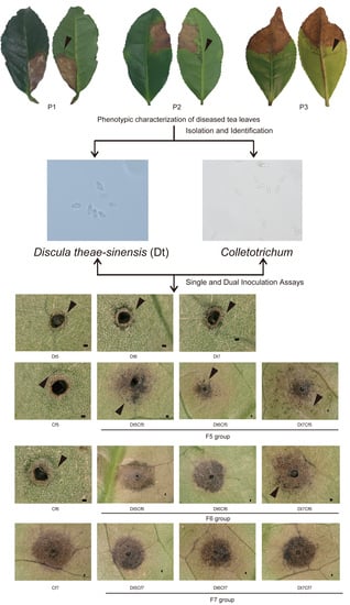
2.1. Phenotypic Characterization of Collected Tea Leaves

We collected diseased tea leaves with visible anthracnose symptoms continuously for two years from June to August. Based on detailed phenotypic differences in details, the symptoms of disease samples can be classified into three types. The first pattern (P1) displayed white disease spots accompanied by tiny black particles on the surface, which only existed on one side of the blade. The scabs of the second pattern (P2) were tawny with scorching on the side of the leaves. In the third pattern (P3), lesions were distributed on the distichous sides at the tip of the blade. The commonality among these three groups was water-soaked lesions close to the demarcation between diseased and healthy parts, which appeared translucent when exposed to light, and no ring spots (Figure 1A).
Based on microscopic examination, the forms of the collected acervuli from diseased tea leaves could be divided into two kinds (Figure 1B). The first kind of acervuli (A1) was suborbicular and flat, which constituted the main type among diseased leaves. The second kind of acervuli (A2) was irregular in shape with a split at the center. The A2 type only appeared once during procedures of sample collection procedures in this study. Additionally, the dominant spores on the scab were divided into two types (Figure 1C). One type was fusiform to obovoid, tapered at the base or both ends (S1), and the other type was cylindrical with obtuse ends (S2). Based on their morphological characteristics, we preliminarily considered S1 to be Diaporthales spp. and S2 to be Colletotrichum spp. Further studies revealed that all isolates collected from type A2 diseased tea leaves were identified as Cf.

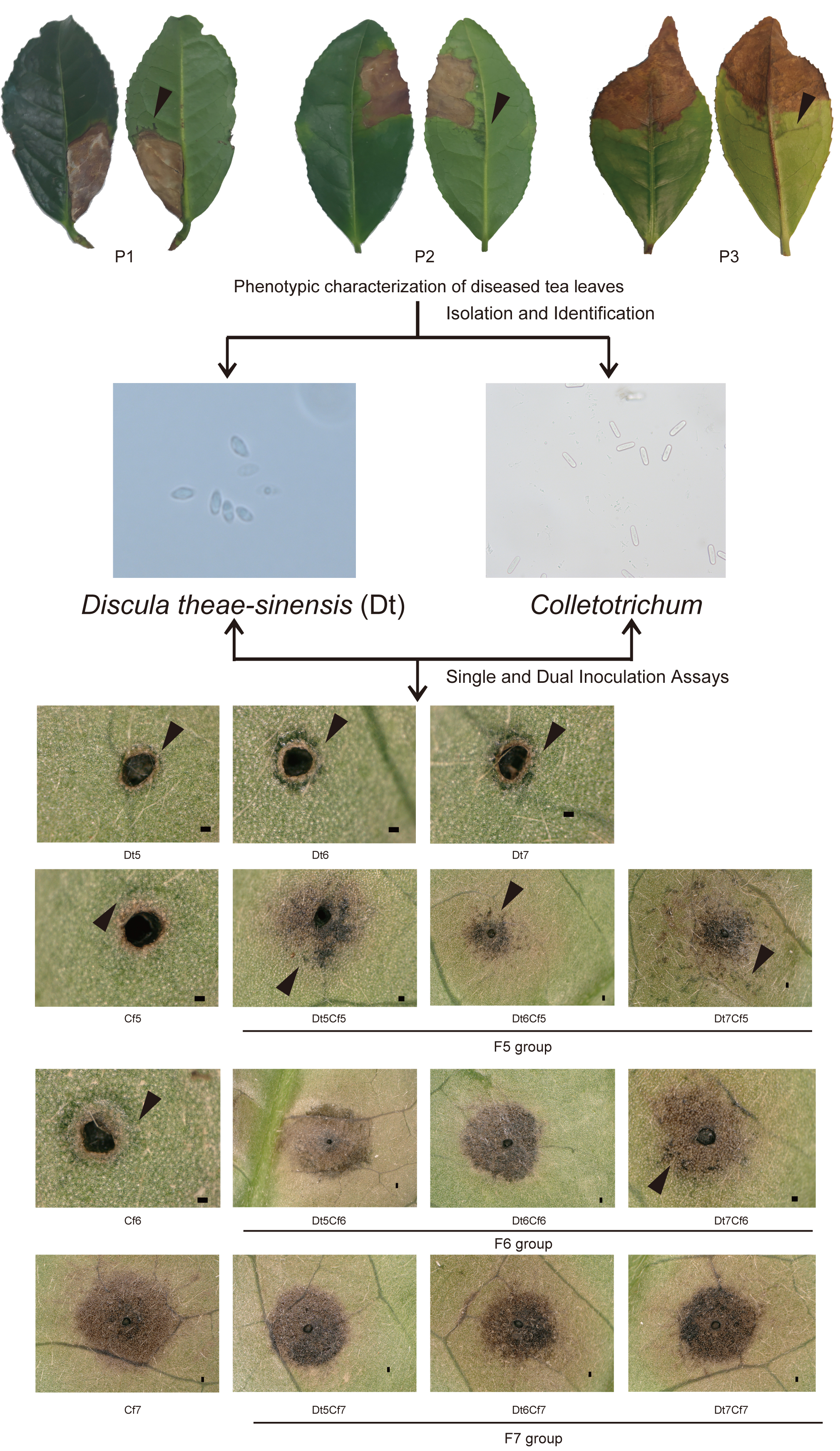
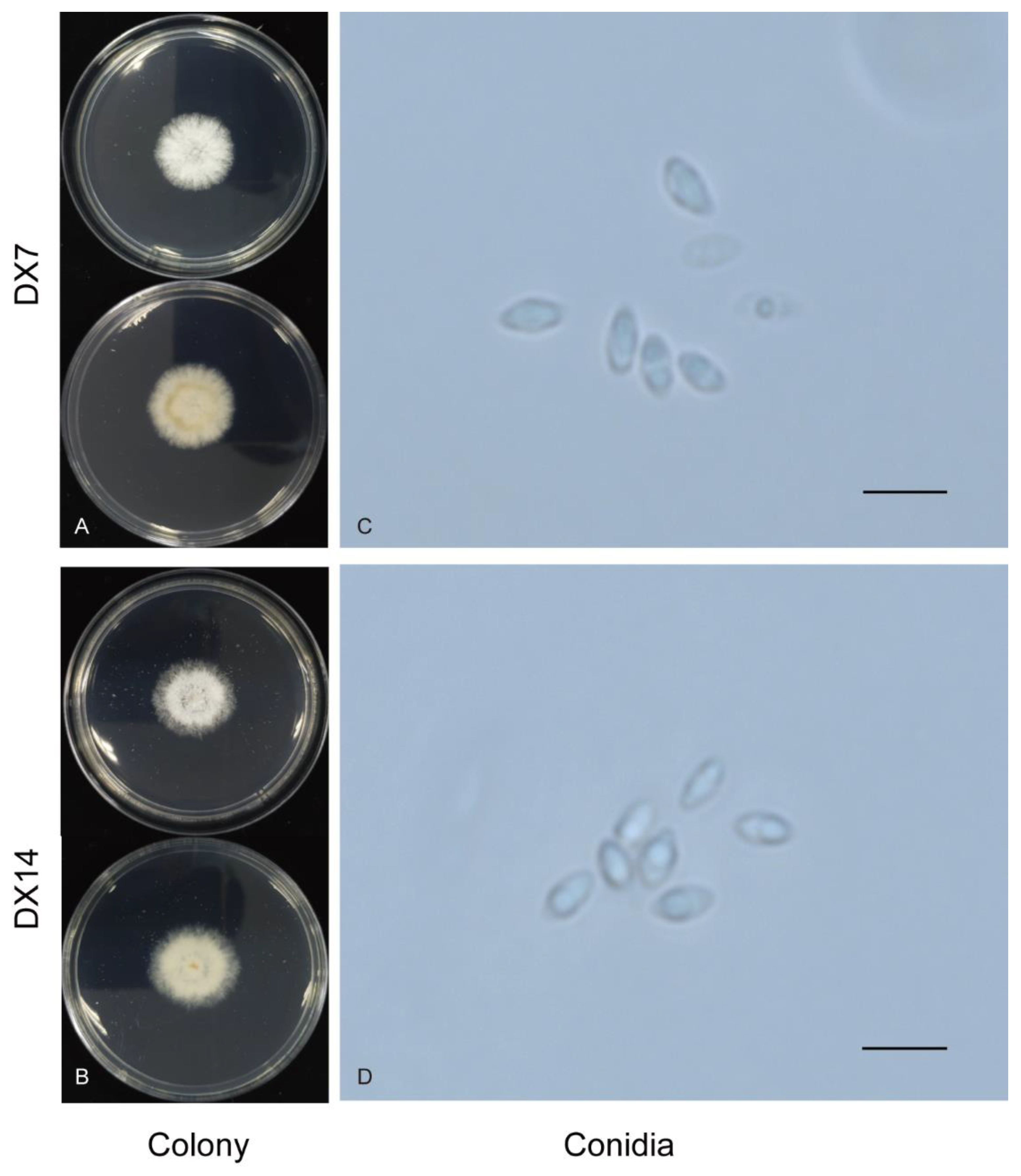
2.2. Identification and Characterization of Dt

In our study, we collected 66 Dt isolates. Phylogenetic trees (Figure 2) were constructed using the combined ITS, EF1, RPB2, and LSU gene data, encompassing 76 Diaporthales strains, including Valsella salicis AR3514 as the outgroup. The combined gene alignment, including gaps, comprised 2248 characters with 579, 518, 247, and 893 bases having alignment gaps in ITS, EF1, RPB2, and LSU. Sixty-six isolates clustered with strain MAFF238240-238143. Dt was the most common species, with 62 isolates separated from the diseased part and four isolates from the healthy part.
Based on phylogenetic analysis, the cluster of DX12, DX14, DX15, and DX16 had a higher expected number of changes per site. The pairwise dissimilarities of DNA sequences between DX12, DX14, DX15, DX16, and MAFF238240 were 23 bp, 23 bp, 22 bp, and 21 bp in LSU, respectively.
Discula theae-sinensis (I. Miyake) Moriwaki and Toy. Sato, J. Gen. Plant Pathol., 75:359–361. 2009.
Discula thea-sinensis are low, dense, fluffy white aerial mycelia. In reverse, they are concentric circles (DX7) and some are a uniform gray-white (DX14) (Figure 3). The growth rate of DX7, DX14, and DX44 was 2.63 and 2.40 per day at 25 °C on the PDA plate. The α-conidia of these three strains were hyaline, fusiform to obovoid, and tapered at the ends. The size of DX7 was 4.2–6.2 × 1.9–3.2 µm, average (av) ± SEM = 5.0 ± 0.6 × 2.4 ± 0.4 µm, n = 30. The size of DX14 was 3.2–6.3 × 1.7–2.8 µm, av ± SEM = 4.9 ± 0.7 × 2.2 ± 0.3 µm, n = 30. Moriwaki named this fungus Dt based on morphology and molecular characteristics, and the reported size of conidia of Dt is 4.6–6.3 × 1.9–3.1 µm. Conidia of strain DX7 were similar to the strain Moriwaki et al. reported, and DX14 was smaller than that strain.

2.3. Identification and Characterization of Colletotrichum spp.

Among all collected isolates, 45 isolates belonged to Colletotrichum. Phylogenetic trees (Figure 4) were constructed using the combined ITS, CAL, TUB2, and GAPDH gene data. The concatenated dataset contained 210 sequences from the genus Colletotrichum including outgroup C. xanthorrhoeae (CBS 127831), and 541, 744, 721, and 270 bases had alignment gaps in ITS, CAL, TUB2, and GAPDH, respectively. The isolates in Colletotrichum collected in the present study belonged to eight known species, Cf, Cc, C. aenigma, C. siamense, C. henanense, C. karstii, C. tropicicola, and C. gigasporum. The isolates in our study clustered with high posterior probability values ≥0.95. Twenty-seven isolates clustered with Cf, 11 isolates clustered with Cc, 2 isolates clustered with C. aenigma, and 1 isolate clustered with C. siamense, C. henanense, C. karstii, C. tropicicola, and C. gigasporum. Cf and Cc were the largest numbers of pathogens, accounting for 60% and 24.4% of all Colletotrichum strains, respectively, and the other six species constituted 15.6% of the total.
Colletotrichum tropicicola S. Phoulivong, P. Noireung, L. Cai and K.D. Hyde, Crytog. Mycol. 33: 356. 2012.
C. tropicicola was first reported on leaves of Citrus maxima. C. tropicicola was first reported in tea plants, which are putative endophytes in the present study. The strain CX33 had a growth rate of 8.57 mm per day at 25 °C. The colony on the PDA was flat with complete edges, aerial mycelia that were short and dense, with orange conidial masses near the center, and white with gray pigment spots on the reverse side near the center after 7 days (Figure 5A). Conidia on the PDA were hyaline, guttulate, long elliptic with obtuse ends, measuring 11.1–18.5 × 4.8–7.1 µm, av ± SEM = 15.0 ± 0.3 × 5.8 ± 0.1 µm, n = 30. conidia dimensions of CX33 were smaller than ex-type culture (LC0598 15–19 × 6–7 μm). Appressoria on SNA (7.2–16.4 × 6.2–11.8, av ± SEM = 12.0 ± 2.5 × 8.2 ± 1.5 µm, n = 30) were black, elliptical, ovoid, or slightly irregular.
Colletotrichum gigasporum E.F. Rakotoniriana and F. Munaut, Mycol. Progr. 12: 407. 2013.
In the present study, C. gigasporum was first reported in tea plants. The colony on the PDA was flat with smooth edges, with white pigmentation becoming black toward the center, and in reverse, typically exhibited white-to-black annular pigment after 7 days (Figure 5B). The growth rate was 11.49 mm per day. The conidia on the PDA (19.9–27.2 × 5.6–8.4 µm, av ± SEM = 23.3 ± 0.3 × 7.1 ± 0.1 µm, n = 30) were hyaline, aseptate, smooth, cylindrical with round ends. C. gigasporum is characterized by large conidia (22–)25–29(–32) × (6–)7–9 μm [27]. CX34 conforms to large conidia characteristics. Appressoria on SNA (11.8–16.3 × 9.3–13.6, av ± SEM = 14.4 ± 1.2 × 11.1 ± 1.0 µm, n = 30) were brown to black, ovoid, or irregular.

2.4. Pathogenicity Test of Dt Collected from Leaf Surface

To explore the virulence of Dt strains collected in the present study, we used mycelial discs of representative isolates of Dt (DX7, DX14) to infect the third tea leaves and carry out pathogenicity assays. The results are shown in Figure 6. Symptoms appeared after inoculation. All these isolates caused necrotic lesions around the wounded areas. As for the details of lesions, suborbicular and flat acervuli can be found in the disease spot at 7 days. The traits of acervuli in inoculated tea leaves were similar to those in diseased leaves in the field.

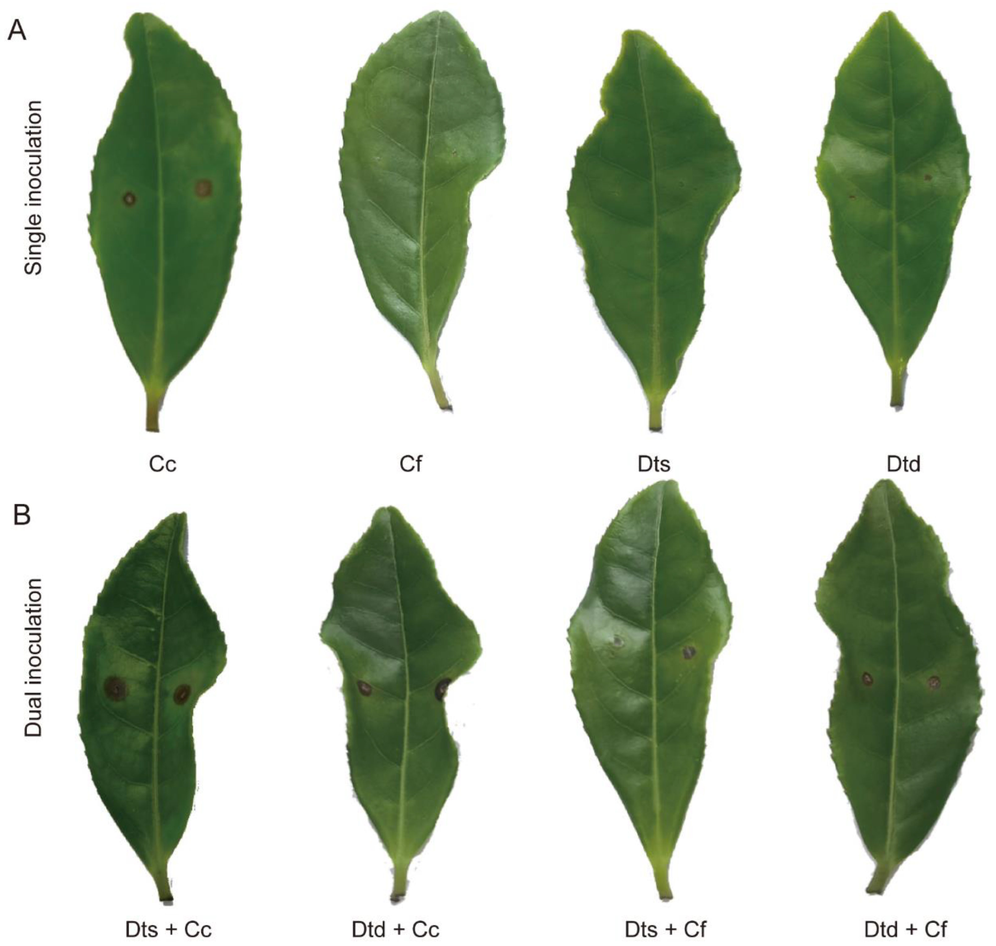
2.5. Dual Inoculation Assays of Dt and Colletotrichum

As mentioned above, Dt and Colletotrichum co-existed in tea leaves. Dual trials in vitro were carried out to find a mutual effect between Dt and Colletotrichum (Figure 7). Cf and Cc were used to co-inoculate with Dt, respectively. For 5 days of dual growth, it was observed that inhibition percentages of Dt were less than those of Colletotrichum. As shown in Figure 7B, Dt and Colletotrichum maintained a short distance (<2 mm) between them. This performance indicated that Dt and Colletotrichum had a slight inhibition in vitro.
To investigate the relationship between them during infection, dual inoculation assays were applied (Figure 8). As shown in Figure 8A, the results of single inoculation were consistent with previous studies showing that Cc was more invasive than Cf. The PDA disc of Dt also caused tea leaf disease. Meanwhile, conidial suspensions Cf and Dt did not lead to obvious lesions on tea leaves. The results of dual inoculation were more intriguing (Figure 8B). The single inoculation and dual inoculation related to virulent Cc had similar results. Dts + Cc and Dtd + Cc gave rise to significantly dark lesions on the tea leaf surface. Nonetheless, a combination of Dts + Cf and Dtd + Cf enhanced pathogenicity. Compared to single inoculation, the dual inoculation of Cf and Dt resulted in larger lesions. The results indicated that the dual inoculation of Dt and Cf in vivo promotes the development of tea plant anthracnose.

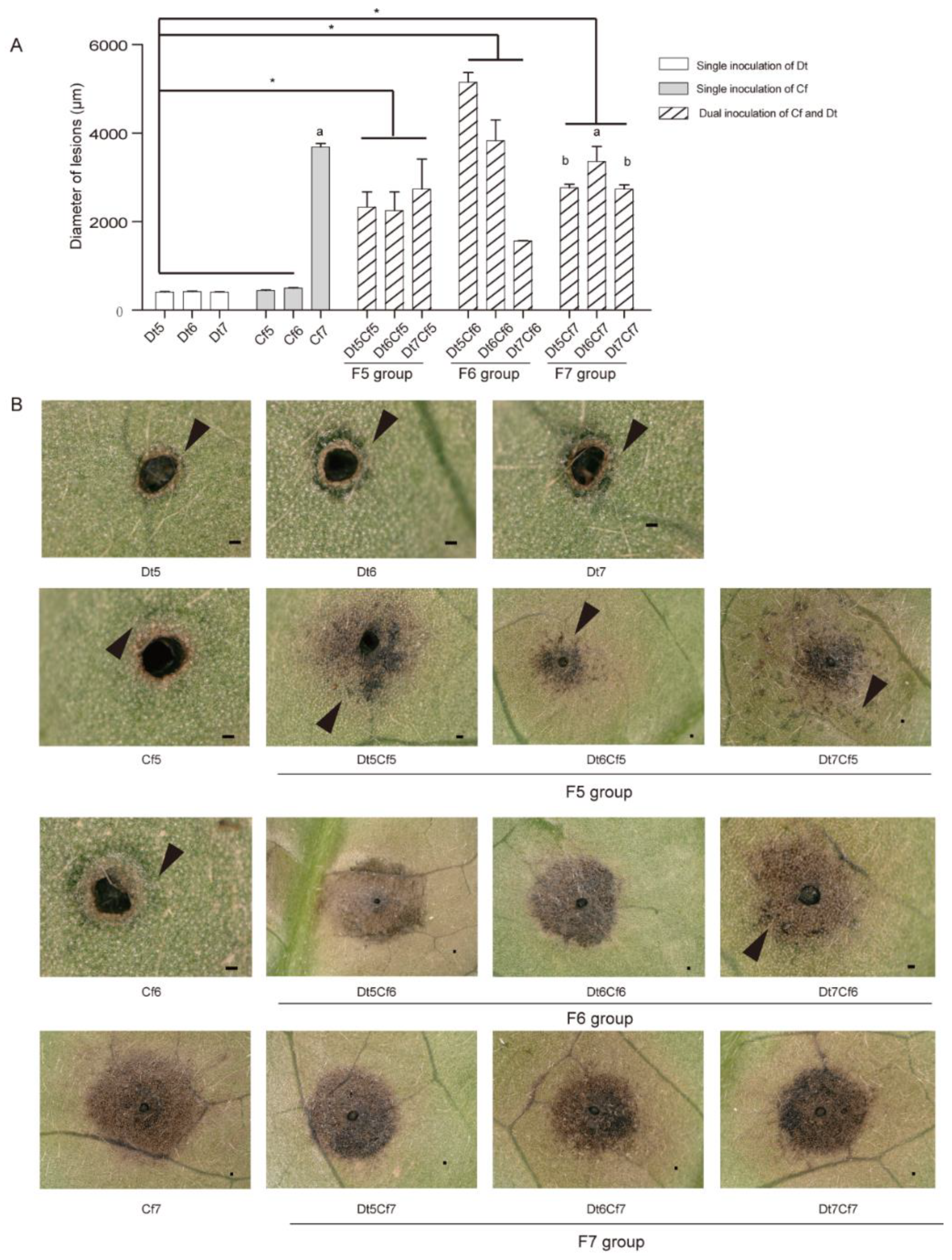
2.6. Interaction of Dt and Cf on Tea Plant Infection

To explore the impact of fungal biomass on the synergistic effect, we inoculated tea leaves with different concentrations of spore suspensions of Dt and Cf, which were set to 105, 106, and 107 spores/mL (Figure 9). The single inoculation results indicated that the spore suspension of Dt could not bring about obvious disease spots regardless of how many concentrations of spore suspensions we used. Lower concentrations of Cf (Cf5 and Cf6) caused limited damage to host plants, and the high concentration (Cf7) had the opposite effect, which generated apparent necrosis. As the lesion diameters showed, the lesion size of the F5 group (Dt5Cf5, Dt6Cf5, and Dt7Cf5) was significantly larger than those of Cf5 and Dt5, Dt6, and Dt7; the same applied for the F6 group (Dt5Cf6, Dt6Cf6, and Dt7Cf6) compared with those of Cf6 and Dt5, Dt6, and Dt7. We also found a significant difference in the F6 group. Compared to the single inoculation of Cf7, the F7 group (Dt5Cf7, Dt6Cf7, and Dt7Cf7) caused smaller lesions. Mixing with spore suspensions of low pathogenic Dt may reduce the virulence of highly pathogenic strains. The results showed that dual inoculation of Dt and Cf could promote pathogenicity when they were inoculated independently at lower concentrations. Moreover, the hypovirulent strain may recede the pathogenicity of the high-virulence strain when the pathogenicity of fungi used in dual inoculation differs greatly.
As Figure 1 shows, water-soaked lesions often occur on the anthracnose of tea plants in the field. During the procession of this study, we found that water-soaked lesions were around the fringe of necrosis when the inoculator was hypovirulent and formed a dispersed distribution of necrosis in the F5 and F6 groups. Especially in the F5 group, water-soaked lesions were in common. Furthermore, as a virulent strain, Cc also did not lead to noticeable water-soaked lesions (Figure S1). It was speculated that water-soaked lesions might play a role in affecting disease development. Exploring the composition and function of water-soaked lesions may be meaningful research in the future.

3. Discussion

3.1. Phenotype and Isolated Main Fungi of Diseased Tea Leaves

As a primary tea disease, anthracnose affects the yield and quality of tea [2,3]. According to previous studies, Colletotrichum is considered the dominant species that causes anthracnose in tea plants in China, while Dt is the major fungi that leads to tea anthracnose in Japan [5,18,20]. In the first year, we collected tea foliage with anthracnose and isolated both Dt and Colletotrichum. To verify the results, we repeated the experiment with new diseased tea leaves with anthracnose the next year, and both aforementioned fungi were detected as well. Thus, Dt widely exists in diseased tea plants as a pathogen. This is a new perspective that can guide research on anti-anthracnose strategies for tea plants with more accurate pathogen recognition.
Phenotypic identification of diseased leaves in the field is the primary method for determining specific diseases in tea plants. However, two typical diseases, anthracnose and tea leaf blight, are often easily confused, due to the complexity of environmental factors and the similarity of disease symptoms. Relying solely on visual observation for tea disease identification may lead to inaccuracies [28]. In this study, we collected pathogens from three different typical anthracnose symptoms, all characterized by sub-round or irregular scabs, scattered small black sporophores, water-soaked lesions, and the absence of thin ring spots. All three main fungi, Cc, Cf, and Dt, were found in topical anthracnose leaves collected in this study. Interestingly, all pathogenic isolates collected from A2 were Cf. This result serves as a notable example for investigating the relationship between specific pathogens and micro-characteristics such as acervuli form in plant disease.
In the present study, water-soaked lesions appeared on field samples and dual inoculation of Dt and Cf. During pathogen infection, water-soaking spots are frequently observed on infected leaves as an early symptom of the disease [29,30]. As time progresses, these spots enlarge and become necrotic [31]. In rice crops, water-soaked lesions with a dark green border are one of the field symptoms displayed by the fungi causing blast disease [32]. Water-soaking and necrosis in leaves infected by Xanthomonas euvesicatoria and X. gardneri can promote colonization of Salmonella enterica inside plant tissues [33]. Water-soaking symptoms may be related to water availability in the apoplastic environment, affecting microbial community structure and colonization [34]. AvrHah1, a single transcription activator-like (TAL) effector (TALE) protein of X. gardneri, confers enhanced water-soaked lesions in pepper [35]. Comparing the process of tomato infection with X. gardneri vs. XgΔavrHah1, water uptake from outside the leaf into the apoplast can be observed in X. gardneri-infected tomato, but not XgΔavrHah1. Water can facilitate bacteria entry into the apoplast [36]. As mentioned above, water-soaked spots affected plant–microbe interactions and might promote microbe entry. In our study, we found that water-soaked lesions always occurred in the weakly pathogenic infection process rather than in the highly pathogenic form. Therefore, we hypothesized that weak pathogens may utilize water-soaked spots to create a suitable living environment, in order to successfully infect host plants. Furthermore, we should conduct an in-depth study on the formation mechanism of water-soaked spots in tea plant–microbe interactions.

3.2. Identification of Dt from Tea Plant

Compared to the identification of Colletotrichum spp., determining the correct position of Dt in Diaporthales was more challenging. For a long time, species recognition principles in Diaporthales depended on culture characteristics, ascospore shape, and perithecium position. Currently, accurate DNA data coupled with conidial characteristics are used in scientific studies to identify and classify Diaporthales.
The nuclear ribosomal internal transcribed spacer (ITS) is commonly used when identifying the Kingdom of fungi. However, ITS sequences used alone in phylogenetic analysis are not ideal for closely related species due to intraspecific variation [37,38]. In previous studies, many researchers revised the order Diaporthales based on analysis of LSU nrDNA sequence data [7,39]. Moriwaki et al. selected 28S rDNA to study the phylogenetic relationships of strains MAFF 238,240 to 238,244 and Colletotrichum species [6]. Van Rensberg et al. [40] used ITS and EF1 sequence data to characterize species of Phomopsis. Hyde et al. [41] suggested that ITS and EF1 are recommended for preliminary identification of Diaporthe species. Senanayake et al. used combined DNA sequence data of ITS, EF1, and RPB2 to investigate the phylogenetic relationships of Diaporthalean families [42]. Thus, ITS, LSU, EF1, and RPB2 genes were used for sequence analysis in our study.
Dt strains collected in this study belong to Diaporthales species, these strains collected in this study fell in the same clade of MAFF 238,240 to 238,244 fungi with 100% pro. Moriwaki et al. used the LSU sequence alone to construct a phylogeny tree [6]. In our results, ITS, EF1, and RPB2 sequence data of Discula theae-sinensis are conducive to the following research. It is worth mentioning that the strains DX12, DX14, DX15, and DX16 had a higher expected number of changes per site. Combined with the characteristics of colony and α-conidial cells, we considered strains DX12, DX14, DX15, and DX16 to belong to Dt. The details of β- conidia or teleomorphs can provide more accurate identification for those species.

3.3. Interaction of Dt and Colletotrichum

In contrast to the type of host–pathogen interaction in monospecies infections, plants are exposed to various biotic challenges simultaneously in the field. Multiple pathogens may encounter the same host simultaneously [43]. Three pathogens were found in association with the black spot disease complex of peas [44]. Oomycete Phytophthora sojae and Fusarium spp. frequently coexist in diseased soybean roots, both of which cause soybean root rot [45]. In a two-year field investigation and sample collection, we discovered Dt, Cf, and Cc coexisting in diseased tea leaves.
Interactions between different pathogens can also have synergistic effects, as reported previously. Wang et al. found that different Fusarium species enhanced P. sojae infection when co-inoculated on soybean. Co-infection with Fusarium promotes the loss of P. sojae resistance in soybeans [45]. Whitelaw-Weckert et al. found that co-inoculation of Ilyonectria and Botryosphaeriaceae isolates led to increased disease severity compared to monoculture inoculations of Ilyonectria isolates [46]. In our study, compared to single inoculation, co-infection with Dt and Cf in tea leaves resulted in larger lesions. Interestingly, an aggravation of the condition was observed in dual inoculation of Dt and Cf on tea leaves, but not in Dt and Cc. According to previous studies and pathogenicity tests in this study, we found that Cc is more virulent than Dt and Cf, and low concentrations of spore suspensions of Dt and Cf do not cause disease spot occurrence on tea leaves [13,16]. Co-inoculation with different concentrations of spore suspensions was performed to explore the effect of Cc, Cf, and Dt during the infection. At the low concentration (Cf cannot cause tea plant disease in this concentration range), the dual inoculation of Dt and Cf caused lesions in tea leaves and significantly enhanced the infection capacity compared with single inoculation. We found that the interaction of multiple pathogens may change the severity of the disease. However, Kaur et al. pre-inoculation with Hyaloperonospora parasitica (an asymptomatic isolate) reduces the incubation period and increases susceptibility of a Brassica to white rust disease [47]. This is different from our results. Our results indicated that the single inoculation and dual inoculation related to virulent Cc had similar results. We considered that the temporal order of host infection may play a role in host–multi-pathogen interaction. Le May et al. revealed that the severity of disease was different between inoculation of two pathogens simultaneously and sequentially [44].
For a long time, research on host–pathogen interaction was in a one-to-one mode where a single pathogen colonizes a host. Disease control measures were also focused on dealing with specific pathogens. However, addressing diseases caused by multiple species requires new measures. According to our results, we believe that the main cause of anthracnose might be the concerted action of a variety of fungi. In the future, the interaction mechanisms of Dt and Colletotrichum and their relationship with tea plants deserve more attention.

4. Materials and Methods

4.1. Collection and Isolation

Tea leaves were collected from Camellia sinensis cv. Longjing43 (LJ43) in three different tea gardens of Zhejiang Province, China. Among each tea garden, more than three tea plant individuals were sampled with diseased tea leaves fitting the symptoms as P1, P2, and P3. In addition, three diseased tea leaves with equivalent leaf positions were sampled randomly in each individual. The sampling period was from June to August continuously for two years. Diseased samples were identified and collected from leaves with visible anthracnose symptoms, which are sub-round or irregular scabs, scattered small black sporophores, water-soaked lesions, and the absence of thin ring spots [48]. The isolates separated from leaves were obtained by a single spore isolation technique described by Cai et al. [49]. In addition, the isolates separated from a healthy part of tea diseased leaves were collected according to a protocol from Kjer et al. [50]. Briefly, the fungi were collected after washing diseased leaves with tap water, alcohol (70%) for 1 min, sodium hypochlorite (2.5%) for 1 min, and then washed in sterile distilled water. To check the efficacy of this method of surface sterilization, 100 µL of the last wash water was incubated on PDA plates.

4.2. Morphological Characterization

Mycelial discs were taken from the margin of 5-day-old PDA cultures and incubated on PDA plates in the dark at 25 °C. For growth rate measurement, each isolate was measured in at least three independent experiments. The colony diameter was measured daily beginning 3 days after inoculation and used for calculating the growth rate. The growth rate can be calculated as the average of mean daily growth (mm/d). The colony and conidial characteristics were determined via methods described by Cai et al. [49]. Appressoria was produced and measured using a slide culture technique and induced on a synthetic nutrient-poor agar (SNA) medium.

4.3. DNA Extraction and PCR Amplification

Genomic DNA was extracted from mycelium cultured on PDA for 7 days. Total genomic DNA, which was used as the template for PCR amplification, was extracted using an Ezup Column Fungi Genomic DNA Purification Kit [Sangon Biotech (Shanghai) Company Limited, Shanghai, China] and stored at −20 °C. Four partial loci were used for Colletotrichum identification, including ribosomal internal transcribed spacer (ITS), glyceraldehyde 3-phosphate dehydrogenase (GAPDH), beta-tubulin (TUB2), and calmodulin (CAL) genes. Other partial loci, such as ITS, the partial 28S nrDNA (LSU), DNA-directed RNA polymerase II second largest subunit (RPB2), and translation elongation factor 1-alpha (EF1) were used to identify Diaporthales. The primers used in this study are listed in Table S1, while the polymerase chain reaction (PCR) was performed as follows: 4 min at 94 °C; followed by 35 cycles of 30 s at 94 °C; 50 s at 57 °C (LSU) or 54 °C (EF1) or 55 °C (RPB2); 30 s at 72 °C, and 10 min at 72 °C. The PCR conditions for ITS, GAPDH, TUB2, and CAL were as described by Lu et al. [16]. PCR purification and DNA sequencing were performed by Shanghai Huagene Biotech Company Limited, Shanghai, China.

4.4. Phylogenetic Analysis

The accession numbers of the sequences were obtained from NCBI GenBank based on BLAST searches and published literature and are listed in Tables S2 and S3. Sequences from forward and reverse primers were edited with SeqMan and DNAStar. We used Bayesian inference (BI) to construct the phylogenies according to multilocus sequence analysis. MrBayes v. 3.2. MrModel test v. 2.3 was used to select the best-fit models of nucleotide substitution, then assembling and modulating the dataset by MAFFT v.7 and MEGA v. 6.0, respectively. All gaps were regarded as missing data. Markov chain Monte Carlo (MCMC) sampling was used to reconstruct the phylogenies in MrBayes v.3.2. Analyses of 6 MCMC chains based on the full dataset were run for 2 × 10⁷ generations and sampled every 100 generations [13,16].

4.5. Pathogenicity Tests of Discula Theae-Sinensis

First, the healthy, non-wounded the third tea leaves were washed with tap water and then sterilized with 1% sodium hypochlorite for 2 min, flushed with sterilized water 3 times, and air dried. Afterward, sterile needles were used to puncture the leaves to make wounded spots. Subsequently, 5 mm mycelial discs from a 5-day-old culture were placed over every wound of tea leaves. The inoculated leaves were cultured in an incubator at 25 °C with a 12/12 h (day/night) photoperiod. The conditions of inoculated leaves were recorded at 5 and 7 days post-inoculation. In this experiment, three replicates were included for each treatment. Sterile-distilled PDA discs with no mycelia were used as controls. Finally, Koch’s postulates were employed to investigate each strain. Test of each isolate was performed in three biological replicates.

4.6. Dual Inoculation Assays of Dt and Colletotrichum

The plate confrontation culture method was used to assess the mutual effect between Dt and Colletotrichum spp. in vitro. The PDA plates were cultured in an incubator at 25 °C for 5 days in the dark. And then, observed the mutual effect between strains. The formula of inhibition ratio of growth of fungi was calculated described by López-González et al. was followed with minor revisions [51].
Inhibition ratio (%) = [(R1 − R2)/R1] × 100
where R1 indicates the radial growth of the fungi colony in control plates and R2 indicates the radial growth of the fungi colony (in the direction of the other fungus).
Dual assays in vivo were used to explore the relationship between Dt and Colletotrichum spp. during the procession of infection. The method of leaf preparation and inoculation condition was the same as the pathogenicity test. The inoculation experiments were grouped into 8 treatments: Dts, Dtd, Cc, Cf, Dts + Cc, Dtd + Cc, Dts + Cf, and Dtd + Cf. The labels and corresponding treatment conditions are shown in Table 1. The concentration of spore suspension was 106 spores/mL in these experiments. Distilled water was used as the control.
Table 1. Labels and corresponding treatment conditions in dual inoculation.
LabelsTreatment Conditions
Dtssingle inoculation of spore suspension of Dt
Dtdsingle inoculation of mycelial discs of Dt
Ccsingle inoculation of spore suspension of Cc
Cfsingle inoculation of spore suspension of Cf
Dts + Ccdual inoculation with a spore suspension of Dt and Cc
Dtd + Ccdual inoculation with mycelial discs of Dt and a spore suspension of Cc
Dts + Cfdual inoculation with spore suspension Dt and Cf
Dtd + Cfdual inoculation with mycelial discs of Dt and spore suspension of Cf

4.7. Impact of Spore Suspension Concentration on Combined Dt and Cf Infection Capacity

To explore the interaction of Dt and Cf at various concentrations in a spore suspension, inocula were prepared by mixing spore suspensions of Dt and Cf in equal amounts. The concentration of the spore suspension was 105 spores/mL, 106 spores/mL, and 107 spores/mL. Six single inoculations and 9 combinations were obtained. The labels and corresponding treatment conditions are shown in Table 2.
Table 2. Labels and corresponding treatment conditions in this sector.
LabelsTreatment Conditions
Single inoculationDt5105 spores/mL spore suspension of Dt
Dt6106 spores/mL spore suspension of Dt
Dt7107 spores/mL spore suspension of Dt
Cf5105 spores/mL spore suspension of Cf
Cf6106 spores/mL spore suspension of Cf
Cf7107 spores/mL spore suspension of Cf
Dual inoculationF5 groupDt5Cf5105 spores/mL Dt and 105 spores/mL Cf
Dt6Cf5106 spores/mL Dt and 105 spores/mL Cf
Dt7Cf5107 spores/mL Dt and 105 spores/mL Cf
F6 groupDt5Cf6105 spores/mL Dt and 106 spores/mL Cf
Dt6Cf6106 spores/mL Dt and 106 spores/mL Cf
Dt7Cf6107 spores/mL Dt and 106 spores/mL Cf
F7 groupDt5Cf7105 spores/mL Dt and 107 spores/mL Cf
Dt6Cf7106 spores/mL Dt and 107 spores/mL Cf
Dt7Cf7107 spores/mL Dt and 107 spores/mL Cf

4.8. Statistical Analysis and Microscopic Observation

IBM SPSS Statistics software version 19 was used to analyze variance (ANOVA) of the experimental data. The mean values were compared by the least significant difference (LSD) method, and differences were considered significant at p < 0.05. All data are represented as the average ± standard error.
An OLYMPUS stereo microscope was used to observe acervuli on diseased tea leaves from the field. A Nikon 80i microscope equipped with a differential interference contrast (DIC) objective was used to visualize spores on diseased tea leaves. Lesion observation after dual inoculation was performed using a field-depth microscope (KEYENCE). Conidial characteristics were recorded by ZEISS Axio Vert. A1.

5. Conclusions

In this study, we delved into the dominant species responsible for anthracnose in tea plants and conducted an extensive exploration of the diversity within Dt and Colletotrichum associated with Camellia sinensis. Our findings revealed that Dt is the predominant species in tea leaves, serving as the primary causative agent of tea plant anthracnose. Furthermore, we identified a total of eight known Colletotrichum species with the noteworthy inclusion of C. tropicicola and C. gigasporum, which were reported for the first time in tea plants. Our investigation also highlighted the advantages of co-inoculation with Dt and C. fructicola, particularly at low concentrations, as it demonstrated superior outcomes compared to single inoculations. This observation suggests that the interaction between Dt and Colletotrichum may play a pivotal role in the development of anthracnose in tea plants. In conclusion, our research sheds new light on the multifaceted nature of anthracnose in tea plants. Rather than a single pathogenic agent, our findings suggest that anthracnose may result from the collaborative efforts of various fungal species. As we move forward, it is imperative to delve deeper into the mechanisms of interaction between Dt and Colletotrichum and their intricate relationship with tea plants. This knowledge will be essential for the development of effective strategies for anthracnose control and the continued prosperity of the tea industry.

Supplementary Materials

The following supporting information can be downloaded at https://www.mdpi.com/article/10.3390/plants12193427/s1. Figure S1: Details of lesions of inoculation with Cc and dual inoculations of Cc and Dt; Table S1: Primers used in this study [4,52,53,54,55,56,57,58,59]; Table S2: Isolates Diaporthales spp. studied and GenBank accession numbers of the generated sequences; Table S3: Isolates Colletotrichum spp. studied and GenBank accession numbers of the generated sequences.

Author Contributions

Conceptualization, Q.L. (Qinhua Lu); Methodology, Q.L. (Qingsheng Li) and Q.L. (Qinhua Lu); Validation, D.L., N.R. and Y.J.; Investigation, J.Z., D.L., N.R. and W.L.; Data Curation; Q.L. (Qinhua Lu); Writing—Original Draft Preparation, Q.L. (Qinhua Lu) and Q.L. (Qingsheng Li); Writing—Review and Editing, Q.L. (Qingsheng Li); Supervision, J.Z. Administration, J.Z.; Funding Acquisition, Q.L. (Qinhua Lu), Q.L. (Qingsheng Li), W.L. and J.Z. All authors have read and agreed to the published version of the manuscript.

Funding

This work was funded by the Open Fund of the State Key Laboratory of Tea Plant Biology and Utilization (SKLTOF20200124); the study was funded by the Zhejiang Science and Technology Major Program on Agricultural New Variety Breeding-Tea Plant (2021C02067-7), the National Natural Science Foundation of China (Project No. 32202545, No. 32102438, No. 32202536), the Discipline Start-up Fund of Zhejiang Academy of Agricultural Sciences (10405080121CC2201G), the Talent Start-up Fund of Zhejiang Academy of Agricultural Sciences (10405010120CF0401G/001), and the Innovation and utilization of Characterized Germplasm of Tea Plant in Huzhou (2022SY010).

Institutional Review Board Statement

Not applicable.

Informed Consent Statement

Not applicable.

Data Availability Statement

Data are available upon request from the corresponding author.

Acknowledgments

We express our gratitude to Xin-Chao Wang and his group from the Tea Resources and Improvement Research Center, Tea Research Institute of Chinese Academy of Agricultural Sciences, Hangzhou, China, for their guidance and sample sharing.

Conflicts of Interest

The authors declare no conflict of interest.

References

  1. Leng, Y.; Tong, J.W.; Huang, P.; Shang, H.G.; Zhou, Z.Y. The development of tea Industry in China during the 13th five-year plan period and prospects for the 14th five-year plan period. China Tea 2021, 43, 25–30. [Google Scholar]
  2. Wang, Y.C.; Qian, W.J.; Li, N.N.; Hao, X.Y.; Wang, L.; Xiao, B.; Wang, X.C.; Yang, Y.J. Metabolic Changes of Caffeine in Tea Plant (Camellia sinensis (L.) O. Kuntze) as Defense Response to Colletotrichum fructicola. J. Agric. Food Chem. 2016, 64, 6685. [Google Scholar] [CrossRef] [PubMed]
  3. Liu, X.; Jiang, J.; Zhan, J.; Zhou, K.; Chen, Z. Main Diseases and Insect Pests of Tea in Meitan County. Guizhou Agric. Sci. 2011, 39, 77–80. [Google Scholar]
  4. Weir, B.S.; Johnston, P.R.; Damm, U. The Colletotrichum gloeosporioides species complex. Stud. Mycol. 2012, 73, 115–180. [Google Scholar] [CrossRef]
  5. Yamamoto, W.A. revision of the genus and species names of anthracnose fungi in Japan. Plant Prot. 1960, 14, 49–52. [Google Scholar]
  6. Moriwaki, J.; Sato, T. A new combination for the causal agent of tea anthracnose: Discula theae-sinensis (I. Miyake) Moriwaki & Toy. Sato, comb. nov. J. Gen. Plant Pathol. 2009, 75, 359–361. [Google Scholar]
  7. Castlebury, L.A.; Rossman, A.Y.; Jaklitsch, W.J.; Vasilyeva, L.N. A preliminary overview of the Diaporthales based on large subunit nuclear ribosomal DNA sequences. Mycologia 2002, 94, 1017–1031. [Google Scholar] [CrossRef]
  8. Sogonov, M.V.; Castlebury, L.A.; Rossman, A.Y.; White, J.F. The type species of Apiognomonia, A. veneta, with its Discula anamorph is distinct from A. Errabunda. Mycol. Res. 2007, 111, 693–709. [Google Scholar] [CrossRef]
  9. Senanayake, I.C.; Crous, P.W.; Groenewald, J.Z.; Maharachchikumbura, S.S.N.; Jeewon, R.; Phillips, A.J.L.; Bhat, J.D.; Perera, R.H.; Li, Q.R.; Li, W.J.; et al. Families of Diaporthales based on morphological and phylogenetic evidence. Stud. Mycol. 2017, 86, 217–296. [Google Scholar] [CrossRef]
  10. Xie, H.; Feng, X.; Wang, M.; Wang, Y.; Kumar Awasthi, M.; Xu, P. Implications of endophytic microbiota in Camellia sinensis: A review on current understanding and future insights. Bioengineered 2020, 11, 1001–1015. [Google Scholar] [CrossRef]
  11. Gao, Y.; Liu, F.; Cai, L. Unravelling Diaporthe species associated with Camellia. Syst. Biodivers. 2016, 14, 102–117. [Google Scholar] [CrossRef]
  12. Jayawardena, R.S.; Bhunjun, C.S.; Hyde, K.D.; Gentekaki, E.; Itthayakorn, P. Colletotrichum: Lifestyles, biology, morpho-species, species complexes and accepted species. Mycosphere 2021, 12, 519–669. [Google Scholar] [CrossRef]
  13. Wang, Y.C.; Hao, X.Y.; Wang, L.; Xiao, B.; Wang, X.C.; Yang, Y.J. Diverse Colletotrichum species cause anthracnose of tea plants (Camellia sinensis (L.) O. Kuntze) in China. Sci. Rep. 2016, 6, 35287. [Google Scholar] [CrossRef] [PubMed]
  14. Peng, X.J.; Wang, Q.C.; Zhang, S.K.; Guo, K.; Zhou, X.D. Colletotrichum species associated with Camellia anthracnose in China. Mycosphere 2023, 14, 130–157. [Google Scholar]
  15. Liu, F.; Weir, B.S.; Damm, U.; Crous, P.W.; Wang, Y.; Liu, B.; Wang, M.; Zhang, M.; Cai, L. Unravelling Colletotrichum species associated with Camellia: Employing ApMat and GS loci to resolve species in the C. gloeosporioides complex. Persoonia 2015, 35, 63–86. [Google Scholar] [CrossRef] [PubMed]
  16. Lu, Q.; Wang, Y.; Li, N.; Ni, D.; Yang, Y.; Wang, X. Differences in the characteristics and pathogenicity of Colletotrichum camelliae and C. fructicola Isolated from the tea plant (Camellia sinensis (L.) O. Kuntze). Front. Microbiol. 2018, 9, 3060. [Google Scholar] [CrossRef]
  17. Li, X.; Wang, Z.K.; Tan, W.Z.; Zhang, K.C.; Li, P.; Wang, Z.W.; Yin, L.L.; Tan, S.X. Effects of wuyiencin on growth and sporulation of Colletotrichum camelliae causing tea leaf blight. J. Southwest China Norm. Univ. 2008, 33, 42–47. (In Chinese) [Google Scholar]
  18. Chen, Y.; Qiao, W.L.; Zeng, L.; Shen, D.; Liu, Z.L.; Wang, X.S.; Tong, H. Characterization, pathogenicity, and phylogenetic analyses of Colletotrichum species associated with brown blight disease on Camellia sinensis in China. Plant Dis. 2017, 106, 1022–1028. [Google Scholar] [CrossRef]
  19. Doehlemann, G.; Ökmen, B.; Zhu, W.; Sharon, A. Plant pathogenic fungi. Microbiol. Spectr. 2017, 5, FUNK-0023-2016. [Google Scholar] [CrossRef]
  20. Yamada, K.; Sonoda, R. A fluorescence microscopic study of the infection process of Discula theae-sinensis in tea. Jpn. Agr. Res. Q 2014, 48, 399–402. [Google Scholar] [CrossRef]
  21. Kim, K.W. Plant trichomes as microbial habitats and infection sites. Eur. J. Plant Pathol. 2019, 154, 157–169. [Google Scholar] [CrossRef]
  22. Hamaya, E. Trichome infection of the tea anthracnose fungus Gloeosporium theae-sinensis. JARQ 1982, 16, 114–118. [Google Scholar]
  23. Zhang, Z.; Jiang, B.; Yang, X.; Yuan, Y.; Liu, N.; Ren, W.; Li, P.; Li, B.; Lian, S. Deficiency of the melanin biosynthesis genes CfSCD1 impedes appressoria formation and disrupts Colletotrichum fructicola infection on apple. Trop. Plant Pathol. 2023. [Google Scholar] [CrossRef]
  24. Takahara, H.; Hacquard, S.; Kombrink, A.; Hughes, H.B.; Halder, V.; Robin, G.P.; Hiruma, K.; Neumann, U.; Shinya, T.; Kombrink, E.; et al. Colletotrichum higginsianum extracellular LysM proteins play dual roles in appressorial function and suppression of chitin-triggered plant immunity. New Phytol. 2016, 211, 1323–1337. [Google Scholar] [CrossRef]
  25. Kleemann, J.; Rincon-Rivera, L.J.; Takahara, H.; Neumann, U.; Ver Loren van Themaat, E.; van der Does, H.C.; Hacquard, S.; Stüber, K.; Will, I.; Schmalenbach, W.; et al. Sequential delivery of host-induced virulence effectors by appressoria and intracellular hyphae of the phytopathogen Colletotrichum higginsianum. PLoS Pathog. 2012, 8, e1002643. [Google Scholar] [CrossRef]
  26. Ryder, L.; Talbot, N. Regulation of appressorium development in pathogenic fungi. Curr. Opin. Plant Biol. 2015, 26, 8–13. [Google Scholar] [CrossRef]
  27. Liu, F.; Cai, L.; Crous, P.W.; Damm, U. The Colletotrichum gigasporum species complex. Persoonia 2014, 33, 83–97. [Google Scholar] [CrossRef] [PubMed]
  28. Wang, Y.C.; Liu, S.A.; Lu, Q.H.; Xiong, F.; Yang, Y.J.; Wang, X.C. Research progress and prospects of Colletotrichum species causing tea plant diseases in China. J. Plant Prot. Res. 2019, 46, 954–963. [Google Scholar]
  29. Kheiri, A.; Moosawi-Jorf, S.A.; Malihipour, A. Infection process and wheat response to Fusarium head blight caused by Fusarium graminearum. Eur. J. Plant Pathol. 2018, 153, 489–502. [Google Scholar] [CrossRef]
  30. Lamichhane, J.R.; Fabi, A.; Ridolfi, R.; Varvaro, L. Epidemiological study of hazelnut bacterial blight in central Italy by using laboratory analysis and geostatistics. PLoS ONE 2013, 8, e56298. [Google Scholar] [CrossRef]
  31. Akhavan, A.; Bahar, M.; Askarian, H.; Lak, M.R.; Nazemi, A.; Zamani, Z. Bean common bacterial blight: Pathogen epiphytic life and effect of irrigation practices. Springerplus 2013, 2, 41. [Google Scholar] [CrossRef] [PubMed]
  32. Kumar, S.; Kashyap, P.L.; Mahapatra, S.; Jasrotia, P.; Singh, G.P. New and emerging technologies for detecting Magnaporthe oryzae causing blast disease in crop plants. Crop Prot. 2021, 143, 105473. [Google Scholar] [CrossRef]
  33. Potnis, N.; Colee, J.; Jones, J.B.; Barak, J.D. Plant pathogen induced water-soaking promotes Salmonella enterica growth on tomato leaves. Appl. Environ. Microbiol. 2015, 81, 8126–8134. [Google Scholar] [CrossRef] [PubMed]
  34. Aung, K.; Jiang, Y.; He, S.Y. The role of water in plant-microbe interactions. Plant J. 2018, 93, 771–780. [Google Scholar] [CrossRef]
  35. Schornack, S.; Minsavage, G.V.; Stall, R.E.; Jones, J.B.; Lahaye, T. Characterization of AvrHah1, a novel AvrBs3-like effector from Xanthomonas gardneri with virulence and avirulence activity. New Phytol. 2008, 179, 546–556. [Google Scholar] [CrossRef]
  36. Schwartz, A.R.; Morbitzer, R.; Lahaye, T.; Staskawicz, B.J. TALE-induced bHLH transcription factors that activate a pectate lyase contribute to water soaking in bacterial spot of tomato. Proc. Natl. Acad. Sci. USA 2017, 114, E897–E903. [Google Scholar] [CrossRef]
  37. Gomes, R.R.; Glienke, C.; Videira, S.I.R.; Lombard, L.; Groenewald, J.Z.; Crous, P.W. Diaporthe: A genus of endophytic, saprobic and plant pathogenic fungi. Persoonia 2013, 31, 1–41. [Google Scholar] [CrossRef]
  38. Santos, J.M.; Correia, V.G.; Phillips, A.J.L. Primers for mating-type diagnosis in Diaporthe and Phomopsis: Their use in teleomorph induction in vitro and biological species definition. Fungal. Biol. 2010, 114, 255–270. [Google Scholar] [CrossRef]
  39. Mostert, L.; Groenewald, J.Z.; Summerbell, R.; Gams, W.; Crous, P.W. Taxonomy and Pathology of Togninia (Diaporthales) and its Phaeoacremonium Anamorphs. Stud. Mycol. 2006, 54, 1–113. [Google Scholar] [CrossRef]
  40. van Rensburg, J.C.J.; Lamprecht, S.C.; Groenewald, J.Z.; Castlebury, L.A.; Crous, P.W. Characterization of Phomopsis spp. associated with die-back of rooibos (Aspalathus linearis) in South Africa. Stud. Mycol. 2006, 55, 65–74. [Google Scholar] [CrossRef]
  41. Hyde, K.D.; Nilsson, R.H.; Alias, S.A.; Ariyawansa, H.A.; Blair, J.E.; Cai, L.; Cock, A.; Dissanayake, A.; Glockling, S.; Goonasekara, I.; et al. One stop shop: Backbones trees for important phytopathogenic genera: I. Fungal. Divers. 2014, 67, 21–125. [Google Scholar] [CrossRef]
  42. Senanayake, I.C.; Jeewon, R.; Chomnunti, P.; Wanasinghe, D.N.; Norphanphoun, C.; Karunarathna, A.; Pem, D.; Perera, R.H.; Camporesi, E.; McKenzie, E.H.C.; et al. Taxonomic circumscription of Diaporthales based on multigene phylogeny and morphology. Fungal Divers. 2018, 93, 241–443. [Google Scholar] [CrossRef]
  43. Fitt, B.D.L.; Huang, Y.J.; Bosch, F.V.D.; West, J.S. Coexistence of related pathogen species on arable crops in space and time. Annu. Rev. Phytopathol. 2006, 44, 163–182. [Google Scholar] [CrossRef] [PubMed]
  44. Le May, C.; Potage, G.; Andrivon, D.; Tivoli, B.; Outreman, Y. Plant disease complex: Antagonism and synergism between pathogens of the Ascochyta blight complex on Pea. J. Phytopathol. 2009, 157, 715–721. [Google Scholar] [CrossRef]
  45. Wang, S.; Zhang, X.; Zhang, Z.; Chen, Y.; Tian, Q.; Zeng, D.; Xu, M.; Wang, Y.; Dong, S.; Ma, Z.; et al. Fusarium-produced vitamin B6 promotes the evasion of soybean resistance by Phytophthora sojae. J. Integr. Plant Biol. 2023, 65, 2204–2217. [Google Scholar] [CrossRef] [PubMed]
  46. Whitelaw-Weckert, M.A.; Rahman, L.; Appleby, L.M.; Hall, A.; Clark, A.C.; Waite, H.; Hardie, W.J. Co-infection by Botryosphaeriaceae and Ilyonectria spp. fungi during propagation causes decline of young grafted grapevines. Plant Pathol. 2013, 62, 1226–1237. [Google Scholar] [CrossRef]
  47. Kaur, P.; Sivasithamparam, K.; Li, H.; Barbetti, M.J. Pre-inoculation with Hyaloperonospora parasitica reduces incubation period and increases severity of disease caused by Albugo candida in a Brassica juncea variety resistant to downy mildew. J. Gen. Plant Pathol. 2011, 77, 101–106. [Google Scholar] [CrossRef]
  48. Tang, M.J.; Xiao, Q. Colored Pictorial Handbook of Tea Plant Pests and Natural Enemies; China Agricultural University Press: Beijing, China, 2019; pp. 4–5. [Google Scholar]
  49. Cai, L.; Hyde, K.D.; Taylor, P.; Weir, B.S.; Waller, J.M.; Abang, M.M.; Zhang, J.Z.; Yang, Y.L.; Phoulivong, S.; Liu, Z.Y.; et al. A polyphasic approach for studying Colletotrichum. Fungal Divers. 2009, 39, 183–204. [Google Scholar]
  50. Kjer, J.; Debbab, A.; Aly, A.H.; Proksch, P. Methods for isolation of marine-derived endophytic fungi and their bioactive secondary products. Nat. Protoc. 2010, 5, 479–490. [Google Scholar] [CrossRef]
  51. López-González, R.C.; Gómez-Cornelio, S.; Rosa-García, D.; Garrido, E.; Oropeza-Mariano, O.; Heil, M.; Partida-Martínez, L. The age oflima bean leaves influences the richness and diversity of the endophytic fungal community, but not the antagonistic effect of endophytes against Colletotrichum lindemuthianum. Fungal Ecol. 2017, 26, 1–10. [Google Scholar] [CrossRef]
  52. Templeton, M.D.; Rikkerink, E.; Solon, S.L.; Crowhurst, R.N. Cloning and molecular characterization of the glyceraldehyde-3-phosphate dehydrogenase-encoding gene and cDNA from the plant pathogenic fungus Glomerella cingulata. Gene 1992, 122, 225–230. [Google Scholar] [CrossRef] [PubMed]
  53. Gardes, M.; Bruns, T.D. ITS primers with enhanced specificity for basidiomycetes—Application to the identification of mycorrhizae and rusts. Mol. Ecol. 1993, 2, 113–118. [Google Scholar] [CrossRef]
  54. White, T.; Bruns, T.; Lee, S.; Taylor, J. Amplification and direct sequencing of fungal ribosomal RNA genes for phylogenetics. Pcr. Protocols. 1990, 18, 315–322. [Google Scholar]
  55. O’Donnell, K.; Cigelnik, E. Two divergent intragenomic rDNA ITS2 types within a monophyletic lineage of the fungus Fusarium are nonorthologous. Mol. Phylogenet. Evol. 1997, 7, 103–116. [Google Scholar] [CrossRef] [PubMed]
  56. Glass, N.L.; Donaldson, G.C. Development of primer sets designed for use with the PCR to amplify conserved genes from filamentous ascomycetes. Appl. Environ. Microbiol 1995, 61, 1323–1330. [Google Scholar] [CrossRef] [PubMed]
  57. Carbone, I.; Kohn, L.M. A method for designing primer sets for speciation studies in filamentous ascomycetes. Mycologia 1999, 91, 553–556. [Google Scholar] [CrossRef]
  58. Liu, Y.J.; Whelen, S.; Hall, B.D. Phylogenetic relationships among ascomycetes: Evidence from an RNA polymerse II subunit. Mol. Biol. Evol. 1999, 16, 1799–1808. [Google Scholar] [CrossRef]
  59. Vilgalys, R.; Hester, M. Rapid genetic identification and mapping of enzymatically amplified ribosomal DNA from several Cryptococcus species. J. Bacteriol. 1990, 172, 4238–4246. [Google Scholar] [CrossRef]
Figure 1. Phenotypic characterization of diseased tea leaves. (A) Morphology of tea leaves. Black triangles indicate water-soaked lesions. (B) Shape of acervuli on tea leaves. (C) Spores isolated from lesions. Red arrows indicate spores.
Plants 12 03427 g001
Figure 2. Phylogenetic tree generated by Bayesian analysis based on a combined 4-gene dataset (ITS, EF1, RPB2, and LSU). Bayesian posterior probabilities above 0.90 are shown at each node. Valsella salicis AR3514 was used as the outgroup. The scale bar indicates 0.4 expected changes per site. The branches crossed by diagonal lines are shortened by 50%. Red squares indicate strains isolated in 2019; blue squares indicate strains isolated in 2020; red circles indicate isolates collected from diseased part of tea diseased leaves; blue circles indicate isolates collected from healthy part of tea diseased leaves; white triangles indicate strains isolated from P1; yellow triangles indicate strains isolated from P2; black triangles indicate strains isolated from P3.
Plants 12 03427 g002
Figure 3. Morphological characteristics of Dt. (A,B) colonies of DX7 and DX14. (C,D) conidia of DX7 and DX14, Scale bar = 80 µm.
Plants 12 03427 g003
Figure 4. Phylogenetic tree generated via Bayesian analysis based on a combined 4-gene dataset (ITS, CAL, TUB2, and GAPDH). Bayesian posterior probabilities above 0.95 are shown at each node. C. xanthorrhoeae CBS 127831 was used as the outgroup. The scale bar indicates 0.05 expected changes per site. The ex-type strains are emphasized in bold. The branches crossed by diagonal lines are shortened by 50%. Red squares indicate strains isolated in 2019; blue squares indicate strains isolated in 2020; red circles indicate isolates collected from diseased part of tea diseased leaves; blue circles indicate isolates collected from healthy part of tea diseased leaves; white triangles indicate strains isolated from P1; yellow triangles indicate strains isolated from P2; black triangles indicate strains isolated from P3.
Plants 12 03427 g004
Figure 5. Morphological characteristics of C. tropicicola and C. gigasporum. (A,B) colonies of C. tropicicola (CX33) and C. gigasporum (CX34). (C,D) conidia of C. tropicicola (CX33) and C. gigasporum (CX34), Scale bar = 20 µm. (E,F) Appressoria of C. tropicicola (CX33) and C. gigasporum (CX34), Scale bar = 50 µm.
Plants 12 03427 g005
Figure 6. Pathogenicity tests of Dt on tea plant leaves after inoculation.
Plants 12 03427 g006
Figure 7. Dual inoculation in vitro at 5 days. (A) Growth inhibition of Dt and Colletotrichum. (B) Plate confrontation culture between Dt (left) and Colletotrichum (right).
Plants 12 03427 g007
Figure 8. Dual inoculation in vivo. (A) Results of single inoculation. (B) Results of dual inoculation. The meaning of labels is shown in Table 1.
Plants 12 03427 g008
Figure 9. The interaction of Cf and Dt on tea plant infection. (A) Bar chart of the diameter of lesions of different inoculation treatments. Asterisks indicate a significant difference between single inoculation of Dt, Cf5, Cf6, and Dual inoculation according to one-way ANOVA (p value < 0.05). letters represent differences between Cf7 and dual inoculations of F7 group. The Error bars indicate standard error (SE). (B) Images of lesions after different inoculation treatments. Black triangles indicate water-soaked lesions, scale bar = 100 µm. The meaning of labels is shown in Table 2.
Plants 12 03427 g009
Disclaimer/Publisher’s Note: The statements, opinions and data contained in all publications are solely those of the individual author(s) and contributor(s) and not of MDPI and/or the editor(s). MDPI and/or the editor(s) disclaim responsibility for any injury to people or property resulting from any ideas, methods, instructions or products referred to in the content.

