
Morphology and Phylogeny Reveal Three Montagnula Species from China and Thailand
by
 , ,
, ,
Ya-Ru Sun
1,2,3,
Jing-Yi Zhang
1,3,
Kevin D. Hyde
1,3,4,
Yong Wang
2,* and
Ruvishika S. Jayawardena
1,3,* 1
School of Science, Mae Fah Luang University, Chiang Rai 57100, Thailand
2
Department of Plant Pathology, College of Agriculture, Guizhou University, Guiyang 550025, China
3
Center of Excellence in Fungal Research, Mae Fah Luang University, Chiang Rai 57100, Thailand
4
Innovative Institute for Plant Health, Zhongkai University of Agriculture and Engineering, Guangzhou 510225, China
*
Authors to whom correspondence should be addressed.
Plants 2023, 12(4), 738; https://doi.org/10.3390/plants12040738
Submission received: 29 December 2022
/
Revised: 28 January 2023
/
Accepted: 1 February 2023
/
Published: 7 February 2023
(This article belongs to the Special Issue The Research of Plant Fungal Disease)
Abstract
:Four stains were isolated from two fresh twigs of Helwingia himalaica and two dead woods during investigations of micro-fungi in China and Thailand. Phylogenetic analyses of four gene regions LSU, ITS, SSU and tef1-α revealed the placement of these species in Montagnula. Based on the morphological examination and molecular data, two new species, M. aquatica and M. guiyangensis, and a known species M. donacina are described. Descriptions and illustrations of the new collections and a key to the Montagnula species are provided. Montagnula chromolaenicola, M. puerensis, M. saikhuensis, and M. thailandica are discussed and synonymized under M. donacina.
1. Introduction
Didymosphaeriaceae (Pleosporales) was established by Munk [1] and with Didymosphaeria as the type genus. There are 33 genera accepted in this family based on morphology and phylogenetic analyses [2,3]. Species belonging to Didymosphaeriaceae have a wide geographical and host distribution and have different modes of nutrition, such as saprobic on plant litter, herbaceous stems, or in soil; endophytic on healthy leaves or twigs; and pathogenic on plants, animals, or humans [2,4,5,6,7,8,9].
Berlese [10] introduced Montagnula, typified by M. infernalis, which has bitunicate asci and dictyosporous ascospores. Around a century later, Crivelli [11] refined Pleospora and transferred eight Pleospora species and one Teichospora species to Montagnula based on morphology. Leuchtmann [12] included phragmosporous and didymosporous species in this genus, making species identification heterogeneous. Aptroot [13] established Munkovalsaria to accommodate Mu. donacina based on valsoid ascomata, bitunicate, fissitunicate asci, and 1-septate ascospores, however, Wanasinghe et al. [14] synonymized Munkovalsaria under Montagnula based on analyses of combined LSU, SSU, and ITS sequence data. Crous et al. [7] reported the first coelomycetous asexual morph species M. cylindrospora in this genus. So far, there are 39 validly published Montagnula species in Species Fungorum (accessed on 28 January 2023) [15]. However, only 18 species have molecular data. Morphologically, sexual morphs of Montagnula have three different types of ascospores (didymospore, phragmospore, and dictyospore) [8,16]. Phylogenetically, species with the same type of ascospore tend to cluster together [9,17]. In recent years, there have been many reports on Montagnula species [8,9,18,19,20], but there are very few comprehensive and systematic papers.
Montagnula species occur on terrestrial habitats with a wide geographic and host distribution [8,21]. Most Montagnula species have been found on dead leaves and twigs by their sexual morph [8,10,17,18,21,22,23]. The sexual morph is characterized by globose to pyriform, immersed to erumpent or superficial, brown to dark brown ascomata with or without ostiole, textura angularis peridium. Asci are cylindric-clavate to clavate, bitunicate, and 2–8-spored, and ascospores are pale to dark brown, phragmosporous, didymosporous, or dictyosporous [8,10,16,19,20]. Only one species has been reported as a coelomycetous asexual morph, which has solitary, superficial, brown to dark brown, globose to subglobose conidiomata, phialidic, ampulliform to dolioform, hyaline conidiogenous cells, and aseptate, hyaline, cylindrical conidia [7].
To study the taxonomy and diversity of Montagnula species, four Montagnula specimens were obtained from terrestrial and freshwater habitats in China and Thailand. Based on the morphological examination and phylogenetic analyses, two new species, viz. M. aquatica and M. guiyangensis, and a known species, M. donacina are introduced with illustrations and descriptions. We also provide a key to Montagnula species.
2. Results
2.1. Phylogenetic Analyses
Phylogenetic relationships of four Montagnula species were evaluated in the multi-gene analysis of 59 Didymosphaeriaceae strains. Two strains of Fuscostagonospora (Fuscostagonosporaceae), F. sasae (HHUF 29106) and F. cytisi (MFLUCC 16–0622), were selected as the outgroup taxa. The analyzed alignment consisted of combined LSU (1–801 bp), ITS (802–1301 bp), SSU (1302–2287 bp), and tef1-α (2288–3127) sequence data, including gaps. The most likely tree (−ln = 17,057.307078) is presented (Figure 1) to show the phylogenetic placements of the new taxa.
The ML and BYPP trees (not shown) were similar in topology. The genus Montagnula formed an independent topmost clade in the phylogenetic tree. Montagnula species were divided into four clades in the phylogenetic tree. Our four strains nested within the genus and represented three species. Montagnula aquatica (MFLU 22–0171) was placed in Clade 2. Two M. guiyangensis strains (HKAS 124556 and HGUP 22–0800) clustered together with ML-BS = 100%, BYPP = 1.00 support and formed a distinct lineage in Clade 3. Our isolate HKAS 124552 clustered together with M. donacina in Clade 1.
2.2. Taxonomy
- Montagnula aquatica Y.R. Sun, Yong Wang bis and K.D. Hyde, sp. nov. Figure 2.
- Index Fungorum number: IF900129; Facesoffungi number: FoF 12922.
- Holotype: MFLU 22−0171.
- Etymology: Referring to the aquatic habitat of the fungus.
Saprobic on submerged decaying wood in freshwater habitat. Sexual morph: Ascomata 250–430 μm long, 250–340 μm high, semi-immersed, solitary or scattered, globose, uniloculate, black, smooth-walled, with a central ostiole. Ostiole papillate, central. Peridium 10–22 μm wide, fused with host tissues, comprising two layers of pale brown to brown cells of textura angularis. Hamathecium comprising 1–2 μm wide, numerous filamentous, branched, hyaline, septate, guttulate, pseudoparaphyses. Asci 110–130 × 13–19 μm ( = 122 × 15.5 μm, n = 10), bitunicate, 8-spored, cylindric-clavate, slightly curved, short-stalked. Ascospores 24–35 × 7.5–14 μm ( = 30.5 × 10.5 μm, n = 30), hyaline to yellow-brown when immature, dark brown when mature, 2-seriate, fusiform to broadly fusiform, 3-septate, widest at the center, tapering towards ends, conical both ends, guttulate, without appendages and mucilaginous sheath. Asexual morph: Not observed.
Culture characteristics: Ascospores germinated on PDA within 12 h at 25 °C. Germ tubes produced from both ends. Colonies on PDA reached 5 cm diam. after 3 weeks at 25 °C; mycelium white, flossy, circular, with the entire edge; white to yellow in reverse.
Material examined: Thailand, Chiang Rai Province, Bandu District, saprobic on decaying wood submerged in a river in an unknown waterfall, 6 March 2021, Y.R. Sun, 26 (MFLU 22–0171, holotype).
Notes: Morphologically, M. aquatica can be distinguished by its larger ascospores from its related species in Clade 3 (Figure 1) (24–35 × 7.5–14 μm in M. aquatica vs. 18–25 × 5–88 μm in M. camporesii vs. 18–22.5 × 6.5–9.5 μm in M. cirsii vs. 20–23 × 7–9 μm in M. scabiosae) [19,24,25]. In addition, M. aquatica has thinner peridia than M. cirsii (10–22 μm vs. 41–58.5 μm) and has larger asci than M. camporesii (110–130 × 13–19 μm vs. 80–120 × 10–15 μm) [19,25]. The results of base pair differences (Table 1) also support the establishment of M. aquatica as a new species [26,27]. Thus, M. aquatica sp. nov is introduced and it is the first Montagnula species reported from freshwater habitats.
- Montagnula guiyangensis Y.R. Sun, Yong Wang bis and K.D. Hyde, sp. nov. Figure 3.
- Index Fungorum number: IF900130; Facesoffungi number: FoF 12923.
- Holotype: HKAS 124556.
- Etymology: Referring to the location in which the fungus was collected.
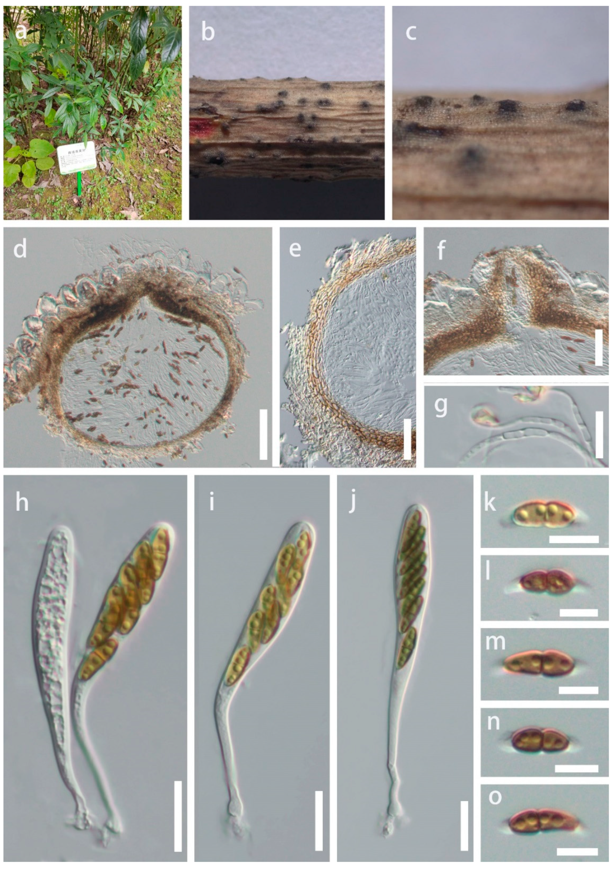
Saprobic on twigs of Helwingia himalaica in terrestrial habitat. Sexual morph: Ascomata 300–400 × 350–400 μm, semi-immersed, solitary or scattered, globose, uniloculate, black, with a central ostiole. Ostiole papillate, central. Peridium 20–40 μm wide, fused with host tissues, comprising two layers of pale brown to brown cells of textura angularis. Hamathecium comprises 1.5–3 μm wide, branched, hyaline, septate, pseudoparaphyses. Asci 84–135 × 10–15 μm (= 104 × 12 μm, n = 15), bitunicate, 8-spored, clavate, with a short, bulbous long pedicel, slightly curved. Ascospores 10–20 × 3.5–6 μm ( = 15.5 × 5 μm, n = 35), hyaline to olivaceous when immature, brown when mature, overlapping uniseriate or 2-seriate, fusiform, 1-septate, constricted at the septum, slightly widest at the upper cell and tapering towards ends, guttulate, sheath drawn out to form polar appendages, from both ends of the ascospores, straight or slightly curved. Asexual morph: Not observed.
Culture characteristics: Ascospores germinated on PDA within 12 h at 25 °C. Germ tubes produced from both ends. Colonies on PDA reached 7 cm diam after four weeks at 25 °C, mycelium white to gray, flossy, circular, undulate, yellow in reverse.
Material examined: China, Guizhou Province, Guiyang City, Nanming District, Guiyang Medicinal Botanical Garden, on twigs of Helwingia himalaica, 22 December 2021, Y.R. Sun, 22-41 (HKAS 124556, holotype; ex-type living culture GUCC 816); ibid, on twigs of Helwingia himalaica, 22 December 2021, Y.R. Sun, 41-2 (HGUP 22–800, paratype; living culture GUCC 22–0817).
Notes: Montagnula guiyangensis was isolated from Helwingia himalaica, an important medicinal plant. Multi-gene analyses showed that M. guiyangensis is a phylogenetically distinct species in Clade 3 (Figure 1). Morphologically, M. guiyangensis resembles M. appendiculata, M. chiangraiensis, and M. chromolaenae in having fusiform, 1-septate ascospores with appendages. Montagnula guiyangensis, however, differs by its larger ascomata from M. chromolaenae and M. appendiculata (300–400 × 350–400 μm in M. guiyangensis vs. 170–190 × 170–190 μm in M. chromolaenae vs. 100–200 μm in M. appendiculata) [8]. Montagnula guiyangensis has larger asci than M. chiangraiensis (84–135 × 10–15 μm vs. 60–75 × 8–11 μm). Montagnula guiyangensis can be distinguished from M. aloes by 1-septate, fusiform ascospores with appendages, while the latter has 3-septate, ovoid to ellipsoid ascospores [22]. In addition, comparisons of ITS, LSU, and SSU sequences between M. guiyangensis and phylogenetically related species are provided in Table 2 (tef1-α not available for M. aloes, M. appendiculata, M. chiangraiensis, and M. chromolaenae).
- Montagnula donacina (Niessl) Wanas., E.B.G. Jones and K.D. Hyde, Fungal Biology 120 (11): 1365 (2016) Figure 4.
- =Montagnula chromolaenicola Mapook and K.D. Hyde.
- =Montagnula puerensis Tibpromma and Du.
- =Montagnula saikhuensis Wanas., E.B.G. Jones and K.D. Hyde.
- =Montagnula thailandica Mapook and K.D. Hyde.
- Index Fungorum number: IF557299; Facesoffungi number: FoF 07792.
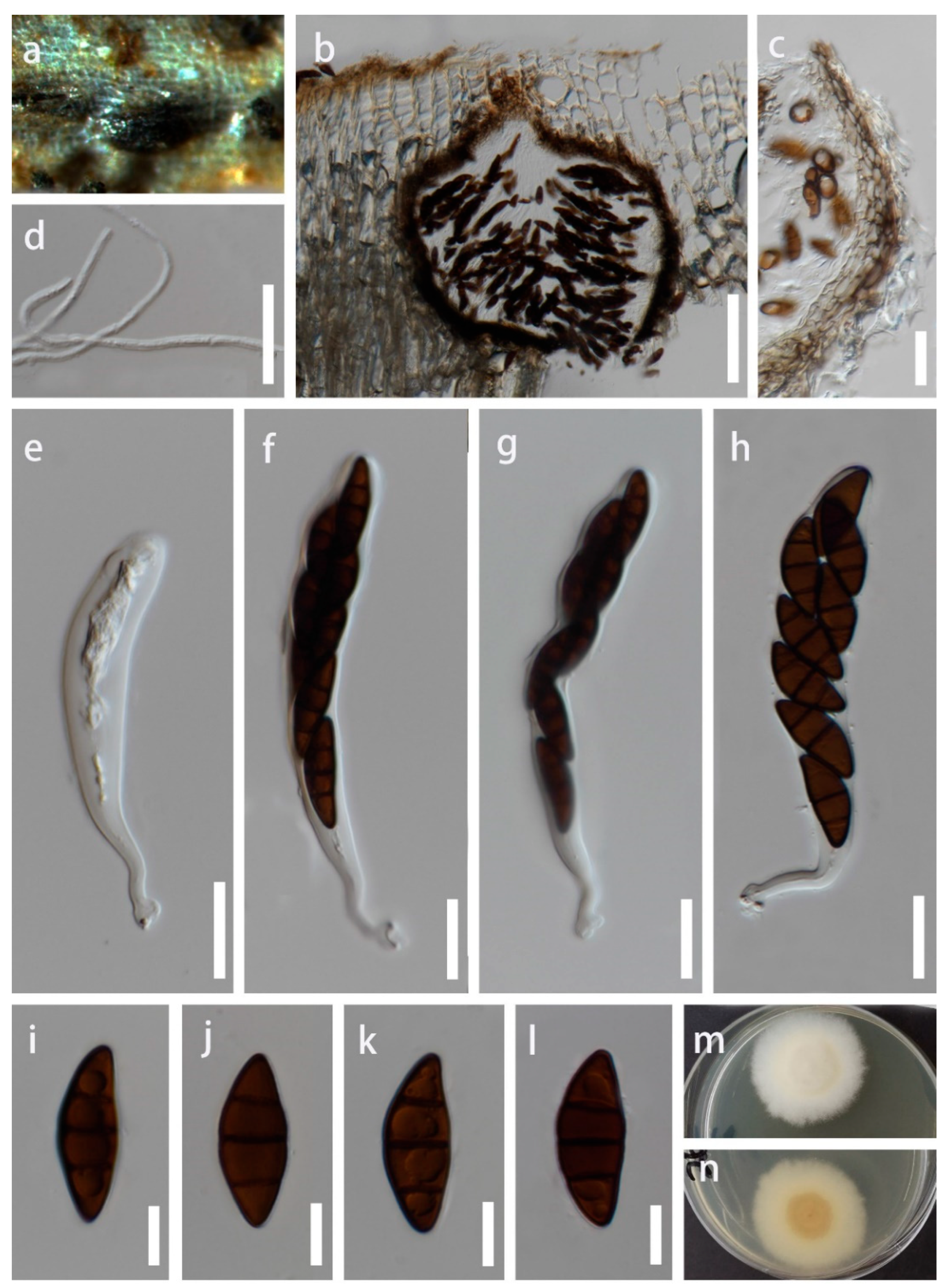
Saprobic on decaying wood in terrestrial habitat. Sexual morph: Ascomata 405–470 μm high, 280–380 μm wide, semi-immersed, solitary or scattered, globose, uniloculate, black, with a central ostiole. Ostiole papillate, central. Peridium 15–30 μm wide, fused with host tissues, comprising of two layers of pale to brown cells of textura angularis. Hamathecium comprising 1–2.5 μm wide, branched, hyaline, septate, pseudoparaphyses. Asci 80–125 × 9–12 μm, bitunicate, 8-spored, clavate, with a bulbous long pedicel, slightly curved. Ascospores 10–15 × 4–7 μm ( = 13.5 × 5.5 μm, n = 30), brown, overlapping uniseriate or 2-seriate, fusiform, 1-septate, constricted at the septum, slightly widest at the upper cell and tapering towards ends, guttulate, straight or slightly curved. Asexual morph: Not observed.
Culture characteristics: Ascospores germinated on PDA within 12 h at 25 °C. Germ tubes produced from one side of the middle of ascospore. Colonies on PDA reached 5 cm diam after four weeks at 25 °C, mycelium white to gray, flossy, circular, undulate, gray in reverse.
Material examined: China, Guizhou Province, Qianxinan Bouyei and Miao Autonomous Prefecture, Anlong County, on dead wood, 16 March 2022, J.Y. Zhang, Y312 (HKAS 124552; living culture GUCC 22–0818).
Notes: Montagnula chromolaenicola, M. donacina, M. puerensis, M. saikhuensisi, and M. thailandica clustered together without obvious branches in the phylogenetic tree (Figure 1). Morphologically, they have similar ascomata, asci, and ascospores, including measurement size (Table 3). It is worth noting that Wanasinghe et al. [14] took multi-loculate ascomata as the difference between M. donacina and M. saikhuensis. Du et al. [21] distinguished M. donacina and M. puerensis by M. donacina having carbonaceous ascostromata. However, the previous literature did not mention that M. donacina has multi-loculate, carbonaceous ascostromata [13,28]. Comparisons of ITS, LSU, SSU, and tef1-α sequences between M. donacina and phylogenetically related species are provided in Table 4. Few differences exist among their ITS, LSU, and SSU sequences, respectively, and there is a maximum difference of 10 bp in tef1-α gene. We conclude that the evidence for these five species as independent species is insufficient. The slight difference of multi-genes may represent the intraspecific variation. Therefore, we synonymize M. chromolaenicola, M. puerensis, M. saikhuensisi, and M. thailandica under M. donacina based on the nomenclatural priority. Our new collection HKAS 124552 has overlapping characteristics with these M. donacina isolates. Phylogenetically, HKAS 124552 grouped with them in Clade 1 (Figure 1). Thus, we identify our isolate as M. donacina.
3. Discussion
Montagnula species have a worldwide distribution that has been reported from America, Australia, Bahamas, China, Italy, Portugal, and Thailand [8,21]. Previous literature reported that all Montagnula species have been derived from terrestrial habitats [7,8,10,11,20,21,23]. We introduced a freshwater Montagnula species here that broke the record of the monolithic habitat for Montagnula species. These species have various hosts, such as Agave sp., Pandanus sp., and Ilex sp. [5,21]. However, rarely have studies focused on fungi associated with H. himalaica (Helwingiaceae). Helwingia himalaica is distributed in Bhutan, China, Nepal, and Thailand (https://www.havlis.cz/karta_en.php?kytkaid=5087 accessed on 27 January 2023). It has a high medicinal value that is used to treat colds, coughs, stomach pains, and fractures. In this study, we introduced a new species, M. guiyangensis, which was isolated from H. himalaica.
Montagnula species had didymosporous, phragmosporous, and dictyosporous ascospores [8,14,25]. Species with the same type of spores tended to cluster together (Figure 1). In our phylogenetic study, Montagnula species were divided into four major phylogenetic clades. (Figure 1). Four didymosporous species (M. acaciae, M. donacina, M. graminicola, and M. opulenta) and a coelomycetous asexual morph species, M. cylindrospora, were placed in Clade 1. Montagnula acaciae, M. donacina, and M. opulenta had didymospores without sheath but M. graminicola was surrounded by a sheath. Species in Clade 2 generally had fusiform to broadly fusiform phragmospores. Although the morphological characteristics of M. jonesii matched well with the species in Clade 2, it formed a distinct and basal clade in the tree. Species in Clade 3 had didymospores with polar appendages or were surrounded by a sheath, except for M. aloes, which had phragmospores without appendages. However, it is worth noting that the characteristics of M. aloes were observed from the culture, whereas other species were observed from the natural substrates. Fresh collections of M. aloes from nature are necessary to resolve the issue. However, there are no sequences available for dictyosporous species, e.g., M. dasylirionis, M. mohavensis, and M. yuccigena. Therefore, whether these species would gather in one clade cannot be inferred. Future molecular studies, incorporating a broad sampling of Montagnula and other Didymosphaeriaceae species, may separate Montagnula into several new genera based on the septation of the ascospores.
4. Materials and Methods
4.1. Collection, Examination, and Isolation
The fresh samples were collected in China and Thailand from 2019 to 2022. Samples were brought to the laboratory in Ziplock plastic bags for examination, as described in Senanayake et al. [29]. The fruiting bodies on natural substrates were observed and photographed using a stereomicroscope (SteREO Discovery, V12, Carl Zeiss Microscopy GmBH, Berlin, Germany; VHX-7000, Keyence, Osaka, Japan). Morphological characters were observed using a Nikon ECLIPSE Ni compound microscope (Nikon, Tokyo, Japan) photographed with a Nikon DS-Ri2 digital camera (Nikon, Japan), and Carl Zeiss compound microscope (Carl Zeiss AG, Germany) photographed with an Axiocam 208 color digital camera (Carl Zeiss AG, Germany). The photo plates were made by the Adobe Photoshop CS6 Extended v. 13.0 software. Measurements were done with the Tarosoft (R) Image Frame Work Version 0.9.7 software.
Single spore isolation was used to obtain pure cultures following the methods described by Senanayake et al. [29]. Germinated ascospores were transferred to new potato dextrose agar (PDA) plates and incubated at 25°C for 4 weeks. The pure cultures obtained were deposited in Mae Fah Luang University Culture Collection (MFLUCC), Chiang Rai, Thailand, and the Guizhou University Culture Collection (GUCC), Guiyang, China. Herbaria materials were deposited in the herbarium of Mae Fah Luang University (MFLU), Chiang Rai, Thailand, and the Kunming Institute of Botany Academia Sinica (HKAS), Kunming, China. Facesoffungi (FoF) and Index Fungorum numbers were acquired as described in Jayasiri et al. [30] and Index Fungorum (2023) [31]. Records were added to the Mekong Subregion (GMS) database [32]. The establishment of new species was decided upon the recommendations of Chethana et al. [27] and Jayawardena et al. [33].
4.2. DNA Extraction, PCR Amplification, and Sequencing
PrepManTM Ultra Sample Preparation Reagent (Thermo Fisher Scientific, Yokohama, Japan) was used to extract DNA directly from fruiting bodies. BIOMIGA Fungus Genomic DNA Extraction Kit (Biomiga, San Diego, CA, USA) was used to extract DNA from fresh fungal mycelia, which were grown on PDA medium for 4 weeks at 25 °C. Three genes were selected in this study: the large subunit nuclear ribosomal DNA (LSU), the small subunit nuclear ribosomal DNA (SSU), the internal transcribed spacers (ITS), and the translation elongation factor 1 (tef1-α). Polymerase chain reaction (PCR) was carried out in 20 μL reaction volume, which contained 10 μL 2 × PCR Master Mix, 7 μL ddH2O, 1 μL of each primer, and 1 μL template DNA. The PCR thermal cycle program and primers are given in Table 5. Purification and sequencing of PCR products were carried out at SinoGenoMax (Beijing) Co., China.
4.3. Phylogenetic Analyses
BLASTn (https://blast.ncbi.nlm.nih.gov//Blast.cgi, accessed on 27 January 2023) was used to evaluate closely related strains to our new taxa. Other sequences used in this study were obtained from GenBank referring to Mapook et al. [8] and Du et al. [21] (Table 6). The single gene sequences were viewed using BioEdit v. 7.0.9.0 [37]. Alignments for each locus were generated with MAFFT v.7 (https://mafft.cbrc.jp/alignment/server/, accessed on 27 January 2023) and manually improved using AliView [38] for maximum alignment and minimum gaps. The final single-gene alignments were combined by SequenceMatrix 1.7.8 [39]. The single locus and combined analyses were carried out for maximum likelihood (ML) and Bayesian posterior probability (BYPP).
The ML analyses were performed in CIPRES [40] with RAxML-HPC v. 8.2.12 [41] using a GTRGAMMA approximation with rapid bootstrap (BS) analysis followed by 1000 bootstrap replicates.
The BYPP analyses were conducted in CIPRES [40] with MrBayes on XSEDE 3.2.7a [42]. The best nucleotide substitution model for each data partition was evaluated by MrModeltest 2.2 [43]. The substitution model GTR+I+G was decided for LSU, ITS, and SSU sequences. The Markov chain Monte Carlo (MCMC) sampling approach was used to calculate posterior probabilities (PP) [44]. Six simultaneous Markov chains were run for 10 million generations and trees were sampled every 1000th generation. The first 20% of trees, representing the burn-in phase of the analyses, were discarded and the remaining trees were used for calculating the PP value in the majority rule consensus tree.
Phylogenetic trees were viewed using FigTree v1.4.0 [45] and modified in Microsoft Office PowerPoint 2019 and converted to a jpg file using Adobe Photoshop CS6 Extended 10.0 (Adobe Systems, San Jose, CA, USA). The new sequences derived from this study were deposited in GenBank.
Key to Accepted Montagnula Species
| 1. Ascospores are didymosporous | 2 |
| 1. Ascospores are phragmosporous | 13 |
| 1. Ascospores are dictyosporous | 20 |
| 2. Didymospores with sheath | 3 |
| 2. Didymospores without sheath | 10 |
| 3. Didymospores surrounded by a mucilaginous sheath | 4 |
| 3. Sheath was drawn out to form polar appendages | 8 |
| 4. Ascospores are fusiform | M. krabiensis |
| 4. Ascospores are ellipsoidal | 5 |
| 5. Ascospores are asymmetrical | M. vakrabeejae |
| 5. Ascospores are symmetrical | 6 |
| 6. Asci are (4–)6–8-spored | M. chromolaenae |
| 6. Asci are 8-spored | 7 |
| 7. Ascospores brown, slightly constricted at the septum | M. graminicola |
| 7. Ascospores dark brown, not constricted at the septum | M. palmacea |
| 8. Ascospores 1-seriate, yellowish brown to brown | M. appendiculata |
| 8. Ascospores 2–3-seriate | 9 |
| 9. Ascomata 300–400 × 350–400 μm, asci 84–135 × 10–15 μm | M. guiyangensis |
| 9. Ascomata 150–220 × 200–230 μm, asci 60–75 × 8–11 μm | M. chiangraiensis |
| 10. Ascomata superficial | M. longipes |
| 10. Ascomata immersed or erumpent | 11 |
| 11. Ascomata 140–180 × 150–200 µm, not more than 200 µm | M. acacia |
| 11. Ascomata greater than 200 μm | 12 |
| 12 Ascospores brown, 12–17 × 4–6.5 μm | M. donacina |
| 12 Ascospores pale brown, 19–25 × 9–13 μm | M. opulenta |
| 13. Ascomata superficial | M. camporesii |
| 13. Ascomata immersed or erumpent | 14 |
| 14. Asci with short stalks | 15 |
| 14. Asci with long pedicellate | 16 |
| 15. Ascospores 5 transverse septa, 21–25 × 5–7 μm | M. subsuperficialis |
| 15. Ascospores 3 transverse septa, 24–35 × 7.5–14 μm | M. aquatica |
| 16. Ascospores with 2 transverse septa | M. bellevaliae |
| 16. Ascospores with 3 transverse septa | 17 |
| 17. Asci not more than 100 μm | M. jonesii |
| 17. Asci greater than 100 μm | 18 |
| 18. Ascospores greater than 30 μm, ovoid to ellipsoid | M. aloes |
| 18. Ascospores not more than 30 μm, ellipsoid to fusiform | 19 |
| 19. Ascomata 385–415 × 510–525 μm, asci 84.5–119.5 × 10.5–13.5 μm | M. cirsii |
| 19. Ascomata 300–320 × 300–360 μm, asci 110–130 × 14–20 μm | M. scabiosae |
| 20. Sporous transverse septa more than 10 | 21 |
| 20. Sporous transverse septa not more than 10 | 23 |
| 21. Sporous transverse septa more than 15, ascospores 40–45 × 15–17 μm | M. gigantea |
| 21. Sporous transverse septa not more than 15 | 22 |
| 22. Ascospores 32–40 × 8–9.8 μm, asci 80–110 × 13–15 μm | M. dura |
| 22. Ascospores 31–45 × 13.5–16.5 μm, asci 110–160 × 13–6 μm | M. triseti |
| 23. Sporous transverse septa not more than 5 | 24 |
| 23. Sporous transverse septa more than 5 | 27 |
| 24. Ascospores without sheath | 25 |
| 24. Ascospores with sheath | 26 |
| 25. Ascospores 2–3 transverse septa, 0–1 longitudinal septum, 12.5–16.5 × 4.8–6.5 μm | M. baatanensis |
| 25. Ascospores 5 transverse septa, 1 longitudinal septum, 24–29 × 9–11 μm | M. infernalis |
| 26. Ascospores 17.5–23 × 5.5–8.5 μm, fusiform to somewhat broadly fusiform | M. opuntiae |
| 26. Ascospores 16–18 × 6–7.5 μm, ellipsoid fusoid | M. thuemeniana |
| 27. Ascospores without sheath | 28 |
| 27. Ascospores with sheath | 29 |
| 28. Ascospores 17–25 × 7.5–10 μm, 5–7 transverse septa | M. obtusa |
| 28. Ascospores 39–47 × 15–19 μm, 7–9 transverse septa | M. opaca |
| 29. Ascospores broadly ellipsoid, 5–7 transverse septa | M. phragmospora |
| 29. Ascospores obovoid fusoid, 7(–10) transverse septa | 30 |
| 30. Ascospores 2–3 longitudinal septa, 40.8–52 × 17.6–22.4 μm | M. mohavensis |
| 30. Ascospores 1–2 longitudinal septa | 31 |
| 31. Ascospores 35–50 × 16–20 μm, asci 2–8-spored | M. dasylirionis |
| 31. Ascospores 27–42 × 12–15 μm, asci 4–8-spored | M. yuccigena |
Author Contributions
Investigation, J.-Y.Z.; Methodology, Y.-R.S. and J.-Y.Z.; Software, Y.-R.S.; Supervision, K.D.H., Y.W. and R.S.J.; Writing—original draft, Y.-R.S.; Writing—review and editing, K.D.H., Y.W. and R.S.J. All authors have read and agreed to the published version of the manuscript.
Funding
The study was funded by Guizhou Science Technology Department International Cooperation Basic project (grant number: [2018]5806), National Natural Science Foundation of China (grant number: 31972222, 31560489), Program of Introducing Talents of Discipline to Universities of China (grant number: 111 Program, D20023), and Talent project of Guizhou Science and Technology Cooperation Platform (grant number: [2017]57885, [2019]5641, and [2020]5001). The article processing fee for this article is provided by Mae Fah Luang University.
Institutional Review Board Statement
Not applicable.
Informed Consent Statement
Not applicable.
Data Availability Statement
Not applicable.
Acknowledgments
We would like to thank Shaun Pennycook for checking the nomenclature. Ya-Ru Sun thanks Mae Fah Luang University for the tuition-fee scholarship and the article processing fees. Kevin D. Hyde thanks Chiang Mai University for the award of Visiting Professor and the Thailand Research Fund, Grant RDG6130001, titled “Impact of climate change on fungal diversity and biogeography in the Greater Mekong Subregion”.
Conflicts of Interest
The authors declare no conflict of interest.
References
- Munk, A. The system of the Pyrenomycetes. Dan. Bot. Ark. 1953, 15, 1–163. [Google Scholar]
- Hongsanan, S.; Hyde, K.D.; Phookamsak, R.; Wanasinghe, D.N.; McKenzie, E.H.C.; Sarma, V.V.; Boonmee, S.; Lücking, R.; Bhat, D.J.; Liu, N.G.; et al. Refined families of Dothideomycetes: Dothideomycetidae and Pleosporomycetidae. Mycosphere 2020, 11, 1553–2107. [Google Scholar] [CrossRef]
- Wijayawardene, N.N.; Hyde, K.D.; Dai, D.Q.; Sánchez-García, M.; Goto, B.; Saxena, R.; Erdogdu, M.; Selçuk, F.; Rajeshkumar, K.C.; Aptroot, A.; et al. Outline of Fungi and fungus-like taxa-2021. Mycosphere 2022, 13, 53–453. [Google Scholar] [CrossRef]
- Hyde, K.D.; Jones, E.B.G.; Liu, J.K.; Ariyawansa, H.A.; Boehm, E.; Boonmee, S.; Braun, U.; Chomnunti, P.; Crous, P.W.; Dai, D.Q.; et al. Families of Dothideomycetes. Fungal Divers. 2013, 63, 1–313. [Google Scholar] [CrossRef]
- Ariyawansa, H.A.; Camporesi, E.; Thambugala, K.M.; Mapook, A.; Kang, J.C.; Alias, S.A.; Chukeatirote, E.; Thines, M.; McKENZIE, E.H.C.; Hyde, K.D. Confusion surrounding Didymosphaeria—Phylogenetic and morphological evidence suggest Didymosphaeriaceae is not a distinct family. Phytotaxa 2014, 176, 102–119. [Google Scholar] [CrossRef]
- Ariyawansa, H.A.; Tanaka, K.; Thambugala, K.M.; Phookamsak, R.; Tian, Q.; Camporesi, E.; Hongsanan, S.; Monkai, J.; Wanasinghe, D.N.; Mapook, A.; et al. A molecular phylogenetic reappraisal of the Didymosphaeriaceae (=Montagnulaceae). Fungal Divers. 2014, 68, 69–104. [Google Scholar] [CrossRef]
- Crous, P.W.; Wingfield, M.J.; Chooi, Y.H.; Gilchrist, C.L.M.; Lacey, E.; Pitt, J.I.; Roets, F.; Swart, W.J.; Cano-Lira, J.F.; Valenzuela-Lopez, N.; et al. Fungal Planet description sheets: 1042–1111. Persoonia 2020, 44, 301–459. [Google Scholar] [CrossRef]
- Mapook, A.; Hyde, K.D.; McKenzie, E.H.C.; Jones, E.B.G.; Bhat, D.J.; Jeewon, R.; Stadler, M.; Samarakoon, M.C.; Malaithong, M.; Tanunchai, B.; et al. Taxonomic and phylogenetic contributions to fungi associated with the invasive weed Chromolaena odorata (Siam weed). Fungal Divers. 2020, 101, 1–175. [Google Scholar] [CrossRef]
- Ren, G.; Wanasinghe, D.N.; de Farias, A.R.G.; Hyde, K.D.; Yasanthika, E.; Xu, J.C.; Balasuriya, A.; Chethana, K.W.T.; Gui, H. Taxonomic Novelties of Woody Litter Fungi (Didymosphaeriaceae, Pleosporales) from the Greater Mekong Subregion. Biology 2022, 11, 1660. [Google Scholar] [CrossRef]
- Berlese, A.N. Icones Fungorum. Pyrenomycetes; Kessinger Publishing, LLC.: Whitefish, MT, USA, 1896; pp. 1–216. [Google Scholar]
- Crivelli, P.G. Ueber die Heterogene Ascomycetengattung Pleospora rabh.: Vorschlag für eine Aufteilung. Doctoral Thesis, Dissertation ETH Zürich, Zürich, Switzerland, 1983. [Google Scholar] [CrossRef]
- Leuchtmann, A. Über Phaeosphaeria Miyake und andere bitunicate Ascomyceten mit mehrfach querseptierten Ascosporen. Sydowia 1984, 37, 75–194. [Google Scholar]
- Aptroot, A. Redisposition of some species excluded from Didymosphaeria (Ascomycotina). Nova Hedwig. 1995, 60, 325–380. [Google Scholar]
- Wanasinghe, D.N.; Jones, E.B.G.; Camporesi, E.; Dissanayake, A.J.; Kamolhan, S.; Mortimer, P.E.; Xu, J.C.; Abd-Elsalam, K.A.; Hyde, K.D. Taxonomy and phylogeny of Laburnicola gen. nov. and Paramassariosphaeria gen. nov. (Didymosphaeriaceae, Massarineae, Pleosporales). Fungal Biol. 2016, 120, 1354–1373. [Google Scholar] [CrossRef]
- Species Fungorum. Available online: http://www.speciesfungorum.org/Names/Names.asp (accessed on 27 January 2023).
- Tennakoon, D.S.; Hyde, K.D.; Wanasinghe, D.N.; Bahkali, A.H.; Camporesi, E.; Khan, S.; Phookamsak, R. Taxonomy and phylogenetic appraisal of Montagnula jonesii sp. nov. (Didymosphaeriaceae, Pleosporales). Mycosphere 2016, 7, 1346–1356. [Google Scholar] [CrossRef]
- Lu, L.; Karunarathna, S.C.; Dai, D.-Q.; Xiong, Y.-R.; Suwannarach, N.; Stephenson, S.L.; Elgorban, A.M.; Al-Rejaie, S.; Jayawardena, R.S.; Tibpromma, S. Description of Four Novel Species in Pleosporales Associated with Coffee in Yunnan, China. J. Fungi 2022, 8, 1113. [Google Scholar] [CrossRef]
- Tibpromma, S.; Hyde, K.D.; McKenzie, E.H.C.; Bhat, D.J.; Phillips, A.J.L.; Wanasinghe, D.N.; Samarakoon, M.C.; Jayawardena, R.S.; Dissanayake, A.J.; Tennakoon, D.S.; et al. Fungal Diversity notes 840–928: Micro-fungi associated with Pandanaceae. Fungal Divers. 2018, 93, 1–160. [Google Scholar] [CrossRef]
- Hyde, K.D.; Dong, Y.; Phookamsak, R.; Jeewon, R.; Bhat, D.J.; Jones, E.B.G.; Liu, N.G.; Abeywickrama, P.D.; Mapook, A.; Wei, D.P.; et al. Fungal Diversity notes 1151–1276: Taxonomic and phylogenetic contributions on genera and species of fungal taxa. Fungal Divers. 2020, 100, 5–277. [Google Scholar] [CrossRef]
- Boonmee, S.; Wanasinghe, D.N.; Calabon, M.S.; Huanraluek, N.; Chandrasiri, S.K.U.; Jones, E.B.G.; Rossi, W.; Leonardi, M.; Singh, S.K.; Rana, S.; et al. Fungal Diversity notes 1387–1511: Taxonomic and phylogenetic contributions on genera and species of fungal taxa. Fungal Divers. 2021, 111, 1–335. [Google Scholar] [CrossRef]
- Du, T.Y.; Hyde, K.D.; Mapook, A.; Mortimer, P.E.; Xu, J.C.; Karunarathna, S.C.; Tibpromma, S. Morphology and phylogenetic analyses reveal Montagnula puerensis sp. nov. (Didymosphaeriaceae, Pleosporales) from southwest China. Phytotaxa 2021, 514, 1–25. [Google Scholar] [CrossRef]
- Crous, P.W.; Summerell, B.A.; Shivas, R.G.; Burgess, T.I.; Decock, C.A.; Dreyer, L.L.; Granke, L.L.; Guest, D.I.; Hardy, G.S.; Hausbeck, M.K.; et al. Fungal Planet description sheets: 107–127. Persoonia 2012, 28, 138–182. [Google Scholar] [CrossRef]
- Liu, J.K.; Hyde, K.D.; Jones, E.B.G.; Ariyawansa, H.A.; Bhat, D.J.; Boonmee, S.; Maharachchikumbura, S.S.N.; McKenzie, E.H.C.; Phookamsak, R.; Phukhamsakda, C.; et al. Fungal Diversity notes 1–110: Taxonomic and phylogenetic contributions to fungal species. Fungal Divers. 2015, 72, 1–197. [Google Scholar] [CrossRef]
- Hongsanan, S.; Hyde, K.D.; Bahkali, A.H.; Camporesi, E.; Chomnunti, P.; Ekanayaka, H.; Gomes, A.A.; Hofstetter, V.; Jones, E.B.G.; Pinho, D.B.; et al. Fungal biodiversity profiles 11–20. Cryptogam. Mycol. 2015, 36, 355–380. [Google Scholar] [CrossRef]
- Hyde, K.D.; Hongsanan, S.; Jeewon, R.; Bhat, D.J.; McKenzie, E.H.C.; Jones, E.B.G.; Phookamsak, R.; Ariyawansa, H.A.; Boonmee, S.; Zhao, Q.; et al. Fungal Diversity notes 367–490: Taxonomic and phylogenetic contributions to fungal taxa. Fungal Divers. 2016, 80, 1–270. [Google Scholar] [CrossRef]
- Jeewon, R.; Hyde, K.D. Establishing species boundaries and new taxa among fungi: Recommendations to resolve taxonomic ambiguities. Mycosphere 2016, 7, 1669–1677. [Google Scholar] [CrossRef]
- Chethana, K.W.T.; Manawasinghe, I.S.; Hurdeal, V.G.; Bhunjun, C.S.; Appadoo, M.A.; Gentekaki, E.; Raspé, O.; Promputtha, I.; Hyde, K.D. What are fungal species and how to delineate them? Fungal Divers. 2021, 109, 1–25. [Google Scholar] [CrossRef]
- Pitt, W.; Úrbez-Torres, J.R.; Trouillas, F.P. Munkovalsaria donacina from grapevines and Desert Ash in Australia. Mycosphere 2014, 5, 656–661. [Google Scholar] [CrossRef]
- Senanayake, I.C.; Rathnayake, A.R.; Marasinghe, D.S.; Calabon, M.S.; Gentekaki, E.; Lee, H.B.; Hurdeal, V.G.; Pem, D.; Dissanayake, L.S.; Wijesinghe, S.N.; et al. Morphological approaches in studying fungi: Collection, examination, isolation, sporulation and preservation. Mycosphere 2020, 11, 2678–2754. [Google Scholar] [CrossRef]
- Jayasiri, S.C.; Hyde, K.D.; Ariyawansa, H.A.; Bha, T.J.; Buyck, B.; Cai, L.; Dai, Y.C.; Abd-Elsalam, K.A.; Ertz, D.; Hidayat, I.; et al. The Faces of Fungi database: Fungal names linked with morphology, phylogeny and human impacts. Fungal Divers. 2015, 74, 3–18. [Google Scholar] [CrossRef]
- Index Fungorum. Available online: http://www.indexfungorum.org/Names/IndexFungorumPartnership.htm (accessed on 10 January 2023).
- Chaiwan, N.; Gomdola, D.; Wang, S.; Monkai, J.; Tibpromma, S.; Doilom, M.; Wanasinghe, D.N.; Mortimer, P.E.; Lumyong, S.; Hyde, K.D. An online database providing updated information of microfungi in the Greater Mekong Subregion. Mycosphere 2021, 12, 1513–1526. [Google Scholar] [CrossRef]
- Jayawardena, R.S.; Hyde, K.D.; de Farias, A.R.G.; Bhunjun, C.S.; Ferdinandez, H.S.; Manamgoda, D.S.; Udayanga, D.; Herath, I.S.; Thambugala, K.M.; Manawasinghe, I.S.; et al. What is a species in fungal plant pathogens? Fungal Divers. 2021, 109, 239–266. [Google Scholar] [CrossRef]
- White, T.J.; Bruns, T.; Lee, S.J.W.T.; Taylor, J. Amplification and direct sequencing of fungal ribosomal RNA genes for phylogenetics. In PCR Protocols: A Guide to Methods and Applications; Innis, M., Gelfand, D., Shinsky, J., White, T., Eds.; Academic Press: New York, NY, USA, 1990; pp. 315–322. [Google Scholar]
- Rehner, S.A.; Samuels, G.J. Molecular systematics of the Hypocreales: A teleomorph gene phylogeny and the status of their anamorphs. Can. J. Bot. 1995, 73, 816–882. [Google Scholar] [CrossRef]
- Rehner, S.A.; Buckley, E. A Beauveria phylogeny inferred from nuclear ITS and EF1-α sequences: Evidence for cryptic diversification and links to Cordyceps teleomorphs. Mycologia 2005, 97, 84–98. [Google Scholar] [CrossRef]
- Hall, T.A. BioEdit: A user-friendly biological sequence alignment editor and analysis program for Windows 95/98/NT. In Nucleic Acids Symposium Series; Information Retrieval Ltd.: London, UK, 1999; pp. 95–98. [Google Scholar]
- Larsson, A. AliView: A fast and lightweight alignment viewer and editor for large datasets. Bioinformatics 2014, 30, 3276–3278. [Google Scholar] [CrossRef] [Green Version]
- Vaidya, G.; Lohman, D.J.; Meier, R. SequenceMatrix: Concatenation software for the fast assembly of multi-gene datasets with character set and codon information. Cladistics 2011, 27, 171–180. [Google Scholar] [CrossRef]
- Miller, M.A.; Pfeiffer, W.; Schwartz, T. Creating the CIPRES Science Gateway for inference of large phylogenetic trees. In Proceedings of the 2010 Gateway Computing Environments Workshop (GCE), New Orleans, LA, USA, 14 November 2010; pp. 1–8. [Google Scholar] [CrossRef]
- Stamatakis, A. RAxML version 8: A tool for phylogenetic analysis and post-analysis of large phylogenies. Bioinformatics 2014, 30, 1312–1313. [Google Scholar] [CrossRef]
- Ronquist, F.; Teslenko, M.; Mark, P.; Ayres, D.L.; Höhna, S.; Larget, B.; Liu, L.; Huelsenbeck, J. MrBayes 3.2: Efficient Bayesian phylogenetic inference and model choice across a large model space. Syst. Biol. 2012, 61, 539–542. [Google Scholar] [CrossRef]
- Nylander, J. MrModeltest, Version 2.2; Evolutionary Biology Centre, Uppsala University: Uppsala, Sweden, 2004. [Google Scholar]
- Rannala, B.; Yang, Z.H. Probability distribution of molecular evolutionary trees: A new method of phylogenetic inference. J. Mol. Evol. 1996, 43, 304–311. [Google Scholar] [CrossRef]
- Rambaut, A. FigTree, Version 1.4.0; University of Edinburgh: Edinburgh, UK, 2014. [Google Scholar]
Figure 1.
The ML tree based on a combined dataset of LSU, ITS, SSU, and tef1-α sequence data. The tree was rooted with Fuscostagonospora sasae (HHUF 29106) and F. cytisi (MFLUCC 16–0622). Bootstrap support values for ML greater than 75% and Bayesian posterior probabilities greater than 0.95 are given near the nodes, respectively. Ex-type strains are in bold, the new isolates are in red.

Figure 2.
Montagnula aquatica (MFLU 22–0171, holotype). (a) Appearance of ascomata on the substrate, (b) Section through ascomata, (c) Peridium, (d) Trabeculate pseudoparaphyses, (e–h) Immature and mature asci, (i–l) Ascospores, (m,n) Colony on PDA medium. Scale bars: (b) = 100 μm, (c–h) = 20 μm, (i–l) = 10 μm.

Figure 3.
Montagnula guiyangensis (HKAS 124556, holotype). (a) Host, (b,c) Appearance of ascomata on the substrate, (d) Section through ascomata, (e) Peridium, (g) Trabeculate pseudoparaphyses, (h–j) Asci, (k–o) Ascospores. Scale bars: d = 100 μm, (e,f) = 50 μm, (g–j) = 20 μm, (k–o) = 10 μm.
Figure 3.
Montagnula guiyangensis (HKAS 124556, holotype). (a) Host, (b,c) Appearance of ascomata on the substrate, (d) Section through ascomata, (e) Peridium, (g) Trabeculate pseudoparaphyses, (h–j) Asci, (k–o) Ascospores. Scale bars: d = 100 μm, (e,f) = 50 μm, (g–j) = 20 μm, (k–o) = 10 μm.

Figure 4.
Montagnula donacina (HKAS 124552). (a) Appearance of ascomata on the substrate, (b) Section through ascomata, (c) Peridium, (h) Paraphyses, (d–j) Asci, (i–n) Ascospores. Scale bars: (b) = 100 μm, (c–h) = 20 μm, (i–n) = 10 μm.
Figure 4.
Montagnula donacina (HKAS 124552). (a) Appearance of ascomata on the substrate, (b) Section through ascomata, (c) Peridium, (h) Paraphyses, (d–j) Asci, (i–n) Ascospores. Scale bars: (b) = 100 μm, (c–h) = 20 μm, (i–n) = 10 μm.


Table 1.
The number of polymorphic nucleotide differences between M. aquatica (tef1-α not available) and M. camporesii, M. cirsii, and M. scabiosae (without gap).
Table 1.
The number of polymorphic nucleotide differences between M. aquatica (tef1-α not available) and M. camporesii, M. cirsii, and M. scabiosae (without gap).
| Species | Strain | ITS (504 bp) | LSU (842 bp) | SSU (971 bp) |
|---|---|---|---|---|
| M. camporesii | MFLUCC 16–1369 | 14 (2.7%) | 7 (0.8%) | 4 (0.4%) |
| M. cirsii | MFLUCC 13–0680 | 14 (2.7%) | 7 (0.8%) | 2 (0.2%) |
| M. scabiosae | MFLUCC 14–0954 | 15 (2.9%) | 7 (0.8%) | 6 (0.6%) |
Table 2.
The number of polymorphic nucleotide differences between M. guiyangensis HKAS 124556 and M. aloes, M. appendiculata, M. chiangraiensis, and M. chromolaenae (without gap).
Table 2.
The number of polymorphic nucleotide differences between M. guiyangensis HKAS 124556 and M. aloes, M. appendiculata, M. chiangraiensis, and M. chromolaenae (without gap).
| Species | Strain | ITS (504 bp) | LSU (842 bp) | SSU (971 bp) |
|---|---|---|---|---|
| M. aloes | CBS 132531 | 20 (4%) | 10 (1.2%) | not available |
| M. appendiculata | CBS 10927 | 18 (3.6%) | 16 (1.9%) | not available |
| M. chiangraiensis | MFLUCC 17–1420 | 13 (2.6%) | 18 (2.1%) | 27 (2.8%) |
| M. chromolaenae | MFLUCC 17–1435 | 14 (2.7%) | 18 (2.1%) | 32 (3.3%) |
Table 3.
Morphological comparison of M. donacina and M. chromolaenicola, M. puerensis, M. saikhuensis, and M. thailandica.
Table 3.
Morphological comparison of M. donacina and M. chromolaenicola, M. puerensis, M. saikhuensis, and M. thailandica.
| Species | Ascomata | Asci | Ascospores | Reference |
|---|---|---|---|---|
| M. chromolaenicola | Solitary, scattered, semi-immersed to erumpent, brown to dark brown, globose to obpyriform, 300–320 × 215–310 μm, ostiole papilla | Bitunicate, elongate-clavate, 8-spored, 80–100 × 10–13 μm, long pedicel | Broadly fusiform to ellipsoid, brown to dark brown, overlapping 1–2-seriate, 1 transverse septum, constricted at the septum, asymmetrical, 15–17 × 5–6.5 μm | Mapook et al. [8] |
| M. donacina | Ascostromata with flat bottom, gregarious, immersed to erumpent, black, globose to pyriform, 500 μm, pseudothecial ostiole | Bitunicate, clavate, 8-spored, with a long pedicel | Ellipsoid, brown, 1–2-seriate, 1 transverse septum, constricted at the septum, asymmetrical, 12–17 × 4–6.5 μm | Aptroot [13], Pitt et al. [28] |
| M. puerensis | Solitary, scattered, semi-immersed, black, globose, 300–600 × 230–380 μm, with a central ostiole | Bitunicate, elongate-clavate, 8-spored, 70–105 × 10–15 μm, long pedicel | Ellipsoid, brown to dark brown, biseriate, 1 transverse septum, constricted at the septum, asymmetrical, 10–20 × 4–7 μm | Du et al. [21] |
| M. saikhuensis | Solitary, scattered, immersed, globose, brown to dark brown, 400–450 × 400–500 μm, ostiolate | Bitunicate, elongate-clavate to short-cylindrical, 8-spored, 70–100 × 10–12μm, long pedicel | Ellipsoid, brown to blackish brown, overlapping 1–2-seriate, 1 transverse septum, unequally and strongly constricted at the septum, asymmetrical, 12–16 × 4–6 µm | Wanasinghe et al. [14] |
| M. thailandica | Solitary, scattered, immersed to erumpent, brown to dark brown, globose to obpyriform, brown to dark brown, 405–415 × 330–350 μm, ostiole papillate | Bitunicate, elongate-clavate, slightly curved, 8-spored, 80–100 × 9–15 μm, long pedicel | Broadly fusiform to ellipsoid, brown to reddish-brown, overlapping 1–2-seriate, 1 transverse septum, constricted at the septum, asymmetrical, 14–17 × 4.5–7.5 μm | Mapook et al. [8] |
Table 4.
Comparison of nucleotide differences between M. donacina (KUMCC 21–0653) and M. chromolaenicola, M. puerensis, M. saikhuensis, and M. thailandica.
Table 4.
Comparison of nucleotide differences between M. donacina (KUMCC 21–0653) and M. chromolaenicola, M. puerensis, M. saikhuensis, and M. thailandica.
| Species | Strain | ITS (504 bp) | LSU (842 bp) | SSU (971 bp) | tef1-α (840 bp) |
|---|---|---|---|---|---|
| M. chromolaenicola | MFLUCC 17–1469 | 0 | 3 | 1 | 10 |
| M. puerensis | KUMCC 20–0225 | 0 | 0 | 1 | 8 |
| M. saikhuensis | MFLUCC 16–0315 | 4 | 2 | 3 | not available |
| M. thailandica | MFLUCC 17–1508 | 3 | 2 | 3 | 2 |
Table 5.
Primers and PCR procedures used in this study.
| Locus | Primers | PCR Procedures | Reference | |
|---|---|---|---|---|
| Name | Sequence (5′-3′) | |||
| LSU | LR0R | ACCCGCTGAACTTAAGC | 94 °C—3 min; 94 °C—30 s; 52 °C—30 s; 72 °C—1 min; Repeat 2–4 for 35 cycles; 72 °C—8 min; 4 °C on hold | White et al. [34], Rehner and Samuels [35] |
| LR5 | TCCTGAGGGAAACTTCG | |||
| SSU | NS1 | GTAGTCATATGCTTGTCTC | White et al. [34] | |
| NS4 | CTTCCGTCAATTCCTTTAAG | |||
| ITS | ITS5 | GGAAGTAAAAGTCGTAACAAGG | ||
| ITS4 | TCCTCCGCTTATTGATATGC | |||
| tef1-α | EF1-983F | GCYCCYGGHCAYCGTGAYTTYAT | 94 °C 2 min; 36 cycles of 66 °C–56 °C (touchdown 9 cycles), 94 °C 30 s, 56 °C 1 min, 72°C 1 min; 72 °C 10 min; 4 °C on hold | Rehner and Buckley [36] |
| EF1-2218R | ATGACACCRACRGCRACRGTYTG | |||
Table 6.
Sequence data were used for phylogenetic analyses with the corresponding GenBank accession numbers. The newly generated strains are in red. N/A: Not available.
Table 6.
Sequence data were used for phylogenetic analyses with the corresponding GenBank accession numbers. The newly generated strains are in red. N/A: Not available.
| Species | Strain/Voucher No. | LSU | SSU | ITS | tef-α |
|---|---|---|---|---|---|
| Bimuria omanensis | SQUCC 15280 | NG_071257 | N/A | NR_173301 | MT279046 |
| Bimuria novae-zelandiae | CBS 107.79 | AY016356 | N/A | MH861181 | DQ471087 |
| Deniquelata barringtoniae | MFLUCC 11−0422 | JX254655 | JX254656 | NR_111779 | N/A |
| Deniquelata quercina | ABRIICC 10068 | MH316157 | MH316155 | MH316153 | N/A |
| Didymocrea leucaenae | MFLUCC 17−0896 | NG_066304 | MK347826 | NR_164298 | MK360052 |
| Didymocrea sadasivanii | CBS 438.65 | DQ384103 | DQ384066 | MH870299 | N/A |
| Fuscostagonospora sasae | HHUF 29106 | NG_059395 | NG_061003 | NR_153964 | AB808524 |
| Fuscostagonospora cytisi | MFLUCC 16−0622 | KY770978 | KY770977 | N/A | KY770979 |
| Letendraea cordylinicola | MFLUCC 11−0148 | NG_059530 | NG_068362 | NR_154118 | N/A |
| Letendraea helminthicola | CBS 884.85 | AY016362 | AY016345 | MK404145 | MK404174 |
| Montagnula acaciae | MFLUCC 18−1636 | ON117298 | ON117267 | ON117280 | ON158093 |
| Montagnula acaciae | NCYUCC 19−0087 | ON117299 | ON117268 | ON117281 | ON158094 |
| Montagnula aloes | CPC 19671 | JX069847 | N/A | JX069863 | N/A |
| Montagnula aloes | CBS 132531 | NG_042676 | N/A | NR_111757 | N/A |
| Montagnula aquatica | MFLU 22−0171 | OP605986 | OP600504 | OP605992 | N/A |
| Montagnula appendiculata | CBS 109027 | AY772016 | N/A | DQ435529 | N/A |
| Montagnula bellevaliae | MFLUCC 14−0924 | KT443902 | KT443904 | KT443906 | KX949743 |
| Montagnula camporesii | MFLUCC 16−1369 | NG_070946 | NG_068418 | MN401746 | MN397908 |
| Montagnula chiangraiensis | MFLUCC 17−1420 | NG_068707 | NG_070155 | NR_168864 | N/A |
| Montagnula chromolaenae | MFLUCC 17−1435 | NG_068708 | NG_070156 | NR_168865 | N/A |
| Montagnula cirsii | MFLUCC 13−0680 | KX274249 | KX274255 | KX274242 | KX284707 |
| Montagnula cylindrospora | UTHSC DI16-208 | LN907351 | N/A | LT796834 | LT797074 |
| Montagnula donacina | HVVV01 | KJ628377 | KJ628376 | KJ628375 | N/A |
| Montagnula donacina | HFG07004 | MF183940 | N/A | MF967419 | N/A |
| Montagnula donacina | KUMCC 21−0653 | OP059052 | OP059003 | OP058961 | OP135938 |
| Montagnula donacina | KUMCC 21−0579 | OP059054 | OP059005 | OP058963 | OP135940 |
| Montagnula donacina | KUMCC 21−0631 | OP059053 | OP059004 | OP058962 | OP135939 |
| Montagnula donacina | HKAS 124552 | OP605987 | N/A | OP605991 | N/A |
| Montagnula donacina (M. chromolaenicola) | MFLUCC 17−1469 | NG_070948 | NG_070157 | NR_168866 | MT235773 |
| Montagnula donacina (M. puerensis) | KUMCC 20−0225 | MW575866 | MW575864 | MW567739 | MW573959 |
| Montagnula donacina (M. puerensis) | KUMCC 20−0331 | MW575867 | MW575865 | MW567740 | MW573960 |
| Montagnula donacina (M. saikhuensis) | MFLUCC 16−0315 | KU743210 | KU743211 | KU743209 | N/A |
| Montagnula donacina (M. thailandica) | MFLUCC 17−1508 | NG_070949 | NG_070158 | MT214352 | MT235774 |
| Montagnula donacina (M. thailandica) | MFLUCC 21−0075 | MZ538549 | N/A | MZ538515 | N/A |
| Montagnula donacina (M. thailandica) | ZHKUCC 22−0206 | OP297777 | OP297791 | OP297807 | OP321576 |
| Montagnula donacina (M. thailandica) | ZHKUCC 22−0207 | OP297778 | OP297792 | OP297808 | OP321577 |
| Montagnula graminicola | MFLUCC 13−0352 | KM658315 | KM658316 | KM658314 | N/A |
| Montagnula guiyangensis | HKAS 124556 | OP600484 | OP600500 | OP605989 | N/A |
| Montagnula guiyangensis | HGUP 22−800 | OP600485 | OP600501 | OP605990 | N/A |
| Montagnula jonesii | MFLUCC 16−1448 | KY273276 | KY313618 | KY313619 | KY313620 |
| Montagnula jonesii | MFLU 18−0084 | ON117300 | ON117269 | ON117282 | ON158095 |
| Montagnula krabiensis | MFLUCC 16−0250 | NG_068826 | NG_068385 | NR_168179 | MH412776 |
| Montagnula opulenta | CBS 16834 | NG_027581 | AF164370 | AF383966 | LT797074 |
| Montagnula scabiosae | MFLUCC 14−0954 | KT443903 | KT443905 | KT443907 | N/A |
| Neokalmusia brevispora | KT 2313 | AB524601 | AB524460 | NR_154262 | AB539113 |
| Neokalmusia kunmingensis | KUMCC 18−0120 | MK079889 | MK079887 | MK079886 | MK070172 |
| Neptunomyces aureus | CMG10A | N/A | N/A | MK912119 | MK947998 |
| Neptunomyces aureus | CMG14 | N/A | N/A | MK912123 | MK948002 |
| Paramassariosphaeria anthostomoides | CBS 615.86 | MH873693 | GU205246 | MH862005 | N/A |
| Paramassariosphaeria clematidicola | MFLU 16−0172 | KU743207 | KU743208 | KU743206 | N/A |
| Phaeodothis winteri | AFTOL-ID 1590 | DQ678073 | DQ678021 | N/A | DQ677917 |
| Phaeodothis winteri | CBS 182.58 | GU301857 | GU296183 | N/A | N/A |
| Pseudopithomyces chartarum | NCYUCC 19−0168 | MW063220 | MW079349 | MW063159 | N/A |
| Pseudopithomyces palmicola | MFLUCC 17−1506 | MT214447 | N/A | MT214353 | N/A |
| Spegazzinia deightonii | yone 212 | AB807582 | AB797292 | N/A | AB808558 |
| Spegazzinia tessarthra | SH 287 | AB807584 | N/A | JQ673429 | AB808560 |
| Tremateia arundicola | MFLU 16−1275 | KX274248 | KX274254 | KX274241 | KX284706 |
| Tremateia guiyangensis | GZAAS01 | KX274247 | KX274253 | KX274240 | KX284705 |
| Tremateia murispora | GZCC 18−2787 | MK972751 | MK972750 | NR_165916 | MK986482 |
Disclaimer/Publisher’s Note: The statements, opinions and data contained in all publications are solely those of the individual author(s) and contributor(s) and not of MDPI and/or the editor(s). MDPI and/or the editor(s) disclaim responsibility for any injury to people or property resulting from any ideas, methods, instructions or products referred to in the content. |
© 2023 by the authors. Licensee MDPI, Basel, Switzerland. This article is an open access article distributed under the terms and conditions of the Creative Commons Attribution (CC BY) license (https://creativecommons.org/licenses/by/4.0/).
Share and Cite
MDPI and ACS Style
Sun, Y.-R.; Zhang, J.-Y.; Hyde, K.D.; Wang, Y.; Jayawardena, R.S. Morphology and Phylogeny Reveal Three Montagnula Species from China and Thailand. Plants 2023, 12, 738. https://doi.org/10.3390/plants12040738
AMA Style
Sun Y-R, Zhang J-Y, Hyde KD, Wang Y, Jayawardena RS. Morphology and Phylogeny Reveal Three Montagnula Species from China and Thailand. Plants. 2023; 12(4):738. https://doi.org/10.3390/plants12040738
Chicago/Turabian StyleSun, Ya-Ru, Jing-Yi Zhang, Kevin D. Hyde, Yong Wang, and Ruvishika S. Jayawardena. 2023. "Morphology and Phylogeny Reveal Three Montagnula Species from China and Thailand" Plants 12, no. 4: 738. https://doi.org/10.3390/plants12040738
Note that from the first issue of 2016, this journal uses article numbers instead of page numbers. See further details here.